1YRN|pdb_00001yrn
CRYSTAL STRUCTURE OF THE MATA1/MATALPHA2 HOMEODOMAIN HETERODIMER BOUND TO DNA
- PDB DOI: https://doi.org/10.2210/pdb1YRN/pdb
- NAKB: 1YRN
- Classification: DNA BINDING PROTEIN/DNA
- Organism(s): Saccharomyces cerevisiae
- Expression System: Escherichia coli
- Mutation(s): No
- Deposited: 1995-11-02 Released: 1996-01-29
- Deposition Author(s): Li, T.,Stark, M.R.,Johnson, A.D.,Wolberger, C.
Experimental Data Snapshot
- Method: X-RAY DIFFRACTION
- Resolution: 2.50 Å
- R-Value Free: 0.298 (Depositor)
- R-Value Work: 0.225 (Depositor)
- R-Value Observed: 0.225 (Depositor)
wwPDB Validation 3D Report Full Report
Crystal structure of the MATa1/MAT alpha 2 homeodomain heterodimer bound to DNA.
Li, T., Stark, M.R., Johnson, A.D., Wolberger, C.(1995) Science 270: 262-269
- PubMed: 7569974 Search on PubMed
- DOI: https://doi.org/10.1126/science.270.5234.262
- Primary Citation of Related Structures:
1YRN - PubMed Abstract:
The Saccharomyces cerevisiae MATa1 and MAT alpha 2 homeodomain proteins, which play a role in determining yeast cell type, form a heterodimer that binds DNA and represses transcription in a cell type-specific manner. Whereas the alpha 2 and a1 proteins on their own have only modest affinity for DNA, the a1/alpha 2 heterodimer binds DNA with high specificity and affinity. The three-dimensional crystal structure of the a1/alpha 2 homeodomain heterodimer bound to DNA was determined at a resolution of 2.5 A. The a1 and alpha 2 homeodomains bind in a head-to-tail orientation, with heterodimer contacts mediated by a 16-residue tail located carboxyl-terminal to the alpha 2 homeodomain. This tail becomes ordered in the presence of a1, part of it forming a short amphipathic helix that packs against the a1 homeodomain between helices 1 and 2. A pronounced 60 degree bend is induced in the DNA, which makes possible protein-protein and protein-DNA contacts that could not take place in a straight DNA fragment. Complex formation mediated by flexible protein-recognition peptides attached to stably folded DNA binding domains may prove to be a general feature of the architecture of other classes of eukaryotic transcriptional regulators.
- Department of Biophysics and Biophysical Chemistry, Johns Hopkins University School of Medicine, Baltimore, MD 21205-2185, USA.
Organizational Affiliation:
Explore in 3D: Structure |Sequence Annotations |Validation Report
Global Symmetry: Asymmetric - C1
Global Stoichiometry: Hetero 2-mer - A1B1
Pseudo Symmetry: Asymmetric - C1
Pseudo Stoichiometry: Homo 2-mer - A2
Find Similar Assemblies
Biological assembly 1 assigned by authors.
Macromolecule Content
- Total Structure Weight: 29.82 kDa
- Atom Count: 1,907
- Modeled Residue Count: 169
- Deposited Residue Count: 186
- Unique protein chains: 2
- Unique nucleic acid chains: 2
Entity ID: 3 | |||||
|---|---|---|---|---|---|
| Molecule | Chains | Sequence Length | Organism | Details | Image |
| PROTEIN (MAT A1 HOMEODOMAIN) | C [auth A] | 61 | Saccharomyces cerevisiae | Mutation(s): 0 Gene Names: MAT A1 RESIDUE 66-126 |  |
UniProt | |||||
Find proteins for P0CY10 (Saccharomyces cerevisiae) Explore P0CY10 Go to UniProtKB: P0CY10 | |||||
Entity Groups | |||||
| Sequence Clusters | 30% Identity50% Identity70% Identity90% Identity95% Identity100% Identity | ||||
| UniProt Group | P0CY10 | ||||
Sequence AnnotationsExpand | |||||
| |||||
Entity ID: 4 | |||||
|---|---|---|---|---|---|
| Molecule | Chains | Sequence Length | Organism | Details | Image |
| PROTEIN (MAT ALPHA2 HOMEODOMAIN) | D [auth B] | 83 | Saccharomyces cerevisiae | Mutation(s): 0 Gene Names: MAT ALPHA2 RESIDUE 128-210 |  |
UniProt | |||||
Find proteins for P0CY08 (Saccharomyces cerevisiae (strain ATCC 204508 / S288c)) Explore P0CY08 Go to UniProtKB: P0CY08 | |||||
Entity Groups | |||||
| Sequence Clusters | 30% Identity50% Identity70% Identity90% Identity95% Identity100% Identity | ||||
| UniProt Group | P0CY08 | ||||
Sequence AnnotationsExpand | |||||
| |||||
Find similar nucleic acids by: Sequence | 3D Structure
Entity ID: 1 | |||||
|---|---|---|---|---|---|
| Molecule | Chains | Length | Organism | Image | |
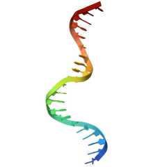
| DNA (5'-D(*TP*AP*CP*AP*TP*GP*TP*AP*AP*TP*TP*TP*AP*TP*TP*AP*C P*AP*TP*CP*A)-3') | A [auth C] | 21 | N/A |  | |
Sequence AnnotationsExpand | |||||
| |||||
Find similar nucleic acids by: Sequence | 3D Structure
Entity ID: 2 | |||||
|---|---|---|---|---|---|
| Molecule | Chains | Length | Organism | Image | |
| DNA (5'-D(*TP*AP*TP*GP*AP*TP*GP*TP*AP*AP*TP*AP*AP*AP*TP*TP*A P*CP*AP*TP*G)-3') | B [auth D] | 21 | N/A |  | |
Sequence AnnotationsExpand | |||||
| |||||
Experimental Data
- Method: X-RAY DIFFRACTION
- Resolution: 2.50 Å
- R-Value Free: 0.298 (Depositor)
- R-Value Work: 0.225 (Depositor)
- R-Value Observed: 0.225 (Depositor)
| Length ( Å ) | Angle ( ˚ ) |
|---|---|
| a = 132.38 | α = 90 |
| b = 132.38 | β = 90 |
| c = 45.86 | γ = 120 |
| Software Name | Purpose |
|---|---|
| X-PLOR | refinement |
| R-AXIS | data reduction |
Deposition Data
- Released Date: 1996-01-29 Deposition Author(s): Li, T.,Stark, M.R.,Johnson, A.D.,Wolberger, C.
Revision History (Full details and data files)
- Version 1.0: 1996-01-29
Type: Initial release - Version 1.1: 2008-05-22
Changes: Version format compliance - Version 1.2: 2011-07-13
Changes: Version format compliance - Version 1.3: 2024-02-14
Changes: Data collection, Database references
- RCSB PDB is a member of
- RCSB Partners
- Nucleic Acid Knowledgebase
RCSB PDB Core Operations are funded by theU.S. National Science Foundation (DBI-2321666), theUS Department of Energy (DE-SC0019749), and theNational Cancer Institute,National Institute of Allergy and Infectious Diseases, andNational Institute of General Medical Sciences of theNational Institutes of Health under grant R01GM157729.


