Movatterモバイル変換


[0]ホーム

URL:


Skip to main page content
U.S. flag

An official website of the United States government

Dot gov

The .gov means it’s official.
Federal government websites often end in .gov or .mil. Before sharing sensitive information, make sure you’re on a federal government site.

Https

The site is secure.
Thehttps:// ensures that you are connecting to the official website and that any information you provide is encrypted and transmitted securely.

NIH NLM Logo
Log inShow account info
Access keysNCBI HomepageMyNCBI HomepageMain ContentMain Navigation
pubmed logo
Advanced Clipboard
User Guide

Full text links

Royal Society of Chemistry full text link Royal Society of Chemistry Free PMC article
Full text links

Actions

Share

Review
.2012 Oct;29(10):1038-49.
doi: 10.1039/c2np20016c. Epub 2012 Apr 13.

Insights from the sea: structural biology of marine polyketide synthases

Affiliations
Review

Insights from the sea: structural biology of marine polyketide synthases

David L Akey et al. Nat Prod Rep.2012 Oct.

Abstract

The world's oceans are a rich source of natural products with extremely interesting chemistry. Biosynthetic pathways have been worked out for a few, and the story is being enriched with crystal structures of interesting pathway enzymes. By far, the greatest number of structural insights from marine biosynthetic pathways has originated with studies of curacin A, a poster child for interesting marine chemistry with its cyclopropane and thiazoline rings, internal cis double bond, and terminal alkene. Using the curacin A pathway as a model, structural details are now available for a novel loading enzyme with remarkable dual decarboxylase and acetyltransferase activities, an Fe(2+)/α-ketoglutarate-dependent halogenase that dictates substrate binding order through conformational changes, a decarboxylase that establishes regiochemistry for cyclopropane formation, and a thioesterase with specificity for β-sulfated substrates that lead to terminal alkene offloading. The four curacin A pathway dehydratases reveal an intrinsic flexibility that may accommodate bulky or stiff polyketide intermediates. In the salinosporamide A pathway, active site volume determines the halide specificity of a halogenase that catalyzes for the synthesis of a halogenated building block. Structures of a number of putative polyketide cyclases may help in understanding reaction mechanisms and substrate specificities although their substrates are presently unknown.


Figures

Fig. 1
Fig. 1
Curacin A: prototype for the rich biosynthesis of marine polyketides.(A) Curacin A biosynthesis with emphasis on reactions informed by protein structures. Changes to the polyketide intermediate are highlighted in red for each step. The molecular structures of curacin A and jamaicamide A from two strains ofMoorea producta are shown. Abbreviations are: ACP, acyl carrier protein; GNAT, GCN5-relatedN-acetyltransferase; KS, ketosynthase; AT, acyltransferase; HCS, HMG-CoA synthase; Hal, halogenase; ECH1, dehydrating enoyl-CoA hydratase; ECH2, decarboxylating enoyl-CoA hydratase; ER, enoyl reductase; DH, dehydratase; ST, sulfotransferase; TE, thioesterase; PAPS, 3’-phosphoadenosine-5’-phosphosulfate; PAP, 3’-phosphoadenosine-5’-phosphate.(B) Curacin A megasynthase. The annotated domains within the thirteen polypeptides (CurA – CurM) with domains are colored by type. Asterisks indicate domains with published structures. Domains not listed in (A) are: AR, GNAT adaptor region; KRC, ketoreductase catalytic domain, KRS, ketoreductase structural domain; CMT,C-methyl transferase; OMT,O-methyl transferase; A, adenylation; Cy, condensation/cyclization; PCP, peptidyl carrier protein. Box sizes are proportional to domain sizes.
Fig. 2
Fig. 2
Gallery of protein structures from marine PKS pathways. Each protein is shown as a ribbon diagram in its natural multimer state with subunits in contrasting colors. Substrates, cofactors or relevant side chains are rendered as spheres to emphasize the active site location in each protein. Protein Data Bank (PDB) accession codes are: CurA GNAT – 2REE, 2REF; CurA Hal – 3NNF, 3NNJ, 3NNL, 3NNM; CurA ACP – 2LIW; CurF ECH2 – 2Q2X, 2Q34, 2Q35; CurF DH – 3KG6; CurH DH – 3KG7; CurJ DH – 3KG8; CurK DH – 3KG9; CurM TE – 3QIT; SalL – 2Q6K, 2Q6I, 2Q6L, 2Q6O; SnoaL-like – 3GZB, 3KKG, 3LZA.
Fig. 3
Fig. 3
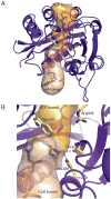
CurA GNAT loading enzyme.(A) Substrate and product tunnels through GNAT. The lower tunnel (tan surface) is the entrance for the malonyl-CoA substrate, based on electron density from crystals soaked in malonyl-CoA. The upper tunnel (gold surface) is for the ACP phosphopantetheine acceptor. Acetyl-CoA and the phosphopantetheine of ACP are shown in stick form with black C atoms. Side chains of key amino acids (Trp249, Thr355, His389 and Arg404) are shown in stick form with yellow C atoms.(B) Close-up view of the active site. Thr355 and His389 are essential for decarboxylation but not acetyl transfer activity. Hydrogen bonds from these residues to the acetyl carbonyl are shown (dashed lines). Trp249 constricts the tunnel at the active site. Arg404 may stabilize the reactive thiolate form of ACP-ppant.
Fig. 4
Fig. 4
CurF ECH2 decarboxylase.(A) ECH2 with modeled substrate, 4-chloro-3-methylglutaconyl-ACP. The substrate (gray C atoms), and key amino acid side chains (yellow C atoms) are shown as sticks and the protein as a green ribbon with the hypervariable region in magenta. The substrate thioester O atom is hydrogen bonded (dashed lines) to protein amides of the ECH2 oxyanion hole. Invariant His240 is essential for catalysis, as is Lys86 on the hypervariable helix α2.(B) Proposed reaction mechanism for ECH2 decarboxylation. Following decarboxylation, Lys86 is proposed to donate a proton to the C4 atom to generate the Δ2 (α-β) unsaturated product of CurF ECH2, 4-chloro-3-methylcrotonyl-ACP, which is subsequently converted to the cyclopropyl moiety of curacin A. In contrast, the ECH2 of the jamaicamide pathway donates a proton to the C2 atom to generate the Δ3 (β-γ) unsaturated product and the vinyl chloride is retained in jamaicamide.
Fig. 5
Fig. 5
Conformational states of CurA Hal.(A) Closed form Hal. A lid (red) covers the active site and a loop (grey) connecting α9-α10 is away from the active site.(B) Open form Hal. The lid between α2 and α4 is disordered and the loop (grey) connecting α9-α10 is next to the active site. α-KG is absent in the open state.(C) CurA Hal active site with a fully occupied iron coordination sphere. The iron center has octahedral coordination with only two protein ligands (His115 and His228). The cosubstrate, α-KG, chelates iron in a bidentate fashion. Formate from the crystallization solution occupies the sixth coordination position, which is thought to contain water in the resting enzyme and dioxygen during catalysis.(D) Conformational switch in CurA Hal. Upon departure of α-KG, the Arg241 side chain swings out, initiating a cascade of events. Strand β3 shifts towards strand β2, which becomes disordered. Tyr68 moves out of the active site. Asp283 moves towards the active site where it forms a salt bridge with the Arg247 side chain, which has moved out of the active site. His99 swings away from the active site to hydrogen bond with Asp69. Closed and open conformations are depicted in cyan and yellow, respectively.
Fig. 6
Fig. 6
SalL chlorinase.(A) Chloroethylmalonate building block. SalL catalyzes the conversion of chloride and SAM to 5’-ClDA and methionine. 5’-ClDA is further processed to yield chloroethylmalonyl-CoA, which is incorporated as a building block in the salinosporamide PKS pathway.(B) Composite image of the active site from wild type SalL (PDB 2Q6I) with chloride and SAM substrates from SalL/Y70T/G131S (PDB 2Q6O) shown in cartoon, and(C) in the same orientation, surface view.
Fig. 7
Fig. 7
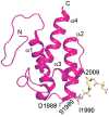
Solution structure of HMG-ACP1. The HMG-Ppant arm is linked to the Ser1989 side chain. Substitutions in α2 (Asp1988, Ile1990) and α3 (Ala2009) reduce chlorination efficiency, implicating a role for these residues in CurA Hal recognition.
Fig. 8
Fig. 8
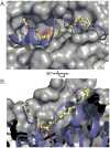
Curacin dehydratase.(A) Top view of CurK DH active site surface after removal of the proposed mobile elements (blue ribbon). The CurK DH substrate (yellow) is modeled with double bonds explicitly indicated. The active site His/Asp dyad is indicated with blue and red patches on the protein surface.(B) Cut-away view of (A) from the side. The V-shaped trench imposes restrictions on substrate placement. Active site His and Asp are shown in stick representation.
Fig. 9
Fig. 9
CurM TE decaboxylation thioesterase.(A) CurM TE monomer. The catalytic triad active site (magenta sticks) is located in a cleft between the core (green ribbon) and open lid (purple ribbon). The lid is held in place by specific contacts of Arg185 in the lid and Asp57 in the core.(B) CurM TE dimer. The novel lid (purple) to core (green) dimer interface holds the lid in an open position. Extensive subunit contacts are apparent in the translucent surface rendering, colored as in (A).(C) Active site cleft in CurM TE monomer. The acyl enzyme intermediate (yellow C atoms) is modeled into the open cleft between the lid (purple) and core (green).(D) CurM TE active site showing the catalytic triad (Ser100, His266, Glu124), sulfate-recognizing Arg205, and the modeled acyl enzyme intermediate (yellow C atoms).
Fig. 10
Fig. 10
Distribution of CurM TE homologues.(A) ORFs encoding ACP-ST-TE domains. The predicted domains are depicted for all of the sequences encoding sequential ACP-ST-TE domains. GenBank entries are:Moorea Producta (ACV42478),Synechococcus PCC 7002 (YP_001734428),Cyanothece PCC 7424 (YP_002377174),Cyanothece PCC 7822 (ZP_03153601),Moorea Producta 3L (ZP_08425908),Prochloron didemni (AEH57210),Pseudomonas entomophila L48 (YP_610919),Haliangium ochraceum DSM 14365 (YP_003265308).(B) Predicted pathway to hydrocarbons based on sequence annotation. The acyl-activating (AA) domain is predicted to load a fatty acid onto the adjacent ACP, the KS-AT-KR to extend and reduce the acyl-ACP to produce a β-hydroxyl substrate for the ST-TE.
See this image and copyright information in PMC

Similar articles

See all similar articles

Cited by

See all "Cited by" articles

References

    1. Jones AC, Monroe EA, Eisman EB, Gerwick L, Sherman DH, Gerwick WH. Nat Prod Rep. 2010;27:1048–1065. - PubMed
    1. Lane AL, Moore BS. Nat Prod Rep. 2011;28:411–428. - PMC - PubMed
    1. Tan LT. Phytochemistry. 2007;68:954–979. - PubMed
    1. Engene N, Rottacker EC, Kastovsky J, Byrum T, Choi H, Ellisman MH, Komarek J, Gerwick WH. Int J Syst Evol Microbiol. 2011 - PMC - PubMed
    1. Gerwick WH, Proteau PJ, Nagle DG, Hamel E, Blokhin A, Slate DL. J Org Chem. 1994;59:1243–1245.

Publication types

MeSH terms

Substances

Related information

Grants and funding

LinkOut - more resources

Full text links
Royal Society of Chemistry full text link Royal Society of Chemistry Free PMC article
Cite
Send To

NCBI Literature Resources

MeSHPMCBookshelfDisclaimer

The PubMed wordmark and PubMed logo are registered trademarks of the U.S. Department of Health and Human Services (HHS). Unauthorized use of these marks is strictly prohibited.


[8]ページ先頭

©2009-2025 Movatter.jp