Movatterモバイル変換


[0]ホーム

URL:


Lecturio medical combi logo
Lecturio medical logo

Hemothorax

A hemothorax is a collection of blood in thepleural cavityPleural cavityPaired but separate cavity within the thoracic cavity. It consists of the space between the parietal and visceral pleura and normally contains a capillary layer of serous fluid that lubricates the pleural surfaces.Pleura: Anatomy. Hemothorax most commonly occurs due to damage to the intercostalarteriesArteriesArteries are tubular collections of cells that transport oxygenated blood and nutrients from the heart to the tissues of the body. The blood passes through the arteries in order of decreasing luminal diameter, starting in the largest artery (the aorta) and ending in the small arterioles. Arteries are classified into 3 types: large elastic arteries, medium muscular arteries, and small arteries and arterioles.Arteries: Histology or from a lunglacerationLacerationTorn, ragged, mangled wounds.Blunt Chest Trauma following chest trauma. Hemothorax can also occur as a complication of disease, or hemothorax may be spontaneous oriatrogenicIatrogenicAny adverse condition in a patient occurring as the result of treatment by a physician, surgeon, or other health professional, especially infections acquired by a patient during the course of treatment.Anterior Cord Syndrome. Large hemothoraces can be life-threatening by leading to lung collapse.PatientsPatientsIndividuals participating in the health care system for the purpose of receiving therapeutic, diagnostic, or preventive procedures.Clinician–Patient Relationship present withshortness of breathShortness of breathDyspnea is the subjective sensation of breathing discomfort. Dyspnea is a normal manifestation of heavy physical or psychological exertion, but also may be caused by underlying conditions (both pulmonary and extrapulmonary).Dyspnea and chestpainPainAn unpleasant sensation induced by noxious stimuli which are detected by nerve endings of nociceptive neurons.Pain: Types and Pathways. Physical exam findings includehypotensionHypotensionHypotension is defined as low blood pressure, specifically< 90/60 mm Hg, and is most commonly a physiologic response. Hypotension may be mild, serious, or life threatening, depending on the cause.Hypotension,tachycardiaTachycardiaAbnormally rapid heartbeat, usually with a heart rate above 100 beats per minute for adults. Tachycardia accompanied by disturbance in the cardiac depolarization (cardiac arrhythmia) is called tachyarrhythmia.Sepsis in Children, decreased lung sounds, and dullness onpercussionPercussionAct of striking a part with short, sharp blows as an aid in diagnosing the condition beneath the sound obtained.Pulmonary Examination of the chest. Diagnosis is by upright chestX-rayX-rayPenetrating electromagnetic radiation emitted when the inner orbital electrons of an atom are excited and release radiant energy. X-ray wavelengths range from 1 pm to 10 nm. Hard x-rays are the higher energy, shorter wavelength x-rays. Soft x-rays or grenz rays are less energetic and longer in wavelength. The short wavelength end of the x-ray spectrum overlaps the gamma rays wavelength range. The distinction between gamma rays and x-rays is based on their radiation source.Pulmonary Function Tests. Management is withtube thoracostomyTube ThoracostomySurgical procedure involving the creation of an opening (stoma) into the chest cavity for drainage; used in the treatment of pleural effusion; pneumothorax; hemothorax; and empyema.Thoracic Surgery drainage, video-assisted thoracoscopic surgery (VATS), orthoracotomyThoracotomySurgical incision into the chest wall.Thoracic Surgery when massive hemothorax or persistent bleeding is present.

Last updated: Feb 19, 2025

Editorial responsibility:Stanley Oiseth, Lindsay Jones, Evelin Maza

Overview

Definition

A hemothorax is defined as a collection of fluid with ahematocritHematocritThe volume of packed red blood cells in a blood specimen. The volume is measured by centrifugation in a tube with graduated markings, or with automated blood cell counters. It is an indicator of erythrocyte status in disease. For example, anemia shows a low value; polycythemia, a high value.Neonatal Polycythemia of at least 50% accumulated in the potential space between theparietalParietalOne of a pair of irregularly shaped quadrilateral bones situated between the frontal bone and occipital bone, which together form the sides of the cranium.Skull: Anatomy andvisceral pleuraVisceral pleuraPleura: Anatomy of thelungsLungsLungs are the main organs of the respiratory system. Lungs are paired viscera located in the thoracic cavity and are composed of spongy tissue. The primary function of the lungs is to oxygenate blood and eliminate CO2.Lungs: Anatomy.

Epidemiology

  • Traumatic injuries cause 140,000 U.S. deaths annually, making them the leading cause of death for people in their 30s.
  • 72.3% ofrib fracturesRib fracturesFractures of any of the ribs.Flail Chest are associated withpneumothoraxPneumothoraxA pneumothorax is a life-threatening condition in which air collects in the pleural space, causing partial or full collapse of the lung. A pneumothorax can be traumatic or spontaneous. Patients present with a sudden onset of sharp chest pain, dyspnea, and diminished breath sounds on exam.Pneumothorax, hemothorax, or hemopneumothorax.
  • Associated with chest trauma and particularlymotor vehicle accidentsMotor Vehicle AccidentsSpinal Cord Injuries
  • Chest trauma seen in 60% ofpolytraumaPolytraumaMultitrauma occurs when 2 or more traumatic injuries occur in at least 2 areas of the body. A systematic management approach is necessary for individuals who have undergone trauma to maximize outcomes and reduce the risk of undiscovered injuries.Multitrauma

Etiology

The source of blood may be thechest wallChest wallThe chest wall consists of skin, fat, muscles, bones, and cartilage. The bony structure of the chest wall is composed of the ribs, sternum, and thoracic vertebrae. The chest wall serves as armor for the vital intrathoracic organs and provides the stability necessary for the movement of the shoulders and arms.Chest Wall: Anatomy, lung parenchyma, heart, or great vessels from either traumatic or non-traumatic causes.

Traumatic causes:

  • Lung parenchymal injuryLung Parenchymal InjuryThoracic Trauma in Children:
    • Most common cause
    • More commonly small (< 10%)
    • Often self-limited
  • Arterial injury:
    • Intercostal artery injury (most common)
    • Internal mammary artery injury
    • Great large vessels (rare, but life threatening)
  • IatrogenicIatrogenicAny adverse condition in a patient occurring as the result of treatment by a physician, surgeon, or other health professional, especially infections acquired by a patient during the course of treatment.Anterior Cord Syndrome:
    • Central venous catheterCentral Venous CatheterCentral venous catheters are IV lines placed into the large central veins for monitoring of central venous pressure (CVP), prolonged drug administration, or administration of parenteral nutrition. The most common sites of insertion are the internal jugular and subclavian veins.Central Venous Catheter placement
    • Thoracostomytube placementTube placementSurgical procedure involving the creation of an opening (stoma) into the chest cavity for drainage; used in the treatment of pleural effusion; pneumothorax; hemothorax; and empyema.Thoracic Surgery

Non-traumatic causes:

  • Malignancy
  • Anticoagulant medications
  • Coagulopathies
  • Aortic dissectionAortic dissectionAortic dissection occurs due to shearing stress from pulsatile pressure causing a tear in the tunica intima of the aortic wall. This tear allows blood to flow into the media, creating a “false lumen.” Aortic dissection is most commonly caused by uncontrolled hypertension.Aortic Dissection oraneurysmAneurysmAn aneurysm is a bulging, weakened area of a blood vessel that causes an abnormal widening of its diameter > 1.5 times the size of the native vessel. Aneurysms occur more often in arteries than in veins and are at risk of dissection and rupture, which can be life-threatening.Thoracic Aortic Aneurysms
  • TuberculosisTuberculosisTuberculosis (TB) is an infectious disease caused by Mycobacterium tuberculosis complex bacteria. The bacteria usually attack the lungs but can also damage other parts of the body. Approximately 30% of people around the world are infected with this pathogen, with the majority harboring a latent infection. Tuberculosis spreads through the air when a person with active pulmonary infection coughs or sneezes.Tuberculosis and necrotizinginfectionsInfectionsInvasion of the host organism by microorganisms or their toxins or by parasites that can cause pathological conditions or diseases.Chronic Granulomatous Disease

Pathophysiology

Hemodynamic component:

Large volume loss (each hemothorax can contain up to 40% of total circulating blood volume) into thepleuraPleuraThe pleura is a serous membrane that lines the walls of the thoracic cavity and the surface of the lungs. This structure of mesodermal origin covers both lungs, the mediastinum, the thoracic surface of the diaphragm, and the inner part of the thoracic cage. The pleura is divided into a visceral pleura and parietal pleura.Pleura: Anatomy can lead to decreased cardiac function due to:

  • DecreasedpreloadPreloadCardiac Mechanics by constricting the vena cava
  • Increased restriction of movement of the cardiac wall
  • Increasedhydrostatic pressureHydrostatic pressureThe pressure due to the weight of fluid.Edema leading topulmonary hypertensionPulmonary HypertensionPulmonary hypertension (PH) or pulmonary arterial hypertension (PAH) is characterized by elevated pulmonary arterial pressure, which can lead to chronic progressive right heart failure. Pulmonary hypertension is grouped into 5 categories based on etiology, which include primary PAH, and PH due to cardiac disease, lung or hypoxic disease, chronic thromboembolic disease, and multifactorial or unclear etiologies.Pulmonary Hypertension

Respiratory component:

Blood collecting in thepleuraPleuraThe pleura is a serous membrane that lines the walls of the thoracic cavity and the surface of the lungs. This structure of mesodermal origin covers both lungs, the mediastinum, the thoracic surface of the diaphragm, and the inner part of the thoracic cage. The pleura is divided into a visceral pleura and parietal pleura.Pleura: Anatomy decreases the lung’s functionalvital capacityVital capacityThe volume of air that is exhaled by a maximal expiration following a maximal inspiration.Ventilation: Mechanics of Breathing by:

Clinical Presentation

Symptoms

  • Shortness of breathShortness of breathDyspnea is the subjective sensation of breathing discomfort. Dyspnea is a normal manifestation of heavy physical or psychological exertion, but also may be caused by underlying conditions (both pulmonary and extrapulmonary).Dyspnea
  • ChestpainPainAn unpleasant sensation induced by noxious stimuli which are detected by nerve endings of nociceptive neurons.Pain: Types and Pathways

Signs

  • Ipsilateral absent or ↓ breath sounds
  • Tracheal deviationTracheal DeviationPneumothorax
  • Dullness onpercussionPercussionAct of striking a part with short, sharp blows as an aid in diagnosing the condition beneath the sound obtained.Pulmonary Examination
  • CrepitusCrepitusOsteoarthritis
  • Signs ofhemorrhagicshockShockShock is a life-threatening condition associated with impaired circulation that results in tissue hypoxia. The different types of shock are based on the underlying cause: distributive (↑ cardiac output (CO), ↓ systemic vascular resistance (SVR)), cardiogenic (↓ CO, ↑ SVR), hypovolemic (↓ CO, ↑ SVR), obstructive (↓ CO), and mixed.Types of Shock in large hemothoraces:

Diagnosis

Diagnosis is established by utilizing history, physical, and appropriate imaging. In cases of trauma, use of theprimary surveyPrimary SurveyThoracic Trauma in Children is paramount to rapid diagnosis and treatment.
Imaging

  • ChestX-rayX-rayPenetrating electromagnetic radiation emitted when the inner orbital electrons of an atom are excited and release radiant energy. X-ray wavelengths range from 1 pm to 10 nm. Hard x-rays are the higher energy, shorter wavelength x-rays. Soft x-rays or grenz rays are less energetic and longer in wavelength. The short wavelength end of the x-ray spectrum overlaps the gamma rays wavelength range. The distinction between gamma rays and x-rays is based on their radiation source.Pulmonary Function Tests:best initial diagnostic study
    • Upright imaging shows layering of blood.
    • Supine imaging shows haziness oropacityOpacityImaging of the Lungs and Pleura (whiteout).
    • May also show free air if apneumothoraxPneumothoraxA pneumothorax is a life-threatening condition in which air collects in the pleural space, causing partial or full collapse of the lung. A pneumothorax can be traumatic or spontaneous. Patients present with a sudden onset of sharp chest pain, dyspnea, and diminished breath sounds on exam.Pneumothorax is present
  • Ultrasound of thelungsLungsLungs are the main organs of the respiratory system. Lungs are paired viscera located in the thoracic cavity and are composed of spongy tissue. The primary function of the lungs is to oxygenate blood and eliminate CO2.Lungs: Anatomy (thoraxsonographySonographyThe visualization of deep structures of the body by recording the reflections or echoes of ultrasonic pulses directed into the tissues. Use of ultrasound for imaging or diagnostic purposes employs frequencies ranging from 1. 6 to 10 megahertz.Diagnostic Procedures in Gynecology):
    • Part of the Extended Focused Assessment withSonographySonographyThe visualization of deep structures of the body by recording the reflections or echoes of ultrasonic pulses directed into the tissues. Use of ultrasound for imaging or diagnostic purposes employs frequencies ranging from 1. 6 to 10 megahertz.Diagnostic Procedures in Gynecology for Trauma (eFAST) exam
    • Able to be obtained quickly
    • Can show complex fluid in thepleural cavityPleural cavityPaired but separate cavity within the thoracic cavity. It consists of the space between the parietal and visceral pleura and normally contains a capillary layer of serous fluid that lubricates the pleural surfaces.Pleura: Anatomy
    • More sensitive than a chestX-rayX-rayPenetrating electromagnetic radiation emitted when the inner orbital electrons of an atom are excited and release radiant energy. X-ray wavelengths range from 1 pm to 10 nm. Hard x-rays are the higher energy, shorter wavelength x-rays. Soft x-rays or grenz rays are less energetic and longer in wavelength. The short wavelength end of the x-ray spectrum overlaps the gamma rays wavelength range. The distinction between gamma rays and x-rays is based on their radiation source.Pulmonary Function Tests in detecting a hemothorax, but is technician dependent
  • Chest computed tomography(CT)—definitive imaging choice:
    • Should only be obtained if the patient is stable 
    • CT can show other associated pathology.
    • CT angiogramCT angiogramA non-invasive method that uses a ct scanner for capturing images of blood vessels and tissues. A contrast material is injected, which helps produce detailed images that aid in diagnosing vascular diseases.Pulmonary Function Tests can show the source of bleeding.
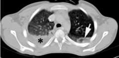
Hemothorax

Chest CT showing a hemothorax in the right lung
Arrow indicates a lung contusion and a collection of fluid thought to be a hemothorax.

Image: “Thoracic CT” by Section Surgery, Emergency Hospital, Bucharest, Romania 8 Calea Floreasca, District 1, Bucharest, 014461, Romania. License:CC BY 2.0.
Massive right hemothorax

Massive right hemothorax
Chest X-ray showing a collection of fluid in the right pleural space, as illustrated by the opacified lung
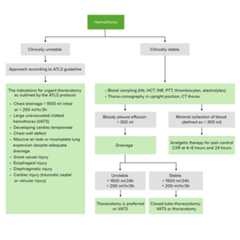
Image: “Massive right hemothorax” by Kaul and Paniagua. License:CC BY 2.0.

Management and Complications

Management

Hemothorax management

Hemothorax management algorithm

ATLS: Advanced Trauma Life Support
CXR: chest X-ray
Hb: hemoglobin
HCT: hematocrit
INR: international normalized ratio
PTT: prothrombin time
VATS: video-assisted thoracoscopic surgery

Image by Lecturio.
  • AirwayAirwayABCDE Assessment, breathing, andcirculationCirculationThe movement of the blood as it is pumped through the cardiovascular system.ABCDE Assessment (ABC) assessment → administer100% oxygen100% OxygenCluster Headaches → establish intravenous (IV) access
  • Stabilize the patient (fluidresuscitationResuscitationThe restoration to life or consciousness of one apparently dead. .Neonatal Respiratory Distress Syndrome and blood transfusion as necessary).
  • ReverseanticoagulantsAnticoagulantsAnticoagulants are drugs that retard or interrupt the coagulation cascade. The primary classes of available anticoagulants include heparins, vitamin K-dependent antagonists (e.g., warfarin), direct thrombin inhibitors, and factor Xa inhibitors.Anticoagulants, if necessary.
  • ProvideanalgesiaAnalgesiaMethods of pain relief that may be used with or in place of analgesics.Anesthesiology: History and Basic Concepts appropriate to the level of the patient’spainPainAn unpleasant sensation induced by noxious stimuli which are detected by nerve endings of nociceptive neurons.Pain: Types and Pathways.
  • Insert a chest tube (thoracostomy) for large hemothoraces or in an unstable patient:
  • Surgical intervention (thoracotomyThoracotomySurgical incision into the chest wall.Thoracic Surgery) is indicated when:
    • Evacuating > 1,500 mL of blood directly after inserting a chest tube
    • Continued high output → collecting of > 1 L (1,000 mL) of blood over 4 hours or > 200 mL/hour for 3 consecutive hours
Thoracostomy

How to insert a chest tube
Stepwise illustration on how to insert a chest tube to drain fluid accumulation from the pleural space

Image by Lecturio.

Complications

  • ImpairedventilationVentilationThe total volume of gas inspired or expired per unit of time, usually measured in liters per minute.Ventilation: Mechanics of Breathing on the affected side:
  • EmpyemaEmpyemaPresence of pus in a hollow organ or body cavity.Pneumonia:
    • Retained blood collection develops a bacterial infection.
    • 5% of cases
  • Fibrothorax:
    • Formation ofscarScarDermatologic Examination tissues within thelungsLungsLungs are the main organs of the respiratory system. Lungs are paired viscera located in the thoracic cavity and are composed of spongy tissue. The primary function of the lungs is to oxygenate blood and eliminate CO2.Lungs: Anatomy due to blood irritation
    • 1% of cases

Differential Diagnosis

  • PneumothoraxPneumothoraxA pneumothorax is a life-threatening condition in which air collects in the pleural space, causing partial or full collapse of the lung. A pneumothorax can be traumatic or spontaneous. Patients present with a sudden onset of sharp chest pain, dyspnea, and diminished breath sounds on exam.Pneumothorax: abnormal collection of air in thepleural spacePleural spaceThe thin serous membrane enveloping the lungs (lung) and lining the thoracic cavity. Pleura consist of two layers, the inner visceral pleura lying next to the pulmonary parenchyma and the outer parietal pleura. Between the two layers is the pleural cavity which contains a thin film of liquid.Pleuritis due tolacerationLacerationTorn, ragged, mangled wounds.Blunt Chest Trauma of thelungsLungsLungs are the main organs of the respiratory system. Lungs are paired viscera located in the thoracic cavity and are composed of spongy tissue. The primary function of the lungs is to oxygenate blood and eliminate CO2.Lungs: Anatomy. Types ofpneumothoraxPneumothoraxA pneumothorax is a life-threatening condition in which air collects in the pleural space, causing partial or full collapse of the lung. A pneumothorax can be traumatic or spontaneous. Patients present with a sudden onset of sharp chest pain, dyspnea, and diminished breath sounds on exam.Pneumothorax include spontaneouspneumothoraxPneumothoraxA pneumothorax is a life-threatening condition in which air collects in the pleural space, causing partial or full collapse of the lung. A pneumothorax can be traumatic or spontaneous. Patients present with a sudden onset of sharp chest pain, dyspnea, and diminished breath sounds on exam.Pneumothorax andtraumatic pneumothoraxTraumatic PneumothoraxPneumothorax.PatientsPatientsIndividuals participating in the health care system for the purpose of receiving therapeutic, diagnostic, or preventive procedures.Clinician–Patient Relationship havedyspneaDyspneaDyspnea is the subjective sensation of breathing discomfort. Dyspnea is a normal manifestation of heavy physical or psychological exertion, but also may be caused by underlying conditions (both pulmonary and extrapulmonary).Dyspnea, chestpainPainAn unpleasant sensation induced by noxious stimuli which are detected by nerve endings of nociceptive neurons.Pain: Types and Pathways, decreased breath sounds, andhyper-resonanceHyper-ResonancePneumothorax onpercussionPercussionAct of striking a part with short, sharp blows as an aid in diagnosing the condition beneath the sound obtained.Pulmonary Examination. Treatment includesneedle decompressionNeedle DecompressionPneumothorax and chesttube placementTube placementSurgical procedure involving the creation of an opening (stoma) into the chest cavity for drainage; used in the treatment of pleural effusion; pneumothorax; hemothorax; and empyema.Thoracic Surgery.
  • Pleural effusionPleural EffusionPleural effusion refers to the accumulation of fluid between the layers of the parietal and visceral pleura. Common causes of this condition include infection, malignancy, autoimmune disorders, or volume overload. Clinical manifestations include chest pain, cough, and dyspnea.Pleural Effusion: accumulation of fluid in thepleural cavityPleural cavityPaired but separate cavity within the thoracic cavity. It consists of the space between the parietal and visceral pleura and normally contains a capillary layer of serous fluid that lubricates the pleural surfaces.Pleura: Anatomy. Can be caused by many conditions, including infection, malignancy, andheart failureHeart FailureA heterogeneous condition in which the heart is unable to pump out sufficient blood to meet the metabolic need of the body. Heart failure can be caused by structural defects, functional abnormalities (ventricular dysfunction), or a sudden overload beyond its capacity. Chronic heart failure is more common than acute heart failure which results from sudden insult to cardiac function, such as myocardial infarction.Total Anomalous Pulmonary Venous Return (TAPVR). Symptoms include chestpainPainAn unpleasant sensation induced by noxious stimuli which are detected by nerve endings of nociceptive neurons.Pain: Types and Pathways,dyspneaDyspneaDyspnea is the subjective sensation of breathing discomfort. Dyspnea is a normal manifestation of heavy physical or psychological exertion, but also may be caused by underlying conditions (both pulmonary and extrapulmonary).Dyspnea, andorthopneaOrthopneaPulmonary Edema. Diagnosis is by imaging, and pleural fluid analysis helps determine the etiology. Management is dependent on the underlying condition and severity, but may include monitoring,thoracentesisThoracentesisAspiration of fluid or air from the thoracic cavity. It is coupled sometimes with the administration of drugs into the pleural cavity.Thoracic Surgery, chesttube placementTube placementSurgical procedure involving the creation of an opening (stoma) into the chest cavity for drainage; used in the treatment of pleural effusion; pneumothorax; hemothorax; and empyema.Thoracic Surgery, or surgery.
  • AtelectasisAtelectasisAtelectasis is the partial or complete collapse of a part of the lung. Atelectasis is almost always a secondary phenomenon from conditions causing bronchial obstruction, external compression, surfactant deficiency, or scarring.Atelectasis: condition characterized by the collapse ofalveoliAlveoliSmall polyhedral outpouchings along the walls of the alveolar sacs, alveolar ducts and terminal bronchioles through the walls of which gas exchange between alveolar air and pulmonary capillary blood takes place.Acute Respiratory Distress Syndrome (ARDS), and eventually, lobar lung collapse and complete obstruction.PatientsPatientsIndividuals participating in the health care system for the purpose of receiving therapeutic, diagnostic, or preventive procedures.Clinician–Patient Relationship present with respiratory distress (dyspneaDyspneaDyspnea is the subjective sensation of breathing discomfort. Dyspnea is a normal manifestation of heavy physical or psychological exertion, but also may be caused by underlying conditions (both pulmonary and extrapulmonary).Dyspnea,tachypneaTachypneaIncreased respiratory rate.Pulmonary Examination,tachycardiaTachycardiaAbnormally rapid heartbeat, usually with a heart rate above 100 beats per minute for adults. Tachycardia accompanied by disturbance in the cardiac depolarization (cardiac arrhythmia) is called tachyarrhythmia.Sepsis in Children) andhypoxemiaHypoxemiaNeonatal Respiratory Distress Syndrome. Physical examination of thelungsLungsLungs are the main organs of the respiratory system. Lungs are paired viscera located in the thoracic cavity and are composed of spongy tissue. The primary function of the lungs is to oxygenate blood and eliminate CO2.Lungs: Anatomy reveals dullness topercussionPercussionAct of striking a part with short, sharp blows as an aid in diagnosing the condition beneath the sound obtained.Pulmonary Examination and decreased breath sounds. ChestX-rayX-rayPenetrating electromagnetic radiation emitted when the inner orbital electrons of an atom are excited and release radiant energy. X-ray wavelengths range from 1 pm to 10 nm. Hard x-rays are the higher energy, shorter wavelength x-rays. Soft x-rays or grenz rays are less energetic and longer in wavelength. The short wavelength end of the x-ray spectrum overlaps the gamma rays wavelength range. The distinction between gamma rays and x-rays is based on their radiation source.Pulmonary Function Tests shows lung opacification. Management is aimed at ventilatory support. 
  • Pulmonary embolismPulmonary EmbolismPulmonary embolism (PE) is a potentially fatal condition that occurs as a result of intraluminal obstruction of the main pulmonary artery or its branches. The causative factors include thrombi, air, amniotic fluid, and fat. In PE, gas exchange is impaired due to the decreased return of deoxygenated blood to the lungs.Pulmonary Embolism(PE): obstruction of pulmonaryarteriesArteriesArteries are tubular collections of cells that transport oxygenated blood and nutrients from the heart to the tissues of the body. The blood passes through the arteries in order of decreasing luminal diameter, starting in the largest artery (the aorta) and ending in the small arterioles. Arteries are classified into 3 types: large elastic arteries, medium muscular arteries, and small arteries and arterioles.Arteries: Histology, most often due to thrombus migration from the deep venous system. Signs and symptoms include pleuritic chestpainPainAn unpleasant sensation induced by noxious stimuli which are detected by nerve endings of nociceptive neurons.Pain: Types and Pathways,dyspneaDyspneaDyspnea is the subjective sensation of breathing discomfort. Dyspnea is a normal manifestation of heavy physical or psychological exertion, but also may be caused by underlying conditions (both pulmonary and extrapulmonary).Dyspnea,tachypneaTachypneaIncreased respiratory rate.Pulmonary Examination, andtachycardiaTachycardiaAbnormally rapid heartbeat, usually with a heart rate above 100 beats per minute for adults. Tachycardia accompanied by disturbance in the cardiac depolarization (cardiac arrhythmia) is called tachyarrhythmia.Sepsis in Children. Severe cases are life threatening. Chest CT withangiographyAngiographyRadiography of blood vessels after injection of a contrast medium.Cardiac Surgery is the primary method of diagnosis. Management includes oxygenation,anticoagulationAnticoagulationPulmonary Hypertension Drugs, and thrombolytic therapy forunstable patientsUnstable PatientsBlunt Chest Trauma.

References

  1. Le, T., & Bhushan, V. (2015). First Aid for the USMLE Step 1 2019. New York: McGraw-Hill Medical.
  2. Broderick, S.R. (2013). Hemothorax: Etiology, diagnosis, and management. Thorac Surg Clin. Feb. 23 (1):89–96, vi-vii.
  3. Legome, E. (2025). Initial evaluation and management of blunt thoracic trauma in adults.UpToDate.Retrieved February 19, 2025 fromhttps://www.uptodate.com/contents/initial-evaluation-and-management-of-blunt-thoracic-trauma-in-adults
  4. Richardson, J.D., Miller, F.B., Carrillo, E.H., & Spain, D.A. (1996). Complex thoracic injuries. Surg Clin North Am. Aug. 76 (4):725–48.
  5. Chou, Y.P., Kuo, L.C., Soo, K.M., Tarng, Y.W., Chiang, H.I., Huang, F.D., et al. (2014). The role of repairing lung lacerations during video-assisted thoracoscopic surgery evacuations for retained haemothorax caused by blunt chest trauma. Eur J Cardiothorac Surg. Jul. 46 (1):107–11.
  6. Boersma, W.G., Stigt, J.A., & Smit, H.J. (2010). Treatment of hemothorax. Respir Med. Nov;104(11):1583-7. doi: 10.1016/j.rmed.2010.08.006. PMID: 20817498.
  7. Pumarejo Gomez, L., & Tran, V. H. (2023, August 8).Hemothorax. StatPearls. National Library of Medicine. Retrieved February 19, 2025, from https://www.ncbi.nlm.nih.gov/books/NBK538219/

Study with Lecturio for

© 2025 Lecturio GmbH. All rights reserved.

USMLE™ is a joint program of the Federation of State Medical Boards (FSMB®) and National Board of Medical Examiners (NBME®). MCAT is a registered trademark of the Association of American Medical Colleges (AAMC). NCLEX®, NCLEX-RN®, and NCLEX-PN® are registered trademarks of the National Council of State Boards of Nursing, Inc (NCSBN®). None of the trademark holders are endorsed by nor affiliated with Lecturio.

Create your free account or log in to continue reading!

Sign up now and get free access to Lecturio with concept pages, medical videos, and questions for your medical education.

User Reviews

Best Week. Best Price. Best Prep! 💸 Get 40% off all plans and certificates now >>

Details
×
Get Premium to test your knowledge

Lecturio Premium gives you full access to all content & features

Get Premium to watch all videos

Verify your email now to get a free trial.

Create a free account to test your knowledge

Lecturio Premium gives you full access to all contents and features—including Lecturio’s Qbank with up-to-date board-style questions.

Get Premium
or log in
0/5 questions correct
Start Test

[8]ページ先頭

©2009-2025 Movatter.jp