Movatterモバイル変換


[0]ホーム

URL:


Skip to main page content
U.S. flag

An official website of the United States government

Dot gov

The .gov means it’s official.
Federal government websites often end in .gov or .mil. Before sharing sensitive information, make sure you’re on a federal government site.

Https

The site is secure.
Thehttps:// ensures that you are connecting to the official website and that any information you provide is encrypted and transmitted securely.

NIH NLM Logo
Log inShow account info
Access keysNCBI HomepageMyNCBI HomepageMain ContentMain Navigation
pubmed logo
Advanced Clipboard
User Guide

Full text links

Free PMC article
Full text links

Actions

Share

.2024 Oct 7;6(5):fcae351.
doi: 10.1093/braincomms/fcae351. eCollection 2024.

Clinical parameters affect the structure and function of superficial pyramidal neurons in the adult human neocortex

Affiliations

Clinical parameters affect the structure and function of superficial pyramidal neurons in the adult human neocortex

Maximilian Lenz et al. Brain Commun..

Abstract

The interplay between neuronal structure and function underpins the dynamic nature of neocortical networks. Despite extensive studies in animal models, our understanding of structure-function interrelations in the adult human brain remains incomplete. Recent methodological advances have facilitated the functional analysis of individual neurons within the human neocortex, providing a new understanding of fundamental brain processes. However, the factors contributing to patient-specific neuronal properties have not been thoroughly explored. In this observational study, we investigated the structural and functional variability of superficial pyramidal neurons in the adult human neocortex. Using whole-cell patch-clamp recordings andpost hoc analyses of dendritic spine morphology in acute neocortical slice preparations from surgical resections of seven patients, we assessed age-related effects on excitatory neurotransmission, membrane properties and dendritic spine morphologies. These results specify age as an endogenous factor that might affect the structural and functional properties of superficial pyramidal neurons.

Keywords: excitatory synapses; human dendritic spines; human neocortex.

© The Author(s) 2024. Published by Oxford University Press on behalf of the Guarantors of Brain.


Conflict of interest statement

The authors report no competing interests.

Figures

Graphical Abstract
Graphical Abstract
Figure 1
Figure 1
Superficial pyramidal neurons in the human neocortex. (A) Representative image ofpost hoc stained human cortical superficial pyramidal neurons (streptavidin) with neuronal cell bodies marked by NeuN staining. L1, layer 1; L2/3, layer 2/3; pm, pia mater. Scale bar: 150 µm. (B)Post hoc stained superficial pyramidal neuron in the human cortex. L1, layer 1; L2/3, layer 2/3; pm, pia mater. Scale bar: 100 µm.
Figure 2
Figure 2
Correlation of structural and functional properties of superficial human pyramidal neurons with clinical patient parameters. Correlation matrix displaying Spearman correlations of continuous numerical data (mean values, left panel) and correspondingP-values (right). Significant results are highlighted with asterisks (left panel) and violet squares (right panel), showing correlations between synaptic and electrophysiological parameters, and between patient age and dendritic spine density.
Figure 3
Figure 3
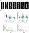
Structural analysis of dendritic spine morphology in human superficial pyramidal neurons. (A) Example images ofpost hoc labelled dendritic segments with spines (streptavidin staining) from superficial pyramidal neurons, sampled from seven patients. Scale bar: 4 µm. (B) Left panel: Analysis of dendritic spine density [#2 (18 years) = 117 segments in six cells; #3 (27 years) = 39 segments in five cells; #1 (39 years) = 76 dendritic segments in six cells; #7 (52 years) = 53 segments in five cells; #5 (54 years) = 60 segments in five cells; #6 (55 years) = 30 segments in three cells; #4 (78 years) = 27 segments in three cells]. Right panel: analysis of dendritic spine head volumes [c.f. Fig. 2; #2 (18 years) = 116 segments in six cells; #3 (27 years) = 39 segments in five cells; #1 (39 years) = 76 dendritic segments in six cells; #7 (52 years) = 53 segments in five cells; #5 (54 years) = 60 segments in five cells; #6 (55 years) = 30 segments in three cells; #4 (78 years) = 27 segments in three cells]. Individual mean values from dendritic segments are indicated by grey dots. (C) Left panel:X-Y graph displaying the correlation between age and overall dendritic spine density across various age groups from the same data as shown inB. A linear regression is shown as black line. Right panel:X-Y graph illustrating the lack of significant correlation between age and overall dendritic spine head volume for the same age groups and segments counts. A linear regression is indicated with a black line (P-values for non-zero slope andR2 values for goodness of fit are reported in the figure, along with Spearmanr for correlation analysis). All violin plots depict the median with 25–75% percentiles, individual values are indicated by coloured dots, and the median is illustrated as a black line. All linear regression analysis graphs depict median ± interquartile range.
Figure 4
Figure 4
Electrophysiological assessment of excitatory synaptic transmission in human superficial pyramidal neurons. (A) Sample traces of AMPA receptor-mediated sEPSCs in superficial pyramidal neurons from seven adult human neocortex samples (sorted by age). (B,C) Quantitative analysis of sEPSC amplitudes (B) and frequencies (C), indicating the absence of age-related changes [#2 (18 years) = 35 cells; #3 (27 years) = 21 cells; #1 (39 years) = 15 cells; #7 (52 years) = 29 cells; #5 (54 years) = 23 cells; #6 (55 years) = 14 cells; #4 (78 years) = 19 cells]. Grey dots represent individual cell values within each sample.X-Y graphs demonstrating the respective median ± interquartile range across different ages. A linear regression is indicated with a black line (P-values for non-zero slope andR2 values for goodness of fit are reported in the figure, along with Spearmanr for correlation analysis). All violin plots depict the median with 25–75% percentiles, individual values are indicated by coloured dots, and the median is illustrated by a black line. All linear regression analysis graphs depict median ± interquartile range.
Figure 5
Figure 5
Electrophysiological assessment of intrinsic membrane properties in human superficial pyramidal neurons. (A) Representative current clamp input–output curve traces depicting responses at −100 and +600 pA current injections in superficial pyramidal neurons. (B–D) Upper panels: analysis of passive [input resistance (B) and resting membrane potential (C)] and active (D; action potential frequency) membrane properties [#2 (18 years) = 35 cells; #3 (27 years) = 21 cells; #1 (39 years) = 14 cells (one cell excluded from analysis, since integrity of the patch was lost during the recordings); #7 (52 years) = 29 cells; #5 (54 years) = 23 cells; #6 (55 years) = 14 cells; #4 (78 years) = 19 cells]. Grey dots represent individual cell values within each sample. Lower panels:X-Y graphs demonstrating the respective median ± interquartile range across different ages. A linear regression is indicated with a black line (P-values for non-zero slope andR2 values for goodness of fit are reported in the figure, along with Spearmanr for correlation analysis). All violin plots depict the median with 25–75% percentiles, individual values are indicated by coloured dots, and the median is illustrated by a black line. All linear regression analysis graphs depict median ± interquartile range.
Figure 6
Figure 6
Disease and treatment related changes in structural properties of superficial pyramidal neurons. Results classified by medical records into two groups: epilepsy/antiepileptic medication (AED) and tumour/steroid medication (c.f. Table 1). (A) Group data of spine head volumes in dendritic compartments (A;nepilepsy/AED = 198 segments in four patients;ntumour/steroids = 203 segments in three patients; c.f. Table 1; nestedt-test). (B) Group data of dendritic spine densities (A;nepilepsy/AED = 198 segments in four patients,ntumour/steroids = 204 segments in three patients; c.f. Table 1; nestedt-test). All violin plots depict the median with 25–75% percentiles, individual values are indicated by coloured dots, and the median is illustrated as a black line. A nestedt-test was used for group comparisons.P-values <0.05 were considered statistically significant; results without statistical significance were indicated as ‘ns’.
Figure 7
Figure 7
Disease and treatment related changes in excitatory synaptic transmission of superficial pyramidal neurons. (A–C) Group data of AMPA receptor-mediated sEPSC amplitudes (A), time-to-max rise slope (B) and sEPSC frequencies (C) (nepilepsy/AED = 79 cells in four patients;ntumour/steroids = 77 cells in three patients; nestedt-test). All violin plots depict the median with 25–75% percentiles, individual values are indicated by coloured dots, and the median is illustrated as a black line. A nestedt-test was used for group comparisons.P-values <0.05 were considered statistically significant; results without statistical significance were indicated as ‘ns’.
Figure 8
Figure 8
Disease and treatment related changes in intrinsic cellular properties of superficial pyramidal neurons. (A–C) Analysis of passive (A; input resistance;B; resting membrane potential) and active (C; action potential frequency) membrane properties [nepilepsy/AED = 78 cells in four patients (one cell excluded from further analysis due to a decline in patch integrity);ntumor/steroids = 77 cells in 3 patients; nestedt-test]. All violin plots depict the median with 25–75% percentiles, individual values are indicated by coloured dots, and the median is illustrated as a black line. A nestedt-test was used for group comparisons.P-values <0.05 were considered statistically significant; results without statistical significance were indicated as ‘ns’.
See this image and copyright information in PMC

Similar articles

See all similar articles

References

    1. Pang JC, Aquino KM, Oldehinkel M, et al. . Geometric constraints on human brain function. Nature. 2023;618(7965):566–574. - PMC - PubMed
    1. Agnes EJ, Vogels TP. Co-dependent excitatory and inhibitory plasticity accounts for quick, stable and long-lasting memories in biological networks. Nat Neurosci. 2024;27(5):964–974. - PMC - PubMed
    1. Takeuchi T, Duszkiewicz AJ, Morris RG. The synaptic plasticity and memory hypothesis: Encoding, storage and persistence. Philos Trans R Soc Lond B Biol Sci. 2014;369(1633):20130288. - PMC - PubMed
    1. Verhoog MB, Goriounova NA, Obermayer J, et al. . Mechanisms underlying the rules for associative plasticity at adult human neocortical synapses. J Neurosci. 2013;33(43):17197–17208. - PMC - PubMed
    1. Ting JT, Lee BR, Chong P, et al. . Preparation of acute brain slices using an optimized N-methyl-D-glucamine protective recovery method. J Vis Exp. 2018(132):53825. - PMC - PubMed

Related information

LinkOut - more resources

Full text links
Free PMC article
Cite
Send To

NCBI Literature Resources

MeSHPMCBookshelfDisclaimer

The PubMed wordmark and PubMed logo are registered trademarks of the U.S. Department of Health and Human Services (HHS). Unauthorized use of these marks is strictly prohibited.


[8]ページ先頭

©2009-2025 Movatter.jp