Movatterモバイル変換


[0]ホーム

URL:


Skip to main page content
U.S. flag

An official website of the United States government

Dot gov

The .gov means it’s official.
Federal government websites often end in .gov or .mil. Before sharing sensitive information, make sure you’re on a federal government site.

Https

The site is secure.
Thehttps:// ensures that you are connecting to the official website and that any information you provide is encrypted and transmitted securely.

NIH NLM Logo
Log inShow account info
Access keysNCBI HomepageMyNCBI HomepageMain ContentMain Navigation
pubmed logo
Advanced Clipboard
User Guide

Full text links

Atypon full text link Atypon Free PMC article
Full text links

Actions

Share

.2023 Dec;9(48):eadk7251.
doi: 10.1126/sciadv.adk7251. Epub 2023 Nov 29.

Swarming magnetic nanorobots bio-interfaced by heparinoid-polymer brushes for in vivo safe synergistic thrombolysis

Affiliations

Swarming magnetic nanorobots bio-interfaced by heparinoid-polymer brushes for in vivo safe synergistic thrombolysis

Manyi Yang et al. Sci Adv.2023 Dec.

Abstract

Biocompatible swarming magnetic nanorobots that work in blood vessels for safe and efficient targeted thrombolytic therapy in vivo are demonstrated. This is achieved by using magnetic beads elaborately grafted with heparinoid-polymer brushes (HPBs) upon the application of an alternating magnetic fieldB(t). Because of the dense surface charges bestowed by HPBs, the swarming nanorobots demonstrate reversible agglomeration-free reconfigurations, low hemolysis, anti-bioadhesion, and self-anticoagulation in high-ionic-strength blood environments. They are confirmed in vitro and in vivo to perform synergistic thrombolysis efficiently by "motile-targeting" drug delivery and mechanical destruction. Moreover, upon the completion of thrombolysis and removal ofB(t), the nanorobots disassemble into dispersed particles in blood, allowing them to safely participate in circulation and be phagocytized by immune cells without apparent organ damage or inflammatory lesion. This work provides a rational multifaceted HPB biointerfacing design strategy for biomedical nanorobots and a general motile platform to deliver drugs for targeted therapies.


Figures
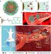
Fig. 1.
Fig. 1.. Schemes illustrating the biosafety, drug loading, and targeted thrombolysis of swarming HPB-NRs based on MB@PSS NPs as building blocks.
An individual MB@PSS NP with loaded drugs and conjugated AT III (A), a heparinoid-PSS brush (B), loading tissue-type plasminogen activator (t-PA) drugs (C), and conjugating AT III (D) with PSS brushes via high affinity. (E) Strong electrostatic repulsions among neighboring MB@PSS NPs and between MB@PSS NPs and vascular wall, ensuring their strong anti-agglomeration, anti-bioadhesion, and high biosafety in blood flows. (F) Safe and efficient targeted thrombolysis process by swarming HPB-NRs, including building block injection (Step-i), magnetic collection (Step-ii), nanorobot assembly/propulsion (Step-iii), thrombus destruction (Step-iv), and nanorobot disassembly (Step-v).
Fig. 2.
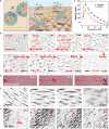
Fig. 2.. Characterization and dispersity of MB@PSS NPs in biological media.
(A toC) SEM image (A), FTIR spectrum (B), and magnetic hysteresis loop (C) of the MB@PSS NPs. a.u., arbitrary units. (D) Schematic diagram illustrating the enhanced surface charge density (σ) and interparticle electrostatic repulsion (F1) of MB@PSS NPs in biological media due to the shrinkage of PSS brush shell thickness (D) with the increased electrolyte concentration (Ce). (E) ζ and hydrodynamic size of the MB@PSS NPs in an aqueous solution with different NaCl concentrations. (F andG) Time-lapse microscopic images depicting the dispersion of agglomerated MB@PSS NPs in an aqueous suspension upon injecting DMEM, where the red dashed circles indicate the DMEM diffusion range (F) or being dropped into whole blood (G). The left-upper inserts are partially magnified microscopic images.
Fig. 3.
Fig. 3.. Magnetic propulsion and blood-environment adaptivity of magnetic HPB-NRs.
(A) Schematic diagram of the kayaking (crawling) and rolling motion of a rod-like HPB-NR assembled from the MB@PSS NPs under a precessing or rotatingB(t).F1,F2, andF3 represent the electrostatic repulsion, van der Waals force, and magnetic dipolar attraction between two neighboring MB@PSS NPs at a specific interparticle distance (d), respectively. (B) Reversible assembly and magnetically driven propulsions (crawling and rolling propulsions) of the HPB-NRs in the culture medium (DMEM) in response to a precessing or rotatingB(t).B0 andf of the precessing or rotatingB(t) are 10 mT and 5 Hz, respectively. Scale bars, 80 μm. (C) Average velocity (v) versusf for the crawling and rolling HPB-NRs. (D) Magnetic propulsions of the HPB-NRs in whole blood. (E) Disassembly of the HPB-NRs in serum afterB(t) is removed. (F) Crawling motion of the HPB-NRs on a dense layer of VECs. Scale bars, 100 μm (D to F).
Fig. 4.
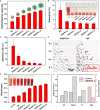
Fig. 4.. Heparinoid-PSS brush length effect on blood-environment compatibility of MB@PSS NPs.
(A) Hydrodynamic sizes of the MB@PSS NPs obtained at different polymerization time (t in a unit of hours), MB@PSS-t. (B) Hemolysis and digital photographs (insets) of RBCs after being exposed to the naked MBs and MB@PSS-t NPs. PBS (−) and deionized water (+) were used as negative and positive controls, respectively. (C) Adhesion (or particle loss) rate of the naked MBs and MB@PSS-t to the substrate when the NRs assembled from them collectively move in DMEM. (D) Optical microscopic images illustrating the magnetic propulsions of the NRs assembled from MB@PSS-6 (left) and naked MBs (right) in DMEM, revealing that the adhesion and particle loss to the substrate occur with the naked MBs but disappear for MB@PSS-6 NPs. Scale bar, 100 μm. (E) Anticoagulation performance of naked MBs and MB@PSS-t NPs evaluated by hemoglobin content in the supernatant. The absorbance of saline is used as the control. (F) In vitro anticoagulation activities of naked MBs (control) and MB@PSS-6 NPs.
Fig. 5.
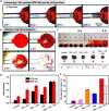
Fig. 5.. Targeted thrombolysis in vitro.
(A) Time-lapse microscopic images depicting the propulsion of the swarming t-PA–loaded HPB-NRs toward a thrombus underB(t) from the top view. (B) Targeted thrombolysis by the swarming t-PA–loaded HPB-NRs. (C) Digital photographs of blood clots after being exposed to control (saline), moving HPB-NRs [NRs +B(t)], immobile t-PA–loaded MB@PSS NPs (NRs + t-PA), t-PA, and moving t-PA–loaded HPB-NRs [NRs + t-PA +B(t)] for 6 hours. (D) Optical absorbance of the supernatant at 410 and 540 nm (OD410 and OD540), corresponding to the indicators of the released fibrin (410 nm) and hemoglobin (540 nm) from the blood clots shown in (C) after 6-hour treatment, respectively. (E) Calculated thrombolytic rate by weighting the mass of the thrombus after 6-hour treatment, where (a) in group NRs + t-PA +B(t) represents the simple sum of the separate thrombolytic rates contributed from the mechanical force and the targeted t-PA.
Fig. 6.
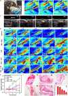
Fig. 6.. Targeted thrombolysis in vivo.
(A) Experimental setup of the targeted thrombolysis treatment of SD rat's femoral vein venous thrombus in vivo by the swarming HPB-NRs. (B) Blood flow in the SD rat's femoral vein monitored by the LSBFMS before (0 min) and after (2.5 and 5 min) the thrombus is generated. Scale bar, 5 mm. (C) Real-time tracking of the MB@PSS NPs in the SD rat's femoral vein by ultrasound imaging when the MB@PSS NPs are magnetically collected near the thrombus by a permanent magnet. Scale bar, 2 mm. (D) LSBFMS-monitored blood flow in the femoral vein of the SD rats from the control (saline), NRs +B(t), t-PA, NRs + t-PA, and NRs + t-PA +B(t) groups at different time. Scale bar, 4 mm. (E) Blood perfusion over time for all groups (n = 5 per group). (F) Representative H&E staining photomicrographs of the femoral vein venous thrombus after 4-hour treatment of control (a), NRs +B(t) (b), t-PA (c), NRs + t-PA (d), NRs + t-PA +B(t) (e), as well as the corresponding residual thrombus area (f). Scale bar, 1 mm.
Fig. 7.
Fig. 7.. In vivo safety analysis.
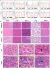
(A) Blood biochemistry analysis. ALT, AST, TBIL, ALB, TP, and GLOB are common markers for hepatic toxicity. Urea and CREA are the markers for renal toxicity. (B) H&E-stained histological sections of major organs, including heart, liver, spleen, lung, and kidney, from the control group, mice treated with MBs and MB@PSS NPs for 14 days, respectively. (C) Biodistributions of the MB@PSS NPs (a and c) and MBs (b and d) in liver and spleen tissues at 14 days after injection.
See this image and copyright information in PMC

Similar articles

See all similar articles

Cited by

See all "Cited by" articles

References

    1. D. Jin, K. Yuan, X. Du, Q. Wang, S. Wang, L. Zhang, Domino reaction encoded heterogeneous colloidal microswarm with on-demand morphological adaptability. Adv. Mater. 33, e2100070 (2021). - PubMed
    1. J. Yu, D. Jin, K.-F. Chan, Q. Wang, K. Yuan, L. Zhang, Active generation and magnetic actuation of microrobotic swarms in bio-fluids. Nat. Commun. 10, 5631 (2019). - PMC - PubMed
    1. B. Yigit, Y. Alapan, M. Sitti, Programmable collective behavior in dynamically self-assembled mobile microrobotic swarms. Adv. Sci. 6, 1801837 (2019). - PMC - PubMed
    1. Q. Wang, L. Zhang, External power-driven microrobotic swarm: From fundamental understanding to imaging-guided delivery. ACS Nano 15, 149–174 (2021). - PubMed
    1. J. Yu, B. Wang, X. Du, Q. Wang, L. Zhang, Ultra-extensible ribbon-like magnetic microswarm. Nat. Commun. 9, 3260 (2018). - PMC - PubMed

MeSH terms

Substances

Related information

LinkOut - more resources

Full text links
Atypon full text link Atypon Free PMC article
Cite
Send To

NCBI Literature Resources

MeSHPMCBookshelfDisclaimer

The PubMed wordmark and PubMed logo are registered trademarks of the U.S. Department of Health and Human Services (HHS). Unauthorized use of these marks is strictly prohibited.


[8]ページ先頭

©2009-2025 Movatter.jp