Movatterモバイル変換


[0]ホーム

URL:


Skip to main page content
U.S. flag

An official website of the United States government

Dot gov

The .gov means it’s official.
Federal government websites often end in .gov or .mil. Before sharing sensitive information, make sure you’re on a federal government site.

Https

The site is secure.
Thehttps:// ensures that you are connecting to the official website and that any information you provide is encrypted and transmitted securely.

NIH NLM Logo
Log inShow account info
Access keysNCBI HomepageMyNCBI HomepageMain ContentMain Navigation
pubmed logo
Advanced Clipboard
User Guide

Full text links

Nature Publishing Group full text link Nature Publishing Group Free PMC article
Full text links

Actions

Share

.2022 Nov 1;12(1):18431.
doi: 10.1038/s41598-022-23134-8.

Cellular adaptations leading to coral fragment attachment on artificial substrates in Acropora millepora (Am-CAM)

Affiliations

Cellular adaptations leading to coral fragment attachment on artificial substrates in Acropora millepora (Am-CAM)

Brett M Lewis et al. Sci Rep..

Abstract

Reproductive propagation by asexual fragmentation in the reef-building coral Acropora millepora depends on (1) successful attachment to the reef substrate through modification of soft tissues and (2) a permanent bond with skeletal encrustation. Despite decades of research examining asexual propagation in corals, the initial response, cellular reorganisation, and development leading to fragment substrate attachment via a newly formed skeleton has not been documented in its entirety. Here, we establish the first "coral attachment model" for this species ("Am-CAM") by developing novel methods that allow correlation of fluorescence and electron microscopy image data with in vivo microscopic time-lapse imagery. This multi-scale imaging approach identified three distinct phases involved in asexual propagation: (1) the contact response of the coral fragment when contact with the substrate, followed by (2) fragment stabilisation through anchoring by the soft tissue, and (3) formation of a "lappet-like appendage" structure leading to substrate bonding of the tissue for encrustation through the onset of skeletal calcification. In developing Am-CAM, we provide new biological insights that can enable reef researchers, managers and coral restoration practitioners to begin evaluating attachment effectiveness, which is needed to optimise species-substrate compatibility and achieve effective outplanting.

© 2022. The Author(s).


Conflict of interest statement

The authors declare no competing interests.

Figures

Figure 1
Figure 1
An overview of the three phases of attachment over 21 days from both the expanded view and the obscured underside and with the timing of each stage and their characteristics. (ac) Coral attachment through three stages: (1) contact response leading to (2) soft tissue anchoring and fragment stabilisation and (3) calcification and lappet-like appendage development leading to bonding and encrustation. (e,f) Micro view of the underside in all three stages. (h) The timing of the processes which characterise each phase represented by a heat map.
Figure 2
Figure 2
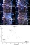
Stills from time-lapse light microscope movies displaying the representative processes that characterise the contact response phase, i.e., tissue enlargement, mucus release, wound cleaning (0–5 days). (a) Wounds (Wd) formed by abrasion are cleaned by mesenterial filaments (MF) as the surrounding immunocompromised tissues become enlarged (Enl). (b,c) Mesenterial filament deployment is increased external to the body cavity as the compromised tissues become further enlarged (2–3 days). (d) Mucus (Mu) secretions that have formed on the substrate surface surrounding the compromised tissues change colour over time—from transparent to white or brown. (e) A plot showing the relative density of the mesenterial filaments activity inside and outside the body cavity on each day the still (a,b,c,d) was captured. Wound; Wd, Enlarged tissues; Enl, mesenterial filaments; MF, mucus layer; Mu.
Figure 3
Figure 3
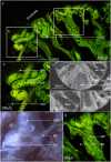
Confocal fluorescence microscopy images, backscatter electron microscopy images and stills from time-lapse microscopy movies highlighting the distribution, cell composition and mesenterial behaviour during the contact response phase. (a,b) Contact with the substrate lead to the deployment of mesenterial filaments (MF) in newly formed wounds and the formation of a mucus barrier. (ce) A diverse number of secretory cells (Sec) were present in the mesenterial filaments ofA. millepora, and their proximity to the wounds and mucus indicates a role in cleaning and mucus formation. (f) For mesenterial filaments to move through the SBW and extend beyond the body cavity, they employ a twisting or corkscrewing motion to slide out of the cinclide-like (Cin) openings (Supplementary Movie S2). (g) Fluorescence image of the mesenterial filament (MF) twisting through the cinclide-like structure and the costae (Sk). surface body wall; SBW, mucus layer; Mu, Nm; nematocyst, Skeleton; Sk.
Figure 4
Figure 4
Backscatter electron microscopy images and stills from time-lapse movies comparingA. millepora' 's regular SBW with the enlarged tissues during the contact response phase. (a) A standard surface body wall (SBW) ofA. millepora showing the two epithelial layers (epidermis; Ep, gastrodermis; Ga) separated by the mesoglea (Mes) and the cells and processes that characterise these layers; supporting cells (Sup), intercellular vacuole (Va) or spaces in between each cell, nematocysts (Nm), type 2 gland cell (Mu), Symbiodiniaceae (Sym) and the soluble surface mucus layer (SML). (b) Higher magnification image of the vesicles (white) in standard type 2 gland cells (mucocytes). (c,d) The soft tissue became enlarged at the points of contact between the coral and the substrate. (d) Electron microscopy images of the enlarged SBW tissues showed a proliferation of densely packed thin supporting cells and Symbiodiniaceae. and the differentiation/proliferation of type 4 (yellow) and type 1 and 2 (blue) gland cells (Sec) with lead to the loss of intercellular vacuoles. (e,f) Electron image showing the vesicles (Sec4) of type 4 gland cells with matter trapped inside and mucocytes (Sec2). (g) Closer look at the densely packed epitheliomuscular supporting cells (Sup) and their nuclei. Skeleton; Sk, surface body wall; SBW, epidermis; Ep, gastrodermis; Ga, mesoglea; Mes, surface mucus layer; SML, Nm; nematocyst, supporting cells; Sup, intercellular vacuole; Va, secretory cells; Sec, type 2 gland cell; Mu and Sec2, type 4 gland cell; Sec4, Symbiodiniaceae; Sym.
Figure 5
Figure 5
Backscatter electron microscopy images and stills from time-lapse microscopy movies comparingA. millepora' 's regular tissues with the enlarged anchored tissues. (a) The enlarged tissues (Enl) from phase one develop into a that anchors and helps stabilise the fragment. (b,c) The anchoring process creates a sealed or enclosed (Enc) environment where the SBW or epidermis can be safely removed and a BBW can form. (d,e) The anchoring tissue took on a complex undulated morphology that likely assists tissue anchoring. The shifting morphology of these tissues is down to an ongoing proliferation of supporting cells which also act as epitheliomuscular cells (Emc) and (f,g) gland cells (Sec), which may also aid with adhesion. Enlarged tissues; Enl, soft tissue attachment; Anc, enclosed environment; Enc, epidermis; Ep, gastrodermis; Ga, mesoglea; Mes.
Figure 6
Figure 6
Representative confocal fluorescence microscopy images and stills from time-lapse microscopy movies highlighting the distribution, cell composition and mesenterial behaviour during the contact response phase. (a) The fragment deploys mesenterial filaments (MF)en-masse as concentrated balls to begin removing the epidermis of the enclosed SBW of the anchored tissue in contact with the substrate. (b) The removal of epidermal cells leads to the development of cells needed for skeleton precipitation. (c) This image shows the mesenterial filaments in the anchored or compromised tissues with increased numbers of Symbiodiniaceae in the mesentery filaments which could be a sign of their digestion. Skeleton; Sk, mesenterial filament; MF, surface body wall; SBW.
Figure 7
Figure 7
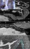
Backscatter electron microscopy images and stills from time-lapse microscopy movies highlighting the location, morphology and function of the lappet-like appendage vital for encrustation of the substrate and for forming an enduring bond. (a,b) The lappet-like appendage (Lap) is located on the distal edge of the encrusting rim and is responsible for both initial basal precipitation and costae development (Cos). (c) The lappet-like appendage is a complex structure possessing an SBW that is thicker than the coral regular SBW. (d) Higher magnification view of the lappet-like appendage shows a complex morphology consisting of 4 key characteristics: (1) tissue undulations (Und) with densely packed cells on its underside, (2) a transition zone (Trn) where the SBW transitions into the BBW, (3) a '''pocket' of calicoblastic cells (Cos) for costae development (Cos)—this is not always present as some areas are not actively precipitating costae and (4) a thin, poorly defined continuation of the calicoderm (Ext) that sits between the lappet-like appendage and the substrate surface and is responsible for the first layers of skeleton (InSk). (e) Higher magnification view of the transition zone (Trn) and the poorly defined calicoderm extension (Ext). Calicoderm extension is non-traditional in that it does not possess a gastrodermis directly adjacent to the corals, which is the standard tissue arrangement. Both epithelial layers of the SBW primarily consist of supporting epitheliomuscular cells, while the BBW consists of a standard calicodermis and gastrodermis. As the lappet-like appendage slowly migrates, it leaves behind a trail of new BBW to continue colony growth and skeletal thickening. (f) The '''pocket' of cells responsible for the costae wall (Cos) forms between the lappet-like appendage SBW epidermis (Ep) and gastrodermis (Ga) (Blue) at the mesoglea. skeleton; Sk, epidermis; Ep, gastrodermis; Ga, calicodermis; Ca, undulations; Und, transition zone; Trn, calicodermis extension; Ext.
Figure 8
Figure 8
Backscatter electron microscopy images highlighting the primary cell composition of a newly developed lappet-like appendage. (a) The base of the developing lappet-like appendage (right) and the trailing basal body wall (BBW, left). (b) The standard BBW consists of two epithelial layers separated by the mesoglea (M); the calicodermis (Ca) primarily consists of cuboidal calicoblasts and a gastrodermis (Ga). (c) A pocket of vestigial epidermal cells that has not yet been removed by mesenterial filaments is still present in the BBW trailing the new lappet-like appendage. (d) The lappet-like primarily consists of epitheliomuscular cells (Emc) that attach to the mesoglea (M) by their filamentous myofibrils (Myo). The heightened musculature gives the lappet-like its complex undulated morphology and the mechanical ability to pulse and perhaps grip the surface. Lappet-like; Lap, costae; Cos, surface body wall; SBW, epidermis; Ep, transition zone; Trn, calicodermis extension; Ext, epitheliomuscular supporting cells; Emc.
Figure 9
Figure 9
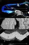
Representative confocal fluorescence microscopy images and backscatter electron microscopy images highlighting the costae wall and initial skeletal layer development in the lappet-like appendage. (a) A confocal fluorescence image of the lappet-like appendage overlayed with the corresponding SEM image showing the morphology of the lappet-like appendage and its relationship to the costae (Cos), initial basal deposits (InSk) and the basal skeleton (Sk). (b,c) First, the lappet-like appendage deposits an initial skeleton (InSk) via the lappet-like appendage poorly defined calicoblastic extension; then, as the lappet-like appendage moves forward, the trailing calicodermis produces a layer of skeleton (Sk) (arrows show the direction of the thickening), building a stronger basal attachment. The lappet-like appendage can generate rapid accretion deposits (RADs) (b) that form the costae walls (Cos) and further the robustness of the skeleton/attachment. (c) The lappet-like 'appendage's poorly defined calicoblastic extending layer (a) (Ext) appears to produce a protuberance (Prt) in the initial skeleton (InSk). (d) The smooth initial skeleton layer possesses characteristics similar to those of Clypeotheca (Clp) and can form a keystone structure (Key) in a similar fashion to dissepiments, which can indicate irregular development. CCA; crustose coralline algae, Sub; substrate. calicodermis; Ca, calicodermis extension; Ext, substrate; Sub.
Figure 10
Figure 10
Digital graphic of the aquaria setup for the time-lapse and macro images of the ceramic clast and the glass substrate in the fragments. (a) The interface between the coral fragment tissue and the substrate was recorded (time-lapse) in a research aquaria setup with LED light for 30 days using light microscopes. (b,c) The ceramic substrate was placed either between the branches or at the surface/base of the fragment to maximise contact with both the higher growth areas and slower growth base tissues. (d) The glass substrate was lighter and less hydrodynamic than the ceramic, which made it easier to dislodge via water flow and therefore had to be placed between the branches for added stability.
See this image and copyright information in PMC

Similar articles

See all similar articles

References

    1. Smith LD, Hughes TP. An experimental assessment of survival, re-attachment and fecundity of coral fragments. J. Exp. Mar. Biol. Ecol. 1999;235:147–164. doi: 10.1016/S0022-0981(98)00178-6. - DOI
    1. Harriott, V. & Fisk, D. in Proc 6th int coral Reef Symp. 375–379.
    1. Wallace CC. Reproduction, recruitment and fragmentation in nine sympatric species of the coral genus Acropora. Mar. Biol. 1985;88:217–233. doi: 10.1007/BF00392585. - DOI
    1. Roniewicz E, Stolarski J. Evolutionary trends in the epithecate scleractinian corals. Acta Palaeontol. Pol. 1999;44:131–166.
    1. Barnes DJ. The structure and formation of growth-ridges in scleractinian coral skeletons. Proc. R. Soc. Lond. Ser. B Biol. Sci. 1972;182:331–350.

MeSH terms

Related information

LinkOut - more resources

Full text links
Nature Publishing Group full text link Nature Publishing Group Free PMC article
Cite
Send To

NCBI Literature Resources

MeSHPMCBookshelfDisclaimer

The PubMed wordmark and PubMed logo are registered trademarks of the U.S. Department of Health and Human Services (HHS). Unauthorized use of these marks is strictly prohibited.


[8]ページ先頭

©2009-2025 Movatter.jp