Movatterモバイル変換


[0]ホーム

URL:


Skip to main page content
U.S. flag

An official website of the United States government

Dot gov

The .gov means it’s official.
Federal government websites often end in .gov or .mil. Before sharing sensitive information, make sure you’re on a federal government site.

Https

The site is secure.
Thehttps:// ensures that you are connecting to the official website and that any information you provide is encrypted and transmitted securely.

NIH NLM Logo
Log inShow account info
Access keysNCBI HomepageMyNCBI HomepageMain ContentMain Navigation
pubmed logo
Advanced Clipboard
User Guide

Full text links

Nature Publishing Group full text link Nature Publishing Group Free PMC article
Full text links

Actions

Share

.2022 May;40(5):731-740.
doi: 10.1038/s41587-021-01133-w. Epub 2021 Dec 9.

Programmable deletion, replacement, integration and inversion of large DNA sequences with twin prime editing

Affiliations

Programmable deletion, replacement, integration and inversion of large DNA sequences with twin prime editing

Andrew V Anzalone et al. Nat Biotechnol.2022 May.

Abstract

The targeted deletion, replacement, integration or inversion of genomic sequences could be used to study or treat human genetic diseases, but existing methods typically require double-strand DNA breaks (DSBs) that lead to undesired consequences, including uncontrolled indel mixtures and chromosomal abnormalities. Here we describe twin prime editing (twinPE), a DSB-independent method that uses a prime editor protein and two prime editing guide RNAs (pegRNAs) for the programmable replacement or excision of DNA sequences at endogenous human genomic sites. The two pegRNAs template the synthesis of complementary DNA flaps on opposing strands of genomic DNA, which replace the endogenous DNA sequence between the prime-editor-induced nick sites. When combined with a site-specific serine recombinase, twinPE enabled targeted integration of gene-sized DNA plasmids (>5,000 bp) and targeted sequence inversions of 40 kb in human cells. TwinPE expands the capabilities of precision gene editing and might synergize with other tools for the correction or complementation of large or complex human pathogenic alleles.

© 2021. The Author(s), under exclusive licence to Springer Nature America, Inc.


Conflict of interest statement

Declaration of Interests

D.R.L. is a consultant and equity holder of Beam Therapeutics, Prime Medicine, Pairwise Plants, and Chroma Medicine, companies that use genome editing or genome engineering technologies. A.V.A., C.J.P., and J.M.L. are currently employees at Prime Medicine. A.V.A., X.D.G., C.J.P., and D.R. L. have filed patent applications on twinPE and prime editing through the Broad Institute.

Figures
Extended Data Fig. 1
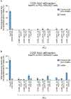
Extended Data Fig. 1. Twin prime editing mediates sequence replacements atCCR5
(a) Replacement of endogenous sequence withinCCR5 region 1 with a 108-bp fragment ofFKBP12 cDNA using twinPE (FKBP12 sequence oriented in the forward direction,) or PE3 (FKBP12 sequence oriented in the reverse direction). For PE3 editing, pegRNA RT templates were designed to encode 108 base pairs ofFKBP12 cDNA sequence and one of three different target-site homology sequence lengths. For PE3 edits, each pegRNA was tested with three nicking sgRNAs. (b) Replacement of endogenous sequence withinCCR5 region 2 with a 108-bp fragment ofFKBP12 cDNA sequence using twinPE (FKBP12 sequence oriented in the forward direction) or PE3 (FKBP12 sequence oriented in the reverse direction). As in (a), PE3 edits were tested with pegRNAs containing RT templates that were designed to encode 108 base pairs ofFKBP12 cDNA sequence and one of three different target-site homology sequence lengths. For PE3 edits, each pegRNA was tested with three nicking sgRNAs. Values and error bars reflect the mean and s.d. of three independent biological replicates. (c) Transfection of HEK293T cells with a pair of pegRNAs targetingCCR5 leads to replacement of 53 base pairs of endogenous sequence with 113 base pairs (attB–[27-bp spacer]–attP) or 103 base pairs (attB–[27-bp spacer]–attB) of exogenous sequence. Values and error bars reflect the mean and s.d. of three independent biological replicates.
Extended Data Fig. 2
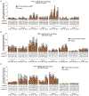
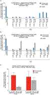
Extended Data Fig. 2. Recoding ofPAH exon sequences in HEK293T cells via twinPE
Screen of pegRNA pairs targetingPAH for recoding of (a) exons 2, 4 and 5, (b) exon 7, and (c) exons 9, 10, 11, and 12. RT templates of pegRNAs encoded partially recoded exonic sequence to optimize orthogonality to the endogenous gene sequence. For each spacer pair, nine pegRNA combinations were tested using three PBS variants for each spacer in a three-by-three matrix, with RT templates encoding the recoded exonic sequence, which was held constant for given spacer pairs. Sequences of pegRNAs are listed in Supplementary Table 1. Sequences of recoded exonic sequences are listed in Supplementary Table 4. Values in (a), (b) and exon 9 in (c) reflect single biological replicates. Values for exons 10, 11 and 12 in (c) reflect the mean of three independent biological replicates.
Extended Data Fig. 3
Extended Data Fig. 3. Installation of a 38-bp Bxb1attB site atCCR5 with twinPE
Spacer pairs targeting theCCR5 locus were designed for twinPE-mediated insertion of the Bxb1attB attachment site. For each spacer, three pegRNAs were designed having three different PBS lengths and a fixed RT template that encodes the full-length Bxb1attB sequence (38 bp). Sequences of pegRNAs are listed in Supplementary Table 1. For each spacer pair, a three-by-three matrix of pegRNA combinations was tested by plasmid DNA co-transfection with PE2 in HEK293T cells. Each pegRNA pair is specified below the x-axis. Values reflect single biological replicates.
Extended Data Fig. 4
Extended Data Fig. 4. Installation of a 50-bp Bxb1attP site atAAVS1 with twinPE
Spacer pairs targeting theAAVS1 locus were designed for twinPE-mediated insertion of the Bxb1attP attachment site. For each spacer, three pegRNAs were designed having three different PBS lengths and a fixed RT template that encodes a portion (43-44 bp) of the Bxb1attP sequence. Sequences of pegRNAs are listed in Supplementary Table 1. For each spacer pair, a three-by-three matrix of pegRNA combinations was tested by plasmid DNA co-transfection with PE2 in HEK293T cells. Each pegRNA pair is specified below the x-axis. Values reflect single biological replicates.
Extended Data Fig. 5
Extended Data Fig. 5. Comparison of twinPE and PE3 for Bxb1attB insertion atCCR5
(a) Replacement of endogenous sequence withinCCR5 region 1 with the Bxb1attB site using twinPE or PE3. For PE3 editing systems, pegRNA RT templates were designed to encode the Bxb1attB sequence and one of three different target-site homology sequence lengths. For PE3 edits, each pegRNA was tested with three nicking sgRNAs. (b) Replacement of endogenous sequence withinCCR5 region 2 with the Bxb1attB sequence using twinPE or PE3. As in (a), PE3 edits were tested with pegRNAs containing RT templates that were designed to encode the Bxb1attB sequence and one of three different target-site homology sequence lengths and tested with three nicking sgRNAs. Values and error bars in (a) and TwinPE edits, PE3 edits ofCCR5_D2_23,CCR5 D2_28 with nicking guide RNA C1 and C1.5 in (b) reflect the mean and s.d. of three independent biological replicates. Values ofCCR5 D2_28 with nicking guide RNA C4 andCCR5 D2_34 in (b) reflect the mean of two independent biological replicates.
Extended Data Fig. 6
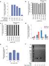
Extended Data Fig. 6. TwinPE combined with Bxb1 recombinase for targeted knock-in of donor DNA plasmids
(a) Bxb1-mediated DNA donor knock-in in clonal HEK293T cell lines. Transfection of a HEK293T clonal cell line containing homozygousattB site insertion atCCR5 with varying amounts of Bxb1-expressing plasmid andattP-containing donor DNA plasmid. Knock-in efficiency was quantified by ddPCR. Values and error bars reflect the mean of two independent biological replicates. (b) Assessment of genome-donor junction purity by high-throughput sequencing. Genomic DNA from single-transfection knock-in experiments was amplified with a forward primer that binds the genome and a reverse primer that binds within the donor plasmid (Supplementary Table 2). Values and error bars reflect the mean and s.d. of three independent biological replicates. (c) Assessment of genome-donor junction purity at the other junction by high-throughput sequencing as performed in (b). Values and error bars in 506c+584b,509b+584b, 1077c+1154c, and 3786c+3903c reflect the mean and s.d. of three independent biological replicates. Values in 325a+414b, 513b+584b, 3786c+3930c reflect the mean of two independent biological replicates. (d) Multiplexed single-transfection knock-in atAAVS1 andCCR5. HEK293T cells were transfected with plasmids encoding PE2, Bxb1, a pair of pegRNAs for the insertion ofattP atAAVS1, anattB-donor, a pegRNA pair for the insertion of one of four attachment sites (attB, attB-GA, attP, orattP-GA) atCCR5, and a corresponding donor. Knock-in was observed at both target loci under all four conditions. Insertion ofattP atAAVS1 andattB atCCR5 gave the lowest knock-in efficiencies overall (0.2% atAAVS1, 0.4% atCCR5). Insertion ofattP at both sites yielded the highest levels of knock-in atAAVS1 (1.8%) but low levels (0.2%) atCCR5. When an orthogonal edit (attB-GA orattP-GA) was introduced atCCR5, AAVS1 knock-in was 0.7-0.8%. Higher knock-in atCCR5 was observed withattB-GA (1.4%) than withattP-GA (0.4%), consistent with our single locus knock-in results. Values and error bars reflect the mean and s.d. of three independent biological replicates. (e) and (f) Effects of reducing pegRNA overlap on twinPE efficiency and donor/pegRNA recombination. (e) The editing efficiencies of pairs of pegRNAs for insertion of Bxb1attB atCCR5 were measured by high-throughput sequencing. The pairs differed in the amount of overlap shared between their flaps, from 38 bp (full-lengthattB sequence) down to 20 bp. Editing efficiency of the pairs with shorter overlaps was comparable to the pair with full-length overlap. Values and error bars reflect the mean and s.d. of three independent biological replicates. (f) Assessment of recombination betweenattB-containing pegRNA plasmids andattP-containing donor plasmids. Following transfection of HEK293T cells with the indicated samples, isolated DNA was amplified with a forward primer that binds the pegRNA expression plasmid (TTGAAAAAGTGGCACCGAGT) and a reverse primer that binds the donor plasmid (CTCCCACTCATGATCTA). A positive 256-bp PCR band confirms recombination between the two plasmids. When the pegRNA encodes full-lengthattB (38-bp) or a truncated version ofattB with 30-bp of overlap between flaps, a band is observed; however, recombination is not observed when the pegRNAs encode a truncatedattB with only 20-bp of flap overlap. The “No PE2” control uses the 38-bp overlap pegRNA pair. No recombination is observed in the absence of Bxb1 or if the donor and pegRNA plasmids both bearattB (Mismatch, “M”). Three independent biological replicates were performed and a representative image from one of the replicates is shown.
Extended Data Fig. 7
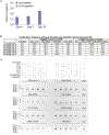
Extended Data Fig. 7. Expression of human Factor IX from theALB promoter following twinPE-recombinase knock-in and characterization of Bxb1 off-target editing
(a) Huh7 cells were transfected with Bxb1, donor (attP-splice acceptor-cDNA ofF9 exons 2-8), PE2, and pegRNAs for installation ofattB in the first intron ofALB or atCCR5. Three days post-transfection, cells were split and allowed to grow to confluence. Their media was changed, and they were left to condition the fresh media, with aliquots taken at days 4, 7, and 10. Factor IX was present at detectable levels by ELISA (dashed line represents the lower limit of detection) in two of three samples treated withALB pegRNAs at Day 4, and in all samples treated withALB pegRNAs at Day 7 and Day 10. Factor IX was never detected in the conditioned media of any samples treated withCCR5 pegRNAs. Values and error bars reflect the mean and s.d. of two or three independent biological replicates. (b) Targeted amplicon sequencing was performed for each of the five nominated pseudo-sites (OT1-OT5) from seven different samples treated with 5.6-kb donor DNA plasmid, twinPE reagents targetingCCR5 or AAVS1, and Bxb1 recombinase. The indels in all five pseudo-sites are either below the limit of detection (<0.1%) or near-background compared to untreated controls. The integration efficiency at the on-target site was measured by ddPCR as shown in Fig. 3d (c) To capture potential donor plasmid integration events at nominated pseudo-sites, primers were used to amplify predicted integration junctions. The gel depicts PCR reactions performed for each off-target site as indicated in the above legend. Confirmation of on-target donor integration from the samples is shown in the right-most column of the gel. In (b) and (c), two or three independent biological replicates were performed.
Extended Data Fig. 8
Extended Data Fig. 8. TwinPE and Bxb1-mediated inversion in HEK293T GFP reporter cells
(a) The lentiviral fluorescent reporter construct used to assess inversion efficiency with twinPE and Bxb1 recombinase. The reporter contains an EF1α promoter followed by an inverted H2B-EGFP coding sequence that is flanked by partialAAVS1 DNA sequence, an internal ribosome entry site (IRES), and a puromycin resistance gene. Successful installation of opposite-facingattB (left) andattP (right) sequences at theAAVS1 target sequences and subsequent inversion by Bxb1 corrects the orientation of GFP for functional expression. (b) The fluorescent reporter construct was stably integrated into HEK293T cells via lentiviral transduction and puromycin selection. The polyclonal GFP reporter cell line was then transfected with twinPE plasmid components (PE2 and four pegRNAs) and varying amounts of Bxb1 plasmid for single-transfection inversion. Cells were analyzed by flow cytometry and gated for live single cells. Quantification of GFP positive cells by flow cytometry. Values and error bars reflect the mean of two independent biological replicates.
Extended Data Fig. 9
Extended Data Fig. 9. TwinPE and Bxb1 recombinase-mediated inversion betweenIDS andIDS2
(a) Assessment of the invertedIDS junction purity by high-throughput sequencing in HEK293T cells. Frequency of expected junction sequences containingattR andattL recombination products after twinPE and BxB1-mediated single-step inversion. The product purities range from 81-89%. Values and error bars reflect the mean and s.d. of three independent biological replicates. (b) Schematic diagram of the designed PCR strategies for quantifyingIDS inversion efficiency. Primer pair 1 (green forward and blue reverse primer) can amplify the unedited alleles (403 bp), twinPE-edited alleles (337 bp), and the inverted alleles (326 bp) at junction 1 in a single PCR reaction. Due to the size difference, a UMI protocol was applied to eliminate PCR bias during quantification of inversion efficiency. Similarly, using primer pair 2 (red forward and blue reverse primer), the unedited alleles (346 bp), twinPE-edited alleles (326 bp), and inverted alleles (320 bp) at junction 2 can be amplified in a single PCR reaction. Amplicons can then be sequenced by standard high-throughput sequencing protocols for amplicon sequencing. (c) Screening of pegRNA pairs for the insertion of Bxb1attB andattP sequences atIDS andIDS2. TwinPE editing was tested with standard pegRNAs and epegRNAs containing a 3’ evoPreQ1 motif. Values and error bars reflect the mean and s.d. of three independent biological replicates.
Extended Data Fig. 10
Extended Data Fig. 10. Twin prime editing mediated insertion inCCR5 region 2 in HEK293T cells, twin prime editing in multiple human cell lines, and editing activity of Cas9 nickase and PE2-dead RT variants
(a) TwinPE-mediated endogenous sequence replacement with Bxb1attB attachment site inCCR5 region 2 in HEK293T cells. (b) TwinPE-mediated endogenous sequence replacement withattP, attB, or 22-nt DNA sequences in multiple human cell lines. Six different pegRNA pairs targeting five loci were tested in HEK293T, HeLa, U2OS and K562 cells. HEK293T and HeLa cell were transfected with PE2 and pegRNA plasmids via Lipofectamine 2000 (Thermo Fisher) andTransIT-HeLaMonster (Mirus), respectively. U2OS and K562 cells were nucleofected using Lonza 4D-Nucleofector and SE kit. DNA loci and the specified insertion edits are shown in the x-axis. (c) HEK293T cells were transfected with twinPE pegRNA pairs and either Cas9–H840A nickase (nCas9), PE2-dRT (a PE2 variant that contains K103L and R110S inactivating mutations to the RT domain), or PE2. Treatment with either nCas9 or PE2-dRT did not result in desired edits, while PE2 installed the specified edits as indicated. Values and error bars in (a) and (c) reflect the mean and s.d. of three independent biological replicates. Values and error bars in (b) reflect the mean and s.d. of at least two independent biological replicates except editing inIDS2 in HeLa cells, editing in U2OS cells, and editing inMYC in K562 cells, which represent two independent biological replicates.
Figure 1 ∣
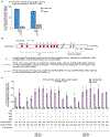
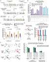
Figure 1 ∣. Overview of twinPE and twinPE-mediated sequence replacement.
(a) TwinPE systems target genomic DNA sequences that contain two protospacer sequences on opposite strands of DNA. PE2•pegRNA complexes target each protospacer, generate a single-stranded nick, and reverse transcribe the pegRNA-encoded template containing the desired insertion sequence. After synthesis and release of the 3’ DNA flaps, a hypothetical intermediate exists possessing annealed 3’ flaps containing the edited DNA sequence and annealed 5’ flaps containing the original DNA sequence. Excision of the original DNA sequence contained in the 5’ flaps, followed by ligation of the 3’ flaps to the corresponding excision sites, generates the desired edited product. (b) Example of twinPE-mediated replacement of a 90-bp sequence inHEK3 with a 38-bp Bxb1attB sequence. Red arrows indicate the position of pegRNA-induced nicks. (c) Evaluation of twinPE in HEK293T cells for the installation of the 38-bp Bxb1attB site as shown in (b) or the 50-bp Bxb1attP site atHEK3 using pegRNAs that template varying lengths of the insertion sequence. pegRNA names indicate spacer (A or B) and length of RT template. Values and error bars reflect the mean and s.d. of three independent biological replicates.
Figure 2 ∣
Figure 2 ∣. Targeted sequence insertion, deletion, and recoding with twinPE in human cells.
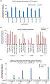
(a) Schematic diagram illustrating designs and edits generated for twinPE, PrimeDel and paired pegRNAs. Shaded gray boxes indicate regions where DNA is excised, green lines indicate the incorporation of heterologous DNA sequence, red and blue lines indicate regions of sequence homology (red to red, blue to blue), and yellow lines indicate regions with small edits. PrimeDel can introduce but does not require introduction of heterologous sequence (green). (b) Insertion ofFKBP coding sequence fragments with PE3 (12 bp, 36 bp, 108 bp) or twinPE (108 bp) atHEK3 in HEK293T cells. (c) Recoding of sequence within exons 4 and 7 inPAH in HEK293T cells using twinPE. A 64-bp target sequence in exon 4 was edited using 24, 36, or 59 bp of overlapping flaps, a 46-bp target sequence in exon 7 was edited using 22 or 42 bp of overlapping flaps, or a 64-bp sequence in exon 7 was edited using 24 or 47 bp of overlapping flaps. Editing activity was compared using standard pegRNAs or epegRNAs containing 3’ evoPreQ1 motifs. (d) Schematic diagram showing three distinct dual-flap deletion strategies that were investigated for carrying out targeted deletions. The “Single-anchor (SA)” twinPE strategy allows for flexible deletion starting at an arbitrary position 3’ of one nick site and ending at the other nick site. The “Hybrid-anchor (HA)” twinPE strategy allows for flexible deletion of sequence at arbitrarily chosen positions between the two nick sites. The “PrimeDel (PD)” strategy of Shendure and co-workers tested here allows for deletion of the sequence starting at one nick site and ending at another nick site. Shaded gray boxes indicate regions where DNA is excised, red and blue lines indicate regions of sequence homology (red to red, blue to blue). (e) Deletion of sequences atHEK3 in HEK293T cells using the SA-twinPE, HA-twinPE, or PD strategies targeting the same protospacer pair. Editing activity was compared using standard pegRNAs or epegRNAs containing 3’ evoPreQ1 motifs. (f) Deletion of exon 51 sequence at theDMD locus in HEK293T cells using SA-twinPE, PD, paired Cas9 nuclease, or twinPE-mediatedattB sequence replacement. A unique molecular identifier (UMI) protocol was applied to remove PCR bias (see Supplementary Note 1). Values and error bars in (b-f) reflect the mean and s.d. of three independent biological replicates.
Figure 3 ∣
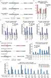
Figure 3 ∣. Site-specific genomic integration of DNA cargo with twinPE and Bxb1 recombinase in human cells.
(a) Schematic diagram of twinPE and Bxb1 recombinase-mediated site-specific genomic integration of DNA cargo. (b) Screening of twinPE pegRNA pairs for insertion of the Bxb1attB sequence at theCCR5 locus in HEK293T cells. (c) Screening of twinPE pegRNA pairs for installation of the Bxb1attP sequence at theAAVS1 locus in HEK293T cells. (d) Single transfection knock-in of 5.6-kb DNA donors using twinPE pegRNA pairs targetingCCR5 (red) orAAVS1 (blue). The twinPE pegRNAs installattB atCCR5 orattP atAAVS1. Bxb1 integrates a donor bearing the corresponding attachment site into the genomic attachment site. The number of integration events per 100 genomes is defined as the ratio of the target amplicon spanning the donor-genome junction to a reference amplicon inACTB, as determined by ddPCR. (e) Optimization of single-transfection integration atCCR5 using the A531+B584 spacers for the twinPE pegRNA pair. Identity of the templated edit (attB orattP), identity of the central dinucleotide (wild-type GT or orthogonal mutant GA), and length of the overlap between flaps were varied to identify combinations that supported the highest integration efficiency. % knock-in quantified as in (d). (f) Pairs of pegRNAs were assessed for their ability to insert Bxb1attB into the first intron ofALB. Protospacer sequences (277 and 358) are constant across the pegRNA pairs. The pegRNAs vary in their PBS lengths (variant b or c). The 277c/358c pair that performs best in HEK293T cells can also introduce the desired edit in Huh7 cells. (g) Comparison of single transfection knock-in efficiencies atCCR5 andALB in HEK293T and Huh7 cell lines. % knock-in quantified as in (d). Values and error bars reflect the mean and s.d. of three independent biological replicates.
Figure 4 ∣
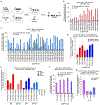
Figure 4 ∣. Site-specific large genomic sequence inversion with twinPE and Bxb1 recombinase in human cells.
(a) Schematic diagram of DNA recombination hot spots inIDS andIDS2 that lead to pathogenic 39-kb inversions, and the combined twinPE-Bxb1 strategy for installing or correcting theIDS inversion. (b) Screen of pegRNA pairs atIDS andIDS2 for insertion ofattP orattB recombination sites. Values and error bars reflect the mean and s.d. three independent biological replicates. (c) DNA sequencing analysis of theIDS andIDS2 loci after twinPE-mediated insertion ofattP orattB sequences, with or without subsequent transfection with Bxb1 recombinase. P-values were derived from a Student’s two-tailedt-test. Values and error bars reflect the mean and s.d. three independent biological replicates. (d) 40,167-bpIDS inversion product purities at the anticipated inversion junctions after twinPE-mediated attachment site installation and sequential transfection with Bxb1 recombinase. Values and error bars reflect the mean and s.d. three independent biological replicates. (e) Analysis of inversion efficiency by amplicon sequencing atIDS andIDS2 loci after sequential transfection or single-step transfection of twinPE editing components and Bxb1 recombinase. Values and error bars for sequential transfection reflect the mean and s.d. of three independent biological replicates; values for single-transfection reflect the mean of two independent biological replicates.
See this image and copyright information in PMC

Similar articles

See all similar articles

Cited by

See all "Cited by" articles

References

    1. Auton A et al. A global reference for human genetic variation. Nature 526, 68–74, doi:10.1038/nature15393 (2015). - DOI - PMC - PubMed
    1. Landrum MJ et al. ClinVar: public archive of relationships among sequence variation and human phenotype. Nucleic Acids Res 42, D980–985, doi:10.1093/nar/gkt1113 (2014). - DOI - PMC - PubMed
    1. Weischenfeldt J, Symmons O, Spitz F & Korbel JO Phenotypic impact of genomic structural variation: insights from and for human disease. Nat Rev Genet 14, 125–138, doi:10.1038/nrg3373 (2013). - DOI - PubMed
    1. Cox DB, Platt RJ & Zhang F Therapeutic genome editing: prospects and challenges. Nat Med 21, 121–131, doi:10.1038/nm.3793 (2015). - DOI - PMC - PubMed
    1. Doudna JA The promise and challenge of therapeutic genome editing. Nature 578, 229–236, doi:10.1038/s41586-020-1978-5 (2020). - DOI - PMC - PubMed

Methods References

    1. Anzalone AV et al. Search-and-replace genome editing without double-strand breaks or donor DNA. Nature 576, 149–157, doi:10.1038/s41586-019-1711-4 (2019). - DOI - PMC - PubMed
    1. Komor AC, Kim YB, Packer MS, Zuris JA & Liu DR Programmable editing of a target base in genomic DNA without double-stranded DNA cleavage. Nature 533, 420–424, doi:10.1038/nature17946 (2016). - DOI - PMC - PubMed
    1. Clement K et al. CRISPResso2 provides accurate and rapid genome editing sequence analysis. Nature Biotechnology 37, 224–226, doi:10.1038/s41587-019-0032-3 (2019). - DOI - PMC - PubMed
    1. Clement K, Farouni R, Bauer DE & Pinello L AmpUMI: design and analysis of unique molecular identifiers for deep amplicon sequencing. Bioinformatics 34, i202–i210, doi:10.1093/bioinformatics/bty264 (2018). - DOI - PMC - PubMed
    1. Shen W, Le S, Li Y & Hu F SeqKit: A Cross-Platform and Ultrafast Toolkit for FASTA/Q File Manipulation. PLoS One 11, e0163962, doi:10.1371/journal.pone.0163962 (2016). - DOI - PMC - PubMed

Publication types

MeSH terms

Substances

Associated data

Related information

Grants and funding

LinkOut - more resources

Full text links
Nature Publishing Group full text link Nature Publishing Group Free PMC article
Cite
Send To

NCBI Literature Resources

MeSHPMCBookshelfDisclaimer

The PubMed wordmark and PubMed logo are registered trademarks of the U.S. Department of Health and Human Services (HHS). Unauthorized use of these marks is strictly prohibited.


[8]ページ先頭

©2009-2025 Movatter.jp