Movatterモバイル変換


[0]ホーム

URL:


Skip to main page content
U.S. flag

An official website of the United States government

Dot gov

The .gov means it’s official.
Federal government websites often end in .gov or .mil. Before sharing sensitive information, make sure you’re on a federal government site.

Https

The site is secure.
Thehttps:// ensures that you are connecting to the official website and that any information you provide is encrypted and transmitted securely.

NIH NLM Logo
Log inShow account info
Access keysNCBI HomepageMyNCBI HomepageMain ContentMain Navigation
pubmed logo
Advanced Clipboard
User Guide

Full text links

Atypon full text link Atypon Free PMC article
Full text links

Actions

Share

.2011 Feb 1;108(5):2118-23.
doi: 10.1073/pnas.1012409108. Epub 2011 Jan 18.

Activation of IFN-β expression by a viral mRNA through RNase L and MDA5

Affiliations

Activation of IFN-β expression by a viral mRNA through RNase L and MDA5

Priya Luthra et al. Proc Natl Acad Sci U S A..

Abstract

IFNs play a critical role in innate immunity against viral infections. Melanoma differentiation-associated protein 5 (MDA5), an RNA helicase, is a key component in activating the expression of type I IFNs in response to certain types of viral infection. MDA5 senses noncellular RNA and triggers the signaling cascade that leads to IFN production. Synthetic double-stranded RNAs are known activators of MDA5. Natural single-stranded RNAs have not been reported to activate MDA5, however. We have serendipitously identified a viral mRNA from parainfluenza virus 5 (PIV5) that activates IFN expression through MDA5. We provide evidence that the signaling pathway includes the antiviral enzyme RNase L. The L mRNA of PIV5 activated expression of IFN-β. We have mapped the RNA to a region of 430 nucleotides within the L mRNA of PIV5. Our results indicate that a viral mRNA, with 5'-cap and 3'-poly (A), can activate IFN expression through an RNase L-MDA5 pathway.


Conflict of interest statement

The authors declare no conflict of interest.

Figures

Fig. 1.
Fig. 1.
Activation of NF-κB by region II of the L gene in an AKT-independent manner. (A) Detection of activation of NF-κB by L region-expressing plasmids using EMSA. Nuclear extracts from cells transfected with empty vector or plasmids encoding L, L-I, or L-II were prepared and incubated with32P-labeled NF-κB probe and appropriate competitors, and then were resolved on a 6% polyacrylamide gel. Treatments were as follows: TNF-α, nuclear extracts from cells treated with 20 ng/mL of TNF-α for 3 h; NF-κB DNA primers labeled with32P; S (specific competitor), unlabeled NF-κB probe (20-fold excess); NS (nonspecific competitor), unlabeled mutant NF-κB probe (20-fold excess). (B) Activation of NF-κB by the L-II region was independent of AKT1. (Left) A dual-luciferase assay, in which BSR-T7 cells were transfected with a plasmid encoding a firefly luciferase gene (F-Luc) under the control of NF-κB–responsive elements and a plasmid encoding PIV5 L, L-II, L-II mut, or L-I-II proteins, along with a plasmid encoding an R-Luc as an indicator of transfection efficiency, was performed in the presence of an AKTIV inhibitor (Inh) (0.5 μM; Calbiochem) or vehicle (DMSO). Ratios of F-Luc to R-Luc serve as an indicator of reporter gene activity. These ratios were normalized to the activity of the vector alone. All transfections were carried out in replicates of four. Error bars represent SD. AllP values were calculated using the pairedt test and are shown. (Right) Inhibition of L-activated NF-κB activity by AKT1 DN.
Fig. 2.
Fig. 2.
L-II RNA activates NF-κB. (A) Activation of NF-κB by the L-II mut. The L-II mutant contains a stop codon in place of the start codon of L-II. A reporter gene assay was performed as described in Fig.1A. (B) Activation of NF-κB by L-II RNA is independent of AKT1. A dual-luciferase experiment was performed using AKT1 inhibitor (Inh) (Left) or AKT1 DN (Right), along with L, L-II, L-II mut, or L-I-II plasmids as described in Fig. 1B. All transfections were carried out in replicates of four. Error bars indicate SD.
Fig. 3.
Fig. 3.
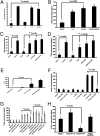
L-II RNA activates IFN-β expression. (A) Activation of IFN-β promoter by L-II mut. A dual-luciferase assay was performed as described in Fig. 1A. A plasmid containing F-Luc under control of an IFN-β promoter was used in place of the NF-κB–containing promoter described in Fig. 1A. (B) Induction of IFN-β production by L-II RNA. Plasmids encoding L-II or L-II mut were transfected into 293T cells, and the amount of IFN-β in the media was measured by ELISA at 1 d posttransfection. Each graph showing concentrations of IFN-β using ELISA is the average of three independent experiments. Error bars represent SD. (C) IFN-β production induced by purified RNA. Vero cells were transfected with empty vector or plasmids containing L-II mut, or were infected with WT PIV5 or rPIV5VΔC, mock-infected, or transfected with poly(I):poly(C). Total RNAs were purified from transfected or infected cells. The purifed RNAs were then transfected into 293T cells, and concentrations of IFN-β were measured in the media using ELISA after 1 d. (D) Induction of IFN-β by purified mRNA. Vero cells were transfected with empty vector or plasmid containing L-II mut, infected with WT PIV5 or rPIV5VΔC, or mock-infected. mRNAs were purified and transfected into 293T cells, and IFN-β concentrations after 1 d were measured using ELISA. (E) Induction of IFN-β by L-II in the presence of CHX. The 293T cells in six-well plates were transfected with 1 μg of RNA or 250 ng of poly(I):poly(C) and then incubated with CHX (20 μg/mL) for 16 h. The total RNAs were purified and subjected to RT, followed by real-time PCR analysis. ΔCT was calculated using actin from each sample as a control. (F) Lack of production of IFN-β in the absence of L-II mRNA. The purified L-II RNA was reverse-transcribed using an L-II sequence-specific primer and RT. The product and/or purified L-II RNA were treated or untreated with RNase H (RH). The purified products were then transfected into 293T cells, and IFN-β concentrations after 1 d were determined by ELISA. (G) Lack of production of IFN-β in the absence of L mRNA. The same experiment as in Fig. 3F was performed using RNAs purified from infected cells. RT(NP), RT using NP-specific primer; RT(L-II), RT using L-specific primer. The graph shows the average of three independent experiments. Error bars represent SD. (H) Induction of IFN-β production by in vitro transcribed L-II RNA. The L-I and L-II RNA were synthesized in vitro using the Riboprobe in vitro transcription system (Promega). The RNA transcripts were treated or untreated with CIP to remove 5′-triphosphate and then transfected into 293T cells. At 1 d posttransfection, IFN-β concentrations in the media were measured by ELISA.
Fig. 4.
Fig. 4.
Role of RIG-I in the activation of NF-κB and IFN-β by L-II RNA. (A) Effect of IPS-1 DN on activation of NF-κB by L RNA. A dual-luciferase experiment was performed as described in Fig. 1A, using IPS-1 DN with a Flag tag (500 ng/μL). Immunoblot analysis was performed to examine the expression of IPS-1 DN using anti-Flag and anti–β-actin antibodies. All transfections were carried out in replicates of four. Error bars represent SD. (B) Activation of NF-κB by L-II RNA was independent of RIG-I. At 18–20 h after transfection, a dual-luciferase assay was performed using lysate from Huh7 or Huh7.5 cells (RIG-I defective due to a T to I mutation at amino acid residue 55) transfected with vector, L, L-I, L-II, or L-II mut. (C) Effect of RIG-I DN on activation of NF-κB by L-II RNA. A reporter gene assay was performed using a plasmid expressing RIG-I DN with a Flag tag (500 ng/μL) along with the plasmids indicated. Immunoblot analysis was performed to examine the expression of RIG-I DN using anti-Flag and anti–β-actin antibodies. All transfections were carried out in replicates of four. Error bars represent SD.
Fig. 5.
Fig. 5.
MDA5 plays a critical role in the activation of IFN-β by viral mRNA. (A) The roles of RIG-I and MDA5 in the activation of the IFN-β promoter by viral mRNA. The 293T cells were transfected with siRNA targeting RIG-I or MDA5 or with NT siRNA. At 48 h after transfection of siRNA, the cells were transfected with vector, L-II mut, or poly(I):poly(C), along with the luciferase reporter plasmids. Luciferase activity was measured at 18–20 h posttransfection. (B) The roles of RIG-I and MDA5 in induction of IFN-β production by viral mRNA. siRNA transfection was performed as described in Fig. 5A in 293T cells, and at 48 h after transfection with siRNA, the cells were transfected with vector, L-II mut, or poly(I):poly(C). After 18–20 h, the amount of IFN-β in the medium was measured by ELISA. Expression levels of RIG-I, MDA5, and β-actin were examined by immunoblot analysis.
Fig. 6.
Fig. 6.
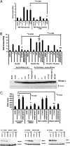
RNase L plays a critical role in the activation of NF-κB and IFN-β by viral mRNA. (A) The role of RNase L in activating the IFN-β promoter. A dual-luciferase assay for IFN-β promoter activation was performed as described in Fig. 3A using WT or RLKO MEFs. (B) Restoration of IFN-β activation in RLKO MEFs. IFN-β activation after complementation with RNase L cDNA in RLKO MEFs was examined by a dual-luciferase experiment. RLKO MEFs were transiently transfected with RNase L cDNA or inactive RNase L mutant (R667A) cDNA. At 18 h after transfection, the cells were transfected with 1 μg/μL of vector, L, L-I, L-I-II, or L-II mut plasmids, along with reporter plasmids. At 1 d posttransfection, the luciferase assay was performed. RNase L and β-actin levels were measured by immunoblot analysis. All transfections were carried out in replicates of four. Error bars represent SD. (C) The role of RNase L in activating IFN-β expression. Then 293T cells were transfected with siRNA targeting RIG-I, MDA5, RNase L, or control siRNA. IFN-β production in response to vector, L-II mut, or poly(I):poly(C) was measured by ELISA. The expression of RIG-I, MDA5, and RNase L was examined by immunoblot analysis, with β-actin as a loading control.
See this image and copyright information in PMC

Similar articles

See all similar articles

Cited by

See all "Cited by" articles

References

    1. Marié I, Durbin JE, Levy DE. Differential viral induction of distinct interferon-α genes by positive feedback through interferon regulatory factor-7. EMBO J. 1998;17:6660–6669. - PMC - PubMed
    1. Sato M, et al. Positive feedback regulation of type I IFN genes by the IFN-inducible transcription factor IRF-7. FEBS Lett. 1998;441:106–110. - PubMed
    1. Andrejeva J, et al. The V proteins of paramyxoviruses bind the IFN-inducible RNA helicase, mda-5, and inhibit its activation of the IFN-beta promoter. Proc Natl Acad Sci USA. 2004;101:17264–17269. - PMC - PubMed
    1. Akira S, Uematsu S, Takeuchi O. Pathogen recognition and innate immunity. Cell. 2006;124:783–801. - PubMed
    1. Kawai T, Akira S. Innate immune recognition of viral infection. Nat Immunol. 2006;7:131–137. - PubMed

Publication types

MeSH terms

Substances

Related information

Grants and funding

LinkOut - more resources

Full text links
Atypon full text link Atypon Free PMC article
Cite
Send To

NCBI Literature Resources

MeSHPMCBookshelfDisclaimer

The PubMed wordmark and PubMed logo are registered trademarks of the U.S. Department of Health and Human Services (HHS). Unauthorized use of these marks is strictly prohibited.


[8]ページ先頭

©2009-2025 Movatter.jp