Movatterモバイル変換


[0]ホーム

URL:


Skip to main page content
U.S. flag

An official website of the United States government

Dot gov

The .gov means it’s official.
Federal government websites often end in .gov or .mil. Before sharing sensitive information, make sure you’re on a federal government site.

Https

The site is secure.
Thehttps:// ensures that you are connecting to the official website and that any information you provide is encrypted and transmitted securely.

NIH NLM Logo
Log inShow account info
Access keysNCBI HomepageMyNCBI HomepageMain ContentMain Navigation
pubmed logo
Advanced Clipboard
User Guide

Full text links

HighWire full text link HighWire Free PMC article
Full text links

Actions

Share

.2010 Apr 15;24(8):827-36.
doi: 10.1101/gad.1905010. Epub 2010 Mar 29.

Nuclear expression of a group II intron is consistent with spliceosomal intron ancestry

Affiliations

Nuclear expression of a group II intron is consistent with spliceosomal intron ancestry

Venkata R Chalamcharla et al. Genes Dev..

Abstract

Group II introns are self-splicing RNAs found in eubacteria, archaea, and eukaryotic organelles. They are mechanistically similar to the metazoan nuclear spliceosomal introns; therefore, group II introns have been invoked as the progenitors of the eukaryotic pre-mRNA introns. However, the ability of group II introns to function outside of the bacteria-derived organelles is debatable, since they are not found in the nuclear genomes of eukaryotes. Here, we show that the Lactococcus lactis Ll.LtrB group II intron splices accurately and efficiently from different pre-mRNAs in a eukaryote, Saccharomyces cerevisiae. However, a pre-mRNA harboring a group II intron is spliced predominantly in the cytoplasm and is subject to nonsense-mediated mRNA decay (NMD), and the mature mRNA from which the group II intron is spliced is poorly translated. In contrast, a pre-mRNA bearing the Tetrahymena group I intron or the yeast spliceosomal ACT1 intron at the same location is not subject to NMD, and the mature mRNA is translated efficiently. Thus, a group II intron can splice from a nuclear transcript, but RNA instability and translation defects would have favored intron loss or evolution into protein-dependent spliceosomal introns, consistent with the bacterial group II intron ancestry hypothesis.


Figures
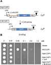
Figure 1.
Figure 1.
A group II intron-bearing nuclear gene is poorly expressed. (A) Schematic representation of the group II intron splicing reporter GpII-CUP1. (Bent arrow) Glyceraldehyde-3-phosphate dehydrogenase gene promoter (GPD); (inverted triangle) cytochrome-c gene terminator (CYC1); (E1 and E2) exon 1 and exon 2 of the Ll.LtrB homing site (Cousineau et al. 1998); (red line) ΔORF variant of the Ll.LtrB intron; (blue rectangle)CUP1 ORF; (c-Myc) c-Myc epitope. The presence of the Ll.LtrB intron renders the fusion out-of-frame (CuS), and Ll.LtrB splicing results in an in-frame fusion, HS-CUP1, which in theory confers increased copper resistance (CuR) inS. cerevisiae. (B) Growth of strains carrying pHS-CUP1 or pGpII-CUP1 on medium containing different concentrations of CuSO4. The expression plasmid was with or without LtrA or NLS-LtrA. Yeast cultures at OD600 ≈ 1.0 were applied in 5-μL spots. “Vector” indicates a plasmid withoutCUP1.
Figure 2.
Figure 2.
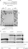
A group II intron can splice from eukaryotic Pol II transcripts, but is subject to NMD. (A) LtrA-dependent Ll.LtrB splicing. Schematic of the predicted primer extension termination products and their respective sizes is shown. (Gray) Intron; (black) exon; (vertical dashed line) intron–exon boundary or splice junction; (black arrow) primer; (horizontal dashed line) primer extension product; (black oval) extension termination. A primer extension termination assay with RNA from pHS-CUP1 or pGpII-CUP1, in the absence (0) or presence ([+] induced; [−] repressed) of LtrA- or NLS-LtrA-expressing plasmid. “Blank” indicates the absence of DNA or RNA template. Lanes labeled G, A, T, and C are sequencing ladders with pGpII-CUP1 and pHS-CUP1 DNA. Expression and localization of LtrA were confirmed by Western blotting (Fig. 3) and indirect immunofluorescence assay (Supplemental Fig. S1), respectively. (B) Northern blot analysis of HS-CUP1 and GpII-CUP1 RNA. RNA expressed in the presence or absence of LtrA was analyzed in the wild-type (WT) and mutant strains, withACT1 mRNA as a loading control. The level of CUP1 RNA was normalized toACT1 mRNA levels in each strain and quantitated relative to the HS-CUP1 level in the wild type. (P) GpII-CUP1 precursor RNA; (SP) HS-CUP1 mRNA or the spliced product; (% splicing) percentage of ligated exons relative to the total CUP1 RNA; [(P + SP)(upf1/WT)] the level of total CUP1 RNA in theupf1 mutant relative to that in the wild-type host is represented by a square bracket with standard deviation from at least two independent experiments. Calculations were made from GpII-CUP1 precursor RNA and HS-CUP1 mRNA or spliced product numbers to two decimal points from the independent experiments, leading to some apparent lack of correspondence in the ratios shown.
Figure 3.
Figure 3.
Spliced group II intron transcripts have an NMD-independent translational defect. (A) Western blot analysis of Cup1p made from pHS-CUP1 or pGpII-CUP1 in the wild-type strain. Expression was in the absence (0) or presence ([+] induced; [−] repressed) of LtrA or NLS-LtrA. LtrA and Tub1p expression was also determined. (B) Cup1p expression from pHS-CUP1 (HS) and pGpII-CUP1 (GpII) in the NMD mutant hosts. LtrA and Tub1p levels were determined in the presence (+) or absence (−) of LtrA in the wild-type (WT) and mutant strains, labeledabove each set of three lanes.
Figure 4.
Figure 4.
Spliced group II intron transcripts are poorly translated. Polysome analysis of HS-CUP1 (A) and GpII-CUP1 RNA in the presence of LtrA in the wild type (B) and theupf1 mutant (C).ACT1 RNA served as a control for polysome accumulation of translatable RNA. Polysomes were analyzed by ethidium bromide staining of formaldehyde agarose gels (26S and 18S rRNA) prior to Northern blotting. The percentage of RNA from each fraction relative to the total RNA was calculated. (⋄)ACT1 mRNA; (●) HS-CUP1 mRNA or the group II intron spliced product (SP); (▲) GpII-CUP1 precursor RNA (P). The strain names are shown in parentheses.
Figure 5.
Figure 5.
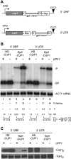
Group II intron transcripts are poorly expressed regardless of exon context. (A) Schematic representation of the group II intron in the homing site (E1 and E2) fused to the 3′ end of the ORF or in the 3′ UTR of the CUP1 RNA (cf. 5′ ORF fusion) (Fig. 1A). (B) Northern blot analysis of RNA from pHS-CUP1 and pGpII-CUP1 in the 3′ ORF and 3′ UTR contexts. RNA was analyzed in the wild type (+) andupf1 mutant (−) withACT1 RNA as a loading control. The level of CUP1 RNA was normalized toACT1 mRNA levels in each strain and quantitated relative to the HS-CUP1 level in the wild-type strain. (P) GpII-CUP1 precursor RNA; (SP) HS-CUP1 mRNA or the spliced product; (% splicing) percentage of ligated exons relative to the total CUP1 RNA; [(P + SP)(upf1/WT)] the level of total CUP1 RNA in theupf1 mutant relative to that in the wild-type host is shown with standard deviation from at least two independent experiments, as in Figure 2B. (C) Western blot analysis. Cup1p expression from pHS-CUP1 and pGpII-CUP1 in the 3′ ORF and 3′ UTR contexts was determined in the wild type andupf1 mutant with Tub1p as the loading control. Cup1p expression in theupf1 mutant was verified by measuring the CuR phenotype (data not shown).
Figure 6.
Figure 6.
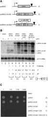
Group II intron expression from theURA3-based splicing reporter. (A) Schematic of theURA3-based splicing reporter. The wild-typeURA3 (pURA3) and theURA3 with an artificial spliceosomal intron (AI) carrying the homing site of Ll.LtrB (pURA3∷AI-HS) are shown, along with the group II intron splicing reporter (pURA3∷AI-GpII), which has the Ll.LtrB ΔORF intron within its homing site in AI. (Bent black arrow)URA3 promoter; (inverted triangle)URA3 terminator. (B) Steady-state levels of URA3∷AI-HS and URA3∷AI-GpII RNA were measured by Northern blotting of total RNA from wild-type (+) andupf1 mutant (−) strains. (ACT1 mRNA) Loading control; (Vector) a plasmid withoutURA3. The square brackets represent numbers from one experiment, although duplicate experiments showed the same trend. (C)URA3 phenotypes. Theura3Δ0 strain carrying the vector, pURA3, pURA3∷AI-HS, or pURA3∷AI-GpII in the absence and presence of LtrA was assayed for growth on synthetic media lacking uracil. The undiluted yeast cultures at OD600 ≈ 1.0 and 10-fold serial dilutions were applied in 5-μL spots.
Figure 7.
Figure 7.
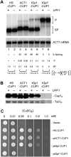
Spliceosomal and group I intron-containing transcripts are expressed efficiently in yeast. (A) Northern blot analysis with RNAs from pACT1-CUP1, ptGpI-CUP1, and ptGpI*-CUP1 (splicing-defective). RNA levels were analyzed in the wild type (+) andupf1 mutant (−) by Northern blotting, withACT1 RNA as a loading control. The level of CUP1 RNA was normalized toACT1 mRNA levels in each strain and was quantitated relative to the HS-CUP1 level in the wild-type strain. (P) ACT1-CUP1, tGpI-CUP1, or tGpI*-CUP1 precursor RNA; (SP) HS-CUP1 mRNA or the spliced product; (% splicing) percentage of ligated exons relative to the total CUP1 RNA; [(P + SP)(upf1/WT)] the level of total CUP1 RNA in theupf1 mutant relative to that in the wild-type host is shown with standard deviation from at least two independent experiments, as in Figure 2B. (B) Cup1p expression from pACT1-CUP1, ptGpI-CUP1, and ptGpI*-CUP1. Western blotting was used to determine protein levels in the wild type andupf1 mutant, with Tub1p as the loading control. (C) Strains carrying pHS-CUP1, pACT1-CUP1, ptGpI-CUP1, or ptGpI*-CUP1 were assayed for CuR as in Figure 1B.
Figure 8.
Figure 8.
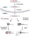
Model for the fate of a group II intron-bearing nuclear transcript. After transcription and transport to the cytoplasm, the transcript can be silenced by NMD and/or translational repression. The EBS–IBS interaction between the intron lariat and the mRNA is shown for the hypothetical translational repression model.
See this image and copyright information in PMC

Similar articles

See all similar articles

Cited by

See all "Cited by" articles

References

    1. Alani E, Cao L, Kleckner N 1987. A method for gene disruption that allows repeated use of URA3 selection in the construction of multiply disrupted yeast strains. Genetics 116: 541–545 - PMC - PubMed
    1. Atkinson GC, Baldauf SL, Hauryliuk V 2008. Evolution of nonstop, no-go and nonsense-mediated mRNA decay and their termination factor-derived components. BMC Evol Biol 8: 290 doi: 10.1186/1471-2148-8-290 - PMC - PubMed
    1. Belfort M, Derbyshire V, Cousineau B, Lambowitz A 2002. Mobile introns: Pathways and proteins. In Mobile DNA II (ed. Craig N et al.), pp. 761–783 ASM Press, Washington, DC
    1. Brachmann CB, Davies A, Cost GJ, Caputo E, Li J, Hieter P, Boeke JD 1998. Designer deletion strains derived from Saccharomyces cerevisiae S288C: A useful set of strains and plasmids for PCR-mediated gene disruption and other applications. Yeast 14: 115–132 - PubMed
    1. Brehm SL, Cech TR 1983. Fate of an intervening sequence ribonucleic acid: Excision and cyclization of the Tetrahymena ribosomal ribonucleic acid intervening sequence in vivo. Biochemistry 22: 2390–2397 - PubMed

Publication types

MeSH terms

Substances

Grants and funding

LinkOut - more resources

Full text links
HighWire full text link HighWire Free PMC article
Cite
Send To

NCBI Literature Resources

MeSHPMCBookshelfDisclaimer

The PubMed wordmark and PubMed logo are registered trademarks of the U.S. Department of Health and Human Services (HHS). Unauthorized use of these marks is strictly prohibited.


[8]ページ先頭

©2009-2025 Movatter.jp