Movatterモバイル変換


[0]ホーム

URL:


Skip to main page content
U.S. flag

An official website of the United States government

Dot gov

The .gov means it’s official.
Federal government websites often end in .gov or .mil. Before sharing sensitive information, make sure you’re on a federal government site.

Https

The site is secure.
Thehttps:// ensures that you are connecting to the official website and that any information you provide is encrypted and transmitted securely.

NIH NLM Logo
Log inShow account info
Access keysNCBI HomepageMyNCBI HomepageMain ContentMain Navigation
pubmed logo
Advanced Clipboard
User Guide

Full text links

Ingenta plc full text link Ingenta plc Free PMC article
Full text links

Actions

Share

Review
.2006:75:743-67.
doi: 10.1146/annurev.biochem.75.103004.142743.

G protein-coupled receptor rhodopsin

Affiliations
Review

G protein-coupled receptor rhodopsin

Krzysztof Palczewski. Annu Rev Biochem.2006.

Abstract

The rhodopsin crystal structure provides a structural basis for understanding the function of this and other G protein-coupled receptors (GPCRs). The major structural motifs observed for rhodopsin are expected to carry over to other GPCRs, and the mechanism of transformation of the receptor from inactive to active forms is thus likely conserved. Moreover, the high expression level of rhodopsin in the retina, its specific localization in the internal disks of the photoreceptor structures [termed rod outer segments (ROS)], and the lack of other highly abundant membrane proteins allow rhodopsin to be examined in the native disk membranes by a number of methods. The results of these investigations provide evidence of the propensity of rhodopsin and, most likely, other GPCRs to dimerize, a property that may be pertinent to their function.


Figures

Figure 1
Figure 1
Vertebrate retina and rhodopsin. (a) Scanning electroretinogram of mouse retina [courtesy of Yan Liang (33)]. Rod cells comprise ~70% of all 6.4 million retinal cells, and cone cells represent <2%. Rods are postmitotic neurons with highly differentiated rod outer segments (ROS) connected to the inner segments (IS), which generate proteins and energy to sustain phototransduction events. (b) Diagram depicting the rod cell. The processes in ROS allow rapid transduction of the light signal to graded hyperpolarization of the plasma membrane, ensuing from the decrease of light-sensitive conductance in the ROS cGMP-gated cation channels. In ROS, hundreds of distinct, rhodopsin-loaded disk membranes (20) are enveloped by the plasma membrane. (c) Electron micrograph of isolated ROS from the mouse retina [courtesy of Yan Liang (33)]. The disk membranes consist of a phospholipid bilayer studded with rhodopsin. (d ) Diagram of disk membranes. The main protein of ROS disk membranes is light-sensitive rhodopsin, which occupies 50% of the disk area. The molar ratio between rhodopsin and phospholipids is about 1:60 (for example 138 and 139; reviewed in 140). Multiple techniques suggest that rhodopsin forms oligomeric structures in the native membranes, with the rhodopsin dimer most likely being the signaling unit.
Figure 2
Figure 2
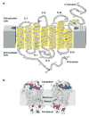
Modification of rhodopsin molecule and orientation in the membranes. (a) Two-dimensional model of rhodopsin. The polypeptide of rhodopsin crosses the membrane seven times. C-I, C-II, and C-III correspond to the cytoplasmic loops, and E-I, E-II, and E-III correspond to extracellular loops. The transmembrane segment is α-helical (yellow cylinders), although the helices are highly distorted and tilted. The stability of the helical segment is increased by the Cys110-Cys187 bridge (141) (depicted indark yellow) a highly conserved feature among many GPCRs. The chromophore, 11-cis-retinal, not depicted here, is attached to Lys296 (dark red ) via a protonated Schiff base. The positive charge of the base is neutralized by counterion Glu113 (blue). During postisomerization changes in the receptor, it was proposed that the counterion migrates to Glu181 (blue) (76). Asn2 and Asn15 (red ) are sites of glycosylation within the conserved glycan composition, and Met1 (orange) is acetylated. Cys322 and Cys323 (light green) are palmitoylated, whereas two other Cys, Cys140 and Cys316 (brown), are reactive to many chemical probes and are used to explore rhodopsin’s structure. Rhodopsin, when exposed to light, is phosphorylated by rhodopsin kinase (or G protein–coupled receptor kinase 1). The predominant phosphorylation sites are Ser334, Ser338, and Ser343 (green) (55), and the whole C-terminal region is highly mobile (142). However, as shown using a model peptide, the C-terminal region may become structured when bound to arrestin (143). The highly conserved domains among GPCRs, D(E)RY in helix 3 and NPXXY in helix VII (gray), are important in transformation of the receptor from an inactive to a G protein–coupled conformation. Different versions of this figure were published previously (for example in 19 and 64), and all of them are refinements of the pioneering work on rhodopsin topology by Paul Hargrave (144, 145). (b) Location of the chromophore and charges on the cytoplasmic and intradiscal (extracellular) surface of rhodopsin in relation to the hypothetical membrane bilayer. The negative charges (red ) and basic residues (blue) are shown. The proposed location of the membrane is shown in gray, and the location of the chromophore 11-cis-retinylidene is shown by deleting fragments of transmembrane helices. Two sides of rhodopsin are depicted.
Figure 3
Figure 3
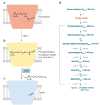
Light-cycle of rhodopsin. (a) Rhodopsin and 11-cis-retinal. Rhodopsin consists of a colorless protein moiety (the opsin) and the chromophore, 11-cis-retinylidene, which imparts a red color to rhodopsin. The chromophore, a geometric isomer of vitamin A in aldehyde form, is coupled to opsin via the protonated Schiff base at Lys296, located in the transmembrane domain of the protein. Bovine rhodopsin absorbs at a λmax = 498 nm. (b) Photoactivated rhodopsin. Absorption of light by rhodopsin leads with high probability (~65%) to photoisomerization of thecis C11-C12 chromophore double bond to atrans configuration. The probability of isomerization depends only modestly on the wavelength of the light (146). This reaction, one of the fastest photochemical reactions known in biology, produces multiple intermediates that culminate in the formation of the G protein–activating state, termed metarhodopsin II, or Meta II. (c) Opsin without chromophore. Ultimately the photoisomerized chromophore, all-trans-retinylidene, is released from the opsin as all-trans-retinal and reduced to alcohol by short-chain alcohol dehydrogenases, such as prRDH, retSDR, and RDH12. The all-trans chromophore diffuses to the adjacent retinal pigment epithelium, where it undergoes enzymatic transformation back to 11-cis-retinal in a metabolic pathway known as the retinoid cycle. Opsin recombines with replenished 11-cis-retinal to form rhodopsin. (d ) Reaction scheme of rhodopsin photoactivation. Upon absorption of a photon by rhodopsin and electronic excitation, fast isomerization of 11-cis-retinylidene to all-trans-retinylidene takes place. At body temperature, the Meta I and Meta II exist in equilibrium shifted toward Meta II. In vitro, further decay of rhodopsin to both opsin and free all-trans-retinal or to Meta III is possible. In vivo, Meta III is not formed at significant levels because it decomposes in the presence of G protein transducin (147). In vitro, prolonged incubation of Meta II involves a thermal isomerization of the chromophore double bond with Lys296 to an all-trans-15-syn configuration. This isomerization step is catalyzed by the opsin itself (148). On the left are maximal temperatures at which indicated intermediates can be trapped, and on the right is time required for that particular transformation. In the brackets are λmax of absorption for different intermediates. The reaction scheme is based on Shichida & Imai [(149); see also the thermodynamic properties of these reactions (19)].
Figure 4
Figure 4
Three-dimensional model of rhodopsin. (a) Ribbon drawings of rhodopsin parallel to the plane of the membrane. (b) View into the membrane plane as seen from the intradiscal side of the membrane. The carbohydrate moieties are at Asn2 and Asn15. The pairs of β1-β2 and β3-β4 hairpins, the transmembrane helices (Hs) I–VII, and the cytoplasmic helix 8 (H8) are labeled. A palmitoyl group is attached to each of the two Cys residues at the end of helix 8. The removal of the palmitoyl groups has only a minor effect on phototransduction processes (e.g., 150 and 151). (c) A view into the membrane plane as seen from the cytoplasmic side. The cytoplasmic side has greater surface area than the intradiscal side. (The roman numeral convention is related to transmembrane helices, whereas an Arabic numeral indicates a solvent-exposed helix.)
Figure 5
Figure 5
The amino acid residues in the vicinity of the chromophore. (a) Schematic showing the side chains surrounding the 11-cis-retinylidene group (pink); side view through helices III, V, and VI. (b) Schematic presenting the residues within 5 Å distance from the 11-cis-retinylidene group (pink). Note that the chromophore is coupled via the protonated Schiff base with Lys296.
Figure 6
Figure 6
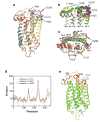
Comparison of the current rhodopsin structures. There are currently five crystallographic entries for rhodopsin in the Protein Data Bank (PDB). The structures deposited under the accession number 1F88, 1HZX, 1GZM, and 1U19 are superimposed. Accession number 1F88 (yellow thread), 1HZX (orange), 1GZM (purple), and 1U19 (gray) are represented in the cartoon. Entries 1F88, 1HZX, 1L9H, and 1U19 are for a tetragonal crystal obtained by very similar methods. Entry 1U19 is at the highest resolution reported, 2.2 Å. Entry 1GZM is for a trigonal crystal form obtained in a different condition than the other listed crystals. (a) Side view. (b) Side view, with a close-up of the cytoplasmic region. (c) View from the cytoplasmic side. (d,e) A plot (d ) and three-dimensional representation (e) of the B factor for rhodopsin structures from three data sets. The B factor is also known as the temperature factor or Debye-Waller factor and describes the degree to which the electron density is spread out, indicating the static or dynamic mobility of an atom or incorrectly built models. In paneld, the orange line represents 1HZX, the purple line represents 1GZM, and the gray line represents 1U19. In panele, spectral grading of the B factor, green represents a low B factor, and red indicates the highest B factor. Note that the loop II is incomplete in 1HZX because of ambiguity in the electron density, and this region in the 1GZM set has the highest B factor.
Figure 7
Figure 7
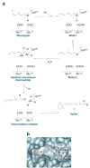
Hydrolysis of the all-trans-retinylidene chromophore and regeneration of rhodopsin with newly synthesized 11-cis-retinal. (a) The scheme of the retinylidene group hydrolysis. The role of Glu181 and Glu113 is hypothetical. Note that Glu181 is protonated in rhodopsin (71). (b) Formation of rhodopsin. Polarization of the carbonyl group of 11-cis-retinal and deprotonation of the Schiff-base group is required before the Schiff base can be formed.
Figure 8
Figure 8
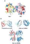
Models of GPCR dimerization. (a) Top view from the cytoplasmic side of the rhodopsin dimer. The model was generated by Dr. S. Filipek using structural constraints of rhodopsin and experimental data obtained by atomic force microscopy on the organization of rhodopsin in native membranes (33, 92, 93, 110, 111). This model is in agreement with cross-linking experiments (94). Photoactivated rhodopsin is depicted in yellow (Rho*), and rhodopsin is shown in pink. The acidic residues are shown in red and basic residues in blue. This cytoplasmic surface is involved in the interaction with G protein transducin. (b) The crystal structures of extracellular domains of different GPCRs. Crystal structures of the extracellular domains of the frizzled 8 receptor, the follicle-stimulating hormone (FSH) receptor, and the Glu receptor revealed that the extracellular domains formed a dimer. These structures may represent a physiological dimer that would stabilize the transmembrane domain and result in a dimeric platform for interaction with G proteins and other partner proteins. Protein Data Bank accession numbers are shown in parentheses. Panelsa andb are not drawn to the same scale.
See this image and copyright information in PMC

Similar articles

See all similar articles

Cited by

See all "Cited by" articles

References

    1. Filipek S, Teller DC, Palczewski K, Stenkamp R. Annu Rev Biophys Biomol Struct. 2003;32:375–97. - PMC - PubMed
    1. Bhandawat V, Reisert J, Yau KW. Science. 2005;308:1931–34. - PMC - PubMed
    1. Minke B, Cook B. Physiol Rev. 2002;82:429–72. - PubMed
    1. Heck M, Hofmann KP. J Biol Chem. 2001;276:10000–9. - PubMed
    1. Leskov IB, Klenchin VA, Handy JW, Whitlock GG, Govardovskii VI, et al. Neuron. 2000;27:525–37. - PubMed

Publication types

MeSH terms

Substances

Related information

Grants and funding

LinkOut - more resources

Full text links
Ingenta plc full text link Ingenta plc Free PMC article
Cite
Send To

NCBI Literature Resources

MeSHPMCBookshelfDisclaimer

The PubMed wordmark and PubMed logo are registered trademarks of the U.S. Department of Health and Human Services (HHS). Unauthorized use of these marks is strictly prohibited.


[8]ページ先頭

©2009-2025 Movatter.jp