Movatterモバイル変換


[0]ホーム

URL:


Skip to main page content
U.S. flag

An official website of the United States government

Dot gov

The .gov means it’s official.
Federal government websites often end in .gov or .mil. Before sharing sensitive information, make sure you’re on a federal government site.

Https

The site is secure.
Thehttps:// ensures that you are connecting to the official website and that any information you provide is encrypted and transmitted securely.

NIH NLM Logo
Log inShow account info
Access keysNCBI HomepageMyNCBI HomepageMain ContentMain Navigation
pubmed logo
Advanced Clipboard
User Guide

Full text links

Wiley full text link Wiley Free PMC article
Full text links

Actions

.1999 Sep;128(2):479-85.
doi: 10.1038/sj.bjp.0702800.

Inhibition of the current of heterologously expressed HERG potassium channels by imipramine and amitriptyline

Affiliations

Inhibition of the current of heterologously expressed HERG potassium channels by imipramine and amitriptyline

A G Teschemacher et al. Br J Pharmacol.1999 Sep.

Abstract

1 Tricyclic antidepressants (TCAs) are associated with cardiovascular side effects including prolongation of the QT interval of the ECG. In this report we studied the effects of two TCAs (imipramine and amitriptyline) on ionic current mediated by cloned HERG potassium channels. 2 Voltage clamp measurements of HERG currents were made from CHO cells transiently transfected with HERG cDNA. HERG-encoded potassium channels were inhibited in a reversible manner by both imipramine and amitriptyline. HERG tail currents (IHERG) following test pulses to +20 mV were inhibited by imipramine with an IC50 of 3.4+/-0.4 microM (mean+/-s.e.mean) and a Hill coefficient of 1.17+/-0.03 (n = 5). 3 microM amitriptyline inhibited IHERG by 34+/-6% (n = 3). The inhibition showed only weak voltage dependence. 3 Using an 'envelope of tails' comprised of pulses to +20 mV of varying durations, the tau of activation was found to be 155+/-30 ms for control and 132+/-26 ms for 3 microM imipramine (n = 5). Once maximal channel activation was achieved after 320 ms (as demonstrated by maximal tail currents), further prolongation of depolarization did not increase imipramine-mediated HERG channel inhibition. 4 Taking current measurements every second during a 10 s depolarizing pulse from -80 mV to 0 mV, block was observed during the first pulse in the presence of imipramine and the level of IHERG block was similar throughout the pulse (n=5). 5 A three pulse protocol (two depolarizing pulses to +20 mV separated by 20 ms at -80 mV) revealed that imipramine did not significantly alter the kinetics of IHERG inactivation. The tau of inactivation was 8+/-2 ms and 5.6+/-0.4 ms (n = 5) in the absence and presence of 3 microM imipramine, respectively, and currents inactivated to a similar extent. 6 Our data are consistent with TCAs causing components of block of the HERG channel in both the closed and open states. Any component of open channel block occurs rapidly upon depolarization. Inhibition of IHERG by the prototype TCAs imipramine and amitriptyline may suggest a mechanism for QT prolongation associated with risks of arrhythmia and sudden death that accompany high concentrations of TCAs following overdose.


Figures

Figure 1
Figure 1
TCAs inhibit current mediated by heterologous HERG. (a) shows the currents elicited from a CHO cell transfected with HERG and from an untransfected cell. Membrane potential was held at −80 mV, depolarized to +20 mV for 400 ms, before the tail was observed at −40 mV. Total current elicited from the untransfected cell during the pulse was under 20 pA, and no tail current could be detected. The HERG current of a typical cell in extracellular solution is shown in (b) in the absence of drug (control), in the presence of 3 μM imipramine, and after washout. The holding potential and voltage steps are identical to those in (a). Interpulse interval was 15 s. (c) shows a typical cell superfused with 3 μM amitriptyline under the same conditions as in (b). The dotted line in (b) and (c) indicates zero current level. In (d), the concentration response curve shows mean (±s.e.mean) inhibition of tail currents measured with the same protocol as above (n=5; at 30 and 100 μM imipramine data using whole cell patch clamp and gramicidin perforated patches were pooled).
Figure 2
Figure 2
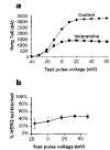
Voltage dependence of the effects of imipramine. (a) demonstrates the effect of voltage on imipramine-mediated block of IHERG. It shows a typical plot of the tail currents (at −40 mV) under these conditions against test pulse potential. In (b) percentage inhibition evoked by 3 μM imipramine at different voltages is shown, with the imipramine-sensitive current being normalized in each cell to the current at the same voltage in the absence of imipramine (n=5).
Figure 3
Figure 3
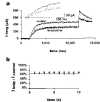
Imipramine inhibition during a long pulse. The currents from a 10 s pulse protocol applied to a typical cell before and after a 10 min application of 3 μM imipramine are shown in (a). The membrane potential was held at −80 mV, and HERG currents were evoked with a 10 s pulse to +20 mV. The cell was held at −80 mV for 10 min while imipramine was applied. The inset shows an expanded portion of the same record at the beginning of the pulse. From the above data, the ratio of current in the presence of imipramine to current before application of imipramine at successive 1 s time points is shown in (b). The horizontal line at the first value of 67% is provided to aid comparison of the data points (n=5).
Figure 4
Figure 4
Activation time course of IHERG with and without imipramine. (a) illustrates an ‘envelope of tails' test performed on the same cell before and after block with 3 μM imipramine. Membrane potential was held at −80 mV, depolarized to +20 mV for varying durations of time, and then tail currents were observed at −40 mV. The inter-pulse interval was 30 s. In (b) the peaks of the tail currents were plotted against the duration of the depolarizing pulse. The percentage of current blocked shown in (c) was calculated from the ratio of the matched peak tail currents for imipramine and control at each time point (n=5). (d) shows the mean of the calculated activation time constants (n=5) derived from a fit with a single exponential to the currents as shown in (b) (pairedt-test,P>0.05).
Figure 5
Figure 5
Inactivation of HERG in the presence and absence of imipramine. A three-pulse protocol was used in which the cells were held at −80 mV, depolarized to +20 mV for 400 ms, repolarized for 20 ms to −80 mV, and stepped again to +20 mV for 400 ms as shown in (a) lower panel. Typical responses are shown in the upper panel for control and 3 μM imipramine. During the brief pulse to −80 mV, rapid recovery from inactivation occurred; the time course for current deactivation is long with respect to this period of time. The inset shows the same current records (just before and after the 20 ms step to −80 mV) on an expanded time scale. The time course of inactivation during the second pulse to +20 mV was fitted with a single exponential and compared in (b) (pairedt-test,n=5,P=0.23). The extent of inactivation in the two conditions was compared in (c) (pairedt-test,n=5,P=0.21).
See this image and copyright information in PMC

References

    1. ABBOTT G.W., SESTI F., SPLAWSKI I., BUCK M., LEHMANN M.H., TIMOTHY K.W., KEATING M.T., GOLDSTEIN S.A.N. MiRP1 forms IKr potassium channels with HERG and is associated with cardiac arrhythmia. Cell. 1999;97:175–187. - PubMed
    1. BALSER J.R., BENNETT P.B., HONDEGHEM L.M., RODEN D.M. Suppression of time-dependent outward current in guinea-pig ventricular myocytes. Actions of quinidine and amiodarone. Circ. Res. 1991;69:519–529. - PubMed
    1. BIGGER J.T., GIARDINA E.G., PEREL J.M., KANTOR S.J., GLASSMAN A.H. Cardiac antiarrhythmic effect of imipramine hydrochloride. N. Engl. J. Med. 1977;296:206–208. - PubMed
    1. BUSCH A.E., EIGENBERGER B., JURKIEWICZ N.K., SALATA J.J., PICA A., SUESSBRICH H., LANG F. Blockade of HERG channels by the class III antiarrhythmic azimilide: mode of action. Br. J. Pharmacol. 1998;123:23–30. - PMC - PubMed
    1. CASIS O., SANCHEZ-CHAPULA J.A. Disopyramide, imipramine, and amitriptyline bind to a common site on the transient outward K+ channel. J. Cardiovasc. Pharm. 1998;32:521–526. - PubMed

Publication types

MeSH terms

Substances

Grants and funding

LinkOut - more resources

Full text links
Wiley full text link Wiley Free PMC article
Cite
Send To

NCBI Literature Resources

MeSHPMCBookshelfDisclaimer

The PubMed wordmark and PubMed logo are registered trademarks of the U.S. Department of Health and Human Services (HHS). Unauthorized use of these marks is strictly prohibited.


[8]ページ先頭

©2009-2025 Movatter.jp