Filed October 18, 2024 CD47-TARGETING MORPHOLINOS CROSS-REFERENCE TO RELATED APPLICATION [0001] This application claims priority to and the benefit of the earlier filing of U.S. Provisional Application No.63/591,961, filed on October 20, 2023, which is incorporated by reference herein in its entirety. FIELD OF THE DISCLOSURE [0002] This disclosure relates to methods of modulating cytotoxicity, for instance of chemotherapeutic agents as a surrogate for genotoxic stress (including radiation), particularly reducing cytotoxicity of chemotherapeutic and other genotoxic stressor agents to non-cancer cells and/or increasing cytotoxicity of chemotherapeutic and other genotoxic stressor agents to cancer cells. INCORPORATION BY REFERENCE OF SEQUENCE LISTING [0003] A computer readable text file, entitled “R286-0002PCT_ST26.xml” created on or about October 18, 2024, with a file size of 94,208 bytes, contains the Sequence Listing for this application and is hereby incorporated by reference in its entirety. BACKGROUND OF THE DISCLOSURE [0004] Previously, it was reported that a morpholino oligonucleotide complementary the 5' region of murine, porcine, and human CD47 mRNA, to prevent translation of this mRNA, would provide tissue protection from a range of stresses, such as hypoxia, ischemia, ischemia-reperfusion injury, transplantation, and others (Isenberg et al., Ann. Surg.247(1):180-190, 2008; Isenberg et al., Circ. Res. 100:712-720, 2007). It was also reported that such a CD47 morpholino would protect non-cancer cells, tissues, and organs from radiation and chemotherapy damage while at the same time increasing cancer cell killing, the latter, in part, via enhanced immune cell activity. These latter findings were verified in multiple subsequent publications (see US Application Publication No.2010/0092467). See also: US Patent No.8,865,672, US Patent No.11,285,169, Australia Patent No.2014244083B2, and WO2012170250A1. [0005] Despite these advances, there is a definite and unmet need for improved CD47 morpholino oligonucleotides capable of exhibiting improved cell uptake, and tighter target binding and greater specificity to human CD47. While other CD47 morpholinos were described, they were promiscuous and targeted the murine, porcine, and human CD47 mRNA. This lack of species specificity is a leading source of off-target hybridization that impacts gene expression beyond CD47 and is associated with delayed or dampened effect after administration. This latter Filed October 18, 2024 deficiency also required that the morpholino be given from 48 to 72 hours prior to the stress whether in vitro or in vivo. It also required higher concentration of the agent. SUMMARY OF ASPECTS OF THE DISCLOSURE [0006] The morpholino agents described herein target only the human CD47 mRNA and pre- mRNA. In so doing, the morpholinos show greater specificity, which helps to eliminate off-target effects. [0007] Described herein are human-specific CD47 targeting morpholinos that effectively, and at low concentration, lower CD47 protein in various human primary (non-cancer) and cancer cell types. The morpholinos, in and of themselves, are non-toxic to human cells at concentrations well above those that lead to a therapeutic lowering of CD47 protein levels. The description includes unique translation blocking and splice blocking human-only morpholino oligonucleotides. [0008] The CD47 morpholinos described herein block cell killing by genotoxic chemotherapy, and by association other genotoxic stress, such as radiation. They do this even if administered simultaneously with the cytotoxic chemotherapy, which is provided as an exemplary genotoxic challenge. This property has not previously shown by any published, reported, or known CD47 morpholino. As well, embodiments provided herein combine cell-penetrating peptides conjugated with the CD47 morpholinos to improve cell uptake and cytoplasmic delivery. Such a combination has not been previously manufactured nor used on human primary non-cancer or cancer cells. [0009] The CD47 morpholinos include at least 15 contiguous bases and hybridize under high stringency conditions to the mRNA of human CD47 (GenBank Human Target RNA: human CD47-202; available online at ncbi.nlm.nih.gov/nuccore/NM_198793.3). CD47 gene transcript CD47-202 encodes functional CD47 protein. By way of example, the morpholino comprises a sequence shown in any one of SEQ ID NOs: 1-16. Optionally, the CD47 morpholino is conjugated to a cell penetrating peptide (CPP); examples of such peptide phosphorodiamidate morpholino oligomer (PPMOs) are described, and include SEQ ID NOs: 19-63. [0010] The CD47 morpholinos described herein exhibit high-affinity and high specificity for binding to human CD47 mRNA and pre-mRNA only. They effectively lower CD47 protein levels in human cells. This then shuts down the thrombospondin (TSP1)-CD47 signaling axis. TSP1 is the stress released dominant high affinity ligand of CD47. The TSP1-CD47 interaction is protective to cancer but damaging to non-cancer cells. The technologies provide herein will be of therapeutic use in cancer and beyond, including cardiovascular diseases, pulmonary hypertension, heart failure, sickle cell anemia, metabolic syndrome, diabetes, and tissue and organ transplantation. The technologies are expected to synergize with transplantation of engineered organoids such as pancreatic islets and to synergize when combined with immune Filed October 18, 2024 checkpoint blocking therapies, such as antibodies to PD-1/PDL1/CTLA4, and engineered T and other immune cells for treatment of cancer. [0011] Compositions disclosed herein include third generation antisense oligomer therapeutics, specifically ~15-25 nucleic acid phosphorodiamidate morpholino oligomers (PMO) conjugated to a cell-penetrating peptide(s). [0012] The human-only mRNA and pre-mRNA targeting CD47 morpholino oligonucleotides of the present disclosure are designed to be between 10-30 bases long, more preferably, 15-30 bases long, and preferably 20-25 bases long. This gives specificity of targeting to the sequence. The sequences of translation blocking morpholinos were designed to bind close to the CD47 ATG start codon, and to exhibit a Tm of 75°C - 115°C, for instance a Tm of 80°C - 100°C, and in some examples a Tm of 90°C - 110°C. The oligonucleotides have a %GC content of 40-80%. The translation-blocking morpholino oligonucleotides are targeted across the CD47 start codon eliminating the chance of internal ribosome entry between the oligonucleotide target and the start codon. The morpholinos described herein are longer (have more bases), as this reduces probability of binding to nonspecific sites and resultant off-target interactions. [0013] Each splice modifying oligonucleotides described below has superior CG% and Tm characteristics to achieve improved solubility and binding to its target. Its activity can be assessed at the RNA level by RT-PCR. The splice-modifying morpholino oligonucleotide targets a region of the pre-mRNA with lower CG content than the 5'-UTR + start of coding region, leading to a morpholino oligonucleotide that is more specific for the target. The splice-modifying morpholino oligonucleotide interferes with snRNP binding at intronic sites near the splice junctions. These later splice blocking morpholinos confirm for the first time that CD47 can be targeted in this manner to therapeutic advantage in human cells. This was not demonstrated previously and not believed to be achievable. [0014] The technology includes conjugation of human-only CD47 morpholino oligonucleotides to cell-penetrating peptides (CPPs). This is a step up in this technology as pertains to targeting CD47. Such technology combination has not been previously developed as pertains to CD47. Data herein support the applicability, the targeting, and the therapeutic effects of the modified morpholinos. These peptides enhance cell uptake of the neutral charged morpholinos (Moutlon, Methods Mol Biol.867:407-14, 2012). This permits improved drug entry in the cell and thus allows use of lower concentrations of the morpholinos systemically to decrease off target activity, lower eventual cost of therapy, and thus increase the availability of the therapy to the public. Based on their activity in human cells, it is predicted that the CD47 morpholinos described herein will be effective at lower concentrations as they have higher affinity for their target sequences and improved cell penetration and cytoplasmic delivery. [0015] The CD47 morpholino oligonucleotides, which inhibit interaction of TSP1 and cell receptor CD47, work through blocking the translational synthesis of CD47 protein. These agents are useful Filed October 18, 2024 to protect non-cancer human cells, tissues, organs, and individuals from damage caused by radiation or chemotherapy exposure, or other genotoxic insult. The technology additionally will protect individuals who may or will in the future be exposed to local or whole-body radiation. The CD47 morpholinos may be administered before, at the same time, or after genotoxic exposure, such as exposure to radiation or chemotherapy. The agents are also of use to individuals experiencing periodic exposure to radiation and in outer space travel and residence. This latter application is relevant as nonlethal radiation drives progressive tissue injury, death and fibrosis (Zhou et al., Front Cell Dev Biol.10: 999600, 2022). The described therapeutics are predicted to have use in such cases. The morpholinos will also be applicable to situations of past, present, chronic, expected, or unexpected exposure to radiation such as possible in the nuclear energy, military, and research communities. [0016] The CD47 morpholinos provided herein are useful to render blood-borne and solid cancers more sensitive to chemotherapy, and/or radiation killing. The CD47 morpholinos will also synergize with checkpoint blocking antibody, bi-specific antibodies, fusion proteins, and engineered immune cells, such as those bearing chimeric antigens, employed as cancer therapies. This function is based on the role that CD47 has as a checkpoint itself (Isenberg & Montero, Clin Transl Med.14(2): e1584, Feb.2024) and as a regulator of the expression of other checkpoint molecules, including PD-1/PD-L1 and CTLA4 (Kaur et al., Int J Mol Sci.24(3):2612, 2023). Additionally, given their efficacy in protein suppression and improved uptake, the agents sensitize cancer cells to genotoxic stress and permit use of lower concentrations of chemotherapy for cancer killing. This is useful to further decrease damaging side effects of chemotherapy and radiation. [0017] The technology additionally indicates that such morpholinos will render cancers, both blood-borne and solid, markedly more sensitive to radiation by all forms of therapeutic radiation. The technology additionally provides that the morpholinos will permit use of lower amounts of radiation and chemotherapy. This will further decrease damaging side effects of radiation. [0018] The technology additionally indicates that such morpholinos will render the natural innate immune system stronger, immune cells, both natural and engineered, more viable and active, and better able to kill and clear cancers, and better to stave off infection, the latter a major adverse side-effect of chemotherapy and radiation. [0019] Additionally, to the above, secondary therapeutic activity of the CD47 morpholino technologies pertains to the decrease in trans and cis CD47-SIRPα signaling and the effects this will have on innate immunity and cancer evasion. It was shown that SIRPα was inherently anti- inflammatory. Loss of same-cell cis CD47-SIRPα signaling in human cells and monocytes/macrophages resulted in increased JAK/STAT inflammatory signaling (Londino, J Biol Chem. 290(52):31113-25, 2015). The morpholinos lower cell surface CD47 and thus decrease the same-cell cis CD47-SIRPα signal in immune cells which will increase their Filed October 18, 2024 activation against cancer cells. Of note, human immune cells such as macrophages and T cells express both CD47 and SIRPα on the outer cell membrane. In other words, they constitutively manifest the cis signal. The morpholinos will also lower cell-to-cell trans CD47-SIRPα signaling (Dai, Sci Immunol.2017 Jun 23;2(12): eaam6202) here again to enhance immune cell killing and clearing of cancer cells. A further advantage of the CD47 morpholinos in this regard is that they are not subject to vagaries of SIRPα single nucleotide polymorphisms / mutations in the ectodomain and the secondary effects this has upon the binding of blocking antibodies. Indeed, the development of a foremost SIRPα blocking antibody GS-0189 was halted secondary to problems with lack of adequate binding as a result of genetic variation in the SIRPα ectodomain structure (Narkhede et al., EJHaem.2023 Apr 7;4(2):370-380). Other issues are rendering clinical CD47 blocking antibodies ineffective in cancer. Additionally, the CD47 morpholinos do not suffer from the harmful intrinsic signaling activities of the clinical CD47 antibodies and other CD47 ectodomain binding molecules. These actions include deterioration in function, and injury to and killing of non-cancer cells. The morpholinos described herein are the path forward to safe and effective targeting CD47 for treatment of cancer, and as predicted, other chronic disease. [0020] Provided herein are CD47 antisense morpholino oligonucleotides, including a nucleic acid sequence set forth in any of: SEQ ID NO: 1 (CD47-A), SEQ ID NO: 3 (CD47-C), SEQ ID NO: 4: (CD47-D), SEQ ID NO: 5: (CD47-E), SEQ ID NO: 16 (CD47-A’), SEQ ID NO: 2, CD47-B (CD47- 202_e1i1), SEQ ID NO: 6, SB1 (CD47-202_e1i1’), SEQ ID NO: 7, SB2 (CD47-202_i1e2), SEQ ID NO: 8, SB3 (CD47-202_e2i2), SEQ ID NO: 9, SB4 (CD47-202_e4i4), SEQ ID NO: 10, SB5 (CD47-202_e5i5), SEQ ID NO: 11, SB6 (CD47-202_i5e6), SEQ ID NO: 12, SB7 (CD47- 202_e6i6), SEQ ID NO: 13, SB8 (CD47-202_e8i8), SEQ ID NO: 14, SB9 (CD47-202_i9e10), or SEQ ID NO: 15, SB10 (CD47-202_e10i10). Also described are CD47 antisense morpholino oligonucleotides including a nucleic acid with a sequence consisting of the sequence set forth in any of: SEQ ID NO: 1 (CD47-A), SEQ ID NO: 3 (CD47-C), SEQ ID NO: 4: (CD47-D), SEQ ID NO: 5: (CD47-E), SEQ ID NO: 16 (CD47-A’), SEQ ID NO: 2, CD47-B (CD47-202_e1i1), SEQ ID NO: 6, SB1 (CD47-202_e1i1’), SEQ ID NO: 7, SB2 (CD47-202_i1e2), SEQ ID NO: 8, SB3 (CD47- 202_e2i2), SEQ ID NO: 9, SB4 (CD47-202_e4i4), SEQ ID NO: 10, SB5 (CD47-202_e5i5), SEQ ID NO: 11, SB6 (CD47-202_i5e6), SEQ ID NO: 12, SB7 (CD47-202_e6i6), SEQ ID NO: 13, SB8 (CD47-202_e8i8), SEQ ID NO: 14, SB9 (CD47-202_i9e10), or SEQ ID NO: 15, SB10 (CD47- 202_e10i10). [0021] Also described are non-naturally occurring antisense oligonucleotides specific for human CD47, the sequence of which consists essentially of the sequence set forth in any of: SEQ ID NO: 1 (CD47-A), SEQ ID NO: 3 (CD47-C), SEQ ID NO: 4: (CD47-D), SEQ ID NO: 5: (CD47-E), SEQ ID NO: 16 (CD47-A’), SEQ ID NO: 2, CD47-B (CD47-202_e1i1), SEQ ID NO: 6, SB1 (CD47- 202_e1i1’), SEQ ID NO: 7, SB2 (CD47-202_i1e2), SEQ ID NO: 8, SB3 (CD47-202_e2i2), SEQ ID NO: 9, SB4 (CD47-202_e4i4), SEQ ID NO: 10, SB5 (CD47-202_e5i5), SEQ ID NO: 11, SB6 Filed October 18, 2024 (CD47-202_i5e6), SEQ ID NO: 12, SB7 (CD47-202_e6i6), SEQ ID NO: 13, SB8 (CD47- 202_e8i8), SEQ ID NO: 14, SB9 (CD47-202_i9e10), or SEQ ID NO: 15, SB10 (CD47- 202_e10i10). In examples, the non-naturally occurring antisense oligonucleotides include at least one morpholino ring. [0022] In embodiments, any of the antisense oligonucleotides further includes a cell-penetrating peptide (CPP) conjugated to the oligonucleotide. The CPP may be conjugated at the 5’ or 3’ end of the oligonucleotide. By way of example, such CPP-modified CD47 antisense morpholino oligonucleotides include a sequence as shown in any one of SEQ ID NOs: 21-68. [0023] An additional embodiment is a hybrid nucleic acid-peptide fusion molecule including: an antisense oligonucleotide specific for human CD47, and including a nucleic acid sequence set forth in any of: SEQ ID NO: 1 (CD47-A), SEQ ID NO: 3 (CD47-C), SEQ ID NO: 4: (CD47-D), SEQ ID NO: 5: (CD47-E), SEQ ID NO: 16 (CD47-A’), SEQ ID NO: 2, CD47-B (CD47-202_e1i1), SEQ ID NO: 6, SB1 (CD47-202_e1i1’), SEQ ID NO: 7, SB2 (CD47-202_i1e2), SEQ ID NO: 8, SB3 (CD47-202_e2i2), SEQ ID NO: 9, SB4 (CD47-202_e4i4), SEQ ID NO: 10, SB5 (CD47-202_e5i5), SEQ ID NO: 11, SB6 (CD47-202_i5e6), SEQ ID NO: 12, SB7 (CD47-202_e6i6), SEQ ID NO: 13, SB8 (CD47-202_e8i8), SEQ ID NO: 14, SB9 (CD47-202_i9e10), or SEQ ID NO: 15, SB10 (CD47- 202_e10i10); and a peptide covalently attached to the antisense oligonucleotide. By way of example, the peptide is covalently attached at the 5’ or the 3’ end of the antisense oligonucleotide. The attached peptide in examples includes a cell penetrating peptide (CPP) or other cell targeting sequence; for instance, the peptide includes: DG9 YArVRRrGPRGYArVRRrGPRrB (SEQ ID NO: 18; r = D-arginine, B = beta-alanine); R6G: RRRRRRG (SEQ ID NO: 19); or r6G: rrrrrrG (SEQ ID NO: 20; r = D-arginine). Examples of the hybrid nucleic acid-peptide fusion molecules include a sequence as shown in any one of SEQ ID NOs: 21-68. [0024] Also provided are compositions, examples of which include: at least one therapeutic molecule selected from: the CD47 antisense morpholino oligonucleotide of any provided embodiment; the non-naturally occurring antisense oligonucleotide of any provided embodiment; and/or the hybrid nucleic acid-peptide fusion molecule of any provided embodiment; and at least one biologically or pharmaceutically acceptable excipient or carrier. Such compositions may be formulated for delivery to a subject (such as a human subject), for instance formulated to be delivered to the subject transdermally, intramuscularly, intravenously, by inhalation, intranasally, or topically as in a cream or droplets (e.g., for administration orally, nasally or in the eye). [0025] Specifically contemplated are compositions that include at least two of the therapeutic molecules, such as one translation blocking molecule and one splice blocking molecule. Such compositions provide customized and timed target suppression, and compensate for cell type- specific or organ-specific differences in response to the morpholino(s). Such combinations will also permit longer time intervals between treatments. In example compositions, the at least two therapeutic molecules include: at least one oligonucleotide with a sequence consisting of the Filed October 18, 2024 sequence set forth in any of: SEQ ID NO: 1 (CD47-A), SEQ ID NO: 3 (CD47-C), SEQ ID NO: 4: (CD47-D), SEQ ID NO: 5: (CD47-E), or SEQ ID NO: 16 (CD47-A’); and at least one oligonucleotide with a sequence consisting of the sequence set forth in any of: SEQ ID NO: 2, CD47-B (CD47-202_e1i1), SEQ ID NO: 6, SB1 (CD47-202_e1i1’), SEQ ID NO: 7, SB2 (CD47- 202_i1e2), SEQ ID NO: 8, SB3 (CD47-202_e2i2), SEQ ID NO: 9, SB4 (CD47-202_e4i4), SEQ ID NO: 10, SB5 (CD47-202_e5i5), SEQ ID NO: 11, SB6 (CD47-202_i5e6), SEQ ID NO: 12, SB7 (CD47-202_e6i6), SEQ ID NO: 13, SB8 (CD47-202_e8i8), SEQ ID NO: 14, SB9 (CD47- 202_i9e10), or SEQ ID NO: 15, SB10 (CD47-202_e10i10). [0026] Also provided are compositions including a plurality of different CD47 antisense morpholino oligonucleotides, a plurality of different non-naturally occurring antisense oligonucleotides, or a plurality of different hybrid nucleic acid-peptide fusion molecules, of any one of the embodiments described herein. In examples of such compositions, the at least two oligonucleotides or fusion molecules include: at least one oligonucleotide with a sequence consisting of the sequence set forth in any of: SEQ ID NO: 1 (CD47-A), SEQ ID NO: 3 (CD47- C), SEQ ID NO: 4: (CD47-D), SEQ ID NO: 5: (CD47-E), or SEQ ID NO: 16 (CD47-A’); and at least one oligonucleotide with a sequence consisting of the sequence set forth in any of: SEQ ID NO: 2, CD47-B (CD47-202_e1i1), SEQ ID NO: 6, SB1 (CD47-202_e1i1’), SEQ ID NO: 7, SB2 (CD47-202_i1e2), SEQ ID NO: 8, SB3 (CD47-202_e2i2), SEQ ID NO: 9, SB4 (CD47-202_e4i4), SEQ ID NO: 10, SB5 (CD47-202_e5i5), SEQ ID NO: 11, SB6 (CD47-202_i5e6), SEQ ID NO: 12, SB7 (CD47-202_e6i6), SEQ ID NO: 13, SB8 (CD47-202_e8i8), SEQ ID NO: 14, SB9 (CD47- 202_i9e10), or SEQ ID NO: 15, SB10 (CD47-202_e10i10). [0027] The CD47 antisense morpholino oligonucleotide of any embodiments herein, the non- naturally occurring antisense oligonucleotide of any embodiments herein, the hybrid nucleic acid- peptide fusion molecule of any embodiments herein, or the composition of any embodiments herein, are also provided for use in modulating CD47 expression and signalling in cells, organs, and/or an individual subject. [0028] The CD47 antisense morpholino oligonucleotide of any embodiments herein, the non- naturally occurring antisense oligonucleotide of any embodiments herein, the hybrid nucleic acid- peptide fusion molecule of any embodiments herein, or the composition of any embodiments herein, are also provided for use in a method of reducing cytotoxicity or genotoxicity of an agent in cells, organs, and/or an individual subject. [0029] Another embodiment is a method including administering to a subject a CD47-level reducing amount of an agent selected from: the CD47 antisense morpholino oligonucleotide of any embodiments herein, the non-naturally occurring antisense oligonucleotide of any embodiments herein, the hybrid nucleic acid-peptide fusion molecule of any embodiments herein, or the composition of any embodiments herein. Such methods may reduce at least one cytotoxic or at least one genotoxic effect of an agent in the subject. Such methods may reduce the effects Filed October 18, 2024 of multiple genotoxins. In examples of these methods, the agent includes a chemotherapeutic agent, a radionuclide administered as part of a radiation treatment or other radiation exposure, or another cytotoxic or genotoxic agent administered to the subject. Optionally, the therapeutic agent is administered transdermally, intramuscularly, intravenously, by inhalation, or intranasally, or topically as in a cream or droplets (e.g., for administration orally, nasally or in the eye) in examples of the methods. [0030] Also provided are methods of reducing cytotoxicity of a chemotherapeutic agent to non- cancer cells in a subject with cancer, including: administering to the subject an effective amount of a CD47 antisense morpholino oligonucleotide including the nucleic acid sequence set forth in any of: SEQ ID NO: 1 (CD47-A), SEQ ID NO: 3 (CD47-C), SEQ ID NO: 4: (CD47-D), SEQ ID NO: 5: (CD47-E), SEQ ID NO: 16 (CD47-A’), SEQ ID NO: 2, CD47-B (CD47-202_e1i1), SEQ ID NO: 6, SB1 (CD47-202_e1i1’), SEQ ID NO: 7, SB2 (CD47-202_i1e2), SEQ ID NO: 8, SB3 (CD47- 202_e2i2), SEQ ID NO: 9, SB4 (CD47-202_e4i4), SEQ ID NO: 10, SB5 (CD47-202_e5i5), SEQ ID NO: 11, SB6 (CD47-202_i5e6), SEQ ID NO: 12, SB7 (CD47-202_e6i6), SEQ ID NO: 13, SB8 (CD47-202_e8i8), SEQ ID NO: 14, SB9 (CD47-202_i9e10), or SEQ ID NO: 15, SB10 (CD47- 202_e10i10); and one or more chemotherapeutic agents, wherein the CD47 antisense morpholino inhibits CD47 signaling and is administered to the subject before, during, or after administration of the one or more chemotherapeutic agents. Examples of such methods employ antisense morpholino oligonucleotide(s) that further include a cell-penetrating peptide (CPP) conjugated to the oligonucleotide. Such CPP is, in examples, conjugated at the 5’ end of the oligonucleotide. By way of example of these methods, the antisense morpholino oligonucleotide includes a sequence as shown in any one of SEQ ID NOs: 21-68. The antisense morpholino oligonucleotide is administered transdermally, intramuscularly, intravenously, by inhalation, intranasally, or topically as in a cream or droplets (e.g., for administration orally, nasally or in the eye) in examples of the methods. [0031] Yet another embodiment is a method of reducing cytotoxicity of a chemotherapeutic agent to non-cancer cells, including: (a) administering an effective amount of at least one CD47 morpholino oligonucleotide including the nucleic acid sequence set forth as in any of: SEQ ID NO: 1 (CD47-A), SEQ ID NO: 3 (CD47-C), SEQ ID NO: 4: (CD47-D), SEQ ID NO: 5: (CD47-E), SEQ ID NO: 16 (CD47-A’), SEQ ID NO: 2, CD47-B (CD47-202_e1i1), SEQ ID NO: 6, SB1 (CD47- 202_e1i1’), SEQ ID NO: 7, SB2 (CD47-202_i1e2), SEQ ID NO: 8, SB3 (CD47-202_e2i2), SEQ ID NO: 9, SB4 (CD47-202_e4i4), SEQ ID NO: 10, SB5 (CD47-202_e5i5), SEQ ID NO: 11, SB6 (CD47-202_i5e6), SEQ ID NO: 12, SB7 (CD47-202_e6i6), SEQ ID NO: 13, SB8 (CD47- 202_e8i8), SEQ ID NO: 14, SB9 (CD47-202_i9e10), or SEQ ID NO: 15, SB10 (CD47- 202_e10i10) to a subject with cancer; (b) administering one or more chemotherapeutic agents to the subject with cancer, wherein (a) and (b) can be performed in either order (before or after the other) or concurrently; and (c) detecting a reduction of cytotoxicity to non-cancer cells in the Filed October 18, 2024 subject. Examples of such methods employ antisense morpholino oligonucleotide(s) that further include a cell-penetrating peptide (CPP) conjugated to the oligonucleotide. Such CPP is, in examples, conjugated at the 5’ or 3’ end of the oligonucleotide. By way of example of these methods, the antisense morpholino oligonucleotide includes a sequence as shown in any one of SEQ ID NOs: 21-68. The antisense morpholino oligonucleotide is administered transdermally, intramuscularly, intravenously, by inhalation, or topically as in a cream or droplets (e.g., for administration orally, nasally or in the eye) in examples of the methods. [0032] In any of the provided methods, the oligonucleotide agent (including e.g., the CD47 antisense morpholino oligonucleotide of any embodiments herein, the non-naturally occurring antisense oligonucleotide of any embodiments herein, the hybrid nucleic acid-peptide fusion molecule of any embodiments herein, or the composition of any embodiments herein) is delivered concurrently with the cytotoxic or genotoxic challenge. Such challenge may be a treatment (e.g., treatment with a chemotherapeutic agent or radiation exposure), or an accidental, unwanted, or unexpected exposure to a cytotoxic or genotoxic agent. [0033] Any of the provided methods may including administering at least two oligonucleotide agents, for instance one splice blocker oligonucleotide and at least one translation blocker oligonucleotide. The oligonucleotides may be administered concurrently (either in the same composition, or separate compositions but at the same time or nearly the same time), or in series (one then the other). In exemplary method embodiments, the at least one splice blocker oligonucleotide has a sequence consisting of the sequence set forth in any of: SEQ ID NO: 1 (CD47-A), SEQ ID NO: 3 (CD47-C), SEQ ID NO: 4: (CD47-D), SEQ ID NO: 5: (CD47-E), or SEQ ID NO: 16 (CD47-A’); and the at least one translation blocker oligonucleotide has a sequence consisting of the sequence set forth in any of: SEQ ID NO: 2, CD47-B (CD47-202_e1i1), SEQ ID NO: 6, SB1 (CD47-202_e1i1’), SEQ ID NO: 7, SB2 (CD47-202_i1e2), SEQ ID NO: 8, SB3 (CD47- 202_e2i2), SEQ ID NO: 9, SB4 (CD47-202_e4i4), SEQ ID NO: 10, SB5 (CD47-202_e5i5), SEQ ID NO: 11, SB6 (CD47-202_i5e6), SEQ ID NO: 12, SB7 (CD47-202_e6i6), SEQ ID NO: 13, SB8 (CD47-202_e8i8), SEQ ID NO: 14, SB9 (CD47-202_i9e10), or SEQ ID NO: 15, SB10 (CD47- 202_e10i10). In examples, the splice blocker oligonucleotide and/or the translation blocker oligonucleotide further includes a cell penetrating peptide (CPP) conjugated thereto. The CPP does not need to be the same on both/all oligonucleotides used in a single treatment regimen. By way of example, the splice blocker oligonucleotide and/or the translation blocker oligonucleotide is selected from SEQ ID NOs: 21-68. [0034] In any of the provided method embodiments, the subject may be a human, such as an immunocompromised human. [0035] In any of the provided method embodiments, the therapeutic agent(s) can be administered via any appropriate route, such as transdermally, intramuscularly, intravenously, by inhalation, or topically as in a cream or droplets (e.g., for administration orally, nasally or in the eye). Filed October 18, 2024 BRIEF DESCRIPTION OF THE DRAWINGS [0036] Some of the drawings submitted herein may be better understood in color. Applicants consider the color versions of the drawings (including the version submitted as part of the first- filed priority application) as part of the original submission and reserves the right to present color images of the drawings in later proceedings. [0037] FIG.1. Checkpoint pathways that coalesce at cell surface CD47. Three important check point interactions emanate from CD47: (1) TSP1→CD47; (2) cis CD47→SIRPα; (3) trans CD47→SIRPα. Each add a separate layer of suppression on immune cells. Data indicates that the current field of clinical CD47 blocking antibodies and molecules indiscriminately inhibit all three signals to incite auto-immune injury with directly dire cytotoxic effects on non-cancer cells and organs. The CD47 morpholino lowers but does not eliminate CD47 and thus motivates cancer cell killing without unwanted auto-immune injury or cell-autonomous damage to non- cancer cells. This is an important unique aspect to these morpholinos. See Montero & Isenberg. Cancer Immunology, Immunotherapy, 2023 Sep;72(9):2879-2888, DOI: 10.1007/s00262-023- 03465-9 and Isenberg & Montero, Clin Transl Med.14(2): e1584, Feb.2024. [0038] FIG.2A. The conventional ‘don’t eat me’ signal is mediated by the ‘forward’ negative effect in trans of macrophage-displayed CD47 towards immune cell displayed SIRPα. This signal is putatively interfered with by the current class of clinical blocking anti-CD47 and anti-SIRPα antibodies and molecules. The potential ‘reverse’ effect of macrophage CD47 on tumor- expressing SIRPα, while a theoretical possibility, remains to be determined. FIG.2B Current data support a predominantly negative effect of CD47-SIRPα on T cells in cis, which may represent a natural mechanism of homeostasis to maintain self-tolerance. It could be reinforced by acting in trans. And data suggest that TSP1 interferes with CD47 binding to SIRPα presumably in trans and cis. MΦ, macrophage; TCR, T cell receptor; MHC, major histocompatibility complex; Ag, antigen; mAb, monoclonal antibody. Adapted from Montero & Isenberg. Cancer Immunology, Immunotherapy, 2023 Sep;72(9):2879-2888 and Isenberg & Montero, Clin Transl Med. 14(2): e1584, Feb.2024. [0039] FIG.3. CD47 exerts same-cell cis control of multiple cell surface receptors. On most cell types CD47 laterally associates with β3 integrins and certain β1 integrins. Red blood cells (erythrocytes) lack integrins, and on red blood cells CD47 instead associates with the Rh antigen complex, which links CD47 to the cytoskeleton via ankyrin and spectrin. Additional cell type- specific lateral interactions of CD47 have been identified involving SIRPα, VEGFR2, the Fas receptor, and most likely CD36. Cytoplasmic binding partners include PLIC1, which in turn binds to Gβγ, and BNIP3. Not pictured are trans CD47 signals with potentially these and other cell surface receptors (Expert Opin Ther Targets.2013 Jan; 17(1): 89–103). Filed October 18, 2024 [0040] FIG.4. Domain organization and localization of selected ligand binding sites in THBS1 (aka, TSP1). THBS1 is a homotrimer linked via disulfide bonds in the N terminus region of each monomer (from Isenberg & Roberts. Atlas Genet Cytogenet Oncol Haematol.24(8): 291–299, 2020). [0041] FIG.5. Table outlining studies undertaken with human-only CD47 morpholinos. [0042] FIG. 6. Four human-only CD47 morpholinos. Relevant characteristics that predict functionality and specificity of each sequence are given for the new human-only CD47 morpholinos. Sequences 1 (SEQ ID NO: 1), 3 (SEQ ID NO: 3), and 4 (SEQ ID NO: 4) are translation blocking morpholinos. All of these are less than 25 bases to ensure greater target specificity and low off-target activity. Sequence 2 (SEQ ID NO: 2) is a splice blocker morpholino targeting pre-RNA. After conjugation to a cell penetrating peptide, the naming notation is: SEQ 1, xyz-CD47-A; SEQ 2, xyz-CD47-B; SEQ 3, xyz-CD47-C; SEQ 4, xyz-CD47-D. “xyz” indicates a cell penetrating peptide (CPP), and in representative embodiments stands for of the three exemplar CPPs discussed herein. [0043] FIG.7. A cell-penetrating peptide does not render a C human-only D47 morpholino toxic to human cells. Primary human aortic arterial smooth muscle cells (AoSMCs) (5x10
3) were incubated in appropriate cell culture medium and treated with the indicated concentrations of CD47 (DG9-CD47-1; SEQ ID NO: 21; also referred to as DG9-CD47-A) or control morpholino. Cell viability determined through characterization of ATP. This technique is a very sensitive means of tracking cell viability since ATP is the essential product of mitochondrial biochemistry and the energy source of all human cells. Control cells were treated with a non-specific morpholino conjugated to the cell-penetrating peptide (DG9-NC705; SEQ ID NO: 73). Similar results were obtained in human Jurkat T cells, renal tubular epithelial cells, and human breast cancer cells. [0044] FIG.8. Therapeutic concentrations of R6G-CD47-1 (SEQ ID NO: 37) are not cytotoxic to human T lymphocyte cells. Concentration-response relationship for R6G-CD47-1 and viability in T Lymphocyte cells. Cells were exposed at the same time to increasing concentration of R6G- CD47-1 (CD47 targeted PPMO), R6G-NC705 PPMO (SEQ ID NO: 74; control PPMO), or vehicle (PBS) for 72 hours and cell viability determined using a Cell-Titer Glo luminescent assay. Concentration-response curves are representative of three biological experimental replicates. No substantial decrease in viability was found within the therapeutic concentration range (1 to 10 µM) of the morpholino. Thus, various cell penetrating peptide-morpholino combinations are not inherently cytotoxic to primary human immune and non-immune cells. [0045] FIG.9. r6G-CD47-1 (SEQ ID NO: 53) is not cytotoxic to T lymphocyte cells. Concentration- response relationship for r6G-CD47-1 did not reduce the viability of human T Lymphocyte cells. Cells were exposed at the same time to increasing concentrations of r6G-CD47-1 (CD47 targeted PPMO), r6G-NC705 PPMO (SEQ ID NO: 75; control PPMO), or vehicle (PBS) for 72 hours and Filed October 18, 2024 cell viability determined using a Cell-Titer Glo luminescent assay. Concentration-response curves are representative of three biological experimental replicates. Here is further evidence that various cell penetrating peptide-morpholino combinations are not inherently cytotoxic to human cells, and importantly immune cell types. [0046] FIG.10. DG9-CD47-A (SEQ ID NO: 21) is not cytotoxic to renal proximal tubule epithelial cells (RPTEC). Concentration-response relationship for DG9-CD47-A to viability in renal primary cells. Cells were exposed at the same time to increasing concentration of DG9-CD47-A (CD47 targeted PPMO), or vehicle (PBS) for 72 hours and cell viability determined using a Cell-Titer Glo luminescent assay. Concentration-response curves are representative of three biological experimental replicates. This extends safety data to cover other critical human cell types such renal tubule cells, this being important since soluble therapeutic agents such as the morpholinos will be subject to renal clearance. [0047] FIG.11. DG9-CD47-1 (SEQ ID NO: 21) is not cytotoxic to human aortic smooth muscle cells (AoSMC). AoSMCs cells were seeded at 5x10
3 and kept at 37° C overnight. Cells were treated the next day with DG9-CD47-1 and DG9-NC705 (SEQ ID NO: 73) for 72 hours. Cell-Titer Glo (G7571) was added to cells and cell viability was analyzed by luminometer. Similar lack of cytotoxic activity in primary human vascular cells for other cell-penetrating conjugated CD47 morpholino combinations were noted. Thus, several morpholinos conjugated to cell penetrating peptides are not inherently cytotoxic to multiple types of primary human cells. This is important, as some reported cell penetrating peptide conjugated morpholinos were cytotoxic to human cells. As well, these data are evidence that the human-specific morpholinos provided herein are non- toxic to human cell types from different cell lineages. [0048] FIG.12. A cell penetrating peptide conjugated CD47 morpholino DG9-CD47-A (SEQ ID NO: 21) is directly cytotoxic to human cancer cells. Concentration-response relationship for DG9- CD47-A and viability in human MDA-MB-231 cancer cells. Cells were exposed at the same time to increasing concentration of DG9-CD47-A (CD47 targeted PPMO), or vehicle (PBS) for 72 hours and cell viability determined using a Cell-Titer Glo luminescent assay. Concentration- response curves are representative of three biological experimental replicates. Interestingly, the CD47 morpholino induced cancer cell death at modest concentrations (~1 to 10 µM). This has not been shown before by any prior published, reported, or known CD47 morpholino. This unique activity of the human-only CD47 morpholino is an additional cancer cell killing property. [0049] FIG.13. DG9-CD47-B (SEQ ID NO: 24) is directly cytotoxic to MDA-MB-231 breast cancer cells. Concentration-response relationship for DG9-CD47-B induced reduction of viability of human MDA-MB-231 breast cancer cells. Cells were exposed at the same time to increasing concentrations of DG9-CD47-B (CD47 targeted PPMO), or vehicle (PBS) for 72 hours and cell viability determined using a Cell-Titer Glo luminescent assay. Concentration-response curves are representative of three biological experimental replicates. Here too, the CD47 morpholino Filed October 18, 2024 induced cancer cell death at modest concentrations (~1 to 10 µM). These data are unexpected as all other CD47 morpholinos previously reported showed no direct cytotoxicity on human cancer cells. This is quite exceptional. Thus, the described morpholinos show cell-type selectivity and do not kill primary non-cancer human cells such as immune, renal, and vascular cells, but, as single agents, are cytotoxic to and induce cell death of human cancer cells. [0050] FIG.14. DG9-CD47-C (SEQ ID NO: 22) promotes growth and proliferation of human renal proximal tubule epithelial cells. Concentration-response relationship for DG9-CD47-C and reduction of viability in renal primary cells. Cells were exposed at the same time to increasing concentration of DG9-CD47-C (CD47 targeted PPMO), or vehicle (PBS) for 72 hours and cell viability determined using a Cell-Titer Glo luminescent assay. Concentration-response curves are representative of three biological experimental replicates. This is quite unexpected as a human- only CD47 morpholino stimulated primary human cell growth (proliferation) while a CD47 morpholino did not stimulate growth but was directly cytotoxic to human cancer cells. Thus, the CD47 morpholino is discriminating and helpful for primary cells and injurious and deadly to cancer cells. The new morpholinos are the only CD47 targeting agents known or reported to do this. Such an outcome was not predictable from prior published CD47 morpholino data. Indeed, prior published CD47 morpholino sequences were found to possibly increase cancer cell growth, but were definitely not toxic to cancer cells. [0051] FIG.15. CD47 protein level is decreased following exposure of human T cells to DG9- CD47-A (SEQ ID NO: 21) and DG9-CD47-B (SEQ ID NO: 24) morpholinos. Immunoblot analysis of CD47 expression in T lymphocytes treated with DG9-CD47-A (1, 2, 5 µM), DG9-CD47-B (1, 2, 5 µM), or PBS, for 72 hours. The Abcam monoclonal ant-CD47 antibody recognized two forms of CD47 at lower (35-38 kDa) and higher (45-50 kDa) molecular weights. CD47 is well known to show several bands corresponding to post-translational modifications of the mature protein. Tracking the several MW forms of CD47 via Western is important and is predicted to give insight into cell type-specific effects of the morpholino. The data are important as it shows the provided morpholinos lower human cell CD47 protein at modest concentrations (1 to 5 µM). Of note, CD47 is a central hub gene and modifications in CD47 also alter the levels of so-called house keeper genes such as GAPDH and actin. Thus, confirmation of change is made relative to untreated cell CD47 protein levels. [0052] FIG. 16. CD47 protein level is decreased following exposure of T lymphocytes to low concentrations of DG9-CD47-A (SEQ ID NO: 21) and DG9-CD47-B (SEQ ID NO: 24) morpholinos. Histograms of quantitative CD47 expression at higher molecular weight are representative of intensities of CD47 bands in FIG.15 (data analyzed by ImageJ). These data suggest that concentrations of the CD47 morpholinos less than 1 µM lower CD47 protein in primary human cells. The data also suggest as some concentrations the splice blocking Filed October 18, 2024 morpholino is as potent, and even, more patent than the translation blocking morpholino at lowering CD47 protein in human primary immune cells. [0053] FIG.17. CD47 protein level is decreased following exposure of human T lymphocytes to DG9-CD47-C (SEQ ID NO: 22) and DG9-CD47-D (SEQ ID NO: 23) morpholinos. Immunoblot analysis of CD47 expression in T lymphocytes treated with DG9-CD47-C (1, 2, 5 µM), DG9- CD47-D (1, 2, 5 µM), or PBS, for 72 hours. The Abcam monoclonal anti-CD47 antibody recognized two forms of CD47 at lower (35-38 kDa) and higher (45-50 kDa) molecular weights. Cell protein levels decreased substantially at low concentrations of CD47 morpholino (≤2 µM). [0054] FIG.18. CD47 protein level is decreased following exposure of T lymphocyte cells to low concentrations of DG9-CD47-C (SEQ ID NO: 22) and DG9-CD47-D (SEQ ID NO: 23) morpholinos. Histograms of quantitative CD47 expression at higher molecular weight are representative of intensities of CD47 bands shown in FIG.17, analyzed by ImageJ. The protein lowering is found to occur at very low concentrations of the morpholinos. Thus, each unique morpholino effectively lowers CD47 protein in primary non-cancer human cells. [0055] FIG. 19. CD47 protein level is decreased following exposure of human proximal renal tubule epithelial cells to DG9-CD47-A (SEQ ID NO: 21) and DG9-CD47-B (SEQ ID NO: 26) morpholinos. Immunoblot analysis of CD47 expression in primary renal proximal tubule epithelial cells treated with DG9-CD47-A (1, 3, 6 µM), DG9-CD47-B (1, 3, 5 µM), or PBS, for 72 hours. The Abcam monoclonal ant-CD47 antibody recognized two forms of CD47 at lower (35-38 kDa) and higher (45-50 kDa) molecular weights. Here again, in another human primary non-cancer cell type and at low concentrations, the CD47 morpholinos suppressed CD47 protein levels. [0056] FIG. 20. CD47 protein level is decreased following exposure of human proximal renal tubule epithelial cells to low concentrations of DG9-CD47-A (SEQ ID NO: 21) and DG9-CD47-B (SEQ ID NO: 24) morpholinos. Histograms of quantitative CD47 expression at higher molecular weight are representative of intensities of CD47 bands in FIG.19, analyzed by ImageJ. [0057] FIG. 21. CD47 protein level is decreased following exposure of human proximal renal tubule epithelial cells to low concentrations of DG9-CD47-C (SEQ ID NO: 22) and DG9-CD47-D (SEQ ID NO: 23) morpholinos. Immunoblot analysis of CD47 expression in primary renal proximal tubule epithelial cells treated with DG9-CD47-C (1, 3, 6 µM), DG9-CD47-D (1, 3, 6 µM), or PBS, for 72 hours. The Abcam monoclonal anti-CD47 antibody recognized two forms of CD47 at lower (35-38 kDa) and higher (45-50 kDa) molecular weights. [0058] FIG. 22. CD47 protein level is decreased following exposure of human proximal renal tubule epithelial cells to low concentration DG9-CD47-C (SEQ ID NO: 22) and DG9-CD47-D (SEQ ID NO: 23) morpholinos. Histograms of quantitative CD47 expression at higher molecular weight are representative of intensities of CD47 bands shown in FIG.21, analyzed by ImageJ. DG9-CD47-D was more effective at lower concentrations at decreasing CD47 protein. Filed October 18, 2024 [0059] FIG.23. CD47 protein levels are decreased following exposure of human breast cancer cells to DG9-CD47-A (SEQ ID NO: 21) and DG9-CD47-B (SEQ ID NO: 26) morpholinos. Immunoblot analysis of CD47 expression in breast cancer cells treated with DG9-CD47-A (6, 12, 25 µM), DG9-CD47-B (6, 12, 25 µM), or PBS, for 72 hours. The Abcam monoclonal ant-CD47 antibody recognized two forms of CD47 at lower (35-38 kDa) and higher (45-50 kDa) molecular weights. This is quite unique data as it was not previously clear if any known or reported CD47 morpholino would alter cancer cell CD47 protein expression. It implies, that the CD47 morpholino will position cancer cells for increased immune cell killing by interrupting the protective CD47 expression pattern that they display on the cell membrane. Thus, the CD47 morpholinos are direct cytotoxins to cancer cells and at the same time also invite increased immune cell killing of cancer cells. Thus, the CD47 morpholinos release a checkpoint that protected cancer cells while rendering them more inherently sensitive to genotoxic stress. In contrast, the CD47 morpholinos give protection to non-cancer cells. This has not been shown for any published, reported, or known CD47 morpholino. [0060] FIG.24. CD47 protein levels are decreased following exposure of breast cancer cells to low concentrations of DG9-CD47-A (SEQ ID NO: 21) and DG9-CD47-B (SEQ ID NO: 26) morpholinos. Histograms of quantitative CD47 expression at higher molecular weight are representative of intensities of CD47 bands shown in FIG.23, analyzed by ImageJ. [0061] FIG.25. Cell surface CD47 level is decreased by DG9-CD47-D (SEQ ID NO: 23) in a dose dependent manner in breast cancer cells. Flow cytometric analysis of anti-CD47 antibody fluorescence intensity in breast cancer cells treated with DG9-CD47-D (3, 6, 12 µM), or vehicle control for 72 hours. Cells were harvested and stained with a FITC-conjugated anti-human CD47 antibody. Quantification of mean fluorescent intensity (MFI) in vehicle and DG9-CD47-D treated breast cancer cells is representative of multiple technical replicates. These data are unique and show that a cell penetrating conjugated CD47 morpholino lowers cell surface CD47 receptor levels and does so on human cancer cells. This has never been reported for any prior published, reported, or known CD47 morpholinos. These data show that cancer cells are rendered CD47 ‘lite’ (e.g., a reduction of CD47 protein of at least 25% compared to an untreated equivalent sample) on the cell membrane and thus are made susceptible to immune cell attack and killing. The immune cells preferentially target cancer cells since the latter give off “eat me attack” signals that non-cancer cells do not. [0062] FIG.26. Cell surface CD47 level is decreased by DG9-CD47-B (SEQ ID NO: 24) in a dose dependent manner in breast cancer cells. Flow cytometric analysis of anti-CD47 antibody fluorescence intensity in breast cancer cells treated with DG9-CD47-B (3, 6, 12 ^M), or vehicle control for 72 hours. Cells were harvested and stained with a FITC-conjugated anti-human CD47 antibody. Quantification of mean fluorescent intensity (MFI) in vehicle and DG9-CD47-B treated breast cancer cells is representative of multiple technical replicates. Thus, several CD47 Filed October 18, 2024 morpholinos lower cell surface protein, that is they decrease the B
max of CD47 on the surface of the cells. This 'uncloaks’ the cancer cells to the immune system and to immune cell killing. [0063] FIGs. 27. A cell penetrating peptide conjugated CD47 morpholino lowers cell surface CD47 protein in human T lymphocytes. Human T cells (1x10
5) were incubated with 10 µM of cell penetrating peptide conjugated CD47 (DG9-CD47-A; SEQ ID NO: 21; at the levels indicated) or control morpholino (DG9-NC705; SEQ ID NO: 73; at the levels indicated) for 72 hours, at the indicated levels. Cell surface CD47 expression was determined via flow cytometry. Quantification of mean fluorescent intensity (MFI) in vehicle, DG9-CD47-1, and DG9-NC705-treated Jurkat cells is illustrated, showing show that the CD47 morpholino lowers the overall harmful CD47 receptor signal in primary human cells. The natural CD47 level renders primary cells sensitive to genotoxic stress, whereas as lowering CD47 with the morpholino renders primary non-cancer cells resistant to genotoxic stress. The results also provide a biomarker for tracking in vivo therapeutic effect of the morpholinos, namely cell surface CD47 expression in PBMCs, something not available with any other prior published, reported, or known morpholinos. The data confirm that herein described human-only CD47 morpholinos are useful to alter not simply total CD47 protein but also cell surface CD47 protein (Bmax). This provides mechanistic understanding into how the CD47 morpholinos work to protect non-cancer cells while killing cancer cells. [0064] FIG.28. CD47 expression (cell surface and cytoplasmic) is decreased by DG9-CD47-1 (SEQ ID NO: 21) in a dose dependent manner in human T cells. Flow cytometric analysis of anti- CD47 antibody’s fluorescence intensity in Jurkat cells treated with DG9-CD47-1 (SEQ ID NO: 21; 1, 2, 5, 10 µM), DG9-NC705 (SEQ ID NO: 73; 1, 2, 5, 10 µM), or vehicle control for 72 hours. Cells were harvested and stained with a FITC-conjugated anti-human CD47 antibody. Quantification of mean fluorescent intensity (MFI) in vehicle, DG9-CD47-1, and DG9-NC705- treated Jurkat cells is representative of multiple technical replicates. These data validate the ability of the morpholino to both lower total and cell surface CD47 (FIG.27) in human cells. As well, this permits use of flow cytometry to track total versus cell surface CD47 protein as a biomarker of therapeutic effect. The control morpholino had no effect upon CD47 cell surface of total protein levels, again providing specificity of action of the CD47 morpholinos. Similarly, data from multiple published studies confirm specificity of effect to the CD47 morpholinos and show no effect in cells or animals treated with the control morpholino. [0065] FIG.29. Human T lymphocytes are rendered immune to Doxorubicin cytotoxicity in the present of low amounts of DG9-CD47-B (SEQ ID NO: 24). Concentration-response relationship for DG9-CD47-B induced reduction of Doxorubicin toxicity in T lymphocytes. Cells were simultaneously exposed to increasing concentrations of DG9-CD47-B + 0.8 μM Doxorubicin, or vehicle (PBS) + 0.8 μM Doxorubicin for 72 hours. Cell viability was determined using a Cell-Titer Glo luminescent assay. Concentration-response curves are representative of three biological experimental replicates. These data show that the human-only CD47 morpholino given at the Filed October 18, 2024 same time as the chemotherapeutic cytotoxin preserves cell viability (increasing viable cells by ~3-fold at a concentration of 3 µM and even under a severe genotoxic stress), and decreases cell killing from the genotoxin. Prior to this discovery, cells and animals required 48-72 hours of pre-treatment with a CD47 morpholinos to render primary cells and animals less sensitive to genotoxic stress. This was not predictable from prior published, reported, or known data. These newly defined properties will positively and substantially impact the applications and uses of the CD47 morpholinos in the clinic and expand the type of situations where they can be of therapeutic value. [0066] FIG.30. Human T cells are less sensitive to Doxorubicin cytotoxicity in the present of low amounts of DG9-CD47-C (SEQ ID NO: 22). Concentration-response relationship for DG9-CD47- C induced reduction of Doxorubicin toxicity in human T lymphocytes. Cells were simultaneously exposed to increasing concentration of DG9-CD47-C + 0.8 μM Doxorubicin, or vehicle (PBS) + 0.8 μM Doxorubicin for 72 hours. Cell viability was determined using a Cell-Titer Glo luminescent assay. Concentration-response curves are representative of three biological experimental replicates. Thus, the provided class of CD47 morpholinos render primary immune cells resistant to genotoxic stress. This has further implications. First, immune cells are notoriously sensitive to genotoxic stress. Second, the concentration of Doxorubicin employed was very strong and killed almost all of the PBS-treated cells. Then, even under this stringent assay the human-only CD47 morpholinos improved cell survival. This advantage occurred without morpholino pre-treatment. [0067] FIG. 31. Human T lymphocytes are less sensitive to Doxorubicin cytotoxicity in the presence of DG9-CD47-1 (SEQ ID NO: 21). Concentration-response relationship for DG9-CD47- 1 induced reduction of Doxorubicin toxicity in T lymphocytes. Cells were simultaneously exposed to increasing concentrations of DG9-CD47-1 + 0.1 μM Doxorubicin, or DG9-NC705 PPMO (control PPMO) + 0.1 μM Doxorubicin, or vehicle (PBS) + 0.1 μM Doxorubicin for 72 hours. Cell viability determined using a Cell-Titer Glo luminescent assay. Concentration-response curves are representative of three biological experimental replicates. These data confirm specificity of effect as the control morpholino NC705 PPMO did not improve cell viability in the presence of the cytotoxin Doxorubicin. [0068] FIG.32. Renal proximal tubule epithelial cells are less sensitive to Doxorubicin cytotoxicity in the presence of DG9-CD47-D (SEQ ID NO: 23). Concentration-response relationship for DG9- CD47-D induced reduction of Doxorubicin toxicity in renal proximal tubule epithelial cells. Cells were simultaneously exposed to increasing concentrations of DG9-CD47-D + 0.8 μM Doxorubicin, or vehicle (PBS) + 0.8 μM Doxorubicin for 72 hours. Cell viability was determined using a Cell-Titer Glo luminescent assay. Concentration-response curves are representative of three biological experimental replicates. These data show the cell type-specific sensitivity of primary human cells to the chemotherapy agent. The also confirm that the enhanced resistance Filed October 18, 2024 to genotoxic stress is not limited to a single CD47 morpholino but is encompassed by the several provided human-only CD47 morpholino sequences. [0069] FIG.33. Renal proximal tubule epithelial cells are less sensitive to Doxorubicin cytotoxicity in the presence of DG9-CD47-D (SEQ ID NO: 23). Concentration-response relationship for DG9- CD47-D induced reduction of Doxorubicin toxicity in renal proximal tubule epithelial cells. Cells were simultaneously exposed to increasing concentration of DG9-CD47-D + 0.8 μM Doxorubicin, or vehicle (PBS) + 0.8 μM Doxorubicin for 72 hours. Cell viability was determined using a Cell- Titer Glo luminescent assay. Concentration-response curves are representative of three biological experimental replicates. [0070] FIG.34. CD47 protein levels are decreased following exposure of human T cells to R6G- CD47-1 PPMO (SEQ ID NO: 21). Immunoblot analysis of CD47 expression in human T cells treated with R6G-CD47-1 (20, 40 µM), DG9-NC705 (SEQ ID NO: 73; 20, 40 µM), or PBS, for 72 h. The Abcam monoclonal anti-CD47 antibody recognized two forms of CD47 at lower (35-38 kDa) and higher (45-50 kDa) molecular weights. [0071] FIG. 35. CD47 protein levels are decreased following exposure of human T cells to concentration of R6G-CD47-1 PPMO (SEQ ID NO: 21). Histograms of quantitative CD47 expression at higher molecular weight are representative of intensities of CD47 bands shown in FIG. 34, analyzed by ImageJ. These data show that another cell penetrating peptide, when conjugated to the CD47 morpholino, lowers CD47 protein in primary human immune cells. REFERENCE TO SEQUENCE LISTING [0072] The nucleic acid and/or amino acid sequences, and conjugated compounds including both, described herein are shown using standard letter abbreviations, as defined in 37 C.F.R. §1.822. Only one strand of each nucleic acid sequence is shown, but the complementary strand is understood as included in embodiments where it would be appropriate. Unless otherwise specified, morpholino sequences are provided as nucleic acids written from 5' to 3' and complementary to the corresponding target (e.g., start codon or splice junction). [0073] In the accompanying Sequence Listing: [0074] SEQ ID NO: 1 is the nucleic acid sequence of morpholino CD47-A (CD47-1): GGCCACATCTCCGCGCCCGC [0075] SEQ ID NO: 2 is the nucleic acid sequence of morpholino CD47-B (CD47-202_e1i1): AGCGAGGAGCCACTCACCGCAGCAC [0076] SEQ ID NO: 3 is the nucleic acid sequence of morpholino CD47-C (CD47-205_5’-UTR): CCGCCGCCGCCGCAGGTGTCC [0077] SEQ ID NO: 4 is the nucleic acid sequence of morpholino CD47-D (CD47-202_AUG): GGGCCACATCTCCGCGCCCGCCGC Filed October 18, 2024 [0078] SEQ ID NO: 5 is the nucleic acid sequence of a morpholino (CD47-E): CCGCCGTTACAGGCAGGACCGACC [0079] SEQ ID NO: 6 is the nucleic acid sequence of morpholino SB1 (CD47-202_e1i1’): GCGAGGAGCCACTCACCGCAGCAC [0080] SEQ ID NO: 7 is the nucleic acid sequence of morpholino SB2 (CD47-202_i1e2): CTGAGCTGATCCTGGAAAGGAAAAA [0081] SEQ ID NO: 8 is the nucleic acid sequence of morpholino SB3 (CD47-202_e2i2): TTCATAGAAGTCTTACCAACACGAT [0082] SEQ ID NO: 9 is the nucleic acid sequence of morpholino SB4 (CD47-202_e4i4): GAACTGCACATCTTACCTGGGACGA [0083] SEQ ID NO: 10 is the nucleic acid sequence of morpholino SB5 (CD47-202_e5i5): ACTGTAACAATGAAAACTCACCTGT [0084] SEQ ID NO: 11 is the nucleic acid sequence of morpholino SB6 (CD47-202_i5e6): CCAATCGCTGGAGGAAGGAAAAGGA [0085] SEQ ID NO: 12 is the nucleic acid sequence of morpholino SB7 (CD47-202_e6i6): TACAGAAAGATGACTCTTACCCGCA [0086] SEQ ID NO: 13 is the nucleic acid sequence of morpholino SB8 (CD47-202_e8i8): TAAACTTTAACTTACCCTAGGAGGT [0087] SEQ ID NO: 14 is the nucleic acid sequence of morpholino SB9 (CD47-202_i9e10): GCTAGGATTAGTAACAAGCCAAGCA [0088] SEQ ID NO: 15 is the nucleic acid sequence of morpholino SB10 (CD47-202_e10i10): AGTGCATTTTATACTTACCATCATT [0089] SEQ ID NO: 16 is the nucleic acid sequence of morpholino CD47-A’: GGCCACATCTCCGCGCCCGCC [0090] SEQ ID NO: 17 is the nucleic acid sequence of control morpholino NC705: CGTGACAGCCACGACCGACTGCGCA [0091] SEQ ID NO: 18 is the amino acid sequence of CPP DG9: YArVRRrGPRGYArVRRrGPRrB (wherein r = D-arginine, B = beta-alanine) [0092] SEQ ID NO: 19 is the amino acid sequence of CPP R6G: RRRRRRG [0093] SEQ ID NO: 20 is the amino acid sequence of CPP r6G: rrrrrrG (wherein r = D-arginine) [0094] SEQ ID NO: 21 is a representation of the CPP-conjugated morpholino PPMO1 (DG9- CD47-A, DG9-CD47-1): DG9-GGCCACATCTCCGCGCCCGC [0095] SEQ ID NO: 22 is a representation of the CPP-conjugated morpholino PPMO2 (DG9- CD47-C): DG9-CCGCCGCCGCCGCAGGTGTCC [0096] SEQ ID NO: 23 is a representation of the CPP-conjugated morpholino PPMO3 (DG9- CD47-D): DG9-GGGCCACATCTCCGCGCCCGCCGC Filed October 18, 2024 [0097] SEQ ID NO: 24 is a representation of the CPP-conjugated morpholino PPMO4 (DG9- CD47-A’): DG9-GGCCACATCTCCGCGCCCGCC [0098] SEQ ID NO: 25 is a representation of the CPP-conjugated morpholino PPMO5 (DG9- CD47-E): DG9-CCGCCGTTACAGGCAGGACCGACC [0099] SEQ ID NO: 26 is a representation of the CPP-conjugated morpholino PPMO6 (DG9- CD47-B, PPMO-SB1): DG9-AGCGAGGAGCCACTCACCGCAGCAC [0100] SEQ ID NO: 27 is a representation of the CPP-conjugated morpholino PPMO-SB2: DG9- CTGAGCTGATCCTGGAAAGGAAAAA [0101] SEQ ID NO: 28 is a representation of the CPP-conjugated morpholino PPMO-SB3: DG9- TTCATAGAAGTCTTACCAACACGAT [0102] SEQ ID NO: 29 is a representation of the CPP-conjugated morpholino PPMO-SB4: DG9- GAACTGCACATCTTACCTGGGACGA [0103] SEQ ID NO: 30 is a representation of the CPP-conjugated morpholino PPMO-SB5: DG9- ACTGTAACAATGAAAACTCACCTGT [0104] SEQ ID NO: 31 is a representation of the CPP-conjugated morpholino PPMO-SB6: DG9- CCAATCGCTGGAGGAAGGAAAAGGA [0105] SEQ ID NO: 32 is a representation of the CPP-conjugated morpholino PPMO-SB7: DG9- TACAGAAAGATGACTCTTACCCGCA [0106] SEQ ID NO: 33 is a representation of the CPP-conjugated morpholino PPMO-SB8: DG9- TAAACTTTAACTTACCCTAGGAGGT [0107] SEQ ID NO: 34 is a representation of the CPP-conjugated morpholino PPMO-SB9: DG9- GCTAGGATTAGTAACAAGCCAAGCA [0108] SEQ ID NO: 35 is a representation of the CPP-conjugated morpholino PPMO-SB10: DG9- AGTGCATTTTATACTTACCATCATT [0109] SEQ ID NO: 36 is a representation of the CPP-conjugated morpholino DG9- GGCCACATCTCCGCGCCCGCC [0110] SEQ ID NO: 37 is a representation of the CPP-conjugated morpholino R6G- GGCCACATCTCCGCGCCCGC [0111] SEQ ID NO: 38 is a representation of the CPP-conjugated morpholino R6G- CCGCCGCCGCCGCAGGTGTCC [0112] SEQ ID NO: 39 is a representation of the CPP-conjugated morpholino R6G- GGGCCACATCTCCGCGCCCGCCGC [0113] SEQ ID NO: 40 is a representation of the CPP-conjugated morpholino R6G- GGCCACATCTCCGCGCCCGCC [0114] SEQ ID NO: 41 is a representation of the CPP-conjugated morpholino R6G- CCGCCGTTACAGGCAGGACCGACC Filed October 18, 2024 [0115] SEQ ID NO: 42 is a representation of the CPP-conjugated morpholino R6G- AGCGAGGAGCCACTCACCGCAGCAC [0116] SEQ ID NO: 43 is a representation of the CPP-conjugated morpholino R6G- CTGAGCTGATCCTGGAAAGGAAAAA [0117] SEQ ID NO: 44 is a representation of the CPP-conjugated morpholino R6G- TTCATAGAAGTCTTACCAACACGAT [0118] SEQ ID NO: 45 is a representation of the CPP-conjugated morpholino R6G- GAACTGCACATCTTACCTGGGACGA [0119] SEQ ID NO: 46 is a representation of the CPP-conjugated morpholino R6G- ACTGTAACAATGAAAACTCACCTGT [0120] SEQ ID NO: 47 is a representation of the CPP-conjugated morpholino R6G- CCAATCGCTGGAGGAAGGAAAAGGA [0121] SEQ ID NO: 48 is a representation of the CPP-conjugated morpholino R6G- TACAGAAAGATGACTCTTACCCGCA [0122] SEQ ID NO: 49 is a representation of the CPP-conjugated morpholino R6G- TAAACTTTAACTTACCCTAGGAGGT [0123] SEQ ID NO: 50 is a representation of the CPP-conjugated morpholino R6G- GCTAGGATTAGTAACAAGCCAAGCA [0124] SEQ ID NO: 51 is a representation of the CPP-conjugated morpholino R6G- AGTGCATTTTATACTTACCATCATT [0125] SEQ ID NO: 52 is a representation of the CPP-conjugated morpholino R6G- GGCCACATCTCCGCGCCCGCC [0126] SEQ ID NO: 53 is a representation of the CPP-conjugated morpholino r6G- GGCCACATCTCCGCGCCCGC [0127] SEQ ID NO: 54 is a representation of the CPP-conjugated morpholino r6G- CCGCCGCCGCCGCAGGTGTCC [0128] SEQ ID NO: 55 is a representation of the CPP-conjugated morpholino r6G- GGGCCACATCTCCGCGCCCGCCGC [0129] SEQ ID NO: 56 is a representation of the CPP-conjugated morpholino r6G- GGCCACATCTCCGCGCCCGCC [0130] SEQ ID NO: 57 is a representation of the CPP-conjugated morpholino r6G- CCGCCGTTACAGGCAGGACCGACC [0131] SEQ ID NO: 58 is a representation of the CPP-conjugated morpholino r6G- AGCGAGGAGCCACTCACCGCAGCAC [0132] SEQ ID NO: 59 is a representation of the CPP-conjugated morpholino r6G- CTGAGCTGATCCTGGAAAGGAAAAA Filed October 18, 2024 [0133] SEQ ID NO: 60 is a representation of the CPP-conjugated morpholino r6G- TTCATAGAAGTCTTACCAACACGAT [0134] SEQ ID NO: 61 is a representation of the CPP-conjugated morpholino r6G- GAACTGCACATCTTACCTGGGACGA [0135] SEQ ID NO: 62 is a representation of the CPP-conjugated morpholino r6G- ACTGTAACAATGAAAACTCACCTGT [0136] SEQ ID NO: 63 is a representation of the CPP-conjugated morpholino r6G- CCAATCGCTGGAGGAAGGAAAAGGA [0137] SEQ ID NO: 64 is a representation of the CPP-conjugated morpholino r6G- TACAGAAAGATGACTCTTACCCGCA [0138] SEQ ID NO: 65 is a representation of the CPP-conjugated morpholino r6G- TAAACTTTAACTTACCCTAGGAGGT [0139] SEQ ID NO: 66 is a representation of the CPP-conjugated morpholino r6G- GCTAGGATTAGTAACAAGCCAAGCA [0140] SEQ ID NO: 67 is a representation of the CPP-conjugated morpholino r6G- AGTGCATTTTATACTTACCATCATT [0141] SEQ ID NO: 68 is a representation of the CPP-conjugated morpholino r6G- GGCCACATCTCCGCGCCCGCC [0142] SEQ ID NO: 69 is the target sequence for CD47-A and CD47-A’: GCGGCGGCGGCTGCTGCTCCGGACACCTGCGGCGGCGGCGGCGACCCCGCGGCGGGC GCGGAGATGTGGCCCCTGGTAGCGGCGCTGT [0143] SEQ ID NO: 70 is the target sequence for CD47-B and SB1: CTCGGCGTGCTGCGgtgagtggctcctcgctcccagccc [0144] SEQ ID NO: 71 is the target sequence for CD47-C: GCGGCGGCGGCTGCTGCTCCGGACACCTGCGGCGGCGGCGGCGACCCCGCGGCGGGC GCGGAGATGTGGCCCCTGGTAGCGGCGCTGT [0145] SEQ ID NO: 72 is the target sequence for CD47-D: GCGGCGGCGGCTGCTGCTCCGGACACCTGCGGCGGCGGCGGCGACCCCGCGGCGGGC GCGGAGATGTGGCCCCTGGTAGCGGCGCTGT [0146] SEQ ID NO: 73 is a representation of the CPP-conjugated control morpholino DG9- NC705: DG9-CGTGACAGCCACGACCGACTGCGCA [0147] SEQ ID NO: 74 is a representation of the CPP-conjugated control morpholino R6G- NC705: R6G-CGTGACAGCCACGACCGACTGCGCA [0148] SEQ ID NO: 75 is a representation of the CPP-conjugated control morpholino r6G-NC705: r6G-CGTGACAGCCACGACCGACTGCGCA Filed October 18, 2024 DETAILED DESCRIPTION [0149] The goal of cancer therapy is to kill tumor cells while preserving primary non-tumor cells, organs, and the individual. However, all current primary first-line cancer therapies involve radiation and chemotherapy in combination with surgery and other agents. Thus, damage to non- cancer cells and organs is ubiquitous and elimination of cancer cells incomplete. The cell surface protein receptor CD47 has been shown to be a gateway to unravel this conundrum. Lowering CD47 protein provides to non-cancer cells, the immune system, tissues, and organs tremendous immunity to standard cancer chemotherapy agents and radiation while at the same time rendering cancers very sensitive to killing by genotoxic stress. Lowering of CD47 on immune cells with the CD47 morpholino(s) allows the immune cells to survive chemotherapy and radiation in a state of health and robustness to subsequently attack cancers, which themselves send out signals to the immune cells to specifically attack. [0150] The human-only CD47 morpholinos of this disclosure are the state-of-the-art to harness this discovery for better health. They embody improved human gene targeting specificity and, being conjugated to cell perpetrating peptides, enhanced cytosolic delivery. Further, as a class of agents, morpholinos are in use around the world and found safe in people, children and adults, with various diseases and over extended times of administration. [0151] Herein is a method(s) of protecting human cells, tissues, organs, and individuals from damage caused by genotoxic stress (i.e., chemotherapy, radiation, others) which method(s) comprises contacting the cell, tissues, and organs with a therapeutically effective amount of an agent that decreases CD47 protein expression, both total and / or cell membrane expressed protein, and, thus, inhibits interaction between thrombospondin-1 (TSP1), the high affinity ligand, and CD47, or CD47 and immune cell SIRPα. In certain embodiments, this method is used to protect individuals exposed to a radioactive substance or ionizing radiation such as in a military setting or military conflict or in the process of having medical care or working with instruments where the radiation exposure comprises diagnostic X-rays, radiation therapy, a CAT-scan, a mammogram, a radionuclide scan, or an interventional radiological procedure under CT or fluoroscopy guidance. In other embodiments, the radiation exposure comprises tissue- incorporated radionuclides from ingestion of therapies or contaminated food or water, non- medical, or unintentional exposure to ionizing radiation from a nuclear weapon, non-medical or unintentional exposure to a radioactive spill, and/or cosmic radiation, including space flight- associated radiation exposure. These embodiments include protection from acute and chronic effects of the genotoxic stress. [0152] Similarly, embodiments comprise instances of exposure to chemotherapies either as part of a therapeutic intervention or not or agents that act in a manner akin to chemotherapies wherein the goal is to prevent injury to normal cells, tissues, organs, and individuals from the immediate and delayed effects of chemotherapy. Filed October 18, 2024 [0153] In various embodiments, the agents will permit optimized and maximal use of genetic stress to kill cancer while abrogating complications of genotoxic stress in non-cancer cells. Thus, the agents will be used either alone, or in combinations with chemotherapies, radiation, checkpoint blocking drugs, engineered chimeric antigen T cells (CART), and other cancer therapies. [0154] In the various embodiments, the agent is administered topically, orally, subcutaneously, intramuscularly, intravenously, intra-peritoneally, intra-plurally, trans-dermally, intranasally, or rectally. The agent is administered as a patch applied to the skin or as a mist inhaled via the nasal passages or airways. As well, the agents may be formulated for extended release and as part of materials such as a vascular stent or other implantable materials or devices. The agents may be incorporated into target-specific delivery technologies such as nano-particles or exosomes/extracellular vesicles. [0155] The agent that inhibits the interaction of TSP1 and CD47 will comprise, in various embodiments, an oligonucleotide comprising at least 15 contiguous bases and that hybridizes to the mRNA of CD47 under high stringency conditions; an oligonucleotide comprising at least 15 contiguous bases that hybridizes to an exon region of the CD47 pre-RNA; an agent that decreases the expression of CD47; an agent that by decreasing CD47 expression also decreases the expression of TSP1 and thus limits injurious TSP1 signaling in primary cells; and agents that alter CD47 protein processing delaying or halting production of new protein and thus encourages degradation of existing protein. [0156] In specific examples, the agent is an oligonucleotide comprising at least 15 contiguous bases and that hybridizes to the mRNA of CD47, or if a splice blocker, to the pre-mRNA of CD47, under high stringency conditions. Such oligonucleotides will, in some embodiments, be a morpholino, for instance a morpholino that comprises the sequence(s) shown below. [0157] In several embodiments, the CD47 morpholino will lower cell surface CD47 in a dose- dependent manner that yields a stepwise effect on immune cell activity boosting cancer killing by immune cells. This will provide a customizable manner to enhance immune killing of cancer. Higher concentrations of morpholino will give greater decreases in cell surface CD47 and this will manifest as more immune cell killing of cancer. [0158] In some embodiments, it is desirable to reduce or suppress CD47 protein expression. This results in CD47 ‘lite’ cell(s) (or tissue(s), or organ(s), or subject(s)). In embodiments, this refers to a reduction of CD47 protein of at least 25% compared to an untreated equivalent sample. Alternatively, the reduction is 15% to 90%, 15% to 75%, 15% to 60%, 15% to 50%, 15% to 40%, 15% to 30%, 20% to 90%, 20% to 75%, 20% to 60%, 20 to 50%, 20% to 40%, 20% to 30%, 25% to 90%, 25% to 75%, 25% to 60%, 25% to 50%, 25% to 40%, 25% to 30%, 30% to 90%, 30% to 75%, 30% to 60%, 30% to 50%, 30% to 40%, 30% to 90%, 30% to 75%, 30% to 60%, 30% to 50%, 30% to 40%, or 25% to 35%. The amount of reduction that engenders a therapeutic effect Filed October 18, 2024 may be influenced by cell type and by the nature of the stress, and whether the situation is in culture or in vivo. One of ordinary skill in the art, relying on the teachings provided for the first time herein, is enabled to tailor the level of CD47 reduction (in protein amount, and/or protein function) to suit the requirements of different therapeutic circumstances. [0159] Such will also lead to less TSP1 protein as an associated benefit as both the TSP1 and CD47 promoters are activated by similar pathways and stress such as hypoxia-mediated activation of hypoxia inducible factor (HIF). Antisense RNA and pre-RNA molecules interfere with gene expression through binding to the endogenous mRNA or bind to an exon-intron junction in a specific nuclear pre-RNA to prevent the splicing needed to yield functional mature mRNA molecules and thereby inhibit translation of the endogenous mRNA or result in its degradation or other means. A reduction of protein expression in a cell may be obtained by introducing into cells an antisense construct based on the CD47 encoding sequence of the human CD47 cDNA or gene sequence or flanking regions thereof or an exon-intron junction in a specific nuclear pre- mRNA. For antisense suppression, a nucleotide sequence from a CD47-encoding sequence, for example all or a portion of the CD47 cDNA or gene, is arranged in reverse orientation relative to the promoter sequence in the transformation vector. One of ordinary skill in the art will understand how other aspects of the vector may be chosen. [0160] The morpholino sequence need not be the full length of the cDNA or gene, or reverse complement thereof, and need not be exactly homologous to the equivalent sequence. Generally, however, where the introduced sequence is of shorter length, a higher degree of homology to the native target sequence will be needed for effective antisense suppression. The introduced antisense sequence in the vector may be at least 15 nucleotides in length to minimize off target interactions. [0161] Aspects of the current disclosure are now described with additional details and options as follows: (I) Thrombospondin-1 and CD47; (II) TSP1-CD47 Signaling and Cell and Tissue Homeostasis; (III) TSP1-CD47 in Radiation and Chemotherapy Injury and Cancer; (IV) Therapeutic Targeting of TSP1-CD47; (V) Therapeutic CD47 and SIRPα antibodies; (VI) CD47 Ectodomain Targeting is Inefficient; (VII) Targeting TSP1 is Inefficient, Likely to Increase Adverse Events; (VIII) Therapeutic Morpholino Oligonucleotides; (IX) CD47 Morpholino Oligonucleotides; (X) Human-Specific CD47-Targeted Morpholino Oligonucleotides; (XI) Selection of Subjects for Treatment; (XII) Administration of Human-Specific CD47 Morpholino Oligonucleotides; (XIII) Determination of Effectiveness of Treatment; (XIV) Co-Administration of Additional Therapeutic Compound(s); (XV) Exemplary Embodiments; (XVI) Experimental Examples; (XVII) References; and (XVIII) Closing Paragraphs. These headings do not limit the interpretation of the disclosure and are provided for organizational purposes only. Filed October 18, 2024 I. Thrombospondin-1 and CD47 [0162] Thrombospondin-1 (TSP1) is a large trimeric glycoprotein that is synthesized and secreted by human cell types (Isenberg, et al., 2008 Cell. Mol. Life Sci.65(5):728-742). Although TSP1 is expressed at various sites during development and is present at low levels in normal plasma, high level expression in healthy adults is largely restricted to megakaryocytes, circulating platelets, and sites of tissue remodeling or injury or acute and chronic diseases and in the plasma and other body fluids under such situations. [0163] The interaction of TSP1 with CD47 is of particular significance. CD47 is a member of the immunoglobulin (Ig) superfamily of membrane proteins, with a single IgV-like domain at its N- terminus, a highly hydrophobic stretch with five membrane-spanning segments (Brown et al., Trends Cell Biol. 11(3): 130-135, 2001; Gao et al., J. Biol. Chem. 271:21-24, 1996). The C- terminal domain of thrombospondin-1 interacts with the Ig domain of CD47 to activate CD47. [0164] TSP1 binds with highest affinity to and is the canonical ligand of receptor CD47 (Isenberg et al., J Biol Chem, 284(2):1116-1125, 2009). TSP1 and CD47 are increased simultaneously by stressors including hypoxia, ischemia, transplantation, metabolic diseases such as diabetes and obesity (Roberts et al., Am J Physiol Cell Physiol. Aug 1;321(2):C201-C213, 2021), oxidative stress such as reactive oxygen species, high glucose such as in diabetes, cancer (Isenberg et al., Atlas Genet Cytogenet Oncol Haematol. 24(8):291-299, 2020), by genotoxic stress from chemotherapy and radiation, and in with normal aging (Ghimire et al., Cells, 9(7), 2020). All human cell types make TSP1 and display CD47 (Kaur et al., Cytogenet Oncol Haematol., 25(2):83-102, 2021). Thus, the major factors present in and that contribute to the cancer milieu, upregulate the TSP1-CD47 signaling axis. In this location, TSP1-CD47 promotes cancer through multiple ways. First, the pathway acts as a potent check upon immune cell surveillance and cancer killing (Soto-Pantoja et al., Cancer Res, 74(23):6771-6783, 2014; Isenberg & Montero, Clin Transl Med.14(2): e1584, Feb.2024). Second, the pathway degrades the function of non- cancer cells at the level of metabolism and growth (Stirling et al., Metabolites.9(10):218, 2019; Frazier et al. Matrix Biol.2011 Mar;30(2):154-61) and makes all non-cancer cells markedly more sensitive to damage from chemotherapy (Feliz-Mosquea et al., Breast Cancer Res Treat. 172(1):69-82, 2018) and radiation (Isenberg et al., Am J Pathol. 173(4):1100-1112, 2008; Maxhimer et al., Sci Transl Med.1(3):3ra7, 2009; Kaur et al., Front Oncol.9: 994, 2019) and to mis-directed immune cell injury from checkpoint pathway-targeting agents (Montero & Isenberg, Cancer Immunol Immunother. 72(9):2879-2888, 2023; Isenberg & Montero, Clin Transl Med. 14(2): e1584, Feb.2024). Many of the pathophysiologic aspects of TSP1-CD47 signaling have been described (Isenberg et al., Am J Physiol Cell Physiol.319(1):C45-C63, 2020) as have the tissue survival aspects of disrupting the TSP1-CD47 interaction (Isenberg et al., Circ Res. Mar 100(5):712-720, 2007). Methods to intersect the TSP1-CD47 axis to limit its pathophysiologic effects were developed and, relevant to the technology described herein, enhance immune cell Filed October 18, 2024 killing of cancer while limiting injury to non-immune cells and organs from chemotherapy and radiation. Importantly, the concept that limiting TSP1-CD47 signaling in combination with genotoxic stress, such as radiation or chemotherapy, increases cancer killing that was initially described in Isenberg et al. (Am J Pathol. 173(4):1100-1112, 2008) was confirmed by other groups independently to validate the rationale and therapeutic concepts behind the technology. II. TSP1-CD47 Signaling and Cell and Tissue Homeostasis [0165] TSP1-CD47 potently limits physiologic nitric oxide (NO) signaling in all vascular cell types including arterial endothelial cells, vascular smooth muscle cells, and platelets. As well, TSP1- CD47 signaling directly and acutely and chronically regulates tissue blood flow and arterial tone by inhibiting NO-driven vasorelaxation. It does this through direct inhibition of vasorelaxation in arterial smooth muscle cells. TSP1-CD47 intersects arterial vasodilation at multiple points, by inhibiting the production of the essential biogas NO (Isenberg et al., Nat Rev Cancer.9(3):182- 94, 2009), by blocking NO-derived cGMP production, and by blocking cGMP vasorelaxation of the contractile apparatus of arterial smooth muscle cells (Isenberg et al., Proc Natl Acad Sci USA. 102(37):13141-13146, 2005; Isenberg et al., Cardiovasc Res., 71(4):785-793, 2006; Isenberg et al., J Biol Chem 281:26069-26080, 2006, Isenberg et al., Blood, 109(5):1945-1952, 2007). The net effect is that TSP1-CD47 blocks new blood vessel formation (angiogenesis) and real-time arterial blood flow and tissue perfusion. Both angiogenesis and blood flow are deregulated and impaired in cancer and after chemotherapy and radiation treatment (Rapisarda and Melillo, Adv Cancer Res. 114:237-267, 2012; Goel et al., Physiol Rev. 91(3):1071-121, 2011). In cancers, TSP1-CD47 regulates tumor perfusion to enhance tumor growth (Isenberg et al., Neoplasia. 10(8):886-96, 2008. Thus, CD47 is a major therapeutic target for the cancer vasculature. [0166] Not only do injury and cellular stress increase the TSP1-CD47 interaction, but this ligand- receptor signal acts in a feed-forward way to promote injury from hypoxia (Rogers et al., Am J Physiol Renal Physiol.303(8): F1117-25, 2012), ischemia, ischemia reperfusion (Isenberg et al., Surgery. 144(5):752-61, 2008), left heart failure (Sharifi-Sanjani, J Am Heart Assoc. 3(3): e000670, 2014), pulmonary hypertension (Rogers et al., Cardiovasc Res.113(1):15-29, 2017), sickle cell disease (Novelli et al., Am J Physiol Lung Cell Mol Physiol. 316(6): L1150-L1164, 2019), and peripheral vascular disease. [0167] Intersecting the TSP1-CD47 signal in the correct manner, such as with a CD47 morpholino consistently showed protective activities in mouse, rat, and pig models of stress, including fixed ischemia, ischemia-reperfusion, and radiation and chemotherapy injury as reported (Maxhimer et al., Plast. Reconstr. Surg. 124:1880-1889, 2009; Maxhimer et al., Sci. Transl. Med.1:3ra7, 2009; Soto-Pantoja et al., Sci Rep.3:1038, 2013; Schwartz et al., Cancer Immunol Immunother. 68(11):1805-1817, 2019; International Patent Publication Nos. WO 2008/060785 and WO 2010/017332). This was also true for living human tissues and organs Filed October 18, 2024 tested ex vivo (Ghimire et al., Cells.9(7):1695, 2020). The correct manner for cellular and tissue protection is lowering of the TSP1-CD47 signal such as is obtained by treatment with the human- only CD47 morpholino oligonucleotides described herein. III. TSP1-CD47 in Radiation and Chemotherapy Injury and Cancer [0168] Previous studies noted a role for the TSP1-CD47 ligand-receptor interaction beyond these forms of cellular and tissue stress. It was discovered that cells, tissues, and animals lacking TSP1 or CD47 were very resistant to injury from high-dose (≥25 Gy) radiation (Isenberg et al., Am J Pathol.173(4):1100-12, 2008; Maxhimer et al., Sci Transl Med.1(3):3ra7, 2009; Soto-Pantoja et al., Sci Rep.3:1038, 2013) and as well to chemotherapy (Lee et al., Hepatology.60(1):179-91, 2014). Indeed, exposure of the hind limbs of adult TSP1- and CD47-null mice to ≥25 Gy caused minimal tissue injury. The skin, hair, muscle and long bone marrow space and hematopoietic cells were intact with little to no cellular death. In contrast, the limbs of control wild type animals, that expressed TSP1 and CD47, showed alopecia, greying of any remaining hair, skin ulceration, skeletal muscle death and fibrosis, and substantial apoptosis of bone marrow hematopoietic cells. There findings were of additional importance as the protection of the bone marrow hematopoietic cells in mice lacking the TSP1-CD47 signal insured a robust immune system. Likewise, lack of TSP1 or CD47 protected normal cells and tissues from chemotherapy-mediated injury and death. The landmark publication by Isenberg et al. (Am J Pathol. 173(4):1100-1112, 2008), in part, stimulated interest in development of clinical CD47 blocking antibodies by other groups. However, such agents have many issues and no ability to protect non-cancer cells, tissues, organs, and animals from genotoxic stress. Indeed, data indicate that the clinical CD47 antibodies impede function and cytotoxic to primary human cells acting in a cell autonomous manner. Extending these findings, several fundamental discoveries very relevant to cancer killing were made. The first was that lowering or blocking TSP1-CD47 signaling itself dramatically lessened injury to non-cancer human cells from genotoxic stress. The second was that targeting TSP1- CD47 increased cancer sensitivity and death to genotoxic stress, namely radiation (Soto-Pantoja et al., J Genet Syndr Gene Ther.2(2):1000105, 2011) and chemotherapy (Isenberg et al., Atlas Genet Cytogenet Oncol Haematol. 24(8):291-299, 2020; Schwartz et al., Cancer Immunol Immunother.68(11):1805-1817, 2019) to further limit or halt tumor growth and kill existing tumor cells. This was unique as other means of intersecting the TSP1-CD47 axis such as peptides and antibodies did have such effects, and concerning the later, Isenberg et al. found that clinical CD47 antibodies have intrinsic injurious and cytotoxic effects on non-cancer human cells. Of interest, this effect was confirmed by others were a lowering of CD47 expression in the cancer microenvironment increased clearance of invasive cancer (Vermeer et al., Int J Cancer. 133(1):120-129, 2013). Filed October 18, 2024 IV. Therapeutic Targeting of TSP1-CD47 [0169] It was found that therapeutic agents such as peptides, antibodies, and gene suppressing strategies, all of which disrupted the TSP1-CD47 signal (Soto-Pantoja et al., Expert Opin Ther Targets.2013 Jan;17(1):89-103), are effective at increasing cancer killing when combined with radiation (Stirling et al., J Immunother Cancer. 2022 Nov;10(11): e004712; Kaur et al., Front Oncol. 2019 Oct 1;9: 994; Nishiga et al., Nat Cancer. 2022 Nov;3(11):1351-1366) or chemotherapy (Schwartz et al., Cancer Immunol Immunother. 2019 Nov;68(11):1805-1817). Concurrently, the loss of TSP1-CD47 signaling rendered non-cancer cells, tissue, organs, and animals quite resistant to genotoxic stress of radiation (Soto-Pantoja et al., Autophagy. 2012 Nov;8(11):1628-42) and chemotherapy (Roberts et al., Antioxid Redox Signal. 2017 Oct 20;27(12):874-911). These latter beneficial effects were, in part, secondary to modulation of apoptosis and metabolism (Miller et al., J Biol Chem. 2015 Oct 9;290(41):24858-74). That this was the case as predicted by prior studies from Isenberg and colleagues, which showed that TSP1-CD47 signaling hindered mitochondrial function (Frazier et al., Matrix Biol. 2011 Mar;30(2):154-61), and metabolism and overall physiology (Isenberg et al., Arterioscler Thromb Vasc Biol. 2007 Dec;27(12):2582-8). Thus, the TSP1-CD47 axis was a watershed signal for cancer being a method of limiting cancer growth and survival, while at the same time relieving the known toxicities of radiation and chemotherapy to normal cells, tissues, organs, and animals. The ramifications of this are substantial and, to date, appear to be singular and entirely unique to the TSP1-CD47 signaling axis. V. Therapeutic CD47 and SIRPα Antibodies [0170] The therapeutic actions of each class of the CD47 or SIRPα agents were based on the principals of either (1) physically interfering with ligand binding to receptor CD47 (the steric antibody blockade strategy) or on (2) a general lowering of the TSP1-CD47 signal (the gene suppression strategy) and the CD47-SIRPα signal. It is important to emphasize that while these two distinct therapeutic approaches could, in theory, achieve similar therapeutic ends, specifically increased cancer killing, they are not equivalent. The blockade approach employs molecules that recognize epitomes on the CD47 ectodomain normally occupied by TSP1 and/or SIRPα, or other interacting partners with CD47. These molecules bind to the ectodomain of CD47 and in this way putatively limit TSP1 binding to CD47 (Kaur et al., Antib Ther 2020, 3(3):179-192). The binding affinities of such molecules may vary with this manifesting as differences in the K
D of each, but the final result is less TSP1 binding to CD47. It is this then that imparts the therapeutic benefit. There are important caveats to taking a blocking approach to the TSP1-CD47 interaction. First is that the CD47 exodomain interacts in cis with important other same-cell receptor ectodomains and a blocking antibody will likely, and in some cases, does interfere with these other signaling pathways to the detriment of cellular health (Montero & Isenberg. Cancer Immunology, Filed October 18, 2024 Immunotherapy, 202372(9):2879-2888; Isenberg & Montero, Clin Transl Med.14(2): e1584, Feb. 2024) (FIG. 1). Further, the given agent, especially an antibody, will not have wide access to cells, tissues, and in cancer, the tumor microenvironment, where it is most needed. This is the case with CD47 targeting antibodies (Bian et al., Biochim Biophys Acta Rev Cancer. 2022 Sep;1877(5):188771). These large molecular weight proteins are administered intravenously and do not readily cross the vascular and lymphatic basement membranes. They have limited distribution to cells and tissues beyond the blood and vascular compartments (Tabrizi et al., AAPS J. 2010 Mar; 12(1): 33–43). As well, the steric blockade approach with CD47-targeting antibodies has issues beyond antibody delivery and tissue, organ, and cancer penetrance. Varied effects with antibodies that bind CD47 were reported. In some situations, the antibody prevented tissue injury while in others the antibody promoted tissue injury. Antibodies that bind to CD47 can exert either stimulatory (agonist) or inhibitory (antagonist) effects on cells, tissues, and organs (Li et al., J Immunol. 2001166:2427-2436; Waclavicek et al., J Immunol. 1997. 159:5345-5354; Pettersen et al., J Immunol.1999162:7031-7040; Ticchioni et al., J Immunol.1997158:677-684). Indeed, each CD47 binding molecule may act as an antagonist to inhibit the TSP1-CD47 signal or it may act and an agonist to enhance the injurious TSP1-CD47 signal. And this is rendered more unpredictable by the reality that effects of a CD47 antibody on cells in isolated culture may be opposite of those obtained in vivo in animals, and in people. The uncertainty of outcomes from CD47 blocking antibodies and molecules is further increased by findings that showed variation in antibody effect based on the nature of the stress (Bauer et al., Cardiovasc Res. 2010;88:471- 481). The reality of this is manifest in the current difficulties in bringing CD47 antibodies to the clinic (Montero & Isenberg, Cancer Immunol Immunother.2023 Sep;72(9):2879-2888). [0171] Over a dozen pharmaceutical and biotechnology companies are developing clinical CD47 antibodies for cancer (Kaur et al., Antib Ther. 2020 Jul;3(3):179-192). There is only one, Magrolimab, that made any progress. The developmental program and clinical trials of Magrolimab were cancelled by the manufacture (Gilead Sciences, Inc.) in 2024. This is due, in part, to the fact, that activity in vitro in cell culture studies did not predict or guarantee in vivo activity. All CD47 blocking antibodies cause anemia and thrombocytopenia (Maute & Weismann, Immuno-Oncology and Technology.2022, doi.org/10.1016/j.iotech.2022.100070), interfere with blood banking transfusion cross-matching (Brierley et al., Transfusion.201959(7):2248-2254), and are predicted to have other complication such as causing endocrinopathies and diabetes (Isenberg & Montero, Clin Transl Med. 14(2): e1584, Feb. 2024). As well, several other companies (AbbVie, Arch, ALX) have closed down or reduced their CD47 antibody programs. [0172] Further, diabetic animals carrying human immune cells transplanted with human insulin- producing pancreatic islets were rendered non-diabetic. However, administration of the clinical CD47 blocking antibody Magrolimab to the animals resulted in complete killing of the islets and return to a diabetic state (Hu et al., Sci Transl Med.2023 Apr 12;15(691): eadg5794). Thus, the Filed October 18, 2024 CD47 blocking antibody Magrolimab drives human immune cell killing of endocrine islets in a pre- clinical humanized animal model. More worrisome is that this suggests that individuals that receive either Magrolimab, or other CD47 blocking antibodies, or a clinical SIRPα-targeting antibody, could develop fulminant fixed type 1 diabetes and became insulin dependent. To circumvent this, the human-only CD47 morpholinos described herein offer a way forward that kill cancers while obviating adverse immune events, such as endocrinopathies, mediated by CD47 and other checkpoint blocking antibodies. [0173] FIG. 1 (from Montero & Isenberg, Cancer Immunology, Immunotherapy, 2023 Sep;72(9):2879-2888) shows the important checkpoint pathways that CD47 blocking antibodies indiscriminately target and disrupt to unwanted adverse events and serious medical complications such as type 1 diabetes, thyroiditis, hypophysitis, anemia, thrombocytopenia, and others. In contrast, the human-only CD47 morpholino oligonucleotides described herein lower these signals to therapeutic effect and will do this without inciting serious auto-immune related adverse events or cytotoxicity of primary human cells. It is quite likely that all CD47-targeting checkpoint blocking antibodies, given their large size (Magrolimab is ~150 kD) will block the natural ligand binding of TSP1 and SIRPα and thus undue important natural breaks on auto- immune activation. This is a particular concern given the unnaturally strong affinity of the lead CD47 checkpoint blocker Magrolimab which is at least a fold above the native binding affinity and even high enough to impact and outcompete TSP1 binding to CD47. FIGs.2A, 2B and FIG.3 highlight the multiple checkpoint pathways that standard CD47-targeting antibodies indiscriminately block and what this means for non-cancer organs such as vital insulin-producing pancreatic islet beta cells. As well, it is probable that any CD47 blocking antibody would be not simply be a steric inhibitor of ligand receptor binding. As noted, it has been shown that a clinical CD47 blocking antibody directly drives apoptosis (cell death) of non-cancer cells. The CD47 morpholinos do not eliminate ligand receptor interactions, but rather lower the signals to therapeutic advantage while avoiding triggering auto-immune injury. VI. CD47 Ectodomain Targeting is Inefficient [0174] CD47 signals via regulating other cell surface proteins. The process requires cis same- cell binding of the CD47 ectodomain with the other cell surface signaling receptor ectodomains. There are over half a dozen such unique receptors that CD47 interacts with in a same-cell cis manner including several beta integrins, VEGFR2, Fas, Rh antigen, SIRPα (FIG.3), and perhaps more, such as CD36 and EGFR. Attempts to intervene on CD47 without capturing and interfering with these pathways would be technically daunting if not impossible. Most unfortunate, all current CD47 clinical blocking antibodies in development and trial have not taken these interactions into consideration, and have not tested for the possible blocking of the natural interactions. Filed October 18, 2024 VII. Targeting Ligand TSP1 is Inefficient and Likely to Increase Adverse Events [0175] The THBS1 (TSP1) precursor contains 1170 amino acids; 129,412 Da. The mature secreted protein comprises residues 19–1170 after removal of the N-terminal signal peptide and assembles into a disulfide linked homotrimer. Secreted THBS1 is a glycoprotein with a molecular mass of 150–180 kDa. TSP1 binds to other molecules separate from cell surface receptors including fibrinogen, fibronectin, collagens, TGFβ1, TSG6, heparin, plasmin, CTSG (cathepsin G), ELANE, MMPs, tissue factor pathway inhibitor, and heparan sulfate proteoglycans. TSP1 inhibits proteases including plasmin, cathepsin G, neutrophil elastase, and sheddases (FIG.4). TSP1 has numerous cell surface receptors including CD36, CD47, syndecans, LRP1 via CALR, and integrins including: ITGA5/ITGB3 (alpha-5/beta-3), ITGA3/ITGB1 (alpha-3/beta-1), ITGA4/ITGB1 (alpha-4/beta-l), and ITGA6/ITGB1 (alpha-6/beta-1). Thus, attempting to intervene on the pathologic activities of TSP1 without blocking any beneficial aspects of TSP1 could also be difficult. VIII. Therapeutic Morpholino Oligonucleotides [0176] Beyond blocking antibodies and molecules, the second and much more efficient approach to targeting for therapeutic intent, namely gene suppression, was done using oligonucleotide morpholinos (Bebe & Borbas, Pharmaceuticals (Basel).2022 Jul 22;15(8):909). Oligonucleotide morpholinos are synthetic uncharged P-chiral analogs of nucleic acids. They are formed by linking together subunits, each bearing one of the four nucleic acid bases. [0177] A morpholino oligo is structurally different from natural nucleic acids, with morpholino rings replacing the ribose or deoxyribose sugar moieties and non-ionic phosphorodiamidate linkages replacing the anionic phosphates of DNA and RNA. Each morpholino ring suitably positions one of the standard bases (A, G, C, T/U), so that a 25-base morpholino oligo strongly and specifically binds to its complementary 25-base target site in a strand of RNA via Watson-Crick pairing. Because the backbone of the morpholino oligo is not recognized by cellular degenerative enzymes such as nucleases, and it is stable and does not trigger an innate immune response through toll-like receptors. This reduces or avoids loss of the oligo, inflammation, and/or interferon induction. [0178] Morpholinos hybridize (bind to) complementary nucleotide sequences in the mRNA or pre-mRNA of a target gene. Unlike RNA interference, they do not induce RNA degradation. Morpholinos prevent gene expression by two mechanisms. Translation-blocking morpholinos hybridize with the 5′ region of mature mRNA to prevent translation. Splice-blocking morpholinos bind to an exon-intron junction in a specific nuclear pre-mRNA to prevent the splicing needed to yield a functional mature mRNA (Moulton, Curr Protoc Nucleic Acid Chem. 2017 Mar 2;68(1):4.30.1-4.30.29). Morpholinos show great potential as therapeutics (Muntoni et al., Nat Rev Drug Discov.2011;10: 621–637; Smith et al., Annu Rev Pharmacol Toxicol.2019 Jan 6;59: Filed October 18, 2024 605-630). Numerous preclinical studies of morpholinos in cells and animal models showed efficacy with minimal to no toxicity (Isenberg et al., Cell Gene Ther. CRC Press; 2008. pp.487– 96; Wu et al., Mol Ther.2010;19: 576–583; Sazani et al., Int J Toxicol.2011;30: 313–321). [0179] Clinical trials employing morpholinos for a range of conditions and diseases continue (Table 1) (AVI BioPharma I. ClinicalTrials gov. National Institutes of Health; Bethesda: 2006. Pharmacokinetic study of a single dose of AVI-4065 in cerebral spinal fluid; AVI BioPharma I. ClinicalTrials gov. National Institutes of Health; Bethesda: 2011. Safety study of single administration post-exposure prophylaxis treatment for Marburg virus). Phase I and II trials were completed using a morpholino oligonucleotide targeted to modify splicing of exon 51 in the human dystrophin pre-mRNA transcript, excluding exon 51 from the mature mRNA (Sazani et al., Int J Toxicol.2010;29: 143–156; Cirak et al., Lancet.2011; 378: 595–605; Kinali et al. Lancet Neurol. 2009; 8: 918–928). In the cancer realm, morpholinos were given to individuals with prostate cancer and found safe and efficient at suppressing the target gene (Iversen et al., Clin Cancer Res.2003;9:2510–2519). Morpholinos have been administered to limit inflammation caused by artificial implants (AVI BioPharma I. ClinicalTrials.gov. National Institutes of Health; Bethesda: 2006; a pharmacokinetic study of a single dose of AVI-4126 (RESTEN-NG®) in cerebral spinal fluid). There is a growing track record showing that morpholino technology is safe and therapeutic in humans. Table 1 summarizes trials listed at the U.S. Federal Government web site ClinicalTrials.gov returned on 3-3-2023 after searching with the phrase ‘morpholino oligonucleotide’. There are now several companies in trial with the same class of morpholinos as the technology herein described. To date, no trial was reported stopped due to drug-related adverse events. Thus, numerous individuals received and are receiving morpholino oligonucleotides for acute and chronic diseases without drug-related complications. In fact, individuals, including children, have safely received these therapies for many years. Further, this clinical activity confirms that Good Manufacturing Protocol quality clinical grade morpholinos can be manufactured reliably and in large amount. [0180] Table 1. Clinical trials using therapeutic morpholinos (as of March 3, 2023). ID Drug Sponsor Disease

Filed October 18, 2024 [0181] The CD47 morpholino decreases the effects of each of the interactions noted above, by lowering the available amount of CD47, the common mediator of the multiple injurious signals. Thus, it is the most rational and efficient means of obtaining therapeutic benefit under any circumstance. The morpholino renders primary cells, tissues, and organs CD47 “lite” to prevent and limit disease and injury such as death from genotoxic stress while preserving and enhancing immune cell actions towards cancers and while driving cancer cell killing from genotoxic stress via increased autophagy (Feliz-Mosquea et al., Breast Cancer Res Treat.172(1):69-82, 2018). IX. CD47 Morpholino Oligonucleotides [0182] A translation promiscuous CD47 morpholino that covered to some degree the murine, porcine, and human mRNA sequence of CD47 was given to adult mice followed three days later by regional radiation to the hindlimb. The morpholino-treated animals showed protection from radiation-mediated tissue destruction (Isenberg et al., Am J Pathol.2008;173: 1100–1112). Two months after, the morpholino-treated animals showed no skin or muscle injury versus control animals that had skin and muscle scarring, tissue loss, and limb contracture from secondary fibrosis. This protective effect of the morpholino was cell autonomous as cells from mice that were rendered CD47 ‘lite’ were also very resistant to radiation-mediated cell death. Even more startling, CD47 morpholino treatment maintained the colony forming capacity of bone marrow hematopoietic cells following high dose radiation (Maxhimer et al., Sci Transl Med.2009;1:3ra7). The CD47 morpholino-treated mice were rendered immune to lethal whole-body lethal radiation. These mice showed no cytopenia after whole-body radiation. This is very significant since one of the major complications of therapeutic radiation is loss of immune cells and loss of immune cell capacity. This effect contributes heavily to death after whole body radiation and lack of cancer killing by radiation. This finding also indicated that a CD47 morpholino will be beneficial in cases of accidental radiation exposure. However, in all of these prior studies it was necessary to treat with the CD47 morpholino 48 to 72 hours prior to exposure to radiation, or other genotoxic stress such as chemotherapy. The human-only CD47 morpholino can be given at the same time as the genotoxic stress. [0183] In vivo, mice given a CD47 morpholino and radiation had no tumor growth. Analysis of the tumors found increased numbers of tumor-associated macrophages, which are known to be cytotoxic to the tumors (Maxhimer et al., Sci Transl Med.2009 Oct 21;1(3):3ra7). These findings are in line with the role that TSP1-CD47 plays in suppressing immune activation and in suppressing most immune cell types including T, NK, and dendritic cells, and macrophages. Then, the CD47 morpholino oligonucleotide enhances anti-tumor immunity by protecting immune cells associated with tumors from the cytotoxic effects of ionizing radiation, and chemotherapy. Lowering of CD47 correlated with better phagocytosis (Hayes et al., J Cell Sci. 2020 Mar 6;133(5): jcs237800). Thus, immune attack of cancer does not require full blockade of CD47, as Filed October 18, 2024 current clinical CD47 antibodies do. This again validates the CD47 morpholino therapeutic strategy as the safer and multiply effective approach. The CD47 morpholino also directly kills tumor cells by dysregulating autophagy (Soto-Pantoja et al., Autophagy.8(11):1628-42, 2012) and that would trigger signaling pathways that immune cells would respond to. [0184] Separate from this, a research grade CD47 morpholino improved function and renewal of deteriorated aged human cells (Porpiglia et al., Cell Stem Cell. 2022 Dec 1;29(12):1653- 1668.e8). These data support many of the findings that have been published or therapeutic effects from a CD47 morpholino under various stressors. This is consistent with the known role we discovered for TSP1-CD47 to increase with age to limit self-renewal (Ghimire, Cells.2020 Jul 15;9(7):1695). Cancer is a primarily disease of aging. Age-related increase in CD47 is no doubt a factor that diminishes immune cell self-renewal and vitality. In this way, the CD47 morpholinos will invigorate and rejuvenate immune cells as well as protect them from genotoxic stress. [0185] Another mechanism of action of CD47 morpholino oligonucleotides is regulating tumor hypoxia through normalization of the tumor vasculature (Frazier et al., Nature Signaling Gateway. 2010). It was shown that TSP1-CD47 controlled tumor blood flow (Isenberg et al., Neoplasia. 2008;10: 886–896). More specifically, the ability of the CD47 suppressing morpholino oligonucleotides to protect non-cancer cells, tissues, and organs from radiation and chemotherapy stems from, in part, increased NO signaling (Isenberg et al., Ann Surg. 2008 May;247(5):860-8). As well, it is known now that the CD47 morpholino activates protective autophagy following radiation (Soto-Pantoja et al., Autophagy 8:1628-1642, 2012), and improves metabolic response in non-cancer cells, tissues, and organs. This protective autophagy response is evident in isolated cells and in tissues of irradiated mice. In addition, the protection afforded by the CD47 morpholino is especially marked under genotoxic stress that damages DNA. [0186] The beneficial effects of CD47 morpholino oligonucleotides are also mediated through preservation of immune cells and enhanced immune cell activity towards, and in, the tumors. This is consistent with the role TSP1-CD47 signaling has in suppressing activation of T, NK, and dendritic cells and macrophages immune cells (Kaur et al., J Immunol 2014, 193(8):3914-3924; Nath et al., Cancer Immunol Res 2019, 7(9):1547-1561; Doyen et al., J Exp Med 2003, 198(8):1277-1283). Thus, TSP1-CD47 signaling is a checkpoint on immune cells; given its high affinity, the very high affinity of the binding interaction it is likely a dominant checkpoint in many situations. Work from our team, collaborators and other groups, shows that suppressing this pathway can be leveraged to treat cancer. This raises, another advantage of morpholino oligonucleotide strategies to lower CD47. Being small in size, morpholinos do not themselves trigger auto-immune injury, that is they are not themselves immunogenic (Paraiso et al., Dev Cell. 2019, 49(4):643-650.e3), as blocking antibodies can and most certainly do trigger immune response and thus have less activity after each administration being sequestered and cleared rapidly. This is not a small advantage as use of such checkpoint blocking antibodies in individuals Filed October 18, 2024 with cancer caused type 1 insulin-dependent diabetes, a likely side-effect so to be encountered with all clinical CD47 blocking antibodies. Although, an exon skipping morpholino that promoted new protein production did alter auto-immunity in mice (Vila et al., J Pathol 2019; 248: 339–351). However, such effects have not been described in large numbers of individuals given exon skipping morpholinos for extended periods of time (Echigoya et al., Mol Ther Nucleic Acids.2015 4(2): e225). [0187] Others have used clustered regularly interspaced short palindromic repeats (CRISPR) gene editing to achieve partial CD47 suppression in tumor cells, with resulting increased macrophage killing of cancer cells (Hayes et al., BioRXiv, 2022 Sept 28, doi.org/10.1101/2022.09.27.509740). This study confirms the overall principle that partial suppression of CD47 increases cancer killing. However, CRISPER has many problems with stability of editing changes, unwanted off-target effects such as actually stimulating cancers, and is found ineffective in suppressing the target gene in many cell types. Indeed, it has been noted that CRISPR is not effective to lower CD47 in primary human cells. X. Human-Specific CD47-Targeted Morpholino Oligonucleotides [0188] Described herein is the development and functional characterization of several human- only CD47 morpholino oligonucleotides. These include translation blocking CD47-targeted morpholinos (SEQ ID NOs: 1, 3-5, 16) as well as splice blocking CD47-targeted morpholinos (SEQ ID NOs: 2, 6-15). Also described are CD47-targeted morpholinos that are each conjugated to a cell-penetrating peptide (CPP). [0189] Translation blocking morpholinos SEQ ID NO: 1 (CD47-A): GGCCACATCTCCGCGCCCGC SEQ ID NO: 3 (CD47-C): CCGCCGCCGCCGCAGGTGTCC SEQ ID NO: 4: (CD47-D): GGGCCACATCTCCGCGCCCGCCGC SEQ ID NO: 5: (CD47-E) CCGCCGTTACAGGCAGGACCGACC SEQ ID NO: 16 (CD47-A’): GGCCACATCTCCGCGCCCGCC [0190] Splice blocking morpholinos SEQ ID NO: 2, CD47-B (CD47-202_e1i1): AGCGAGGAGCCACTCACCGCAGCAC SEQ ID NO: 6, SB1 (CD47-202_e1i1’): GCGAGGAGCCACTCACCGCAGCAC SEQ ID NO: 7, SB2 (CD47-202_i1e2): CTGAGCTGATCCTGGAAAGGAAAAA SEQ ID NO: 8, SB3 (CD47-202_e2i2): TTCATAGAAGTCTTACCAACACGAT SEQ ID NO: 9, SB4 (CD47-202_e4i4): GAACTGCACATCTTACCTGGGACGA SEQ ID NO: 10, SB5 (CD47-202_e5i5): ACTGTAACAATGAAAACTCACCTGT SEQ ID NO: 11, SB6 (CD47-202_i5e6): CCAATCGCTGGAGGAAGGAAAAGGA SEQ ID NO: 12, SB7 (CD47-202_e6i6): TACAGAAAGATGACTCTTACCCGCA Filed October 18, 2024 SEQ ID NO: 13, SB8 (CD47-202_e8i8): TAAACTTTAACTTACCCTAGGAGGT SEQ ID NO: 14, SB9 (CD47-202_i9e10): GCTAGGATTAGTAACAAGCCAAGCA SEQ ID NO: 15, SB10 (CD47-202_e10i10): AGTGCATTTTATACTTACCATCATT [0191] In the following CD47 target sequences, brackets are shown (on sense strand) around the pre-mRNA target, to illustrate its position in the sequence; the start codon is indicated in parentheses. The target sequence of CD47-A (SEQ ID NOs: 1 / 16) is: GCGGCGGCGGCTGCTGCTCCGGACACCTGCGGCGGCGGCGGCGACCCCGC[GGCGGGC GCGGAG(ATG)TGGCC]CCTGGTAGCGGCGCTGT (SEQ ID NO: 69). The target sequence of CD47-B (SEQ ID NOs: 2 / 6) target sequence; splice blocker): CTCGGC[GTGCTGCGgtgagtggctcctcgct]cccagccc (SEQ ID NO: 70). The target sequence of CD47-C (SEQ ID NO: 3) is: GCGGCGGCGGCTGCTGCTCC[GGACACCTGCGGCGGCGGCGG]CGACCCCGCGGCGGG CGCGGAG(ATG)TGGCCCCTGGTAGCGGCGCTGT (SEQ ID NO: 71). The target sequence of CD47-D (SEQ ID NO: 4) is: GCGGCGGCGGCTGCTGCTCCGGACACCTGCGGCGGCGGCGGCGACCCC[GCGGCGGGC GCGGAG(ATG)TGGCCC]CTGGTAGCGGCGCTGT (SEQ ID NO: 72). Methods of Making Morpholino Oligonucleotides [0192] Methods of making morpholino oligonucleotides are known in the art (Summerton et al., Antisense and Nucleic Acid Drug Development, 7(3):187-195, 1997; Harakawa et al., Bioorganic & Medicinal Chemistry Letters, 22(3):1445-1447, 2012; Bhadra et al., Curr Prot Nucleic Acid Chem, 62: 4.65.1-4.65.26, 2015). Morpholino oligonucleotides also can be ordered commercially, for instance from a company such as Gene Tools LLC (Philomath, OR) and Gene Link Inc. (Hawthorne, NY). cGMP clinical grade morpholinos can be obtained from companies such as Anjinomoto Co. (Tokyo, Japan). See, for instance, U.S. Patent Nos. 5,506,337; 5,034,506; 5,142,047; 5,166,315; 5,185,444; 5,217,866; 5,235,033; 5,489,677; 5,521,063; 5,602,240; 5,698,685; 8,076,476; 8,299,206; 10,415,036; 11,230,565; and 11,434,486. Cell-Penetrating Peptide, and Attachment to Morpholino Oligonucleotide [0193] Cell-penetrating peptide (CPP) conjugates (a.k.a., peptide-oligomer conjugates) can be important for improving the therapeutic efficacy of morpholino oligonucleotides, for instance where the morpholinos have poor cellular uptake. Effective CPPs are considered to have the ability to deliver the morpholino oligonucleotides efficiently to the cell cytoplasm and nucleus, with minimal cell cytotoxicity. Cell-penetrating peptides useful for delivering oligonucleotides are known in the art. Examples include: L- and D-forms of R8, BPEP, Bac7, TAT, TATp, Penetratin, Hel11-7, DPV6, DPV7, MPG (Schissel et al., ACS Bio Med Chem Au, 2(2): 150-160, 2022); Filed October 18, 2024 sequences with RXR, RX, and RB repeats and sequences with D-arginine (Wu et al. Nucleic Acids Res, 35(15):5182-5191, 2007); and R6Pen (Lebleu et al., Adv Drug Deliv Rev, Mar 1;60(4- 5):517-29, 2008). See also: Reissmann, J Peptide Sci.20(10):760-784, 2014; Herce & Garcia, J Biol Phys 33(5-6):345-356, 2007; Derakhshankhah & Jafari, Biomed & Pharmacother., 108:1090- 1096, 2018; Xie et al., Front. Pharmacol.11, 2020; Szabó et al., Pharmaceutics 14(5):907, 2022. [0194] Thus, also provided are CD47-targeted morpholinos that are each conjugated to a cell- penetrating peptide (CPP). Conjugation of a CPP to a PMO is generally through an amide linker. CPP-conjugated morpholinos (PPMOs) are exemplified herein with three different CPPs: DG9 YArVRRrGPRGYArVRRrGPRrB (SEQ ID NO: 18; r = D-arginine, B = beta-alanine) (as described in Aslesh T et al., JCI Insight.2023 Mar 8;8(5): e160516); R6G: RRRRRRG (SEQ ID NO: 19; EP2170363B1); and r6G: rrrrrrG (SEQ ID NO: 20; r = D-arginine; EP2170363B1). However, other CPPs could be used with the herein described human-only CD47-targeted morpholino oligonucleotides. [0195] Table 2: Representative CPP-Conjugated CD47-Targeted Morpholino Oligomers : n O
Noit DI C A
Filed October 18, 2024 : n O
NoitaDI :
s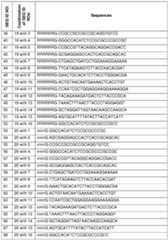
Filed October 18, 2024 [0196] Although exemplified herein with a CPP conjugated (that is, covalently bonded) at the 3’ end of the antisense morpholino sequence, also contemplated are hybrid molecules in which the CPP is attached at another position in the morpholino. By way of example, the CPP may be attached at the 5’ end of the morpholino oligonucleotide, or at any other position within the length of the morpholino. Placement of the CPP can be governed, for instance, by electing the oligonucleotide that is modified to accept addition of the CPP during synthesis via an amide linker. [0197] Optionally, a linker and/or a spacer may be included between the CPP and the CD47- targeted antisense morpholino oligonucleotide. Those of skill in the art will recognize spacers/linkers that may be useful. XI. Selection of Subjects for Treatment [0198] Subjects who will need reduction of CD47 expression and/or protection for non-cancer cells, tissues, and organs from radiation and/or chemotherapy damage while increasing cancer killing will be selected for treatment. [0199] Subjects exposed to radiation or chemotherapy as part of their occupation, by accident, non-cancer related use of radiation or chemotherapy for any treatment, or in military settings will be candidates for treatment. [0200] The provided CD47 morpholinos are applicable for treatment of individuals with solid and liquid cancers, who will receive chemotherapy and or radiation and or check point blockers and or engineered CART cells as part of their treatment plan. They are also applicable to individuals exposed to radiation or chemotherapy for reasons unrelated to cancer therapy. XII. Administration of Human-Specific CD47 Morpholino Oligonucleotides [0201] An effective amount of a compound (such as a morpholino oligonucleotide, or a morpholino oligonucleotide conjugated to a cell penetrating peptide) may be administered in a single dose, or in several doses, for example daily, or periodically with intervals of no treatment of several days or longer, during a course of treatment. However, the effective amount will be dependent on the compound applied, the subject being treated, the severity and type of the affliction, and the manner of administration of the compound. For example, a therapeutically effective amount of an active ingredient can be measured as the concentration (moles per liter or molar-M) of the active ingredient (such as a nucleic acid molecule, peptide, or fusion between a nucleic acid molecule and peptide) in blood (in vivo) or a buffer (in vitro) that produces an effect. [0202] Therapeutically effective dosages of published morpholinos are generally in the range of 1-10 μM. However, data herein demonstrate that the human-only CD47 morpholinos show effective lowering of CD47 protein and cell responses in the lower range of concentrations and even ≤1 µM, a degree of effectiveness not found with other published, known, or reported morpholinos. Filed October 18, 2024 [0203] Specifically contemplated herein are treatments that employ more than one of the described morpholinos, administered either concurrently (in the same composition or different compositions) or in series. Combinations of at least one translation blocking morpholino and at least one splice blocking morpholino are specifically contemplated. By way of example, administration of both CD47-A and CD47-B morpholinos, for instance each with an attached CPP, is provided in some embodiments. In combination morpholino embodiments, the conjugated CPP may be the same on the different morpholinos; in other examples, the CPP conjugated to each type of morpholino is different. [0204] Specifically contemplated herein are treatments that employ more than one of the described morpholinos, administered in a carrier such an endosome (Evans et al., Nat Commun. 10(1):501, 2019). This formulation achieved increased cytosolic delivery of a CD47 morpholino in human cells. The same is contemplated for exosomes and other nano-packaging technologies. [0205] In applications for cancer therapy, the CD47 morpholino will be given as an intravenous infusion to permit the widest cell and tissue access for the agent. For other indications, more regional targeting of agent delivery may be appropriate. [0206] The CD47 morpholinos can be given prior to, or at the same time as a genotoxic stress for cancer via intravenous injection or other methods. This latter feature is unexpected and unprecedented based on prior studies and reports of CD47 morpholinos. Prior knowledge taught that CD47 morpholinos had to be given several days prior to the genotoxic stress to obtain protein suppression and therapeutic benefit. Data herein provided indicate that the provided human-only CD47 morpholinos have therapeutic effects prior to demonstrable lowering of total CD47 protein. [0207] The therapeutic compounds described herein may be formulated in a variety of ways depending on the location and type of disease to be treated or prevented in the subject. Pharmaceutical compositions are thus provided for both local use at or near an affected area and for systemic use (in which the agent is administered in a manner that is widely disseminated via the cardiovascular and lymphatic systems). [0208] Pharmaceutical compositions that include at least one morpholino oligonucleotide, optionally fused with a peptide (e.g., a CPP) described herein as an active ingredient, or that include both a therapeutic morpholino oligonucleotide and an additional agent as active ingredients, or that include both a radioprotective peptide or inhibitor and an additional therapeutic agent, may be formulated with an appropriate solid or liquid carrier, depending upon the particular mode of administration chosen. [0209] A suitable administration format may best be determined by a medical practitioner for each subject individually. Various pharmaceutically acceptable carriers and their formulation are described in standard formulation treatises, for example, Remington's Pharmaceutical Sciences by E. W. Martin. See also Wang and Hanson, J. Parenteral Sci. Technol., Technical Report No.10, Supp.42: 2S, 1988. Filed October 18, 2024 [0210] The dosage form of the pharmaceutical composition will be determined by the mode of administration chosen. For instance, in addition to injectable fluids, inhalational, topical, ophthalmic, peritoneal, intra-pleural, trans-cranial, intra-cerebrospinal, intravenous, and oral formulations can be employed. Inhalational preparations can include aerosols, mists, nebulizers, particulates, and the like. In general, the goal for particle size for inhalation is 1 μm or less in order that the pharmaceutical reach the alveolar region of the lung for absorption. Oral formulations may be liquid (for example, syrups, solutions, or suspensions), or solid (for example, powders, pills, tablets, lozenges, or capsules). For solid compositions, conventional non-toxic solid carriers can include pharmaceutical grades of mannitol, lactose, starch, or magnesium stearate. Actual methods of preparing such dosage forms are known, or will be apparent, to those of ordinary skill in the art. [0211] The compositions or pharmaceutical compositions can be administered by any route including parenteral administration, for example, intravenous, intramuscular, intraperitoneal, intra-sternal, or intra-articular injection or infusion, or by intra-bone marrow space injection such as via the anterior tibia, or by sublingual, oral, topical, intra-nasal, ophthalmic, or transmucosal administration, or by pulmonary inhalation. When the active compounds are provided as parenteral compositions, for example, for injection or infusion, they are generally suspended in an aqueous carrier, for example, in an isotonic buffer solution at a pH of 3.0 to 8.0, preferably at a pH of 3.5 to 7.4, 3.5 to 6.0, or 3.5 to 5.0. Useful buffers include sodium citrate-citric acid and sodium phosphate-phosphoric acid, and sodium acetate/acetic acid buffers. A form of repository or depot slow-release preparation may be used so that therapeutically effective amounts of the preparation are delivered into the bloodstream over many hours or days following transdermal injection or delivery. The agent may also be part of (e.g., coated on surface(s) of) an implantable device such as a stent, vascular conduit, catheter, pump, or other engineered implant or scaffold, the latter sometimes seeded with cells of various types, from which it will be released. [0212] Active compounds (e.g., oligonucleotides, peptides, and fusions thereof) are also suitably administered by sustained-release systems. Suitable examples of sustained-release formulations include suitable polymeric materials (such as, for example, semi-permeable polymer matrices in the form of shaped articles, for example, films, or microcapsules), suitable hydrophobic materials (for example as an emulsion in an acceptable oil) or ion exchange resins, and sparingly soluble derivatives (such as, for example, a sparingly soluble salt). Sustained- release compounds may be administered by intravascular, intravenous, intra-arterial, intramuscular, subcutaneous, intra-pericardial, or intra-coronary injection. Administration can also be oral, rectal, parenteral, intracisternal, intravaginal, intraperitoneal, topical (as by powders, ointments, gels, drops or transdermal patch), buccal, or as an oral or nasal spray. [0213] Preparations for administration can be suitably formulated to give controlled release of the therapeutic agent(s) (e.g., oligonucleotides or oligonucleotide/peptide fusions). For example, the Filed October 18, 2024 pharmaceutical compositions may be in the form of particles comprising a biodegradable polymer and/or a polysaccharide jellifying and/or bio-adhesive polymer, an amphiphilic polymer, an agent modifying the interface properties of the particles and a pharmacologically active substance. These compositions exhibit certain biocompatibility features that allow a controlled release of the active substance. See, for example, U.S. Pat. No.5,700,486. [0214] In some embodiments, therapeutic agent(s) are delivered by way of a pump (see Sefton, CRC Crit. Ref. Biomed. Eng.14:201, 1987; Buchwald et al., Surgery 88:507, 1980; Saudek et al., N. Engl. J. Med.321:574, 1989) or by continuous subcutaneous infusions, for example, using a mini-pump. An intravenous bag solution may also be employed. The key factor in selecting an appropriate dose is the result obtained, as measured by increases or decreases in radioprotection or protection from chemotherapy-mediated injury, or by other criteria for measuring control or prevention of disease, as are deemed appropriate by the practitioner. Other controlled release systems are discussed in the review by Langer (Science 249:1527-1533, 1990). [0215] In another aspect of the disclosure, therapeutic agent(s) are delivered by way of an implanted pump, described, for example, in U.S. Pat. No. 6,436,091; U.S. Pat. No. 5,939,380; and U.S. Pat. No.5,993,414. Implantable drug infusion devices are used to provide subjects with a constant and long-term dosage or infusion of a drug or any other therapeutic agent. Essentially, such device may be categorized as either active or passive. [0216] Active drug or programmable infusion devices feature a pump or a metering system to deliver the drug into the individual’s system. An example of such an active drug infusion device currently available is the Medtronic SynchroMed™ programmable pump. Such pumps typically include a drug reservoir, a peristaltic pump to pump the drug out from the reservoir, and a catheter port to transport the pumped-out drug from the reservoir via the pump to an individual’s anatomy. Such devices also typically include a battery to power the pump, as well as an electronic module to control the flow rate of the pump. The Medtronic SynchroMed™ pump further includes an antenna to permit the remote programming of the pump. [0217] An implanted pump can be completely implanted under the skin of a subject, thereby negating the need for a percutaneous catheter. These implanted pumps can provide the individual with therapeutic agent(s) at a constant or a programmed delivery rate. Constant rate or programmable rate pumps are based on either phase-change or peristaltic technology. When a constant, unchanging delivery rate is required, a constant-rate pump is well suited for long-term implanted drug delivery. If changes to the infusion rate are expected, a programmable pump may be used in place of the constant rate pump system. Osmotic pumps may be much smaller than other constant rate or programmable pumps, because their infusion rate can be very low. An example of such a pump is described listed in U.S. Pat. No.5,728,396. Filed October 18, 2024 [0218] Passive drug infusion devices, in contrast, do not feature a pump, but rather rely upon a pressurized drug reservoir to deliver the drug. Thus, such devices tend to be both smaller as well as cheaper as compared to active devices. An example of such a device includes the Medtronic IsoMed™. This device delivers the drug into the patient through the force provided by a pressurized reservoir applied across a flow control unit. [0219] The therapeutic agents may also be delivered passively and in sustained fashion as part of and incorporated into implantable devices, such as vascular stents which can be placed directly into diseased blood vessels through several standard approaches, including direct surgical insertion or percutaneously with angiographic control. Other formations may be combined with osteo-integrated implants to promote bone healing. [0220] The CD47 morpholinos are quite water soluble and stable and thus ensure that their use with implantable and other delivery technology will provide a therapeutically active agent. [0221] For oral administration, the pharmaceutical compositions can take the form of, for example, tablets or capsules prepared by conventional means with pharmaceutically acceptable excipients such as binding agents (for example, pregelatinized maize starch, polyvinylpyrrolidone or hydroxypropyl methylcellulose); fillers (for example, lactose, microcrystalline cellulose or calcium hydrogen phosphate); lubricants (for example, magnesium stearate, talc or silica); disintegrants (for example, potato starch or sodium starch glycolate); or wetting agents (for example, sodium lauryl sulphate). The tablets can be coated by methods well known in the art. Liquid preparations for oral administration can take the form of, for example, solutions, syrups or suspensions, or they can be presented as a dry product for constitution with water or other suitable vehicle before use. Such liquid preparations can be prepared by conventional means with pharmaceutically acceptable additives such as suspending agents (for example, sorbitol syrup, cellulose derivatives or hydrogenated edible fats); emulsifying agents (for example, lecithin or acacia); non-aqueous vehicles (for example, almond oil, oily esters, ethyl alcohol or fractionated vegetable oils); and preservatives (for example, methyl or propyl-p- hydroxybenzoates or sorbic acid). The preparations can also contain buffer salts, flavoring, coloring, and sweetening agents as appropriate. The herein provided CD47 morpholinos are stable and will and do not react with any of the agents listed above. [0222] For administration by inhalation, the compounds for use according to the present disclosure are conveniently delivered in the form of an aerosol spray presentation from pressurized packs or a nebulizer, with the use of a suitable propellant, for example, dichlorodifluoromethane, trichlorofluoromethane, dichlorotetrafluoroethane, carbon dioxide or other suitable gas. In the case of a pressurized aerosol, the dosage unit can be determined by providing a valve to deliver a metered amount. Capsules and cartridges for use in an inhaler or insufflator can be formulated containing a powder mix of the compound and a suitable powder base such as lactose or starch. Filed October 18, 2024 [0223] For topical administration, the compounds can be, for example, mixed with a liquid delivery agent for administration locally. The agents used therapeutically (such as peptides, antibodies and morpholinos) are readily soluble or can be suspended in water and saline, and as such these would be useful for delivery since water or saline do not cause adverse biological tissue effects. This allows sufficiently high doses to be administered locally or systemically, without secondary toxicity from the delivery vehicle. In this same line, the morpholinos could be part of a patch placed on the skin (that is, a transdermal delivery patch) that would deliver the agent to the skin for absorption. [0224] Pharmaceutical compositions that comprise at least one therapeutic agent as described herein as an active ingredient will normally be formulated with an appropriate solid or liquid carrier, depending upon the particular mode of administration chosen. The pharmaceutically acceptable carriers and excipients useful in this disclosure are conventional. For instance, parenteral formulations usually comprise injectable fluids that are pharmaceutically and physiologically acceptable fluid vehicles such as water, physiological saline, other balanced salt solutions, aqueous dextrose, glycerol or the like. Excipients that can be included are, for instance, proteins, such as human serum albumin or plasma preparations. If desired, the pharmaceutical composition to be administered may also contain minor amounts of non-toxic auxiliary substances, such as wetting or emulsifying agents, preservatives, and pH buffering agents and the like, for example sodium acetate or sorbitan monolaurate. Actual methods of preparing such dosage forms are known, or will be apparent, to those skilled in the art. [0225] For example, for parenteral administration, therapeutic agent(s) can be formulated generally by mixing them at the desired degree of purity, in a unit dosage injectable form (solution, suspension, or emulsion), with a pharmaceutically acceptable carrier, for instance, one that is non-toxic to recipients at the dosages and concentrations employed and is compatible with other ingredients of the formulation. A pharmaceutically acceptable carrier is a non-toxic solid, semisolid or liquid filler, diluent, encapsulating material or formulation auxiliary of any type. [0226] Generally, the formulations are prepared by contacting the therapeutic agent(s) each uniformly and intimately with liquid carriers or finely divided solid carriers or both. Then, if necessary, the product is shaped into the desired formulation. Optionally, the carrier is a parenteral carrier, and in some embodiments, it is a solution that is isotonic with the blood of the recipient. Examples of such carrier vehicles include water, saline, Ringer's solution, and dextrose solution. Non-aqueous vehicles such as fixed oils and ethyl oleate are also useful herein, as well as liposomes. The agent could also be included within a transfused human product such a blood, plasma, platelets, bone marrow cells, etc. [0227] The pharmaceutical compositions that comprise at least one therapeutic agent, in some embodiments, will be formulated in unit dosage form, suitable for individual administration of precise dosages. The amount of the active compound(s) administered will be dependent on the Filed October 18, 2024 subject being treated, the severity of the affliction, and the manner of administration, and is best left to the judgment of the prescribing clinician. Within these bounds, the formulation to be administered will contain a quantity of the active component(s) in amounts effective to achieve the desired effect in the subject being treated. [0228] The therapeutically effective amount of the agent, such as an oligonucleotide (e.g., morpholino or other antisense molecule) will be dependent on the agent utilized, the subject being treated, the severity and type of the affliction, and the manner of administration. The exact dose is readily determined by one of skilled in the art based on the potency of the specific compound, the age, weight, sex, and physiological condition of the subject. [0229] The therapeutic agents can also be administered directly as part of a surgical or other medical procedure, or at the bedside by a treating physician. Drug quality product (e.g., morpholino) can be diluted for instance in sterile saline and given by injection using sterile 1 cc syringes and small-bore needles (25 gauge and less) to a subject in need of radioprotection. Alternatively, a wound bed can be irrigated for instance with a saline or other therapeutically effective solution containing a known concentration (dosage) of drug or compound, or a combination thereof. Precise control and localization of therapeutic effects can thus be obtained. [0230] The agent may also be instilled into a transplanted organ prior to transplantation, such as a kidney, liver, heart, or lungs via the vasculature to minimize organ injury and enhance organ take, survival, and function after transplantation. [0231] Controlled release parenteral formulations can be made as implants, oily injections, or as particulate systems. For a broad overview of protein delivery systems, see Banga, Therapeutic Peptides and Proteins: Formulation, Processing, and Delivery Systems, Technomic Publishing Company, Inc., Lancaster, Pa., 1995. Particulate systems include microspheres, microparticles, microcapsules, nano-capsules, nanospheres, and nanoparticles. Microcapsules contain the therapeutic peptide as a central core. In microspheres, the therapeutic agent is dispersed throughout the particle. Particles, microspheres, and microcapsules smaller than 1 μm are generally referred to as nanoparticles, nanospheres, and nano-capsules, respectively. Capillaries have a diameter of approximately 5 μm so that only nanoparticles are administered intravenously. Microparticles are typically around 100 μm in diameter and are administered subcutaneously or intramuscularly (see Kreuter, Colloidal Drug Delivery Systems, J. Kreuter, ed., Marcel Dekker, Inc., New York, N.Y., pp.219-342, 1994; Tice & Tabibi, Treatise on Controlled Drug Delivery, A. Kydonieus, ed., Marcel Dekker, Inc. New York, N.Y., pp.315-339, 1992). CD47 morpholino can also be delivered efficiently to cells via endosomes (Evans et al., Nat Commun.10(1):501, 2019). [0232] Also contemplated is the use of nanoparticles as delivery agents, which can be targeted to specific cells, tissues or organ for instance by incorporation on their surface ligands of receptors specific in their expression to the targeted cells, tissues or organs. The targeting entity can be the same or different than the therapeutically active agent carried by the nanoparticle. Filed October 18, 2024 Further, distribution of nanoparticles to certain tissues spaces (e.g. the blood versus the central nervous system protected by the blood-brain barrier) can be determined by altering the size of the nanoparticles thereby allowing or preventing their transit of such barriers between tissue compartments and through displaying target-cell specific antigen(s) on the surface of the nanoparticles to yield precision directed therapy. The same applies to delivery methods above. [0233] Polymers can be used for ion-controlled release. Various degradable and nondegradable polymeric matrices for use in controlled drug delivery are known in the art (Langer, Accounts Chem. Res.26:537, 1993). For example, the block copolymer, poloxamer 407 exists as a viscous yet mobile liquid at low temperatures but forms a semisolid gel at body temperature. It has shown to be an effective vehicle for formulation and sustained delivery of recombinant interleukin-2 and urease (Johnston et al., Pharm. Res.9:425, 1992; Pec, J. Parent. Sci. Tech.44(2):58, 1990). Alternatively, hydroxyapatite has been used as a microcarrier for controlled release of proteins (Ijntema et al., Int. J. Pharm.112:215, 1994). In yet another aspect, liposomes are used for controlled release as well as drug targeting of lipid-capsulated compounds (Betageri et al., Liposome Drug Delivery Systems, Technomic Publishing Co., Inc., Lancaster, Pa., 1993). Numerous additional systems for controlled delivery of therapeutic proteins are known (e.g., U.S. Pat. No.5,055,303; U.S. Pat. No.5,188,837; U.S. Pat. No.4,235,871; U.S. Pat. No.4,501,728; U.S. Pat. No.4,837,028; U.S. Pat. No. 4,957,735; and U.S. Pat. No.5,019,369; U.S. Pat. No. 5,055,303; U.S. Pat. No.5,514,670; U.S. Pat. No.5,413,797; U.S. Pat. No.5,268,164; U.S. Pat. No.5,004,697; U.S. Pat. No.4,902,505; U.S. Pat. No.5,506,206; U.S. Pat. No.5,271,961; U.S. Pat. No.5,254,342; and U.S. Pat. No.5,534,496). Such polymers may take the form of mesh and matrix or similar supporting type materials used for enhanced wound healing and tissue healing. [0234] The specific form of the agents and their manner of administration may also be influenced, at least in part, by the particular tissue to be treated. [0235] The amount of agent to be delivered, as well as the dosing schedule useful to provide the desired protective effects, will be influenced by the bioavailability of the specific therapeutic compound selected (and/or an active metabolite thereof), the type and extent of radiation exposure or radiation dosage schedule, and other factors that will be apparent to those of skill in the art. [0236] Alternatively, the compound may be administered in a gel, lotion, ointment, or other suitable form which is applied to the tissue up to 90 minutes before irradiation or treatment and remains on the tissue during and optionally after the treatment. [0237] The same dosage and concentrations can be used when the radioprotective agent is administered after irradiation and/or radiotherapeutic treatment. The three administrations (before, during and after radiotherapy treatment) may be used alone, or in any combination of two or all three administrations, as needed. Filed October 18, 2024 [0238] The provided human-only CD47 morpholinos described herein are the first ever to be effective against genotoxic stress when administered at the same time as the stress. XIII. Determination of Effectiveness of Treatment [0239] Therapeutic effect following morpholino administration may be followed and tracked through analysis of CD47 protein expression, both total and cell surface (Bmax), in circulating blood cells or cells from other body fluids such as cerebrospinal or pleural fluids. Peripheral blood mononuclear cells will be isolated from blood samples and protein expression characterized by flow cytometry, a method we have standardized, and / or Western blot as shown herein. [0240] Lowering of total and or cell surface CD47 expression on cells from the blood and other body fluids would be taken as evidence of CD47 morpholino activity and will show correlation with therapeutic effect. In instances where tumor tissue are available from individuals given the CD47 morpholino, samples could be assessed via several technique for CD47 expression including immunohistochemistry, immunofluorescence, flow cytometry, or Western blot as published (Erdem et al., Am J Physiol Endocrinol Metab.2023 Apr 1;324(4): E347-E357). XIV. Co-Administration of Additional Therapeutic Compound(s) [0241] Data indicate that the CD47 morpholinos lower CD47 protein to maximum effect 24-72 hours after administration. However, in combination with genotoxic stress it also enhanced non- cancer cell viability when given at the same time as the stressor (e.g., chemotherapy). Thus, the morpholino will be administered either at the same time as or 1-3 days prior to chemotherapy or radiation or other cancer agents. [0242] Additional active ingredients include, for example, nitric oxide donors, nitro-vasodilators, activators of the enzyme soluble guanylyl cyclase, or cGMP phosphodiesterase inhibitors. Pharmaceutical compositions may include additional cytoprotective or radioprotective agents known to the art (for example as described in Tofilon, Chem. Rev.109:2974-88, 2009). [0243] The Exemplary Embodiments and Experimental Examples below are included to demonstrate particular embodiments of the disclosure. Those of ordinary skill in the art should recognize in light of the present disclosure that many changes can be made to the specific embodiments disclosed herein and still obtain a like or similar result without departing from the spirit and scope of the disclosure. XV. Exemplary Embodiments [0244] 1. A CD47 antisense morpholino oligonucleotide including a nucleic acid sequence set forth in any of: SEQ ID NO: 1 (CD47-A), SEQ ID NO: 3 (CD47-C), SEQ ID NO: 4: (CD47-D), SEQ ID NO: 5: (CD47-E), SEQ ID NO: 16 (CD47-A’), SEQ ID NO: 2, CD47-B (CD47-202_e1i1), SEQ ID NO: 6, SB1 (CD47-202_e1i1’), SEQ ID NO: 7, SB2 (CD47-202_i1e2), SEQ ID NO: 8, Filed October 18, 2024 SB3 (CD47-202_e2i2), SEQ ID NO: 9, SB4 (CD47-202_e4i4), SEQ ID NO: 10, SB5 (CD47- 202_e5i5), SEQ ID NO: 11, SB6 (CD47-202_i5e6), SEQ ID NO: 12, SB7 (CD47-202_e6i6), SEQ ID NO: 13, SB8 (CD47-202_e8i8), SEQ ID NO: 14, SB9 (CD47-202_i9e10), or SEQ ID NO: 15, SB10 (CD47-202_e10i10). [0245] 2. A CD47 antisense morpholino oligonucleotide including a nucleic acid with a sequence consisting of the sequence set forth in any of: SEQ ID NO: 1 (CD47-A), SEQ ID NO: 3 (CD47-C), SEQ ID NO: 4: (CD47-D), SEQ ID NO: 5: (CD47-E), SEQ ID NO: 16 (CD47-A’), SEQ ID NO: 2, CD47-B (CD47-202_e1i1), SEQ ID NO: 6, SB1 (CD47-202_e1i1’), SEQ ID NO: 7, SB2 (CD47-202_i1e2), SEQ ID NO: 8, SB3 (CD47-202_e2i2), SEQ ID NO: 9, SB4 (CD47-202_e4i4), SEQ ID NO: 10, SB5 (CD47-202_e5i5), SEQ ID NO: 11, SB6 (CD47-202_i5e6), SEQ ID NO: 12, SB7 (CD47-202_e6i6), SEQ ID NO: 13, SB8 (CD47-202_e8i8), SEQ ID NO: 14, SB9 (CD47- 202_i9e10), or SEQ ID NO: 15, SB10 (CD47-202_e10i10). [0246] 3. The CD47 antisense morpholino oligonucleotide of embodiment 1 or embodiment 2, further including a cell-penetrating peptide (CPP) conjugated to the oligonucleotide. [0247] 4. The CD47 antisense morpholino oligonucleotide of embodiment 3, wherein the CPP is conjugated at the 5’ end of the oligonucleotide. [0248] 5. The CD47 antisense morpholino oligonucleotide of embodiment 3, including a sequence as shown in any one of SEQ ID NOs: 21-68. [0249] 6. A non-naturally occurring antisense oligonucleotide specific for human CD47, the sequence of which consists essentially of the sequence set forth in any of: SEQ ID NO: 1 (CD47- A), SEQ ID NO: 3 (CD47-C), SEQ ID NO: 4: (CD47-D), SEQ ID NO: 5: (CD47-E), SEQ ID NO: 16 (CD47-A’), SEQ ID NO: 2, CD47-B (CD47-202_e1i1), SEQ ID NO: 6, SB1 (CD47-202_e1i1’), SEQ ID NO: 7, SB2 (CD47-202_i1e2), SEQ ID NO: 8, SB3 (CD47-202_e2i2), SEQ ID NO: 9, SB4 (CD47-202_e4i4), SEQ ID NO: 10, SB5 (CD47-202_e5i5), SEQ ID NO: 11, SB6 (CD47- 202_i5e6), SEQ ID NO: 12, SB7 (CD47-202_e6i6), SEQ ID NO: 13, SB8 (CD47-202_e8i8), SEQ ID NO: 14, SB9 (CD47-202_i9e10), or SEQ ID NO: 15, SB10 (CD47-202_e10i10). [0250] 7. The non-naturally occurring antisense oligonucleotide of embodiment 6, which includes at least one morpholino ring. [0251] 8. The non-naturally occurring antisense oligonucleotide of embodiment 6 or embodiment 7, further including a cell-penetrating peptide (CPP) conjugated to the oligonucleotide. [0252] 9. The non-naturally occurring antisense oligonucleotide of embodiment 8, wherein the CPP is conjugated at the 5’ end of the oligonucleotide. [0253] 10. The non-naturally occurring antisense oligonucleotide of embodiment 8, including a sequence as shown in any one of SEQ ID NOs: 21-68. [0254] 11. A hybrid nucleic acid-peptide fusion molecule including: an antisense oligonucleotide specific for human CD47, and including a nucleic acid sequence set forth in any Filed October 18, 2024 of: SEQ ID NO: 1 (CD47-A), SEQ ID NO: 3 (CD47-C), SEQ ID NO: 4: (CD47-D), SEQ ID NO: 5: (CD47-E), SEQ ID NO: 16 (CD47-A’), SEQ ID NO: 2, CD47-B (CD47-202_e1i1), SEQ ID NO: 6, SB1 (CD47-202_e1i1’), SEQ ID NO: 7, SB2 (CD47-202_i1e2), SEQ ID NO: 8, SB3 (CD47- 202_e2i2), SEQ ID NO: 9, SB4 (CD47-202_e4i4), SEQ ID NO: 10, SB5 (CD47-202_e5i5), SEQ ID NO: 11, SB6 (CD47-202_i5e6), SEQ ID NO: 12, SB7 (CD47-202_e6i6), SEQ ID NO: 13, SB8 (CD47-202_e8i8), SEQ ID NO: 14, SB9 (CD47-202_i9e10), or SEQ ID NO: 15, SB10 (CD47- 202_e10i10); and a peptide covalently attached to the antisense oligonucleotide. [0255] 12. The fusion molecule of embodiment 11, wherein the peptide is covalently attached at the 5’ end of the antisense oligonucleotide. [0256] 13. The fusion molecule of embodiment 11, wherein the peptide includes a cell penetrating peptide (CPP) or other cell targeting sequence. [0257] 14. The fusion molecule of embodiment 13, wherein the peptide includes: DG9 YArVRRrGPRGYArVRRrGPRrB (SEQ ID NO: 18; r = D-arginine, B = beta-alanine); R6G: RRRRRRG (SEQ ID NO: 19); or r6G: rrrrrrG (SEQ ID NO: 20; r = D-arginine). [0258] 15. The fusion molecule of embodiment 13, including a sequence as shown in any one of SEQ ID NOs: 21-68. [0259] 16. A composition including: at least one therapeutic molecule selected from: the CD47 antisense morpholino oligonucleotide of any one of embodiments 1-5; the non-naturally occurring antisense oligonucleotide of any one of embodiments 6-10; or the hybrid nucleic acid- peptide fusion molecule of any one of embodiments 11-15; and at least one biologically or pharmaceutically acceptable excipient or carrier. [0260] 17. The composition of embodiment 16, formulated for delivery to a subject. [0261] 18. The formulated composition of embodiment 17, formulated to be delivered to the subject transdermally, intramuscularly, intravascularly, by inhalation, or intranasally. [0262] 19. The composition of embodiment 16, including at least two of the therapeutic molecules. [0263] 20. The composition of embodiment 19, wherein the at least two therapeutic molecules include: at least one oligonucleotide with a sequence consisting of the sequence set forth in any of: SEQ ID NO: 1 (CD47-A), SEQ ID NO: 3 (CD47-C), SEQ ID NO: 4: (CD47-D), SEQ ID NO: 5: (CD47-E), or SEQ ID NO: 16 (CD47-A’); and at least one oligonucleotide with a sequence consisting of the sequence set forth in any of: SEQ ID NO: 2, CD47-B (CD47- 202_e1i1), SEQ ID NO: 6, SB1 (CD47-202_e1i1’), SEQ ID NO: 7, SB2 (CD47-202_i1e2), SEQ ID NO: 8, SB3 (CD47-202_e2i2), SEQ ID NO: 9, SB4 (CD47-202_e4i4), SEQ ID NO: 10, SB5 (CD47-202_e5i5), SEQ ID NO: 11, SB6 (CD47-202_i5e6), SEQ ID NO: 12, SB7 (CD47- 202_e6i6), SEQ ID NO: 13, SB8 (CD47-202_e8i8), SEQ ID NO: 14, SB9 (CD47-202_i9e10), or SEQ ID NO: 15, SB10 (CD47-202_e10i10). Filed October 18, 2024 [0264] 21. A composition including a plurality of different CD47 antisense morpholino oligonucleotides of any of embodiments 1-5. [0265] 22. A composition including a plurality of different non-naturally occurring antisense oligonucleotides of any one of embodiments 6-10. [0266] 23. A composition including a plurality of different hybrid nucleic acid-peptide fusion molecules of any one of embodiments 11-15. [0267] 24. The composition of any one of embodiments 20-23, wherein the at least two oligonucleotides or fusion molecules include: at least one oligonucleotide with a sequence consisting of the sequence set forth in any of: SEQ ID NO: 1 (CD47-A), SEQ ID NO: 3 (CD47- C), SEQ ID NO: 4: (CD47-D), SEQ ID NO: 5: (CD47-E), or SEQ ID NO: 16 (CD47-A’); and at least one oligonucleotide with a sequence consisting of the sequence set forth in any of: SEQ ID NO: 2, CD47-B (CD47-202_e1i1), SEQ ID NO: 6, SB1 (CD47-202_e1i1’), SEQ ID NO: 7, SB2 (CD47-202_i1e2), SEQ ID NO: 8, SB3 (CD47-202_e2i2), SEQ ID NO: 9, SB4 (CD47-202_e4i4), SEQ ID NO: 10, SB5 (CD47-202_e5i5), SEQ ID NO: 11, SB6 (CD47-202_i5e6), SEQ ID NO: 12, SB7 (CD47-202_e6i6), SEQ ID NO: 13, SB8 (CD47-202_e8i8), SEQ ID NO: 14, SB9 (CD47- 202_i9e10), or SEQ ID NO: 15, SB10 (CD47-202_e10i10). [0268] 26. The CD47 antisense morpholino oligonucleotide of any one of embodiments 1-5, the non-naturally occurring antisense oligonucleotide of any one of embodiments 6-10, the hybrid nucleic acid-peptide fusion molecule of any one of embodiments 11-15, or the composition of any one of embodiments 16-24, for use in modulating CD47 signalling in a subject. [0269] 27. The CD47 antisense morpholino oligonucleotide of any one of embodiments 1-5, the non-naturally occurring antisense oligonucleotide of any one of embodiments 6-10, the hybrid nucleic acid-peptide fusion molecule of any one of embodiments 11-15, or the composition of any one of embodiments 16-24, for use in a method of reducing cytotoxicity or genotoxicity of an agent in a subject. [0270] 28. A method including administering to a subject a CD47-level reducing amount of an agent selected from: the CD47 antisense morpholino oligonucleotide of any one of embodiments 1-5, the non-naturally occurring antisense oligonucleotide of any one of embodiments 6-10, the hybrid nucleic acid-peptide fusion molecule of any one of embodiments 11-15, or the composition of any one of embodiments 16-24. [0271] 29. The method of embodiment 28, which reduces at least one cytotoxic or at least one genotoxic effect of an agent in the subject. [0272] 30. The method of embodiment 28, wherein the agent includes a chemotherapeutic agent, a radionuclide administered as part of a radiation treatment or other radiation exposure, or another cytotoxic or genotoxic agent administered to the subject. [0273] 31. The method of embodiments 28-30, wherein the agent is administered transdermally, intramuscularly, intravascularly, by inhalation, or intranasally. Filed October 18, 2024 [0274] 32. A method of reducing cytotoxicity of a chemotherapeutic agent to non-cancer cells in a subject with cancer, including: administering to the subject an effective amount of a CD47 antisense morpholino oligonucleotide including the nucleic acid sequence set forth in any of: SEQ ID NO: 1 (CD47-A), SEQ ID NO: 3 (CD47-C), SEQ ID NO: 4: (CD47-D), SEQ ID NO: 5: (CD47- E), SEQ ID NO: 16 (CD47-A’), SEQ ID NO: 2, CD47-B (CD47-202_e1i1), SEQ ID NO: 6, SB1 (CD47-202_e1i1’), SEQ ID NO: 7, SB2 (CD47-202_i1e2), SEQ ID NO: 8, SB3 (CD47-202_e2i2), SEQ ID NO: 9, SB4 (CD47-202_e4i4), SEQ ID NO: 10, SB5 (CD47-202_e5i5), SEQ ID NO: 11, SB6 (CD47-202_i5e6), SEQ ID NO: 12, SB7 (CD47-202_e6i6), SEQ ID NO: 13, SB8 (CD47- 202_e8i8), SEQ ID NO: 14, SB9 (CD47-202_i9e10), or SEQ ID NO: 15, SB10 (CD47- 202_e10i10); and one or more chemotherapeutic agents, [0275] wherein the CD47 antisense morpholino inhibits CD47 signaling and is administered to the subject before, during, or after administration of the one or more chemotherapeutic agents. [0276] 33. The method of embodiment 32, wherein the antisense morpholino oligonucleotide further includes a cell-penetrating peptide (CPP) conjugated to the oligonucleotide. [0277] 34. The method of embodiment 33, wherein the CPP is conjugated at the 5’ end of the oligonucleotide. [0278] 35. The method of embodiment 33, antisense morpholino oligonucleotide includes a sequence as shown in any one of SEQ ID NOs: 21-68. [0279] 36. The method of embodiments 32-35, in which the antisense morpholino oligonucleotide is administered transdermally, intramuscularly, intravascularly, by inhalation, or intranasally. [0280] 37. A method of reducing cytotoxicity of a chemotherapeutic agent to non-cancer cells, including: (a) administering an effective amount of at least one CD47 morpholino oligonucleotide including the nucleic acid sequence set forth as in any of: SEQ ID NO: 1 (CD47- A), SEQ ID NO: 3 (CD47-C), SEQ ID NO: 4: (CD47-D), SEQ ID NO: 5: (CD47-E), SEQ ID NO: 16 (CD47-A’), SEQ ID NO: 2, CD47-B (CD47-202_e1i1), SEQ ID NO: 6, SB1 (CD47-202_e1i1’), SEQ ID NO: 7, SB2 (CD47-202_i1e2), SEQ ID NO: 8, SB3 (CD47-202_e2i2), SEQ ID NO: 9, SB4 (CD47-202_e4i4), SEQ ID NO: 10, SB5 (CD47-202_e5i5), SEQ ID NO: 11, SB6 (CD47- 202_i5e6), SEQ ID NO: 12, SB7 (CD47-202_e6i6), SEQ ID NO: 13, SB8 (CD47-202_e8i8), SEQ ID NO: 14, SB9 (CD47-202_i9e10), or SEQ ID NO: 15, SB10 (CD47-202_e10i10) to a subject with cancer; (b) administering one or more chemotherapeutic agents to the subject with cancer, wherein (a) and (b) can be performed in either order or concurrently; and (c) detecting a reduction of cytotoxicity to non-cancer cells in the subject. [0281] 38. The method of embodiment 37, wherein the antisense morpholino oligonucleotide further includes a cell-penetrating peptide (CPP) conjugated to the oligonucleotide. [0282] 39. The method of embodiment 38, wherein the CPP is conjugated at the 5’ end of the oligonucleotide. Filed October 18, 2024 [0283] 40. The method of embodiment 38, antisense morpholino oligonucleotide includes a sequence as shown in any one of SEQ ID NOs: 21-68. [0284] 41. The method of embodiments 37-40, wherein the antisense morpholino oligonucleotide is administered transdermally, intramuscularly, intravascularly, by inhalation, or intranasally. [0285] 42. The method of any one of embodiments 35-38, wherein the oligonucleotide agent is delivered concurrently with the cytotoxic or genotoxic challenge. [0286] 43. The method of any one of embodiments 28-42, including administering at least one splice blocker oligonucleotide and at least one translation blocker oligonucleotide. [0287] 44. The method of embodiment 43, wherein: the at least one splice blocker oligonucleotide has a sequence consisting of the sequence set forth in any of: SEQ ID NO: 1 (CD47-A), SEQ ID NO: 3 (CD47-C), SEQ ID NO: 4: (CD47-D), SEQ ID NO: 5: (CD47-E), or SEQ ID NO: 16 (CD47-A’); and the at least one translation blocker oligonucleotide has a sequence consisting of the sequence set forth in any of: SEQ ID NO: 2, CD47-B (CD47-202_e1i1), SEQ ID NO: 6, SB1 (CD47-202_e1i1’), SEQ ID NO: 7, SB2 (CD47-202_i1e2), SEQ ID NO: 8, SB3 (CD47- 202_e2i2), SEQ ID NO: 9, SB4 (CD47-202_e4i4), SEQ ID NO: 10, SB5 (CD47-202_e5i5), SEQ ID NO: 11, SB6 (CD47-202_i5e6), SEQ ID NO: 12, SB7 (CD47-202_e6i6), SEQ ID NO: 13, SB8 (CD47-202_e8i8), SEQ ID NO: 14, SB9 (CD47-202_i9e10), or SEQ ID NO: 15, SB10 (CD47- 202_e10i10). [0288] 45. The method of embodiment 43 or embodiment 44, wherein the splice blocker oligonucleotide and/or the translation blocker oligonucleotide further includes a cell penetrating peptide (CPP) conjugated thereto. [0289] 46. The method of embodiment 45, wherein the splice blocker oligonucleotide and/or the translation blocker oligonucleotide is selected from SEQ ID NOs: 21-68. [0290] 47. The method of any one of embodiments 28-46, wherein the subject is a human. [0291] 48. The method of embodiment 47, wherein the subject is immunocompromised. [0292] 49. The method of embodiment 47, wherein the therapeutic agent(s) is administered transdermally, intramuscularly, intravascularly, by inhalation, or intranasally. XVI. Experimental Examples Example 1: Testing cell uptake-enhanced CD47 morpholinos for cytotoxicity, effectivity of decreasing CD47 protein levels in cells, and decreasing sensitivity of cells to chemotherapy. [0293] CD47 morpholinos conjugated to uptake-enhanced cell-penetrating peptide were tested for cytotoxicity, ability to decrease CD47 protein expression, both total and cell surface, and ability to reduce sensitivity of cells to the standard cytotoxic chemotherapeutic agent Doxorubicin. Cell uptake-enhanced CD47 morpholinos were non-toxic to several types of primary human cells from Filed October 18, 2024 different distinct cell lineages. Of note, all CD47 morpholinos previously described in the literature were employed as naked entities lacking any modifications to enhance cell uptake. This was true for studies that treated cells in vitro or that treated animals with the morpholino. Yet, to be effective in human disease, morpholinos are modified to increase their delivery and increase uptake by cells. It was not known if a CD47 morpholino conjugated to a cell uptake-enhancing peptide would be inherently cytotoxic. This was an important question to test as such a construct could have been injurious. [0294] It was established that such uptake-enhanced cell-penetrating peptide conjugated CD47 morpholinos were not cytotoxic at concentrations that showed lowering of CD47 protein expression and increased viability and protection from the chemotherapy agent in primary human cells. The highest morpholino concentrations were substantially greater than that which will be used in treating individuals (FIGs.7-11). These are important finding that pave the way forward for developing CD47 morpholino sequences conjugated to cell uptake-enhancing peptides. As well, different cell uptake-enhancing peptide conjugated to the CD47 morpholino were found to be nontoxic to human cells, indicating that multiple uptake-enhancing cell-penetrating peptides do not render the CD47 morpholinos inherently toxic. This finding provides flexibility in development of cell uptake strategies. Studies were repeated at least 3 times. Representative data is presented. [0295] A cell uptake-enhanced CD47 morpholino decreased CD47 protein levels in human cells in a dose-dependent manner. As it was published, the therapeutic benefit in any CD47 morpholino resides in its ability to lower CD47 protein. Thus, this remains the first initial step in confirming bio-activity of a CD47 morpholino. It was established that a cell uptake-enhancing peptide conjugated CD47 morpholino lowered total CD47 protein in multiple human primary cell types. The peptide modified morpholino showed dose-dependent activity in lowering CD47 protein in human immune T cells (FIGs. 15 and 16). This confirmed that the cell uptake enhancement to the CD47 morpholino did not interfere with the protein lowering capacity of the morpholino. This was important to check, as some data suggest the cell uptake enhancing peptides may be inflammatory (Lebleu et al., Adv Drug Deliv Rev.2008 Mar 1;60(4-5):517-29). This could have been an issue as CD47 and TSP1 are increased by inflammation. Indeed, by increasing cell uptake the peptide modification of the morpholino is actually predicted to improve the CD47 morpholino efficacy and permit lower dosing. Of note, a non-specific CD47 morpholino had no effect on CD47 protein expression levels. How human cells would respond to any CD47 morpholino conjugated to a cell-penetrating peptide was unknown. Following these studies, the other human-only CD47 morpholino oligonucleotides were tested. Studies were repeated at least 3 times. Representative data is presented. [0296] A cell uptake enhancement CD47 morpholino decreases sensitivity of primary non-cancer human cells to chemotherapy. The principal behind the CD47 morpholino is that it protects non- Filed October 18, 2024 cancer cells, tissues, organs, and animals from genotoxic injury from chemotherapy and radiation. In so doing, it preserves and enhances immune cell killing of cancers, and blocks the unwanted side effects and complications of chemotherapy and radiation on non-cancer cells. Human cells treated with the cell uptake-enhancing peptide CD47 morpholino showed cell growth in the presence of Doxorubicin, a widely used cancer chemotherapy agent (FIGs.29-33). Thus, lowering CD47 mitigated the killing activity of the chemotherapeutic agent on primary human non- cancer cells. Additionally, these data demonstrated that simultaneous administration of the cell- penetrating peptide conjugated CD47 morpholino and the chemotherapy Doxorubicin did not limit the protective effects of the morpholino. Prior to this discovery, it was believed that cells and animals required 48 to 72 hours of pre-treatment with a CD47 morpholino to obtain therapeutic protection from genotoxic stress (Isenberg et al., Circ Res.2007 Mar 16;100(5):712-20; Isenberg et al., Ann Surg. 2008 Jan;247(1):180-90; Isenberg et al., Ann Surg. 2008 May;247(5):860-8; Maxhimer et al., Sci Transl Med. 2009 Oct 21;1(3):3ra7). This enhanced activity of the human only CD47 morpholinos may be secondary to the human-only sequence targeting, actions of the cell-penetrating peptide, or a combination of these. In any case, this feature of the CD47 morpholinos provided herein will have a positive impact on the application of the therapies in the clinic. Example 2: Design of unique human-only targeting CD47 morpholinos. [0297] A portfolio of one-of-a-kind never before constructed translational and splice blocking morpholinos that specifically target the human CD47 gene only were designed (FIG.6). Example 3: Assays to determine CD47 protein expression in human cells. [0298] A flow cytometry assay was developed to determine CD47 compartment expression in human cells treated with a CD47 morpholino. Total (cytoplasmic and cell surface) CD47 expression was measured. This permits compartment-specific CD47 expression details to be acquired. As a cell surface receptor characterization of cell surface protein copy number (Bmax) is important to understanding the mechanism of action of the morpholinos. This approach has not been attempted or demonstrated previously. [0299] As a major cell regulator, CD47 resides on the surface of all cells and interacts with its primary high affinity soluble ligand TSP1 (Isenberg et al., J Biol Chem, 284(2):1116-1125, 2009) and in cis with other same cell membrane receptor ectodomains. Total CD47 protein levels reflect the combination of CD47 located in various compartment of cells including the cytoplasm, peri- nuclear area, ER, and the outer cell membrane. The cell surface protein is the active receptor. The effect of a CD47 morpholino on the several cell compartments of CD47, and especially cell Filed October 18, 2024 surface CD47, has not previously been tested. Thus, results provided herein showing that the new CD47 morpholinos lower cell surface receptor CD47 protein are of particular interest. [0300] Using flow cytometry and human cells, total CD47 expression (cytoplasmic and cell surface) changes in response to the cell-penetrating conjugated CD47 morpholinos, DG9-CD47- 1 (DG9-CD47-A) (SEQ ID NO: 21) and DG9-NC705 (SEQ ID NO: 73), was characterized (FIGs. 27, 28). CD47 protein levels decreased in CD47 morpholino treated cells at low concentration of 1-2 to 10 µM consistent with findings on Western blot (FIGs.15-24). Thus, both Western blot and flow cytometry confirmed that the cell-penetrating peptide conjugated CD47 morpholino lowered protein levels at quite low concentrations. Indeed, substantial lowering of protein (~50%) was found in some studies at ≤1 µM concentrations of the CD47 morpholino. This is a manifestation of greater sequence specificity and enhanced morpholino delivery across the cell membrane. [0301] These ongoing studies may provide new mechanistic insights into mechanism(s) or action of the human-only CD47 morpholinos. The assay may permit comparison of cell surface CD47 to total protein. The assay may also allow routine characterization of the relative copy number of CD47 molecules on cells (B
max) at baseline and under stress. And, these data may be obtained on cells in the immune compartment such as macrophages, and T, NK, and dendritic cells. In this location, as was shown by Isenberg and colleagues, CD47 may play a role as a determinate of self (Dai et al., Sci Immunol.2(12): eaam6202, 2017). It may be expected that immune cell CD47 expression will reflect and/or predict immune response to tumors following treatment with the CD47 morpholino. In this regard, these data may be used to inform design of clinical trials permitting flow cytometry analysis and or Western blot of PBMC levels of CD47 as a demonstration of target modulation and tissue changes in other areas. This applies to cells from other body fluids such as cerebrospinal fluid and would be relevant to individuals given a CD47 morpholino for glioblastoma. As well, flow cytometry may provide confirmatory protein expression validation of Western blot data. Further, the analysis of soluble to TSP1 in plasma and other body fluids will be tracked and can be done using ELISA as published (Erdem et al., Am J Physiol Endocrinol Metab. 324(4): E347-E357, 2023). These assays will be part of a simple and reproducible biomarker and therapeutic effect index kit. They will help to provide personalized insights in tumor biology relevant to and specific for treated individuals and the response to a CD47 morpholino. [0302] Studies were repeated at least 3 times. Representative data is presented. The flow cytometry assay may be used to demonstrate a possible checkpoint blocking function in cells based upon the CD47 morpholino lowering cell surface CD47 (FIGs.25-28). As well, the CD47 morpholinos are predicted to alter immune cells from treated individuals. This indicates another method of tracking CD47 morpholino effect, namely using immune cells from CD47 morpholino- treated individuals exposed to an inflammatory activator and or tumor cells. Such in vitro assays are well established and used in pre- and clinical science. Filed October 18, 2024 Example 4: Testing effectiveness of human-only CD47 morpholinos in lowering CD47 protein in various human cell types. [0303] Human-only cell-penetrating peptide conjugated CD47 morpholinos, DG9-CD47-A (SEQ ID NO: 21), DG9-CD47-B (SEQ ID NO: 26), DG9-CD47-C (SEQ ID NO: 22), and DG9-CD47-D (SEQ ID NO: 23), were tested for their ability to lower CD47 protein in human renal tubular epithelial cells, human T cells, and human breast cancer cells. [0304] Human-only cell-penetrating peptide conjugated CD47 morpholino oligonucleotides DG9- CD47-A, DG9-CD47-B, DG9-CD47-C, and DG9-CD47-D, effectively and at low concentrations lowered CD47 protein in human renal tubular epithelial cells. A non-species specific CD47 morpholino was previously reported to lower CD47 protein (Isenberg et al., Arterioscler Thromb Vasc Biol.2008 Apr; 28(4):615-21). However, the lack of target specificity of that prior morpholino resulted in less effectiveness. That is, it did not lower CD47 protein levels at the same concentrations as the human-only CD47 morpholinos provided herein, and could cause undesirable off-target effects. [0305] Human renal tubular epithelial cells were treated with several concentrations of the morpholino DG9-CD47-A (SEQ ID NO: 21) for 72 hours, and CD47 protein levels determined via Western blot (FIGs.19 and 20). The morpholino was effective at decreasing CD47 protein and at concentrations found effective at protecting normal non-cancer cells from genotoxic stress while rendering tumors susceptible to increased killing. Thus, the human-only CD47 morpholino was non-toxic and lowered CD47 protein in human cells. The CD47 morpholino is more effective than any prior published CD47 sequence in lowering CD47 protein (Isenberg et al., Circ Res. 2007 Mar 16;100(5):712-20). [0306] Given the position CD47 occupies in cell homeostasis, the targeting of CD47 ought to be done with care. Overly aggressive and rapid fluxes in CD47 protein levels induce cell death (Leslie et al., Diabet Med.38(12): e14724, 2021). This includes both suppression of CD47 protein or its forced over-expression as is being contemplated and tested in transplant settings to try to “cloak” transplanted cells from the immune system (Shrestha et al., Am J Transplant. 20(10):2703-2714, 2020). MATERIALS AND METHODS [0307] Human renal epithelial cells (3.5x10
4) were harvested following 72 hours exposure to a cell-penetrating conjugated CD47 morpholino or vehicle. Cells were then lysed using 1xRIPA Buffer (Millipore, 20-188) plus phosphatase and protease inhibitors (Roche, 11836170001 and 04906845001). Protein concentration was measured using Pierce Rapid Gold BCA Protein Assay kit (ThermoFisher, A53226).50 µg of protein per sample was resolved by electrophoresis on Bolt 4-12% Bis-tris Plus (Invitrogen, NW04122BOX), transferred to nitrocellulose membranes, and Filed October 18, 2024 incubated with blocking buffer (5% milk in TBST) for 30 minutes at room temperature (RT), followed by primary antibodies at RT for 2 hours. Membranes were incubated with IRDye 800CW secondary antibodies (goat anti mouse 800CW # C90917-25, goat anti rabbit 800CW # C80718- 15) for 1 hour at RT. Imaging was performed using an Odyssey Sa Infrared Imaging system. Studies were repeated at least 3 times. Representative data is presented. [0308] Treatment with 1, 3, and 6 µM of the other CD47 morpholinos sequences showed that similar of concentration-dependent suppression of CD47 protein was obtained in primary human renal tubular epithelial cells treated with human-only morpholino sequences DG9-CD47-B (SEQ ID NO: 26), DG9-CD47-C (SEQ ID NO: 22), and DG9-CD47-D (SEQ ID NO: 23) (FIGs.19-22). DG9-CD47-B is a splice blocker and DG9-CD47-A, DG9-CD47-C, and DG9-CD47-D are translation blockers. That CD47 protein was decreased via targeting the pre-RNA and mature mRNA is a powerful internal control and further confirmation that the morpholinos were targeting only CD47. Relevant to this disclosure, the other cell-penetrating peptides, when conjugated to a CD47 morpholino, also suppressed CD47 protein without cell toxicity (FIGs.34 and 35). The findings that DG9-CD47-B (SEQ ID NO: 26; DG9-AGCGAGGAGCCACTCACCGCAGCAC, a splice blocking sequence; CD47-202 e1i1) suppressed CD47 protein is a large step forward in targeting CD47 in human primary cell types. This result was surprising. [0309] A human-only cell-penetrating peptide conjugated CD47 morpholino oligonucleotide effectively and at low concentrations lowered CD47 protein in human immune T cells. CD47 morpholino therapy may work, in part, by lowering protein levels in immune cells to maximize their cancer killing properties. Human T cells were treated with a concentration range of the human only morpholino DG9-CD47-A (SEQ ID NO: 21) and protein levels were determined. As with primary adherent human renal tubule epithelial cells, the morpholino suppressed total CD47 protein levels in human T CD4+ CD8+ cells (FIGs.15-18). This demonstrates that the morpholino was effective against non-adherent circulating immune cells. Second, the data shows that concentrations in the same range were effective at lowering CD47 protein in primary human cells from different distinct cell lineages. These data point to broad applicability of the herein provided CD47 morpholinos to many human cell types. [0037] A similar of concertation-dependent suppression of CD47 protein was obtained in human immune T cells treated with human-only morpholino sequences DG9-CD47-B (SEQ ID NO: 24), DG9-CD47-C (SEQ ID NO: 22), and DG9-CD47-D (SEQ ID NO: 23). MATERIALS AND METHODS [0310] Human T cells were treated with human-only cell-penetrating peptide conjugated CD47 morpholino oligonucleotide at the indicated concentrations or vehicle for 72 hours. Cell lysate was prepared using 1xRIPA Buffer (Millipore, 20-188) plus phosphatase and protease inhibitors (Roche, 11836170001 and 04906845001). Protein concentration was measured using Pierce Filed October 18, 2024 Rapid Gold BCA Protein Assay kit (ThermoFisher, A53226). 50 µg of protein per sample was resolved by electrophoresis on Bolt 4-12% Bis-tris Plus (Invitrogen, NW04122BOX), transferred to nitrocellulose membranes, and incubated with blocking buffer (5% milk in TBST) for 30 minutes at RT, followed by primary antibodies at RT for 2 hours. Membranes were incubated with IRDye 800CW secondary antibodies (goat anti mouse 800CW # C90917-25, goat anti rabbit 800CW # C80718-15) for 1 hour at RT. Imaging was performed using an Odyssey Sa Infrared Imaging system. Studies were repeated at least three times. Representative data is presented. [0311] A human-only cell-penetrating peptide conjugated CD47 morpholino oligonucleotide lowered CD47 in human cancer cells. It was not clear what effect the human-only CD47 morpholinos would have on human cancer cells. Human breast cancer cells were treated with the splice blocking morpholino DG9-CD47-B (SEQ ID NO: 24) for 72 hours and CD47 protein levels were determined via Western blot. Interestingly, CD47 levels were decreased in the cancer cells, and non-cancer cells. The other modified CD47 morpholinos also lowered total CD47 protein levels in the cancer cells (FIGs.23-26). This may enable selective targeting of therapeutic effect. Additionally, this study confirmed the effectiveness of the CD47 splice blocking morpholino and the theoretical use of this approach to alter CD47. These data open up a new group of morpholinos for application to human disease. MATERIALS AND METHODS [0312] MDA-Mb-231 breast cancer cells were treated for 72 hours with several concentrations of DG-9-CD47-B (SEQ ID NO: 26) or vehicle and lysates prepared using 1xRIPA Buffer (Millipore, 20-188) plus phosphatase and protease inhibitors (Roche, 11836170001 and 04906845001). Protein concentration was measured using Pierce Rapid Gold BCA Protein Assay kit (ThermoFisher, A53226).50 µg of protein per sample was resolved by electrophoresis on Bolt 4- 12% Bis-tris Plus (Invitrogen, NW04122BOX), transferred to nitrocellulose membranes, and incubated with blocking buffer (5% milk in TBST) for 30 minutes at RT, followed by primary antibodies at RT for 2 hours. Membranes were incubated with IRDye 800CW secondary antibodies (goat anti mouse 800CW # C90917-25, goat anti rabbit 800 CW # C80718-15) for 1 hour at RT. Imaging was performed using Odyssey Sa Infrared Imaging system. [0313] A human-only cell-penetrating peptide conjugated CD47 morpholino oligonucleotide decreased levels of cell surface CD47 in human cells. The effects of the new agents were demonstrated in cells with extended life. Indeed, a human only cell-penetrating conjugated CD47 morpholino lowered total CD47 protein in cancer cells. However, it was not known if lowering of total protein manifested coincided with changes in the levels of cell surface CD47. This is an important question in seeking to better understand the mechanism of action of CD47 morpholinos. CD47 is a checkpoint hub. Displayed on the cell surface of immune cells, CD47 acts in a trans manner with immune cell surface SIRPα to limit immune cell attack (Oldenborg et Filed October 18, 2024 al., Science.288(5473):2051-4, 2000; Oldenborg et al., J Exp Med.2001 Apr 2;193(7):855-62). Loss of cell surface CD47 may permit immune attack of cancer cells. This is supported by previously published reports that the cis CD47-SIRPα suppresses inflammation (Londino et al., J Biol Chem.290(52):31113-25, 2015). Thus, loss of the cis CD47-SIRPα signal increases tumor cell inflammatory signals to increase immune cell activation and tumor killing. However, the level of cell surface CD47 will be influenced by the turnover rate of the protein, possible cytoplasmic pools of mature protein, and the rate at which it is placed on and taken off the membrane. Human immune T cells were treated with the translation blocking morpholino DG9-CD47-A for 72 hours and cell surface CD47 protein levels were determined via flow cytometry. Interestingly, cell surface CD47 levels were decreased in the treated cells (FIGs.27 and 28). Cells treated with the control morpholino showed no decrease in cell surface CD47 protein. These data provide mechanistic insight in the therapeutic actions of the CD47 morpholinos and as a method to enhance cancer killing without inciting autoimmune injury of non-cancer cells. MATERIALS AND METHODS [0314] Human T cells were incubated in standard culture medium with translation blocking morpholino DG9-CD47-A (SEQ ID NO: 21) or control morpholino (NC705) (1 and 5 µM) for 72 hours and then cell surface CD47 protein levels determined via flow cytometry. Example 5: Testing cytotoxicity of cell uptake-enhanced, human-only CD47 morpholinos. [0315] Cell uptake-enhanced, human-only CD47 morpholinos DG9-CD47-A (SEQ ID NO: 21) and DG9-CD47-C (SEQ ID NO: 22) were tested for its effect on cell viability in human renal tubular epithelial cells. [0316] Human-only uptake-enhanced cell-penetrating peptide conjugated CD47 morpholino oligonucleotide, DG9-CD47-A, was not toxic to human renal tubular epithelial cells. To date, all CD47 morpholinos employed in cell and animal studies were used in a naked state, unconjugated to cell penetrating peptide(s). As the morpholino molecule is neutral in charge it has only modest cell membrane up-take. Such naked morpholinos required physical stress or proprietary soluble reagents to enhance morpholino uptake by mammalian cells, including human cells. However, such research grade methods of increasing cell uptake of morpholinos are not suitable for the clinic and human beings. Cell-penetrating peptides have a track record of clinical use to enhance cell uptake of various molecule. Herein, validated cell-penetrating peptides were added to the CD47 morpholino 3` end. The peptides were known to increase cell uptake of morpholinos (Shadid et al., Expert Opin Drug Metab Toxicol. 2021 Nov;17(11):1281-1292; Moulton HM. Pharm Des.2013;19(16):2963-9). Human renal tubular epithelial cells and human aortic arterial smooth muscle cells were treated with the cell-penetrating peptide conjugated CD47 morpholino (DG9-CD47-A) and found that there was no effect on cell viability at concentrations in the range Filed October 18, 2024 that may or will be employed clinically (FIGs.7, 10, and 11) and which markedly decrease CD47 protein levels. Likewise, morpholinos DG9-CD47-B (SEQ ID NO: 26), -C (SEQ ID NO: 22), and - D (SEQ ID NO: 23) were not cytotoxic at concentrations that lower CD47 protein (1-10 µM). As expected, excessively high concentrations of morpholino (20-25 µM) that would not be used therapeutically decreased cell viability, likely due to rapid and or near complete loss of CD47 which was reported to damage primary cells. MATERIALS AND METHODS [0317] Human renal tubular epithelial cells were seeded at 5x10
3 and kept at 37°C overnight. Cells were treated the next day with DG9-CD47-A (SEQ ID NO: 21) for 72 hours. Cell-Titer Glo (G7571) was added to cells according to manufacturer’s protocol and cell viability was analyzed by luminometer. Studies were repeated at least 3 times. Representative data is presented. [0318] A human-only cell-penetrating peptide conjugated CD47 morpholino oligonucleotide (DG9-CD47-C; SEQ ID NO: 22) was nontoxic to human renal tubular epithelial cells and stimulated cell growth. Primary human renal tubular cells were treated with the cell-penetrating peptide conjugated CD47 morpholino (DG9-CD47-C) at several concentrations and found no effect on cell viability (FIG.14). Interestingly, treated cells showed proliferation/cell growth. This is not unexpected as CD47 acts to decrease cell growth. This data is shown to further emphasize the cell protective aspects of the morpholinos on primary cells. However, in cancer cells the CD47 morpholinos DG9-CD47-A (SEQ ID NO: 21) and -B (SEQ ID NO: 26) are directly cytotoxic (FIGs. 12 and 13), and did not stimulate cell proliferation/growth. Cell type specificity of this kind has not been previously reported for a CD47 morpholino, namely a pro-growth effect in primary cells and a direct cell killing in cancer cells. MATERIALS AND METHODS [0319] Human renal tubular epithelial cells were seeded at 5x10
3 and kept at 37° C overnight. Cells were treated the next day with DG9-CD47-C (SEQ ID NO: 22) for 72 hours. Cell-Titer Glo (G7571) was added to cells according to the manufacturer protocol and cell viability was analyzed by luminometer. Studies were repeated at least 3 times. Representative data is presented. Example 6: Testing mitigation of chemotherapy-mediated cell killing by human-only CD47 morpholinos [0320] Human-only cell-penetrating peptide conjugated CD47 morpholinos (SEQ ID NOs: 21, 22, 23, 26) were tested for providing human T cells and renal tubular epithelial cells resistance to and protection from chemotherapy-mediated cell killing. [0321] A human-only cell-penetrating peptide conjugated CD47 morpholino oligonucleotide mitigated chemotherapy-mediated cell killing in human immune T cells. The treatment Filed October 18, 2024 advantages obtained from CD47 morpholinos in general include protection of non-cancer cells, including immune cells, tissues, and organs from genotoxic stress. Human T cells were treated with the human only CD47 morpholino + Doxorubicin. As noted, cell viability was improved in CD47 morpholino treated cells despite concurrent treatment with cytotoxic Doxorubicin (FIGs. 29-33). Other human-only morpholino sequences also conferred dose-dependent protection upon human T cells to Doxorubicin-mediated cell death. Thus, the human-only CD47 morpholinos provided to human immune T cells immunity and resistance to genotoxic stress. [0322] In these studies, the CD47 morpholino was given at the same time as the chemotherapy agent. These data show that the human-only CD47 morpholinos have therapeutic properties even if administered at the same time as exposure to the chemotherapeutic genotoxic stress. Especially noteworthy is the effectiveness of the morpholino to limit cell death at low concentrations (1-5 µM). Related to this, Doxorubicin can kill cells within 3 hours (Lüpertz et al., Toxicology. 271(3):115-21, 2010). The results of the CD47 morpholino/Doxorubicin studies described herein show for the first time that a CD47 morpholino is effective in relation to the almost immediate cell killing activity of the chemotherapy agent. MATERIALS AND METHODS [0323] Human T cells were seeded at 5x10
3 and kept at 37° C overnight. Cells were treated the next day with DG9-CD47-D (SEQ ID NO: 23) at several concentrations + Doxorubicin for 72 hours. Cell-Titer Glo (G7571) was added to cells according to the manufacturer protocol and cell viability was analyzed by luminometer. [0324] A human-only cell-penetrating peptide conjugated CD47 morpholino oligonucleotide mitigated chemotherapy-mediated cell killing in human renal tubular epithelial cells. It was found that the morpholino preserved immune cells against chemotherapy. Expanding on this, human renal cells were treated with the human only CD47 morpholino + Doxorubicin. As noted, chemotherapy-mediated cell growth suppression/killing was markedly abrogated in CD47 morpholino treated cells (FIGs.32 and 33). Thus, both adherent matrix-bound cells (renal tubular epithelium) and circulating blood cells (immune T cells) were protected from genotoxic chemotherapy cell death. These data further confirm the enhanced efficiency of the described CD47 morpholinos as treatment was concurrent with Doxorubicin administration. [0325] These data can have very important implications. First, Dai et al. (Sci Immunol.2017 Jun 23;2(12): eaam6202) and others showed that the amount of CD47-SIRPα interaction is important to controlling macrophage activity. This may be true for the binding affinities of both ectodomains to each other as well as for the actual copy number of each ectodomain on the cell surface (Suter et al., Cell Rep.2021 Aug 24;36(8):109587). Thus, graded lowering of cell surface CD47 may yield a range of macrophage activities. The human-only CD47 morpholino was shown herein to lower cell surface CD47 in a stepwise fashion. Thus, it may not only protect macrophages from Filed October 18, 2024 the cell depleting and killing activities of genotoxic stress, but also act like a checkpoint blocker to enhance macrophage activity towards cancer cells via relative lowering of total CD47 signal. [0326] Second, suppression of CD47 in cancer cells will drive cell killing through activating damaging autophagy in cancer cells. More specifically, as shown in Maxhimer et al. (Sci Transl Med. Oct 21;1(3):3ra7, 2009) and (Soto-Pantoja et al., J Genet Syndr Gene Ther. 2011 Oct;2(2):1000105), suppression of CD47 under genotoxic stress turns on protective autophagy in primary non-cancer cells while at the same time activating cell killing autophagy in cancer cells. This divergent activity that discriminates between non-cancer cells, tissues, and organs and cancers is important to CD47 morpholino technology. Example 7: Representative Therapeutic Applications. [0327] The herein-described human-only cell-penetrating conjugated CD47 morpholinos will find therapeutic application in many conditions including (1) cancer alone and/or in combination with radiation, chemotherapy, checkpoint blocking molecules, CART cells, and/or other cancer therapies such as primary oncologic surgery, (2) radiation mitigation separate from its application in cancer (Soto-Pantoja et al., Sci Rep. 2013;3:1038), (3) cardiovascular diseases including hypertension (Bauer et al., Cardiovasc Res. 2010 Dec 1;88(3):471-81; Isenberg et al., Matrix Biol.2009 Mar;28(2):110-9; Labrousse-Arias et al., Cardiovasc Res.2016 Jan 1;109(1):115-30), peripheral arterial vascular disease (Rogers et al., Ann Surg.2013 Jul;258(1):184-91; Isenberg et al., Circ Res.2007 Mar 16;100(5):712-20; Isenberg et al., Arterioscler Thromb Vasc Biol.2007 Dec;27(12):2582-8; Isenberg et al., Ann Surg.2008 May;247(5):860-8), coronary artery disease, and stroke (Csányi et al., Arterioscler Thromb Vasc Biol.2012 Dec;32(12):2966-73), (4) left sided heat failure and other heart diseases (Sharifi-Sanjani et al., J Am Heart Assoc.2014 Jun 10;3(3): e000670; Procter et al., Int J Cardiol. 2015 Jan 20;179: 160-5; Procter et al., Herz. 2016 Feb;41(1):57-62; Procter et al., Am J Cardiovasc Dis. 2015 Aug 1;5(2):101-9), (5) pulmonary hypertension and right sided heart failure (Rogers et al., Cardiovasc Res.2017 Jul 1;113(8):858- 868; Rogers et al., Pulm Circ. 2013 Dec;3(4):936-51; Bauer et al., Cardiovasc Res. 2012 Mar 15;93(4):682-93), (6) sickle cell disease (Novelli et al., Am J Physiol Lung Cell Mol Physiol.2019 Jun 1;316(6): L1150-L1164; Jacob et al., Am J Hematol.2017 Mar;92(3): E31-E34; Novelli et al., Haematologica.2013 Nov;98(11): e132-4; Novelli et al., Am J Hematol.2012 Mar;87(3):326-30), (7) other hemoglobinopathies, (8) transplantation of visceral organs such as lungs, liver, kidneys (Rogers et al. J Am Soc Nephrol. 2012 Sep;23(9):1538-50; Rogers et al., Kidney Int. 2016 Aug;90(2):334-347) and others (Isenberg et al., Surgery. 2008144(5):752-61), (9) cell therapy such as islet transplantation for type 1 diabetes of and cell therapy; (10) bone marrow transplantation when used for malignant and non-malignant diseases, metabolic diseases such as diabetes (Wang et al., Am J Physiol Endocrinol Metab. 305(12): E1464-72, 2013) and (11) metabolic syndrome (Ghimire et al., Cells.9(7):1695, 2020), and (12) skin graft healing (Isenberg Filed October 18, 2024 et al., Ann Surg.247(1):180-90, 2008) and (13) wound healing of soft tissues (Isenberg et al., Nitric Oxide.21(1):52-62, 2009; Maxhimer et al., Plast Reconstr Surg.124(6):1880-1889, 2009) and bone (Zondervan et al., bioRxiv. 6:2024.03.06.583756, 2024). The agents may also find applications as additives to implantable devices such as vascular stents where the CD47 morpholino may slowly release and interact with endothelial cells and immune cells to limit pathologic remodeling of blood vessels and to stimulate angiogenesis, properties that were shown to be inherent to a CD47 morpholino (Isenberg et al., Circ Res. Mar 100(5):712-20, 2007) or nanoparticles. The technologies may also be useful to treat metabolic disorders and obesity. [0328] It has been shown that loss of CD47 in rodents improved mitochondrial function and exercise capacity and limited age-related adiposity (Frazier et al., Matrix Biol. 30(2):154-61, 2011). It was also found that targeting CD47 in rodents improved glucose balance in rodents on a high fat diet (Ghimire et al., Cells. 9(7):1695, 2020). Beyond the uses for various metabolic related diseases, the CD47 morpholinos may find some benefit as performance enhancers for elite athletes. XVII. References 1. Gao AG, Lindberg FP, Finn MB, Blystone SD, Brown EJ, Frazier WA. Integrin-associated protein is a receptor for the C-terminal domain of thrombospondin. J Biol Chem.1996 Jan 5;271(1):21-4. doi: 10.1074/jbc.271.1.21. PMID: 8550562. 2. Waclavicek M, Majdic O, Stulnig T, Berger M, Baumruker T, Knapp W, Pickl WF. T cell stimulation via CD47: agonistic and antagonistic effects of CD47 monoclonal antibody 1/1A4. J Immunol.1997 Dec 1;159(11):5345-54. PMID: 9548474. 3. Ticchioni M, Deckert M, Mary F, Bernard G, Brown EJ, Bernard A. Integrin-associated protein (CD47) is a comitogenic molecule on CD3-activated human T cells. J Immunol. 1997 Jan 15;158(2):677-84. PMID: 8992983. 4. Pettersen RD, Hestdal K, Olafsen MK, Lie SO, Lindberg FP. CD47 signals T cell death. J Immunol.1999 Jun 15;162(12):7031-40. PMID: 10358145. 5. Brown EJ, Frazier WA. Integrin-associated protein (CD47) and its ligands. Trends Cell Biol.2001 Mar;11(3):130-5. doi: 10.1016/s0962-8924(00)01906-1. PMID: 11306274. 6. Li Z, He L, Wilson K, Roberts D. Thrombospondin-1 inhibits TCR-mediated T lymphocyte early activation. J Immunol. 2001 Feb 15;166(4):2427-36. doi: 10.4049/jimmunol.166.4.2427. PMID: 11160302. 7. Iversen PL, Arora V, Acker AJ, Mason DH, Devi GR. Efficacy of antisense morpholino oligomer targeted to c-myc in prostate cancer xenograft murine model and a Phase I safety study in humans. Clin Cancer Res.2003 Jul;9(7):2510-9. PMID: 12855625. 8. Doyen V, Rubio M, Braun D, Nakajima T, Abe J, Saito H, Delespesse G, Sarfati M: Thrombospondin 1 is an autocrine negative regulator of human dendritic cell activation. J Exp Med 2003, 198(8):1277-1283. 9. Isenberg JS, Ridnour LA, Perruccio EM, Espey MG, Wink DA, Roberts DD. Thrombospondin-1 inhibits endothelial cell responses to nitric oxide in a cGMP-dependent manner. Proc Natl Acad Sci U S A. 2005 Sep 13;102(37):13141-6. doi: 10.1073/pnas.0502977102. Epub 2005 Sep 6. PMID: 16150726; PMCID: PMC1201579. 10. Isenberg JS, Wink DA, Roberts DD. Thrombospondin-1 antagonizes nitric oxide- stimulated vascular smooth muscle cell responses. Cardiovasc Res. 2006 Sep 1;71(4):785-93. doi: 10.1016/j.cardiores.2006.05.024. Epub 2006 May 27. PMID: 16820142. 11. Isenberg JS, Ridnour LA, Dimitry J, Frazier WA, Wink DA, Roberts DD. CD47 is necessary for inhibition of nitric oxide-stimulated vascular cell responses by Filed October 18, 2024 thrombospondin-1. J Biol Chem. 2006 Sep 8;281(36):26069-80. doi: 10.1074/jbc.M605040200. Epub 2006 Jul 11. PMID: 16835222. Isenberg JS, Hyodo F, Matsumoto K, Romeo MJ, Abu-Asab M, Tsokos M, Kuppusamy P, Wink DA, Krishna MC, Roberts DD. Thrombospondin-1 limits ischemic tissue survival by inhibiting nitric oxide-mediated vascular smooth muscle relaxation. Blood. 2007 Mar 1;109(5):1945-52. doi: 10.1182/blood-2006-08-041368. Epub 2006 Nov 2. PMID: 17082319; PMCID: PMC1801044. Isenberg JS, Romeo MJ, Abu-Asab M, Tsokos M, Oldenborg A, Pappan L, Wink DA, Frazier WA, Roberts DD. Increasing survival of ischemic tissue by targeting CD47. Circ Res. 2007 Mar 16;100(5):712-20. doi: 10.1161/01.RES.0000259579.35787.4e. Epub 2007 Feb 9. PMID: 17293482. Isenberg JS, Hyodo F, Pappan LK, Abu-Asab M, Tsokos M, Krishna MC, Frazier WA, Roberts DD. Blocking thrombospondin-1/CD47 signaling alleviates deleterious effects of aging on tissue responses to ischemia. Arterioscler Thromb Vasc Biol. 2007 Dec;27(12):2582-8. doi: 10.1161/ATVBAHA.107.155390. Epub 2007 Oct 4. PMID: 17916772. Isenberg JS, Pappan LK, Romeo MJ, Abu-Asab M, Tsokos M, Wink DA, Frazier WA, Roberts DD. Blockade of thrombospondin-1-CD47 interactions prevents necrosis of full thickness skin grafts. Ann Surg. 2008 Jan;247(1):180-90. doi: 10.1097/SLA.0b013e31815685dc. PMID: 18156939; PMCID: PMC2432017. Isenberg JS, Romeo MJ, Maxhimer JB, Smedley J, Frazier WA, Roberts DD. Gene silencing of CD47 and antibody ligation of thrombospondin-1 enhance ischemic tissue survival in a porcine model: implications for human disease. Ann Surg. 2008 May;247(5):860-8. doi: 10.1097/SLA.0b013e31816c4006. PMID: 18438125; PMCID: PMC2430143. Isenberg JS, Maxhimer JB, Hyodo F, Pendrak ML, Ridnour LA, DeGraff WG, Tsokos M, Wink DA, Roberts DD. Thrombospondin-1 and CD47 limit cell and tissue survival of radiation injury. Am J Pathol. 2008 Oct;173(4):1100-12. doi: 10.2353/ajpath.2008.080237. Epub 2008 Sep 11. PMID: 18787106; PMCID: PMC2543077. Isenberg JS, Frazier WA, Roberts DD. In vivo applications of morpholino oligonucleotides. In: Templeton NS, editor. Cell and Gene Therapy. CRC Press; Boca Raton, FL: 2008. pp. 487–96. Isenberg JS, Annis DS, Pendrak ML, Ptaszynska M, Frazier WA, Mosher DF, Roberts DD. Differential interactions of thrombospondin-1, -2, and -4 with CD47 and effects on cGMP signaling and ischemic injury responses. J Biol Chem.2009 Jan 9;284(2):1116-25. doi: 10.1074/jbc.M804860200. Epub 2008 Nov 11. PMID: 19004835; PMCID: PMC2613617. Isenberg JS, Maxhimer JB, Powers P, Tsokos M, Frazier WA, Roberts DD. Treatment of liver ischemia-reperfusion injury by limiting thrombospondin-1/CD47 signaling. Surgery. 2008 Nov;144(5):752-61. doi: 10.1016/j.surg.2008.07.009. Epub 2008 Sep 2. PMID: 19081017; PMCID: PMC2635486. Isenberg JS, Roberts DD, Frazier WA. CD47: a new target in cardiovascular therapy. Arterioscler Thromb Vasc Biol. 2008 Apr;28(4):615-21. doi: 10.1161/ATVBAHA.107.158154. Epub 2008 Jan 10. PMID: 18187671; PMCID: PMC2553319. Isenberg JS, Frazier WA, Roberts DD. Thrombospondin-1: a physiological regulator of nitric oxide signaling. Cell Mol Life Sci.2008 Mar;65(5):728-42. doi: 10.1007/s00018-007- 7488-x. PMID: 18193160; PMCID: PMC2562780. Isenberg JS, Qin Y, Maxhimer JB, Sipes JM, Despres D, Schnermann J, Frazier WA, Roberts DD. Thrombospondin-1 and CD47 regulate blood pressure and cardiac responses to vasoactive stress. Matrix Biol. 2009 Mar;28(2):110-9. doi: 10.1016/j.matbio.2009.01.002. Epub 2009 Jan 21. PMID: 19284971; PMCID: PMC2663008. Kinali M, Arechavala-Gomeza V, Feng L, Cirak S, Hunt D, Adkin C, Guglieri M, Ashton E, Abbs S, Nihoyannopoulos P, Garralda ME, Rutherford M, McCulley C, Popplewell L, Filed October 18, 2024 Graham IR, Dickson G, Wood MJ, Wells DJ, Wilton SD, Kole R, Straub V, Bushby K, Sewry C, Morgan JE, Muntoni F. Local restoration of dystrophin expression with the morpholino oligomer AVI-4658 in Duchenne muscular dystrophy: a single-blind, placebo- controlled, dose-escalation, proof-of-concept study. Lancet Neurol.2009 Oct;8(10):918- 28. doi: 10.1016/S1474-4422(09)70211-X. Epub 2009 Aug 25. Erratum in: Lancet Neurol. 2009 Dec;8(12):1083. PMID: 19713152; PMCID: PMC2755039. Isenberg JS, Shiva S, Gladwin M. Thrombospondin-1-CD47 blockade and exogenous nitrite enhance ischemic tissue survival, blood flow and angiogenesis via coupled NO- cGMP pathway activation. Nitric Oxide. 2009 Aug;21(1):52-62. doi: 10.1016/j.niox.2009.05.005. Epub 2009 May 27. PMID: 19481167; PMCID: PMC2768066. Maxhimer JB, Shih HB, Isenberg JS, Miller TW, Roberts DD. Thrombospondin-1/CD47 blockade following ischemia-reperfusion injury is tissue protective. Plast Reconstr Surg. 2009 Dec;124(6):1880-1889. doi: 10.1097/PRS.0b013e3181bceec3. PMID: 19952644; PMCID: PMC2794041. Maxhimer JB, Soto-Pantoja DR, Ridnour LA, Shih HB, Degraff WG, Tsokos M, Wink DA, Isenberg JS, Roberts DD. Radioprotection in normal tissue and delayed tumor growth by blockade of CD47 signaling. Sci Transl Med. 2009 Oct 21;1(3):3ra7. doi: 10.1126/scitranslmed.3000139. PMID: 20161613; PMCID: PMC2811586. Isenberg JS, Martin-Manso G, Maxhimer JB, Roberts DD. Regulation of nitric oxide signalling by thrombospondin 1: implications for anti-angiogenic therapies. Nat Rev Cancer. 2009 Mar;9(3):182-94. doi: 10.1038/nrc2561. Epub 2009 Feb 5. PMID: 19194382; PMCID: PMC2796182. Miller TW, Isenberg JS, Roberts DD. Thrombospondin-1 is an inhibitor of pharmacological activation of soluble guanylate cyclase. Br J Pharmacol. 2010 Apr;159(7):1542-7. doi: 10.1111/j.1476-5381.2009.00631. x. Epub 2010 Mar 3. PMID: 20233213; PMCID: PMC2850410. Tabrizi M, Bornstein GG, Suria H. Biodistribution mechanisms of therapeutic monoclonal antibodies in health and disease. AAPS J.2010 Mar;12(1):33-43. doi: 10.1208/s12248- 009-9157-5. Epub 2009 Nov 19. PMID: 19924542; PMCID: PMC2811642. Bauer EM, Qin Y, Miller TW, Bandle RW, Csanyi G, Pagano PJ, Bauer PM, Schnermann J, Roberts DD, Isenberg JS. Thrombospondin-1 supports blood pressure by limiting eNOS activation and endothelial-dependent vasorelaxation. Cardiovasc Res. 2010 Dec 1;88(3):471-81. doi: 10.1093/cvr/cvq218. Epub 2010 Jul 7. PMID: 20610415; PMCID: PMC2972685. Frazier EP, Isenberg JS, Shiva S, Zhao L, Schlesinger P, Dimitry J, Abu-Asab MS, Tsokos M, Roberts DD, Frazier WA. Age-dependent regulation of skeletal muscle mitochondria by the thrombospondin-1 receptor CD47. Matrix Biol. 2011 Mar;30(2):154-61. doi: 10.1016/j.matbio.2010.12.004. Epub 2011 Jan 20. PMID: 21256215; PMCID: PMC3070423. Wu B, Xiao B, Cloer C, Shaban M, Sali A, Lu P, Li J, Nagaraju K, Xiao X, Lu QL. One- year treatment of morpholino antisense oligomer improves skeletal and cardiac muscle functions in dystrophic mdx mice. Mol Ther. 2011 Mar;19(3):576-83. doi: 10.1038/mt.2010.288. Epub 2010 Dec 21. PMID: 21179007; PMCID: PMC3048192. Cirak S, Arechavala-Gomeza V, Guglieri M, Feng L, Torelli S, Anthony K, Abbs S, Garralda ME, Bourke J, Wells DJ, Dickson G, Wood MJ, Wilton SD, Straub V, Kole R, Shrewsbury SB, Sewry C, Morgan JE, Bushby K, Muntoni F. Exon skipping and dystrophin restoration in patients with Duchenne muscular dystrophy after systemic phosphorodiamidate morpholino oligomer treatment: an open-label, phase 2, dose- escalation study. Lancet. 2011 Aug 13;378(9791):595-605. doi: 10.1016/S0140- 6736(11)60756-3. Epub 2011 Jul 23. PMID: 21784508; PMCID: PMC3156980. Muntoni F, Wood MJ. Targeting RNA to treat neuromuscular disease. Nat Rev Drug Discov.2011 Aug 1;10(8):621-37. doi: 10.1038/nrd3459. PMID: 21804598. Sazani P, Ness KP, Weller DL, Poage DW, Palyada K, Shrewsbury SB. Repeat-dose toxicology evaluation in cynomolgus monkeys of AVI-4658, a phosphorodiamidate morpholino oligomer (PMO) drug for the treatment of duchenne muscular dystrophy. Int Filed October 18, 2024 J Toxicol.2011 May;30(3):313-21. doi: 10.1177/1091581811403505. Epub 2011 May 3. PMID: 21540336. Goel S, Duda DG, Xu L, Munn LL, Boucher Y, Fukumura D, Jain RK. Normalization of the vasculature for treatment of cancer and other diseases. Physiol Rev.2011 Jul;91(3):1071- 121. doi: 10.1152/physrev.00038.2010. Erratum in: Physiol Rev. 2014 Apr;94(2):707. PMID: 21742796; PMCID: PMC3258432. Rapisarda A, Melillo G. Role of the VEGF/VEGFR axis in cancer biology and therapy. Adv Cancer Res. 2012;114: 237-67. doi: 10.1016/B978-0-12-386503-8.00006-5. PMID: 22588059. Moulton HM. Cell-penetrating peptides enhance systemic delivery of antisense morpholino oligomers. Methods Mol Biol.2012; 867:407-14. doi: 10.1007/978-1-61779- 767-5_26. PMID: 22454076. Bauer PM, Bauer EM, Rogers NM, Yao M, Feijoo-Cuaresma M, Pilewski JM, Champion HC, Zuckerbraun BS, Calzada MJ, Isenberg JS. Activated CD47 promotes pulmonary arterial hypertension through targeting caveolin-1. Cardiovasc Res. 2012 Mar 15;93(4):682-93. doi: 10.1093/cvr/cvr356. Epub 2012 Jan 2. PMID: 22215724; PMCID: PMC3291089. Novelli EM, Kato GJ, Ragni MV, Zhang Y, Hildesheim ME, Nouraie M, Barge S, Meyer MP, Hassett AC, Gordeuk VR, Gladwin MT, Isenberg JS. Plasma thrombospondin-1 is increased during acute sickle cell vaso-occlusive events and associated with acute chest syndrome, hydroxyurea therapy, and lower hemolytic rates. Am J Hematol. 2012 Mar;87(3):326-30. doi: 10.1002/ajh.22274. Epub 2012 Feb 8. PMID: 22318901; PMCID: PMC3619659. Rogers NM, Thomson AW, Isenberg JS. Activation of parenchymal CD47 promotes renal ischemia-reperfusion injury. J Am Soc Nephrol. 2012 Sep;23(9):1538-50. doi: 10.1681/ASN.2012020137. Epub 2012 Aug 2. PMID: 22859854; PMCID: PMC3431420. Soto-Pantoja DR, Miller TW, Pendrak ML, DeGraff WG, Sullivan C, Ridnour LA, Abu- Asab M, Wink DA, Tsokos M, Roberts DD. CD47 deficiency confers cell and tissue radioprotection by activation of autophagy. Autophagy. 2012 Nov;8(11):1628-42. doi: 10.4161/auto.21562. Epub 2012 Aug 9. PMID: 22874555; PMCID: PMC3494592. Rogers NM, Yao M, Novelli EM, Thomson AW, Roberts DD, Isenberg JS. Activated CD47 regulates multiple vascular and stress responses: implications for acute kidney injury and its management. Am J Physiol Renal Physiol. 2012 Oct 15;303(8): F1117-25. doi: 10.1152/ajprenal.00359.2012. Epub 2012 Aug 8. PMID: 22874763; PMCID: PMC3469673. Soto-Pantoja DR, Stein EV, Rogers NM, Sharifi-Sanjani M, Isenberg JS, Roberts DD. Therapeutic opportunities for targeting the ubiquitous cell surface receptor CD47. Expert Opin Ther Targets.2013 Jan;17(1):89-103. doi: 10.1517/14728222.2013.733699. Epub 2012 Oct 27. PMID: 23101472; PMCID: PMC3564224. Vermeer DW, Spanos WC, Vermeer PD, Bruns AM, Lee KM, Lee JH. Radiation-induced loss of cell surface CD47 enhances immune-mediated clearance of human papillomavirus-positive cancer. Int J Cancer. 2013 Jul;133(1):120-9. doi: 10.1002/ijc.28015. Epub 2013 Feb 12. PMID: 23292955; PMCID: PMC3972896. Kaur S, Soto-Pantoja DR, Stein EV, Liu C, Elkahloun AG, Pendrak ML, Nicolae A, Singh SP, Nie Z, Levens D, Isenberg JS, Roberts DD. Thrombospondin-1 signaling through CD47 inhibits self-renewal by regulating c-Myc and other stem cell transcription factors. Sci Rep. 2013;3: 1673. doi: 10.1038/srep01673. PMID: 23591719; PMCID: PMC3628113. Soto-Pantoja DR, Ridnour LA, Wink DA, Roberts DD. Blockade of CD47 increases survival of mice exposed to lethal total body irradiation. Sci Rep. 2013;3: 1038. doi: 10.1038/srep01038. Epub 2013 Jan 8. PMID: 23301159; PMCID: PMC3539147. Moulton HM. In vivo delivery of morpholino oligos by cell-penetrating peptides. Curr Pharm Des. 2013;19(16):2963-9. doi: 10.2174/1381612811319160010. PMID: 23140456. Filed October 18, 2024 Kaur S, Chang T, Singh SP, Lim L, Mannan P, Garfield SH, Pendrak ML, Soto-Pantoja DR, Rosenberg AZ, Jin S et al: CD47 signaling regulates the immunosuppressive activity of VEGF in T cells. J Immunol 2014, 193(8):3914-3924. Rogers NM, Sharifi-Sanjani M, Csányi G, Pagano PJ, Isenberg JS. Thrombospondin-1 and CD47 regulation of cardiac, pulmonary and vascular responses in health and disease. Matrix Biol.2014 Jul;37: 92-101. doi: 10.1016/j.matbio.2014.01.002. Epub 2014 Jan 11. PMID: 24418252; PMCID: PMC4096433. Lee TK, Cheung VC, Lu P, Lau EY, Ma S, Tang KH, Tong M, Lo J, Ng IO. Blockade of CD47-mediated cathepsin S/protease-activated receptor 2 signaling provides a therapeutic target for hepatocellular carcinoma. Hepatology.2014 Jul;60(1):179-91. doi: 10.1002/hep.27070. Epub 2014 May 29. PMID: 24523067. Soto-Pantoja DR, Shih HB, Maxhimer JB, Cook KL, Ghosh A, Isenberg JS, Roberts DD. Thrombospondin-1 and CD47 signaling regulate healing of thermal injury in mice. Matrix Biol.2014 Jul;37: 25-34. doi: 10.1016/j.matbio.2014.05.003. Epub 2014 May 17. PMID: 24840925; PMCID: PMC4955854. Sharifi-Sanjani M, Shoushtari AH, Quiroz M, Baust J, Sestito SF, Mosher M, Ross M, McTiernan CF, St Croix CM, Bilonick RA, Champion HC, Isenberg JS. Cardiac CD47 drives left ventricular heart failure through Ca2+-CaMKII-regulated induction of HDAC3. J Am Heart Assoc.2014 Jun 10;3(3): e000670. doi: 10.1161/JAHA.113.000670. PMID: 24922625; PMCID: PMC4309049. Soto-Pantoja DR, Terabe M, Ghosh A, Ridnour LA, DeGraff WG, Wink DA, Berzofsky JA, Roberts DD. CD47 in the tumor microenvironment limits cooperation between antitumor T-cell immunity and radiotherapy. Cancer Res. 2014 Dec 1;74(23):6771-83. doi: 10.1158/0008-5472.CAN-14-0037-T. Epub 2014 Oct 8. PMID: 25297630; PMCID: PMC4253868. Miller TW, Soto-Pantoja DR, Schwartz AL, Sipes JM, DeGraff WG, Ridnour LA, Wink DA, Roberts DD. CD47 Receptor Globally Regulates Metabolic Pathways That Control Resistance to Ionizing Radiation. J Biol Chem. 2015 Oct 9;290(41):24858-74. doi: 10.1074/jbc.M115.665752. Epub 2015 Aug 26. PMID: 26311851; PMCID: PMC4598996. Roberts DD, Kaur S, Isenberg JS. Regulation of Cellular Redox Signaling by Matricellular Proteins in Vascular Biology, Immunology, and Cancer. Antioxid Redox Signal.2017 Oct 20;27(12):874-911. doi: 10.1089/ars.2017.7140. Epub 2017 Sep 8. PMID: 28712304; PMCID: PMC5653149. Labrousse-Arias D, Castillo-González R, Rogers NM, Torres-Capelli M, Barreira B, Aragonés J, Cogolludo Á, Isenberg JS, Calzada MJ. HIF-2α-mediated induction of pulmonary thrombospondin-1 contributes to hypoxia-driven vascular remodelling and vasoconstriction. Cardiovasc Res. 2016 Jan 1;109(1):115-30. doi: 10.1093/cvr/cvv243. Epub 2015 Oct 26. Erratum in: Cardiovasc Res.2017 Feb;113(2):247. PMID: 26503986; PMCID: PMC4692290. Rogers NM, Sharifi-Sanjani M, Yao M, Ghimire K, Bienes-Martinez R, Mutchler SM, Knupp HE, Baust J, Novelli EM, Ross M, St Croix C, Kutten JC, Czajka CA, Sembrat JC, Rojas M, Labrousse-Arias D, Bachman TN, Vanderpool RR, Zuckerbraun BS, Champion HC, Mora AL, Straub AC, Bilonick RA, Calzada MJ, Isenberg JS. TSP1-CD47 signaling is upregulated in clinical pulmonary hypertension and contributes to pulmonary arterial vasculopathy and dysfunction. Cardiovasc Res. 2017 Jan;113(1):15-29. doi: 10.1093/cvr/cvw218. Epub 2016 Oct 13. PMID: 27742621; PMCID: PMC5220673. Moulton JD. Using Morpholinos to Control Gene Expression. Curr Protoc Nucleic Acid Chem.2017 Mar 2;68(1):4.30.1-4.30.29. doi: 10.1002/cpnc.21. PMID: 28252184; PMCID: PMC7162182. Dai H, Friday AJ, Abou-Daya KI, Williams AL, Mortin-Toth S, Nicotra ML, Rothstein DM, Shlomchik WD, Matozaki T, Isenberg JS, Oberbarnscheidt MH, Danska JS, Lakkis FG. Donor SIRPα polymorphism modulates the innate immune response to allogeneic grafts. Sci Immunol.2017 Jun 23;2(12): eaam6202. doi: 10.1126/sciimmunol.aam6202. PMID: 28783664; PMCID: PMC5653256. Feliz-Mosquea YR, Christensen AA, Wilson AS, Westwood B, Varagic J, Meléndez GC, Schwartz AL, Chen QR, Mathews Griner L, Guha R, Thomas CJ, Ferrer M, Merino MJ, Filed October 18, 2024 Cook KL, Roberts DD, Soto-Pantoja DR. Combination of anthracyclines and anti-CD47 therapy inhibit invasive breast cancer growth while preventing cardiac toxicity by regulation of autophagy. Breast Cancer Res Treat. 2018 Nov;172(1):69-82. doi: 10.1007/s10549-018-4884-x. Epub 2018 Jul 28. PMID: 30056566; PMCID: PMC6195817. Novelli EM, Little-Ihrig L, Knupp HE, Rogers NM, Yao M, Baust JJ, Meijles D, St Croix CM, Ross MA, Pagano PJ, DeVallance ER, Miles G, Potoka KP, Isenberg JS, Gladwin MT. Vascular TSP1-CD47 signaling promotes sickle cell-associated arterial vasculopathy and pulmonary hypertension in mice. Am J Physiol Lung Cell Mol Physiol. 2019 Jun 1;316(6): vL1150-L1164. doi: 10.1152/ajplung.00302.2018. Epub 2019 Mar 20. PMID: 30892078; PMCID: PMC6620668. Smith CIE, Zain R. Therapeutic Oligonucleotides: State of the Art. Annu Rev Pharmacol Toxicol.2019 Jan 6;59: 605-630. doi: 10.1146/annurev-pharmtox-010818-021050. Epub 2018 Oct 9. PMID: 30285540. Kaur S, Schwartz AL, Jordan DG, Soto-Pantoja DR, Kuo B, Elkahloun AG, Mathews Griner L, Thomas CJ, Ferrer M, Thomas A, Tang SW, Rajapakse VN, Pommier Y, Roberts DD. Identification of Schlafen-11 as a Target of CD47 Signaling That Regulates Sensitivity to Ionizing Radiation and Topoisomerase Inhibitors. Front Oncol.2019 Oct 1;9: 994. doi: 10.3389/fonc.2019.00994. PMID: 31632920; PMCID: PMC6781860. Stirling ER, Cook KL, Roberts DD, Soto-Pantoja DR. Metabolomic Analysis Reveals Unique Biochemical Signatures Associated with Protection from Radiation Induced Lung Injury by Lack of cd47 Receptor Gene Expression. Metabolites. 2019 Oct 8;9(10):218. doi: 10.3390/metabo9100218. PMID: 31597291; PMCID: PMC6835245. Schwartz AL, Nath PR, Allgauer M, Lessey-Morillon EC, Sipes JM, Ridnour LA, Morillon Ii YM, Yu Z, Restifo NP, Roberts DD. Antisense targeting of CD47 enhances human cytotoxic T-cell activity and increases survival of mice bearing B16 melanoma when combined with anti-CTLA4 and tumor irradiation. Cancer Immunol Immunother. 2019 Nov;68(11):1805-1817. doi: 10.1007/s00262-019-02397-7. Epub 2019 Oct 18. PMID: 31628526; PMCID: PMC7416244. Paraiso KD, Blitz IL, Zhou JJ, Cho KWY. Morpholinos Do Not Elicit an Innate Immune Response during Early Xenopus Embryogenesis. Dev Cell. 2019 May 20;49(4):643- 650.e3. doi: 10.1016/j.devcel.2019.04.019. PMID: 31112700; PMCID: PMC6599594. Vila MC, Novak JS, Benny Klimek M, Li N, Morales M, Fritz AG, Edwards K, Boehler JF, Hogarth MW, Kinder TB, Zhang A, Mazala D, Fiorillo AA, Douglas B, Chen YW, van den Anker J, Lu QL, Hathout Y, Hoffman EP, Partridge TA, Nagaraju K. Morpholino-induced exon skipping stimulates cell-mediated and humoral responses to dystrophin in mdx mice. J Pathol. 2019 Jul;248(3):339-351. doi: 10.1002/path.5263. Epub 2019 Apr 16. PMID: 30883742; PMCID: PMC6579705. Nath PR, Pal-Nath D, Mandal A, Cam MC, Schwartz AL, Roberts DD: Natural Killer Cell Recruitment and Activation Are Regulated by CD47 Expression in the Tumor Microenvironment. Cancer Immunol Res 2019, 7(9):1547-1561. Brierley CK, Staves J, Roberts C, Johnson H, Vyas P, Goodnough LT, Murphy MF. The effects of monoclonal anti-CD47 on RBCs, compatibility testing, and transfusion requirements in refractory acute myeloid leukemia. Transfusion. 2019 Jul;59(7):2248- 2254. doi: 10.1111/trf.15397. Epub 2019 Jun 10. PMID: 31183877. Ghimire K, Li Y, Chiba T, Julovi SM, Li J, Ross MA, Straub AC, O'Connell PJ, Rüegg C, Pagano PJ, Isenberg JS, Rogers NM. CD47 Promotes Age-Associated Deterioration in Angiogenesis, Blood Flow and Glucose Homeostasis. Cells.2020 Jul 15;9(7):1695. doi: 10.3390/cells9071695. PMID: 32679764; PMCID: PMC7407670. Kaur S, Cicalese KV, Bannerjee R, Roberts DD. Preclinical and Clinical Development of Therapeutic Antibodies Targeting Functions of CD47 in the Tumor Microenvironment. Antib Ther. 2020 Jul;3(3):179-192. doi: 10.1093/abt/tbaa017. Epub 2020 Aug 8. PMID: 33244513; PMCID: PMC7687918. Hayes BH, Tsai RK, Dooling LJ, Kadu S, Lee JY, Pantano D, Rodriguez PL, Subramanian S, Shin JW, Discher DE. Macrophages show higher levels of engulfment after disruption of cis interactions between CD47 and the checkpoint receptor SIRPα. J Cell Sci. 2020 Filed October 18, 2024 Mar 6;133(5): jcs237800. doi: 10.1242/jcs.237800. PMID: 31964705; PMCID: PMC7064788. Shrestha P, Batra L, Tariq Malik M, Tan M, Yolcu ES, Shirwan H. Immune checkpoint CD47 molecule engineered islets mitigate instant blood-mediated inflammatory reaction and show improved engraftment following intraportal transplantation. Am J Transplant. 2020 Oct;20(10):2703-2714. doi: 10.1111/ajt.15958. Epub 2020 May 27. PMID: 32342638. Isenberg JS, Roberts DD. Thrombospondin-1 in maladaptive aging responses: a concept whose time has come. Am J Physiol Cell Physiol. 2020 Jul 1;319(1):C45-C63. doi: 10.1152/ajpcell.00089.2020. Epub 2020 May 6. PMID: 32374675; PMCID: PMC7468894. Kaur S, Isenberg JS, Roberts DD. CD47 (Cluster of Differentiation 47). Atlas Genet Cytogenet Oncol Haematol. 2021;25(2):83-102. PMID: 34707698; PMCID: PMC8547767. Roberts DD, Isenberg JS. CD47 and thrombospondin-1 regulation of mitochondria, metabolism, and diabetes. Am J Physiol Cell Physiol.2021 Aug 1;321(2):C201-C213. doi: 10.1152/ajpcell.00175.2021. Epub 2021 Jun 9. PMID: 34106789; PMCID: PMC8424672. Kaur S, Bronson SM, Pal-Nath D, Miller TW, Soto-Pantoja DR, Roberts DD. Functions of Thrombospondin-1 in the Tumor Microenvironment. Int J Mol Sci. 2021 Apr 27;22(9):4570. doi: 10.3390/ijms22094570. PMID: 33925464; PMCID: PMC8123789. Leslie KA, Richardson SJ, Russell MA, Morgan NG. Expression of CD47 in the pancreatic β-cells of people with recent-onset type 1 diabetes varies according to disease endotype. Diabet Med. 2021 Dec;38(12): e14724. doi: 10.1111/dme.14724. Epub 2021 Nov 4. PMID: 34654058. Shadid M, Badawi M, Abulrob A. Antisense oligonucleotides: absorption, distribution, metabolism, and excretion. Expert Opin Drug Metab Toxicol. 2021 Nov;17(11):1281- 1292. doi: 10.1080/17425255.2021.1992382. Epub 2021 Oct 22. PMID: 34643122. Porpiglia E, Mai T, Kraft P, Holbrook CA, de Morree A, Gonzalez VD, Hilgendorf KI, Frésard L, Trejo A, Bhimaraju S, Jackson PK, Fantl WJ, Blau HM. Elevated CD47 is a hallmark of dysfunctional aged muscle stem cells that can be targeted to augment regeneration. Cell Stem Cell. 2022 Dec 1;29(12):1653-1668.e8. doi: 10.1016/j.stem.2022.10.009. Epub 2022 Nov 15. PMID: 36384141; PMCID: PMC9746883. Nishiga Y, Drainas AP, Baron M, Bhattacharya D, Barkal AA, Ahrari Y, Mancusi R, Ross JB, Takahashi N, Thomas A, Diehn M, Weissman IL, Graves EE, Sage J. Radiotherapy in combination with CD47 blockade elicits a macrophage-mediated abscopal effect. Nat Cancer.2022 Nov;3(11):1351-1366. doi: 10.1038/s43018-022-00456-0. Epub 2022 Nov 21. PMID: 36411318; PMCID: PMC9701141. Maute R, Xu J, Weissman IL. CD47-SIRPα-targeted therapeutics: status and prospects. Immunooncol Technol. 2022 Jan 17;13: 100070. doi: 10.1016/j.iotech.2022.100070. PMID: 35754851; PMCID: PMC9216458. Bian HT, Shen YW, Zhou YD, Nagle DG, Guan YY, Zhang WD, Luan X. CD47: Beyond an immune checkpoint in cancer treatment. Biochim Biophys Acta Rev Cancer. 2022 Sep;1877(5):188771. doi: 10.1016/j.bbcan.2022.188771. Epub 2022 Aug 2. PMID: 35931392. Bege M, Borbás A. The Medicinal Chemistry of Artificial Nucleic Acids and Therapeutic Oligonucleotides. Pharmaceuticals (Basel). 2022 Jul 22;15(8):909. doi: 10.3390/ph15080909. PMID: 35893733; PMCID: PMC9330994. Zhou S, Zhu J, Zhou PK, Gu Y. Alveolar type 2 epithelial cell senescence and radiation- induced pulmonary fibrosis. Front Cell Dev Biol. 2022 Nov 2;10 fibrosis:999600. doi: 10.3389/fcell.2022.999600. PMID: 36407111; PMCID: PMC9666897. Stirling ER, Terabe M, Wilson AS, Kooshki M, Yamaleyeva LM, Alexander-Miller MA, Zhang W, Miller LD, Triozzi PL, Soto-Pantoja DR. Targeting the CD47/thrombospondin-1 signaling axis regulates immune cell bioenergetics in the tumor microenvironment to potentiate antitumor immune response. J Immunother Cancer. 2022 Nov;10(11): e004712. doi: 10.1136/jitc-2022-004712. PMID: 36418073; PMCID: PMC9685258. Filed October 18, 2024 Hayes BH Zhu H, Andrechak JC, Discher DE. Titrating CD47 by mismatch CRISPRi reveals incomplete repression can eliminate IgG-opsonized tumors but CD47 heterogeneity limits induction of anti-tumor IgG. BioRXiv, 2022 Sept 28, doi.org/10.1101/2022.09.27.509740. Erdem N, Chen KT, Qi M, Zhao Y, Wu X, Garcia I, Ku HT, Montero E, Al-Abdullah IH, Kandeel F, Roep BO, Isenberg JS. Thrombospondin-1, CD47, and SIRPα Display Cell- Specific Molecular Signatures in Human Islets and Pancreata. Am J Physiol Endocrinol Metab. 2023 Feb 15. doi: 10.1152/ajpendo.00221.2022. Epub ahead of print. PMID: 36791324. Kaur S, Awad D, Finney RP, Meyer TJ, Singh SP, Cam MC, Karim BO, Warner AC, Roberts DD. CD47-dependent regulation of immune checkpoint gene expression and MYCN mRNA splicing in murine CD8 and Jurkat T cells. Int J Mol Sci. 2023 Jan 30;24(3):2612. doi: 10.3390/ijms24032612. PMID: 36768931; PMCID: PMC9916813. Montero, E., Isenberg, J.S. The TSP1-CD47-SIRPα interactome: an immune triangle for the checkpoint era. Cancer Immunol Immunother (2023). https://doi.org/10.1007/s00262- 023-03465-9 Oldenborg PA, Zheleznyak A, Fang YF, Lagenaur CF, Gresham HD, Lindberg FP. Role of CD47 as a marker of self on red blood cells. Science.2000 Jun 16;288(5473):2051-4. doi: 10.1126/science.288.5473.2051. PMID: 10856220. Oldenborg PA, Gresham HD, Lindberg FP. CD47-signal regulatory protein alpha (SIRPalpha) regulates Fcgamma and complement receptor-mediated phagocytosis. J Exp Med. 2001 Apr 2;193(7):855-62. doi: 10.1084/jem.193.7.855. PMID: 11283158; PMCID: PMC2193364. Suter EC, Schmid EM, Harris AR, Voets E, Francica B, Fletcher DA. Antibody:CD47 ratio regulates macrophage phagocytosis through competitive receptor phosphorylation. Cell Rep. 2021 Aug 24;36(8):109587. doi: 10.1016/j.celrep.2021.109587. PMID: 34433055; PMCID: PMC8477956. Rogers NM, Roberts DD, Isenberg JS. Age-associated induction of cell membrane CD47 limits basal and temperature-induced changes in cutaneous blood flow. Ann Surg.2013 Jul;258(1):184-91. doi: 10.1097/SLA.0b013e31827e52e1. PMID: 23275312; PMCID: PMC3626753. Csányi G, Yao M, Rodríguez AI, Al Ghouleh I, Sharifi-Sanjani M, Frazziano G, Huang X, Kelley EE, Isenberg JS, Pagano PJ. Thrombospondin-1 regulates blood flow via CD47 receptor-mediated activation of NADPH oxidase 1. Arterioscler Thromb Vasc Biol.2012 Dec;32(12):2966-73. doi: 10.1161/ATVBAHA.112.300031. Epub 2012 Oct 18. PMID: 23087362; PMCID: PMC4394361. Rogers NM, Zhang ZJ, Wang JJ, Thomson AW, Isenberg JS. CD47 regulates renal tubular epithelial cell self-renewal and proliferation following renal ischemia reperfusion. Kidney Int. 2016 Aug;90(2):334-347. doi: 10.1016/j.kint.2016.03.034. Epub 2016 Jun 1. PMID: 27259369. Rogers NM, Ghimire K, Calzada MJ, Isenberg JS. Matricellular protein thrombospondin- 1 in pulmonary hypertension: multiple pathways to disease. Cardiovasc Res. 2017 Jul 1;113(8):858-868. doi: 10.1093/cvr/cvx094. PMID: 28472457; PMCID: PMC5852507. Rogers NM, Yao M, Sembrat J, George MP, Knupp H, Ross M, Sharifi-Sanjani M, Milosevic J, St Croix C, Rajkumar R, Frid MG, Hunter KS, Mazzaro L, Novelli EM, Stenmark KR, Gladwin MT, Ahmad F, Champion HC, Isenberg JS. Cellular, pharmacological, and biophysical evaluation of explanted lungs from a patient with sickle cell disease and severe pulmonary arterial hypertension. Pulm Circ.2013 Dec;3(4):936- 51. doi: 10.1086/674754. PMID: 25006410; PMCID: PMC4070844. Jacob SA, Novelli EM, Isenberg JS, Garrett ME, Chu Y, Soldano K, Ataga KI, Telen MJ, Ashley-Koch A, Gladwin MT, Zhang Y, Kato GJ. Thrombospondin-1 gene polymorphism is associated with estimated pulmonary artery pressure in patients with sickle cell anemia. Am J Hematol. 2017 Mar;92(3): E31-E34. doi: 10.1002/ajh.24635. Epub 2017 Feb 3. PMID: 28033687; PMCID: PMC5303556. Novelli EM, Kato GJ, Hildesheim ME, Barge S, Meyer MP, Lozier J, Hassett AC, Ragni MV, Isenberg JS, Gladwin MT. Thrombospondin-1 inhibits ADAMTS13 activity in sickle Filed October 18, 2024 cell disease. Haematologica. 2013 Nov;98(11): e132-4. doi: 10.3324/haematol.2013.092635. PMID: 24186313; PMCID: PMC3815185. 103. Procter NE, Ball J, Liu S, Hurst N, Nooney VB, Goh V, Stafford I, Heresztyn T, Carrington M, Ngo DT, Hylek EM, Isenberg JS, Chirkov YY, Stewart S, Horowitz JD; SAFETY Investigators. Impaired platelet nitric oxide response in patients with new onset atrial fibrillation. Int J Cardiol.2015 Jan 20;179: 160-5. doi: 10.1016/j.ijcard.2014.10.137. Epub 2014 Oct 23. PMID: 25464437. 104. Procter NE, Ball J, Ngo DT, Chirkov YY, Isenberg JS, Hylek EM, Stewart S, Horowitz JD. Platelet hyperaggregability in patients with atrial fibrillation. Evidence of a background proinflammatory milieu. Herz. 2016 Feb;41(1):57-62. doi: 10.1007/s00059-015-4335-y. Epub 2015 Jul 2. PMID: 26135468. 105. Procter NE, Ball J, Heresztyn T, Nooney VB, Liu S, Chong CR, Ngo DT, Isenberg JS, Chirkov YY, Stewart S, Horowitz JD. Subtle renal dysfunction and bleeding risk in atrial fibrillation: symmetric dimethylarginine predicts HAS-BLED score. Am J Cardiovasc Dis. 2015 Aug 1;5(2):101-9. PMID: 26309773; PMCID: PMC4539096. 106. Wang JM, Isenberg JS, Billiar TR, Chen AF. Thrombospondin-1/CD36 pathway contributes to bone marrow-derived angiogenic cell dysfunction in type 1 diabetes via Sonic hedgehog pathway suppression. Am J Physiol Endocrinol Metab. 2013 Dec;305(12): E1464-72. doi: 10.1152/ajpendo.00516.2013. Epub 2013 Oct 22. PMID: 24148348; PMCID: PMC3882377. 107. Hu X, Gattis C, Olroyd AG, Friera AM, White K, Young C, Basco R, Lamba M, Wells F, Ankala R, Dowdle WE, Lin A, Egenberger K, Rukstalis JM, Millman JR, Connolly AJ, Deuse T, Schrepfer S. Human hypoimmune primary pancreatic islets avoid rejection and autoimmunity and alleviate diabetes in allogeneic humanized mice. Sci Transl Med.2023 Apr 12;15(691): eadg5794. 108. Londino JD, Gulick D, Isenberg JS, Mallampalli RK. Cleavage of Signal Regulatory Protein α (SIRPα) Enhances Inflammatory Signaling. J Biol Chem. 2015 Dec 25;290(52):31113-25. doi: 10.1074/jbc.M115.682914. 109. Narkhede M, Bartlett NL, Ibrahimi S, Popplewell L, Seto A, Bates J, Lee Y, Ganti V, Han L, Chen T, Patel MR. A phase 1 first-in-human study of GS-0189, an anti-signal regulatory protein alpha (SIRPα) monoclonal antibody, in patients with relapsed/refractory (R/R) non-Hodgkin lymphoma (NHL). EJHaem.2023 Apr 7;4(2):370-380 110. Lebleu B, Moulton HM, Abes R, Ivanova GD, Abes S, Stein DA, Iversen PL, Arzumanov AA, Gait MJ. Cell penetrating peptide conjugates of steric block oligonucleotides. Adv Drug Deliv Rev.2008 Mar 1;60(4-5):517-29. 111. Evans et al., An anionic, endosome-escaping polymer to potentiate intracellular delivery of cationic peptides, biomacromolecules, and nanoparticles. Nat Commun.201910:4012; doi.org/10.1038/s41467-019-12906-y 112. Zondervan RL, Capobianco CA, Jenkins DC, Reicha JD, Fredrick LM, Lam C, Isenberg JS, Ahn J, Marcucio RS, Hankenson KD. CD47 is Required for Mesenchymal Progenitor Proliferation and Fracture Repair. bioRxiv [Preprint].2024 Mar 6:2024.03.06.583756. XVIII. Closing Paragraphs [0329] As will be understood by one of ordinary skill in the art, each embodiment disclosed herein can comprise, consist essentially of or consist of its particular stated element, step, ingredient or component. Thus, the terms “include” or “including” should be interpreted to recite: “comprise, consist of, or consist essentially of.” The transition term “comprise” or “comprises” means has, but is not limited to, and allows for the inclusion of unspecified elements, steps, ingredients, or components, even in major amounts. The transitional phrase “consisting of” excludes any element, step, ingredient or component not specified. The transition phrase “consisting Filed October 18, 2024 essentially of” limits the scope of the embodiment to the specified elements, steps, ingredients or components and to those that do not materially affect the embodiment. [0330] Unless otherwise indicated, all numbers expressing quantities of ingredients, properties such as molecular weight, reaction conditions, and so forth used in the specification and claims are to be understood as being modified in all instances by the term “about.” Accordingly, unless indicated to the contrary, the numerical parameters set forth in the specification and attached claims are approximations that may vary depending upon the desired properties sought to be obtained by the present invention. At the very least, and not as an attempt to limit the application of the doctrine of equivalents to the scope of the claims, each numerical parameter should at least be construed in light of the number of reported significant digits and by applying ordinary rounding techniques. When further clarity is required, the term “about” has the meaning reasonably ascribed to it by a person skilled in the art when used in conjunction with a stated numerical value or range, i.e. denoting somewhat more or somewhat less than the stated value or range, to within a range of ±20% of the stated value; ±19% of the stated value; ±18% of the stated value; ±17% of the stated value; ±16% of the stated value; ±15% of the stated value; ±14% of the stated value; ±13% of the stated value; ±12% of the stated value; ±11% of the stated value; ±10% of the stated value; ±9% of the stated value; ±8% of the stated value; ±7% of the stated value; ±6% of the stated value; ±5% of the stated value; ±4% of the stated value; ±3% of the stated value; ±2% of the stated value; or ±1% of the stated value. [0331] Notwithstanding that the numerical ranges and parameters setting forth the broad scope of the invention are approximations, the numerical values set forth in the specific examples are reported as precisely as possible. Any numerical value, however, inherently contains certain errors necessarily resulting from the standard deviation found in their respective testing measurements. [0332] The terms “a,” “an,” “the” and similar referents used in the context of describing the invention (especially in the context of the following claims) are to be construed to cover both the singular and the plural, unless otherwise indicated herein or clearly contradicted by context. Recitation of ranges of values herein is merely intended to serve as a shorthand method of referring individually to each separate value falling within the range. Unless otherwise indicated herein, each individual value is incorporated into the specification as if it were individually recited herein. All methods described herein can be performed in any suitable order unless otherwise indicated herein or otherwise clearly contradicted by context. The use of any and all examples, or exemplary language (e.g., “such as”) provided herein is intended merely to better illuminate the invention and does not pose a limitation on the scope of the invention otherwise claimed. No language in the specification should be construed as indicating any non-claimed element essential to the practice of the invention. Filed October 18, 2024 [0333] Groupings of alternative elements or embodiments of the invention disclosed herein are not to be construed as limitations. Each group member may be referred to and claimed individually or in any combination with other members of the group or other elements found herein. It is anticipated that one or more members of a group may be included in, or deleted from, a group for reasons of convenience and/or patentability. When any such inclusion or deletion occurs, the specification is deemed to contain the group as modified thus fulfilling the written description of all Markush groups used in the appended claims. [0334] Certain embodiments of this invention are described herein, including the best mode known to the inventors for carrying out the invention. Of course, variations on these described embodiments will become apparent to those of ordinary skill in the art upon reading the foregoing description. The inventor expects skilled artisans to employ such variations as appropriate, and the inventors intend for the invention to be practiced otherwise than specifically described herein. Accordingly, this invention includes all modifications and equivalents of the subject matter recited in the claims appended hereto as permitted by applicable law. Moreover, any combination of the above-described elements in all possible variations thereof is encompassed by the invention unless otherwise indicated herein or otherwise clearly contradicted by context. [0335] Furthermore, numerous references have been made to patents, printed publications, journal articles, other written text, and web site content throughout this specification (referenced materials herein). Each of the referenced materials are individually incorporated herein by reference in their entirety for their referenced teaching(s), as of the filing date of the first application in the priority chain in which the specific reference was included. For instance, with regard to chemical compounds, nucleic acid, and amino acids sequences referenced herein that are available in a public database, the information in the database entry is incorporated herein by reference as of the date of an application in the priority chain in which the database identifier for that compound or sequence was first included in the text. [0336] It is to be understood that the embodiments of the invention disclosed herein are illustrative of the principles of the present invention. Other modifications that may be employed are within the scope of the invention. Thus, by way of example, but not of limitation, alternative configurations of the present invention may be utilized in accordance with the teachings herein. Accordingly, the present invention is not limited to that precisely as shown and described. [0337] The particulars shown herein are by way of example and for purposes of illustrative discussion of the preferred embodiments of the present invention only and are presented in the cause of providing what is believed to be the most useful and readily understood description of the principles and conceptual aspects of various embodiments of the invention. In this regard, no attempt is made to show structural details of the invention in more detail than is necessary for the fundamental understanding of the invention, the description taken with the drawings and/or Filed October 18, 2024 examples making apparent to those skilled in the art how the several forms of the invention may be embodied in practice. [0338] Definitions and explanations used in the present disclosure are meant and intended to be controlling in any future construction unless clearly and unambiguously modified in the example(s) or when application of the meaning renders any construction meaningless or essentially meaningless. In cases where the construction of the term would render it meaningless or essentially meaningless, the definition should be taken from Webster's Dictionary, 11
th Edition or a dictionary or reference text known to those of ordinary skill in the art, such as the Oxford Dictionary of Biochemistry and Molecular Biology, 2
nd Edition (Ed. Anthony Smith, Oxford University Press, Oxford, 2006), A Dictionary of Chemistry, 8
th Edition (Ed. J. Law & R. Rennie, Oxford University Press, 2020), Green and Sambrook, Molecular Cloning: A Laboratory Manual, 4
th Edition (2012); F. M. Ausubel et al. eds., Current Protocols in Molecular Biology, (2003); the series Methods In Enzymology (Academic Press, Inc.); Behlke et al., Polymerase Chain Reaction: Theory and Technology (2019); Greenfield, ed. Antibodies, A Laboratory Manual, 2
nd Edition (2014); and Capes-Davis and Freshney, eds. Freshney's Culture of Animal Cells 8
th Edition (2021).
 Filed October 18, 2024 : n ONoitaDI :s


 , , , ID NO: 14, SB9 (CD47-202_i9e10), or SEQ ID NO: 15, SB10 (CD47-202_e10i10); and one or more chemotherapeutic agents, wherein the CD47 antisense morpholino inhibits CD47 signaling and is administered to the subject before, during, or after administration of the one or more chemotherapeutic agents. 33. The method of claim 32, wherein the CD47 antisense morpholino oligonucleotide further comprises a cell-penetrating peptide (CPP) conjugated to the oligonucleotide. 34. The method of claim 33, wherein the CPP is conjugated at the 5’ end of the oligonucleotide. 35. The method of claim 33, wherein the CD47 antisense morpholino oligonucleotide comprises a sequence as shown in any one of SEQ ID NOs: 21-68. 36. The method of claims 32-25, in which the CD47 antisense morpholino oligonucleotide is administered transdermally, intramuscularly, by inhalation, or intranasally. 37. A method of reducing cytotoxicity of a chemotherapeutic agent to non-cancer cells, comprising: (a) administering an effective amount of at least one CD47 morpholino oligonucleotide comprising the nucleic acid sequence set forth as in any of: SEQ ID NO: 1 (CD47-A), SEQ ID Filed October 18, 2024 NO: 3 (CD47-C), SEQ ID NO: 4: (CD47-D), SEQ ID NO: 5: (CD47-E), SEQ ID NO: 16 (CD47- A’), SEQ ID NO: 2, CD47-B (CD47-202_e1i1), SEQ ID NO: 6, SB1 (CD47-202_e1i1’), SEQ ID NO: 7, SB2 (CD47-202_i1e2), SEQ ID NO: 8, SB3 (CD47-202_e2i2), SEQ ID NO: 9, SB4 (CD47- 202_e4i4), SEQ ID NO: 10, SB5 (CD47-202_e5i5), SEQ ID NO: 11, SB6 (CD47-202_i5e6), SEQ ID NO: 12, SB7 (CD47-202_e6i6), SEQ ID NO: 13, SB8 (CD47-202_e8i8), SEQ ID NO: 14, SB9 (CD47-202_i9e10), or SEQ ID NO: 15, SB10 (CD47-202_e10i10) to a subject with cancer; (b) administering one or more chemotherapeutic agents to the subject with cancer, wherein (a) and (b) can be performed in either order or concurrently; and (c) detecting a reduction of cytotoxicity to non-cancer cells in the subject. 38. The method of claim 37, wherein the CD47 antisense morpholino oligonucleotide further comprises a cell-penetrating peptide (CPP) conjugated to the oligonucleotide. 39. The method of claim 38, wherein the CPP is conjugated at the 5’ end of the oligonucleotide. 40. The method of claim 38, wherein the CD47 antisense morpholino oligonucleotide comprises a sequence as shown in any one of SEQ ID NOs: 21-68. 41. The method of claims 37-40, wherein the CD47 antisense morpholino oligonucleotide is administered transdermally, intramuscularly, by inhalation, or intranasally. 42. The method of any one of claims 37-41, wherein the CD47 antisense morpholino oligonucleotide is delivered concurrently with the cytotoxic or genotoxic challenge. 43. The method of any one of claims 28-42, comprising administering at least one splice blocker oligonucleotide and at least one translation blocker oligonucleotide. 44. The method of claim 43, wherein: the at least one splice blocker oligonucleotide has a sequence consisting of the sequence set forth in any of: SEQ ID NO: 1 (CD47-A), SEQ ID NO: 3 (CD47-C), SEQ ID NO: 4: (CD47-D), SEQ ID NO: 5: (CD47-E), or SEQ ID NO: 16 (CD47-A’); and the at least one translation blocker oligonucleotide has a sequence consisting of the sequence set forth in any of: SEQ ID NO: 2, CD47-B (CD47-202_e1i1), SEQ ID NO: 6, SB1 (CD47-202_e1i1’), SEQ ID NO: 7, SB2 (CD47-202_i1e2), SEQ ID NO: 8, SB3 (CD47-202_e2i2), SEQ ID NO: 9, SB4 (CD47-202_e4i4), SEQ ID NO: 10, SB5 (CD47-202_e5i5), SEQ ID NO: 11, SB6 (CD47-202_i5e6), SEQ ID NO: 12, SB7 (CD47-202_e6i6), SEQ ID NO: 13, SB8 (CD47- Filed October 18, 2024 202_e8i8), SEQ ID NO: 14, SB9 (CD47-202_i9e10), or SEQ ID NO: 15, SB10 (CD47- 202_e10i10). 45. The method of claim 43 or claim 44, wherein the splice blocker oligonucleotide and/or the translation blocker oligonucleotide further comprises a cell penetrating peptide (CPP) conjugated thereto. 46. The method of claim 45, wherein the splice blocker oligonucleotide and/or the translation blocker oligonucleotide is selected from SEQ ID NOs: 21-68. 47. The method of any one of claims 28-46, wherein the subject is a human. 48. The method of claim 47, wherein the subject is immunocompromised. 49. The method of claim 47, wherein the therapeutic agent(s) is administered transdermally, intramuscularly, intravenously, by inhalation, or intranasally.