Attorney Docket No: NATE-055/01WO (321329-2902) ASSAY FOR RECOMBINASE ACCESSIBLE CHROMATIN AND RELATED COMPOSITIONS AND METHODS CROSS REFERENCE TO RELATED APPLICATIONS [0001] This application claims the benefit of and priority to U.S. provisional patent application no.63/490,865, filed March 17, 2023, U.S. provisional patent application no.63/490,868, filed March 17, 2023, U.S. provisional patent application no.63/490,871, filed March 17, 2023, and U.S. provisional patent application no.63/490,873, filed March 17, 2023, the entire disclosure of each of which is herein incorporated by reference in their entireties for all purposes. SEQUENCE LISTING [0002] The Sequence Listing XML associated with this application is provided electronically in XML file format and is hereby incorporated by reference into the specification. The name of the XML file containing the Sequence Listing XML is “NATE- 055_01WO_SeqList.xml”. The XML file is 11,103 bytes, created on March 12, 2024, and is being submitted electronically via USPTO Patent Center. BACKGROUND [0003] Although there are currently a variety of methods for sequencing nucleic acids in a biological sample, a need remains for methods that allow for the in situ amplification and subsequent sequencing of specific portions of double-stranded DNA sequences (e.g., specific loci within genomic DNA) present within a tissue sample that has maintained its original morphology and/or its original chromatin state. That is, many existing methods for sequencing double-stranded DNA suffer from technical limitations including, but not limited to, the need for harsh heating conditions to denature the double-stranded DNA to render it accessible to amplification primers. These harsh conditions can disrupt the morphological and/or chromatin state of the tissue sample, as well as degrade RNA molecules within the tissue sample, making them unsuitable for profiling of the original chromatin state. The present disclosure provides for the first time, loci-specific methods of amplifying and sequencing double-stranded DNA molecules under conditions that can preserve one or more of tissue morphology, chromatin patterns and RNA molecule integrity, thereby detecting the presence and abundance of said double-stranded DNA molecules and/or sequences. 1 299255491 Attorney Docket No: NATE-055/01WO (321329-2902) SUMMARY [0004] In some aspects, the present disclosure provides a method for sequencing at least one target DNA sequence in a biological sample, the method comprising: a
1) contacting the biological sample with a solution comprising a plurality of a first recombinase proteins; a plurality of a second recombinase proteins; a plurality of single-stranded DNA binding proteins; and a plurality of primer pairs, wherein individual primer pairs comprise a first primer and a second primer; wherein the first recombinase proteins, second recombinase proteins, single-stranded DNA binding proteins and primer pairs interact with at least one double- stranded DNA molecule comprising the at least one target DNA sequence; b
1) performing at least one amplification reaction using the primer pairs that interact with the at least one double- stranded DNA molecule, thereby producing a plurality of amplification products comprising the at least one target DNA sequence; c1) sequencing the amplification products produced in step (b
1), thereby sequencing the at least one target DNA sequence. [0005] In some embodiments, individual primers in the primer pairs comprise: target binding domain that binds to one strand of the at least one double-stranded DNA molecule comprising the at least one target DNA sequence; and at least one tail domain, optionally wherein the plurality of primer pairs comprises at least two species of primer pairs, wherein the first primer and second primers of distinct primer probe species comprise unique target binding domains, thereby allowing for sequencing of at least two target DNA sequences in the biological sample. [0006] In some embodiments, the at least one tail domain comprises at least one primer binding site, and optionally wherein the primer binding site is suitable for sequencing library preparation. [0007] In some embodiments, a single oligonucleotide comprises the first primer and the second primer. [0008] In some embodiments, sequencing the amplification products produced in step (b1) comprises preparing a sequencing library using the amplification products, or next-generation sequencing (NGS). [0009] In some embodiments, performing at least one amplification reaction comprises: contacting the biological sample with a plurality of strand displacing polymerases, optionally wherein the strand displacing polymerases are Bsu polymerases; or optionally contacting the biological sample with at least one crowding agent, optionally wherein the at least one crowding agent is selected from a polyethylene glycol, dextran and Ficoll. [0010] In another aspect, provided herein is a method for determining the abundance and spatial position of at least one target DNA sequence in a biological sample, the method 2 299255491 Attorney Docket No: NATE-055/01WO (321329-2902) comprising: a1) contacting the biological sample with a solution comprising: a plurality of a first recombinase proteins; a plurality of a second recombinase proteins; a plurality of single- stranded DNA binding proteins; and a plurality of primers; wherein the first recombinase proteins, second recombinase proteins, single-stranded DNA binding proteins and primers interact with at least one double-stranded DNA molecule comprising the at least one target DNA sequence, thereby exposing the at least one target DNA sequence; b
1) contacting the biological sample with a plurality of nucleic acid probes thereby binding a nucleic acid probe to the exposed at least one target DNA sequence, wherein the nucleic acid probes comprise a first target binding domain that binds to a first portion of the at least one target DNA sequence and that is located at one terminus of the nucleic acid probe; a second target binding domain that binds to a second portion of the at least one target DNA sequence and that is located at the other terminus of the nucleic acid probes; and a barcode domain specific for the at least one target DNA sequence, wherein the first portion of the at least one target DNA sequence and the second portion of the at least one target DNA sequence are immediately adjacent to each other such that the first target binding domain and the second target binding domain of a single nucleic acid probe bind immediately adjacent to each other on the exposed at least one target DNA sequence; c
1) contacting the biological sample with a plurality of ligases, thereby ligating together the first target binding domains and the second target binding domains that are bound immediately adjacent to each other on the exposed at least one target DNA sequence; d
1) detecting the ligated probes using rolling circle amplification (RCA). [0011] In some embodiments, detecting the ligated probes using RCA comprises a rolling circle amplification step prior to a detection step, optionally wherein the detection step comprises identifying concatemers produced by the RCA by hybridization or sequencing. [0012] In some embodiments, the method further comprises a cleavage step to release the ligated probe or concatemer produced by RCA, optionally wherein the cleavage comprises a light-activatable cleavage. [0013] In some embodiments, the method further comprises determining the abundance and/or spatial position of the at least one target DNA sequence based on the ligated probes detected in step (d
1). [0014] In some embodiments, detecting the ligated probes using RCA comprises: optionally, treating the biological sample to produce an acrylamide gel matrix; i) amplifying the ligated nucleic acid probes by contacting the biological sample with a plurality of RCA polymerases; a plurality of RCA primers; and a plurality of dNTPs, wherein the plurality of dNTPs optionally comprise a plurality of fixable nucleotides, optionally wherein the fixable 3 299255491 Attorney Docket No: NATE-055/01WO (321329-2902) nucleotides comprise aminoallyl-dUTPs; ii) treating the biological sample with paraformaldehyde or other reactive NH2 modifying agent to crosslink to amplification products produced in step (i) to the biological sample directly or the acrylamide gel matrix produced in step (i); and iii) contacting the biological sample with a plurality of reporter probes, wherein the reporter probes bind to the barcode domain of the amplified nucleic acid probes, and wherein the reporter probe comprises at least one detectable label. [0015] In another aspect, provided herein is a method for determining the abundance and spatial position of at least one target DNA sequence in a biological sample, the method comprising: a
1) contacting the biological sample with a solution comprising: a plurality of a first recombinase proteins; a plurality of a second recombinase proteins; a plurality of single- stranded DNA binding proteins; and a plurality of primers; wherein the first recombinase proteins, second recombinase proteins, single-stranded DNA binding proteins and primers interact with at least one double-stranded DNA molecule comprising the at least one target DNA sequence, thereby exposing the at least one target DNA sequence; b1) contacting the biological sample with a plurality of nucleic acid probe pairs, thereby binding a nucleic acid probe pair to the exposed at least one target DNA sequence, wherein the nucleic acid probe pair comprises a first nucleic acid probe and second nucleic acid probe, wherein the first nucleic acid probe comprises: a target binding domain that binds to a first portion of the at least one target DNA sequence; and a barcode domain specific for the at least one target DNA sequence, wherein the barcode domain comprises at least one attachment position; wherein the second nucleic acid probe comprises a target binding domain that binds to a second portion of the at least one target DNA sequence; and a barcode domain specific for the at least one target DNA sequence, wherein the barcode domain comprises at least one attachment position; wherein the first portion of the at least one target DNA sequence and the second portion of the at least one target DNA sequence are immediately adjacent to each other such that the first nucleic acid probe and the second nucleic acid probe bind immediately adjacent to each other on the exposed at least one target DNA sequence; c1) contacting the biological sample with a plurality of ligases, thereby ligating together first nucleic acid probes and second nucleic acid probes that are bound immediately adjacent to each other on exposed target DNA sequences; d
1) contacting the biological sample with a plurality of 5′ exonucleases and a plurality of 3′ exonucleases; e1) contacting the biological sample with a plurality of reporter probes, thereby binding a reporter probe to an attachment region of the barcode domain of a first nucleic acid probe and/or a second nucleic acid probe bound to the at least one target DNA sequence wherein individual reporter probes comprise at least one detectable label, f1) 4 299255491 Attorney Docket No: NATE-055/01WO (321329-2902) recording the identity and spatial position of the detectable labels of the bound reporter probes; and g1) determining the abundance and spatial position of the at least one target DNA sequence in the biological sample based on the detectable labels that were recorded in step (f
1). [0016] In some embodiments, the barcode domains of the nucleic acid probes comprise at least two, or at least three, or at least four attachment positions, wherein the method further comprises, after step (f
1) and prior to step (g
1): (f
2) removing the detectable labels of the bound reporter probes; and (f
3) repeating steps (e
1) – (f
2) until each attachment position in the barcode domains of the first nucleic acid probes and/or second nucleic acid probes bound to the target DNA sequences in the biological sample have been bound to a reporter probe comprising at least one detectable label; and wherein step (g
1) comprises determining the abundance and spatial position of the target DNA sequence in the biological sample based on the sequence in which the detectable labels were recorded. [0017] In another aspect, provided herein is method for determining the abundance and spatial position of at least one target DNA sequence in a biological sample, the method comprising: a1) contacting the biological sample with a solution comprising: a plurality of a first recombinase proteins; a plurality of a second recombinase proteins; a plurality of single- stranded DNA binding proteins; and a plurality of primers; wherein the first recombinase proteins, second recombinase proteins, single-stranded DNA binding proteins and primers interact with at least one double-stranded DNA molecule comprising the at least one target DNA sequence, thereby exposing the at least one target DNA sequence; b
1) contacting the biological sample with a plurality of nucleic acid probe pairs, thereby binding a nucleic acid probe pair to the exposed at least one target DNA sequence, wherein the nucleic acid probe pair comprises a first nucleic acid probe and second nucleic acid probe, wherein the first nucleic acid probe comprises: a target binding domain that binds to a first portion of the at least one target DNA sequence, wherein the target binding domain of the first nucleic acid probe comprises at least one uracil residue; and optionally a barcode domain specific for the at least one target DNA sequence, wherein the barcode domain comprises at least one attachment position; wherein the second nucleic acid probe comprises: a target binding domain that binds to a second portion of the at least one target DNA sequence; and a barcode domain specific for the at least one target DNA sequence, wherein the barcode domain comprises at least one attachment position; wherein the first portion of the at least one target DNA sequence and the second portion of the at least one target DNA sequence are immediately adjacent to each other such that the first nucleic acid probe and the second nucleic acid probe bind immediately adjacent to each other on the exposed at least one target DNA sequence; c1) 5 299255491 Attorney Docket No: NATE-055/01WO (321329-2902) contacting the biological sample with a plurality of ligases, thereby ligating together first nucleic acid probes and second nucleic acid probes that are bound immediately adjacent to each other on exposed target DNA sequences; d
1) contacting the biological sample with a plurality of uracil-DNA glycosylase enzymes; e1) heating the biological sample to a temperature sufficient to unbind any second nucleic acid probes that were not ligated to first nucleic acid probes in step (c
1); f
1) contacting the biological sample with a plurality of reporter probes, thereby binding a reporter probe to an attachment region of a barcode domain of a second nucleic acid probe bound to the at least one target DNA sequence wherein each reporter probe comprises at least one detectable label, g
1) recording the identity and spatial position of the detectable labels of the bound reporter probes; h
1) determining the abundance and spatial position of the at least one target DNA sequence in the biological sample based on the detectable labels that were recorded in step (f1). [0018] In some embodiments, the barcode domains of the nucleic acid probes comprise at least two, or at least three, or at least four attachment positions, wherein the method further comprises, after step (g1) and prior to step (h1): (g2) removing the detectable labels of the bound reporter probes; and (g
3) repeating steps (f
1) – (g
2) until each attachment position in the barcode domains of the first nucleic acid probes and/or second nucleic acid probes bound to the target DNA sequences in the biological sample have been bound to a reporter probe comprising at least one detectable label; and wherein step (h
1) comprises determining the abundance and spatial position of the target DNA sequence in the biological sample based on the sequence in which the detectable labels were recorded. [0019] In some embodiments, the target binding domains are single-stranded polynucleotides comprising a nucleic acid sequence that is complementary to the target DNA sequence, optionally wherein the target binding domains are about 35 to about 40 nucleotides in length, and optionally wherein the target binding domains comprise D-DNA; the barcode domains are a single-stranded polynucleotide comprising at least one attachment region, optionally wherein each attachment region comprises about one attachment sequence, optionally wherein each of the attachment sequences is about 14 nucleotides in length, optionally wherein the sequences of each of the attachment sequences are different, and optionally wherein the barcode domain comprises L-DNA; and/or the reporter probes comprise a primary nucleic acid molecule comprising a first domain, a second domain and a photocleavable linker located between the first domain and the second domain, wherein the second domain of the primary nucleic acid molecule is hybridized to six secondary nucleic acid molecules, wherein individual secondary nucleic acid molecule comprises a first domain, a second domain and a photocleavable linker 6 299255491 Attorney Docket No: NATE-055/01WO (321329-2902) located between the first domain and the second domain, wherein the first domain of individual secondary nucleic acid molecules is hybridized to the second domain of the primary nucleic acid molecule, wherein the second domain of individual secondary nucleic acid molecules is hybridized to five tertiary nucleic acid molecules, wherein individual tertiary nucleic acid molecules comprise at least one detectable label, and wherein the primary nucleic acid molecule, the secondary nucleic acid molecules, and the tertiary nucleic acid molecules comprise L-DNA [0020] In another aspect, provided herein is a method of determining the abundance and spatial position of at least one target DNA sequence in a biological sample, the method comprising: a
1) contacting the biological sample with a solution comprising: a plurality of a first recombinase proteins; a plurality of a second recombinase proteins; a plurality of single- stranded DNA binding proteins; and a plurality of primers; wherein the first recombinase proteins, second recombinase proteins, single-stranded DNA binding proteins and primers interact with at least one double-stranded DNA molecule comprising the at least one target DNA sequence, thereby exposing the at least one target DNA sequence; b1) contacting the biological sample with a plurality of nucleic acid probes thereby binding a nucleic acid probe to the exposed at least one target DNA sequence, wherein the nucleic acid probes comprise: a target binding domain that binds to the at least one target DNA sequence; and a barcode domain specific for the at least one target DNA sequence, wherein the barcode domain comprises at least one attachment position; c
1) contacting the biological sample with a plurality of reporter probes, thereby binding a reporter probe to an attachment region of the barcode domain of nucleic acid probes bound to the at least one target DNA sequence wherein each reporter probe comprises at least one detectable label, d
1) recording the identity and spatial position of the detectable labels of the bound reporter probes. [0021] In some embodiments, the method further comprises e1) determining the abundance and spatial position of the at least one target DNA sequence in the biological sample based at least in part on the detectable labels that were recorded in step (d1). [0022] In any of the above aspects or embodiments, the target DNA sequence is located within a region of open chromatin of genomic DNA. [0023] In any of the above aspects or embodiments, the target DNA sequence comprises a single nucleotide variant of interest. [0024] In any of the above aspects or embodiments, the plurality of nucleic acid probes comprises at least two species of nucleic acid probes, wherein the two species of nucleic acid probes comprise unique target binding domains that bind to different target DNA sequences, 7 299255491 Attorney Docket No: NATE-055/01WO (321329-2902) thereby allowing for the determination of the abundance and spatial position of at least two target DNA sequences in the biological sample. [0025] In any of the above aspects or embodiments, the interaction of the first recombinase proteins, second recombinase proteins, single-stranded DNA binding proteins and primers with the at least one double-stranded DNA molecule results in the denaturing of at least a portion of the double-stranded DNA molecule comprising the target DNA sequence, thereby allowing for the hybridization of a nucleic acid probe pair to the target DNA sequence. [0026] In any of the above aspects or embodiments, the plurality of primers comprise one species of primer; two species of primers; at least two species of primers; or at least three species of primers. [0027] In any of the above aspects or embodiments, the first primer and the second primer of individual primer pairs bind to the at least one double-stranded DNA molecule within about 50 nucleotides of the target DNA sequence; about 100 nucleotides of the target DNA sequence; about 250 nucleotides of the target DNA sequence; about 500 nucleotides of the target DNA sequence; about 750 nucleotides of the target DNA sequence; or about 1000 nucleotides of the target DNA sequence. [0028] In any of the above aspects or embodiments, the method further comprises removing unbound primer pairs. [0029] In any of the above aspects or embodiments, the biological sample is a tissue sample. [0030] In any of the above aspects or embodiments, the tissue sample is a fresh frozen tissue sample; or a fixed tissue sample, optionally wherein the fixed tissue sample is a formalin-fixed, paraffin-embedded (FFPE) tissue sample. [0031] In any of the above aspects or embodiments, the first recombinase proteins and the second recombinase proteins comprise the same species of recombinase proteins. [0032] In any of the above aspects or embodiments, the first recombinase proteins and the second recombinase proteins comprise different species of recombinase proteins. In some embodiments, the first recombinase proteins comprise T4 uvsX recombinase proteins. In some embodiments, the second recombinase proteins comprise T4 uvsY recombinase proteins. [0033] In any of the above aspects or embodiments, the single-stranded DNA binding proteins comprise T4 Gene 32 Proteins. [0034] In another aspects, provided herein is a system or apparatus for performing the method of any one of the above aspects or embodiments. [0035] Any of the above aspects or aspects described herein can be combined with any other aspect. 8 299255491 Attorney Docket No: NATE-055/01WO (321329-2902) [0036] Unless otherwise defined, all technical and scientific terms used herein have the same meaning as commonly understood by one of ordinary skill in the art to which this disclosure belongs. In the Specification, the singular forms also include the plural unless the context clearly dictates otherwise; as examples, the terms “a,” “an,” and “the” are understood to be singular or plural and the term “or” is understood to be inclusive. By way of example, “an element” means one or more element. Throughout the specification the word “comprising,” or variations such as “comprises” or “comprising,” will be understood to imply the inclusion of a stated element, integer or step, or group of elements, integers or steps, but not the exclusion of any other element, integer or step, or group of elements, integers or steps. About can be understood as within 10%, 9%, 8%, 7%, 6%, 5%, 4%, 3%, 2%, 1%, 0.5%, 0.1%, 0.05%, or 0.01% of the stated value. Unless otherwise clear from the context, all numerical values provided herein are modified by the term “about.” [0037] Although methods and materials similar or equivalent to those described herein can be used in the practice or testing of the present disclosure, suitable methods and materials are described below. All publications, patent applications, patents, and other references mentioned herein are incorporated by reference in their entirety. The references cited herein are not admitted to be prior art to the claims. In the case of conflict, the present Specification, including definitions, will control. In addition, the materials, methods, and examples are illustrative only and are not intended to be limiting. Other features and advantages of the disclosure will be apparent from the following detailed description and claim. BRIEF DESCRIPTION OF THE DRAWINGS [0038] The above and further features will be more clearly appreciated from the following detailed description when taken in conjunction with the accompanying drawings. [0039] FIGs.1A, 1B and 1C are schematic diagrams of existing methods of probing double- stranded DNA. [0040] FIGs.2A, 2B and 2C are exemplary schematics of methods of the present disclosure that expose a target DNA sequence within a double-stranded DNA molecule. [0041] FIGs.3A and 3B are images of gel analysis of amplification reactions performed using the methods of the present disclosure to amplify specific gene fragments. [0042] FIGs. 4A, 4B, 4C and 4D are exemplary schematics of methods of the present disclosure that expose a target DNA sequence within a double-stranded DNA molecule and detect the target DNA sequence, e.g., following rolling circle amplification (RCA). 9 299255491 Attorney Docket No: NATE-055/01WO (321329-2902) [0043] FIGs.5A – 5B are schematic diagrams of exemplary nucleic acid probes of the present disclosure. In this non-limiting example, the barcode domain of the nucleic acid probe comprises four attachment positions and two target binding domains (FIG. 5A) or a single target binding domain (FIG.5B). [0044] FIG.6 is a schematic diagram of an exemplary reporter probe of the present disclosure. [0045] FIGs.7A, 7B, 7C, 7D, 7E, 7F, 7G and 7H are exemplary schematics of the steps of a method of detecting the abundance and spatial location of more than one species of target DNA sequence in a biological sample. [0046] FIG.8A and 8B are exemplary schematics of the use of one or two barcode domains following the ligation of nucleic acid probe pairs in the methods of the present disclosure. [0047] FIGs.9A and 9B are exemplary schematics showing the use of the methods of the present disclosure for the detection of specific SNVs. [0048] FIG.10 is an exemplary schematic showing the use of the methods of the present disclosure for the detection of specific SNVs. [0049] FIG.11 is an exemplary schematic showing the use of the methods of the present disclosure for the detection of specific SNVs. DETAILED DESCRIPTION [0050] The present disclosure provides methods for sequencing at least one target DNA sequence in a biological sample. The preceding methods can be multiplexed to sequence a plurality of different target DNA sequences (e.g., one, two, three, four, five, six, seven, eight, nine, ten or more different target DNA sequences). In some embodiments, the present disclosure provides methods for determining the abundance and/or spatial position of at least one target DNA sequence in a biological sample. These preceding methods can also be combined with existing methods to concurrently detect RNA molecules and/or protein molecules, in addition to the target DNA sequence(s). The present disclosure also provides nucleic acid probes and kits for use in the methods described herein. Recombinase-based methods of the present disclosure [0051] The methods of the present disclosure are based on, inter alia, the surprising discovery that recombinase proteins, optionally in combination with single-stranded DNA binding proteins, can be used to one or more “insert” nucleic acid primers into double- stranded DNA molecules within a biological sample such that the one or more nucleic acid 10 299255491 Attorney Docket No: NATE-055/01WO (321329-2902) primers bind to the individual strands of the double-stranded DNA molecules. These bound primers can then be utilized with various further applications. [0052] As would be appreciated by the skilled artisan, DNA within a cell (e.g., genomic DNA and mitochondrial DNA) exist primarily as double-stranded molecules, which precludes the binding of single-stranded nucleic acid primers that are typically used to amplify other nucleic acid species such as RNA (see FIG.1A). FIG.1A is an exemplary schematic that shows a double-stranded DNA molecule that includes a target DNA sequence of interest (shown as Target DNA sequence #1). Nucleic acid primers that are complementary to the target DNA sequence are unable to bind because they are physically excluded by the double-stranded structure of the DNA molecule. [0053] Existing methods rely on the use of harsh heating conditions and/or harsh chemical conditions in order to denature double-stranded DNA strands to allow for nucleic acid primer binding, as is shown in FIG.1B. The heat and/or harsh chemical conditions denature the double-stranded DNA, allowing the nucleic acid primers to successfully “invade” the strands and bind to the target DNA sequence. However, as the heat and/or chemical conditions are typically applied across an entire sample, there is indiscriminate denaturing of double- stranded DNA molecules in locations that do not contain the target DNA sequences. This indiscriminate denaturing increases the probability that there could be off-target binding of the nucleic acid primers, as is shown in the bottom left of FIG.1B. [0054] As would be appreciated by the skilled artisan, genomic DNA is typically found wrapped around octamers of histones into nucleosomes. The DNA that is wrapped around these histones is typically inaccessible to proteins such as transcriptase proteins, which precludes the expression of genes on these pieces of DNA. This is typically referred to as a “closed” chromatin state or as “heterochromatin” (see FIG.1C). In contrast, other parts of the DNA are not wrapped around the histones, rendering them accessible to proteins, allowing for the expression of genes on these pieces of DNA. This is typically referred to as an “open” chromatin state or as “euchromatin” (see FIG.1C). Histones and chromatin are continually remodeled within the cell, meaning the same piece of DNA comprising a certain gene can transition from being located within an open chromatin area to a closed chromatin area, based on a variety of factors including cell cycle, environmental cues and disease states. Accordingly, probing what specific stretches of DNA are open or closed at a given moment in time can give insight into cellular states, including disease states. Thus, there is a need to be able to specifically probe open vs. closed states of chromatin. 11 299255491 Attorney Docket No: NATE-055/01WO (321329-2902) [0055] The use of heat and/or harsh chemical denaturants in existing methods (see supra) leads to the destruction of chromatin structures within a biological sample, as shown in the bottom panel of FIG.1C. Accordingly, when such conditions are used to denature DNA for nucleic acid primer binding, the natural chromatin state of the DNA is “lost”, meaning that researchers cannot determine which pieces of DNA were in a closed configuration and which pieces of DNA were in an open configuration, as everything will appear to be accessible and therefore open. [0056] The present disclosure uses recombinase proteins, optionally in combination with single-stranded DNA binding proteins, to target specific regions of double-stranded DNA for denaturation and subsequent invasion of the double-stranded DNA by nucleic acid primers. Without wishing to be bound by theory, this targeted denaturation/probe invasion is accomplished in the methods of the present disclosure by treating a biological sample with a combination of one or more pluralities of recombinase proteins, one or more pluralities of single-stranded DNA binding proteins, and one or more plurality of primers that target the recombinase proteins to predetermined DNA sequences. [0057] Without wishing to be bound by theory, treating a biological sample with one or more pluralities of recombinase proteins, one or more pluralities of single-stranded DNA binding proteins and one or more pluralities of nucleic acid primer pairs allows for the recombinase proteins, single-stranded DNA proteins and primers to interact with at least one double-stranded DNA molecule to insert one or more primer pairs into the double-stranded DNA molecule, thereby allowing the target DNA sequence to be amplified. That is, without wishing to be bound by theory, the interaction of the one or more pluralities of recombinase proteins, one or more pluralities of single-stranded DNA binding proteins and one or more pluralities of nucleic acid primer pairs with the at least one double-stranded DNA molecule results in the denaturing of at least a portion of the double-stranded DNA molecule, thereby allowing for the hybridization of the nucleic acid primer pairs to the target DNA sequence. [0058] Accordingly, the primers can be designed to bind within a certain distance of a target DNA sequence to allow targeted denaturation of double-stranded DNA molecules at or near the locations that include the target DNA sequences. That is, the methods of the present disclosure provide, for the first time, the ability to specifically target regions of double- stranded DNA molecules (e.g., genomic DNA molecules) for denaturation and subsequent binding of nucleic acid primer pairs for amplification. In contrast, existing methods such Assay for Transposase-Accessible Chromatin with high throughput sequencing (hereafter referred to as “ATAC-Seq”) are not sequence specific and therefore indiscriminately sample 12 299255491 Attorney Docket No: NATE-055/01WO (321329-2902) all accessible double-stranded DNA in a cell. Without wishing to be bound by theory, by providing targeted denaturation the methods of the present disclosure increase the sensitivity, specificity and accuracy of detection by reducing the potential for nonspecific binding of nucleic acid primers to non-targeted regions. [0059] In some aspects, a single species of recombinase proteins is used in the methods of the present disclosure. [0060] In some aspects, a combination of species of recombinase proteins are used in the methods of the present disclosure. A combination of recombinase proteins can comprise a first recombinase protein and a second recombinase protein. [0061] In some aspects of the methods of the present disclosure, for a given target DNA sequence that is to be probed, the tissue sample is contacted with a primer pair comprising a first primer and second primer that target the recombinase proteins to portions of a double- stranded DNA molecule that flank the target DNA sequence, thereby allowing the recombinase proteins, in combination with the single-stranded DNA binding proteins, to denature said portions. FIG.2A shows a schematic diagram in which a primer pair is used in combination with one or more recombinase species, single-stranded DNA binding proteins and ATP to selectively denature a portion of a double-stranded DNA molecule comprising a target DNA sequence. In FIG.2A, a target DNA sequence (Target DNA sequence #1) is located within a double-stranded DNA molecule and is flanked by primer binding site #1 and primer binding site #2. The sample is treated with a combination of recombinases (e.g., UvsX, UvsY, etc.), a primer species specific to primer binding site #1, a primer species specific to primer binding site #2, single-stranded DNA binding proteins (e.g., T4 gene 32) and ATP. The primers and recombinases bind to create primer-recombinase complexes. These complexes then invade the double-stranded DNA molecule, locally denaturing the double-stranded DNA around primer binding sites to create “bubble” structures. However, this structure is inherently unstable, as indicated by the double-headed arrow. The single- stranded DNA binding proteins bind to these bubble structures to stabilize them in an ATP- dependent manner. [0062] As would be readily appreciated by the skilled artisan, a primer species or primer pair may bind sequentially to the recombinase to form a complex, wherein the complex binds to the target DNA sequence. Alternatively, a primer species or primer pair and recombinase may bind individually to the target DNA sequence. [0063] Without wishing to be bound by theory, following the stabilization of the “bubble” structures by the single-stranded DNA binding proteins, the sample can be further treated 13 299255491 Attorney Docket No: NATE-055/01WO (321329-2902) with a strand-displacing polymerase (e.g., Bsu polymerase), dNTPs and single-stranded DNA binding proteins to allow for extension of the bound primers. As shown in FIG.2B, the strand displacing polymerase will begin extending the primer, thereby enlarging the “bubble” structure, which is further stabilized by the binding of additional single-stranded DNA binding proteins. In some aspects, the strand-displacing polymerase will continue to extend the primer until it reaches the “bubble” structure formed by the other primer-recombinase complex bound to the double-stranded DNA molecule. [0064] In addition to specifically targeting predetermined regions of DNA, the recombinase- based methods of the present disclosure also allow for the preservation of native chromatin states. As shown in FIG.2C, the recombinase proteins can mediate the localized denaturation of specific regions with open DNA, allowing these regions to be interrogated using the probes of the present disclosure. In contrast, the recombinase protein cannot mediate the localized denaturation of specific regions of closed DNA, meaning those regions will not be rendered accessible to the probes of the present disclosure. Accordingly, the absence of signal from said probes can inform the user of the method that this region of DNA is located within a closed DNA. [0065] In some aspects of the methods of the present disclosure, for a given target DNA sequence that is to be probed, the tissue sample is contacted with a single primer species that targets the recombinase proteins to the portion of a double-stranded DNA molecule that includes the target DNA sequence, thereby allowing the recombinase proteins, in combination with the single-stranded DNA binding proteins, to denature said portion. FIG. 4A shows a schematic diagram in which a single primer species is used in combination with one or more recombinase species, single-stranded DNA binding proteins and ATP to selectively denature a portion of a double-stranded DNA molecule comprising a target DNA sequence. In FIG.4A, a target DNA sequence (Target DNA sequence #1) is located within a double-stranded DNA molecule. The sample is treated with a combination of recombinases (e.g., UvsX, UvsY, etc.), a single primer species specific to target DNA sequence #1, single-stranded DNA binding proteins (e.g., T4 gene 32) and ATP. The single primer species and recombinases bind to create primer-recombinase complexes. These complexes then invade the double-stranded DNA molecule, locally denaturing the double-stranded DNA around the target DNA sequence to create a “bubble” structure. However, this structure is inherently unstable, as indicated by the double-headed arrow. The single-stranded DNA binding proteins bind to this bubble structure to stabilize it in an ATP-dependent manner. 14 299255491 Attorney Docket No: NATE-055/01WO (321329-2902) [0066] As would be readily appreciated by the skilled artisan, a primer species may bind sequentially to the recombinase to form a complex, wherein the complex binds to the target DNA sequence. Alternatively, a primer species and recombinase may bind individually to the target DNA sequence. [0067] In some aspects of the methods of the present disclosure, for a given target DNA sequence that is to be probed, the tissue sample is contacted with a two primer species that targets the recombinase proteins to the portion of a double-stranded DNA molecule that includes the target DNA sequence, thereby allowing the recombinase proteins, in combination with the single-stranded DNA binding proteins, to denature said portion. FIG. 4B shows a schematic diagram in which a two primer species are used in combination with one or more recombinase species and single-stranded DNA binding proteins to selectively denature a portion of a double-stranded DNA molecule comprising a target DNA sequence. [0068] In some aspects of the methods of the present disclosure, in lieu of using separate primers, the nucleic acid probe pairs (described in further detail herein) themselves can be used to target the recombinase proteins to allow for denaturation of a double-stranded DNA molecule comprising the target DNA sequence (see FIG.4C). The bound nucleic acid probe pairs can be further amplified, thereby allowing identification of probe pairs in a spatially resolved manner via either in situ or off-sample readout. [0069] In addition to specifically targeting predetermined regions of DNA, the recombinase- based methods of the present disclosure also allow for the preservation of native chromatin states. As shown in FIG.4D, the recombinase proteins can mediate the localized denaturation of specific regions with open DNA, allowing these regions to be interrogated using the probes of the present disclosure. In contrast, the recombinase protein cannot mediate the localized denaturation of specific regions of closed DNA, meaning those regions will not be rendered accessible to the probes of the present disclosure. Accordingly, the absence of signal from said probes can inform the user of the method that this region of DNA is located within a closed DNA. Sequencing Methods [0070] In some aspects, the present disclosure provides methods for sequencing at least one target DNA sequence in a biological sample, the method comprising: a1) contacting the biological sample with a solution comprising: i) a plurality of a first recombinase proteins; ii) a plurality of a second recombinase proteins; 15 299255491 Attorney Docket No: NATE-055/01WO (321329-2902) iii) a plurality of single-stranded DNA binding proteins; and iv) a plurality of a primer pairs, wherein each primer pair comprises a first primer and a second primer; wherein the first recombinase proteins, second recombinase proteins, single-stranded DNA binding proteins and primer pairs interact with at least one double-stranded DNA molecule comprising the at least one target DNA sequence b
1) performing at least one amplification reaction using the primer pairs that interact with the at least one double-stranded DNA molecule, thereby producing a plurality of amplification products comprising the at least one target DNA sequence; c
1) sequencing the amplification products produced in step (b
1), thereby sequencing the at least one target DNA sequence. [0071] In some aspects, the first recombinase proteins and second recombinase proteins are the same, e.g., comprise a single species of recombinase proteins. [0072] In some aspects, the first recombinase proteins and second recombinase proteins are different, e.g., comprise a combination of species of recombinase proteins. [0073] In some aspects of the preceding methods, sequencing the amplification products produced in step (b
1) comprises preparing a sequencing library using the amplification products. As would be appreciated by the skilled artisan, any sequence library preparation technique that is known in the art can be used to prepare a sequencing using the amplification products produced by the methods of the present disclosure. [0074] In some aspects of the preceding methods, sequencing the amplification products produced in step (b
1) comprises next-generation sequencing. [0075] In some aspects of the preceding methods, performing at least one amplification reaction can comprise contacting the biological sample with a plurality of strand displacing polymerases. [0076] In some aspects of the preceding methods, performing at least one amplification reaction can comprises contacting the biological sample with at least one crowding agent. [0077] In some aspects, the methods of the present disclosure can further comprise after step (a
1) and prior to step (b
1), removing unbound primer pairs. Rolling Circle Amplification Methods [0078] In some aspects, the present disclosure provides methods for determining the presence of at least one target DNA sequence in a biological sample, the methods comprising: 16 299255491 Attorney Docket No: NATE-055/01WO (321329-2902) a1) contacting the biological sample with a solution comprising: i) a plurality of a first recombinase proteins; ii) a plurality of a second recombinase proteins; iii) a plurality of single-stranded DNA binding proteins; and iv) a plurality of primers; wherein the first recombinase proteins, second recombinase proteins, single- stranded DNA binding proteins and primers interact with at least one double-stranded DNA molecule comprising the at least one target DNA sequence, thereby exposing the at least one target DNA sequence; b
1) contacting the biological sample with a plurality of nucleic acid probes thereby binding a nucleic acid probe to the exposed at least one target DNA sequence, wherein the nucleic acid probes comprise: i) a first target binding domain that binds to a first portion of the at least one target DNA sequence and that is located at one terminus of the nucleic acid probes; ii) a second target binding domain that binds to a second portion of the at least one target DNA sequence and that is located at the other terminus of the nucleic acid probes; and ii) a barcode domain specific for the at least one target DNA sequence, wherein the first portion of the at least one target DNA sequence and the second portion of the at least one target DNA sequence are immediately adjacent to each other such that the first target binding domain and the second target binding domain of a single nucleic acid probe bind immediately adjacent to each other on the exposed at least one target DNA sequence; c1) contacting the biological sample with a plurality of ligases, thereby ligating together the first target binding domains and the second target binding domains that are bound immediately adjacent to each other on the exposed at least one target DNA sequence; d
1) amplifying the ligated probes using rolling circle amplification (RCA). [0079] In some aspects, the first recombinase proteins and second recombinase proteins are the same, e.g., comprise a single species of recombinase proteins. [0080] In some aspects, the first recombinase proteins and second recombinase proteins are different, e.g., comprise a combination of species of recombinase proteins. 17 299255491 Attorney Docket No: NATE-055/01WO (321329-2902) [0081] In some aspects, the preceding method can further comprise: e1) determining the presence of the at least one target DNA sequence based on the ligated probes amplified at step (d
1). [0082] In some aspects, the present disclosure provides methods for determining the abundance and/or spatial position of at least one target DNA sequence in a biological sample, the methods comprising: a
1) contacting the biological sample with a solution comprising: i) a plurality of a first recombinase proteins; ii) a plurality of a second recombinase proteins; iii) a plurality of single-stranded DNA binding proteins; and iv) a plurality of primers; wherein the first recombinase proteins, second recombinase proteins, single- stranded DNA binding proteins and primers interact with at least one double-stranded DNA molecule comprising the at least one target DNA sequence, thereby exposing the at least one target DNA sequence; b
1) contacting the biological sample with a plurality of nucleic acid probes thereby binding a nucleic acid probe to the exposed at least one target DNA sequence, wherein the nucleic acid probes comprise: i) a first target binding domain that binds to a first portion of the at least one target DNA sequence and that is located at one terminus of the nucleic acid probes; ii) a second target binding domain that binds to a second portion of the at least one target DNA sequence and that is located at the other terminus of the nucleic acid probes; and ii) a barcode domain specific for the at least one target DNA sequence, wherein the first portion of the at least one target DNA sequence and the second portion of the at least one target DNA sequence are immediately adjacent to each other such that the first target binding domain and the second target binding domain of a single nucleic acid probe bind immediately adjacent to each other on the exposed at least one target DNA sequence; c1) contacting the biological sample with a plurality of ligases, thereby ligating together the first target binding domains and the second target binding domains that are bound immediately adjacent to each other on the exposed at least one target DNA sequence; 18 299255491 Attorney Docket No: NATE-055/01WO (321329-2902) d1) amplifying the ligated probes using rolling circle amplification (RCA). [0083] In some aspects, the first recombinase proteins and second recombinase proteins are the same, e.g., comprise a single species of recombinase proteins. [0084] In some aspects, the first recombinase proteins and second recombinase proteins are different, e.g., comprise a combination of species of recombinase proteins. [0085] In some aspects, the preceding method can further comprise: e
1) determining the abundance and/or spatial position of the at least one target DNA sequence based on the ligated probes amplified at step (d1). [0086] In some aspects, the present disclosure provides methods for determining the presence of at least one target DNA sequence in a biological sample, the methods comprising: a1) contacting the biological sample with a solution comprising: i) a plurality of a first recombinase proteins; ii) a plurality of a second recombinase proteins; iii) a plurality of single-stranded DNA binding proteins; and iv) a plurality of nucleic acid probes; wherein the nucleic acid probes comprise: i) a first target binding domain that binds to a first portion of the at least one target DNA sequence and that is located at one terminus of the nucleic acid probes; ii) a second target binding domain that binds to a second portion of the at least one target DNA sequence and that is located at the other terminus of the nucleic acid probes; and iii) a barcode domain specific for the at least one target DNA sequence, wherein the first portion of the at least one target DNA sequence and the second portion of the at least one target DNA sequence are immediately adjacent to each other, wherein the first recombinase proteins, second recombinase proteins, single- stranded DNA binding proteins and primers interact with at least one double- stranded DNA molecule comprising the at least one target DNA sequence, thereby exposing the at least one target DNA sequence, and 19 299255491 Attorney Docket No: NATE-055/01WO (321329-2902) wherein the first target binding domain and the second target binding domain of a nucleic acid probe bind immediately adjacent to each other on the exposed at least one target DNA sequence; b1) contacting the biological sample with a plurality of ligases, thereby ligating together the first target binding domains and the second target binding domains that are bound immediately adjacent to each other on the exposed at least one target DNA sequence; c1) amplifying the ligated probes using rolling circle amplification (RCA). [0087] In some aspects, the first recombinase proteins and second recombinase proteins are the same, e.g., comprise a single species of recombinase proteins. [0088] In some aspects, the first recombinase proteins and second recombinase proteins are different, e.g., comprise a combination of species of recombinase proteins. [0089] In some aspects, the preceding method can further comprise: d
1) determining the presence of the at least one target DNA sequence based on the ligated probes amplified at step (c1). [0090] In some aspects, the present disclosure provides methods for determining the abundance and/or spatial position of at least one target DNA sequence in a biological sample, the methods comprising: a
1) contacting the biological sample with a solution comprising: i) a plurality of a first recombinase proteins; ii) a plurality of a second recombinase proteins; iii) a plurality of single-stranded DNA binding proteins; and iv) a plurality of nucleic acid probes; wherein the nucleic acid probes comprise: i) a first target binding domain that binds to a first portion of the at least one target DNA sequence and that is located at one terminus of the nucleic acid probes; ii) a second target binding domain that binds to a second portion of the at least one target DNA sequence and that is located at the other terminus of the nucleic acid probes; and iii) a barcode domain specific for the at least one target DNA sequence, 20 299255491 Attorney Docket No: NATE-055/01WO (321329-2902) wherein the first portion of the at least one target DNA sequence and the second portion of the at least one target DNA sequence are immediately adjacent to each other, wherein the first recombinase proteins, second recombinase proteins, single- stranded DNA binding proteins and primers interact with at least one double- stranded DNA molecule comprising the at least one target DNA sequence, thereby exposing the at least one target DNA sequence, and wherein the first target binding domain and the second target binding domain of a nucleic acid probe bind immediately adjacent to each other on the exposed at least one target DNA sequence; b1) contacting the biological sample with a plurality of ligases, thereby ligating together the first target binding domains and the second target binding domains that are bound immediately adjacent to each other on the exposed at least one target DNA sequence; c1) amplifying the ligated probes using rolling circle amplification (RCA). [0091] In some aspects, the first recombinase proteins and second recombinase proteins are the same, e.g., comprise a single species of recombinase proteins. [0092] In some aspects, the first recombinase proteins and second recombinase proteins are different, e.g., comprise a combination of species of recombinase proteins. [0093] In some aspects, the preceding method can further comprise: d
1) determining the abundance and/or spatial position of the at least one target DNA sequence based on the ligated probes amplified at step (c
1). [0094] In some aspects of the methods of the present disclosure, the ligation of target binding domains of a single nucleic acid probe that bound immediately adjacent to each other results in the circularization of the nucleic acid probes that are bound to the target DNA sequences. Accordingly, following ligation, these probes can be referred to as “circularized nucleic acid probes.” [0095] Detecting ligated probes using rolling circle amplification (RCA) can be accomplished using standard RCA detection methods known in the art. Such methods include, but are not limited to, the methods described in Larsson et al. In situ genotyping individual DNA molecules by target-primed rolling-circle amplification of padlock probes, Nature Methods (1), 227-232; US Patent No.8,551,708; and US Patent No.6,783,943. As would be appreciated by the skilled artisan, RCA is an amplification protocol that uses RCA- compatible polymerases to amplify circularized nucleic acids (e.g., the ligated nucleic acid 21 299255491 Attorney Docket No: NATE-055/01WO (321329-2902) probes in the methods of the present disclosure). The amplification produces single-strand, linear concatenated copies of the circular sequence. These concatenated copies can then be detected using one or more reporter probes that specifically bind to a portion of the amplified probes and that comprise at least one detectable labels. [0096] Accordingly, in some aspects, detecting ligated probes using RCA can comprise treating the biological sample to produce an acrylamide gel matrix. [0097] In some aspects, detecting ligated probes using RCA can comprise amplifying the ligated nucleic acid probes by contacting the biological sample with: a plurality of RCA polymerases; a plurality of RCA primers; and a plurality of dNTPs. In some aspects, the plurality of dNTPs can comprise a plurality of aminoallyl-dUTPs. In aspects wherein the plurality of dNTPs comprises a plurality of aminoallyl-dUTPs, the RCA can further comprise treating the biological sample with paraformaldehyde to crosslink to amplification products produced in step (ii) to an acrylamide gel matrix. [0098] In some aspects, detecting ligated probes using RCA comprises fixing, e.g., cross- linking, the nucleic acids, e.g., concatemer, to the extracellular matrix of the biological sample directly. Such detection methods may use a fixative, e.g., formamide, or other reactive NH2 modifying agent as known in the art (e.g., Label It® Reagent from MirusBio®). These detection methods do not require generating a gel matrix or using aminoallyl-dUTPs. [0099] In some aspects, detecting ligated probes using RCA can comprise amplifying the ligated nucleic acid probes by contacting the biological sample with a plurality of reporter probes, wherein the reporter probes bind to the barcode domain of the amplified nucleic acid probes, and wherein the reporter probe comprises at least one detectable label. [00100] Accordingly, detecting ligated probes using RCA can comprise: i) treating the biological sample to produce an acrylamide gel matrix; ii) amplifying the ligated nucleic acid probes by contacting the biological sample with: a plurality of RCA polymerases; a plurality of RCA primers; and a plurality of dNTPs, wherein the plurality of dNTPs comprise a plurality of aminoallyl-dUTPs; iii) treating the biological sample with paraformaldehyde to crosslink to amplification products produced in step (ii) to the acrylamide gel matrix produced in step (i); iv) contacting the biological sample with a plurality of reporter probes, wherein the reporter probes bind to the barcode domain of the amplified nucleic acid probes, and wherein the reporter probes comprise at least one detectable label. In some embodiments, detecting ligated probes using RCA does not comprising treating the biological sample to produce the acrylamide gel matrix. 22 299255491 Attorney Docket No: NATE-055/01WO (321329-2902) [00101] In some aspects, RCA polymerases can be selected from phi29 polymerase and T7 DNA polymerase. [00102] Accordingly, in some aspects, detecting ligated probes using RCA comprises cleavage of the probes or RCA concatemers. This cleavage releases part of the nucleic acid from the biological sample, at a selected time, and thus allows detection and/or identification of the released probes or RCA concatemers. In some embodiments, cleavage is performed after recording the original spatial location of the probe or concatemer in the biological sample. In some aspects, the cleavage is light-activatable. In some aspects, the light-activatable cleavage is achieved via photocleavable linker within nucleic acid probes or RCA concatemer. In some aspects, the light-activatable cleavage is achieved via light-activatable caged chelator (e.g,, DMNP-EDTA, DMNP-EGTA, etc.), which releases bivalent cations that could fragment the nucleic acid when present at high local concentrations. In some aspects, the light-activatable cleavage is achieved via light-activatable caged chelator and cofactor-dependent nuclease enzymes (e.g., DNase I, RNase H, and restriction endonucleases), whose activity in nucleic acid cleavage depends on the local concentration of bivalent cation cofactor and thus could be modulated by light via photo-caged chelator. In some aspects, the detection and identification of the released probes or RCA concatemer is achieved via sequencing methods (e.g., NGS sequencing or sequencing-by-ligation method) or hybridization-based methods (e.g., microarray or fluorescent in situ hybridization). Exonuclease – Ligation Methods Method #1 [00103] In some aspects, the present disclosure provides methods of determining the presence of at least one target DNA sequence in a biological sample, the method comprising: a1) contacting the biological sample with a solution comprising: i) a plurality of a first recombinase proteins; ii) a plurality of a second recombinase proteins; iii) a plurality of single-stranded DNA binding proteins; and iv) a plurality of primers; wherein the first recombinase proteins, second recombinase proteins, single-stranded DNA binding proteins and primers interact with at least one double-stranded DNA molecule comprising the at least one target DNA sequence, thereby exposing the at least one target DNA sequence; 23 299255491 Attorney Docket No: NATE-055/01WO (321329-2902) b1) contacting the biological sample with a plurality of nucleic acid probe pairs, thereby binding a nucleic acid probe pair to the exposed at least one target DNA sequence, wherein the nucleic acid probe pair comprises a first nucleic acid probe and second nucleic acid probe, wherein the first nucleic acid probe comprises: i) a target binding domain that binds to a first portion of the at least one target DNA sequence; and ii) a barcode domain specific for the at least one target DNA sequence, wherein the barcode domain comprises at least one attachment position; wherein the second nucleic acid probe comprises: i) a target binding domain that binds to a second portion of the at least one target DNA sequence; and ii) a barcode domain specific for the at least one target DNA sequence, wherein the barcode domain comprises at least one attachment position; wherein the first portion of the at least one target DNA sequence and the second portion of the at least one target DNA sequence are immediately adjacent to each other such that the first nucleic acid probe and the second nucleic acid probe bind immediately adjacent to each other on the exposed at least one target DNA sequence; c
1) contacting the biological sample with a plurality of ligases, thereby ligating together first nucleic acid probes and second nucleic acid probes that are bound immediately adjacent to each other on exposed target DNA sequences; d
1) contacting the biological sample with a plurality of 5′ exonucleases and a plurality of 3′ exonucleases; e1) contacting the biological sample with a plurality of reporter probes, thereby binding a reporter probe to an attachment region of the barcode domain of a first nucleic acid probe and/or a second nucleic acid probe bound to the at least one target DNA sequence wherein each reporter probe comprises at least one detectable label, f
1) recording the identity and spatial position of the detectable labels of the bound reporter probes. 24 299255491 Attorney Docket No: NATE-055/01WO (321329-2902) [00104] The preceding method can further comprise: g1) determining the presence of the at least one target DNA sequence in the biological sample based on the detectable labels that were recorded in step (f
1). [00105] In some aspects, the first recombinase proteins and second recombinase proteins are the same, e.g., comprise a single species of recombinase proteins. [00106] In some aspects, the first recombinase proteins and second recombinase proteins are different, e.g., comprise a combination of species of recombinase proteins. [00107] In some aspects, the present disclosure provides methods for determining the abundance and/or spatial position of at least one target DNA sequence in a biological sample, the method comprising: a1) contacting the biological sample with a solution comprising: i) a plurality of a first recombinase proteins; ii) a plurality of a second recombinase proteins; iii) a plurality of single-stranded DNA binding proteins; and iv) a plurality of primers; wherein the first recombinase proteins, second recombinase proteins, single-stranded DNA binding proteins and primers interact with at least one double-stranded DNA molecule comprising the at least one target DNA sequence, thereby exposing the at least one target DNA sequence; b
1) contacting the biological sample with a plurality of nucleic acid probe pairs, thereby binding a nucleic acid probe pair to the exposed at least one target DNA sequence, wherein the nucleic acid probe pair comprises a first nucleic acid probe and second nucleic acid probe, wherein the first nucleic acid probe comprises: i) a target binding domain that binds to a first portion of the at least one target DNA sequence; and ii) a barcode domain specific for the at least one target DNA sequence, wherein the barcode domain comprises at least one attachment position; wherein the second nucleic acid probe comprises: i) a target binding domain that binds to a second portion of the at least one target DNA sequence; and ii) a barcode domain specific for the at least one target DNA sequence, wherein the barcode domain comprises at least one attachment position; 25 299255491 Attorney Docket No: NATE-055/01WO (321329-2902) wherein the first portion of the at least one target DNA sequence and the second portion of the at least one target DNA sequence are immediately adjacent to each other such that the first nucleic acid probe and the second nucleic acid probe bind immediately adjacent to each other on the exposed at least one target DNA sequence; c
1) contacting the biological sample with a plurality of ligases, thereby ligating together first nucleic acid probes and second nucleic acid probes that are bound immediately adjacent to each other on exposed target DNA sequences; d
1) contacting the biological sample with a plurality of 5′ exonucleases and a plurality of 3′ exonucleases; e1) contacting the biological sample with a plurality of reporter probes, thereby binding a reporter probe to an attachment region of the barcode domain of a first nucleic acid probe and/or a second nucleic acid probe bound to the at least one target DNA sequence wherein each reporter probe comprises at least one detectable label, f
1) recording the identity and spatial position of the detectable labels of the bound reporter probes. [00108] The preceding method can further comprise: g1) determining the abundance and/or spatial position of the at least one target DNA sequence in the biological sample based on the detectable labels that were recorded in step (f
1). [00109] In some aspects, the first recombinase proteins and second recombinase proteins are the same, e.g., comprise a single species of recombinase proteins. [00110] In some aspects, the first recombinase proteins and second recombinase proteins are different, e.g., comprise a combination of species of recombinase proteins. [00111] In some aspects of the preceding methods, the barcode domains of the first nucleic acid probes and/or the second nucleic acid probes can comprise at least two, or at least three, or at least four attachment positions, wherein the method further comprises, after step (f1) and prior to step (g1): (f
2) removing the detectable labels of the bound reporter probes; and (f
3) repeating steps (e
1) – (f
2) until each attachment position in the barcode domains of the first nucleic acid probes and/or second nucleic acid probes bound to the target DNA sequences in the biological sample have been bound to a reporter probe comprising at least one detectable label; and 26 299255491 Attorney Docket No: NATE-055/01WO (321329-2902) wherein step (g1) comprises determining the presence, abundance and/or spatial position of the target DNA sequence in the biological sample based on the sequence in which the detectable labels were recorded. [00112] In some aspects, the present disclosure provides methods of determining the presence of at least one target DNA sequence in a biological sample, the method comprising: a
1) contacting the biological sample with a solution comprising: i) a plurality of a first recombinase proteins; ii) a plurality of a second recombinase proteins; iii) a plurality of single-stranded DNA binding proteins; and iv) a plurality of primers; wherein the first recombinase proteins, second recombinase proteins, single-stranded DNA binding proteins and primers interact with at least one double-stranded DNA molecule comprising the at least one target DNA sequence, thereby exposing the at least one target DNA sequence; b1) contacting the biological sample with a plurality of nucleic acid probe pairs, thereby binding a nucleic acid probe pair to the exposed at least one target DNA sequence, wherein the nucleic acid probe pair comprises a first nucleic acid probe and second nucleic acid probe, wherein the first nucleic acid probe comprises: i) a target binding domain that binds to a first portion of the at least one target DNA sequence wherein the second nucleic acid probe comprises: i) a target binding domain that binds to a second portion of the at least one target DNA sequence; wherein the first portion of the at least one target DNA sequence and the second portion of the at least one target DNA sequence are immediately adjacent to each other such that the first nucleic acid probe and the second nucleic acid probe bind immediately adjacent to each other on the exposed at least one target DNA sequence; c1) contacting the biological sample with a plurality of ligases, thereby ligating together first nucleic acid probes and second nucleic acid probes that are bound immediately adjacent to each other on exposed target DNA sequences; 27 299255491 Attorney Docket No: NATE-055/01WO (321329-2902) d1) contacting the biological sample with a plurality of 5′ exonucleases and a plurality of 3′ exonucleases; e
1) detecting the probes ligated in step (c
1), [00113] The preceding method can further comprise: f1) determining the presence of the at least one target DNA sequence in the biological sample based on the ligated probes that were detected in step (e
1). [00114] In some aspects, the first recombinase proteins and second recombinase proteins are the same, e.g., comprise a single species of recombinase proteins. [00115] In some aspects, the first recombinase proteins and second recombinase proteins are different, e.g., comprise a combination of species of recombinase proteins. [00116] The present disclosure provides, in some aspects, methods for determining the abundance and/or spatial position of at least one target DNA sequence in a biological sample, the method comprising: a1) contacting the biological sample with a solution comprising: i) a plurality of a first recombinase proteins; ii) a plurality of a second recombinase proteins; iii) a plurality of single-stranded DNA binding proteins; and iv) a plurality of primers; wherein the first recombinase proteins, second recombinase proteins, single-stranded DNA binding proteins and primers interact with at least one double-stranded DNA molecule comprising the at least one target DNA sequence, thereby exposing the at least one target DNA sequence; b
1) contacting the biological sample with a plurality of nucleic acid probe pairs, thereby binding a nucleic acid probe pair to the exposed at least one target DNA sequence, wherein the nucleic acid probe pair comprises a first nucleic acid probe and second nucleic acid probe, wherein the first nucleic acid probe comprises: i) a target binding domain that binds to a first portion of the at least one target DNA sequence; and wherein the second nucleic acid probe comprises: i) a target binding domain that binds to a second portion of the at least one target DNA sequence; 28 299255491 Attorney Docket No: NATE-055/01WO (321329-2902) wherein the first portion of the at least one target DNA sequence and the second portion of the at least one target DNA sequence are immediately adjacent to each other such that the first nucleic acid probe and the second nucleic acid probe bind immediately adjacent to each other on the exposed at least one target DNA sequence; c
1) contacting the biological sample with a plurality of ligases, thereby ligating together first nucleic acid probes and second nucleic acid probes that are bound immediately adjacent to each other on exposed target DNA sequences; d
1) contacting the biological sample with a plurality of 5′ exonucleases and a plurality of 3′ exonucleases; and e1) detecting the probes ligated in step (c1). [00117] The preceding method can further comprise: f1) determining the abundance and/or spatial position of the at least one target DNA sequence in the biological sample based on the ligated probes that were detected in step (e1). [00118] In some aspects, the first recombinase proteins and second recombinase proteins are the same, e.g., comprise a single species of recombinase proteins. [00119] In some aspects, the first recombinase proteins and second recombinase proteins are different, e.g., comprise a combination of species of recombinase proteins. [00120] The present disclosure provides, in some aspects, methods for determining the presence of at least one target DNA sequence in a biological sample, the method comprising: a1) contacting the biological sample with a solution comprising: i) a plurality of a first recombinase proteins; ii) a plurality of a second recombinase proteins; iii) a plurality of single-stranded DNA binding proteins; and iv) a plurality of nucleic acid probe pairs, wherein the nucleic acid probe pair comprises a first nucleic acid probe and second nucleic acid probe, wherein the first nucleic acid probe comprises: i) a target binding domain that binds to a first portion of the at least one target DNA sequence; and ii) a barcode domain specific for the at least one target DNA sequence, wherein the barcode domain comprises at least one attachment position; wherein the second nucleic acid probe comprises: 29 299255491 Attorney Docket No: NATE-055/01WO (321329-2902) i) a target binding domain that binds to a second portion of the at least one target DNA sequence; and ii) a barcode domain specific for the at least one target DNA sequence, wherein the barcode domain comprises at least one attachment position, wherein the first portion of the at least one target DNA sequence and the second portion of the at least one target DNA sequence are immediately adjacent; wherein the first recombinase proteins, second recombinase proteins, single-stranded DNA binding proteins and nucleic acid probe pairs interact with at least one double- stranded DNA molecule comprising the at least one target DNA sequence, thereby exposing the at least one target DNA sequence, and wherein the first nucleic acid probe and the second nucleic acid probe of the nucleic acid probe pairs bind immediately adjacent to each other on the exposed at least one target DNA sequence; b
1) contacting the biological sample with a plurality of ligases, thereby ligating together first nucleic acid probes and second nucleic acid probes that are bound immediately adjacent to each other on exposed target DNA sequences; c
1) contacting the biological sample with a plurality of 5′ exonucleases and a plurality of 3′ exonucleases; d1) contacting the biological sample with a plurality of reporter probes, thereby binding a reporter probe to an attachment region of the barcode domain of a first nucleic acid probe and/or a second nucleic acid probe bound to the at least one target DNA sequence wherein each reporter probe comprises at least one detectable label, e
1) recording the identity and spatial position of the detectable labels of the bound reporter probes. [00121] In some aspects, the preceding method can further comprise: f1) determining the presence of the at least one target DNA sequence in the biological sample based on the detectable labels that were recorded in step (e
1). [00122] In some aspects, the first recombinase proteins and second recombinase proteins are the same, e.g., comprise a single species of recombinase proteins. [00123] In some aspects, the first recombinase proteins and second recombinase proteins are different, e.g., comprise a combination of species of recombinase proteins. 30 299255491 Attorney Docket No: NATE-055/01WO (321329-2902) [00124] The present disclosure provides, in some aspects, methods for determining the abundance and/or spatial position of at least one target DNA sequence in a biological sample, the method comprising: a1) contacting the biological sample with a solution comprising: i) a plurality of a first recombinase proteins; ii) a plurality of a second recombinase proteins; iii) a plurality of single-stranded DNA binding proteins; and iv) a plurality of nucleic acid probe pairs, wherein the nucleic acid probe pair comprises a first nucleic acid probe and second nucleic acid probe, wherein the first nucleic acid probe comprises: i) a target binding domain that binds to a first portion of the at least one target DNA sequence; and ii) a barcode domain specific for the at least one target DNA sequence, wherein the barcode domain comprises at least one attachment position; wherein the second nucleic acid probe comprises: i) a target binding domain that binds to a second portion of the at least one target DNA sequence; and ii) a barcode domain specific for the at least one target DNA sequence, wherein the barcode domain comprises at least one attachment position, wherein the first portion of the at least one target DNA sequence and the second portion of the at least one target DNA sequence are immediately adjacent; wherein the first recombinase proteins, second recombinase proteins, single-stranded DNA binding proteins and nucleic acid probe pairs interact with at least one double- stranded DNA molecule comprising the at least one target DNA sequence, thereby exposing the at least one target DNA sequence, and wherein the first nucleic acid probe and the second nucleic acid probe of the nucleic acid probe pairs bind immediately adjacent to each other on the exposed at least one target DNA sequence; b
1) contacting the biological sample with a plurality of ligases, thereby ligating together first nucleic acid probes and second nucleic acid probes that are bound immediately adjacent to each other on exposed target DNA sequences; c
1) contacting the biological sample with a plurality of 5′ exonucleases and a plurality of 3′ exonucleases; 31 299255491 Attorney Docket No: NATE-055/01WO (321329-2902) d1) contacting the biological sample with a plurality of reporter probes, thereby binding a reporter probe to an attachment region of the barcode domain of a first nucleic acid probe and/or a second nucleic acid probe bound to the at least one target DNA sequence wherein each reporter probe comprises at least one detectable label, e
1) recording the identity and spatial position of the detectable labels of the bound reporter probes. [00125] In some aspects, the preceding method can further comprise: f1) determining the abundance and/or spatial position of the at least one target DNA sequence in the biological sample based on the detectable labels that were recorded in step (e
1). [00126] In some aspects, the first recombinase proteins and second recombinase proteins are the same, e.g., comprise a single species of recombinase proteins. [00127] In some aspects, the first recombinase proteins and second recombinase proteins are different, e.g., comprise a combination of species of recombinase proteins. [00128] In some aspects of the preceding methods, the barcode domains of the first nucleic acid probes and/or the second nucleic acid probes can comprise at least two, or at least three, or at least four attachment positions, wherein the method further comprises, after step (e1) and prior to step (f1): (e
2) removing the detectable labels of the bound reporter probes; and (e
3) repeating steps (d
1) – (e
2) until each attachment position in the barcode domains of the first nucleic acid probes and/or second nucleic acid probes bound to the target DNA sequences in the biological sample have been bound to a reporter probe comprising at least one detectable label; and wherein step (f1) comprises determining the presence, abundance, and/or spatial position of the target DNA sequence in the biological sample based on the sequence in which the detectable labels were recorded. [00129] The present disclosure provides, in some aspects, methods for determining the presence of at least one target DNA sequence in a biological sample, the method comprising: a
1) contacting the biological sample with a solution comprising: i) a plurality of a first recombinase proteins; ii) a plurality of a second recombinase proteins; iii) a plurality of single-stranded DNA binding proteins; and iv) a plurality of nucleic acid probe pairs, 32 299255491 Attorney Docket No: NATE-055/01WO (321329-2902) wherein the nucleic acid probe pair comprises a first nucleic acid probe and second nucleic acid probe, wherein the first nucleic acid probe comprises: i) a target binding domain that binds to a first portion of the at least one target DNA sequence; wherein the second nucleic acid probe comprises: i) a target binding domain that binds to a second portion of the at least one target DNA sequence, wherein the first portion of the at least one target DNA sequence and the second portion of the at least one target DNA sequence are immediately adjacent; wherein the first recombinase proteins, second recombinase proteins, single-stranded DNA binding proteins and nucleic acid probe pairs interact with at least one double- stranded DNA molecule comprising the at least one target DNA sequence, thereby exposing the at least one target DNA sequence, and wherein the first nucleic acid probe and the second nucleic acid probe of the nucleic acid probe pairs bind immediately adjacent to each other on the exposed at least one target DNA sequence; b
1) contacting the biological sample with a plurality of ligases, thereby ligating together first nucleic acid probes and second nucleic acid probes that are bound immediately adjacent to each other on exposed target DNA sequences; c
1) contacting the biological sample with a plurality of 5′ exonucleases and a plurality of 3′ exonucleases; and d1) detecting the probes ligated in step (b1). [00130] In some aspects, the first recombinase proteins and second recombinase proteins are the same, e.g., comprise a single species of recombinase proteins. [00131] In some aspects, the first recombinase proteins and second recombinase proteins are different, e.g., comprise a combination of species of recombinase proteins. [00132] In some aspects, the preceding method can further comprise: e
1) determining the presence of the at least one target DNA sequence in the biological sample based on the ligated probes that were detected in step (d1). [00133] The present disclosure provides, in some aspects, methods for determining the abundance and/or spatial position of at least one target DNA sequence in a biological sample, the method comprising: 33 299255491 Attorney Docket No: NATE-055/01WO (321329-2902) a1) contacting the biological sample with a solution comprising: i) a plurality of a first recombinase proteins; ii) a plurality of a second recombinase proteins; iii) a plurality of single-stranded DNA binding proteins; and iv) a plurality of nucleic acid probe pairs, wherein the nucleic acid probe pair comprises a first nucleic acid probe and second nucleic acid probe, wherein the first nucleic acid probe comprises: i) a target binding domain that binds to a first portion of the at least one target DNA sequence; wherein the second nucleic acid probe comprises: i) a target binding domain that binds to a second portion of the at least one target DNA sequence; and wherein the first portion of the at least one target DNA sequence and the second portion of the at least one target DNA sequence are immediately adjacent; wherein the first recombinase proteins, second recombinase proteins, single-stranded DNA binding proteins and nucleic acid probe pairs interact with at least one double- stranded DNA molecule comprising the at least one target DNA sequence, thereby exposing the at least one target DNA sequence, and wherein the first nucleic acid probe and the second nucleic acid probe of the nucleic acid probe pairs bind immediately adjacent to each other on the exposed at least one target DNA sequence; b
1) contacting the biological sample with a plurality of ligases, thereby ligating together first nucleic acid probes and second nucleic acid probes that are bound immediately adjacent to each other on exposed target DNA sequences; c
1) contacting the biological sample with a plurality of 5′ exonucleases and a plurality of 3′ exonucleases; and d1) detecting the probes ligated in step (b1). [00134] In some aspects, the preceding method can further comprise: e
1) determining the abundance and/or spatial position of the at least one target DNA sequence in the biological sample based on the ligated probes that were detected (d1). [00135] In some aspects, the first recombinase proteins and second recombinase proteins are the same, e.g., comprise a single species of recombinase proteins. 34 299255491 Attorney Docket No: NATE-055/01WO (321329-2902) [00136] In some aspects, the first recombinase proteins and second recombinase proteins are different, e.g., comprise a combination of species of recombinase proteins. [00137] In some aspects, determining the presence, abundance, and/or spatial position of the at least one target DNA sequence in the biological sample based on the detectable labels that were recorded in step (e1), step (f1), or step (g1) of the methods of the present disclosure comprises determining the presence, abundance, and/or spatial position of the at least one target DNA sequence based on the identity of the detectable labels recorded in step (e1), step (f1), or step (g1) (e.g., in aspects wherein the detectable labels are fluorescent labels, the identity can correspond the color of the detectable label) and/or the order in which the specific detectable labels appeared. [00138] Without wishing to be bound by theory, the steps of contacting the biological sample with a plurality of ligases followed by contacting the biological sample with a plurality of 5′ exonuclease and 3′ exonucleases, results in only first nucleic acid probes and second nucleic acid probes that successfully bind immediately adjacent to each other on exposed target DNA sequences will be subsequently detected using the reporter probes, as only these probes can be successfully ligated together and protected from nuclease digestion. Accordingly, the use of the ligation and exonuclease digestion steps can allow for an “error-checking mechanism” by removing probes that are not bound adjacent to each other, which likely represent off- target binding of a single probe in a probe pair. This error-checking mechanism can be used to increase the accuracy, specificity and/or sensitivity of the methods of the present disclosure. [00139] Alternatively, the ligation and exonuclease digestion steps can allow the probing of a single-nucleotide variant (SNV), as shown in FIG.9A. In the top panel of FIG.9A, when a probe pair properly matches an SNV (in this example the A nucleotide), then ligation is successful and the probes are protected against degradation by exonucleases. In contrast, as shown in the bottom panel of FIG.9A, when a probe pair does not properly match an SNV, ligation is not successful, and the unligated probes are degraded by the exonucleases. This method is also depicted in FIG.9B. Method #2 [00140] In some aspects, the present disclosure provides methods of determining the presence of at least one target DNA sequence in a biological sample, the method comprising: a
1) contacting the biological sample with a solution comprising: i) a plurality of a first recombinase proteins; 35 299255491 Attorney Docket No: NATE-055/01WO (321329-2902) ii) a plurality of a second recombinase proteins; iii) a plurality of single-stranded DNA binding proteins; and iv) a plurality of primers; wherein the first recombinase proteins, second recombinase proteins, single-stranded DNA binding proteins and primers interact with at least one double-stranded DNA molecule comprising the at least one target DNA sequence, thereby exposing the at least one target DNA sequence; b1) contacting the biological sample with a plurality of nucleic acid probe pairs, thereby binding a nucleic acid probe pair to the exposed at least one target DNA sequence, wherein the nucleic acid probe pair comprises a first nucleic acid probe and second nucleic acid probe, wherein the first nucleic acid probe comprises: i) a target binding domain that binds to a first portion of the at least one target DNA sequence, wherein the target binding domain of the first nucleic acid probe comprises at least one uracil residue; and optionally ii) a barcode domain specific for the at least one target DNA sequence, wherein the barcode domain comprises at least one attachment position; wherein the second nucleic acid probe comprises: i) a target binding domain that binds to a second portion of the at least one target DNA sequence; and ii) a barcode domain specific for the at least one target DNA sequence, wherein the barcode domain comprises at least one attachment position; wherein the first portion of the at least one target DNA sequence and the second portion of the at least one target DNA sequence are immediately adjacent to each other such that the first nucleic acid probe and the second nucleic acid probe bind immediately adjacent to each other on the exposed at least one target DNA sequence; c1) contacting the biological sample with a plurality of ligases, thereby ligating together first nucleic acid probes and second nucleic acid probes that are bound immediately adjacent to each other on exposed target DNA sequences; 36 299255491 Attorney Docket No: NATE-055/01WO (321329-2902) d1) contacting the biological sample with a plurality of uracil-DNA glycosylase enzymes; e
1) heating the biological sample to a temperature sufficient to unbind any second nucleic acid probes that were not ligated to first nucleic acid probes in step (c1); f1) contacting the biological sample with a plurality of reporter probes, thereby binding a reporter probe to an attachment region of a barcode domain of a second nucleic acid probe bound to the at least one target DNA sequence wherein each reporter probe comprises at least one detectable label, g
1) recording the identity and spatial position of the detectable labels of the bound reporter probes. [00141] In some aspects, the preceding method can further comprise: h1) determining the presence of the at least one target DNA sequence in the biological sample based on the detectable labels that were recorded in step (g
1). [00142] In some aspects, the first recombinase proteins and second recombinase proteins are the same, e.g., comprise a single species of recombinase proteins. [00143] In some aspects, the first recombinase proteins and second recombinase proteins are different, e.g., comprise a combination of species of recombinase proteins. [00144] In some aspects, the present disclosure provides methods of determining the abundance and/or spatial position of at least one target DNA sequence in a biological sample, the method comprising: a1) contacting the biological sample with a solution comprising: i) a plurality of a first recombinase proteins; ii) a plurality of a second recombinase proteins; iii) a plurality of single-stranded DNA binding proteins; and iv) a plurality of primers; wherein the first recombinase proteins, second recombinase proteins, single-stranded DNA binding proteins and primers interact with at least one double-stranded DNA molecule comprising the at least one target DNA sequence, thereby exposing the at least one target DNA sequence; b
1) contacting the biological sample with a plurality of nucleic acid probe pairs, thereby binding a nucleic acid probe pair to the exposed at least one target DNA sequence, wherein the nucleic acid probe pair comprises a first nucleic acid probe and second nucleic acid probe, 37 299255491 Attorney Docket No: NATE-055/01WO (321329-2902) wherein the first nucleic acid probe comprises: i) a target binding domain that binds to a first portion of the at least one target DNA sequence, wherein the target binding domain of the first nucleic acid probe comprises at least one uracil residue; and optionally ii) a barcode domain specific for the at least one target DNA sequence, wherein the barcode domain comprises at least one attachment position; wherein the second nucleic acid probe comprises: i) a target binding domain that binds to a second portion of the at least one target DNA sequence; and ii) a barcode domain specific for the at least one target DNA sequence, wherein the barcode domain comprises at least one attachment position; wherein the first portion of the at least one target DNA sequence and the second portion of the at least one target DNA sequence are immediately adjacent to each other such that the first nucleic acid probe and the second nucleic acid probe bind immediately adjacent to each other on the exposed at least one target DNA sequence; c
1) contacting the biological sample with a plurality of ligases, thereby ligating together first nucleic acid probes and second nucleic acid probes that are bound immediately adjacent to each other on exposed target DNA sequences; d
1) contacting the biological sample with a plurality of uracil-DNA glycosylase enzymes; e1) heating the biological sample to a temperature sufficient to unbind any second nucleic acid probes that were not ligated to first nucleic acid probes in step (c1); f
1) contacting the biological sample with a plurality of reporter probes, thereby binding a reporter probe to an attachment region of a barcode domain of a second nucleic acid probe bound to the at least one target DNA sequence wherein each reporter probe comprises at least one detectable label, g
1) recording the identity and spatial position of the detectable labels of the bound reporter probes. [00145] In some aspects, the preceding method can further comprise: h
1) determining the abundance and/or spatial position of the at least one target DNA sequence in the biological sample based on the detectable labels that were recorded in step (g1). 38 299255491 Attorney Docket No: NATE-055/01WO (321329-2902) [00146] In some aspects of the preceding methods, the barcode domains of the nucleic acid probes can comprise at least two, or at least three, or at least four attachment positions, wherein the method further comprises, after step (g
1) and prior to step (h
1): (g2) removing the detectable labels of the bound reporter probes; and (g3) repeating steps (f1) – (g2) until each attachment position in the barcode domains of the first nucleic acid probes and/or second nucleic acid probes bound to the target DNA sequences in the biological sample have been bound to a reporter probe comprising at least one detectable label; and wherein step (h
1) comprises determining the abundance and spatial position of the target DNA sequence in the biological sample based at least in part on the sequence in which the detectable labels were recorded. [00147] In some aspects, the present disclosure provides methods of determining the presence of at least one target DNA sequence in a biological sample, the method comprising: a1) contacting the biological sample with a solution comprising: i) a plurality of a first recombinase proteins; ii) a plurality of a second recombinase proteins; iii) a plurality of single-stranded DNA binding proteins; and iv) a plurality of primers; wherein the first recombinase proteins, second recombinase proteins, single-stranded DNA binding proteins and primers interact with at least one double-stranded DNA molecule comprising the at least one target DNA sequence, thereby exposing the at least one target DNA sequence; b
1) contacting the biological sample with a plurality of nucleic acid probe pairs, thereby binding a nucleic acid probe pair to the exposed at least one target DNA sequence, wherein the nucleic acid probe pair comprises a first nucleic acid probe and second nucleic acid probe, wherein the first nucleic acid probe comprises: i) a target binding domain that binds to a first portion of the at least one target DNA sequence, wherein the target binding domain of the first nucleic acid probe comprises at least one uracil residue wherein the second nucleic acid probe comprises: i) a target binding domain that binds to a second portion of the at least one target DNA sequence; 39 299255491 Attorney Docket No: NATE-055/01WO (321329-2902) wherein the first portion of the at least one target DNA sequence and the second portion of the at least one target DNA sequence are immediately adjacent to each other such that the first nucleic acid probe and the second nucleic acid probe bind immediately adjacent to each other on the exposed at least one target DNA sequence; c
1) contacting the biological sample with a plurality of ligases, thereby ligating together first nucleic acid probes and second nucleic acid probes that are bound immediately adjacent to each other on exposed target DNA sequences; d
1) contacting the biological sample with a plurality of uracil-DNA glycosylase enzymes; e1) heating the biological sample to a temperature sufficient to unbind any second nucleic acid probes that were not ligated to first nucleic acid probes in step (c1); f
1) detecting the probes ligated in step (c
1). [00148] In some aspects, the preceding method can further comprise: g1) determining the presence of the at least one target DNA sequence in the biological sample based on the ligated probes that were detected in step (f
1). [00149] In some aspects, the first recombinase proteins and second recombinase proteins are the same, e.g., comprise a single species of recombinase proteins. [00150] In some aspects, the first recombinase proteins and second recombinase proteins are different, e.g., comprise a combination of species of recombinase proteins. [00151] In some aspects, the present disclosure provides methods of determining the abundance and/or spatial position of at least one target DNA sequence in a biological sample, the method comprising: a1) contacting the biological sample with a solution comprising: i) a plurality of a first recombinase proteins; ii) a plurality of a second recombinase proteins; iii) a plurality of single-stranded DNA binding proteins; and iv) a plurality of primers; wherein the first recombinase proteins, second recombinase proteins, single-stranded DNA binding proteins and primers interact with at least one double-stranded DNA molecule comprising the at least one target DNA sequence, thereby exposing the at least one target DNA sequence; 40 299255491 Attorney Docket No: NATE-055/01WO (321329-2902) b1) contacting the biological sample with a plurality of nucleic acid probe pairs, thereby binding a nucleic acid probe pair to the exposed at least one target DNA sequence, wherein the nucleic acid probe pair comprises a first nucleic acid probe and second nucleic acid probe, wherein the first nucleic acid probe comprises: i) a target binding domain that binds to a first portion of the at least one target DNA sequence, wherein the target binding domain of the first nucleic acid probe comprises at least one uracil residue; wherein the second nucleic acid probe comprises: i) a target binding domain that binds to a second portion of the at least one target DNA sequence; wherein the first portion of the at least one target DNA sequence and the second portion of the at least one target DNA sequence are immediately adjacent to each other such that the first nucleic acid probe and the second nucleic acid probe bind immediately adjacent to each other on the exposed at least one target DNA sequence; c1) contacting the biological sample with a plurality of ligases, thereby ligating together first nucleic acid probes and second nucleic acid probes that are bound immediately adjacent to each other on exposed target DNA sequences; d1) contacting the biological sample with a plurality of uracil-DNA glycosylase enzymes; e
1) heating the biological sample to a temperature sufficient to unbind any second nucleic acid probes that were not ligated to first nucleic acid probes in step (c1); f1) detecting the probes ligated in step (c1). [00152] In some aspects, the preceding method can further comprise: g
1) determining the abundance and/or spatial position of the at least one target DNA sequence in the biological sample based on the ligated probes detected in step (f1). [00153] In some aspects, the first recombinase proteins and second recombinase proteins are the same, e.g., comprise a single species of recombinase proteins. [00154] In some aspects, the first recombinase proteins and second recombinase proteins are different, e.g., comprise a combination of species of recombinase proteins. [00155] In some aspects, the present disclosure provides methods of determining the presence of at least one target DNA sequence in a biological sample, the method comprising: 41 299255491 Attorney Docket No: NATE-055/01WO (321329-2902) a1) contacting the biological sample with a solution comprising: i) a plurality of a first recombinase proteins; ii) a plurality of a second recombinase proteins; iii) a plurality of single-stranded DNA binding proteins; and iv) a plurality of nucleic acid probe pairs; wherein the nucleic acid probe pair comprises a first nucleic acid probe and second nucleic acid probe, wherein the first nucleic acid probe comprises: i) a target binding domain that binds to a first portion of the at least one target DNA sequence, wherein the target binding domain of the first nucleic acid probe comprises at least one uracil residue; and optionally ii) a barcode domain specific for the at least one target DNA sequence, wherein the barcode domain comprises at least one attachment position; wherein the second nucleic acid probe comprises: i) a target binding domain that binds to a second portion of the at least one target DNA sequence; and ii) a barcode domain specific for the at least one target DNA sequence, wherein the barcode domain comprises at least one attachment position; wherein the first portion of the at least one target DNA sequence and the second portion of the at least one target DNA sequence are immediately adjacent to each other wherein the first recombinase proteins, second recombinase proteins, single- stranded DNA binding proteins and nucleic acid probe pairs interact with at least one double-stranded DNA molecule comprising the at least one target DNA sequence, thereby exposing the at least one target DNA sequence, and wherein the first nucleic acid probe and the second nucleic acid probe of the nucleic acid probe pairs bind immediately adjacent to each other on the exposed at least one target DNA sequence; b
1) contacting the biological sample with a plurality of ligases, thereby ligating together first nucleic acid probes and second nucleic acid probes that are bound immediately adjacent to each other on exposed target DNA sequences; 42 299255491 Attorney Docket No: NATE-055/01WO (321329-2902) c1) contacting the biological sample with a plurality of uracil-DNA glycosylase enzymes; d
1) heating the biological sample to a temperature sufficient to unbind any second nucleic acid probes that were not ligated to first nucleic acid probes in step (b1); e1) contacting the biological sample with a plurality of reporter probes, thereby binding a reporter probe to an attachment region of a barcode domain of a second nucleic acid probe bound to the at least one target DNA sequence wherein each reporter probe comprises at least one detectable label, f
1) recording the identity and spatial position of the detectable labels of the bound reporter probes. [00156] In some aspects, the preceding method can further comprise: g1) determining the presence of the at least one target DNA sequence in the biological sample based on the detectable labels that were recorded in step (f
1). [00157] In some aspects, the first recombinase proteins and second recombinase proteins are the same, e.g., comprise a single species of recombinase proteins. [00158] In some aspects, the first recombinase proteins and second recombinase proteins are different, e.g., comprise a combination of species of recombinase proteins. [00159] In some aspects, the present disclosure provides methods of determining the abundance and/or spatial position of at least one target DNA sequence in a biological sample, the method comprising: a1) contacting the biological sample with a solution comprising: i) a plurality of a first recombinase proteins; ii) a plurality of a second recombinase proteins; iii) a plurality of single-stranded DNA binding proteins; and iv) a plurality of nucleic acid probe pairs; wherein the nucleic acid probe pair comprises a first nucleic acid probe and second nucleic acid probe, wherein the first nucleic acid probe comprises: i) a target binding domain that binds to a first portion of the at least one target DNA sequence, wherein the target binding domain of the first nucleic acid probe comprises at least one uracil residue; and optionally 43 299255491 Attorney Docket No: NATE-055/01WO (321329-2902) ii) a barcode domain specific for the at least one target DNA sequence, wherein the barcode domain comprises at least one attachment position; wherein the second nucleic acid probe comprises: i) a target binding domain that binds to a second portion of the at least one target DNA sequence; and ii) a barcode domain specific for the at least one target DNA sequence, wherein the barcode domain comprises at least one attachment position; wherein the first portion of the at least one target DNA sequence and the second portion of the at least one target DNA sequence are immediately adjacent to each other wherein the first recombinase proteins, second recombinase proteins, single- stranded DNA binding proteins and nucleic acid probe pairs interact with at least one double-stranded DNA molecule comprising the at least one target DNA sequence, thereby exposing the at least one target DNA sequence, and wherein the first nucleic acid probe and the second nucleic acid probe of the nucleic acid probe pairs bind immediately adjacent to each other on the exposed at least one target DNA sequence; b
1) contacting the biological sample with a plurality of ligases, thereby ligating together first nucleic acid probes and second nucleic acid probes that are bound immediately adjacent to each other on exposed target DNA sequences; c
1) contacting the biological sample with a plurality of uracil-DNA glycosylase enzymes; d1) heating the biological sample to a temperature sufficient to unbind any second nucleic acid probes that were not ligated to first nucleic acid probesin step (b
1); e1) contacting the biological sample with a plurality of reporter probes, thereby binding a reporter probe to an attachment region of a barcode domain of a second nucleic acid probe bound to the at least one target DNA sequence wherein each reporter probe comprises at least one detectable label, f1) recording the identity and spatial position of the detectable labels of the bound reporter probes. 44 299255491 Attorney Docket No: NATE-055/01WO (321329-2902) [00160] In some aspects, the preceding method can further comprise: g1) determining the abundance and/or spatial position of the at least one target DNA sequence in the biological sample based on the detectable labels that were recorded in step (f
1). [00161] In some aspects, the first recombinase proteins and second recombinase proteins are the same, e.g., comprise a single species of recombinase proteins. [00162] In some aspects, the first recombinase proteins and second recombinase proteins are different, e.g., comprise a combination of species of recombinase proteins. [00163] In some aspects of the preceding methods, the barcode domains of the nucleic acid probes can comprise at least two, or at least three, or at least four attachment positions, wherein the method further comprises, after step (f
1) and prior to step (g
1): (f2) removing the detectable labels of the bound reporter probes; and (f3) repeating steps (e1) – (f2) until each attachment position in the barcode domains of the first nucleic acid probes and/or second nucleic acid probes bound to the target DNA sequences in the biological sample have been bound to a reporter probe comprising at least one detectable label; and wherein step (g
1) comprises determining the abundance and spatial position of the target DNA sequence in the biological sample based on the sequence in which the detectable labels were recorded. [00164] In some aspects, the present disclosure provides methods of determining the presence of at least one target DNA sequence in a biological sample, the method comprising: a1) contacting the biological sample with a solution comprising: i) a plurality of a first recombinase proteins; ii) a plurality of a second recombinase proteins; iii) a plurality of single-stranded DNA binding proteins; and iv) a plurality of nucleic acid probe pairs; wherein the nucleic acid probe pair comprises a first nucleic acid probe and second nucleic acid probe, wherein the first nucleic acid probe comprises: i) a target binding domain that binds to a first portion of the at least one target DNA sequence, wherein the target binding domain of the first nucleic acid probe comprises at least one uracil residue; wherein the second nucleic acid probe comprises: 45 299255491 Attorney Docket No: NATE-055/01WO (321329-2902) i) a target binding domain that binds to a second portion of the at least one target DNA sequence; and wherein the first portion of the at least one target DNA sequence and the second portion of the at least one target DNA sequence are immediately adjacent to each other wherein the first recombinase proteins, second recombinase proteins, single- stranded DNA binding proteins and nucleic acid probe pairs interact with at least one double-stranded DNA molecule comprising the at least one target DNA sequence, thereby exposing the at least one target DNA sequence, and wherein the first nucleic acid probe and the second nucleic acid probe of the nucleic acid probe pairs bind immediately adjacent to each other on the exposed at least one target DNA sequence; b
1) contacting the biological sample with a plurality of ligases, thereby ligating together first nucleic acid probes and second nucleic acid probes that are bound immediately adjacent to each other on exposed target DNA sequences; c
1) contacting the biological sample with a plurality of uracil-DNA glycosylase enzymes; d1) heating the biological sample to a temperature sufficient to unbind any second nucleic acid probes that were not ligated to first nucleic acid probes in step (b
1); e
1) detecting the probes ligated in step (b
1). [00165] In some aspects, the preceding method can further comprise: f1) determining the presence of the at least one target DNA sequence in the biological sample based on the ligated probes detected in step (e
1). [00166] In some aspects, the first recombinase proteins and second recombinase proteins are the same, e.g., comprise a single species of recombinase proteins. [00167] In some aspects, the first recombinase proteins and second recombinase proteins are different, e.g., comprise a combination of species of recombinase proteins. [00168] In some aspects, the present disclosure provides methods of determining the abundance and/or spatial position of at least one target DNA sequence in a biological sample, the method comprising: a1) contacting the biological sample with a solution comprising: i) a plurality of a first recombinase proteins; ii) a plurality of a second recombinase proteins; iii) a plurality of single-stranded DNA binding proteins; and 46 299255491 Attorney Docket No: NATE-055/01WO (321329-2902) iv) a plurality of nucleic acid probe pairs; wherein the nucleic acid probe pair comprises a first nucleic acid probe and second nucleic acid probe, wherein the first nucleic acid probe comprises: i) a target binding domain that binds to a first portion of the at least one target DNA sequence, wherein the target binding domain of the first nucleic acid probe comprises at least one uracil residue wherein the second nucleic acid probe comprises: i) a target binding domain that binds to a second portion of the at least one target DNA sequence; wherein the first portion of the at least one target DNA sequence and the second portion of the at least one target DNA sequence are immediately adjacent to each other wherein the first recombinase proteins, second recombinase proteins, single- stranded DNA binding proteins and nucleic acid probe pairs interact with at least one double-stranded DNA molecule comprising the at least one target DNA sequence, thereby exposing the at least one target DNA sequence, and wherein the first nucleic acid probe and the second nucleic acid probe of the nucleic acid probe pairs bind immediately adjacent to each other on the exposed at least one target DNA sequence; b
1) contacting the biological sample with a plurality of ligases, thereby ligating together first nucleic acid probes and second nucleic acid probes that are bound immediately adjacent to each other on exposed target DNA sequences; c1) contacting the biological sample with a plurality of uracil-DNA glycosylase enzymes; d1) heating the biological sample to a temperature sufficient to unbind any second nucleic acid probes that were not ligated to first nucleic acid probes in step (b1); e
1) detecting the probes ligated in step (b
1). [00169] In some aspects, the preceding method can further comprise: f
1) determining the abundance and/or spatial position of the at least one target DNA sequence in the biological sample based on the detectable labels that were recorded in step (e
1). [00170] In some aspects, the first recombinase proteins and second recombinase proteins are the same, e.g., comprise a single species of recombinase proteins. 47 299255491 Attorney Docket No: NATE-055/01WO (321329-2902) [00171] In some aspects, the first recombinase proteins and second recombinase proteins are different, e.g., comprise a combination of species of recombinase proteins. [00172] Without wishing to be bound by theory, the steps of contacting the biological sample with a plurality of ligases, followed by contacting the biological sample with a plurality of uracil-DNA glycosylase enzymes, followed by heating the biological sample to a temperature sufficient to unbind any second nucleic acid probes that were not ligated to first nucleic acid probe, means that only first nucleic acid probes and second nucleic acid probes that successfully bind immediately adjacent to each other on exposed target DNA sequences will be subsequently detected using the reporter probes, as only these probes would be successfully ligated. Accordingly, the use of the ligation, uracil-DNA glycosylase enzyme treatment, and heating steps can allow for an “error-checking mechanism” by removing probes that are not bound adjacent to each other, and therefore most likely represent off- target binding of one probe in a probe pair. This error-checking mechanism can be used to increase the accuracy, specificity and/or sensitivity of the methods of the present disclosure. [00173] Alternatively, the use of the ligation, uracil-DNA glycosylase (UDG) enzyme treatment, and heating steps can allow the probing of a single-nucleotide variant (SNV), as shown in FIG.10. FIG.10 shows a non-limiting example wherein the first NA probe has a target binding domain comprising multiple uracil residues and the second NA probe has a target binding domain with a melting temperature (Tm) of approximately 45°C. In the top panel of FIG.10, when a probe pair properly matches an SNV (in this example the A nucleotide), then ligation is successful. If ligation is successful, after UDG treatment which cuts portions of the target binding domain of NA probe 1, a portion of the target binding domain of NA Probe 1 remains appended to the target binding domain of NA probe 2, resulting in a “combined” target binding domain that has a Tm of approximately 65°C. Accordingly, NA probe 2, with its “new” “combined” target binding domain will stay bound after heating the sample to approximately 55°C to 60°C. In contrast, if the probe pair does not match the SNV, no ligation could occur, meaning no “combined” target binding domain would be formed. Thus, the second nucleic acid probe will be removed when the sample is heated to approximately 55°C to 60°C. Single-stranded DNA binding proteins and recombinase proteins are not shown in FIG.10, for clarity. [00174] More generally, as shown FIG.11, the use of ligation can allow the probing of a single-nucleotide variant (SNV) by creating longer, and thus more stable, hybridized probe regions. FIG.11 shows a non-limiting example wherein the first NA probe has a target binding domain specific for region “1A” of a target DNA sequencing comprising an SNV and 48 299255491 Attorney Docket No: NATE-055/01WO (321329-2902) a second NA probe is specific for a region “1B” of the target DNA sequence. The double- stranded DNA molecule comprising the target DNA sequence is treated with a combination of the two NA probes, recombinase proteins, single-stranded DNA binding proteins, ATP, NTPs, and DNA ligase. When a probe pair properly matches an SNV, then ligation is successful. If ligation is successful, a longer hybridized region that is more stable and thus less easily removed is created. In contrast, if the probe pair does not match the SNV, no ligation occurs, resulting in two shorter hybridized regions that are more easily removed. Spatial Position methods [00175] In some aspects, the present disclosure provides methods of determining the presence of at least one target DNA sequence in a biological sample, the methods comprising: a
1) contacting the biological sample with a solution comprising: i) a plurality of a first recombinase proteins; ii) a plurality of a second recombinase proteins; iii) a plurality of single-stranded DNA binding proteins; and iv) a plurality of primers; wherein the first recombinase proteins, second recombinase proteins, single-stranded DNA binding proteins and primers interact with at least one double- stranded DNA molecule comprising the at least one target DNA sequence, thereby exposing the at least one target DNA sequence; b1) contacting the biological sample with a plurality of nucleic acid probes thereby binding a nucleic acid probe to the exposed at least one target DNA sequence, wherein the nucleic acid probes comprise: i) a target binding domain that binds to the at least one target DNA sequence; and ii) a barcode domain specific for the at least one target DNA sequence, wherein the barcode domain comprises at least one attachment position; c1) contacting the biological sample with a plurality of reporter probes, thereby binding a reporter probe to an attachment region of the barcode domain of nucleic acid probes bound to the at least one target DNA sequence wherein each reporter probe comprises at least one detectable label; and d
1) recording the identity and spatial position of the detectable labels of the bound reporter probes. 49 299255491 Attorney Docket No: NATE-055/01WO (321329-2902) [00176] The preceding method can further comprise: e1) determining the presence of the at least one target DNA sequence in the biological sample based at least in part on the detectable labels that were recorded in step (d
1). [00177] In some aspects, the first recombinase proteins and second recombinase proteins are the same, e.g., comprise a single species of recombinase proteins. [00178] In some aspects, the first recombinase proteins and second recombinase proteins are different, e.g., comprise a combination of species of recombinase proteins. [00179] In some aspects, the present disclosure provides methods of determining the abundance and/or spatial position of at least one target DNA sequence in a biological sample, the methods comprising: a1) contacting the biological sample with a solution comprising: i) a plurality of a first recombinase proteins; ii) a plurality of a second recombinase proteins; iii) a plurality of single-stranded DNA binding proteins; and iv) a plurality of primers; wherein the first recombinase proteins, second recombinase proteins, single-stranded DNA binding proteins and primers interact with at least one double- stranded DNA molecule comprising the at least one target DNA sequence, thereby exposing the at least one target DNA sequence; b
1) contacting the biological sample with a plurality of nucleic acid probes thereby binding a nucleic acid probe to the exposed at least one target DNA sequence, wherein the nucleic acid probes comprise: i) a target binding domain that binds to the at least one target DNA sequence; and ii) a barcode domain specific for the at least one target DNA sequence, wherein the barcode domain comprises at least one attachment position; c1) contacting the biological sample with a plurality of reporter probes, thereby binding a reporter probe to an attachment region of the barcode domain of nucleic acid probes bound to the at least one target DNA sequence wherein each reporter probe comprises at least one detectable label; and d1) recording the identity and spatial position of the detectable labels of the bound reporter probes. 50 299255491 Attorney Docket No: NATE-055/01WO (321329-2902) [00180] The preceding method can further comprise: e1) determining the abundance and/or spatial position of the at least one target DNA sequence in the biological sample based at least in part on the detectable labels that were recorded in step (d
1). [00181] In some aspects, the first recombinase proteins and second recombinase proteins are the same, e.g., comprise a single species of recombinase proteins. [00182] In some aspects, the first recombinase proteins and second recombinase proteins are different, e.g., comprise a combination of species of recombinase proteins. [00183] In some aspects of the preceding methods, the barcode domains of the nucleic acid probes comprise at least two, or at least three, or at least four attachment positions, and the method can further comprise, after step (d
1) (and optionally prior to step (e
1)): (d2) removing the detectable labels of the bound reporter probes; and (d3) repeating steps (c1) – (d2) until each attachment position in the barcode domains of the nucleic acid probes bound to the target DNA sequences in the biological sample have been bound to a reporter probe comprising at least one detectable label; and wherein step (e
1) comprises determining the abundance and spatial position of the target DNA sequence in the biological sample based at least in part on the sequence in which the detectable labels were recorded. [00184] In some aspects, the present disclosure provides methods of determining the presence of at least one target DNA sequence in a biological sample, the methods comprising: a1) contacting the biological sample with a solution comprising: i) a plurality of a first recombinase proteins; ii) a plurality of a second recombinase proteins; iii) a plurality of single-stranded DNA binding proteins; and iv) a plurality of nucleic acid probes, wherein the nucleic acid probes comprise: a target binding domain that binds to the at least one target DNA sequence; and a barcode domain specific for the at least one target DNA sequence, wherein the barcode domain comprises at least one attachment position; wherein the first recombinase proteins, second recombinase proteins, single- stranded DNA binding proteins and nucleic acid probes interact with at least one 51 299255491 Attorney Docket No: NATE-055/01WO (321329-2902) double-stranded DNA molecule comprising the at least one target DNA sequence, thereby exposing the at least one target DNA sequence, and wherein at least one nucleic acid probe binds to the exposed at least one target DNA sequence; b1) contacting the biological sample with a plurality of reporter probes, thereby binding a reporter probe to an attachment region of the barcode domain of nucleic acid probes bound to the at least one target DNA sequence wherein each reporter probe comprises at least one detectable label; and c
1) recording the identity and spatial position of the detectable labels of the bound reporter probes. [00185] The preceding method can further comprise: d1) determining the presence of the at least one target DNA sequence in the biological sample based at least in part on the detectable labels that were recorded in step (c
1). [00186] In some aspects, the first recombinase proteins and second recombinase proteins are the same, e.g., comprise a single species of recombinase proteins. [00187] In some aspects, the first recombinase proteins and second recombinase proteins are different, e.g., comprise a combination of species of recombinase proteins. [00188] In some aspects, the present disclosure provides methods of determining the abundance and/or spatial position of at least one target DNA sequence in a biological sample, the methods comprising: a1) contacting the biological sample with a solution comprising: i) a plurality of a first recombinase proteins; ii) a plurality of a second recombinase proteins; iii) a plurality of single-stranded DNA binding proteins; and iv) a plurality of nucleic acid probes, wherein the nucleic acid probes comprise: a target binding domain that binds to the at least one target DNA sequence; and a barcode domain specific for the at least one target DNA sequence, wherein the barcode domain comprises at least one attachment position; wherein the first recombinase proteins, second recombinase proteins, single- stranded DNA binding proteins and nucleic acid probes interact with at least one 52 299255491 Attorney Docket No: NATE-055/01WO (321329-2902) double-stranded DNA molecule comprising the at least one target DNA sequence, thereby exposing the at least one target DNA sequence, and wherein at least one nucleic acid probe binds to the exposed at least one target DNA sequence; b1) contacting the biological sample with a plurality of reporter probes, thereby binding a reporter probe to an attachment region of the barcode domain of nucleic acid probes bound to the at least one target DNA sequence wherein each reporter probe comprises at least one detectable label; and c
1) recording the identity and spatial position of the detectable labels of the bound reporter probes. [00189] The preceding method can further comprise: d1) determining the abundance and/or spatial position of the at least one target DNA sequence in the biological sample based at least in part on the detectable labels that were recorded in step (c
1). [00190] In some aspects, the first recombinase proteins and second recombinase proteins are the same, e.g., comprise a single species of recombinase proteins. [00191] In some aspects, the first recombinase proteins and second recombinase proteins are different, e.g., comprise a combination of species of recombinase proteins. [00192] In some aspects of the preceding methods, the barcode domains of the nucleic acid probes comprise at least two, or at least three, or at least four attachment positions, and the method can further comprise, after step (c
1) (and optionally prior to step (d
1)): (c2) removing the detectable labels of the bound reporter probes; and (c
3) repeating steps (b
1) – (c
2) until each attachment position in the barcode domains of the nucleic acid probes bound to the target DNA sequences in the biological sample have been bound to a reporter probe comprising at least one detectable label; and wherein step (d
1) comprises determining the abundance and spatial position of the target DNA sequence in the biological sample based at least in part on the sequence in which the detectable labels were recorded. [00193] In some aspects, determining the presence, abundance, and/or spatial position of the at least one target DNA sequence in the biological sample based at least in part on the detectable labels that were recorded in step (c1) or step (d1) comprises determining the presence, abundance, and/or spatial position of the at least one target DNA sequence based at least in part on the identity of the detectable labels recorded in step (c
1) or step (d
1) (e.g., in aspects wherein the detectable labels are fluorescent labels, the identity can correspond the 53 299255491 Attorney Docket No: NATE-055/01WO (321329-2902) color of the detectable label) and/or the order in which the specific detectable labels appeared. [00194] In some aspects, determining the presence, abundance, and/or spatial position of the at least one target DNA sequence in the biological sample based on the ligated probes detected step (c1) or step (d1) of the methods of the present disclosure comprises determining the presence, abundance, and/or spatial position of the at least one target DNA sequence based on the identity of the detectable labels recorded when performing RCA (e.g., in aspects wherein the detectable labels are fluorescent labels, the identity can correspond the color of the detectable label) and/or the order in which the specific detectable labels appeared. [00195] As would be appreciated by the skilled artisan, determining the presence, abundance, and/or spatial position of the at least one target DNA sequence in the biological sample based on the ligated probes or detectable labels of the reporter probes can comprise the use of a computer and an accompanying program. Said program can contain a list of patterns of detectable labels that correspond specific DNA sequences that are to be detected. For example, a detectable label pattern of “Green-Blue-Red” can corresponding to a first target DNA sequence and a detectable label pattern of “Red-Yellow-Red” can correspond to a second target DNA sequence. In some aspects, the pattern could be a single detectable label. For example, a detectable label of “Green” can correspond to a first target DNA sequence and a detectable label of “Red” can correspond to a second target DNA sequence. [00196] That is, the use of nucleic acid probes or probe pairs comprising barcode domains, in combination with reporter probes comprising detectable labels, can allow the methods of the present disclosure to be multiplexed such that more than one target DNA sequence can be probed. [00197] Accordingly, in some aspects of the preceding methods, the plurality of nucleic acid probes comprises at least two species of nucleic acid probes, wherein the two species of nucleic acid probes comprise unique target binding domains that bind to different target DNA sequences, thereby allowing for the determination of the presence, abundance, and/or spatial position of at least two target DNA sequences in a biological sample. [00198] The use of nucleic acid probes comprising barcode domains, in combination with labeled-reporter probes, for detecting one or more specific nucleic acid sequences are further detailed in PCT Application Publication No. WO 2022/06097, US Patent No.11,549,139, US Patent No.10,415,080, US Patent Publication No. US 2017-0327876 A1 and US Patent Publication No. US 2016-0194701 A1. The contents of each of the aforementioned patents and patent application publications are incorporated in their entireties for all purposes. 54 299255491 Attorney Docket No: NATE-055/01WO (321329-2902) [00199] In brief, each species of target DNA sequence that is to be detected in a biological sample is assigned a predetermined and unique nucleic acid probe that comprises: a) two target binding domains that are complementary to that specific portions of the target nucleic acid (i.e., that is designed such that it only hybridizes to that specific species of target nucleic acid); and b) a unique barcode domain comprising a unique nucleic acid sequence that is specific to that species of target DNA sequence. the unique nucleic acid sequence of the barcode domain is designed such that a specific reporter probe with a specific detectable label will bind to the barcode domain, thereby allowing detection. Alternatively, the unique nucleic acid sequence of the barcode domain is designed such that a specific sequence of reporter probes of the present disclosure will bind sequentially to the different attachment regions in the barcode domain, thereby creating a “order of detectable labels” which is specific to that species of target nucleic acid. [00200] A schematic of a non-limiting example of these methods is shown in FIGs.7A-7H, which shows the detection of two different species of target DNA sequence in a biological sample using the nucleic acid probes of the present disclosure and reporter probes of the present disclosure. The method begins in FIG.7A with a biological sample that comprises two copies of target DNA sequence #1 (one in the upper left part of the sample and one in the lower right part of the sample) and one copy of target DNA sequence #2 (in the upper right part of the biological sample). In the first step of the method, the biological sample is contacted with a plurality of probes of the present disclosure. The nucleic acid (NA) probes with target binding domains that are complementary to target DNA sequence #1 (NA probe type #1) hybridize to target nucleic acid #1 and NA probes with target binding domains that are complementary to target nucleic acid #2 (NA probe type #2) hybridize to target DNA sequence #2. A third type of probe (NA probe type #3), which has a target binding domain complementary to a third type of target DNA sequence does not hybridize within the biological sample, because the biological sample does not contain the third type of target DNA sequence. [00201] In the second step, the non-hybridized NA probes are optionally washed off of the biological sample. [00202] In a third step, shown in FIG.7B, the biological sample is contacted with a plurality of reporter probes comprising detectable labels. In this non-limiting example, the detectable labels are fluorescent labels. The barcode domain of NA probe type #1 is designed such that the first attachment region hybridizes to a reporter probe with a red fluorescent label and NA 55 299255491 Attorney Docket No: NATE-055/01WO (321329-2902) probe type #2 is designed such that the first attachment region hybridizes to a reporter probe with a green-fluorescent label. [00203] In a fourth step, shown in FIG.7C, the identity and spatial location of the detectable labels of the hybridized reporter probes are recorded. Accordingly, during the first round of imaging, a red label was detected in “Location 1”, a green label was detected in “Location 2” and a red label was detected in “Location 3”. [00204] In a fifth step, shown in FIG.7D, the detectable labels are removed from the hybridized reporter probes. In this non-limiting example, the reporter probes comprise photocleavable moieties that can be cleaved by illumination with UV light, which releases the detectable labels, which are subsequently washed away. [00205] In a sixth step, shown in FIG.7E, the biological sample is contacted with a second plurality of reporter probes comprising detectable labels. The barcode domain of NA probe type #1 is designed such that the second attachment region hybridizes to a reporter probe with a yellow fluorescent label and MA probe type #2 is designed such that the second attachment region hybridizes to a reporter probe with a red fluorescent label. [00206] In a seventh step, as shown in FIG.7F, the identity and spatial location of the detectable labels of the hybridized reporter probes are recorded. Accordingly, during the second round of imaging, a yellow label was detected in Location 1, a red label was detected in Location 2 and a yellow label was detected in Location 3. [00207] In an eighth step, as shown in FIG.7G the detectable labels are removed from the hybridized reporter probes by UV-induced cleavage of photocleavable moieties within the reporter probes. [00208] These steps are repeated until each of the attachment regions in each NA probe has been bound by a reporter probe, and the identity of the detectable label of the reporter probe has been recorded. Thus, at the end of the method, a “order of detectable labels” will have been recorded at each location of interest. As shown in FIG.7H, in this non-limiting example, the order of detectable labels at Location 1 and Location 3 was red-yellow-green- red and the order of detectable labels at Location 2 was green-red-yellow-yellow. Thus, given that red-yellow-green-red is specific to target DNA sequence #1 and green-red-yellow-yellow is specific to target DNA sequence #2, the method has allowed for the identification of two copies of target DNA sequence #1 in the biological sample, with one of the copies being present at Location 1 and one of the copies being present at Location 3, and the identification of one copy of target DNA sequence #2 at Location 2. 56 299255491 Attorney Docket No: NATE-055/01WO (321329-2902) [00209] The exemplary schematics shown in FIGs.7A-7H display only one nucleic acid probe of a possible nucleic acid probe pair that have bound to a target DNA sequence and that have been ligated together. From the description of the methods presented herein, it is apparent that a ligated probe pair may be bound to a target DNA sequence and thus two barcode domains will be indirectly bound to the target DNA sequence via the ligated target binding domains of the ligated probes at a given time. This situation is shown in the exemplary schematic presented in FIG.8A. In these cases in which two barcode domains are present, the target DNA sequence can be probed (e.g., assessed for its presence, signal intensity, abundance and/or spatial location) using the first barcode domain (as shown in Option #1 presented in FIG.8B), the second barcode domain (as shown in Option #2 in FIG. 8B) or both the first and the second barcode domain (as shown in Option #3 in FIG.8B). Single-stranded DNA binding proteins and recombinase proteins are not shown in FIGs.8A- 8B for clarity. [00210] Without wishing to be bound by theory, the use of both of the barcode domains can allow for an “error-checking mechanism”, i.e., the presence of both barcode domains must be verified within the same location of the tissue sample to confirm that both probes of a probe pair were bound to the target DNA sequence and were successfully ligated. If only one barcode is identified at a given location, that barcode identification can be labeled off-target binding of one of the probes of the probe pair. This error-checking mechanism can be used to increase the accuracy, specificity and/or sensitivity of the methods of the present disclosure. [00211] Without wishing to be bound by theory, the use of both of the barcode domains can allow for an increase in detection signal such that it exceeds the background threshold. [00212] The above methods can be multiplexed to detect any number of target nucleic acids at any number of locations with a biological sample. In some aspects, the methods of the present disclosure can be used to determine the spatial abundance of at least about 10, or at least about 20, or at least about 30, or at least about 40, or at least about 50, or at least about 60, or at least about 70, or at least about 80, or at least about 90, or at least about 100, or at least about 110, or at least about 120, or at least about 130, or at least about 140, or at least about 150, or at least about 160, or at least about 170, or at least about 180, or at least about 190, or at least about 200, or at least about 210, or at least about 220, or at least about 240, or at least about 250, or at least about 260, or at least about 270, or at least about 280, or at least about 290, or at least about 300, or at least about 500, or at least about 1,000, or at least about 10,000, or at least about 100,000, or at least about 1,000,000 different species of target nucleic acids within a biological sample. 57 299255491 Attorney Docket No: NATE-055/01WO (321329-2902) [00213] The above method can be multiplexed to detect any number of target nucleic acids at any number of locations with a biological sample. In some aspects, the methods of the present disclosure can be used to determine the spatial abundance of about 10, or about 20, or about 30, or about 40, or about 50, or about 60, or about 70, or about 80, or about 90, or about 100, or about 110, or about 120, or about 130, or about 140, or about 150, or about 160, or about 170, or about 180, or about 190, or about 200, or about 210, or about 220, or about 240, or about 250, or about 260, or about 270, or about 280, or about 290, or about 300, or about 500, or about 1,000, or about 10,000, or about 100,000, or about 1,000,000 different species of target nucleic acids within a biological sample [00214] In some aspects, the methods of the present disclosure can further comprise after step (c1) and prior to step (d1), removing unbound reporter probes. In some aspects, the methods of the present disclosure can further comprise after step (b1) and prior to step (c1), removing unbound reporter probes. For example, the unbound reporter probes can be removed by washing. [00215] In some aspects, the methods of the present disclosure can further comprise, prior to the addition of reporter probes, removing unbound nucleic acid probes from the sample. For example, the unbound nucleic acid probes can be removed by washing. [00216] In some aspects, the methods of the present disclosure can further comprise contacting a biological sample with ATP. In a non-limiting example, when a biological sample is contacted with a plurality of single-stranded DNA binding proteins, the biological sample can be concurrently contacted with ATP. Primers [00217] In some aspects of the methods of the present disclosure, a primer can comprise a target binding domain that binds to one strand of the at least one double-stranded DNA molecule comprising the at least one target DNA sequence. [00218] In some aspects of the methods of the present disclosure, a primer can comprise at least on tail domain. [00219] In some aspects of the methods of the present disclosure, a primer can comprise: i) a target binding domain that binds to one strand of the at least one double-stranded DNA molecule comprising the at least one target DNA sequence; and ii) at least one tail domain. [00220] In some aspects, a tail domain does not hybridize to DNA molecule comprising the at least one target DNA sequence. 58 299255491 Attorney Docket No: NATE-055/01WO (321329-2902) [00221] In some aspects, a tail domain comprises at least one primer binding site. In some aspects, a primer binding site is suitable for sequencing library preparation. [00222] In some aspects, a primer can be a single stranded polynucleotide. [00223] In some aspects, a primer can be at least about 10 nucleotides in length, at least about 15 nucleotides in length, at least about 20 nucleotides in length, at least about 25 nucleotides in length, or at least about 30 nucleotides in length, at least about 35 nucleotides in length, at least about 40 nucleotides in length, at least 45 nucleotides in length, at least about 50 nucleotide in length, at least about 55 nucleotides in length, at least about 60 nucleotides in length, at least about 65 nucleotides in length, at least about 70 nucleotides in length, at least about 75 nucleotides in length, at least about 80 nucleotides in length, at least about 85 nucleotides in length, at least about 90 nucleotides in length, at least about 95 nucleotides in length or at least about 100 nucleotides in length. [00224] In some aspects, a primer can be about 10 nucleotides in length, about 15 nucleotides in length, about 20 nucleotides in length, about 25 nucleotides in length, or about 30 nucleotides in length, about 35 nucleotides in length, about 40 nucleotides in length, 45 nucleotides in length, about 50 nucleotide in length, about 55 nucleotides in length, about 60 nucleotides in length, about 65 nucleotides in length, about 70 nucleotides in length, about 75 nucleotides in length, about 80 nucleotides in length, about 85 nucleotides in length, about 90 nucleotides in length, about 95 nucleotides in length or about 100 nucleotides in length. [00225] In some aspects, a primer can comprise a nucleic acid sequence that is completely or partially complementary to the target DNA sequence to which the primer corresponds (i.e., the target DNA sequence that the primer is being used to direct the recombinase proteins to). In some aspects, a primer can comprise a nucleic acid sequence that is completely or partially complementary to the sequence which is complementary to the target DNA sequence to which the primer corresponds. [00226] In some aspects, a primer can comprise a nucleic acid sequence that is complementary to a sequence within a double-stranded DNA molecule that comprises the target DNA sequence to which the primer corresponds such that the primer binds within at least about 10 nucleotides, at least about 20 nucleotides, at least about 30 nucleotides, at least about 40 nucleotides, at least about 50 nucleotides, at least about 75 nucleotides, at least about 100 nucleotides, at least about 150 nucleotides, at least about 200 nucleotides, at least about 250 nucleotides, at least about 300 nucleotides, at least about 350 nucleotides, at least about 400 nucleotides, at least about 450 nucleotides, at least about 500 nucleotides, at least about 550 nucleotides, at least about 600 nucleotides, at least about 650 nucleotides, at least 59 299255491 Attorney Docket No: NATE-055/01WO (321329-2902) about 700 nucleotides, at least about 750 nucleotides, at least about 800 nucleotides, at least about 850 nucleotides, at least about 900 nucleotides, at least about 950 nucleotides, at least about 1,000 nucleotides, at least about 2,000 nucleotides, at least about 3,000 nucleotides, at least about 4,000 nucleotides, at least about 5,000 nucleotides, at least about 6,000 nucleotides, at least about 7,000 nucleotides, at least about 8,000 nucleotides, at least about 9,000 nucleotides, or at least about 10,000 nucleotides. [00227] In some aspects, a primer can comprise a nucleic acid sequence that is complementary to a sequence within a double-stranded DNA molecule that comprises the target DNA sequence to which the primer corresponds such that the primer binds within about 10 nucleotides, about 20 nucleotides, about 30 nucleotides, about 40 nucleotides, about 50 nucleotides, about 75 nucleotides, about 100 nucleotides, about 150 nucleotides, about 200 nucleotides, about 250 nucleotides, about 300 nucleotides, about 350 nucleotides, about 400 nucleotides, about 450 nucleotides, about 500 nucleotides, about 550 nucleotides, about 600 nucleotides, about 650 nucleotides, about 700 nucleotides, about 750 nucleotides, about 800 nucleotides, about 850 nucleotides, about 900 nucleotides, about 950 nucleotides, about 1,000 nucleotides, about 2,000 nucleotides, about 3,000 nucleotides, about 4,000 nucleotides, about 5,000 nucleotides, about 6,000 nucleotides, about 7,000 nucleotides, about 8,000 nucleotides, about 9,000 nucleotides, or about 10,000 nucleotides. [00228] In some aspects, a primer can bind to the same strand as the target DNA sequence to which the primer corresponds. In some aspects, a primer can bind to the opposing stand of the target DNA sequence to which the primer corresponds. [00229] In some embodiments, a modified primer or primer pair is used in the methods of the disclosure. Modified primers can include modified residues (e.g., 2′-O-methyl RNA or abasic residues, or light-activatable moieties including 6-nitropiperonyloxymethyl (NPOM)-caged nucleic acid). Such modified primers may reduce sequencing artifacts, improving accuracy, or allow additional control in downstream reactions of the methods described herein. Additional oligonucleotide modifications, such as tags (e.g., biotin-tags), are known in the art and are contemplated within the scope of the instant disclosure. Primer Pairs [00230] In some aspects, of the preceding methods, the plurality of primer pairs can comprise at least two species of primer pairs, wherein the first primer and second primers of distinct primer probe species comprise unique target binding domains, thereby allowing for sequencing of at least two target DNA sequences in the biological sample. That is, the use of 60 299255491 Attorney Docket No: NATE-055/01WO (321329-2902) multiple species of primer pairs can allow the methods of the present disclosure to be multiplexed such that more than one target DNA sequence can be sequenced. [00231] Accordingly, in some aspects of the preceding methods, the plurality of primer pairs can comprise at least about three, at least about four, at least about five, at least about six, at least about seven, at least about eight, at least about nine, at least about 10, at least about 25, at least about 50, at least about 100, at least about 250, at least about 500, at least about 750 or at least about 1000 species of primer pairs, wherein the first primer and second primers of distinct primer probe species comprise unique target binding domains. [00232] Accordingly, in some aspects of the preceding methods, the plurality of primer pairs can comprise at least three, at least four, at least five, at least six, at least seven, at least eight, at least nine, at least 10, at least 25, at least 50, at least 100, at least 250, at least 500, at least 750 or at least 1000 species of primer pairs, wherein the first primer and second primers of distinct primer probe species comprise unique target binding domains. [00233] The methods of the present disclosure can be multiplexed to sequence any number of target nucleic acids at any number of locations within a biological sample. In some aspects, the methods of the present disclosure can be used to sequence at least about 10, or at least about 20, or at least about 30, or at least about 40, or at least about 50, or at least about 60, or at least about 70, or at least about 80, or at least about 90, or at least about 100, or at least about 110, or at least about 120, or at least about 130, or at least about 140, or at least about 150, or at least about 160, or at least about 170, or at least about 180, or at least about 190, or at least about 200, or at least about 210, or at least about 220, or at least about 240, or at least about 250, or at least about 260, or at least about 270, or at least about 280, or at least about 290, or at least about 300, or at least about 500, or at least about 1,000, or at least about 10,000, or at least about 100,000, or at least about 1,000,000 different species of target nucleic acids within a biological sample. [00234] The methods of the present disclosure can be multiplexed to sequence any number of target nucleic acids at any number of locations within a biological sample. In some aspects, the methods of the present disclosure can be used to sequence about 10, or about 20, or about 30, or about 40, or about 50, or about 60, or about 70, or about 80, or about 90, or about 100, or about 110, or about 120, or about 130, or about 140, or about 150, or about 160, or about 170, or about 180, or about 190, or about 200, or about 210, or about 220, or about 240, or about 250, or about 260, or about 270, or about 280, or about 290, or about 300, or about 500, or about 1,000, or about 10,000, or about 100,000, or about 1,000,000 different species of target nucleic acids within a biological sample 61 299255491 Attorney Docket No: NATE-055/01WO (321329-2902) [00235] In some aspects of the methods of the present disclosure, a first primer in a primer pair can be present at a concentration that is about the same as the concentration of the second primer in the primer pair. That is, the concentration of the first primer and the second primer are about the same when the tissue sample is treated with the primers. Without wishing to be bound by theory, when the first primer and the second primer are present at about the same concentration, “symmetric amplification” of a region of a double-stranded DNA molecule can be accomplished, i.e., both strands of the double-stranded DNA molecule will be amplified to approximately the same amount. [00236] In some aspects of the methods of the present disclosure, a first primer in a primer pair can be present at a concentration that is greater than the concentration of the second primer in the primer pair. In some aspects, the concentration of the first primer is at least about ten times, or at least about 100 times, or at least about 1000 times greater than the concentration of the second primer. Without wishing to be bound by theory, when the first primer is present at a concentration that is greater than the concentration of the second primer, “asymmetric amplification” of a region of a double-stranded DNA molecule can be accomplished, i.e., one strand of the double-stranded DNA molecule will be amplified more than the opposite strand. Recombinase Proteins, Single-Stranded DNA binding Proteins and Primers [00237] In some aspects of the methods of the present disclosure, recombinase proteins can be selected from T4 uvsX protein, T4 uvsY protein, recA protein, recF protein, recO protein, recR protein, RadA protein, RadB protein, Rad51 protein, RuvA protein, RuvB protein, RuvC protein and RecG protein or combinations thereof. [00238] In some aspects, recombinase proteins of the present disclosure comprise a single species of recombinase proteins. [00239] In some aspects, recombinase proteins of the present disclosure comprise a combination of species of recombinase proteins, e.g., more than one species of recombinase protein. [00240] As would be appreciated by the skilled artisan, a T4 bacteriophage uvsX protein can comprise, consist essentially of, or consist of an amino acid sequence at least 65%, 70%, 75%, 80%, 85%, 90%, 95%, 96%, 97%, 98%, 99% or 100% (or any percentage therebetween) identical to SEQ ID NO: 1, or a biologically active portion thereof. [00241] As would be appreciated by the skilled artisan, a T4 bacteriophage uvsY protein can comprise, consist essentially of, or consist of an amino acid sequence at least 65%, 70%, 62 299255491 Attorney Docket No: NATE-055/01WO (321329-2902) 75%, 80%, 85%, 90%, 95%, 96%, 97%, 98%, 99% or 100% (or any percentage therebetween) identical to SEQ ID NO: 2, or a biologically active portion thereof. [00242] In some aspects, single-stranded DNA binding proteins can be selected from T4 bacteriophage Gene 32 protein and E. coli Single-Stranded Binding (SSB) protein. [00243] As would be appreciated by the skilled artisan, a T4 bacteriophage Gene 32 protein can comprise, consist essentially of, or consist of an amino acid sequence at least 65%, 70%, 75%, 80%, 85%, 90%, 95%, 96%, 97%, 98%, 99% or 100% (or any percentage therebetween) identical to SEQ ID NO: 3, or a biologically active portion thereof. [00244] Accordingly, in some aspects of the methods of the present disclosure, a plurality of a first recombinase proteins can be a plurality of T4 bacteriophage uvsX proteins, a plurality of a second recombinase proteins can be a plurality of T4 bacteriophage uvsY proteins and a plurality of single-stranded DNA binding proteins can be a plurality of T4 bacteriophage Gene 32 proteins. [00245] It is contemplated herein that any derivatives and functional analogs of a recombinase protein above may also function itself as a recombinase protein, and is contemplated for use in the methods of the present disclosure. In a non-limiting example, a peptide comprising residues 193 to 212 of E. coli recA can mediate pairing of single stranded oligonucleotides, that is, retaining certain functional aspects of the recombination properties of recA, may be used in the methods of the disclosure. Polymerases [00246] In some aspects of the methods of the present disclosure, strand displacing polymerases can be selected from Bsu polymerases and phi29 polymerases. [00247] As would be appreciated by the skilled artisan, a Bsu polymerase can comprise, consist essentially of, or consist of an amino acid sequence at least 65%, 70%, 75%, 80%, 85%, 90%, 95%, 96%, 97%, 98%, 99% or 100% (or any percentage in between) identical to SEQ ID NO: 4, or a biologically active portion thereof. Crowding Agents [00248] In some aspects of the methods of the present disclosure, a crowding agent may be used. Crowding agents can be selected from polyethylene glycol, dextran, Ficoll, at least one inert protein, at least one polysaccharide (e.g., methyl cellulose) and any combination thereof. 63 299255491 Attorney Docket No: NATE-055/01WO (321329-2902) Ligases [00249] In some aspects of the methods of the present disclosure, a ligase may be used. Ligases can be selected from SplintR ligase and T4 DNA ligase, and variants thereof. 5′ exonucleases [00250] In some aspects of the methods of the present disclosure, a 5′ exonuclease can be used.5′ exonucleases can be selected from T7 exonuclease and T5 exonuclease, and variants thereof. 3′ exonucleases [00251] In some aspects of the methods of the present disclosure, a 3′ exonuclease can be used. A 3′ exonuclease can be selected from Exonuclease III and variants thereof. Uracil-DNA glycosylase enzymes [00252] In some aspects of the methods of the present disclosure, a Uracil-DNA glycosylase enzyme can be used. A Uracil-DNA glycosylase enzyme can be selected from E. coli uracil- DNA glycosylase and variants thereof. Target DNA Sequences [00253] In some aspects, a target DNA sequence to be probed using the methods of the present disclosure can be located within a genomic DNA molecule. [00254] In some aspects, a target DNA sequenced to be probed using the methods of the present disclosure can be located within a mitochondrial DNA molecule. [00255] In some aspects, a target DNA sequence to be probed using the methods of the present disclosure can be located within a region of open chromatin of genomic DNA. [00256] In some aspects, a target DNA sequence to be probed using the methods of the present disclosure can be located within a viral double-stranded DNA molecule. [00257] In some aspects, a target DNA sequence can comprise a single nucleotide variant of interest. Biological Samples [00258] In some aspects, a biological sample can be a tissue sample. In some aspects a tissue sample can be a fresh frozen tissue sample. In some aspects, a tissue can be a fixed tissue sample. In some aspects, a fixed tissue sample can be a formalin-fixed, paraffin-embedded (FFPE) tissue sample. In some aspects a tissue sample can be a cell culture sample. 64 299255491 Attorney Docket No: NATE-055/01WO (321329-2902) [00259] In some aspects, a biological sample can be an FFPE microtome section that is at least about 1 µm, or at least about 2 µm, or at least about 3 µm, or at least about 4 µm, or at least about 5 µm, or at least about 6 µm, or at least about 7 µm, or at least about 8 µm, or at least about 9 µm, or at least about 10 µm thick. In some aspects, the biological sample is an FFPE microtome section that is at least about 5 µm thick. [00260] In some aspects, a biological sample can be an FFPE microtome section that is about 1 µm, or about 2 µm, or about 3 µm, or about 4 µm, or about 5 µm, or about 6 µm, or about 7 µm, or about 8 µm, or about 9 µm, or about 10 µm thick. In some aspects, the biological sample is an FFPE microtome section that is about 5 µm thick. [00261] In some aspects, the biological sample can be a tissue sample from any organ. In some aspects, the biological sample is a tissue sample from the Intestine, Embryo, Brain, Spleen, Eye, Retina, Liver, Kidney, Breast, Throat, Colon, Lung, Prostate, Lymph node, Tonsil, Pancreas, Cervix, Head, Neck, Liver, Skin, Nevus, Placenta or any other organ. [00262] In some aspects, the biological sample can comprise non-cancerous cells. In some aspects, the biological sample can comprise cancerous cells. In some aspects, the biological sample can comprise a combination of both non-cancerous cells and cancerous cells. The cancerous cells can be from a carcinoma, lymphoma, blastoma, sarcoma, leukemia and germ cell tumors. The cancerous cells can be from a adrenocortical carcinoma, bladder urothelial carcinoma, breast invasive carcinoma, cervical squamous cell carcinoma, endocervical adenocarcinoma, cholangiocarcinoma, colon adenocarcinoma, lymphoid neoplasm diffuse large B-cell lymphoma, esophageal carcinoma, glioblastoma multiforme, head and neck squamous cell carcinoma, kidney chromophobe, kidney renal clear cell carcinoma, kidney renal papillary cell carcinoma, acute myeloid leukemia, brain lower grade glioma, liver hepatocellular carcinoma, lung adenocarcinoma, lung squamous cell carcinoma, mesothelioma, ovarian serous cystadenocarcinoma, pancreatic adenocarcinoma, pheochromocytoma, paraganglioma, prostate adenocarcinoma, rectum adenocarcinoma, sarcoma, skin cutaneous melanoma, stomach adenocarcinoma, testicular germ cell tumors, thyroid carcinoma, thymoma, uterine carcinosarcoma, uveal melanoma. Other examples include breast cancer, lung cancer, lymphoma, melanoma, liver cancer, colorectal cancer, ovarian cancer, bladder cancer, renal cancer or gastric cancer. Further examples of cancer include neuroendocrine cancer, non-small cell lung cancer (NSCLC), small cell lung cancer, thyroid cancer, endometrial cancer, biliary cancer, esophageal cancer, anal cancer, salivary, cancer, vulvar cancer, cervical cancer, Acute lymphoblastic leukemia (ALL), Acute myeloid leukemia (AML), Adrenal gland tumors, Anal cancer, Bile duct cancer, Bladder cancer, Bone 65 299255491 Attorney Docket No: NATE-055/01WO (321329-2902) cancer, Bowel cancer, Brain tumors, Breast cancer, Cancer of unknown primary (CUP), Cancer spread to bone, Cancer spread to brain, Cancer spread to liver, Cancer spread to lung, Carcinoid, Cervical cancer, Children’s cancers, Chronic lymphocytic leukemia (CLL), Chrome myeloid leukemia (CML), Colorectal cancer, Ear cancer, Endometrial cancer, Eye cancer, Follicular dendritic cell sarcoma, Gallbladder cancer, Gastric cancer, Gastro esophageal junction cancers, Germ cell tumors, Gestational trophoblastic disease (GIT)), Hairy cell leukemia, Head and neck cancer, Hodgkin lymphoma, Kaposi’s sarcoma, Kidney cancer, Laryngeal cancer, Leukemia, Gastric linitis plastica, Liver cancer, Lung cancer, Lymphoma, Malignant schwannoma, Mediastinal germ cell tumors, Melanoma skin cancer, Men’s cancer, Merkel cell skin cancer, Mesothelioma, Molar pregnancy, Mouth and oropharyngeal cancer, Myeloma, Nasal and paranasal sinus cancer, Nasopharyngeal cancer, Neuroblastoma, Neuroendocrine tumors, Non-Hodgkin lymphoma (NHL), Esophageal cancer, Ovarian cancer, Pancreatic cancer, Penile cancer, Persistent trophoblastic disease and choriocarcinoma, Pheochromocytoma, Prostate cancer, Pseudomyxoma peritonei, Rectal cancer. Retinoblastoma, Salivary gland cancer, Secondary’ cancer, Signet cell cancer, Skin cancer, Small bowel cancer, Soft tissue sarcoma, Stomach cancer, T cell childhood non Hodgkin lymphoma (NHL), Testicular cancer, Thymus gland cancer, Thyroid cancer, Tongue cancer, Tonsil cancer, Tumors of the adrenal gland, Uterine cancer. Vaginal cancer, Vulval cancer, Wilms’ tumor, Womb cancer and Gynaecological cancer. Examples of cancer also include, but are not limited to, Hematologic malignancies, Lymphoma, Cutaneous T-cell lymphoma, Peripheral T-cell lymphoma, Hodgkin’s lymphoma, Non-Hodgkin’s lymphoma, Multiple myeloma, Chrome lymphocytic leukemia, chronic myeloid leukemia, acute myeloid leukemia, Myelodysplastic syndromes, Myelofibrosis, Biliary tract cancer, Hepatocellular cancer, Colorectal cancer, Breast cancer, Lung cancer, Non-small cell lung cancer, Ovarian cancer, Thyroid Carcinoma, Renal Cell Carcinoma, Pancreatic cancer, Bladder cancer, skin cancer, malignant melanoma, merkel cell carcinoma, Uveal Melanoma or Glioblastoma multiforme. [00263] The biological sample can be derived from any species, including, but not limited to, humans, mice, rats, dogs, cats, sheep, rabbits, cows, goats or any other species. In some aspects, a biological sample can be derived from a fungi. In some aspects, a biological sample can be derived from a plant. [00264] In some aspects of the preceding methods, the biological sample can be a mounted biological sample. In some aspects, the mounted biological sample is in a flow cell. 66 299255491 Attorney Docket No: NATE-055/01WO (321329-2902) [00265] In some aspects, any of the methods of the present disclosure can further comprise morphology scanning of the biological sample. In some aspects, morphology scanning can be used to determine one or more regions of interest to be imaged. In some aspects, morphology scanning can be used to identify specific features of the biological sample (e.g., tumorous cells, healthy cells, tumor margins, cellular membranes, cellular nuclei, one or more cellular organelles, vasculature, or any other features known in the art by the skilled artisan). In some aspects, the specific features of the biological sample can be correlated with the abundance and spatial position of target analytes measured using the methods of the present disclosure. In some aspects, morphology scanning can be used to determine the boundaries of individual cells within the biological sample. The determination of the boundaries of individual cells is referred to herein as "cell segmentation". Morphology scanning can include the use of fiducial markers. Morphology scanning can include optical scanning. [00266] In some aspects, the methods of the present disclosure can further comprise staining the biological sample with a membrane specific-fluorescent staining solution and imaging the biological sample to identify the spatial location of cellular membranes within the sample. This staining can be performed at any step in the protocol. [00267] In some aspects, the methods of the present disclosure can further comprise staining the biological sample with a nuclear-specific fluorescent staining solution and imaging the biological sample to identify the spatial location of cellular nuclei in the sample. This staining can be performed at any step in the methods of the present disclosure. [00268] In some aspects, the methods of the present disclosure can further comprise staining the biological sample with a membrane specific-fluorescent staining solution and imaging the biological sample to identify the spatial location of cellular membranes within the sample. [00269] In some aspects of the preceding method, the method can further comprise staining the biological sample with a nuclear-specific fluorescent staining solution and imaging the biological sample to identify the spatial location of cellular nuclei in the sample. [00270] In some aspects, membrane and/or nuclear stains are used to perform morphology scanning on the biological sample. Accordingly, in aspects wherein the membrane and/or nuclear stains are performed before contacting the biological sample with at least one nucleic acid probe or a plurality of reporter probes, the membrane and/or nuclear stains can be used to determine one or more regions of interest to be imaged during determination of the abundance and spatial position of target analytes (e.g., target nucleic acid molecules.). Without wishing to be bound by theory, by determining the region to be imaged using the membrane and/or nuclear stains, the total time to run imaging experiments can be decreased 67 299255491 Attorney Docket No: NATE-055/01WO (321329-2902) by imaging only particular regions of interest, as the total duration of an experiment increases as more areas of the biological sample are imaged. Morphology scanning using membrane and/or nuclear stains can be performed before or after any steps of any of the methods of the present disclosure and can also be repeated multiple times. [00271] In some aspects of the methods of the present disclosure, fiducial markers added to the biological sample can be used to focus the biological sample using methods standard in the art, as would be appreciated by the skilled artisan. Specifically, the fiducial markers can be used to determine the best x, y or z-position for imaging a particular location within the biological sample. In some aspects of the methods of the present disclosure, fiducial markers added to the biological sample can be used to correlate images of the sample obtained at varying steps of the methods. [00272] In some aspects of the methods of the present disclosure, a biological sample can be pre-treated using standard methods known in the art to allow for the recombinase proteins, single-stranded DNA binding proteins, primers, nucleic acid probes and reporter probes to permeate throughout the sample, including, but not limited to, within individual cells in the sample. That is, a biological sample can be permeabilized using standard methods known in the art prior to performing the methods of the present disclosure. [00273] In some aspects of the methods of the present disclosure, a biological sample can be treated with proteinase K or other suitable proteinases known in the art. Sequencing [00274] Sequencing, as referred to herein, can be carried out using any suitable “sequencing-by-synthesis” technique, wherein nucleotides are added successively to a free 3' hydroxyl group, resulting in synthesis of a polynucleotide chain in the 5' to 3' direction. The nature of the nucleotide added is preferably determined after each nucleotide addition. [00275] One sequencing method which can be used in accordance with the present disclosure relies on the use of modified nucleotides that can act as chain terminators. Once the modified nucleotide has been incorporated into the growing polynucleotide chain complementary to the region of the template being sequenced there is no free 3'-OH group available to direct further sequence extension and therefore the polymerase cannot add further nucleotides. Once the nature of the base incorporated into the growing chain has been determined, the 3' block may be removed to allow addition of the next successive nucleotide. By ordering the products derived using these modified nucleotides it is possible to deduce the DNA sequence of the DNA template. Such reactions can be done in a single experiment if 68 299255491 Attorney Docket No: NATE-055/01WO (321329-2902) each of the modified nucleotides has attached a different label, known to correspond to the particular base, to facilitate discrimination between the bases added at each incorporation step. Alternatively, a separate reaction may be carried out containing each of the modified nucleotides separately. [00276] The modified nucleotides may carry a label to facilitate their detection. Optionally, this is a fluorescent label. Each nucleotide type may carry a different fluorescent label. However, the detectable label need not be a fluorescent label. Any label can be used which allows the detection of an incorporated nucleotide. [00277] One method for detecting fluorescently labelled nucleotides comprises using laser light of a wavelength specific for the labelled nucleotides, or the use of other suitable sources of illumination. The fluorescence from the label on the nucleotide may be detected by a CCD camera or other suitable detection means. [00278] The methods of the present disclosure are not intended to be limited to use of the sequencing method outlined above, as essentially any sequencing methodology which relies on successive incorporation of nucleotides into a polynucleotide chain can be used. Suitable alternative techniques include, for example, Pyrosequencing, FISSEQ (fluorescent in situ sequencing), MPSS (massively parallel signature sequencing), any next-generation sequencing technique, and sequencing by ligation-based methods. Nucleic acid probes of the present disclosure Target binding domain [00279] The present disclosure provides nucleic acid probes for use in the methods of the present disclosure. [00280] A nucleic acid probe can comprise a first target binding domain that binds to a first portion of a target DNA sequence and that is located at one terminus of the nucleic acid probe; a second target binding domain that binds to a second portion of the target DNA sequence and that is located at the other terminus of the nucleic acid probes; and a barcode domain specific for the target DNA sequence to which the nucleic acid probe binds. A schematic of this exemplary nucleic acid probe is shown in FIG.5A. A nucleic acid probe can comprise a target binding domain that binds to a portion of a target DNA sequence and that is located at one terminus of the nucleic acid probe and a barcode domain specific for the target DNA sequence to which the nucleic acid probe binds; such a probe is shown in FIG. 5B. 69 299255491 Attorney Docket No: NATE-055/01WO (321329-2902) [00281] In some aspects, the first portion of the at least one target DNA sequence and the second portion of the at least one target DNA sequence are immediately adjacent to each other such that the first target binding domain and the second target binding domain of a single nucleic acid probe bind immediately adjacent to each other on exposed target DNA sequences. [00282] In some aspects, a target binding domain can be a single stranded polynucleotide. A target binding domain can comprise a sequence that is complementary to a target DNA sequence that is to be probed using the methods of the present disclosure. [00283] In some aspects, a target binding domain can be at least about 25 nucleotides in length to at least about 45 nucleotides in length. a target binding domain in be at least about 35 nucleotides in length to at least about 40 nucleotides in length. In some aspects, a target binding domain can be about 25 nucleotides to about 45 nucleotides in length. In some aspects, a target binding domain can be about 35 nucleotides to about 40 nucleotides in length. In some aspects, a target binding domain can comprise about 20 nucleotides, or about 21 nucleotides, or about 22 nucleotides, or about 23 nucleotides, or about 24 nucleotides, or about 25 nucleotides, or about 26 nucleotides, or about 27 nucleotides, or about 28 nucleotides, or about 29 nucleotides, or about 30 nucleotides, or about 31 nucleotides, or about 32 nucleotides, or about 33 nucleotides, or about 34 nucleotides, or about 35 nucleotides, or about 36 nucleotides, or about 37 nucleotides, or about 38 nucleotides, or about 39 nucleotides, or about 40 nucleotides, or about 41 nucleotides, or about 42 nucleotides, or about 43 nucleotides, or about 45 nucleotides in length. [00284] In some aspects, a target binding domain comprises D-DNA. In some aspects, a target binding domain consists of D-DNA. [00285] In some aspects, a target binding domain can be about 35 nucleotides to about 40 nucleotides in length and comprises D-DNA. In some aspects, a target binding domain can be about 35 nucleotides to about 40 nucleotides in length and consists of D-DNA. Barcode domain [00286] In some aspects, a barcode domain can be a single stranded polynucleotide. [00287] A barcode domain can comprise at least one attachment region. In some aspects, a barcode domain can comprise at least two, at least three, at least four, at least five, at least six, at least seven, at least eight, at least nine, or at least ten attachment regions. [00288] In some aspects, a barcode domain can comprise about 2 attachment regions. [00289] In some aspects, a barcode domain can comprise about 3 attachment regions. 70 299255491 Attorney Docket No: NATE-055/01WO (321329-2902) [00290] In some aspects, a barcode domain can comprise about 4 attachment regions. [00291] An attachment region can comprise at least one nucleic acid sequence that is capable of being reversibly bound by a reporter probe of the present disclosure. A nucleic acid sequence that is capable of being reversibly bound by a reporter probe of the present disclosure is herein referred to as an attachment sequence. Accordingly, an attachment region of a barcode domain can comprise at least one attachment sequence. In some aspects, the attachment sequences within a single attachment region can be identical; thus, the reporter probes that bind within that single attachment region will be identical. In some aspects, the attachment sequences within a single attachment can be different; thus, the reporter probes that bind within that single attachment will be different. [00292] In some aspects, wherein a barcode domain comprises more than one attachment region, the attachment sequences in each of the different attachment regions can be different; thus, different reporter probes will bind to each attachment region in the barcode domain. [00293] In some aspects, an attachment sequence can be about 5 nucleotides, or about 6 nucleotides, or about 7 nucleotides, or about 8 nucleotides, or about 9 nucleotides, or about 10 nucleotides, or about 11 nucleotides, or about 12 nucleotides, or about 13 nucleotides, or about 14 nucleotides, or about 15 nucleotides, or about 16 nucleotides, or about 17 nucleotides, or about 18 nucleotides, or about 19 nucleotides, or about 20 nucleotides in length. In some aspects, an attachment sequence can be about 14 nucleotides in length. [00294] In some aspects, a barcode domain comprises L-DNA. In some aspects, a barcode domain consists of L-DNA. [00295] In some aspects, a barcode domain can comprise about 4 attachment regions, wherein each attachment region comprises about 1 attachment sequence, wherein each attachment sequence is about 14 nucleotides in length, such that the barcode domain is about 56 nucleotides in length, and wherein the nucleic acid sequence of each of the attachment sequences are different, wherein the barcode domain comprises L-DNA. In some aspects, a barcode domain can comprise about 4 attachment regions, wherein each attachment region comprises about 1 attachment sequence, wherein each attachment sequence is about 14 nucleotides in length, such that the barcode domain is about 56 nucleotides in length, and wherein the nucleic acid sequence of each of the attachment sequences are different, wherein the barcode domain consists of L-DNA. 71 299255491 Attorney Docket No: NATE-055/01WO (321329-2902) Nucleic Acid Probe Pairs [00296] In some aspects, the present disclosure provides nucleic acid probe pairs comprising a first nucleic acid probe and a second nucleic acid probe, wherein the first nucleic acid probe comprises: i) a target binding domain that binds to a first portion of the at least one target DNA sequence; and ii) a barcode domain specific for the at least one target DNA sequence, wherein the barcode domain comprises at least one attachment position, and wherein the second nucleic acid probe comprises: i) a target binding domain that binds to a second portion of the at least one target DNA sequence; and ii) a barcode domain specific for the at least one target DNA sequence, wherein the barcode domain comprises at least one attachment position. [00297] In some aspects, the first and second nucleic acid probe of a nucleic acid probe pair comprise different parts of the same oligonucleotide. In a non-limiting example, the first and second nucleic acid probe may within a padlock oligonucleotide, whose first and second target binding domains are at the ends of the same oligonucleotide. [00298] In some aspects, the first portion of the at least one target DNA sequence and the second portion of the at least one target DNA sequence are immediately adjacent to each other such that the first nucleic acid probe and the second nucleic acid probe bind immediately adjacent to each other on an exposed at least one target DNA sequence. [00299] As described above, the two target binding domains within a nucleic acid probe pair can be designed with specific melting temperatures and specific placement of uracil residues for use in the methods of the present disclosure. Reporter probes of the present disclosure [00300] The present disclosure provides, in some aspects, reporter probes for use in the methods of the present disclosure. The reporter probes of the present disclosure bind to the attachment sequences within the attachment regions of the barcode domains of the nucleic acid probes of the present disclosure. The reporter probes comprise at least one detectable label, e.g., a fluorescent moiety, that allows them to be detected in the methods of the present disclosure. [00301] A reporter probe can comprise at least two domains, wherein the first domain hybridizes to an attachment sequence and the second domain comprises at least one detectable label. [00302] In some aspects, a reporter probe can comprise at least about 10, or at least about 15, or at least about 20, or at least about 25, or at least about 30, or at least about 35, or at least 72 299255491 Attorney Docket No: NATE-055/01WO (321329-2902) about 40, or at least about 45, or at least about 50 detectable labels. In some aspects, a reporter probe can comprise about 10, or about 15, or about 20, or about 25, or about 30, or about 35, or about 40, or about 45, or about 50 detectable labels. [00303] In some aspects, a reporter probe can be pre-assembled prior to being contacted with a biological sample. [00304] In some aspects, a reporter probe can comprise a primary nucleic acid molecule. A primary nucleic acid molecule can be a single-stranded polynucleotide. In some aspects, a primary nucleic acid molecule can comprise L-DNA. In some aspects, a primary nucleic acid molecule can consist of L-DNA. [00305] A primary nucleic acid molecule can comprise at least two domains. In some aspects, the first domain of a primary nucleic acid molecule can hybridize to an attachment sequence in an attachment region of a barcode domain of an nucleic acid probe of the present disclosure. In some aspects, the second domain of a primary nucleic acid molecule comprises at least one detectable label. [00306] In some aspects, the second domain of a primary nucleic acid molecule can hybridize to at least one secondary nucleic acid molecule. In some aspects, a primary nucleic acid molecule can hybridize to at least about two, or at least about three, or at least about four, or at least about five, or at least about six, or at least about seven, or at least about eight, or at least about nine, or at least about ten secondary nucleic acid molecules. In some aspects, a primary nucleic acid molecule can hybridize to about 6 secondary nucleic acid molecules. [00307] In some aspects, a primary nucleic acid molecule can further comprise a cleavable linker moiety. In some aspects, the cleavable linker moiety can be located between the first domain and the second domain, such that when the cleavable linker moiety is cleaved, the first domain and the second domain are separated. In some aspects, the cleavable linker moiety is a photocleavable linker moiety. [00308] In some aspects, the first domain of a primary nucleic acid molecule can be about 5 nucleotides, or about 6 nucleotides, or about 7 nucleotides, or about 8 nucleotides, or about 9 nucleotides, or about 10 nucleotides, or about 11 nucleotides, or about 12 nucleotides, or about 13 nucleotides, or about 14 nucleotides, or about 15 nucleotides, or about 16 nucleotides, or about 17 nucleotides, or about 18 nucleotides, or about 19 nucleotides, or about 20 nucleotides in length. In some aspects, the first domain of a primary nucleic acid molecule can be about 14 nucleotides in length. [00309] In some aspects, the second domain of a primary nucleic acid molecule can be about 75 nucleotides, or about 76 nucleotides, or about 77 nucleotides, or about 78 nucleotides, or 73 299255491 Attorney Docket No: NATE-055/01WO (321329-2902) about 79 nucleotides, or about 80 nucleotides, or about 81 nucleotides, or about 82 nucleotides, or about 83 nucleotides, or about 84 nucleotides, or about 85 nucleotides, or about 86 nucleotides, or about 87 nucleotides, or about 88 nucleotides, or about 89 nucleotides, or about 90 nucleotides in length. In some aspects, the second domain of a primary nucleic acid molecule can be about 84 nucleotides in length. [00310] In some aspects, a primary nucleic acid molecule can be about 90 nucleotides, or about 91 nucleotides, or about 92 nucleotides, or about 93 nucleotides, or about 94 nucleotides, or about 95 nucleotides, or about 96 nucleotides, or about 97 nucleotides, or about 98 nucleotides, or about 99 nucleotides, or about 100 nucleotides, or about 101 nucleotides, or about 102 nucleotides, or about 103 nucleotides, or about 104 nucleotides, or about 105 nucleotides, or about 106 nucleotides, or about 107 nucleotides, or about 108 nucleotides, or about 109 nucleotides, or about 110 nucleotides in length. In some aspects, a primary nucleic acid can be about 98 nucleotides in length. [00311] In some aspects, a reporter probe can comprise at least one secondary nucleic acid molecule. In some aspects, a reporter probe can comprise at least about two, or at least about three, or at least about four, or at least about five, or at least about six, or at least about seven, or at least about eight, or at least about nine, or at least about ten secondary nucleic acid molecules. In some aspects, a reporter probe can comprise about six secondary nucleic acid molecules. A secondary nucleic acid molecule can be a single-stranded polynucleotide. In some aspects, a secondary nucleic acid molecule can comprise L-DNA. In some aspects, a secondary nucleic acid molecule can consist of L-DNA. [00312] A secondary nucleic acid molecule can comprise at least two domains. In some aspects, the first domain of a secondary nucleic acid molecule can hybridize to a primary nucleic acid molecule. In some aspects, the second domain of a secondary nucleic acid molecule can comprise at least one detectable label. [00313] In some aspects, a secondary nucleic acid molecule can further comprise a cleavable linker moiety. In some aspects, the cleavable linker moiety can be located between the first domain and the second domain, such that when the cleavable linker moiety is cleaved, the first domain and the second domain of the secondary nucleic acid molecule are separated. In some aspects, the cleavable linker moiety is a photocleavable linker moiety. [00314] In some aspects, the second domain of a secondary nucleic acid molecule can hybridize to at least one tertiary nucleic acid molecule. In some aspects, the second domain of a secondary nucleic acid molecule can hybridize to at least about two, or at least about three, or at least about four, or at least about five, or at least about six, or at least about seven, or at 74 299255491 Attorney Docket No: NATE-055/01WO (321329-2902) least about eight, or at least about nine, or at least about ten tertiary nucleic acid molecules. In some aspects, the second domain of a secondary nucleic acid molecule can hybridize to about five tertiary nucleic acid molecules. [00315] In some aspects, the first domain of a secondary nucleic acid molecule can be about 5 nucleotides, or about 6 nucleotides, or about 7 nucleotides, or about 8 nucleotides, or about 9 nucleotides, or about 10 nucleotides, or about 11 nucleotides, or about 12 nucleotides, or about 13 nucleotides, or about 14 nucleotides, or about 15 nucleotides, or about 16 nucleotides, or about 17 nucleotides, or about 18 nucleotides, or about 19 nucleotides, or about 20 nucleotides in length. In some aspects, the first domain of a secondary nucleic acid molecule can be about 14 nucleotides in length. [00316] In some aspects, the second domain of a secondary nucleic acid molecule can be about 65 nucleotides, or about 66 nucleotides, or about 67 nucleotides, or about 68 nucleotides, or about 69 nucleotides, or about 70 nucleotides, or about 71 nucleotides, or about 72 nucleotides, or about 73 nucleotides, or about 74 nucleotides, or about 75 nucleotides, or about 76 nucleotides, or about 77 nucleotides, or about 78 nucleotides, or about 79 nucleotides, or about 80 nucleotides, or about 81 nucleotides, or about 82 nucleotides, or about 83 nucleotides, or about 84 nucleotides, or about 85 nucleotides in length. In some aspects, the second domain of a secondary nucleic acid molecule can be about 75 nucleotides in length. [00317] In some aspects, a reporter probe can comprise at least one tertiary nucleic acid molecule. In some aspects, a reporter probe can comprise at least about 20, or at least about 21, or at least about 22, or at least about 23, or at least about 24, or at least about 25, or at least about 26, or at least about 27, or at least about 28, or at least about 29, or at least about 30, or at least about 31, or at least about 32, or at least about 33, or at least about 34, or at least about 35, or at least about 36, or at least about 37, or at least about 38, or at least about 39, or at least about 40 tertiary nucleic acid molecules. In some aspects, a reporter probe can comprise about 30 tertiary nucleic acid molecules. [00318] In some aspects, a tertiary nucleic acid molecule can comprise a domain that hybridizes to a secondary nucleic acid molecule. [00319] In some aspects, a tertiary nucleic acid molecule can comprise at least one detectable label. [00320] In some aspects, a tertiary nucleic acid molecule can be about 5 nucleotides, or about 6 nucleotides, or about 7 nucleotides, or about 8 nucleotides, or about 9 nucleotides, or about 10 nucleotides, or about 11 nucleotides, or about 12 nucleotides, or about 13 nucleotides, or 75 299255491 Attorney Docket No: NATE-055/01WO (321329-2902) about 14 nucleotides, or about 15 nucleotides, or about 16 nucleotides, or about 17 nucleotides, or about 18 nucleotides, or about 19 nucleotides, or about 20 nucleotides, or about 21 nucleotides, or about 22 nucleotides, or about 23 nucleotides, or about 24 nucleotides, or about 25 nucleotides in length. In some aspects, a tertiary nucleic acid molecule can be about 15 nucleotides in length. [00321] In some aspects wherein a reporter probe comprises more than one detectable label, all of the detectable labels of the reporter probe can have the same emission spectrum. In aspects wherein the detectable labels are fluorescent labels, reporter probes wherein all of the detectable labels have the same emission spectrum can be referred to as “single-color” reporter probes. [00322] In some aspects wherein a reporter probe comprises more than one detectable label, the reporter probe can have two or more detectable labels that each have a different emission spectra. In aspects wherein the detectable labels are fluorescent labels, reporter probes that have two or more detectable labels that each have a different emission spectra can be referred to as “multi-color” reporter probes. [00323] In some aspects, the present disclosure provides a reporter probe comprising a primary nucleic acid molecule comprising a first domain, a second domain and a photocleavable linker located between the first domain and the second domain, wherein the second domain of the primary nucleic acid molecule is hybridized to about six secondary nucleic acid molecules, wherein each secondary nucleic acid molecule comprises a first domain, a second domain and a photocleavable linker located between the first domain and the second domain, wherein the first domain of each of the secondary nucleic acid molecules is hybridized to the second domain of the primary nucleic acid molecule, wherein the second domain of each of the secondary nucleic acid molecules is hybridized to about five tertiary nucleic acid molecules, wherein each of the tertiary nucleic acid molecules comprise at least one detectable label, and wherein the primary nucleic acid molecule, the secondary nucleic acid molecules, and the tertiary nucleic acid molecules comprise L-DNA. [00324] A schematic of an exemplary reporter probe is shown in FIG.6. In some aspects, the first domain of the primary nucleic acid molecule is about 14 nucleotides in length, the second domain of the primary nucleic acid molecule is about 84 nucleotides in length, the first domain of the secondary nucleic acid molecules is about 14 nucleotides in length, the second domain of the secondary nucleic acid molecules is about 75 nucleotides in length, and each of the tertiary nucleic acid molecules is about 15 nucleotides in length. 76 299255491 Attorney Docket No: NATE-055/01WO (321329-2902) [00325] In some aspects, the present disclosure provides a reporter probe comprising a primary nucleic acid molecule comprising a first domain, a second domain and a photocleavable linker located between the first domain and the second domain, wherein the second domain of the primary nucleic acid molecule is hybridized to about six secondary nucleic acid molecules, wherein each secondary nucleic acid molecule comprises a first domain, a second domain and a photocleavable linker located between the first domain and the second domain, wherein the first domain of each of the secondary nucleic acid molecules is hybridized to the second domain of the primary nucleic acid molecule, wherein the second domain of each of the secondary nucleic acid molecules is hybridized to about five tertiary nucleic acid molecules, wherein each of the tertiary nucleic acid molecules comprise at least one detectable label, and wherein the primary nucleic acid molecule, the secondary nucleic acid molecules, and the tertiary nucleic acid molecules consists of L-DNA. In some aspects, the first domain of the primary nucleic acid molecule is about 14 nucleotides in length, the second domain of the primary nucleic acid molecule is about 84 nucleotides in length, the first domain of the secondary nucleic acid molecules is about 14 nucleotides in length, the second domain of the secondary nucleic acid molecules is about 75 nucleotides in length, and each of the tertiary nucleic acid molecules is about 15 nucleotides in length. [00326] In some aspects, a photocleavable moiety can be cleaved upon exposure to UV light. The light can be provided by a light source selected from the group consisting of an arc-lamp, a laser, a focused UV light source, and light emitting diode. [00327] A cleavable linker moiety can be

stereoisomer or salt thereof [00328] A cleavable linker moiety can be 77 299255491 Attorney Docket No: NATE-055/01WO (321329-2902)
stereoisomer or salt thereof. [00329] A cleavable linker moiety can
78 299255491 Attorney Docket No: NATE-055/01WO (321329-2902) [00330] A cleavable linker moiety can
[00331] A cleavable linker moiety can
[00332] A cleavable linker moiety can

[00333] In some aspects, a detectable label can be a fluorescent moiety or a fluorescent label. One of skill in the art can consult references directed to labeling nucleic acids. Examples of fluorescent moieties include, but are not limited to, yellow fluorescent protein (YFP), green fluorescent protein (GFP), cyan fluorescent protein (CFP), red fluorescent protein (RFP), umbelliferone, fluorescein, fluorescein isothiocyanate, rhodamine, dichlorotriazinylamine fluorescein, cyanines, dansyl chloride, phycocyanin, phycoerythrin and the like. [00334] Fluorescent labels and their attachment to nucleotides and/or oligonucleotides are described in many reviews, including Haugland, Handbook of Fluorescent Probes and Research Chemicals, Ninth Edition (Molecular Probes, Inc., Eugene, 2002); Keller and 79 299255491 Attorney Docket No: NATE-055/01WO (321329-2902) Manak, DNA Probes, 2nd Edition (Stockton Press, New York, 1993); Eckstein, editor, Oligonucleotides and Analogues: A Practical Approach (IRL Press, Oxford, 1991); and Wetmur, Critical Reviews in Biochemistry and Molecular Biology, 26:227-259 (1991). Particular methodologies applicable to the disclosure are disclosed in the following sample of references: U.S. Patent Nos.4,757,141; 5,151,507; and 5,091,519. One or more fluorescent dyes can be used as labels for labeled target sequences, e.g., as disclosed by U.S. Patent Nos. 5,188,934 (4,7-dichlorofluorescein dyes); 5,366,860 (spectrally resolvable rhodamine dyes); 5,847,162 (4,7-dichlororhodamine dyes); 4,318,846 (ether-substituted fluorescein dyes); 5,800,996 (energy transfer dyes); Lee et al.5,066,580 (xanthine dyes); 5,688,648 (energy transfer dyes); and the like. Labelling can also be carried out with quantum dots, as disclosed in the following patents and patent publications: U.S. Patent Nos.6,322,901; 6,576,291; 6,423,551; 6,251,303; 6,319,426; 6,426,513; 6,444,143; 5,990,479; 6,207,392; 2002/0045045; and 2003/0017264. As used herein, the term “fluorescent label” comprises a signaling moiety that conveys information through the fluorescent absorption and/or emission properties of one or more molecules. Such fluorescent properties include fluorescence intensity, fluorescence lifetime, emission spectrum characteristics, energy transfer, and the like. [00335] Commercially available fluorescent nucleotide analogues readily incorporated into nucleotide and/or oligonucleotide sequences include, but are not limited to, Cy3-dCTP, Cy3-dUTP, Cy5-dCTP, Cy5-dUTP (Amersham Biosciences, Piscataway, NJ), fluorescein- 12- dUTP, tetramethylrhodamine-6-dUTP, TEXAS RED™-5-dUTP, CASCADE BLUE™-7- dUTP, BODIPY TMFL-14-dUTP, BODIPY TMR-14-dUTP, BODIPY TMTR-14-dUTP, RHODAMINE GREEN™-5-dUTP, OREGON GREENR™ 488-5-dUTP, TEXAS RED™- 12-dUTP, BODIPY TM 630/650- 14-dUTP, BODIPY TM 650/665- 14-dUTP, ALEXA FLUOR™ 488-5-dUTP, ALEXA FLUOR™ 532-5-dUTP, ALEXA FLUOR™ 568-5-dUTP, ALEXA FLUOR™ 594-5-dUTP, ALEXA FLUOR™ 546- 14-dUTP, fluorescein- 12-UTP, tetramethylrhodamine-6-UTP, TEXAS RED™-5-UTP, mCherry, CASCADE BLUE™-7- UTP, BODIPY TM FL-14-UTP, BODIPY TMR-14-UTP, BODIPY TM TR-14-UTP, RHODAMINE GREEN™-5-UTP, ALEXA FLUOR™ 488-5-UTP, LEXA FLUOR™ 546- 14-UTP (Molecular Probes, Inc. Eugene, OR) and the like. Alternatively, the above fluorophores and those mentioned herein can be added during oligonucleotide synthesis using for example phosphoroamidite or NHS chemistry. Protocols are known in the art for custom synthesis of nucleotides having other fluorophores (See, Henegariu et al. (2000) Nature Biotechnol.18:345).2-Aminopurine is a fluorescent base that can be incorporated directly in 80 299255491 Attorney Docket No: NATE-055/01WO (321329-2902) the oligonucleotide sequence during its synthesis. Nucleic acid could also be stained, a priori, with an intercalating dye such as DAPI, YOYO- 1 , ethidium bromide, cyanine dyes (e.g., SYBR Green) and the like. [00336] Other fluorophores available for post-synthetic attachment include, but are not limited to, ALEXA FLUOR™ 350, ALEXA FLUOR™ 405, ALEXA FLUOR™ 430, ALEXA FLUOR™ 532, ALEXA FLUOR™ 546, ALEXA FLUOR™ 568, ALEXA FLUOR™ 594, ALEXA FLUOR™ 647, BODIPY 493/503, BODIPY FL, BODIPY R6G, BODIPY 530/550, BODIPY TMR, BODIPY 558/568, BODIPY 558/568, BODIPY 564/570, BODIPY 576/589, BODIPY 581/591, BODIPY TR, BODIPY 630/650, BODIPY 650/665, Cascade Blue, Cascade Yellow, Dansyl, lissamine rhodamine B, Marina Blue, Oregon Green 488, Oregon Green 514, Pacific Blue, Pacific Orange, rhodamine 6G, rhodamine green, rhodamine red, tetramethyl rhodamine, Texas Red (available from Molecular Probes, Inc., Eugene, OR), Cy2, Cy3, Cy3.5, Cy5, Cy5.5, Cy7 (Amersham Biosciences, Piscataway, NJ) and the like. FRET tandem fluorophores can also be used, including, but not limited to, PerCP-Cy5.5, PE-Cy5, PE-Cy5.5, PE-Cy7, PE-Texas Red, APC-Cy7, PE-Alexa dyes (610, 647, and 680), APC-Alexa dyes and the like. [00337] Metallic silver or gold particles can be used to enhance signal from fluorescently labeled nucleotide and/or oligonucleotide sequences (Lakowicz et al. (2003) BioTechniques 34:62). [00338] Other suitable labels for an oligonucleotide sequence can include fluorescein (FAM, FITC), digoxigenin, dinitrophenol (DNP), dansyl, biotin, bromodeoxyuridine (BrdU), hexahistidine (6xHis), phosphor-amino acids (e.g., P-tyr, P-ser, P-thr) and the like. The following hapten/antibody pairs can be used for detection, in which each of the antibodies is derivatized with a detectable label: biotin/a-biotin, digoxigenin/a-digoxigenin, dinitrophenol (DNP)/a-DNP, 5-Carboxyfluorescein (FAM)/a-FAM. [00339] Detectable labels described herein are spectrally resolvable. “Spectrally resolvable” in reference to a plurality of fluorescent labels means that the fluorescent emission bands of the labels are sufficiently distinct, i.e., sufficiently non-overlapping, that molecular tags to which the respective labels are attached can be distinguished on the basis of the fluorescent signal generated by the respective labels by standard photodetection systems, e.g., employing a system of band pass filters and photomultiplier tubes, or the like, as exemplified by the systems described in U.S. Patent Nos.4,230,558; 4,811,218; or the like, or in Wheeless et al., pgs.21-76, in Flow Cytometry: Instrumentation and Data Analysis (Academic Press, New York, 1985). Spectrally resolvable organic dyes, such as fluorescein, rhodamine, and the like, 81 299255491 Attorney Docket No: NATE-055/01WO (321329-2902) means that wavelength emission maxima are spaced at least 20 nm apart, and in another aspect, at least 40 nm apart. For chelated lanthanide compounds, quantum dots, and the like, spectrally resolvable means that wavelength emission maxima are spaced at least 10 nm apart, or at least 15 nm apart. Kits [00340] The present disclosure provides kits for use in the methods of the present disclosure. [00341] In some aspects, a kit of the present disclosure can comprise one or more pluralities of recombinase proteins, as described herein. [00342] In some aspects, a kit of the present disclosure can comprise one or more pluralities of single-stranded DNA binding proteins, as described herein. [00343] In some aspects, a kit of the present disclosure can comprise one or more pluralities of primers, as described herein. [00344] In some aspects, a kit of the present disclosure can further comprise a plurality of primer pairs, as described herein. [00345] In some aspects, a kit of the present disclosure can further comprise a plurality of nucleic acid probe pairs. [00346] In some aspects, a kit of the present disclosure can further comprise a plurality of reporter probes of the present disclosure. [00347] In some aspects, a kit of the present disclosure can further comprise a plurality of ligases. [00348] In some aspects, a kit of the present disclosure can further comprise a plurality of uracil-DNA glycosylase enzymes. [00349] In some aspects, a kit of the present disclosure can further comprise a plurality of 5′ exonucleases. [00350] In some aspects, a kit of the present disclosure can further comprise a plurality of 3′ exonucleases. [00351] In some aspects, a kit of the present disclosure can further comprise a plurality of strand displacing polymerases. [00352] In some aspects, a kit of the present disclosure can comprise a system suitable for use in the methods of the present disclosure. [00353] In some aspects, a kit of the present disclosure can comprise an apparatus suitable for use in the methods of the present disclosure. 82 299255491 Attorney Docket No: NATE-055/01WO (321329-2902) Enumerated Embodiments 1. A method of determining the abundance and spatial position of at least one target DNA sequence in a biological sample, the method comprising: a1) contacting the biological sample with a solution comprising: i) a plurality of a first recombinase proteins; ii) a plurality of a second recombinase proteins; iii) a plurality of single-stranded DNA binding proteins; and iv) a plurality of primers; wherein the first recombinase proteins, second recombinase proteins, single- stranded DNA binding proteins and primers interact with at least one double-stranded DNA molecule comprising the at least one target DNA sequence, thereby exposing the at least one target DNA sequence; b
1) contacting the biological sample with a plurality of nucleic acid probes thereby binding a nucleic acid probe to the exposed at least one target DNA sequence, wherein the nucleic acid probes comprise: i) a target binding domain that binds to the at least one target DNA sequence; and ii) a barcode domain specific for the at least one target DNA sequence, wherein the barcode domain comprises at least one attachment position; c
1) contacting the biological sample with a plurality of reporter probes, thereby binding a reporter probe to an attachment region of the barcode domain of nucleic acid probes bound to the at least one target DNA sequence wherein each reporter probe comprises at least one detectable label, d1) recording the identity and spatial position of the detectable labels of the bound reporter probes. 2. The method of embodiment 1, further comprising: e1) determining the abundance and spatial position of the at least one target DNA sequence in the biological sample based at least in part on the detectable labels that were recorded in step (d
1). 3. The method of embodiment 1 or embodiment 2, wherein the target DNA sequence is located within a region of open chromatin of genomic DNA. 4. The method of any one of the preceding embodiments, wherein the interaction of the first recombinase proteins, second recombinase proteins, single-stranded DNA binding proteins and primers with the at least one double-stranded DNA molecule results in the 83 299255491 Attorney Docket No: NATE-055/01WO (321329-2902) denaturing of at least a portion of the double-stranded DNA molecule comprising the target DNA sequence, thereby allowing for the hybridization of a nucleic acid probe to the target DNA sequence. 5. The method of any one of the preceding embodiments, wherein the plurality of nucleic acid probes comprises at least two species of nucleic acid probes, wherein the two species of nucleic acid probes comprise unique target binding domains that bind to different target DNA sequences, thereby allowing for the determination of the abundance and spatial position of at least two target DNA sequences in the biological sample. 6. The method of any one of the preceding embodiments, wherein the plurality of primers comprise: i) one species of primer; ii) two species of primers; iii) at least two species of primers; or iv) at least three species of primers. 7. The method of any one of the preceding embodiments, wherein the first recombinase proteins are T4 uvsX recombinase proteins. 8. The method of any one of the preceding embodiments, wherein the second recombinase proteins are T4 uvsY recombinase proteins. 9. The method of any one of the preceding embodiments, wherein the single-stranded DNA binding proteins are T4 Gene 32 Proteins. 10. The method of any one of the preceding embodiments, wherein the plurality of primers comprises at least two species of primers, wherein the at least two species of primers bind to the at least one double-stranded DNA molecule within 500 nucleotides of the target DNA sequence. 11. The method of any one of the preceding embodiments, wherein the primers are at least 30 nucleotides in length, at least 35 nucleotides in length, at least 40 nucleotides in length or at least 45 nucleotides in length. 12. The method of any one of the preceding embodiments, wherein the target binding domains are single-stranded polynucleotides comprising a nucleic acid sequence that is complementary to the target DNA sequence, preferably wherein the target binding domains are about 35 to about 40 nucleotides in length, and preferably wherein the target binding domains comprise D-DNA. 84 299255491 Attorney Docket No: NATE-055/01WO (321329-2902) 13. The method of any one of the preceding embodiments, wherein the barcode domains are a single-stranded polynucleotide comprising at least one attachment region, preferably wherein each attachment region comprises about one attachment sequence, preferably wherein each of the attachment sequences is about 14 nucleotides in length, preferably wherein the sequences of each of the attachment sequences are different, and preferably wherein the barcode domain comprises L-DNA. 14. The method of any one of the preceding embodiments, wherein the barcode domains of the nucleic acid probes comprise at least two, or at least three, or at least four attachment positions, wherein the method further comprises, after step (d
1): (d2) removing the detectable labels of the bound reporter probes; and (d3) repeating steps (c1) – (d2) until each attachment position in the barcode domains of the nucleic acid probes bound to the target DNA sequences in the biological sample have been bound to a reporter probe comprising at least one detectable label; and wherein step (e1) comprises determining the abundance and spatial position of the target DNA sequence in the biological sample based at least in part on the sequence in which the detectable labels were recorded. 15. The method of any one of the preceding embodiments, wherein the reporter probes comprise: a primary nucleic acid molecule comprising a first domain, a second domain and a photocleavable linker located between the first domain and the second domain, wherein the second domain of the primary nucleic acid molecule is hybridized to about six secondary nucleic acid molecules, wherein each secondary nucleic acid molecule comprises a first domain, a second domain and a photocleavable linker located between the first domain and the second domain, wherein the first domain of each of the secondary nucleic acid molecules is hybridized to the second domain of the primary nucleic acid molecule, wherein the second domain of each of the secondary nucleic acid molecules is hybridized to about five tertiary nucleic acid molecules, wherein each of the tertiary nucleic acid molecules comprise at least one detectable label, and 85 299255491 Attorney Docket No: NATE-055/01WO (321329-2902) wherein the primary nucleic acid molecule, the secondary nucleic acid molecules, and the tertiary nucleic acid molecules comprise L-DNA. 16. The method of any one of the preceding embodiments, wherein the at least one detectable label is a fluorescent moiety. 17. The method of any one of the preceding embodiments, wherein the method further comprises, after step (c
1) and prior to step (d
1), removing unbound reporter probes. 18. The method of any one of the preceding embodiments, wherein the biological sample is a tissue sample. 19. The method of any one of the preceding embodiments, wherein the tissue sample is: i) a fresh frozen tissue sample; or ii) a fixed tissue sample, preferably wherein the fixed tissue sample is a formalin-fixed, paraffin-embedded (FFPE) tissue sample. 20. A method for determining the abundance and spatial position of at least one target DNA sequence in a biological sample, the method comprising: a1) contacting the biological sample with a solution comprising: i) a plurality of a first recombinase proteins; ii) a plurality of a second recombinase proteins; iii) a plurality of single-stranded DNA binding proteins; and iv) a plurality of primers; wherein the first recombinase proteins, second recombinase proteins, single- stranded DNA binding proteins and primers interact with at least one double-stranded DNA molecule comprising the at least one target DNA sequence, thereby exposing the at least one target DNA sequence; b1) contacting the biological sample with a plurality of nucleic acid probe pairs, thereby binding a nucleic acid probe pair to the exposed at least one target DNA sequence, wherein the nucleic acid probe pair comprises a first nucleic acid probe and second nucleic acid probe, wherein the first nucleic acid probe comprises: i) a target binding domain that binds to a first portion of the at least one target DNA sequence; and ii) a barcode domain specific for the at least one target DNA sequence, wherein the barcode domain comprises at least one attachment position; wherein the second nucleic acid probe comprises: 86 299255491 Attorney Docket No: NATE-055/01WO (321329-2902) i) a target binding domain that binds to a second portion of the at least one target DNA sequence; and ii) a barcode domain specific for the at least one target DNA sequence, wherein the barcode domain comprises at least one attachment position; wherein the first portion of the at least one target DNA sequence and the second portion of the at least one target DNA sequence are immediately adjacent to each other such that the first nucleic acid probe and the second nucleic acid probe bind immediately adjacent to each other on the exposed at least one target DNA sequence; c
1) contacting the biological sample with a plurality of ligases, thereby ligating together first nucleic acid probes and second nucleic acid probes that are bound immediately adjacent to each other on exposed target DNA sequences; d
1) contacting the biological sample with a plurality of 5′ exonucleases and a plurality of 3′ exonucleases; e1) contacting the biological sample with a plurality of reporter probes, thereby binding a reporter probe to an attachment region of the barcode domain of a first nucleic acid probe and/or a second nucleic acid probe bound to the at least one target DNA sequence wherein each reporter probe comprises at least one detectable label, f
1) recording the identity and spatial position of the detectable labels of the bound reporter probes; g1) determining the abundance and spatial position of the at least one target DNA sequence in the biological sample based on the detectable labels that were recorded in step (f
1). 21. The method of embodiment 20, wherein the barcode domains of the nucleic acid probes comprise at least two, or at least three, or at least four attachment positions, wherein the method further comprises, after step (f
1) and prior to step (g
1): (f2) removing the detectable labels of the bound reporter probes; and (f3) repeating steps (e1) – (f2) until each attachment position in the barcode domains of the first nucleic acid probes and/or second nucleic acid probes bound to the target DNA sequences in the biological sample have been bound to a reporter probe comprising at least one detectable label; and wherein step (g
1) comprises determining the abundance and spatial position of the target DNA sequence in the biological sample based on the sequence in which the detectable labels were recorded. 87 299255491 Attorney Docket No: NATE-055/01WO (321329-2902) 22. A method for determining the abundance and spatial position of at least one target DNA sequence in a biological sample, the method comprising: a
1) contacting the biological sample with a solution comprising: i) a plurality of a first recombinase proteins; ii) a plurality of a second recombinase proteins; iii) a plurality of single-stranded DNA binding proteins; and iv) a plurality of primers; wherein the first recombinase proteins, second recombinase proteins, single- stranded DNA binding proteins and primers interact with at least one double-stranded DNA molecule comprising the at least one target DNA sequence, thereby exposing the at least one target DNA sequence; b1) contacting the biological sample with a plurality of nucleic acid probe pairs, thereby binding a nucleic acid probe pair to the exposed at least one target DNA sequence, wherein the nucleic acid probe pair comprises a first nucleic acid probe and second nucleic acid probe, wherein the first nucleic acid probe comprises: i) a target binding domain that binds to a first portion of the at least one target DNA sequence, wherein the target binding domain of the first nucleic acid probe comprises at least one uracil residue; and optionally ii) a barcode domain specific for the at least one target DNA sequence, wherein the barcode domain comprises at least one attachment position; wherein the second nucleic acid probe comprises: i) a target binding domain that binds to a second portion of the at least one target DNA sequence; and ii) a barcode domain specific for the at least one target DNA sequence, wherein the barcode domain comprises at least one attachment position; wherein the first portion of the at least one target DNA sequence and the second portion of the at least one target DNA sequence are immediately adjacent to each other such that the first nucleic acid probe and the second nucleic acid probe bind immediately adjacent to each other on the exposed at least one target DNA sequence; 88 299255491 Attorney Docket No: NATE-055/01WO (321329-2902) c1) contacting the biological sample with a plurality of ligases, thereby ligating together first nucleic acid probes and second nucleic acid probes that are bound immediately adjacent to each other on exposed target DNA sequences; d1) contacting the biological sample with a plurality of uracil-DNA glycosylase enzymes; e
1) heating the biological sample to a temperature sufficient to unbind any second nucleic acid probes that were not ligated to first nucleic acid probes in step (c
1); f1) contacting the biological sample with a plurality of reporter probes, thereby binding a reporter probe to an attachment region of a barcode domain of a second nucleic acid probe bound to the at least one target DNA sequence wherein each reporter probe comprises at least one detectable label, g1) recording the identity and spatial position of the detectable labels of the bound reporter probes; h1) determining the abundance and spatial position of the at least one target DNA sequence in the biological sample based on the detectable labels that were recorded in step (f
1). 23. The method of embodiment 22, wherein the barcode domains of the nucleic acid probes comprise at least two, or at least three, or at least four attachment positions, wherein the method further comprises, after step (g
1) and prior to step (h
1): (g
2) removing the detectable labels of the bound reporter probes; and (g3) repeating steps (f1) – (g2) until each attachment position in the barcode domains of the first nucleic acid probes and/or second nucleic acid probes bound to the target DNA sequences in the biological sample have been bound to a reporter probe comprising at least one detectable label; and wherein step (h1) comprises determining the abundance and spatial position of the target DNA sequence in the biological sample based on the sequence in which the detectable labels were recorded. 24. The method of any one of the preceding embodiments, wherein the target DNA sequence is located within a region of open chromatin of genomic DNA. 25. The method of embodiment 20 or embodiment 21, wherein the target DNA sequence comprises a single nucleotide variant of interest. 26. The method of any one of the preceding embodiments, wherein the interaction of the first recombinase proteins, second recombinase proteins, single-stranded DNA binding proteins and primers with the at least one double-stranded DNA molecule results in the 89 299255491 Attorney Docket No: NATE-055/01WO (321329-2902) denaturing of at least a portion of the double-stranded DNA molecule comprising the target DNA sequence, thereby allowing for the hybridization of a nucleic acid probe pair to the target DNA sequence. 27. The method of any one of the preceding embodiments, wherein the plurality of nucleic acid probe pairs comprises at least two species of nucleic acid probe pairs, wherein the two species of nucleic acid probe pair comprise unique target binding domains that bind to different target DNA sequences, thereby allowing for the determination of the abundance and spatial position of at least two target DNA sequences in the biological sample. 28. The method of any one of the preceding embodiments, wherein the plurality of primers comprise: i) one species of primer; ii) two species of primers; iii) at least two species of primers; or iv) at least three species of primers. 29. The method of any one of the preceding embodiments, wherein the first recombinase proteins are T4 uvsX recombinase proteins. 30. The method of any one of the preceding embodiments, wherein the second recombinase proteins are T4 uvsY recombinase proteins. 31. The method of any one of the preceding embodiments, wherein the single-stranded DNA binding proteins are T4 Gene 32 Proteins. 32. The method of any one of the preceding embodiments, wherein the plurality of primers comprises at least two species of primers, wherein the at least two species of primers bind to the at least one double-stranded DNA molecule within 500 nucleotides of the target DNA sequence. 33. The method of any one of the preceding embodiments, wherein the primers are at least 30 nucleotides in length, at least 35 nucleotides in length, at least 40 nucleotides in length or at least 45 nucleotides in length. 34. The method of any one of the preceding embodiments, wherein the target binding domains are single-stranded polynucleotides comprising a nucleic acid sequence that is complementary to the target DNA sequence, preferably wherein the target binding domains are about 35 to about 40 nucleotides in length, and preferably wherein the target binding domains comprise D-DNA. 90 299255491 Attorney Docket No: NATE-055/01WO (321329-2902) 35. The method of any one of the preceding embodiments, wherein the barcode domains are a single-stranded polynucleotide comprising at least one attachment region, preferably wherein each attachment region comprises about one attachment sequence, preferably wherein each of the attachment sequences is about 14 nucleotides in length, preferably wherein the sequences of each of the attachment sequences are different, and preferably wherein the barcode domain comprises L-DNA. 36. The method of any one of the preceding embodiments, wherein the reporter probes comprise: a primary nucleic acid molecule comprising a first domain, a second domain and a photocleavable linker located between the first domain and the second domain, wherein the second domain of the primary nucleic acid molecule is hybridized to about six secondary nucleic acid molecules, wherein each secondary nucleic acid molecule comprises a first domain, a second domain and a photocleavable linker located between the first domain and the second domain, wherein the first domain of each of the secondary nucleic acid molecules is hybridized to the second domain of the primary nucleic acid molecule, wherein the second domain of each of the secondary nucleic acid molecules is hybridized to about five tertiary nucleic acid molecules, wherein each of the tertiary nucleic acid molecules comprise at least one detectable label, and wherein the primary nucleic acid molecule, the secondary nucleic acid molecules, and the tertiary nucleic acid molecules comprise L-DNA. 37. The method of any one of the preceding embodiments, wherein the at least one detectable label is a fluorescent moiety. 38. The method of any one of the preceding embodiments, wherein the method further comprises, after step (e
1) and prior to step (f
1), or after step (f
1) and prior to step (g
1), removing unbound reporter probes. 39. The method of any one of the preceding embodiments, wherein the biological sample is a tissue sample. 40. The method of any one of the preceding embodiments, wherein the tissue sample is: i) a fresh frozen tissue sample; or 91 299255491 Attorney Docket No: NATE-055/01WO (321329-2902) ii) a fixed tissue sample, preferably wherein the fixed tissue sample is a formalin-fixed, paraffin-embedded (FFPE) tissue sample. 41. A method for determining the abundance and spatial position of at least one target DNA sequence in a biological sample, the method comprising: a1) contacting the biological sample with a solution comprising: i) a plurality of a first recombinase proteins; ii) a plurality of a second recombinase proteins; iii) a plurality of single-stranded DNA binding proteins; and iv) a plurality of primers; wherein the first recombinase proteins, second recombinase proteins, single- stranded DNA binding proteins and primers interact with at least one double-stranded DNA molecule comprising the at least one target DNA sequence, thereby exposing the at least one target DNA sequence; b1) contacting the biological sample with a plurality of nucleic acid probes thereby binding a nucleic acid probe to the exposed at least one target DNA sequence, wherein the nucleic acid probes comprise: i) a first target binding domain that binds to a first portion of the at least one target DNA sequence and that is located at one terminus of the nucleic acid probe; ii) a second target binding domain that binds to a second portion of the at least one target DNA sequence and that is located at the other terminus of the nucleic acid probes; and iii) a barcode domain specific for the at least one target DNA sequence, wherein the first portion of the at least one target DNA sequence and the second portion of the at least one target DNA sequence are immediately adjacent to each other such that the first target binding domain and the second target binding domain of a single nucleic acid probe bind immediately adjacent to each other on the exposed at least one target DNA sequence; c
1) contacting the biological sample with a plurality of ligases, thereby ligating together the first target binding domains and the second target binding domains that are bound immediately adjacent to each other on the exposed at least one target DNA sequence; d
1) detecting the ligated probes using rolling circle amplification (RCA). 92 299255491 Attorney Docket No: NATE-055/01WO (321329-2902) 42. The method of embodiment 41, further comprising determining the abundance and/or spatial position of the at least one target DNA sequence based on the ligated probes detected step (d
1). 43. The method of any one of the preceding embodiments, wherein the target DNA sequence is located within a region of open chromatin of genomic DNA. 44. The method of any one of the preceding embodiments, wherein the target DNA sequence comprises a single nucleotide variant of interest. 45. The method of any one of the preceding embodiments, wherein detecting the ligated probes using RCA comprises: i) treating the biological sample to produce an acrylamide gel matrix; ii) amplifying the ligated nucleic acid probes by contacting the biological sample with: a plurality of RCA polymerases; a plurality of RCA primers; and a plurality of dNTPs, wherein the plurality of dNTPs comprise a plurality of aminoallyl-dUTPs; iii) treating the biological sample with paraformaldehyde to crosslink to amplification products produced in step (ii) to the acrylamide gel matrix produced in step (i); iv) contacting the biological sample with a plurality of reporter probes, wherein the reporter probes bind to the barcode domain of the amplified nucleic acid probes, and wherein the reporter probe comprises at least one detectable label. 46. The method of embodiment 45, wherein the RCA polymerases are phi29 polymerases. 47. The method of any one of the preceding embodiments, wherein the interaction of the first recombinase proteins, second recombinase proteins, single-stranded DNA binding proteins and primers with the at least one double-stranded DNA molecule results in the denaturing of at least a portion of the double-stranded DNA molecule comprising the target DNA sequence, thereby allowing for the hybridization of a nucleic acid probe to the target DNA sequence. 48. The method of any one of the preceding embodiments, wherein the plurality of nucleic acid probes comprises at least two species of nucleic acid probes, wherein the two species of nucleic acid probes comprise unique target binding domains that bind to different target DNA sequences, thereby allowing for the determination 93 299255491 Attorney Docket No: NATE-055/01WO (321329-2902) of the abundance and spatial position of at least two target DNA sequences in the biological sample. 49. The method of any one of the preceding embodiments, wherein the plurality of primers comprise: i) one species of primer; ii) two species of primers; iii) at least two species of primers; or iv) at least three species of primers. 50. The method of any one of the preceding embodiments, wherein the first recombinase proteins are T4 uvsX recombinase proteins. 51. The method of any one of the preceding embodiments, wherein the second recombinase proteins are T4 uvsY recombinase proteins. 52. The method of any one of the preceding embodiments, wherein the single-stranded DNA binding proteins are T4 Gene 32 Proteins. 53. The method of any one of the preceding embodiments, wherein the plurality of primers comprises at least two species of primers, wherein the at least two species of primers bind to the at least one double-stranded DNA molecule within 500 nucleotides of the target DNA sequence. 54. The method of any one of the preceding embodiments, wherein the primers are at least 30 nucleotides in length, at least 35 nucleotides in length, at least 40 nucleotides in length or at least 45 nucleotides in length. 55. The method of any one of the preceding embodiments, wherein the nucleic acid probes are single-stranded polynucleotides 56. The method of any one of the preceding embodiments, wherein the at least one detectable label is a fluorescent moiety. 57. The method of any one of the preceding embodiments, wherein the biological sample is a tissue sample. 58. The method of any one of the preceding embodiments, wherein the tissue sample is: i) a fresh frozen tissue sample; or ii) a fixed tissue sample, preferably wherein the fixed tissue sample is a formalin-fixed, paraffin-embedded (FFPE) tissue sample. 59. A method for sequencing at least one target DNA sequence in a biological sample, the method comprising: a1) contacting the biological sample with a solution comprising: 94 299255491 Attorney Docket No: NATE-055/01WO (321329-2902) i) a plurality of a first recombinase proteins; ii) a plurality of a second recombinase proteins; iii) a plurality of single-stranded DNA binding proteins; and iv) a plurality of a primer pairs, wherein each primer pair comprises a first primer and a second primer; wherein the first recombinase proteins, second recombinase proteins, single- stranded DNA binding proteins and primer pairs interact with at least one double-stranded DNA molecule comprising the at least one target DNA sequence; b
1) performing at least one amplification reaction using the primer pairs that interact with the at least one double-stranded DNA molecule, thereby producing a plurality of amplification products comprising the at least one target DNA sequence; c1) sequencing the amplification products produced in step (b1), thereby sequencing the at least one target DNA sequence. 60. The method of embodiment 59, wherein the primers in the primer pairs comprise: i) a target binding domain that binds to one strand of the at least one double-stranded DNA molecule comprising the at least one target DNA sequence; and ii) at least one tail domain. 61. The method of any one of the preceding embodiments, wherein the plurality of primer pairs comprise at least two species of primer pairs, wherein the first primer and second primers of distinct primer probe species comprise unique target binding domains, thereby allowing for sequencing of at least two target DNA sequences in the biological sample. 62. The method of embodiment 61, wherein the plurality of primer pairs comprise at least three, at least four, at least five, at least six, at least seven, at least eight, at least nine or at least ten species of primer pairs. 63. The method of any one of the preceding embodiments, wherein the target DNA sequence is located within a region of open chromatin of genomic DNA. 64. The method of any one of the preceding embodiments, wherein the interaction of the first recombinase proteins, second recombinase proteins, single-stranded DNA binding proteins and primers with the at least one double-stranded DNA molecule results in the denaturing of at least one portion of the double-stranded DNA molecule comprising the target DNA sequence, thereby allowing for the hybridization of the primer pairs to the target DNA sequence. 95 299255491 Attorney Docket No: NATE-055/01WO (321329-2902) 65. The method of any one of the preceding embodiments, wherein the at least one tail domain comprises at least one primer binding site, preferably wherein the primer binding site is suitable for sequencing library preparation. 66. The method of any one of the preceding embodiments, wherein sequencing the amplification products produced in step (b1) comprises preparing a sequencing library using the amplification products. 67. The method of any one of the preceding embodiments, wherein sequencing the amplification products produced in step (b1) comprises next-generation sequencing. 68. The method of any one of the preceding embodiments, wherein the first recombinase proteins are T4 uvsX recombinase proteins. 69. The method of any one of the preceding embodiments, wherein the second recombinase proteins are T4 uvsY recombinase proteins. 70. The method of any one of the preceding embodiments, wherein the single-stranded DNA binding proteins are T4 Gene 32 Proteins. 71. The method of any one of the preceding embodiments, wherein performing at least one amplification reaction comprises contacting the biological sample with a plurality of strand displacing polymerases, preferably wherein the strand displacing polymerases are Bsu polymerases. 72. The method of any one of the preceding embodiments, wherein performing at least one amplification reaction comprises contacting the biological sample with at least one crowding agent, preferably wherein the at least one crowding agent is selected from a polyethylene glycol, dextran and Ficoll. 73. The method of any one of the preceding embodiments, the first primer and the second primer of the primer pairs bind to the at least one double-stranded DNA molecule within: about 50 nucleotides of the target DNA sequence; about 100 nucleotides of the target DNA sequence; about 250 nucleotides of the target DNA sequence; about 500 nucleotides of the target DNA sequence; about 750 nucleotides of the target DNA sequence; or about 1000 nucleotides of the target DNA sequence. 74. The method of any one of the preceding embodiments, wherein the first primer and/or the second primer of the primer pairs are at least 30 nucleotides in length, at least 35 nucleotides in length, at least 40 nucleotides in length or at least 45 nucleotides in length. 96 299255491 Attorney Docket No: NATE-055/01WO (321329-2902) 75. The method of any one of the preceding embodiments, wherein the first primer in a primer pair is present at a concentration that is about the same as the concentration of the second primer in the primer pair. 76. The method of any one of the preceding embodiments, wherein the first primer in a primer pair is present at a concentration that is greater than the concentration of the second primer in the primer pair, preferably wherein the concentration of the first primer is at least about ten times, or at least about 100 times, or at least about 1000 times greater than the concentration of the second primer. 77. The method of any one of the preceding embodiments, wherein the method further comprises, after step (a
1) and prior to step (b
1), removing unbound primer pairs. 78. The method of any one of the preceding embodiments, wherein the biological sample is a tissue sample. 79. The method of any one of the preceding embodiments, wherein the tissue sample is: i) a fresh frozen tissue sample; or ii) a fixed tissue sample, preferably wherein the fixed tissue sample is a formalin- fixed, paraffin-embedded (FFPE) tissue sample. Experimental Examples Example #1 [00354] The following is a non-limiting example demonstrating the use of the methods of the present disclosure to amplify specific nucleic acid targets from a sample comprising genomic DNA (gDNA). The target sequences included fragments of the GAP, MIT, MYC and LIN genes. Different pairs of primers were designed to target particular fragments of these genes. [00355] For example, for the MYC gene fragments, the following primers were designed: Primer: MYC Fragment 1_Forward Primer #1 CCGCCCACCGGCCCTTTATAATGCGAGGGTCTGGA (SEQ ID NO: 5) Primer: MYC Fragment 1_Reverse Primer #1 CCGGCTTTTATACTCAGCGCGATCCCTCCCTCCGTTCTTT (SEQ ID NO: 6) Primer: MYC Fragment 1_Forward Primer #2 CGAGAAGGGCAGGGCTTCTCAGAGGCTTGGCGGGAAA (SEQ ID NO: 7) 97 299255491 Attorney Docket No: NATE-055/01WO (321329-2902) Primer: MYC Fragment 1_Reverse Primer #2 CCCGCTCGCTCCCTCTGCCTCTCGCTGGAATTACTA (SEQ ID NO: 8) Primer: MYC Fragment 2_Forward Primer #1 TAGAGATGGGGTTTCATCGTGTTGGCCAGGATGGTCTCTC (SEQ ID NO: 9) Primer: MYC Fragment 2_Reverse Primer #1 TGCTGTACAAATACAAGGCATGAATACGTTAGAAAGGTCTC (SEQ ID NO: 10) [00356] The gene fragments were amplified using the methods of the present disclosure as follows: 1 lyophilized pellet comprising recombinase proteins UvsX, UvsY and T4 bacteriophage Gene 32 protein were resuspended with 29.5 µl primer-free rehydration buffer, 0.4 µl of gDNA from Jurkat cells (100 ng/µl, 20 ng per 25 µl reaction volume), 12.8 µl of water. [00357] 21.4 µl of this solution was then mixed with 1.2 µl of Forward Primer (10 µM), 1.2 µl of Reverse Primer (10 µM) and 1.25 µL MgOAc (280 mM) for each amplification reaction. Reactions were then performed at 40°C for 45 minutes. At 15 minutes, 30 minutes and 45 minutes, 5 µl of the reaction was removed and quenched with 5 µl of 50 mM EDTA. [00358] The time points of the reaction were then analyzed using an E-gel. The results of this analysis are shown in FIGs.3A and 3B. [00359] The lanes for the gel shown in FIG.3A are as follows:
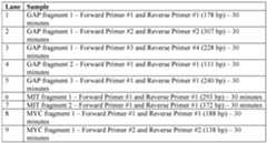
98 299255491 Attorney Docket No: NATE-055/01WO (321329-2902)
[00360] The lanes for the gel shown in FIG.3B are as follows:
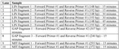
[00361] Without wishing to be bound by theory, the results presented in this example demonstrate that the methods of the present disclosure can be used to amplify target sequences from double-stranded DNA molecules, including those present in genomic DNA samples. Equivalents [00362] The foregoing description has been presented only for the purposes of illustration and is not intended to limit the disclosure to the precise form disclosed. The details of one or more embodiments of the disclosure are set forth in the accompanying description above. Although any methods and materials similar or equivalent to those described herein can be used in the practice or testing of the present disclosure, the preferred methods and materials are now described. Other features, objects, and advantages of the disclosure will be apparent from the description and from the claims. In the specification and the appended claims, the singular forms include plural referents unless the context clearly dictates otherwise. Unless defined otherwise, all technical and scientific terms used herein have the same meaning as commonly understood by one of ordinary skill in the art to which this disclosure belongs. All patents and publications cited in this specification are incorporated by reference. 99 299255491
 stereoisomer or salt thereof. [00329] A cleavable linker moiety can
 78 299255491 Attorney Docket No: NATE-055/01WO (321329-2902) [00330] A cleavable linker moiety can
 [00331] A cleavable linker moiety can
 [00332] A cleavable linker moiety can
 [00360] The lanes for the gel shown in FIG.3B are as follows:


