WO2024118836A1 - Processes for production of tumor infiltrating lymphocytes with shortened rep step - Google Patents
Processes for production of tumor infiltrating lymphocytes with shortened rep stepDownload PDFInfo
- Publication number
- WO2024118836A1 WO2024118836A1PCT/US2023/081680US2023081680WWO2024118836A1WO 2024118836 A1WO2024118836 A1WO 2024118836A1US 2023081680 WUS2023081680 WUS 2023081680WWO 2024118836 A1WO2024118836 A1WO 2024118836A1
- Authority
- WO
- WIPO (PCT)
- Prior art keywords
- tils
- expansion
- population
- days
- cells
- Prior art date
- Legal status (The legal status is an assumption and is not a legal conclusion. Google has not performed a legal analysis and makes no representation as to the accuracy of the status listed.)
- Ceased
Links
Classifications
- C—CHEMISTRY; METALLURGY
- C12—BIOCHEMISTRY; BEER; SPIRITS; WINE; VINEGAR; MICROBIOLOGY; ENZYMOLOGY; MUTATION OR GENETIC ENGINEERING
- C12N—MICROORGANISMS OR ENZYMES; COMPOSITIONS THEREOF; PROPAGATING, PRESERVING, OR MAINTAINING MICROORGANISMS; MUTATION OR GENETIC ENGINEERING; CULTURE MEDIA
- C12N5/00—Undifferentiated human, animal or plant cells, e.g. cell lines; Tissues; Cultivation or maintenance thereof; Culture media therefor
- C12N5/06—Animal cells or tissues; Human cells or tissues
- C12N5/0602—Vertebrate cells
- C12N5/0634—Cells from the blood or the immune system
- C12N5/0636—T lymphocytes
- A—HUMAN NECESSITIES
- A61—MEDICAL OR VETERINARY SCIENCE; HYGIENE
- A61K—PREPARATIONS FOR MEDICAL, DENTAL OR TOILETRY PURPOSES
- A61K35/00—Medicinal preparations containing materials or reaction products thereof with undetermined constitution
- A61K35/12—Materials from mammals; Compositions comprising non-specified tissues or cells; Compositions comprising non-embryonic stem cells; Genetically modified cells
- A61K35/14—Blood; Artificial blood
- A61K35/17—Lymphocytes; B-cells; T-cells; Natural killer cells; Interferon-activated or cytokine-activated lymphocytes
- C—CHEMISTRY; METALLURGY
- C12—BIOCHEMISTRY; BEER; SPIRITS; WINE; VINEGAR; MICROBIOLOGY; ENZYMOLOGY; MUTATION OR GENETIC ENGINEERING
- C12N—MICROORGANISMS OR ENZYMES; COMPOSITIONS THEREOF; PROPAGATING, PRESERVING, OR MAINTAINING MICROORGANISMS; MUTATION OR GENETIC ENGINEERING; CULTURE MEDIA
- C12N5/00—Undifferentiated human, animal or plant cells, e.g. cell lines; Tissues; Cultivation or maintenance thereof; Culture media therefor
- C12N5/06—Animal cells or tissues; Human cells or tissues
- C12N5/0602—Vertebrate cells
- C12N5/0634—Cells from the blood or the immune system
- C12N5/0636—T lymphocytes
- C12N5/0638—Cytotoxic T lymphocytes [CTL] or lymphokine activated killer cells [LAK]
- C—CHEMISTRY; METALLURGY
- C12—BIOCHEMISTRY; BEER; SPIRITS; WINE; VINEGAR; MICROBIOLOGY; ENZYMOLOGY; MUTATION OR GENETIC ENGINEERING
- C12N—MICROORGANISMS OR ENZYMES; COMPOSITIONS THEREOF; PROPAGATING, PRESERVING, OR MAINTAINING MICROORGANISMS; MUTATION OR GENETIC ENGINEERING; CULTURE MEDIA
- C12N2501/00—Active agents used in cell culture processes, e.g. differentation
- C12N2501/20—Cytokines; Chemokines
- C12N2501/23—Interleukins [IL]
- C12N2501/2302—Interleukin-2 (IL-2)
- C—CHEMISTRY; METALLURGY
- C12—BIOCHEMISTRY; BEER; SPIRITS; WINE; VINEGAR; MICROBIOLOGY; ENZYMOLOGY; MUTATION OR GENETIC ENGINEERING
- C12N—MICROORGANISMS OR ENZYMES; COMPOSITIONS THEREOF; PROPAGATING, PRESERVING, OR MAINTAINING MICROORGANISMS; MUTATION OR GENETIC ENGINEERING; CULTURE MEDIA
- C12N2501/00—Active agents used in cell culture processes, e.g. differentation
- C12N2501/50—Cell markers; Cell surface determinants
- C12N2501/515—CD3, T-cell receptor complex
Definitions
- TILstumor infiltrating lymphocytes
- IL-2-based TIL expansion followed by a “rapid expansion process”has become a preferred method for TIL expansion because of its speed and efficiency.
- REPcan result in a 1,000-fold expansion of TILs over a 14-day period, although it requires a large excess (e.g., 200-fold) of irradiated allogeneic peripheral blood mononuclear cells (PBMCs, also known as mononuclear cells (MNCs)), often from multiple donors, as feeder cells, as well as anti-CD3 antibody (OKT3) and high doses of IL-2.
- PBMCsperipheral blood mononuclear cells
- MNCsmononuclear cells
- OKT3anti-CD3 antibody
- TILs that have undergone an REP procedurehave produced successful adoptive cell therapy following host immunosuppression in patients with melanoma.
- T cellsundergo a profound metabolic shift during the course of their maturation from na ⁇ ve to effector T cells (see Chang, et al., Nat. Immunol.2016, 17, 364, hereby expressly incorporated in its entirety, and in particular for the discussion and markers of anaerobic and aerobic metabolism).
- na ⁇ ve T cellsrely on mitochondrial respiration to produce ATP
- mature, healthy effector T cellssuch as TIL are highly glycolytic, relying on aerobic glycolysis to provide the bioenergetics substrates they require for proliferation, migration, activation, and anti-tumor efficacy.
- the present inventionprovides methods of expanding TILs with a modified REP, for example, a shortened REP.
- the modified REPcomprises adding an anti-CD3 antibody more than 1 day after the initiation of the REP.
- the present inventionprovides methods of expanding TILs without a REP. BRIEF SUMMARY OF THE INVENTION [0007]
- the present inventionprovides TIL expansion methods with improved and/or shortened REP step for expanding TILs and producing therapeutic populations of TILs.
- Some embodiments disclosed hereinprovide a method for expanding TILs, comprising: a) performing a first expansion of a first population of TILs obtained from a tumor sample by culturing the tumor sample in a first cell culture medium and IL-2 for about 7-11 days to produce a second population of TILs; and b) performing a second expansion of the second population of TILs in a second cell culture medium, IL-2, and feeder cells for about 3-11 days to produce a third population of TILs, wherein an anti-CD3 antibody is added to the second cell culture medium about 1 day or more after the initiation of the second expansion.
- the anti-CD3 antibodyis added to the second cell culture medium about 2-4 days after the initiation of the second expansion. In some embodiments, the anti-CD3 antibody is added to the second cell culture medium about 2 days after the initiation of the second expansion. In some embodiments, the anti-CD3 antibody is added to the second cell culture medium about 3 days after the initiation of the second expansion. In some embodiments, the anti-CD3 antibody is added to the second cell culture medium about 4 days after the initiation of the second expansion.
- Some embodiments disclosed hereinprovide a method for expanding TILs, comprising: a) performing a first expansion of a first population of TILs obtained from a tumor sample by culturing the tumor sample in a first cell culture medium and IL-2 for about 7-11 days to produce a second population of TILs; and b) performing a second expansion of the second population of TILs in a second cell culture medium, IL-2, an anti-CD3 antibody and feeder cells for no more than 10 days to produce a third population of TILs.
- the second expansion stepis performed for no more than 9 days. In some embodiments, the second expansion step is performed for no more than 8 days.
- the second expansion stepis performed for no more than 7 days. In some embodiments, the second expansion step is performed for no more than 6 days. In some embodiments, the second expansion step is performed for no more than 5 days. In some embodiments, the second expansion step is performed for no more than 4 days. In some embodiments, the second expansion step is performed for no more than 3 days. In some embodiments, the second expansion step is performed for no more than 2 days. In some embodiments, the second expansion step is performed for no more than 1 day. [0012] In some embodiments, the anti-CD3 antibody is added to the second cell culture medium at about 1-30 ng/mL. In some embodiments, the anti-CD3 antibody is added to the second cell culture medium at about 2-10 ng/mL.
- the anti-CD3 antibodyis added to the second cell culture medium at about 30 ng/mL. In some embodiments, the anti-CD3 antibody is added to the second cell culture medium at about 20 ng/mL. In some embodiments, the anti-CD3 antibody is added to the second cell culture medium at about 10 ng/mL. In some embodiments, the anti-CD3 antibody is added to the second cell culture medium at about 5 ng/mL. In some embodiments, the anti-CD3 antibody is added to the second cell culture medium at about 3 ng/mL. [0013] In some embodiments, the anti-CD3 antibody is OKT3. In some embodiments, the anti-CD3 antibody is HIT3a.
- the second population of TILsis cultured with the feeder cells at a TIL:feeder cell ratio of about 1:50 to about 1:350. In some embodiments, the second population of TILs is cultured with the feeder cells at a TIL:feeder cell ratio of about 1:50, about 1:75, about 1:100, about 1:125, about 1:150, about 1:175, about 1:200, about 1:225, about 1:250, about 1:275, about 1:300, about 1:325, or about 1:350. In some embodiments, the second population of TILs is cultured with the feeder cells at a TIL:feeder cell ratio of about 1:250.
- the second population of TILsis cultured with the feeder cells at a TIL:feeder cell ratio of about 1:350. In some embodiments, the second expansion results in at least a 1000-fold expansion of the TILs. In some embodiments, the viability of the third population of TILs is greater than about 80%. In some embodiments, the viability of the third population of TILs is greater than about 85%. In some embodiments, at least about 40% of the CD8+ T cells of the third population of TILs expresses IFN ⁇ . In some embodiments, the third population of TILs is at least 500-fold greater than the second population of TILs. In some embodiments, the third population of TILs is at least 1000-fold greater than the second population of TILs.
- the third population of TILsis at least 1500-fold greater than the second population of TILs. In some embodiments, the third population of TILs comprises sufficient TILs for a therapeutically effective dosage of the TILs. In some embodiments, the number of TILs sufficient for a therapeutically effective dosage is from about 1 ⁇ 10 9 to about 10 ⁇ 10 10 .
- the third population of TILscomprises an increased subpopulation of effector T cells and/or central memory T cells relative to the second population of TILs, wherein the effector T cells and/or central memory T cells exhibit one or more characteristics selected from the group consisting of expressing CD27+, expressing CD28+, longer telomeres, increased CD57 expression, and decreased CD56 expression relative to effector T cells, and/or central memory T cells in the second population of cells.
- the effector T cells and/or central memory T cells in the third population of TILsexhibit increased CD57 expression and decreased CD56 expression relative to effector T cells, and/or central memory T cells in the second population of cells.
- a portion of the population of TILsare modified TILs each comprising an immunomodulatory agent/composition associated with its surface membrane.
- the immunomodulatory agent/compositioncomprises a cytokine.
- the cytokineis selected from the group consisting of IL-2, IL-7, IL-12, IL-15, IL-18, IL-21, IL-23, IL-27, IL-4, IL-1 ⁇ , IL-1 ⁇ , IL-5, IFN ⁇ , TNF ⁇ (TNFa), IFN ⁇ , IFN ⁇ , GM-CSF, and GCSF or a biologically active variant thereof.
- the immunomodulatory agent/compositioncomprises a costimulatory molecule.
- the costimulatory moleculeis selected from the group consisting of OX40, CD28, GITR, VISTA, CD40, CD3, and an agonist of CD137.
- the immunomodulatory agent/compositioncomprises a CD40 agonist (e.g., CD40L or an agonistic CD40 binding domain).
- a portion portion of the population of TILsare modified TILs each comprising an immunomodulatory agent/composition associated with its surface membrane, including combinations of modified TILs having a cytokine (including where the cytokine is selected from the group consisting of IL-2, IL-7, IL-12, IL-15, IL-18, IL-21, IL-23, IL-27, IL-4, IL-1 ⁇ , IL-1 ⁇ , IL-5, IFN ⁇ , TNF ⁇ (TNFa), IFN ⁇ , IFN ⁇ , GM-CSF, and GCSF or a biologically active variant thereof), a costimulatory molecule (including where the costimulatory molecule is selected from the group consisting of OX40, CD28, GITR, VISTA, CD40, CD3, and an agonist of CD137), and/or a CD40 agonist (e.g., CD40L or an agonistic CD40 binding domain).
- a cytokineincluding where the costimulatory molecule
- Figure 1Exemplary Gen 2 (process 2A) chart providing an overview of Steps A through F.
- Figure 2A-2CProcess flow chart of an embodiment of Gen 2 (process 2A) for TIL manufacturing.
- Figure 3Shows a diagram of an embodiment of a cryopreserved TIL exemplary manufacturing process ( ⁇ 22 days).
- Figure 4Shows a diagram of an embodiment of Gen 2 (process 2A), a 22-day process for TIL manufacturing.
- Figure 5Comparison table of Steps A through F from exemplary embodiments of process 1C and Gen 2 (process 2A) for TIL manufacturing.
- Figure 6Detailed comparison of an embodiment of process 1C and an embodiment of Gen 2 (process 2A) for TIL manufacturing.
- Figure 7Exemplary Gen 3 type TIL manufacturing process.
- Figure 8A-8CA) Exemplary modified Gen 2-like process providing an overview of Steps A through F.
- Figure 9Shows (A) cell expansion and (B) surface expression of TeIL-15/TeIL-21 after the procedure: after gene transduction of TeIL-15 lentivirus, Pre-REP TIL was processed for REP expansion with feeder cell, 3000IU/ml IL-2 and aCD3 Ab OKT3 or HIT3a. OKT3 (30ng/ml) or HIT3a(30ng/ml) was added into REP culture medium in different days ( Day 0, Day 2 and Day 4) after setting REP process. After 11 days REP expansion, Post-REP-TIL was harvested and analyzed.
- Figure 10Shows (A) cell expansion and (B) surface expression of TeIL-15/TeIL-21 after the procedure: after gene transduction of TeIL-15/TeIL-21 lentivirus, Pre-REP TIL was processed for REP expansion with feeder cell, 3000IU/ml IL-2 and aCD3 Ab OKT3 or HIT3a. OKT3 (30ng/ml) or HIT3a(30ng/ml) was added into REP culture medium in different days ( Day 0, Day 2 and Day 4) after setting REP process. After 11 days REP expansion, Post-REP-TIL was harvested and analyzed.
- Figure 11Shows (A) cell expansion and (B) surface expression of TeIL-15 after the procedure: after gene transduction of TeIL-15 lentivirus, Pre-REP TIL was processed for 11 days REP expansion with feeder cell, 3000IU/ml IL-2 and OKT3 with indicated concentration. After 11 days REP expansion, Post-REP-TIL was harvested and analyzed.
- Figure 12Shows (A) cell expansion and (B)surface expression of TeIL-15/TeIL-21 after the procedure: after gene transduction of TeIL-15/TeIL-21 lentivirus, Pre-REP TIL was processed for REP expansion with feeder cell, 3000IU/ml IL-2 and OKT3 with indicated concentration.
- FIG. 13Shows the expansion and viability of TILs after REP process under various expansion condition.
- Figure 14Shows the expression of IFNg, IFNa, and IL-2 of CD8+ TILs after REP process under various expansion condition.
- Figure 15Shows the expression of IFNg + IFNa, CD107a, and GXMB of CD8+ TILs after REP process under various expansion condition.
- Figure 16Shows the expression of CD69, CD38, and CD39 of CD8+ TILs after REP process under various expansion condition.
- Figure 17Shows the expression of PD-1, TIGIT, and LAG3 of CD8+ TILs after REP process under various expansion condition.
- Figure 18A-18JExemplary membrane anchored immunomodulatory fusion proteins that can be included in the TILs described herein.
- Figure19A-19DExemplary membrane anchored immunomodulatory fusion proteins that can be included in the TILs described herein. BRIEF DESCRIPTION OF THE SEQUENCE LISTING [0034]
- SEQ ID NO:1is the amino acid sequence of the heavy chain of muromonab.
- SEQ ID NO:2is the amino acid sequence of the light chain of muromonab.
- SEQ ID NO:3is the amino acid sequence of a recombinant human IL-2 protein.
- SEQ ID NO:4is the amino acid sequence of aldesleukin.
- SEQ ID NO:5is the amino acid sequence of a recombinant human IL-4 protein.
- SEQ ID NO:6is the amino acid sequence of a recombinant human IL-7 protein.
- SEQ ID NO:7is the amino acid sequence of a recombinant human IL-15 protein.
- SEQ ID NO:8is the amino acid sequence of a recombinant human IL-21 protein.
- SEQ ID NO:9is the amino acid sequence of a recombinant human IL-4 protein.
- SEQ ID NO:10is the amino acid sequence of a recombinant human IL-7 protein.
- SEQ ID NO:11is the amino acid sequence of a recombinant human IL-15 protein.
- SEQ ID NO:12is the amino acid sequence of a recombinant human IL-21 protein.
- SEQ ID NO:13is an IL-2 sequence.
- SEQ ID NO:14is an IL-2 mutein sequence.
- SEQ ID NO:15is an IL-2 mutein sequence.
- SEQ ID NO:16is the HCDR1_IL-2 for IgG.IL2R67A.H1.
- SEQ ID NO:17is the HCDR2 for IgG.IL2R67A.H1.
- SEQ ID NO:18is the HCDR3 for IgG.IL2R67A.H1.
- SEQ ID NO:19is the HCDR1_IL-2 kabat for IgG.IL2R67A.H1.
- SEQ ID NO:20is the HCDR2 kabat for IgG.IL2R67A.H1.
- SEQ ID NO:21is the HCDR3 kabat for IgG.IL2R67A.H1.
- SEQ ID NO:22is the HCDR1_IL-2 clothia for IgG.IL2R67A.H1.
- SEQ ID NO:23is the HCDR2 clothia for IgG.IL2R67A.H1.
- SEQ ID NO:24is the HCDR3 clothia for IgG.IL2R67A.H1.
- SEQ ID NO:25is the HCDR1_IL-2 IMGT for IgG.IL2R67A.H1.
- SEQ ID NO:26is the HCDR2 IMGT for IgG.IL2R67A.H1.
- SEQ ID NO:27is the HCDR3 IMGT for IgG.IL2R67A.H1.
- SEQ ID NO:28is the V H chain for IgG.IL2R67A.H1.
- SEQ ID NO:29is the heavy chain for IgG.IL2R67A.H1.
- SEQ ID NO:30is the LCDR1 kabat for IgG.IL2R67A.H1.
- SEQ ID NO:31is the LCDR2 kabat for IgG.IL2R67A.H1.
- SEQ ID NO:32is the LCDR3 kabat for IgG.IL2R67A.H1.
- SEQ ID NO:33is the LCDR1 chothia for IgG.IL2R67A.H1.
- SEQ ID NO:34is the LCDR2 chothia for IgG.IL2R67A.H1.
- SEQ ID NO:35is the LCDR3 chothia for IgG.IL2R67A.H1.
- SEQ ID NO:36is a V L chain.
- SEQ ID NO:37is a light chain.
- SEQ ID NO:38is a light chain.
- SEQ ID NO:39is a light chain.
- SEQ ID NO:258is human IL-15 (N72D mutant).
- SEQ ID NO:259is human IL-15R-alpha-Su/Fc domain.
- SEQ ID NO:260is human IL-15R-alpha-Su (65aa truncated extracellular domain).
- SEQ ID NO:261is human IL-15 isoform 2.
- SEQ ID NO:262is human IL-15 isoform 1.
- SEQ ID NO:263is human IL-15 (without signal peptide).
- SEQ ID NO:264is human IL-15R-alpha (85 aa truncated extracellular domain).
- SEQ ID NO:265is human IL-15R-alpha (182aa truncated extracellular domain).
- SEQ ID NO:266is human IL-15R-alpha.
- SEQ ID NO:267is human IL-12 p35 subunit.
- SEQ ID NO:268is human IL-12 p40 subunit.
- SEQ ID NO:269is human IL-18.
- SEQ ID NO:270is a human IL-18 variant.
- SEQ ID NO:271is human IL-21.
- SEQ ID NO: 272is human IL-2.
- SEQ ID NO:273is human CD40L.
- SEQ ID NO:274is agonistic anti-human CD40 VH (Sotigalimab).
- SEQ ID NO:275is agonistic anti-human CD40 VL (Sotigalimab).
- SEQ ID NO:276is agonistic anti-human CD40 scFv (Sotigalimab).
- SEQ ID NO:277is agonistic anti-human CD40 VH (Dacetuzumab).
- SEQ ID NO:278is agonistic anti-human CD40 VL (Dacetuzumab).
- SEQ ID NO:279is agonistic anti-human CD40 scFv (Dacetuzumab).
- SEQ ID NO:280is agonistic anti-human CD40 VH (Lucatutuzumab).
- SEQ ID NO:281is agonistic anti-human CD40 VL (Lucatutuzumab).
- SEQ ID NO:282is agonistic anti-human CD40 scFv (Lucatutuzumab).
- SEQ ID NO:283is agonistic anti-human CD40 VH (Selicrelumab).
- SEQ ID NO:284is agonistic anti-human CD40 VL (Selicrelumab).
- SEQ ID NO:285is agonistic anti-human CD40 scFv (Selicrelumab).
- SEQ ID NOS:286-295have no associated sequence.
- SEQ ID NO:296is a nucleic acid sequence that encodes for the tethered IL-15 of SEQ ID NO:328.
- SEQ ID NO:297is a nucleic acid sequence that encodes for the tethered IL-21 fusion protein of SEQ ID NO:271.
- SEQ ID NO:298is a nucleic acid sequence that encodes for the tethered IL-15 fusion protein of SEQ ID NO:328 and tether IL-21 fusion protein of SEQ ID NO:331.
- SEQ ID NO:299is a nucleic acid sequence that encodes for the tethered IL-12 fusion protein of SEQ ID NO:303.
- the nucleic acid sequenceincludes an NFAT promoter.
- SEQ ID NO:300is a nucleic acid sequence that encodes for the tethered IL-15 fusion protein of SEQ ID NO:328.
- the nucleic acid sequenceincludes an NFAT promoter.
- SEQ ID NO:301is a nucleic acid sequence that encodes for the tethered IL-21 fusion protein of SEQ ID NO:271.
- the nucleic acid sequenceincludes an NFAT promoter.
- SEQ ID NO:302is a nucleic acid sequence that encodes for the tethered IL-15 fusion protein of SEQ ID NO:328 and tether IL-21 fusion protein of SEQ ID NO:331.
- SEQ ID NO:303is the amino acid sequence of an exemplary tethered IL-12 (tethered IL-12-Lr1-Ar2).
- SEQ ID NO:304is a nucleic acid sequence that encodes for the tethered IL-12 of SEQ ID NO:303.
- SEQ ID NO:305is the amino acid sequence of an exemplary tethered IL-18 (tethered IL-18-Lr1-Ar2).
- SEQ ID NO:306is a nucleic acid sequence that encodes for the tethered IL-18 of SEQ ID NO:305.
- SEQ ID NO:307is the amino acid sequence of an exemplary tethered variant IL-18 (tethered DR-IL-18 (6-27 variant)-Lr1-Ar2).
- SEQ ID NO:308is a nucleic acid sequence that encodes for the tethered variant IL- 18 of SEQ ID NO:307.
- SEQ ID NO:309is the amino acid sequence of an exemplary tethered IL-12/IL-15.
- SEQ ID NO:310is a nucleic acid sequence that encodes for the tethered IL-12/IL- 15 of SEQ ID NO:309.
- SEQ ID NO:311is the amino acid sequence of an exemplary tethered IL-18/IL-15.
- SEQ ID NO:312is a nucleic acid sequence that encodes for the tethered IL-18/IL- 15 of SEQ ID NO:311.
- SEQ ID NO:313is the amino acid sequence of an exemplary tethered anti- CD40scFV (APX005M).
- SEQ ID NO:314is a nucleic acid sequence that encodes for the tethered anti- CD40scFV (APX005M) of SEQ ID NO:313.
- SEQ ID NO:315is the amino acid sequence of an exemplary tethered anti- CD40scFV (Dacetuzumab).
- SEQ ID NO:316is a nucleic acid sequence that encodes for the tethered anti- CD40scFV (Dacetuzumab) of SEQ ID NO:315.
- SEQ ID NO:317is the amino acid sequence of an exemplary tethered anti- CD40scFV (Lucatutuzumab).
- SEQ ID NO:318is a nucleic acid sequence that encodes for the tethered anti- CD40scFV (Lucatutuzumab) of SEQ ID NO:317.
- SEQ ID NO:319is the amino acid sequence of an exemplary tethered anti- CD40scFV (Selicrelumab).
- SEQ ID NO:320is a nucleic acid sequence that encodes for the tethered anti- CD40scFV (Selicrelumab) of SEQ ID NO:319.
- SEQ ID NO:321is a nucleic acid sequence that encodes for the CD40L of SEQ ID NO:273.
- SEQ ID NO:322is the amino acid sequence an exemplary tethered CD40L/IL-15.
- SEQ ID NO:323is a nucleic acid sequence that encodes for the tethered CD40L/IL- 15 of SEQ ID NO:311.
- SEQ ID NO:324is the amino acid sequence of an exemplary tethered IL-2.
- SEQ ID NO:325is a nucleic acid sequence that encodes for the tethered IL-2 of SEQ ID NO:313.
- SEQ ID NO:326is the amino acid sequence of an exemplary tethered IL-12.
- SEQ ID NO:327is a nucleic acid sequence that encodes for the tethered IL-12 of SEQ ID NO:315.
- SEQ ID NO:328is the amino acid sequence of an exemplary tethered IL-15.
- SEQ ID NO:329is a nucleic acid sequence that encodes for the tethered IL-15 of SEQ ID NO:317.
- SEQ ID NO:330is a nucleic acid sequence that encodes for GFP.
- SEQ ID NOS:331-385are nucleic acids of additiona variant IL-18s (e.g., decoy- resistant IL-18s or “DR-IL18”).
- SEQ ID NO:386is an exemplary piggyBac (PB) transposase enzyme sequence.
- SEQ ID NO:387is an exemplary Sleeping Beauty transposase sequence.
- SEQ ID NO:388is an exemplary Sleeping Beauty (SB100X) transposase sequence.
- SEQ ID NO:389is the amino acid sequence of an exemplary Tethered IL-21. DETAILED DESCRIPTION OF THE INVENTION Definitions [00142] Unless defined otherwise, all technical and scientific terms used herein have the same meaning as is commonly understood by one of skill in the art to which this invention belongs. All patents and publications referred to herein are incorporated by reference in their entireties.
- in vivorefers to an event that takes place in a subject's body.
- in vitrorefers to an event that takes places outside of a subject's body. In vitro assays encompass cell-based assays in which cells alive or dead are employed and may also encompass a cell-free assay in which no intact cells are employed.
- ex vivorefers to an event which involves treating or performing a procedure on a cell, tissue and/or organ which has been removed from a subject’s body. Aptly, the cell, tissue and/or organ may be returned to the subject’s body in a method of surgery or treatment.
- TILstumor infiltrating lymphocytes
- TILstumor infiltrating lymphocytes
- TILsinclude, but are not limited to, CD8 + cytotoxic T cells (lymphocytes), Th1 and Th17 CD4 + T cells, natural killer cells, dendritic cells and M1 macrophages.
- TILsinclude both primary and secondary TILs. “Primary TILs” are those that are obtained from patient tissue samples as outlined herein (sometimes referred to as “freshly harvested”), and “secondary TILs” are any TIL cell populations that have been expanded or proliferated as discussed herein, including, but not limited to bulk TILs and expanded TILs (“REP TILs” or “post-REP TILs”). TIL cell populations can include genetically modified TILs.
- population of cellsherein is meant a number of cells that share common traits. In general, populations generally range from 1 X 10 6 to 1 X 10 10 in number, with different TIL populations comprising different numbers. For example, initial growth of primary TILs in the presence of IL-2 results in a population of bulk TILs of roughly 1 ⁇ 10 8 cells. REP expansion is generally done to provide populations of 1.5 ⁇ 10 9 to 1.5 ⁇ 10 10 cells for infusion. [00149] By “cryopreserved TILs” herein is meant that TILs, either primary, bulk, or expanded (REP TILs), are treated and stored in the range of about -150°C to -60°C.
- cryopreserved TILsare distinguishable from frozen tissue samples which may be used as a source of primary TILs.
- thawed cryopreserved TILsherein is meant a population of TILs that was previously cryopreserved and then treated to return to room temperature or higher, including but not limited to cell culture temperatures or temperatures wherein TILs may be administered to a patient.
- TILscan generally be defined either biochemically, using cell surface markers, or functionally, by their ability to infiltrate tumors and effect treatment.
- TILscan be generally categorized by expressing one or more of the following biomarkers: CD4, CD8, TCR ⁇ , CD27, CD28, CD56, CCR7, CD45Ra, CD95, PD-1, and CD25. Additionally and alternatively, TILs can be functionally defined by their ability to infiltrate solid tumors upon reintroduction into a patient.
- the term “cryopreservation media” or “cryopreservation medium”refers to any medium that can be used for cryopreservation of cells. Such media can include media comprising 7% to 10% DMSO. Exemplary media include CryoStor CS10, Hyperthermasol, as well as combinations thereof.
- CS10refers to a cryopreservation medium which is obtained from Stemcell Technologies or from Biolife Solutions.
- the CS10 mediummay be referred to by the trade name “CryoStor® CS10”.
- the CS10 mediumis a serum-free, animal component-free medium which comprises DMSO.
- central memory T cellrefers to a subset of T cells that in the human are CD45R0+ and constitutively express CCR7 (CCR7 hi ) and CD62L (CD62 hi ).
- the surface phenotype of central memory T cellsalso includes TCR, CD3, CD127 (IL-7R), and IL- 15R.
- central memory T cellsTranscription factors for central memory T cells include BCL-6, BCL-6B, MBD2, and BMI1.
- Central memory T cellsprimarily secret IL-2 and CD40L as effector molecules after TCR triggering.
- Central memory T cellsare predominant in the CD4 compartment in blood, and in the human are proportionally enriched in lymph nodes and tonsils.
- effector memory T cellrefers to a subset of human or mammalian T cells that, like central memory T cells, are CD45R0+, but have lost the constitutive expression of CCR7 (CCR7 lo ) and are heterogeneous or low for CD62L expression (CD62L lo ).
- the surface phenotype of central memory T cellsalso includes TCR, CD3, CD127 (IL-7R), and IL-15R.
- Transcription factors for central memory T cellsinclude BLIMP1. Effector memory T cells rapidly secret high levels of inflammatory cytokines following antigenic stimulation, including interferon- ⁇ , IL-4, and IL-5. Effector memory T cells are predominant in the CD8 compartment in blood, and in the human are proportionally enriched in the lung, liver, and gut. CD8+ effector memory T cells carry large amounts of perforin.
- the term “closed system”refers to a system that is closed to the outside environment. Any closed system appropriate for cell culture methods can be employed with the methods of the present invention.
- Closed systemsinclude, for example, but are not limited to closed G-containers. Once a tumor segment is added to the closed system, the system is no opened to the outside environment until the TILs are ready to be administered to the patient.
- fragmentingincludes mechanical fragmentation methods such as crushing, slicing, dividing, and morcellating tumor tissue as well as any other method for disrupting the physical structure of tumor tissue.
- PBMCsperipheral blood mononuclear cells
- PBMCsrefers to a peripheral blood cell having a round nucleus, including lymphocytes (T cells, B cells, NK cells) and monocytes.
- the peripheral blood mononuclear cellsare irradiated allogeneic peripheral blood mononuclear cells.
- PBMCsare a type of antigen-presenting cell.
- anti-CD3 antibodyrefers to an antibody or variant thereof, e.g., a monoclonal antibody and including human, humanized, chimeric or murine antibodies which are directed against the CD3 receptor in the T cell antigen receptor of mature T cells.
- Anti- CD3 antibodiesinclude OKT-3, also known as muromonab.
- Anti-CD3 antibodiesalso include the UHCT1 clone, also known as T3 and CD3 ⁇ .
- Anti-CD3 antibodiesinclude, for example, otelixizumab, teplizumab, and visilizumab.
- Anti-CD3 antibodiescan include agonist antibodies.
- Such agonist antibodiesinclude, but are not limited to, OKT3 or HIT3a.
- OKT-3also referred to herein as “OKT3” refers to a monoclonal antibody or biosimilar or variant thereof, including human, humanized, chimeric, or murine antibodies, directed against the CD3 receptor in the T cell antigen receptor of mature T cells, and includes commercially-available forms such as OKT-3 (30 ng/mL, MACS GMP CD3 pure, Miltenyi Biotech, Inc., San Diego, CA, USA) and muromonab or variants, conservative amino acid substitutions, glycoforms, or biosimilars thereof.
- the amino acid sequences of the heavy and light chains of muromonabare given in Table 1 (SEQ ID NO:1 and SEQ ID NO:2).
- a hybridoma capable of producing OKT-3is deposited with the American Type Culture Collection and assigned the ATCC accession number CRL 8001.
- a hybridoma capable of producing OKT-3is also deposited with European Collection of Authenticated Cell Cultures (ECACC) and assigned Catalogue No.86022706. TABLE 1. Amino acid sequences of muromonab.
- IL-2refers to the T cell growth factor known as interleukin-2, and includes all forms of IL-2 including human and mammalian forms, conservative amino acid substitutions, glycoforms, biosimilars, and variants thereof.
- IL-2is described, e.g., in Nelson, J. Immunol.2004, 172, 3983-88 and Malek, Annu. Rev. Immunol.2008, 26, 453-79, the disclosures of which are incorporated by reference herein.
- the amino acid sequence of recombinant human IL-2 suitable for use in the inventionis given in Table 2 (SEQ ID NO:3).
- IL-2encompasses human, recombinant forms of IL-2 such as aldesleukin (PROLEUKIN, available commercially from multiple suppliers in 22 million IU per single use vials), as well as the form of recombinant IL-2 commercially supplied by CellGenix, Inc., Portsmouth, NH, USA (CELLGRO GMP) or ProSpec-Tany TechnoGene Ltd., East Brunswick, NJ, USA (Cat. No. CYT-209-b) and other commercial equivalents from other vendors.
- Aldesleukin(des-alanyl-1, serine-125 human IL-2) is a nonglycosylated human recombinant form of IL-2 with a molecular weight of approximately 15 kDa.
- IL-2also encompasses pegylated forms of IL-2, as described herein, including the pegylated IL2 prodrug bempegaldesleukin (NKTR-214, pegylated human recombinant IL-2 as in SEQ ID NO:4 in which an average of 6 lysine residues are N 6 substituted with [(2,7-bis ⁇ [methylpoly(oxyethylene)]carbamoyl ⁇ -9H- fluoren-9-yl)methoxy]carbonyl), which is available from Nektar Therapeutics, South San Francisco, CA, USA, or which may be prepared by methods known in the art, such as the methods described in Example 19 of International Patent Application Publication No.
- NKTR-214pegylated human recombinant IL-2 as in SEQ ID NO:4 in which an average of 6 lysine residues are N 6 substituted with [(2,7-bis ⁇ [methylpoly(oxyethylene)]carbamoyl ⁇ -9H- fluoren
- WO 2018/132496 A1or the method described in Example 1 of U.S. Patent Application Publication No. US 2019/0275133 A1, the disclosures of which are incorporated by reference herein.
- Bempegaldesleukin (NKTR-214) and other pegylated IL-2 molecules suitable for use in the inventionare described in U.S. Patent Application Publication No. US 2014/0328791 A1 and International Patent Application Publication No. WO 2012/065086 A1, the disclosures of which are incorporated by reference herein.
- Alternative forms of conjugated IL-2 suitable for use in the inventionare described in U.S. Patent Nos.4,766,106, 5,206,344, 5,089,261 and 4,902,502, the disclosures of which are incorporated by reference herein.
- an IL-2 form suitable for use in the present inventionis THOR-707, available from Synthorx, Inc.
- the preparation and properties of THOR-707 and additional alternative forms of IL-2 suitable for use in the inventionare described in U.S. Patent Application Publication Nos. US 2020/0181220 A1 and US 2020/0330601 A1, the disclosures of which are incorporated by reference herein.
- IL-2 form suitable for use in the inventionis an interleukin 2 (IL-2) conjugate comprising: an isolated and purified IL-2 polypeptide; and a conjugating moiety that binds to the isolated and purified IL-2 polypeptide at an amino acid position selected from K35, T37, R38, T41, F42, K43, F44, Y45, E61, E62, E68, K64, P65, V69, L72, and Y107, wherein the numbering of the amino acid residues corresponds to SEQ ID NO:5.
- IL-2interleukin 2
- the amino acid positionis selected from T37, R38, T41, F42, F44, Y45, E61, E62, E68, K64, P65, V69, L72, and Y107. In some embodiments, the amino acid position is selected from T37, R38, T41, F42, F44, Y45, E61, E62, E68, P65, V69, L72, and Y107. In some embodiments, the amino acid position is selected from T37, T41, F42, F44, Y45, P65, V69, L72, and Y107. In some embodiments, the amino acid position is selected from R38 and K64.
- the amino acid positionis selected from E61, E62, and E68. In some embodiments, the amino acid position is at E62. In some embodiments, the amino acid residue selected from K35, T37, R38, T41, F42, K43, F44, Y45, E61, E62, E68, K64, P65, V69, L72, and Y107 is further mutated to lysine, cysteine, or histidine. In some embodiments, the amino acid residue is mutated to cysteine. In some embodiments, the amino acid residue is mutated to lysine.
- the amino acid residue selected from K35, T37, R38, T41, F42, K43, F44, Y45, E61, E62, E68, K64, P65, V69, L72, and Y107is further mutated to an unnatural amino acid.
- the unnatural amino acidcomprises N6-azidoethoxy-L- lysine (AzK), N6-propargylethoxy-L-lysine (PraK), BCN-L-lysine, norbornene lysine, TCO- lysine, methyltetrazine lysine, allyloxycarbonyllysine, 2-amino-8-oxononanoic acid, 2- amino-8-oxooctanoic acid, p-acetyl-L-phenylalanine, p-azidomethyl-L-phenylalanine (pAMF), p-iodo-L-phenylalanine, m-acetylphenylalanine, 2-amino-8-oxononanoic acid, p- propargyloxyphenylalanine, p-propargyl-phenylalanine, 3-methyl-phenylalanine, L-Dopa
- the IL-2 conjugatehas a decreased affinity to IL-2 receptor ⁇ (IL-2R ⁇ ) subunit relative to a wild-type IL-2 polypeptide.
- the decreased affinityis about 10%, 20%, 30%, 40%, 50%, 60%, 70%, 80%, 90%, 95%, 99%, or greater than 99% decrease in binding affinity to IL-2R ⁇ relative to a wild-type IL-2 polypeptide.
- the decreased affinityis about 1-fold, 2-fold, 3-fold, 4- fold, 5-fold, 6-fold, 7-fold, 8-fold, 9-fold, 10-fold, 30-fold, 50-fold, 100-fold, 200-fold, 300- fold, 500-fold, 1000-fold, or more relative to a wild-type IL-2 polypeptide.
- the conjugating moietyimpairs or blocks the binding of IL-2 with IL-2R ⁇ .
- the conjugating moietycomprises a water-soluble polymer.
- the additional conjugating moietycomprises a water-soluble polymer.
- each of the water-soluble polymersindependently comprises polyethylene glycol (PEG), poly(propylene glycol) (PPG), copolymers of ethylene glycol and propylene glycol, poly(oxyethylated polyol), poly(olefinic alcohol), poly(vinylpyrrolidone), poly(hydroxyalkylmethacrylamide), poly(hydroxyalkylmethacrylate), poly(saccharides), poly( ⁇ -hydroxy acid), poly(vinyl alcohol), polyphosphazene, polyoxazolines (POZ), poly(N- acryloylmorpholine), or a combination thereof.
- each of the water- soluble polymersindependently comprises PEG.
- the PEGis a linear PEG or a branched PEG.
- each of the water-soluble polymersindependently comprises a polysaccharide.
- the polysaccharidecomprises dextran, polysialic acid (PSA), hyaluronic acid (HA), amylose, heparin, heparan sulfate (HS), dextrin, or hydroxyethyl-starch (HES).
- each of the water-soluble polymersindependently comprises a glycan.
- each of the water-soluble polymersindependently comprises polyamine.
- the conjugating moietycomprises a protein.
- the additional conjugating moietycomprises a protein. In some embodiments, each of the proteins independently comprises an albumin, a transferrin, or a transthyretin. In some embodiments, each of the proteins independently comprises an Fc portion. In some embodiments, each of the proteins independently comprises an Fc portion of IgG. In some embodiments, the conjugating moiety comprises a polypeptide. In some embodiments, the additional conjugating moiety comprises a polypeptide.
- each of the polypeptidesindependently comprises a XTEN peptide, a glycine-rich homoamino acid polymer (HAP), a PAS polypeptide, an elastin-like polypeptide (ELP), a CTP peptide, or a gelatin-like protein (GLK) polymer.
- the isolated and purified IL-2 polypeptideis modified by glutamylation.
- the conjugating moietyis directly bound to the isolated and purified IL-2 polypeptide.
- the conjugating moietyis indirectly bound to the isolated and purified IL-2 polypeptide through a linker.
- the linkercomprises a homobifunctional linker.
- the homobifunctional linkercomprises Lomant's reagent dithiobis (succinimidylpropionate) DSP, 3′3′- dithiobis(sulfosuccinimidyl proprionate) (DTSSP), disuccinimidyl suberate (DSS), bis(sulfosuccinimidyl)suberate (BS), disuccinimidyl tartrate (DST), disulfosuccinimidyl tartrate (sulfo DST), ethylene glycobis(succinimidylsuccinate) (EGS), disuccinimidyl glutarate (DSG), N,N′-disuccinimidyl carbonate (DSC), dimethyl adipimidate (DMA), dimethyl pimelimidate (DMP), dimethyl suberimidate (DMS), dimethyl-3,3′- dithiobispropionimidate (DTBP), 1,4-di-(3′-(2′-)
- the linkercomprises a heterobifunctional linker.
- the heterobifunctional linkercomprises N-succinimidyl 3-(2- pyridyldithio)propionate (sPDP), long-chain N-succinimidyl 3-(2-pyridyldithio)propionate (LC-sPDP), water-soluble-long-chain N-succinimidyl 3-(2-pyridyldithio) propionate (sulfo- LC-sPDP), succinimidyloxycarbonyl- ⁇ -methyl- ⁇ -(2-pyridyldithio)toluene (sMPT), sulfosuccinimidyl-6-[ ⁇ -methyl- ⁇ -(2-pyridyldithio)toluamido]hexanoate (sulfo-LC-sMPT), succinimidyl-4-(N-maleimidomethyl)cyclo
- the linkercomprises a cleavable linker, optionally comprising a dipeptide linker.
- the dipeptide linkercomprises Val-Cit, Phe-Lys, Val-Ala, or Val-Lys.
- the linkercomprises a non-cleavable linker.
- the linkercomprises a maleimide group, optionally comprising maleimidocaproyl (mc), succinimidyl-4-(N-maleimidomethyl)cyclohexane-1-carboxylate (sMCC), or sulfosuccinimidyl-4-(N-maleimidomethyl)cyclohexane-1-carboxylate (sulfo- sMCC).
- the linkerfurther comprises a spacer.
- the spacercomprises p-aminobenzyl alcohol (PAB), p-aminobenzyoxycarbonyl (PABC), a derivative, or an analog thereof.
- the conjugating moietyis capable of extending the serum half-life of the IL-2 conjugate.
- the additional conjugating moietyis capable of extending the serum half-life of the IL-2 conjugate.
- the IL-2 form suitable for use in the inventionis a fragment of any of the IL-2 forms described herein.
- the IL-2 form suitable for use in the inventionis pegylated as disclosed in U.S. Patent Application Publication No. US 2020/0181220 A1 and U.S. Patent Application Publication No. US 2020/0330601 A1.
- the IL-2 form suitable for use in the inventionis an IL-2 conjugate comprising: an IL-2 polypeptide comprising an N6-azidoethoxy-L-lysine (AzK) covalently attached to a conjugating moiety comprising a polyethylene glycol (PEG), wherein: the IL-2 polypeptide comprises an amino acid sequence having at least 80% sequence identity to SEQ ID NO:5; and the AzK substitutes for an amino acid at position K35, F42, F44, K43, E62, P65, R38, T41, E68, Y45, V69, or L72 in reference to the amino acid positions within SEQ ID NO:5.
- AzKN6-azidoethoxy-L-lysine
- the IL-2 polypeptidecomprises an N-terminal deletion of one residue relative to SEQ ID NO:5.
- the IL-2 form suitable for use in the inventionlacks IL-2R alpha chain engagement but retains normal binding to the intermediate affinity IL-2R beta-gamma signaling complex.
- the IL-2 form suitable for use in the inventionis an IL-2 conjugate comprising: an IL-2 polypeptide comprising an N6-azidoethoxy-L-lysine (AzK) covalently attached to a conjugating moiety comprising a polyethylene glycol (PEG), wherein: the IL-2 polypeptide comprises an amino acid sequence having at least 90% sequence identity to SEQ ID NO:5; and the AzK substitutes for an amino acid at position K35, F42, F44, K43, E62, P65, R38, T41, E68, Y45, V69, or L72 in reference to the amino acid positions within SEQ ID NO:5.
- AzKN6-azidoethoxy-L-lysine
- the IL-2 form suitable for use in the inventionis an IL-2 conjugate comprising: an IL-2 polypeptide comprising an N6-azidoethoxy-L-lysine (AzK) covalently attached to a conjugating moiety comprising a polyethylene glycol (PEG), wherein: the IL-2 polypeptide comprises an amino acid sequence having at least 95% sequence identity to SEQ ID NO:5; and the AzK substitutes for an amino acid at position K35, F42, F44, K43, E62, P65, R38, T41, E68, Y45, V69, or L72 in reference to the amino acid positions within SEQ ID NO:5.
- AzKN6-azidoethoxy-L-lysine
- the IL-2 form suitable for use in the inventionis an IL-2 conjugate comprising: an IL-2 polypeptide comprising an N6-azidoethoxy-L-lysine (AzK) covalently attached to a conjugating moiety comprising a polyethylene glycol (PEG), wherein: the IL-2 polypeptide comprises an amino acid sequence having at least 98% sequence identity to SEQ ID NO:5; and the AzK substitutes for an amino acid at position K35, F42, F44, K43, E62, P65, R38, T41, E68, Y45, V69, or L72 in reference to the amino acid positions within SEQ ID NO:5.
- AzKN6-azidoethoxy-L-lysine
- an IL-2 form suitable for use in the inventionis nemvaleukin alfa, also known as ALKS-4230 (SEQ ID NO:6), which is available from Alkermes, Inc.
- Nemvaleukin alfais also known as human interleukin 2 fragment (1-59), variant (Cys 125 >Ser 51 ), fused via peptidyl linker ( 60 GG 61 ) to human interleukin 2 fragment (62-132), fused via peptidyl linker ( 133 GSGGGS 138 ) to human interleukin 2 receptor ⁇ -chain fragment (139-303), produced in Chinese hamster ovary (CHO) cells, glycosylated; human interleukin 2 (IL-2) (75-133)-peptide [Cys 125 (51)>Ser]-mutant (1-59), fused via a G 2 peptide linker (60- 61) to human interleukin 2 (IL-2) (4-74)-peptide (62-132)
- nemvaleukin alfaexhibits the following post-translational modifications: disulfide bridges at positions: 31-116, 141-285, 184-242, 269-301, 166-197 or 166-199, 168- 199 or 168-197 (using the numbering in SEQ ID NO:6), and glycosylation sites at positions: N187, N206, T212 using the numbering in SEQ ID NO:6.
- disulfide bridgesat positions: 31-116, 141-285, 184-242, 269-301, 166-197 or 166-199, 168- 199 or 168-197 (using the numbering in SEQ ID NO:6)
- glycosylation sitesat positions: N187, N206, T212 using the numbering in SEQ ID NO:6.
- an IL-2 form suitable for use in the inventionis a protein having at least 80%, at least 90%, at least 95%, or at least 90% sequence identity to SEQ ID NO:6.
- an IL-2 form suitable for use in the inventionhas the amino acid sequence given in SEQ ID NO:6 or conservative amino acid substitutions thereof.
- an IL-2 form suitable for use in the inventionis a fusion protein comprising amino acids 24-452 of SEQ ID NO:7, or variants, fragments, or derivatives thereof.
- an IL-2 form suitable for use in the inventionis a fusion protein comprising an amino acid sequence having at least 80%, at least 90%, at least 95%, or at least 90% sequence identity to amino acids 24-452 of SEQ ID NO:7, or variants, fragments, or derivatives thereof.
- Other IL-2 forms suitable for use in the present inventionare described in U.S. Patent No.10,183,979, the disclosures of which are incorporated by reference herein.
- an IL-2 form suitable for use in the inventionis a fusion protein comprising a first fusion partner that is linked to a second fusion partner by a mucin domain polypeptide linker, wherein the first fusion partner is IL-1R ⁇ or a protein having at least 98% amino acid sequence identity to IL-1R ⁇ and having the receptor antagonist activity of IL-R ⁇ , and wherein the second fusion partner comprises all or a portion of an immunoglobulin comprising an Fc region, wherein the mucin domain polypeptide linker comprises SEQ ID NO:8 or an amino acid sequence having at least 90% sequence identity to SEQ ID NO:8 and wherein the half-life of the fusion protein is improved as compared to a fusion of the first fusion partner to the second fusion partner in the absence of the mucin domain polypeptide linker.
- an IL-2 form suitable for use in the inventionincludes a antibody cytokine engrafted protein comprises a heavy chain variable region (V H ), comprising complementarity determining regions HCDR1, HCDR2, HCDR3; a light chain variable region (V L ), comprising LCDR1, LCDR2, LCDR3; and an IL-2 molecule or a fragment thereof engrafted into a CDR of the V H or the V L , wherein the antibody cytokine engrafted protein preferentially expands T effector cells over regulatory T cells.
- V Hheavy chain variable region
- V Llight chain variable region
- the antibody cytokine engrafted proteincomprises a heavy chain variable region (V H ), comprising complementarity determining regions HCDR1, HCDR2, HCDR3; a light chain variable region (V L ), comprising LCDR1, LCDR2, LCDR3; and an IL-2 molecule or a fragment thereof engrafted into a CDR of the V H or the V L , wherein the IL-2 molecule is a mutein, and wherein the antibody cytokine engrafted protein preferentially expands T effector cells over regulatory T cells.
- the IL-2 regimencomprises administration of an antibody described in U.S. Patent Application Publication No.
- the antibody cytokine engrafted proteincomprises a heavy chain variable region (VH), comprising complementarity determining regions HCDR1, HCDR2, HCDR3; a light chain variable region (V L ), comprising LCDR1, LCDR2, LCDR3; and an IL-2 molecule or a fragment thereof engrafted into a CDR of the V H or the V L , wherein the IL-2 molecule is a mutein, wherein the antibody cytokine engrafted protein preferentially expands T effector cells over regulatory T cells, and wherein the antibody further comprises an IgG class heavy chain and an IgG class light chain selected from the group consisting of: a IgG class light chain comprising SEQ ID NO:39 and a IgG class heavy chain comprising SEQ ID NO:38; a IgG class light chain comprising SEQ ID NO:37 and a IgG class heavy chain
- an IL-2 molecule or a fragment thereofis engrafted into HCDR1 of the V H , wherein the IL-2 molecule is a mutein. In some embodiments, an IL-2 molecule or a fragment thereof is engrafted into HCDR2 of the V H , wherein the IL-2 molecule is a mutein. In some embodiments, an IL-2 molecule or a fragment thereof is engrafted into HCDR3 of the V H , wherein the IL-2 molecule is a mutein. In some embodiments, an IL-2 molecule or a fragment thereof is engrafted into LCDR1 of the V L , wherein the IL-2 molecule is a mutein.
- an IL-2 molecule or a fragment thereofis engrafted into LCDR2 of the V L , wherein the IL-2 molecule is a mutein. In some embodiments, an IL-2 molecule or a fragment thereof is engrafted into LCDR3 of the V L , wherein the IL-2 molecule is a mutein. [00165] The insertion of the IL-2 molecule can be at or near the N-terminal region of the CDR, in the middle region of the CDR or at or near the C-terminal region of the CDR. In some embodiments, the antibody cytokine engrafted protein comprises an IL-2 molecule incorporated into a CDR, wherein the IL2 sequence does not frameshift the CDR sequence.
- the antibody cytokine engrafted proteincomprises an IL-2 molecule incorporated into a CDR, wherein the IL-2 sequence replaces all or part of a CDR sequence.
- the replacement by the IL-2 moleculecan be the N-terminal region of the CDR, in the middle region of the CDR or at or near the C-terminal region the CDR.
- a replacement by the IL-2 moleculecan be as few as one or two amino acids of a CDR sequence, or the entire CDR sequences.
- an IL-2 moleculeis engrafted directly into a CDR without a peptide linker, with no additional amino acids between the CDR sequence and the IL-2 sequence.
- an IL-2 moleculeis engrafted indirectly into a CDR with a peptide linker, with one or more additional amino acids between the CDR sequence and the IL-2 sequence.
- the IL-2 molecule described hereinis an IL-2 mutein.
- the IL-2 muteincomprising an R67A substitution.
- the IL-2 muteincomprises the amino acid sequence SEQ ID NO:14 or SEQ ID NO:15.
- the IL-2 muteincomprises an amino acid sequence in Table 1 in U.S. Patent Application Publication No. US 2020/0270334 A1, the disclosure of which is incorporated by reference herein.
- the antibody cytokine engrafted proteincomprises an HCDR1 selected from the group consisting of SEQ ID NO:16, SEQ ID NO:19, SEQ ID NO:22 and SEQ ID NO:25. In some embodiments, the antibody cytokine engrafted protein comprises an HCDR1 selected from the group consisting of SEQ ID NO:7, SEQ ID NO:10, SEQ ID NO:13 and SEQ ID NO:16. In some embodiments, the antibody cytokine engrafted protein comprises an HCDR1 selected from the group consisting of HCDR2 selected from the group consisting of SEQ ID NO:17, SEQ ID NO:20, SEQ ID NO:23, and SEQ ID NO:26.
- the antibody cytokine engrafted proteincomprises an HCDR3 selected from the group consisting of SEQ ID NO:18, SEQ ID NO:21, SEQ ID NO:24, and SEQ ID NO:27. In some embodiments, the antibody cytokine engrafted protein comprises a V H region comprising the amino acid sequence of SEQ ID NO:28. In some embodiments, the antibody cytokine engrafted protein comprises a heavy chain comprising the amino acid sequence of SEQ ID NO:29. In some embodiments, the antibody cytokine engrafted protein comprises a V L region comprising the amino acid sequence of SEQ ID NO:36.
- the antibody cytokine engrafted proteincomprises a light chain comprising the amino acid sequence of SEQ ID NO:37. In some embodiments, the antibody cytokine engrafted protein comprises a V H region comprising the amino acid sequence of SEQ ID NO:28 and a V L region comprising the amino acid sequence of SEQ ID NO:36. In some embodiments, the antibody cytokine engrafted protein comprises a heavy chain region comprising the amino acid sequence of SEQ ID NO:29 and a light chain region comprising the amino acid sequence of SEQ ID NO:37.
- the antibody cytokine engrafted proteincomprises a heavy chain region comprising the amino acid sequence of SEQ ID NO:29 and a light chain region comprising the amino acid sequence of SEQ ID NO:39. In some embodiments, the antibody cytokine engrafted protein comprises a heavy chain region comprising the amino acid sequence of SEQ ID NO:38 and a light chain region comprising the amino acid sequence of SEQ ID NO:37. In some embodiments, the antibody cytokine engrafted protein comprises a heavy chain region comprising the amino acid sequence of SEQ ID NO:38 and a light chain region comprising the amino acid sequence of SEQ ID NO:39.
- the antibody cytokine engrafted proteincomprises IgG.IL2F71A.H1 or IgG.IL2R67A.H1 of U.S. Patent Application Publication No. 2020/0270334 A1, or variants, derivatives, or fragments thereof, or conservative amino acid substitutions thereof, or proteins with at least 80%, at least 90%, at least 95%, or at least 98% sequence identity thereto.
- the antibody components of the antibody cytokine engrafted protein described hereincomprise immunoglobulin sequences, framework sequences, or CDR sequences of palivizumab.
- the antibody cytokine engrafted protein described hereinhas a longer serum half-life than a wild-type IL-2 molecule such as, but not limited to, aldesleukin or a comparable molecule.
- the antibody cytokine engrafted protein described hereinhas a sequence as set forth in Table 3. TABLE 3: Sequences of exemplary palivizumab antibody-IL-2 engrafted proteins [00169] [00170]
- the term “IL-4”(also referred to herein as “IL4”) refers to the cytokine known as interleukin 4, which is produced by Th2 T cells and by eosinophils, basophils, and mast cells.
- IL-4regulates the differentiation of na ⁇ ve helper T cells (Th0 cells) to Th2 T cells. Steinke and Borish, Respir. Res.2001, 2, 66-70. Upon activation by IL-4, Th2 T cells subsequently produce additional IL-4 in a positive feedback loop. IL-4 also stimulates B cell proliferation and class II MHC expression, and induces class switching to IgE and IgG 1 expression from B cells.
- Recombinant human IL-4 suitable for use in the inventionis commercially available from multiple suppliers, including ProSpec-Tany TechnoGene Ltd., East Brunswick, NJ, USA (Cat. No. CYT-211) and ThermoFisher Scientific, Inc., Waltham, MA, USA (human IL-15 recombinant protein, Cat.
- IL-7refers to a glycosylated tissue- derived cytokine known as interleukin 7, which may be obtained from stromal and epithelial cells, as well as from dendritic cells. Fry and Mackall, Blood 2002, 99, 3892-904. IL-7 can stimulate the development of T cells.
- IL-7binds to the IL-7 receptor, a heterodimer consisting of IL-7 receptor alpha and common gamma chain receptor, which in a series of signals important for T cell development within the thymus and survival within the periphery.
- Recombinant human IL-7 suitable for use in the inventionis commercially available from multiple suppliers, including ProSpec-Tany TechnoGene Ltd., East Brunswick, NJ, USA (Cat. No. CYT-254) and ThermoFisher Scientific, Inc., Waltham, MA, USA (human IL-15 recombinant protein, Cat. No. Gibco PHC0071).
- the amino acid sequence of recombinant human IL-7 suitable for use in the inventionis given in Table 2 (SEQ ID NO:6).
- IL-15refers to the T cell growth factor known as interleukin-15, and includes all forms of IL-2 including human and mammalian forms, conservative amino acid substitutions, glycoforms, biosimilars, and variants thereof.
- IL-15is described, e.g., in Fehniger and Caligiuri, Blood 2001, 97, 14-32, the disclosure of which is incorporated by reference herein.
- IL-15shares ⁇ and ⁇ signaling receptor subunits with IL-2.
- Recombinant human IL-15is a single, non-glycosylated polypeptide chain containing 114 amino acids (and an N-terminal methionine) with a molecular mass of 12.8 kDa.
- Recombinant human IL-15is commercially available from multiple suppliers, including ProSpec-Tany TechnoGene Ltd., East Brunswick, NJ, USA (Cat. No. CYT-230-b) and ThermoFisher Scientific, Inc., Waltham, MA, USA (human IL-15 recombinant protein, Cat. No.34-8159-82).
- the amino acid sequence of recombinant human IL-15 suitable for use in the inventionis given in Table 2 (SEQ ID NO:7).
- IL-21refers to the pleiotropic cytokine protein known as interleukin-21, and includes all forms of IL-21 including human and mammalian forms, conservative amino acid substitutions, glycoforms, biosimilars, and variants thereof. IL-21 is described, e.g., in Spolski and Leonard, Nat. Rev. Drug. Disc. 2014, 13, 379-95, the disclosure of which is incorporated by reference herein. IL-21 is primarily produced by natural killer T cells and activated human CD4 + T cells.
- Recombinant human IL-21is a single, non-glycosylated polypeptide chain containing 132 amino acids with a molecular mass of 15.4 kDa.
- Recombinant human IL-21is commercially available from multiple suppliers, including ProSpec-Tany TechnoGene Ltd., East Brunswick, NJ, USA (Cat. No. CYT-408-b) and ThermoFisher Scientific, Inc., Waltham, MA, USA (human IL-21 recombinant protein, Cat. No.14-8219-80).
- the amino acid sequence of recombinant human IL-21 suitable for use in the inventionis given in Table 2 (SEQ ID NO:8).
- an anti-tumor effective amount“an tumor-inhibiting effective amount”, or “therapeutic amount”
- the precise amount of the compositions of the present invention to be administeredcan be determined by a physician with consideration of individual differences in age, weight, tumor size, extent of infection or metastasis, and condition of the patient (subject). It can generally be stated that a pharmaceutical composition comprising the tumor infiltrating lymphocytes (e.g.
- secondary TILs or genetically modified cytotoxic lymphocytesdescribed herein may be administered at a dosage of 10 4 to 10 11 cells/kg body weight (e.g., 10 5 to 10 6 , 10 5 to 10 10 , 10 5 to 10 11 , 10 6 to 10 10 , 10 6 to 10 11 ,10 7 to 10 11 , 10 7 to 10 10 , 10 8 to 10 11 , 10 8 to 10 10 , 10 9 to 10 11 , or 10 9 to 10 10 cells/kg body weight), including all integer values within those ranges.
- Tumor infiltrating lymphocytes (inlcuding in some cases, genetically modified cytotoxic lymphocytes) compositionsmay also be administered multiple times at these dosages.
- the tumor infiltrating lymphocytes(inlcuding in some cases, genetically) can be administered by using infusion techniques that are commonly known in immunotherapy (see, e.g., Rosenberg et al., New Eng. J. of Med.319: 1676, 1988).
- the optimal dosage and treatment regime for a particular patientcan readily be determined by one skilled in the art of medicine by monitoring the patient for signs of disease and adjusting the treatment accordingly.
- the term “hematological malignancy”refers to mammalian cancers and tumors of the hematopoietic and lymphoid tissues, including but not limited to tissues of the blood, bone marrow, lymph nodes, and lymphatic system.
- Hematological malignanciesare also referred to as “liquid tumors.” Hematological malignancies include, but are not limited to, acute lymphoblastic leukemia (ALL), chronic lymphocytic lymphoma (CLL), small lymphocytic lymphoma (SLL), acute myelogenous leukemia (AML), chronic myelogenous leukemia (CML), acute monocytic leukemia (AMoL), Hodgkin's lymphoma, and non- Hodgkin's lymphomas.
- ALLacute lymphoblastic leukemia
- CLLchronic lymphocytic lymphoma
- SLLsmall lymphocytic lymphoma
- AMLacute myelogenous leukemia
- CMLchronic myelogenous leukemia
- AoLacute monocytic leukemia
- Hodgkin's lymphomaand non- Hodgkin's lymphomas.
- B cell hematological malignancyrefers to hematological
- Solid tumorsmay be benign or malignant.
- solid tumor cancerrefers to malignant, neoplastic, or cancerous solid tumors.
- Solid tumor cancersinclude, but are not limited to, sarcomas, carcinomas, and lymphomas, such as cancers of the lung, breast, prostate, colon, rectum, and bladder.
- the tissue structure of solid tumorsincludes interdependent tissue compartments including the parenchyma (cancer cells) and the supporting stromal cells in which the cancer cells are dispersed and which may provide a supporting microenvironment.
- the term “liquid tumor”refers to an abnormal mass of cells that is fluid in nature.
- Liquid tumor cancersinclude, but are not limited to, leukemias, myelomas, and lymphomas, as well as other hematological malignancies. TILs obtained from liquid tumors may also be referred to herein as marrow infiltrating lymphocytes (MILs).
- MILsmarrow infiltrating lymphocytes
- the tumor microenvironmentrefers to a complex mixture of “cells, soluble factors, signaling molecules, extracellular matrices, and mechanical cues that promote neoplastic transformation, support tumor growth and invasion, protect the tumor from host immunity, foster therapeutic resistance, and provide niches for dominant metastases to thrive,” as described in Swartz, et al., Cancer Res., 2012, 72, 2473.
- tumorsexpress antigens that should be recognized by T cells, tumor clearance by the immune system is rare because of immune suppression by the microenvironment.
- the inventionincludes a method of treating a cancer with a population of TILs, wherein a patient is pre-treated with non-myeloablative chemotherapy prior to an infusion of TILs according to the invention.
- the population of TILsmay be provided wherein a patient is pre-treated with nonmyeloablative chemotherapy prior to an infusion of TILs according to the present invention.
- the non-myeloablative chemotherapyis cyclophosphamide 60 mg/kg/d for 2 days (days 27 and 26 prior to TIL infusion) and fludarabine 25 mg/m2/d for 5 days (days 27 to 23 prior to TIL infusion).
- the patientreceives an intravenous infusion of IL-2 intravenously at 720,000 IU/kg every 8 hours to physiologic tolerance.
- lymphodepletion prior to adoptive transfer of tumor-specific T lymphocytesplays a key role in enhancing treatment efficacy by eliminating regulatory T cells and competing elements of the immune system (“cytokine sinks”).
- cytokine sinksregulatory T cells and competing elements of the immune system
- some embodiments of the inventionutilize a lymphodepletion step (sometimes also referred to as “immunosuppressive conditioning”) on the patient prior to the introduction of the rTILs of the invention.
- co-administrationencompass administration of two or more active pharmaceutical ingredients (in a preferred embodiment of the present invention, for example, at least one potassium channel agonist in combination with a plurality of TILs) to a subject so that both active pharmaceutical ingredients and/or their metabolites are present in the subject at the same time.
- Co-administrationincludes simultaneous administration in separate compositions, administration at different times in separate compositions, or administration in a composition in which two or more active pharmaceutical ingredients are present. Simultaneous administration in separate compositions and administration in a composition in which both agents are present are preferred.
- an effective amountrefers to that amount of a compound or combination of compounds as described herein that is sufficient to effect the intended application including, but not limited to, disease treatment.
- a therapeutically effective amountmay vary depending upon the intended application (in vitro or in vivo), or the subject and disease condition being treated (e.g., the weight, age and gender of the subject), the severity of the disease condition, or the manner of administration.
- the termalso applies to a dose that will induce a particular response in target cells (e.g., the reduction of platelet adhesion and/or cell migration).
- treatmentrefers to obtaining a desired pharmacologic and/or physiologic effect.
- the effectmay be prophylactic in terms of completely or partially preventing a disease or symptom thereof and/or may be therapeutic in terms of a partial or complete cure for a disease and/or adverse effect attributable to the disease.
- Treatmentcovers any treatment of a disease in a mammal, particularly in a human, and includes: (a) preventing the disease from occurring in a subject which may be predisposed to the disease but has not yet been diagnosed as having it; (b) inhibiting the disease, i.e., arresting its development or progression; and (c) relieving the disease, i.e., causing regression of the disease and/or relieving one or more disease symptoms. “Treatment” is also meant to encompass delivery of an agent in order to provide for a pharmacologic effect, even in the absence of a disease or condition.
- treatmentencompasses delivery of a composition that can elicit an immune response or confer immunity in the absence of a disease condition, e.g., in the case of a vaccine.
- heterologouswhen used with reference to portions of a nucleic acid or protein indicates that the nucleic acid or protein comprises two or more subsequences that are not found in the same relationship to each other in nature.
- the nucleic acidis typically recombinantly produced, having two or more sequences from unrelated genes arranged to make a new functional nucleic acid, e.g., a promoter from one source and a coding region from another source, or coding regions from different sources.
- a heterologous proteinindicates that the protein comprises two or more subsequences that are not found in the same relationship to each other in nature (e.g., a fusion protein).
- sequence identityrefers to two or more sequences or subsequences that are the same or have a specified percentage of nucleotides or amino acid residues that are the same, when compared and aligned (introducing gaps, if necessary) for maximum correspondence, not considering any conservative amino acid substitutions as part of the sequence identity.
- the percent identitycan be measured using sequence comparison software or algorithms or by visual inspection.
- Various algorithms and softwareare known in the art that can be used to obtain alignments of amino acid or nucleotide sequences. Suitable programs to determine percent sequence identity include for example the BLAST suite of programs available from the U.S. Government’s National Center for Biotechnology Information BLAST web site. Comparisons between two sequences can be carried using either the BLASTN or BLASTP algorithm. BLASTN is used to compare nucleic acid sequences, while BLASTP is used to compare amino acid sequences. ALIGN, ALIGN-2 (Genentech, South San Francisco, California) or MegAlign, available from DNASTAR, are additional publicly available software programs that can be used to align sequences.
- the term “variant”encompasses but is not limited to antibodies or fusion proteins which comprise an amino acid sequence which differs from the amino acid sequence of a reference antibody by way of one or more substitutions, deletions and/or additions at certain positions within or adjacent to the amino acid sequence of the reference antibody.
- the variantmay comprise one or more conservative substitutions in its amino acid sequence as compared to the amino acid sequence of a reference antibody. Conservative substitutions may involve, e.g., the substitution of similarly charged or uncharged amino acids.
- the variantretains the ability to specifically bind to the antigen of the reference antibody.
- TILstumor infiltrating lymphocytes
- lymphocytescytotoxic T cells
- Th1 and Th17 CD4 + T cellsnatural killer cells
- dendritic cellsdendritic cells
- M1 macrophagesinclude both primary and secondary TILs.
- Primary TILsare those that are obtained from patient tissue samples as outlined herein (sometimes referred to as “freshly harvested”), and “secondary TILs” are any TIL cell populations that have been expanded or proliferated as discussed herein, including, but not limited to bulk TILs, expanded TILs (“REP TILs”) as well as “reREP TILs” as discussed herein.
- reREP TILscan include for example second expansion TILs or second additional expansion TILs (such as, for example, those described in Step D of Figure 8, including TILs referred to as reREP TILs).
- TILscan generally be defined either biochemically, using cell surface markers, or functionally, by their ability to infiltrate tumors and effect treatment.
- TILscan be generally categorized by expressing one or more of the following biomarkers: CD4, CD8, TCR ⁇ , CD27, CD28, CD56, CCR7, CD45Ra, CD95, PD-1, and CD25. Additionally, and alternatively, TILs can be functionally defined by their ability to infiltrate solid tumors upon reintroduction into a patient. TILS may further be characterized by potency – for example, TILS may be considered potent if, for example, interferon (IFN) release is greater than about 50 pg/mL, greater than about 100 pg/mL, greater than about 150 pg/mL, or greater than about 200 pg/mL.
- IFNinterferon
- pharmaceutically acceptable carrieror “pharmaceutically acceptable excipient” are intended to include any and all solvents, dispersion media, coatings, antibacterial and antifungal agents, isotonic and absorption delaying agents, and inert ingredients.
- pharmaceutically acceptable carriers or pharmaceutically acceptable excipients for active pharmaceutical ingredientsis well known in the art. Except insofar as any conventional pharmaceutically acceptable carrier or pharmaceutically acceptable excipient is incompatible with the active pharmaceutical ingredient, its use in the therapeutic compositions of the invention is contemplated. Additional active pharmaceutical ingredients, such as other drugs, can also be incorporated into the described compositions and methods.
- the terms “about” and “approximately”mean within a statistically meaningful range of a value.
- Such a rangecan be within an order of magnitude, preferably within 50%, more preferably within 20%, more preferably still within 10%, and even more preferably within 5% of a given value or range.
- the allowable variation encompassed by the terms “about” or “approximately”depends on the particular system under study, and can be readily appreciated by one of ordinary skill in the art. Moreover, as used herein, the terms “about” and “approximately” mean that dimensions, sizes, formulations, parameters, shapes and other quantities and characteristics are not and need not be exact, but may be approximate and/or larger or smaller, as desired, reflecting tolerances, conversion factors, rounding off, measurement error and the like, and other factors known to those of skill in the art.
- a dimension, size, formulation, parameter, shape or other quantity or characteristicis “about” or “approximate” whether or not expressly stated to be such. It is noted that embodiments of very different sizes, shapes and dimensions may employ the described arrangements. [00191]
- the term “comprising”is intended to be inclusive or open-ended and does not exclude any additional, unrecited element, method, step or material.
- TIL Expanding Processes With Modified REPSome embodiments disclosed herein provide a method for expanding TILs, comprising: a) performing a first expansion of a first population of TILs obtained from a tumor sample by culturing the tumor sample in a first cell culture medium and IL-2 for about 7-11 days to produce a second population of TILs; and b) performing a second expansion of the second population of TILs in a second cell culture medium, IL-2, and feeder cells for about 3-11 days to produce a third population of TILs, wherein an anti-CD3 antibody is added to the second cell culture medium more than 1 day after the initiation of the second expansion.
- the anti-CD3 antibodyis added to the second cell culture medium about 2-4 days after the initiation of the second expansion. In some embodiments, the anti-CD3 antibody is added to the second cell culture medium about 2 days after the initiation of the second expansion. In some embodiments, the anti-CD3 antibody is added to the second cell culture medium about 3 days after the initiation of the second expansion. In some embodiments, the anti-CD3 antibody is added to the second cell culture medium about 4 days after the initiation of the second expansion.
- Some embodiments disclosed hereinprovide a method for expanding TILs, comprising: a) performing a first expansion of a first population of TILs obtained from a tumor sample by culturing the tumor sample in a first cell culture medium and IL-2 for about 7-11 days to produce a second population of TILs; and b) performing a second expansion of the second population of TILs in a second cell culture medium, IL-2, an anti-CD3 antibody and feeder cells for no more than 10 days to produce a third population of TILs.
- the second expansion stepis performed for no more than 9 days. In some embodiments, the second expansion step is performed for no more than 8 days.
- the second expansion stepis performed for no more than 7 days. In some embodiments, the second expansion step is performed for no more than 6 days. In some embodiments, the second expansion step is performed for no more than 5 days. In some embodiments, the second expansion step is performed for no more than 4 days. In some embodiments, the second expansion step is performed for no more than 3 days. In some embodiments, the second expansion step is performed for no more than 2 days. In some embodiments, the second expansion step is performed for no more than 1 day. [00196] An exemplary TIL expanding process known as process 2A containing some of these features is depicted in Figures 1-8.
- the present inventioncan include a step relating to the restimulation of cryopreserved TILs to increase their metabolic activity and thus relative health prior to transplant into a patient, and methods of testing said metabolic health.
- TILsare generally taken from a patient sample and manipulated to expand their number prior to transplant into a patient.
- the TILsmay be optionally genetically manipulated as discussed below.
- the TILsmay be cryopreserved. Once thawed, they may also be restimulated to increase their metabolism prior to infusion into a patient.
- the first expansion(including processes referred to as the preREP as well as processes shown in Figure 8 as Step A) is shortened to 3 to 14 days and the second expansion (including processes referred to as the REP as well as processes shown in Figure 8 as Step B) is shortened to 7 to 14 days, as discussed in detail below as well as in the examples and figures.
- the first expansion(for example, an expansion described as Step B in Figure 8) is shortened to 11 days and the second expansion (for example, an expansion as described in Step D in Figure 8) is shortened to 11 days, as discussed in the Examples and shown in Figures 1-8.
- the combination of the first expansion and second expansionis shortened to 22 days, as discussed in detail below and in the examples and figures.
- the “Step” Designations A, B, C, etc., beloware in reference to Figure 8 and in reference to certain embodiments described herein.
- the ordering of the Steps below and in Figure 8is exemplary and any combination or order of steps, as well as additional steps, repetition of steps, and/or omission of steps is contemplated by the present application and the methods disclosed herein.
- TILsare initially obtained from a patient tumor sample (“primary TILs”) and then expanded into a larger population for further manipulation as described herein, optionally cryopreserved, restimulated as outlined herein and optionally evaluated for phenotype and metabolic parameters as an indication of TIL health.
- a patient tumor samplemay be obtained using methods known in the art, generally via surgical resection, needle biopsy or other means for obtaining a sample that contains a mixture of tumor and TIL cells.
- the tumor samplemay be from any solid tumor, including primary tumors, invasive tumors or metastatic tumors.
- the tumor samplemay also be a liquid tumor, such as a tumor obtained from a hematological malignancy.
- the solid tumormay be of any cancer type, including, but not limited to, breast, pancreatic, prostate, colorectal, lung, brain, renal, stomach, and skin (including but not limited to squamous cell carcinoma, basal cell carcinoma, and melanoma).
- useful TILsare obtained from malignant melanoma tumors, as these have been reported to have particularly high levels of TILs.
- the term “solid tumor”refers to an abnormal mass of tissue that usually does not contain cysts or liquid areas. Solid tumors may be benign or malignant.
- solid tumor cancerrefers to malignant, neoplastic, or cancerous solid tumors.
- Solid tumor cancersinclude, but are not limited to, sarcomas, carcinomas, and lymphomas, such as cancers of the lung, breast, triple negative breast cancer, prostate, colon, rectum, and bladder.
- the canceris selected from cervical cancer, head and neck cancer (including, for example, head and neck squamous cell carcinoma (HNSCC)) glioblastoma, ovarian cancer, sarcoma, pancreatic cancer, bladder cancer, breast cancer, triple negative breast cancer, and non-small cell lung carcinoma.
- HNSCChead and neck squamous cell carcinoma
- the tissue structure of solid tumorsincludes interdependent tissue compartments including the parenchyma (cancer cells) and the supporting stromal cells in which the cancer cells are dispersed and which may provide a supporting microenvironment.
- hematological malignancyrefers to mammalian cancers and tumors of the hematopoietic and lymphoid tissues, including but not limited to tissues of the blood, bone marrow, lymph nodes, and lymphatic system.
- Hematological malignanciesare also referred to as “liquid tumors.” Hematological malignancies include, but are not limited to, acute lymphoblastic leukemia (ALL), chronic lymphocytic lymphoma (CLL), small lymphocytic lymphoma (SLL), acute myelogenous leukemia (AML), chronic myelogenous leukemia (CML), acute monocytic leukemia (AMoL), Hodgkin's lymphoma, and non- Hodgkin's lymphomas.
- ALLacute lymphoblastic leukemia
- CLLchronic lymphocytic lymphoma
- SLLsmall lymphocytic lymphoma
- AMLacute myelogenous leukemia
- CMLchronic myelogenous leukemia
- AoLacute monocytic leukemia
- Hodgkin's lymphomaand non- Hodgkin's lymphomas.
- B cell hematological malignancyrefers to hematological
- the tumor sampleis generally fragmented using sharp dissection into small pieces of between 1 to about 8 mm 3 , with from about 2-3 mm 3 being particularly useful.
- the TILsare cultured from these fragments using enzymatic tumor digests.
- Such tumor digestsmay be produced by incubation in enzymatic media (e.g., Roswell Park Memorial Institute (RPMI) 1640 buffer, 2 mM glutamate, 10 mcg/mL gentamicine, 30 units/mL of DNase and 1.0 mg/mL of collagenase) followed by mechanical dissociation (e.g., using a tissue dissociator).
- enzymatic mediae.g., Roswell Park Memorial Institute (RPMI) 1640 buffer, 2 mM glutamate, 10 mcg/mL gentamicine, 30 units/mL of DNase and 1.0 mg/mL of collagenase
- Tumor digestsmay be produced by placing the tumor in enzymatic media and mechanically dissociating the tumor for approximately 1 minute, followed by incubation for 30 minutes at 37 °C in 5% CO 2 , followed by repeated cycles of mechanical dissociation and incubation under the foregoing conditions until only small tissue pieces are present.
- a density gradient separation using FICOLL branched hydrophilic polysaccharidemay be performed to remove these cells.
- Alternative methods known in the artmay be used, such as those described in U.S. Patent Application Publication No.2012/0244133 A1, the disclosure of which is incorporated by reference herein.
- the harvested cell suspensionis called a “primary cell population” or a “freshly harvested” cell population.
- fragmentationincludes physical fragmentation, including for example, dissection as well as digestion.
- the fragmentationis physical fragmentation.
- the fragmentationis dissection.
- the fragmentationis by digestion.
- TILscan be initially cultured from enzymatic tumor digests and tumor fragments obtained from patients.
- TILscan be initially cultured from enzymatic tumor digests and tumor fragments obtained from patients.
- the tumorundergoes physical fragmentation after the tumor sample is obtained in, for example, Step A (as provided in Figure 8).
- the fragmentationoccurs before cryopreservation.
- the fragmentationoccurs after cryopreservation.
- the fragmentationoccurs after obtaining the tumor and in the absence of any cryopreservation.
- the tumoris fragmented and 10, 20, 30, 40 or more fragments or pieces are placed in each container for the first expansion.
- the tumoris fragmented and 30 or 40 fragments or pieces are placed in each container for the first expansion.
- the tumoris fragmented and 40 fragments or pieces are placed in each container for the first expansion.
- the multiple fragmentscomprise about 4 to about 50 fragments, wherein each fragment has a volume of about 27 mm 3 .
- the multiple fragmentscomprise about 30 to about 60 fragments with a total volume of about 1300 mm 3 to about 1500 mm 3 .
- the multiple fragmentscomprise about 50 fragments with a total volume of about 1350 mm 3 .
- the multiple fragmentscomprise about 50 fragments with a total mass of about 1 gram to about 1.5 grams.
- the multiple fragmentscomprise about 4 fragments. [00209]
- the TILsare obtained from tumor fragments.
- the tumor fragmentis obtained by sharp dissection. In some embodiments, the tumor fragment is between about 1 mm 3 and 10 mm 3 . In some embodiments, the tumor fragment is between about 1 mm 3 and 8 mm 3 . In some embodiments, the tumor fragment is about 1 mm 3 . In some embodiments, the tumor fragment is about 2 mm 3 . In some embodiments, the tumor fragment is about 3 mm 3 . In some embodiments, the tumor fragment is about 4 mm 3 . In some embodiments, the tumor fragment is about 5 mm 3 . In some embodiments, the tumor fragment is about 6 mm 3 . In some embodiments, the tumor fragment is about 7 mm 3 . In some embodiments, the tumor fragment is about 8 mm 3 .
- the tumor fragmentis about 9 mm 3 . In some embodiments, the tumor fragment is about 10 mm 3 . In some embodiments, the tumors are 1-4 mm x 1-4 mm x 1-4 mm. In some embodiments, the tumors are 1 mm x 1 mm x 1 mm. In some embodiments, the tumors are 2 mm x 2 mm x 2 mm. In some embodiments, the tumors are 3 mm x 3 mm x 3 mm. In some embodiments, the tumors are 4 mm x 4 mm x 4 mm.
- the tumorsare resected in order to minimize the amount of hemorrhagic, necrotic, and/or fatty tissues on each piece. In some embodiments, the tumors are resected in order to minimize the amount of hemorrhagic tissue on each piece. In some embodiments, the tumors are resected in order to minimize the amount of necrotic tissue on each piece. In some embodiments, the tumors are resected in order to minimize the amount of fatty tissue on each piece. [00211] In some embodiments, the tumor fragmentation is performed in order to maintain the tumor internal structure. In some embodiments, the tumor fragmentation is performed without preforming a sawing motion with a scapel.
- the TILsare obtained from tumor digests.
- tumor digestswere generated by incubation in enzyme media, for example but not limited to RPMI 1640, 2 mM GlutaMAX, 10 mg/mL gentamicin, 30 U/mL DNase, and 1.0 mg/mL collagenase, followed by mechanical dissociation (GentleMACS, Miltenyi Biotec, Auburn, CA). After placing the tumor in enzyme media, the tumor can be mechanically dissociated for approximately 1 minute. The solution can then be incubated for 30 minutes at 37 °C in 5% CO 2 and it then mechanically disrupted again for approximately 1 minute.
- the tumorcan be mechanically disrupted a third time for approximately 1 minute.
- 1 or 2 additional mechanical dissociationswere applied to the sample, with or without 30 additional minutes of incubation at 37 °C in 5% CO 2 .
- a density gradient separation using Ficollcan be performed to remove these cells.
- the harvested cell suspension prior to the first expansion stepis called a “primary cell population” or a “freshly harvested” cell population.
- cellscan be optionally frozen after sample harvest and stored frozen prior to entry into the expansion described in Step B, which is described in further detail below, as well as exemplified in Figure 8.
- the diverse antigen receptors of T and B lymphocytesare produced by somatic recombination of a limited, but large number of gene segments. These gene segments: V (variable), D (diversity), J (joining), and C (constant), determine the binding specificity and downstream applications of immunoglobulins and T-cell receptors (TCRs).
- the present inventionprovides a method for generating TILs which exhibit and increase the T-cell repertoire diversity.
- the TILs obtained by the present methodexhibit an increase in the T-cell repertoire diversity. In some embodiments, the TILs obtained by the present method exhibit an increase in the T-cell repertoire diversity as compared to freshly harvested TILs and/or TILs prepared using other methods than those provide herein including for example, methods other than those embodied in Figure 8. In some embodiments, the TILs obtained in the first expansion exhibit an increase in the T-cell repertoire diversity. In some embodiments, the increase in diversity is an increase in the immunoglobulin diversity and/or the T-cell receptor diversity. In some embodiments, the diversity is in the immunoglobulin is in the immunoglobulin heavy chain. In some embodiments, the diversity is in the immunoglobulin is in the immunoglobulin light chain.
- the diversityis in the T-cell receptor. In some embodiments, the diversity is in one of the T-cell receptors selected from the group consisting of alpha, beta, gamma, and delta receptors. In some embodiments, there is an increase in the expression of T-cell receptor (TCR) alpha and/or beta. In some embodiments, there is an increase in the expression of T-cell receptor (TCR) alpha. In some embodiments, there is an increase in the expression of T-cell receptor (TCR) beta. In some embodiments, there is an increase in the expression of TCRab (i.e., TCR ⁇ / ⁇ ).
- the resulting cellsare cultured in serum containing IL-2 under conditions that favor the growth of TILs over tumor and other cells.
- the tumor digestsare incubated in 2 mL wells in media comprising inactivated human AB serum with 6000 IU/mL of IL-2.
- This primary cell populationis cultured for a period of days, generally from 3 to 14 days, resulting in a bulk TIL population, generally about 1 ⁇ 10 8 bulk TIL cells.
- this primary cell populationis cultured for a period of 7 to 14 days, resulting in a bulk TIL population, generally about 1 ⁇ 10 8 bulk TIL cells.
- this primary cell populationis cultured for a period of 10 to 14 days, resulting in a bulk TIL population, generally about 1 ⁇ 10 8 bulk TIL cells. In some embodiments, this primary cell population is cultured for a period of about 11 days, resulting in a bulk TIL population, generally about 1 ⁇ 10 8 bulk TIL cells.
- expansion of TILsmay be performed using an initial bulk TIL expansion step (for example such as those described in Step B of Figure 8, which can include processes referred to as pre-REP) as described below and herein, followed by a second expansion (Step D, including processes referred to as rapid expansion protocol (REP) steps) as described below under Step D and herein, followed by optional cryopreservation, and followed by a second Step D (including processes referred to as restimulation REP steps) as described below and herein.
- the TILs obtained from this processmay be optionally characterized for phenotypic characteristics and metabolic parameters as described herein.
- each wellcan be seeded with 1 ⁇ 10 6 tumor digest cells or one tumor fragment in 2 mL of complete medium (CM) with IL-2 (6000 IU/mL; Chiron Corp., Emeryville, CA).
- CMcomplete medium
- IL-26000 IU/mL
- the tumor fragmentis between about 1 mm 3 and 10 mm 3 .
- the first expansion culture mediumis referred to as “CM”, an abbreviation for culture media.
- CM for Step Bconsists of RPMI 1640 with GlutaMAX, supplemented with 10% human AB serum, 25 mM Hepes, and 10 mg/mL gentamicin.
- gas-permeable flaskswith a 40 mL capacity and a 10 cm 2 gas-permeable silicon bottom (for example, G-Rex10; Wilson Wolf Manufacturing, New Brighton, MN) (Fig.1)
- each flaskwas loaded with 10–40 ⁇ 10 6 viable tumor digest cells or 5–30 tumor fragments in 10–40 mL of CM with IL-2.
- the resulting cellsare cultured in serum containing IL-2 under conditions that favor the growth of TILs over tumor and other cells.
- the tumor digestsare incubated in 2 mL wells in media comprising inactivated human AB serum (or, in some cases, as outlined herein, in the presence of aAPC cell population) with 6000 IU/mL of IL-2.
- the growth media during the first expansioncomprises IL-2 or a variant thereof.
- the ILis recombinant human IL-2 (rhIL-2).
- the IL-2 stock solutionhas a specific activity of 20-30 ⁇ 10 6 IU/mg for a 1 mg vial.
- the IL-2 stock solutionhas a specific activity of 20 ⁇ 10 6 IU/mg for a 1 mg vial.
- the IL-2 stock solutionhas a specific activity of 25 ⁇ 10 6 IU/mg for a 1 mg vial.
- the IL-2 stock solutionhas a specific activity of 30 ⁇ 10 6 IU/mg for a 1 mg vial. In some embodiments, the IL- 2 stock solution has a final concentration of 4-8 ⁇ 10 6 IU/mg of IL-2. In some embodiments, the IL- 2 stock solution has a final concentration of 5-7 ⁇ 10 6 IU/mg of IL-2. In some embodiments, the IL- 2 stock solution has a final concentration of 6 ⁇ 10 6 IU/mg of IL-2. In some embodiments, the IL-2 stock solution is prepare as described in Example 4.
- the first expansion culture mediacomprises about 10,000 IU/mL of IL-2, about 9,000 IU/mL of IL-2, about 8,000 IU/mL of IL-2, about 7,000 IU/mL of IL-2, about 6000 IU/mL of IL-2 or about 5,000 IU/mL of IL-2. In some embodiments, the first expansion culture media comprises about 9,000 IU/mL of IL-2 to about 5,000 IU/mL of IL-2. In some embodiments, the first expansion culture media comprises about 8,000 IU/mL of IL-2 to about 6,000 IU/mL of IL-2.
- the first expansion culture mediacomprises about 7,000 IU/mL of IL-2 to about 6,000 IU/mL of IL-2. In some embodiments, the first expansion culture media comprises about 6,000 IU/mL of IL-2. In an embodiment, the cell culture medium further comprises IL-2. In some embodiments, the cell culture medium comprises about 3000 IU/mL of IL-2. In an embodiment, the cell culture medium further comprises IL-2. In a preferred embodiment, the cell culture medium comprises about 3000 IU/mL of IL-2.
- the cell culture mediumcomprises about 1000 IU/mL, about 1500 IU/mL, about 2000 IU/mL, about 2500 IU/mL, about 3000 IU/mL, about 3500 IU/mL, about 4000 IU/mL, about 4500 IU/mL, about 5000 IU/mL, about 5500 IU/mL, about 6000 IU/mL, about 6500 IU/mL, about 7000 IU/mL, about 7500 IU/mL, or about 8000 IU/mL of IL-2.
- the cell culture mediumcomprises between 1000 and 2000 IU/mL, between 2000 and 3000 IU/mL, between 3000 and 4000 IU/mL, between 4000 and 5000 IU/mL, between 5000 and 6000 IU/mL, between 6000 and 7000 IU/mL, between 7000 and 8000 IU/mL, or about 8000 IU/mL of IL-2.
- first expansion culture mediacomprises about 500 IU/mL of IL-15, about 400 IU/mL of IL-15, about 300 IU/mL of IL-15, about 200 IU/mL of IL-15, about 180 IU/mL of IL-15, about 160 IU/mL of IL-15, about 140 IU/mL of IL-15, about 120 IU/mL of IL-15, or about 100 IU/mL of IL-15.
- the first expansion culture mediacomprises about 500 IU/mL of IL-15 to about 100 IU/mL of IL-15.
- the first expansion culture mediacomprises about 400 IU/mL of IL-15 to about 100 IU/mL of IL-15. In some embodiments, the first expansion culture media comprises about 300 IU/mL of IL-15 to about 100 IU/mL of IL-15. In some embodiments, the first expansion culture media comprises about 200 IU/mL of IL-15. In some embodiments, the cell culture medium comprises about 180 IU/mL of IL-15. In an embodiment, the cell culture medium further comprises IL-15. In a preferred embodiment, the cell culture medium comprises about 180 IU/mL of IL-15.
- first expansion culture mediacomprises about 20 IU/mL of IL-21, about 15 IU/mL of IL-21, about 12 IU/mL of IL-21, about 10 IU/mL of IL-21, about 5 IU/mL of IL-21, about 4 IU/mL of IL-21, about 3 IU/mL of IL-21, about 2 IU/mL of IL-21, about 1 IU/mL of IL-21, or about 0.5 IU/mL of IL-21.
- the first expansion culture mediacomprises about 20 IU/mL of IL-21 to about 0.5 IU/mL of IL-21.
- the first expansion culture mediacomprises about 15 IU/mL of IL-21 to about 0.5 IU/mL of IL-21. In some embodiments, the first expansion culture media comprises about 12 IU/mL of IL-21 to about 0.5 IU/mL of IL-21. In some embodiments, the first expansion culture media comprises about 10 IU/mL of IL-21 to about 0.5 IU/mL of IL-21. In some embodiments, the first expansion culture media comprises about 5 IU/mL of IL-21 to about 1 IU/mL of IL-21. In some embodiments, the first expansion culture media comprises about 2 IU/mL of IL-21. In some embodiments, the cell culture medium comprises about 1 IU/mL of IL-21.
- the cell culture mediumcomprises about 0.5 IU/mL of IL-21. In an embodiment, the cell culture medium further comprises IL-21. In a preferred embodiment, the cell culture medium comprises about 1 IU/mL of IL-21. [00222] In an embodiment, the cell culture medium comprises OKT-3 antibody. In some embodiments, the cell culture medium comprises about 30 ng/mL of OKT-3 antibody.
- the cell culture mediumcomprises about 0.1 ng/mL, about 0.5 ng/mL, about 1 ng/mL, about 2.5 ng/mL, about 5 ng/mL, about 7.5 ng/mL, about 10 ng/mL, about 15 ng/mL, about 20 ng/mL, about 25 ng/mL, about 30 ng/mL, about 35 ng/mL, about 40 ng/mL, about 50 ng/mL, about 60 ng/mL, about 70 ng/mL, about 80 ng/mL, about 90 ng/mL, about 100 ng/mL, about 200 ng/mL, about 500 ng/mL, and about 1 ⁇ g/mL of OKT-3 antibody.
- the cell culture mediumcomprises between 0.1 ng/mL and 1 ng/mL, between 1 ng/mL and 5 ng/mL, between 5 ng/mL and 10 ng/mL, between 10 ng/mL and 20 ng/mL, between 20 ng/mL and 30 ng/mL, between 30 ng/mL and 40 ng/mL, between 40 ng/mL and 50 ng/mL, and between 50 ng/mL and 100 ng/mL of OKT-3 antibody.
- the cell culture mediumdoes not comprise OKT-3 antibody.
- the cell culture mediumcomprises HIT3a antibody.
- the cell culture mediumcomprises about 30 ng/mL of HIT3a antibody.
- the cell culture mediumcomprises about 0.1 ng/mL, about 0.5 ng/mL, about 1 ng/mL, about 2.5 ng/mL, about 5 ng/mL, about 7.5 ng/mL, about 10 ng/mL, about 15 ng/mL, about 20 ng/mL, about 25 ng/mL, about 30 ng/mL, about 35 ng/mL, about 40 ng/mL, about 50 ng/mL, about 60 ng/mL, about 70 ng/mL, about 80 ng/mL, about 90 ng/mL, about 100 ng/mL, about 200 ng/mL, about 500 ng/mL, and about 1 ⁇ g/mL of HIT3a antibody.
- the cell culture mediumcomprises between 0.1 ng/mL and 1 ng/mL, between 1 ng/mL and 5 ng/mL, between 5 ng/mL and 10 ng/mL, between 10 ng/mL and 20 ng/mL, between 20 ng/mL and 30 ng/mL, between 30 ng/mL and 40 ng/mL, between 40 ng/mL and 50 ng/mL, and between 50 ng/mL and 100 ng/mL of HIT3a antibody.
- the cell culture mediumdoes not comprise HIT3a antibody.
- the first expansion culture mediumis referred to as “CM”, an abbreviation for culture media.
- CM1culture medium 1
- CMconsists of RPMI 1640 with GlutaMAX, supplemented with 10% human AB serum, 25 mM Hepes, and 10 mg/mL gentamicin.
- gas-permeable flaskswith a 40 mL capacity and a 10cm 2 gas-permeable silicon bottom (for example, G-Rex10; Wilson Wolf Manufacturing, New Brighton, MN) (Fig.1)
- each flaskwas loaded with 10–40x 10 6 viable tumor digest cells or 5–30 tumor fragments in 10–40mL of CM with IL-2.
- the CMis the CM1 described in the Examples, see, Example 5.
- the first expansionoccurs in an initial cell culture medium or a first cell culture medium.
- the initial cell culture medium or the first cell culture mediumcomprises IL-2.
- the first expansion (including processes such as for example those described in Step B of Figure 8, which can include those sometimes referred to as the pre-REP) processis shortened to 3-14 days, as discussed in the examples and figures.
- the first expansion (including processes such as for example those described in Step B of Figure 8, which can include those sometimes referred to as the pre- REP)is shortened to 7 to 14 days, as discussed in the Examples and shown in Figures 1-8, as well as including for example, an expansion as described in Step B of Figure 8.
- the first expansion of Step Bis shortened to 10-14 days, as discussed in the Examples and shown in Figures 1-8.
- the first expansionis shortened to 11 days, as discussed in the Examples and shown in Figures 1-8, as well as including for example, an expansion as described in Step B of Figure 8.
- the first TIL expansioncan proceed for 1 day, 2 days, 3 days, 4 days, 5 days, 6 days, 7 days, 8 days, 9 days, 10 days, 11 days, 12 days, 13 days, or 14 days.
- the first TIL expansioncan proceed for 1 day to 14 days.
- the first TIL expansioncan proceed for 2 days to 14 days.
- the first TIL expansioncan proceed for 3 days to 14 days.
- the first TIL expansioncan proceed for 4 days to 14 days.
- the first TIL expansioncan proceed for 5 days to 14 days. In some embodiments, the first TIL expansion can proceed for 6 days to 14 days. In some embodiments, the first TIL expansion can proceed for 7 days to 14 days. In some embodiments, the first TIL expansion can proceed for 8 days to 14 days. In some embodiments, the first TIL expansion can proceed for 9 days to 14 days. In some embodiments, the first TIL expansion can proceed for 10 days to 14 days. In some embodiments, the first TIL expansion can proceed for 11 days to 14 days. In some embodiments, the first TIL expansion can proceed for 12 days to 14 days. In some embodiments, the first TIL expansion can proceed for 13 days to 14 days. In some embodiments, the first TIL expansion can proceed for 14 days.
- the first TIL expansioncan proceed for 1 day to 11 days. In some embodiments, the first TIL expansion can proceed for 2 days to 11 days. In some embodiments, the first TIL expansion can proceed for 3 days to 11 days. In some embodiments, the first TIL expansion can proceed for 4 days to 11 days. In some embodiments, the first TIL expansion can proceed for 5 days to 11 days. In some embodiments, the first TIL expansion can proceed for 6 days to 11 days. In some embodiments, the first TIL expansion can proceed for 7 days to 11 days. In some embodiments, the first TIL expansion can proceed for 8 days to 11 days. In some embodiments, the first TIL expansion can proceed for 9 days to 11 days. In some embodiments, the first TIL expansion can proceed for 10 days to 11 days.
- the first TIL expansioncan proceed for 11 days.
- a combination of IL-2, IL-7, IL-15, and/or IL-21are employed as a combination during the first expansion.
- IL-2, IL-7, IL- 15, and/or IL-21 as well as any combinations thereofcan be included during the first expansion, including for example during a Step B processes according to Figure 8, as well as described herein.
- a combination of IL-2, IL-15, and IL-21are employed as a combination during the first expansion.
- IL-2, IL-15, and IL-21 as well as any combinations thereofcan be included during Step B processes according to Figure 8 and as described herein.
- the first expansion(including processes referred to as the pre-REP; for example, Step B according to Figure 8) process is shortened to 3 to 14 days, as discussed in the examples and figures.
- the first expansion of Step Bis shortened to 7 to14 days, as discussed in the Examples and shown in Figures 4 and 5.
- the first expansion of Step Bis shortened to 10 to14 days, as discussed in the Examples and and Figures 1-8.
- the first expansionis shortened to 11 days, as discussed in the Examples and shown in and Figures 1-8.
- the first expansionfor example, Step B according to Figure 8, is performed in a closed system bioreactor.
- a closed systemis employed for the TIL expansion, as described herein.
- a single bioreactoris employed.
- the single bioreactor employedis for example a G-REX -10 or a G-REX -100.
- the closed system bioreactoris a single bioreactor.
- the TIL population obtained from the first expansioncan be subjected to a second expansion (which can include expansions sometimes referred to as REP) and then cryopreserved as discussed below.
- the first TIL populationsometimes referred to as the bulk TIL population
- the second TIL populationwhich can in some embodiments include populations referred to as the REP TIL populations
- the TILs obtained from the first expansionare stored until phenotyped for selection.
- the TILs obtained from the first expansionare not stored and proceed directly to the second expansion. In some embodiments, the TILs obtained from the first expansion are not cryopreserved after the first expansion and prior to the second expansion. In some embodiments, the transition from the first expansion to the second expansion occurs at about 3 days, 4, days, 5 days, 6 days, 7 days, 8 days, 9 days, 10 days, 11 days, 12 days, 13 days, or 14 days from when fragmentation occurs. In some embodiments, the transition from the first expansion to the second expansion occurs at about 3 days to 14 days from when fragmentation occurs. In some embodiments, the transition from the first expansion to the second expansion occurs at about 4 days to 14 days from when fragmentation occurs.
- the transition from the first expansion to the second expansionoccurs at about 4 days to 10 days from when fragmentation occurs. In some embodiments, the transition from the first expansion to the second expansion occurs at about 7 days to 14 days from when fragmentation occurs. In some embodiments, the transition from the first expansion to the second expansion occurs at about 14 days from when fragmentation occurs. [00232] In some embodiments, the transition from the first expansion to the second expansion occurs at 1 day, 2 days, 3 days, 4 days, 5 days, 6 days, 7 days, 8 days, 9 days, 10 days, 11 days, 12 days, 13 days, or 14 days from when fragmentation occurs. In some embodiments, the transition from the first expansion to the second expansion occurs 1 day to 14 days from when fragmentation occurs.
- the first TIL expansioncan proceed for 2 days to 14 days. In some embodiments, the transition from the first expansion to the second expansion occurs 3 days to 14 days from when fragmentation occurs. In some embodiments, the transition from the first expansion to the second expansion occurs 4 days to 14 days from when fragmentation occurs. In some embodiments, the transition from the first expansion to the second expansion occurs 5 days to 14 days from when fragmentation occurs. In some embodiments, the transition from the first expansion to the second expansion occurs 6 days to 14 days from when fragmentation occurs. In some embodiments, the transition from the first expansion to the second expansion occurs 7 days to 14 days from when fragmentation occurs. In some embodiments, the transition from the first expansion to the second expansion occurs 8 days to 14 days from when fragmentation occurs.
- the transition from the first expansion to the second expansionoccurs 9 days to 14 days from when fragmentation occurs. In some embodiments, the transition from the first expansion to the second expansion occurs 10 days to 14 days from when fragmentation occurs. In some embodiments, the transition from the first expansion to the second expansion occurs 11 days to 14 days from when fragmentation occurs. In some embodiments, the transition from the first expansion to the second expansion occurs 12 days to 14 days from when fragmentation occurs. In some embodiments, the transition from the first expansion to the second expansion occurs 13 days to 14 days from when fragmentation occurs. In some embodiments, the transition from the first expansion to the second expansion occurs 14 days from when fragmentation occurs. In some embodiments, the transition from the first expansion to the second expansion occurs 1 day to 11 days from when fragmentation occurs.
- the transition from the first expansion to the second expansionoccurs 2 days to 11 days from when fragmentation occurs. In some embodiments, the transition from the first expansion to the second expansion occurs 3 days to 11 days from when fragmentation occurs. In some embodiments, the transition from the first expansion to the second expansion occurs 4 days to 11 days from when fragmentation occurs. In some embodiments, the transition from the first expansion to the second expansion occurs 5 days to 11 days from when fragmentation occurs. In some embodiments, the transition from the first expansion to the second expansion occurs 6 days to 11 days from when fragmentation occurs. In some embodiments, the transition from the first expansion to the second expansion occurs 7 days to 11 days from when fragmentation occurs. In some embodiments, the transition from the first expansion to the second expansion occurs 8 days to 11 days from when fragmentation occurs.
- the transition from the first expansion to the second expansionoccurs 9 days to 11 days from when fragmentation occurs. In some embodiments, the transition from the first expansion to the second expansion occurs 10 days to 11 days from when fragmentation occurs. In some embodiments, the transition from the first expansion to the second expansion occurs 11 days from when fragmentation occurs. [00233] In some embodiments, the TILs are not stored after the first expansion and prior to the second expansion, and the TILs proceed directly to the second expansion (for example, in some embodiments, there is no storage during the transition from Step B to Step D as shown in Figure 8). In some embodiments, the transition occurs in closed system, as described herein.
- the TILs from the first expansion, the second population of TILsproceeds directly into the second expansion with no transition period.
- the transition from the first expansion to the second expansionis performed in a closed system bioreactor.
- a closed systemis employed for the TIL expansion, as described herein.
- a single bioreactoris employed.
- the single bioreactor employedis for example a G-REX -10 or a G-REX -100.
- the closed system bioreactoris a single bioreactor.
- the TIL cell populationis expanded in number after harvest and initial bulk processing for example, after Step A and Step B, and the transition referred to as Step C, as indicated in Figure 8.
- This further expansionis referred to herein as the second expansion, which can include expansion processes generally referred to in the art as a rapid expansion process (REP; as well as processes as indicated in Step D of Figure 8).
- the second expansionis generally accomplished using a culture media comprising a number of components, including feeder cells, a cytokine source, and an anti-CD3 antibody, in a gas- permeable container.
- the second expansion or second TIL expansion(which can include expansions sometimes referred to as REP; as well as processes as indicated in Step D of Figure 8) of TIL can be performed using any TIL flasks or containers known by those of skill in the art.
- the second TIL expansioncan proceed for 7 days, 8 days, 9 days, 10 days, 11 days, 12 days, 13 days, or 14 days.
- the second TIL expansioncan proceed for about 7 days to about 14 days.
- the second TIL expansioncan proceed for about 8 days to about 14 days.
- the second TIL expansioncan proceed for about 9 days to about 14 days.
- the second TIL expansioncan proceed for about 10 days to about 14 days.
- the second TIL expansioncan proceed for about 11 days to about 14 days. In some embodiments, the second TIL expansion can proceed for about 12 days to about 14 days. In some embodiments, the second TIL expansion can proceed for about 13 days to about 14 days. In some embodiments, the second TIL expansion can proceed for about 14 days. [00237] In some embodiments, the second expansion step is performed for no more than 9 days. In some embodiments, the second expansion step is performed for no more than 8 days. In some embodiments, the second expansion step is performed for no more than 7 days. In some embodiments, the second expansion step is performed for no more than 6 days. In some embodiments, the second expansion step is performed for no more than 5 days.
- the second expansion stepis performed for no more than 4 days. In some embodiments, the second expansion step is performed for no more than 3 days. In some embodiments, the second expansion step is performed for no more than 2 days. In some embodiments, the second expansion step is performed for no more than 1 day.
- the second expansioncan be performed in a gas permeable container using the methods of the present disclosure (including for example, expansions referred to as REP; as well as processes as indicated in Step D of Figure 8).
- TILscan be rapidly expanded using non-specific T-cell receptor stimulation in the presence of interleukin-2 (IL-2) or interleukin-15 (IL-15).
- the non-specific T-cell receptor stimuluscan include, for example, an anti-CD3 antibody, such as about 30 ng/ml of OKT3, a mouse monoclonal anti-CD3 antibody (commercially available from Ortho-McNeil, Raritan, NJ or Miltenyi Biotech, Auburn, CA) or UHCT-1 (commercially available from BioLegend, San Diego, CA, USA).
- an anti-CD3 antibodysuch as about 30 ng/ml of OKT3
- a mouse monoclonal anti-CD3 antibodycommercially available from Ortho-McNeil, Raritan, NJ or Miltenyi Biotech, Auburn, CA
- UHCT-1commercially available from BioLegend, San Diego, CA, USA.
- TILscan be expanded to induce further stimulation of the TILs in vitro by including one or more antigens during the second expansion, including antigenic portions thereof, such as epitope(s), of the cancer, which can be optionally expressed from a vector, such as a human leukocyte antigen A2 (HLA-A2) binding peptide, e.g., 0.3 ⁇ MART-1 :26- 35 (27 L) or gpl 00:209-217 (210M), optionally in the presence of a T-cell growth factor, such as 300 IU/mL IL-2 or IL-15.
- HLA-A2human leukocyte antigen A2
- TILmay include, e.g., NY-ESO-1, TRP-1, TRP-2, tyrosinase cancer antigen, MAGE-A3, SSX-2, and VEGFR2, or antigenic portions thereof.
- TILmay also be rapidly expanded by re-stimulation with the same antigen(s) of the cancer pulsed onto HLA-A2-expressing antigen-presenting cells.
- the TILscan be further re-stimulated with, e.g., example, irradiated, autologous lymphocytes or with irradiated HLA-A2+ allogeneic lymphocytes and IL-2.
- the re-stimulationoccurs as part of the second expansion.
- the second expansionoccurs in the presence of irradiated, autologous lymphocytes or with irradiated HLA-A2+ allogeneic lymphocytes and IL-2.
- the cell culture mediumfurther comprises IL-2. In some embodiments, the cell culture medium comprises about 3000 IU/mL of IL-2.
- the cell culture mediumcomprises about 1000 IU/mL, about 1500 IU/mL, about 2000 IU/mL, about 2500 IU/mL, about 3000 IU/mL, about 3500 IU/mL, about 4000 IU/mL, about 4500 IU/mL, about 5000 IU/mL, about 5500 IU/mL, about 6000 IU/mL, about 6500 IU/mL, about 7000 IU/mL, about 7500 IU/mL, or about 8000 IU/mL of IL-2.
- the cell culture mediumcomprises between 1000 and 2000 IU/mL, between 2000 and 3000 IU/mL, between 3000 and 4000 IU/mL, between 4000 and 5000 IU/mL, between 5000 and 6000 IU/mL, between 6000 and 7000 IU/mL, between 7000 and 8000 IU/mL, or between 8000 IU/mL of IL-2.
- the cell culture mediumcomprises OKT-3 antibody. In some embodiments, the cell culture medium comprises about 30 ng/mL of OKT-3 antibody.
- the cell culture mediumcomprises about 0.1 ng/mL, about 0.5 ng/mL, about 1 ng/mL, about 2.5 ng/mL, about 5 ng/mL, about 7.5 ng/mL, about 10 ng/mL, about 15 ng/mL, about 20 ng/mL, about 25 ng/mL, about 30 ng/mL, about 35 ng/mL, about 40 ng/mL, about 50 ng/mL, about 60 ng/mL, about 70 ng/mL, about 80 ng/mL, about 90 ng/mL, about 100 ng/mL, about 200 ng/mL, about 500 ng/mL, and about 1 ⁇ g/mL of OKT-3 antibody.
- the cell culture mediumcomprises between 0.1 ng/mL and 1 ng/mL, between 1 ng/mL and 5 ng/mL, between 5 ng/mL and 10 ng/mL, between 10 ng/mL and 20 ng/mL, between 20 ng/mL and 30 ng/mL, between 30 ng/mL and 40 ng/mL, between 40 ng/mL and 50 ng/mL, and between 50 ng/mL and 100 ng/mL of OKT-3 antibody.
- the cell culture mediumdoes not comprise OKT-3 antibody.
- the OKT-3 antibodyis added at Day 0, day 2, or Day 4 after initiation of the second expansion.
- the OKT-3 antibodyis added at Day 0. In some embodiments the OKT-3 antibody is added at Day 2. In some embodiments the OKT-3 antibody is added at Day 4. [00241] In an embodiment, the cell culture medium comprises HIT3a antibody. In some embodiments, the cell culture medium comprises about 30 ng/mL of HIT3a antibody.
- the cell culture mediumcomprises about 0.1 ng/mL, about 0.5 ng/mL, about 1 ng/mL, about 2.5 ng/mL, about 5 ng/mL, about 7.5 ng/mL, about 10 ng/mL, about 15 ng/mL, about 20 ng/mL, about 25 ng/mL, about 30 ng/mL, about 35 ng/mL, about 40 ng/mL, about 50 ng/mL, about 60 ng/mL, about 70 ng/mL, about 80 ng/mL, about 90 ng/mL, about 100 ng/mL, about 200 ng/mL, about 500 ng/mL, and about 1 ⁇ g/mL of HIT3a antibody.
- the cell culture mediumcomprises between 0.1 ng/mL and 1 ng/mL, between 1 ng/mL and 5 ng/mL, between 5 ng/mL and 10 ng/mL, between 10 ng/mL and 20 ng/mL, between 20 ng/mL and 30 ng/mL, between 30 ng/mL and 40 ng/mL, between 40 ng/mL and 50 ng/mL, and between 50 ng/mL and 100 ng/mL of OKT-3 antibody.
- the cell culture mediumdoes not comprise HIT3a antibody.
- the HIT3a antibodyis added at Day 0, day 2, or Day 4 after initiation of the second expansion.
- the OKT-3 antibodyis added at Day 0.
- the HIT3a antibodyis added at Day 2.
- the HIT3a antibodyis added at Day 4.
- a combination of IL-2, IL-7, IL-15, and/or IL-21are employed as a combination during the second expansion.
- IL-2, IL-7, IL-15, and/or IL-21 as well as any combinations thereofcan be included during the second expansion, including for example during a Step D processes according to Figure 8, as well as described herein.
- a combination of IL-2, IL-15, and IL-21are employed as a combination during the second expansion.
- the second expansioncan be conducted in a supplemented cell culture medium comprising IL-2, OKT-3, antigen-presenting feeder cells, and optionally a TNFRSF agonist.
- the second expansionoccurs in a supplemented cell culture medium.
- the supplemented cell culture mediumcomprises IL-2, OKT-3, and antigen-presenting feeder cells.
- the second cell culture mediumcomprises IL-2, OKT-3, and antigen-presenting cells (APCs; also referred to as antigen-presenting feeder cells).
- the second expansionoccurs in a cell culture medium comprising IL-2, OKT-3, and antigen-presenting feeder cells (i.e., antigen presenting cells).
- the second expansion culture mediacomprises about 500 IU/mL of IL-15, about 400 IU/mL of IL-15, about 300 IU/mL of IL-15, about 200 IU/mL of IL-15, about 180 IU/mL of IL-15, about 160 IU/mL of IL-15, about 140 IU/mL of IL-15, about 120 IU/mL of IL-15, or about 100 IU/mL of IL-15.
- the second expansion culture mediacomprises about 500 IU/mL of IL-15 to about 100 IU/mL of IL-15. In some embodiments, the second expansion culture media comprises about 400 IU/mL of IL-15 to about 100 IU/mL of IL-15. In some embodiments, the second expansion culture media comprises about 300 IU/mL of IL-15 to about 100 IU/mL of IL-15. In some embodiments, the second expansion culture media comprises about 200 IU/mL of IL-15. In some embodiments, the cell culture medium comprises about 180 IU/mL of IL-15. In an embodiment, the cell culture medium further comprises IL-15. In a preferred embodiment, the cell culture medium comprises about 180 IU/mL of IL-15.
- the second expansion culture mediacomprises about 20 IU/mL of IL-21, about 15 IU/mL of IL-21, about 12 IU/mL of IL-21, about 10 IU/mL of IL- 21, about 5 IU/mL of IL-21, about 4 IU/mL of IL-21, about 3 IU/mL of IL-21, about 2 IU/mL of IL-21, about 1 IU/mL of IL-21, or about 0.5 IU/mL of IL-21.
- the second expansion culture mediacomprises about 20 IU/mL of IL-21 to about 0.5 IU/mL of IL-21.
- the second expansion culture mediacomprises about 15 IU/mL of IL-21 to about 0.5 IU/mL of IL-21. In some embodiments, the second expansion culture media comprises about 12 IU/mL of IL-21 to about 0.5 IU/mL of IL-21. In some embodiments, the second expansion culture media comprises about 10 IU/mL of IL-21 to about 0.5 IU/mL of IL-21. In some embodiments, the second expansion culture media comprises about 5 IU/mL of IL-21 to about 1 IU/mL of IL-21. In some embodiments, the second expansion culture media comprises about 2 IU/mL of IL-21. In some embodiments, the cell culture medium comprises about 1 IU/mL of IL-21.
- the cell culture mediumcomprises about 0.5 IU/mL of IL-21. In an embodiment, the cell culture medium further comprises IL-21. In a preferred embodiment, the cell culture medium comprises about 1 IU/mL of IL-21.
- the antigen-presenting feeder cellsare PBMCs.
- the ratio of TILs to PBMCs and/or antigen-presenting cells in the rapid expansion and/or the second expansionis about 1 to 25, about 1 to 50, about 1 to 100, about 1 to 125, about 1 to 150, about 1 to 175, about 1 to 200, about 1 to 225, about 1 to 250, about 1 to 275, about 1 to 300, about 1 to 325, about 1 to 350, about 1 to 375, about 1 to 400, or about 1 to 500.
- the ratio of TILs to PBMCs in the rapid expansion and/or the second expansionis between 1 to 50 and 1 to 300.
- the ratio of TILs to PBMCs in the rapid expansion and/or the second expansionis between 1 to 100 and 1 to 200.
- REP and/or the second expansionis performed in flasks with the bulk TILs being mixed with a 100- or 200-fold excess of inactivated feeder cells, 30 mg/mL OKT3 anti-CD3 antibody and 3000 IU/mL IL-2 in 150 ml media.
- Media replacementis done (generally 2/3 media replacement via respiration with fresh media) until the cells are transferred to an alternative growth chamber.
- Alternative growth chambersinclude G-REX flasks and gas permeable containers as more fully discussed below.
- the second expansion(which can include processes referred to as the REP process) is shortened to 7-14 days, as discussed in the examples and figures. In some embodiments, the second expansion is shortened to 11 days.
- REP and/or the second expansionmay be performed using T- 175 flasks and gas permeable bags as previously described (Tran, et al., J. Immunother.2008, 31, 742-51; Dudley, et al., J. Immunother.2003, 26, 332-42) or gas permeable cultureware (G-Rex flasks).
- the second expansion(including expansions referred to as rapid expansions) is performed in T-175 flasks, and about 1 x 10 6 TILs suspended in 150 mL of media may be added to each T-175 flask.
- the TILsmay be cultured in a 1 to 1 mixture of CM and AIM-V medium, supplemented with 3000 IU per mL of IL-2 and 30 ng per ml of anti-CD3.
- the T-175 flasksmay be incubated at 37° C in 5% CO 2 .
- Half the mediamay be exchanged on day 5 using 50/50 medium with 3000 IU per mL of IL-2.
- cells from two T-175 flasksmay be combined in a 3 L bag and 300 mL of AIM V with 5% human AB serum and 3000 IU per mL of IL-2 was added to the 300 ml of TIL suspension.
- the second expansion(which can include expansions referred to as REP, as well as those referred to in Step D of Figure 8) may be performed in 500 mL capacity gas permeable flasks with 100 cm gas-permeable silicon bottoms (G-Rex 100, commercially available from Wilson Wolf Manufacturing Corporation, New Brighton, MN, USA), 5 ⁇ 10 6 or 10 ⁇ 10 6 TIL may be cultured with PBMCs in 400 mL of 50/50 medium, supplemented with 5% human AB serum, 3000 IU per mL of IL-2 and 30 ng per ml of anti- CD3 (OKT3).
- G-Rex 100100 cm gas-permeable silicon bottoms
- the G-Rex 100 flasksmay be incubated at 37°C in 5% CO 2 . On day 5, 250 mL of supernatant may be removed and placed into centrifuge bottles and centrifuged at 1500 rpm (491 ⁇ g) for 10 minutes. The TIL pellets may be re-suspended with 150 mL of fresh medium with 5% human AB serum, 3000 IU per mL of IL-2, and added back to the original G-Rex 100 flasks.
- TILWhen TIL are expanded serially in G-Rex 100 flasks, on day 7 the TIL in each G-Rex 100 may be suspended in the 300 mL of media present in each flask and the cell suspension may be divided into 3100 mL aliquots that may be used to seed 3 G-Rex 100 flasks. Then 150 mL of AIM-V with 5% human AB serum and 3000 IU per mL of IL-2 may be added to each flask. The G-Rex 100 flasks may be incubated at 37° C in 5% CO 2 and after 4 days 150 mL of AIM-V with 3000 IU per mL of IL-2 may be added to each G-REX 100 flask.
- the cellsmay be harvested on day 14 of culture.
- the second expansion(including expansions referred to as REP) is performed in flasks with the bulk TILs being mixed with a 100- or 200-fold excess of inactivated feeder cells, 30 mg/mL OKT3 anti-CD3 antibody and 3000 IU/mL IL-2 in 150 ml media.
- media replacementis done until the cells are transferred to an alternative growth chamber.
- 2/3 of the mediais replaced by respiration with fresh media.
- alternative growth chambersinclude G- REX flasks and gas permeable containers as more fully discussed below.
- the second expansion(including expansions referred to as REP) is performed and further comprises a step wherein TILs are selected for superior tumor reactivity.
- Any selection method known in the artmay be used.
- the methods described in U.S. Patent Application Publication No. 2016/0010058 A1, the disclosures of which are incorporated herein by reference,may be used for selection of TILs for superior tumor reactivity.
- a cell viability assaycan be performed after the second expansion (including expansions referred to as the REP expansion), using standard assays known in the art.
- a trypan blue exclusion assaycan be done on a sample of the bulk TILs, which selectively labels dead cells and allows a viability assessment.
- TIL samplescan be counted and viability determined using a Cellometer K2 automated cell counter (Nexcelom Bioscience, Lawrence, MA).
- viabilityis determined according to the Cellometer K2 Image Cytometer Automatic Cell Counter protocol described, for example, in Example 15.
- the second expansion (including expansions referred to as REP) of TILcan be performed using T-175 flasks and gas-permeable bags as previously described (Tran KQ, Zhou J, Durflinger KH, et al., 2008, J Immunother., 31:742–751, and Dudley ME, Wunderlich JR, Shelton TE, et al.2003, J Immunother., 26:332–342) or gas-per- meable G-Rex flasks.
- the second expansionis performed using flasks.
- the second expansionis performed using gas-permeable G- Rex flasks.
- the second expansionis performed in T-175 flasks, and about 1 x 10 6 TIL are suspended in about 150 mL of media and this is added to each T-175 flask.
- the TILare cultured with irradiated (50 Gy) allogeneic PBMC as “feeder” cells at a ratio of 1 to 100 and the cells were cultured in a 1 to 1 mixture of CM and AIM-V medium (50/50 medium), supplemented with 3000 IU/mL of IL-2 and 30 ng/mL of anti-CD3.
- the T-175 flasksare incubated at 37°C in 5% CO 2 .
- half the mediais changed on day 5 using 50/50 medium with 3000 IU/mL of IL-2.
- cells from 2 T-175 flasksare combined in a 3 L bag and 300 mL of AIM-V with 5% human AB serum and 3000 IU/mL of IL-2 is added to the 300 mL of TIL suspension.
- the number of cells in each bagcan be counted every day or two and fresh media can be added to keep the cell count between about 0.5 and about 2.0 x 10 6 cells/mL.
- the second expansion(including expansions referred to as REP) are performed in 500 mL capacity flasks with 100 cm 2 gas-permeable silicon bottoms (G-Rex 100, Wilson Wolf) (Fig.1), about 5x10 6 or 10x10 6 TIL are cultured with irradiated allogeneic PBMC at a ratio of 1 to 100 in 400 mL of 50/50 medium, supplemented with 3000 IU/mL of IL-2 and 30 ng/ mL of anti-CD3.
- the G-Rex 100 flasksare incubated at 37°C in 5% CO 2 .
- TILsare expanded serially in G-Rex 100 flasks
- the TIL in each G-Rex 100are suspended in the 300 mL of media present in each flask and the cell suspension was divided into three 100 mL aliquots that are used to seed 3 G-Rex 100 flasks.
- the diverse antigen receptors of T and B lymphocytesare produced by somatic recombination of a limited, but large number of gene segments.
- the present inventionprovides a method for generating TILs which exhibit and increase the T-cell repertoire diversity.
- the TILs obtained by the present methodexhibit an increase in the T-cell repertoire diversity.
- the TILs obtained in the second expansionexhibit an increase in the T-cell repertoire diversity.
- the increase in diversityis an increase in the immunoglobulin diversity and/or the T-cell receptor diversity.
- the diversityis in the immunoglobulin is in the immunoglobulin heavy chain.
- the diversityis in the immunoglobulin is in the immunoglobulin light chain. In some embodiments, the diversity is in the T-cell receptor. In some embodiments, the diversity is in one of the T-cell receptors selected from the group consisting of alpha, beta, gamma, and delta receptors. In some embodiments, there is an increase in the expression of T-cell receptor (TCR) alpha and/or beta. In some embodiments, there is an increase in the expression of T-cell receptor (TCR) alpha. In some embodiments, there is an increase in the expression of T-cell receptor (TCR) beta. In some embodiments, there is an increase in the expression of TCRab (i.e., TCR ⁇ / ⁇ ).
- the second expansion culture medium(e.g., sometimes referred to as CM2 or the second cell culture medium), comprises IL-2, OKT-3, as well as the antigen-presenting feeder cells (APCs), as discussed in more detail below.
- the second expansionfor example, Step D according to Figure 8, is performed in a closed system bioreactor.
- a closed systemis employed for the TIL expansion, as described herein.
- a single bioreactoris employed.
- the single bioreactor employedis for example a G-REX -10 or a G-REX -100.
- the closed system bioreactoris a single bioreactor. 1.
- the second expansion procedures described hereinrequire an excess of feeder cells during REP TIL expansion and/or during the second expansion.
- the feeder cellsare peripheral blood mononuclear cells (PBMCs) obtained from standard whole blood units from healthy blood donors.
- PBMCsare obtained using standard methods such as Ficoll-Paque gradient separation.
- the allogenic PBMCsare inactivated, either via irradiation or heat treatment, and used in the REP procedures, as described in the examples, in particular example 14, which provides an exemplary protocol for evaluating the replication incompetence of irradiate allogeneic PBMCs.
- PBMCsare considered replication incompetent and accepted for use in the TIL expansion procedures described herein if the total number of viable cells on day 14 is less than the initial viable cell number put into culture on day 0 of the REP and/or day 0 of the second expansion (i.e., the start day of the second expansion). See, for example, Example 14.
- PBMCsare considered replication incompetent and accepted for use in the TIL expansion procedures described herein if the total number of viable cells, cultured in the presence of OKT3 and IL-2, on day 7 and day 14 has not increased from the initial viable cell number put into culture on day 0 of the REP and/or day 0 of the second expansion (i.e., the start day of the second expansion).
- the PBMCsare cultured in the presence of 30 ng/ml OKT3 antibody and 3000 IU/ml IL-2. See, for example, Example 13.
- PBMCsare considered replication incompetent and accepted for use in the TIL expansion procedures described herein if the total number of viable cells, cultured in the presence of OKT3 and IL-2, on day 7 and day 14 has not increased from the initial viable cell number put into culture on day 0 of the REP and/or day 0 of the second expansion (i.e., the start day of the second expansion).
- the PBMCsare cultured in the presence of 5-60 ng/ml OKT3 antibody and 1000-6000 IU/ml IL-2.
- the PBMCsare cultured in the presence of 10-50 ng/ml OKT3 antibody and 2000-5000 IU/ml IL-2.
- the PBMCsare cultured in the presence of 20-40 ng/ml OKT3 antibody and 2000-4000 IU/ml IL-2. In some embodiments, the PBMCs are cultured in the presence of 25-35 ng/ml OKT3 antibody and 2500-3500 IU/ml IL-2.
- the antigen-presenting feeder cellsare PBMCs. In some embodiments, the antigen-presenting feeder cells are artificial antigen-presenting feeder cells.
- the ratio of TILs to antigen-presenting feeder cells in the second expansionis about 1 to 25, about 1 to 50, about 1 to 100, about 1 to 125, about 1 to 150, about 1 to 175, about 1 to 200, about 1 to 225, about 1 to 250, about 1 to 275, about 1 to 300, about 1 to 325, about 1 to 350, about 1 to 375, about 1 to 400, or about 1 to 500.
- the ratio of TILs to antigen-presenting feeder cells in the second expansionis between 1 to 50 and 1 to 300.
- the ratio of TILs to antigen-presenting feeder cells in the second expansionis between 1 to 100 and 1 to 200.
- the second expansion procedures described hereinrequire a ratio of about 2.5x10 9 feeder cells to about 100x10 6 TILs. In another embodiment, the second expansion procedures described herein require a ratio of about 2.5x10 9 feeder cells to about 50x10 6 TILs. In yet another embodiment, the second expansion procedures described herein require about 2.5x10 9 feeder cells to about 25x10 6 TILs. [00266] In an embodiment, the second expansion procedures described herein require an excess of feeder cells during the second expansion.
- the feeder cellsare peripheral blood mononuclear cells (PBMCs) obtained from standard whole blood units from healthy blood donors. The PBMCs are obtained using standard methods such as Ficoll- Paque gradient separation.
- artificial antigen-presenting (aAPC) cellsare used in place of PBMCs.
- the allogenic PBMCsare inactivated, either via irradiation or heat treatment, and used in the TIL expansion procedures described herein, including the exemplary procedures described in Figures 1-8.
- artificial antigen presenting cellsare used in the second expansion as a replacement for, or in combination with, PBMCs. 2.
- Cytokines[00269] The expansion methods described herein generally use culture media with high doses of a cytokine, in particular IL-2, as is known in the art.
- cytokinesfor the rapid expansion and or second expansion of TILS is additionally possible, with combinations of two or more of IL-2, IL-15 and IL-21 as is generally outlined in International Publication No. WO 2015/189356 and W International Publication No. WO 2015/189357, hereby expressly incorporated by reference in their entirety.
- possible combinationsinclude IL-2 and IL-15, IL-2 and IL- 21, IL-15 and IL-21 and IL-2, IL-15 and IL-21, with the latter finding particular use in many embodiments.
- the use of combinations of cytokinesspecifically favors the generation of lymphocytes, and in particular T-cells as described therein. 3.
- the culture media used in expansion methods described hereinalso includes an anti- CD3 antibody.
- An anti-CD3 antibody in combination with IL-2induces T cell activation and cell division in the TIL population. This effect can be seen with full length antibodies as well as Fab and F(ab’)2 fragments, with the former being generally preferred; see, e.g., Tsoukas et al., J. Immunol.1985, 135, 1719, hereby incorporated by reference in its entirety.
- the anti-CD3 antibodymay be added to the second cell culture medium after the initiation of the second expansion.
- the anti-CD3 antibodyis added to the second cell culture medium about 2-4 days after the initiation of the second expansion. In some embodiments, the anti-CD3 antibody is added to the second cell culture medium about 2 days after the initiation of the second expansion. In some embodiments, the anti-CD3 antibody is added to the second cell culture medium about 3 days after the initiation of the second expansion. In some embodiments, the anti-CD3 antibody is added to the second cell culture medium about 4 days after the initiation of the second expansion.
- anti-human CD3 antibodiesthat find use in the invention, including anti-human CD3 polyclonal and monoclonal antibodies from various mammals, including, but not limited to, murine, human, primate, rat, and canine antibodies.
- the OKT3 anti-CD3 antibodyis used (commercially available from Ortho-McNeil, Raritan, NJ or Miltenyi Biotech, Auburn, CA).
- the HIT3a anti-CD3 antibodyis used.
- the anti-CD3 antibodyis added to the second cell culture medium at about 1-30 ng/mL.
- the anti-CD3 antibodyis added to the second cell culture medium at about 2-10 ng/mL. In some embodiments, the anti-CD3 antibody is added to the second cell culture medium at about 30 ng/mL. In some embodiments, the anti-CD3 antibody is added to the second cell culture medium at about 20 ng/mL. In some embodiments, the anti-CD3 antibody is added to the second cell culture medium at about 10 ng/mL. In some embodiments, the anti-CD3 antibody is added to the second cell culture medium at about 5 ng/mL. In some embodiments, the anti-CD3 antibody is added to the second cell culture medium at about 3 ng/mL. STEP E: Harvest TILS [00275] After the second expansion step, cells can be harvested.
- the TILsare harvested after one, two, three, four or more expansion steps, for example as provided in Figure 8. In some embodiments the TILs are harvested after two expansion steps, for example as provided in Figure 8. [00276] TILs can be harvested in any appropriate and sterile manner, including for example by centrifugation. Methods for TIL harvesting are well known in the art and any such know methods can be employed with the present process. In some embodiments, TILS are harvest using an automated system. [00277] Cell harvesters and/or cell processing systems are commercially available from a variety of sources, including, for example, Fresenius Kabi, Tomtec Life Science, Perkin Elmer, and Inotech Biosystems International, Inc.
- any cell based harvestercan be employed with the present methods.
- the cell harvester and/or cell processing systemsis a membrane-based cell harvester.
- cell harvestingis via a cell processing system, such as the LOVO system (manufactured by Fresenius Kabi).
- LOVO cell processing systemalso refers to any instrument or device manufactured by any vendor that can pump a solution comprising cells through a membrane or filter such as a spinning membrane or spinning filter in a sterile and/or closed system environment, allowing for continuous flow and cell processing to remove supernatant or cell culture media without pelletization.
- the cell harvester and/or cell processing systemcan perform cell separation, washing, fluid-exchange, concentration, and/or other cell processing steps in a closed, sterile system.
- the harvestfor example, Step E according to Figure 8, is performed from a closed system bioreactor.
- a closed systemis employed for the TIL expansion, as described herein.
- a single bioreactoris employed.
- the single bioreactor employedis for example a G-REX -10 or a G-REX -100.
- the closed system bioreactoris a single bioreactor.
- Step E according to Figure 8is performed according to the processes described in Example 30.
- the closed systemis accessed via syringes under sterile conditions in order to maintain the sterility and closed nature of the system.
- a closed system as described in Example 30is employed.
- TILsare harvested according to the methods described in Example 30.
- TILs between days 1 and 11are harvested using the methods as described in Section 8.5 (referred to as the Day 11 TIL harvest in Example 30).
- TILs between days 12 and 22are harvested using the methods as described in Section 8.12 (referred to as the Day 22 TIL harvest in Example 30).
- Steps A through Eas provided in an exemplary order in Figure 8 and as outlined in detailed above and herein are complete, cells are transferred to a container for use in administration to a patient.
- a therapeutically sufficient number of TILsare obtained using the expansion methods described above, they are transferred to a container for use in administration to a patient.
- TILs expanded using APCs of the present disclosureare administered to a patient as a pharmaceutical composition.
- the pharmaceutical compositionis a suspension of TILs in a sterile buffer.
- TILs expanded using PBMCs of the present disclosuremay be administered by any suitable route as known in the art.
- the T-cellsare administered as a single intra-arterial or intravenous infusion, which preferably lasts approximately 30 to 60 minutes.
- Other suitable routes of administrationinclude intraperitoneal, intrathecal, and intralymphatic. 4.
- Pharmaceutical Compositions, Dosages, and Dosing Regimens[00283]
- TILs expanded using the methods of the present disclosureare administered to a patient as a pharmaceutical composition.
- the pharmaceutical compositionis a suspension of TILs in a sterile buffer.
- TILs expanded using PBMCs of the present disclosuremay be administered by any suitable route as known in the art.
- the T-cellsare administered as a single intra-arterial or intravenous infusion, which preferably lasts approximately 30 to 60 minutes.
- Other suitable routes of administrationinclude intraperitoneal, intrathecal, and intralymphatic administration.
- Any suitable dose of TILscan be administered. In some embodiments, from about 2.3 ⁇ 10 10 to about 13.7 ⁇ 10 10 TILs are administered, with an average of around 7.8 ⁇ 10 10 TILs, particularly if the cancer is melanoma. In an embodiment, about 1.2 ⁇ 10 10 to about 4.3 ⁇ 10 10 of TILs are administered. In some embodiments, about 3 ⁇ 10 10 to about 12 ⁇ 10 10 TILs are administered.
- the therapeutically effective dosageis about 2.3 ⁇ 10 10 to about 13.7 ⁇ 10 10 . In some embodiments, the therapeutically effective dosage is about 7.8 ⁇ 10 10 TILs, particularly of the cancer is melanoma. In some embodiments, the therapeutically effective dosage is about 1.2 ⁇ 10 10 to about 4.3 ⁇ 10 10 of TILs.
- the therapeutically effective dosageis about 3 ⁇ 10 10 to about 12 ⁇ 10 10 TILs. In some embodiments, the therapeutically effective dosage is about 4 ⁇ 10 10 to about 10 ⁇ 10 10 TILs. In some embodiments, the therapeutically effective dosage is about 5 ⁇ 10 10 to about 8 ⁇ 10 10 TILs. In some embodiments, the therapeutically effective dosage is about 6 ⁇ 10 10 to about 8 ⁇ 10 10 TILs. In some embodiments, the therapeutically effective dosage is about 7 ⁇ 10 10 to about 8 ⁇ 10 10 TILs.
- the number of the TILs provided in the pharmaceutical compositions of the inventionis about 1 ⁇ 10 6 , 2 ⁇ 10 6 , 3 ⁇ 10 6 , 4 ⁇ 10 6 , 5 ⁇ 10 6 , 6 ⁇ 10 6 , 7 ⁇ 10 6 , 8 ⁇ 10 6 , 9 ⁇ 10 6 , 1 ⁇ 10 7 , 2 ⁇ 10 7 , 3 ⁇ 10 7 , 4 ⁇ 10 7 , 5 ⁇ 10 7 , 6 ⁇ 10 7 , 7 ⁇ 10 7 , 8 ⁇ 10 7 , 9 ⁇ 10 7 , 1 ⁇ 10 8 , 2 ⁇ 10 8 , 3 ⁇ 10 8 , 4 ⁇ 10 8 , 5 ⁇ 10 8 , 6 ⁇ 10 8 , 7 ⁇ 10 8 , 8 ⁇ 10 8 , 9 ⁇ 10 8 , 1 ⁇ 10 9 , 2 ⁇ 10 9 , 3 ⁇ 10 9 , 4 ⁇ 10 9 , 5 ⁇ 10 9 , 6 ⁇ 10 9 , 7 ⁇ 10 9 , 8 ⁇ 10 9 , 9 ⁇ 10 9 , 1 ⁇ 10 9 , 2 ⁇ 10 9 , 3 ⁇ 10 9 , 4 ⁇
- the number of the TILs provided in the pharmaceutical compositions of the inventionis in the range of 1 ⁇ 10 6 to 5 ⁇ 10 6 , 5 ⁇ 10 6 to 1 ⁇ 10 7 , 1 ⁇ 10 7 to 5 ⁇ 10 7 , 5 ⁇ 10 7 to 1 ⁇ 10 8 , 1 ⁇ 10 8 to 5 ⁇ 10 8 , 5 ⁇ 10 8 to 1 ⁇ 10 9 , 1 ⁇ 10 9 to 5 ⁇ 10 9 , 5 ⁇ 10 9 to 1 ⁇ 10 10 , 1 ⁇ 10 10 to 5 ⁇ 10 10 , 5 ⁇ 10 10 to 1 ⁇ 10 11 , 5 ⁇ 10 11 to 1 ⁇ 10 12 , 1 ⁇ 10 12 to 5 ⁇ 10 12 , and 5 ⁇ 10 12 to 1 ⁇ 10 13 .
- the concentration of the TILs provided in the pharmaceutical compositions of the inventionis less than, for example, 100%, 90%, 80%, 70%, 60%, 50%, 40%, 30%, 20%, 19%, 18%, 17%, 16%, 15%, 14%, 13%, 12%, 11%, 10%, 9%, 8%, 7%, 6%, 5%, 4%, 3%, 2%, 1%, 0.5%, 0.4%, 0.3%, 0.2%, 0.1%, 0.09%, 0.08%, 0.07%, 0.06%, 0.05%, 0.04%, 0.03%, 0.02%, 0.01%, 0.009%, 0.008%, 0.007%, 0.006%, 0.005%, 0.004%, 0.003%, 0.002%, 0.001%, 0.0009%, 0.0008%, 0.0007%, 0.0006%, 0.0005%, 0.0004%, 0.0003%, 0.0002% or 0.0001% w/w, w/v or v/v of the pharmaceutical composition.
- the concentration of the TILs provided in the pharmaceutical compositions of the inventionis greater than 90%, 80%, 70%, 60%, 50%, 40%, 30%, 20%, 19.75%, 19.50%, 19.25% 19%, 18.75%, 18.50%, 18.25% 18%, 17.75%, 17.50%, 17.25% 17%, 16.75%, 16.50%, 16.25% 16%, 15.75%, 15.50%, 15.25% 15%, 14.75%, 14.50%, 14.25% 14%, 13.75%, 13.50%, 13.25% 13%, 12.75%, 12.50%, 12.25% 12%, 11.75%, 11.50%, 11.25% 11%, 10.75%, 10.50%, 10.25% 10%, 9.75%, 9.50%, 9.25% 9%, 8.75%, 8.50%, 8.25% 8%, 7.75%, 7.50%, 7.25% 7%, 6.75%, 6.50%, 6.25% 6%, 5.75%, 5.50%, 5.25% 5%,
- the concentration of the TILs provided in the pharmaceutical compositions of the inventionis in the range from about 0.0001% to about 50%, about 0.001% to about 40%, about 0.01% to about 30%, about 0.02% to about 29%, about 0.03% to about 28%, about 0.04% to about 27%, about 0.05% to about 26%, about 0.06% to about 25%, about 0.07% to about 24%, about 0.08% to about 23%, about 0.09% to about 22%, about 0.1% to about 21%, about 0.2% to about 20%, about 0.3% to about 19%, about 0.4% to about 18%, about 0.5% to about 17%, about 0.6% to about 16%, about 0.7% to about 15%, about 0.8% to about 14%, about 0.9% to about 12% or about 1% to about 10% w/w, w/v or v/v of the pharmaceutical composition.
- the concentration of the TILs provided in the pharmaceutical compositions of the inventionis in the range from about 0.001% to about 10%, about 0.01% to about 5%, about 0.02% to about 4.5%, about 0.03% to about 4%, about 0.04% to about 3.5%, about 0.05% to about 3%, about 0.06% to about 2.5%, about 0.07% to about 2%, about 0.08% to about 1.5%, about 0.09% to about 1%, about 0.1% to about 0.9% w/w, w/v or v/v of the pharmaceutical composition.
- the amount of the TILs provided in the pharmaceutical compositions of the inventionis equal to or less than 10 g, 9.5 g, 9.0 g, 8.5 g, 8.0 g, 7.5 g, 7.0 g, 6.5 g, 6.0 g, 5.5 g, 5.0 g, 4.5 g, 4.0 g, 3.5 g, 3.0 g, 2.5 g, 2.0 g, 1.5 g, 1.0 g, 0.95 g, 0.9 g, 0.85 g, 0.8 g, 0.75 g, 0.7 g, 0.65 g, 0.6 g, 0.55 g, 0.5 g, 0.45 g, 0.4 g, 0.35 g, 0.3 g, 0.25 g, 0.2 g, 0.15 g, 0.1 g, 0.09 g, 0.08 g, 0.07 g, 0.06 g, 0.05 g, 0.04 g, 0.03 g, 0.02 g, 0.01
- the amount of the TILs provided in the pharmaceutical compositions of the inventionis more than 0.0001 g, 0.0002 g, 0.0003 g, 0.0004 g, 0.0005 g, 0.0006 g, 0.0007 g, 0.0008 g, 0.0009 g, 0.001 g, 0.0015 g, 0.002 g, 0.0025 g, 0.003 g, 0.0035 g, 0.004 g, 0.0045 g, 0.005 g, 0.0055 g, 0.006 g, 0.0065 g, 0.007 g, 0.0075 g, 0.008 g, 0.0085 g, 0.009 g, 0.0095 g, 0.01 g, 0.015 g, 0.02 g, 0.025 g, 0.03 g, 0.035 g, 0.04 g, 0.045 g, 0.05 g, 0.055 g, 0.06 g, 0.065
- TILs provided in the pharmaceutical compositions of the inventionare effective over a wide dosage range.
- the exact dosagewill depend upon the route of administration, the form in which the compound is administered, the gender and age of the subject to be treated, the body weight of the subject to be treated, and the preference and experience of the attending physician.
- the clinically-established dosages of the TILsmay also be used if appropriate.
- the amounts of the pharmaceutical compositions administered using the methods herein, such as the dosages of TILswill be dependent on the human or mammal being treated, the severity of the disorder or condition, the rate of administration, the disposition of the active pharmaceutical ingredients and the discretion of the prescribing physician.
- TILsmay be administered in a single dose.
- TILsmay be administered in multiple doses. Dosing may be once, twice, three times, four times, five times, six times, or more than six times per year. Dosing may be once a month, once every two weeks, once a week, or once every other day. Administration of TILs may continue as long as necessary.
- an effective dosage of TILsis about 1 ⁇ 10 6 , 2 ⁇ 10 6 , 3 ⁇ 10 6 , 4 ⁇ 10 6 , 5 ⁇ 10 6 , 6 ⁇ 10 6 , 7 ⁇ 10 6 , 8 ⁇ 10 6 , 9 ⁇ 10 6 , 1 ⁇ 10 7 , 2 ⁇ 10 7 , 3 ⁇ 10 7 , 4 ⁇ 10 7 , 5 ⁇ 10 7 , 6 ⁇ 10 7 , 7 ⁇ 10 7 , 8 ⁇ 10 7 , 9 ⁇ 10 7 , 1 ⁇ 10 8 , 2 ⁇ 10 8 , 3 ⁇ 10 8 , 4 ⁇ 10 8 , 5 ⁇ 10 8 , 6 ⁇ 10 8 , 7 ⁇ 10 8 , 8 ⁇ 10 8 , 9 ⁇ 10 8 , 1 ⁇ 10 9 , 2 ⁇ 10 9 , 3 ⁇ 10 9 , 4 ⁇ 10 9 , 5 ⁇ 10 9 , 6 ⁇ 10 9 , 7 ⁇ 10 9 , 8 ⁇ 10 9 , 9 ⁇ 10 9 , 1 ⁇ 10 10 , 2 ⁇ 10 9 , 3 ⁇ 10 9 , 4 ⁇ 10 9 , 5 ⁇ 10 9
- an effective dosage of TILsis in the range of 1 ⁇ 10 6 to 5 ⁇ 10 6 , 5 ⁇ 10 6 to 1 ⁇ 10 7 , 1 ⁇ 10 7 to 5 ⁇ 10 7 , 5 ⁇ 10 7 to 1 ⁇ 10 8 , 1 ⁇ 10 8 to 5 ⁇ 10 8 , 5 ⁇ 10 8 to 1 ⁇ 10 9 , 1 ⁇ 10 9 to 5 ⁇ 10 9 , 5 ⁇ 10 9 to 1 ⁇ 10 10 , 1 ⁇ 10 10 to 5 ⁇ 10 10 , 5 ⁇ 10 10 to 1 ⁇ 10 11 , 5 ⁇ 10 11 to 1 ⁇ 10 12 , 1 ⁇ 10 12 to 5 ⁇ 10 12 , and 5 ⁇ 10 12 to 1 ⁇ 10 13 .
- an effective dosage of TILsis in the range of about 0.01 mg/kg to about 4.3 mg/kg, about 0.15 mg/kg to about 3.6 mg/kg, about 0.3 mg/kg to about 3.2 mg/kg, about 0.35 mg/kg to about 2.85 mg/kg, about 0.15 mg/kg to about 2.85 mg/kg, about 0.3 mg to about 2.15 mg/kg, about 0.45 mg/kg to about 1.7 mg/kg, about 0.15 mg/kg to about 1.3 mg/kg, about 0.3 mg/kg to about 1.15 mg/kg, about 0.45 mg/kg to about 1 mg/kg, about 0.55 mg/kg to about 0.85 mg/kg, about 0.65 mg/kg to about 0.8 mg/kg, about 0.7 mg/kg to about 0.75 mg/kg, about 0.7 mg/kg to about 2.15 mg/kg, about 0.85 mg/kg to about 2 mg/kg, about 1 mg/kg to about 1.85 mg/kg, about 1.15 mg/kg to about 1.7 mg/kg,
- an effective dosage of TILsis in the range of about 1 mg to about 500 mg, about 10 mg to about 300 mg, about 20 mg to about 250 mg, about 25 mg to about 200 mg, about 1 mg to about 50 mg, about 5 mg to about 45 mg, about 10 mg to about 40 mg, about 15 mg to about 35 mg, about 20 mg to about 30 mg, about 23 mg to about 28 mg, about 50 mg to about 150 mg, about 60 mg to about 140 mg, about 70 mg to about 130 mg, about 80 mg to about 120 mg, about 90 mg to about 110 mg, or about 95 mg to about 105 mg, about 98 mg to about 102 mg, about 150 mg to about 250 mg, about 160 mg to about 240 mg, about 170 mg to about 230 mg, about 180 mg to about 220 mg, about 190 mg to about 210 mg, about 195 mg to about 205 mg, or about 198 to about 207 mg.
- an effective amount of the TILsmay be administered in either single or multiple doses by any of the accepted modes of administration of agents having similar utilities, including intranasal and transdermal routes, by intra-arterial injection, intravenously, intraperitoneally, parenterally, intramuscularly, subcutaneously, topically, by transplantation, or by inhalation.
- Optional Cell Viability Analyses[00298]
- a cell viability assaycan be performed after the Step B first expansion, using standard assays known in the art. For example, a trypan blue exclusion assay can be done on a sample of the bulk TILs, which selectively labels dead cells and allows a viability assessment.
- cell counts and/or viabilityare measured.
- the expression of markers such as but not limited CD3, CD4, CD8, and CD56, as well as any other disclosed or described herein,can be measured by flow cytometry with antibodies, for example but not limited to those commercially available from BD Bio-sciences (BD Biosciences, San Jose, CA) using a FACSCanto TM flow cytometer (BD Biosciences).
- the TILsare analyzed for CD3+ cell population percentages.
- the TILs for use in treatmentare analyzed for CD3+ cell population percentages.
- the TILsare CD3+/CD45+ TILs.
- the CD3+ percentageis between about 70% and about 99.9%. In some embodiments, the CD3+ percentage is between about 74% and about 99.9%. In some embodiments, the CD3+ percentage is between about 74% and about 99.9%.
- the CD3+ percentageis between about 74% and about 97.1%. In some embodiments, the CD3+ percentage is between about 80% and about 99.9%. In some embodiments, the CD3+ percentage is between about 85% and about 99.9%. In some embodiments, the CD3+ percentage is between about 90% and about 99.9%. In some embodiments, the CD3+ percentage is between about 85% and about 95%. In some embodiments, the CD3+ percentage is between about 80% and about 95%. In some embodiments, the CD3+ percentage is between about 95% and about 99.9%. In some embodiments, the CD3+/CD45+ percentage is between about 70% and about 99.9%.
- the CD3+/CD45+ percentageis between about 74% and about 99.9%. In some embodiments, the CD3+/CD45+ percentage is between about 74% and about 99.9%. In some embodiments, the CD3+/CD45+ percentage is between about 74% and about 97.1%. In some embodiments, the CD3+/CD45+ percentage is between about 80% and about 99.9%. In some embodiments, the CD3+/CD45+ percentage is between about 85% and about 99.9%. In some embodiments, the CD3+/CD45+ percentage is between about 90% and about 99.9%. In some embodiments, the CD3+/CD45+ percentage is between about 85% and about 95%.
- the CD3+/CD45+ percentageis between about 80% and about 95%. In some embodiments, the CD3+/CD45+ percentage is between about 95% and about 99.9%.
- the bulk TIL populationcan be cryopreserved immediately, using the protocols discussed below. Alternatively, the bulk TIL population can be subjected to REP and then cryopreserved as discussed below. Similarly, in the case where genetically modified TILs will be used in therapy, the bulk or REP TIL populations can be subjected to genetic modifications for suitable treatments. 6.
- a method for expanding TILsmay include using about 5,000 mL to about 25,000 mL of cell medium, about 5,000 mL to about 10,000 mL of cell medium, or about 5,800 mL to about 8,700 mL of cell medium.
- expanding the number of TILsuses no more than one type of cell culture medium.
- Any suitable cell culture mediummay be used, e.g., AIM-V cell medium (L-glutamine, 50 ⁇ M streptomycin sulfate, and 10 ⁇ M gentamicin sulfate) cell culture medium (Invitrogen, Carlsbad CA).
- expanding the number of TILmay comprise adding fresh cell culture media to the cells (also referred to as feeding the cells) no more frequently than every third or fourth day. Expanding the number of cells in a gas permeable container simplifies the procedures necessary to expand the number of cells by reducing the feeding frequency necessary to expand the cells.
- the cell medium in the first and/or second gas permeable containeris unfiltered. The use of unfiltered cell medium may simplify the procedures necessary to expand the number of cells.
- the cell medium in the first and/or second gas permeable containerlacks beta-mercaptoethanol (BME).
- the duration of the methodcomprising obtaining a tumor tissue sample from the mammal; culturing the tumor tissue sample in a first gas permeable container containing cell medium therein; obtaining TILs from the tumor tissue sample; expanding the number of TILs in a second gas permeable container containing cell medium therein using aAPCs for a duration of about 14 to about 42 days, e.g., about 28 days.
- TILsare expanded in gas-permeable containers. Gas-permeable containers have been used to expand TILs using PBMCs using methods, compositions, and devices known in the art, including those described in U.S.
- TILsare expanded in gas-permeable bags.
- TILsare expanded using a cell expansion system that expands TILs in gas permeable bags, such as the Xuri Cell Expansion System W25 (GE Healthcare).
- TILsare expanded using a cell expansion system that expands TILs in gas permeable bags, such as the WAVE Bioreactor System, also known as the Xuri Cell Expansion System W5 (GE Healthcare).
- the cell expansion systemincludes a gas permeable cell bag with a volume selected from the group consisting of about 100 mL, about 200 mL, about 300 mL, about 400 mL, about 500 mL, about 600 mL, about 700 mL, about 800 mL, about 900 mL, about 1 L, about 2 L, about 3 L, about 4 L, about 5 L, about 6 L, about 7 L, about 8 L, about 9 L, and about 10 L.
- TILscan be expanded in G-Rex flasks (commercially available from Wilson Wolf Manufacturing). Such embodiments allow for cell populations to expand from about 5x10 5 cells/cm 2 to between 10x10 6 and 30x10 6 cells/cm 2 .
- this expansionis conducted without adding fresh cell culture media to the cells (also referred to as feeding the cells). In an embodiment, this is without feeding so long as medium resides at a height of about 10 cm in the G-Rex flask. In an embodiment this is without feeding but with the addition of one or more cytokines. In an embodiment, the cytokine can be added as a bolus without any need to mix the cytokine with the medium.
- Such containers, devices, and methodsare known in the art and have been used to expand TILs, and include those described in U.S. Patent Application Publication No. US 2014/0377739A1, International Publication No. WO 2014/210036 A1, U.S. Patent Application Publication No. us 2013/0115617 A1, International Publication No.
- the TILsare optionally genetically engineered to include additional functionalities, including, but not limited to, a high-affinity T cell receptor (TCR), e.g., a TCR targeted at a tumor-associated antigen such as MAGE-1, HER2, or NY-ESO-1, or a chimeric antigen receptor (CAR) which binds to a tumor-associated cell surface molecule (e.g., mesothelin) or lineage-restricted cell surface molecule (e.g., CD19).
- TCRhigh-affinity T cell receptor
- CARchimeric antigen receptor
- cryopreservationcan occur at numerous points throughout the TIL expansion process.
- the expanded population of TILs after the second expansion(as provided for example, according to Step D of Figure 8) can be cryopreserved.
- Cryopreservationcan be generally accomplished by placing the TIL population into a freezing solution, e.g., 85% complement inactivated AB serum and 15% dimethyl sulfoxide (DMSO). The cells in solution are placed into cryogenic vials and stored for 24 hours at -80 °C, with optional transfer to gaseous nitrogen freezers for cryopreservation.
- a freezing solutione.g., 85% complement inactivated AB serum and 15% dimethyl sulfoxide (DMSO).
- DMSOdimethyl sulfoxide
- the TILsare cryopreserved in 5% DMSO. In some embodiments, the TILs are cryopreserved in cell culture media plus 5% DMSO. In some embodiments, the TILs are cryopreserved according to the methods provided in Examples 8 and 9. [00308] When appropriate, the cells are removed from the freezer and thawed in a 37 °C water bath until approximately 4/5 of the solution is thawed. The cells are generally resuspended in complete media and optionally washed one or more times.
- the thawed TILscan be counted and assessed for viability as is known in the art.
- Phenotypic Characteristics of Expanded TILs[00309] In some embodiment, the TILs are analyzed for expression of numerous phenotype markers after expansion, including those described herein and in the Examples. In an embodiment, expression of one or more phenotypic markers is examined. In some embodiments, the phenotypic characteristics of the TILs are analyzed after the first expansion in Step B. In some embodiments, the phenotypic characteristics of the TILs are analyzed during the transition in Step C. In some embodiments, the phenotypic characteristics of the TILs are analyzed during the transition according to Step C and after cryopreservation.
- the phenotypic characteristics of the TILsare analyzed after the second expansion according to Step D. In some embodiments, the phenotypic characteristics of the TILs are analyzed after two or more expansions according to Step D.
- the markeris selected from the group consisting of TCRab (i.e., TCR ⁇ / ⁇ ), CD57, CD28, CD4, CD27, CD56, CD8a, CD45RA, CD8a, CCR7, CD4, CD3, CD38, and HLA-DR. In some embodiments, the marker is selected from the group consisting of TCRab (i.e., TCR ⁇ / ⁇ ), CD57, CD28, CD4, CD27, CD56, and CD8a.
- the markeris selected from the group consisting of CD45RA, CD8a, CCR7, CD4, CD3, CD38, and HLA- DR.
- expression of one, two, three, four, five, six, seven, eight, nine, ten, eleven, twelve, thirteen, or fourteen markersis examined.
- the expression from one or more markers from each groupis examined.
- one or more of HLA-DR, CD38, and CD69 expressionis maintained (i.e., does not exhibit a statistically significant difference) in fresh TILs as compared to thawed TILs.
- the activation status of TILsis maintained in the thawed TILs.
- expression of one or more regulatory markersis measured.
- the regulatory markeris selected from the group consisting of CD137, CD8a, Lag3, CD4, CD3, PD-1, TIM-3, CD69, CD8a, TIGIT, CD4, CD3, KLRG1, and CD154.
- the regulatory markeris selected from the group consisting of CD137, CD8a, Lag3, CD4, CD3, PD-1, and TIM-3.
- the regulatory markeris selected from the group consisting of CD69, CD8a, TIGIT, CD4, CD3, KLRG1, and CD154.
- regulatory molecule expressionis decreased in thawed TILs as compared to fresh TILs.
- expression of regulatory molecules LAG-3 and TIM-3is decreased in thawed TILs as compared to fresh TILs.
- no selection of the first population of TILs, second population of TILs, third population of TILs, harvested TIL population, and/or the therapeutic TIL population based on CD4, CD8, and/or NK, TCR ⁇ expressionis performed during any of steps, including those discussed above or as provided for example in Figure 8.
- no selection of the first population of TILs based on CD4, CD8, and/or NK, TCR ⁇is performed.
- no selection of the second population of TILs based on CD4, CD8, and/or NK, TCR ⁇ expressionis performed.
- no selection of the third population of TILs based on CD4, CD8, and/or NK, TCR ⁇ expressionis performed.
- no selection of the harvested population of TILs based on CD4, CD8, and/or NK, TCR ⁇ expressionis performed. In some embodiments, no selection of the therapeutic population of TILs based on CD4, CD8, and/or NK, TCR ⁇ expression is performed.
- TCR ⁇ expressionis performed during any of steps (a) to (f) of the method for expanding tumor infiltrating lymphocytes (TILs) into a therapeutic population of TILs comprising: (a) obtaining a first population of TILs from a tumor resected from a patient by processing a tumor sample obtained from the patient into multiple tumor fragments; (b) adding the tumor fragments into a closed system; (c) performing a first expansion by culturing the first population of TILs in a cell culture medium comprising IL-2 to produce a second population of TILs, wherein the first expansion is performed in a closed container providing a first gas- permeable surface area, wherein the first expansion is performed for about 3-14 days to obtain the second population of TILs, wherein the second population of TILs is at least 50
- TILsexpanded tumor infiltrating lymphocytes
- the methodcomprising administering expanded tumor infiltrating lymphocytes (TILs) comprising: (a) obtaining a first population of TILs from a tumor resected from a subject by processing a tumor sample obtained from the patient into multiple tumor fragments; (b) adding the tumor fragments into a closed system; (c) performing a first expansion by culturing the first population of TILs in a cell culture medium comprising IL-2 to produce a second population of TILs, wherein the first expansion is performed in a closed container providing a first gas- permeable surface area, wherein the first expansion is performed for about 3-14 days to obtain the second population of TILs, wherein the second population of TILs
- TILsexpanded tumor infiltrating lymphocytes
- the memory markeris selected from the group consisting of CCR7 and CD62L
- the viability of the fresh TILs as compared to the thawed TILsis at least 70%, at least 75%, at least 80%, at least 85%, at least 90%, at least 95%, or at least 98%. In some embodiments, the viability of both the fresh and thawed TILs is greater than 70%, greater than 75%, greater than 80%, greater than 85%, greater than 90%, greater than 95%, or greater than 98%.
- the viability of both the fresh and thawed productis greater than 80%, greater than 81%, greater than 82%, greater than 83%, greater than 84%, greater than 85%, greater than 86%, greater than 87%, greater than 88%, greater than 89%, or greater than 90%. In some embodiments, the viability of both the fresh and thawed product is greater than 86%.
- restimulated TILscan also be evaluated for cytokine release, using cytokine release assays.
- TILscan be evaluated for interferon-7 (IFN-7) secretion in response to stimulation either with OKT3 or co-culture with autologous tumor digest.
- IFN-7interferon-7
- TILsare washed extensively, and duplicate wells are prepared with 1 x 10 5 cells in 0.2 mL CM in 96-well flat- bottom plates precoated with 0.1 or 1.0 ⁇ g/mL of OKT3 diluted in phosphate-buffered saline. After overnight incubation, the supernatants are harvested and IFN-7 in the supernatant is measured by ELISA (Pierce/Endogen, Woburn, MA). For the co-culture assay, 1 x 10 5 TIL cells are placed into a 96-well plate with autologous tumor cells. (1:1 ratio).
- TIL sampleswere aliquoted for flow cytometric analysis of cell surface markers see, for Example see Examples 7, 8, and 9.
- the TILsare being evaluated for various regulatory markers.
- the regulatory markeris selected from the group consisting of TCR ⁇ / ⁇ , CD56, CD27, CD28, CD57, CD45RA, CD45RO, CD25, CD127, CD95, IL-2R-, CCR7, CD62L, KLRG1, and CD122.
- the regulatory markeris TCR ⁇ / ⁇ .
- the regulatory markeris CD56. In some embodiments, the regulatory marker is CD27. In some embodiments, the regulatory marker is CD28. In some embodiments, the regulatory marker is CD57. In some embodiments, the regulatory marker is CD45RA. In some embodiments, the regulatory marker is CD45RO. In some embodiments, the regulatory marker is CD25. In some embodiments, the regulatory marker is CD127. In some embodiments, the regulatory marker is CD95. In some embodiments, the regulatory marker is IL-2R-. In some embodiments, the regulatory marker is CCR7. In some embodiments, the regulatory marker is CD62L. In some embodiments, the regulatory marker is KLRG1. In some embodiments, the regulatory marker is CD122.
- the expanded TILsare analyzed for expression of numerous phenotype markers, including those described herein and in the Examples.
- expression of one or more phenotypic markersis examined.
- the markeris selected from the group consisting of TCRab (i.e., TCR ⁇ / ⁇ ), CD57, CD28, CD4, CD27, CD56, CD8a, CD45RA, CD8a, CCR7, CD4, CD3, CD38, and HLA-DR.
- the markeris selected from the group consisting of TCRab (i.e., TCR ⁇ / ⁇ ), CD57, CD28, CD4, CD27, CD56, and CD8a.
- the markeris selected from the group consisting of CD45RA, CD8a, CCR7, CD4, CD3, CD38, and HLA-DR.
- expression of one, two, three, four, five, six, seven, eight, nine, ten, eleven, twelve, thirteen, or fourteen markersis examined.
- the expression from one or more markers from each groupis examined.
- one or more of HLA-DR, CD38, and CD69 expressionis maintained (i.e., does not exhibit a statistically significant difference) in fresh TILs as compared to thawed TILs.
- the activation status of TILsis maintained in the thawed TILs.
- expression of one or more regulatory markersis measured.
- the regulatory markeris selected from the group consisting of CD137, CD8a, Lag3, CD4, CD3, PD1, TIM-3, CD69, CD8a, TIGIT, CD4, CD3, KLRG1, and CD154.
- the regulatory markeris selected from the group consisting of CD137, CD8a, Lag3, CD4, CD3, PD1, and TIM-3.
- the regulatory markeris selected from the group consisting of CD69, CD8a, TIGIT, CD4, CD3, KLRG1, and CD154.
- regulatory molecule expressionis decreased in thawed TILs as compared to fresh TILs.
- expression of regulatory molecules LAG-3 and TIM-3is decreased in thawed TILs as compared to fresh TILs.
- the memory markeris selected from the group consisting of CCR7 and CD62L.
- the viability of the fresh TILs as compared to the thawed TILsis at least 70%, at least 75%, at least 80%, at least 85%, at least 90%, at least 95%, or at least 98%. In some embodiments, the viability of both the fresh and thawed TILs is greater than 70%, greater than 75%, greater than 80%, greater than 85%, greater than 90%, greater than 95%, or greater than 98%.
- the viability of both the fresh and thawed productis greater than 80%, greater than 81%, greater than 82%, greater than 83%, greater than 84%, greater than 85%, greater than 86%, greater than 87%, greater than 88%, greater than 89%, or greater than 90%. In some embodiments, the viability of both the fresh and thawed product is greater than 86%.
- restimulated TILscan also be evaluated for cytokine release, using cytokine release assays.
- TILscan be evaluated for interferon-7 (IFN-7) secretion in response to stimulation either with OKT3 or coculture with autologous tumor digest.
- IFN-7interferon-7
- TILsare washed extensively, and duplicate wells are prepared with 1 x 10 5 cells in 0.2 mL CM in 96-well flat- bottom plates precoated with 0.1 or 1.0 ⁇ g/mL of OKT3 diluted in phosphate-buffered saline. After overnight incubation, the supernatants are harvested and IFN-7 in the supernatant is measured by ELISA (Pierce/Endogen, Woburn, MA). For the coculture assay, 1 x 10 5 TIL cells are placed into a 96-well plate with autologous tumor cells. (1:1 ratio).
- the restimulated TILsare characterized by significant enhancement of basal glycolysis as compared to either freshly harvested TILs and/or post-thawed TILs. In an embodiment, no selection of the first population of TILs, second population of TILs, third population of TILs, harvested TIL population, and/or the therapeutic TIL population based on CD8 expression is performed during any of steps, including those discussed above or as provided for example in Figure 8.
- no selection of the first population of TILs based on CD8 expressionis performed. In some embodiments, no selection of the second population of TILs based on CD8 expression is performed. In some embodiments, no selection of the third population of TILs based on CD8 expression is performed. In some embodiments, no selection of the harvested population of TILs based on CD8 expression is performed. In some embodiments, no selection of the therapeutic population of TILs based on CD8 expression is performed.
- no selection of the first population of TILs, second population of TILs, third population of TILs, or harvested TIL population based on CD8 expressionis performed during any of steps (a) to (f) of the method for expanding tumor infiltrating lymphocytes (TILs) into a therapeutic population of TILs comprising: (a) obtaining a first population of TILs from a tumor resected from a patient by processing a tumor sample obtained from the patient into multiple tumor fragments; (b) adding the tumor fragments into a closed system; (c) performing a first expansion by culturing the first population of TILs in a cell culture medium comprising IL-2 to produce a second population of TILs, wherein the first expansion is performed in a closed container providing a first gas- permeable surface area, wherein the first expansion is performed for about 3-14 days to obtain the second population of TILs, wherein the second population of TILs is at least 50-fold greater in number than the first population of TILs
- no selection of the first population of TILs, second population of TILs, third population of TILs, or harvested TIL population based on CD8 expressionis performed during any of steps (a) to (h) of the method for treating a subject with cancer, the method comprising administering expanded tumor infiltrating lymphocytes (TILs) comprising: (a) obtaining a first population of TILs from a tumor resected from a subject by processing a tumor sample obtained from the patient into multiple tumor fragments; (b) adding the tumor fragments into a closed system; (c) performing a first expansion by culturing the first population of TILs in a cell culture medium comprising IL-2 to produce a second population of TILs, wherein the first expansion is performed in a closed container providing a first gas- permeable surface area, wherein the first expansion is performed for about 3-14 days to obtain the second population of TILs, wherein the second population of TILs is at least 50-fold greater in number than the
- the TILs prepared by the methods described hereinare characterized by significant enhancement of basal glycolysis as compared to, for example, freshly harvested TILs and/or TILs prepared using other methods than those provide herein including for example, methods other than those embodied in Figure 8.
- no selection of the first population of TILs, second population of TILs, third population of TILs, harvested TIL population, and/or the therapeutic TIL population based on CD8 expressionis performed during any of steps, including those discussed above or as provided for example in Figure 8.
- no selection of the first population of TILs based on CD8 expressionis performed.
- no selection of the second population of TILs based on CD8 expressionis performed.
- no selection of the third population of TILs based on CD8 expressionis performed. In some embodiments, no selection of the harvested population of TILs based on CD8 expression is performed. In some embodiments, no selection of the therapeutic population of TILs based on CD8 expression is performed. In an embodiment, no selection of the first population of TILs, second population of TILs, third population of TILs, or harvested TIL population based on CD8 expression is performed during any of steps (a) to (h).
- Spare respiratory capacity (SRC) and glycolytic reservecan be evaluated for TILs expanded with different methods of the present disclosure.
- the Seahorse XF Cell Mito Stress Testmeasures mitochondrial function by directly measuring the oxygen consumption rate (OCR) of cells, using modulators of respiration that target components of the electron transport chain in the mitochondria.
- OCRoxygen consumption rate
- the test compoundsoligomycin, FCCP, and a mix of rotenone and antimycin A, described below
- ATP productionmaximal respiration
- non-mitochondrial respirationrespectively.
- Proton leak and spare respiratory capacityare then calculated using these parameters and basal respiration.
- Each modulatortargets a specific component of the electron transport chain.
- Oligomycininhibits ATP synthase (complex V) and the decrease in OCR following injection of oligomycin correlates to the mitochondrial respiration associated with cellular ATP production.
- FCCPCarbonyl cyanide-4 (trifluoromethoxy) phenylhydrazone
- FCCP-stimulated OCRcan then be used to calculate spare respiratory capacity, defined as the difference between maximal respiration and basal respiration.
- Spare respiratory capacitySRC is a measure of the ability of the cell to respond to increased energy demand.
- the third injectionis a mix of rotenone, a complex I inhibitor, and antimycin A, a complex III inhibitor.
- the comparisonis to, for example, freshly harvested TILs and/or TILs prepared using other methods than those provide herein including for example, methods other than those embodied in Figure 8.
- the metabolic assayis basal respiration.
- second expansion TILshave a basal respiration rate that is at least 50%, at least 55%, at least 60%, at least 65%, at least 70% ⁇ at least 75%, at least 80% ⁇ at least 85% ⁇ at least 90% ⁇ at least 95%, at least 97%, at least 98%, or at least 99% of the basal respiration rate of freshly harvested TILs and/or TILs prepared using other methods than those provide herein including for example, methods other than those embodied in Figure 8.
- the basal respiration rateis from about 50% to about 99% of the basal respiration rate of freshly harvested TILs and/or TILs prepared using other methods than those provide herein including for example, methods other than those embodied in Figure 8.
- the basal respiration rateis from about 60% to about 99% of the basal respiration rate of freshly harvested TILs and/or TILs prepared using other methods than those provide herein including for example, methods other than those embodied in Figure 8. In some embodiments, the basal respiration rate is from about 70% to about 99% of the basal respiration rate of freshly harvested TILs and/or TILs prepared using other methods than those provide herein including for example, methods other than those embodied in Figure 8. In some embodiments, the basal respiration rate is from about 80% to about 99% of the basal respiration rate of freshly harvested TILs and/or TILs prepared using other methods than those provide herein including for example, methods other than those embodied in Figure 8.
- the basal respiration rateis from about 90% to about 99% of the basal respiration rate of freshly harvested TILs and/or TILs prepared using other methods than those provide herein including for example, methods other than those embodied in Figure 8. In some embodiments, the basal respiration rate is from about 95% to about 99% of the basal respiration rate of freshly harvested TILs and/or TILs prepared using other methods than those provide herein including for example, methods other than those embodied in Figure 8.
- the second expansion TILs or second additional expansion TILs(such as, for example, those described in Step D of Figure 8, including TILs referred to as reREP TILs) have a basal respiration rate that is not statistically significantly different than the basal respiration rate of freshly harvested TILs and/or TILs prepared using other methods than those provide herein including for example, methods other than those embodied in Figure 8.
- the comparisonis to, for example, freshly harvested TILs and/or TILs prepared using other methods than those provide herein including for example, methods other than those embodied in Figure 8.
- the metabolic assayis spare respiratory capacity.
- the second expansion TILs or second additional expansion TILs(such as, for example, those described in Step D of Figure 8, including TILs referred to as reREP TILs) have a spare respiratory capacity that is at least is at least 50%, at least 55%, at least 60%, at least 65%, at least 70% ⁇ at least 75%, at least 80% ⁇ at least 85% ⁇ at least 90% ⁇ at least 95%, at least 97%, at least 98%, or at least 99% of the basal respiration rate of freshly harvested TILs and/or TILs prepared using other methods than those provide herein including for example, methods other than those embodied in Figure 8.
- the spare respiratory capacityis from about 50% to about 99% of the basal respiration rate of freshly harvested TILs. In some embodiments, the spare respiratory capacity is from about 50% to about 99% of the basal respiration rate of freshly harvested TILs and/or TILs prepared using other methods than those provide herein including for example, methods other than those embodied in Figure 8. In some embodiments, the spare respiratory capacity is from about 60% to about 99% of the basal respiration rate of freshly harvested TILs and/or TILs prepared using other methods than those provide herein including for example, methods other than those embodied in Figure 8.
- the spare respiratory capacityis from about 70% to about 99% of the basal respiration rate of freshly harvested TILs and/or TILs prepared using other methods than those provide herein including for example, methods other than those embodied in Figure 8. In some embodiments, the spare respiratory capacity is from about 80% to about 99% of the basal respiration rate of freshly harvested TILs. In some embodiments, the spare respiratory capacity is from about 90% to about 99% of the basal respiration rate of freshly harvested TILs and/or TILs prepared using other methods than those provide herein including for example, methods other than those embodied in Figure 8.
- the spare respiratory capacityis from about 95% to about 99% of the basal respiration rate of freshly harvested TILs and/or TILs prepared using other methods than those provide herein including for example, methods other than those embodied in Figure 8.
- the second expansion TILs or second additional expansion TILs(such as, for example, those described in Step D of Figure 8, including TILs referred to as reREP TILs) have a spare respiratory capacity that is not statistically significantly different than the basal respiration rate of freshly harvested TILs and/or TILs prepared using other methods than those provide herein including for example, methods other than those embodied in Figure 8.
- second expansion TILs or second additional expansion TILs(such as, for example, those described in Step D of Figure 8, including TILs referred to as reREP TILs) have a spare respiratory capacity that is at least is at least 50%, at least 55%, at least 60%, at least 65%, at least 70% ⁇ at least 75%, at least 80% ⁇ at least 85% ⁇ at least 90% ⁇ at least 95%, at least 97%, at least 98%, or at least 99% of the basal respiration rate of freshly harvested TILs and/or TILs prepared using other methods than those provide herein including for example, methods other than those embodied in Figure 8.
- the metabolic assay measuredis glycolytic reserve.
- the metabolic assayis spare respiratory capacity.
- To measure cellular (respiratory) metabolism cellswere treated with inhibitors of mitochondrial respiration and glycolysis to determine a metabolic profile for the TIL consisting of the following measures: baseline oxidative phosphorylation (as measured by OCR), spare respiratory capacity, baseline glycolytic activity (as measured by ECAR), and glycolytic reserve. Metabolic profiles were performed using the Seahorse Combination Mitochondrial/Glycolysis Stress Test Assay (including the kit commercially available from Agilent®), which allows for determining a cells’ capacity to perform glycolysis upon blockage of mitochondrial ATP production. In some embodiments, cells are starved of glucose, then glucose is injected, followed by a stress agent.
- the stress agentis selected from the group consisting of oligomycin, FCCP, rotenone, antimycin A and/or 2-deoxyglucose (2-DG), as well as combinations thereof.
- oligomycinis added at 10 mM.
- FCCPis added at 10 mM.
- rotenoneis added at 2.5 mM.
- antimycin Ais added at 2.5 mM.
- 2-deoxyglucose (2-DG)is added at 500 mM.
- glycolytic capacity, glycolytic reserve, and/or non- glycolytic acidificationare measured.
- TILshave a glycolytic reserve that is at least 50%, at least 55%, at least 60%, at least 65%, at least 70% ⁇ at least 75%, at least 80% ⁇ at least 85% ⁇ at least 90% ⁇ at least 95%, at least 97%, at least 98%, or at least 99% of the basal respiration rate of freshly harvested TILs and/or TILs prepared using other methods than those provide herein including for example, methods other than those embodied in Figure 8.
- the glycolytic reserveis from about 50% to about 99% of the basal respiration rate of freshly harvested TILs and/or TILs prepared using other methods than those provide herein including for example, methods other than those embodied in Figure 8.
- the glycolytic reserveis from about 60% to about 99% of the basal respiration rate of freshly harvested TILs and/or TILs prepared using other methods than those provide herein including for example, methods other than those embodied in Figure 8. In some embodiments, the glycolytic reserve is from about 70% to about 99% of the basal respiration rate of freshly harvested TILs and/or TILs prepared using other methods than those provide herein including for example, methods other than those embodied in Figure 8. In some embodiments, the glycolytic reserve is from about 80% to about 99% of the basal respiration rate of freshly harvested TILs and/or TILs prepared using other methods than those provide herein including for example, methods other than those embodied in Figure 8.
- the glycolytic reserveis from about 90% to about 99% of the basal respiration rate of freshly harvested TILs and/or TILs prepared using other methods than those provide herein including for example, methods other than those embodied in Figure 8. In some embodiments, the glycolytic reserve is from about 95% to about 99% of the basal respiration rate of freshly harvested TILs and/or TILs prepared using other methods than those provide herein including for example, methods other than those embodied in Figure 8. [00333] In some embodiments, the metabolic assay is basal glycolysis.
- second expansion TILs or second additional expansion TILshave an increase in basal glycolysis of at least two-fold, at least three-fold, at least four-fold, at least five-fold, at least six-fold, at least 7-fold, at least eight-fold, at least nine-fold, or at least ten-fold as compared to freshly harvested TILs and/or TILs prepared using other methods than those provide herein including for example, methods other than those embodied in Figure 8.
- the second expansion TILs or second additional expansion TILs(such as, for example, those described in Step D of Figure 8, including TILs referred to as reREP TILs) have an increase in basal glycolysis of about two-fold to about ten-fold as compared to freshly harvested TILs and/or TILs prepared using other methods than those provide herein including for example, methods other than those embodied in Figure 8.
- the second expansion TILs or second additional expansion TILs(such as, for example, those described in Step D of Figure 8, including TILs referred to as reREP TILs) have an increase in basal glycolysis of about two-fold to about eight-fold as compared to freshly harvested TILs and/or TILs prepared using other methods than those provide herein including for example, methods other than those embodied in Figure 8.
- second expansion TILs or second additional expansion TILshave an increase in basal glycolysis of about three-fold to about seven-fold as compared to freshly harvested TILs and/or TILs prepared using other methods than those provide herein including for example, methods other than those embodied in Figure 8.
- the second expansion TILs or second additional expansion TILs(such as, for example, those described in Step D of Figure 8, including TILs referred to as reREP TILs) have an increase in basal glycolysis of about two-fold to about four-fold as compared to freshly harvested TILs and/or TILs prepared using other methods than those provide herein including for example, methods other than those embodied in Figure 8.
- the second expansion TILs or second additional expansion TILs(such as, for example, those described in Step D of Figure 8, including TILs referred to as reREP TILs) have an increase in basal glycolysis of about two-fold to about three-fold as compared to freshly harvested TILs and/or TILs prepared using other methods than those provide herein including for example, methods other than those embodied in Figure 8.
- the second expansion TILs or second additional expansion TILs(such as, for example, those described in Step D of Figure 8, including TILs referred to as reREP TILs) have a glycolytic reserve that is at least 50%, at least 55%, at least 60%, at least 65%, at least 70% ⁇ at least 75%, at least 80% ⁇ at least 85% ⁇ at least 90% ⁇ at least 95%, at least 97%, at least 98%, or at least 99% of the basal respiration rate of freshly harvested TILs and/or TILs prepared using other methods than those provide herein including for example, methods other than those embodied in Figure 8.
- the glycolytic reserveis from about 50% to about 99% of the basal respiration rate of freshly harvested TILs. In some embodiments, the glycolytic reserve is from about 60% to about 99% of the basal respiration rate of freshly harvested TILs and/or TILs prepared using other methods than those provide herein including for example, methods other than those embodied in Figure 8. In some embodiments, the glycolytic reserve is from about 70% to about 99% of the basal respiration rate of freshly harvested TILs and/or TILs prepared using other methods than those provide herein including for example, methods other than those embodied in Figure 8.
- the glycolytic reserveis from about 80% to about 99% of the basal respiration rate of freshly harvested TILs and/or TILs prepared using other methods than those provide herein including for example, methods other than those embodied in Figure 8. In some embodiments, the glycolytic reserve is from about 90% to about 99% of the basal respiration rate of freshly harvested TILs and/or TILs prepared using other methods than those provide herein including for example, methods other than those embodied in Figure 8. In some embodiments, the glycolytic reserve is from about 95% to about 99% of the basal respiration rate of freshly harvested TILs. [00335] Granzyme B Production: Granzyme B is another measure of the ability of TIL to kill target cells.
- the second expansion TILs or second additional expansion TILs(such as, for example, those described in Step D of Figure 8, including TILs referred to as reREP TILs) have increased Granzyme B production.
- the second expansion TILs or second additional expansion TILs(such as, for example, those described in Step D of Figure 8, including TILs referred to as reREP TILs) have increased cytotoxic activity.
- telomere lengthcan be used as a measure of cell viability and/or cellular function.
- the telomeresare surprisingly the same length in the TILs produced by the present invention as compared to TILs prepared using other methods than those provide herein including for example, methods other than those embodied in Figure 8.
- Telomere length measurementDiverse methods have been used to measure the length of telomeres in genomic DNA and cytological preparations.
- the telomere restriction fragment (TRF) analysisis the gold standard to measure telomere length (de Lange et al., 1990).
- TRFtelomere restriction fragment
- telomere lengthsthere is no change in telomere length between the initially harvest TILs in Step A and the expanded TILs from for example Step D as provided in Figure 8.
- TIL healthis measured by IFN-gamma (IFN- ⁇ ) secretion.
- IFN- ⁇ secretionis indicative of active TILs.
- a potency assay for IFN- ⁇ productionis employed. IFN- ⁇ production is another measure of cytotoxic potential.
- IFN- ⁇ productioncan be measured by determining the levels of the cytokine IFN- ⁇ in the media of TIL stimulated with antibodies to CD3, and/or CD28. IFN- ⁇ levels in media from these stimulated TIL can be determined using by measuring IFN- ⁇ release.
- an increase in IFN- ⁇ production in for example Step D as provided in Figure 8 TILs as compared to initially harvested TILs in for example Step A as provided in Figure 8is indicative of an increase in cytotoxic potential of the Step D TILs.
- IFN- ⁇ secretionis increased one-fold, two-fold, three-fold, four-fold, or five-fold or more. In some embodiments, IFN- ⁇ secretion is increased one-fold.
- IFN- ⁇ secretionis increased two-fold. In some embodiments, IFN- ⁇ secretion is increased three-fold. In some embodiments, IFN- ⁇ secretion is increased four-fold. In some embodiments, IFN- ⁇ secretion is increased five-fold. In some embodiments, IFN- ⁇ is measured using a Quantikine ELISA kit. In some embodiments, IFN- ⁇ is measured in TILs ex vivo. In some embodiments, IFN- ⁇ is measured in TILs ex vivo, including TILs produced by the methods of the present invention, including for example Figure 8 methods.
- the cytotoxic potential of TIL to lyse target cellswas assessed using a co-culture assay of TIL with the bioluminescent cell line, P815 (Clone G6), according to a bioluminescent redirected lysis assay (potency assay) for TIL assay which measures TIL cytotoxicity in a highly sensitive dose dependent manner.
- the present methodsprovide an assay for assessing TIL viability, using the methods as described above.
- the TILsare expanded as discussed above, including for example as provided in Figure 8.
- the TILsare cryopreserved prior to being assessed for viability.
- the viability assessmentincludes thawing the TILs prior to performing a first expansion, a second expansion, and an additional second expansion.
- the present methodsprovide an assay for assessing cell proliferation, cell toxicity, cell death, and/or other terms related to viability of the TIL population. Viability can be measured by any of the TIL metabolic assays described above as well as any methods know for assessing cell viability that are known in the art.
- the present methodsprovide as assay for assessment of cell proliferation, cell toxicity, cell death, and/or other terms related to viability of the TILs expanded using the methods described herein, including those exemplified in Figure 8. [00340]
- the present inventionalso provides assay methods for determining TIL viability.
- the TILshave equal viability as compared to freshly harvested TILs and/or TILs prepared using other methods than those provide herein including for example, methods other than those embodied in Figure 8. In some embodiments, the TILs have increased viability as compared to freshly harvested TILs and/or TILs prepared using other methods than those provide herein including for example, methods other than those embodied in Figure 8.
- the present disclosureprovides methods for assaying TILs for viability by expanding tumor infiltrating lymphocytes (TILs) into a larger population of TILs comprising: (i) obtaining a first population of TILs which has been previously expanded; (ii) performing a first expansion by culturing the first population of TILs in a cell culture medium comprising IL-2 to produce a second population of TILs; and (iii) performing a second expansion by supplementing the cell culture medium of the second population of TILs with additional IL-2, OKT-3, and antigen presenting cells (APCs), to produce a third population of TILs, wherein the third population of TILs is at least 100-fold greater in number than the second population of TILs, and wherein the second expansion is performed for at least 14 days in order to obtain the third population of TILs, wherein the third population of TILs comprises an increased subpopulation of effector T cells and/or central memory T cells relative to the second population of TILs, and wherein
- the methodfurther comprises: (iv) performing an additional second expansion by supplementing the cell culture medium of the third population of TILs with additional IL-2, additional OKT-3, and additional APCs, wherein the additional second expansion is performed for at least 14 days to obtain a larger population of TILs than obtained in step (iii), wherein the larger population of TILs comprises an increased subpopulation of effector T cells and/or central memory T cells relative to the third population of TILs, and wherein the third population is further assayed for viability.
- the cellsprior to step (i), the cells are cryopreserved.
- the cellsare thawed prior to performing step (i).
- step (iv)is repeated one to four times in order to obtain sufficient TILs for analysis.
- steps (i) through (iii) or (iv)are performed within a period of about 40 days to about 50 days.
- steps (i) through (iii) or (iv)are performed within a period of about 42 days to about 48 days.
- steps (i) through (iii) or (iv)are performed within a period of about 42 days to about 45 days.
- steps (i) through (iii) or (iv)are performed within about 44 days.
- the cells from steps (iii) or (iv)express CD4, CD8, and TCR ⁇ ⁇ at levels similar to freshly harvested cells.
- the antigen presenting cellsare peripheral blood mononuclear cells (PBMCs).
- PBMCsperipheral blood mononuclear cells
- the PBMCsare added to the cell culture on any of days 9 through 17 in step (iii).
- the effector T cells and/or central memory T cells in the larger population of TILs in step (iv)exhibit one or more characteristics selected from the group consisting of expression of CD27, expression of CD28, longer telomeres, increased CD57 expression, and decreased CD56 expression, relative to effector T cells, and/or central memory T cells in the third population of cells.
- the effector T cells and/or central memory T cellsexhibit increased CD57 expression and decreased CD56 expression.
- the APCsare artificial APCs (aAPCs).
- the methodfurther comprises the step of transducing the first population of TILs with an expression vector comprising a nucleic acid encoding a high- affinity T cell receptor.
- the step of transducingoccurs before step (i).
- the methodfurther comprises the step of transducing the first population of TILs with an expression vector comprising a nucleic acid encoding a chimeric antigen receptor (CAR) comprising a single chain variable fragment antibody fused with at least one endodomain of a T-cell signaling molecule.
- the step of transducingoccurs before step (i).
- the TILsare assayed for viability.
- the TILsare assayed for viability after cryopreservation.
- the TILsare assayed for viability after cryopreservation and after step (iv).
- the diverse antigen receptors of T and B lymphocytesare produced by somatic recombination of a limited, but large number of gene segments. These gene segments: V (variable), D (diversity), J (joining), and C (constant), determine the binding specificity and downstream applications of immunoglobulins and T-cell receptors (TCRs).
- the present inventionprovides a method for generating TILs which exhibit and increase the T-cell repertoire diversity (sometimes referred to as polyclonality).
- the increase in T-cell repertoire diversityis as compared to freshly harvested TILs and/or TILs prepared using other methods than those provide herein including for example, methods other than those embodied in Figure 8.
- the TILs obtained by the present methodexhibit an increase in the T-cell repertoire diversity.
- the TILs obtained in the first expansionexhibit an increase in the T-cell repertoire diversity.
- the increase in diversityis an increase in the immunoglobulin diversity and/or the T-cell receptor diversity.
- the diversityis in the immunoglobulin is in the immunoglobulin heavy chain.
- the diversityis in the immunoglobulin is in the immunoglobulin light chain. In some embodiments, the diversity is in the T-cell receptor. In some embodiments, the diversity is in one of the T-cell receptors selected from the group consisting of alpha, beta, gamma, and delta receptors. In some embodiments, there is an increase in the expression of T-cell receptor (TCR) alpha and/or beta. In some embodiments, there is an increase in the expression of T-cell receptor (TCR) alpha. In some embodiments, there is an increase in the expression of T-cell receptor (TCR) beta. In some embodiments, there is an increase in the expression of TCRab (i.e., TCR ⁇ / ⁇ ).
- the method for assay tumor infiltrating lymphocytescomprises: (i) obtaining a first population of TILs; (ii) performing a first expansion by culturing the first population of TILs in a cell culture medium comprising IL-2 to produce a second population of TILs; and (iii) performing a second expansion by supplementing the cell culture medium of the second population of TILs with additional IL-2, OKT-3, and antigen presenting cells (APCs), to produce a third population of TILs, wherein the third population of TILs is at least 50-fold greater in number than the second population of TILs; (iv) harvesting, washing, and cryopreserving the third population of TILs; (v) storing the cryopreserved TILs at a cryogenic temperature; (vi) thawing the third
- the additional expansion period(sometimes referred to as a reREP period) is performed until the ratio of the number of TILs in the fourth population of TILs to the number of TILs in the third population of TILs is greater than 50:1.
- the number of TILs sufficient for a therapeutically effective dosageis from about 2.3 ⁇ 10 10 to about 13.7 ⁇ 10 10 .
- steps (i) through (vii)are performed within a period of about 40 days to about 50 days. In some embodiments, steps (i) through (vii) are performed within a period of about 42 days to about 48 days.
- steps (i) through (vii)are performed within a period of about 42 days to about 45 days. In some embodiments, steps (i) through (vii) are performed within about 44 days.
- the cells from steps (iii) or (vii)express CD4, CD8, and TCR ⁇ ⁇ at levels similar to freshly harvested cells. In some embodiments the cells are TILs.
- the antigen presenting cellsare peripheral blood mononuclear cells (PBMCs). In some embodiments, the PBMCs are added to the cell culture on any of days 9 through 17 in step (iii).
- the effector T cells and/or central memory T cells in the larger population of TILs in steps (iii) or (vii)exhibit one or more characteristics selected from the group consisting of expression of CD27, expression of CD28, longer telomeres, increased CD57 expression, and decreased CD56 expression, relative to effector T cells, and/or central memory T cells in the third population of cells.
- the effector T cells and/or central memory T cellsexhibit increased CD57 expression and decreased CD56 expression.
- the APCsare artificial APCs (aAPCs).
- the step of transducing the first population of TILs with an expression vector comprising a nucleic acid encoding a high-affinity T cell receptoroccurs before step (i).
- the step of transducingoccurs before step (i).
- the TILsare assayed for viability after step (vii).
- the present disclosurealso provides further methods for assaying TILs.
- the disclosureprovides a method for assaying TILs comprising: (i) obtaining a portion of a first population of cryopreserved TILs; (ii) thawing the portion of the first population of cryopreserved TILs; (iii) performing a first expansion by culturing the portion of the first population of TILs in a cell culture medium comprising IL-2, OKT-3, and antigen presenting cells (APCs) for an additional expansion period (sometimes referred to as a reREP period) of at least 3 days, to produce a second population of TILs, wherein the portion from the first population of TILs is compared to the second population of TILs to obtain a ratio of the number of TILs, wherein the ratio of the number of TILs in the second
- APCsantigen presenting cells
- the ratio of the number of TILs in the second population of TILs to the number of TILs in the portion of the first population of TILsis greater than 50:1.
- the methodfurther comprises performing expansion of the entire first population of cryopreserved TILs from step (i) according to the methods as described in any of the embodiments provided herein.
- the methodfurther comprises administering the entire first population of cryopreserved TILs from step (i) to the patient. Closed Systems for TIL Manufacturing [00381] The present invention provides for the use of closed systems during the TIL culturing process.
- closed systemsallow for preventing and/or reducing microbial contamination, allow for the use of fewer flasks, and allow for cost reductions.
- the closed systemuses two containers.
- Such closed systemsare well-known in the art and can be found, for example, at http://www.fda.gov/cber/guidelines.htm and https://www.fda.gov/BiologicsBloodVaccines/GuidanceComplianceRegulatoryInformation/G uidances/Blood/ucm076779.htm.
- the closed systemsinclude luer lock and heat sealed systems as described in for example, Example 30.
- the closed systemis accessed via syringes under sterile conditions in order to maintain the sterility and closed nature of the system.
- a closed system as described in Example 30is employed.
- the TILsare formulated into a final product formulation container according to the method described in Example 30, section 8.14 “Final Formulation and Fill”. [00384] As provided on the FDA website, closed systems with sterile methods are known and well described. See, https://www.fda.gov/BiologicsBloodVaccines/GuidanceComplianceRegulatoryInformation/G uidances/Blood/ucm076779.htm, as referenced above and provided in pertinent part below.
- STCDsSterile connecting devices
- pooling or mixing that involves cellular componentsrepresents a change in the product that requires submission and approval of a license application or application supplement.
- Such applications and application supplementsshould contain data and descriptions of manufacturing procedures that demonstrate that the "new product" is safe and effective for its intended use throughout the proposed dating period.
- the following commentsare provided as guidance on the more common uses of an FDA cleared or approved STCD: Adding a new or smaller needle to a blood collection set [00389] Using the STCD to add a needle prior to the initiation of a procedure (whole blood collection, plateletpheresis or source plasma collection) is not considered to open a functionally closed system. If a needle is added during a procedure, only an STCD approved to weld liquid-filled tubing should be used.
- Platelets manufactured under licensuremust contain at least 5.5 X (10) 10 platelets (21 CFR 640.24 (c)). Platelets, Pheresis manufactured under licensure should contain at least 3.0 X (10) 11 platelets (See Revised Guideline for the Collection of Platelets, Pheresis, October 7, 1988).
- Procedures to be followed regarding the use of an STCD to prepare divided products from Whole Blood collections and from plasma and platelets prepared by automated hemapheresis proceduresshould include descriptions of: 6.0 How the apheresis harness or collection container will be modified with an FDA- cleared STCD; 7.0 the minimum volume of the split plasma or whole blood products; 8.0 the volume and platelet concentration of the split plateletpheresis products; 9.0 storage time of the product.
- the productshould be in an approved container and should be consistent with the storage time on the label of such container; 10.0 method(s) to be used to label and track divided products in the blood center's records.
- the closed systemuses one container from the time the tumor fragments are obtained until the TILs are ready for administration to the patient or cryopreserving.
- the first containeris a closed G-container and the population of TILs is centrifuged and transferred to an infusion bag without opening the first closed G-container.
- the infusion bagis a HypoThermosol-containing infusion bag.
- a closed system or closed TIL cell culture systemis characterized in that once the tumor sample and/or tumor fragments have been added, the system is tightly sealed from the outside to form a closed environment free from the invasion of bacteria, fungi, and/or any other microbial contamination.
- the reduction in microbial contaminationis between about 5% and about 100%. In some embodiments, the reduction in microbial contamination is between about 5% and about 95%. In some embodiments, the reduction in microbial contamination is between about 5% and about 90%. In some embodiments, the reduction in microbial contamination is between about 10% and about 90%.
- the reduction in microbial contaminationis between about 15% and about 85%. In some embodiments, the reduction in microbial contamination is about 5%, about 10%, about 15%, about 20%, about 25%, about 30%, about 35%, about 40%, about 45%, about 50%, about 55%, about 60%, about 65%, about 70%, about 75%, about 80%, about 85%, about 90%, about 95%, about 97%, about 98%, about 99%, or about 100%.
- the closed systemallows for TIL growth in the absence and/or with a significant reduction in microbial contamination.
- pH, carbon dioxide partial pressure and oxygen partial pressure of the TIL cell culture environmenteach vary as the cells are cultured.
- the closed environmentstill needs to be constantly maintained as an optimal environment for TIL proliferation.
- the physical factors of pH, carbon dioxide partial pressure and oxygen partial pressure within the culture liquid of the closed environmentbe monitored by means of a sensor, the signal whereof is used to control a gas exchanger installed at the inlet of the culture environment, and the that gas partial pressure of the closed environment be adjusted in real time according to changes in the culture liquid so as to optimize the cell culture environment.
- the present inventionprovides a closed cell culture system which incorporates at the inlet to the closed environment a gas exchanger equipped with a monitoring device which measures the pH, carbon dioxide partial pressure and oxygen partial pressure of the closed environment, and optimizes the cell culture environment by automatically adjusting gas concentrations based on signals from the monitoring device.
- the pressure within the closed environmentis continuously or intermittently controlled. That is, the pressure in the closed environment can be varied by means of a pressure maintenance device for example, thus ensuring that the space is suitable for growth of TILs in a positive pressure state, or promoting exudation of fluid in a negative pressure state and thus promoting cell proliferation.
- a method for expanding TILsmay include using about 5,000 mL to about 25,000 mL of cell medium, about 5,000 mL to about 10,000 mL of cell medium, or about 5,800 mL to about 8,700 mL of cell medium.
- the mediais a serum free medium, as described for example in Example 21.
- the media in the first expansionis serum free.
- the media in the second expansionis serum free..
- the media in the first expansion and the secondare both serum free.
- expanding the number of TILsuses no more than one type of cell culture medium. Any suitable cell culture medium may be used, e.g., AIM-V cell medium (L- glutamine, 50 ⁇ M streptomycin sulfate, and 10 ⁇ M gentamicin sulfate) cell culture medium (Invitrogen, Carlsbad CA).
- AIM-V cell mediumL- glutamine, 50 ⁇ M streptomycin sulfate, and 10 ⁇ M gentamicin sulfate
- the inventive methodsadvantageously reduce the amount of medium and the number of types of medium required to expand the number of TIL.
- expanding the number of TILmay comprise feeding the cells no more frequently than every third or fourth day. Expanding the number of cells in a gas permeable container simplifies the procedures necessary to expand the number of cells by reducing the feeding frequency necessary to expand the cells.
- the cell medium in the first and/or second gas permeable containeris unfiltered. The use of unfiltered cell medium may simplify the procedures necessary to expand the number of cells.
- the cell medium in the first and/or second gas permeable containerlacks beta-mercaptoethanol (BME).
- the duration of the methodcomprising obtaining a tumor tissue sample from the mammal; culturing the tumor tissue sample in a first gas permeable container containing cell medium therein; obtaining TILs from the tumor tissue sample; expanding the number of TILs in a second gas permeable container containing cell medium for a duration of about 7 to 14 days, e.g., about 11 days.
- pre-REPis about 7 to 14 days, e.g., about 11 days.
- REPis about 7 to 14 days, e.g., about 11 days.
- TILsare expanded in gas-permeable containers.
- TILsare expanded in gas-permeable bags.
- TILsare expanded using a cell expansion system that expands TILs in gas permeable bags, such as the Xuri Cell Expansion System W25 (GE Healthcare).
- TILsare expanded using a cell expansion system that expands TILs in gas permeable bags, such as the WAVE Bioreactor System, also known as the Xuri Cell Expansion System W5 (GE Healthcare).
- the cell expansion systemincludes a gas permeable cell bag with a volume selected from the group consisting of about 100 mL, about 200 mL, about 300 mL, about 400 mL, about 500 mL, about 600 mL, about 700 mL, about 800 mL, about 900 mL, about 1 L, about 2 L, about 3 L, about 4 L, about 5 L, about 6 L, about 7 L, about 8 L, about 9 L, and about 10 L.
- TILscan be expanded in G-Rex flasks (commercially available from Wilson Wolf Manufacturing).
- Such embodimentsallow for cell populations to expand from about 5x10 5 cells/cm 2 to between 10x10 6 and 30x10 6 cells/cm 2 .
- thisis without feeding.
- thisis without feeding so long as medium resides at a height of about 10 cm in the G-Rex flask.
- thisis without feeding but with the addition of one or more cytokines.
- the cytokinecan be added as a bolus without any need to mix the cytokine with the medium.
- Such containers, devices, and methodsare known in the art and have been used to expand TILs, and include those described in U.S. Patent Application Publication No. US 2014/0377739A1, International Publication No. WO 2014/210036 A1, U.S.
- the TILsare optionally genetically engineered to include additional functionalities, including, but not limited to, a high-affinity T cell receptor (TCR), e.g., a TCR targeted at a tumor-associated antigen such as MAGE-1, HER2, or NY-ESO-1, or a chimeric antigen receptor (CAR) which binds to a tumor-associated cell surface molecule (e.g., mesothelin) or lineage-restricted cell surface molecule (e.g., CD19).
- TCRhigh-affinity T cell receptor
- CARchimeric antigen receptor
- the methods disclosed hereincomprise gene-editing at least a portion of the TILs, for example, after the first expansion step, after the enriching step, after the collection step, or after the second expansion step. In some embodiments, the methods disclosed herein comprise gene-editing the second population of TILs after the first expansion step.
- the methods disclosed hereincomprise gene-editing the third population of TILs after the enriching step, wherein the enriching step comprises: (a) co-culture of TILs from the first expansion step with autologous tumor digest or tumor lysate; (b) co-culture of TILs from the first expansion with mature dendritic cells (that previously were cultured with autologous tumor antigens—either in the form of a tumor digest/tumor lysate or isolated peptides); or (c) co-culture of the TILs from the first expansion with autologous tumoroids or organoids, such that the tumor reactive TIL population becomes enriched.
- the methods disclosed hereincomprise gene- editing the plurality of tumor reactive TILs after the plurality of tumor reactive TILs is separated from the non-tumor reactive TILs. In some embodiments, the methods disclosed herein comprise gene-editing the fourth population of TILs after the second expansion step.
- “gene-editing,” “gene editing,” and “genome editing”refer to a type of genetic modification in which DNA is permanently modified in the genome of a cell, e.g., DNA is inserted, deleted, modified or replaced within the cell’s genome.
- gene-editingcauses the expression of a DNA sequence to be silenced (sometimes referred to as a gene knockout) or inhibited/reduced (sometimes referred to as a gene knockdown).
- gene-editing technologyis used to enhance the effectiveness of a therapeutic population of TILs. Exemplary gene-editing processes/methods of the present invention, as well as gene-edited TIL products can also be found in International Patent Application No. PCT/US22/14425, U.S. Provisional Application Nos.63/304,498 and 63/242,373, all of which are incorporated herein by reference in their entireties for all related purposes.
- the methodscomprise one or more steps of introducing into at least a portion of the TILs nucleic acids, e.g., mRNAs, for transient expression of an immunomodulatory protein, e.g., an immunomodulatory fusion protein comprising an immunomodulatory protein fused to a membrane anchor, in order to produce modified TILs with (i) reduced dependence on cytokines in when expanded in culture and/or (ii) an enhanced therapeutic effect.
- nucleic acidse.g., mRNAs
- an immunomodulatory proteine.g., an immunomodulatory fusion protein comprising an immunomodulatory protein fused to a membrane anchor
- transient gene-editingrefers to a type of cellular modification or phenotypic change in which nucleic acid (e.g., mRNA) is introduced into a cell, such as transfer of nucleic acid into a cell.
- nucleic acide.g., mRNA
- transient phenotypic alteration technologyis used to reduce dependence on cytokines in the expansion of TILs in culture and/or enhance the effectiveness of a therapeutic population of TILs.
- a microfluidic platformis used for intracellular delivery of nucleic acids encoding the immunomodulatory fusion proteins provided herein.
- the microfluidic platformis a SQZ vector-free microfluidic platform.
- the SQZ platformis capable of delivering nucleic acids and proteins, to a variety of primary human cells, including T cells (Sharei et al. PNAS 2013, as well as Sharei et al. PLOS ONE 2015 and Greisbeck et al. J.
- US 2014/0287509A1, US 2018/0201889A1, or US 2018/0245089A1can be employed with the present invention for delivering nucleic acids encoding the subject immunomodulatory fusion proteins to a population of TILs.
- the delivered nucleic acidallows for transient protein expression of the immunomodulatory fusion proteins in the modified TILs.
- the SQZ platformis used for stable incorporation of the delivered nucleic acid encoding the immunomodulatory fusion protein into the TIL cell genome. Additional exemplary disclosures for the SQZ platform and its use can be found in International Patent Application Publication No. WO/2019/136456, which is incorporated herein by reference in its entirety for all purposes.
- embodiments of the present inventionprovide tumor infiltrating lymphocytes (TILs) that have been genetically modified via gene-editing to enhance their therapeutic effect (e.g., expression of an immunomodulatory fusion protein on its cell surface).
- TILstumor infiltrating lymphocytes
- Embodiments of the present inventionembrace genetic editing through nucleotide insertion (RNA or DNA) into a population of TILs for both promotion of the expression of one or more proteins and inhibition of the expression of one or more proteins, as well as combinations thereof.
- embodiments of the present inventionalso provide methods for expanding TILs into a therapeutic population, wherein the methods comprise gene-editing the TILs.
- a method of genetically modifying a population of TILsincludes the step of stable incorporation of genes for production of one or more proteins.
- a method of genetically modifying a population of TILsincludes the step of retroviral transduction.
- a method of genetically modifying a population of TILsincludes the step of lentiviral transduction. Lentiviral transduction systems are known in the art and are described, e.g., in Levine, et al., Proc. Nat’l Acad. Sci.
- a method of genetically modifying a population of TILsincludes the step of gamma-retroviral transduction.
- Gamma- retroviral transduction systemsare known in the art and are described, e.g., Cepko and Pear, Cur. Prot. Mol. Biol.1996, 9.9.1-9.9.16, the disclosure of which is incorporated by reference herein.
- a method of genetically modifying a population of TILsincludes the step of transposon-mediated gene transfer.
- Transposon-mediated gene transfer systemsare known in the art and include systems wherein the transposase is provided as DNA expression vector or as an expressible RNA or a protein such that long-term expression of the transposase does not occur in the transgenic cells, for example, a transposase provided as an mRNA (e.g., an mRNA comprising a cap and poly-A tail).
- Suitable transposon- mediated gene transfer systemsincluding the salmonid-type Tel-like transposase (SB or Sleeping Beauty transposase), such as SB10, SB11, and SB100x, and engineered enzymes with increased enzymatic activity, are described in, e.g., Bushett, et al., Mol. Therapy 2010, 18, 674-83 and U.S. Patent No.6,489,458, the disclosures of each of which are incorporated by reference herein.
- a method of genetically modifying a population of TILsincludes the step of stable incorporation of genes for production or inhibition (e.g., silencing) of one or more proteins.
- a method of genetically modifying a population of TILsincludes the step of electroporation.
- Electroporation methodsare known in the art and are described, e.g., in Tsong, Biophys. J.1991, 60, 297-306, and U.S. Patent Application Publication No.2014/0227237 A1, the disclosures of each of which are incorporated by reference herein.
- the electroporation methodis a sterile electroporation method. In some embodiments, the electroporation method is a pulsed electroporation method.
- the electroporation methodis a pulsed electroporation method comprising the steps of treating TILs with pulsed electrical fields to alter, manipulate, or cause defined and controlled, permanent or temporary changes in the TILs, comprising the step of applying a sequence of at least three single, operator-controlled, independently programmed, DC electrical pulses, having field strengths equal to or greater than 100 V/cm, to the TILs, wherein the sequence of at least three DC electrical pulses has one, two, or three of the following characteristics: (1) at least two of the at least three pulses differ from each other in pulse amplitude; (2) at least two of the at least three pulses differ from each other in pulse width; and (3) a first pulse interval for a first set of two of the at least three pulses is different from a second pulse interval for a second set of two of the at least three pulses.
- the electroporation methodis a pulsed electroporation method comprising the steps of treating TILs with pulsed electrical fields to alter, manipulate, or cause defined and controlled, permanent or temporary changes in the TILs, comprising the step of applying a sequence of at least three single, operator-controlled, independently programmed, DC electrical pulses, having field strengths equal to or greater than 100 V/cm, to the TILs, wherein at least two of the at least three pulses differ from each other in pulse amplitude.
- the electroporation methodis a pulsed electroporation method comprising the steps of treating TILs with pulsed electrical fields to alter, manipulate, or cause defined and controlled, permanent or temporary changes in the TILs, comprising the step of applying a sequence of at least three single, operator-controlled, independently programmed, DC electrical pulses, having field strengths equal to or greater than 100 V/cm, to the TILs, wherein at least two of the at least three pulses differ from each other in pulse width.
- the electroporation methodis a pulsed electroporation method comprising the steps of treating TILs with pulsed electrical fields to alter, manipulate, or cause defined and controlled, permanent or temporary changes in the TILs, comprising the step of applying a sequence of at least three single, operator-controlled, independently programmed, DC electrical pulses, having field strengths equal to or greater than 100 V/cm, to the TILs, wherein a first pulse interval for a first set of two of the at least three pulses is different from a second pulse interval for a second set of two of the at least three pulses.
- the electroporation methodis a pulsed electroporation method comprising the steps of treating TILs with pulsed electrical fields to induce pore formation in the TILs, comprising the step of applying a sequence of at least three DC electrical pulses, having field strengths equal to or greater than 100 V/cm, to TILs, wherein the sequence of at least three DC electrical pulses has one, two, or three of the following characteristics: (1) at least two of the at least three pulses differ from each other in pulse amplitude; (2) at least two of the at least three pulses differ from each other in pulse width; and (3) a first pulse interval for a first set of two of the at least three pulses is different from a second pulse interval for a second set of two of the at least three pulses, such that induced pores are sustained for a relatively long period of time, and such that viability of the TILs is maintained.
- a method of genetically modifying a population of TILsincludes the step of calcium phosphate transfection.
- Calcium phosphate transfection methods(calcium phosphate DNA precipitation, cell surface coating, and endocytosis) are known in the art and are described in Graham and van der Eb, Virology 1973, 52, 456-467; Wigler, et al., Proc. Natl. Acad. Sci.1979, 76, 1373-1376; and Chen and Okayarea, Mol. Cell. Biol. 1987, 7, 2745-2752; and in U.S. Patent No.5,593,875, the disclosures of each of which are incorporated by reference herein.
- a method of genetically modifying a population of TILsincludes the step of liposomal transfection.
- Liposomal transfection methodssuch as methods that employ a 1:1 (w/w) liposome formulation of the cationic lipid N-[1-(2,3-dioleyloxy)propyl]-n,n,n-trimethylammonium chloride (DOTMA) and dioleoyl phophotidylethanolamine (DOPE) in filtered water, are known in the art and are described in Rose, et al., Biotechniques 1991, 10, 520-525 and Felgner, et al., Proc. Natl. Acad. Sci.
- DOTMAcationic lipid N-[1-(2,3-dioleyloxy)propyl]-n,n,n-trimethylammonium chloride
- DOPEdioleoyl phophotidylethanolamine
- a method of genetically modifying a population of TILsincludes the step of transfection using methods described in U.S. Patent Nos.5,766,902; 6,025,337; 6,410,517; 6,475,994; and 7,189,705; the disclosures of each of which are incorporated by reference herein.
- the gene-editing processmay comprise the use of a programmable nuclease that mediates the generation of a double-strand or single-strand break at one or more immune checkpoint genes.
- programmable nucleasesenable precise genome editing by introducing breaks at specific genomic loci, i.e., they rely on the recognition of a specific DNA sequence within the genome to target a nuclease domain to this location and mediate the generation of a double-strand break at the target sequence.
- a double-strand break in the DNAsubsequently recruits endogenous repair machinery to the break site to mediate genome editing by either non-homologous end-joining (NHEJ) or homology-directed repair (HDR).
- NHEJnon-homologous end-joining
- HDRhomology-directed repair
- the repair of the breakcan result in the introduction of insertion/deletion mutations that disrupt (e.g., silence, repress, or enhance) the target gene product.
- Major classes of nucleases that have been developed to enable site-specific genomic editinginclude zinc finger nucleases (ZFNs), transcription activator-like nucleases (TALENs), and CRISPR-associated nucleases (e.g., CRISPR/Cas9).
- Non-limiting examples of gene-editing methodsthat may be used in accordance with TIL expansion methods of the present invention include CRISPR methods, TALE methods, and ZFN methods, embodiments of which are described in more detail below.
- a method for expanding TILs into a therapeutic populationmay be carried out in accordance with any embodiment of the methods described herein (e.g., process 2A) or as described in PCT/US2017/058610, PCT/US2018/012605, or PCT/US2018/012633, wherein the method further comprises gene-editing at least a portion of the TILs by one or more of a CRISPR method, a TALE method or a ZFN method, in order to generate TILs that can provide an enhanced therapeutic effect.
- gene-edited TILscan be evaluated for an improved therapeutic effect by comparing them to non-modified TILs in vitro, e.g., by evaluating in vitro effector function, cytokine profiles, etc. compared to unmodified TILs.
- electroporationis used for delivery of a gene editing system, such as CRISPR, TALEN, and ZFN systems.
- the electroporation systemis a flow electroporation system.
- An example of a suitable flow electroporation system suitable for use with some embodiments of the present inventionis the commercially-available MaxCyte STX system.
- electroporation instrumentswhich may be suitable for use with the present invention, such as the AgilePulse system or ECM 830 available from BTX- Harvard Apparatus, Cellaxess Elektra (Cellectricon), Nucleofector (Lonza/Amaxa), GenePulser MXcell (BIORAD), iPorator-96 (Primax) or siPORTer96 (Ambion).
- the electroporation systemforms a closed, sterile system with the remainder of the TIL expansion method.
- the electroporation systemis a pulsed electroporation system as described herein, and forms a closed, sterile system with the remainder of the TIL expansion method.
- a microfluidic platformis used for delivery of the gene editing system.
- the microfluidic platformis a SQZ vector-free microfluidic platform.
- a method for expanding TILs into a therapeutic populationmay be carried out in accordance with any embodiment of the methods described herein (e.g., process 2A) or as described in PCT/US2017/058610, PCT/US2018/012605, or PCT/US2018/012633, wherein the method further comprises gene-editing at least a portion of the TILs by a CRISPR method (e.g., CRISPR/Cas9 or CRISPR/Cpf1).
- a CRISPR methode.g., CRISPR/Cas9 or CRISPR/Cpf1
- the use of a CRISPR method during the TIL expansion processcauses expression of at least one immunomodulatory composition at the cell surface of, and optionally causes one or more immune checkpoint genes to be silenced or reduced in, at least a portion of the therapeutic population of TILs.
- the use of a CRISPR method during the TIL expansion processcauses expression of at least one immunomodulatory composition at the cell surface of, and optionally causes one or more immune checkpoint genes to be enhanced in, at least a portion of the therapeutic population of TILs.
- the at least one immunomodulatory compositioncomprises an immunomodulatory agent fused to a membrane anchor (e.g., a membrane anchored immunomodulatory fusion protein described herein).
- the immunomodulatory agentis selected from the group consisting of IL-2, IL-7, IL-10, IL-12, IL-15, IL-18, IL-21, and a CD40 agonist (e.g., a CD40L or an agonistic CD40 binding domain).
- the immunomodulatory agentis selected from the group consisting of IL-2, IL-12, IL-15, IL-18, IL-21, and a CD40 agonist.
- the immunomodulatory agentis selected from the group consisting of IL-12, IL-15, IL-18, IL-21, and a CD40 agonist.
- CRISPRstands for “Clustered Regularly Interspaced Short Palindromic Repeats.”
- a method of using a CRISPR system for gene editingis also referred to herein as a CRISPR method.
- CRISPR systemscan be divided into two main classes, Class 1 and Class 2, which are further classified into different types and sub-types.
- the classification of the CRISPR systemsis based on the effector Cas proteins that are capable of cleaving specific nucleic acids.
- the effector moduleconsists of a multi-protein complex, whereas Class 2 systems only use one effector protein.
- Class 1 CRISPRincludes Types I, III, and IV and Class 2 CRISPR includes Types II, V, and VI.
- CRISPR systemswhich incorporate RNAs and Cas proteins that are preferred for use in accordance with the present invention: Types I (exemplified by Cas3), II (exemplified by Cas9), and III (exemplified by Cas10).
- Type II CRISPRis one of the most well- characterized systems.
- CRISPR technologywas adapted from the natural defense mechanisms of bacteria and archaea (the domain of single-celled microorganisms). These organisms use CRISPR- derived RNA and various Cas proteins, including Cas9, to foil attacks by viruses and other foreign bodies by chopping up and destroying the DNA of a foreign invader.
- a CRISPRis a specialized region of DNA with two distinct characteristics: the presence of nucleotide repeats and spacers. Repeated sequences of nucleotides are distributed throughout a CRISPR region with short segments of foreign DNA (spacers) interspersed among the repeated sequences. In the type II CRISPR/Cas system, spacers are integrated within the CRISPR genomic loci and transcribed and processed into short CRISPR RNA (crRNA). These crRNAs anneal to trans-activating crRNAs (tracrRNAs) and direct sequence-specific cleavage and silencing of pathogenic DNA by Cas proteins.
- crRNAshort CRISPR RNA
- Cas9serves as an RNA-guided DNA endonuclease that cleaves DNA upon crRNA-tracrRNA recognition.
- the crRNA and tracrRNA in the native systemcan be simplified into a single guide RNA (sgRNA) of approximately 100 nucleotides for use in genetic engineering.
- the sgRNAis a synthetic RNA that includes a scaffold sequence necessary for Cas-binding and a user- defined approximately 17- to 20-nucleotide spacer that defines the genomic target to be modified.
- a usercan change the genomic target of the Cas protein by changing the target sequence present in the sgRNA.
- the CRISPR/Cas systemis directly portable to human cells by co-delivery of plasmids expressing the Cas9 endo-nuclease and the RNA components (e.g., sgRNA).
- Different variants of Cas proteinsmay be used to reduce targeting limitations (e.g., orthologs of Cas9, such as Cpf1).
- an engineered, programmable, non-naturally occurring Type II CRISPR-Cas systemcomprises a Cas9 protein and at least one guide RNA that targets and hybridizes to a target sequence of a DNA molecule in a TIL, wherein the DNA molecule encodes and the TIL expresses at least one immune checkpoint molecule, and the Cas9 protein cleaves the DNA molecules, whereby expression of the at least one immune checkpoint molecule is altered; and, wherein the Cas9 protein and the guide RNA do not naturally occur together.
- the expression of two or more immune checkpoint moleculesis altered.
- the guide RNA(s)comprise a guide sequence fused to a tracr sequence.
- the guide RNAmay comprise crRNA-tracrRNA or sgRNA.
- the terms "guide RNA”, “single guide RNA” and “synthetic guide RNA”may be used interchangeably and refer to the polynucleotide sequence comprising the guide sequence, which is the approximately 17-20 bp sequence within the guide RNA that specifies the target site.
- Variants of Cas9 having improved on-target specificity compared to Cas9may also be used in accordance with embodiments of the present invention. Such variants may be referred to as high-fidelity Cas-9s.
- a dual nickase approachmay be utilized, wherein two nickases targeting opposite DNA strands generate a DSB within the target DNA (often referred to as a double nick or dual nickase CRISPR system).
- this approachmay involve the mutation of one of the two Cas9 nuclease domains, turning Cas9 from a nuclease into a nickase.
- high-fidelity Cas9sinclude eSpCas9, SpCas9-HF1 and HypaCas9.
- Such variantsmay reduce or eliminate unwanted changes at non-target DNA sites. See, e.g., Slaymaker IM, et al.
- Cas9 scaffoldsmay be used that improve gene delivery of Cas9 into cells and improve on-target specificity, such as those disclosed in U.S. Patent Application Publication No.2016/0102324, which is incorporated by reference herein.
- Cas9 scaffoldsmay include a RuvC motif as defined by (D- [I/L]-G-X-X-S-X-G-W-A) and/or a HNH motif defined by (Y-X-X-D-H-X-X-P-X-S-X-X-X- D-X-S), where X represents any one of the 20 naturally occurring amino acids and [I/L] represents isoleucine or leucine.
- the HNH domainis responsible for nicking one strand of the target dsDNA and the RuvC domain is involved in cleavage of the other strand of the dsDNA.
- each of these domainsnick a strand of the target DNA within the protospacer in the immediate vicinity of PAM, resulting in blunt cleavage of the DNA.
- These motifsmay be combined with each other to create more compact and/or more specific Cas9 scaffolds. Further, the motifs may be used to create a split Cas9 protein (i.e., a reduced or truncated form of a Cas9 protein or Cas9 variant that comprises either a RuvC domain or a HNH domain) that is divided into two separate RuvC and HNH domains, which can process the target DNA together or separately.
- a CRISPR methodcomprises silencing or reducing the expression of one or more immune checkpoint genes in TILs by introducing a Cas9 nuclease and a guide RNA (e.g., crRNA-tracrRNA or sgRNA) containing a sequence of approximately 17-20 nucleotides specific to a target DNA sequence of the immune checkpoint gene(s).
- the guide RNAmay be delivered as RNA or by transforming a plasmid with the guide RNA-coding sequence under a promoter.
- the CRISPR/Cas enzymesintroduce a double-strand break (DSB) at a specific location based on a sgRNA-defined target sequence.
- DSBdouble-strand break
- DSBsmay be repaired in the cells by non-homologous end joining (NHEJ), a mechanism which frequently causes insertions or deletions (indels) in the DNA. Indels often lead to frameshifts, creating loss of function alleles; for example, by causing premature stop codons within the open reading frame (ORF) of the targeted gene. According to certain embodiments, the result is a loss-of-function mutation within the targeted immune checkpoint gene.
- NHEJnon-homologous end joining
- Indelsoften lead to frameshifts, creating loss of function alleles; for example, by causing premature stop codons within the open reading frame (ORF) of the targeted gene.
- ORFopen reading frame
- the resultis a loss-of-function mutation within the targeted immune checkpoint gene.
- DSBs induced by CRISPR/Cas enzymesmay be repaired by homology- directed repair (HDR) instead of NHEJ.
- HDRhomology- directed repair
- HDRhomology directed repair
- the repair templatepreferably contains the desired edit as well as additional homologous sequence immediately upstream and downstream of the target gene (often referred to as left and right homology arms).
- an enzymatically inactive version of Cas9may be targeted to transcription start sites in order to repress transcription by blocking initiation.
- targeted immune checkpoint genesmay be repressed without the use of a DSB.
- a dCas9 moleculeretains the ability to bind to target DNA based on the sgRNA targeting sequence.
- a CRISPR methodcomprises silencing or reducing the expression of one or more immune checkpoint genes by inhibiting or preventing transcription of the targeted gene(s).
- a CRISPR methodmay comprise fusing a transcriptional repressor domain, such as a Kruppel-associated box (KRAB) domain, to an enzymatically inactive version of Cas9, thereby forming, e.g., a dCas9-KRAB, that targets the immune checkpoint gene’s transcription start site, leading to the inhibition or prevention of transcription of the gene.
- a transcriptional repressor domainsuch as a Kruppel-associated box (KRAB) domain
- KRABKruppel-associated box
- the repressor domainis targeted to a window downstream from the transcription start site, e.g., about 500 bp downstream.
- CRISPR interferenceCRISPR interference
- an enzymatically inactive version of Cas9may be targeted to transcription start sites in order to activate transcription.
- This approachmay be referred to as CRISPR activation (CRISPRa).
- CRISPRaCRISPR activation
- a CRISPR methodcomprises increasing the expression of one or more immune checkpoint genes by activating transcription of the targeted gene(s).
- targeted immune checkpoint genesmay be activated without the use of a DSB.
- a CRISPR methodmay comprise targeting transcriptional activation domains to the transcription start site; for example, by fusing a transcriptional activator, such as VP64, to dCas9, thereby forming, e.g., a dCas9-VP64, that targets the immune checkpoint gene’s transcription start site, leading to activation of transcription of the gene.
- a transcriptional activatorsuch as VP64
- the activator domainis targeted to a window upstream from the transcription start site, e.g., about 50-400 bp downstream
- Additional embodiments of the present inventionmay utilize activation strategies that have been developed for potent activation of target genes in mammalian cells.
- Non-limiting examplesinclude co-expression of epitope-tagged dCas9 and antibody-activator effector proteins (e.g., the SunTag system), dCas9 fused to a plurality of different activation domains in series (e.g., dCas9-VPR) or co-expression of dCas9-VP64 with a modified scaffold gRNA and additional RNA-binding helper activators (e.g., SAM activators).
- CRISPR-mediated genome editing methodreferred to as CRISPR assisted rational protein engineering (CARPE) may be used in accordance with embodiments of the present invention, as disclosed in US Patent No. 9,982,278, which is incorporated by reference herein.
- CARPEinvolves the generation of “donor” and “destination” libraries that incorporate directed mutations from single-stranded DNA (ssDNA) or double-stranded DNA (dsDNA) editing cassettes directly into the genome.
- Construction of the donor libraryinvolves cotransforming rationally designed editing oligonucleotides into cells with a guide RNA (gRNA) that hybridizes to a target DNA sequence.
- the editing oligonucleotidesare designed to couple deletion or mutation of a PAM with the mutation of one or more desired codons in the adjacent gene. This enables the entire donor library to be generated in a single transformation.
- the donor libraryis retrieved by amplification of the recombinant chromosomes, such as by a PCR reaction, using a synthetic feature from the editing oligonucleotide, namely, a second PAM deletion or mutation that is simultaneously incorporated at the 3’ terminus of the gene. This covalently couples the codon target mutations directed to a PAM deletion.
- the donor librariesare then co-transformed into cells with a destination gRNA vector to create a population of cells that express a rationally designed protein library.
- GEn-TraCERGenome Engineering by Trackable CRISPR Enriched Recombineering
- US Patent No.9,982,278which is incorporated by reference herein.
- the GEn-TraCER methods and vectorscombine an editing cassette with a gene encoding gRNA on a single vector.
- the cassettecontains a desired mutation and a PAM mutation.
- the vectorwhich may also encode Cas9, is the introduced into a cell or population of cells.
- Non-limiting examples of genes that may be silenced or inhibited by permanently gene-editing TILs via a CRISPR methodinclude PD-1, CTLA-4, LAG-3, HAVCR2 (TIM-3), Cish, TGF ⁇ , PKA, CBL-B, PPP2CA, PPP2CB, PTPN6, PTPN22, PDCD1, BTLA, CD160, TIGIT, TET2, CD96, CRTAM, LAIR1, SIGLEC7, SIGLEC9, CD244, TNFRSF10B, TNFRSF10A, CASP8, CASP10, CASP3, CASP6, CASP7, FADD, FAS, SMAD2, SMAD3, SMAD4, SMAD10, SKI, SKIL, TGIF1, IL10RA,
- Non-limiting examples of genes that may be enhanced by permanently gene-editing TILs via a CRISPR methodinclude CCR2, CCR4, CCR5, CXCR2, CXCR3, CX3CR1, IL-2, IL-4, IL-7, IL-10, IL-15, IL-18, IL-21, the NOTCH 1/2 intracellular domain (ICD), and/or the NOTCH ligand mDLL1.
- Examples of systems, methods, and compositions for altering the expression of a target gene sequence by a CRISPR method, and which may be used in accordance with embodiments of the present invention,are described in U.S.
- Resources for carrying out CRISPR methodssuch as plasmids for expressing CRISPR/Cas9 and CRISPR/Cpf1
- GenScriptGenScript
- genetic modifications of populations of TILsmay be performed using the CRISPR/Cpf1 system as described in U.S. Patent No.
- the CRISPR/Cpf1 systemis functionally distinct from the CRISPR-Cas9 system in that Cpf1-associated CRISPR arrays are processed into mature crRNAs without the need for an additional tracrRNA.
- the crRNAs used in the CRISPR/Cpf1 systemhave a spacer or guide sequence and a direct repeat sequence.
- the Cpf1p-crRNA complex that is formed using this methodis sufficient by itself to cleave the target DNA.
- a method for expanding TILs into a therapeutic populationmay be carried out in accordance with any embodiment of the methods described herein (e.g., process 2A) or as described in WO2018081473, WO2018129332, or WO2018182817, wherein the method further comprises gene-editing at least a portion of the TILs by a TALE method.
- the use of a TALE method during the TIL expansion processcauses expression of at least one immunomodulatory composition at the cell surface, and optionally causes expression of one or more immune checkpoint genes to be silenced or reduced, in at least a portion of the therapeutic population of TILs.
- TALETranscription Activator-Like Effector proteins, which include TALENs (“Transcription Activator-Like Effector Nucleases”).
- a method of using a TALE system for gene editingmay also be referred to herein as a TALE method.
- TALEsare naturally occurring proteins from the plant pathogenic bacteria genus Xanthomonas, and contain DNA-binding domains composed of a series of 33–35-amino-acid repeat domains that each recognizes a single base pair. TALE specificity is determined by two hypervariable amino acids that are known as the repeat-variable di-residues (RVDs). Modular TALE repeats are linked together to recognize contiguous DNA sequences. A specific RVD in the DNA-binding domain recognizes a base in the target locus, providing a structural feature to assemble predictable DNA-binding domains.
- RVDsrepeat-variable di-residues
- TALETranscription activator-like effector
- the DNA binding domains of a TALEare fused to the catalytic domain of a type IIS FokI endonuclease to make a targetable TALE nuclease.
- two individual TALEN armsseparated by a 14- 20 base pair spacer region, bring FokI monomers in close proximity to dimerize and produce a targeted double-strand break.
- TALE repeatscan be combined to recognize virtually any user-defined sequence.
- Strategies that enable the rapid assembly of custom TALE arraysinclude Golden Gate molecular cloning, high-throughput solid-phase assembly, and ligation-independent cloning techniques.
- Custom-designed TALE arraysare also commercially available through Cellectis Bioresearch (Paris, France), Transposagen Biopharmaceuticals (Lexington, KY, USA), and Life Technologies (Grand Island, NY, USA). Additionally web-based tools, such as TAL Effector-Nucleotide Target 2.0, are available that enable the design of custom TAL effector repeat arrays for desired targets and also provides predicted TAL effector binding sites. See Doyle, et al., Nucleic Acids Research, 2012, Vol.40, W117-W122. Examples of TALE and TALEN methods suitable for use in the present invention are described in U.S. Patent Application Publication Nos.
- a TALE methodcomprises silencing or reducing the expression of one or more immune checkpoint genes by inhibiting or preventing transcription of the targeted gene(s).
- a TALE methodmay include utilizing KRAB-TALEs, wherein the method comprises fusing a transcriptional Kruppel-associated box (KRAB) domain to a DNA binding domain that targets the gene’s transcription start site, leading to the inhibition or prevention of transcription of the gene.
- KRABtranscriptional Kruppel-associated box
- a TALE methodcomprises silencing or reducing the expression of one or more immune checkpoint genes by introducing mutations in the targeted gene(s).
- a TALE methodmay include fusing a nuclease effector domain, such as Fokl, to the TALE DNA binding domain, resulting in a TALEN.
- Foklis active as a dimer; hence, the method comprises constructing pairs of TALENs to position the FOKL nuclease domains to adjacent genomic target sites, where they introduce DNA double strand breaks. A double strand break may be completed following correct positioning and dimerization of Fokl.
- DNA repaircan be achieved via two different mechanisms: the high-fidelity homologous recombination pair (HRR) (also known as homology-directed repair or HDR) or the error-prone non-homologous end joining (NHEJ).
- HRRhigh-fidelity homologous recombination pair
- NHEJerror-prone non-homologous end joining
- Repair of double strand breaks via NHEJpreferably results in DNA target site deletions, insertions or substitutions, i.e., NHEJ typically leads to the introduction of small insertions and deletions at the site of the break, often inducing frameshifts that knockout gene function.
- the TALEN pairsare targeted to the most 5’ exons of the genes, promoting early frame shift mutations or premature stop codons.
- the genetic mutation(s) introduced by TALENare preferably permanent.
- the methodcomprises silencing or reducing expression of an immune checkpoint gene by utilizing dimerized TALENs to induce a site-specific double strand break that is repaired via error-prone NHEJ, leading to one or more mutations in the targeted immune checkpoint gene.
- TALENsare utilized to introduce genetic alterations via HRR, such as non-random point mutations, targeted deletion, or addition of DNA fragments. The introduction of DNA double strand breaks enables gene editing via homologous recombination in the presence of suitable donor DNA.
- the methodcomprises co-delivering dimerized TALENs and a donor plasmid bearing locus-specific homology arms to induce a site-specific double strand break and integrate one or more transgenes into the DNA.
- a TALENthat is a hybrid protein derived from FokI and AvrXa7, as disclosed in U.S. Patent Publication No.2011/0201118, may be used in accordance with embodiments of the present invention.
- This TALENretains recognition specificity for target nucleotides of AvrXa7 and the double-stranded DNA cleaving activity of FokI. The same methods can be used to prepare other TALEN having different recognition specificity.
- compact TALENsmay be generated by engineering a core TALE scaffold having different sets of RVDs to change the DNA binding specificity and target a specific single dsDNA target sequence. See U.S. Patent Publication No. 2013/0117869.
- a selection of catalytic domainscan be attached to the scaffold to effect DNA processing, which may be engineered to ensure that the catalytic domain is capable of processing DNA near the single dsDNA target sequence when fused to the core TALE scaffold.
- a peptide linkermay also be engineered to fuse the catalytic domain to the scaffold to create a compact TALEN made of a single polypeptide chain that does not require dimerization to target a specific single dsDNA sequence.
- a core TALE scaffoldmay also be modified by fusing a catalytic domain, which may be a TAL monomer, to its N-terminus, allowing for the possibility that this catalytic domain might interact with another catalytic domain fused to another TAL monomer, thereby creating a catalytic entity likely to process DNA in the proximity of the target sequences.
- a catalytic domainwhich may be a TAL monomer
- This architectureallows only one DNA strand to be targeted, which is not an option for classical TALEN architectures.
- conventional RVDsmay be used create TALENs that are capable of significantly reducing gene expression.
- RVDsare used to target adenine, cytosine, guanine, and thymine, respectively.
- These conventional RVDscan be used to, for instance, create TALENs targeting the PD-1 gene.
- TALENs using conventional RVDsinclude the T3v1 and T1 TALENs disclosed in Gautron et al., Molecular Therapy: Nucleic Acids Dec.2017, Vol.9:312-321 (Gautron), which is incorporated by reference herein.
- the T3v1 and T1 TALENstarget the second exon of the PDCD1 locus where the PD-L1 binding site is located and are able to considerably reduce PD-1 production.
- the T1 TALENdoes so by using target SEQ ID NO:256 and the T3v1 TALEN does so by using target SEQ ID NO:257.
- TALENsare modified using non-conventional RVDs to improve their activity and specificity for a target gene, such as disclosed in Gautron.
- Naturally occurring RVDsonly cover a small fraction of the potential diversity repertoire for the hypervariable amino acid locations.
- Non-conventional RVDsprovide an alternative to natural RVDs and have novel intrinsic targeting specificity features that can be used to exclude the targeting of off-site targets (sequences within the genome that contain a few mismatches relative to the targeted sequence) by TALEN.
- Non-conventional RVDsmay be identified by generating and screening collections of TALEN containing alternative combinations of amino acids at the two hypervariable amino acid locations at defined positions of an array as disclosed in Juillerat, et al., Scientific Reports 5, Article Number 8150 (2015), which is incorporated by reference herein. Next, non-conventional RVDs may be selected that discriminate between the nucleotides present at the position of mismatches, which can prevent TALEN activity at off-site sequences while still allowing appropriate processing of the target location. The selected non-conventional RVDs may then be used to replace the conventional RVDs in a TALEN.
- TALENswhere conventional RVDs have been replaced by non-conventional RVDs include the T3v2 and T3v3 PD-1 TALENs produced by Gautron. These TALENs had increased specificity when compared to TALENs using conventional RVDs.
- TALENmay be utilized to introduce genetic alterations to silence or reduce the expression of two genes. For instance, two separate TALEN may be generated to target two different genes and then used together. The molecular events generated by the two TALEN at their respective loci and potential off-target sites may be characterized by high-throughput DNA sequencing. This enables the analysis of off-target sites and identification of the sites that might result from the use of both TALEN.
- RVDsmay be selected to engineer TALEN that have increased specificity and activity even when used together.
- Gautrondiscloses the combined use of T3v4 PD-1 and TRAC TALEN to produce double knockout CAR T cells, which maintained a potent in vitro anti- tumor function.
- the method of Gautron or other methods described hereinmay be employed to genetically-edit TILs, which may then be expanded by any of the procedures described herein.
- TALENsmay be specifically designed, which allows higher rates of DSB events within the target cell(s) that are able to target a specific selection of genes. See U.S.
- Patent Publication No.2013/0315884The use of such rare cutting endonucleases increases the chances of obtaining double inactivation of target genes in transfected cells, allowing for the production of engineered cells, such as T-cells. Further, additional catalytic domains can be introduced with the TALEN to increase mutagenesis and enhance target gene inactivation.
- the TALENs described in U.S. Patent Publication No. 2013/0315884were successfully used to engineer T-cells to make them suitable for immunotherapy. TALENs may also be used to inactivate various immune checkpoint genes in T-cells, including the inactivation of at least two genes in a single T-cell. See U.S. Patent Publication No.2016/0120906.
- TALENsmay be used to inactivate genes encoding targets for immunosuppressive agents and T-cell receptors, as disclosed in U.S. Patent Publication No.2018/0021379, which is incorporated by reference herein. Further, TALENs may be used to inhibit the expression of beta 2-microglobulin (B2M) and/or class II major histocompatibility complex transactivator (CIITA), as disclosed in U.S. Patent Publication No.2019/0010514, which is incorporated by reference herein.
- B2Mbeta 2-microglobulin
- CIITAmajor histocompatibility complex transactivator
- Non-limiting examples of genes that may be silenced or inhibited by permanently gene-editing TILs via a TALE methodinclude PD-1, CTLA-4, LAG-3, HAVCR2 (TIM-3), Cish, TGF ⁇ , PKA, CBL-B, PPP2CA, PPP2CB, PTPN6, PTPN22, PDCD1, BTLA, CD160, TIGIT, TET2, CD96, CRTAM, LAIR1, SIGLEC7, SIGLEC9, CD244, TNFRSF10B, TNFRSF10A, CASP8, CASP10, CASP3, CASP6, CASP7, FADD, FAS, SMAD2, SMAD3, SMAD4, SMAD10, SKI, SKIL, TGIF1, IL10RA, IL10RB, HMOX2, IL6R, IL6ST, EIF2AK4, CSK, PAG1, SIT1, FOXP3, PRDM1, BATF, GUCY1A2, GUCY1A
- a method for expanding TILs into a therapeutic populationmay be carried out in accordance with any embodiment of the methods described herein (e.g., process 2A) or as described in PCT/US2017/058610, PCT/US2018/012605, or PCT/US2018/012633, wherein the method further comprises gene-editing at least a portion of the TILs by a zinc finger or zinc finger nuclease method.
- the use of a zinc finger method during the TIL expansion processcauses expression of at least one immunomodulatory composition at the cell surface, and optionally causes expression of one or more immune checkpoint genes to be silenced or reduced in at least a portion of the therapeutic population of TILs.
- a zinc finger method during the TIL expansion processcauses expression of at least one immunomodulatory composition at the cell surface, and optionally causes expression of one or more immune checkpoint genes to be enhanced in at least a portion of the therapeutic population of TILs.
- An individual zinc fingercontains approximately 30 amino acids in a conserved ⁇ configuration. Several amino acids on the surface of the ⁇ -helix typically contact 3 bp in the major groove of DNA, with varying levels of selectivity.
- Zinc fingershave two protein domains. The first domain is the DNA binding domain, which includes eukaryotic transcription factors and contain the zinc finger. The second domain is the nuclease domain, which includes the FokI restriction enzyme and is responsible for the catalytic cleavage of DNA.
- the DNA-binding domains of individual ZFNstypically contain between three and six individual zinc finger repeats and can each recognize between 9 and 18 base pairs. If the zinc finger domains are specific for their intended target site then even a pair of 3-finger ZFNs that recognize a total of 18 base pairs can, in theory, target a single locus in a mammalian genome.
- One method to generate new zinc-finger arraysis to combine smaller zinc-finger "modules" of known specificity. The most common modular assembly process involves combining three separate zinc fingers that can each recognize a 3 base pair DNA sequence to generate a 3-finger array that can recognize a 9 base pair target site.
- selection-based approachessuch as oligomerized pool engineering (OPEN) can be used to select for new zinc-finger arrays from randomized libraries that take into consideration context-dependent interactions between neighboring fingers.
- Engineered zinc fingersare available commercially; Sangamo Biosciences (Richmond, CA, USA) has developed a propriety platform (CompoZr®) for zinc-finger construction in partnership with Sigma–Aldrich (St. Louis, MO, USA).
- Non-limiting examples of genes that may be silenced or inhibited by permanently gene-editing TILs via a zinc finger methodinclude PD-1, CTLA-4, LAG-3, HAVCR2 (TIM- 3), Cish, TGF ⁇ , PKA, CBL-B, PPP2CA, PPP2CB, PTPN6, PTPN22, PDCD1, BTLA, CD160, TIGIT, CD96, CRTAM, LAIR1, SIGLEC7, SIGLEC9, CD244, TNFRSF10B, TNFRSF10A, CASP8, CASP10, CASP3, CASP6, CASP7, FADD, FAS, SMAD2, SMAD3, SMAD4, SMAD10, SKI, SKIL, TGIF1, IL10RA, IL10RB, HMOX2, IL6R, IL6ST, EIF2AK4, CSK, PAG1, SIT1, FOXP3, PRDM1, BATF, GUCY1A2, GUCY1A3, PRDM1, BA
- Non-limiting examples of genes that may be enhanced by permanently gene-editing TILs via a zinc finger methodinclude CCR2, CCR4, CCR5, CXCR2, CXCR3, CX3CR1, IL- 2, IL-4, IL-7, IL-10, IL-15, IL-18, IL-21, the NOTCH 1/2 intracellular domain (ICD), and/or the NOTCH ligand mDLL1.
- Examples of systems, methods, and compositions for altering the expression of a target gene sequence by a zinc finger method, which may be used in accordance with embodiments of the present invention,are described in U.S.
- Other examples of systems, methods, and compositions for altering the expression of a target gene sequence by a zinc finger methodwhich may be used in accordance with embodiments of the present invention, are described in Beane, et al., Mol.
- a method for expanding TILs into a therapeutic populationmay be carried out in accordance with any embodiment of the methods described herein (e.g., process 2A) or as described in PCT/US2017/058610, PCT/US2018/012605, or PCT/US2018/012633, wherein the method further comprises gene-editing at least a portion of the TILs by a Cas-CLOVER method.
- the use of a Cas-CLOVER method during the TIL expansion processcauses expression of one or more immune checkpoint genes to be silenced or reduced in at least a portion of the therapeutic population of TILs.
- Cas-CLOVERis a dimeric, high-fidelity site-specific nuclease (SSN) that consists of a fusion of catalytically dead SpCas9 (dCas9) with the nuclease domain from a Clostridium Clo051 type IIs restriction endonuclease (Madison, et al., “Cas-CLOVER is a novel high-fidelity nuclease for safe and robust generation of T SCM-enriched allogeneic CAR-T cells,” Molecular Therapy - Nucleic Acids, 2022).
- SSNsite-specific nuclease
- Cas-CLOVERhas been shown to have low off-target nuclease activity.
- Exemplary Cas-CLOVER systemsinclude those described in WO2019/126578, the contents of which are incorporated herein by reference in their entirety.
- the Cas-CLOVER systemcomprises a fusion protein comprising, consisting essentially of, or consisting of a DNA localization component and an effector molecule.
- DNA localization componentsare capable of binding a specific DNA sequence.
- the DNA localization componentis selected from, for example, a DNA-binding oligonucleotide, a DNA-binding protein, a DNA binding protein complex, and combinations thereof. Other suitable DNA binding components will be recognized by one of ordinary skill in the art.
- the DNA localization componentscomprise an oligonucleotide directed to a specific locus or loci in the genome.
- the oligonucleotidemay be selected from DNA, RNA, DNA/RNA hybrids, and combinations thereof.
- the DNA localization componentscomprise a nucleotide binding protein or protein complex that binds an oligonucleotide when bound to a target DNA.
- the protein or protein complexmay be capable of recognizing a feature selected from RNA-DNA heteroduplexes, R-loops, or combinations thereof.
- the DNA localization componentcomprises a protein or protein complex capable of recognizing an R-loop selected from Cas9, Cascade complex, RecA, RNase H, RNA polymerase, DNA polymerase, or a combination thereof.
- the DNA localization componentcomprises an engineered protein capable of binding to target DNA.
- the DNA localization componentcomprises a protein capable of binding a DNA sequence selected from meganuclease, zinc finger array, transcription activator-like (TAL) array, and combinations thereof.
- the DNA localization componentcomprises a protein that contains a naturally occurring DNA binding domain.
- the DNA localization componentcomprises a bZIP domain, a Helix-loop-helix, a Helix-turn-helix, a HMG-box, a Leucine zipper, a Zinc finger, or a combination thereof.
- the DNA localization componentcomprises an oligonucleotide directed to a specific locus in the genome.
- Exemplary oligonucleotidesinclude, but are not limited to, DNA, RNA, DNA/RNA hybrids, and any combination thereof.
- the DNA localization componentcomprises a protein or a protein complex capable of recognizing a feature selected from RNA-DNA heteroduplexes, R-loops, and any combination thereof.
- Exemplary proteins or protein complexes capable of recognizing an R-loopinclude, but are not limited to, Cas9, Cascade complex, RecA, RNase H, RNA polymerase, DNA polymerase, and any combination thereof.
- the protein or protein complex capable of recognizing an R-loopcomprises Cas9.
- the DNA localization componentcomprises a protein capable of binding a DNA sequence selected from meganuclease, Zinc Finger array, TAL array, and any combination thereof.
- the DNA localization componentcomprises an oligonucleotide directed to a target location in a genome and a protein capable of binding to a target DNA sequence.
- the DNA localization componentscomprise, consist essentially of, or consist of, at least one guide RNA (gRNA).
- the DNA localization componentscomprise, consist essentially of, or consist of, two gRNAs, wherein a first gRNA specifically binds to a first strand of a double-stranded DNA target sequence and a second gRNA specifically binds to a second strand of the double-stranded DNA target sequence.
- DNA localization componentscomprise, consist essentially of, or consist of, a DNA binding domain of a transcription activator-like effector nuclease (TALEN, also referred to as a TAL protein).
- TALENtranscription activator-like effector nuclease
- DNA localization componentscomprise, consist essentially of, or consist of, a DNA-binding domain of a TALEN, or TAL protein, derived from Xanthomonas or Ralstonia.
- Effector molecules[0059] In embodiments, effector molecules are capable of a predetermined effect at a specific locus in the genome.
- effector moleculesare not limited to, a transcription factor (activator or repressor), chromatin remodeling factor, nuclease, exonuclease, endonuclease, transposase, methytransferase, demethylase, acetyltransferase, deacetylase, kinase, phosphatase, integrase, recombinase, ligase, topoisomerase, gyrase, helicase, fluorophore, or any combination thereof.
- effector moleculescomprise a transposase.
- effector moleculescomprise a PB transposase (PBase).
- effector moleculescomprise a nuclease.
- nucleasesinclude restriction endonucleases, homing endonucleases, S1 nuclease, mung bean nuclease, pancreatic DNase I, micrococcal nuclease, yeast HO endonuclease, or any combination thereof.
- the effector moleculecomprises a restriction endonuclease.
- the effector moleculecomprises a Type IIS restriction endonuclease.
- effector moleculescomprise an endonuclease.
- Non-limiting examples of the endonucleaseinclude AciI, Mn1I, AlwI, BbvI, BccI, BceAI, BsmAI, BsmFI, BspCNI, BsrI, BtsCI, HgaI, HphI, HpyAV, Mbo1I, My1I, PleI, SfaNI, AcuI, BciVI, BfuAI, BmgBI, BmrI, BpmI, BpuEI, BsaI, BseRI, BsgI, BsmI, BspMI, BsrBI, BsrBI, BsrDI, BtgZI, BtsI, EarI, EciI, MmeI, NmeAIII, BbvCI, Bpu10I, BspQI, SapI, BaeI, BsaXI, CspCI, BfiI, MboII, Acc36I and Clo05
- effector moleculecomprises BmrI, BfiI, or Clo051.
- effector moleculescomprise, consist essentially of, or consist of, a homodimer or a heterodimer.
- effector moleculescomprise, consist essentially of, or consist of, a nuclease, optionally an endonuclease.
- effector moleculesincluding those effector molecules comprising a homodimer or a heterodimer, comprise, consist essentially of, or consist of, a Cas9, a Cas9 nuclease domain or a fragment thereof.
- the Cas9is a catalytically inactive or “inactivated” Cas9 (dCas9 (SEQ ID NO: 302 and 303 of WO2019/126578)).
- the Cas9is a catalytically inactive or “inactivated” nuclease domain of Cas9.
- the dCas9is encoded by a shorter sequence that is derived from a full length, catalytically inactivated, Cas9, referred to herein as a “small” dCas9 or dSaCas9 (SEQ ID NO: 23 of WO2019/126578).
- the effector moleculecomprises, consists essentially of, or consists of a homodimer or a heterodimer of one or more Type II nucleases. In embodiments of the fusion protein, the effector molecule comprises, consists essentially of, or consists of a homodimer or a heterodimer of a Type II nuclease.
- the Type II nucleasecomprises one or more of AciI, Mn1I, AlwI, BbvI, BccI, BceAI, BsmAI, BsmFI, BspCNI, BsrI, BtsCI, HgaI, HphI, HpyAV, Mbo1I, My1I, PleI, SfaNI, AcuI, BciVI, BfuAI, BmgBI, BmrI, BpmI, BpuEI, BsaI, BseRI, BsgI, BsmI, BspMI, BsrBI, BsrBI, BsrDI, BtgZI, BtsI, EarI, EciI, MmeI, NmeAIII, BbvCI, Bpu10I, BspQI, SapI, BaeI, BsaXI, CspCI, BfiI, MboII, Acc36I or
- effector moleculescomprising those effector molecules comprising a homodimer or a heterodimer, comprise, consist essentially of, or consist of, Clo051, BfiI or BmrI.
- effector moleculesincluding those effector molecules comprising a homodimer or a heterodimer, comprise, consist essentially of, or consist of, a Cas9, a Cas9 nuclease domain or a fragment thereof that forms a heterodimer with Clo051, BfiI or BmrI.
- effector moleculescomprising a homodimer or a heterodimer, comprise, consist essentially of, or consist of, a DNA-binding domain of a TALEN, or TAL protein, derived from Xanthomonas or Ralstonia that forms a homodimer or a heterodimer with Clo051, BfiI or BmrI.
- the fusion proteincan comprise a non-covalent linkage between the DNA localization component and the effector molecule.
- the non-covalent linkagecan comprise an antibody, an antibody fragment, an antibody mimetic, or a scaffold protein.
- Fusion proteins[0068]
- the DNA localization componentcomprises, consists essentially of or consists of, at least one gRNA
- the effector moleculecomprises, consists essentially of, or consists of a Cas9, a Cas9 nuclease domain, or a fragment thereof.
- the second endonucleasecan comprise, consist essentially of, or consist of a Type IIS endonuclease, including, but not limited to, one or more of AciI, Mn1I, AlwI, BbvI, BccI, BceAI, BsmAI, BsmFI, BspCNI, BsrI, BtsCI, HgaI, HphI, HpyAV, Mbo1I, My1I, PleI, SfaNI, AcuI, BciVI, BfuAI, BmgBI, BmrI, BpmI, BpuEI, BsaI, BseRI, BsgI, BsmI, BspMI, BsrBI, BsrBI, BsrDI, BtgZI, BtsI, EarI, EciI, MmeI, NmeAIII, BbvCI, Bpu10I, BspQI, SapI, BaeI
- the DNA localization componentcomprises, consists essentially of, or consists of, a DNA-binding domain of a transcription activator-like effector nuclease (TALEN, also referred to as a TAL protein), and the effector molecule comprises, consists essentially of, or consists of, an endonuclease.
- TALENtranscription activator-like effector nuclease
- the nuclease domaincomprises, consists essentially of, or consists of, a Xanthomonas-TALE and Clo051. In embodiments, the nuclease domain comprises, consists essentially of, or consists of, a Ralstonia-TALE and Clo051. In embodiments, the fusion protein comprises dCas9-Clo051, dSaCas9-Clo051, Xanthomonas-TALE-Clo051, or Ralstonia-TALE-Clo051.
- a vector encoding the fusion proteincomprises Csy4-T2A-Clo051-G4Slinker- dCas9 (Streptoccocus pyogenes) or pRT1-Clo051-dCas9 double NLS.
- a Cas-CLOVER systemcomprises a fusion protein comprising a DNA localization component and an effector molecule, wherein the DNA localization component hybridizes to a target sequence of a DNA molecule in a TIL, wherein the DNA molecule encodes and the TIL expresses at least one immune checkpoint molecule, and the effector molecule cleaves the DNA molecule, whereby expression of the at least one immune checkpoint molecule is altered.
- a Cas-CLOVER methodcomprises silencing or reducing the expression of one or more immune checkpoint genes in TILs by introducing a Cas-CLOVER system (e.g., dCas9-Clo051, dSaCas9-Clo051, Xanthomonas-TALE-Clo051, or Ralstonia-TALE-Clo051 fusion protein) specific to a target DNA sequence of the immune checkpoint gene(s).
- the fusion proteinmay be delivered as DNA, mRNA, protein.
- the Cas-CLOVER methodeither interrupts gene expression or modifies the genomic sequence by insertion, deletion, or substitution of one or more base pairs.
- DSBsmay be repaired in the cells by non-homologous end joining (NHEJ), a mechanism which frequently causes insertions or deletions (indels) in the DNA. Indels often lead to frameshifts, creating loss of function alleles; for example, by causing premature stop codons within the open reading frame (ORF) of the targeted gene.
- the resultis a loss-of-function mutation within the targeted immune checkpoint gene.
- HDRhomology-directed repair
- the repair templatepreferably contains the desired edit as well as additional homologous sequence immediately upstream and downstream of the target gene (often referred to as left and right homology arms).
- Non-limiting examples of genes that may be silenced or inhibited by permanently gene-editing TILs via a Cas-CLOVER methodinclude PD-1, CTLA-4, LAG-3, HAVCR2 (TIM-3), Cish, TGF ⁇ , PKA, CBL-B, PPP2CA, PPP2CB, PTPN6, PTPN22, PDCD1, BTLA, CD160, TIGIT, TET2, CD96, CRTAM, LAIR1, SIGLEC7, SIGLEC9, CD244, TNFRSF10B, TNFRSF10A, CASP8, CASP10, CASP3, CASP6, CASP7, FADD, FAS, SMAD2, SMAD3, SMAD4, SMAD10, SKI, SKIL, TGIF1, IL10RA, IL10RB, HMOX2, IL6R, IL6ST, EIF2AK4, CSK, PAG1, SIT1, FOXP3, PRDM1, BATF, GUCY1A2, GU
- a method for expanding TILs into a therapeutic populationmay be carried out in accordance with any embodiment of the methods described herein (e.g., process 2A) or as described in PCT/US2017/058610, PCT/US2018/012605, or PCT/US2018/012633, wherein the method further comprises gene-editing at least a portion of the TILs by a piggyBac method (e.g., piggyBac transposons and transposases or piggyBac-like transposons and transposases).
- a piggyBac methode.g., piggyBac transposons and transposases or piggyBac-like transposons and transposases.
- the use of a piggyBac method during the TIL expansion processcauses expression of at least one immunomodulatory composition at the cell surface of at least a portion of the therapeutic population of TILs.
- the use of a piggyBac method during the TIL expansion processcauses expression of at least one immunomodulatory composition at the cell surface of, and optionally causes one or more immune checkpoint genes to be enhanced in, at least a portion of the therapeutic population of TILs.
- the at least one immunomodulatory compositioncomprises an immunomodulatory agent fused to a membrane anchor (e.g., a membrane anchored immunomodulatory fusion protein described herein).
- the immunomodulatory agentis selected from the group consisting of IL-2, IL-7, IL-10, IL-12, IL-15, IL-18, IL-21, and a CD40 agonist (e.g., a CD40L or an agonistic CD40 binding domain).
- the immunomodulatory agentis selected from the group consisting of IL-2, IL-12, IL-15, IL-18, IL-21, and a CD40 agonist.
- the immunomodulatory agentis selected from the group consisting of IL-12, IL-15, IL-18, IL-21, and a CD40 agonist.
- the piggyBac transposonis a mobile genetic element that efficiently transposes between the donor vector and host chromosomes. This system has almost no cargo limit, and is fully reversible, leaving no footprint in the genome after excision.
- the piggyBac transposon/transposase systemconsists of a transposase that recognizes piggyBac-specific inverted terminal repeat sequences (ITRs) located on both sides of the transposon cassette.
- the transposaseexcises the transposable element to integrate it into TT/AA chromosomal sites that are preferentially located in euchromatic regions of mammalian genomes (Ding et al.2005; Cadina ⁇ os and Bradley 2007; Wilson et al.2007; Wang et al.2008; Li et al.2011).
- Exemplary piggyBac systemsinclude those described in WO2019/046815, the contents of which are incorporated herein by reference in their entirety.
- the piggyBac systemcomprises a transposon/transposase system.
- a piggyBac methodcomprises delivering to the TILs, (a) a nucleic acid or amino acid sequence comprising a sequence encoding a transposase enzyme and (b) a recombinant and non-naturally occurring DNA sequence comprising a DNA sequence encoding a transposon.
- the sequence encoding a transposase enzymeis an mRNA sequence.
- the sequence encoding a transposase enzymeis a DNA sequence.
- the DNA sequenceis a cDNA sequence.
- the sequence encoding a transposase enzymeis an amino acid sequence.
- a protein Super piggybac transposasemay be delivered following pre-incubation with transposon DNA.
- Transposons/Transposasesinclude, but are not limited to, piggyBac transposons and transposases, Sleeping Beauty transposons and transposases, Helraiser transposons and transposases and Tol2 transposons and transposases.
- the piggyBac transposaserecognizes transposon-specific inverted terminal repeat sequences (ITRs) on the ends of the transposon, and moves the contents between the ITRs into TTAA chromosomal sites.
- ITRsinverted terminal repeat sequences
- the piggyBac transposon systemhas no payload limit for the genes of interest that can be included between the ITRs.
- the transposonis a piggyBac transposon or a piggyBac-like transposon.
- Examples of piggyBac and piggyBac-like transposases and transposonsinclude, for example, those disclosed in WO2019/046815, the contents of which are incorporated herein by reference in their entirety.
- the piggyBac or piggyBac-like transposaseis hyperactive.
- a hyperactive piggyBac or piggyBac-like transposaseis a transposase that is more active than the naturally occurring variant from which it is derived.
- the hyperactive piggyBac or piggyBac-like transposase enzymeis isolated or derived from Bombyx mori.
- a list of hyperactive amino acid substitutionscan be found in US patent No. 10,041,077, the contents of which are incorporated herein by reference in their entirety.
- the piggyBac or piggyBac-like transposaseis integration deficient.
- an integration deficient piggyBac or piggyBac-like transposaseis a transposase that can excise its corresponding transposon, but that integrates the excised transposon at a lower frequency than a corresponding wild-type transposase.
- the piggyBac or piggyBac-like transposonis capable of insertion by a piggyBac or piggyBac-like transposase at the sequence 5'-TTAT-3 within a target nucleic acid.
- the transposaseis a piggyBac transposase.
- the transposaseis a piggyBac-like transposase.
- the transposaseis a piggyBacTM or a Super piggyBacTM (SPB) transposase.
- the sequence encoding the transposaseis an mRNA sequence.
- the Helitron transposasedoes not contain an RNase-H like catalytic domain, but instead comprises a RepHel motif made up of a replication initiator domain (Rep) and a DNA helicase domain.
- the Rep domainis a nuclease domain of the HUH superfamily of nucleases.
- the transposonis a Helraiser transposon.
- the transposaseis flanked by left and right terminal sequences termed LTS and RTS. In embodiments, these sequences terminate with a conserved 5'-TC/CTAG-3' motif.
- a 19 bp palindromic sequence with the potential to form the hairpin termination structureis located 11 nucleotides upstream of the RTS and comprises the sequence GTGCACGAATTTCGTGCACCGGGCCACTAG.
- the transposase enzymeis a Helitron transposase enzyme.
- Tol2 transposonsmay be isolated or derived from the genome of the medaka fish, and may be similar to transposons of the hAT family.
- Exemplary Tol2 transposons of the disclosureare encoded by a sequence comprising about 4.7 kilobases and contain a gene encoding the Tol2 transposase, which contains four exons.
- the transposonis a Tol2 transposon.
- the transposase enzymeis a Tol2 transposase enzyme.
- a vectorcomprises the recombinant and non-naturally occurring DNA sequence encoding the transposon.
- the vectorcomprises any form of DNA and wherein the vector comprises at least 100 nucleotides (nts), 500 nts, 1000 nts, 1500 nts, 2000 nts, 2500 nts, 3000 nts, 3500 nts, 4000 nts, 4500 nts, 5000 nts, 6500 nts, 7000 nts, 7500 nts, 8000 nts, 8500 nts, 9000 nts, 9500 nts, 10,000 nts or any number of nucleotides in between.
- the vectorcomprises single-stranded or double-stranded DNA.
- the vectorcomprises circular DNA.
- the nucleic acid sequence encoding the transposase enzymeis a DNA sequence
- an amount of the DNA sequence encoding the transposase enzyme and an amount of the DNA sequence encoding the transposonis equal to or less than 10.0 ⁇ g per 100 ⁇ L, less than 7.5 ⁇ g per 100 ⁇ L, less than 6.0 ⁇ g per 100 ⁇ L, less than 5.0 ⁇ g per 100 ⁇ L, less than 2.5 ⁇ g per 100 ⁇ L, or less than 1.67 ⁇ g per 100 ⁇ L, less than 0.55 ⁇ g per 100 ⁇ L, less than 0.19 ⁇ g per 100 ⁇ L, less than 0.10 ⁇ g per 100 ⁇ L of an electroporation or nucleofection reaction.
- the TILsare further modified by a second gene editing tool, including, but not limited to those described herein.
- the second gene editing toolmay include an excision-only piggyBac transposase to re-excise the inserted sequences or any portion thereof.
- the excision-only piggyBac transposasemay be used to "re-excise" the transposon.
- any suitable cell culture mediummay be used, e.g., AIM-V cell medium (L-glutamine, 50 ⁇ M streptomycin sulfate, and 10 ⁇ M gentamicin sulfate) cell culture medium (Invitrogen, Carlsbad CA).
- AIM-V cell mediumL-glutamine, 50 ⁇ M streptomycin sulfate, and 10 ⁇ M gentamicin sulfate cell culture medium (Invitrogen, Carlsbad CA).
- expanding the number of TILmay comprise feeding the cells no more frequently than every third or fourth day. Expanding the number of cells in a gas permeable container simplifies the procedures necessary to expand the number of cells by reducing the feeding frequency necessary to expand the cells.
- the cell medium in the first and/or second gas permeable containeris unfiltered.
- the use of unfiltered cell mediummay simplify the procedures necessary to expand the number of cells.
- the cell medium in the first and/or second gas permeable containerlacks beta-mercaptoethanol (BME).
- BMEbeta-mercaptoethanol
- the duration of the methodcomprising obtaining a tumor tissue sample from the mammal; culturing the tumor tissue sample in a first gas permeable container containing cell medium therein; obtaining TILs from the tumor tissue sample; expanding the number of TILs in a second gas permeable container containing cell medium therein using aAPCs for a duration of about 14 to about 42 days, e.g., about 28 days.
- TILsare expanded in gas-permeable containers.
- Such embodimentsallow for cell populations to expand from about 5x10 5 cells/cm 2 to between 10x10 6 and 30x10 6 cells/cm 2 .
- thisis without feeding.
- thisis without feeding so long as medium resides at a height of about 10 cm in the G-Rex flask.
- thisis without feeding but with the addition of one or more cytokines.
- the cytokinecan be added as a bolus without any need to mix the cytokine with the medium.
- Such containers, devices, and methodsare known in the art and have been used to expand TILs, and include those described in U.S. Patent Application Publication No. US 2014/0377739A1, International Publication No. WO 2014/210036 A1, U.S.
- the TILsare optionally genetically engineered to include additional functionalities, including, but not limited to, a high-affinity T cell receptor (TCR), e.g., a TCR targeted at a tumor-associated antigen such as MAGE-1, HER2, or NY-ESO-1, or a chimeric antigen receptor (CAR) which binds to a tumor-associated cell surface molecule (e.g., mesothelin) or lineage-restricted cell surface molecule (e.g., CD19).
- TCRhigh-affinity T cell receptor
- CARchimeric antigen receptor
- the modified TILs provided hereinmay include one or more immunomodulatory agents attached to its surface.
- the immunomodulatory agentscan be incorporated into any of the immunomodulatory fusion proteins described herein, including, for example, the membrane anchored immunomodulatory fusion proteins described herein. Any suitable immunomodulatory agent can be included in the subject modified TIL.
- the immunomodulatory agentenhances TIL survival and/or anti-tumor activity once transferred to a patient.
- Exemplary immunomodulatory agentsinclude, for example, cytokines.
- the modified TILincludes one or more of the following cytokines: IL-2, IL-7, IL-12, IL-15, IL-18, IL-21, IL-23, IL-27, IL-4, IL-1 ⁇ , IL-1 ⁇ , IL-5, IFN ⁇ , TNF ⁇ (TNFa), IFN ⁇ , IFN ⁇ , GM-CSF, or GCSF or a biologically active variant thereof.
- the immunomodulatory agentis a costimulatory molecule.
- the costimulatory moleculeis one of the following: OX40, CD28, GITR, VISTA, CD40, CD3, or an agonist of CD137.
- the immunomodulatory agentis a CD40 agonist (e.g., CD40L or an agonistic CD40 binding domain). Exemplary immunomodulatory agents are discussed in detailed further below. 1.
- the modified TILs provided hereininclude an IL-15.
- the IL-15is included as part of an immunomodulatory fusion protein as described herein (e.g., a membrane anchored immunomodulatory fusion protein).
- interleukin 15refers to an interleukin that binds to and signals through a complex composed of an IL-15 specific receptor alpha chain (IL-15R ⁇ ), an IL-2/IL-15 receptor beta chain (CD122) and the common gamma chain (gamma-C, CD132) (e.g., Genbank Accession numbers: NM_00000585, NP_000576 and NP_751915 (human); and NM_001254747 and NP_001241676 (mouse)).
- IL-15has been shown to stimulate T cell proliferation inside tumors.
- IL-15also is able to extend the survivability of effector memory CD8+ T cells and is critical for the development of NK cells. Therefore, without being bound by any particular theory of operation, it is believed that modified TILs associated with an IL-15s described herein exhibit enhanced survival and/or anti-tumor effects. [00438] IL-15 has a short half-life of less than 40 minutes in vivo. Modifications to IL-15 monomer can improve its in vivo pharmacokinetics in the treatment of cancers. These modifications have generally centered on improving the trans-presentation of IL-15 with the alpha subunit of IL-15 receptor, IL-15R ⁇ .
- Such modificationsinclude: 1) pre-association of IL-15 and its soluble receptor a-subunit-Fc fusion to form IL-15: IL-15R ⁇ -Fc complex (see, e.g., Rubinstein et al., Proc Natl Acad Sci U.S.A.103:9166–71 (2006)); 2) expression of the superagonist IL-15-sIL-15R ⁇ -sushi protein (see, e.g., Bessard et al., Molecular cancer therapeutics 8: 2736-45 (2009)); and 3) pre-association of human IL-15 mutant IL-15N72D with IL-15R ⁇ -Fc sushi-Fc fusion complex (see, e.g., Zhu et al., Journal of Immunology 183: 3598-6007 (2009)).
- the IL-15 associated with the modified TILis a full length IL-15, a fragment or a variant of IL-15.
- the IL-15is a human IL-15 or a variant human IL-15.
- the IL-15is a biological active human IL-15 variant.
- the IL-15includes a 1, 2, 3,4 ,5 ,67, 8, 9, or 10 mutations as compared to a wild-type IL-15.
- the IL-15includes an N72D mutation relative to a wild type human IL-15.
- the variant IL-15exhibits IL-15R ⁇ binding activity.
- the immunomodulatory agentincludes an IL-15 and an extracellular domain of an IL-15R ⁇ .
- the immunomodulatory agentincludes an IL-15 and an IL-15R ⁇ fused to an Fc domain (IL-15R ⁇ -Fc) TABLE 5 – IL-15 Related Sequences.
- the immunostimulatory proteinis a superagonist IL-15 (IL-15SA) that includes a complex of human IL-15 and soluble human IL-15R ⁇ . The combination of human IL-15 with soluble human IL-15R ⁇ forms an IL-15 SA complex that possesses greater biological activity than human IL-15 alone.
- the IL-15SAincludes a complex of human IL-15 and soluble human. IL-15R ⁇ comprising all or a portion of the extracellular domain, without the transmembrane or cytoplasmic domain. In some embodiments, the IL-15SA includes a complex of human IL-15 and soluble human IL-15R ⁇ that includes the full extracellular domain or a truncated form of the extracellular domain which retains IL-15 binding activity.
- the IL-15SAincludes a complex of human IL-15 and soluble human IL-15R ⁇ that includes a truncated form of the extracellular domain which retains IL-15 binding activity.
- the soluble human IL-15R ⁇includes amino acids 1-60, 1-61, 1-62, 1-63, 1-64 or 1-65 of human IL-15R ⁇ .
- the soluble human IL-15R ⁇includes amino acids 1-80, 1-81, 1-82, 1-83, 1-84 or 1-85 of human IL-15R ⁇ .
- the soluble human IL-15R ⁇includes amino acids 1- 180, 1-181, or 1-182 of human IL-15R ⁇ .
- the immunomodulatory agentis an IL-15SA comprising a complex of human IL-15 and soluble human IL-15R ⁇ comprising a truncated form of the extracellular domain which retains IL-15 binding activity and comprises a Sushi domain.
- the Sushi domain of IL-15R ⁇is described in the art as approximately 60 amino acids in length and comprises 4 cysteines. (Wei et al., 2001). Truncated forms of soluble human IL-15R ⁇ which retain IL-15 activity and comprise a Sushi domain are useful in IL- 15SA of the present disclosure.
- the immunomodulatory agentincludes a complex comprising soluble human IL-15R ⁇ expressed as a fusion protein, such as an Fc fusion as described herein (e.g., human IgG1 Fc), with IL-15.
- IL-15SAcomprises a dimeric human IL-15R ⁇ Fc fusion protein (e.g., human IgG1 Fc) complexed with two human IL-15 molecules.
- the immunomodulatory agentis an IL-15SA cytokine complex that includes an IL-15 molecule comprising an amino acid sequence set forth in SEQ ID NO: 258, SEQ ID NO: 261, SEQ ID NO:262, or SEQ ID NO:263.
- an IL-15SA cytokine complexcomprises a soluble IL-15R ⁇ molecule comprising a sequence of SEQ ID NO:260, SEQ ID NO: 264 or SEQ ID NO:265.
- the immunomodulatory agentis an IL-15SA cytokine complex that includes a dimeric IL-15R ⁇ Fc fusion protein complexed with two IL-15 molecules.
- IL-15-SAcomprises a dimeric IL-15R ⁇ Su (Sushi domain)/Fc (SEQ ID NO:259) and two IL-15N72D (SEQ ID NO:258) molecules (also known as ALT-803), as described in US20140134128, incorporated herein by reference.
- the IL-15SAcomprises a dimeric IL-15R ⁇ Su/Fc molecule (SEQ ID NO: 259) and two IL-15 molecules (SEQ ID NO: 261). In some embodiments, the IL-15SA comprises a dimeric IL-15R ⁇ Su/Fc molecule (SEQ ID NO: 259) and two IL-15 molecules (SEQ ID NO:262). In some embodiments, the IL-15SA comprises a dimeric IL-15R ⁇ Su/Fc molecule (SEQ ID NO:259) and two IL-15 molecules (SEQ ID NO:263).
- the IL-15SAincludes a dimeric IL-15R ⁇ Su/Fc molecule (SEQ ID NO:259) and two IL-15 molecules having amino acid sequences selected from SEQ ID NO: 258, 258, 262, and 263.
- the IL-15SAincludes a soluble IL-15R ⁇ molecule (SEQ ID NO:260) and two IL-15 molecules (SEQ ID NO:258).
- the IL-15SAcomprises a soluble IL-15R ⁇ molecule (SEQ ID NO:260) and two IL-15 molecules (SEQ ID NO:261).
- the IL-15SAcomprises a soluble IL-15R ⁇ molecule (SEQ ID NO:260) and two IL-15 molecules (SEQ ID NO:262). In some embodiments, the IL-15SA comprises a soluble IL-15R ⁇ molecule (SEQ ID NO:260) and two IL-15 molecules (SEQ ID NO:263). [00449] In some embodiments, the IL-15SA comprises a soluble IL-15R ⁇ molecule (SEQ ID NO:264) and two IL-15 molecules (SEQ ID NO:258). In some embodiments, the IL-15SA comprises a soluble IL-15R ⁇ molecule (SEQ ID NO:264) and two IL-15 molecules (SEQ ID NO:261).
- the IL-15SAcomprises a soluble IL-15R ⁇ molecule (SEQ ID NO:264) and two IL-15 molecules (SEQ ID NO:262). In some embodiments, the IL-15SA comprises a soluble IL-15R ⁇ molecule (SEQ ID NO:264) and two IL-15 molecules (SEQ ID NO:261). [00450] In some embodiments, the IL-15SA includes a soluble IL-15R ⁇ molecule (SEQ ID NO:265) and two IL-15 molecules (SEQ ID NO:258). In some embodiments, the IL-15SA comprises a soluble IL-15R ⁇ molecule (SEQ ID NO:265) and two IL-15 molecules (SEQ ID NO:261).
- the IL-15SAcomprises a soluble IL-15R ⁇ molecule (SEQ ID NO:265) and two IL-15 molecules (SEQ ID NO:262). In some embodiments, the IL-15SA comprises a soluble IL-15R ⁇ molecule (SEQ ID NO:265) and two IL-15 molecules (SEQ ID NO:263). [00451] In some embodiments, the IL-15SA comprises a dimeric IL-15R ⁇ Su/Fc (SEQ ID NO:259) molecule and two IL-15 molecules (SEQ ID NO:262).
- the IL-15SAincludes a dimeric IL-15R ⁇ Su/Fc (SEQ ID NO:259) molecule and two IL-15 molecules (SEQ ID NO:263).
- the IL-15SAincludes SEQ ID NO:259 and SEQ ID NO:260.
- IL-15SAcomprises SEQ ID NO:261 or SEQ ID NO:262.
- the IL-15SAcomprises SEQ ID NO:261 and SEQ ID NO:259.
- the IL-15SAcomprises SEQ ID NO:262 and SEQ ID NO:259.
- the IL-15SAcomprises SEQ ID NO:263 and SEQ ID NO:259.
- the IL-15SAcomprises SEQ ID NO:261 and SEQ ID NO:260. In some embodiments the IL-15SA comprises SEQ ID NO:262 and SEQ ID NO:260.
- the TIL compositionsinclude an immunomodulatory fusion protein or nanoparticle composition that includes a IL-15 or a bioactive variant thereof. Exemplary fusion proteins that include IL-15 are depicted in Tables 58 and 59.
- the TIL compositions provided hereinincludes a nucleic acid encoding an immunomodulatory fusion protein that includes an IL-15, wherein the nucleic acid is operably linked to a an NFAT promoter, an EF-1a promoter, an MND promoter, or an SSFV promoter, as described herein.
- Exemplary NFAT promoter-driven constructs for expression of immunomodulatory fusion proteins that include IL-15are depicted in Table 59.
- TABLE 58Membrane anchored IL-15 and IL-21 fusion protein DNA sequences 2.
- the modified TILis associated with an IL-12 or a variant thereof.
- the IL-12is included as part of an immunomodulatory fusion protein as described herein (e.g., a membrane anchored immunomodulatory fusion protein).
- immunomodulatory fusion proteine.g., a membrane anchored immunomodulatory fusion protein.
- IL-12interleukin 12
- IL-12BIL-12B genes
- IL-12is composed of a bundle of four alpha helices and is involved in the differentiation of native T cells into TH1 cells.
- IL-12Ap35
- IL-12Bp40
- the active heterodimerreferred to as 'p70'
- IL-12binds to the IL-12 receptor, which is a heterodimeric receptor formed by IL- 12R- ⁇ 1 and IL-12R- ⁇ 2.
- IL-12is known as a T cell-stimulating factor that can stimulate the growth and function of T cells.
- IL-12can stimulate the production of interferon gamma (IFN- ⁇ ), and tumor necrosis factor-alpha (TNF- ⁇ ) from T cells and natural killer (NK) cells and reduce IL-4 mediated suppression of IFN- ⁇ .
- IFN- ⁇interferon gamma
- TNF- ⁇tumor necrosis factor-alpha
- IL-12can further mediate enhancement of the cytotoxic activity of NK cells and CD8+ cytotoxic T lymphocytes. Moreover, IL-12 can also have anti-angiogenic activity by increasing production of interferon gamma, which in turn increases the production of the chemokine inducible protein-10 (IP-10 or CXCL10). IP-10 then mediates this anti-angiogenic effect. Thus, without being bound by any particular theory of operation, it is believed that IL-12 can increase the survivability and/or anti-tumor effects of the TIL compositions provided herein. [00459] In some embodiments, the IL-12 associated with the modified TIL is a full length IL-12, a fragment or a variant of IL-12.
- the IL-12is a human IL-12 or a variant human IL-12.
- the IL-12is a biological active human IL-12 variant.
- the IL-12includes a 1, 2, 3,4 ,5 ,67, 8, 9, or 10 mutations as compared to a wild-type IL-12.
- the IL-12 included in the modified TIL compositionsinclude an IL-12 p35 subunit or a variant thereof.
- the IL-12 p35 subunitis a human IL-12 p35 subunit.
- the IL-12 p35 subunithas the amino acid sequence
- the IL-12 included in the modified TIL compositionsinclude an IL-12 p40 subunit or a variant thereof.
- the IL-12is a single chain IL-12 polypeptide comprising an IL-12 p35 subunit attached to an IL- 12 p40 subunit. Such IL-12 single chain polypeptides advantageously retain one or more of the biological activities of wildtype IL-12.
- the single chain IL-12 polypeptide described hereinis according to the formula, from N-terminus to C-terminus, (p40)-(L)-(p35), wherein “p40” is an IL-12 p40 subunit, “p35” is IL-12 p35 subunit and L is a linker.
- the single chain IL-12is according to the formula from N- terminus to C-terminus, (p35)-(L)-(p40).
- Any suitable linkercan be used in the single chain IL-12 polypeptide including those described herein. Suitable linkers can include, for example, linkers having the amino acid sequence (GGGGS)x wherein x is an integer from 1- 10.
- linkersinclude, for example, the amino acid sequence GGGGGGS.
- Exemplary single chain IL-12 linkersthan can be used with the subject single chain IL-12 polypeptides are also described in Lieschke et al., Nature Biotechnology 15: 35-40 (1997), which is incorporated herein in its entirety by reference and particularly for its teaching of IL- 12 polypeptide linkers.
- the single chain IL-12 polypeptideis a single chain human IL-12 polypeptide (i.e., it includes a human p35 and p40 IL-12 subunit). TABLE 6 – IL-12 Related Sequences.
- the TIL compositionsinclude an immunomodulatory fusion protein or nanoparticle composition that includes a IL-12 or a bioactive variant thereof.
- the TIL compositions provided hereinincludes a nucleic acid encoding an immunomodulatory fusion protein that includes an IL-12, wherein the nucleic acid is operably linked to an NFAT promoter, an EF-1a promoter, an MND promoter, or an SSFV promoter, as described herein. See, e.g., US Patent No.8,556,882, which is incorporated by reference in its entirety and particularly for pertinent parts relating to NFAT promoters for IL-12 expression.
- fusion proteins that include IL-12are depicted in Table 58. 3.
- IL-18[00463]
- the modified TILis associated with an IL-18 or a variant thereof.
- the IL-18is included as part of an immunomodulatory fusion protein as described herein (e.g., a membrane anchored immunomodulatory fusion protein).
- interleukin 18As used herein, “interleukin 18”, “IL-18,” “IL18,” “IGIF,” “IL-1g,” “interferon-gamma inducing factor,” and “IL1F4,” all refer to an interleukin that is a heterodimeric cytokine encoded by the IL-18 gene (e.g., Genbank Accession numbers: NM_001243211, NM_001562 and NM_001386420).
- IL-18structurally similar to IL-1 ⁇ , is a member of IL-1 superfamily of cytokines. This cytokine, which is expressed by many human lymphoid and nonlymphoid cells, has an important role in inflammatory processes.
- IL-18 in combination with IL-12can activate cytotoxic T cells (CTLs), as well as natural killer (NK) cells, to produce IFN- ⁇ and, therefore, contributes to tumor immunity.
- CTLscytotoxic T cells
- NKnatural killer cells
- IL-18can enhance the anti-tumor effects of the TIL compositions provided herein.
- the IL-18 associated with the modified TILis a full length IL-18, a fragment or a variant of IL-18.
- the IL-18is a human IL-18 or a variant human IL-18.
- the IL-18is a biological active human IL-18 variant.
- the IL-18includes 1, 2, 3,4 ,5 ,67, 8, 9, 10, 11, 12, 13, 14, 15, 16, 17,.18, 19, or 20 mutations as compared to a wild-type IL-18 (SEQ ID NO:269).
- the bioactive variantis a decoy resistant IL-18 variant (“DR-IL18,” or “DR-IL-18”) that provides IL-18 signaling activity even in the presence of an inhibitory molecule such as IL-18 binding protein (IL-18BP).
- DR-IL18decoy resistant IL-18 variant
- IL-18BPIL-18 binding protein
- Exemplary IL-18 variants that can be included in the subject modified TILs described hereinare shown below in Table 7.
- the variant IL-18includes a stability mutation pair selection fromL C38S/C68S, C38S/C68G, C38S/C68A, C38S/C68D, and C38S/C68N [relative to the human wild-type IL-18 - SEQ ID NO: 269].
- the variant IL-18includes mutations at amino acid positions M51 (e.g., M51E, M51R, M51K, M51T, M51D, or M51N), K53 (e.g., K53G, K53S, K53T, or K53R), Q56 (e g., Q56G, Q56R, Q56L, Q56E, Q56A, Q56V, or Q56K), D110 (e.g., D110S, DI 10N, D110G, D110K, D110H, D110Q, or D110E) and N111 (e.g., N111G, N111R, Ni l IS, Ni l ID, N111H, or N111Y) in addition to a stabilizing mutation pair selected from: C38S/C68S, C38S/C68G, C38S/C68A, C38S/C68D, and C38S/C68N [relative to the human wild type IL-18 - SEQ ID NO
- the TIL compositionsinclude an immunomodulatory fusion protein or nanoparticle composition that includes a IL-18 or a bioactive variant thereof (e.g., any one of the IL-18 variants included in Table 7). Exemplary fusion proteins that include IL-18 are depicted in Figure 32. [00468] In exemplary embodiments the TIL compositions provided herein includes a nucleic acid encoding an immunomodulatory fusion protein that includes an IL-18, wherein the nucleic acid is operably linked to an NFAT promoter, an EF-1a promoter, an MND promoter, or an SSFV promoter, as described herein.
- NFAT promoter-driven constructs for expression of immunomodulatory fusion proteins that include IL-21are depicted in Table 59. 4.
- IL-21[00469]
- the modified TILis associated with an IL-21 or a variant thereof.
- the IL-21is included as part of an immunomodulatory fusion protein as described herein (e.g., a membrane anchored immunomodulatory fusion protein).
- the cytokine-ABDincludes an IL-21 molecule or fragment thereof.
- IL-21interleukin 21
- IL21e.g., Genbank Accession numbers: NM_001207006 and NP_001193935 (human); and NM_0001291041 and NP_001277970 (mouse)
- NKnatural killer
- IL-21can increase the survivability and/or anti-tumor effects of the TIL compositions provided herein.
- the IL-21is a human IL-21.
- the IL-21 associated with the modified TILis a full length IL-21, a fragment or a variant of IL- 21.
- the IL-21is a human IL-21 or a variant human IL-21.
- the IL-21is a biological active human IL-21 variant.
- the IL-21includes a 1, 2, 3,4 ,5 ,67, 8, 9, or 10 mutations as compared to a wild-type IL-21. TABLE 8 – IL-21 Related Sequences.
- the TIL compositionsinclude an immunomodulatory fusion protein or nanoparticle composition that includes a IL-21 or a bioactive variant thereof. Exemplary fusion proteins that include IL-21 are depicted in Figures 32 and 33, and Tables 58 and 59.
- the TIL compositions provided hereinincludes a nucleic acid encoding an immunomodulatory fusion protein that includes an IL-21, wherein the nucleic acid is operably linked to an NFAT promoter, an EF-1a promoter, an MND promoter, or an SSFV promoter, as described herein. 5.
- the modified TILis associated with an IL-2 or a variant thereof.
- the IL-2is included as part of an immunomodulatory fusion protein as described herein (e.g., a membrane anchored immunomodulatory fusion protein).
- the cytokine-ABDincludes an IL-2 molecule or fragment thereof.
- IL-2interleukin 2
- IL2IL2
- TCGFe.g., Genbank Accession numbers: NM_000586 and NP_000577 (human) all refer to a member of a cytokine that binds to IL-2 receptor.
- IL-2enhances activation-induced cell death (AICD).
- IL-2also promotes the differentiation of T cells into effector T cells and into memory T cells when the initial T cell is also stimulated by an antigen, thus helping the body fight off infections. Together with other polarizing cytokines, IL-2 stimulates naive CD4+ T cell differentiation into Th1 and Th2 lymphocytes and impedes differentiation into Th17 and follicular Th lymphocytes.. IL-2 also increases the cell killing activity of both natural killer cells and cytotoxic T cells. Thus, without being bound by any particular theory of operation, it is believed that IL-2 can increase the survivability and/or anti-tumor effects of the TIL compositions provided herein. [00476] In some embodiments, the IL-2 is a human IL-2.
- the IL-2 associated with the modified TILis a full length IL-2, a fragment or a variant of IL-2.
- the IL-2is a human IL-2 or a variant human IL-2.
- the IL-2is a biological active human IL-2 variant.
- the IL-2includes a 1, 2, 3,4 ,5 ,67, 8, 9, or 10 mutations as compared to a wild-type IL-2. TABLE 9 – IL-2 Related Sequences.
- the TIL compositionsinclude an immunomodulatory fusion protein or nanoparticle composition that includes a IL-2 or a bioactive variant thereof.
- the TIL compositions provided hereinincludes a nucleic acid encoding an immunomodulatory fusion protein that includes an IL-2, wherein the nucleic acid is operably linked to an NFAT promoter, an EF-1a promoter, an MND promoter, or an SSFV promoter, as described herein. 6.
- CD40 Agonists[00479]
- the modified TILis associated with CD40 agonist.
- the CD40 agonistis included as part of an immunomodulatory fusion protein as described herein (e.g., a membrane anchored immunomodulatory fusion protein).
- CD40Cluster of differentiation 40, CD40, is a costimulatory protein found on antigen-presenting cells (APCs) and is required for APC activation.
- APCsantigen-presenting cells
- CD40LCD154
- CD40LCD154
- CD40 agonistscan enhance the anti-tumor effects of the TIL compositions provided herein.
- CD40 agonistsinclude, for example, CD40L and antibody or antibody fragments thereof (e.g., an scFv) that agonistically binds CD40.
- the TIL compositionsinclude an immunomodulatory fusion protein or nanoparticle composition that includes a CD40L or a bioactive variant thereof.
- the TIL compositionincludes an immunomodulatory fusion protein that includes an agonistic anti-CD40 binding domain (e.g., an scFv).
- Exemplary CD40 agonist sequencesare depicted in the table below. [00481] CD40 agonist activity can be measured using any suitable method known in the art.
- the TIL compositionincludes an agonistic anti-CD40 binding domain having the VH and VL sequences of an anti-CD40 scFv depicted in Table 10 or a bioactive variant thereof.
- the anti-CD40 binding domainincludes a VH sequence that is at least about 80%, 85%, 90%, 91%, 92%, 93%, 94%, 95%, 96%, 97%, 98%, 99% identical to the VH sequence depicted in Table 10.
- the agonistic anti-CD40 binding domainincludes a VH sequence that includes 1, 2, 3, 4, 5, 6, 7, 8, 9, 10, 11, 12, 13, 14, 15, 16, 17, 18, 19, or 20 amino acid substitutions as compared to the VH sequence depicted in Table 10.
- the anti-CD40 binding domainincludes a VL sequence that is at least about 80%, 85%, 90%, 91%, 92%, 93%, 94%, 95%, 96%, 97%, 98%, 99% identical to the VL sequence depicted in Table 10.
- the anti-CD40 binding domainincludes a VL sequence that includes 1, 2, 3, 4, 5, 6, 7, 8, 9, 10, 11, 12, 13, 14, 15, 16, 17, 18, 19, or 20 amino acid substitutions as compared to the VL sequence depicted in Table 10.
- the anti-CD40 binding domainis an anti-CD40 scFv selected from SEQ ID NOs:276, 279, 282, and 285 in Table 10.
- the anti-CD40 binding domainis a variant of an anti- CD40 scFv in Table 10 that is capable of binding to human CD40.
- the variant anti-CD40 scFvis least about 75%, 80%, 85%, 90%, 95%, or 99% identical to an anti-CD40 scFv selected from SEQ ID NOs:276, 279, 282, and 285 in Table 10.
- Assessment of CD40 binding domain bindingcan be measured using any suitable assay known in the art, including, but not limited to: a Biacore, surface plasmon resonance (SPR) and/or BLI (biolayer interferometry, e.g., Octet assay) assay.
- CD40 binding domainsthat are useful as immunomodulatory agents include those described in US Patent Nos. US 6,838,261, US 6,843,989, US 7,338,660, US 8,7778,345, which are incorporated by reference herein, particularly with respect to teachings of anti-CD40 antibodies and VH, VL and CDR sequences.
- the CD40 agonistis a CD40 ligand (CD40L).
- the CD40Lis human CD40L (SEQ ID NO:270).
- the CD40Lis a variant of a human CD40L that is at least about 75%, 80%, 85%, 90%, 95%, or 99% identical to SEQ ID NO:253. In some embodiments, the CD40L is a variant of a human CD40L that includes 1, 2, 3, 4, 5, 6, 7, 8, 9, 10, 11, 12, 13, 14, 15, 16, 17, 18, 19, or 20 amino acid substitutions as compared to SEQ ID NO:273. [00487] Exemplary fusion proteins that include CD40 agonists are depicted in Figures 32 and 33.
- the TIL compositions provided hereinincludes a nucleic acid encoding an immunomodulatory fusion protein that includes a CD40 agonist, wherein the nucleic acid is operably linked to a NFAT promoter, as described herein.
- TABLE 10 – CD40 Agonist Related Sequences. Methods of Treating Patients[00489] Methods of treatment begin with the initial TIL collection and culture of TILs. Such methods have been both described in the art by, for example, Jin et al., J. Immunotherapy, 2012, 35(3):283-292, incorporated by reference herein in its entirety. Embodiments of methods of treatment are described throughout the sections below, including the Examples.
- the expanded TILs produced according the methods described herein, including for example as described in Steps A through F above or according to Steps A through F above (also as shown, for example, in Figure 8)find particular use in the treatment of patients with cancer (for example, as described in Goff, et al., J. Clinical Oncology, 2016, 34(20):2389- 239, as well as the supplemental content; incorporated by reference herein in its entirety.
- TILwere grown from resected deposits of metastatic melanoma as previously described (see, Dudley, et al., J Immunother., 2003, 26:332-342; incorporated by reference herein in its entirety). Fresh tumor can be dissected under sterile conditions.
- a representative samplecan be collected for formal pathologic analysis. Single fragments of 2 mm 3 to 3 mm 3 may be used. In some embodiments, 5, 10, 15, 20, 25 or 30 samples per patient are obtained. In some embodiments, 20, 25, or 30 samples per patient are obtained. In some embodiments, 20, 22, 24, 26, or 28 samples per patient are obtained. In some embodiments, 24 samples per patient are obtained. Samples can be placed in individual wells of a 24-well plate, maintained in growth media with high-dose IL-2 (6,000 IU/mL), and monitored for destruction of tumor and/or proliferation of TIL. Any tumor with viable cells remaining after processing can be enzymatically digested into a single cell suspension and cryopreserved, as described herein.
- successfully grown TILcan be sampled for phenotype analysis (CD3, CD4, CD8, and CD56) and tested against autologous tumor when available. TIL can be considered reactive if overnight coculture yielded interferon-gamma (IFN- ⁇ ) levels ⁇ 200 pg/mL and twice background. (Goff, et al., J Immunother., 2010, 33:840-847; incorporated by reference herein in its entirety).
- cultures with evidence of autologous reactivity or sufficient growth patternscan be selected for a second expansion (for example, a second expansion as provided in according to Step D of Figure 8), including second expansions that are sometimes referred to as rapid expansion (REP).
- a second expansionfor example, a second expansion as provided in according to Step D of Figure 8
- second expansionsthat are sometimes referred to as rapid expansion (REP).
- expanded TILs with high autologous reactivityare selected for an additional second expansion.
- TILs with high autologous reactivityare selected for an additional second expansion according to Step D of Figure 8.
- the patientis not moved directly to ACT (adoptive cell transfer), for example, in some embodiments, after tumor harvesting and/or a first expansion, the cells are not utilized immediately.
- TILscan be cryopreserved and thawed 2 days before administration to a patient.
- TILscan be cryopreserved and thawed 1 day before administration to a patient. In some embodiments, the TILs can be cryopreserved and thawed immediately before the administration to a patient.
- Cell phenotypes of cryopreserved samples of infusion bag TILcan be analyzed by flow cytometry (e.g., FlowJo) for surface markers CD3, CD4, CD8, CCR7, and CD45RA (BD BioSciences), as well as by any of the methods described herein. Serum cytokines were measured by using standard enzyme-linked immunosorbent assay techniques. A rise in serum IFN-g was defined as ⁇ 100 pg/mL and greater than 43 baseline levels.
- the TILs produced by the methods provided herein, for example those exemplified in Figure 8provide for a surprising improvement in clinical efficacy of the TILs.
- the TILs produced by the methods provided herein, for example those exemplified in Figure 8exhibit increased clinical efficacy as compared to TILs produced by methods other than those described herein, including for example, methods other than those exemplified in Figure 8.
- the increased efficacyis measured by DCR, ORR, and/or other clinical responses.
- IFN-gammaIFN- ⁇
- IFN- ⁇ in the blood of subjects treated with TILsis indicative of active TILs.
- a potency assay for IFN- ⁇ productionis employed. IFN- ⁇ production is another measure of cytotoxic potential.
- IFN- ⁇ productioncan be measured by determining the levels of the cytokine IFN- ⁇ in the blood, serum, or TILs ex vivo of a subject treated with TILs prepared by the methods of the present invention, including those as described for example in Figure 8.
- an increase in IFN- ⁇is indicative of treatment efficacy in a patient treated with the TILs produced by the methods of the present invention.
- IFN- ⁇is increased one-fold, two-fold, three-fold, four-fold, or five-fold or more as compared to an untreated patient and/or as compared to a patient treated with TILs prepared using other methods than those provide herein including for example, methods other than those embodied in Figure 8.
- IFN- ⁇ secretionis increased one-fold as compared to an untreated patient and/or as compared to a patient treated with TILs prepared using other methods than those provide herein including for example, methods other than those embodied in Figure 8.
- IFN- ⁇ secretionis increased two-fold as compared to an untreated patient and/or as compared to a patient treated with TILs prepared using other methods than those provide herein including for example, methods other than those embodied in Figure 8.
- IFN- ⁇ secretionis increased three-fold as compared to an untreated patient and/or as compared to a patient treated with TILs prepared using other methods than those provide herein including for example, methods other than those embodied in Figure 8.
- IFN- ⁇ secretionis increased four-fold as compared to an untreated patient and/or as compared to a patient treated with TILs prepared using other methods than those provide herein including for example, methods other than those embodied in Figure 8. In some embodiments, IFN- ⁇ secretion is increased five-fold as compared to an untreated patient and/or as compared to a patient treated with TILs prepared using other methods than those provide herein including for example, methods other than those embodied in Figure 8. In some embodiments, IFN- ⁇ is measured using a Quantikine ELISA kit.
- IFN- ⁇is measured in TILs ex vivo of a subject treated with TILs prepared by the methods of the present invention, including those as described for example in Figure 8. In some embodiments, IFN- ⁇ is measured in blood of a subject treated with TILs prepared by the methods of the present invention, including those as described for example in Figure 8. In some embodiments, IFN- ⁇ is measured in TILs serum of a subject treated with TILs prepared by the methods of the present invention, including those as described for example in Figure 8. [00496] In some embodiments, higher average IP-10 is indicative of treatment efficacy and/or increased clinical efficacy. In some embodiments, higher average IP-10 in the blood of subjects treated with TILs is indicative of active TILs.
- IP-10 productioncan be measured by determining the levels of the IP-10 in the blood of a subject treated with TILs prepared by the methods of the present invention, including those as described for example in Figure 8.
- higher average IP-10is indicative of treatment efficacy in a patient treated with the TILs produced by the methods of the present invention.
- higher average IP-10correlates to an increase of one-fold, two-fold, three-fold, four-fold, or five-fold or more as compared to an untreated patient and/or as compared to a patient treated with TILs prepared using other methods than those provide herein including for example, methods other than those embodied in Figure 8.
- higher average IP-10correlates to an increase of one-fold as compared to an untreated patient and/or as compared to a patient treated with TILs prepared using other methods than those provide herein including for example, methods other than those embodied in Figure 8.
- higher average IP-10correlates to an increase of two-fold as compared to an untreated patient and/or as compared to a patient treated with TILs prepared using other methods than those provide herein including for example, methods other than those embodied in Figure 8.
- higher average IP-10correlates to an increase of three- fold as compared to an untreated patient and/or as compared to a patient treated with TILs prepared using other methods than those provide herein including for example, methods other than those embodied in Figure 8.
- higher average IP-10correlates to an increase of four-fold as compared to an untreated patient and/or as compared to a patient treated with TILs prepared using other methods than those provide herein including for example, methods other than those embodied in Figure 8.
- higher average IP-10correlates to an increase of five-fold as compared to an untreated patient and/or as compared to a patient treated with TILs prepared using other methods than those provide herein including for example, methods other than those embodied in Figure 8.
- IP-10is measured in blood of a subject treated with TILs prepared by the methods of the present invention, including those as described for example in Figure 8.
- IP-10is measured in TILs serum of a subject treated with TILs prepared by the methods of the present invention, including those as described for example in Figure 8.
- higher average MCP-1is indicative of treatment efficacy and/or increased clinical efficacy.
- higher average MCP-1in the blood of subjects treated with TILsis indicative of active TILs.
- MCP-1 productioncan be measured by determining the levels of the MCP-1 in the blood of a subject treated with TILs prepared by the methods of the present invention, including those as described for example in Figure 8.
- higher average MCP-1is indicative of treatment efficacy in a patient treated with the TILs produced by the methods of the present invention.
- higher average MCP-1correlates to an increase of one-fold, two-fold, three- fold, four-fold, or five-fold or more as compared to an untreated patient and/or as compared to a patient treated with TILs prepared using other methods than those provide herein including for example, methods other than those embodied in Figure 8.
- higher average MCP-1correlates to an increase of one-fold as compared to an untreated patient and/or as compared to a patient treated with TILs prepared using other methods than those provide herein including for example, methods other than those embodied in Figure 8.
- higher average MCP-1correlates to an increase of two- fold as compared to an untreated patient and/or as compared to a patient treated with TILs prepared using other methods than those provide herein including for example, methods other than those embodied in Figure 8. In some embodiments, higher average MCP-1 correlates to an increase of three-fold as compared to an untreated patient and/or as compared to a patient treated with TILs prepared using other methods than those provide herein including for example, methods other than those embodied in Figure 8.
- higher average MCP-1correlates to an increase of four-fold as compared to an untreated patient and/or as compared to a patient treated with TILs prepared using other methods than those provide herein including for example, methods other than those embodied in Figure 8.
- higher average MCP-1correlates to an increase of five-fold as compared to an untreated patient and/or as compared to a patient treated with TILs prepared using other methods than those provide herein including for example, methods other than those embodied in Figure 8.
- MCP-1is measured in blood of a subject treated with TILs prepared by the methods of the present invention, including those as described for example in Figure 8.
- MCP-1is measured in TILs serum of a subject treated with TILs prepared by the methods of the present invention, including those as described for example in Figure 8.
- the TILs prepared by the methods of the present inventionincluding those as described for example in Figure 8, exibit increased polyclonality as compared to TILs produced by other methods Figure 8.
- significantly improved polyclonality and/or increased polyclonalityis indicative of treatment efficacy and/or increased clinical efficacy.
- polyclonalityrefers to the T-cell repertoire diversity.
- an increase in polyclonalitycan be indicative of treatment efficacy with regard to administration of the TILs produced by the methods of the present invention.
- polyclonalityis increased one-fold, two-fold, ten- fold, 100-fold, 500-fold, or 1000-fold as compared to TILs prepared using methods than those provide herein including for example, methods other than those embodied in Figure 8.
- polyclonalityis increased one-fold as compared to an untreated patient and/or as compared to a patient treated with TILs prepared using other methods than those provide herein including for example, methods other than those embodied in Figure 8.
- polyclonalityis increased two-fold as compared to an untreated patient and/or as compared to a patient treated with TILs prepared using other methods than those provide herein including for example, methods other than those embodied in Figure 8.
- polyclonalityis increased ten-fold as compared to an untreated patient and/or as compared to a patient treated with TILs prepared using other methods than those provide herein including for example, methods other than those embodied in Figure 8.
- polyclonalityis increased 100-fold as compared to an untreated patient and/or as compared to a patient treated with TILs prepared using other methods than those provide herein including for example, methods other than those embodied in Figure 8.
- polyclonalityis increased 500-fold as compared to an untreated patient and/or as compared to a patient treated with TILs prepared using other methods than those provide herein including for example, methods other than those embodied in Figure 8.
- polyclonalityis increased 1000-fold as compared to an untreated patient and/or as compared to a patient treated with TILs prepared using other methods than those provide herein including for example, methods other than those embodied in Figure 8.
- Measures of efficacycan include the disease control rate (DCR) measuremtns as well as overall response rate (ORR), as known in the art as well as described in the Examples provided herein, including Example 28. 7.
- DCRdisease control rate
- ORRoverall response rate
- the compositions and methods described hereincan be used in a method for treating diseases. In an embodiment, they are for use in treating hyperproliferative disorders. They may also be used in treating other disorders as described herein and in the following paragraphs.
- the hyperproliferative disorderis cancer.
- the hyperproliferative disorderis a solid tumor cancer.
- the solid tumor canceris selected from the group consisting of melanoma, ovarian cancer, cervical cancer, non-small-cell lung cancer (NSCLC), lung cancer, bladder cancer, breast cancer, cancer caused by human papilloma virus, head and neck cancer (including head and neck squamous cell carcinoma (HNSCC)), renal cancer, and renal cell carcinoma.
- the hyperproliferative disorderis a hematological malignancy.
- the solid tumor canceris selected from the group consisting of chronic lymphocytic leukemia, acute lymphoblastic leukemia, diffuse large B cell lymphoma, non- Hodgkin’s lymphoma, Hodgkin’s lymphoma, follicular lymphoma, and mantle cell lymphoma.
- the inventionincludes a method of treating a cancer with a population of TILs, wherein a patient is pre-treated with non-myeloablative chemotherapy prior to an infusion of TILs according to the present disclosure.
- the non- myeloablative chemotherapyis cyclophosphamide 60 mg/kg/d for 2 days (days 27 and 26 prior to TIL infusion) and fludarabine 25 mg/m 2 /d for 5 days (days 27 to 23 prior to TIL infusion).
- the patientreceives an intravenous infusion of IL- 2 intravenously at 720,000 IU/kg every 8 hours to physiologic tolerance.
- models for determining efficacy of treatments for ovarian cancerare described, e.g., in Mullany, et al., Endocrinology 2012, 153, 1585-92; and Fong, et al., J. Ovarian Res. 2009, 2, 12.
- Models for determining efficacy of treatments for pancreatic cancerare described in Herreros-Villanueva, et al., World J. Gastroenterol.2012, 18, 1286-1294.
- Models for determining efficacy of treatments for breast cancerare described, e.g., in Fantozzi, Breast Cancer Res.2006, 8, 212.
- Models for determining efficacy of treatments for melanomaare described, e.g., in Damsky, et al., Pigment Cell & Melanoma Res.2010, 23, 853–859.
- Models for determining efficacy of treatments for lung cancerare described, e.g., in Meu Giveaway, et al., Genes & Development, 2005, 19, 643-664.
- Models for determining efficacy of treatments for lung cancerare described, e.g., in Kim, Clin. Exp. Otorhinolaryngol.2009, 2, 55-60; and Sano, Head Neck Oncol.2009, 1, 32.
- IFN-gammais indicative of treatment efficacy for hyperproliferative disorder treatment.
- IFN- ⁇ in the blood of subjects treated with TILsis indicative of active TILs.
- a potency assay for IFN- ⁇ productionis employed.
- IFN- ⁇ productionis another measure of cytotoxic potential. IFN- ⁇ production can be measured by determining the levels of the cytokine IFN- ⁇ in the blood of a subject treated with TILs prepared by the methods of the present invention, including those as described for example in Figure 8.
- the TILs obtained by the present methodprovide for increased IFN- ⁇ in the blood of subjects treated with the TILs of the present method as compared subjects treated with TILs prepared using other methods.
- an increase in IFN- ⁇is indicative of treatment efficacy in a patient treated with the TILs produced by the methods of the present invention.
- IFN- ⁇is increased one-fold, two-fold, three-fold, four-fold, or five-fold or more as compared to an untreated patient and/or as compared to a patient treated with TILs prepared using other methods than those provide herein including for example, methods other than those embodied in Figure 8.
- IFN- ⁇ secretionis increased one-fold as compared to an untreated patient and/or as compared to a patient treated with TILs prepared using other methods than those provide herein including for example, methods other than those embodied in Figure 8.
- IFN- ⁇ secretionis increased two- fold as compared to an untreated patient and/or as compared to a patient treated with TILs prepared using other methods than those provide herein including for example, methods other than those embodied in Figure 8.
- IFN- ⁇ secretionis increased three- fold as compared to an untreated patient and/or as compared to a patient treated with TILs prepared using other methods than those provide herein including for example, methods other than those embodied in Figure 8.
- IFN- ⁇ secretionis increased four- fold as compared to an untreated patient and/or as compared to a patient treated with TILs prepared using other methods than those provide herein including for example, methods other than those embodied in Figure 8. In some embodiments, IFN- ⁇ secretion is increased five- fold as compared to an untreated patient and/or as compared to a patient treated with TILs prepared using other methods than those provide herein including for example, methods other than those embodied in Figure 8. In some embodiments, IFN- ⁇ is measured using a Quantikine ELISA kit. In some embodiments, IFN- ⁇ is measured using a Quantikine ELISA kit.
- IFN- ⁇is measured in TILs ex vivo from a patient treated with the TILs produced by the methods of the present invention. In some embodiments, IFN- ⁇ is measured in blood in a patient treated with the TILs produced by the methods of the present invention. In some embodiments, IFN- ⁇ is measured in serum in a patient treated with the TILs produced by the methods of the present invention. [00505] In some embodiments, higher average IP-10 is indicative of treatment efficacy and/or increased clinical efficacy for hyperproliferative disorder treatment. In some embodiments, higher average IP-10 in the blood of subjects treated with TILs is indicative of active TILs.
- the TILs obtained by the present methodprovide for higher average IP-10 in the blood of subjects treated with the TILs of the present method as compared subjects treated with TILs prepared using other methods.
- IP-10 productioncan be measured by determining the levels of the IP-10 in the blood of a subject treated with TILs prepared by the methods of the present invention, including those as described for example in Figure 8.
- higher average IP-10is indicative of treatment efficacy in a patient treated with the TILs produced by the methods of the present invention.
- higher average IP-10correlates to an increase of one-fold, two-fold, three-fold, four-fold, or five-fold or more as compared to an untreated patient and/or as compared to a patient treated with TILs prepared using other methods than those provide herein including for example, methods other than those embodied in Figure 8. In some embodiments, higher average IP-10 correlates to an increase of one-fold as compared to an untreated patient and/or as compared to a patient treated with TILs prepared using other methods than those provide herein including for example, methods other than those embodied in Figure 8.
- higher average IP-10correlates to an increase of two-fold as compared to an untreated patient and/or as compared to a patient treated with TILs prepared using other methods than those provide herein including for example, methods other than those embodied in Figure 8. In some embodiments, higher average IP-10 correlates to an increase of three- fold as compared to an untreated patient and/or as compared to a patient treated with TILs prepared using other methods than those provide herein including for example, methods other than those embodied in Figure 8. In some embodiments, higher average IP-10 correlates to an increase of four-fold as compared to an untreated patient and/or as compared to a patient treated with TILs prepared using other methods than those provide herein including for example, methods other than those embodied in Figure 8.
- higher average IP-10correlates to an increase of five-fold as compared to an untreated patient and/or as compared to a patient treated with TILs prepared using other methods than those provide herein including for example, methods other than those embodied in Figure 8.
- higher average MCP-1is indicative of treatment efficacy and/or increased clinical efficacy for hyperproliferative disorder treatment.
- higher average MCP-1in the blood of subjects treated with TILsis indicative of active TILs.
- the TILs obtained by the present methodprovide for higher average MCP-1 in the blood of subjects treated with the TILs of the present method as compared subjects treated with TILs prepared using other methods.
- MCP-1 productioncan be measured by determining the levels of the MCP-1 in the blood of a subject treated with TILs prepared by the methods of the present invention, including those as described for example in Figure 8.
- higher average MCP-1is indicative of treatment efficacy in a patient treated with the TILs produced by the methods of the present invention.
- higher average MCP-1correlates to an increase of one-fold, two-fold, three-fold, four-fold, or five-fold or more as compared to an untreated patient and/or as compared to a patient treated with TILs prepared using other methods than those provide herein including for example, methods other than those embodied in Figure 8.
- higher average MCP-1correlates to an increase of one-fold as compared to an untreated patient and/or as compared to a patient treated with TILs prepared using other methods than those provide herein including for example, methods other than those embodied in Figure 8. In some embodiments, higher average MCP-1 correlates to an increase of two- fold as compared to an untreated patient and/or as compared to a patient treated with TILs prepared using other methods than those provide herein including for example, methods other than those embodied in Figure 8.
- higher average MCP-1correlates to an increase of three-fold as compared to an untreated patient and/or as compared to a patient treated with TILs prepared using other methods than those provide herein including for example, methods other than those embodied in Figure 8. In some embodiments, higher average MCP-1 correlates to an increase of four-fold as compared to an untreated patient and/or as compared to a patient treated with TILs prepared using other methods than those provide herein including for example, methods other than those embodied in Figure 8.
- higher average MCP-1correlates to an increase of five-fold as compared to an untreated patient and/or as compared to a patient treated with TILs prepared using other methods than those provide herein including for example, methods other than those embodied in Figure 8.
- the TILs prepared by the methods of the present inventionincluding those as described for example in Figure 8, exibit increased polyclonality as compared to TILs produced by other methods, including those not exemplified in Figure 8.
- significantly improved polyclonality and/or increased polyclonalityis indicative of treatment efficacy and/or increased clinical efficacy for cancer treatment.
- polyclonalityrefers to the T-cell repertoire diversity.
- an increase in polyclonalitycan be indicative of treatment efficacy with regard to administration of the TILs produced by the methods of the present invention.
- polyclonalityis increased one-fold, two-fold, ten-fold, 100-fold, 500-fold, or 1000-fold as compared to TILs prepared using methods than those provide herein including for example, methods other than those embodied in Figure 8.
- polyclonalityis increased one-fold as compared to an untreated patient and/or as compared to a patient treated with TILs prepared using other methods than those provide herein including for example, methods other than those embodied in Figure 8.
- polyclonalityis increased two-fold as compared to an untreated patient and/or as compared to a patient treated with TILs prepared using other methods than those provide herein including for example, methods other than those embodied in Figure 8.
- polyclonalityis increased ten-fold as compared to an untreated patient and/or as compared to a patient treated with TILs prepared using other methods than those provide herein including for example, methods other than those embodied in Figure 8.
- polyclonalityis increased 100-fold as compared to an untreated patient and/or as compared to a patient treated with TILs prepared using other methods than those provide herein including for example, methods other than those embodied in Figure 8.
- polyclonalityis increased 500-fold as compared to an untreated patient and/or as compared to a patient treated with TILs prepared using other methods than those provide herein including for example, methods other than those embodied in Figure 8.
- polyclonalityis increased 1000-fold as compared to an untreated patient and/or as compared to a patient treated with TILs prepared using other methods than those provide herein including for example, methods other than those embodied in Figure 8. 8.
- Methods of co-administration[00508]
- the TILs produced as described herein, including for example TILs derived from a method described in Steps A through F of Figure 8can be administered in combination with one or more immune checkpoint regulators, such as the antibodies described below.
- antibodies that target PD-1 and which can be co- administered with the TILs of the present inventioninclude, e.g., but are not limited to nivolumab (BMS-936558, Bristol-Myers Squibb; Opdivo®), pembrolizumab (lambrolizumab, MK03475 or MK-3475, Merck; Keytruda®), humanized anti-PD-1 antibody JS001 (ShangHai JunShi), monoclonal anti-PD-1 antibody TSR-042 (Tesaro, Inc.), Pidilizumab (anti-PD-1 mAb CT-011, Medivation), anti-PD-1 monoclonal Antibody BGB- A317 (BeiGene), and/or anti-PD-1 antibody SHR-1210 (ShangHai HengRui), human monoclonal antibody REGN2810 (Regeneron), human monoclonal antibody MDX-1106 (Bristol-Myers Squibb), and/or humanized anti
- the PD-1 antibodyis from clone: RMP1-14 (rat IgG) - BioXcell cat# BP0146.
- RMP1-14rat IgG
- BP0146BioXcell cat#
- Other suitable antibodies suitable for use in co-administration methods with TILs produced according to Steps A through F as described hereinare anti-PD-1 antibodies disclosed in U.S. Patent No.8,008,449, herein incorporated by reference.
- the antibody or antigen-binding portion thereofbinds specifically to PD-L1 and inhibits its interaction with PD-1, thereby increasing immune activity.
- any antibodies known in the art which bind to PD-L1 and disrupt the interaction between the PD-1 and PD- L1, and stimulates an anti- tumor immune responseare suitable for use in co-administration methods with TILs produced according to Steps A through F as described herein.
- antibodies that target PD-L1 and are in clinical trialsinclude BMS-936559 (Bristol-Myers Squibb) and MPDL3280A (Genentech).
- BMS-936559Bristol-Myers Squibb
- MPDL3280AGenetech
- Other suitable antibodies that target PD-Llare disclosed in U.S. Patent No.7,943,743, herein incorporated by reference.
- any antibody which binds to PD-1 or PD-L1, disrupts the PD-1/PD-L1 interaction, and stimulates an anti-tumor immune responseare suitable for use in co-administration methods with TILs produced according to Steps A through F as described herein.
- the subject administered the combination of TILs produced according to Steps A through Fis co administered with a and anti-PD-1 antibody when the patient has a cancer type that is refractory to administration of the anti-PD-1 antibody alone.
- the patientis administered TILs in combination with and anti-PD-1 when the patient has refractory melanoma.
- the patientis administered TILs in combination with and anti-PD-1 when the patient has non-small-cell lung carcinoma (NSCLC).
- NSCLCnon-small-cell lung carcinoma
- the inventionincludes a method of treating a cancer with a population of TILs, wherein a patient is pre-treated with non-myeloablative chemotherapy prior to an infusion of TILs according to the present disclosure.
- the inventionincludes a population of TILs for use in the treatment of cancer in a patient which has been pre-treated with non-myeloablative chemotherapy.
- the population of TILsis for administration by infusion.
- the non- myeloablative chemotherapyis cyclophosphamide 60 mg/kg/d for 2 days (days 27 and 26 prior to TIL infusion) and fludarabine 25 mg/m 2 /d for 5 days (days 27 to 23 prior to TIL infusion).
- the patientreceives an intravenous infusion of IL- 2 (aldesleukin, commercially available as PROLEUKIN) intravenously at 720,000 IU/kg every 8 hours to physiologic tolerance.
- IL- 2aldesleukin, commercially available as PROLEUKIN
- the population of TILsis for use in treating cancer in combination with IL-2, wherein the IL-2 is administered after the population of TILs.
- cytokine sinksregulatory T cells and competing elements of the immune system
- some embodiments of the inventionutilize a lymphodepletion step (sometimes also referred to as “immunosuppressive conditioning”) on the patient prior to the introduction of the TILs of the invention.
- lymphodepletionis achieved using administration of fludarabine or cyclophosphamide (the active form being referred to as mafosfamide) and combinations thereof.
- fludarabine or cyclophosphamidethe active form being referred to as mafosfamide
- mafosfamidethe active form being referred to as mafosfamide
- Such methodsare described in Gassner, et al., Cancer Immunol. Immunother.2011, 60, 75–85, Muranski, et al., Nat. Clin. Pract. Oncol., 2006, 3, 668–681, Dudley, et al., J. Clin. Oncol.2008, 26, 5233-5239, and Dudley, et al., J. Clin. Oncol.2005, 23, 2346–2357, all of which are incorporated by reference herein in their entireties.
- the fludarabineis administered at a concentration of 0.5 ⁇ g/mL -10 ⁇ g/mL fludarabine. In some embodiments, the fludarabine is administered at a concentration of 1 ⁇ g/mL fludarabine. In some embodiments, the fludarabine treatment is administered for 1 day, 2 days, 3 days, 4 days, 5 days, 6 days, or 7 days or more. In some embodiments, the fludarabine is administered at a dosage of 10 mg/kg/day, 15 mg/kg/day, 20 mg/kg/day ⁇ 25 mg/kg/day, 30 mg/kg/day, 35 mg/kg/day, 40 mg/kg/day, or 45 mg/kg/day.
- the fludarabine treatmentis administered for 2-7 days at 35 mg/kg/day. In some embodiments, the fludarabine treatment is administered for 4-5 days at 35 mg/kg/day. In some embodiments, the fludarabine treatment is administered for 4- 5 days at 25 mg/kg/day. [00513]
- the mafosfamide, the active form of cyclophosphamideis obtained at a concentration of 0.5 ⁇ g/mL -10 ⁇ g/mL by administration of cyclophosphamide. In some embodiments, mafosfamide, the active form of cyclophosphamide, is obtained at a concentration of 1 ⁇ g/mL by administration of cyclophosphamide.
- the cyclophosphamide treatmentis administered for 1 day, 2 days, 3 days, 4 days, 5 days, 6 days, or 7 days or more. In some embodiments, the cyclophosphamide is administered at a dosage of 100 mg/m 2 /day, 150 mg/m 2 /day, 175 mg/m 2 /day ⁇ 200 mg/m 2 /day, 225 mg/m 2 /day, 250 mg/m 2 /day, 275 mg/m 2 /day, or 300 mg/m 2 /day.
- the cyclophosphamideis administered intravenously (i.e., i.v.) In some embodiments, the cyclophosphamide treatment is administered for 2-7 days at 35 mg/kg/day. In some embodiments, the cyclophosphamide treatment is administered for 4-5 days at 250 mg/m 2 /day i.v. In some embodiments, the cyclophosphamide treatment is administered for 4 days at 250 mg/m 2 /day i.v. [00514] In some embodiments, lymphodepletion is performed by administering the fludarabine and the cyclophosphamide together to a patient.
- fludarabineis administered at 25 mg/m 2 /day i.v. and cyclophosphamide is administered at 250 mg/m 2 /day i.v. over 4 days.
- the lymphodepletionis performed by administration of cyclophosphamide at a dose of 60 mg/m 2 /day for two days followed by administration of fludarabine at a dose of 25 mg/m 2 /day for five days. 10.
- the IL-2 regimencomprises a high-dose IL-2 regimen, wherein the high-dose IL-2 regimen comprises aldesleukin, or a biosimilar or variant thereof, administered intravenously starting on the day after administering a therapeutically effective portion of the therapeutic population of TILs, wherein the aldesleukin or a biosimilar or variant thereof is administered at a dose of 0.037 mg/kg or 0.044 mg/kg IU/kg (patient body mass) using 15-minute bolus intravenous infusions every eight hours until tolerance, for a maximum of 14 doses. Following 9 days of rest, this schedule may be repeated for another 14 doses, for a maximum of 28 doses in total.
- the IL-2 regimencomprises a decrescendo IL-2 regimen. Decrescendo IL-2 regimens have been described in O’Day, et al., J. Clin. Oncol.1999, 17, 2752-61 and Eton, et al., Cancer 2000, 88, 1703-9, the disclosures of which are incorporated herein by reference.
- a decrescendo IL-2 regimencomprises 18 ⁇ 10 6 IU/m 2 administered intravenously over 6 hours, followed by 18 ⁇ 10 6 IU/m 2 administered intravenously over 12 hours, followed by 18 ⁇ 10 6 IU/m 2 administered intravenously over 24 hrs, followed by 4.5 ⁇ 10 6 IU/m 2 administered intravenously over 72 hours. This treatment cycle may be repeated every 28 days for a maximum of four cycles.
- a decrescendo IL-2 regimencomprises 18,000,000 IU/m 2 on day 1, 9,000,000 IU/m 2 on day 2, and 4,500,000 IU/m 2 on days 3 and 4.
- the IL-2 regimencomprises administration of pegylated IL-2 every 1, 2, 4, 6, 7, 14 or 21 days at a dose of 0.10 mg/day to 50 mg/day.
- Adoptive Cell Transferis a very effective form of immunotherapy and involves the transfer of immune cells with antitumor activity into cancer patients. ACT is a treatment approach that involves the identification, in vitro, of lymphocytes with antitumor activity, the in vitro expansion of these cells to large numbers and their infusion into the cancer-bearing host. Lymphocytes used for adoptive transfer can be derived from the stroma of resected tumors (tumor infiltrating lymphocytes or TILs).
- TILs for ACTcan be prepared as described herein.
- the TILsare prepared, for example, according to a method as described in Figure 8. They can also be derived or from blood if they are genetically engineered to express antitumor T-cell receptors (TCRs) or chimeric antigen receptors (CARs), enriched with mixed lymphocyte tumor cell cultures (MLTCs), or cloned using autologous antigen presenting cells and tumor derived peptides.
- TCRsantitumor T-cell receptors
- CARschimeric antigen receptors
- MLTCsmixed lymphocyte tumor cell cultures
- ACT in which the lymphocytes originate from the cancer-bearing host to be infusedis termed autologous ACT.
- TILscan be administered as described herein.
- TILscan be administered in a single dose. Such administration may be by injection, e.g., intravenous injection.
- TILs and/or cytotoxic lymphocytesmay be administered in multiple doses. Dosing may be once, twice, three times, four times, five times, six times, or more than six times per year. Dosing may be once a month, once every two weeks, once a week, or once every other day.
- the present disclosureprovides a method of treating a cancer with a population of tumor infiltrating lymphocytes (TILs) comprising the steps of (a) obtaining a first population of TILs from a tumor resected from a patient; (b) performing an initial expansion of the first population of TILs in a first cell culture medium to obtain a second population of TILs, wherein the second population of TILs is at least 5-fold greater in number than the first population of TILs, and wherein the first cell culture medium comprises IL-2; (c) performing a rapid expansion of the second population of TILs using a population of myeloid artificial antigen presenting cells (myeloid aAPCs) in a second cell culture medium to obtain a third population of TILs, wherein the third population of TILs is at least 50-fold greater in number than the second population of TILs
- TILstumor infiltrating lymphocytes
- the present disclosurea population of tumor infiltrating lymphocytes (TILs) for use in treating cancer, wherein the population of TILs are obtainable by a method comprising the steps of (b) performing an initial expansion of a first population of TILs obtained from a tumor resected from a patient in a first cell culture medium to obtain a second population of TILs, wherein the second population of TILs is at least 5-fold greater in number than the first population of TILs, and wherein the first cell culture medium comprises IL-2; (c) performing a rapid expansion of the second population of TILs using a population of myeloid artificial antigen presenting cells (myeloid aAPCs) in a second cell culture medium to obtain a third population of TILs, wherein the third population of TILs is at least 50-fold greater in number than the second population of TILs after 7 days from the start of the rapid expansion; and wherein the second cell culture medium comprises IL-2 and OKT-3; (d) administering
- the methodcomprises a first step (a) of obtaining the first population of TILs from a tumor resected from a patient.
- the IL-2is present at an initial concentration of about 3000 IU/mL and OKT-3 antibody is present at an initial concentration of about 30 ng/mL in the second cell culture medium.
- first expansionis performed over a period not greater than 14 days.
- the first expansionis performed using a gas permeable container.
- the second expansionis performed using a gas permeable container.
- the ratio of the second population of TILs to the population of aAPCs in the rapid expansionis between 1 to 80 and 1 to 400.
- the ratio of the second population of TILs to the population of aAPCs in the rapid expansionis about 1 to 300.
- the cancer for treatmentis selected from the group consisting of melanoma, ovarian cancer, cervical cancer, non-small-cell lung cancer (NSCLC), lung cancer, bladder cancer, breast cancer, cancer caused by human papilloma virus, head and neck cancer (including head and neck squamous cell carcinoma (HNSCC)), renal cancer, and renal cell carcinoma.
- the cancer for treatmentis selected from the group consisting of melanoma, ovarian cancer, and cervical cancer.
- the cancer for treatmentis melanoma.
- the cancer for treatmentis ovarian cancer.
- the cancer for treatmentis cervical cancer.
- the method of treating cancerfurther comprises the step of treating the patient with a non-myeloablative lymphodepletion regimen prior to administering the third population of TILs to the patient.
- the non-myeloablative lymphodepletion regimencomprises the steps of administration of cyclophosphamide at a dose of 60 mg/m2/day for two days followed by administration of fludarabine at a dose of 25 mg/m2/day for five days.
- the high dose IL-2 regimencomprises 600,000 or 720,000 IU/kg of aldesleukin, or a biosimilar or variant thereof, administered as a 15-minute bolus intravenous infusion every eight hours until tolerance.
- EXAMPLE 1PROCESS 2A – DAY 0 [00522] This example describes the detailed day 0 protocol for the 2A process described in Examples 1 to 4.
- CM1cell media 1
- BSCbiological safety cabinet
- TIL media CM1in the biological safety cabinet• Prepared TIL media CM1 containing 6000 IU/mL IL-2: o 1L CM1 o 1ml IL-2 (6,000,000 IU/mL) o Placed 25ml of CM1+IL2 into 50ml conical to be used for fragments when adding to G-REX .
- Tissue Dissection[0001] Recorded the start time of tumor processing. [0002] Transferred Tumor Wash Medium to BSC. [0003] Placed 5100 mm petri dishes in biosafety cabinet, 3 for washes, 1 for holding and 1 for unfavorable tissue. Labeled dishes accordingly. Unfavorable tissue was indicated by yellow adipose tissue or necrotic tissue. [0004] Placed three 6 well plates into biosafety cabinet. [0005] Pipetted 3-5 mL of Tumor Wash Medium into each well of one six well plates for excess tumor pieces. [0006] Pipetted 50 mL of Tumor Wash Medium to wash dishes 1-3 and holding dish.
- step 23- 26Continued processing by repeating step 23- 26 for the remaining intermediate tumor pieces, working one intermediate piece at a time until the entire tumor had been processed. (Obtained a fresh scalpel or forceps as needed, to be decided by processing technician.) [0029] Moved fragment plates toward rear of hood. [0030] Using transfer pipette, the scapel, or the forceps, transferred up to 50 of the best tumor fragments to the 50 mL conical tube labeled tumor fragments containing the CM1. [0031] Removed floaters from 50 mL conical with transfer pipet. Recorded number of fragments and floaters.
- EXAMPLE 2PROCESS 2A – DAY 11
- This exampledescribes the detailed day 11 protocol for the 2A process described in Examples 1 to 4.
- Prior Preparation[0001] Day before processing: [0002] CM2 could be prepared the day before processing occurred. Place at 4°C. [0003] Day of processing. [0004] Prepared the feeder cell harness. [0005] Closed all clamps on a CC2 and 4S-4M60 connector sets. [0006] Sterile welded 4 spikes of 4S-4M60 harness to the spike line on the CC2 removing the spike. [0007] Set aside for feeder cell pooling.
- G-Rex 500MCS Flask[1000] Using 10 mL syringe aseptically transferred 0.5mL of IL-2 (stock is 6 ⁇ 10 6 IU/mL) for each liter of CM2 (cell media 2) into the bioprocess bag through an unused sterile female luer connector. [1001] Used excess air in the syringe to clear the line, drew up some media from the bag and expel back into back. This ensured all the IL-2 has been mixed with the media. Mixed well. [1002] Opened exterior packaging and place G-Rex 500MCS in the BSC. Closed all clamps on the device except large filter line.
- [0201]Recorded the dry weight of a 1L transfer pack (TP). [0202] Sterile welded the 1L transfer pack to the acacia pump boot ⁇ 12” from bag. [0203] The other end of the pump tubing was still connected to the 10L labtainer. [0204] Pumped 500mL CM2 by weight into the TP. [0205] Closed clamp and sealed close to weld joint. [0206] Placed in incubator. [0207] Verified and Logged out feeder cell bags. [0208] Recorded feeder lot used. [0209] Wiped bags with alcohol. [0210] Placed in zip lock bags. [0211] Thawed feeder cells in the 37° C (+/- 1° C) water bath.
- [0302]Set up in-process surveillance plates and left in biosafety cabinet for 1-2 hour during procedure. [00536] Harvest and Count TIL. [0700] Warmed one 10L bag of CM4 for cultures initiated with less than 50x10 6 TIL in a 37°C incubator at least 30 minutes or until ready to use. [0701] In the BSC aseptically attached a Baxter extension set to a 10 L Labtainer bag. [0702] Removed the G-Rex 500MCS flask from the incubator and placed on the benchtop adjacent the GatheRex. Checked all clamps were closed except large filter line. Moved the clamp on the quick connect line close to the “T” junction.
- EXAMPLE 4PROCESS 2A – DAY 22 [00544] This example describes the detailed day 22 protocol for the 2A process described in Examples 1 to 4. [00545] Document Negative In-Process Sterility Results [00546] Before beginning harvest, obtained the Day 16 preliminary sterility results from Microbiology lab. Contacted the Laboratory Director or designee for further instructions if the results were positive. [00547] Clean Room Environmental Monitoring - Pre-Processing • Verified Particle Counts for 10 minutes before beginning processing.
- IL-2Preparation 1.0 Dispensed Plasmalyte/1%HSA from 5 mL syringe into a labeled 50 ml sterile conical tube.
- 35Elevated the cell suspension by hanging cells on an IV pole to initiate gravity-flow transfer of cells. (Note: Did not allow the source bag to hang from the filtration apparatus.) 36 Opened all necessary clamps and allow TIL to drain from the cell suspension bag through the filter and into the LOVO source bag. 37 Once all cells were transferred to the LOVO source bag, closed all clamps, heated seal just above the mark and detached to remove filter. 38 Mixed bag well and using a two 3mL syringe take 2 independent 2 mL samples from the syringe sample port for cell counting and viability. 39 Weighed the bag and determined the difference between the initial and final weight. 40 Recorded data and place in incubator, including dry mass. [00551] Cell Count.
- the Filtrate containerwas New and Off Scale 34.0 If the Filtrate container was already shown as New and Off-Scale, no changes were made. 35.0 If the Filtrate container type was shown as Original, touched the Original button to toggle to New. 36.0 If the Filtrate location was shown as On-Scale, touched the On-Scale button to toggle to Off-Scale. 37.0 If the volume of Filtrate to be generated was ⁇ 2500 mL, the Filtrate Container Location was shown as On-Scale For consistency among runs, the Filtrate Container Location was changed to Off-Scale and container type was “new”. 38.0 Touched the On-Scale button to toggle to Off-Scale.
- Attached transfer setUse sterile welding technique to replace the LOVO disposable kit Filtrate container with a 10-L bag. Opened the weld. 39.0 Placed the Filtrate container on the benchtop. Did NOT hang the Filtrate bag on weigh scale #3. Weigh scale #3 was empty during the procedure. 40.0 Opened any plastic clamps on the tubing leading to the Filtrate container. NOTE: If the tubing was removed from the F clamp during welding, replaced in clamp. 41.0 Touched the Filtrate Container Capacity entry field. A numeric keypad displayed. Entered the total new Filtrate capacity (10,000 mL). Touched the “check” button to accept the entry.
- the Connect Source Screendisplayed. 59.0
- the Source containerwas New and Off-Scale 60.0 If the Source container was already shown as New and Off-Scale , no changes were made. 61.0 If the Source location was shown as On-Scale, touched the On-Scale button to toggle to Off-Scale. 62.0 Touched the Source Capacity (mL) entry field. A numeric keypad displayed. Enter the capacity of the container that held the Source product. Touched the check button to accept the entry.
- the Source Capacity entrywas used to make sure that the Source bag was able to hold the additional solution that was added to the bag during the Source Prime phase.
- 63.0Used sterile welding technique to attach the Source container to the tubing passing through Clamp S. Opened the weld. Remove the tubing from the clamp as needed. 64.0 Made sure to replace source tubing into the S clamp. 65.0 Touched the Next button.
- the Source Prime overlaydisplayed. Verified that the Source was attached to the disposable kit and any welds and plastic clamps on the tubing leading to the Source were open, then touched the Yes button. 66.0 Source prime started and the Priming Source Screen displayed. Visually observed that PlasmaLyte was moving through the tubing attached to the Source bag.
- Source Rinse Pause[00562] After draining the Source bag, the LOVO added wash buffer to the Source bag to rinse the bag. After the configured volume of wash buffer had been added to the Source bag, the LOVO paused automatically and displayed the Source Rinse Paused Screen. [00563] When the Source Rinse Paused Screen displayed, the operator: • Removed the Source bag from weigh scale #1. • Inverted the Source bag several times to allow the wash buffer to touch the entire bag interior. • Re-hung the Source bag on weigh scale #1. Made sure the Source bag is not swinging on weigh scale #1. • Pressed the Resume button. [00564] The LOVO processed the rinse fluid from the Source bag, then continued with the automated procedure.
- the LOVObegan processing fluid from the IP bag.
- Massage IP corners pause[00569] During the final wash cycle of the LOVO procedure, cells were pumped from the IP bag, through the spinner, and to the Retentate (Final Product) bag. When the IP bag was empty, 10 mL of wash buffers was added to the bottom port of the IP bag to rinse the bag. After adding the rinse fluid, the LOVO paused automatically and displayed the “Massage IP corners” Pause Screen. [00570] When the “Massage IP corners” Pause Screen displayed, the operator: 1. Did NOT remove the IP bag from weigh scale #2. 2. With the IP bag still hanging on weigh scale #2, massaged the corners of the bag to bring any residual cells into suspension. 3.
- oDispensed into LOVO product. o Sterile welded LOVO product bag to CC2 single spike line removing the spike. o Placed cells and apparatus in transport bag and place at 2-8 °C for ⁇ 15 min. • Addition of CS10 o In BSC attached 3 way stopcock to male luer on bag of cold CS10. o Attached appropriate size syringe to female luer of stopcock. o Unclamped bag and drew up the amount of CS10 determined in the “Final Formulated Product Volume” table. o Removed syringe and red capped. o Repeated if multiple syringes were required.
- Controlled-rate freezer (CRF) procedure(see also Example 9) 1. Turned on the CRF (CryoMed Controlled Rate Freezer, Model 7454) and associated laptop computer. 2. Logged onto the computer using account and password 3. Opened Controlled Rate Freezer icon located on the desktop. 4. Clicked the Run button on the Main screen. 5. Clicked Open Profile, Click Open. 6.
- CRFControlled-rate freezer
- Cell Count[00591] Performed a single cell count on each sample and recorded data and attached counting raw data to batch record. Document the Cellometer counting program. Verified the correct dilution was entered into the Cellometer. [00592] Cryopreservation of Post Formulation Retention Cells: [00759] Placed vial in CRF. [00760] Moved to storage location after completion of freeze and recorded date and time placed in CFR. Recorded date and time moved to LN 2 . [00593] Microbiology testing • Ordered testing for settle plates to the microbiology lab. • Recorded accession numbers. • Ordered testing for aerobic and anaerobic sterility. • Ensured delivery of plates and bottles to the microbiology lab.
- EXAMPLE 5PREPARATION OF MEDIA FOR PRE-REP AND REP PROCESSES [00595]
- TILtumor infiltrating lymphocytes
- HNSCChead and neck squamous cell carcinoma
- ovarian carcinomatriple-negative breast carcinoma
- lung adenocarcinomaThis media can be used for preparation of any of the TILs described in the present application and Examples.
- ProcedureAll procedures are done using sterile technique in a BSC (Class II, Type A2). (i) Sprayed surface of hood with 70% ethanol prior to its use. (ii) Sprayed all items and reagents with 70% ethanol prior to placing them into tissue culture hood. (b) Aliquotting of 200mM L-glutamine (i) L-glutamine was supplied in larger volumes than needed for the preparation of serum (e.g., 100m1 or 500m1 volumes). (ii) Thawed bottle of L-glutamine in 37°C water bath. (iii) Mixed L-glutamine well after thawing, as it precipitates after thaw. Ensured that all precipitates have returned to solution prior to aliquotting.
- CM1Preparation of CM1 (i) Removed the following reagents from cold storage and warmed them in a 37°C water bath: (1) RPMI1640 (2) Human AB serum (3) 200mM L-glutamine (ii) Removed the BME from 4°C storage and place in tissue culture hood. (iii) Placed the gentamycin stock solution from room temperature storage into tissue culture hood.
- CM1 mediumPrepared CM1 medium according to Table 23 below by adding each of the ingredients into the top section of a 0.2um filter unit appropriate to the volume to be filtered. TABLE 23. Preparation of CM1 (v) Labeled the CM1 media bottle with its name, the initials of the preparer, the date it was filtered/prepared, the two week expiration date and store at 4°C until needed for tissue culture. Media can be aliquotted into smaller volume bottles as required. (vi) Any remaining RPMI1640, Human AB serum, or L-glutamine was stored at 4°C until next preparation of media. (vii) Stock bottle of BME was returned to 4°C storage. (viii) Stock bottle of gentamicin was returned to its proper RT storage location.
- CM1Because of the limited buffering capacity of the medium, CM1 was discarded no more than two weeks after preparation, or as the phenol red pH indicator showed an extreme shift in pH (bright red to pink coloration).
- CM1On the day of use, prewarmed required amount of CM1 in 37°C water bath and add 6000 IU/m1 IL-2.
- CM1Additional supplementation - as needed (1) CM1 supplemented with GlutaMAX® a. CM1 could be prepared by substituting 2mM G1utaMAXTM for 2mM glutamine (final concentration, see Table 2.) If this was done, labeled the media bottle as in Step 7.3.5 above adding "2mM G1utaMAX" to prevent confusion with the standard formulation of CM1.
- CM1supplemented with extra antibiotic/antimycotic
- Some CM1 formulationsrequired additional antibiotic or antimycotic to prevent contamination of pre-REP TIL grown from certain tumor types.
- b.Added antibiotic/antimycotic to the final concentrations shown in Table 24 below.
- c.If this was done, label the media bottle as in Step 7.3.1 above adding the name/s of the additional antibiotic/antimycotic to prevent confusion with the standard formulation of CM1. TABLE 24. Additional supplementation of CM1, as needed.
- CM2Preparation of CM2
- CM1Removed prepared CM1 from refrigerator or prepare fresh CM1 as per Section 7.3 above.
- ii)Removed AIM-V® from refrigerator.
- CM2Prepared the amount of CM2 needed by mixing prepared CM1 with an equal volume of AIM-V® in a sterile media bottle.
- CM2 mediumAdded 3000 IU/ml IL-2 to CM2 medium on the day of usage.
- CM2Made sufficient amount of CM2 with 3000 IU/ml IL-2 on the day of usage.
- CM3Returned any CM2 without IL-2 to the refrigerator where it can be stored for up to two weeks, or until phenol red pH indicator shows an extreme shift in pH (bright red to pink coloration).
- CM3Preparation of CM3
- CM3was the same as AIM-V® medium, supplemented with 3000 IU/ml IL-2 on the day of use.
- CM3Prepared an amount of CM3 sufficient to experimental needs by adding IL-2 stock solution directly to the bottle or bag of AIM-V. Mixed well by gentle shaking. Label bottle with "3000 IU/ml IL-2" immediately after adding to the AIM-V.
- CM4was the same as CM3, with the additional supplement of 2mM G1utaMAXTM (final concentration). (1) For every 1L of CM3, added 10m1 of 200mM G1utaMAXTM. (ii) Prepared an amount of CM4 sufficient to experimental needs by adding IL-2 stock solution and G1utaMAXTM stock solution directly to the bottle or bag of AIM-V.
- EXAMPLE 7OPTIMIZATION OF ANTI-CD3 ANTIBODY TIMING AND DOSAGE DURING REP AND IMPACT ON EXPANSION AND SURFACE EXPRESSION OF TETHERED IL-15/IL21 [00599]
- This studytested the impact on TIL expansion and surface expression of TeIL- 15/TeIL21 by adding an anti-CD3 antibody at different days after the initiation of REP.
- the above examplesare modified as described herein with regard to the timing of the anti-CD3 antibody addition; anti-CD3 antibody is added at the time as indicated in the paragraphs below.
- Pre-REP TILswere prepared using the process 2A described in Example 1, followed by gene transduction with a lentiviral vector encoding TeIL-15 or TeIL-15 and TeIL-21 under the control of an EF-1 ⁇ promotor using the protocol below.
- Viral Transduction of TILs[00601] TILs were thawed by adding 1mL of thawed cell suspension to 9mL pre- warmed CM2 without gentamicin and centrifuge at 500xg, 4min, room temperature (RT). [00602] Supernatant was discarded and TILs were suspended in 1mL CM2 without gentamicin + 300IU/mL IL-2 and count.
- TILswere suspended at 1e6/mL CM2 without gentamicin + 300IU/mL IL-2 in a 24 well plate at 2mL per well. Incubate overnight.
- a 20x cocktail of IL-15, IL-7was prepared for the addition of 100uL per well.
- a Non-tissue culture 48 well plate with Retronectin(diluted 1:100 in PBS) was coated by adding 250uL per well. Wrap plate in parafilm and then incubate at 4 ⁇ C overnight. [00607] The coating solution solution was removed from retronectin plate and replace with 250uL 2% BSA Fraction V. Note: The 2% BSA Fraction V was added immediately so that the well did not dry out. [00608] Incubated at RT for 30min for blocking. [00609] Blocking solution was removed. The calculated volumes of viral supernatant were added and wrapped in parafilm. Centrifuged for 2000xg, 90min, 32 ⁇ C. Note: Centrifuge was warmed prior to adding plate.
- Pre-REP TILswere processed for REP expansion with feeder cell, 3000 IU/ml IL-2 and an anti-CD3 antibody, OKT3 or HIT3a.
- OKT3 (30ng/ml) or HIT3a (30ng/ml)was added into the REP culture medium on different days (Day 0, Day 2, and Day 4) after starting the REP process.
- post-REP TILswere harvested. TIL expansion, and surface expression of TeIL-15 were analyzed using the protocol below. Adding OKT3 or HIT3a on Day 2 or Day 4 led to increased surface expression of TeIL-15 (Fig.9).
- OKT3(30ng/ml) or HIT3a (30ng/ml) was added into the REP culture medium on different days (Day 0, Day 2, and Day 4) after starting the REP process. After 11 days of REP expansion, post-REP TILs were harvested. TIL expansion, and surface expression of TeIL-15/TeIL-21 were analyzed using the protocol above. Adding OKT3 or HIT3a on Day 2 or Day 4 led to increased surface expression of TeIL-15 and TeIL- 21 (Fig.10). [00621] After gene transduction of a lentiviral vector expressing TeIL-15, Pre-REP TILs were processed for REP expansion with feeder cell, 3000 IU/ml IL-2 and an anti-CD3 antibody, OKT3.
- OKT3was added into the REP culture medium on Day 0 at different concentrations (30 ng/mL, 10 ng/mL, 5 ng/mL, 3 ng/mL, and 1 ng/mL). After 11 days of REP expansion, post-REP TILs were harvested. TIL expansion, and surface expression of TeIL-15/TeIL-21 were analyzed using the protocol above. Adding OKT3 at 30 ng/mL led to the highest surface expression of TeIL-15 (Fig.11). [00622] After gene transduction of a lentiviral vector expressing TeIL-15 and TeIL-21, Pre-REP TILs were processed for REP expansion with feeder cell, 3000 IU/ml IL-2 and an anti-CD3 antibody, OKT3.
- OKT3was added into the REP culture medium on Day 0 at different concentrations (30 ng/mL, 10 ng/mL, 5 ng/mL, 3 ng/mL, and 1 ng/mL). After 11 days of REP expansion, post-REP TILs were harvested. TIL expansion, and surface expression of TeIL-15/TeIL-21 were analyzed using the protocol above. Adding OKT3 at all concentrations led to low surface expression of TeIL-15 and TeIL-21 (Fig.12).
- EXAMPLE 8EVALUATING A RANGE OF OKT3 LEVELS AND ALLOGENEIC FEEDER CELL:TIL RATIOS DURING REP AND EFFECTS ON TIL FPROPERTIES
- This studytested the proliferation and viability of TILs at different OKT3 levels and TIL:feeder cell ratios during REP, as well as TIL properties post-REP.
- Pre-REP TILs from four different indicationswere prepared using the process 2A described in Example 1. Pre-REP TILs were processed for REP expansion with feeder cell, 3000 IU/ml IL-2 and an anti-CD3 antibody, OKT3.
- OKT3was added into the REP culture medium at different concentrations (30 ng/mL, 10 ng/mL, 3 ng/mL, and 1 ng/mL). Feeder cells were added at a TIL:feeder cell ratio of 1:350, 1:250, 1:150 and 1:50.
- Fig.13shows the fold expansion and viability of post-REP TILs after a 11-day REP process.
- Fig.14shows the frequency of post-REP CD8+ TILs expressing IFNg, TNFa, and IL-2.
- Fig.15shows the frequency of post-REP CD8+ TILs expressing IFN ⁇ abd TNF ⁇ , CD107, and GZMB after a 11-day REP process.
- Fig.16shows the frequency of post-REP CD8+ TILs expressing CD69, CD38, and CD39 after a 11-day REP process.
- Fig.17shows the frequency of post-REP CD8+ TILs expressing PD-1, TIGIT, and LAG3 after a 11-day REP process.
Landscapes
- Health & Medical Sciences (AREA)
- Life Sciences & Earth Sciences (AREA)
- Engineering & Computer Science (AREA)
- Immunology (AREA)
- Biomedical Technology (AREA)
- Zoology (AREA)
- Chemical & Material Sciences (AREA)
- Biotechnology (AREA)
- Cell Biology (AREA)
- Bioinformatics & Cheminformatics (AREA)
- Genetics & Genomics (AREA)
- Organic Chemistry (AREA)
- Wood Science & Technology (AREA)
- General Health & Medical Sciences (AREA)
- Hematology (AREA)
- Biochemistry (AREA)
- General Engineering & Computer Science (AREA)
- Microbiology (AREA)
- Medicinal Chemistry (AREA)
- Virology (AREA)
- Developmental Biology & Embryology (AREA)
- Pharmacology & Pharmacy (AREA)
- Epidemiology (AREA)
- Animal Behavior & Ethology (AREA)
- Public Health (AREA)
- Veterinary Medicine (AREA)
- Micro-Organisms Or Cultivation Processes Thereof (AREA)
Abstract
Description
PROCESSES FOR PRODUCTION OF TUMOR INFILTRATING LYMPHOCYTES WITH SHORTENED REP STEP CROSS-REFERENCE TO RELATED APPLICATIONS [0001] This application claims priority to U.S. Provisional Application No.63/429,117, filed November 30, 2022, all of which is herein incorporated by reference in its entirety. BACKGROUND OF THE INVENTION [0002] Treatment of bulky, refractory cancers using adoptive transfer of tumor infiltrating lymphocytes (TILs) represents a powerful approach to therapy for patients with poor prognoses. Gattinoni, et al., Nat. Rev. Immunol.2006, 6, 383-393. IL-2-based TIL expansion followed by a “rapid expansion process” (REP) has become a preferred method for TIL expansion because of its speed and efficiency. Dudley, et al., Science 2002, 298, 850- 54; Dudley, et al., J. Clin. Oncol.2005, 23, 2346-57; Dudley, et al., J. Clin. Oncol.2008, 26, 5233-39; Riddell, et al., Science 1992, 257, 238-41; Dudley, et al., J. Immunother.2003, 26, 332-42. REP can result in a 1,000-fold expansion of TILs over a 14-day period, although it requires a large excess (e.g., 200-fold) of irradiated allogeneic peripheral blood mononuclear cells (PBMCs, also known as mononuclear cells (MNCs)), often from multiple donors, as feeder cells, as well as anti-CD3 antibody (OKT3) and high doses of IL-2. Dudley, et al., J. Immunother.2003, 26, 332-42. TILs that have undergone an REP procedure have produced successful adoptive cell therapy following host immunosuppression in patients with melanoma. Current infusion acceptance parameters rely on readouts of the composition of TILs (e.g., CD28, CD8, or CD4 positivity) and on fold expansion and viability of the REP product. [0003] Current TIL manufacturing processes are limited by length, cost, sterility concerns, and other factors described herein such that the potential to commercialize such processes is severely limited, and for these and other reasons, at the present time no commercial process has become available. There is an urgent need to provide TIL manufacturing processes and therapies based on such processes that are appropriate for commercial scale manufacturing and regulatory approval for use in human patients at multiple clinical centers. [0004] Current REP protocols give little insight into the health of the TIL that will be infused into the patient. T cells undergo a profound metabolic shift during the course of their maturation from naïve to effector T cells (see Chang, et al., Nat. Immunol.2016, 17, 364, hereby expressly incorporated in its entirety, and in particular for the discussion and markers of anaerobic and aerobic metabolism). For example, naïve T cells rely on mitochondrial respiration to produce ATP, while mature, healthy effector T cells such as TIL are highly glycolytic, relying on aerobic glycolysis to provide the bioenergetics substrates they require for proliferation, migration, activation, and anti-tumor efficacy. [0005] Previous papers report that limiting glycolysis and promoting mitochondrial metabolism in TILs prior to transfer is desirable as cells that are relying heavily on glycolysis will suffer nutrient deprivation upon adoptive transfer which results in a majority of the transferred cells dying. Thus, the art teaches that promoting mitochondrial metabolism might promote in vivo longevity and in fact suggests using inhibitors of glycolysis before induction of the immune response. See Chang, et al., Nat. Immunol.2016, 17, 364. [0006] Thus, the present invention provides methods of expanding TILs with a modified REP, for example, a shortened REP. In some embodiments, the modified REP comprises adding an anti-CD3 antibody more than 1 day after the initiation of the REP. In some embodiments, the present invention provides methods of expanding TILs without a REP. BRIEF SUMMARY OF THE INVENTION [0007] The present invention provides TIL expansion methods with improved and/or shortened REP step for expanding TILs and producing therapeutic populations of TILs. [0008] Some embodiments disclosed herein provide a method for expanding TILs, comprising: a) performing a first expansion of a first population of TILs obtained from a tumor sample by culturing the tumor sample in a first cell culture medium and IL-2 for about 7-11 days to produce a second population of TILs; and b) performing a second expansion of the second population of TILs in a second cell culture medium, IL-2, and feeder cells for about 3-11 days to produce a third population of TILs, wherein an anti-CD3 antibody is added to the second cell culture medium about 1 day or more after the initiation of the second expansion. [0009] In some embodiments, the anti-CD3 antibody is added to the second cell culture medium about 2-4 days after the initiation of the second expansion. In some embodiments, the anti-CD3 antibody is added to the second cell culture medium about 2 days after the initiation of the second expansion. In some embodiments, the anti-CD3 antibody is added to the second cell culture medium about 3 days after the initiation of the second expansion. In some embodiments, the anti-CD3 antibody is added to the second cell culture medium about 4 days after the initiation of the second expansion. [0010] Some embodiments disclosed herein provide a method for expanding TILs, comprising: a) performing a first expansion of a first population of TILs obtained from a tumor sample by culturing the tumor sample in a first cell culture medium and IL-2 for about 7-11 days to produce a second population of TILs; and b) performing a second expansion of the second population of TILs in a second cell culture medium, IL-2, an anti-CD3 antibody and feeder cells for no more than 10 days to produce a third population of TILs. [0011] In some embodiments, the second expansion step is performed for no more than 9 days. In some embodiments, the second expansion step is performed for no more than 8 days. In some embodiments, the second expansion step is performed for no more than 7 days. In some embodiments, the second expansion step is performed for no more than 6 days. In some embodiments, the second expansion step is performed for no more than 5 days. In some embodiments, the second expansion step is performed for no more than 4 days. In some embodiments, the second expansion step is performed for no more than 3 days. In some embodiments, the second expansion step is performed for no more than 2 days. In some embodiments, the second expansion step is performed for no more than 1 day. [0012] In some embodiments, the anti-CD3 antibody is added to the second cell culture medium at about 1-30 ng/mL. In some embodiments, the anti-CD3 antibody is added to the second cell culture medium at about 2-10 ng/mL. In some embodiments, the anti-CD3 antibody is added to the second cell culture medium at about 30 ng/mL. In some embodiments, the anti-CD3 antibody is added to the second cell culture medium at about 20 ng/mL. In some embodiments, the anti-CD3 antibody is added to the second cell culture medium at about 10 ng/mL. In some embodiments, the anti-CD3 antibody is added to the second cell culture medium at about 5 ng/mL. In some embodiments, the anti-CD3 antibody is added to the second cell culture medium at about 3 ng/mL. [0013] In some embodiments, the anti-CD3 antibody is OKT3. In some embodiments, the anti-CD3 antibody is HIT3a. In some embodiments, the second population of TILs is cultured with the feeder cells at a TIL:feeder cell ratio of about 1:50 to about 1:350. In some embodiments, the second population of TILs is cultured with the feeder cells at a TIL:feeder cell ratio of about 1:50, about 1:75, about 1:100, about 1:125, about 1:150, about 1:175, about 1:200, about 1:225, about 1:250, about 1:275, about 1:300, about 1:325, or about 1:350. In some embodiments, the second population of TILs is cultured with the feeder cells at a TIL:feeder cell ratio of about 1:250. In some embodiments, the second population of TILs is cultured with the feeder cells at a TIL:feeder cell ratio of about 1:350. In some embodiments, the second expansion results in at least a 1000-fold expansion of the TILs. In some embodiments, the viability of the third population of TILs is greater than about 80%. In some embodiments, the viability of the third population of TILs is greater than about 85%. In some embodiments, at least about 40% of the CD8+ T cells of the third population of TILs expresses IFNγ. In some embodiments, the third population of TILs is at least 500-fold greater than the second population of TILs. In some embodiments, the third population of TILs is at least 1000-fold greater than the second population of TILs. In some embodiments, the third population of TILs is at least 1500-fold greater than the second population of TILs. In some embodiments, the third population of TILs comprises sufficient TILs for a therapeutically effective dosage of the TILs. In some embodiments, the number of TILs sufficient for a therapeutically effective dosage is from about 1×10⁹ to about 10 ×1010. In some embodiments, the third population of TILs comprises an increased subpopulation of effector T cells and/or central memory T cells relative to the second population of TILs, wherein the effector T cells and/or central memory T cells exhibit one or more characteristics selected from the group consisting of expressing CD27+, expressing CD28+, longer telomeres, increased CD57 expression, and decreased CD56 expression relative to effector T cells, and/or central memory T cells in the second population of cells. In some embodiments, the effector T cells and/or central memory T cells in the third population of TILs exhibit increased CD57 expression and decreased CD56 expression relative to effector T cells, and/or central memory T cells in the second population of cells. [0014] In some embodiments, a portion of the population of TILs are modified TILs each comprising an immunomodulatory agent/composition associated with its surface membrane. In some embodiments, the immunomodulatory agent/composition comprises a cytokine. In some embodiments, the cytokine is selected from the group consisting of IL-2, IL-7, IL-12, IL-15, IL-18, IL-21, IL-23, IL-27, IL-4, IL-1α, IL-1β, IL-5, IFNγ, TNF α (TNFa), IFNα, IFNβ, GM-CSF, and GCSF or a biologically active variant thereof. In some embodiments, the immunomodulatory agent/composition comprises a costimulatory molecule. In some embodiments, the costimulatory molecule is selected from the group consisting of OX40, CD28, GITR, VISTA, CD40, CD3, and an agonist of CD137. In some embodiments, the immunomodulatory agent/composition comprises a CD40 agonist (e.g., CD40L or an agonistic CD40 binding domain). In some embodiments, a portion portion of the population of TILs are modified TILs each comprising an immunomodulatory agent/composition associated with its surface membrane, including combinations of modified TILs having a cytokine (including where the cytokine is selected from the group consisting of IL-2, IL-7, IL-12, IL-15, IL-18, IL-21, IL-23, IL-27, IL-4, IL-1α, IL-1β, IL-5, IFNγ, TNF α (TNFa), IFNα, IFNβ, GM-CSF, and GCSF or a biologically active variant thereof), a costimulatory molecule (including where the costimulatory molecule is selected from the group consisting of OX40, CD28, GITR, VISTA, CD40, CD3, and an agonist of CD137), and/or a CD40 agonist (e.g., CD40L or an agonistic CD40 binding domain). BRIEF DESCRIPTION OF THE DRAWINGS [0015] Figure 1: Exemplary Gen 2 (process 2A) chart providing an overview of Steps A through F. [0016] Figure 2A-2C: Process flow chart of an embodiment of Gen 2 (process 2A) for TIL manufacturing. [0017] Figure 3: Shows a diagram of an embodiment of a cryopreserved TIL exemplary manufacturing process (~22 days). [0018] Figure 4: Shows a diagram of an embodiment of Gen 2 (process 2A), a 22-day process for TIL manufacturing. [0019] Figure 5: Comparison table of Steps A through F from exemplary embodiments of process 1C and Gen 2 (process 2A) for TIL manufacturing. [0020] Figure 6: Detailed comparison of an embodiment of process 1C and an embodiment of Gen 2 (process 2A) for TIL manufacturing. [0021] Figure 7: Exemplary Gen 3 type TIL manufacturing process. [0022] Figure 8A-8C: A) Exemplary modified Gen 2-like process providing an overview of Steps A through F. B) Exemplary modified Gen 2-like process providing an overview of Steps A through F. C) Exemplary modified Gen 2-like process providing an overview of Steps A through F. [0023] Figure 9: Shows (A) cell expansion and (B) surface expression of TeIL-15/TeIL-21 after the procedure: after gene transduction of TeIL-15 lentivirus, Pre-REP TIL was processed for REP expansion with feeder cell, 3000IU/ml IL-2 and aCD3 Ab OKT3 or HIT3a. OKT3 (30ng/ml) or HIT3a(30ng/ml) was added into REP culture medium in different days ( Day 0, Day 2 and Day 4) after setting REP process. After 11 days REP expansion, Post-REP-TIL was harvested and analyzed. [0024] Figure 10: Shows (A) cell expansion and (B) surface expression of TeIL-15/TeIL-21 after the procedure: after gene transduction of TeIL-15/TeIL-21 lentivirus, Pre-REP TIL was processed for REP expansion with feeder cell, 3000IU/ml IL-2 and aCD3 Ab OKT3 or HIT3a. OKT3 (30ng/ml) or HIT3a(30ng/ml) was added into REP culture medium in different days ( Day 0, Day 2 and Day 4) after setting REP process. After 11 days REP expansion, Post-REP-TIL was harvested and analyzed. [0025] Figure 11: Shows (A) cell expansion and (B) surface expression of TeIL-15 after the procedure: after gene transduction of TeIL-15 lentivirus, Pre-REP TIL was processed for 11 days REP expansion with feeder cell, 3000IU/ml IL-2 and OKT3 with indicated concentration. After 11 days REP expansion, Post-REP-TIL was harvested and analyzed. [0026] Figure 12: Shows (A) cell expansion and (B)surface expression of TeIL-15/TeIL-21 after the procedure: after gene transduction of TeIL-15/TeIL-21 lentivirus, Pre-REP TIL was processed for REP expansion with feeder cell, 3000IU/ml IL-2 and OKT3 with indicated concentration. After 11 days REP expansion, Post-REP-TIL was harvested and analyzed. [0027] Figure 13: Shows the expansion and viability of TILs after REP process under various expansion condition. [0028] Figure 14: Shows the expression of IFNg, IFNa, and IL-2 of CD8+ TILs after REP process under various expansion condition. [0029] Figure 15: Shows the expression of IFNg + IFNa, CD107a, and GXMB of CD8+ TILs after REP process under various expansion condition. [0030] Figure 16: Shows the expression of CD69, CD38, and CD39 of CD8+ TILs after REP process under various expansion condition. [0031] Figure 17: Shows the expression of PD-1, TIGIT, and LAG3 of CD8+ TILs after REP process under various expansion condition. [0032] Figure 18A-18J: Exemplary membrane anchored immunomodulatory fusion proteins that can be included in the TILs described herein. [0033] Figure19A-19D: Exemplary membrane anchored immunomodulatory fusion proteins that can be included in the TILs described herein. BRIEF DESCRIPTION OF THE SEQUENCE LISTING [0034] SEQ ID NO:1 is the amino acid sequence of the heavy chain of muromonab. [0035] SEQ ID NO:2 is the amino acid sequence of the light chain of muromonab. [0036] SEQ ID NO:3 is the amino acid sequence of a recombinant human IL-2 protein. [0037] SEQ ID NO:4 is the amino acid sequence of aldesleukin. [0038] SEQ ID NO:5 is the amino acid sequence of a recombinant human IL-4 protein. [0039] SEQ ID NO:6 is the amino acid sequence of a recombinant human IL-7 protein. [0040] SEQ ID NO:7 is the amino acid sequence of a recombinant human IL-15 protein. [0041] SEQ ID NO:8 is the amino acid sequence of a recombinant human IL-21 protein. [0042] SEQ ID NO:9 is the amino acid sequence of a recombinant human IL-4 protein. [0043] SEQ ID NO:10 is the amino acid sequence of a recombinant human IL-7 protein. [0044] SEQ ID NO:11 is the amino acid sequence of a recombinant human IL-15 protein. [0045] SEQ ID NO:12 is the amino acid sequence of a recombinant human IL-21 protein. [0046] SEQ ID NO:13 is an IL-2 sequence. [0047] SEQ ID NO:14 is an IL-2 mutein sequence. [0048] SEQ ID NO:15 is an IL-2 mutein sequence. [0049] SEQ ID NO:16 is the HCDR1_IL-2 for IgG.IL2R67A.H1. [0050] SEQ ID NO:17 is the HCDR2 for IgG.IL2R67A.H1. [0051] SEQ ID NO:18 is the HCDR3 for IgG.IL2R67A.H1. [0052] SEQ ID NO:19 is the HCDR1_IL-2 kabat for IgG.IL2R67A.H1. [0053] SEQ ID NO:20 is the HCDR2 kabat for IgG.IL2R67A.H1. [0054] SEQ ID NO:21 is the HCDR3 kabat for IgG.IL2R67A.H1. [0055] SEQ ID NO:22 is the HCDR1_IL-2 clothia for IgG.IL2R67A.H1. [0056] SEQ ID NO:23 is the HCDR2 clothia for IgG.IL2R67A.H1. [0057] SEQ ID NO:24 is the HCDR3 clothia for IgG.IL2R67A.H1. [0058] SEQ ID NO:25 is the HCDR1_IL-2 IMGT for IgG.IL2R67A.H1. [0059] SEQ ID NO:26 is the HCDR2 IMGT for IgG.IL2R67A.H1. [0060] SEQ ID NO:27 is the HCDR3 IMGT for IgG.IL2R67A.H1. [0061] SEQ ID NO:28 is the VH chain for IgG.IL2R67A.H1. [0062] SEQ ID NO:29 is the heavy chain for IgG.IL2R67A.H1. [0063] SEQ ID NO:30 is the LCDR1 kabat for IgG.IL2R67A.H1. [0064] SEQ ID NO:31 is the LCDR2 kabat for IgG.IL2R67A.H1. [0065] SEQ ID NO:32 is the LCDR3 kabat for IgG.IL2R67A.H1. [0066] SEQ ID NO:33 is the LCDR1 chothia for IgG.IL2R67A.H1. [0067] SEQ ID NO:34 is the LCDR2 chothia for IgG.IL2R67A.H1. [0068] SEQ ID NO:35 is the LCDR3 chothia for IgG.IL2R67A.H1. [0069] SEQ ID NO:36 is a VL chain. [0070] SEQ ID NO:37 is a light chain. [0071] SEQ ID NO:38 is a light chain. [0072] SEQ ID NO:39 is a light chain. [0073] SEQ ID NO:258 is human IL-15 (N72D mutant). [0074] SEQ ID NO:259 is human IL-15R-alpha-Su/Fc domain. [0075] SEQ ID NO:260 is human IL-15R-alpha-Su (65aa truncated extracellular domain). [0076] SEQ ID NO:261 is human IL-15 isoform 2. [0077] SEQ ID NO:262 is human IL-15 isoform 1. [0078] SEQ ID NO:263 is human IL-15 (without signal peptide). [0079] SEQ ID NO:264 is human IL-15R-alpha (85 aa truncated extracellular domain). [0080] SEQ ID NO:265 is human IL-15R-alpha (182aa truncated extracellular domain). [0081] SEQ ID NO:266 is human IL-15R-alpha. [0082] SEQ ID NO:267 is human IL-12 p35 subunit. [0083] SEQ ID NO:268 is human IL-12 p40 subunit. [0084] SEQ ID NO:269 is human IL-18. [0085] SEQ ID NO:270 is a human IL-18 variant. [0086] SEQ ID NO:271 is human IL-21. [0087] SEQ ID NO: 272 is human IL-2. [0088] SEQ ID NO:273 is human CD40L. [0089] SEQ ID NO:274 is agonistic anti-human CD40 VH (Sotigalimab). [0090] SEQ ID NO:275 is agonistic anti-human CD40 VL (Sotigalimab). [0091] SEQ ID NO:276 is agonistic anti-human CD40 scFv (Sotigalimab). [0092] SEQ ID NO:277 is agonistic anti-human CD40 VH (Dacetuzumab). [0093] SEQ ID NO:278 is agonistic anti-human CD40 VL (Dacetuzumab). [0094] SEQ ID NO:279 is agonistic anti-human CD40 scFv (Dacetuzumab). [0095] SEQ ID NO:280 is agonistic anti-human CD40 VH (Lucatutuzumab). [0096] SEQ ID NO:281 is agonistic anti-human CD40 VL (Lucatutuzumab). [0097] SEQ ID NO:282 is agonistic anti-human CD40 scFv (Lucatutuzumab). [0098] SEQ ID NO:283 is agonistic anti-human CD40 VH (Selicrelumab). [0099] SEQ ID NO:284 is agonistic anti-human CD40 VL (Selicrelumab). [00100] SEQ ID NO:285 is agonistic anti-human CD40 scFv (Selicrelumab). [00101] SEQ ID NOS:286-295 have no associated sequence. [00102] SEQ ID NO:296 is a nucleic acid sequence that encodes for the tethered IL-15 of SEQ ID NO:328. [00103] SEQ ID NO:297 is a nucleic acid sequence that encodes for the tethered IL-21 fusion protein of SEQ ID NO:271. [00104] SEQ ID NO:298 is a nucleic acid sequence that encodes for the tethered IL-15 fusion protein of SEQ ID NO:328 and tether IL-21 fusion protein of SEQ ID NO:331. [00105] SEQ ID NO:299 is a nucleic acid sequence that encodes for the tethered IL-12 fusion protein of SEQ ID NO:303. The nucleic acid sequence includes an NFAT promoter. [00106] SEQ ID NO:300 is a nucleic acid sequence that encodes for the tethered IL-15 fusion protein of SEQ ID NO:328. The nucleic acid sequence includes an NFAT promoter. [00107] SEQ ID NO:301 is a nucleic acid sequence that encodes for the tethered IL-21 fusion protein of SEQ ID NO:271. The nucleic acid sequence includes an NFAT promoter. [00108] SEQ ID NO:302 is a nucleic acid sequence that encodes for the tethered IL-15 fusion protein of SEQ ID NO:328 and tether IL-21 fusion protein of SEQ ID NO:331. The nucleic acid sequence includes an NFAT promoter. [00109] SEQ ID NO:303 is the amino acid sequence of an exemplary tethered IL-12 (tethered IL-12-Lr1-Ar2). [00110] SEQ ID NO:304 is a nucleic acid sequence that encodes for the tethered IL-12 of SEQ ID NO:303. [00111] SEQ ID NO:305 is the amino acid sequence of an exemplary tethered IL-18 (tethered IL-18-Lr1-Ar2). [00112] SEQ ID NO:306 is a nucleic acid sequence that encodes for the tethered IL-18 of SEQ ID NO:305. [00113] SEQ ID NO:307 is the amino acid sequence of an exemplary tethered variant IL-18 (tethered DR-IL-18 (6-27 variant)-Lr1-Ar2). [00114] SEQ ID NO:308 is a nucleic acid sequence that encodes for the tethered variant IL- 18 of SEQ ID NO:307. [00115] SEQ ID NO:309 is the amino acid sequence of an exemplary tethered IL-12/IL-15. [00116] SEQ ID NO:310 is a nucleic acid sequence that encodes for the tethered IL-12/IL- 15 of SEQ ID NO:309. [00117] SEQ ID NO:311 is the amino acid sequence of an exemplary tethered IL-18/IL-15. [00118] SEQ ID NO:312 is a nucleic acid sequence that encodes for the tethered IL-18/IL- 15 of SEQ ID NO:311. [00119] SEQ ID NO:313 is the amino acid sequence of an exemplary tethered anti- CD40scFV (APX005M). [00120] SEQ ID NO:314 is a nucleic acid sequence that encodes for the tethered anti- CD40scFV (APX005M) of SEQ ID NO:313. [00121] SEQ ID NO:315 is the amino acid sequence of an exemplary tethered anti- CD40scFV (Dacetuzumab). [00122] SEQ ID NO:316 is a nucleic acid sequence that encodes for the tethered anti- CD40scFV (Dacetuzumab) of SEQ ID NO:315. [00123] SEQ ID NO:317 is the amino acid sequence of an exemplary tethered anti- CD40scFV (Lucatutuzumab). [00124] SEQ ID NO:318 is a nucleic acid sequence that encodes for the tethered anti- CD40scFV (Lucatutuzumab) of SEQ ID NO:317. [00125] SEQ ID NO:319 is the amino acid sequence of an exemplary tethered anti- CD40scFV (Selicrelumab). [00126] SEQ ID NO:320 is a nucleic acid sequence that encodes for the tethered anti- CD40scFV (Selicrelumab) of SEQ ID NO:319. [00127] SEQ ID NO:321 is a nucleic acid sequence that encodes for the CD40L of SEQ ID NO:273. [00128] SEQ ID NO:322 is the amino acid sequence an exemplary tethered CD40L/IL-15. [00129] SEQ ID NO:323 is a nucleic acid sequence that encodes for the tethered CD40L/IL- 15 of SEQ ID NO:311. [00130] SEQ ID NO:324 is the amino acid sequence of an exemplary tethered IL-2. [00131] SEQ ID NO:325 is a nucleic acid sequence that encodes for the tethered IL-2 of SEQ ID NO:313. [00132] SEQ ID NO:326 is the amino acid sequence of an exemplary tethered IL-12. [00133] SEQ ID NO:327 is a nucleic acid sequence that encodes for the tethered IL-12 of SEQ ID NO:315. [00134] SEQ ID NO:328 is the amino acid sequence of an exemplary tethered IL-15. [00135] SEQ ID NO:329 is a nucleic acid sequence that encodes for the tethered IL-15 of SEQ ID NO:317. [00136] SEQ ID NO:330 is a nucleic acid sequence that encodes for GFP. [00137] SEQ ID NOS:331-385 are nucleic acids of additiona variant IL-18s (e.g., decoy- resistant IL-18s or “DR-IL18”). [00138] SEQ ID NO:386 is an exemplary piggyBac (PB) transposase enzyme sequence. [00139] SEQ ID NO:387 is an exemplary Sleeping Beauty transposase sequence. [00140] SEQ ID NO:388 is an exemplary Sleeping Beauty (SB100X) transposase sequence. [00141] SEQ ID NO:389 is the amino acid sequence of an exemplary Tethered IL-21. DETAILED DESCRIPTION OF THE INVENTION Definitions [00142] Unless defined otherwise, all technical and scientific terms used herein have the same meaning as is commonly understood by one of skill in the art to which this invention belongs. All patents and publications referred to herein are incorporated by reference in their entireties. [00143] The term “in vivo” refers to an event that takes place in a subject's body. [00144] The term “in vitro” refers to an event that takes places outside of a subject's body. In vitro assays encompass cell-based assays in which cells alive or dead are employed and may also encompass a cell-free assay in which no intact cells are employed. [00145] The term “ex vivo” refers to an event which involves treating or performing a procedure on a cell, tissue and/or organ which has been removed from a subject’s body. Aptly, the cell, tissue and/or organ may be returned to the subject’s body in a method of surgery or treatment. [00146] The term “rapid expansion” means an increase in the number of antigen-specific TILs of at least about 3-fold (or 4-, 5-, 6-, 7-, 8-, or 9-fold) over a period of a week, more preferably at least about 10-fold (or 20-, 30-, 40-, 50-, 60-, 70-, 80-, or 90-fold) over a period of a week, or most preferably at least about 100-fold over a period of a week. A number of rapid expansion protocols are outlined below. [00147] By “tumor infiltrating lymphocytes” or “TILs” herein is meant a population of cells originally obtained as white blood cells that have left the bloodstream of a subject and migrated into a tumor. TILs include, but are not limited to, CD8+ cytotoxic T cells (lymphocytes), Th1 and Th17 CD4+ T cells, natural killer cells, dendritic cells and M1 macrophages. TILs include both primary and secondary TILs. “Primary TILs” are those that are obtained from patient tissue samples as outlined herein (sometimes referred to as “freshly harvested”), and “secondary TILs” are any TIL cell populations that have been expanded or proliferated as discussed herein, including, but not limited to bulk TILs and expanded TILs (“REP TILs” or “post-REP TILs”). TIL cell populations can include genetically modified TILs. [00148] By “population of cells” (including TILs) herein is meant a number of cells that share common traits. In general, populations generally range from 1 X 10⁶ to 1 X 10¹⁰ in number, with different TIL populations comprising different numbers. For example, initial growth of primary TILs in the presence of IL-2 results in a population of bulk TILs of roughly 1 × 10⁸ cells. REP expansion is generally done to provide populations of 1.5 × 10⁹ to 1.5 × 10¹⁰ cells for infusion. [00149] By “cryopreserved TILs” herein is meant that TILs, either primary, bulk, or expanded (REP TILs), are treated and stored in the range of about -150°C to -60°C. General methods for cryopreservation are also described elsewhere herein, including in the Examples. For clarity, “cryopreserved TILs” are distinguishable from frozen tissue samples which may be used as a source of primary TILs. [00150] By “thawed cryopreserved TILs” herein is meant a population of TILs that was previously cryopreserved and then treated to return to room temperature or higher, including but not limited to cell culture temperatures or temperatures wherein TILs may be administered to a patient. [00151] TILs can generally be defined either biochemically, using cell surface markers, or functionally, by their ability to infiltrate tumors and effect treatment. TILs can be generally categorized by expressing one or more of the following biomarkers: CD4, CD8, TCR αβ, CD27, CD28, CD56, CCR7, CD45Ra, CD95, PD-1, and CD25. Additionally and alternatively, TILs can be functionally defined by their ability to infiltrate solid tumors upon reintroduction into a patient. [00152] The term “cryopreservation media” or “cryopreservation medium” refers to any medium that can be used for cryopreservation of cells. Such media can include media comprising 7% to 10% DMSO. Exemplary media include CryoStor CS10, Hyperthermasol, as well as combinations thereof. The term “CS10” refers to a cryopreservation medium which is obtained from Stemcell Technologies or from Biolife Solutions. The CS10 medium may be referred to by the trade name “CryoStor® CS10”. The CS10 medium is a serum-free, animal component-free medium which comprises DMSO. [00153] The term “central memory T cell” refers to a subset of T cells that in the human are CD45R0+ and constitutively express CCR7 (CCR7hi) and CD62L (CD62hi). The surface phenotype of central memory T cells also includes TCR, CD3, CD127 (IL-7R), and IL- 15R. Transcription factors for central memory T cells include BCL-6, BCL-6B, MBD2, and BMI1. Central memory T cells primarily secret IL-2 and CD40L as effector molecules after TCR triggering. Central memory T cells are predominant in the CD4 compartment in blood, and in the human are proportionally enriched in lymph nodes and tonsils. [00154] The term “effector memory T cell” refers to a subset of human or mammalian T cells that, like central memory T cells, are CD45R0+, but have lost the constitutive expression of CCR7 (CCR7lo) and are heterogeneous or low for CD62L expression (CD62Llo). The surface phenotype of central memory T cells also includes TCR, CD3, CD127 (IL-7R), and IL-15R. Transcription factors for central memory T cells include BLIMP1. Effector memory T cells rapidly secret high levels of inflammatory cytokines following antigenic stimulation, including interferon-γ, IL-4, and IL-5. Effector memory T cells are predominant in the CD8 compartment in blood, and in the human are proportionally enriched in the lung, liver, and gut. CD8+ effector memory T cells carry large amounts of perforin. [00155] The term “closed system” refers to a system that is closed to the outside environment. Any closed system appropriate for cell culture methods can be employed with the methods of the present invention. Closed systems include, for example, but are not limited to closed G-containers. Once a tumor segment is added to the closed system, the system is no opened to the outside environment until the TILs are ready to be administered to the patient. [00156] The terms “fragmenting,” “fragment,” and “fragmented,” as used herein to describe processes for disrupting a tumor, includes mechanical fragmentation methods such as crushing, slicing, dividing, and morcellating tumor tissue as well as any other method for disrupting the physical structure of tumor tissue. [00157] The terms “peripheral blood mononuclear cells” and “PBMCs” refers to a peripheral blood cell having a round nucleus, including lymphocytes (T cells, B cells, NK cells) and monocytes. Preferably, the peripheral blood mononuclear cells are irradiated allogeneic peripheral blood mononuclear cells. PBMCs are a type of antigen-presenting cell. [00158] The term “anti-CD3 antibody” refers to an antibody or variant thereof, e.g., a monoclonal antibody and including human, humanized, chimeric or murine antibodies which are directed against the CD3 receptor in the T cell antigen receptor of mature T cells. Anti- CD3 antibodies include OKT-3, also known as muromonab. Anti-CD3 antibodies also include the UHCT1 clone, also known as T3 and CD3ε. Other anti-CD3 antibodies include, for example, otelixizumab, teplizumab, and visilizumab. Anti-CD3 antibodies can include agonist antibodies. Such agonist antibodies include, but are not limited to, OKT3 or HIT3a.The term “OKT-3” (also referred to herein as “OKT3”) refers to a monoclonal antibody or biosimilar or variant thereof, including human, humanized, chimeric, or murine antibodies, directed against the CD3 receptor in the T cell antigen receptor of mature T cells, and includes commercially-available forms such as OKT-3 (30 ng/mL, MACS GMP CD3 pure, Miltenyi Biotech, Inc., San Diego, CA, USA) and muromonab or variants, conservative amino acid substitutions, glycoforms, or biosimilars thereof. The amino acid sequences of the heavy and light chains of muromonab are given in Table 1 (SEQ ID NO:1 and SEQ ID NO:2). A hybridoma capable of producing OKT-3 is deposited with the American Type Culture Collection and assigned the ATCC accession number CRL 8001. A hybridoma capable of producing OKT-3 is also deposited with European Collection of Authenticated Cell Cultures (ECACC) and assigned Catalogue No.86022706. TABLE 1. Amino acid sequences of muromonab. [00159] The term “IL-2” (also referred to herein as “IL2”) refers to the T cell growth factor known as interleukin-2, and includes all forms of IL-2 including human and mammalian forms, conservative amino acid substitutions, glycoforms, biosimilars, and variants thereof. IL-2 is described, e.g., in Nelson, J. Immunol.2004, 172, 3983-88 and Malek, Annu. Rev. Immunol.2008, 26, 453-79, the disclosures of which are incorporated by reference herein. The amino acid sequence of recombinant human IL-2 suitable for use in the invention is given in Table 2 (SEQ ID NO:3). For example, the term IL-2 encompasses human, recombinant forms of IL-2 such as aldesleukin (PROLEUKIN, available commercially from multiple suppliers in 22 million IU per single use vials), as well as the form of recombinant IL-2 commercially supplied by CellGenix, Inc., Portsmouth, NH, USA (CELLGRO GMP) or ProSpec-Tany TechnoGene Ltd., East Brunswick, NJ, USA (Cat. No. CYT-209-b) and other commercial equivalents from other vendors. Aldesleukin (des-alanyl-1, serine-125 human IL-2) is a nonglycosylated human recombinant form of IL-2 with a molecular weight of approximately 15 kDa. The amino acid sequence of aldesleukin suitable for use in the invention is given in Table 2 (SEQ ID NO:4). The term IL-2 also encompasses pegylated forms of IL-2, as described herein, including the pegylated IL2 prodrug bempegaldesleukin (NKTR-214, pegylated human recombinant IL-2 as in SEQ ID NO:4 in which an average of 6 lysine residues are N6 substituted with [(2,7-bis{[methylpoly(oxyethylene)]carbamoyl}-9H- fluoren-9-yl)methoxy]carbonyl), which is available from Nektar Therapeutics, South San Francisco, CA, USA, or which may be prepared by methods known in the art, such as the methods described in Example 19 of International Patent Application Publication No. WO 2018/132496 A1 or the method described in Example 1 of U.S. Patent Application Publication No. US 2019/0275133 A1, the disclosures of which are incorporated by reference herein. Bempegaldesleukin (NKTR-214) and other pegylated IL-2 molecules suitable for use in the invention are described in U.S. Patent Application Publication No. US 2014/0328791 A1 and International Patent Application Publication No. WO 2012/065086 A1, the disclosures of which are incorporated by reference herein. Alternative forms of conjugated IL-2 suitable for use in the invention are described in U.S. Patent Nos.4,766,106, 5,206,344, 5,089,261 and 4,902,502, the disclosures of which are incorporated by reference herein. Formulations of IL-2 suitable for use in the invention are described in U.S. Patent No. 6,706,289, the disclosure of which is incorporated by reference herein. [00160] In some embodiments, an IL-2 form suitable for use in the present invention is THOR-707, available from Synthorx, Inc. The preparation and properties of THOR-707 and additional alternative forms of IL-2 suitable for use in the invention are described in U.S. Patent Application Publication Nos. US 2020/0181220 A1 and US 2020/0330601 A1, the disclosures of which are incorporated by reference herein. In some embodiments, and IL-2 form suitable for use in the invention is an interleukin 2 (IL-2) conjugate comprising: an isolated and purified IL-2 polypeptide; and a conjugating moiety that binds to the isolated and purified IL-2 polypeptide at an amino acid position selected from K35, T37, R38, T41, F42, K43, F44, Y45, E61, E62, E68, K64, P65, V69, L72, and Y107, wherein the numbering of the amino acid residues corresponds to SEQ ID NO:5. In some embodiments, the amino acid position is selected from T37, R38, T41, F42, F44, Y45, E61, E62, E68, K64, P65, V69, L72, and Y107. In some embodiments, the amino acid position is selected from T37, R38, T41, F42, F44, Y45, E61, E62, E68, P65, V69, L72, and Y107. In some embodiments, the amino acid position is selected from T37, T41, F42, F44, Y45, P65, V69, L72, and Y107. In some embodiments, the amino acid position is selected from R38 and K64. In some embodiments, the amino acid position is selected from E61, E62, and E68. In some embodiments, the amino acid position is at E62. In some embodiments, the amino acid residue selected from K35, T37, R38, T41, F42, K43, F44, Y45, E61, E62, E68, K64, P65, V69, L72, and Y107 is further mutated to lysine, cysteine, or histidine. In some embodiments, the amino acid residue is mutated to cysteine. In some embodiments, the amino acid residue is mutated to lysine. In some embodiments, the amino acid residue selected from K35, T37, R38, T41, F42, K43, F44, Y45, E61, E62, E68, K64, P65, V69, L72, and Y107 is further mutated to an unnatural amino acid. In some embodiments, the unnatural amino acid comprises N6-azidoethoxy-L- lysine (AzK), N6-propargylethoxy-L-lysine (PraK), BCN-L-lysine, norbornene lysine, TCO- lysine, methyltetrazine lysine, allyloxycarbonyllysine, 2-amino-8-oxononanoic acid, 2- amino-8-oxooctanoic acid, p-acetyl-L-phenylalanine, p-azidomethyl-L-phenylalanine (pAMF), p-iodo-L-phenylalanine, m-acetylphenylalanine, 2-amino-8-oxononanoic acid, p- propargyloxyphenylalanine, p-propargyl-phenylalanine, 3-methyl-phenylalanine, L-Dopa, fluorinated phenylalanine, isopropyl-L-phenylalanine, p-azido-L-phenylalanine, p-acyl-L- phenylalanine, p-benzoyl-L-phenylalanine, p-bromophenylalanine, p-amino-L-phenylalanine, isopropyl-L-phenylalanine, O-allyltyrosine, O-methyl-L-tyrosine, O-4-allyl-L-tyrosine, 4- propyl-L-tyrosine, phosphonotyrosine, tri-O-acetyl-GlcNAcp-serine, L-phosphoserine, phosphonoserine, L-3-(2-naphthyl)alanine, 2-amino-3-((2-((3-(benzyloxy)-3- oxopropyl)amino)ethyl)selanyl)propanoic acid, 2-amino-3-(phenylselanyl)propanoic, or selenocysteine. In some embodiments, the IL-2 conjugate has a decreased affinity to IL-2 receptor α (IL-2Rα) subunit relative to a wild-type IL-2 polypeptide. In some embodiments, the decreased affinity is about 10%, 20%, 30%, 40%, 50%, 60%, 70%, 80%, 90%, 95%, 99%, or greater than 99% decrease in binding affinity to IL-2Rα relative to a wild-type IL-2 polypeptide. In some embodiments, the decreased affinity is about 1-fold, 2-fold, 3-fold, 4- fold, 5-fold, 6-fold, 7-fold, 8-fold, 9-fold, 10-fold, 30-fold, 50-fold, 100-fold, 200-fold, 300- fold, 500-fold, 1000-fold, or more relative to a wild-type IL-2 polypeptide. In some embodiments, the conjugating moiety impairs or blocks the binding of IL-2 with IL-2Rα. In some embodiments, the conjugating moiety comprises a water-soluble polymer. In some embodiments, the additional conjugating moiety comprises a water-soluble polymer. In some embodiments, each of the water-soluble polymers independently comprises polyethylene glycol (PEG), poly(propylene glycol) (PPG), copolymers of ethylene glycol and propylene glycol, poly(oxyethylated polyol), poly(olefinic alcohol), poly(vinylpyrrolidone), poly(hydroxyalkylmethacrylamide), poly(hydroxyalkylmethacrylate), poly(saccharides), poly(α-hydroxy acid), poly(vinyl alcohol), polyphosphazene, polyoxazolines (POZ), poly(N- acryloylmorpholine), or a combination thereof. In some embodiments, each of the water- soluble polymers independently comprises PEG. In some embodiments, the PEG is a linear PEG or a branched PEG. In some embodiments, each of the water-soluble polymers independently comprises a polysaccharide. In some embodiments, the polysaccharide comprises dextran, polysialic acid (PSA), hyaluronic acid (HA), amylose, heparin, heparan sulfate (HS), dextrin, or hydroxyethyl-starch (HES). In some embodiments, each of the water-soluble polymers independently comprises a glycan. In some embodiments, each of the water-soluble polymers independently comprises polyamine. In some embodiments, the conjugating moiety comprises a protein. In some embodiments, the additional conjugating moiety comprises a protein. In some embodiments, each of the proteins independently comprises an albumin, a transferrin, or a transthyretin. In some embodiments, each of the proteins independently comprises an Fc portion. In some embodiments, each of the proteins independently comprises an Fc portion of IgG. In some embodiments, the conjugating moiety comprises a polypeptide. In some embodiments, the additional conjugating moiety comprises a polypeptide. In some embodiments, each of the polypeptides independently comprises a XTEN peptide, a glycine-rich homoamino acid polymer (HAP), a PAS polypeptide, an elastin-like polypeptide (ELP), a CTP peptide, or a gelatin-like protein (GLK) polymer. In some embodiments, the isolated and purified IL-2 polypeptide is modified by glutamylation. In some embodiments, the conjugating moiety is directly bound to the isolated and purified IL-2 polypeptide. In some embodiments, the conjugating moiety is indirectly bound to the isolated and purified IL-2 polypeptide through a linker. In some embodiments, the linker comprises a homobifunctional linker. In some embodiments, the homobifunctional linker comprises Lomant's reagent dithiobis (succinimidylpropionate) DSP, 3′3′- dithiobis(sulfosuccinimidyl proprionate) (DTSSP), disuccinimidyl suberate (DSS), bis(sulfosuccinimidyl)suberate (BS), disuccinimidyl tartrate (DST), disulfosuccinimidyl tartrate (sulfo DST), ethylene glycobis(succinimidylsuccinate) (EGS), disuccinimidyl glutarate (DSG), N,N′-disuccinimidyl carbonate (DSC), dimethyl adipimidate (DMA), dimethyl pimelimidate (DMP), dimethyl suberimidate (DMS), dimethyl-3,3′- dithiobispropionimidate (DTBP), 1,4-di-(3′-(2′-pyridyldithio)propionamido)butane (DPDPB), bismaleimidohexane (BMH), aryl halide-containing compound (DFDNB), such as e.g.1,5- difluoro-2,4-dinitrobenzene or 1,3-difluoro-4,6-dinitrobenzene, 4,4′-difluoro-3,3′- dinitrophenylsulfone (DFDNPS), bis-[β-(4-azidosalicylamido)ethyl]disulfide (BASED), formaldehyde, glutaraldehyde, 1,4-butanediol diglycidyl ether, adipic acid dihydrazide, carbohydrazide, o-toluidine, 3,3′-dimethylbenzidine, benzidine, α,α′-p-diaminodiphenyl, diiodo-p-xylene sulfonic acid, N,N′-ethylene-bis(iodoacetamide), or N,N′-hexamethylene- bis(iodoacetamide). In some embodiments, the linker comprises a heterobifunctional linker. In some embodiments, the heterobifunctional linker comprises N-succinimidyl 3-(2- pyridyldithio)propionate (sPDP), long-chain N-succinimidyl 3-(2-pyridyldithio)propionate (LC-sPDP), water-soluble-long-chain N-succinimidyl 3-(2-pyridyldithio) propionate (sulfo- LC-sPDP), succinimidyloxycarbonyl-α-methyl-α-(2-pyridyldithio)toluene (sMPT), sulfosuccinimidyl-6-[α-methyl-α-(2-pyridyldithio)toluamido]hexanoate (sulfo-LC-sMPT), succinimidyl-4-(N-maleimidomethyl)cyclohexane-1-carboxylate (sMCC), sulfosuccinimidyl- 4-(N-maleimidomethyl)cyclohexane-1-carboxylate (sulfo-sMCC), m-maleimidobenzoyl-N- hydroxysuccinimide ester (MBs), m-maleimidobenzoyl-N-hydroxysulfosuccinimide ester (sulfo-MBs), N-succinimidyl(4-iodoacteyl)aminobenzoate (sIAB), sulfosuccinimidyl(4- iodoacteyl)aminobenzoate (sulfo-sIAB), succinimidyl-4-(p-maleimidophenyl)butyrate (sMPB), sulfosuccinimidyl-4-(p-maleimidophenyl)butyrate (sulfo-sMPB), N-(γ- maleimidobutyryloxy)succinimide ester (GMBs), N-(γ-maleimidobutyryloxy) sulfosuccinimide ester (sulfo-GMBs), succinimidyl 6-((iodoacetyl)amino)hexanoate (sIAX), succinimidyl 6-[6-(((iodoacetyl)amino)hexanoyl)amino]hexanoate (slAXX), succinimidyl 4- (((iodoacetyl)amino)methyl)cyclohexane-1-carboxylate (sIAC), succinimidyl 6-(((((4- iodoacetyl)amino)methyl)cyclohexane-1-carbonyl)amino) hexanoate (sIACX), p-nitrophenyl iodoacetate (NPIA), carbonyl-reactive and sulfhydryl-reactive cross-linkers such as 4-(4-N- maleimidophenyl)butyric acid hydrazide (MPBH), 4-(N-maleimidomethyl)cyclohexane-1- carboxyl-hydrazide-8 (M2C2H), 3-(2-pyridyldithio)propionyl hydrazide (PDPH), N- hydroxysuccinimidyl-4-azidosalicylic acid (NHs-AsA), N-hydroxysulfosuccinimidyl-4- azidosalicylic acid (sulfo-NHs-AsA), sulfosuccinimidyl-(4-azidosalicylamido)hexanoate (sulfo-NHs-LC-AsA), sulfosuccinimidyl-2-(p-azidosalicylamido)ethyl-1,3′-dithiopropionate (sAsD), N-hydroxysuccinimidyl-4-azidobenzoate (HsAB), N-hydroxysulfosuccinimidyl-4- azidobenzoate (sulfo-HsAB), N-succinimidyl-6-(4′-azido-2′-nitrophenyl amino)hexanoate (sANPAH), sulfosuccinimidyl-6-(4′-azido-2′-nitrophenylamino)hexanoate (sulfo-sANPAH), N-5-azido-2-nitrobenzoyloxysuccinimide (ANB-NOs), sulfosuccinimidyl-2-(m-azido-o- nitrobenzamido)-ethyl-1,3′-dithiopropionate (sAND), N-succinimidyl-4(4-azidophenyl)1,3′- dithiopropionate (sADP), N-sulfosuccinimidyl(4-azidophenyl)-1,3′-dithiopropionate (sulfo- sADP), sulfosuccinimidyl 4-(ρ-azidophenyl)butyrate (sulfo-sAPB), sulfosuccinimidyl 2-(7- azido-4-methylcoumarin-3-acetamide)ethyl-1,3′-dithiopropionate (sAED), sulfosuccinimidyl 7-azido-4-methylcoumain-3-acetate (sulfo-sAMCA), p-nitrophenyl diazopyruvate (pNPDP), p-nitrophenyl-2-diazo-3,3,3-trifluoropropionate (PNP-DTP), 1-(ρ-azidosalicylamido)-4- (iodoacetamido)butane (AsIB), N-[4-(ρ-azidosalicylamido)butyl]-3′-(2′-pyridyldithio) propionamide (APDP), benzophenone-4-iodoacetamide, p-azidobenzoyl hydrazide (ABH), 4- (ρ-azidosalicylamido)butylamine (AsBA), or p-azidophenyl glyoxal (APG). In some embodiments, the linker comprises a cleavable linker, optionally comprising a dipeptide linker. In some embodiments, the dipeptide linker comprises Val-Cit, Phe-Lys, Val-Ala, or Val-Lys. In some embodiments, the linker comprises a non-cleavable linker. In some embodiments, the linker comprises a maleimide group, optionally comprising maleimidocaproyl (mc), succinimidyl-4-(N-maleimidomethyl)cyclohexane-1-carboxylate (sMCC), or sulfosuccinimidyl-4-(N-maleimidomethyl)cyclohexane-1-carboxylate (sulfo- sMCC). In some embodiments, the linker further comprises a spacer. In some embodiments, the spacer comprises p-aminobenzyl alcohol (PAB), p-aminobenzyoxycarbonyl (PABC), a derivative, or an analog thereof. In some embodiments, the conjugating moiety is capable of extending the serum half-life of the IL-2 conjugate. In some embodiments, the additional conjugating moiety is capable of extending the serum half-life of the IL-2 conjugate. In some embodiments, the IL-2 form suitable for use in the invention is a fragment of any of the IL-2 forms described herein. In some embodiments, the IL-2 form suitable for use in the invention is pegylated as disclosed in U.S. Patent Application Publication No. US 2020/0181220 A1 and U.S. Patent Application Publication No. US 2020/0330601 A1. In some embodiments, the IL-2 form suitable for use in the invention is an IL-2 conjugate comprising: an IL-2 polypeptide comprising an N6-azidoethoxy-L-lysine (AzK) covalently attached to a conjugating moiety comprising a polyethylene glycol (PEG), wherein: the IL-2 polypeptide comprises an amino acid sequence having at least 80% sequence identity to SEQ ID NO:5; and the AzK substitutes for an amino acid at position K35, F42, F44, K43, E62, P65, R38, T41, E68, Y45, V69, or L72 in reference to the amino acid positions within SEQ ID NO:5. In some embodiments, the IL-2 polypeptide comprises an N-terminal deletion of one residue relative to SEQ ID NO:5. In some embodiments, the IL-2 form suitable for use in the invention lacks IL-2R alpha chain engagement but retains normal binding to the intermediate affinity IL-2R beta-gamma signaling complex. In some embodiments, the IL-2 form suitable for use in the invention is an IL-2 conjugate comprising: an IL-2 polypeptide comprising an N6-azidoethoxy-L-lysine (AzK) covalently attached to a conjugating moiety comprising a polyethylene glycol (PEG), wherein: the IL-2 polypeptide comprises an amino acid sequence having at least 90% sequence identity to SEQ ID NO:5; and the AzK substitutes for an amino acid at position K35, F42, F44, K43, E62, P65, R38, T41, E68, Y45, V69, or L72 in reference to the amino acid positions within SEQ ID NO:5. In some embodiments, the IL-2 form suitable for use in the invention is an IL-2 conjugate comprising: an IL-2 polypeptide comprising an N6-azidoethoxy-L-lysine (AzK) covalently attached to a conjugating moiety comprising a polyethylene glycol (PEG), wherein: the IL-2 polypeptide comprises an amino acid sequence having at least 95% sequence identity to SEQ ID NO:5; and the AzK substitutes for an amino acid at position K35, F42, F44, K43, E62, P65, R38, T41, E68, Y45, V69, or L72 in reference to the amino acid positions within SEQ ID NO:5. In some embodiments, the IL-2 form suitable for use in the invention is an IL-2 conjugate comprising: an IL-2 polypeptide comprising an N6-azidoethoxy-L-lysine (AzK) covalently attached to a conjugating moiety comprising a polyethylene glycol (PEG), wherein: the IL-2 polypeptide comprises an amino acid sequence having at least 98% sequence identity to SEQ ID NO:5; and the AzK substitutes for an amino acid at position K35, F42, F44, K43, E62, P65, R38, T41, E68, Y45, V69, or L72 in reference to the amino acid positions within SEQ ID NO:5. [00161] In some embodiments, an IL-2 form suitable for use in the invention is nemvaleukin alfa, also known as ALKS-4230 (SEQ ID NO:6), which is available from Alkermes, Inc. Nemvaleukin alfa is also known as human interleukin 2 fragment (1-59), variant (Cys125>Ser51), fused via peptidyl linker (60GG61) to human interleukin 2 fragment (62-132), fused via peptidyl linker (133GSGGGS138) to human interleukin 2 receptor α-chain fragment (139-303), produced in Chinese hamster ovary (CHO) cells, glycosylated; human interleukin 2 (IL-2) (75-133)-peptide [Cys125(51)>Ser]-mutant (1-59), fused via a G2 peptide linker (60- 61) to human interleukin 2 (IL-2) (4-74)-peptide (62-132) and via a GSG3S peptide linker (133-138) to human interleukin 2 receptor α-chain (IL2R subunit alpha, IL2Rα, IL2RA) (1- 165)-peptide (139-303), produced in Chinese hamster ovary (CHO) cells, glycoform alfa. The amino acid sequence of nemvaleukin alfa is given in SEQ ID NO:6. In some embodiments, nemvaleukin alfa exhibits the following post-translational modifications: disulfide bridges at positions: 31-116, 141-285, 184-242, 269-301, 166-197 or 166-199, 168- 199 or 168-197 (using the numbering in SEQ ID NO:6), and glycosylation sites at positions: N187, N206, T212 using the numbering in SEQ ID NO:6. The preparation and properties of nemvaleukin alfa, as well as additional alternative forms of IL-2 suitable for use in the invention, is described in U.S. Patent Application Publication No. US 2021/0038684 A1 and U.S. Patent No.10,183,979, the disclosures of which are incorporated by reference herein. In some embodiments, an IL-2 form suitable for use in the invention is a protein having at least 80%, at least 90%, at least 95%, or at least 90% sequence identity to SEQ ID NO:6. In some embodiments, an IL-2 form suitable for use in the invention has the amino acid sequence given in SEQ ID NO:6 or conservative amino acid substitutions thereof. In some embodiments, an IL-2 form suitable for use in the invention is a fusion protein comprising amino acids 24-452 of SEQ ID NO:7, or variants, fragments, or derivatives thereof. In some embodiments, an IL-2 form suitable for use in the invention is a fusion protein comprising an amino acid sequence having at least 80%, at least 90%, at least 95%, or at least 90% sequence identity to amino acids 24-452 of SEQ ID NO:7, or variants, fragments, or derivatives thereof. Other IL-2 forms suitable for use in the present invention are described in U.S. Patent No.10,183,979, the disclosures of which are incorporated by reference herein. Optionally, in some embodiments, an IL-2 form suitable for use in the invention is a fusion protein comprising a first fusion partner that is linked to a second fusion partner by a mucin domain polypeptide linker, wherein the first fusion partner is IL-1Rα or a protein having at least 98% amino acid sequence identity to IL-1Rα and having the receptor antagonist activity of IL-Rα, and wherein the second fusion partner comprises all or a portion of an immunoglobulin comprising an Fc region, wherein the mucin domain polypeptide linker comprises SEQ ID NO:8 or an amino acid sequence having at least 90% sequence identity to SEQ ID NO:8 and wherein the half-life of the fusion protein is improved as compared to a fusion of the first fusion partner to the second fusion partner in the absence of the mucin domain polypeptide linker. [00162] TABLE 2. Amino acid sequences of interleukins.
 [00163] In some embodiments, an IL-2 form suitable for use in the invention includes a antibody cytokine engrafted protein comprises a heavy chain variable region (VH), comprising complementarity determining regions HCDR1, HCDR2, HCDR3; a light chain variable region (VL), comprising LCDR1, LCDR2, LCDR3; and an IL-2 molecule or a fragment thereof engrafted into a CDR of the VH or the VL, wherein the antibody cytokine engrafted protein preferentially expands T effector cells over regulatory T cells. In some embodiments, the antibody cytokine engrafted protein comprises a heavy chain variable region (VH), comprising complementarity determining regions HCDR1, HCDR2, HCDR3; a light chain variable region (VL), comprising LCDR1, LCDR2, LCDR3; and an IL-2 molecule or a fragment thereof engrafted into a CDR of the VH or the VL, wherein the IL-2 molecule is a mutein, and wherein the antibody cytokine engrafted protein preferentially expands T effector cells over regulatory T cells. In some embodiments, the IL-2 regimen comprises administration of an antibody described in U.S. Patent Application Publication No. US 2020/0270334 A1, the disclosures of which are incorporated by reference herein. In some embodiments, the antibody cytokine engrafted protein comprises a heavy chain variable region (VH), comprising complementarity determining regions HCDR1, HCDR2, HCDR3; a light chain variable region (VL), comprising LCDR1, LCDR2, LCDR3; and an IL-2 molecule or a fragment thereof engrafted into a CDR of the VH or the VL, wherein the IL-2 molecule is a mutein, wherein the antibody cytokine engrafted protein preferentially expands T effector cells over regulatory T cells, and wherein the antibody further comprises an IgG class heavy chain and an IgG class light chain selected from the group consisting of: a IgG class light chain comprising SEQ ID NO:39 and a IgG class heavy chain comprising SEQ ID NO:38; a IgG class light chain comprising SEQ ID NO:37 and a IgG class heavy chain comprising SEQ ID NO:29; a IgG class light chain comprising SEQ ID NO:39 and a IgG class heavy chain comprising SEQ ID NO:29; and a IgG class light chain comprising SEQ ID NO:37 and a IgG class heavy chain comprising SEQ ID NO:38. [00164] In some embodiments, an IL-2 molecule or a fragment thereof is engrafted into HCDR1 of the VH, wherein the IL-2 molecule is a mutein. In some embodiments, an IL-2 molecule or a fragment thereof is engrafted into HCDR2 of the VH, wherein the IL-2 molecule is a mutein. In some embodiments, an IL-2 molecule or a fragment thereof is engrafted into HCDR3 of the VH, wherein the IL-2 molecule is a mutein. In some embodiments, an IL-2 molecule or a fragment thereof is engrafted into LCDR1 of the VL, wherein the IL-2 molecule is a mutein. In some embodiments, an IL-2 molecule or a fragment thereof is engrafted into LCDR2 of the VL, wherein the IL-2 molecule is a mutein. In some embodiments, an IL-2 molecule or a fragment thereof is engrafted into LCDR3 of the VL, wherein the IL-2 molecule is a mutein. [00165] The insertion of the IL-2 molecule can be at or near the N-terminal region of the CDR, in the middle region of the CDR or at or near the C-terminal region of the CDR. In some embodiments, the antibody cytokine engrafted protein comprises an IL-2 molecule incorporated into a CDR, wherein the IL2 sequence does not frameshift the CDR sequence. In some embodiments, the antibody cytokine engrafted protein comprises an IL-2 molecule incorporated into a CDR, wherein the IL-2 sequence replaces all or part of a CDR sequence. The replacement by the IL-2 molecule can be the N-terminal region of the CDR, in the middle region of the CDR or at or near the C-terminal region the CDR. A replacement by the IL-2 molecule can be as few as one or two amino acids of a CDR sequence, or the entire CDR sequences. [00166] In some embodiments, an IL-2 molecule is engrafted directly into a CDR without a peptide linker, with no additional amino acids between the CDR sequence and the IL-2 sequence. In some embodiments, an IL-2 molecule is engrafted indirectly into a CDR with a peptide linker, with one or more additional amino acids between the CDR sequence and the IL-2 sequence. [00167] In some embodiments, the IL-2 molecule described herein is an IL-2 mutein. In some instances, the IL-2 mutein comprising an R67A substitution. In some embodiments, the IL-2 mutein comprises the amino acid sequence SEQ ID NO:14 or SEQ ID NO:15. In some embodiments, the IL-2 mutein comprises an amino acid sequence in Table 1 in U.S. Patent Application Publication No. US 2020/0270334 A1, the disclosure of which is incorporated by reference herein. [00168] In some embodiments, the antibody cytokine engrafted protein comprises an HCDR1 selected from the group consisting of SEQ ID NO:16, SEQ ID NO:19, SEQ ID NO:22 and SEQ ID NO:25. In some embodiments, the antibody cytokine engrafted protein comprises an HCDR1 selected from the group consisting of SEQ ID NO:7, SEQ ID NO:10, SEQ ID NO:13 and SEQ ID NO:16. In some embodiments, the antibody cytokine engrafted protein comprises an HCDR1 selected from the group consisting of HCDR2 selected from the group consisting of SEQ ID NO:17, SEQ ID NO:20, SEQ ID NO:23, and SEQ ID NO:26. In some embodiments, the antibody cytokine engrafted protein comprises an HCDR3 selected from the group consisting of SEQ ID NO:18, SEQ ID NO:21, SEQ ID NO:24, and SEQ ID NO:27. In some embodiments, the antibody cytokine engrafted protein comprises a VH region comprising the amino acid sequence of SEQ ID NO:28. In some embodiments, the antibody cytokine engrafted protein comprises a heavy chain comprising the amino acid sequence of SEQ ID NO:29. In some embodiments, the antibody cytokine engrafted protein comprises a VL region comprising the amino acid sequence of SEQ ID NO:36. In some embodiments, the antibody cytokine engrafted protein comprises a light chain comprising the amino acid sequence of SEQ ID NO:37. In some embodiments, the antibody cytokine engrafted protein comprises a VH region comprising the amino acid sequence of SEQ ID NO:28 and a VL region comprising the amino acid sequence of SEQ ID NO:36. In some embodiments, the antibody cytokine engrafted protein comprises a heavy chain region comprising the amino acid sequence of SEQ ID NO:29 and a light chain region comprising the amino acid sequence of SEQ ID NO:37. In some embodiments, the antibody cytokine engrafted protein comprises a heavy chain region comprising the amino acid sequence of SEQ ID NO:29 and a light chain region comprising the amino acid sequence of SEQ ID NO:39. In some embodiments, the antibody cytokine engrafted protein comprises a heavy chain region comprising the amino acid sequence of SEQ ID NO:38 and a light chain region comprising the amino acid sequence of SEQ ID NO:37. In some embodiments, the antibody cytokine engrafted protein comprises a heavy chain region comprising the amino acid sequence of SEQ ID NO:38 and a light chain region comprising the amino acid sequence of SEQ ID NO:39. In some embodiments, the antibody cytokine engrafted protein comprises IgG.IL2F71A.H1 or IgG.IL2R67A.H1 of U.S. Patent Application Publication No. 2020/0270334 A1, or variants, derivatives, or fragments thereof, or conservative amino acid substitutions thereof, or proteins with at least 80%, at least 90%, at least 95%, or at least 98% sequence identity thereto. In some embodiments, the antibody components of the antibody cytokine engrafted protein described herein comprise immunoglobulin sequences, framework sequences, or CDR sequences of palivizumab. In some embodiments, the antibody cytokine engrafted protein described herein has a longer serum half-life than a wild-type IL-2 molecule such as, but not limited to, aldesleukin or a comparable molecule. In some embodiments, the antibody cytokine engrafted protein described herein has a sequence as set forth in Table 3. TABLE 3: Sequences of exemplary palivizumab antibody-IL-2 engrafted proteins
 [00169] [00170] The term “IL-4” (also referred to herein as “IL4”) refers to the cytokine known as interleukin 4, which is produced by Th2 T cells and by eosinophils, basophils, and mast cells. IL-4 regulates the differentiation of naïve helper T cells (Th0 cells) to Th2 T cells. Steinke and Borish, Respir. Res.2001, 2, 66-70. Upon activation by IL-4, Th2 T cells subsequently produce additional IL-4 in a positive feedback loop. IL-4 also stimulates B cell proliferation and class II MHC expression, and induces class switching to IgE and IgG1 expression from B cells. Recombinant human IL-4 suitable for use in the invention is commercially available from multiple suppliers, including ProSpec-Tany TechnoGene Ltd., East Brunswick, NJ, USA (Cat. No. CYT-211) and ThermoFisher Scientific, Inc., Waltham, MA, USA (human IL-15 recombinant protein, Cat. No. Gibco CTP0043). The amino acid sequence of recombinant human IL-4 suitable for use in the invention is given in Table 2 (SEQ ID NO:5). [00171] The term “IL-7” (also referred to herein as “IL7”) refers to a glycosylated tissue- derived cytokine known as interleukin 7, which may be obtained from stromal and epithelial cells, as well as from dendritic cells. Fry and Mackall, Blood 2002, 99, 3892-904. IL-7 can stimulate the development of T cells. IL-7 binds to the IL-7 receptor, a heterodimer consisting of IL-7 receptor alpha and common gamma chain receptor, which in a series of signals important for T cell development within the thymus and survival within the periphery. Recombinant human IL-7 suitable for use in the invention is commercially available from multiple suppliers, including ProSpec-Tany TechnoGene Ltd., East Brunswick, NJ, USA (Cat. No. CYT-254) and ThermoFisher Scientific, Inc., Waltham, MA, USA (human IL-15 recombinant protein, Cat. No. Gibco PHC0071). The amino acid sequence of recombinant human IL-7 suitable for use in the invention is given in Table 2 (SEQ ID NO:6). [00172] The term “IL-15” (also referred to herein as “IL15”) refers to the T cell growth factor known as interleukin-15, and includes all forms of IL-2 including human and mammalian forms, conservative amino acid substitutions, glycoforms, biosimilars, and variants thereof. IL-15 is described, e.g., in Fehniger and Caligiuri, Blood 2001, 97, 14-32, the disclosure of which is incorporated by reference herein. IL-15 shares β and γ signaling receptor subunits with IL-2. Recombinant human IL-15 is a single, non-glycosylated polypeptide chain containing 114 amino acids (and an N-terminal methionine) with a molecular mass of 12.8 kDa. Recombinant human IL-15 is commercially available from multiple suppliers, including ProSpec-Tany TechnoGene Ltd., East Brunswick, NJ, USA (Cat. No. CYT-230-b) and ThermoFisher Scientific, Inc., Waltham, MA, USA (human IL-15 recombinant protein, Cat. No.34-8159-82). The amino acid sequence of recombinant human IL-15 suitable for use in the invention is given in Table 2 (SEQ ID NO:7). [00173] The term “IL-21” (also referred to herein as “IL21”) refers to the pleiotropic cytokine protein known as interleukin-21, and includes all forms of IL-21 including human and mammalian forms, conservative amino acid substitutions, glycoforms, biosimilars, and variants thereof. IL-21 is described, e.g., in Spolski and Leonard, Nat. Rev. Drug. Disc. 2014, 13, 379-95, the disclosure of which is incorporated by reference herein. IL-21 is primarily produced by natural killer T cells and activated human CD4+ T cells. Recombinant human IL-21 is a single, non-glycosylated polypeptide chain containing 132 amino acids with a molecular mass of 15.4 kDa. Recombinant human IL-21 is commercially available from multiple suppliers, including ProSpec-Tany TechnoGene Ltd., East Brunswick, NJ, USA (Cat. No. CYT-408-b) and ThermoFisher Scientific, Inc., Waltham, MA, USA (human IL-21 recombinant protein, Cat. No.14-8219-80). The amino acid sequence of recombinant human IL-21 suitable for use in the invention is given in Table 2 (SEQ ID NO:8). [00174] When “an anti-tumor effective amount”, “an tumor-inhibiting effective amount”, or “therapeutic amount” is indicated, the precise amount of the compositions of the present invention to be administered can be determined by a physician with consideration of individual differences in age, weight, tumor size, extent of infection or metastasis, and condition of the patient (subject). It can generally be stated that a pharmaceutical composition comprising the tumor infiltrating lymphocytes (e.g. secondary TILs or genetically modified cytotoxic lymphocytes) described herein may be administered at a dosage of 104 to 1011 cells/kg body weight (e.g., 105 to 106, 105 to 1010, 105 to 1011, 106 to 1010, 106 to 1011,107 to 1011, 107 to 1010, 108 to 1011, 108 to 1010, 109 to 1011, or 109 to 1010 cells/kg body weight), including all integer values within those ranges. Tumor infiltrating lymphocytes (inlcuding in some cases, genetically modified cytotoxic lymphocytes) compositions may also be administered multiple times at these dosages. The tumor infiltrating lymphocytes (inlcuding in some cases, genetically) can be administered by using infusion techniques that are commonly known in immunotherapy (see, e.g., Rosenberg et al., New Eng. J. of Med.319: 1676, 1988). The optimal dosage and treatment regime for a particular patient can readily be determined by one skilled in the art of medicine by monitoring the patient for signs of disease and adjusting the treatment accordingly. [00175] The term “hematological malignancy” refers to mammalian cancers and tumors of the hematopoietic and lymphoid tissues, including but not limited to tissues of the blood, bone marrow, lymph nodes, and lymphatic system. Hematological malignancies are also referred to as “liquid tumors.” Hematological malignancies include, but are not limited to, acute lymphoblastic leukemia (ALL), chronic lymphocytic lymphoma (CLL), small lymphocytic lymphoma (SLL), acute myelogenous leukemia (AML), chronic myelogenous leukemia (CML), acute monocytic leukemia (AMoL), Hodgkin's lymphoma, and non- Hodgkin's lymphomas. The term “B cell hematological malignancy” refers to hematological malignancies that affect B cells. [00176] The term “solid tumor” refers to an abnormal mass of tissue that usually does not contain cysts or liquid areas. Solid tumors may be benign or malignant. The term “solid tumor cancer refers to malignant, neoplastic, or cancerous solid tumors. Solid tumor cancers include, but are not limited to, sarcomas, carcinomas, and lymphomas, such as cancers of the lung, breast, prostate, colon, rectum, and bladder. The tissue structure of solid tumors includes interdependent tissue compartments including the parenchyma (cancer cells) and the supporting stromal cells in which the cancer cells are dispersed and which may provide a supporting microenvironment. [00177] The term “liquid tumor” refers to an abnormal mass of cells that is fluid in nature. Liquid tumor cancers include, but are not limited to, leukemias, myelomas, and lymphomas, as well as other hematological malignancies. TILs obtained from liquid tumors may also be referred to herein as marrow infiltrating lymphocytes (MILs). [00178] The term “microenvironment,” as used herein, may refer to the solid or hematological tumor microenvironment as a whole or to an individual subset of cells within the microenvironment. The tumor microenvironment, as used herein, refers to a complex mixture of “cells, soluble factors, signaling molecules, extracellular matrices, and mechanical cues that promote neoplastic transformation, support tumor growth and invasion, protect the tumor from host immunity, foster therapeutic resistance, and provide niches for dominant metastases to thrive,” as described in Swartz, et al., Cancer Res., 2012, 72, 2473. Although tumors express antigens that should be recognized by T cells, tumor clearance by the immune system is rare because of immune suppression by the microenvironment. [00179] In an embodiment, the invention includes a method of treating a cancer with a population of TILs, wherein a patient is pre-treated with non-myeloablative chemotherapy prior to an infusion of TILs according to the invention. In some embodiments, the population of TILs may be provided wherein a patient is pre-treated with nonmyeloablative chemotherapy prior to an infusion of TILs according to the present invention. In an embodiment, the non-myeloablative chemotherapy is cyclophosphamide 60 mg/kg/d for 2 days (days 27 and 26 prior to TIL infusion) and fludarabine 25 mg/m2/d for 5 days (days 27 to 23 prior to TIL infusion). In an embodiment, after non-myeloablative chemotherapy and TIL infusion (at day 0) according to the invention, the patient receives an intravenous infusion of IL-2 intravenously at 720,000 IU/kg every 8 hours to physiologic tolerance. [00180] Experimental findings indicate that lymphodepletion prior to adoptive transfer of tumor-specific T lymphocytes plays a key role in enhancing treatment efficacy by eliminating regulatory T cells and competing elements of the immune system (“cytokine sinks”). Accordingly, some embodiments of the invention utilize a lymphodepletion step (sometimes also referred to as “immunosuppressive conditioning”) on the patient prior to the introduction of the rTILs of the invention. [00181] The terms “co-administration,” “co-administering,” “administered in combination with,” “administering in combination with,” “simultaneous,” and “concurrent,” as used herein, encompass administration of two or more active pharmaceutical ingredients (in a preferred embodiment of the present invention, for example, at least one potassium channel agonist in combination with a plurality of TILs) to a subject so that both active pharmaceutical ingredients and/or their metabolites are present in the subject at the same time. Co-administration includes simultaneous administration in separate compositions, administration at different times in separate compositions, or administration in a composition in which two or more active pharmaceutical ingredients are present. Simultaneous administration in separate compositions and administration in a composition in which both agents are present are preferred. [00182] The term “effective amount” or “therapeutically effective amount” refers to that amount of a compound or combination of compounds as described herein that is sufficient to effect the intended application including, but not limited to, disease treatment. A therapeutically effective amount may vary depending upon the intended application (in vitro or in vivo), or the subject and disease condition being treated (e.g., the weight, age and gender of the subject), the severity of the disease condition, or the manner of administration. The term also applies to a dose that will induce a particular response in target cells (e.g., the reduction of platelet adhesion and/or cell migration). The specific dose will vary depending on the particular compounds chosen, the dosing regimen to be followed, whether the compound is administered in combination with other compounds, timing of administration, the tissue to which it is administered, and the physical delivery system in which the compound is carried. [00183] The terms “treatment”, “treating”, “treat”, and the like, refer to obtaining a desired pharmacologic and/or physiologic effect. The effect may be prophylactic in terms of completely or partially preventing a disease or symptom thereof and/or may be therapeutic in terms of a partial or complete cure for a disease and/or adverse effect attributable to the disease. “Treatment”, as used herein, covers any treatment of a disease in a mammal, particularly in a human, and includes: (a) preventing the disease from occurring in a subject which may be predisposed to the disease but has not yet been diagnosed as having it; (b) inhibiting the disease, i.e., arresting its development or progression; and (c) relieving the disease, i.e., causing regression of the disease and/or relieving one or more disease symptoms. “Treatment” is also meant to encompass delivery of an agent in order to provide for a pharmacologic effect, even in the absence of a disease or condition. For example, “treatment” encompasses delivery of a composition that can elicit an immune response or confer immunity in the absence of a disease condition, e.g., in the case of a vaccine. [00184] The term “heterologous” when used with reference to portions of a nucleic acid or protein indicates that the nucleic acid or protein comprises two or more subsequences that are not found in the same relationship to each other in nature. For instance, the nucleic acid is typically recombinantly produced, having two or more sequences from unrelated genes arranged to make a new functional nucleic acid, e.g., a promoter from one source and a coding region from another source, or coding regions from different sources. Similarly, a heterologous protein indicates that the protein comprises two or more subsequences that are not found in the same relationship to each other in nature (e.g., a fusion protein). [00185] The terms “sequence identity,” “percent identity,” and “sequence percent identity” (or synonyms thereof, e.g., “99% identical”) in the context of two or more nucleic acids or polypeptides, refer to two or more sequences or subsequences that are the same or have a specified percentage of nucleotides or amino acid residues that are the same, when compared and aligned (introducing gaps, if necessary) for maximum correspondence, not considering any conservative amino acid substitutions as part of the sequence identity. The percent identity can be measured using sequence comparison software or algorithms or by visual inspection. Various algorithms and software are known in the art that can be used to obtain alignments of amino acid or nucleotide sequences. Suitable programs to determine percent sequence identity include for example the BLAST suite of programs available from the U.S. Government’s National Center for Biotechnology Information BLAST web site. Comparisons between two sequences can be carried using either the BLASTN or BLASTP algorithm. BLASTN is used to compare nucleic acid sequences, while BLASTP is used to compare amino acid sequences. ALIGN, ALIGN-2 (Genentech, South San Francisco, California) or MegAlign, available from DNASTAR, are additional publicly available software programs that can be used to align sequences. One skilled in the art can determine appropriate parameters for maximal alignment by particular alignment software. In certain embodiments, the default parameters of the alignment software are used. [00186] As used herein, the term “variant” encompasses but is not limited to antibodies or fusion proteins which comprise an amino acid sequence which differs from the amino acid sequence of a reference antibody by way of one or more substitutions, deletions and/or additions at certain positions within or adjacent to the amino acid sequence of the reference antibody. The variant may comprise one or more conservative substitutions in its amino acid sequence as compared to the amino acid sequence of a reference antibody. Conservative substitutions may involve, e.g., the substitution of similarly charged or uncharged amino acids. The variant retains the ability to specifically bind to the antigen of the reference antibody. The term variant also includes pegylated antibodies or proteins. [00187] By “tumor infiltrating lymphocytes” or “TILs” herein is meant a population of cells originally obtained as white blood cells that have left the bloodstream of a subject and migrated into a tumor. TILs include, but are not limited to, CD8+ cytotoxic T cells (lymphocytes), Th1 and Th17 CD4+ T cells, natural killer cells, dendritic cells and M1 macrophages. TILs include both primary and secondary TILs. “Primary TILs” are those that are obtained from patient tissue samples as outlined herein (sometimes referred to as “freshly harvested”), and “secondary TILs” are any TIL cell populations that have been expanded or proliferated as discussed herein, including, but not limited to bulk TILs, expanded TILs (“REP TILs”) as well as “reREP TILs” as discussed herein. reREP TILs can include for example second expansion TILs or second additional expansion TILs (such as, for example, those described in Step D of Figure 8, including TILs referred to as reREP TILs). [00188] TILs can generally be defined either biochemically, using cell surface markers, or functionally, by their ability to infiltrate tumors and effect treatment. TILs can be generally categorized by expressing one or more of the following biomarkers: CD4, CD8, TCR αβ, CD27, CD28, CD56, CCR7, CD45Ra, CD95, PD-1, and CD25. Additionally, and alternatively, TILs can be functionally defined by their ability to infiltrate solid tumors upon reintroduction into a patient. TILS may further be characterized by potency – for example, TILS may be considered potent if, for example, interferon (IFN) release is greater than about 50 pg/mL, greater than about 100 pg/mL, greater than about 150 pg/mL, or greater than about 200 pg/mL. [00189] The terms “pharmaceutically acceptable carrier” or “pharmaceutically acceptable excipient” are intended to include any and all solvents, dispersion media, coatings, antibacterial and antifungal agents, isotonic and absorption delaying agents, and inert ingredients. The use of such pharmaceutically acceptable carriers or pharmaceutically acceptable excipients for active pharmaceutical ingredients is well known in the art. Except insofar as any conventional pharmaceutically acceptable carrier or pharmaceutically acceptable excipient is incompatible with the active pharmaceutical ingredient, its use in the therapeutic compositions of the invention is contemplated. Additional active pharmaceutical ingredients, such as other drugs, can also be incorporated into the described compositions and methods. [00190] The terms “about” and “approximately” mean within a statistically meaningful range of a value. Such a range can be within an order of magnitude, preferably within 50%, more preferably within 20%, more preferably still within 10%, and even more preferably within 5% of a given value or range. The allowable variation encompassed by the terms “about” or “approximately” depends on the particular system under study, and can be readily appreciated by one of ordinary skill in the art. Moreover, as used herein, the terms “about” and “approximately” mean that dimensions, sizes, formulations, parameters, shapes and other quantities and characteristics are not and need not be exact, but may be approximate and/or larger or smaller, as desired, reflecting tolerances, conversion factors, rounding off, measurement error and the like, and other factors known to those of skill in the art. In general, a dimension, size, formulation, parameter, shape or other quantity or characteristic is “about” or “approximate” whether or not expressly stated to be such. It is noted that embodiments of very different sizes, shapes and dimensions may employ the described arrangements. [00191] The transitional terms “comprising,” “consisting essentially of,” and “consisting of,” when used in the appended claims, in original and amended form, define the claim scope with respect to what unrecited additional claim elements or steps, if any, are excluded from the scope of the claim(s). The term “comprising” is intended to be inclusive or open-ended and does not exclude any additional, unrecited element, method, step or material. The term “consisting of” excludes any element, step or material other than those specified in the claim and, in the latter instance, impurities ordinary associated with the specified material(s). The term “consisting essentially of” limits the scope of a claim to the specified elements, steps or material(s) and those that do not materially affect the basic and novel characteristic(s) of the claimed invention. All compositions, methods, and kits described herein that embody the present invention can, in alternate embodiments, be more specifically defined by any of the transitional terms “comprising,” “consisting essentially of,” and “consisting of.” TIL Expanding Processes With Modified REP [00192] Some embodiments disclosed herein provide a method for expanding TILs, comprising: a) performing a first expansion of a first population of TILs obtained from a tumor sample by culturing the tumor sample in a first cell culture medium and IL-2 for about 7-11 days to produce a second population of TILs; and b) performing a second expansion of the second population of TILs in a second cell culture medium, IL-2, and feeder cells for about 3-11 days to produce a third population of TILs, wherein an anti-CD3 antibody is added to the second cell culture medium more than 1 day after the initiation of the second expansion. [00193] In some embodiments, the anti-CD3 antibody is added to the second cell culture medium about 2-4 days after the initiation of the second expansion. In some embodiments, the anti-CD3 antibody is added to the second cell culture medium about 2 days after the initiation of the second expansion. In some embodiments, the anti-CD3 antibody is added to the second cell culture medium about 3 days after the initiation of the second expansion. In some embodiments, the anti-CD3 antibody is added to the second cell culture medium about 4 days after the initiation of the second expansion. [00194] Some embodiments disclosed herein provide a method for expanding TILs, comprising: a) performing a first expansion of a first population of TILs obtained from a tumor sample by culturing the tumor sample in a first cell culture medium and IL-2 for about 7-11 days to produce a second population of TILs; and b) performing a second expansion of the second population of TILs in a second cell culture medium, IL-2, an anti-CD3 antibody and feeder cells for no more than 10 days to produce a third population of TILs. [00195] In some embodiments, the second expansion step is performed for no more than 9 days. In some embodiments, the second expansion step is performed for no more than 8 days. In some embodiments, the second expansion step is performed for no more than 7 days. In some embodiments, the second expansion step is performed for no more than 6 days. In some embodiments, the second expansion step is performed for no more than 5 days. In some embodiments, the second expansion step is performed for no more than 4 days. In some embodiments, the second expansion step is performed for no more than 3 days. In some embodiments, the second expansion step is performed for no more than 2 days. In some embodiments, the second expansion step is performed for no more than 1 day. [00196] An exemplary TIL expanding process known as process 2A containing some of these features is depicted in Figures 1-8. [00197] As discussed herein, the present invention can include a step relating to the restimulation of cryopreserved TILs to increase their metabolic activity and thus relative health prior to transplant into a patient, and methods of testing said metabolic health. As generally outlined herein, TILs are generally taken from a patient sample and manipulated to expand their number prior to transplant into a patient. In some embodiments, the TILs may be optionally genetically manipulated as discussed below. [00198] In some embodiments, the TILs may be cryopreserved. Once thawed, they may also be restimulated to increase their metabolism prior to infusion into a patient. [00199] In some embodiments, the first expansion (including processes referred to as the preREP as well as processes shown in Figure 8 as Step A) is shortened to 3 to 14 days and the second expansion (including processes referred to as the REP as well as processes shown in Figure 8 as Step B) is shortened to 7 to 14 days, as discussed in detail below as well as in the examples and figures. In some embodiments, the first expansion (for example, an expansion described as Step B in Figure 8) is shortened to 11 days and the second expansion (for example, an expansion as described in Step D in Figure 8) is shortened to 11 days, as discussed in the Examples and shown in Figures 1-8. In some embodiments, the combination of the first expansion and second expansion (for example, expansions described as Step B and Step D in Figure 8) is shortened to 22 days, as discussed in detail below and in the examples and figures. [00200] The “Step” Designations A, B, C, etc., below are in reference to Figure 8 and in reference to certain embodiments described herein. The ordering of the Steps below and in Figure 8 is exemplary and any combination or order of steps, as well as additional steps, repetition of steps, and/or omission of steps is contemplated by the present application and the methods disclosed herein. STEP A: Obtain Patient tumor sample [00201] In general, TILs are initially obtained from a patient tumor sample (“primary TILs”) and then expanded into a larger population for further manipulation as described herein, optionally cryopreserved, restimulated as outlined herein and optionally evaluated for phenotype and metabolic parameters as an indication of TIL health. [00202] A patient tumor sample may be obtained using methods known in the art, generally via surgical resection, needle biopsy or other means for obtaining a sample that contains a mixture of tumor and TIL cells. In general, the tumor sample may be from any solid tumor, including primary tumors, invasive tumors or metastatic tumors. The tumor sample may also be a liquid tumor, such as a tumor obtained from a hematological malignancy. The solid tumor may be of any cancer type, including, but not limited to, breast, pancreatic, prostate, colorectal, lung, brain, renal, stomach, and skin (including but not limited to squamous cell carcinoma, basal cell carcinoma, and melanoma). In some embodiments, useful TILs are obtained from malignant melanoma tumors, as these have been reported to have particularly high levels of TILs. [00203] The term “solid tumor” refers to an abnormal mass of tissue that usually does not contain cysts or liquid areas. Solid tumors may be benign or malignant. The term “solid tumor cancer” refers to malignant, neoplastic, or cancerous solid tumors. Solid tumor cancers include, but are not limited to, sarcomas, carcinomas, and lymphomas, such as cancers of the lung, breast, triple negative breast cancer, prostate, colon, rectum, and bladder. In some embodiments, the cancer is selected from cervical cancer, head and neck cancer (including, for example, head and neck squamous cell carcinoma (HNSCC)) glioblastoma, ovarian cancer, sarcoma, pancreatic cancer, bladder cancer, breast cancer, triple negative breast cancer, and non-small cell lung carcinoma. The tissue structure of solid tumors includes interdependent tissue compartments including the parenchyma (cancer cells) and the supporting stromal cells in which the cancer cells are dispersed and which may provide a supporting microenvironment. [00204] The term “hematological malignancy” refers to mammalian cancers and tumors of the hematopoietic and lymphoid tissues, including but not limited to tissues of the blood, bone marrow, lymph nodes, and lymphatic system. Hematological malignancies are also referred to as “liquid tumors.” Hematological malignancies include, but are not limited to, acute lymphoblastic leukemia (ALL), chronic lymphocytic lymphoma (CLL), small lymphocytic lymphoma (SLL), acute myelogenous leukemia (AML), chronic myelogenous leukemia (CML), acute monocytic leukemia (AMoL), Hodgkin's lymphoma, and non- Hodgkin's lymphomas. The term “B cell hematological malignancy” refers to hematological malignancies that affect B cells. [00205] Once obtained, the tumor sample is generally fragmented using sharp dissection into small pieces of between 1 to about 8 mm3, with from about 2-3 mm3 being particularly useful. The TILs are cultured from these fragments using enzymatic tumor digests. Such tumor digests may be produced by incubation in enzymatic media (e.g., Roswell Park Memorial Institute (RPMI) 1640 buffer, 2 mM glutamate, 10 mcg/mL gentamicine, 30 units/mL of DNase and 1.0 mg/mL of collagenase) followed by mechanical dissociation (e.g., using a tissue dissociator). Tumor digests may be produced by placing the tumor in enzymatic media and mechanically dissociating the tumor for approximately 1 minute, followed by incubation for 30 minutes at 37 °C in 5% CO2, followed by repeated cycles of mechanical dissociation and incubation under the foregoing conditions until only small tissue pieces are present. At the end of this process, if the cell suspension contains a large number of red blood cells or dead cells, a density gradient separation using FICOLL branched hydrophilic polysaccharide may be performed to remove these cells. Alternative methods known in the art may be used, such as those described in U.S. Patent Application Publication No.2012/0244133 A1, the disclosure of which is incorporated by reference herein. Any of the foregoing methods may be used in any of the embodiments described herein for methods of expanding TILs or methods treating a cancer. [00206] In general, the harvested cell suspension is called a “primary cell population” or a “freshly harvested” cell population. [00207] In some embodiments, fragmentation includes physical fragmentation, including for example, dissection as well as digestion. In some embodiments, the fragmentation is physical fragmentation. In some embodiments, the fragmentation is dissection. In some embodiments, the fragmentation is by digestion. In some embodiments, TILs can be initially cultured from enzymatic tumor digests and tumor fragments obtained from patients. In an embodiment, TILs can be initially cultured from enzymatic tumor digests and tumor fragments obtained from patients. [00208] In some embodiments, where the tumor is a solid tumor, the tumor undergoes physical fragmentation after the tumor sample is obtained in, for example, Step A (as provided in Figure 8). In some embodiments, the fragmentation occurs before cryopreservation. In some embodiments, the fragmentation occurs after cryopreservation. In some embodiments, the fragmentation occurs after obtaining the tumor and in the absence of any cryopreservation. In some embodiments, the tumor is fragmented and 10, 20, 30, 40 or more fragments or pieces are placed in each container for the first expansion. In some embodiments, the tumor is fragmented and 30 or 40 fragments or pieces are placed in each container for the first expansion. In some embodiments, the tumor is fragmented and 40 fragments or pieces are placed in each container for the first expansion. In some embodiments, the multiple fragments comprise about 4 to about 50 fragments, wherein each fragment has a volume of about 27 mm3. In some embodiments, the multiple fragments comprise about 30 to about 60 fragments with a total volume of about 1300 mm3 to about 1500 mm3. In some embodiments, the multiple fragments comprise about 50 fragments with a total volume of about 1350 mm3. In some embodiments, the multiple fragments comprise about 50 fragments with a total mass of about 1 gram to about 1.5 grams. In some embodiments, the multiple fragments comprise about 4 fragments. [00209] In some embodiments, the TILs are obtained from tumor fragments. In some embodiments, the tumor fragment is obtained by sharp dissection. In some embodiments, the tumor fragment is between about 1 mm3 and 10 mm3. In some embodiments, the tumor fragment is between about 1 mm3 and 8 mm3. In some embodiments, the tumor fragment is about 1 mm3. In some embodiments, the tumor fragment is about 2 mm3. In some embodiments, the tumor fragment is about 3 mm3. In some embodiments, the tumor fragment is about 4 mm3. In some embodiments, the tumor fragment is about 5 mm3. In some embodiments, the tumor fragment is about 6 mm3. In some embodiments, the tumor fragment is about 7 mm3. In some embodiments, the tumor fragment is about 8 mm3. In some embodiments, the tumor fragment is about 9 mm3. In some embodiments, the tumor fragment is about 10 mm3. In some embodiments, the tumors are 1-4 mm x 1-4 mm x 1-4 mm. In some embodiments, the tumors are 1 mm x 1 mm x 1 mm. In some embodiments, the tumors are 2 mm x 2 mm x 2 mm. In some embodiments, the tumors are 3 mm x 3 mm x 3 mm. In some embodiments, the tumors are 4 mm x 4 mm x 4 mm. [00210] In some embodiments, the tumors are resected in order to minimize the amount of hemorrhagic, necrotic, and/or fatty tissues on each piece. In some embodiments, the tumors are resected in order to minimize the amount of hemorrhagic tissue on each piece. In some embodiments, the tumors are resected in order to minimize the amount of necrotic tissue on each piece. In some embodiments, the tumors are resected in order to minimize the amount of fatty tissue on each piece. [00211] In some embodiments, the tumor fragmentation is performed in order to maintain the tumor internal structure. In some embodiments, the tumor fragmentation is performed without preforming a sawing motion with a scapel. In some embodiments, the TILs are obtained from tumor digests. In some embodiments, tumor digests were generated by incubation in enzyme media, for example but not limited to RPMI 1640, 2 mM GlutaMAX, 10 mg/mL gentamicin, 30 U/mL DNase, and 1.0 mg/mL collagenase, followed by mechanical dissociation (GentleMACS, Miltenyi Biotec, Auburn, CA). After placing the tumor in enzyme media, the tumor can be mechanically dissociated for approximately 1 minute. The solution can then be incubated for 30 minutes at 37 °C in 5% CO2 and it then mechanically disrupted again for approximately 1 minute. After being incubated again for 30 minutes at 37 °C in 5% CO2, the tumor can be mechanically disrupted a third time for approximately 1 minute. In some embodiments, after the third mechanical disruption if large pieces of tissue were present, 1 or 2 additional mechanical dissociations were applied to the sample, with or without 30 additional minutes of incubation at 37 °C in 5% CO2. In some embodiments, at the end of the final incubation if the cell suspension contained a large number of red blood cells or dead cells, a density gradient separation using Ficoll can be performed to remove these cells. [00212] In some embodiments, the harvested cell suspension prior to the first expansion step is called a “primary cell population” or a “freshly harvested” cell population. [00213] In some embodiments, cells can be optionally frozen after sample harvest and stored frozen prior to entry into the expansion described in Step B, which is described in further detail below, as well as exemplified in Figure 8. STEP B: First Expansion [00214] The diverse antigen receptors of T and B lymphocytes are produced by somatic recombination of a limited, but large number of gene segments. These gene segments: V (variable), D (diversity), J (joining), and C (constant), determine the binding specificity and downstream applications of immunoglobulins and T-cell receptors (TCRs). The present invention provides a method for generating TILs which exhibit and increase the T-cell repertoire diversity. In some embodiments, the TILs obtained by the present method exhibit an increase in the T-cell repertoire diversity. In some embodiments, the TILs obtained by the present method exhibit an increase in the T-cell repertoire diversity as compared to freshly harvested TILs and/or TILs prepared using other methods than those provide herein including for example, methods other than those embodied in Figure 8. In some embodiments, the TILs obtained in the first expansion exhibit an increase in the T-cell repertoire diversity. In some embodiments, the increase in diversity is an increase in the immunoglobulin diversity and/or the T-cell receptor diversity. In some embodiments, the diversity is in the immunoglobulin is in the immunoglobulin heavy chain. In some embodiments, the diversity is in the immunoglobulin is in the immunoglobulin light chain. In some embodiments, the diversity is in the T-cell receptor. In some embodiments, the diversity is in one of the T-cell receptors selected from the group consisting of alpha, beta, gamma, and delta receptors. In some embodiments, there is an increase in the expression of T-cell receptor (TCR) alpha and/or beta. In some embodiments, there is an increase in the expression of T-cell receptor (TCR) alpha. In some embodiments, there is an increase in the expression of T-cell receptor (TCR) beta. In some embodiments, there is an increase in the expression of TCRab (i.e., TCRα/β). [00215] After dissection or digestion of tumor fragments, for example such as described in Step A of Figure 8, the resulting cells are cultured in serum containing IL-2 under conditions that favor the growth of TILs over tumor and other cells. In some embodiments, the tumor digests are incubated in 2 mL wells in media comprising inactivated human AB serum with 6000 IU/mL of IL-2. This primary cell population is cultured for a period of days, generally from 3 to 14 days, resulting in a bulk TIL population, generally about 1 × 10⁸ bulk TIL cells. In some embodiments, this primary cell population is cultured for a period of 7 to 14 days, resulting in a bulk TIL population, generally about 1 × 10⁸ bulk TIL cells. In some embodiments, this primary cell population is cultured for a period of 10 to 14 days, resulting in a bulk TIL population, generally about 1 × 10⁸ bulk TIL cells. In some embodiments, this primary cell population is cultured for a period of about 11 days, resulting in a bulk TIL population, generally about 1 × 10⁸ bulk TIL cells. [00216] In a preferred embodiment, expansion of TILs may be performed using an initial bulk TIL expansion step (for example such as those described in Step B of Figure 8, which can include processes referred to as pre-REP) as described below and herein, followed by a second expansion (Step D, including processes referred to as rapid expansion protocol (REP) steps) as described below under Step D and herein, followed by optional cryopreservation, and followed by a second Step D (including processes referred to as restimulation REP steps) as described below and herein. The TILs obtained from this process may be optionally characterized for phenotypic characteristics and metabolic parameters as described herein. [00217] In embodiments where TIL cultures are initiated in 24-well plates, for example, using Costar 24-well cell culture cluster, flat bottom (Corning Incorporated, Corning, NY, each well can be seeded with 1 × 10⁶ tumor digest cells or one tumor fragment in 2 mL of complete medium (CM) with IL-2 (6000 IU/mL; Chiron Corp., Emeryville, CA). In some embodiments, the tumor fragment is between about 1 mm3 and 10 mm3. [00218] In some embodiments, the first expansion culture medium is referred to as “CM”, an abbreviation for culture media. In some embodiments, CM for Step B consists of RPMI 1640 with GlutaMAX, supplemented with 10% human AB serum, 25 mM Hepes, and 10 mg/mL gentamicin. In embodiments where cultures are initiated in gas-permeable flasks with a 40 mL capacity and a 10 cm2 gas-permeable silicon bottom (for example, G-Rex10; Wilson Wolf Manufacturing, New Brighton, MN) (Fig.1), each flask was loaded with 10–40 × 106 viable tumor digest cells or 5–30 tumor fragments in 10–40 mL of CM with IL-2. Both the G- Rex10 and 24-well plates were incubated in a humidified incubator at 37°C in 5% CO2 and 5 days after culture initiation, half the media was removed and replaced with fresh CM and IL- 2 and after day 5, half the media was changed every 2–3 days. [00219] After preparation of the tumor fragments, the resulting cells (i.e., fragments) are cultured in serum containing IL-2 under conditions that favor the growth of TILs over tumor and other cells. In some embodiments, the tumor digests are incubated in 2 mL wells in media comprising inactivated human AB serum (or, in some cases, as outlined herein, in the presence of aAPC cell population) with 6000 IU/mL of IL-2. This primary cell population is cultured for a period of days, generally from 10 to 14 days, resulting in a bulk TIL population, generally about 1×10⁸ bulk TIL cells. In some embodiments, the growth media during the first expansion comprises IL-2 or a variant thereof. In some embodiments, the IL is recombinant human IL-2 (rhIL-2). In some embodiments the IL-2 stock solution has a specific activity of 20-30×106 IU/mg for a 1 mg vial. In some embodiments the IL-2 stock solution has a specific activity of 20×106 IU/mg for a 1 mg vial. In some embodiments the IL-2 stock solution has a specific activity of 25×106 IU/mg for a 1 mg vial. In some embodiments the IL-2 stock solution has a specific activity of 30×106 IU/mg for a 1 mg vial. In some embodiments, the IL- 2 stock solution has a final concentration of 4-8×10⁶ IU/mg of IL-2. In some embodiments, the IL- 2 stock solution has a final concentration of 5-7×10⁶ IU/mg of IL-2. In some embodiments, the IL- 2 stock solution has a final concentration of 6×10⁶ IU/mg of IL-2. In some embodiments, the IL-2 stock solution is prepare as described in Example 4. In some embodiments, the first expansion culture media comprises about 10,000 IU/mL of IL-2, about 9,000 IU/mL of IL-2, about 8,000 IU/mL of IL-2, about 7,000 IU/mL of IL-2, about 6000 IU/mL of IL-2 or about 5,000 IU/mL of IL-2. In some embodiments, the first expansion culture media comprises about 9,000 IU/mL of IL-2 to about 5,000 IU/mL of IL-2. In some embodiments, the first expansion culture media comprises about 8,000 IU/mL of IL-2 to about 6,000 IU/mL of IL-2. In some embodiments, the first expansion culture media comprises about 7,000 IU/mL of IL-2 to about 6,000 IU/mL of IL-2. In some embodiments, the first expansion culture media comprises about 6,000 IU/mL of IL-2. In an embodiment, the cell culture medium further comprises IL-2. In some embodiments, the cell culture medium comprises about 3000 IU/mL of IL-2. In an embodiment, the cell culture medium further comprises IL-2. In a preferred embodiment, the cell culture medium comprises about 3000 IU/mL of IL-2. In an embodiment, the cell culture medium comprises about 1000 IU/mL, about 1500 IU/mL, about 2000 IU/mL, about 2500 IU/mL, about 3000 IU/mL, about 3500 IU/mL, about 4000 IU/mL, about 4500 IU/mL, about 5000 IU/mL, about 5500 IU/mL, about 6000 IU/mL, about 6500 IU/mL, about 7000 IU/mL, about 7500 IU/mL, or about 8000 IU/mL of IL-2. In an embodiment, the cell culture medium comprises between 1000 and 2000 IU/mL, between 2000 and 3000 IU/mL, between 3000 and 4000 IU/mL, between 4000 and 5000 IU/mL, between 5000 and 6000 IU/mL, between 6000 and 7000 IU/mL, between 7000 and 8000 IU/mL, or about 8000 IU/mL of IL-2. [00220] In some embodiments, first expansion culture media comprises about 500 IU/mL of IL-15, about 400 IU/mL of IL-15, about 300 IU/mL of IL-15, about 200 IU/mL of IL-15, about 180 IU/mL of IL-15, about 160 IU/mL of IL-15, about 140 IU/mL of IL-15, about 120 IU/mL of IL-15, or about 100 IU/mL of IL-15. In some embodiments, the first expansion culture media comprises about 500 IU/mL of IL-15 to about 100 IU/mL of IL-15. In some embodiments, the first expansion culture media comprises about 400 IU/mL of IL-15 to about 100 IU/mL of IL-15. In some embodiments, the first expansion culture media comprises about 300 IU/mL of IL-15 to about 100 IU/mL of IL-15. In some embodiments, the first expansion culture media comprises about 200 IU/mL of IL-15. In some embodiments, the cell culture medium comprises about 180 IU/mL of IL-15. In an embodiment, the cell culture medium further comprises IL-15. In a preferred embodiment, the cell culture medium comprises about 180 IU/mL of IL-15. [00221] In some embodiments, first expansion culture media comprises about 20 IU/mL of IL-21, about 15 IU/mL of IL-21, about 12 IU/mL of IL-21, about 10 IU/mL of IL-21, about 5 IU/mL of IL-21, about 4 IU/mL of IL-21, about 3 IU/mL of IL-21, about 2 IU/mL of IL-21, about 1 IU/mL of IL-21, or about 0.5 IU/mL of IL-21. In some embodiments, the first expansion culture media comprises about 20 IU/mL of IL-21 to about 0.5 IU/mL of IL-21. In some embodiments, the first expansion culture media comprises about 15 IU/mL of IL-21 to about 0.5 IU/mL of IL-21. In some embodiments, the first expansion culture media comprises about 12 IU/mL of IL-21 to about 0.5 IU/mL of IL-21. In some embodiments, the first expansion culture media comprises about 10 IU/mL of IL-21 to about 0.5 IU/mL of IL-21. In some embodiments, the first expansion culture media comprises about 5 IU/mL of IL-21 to about 1 IU/mL of IL-21. In some embodiments, the first expansion culture media comprises about 2 IU/mL of IL-21. In some embodiments, the cell culture medium comprises about 1 IU/mL of IL-21. In some embodiments, the cell culture medium comprises about 0.5 IU/mL of IL-21. In an embodiment, the cell culture medium further comprises IL-21. In a preferred embodiment, the cell culture medium comprises about 1 IU/mL of IL-21. [00222] In an embodiment, the cell culture medium comprises OKT-3 antibody. In some embodiments, the cell culture medium comprises about 30 ng/mL of OKT-3 antibody. In an embodiment, the cell culture medium comprises about 0.1 ng/mL, about 0.5 ng/mL, about 1 ng/mL, about 2.5 ng/mL, about 5 ng/mL, about 7.5 ng/mL, about 10 ng/mL, about 15 ng/mL, about 20 ng/mL, about 25 ng/mL, about 30 ng/mL, about 35 ng/mL, about 40 ng/mL, about 50 ng/mL, about 60 ng/mL, about 70 ng/mL, about 80 ng/mL, about 90 ng/mL, about 100 ng/mL, about 200 ng/mL, about 500 ng/mL, and about 1 µg/mL of OKT-3 antibody. In an embodiment, the cell culture medium comprises between 0.1 ng/mL and 1 ng/mL, between 1 ng/mL and 5 ng/mL, between 5 ng/mL and 10 ng/mL, between 10 ng/mL and 20 ng/mL, between 20 ng/mL and 30 ng/mL, between 30 ng/mL and 40 ng/mL, between 40 ng/mL and 50 ng/mL, and between 50 ng/mL and 100 ng/mL of OKT-3 antibody. In some embodiments, the cell culture medium does not comprise OKT-3 antibody. [00223] In an embodiment, the cell culture medium comprises HIT3a antibody. In some embodiments, the cell culture medium comprises about 30 ng/mL of HIT3a antibody. In an embodiment, the cell culture medium comprises about 0.1 ng/mL, about 0.5 ng/mL, about 1 ng/mL, about 2.5 ng/mL, about 5 ng/mL, about 7.5 ng/mL, about 10 ng/mL, about 15 ng/mL, about 20 ng/mL, about 25 ng/mL, about 30 ng/mL, about 35 ng/mL, about 40 ng/mL, about 50 ng/mL, about 60 ng/mL, about 70 ng/mL, about 80 ng/mL, about 90 ng/mL, about 100 ng/mL, about 200 ng/mL, about 500 ng/mL, and about 1 µg/mL of HIT3a antibody. In an embodiment, the cell culture medium comprises between 0.1 ng/mL and 1 ng/mL, between 1 ng/mL and 5 ng/mL, between 5 ng/mL and 10 ng/mL, between 10 ng/mL and 20 ng/mL, between 20 ng/mL and 30 ng/mL, between 30 ng/mL and 40 ng/mL, between 40 ng/mL and 50 ng/mL, and between 50 ng/mL and 100 ng/mL of HIT3a antibody. In some embodiments, the cell culture medium does not comprise HIT3a antibody. [00224] In some embodiments, the first expansion culture medium is referred to as “CM”, an abbreviation for culture media. In some embodiments, it is referred to as CM1 (culture medium 1). In some embodiments, CM consists of RPMI 1640 with GlutaMAX, supplemented with 10% human AB serum, 25 mM Hepes, and 10 mg/mL gentamicin. In embodiments where cultures are initiated in gas-permeable flasks with a 40 mL capacity and a 10cm2 gas-permeable silicon bottom (for example, G-Rex10; Wilson Wolf Manufacturing, New Brighton, MN) (Fig.1), each flask was loaded with 10–40x 106 viable tumor digest cells or 5–30 tumor fragments in 10–40mL of CM with IL-2. Both the G-Rex10 and 24-well plates were incubated in a humidified incubator at 37°C in 5% CO2 and 5 days after culture initiation, half the media was removed and replaced with fresh CM and IL-2 and after day 5, half the media was changed every 2–3 days. In some embodiments, the CM is the CM1 described in the Examples, see, Example 5. In some embodiments, the first expansion occurs in an initial cell culture medium or a first cell culture medium. In some embodiments, the initial cell culture medium or the first cell culture medium comprises IL-2. [00225] In some embodiments, the first expansion (including processes such as for example those described in Step B of Figure 8, which can include those sometimes referred to as the pre-REP) process is shortened to 3-14 days, as discussed in the examples and figures. In some embodiments, the first expansion (including processes such as for example those described in Step B of Figure 8, which can include those sometimes referred to as the pre- REP) is shortened to 7 to 14 days, as discussed in the Examples and shown in Figures 1-8, as well as including for example, an expansion as described in Step B of Figure 8. In some embodiments, the first expansion of Step B is shortened to 10-14 days, as discussed in the Examples and shown in Figures 1-8. In some embodiments, the first expansion is shortened to 11 days, as discussed in the Examples and shown in Figures 1-8, as well as including for example, an expansion as described in Step B of Figure 8. [00226] In some embodiments, the first TIL expansion can proceed for 1 day, 2 days, 3 days, 4 days, 5 days, 6 days, 7 days, 8 days, 9 days, 10 days, 11 days, 12 days, 13 days, or 14 days. In some embodiments, the first TIL expansion can proceed for 1 day to 14 days. In some embodiments, the first TIL expansion can proceed for 2 days to 14 days. In some embodiments, the first TIL expansion can proceed for 3 days to 14 days. In some embodiments, the first TIL expansion can proceed for 4 days to 14 days. In some embodiments, the first TIL expansion can proceed for 5 days to 14 days. In some embodiments, the first TIL expansion can proceed for 6 days to 14 days. In some embodiments, the first TIL expansion can proceed for 7 days to 14 days. In some embodiments, the first TIL expansion can proceed for 8 days to 14 days. In some embodiments, the first TIL expansion can proceed for 9 days to 14 days. In some embodiments, the first TIL expansion can proceed for 10 days to 14 days. In some embodiments, the first TIL expansion can proceed for 11 days to 14 days. In some embodiments, the first TIL expansion can proceed for 12 days to 14 days. In some embodiments, the first TIL expansion can proceed for 13 days to 14 days. In some embodiments, the first TIL expansion can proceed for 14 days. In some embodiments, the first TIL expansion can proceed for 1 day to 11 days. In some embodiments, the first TIL expansion can proceed for 2 days to 11 days. In some embodiments, the first TIL expansion can proceed for 3 days to 11 days. In some embodiments, the first TIL expansion can proceed for 4 days to 11 days. In some embodiments, the first TIL expansion can proceed for 5 days to 11 days. In some embodiments, the first TIL expansion can proceed for 6 days to 11 days. In some embodiments, the first TIL expansion can proceed for 7 days to 11 days. In some embodiments, the first TIL expansion can proceed for 8 days to 11 days. In some embodiments, the first TIL expansion can proceed for 9 days to 11 days. In some embodiments, the first TIL expansion can proceed for 10 days to 11 days. In some embodiments, the first TIL expansion can proceed for 11 days. [00227] In some embodiments, a combination of IL-2, IL-7, IL-15, and/or IL-21 are employed as a combination during the first expansion. In some embodiments, IL-2, IL-7, IL- 15, and/or IL-21 as well as any combinations thereof can be included during the first expansion, including for example during a Step B processes according to Figure 8, as well as described herein. In some embodiments, a combination of IL-2, IL-15, and IL-21 are employed as a combination during the first expansion. In some embodiments, IL-2, IL-15, and IL-21 as well as any combinations thereof can be included during Step B processes according to Figure 8 and as described herein. [00228] In some embodiments, the first expansion (including processes referred to as the pre-REP; for example, Step B according to Figure 8) process is shortened to 3 to 14 days, as discussed in the examples and figures. In some embodiments, the first expansion of Step B is shortened to 7 to14 days, as discussed in the Examples and shown in Figures 4 and 5. In some embodiments, the first expansion of Step B is shortened to 10 to14 days, as discussed in the Examples and and Figures 1-8. In some embodiments, the first expansion is shortened to 11 days, as discussed in the Examples and shown in and Figures 1-8. [00229] In some embodiments, the first expansion, for example, Step B according to Figure 8, is performed in a closed system bioreactor. In some embodiments, a closed system is employed for the TIL expansion, as described herein. In some embodiments, a single bioreactor is employed. In some embodiments, the single bioreactor employed is for example a G-REX -10 or a G-REX -100. In some embodiments, the closed system bioreactor is a single bioreactor. STEP C: First Expansion to Second Expansion Transition [00230] In some cases, the bulk TIL population obtained from the first expansion, including for example the TIL population obtained from for example, Step B as indicated in Figure 8, can be cryopreserved immediately, using the protocols discussed herein below. Alternatively, the TIL population obtained from the first expansion, referred to as the second TIL population, can be subjected to a second expansion (which can include expansions sometimes referred to as REP) and then cryopreserved as discussed below. Similarly, in the case where genetically modified TILs will be used in therapy, the first TIL population (sometimes referred to as the bulk TIL population) or the second TIL population (which can in some embodiments include populations referred to as the REP TIL populations) can be subjected to genetic modifications for suitable treatments prior to expansion or after the first expansion and prior to the second expansion. [00231] In some embodiments, the TILs obtained from the first expansion (for example, from Step B as indicated in Figure 8) are stored until phenotyped for selection. In some embodiments, the TILs obtained from the first expansion (for example, from Step B as indicated in Figure 8) are not stored and proceed directly to the second expansion. In some embodiments, the TILs obtained from the first expansion are not cryopreserved after the first expansion and prior to the second expansion. In some embodiments, the transition from the first expansion to the second expansion occurs at about 3 days, 4, days, 5 days, 6 days, 7 days, 8 days, 9 days, 10 days, 11 days, 12 days, 13 days, or 14 days from when fragmentation occurs. In some embodiments, the transition from the first expansion to the second expansion occurs at about 3 days to 14 days from when fragmentation occurs. In some embodiments, the transition from the first expansion to the second expansion occurs at about 4 days to 14 days from when fragmentation occurs. In some embodiments, the transition from the first expansion to the second expansion occurs at about 4 days to 10 days from when fragmentation occurs. In some embodiments, the transition from the first expansion to the second expansion occurs at about 7 days to 14 days from when fragmentation occurs. In some embodiments, the transition from the first expansion to the second expansion occurs at about 14 days from when fragmentation occurs. [00232] In some embodiments, the transition from the first expansion to the second expansion occurs at 1 day, 2 days, 3 days, 4 days, 5 days, 6 days, 7 days, 8 days, 9 days, 10 days, 11 days, 12 days, 13 days, or 14 days from when fragmentation occurs. In some embodiments, the transition from the first expansion to the second expansion occurs 1 day to 14 days from when fragmentation occurs. In some embodiments, the first TIL expansion can proceed for 2 days to 14 days. In some embodiments, the transition from the first expansion to the second expansion occurs 3 days to 14 days from when fragmentation occurs. In some embodiments, the transition from the first expansion to the second expansion occurs 4 days to 14 days from when fragmentation occurs. In some embodiments, the transition from the first expansion to the second expansion occurs 5 days to 14 days from when fragmentation occurs. In some embodiments, the transition from the first expansion to the second expansion occurs 6 days to 14 days from when fragmentation occurs. In some embodiments, the transition from the first expansion to the second expansion occurs 7 days to 14 days from when fragmentation occurs. In some embodiments, the transition from the first expansion to the second expansion occurs 8 days to 14 days from when fragmentation occurs. In some embodiments, the transition from the first expansion to the second expansion occurs 9 days to 14 days from when fragmentation occurs. In some embodiments, the transition from the first expansion to the second expansion occurs 10 days to 14 days from when fragmentation occurs. In some embodiments, the transition from the first expansion to the second expansion occurs 11 days to 14 days from when fragmentation occurs. In some embodiments, the transition from the first expansion to the second expansion occurs 12 days to 14 days from when fragmentation occurs. In some embodiments, the transition from the first expansion to the second expansion occurs 13 days to 14 days from when fragmentation occurs. In some embodiments, the transition from the first expansion to the second expansion occurs 14 days from when fragmentation occurs. In some embodiments, the transition from the first expansion to the second expansion occurs 1 day to 11 days from when fragmentation occurs. In some embodiments, the transition from the first expansion to the second expansion occurs 2 days to 11 days from when fragmentation occurs. In some embodiments, the transition from the first expansion to the second expansion occurs 3 days to 11 days from when fragmentation occurs. In some embodiments, the transition from the first expansion to the second expansion occurs 4 days to 11 days from when fragmentation occurs. In some embodiments, the transition from the first expansion to the second expansion occurs 5 days to 11 days from when fragmentation occurs. In some embodiments, the transition from the first expansion to the second expansion occurs 6 days to 11 days from when fragmentation occurs. In some embodiments, the transition from the first expansion to the second expansion occurs 7 days to 11 days from when fragmentation occurs. In some embodiments, the transition from the first expansion to the second expansion occurs 8 days to 11 days from when fragmentation occurs. In some embodiments, the transition from the first expansion to the second expansion occurs 9 days to 11 days from when fragmentation occurs. In some embodiments, the transition from the first expansion to the second expansion occurs 10 days to 11 days from when fragmentation occurs. In some embodiments, the transition from the first expansion to the second expansion occurs 11 days from when fragmentation occurs. [00233] In some embodiments, the TILs are not stored after the first expansion and prior to the second expansion, and the TILs proceed directly to the second expansion (for example, in some embodiments, there is no storage during the transition from Step B to Step D as shown in Figure 8). In some embodiments, the transition occurs in closed system, as described herein. In some embodiments, the TILs from the first expansion, the second population of TILs, proceeds directly into the second expansion with no transition period. [00234] In some embodiments, the transition from the first expansion to the second expansion, for example, Step C according to Figure 8, is performed in a closed system bioreactor. In some embodiments, a closed system is employed for the TIL expansion, as described herein. In some embodiments, a single bioreactor is employed. In some embodiments, the single bioreactor employed is for example a G-REX -10 or a G-REX -100. In some embodiments, the closed system bioreactor is a single bioreactor. STEP D: Second Expansion [00235] In some embodiments, the TIL cell population is expanded in number after harvest and initial bulk processing for example, after Step A and Step B, and the transition referred to as Step C, as indicated in Figure 8. This further expansion is referred to herein as the second expansion, which can include expansion processes generally referred to in the art as a rapid expansion process (REP; as well as processes as indicated in Step D of Figure 8). The second expansion is generally accomplished using a culture media comprising a number of components, including feeder cells, a cytokine source, and an anti-CD3 antibody, in a gas- permeable container. [00236] In some embodiments, the second expansion or second TIL expansion (which can include expansions sometimes referred to as REP; as well as processes as indicated in Step D of Figure 8) of TIL can be performed using any TIL flasks or containers known by those of skill in the art. In some embodiments, the second TIL expansion can proceed for 7 days, 8 days, 9 days, 10 days, 11 days, 12 days, 13 days, or 14 days. In some embodiments, the second TIL expansion can proceed for about 7 days to about 14 days. In some embodiments, the second TIL expansion can proceed for about 8 days to about 14 days. In some embodiments, the second TIL expansion can proceed for about 9 days to about 14 days. In some embodiments, the second TIL expansion can proceed for about 10 days to about 14 days. In some embodiments, the second TIL expansion can proceed for about 11 days to about 14 days. In some embodiments, the second TIL expansion can proceed for about 12 days to about 14 days. In some embodiments, the second TIL expansion can proceed for about 13 days to about 14 days. In some embodiments, the second TIL expansion can proceed for about 14 days. [00237] In some embodiments, the second expansion step is performed for no more than 9 days. In some embodiments, the second expansion step is performed for no more than 8 days. In some embodiments, the second expansion step is performed for no more than 7 days. In some embodiments, the second expansion step is performed for no more than 6 days. In some embodiments, the second expansion step is performed for no more than 5 days. In some embodiments, the second expansion step is performed for no more than 4 days. In some embodiments, the second expansion step is performed for no more than 3 days. In some embodiments, the second expansion step is performed for no more than 2 days. In some embodiments, the second expansion step is performed for no more than 1 day. [00238] In an embodiment, the second expansion can be performed in a gas permeable container using the methods of the present disclosure (including for example, expansions referred to as REP; as well as processes as indicated in Step D of Figure 8). For example, TILs can be rapidly expanded using non-specific T-cell receptor stimulation in the presence of interleukin-2 (IL-2) or interleukin-15 (IL-15). The non-specific T-cell receptor stimulus can include, for example, an anti-CD3 antibody, such as about 30 ng/ml of OKT3, a mouse monoclonal anti-CD3 antibody (commercially available from Ortho-McNeil, Raritan, NJ or Miltenyi Biotech, Auburn, CA) or UHCT-1 (commercially available from BioLegend, San Diego, CA, USA). TILs can be expanded to induce further stimulation of the TILs in vitro by including one or more antigens during the second expansion, including antigenic portions thereof, such as epitope(s), of the cancer, which can be optionally expressed from a vector, such as a human leukocyte antigen A2 (HLA-A2) binding peptide, e.g., 0.3 μΜ MART-1 :26- 35 (27 L) or gpl 00:209-217 (210M), optionally in the presence of a T-cell growth factor, such as 300 IU/mL IL-2 or IL-15. Other suitable antigens may include, e.g., NY-ESO-1, TRP-1, TRP-2, tyrosinase cancer antigen, MAGE-A3, SSX-2, and VEGFR2, or antigenic portions thereof. TIL may also be rapidly expanded by re-stimulation with the same antigen(s) of the cancer pulsed onto HLA-A2-expressing antigen-presenting cells. Alternatively, the TILs can be further re-stimulated with, e.g., example, irradiated, autologous lymphocytes or with irradiated HLA-A2+ allogeneic lymphocytes and IL-2. In some embodiments, the re-stimulation occurs as part of the second expansion. In some embodiments, the second expansion occurs in the presence of irradiated, autologous lymphocytes or with irradiated HLA-A2+ allogeneic lymphocytes and IL-2. [00239] In an embodiment, the cell culture medium further comprises IL-2. In some embodiments, the cell culture medium comprises about 3000 IU/mL of IL-2. In an embodiment, the cell culture medium comprises about 1000 IU/mL, about 1500 IU/mL, about 2000 IU/mL, about 2500 IU/mL, about 3000 IU/mL, about 3500 IU/mL, about 4000 IU/mL, about 4500 IU/mL, about 5000 IU/mL, about 5500 IU/mL, about 6000 IU/mL, about 6500 IU/mL, about 7000 IU/mL, about 7500 IU/mL, or about 8000 IU/mL of IL-2. In an embodiment, the cell culture medium comprises between 1000 and 2000 IU/mL, between 2000 and 3000 IU/mL, between 3000 and 4000 IU/mL, between 4000 and 5000 IU/mL, between 5000 and 6000 IU/mL, between 6000 and 7000 IU/mL, between 7000 and 8000 IU/mL, or between 8000 IU/mL of IL-2. [00240] In an embodiment, the cell culture medium comprises OKT-3 antibody. In some embodiments, the cell culture medium comprises about 30 ng/mL of OKT-3 antibody. In an embodiment, the cell culture medium comprises about 0.1 ng/mL, about 0.5 ng/mL, about 1 ng/mL, about 2.5 ng/mL, about 5 ng/mL, about 7.5 ng/mL, about 10 ng/mL, about 15 ng/mL, about 20 ng/mL, about 25 ng/mL, about 30 ng/mL, about 35 ng/mL, about 40 ng/mL, about 50 ng/mL, about 60 ng/mL, about 70 ng/mL, about 80 ng/mL, about 90 ng/mL, about 100 ng/mL, about 200 ng/mL, about 500 ng/mL, and about 1 µg/mL of OKT-3 antibody. In an embodiment, the cell culture medium comprises between 0.1 ng/mL and 1 ng/mL, between 1 ng/mL and 5 ng/mL, between 5 ng/mL and 10 ng/mL, between 10 ng/mL and 20 ng/mL, between 20 ng/mL and 30 ng/mL, between 30 ng/mL and 40 ng/mL, between 40 ng/mL and 50 ng/mL, and between 50 ng/mL and 100 ng/mL of OKT-3 antibody. In some embodiments, the cell culture medium does not comprise OKT-3 antibody. In some embodiments the OKT-3 antibody is added at Day 0, day 2, or Day 4 after initiation of the second expansion. In some embodiments the OKT-3 antibody is added at Day 0. In some embodiments the OKT-3 antibody is added at Day 2. In some embodiments the OKT-3 antibody is added at Day 4. [00241] In an embodiment, the cell culture medium comprises HIT3a antibody. In some embodiments, the cell culture medium comprises about 30 ng/mL of HIT3a antibody. In an embodiment, the cell culture medium comprises about 0.1 ng/mL, about 0.5 ng/mL, about 1 ng/mL, about 2.5 ng/mL, about 5 ng/mL, about 7.5 ng/mL, about 10 ng/mL, about 15 ng/mL, about 20 ng/mL, about 25 ng/mL, about 30 ng/mL, about 35 ng/mL, about 40 ng/mL, about 50 ng/mL, about 60 ng/mL, about 70 ng/mL, about 80 ng/mL, about 90 ng/mL, about 100 ng/mL, about 200 ng/mL, about 500 ng/mL, and about 1 µg/mL of HIT3a antibody. In an embodiment, the cell culture medium comprises between 0.1 ng/mL and 1 ng/mL, between 1 ng/mL and 5 ng/mL, between 5 ng/mL and 10 ng/mL, between 10 ng/mL and 20 ng/mL, between 20 ng/mL and 30 ng/mL, between 30 ng/mL and 40 ng/mL, between 40 ng/mL and 50 ng/mL, and between 50 ng/mL and 100 ng/mL of OKT-3 antibody. In some embodiments, the cell culture medium does not comprise HIT3a antibody. In some embodiments the HIT3a antibody is added at Day 0, day 2, or Day 4 after initiation of the second expansion. In some embodiments the OKT-3 antibody is added at Day 0. In some embodiments the HIT3a antibody is added at Day 2. In some embodiments the HIT3a antibody is added at Day 4. [00242] In some embodiments, a combination of IL-2, IL-7, IL-15, and/or IL-21 are employed as a combination during the second expansion. In some embodiments, IL-2, IL-7, IL-15, and/or IL-21 as well as any combinations thereof can be included during the second expansion, including for example during a Step D processes according to Figure 8, as well as described herein. In some embodiments, a combination of IL-2, IL-15, and IL-21 are employed as a combination during the second expansion. In some embodiments, IL-2, IL-15, and IL-21 as well as any combinations thereof can be included during Step D processes according to Figure 8 and as described herein. [00243] In some embodiments, the second expansion can be conducted in a supplemented cell culture medium comprising IL-2, OKT-3, antigen-presenting feeder cells, and optionally a TNFRSF agonist. In some embodiments, the second expansion occurs in a supplemented cell culture medium. In some embodiments, the supplemented cell culture medium comprises IL-2, OKT-3, and antigen-presenting feeder cells. In some embodiments, the second cell culture medium comprises IL-2, OKT-3, and antigen-presenting cells (APCs; also referred to as antigen-presenting feeder cells). In some embodiments, the second expansion occurs in a cell culture medium comprising IL-2, OKT-3, and antigen-presenting feeder cells (i.e., antigen presenting cells). [00244] In some embodiments, the second expansion culture media comprises about 500 IU/mL of IL-15, about 400 IU/mL of IL-15, about 300 IU/mL of IL-15, about 200 IU/mL of IL-15, about 180 IU/mL of IL-15, about 160 IU/mL of IL-15, about 140 IU/mL of IL-15, about 120 IU/mL of IL-15, or about 100 IU/mL of IL-15. In some embodiments, the second expansion culture media comprises about 500 IU/mL of IL-15 to about 100 IU/mL of IL-15. In some embodiments, the second expansion culture media comprises about 400 IU/mL of IL-15 to about 100 IU/mL of IL-15. In some embodiments, the second expansion culture media comprises about 300 IU/mL of IL-15 to about 100 IU/mL of IL-15. In some embodiments, the second expansion culture media comprises about 200 IU/mL of IL-15. In some embodiments, the cell culture medium comprises about 180 IU/mL of IL-15. In an embodiment, the cell culture medium further comprises IL-15. In a preferred embodiment, the cell culture medium comprises about 180 IU/mL of IL-15. [00245] In some embodiments, the second expansion culture media comprises about 20 IU/mL of IL-21, about 15 IU/mL of IL-21, about 12 IU/mL of IL-21, about 10 IU/mL of IL- 21, about 5 IU/mL of IL-21, about 4 IU/mL of IL-21, about 3 IU/mL of IL-21, about 2 IU/mL of IL-21, about 1 IU/mL of IL-21, or about 0.5 IU/mL of IL-21. In some embodiments, the second expansion culture media comprises about 20 IU/mL of IL-21 to about 0.5 IU/mL of IL-21. In some embodiments, the second expansion culture media comprises about 15 IU/mL of IL-21 to about 0.5 IU/mL of IL-21. In some embodiments, the second expansion culture media comprises about 12 IU/mL of IL-21 to about 0.5 IU/mL of IL-21. In some embodiments, the second expansion culture media comprises about 10 IU/mL of IL-21 to about 0.5 IU/mL of IL-21. In some embodiments, the second expansion culture media comprises about 5 IU/mL of IL-21 to about 1 IU/mL of IL-21. In some embodiments, the second expansion culture media comprises about 2 IU/mL of IL-21. In some embodiments, the cell culture medium comprises about 1 IU/mL of IL-21. In some embodiments, the cell culture medium comprises about 0.5 IU/mL of IL-21. In an embodiment, the cell culture medium further comprises IL-21. In a preferred embodiment, the cell culture medium comprises about 1 IU/mL of IL-21. [00246] In some embodiments the antigen-presenting feeder cells (APCs) are PBMCs. In an embodiment, the ratio of TILs to PBMCs and/or antigen-presenting cells in the rapid expansion and/or the second expansion is about 1 to 25, about 1 to 50, about 1 to 100, about 1 to 125, about 1 to 150, about 1 to 175, about 1 to 200, about 1 to 225, about 1 to 250, about 1 to 275, about 1 to 300, about 1 to 325, about 1 to 350, about 1 to 375, about 1 to 400, or about 1 to 500. In an embodiment, the ratio of TILs to PBMCs in the rapid expansion and/or the second expansion is between 1 to 50 and 1 to 300. In an embodiment, the ratio of TILs to PBMCs in the rapid expansion and/or the second expansion is between 1 to 100 and 1 to 200. [00247] In an embodiment, REP and/or the second expansion is performed in flasks with the bulk TILs being mixed with a 100- or 200-fold excess of inactivated feeder cells, 30 mg/mL OKT3 anti-CD3 antibody and 3000 IU/mL IL-2 in 150 ml media. Media replacement is done (generally 2/3 media replacement via respiration with fresh media) until the cells are transferred to an alternative growth chamber. Alternative growth chambers include G-REX flasks and gas permeable containers as more fully discussed below. [00248] In some embodiments, the second expansion (which can include processes referred to as the REP process) is shortened to 7-14 days, as discussed in the examples and figures. In some embodiments, the second expansion is shortened to 11 days. [00249] In an embodiment, REP and/or the second expansion may be performed using T- 175 flasks and gas permeable bags as previously described (Tran, et al., J. Immunother.2008, 31, 742-51; Dudley, et al., J. Immunother.2003, 26, 332-42) or gas permeable cultureware (G-Rex flasks). In some embodiments, the second expansion (including expansions referred to as rapid expansions) is performed in T-175 flasks, and about 1 x 10⁶ TILs suspended in 150 mL of media may be added to each T-175 flask. The TILs may be cultured in a 1 to 1 mixture of CM and AIM-V medium, supplemented with 3000 IU per mL of IL-2 and 30 ng per ml of anti-CD3. The T-175 flasks may be incubated at 37° C in 5% CO2. Half the media may be exchanged on day 5 using 50/50 medium with 3000 IU per mL of IL-2. In some embodiments, on day 7 cells from two T-175 flasks may be combined in a 3 L bag and 300 mL of AIM V with 5% human AB serum and 3000 IU per mL of IL-2 was added to the 300 ml of TIL suspension. The number of cells in each bag was counted every day or two and fresh media was added to keep the cell count between 0.5 and 2.0 x 10⁶ cells/mL. [00250] In an embodiment, the second expansion (which can include expansions referred to as REP, as well as those referred to in Step D of Figure 8) may be performed in 500 mL capacity gas permeable flasks with 100 cm gas-permeable silicon bottoms (G-Rex 100, commercially available from Wilson Wolf Manufacturing Corporation, New Brighton, MN, USA), 5 × 10⁶ or 10 × 106 TIL may be cultured with PBMCs in 400 mL of 50/50 medium, supplemented with 5% human AB serum, 3000 IU per mL of IL-2 and 30 ng per ml of anti- CD3 (OKT3). The G-Rex 100 flasks may be incubated at 37°C in 5% CO2. On day 5, 250 mL of supernatant may be removed and placed into centrifuge bottles and centrifuged at 1500 rpm (491 × g) for 10 minutes. The TIL pellets may be re-suspended with 150 mL of fresh medium with 5% human AB serum, 3000 IU per mL of IL-2, and added back to the original G-Rex 100 flasks. When TIL are expanded serially in G-Rex 100 flasks, on day 7 the TIL in each G-Rex 100 may be suspended in the 300 mL of media present in each flask and the cell suspension may be divided into 3100 mL aliquots that may be used to seed 3 G-Rex 100 flasks. Then 150 mL of AIM-V with 5% human AB serum and 3000 IU per mL of IL-2 may be added to each flask. The G-Rex 100 flasks may be incubated at 37° C in 5% CO2 and after 4 days 150 mL of AIM-V with 3000 IU per mL of IL-2 may be added to each G-REX 100 flask. The cells may be harvested on day 14 of culture. [00251] In an embodiment, the second expansion (including expansions referred to as REP) is performed in flasks with the bulk TILs being mixed with a 100- or 200-fold excess of inactivated feeder cells, 30 mg/mL OKT3 anti-CD3 antibody and 3000 IU/mL IL-2 in 150 ml media. In some embodiments, media replacement is done until the cells are transferred to an alternative growth chamber. In some embodiments, 2/3 of the media is replaced by respiration with fresh media. In some embodiments, alternative growth chambers include G- REX flasks and gas permeable containers as more fully discussed below. [00252] In an embodiment, the second expansion (including expansions referred to as REP) is performed and further comprises a step wherein TILs are selected for superior tumor reactivity. Any selection method known in the art may be used. For example, the methods described in U.S. Patent Application Publication No. 2016/0010058 A1, the disclosures of which are incorporated herein by reference, may be used for selection of TILs for superior tumor reactivity. [00253] Optionally, a cell viability assay can be performed after the second expansion (including expansions referred to as the REP expansion), using standard assays known in the art. For example, a trypan blue exclusion assay can be done on a sample of the bulk TILs, which selectively labels dead cells and allows a viability assessment. In some embodiments, TIL samples can be counted and viability determined using a Cellometer K2 automated cell counter (Nexcelom Bioscience, Lawrence, MA). In some embodiments, viability is determined according to the Cellometer K2 Image Cytometer Automatic Cell Counter protocol described, for example, in Example 15. [00254] In some embodiments, the second expansion (including expansions referred to as REP) of TIL can be performed using T-175 flasks and gas-permeable bags as previously described (Tran KQ, Zhou J, Durflinger KH, et al., 2008, J Immunother., 31:742–751, and Dudley ME, Wunderlich JR, Shelton TE, et al.2003, J Immunother., 26:332–342) or gas-per- meable G-Rex flasks. In some embodiments, the second expansion is performed using flasks. In some embodiments, the second expansion is performed using gas-permeable G- Rex flasks. In some embodiments, the second expansion is performed in T-175 flasks, and about 1 x 10⁶ TIL are suspended in about 150 mL of media and this is added to each T-175 flask. The TIL are cultured with irradiated (50 Gy) allogeneic PBMC as “feeder” cells at a ratio of 1 to 100 and the cells were cultured in a 1 to 1 mixture of CM and AIM-V medium (50/50 medium), supplemented with 3000 IU/mL of IL-2 and 30 ng/mL of anti-CD3. The T-175 flasks are incubated at 37°C in 5% CO2. In some embodiments, half the media is changed on day 5 using 50/50 medium with 3000 IU/mL of IL-2. In some embodiments, on day 7, cells from 2 T-175 flasks are combined in a 3 L bag and 300 mL of AIM-V with 5% human AB serum and 3000 IU/mL of IL-2 is added to the 300 mL of TIL suspension. The number of cells in each bag can be counted every day or two and fresh media can be added to keep the cell count between about 0.5 and about 2.0 x 10⁶ cells/mL. [00255] In some embodiments, the second expansion (including expansions referred to as REP) are performed in 500 mL capacity flasks with 100 cm2 gas-permeable silicon bottoms (G-Rex 100, Wilson Wolf) (Fig.1), about 5x10⁶ or 10x106 TIL are cultured with irradiated allogeneic PBMC at a ratio of 1 to 100 in 400 mL of 50/50 medium, supplemented with 3000 IU/mL of IL-2 and 30 ng/ mL of anti-CD3. The G-Rex 100 flasks are incubated at 37°C in 5% CO2. In some embodiments, on day 5, 250mL of supernatant is removed and placed into centrifuge bottles and centrifuged at 1500 rpm (491g) for 10 minutes. The TIL pellets can then be resuspended with 150 mL of fresh 50/50 medium with 3000 IU/ mL of IL-2 and added back to the original G-Rex 100 flasks. In embodiments where TILs are expanded serially in G-Rex 100 flasks, on day 7 the TIL in each G-Rex 100 are suspended in the 300 mL of media present in each flask and the cell suspension was divided into three 100 mL aliquots that are used to seed 3 G-Rex 100 flasks. Then 150 mL of AIM-V with 5% human AB serum and 3000 IU/mL of IL-2 is added to each flask. The G-Rex 100 flasks are incubated at 37°C in 5% CO2 and after 4 days 150 mL of AIM-V with 3000 IU/mL of IL-2 is added to each G-Rex 100 flask. The cells are harvested on day 14 of culture. [00256] The diverse antigen receptors of T and B lymphocytes are produced by somatic recombination of a limited, but large number of gene segments. These gene segments: V (variable), D (diversity), J (joining), and C (constant), determine the binding specificity and downstream applications of immunoglobulins and T-cell receptors (TCRs). The present invention provides a method for generating TILs which exhibit and increase the T-cell repertoire diversity. In some embodiments, the TILs obtained by the present method exhibit an increase in the T-cell repertoire diversity. In some embodiments, the TILs obtained in the second expansion exhibit an increase in the T-cell repertoire diversity. In some embodiments, the increase in diversity is an increase in the immunoglobulin diversity and/or the T-cell receptor diversity. In some embodiments, the diversity is in the immunoglobulin is in the immunoglobulin heavy chain. In some embodiments, the diversity is in the immunoglobulin is in the immunoglobulin light chain. In some embodiments, the diversity is in the T-cell receptor. In some embodiments, the diversity is in one of the T-cell receptors selected from the group consisting of alpha, beta, gamma, and delta receptors. In some embodiments, there is an increase in the expression of T-cell receptor (TCR) alpha and/or beta. In some embodiments, there is an increase in the expression of T-cell receptor (TCR) alpha. In some embodiments, there is an increase in the expression of T-cell receptor (TCR) beta. In some embodiments, there is an increase in the expression of TCRab (i.e., TCRα/β). [00257] In some embodiments, the second expansion culture medium (e.g., sometimes referred to as CM2 or the second cell culture medium), comprises IL-2, OKT-3, as well as the antigen-presenting feeder cells (APCs), as discussed in more detail below. [00258] In some embodiments, the second expansion, for example, Step D according to Figure 8, is performed in a closed system bioreactor. In some embodiments, a closed system is employed for the TIL expansion, as described herein. In some embodiments, a single bioreactor is employed. In some embodiments, the single bioreactor employed is for example a G-REX -10 or a G-REX -100. In some embodiments, the closed system bioreactor is a single bioreactor. 1. Feeder Cells and Antigen Presenting Cells [00259] In an embodiment, the second expansion procedures described herein (for example including expansion such as those described in Step D from Figure 8, as well as those referred to as REP) require an excess of feeder cells during REP TIL expansion and/or during the second expansion. In many embodiments, the feeder cells are peripheral blood mononuclear cells (PBMCs) obtained from standard whole blood units from healthy blood donors. The PBMCs are obtained using standard methods such as Ficoll-Paque gradient separation. [00260] In general, the allogenic PBMCs are inactivated, either via irradiation or heat treatment, and used in the REP procedures, as described in the examples, in particular example 14, which provides an exemplary protocol for evaluating the replication incompetence of irradiate allogeneic PBMCs. [00261] In some embodiments, PBMCs are considered replication incompetent and accepted for use in the TIL expansion procedures described herein if the total number of viable cells on day 14 is less than the initial viable cell number put into culture on day 0 of the REP and/or day 0 of the second expansion (i.e., the start day of the second expansion). See, for example, Example 14. [00262] In some embodiments, PBMCs are considered replication incompetent and accepted for use in the TIL expansion procedures described herein if the total number of viable cells, cultured in the presence of OKT3 and IL-2, on day 7 and day 14 has not increased from the initial viable cell number put into culture on day 0 of the REP and/or day 0 of the second expansion (i.e., the start day of the second expansion). In some embodiments, the PBMCs are cultured in the presence of 30 ng/ml OKT3 antibody and 3000 IU/ml IL-2. See, for example, Example 13. [00263] In some embodiments, PBMCs are considered replication incompetent and accepted for use in the TIL expansion procedures described herein if the total number of viable cells, cultured in the presence of OKT3 and IL-2, on day 7 and day 14 has not increased from the initial viable cell number put into culture on day 0 of the REP and/or day 0 of the second expansion (i.e., the start day of the second expansion). In some embodiments, the PBMCs are cultured in the presence of 5-60 ng/ml OKT3 antibody and 1000-6000 IU/ml IL-2. In some embodiments, the PBMCs are cultured in the presence of 10-50 ng/ml OKT3 antibody and 2000-5000 IU/ml IL-2. In some embodiments, the PBMCs are cultured in the presence of 20-40 ng/ml OKT3 antibody and 2000-4000 IU/ml IL-2. In some embodiments, the PBMCs are cultured in the presence of 25-35 ng/ml OKT3 antibody and 2500-3500 IU/ml IL-2. [00264] In some embodiments, the antigen-presenting feeder cells are PBMCs. In some embodiments, the antigen-presenting feeder cells are artificial antigen-presenting feeder cells. In an embodiment, the ratio of TILs to antigen-presenting feeder cells in the second expansion is about 1 to 25, about 1 to 50, about 1 to 100, about 1 to 125, about 1 to 150, about 1 to 175, about 1 to 200, about 1 to 225, about 1 to 250, about 1 to 275, about 1 to 300, about 1 to 325, about 1 to 350, about 1 to 375, about 1 to 400, or about 1 to 500. In an embodiment, the ratio of TILs to antigen-presenting feeder cells in the second expansion is between 1 to 50 and 1 to 300. In an embodiment, the ratio of TILs to antigen-presenting feeder cells in the second expansion is between 1 to 100 and 1 to 200. [00265] In an embodiment, the second expansion procedures described herein require a ratio of about 2.5x10⁹ feeder cells to about 100x106 TILs. In another embodiment, the second expansion procedures described herein require a ratio of about 2.5x10⁹ feeder cells to about 50x106 TILs. In yet another embodiment, the second expansion procedures described herein require about 2.5x10⁹ feeder cells to about 25x106 TILs. [00266] In an embodiment, the second expansion procedures described herein require an excess of feeder cells during the second expansion. In many embodiments, the feeder cells are peripheral blood mononuclear cells (PBMCs) obtained from standard whole blood units from healthy blood donors. The PBMCs are obtained using standard methods such as Ficoll- Paque gradient separation. In an embodiment, artificial antigen-presenting (aAPC) cells are used in place of PBMCs. [00267] In general, the allogenic PBMCs are inactivated, either via irradiation or heat treatment, and used in the TIL expansion procedures described herein, including the exemplary procedures described in Figures 1-8. [00268] In an embodiment, artificial antigen presenting cells are used in the second expansion as a replacement for, or in combination with, PBMCs. 2. Cytokines [00269] The expansion methods described herein generally use culture media with high doses of a cytokine, in particular IL-2, as is known in the art. [00270] Alternatively, using combinations of cytokines for the rapid expansion and or second expansion of TILS is additionally possible, with combinations of two or more of IL-2, IL-15 and IL-21 as is generally outlined in International Publication No. WO 2015/189356 and W International Publication No. WO 2015/189357, hereby expressly incorporated by reference in their entirety. Thus, possible combinations include IL-2 and IL-15, IL-2 and IL- 21, IL-15 and IL-21 and IL-2, IL-15 and IL-21, with the latter finding particular use in many embodiments. The use of combinations of cytokines specifically favors the generation of lymphocytes, and in particular T-cells as described therein. 3. Anti-CD3 Antibodies [00271] In some embodiments, the culture media used in expansion methods described herein (including those referred to as REP, see for example, Figure 8) also includes an anti- CD3 antibody. An anti-CD3 antibody in combination with IL-2 induces T cell activation and cell division in the TIL population. This effect can be seen with full length antibodies as well as Fab and F(ab’)2 fragments, with the former being generally preferred; see, e.g., Tsoukas et al., J. Immunol.1985, 135, 1719, hereby incorporated by reference in its entirety. [00272] The anti-CD3 antibody may be added to the second cell culture medium after the initiation of the second expansion. In some embodiments, the anti-CD3 antibody is added to the second cell culture medium about 2-4 days after the initiation of the second expansion. In some embodiments, the anti-CD3 antibody is added to the second cell culture medium about 2 days after the initiation of the second expansion. In some embodiments, the anti-CD3 antibody is added to the second cell culture medium about 3 days after the initiation of the second expansion. In some embodiments, the anti-CD3 antibody is added to the second cell culture medium about 4 days after the initiation of the second expansion. [00273] As will be appreciated by those in the art, there are a number of suitable anti-human CD3 antibodies that find use in the invention, including anti-human CD3 polyclonal and monoclonal antibodies from various mammals, including, but not limited to, murine, human, primate, rat, and canine antibodies. In particular embodiments, the OKT3 anti-CD3 antibody is used (commercially available from Ortho-McNeil, Raritan, NJ or Miltenyi Biotech, Auburn, CA). In some embodiments, the HIT3a anti-CD3 antibody is used. [00274] In some embodiments, the anti-CD3 antibody is added to the second cell culture medium at about 1-30 ng/mL. In some embodiments, the anti-CD3 antibody is added to the second cell culture medium at about 2-10 ng/mL. In some embodiments, the anti-CD3 antibody is added to the second cell culture medium at about 30 ng/mL. In some embodiments, the anti-CD3 antibody is added to the second cell culture medium at about 20 ng/mL. In some embodiments, the anti-CD3 antibody is added to the second cell culture medium at about 10 ng/mL. In some embodiments, the anti-CD3 antibody is added to the second cell culture medium at about 5 ng/mL. In some embodiments, the anti-CD3 antibody is added to the second cell culture medium at about 3 ng/mL. STEP E: Harvest TILS [00275] After the second expansion step, cells can be harvested. In some embodiments the TILs are harvested after one, two, three, four or more expansion steps, for example as provided in Figure 8. In some embodiments the TILs are harvested after two expansion steps, for example as provided in Figure 8. [00276] TILs can be harvested in any appropriate and sterile manner, including for example by centrifugation. Methods for TIL harvesting are well known in the art and any such know methods can be employed with the present process. In some embodiments, TILS are harvest using an automated system. [00277] Cell harvesters and/or cell processing systems are commercially available from a variety of sources, including, for example, Fresenius Kabi, Tomtec Life Science, Perkin Elmer, and Inotech Biosystems International, Inc. Any cell based harvester can be employed with the present methods. In some embodiments, the cell harvester and/or cell processing systems is a membrane-based cell harvester. In some embodiments, cell harvesting is via a cell processing system, such as the LOVO system (manufactured by Fresenius Kabi). The term “LOVO cell processing system” also refers to any instrument or device manufactured by any vendor that can pump a solution comprising cells through a membrane or filter such as a spinning membrane or spinning filter in a sterile and/or closed system environment, allowing for continuous flow and cell processing to remove supernatant or cell culture media without pelletization. In some embodiments, the cell harvester and/or cell processing system can perform cell separation, washing, fluid-exchange, concentration, and/or other cell processing steps in a closed, sterile system. [00278] In some embodiments, the harvest, for example, Step E according to Figure 8, is performed from a closed system bioreactor. In some embodiments, a closed system is employed for the TIL expansion, as described herein. In some embodiments, a single bioreactor is employed. In some embodiments, the single bioreactor employed is for example a G-REX -10 or a G-REX -100. In some embodiments, the closed system bioreactor is a single bioreactor. [00279] In some embodiments, Step E according to Figure 8, is performed according to the processes described in Example 30. In some embodiments, the closed system is accessed via syringes under sterile conditions in order to maintain the sterility and closed nature of the system. In some embodiments, a closed system as described in Example 30 is employed. [00280] In some embodiments, TILs are harvested according to the methods described in Example 30. In some embodiments, TILs between days 1 and 11 are harvested using the methods as described in Section 8.5 (referred to as the Day 11 TIL harvest in Example 30). In some embodiments, TILs between days 12 and 22 are harvested using the methods as described in Section 8.12 (referred to as the Day 22 TIL harvest in Example 30). STEP F: Final Formulation/ Transfer to Infusion Bag [00281] After Steps A through E as provided in an exemplary order in Figure 8 and as outlined in detailed above and herein are complete, cells are transferred to a container for use in administration to a patient. In some embodiments, once a therapeutically sufficient number of TILs are obtained using the expansion methods described above, they are transferred to a container for use in administration to a patient. [00282] In an embodiment, TILs expanded using APCs of the present disclosure are administered to a patient as a pharmaceutical composition. In an embodiment, the pharmaceutical composition is a suspension of TILs in a sterile buffer. TILs expanded using PBMCs of the present disclosure may be administered by any suitable route as known in the art. In some embodiments, the T-cells are administered as a single intra-arterial or intravenous infusion, which preferably lasts approximately 30 to 60 minutes. Other suitable routes of administration include intraperitoneal, intrathecal, and intralymphatic. 4. Pharmaceutical Compositions, Dosages, and Dosing Regimens [00283] In an embodiment, TILs expanded using the methods of the present disclosure are administered to a patient as a pharmaceutical composition. In an embodiment, the pharmaceutical composition is a suspension of TILs in a sterile buffer. TILs expanded using PBMCs of the present disclosure may be administered by any suitable route as known in the art. In some embodiments, the T-cells are administered as a single intra-arterial or intravenous infusion, which preferably lasts approximately 30 to 60 minutes. Other suitable routes of administration include intraperitoneal, intrathecal, and intralymphatic administration. [00284] Any suitable dose of TILs can be administered. In some embodiments, from about 2.3×10¹⁰ to about 13.7×10¹⁰ TILs are administered, with an average of around 7.8×10¹⁰ TILs, particularly if the cancer is melanoma. In an embodiment, about 1.2×10¹⁰ to about 4.3×10¹⁰ of TILs are administered. In some embodiments, about 3×10¹⁰ to about 12×1010 TILs are administered. In some embodiments, about 4×10¹⁰ to about 10×1010 TILs are administered. In some embodiments, about 5×10¹⁰ to about 8×10¹⁰ TILs are administered. In some embodiments, about 6×10¹⁰ to about 8×10¹⁰ TILs are administered. In some embodiments, about 7×10¹⁰ to about 8×10¹⁰ TILs are administered. In some embodiments, the therapeutically effective dosage is about 2.3×10¹⁰ to about 13.7×10¹⁰. In some embodiments, the therapeutically effective dosage is about 7.8×10¹⁰ TILs, particularly of the cancer is melanoma. In some embodiments, the therapeutically effective dosage is about 1.2×10¹⁰ to about 4.3×10¹⁰ of TILs. In some embodiments, the therapeutically effective dosage is about 3×10¹⁰ to about 12×1010 TILs. In some embodiments, the therapeutically effective dosage is about 4×10¹⁰ to about 10×1010 TILs. In some embodiments, the therapeutically effective dosage is about 5×10¹⁰ to about 8×10¹⁰ TILs. In some embodiments, the therapeutically effective dosage is about 6×10¹⁰ to about 8×10¹⁰ TILs. In some embodiments, the therapeutically effective dosage is about 7×10¹⁰ to about 8×10¹⁰ TILs. [00285] In some embodiments, the number of the TILs provided in the pharmaceutical compositions of the invention is about 1×10⁶, 2×10⁶, 3×10⁶, 4×10⁶, 5×10⁶, 6×10⁶, 7×10⁶, 8×10⁶, 9×10⁶, 1×10⁷, 2×10⁷, 3×10⁷, 4×10⁷, 5×10⁷, 6×10⁷, 7×10⁷, 8×10⁷, 9×10⁷, 1×10⁸, 2×10⁸, 3×10⁸, 4×10⁸, 5×10⁸, 6×10⁸, 7×10⁸, 8×10⁸, 9×10⁸, 1×10⁹, 2×10⁹, 3×10⁹, 4×10⁹, 5×10⁹, 6×10⁹, 7×10⁹, 8×10⁹, 9×10⁹, 1×10¹⁰, 2×10¹⁰, 3×10¹⁰, 4×10¹⁰, 5×10¹⁰, 6×10¹⁰, 7×10¹⁰, 8×10¹⁰, 9×10¹⁰, 1×10¹¹, 2×10¹¹, 3×10¹¹, 4×10¹¹, 5×10¹¹, 6×10¹¹, 7×10¹¹, 8×10¹¹, 9×10¹¹, 1×10¹², 2×10¹², 3×10¹², 4×10¹², 5×10¹², 6×10¹², 7×10¹², 8×10¹², 9×10¹², 1×10¹³, 2×10¹³, 3×10¹³, 4×10¹³, 5×10¹³, 6×10¹³, 7×10¹³, 8×10¹³, and 9×10¹³. In an embodiment, the number of the TILs provided in the pharmaceutical compositions of the invention is in the range of 1×10⁶ to 5×10⁶, 5×10⁶ to 1×10⁷, 1×10⁷ to 5×10⁷, 5×10⁷ to 1×10⁸, 1×10⁸ to 5×10⁸, 5×10⁸ to 1×10⁹, 1×10⁹ to 5×10⁹, 5×10⁹ to 1×10¹⁰, 1×10¹⁰ to 5×10¹⁰, 5×10¹⁰ to 1×10¹¹, 5×10¹¹ to 1×10¹², 1×10¹² to 5×10¹², and 5×10¹² to 1×10¹³. [00286] In some embodiments, the concentration of the TILs provided in the pharmaceutical compositions of the invention is less than, for example, 100%, 90%, 80%, 70%, 60%, 50%, 40%, 30%, 20%, 19%, 18%, 17%, 16%, 15%, 14%, 13%, 12%, 11%, 10%, 9%, 8%, 7%, 6%, 5%, 4%, 3%, 2%, 1%, 0.5%, 0.4%, 0.3%, 0.2%, 0.1%, 0.09%, 0.08%, 0.07%, 0.06%, 0.05%, 0.04%, 0.03%, 0.02%, 0.01%, 0.009%, 0.008%, 0.007%, 0.006%, 0.005%, 0.004%, 0.003%, 0.002%, 0.001%, 0.0009%, 0.0008%, 0.0007%, 0.0006%, 0.0005%, 0.0004%, 0.0003%, 0.0002% or 0.0001% w/w, w/v or v/v of the pharmaceutical composition. [00287] In some embodiments, the concentration of the TILs provided in the pharmaceutical compositions of the invention is greater than 90%, 80%, 70%, 60%, 50%, 40%, 30%, 20%, 19.75%, 19.50%, 19.25% 19%, 18.75%, 18.50%, 18.25% 18%, 17.75%, 17.50%, 17.25% 17%, 16.75%, 16.50%, 16.25% 16%, 15.75%, 15.50%, 15.25% 15%, 14.75%, 14.50%, 14.25% 14%, 13.75%, 13.50%, 13.25% 13%, 12.75%, 12.50%, 12.25% 12%, 11.75%, 11.50%, 11.25% 11%, 10.75%, 10.50%, 10.25% 10%, 9.75%, 9.50%, 9.25% 9%, 8.75%, 8.50%, 8.25% 8%, 7.75%, 7.50%, 7.25% 7%, 6.75%, 6.50%, 6.25% 6%, 5.75%, 5.50%, 5.25% 5%, 4.75%, 4.50%, 4.25%, 4%, 3.75%, 3.50%, 3.25%, 3%, 2.75%, 2.50%, 2.25%, 2%, 1.75%, 1.50%, 125%, 1%, 0.5%, 0.4%, 0.3%, 0.2%, 0.1%, 0.09%, 0.08%, 0.07%, 0.06%, 0.05%, 0.04%, 0.03%, 0.02%, 0.01%, 0.009%, 0.008%, 0.007%, 0.006%, 0.005%, 0.004%, 0.003%, 0.002%, 0.001%, 0.0009%, 0.0008%, 0.0007%, 0.0006%, 0.0005%, 0.0004%, 0.0003%, 0.0002% or 0.0001% w/w, w/v, or v/v of the pharmaceutical composition. [00288] In some embodiments, the concentration of the TILs provided in the pharmaceutical compositions of the invention is in the range from about 0.0001% to about 50%, about 0.001% to about 40%, about 0.01% to about 30%, about 0.02% to about 29%, about 0.03% to about 28%, about 0.04% to about 27%, about 0.05% to about 26%, about 0.06% to about 25%, about 0.07% to about 24%, about 0.08% to about 23%, about 0.09% to about 22%, about 0.1% to about 21%, about 0.2% to about 20%, about 0.3% to about 19%, about 0.4% to about 18%, about 0.5% to about 17%, about 0.6% to about 16%, about 0.7% to about 15%, about 0.8% to about 14%, about 0.9% to about 12% or about 1% to about 10% w/w, w/v or v/v of the pharmaceutical composition. [00289] In some embodiments, the concentration of the TILs provided in the pharmaceutical compositions of the invention is in the range from about 0.001% to about 10%, about 0.01% to about 5%, about 0.02% to about 4.5%, about 0.03% to about 4%, about 0.04% to about 3.5%, about 0.05% to about 3%, about 0.06% to about 2.5%, about 0.07% to about 2%, about 0.08% to about 1.5%, about 0.09% to about 1%, about 0.1% to about 0.9% w/w, w/v or v/v of the pharmaceutical composition. [00290] In some embodiments, the amount of the TILs provided in the pharmaceutical compositions of the invention is equal to or less than 10 g, 9.5 g, 9.0 g, 8.5 g, 8.0 g, 7.5 g, 7.0 g, 6.5 g, 6.0 g, 5.5 g, 5.0 g, 4.5 g, 4.0 g, 3.5 g, 3.0 g, 2.5 g, 2.0 g, 1.5 g, 1.0 g, 0.95 g, 0.9 g, 0.85 g, 0.8 g, 0.75 g, 0.7 g, 0.65 g, 0.6 g, 0.55 g, 0.5 g, 0.45 g, 0.4 g, 0.35 g, 0.3 g, 0.25 g, 0.2 g, 0.15 g, 0.1 g, 0.09 g, 0.08 g, 0.07 g, 0.06 g, 0.05 g, 0.04 g, 0.03 g, 0.02 g, 0.01 g, 0.009 g, 0.008 g, 0.007 g, 0.006 g, 0.005 g, 0.004 g, 0.003 g, 0.002 g, 0.001 g, 0.0009 g, 0.0008 g, 0.0007 g, 0.0006 g, 0.0005 g, 0.0004 g, 0.0003 g, 0.0002 g, or 0.0001 g. [00291] In some embodiments, the amount of the TILs provided in the pharmaceutical compositions of the invention is more than 0.0001 g, 0.0002 g, 0.0003 g, 0.0004 g, 0.0005 g, 0.0006 g, 0.0007 g, 0.0008 g, 0.0009 g, 0.001 g, 0.0015 g, 0.002 g, 0.0025 g, 0.003 g, 0.0035 g, 0.004 g, 0.0045 g, 0.005 g, 0.0055 g, 0.006 g, 0.0065 g, 0.007 g, 0.0075 g, 0.008 g, 0.0085 g, 0.009 g, 0.0095 g, 0.01 g, 0.015 g, 0.02 g, 0.025 g, 0.03 g, 0.035 g, 0.04 g, 0.045 g, 0.05 g, 0.055 g, 0.06 g, 0.065 g, 0.07 g, 0.075 g, 0.08 g, 0.085 g, 0.09 g, 0.095 g, 0.1 g, 0.15 g, 0.2 g, 0.25 g, 0.3 g, 0.35 g, 0.4 g, 0.45 g, 0.5 g, 0.55 g, 0.6 g, 0.65 g, 0.7 g, 0.75 g, 0.8 g, 0.85 g, 0.9 g, 0.95 g, 1 g, 1.5 g, 2 g, 2.5, 3 g, 3.5, 4 g, 4.5 g, 5 g, 5.5 g, 6 g, 6.5 g, 7 g, 7.5 g, 8 g, 8.5 g, 9 g, 9.5 g, or 10 g. [00292] The TILs provided in the pharmaceutical compositions of the invention are effective over a wide dosage range. The exact dosage will depend upon the route of administration, the form in which the compound is administered, the gender and age of the subject to be treated, the body weight of the subject to be treated, and the preference and experience of the attending physician. The clinically-established dosages of the TILs may also be used if appropriate. The amounts of the pharmaceutical compositions administered using the methods herein, such as the dosages of TILs, will be dependent on the human or mammal being treated, the severity of the disorder or condition, the rate of administration, the disposition of the active pharmaceutical ingredients and the discretion of the prescribing physician. [00293] In some embodiments, TILs may be administered in a single dose. Such administration may be by injection, e.g., intravenous injection. In some embodiments, TILs may be administered in multiple doses. Dosing may be once, twice, three times, four times, five times, six times, or more than six times per year. Dosing may be once a month, once every two weeks, once a week, or once every other day. Administration of TILs may continue as long as necessary. [00294] In some embodiments, an effective dosage of TILs is about 1×10⁶, 2×10⁶, 3×10⁶, 4×10⁶, 5×10⁶, 6×10⁶, 7×10⁶, 8×10⁶, 9×10⁶, 1×10⁷, 2×10⁷, 3×10⁷, 4×10⁷, 5×10⁷, 6×10⁷, 7×10⁷, 8×10⁷, 9×10⁷, 1×10⁸, 2×10⁸, 3×10⁸, 4×10⁸, 5×10⁸, 6×10⁸, 7×10⁸, 8×10⁸, 9×10⁸, 1×10⁹, 2×10⁹, 3×10⁹, 4×10⁹, 5×10⁹, 6×10⁹, 7×10⁹, 8×10⁹, 9×10⁹, 1×10¹⁰, 2×10¹⁰, 3×10¹⁰, 4×10¹⁰, 5×10¹⁰, 6×10¹⁰, 7×10¹⁰, 8×10¹⁰, 9×10¹⁰, 1×10¹¹, 2×10¹¹, 3×10¹¹, 4×10¹¹, 5×10¹¹, 6×10¹¹, 7×10¹¹, 8×10¹¹, 9×10¹¹, 1×10¹², 2×10¹², 3×10¹², 4×10¹², 5×10¹², 6×10¹², 7×10¹², 8×10¹², 9×10¹², 1×10¹³, 2×10¹³, 3×10¹³, 4×10¹³, 5×10¹³, 6×10¹³, 7×10¹³, 8×10¹³, and 9×10¹³. In some embodiments, an effective dosage of TILs is in the range of 1×10⁶ to 5×10⁶, 5×10⁶ to 1×10⁷, 1×10⁷ to 5×10⁷, 5×10⁷ to 1×10⁸, 1×10⁸ to 5×10⁸, 5×10⁸ to 1×10⁹, 1×10⁹ to 5×10⁹, 5×10⁹ to 1×10¹⁰, 1×10¹⁰ to 5×10¹⁰, 5×10¹⁰ to 1×10¹¹, 5×10¹¹ to 1×10¹², 1×10¹² to 5×10¹², and 5×10¹² to 1×10¹³. [00295] In some embodiments, an effective dosage of TILs is in the range of about 0.01 mg/kg to about 4.3 mg/kg, about 0.15 mg/kg to about 3.6 mg/kg, about 0.3 mg/kg to about 3.2 mg/kg, about 0.35 mg/kg to about 2.85 mg/kg, about 0.15 mg/kg to about 2.85 mg/kg, about 0.3 mg to about 2.15 mg/kg, about 0.45 mg/kg to about 1.7 mg/kg, about 0.15 mg/kg to about 1.3 mg/kg, about 0.3 mg/kg to about 1.15 mg/kg, about 0.45 mg/kg to about 1 mg/kg, about 0.55 mg/kg to about 0.85 mg/kg, about 0.65 mg/kg to about 0.8 mg/kg, about 0.7 mg/kg to about 0.75 mg/kg, about 0.7 mg/kg to about 2.15 mg/kg, about 0.85 mg/kg to about 2 mg/kg, about 1 mg/kg to about 1.85 mg/kg, about 1.15 mg/kg to about 1.7 mg/kg, about 1.3 mg/kg mg to about 1.6 mg/kg, about 1.35 mg/kg to about 1.5 mg/kg, about 2.15 mg/kg to about 3.6 mg/kg, about 2.3 mg/kg to about 3.4 mg/kg, about 2.4 mg/kg to about 3.3 mg/kg, about 2.6 mg/kg to about 3.15 mg/kg, about 2.7 mg/kg to about 3 mg/kg, about 2.8 mg/kg to about 3 mg/kg, or about 2.85 mg/kg to about 2.95 mg/kg. [00296] In some embodiments, an effective dosage of TILs is in the range of about 1 mg to about 500 mg, about 10 mg to about 300 mg, about 20 mg to about 250 mg, about 25 mg to about 200 mg, about 1 mg to about 50 mg, about 5 mg to about 45 mg, about 10 mg to about 40 mg, about 15 mg to about 35 mg, about 20 mg to about 30 mg, about 23 mg to about 28 mg, about 50 mg to about 150 mg, about 60 mg to about 140 mg, about 70 mg to about 130 mg, about 80 mg to about 120 mg, about 90 mg to about 110 mg, or about 95 mg to about 105 mg, about 98 mg to about 102 mg, about 150 mg to about 250 mg, about 160 mg to about 240 mg, about 170 mg to about 230 mg, about 180 mg to about 220 mg, about 190 mg to about 210 mg, about 195 mg to about 205 mg, or about 198 to about 207 mg. [00297] An effective amount of the TILs may be administered in either single or multiple doses by any of the accepted modes of administration of agents having similar utilities, including intranasal and transdermal routes, by intra-arterial injection, intravenously, intraperitoneally, parenterally, intramuscularly, subcutaneously, topically, by transplantation, or by inhalation. Optional Cell Viability Analyses [00298] Optionally, a cell viability assay can be performed after the Step B first expansion, using standard assays known in the art. For example, a trypan blue exclusion assay can be done on a sample of the bulk TILs, which selectively labels dead cells and allows a viability assessment. Other assays for use in testing viability can include but are not limited to the Alamar blue assay; and the MTT assay. 5. Cell Counts, Viability, Flow Cytometry [00299] In some embodiments, cell counts and/or viability are measured. The expression of markers such as but not limited CD3, CD4, CD8, and CD56, as well as any other disclosed or described herein, can be measured by flow cytometry with antibodies, for example but not limited to those commercially available from BD Bio-sciences (BD Biosciences, San Jose, CA) using a FACSCantoTM flow cytometer (BD Biosciences). The cells can be counted manually using a disposable c-chip hemocytometer (VWR, Batavia, IL) and viability can be assessed using any method known in the art, including but not limited to trypan blue staining. [00300] In some embodiments, the TILs are analyzed for CD3+ cell population percentages. In some embodiments, the TILs for use in treatment are analyzed for CD3+ cell population percentages. In some embodiments, the TILs are CD3+/CD45+ TILs. In some embodiments, the CD3+ percentage is between about 70% and about 99.9%. In some embodiments, the CD3+ percentage is between about 74% and about 99.9%. In some embodiments, the CD3+ percentage is between about 74% and about 99.9%. In some embodiments, the CD3+ percentage is between about 74% and about 97.1%. In some embodiments, the CD3+ percentage is between about 80% and about 99.9%. In some embodiments, the CD3+ percentage is between about 85% and about 99.9%. In some embodiments, the CD3+ percentage is between about 90% and about 99.9%. In some embodiments, the CD3+ percentage is between about 85% and about 95%. In some embodiments, the CD3+ percentage is between about 80% and about 95%. In some embodiments, the CD3+ percentage is between about 95% and about 99.9%. In some embodiments, the CD3+/CD45+ percentage is between about 70% and about 99.9%. In some embodiments, the CD3+/CD45+ percentage is between about 74% and about 99.9%. In some embodiments, the CD3+/CD45+ percentage is between about 74% and about 99.9%. In some embodiments, the CD3+/CD45+ percentage is between about 74% and about 97.1%. In some embodiments, the CD3+/CD45+ percentage is between about 80% and about 99.9%. In some embodiments, the CD3+/CD45+ percentage is between about 85% and about 99.9%. In some embodiments, the CD3+/CD45+ percentage is between about 90% and about 99.9%. In some embodiments, the CD3+/CD45+ percentage is between about 85% and about 95%. In some embodiments, the CD3+/CD45+ percentage is between about 80% and about 95%. In some embodiments, the CD3+/CD45+ percentage is between about 95% and about 99.9%. [00301] In some cases, the bulk TIL population can be cryopreserved immediately, using the protocols discussed below. Alternatively, the bulk TIL population can be subjected to REP and then cryopreserved as discussed below. Similarly, in the case where genetically modified TILs will be used in therapy, the bulk or REP TIL populations can be subjected to genetic modifications for suitable treatments. 6. Cell Cultures [00302] In an embodiment, a method for expanding TILs may include using about 5,000 mL to about 25,000 mL of cell medium, about 5,000 mL to about 10,000 mL of cell medium, or about 5,800 mL to about 8,700 mL of cell medium. In an embodiment, expanding the number of TILs uses no more than one type of cell culture medium. Any suitable cell culture medium may be used, e.g., AIM-V cell medium (L-glutamine, 50 μM streptomycin sulfate, and 10 μM gentamicin sulfate) cell culture medium (Invitrogen, Carlsbad CA). In this regard, the inventive methods advantageously reduce the amount of medium and the number of types of medium required to expand the number of TIL. In an embodiment, expanding the number of TIL may comprise adding fresh cell culture media to the cells (also referred to as feeding the cells) no more frequently than every third or fourth day. Expanding the number of cells in a gas permeable container simplifies the procedures necessary to expand the number of cells by reducing the feeding frequency necessary to expand the cells. [00303] In an embodiment, the cell medium in the first and/or second gas permeable container is unfiltered. The use of unfiltered cell medium may simplify the procedures necessary to expand the number of cells. In an embodiment, the cell medium in the first and/or second gas permeable container lacks beta-mercaptoethanol (BME). [00304] In an embodiment, the duration of the method comprising obtaining a tumor tissue sample from the mammal; culturing the tumor tissue sample in a first gas permeable container containing cell medium therein; obtaining TILs from the tumor tissue sample; expanding the number of TILs in a second gas permeable container containing cell medium therein using aAPCs for a duration of about 14 to about 42 days, e.g., about 28 days. [00305] In an embodiment, TILs are expanded in gas-permeable containers. Gas-permeable containers have been used to expand TILs using PBMCs using methods, compositions, and devices known in the art, including those described in U.S. Patent Application Publication No.2005/0106717 A1, the disclosures of which are incorporated herein by reference. In an embodiment, TILs are expanded in gas-permeable bags. In an embodiment, TILs are expanded using a cell expansion system that expands TILs in gas permeable bags, such as the Xuri Cell Expansion System W25 (GE Healthcare). In an embodiment, TILs are expanded using a cell expansion system that expands TILs in gas permeable bags, such as the WAVE Bioreactor System, also known as the Xuri Cell Expansion System W5 (GE Healthcare). In an embodiment, the cell expansion system includes a gas permeable cell bag with a volume selected from the group consisting of about 100 mL, about 200 mL, about 300 mL, about 400 mL, about 500 mL, about 600 mL, about 700 mL, about 800 mL, about 900 mL, about 1 L, about 2 L, about 3 L, about 4 L, about 5 L, about 6 L, about 7 L, about 8 L, about 9 L, and about 10 L. In an embodiment, TILs can be expanded in G-Rex flasks (commercially available from Wilson Wolf Manufacturing). Such embodiments allow for cell populations to expand from about 5x10⁵ cells/cm2 to between 10x106 and 30x106 cells/cm2. In an embodiment this expansion is conducted without adding fresh cell culture media to the cells (also referred to as feeding the cells). In an embodiment, this is without feeding so long as medium resides at a height of about 10 cm in the G-Rex flask. In an embodiment this is without feeding but with the addition of one or more cytokines. In an embodiment, the cytokine can be added as a bolus without any need to mix the cytokine with the medium. Such containers, devices, and methods are known in the art and have been used to expand TILs, and include those described in U.S. Patent Application Publication No. US 2014/0377739A1, International Publication No. WO 2014/210036 A1, U.S. Patent Application Publication No. us 2013/0115617 A1, International Publication No. WO 2013/188427 A1, U.S. Patent Application Publication No. US 2011/0136228 A1, U.S. Patent No. US 8,809,050 B2, International publication No. WO 2011/072088 A2, U.S. Patent Application Publication No. US 2016/0208216 A1, U.S. Patent Application Publication No. US 2012/0244133 A1, International Publication No. WO 2012/129201 A1, U.S. Patent Application Publication No. US 2013/0102075 A1, U.S. Patent No. US 8,956,860 B2, International Publication No. WO 2013/173835 A1, U.S. Patent Application Publication No. US 2015/0175966 A1, the disclosures of which are incorporated herein by reference. Such processes are also described in Jin et al., J. Immunotherapy, 2012, 35:283-292. Optional Genetic Engineering of TILs [00306] In some embodiments, the TILs are optionally genetically engineered to include additional functionalities, including, but not limited to, a high-affinity T cell receptor (TCR), e.g., a TCR targeted at a tumor-associated antigen such as MAGE-1, HER2, or NY-ESO-1, or a chimeric antigen receptor (CAR) which binds to a tumor-associated cell surface molecule (e.g., mesothelin) or lineage-restricted cell surface molecule (e.g., CD19). Optional Cryopreservation of TILs [00307] As discussed above, and exemplified in Steps A through E as provided in Figure 8, cryopreservation can occur at numerous points throughout the TIL expansion process. In some embodiments, the expanded population of TILs after the second expansion (as provided for example, according to Step D of Figure 8) can be cryopreserved. Cryopreservation can be generally accomplished by placing the TIL population into a freezing solution, e.g., 85% complement inactivated AB serum and 15% dimethyl sulfoxide (DMSO). The cells in solution are placed into cryogenic vials and stored for 24 hours at -80 °C, with optional transfer to gaseous nitrogen freezers for cryopreservation. See Sadeghi, et al., Acta Oncologica 2013, 52, 978-986. In some embodiments, the TILs are cryopreserved in 5% DMSO. In some embodiments, the TILs are cryopreserved in cell culture media plus 5% DMSO. In some embodiments, the TILs are cryopreserved according to the methods provided in Examples 8 and 9. [00308] When appropriate, the cells are removed from the freezer and thawed in a 37 °C water bath until approximately 4/5 of the solution is thawed. The cells are generally resuspended in complete media and optionally washed one or more times. In some embodiments, the thawed TILs can be counted and assessed for viability as is known in the art. Phenotypic Characteristics of Expanded TILs [00309] In some embodiment, the TILs are analyzed for expression of numerous phenotype markers after expansion, including those described herein and in the Examples. In an embodiment, expression of one or more phenotypic markers is examined. In some embodiments, the phenotypic characteristics of the TILs are analyzed after the first expansion in Step B. In some embodiments, the phenotypic characteristics of the TILs are analyzed during the transition in Step C. In some embodiments, the phenotypic characteristics of the TILs are analyzed during the transition according to Step C and after cryopreservation. In some embodiments, the phenotypic characteristics of the TILs are analyzed after the second expansion according to Step D. In some embodiments, the phenotypic characteristics of the TILs are analyzed after two or more expansions according to Step D. In some embodiments, the marker is selected from the group consisting of TCRab (i.e., TCRα/β), CD57, CD28, CD4, CD27, CD56, CD8a, CD45RA, CD8a, CCR7, CD4, CD3, CD38, and HLA-DR. In some embodiments, the marker is selected from the group consisting of TCRab (i.e., TCRα/β), CD57, CD28, CD4, CD27, CD56, and CD8a. In an embodiment, the marker is selected from the group consisting of CD45RA, CD8a, CCR7, CD4, CD3, CD38, and HLA- DR. In some embodiments, expression of one, two, three, four, five, six, seven, eight, nine, ten, eleven, twelve, thirteen, or fourteen markers is examined. In some embodiments, the expression from one or more markers from each group is examined. In some embodiments, one or more of HLA-DR, CD38, and CD69 expression is maintained (i.e., does not exhibit a statistically significant difference) in fresh TILs as compared to thawed TILs. In some embodiments, the activation status of TILs is maintained in the thawed TILs. [00310] In an embodiment, expression of one or more regulatory markers is measured. In some embodiments, the regulatory marker is selected from the group consisting of CD137, CD8a, Lag3, CD4, CD3, PD-1, TIM-3, CD69, CD8a, TIGIT, CD4, CD3, KLRG1, and CD154. In some embodiments, the regulatory marker is selected from the group consisting of CD137, CD8a, Lag3, CD4, CD3, PD-1, and TIM-3. In some embodiments, the regulatory marker is selected from the group consisting of CD69, CD8a, TIGIT, CD4, CD3, KLRG1, and CD154. In some embodiments, regulatory molecule expression is decreased in thawed TILs as compared to fresh TILs. In some embodiments, expression of regulatory molecules LAG-3 and TIM-3 is decreased in thawed TILs as compared to fresh TILs. In some embodiments, there is no significant difference in CD4, CD8, NK, TCRαβ expression. In some embodiments, there is no significant difference in CD4, CD8, NK, TCRαβ expression, and/or memory markers in fresh TILs as compared to thawed TILs. In some embodiments, there is no significant difference in CD4, CD8, NK, TCRαβ expression between the TILs produced by the methods provided herein, as exemplified for example in Figure 8, and/or TILs prepared using other methods than those provide herein including for example, methods other than those embodied in Figure 8. [00311] In some emodiments, no selection of the first population of TILs, second population of TILs, third population of TILs, harvested TIL population, and/or the therapeutic TIL population based on CD4, CD8, and/or NK, TCRαβ expression is performed during any of steps, including those discussed above or as provided for example in Figure 8. In some embodiments, no selection of the first population of TILs based on CD4, CD8, and/or NK, TCRαβ is performed. In some embodiments, no selection of the second population of TILs based on CD4, CD8, and/or NK, TCRαβ expression is performed. In some embodiments, no selection of the third population of TILs based on CD4, CD8, and/or NK, TCRαβ expression is performed. In some embodiments, no selection of the harvested population of TILs based on CD4, CD8, and/or NK, TCRαβ expression is performed. In some embodiments, no selection of the therapeutic population of TILs based on CD4, CD8, and/or NK, TCRαβ expression is performed. [00312] In an embodiment, no selection of the first population of TILs, second population of TILs, third population of TILs, or harvested TIL population based on CD4, CD8, and/or NK, TCRαβ expression is performed during any of steps (a) to (f) of the method for expanding tumor infiltrating lymphocytes (TILs) into a therapeutic population of TILs comprising: (a) obtaining a first population of TILs from a tumor resected from a patient by processing a tumor sample obtained from the patient into multiple tumor fragments; (b) adding the tumor fragments into a closed system; (c) performing a first expansion by culturing the first population of TILs in a cell culture medium comprising IL-2 to produce a second population of TILs, wherein the first expansion is performed in a closed container providing a first gas- permeable surface area, wherein the first expansion is performed for about 3-14 days to obtain the second population of TILs, wherein the second population of TILs is at least 50-fold greater in number than the first population of TILs, and wherein the transition from step (b) to step (c) occurs without opening the system; (d) performing a second expansion by supplementing the cell culture medium of the second population of TILs with additional IL-2, OKT-3, and antigen presenting cells (APCs), to produce a third population of TILs, wherein the second expansion is performed for about 7-14 days to obtain the third population of TILs, wherein the third population of TILs is a therapeutic population of TILs which comprises an increased subpopulation of effector T cells and/or central memory T cells relative to the second population of TILs, wherein the second expansion is performed in a closed container providing a second gas-permeable surface area, and wherein the transition from step (c) to step (d) occurs without opening the system; (e) harvesting the therapeutic population of TILs obtained from step (d), wherein the transition from step (d) to step (e) occurs without opening the system; and (f) transferring the harvested TIL population from step (e) to an infusion bag, wherein the transfer from step (e) to (f) occurs without opening the system. [00313] In an embodiment, no selection of the first population of TILs, second population of TILs, third population of TILs, or harvested TIL population based on CD4, CD8, and/or NK, TCRαβ expression is performed during any of steps (a) to (h) of the method for treating a subject with cancer, the method comprising administering expanded tumor infiltrating lymphocytes (TILs) comprising: (a) obtaining a first population of TILs from a tumor resected from a subject by processing a tumor sample obtained from the patient into multiple tumor fragments; (b) adding the tumor fragments into a closed system; (c) performing a first expansion by culturing the first population of TILs in a cell culture medium comprising IL-2 to produce a second population of TILs, wherein the first expansion is performed in a closed container providing a first gas- permeable surface area, wherein the first expansion is performed for about 3-14 days to obtain the second population of TILs, wherein the second population of TILs is at least 50-fold greater in number than the first population of TILs, and wherein the transition from step (b) to step (c) occurs without opening the system; (d) performing a second expansion by supplementing the cell culture medium of the second population of TILs with additional IL-2, OKT-3, and antigen presenting cells (APCs), to produce a third population of TILs, wherein the second expansion is performed for about 7-14 days to obtain the third population of TILs, wherein the third population of TILs is a therapeutic population of TILs which comprises an increased subpopulation of effector T cells and/or central memory T cells relative to the second population of TILs, wherein the second expansion is performed in a closed container providing a second gas-permeable surface area, and wherein the transition from step (c) to step (d) occurs without opening the system; (e) harvesting the therapeutic population of TILs obtained from step (d), wherein the transition from step (d) to step (e) occurs without opening the system; and (f) transferring the harvested TIL population from step (e) to an infusion bag, wherein the transfer from step (e) to (f) occurs without opening the system; (g) optionally cryopreserving the infusion bag comprising the harvested TIL population from step (f) using a cryopreservation process; and (h) administering a therapeutically effective dosage of the third population of TILs from the infusion bag in step (g) to the patient. [00314] In some embodiments the memory marker is selected from the group consisting of CCR7 and CD62L [00315] In some embodiments, the viability of the fresh TILs as compared to the thawed TILs is at least 70%, at least 75%, at least 80%, at least 85%, at least 90%, at least 95%, or at least 98%. In some embodiments, the viability of both the fresh and thawed TILs is greater than 70%, greater than 75%, greater than 80%, greater than 85%, greater than 90%, greater than 95%, or greater than 98%. In some embodiments, the viability of both the fresh and thawed product is greater than 80%, greater than 81%, greater than 82%, greater than 83%, greater than 84%, greater than 85%, greater than 86%, greater than 87%, greater than 88%, greater than 89%, or greater than 90%. In some embodiments, the viability of both the fresh and thawed product is greater than 86%. [00316] In an embodiment, restimulated TILs can also be evaluated for cytokine release, using cytokine release assays. In some embodiments, TILs can be evaluated for interferon-7 (IFN-7) secretion in response to stimulation either with OKT3 or co-culture with autologous tumor digest. For example, in embodiments employing OKT3 stimulation, TILs are washed extensively, and duplicate wells are prepared with 1 x 10⁵ cells in 0.2 mL CM in 96-well flat- bottom plates precoated with 0.1 or 1.0 µg/mL of OKT3 diluted in phosphate-buffered saline. After overnight incubation, the supernatants are harvested and IFN-7 in the supernatant is measured by ELISA (Pierce/Endogen, Woburn, MA). For the co-culture assay, 1 x 10⁵ TIL cells are placed into a 96-well plate with autologous tumor cells. (1:1 ratio). After a 24-hour incubation, supernatants are harvested and IFN-7 release can be quantified, for example by ELISA. [00317] Flow cytometric analysis of cell surface biomarkers: TIL samples were aliquoted for flow cytometric analysis of cell surface markers see, for Example see Examples 7, 8, and 9. [00318] In some embodiments, the TILs are being evaluated for various regulatory markers. In some embodiments, the regulatory marker is selected from the group consisting of TCR α/β, CD56, CD27, CD28, CD57, CD45RA, CD45RO, CD25, CD127, CD95, IL-2R-, CCR7, CD62L, KLRG1, and CD122. In some embodiments, the regulatory marker is TCR α/β. In some embodiments, the regulatory marker is CD56. In some embodiments, the regulatory marker is CD27. In some embodiments, the regulatory marker is CD28. In some embodiments, the regulatory marker is CD57. In some embodiments, the regulatory marker is CD45RA. In some embodiments, the regulatory marker is CD45RO. In some embodiments, the regulatory marker is CD25. In some embodiments, the regulatory marker is CD127. In some embodiments, the regulatory marker is CD95. In some embodiments, the regulatory marker is IL-2R-. In some embodiments, the regulatory marker is CCR7. In some embodiments, the regulatory marker is CD62L. In some embodiments, the regulatory marker is KLRG1. In some embodiments, the regulatory marker is CD122. [00319] In an embodiment, the expanded TILs are analyzed for expression of numerous phenotype markers, including those described herein and in the Examples. In an embodiment, expression of one or more phenotypic markers is examined. In some embodiments, the marker is selected from the group consisting of TCRab (i.e., TCRα/β), CD57, CD28, CD4, CD27, CD56, CD8a, CD45RA, CD8a, CCR7, CD4, CD3, CD38, and HLA-DR. In some embodiments, the marker is selected from the group consisting of TCRab (i.e., TCRα/β), CD57, CD28, CD4, CD27, CD56, and CD8a. In an embodiment, the marker is selected from the group consisting of CD45RA, CD8a, CCR7, CD4, CD3, CD38, and HLA-DR. In some embodiments, expression of one, two, three, four, five, six, seven, eight, nine, ten, eleven, twelve, thirteen, or fourteen markers is examined. In some embodiments, the expression from one or more markers from each group is examined. In some embodiments, one or more of HLA-DR, CD38, and CD69 expression is maintained (i.e., does not exhibit a statistically significant difference) in fresh TILs as compared to thawed TILs. In some embodiments, the activation status of TILs is maintained in the thawed TILs. [00320] In an embodiment, expression of one or more regulatory markers is measured. In some embodiments, the regulatory marker is selected from the group consisting of CD137, CD8a, Lag3, CD4, CD3, PD1, TIM-3, CD69, CD8a, TIGIT, CD4, CD3, KLRG1, and CD154. In some embodiments, the regulatory marker is selected from the group consisting of CD137, CD8a, Lag3, CD4, CD3, PD1, and TIM-3. In some embodiments, the regulatory marker is selected from the group consisting of CD69, CD8a, TIGIT, CD4, CD3, KLRG1, and CD154. In some embodiments, regulatory molecule expression is decreased in thawed TILs as compared to fresh TILs. In some embodiments, expression of regulatory molecules LAG-3 and TIM-3 is decreased in thawed TILs as compared to fresh TILs. In some embodiments, there is no significant difference in CD4, CD8, NK, TCRαβ expression. In some embodiments, there is no significant difference in CD4, CD8, NK, TCRαβ expression, and/or memory markers in fresh TILs as compared to thawed TILs. [00321] In some embodiments the memory marker is selected from the group consisting of CCR7 and CD62L. [00322] In some embodiments, the viability of the fresh TILs as compared to the thawed TILs is at least 70%, at least 75%, at least 80%, at least 85%, at least 90%, at least 95%, or at least 98%. In some embodiments, the viability of both the fresh and thawed TILs is greater than 70%, greater than 75%, greater than 80%, greater than 85%, greater than 90%, greater than 95%, or greater than 98%. In some embodiments, the viability of both the fresh and thawed product is greater than 80%, greater than 81%, greater than 82%, greater than 83%, greater than 84%, greater than 85%, greater than 86%, greater than 87%, greater than 88%, greater than 89%, or greater than 90%. In some embodiments, the viability of both the fresh and thawed product is greater than 86%. [00323] In an embodiment, restimulated TILs can also be evaluated for cytokine release, using cytokine release assays. In some embodiments, TILs can be evaluated for interferon-7 (IFN-7) secretion in response to stimulation either with OKT3 or coculture with autologous tumor digest. For example, in embodiments employing OKT3 stimulation, TILs are washed extensively, and duplicate wells are prepared with 1 x 10⁵ cells in 0.2 mL CM in 96-well flat- bottom plates precoated with 0.1 or 1.0 µg/mL of OKT3 diluted in phosphate-buffered saline. After overnight incubation, the supernatants are harvested and IFN-7 in the supernatant is measured by ELISA (Pierce/Endogen, Woburn, MA). For the coculture assay, 1 x 10⁵ TIL cells are placed into a 96-well plate with autologous tumor cells. (1:1 ratio). After a 24-hour incubation, supernatants are harvested and IFN-7 release can be quantified, for example by ELISA. [00324] In some embodiments, the phenotypic characterization is examined after cryopreservation. Metabolic Health of Expanded TILs [00325] The restimulated TILs are characterized by significant enhancement of basal glycolysis as compared to either freshly harvested TILs and/or post-thawed TILs. In an embodiment, no selection of the first population of TILs, second population of TILs, third population of TILs, harvested TIL population, and/or the therapeutic TIL population based on CD8 expression is performed during any of steps, including those discussed above or as provided for example in Figure 8. In some embodiments, no selection of the first population of TILs based on CD8 expression is performed. In some embodiments, no selection of the second population of TILs based on CD8 expression is performed. In some embodiments, no selection of the third population of TILs based on CD8 expression is performed. In some embodiments, no selection of the harvested population of TILs based on CD8 expression is performed. In some embodiments, no selection of the therapeutic population of TILs based on CD8 expression is performed. [00326] In an embodiment, no selection of the first population of TILs, second population of TILs, third population of TILs, or harvested TIL population based on CD8 expression is performed during any of steps (a) to (f) of the method for expanding tumor infiltrating lymphocytes (TILs) into a therapeutic population of TILs comprising: (a) obtaining a first population of TILs from a tumor resected from a patient by processing a tumor sample obtained from the patient into multiple tumor fragments; (b) adding the tumor fragments into a closed system; (c) performing a first expansion by culturing the first population of TILs in a cell culture medium comprising IL-2 to produce a second population of TILs, wherein the first expansion is performed in a closed container providing a first gas- permeable surface area, wherein the first expansion is performed for about 3-14 days to obtain the second population of TILs, wherein the second population of TILs is at least 50-fold greater in number than the first population of TILs, and wherein the transition from step (b) to step (c) occurs without opening the system; (d) performing a second expansion by supplementing the cell culture medium of the second population of TILs with additional IL-2, OKT-3, and antigen presenting cells (APCs), to produce a third population of TILs, wherein the second expansion is performed for about 7-14 days to obtain the third population of TILs, wherein the third population of TILs is a therapeutic population of TILs which comprises an increased subpopulation of effector T cells and/or central memory T cells relative to the second population of TILs, wherein the second expansion is performed in a closed container providing a second gas-permeable surface area, and wherein the transition from step (c) to step (d) occurs without opening the system; (e) harvesting the therapeutic population of TILs obtained from step (d), wherein the transition from step (d) to step (e) occurs without opening the system; and (f) transferring the harvested TIL population from step (e) to an infusion bag, wherein the transfer from step (e) to (f) occurs without opening the system. [00327] In an embodiment, no selection of the first population of TILs, second population of TILs, third population of TILs, or harvested TIL population based on CD8 expression is performed during any of steps (a) to (h) of the method for treating a subject with cancer, the method comprising administering expanded tumor infiltrating lymphocytes (TILs) comprising: (a) obtaining a first population of TILs from a tumor resected from a subject by processing a tumor sample obtained from the patient into multiple tumor fragments; (b) adding the tumor fragments into a closed system; (c) performing a first expansion by culturing the first population of TILs in a cell culture medium comprising IL-2 to produce a second population of TILs, wherein the first expansion is performed in a closed container providing a first gas- permeable surface area, wherein the first expansion is performed for about 3-14 days to obtain the second population of TILs, wherein the second population of TILs is at least 50-fold greater in number than the first population of TILs, and wherein the transition from step (b) to step (c) occurs without opening the system; (d) performing a second expansion by supplementing the cell culture medium of the second population of TILs with additional IL-2, OKT-3, and antigen presenting cells (APCs), to produce a third population of TILs, wherein the second expansion is performed for about 7-14 days to obtain the third population of TILs, wherein the third population of TILs is a therapeutic population of TILs which comprises an increased subpopulation of effector T cells and/or central memory T cells relative to the second population of TILs, wherein the second expansion is performed in a closed container providing a second gas-permeable surface area, and wherein the transition from step (c) to step (d) occurs without opening the system; (e) harvesting the therapeutic population of TILs obtained from step (d), wherein the transition from step (d) to step (e) occurs without opening the system; and (f) transferring the harvested TIL population from step (e) to an infusion bag, wherein the transfer from step (e) to (f) occurs without opening the system; (g) optionally cryopreserving the infusion bag comprising the harvested TIL population from step (f) using a cryopreservation process; and (h) administering a therapeutically effective dosage of the third population of TILs from the infusion bag in step (g) to the patient. [00328] The TILs prepared by the methods described herein are characterized by significant enhancement of basal glycolysis as compared to, for example, freshly harvested TILs and/or TILs prepared using other methods than those provide herein including for example, methods other than those embodied in Figure 8. In an embodiment, no selection of the first population of TILs, second population of TILs, third population of TILs, harvested TIL population, and/or the therapeutic TIL population based on CD8 expression is performed during any of steps, including those discussed above or as provided for example in Figure 8. In some embodiments, no selection of the first population of TILs based on CD8 expression is performed. In some embodiments, no selection of the second population of TILs based on CD8 expression is performed. In some embodiments, no selection of the third population of TILs based on CD8 expression is performed. In some embodiments, no selection of the harvested population of TILs based on CD8 expression is performed. In some embodiments, no selection of the therapeutic population of TILs based on CD8 expression is performed. In an embodiment, no selection of the first population of TILs, second population of TILs, third population of TILs, or harvested TIL population based on CD8 expression is performed during any of steps (a) to (h). [00329] Spare respiratory capacity (SRC) and glycolytic reserve can be evaluated for TILs expanded with different methods of the present disclosure. The Seahorse XF Cell Mito Stress Test measures mitochondrial function by directly measuring the oxygen consumption rate (OCR) of cells, using modulators of respiration that target components of the electron transport chain in the mitochondria. The test compounds (oligomycin, FCCP, and a mix of rotenone and antimycin A, described below) are serially injected to measure ATP production, maximal respiration, and non-mitochondrial respiration, respectively. Proton leak and spare respiratory capacity are then calculated using these parameters and basal respiration. Each modulator targets a specific component of the electron transport chain. Oligomycin inhibits ATP synthase (complex V) and the decrease in OCR following injection of oligomycin correlates to the mitochondrial respiration associated with cellular ATP production. Carbonyl cyanide-4 (trifluoromethoxy) phenylhydrazone (FCCP) is an uncoupling agent that collapses the proton gradient and disrupts the mitochondrial membrane potential. As a result, electron flow through the electron transport chain is uninhibited and oxygen is maximally consumed by complex IV. The FCCP-stimulated OCR can then be used to calculate spare respiratory capacity, defined as the difference between maximal respiration and basal respiration. Spare respiratory capacity (SRC) is a measure of the ability of the cell to respond to increased energy demand. The third injection is a mix of rotenone, a complex I inhibitor, and antimycin A, a complex III inhibitor. This combination shuts down mitochondrial respiration and enables the calculation of nonmitochondrial respiration driven by processes outside the mitochondria. In some embodiments, the comparison is to, for example, freshly harvested TILs and/or TILs prepared using other methods than those provide herein including for example, methods other than those embodied in Figure 8. [00330] In some embodiments, the metabolic assay is basal respiration. In general, second expansion TILs have a basal respiration rate that is at least 50%, at least 55%, at least 60%, at least 65%, at least 70%¸ at least 75%, at least 80%¸ at least 85%¸ at least 90%¸ at least 95%, at least 97%, at least 98%, or at least 99% of the basal respiration rate of freshly harvested TILs and/or TILs prepared using other methods than those provide herein including for example, methods other than those embodied in Figure 8. In some embodiments, the basal respiration rate is from about 50% to about 99% of the basal respiration rate of freshly harvested TILs and/or TILs prepared using other methods than those provide herein including for example, methods other than those embodied in Figure 8. In some embodiments, the basal respiration rate is from about 60% to about 99% of the basal respiration rate of freshly harvested TILs and/or TILs prepared using other methods than those provide herein including for example, methods other than those embodied in Figure 8. In some embodiments, the basal respiration rate is from about 70% to about 99% of the basal respiration rate of freshly harvested TILs and/or TILs prepared using other methods than those provide herein including for example, methods other than those embodied in Figure 8. In some embodiments, the basal respiration rate is from about 80% to about 99% of the basal respiration rate of freshly harvested TILs and/or TILs prepared using other methods than those provide herein including for example, methods other than those embodied in Figure 8. In some embodiments, the basal respiration rate is from about 90% to about 99% of the basal respiration rate of freshly harvested TILs and/or TILs prepared using other methods than those provide herein including for example, methods other than those embodied in Figure 8. In some embodiments, the basal respiration rate is from about 95% to about 99% of the basal respiration rate of freshly harvested TILs and/or TILs prepared using other methods than those provide herein including for example, methods other than those embodied in Figure 8. In some embodiments, the second expansion TILs or second additional expansion TILs (such as, for example, those described in Step D of Figure 8, including TILs referred to as reREP TILs) have a basal respiration rate that is not statistically significantly different than the basal respiration rate of freshly harvested TILs and/or TILs prepared using other methods than those provide herein including for example, methods other than those embodied in Figure 8. In some embodiments, the comparison is to, for example, freshly harvested TILs and/or TILs prepared using other methods than those provide herein including for example, methods other than those embodied in Figure 8. [00331] In some embodiments, the metabolic assay is spare respiratory capacity. In general, the second expansion TILs or second additional expansion TILs (such as, for example, those described in Step D of Figure 8, including TILs referred to as reREP TILs) have a spare respiratory capacity that is at least is at least 50%, at least 55%, at least 60%, at least 65%, at least 70%¸ at least 75%, at least 80%¸ at least 85%¸ at least 90%¸ at least 95%, at least 97%, at least 98%, or at least 99% of the basal respiration rate of freshly harvested TILs and/or TILs prepared using other methods than those provide herein including for example, methods other than those embodied in Figure 8. In some embodiments, the spare respiratory capacity is from about 50% to about 99% of the basal respiration rate of freshly harvested TILs. In some embodiments, the spare respiratory capacity is from about 50% to about 99% of the basal respiration rate of freshly harvested TILs and/or TILs prepared using other methods than those provide herein including for example, methods other than those embodied in Figure 8. In some embodiments, the spare respiratory capacity is from about 60% to about 99% of the basal respiration rate of freshly harvested TILs and/or TILs prepared using other methods than those provide herein including for example, methods other than those embodied in Figure 8. In some embodiments, the spare respiratory capacity is from about 70% to about 99% of the basal respiration rate of freshly harvested TILs and/or TILs prepared using other methods than those provide herein including for example, methods other than those embodied in Figure 8. In some embodiments, the spare respiratory capacity is from about 80% to about 99% of the basal respiration rate of freshly harvested TILs. In some embodiments, the spare respiratory capacity is from about 90% to about 99% of the basal respiration rate of freshly harvested TILs and/or TILs prepared using other methods than those provide herein including for example, methods other than those embodied in Figure 8. In some embodiments, the spare respiratory capacity is from about 95% to about 99% of the basal respiration rate of freshly harvested TILs and/or TILs prepared using other methods than those provide herein including for example, methods other than those embodied in Figure 8. In some embodiments, the second expansion TILs or second additional expansion TILs (such as, for example, those described in Step D of Figure 8, including TILs referred to as reREP TILs) have a spare respiratory capacity that is not statistically significantly different than the basal respiration rate of freshly harvested TILs and/or TILs prepared using other methods than those provide herein including for example, methods other than those embodied in Figure 8. [00332] In general, second expansion TILs or second additional expansion TILs (such as, for example, those described in Step D of Figure 8, including TILs referred to as reREP TILs) have a spare respiratory capacity that is at least is at least 50%, at least 55%, at least 60%, at least 65%, at least 70%¸ at least 75%, at least 80%¸ at least 85%¸ at least 90%¸ at least 95%, at least 97%, at least 98%, or at least 99% of the basal respiration rate of freshly harvested TILs and/or TILs prepared using other methods than those provide herein including for example, methods other than those embodied in Figure 8. In some embodiments, the metabolic assay measured is glycolytic reserve. In some embodiments, the metabolic assay is spare respiratory capacity. To measure cellular (respiratory) metabolism cells were treated with inhibitors of mitochondrial respiration and glycolysis to determine a metabolic profile for the TIL consisting of the following measures: baseline oxidative phosphorylation (as measured by OCR), spare respiratory capacity, baseline glycolytic activity (as measured by ECAR), and glycolytic reserve. Metabolic profiles were performed using the Seahorse Combination Mitochondrial/Glycolysis Stress Test Assay (including the kit commercially available from Agilent®), which allows for determining a cells’ capacity to perform glycolysis upon blockage of mitochondrial ATP production. In some embodiments, cells are starved of glucose, then glucose is injected, followed by a stress agent. In some embodiments, the stress agent is selected from the group consisting of oligomycin, FCCP, rotenone, antimycin A and/or 2-deoxyglucose (2-DG), as well as combinations thereof. In some embodiments, oligomycin is added at 10 mM. In some embodiments, FCCP is added at 10 mM. In some embodiments, rotenone is added at 2.5 mM. In some embodiments, antimycin A is added at 2.5 mM. In some embodiments, 2-deoxyglucose (2-DG) is added at 500 mM. In some embodiments, glycolytic capacity, glycolytic reserve, and/or non- glycolytic acidification are measured. In general, TILs have a glycolytic reserve that is at least 50%, at least 55%, at least 60%, at least 65%, at least 70%¸ at least 75%, at least 80%¸ at least 85%¸ at least 90%¸ at least 95%, at least 97%, at least 98%, or at least 99% of the basal respiration rate of freshly harvested TILs and/or TILs prepared using other methods than those provide herein including for example, methods other than those embodied in Figure 8. In some embodiments, the glycolytic reserve is from about 50% to about 99% of the basal respiration rate of freshly harvested TILs and/or TILs prepared using other methods than those provide herein including for example, methods other than those embodied in Figure 8. In some embodiments, the glycolytic reserve is from about 60% to about 99% of the basal respiration rate of freshly harvested TILs and/or TILs prepared using other methods than those provide herein including for example, methods other than those embodied in Figure 8. In some embodiments, the glycolytic reserve is from about 70% to about 99% of the basal respiration rate of freshly harvested TILs and/or TILs prepared using other methods than those provide herein including for example, methods other than those embodied in Figure 8. In some embodiments, the glycolytic reserve is from about 80% to about 99% of the basal respiration rate of freshly harvested TILs and/or TILs prepared using other methods than those provide herein including for example, methods other than those embodied in Figure 8. In some embodiments, the glycolytic reserve is from about 90% to about 99% of the basal respiration rate of freshly harvested TILs and/or TILs prepared using other methods than those provide herein including for example, methods other than those embodied in Figure 8. In some embodiments, the glycolytic reserve is from about 95% to about 99% of the basal respiration rate of freshly harvested TILs and/or TILs prepared using other methods than those provide herein including for example, methods other than those embodied in Figure 8. [00333] In some embodiments, the metabolic assay is basal glycolysis. In some embodiments, second expansion TILs or second additional expansion TILs (such as, for example, those described in Step D of Figure 8, including TILs referred to as reREP TILs) have an increase in basal glycolysis of at least two-fold, at least three-fold, at least four-fold, at least five-fold, at least six-fold, at least 7-fold, at least eight-fold, at least nine-fold, or at least ten-fold as compared to freshly harvested TILs and/or TILs prepared using other methods than those provide herein including for example, methods other than those embodied in Figure 8. In some embodiments, the second expansion TILs or second additional expansion TILs (such as, for example, those described in Step D of Figure 8, including TILs referred to as reREP TILs) have an increase in basal glycolysis of about two-fold to about ten-fold as compared to freshly harvested TILs and/or TILs prepared using other methods than those provide herein including for example, methods other than those embodied in Figure 8. In some embodiments, the second expansion TILs or second additional expansion TILs (such as, for example, those described in Step D of Figure 8, including TILs referred to as reREP TILs) have an increase in basal glycolysis of about two-fold to about eight-fold as compared to freshly harvested TILs and/or TILs prepared using other methods than those provide herein including for example, methods other than those embodied in Figure 8. In some embodiments, second expansion TILs or second additional expansion TILs (such as, for example, those described in Step D of Figure 8, including TILs referred to as reREP TILs) have an increase in basal glycolysis of about three-fold to about seven-fold as compared to freshly harvested TILs and/or TILs prepared using other methods than those provide herein including for example, methods other than those embodied in Figure 8. In some embodiments, the second expansion TILs or second additional expansion TILs (such as, for example, those described in Step D of Figure 8, including TILs referred to as reREP TILs) have an increase in basal glycolysis of about two-fold to about four-fold as compared to freshly harvested TILs and/or TILs prepared using other methods than those provide herein including for example, methods other than those embodied in Figure 8. In some embodiments, the second expansion TILs or second additional expansion TILs (such as, for example, those described in Step D of Figure 8, including TILs referred to as reREP TILs) have an increase in basal glycolysis of about two-fold to about three-fold as compared to freshly harvested TILs and/or TILs prepared using other methods than those provide herein including for example, methods other than those embodied in Figure 8. [00334] In general, the second expansion TILs or second additional expansion TILs (such as, for example, those described in Step D of Figure 8, including TILs referred to as reREP TILs) have a glycolytic reserve that is at least 50%, at least 55%, at least 60%, at least 65%, at least 70%¸ at least 75%, at least 80%¸ at least 85%¸ at least 90%¸ at least 95%, at least 97%, at least 98%, or at least 99% of the basal respiration rate of freshly harvested TILs and/or TILs prepared using other methods than those provide herein including for example, methods other than those embodied in Figure 8. In some embodiments, the glycolytic reserve is from about 50% to about 99% of the basal respiration rate of freshly harvested TILs. In some embodiments, the glycolytic reserve is from about 60% to about 99% of the basal respiration rate of freshly harvested TILs and/or TILs prepared using other methods than those provide herein including for example, methods other than those embodied in Figure 8. In some embodiments, the glycolytic reserve is from about 70% to about 99% of the basal respiration rate of freshly harvested TILs and/or TILs prepared using other methods than those provide herein including for example, methods other than those embodied in Figure 8. In some embodiments, the glycolytic reserve is from about 80% to about 99% of the basal respiration rate of freshly harvested TILs and/or TILs prepared using other methods than those provide herein including for example, methods other than those embodied in Figure 8. In some embodiments, the glycolytic reserve is from about 90% to about 99% of the basal respiration rate of freshly harvested TILs and/or TILs prepared using other methods than those provide herein including for example, methods other than those embodied in Figure 8. In some embodiments, the glycolytic reserve is from about 95% to about 99% of the basal respiration rate of freshly harvested TILs. [00335] Granzyme B Production: Granzyme B is another measure of the ability of TIL to kill target cells. Media supernatants restimulated as described above using antibodies to CD3 and/orCD28 were also evaluated for their levels of Granzyme B using the Human Granzyme B DuoSet ELISA Kit (R & D Systems, Minneapolis, MN) according to the manufacturer’s instructions. In some embodiments, the second expansion TILs or second additional expansion TILs (such as, for example, those described in Step D of Figure 8, including TILs referred to as reREP TILs) have increased Granzyme B production. In some embodiments, the second expansion TILs or second additional expansion TILs (such as, for example, those described in Step D of Figure 8, including TILs referred to as reREP TILs) have increased cytotoxic activity. [00336] In some embodiments, telomere length can be used as a measure of cell viability and/or cellular function. In some embodiments, the telomeres are surprisingly the same length in the TILs produced by the present invention as compared to TILs prepared using other methods than those provide herein including for example, methods other than those embodied in Figure 8. Telomere length measurement: Diverse methods have been used to measure the length of telomeres in genomic DNA and cytological preparations. The telomere restriction fragment (TRF) analysis is the gold standard to measure telomere length (de Lange et al., 1990). However, the major limitation of TRF is the requirement of a large amount of DNA (1.5 ^g). Two widely used techniques for the measurement of telomere lengths namely, fluorescence in situ hybridization (FISH; Agilent Technologies, Santa Clara, CA) and quantitative PCR can be employed with the present invention. In some embodiments, there is no change in telomere length between the initially harvest TILs in Step A and the expanded TILs from for example Step D as provided in Figure 8. [00337] In some embodiments, TIL health is measured by IFN-gamma (IFN-γ) secretion. In some embodiments, IFN-γ secretion is indicative of active TILs. In some embodiments, a potency assay for IFN-γ production is employed. IFN-γ production is another measure of cytotoxic potential. IFN-γ production can be measured by determining the levels of the cytokine IFN-γ in the media of TIL stimulated with antibodies to CD3, and/or CD28. IFN-γ levels in media from these stimulated TIL can be determined using by measuring IFN-γ release. In some embodiments, an increase in IFN-γ production in for example Step D as provided in Figure 8 TILs as compared to initially harvested TILs in for example Step A as provided in Figure 8 is indicative of an increase in cytotoxic potential of the Step D TILs. In some embodiments, IFN-γ secretion is increased one-fold, two-fold, three-fold, four-fold, or five-fold or more. In some embodiments, IFN-γ secretion is increased one-fold. In some embodiments, IFN-γ secretion is increased two-fold. In some embodiments, IFN-γ secretion is increased three-fold. In some embodiments, IFN-γ secretion is increased four-fold. In some embodiments, IFN-γ secretion is increased five-fold. In some embodiments, IFN-γ is measured using a Quantikine ELISA kit. In some embodiments, IFN-γ is measured in TILs ex vivo. In some embodiments, IFN-γ is measured in TILs ex vivo, including TILs produced by the methods of the present invention, including for example Figure 8 methods. [00338] In some embodiments, the cytotoxic potential of TIL to lyse target cells was assessed using a co-culture assay of TIL with the bioluminescent cell line, P815 (Clone G6), according to a bioluminescent redirected lysis assay (potency assay) for TIL assay which measures TIL cytotoxicity in a highly sensitive dose dependent manner. [00339] In some embodiments, the present methods provide an assay for assessing TIL viability, using the methods as described above. In some embodiments, the TILs are expanded as discussed above, including for example as provided in Figure 8. In some embodiments, the TILs are cryopreserved prior to being assessed for viability. In some embodiments, the viability assessment includes thawing the TILs prior to performing a first expansion, a second expansion, and an additional second expansion. In some embodiments, the present methods provide an assay for assessing cell proliferation, cell toxicity, cell death, and/or other terms related to viability of the TIL population. Viability can be measured by any of the TIL metabolic assays described above as well as any methods know for assessing cell viability that are known in the art. In some embodiments, the present methods provide as assay for assessment of cell proliferation, cell toxicity, cell death, and/or other terms related to viability of the TILs expanded using the methods described herein, including those exemplified in Figure 8. [00340] The present invention also provides assay methods for determining TIL viability. In some embodiments, the TILs have equal viability as compared to freshly harvested TILs and/or TILs prepared using other methods than those provide herein including for example, methods other than those embodied in Figure 8. In some embodiments, the TILs have increased viability as compared to freshly harvested TILs and/or TILs prepared using other methods than those provide herein including for example, methods other than those embodied in Figure 8. The present disclosure provides methods for assaying TILs for viability by expanding tumor infiltrating lymphocytes (TILs) into a larger population of TILs comprising: (i) obtaining a first population of TILs which has been previously expanded; (ii) performing a first expansion by culturing the first population of TILs in a cell culture medium comprising IL-2 to produce a second population of TILs; and (iii) performing a second expansion by supplementing the cell culture medium of the second population of TILs with additional IL-2, OKT-3, and antigen presenting cells (APCs), to produce a third population of TILs, wherein the third population of TILs is at least 100-fold greater in number than the second population of TILs, and wherein the second expansion is performed for at least 14 days in order to obtain the third population of TILs, wherein the third population of TILs comprises an increased subpopulation of effector T cells and/or central memory T cells relative to the second population of TILs, and wherein the third population is further assayed for viability. [00341] In some embodiments, the method further comprises: (iv) performing an additional second expansion by supplementing the cell culture medium of the third population of TILs with additional IL-2, additional OKT-3, and additional APCs, wherein the additional second expansion is performed for at least 14 days to obtain a larger population of TILs than obtained in step (iii), wherein the larger population of TILs comprises an increased subpopulation of effector T cells and/or central memory T cells relative to the third population of TILs, and wherein the third population is further assayed for viability. [00342] In some embodiments, prior to step (i), the cells are cryopreserved. [00343] In some embodiments, the cells are thawed prior to performing step (i). [00344] In some embodiments, step (iv) is repeated one to four times in order to obtain sufficient TILs for analysis. [00345] In some embodiments, steps (i) through (iii) or (iv) are performed within a period of about 40 days to about 50 days. [00346] In some embodiments, steps (i) through (iii) or (iv) are performed within a period of about 42 days to about 48 days. [00347] In some embodiments, steps (i) through (iii) or (iv) are performed within a period of about 42 days to about 45 days. [00348] In some embodiments, steps (i) through (iii) or (iv) are performed within about 44 days. [00349] In some embodiments, the cells from steps (iii) or (iv) express CD4, CD8, and TCR α β at levels similar to freshly harvested cells. [00350] In some embodiments, the antigen presenting cells are peripheral blood mononuclear cells (PBMCs). [00351] In some embodiments, the PBMCs are added to the cell culture on any of days 9 through 17 in step (iii). [00352] In some embodiments, the effector T cells and/or central memory T cells in the larger population of TILs in step (iv) exhibit one or more characteristics selected from the group consisting of expression of CD27, expression of CD28, longer telomeres, increased CD57 expression, and decreased CD56 expression, relative to effector T cells, and/or central memory T cells in the third population of cells. [00353] In some embodiments, the effector T cells and/or central memory T cells exhibit increased CD57 expression and decreased CD56 expression. [00354] In some embodiments, the APCs are artificial APCs (aAPCs). [00355] In some embodiments, the method further comprises the step of transducing the first population of TILs with an expression vector comprising a nucleic acid encoding a high- affinity T cell receptor. [00356] In some embodiments, the step of transducing occurs before step (i). [00357] In some embodiments, the method further comprises the step of transducing the first population of TILs with an expression vector comprising a nucleic acid encoding a chimeric antigen receptor (CAR) comprising a single chain variable fragment antibody fused with at least one endodomain of a T-cell signaling molecule. [00358] In some embodiments, the step of transducing occurs before step (i). [00359] In some embodiments, the TILs are assayed for viability. [00360] In some embodiments, the TILs are assayed for viability after cryopreservation. [00361] In some embodiments, the TILs are assayed for viability after cryopreservation and after step (iv). [00362] The diverse antigen receptors of T and B lymphocytes are produced by somatic recombination of a limited, but large number of gene segments. These gene segments: V (variable), D (diversity), J (joining), and C (constant), determine the binding specificity and downstream applications of immunoglobulins and T-cell receptors (TCRs). The present invention provides a method for generating TILs which exhibit and increase the T-cell repertoire diversity (sometimes referred to as polyclonality). In some embodiments, the increase in T-cell repertoire diversity is as compared to freshly harvested TILs and/or TILs prepared using other methods than those provide herein including for example, methods other than those embodied in Figure 8. In some embodiments, the TILs obtained by the present method exhibit an increase in the T-cell repertoire diversity. In some embodiments, the TILs obtained in the first expansion exhibit an increase in the T-cell repertoire diversity. In some embodiments, the increase in diversity is an increase in the immunoglobulin diversity and/or the T-cell receptor diversity. In some embodiments, the diversity is in the immunoglobulin is in the immunoglobulin heavy chain. In some embodiments, the diversity is in the immunoglobulin is in the immunoglobulin light chain. In some embodiments, the diversity is in the T-cell receptor. In some embodiments, the diversity is in one of the T-cell receptors selected from the group consisting of alpha, beta, gamma, and delta receptors. In some embodiments, there is an increase in the expression of T-cell receptor (TCR) alpha and/or beta. In some embodiments, there is an increase in the expression of T-cell receptor (TCR) alpha. In some embodiments, there is an increase in the expression of T-cell receptor (TCR) beta. In some embodiments, there is an increase in the expression of TCRab (i.e., TCRα/β). [00363] According to the present disclosure, a method for assaying TILs for viability and/or further use in administration to a subject. In some embodiments, the method for assay tumor infiltrating lymphocytes (TILs) comprises: (i) obtaining a first population of TILs; (ii) performing a first expansion by culturing the first population of TILs in a cell culture medium comprising IL-2 to produce a second population of TILs; and (iii) performing a second expansion by supplementing the cell culture medium of the second population of TILs with additional IL-2, OKT-3, and antigen presenting cells (APCs), to produce a third population of TILs, wherein the third population of TILs is at least 50-fold greater in number than the second population of TILs; (iv) harvesting, washing, and cryopreserving the third population of TILs; (v) storing the cryopreserved TILs at a cryogenic temperature; (vi) thawing the third population of TILs to provide a thawed third population of TILs; and (vii) performing an additional second expansion of a portion of the thawed third population of TILs by supplementing the cell culture medium of the third population with IL-2, OKT-3, and APCs for an additional exapansion period (sometimes referred to as a reREP period) of at least 3 days, wherein the third expansion is performed to obtain a fourth population of TILs, wherein the number of TILs in the fourth population of TILs is compared to the number of TILs in the third population of TILs to obtain a ratio; (viii) determining based on the ratio in step (vii) whether the thawed population of TILs is suitable for administration to a patient; (ix) administering a therapeutically effective dosage of the thawed third population of TILs to the patient when the ratio of the number of TILs in the fourth population of TILs to the number of TILs in the third population of TILs is determined to be greater than 5:1 in step (viii). [00364] In some embodiments, the additional expansion period (sometimes referred to as a reREP period) is performed until the ratio of the number of TILs in the fourth population of TILs to the number of TILs in the third population of TILs is greater than 50:1. [00365] In some embodiments, the number of TILs sufficient for a therapeutically effective dosage is from about 2.3×10¹⁰ to about 13.7×10¹⁰. [00366] In some embodiments, steps (i) through (vii) are performed within a period of about 40 days to about 50 days. In some embodiments, steps (i) through (vii) are performed within a period of about 42 days to about 48 days. In some embodiments, steps (i) through (vii) are performed within a period of about 42 days to about 45 days. In some embodiments, steps (i) through (vii) are performed within about 44 days. [00367] In some embodiments, the cells from steps (iii) or (vii) express CD4, CD8, and TCR α β at levels similar to freshly harvested cells. In some embodiments the cells are TILs. [00368] In some embodiments, the antigen presenting cells are peripheral blood mononuclear cells (PBMCs). In some embodiments, the PBMCs are added to the cell culture on any of days 9 through 17 in step (iii). [00369] In some embodiments, the effector T cells and/or central memory T cells in the larger population of TILs in steps (iii) or (vii) exhibit one or more characteristics selected from the group consisting of expression of CD27, expression of CD28, longer telomeres, increased CD57 expression, and decreased CD56 expression, relative to effector T cells, and/or central memory T cells in the third population of cells. [00370] In some embodiments, the effector T cells and/or central memory T cells exhibit increased CD57 expression and decreased CD56 expression. [00371] In some embodiments, the APCs are artificial APCs (aAPCs). [00372] In some embodiments, the step of transducing the first population of TILs with an expression vector comprising a nucleic acid encoding a high-affinity T cell receptor. [00373] In some embodiments, the step of transducing occurs before step (i). [00374] In some embodiments, the step of transducing the first population of TILs with an expression vector comprising a nucleic acid encoding a chimeric antigen receptor (CAR) comprising a single chain variable fragment antibody fused with at least one endodomain of a T-cell signaling molecule. [00375] In some embodiments, the step of transducing occurs before step (i). [00376] In some embodiments, the TILs are assayed for viability after step (vii). [00377] The present disclosure also provides further methods for assaying TILs. In some embodiments, the disclosure provides a method for assaying TILs comprising: (i) obtaining a portion of a first population of cryopreserved TILs; (ii) thawing the portion of the first population of cryopreserved TILs; (iii) performing a first expansion by culturing the portion of the first population of TILs in a cell culture medium comprising IL-2, OKT-3, and antigen presenting cells (APCs) for an additional expansion period (sometimes referred to as a reREP period) of at least 3 days, to produce a second population of TILs, wherein the portion from the first population of TILs is compared to the second population of TILs to obtain a ratio of the number of TILs, wherein the ratio of the number of TILs in the second population of TILs to the number of TILs in the portion of the first population of TILs is greater than 5:1; (iv) determining based on the ratio in step (iii) whether the first population of TILs is suitable for use in therapeutic administration to a patient; (v) determining the first population of TILs is suitable for use in therapeutic administration when the ratio of the number of TILs in the second population of TILs to the number of TILs in the first population of TILs is determined to be greater than 5:1 in step (iv). [00378] In some embodiments, the ratio of the number of TILs in the second population of TILs to the number of TILs in the portion of the first population of TILs is greater than 50:1. [00379] In some embodiments, the method further comprises performing expansion of the entire first population of cryopreserved TILs from step (i) according to the methods as described in any of the embodiments provided herein. [00380] In some embodiments, the method further comprises administering the entire first population of cryopreserved TILs from step (i) to the patient. Closed Systems for TIL Manufacturing [00381] The present invention provides for the use of closed systems during the TIL culturing process. Such closed systems allow for preventing and/or reducing microbial contamination, allow for the use of fewer flasks, and allow for cost reductions. In some embodiments, the closed system uses two containers. [00382] Such closed systems are well-known in the art and can be found, for example, at http://www.fda.gov/cber/guidelines.htm and https://www.fda.gov/BiologicsBloodVaccines/GuidanceComplianceRegulatoryInformation/G uidances/Blood/ucm076779.htm. [00383] In some embodiments, the closed systems include luer lock and heat sealed systems as described in for example, Example 30. In some embodiments, the closed system is accessed via syringes under sterile conditions in order to maintain the sterility and closed nature of the system. In some embodiments, a closed system as described in Example 30 is employed. In some embodiments, the TILs are formulated into a final product formulation container according to the method described in Example 30, section 8.14 “Final Formulation and Fill”. [00384] As provided on the FDA website, closed systems with sterile methods are known and well described. See, https://www.fda.gov/BiologicsBloodVaccines/GuidanceComplianceRegulatoryInformation/G uidances/Blood/ucm076779.htm, as referenced above and provided in pertinent part below. Introduction [00385] Sterile connecting devices (STCDs) produce sterile welds between two pieces of compatible tubing. This procedure permits sterile connection of a variety of containers and tube diameters. This guidance describes recommended practices and procedures for use of these devices. This guidance does not address the data or information that a manufacturer of a sterile connecting device must submit to FDA in order to obtain approval or clearance for marketing. It is also important to note that the use of an approved or cleared sterile connecting device for purposes not authorized in the labeling may cause the device to be considered adulterated and misbranded under the Federal Food, Drug and Cosmetic Act. 7. FDA Recommendations [00386] Manufacturers of blood products who propose to routinely use an FDA-cleared STCD should incorporate information regarding such use in standard operating procedure (SOP) manuals for each blood product. These entries should include record keeping, product tracking, tube weld quality control, lot numbers of software and disposables (including source(s) of elements to be added). Quality control procedures should include a test of the integrity of each weld. 8. Applications of the STCD [00387] The user should be aware that use of the device may create a new product or significantly modify the configuration of a regulated product for which safety and efficacy have not been demonstrated. For those "new products" subject to licensure, applications, or application supplements must be submitted to FDA in addition to submission of a SOP. In general, pooling or mixing that involves cellular components represents a change in the product that requires submission and approval of a license application or application supplement. Such applications and application supplements should contain data and descriptions of manufacturing procedures that demonstrate that the "new product" is safe and effective for its intended use throughout the proposed dating period. [00388] The following comments are provided as guidance on the more common uses of an FDA cleared or approved STCD: Adding a new or smaller needle to a blood collection set [00389] Using the STCD to add a needle prior to the initiation of a procedure (whole blood collection, plateletpheresis or source plasma collection) is not considered to open a functionally closed system. If a needle is added during a procedure, only an STCD approved to weld liquid-filled tubing should be used. If the test of weld integrity is satisfactory, the use of an STCD is not considered to open a functionally closed system. [00390] Platelets, Pheresis prepared in an open system should be labeled with a 24 hour outdate and Platelets, Pheresis products prepared in a functionally closed system should be labeled with a five day outdate (See Revised Guideline for Collection of Platelets, Pheresis, October 7, 1988). [00391] The source and specifications of added tubing and needles should be addressed in the blood center's SOP and records. Using the STCD to add needles does not represent a major change in manufacturing for which licensed establishments need preapproval. Using the STCD to prepare components [00392] When the STCD is used to attach additional component preparation bags, records should be properly maintained identifying the source of the transfer packs and the appropriate verification of blood unit number and ABO/Rh. All blood and blood components must be appropriately labeled (21 CFR 606.121). Examples: 1.0 Adding a fourth bag to a whole blood collection triple-pack for the production of Cryoprecipitated AHF from Fresh Frozen Plasma. 2.0 Connection of an additive solution to a red blood cell unit. 3.0 Addition of an in-line filter that has been FDA cleared for use in manufacturing components. 4.0 Addition of a third storage container to a plateletpheresis harness. 5.0 For the above stated uses, procedures should be developed and records maintained, but licensees need not have FDA approval in order to institute the procedures. 9. Using the STCD to pool blood products [00393] Appropriate use of an STCD to pool Platelets prepared from Whole Blood collection may obviate potential contamination from the spike and port entries commonly used. Pooling performed immediately before transfusion is an example of such appropriate use. Pooled Platelets should be administered not more than 4 hours after pooling (See 21 CFR 606.122(l)(2)). [00394] However, pooling and subsequent storage may increase the risk compared to administration of random donor units; if one contaminated unit is pooled with others and stored before administration, the total bacterial inoculum administered may be increased as a result of replication in the additional volume. Accordingly, the proposed use of an STCD to pool and store platelets for more than 4 hours should be supported by data which satisfactorily addresses whether such pooling is associated with increased risk. [00395] Such platelet pooling constitutes manufacture of a new product. [00396] Pooling or mixing that involves platelets is considered the manufacture of a new product that requires submission and approval of a license application or application supplement if the storage period is to exceed four hours. 10. Using the STCD to prepare an aliquot for pediatric use and divided units [00397] Pediatric units and divided units for Whole Blood, Red Blood Cells, and Fresh Frozen Plasma prepared using an STCD will not be considered a new product for which a biologics license application (BLA) supplement is required providing the following conditions are met: The manufacturer should have an approved biologics license or license supplement, for the original (i.e., undivided) product, including approval for each anticoagulant used. Labels should be submitted for review and approval before distribution. A notation should be made under the comments section of FDA Form 2567, Transmittal of Labels and Circulars. Final product containers approved for storage of the component being prepared should be used. [00398] Platelets manufactured under licensure must contain at least 5.5 X (10)10 platelets (21 CFR 640.24 (c)). Platelets, Pheresis manufactured under licensure should contain at least 3.0 X (10)11 platelets (See Revised Guideline for the Collection of Platelets, Pheresis, October 7, 1988). [00399] Procedures to be followed regarding the use of an STCD to prepare divided products from Whole Blood collections and from plasma and platelets prepared by automated hemapheresis procedures should include descriptions of: 6.0 How the apheresis harness or collection container will be modified with an FDA- cleared STCD; 7.0 the minimum volume of the split plasma or whole blood products; 8.0 the volume and platelet concentration of the split plateletpheresis products; 9.0 storage time of the product. The product should be in an approved container and should be consistent with the storage time on the label of such container; 10.0 method(s) to be used to label and track divided products in the blood center's records. [00400] NOTE: Procedures for labeling the aliquots should be clearly stated in the procedure record keeping should be adequate to permit tracking and recall of all components, if necessary. 11. Using an STCD to connect additional saline or anticoagulant lines during an automated plasmapheresis procedure [00401] Procedures should be developed and records maintained consistent with the instrument manufacturer's directions for use, but licensees need not have FDA approval in order to institute the procedures. 12. Using the STCD to attach processing solutions [00402] When using an STCD to attach containers with processing solutions to washed or frozen red blood cell products, the dating period for the resulting products is 24 hours, unless data are provided in the form of license applications or application supplements to CBER to support a longer dating period (21 CFR 610.53(c)). Exemptions or modifications must be approved in writing from the Director, CBER (21 CFR 610.53(d)). 13. Using an STCD to add an FDA-cleared leukocyte reduction filter [00403] Some leuko-reduction filters are not integrally attached to the Whole Blood collection systems. Procedures for use of an STCD for pre-storage filtration should be consistent with filter manufacturers' directions for use. [00404] Leukocyte reduction prior to issue constitutes a major manufacturing change. Therefore, for new leukocyte-reduced products prepared using an STCD, manufacturers must submit biologics license applications (21 CFR 601.2) or prior approval application supplements to FDA (21 CFR 601.12). [00405] Using an STCD to remove samples from blood product containers for testing (e.g., using an STCD to obtain a sample of platelets from a container of Platelets or Platelets, Pheresis for cross matching). [00406] If the volume and/or cell count of the product after sample withdrawal differ from what is stated on the original label or in the circular of information, the label on the product should be modified to reflect the new volume and/or cell count. For example, samples may not be removed that reduce the platelet count of a unit of Platelets to less than 5.5 x (10)10 platelets (21 CFR 640.24 (c)). 14. Additional Information from FDA Guidance [00407] The FDA guidance presents general guidance as well as specific information and examples concerning specifications for submission of applications and application supplements to FDA addressing use of an STCD. If further questions arise concerning appropriate use of an STCD, concerns should be directed to the Office of Blood Research and Review, Center for Biologics Evaluation and Research. [00408] In some embodiments, the closed system uses one container from the time the tumor fragments are obtained until the TILs are ready for administration to the patient or cryopreserving. In some embodiments when two containers are used, the first container is a closed G-container and the population of TILs is centrifuged and transferred to an infusion bag without opening the first closed G-container. In some embodiments, when two containers are used, the infusion bag is a HypoThermosol-containing infusion bag. A closed system or closed TIL cell culture system is characterized in that once the tumor sample and/or tumor fragments have been added, the system is tightly sealed from the outside to form a closed environment free from the invasion of bacteria, fungi, and/or any other microbial contamination. [00409] In some embodiments, the reduction in microbial contamination is between about 5% and about 100%. In some embodiments, the reduction in microbial contamination is between about 5% and about 95%. In some embodiments, the reduction in microbial contamination is between about 5% and about 90%. In some embodiments, the reduction in microbial contamination is between about 10% and about 90%. In some embodiments, the reduction in microbial contamination is between about 15% and about 85%. In some embodiments, the reduction in microbial contamination is about 5%, about 10%, about 15%, about 20%, about 25%, about 30%, about 35%, about 40%, about 45%, about 50%, about 55%, about 60%, about 65%, about 70%, about 75%, about 80%, about 85%, about 90%, about 95%, about 97%, about 98%, about 99%, or about 100%. [00410] The closed system allows for TIL growth in the absence and/or with a significant reduction in microbial contamination. [00411] Moreover, pH, carbon dioxide partial pressure and oxygen partial pressure of the TIL cell culture environment each vary as the cells are cultured. Consequently, even though a medium appropriate for cell culture is circulated, the closed environment still needs to be constantly maintained as an optimal environment for TIL proliferation. To this end, it is desirable that the physical factors of pH, carbon dioxide partial pressure and oxygen partial pressure within the culture liquid of the closed environment be monitored by means of a sensor, the signal whereof is used to control a gas exchanger installed at the inlet of the culture environment, and the that gas partial pressure of the closed environment be adjusted in real time according to changes in the culture liquid so as to optimize the cell culture environment. In some embodiments, the present invention provides a closed cell culture system which incorporates at the inlet to the closed environment a gas exchanger equipped with a monitoring device which measures the pH, carbon dioxide partial pressure and oxygen partial pressure of the closed environment, and optimizes the cell culture environment by automatically adjusting gas concentrations based on signals from the monitoring device. [00412] In some embodiments, the pressure within the closed environment is continuously or intermittently controlled. That is, the pressure in the closed environment can be varied by means of a pressure maintenance device for example, thus ensuring that the space is suitable for growth of TILs in a positive pressure state, or promoting exudation of fluid in a negative pressure state and thus promoting cell proliferation. By applying negative pressure intermittently, moreover, it is possible to uniformly and efficiently replace the circulating liquid in the closed environment by means of a temporary shrinkage in the volume of the closed environment. [00413] In some embodiments, optimal culture components for proliferation of the TILs can be substituted or added, and including factors such as IL-2 and/or OKT3, as well as combination, can be added. Cell Cultures [00414] In an embodiment, a method for expanding TILs, including those discuss above as well as exemplified in Figure 8, may include using about 5,000 mL to about 25,000 mL of cell medium, about 5,000 mL to about 10,000 mL of cell medium, or about 5,800 mL to about 8,700 mL of cell medium. In some embodiments, the media is a serum free medium, as described for example in Example 21. In some embodiments, the media in the first expansion is serum free. In some embodiments, the media in the second expansion is serum free.. In some embodiments, the media in the first expansion and the second are both serum free. In an embodiment, expanding the number of TILs uses no more than one type of cell culture medium. Any suitable cell culture medium may be used, e.g., AIM-V cell medium (L- glutamine, 50 μM streptomycin sulfate, and 10 μM gentamicin sulfate) cell culture medium (Invitrogen, Carlsbad CA). In this regard, the inventive methods advantageously reduce the amount of medium and the number of types of medium required to expand the number of TIL. In an embodiment, expanding the number of TIL may comprise feeding the cells no more frequently than every third or fourth day. Expanding the number of cells in a gas permeable container simplifies the procedures necessary to expand the number of cells by reducing the feeding frequency necessary to expand the cells. [00415] In an embodiment, the cell medium in the first and/or second gas permeable container is unfiltered. The use of unfiltered cell medium may simplify the procedures necessary to expand the number of cells. In an embodiment, the cell medium in the first and/or second gas permeable container lacks beta-mercaptoethanol (BME). [00416] In an embodiment, the duration of the method comprising obtaining a tumor tissue sample from the mammal; culturing the tumor tissue sample in a first gas permeable container containing cell medium therein; obtaining TILs from the tumor tissue sample; expanding the number of TILs in a second gas permeable container containing cell medium for a duration of about 7 to 14 days, e.g., about 11 days. In some embodiments pre-REP is about 7 to 14 days, e.g., about 11 days. In some embodiments, REP is about 7 to 14 days, e.g., about 11 days. [00417] In an embodiment, TILs are expanded in gas-permeable containers. Gas-permeable containers have been used to expand TILs using PBMCs using methods, compositions, and devices known in the art, including those described in U.S. Patent Application Publication No.2005/0106717 A1, the disclosures of which are incorporated herein by reference. In an embodiment, TILs are expanded in gas-permeable bags. In an embodiment, TILs are expanded using a cell expansion system that expands TILs in gas permeable bags, such as the Xuri Cell Expansion System W25 (GE Healthcare). In an embodiment, TILs are expanded using a cell expansion system that expands TILs in gas permeable bags, such as the WAVE Bioreactor System, also known as the Xuri Cell Expansion System W5 (GE Healthcare). In an embodiment, the cell expansion system includes a gas permeable cell bag with a volume selected from the group consisting of about 100 mL, about 200 mL, about 300 mL, about 400 mL, about 500 mL, about 600 mL, about 700 mL, about 800 mL, about 900 mL, about 1 L, about 2 L, about 3 L, about 4 L, about 5 L, about 6 L, about 7 L, about 8 L, about 9 L, and about 10 L. [00418] In an embodiment, TILs can be expanded in G-Rex flasks (commercially available from Wilson Wolf Manufacturing). Such embodiments allow for cell populations to expand from about 5x10⁵ cells/cm2 to between 10x106 and 30x106 cells/cm2. In an embodiment this is without feeding. In an embodiment, this is without feeding so long as medium resides at a height of about 10 cm in the G-Rex flask. In an embodiment this is without feeding but with the addition of one or more cytokines. In an embodiment, the cytokine can be added as a bolus without any need to mix the cytokine with the medium. Such containers, devices, and methods are known in the art and have been used to expand TILs, and include those described in U.S. Patent Application Publication No. US 2014/0377739A1, International Publication No. WO 2014/210036 A1, U.S. Patent Application Publication No. us 2013/0115617 A1, International Publication No. WO 2013/188427 A1, U.S. Patent Application Publication No. US 2011/0136228 A1, U.S. Patent No. US 8,809,050 B2, International publication No. WO 2011/072088 A2, U.S. Patent Application Publication No. US 2016/0208216 A1, U.S. Patent Application Publication No. US 2012/0244133 A1, International Publication No. WO 2012/129201 A1, U.S. Patent Application Publication No. US 2013/0102075 A1, U.S. Patent No. US 8,956,860 B2, International Publication No. WO 2013/173835 A1, U.S. Patent Application Publication No. US 2015/0175966 A1, the disclosures of which are incorporated herein by reference. Such processes are also described in Jin et al., J. Immunotherapy, 2012, 35:283-292. Optional Genetic Engineering of TILs [00419] In some embodiments, the TILs are optionally genetically engineered to include additional functionalities, including, but not limited to, a high-affinity T cell receptor (TCR), e.g., a TCR targeted at a tumor-associated antigen such as MAGE-1, HER2, or NY-ESO-1, or a chimeric antigen receptor (CAR) which binds to a tumor-associated cell surface molecule (e.g., mesothelin) or lineage-restricted cell surface molecule (e.g., CD19). Gene-Editing TILs [0001] In some embodiments, the methods disclosed herein comprise gene-editing at least a portion of the TILs, for example, after the first expansion step, after the enriching step, after the collection step, or after the second expansion step. In some embodiments, the methods disclosed herein comprise gene-editing the second population of TILs after the first expansion step. In some embodiments, the methods disclosed herein comprise gene-editing the third population of TILs after the enriching step, wherein the enriching step comprises: (a) co-culture of TILs from the first expansion step with autologous tumor digest or tumor lysate; (b) co-culture of TILs from the first expansion with mature dendritic cells (that previously were cultured with autologous tumor antigens—either in the form of a tumor digest/tumor lysate or isolated peptides); or (c) co-culture of the TILs from the first expansion with autologous tumoroids or organoids, such that the tumor reactive TIL population becomes enriched. In some embodiments, the methods disclosed herein comprise gene- editing the plurality of tumor reactive TILs after the plurality of tumor reactive TILs is separated from the non-tumor reactive TILs. In some embodiments, the methods disclosed herein comprise gene-editing the fourth population of TILs after the second expansion step. [0002] As used herein, “gene-editing,” “gene editing,” and “genome editing” refer to a type of genetic modification in which DNA is permanently modified in the genome of a cell, e.g., DNA is inserted, deleted, modified or replaced within the cell’s genome. In some embodiments, gene-editing causes the expression of a DNA sequence to be silenced (sometimes referred to as a gene knockout) or inhibited/reduced (sometimes referred to as a gene knockdown). In accordance with embodiments of the present invention, gene-editing technology is used to enhance the effectiveness of a therapeutic population of TILs. Exemplary gene-editing processes/methods of the present invention, as well as gene-edited TIL products can also be found in International Patent Application No. PCT/US22/14425, U.S. Provisional Application Nos.63/304,498 and 63/242,373, all of which are incorporated herein by reference in their entireties for all related purposes. [0003] In some embodiments of the present invention directed to methods for expanding TIL populations, the methods comprise one or more steps of introducing into at least a portion of the TILs nucleic acids, e.g., mRNAs, for transient expression of an immunomodulatory protein, e.g., an immunomodulatory fusion protein comprising an immunomodulatory protein fused to a membrane anchor, in order to produce modified TILs with (i) reduced dependence on cytokines in when expanded in culture and/or (ii) an enhanced therapeutic effect. As used herein, “transient gene-editing”, “transient gene editing”, “transient phenotypic alteration,” “transient phenotypic modification”, “temporary phenotypic alteration,” “temporary phenotypic modification”, “transient cellular change”, “transient cellular modification”, “temporary cellular alteration”, “temporary cellular modification”, “transient expression”, “transient alteration of expression”, “transient alteration of protein expression”, “transient modification”, “transitory phenotypic alteration”, “non-permanent phenotypic alteration”, “transiently modified”, “temporarily modified”, “non-permanently modified”, “transiently altered”, “temporarily altered”, grammatical variations of any of the foregoing, and any expressions of similar meaning, refer to a type of cellular modification or phenotypic change in which nucleic acid (e.g., mRNA) is introduced into a cell, such as transfer of nucleic acid into a cell by electroporation, calcium phosphate transfection, viral transduction, etc., and expressed in the cell (e.g., expression of an immunomodulatory protein, such as an immunomodulatory fusion protein comprising an immunomodulatory protein fused to a membrane anchor) in order to effect a transient or non-permanent phenotypic change in the cell, such as the transient display of membrane-anchored immunomodulatory fusion protein on the cell surface. In accordance with embodiments of the present invention, transient phenotypic alteration technology is used to reduce dependence on cytokines in the expansion of TILs in culture and/or enhance the effectiveness of a therapeutic population of TILs. [0004] In some embodiments, a microfluidic platform is used for intracellular delivery of nucleic acids encoding the immunomodulatory fusion proteins provided herein. In some embodiments, the microfluidic platform is a SQZ vector-free microfluidic platform. The SQZ platform is capable of delivering nucleic acids and proteins, to a variety of primary human cells, including T cells (Sharei et al. PNAS 2013, as well as Sharei et al. PLOS ONE 2015 and Greisbeck et al. J. Immunology vol.195, 2015). In the SQZ platform, the cell membranes of the cells for modification (e.g., TILs) are temporarily disrupted by microfluidic constriction, thereby allowing the delivery of nucleic acids encoding the immunomodulatory fusion proteins into the cells. Such methods as described in International Patent Application Publication Nos. WO 2013/059343A1, WO 2017/008063A1, or WO 2017/123663A1, or U.S. Patent Application Publication Nos. US 2014/0287509A1, US 2018/0201889A1, or US 2018/0245089A1 (incorporated herein by reference in their entireties) can be employed with the present invention for delivering nucleic acids encoding the subject immunomodulatory fusion proteins to a population of TILs. In some embodiments, the delivered nucleic acid allows for transient protein expression of the immunomodulatory fusion proteins in the modified TILs. In some embodiments, the SQZ platform is used for stable incorporation of the delivered nucleic acid encoding the immunomodulatory fusion protein into the TIL cell genome. Additional exemplary disclosures for the SQZ platform and its use can be found in International Patent Application Publication No. WO/2019/136456, which is incorporated herein by reference in its entirety for all purposes. [0005] As discussed above, embodiments of the present invention provide tumor infiltrating lymphocytes (TILs) that have been genetically modified via gene-editing to enhance their therapeutic effect (e.g., expression of an immunomodulatory fusion protein on its cell surface). Embodiments of the present invention embrace genetic editing through nucleotide insertion (RNA or DNA) into a population of TILs for both promotion of the expression of one or more proteins and inhibition of the expression of one or more proteins, as well as combinations thereof. Embodiments of the present invention also provide methods for expanding TILs into a therapeutic population, wherein the methods comprise gene-editing the TILs. There are several gene-editing technologies that may be used to genetically modify a population of TILs, which are suitable for use in accordance with the present invention. [0006] In some embodiments, a method of genetically modifying a population of TILs includes the step of stable incorporation of genes for production of one or more proteins. In some embodiments, a method of genetically modifying a population of TILs includes the step of retroviral transduction. In some embodiments, a method of genetically modifying a population of TILs includes the step of lentiviral transduction. Lentiviral transduction systems are known in the art and are described, e.g., in Levine, et al., Proc. Nat’l Acad. Sci. 2006, 103, 17372-77; Zufferey, et al., Nat. Biotechnol.1997, 15, 871-75; Dull, et al., J. Virology 1998, 72, 8463-71, and U.S. Patent No.6,627,442, the disclosures of each of which are incorporated by reference herein. In some embodiments, a method of genetically modifying a population of TILs includes the step of gamma-retroviral transduction. Gamma- retroviral transduction systems are known in the art and are described, e.g., Cepko and Pear, Cur. Prot. Mol. Biol.1996, 9.9.1-9.9.16, the disclosure of which is incorporated by reference herein. In some embodiments, a method of genetically modifying a population of TILs includes the step of transposon-mediated gene transfer. Transposon-mediated gene transfer systems are known in the art and include systems wherein the transposase is provided as DNA expression vector or as an expressible RNA or a protein such that long-term expression of the transposase does not occur in the transgenic cells, for example, a transposase provided as an mRNA (e.g., an mRNA comprising a cap and poly-A tail). Suitable transposon- mediated gene transfer systems, including the salmonid-type Tel-like transposase (SB or Sleeping Beauty transposase), such as SB10, SB11, and SB100x, and engineered enzymes with increased enzymatic activity, are described in, e.g., Hackett, et al., Mol. Therapy 2010, 18, 674-83 and U.S. Patent No.6,489,458, the disclosures of each of which are incorporated by reference herein. [0007] In some embodiments, a method of genetically modifying a population of TILs includes the step of stable incorporation of genes for production or inhibition (e.g., silencing) of one or more proteins. In some embodiments, a method of genetically modifying a population of TILs includes the step of electroporation. Electroporation methods are known in the art and are described, e.g., in Tsong, Biophys. J.1991, 60, 297-306, and U.S. Patent Application Publication No.2014/0227237 A1, the disclosures of each of which are incorporated by reference herein. Other electroporation methods known in the art, such as those described in U.S. Patent Nos.5,019,034; 5,128,257; 5,137,817; 5,173,158; 5,232,856; 5,273,525; 5,304,120; 5,318,514; 6,010,613 and 6,078,490, the disclosures of which are incorporated by reference herein, may be used. In some embodiments, the electroporation method is a sterile electroporation method. In some embodiments, the electroporation method is a pulsed electroporation method. In some embodiments, the electroporation method is a pulsed electroporation method comprising the steps of treating TILs with pulsed electrical fields to alter, manipulate, or cause defined and controlled, permanent or temporary changes in the TILs, comprising the step of applying a sequence of at least three single, operator-controlled, independently programmed, DC electrical pulses, having field strengths equal to or greater than 100 V/cm, to the TILs, wherein the sequence of at least three DC electrical pulses has one, two, or three of the following characteristics: (1) at least two of the at least three pulses differ from each other in pulse amplitude; (2) at least two of the at least three pulses differ from each other in pulse width; and (3) a first pulse interval for a first set of two of the at least three pulses is different from a second pulse interval for a second set of two of the at least three pulses. In some embodiments, the electroporation method is a pulsed electroporation method comprising the steps of treating TILs with pulsed electrical fields to alter, manipulate, or cause defined and controlled, permanent or temporary changes in the TILs, comprising the step of applying a sequence of at least three single, operator-controlled, independently programmed, DC electrical pulses, having field strengths equal to or greater than 100 V/cm, to the TILs, wherein at least two of the at least three pulses differ from each other in pulse amplitude. In some embodiments, the electroporation method is a pulsed electroporation method comprising the steps of treating TILs with pulsed electrical fields to alter, manipulate, or cause defined and controlled, permanent or temporary changes in the TILs, comprising the step of applying a sequence of at least three single, operator-controlled, independently programmed, DC electrical pulses, having field strengths equal to or greater than 100 V/cm, to the TILs, wherein at least two of the at least three pulses differ from each other in pulse width. In some embodiments, the electroporation method is a pulsed electroporation method comprising the steps of treating TILs with pulsed electrical fields to alter, manipulate, or cause defined and controlled, permanent or temporary changes in the TILs, comprising the step of applying a sequence of at least three single, operator-controlled, independently programmed, DC electrical pulses, having field strengths equal to or greater than 100 V/cm, to the TILs, wherein a first pulse interval for a first set of two of the at least three pulses is different from a second pulse interval for a second set of two of the at least three pulses. In some embodiments, the electroporation method is a pulsed electroporation method comprising the steps of treating TILs with pulsed electrical fields to induce pore formation in the TILs, comprising the step of applying a sequence of at least three DC electrical pulses, having field strengths equal to or greater than 100 V/cm, to TILs, wherein the sequence of at least three DC electrical pulses has one, two, or three of the following characteristics: (1) at least two of the at least three pulses differ from each other in pulse amplitude; (2) at least two of the at least three pulses differ from each other in pulse width; and (3) a first pulse interval for a first set of two of the at least three pulses is different from a second pulse interval for a second set of two of the at least three pulses, such that induced pores are sustained for a relatively long period of time, and such that viability of the TILs is maintained. In some embodiments, a method of genetically modifying a population of TILs includes the step of calcium phosphate transfection. Calcium phosphate transfection methods (calcium phosphate DNA precipitation, cell surface coating, and endocytosis) are known in the art and are described in Graham and van der Eb, Virology 1973, 52, 456-467; Wigler, et al., Proc. Natl. Acad. Sci.1979, 76, 1373-1376; and Chen and Okayarea, Mol. Cell. Biol. 1987, 7, 2745-2752; and in U.S. Patent No.5,593,875, the disclosures of each of which are incorporated by reference herein. In some embodiments, a method of genetically modifying a population of TILs includes the step of liposomal transfection. Liposomal transfection methods, such as methods that employ a 1:1 (w/w) liposome formulation of the cationic lipid N-[1-(2,3-dioleyloxy)propyl]-n,n,n-trimethylammonium chloride (DOTMA) and dioleoyl phophotidylethanolamine (DOPE) in filtered water, are known in the art and are described in Rose, et al., Biotechniques 1991, 10, 520-525 and Felgner, et al., Proc. Natl. Acad. Sci. USA, 1987, 84, 7413-7417 and in U.S. Patent Nos.5,279,833; 5,908,635; 6,056,938; 6,110,490; 6,534,484; and 7,687,070, the disclosures of each of which are incorporated by reference herein. In some embodiments, a method of genetically modifying a population of TILs includes the step of transfection using methods described in U.S. Patent Nos.5,766,902; 6,025,337; 6,410,517; 6,475,994; and 7,189,705; the disclosures of each of which are incorporated by reference herein. [0008] According to some embodiments, the gene-editing process may comprise the use of a programmable nuclease that mediates the generation of a double-strand or single-strand break at one or more immune checkpoint genes. Such programmable nucleases enable precise genome editing by introducing breaks at specific genomic loci, i.e., they rely on the recognition of a specific DNA sequence within the genome to target a nuclease domain to this location and mediate the generation of a double-strand break at the target sequence. A double-strand break in the DNA subsequently recruits endogenous repair machinery to the break site to mediate genome editing by either non-homologous end-joining (NHEJ) or homology-directed repair (HDR). Thus, the repair of the break can result in the introduction of insertion/deletion mutations that disrupt (e.g., silence, repress, or enhance) the target gene product. [0009] Major classes of nucleases that have been developed to enable site-specific genomic editing include zinc finger nucleases (ZFNs), transcription activator-like nucleases (TALENs), and CRISPR-associated nucleases (e.g., CRISPR/Cas9). These nuclease systems can be broadly classified into two categories based on their mode of DNA recognition: ZFNs and TALENs achieve specific DNA binding via protein-DNA interactions, whereas CRISPR systems, such as Cas9, are targeted to specific DNA sequences by a short RNA guide molecule that base-pairs directly with the target DNA and by protein-DNA interactions. See, e.g., Cox et al., Nature Medicine, 2015, Vol.21, No.2. [0010] Non-limiting examples of gene-editing methods that may be used in accordance with TIL expansion methods of the present invention include CRISPR methods, TALE methods, and ZFN methods, embodiments of which are described in more detail below. According to some embodiments, a method for expanding TILs into a therapeutic population may be carried out in accordance with any embodiment of the methods described herein (e.g., process 2A) or as described in PCT/US2017/058610, PCT/US2018/012605, or PCT/US2018/012633, wherein the method further comprises gene-editing at least a portion of the TILs by one or more of a CRISPR method, a TALE method or a ZFN method, in order to generate TILs that can provide an enhanced therapeutic effect. According to some embodiments, gene-edited TILs can be evaluated for an improved therapeutic effect by comparing them to non-modified TILs in vitro, e.g., by evaluating in vitro effector function, cytokine profiles, etc. compared to unmodified TILs. [0011] In some embodiments of the present invention, electroporation is used for delivery of a gene editing system, such as CRISPR, TALEN, and ZFN systems. In some embodiments of the present invention, the electroporation system is a flow electroporation system. An example of a suitable flow electroporation system suitable for use with some embodiments of the present invention is the commercially-available MaxCyte STX system. There are several alternative commercially-available electroporation instruments which may be suitable for use with the present invention, such as the AgilePulse system or ECM 830 available from BTX- Harvard Apparatus, Cellaxess Elektra (Cellectricon), Nucleofector (Lonza/Amaxa), GenePulser MXcell (BIORAD), iPorator-96 (Primax) or siPORTer96 (Ambion). In some embodiments of the present invention, the electroporation system forms a closed, sterile system with the remainder of the TIL expansion method. In some embodiments of the present invention, the electroporation system is a pulsed electroporation system as described herein, and forms a closed, sterile system with the remainder of the TIL expansion method. [0012] In some embodiments, a microfluidic platform is used for delivery of the gene editing system. In some embodiments, the microfluidic platform is a SQZ vector-free microfluidic platform. a. CRISPR Methods [0013] A method for expanding TILs into a therapeutic population may be carried out in accordance with any embodiment of the methods described herein (e.g., process 2A) or as described in PCT/US2017/058610, PCT/US2018/012605, or PCT/US2018/012633, wherein the method further comprises gene-editing at least a portion of the TILs by a CRISPR method (e.g., CRISPR/Cas9 or CRISPR/Cpf1). According to particular embodiments, the use of a CRISPR method during the TIL expansion process causes expression of at least one immunomodulatory composition at the cell surface of, and optionally causes one or more immune checkpoint genes to be silenced or reduced in, at least a portion of the therapeutic population of TILs. Alternatively, the use of a CRISPR method during the TIL expansion process causes expression of at least one immunomodulatory composition at the cell surface of, and optionally causes one or more immune checkpoint genes to be enhanced in, at least a portion of the therapeutic population of TILs. In some embodiments, the at least one immunomodulatory composition comprises an immunomodulatory agent fused to a membrane anchor (e.g., a membrane anchored immunomodulatory fusion protein described herein). In some embodiments, the immunomodulatory agent is selected from the group consisting of IL-2, IL-7, IL-10, IL-12, IL-15, IL-18, IL-21, and a CD40 agonist (e.g., a CD40L or an agonistic CD40 binding domain). In some embodiments, the immunomodulatory agent is selected from the group consisting of IL-2, IL-12, IL-15, IL-18, IL-21, and a CD40 agonist. In some embodiments, the immunomodulatory agent is selected from the group consisting of IL-12, IL-15, IL-18, IL-21, and a CD40 agonist. [0014] CRISPR stands for “Clustered Regularly Interspaced Short Palindromic Repeats.” A method of using a CRISPR system for gene editing is also referred to herein as a CRISPR method. CRISPR systems can be divided into two main classes, Class 1 and Class 2, which are further classified into different types and sub-types. The classification of the CRISPR systems is based on the effector Cas proteins that are capable of cleaving specific nucleic acids. In Class 1 CRISPR systems the effector module consists of a multi-protein complex, whereas Class 2 systems only use one effector protein. Class 1 CRISPR includes Types I, III, and IV and Class 2 CRISPR includes Types II, V, and VI. While any of these types of CRISPR systems may be used in accordance with the present invention, there are three types of CRISPR systems which incorporate RNAs and Cas proteins that are preferred for use in accordance with the present invention: Types I (exemplified by Cas3), II (exemplified by Cas9), and III (exemplified by Cas10). The Type II CRISPR is one of the most well- characterized systems. [0015] CRISPR technology was adapted from the natural defense mechanisms of bacteria and archaea (the domain of single-celled microorganisms). These organisms use CRISPR- derived RNA and various Cas proteins, including Cas9, to foil attacks by viruses and other foreign bodies by chopping up and destroying the DNA of a foreign invader. A CRISPR is a specialized region of DNA with two distinct characteristics: the presence of nucleotide repeats and spacers. Repeated sequences of nucleotides are distributed throughout a CRISPR region with short segments of foreign DNA (spacers) interspersed among the repeated sequences. In the type II CRISPR/Cas system, spacers are integrated within the CRISPR genomic loci and transcribed and processed into short CRISPR RNA (crRNA). These crRNAs anneal to trans-activating crRNAs (tracrRNAs) and direct sequence-specific cleavage and silencing of pathogenic DNA by Cas proteins. Target recognition by the Cas9 protein requires a “seed” sequence within the crRNA and a conserved dinucleotide- containing protospacer adjacent motif (PAM) sequence upstream of the crRNA-binding region. The CRISPR/Cas system can thereby be retargeted to cleave virtually any DNA sequence by redesigning the crRNA. Thus, according to certain embodiments, Cas9 serves as an RNA-guided DNA endonuclease that cleaves DNA upon crRNA-tracrRNA recognition. The crRNA and tracrRNA in the native system can be simplified into a single guide RNA (sgRNA) of approximately 100 nucleotides for use in genetic engineering. The sgRNA is a synthetic RNA that includes a scaffold sequence necessary for Cas-binding and a user- defined approximately 17- to 20-nucleotide spacer that defines the genomic target to be modified. Thus, a user can change the genomic target of the Cas protein by changing the target sequence present in the sgRNA. The CRISPR/Cas system is directly portable to human cells by co-delivery of plasmids expressing the Cas9 endo-nuclease and the RNA components (e.g., sgRNA). Different variants of Cas proteins may be used to reduce targeting limitations (e.g., orthologs of Cas9, such as Cpf1). [0016] According to some embodiments, an engineered, programmable, non-naturally occurring Type II CRISPR-Cas system comprises a Cas9 protein and at least one guide RNA that targets and hybridizes to a target sequence of a DNA molecule in a TIL, wherein the DNA molecule encodes and the TIL expresses at least one immune checkpoint molecule, and the Cas9 protein cleaves the DNA molecules, whereby expression of the at least one immune checkpoint molecule is altered; and, wherein the Cas9 protein and the guide RNA do not naturally occur together. According to some embodiments, the expression of two or more immune checkpoint molecules is altered. According to some embodiments, the guide RNA(s) comprise a guide sequence fused to a tracr sequence. For example, the guide RNA may comprise crRNA-tracrRNA or sgRNA. According to aspects of the present invention, the terms "guide RNA", "single guide RNA" and "synthetic guide RNA" may be used interchangeably and refer to the polynucleotide sequence comprising the guide sequence, which is the approximately 17-20 bp sequence within the guide RNA that specifies the target site. [0017] Variants of Cas9 having improved on-target specificity compared to Cas9 may also be used in accordance with embodiments of the present invention. Such variants may be referred to as high-fidelity Cas-9s. According to some embodiments, a dual nickase approach may be utilized, wherein two nickases targeting opposite DNA strands generate a DSB within the target DNA (often referred to as a double nick or dual nickase CRISPR system). For example, this approach may involve the mutation of one of the two Cas9 nuclease domains, turning Cas9 from a nuclease into a nickase. Non-limiting examples of high-fidelity Cas9s include eSpCas9, SpCas9-HF1 and HypaCas9. Such variants may reduce or eliminate unwanted changes at non-target DNA sites. See, e.g., Slaymaker IM, et al. Science.2015 Dec 1, Kleinstiver BP, et al. Nature.2016 Jan 6, and Ran et al., Nat Protoc.2013 Nov; 8(11):2281-2308, the disclosures of which are incorporated by reference herein. [0018] Additionally, according to particular embodiments, Cas9 scaffolds may be used that improve gene delivery of Cas9 into cells and improve on-target specificity, such as those disclosed in U.S. Patent Application Publication No.2016/0102324, which is incorporated by reference herein. For example, Cas9 scaffolds may include a RuvC motif as defined by (D- [I/L]-G-X-X-S-X-G-W-A) and/or a HNH motif defined by (Y-X-X-D-H-X-X-P-X-S-X-X-X- D-X-S), where X represents any one of the 20 naturally occurring amino acids and [I/L] represents isoleucine or leucine. The HNH domain is responsible for nicking one strand of the target dsDNA and the RuvC domain is involved in cleavage of the other strand of the dsDNA. Thus, each of these domains nick a strand of the target DNA within the protospacer in the immediate vicinity of PAM, resulting in blunt cleavage of the DNA. These motifs may be combined with each other to create more compact and/or more specific Cas9 scaffolds. Further, the motifs may be used to create a split Cas9 protein (i.e., a reduced or truncated form of a Cas9 protein or Cas9 variant that comprises either a RuvC domain or a HNH domain) that is divided into two separate RuvC and HNH domains, which can process the target DNA together or separately. [0019] According to particular embodiments, a CRISPR method comprises silencing or reducing the expression of one or more immune checkpoint genes in TILs by introducing a Cas9 nuclease and a guide RNA (e.g., crRNA-tracrRNA or sgRNA) containing a sequence of approximately 17-20 nucleotides specific to a target DNA sequence of the immune checkpoint gene(s). The guide RNA may be delivered as RNA or by transforming a plasmid with the guide RNA-coding sequence under a promoter. The CRISPR/Cas enzymes introduce a double-strand break (DSB) at a specific location based on a sgRNA-defined target sequence. DSBs may be repaired in the cells by non-homologous end joining (NHEJ), a mechanism which frequently causes insertions or deletions (indels) in the DNA. Indels often lead to frameshifts, creating loss of function alleles; for example, by causing premature stop codons within the open reading frame (ORF) of the targeted gene. According to certain embodiments, the result is a loss-of-function mutation within the targeted immune checkpoint gene. [0020] Alternatively, DSBs induced by CRISPR/Cas enzymes may be repaired by homology- directed repair (HDR) instead of NHEJ. While NHEJ-mediated DSB repair often disrupts the open reading frame of the gene, homology directed repair (HDR) can be used to generate specific nucleotide changes ranging from a single nucleotide change to large insertions. According to some embodiments, HDR is used for gene editing immune checkpoint genes by delivering a DNA repair template containing the desired sequence into the TILs with the sgRNA(s) and Cas9 or Cas9 nickase. The repair template preferably contains the desired edit as well as additional homologous sequence immediately upstream and downstream of the target gene (often referred to as left and right homology arms). [0021] According to particular embodiments, an enzymatically inactive version of Cas9 (deadCas9 or dCas9) may be targeted to transcription start sites in order to repress transcription by blocking initiation. Thus, targeted immune checkpoint genes may be repressed without the use of a DSB. A dCas9 molecule retains the ability to bind to target DNA based on the sgRNA targeting sequence. According to some embodiments of the present invention, a CRISPR method comprises silencing or reducing the expression of one or more immune checkpoint genes by inhibiting or preventing transcription of the targeted gene(s). For example, a CRISPR method may comprise fusing a transcriptional repressor domain, such as a Kruppel-associated box (KRAB) domain, to an enzymatically inactive version of Cas9, thereby forming, e.g., a dCas9-KRAB, that targets the immune checkpoint gene’s transcription start site, leading to the inhibition or prevention of transcription of the gene. Preferably, the repressor domain is targeted to a window downstream from the transcription start site, e.g., about 500 bp downstream. This approach, which may be referred to as CRISPR interference (CRISPRi), leads to robust gene knockdown via transcriptional reduction of the target RNA. [0022] According to particular embodiments, an enzymatically inactive version of Cas9 (deadCas9 or dCas9) may be targeted to transcription start sites in order to activate transcription. This approach may be referred to as CRISPR activation (CRISPRa). According to some embodiments, a CRISPR method comprises increasing the expression of one or more immune checkpoint genes by activating transcription of the targeted gene(s). According to such embodiments, targeted immune checkpoint genes may be activated without the use of a DSB. A CRISPR method may comprise targeting transcriptional activation domains to the transcription start site; for example, by fusing a transcriptional activator, such as VP64, to dCas9, thereby forming, e.g., a dCas9-VP64, that targets the immune checkpoint gene’s transcription start site, leading to activation of transcription of the gene. Preferably, the activator domain is targeted to a window upstream from the transcription start site, e.g., about 50-400 bp downstream [0023] Additional embodiments of the present invention may utilize activation strategies that have been developed for potent activation of target genes in mammalian cells. Non-limiting examples include co-expression of epitope-tagged dCas9 and antibody-activator effector proteins (e.g., the SunTag system), dCas9 fused to a plurality of different activation domains in series (e.g., dCas9-VPR) or co-expression of dCas9-VP64 with a modified scaffold gRNA and additional RNA-binding helper activators (e.g., SAM activators). [0024] According to other embodiments, a CRISPR-mediated genome editing method referred to as CRISPR assisted rational protein engineering (CARPE) may be used in accordance with embodiments of the present invention, as disclosed in US Patent No. 9,982,278, which is incorporated by reference herein. CARPE involves the generation of “donor” and “destination” libraries that incorporate directed mutations from single-stranded DNA (ssDNA) or double-stranded DNA (dsDNA) editing cassettes directly into the genome. Construction of the donor library involves cotransforming rationally designed editing oligonucleotides into cells with a guide RNA (gRNA) that hybridizes to a target DNA sequence. The editing oligonucleotides are designed to couple deletion or mutation of a PAM with the mutation of one or more desired codons in the adjacent gene. This enables the entire donor library to be generated in a single transformation. The donor library is retrieved by amplification of the recombinant chromosomes, such as by a PCR reaction, using a synthetic feature from the editing oligonucleotide, namely, a second PAM deletion or mutation that is simultaneously incorporated at the 3’ terminus of the gene. This covalently couples the codon target mutations directed to a PAM deletion. The donor libraries are then co-transformed into cells with a destination gRNA vector to create a population of cells that express a rationally designed protein library. [0025] According to other embodiments, methods for trackable, precision genome editing using a CRISPR-mediated system referred to as Genome Engineering by Trackable CRISPR Enriched Recombineering (GEn-TraCER) may be used in accordance with embodiments of the present invention, as disclosed in US Patent No.9,982,278, which is incorporated by reference herein. The GEn-TraCER methods and vectors combine an editing cassette with a gene encoding gRNA on a single vector. The cassette contains a desired mutation and a PAM mutation. The vector, which may also encode Cas9, is the introduced into a cell or population of cells. This activates expression of the CRISPR system in the cell or population of cells, causing the gRNA to recruit Cas9 to the target region, where a dsDNA break occurs, allowing integration of the PAM mutation. [0026] Non-limiting examples of genes that may be silenced or inhibited by permanently gene-editing TILs via a CRISPR method include PD-1, CTLA-4, LAG-3, HAVCR2 (TIM-3), Cish, TGFβ, PKA, CBL-B, PPP2CA, PPP2CB, PTPN6, PTPN22, PDCD1, BTLA, CD160, TIGIT, TET2, CD96, CRTAM, LAIR1, SIGLEC7, SIGLEC9, CD244, TNFRSF10B, TNFRSF10A, CASP8, CASP10, CASP3, CASP6, CASP7, FADD, FAS, SMAD2, SMAD3, SMAD4, SMAD10, SKI, SKIL, TGIF1, IL10RA, IL10RB, HMOX2, IL6R, IL6ST, EIF2AK4, CSK, PAG1, SIT1, FOXP3, PRDM1, BATF, GUCY1A2, GUCY1A3, GUCY1B2, GUCY1B3, TOX, SOCS1, ANKRD11, and BCOR. [0027] Non-limiting examples of genes that may be enhanced by permanently gene-editing TILs via a CRISPR method include CCR2, CCR4, CCR5, CXCR2, CXCR3, CX3CR1, IL-2, IL-4, IL-7, IL-10, IL-15, IL-18, IL-21, the NOTCH 1/2 intracellular domain (ICD), and/or the NOTCH ligand mDLL1. [0028] Examples of systems, methods, and compositions for altering the expression of a target gene sequence by a CRISPR method, and which may be used in accordance with embodiments of the present invention, are described in U.S. Patent Nos.8,697,359; 8,993,233; 8,795,965; 8,771,945; 8,889,356; 8,865,406; 8,999,641; 8,945,839; 8,932,814; 8,871,445; 8,906,616; and 8,895,308, which are incorporated by reference herein. Resources for carrying out CRISPR methods, such as plasmids for expressing CRISPR/Cas9 and CRISPR/Cpf1, are commercially available from companies such as GenScript. [0029] In some embodiments, genetic modifications of populations of TILs, as described herein, may be performed using the CRISPR/Cpf1 system as described in U.S. Patent No. US 9,790,490, the disclosure of which is incorporated by reference herein. The CRISPR/Cpf1 system is functionally distinct from the CRISPR-Cas9 system in that Cpf1-associated CRISPR arrays are processed into mature crRNAs without the need for an additional tracrRNA. The crRNAs used in the CRISPR/Cpf1 system have a spacer or guide sequence and a direct repeat sequence. The Cpf1p-crRNA complex that is formed using this method is sufficient by itself to cleave the target DNA. b. TALE Methods [0030] A method for expanding TILs into a therapeutic population may be carried out in accordance with any embodiment of the methods described herein (e.g., process 2A) or as described in WO2018081473, WO2018129332, or WO2018182817, wherein the method further comprises gene-editing at least a portion of the TILs by a TALE method. According to particular embodiments, the use of a TALE method during the TIL expansion process causes expression of at least one immunomodulatory composition at the cell surface, and optionally causes expression of one or more immune checkpoint genes to be silenced or reduced, in at least a portion of the therapeutic population of TILs. Alternatively, the use of a TALE method during the TIL expansion process causes expression of at least one immunomodulatory composition at the cell surface, and optionally causes expression of one or more immune checkpoint genes to be enhanced, in at least a portion of the therapeutic population of TILs. [0031] TALE stands for “Transcription Activator-Like Effector” proteins, which include TALENs (“Transcription Activator-Like Effector Nucleases”). A method of using a TALE system for gene editing may also be referred to herein as a TALE method. TALEs are naturally occurring proteins from the plant pathogenic bacteria genus Xanthomonas, and contain DNA-binding domains composed of a series of 33–35-amino-acid repeat domains that each recognizes a single base pair. TALE specificity is determined by two hypervariable amino acids that are known as the repeat-variable di-residues (RVDs). Modular TALE repeats are linked together to recognize contiguous DNA sequences. A specific RVD in the DNA-binding domain recognizes a base in the target locus, providing a structural feature to assemble predictable DNA-binding domains. The DNA binding domains of a TALE are fused to the catalytic domain of a type IIS FokI endonuclease to make a targetable TALE nuclease. To induce site-specific mutation, two individual TALEN arms, separated by a 14- 20 base pair spacer region, bring FokI monomers in close proximity to dimerize and produce a targeted double-strand break. [0032] Several large, systematic studies utilizing various assembly methods have indicated that TALE repeats can be combined to recognize virtually any user-defined sequence. Strategies that enable the rapid assembly of custom TALE arrays include Golden Gate molecular cloning, high-throughput solid-phase assembly, and ligation-independent cloning techniques. Custom-designed TALE arrays are also commercially available through Cellectis Bioresearch (Paris, France), Transposagen Biopharmaceuticals (Lexington, KY, USA), and Life Technologies (Grand Island, NY, USA). Additionally web-based tools, such as TAL Effector-Nucleotide Target 2.0, are available that enable the design of custom TAL effector repeat arrays for desired targets and also provides predicted TAL effector binding sites. See Doyle, et al., Nucleic Acids Research, 2012, Vol.40, W117-W122. Examples of TALE and TALEN methods suitable for use in the present invention are described in U.S. Patent Application Publication Nos. US 2011/0201118 A1; US 2013/0117869 A1; US 2013/0315884 A1; US 2015/0203871 A1 and US 2016/0120906 A1, the disclosures of which are incorporated by reference herein. [0033] According to some embodiments of the present invention, a TALE method comprises silencing or reducing the expression of one or more immune checkpoint genes by inhibiting or preventing transcription of the targeted gene(s). For example, a TALE method may include utilizing KRAB-TALEs, wherein the method comprises fusing a transcriptional Kruppel-associated box (KRAB) domain to a DNA binding domain that targets the gene’s transcription start site, leading to the inhibition or prevention of transcription of the gene. [0034] According to other embodiments, a TALE method comprises silencing or reducing the expression of one or more immune checkpoint genes by introducing mutations in the targeted gene(s). For example, a TALE method may include fusing a nuclease effector domain, such as Fokl, to the TALE DNA binding domain, resulting in a TALEN. Fokl is active as a dimer; hence, the method comprises constructing pairs of TALENs to position the FOKL nuclease domains to adjacent genomic target sites, where they introduce DNA double strand breaks. A double strand break may be completed following correct positioning and dimerization of Fokl. Once the double strand break is introduced, DNA repair can be achieved via two different mechanisms: the high-fidelity homologous recombination pair (HRR) (also known as homology-directed repair or HDR) or the error-prone non-homologous end joining (NHEJ). Repair of double strand breaks via NHEJ preferably results in DNA target site deletions, insertions or substitutions, i.e., NHEJ typically leads to the introduction of small insertions and deletions at the site of the break, often inducing frameshifts that knockout gene function. According to particular embodiments, the TALEN pairs are targeted to the most 5’ exons of the genes, promoting early frame shift mutations or premature stop codons. The genetic mutation(s) introduced by TALEN are preferably permanent. Thus, according to some embodiments, the method comprises silencing or reducing expression of an immune checkpoint gene by utilizing dimerized TALENs to induce a site-specific double strand break that is repaired via error-prone NHEJ, leading to one or more mutations in the targeted immune checkpoint gene. [0035] According to additional embodiments, TALENs are utilized to introduce genetic alterations via HRR, such as non-random point mutations, targeted deletion, or addition of DNA fragments. The introduction of DNA double strand breaks enables gene editing via homologous recombination in the presence of suitable donor DNA. According to some embodiments, the method comprises co-delivering dimerized TALENs and a donor plasmid bearing locus-specific homology arms to induce a site-specific double strand break and integrate one or more transgenes into the DNA. [0036] According to other embodiments, a TALEN that is a hybrid protein derived from FokI and AvrXa7, as disclosed in U.S. Patent Publication No.2011/0201118, may be used in accordance with embodiments of the present invention. This TALEN retains recognition specificity for target nucleotides of AvrXa7 and the double-stranded DNA cleaving activity of FokI. The same methods can be used to prepare other TALEN having different recognition specificity. For example, compact TALENs may be generated by engineering a core TALE scaffold having different sets of RVDs to change the DNA binding specificity and target a specific single dsDNA target sequence. See U.S. Patent Publication No. 2013/0117869. A selection of catalytic domains can be attached to the scaffold to effect DNA processing, which may be engineered to ensure that the catalytic domain is capable of processing DNA near the single dsDNA target sequence when fused to the core TALE scaffold. A peptide linker may also be engineered to fuse the catalytic domain to the scaffold to create a compact TALEN made of a single polypeptide chain that does not require dimerization to target a specific single dsDNA sequence. A core TALE scaffold may also be modified by fusing a catalytic domain, which may be a TAL monomer, to its N-terminus, allowing for the possibility that this catalytic domain might interact with another catalytic domain fused to another TAL monomer, thereby creating a catalytic entity likely to process DNA in the proximity of the target sequences. See U.S. Patent Publication No. 2015/0203871. This architecture allows only one DNA strand to be targeted, which is not an option for classical TALEN architectures. [0037] According to some embodiments of the present invention, conventional RVDs may be used create TALENs that are capable of significantly reducing gene expression. In some embodiments, four RVDs, NI, HD, NN, and NG, are used to target adenine, cytosine, guanine, and thymine, respectively. These conventional RVDs can be used to, for instance, create TALENs targeting the PD-1 gene. Examples of TALENs using conventional RVDs include the T3v1 and T1 TALENs disclosed in Gautron et al., Molecular Therapy: Nucleic Acids Dec.2017, Vol.9:312-321 (Gautron), which is incorporated by reference herein. The T3v1 and T1 TALENs target the second exon of the PDCD1 locus where the PD-L1 binding site is located and are able to considerably reduce PD-1 production. In some embodiments, the T1 TALEN does so by using target SEQ ID NO:256 and the T3v1 TALEN does so by using target SEQ ID NO:257. [0038] According to other embodiments, TALENs are modified using non-conventional RVDs to improve their activity and specificity for a target gene, such as disclosed in Gautron. Naturally occurring RVDs only cover a small fraction of the potential diversity repertoire for the hypervariable amino acid locations. Non-conventional RVDs provide an alternative to natural RVDs and have novel intrinsic targeting specificity features that can be used to exclude the targeting of off-site targets (sequences within the genome that contain a few mismatches relative to the targeted sequence) by TALEN. Non-conventional RVDs may be identified by generating and screening collections of TALEN containing alternative combinations of amino acids at the two hypervariable amino acid locations at defined positions of an array as disclosed in Juillerat, et al., Scientific Reports 5, Article Number 8150 (2015), which is incorporated by reference herein. Next, non-conventional RVDs may be selected that discriminate between the nucleotides present at the position of mismatches, which can prevent TALEN activity at off-site sequences while still allowing appropriate processing of the target location. The selected non-conventional RVDs may then be used to replace the conventional RVDs in a TALEN. Examples of TALENs where conventional RVDs have been replaced by non-conventional RVDs include the T3v2 and T3v3 PD-1 TALENs produced by Gautron. These TALENs had increased specificity when compared to TALENs using conventional RVDs. [0039] According to additional embodiments, TALEN may be utilized to introduce genetic alterations to silence or reduce the expression of two genes. For instance, two separate TALEN may be generated to target two different genes and then used together. The molecular events generated by the two TALEN at their respective loci and potential off-target sites may be characterized by high-throughput DNA sequencing. This enables the analysis of off-target sites and identification of the sites that might result from the use of both TALEN. Based on this information, appropriate conventional and non-conventional RVDs may be selected to engineer TALEN that have increased specificity and activity even when used together. For example, Gautron discloses the combined use of T3v4 PD-1 and TRAC TALEN to produce double knockout CAR T cells, which maintained a potent in vitro anti- tumor function. [0040] In some embodiments, the method of Gautron or other methods described herein may be employed to genetically-edit TILs, which may then be expanded by any of the procedures described herein. [0041] According to other embodiments, TALENs may be specifically designed, which allows higher rates of DSB events within the target cell(s) that are able to target a specific selection of genes. See U.S. Patent Publication No.2013/0315884. The use of such rare cutting endonucleases increases the chances of obtaining double inactivation of target genes in transfected cells, allowing for the production of engineered cells, such as T-cells. Further, additional catalytic domains can be introduced with the TALEN to increase mutagenesis and enhance target gene inactivation. The TALENs described in U.S. Patent Publication No. 2013/0315884 were successfully used to engineer T-cells to make them suitable for immunotherapy. TALENs may also be used to inactivate various immune checkpoint genes in T-cells, including the inactivation of at least two genes in a single T-cell. See U.S. Patent Publication No.2016/0120906. Additionally, TALENs may be used to inactivate genes encoding targets for immunosuppressive agents and T-cell receptors, as disclosed in U.S. Patent Publication No.2018/0021379, which is incorporated by reference herein. Further, TALENs may be used to inhibit the expression of beta 2-microglobulin (B2M) and/or class II major histocompatibility complex transactivator (CIITA), as disclosed in U.S. Patent Publication No.2019/0010514, which is incorporated by reference herein. [0042] Non-limiting examples of genes that may be silenced or inhibited by permanently gene-editing TILs via a TALE method include PD-1, CTLA-4, LAG-3, HAVCR2 (TIM-3), Cish, TGFβ, PKA, CBL-B, PPP2CA, PPP2CB, PTPN6, PTPN22, PDCD1, BTLA, CD160, TIGIT, TET2, CD96, CRTAM, LAIR1, SIGLEC7, SIGLEC9, CD244, TNFRSF10B, TNFRSF10A, CASP8, CASP10, CASP3, CASP6, CASP7, FADD, FAS, SMAD2, SMAD3, SMAD4, SMAD10, SKI, SKIL, TGIF1, IL10RA, IL10RB, HMOX2, IL6R, IL6ST, EIF2AK4, CSK, PAG1, SIT1, FOXP3, PRDM1, BATF, GUCY1A2, GUCY1A3, GUCY1B2, GUCY1B3, TOX, SOCS1, ANKRD11, and BCOR. c. Zinc Finger Methods [0043] A method for expanding TILs into a therapeutic population may be carried out in accordance with any embodiment of the methods described herein (e.g., process 2A) or as described in PCT/US2017/058610, PCT/US2018/012605, or PCT/US2018/012633, wherein the method further comprises gene-editing at least a portion of the TILs by a zinc finger or zinc finger nuclease method. According to particular embodiments, the use of a zinc finger method during the TIL expansion process causes expression of at least one immunomodulatory composition at the cell surface, and optionally causes expression of one or more immune checkpoint genes to be silenced or reduced in at least a portion of the therapeutic population of TILs. Alternatively, the use of a zinc finger method during the TIL expansion process causes expression of at least one immunomodulatory composition at the cell surface, and optionally causes expression of one or more immune checkpoint genes to be enhanced in at least a portion of the therapeutic population of TILs. [0044] An individual zinc finger contains approximately 30 amino acids in a conserved ββα configuration. Several amino acids on the surface of the α-helix typically contact 3 bp in the major groove of DNA, with varying levels of selectivity. Zinc fingers have two protein domains. The first domain is the DNA binding domain, which includes eukaryotic transcription factors and contain the zinc finger. The second domain is the nuclease domain, which includes the FokI restriction enzyme and is responsible for the catalytic cleavage of DNA. [0045] The DNA-binding domains of individual ZFNs typically contain between three and six individual zinc finger repeats and can each recognize between 9 and 18 base pairs. If the zinc finger domains are specific for their intended target site then even a pair of 3-finger ZFNs that recognize a total of 18 base pairs can, in theory, target a single locus in a mammalian genome. One method to generate new zinc-finger arrays is to combine smaller zinc-finger "modules" of known specificity. The most common modular assembly process involves combining three separate zinc fingers that can each recognize a 3 base pair DNA sequence to generate a 3-finger array that can recognize a 9 base pair target site. Alternatively, selection-based approaches, such as oligomerized pool engineering (OPEN) can be used to select for new zinc-finger arrays from randomized libraries that take into consideration context-dependent interactions between neighboring fingers. Engineered zinc fingers are available commercially; Sangamo Biosciences (Richmond, CA, USA) has developed a propriety platform (CompoZr®) for zinc-finger construction in partnership with Sigma–Aldrich (St. Louis, MO, USA). [0046] Non-limiting examples of genes that may be silenced or inhibited by permanently gene-editing TILs via a zinc finger method include PD-1, CTLA-4, LAG-3, HAVCR2 (TIM- 3), Cish, TGFβ, PKA, CBL-B, PPP2CA, PPP2CB, PTPN6, PTPN22, PDCD1, BTLA, CD160, TIGIT, CD96, CRTAM, LAIR1, SIGLEC7, SIGLEC9, CD244, TNFRSF10B, TNFRSF10A, CASP8, CASP10, CASP3, CASP6, CASP7, FADD, FAS, SMAD2, SMAD3, SMAD4, SMAD10, SKI, SKIL, TGIF1, IL10RA, IL10RB, HMOX2, IL6R, IL6ST, EIF2AK4, CSK, PAG1, SIT1, FOXP3, PRDM1, BATF, GUCY1A2, GUCY1A3, GUCY1B2, GUCY1B3, TOX, SOCS1, ANKRD11, and BCOR. [0047] Non-limiting examples of genes that may be enhanced by permanently gene-editing TILs via a zinc finger method include CCR2, CCR4, CCR5, CXCR2, CXCR3, CX3CR1, IL- 2, IL-4, IL-7, IL-10, IL-15, IL-18, IL-21, the NOTCH 1/2 intracellular domain (ICD), and/or the NOTCH ligand mDLL1. [0048] Examples of systems, methods, and compositions for altering the expression of a target gene sequence by a zinc finger method, which may be used in accordance with embodiments of the present invention, are described in U.S. Patent Nos.6,534,261, 6,607,882, 6,746,838, 6,794,136, 6,824,978, 6,866,997, 6,933,113, 6,979,539, 7,013,219, 7,030,215, 7,220,719, 7,241,573, 7,241,574, 7,585,849, 7,595,376, 6,903,185, and 6,479,626, which are incorporated by reference herein. [0049] Other examples of systems, methods, and compositions for altering the expression of a target gene sequence by a zinc finger method, which may be used in accordance with embodiments of the present invention, are described in Beane, et al., Mol. Therapy, 2015, 23 1380-1390, the disclosure of which is incorporated by reference herein. d. Cas-CLOVER Methods [0050] A method for expanding TILs into a therapeutic population may be carried out in accordance with any embodiment of the methods described herein (e.g., process 2A) or as described in PCT/US2017/058610, PCT/US2018/012605, or PCT/US2018/012633, wherein the method further comprises gene-editing at least a portion of the TILs by a Cas-CLOVER method. According to particular embodiments, the use of a Cas-CLOVER method during the TIL expansion process causes expression of one or more immune checkpoint genes to be silenced or reduced in at least a portion of the therapeutic population of TILs. Alternatively, the use of a Cas-CLOVER method during the TIL expansion process causes expression of one or more immune checkpoint genes to be enhanced in at least a portion of the therapeutic population of TILs. [0051] Cas-CLOVER is a dimeric, high-fidelity site-specific nuclease (SSN) that consists of a fusion of catalytically dead SpCas9 (dCas9) with the nuclease domain from a Clostridium Clo051 type IIs restriction endonuclease (Madison, et al., “Cas-CLOVER is a novel high-fidelity nuclease for safe and robust generation of T SCM-enriched allogeneic CAR-T cells,” Molecular Therapy - Nucleic Acids, 2022). This yields a nuclease whose activity is predicated upon the dimerization of the Clo051 nuclease domain, enabled by RNA- guided recognition of two adjacent 20-nt target sequences. Unlike a paired nickase approach, e.g., when using the Cas9-D10A mutant, monomeric Cas-CLOVER does not introduce a nick or a DSB. Cas-CLOVER has been shown to have low off-target nuclease activity. [0052] Exemplary Cas-CLOVER systems include those described in WO2019/126578, the contents of which are incorporated herein by reference in their entirety. In embodiments, the Cas-CLOVER system comprises a fusion protein comprising, consisting essentially of, or consisting of a DNA localization component and an effector molecule. [0053] DNA localization components [0054] In embodiments, the DNA localization components are capable of binding a specific DNA sequence. In embodiments, the DNA localization component is selected from, for example, a DNA-binding oligonucleotide, a DNA-binding protein, a DNA binding protein complex, and combinations thereof. Other suitable DNA binding components will be recognized by one of ordinary skill in the art. [0055] In embodiments, the DNA localization components comprise an oligonucleotide directed to a specific locus or loci in the genome. The oligonucleotide may be selected from DNA, RNA, DNA/RNA hybrids, and combinations thereof. [0056] In embodiments, the DNA localization components comprise a nucleotide binding protein or protein complex that binds an oligonucleotide when bound to a target DNA. The protein or protein complex may be capable of recognizing a feature selected from RNA-DNA heteroduplexes, R-loops, or combinations thereof. In embodiments, the DNA localization component comprises a protein or protein complex capable of recognizing an R-loop selected from Cas9, Cascade complex, RecA, RNase H, RNA polymerase, DNA polymerase, or a combination thereof. In embodiments, the DNA localization component comprises an engineered protein capable of binding to target DNA. In embodiments, the DNA localization component comprises a protein capable of binding a DNA sequence selected from meganuclease, zinc finger array, transcription activator-like (TAL) array, and combinations thereof. In embodiments, the DNA localization component comprises a protein that contains a naturally occurring DNA binding domain. In embodiments, the DNA localization component comprises a bZIP domain, a Helix-loop-helix, a Helix-turn-helix, a HMG-box, a Leucine zipper, a Zinc finger, or a combination thereof. In embodiments, the DNA localization component comprises an oligonucleotide directed to a specific locus in the genome. Exemplary oligonucleotides include, but are not limited to, DNA, RNA, DNA/RNA hybrids, and any combination thereof. In embodiments, the DNA localization component comprises a protein or a protein complex capable of recognizing a feature selected from RNA-DNA heteroduplexes, R-loops, and any combination thereof. Exemplary proteins or protein complexes capable of recognizing an R-loop include, but are not limited to, Cas9, Cascade complex, RecA, RNase H, RNA polymerase, DNA polymerase, and any combination thereof. In embodiments, the protein or protein complex capable of recognizing an R-loop comprises Cas9. In embodiments, the DNA localization component comprises a protein capable of binding a DNA sequence selected from meganuclease, Zinc Finger array, TAL array, and any combination thereof. In embodiments, the DNA localization component comprises an oligonucleotide directed to a target location in a genome and a protein capable of binding to a target DNA sequence. [0057] In embodiments, the DNA localization components comprise, consist essentially of, or consist of, at least one guide RNA (gRNA). In embodiments, the DNA localization components comprise, consist essentially of, or consist of, two gRNAs, wherein a first gRNA specifically binds to a first strand of a double-stranded DNA target sequence and a second gRNA specifically binds to a second strand of the double-stranded DNA target sequence. Alternatively, in embodiments, DNA localization components comprise, consist essentially of, or consist of, a DNA binding domain of a transcription activator-like effector nuclease (TALEN, also referred to as a TAL protein). In embodiments DNA localization components comprise, consist essentially of, or consist of, a DNA-binding domain of a TALEN, or TAL protein, derived from Xanthomonas or Ralstonia. [0058] Effector molecules [0059] In embodiments, effector molecules are capable of a predetermined effect at a specific locus in the genome. Exemplary effector molecules, but are not limited to, a transcription factor (activator or repressor), chromatin remodeling factor, nuclease, exonuclease, endonuclease, transposase, methytransferase, demethylase, acetyltransferase, deacetylase, kinase, phosphatase, integrase, recombinase, ligase, topoisomerase, gyrase, helicase, fluorophore, or any combination thereof. [0060] In embodiments, effector molecules comprise a transposase. In embodiments, effector molecules comprise a PB transposase (PBase). In embodiments, effector molecules comprise a nuclease. Non-limiting examples of nucleases include restriction endonucleases, homing endonucleases, S1 nuclease, mung bean nuclease, pancreatic DNase I, micrococcal nuclease, yeast HO endonuclease, or any combination thereof. In certain embodiments, the effector molecule comprises a restriction endonuclease. In certain embodiments, the effector molecule comprises a Type IIS restriction endonuclease. In embodiments, effector molecules comprise an endonuclease. Non-limiting examples of the endonuclease include AciI, Mn1I, AlwI, BbvI, BccI, BceAI, BsmAI, BsmFI, BspCNI, BsrI, BtsCI, HgaI, HphI, HpyAV, Mbo1I, My1I, PleI, SfaNI, AcuI, BciVI, BfuAI, BmgBI, BmrI, BpmI, BpuEI, BsaI, BseRI, BsgI, BsmI, BspMI, BsrBI, BsrBI, BsrDI, BtgZI, BtsI, EarI, EciI, MmeI, NmeAIII, BbvCI, Bpu10I, BspQI, SapI, BaeI, BsaXI, CspCI, BfiI, MboII, Acc36I and Clo051. In embodiments, the effector molecule comprises BmrI, BfiI, or Clo051. [0061] In embodiments, effector molecules comprise, consist essentially of, or consist of, a homodimer or a heterodimer. In embodiments, effector molecules comprise, consist essentially of, or consist of, a nuclease, optionally an endonuclease. In embodiments, effector molecules, including those effector molecules comprising a homodimer or a heterodimer, comprise, consist essentially of, or consist of, a Cas9, a Cas9 nuclease domain or a fragment thereof. In embodiments, the Cas9 is a catalytically inactive or “inactivated” Cas9 (dCas9 (SEQ ID NO: 302 and 303 of WO2019/126578)). In embodiments, the Cas9 is a catalytically inactive or “inactivated” nuclease domain of Cas9. In embodiments, the dCas9 is encoded by a shorter sequence that is derived from a full length, catalytically inactivated, Cas9, referred to herein as a “small” dCas9 or dSaCas9 (SEQ ID NO: 23 of WO2019/126578). [0062] In embodiments of the fusion protein, the effector molecule comprises, consists essentially of, or consists of a homodimer or a heterodimer of one or more Type II nucleases. In embodiments of the fusion protein, the effector molecule comprises, consists essentially of, or consists of a homodimer or a heterodimer of a Type II nuclease. In embodiments, the Type II nuclease comprises one or more of AciI, Mn1I, AlwI, BbvI, BccI, BceAI, BsmAI, BsmFI, BspCNI, BsrI, BtsCI, HgaI, HphI, HpyAV, Mbo1I, My1I, PleI, SfaNI, AcuI, BciVI, BfuAI, BmgBI, BmrI, BpmI, BpuEI, BsaI, BseRI, BsgI, BsmI, BspMI, BsrBI, BsrBI, BsrDI, BtgZI, BtsI, EarI, EciI, MmeI, NmeAIII, BbvCI, Bpu10I, BspQI, SapI, BaeI, BsaXI, CspCI, BfiI, MboII, Acc36I or Clo051. [0063] In embodiments, effector molecules, including those effector molecules comprising a homodimer or a heterodimer, comprise, consist essentially of, or consist of, Clo051, BfiI or BmrI. In embodiments, effector molecules, including those effector molecules comprising a homodimer or a heterodimer, comprise, consist essentially of, or consist of, a Cas9, a Cas9 nuclease domain or a fragment thereof that forms a heterodimer with Clo051, BfiI or BmrI. In embodiments, effector molecules, including those effector molecules comprising a homodimer or a heterodimer, comprise, consist essentially of, or consist of, a catalytically- inactive form of Cas9 (e.g. dCas9 or dSaCas9) or a fragment thereof that forms a heterodimer with Clo051. An exemplary Clo05 l nuclease domain may comprise, consist essentially of or consist of, the amino acid sequence of:
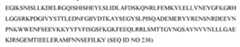 [0064] In embodiments, effector molecules, including those effector molecules comprising a homodimer or a heterodimer, comprise, consist essentially of, or consist of, a DNA-binding domain of a TALEN, or TAL protein, derived from Xanthomonas or Ralstonia. In embodiments, effector molecules, including those effector molecules comprising a homodimer or a heterodimer, comprise, consist essentially of, or consist of, a DNA-binding domain of a TALEN, or TAL protein, derived from Xanthomonas or Ralstonia that forms a homodimer or a heterodimer with Clo051, BfiI or BmrI. In embodiments, effector molecules, including those effector molecules comprising a homodimer or a heterodimer, comprise, consist essentially of, or consist of, a DNA-binding domain of a TALEN, or TAL protein, derived from Xanthomonas or Ralstonia that forms a homodimer or a heterodimer with Clo051. [0065] Linkages [0066] In embodiments, the fusion protein comprises, consists essentially of, or consists of, a DNA localization component and an effector molecule. In embodiments, the nucleic acid sequences encoding one or more components of the fusion protein can be operably linked, for example, in an expression vector. In embodiments, the fusion proteins are chimeric proteins. In embodiments, the fusion proteins are encoded by one or more recombinant nucleic acid sequences. In embodiments, the fusion proteins also include a linker region to operatively- link two components of the fusion protein. For example, in embodiments, the fusion protein comprises, consists essentially of, or consists, of a DNA localization component and an effector molecule, operatively-linked by a linker region. In embodiments, the DNA localization component, the linker region, and the effector molecule can be encoded by one or more nucleic acid sequences inserted into an expression cassette and/or expression vector such that translation of the nucleic acid sequence results in the fusion protein. in embodiments, the fusion protein can comprise a non-covalent linkage between the DNA localization component and the effector molecule. The non-covalent linkage can comprise an antibody, an antibody fragment, an antibody mimetic, or a scaffold protein. [0067] Fusion proteins [0068] In embodiments, the DNA localization component comprises, consists essentially of or consists of, at least one gRNA, and the effector molecule comprises, consists essentially of, or consists of a Cas9, a Cas9 nuclease domain, or a fragment thereof. In embodiments, the DNA localization component comprises, consists essentially of, or consists of, at least one gRNA, and the effector molecule comprises, consists essentially of, or consist of an inactivated Cas9 (dCas9) or an inactivated nuclease domain. In embodiments, the DNA localization component comprises, consists essentially of, or consists of, at least one gRNA, and the effector molecule comprises, consists essentially of, or consist of an inactivated small Cas9 (dSaCas9). In embodiments, the effector molecule comprises, consists essentially of, or consists of a Cas9, dCas9, dSaCas9, or nuclease domain thereof, and a second endonuclease. The second endonuclease can comprise, consist essentially of, or consist of a Type IIS endonuclease, including, but not limited to, one or more of AciI, Mn1I, AlwI, BbvI, BccI, BceAI, BsmAI, BsmFI, BspCNI, BsrI, BtsCI, HgaI, HphI, HpyAV, Mbo1I, My1I, PleI, SfaNI, AcuI, BciVI, BfuAI, BmgBI, BmrI, BpmI, BpuEI, BsaI, BseRI, BsgI, BsmI, BspMI, BsrBI, BsrBI, BsrDI, BtgZI, BtsI, EarI, EciI, MmeI, NmeAIII, BbvCI, Bpu10I, BspQI, SapI, BaeI, BsaXI, CspCI, BfiI, MboII, Acc36I, FokI or Clo051. [0069] In embodiments of the fusion proteins, the DNA localization component comprises, consists essentially of, or consists of, a DNA-binding domain of a transcription activator-like effector nuclease (TALEN, also referred to as a TAL protein), and the effector molecule comprises, consists essentially of, or consists of, an endonuclease. In embodiments of the fusion proteins of the disclosure, the DNA localization component comprises, consists essentially of, or consists of, a DNA-binding domain of a TALEN, or TAL protein, derived from Xanthomonas or Ralstonia, and the effector molecule comprises, consists essentially of, or consists of, a Type IIS endonuclease, including, but not limited to, one or more of AciI, Mn1I, AlwI, BbvI, BccI, BceAI, BsmAI, BsmFI, BspCNI, BsrI, BtsCI, HgaI, HphI, HpyAV, Mbo1I, My1I, PleI, SfaNI, AcuI, BciVI, BfuAI, BmgBI, BmrI, BpmI, BpuEI, BsaI, BseRI, BsgI, BsmI, BspMI, BsrBI, BsrBI, BsrDI, BtgZI, BtsI, EarI, EciI, MmeI, NmeAIII, BbvCI, Bpu10I, BspQI, SapI, BaeI, BsaXI, CspCI, BfiI, MboII, Acc36I or Clo051. [0070] In certain embodiments, an exemplary dCas9-Clo051 fusion protein may comprise, consist essentially of or consist of the amino acid sequence of SEQ ID NO: 305 or 307 of WO2019/126578 or the nucleic acid sequence of SEQ ID NO: 306 or 308 of WO2019/126578. [0071] Constructs [0072] In embodiments, the nuclease domain comprises, consists essentially of, or consists of, a dCas9 and Clo051. In embodiments, the nuclease domain comprises, consists essentially of, or consists of, a dSaCas9 and Clo051. In embodiments, the nuclease domain comprises, consists essentially of, or consists of, a Xanthomonas-TALE and Clo051. In embodiments, the nuclease domain comprises, consists essentially of, or consists of, a Ralstonia-TALE and Clo051. In embodiments, the fusion protein comprises dCas9-Clo051, dSaCas9-Clo051, Xanthomonas-TALE-Clo051, or Ralstonia-TALE-Clo051. In embodiments, a vector encoding the fusion protein comprises Csy4-T2A-Clo051-G4Slinker- dCas9 (Streptoccocus pyogenes) or pRT1-Clo051-dCas9 double NLS. [0073] According to some embodiments, a Cas-CLOVER system comprises a fusion protein comprising a DNA localization component and an effector molecule, wherein the DNA localization component hybridizes to a target sequence of a DNA molecule in a TIL, wherein the DNA molecule encodes and the TIL expresses at least one immune checkpoint molecule, and the effector molecule cleaves the DNA molecule, whereby expression of the at least one immune checkpoint molecule is altered. [0074] According to particular embodiments, a Cas-CLOVER method comprises silencing or reducing the expression of one or more immune checkpoint genes in TILs by introducing a Cas-CLOVER system (e.g., dCas9-Clo051, dSaCas9-Clo051, Xanthomonas-TALE-Clo051, or Ralstonia-TALE-Clo051 fusion protein) specific to a target DNA sequence of the immune checkpoint gene(s). The fusion protein may be delivered as DNA, mRNA, protein. Upon contact of the genome with the Cas-CLOVER system, one or more strand of the target double-stranded DNA may be cut. If the cut is made in the presence of one or more DNA repair pathways or components thereof, the Cas-CLOVER method either interrupts gene expression or modifies the genomic sequence by insertion, deletion, or substitution of one or more base pairs. DSBs may be repaired in the cells by non-homologous end joining (NHEJ), a mechanism which frequently causes insertions or deletions (indels) in the DNA. Indels often lead to frameshifts, creating loss of function alleles; for example, by causing premature stop codons within the open reading frame (ORF) of the targeted gene. According to certain embodiments, the result is a loss-of-function mutation within the targeted immune checkpoint gene. [0075] Alternatively, DSBs induced by Cas-CLOVER systems may be repaired by homology-directed repair (HDR) instead of NHEJ. While NHEJ-mediated DSB repair often disrupts the open reading frame of the gene, homology directed repair (HDR) can be used to generate specific nucleotide changes ranging from a single nucleotide change to large insertions. According to some embodiments, HDR is used for gene editing immune checkpoint genes by delivering a DNA repair template containing the desired sequence into the TILs with the Cas-CLOVER system. The repair template preferably contains the desired edit as well as additional homologous sequence immediately upstream and downstream of the target gene (often referred to as left and right homology arms). [0076] Non-limiting examples of genes that may be silenced or inhibited by permanently gene-editing TILs via a Cas-CLOVER method include PD-1, CTLA-4, LAG-3, HAVCR2 (TIM-3), Cish, TGFβ, PKA, CBL-B, PPP2CA, PPP2CB, PTPN6, PTPN22, PDCD1, BTLA, CD160, TIGIT, TET2, CD96, CRTAM, LAIR1, SIGLEC7, SIGLEC9, CD244, TNFRSF10B, TNFRSF10A, CASP8, CASP10, CASP3, CASP6, CASP7, FADD, FAS, SMAD2, SMAD3, SMAD4, SMAD10, SKI, SKIL, TGIF1, IL10RA, IL10RB, HMOX2, IL6R, IL6ST, EIF2AK4, CSK, PAG1, SIT1, FOXP3, PRDM1, BATF, GUCY1A2, GUCY1A3, GUCY1B2, GUCY1B3, TOX, SOCS1, ANKRD11, and BCOR. [0077] Examples of systems, methods, and compositions for altering the expression of a target gene sequence by a Cas-CLOVER method, and which may be used in accordance with embodiments of the present invention, are described in WO2019126578, US2017/0107541, US2017/0114149, US2018/0187185, and U.S. Patent No.10,415,024, the contents of which are incorporated herein by reference in their entirety. Resources for carrying out Cas- CLOVER methods, such as CLOVER mRNA and Cas-CLOVER mRNA constructs, are commercially available from companies such as Demeetra and Hera Biolabs. a. piggyBac Methods [0078] A method for expanding TILs into a therapeutic population may be carried out in accordance with any embodiment of the methods described herein (e.g., process 2A) or as described in PCT/US2017/058610, PCT/US2018/012605, or PCT/US2018/012633, wherein the method further comprises gene-editing at least a portion of the TILs by a piggyBac method (e.g., piggyBac transposons and transposases or piggyBac-like transposons and transposases). According to particular embodiments, the use of a piggyBac method during the TIL expansion process causes expression of at least one immunomodulatory composition at the cell surface of at least a portion of the therapeutic population of TILs. Alternatively, the use of a piggyBac method during the TIL expansion process causes expression of at least one immunomodulatory composition at the cell surface of, and optionally causes one or more immune checkpoint genes to be enhanced in, at least a portion of the therapeutic population of TILs. In some embodiments, the at least one immunomodulatory composition comprises an immunomodulatory agent fused to a membrane anchor (e.g., a membrane anchored immunomodulatory fusion protein described herein). In some embodiments, the immunomodulatory agent is selected from the group consisting of IL-2, IL-7, IL-10, IL-12, IL-15, IL-18, IL-21, and a CD40 agonist (e.g., a CD40L or an agonistic CD40 binding domain). In some embodiments, the immunomodulatory agent is selected from the group consisting of IL-2, IL-12, IL-15, IL-18, IL-21, and a CD40 agonist. In some embodiments, the immunomodulatory agent is selected from the group consisting of IL-12, IL-15, IL-18, IL-21, and a CD40 agonist. [0079] The piggyBac transposon is a mobile genetic element that efficiently transposes between the donor vector and host chromosomes. This system has almost no cargo limit, and is fully reversible, leaving no footprint in the genome after excision. The piggyBac transposon/transposase system consists of a transposase that recognizes piggyBac-specific inverted terminal repeat sequences (ITRs) located on both sides of the transposon cassette. The transposase excises the transposable element to integrate it into TT/AA chromosomal sites that are preferentially located in euchromatic regions of mammalian genomes (Ding et al.2005; Cadinaños and Bradley 2007; Wilson et al.2007; Wang et al.2008; Li et al.2011). [0080] Exemplary piggyBac systems include those described in WO2019/046815, the contents of which are incorporated herein by reference in their entirety. In embodiments, the piggyBac system comprises a transposon/transposase system. [0081] In embodiments, a piggyBac method comprises delivering to the TILs, (a) a nucleic acid or amino acid sequence comprising a sequence encoding a transposase enzyme and (b) a recombinant and non-naturally occurring DNA sequence comprising a DNA sequence encoding a transposon. [0082] In embodiments, the sequence encoding a transposase enzyme is an mRNA sequence. In embodiments, the sequence encoding a transposase enzyme is a DNA sequence. In embodiments, the DNA sequence is a cDNA sequence. In embodiments, the sequence encoding a transposase enzyme is an amino acid sequence. A protein Super piggybac transposase (SPB) may be delivered following pre-incubation with transposon DNA. [0083] Transposons/Transposases [0084] Exemplary transposon/transposase systems include, but are not limited to, piggyBac transposons and transposases, Sleeping Beauty transposons and transposases, Helraiser transposons and transposases and Tol2 transposons and transposases. [0085] The piggyBac transposase recognizes transposon-specific inverted terminal repeat sequences (ITRs) on the ends of the transposon, and moves the contents between the ITRs into TTAA chromosomal sites. The piggyBac transposon system has no payload limit for the genes of interest that can be included between the ITRs. In embodiments, the transposon is a piggyBac transposon or a piggyBac-like transposon. [0086] Examples of piggyBac and piggyBac-like transposases and transposons include, for example, those disclosed in WO2019/046815, the contents of which are incorporated herein by reference in their entirety. In embodiments, the piggyBac or piggyBac-like transposase is hyperactive. A hyperactive piggyBac or piggyBac-like transposase is a transposase that is more active than the naturally occurring variant from which it is derived. In embodiments, the hyperactive piggyBac or piggyBac-like transposase enzyme is isolated or derived from Bombyx mori. A list of hyperactive amino acid substitutions can be found in US patent No. 10,041,077, the contents of which are incorporated herein by reference in their entirety. In embodiments, the piggyBac or piggyBac-like transposase is integration deficient. In embodiments, an integration deficient piggyBac or piggyBac-like transposase is a transposase that can excise its corresponding transposon, but that integrates the excised transposon at a lower frequency than a corresponding wild-type transposase. A list of integration deficient amino acid substitutions can be found in US patent No.10,041,077, the contents of which are incorporated by reference in their entirety. [0087] In embodiments, the piggyBac or piggyBac-like transposon is capable of insertion by a piggyBac or piggyBac-like transposase at the sequence 5'-TTAT-3 within a target nucleic acid. In embodiments, and, in particular, embodiments wherein the transposon is a piggyBac transposon, the transposase is a piggyBac transposase. In embodiments, and, in particular, embodiments wherein the transposon is a piggyBac- like transposon, the transposase is a piggyBac-like transposase. In embodiments, and, in particular, embodiments wherein the transposon is a piggyBac transposon, the transposase is a piggyBac™ or a Super piggyBac™ (SPB) transposase. In embodiments, and, in particular, embodiments wherein the transposase is a Super piggyBac™ (SPB) transposase, the sequence encoding the transposase is an mRNA sequence. [0088] The sleeping beauty (SB) transposon is transposed into the target genome by the Sleeping Beauty transposase that recognizes ITRs, and moves the contents between the ITRs into TA chromosomal sites. In embodiments, the transposon is a Sleeping Beauty transposon. In embodiments, the transposase enzyme is a Sleeping Beauty transposase enzyme (see, for example, US Patent No.9,228,180, the contents of which are incorporated herein in their entirety). In embodiments, the Sleeping Beauty transposase is a hyperactive Sleeping Beauty (SB100X) transposase. [0089] The Helraiser transposon is transposed by the Helitron transposase. Unlike other transposases, the Helitron transposase does not contain an RNase-H like catalytic domain, but instead comprises a RepHel motif made up of a replication initiator domain (Rep) and a DNA helicase domain. The Rep domain is a nuclease domain of the HUH superfamily of nucleases. In embodiments, the transposon is a Helraiser transposon. In embodiments of the Helraiser transposon sequence, the transposase is flanked by left and right terminal sequences termed LTS and RTS. In embodiments, these sequences terminate with a conserved 5'-TC/CTAG-3' motif. In embodiments, a 19 bp palindromic sequence with the potential to form the hairpin termination structure is located 11 nucleotides upstream of the RTS and comprises the sequence GTGCACGAATTTCGTGCACCGGGCCACTAG. In embodiments, and, in particular embodiments wherein the transposon is a Helraiser transposon, the transposase enzyme is a Helitron transposase enzyme. [0090] Tol2 transposons may be isolated or derived from the genome of the medaka fish, and may be similar to transposons of the hAT family. Exemplary Tol2 transposons of the disclosure are encoded by a sequence comprising about 4.7 kilobases and contain a gene encoding the Tol2 transposase, which contains four exons. In embodiments, the transposon is a Tol2 transposon. In certain embodiments of the methods of the disclosure, and, in particular those embodiments wherein the transposon is a Tol2 transposon, the transposase enzyme is a Tol2 transposase enzyme. [0091] In embodiments, a vector comprises the recombinant and non-naturally occurring DNA sequence encoding the transposon. In embodiments, the vector comprises any form of DNA and wherein the vector comprises at least 100 nucleotides (nts), 500 nts, 1000 nts, 1500 nts, 2000 nts, 2500 nts, 3000 nts, 3500 nts, 4000 nts, 4500 nts, 5000 nts, 6500 nts, 7000 nts, 7500 nts, 8000 nts, 8500 nts, 9000 nts, 9500 nts, 10,000 nts or any number of nucleotides in between. In embodiments, the vector comprises single-stranded or double-stranded DNA. In embodiments, the vector comprises circular DNA. In embodiments, the vector is a plasmid vector, a nanoplasmid vector, a minicircle. In embodiments, the vector comprises linear or linearized DNA. In embodiments, the vector is a double-stranded doggybone™ DNA sequence. [0092] In embodiments, the recombinant and non-naturally occurring DNA sequence encoding a transposon further comprises a sequence encoding one or more immune checkpoint genes. [0093] In embodiments, the nucleic acid sequence encoding the transposase enzyme is a DNA sequence, and an amount of the DNA sequence encoding the transposase enzyme and an amount of the DNA sequence encoding the transposon is equal to or less than 10.0 μg per 100 μL, less than 7.5 μg per 100 μL, less than 6.0 μg per 100 μL, less than 5.0 μg per 100 μL, less than 2.5 μg per 100 μL, or less than 1.67 μg per 100 μL, less than 0.55 μg per 100 μL, less than 0.19 μg per 100 μL, less than 0.10 μg per 100 μL of an electroporation or nucleofection reaction. In certain embodiments, a concentration of the amount of the DNA sequence encoding the transposase enzyme and an amount of the DNA sequence encoding the transposon in the electroporation or nucleofection reaction is equal to or less than 100 μg/mL, equal to or less than 75 μg/mL, equal to or less than 60 μg/mL, equal to or less than 50 μg/mL, equal to or less than 25 μg/mL, equal to or less than 16.7 μg/mL, equal to or less than 5.5 μg/mL, equal to or less than 1.9 μg/mL, equal to or less than 1.0 μg/mL. [0094] In embodiments, the nucleic acid sequence encoding the transposase enzyme is an RNA sequence, and an amount of the RNA sequence encoding the transposase enzyme and an amount of the RNA sequence encoding the transposon is equal to or less than 10.0 μg per 100 μL, less than 7.5 μg per 100 μL, less than 6.0 μg per 100 μL, less than 5.0 μg per 100 μL, less than 2.5 μg per 100 μL, or less than 1.67 μg per 100 μL, less than 0.55 μg per 100 μL, less than 0.19 μg per 100 μL, less than 0.10 μg per 100 μL of an electroporation or nucleofection reaction. In certain embodiments, a concentration of the amount of the RNA sequence encoding the transposase enzyme and an amount of the RNA sequence encoding the transposon in the electroporation or nucleofection reaction is equal to or less than 100 μg/mL, equal to or less than 75 μg/mL, equal to or less than 60 μg/mL, equal to or less than 50 μg/mL, equal to or less than 25 μg/mL, equal to or less than 16.7 μg/mL, equal to or less than 5.5 μg/mL, equal to or less than 1.9 μg/mL, equal to or less than 1.0 μg/mL. [0095] In embodiments, the TILs are further modified by a second gene editing tool, including, but not limited to those described herein. In embodiments, the second gene editing tool may include an excision-only piggyBac transposase to re-excise the inserted sequences or any portion thereof. For example, the excision-only piggyBac transposase may be used to "re-excise" the transposon. [0096] According to some embodiments, a piggyBac system comprises a transposon/transposase system, wherein the transposase recognizes the ITRs located on both sides of the transposon cassette comprising a cargo encoding one or more immune checkpoint genes, and excises the transposable element to integrate it into TT/AA chromosomal sites, resulting in genomic insertion of the transposon cassette and expression of the one or more immune checkpoint genes. According to some embodiments, the cargo encodes two or more immune checkpoint molecules. [0097] Non-limiting examples of genes that may be enhanced by permanently gene-editing TILs via a piggyBac method include CCR2, CCR4, CCR5, CXCR2, CXCR3, CX3CR1, IL-2, IL-4, IL-7, IL-10, IL-15, IL-18, IL-21, the NOTCH 1/2 intracellular domain (ICD), and/or the NOTCH ligand mDLL1. [0098] Examples of systems, methods, and compositions for altering the expression of a target gene sequence by a piggyBac method, and which may be used in accordance with embodiments of the present invention, are described in WO2019/046815, WO2015006700, WO2010085699, WO2010099301, WO2010099296, WO2006122442, WO2001081565, and WO1998040510, the contents of which are incorporated herein by reference in their entirety. [0099] Resources for carrying out piggyBac methods, such as plasmids for expressing transposons/transposases, are commercially available from companies such as Demeetra and Hera Biolabs. [00100] In some embodiments, a method of genetically modifying a population of TILs includes the use of a non-viral technique such as a piggyBac method (e.g., piggyBac transposons and transposases or piggyBac-like transposons and transposases). In some embodiments, the method comprises delivering to the TILs: (a) a nucleic acid or amino acid sequence comprising a sequence encoding a transposase enzyme; and (b) a recombinant and non-naturally occurring DNA sequence comprising a DNA sequence encoding a transposon. In certain embodiments of the methods of the disclosure, the sequence encoding a transposase enzyme is an mRNA sequence. The mRNA sequence encoding a transposase enzyme may be produced in vitro. In certain embodiments of the methods of the disclosure, the sequence encoding a transposase enzyme is a DNA sequence. The DNA sequence encoding a transposase enzyme may be produced in vitro. The DNA sequence may be a cDNA sequence. In certain embodiments of the methods of the disclosure, the sequence encoding a transposase enzyme is an amino acid sequence. The amino acid sequence encoding a transposase enzyme may be produced in vitro. A protein Super piggybac transposase (SPB) may be delivered following pre-incubation with transposon DNA. In certain embodiments, the transposon is a piggyBac transposon or a piggyBac-like transposon. In certain embodiments, and, in particular, those embodiments wherein the transposon is a piggyBac transposon, the transposase is a piggyBac transposase. In certain embodiments, and, in particular, those embodiments wherein the transposon is a piggyBac- like transposon, the transposase is a piggyBac-like transposase. In certain embodiments, the piggyBac transposase comprises an amino acid sequence comprising SEQ ID NO: 14487 of WO2019046815. In certain embodiments, and, in particular, those embodiments wherein the transposon is a piggyBac transposon, the transposase is a piggyBac™ or a Super piggyBac™ (SPB) transposase. In certain embodiments, and, in particular, those embodiments wherein the transposase is a Super piggyBac™ (SPB) transposase, the sequence encoding the transposase is an mRNA sequence. In certain embodiments of the methods of the disclosure, the transposase enzyme is a piggyBac™ (PB) transposase enzyme. The piggyBac (PB) transposase enzyme may comprise or consist of an amino acid sequence at least 75%, 80%, 85%, 90%, 95%, 99% or any percentage in between identical to:
 [00101] In certain embodiments of the methods of the disclosure, the transposon is a Sleeping Beauty transposon. In certain embodiments of the methods of the disclosure, the transposase enzyme is a Sleeping Beauty transposase enzyme (see, for example, US Patent No.9,228,180, the contents of which are incorporated herein in their entirety). In certain embodiments, the Sleeping Beauty transposase is a hyperactive Sleeping Beauty (SB100X) transposase. In certain embodiments, the Sleeping Beauty transposase enzyme comprises an amino acid sequence at least 75%, 80%, 85%, 90%, 95%, 99% or any percentage in between identical to:
 [00102] In certain embodiments, including those wherein the Sleeping Beauty transposase is a hyperactive Sleeping Beauty (SB100X) transposase, the Sleeping Beauty transposase enzyme comprises an amino acid sequence at least at least 75%, 80%, 85%, 90%, 95%, 99% or any percentage in between identical to:
 Optional Cryopreservation of TILs [00420] Either the bulk TIL population or the expanded population of TILs can be optionally cryopreserved. In some embodiments, cryopreservation occurs on the therapeutic TIL population. In some embodiments, cryopreservation occurs on the TILs harvested after the second expansion. In some embodiments, cryopreservation occurs on the TILs in exemplary Step F of Figure 8. In some embodiments, the TILs are cryopreserved in the infusion bag. In some embodiments, the TILs are cryopreserved prior to placement in an infusion bag. In some embodiments, the TILs are cryopreserved and not placed in an infusion bag. In some embodiments, cryopreservation is performed using a cryopreservation medium. In some embodiments, the cryopreservation media contains dimethylsulfoxide (DMSO). This is generally accomplished by putting the TIL population into a freezing solution, e.g.85% complement inactivated AB serum and 15% dimethyl sulfoxide (DMSO). The cells in solution are placed into cryogenic vials and stored for 24 hours at -80 °C, with optional transfer to gaseous nitrogen freezers for cryopreservation. See, Sadeghi, et al., Acta Oncologica 2013, 52, 978-986. [00421] When appropriate, the cells are removed from the freezer and thawed in a 37 °C water bath until approximately 4/5 of the solution is thawed. The cells are generally resuspended in complete media and optionally washed one or more times. In some embodiments, the thawed TILs can be counted and assessed for viability as is known in the art. [00422] In a preferred embodiment, a population of TILs is cryopreserved using CS10 cryopreservation media (CryoStor 10, BioLife Solutions). In a preferred embodiment, a population of TILs is cryopreserved using a cryopreservation media containing dimethylsulfoxide (DMSO). In a preferred embodiment, a population of TILs is cryopreserved using a 1:1 (vol:vol) ratio of CS10 and cell culture media. In a preferred embodiment, a population of TILs is cryopreserved using about a 1:1 (vol:vol) ratio of CS10 and cell culture media, further comprising additional IL-2. [00423] As discussed above in Steps A through E, cryopreservation can occur at numerous points throughout the TIL expansion process. In some embodiments, the bulk TIL population after the first expansion according to Step B or the expanded population of TILs after the one or more second expansions according to Step D can be cryopreserved. Cryopreservation can be generally accomplished by placing the TIL population into a freezing solution, e.g., 85% complement inactivated AB serum and 15% dimethyl sulfoxide (DMSO). The cells in solution are placed into cryogenic vials and stored for 24 hours at -80 °C, with optional transfer to gaseous nitrogen freezers for cryopreservation. See Sadeghi, et al., Acta Oncologica 2013, 52, 978-986. [00424] When appropriate, the cells are removed from the freezer and thawed in a 37 °C water bath until approximately 4/5 of the solution is thawed. The cells are generally resuspended in complete media and optionally washed one or more times. In some embodiments, the thawed TILs can be counted and assessed for viability as is known in the art. [00425] In some cases, the Step B TIL population can be cryopreserved immediately, using the protocols discussed below. Alternatively, the bulk TIL population can be subjected to Step C and Step D and then cryopreserved after Step D. Similarly, in the case where genetically modified TILs will be used in therapy, the Step B or Step D TIL populations can be subjected to genetic modifications for suitable treatments. Optional Cell Viability Analyses [00426] Optionally, a cell viability assay can be performed after the first expansion (sometimes referred to as the initial bulk expansion), using standard assays known in the art. For example, a trypan blue exclusion assay can be done on a sample of the bulk TILs, which selectively labels dead cells and allows a viability assessment. Other assays for use in testing viability can include but are not limited to the Alamar blue assay; and the MTT assay. 15. Cell Counts, Viability, Flow Cytometry [00427] In some embodiments, cell counts and/or viability are measured. The expression of markers such as but not limited CD3, CD4, CD8, and CD56, as well as any other disclosed or described herein, can be measured by flow cytometry with antibodies, for example but not limited to those commercially available from BD Bio-sciences (BD Biosciences, San Jose, CA) using a FACSCantoTM flow cytometer (BD Biosciences). The cells can be counted manually using a disposable c-chip hemocytometer (VWR, Batavia, IL) and viability can be assessed using any method known in the art, including but not limited to trypan blue staining. [00428] In some cases, the bulk TIL population can be cryopreserved immediately, using the protocols discussed below. Alternatively, the bulk TIL population can be subjected to REP and then cryopreserved as discussed below. Similarly, in the case where genetically modified TILs will be used in therapy, the bulk or REP TIL populations can be subjected to genetic modifications for suitable treatments. 16. Cell Cultures [00429] In an embodiment, a method for expanding TILs may include using about 5,000 mL to about 25,000 mL of cell medium, about 5,000 mL to about 10,000 mL of cell medium, or about 5,800 mL to about 8,700 mL of cell medium. In an embodiment, expanding the number of TILs uses no more than one type of cell culture medium. Any suitable cell culture medium may be used, e.g., AIM-V cell medium (L-glutamine, 50 μM streptomycin sulfate, and 10 μM gentamicin sulfate) cell culture medium (Invitrogen, Carlsbad CA). In this regard, the inventive methods advantageously reduce the amount of medium and the number of types of medium required to expand the number of TIL. In an embodiment, expanding the number of TIL may comprise feeding the cells no more frequently than every third or fourth day. Expanding the number of cells in a gas permeable container simplifies the procedures necessary to expand the number of cells by reducing the feeding frequency necessary to expand the cells. [00430] In an embodiment, the cell medium in the first and/or second gas permeable container is unfiltered. The use of unfiltered cell medium may simplify the procedures necessary to expand the number of cells. In an embodiment, the cell medium in the first and/or second gas permeable container lacks beta-mercaptoethanol (BME). [00431] In an embodiment, the duration of the method comprising obtaining a tumor tissue sample from the mammal; culturing the tumor tissue sample in a first gas permeable container containing cell medium therein; obtaining TILs from the tumor tissue sample; expanding the number of TILs in a second gas permeable container containing cell medium therein using aAPCs for a duration of about 14 to about 42 days, e.g., about 28 days. [00432] In an embodiment, TILs are expanded in gas-permeable containers. Gas-permeable containers have been used to expand TILs using PBMCs using methods, compositions, and devices known in the art, including those described in U.S. Patent Application Publication No.2005/0106717 A1, the disclosures of which are incorporated herein by reference. In an embodiment, TILs are expanded in gas-permeable bags. In an embodiment, TILs are expanded using a cell expansion system that expands TILs in gas permeable bags, such as the Xuri Cell Expansion System W25 (GE Healthcare). In an embodiment, TILs are expanded using a cell expansion system that expands TILs in gas permeable bags, such as the WAVE Bioreactor System, also known as the Xuri Cell Expansion System W5 (GE Healthcare). In an embodiment, the cell expansion system includes a gas permeable cell bag with a volume selected from the group consisting of about 100 mL, about 200 mL, about 300 mL, about 400 mL, about 500 mL, about 600 mL, about 700 mL, about 800 mL, about 900 mL, about 1 L, about 2 L, about 3 L, about 4 L, about 5 L, about 6 L, about 7 L, about 8 L, about 9 L, and about 10 L. [00433] In an embodiment, TILs can be expanded in G-Rex flasks (commercially available from Wilson Wolf Manufacturing). Such embodiments allow for cell populations to expand from about 5x10⁵ cells/cm2 to between 10x106 and 30x106 cells/cm2. In an embodiment this is without feeding. In an embodiment, this is without feeding so long as medium resides at a height of about 10 cm in the G-Rex flask. In an embodiment this is without feeding but with the addition of one or more cytokines. In an embodiment, the cytokine can be added as a bolus without any need to mix the cytokine with the medium. Such containers, devices, and methods are known in the art and have been used to expand TILs, and include those described in U.S. Patent Application Publication No. US 2014/0377739A1, International Publication No. WO 2014/210036 A1, U.S. Patent Application Publication No. us 2013/0115617 A1, International Publication No. WO 2013/188427 A1, U.S. Patent Application Publication No. US 2011/0136228 A1, U.S. Patent No. US 8,809,050 B2, International publication No. WO 2011/072088 A2, U.S. Patent Application Publication No. US 2016/0208216 A1, U.S. Patent Application Publication No. US 2012/0244133 A1, International Publication No. WO 2012/129201 A1, U.S. Patent Application Publication No. US 2013/0102075 A1, U.S. Patent No. US 8,956,860 B2, International Publication No. WO 2013/173835 A1, U.S. Patent Application Publication No. US 2015/0175966 A1, the disclosures of which are incorporated herein by reference. Such processes are also described in Jin et al., J. Immunotherapy, 2012, 35:283-292. Optional Genetic Engineering of TILs [00434] In some embodiments, the TILs are optionally genetically engineered to include additional functionalities, including, but not limited to, a high-affinity T cell receptor (TCR), e.g., a TCR targeted at a tumor-associated antigen such as MAGE-1, HER2, or NY-ESO-1, or a chimeric antigen receptor (CAR) which binds to a tumor-associated cell surface molecule (e.g., mesothelin) or lineage-restricted cell surface molecule (e.g., CD19). Immunomodulatory Agents [00435] The modified TILs provided herein may include one or more immunomodulatory agents attached to its surface. The immunomodulatory agents can be incorporated into any of the immunomodulatory fusion proteins described herein, including, for example, the membrane anchored immunomodulatory fusion proteins described herein. Any suitable immunomodulatory agent can be included in the subject modified TIL. In some embodiments, the immunomodulatory agent enhances TIL survival and/or anti-tumor activity once transferred to a patient. Exemplary immunomodulatory agents include, for example, cytokines. In some embodiments, the modified TIL includes one or more of the following cytokines: IL-2, IL-7, IL-12, IL-15, IL-18, IL-21, IL-23, IL-27, IL-4, IL-1α, IL-1β, IL-5, IFNγ, TNF α (TNFa), IFNα, IFNβ, GM-CSF, or GCSF or a biologically active variant thereof. In some embodiments, the immunomodulatory agent is a costimulatory molecule. In particular embodiments, the costimulatory molecule is one of the following: OX40, CD28, GITR, VISTA, CD40, CD3, or an agonist of CD137. In some embodiments, the immunomodulatory agent is a CD40 agonist (e.g., CD40L or an agonistic CD40 binding domain). Exemplary immunomodulatory agents are discussed in detailed further below. 1. IL-15 [00436] In some embodiments, the modified TILs provided herein include an IL-15. In exemplary embodiments, the IL-15 is included as part of an immunomodulatory fusion protein as described herein (e.g., a membrane anchored immunomodulatory fusion protein). [00437] As used herein, “interleukin 15”, “IL-15” and “IL15” all refer to an interleukin that binds to and signals through a complex composed of an IL-15 specific receptor alpha chain (IL-15Rα), an IL-2/IL-15 receptor beta chain (CD122) and the common gamma chain (gamma-C, CD132) (e.g., Genbank Accession numbers: NM_00000585, NP_000576 and NP_751915 (human); and NM_001254747 and NP_001241676 (mouse)). IL-15 has been shown to stimulate T cell proliferation inside tumors. IL-15 also is able to extend the survivability of effector memory CD8+ T cells and is critical for the development of NK cells. Therefore, without being bound by any particular theory of operation, it is believed that modified TILs associated with an IL-15s described herein exhibit enhanced survival and/or anti-tumor effects. [00438] IL-15 has a short half-life of less than 40 minutes in vivo. Modifications to IL-15 monomer can improve its in vivo pharmacokinetics in the treatment of cancers. These modifications have generally centered on improving the trans-presentation of IL-15 with the alpha subunit of IL-15 receptor, IL-15Rα. Such modifications include: 1) pre-association of IL-15 and its soluble receptor a-subunit-Fc fusion to form IL-15: IL-15Rα-Fc complex (see, e.g., Rubinstein et al., Proc Natl Acad Sci U.S.A.103:9166–71 (2006)); 2) expression of the superagonist IL-15-sIL-15Rα-sushi protein (see, e.g., Bessard et al., Molecular cancer therapeutics 8: 2736-45 (2009)); and 3) pre-association of human IL-15 mutant IL-15N72D with IL-15Rα-Fc sushi-Fc fusion complex (see, e.g., Zhu et al., Journal of Immunology 183: 3598-6007 (2009)). [00439] In some embodiments, the IL-15 associated with the modified TIL is a full length IL-15, a fragment or a variant of IL-15. In some embodiments, the IL-15 is a human IL-15 or a variant human IL-15. In exemplary embodiments, the IL-15 is a biological active human IL-15 variant. In some embodiments, the IL-15 includes a 1, 2, 3,4 ,5 ,67, 8, 9, or 10 mutations as compared to a wild-type IL-15. In certain embodiments, the IL-15 includes an N72D mutation relative to a wild type human IL-15. In some embodiments, the variant IL-15 exhibits IL-15Rα binding activity. [00440] In some embodiments, the immunomodulatory agent includes an IL-15 and an extracellular domain of an IL-15Rα. In certain embodiments, the immunomodulatory agent includes an IL-15 and an IL-15Rα fused to an Fc domain (IL-15Rα-Fc) TABLE 5 – IL-15 Related Sequences.
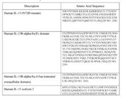
 [00441] In some embodiments the immunostimulatory protein is a superagonist IL-15 (IL-15SA) that includes a complex of human IL-15 and soluble human IL-15Rα. The combination of human IL-15 with soluble human IL-15Rα forms an IL-15 SA complex that possesses greater biological activity than human IL-15 alone. Soluble human IL-15Rα, as well as truncated versions of the extracellular domain, has been described in the art (Wei et al., 2001 J of Immunol.167: 277-282). The amino acid sequence of human IL-15Rα is set forth in SEQ ID NO: 266. In some embodiments, the IL-15SA includes a complex of human IL-15 and soluble human. IL-15Rα comprising all or a portion of the extracellular domain, without the transmembrane or cytoplasmic domain. In some embodiments, the IL-15SA includes a complex of human IL-15 and soluble human IL-15Rα that includes the full extracellular domain or a truncated form of the extracellular domain which retains IL-15 binding activity. [00442] In some embodiments, the IL-15SA includes a complex of human IL-15 and soluble human IL-15Rα that includes a truncated form of the extracellular domain which retains IL-15 binding activity. In some embodiments, the soluble human IL-15Rα includes amino acids 1-60, 1-61, 1-62, 1-63, 1-64 or 1-65 of human IL-15Rα. In some embodiments, the soluble human IL-15Rα includes amino acids 1-80, 1-81, 1-82, 1-83, 1-84 or 1-85 of human IL-15Rα. In some embodiments, the soluble human IL-15Rα includes amino acids 1- 180, 1-181, or 1-182 of human IL-15Rα. [00443] In some embodiments, the immunomodulatory agent is an IL-15SA comprising a complex of human IL-15 and soluble human IL-15Rα comprising a truncated form of the extracellular domain which retains IL-15 binding activity and comprises a Sushi domain. The Sushi domain of IL-15Rα is described in the art as approximately 60 amino acids in length and comprises 4 cysteines. (Wei et al., 2001). Truncated forms of soluble human IL-15Rα which retain IL-15 activity and comprise a Sushi domain are useful in IL- 15SA of the present disclosure. [00444] In some embodiments, the immunomodulatory agent includes a complex comprising soluble human IL-15Rα expressed as a fusion protein, such as an Fc fusion as described herein (e.g., human IgG1 Fc), with IL-15. In some embodiments, IL-15SA comprises a dimeric human IL-15RαFc fusion protein (e.g., human IgG1 Fc) complexed with two human IL-15 molecules. [00445] In some embodiments, the immunomodulatory agent is an IL-15SA cytokine complex that includes an IL-15 molecule comprising an amino acid sequence set forth in SEQ ID NO: 258, SEQ ID NO: 261, SEQ ID NO:262, or SEQ ID NO:263. In some embodiments, an IL-15SA cytokine complex comprises a soluble IL-15Rα molecule comprising a sequence of SEQ ID NO:260, SEQ ID NO: 264 or SEQ ID NO:265. [00446] In some embodiments, the immunomodulatory agent is an IL-15SA cytokine complex that includes a dimeric IL-15RαFc fusion protein complexed with two IL-15 molecules. In some embodiments, IL-15-SA comprises a dimeric IL-15RαSu (Sushi domain)/Fc (SEQ ID NO:259) and two IL-15N72D (SEQ ID NO:258) molecules (also known as ALT-803), as described in US20140134128, incorporated herein by reference. In some embodiments, the IL-15SA comprises a dimeric IL-15RαSu/Fc molecule (SEQ ID NO: 259) and two IL-15 molecules (SEQ ID NO: 261). In some embodiments, the IL-15SA comprises a dimeric IL-15RαSu/Fc molecule (SEQ ID NO: 259) and two IL-15 molecules (SEQ ID NO:262). In some embodiments, the IL-15SA comprises a dimeric IL-15RαSu/Fc molecule (SEQ ID NO:259) and two IL-15 molecules (SEQ ID NO:263). [00447] In some embodiments, the IL-15SA includes a dimeric IL-15RαSu/Fc molecule (SEQ ID NO:259) and two IL-15 molecules having amino acid sequences selected from SEQ ID NO: 258, 258, 262, and 263. [00448] In some embodiments, the IL-15SA includes a soluble IL-15Rα molecule (SEQ ID NO:260) and two IL-15 molecules (SEQ ID NO:258). In some embodiments, the IL-15SA comprises a soluble IL-15Rα molecule (SEQ ID NO:260) and two IL-15 molecules (SEQ ID NO:261). In some embodiments, the IL-15SA comprises a soluble IL-15Rα molecule (SEQ ID NO:260) and two IL-15 molecules (SEQ ID NO:262). In some embodiments, the IL-15SA comprises a soluble IL-15Rα molecule (SEQ ID NO:260) and two IL-15 molecules (SEQ ID NO:263). [00449] In some embodiments, the IL-15SA comprises a soluble IL-15Rα molecule (SEQ ID NO:264) and two IL-15 molecules (SEQ ID NO:258). In some embodiments, the IL-15SA comprises a soluble IL-15Rα molecule (SEQ ID NO:264) and two IL-15 molecules (SEQ ID NO:261). In some embodiments, the IL-15SA comprises a soluble IL-15Rα molecule (SEQ ID NO:264) and two IL-15 molecules (SEQ ID NO:262). In some embodiments, the IL-15SA comprises a soluble IL-15Rα molecule (SEQ ID NO:264) and two IL-15 molecules (SEQ ID NO:261). [00450] In some embodiments, the IL-15SA includes a soluble IL-15Rα molecule (SEQ ID NO:265) and two IL-15 molecules (SEQ ID NO:258). In some embodiments, the IL-15SA comprises a soluble IL-15Rα molecule (SEQ ID NO:265) and two IL-15 molecules (SEQ ID NO:261). In some embodiments, the IL-15SA comprises a soluble IL-15Rα molecule (SEQ ID NO:265) and two IL-15 molecules (SEQ ID NO:262). In some embodiments, the IL-15SA comprises a soluble IL-15Rα molecule (SEQ ID NO:265) and two IL-15 molecules (SEQ ID NO:263). [00451] In some embodiments, the IL-15SA comprises a dimeric IL-15RαSu/Fc (SEQ ID NO:259) molecule and two IL-15 molecules (SEQ ID NO:262). In some embodiments, the IL-15SA includes a dimeric IL-15RαSu/Fc (SEQ ID NO:259) molecule and two IL-15 molecules (SEQ ID NO:263). [00452] In some embodiments, the IL-15SA includes SEQ ID NO:259 and SEQ ID NO:260. In some embodiments IL-15SA comprises SEQ ID NO:261 or SEQ ID NO:262. In some embodiments the IL-15SA comprises SEQ ID NO:261 and SEQ ID NO:259. In some embodiments the IL-15SA comprises SEQ ID NO:262 and SEQ ID NO:259. In some embodiments the IL-15SA comprises SEQ ID NO:263 and SEQ ID NO:259. In some embodiments, the IL-15SA comprises SEQ ID NO:261 and SEQ ID NO:260. In some embodiments the IL-15SA comprises SEQ ID NO:262 and SEQ ID NO:260. [00453] In some embodiments, the TIL compositions include an immunomodulatory fusion protein or nanoparticle composition that includes a IL-15 or a bioactive variant thereof. Exemplary fusion proteins that include IL-15 are depicted in Tables 58 and 59. [00454] In exemplary embodiments the TIL compositions provided herein includes a nucleic acid encoding an immunomodulatory fusion protein that includes an IL-15, wherein the nucleic acid is operably linked to a an NFAT promoter, an EF-1a promoter, an MND promoter, or an SSFV promoter, as described herein. Exemplary NFAT promoter-driven constructs for expression of immunomodulatory fusion proteins that include IL-15 are depicted in Table 59. [00455] TABLE 58. Membrane anchored IL-15 and IL-21 fusion protein DNA sequences


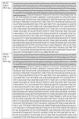
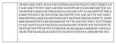 2. IL-12 [00457] In some embodiments, the modified TIL is associated with an IL-12 or a variant thereof. In exemplary embodiments, the IL-12 is included as part of an immunomodulatory fusion protein as described herein (e.g., a membrane anchored immunomodulatory fusion protein). [00458] As used herein, “interleukin 12”, “IL-12” and “IL12” all refer to an interleukin that is a heterodimeric cytokine encoded by the IL-12A and IL-12B genes (Genbank Accession numbers: NM_000882 (IL-12A) and NM_002187 (IL-12B)). IL-12 is composed of a bundle of four alpha helices and is involved in the differentiation of native T cells into TH1 cells. It is encoded by two separate genes, IL-12A (p35) and IL-12B (p40). The active heterodimer (referred to as 'p70'), and a homodimer of p40 are formed following protein synthesis. IL-12 binds to the IL-12 receptor, which is a heterodimeric receptor formed by IL- 12R-β1 and IL-12R-β2. IL-12 is known as a T cell-stimulating factor that can stimulate the growth and function of T cells. In particular, IL-12 can stimulate the production of interferon gamma (IFN-γ), and tumor necrosis factor-alpha (TNF-α) from T cells and natural killer (NK) cells and reduce IL-4 mediated suppression of IFN-γ. IL-12 can further mediate enhancement of the cytotoxic activity of NK cells and CD8+ cytotoxic T lymphocytes. Moreover, IL-12 can also have anti-angiogenic activity by increasing production of interferon gamma, which in turn increases the production of the chemokine inducible protein-10 (IP-10 or CXCL10). IP-10 then mediates this anti-angiogenic effect. Thus, without being bound by any particular theory of operation, it is believed that IL-12 can increase the survivability and/or anti-tumor effects of the TIL compositions provided herein. [00459] In some embodiments, the IL-12 associated with the modified TIL is a full length IL-12, a fragment or a variant of IL-12. In some embodiments, the IL-12 is a human IL-12 or a variant human IL-12. In exemplary embodiments, the IL-12 is a biological active human IL-12 variant. In some embodiments, the IL-12 includes a 1, 2, 3,4 ,5 ,67, 8, 9, or 10 mutations as compared to a wild-type IL-12. [00460] In some embodiments, the IL-12 included in the modified TIL compositions include an IL-12 p35 subunit or a variant thereof. In some embodiments, the IL-12 p35 subunit is a human IL-12 p35 subunit. In some embodiments, the IL-12 p35 subunit has the amino acid sequence In certain embodiments, the IL-12 included in the modified TIL compositions include an IL-12 p40 subunit or a variant thereof. In certain embodiments, the IL-12 is a single chain IL-12 polypeptide comprising an IL-12 p35 subunit attached to an IL- 12 p40 subunit. Such IL-12 single chain polypeptides advantageously retain one or more of the biological activities of wildtype IL-12. In some embodiments, the single chain IL-12 polypeptide described herein is according to the formula, from N-terminus to C-terminus, (p40)-(L)-(p35), wherein “p40” is an IL-12 p40 subunit, “p35” is IL-12 p35 subunit and L is a linker. In other embodiments, the single chain IL-12 is according to the formula from N- terminus to C-terminus, (p35)-(L)-(p40). Any suitable linker can be used in the single chain IL-12 polypeptide including those described herein. Suitable linkers can include, for example, linkers having the amino acid sequence (GGGGS)x wherein x is an integer from 1- 10. Other suitable linkers include, for example, the amino acid sequence GGGGGGS. Exemplary single chain IL-12 linkers than can be used with the subject single chain IL-12 polypeptides are also described in Lieschke et al., Nature Biotechnology 15: 35-40 (1997), which is incorporated herein in its entirety by reference and particularly for its teaching of IL- 12 polypeptide linkers. In an exemplary embodiment, the single chain IL-12 polypeptide is a single chain human IL-12 polypeptide (i.e., it includes a human p35 and p40 IL-12 subunit). TABLE 6 – IL-12 Related Sequences.
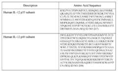 [00461] In some embodiments, the TIL compositions include an immunomodulatory fusion protein or nanoparticle composition that includes a IL-12 or a bioactive variant thereof. [00462] In exemplary embodiments the TIL compositions provided herein includes a nucleic acid encoding an immunomodulatory fusion protein that includes an IL-12, wherein the nucleic acid is operably linked to an NFAT promoter, an EF-1a promoter, an MND promoter, or an SSFV promoter, as described herein. See, e.g., US Patent No.8,556,882, which is incorporated by reference in its entirety and particularly for pertinent parts relating to NFAT promoters for IL-12 expression. Exemplary fusion proteins that include IL-12 are depicted in Table 58. 3. IL-18 [00463] In some embodiments, the modified TIL is associated with an IL-18 or a variant thereof. In exemplary embodiments, the IL-18 is included as part of an immunomodulatory fusion protein as described herein (e.g., a membrane anchored immunomodulatory fusion protein). [00464] As used herein, “interleukin 18”, “IL-18,” “IL18,” “IGIF,” “IL-1g,” “interferon-gamma inducing factor,” and “IL1F4,” all refer to an interleukin that is a heterodimeric cytokine encoded by the IL-18 gene (e.g., Genbank Accession numbers: NM_001243211, NM_001562 and NM_001386420). IL-18, structurally similar to IL-1β, is a member of IL-1 superfamily of cytokines. This cytokine, which is expressed by many human lymphoid and nonlymphoid cells, has an important role in inflammatory processes. IL-18 in combination with IL-12 can activate cytotoxic T cells (CTLs), as well as natural killer (NK) cells, to produce IFN-γ and, therefore, contributes to tumor immunity. Thus, without being bound by any particular theory of operation, it is believed that IL-18 can enhance the anti-tumor effects of the TIL compositions provided herein. [00465] In some embodiments, the IL-18 associated with the modified TIL is a full length IL-18, a fragment or a variant of IL-18. In some embodiments, the IL-18 is a human IL-18 or a variant human IL-18. In exemplary embodiments, the IL-18 is a biological active human IL-18 variant. In some embodiments, the IL-18 includes 1, 2, 3,4 ,5 ,67, 8, 9, 10, 11, 12, 13, 14, 15, 16, 17,.18, 19, or 20 mutations as compared to a wild-type IL-18 (SEQ ID NO:269). In some embodiments, the bioactive variant is a decoy resistant IL-18 variant (“DR-IL18,” or “DR-IL-18”) that provides IL-18 signaling activity even in the presence of an inhibitory molecule such as IL-18 binding protein (IL-18BP). Exemplary IL-18 variants that can be included in the subject modified TILs described herein are shown below in Table 7. Additional IL-18 variants that can be included in the subject modified TILs are described in WO 2022/094473, which is incorporated by reference in its entirety and particular with respect to disclosures relating to variant DR-IL-18. [00466] In some embodiments, the variant IL-18 includes a stability mutation pair selection fromL C38S/C68S, C38S/C68G, C38S/C68A, C38S/C68D, and C38S/C68N [relative to the human wild-type IL-18 - SEQ ID NO: 269]. In some embodiments, the variant IL-18 includes mutations at amino acid positions M51 (e.g., M51E, M51R, M51K, M51T, M51D, or M51N), K53 (e.g., K53G, K53S, K53T, or K53R), Q56 (e g., Q56G, Q56R, Q56L, Q56E, Q56A, Q56V, or Q56K), D110 (e.g., D110S, DI 10N, D110G, D110K, D110H, D110Q, or D110E) and N111 (e.g., N111G, N111R, Ni l IS, Ni l ID, N111H, or N111Y) in addition to a stabilizing mutation pair selected from: C38S/C68S, C38S/C68G, C38S/C68A, C38S/C68D, and C38S/C68N [relative to the human wild type IL-18 - SEQ ID NO: 269], In some such cases the stabilized IL-18 variant polypeptide additionally includes a mutation at amino acid position S105 (e.g., S105D, SI 05 A, S105N, S105R, S105D, or S105K); and in some cases, further includes mutations at amino acid positions P57 (e.g., P57A, P57L, P57G, or P57K) and M60 (e.g., M60L, M60R, M60K, or M60Q). TABLE 7 – IL-18 Related Sequences.


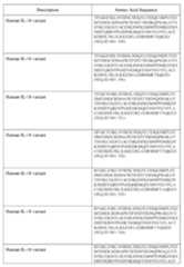


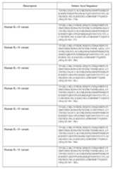 [00467] In some embodiments, the TIL compositions include an immunomodulatory fusion protein or nanoparticle composition that includes a IL-18 or a bioactive variant thereof (e.g., any one of the IL-18 variants included in Table 7). Exemplary fusion proteins that include IL-18 are depicted in Figure 32. [00468] In exemplary embodiments the TIL compositions provided herein includes a nucleic acid encoding an immunomodulatory fusion protein that includes an IL-18, wherein the nucleic acid is operably linked to an NFAT promoter, an EF-1a promoter, an MND promoter, or an SSFV promoter, as described herein. Exemplary NFAT promoter-driven constructs for expression of immunomodulatory fusion proteins that include IL-21 are depicted in Table 59. 4. IL-21 [00469] In some embodiments, the modified TIL is associated with an IL-21 or a variant thereof. In exemplary embodiments, the IL-21 is included as part of an immunomodulatory fusion protein as described herein (e.g., a membrane anchored immunomodulatory fusion protein). [00470] In certain embodiments, the cytokine-ABD includes an IL-21 molecule or fragment thereof. As used herein, “interleukin 21” “IL-21”, and “IL21” (e.g., Genbank Accession numbers: NM_001207006 and NP_001193935 (human); and NM_0001291041 and NP_001277970 (mouse)) all refer to a member of a cytokine that binds to IL-21 receptor and has potent regulatory effects on cells of the immune system, including natural killer (NK) cells and cytotoxic cells and binds to IL-21 receptor that can destroy virally infected or cancerous cells. Thus, without being bound by any particular theory of operation, it is believed that IL-21 can increase the survivability and/or anti-tumor effects of the TIL compositions provided herein. [00471] In some embodiments, the IL-21 is a human IL-21. In some embodiments, the IL-21 associated with the modified TIL is a full length IL-21, a fragment or a variant of IL- 21. In some embodiments, the IL-21 is a human IL-21 or a variant human IL-21. In exemplary embodiments, the IL-21 is a biological active human IL-21 variant. In some embodiments, the IL-21 includes a 1, 2, 3,4 ,5 ,67, 8, 9, or 10 mutations as compared to a wild-type IL-21. TABLE 8 – IL-21 Related Sequences.
 [00472] In some embodiments, the TIL compositions include an immunomodulatory fusion protein or nanoparticle composition that includes a IL-21 or a bioactive variant thereof. Exemplary fusion proteins that include IL-21 are depicted in Figures 32 and 33, and Tables 58 and 59. [00473] In exemplary embodiments the TIL compositions provided herein includes a nucleic acid encoding an immunomodulatory fusion protein that includes an IL-21, wherein the nucleic acid is operably linked to an NFAT promoter, an EF-1a promoter, an MND promoter, or an SSFV promoter, as described herein. 5. IL-2 [00474] In some embodiments, the modified TIL is associated with an IL-2 or a variant thereof. In exemplary embodiments, the IL-2 is included as part of an immunomodulatory fusion protein as described herein (e.g., a membrane anchored immunomodulatory fusion protein). [00475] In certain embodiments, the cytokine-ABD includes an IL-2 molecule or fragment thereof. As used herein, “interleukin 2” “IL-2”, “IL2,” and “TCGF” (e.g., Genbank Accession numbers: NM_000586 and NP_000577 (human) all refer to a member of a cytokine that binds to IL-2 receptor. IL-2 enhances activation-induced cell death (AICD). IL-2 also promotes the differentiation of T cells into effector T cells and into memory T cells when the initial T cell is also stimulated by an antigen, thus helping the body fight off infections. Together with other polarizing cytokines, IL-2 stimulates naive CD4+ T cell differentiation into Th1 and Th2 lymphocytes and impedes differentiation into Th17 and follicular Th lymphocytes.. IL-2 also increases the cell killing activity of both natural killer cells and cytotoxic T cells. Thus, without being bound by any particular theory of operation, it is believed that IL-2 can increase the survivability and/or anti-tumor effects of the TIL compositions provided herein. [00476] In some embodiments, the IL-2 is a human IL-2. In some embodiments, the IL-2 associated with the modified TIL is a full length IL-2, a fragment or a variant of IL-2. In some embodiments, the IL-2 is a human IL-2 or a variant human IL-2. In exemplary embodiments, the IL-2 is a biological active human IL-2 variant. In some embodiments, the IL-2 includes a 1, 2, 3,4 ,5 ,67, 8, 9, or 10 mutations as compared to a wild-type IL-2. TABLE 9 – IL-2 Related Sequences.
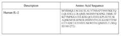 [00477] In some embodiments, the TIL compositions include an immunomodulatory fusion protein or nanoparticle composition that includes a IL-2 or a bioactive variant thereof. Exemplary fusion proteins that include IL-2 are depicted in Figures 32 and 33. [00478] In exemplary embodiments the TIL compositions provided herein includes a nucleic acid encoding an immunomodulatory fusion protein that includes an IL-2, wherein the nucleic acid is operably linked to an NFAT promoter, an EF-1a promoter, an MND promoter, or an SSFV promoter, as described herein. 6. CD40 Agonists [00479] In some embodiments, the modified TIL is associated with CD40 agonist. In exemplary embodiments, the CD40 agonist is included as part of an immunomodulatory fusion protein as described herein (e.g., a membrane anchored immunomodulatory fusion protein). [00480] Cluster of differentiation 40, CD40, is a costimulatory protein found on antigen-presenting cells (APCs) and is required for APC activation. The binding of CD40L (CD154) on T helper cells to CD40 activates antigen presenting cells (e.g., dendritic cells) and induces a variety of downstream effects. Without being by any particular theory of operation, it is believed that the addition of one or more immunomodulatory agents that activate CD40 on antigen presenting cells (i.e., CD40 agonists) can enhance the anti-tumor effects of the TIL compositions provided herein. CD40 agonists, include, for example, CD40L and antibody or antibody fragments thereof (e.g., an scFv) that agonistically binds CD40. In some embodiments, the TIL compositions include an immunomodulatory fusion protein or nanoparticle composition that includes a CD40L or a bioactive variant thereof. In some embodiments, the TIL composition includes an immunomodulatory fusion protein that includes an agonistic anti-CD40 binding domain (e.g., an scFv). Exemplary CD40 agonist sequences are depicted in the table below. [00481] CD40 agonist activity can be measured using any suitable method known in the art. Ligation of CD40 on DC, for example, induces increased surface expression of costimulatory and MHC molecules, production of proinflammatory cytokines, and enhanced T cell triggering. CD40 ligation on resting B cells increases antigen-presenting function and proliferation. In exemplary embodiments, the CD40 agonist is capable of activating human dendritic cells. [00482] In some embodiments, the TIL composition includes an agonistic anti-CD40 binding domain having the VH and VL sequences of an anti-CD40 scFv depicted in Table 10 or a bioactive variant thereof. In some embodiments, the anti-CD40 binding domain includes a VH sequence that is at least about 80%, 85%, 90%, 91%, 92%, 93%, 94%, 95%, 96%, 97%, 98%, 99% identical to the VH sequence depicted in Table 10. In some embodiments, the agonistic anti-CD40 binding domain includes a VH sequence that includes 1, 2, 3, 4, 5, 6, 7, 8, 9, 10, 11, 12, 13, 14, 15, 16, 17, 18, 19, or 20 amino acid substitutions as compared to the VH sequence depicted in Table 10. In some embodiments, the anti-CD40 binding domain includes a VL sequence that is at least about 80%, 85%, 90%, 91%, 92%, 93%, 94%, 95%, 96%, 97%, 98%, 99% identical to the VL sequence depicted in Table 10. In some embodiments, the anti-CD40 binding domain includes a VL sequence that includes 1, 2, 3, 4, 5, 6, 7, 8, 9, 10, 11, 12, 13, 14, 15, 16, 17, 18, 19, or 20 amino acid substitutions as compared to the VL sequence depicted in Table 10. In exemplary embodiments, the anti-CD40 binding domain is an anti-CD40 scFv selected from SEQ ID NOs:276, 279, 282, and 285 in Table 10. [00483] In some embodiments, the anti-CD40 binding domain is a variant of an anti- CD40 scFv in Table 10 that is capable of binding to human CD40. In exemplary embodiments, the variant anti-CD40 scFv is least about 75%, 80%, 85%, 90%, 95%, or 99% identical to an anti-CD40 scFv selected from SEQ ID NOs:276, 279, 282, and 285 in Table 10. [00484] Assessment of CD40 binding domain binding can be measured using any suitable assay known in the art, including, but not limited to: a Biacore, surface plasmon resonance (SPR) and/or BLI (biolayer interferometry, e.g., Octet assay) assay. [00485] Additional CD40 binding domains (VH and VLs) that are useful as immunomodulatory agents include those described in US Patent Nos. US 6,838,261, US 6,843,989, US 7,338,660, US 8,7778,345, which are incorporated by reference herein, particularly with respect to teachings of anti-CD40 antibodies and VH, VL and CDR sequences. [00486] In some embodiments, the CD40 agonist is a CD40 ligand (CD40L). In exemplary embodiments, the CD40L is human CD40L (SEQ ID NO:270). In some embodiments, the CD40L is a variant of a human CD40L that is at least about 75%, 80%, 85%, 90%, 95%, or 99% identical to SEQ ID NO:253. In some embodiments, the CD40L is a variant of a human CD40L that includes 1, 2, 3, 4, 5, 6, 7, 8, 9, 10, 11, 12, 13, 14, 15, 16, 17, 18, 19, or 20 amino acid substitutions as compared to SEQ ID NO:273. [00487] Exemplary fusion proteins that include CD40 agonists are depicted in Figures 32 and 33. [00488] In exemplary embodiments the TIL compositions provided herein includes a nucleic acid encoding an immunomodulatory fusion protein that includes a CD40 agonist, wherein the nucleic acid is operably linked to a NFAT promoter, as described herein. TABLE 10 – CD40 Agonist Related Sequences.

 Methods of Treating Patients [00489] Methods of treatment begin with the initial TIL collection and culture of TILs. Such methods have been both described in the art by, for example, Jin et al., J. Immunotherapy, 2012, 35(3):283-292, incorporated by reference herein in its entirety. Embodiments of methods of treatment are described throughout the sections below, including the Examples. [00490] The expanded TILs produced according the methods described herein, including for example as described in Steps A through F above or according to Steps A through F above (also as shown, for example, in Figure 8) find particular use in the treatment of patients with cancer (for example, as described in Goff, et al., J. Clinical Oncology, 2016, 34(20):2389- 239, as well as the supplemental content; incorporated by reference herein in its entirety. In some embodiments, TIL were grown from resected deposits of metastatic melanoma as previously described (see, Dudley, et al., J Immunother., 2003, 26:332-342; incorporated by reference herein in its entirety). Fresh tumor can be dissected under sterile conditions. A representative sample can be collected for formal pathologic analysis. Single fragments of 2 mm3 to 3 mm3 may be used. In some embodiments, 5, 10, 15, 20, 25 or 30 samples per patient are obtained. In some embodiments, 20, 25, or 30 samples per patient are obtained. In some embodiments, 20, 22, 24, 26, or 28 samples per patient are obtained. In some embodiments, 24 samples per patient are obtained. Samples can be placed in individual wells of a 24-well plate, maintained in growth media with high-dose IL-2 (6,000 IU/mL), and monitored for destruction of tumor and/or proliferation of TIL. Any tumor with viable cells remaining after processing can be enzymatically digested into a single cell suspension and cryopreserved, as described herein. [00491] In some embodiments, successfully grown TIL can be sampled for phenotype analysis (CD3, CD4, CD8, and CD56) and tested against autologous tumor when available. TIL can be considered reactive if overnight coculture yielded interferon-gamma (IFN-γ) levels ˃ 200 pg/mL and twice background. (Goff, et al., J Immunother., 2010, 33:840-847; incorporated by reference herein in its entirety). In some embodiments, cultures with evidence of autologous reactivity or sufficient growth patterns can be selected for a second expansion (for example, a second expansion as provided in according to Step D of Figure 8), including second expansions that are sometimes referred to as rapid expansion (REP). In some embodiments, expanded TILs with high autologous reactivity (for example, high proliferation during a second expansion), are selected for an additional second expansion. In some embodiments, TILs with high autologous reactivity (for example, high proliferation during second expansion as provided in Step D of Figure 8), are selected for an additional second expansion according to Step D of Figure 8. [00492] In some embodiments, the patient is not moved directly to ACT (adoptive cell transfer), for example, in some embodiments, after tumor harvesting and/or a first expansion, the cells are not utilized immediately. In such embodiments, TILs can be cryopreserved and thawed 2 days before administration to a patient. In such embodiments, TILs can be cryopreserved and thawed 1 day before administration to a patient. In some embodiments, the TILs can be cryopreserved and thawed immediately before the administration to a patient. [00493] Cell phenotypes of cryopreserved samples of infusion bag TIL can be analyzed by flow cytometry (e.g., FlowJo) for surface markers CD3, CD4, CD8, CCR7, and CD45RA (BD BioSciences), as well as by any of the methods described herein. Serum cytokines were measured by using standard enzyme-linked immunosorbent assay techniques. A rise in serum IFN-g was defined as ˃100 pg/mL and greater than 43 baseline levels. [00494] In some embodiments, the TILs produced by the methods provided herein, for example those exemplified in Figure 8, provide for a surprising improvement in clinical efficacy of the TILs. In some embodiments, the TILs produced by the methods provided herein, for example those exemplified in Figure 8, exhibit increased clinical efficacy as compared to TILs produced by methods other than those described herein, including for example, methods other than those exemplified in Figure 8. In some embodiments, the increased efficacy is measured by DCR, ORR, and/or other clinical responses. In some embodiments, the TILS produced by the methods provided herein, for example those exemplified in Figure 8, exhibit a similar time to response and safety profile compared to TILs produced by methods other than those described herein, including for example, methods other than those exemplified in Figure 8, for example the Gen 1 process. [00495] In some embodiments, IFN-gamma (IFN-γ) is indicative of treatment efficacy and/or increased clinical efficacy. In some embodiments, IFN-γ in the blood of subjects treated with TILs is indicative of active TILs. In some embodiments, a potency assay for IFN-γ production is employed. IFN-γ production is another measure of cytotoxic potential. IFN-γ production can be measured by determining the levels of the cytokine IFN-γ in the blood, serum, or TILs ex vivo of a subject treated with TILs prepared by the methods of the present invention, including those as described for example in Figure 8. In some embodiments, an increase in IFN-γ is indicative of treatment efficacy in a patient treated with the TILs produced by the methods of the present invention. In some embodiments, IFN-γ is increased one-fold, two-fold, three-fold, four-fold, or five-fold or more as compared to an untreated patient and/or as compared to a patient treated with TILs prepared using other methods than those provide herein including for example, methods other than those embodied in Figure 8. In some embodiments, IFN-γ secretion is increased one-fold as compared to an untreated patient and/or as compared to a patient treated with TILs prepared using other methods than those provide herein including for example, methods other than those embodied in Figure 8. In some embodiments, IFN-γ secretion is increased two-fold as compared to an untreated patient and/or as compared to a patient treated with TILs prepared using other methods than those provide herein including for example, methods other than those embodied in Figure 8. In some embodiments, IFN-γ secretion is increased three-fold as compared to an untreated patient and/or as compared to a patient treated with TILs prepared using other methods than those provide herein including for example, methods other than those embodied in Figure 8. In some embodiments, IFN-γ secretion is increased four-fold as compared to an untreated patient and/or as compared to a patient treated with TILs prepared using other methods than those provide herein including for example, methods other than those embodied in Figure 8. In some embodiments, IFN-γ secretion is increased five-fold as compared to an untreated patient and/or as compared to a patient treated with TILs prepared using other methods than those provide herein including for example, methods other than those embodied in Figure 8. In some embodiments, IFN-γ is measured using a Quantikine ELISA kit. In some embodiments, IFN-γ is measured in TILs ex vivo of a subject treated with TILs prepared by the methods of the present invention, including those as described for example in Figure 8. In some embodiments, IFN-γ is measured in blood of a subject treated with TILs prepared by the methods of the present invention, including those as described for example in Figure 8. In some embodiments, IFN-γ is measured in TILs serum of a subject treated with TILs prepared by the methods of the present invention, including those as described for example in Figure 8. [00496] In some embodiments, higher average IP-10 is indicative of treatment efficacy and/or increased clinical efficacy. In some embodiments, higher average IP-10 in the blood of subjects treated with TILs is indicative of active TILs. IP-10 production can be measured by determining the levels of the IP-10 in the blood of a subject treated with TILs prepared by the methods of the present invention, including those as described for example in Figure 8. In some embodiments, higher average IP-10 is indicative of treatment efficacy in a patient treated with the TILs produced by the methods of the present invention. In some embodiments, higher average IP-10 correlates to an increase of one-fold, two-fold, three-fold, four-fold, or five-fold or more as compared to an untreated patient and/or as compared to a patient treated with TILs prepared using other methods than those provide herein including for example, methods other than those embodied in Figure 8. In some embodiments, higher average IP-10 correlates to an increase of one-fold as compared to an untreated patient and/or as compared to a patient treated with TILs prepared using other methods than those provide herein including for example, methods other than those embodied in Figure 8. In some embodiments, higher average IP-10 correlates to an increase of two-fold as compared to an untreated patient and/or as compared to a patient treated with TILs prepared using other methods than those provide herein including for example, methods other than those embodied in Figure 8. In some embodiments, higher average IP-10 correlates to an increase of three- fold as compared to an untreated patient and/or as compared to a patient treated with TILs prepared using other methods than those provide herein including for example, methods other than those embodied in Figure 8. In some embodiments, higher average IP-10 correlates to an increase of four-fold as compared to an untreated patient and/or as compared to a patient treated with TILs prepared using other methods than those provide herein including for example, methods other than those embodied in Figure 8. In some embodiments, higher average IP-10 correlates to an increase of five-fold as compared to an untreated patient and/or as compared to a patient treated with TILs prepared using other methods than those provide herein including for example, methods other than those embodied in Figure 8. In some embodiments, IP-10 is measured in blood of a subject treated with TILs prepared by the methods of the present invention, including those as described for example in Figure 8. In some embodiments, IP-10 is measured in TILs serum of a subject treated with TILs prepared by the methods of the present invention, including those as described for example in Figure 8. [00497] In some embodiments, higher average MCP-1 is indicative of treatment efficacy and/or increased clinical efficacy. In some embodiments, higher average MCP-1in the blood of subjects treated with TILs is indicative of active TILs. MCP-1 production can be measured by determining the levels of the MCP-1 in the blood of a subject treated with TILs prepared by the methods of the present invention, including those as described for example in Figure 8. In some embodiments, higher average MCP-1 is indicative of treatment efficacy in a patient treated with the TILs produced by the methods of the present invention. In some embodiments, higher average MCP-1 correlates to an increase of one-fold, two-fold, three- fold, four-fold, or five-fold or more as compared to an untreated patient and/or as compared to a patient treated with TILs prepared using other methods than those provide herein including for example, methods other than those embodied in Figure 8. In some embodiments, higher average MCP-1 correlates to an increase of one-fold as compared to an untreated patient and/or as compared to a patient treated with TILs prepared using other methods than those provide herein including for example, methods other than those embodied in Figure 8. In some embodiments, higher average MCP-1 correlates to an increase of two- fold as compared to an untreated patient and/or as compared to a patient treated with TILs prepared using other methods than those provide herein including for example, methods other than those embodied in Figure 8. In some embodiments, higher average MCP-1 correlates to an increase of three-fold as compared to an untreated patient and/or as compared to a patient treated with TILs prepared using other methods than those provide herein including for example, methods other than those embodied in Figure 8. In some embodiments, higher average MCP-1 correlates to an increase of four-fold as compared to an untreated patient and/or as compared to a patient treated with TILs prepared using other methods than those provide herein including for example, methods other than those embodied in Figure 8. In some embodiments, higher average MCP-1 correlates to an increase of five-fold as compared to an untreated patient and/or as compared to a patient treated with TILs prepared using other methods than those provide herein including for example, methods other than those embodied in Figure 8. In some embodiments, MCP-1 is measured in blood of a subject treated with TILs prepared by the methods of the present invention, including those as described for example in Figure 8. In some embodiments, MCP-1 is measured in TILs serum of a subject treated with TILs prepared by the methods of the present invention, including those as described for example in Figure 8. [00498] In some embodiments, the TILs prepared by the methods of the present invention, including those as described for example in Figure 8, exibit increased polyclonality as compared to TILs produced by other methodsFigure 8. In some embodiments, significantly improved polyclonality and/or increased polyclonality is indicative of treatment efficacy and/or increased clinical efficacy. In some embodiments, polyclonality refers to the T-cell repertoire diversity. In some embodiments, an increase in polyclonality can be indicative of treatment efficacy with regard to administration of the TILs produced by the methods of the present invention. In some embodiments, polyclonality is increased one-fold, two-fold, ten- fold, 100-fold, 500-fold, or 1000-fold as compared to TILs prepared using methods than those provide herein including for example, methods other than those embodied in Figure 8. In some embodiments, polyclonality is increased one-fold as compared to an untreated patient and/or as compared to a patient treated with TILs prepared using other methods than those provide herein including for example, methods other than those embodied in Figure 8. In some embodiments, polyclonality is increased two-fold as compared to an untreated patient and/or as compared to a patient treated with TILs prepared using other methods than those provide herein including for example, methods other than those embodied in Figure 8. In some embodiments, polyclonality is increased ten-fold as compared to an untreated patient and/or as compared to a patient treated with TILs prepared using other methods than those provide herein including for example, methods other than those embodied in Figure 8. In some embodiments, polyclonality is increased 100-fold as compared to an untreated patient and/or as compared to a patient treated with TILs prepared using other methods than those provide herein including for example, methods other than those embodied in Figure 8. In some embodiments, polyclonality is increased 500-fold as compared to an untreated patient and/or as compared to a patient treated with TILs prepared using other methods than those provide herein including for example, methods other than those embodied in Figure 8. In some embodiments, polyclonality is increased 1000-fold as compared to an untreated patient and/or as compared to a patient treated with TILs prepared using other methods than those provide herein including for example, methods other than those embodied in Figure 8. [00499] Measures of efficacy can include the disease control rate (DCR) measuremtns as well as overall response rate (ORR), as known in the art as well as described in the Examples provided herein, including Example 28. 7. Methods of Treating Cancers and Other Diseases [00500] The compositions and methods described herein can be used in a method for treating diseases. In an embodiment, they are for use in treating hyperproliferative disorders. They may also be used in treating other disorders as described herein and in the following paragraphs. [00501] In some embodiments, the hyperproliferative disorder is cancer. In some embodiments, the hyperproliferative disorder is a solid tumor cancer. In some embodiments, the solid tumor cancer is selected from the group consisting of melanoma, ovarian cancer, cervical cancer, non-small-cell lung cancer (NSCLC), lung cancer, bladder cancer, breast cancer, cancer caused by human papilloma virus, head and neck cancer (including head and neck squamous cell carcinoma (HNSCC)), renal cancer, and renal cell carcinoma. In some embodiments, the hyperproliferative disorder is a hematological malignancy. In some embodiments, the solid tumor cancer is selected from the group consisting of chronic lymphocytic leukemia, acute lymphoblastic leukemia, diffuse large B cell lymphoma, non- Hodgkin’s lymphoma, Hodgkin’s lymphoma, follicular lymphoma, and mantle cell lymphoma. [00502] In an embodiment, the invention includes a method of treating a cancer with a population of TILs, wherein a patient is pre-treated with non-myeloablative chemotherapy prior to an infusion of TILs according to the present disclosure. In an embodiment, the non- myeloablative chemotherapy is cyclophosphamide 60 mg/kg/d for 2 days (days 27 and 26 prior to TIL infusion) and fludarabine 25 mg/m2/d for 5 days (days 27 to 23 prior to TIL infusion). In an embodiment, after non-myeloablative chemotherapy and TIL infusion (at day 0) according to the present disclosure, the patient receives an intravenous infusion of IL- 2 intravenously at 720,000 IU/kg every 8 hours to physiologic tolerance. [00503] Efficacy of the compounds and combinations of compounds described herein in treating, preventing and/or managing the indicated diseases or disorders can be tested using various models known in the art, which provide guidance for treatment of human disease. For example, models for determining efficacy of treatments for ovarian cancer are described, e.g., in Mullany, et al., Endocrinology 2012, 153, 1585-92; and Fong, et al., J. Ovarian Res. 2009, 2, 12. Models for determining efficacy of treatments for pancreatic cancer are described in Herreros-Villanueva, et al., World J. Gastroenterol.2012, 18, 1286-1294. Models for determining efficacy of treatments for breast cancer are described, e.g., in Fantozzi, Breast Cancer Res.2006, 8, 212. Models for determining efficacy of treatments for melanoma are described, e.g., in Damsky, et al., Pigment Cell & Melanoma Res.2010, 23, 853–859. Models for determining efficacy of treatments for lung cancer are described, e.g., in Meuwissen, et al., Genes & Development, 2005, 19, 643-664. Models for determining efficacy of treatments for lung cancer are described, e.g., in Kim, Clin. Exp. Otorhinolaryngol.2009, 2, 55-60; and Sano, Head Neck Oncol.2009, 1, 32. [00504] In some embodiments, IFN-gamma (IFN-γ) is indicative of treatment efficacy for hyperproliferative disorder treatment. In some embodiments, IFN-γ in the blood of subjects treated with TILs is indicative of active TILs. In some embodiments, a potency assay for IFN-γ production is employed. IFN-γ production is another measure of cytotoxic potential. IFN-γ production can be measured by determining the levels of the cytokine IFN-γ in the blood of a subject treated with TILs prepared by the methods of the present invention, including those as described for example in Figure 8. In some embodiments, the TILs obtained by the present method provide for increased IFN-γ in the blood of subjects treated with the TILs of the present method as compared subjects treated with TILs prepared using other methods. In some embodiments, an increase in IFN-γ is indicative of treatment efficacy in a patient treated with the TILs produced by the methods of the present invention. In some embodiments, IFN-γ is increased one-fold, two-fold, three-fold, four-fold, or five-fold or more as compared to an untreated patient and/or as compared to a patient treated with TILs prepared using other methods than those provide herein including for example, methods other than those embodied in Figure 8. In some embodiments, IFN-γ secretion is increased one-fold as compared to an untreated patient and/or as compared to a patient treated with TILs prepared using other methods than those provide herein including for example, methods other than those embodied in Figure 8. In some embodiments, IFN-γ secretion is increased two- fold as compared to an untreated patient and/or as compared to a patient treated with TILs prepared using other methods than those provide herein including for example, methods other than those embodied in Figure 8. In some embodiments, IFN-γ secretion is increased three- fold as compared to an untreated patient and/or as compared to a patient treated with TILs prepared using other methods than those provide herein including for example, methods other than those embodied in Figure 8. In some embodiments, IFN-γ secretion is increased four- fold as compared to an untreated patient and/or as compared to a patient treated with TILs prepared using other methods than those provide herein including for example, methods other than those embodied in Figure 8. In some embodiments, IFN-γ secretion is increased five- fold as compared to an untreated patient and/or as compared to a patient treated with TILs prepared using other methods than those provide herein including for example, methods other than those embodied in Figure 8. In some embodiments, IFN-γ is measured using a Quantikine ELISA kit. In some embodiments, IFN-γ is measured using a Quantikine ELISA kit. In some embodiments, IFN-γ is measured in TILs ex vivo from a patient treated with the TILs produced by the methods of the present invention. In some embodiments, IFN-γ is measured in blood in a patient treated with the TILs produced by the methods of the present invention. In some embodiments, IFN-γ is measured in serum in a patient treated with the TILs produced by the methods of the present invention. [00505] In some embodiments, higher average IP-10 is indicative of treatment efficacy and/or increased clinical efficacy for hyperproliferative disorder treatment. In some embodiments, higher average IP-10 in the blood of subjects treated with TILs is indicative of active TILs. In some embodiments, the TILs obtained by the present method provide for higher average IP-10 in the blood of subjects treated with the TILs of the present method as compared subjects treated with TILs prepared using other methods. IP-10 production can be measured by determining the levels of the IP-10 in the blood of a subject treated with TILs prepared by the methods of the present invention, including those as described for example in Figure 8. In some embodiments, higher average IP-10 is indicative of treatment efficacy in a patient treated with the TILs produced by the methods of the present invention. In some embodiments, higher average IP-10 correlates to an increase of one-fold, two-fold, three-fold, four-fold, or five-fold or more as compared to an untreated patient and/or as compared to a patient treated with TILs prepared using other methods than those provide herein including for example, methods other than those embodied in Figure 8. In some embodiments, higher average IP-10 correlates to an increase of one-fold as compared to an untreated patient and/or as compared to a patient treated with TILs prepared using other methods than those provide herein including for example, methods other than those embodied in Figure 8. In some embodiments, higher average IP-10 correlates to an increase of two-fold as compared to an untreated patient and/or as compared to a patient treated with TILs prepared using other methods than those provide herein including for example, methods other than those embodied in Figure 8. In some embodiments, higher average IP-10 correlates to an increase of three- fold as compared to an untreated patient and/or as compared to a patient treated with TILs prepared using other methods than those provide herein including for example, methods other than those embodied in Figure 8. In some embodiments, higher average IP-10 correlates to an increase of four-fold as compared to an untreated patient and/or as compared to a patient treated with TILs prepared using other methods than those provide herein including for example, methods other than those embodied in Figure 8. In some embodiments, higher average IP-10 correlates to an increase of five-fold as compared to an untreated patient and/or as compared to a patient treated with TILs prepared using other methods than those provide herein including for example, methods other than those embodied in Figure 8. [00506] In some embodiments, higher average MCP-1 is indicative of treatment efficacy and/or increased clinical efficacy for hyperproliferative disorder treatment. In some embodiments, higher average MCP-1in the blood of subjects treated with TILs is indicative of active TILs. In some embodiments, the TILs obtained by the present method provide for higher average MCP-1 in the blood of subjects treated with the TILs of the present method as compared subjects treated with TILs prepared using other methods. MCP-1 production can be measured by determining the levels of the MCP-1 in the blood of a subject treated with TILs prepared by the methods of the present invention, including those as described for example in Figure 8. In some embodiments, higher average MCP-1 is indicative of treatment efficacy in a patient treated with the TILs produced by the methods of the present invention. In some embodiments, higher average MCP-1 correlates to an increase of one-fold, two-fold, three-fold, four-fold, or five-fold or more as compared to an untreated patient and/or as compared to a patient treated with TILs prepared using other methods than those provide herein including for example, methods other than those embodied in Figure 8. In some embodiments, higher average MCP-1 correlates to an increase of one-fold as compared to an untreated patient and/or as compared to a patient treated with TILs prepared using other methods than those provide herein including for example, methods other than those embodied in Figure 8. In some embodiments, higher average MCP-1 correlates to an increase of two- fold as compared to an untreated patient and/or as compared to a patient treated with TILs prepared using other methods than those provide herein including for example, methods other than those embodied in Figure 8. In some embodiments, higher average MCP-1 correlates to an increase of three-fold as compared to an untreated patient and/or as compared to a patient treated with TILs prepared using other methods than those provide herein including for example, methods other than those embodied in Figure 8. In some embodiments, higher average MCP-1 correlates to an increase of four-fold as compared to an untreated patient and/or as compared to a patient treated with TILs prepared using other methods than those provide herein including for example, methods other than those embodied in Figure 8. In some embodiments, higher average MCP-1 correlates to an increase of five-fold as compared to an untreated patient and/or as compared to a patient treated with TILs prepared using other methods than those provide herein including for example, methods other than those embodied in Figure 8. [00507] In some embodiments, the TILs prepared by the methods of the present invention, including those as described for example in Figure 8, exibit increased polyclonality as compared to TILs produced by other methods, including those not exemplified in Figure 8. In some embodiments, significantly improved polyclonality and/or increased polyclonality is indicative of treatment efficacy and/or increased clinical efficacy for cancer treatment. In some embodiments, polyclonality refers to the T-cell repertoire diversity. In some embodiments, an increase in polyclonality can be indicative of treatment efficacy with regard to administration of the TILs produced by the methods of the present invention. In some embodiments, polyclonality is increased one-fold, two-fold, ten-fold, 100-fold, 500-fold, or 1000-fold as compared to TILs prepared using methods than those provide herein including for example, methods other than those embodied in Figure 8. In some embodiments, polyclonality is increased one-fold as compared to an untreated patient and/or as compared to a patient treated with TILs prepared using other methods than those provide herein including for example, methods other than those embodied in Figure 8. In some embodiments, polyclonality is increased two-fold as compared to an untreated patient and/or as compared to a patient treated with TILs prepared using other methods than those provide herein including for example, methods other than those embodied in Figure 8. In some embodiments, polyclonality is increased ten-fold as compared to an untreated patient and/or as compared to a patient treated with TILs prepared using other methods than those provide herein including for example, methods other than those embodied in Figure 8. In some embodiments, polyclonality is increased 100-fold as compared to an untreated patient and/or as compared to a patient treated with TILs prepared using other methods than those provide herein including for example, methods other than those embodied in Figure 8. In some embodiments, polyclonality is increased 500-fold as compared to an untreated patient and/or as compared to a patient treated with TILs prepared using other methods than those provide herein including for example, methods other than those embodied in Figure 8. In some embodiments, polyclonality is increased 1000-fold as compared to an untreated patient and/or as compared to a patient treated with TILs prepared using other methods than those provide herein including for example, methods other than those embodied in Figure 8. 8. Methods of co-administration [00508] In some embodiments, the TILs produced as described herein, including for example TILs derived from a method described in Steps A through F of Figure 8, can be administered in combination with one or more immune checkpoint regulators, such as the antibodies described below. For example, antibodies that target PD-1 and which can be co- administered with the TILs of the present invention include, e.g., but are not limited to nivolumab (BMS-936558, Bristol-Myers Squibb; Opdivo®), pembrolizumab (lambrolizumab, MK03475 or MK-3475, Merck; Keytruda®), humanized anti-PD-1 antibody JS001 (ShangHai JunShi), monoclonal anti-PD-1 antibody TSR-042 (Tesaro, Inc.), Pidilizumab (anti-PD-1 mAb CT-011, Medivation), anti-PD-1 monoclonal Antibody BGB- A317 (BeiGene), and/or anti-PD-1 antibody SHR-1210 (ShangHai HengRui), human monoclonal antibody REGN2810 (Regeneron), human monoclonal antibody MDX-1106 (Bristol-Myers Squibb), and/or humanized anti-PD-1 IgG4 antibody PDR001 (Novartis). In some embodiments, the PD-1 antibody is from clone: RMP1-14 (rat IgG) - BioXcell cat# BP0146. Other suitable antibodies suitable for use in co-administration methods with TILs produced according to Steps A through F as described herein are anti-PD-1 antibodies disclosed in U.S. Patent No.8,008,449, herein incorporated by reference. In some embodiments, the antibody or antigen-binding portion thereof binds specifically to PD-L1 and inhibits its interaction with PD-1, thereby increasing immune activity. Any antibodies known in the art which bind to PD-L1 and disrupt the interaction between the PD-1 and PD- L1, and stimulates an anti- tumor immune response, are suitable for use in co-administration methods with TILs produced according to Steps A through F as described herein. For example, antibodies that target PD-L1 and are in clinical trials, include BMS-936559 (Bristol-Myers Squibb) and MPDL3280A (Genentech). Other suitable antibodies that target PD-Ll are disclosed in U.S. Patent No.7,943,743, herein incorporated by reference. It will be understood by one of ordinary skill that any antibody which binds to PD-1 or PD-L1, disrupts the PD-1/PD-L1 interaction, and stimulates an anti-tumor immune response, are suitable for use in co-administration methods with TILs produced according to Steps A through F as described herein. In some embodiments, the subject administered the combination of TILs produced according to Steps A through F is co administered with a and anti-PD-1 antibody when the patient has a cancer type that is refractory to administration of the anti-PD-1 antibody alone. In some embodiments, the patient is administered TILs in combination with and anti-PD-1 when the patient has refractory melanoma. In some embodiments, the patient is administered TILs in combination with and anti-PD-1 when the patient has non-small-cell lung carcinoma (NSCLC). 9. Optional Lymphodepletion Preconditioning of Patients [00509] In an embodiment, the invention includes a method of treating a cancer with a population of TILs, wherein a patient is pre-treated with non-myeloablative chemotherapy prior to an infusion of TILs according to the present disclosure. In an embodiment, the invention includes a population of TILs for use in the treatment of cancer in a patient which has been pre-treated with non-myeloablative chemotherapy. In an embodiment, the population of TILs is for administration by infusion. In an embodiment, the non- myeloablative chemotherapy is cyclophosphamide 60 mg/kg/d for 2 days (days 27 and 26 prior to TIL infusion) and fludarabine 25 mg/m2/d for 5 days (days 27 to 23 prior to TIL infusion). In an embodiment, after non-myeloablative chemotherapy and TIL infusion (at day 0) according to the present disclosure, the patient receives an intravenous infusion of IL- 2 (aldesleukin, commercially available as PROLEUKIN) intravenously at 720,000 IU/kg every 8 hours to physiologic tolerance. In certain embodiments, the population of TILs is for use in treating cancer in combination with IL-2, wherein the IL-2 is administered after the population of TILs. [00510] Experimental findings indicate that lymphodepletion prior to adoptive transfer of tumor-specific T lymphocytes plays a key role in enhancing treatment efficacy by eliminating regulatory T cells and competing elements of the immune system (‘cytokine sinks’). Accordingly, some embodiments of the invention utilize a lymphodepletion step (sometimes also referred to as “immunosuppressive conditioning”) on the patient prior to the introduction of the TILs of the invention. [00511] In general, lymphodepletion is achieved using administration of fludarabine or cyclophosphamide (the active form being referred to as mafosfamide) and combinations thereof. Such methods are described in Gassner, et al., Cancer Immunol. Immunother.2011, 60, 75–85, Muranski, et al., Nat. Clin. Pract. Oncol., 2006, 3, 668–681, Dudley, et al., J. Clin. Oncol.2008, 26, 5233-5239, and Dudley, et al., J. Clin. Oncol.2005, 23, 2346–2357, all of which are incorporated by reference herein in their entireties. [00512] In some embodiments, the fludarabine is administered at a concentration of 0.5 μg/mL -10 μg/mL fludarabine. In some embodiments, the fludarabine is administered at a concentration of 1 μg/mL fludarabine. In some embodiments, the fludarabine treatment is administered for 1 day, 2 days, 3 days, 4 days, 5 days, 6 days, or 7 days or more. In some embodiments, the fludarabine is administered at a dosage of 10 mg/kg/day, 15 mg/kg/day, 20 mg/kg/day¸ 25 mg/kg/day, 30 mg/kg/day, 35 mg/kg/day, 40 mg/kg/day, or 45 mg/kg/day. In some embodiments, the fludarabine treatment is administered for 2-7 days at 35 mg/kg/day. In some embodiments, the fludarabine treatment is administered for 4-5 days at 35 mg/kg/day. In some embodiments, the fludarabine treatment is administered for 4- 5 days at 25 mg/kg/day. [00513] In some embodiments, the mafosfamide, the active form of cyclophosphamide, is obtained at a concentration of 0.5 μg/mL -10 μg/mL by administration of cyclophosphamide. In some embodiments, mafosfamide, the active form of cyclophosphamide, is obtained at a concentration of 1 μg/mL by administration of cyclophosphamide. In some embodiments, the cyclophosphamide treatment is administered for 1 day, 2 days, 3 days, 4 days, 5 days, 6 days, or 7 days or more. In some embodiments, the cyclophosphamide is administered at a dosage of 100 mg/m2/day, 150 mg/m2/day, 175 mg/m2/day¸ 200 mg/m2/day, 225 mg/m2/day, 250 mg/m2/day, 275 mg/m2/day, or 300 mg/m2/day. In some embodiments, the cyclophosphamide is administered intravenously (i.e., i.v.) In some embodiments, the cyclophosphamide treatment is administered for 2-7 days at 35 mg/kg/day. In some embodiments, the cyclophosphamide treatment is administered for 4-5 days at 250 mg/m2/day i.v. In some embodiments, the cyclophosphamide treatment is administered for 4 days at 250 mg/m2/day i.v. [00514] In some embodiments, lymphodepletion is performed by administering the fludarabine and the cyclophosphamide together to a patient. In some embodiments, fludarabine is administered at 25 mg/m2/day i.v. and cyclophosphamide is administered at 250 mg/m2/day i.v. over 4 days. [00515] In an embodiment, the lymphodepletion is performed by administration of cyclophosphamide at a dose of 60 mg/m2/day for two days followed by administration of fludarabine at a dose of 25 mg/m2/day for five days. 10. IL-2 Regimens [00516] In an embodiment, the IL-2 regimen comprises a high-dose IL-2 regimen, wherein the high-dose IL-2 regimen comprises aldesleukin, or a biosimilar or variant thereof, administered intravenously starting on the day after administering a therapeutically effective portion of the therapeutic population of TILs, wherein the aldesleukin or a biosimilar or variant thereof is administered at a dose of 0.037 mg/kg or 0.044 mg/kg IU/kg (patient body mass) using 15-minute bolus intravenous infusions every eight hours until tolerance, for a maximum of 14 doses. Following 9 days of rest, this schedule may be repeated for another 14 doses, for a maximum of 28 doses in total. [00517] In an embodiment, the IL-2 regimen comprises a decrescendo IL-2 regimen. Decrescendo IL-2 regimens have been described in O’Day, et al., J. Clin. Oncol.1999, 17, 2752-61 and Eton, et al., Cancer 2000, 88, 1703-9, the disclosures of which are incorporated herein by reference. In an embodiment, a decrescendo IL-2 regimen comprises 18 × 106 IU/m2 administered intravenously over 6 hours, followed by 18 × 106 IU/m2 administered intravenously over 12 hours, followed by 18 × 106 IU/m2 administered intravenously over 24 hrs, followed by 4.5 × 10⁶ IU/m2 administered intravenously over 72 hours. This treatment cycle may be repeated every 28 days for a maximum of four cycles. In an embodiment, a decrescendo IL-2 regimen comprises 18,000,000 IU/m2 on day 1, 9,000,000 IU/m2 on day 2, and 4,500,000 IU/m2 on days 3 and 4. [00518] In an embodiment, the IL-2 regimen comprises administration of pegylated IL-2 every 1, 2, 4, 6, 7, 14 or 21 days at a dose of 0.10 mg/day to 50 mg/day. 11. Adoptive Cell Transfer [00519] Adoptive cell transfer (ACT) is a very effective form of immunotherapy and involves the transfer of immune cells with antitumor activity into cancer patients. ACT is a treatment approach that involves the identification, in vitro, of lymphocytes with antitumor activity, the in vitro expansion of these cells to large numbers and their infusion into the cancer-bearing host. Lymphocytes used for adoptive transfer can be derived from the stroma of resected tumors (tumor infiltrating lymphocytes or TILs). TILs for ACT can be prepared as described herein. In some embodiments, the TILs are prepared, for example, according to a method as described in Figure 8. They can also be derived or from blood if they are genetically engineered to express antitumor T-cell receptors (TCRs) or chimeric antigen receptors (CARs), enriched with mixed lymphocyte tumor cell cultures (MLTCs), or cloned using autologous antigen presenting cells and tumor derived peptides. ACT in which the lymphocytes originate from the cancer-bearing host to be infused is termed autologous ACT. U.S. Publication No.2011/0052530 relates to a method for performing adoptive cell therapy to promote cancer regression, primarily for treatment of patients suffering from metastatic melanoma, which is incorporated by reference in its entirety for these methods. In some embodiments, TILs can be administered as described herein. In some embodiments, TILs can be administered in a single dose. Such administration may be by injection, e.g., intravenous injection. In some embodiments, TILs and/or cytotoxic lymphocytes may be administered in multiple doses. Dosing may be once, twice, three times, four times, five times, six times, or more than six times per year. Dosing may be once a month, once every two weeks, once a week, or once every other day. Administration of TILs and/or cytotoxic lymphocytes may continue as long as necessary. 12. Exemplary Treatment Embodiments [00520] In some embodiments, the present disclosure provides a method of treating a cancer with a population of tumor infiltrating lymphocytes (TILs) comprising the steps of (a) obtaining a first population of TILs from a tumor resected from a patient; (b) performing an initial expansion of the first population of TILs in a first cell culture medium to obtain a second population of TILs, wherein the second population of TILs is at least 5-fold greater in number than the first population of TILs, and wherein the first cell culture medium comprises IL-2; (c) performing a rapid expansion of the second population of TILs using a population of myeloid artificial antigen presenting cells (myeloid aAPCs) in a second cell culture medium to obtain a third population of TILs, wherein the third population of TILs is at least 50-fold greater in number than the second population of TILs after 7 days from the start of the rapid expansion; and wherein the second cell culture medium comprises IL-2 and OKT-3; (d) administering a therapeutically effective portion of the third population of TILs to a patient with the cancer. In some embodiments, the present disclosure a population of tumor infiltrating lymphocytes (TILs) for use in treating cancer, wherein the population of TILs are obtainable by a method comprising the steps of (b) performing an initial expansion of a first population of TILs obtained from a tumor resected from a patient in a first cell culture medium to obtain a second population of TILs, wherein the second population of TILs is at least 5-fold greater in number than the first population of TILs, and wherein the first cell culture medium comprises IL-2; (c) performing a rapid expansion of the second population of TILs using a population of myeloid artificial antigen presenting cells (myeloid aAPCs) in a second cell culture medium to obtain a third population of TILs, wherein the third population of TILs is at least 50-fold greater in number than the second population of TILs after 7 days from the start of the rapid expansion; and wherein the second cell culture medium comprises IL-2 and OKT-3; (d) administering a therapeutically effective portion of the third population of TILs to a patient with the cancer. In some embodiments, the method comprises a first step (a) of obtaining the first population of TILs from a tumor resected from a patient. In some embodiments, the IL-2 is present at an initial concentration of about 3000 IU/mL and OKT-3 antibody is present at an initial concentration of about 30 ng/mL in the second cell culture medium. In some embodiments, first expansion is performed over a period not greater than 14 days. In some embodiments, the first expansion is performed using a gas permeable container. In some embodiments, the second expansion is performed using a gas permeable container. In some embodiments, the ratio of the second population of TILs to the population of aAPCs in the rapid expansion is between 1 to 80 and 1 to 400. In some embodiments, the ratio of the second population of TILs to the population of aAPCs in the rapid expansion is about 1 to 300. In some embodiments, the cancer for treatment is selected from the group consisting of melanoma, ovarian cancer, cervical cancer, non-small-cell lung cancer (NSCLC), lung cancer, bladder cancer, breast cancer, cancer caused by human papilloma virus, head and neck cancer (including head and neck squamous cell carcinoma (HNSCC)), renal cancer, and renal cell carcinoma. In some embodiments, the cancer for treatment is selected from the group consisting of melanoma, ovarian cancer, and cervical cancer. In some embodiments, the cancer for treatment is melanoma. In some embodiments, the cancer for treatment is ovarian cancer. In some embodiments, the cancer for treatment is cervical cancer. In some embodiments, the method of treating cancer further comprises the step of treating the patient with a non-myeloablative lymphodepletion regimen prior to administering the third population of TILs to the patient. In some embodiments, the non-myeloablative lymphodepletion regimen comprises the steps of administration of cyclophosphamide at a dose of 60 mg/m2/day for two days followed by administration of fludarabine at a dose of 25 mg/m2/day for five days. In some embodiments, the high dose IL-2 regimen comprises 600,000 or 720,000 IU/kg of aldesleukin, or a biosimilar or variant thereof, administered as a 15-minute bolus intravenous infusion every eight hours until tolerance. EXAMPLES [00521] The embodiments encompassed herein are now described with reference to the following examples. These examples are provided for the purpose of illustration only and the disclosure encompassed herein should in no way be construed as being limited to these examples, but rather should be construed to encompass any and all variations which become evident as a result of the teachings provided herein. EXAMPLE 1: PROCESS 2A – DAY 0 [00522] This example describes the detailed day 0 protocol for the 2A process described in Examples 1 to 4. [00523] Preparation. 1. Confirmed Tumor Wash Medium, CM1, and IL-2 are within expiration date. 2. Placed CM1 (cell media 1) in incubator. [00524] Method. • Cleaned the biological safety cabinet (BSC). • Set up in-process surveillance plates and left in biosafety cabinet for 1-2 hours during procedure. • Placed the TIL media CM1in the biological safety cabinet. • Prepared TIL media CM1 containing 6000 IU/mL IL-2: o 1L CM1 o 1ml IL-2 (6,000,000 IU/mL) o Placed 25ml of CM1+IL2 into 50ml conical to be used for fragments when adding to G-REX . o Placed in 37 °C incubator to pre-warm • Wiped G-REX 100MCS package with 70% alcohol and place in biosafety cabinet. Closed all clamps except filter line. • Performed Acacia pump calibration. • Attached the red line of G-REX 100MCS flask to the outlet line of the acacia pump boot. • Attached pumpmatic to inlet line of pump boot and placed in bottle with media. Released clamps to pump boot. • Pumped remaining 975 ml of pre-warmed CM1 containing 6,000 IU/ml of IL-2 in each G-REX 100MCS bioreactor. • Heated seal red line, disconnect from pump boot. • Placed label on G-REX . • Placed G-REX 100MCS in incubator until needed. [00525] Tissue Dissection [0001] Recorded the start time of tumor processing. [0002] Transferred Tumor Wash Medium to BSC. [0003] Placed 5100 mm petri dishes in biosafety cabinet, 3 for washes, 1 for holding and 1 for unfavorable tissue. Labeled dishes accordingly. Unfavorable tissue was indicated by yellow adipose tissue or necrotic tissue. [0004] Placed three 6 well plates into biosafety cabinet. [0005] Pipetted 3-5 mL of Tumor Wash Medium into each well of one six well plates for excess tumor pieces. [0006] Pipetted 50 mL of Tumor Wash Medium to wash dishes 1-3 and holding dish. [0007] Placed two 150 mm dissection dishes into biosafety cabinet. [0008] Placed 3 sterile 50 mL conical tubes into the BSC. [0009] Labeled one as forceps tumor wash medium, the second as scalpel tumor wash medium, and third for Tumor wash medium used in for lid drops. [0010] Added 5-20 mL of tumor wash medium to each conical. The forceps and scalpels were dipped into the tumor wash media as needed during the tumor washing and dissection process. [0011] Placed scapel and forceps in appropriate tubes. [0012] Using long forceps removed the tumor(s) from the Specimen bottle and transferred to the Wash 1 dish. [0013] Incubated the tumor at ambient in the Wash 1 dish for ≥3 minutes. [0014] During the incubation, re-labeled the Specimen bottle “Bioburden” and stored at 2-8 °C until the final harvest or further sterility testing is required. [0015] Using forceps transferred the tumor to the Wash 2 dish. [0016] Incubated the tumor at ambient in the Wash 2 dish for ≥3 minutes. [0017] During the incubation, using a transfer pipette, added approximately 4 evenly- spaced, individual drops of Tumor Wash Medium to each circle of the 6 well plate lids designated as Tumor Fragments dishes. [0018] Using forceps transferred the tumor to the Wash 3 dish. [0019] Incubated the tumor at ambient in the Wash 3 dish for ≥3 minutes. [0020] The 150 mm dish lid was used for dissection. Placed a ruler underneath. [0021] Using forceps transferred the tumor to the Dissection dish, measured and recorded the length of the tumor. [0022] Took photograph of tumor. [0023] Performed an initial dissection of the tumor pieces in the Dissection dish into intermediate pieces taking care to conserve the tumor structure of each intermediate piece. [0024] Transferred any intermediate tumor pieces not being actively dissected into fragments to the tissue holding dish to ensure the tissue remained hydrated during the entire dissection procedure. [0025] Worked with one intermediate tumor piece at a time, carefully sliced the tumor into approximately 3×3×3 mm fragments in the Dissection Dish, using the rule underneath the dish for reference. [0026] Continued dissecting fragments from the intermediate tumor piece until all tissue in the intermediate piece had been evaluated. [0027] Selected favorable fragments and using a transfer pipette transferred up to 4 favorable fragments into the wash medium drops in one circle in the Tumor Fragments dish. Using a transfer pipette scalpel or forceps, transferred, as much as possible of the unfavorable tissue and waste product to the Unfavorable Tissue dish to clear the dissection dish. All remaining tissue was place into one of the wells of the six-well plate. (Unfavorable tissue was indicated by yellow adipose tissue or necrotic tissue.) [0028] Continued processing by repeating step 23- 26 for the remaining intermediate tumor pieces, working one intermediate piece at a time until the entire tumor had been processed. (Obtained a fresh scalpel or forceps as needed, to be decided by processing technician.) [0029] Moved fragment plates toward rear of hood. [0030] Using transfer pipette, the scapel, or the forceps, transferred up to 50 of the best tumor fragments to the 50 mL conical tube labeled tumor fragments containing the CM1. [0031] Removed floaters from 50 mL conical with transfer pipet. Recorded number of fragments and floaters. [0032] Removed all unnecessary items from hood, retaining the favorable tissue plates if they contain extra fragments. Wiped hood with alcohol wipe. [0033] Removed G-REX 100MCS from incubator, wipe with 70% alcohol and place in biosafety cabinet. [0034] Swirled conical with tumor fragments and poured the contents on the 50ml conical into the G-Rex 100MCS flask. [0035] If one or more tumor fragments transferred to the G-Rex 100M flask float, obtained one additional tumor fragment when available from the Favorable Tissue Dish and transfer it to the G-Rex 100M flask. [0036] Recorded incubator # (s) and total number of fragments added to each flask. [0037] Placed the G–REX 100M bioreactor in 37 °C, 5% CO2 incubator. [0038] Any unused tumor were placed in 100 mL of HypoThermosol and delivered to the laboratory. [0039] Recorded the stop time of tumor processing. [0040] Discarded any un-used TIL complete media containing IL-2 and any un-used aliquots of IL-2. [0041] Cleaned biological safety cabinet. [0042] Placed the Bioburden sample in the proper storage conditions. [0043] Recorded data. [0044] Saved the picture as file specimen ID#Tumor process Date to the prepared patient’s file. [0045] Ordered and ensured delivery of settle plates to the microbiology lab. EXAMPLE 2: PROCESS 2A – DAY 11 [00526] This example describes the detailed day 11 protocol for the 2A process described in Examples 1 to 4. [00527] Prior Preparation. [0001] Day before processing: [0002] CM2 could be prepared the day before processing occurred. Place at 4°C. [0003] Day of processing. [0004] Prepared the feeder cell harness. [0005] Closed all clamps on a CC2 and 4S-4M60 connector sets. [0006] Sterile welded 4 spikes of 4S-4M60 harness to the spike line on the CC2 removing the spike. [0007] Set aside for feeder cell pooling. [0008] Prepared 5 mL of cryopreservation media per CTF-FORM-318 and place at 4°C until needed. [00528] Clean Room Environmental Monitoring - Pre-Processing [0100] Recorded clean room information. [0101] Biosafety Cabinets (BSC) were cleaned with large saturated alcohol wipes or alcohol spray. [0102] Verified Particle Counts for 10 minutes before beginning processing. [0103] Set up in-process surveillance plates and left in biosafety cabinet for 1-2 hours during procedure. [00529] Prepare G-Rex 500MCS Flask: [1000] Using 10 mL syringe aseptically transferred 0.5mL of IL-2 (stock is 6 × 10⁶ IU/mL) for each liter of CM2 (cell media 2) into the bioprocess bag through an unused sterile female luer connector. [1001] Used excess air in the syringe to clear the line, drew up some media from the bag and expel back into back. This ensured all the IL-2 has been mixed with the media. Mixed well. [1002] Opened exterior packaging and place G-Rex 500MCS in the BSC. Closed all clamps on the device except large filter line. [1003] Sterile welded the red harvest line from the G-Rex 500MCS to the pump tubing outlet line. [1004] Connected bioprocess bag female luer to male luer of the Pump boot. [1005] Hung the bioprocess bag on the IV pole, opened the clamps and pump 4.5 Liters of the CM2 media into the G-Rex 500MCS. Cleared the line, clamp, and heat seal. [1006] Retained the line from pump to media. It was used when preparing feeder cells. [1007] Placed G-Rex 500MCS in the incubator. [00530] Prepare Irradiated Feeder Cells [0200] Sealed and removed spike(s) from IL TP. Clamped both lines. [0201] Recorded the dry weight of a 1L transfer pack (TP). [0202] Sterile welded the 1L transfer pack to the acacia pump boot ~12” from bag. [0203] The other end of the pump tubing was still connected to the 10L labtainer. [0204] Pumped 500mL CM2 by weight into the TP. [0205] Closed clamp and sealed close to weld joint. [0206] Placed in incubator. [0207] Verified and Logged out feeder cell bags. [0208] Recorded feeder lot used. [0209] Wiped bags with alcohol. [0210] Placed in zip lock bags. [0211] Thawed feeder cells in the 37° C (+/- 1° C) water bath. Recorded temperature of water bath. [0212] Removed and dried with gauze. [0213] Passed feeder cells through pass thru into Prep Room. [0214] Transferred to BSC in Clean Room. [0215] Using the previously prepared feeder harness, welded the 1L TP with media to one of the unused lines on the sample port side of the 3 way stopcock as close as possible to the seal junction loosing as little tubing as possible. [0216] Put feeder harness into BSC. [0217] Spiked each of the 3 feeder bags with the spike from the feeder harness into the single port of the feeder bag. [0218] Rotated the stopcock valve so the 1L TP is in the “OFF” position. [0219] Working with one bag at a time, opened the clamps on the line to the feeder bag, expel air in syringe and draw the contents of the feeder bag into the syringe. Expelled air from syringe helped in recovering cells. Closed clamp to feeder bag. [0220] Recorded the volume recovered of thawed feeder cells in each bag. [0221] Rotated the stopcock valve so that the feeder bag is in the “OFF” position [0222] Opened the clamp on the TP and dispense the contents of the syringe into the TP. [0223] Ensured the line has been cleared and re-clamp the TP. You may have had to draw some air into syringe from TP for use in clearing the line. [0224] Mixed the cells well. [0225] Closed clamp to feeder bag. [0226] Rotated stopcock so syringe port is in the “OFF” position. Disconnected the 60mL syringe from the stopcock. [0227] Replaced with new syringe for each feeder bag. [0228] Left syringe on after final bag. [0229] Mixed final feeder formulation well. [0230] Rotated stopcock so feeder cell suspension is in the “OFF” position. [0231] Mixed cells cell and using a 5 mL syringe and needless port, rinsed port with some cell solution to ensure accurate sampling and remove 1ml of cells, placed into tube labeled for counting. [0232] Repeated with second syringe. These two independent samples each had a single cell count performed. [0233] Turned stopcock so feeder suspension is in the “OPEN” position and using the 60ml syringe attached to harness expelled air into the TP to clear the line. [0234] Removed syringe and covered luer port with a new sterile cap. [0235] Heated seal the TP close to weld joint, removed the harness. [0236] Recorded mass of transfer pack with cell suspension and calculated the volume of cell suspension. [0237] Placed in incubator. [0238] Performed a single cell counts on the feeder cell sample and record data and attach counting raw data to batch record. [0239] Documented the Cellometer counting program. [0240] Verified the correct dilution was entered into the Cellometer. [0241] Calculated the total viable cell density in the feeder transfer pack. [0242] If cell count was < 5 x10⁹, thawed more cells, count, and added to feeder cells. [0243] Re-weighed feeder bag and calculated volume. [0244] Calculated volume of cells to remove. [00531] Addition of Feeder to G-REX [0800] Sterile welded a 4” transfer set to feeder TP. [0801] In the BSC attached an appropriately sized syringe to the female luer welded to the feeder transfer pack. [0802] Mixed cells well and removed the volume calculated in step 40 or 41 to achieve 5.0x 10⁹ cells. Discarded unneeded cells. [0803] Using a 1mL syringe and 18G needle draw up 0.150mL of OKT3, removed needle and transferred to the feeder TP through the female luer. [0804] Rinsed tubing and syringe with feeder cell and mixed bag well. Cleared the line with air from syringe. [0805] Removed the G-Rex 500MCS from the incubator, wiped with alcohol wipes and placed beside the SCD. [0806] Sterile welded the feeder bag to the red line on the G-Rex 500MCS. Unclamped the line and allowed the feeder cells to flow into the flask by gravity. [0807] Ensured the line has been completely cleared then heat sealed the line close to the original weld and removed the feeder bag. [0808] Returned the G-Rex 500MCS to the incubator and recorded time. [00532] Prepare TIL: record time initiation of TIL harvest [0900] Carefully removed G-Rex 100MCS from incubator and closed all clamps except large filter line. [0901] Welded a 1L transfer pack to the redline on the G-REX 100MCS. [0902] Closed clamp on a 300ml TP. Heat seal ~12 inches from the bag removing the spike. Recorded dry weight/mass. [0903] Sterile welded the 300mL transfer pack to the cell collection line on the 100MCS close to the heat seal. Clamped the line. [0904] Released all clamps leading to the 1L TP. [0905] Using the GatheRex transferred ~900mL of the culture supernatant to the 1L transfer pack. Gatherex stopped when air entered the line. Clamped the line and heat seal. [0906] Swirled the flask until all the cells had been detached from the membrane. Checked the membrane to make sure all cells are detached. [0907] Tilted flask away from collection tubing and allowed tumor fragments to settle along edge. [0908] Slowly tipped flask toward collection tubing so fragments remain on opposite side of flask. [0909] Using the GatheRex transferred the residual cell suspension into the 300mL transferred pack avoiding tumor fragments. [0910] Rechecked that all cells had been removed from the membrane. [0911] If necessary, back washed by releasing clamps on GatheRex and allowed some media to flow into the G-Rex 100MCS flask by gravity. [0912] Vigorously tapped flask to release cells and pumped into 300ml TP. [0913] After collection was complete, closed the red line and heat seal. [0914] Heated seal the collection line leaving roughly the same length of tubing as when dry weight was recorded. [0915] Recorded mass (including dry mass) of the 300ml TP containing the cell suspension and calculated the volume of cell suspension. [0916] In the BSC spike the 300mL TP with a 4” plasma transferred set. Mixed cells well. Aseptically attached a 5mL syringe draw 1mL, placed in cryo vial. Repeated with second syringe. These were used for cell counting, viability. [0917] Re-clamped and replaced luer cap with new sterile luer cap. [0918] Placed in incubator and recorded time place in incubator. [0919] Performed a single cell count on each sample and recorded data and attach counting raw data to batch record. [0920] Documented the Cellometer counting program. [0921] Verified the correct dilution was entered into the Cellometer. [0922] If necessary adjusted total viable TIL density to ≤ 2x10⁸ viable cells. [0923] Calculated volume to remove or note adjustement not necessary. [0924] In the BSC aseptically attached an appropriately sized syringe to the 300mL TP. [0925] If required, removed the calculated volume of cells calculated in the “Calculate volume to remove” table. [0926] Clamped and heat sealed the 300ml TP. [0927] Transferred excess cells to an appropriately sized conical tube and placed in the incubator with cap loosened for later cryopreservation. [0928] Removed the G-Rex 500MCS from the incubator and place beside the SCD. [0929] Sterile welded the 300ml TP to the inlet line of the Acacia pump. [0930] Sterile welded the red line of the G-Rex 500MCS to the outlet line of the Acacia pump. [0931] Pumped cells into flask. [0932] Ensured the line has been completely cleared then heat sealed the red line close to the original weld. [0933] Checked that all clamps on the G-Rex 500MCS were closed except the large filter line. [0934] Returned the G-Rex 500MCS to the incubator and record the time placed in the G- Rex incubator. [0935] Ordered and ensured delivery of settle plates to the microbiology lab. Cryopreservation of Excess [00533] Calculated amount of freezing media to add to cells: TABLE 13: Target cell concentration was 1 x 10⁸/ml
 [0936] Spun down TIL at 400 x g for 5 min at 20°C with full brake and full acceleration. [0937] Aseptically aspirated supernatant. [0938] Gently tapped bottom of tube to resuspend cells in remaining fluid. [0939] While gently tapping the tube slowly added prepared freezing media. [0940] Aliquoted into appropriate size cryo tubes and record time cells placed into -80°C. EXAMPLE 3: PROCESS 2A – DAY 16 [00534] This example describes the detailed day 16 protocol for the 2A process described in Examples 1 to 4. [00535] Clean Room Environmental Monitoring - Pre-Processing. [0300] Biosafety Cabinets were cleaned with large saturated alcohol wipes or alcohol spray. [0301] Verified Particle Counts for 10 minutes before beginning processing. [0302] Set up in-process surveillance plates and left in biosafety cabinet for 1-2 hour during procedure. [00536] Harvest and Count TIL. [0700] Warmed one 10L bag of CM4 for cultures initiated with less than 50x106 TIL in a 37°C incubator at least 30 minutes or until ready to use. [0701] In the BSC aseptically attached a Baxter extension set to a 10 L Labtainer bag. [0702] Removed the G-Rex 500MCS flask from the incubator and placed on the benchtop adjacent the GatheRex. Checked all clamps were closed except large filter line. Moved the clamp on the quick connect line close to the “T” junction. [0703] Sterile welded a 10L Labtainer to the red harvest line on the G-Rex 500MCS via the weldable tubing on the Baxter extension. [0704] Heat sealed a 2L transfer pack 2” below the “Y removing the spike and recorded dry weight. Sterile welded the 2L TP to the clear collection line on the G- Rex 500MCS. [0705] Set the G-Rex 500MCS on a level surface. [0706] Unclamped all clamps leading to the 10L Labtainer and using the GatheRex transferred ~4L of culture supernatant to the 10L Labtainer. [0707] Harvested according to appropriate GatheRex harvesting instructions. [0708] Clamped the red line and recorded time TIL harvest initiated. [0709] GatheRex stopped when air entered the line. Clamped the red line. [0710] After removal of the supernatant, swirled the flask until all the cells had been detached from the membrane. Tilted the flask to ensure hose was at the edge of the flask. [0711] Released all clamps leading to the 2L TP and using the GatheRex transfer the residual cell suspension into the 2L TP maintaining the tilted edge until all cells were collected. [0712] Inspected membrane for adherent cells. [0713] If necessary, back washed by releasing clamps on red line and allowed some media to flow into the flask by gravity. [0714] Closed the red line and triple heat seal. [0715] Vigorously tapped flask to release cells. [0716] Added cells to 2L TP. [0717] Heated seal the 2 L transfer pack leaving roughly the same length of tubing as when dry weight was recorded. [0718] Retained G-Rex 500MCS, it was reused in the split. [0719] Recorded mass of transfer pack with cell suspension and calculated the volume of cell suspension. [0720] Determined cell suspension volume, including dry mass. [0721] Sterile welded a 4” transfer set to the cell suspension bag. [0722] In the BSC mixed the cells gently and with 20cc syringe draw up 11ml and aliquoted as shown in Table 14: TABLE 14. Testing parameters.
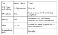 [0723] Heat sealed. Closed the luer connection retaining the clamp [0724] Labeled and placed the cell suspension in the incubator and recorded time placed in the incubator. [0725] Calculated new volume. [0726] Recorded Volume in 2 L transfer pack based on volume of cell suspension and volume removed for QC (11 mL). [0727] Inoculated and ordered sterility testing. [0728] Stored the mycoplasma sample at 4° C in the pending rack for mycoplasma testing. [0729] Set aside until TIL was seeded. [00537] Cell Count: [00538] Performed single cell counts and recorded data and attach counting raw data to batch record. Documented Dilution. Documented the Cellometer counting program. Verified the correct dilution was entered into the Cellometer. [00539] Method continued: [0730] Calculated the total number of flasks required for subculture **Re-used the original vessel and rounded fractions of additional vessels up. [00540] IL-2 addition to CM [0400] Placed 10L bag of Aim V with Glutamax in the BSC. [0401] Spiked the media bag with a 4” plasma transfer set. [0402] Attached an 18G needle to a 10mL syringe and draw 5mL of IL-2 into the syringe (final concentration is 3000 IU/ml). [0403] Removed the needle and aseptically attach the syringe to the plasma transfer set and dispensed IL-2 into the bag. [0404] Flushed the line with air, draw up some media and dispense into the bag. This insured all IL-2 is in the media. [0405] Repeated for remaining bags of Aim V. [00541] Prepare G-REX500MCS Flasks [0500] Determined amount of CM4 to add to flasks. Recorded volume of cells added per flask and volume of CM45000mL-A. [0501] Closed all clamps except the large filter line. [0502] Sterile welded the inlet line of the Acacia pump to the 4” plasma transfer set on the media bag containing CM4. [0503] Sterile welded the outlet line of the pump to the G-Rex 500MCS via the red collection line. [0504] Pump determined amount of CM4 into the G-Rex 500MCS using lines on flask as guide. [0505] Heated seal the G-Rex 500MCS red line. [0506] Repeated steps 4-6 for each flask. Multiple flasks could be filled at the same time using gravity fill or multiple pumps. A “Y” connector could be welded to the outlet line of the pump and the two arms welded to two G-Rex 500MCS flasks filling both at the same time. [0507] Placed flasks in a 37°C, 5% CO2. [00542] Seed Flasks With TIL [0600] Closed all clamps on G-Rex 500MCS except large filter line [0601] Sterile welded cell product bag to inlet line of the Acacia pump. [0602] Sterile welded the other end of the pump to the red line on the G-Rex 500MCS. [0603] Placed pump boot in pump. [0604] Placed the cell product bag on analytical balance and recorded time TIL added to G-REX flask. [0605] Zeroed the balance. [0606] Unclamped lines and pump required volume of cells into G-Rex 500MCS by weight assuming 1g=1mL. [0607] Turned cell bag upside down and pump air to clear the line. Heated seal red line of G-Rex 500MCS. Placed flask in incubator. [0608] Sterile welded the outlet line of the pump to the next G-Rex 500MCS via the red collection line [0609] Mixed cells well. [0610] Repeated cell transfer for all flasks. [0611] Placed flasks in a 37°C, 5% CO2 and recorded time TIL added to G_REX flask. [0612] Ordered testing for settle plates to the microbiology lab. [0613] Recorded accession numbers. [0614] Ordered testing for aerobic and anaerobic sterility. [0615] Ensured delivery of plates and bottles to the microbiology lab. [00543] Cryopreservation of Flow or Excess Cells: • Calculated amount of freezing media required: o Target cell concentration was 1 x 10⁸/ml; record total cells removed. Target cell concentration was 1x10⁸ cells/mL. Calculated total volume of freezing media to add. • Prepared cryo preservation media and placed at 40°C until needed. • Spun down TIL at 400 x g for 5 min at 20°C with full brake and full acceleration. • Aseptically aspirated supernatant. • Gently tapped bottom of tube to resuspend cells in remaining fluid. • While gently tapping the tube slowly added prepared freezing media. • Aliquoted into appropriate sized labelled cryo tubes. • Placed vial in a Mr. Frosty or equivalent and placed in a -80°C freezer. • Within 72 hours transferred to permanent storage location and documented and recorded date and time placed in -80°C freezer. EXAMPLE 4: PROCESS 2A – DAY 22 [00544] This example describes the detailed day 22 protocol for the 2A process described in Examples 1 to 4. [00545] Document Negative In-Process Sterility Results [00546] Before beginning harvest, obtained the Day 16 preliminary sterility results from Microbiology lab. Contacted the Laboratory Director or designee for further instructions if the results were positive. [00547] Clean Room Environmental Monitoring - Pre-Processing • Verified Particle Counts for 10 minutes before beginning processing. • Biosafety Cabinets were cleaned with large saturated alcohol wipes or alcohol spray. • Set up in-process surveillance plates and left in biosafety cabinet for 1-2 hour during procedure. [00548] Advanced Preparation 1. In BSC aseptically attached a Baxter extension set to a 10L labtainer bag or equivalent. Label LOVO filtrate bag. 2. Placed three 1L bags of PlasmaLyte A in the BSC 3. Prepared pool and labeled the PlasmaLyte A bags with 1% HSA: a. Closed all clamps on a 4S-4M60 Connector set and spiked each of the PlasmaLyte bags. b. Welded one of the male ends of the 4S-4M60 to the inlet line of the Acacia pump boot. c. Welded the outlet line of the pump boot to a 3 liter collection bag. Closed all clamps on 3L bag except the line to pump. d. Pumped the 3 liters of Plasmalyte into the 3 liter bag. If necessary removed air from 3L bag by reversing the pump. e. Closed all clamps except line with female luer. f. Using two 100 mL syringes and 16-18G needles, load 120 mL of 25% HSA. Red capped syringes. g. Attached one syringe to the female luer on the 3 liter bag and transferred HSA to 3L PlasmaLyte bag. Mix well. h. Repeated with second syringe. i. Mixed well. j. Closed all clamps. k. Using a 10mL syringe, removed 5 mL of PlasmaLyte with 1%HSA from the needleless port on the 3 liter bag. l. Capped syringe and kept in BSC for IL-2 dilution. m. Closed all clamps. n. Heated seal removing the female luer line from the pump boot. o. Labeled LOVO Wash buffer and date. Expired within 24 hrs at ambient temperature. [00549] IL-2 Preparation 1.0 Dispensed Plasmalyte/1%HSA from 5 mL syringe into a labeled 50 ml sterile conical tube. 2.0 Added 0.05mL IL-2 stock to the tube containing PlasmaLyte. 3.0 Labeled IL-26X104 4.0 Capped label and store at 2-8°C. Record volumes. [00550] Preparation of Cells 7 Closed all clamps on a 10 L Labtainerbag. At Attach Baxter extension set to the 10L bag via luer connection. 8 Removed the G-REX 500M flasks from the 37°C 9 Sterile welded the red media removal line from the G-Rex 500MCS to the extension set on the10L bioprocess bag. 10 Sterile welded the clear cell removal line from the G-Rex 500MCS to a 3L collection bag and labeled “pooled cell suspension”. 11 Unclamped red line and 10L bag. 12 Used the GatheRex pump, volume reduced the first flask. Note: If an air bubble was detected then the pump could stop prematurely. If full volume reduction was not complete reactivated GatheRex pump. 13 Closed the clamp on the supernatant bag and red line. 14 Swirled the G-REX 500M flask until the TIL were completely resuspended while avoiding splashing or foaming. Made sure all cells have been dislodged from the membrane. 15 Opened clamps on clear line and 3L cell bag. 16 Tilted the G-Rex flask such that the cell suspension was pooled in the side of the flask where the collection straw was located. 17 Started GatherRex to collect the cell suspension. Note: If the cell collection straw was not at the junction of the wall and bottom membrane, rapping the flask while tiled at a 45⁰ angle was usually sufficient to properly position the straw. 18 Ensured all cells had been removed from the flask. 19 If cells remained in the flask, added 100mL of supernatant back to the flask, swirled, and collected into the cell suspension bag. 20 Closed clamp on the line to the cell collection bag. Released clamps on GatheRex. 21 Heated seal clear line of G-Rex 500MCS. 22 Heated seal red line of G-Rex 500MCS. 23 Repeated steps 3-16 for additional flasks. 24 It was necessary to replace 10L supernatant bag as needed after every 2nd flask. 25 Multiple GatherRex could be used. 26 Documented number of G-Rex 500MCS processed. 27 Heated seal cell collection bag. Recorded number of G-REX harvested. 28 With a marker made a mark ~2” from one of the female luer connectors on a new 3 liter collection bag. 29 Heated seal and removed the female luer just below the mark. 30 Labeled as LOVO Source Bag 31 Recorded the dry weight. 32 Closed all clamps of a 170 µm blood filter. 33 Sterile welded the terminal end of the filter to the LOVO source bag just below the mark. 34 Sterile welded a source line of the filter to the bag containing the cell suspension. 35 Elevated the cell suspension by hanging cells on an IV pole to initiate gravity-flow transfer of cells. (Note: Did not allow the source bag to hang from the filtration apparatus.) 36 Opened all necessary clamps and allow TIL to drain from the cell suspension bag through the filter and into the LOVO source bag. 37 Once all cells were transferred to the LOVO source bag, closed all clamps, heated seal just above the mark and detached to remove filter. 38 Mixed bag well and using a two 3mL syringe take 2 independent 2 mL samples from the syringe sample port for cell counting and viability. 39 Weighed the bag and determined the difference between the initial and final weight. 40 Recorded data and place in incubator, including dry mass. [00551] Cell Count. [00552] Performed a single cell count on each sample and recorded data and attach counting raw data to batch record. Documented the Cellometer counting program. Verified the correct dilution was entered into the Cellometer. Determined total number of nucleated cells. Determined number of TNC to remove to retain = 1.5 X 10¹¹ cells for LOVO processing. Place removed cell into appropriate size container for disposal. [00553] LOVO Harvest [00554] The 10L Labtainer with Baxter extension set in Prior Preparation was the replacement filtrate bag welded to the LOVO kit later on. Turned LOVO on and follow the screen displays. [00555] Check weigh scales and pressure sensor [00556] To access the Instrument Operation Profile: • Touched the information button. • Touched the instrument settings tab. • Touched the Instrument Operation Profile button. • The Instrument Operation Profile displayed. [00557] Check the weigh scales • Made sure there was nothing hanging on any of the weigh scales and reviewed the reading for each scale. • If any of the scales read outside of a range of 0 +/- 2 g, performed weigh scale calibration as described in the Weigh Scale Calibration Manual from the manufacturer. • If all scales were in tolerance with no weight hanging, proceed to hang a 1-kg weight on each scale (#1-4) and reviewed the reading. • If any of the scales read outside of a range of 1000 +/- 10 g when a 1- kg weight was hanging, performed weigh scale calibration as described in the LOVO Operator’s Manual from the manufacturer. [00558] Check the pressure sensor • Reviewed the pressure sensor reading on the Instrument Operation Profile Screen. • N/A: If the pressure sensor reading was outside 0 +/- 10 mmHg, stored a new atmospheric pressure setting in Service Mode as described in the LOVO Operator’s Manual from the manufacturer. 1.1 Touched the check button on the Instrument Operation Profile screen. 1.2 Touched the check button on the Instrument Settings tab. • If weigh scale calibration had been performed or a new atmospheric pressure setting had been stored, repeated the relevant sections. [00559] To start the procedure, selected the “TIL G-Rex Harvest” protocol from the drop- down menu on the Protocol Selection Screen and press Start. 1.0 The Procedure Setup Screen displayed. 2.0 Touched the Solutions Information button. 3.0 The Solution 1 Screen displayed. Review the type of wash buffer required for Solution 1. (Should read PlasmaLyte.) 4.0 Touched the Next button to advance to the Solution 2 Screen. Reviewed the type of wash buffer required for Solution 2. (Should read “NONE”, indicating that the protocol had been configured to only use one type of wash buffer, which was PlasmaLyte) 5.0 Touched the check button on the Solution 2 Information Screen to return to the Procedure Setup screen. 6.0 Touched the Procedure Information Button. 7.0 The Procedure Information Screen displayed. 8.0 Touched the User ID entry field. A keypad will display. Entered the initials of the performer and verifier. Touched the button to accept the entry. 9.0 Touched the Source ID entry field. A keypad will display. Entered the product lot #. Touched the button to accept the entry. 10.0 Touched the Procedure ID entry field. A keypad will display. Entered “TIL Harvest”. Touched the button to accept the entry. 11.0 If there are extra notes to record, touched the Procedure Note entry field. A keypad displayed. Entered any notes. Touched the button to accept the entry. NOTE: The Procedure Note entry field is optional and can be left blank. 12.0 Touched the check button on the Procedure Information Screen to return to the Procedure Setup Screen. 13.0 Verified that a “check” displays in the Procedure Information button. If no “check” displays, touched the Procedure Information button again and ensured that the User ID, Source ID, and Procedure ID fields all had entries. 14.0 Touched the Parameter Configuration Button. 15.0 The General Procedure Information Screen displayed. 16.0 Touched the Source Volume (mL) entry field. A numeric keypad displayed. Entered the Calculated volume of cell suspension (mL) from Table 1 17.0 Touched the button to accept the entry. 18.0 Touched the Source PCV (%) entry field. The TIL (viable+dead) screen displays. 19.0 Touched the Cell Concentration entry field. A numeric keypad displayed. Entered the Total Cellular concentration/mL from Table 14 in the Source product in units of “x 106/mL”. The entry could range from 00.0 to 99.9. Touched the button to accept the entry and return to the General Procedure Information Screen. NOTE: After the Cell Concentration was accepted, the Source PCV (%) entry field on the General Procedure Information Screen displayed the PCV % calculated by the LOVO, based on the Cell Concentration entry made by the operator. 20.0 On the General Procedure screen, touched the Next button to advance to screen 4 of 8, the Final Product Volume (Retentate Volume) screen. Note: Screens 2 and 3 did not have any entry fields for the operator to fill in. 21.0 The Final Product Volume (Retentate Volume) screen displayed. 22.0 Using the Total nucleated cells (TNC) value from Table 15, determined the final product target volume in the table below (Table 16). Entered the Final Product Volume (mL) associated with that Cell Range during LOVO Procedure setup. TABLE 15. Determination of Final Product Target Volume.
 TABLE 16. Product target volume.
 23.0 To target the specified volume from Table 16 touched the Final Product Volume (mL) entry field. A numeric keypad displayed. Entered the desired Final Product Volume in unit of mL. Touched the button to accept the entry. 24.0 Touched on the Final Product Volume (Retentate Volume) screen to return to the Procedure Setup Screen. Note: Screens 5-8 did not have any entry fields for the operator to fill in. 25.0 Verified that a “Check” displays in the Parameter Configuration button. If no “check” displays, touched the Procedure Information button again and ensured that Source Volume and Source PCV on page 1 have entries. Also ensured that either the Target Minimum Final Product Volume checkbox was checked OR the Final Product Volume (mL) field had an entry on page 4. 26.0 Touched the Estimate Button at the top right corner of the screen. 27.0 The Estimation Summary Screen displayed. Confirmed sufficient and accurate values for Source and PlasmaLyte Wash Buffer. 28.0 Loaded the disposable kit: Followed screen directions for kit loading by selecting help button “(?)”. 29.0 Made a note of the volumes displayed for Filtrate and Solution 1 (read PlasmaLyte) 30.0 Made a note of the volumes displayed for Filtrate and Solution 1 (read PlasmaLyte). 31.0 For instructions on loading the disposable kit touched the help button or followed instructions in operators manual for detailed instructions. 32.0 When the standard LOVO disposable kit had been loaded, touched the Next button. The Container Information and Location Screen displays. Removed filtrate bag from scale #3. 33.0 For this protocol, the Filtrate container was New and Off Scale 34.0 If the Filtrate container was already shown as New and Off-Scale, no changes were made. 35.0 If the Filtrate container type was shown as Original, touched the Original button to toggle to New. 36.0 If the Filtrate location was shown as On-Scale, touched the On-Scale button to toggle to Off-Scale. 37.0 If the volume of Filtrate to be generated was ≤ 2500 mL, the Filtrate Container Location was shown as On-Scale For consistency among runs, the Filtrate Container Location was changed to Off-Scale and container type was “new”. 38.0 Touched the On-Scale button to toggle to Off-Scale. Attached transfer set Use sterile welding technique to replace the LOVO disposable kit Filtrate container with a 10-L bag. Opened the weld. 39.0 Placed the Filtrate container on the benchtop. Did NOT hang the Filtrate bag on weigh scale #3. Weigh scale #3 was empty during the procedure. 40.0 Opened any plastic clamps on the tubing leading to the Filtrate container. NOTE: If the tubing was removed from the F clamp during welding, replaced in clamp. 41.0 Touched the Filtrate Container Capacity entry field. A numeric keypad displayed. Entered the total new Filtrate capacity (10,000 mL). Touched the “check” button to accept the entry. 42.0 Used sterile welding technique to replace the LOVO disposable kit Filtrate container with a 10-L bag. Opened the weld. Note: If tubing was removed from the F clamp during welding, replaced in clamp. 43.0 Placed the new Filtrate container on the benchtop. Did NOT hang the Filtrate bag on weigh scale #3. Weigh scale #3 was empty during the procedure 44.0 Opened any plastic clamps on the tubing leading to the Filtrate container. 45.0 For the Retentate container, the screen displayed Original and On-Scale. 46.0 No changes were made to the Retentate container. 47.0 When all changes were made to the Filtrate container and appropriate information entered, touched the Next button. 48.0 The Disposable Kit Dry Checks overlay displays. Checked that the kit had been loaded properly, then pressed the Yes button. 49.0 All LOVO mechanical clamps closed automatically and the Checking Disposable Kit Installation screen displayed. The LOVO went through a series of pressurizing steps to check the kit. 50.0 After the disposable kit check passed successfully, the Connect Solutions screen displayed. 51.0 3L was the wash volume. Entered this value on screen. 52.0 Used sterile welding technique to attach the 3-L bag of PlasmaLyte to the tubing passing through Clamp 1. Opened the weld. 53.0 Hung the PlasmaLyte bag on an IV pole, 54.0 Opened any plastic clamps on the tubing leading to the PlasmaLyte bag. 55.0 Verified that the Solution Volume entry is 3000mL. This was previously entered. 56.0 Touched the Next button. The Disposable Kit Prime overlay displayed. Verified that the PlasmaLyte was attached and any welds and plastic clamps on the tubing leading to the PlasmaLyte were open, then touched the Yes button. NOTE: Because only one type of wash buffer (PlasmaLyte) was used during the LOVO procedure, no solution was attached to the tubing passing through Clamp 2. The Roberts clamp on this tubing remained closed during the procedure. 57.0 Disposable kit prime started and the Priming Disposable Kit Screen displayed. Visually observed that PlasmaLyte moving through the tubing connected to the bag of PlasmaL Lyte. If no fluid was moving, pressed the Pause Button on the screen and determined if a clamp or weld was still closed. Once the problem had been solved, pressed the Resume button on the screen to resume the Disposable Kit Prime. 58.0 When disposable kit prime finished successfully, the Connect Source Screen displayed. 59.0 For this protocol, the Source container was New and Off-Scale 60.0 If the Source container was already shown as New and Off-Scale , no changes were made. 61.0 If the Source location was shown as On-Scale, touched the On-Scale button to toggle to Off-Scale. 62.0 Touched the Source Capacity (mL) entry field. A numeric keypad displayed. Enter the capacity of the container that held the Source product. Touched the check button to accept the entry. Note: The Source Capacity entry was used to make sure that the Source bag was able to hold the additional solution that was added to the bag during the Source Prime phase. 63.0 Used sterile welding technique to attach the Source container to the tubing passing through Clamp S. Opened the weld. Remove the tubing from the clamp as needed. 64.0 Made sure to replace source tubing into the S clamp. 65.0 Touched the Next button. The Source Prime overlay displayed. Verified that the Source was attached to the disposable kit and any welds and plastic clamps on the tubing leading to the Source were open, then touched the Yes button. 66.0 Source prime started and the Priming Source Screen displayed. Visually observed that PlasmaLyte was moving through the tubing attached to the Source bag. If no fluid was moving, pressed the Pause Button on the screen and determined if a clamp or weld was still closed. Once the problem had been solved, pressed the Resume button on the screen to resume the Source Prime. 67.0 When Source prime finished successfully, the Start Procedure Screen displayed. 68.0 Pressed the Start button. The “Pre-Wash Cycle 1” pause screen appeared, with the instructions to “Coat IP, Mix Source”. 69.0 Pre-coated the IP bag. 70.0 Before pressing the Start button, removed the IP bag from weigh scale #2 (could also remove tubing from the IP top port tubing guide) and manually inverted it to allow the wash buffer added during the disposable kit prime step to coat all interior surfaces of the bag. 71.0 Re-hung the IP bag on weigh scale #2 (label on the bag faced to the left). Replaced the top port tubing in the tubing guide, if it was removed. 72.0 Mixed the Source bag. 73.0 Before pressing the Start button, removed the Source bag from weigh scale #1 and inverted it several times to create a homogeneous cell suspension. 74.0 Rehung the Source bag on weigh scale #1 or the IV pole. Made sure the bag was not swinging. 75.0 Pressed the Start button. 76.0 The LOVO started processing fluid from the Source bag and the Wash Cycle 1 Screen displayed. [00560] During the LOVO procedure, the system automatically paused to allow the operator to interact with different bags. Different screens displayed during different pauses. Followed the corresponding instructions for each screen. [00561] Source Rinse Pause [00562] After draining the Source bag, the LOVO added wash buffer to the Source bag to rinse the bag. After the configured volume of wash buffer had been added to the Source bag, the LOVO paused automatically and displayed the Source Rinse Paused Screen. [00563] When the Source Rinse Paused Screen displayed, the operator: • Removed the Source bag from weigh scale #1. • Inverted the Source bag several times to allow the wash buffer to touch the entire bag interior. • Re-hung the Source bag on weigh scale #1. Made sure the Source bag is not swinging on weigh scale #1. • Pressed the Resume button. [00564] The LOVO processed the rinse fluid from the Source bag, then continued with the automated procedure. [00565] Mix IP bag pause [00566] To prepare cells for another pass through the spinner, the IP bag was diluted with wash buffer. After adding the wash buffer to the IP bag, the LOVO paused automatically and displayed the “Mix IP bag” Pause Screen. [00567] When the “Mix IP bag” Pause Screen displayed, the operator: • Removed the IP bag from weigh scale #2. Could also remove the tubing from the IP top port tubing guide. • Inverted the IP bag several times to thoroughly mix the cell suspension. • Re-hung the IP bag on weigh scale #2. Also replaced the IP top port tubing in the tubing guide, if it was removed. Made sure the IP bag was not swinging on weigh scale #2. • Pressed the Resume button. The LOVO began processing fluid from the IP bag. [00568] Massage IP corners pause [00569] During the final wash cycle of the LOVO procedure, cells were pumped from the IP bag, through the spinner, and to the Retentate (Final Product) bag. When the IP bag was empty, 10 mL of wash buffers was added to the bottom port of the IP bag to rinse the bag. After adding the rinse fluid, the LOVO paused automatically and displayed the “Massage IP corners” Pause Screen. [00570] When the “Massage IP corners” Pause Screen displayed, the operator: 1. Did NOT remove the IP bag from weigh scale #2. 2. With the IP bag still hanging on weigh scale #2, massaged the corners of the bag to bring any residual cells into suspension. 3. Made sure the IP bag was not swinging on weigh scale #2. 4. Pressed the Resume button. 5. The LOVO began pumping out the rinse fluid from the IP bag. [00571] At the end of the LOVO procedure, the Remove Products Screen displayed. When this screen displayed, all bags on the LOVO kit could be manipulated. Note: Did not touch any bags until the Remove Products Screen displays. [00572] Placed a hemostat on the tubing very close to the port on the Retentate bag to keep the cell suspension from settling into the tubing and triple heat sealed below the hemostat. [00573] Removed the retentate bag by breaking the middle seal and transferred to the BSC. [00574] Followed the instructions on the Remove Products Screen [00575] Touched the Next button. All LOVO mechanical clamps opened and the Remove Kit Screen displayed. [00576] Followed the instructions on the Remove Kit screen. When completed proceeded. [00577] Touched the Next button. All LOVO mechanical clamps closed and the Results Summary Screen displayed. Recorded the data from the results summary screen in Table 17. Closed all pumps and filtered support. TABLE 17. LOVO results summary table.
 [00578] Touched the Next button. The Protocol Selection Screen displayed. [00579] LOVO Shutdown procedure • Ensured all clamps were closed and filter support is in the upright position. • Touched the STOP button on the front of the LOVO. • The STOP Button Decision Overlay displayed. • The Shutdown Confirmation Overlay displayed. • Touched the Yes button. The Shutting Down Screen displayed. • After a few seconds, the Power Off Screen displayed. When this screen displayed, turned off the LOVO using the switch on the back left of the instrument. [00580] Recorded final formulated product volume in a table. [00581] Calculate amount of IL-2 required from final product table
 [00582] Determined the number of Cryobags and Retain Volume [00583] Marked on the Target volume and retain table below the number of cryopreservation bags and volume of retention sample for product. [00584] Targeted volume/bag calculation: (Final formulated volume – volume adjustment due to not getting 100% recovery=10 mL)/# bags. [00585] Prepared cells with 1:1 (vol:vol) CS10 (CryoStor 10, BioLife Solutions) and IL-2. R 1 Assemble Connect apparatus o Sterile welded the CS750 cryobags to the CC2 Cell Connect apparatus replacing one of the distal male luer ends for each bag. o Retained the clamps in the closed position. o Labeled the bags 1-4. • Prepared cells with IL-2 and connected apparatus. o In BSC spike the cell product bag with a 4” plasma transfer set with female luer connector. Be sure the clamp was closed on the transfer set. o With an appropriate size syringe drew up the volume of IL-2 working dilution determined from the Final Product Table. o Dispensed into LOVO product. o Sterile welded LOVO product bag to CC2 single spike line removing the spike. o Placed cells and apparatus in transport bag and place at 2-8 °C for ≤ 15 min. • Addition of CS10 o In BSC attached 3 way stopcock to male luer on bag of cold CS10. o Attached appropriate size syringe to female luer of stopcock. o Unclamped bag and drew up the amount of CS10 determined in the “Final Formulated Product Volume” table. o Removed syringe and red capped. o Repeated if multiple syringes were required. o Removed cell/CC2 apparatus from 2-8 °C refrigerator and placed in BSC. o Attached first syringe containing CS10 to middle luer of stopcock. Turned stopcock so line to CS750 bags is in “OFF” position. o Slowly and with gentle mixing, added CS10 (1:1, vol:vol) to cells. o Repeated for additional syringes of CS10. [00586] Addition of Formulated Cell Product into Cryobags • Replaced syringe with appropriate size syringe for volume of cells to be placed in each cryo bag. • Mixed cell product. • Opened the clamp leading to the cell product bag and drew up appropriate volume • Turned stopcock so cell product bag is in “OFF” position and dispensed the contents of the syringe into cryobag #1. Cleared the line with air from syringe. [00587] Record final product volume Appendix A. Using needless port and appropriate size syringe, drew up amount of retain determined previously. Appendix B. Place retained in 50 mL conical tube labelled “Retain” Appendix C. Using the syringe attached to the harness removed all air from bag drawing up cells to about 1” past bag into tubing. Clamped and heat sealed. Placed at 2-8 °C. Appendix D. Turned stopcock so cryo bags were in the “OFF” position Appendix E. Mixed cells in cell product bag and repeat steps 3-8 for remaining CS750 bags using a new syringe on the stopcock and new syringe to obtain cell retain. Appendix F. Retained should be set aside for processing once product was in CRF. [00588] Controlled-rate freezer (CRF) procedure (see also Example 9) 1. Turned on the CRF (CryoMed Controlled Rate Freezer, Model 7454) and associated laptop computer. 2. Logged onto the computer using account and password 3. Opened Controlled Rate Freezer icon located on the desktop. 4. Clicked the Run button on the Main screen. 5. Clicked Open Profile, Click Open. 6. Entered the Run File Name followed by the date in this format: runMMDDYYYY. 7. Entered the Data Tag as the date with no dashes as MMDDYYY. 8. Closed door to the CRF. 9. Clicked Start Run. 10. Selected COM 6 on the pull down menu. 11. Clicked Ok. Waited about 30 seconds. 12. When “Profile Download,” pops up, Clicked OK. Clicked Save. (See Example 9 for controlled-rate freezing profile details.) 13. Waited to press green button until the samples were in the CRF. The freezer was held at 4 °C until ready to add them. 14. Added samples to CRF. 15. Waited until CRF returns to 4 °C. Once temperature was reached, clicked the green continue button. This initiated program to go to next step in program. 16. Performed a visual inspection of the cryobags for the following (Note: did not inspect for over or underfill): container integrity, port integrity, seal integrity, presence of cell clumps, and presence of particles. [0616] Placed approved hang tag labels on each bag. [0617] Verified final product label including: Lot number, product name, manufacturer date, product volume, other additives, storage temperature, and expiration. [0618] Placed each cryobag (with hangtag) into an over-bag. [0619] Heat sealed. [0620] Placed in a cold cassette. [0621] Repeated for each bag. [0622] Placed the labeled cryobags into preconditioned cassettes and transferred to the CRF. [0623] Evenly distributed the cassettes in the rack in the CRF. [0624] Applied ribbon thermocouple to the center cassette, or place dummy bag in center position. [0625] Closed the door to the CRF. [0626] Once the chamber temperature reached 4 °C +/- 1.5 °C, Press Run on the PC Interface software. [0627] Recorded the time and the chamber temperature that the product is transferred to the CRF. [00589] Processing of quality control sample 1) Aseptically transferred the following materials to the BSC, as needed, and labeled according to the table below: 2) Used a new pipette for pipette the following: QC and Retention Table 3) Delivered to QC: 1 -Cell Count tube, 1- Endotoxin tube, 1-Mycoplasma tube, 1-Gram stain tube, 1 tube restimulation tube, and 1- flow tube to QC for immediate testing. The remaining duplicate tubes were placed in the controlled rate freezer. 4) Contacted the QC supervisor notifying of required testing. 5) See Table 18 for testing and storage instructions. TABLE 18. Testing and storage instructions.
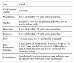 [00590] Cell Count [00591] Performed a single cell count on each sample and recorded data and attached counting raw data to batch record. Document the Cellometer counting program. Verified the correct dilution was entered into the Cellometer. [00592] Cryopreservation of Post Formulation Retention Cells: [00759] Placed vial in CRF. [00760] Moved to storage location after completion of freeze and recorded date and time placed in CFR. Recorded date and time moved to LN2. [00593] Microbiology testing • Ordered testing for settle plates to the microbiology lab. • Recorded accession numbers. • Ordered testing for aerobic and anaerobic sterility. • Ensured delivery of plates and bottles to the microbiology lab. [00594] Post-Cryopreservation of Cell Product Bags • Stopped the freezer after the completion of the run. Run could be stopped by clicking on the Stop button or pressing the Back key on the freezer keypad. • Removed cryobags from cassette • Transferred cassettes to vapor phase LN2. • Recorded storage location. • Entered any additional comments when the text entry window opens again. This window appeared regardless of the Run stop method. • Printed the profile report and attached to the batch record labeled with the lot number for the run. • Terminated Warm Mode and closed the Run screen with Exit button. EXAMPLE 5: PREPARATION OF MEDIA FOR PRE-REP AND REP PROCESSES [00595] This Example describes the procedure for the preparation of tissue culture media for use in protocols involving the culture of tumor infiltrating lymphocytes (TIL) derived from various tumor types including, but not limited to, metastatic melanoma, head and neck squamous cell carcinoma (HNSCC), ovarian carcinoma, triple-negative breast carcinoma, and lung adenocarcinoma. This media can be used for preparation of any of the TILs described in the present application and Examples. [00596] Definition µg microgram µm micrometer µM micromolar AIM-V® serum-free tissue culture medium (Thermo Fisher Scientific) BSC Biological Safety Cabinet CM1 Complete Medium #1 CM2 Complete Medium #2 CM3 Complete Medium #3 CM4 Complete Medium #4 IU or U International units ml milliliter mM millimolar NA not applicable PPE personal protective equipment Pre-REP pre-Rapid Expansion Process REP Rapid Expansion Process rhIL-2, IL-2 recombinant human Interleukin-2 RPMI1640 Roswell Park Memorial Institute medium, formulation 1640 SOP Standard Operating Procedure TIL tumor infiltrating lymphocytes I. Procedure (a) All procedures are done using sterile technique in a BSC (Class II, Type A2). (i) Sprayed surface of hood with 70% ethanol prior to its use. (ii) Sprayed all items and reagents with 70% ethanol prior to placing them into tissue culture hood. (b) Aliquotting of 200mM L-glutamine (i) L-glutamine was supplied in larger volumes than needed for the preparation of serum (e.g., 100m1 or 500m1 volumes). (ii) Thawed bottle of L-glutamine in 37°C water bath. (iii) Mixed L-glutamine well after thawing, as it precipitates after thaw. Ensured that all precipitates have returned to solution prior to aliquotting. (iv) Placed 5-10m1 aliquots of L-glutamine into sterile 15m1 conical tubes. (v) Labeled tubes with concentration, vendor, lot number, date aliquotted, and expiration date. (vi) Tubes were then stored at -20°C and pulled as needed for media preparation. (c) Preparation of CM1 (i) Removed the following reagents from cold storage and warmed them in a 37°C water bath: (1) RPMI1640 (2) Human AB serum (3) 200mM L-glutamine (ii) Removed the BME from 4°C storage and place in tissue culture hood. (iii) Placed the gentamycin stock solution from room temperature storage into tissue culture hood. (iv) Prepared CM1 medium according to Table 23 below by adding each of the ingredients into the top section of a 0.2um filter unit appropriate to the volume to be filtered. TABLE 23. Preparation of CM1
 (v) Labeled the CM1 media bottle with its name, the initials of the preparer, the date it was filtered/prepared, the two week expiration date and store at 4°C until needed for tissue culture. Media can be aliquotted into smaller volume bottles as required. (vi) Any remaining RPMI1640, Human AB serum, or L-glutamine was stored at 4°C until next preparation of media. (vii) Stock bottle of BME was returned to 4°C storage. (viii) Stock bottle of gentamicin was returned to its proper RT storage location. (ix) Because of the limited buffering capacity of the medium, CM1 was discarded no more than two weeks after preparation, or as the phenol red pH indicator showed an extreme shift in pH (bright red to pink coloration). (x) On the day of use, prewarmed required amount of CM1 in 37°C water bath and add 6000 IU/m1 IL-2. (xi) Additional supplementation - as needed (1) CM1 supplemented with GlutaMAX® a. CM1 could be prepared by substituting 2mM G1utaMAXTM for 2mM glutamine (final concentration, see Table 2.) If this was done, labeled the media bottle as in Step 7.3.5 above adding "2mM G1utaMAX" to prevent confusion with the standard formulation of CM1. (2) CM1 supplemented with extra antibiotic/antimycotic a. Some CM1 formulations required additional antibiotic or antimycotic to prevent contamination of pre-REP TIL grown from certain tumor types. b. Added antibiotic/antimycotic to the final concentrations shown in Table 24 below. c. If this was done, label the media bottle as in Step 7.3.1 above adding the name/s of the additional antibiotic/antimycotic to prevent confusion with the standard formulation of CM1. TABLE 24. Additional supplementation of CM1, as needed.
 (d) Preparation of CM2 (i) Removed prepared CM1 from refrigerator or prepare fresh CM1 as per Section 7.3 above. (ii) Removed AIM-V® from refrigerator. (iii) Prepared the amount of CM2 needed by mixing prepared CM1 with an equal volume of AIM-V® in a sterile media bottle. (iv) Added 3000 IU/ml IL-2 to CM2 medium on the day of usage. (v) Made sufficient amount of CM2 with 3000 IU/ml IL-2 on the day of usage. (vi) Labeled the CM2 media bottle with its name, the initials of the preparer, the date it was filtered/prepared, the two week expiration date and store at 4°C until needed for tissue culture. Media was aliquotted into smaller volume bottles as required. (vii) Returned any CM2 without IL-2 to the refrigerator where it can be stored for up to two weeks, or until phenol red pH indicator shows an extreme shift in pH (bright red to pink coloration). (e) Preparation of CM3 (i) Prepared CM3 on the day it was required for use. (ii) CM3 was the same as AIM-V® medium, supplemented with 3000 IU/ml IL-2 on the day of use. (iii) Prepared an amount of CM3 sufficient to experimental needs by adding IL-2 stock solution directly to the bottle or bag of AIM-V. Mixed well by gentle shaking. Label bottle with "3000 IU/ml IL-2" immediately after adding to the AIM-V. If there was excess CM3, stored it in bottles at 4°C labeled with the media name, the initials of the preparer, the date the media was prepared, and its expiration date (7 days after preparation). (iv) Discarded media supplemented with IL-2 after 7 days storage at 4°C. (f) Preparation of CM4 (i) CM4 was the same as CM3, with the additional supplement of 2mM G1utaMAXTM (final concentration). (1) For every 1L of CM3, added 10m1 of 200mM G1utaMAXTM. (ii) Prepared an amount of CM4 sufficient to experimental needs by adding IL-2 stock solution and G1utaMAXTM stock solution directly to the bottle or bag of AIM-V. Mixed well by gentle shaking. (iii) Labeled bottle with "3000 IL/nil IL-2 and G1utaMAX" immediately after adding to the AIM-V. (iv) If there was excess CM4, stored it in bottles at 4°C labeled with the media name, "G1utaMAX", the initials of the preparer, the date the media was prepared, and its expiration date (7 days after preparation). (v) Discarded media supplemented with IL-2 after 7 days storage at 4°C. EXAMPLE 6: SURFACE ANTIGEN STAINING OF POST REP TIL • PURPOSE [00597] The Example describes the procedure for cell surface staining of post-REP TILs by flow cytometry. This procedure can be applied to any TILs described in the application and Examples. [00598] KEY TERMS AND DEFINITIONS α: Alpha β: Beta μl: Microliter APC: Allophycocyanin Ax647: Alex Fluor 647 BD: Becton Dickinson Company BSA: Bovine Serum Albumin BSC: Biological Safety Cabinet BV421: Brilliant Violet 421 CD: Cluster of Differentiation CST: Cytometer Setup and Tracking Cy: Cyanine DPBS: Dulbecco’s Phosphate Buffered Saline FACS: Fluorescence Activated Cell Sorter FBS: Fetal Bovine Serum FITC: Fluorescein Isothiocyanate FMO: Fluorescence Minus One G: Gram H7: Analog of Cy7 Ml: Milliliter PE: Phycoerythrin PerCP-Cy5.5: Peridinin-Chlorophyll proteins PPE: Personal Protective Equipment REP: Rapid Expansion Protocol SIT: Sample Injection Tube TCR: T Cell Receptor w/v: Weight to Volume Flow Cytometry Antibodies and Stains TABLE 25: Live/Dead Aqua Stain ThermoFisher Catalog # L34966.


Claims
WHAT IS CLAIMED IS: 1. A method for expanding tumor infiltrating lymphocytes (TILs), comprising: performing a first expansion of a first population of TILs obtained from a tumor sample by culturing the tumor sample in a first cell culture medium and IL-2 for about 7-11 days to produce a second population of TILs; and performing a second expansion of the second population of TILs in a second cell culture medium, IL-2, and feeder cells for about 3-11 days to produce a third population of TILs, wherein an anti-CD3 antibody is added to the second cell culture medium about 1 day after the initiation of the second expansion.
2. The method of claim 1, wherein the anti-CD3 antibody is added to the second cell culture medium about 2-4 days after the initiation of the second expansion.
3. The method of claim 1 or 3, wherein the anti-CD3 antibody is added to the second cell culture medium about 2 days after the initiation of the second expansion.
4. The method of any one of claims 1-3, wherein the anti-CD3 antibody is added to the second cell culture medium about 3 days after the initiation of the second expansion.
5. The method of any one of claims 1-4, wherein the anti-CD3 antibody is added to the second cell culture medium about 4 days after the initiation of the second expansion.
6. A method for expanding tumor infiltrating lymphocytes (TILs), comprising: performing a first expansion of a first population of TILs obtained from a tumor sample by culturing the tumor sample in a first cell culture medium and IL-2 for about 7-11 days to produce a second population of TILs; and performing a second expansion of the second population of TILs in a second cell culture medium, IL-2, an anti-CD3 antibody and feeder cells for no more than 10 days to produce a third population of TILs.
7. The method of claim 6, wherein the second expansion step is performed for no more than 9 days.
8. The method of claim 6, wherein the second expansion step is performed for no more than 8 days.
9. The method of claim 6, wherein the second expansion step is performed for no more than 7 days.
10. The method of claim 6, wherein the second expansion step is performed for no more than 6 days.
11. The method of claim 6, wherein the second expansion step is performed for no more than 5 days.
12. The method of claim 6, wherein the second expansion step is performed for no more than 4 days.
13. The method of any one of claims 1-12, wherein the anti-CD3 antibody is added to the second cell culture medium at about 1-30 ng/mL.
14. The method of any one of claims 1-12, wherein the anti-CD3 antibody is added to the second cell culture medium at about 2-10 ng/mL.
15. The method of any one of claims 1-12, wherein the anti-CD3 antibody is added to the second cell culture medium at about 30 ng/mL.
16. The method of any one of claims 1-15, wherein the anti-CD3 antibody is OKT3.
17. The method of any one of claims 1-15, wherein the anti-CD3 antibody is HIT3a.
18. The method of any one of claims 1-17, wherein the second population of TILs is cultured with the feeder cells at a TIL:feeder cell ratio of about 1:50 to about 1:350.
19. The method of any one of claims 1-17, wherein the second population of TILs is cultured with the feeder cells at a TIL:feeder cell ratio of about 1:250.
20. The method of any one of claims 1-17, wherein the second population of TILs is cultured with the feeder cells at a TIL:feeder cell ratio of about 1:350.
21. The method of any one of claims 1-20, wherein the second expansion results in at least a 1000-fold expansion of the TILs.
22. The method of any one of claims 1-20, wherein the viability of the third population of TILs is greater than about 80%.
23. The method of any one of claims 1-20, wherein the viability of the third population of TILs is greater than about 85%.
24. The method of any one of claims 1-23, wherein at least about 40% of the CD8+ T cells of the third population of TILs expresses IFNγ.
25. The method of any one of claims 1-24, wherein the third population of TILs is at least 500-fold greater than the second population of TILs.
26. The method of any one of claims 1-24, wherein the third population of TILs is at least 1000-fold greater than the second population of TILs.
27. The method of any one of claims 1-24, wherein the third population of TILs is at least 1500-fold greater than the second population of TILs.
28. The method of any one of claims 1-27, wherein the third population of TILs comprises sufficient TILs for a therapeutically effective dosage of the TILs.
29. The method of claim 28, where the number of TILs sufficient for a therapeutically effective dosage is from about 1×10⁹ to about 10 ×1010.
30. The method of any one of claims 1-29, wherein the third population of TILs comprises an increased subpopulation of effector T cells and/or central memory T cells relative to the second population of TILs, wherein the effector T cells and/or central memory T cells exhibit one or more characteristics selected from the group consisting of expressing CD27+, expressing CD28+, longer telomeres, increased CD57 expression, and decreased CD56 expression relative to effector T cells, and/or central memory T cells in the second population of cells.
31. The method of claim 30, wherein the effector T cells and/or central memory T cells in the third population of TILs exhibit increased CD57 expression and decreased CD56 expression relative to effector T cells, and/or central memory T cells in the second population of cells.
32. The method of any one of claims 1-31, wherein a portion of the population of TILs are modified TILs each comprising an immunomodulatory agent/composition associated with its surface membrane.
33. The method of claim 32, wherein the immunomodulatory agent/composition comprises a cytokine.
34. The method of claim 33, wherein the cytokine is selected from the group consisting of IL-2, IL-7, IL-12, IL-15, IL-18, IL-21, IL-23, IL-27, IL-4, IL-1α, IL-1β, IL-5, IFNγ, TNF α (TNFa), IFNα, IFNβ, GM-CSF, and GCSF or a biologically active variant thereof.
35. The method of claim 32, wherein the immunomodulatory agent/composition comprises a costimulatory molecule.
36. The method of claim 35, wherein the costimulatory molecule is selected from the group consisting of OX40, CD28, GITR, VISTA, CD40, CD3, and an agonist of CD137.
37. The method of claim 32, wherein the immunomodulatory agent/composition comprises a CD40 agonist (e.g., CD40L or an agonistic CD40 binding domain).
38. The method of any one of claims 32-37, wherein a portion of the population of TILs are modified TILs each comprising an immunomodulatory agent/composition associated with its surface membrane, including combinations of modified TILs according to any one of claims 32-37.
Applications Claiming Priority (2)
| Application Number | Priority Date | Filing Date | Title |
|---|---|---|---|
| US202263429117P | 2022-11-30 | 2022-11-30 | |
| US63/429,117 | 2022-11-30 |
Publications (1)
| Publication Number | Publication Date |
|---|---|
| WO2024118836A1true WO2024118836A1 (en) | 2024-06-06 |
Family
ID=89620015
Family Applications (1)
| Application Number | Title | Priority Date | Filing Date |
|---|---|---|---|
| PCT/US2023/081680CeasedWO2024118836A1 (en) | 2022-11-30 | 2023-11-29 | Processes for production of tumor infiltrating lymphocytes with shortened rep step |
Country Status (1)
| Country | Link |
|---|---|
| WO (1) | WO2024118836A1 (en) |
Citations (100)
| Publication number | Priority date | Publication date | Assignee | Title |
|---|---|---|---|---|
| US4766106A (en) | 1985-06-26 | 1988-08-23 | Cetus Corporation | Solubilization of proteins for pharmaceutical compositions using polymer conjugation |
| US4902502A (en) | 1989-01-23 | 1990-02-20 | Cetus Corporation | Preparation of a polymer/interleukin-2 conjugate |
| US5019034A (en) | 1988-01-21 | 1991-05-28 | Massachusetts Institute Of Technology | Control of transport of molecules across tissue using electroporation |
| US5089261A (en) | 1989-01-23 | 1992-02-18 | Cetus Corporation | Preparation of a polymer/interleukin-2 conjugate |
| US5128257A (en) | 1987-08-31 | 1992-07-07 | Baer Bradford W | Electroporation apparatus and process |
| US5137817A (en) | 1990-10-05 | 1992-08-11 | Amoco Corporation | Apparatus and method for electroporation |
| US5173158A (en) | 1991-07-22 | 1992-12-22 | Schmukler Robert E | Apparatus and methods for electroporation and electrofusion |
| US5206344A (en) | 1985-06-26 | 1993-04-27 | Cetus Oncology Corporation | Interleukin-2 muteins and polymer conjugation thereof |
| US5232856A (en) | 1990-06-25 | 1993-08-03 | Firth Kevin L | Electroporation device |
| US5273525A (en) | 1992-08-13 | 1993-12-28 | Btx Inc. | Injection and electroporation apparatus for drug and gene delivery |
| US5279833A (en) | 1990-04-04 | 1994-01-18 | Yale University | Liposomal transfection of nucleic acids into animal cells |
| US5304120A (en) | 1992-07-01 | 1994-04-19 | Btx Inc. | Electroporation method and apparatus for insertion of drugs and genes into endothelial cells |
| US5318514A (en) | 1992-08-17 | 1994-06-07 | Btx, Inc. | Applicator for the electroporation of drugs and genes into surface cells |
| US5593875A (en) | 1994-09-08 | 1997-01-14 | Genentech, Inc. | Methods for calcium phosphate transfection |
| US5766902A (en) | 1993-08-20 | 1998-06-16 | Therexsys Limited | Transfection process |
| WO1998040510A1 (en) | 1997-03-11 | 1998-09-17 | Regents Of The University Of Minnesota | Dna-based transposon system for the introduction of nucleic acid into dna of a cell |
| US5908635A (en) | 1994-08-05 | 1999-06-01 | The United States Of America As Represented By The Department Of Health And Human Services | Method for the liposomal delivery of nucleic acids |
| US6010613A (en) | 1995-12-08 | 2000-01-04 | Cyto Pulse Sciences, Inc. | Method of treating materials with pulsed electrical fields |
| US6025337A (en) | 1994-06-27 | 2000-02-15 | Johns Hopkins University | Solid microparticles for gene delivery |
| US6056938A (en) | 1995-02-21 | 2000-05-02 | Imarx Pharaceutical Corp. | Cationic lipids and the use thereof |
| WO2001081565A2 (en) | 2000-04-27 | 2001-11-01 | Max-Delbrück-Centrum für Molekulare Medizin | Sleeping beauty, a transposon vector with a broad host range for the genetic transformation in vertebrates |
| US6475994B2 (en) | 1998-01-07 | 2002-11-05 | Donald A. Tomalia | Method and articles for transfection of genetic material |
| US6479626B1 (en) | 1998-03-02 | 2002-11-12 | Massachusetts Institute Of Technology | Poly zinc finger proteins with improved linkers |
| US6534484B1 (en) | 1995-06-07 | 2003-03-18 | Inex Pharmaceuticals Corp. | Methods for encapsulating plasmids in lipid bilayers |
| US6534261B1 (en) | 1999-01-12 | 2003-03-18 | Sangamo Biosciences, Inc. | Regulation of endogenous gene expression in cells using zinc finger proteins |
| US6627442B1 (en) | 2000-08-31 | 2003-09-30 | Virxsys Corporation | Methods for stable transduction of cells with hiv-derived viral vectors |
| US6706289B2 (en) | 2000-10-31 | 2004-03-16 | Pr Pharmaceuticals, Inc. | Methods and compositions for enhanced delivery of bioactive molecules |
| US6746838B1 (en) | 1997-05-23 | 2004-06-08 | Gendaq Limited | Nucleic acid binding proteins |
| US6794136B1 (en) | 2000-11-20 | 2004-09-21 | Sangamo Biosciences, Inc. | Iterative optimization in the design of binding proteins |
| US6838261B1 (en) | 1999-06-08 | 2005-01-04 | Seattle Genetics, Inc. | Nucleic acids encoding anti-CD40 proteins and methods of producing recombinant anti-CD40 proteins |
| US20050106717A1 (en) | 2003-10-08 | 2005-05-19 | Wilson John R. | Cell culture methods and devices utilizing gas permeable materials |
| US7013219B2 (en) | 1999-01-12 | 2006-03-14 | Sangamo Biosciences, Inc. | Regulation of endogenous gene expression in cells using zinc finger proteins |
| US7030215B2 (en) | 1999-03-24 | 2006-04-18 | Sangamo Biosciences, Inc. | Position dependent recognition of GNN nucleotide triplets by zinc fingers |
| WO2006122442A1 (en) | 2005-05-14 | 2006-11-23 | Fudan University | Piggybac as a tool for genetic manipulation and analysis in vertebrates |
| US7189705B2 (en) | 2000-04-20 | 2007-03-13 | The University Of British Columbia | Methods of enhancing SPLP-mediated transfection using endosomal membrane destabilizers |
| US7338660B2 (en) | 2001-11-09 | 2008-03-04 | Abgenix, Inc. | Methods of treating cancer and enhancing immune responses with antibodies that bind CD40 |
| US7585849B2 (en) | 1999-03-24 | 2009-09-08 | Sangamo Biosciences, Inc. | Position dependent recognition of GNN nucleotide triplets by zinc fingers |
| US7687070B2 (en) | 1994-02-11 | 2010-03-30 | Life Technologies Corporation | Reagents for intracellular delivery of macromolecules |
| WO2010085699A2 (en) | 2009-01-23 | 2010-07-29 | The Johns Hopkins University | Mammalian piggybac transposon and methods of use |
| WO2010099296A1 (en) | 2009-02-26 | 2010-09-02 | Transposagen Biopharmaceuticals, Inc. | Hyperactive piggybac transposases |
| WO2010099301A2 (en) | 2009-02-25 | 2010-09-02 | The Johns Hopkins University | Piggybac transposon variants and methods of use |
| US20110052530A1 (en) | 2009-08-28 | 2011-03-03 | The United States Of America, As Represented By The Secretary, Department Of Health And Human Serv | Adoptive cell therapy with young t cells |
| US7943743B2 (en) | 2005-07-01 | 2011-05-17 | Medarex, Inc. | Human monoclonal antibodies to programmed death ligand 1 (PD-L1) |
| US20110136228A1 (en) | 2009-12-08 | 2011-06-09 | Vera Juan F | Methods of cell culture for adoptive cell therapy |
| US20110201118A1 (en) | 2010-06-14 | 2011-08-18 | Iowa State University Research Foundation, Inc. | Nuclease activity of tal effector and foki fusion protein |
| US8008449B2 (en) | 2005-05-09 | 2011-08-30 | Medarex, Inc. | Human monoclonal antibodies to programmed death 1 (PD-1) and methods for treating cancer using anti-PD-1 antibodies alone or in combination with other immunotherapeutics |
| WO2012065086A1 (en) | 2010-11-12 | 2012-05-18 | Nektar Therapeutics | Conjugates of an il-2 moiety and a polymer |
| WO2012129201A1 (en) | 2011-03-22 | 2012-09-27 | The United States Of America, As Represented By The Secretary, Department Of Health And Human Services | Methods of growing tumor infiltrating lymphocytes in gas-permeable containers |
| WO2013059343A1 (en) | 2011-10-17 | 2013-04-25 | Massachusetts Institute Of Technology | Intracellular delivery |
| US20130102075A1 (en) | 2009-12-08 | 2013-04-25 | Juan F. Vera | Methods of cell culture for adoptive cell therapy |
| US20130115617A1 (en) | 2009-12-08 | 2013-05-09 | John R. Wilson | Methods of cell culture for adoptive cell therapy |
| US20130117869A1 (en) | 2011-04-05 | 2013-05-09 | Cellectis S.A. | Method for the generation of compact tale-nucleases and uses thereof |
| US8556882B2 (en) | 2009-04-30 | 2013-10-15 | The United States Of America, As Represented By The Secretary, Department Of Health And Human Services | Inducible interleukin-12 |
| WO2013173835A1 (en) | 2012-05-18 | 2013-11-21 | Wilson Wolf Manufacturing Corporation | Improved methods of cell culture for adoptive cell therapy |
| US20130315884A1 (en) | 2012-05-25 | 2013-11-28 | Roman Galetto | Methods for engineering allogeneic and immunosuppressive resistant t cell for immunotherapy |
| WO2013188427A1 (en) | 2012-06-11 | 2013-12-19 | Wilson Wolf Manufacturing Corporation | Improved methods of cell culture for adoptive cell therapy |
| US8697359B1 (en) | 2012-12-12 | 2014-04-15 | The Broad Institute, Inc. | CRISPR-Cas systems and methods for altering expression of gene products |
| US20140134128A1 (en) | 2010-09-21 | 2014-05-15 | Altor Bioscience Corporation | Multimeric il-15 soluble fusion molecules and methods of making and using same |
| US8778345B2 (en) | 2011-04-29 | 2014-07-15 | Apexigen, Inc. | Anti-CD40 antibodies |
| US8795965B2 (en) | 2012-12-12 | 2014-08-05 | The Broad Institute, Inc. | CRISPR-Cas component systems, methods and compositions for sequence manipulation |
| US20140227237A1 (en) | 2011-09-16 | 2014-08-14 | The Trustees Of The University Of Pennsylvania | Rna engineered t cells for the treatment of cancer |
| US8865406B2 (en) | 2012-12-12 | 2014-10-21 | The Broad Institute Inc. | Engineering and optimization of improved systems, methods and enzyme compositions for sequence manipulation |
| US8889356B2 (en) | 2012-12-12 | 2014-11-18 | The Broad Institute Inc. | CRISPR-Cas nickase systems, methods and compositions for sequence manipulation in eukaryotes |
| US8906616B2 (en) | 2012-12-12 | 2014-12-09 | The Broad Institute Inc. | Engineering of systems, methods and optimized guide compositions for sequence manipulation |
| US20140377739A1 (en) | 2013-06-24 | 2014-12-25 | Wilson Wolf Manufacturing | Closed system device and methods for gas permeable cell culture process |
| WO2015006700A1 (en) | 2013-07-12 | 2015-01-15 | University Of South Alabama | Minimal piggybac vectors for genome integration |
| US8993233B2 (en) | 2012-12-12 | 2015-03-31 | The Broad Institute Inc. | Engineering and optimization of systems, methods and compositions for sequence manipulation with functional domains |
| US20150203871A1 (en) | 2012-06-05 | 2015-07-23 | Cellectis | Transcription Activator-Like Effector (TALE) Fusion Protein |
| WO2015189356A1 (en) | 2014-06-11 | 2015-12-17 | Polybiocept Ab | Expansion of lymphocytes with a cytokine composition for active cellular immunotherapy |
| US9228180B2 (en) | 2007-07-04 | 2016-01-05 | Max-Delbruck-Centrum Fur Molekulare Medizin | Polypeptide variants of sleeping beauty transposase |
| US20160010058A1 (en) | 2013-03-01 | 2016-01-14 | The United States Of America, As Represented By The Secretary, Department Of Health And Human Serv | Methods of producing enriched populations of tumor-reactive t cells from tumor |
| US20160102324A1 (en) | 2013-05-29 | 2016-04-14 | Cellectis | New compact scaffold of cas9 in the type ii crispr system |
| US20160120906A1 (en) | 2013-05-13 | 2016-05-05 | Cellectis | Methods for engineering highly active t cell for immunotheraphy |
| WO2017008063A1 (en) | 2015-07-09 | 2017-01-12 | Massachusetts Institute Of Technology | Delivery of materials to anucleate cells |
| US20170107541A1 (en) | 2014-06-17 | 2017-04-20 | Poseida Therapeutics, Inc. | A method for directing proteins to specific loci in the genome and uses thereof |
| US20170114149A1 (en) | 2014-06-17 | 2017-04-27 | Poseida Therapeutics, Inc. | Methods and compositions for in vivo non-covalent linking |
| WO2017123663A1 (en) | 2016-01-12 | 2017-07-20 | Sqz Biotechnologies Company | Intracellular delivery of complexes |
| US9790490B2 (en) | 2015-06-18 | 2017-10-17 | The Broad Institute Inc. | CRISPR enzymes and systems |
| WO2018081473A1 (en) | 2016-10-26 | 2018-05-03 | Iovance Biotherapeutics, Inc. | Restimulation of cryopreserved tumor infiltrating lymphocytes |
| US9982278B2 (en) | 2014-02-11 | 2018-05-29 | The Regents Of The University Of Colorado, A Body Corporate | CRISPR enabled multiplexed genome engineering |
| US20180187185A1 (en) | 2015-06-17 | 2018-07-05 | Poseida Therapeutics, Inc. | Compositions and methods for directing proteins to specific loci in the genome |
| WO2018129332A1 (en) | 2017-01-06 | 2018-07-12 | Iovance Biotherapeutics, Inc. | Expansion of tumor infiltrating lymphocytes (tils) with tumor necrosis factor receptor superfamily (tnfrsf) agonists and therapeutic combinations of tils and tnfrsf agonists |
| WO2018132496A1 (en) | 2017-01-10 | 2018-07-19 | Nektar Therapeutics | Multi-arm polymer conjugates of tlr agonist compounds and related immunotherapeutic treatment methods |
| US10041077B2 (en) | 2014-04-09 | 2018-08-07 | Dna2.0, Inc. | DNA vectors, transposons and transposases for eukaryotic genome modification |
| US20180245089A1 (en) | 2015-09-04 | 2018-08-30 | Sqz Biotechnologies Company | Intracellular delivery of biomolecules to cells comprising a cell wall |
| WO2018182817A1 (en) | 2017-03-29 | 2018-10-04 | Iovance Biotherapeutics, Inc. | Processes for production of tumor infiltrating lymphocytes and uses of same in immunotherapy |
| US20190010514A1 (en) | 2014-03-11 | 2019-01-10 | Cellectis | Method for generating t-cells compatible for allogenic transplantation |
| US10183979B2 (en) | 2012-06-08 | 2019-01-22 | Alkermes, Inc. | Fusion polypeptides comprising mucin-domain polypeptide linkers |
| WO2019046815A1 (en) | 2017-08-31 | 2019-03-07 | Poseida Therapeutics, Inc. | Transposon system and methods of use |
| WO2019126578A1 (en) | 2017-12-20 | 2019-06-27 | Poseida Therapeutics, Inc. | Compositions and methods for directing proteins to specific loci in the genome |
| WO2019136456A1 (en) | 2018-01-08 | 2019-07-11 | Iovance Biotherapeutics, Inc. | Processes for generating til products enriched for tumor antigen-specific t-cells |
| US20190275133A1 (en) | 2016-11-10 | 2019-09-12 | Nektar Therapeutics | Immunotherapeutic tumor treatment method |
| US10415024B2 (en) | 2012-11-16 | 2019-09-17 | Poseida Therapeutics, Inc. | Site-specific enzymes and methods of use |
| US20200181220A1 (en) | 2017-08-03 | 2020-06-11 | Synthorx, Inc. | Cytokine conjugates for the treatment of proliferative and infectious diseases |
| US20200270334A1 (en) | 2017-05-24 | 2020-08-27 | Novartis Ag | Antibody-cytokine engrafted proteins and methods of use in the treatment of cancer |
| US20200330601A1 (en) | 2019-02-06 | 2020-10-22 | Synthorx, Inc. | IL-2 Conjugates and Methods of Use Thereof |
| US20210038684A1 (en) | 2019-06-11 | 2021-02-11 | Alkermes Pharma Ireland Limited | Compositions and Methods for Cancer Immunotherapy |
| WO2022094473A1 (en) | 2020-11-02 | 2022-05-05 | Simcha IL-18, Inc. | Interleukin-18 variants and methods of use |
| WO2022198141A1 (en)* | 2021-03-19 | 2022-09-22 | Iovance Biotherapeutics, Inc. | Methods for tumor infiltrating lymphocyte (til) expansion related to cd39/cd69 selection and gene knockout in tils |
| WO2022225981A2 (en)* | 2021-04-19 | 2022-10-27 | Iovance Biotherapeutics, Inc. | Chimeric costimulatory receptors, chemokine receptors, and the use of same in cellular immunotherapies |
- 2023
- 2023-11-29WOPCT/US2023/081680patent/WO2024118836A1/ennot_activeCeased
Patent Citations (134)
| Publication number | Priority date | Publication date | Assignee | Title |
|---|---|---|---|---|
| US4766106A (en) | 1985-06-26 | 1988-08-23 | Cetus Corporation | Solubilization of proteins for pharmaceutical compositions using polymer conjugation |
| US5206344A (en) | 1985-06-26 | 1993-04-27 | Cetus Oncology Corporation | Interleukin-2 muteins and polymer conjugation thereof |
| US5128257A (en) | 1987-08-31 | 1992-07-07 | Baer Bradford W | Electroporation apparatus and process |
| US5019034A (en) | 1988-01-21 | 1991-05-28 | Massachusetts Institute Of Technology | Control of transport of molecules across tissue using electroporation |
| US5019034B1 (en) | 1988-01-21 | 1995-08-15 | Massachusetts Inst Technology | Control of transport of molecules across tissue using electroporation |
| US4902502A (en) | 1989-01-23 | 1990-02-20 | Cetus Corporation | Preparation of a polymer/interleukin-2 conjugate |
| US5089261A (en) | 1989-01-23 | 1992-02-18 | Cetus Corporation | Preparation of a polymer/interleukin-2 conjugate |
| US5279833A (en) | 1990-04-04 | 1994-01-18 | Yale University | Liposomal transfection of nucleic acids into animal cells |
| US5232856A (en) | 1990-06-25 | 1993-08-03 | Firth Kevin L | Electroporation device |
| US5137817A (en) | 1990-10-05 | 1992-08-11 | Amoco Corporation | Apparatus and method for electroporation |
| US5173158A (en) | 1991-07-22 | 1992-12-22 | Schmukler Robert E | Apparatus and methods for electroporation and electrofusion |
| US5304120A (en) | 1992-07-01 | 1994-04-19 | Btx Inc. | Electroporation method and apparatus for insertion of drugs and genes into endothelial cells |
| US5273525A (en) | 1992-08-13 | 1993-12-28 | Btx Inc. | Injection and electroporation apparatus for drug and gene delivery |
| US5318514A (en) | 1992-08-17 | 1994-06-07 | Btx, Inc. | Applicator for the electroporation of drugs and genes into surface cells |
| US5766902A (en) | 1993-08-20 | 1998-06-16 | Therexsys Limited | Transfection process |
| US7687070B2 (en) | 1994-02-11 | 2010-03-30 | Life Technologies Corporation | Reagents for intracellular delivery of macromolecules |
| US6025337A (en) | 1994-06-27 | 2000-02-15 | Johns Hopkins University | Solid microparticles for gene delivery |
| US6410517B1 (en) | 1994-06-27 | 2002-06-25 | Johns Hopkins University | Targeted gene delivery system |
| US6110490A (en) | 1994-08-05 | 2000-08-29 | The United States Of America As Represented By The Department Of Health And Human Services | Liposomal delivery system for biologically active agents |
| US5908635A (en) | 1994-08-05 | 1999-06-01 | The United States Of America As Represented By The Department Of Health And Human Services | Method for the liposomal delivery of nucleic acids |
| US5593875A (en) | 1994-09-08 | 1997-01-14 | Genentech, Inc. | Methods for calcium phosphate transfection |
| US6056938A (en) | 1995-02-21 | 2000-05-02 | Imarx Pharaceutical Corp. | Cationic lipids and the use thereof |
| US6534484B1 (en) | 1995-06-07 | 2003-03-18 | Inex Pharmaceuticals Corp. | Methods for encapsulating plasmids in lipid bilayers |
| US6078490A (en) | 1995-12-08 | 2000-06-20 | Cyto Pulse Sciences, Inc. | Method of treating materials with pulsed electrical fields |
| US6010613A (en) | 1995-12-08 | 2000-01-04 | Cyto Pulse Sciences, Inc. | Method of treating materials with pulsed electrical fields |
| US6489458B2 (en) | 1997-03-11 | 2002-12-03 | Regents Of The University Of Minnesota | DNA-based transposon system for the introduction of nucleic acid into DNA of a cell |
| WO1998040510A1 (en) | 1997-03-11 | 1998-09-17 | Regents Of The University Of Minnesota | Dna-based transposon system for the introduction of nucleic acid into dna of a cell |
| US7241574B2 (en) | 1997-05-23 | 2007-07-10 | Gendaq Ltd. | Nucleic acid binding proteins |
| US6746838B1 (en) | 1997-05-23 | 2004-06-08 | Gendaq Limited | Nucleic acid binding proteins |
| US6866997B1 (en) | 1997-05-23 | 2005-03-15 | Gendaq Limited | Nucleic acid binding proteins |
| US7241573B2 (en) | 1997-05-23 | 2007-07-10 | Gendaq Ltd. | Nucleic acid binding proteins |
| US6475994B2 (en) | 1998-01-07 | 2002-11-05 | Donald A. Tomalia | Method and articles for transfection of genetic material |
| US7595376B2 (en) | 1998-03-02 | 2009-09-29 | Massachusetts Institute Of Technology | Poly zinc finger proteins with improved linkers |
| US6479626B1 (en) | 1998-03-02 | 2002-11-12 | Massachusetts Institute Of Technology | Poly zinc finger proteins with improved linkers |
| US6903185B2 (en) | 1998-03-02 | 2005-06-07 | Massachusetts Institute Of Technology | Poly zinc finger proteins with improved linkers |
| US6824978B1 (en) | 1999-01-12 | 2004-11-30 | Sangamo Biosciences, Inc. | Regulation of endogenous gene expression in cells using zinc finger proteins |
| US6607882B1 (en) | 1999-01-12 | 2003-08-19 | Sangamo Biosciences, Inc. | Regulation of endogenous gene expression in cells using zinc finger proteins |
| US6534261B1 (en) | 1999-01-12 | 2003-03-18 | Sangamo Biosciences, Inc. | Regulation of endogenous gene expression in cells using zinc finger proteins |
| US7220719B2 (en) | 1999-01-12 | 2007-05-22 | Sangamo Biosciences, Inc. | Modulation of endogenous gene expression in cells |
| US6933113B2 (en) | 1999-01-12 | 2005-08-23 | Sangamo Biosciences, Inc. | Modulation of endogenous gene expression in cells |
| US6979539B2 (en) | 1999-01-12 | 2005-12-27 | Sangamo Biosciences, Inc. | Regulation of endogenous gene expression in cells using zinc finger proteins |
| US7013219B2 (en) | 1999-01-12 | 2006-03-14 | Sangamo Biosciences, Inc. | Regulation of endogenous gene expression in cells using zinc finger proteins |
| US7585849B2 (en) | 1999-03-24 | 2009-09-08 | Sangamo Biosciences, Inc. | Position dependent recognition of GNN nucleotide triplets by zinc fingers |
| US7030215B2 (en) | 1999-03-24 | 2006-04-18 | Sangamo Biosciences, Inc. | Position dependent recognition of GNN nucleotide triplets by zinc fingers |
| US6838261B1 (en) | 1999-06-08 | 2005-01-04 | Seattle Genetics, Inc. | Nucleic acids encoding anti-CD40 proteins and methods of producing recombinant anti-CD40 proteins |
| US6843989B1 (en) | 1999-06-08 | 2005-01-18 | Seattle Genetics, Inc. | Methods for the treatment and prevention of cancer using anti-CD40 antibodies |
| US7189705B2 (en) | 2000-04-20 | 2007-03-13 | The University Of British Columbia | Methods of enhancing SPLP-mediated transfection using endosomal membrane destabilizers |
| WO2001081565A2 (en) | 2000-04-27 | 2001-11-01 | Max-Delbrück-Centrum für Molekulare Medizin | Sleeping beauty, a transposon vector with a broad host range for the genetic transformation in vertebrates |
| US6627442B1 (en) | 2000-08-31 | 2003-09-30 | Virxsys Corporation | Methods for stable transduction of cells with hiv-derived viral vectors |
| US6706289B2 (en) | 2000-10-31 | 2004-03-16 | Pr Pharmaceuticals, Inc. | Methods and compositions for enhanced delivery of bioactive molecules |
| US6794136B1 (en) | 2000-11-20 | 2004-09-21 | Sangamo Biosciences, Inc. | Iterative optimization in the design of binding proteins |
| US7338660B2 (en) | 2001-11-09 | 2008-03-04 | Abgenix, Inc. | Methods of treating cancer and enhancing immune responses with antibodies that bind CD40 |
| US20050106717A1 (en) | 2003-10-08 | 2005-05-19 | Wilson John R. | Cell culture methods and devices utilizing gas permeable materials |
| US8008449B2 (en) | 2005-05-09 | 2011-08-30 | Medarex, Inc. | Human monoclonal antibodies to programmed death 1 (PD-1) and methods for treating cancer using anti-PD-1 antibodies alone or in combination with other immunotherapeutics |
| WO2006122442A1 (en) | 2005-05-14 | 2006-11-23 | Fudan University | Piggybac as a tool for genetic manipulation and analysis in vertebrates |
| US7943743B2 (en) | 2005-07-01 | 2011-05-17 | Medarex, Inc. | Human monoclonal antibodies to programmed death ligand 1 (PD-L1) |
| US9228180B2 (en) | 2007-07-04 | 2016-01-05 | Max-Delbruck-Centrum Fur Molekulare Medizin | Polypeptide variants of sleeping beauty transposase |
| WO2010085699A2 (en) | 2009-01-23 | 2010-07-29 | The Johns Hopkins University | Mammalian piggybac transposon and methods of use |
| WO2010099301A2 (en) | 2009-02-25 | 2010-09-02 | The Johns Hopkins University | Piggybac transposon variants and methods of use |
| WO2010099296A1 (en) | 2009-02-26 | 2010-09-02 | Transposagen Biopharmaceuticals, Inc. | Hyperactive piggybac transposases |
| US8556882B2 (en) | 2009-04-30 | 2013-10-15 | The United States Of America, As Represented By The Secretary, Department Of Health And Human Services | Inducible interleukin-12 |
| US20110052530A1 (en) | 2009-08-28 | 2011-03-03 | The United States Of America, As Represented By The Secretary, Department Of Health And Human Serv | Adoptive cell therapy with young t cells |
| US20150175966A1 (en) | 2009-12-08 | 2015-06-25 | Juan F. Vera | Methods of cell culture for adoptive cell therapy |
| US20160208216A1 (en) | 2009-12-08 | 2016-07-21 | Juan F. Vera | Methods of cell culture for adoptive cell therapy |
| US20130102075A1 (en) | 2009-12-08 | 2013-04-25 | Juan F. Vera | Methods of cell culture for adoptive cell therapy |
| US20130115617A1 (en) | 2009-12-08 | 2013-05-09 | John R. Wilson | Methods of cell culture for adoptive cell therapy |
| WO2011072088A2 (en) | 2009-12-08 | 2011-06-16 | Wilson Wolf Manufacturing Corporation | Improved methods of cell culture for adoptive cell therapy |
| US20110136228A1 (en) | 2009-12-08 | 2011-06-09 | Vera Juan F | Methods of cell culture for adoptive cell therapy |
| US8956860B2 (en) | 2009-12-08 | 2015-02-17 | Juan F. Vera | Methods of cell culture for adoptive cell therapy |
| US8809050B2 (en) | 2009-12-08 | 2014-08-19 | Wilson Wolf Manufacturing | Methods of cell culture for adoptive cell therapy |
| US20110201118A1 (en) | 2010-06-14 | 2011-08-18 | Iowa State University Research Foundation, Inc. | Nuclease activity of tal effector and foki fusion protein |
| US20140134128A1 (en) | 2010-09-21 | 2014-05-15 | Altor Bioscience Corporation | Multimeric il-15 soluble fusion molecules and methods of making and using same |
| WO2012065086A1 (en) | 2010-11-12 | 2012-05-18 | Nektar Therapeutics | Conjugates of an il-2 moiety and a polymer |
| US20140328791A1 (en) | 2010-11-12 | 2014-11-06 | Nektar Therapeutics | Conjugates of an IL-2 Moiety and a Polymer |
| US20120244133A1 (en) | 2011-03-22 | 2012-09-27 | The United States of America, as represented by the Secretary, Department of Health and | Methods of growing tumor infiltrating lymphocytes in gas-permeable containers |
| WO2012129201A1 (en) | 2011-03-22 | 2012-09-27 | The United States Of America, As Represented By The Secretary, Department Of Health And Human Services | Methods of growing tumor infiltrating lymphocytes in gas-permeable containers |
| US20130117869A1 (en) | 2011-04-05 | 2013-05-09 | Cellectis S.A. | Method for the generation of compact tale-nucleases and uses thereof |
| US8778345B2 (en) | 2011-04-29 | 2014-07-15 | Apexigen, Inc. | Anti-CD40 antibodies |
| US20140227237A1 (en) | 2011-09-16 | 2014-08-14 | The Trustees Of The University Of Pennsylvania | Rna engineered t cells for the treatment of cancer |
| WO2013059343A1 (en) | 2011-10-17 | 2013-04-25 | Massachusetts Institute Of Technology | Intracellular delivery |
| US20140287509A1 (en) | 2011-10-17 | 2014-09-25 | Massachusetts Institute Of Technology | Intracellular Delivery |
| WO2013173835A1 (en) | 2012-05-18 | 2013-11-21 | Wilson Wolf Manufacturing Corporation | Improved methods of cell culture for adoptive cell therapy |
| US20180021379A1 (en) | 2012-05-25 | 2018-01-25 | Cellectis | Methods for engineering allogeneic and immunosuppressive resistant t cell for immunotherapy |
| US20130315884A1 (en) | 2012-05-25 | 2013-11-28 | Roman Galetto | Methods for engineering allogeneic and immunosuppressive resistant t cell for immunotherapy |
| US20150203871A1 (en) | 2012-06-05 | 2015-07-23 | Cellectis | Transcription Activator-Like Effector (TALE) Fusion Protein |
| US10183979B2 (en) | 2012-06-08 | 2019-01-22 | Alkermes, Inc. | Fusion polypeptides comprising mucin-domain polypeptide linkers |
| WO2013188427A1 (en) | 2012-06-11 | 2013-12-19 | Wilson Wolf Manufacturing Corporation | Improved methods of cell culture for adoptive cell therapy |
| US10415024B2 (en) | 2012-11-16 | 2019-09-17 | Poseida Therapeutics, Inc. | Site-specific enzymes and methods of use |
| US8993233B2 (en) | 2012-12-12 | 2015-03-31 | The Broad Institute Inc. | Engineering and optimization of systems, methods and compositions for sequence manipulation with functional domains |
| US8865406B2 (en) | 2012-12-12 | 2014-10-21 | The Broad Institute Inc. | Engineering and optimization of improved systems, methods and enzyme compositions for sequence manipulation |
| US8932814B2 (en) | 2012-12-12 | 2015-01-13 | The Broad Institute Inc. | CRISPR-Cas nickase systems, methods and compositions for sequence manipulation in eukaryotes |
| US8906616B2 (en) | 2012-12-12 | 2014-12-09 | The Broad Institute Inc. | Engineering of systems, methods and optimized guide compositions for sequence manipulation |
| US8945839B2 (en) | 2012-12-12 | 2015-02-03 | The Broad Institute Inc. | CRISPR-Cas systems and methods for altering expression of gene products |
| US8895308B1 (en) | 2012-12-12 | 2014-11-25 | The Broad Institute Inc. | Engineering and optimization of improved systems, methods and enzyme compositions for sequence manipulation |
| US8771945B1 (en) | 2012-12-12 | 2014-07-08 | The Broad Institute, Inc. | CRISPR-Cas systems and methods for altering expression of gene products |
| US8889356B2 (en) | 2012-12-12 | 2014-11-18 | The Broad Institute Inc. | CRISPR-Cas nickase systems, methods and compositions for sequence manipulation in eukaryotes |
| US8697359B1 (en) | 2012-12-12 | 2014-04-15 | The Broad Institute, Inc. | CRISPR-Cas systems and methods for altering expression of gene products |
| US8871445B2 (en) | 2012-12-12 | 2014-10-28 | The Broad Institute Inc. | CRISPR-Cas component systems, methods and compositions for sequence manipulation |
| US8999641B2 (en) | 2012-12-12 | 2015-04-07 | The Broad Institute Inc. | Engineering and optimization of systems, methods and compositions for sequence manipulation with functional domains |
| US8795965B2 (en) | 2012-12-12 | 2014-08-05 | The Broad Institute, Inc. | CRISPR-Cas component systems, methods and compositions for sequence manipulation |
| US20160010058A1 (en) | 2013-03-01 | 2016-01-14 | The United States Of America, As Represented By The Secretary, Department Of Health And Human Serv | Methods of producing enriched populations of tumor-reactive t cells from tumor |
| US20160120906A1 (en) | 2013-05-13 | 2016-05-05 | Cellectis | Methods for engineering highly active t cell for immunotheraphy |
| US20160102324A1 (en) | 2013-05-29 | 2016-04-14 | Cellectis | New compact scaffold of cas9 in the type ii crispr system |
| US20140377739A1 (en) | 2013-06-24 | 2014-12-25 | Wilson Wolf Manufacturing | Closed system device and methods for gas permeable cell culture process |
| WO2014210036A1 (en) | 2013-06-24 | 2014-12-31 | Wilson Wolf Manufacturing Corporation | Closed system device and methods for gas permeable cell culture process |
| WO2015006700A1 (en) | 2013-07-12 | 2015-01-15 | University Of South Alabama | Minimal piggybac vectors for genome integration |
| US9982278B2 (en) | 2014-02-11 | 2018-05-29 | The Regents Of The University Of Colorado, A Body Corporate | CRISPR enabled multiplexed genome engineering |
| US20190010514A1 (en) | 2014-03-11 | 2019-01-10 | Cellectis | Method for generating t-cells compatible for allogenic transplantation |
| US10041077B2 (en) | 2014-04-09 | 2018-08-07 | Dna2.0, Inc. | DNA vectors, transposons and transposases for eukaryotic genome modification |
| WO2015189357A1 (en) | 2014-06-11 | 2015-12-17 | Polybiocept Ab | Expansion of lymphocytes with a cytokine composition for active cellular immunotherapy |
| WO2015189356A1 (en) | 2014-06-11 | 2015-12-17 | Polybiocept Ab | Expansion of lymphocytes with a cytokine composition for active cellular immunotherapy |
| US20170114149A1 (en) | 2014-06-17 | 2017-04-27 | Poseida Therapeutics, Inc. | Methods and compositions for in vivo non-covalent linking |
| US20170107541A1 (en) | 2014-06-17 | 2017-04-20 | Poseida Therapeutics, Inc. | A method for directing proteins to specific loci in the genome and uses thereof |
| US20180187185A1 (en) | 2015-06-17 | 2018-07-05 | Poseida Therapeutics, Inc. | Compositions and methods for directing proteins to specific loci in the genome |
| US9790490B2 (en) | 2015-06-18 | 2017-10-17 | The Broad Institute Inc. | CRISPR enzymes and systems |
| US20180201889A1 (en) | 2015-07-09 | 2018-07-19 | Massachusetts Institute Of Technology | Delivery of materials to anucleate cells |
| WO2017008063A1 (en) | 2015-07-09 | 2017-01-12 | Massachusetts Institute Of Technology | Delivery of materials to anucleate cells |
| US20180245089A1 (en) | 2015-09-04 | 2018-08-30 | Sqz Biotechnologies Company | Intracellular delivery of biomolecules to cells comprising a cell wall |
| WO2017123663A1 (en) | 2016-01-12 | 2017-07-20 | Sqz Biotechnologies Company | Intracellular delivery of complexes |
| WO2018081473A1 (en) | 2016-10-26 | 2018-05-03 | Iovance Biotherapeutics, Inc. | Restimulation of cryopreserved tumor infiltrating lymphocytes |
| US20190275133A1 (en) | 2016-11-10 | 2019-09-12 | Nektar Therapeutics | Immunotherapeutic tumor treatment method |
| WO2018129332A1 (en) | 2017-01-06 | 2018-07-12 | Iovance Biotherapeutics, Inc. | Expansion of tumor infiltrating lymphocytes (tils) with tumor necrosis factor receptor superfamily (tnfrsf) agonists and therapeutic combinations of tils and tnfrsf agonists |
| WO2018132496A1 (en) | 2017-01-10 | 2018-07-19 | Nektar Therapeutics | Multi-arm polymer conjugates of tlr agonist compounds and related immunotherapeutic treatment methods |
| WO2018182817A1 (en) | 2017-03-29 | 2018-10-04 | Iovance Biotherapeutics, Inc. | Processes for production of tumor infiltrating lymphocytes and uses of same in immunotherapy |
| US20200270334A1 (en) | 2017-05-24 | 2020-08-27 | Novartis Ag | Antibody-cytokine engrafted proteins and methods of use in the treatment of cancer |
| US20200181220A1 (en) | 2017-08-03 | 2020-06-11 | Synthorx, Inc. | Cytokine conjugates for the treatment of proliferative and infectious diseases |
| WO2019046815A1 (en) | 2017-08-31 | 2019-03-07 | Poseida Therapeutics, Inc. | Transposon system and methods of use |
| WO2019126578A1 (en) | 2017-12-20 | 2019-06-27 | Poseida Therapeutics, Inc. | Compositions and methods for directing proteins to specific loci in the genome |
| WO2019136456A1 (en) | 2018-01-08 | 2019-07-11 | Iovance Biotherapeutics, Inc. | Processes for generating til products enriched for tumor antigen-specific t-cells |
| US20200330601A1 (en) | 2019-02-06 | 2020-10-22 | Synthorx, Inc. | IL-2 Conjugates and Methods of Use Thereof |
| US20210038684A1 (en) | 2019-06-11 | 2021-02-11 | Alkermes Pharma Ireland Limited | Compositions and Methods for Cancer Immunotherapy |
| WO2022094473A1 (en) | 2020-11-02 | 2022-05-05 | Simcha IL-18, Inc. | Interleukin-18 variants and methods of use |
| WO2022198141A1 (en)* | 2021-03-19 | 2022-09-22 | Iovance Biotherapeutics, Inc. | Methods for tumor infiltrating lymphocyte (til) expansion related to cd39/cd69 selection and gene knockout in tils |
| WO2022225981A2 (en)* | 2021-04-19 | 2022-10-27 | Iovance Biotherapeutics, Inc. | Chimeric costimulatory receptors, chemokine receptors, and the use of same in cellular immunotherapies |
Non-Patent Citations (69)
| Title |
|---|
| "Genbank", Database accession no. NM_001207006 |
| "Revised Guideline for Collection of Platelets", PHERESIS, 7 October 1988 (1988-10-07) |
| "Revised Guideline for the Collection of Platelets", PHERESIS, 7 October 1988 (1988-10-07) |
| BEANE ET AL., MOL. THERAPY, vol. 23, 2015, pages 1380 - 1390 |
| BESSARD ET AL., MOLECULAR CANCER THERAPEUTICS, vol. 8, 2009, pages 2736 - 45 |
| BORISH, RESPIR. RES, vol. 2, 2001, pages 66 - 70 |
| CEPKOPEAR, CUR. PROT. MOL. BIOL., 1996, pages 1 - 9 |
| CHANG ET AL., NAT. IMMUNOL., vol. 77, 2016, pages 364 |
| CHENOKAY, MOL. CELL. BIOL., vol. 7, 1987, pages 2745 - 2752 |
| COX ET AL., NATURE MEDICINE, vol. 21, no. 2, 2015 |
| DAMSKY ET AL., PIGMENT CELL & MELANOMA RES., vol. 23, 2010, pages 853 - 859 |
| DOYLE ET AL., NUCLEIC ACIDS RESEARCH, vol. 40, 2012 |
| DUDLE3, MEWUNDERLICH JRSHELTON TE ET AL., J IMMUNOTHER., vol. 26, 2003, pages 332 - 342 |
| DUDLEY ET AL., J. CLIN. ONCOL., vol. 23, 2005, pages 2346 - 2357 |
| DUDLEY ET AL., J. CLIN. ONCOL., vol. 26, 2008, pages 5233 - 5239 |
| DUDLEY ET AL., J. IMMUNOTHER., vol. 26, 2003, pages 332 - 42 |
| DUDLEY ET AL., SCIENCE, vol. 298, 2002, pages 850 - 54 |
| DUDLEY, IMMUNOTHER., vol. 26, 2003, pages 332 - 42 |
| DUHEN THOMAS ET AL: "Co-expression of CD39 and CD103 identifies tumor-reactive CD8 T cells in human solid tumors", NATURE COMMUNICATIONS, vol. 9, no. 1, 1 December 2018 (2018-12-01), pages 1 - 13, XP055805847, Retrieved from the Internet <URL:https://www.nature.com/articles/s41467-018-05072-0.pdf> DOI: 10.1038/s41467-018-05072-0* |
| DULL ET AL., J. VIROLOGY, vol. 72, 1998, pages 8463 - 71 |
| ETON, CANCER, vol. 88, 2000, pages 1703 - 9 |
| FANTOZZI, BREAST CANCER RES., vol. 8, 2006, pages 212 |
| FEHNIGERCALIGIURI, BLOOD, vol. 97, 2001, pages 14 - 32 |
| FELGNER ET AL., PROC. NATL. ACAD. SCI. USA, vol. 84, 1987, pages 7413 - 7417 |
| FONG ET AL., J. OVARIAN RES., vol. 2, no. 12, 2009 |
| FRYMACKALL, BLOOD, vol. 99, 2002, pages 3892 - 904 |
| GASSNER, CANCER IMMUNOL. IMMUNOTHER., vol. 60, 2011, pages 75 - 85 |
| GATTINONI ET AL., NAT. REV. IMMUNOL., vol. 6, 2006, pages 383 - 393 |
| GAUTRON ET AL.: "Molecular Therapy: Nucleic Acids", vol. 9, December 2017, pages: 312 - 321 |
| GOFF ET AL., J IMMUNOTHER., vol. 33, 2010, pages 840 - 847 |
| GOFF ET AL., J. CLINICAL ONCOLOGY, vol. 34, no. 20, 2016, pages 2389 - 239 |
| GRAHAMVAN DER EB, VIROLOGY, vol. 52, 1973, pages 456 - 467 |
| GREISBECK ET AL., J. IMMUNOLOGY, vol. 195, 2015 |
| HACKETT ET AL., MOL. THERAPY, vol. 18, 2010, pages 674 - 83 |
| HERREROS-VILLANUEVA ET AL., WORLD J GASTROENTEROL., vol. 18, 2012, pages 1286 - 1294 |
| JIN ET AL., J. IMMUNOTHERAPY, vol. 35, no. 3, 2012, pages 283 - 292 |
| JUILLERAT ET AL., SCIENTIFIC REPORTS, vol. 5, 2015, pages 8150 |
| KLEINSTIVER BP ET AL., NATURE, 6 January 2016 (2016-01-06) |
| LEVINE ET AL., PROC. NAT'L ACAD. SCI., vol. 103, 2006, pages 17372 - 77 |
| LIESCHKE ET AL., NATURE BIOTECHNOLOGY, vol. 15, 1997, pages 35 - 40 |
| MADISON ET AL.: "Cas-CLOVER is a novel high-fidelity nuclease for safe and robust generation of T SCM-enriched allogeneic CAR-T cells", MOLECULAR THERAPY - NUCLEIC ACIDS, 2022 |
| MALEK, ANNU. REV. IMMUNOL., vol. 26, 2008, pages 453 - 79 |
| MEUWISSEN, GENES & DEVELOPMENT, vol. 19, 2005, pages 643 - 664 |
| MULLANY ET AL., ENDOCRINOLOGY, vol. 153, 2012, pages 1585 - 92 |
| MURANSKI, NAT. CLIN. PRACT. ONCOL., vol. 3, 2006, pages 668 - 681 |
| NELSON, J. IMMUNOL., vol. 172, 2004, pages 3983 - 88 |
| O'DAY, J. CLIN. ONCOL., vol. 17, 1999, pages 2752 - 61 |
| OTORHINOLARYNGOL., vol. 2, 2009, pages 55 - 60 |
| RAN ET AL., NAT PROTOC., vol. 8, no. 11, November 2013 (2013-11-01), pages 2281 - 2308 |
| RIDDELL ET AL., SCIENCE, vol. 257, 1992, pages 238 - 41 |
| ROSE ET AL., BIOTECHNIQUES, vol. 10, 1991, pages 520 - 525 |
| ROSENBERG ET AL., NEW ENG. J. OF MED., vol. 319, 1988, pages 1676 |
| RUBINSTEIN ET AL., PROC NATL ACAD SCI U.S.A., vol. 103, 2006, pages 9166 - 71 |
| SADEGHI ET AL., ACTA ONCOLOGICA, vol. 52, 2013, pages 978 - 986 |
| SANO, HEAD NECK ONCOL., vol. 1, no. 32, 2009 |
| SHAREI ET AL., PLOS ONE, 2015 |
| SHAREI ET AL., PNAS, 2013 |
| SLAYMAKER IM ET AL., SCIENCE, 1 December 2015 (2015-12-01) |
| SPOLSKILEONARD, NAT. REV. DRUG. DISC., vol. 13, 2014, pages 379 - 95 |
| SWARTZ ET AL., CANCER RES., vol. 72, 2012, pages 2473 |
| TRAN ET AL., J. IMMUNOTHER., vol. 31, 2008, pages 742 - 51 |
| TRAN KQZHOU JDURFLINGER KH ET AL., J IMMUNOTHER., vol. 31, 2008, pages 742 - 751 |
| TSONG, BIOPHYS. J., vol. 60, 1991, pages 297 - 306 |
| TSOUKAS ET AL., J. IMMUNOL., vol. 135, 1985, pages 1719 |
| WEI ET AL., J OF IMMUNOL., vol. 167, 2001, pages 277 - 282 |
| WIGLER ET AL., PROC. NATL. ACAD. SCI., vol. 76, 1979, pages 1373 - 1376 |
| YANG RUI ET AL: "Distinct epigenetic features of tumor-reactive CD8+ T cells in colorectal cancer patients revealed by genome-wide DNA methylation analysis", GENOME BIOLOGY 2015, vol. 21, no. 1, 31 December 2019 (2019-12-31), London, UK, XP093138468, ISSN: 1474-760X, Retrieved from the Internet <URL:http://link.springer.com/article/10.1186/s13059-019-1921-y/fulltext.html> DOI: 10.1186/s13059-019-1921-y* |
| ZHU ET AL., JOURNAL OF IMMUNOLOGY, vol. 183, 2009, pages 3598 - 6007 |
| ZUFFEREY ET AL., NAT. BIOTECHNOL., vol. 15, 1997, pages 871 - 75 |
Similar Documents
| Publication | Publication Date | Title |
|---|---|---|
| EP3877513B1 (en) | Processes for production of tumor infiltrating lymphocytes and uses of the same in immunotherapy | |
| JP2022553389A (en) | Gene editing of tumor-infiltrating lymphocytes and its use in immunotherapy | |
| TW202241508A (en) | Cytokine associated tumor infiltrating lymphocytes compositions and methods | |
| TW202208617A (en) | Processes for production of tumor infiltrating lymphocytes and uses of the same in immunotherapy | |
| TW202241468A (en) | Treatment of cancer patients with tumor infiltrating lymphocyte therapies in combination with braf inhibitors and/or mek inhibitors | |
| TW202239415A (en) | Treatment with tumor infiltrating lymphocyte therapies in combination with ctla-4 and pd-1 inhibitors | |
| TW202208616A (en) | Selection of improved tumor reactive t-cells | |
| WO2022245754A9 (en) | Pd-1 gene-edited tumor infiltrating lymphocytes and uses of same in immunotherapy | |
| TW202346573A (en) | Cytokine associated tumor infiltrating lymphocytes compositions and methods | |
| TW202328439A (en) | Processes for generating til products using pd-1 talen knockdown | |
| WO2023147486A1 (en) | Tumor infiltrating lymphocytes engineered to express payloads | |
| EP4504220A1 (en) | Treatment of nsclc patients with tumor infiltrating lymphocyte therapies | |
| WO2024118836A1 (en) | Processes for production of tumor infiltrating lymphocytes with shortened rep step | |
| TW202305360A (en) | Methods and compositions for t-cell coculture potency assays and use with cell therapy products | |
| CN117480246A (en) | Methods and compositions for T cell co-culture potency determination and use with cell therapy products | |
| WO2025054540A1 (en) | Methods of gene-editing using programmable nucleases | |
| WO2024112571A2 (en) | Two-dimensional processes for the expansion of tumor infiltrating lymphocytes and therapies therefrom | |
| EP4612277A1 (en) | Methods for tumor infiltrating lymphocyte (til) expansion related to cd39/cd103 selection | |
| CN117940557A (en) | Method for preparing modified tumor-infiltrating lymphocytes and their use in adoptive cell therapy | |
| TW202310745A (en) | Method for cryopreservation of solid tumor fragments | |
| EP4522202A1 (en) | Treatment of cancer patients with tumor infiltrating lymphocyte therapies in combination with an il-15r agonist | |
| CN118541159A (en) | Methods of producing TIL products using PD-1 TALEN gene knockdown | |
| TW202426633A (en) | Processes for generating til products using pd-1/tigit talen double knockdown | |
| WO2024055017A1 (en) | Processes for generating til products using pd-1/tigit talen double knockdown | |
| HK40056434B (en) | Processes for production of tumor infiltrating lymphocytes and uses of the same in immunotherapy |
Legal Events
| Date | Code | Title | Description |
|---|---|---|---|
| 121 | Ep: the epo has been informed by wipo that ep was designated in this application | Ref document number:23841374 Country of ref document:EP Kind code of ref document:A1 | |
| NENP | Non-entry into the national phase | Ref country code:DE |