WO2024111657A1 - Method for producing protein - Google Patents
Method for producing proteinDownload PDFInfo
- Publication number
- WO2024111657A1 WO2024111657A1PCT/JP2023/042131JP2023042131WWO2024111657A1WO 2024111657 A1WO2024111657 A1WO 2024111657A1JP 2023042131 WJP2023042131 WJP 2023042131WWO 2024111657 A1WO2024111657 A1WO 2024111657A1
- Authority
- WO
- WIPO (PCT)
- Prior art keywords
- antigen
- amino acid
- region
- binding domain
- seq
- Prior art date
- Legal status (The legal status is an assumption and is not a legal conclusion. Google has not performed a legal analysis and makes no representation as to the accuracy of the status listed.)
- Ceased
Links
Images


Classifications
- C—CHEMISTRY; METALLURGY
- C07—ORGANIC CHEMISTRY
- C07K—PEPTIDES
- C07K16/00—Immunoglobulins [IGs], e.g. monoclonal or polyclonal antibodies
- C—CHEMISTRY; METALLURGY
- C07—ORGANIC CHEMISTRY
- C07K—PEPTIDES
- C07K1/00—General methods for the preparation of peptides, i.e. processes for the organic chemical preparation of peptides or proteins of any length
- C07K1/14—Extraction; Separation; Purification
- C07K1/16—Extraction; Separation; Purification by chromatography
- A—HUMAN NECESSITIES
- A61—MEDICAL OR VETERINARY SCIENCE; HYGIENE
- A61K—PREPARATIONS FOR MEDICAL, DENTAL OR TOILETRY PURPOSES
- A61K39/00—Medicinal preparations containing antigens or antibodies
- A61K39/395—Antibodies; Immunoglobulins; Immune serum, e.g. antilymphocytic serum
- A—HUMAN NECESSITIES
- A61—MEDICAL OR VETERINARY SCIENCE; HYGIENE
- A61P—SPECIFIC THERAPEUTIC ACTIVITY OF CHEMICAL COMPOUNDS OR MEDICINAL PREPARATIONS
- A61P35/00—Antineoplastic agents
- C—CHEMISTRY; METALLURGY
- C07—ORGANIC CHEMISTRY
- C07K—PEPTIDES
- C07K1/00—General methods for the preparation of peptides, i.e. processes for the organic chemical preparation of peptides or proteins of any length
- C07K1/14—Extraction; Separation; Purification
- C07K1/16—Extraction; Separation; Purification by chromatography
- C07K1/22—Affinity chromatography or related techniques based upon selective absorption processes
- C—CHEMISTRY; METALLURGY
- C07—ORGANIC CHEMISTRY
- C07K—PEPTIDES
- C07K16/00—Immunoglobulins [IGs], e.g. monoclonal or polyclonal antibodies
- C07K16/06—Immunoglobulins [IGs], e.g. monoclonal or polyclonal antibodies from serum
- C07K16/065—Purification, fragmentation
- C—CHEMISTRY; METALLURGY
- C07—ORGANIC CHEMISTRY
- C07K—PEPTIDES
- C07K16/00—Immunoglobulins [IGs], e.g. monoclonal or polyclonal antibodies
- C07K16/18—Immunoglobulins [IGs], e.g. monoclonal or polyclonal antibodies against material from animals or humans
- C07K16/28—Immunoglobulins [IGs], e.g. monoclonal or polyclonal antibodies against material from animals or humans against receptors, cell surface antigens or cell surface determinants
- C—CHEMISTRY; METALLURGY
- C07—ORGANIC CHEMISTRY
- C07K—PEPTIDES
- C07K16/00—Immunoglobulins [IGs], e.g. monoclonal or polyclonal antibodies
- C07K16/46—Hybrid immunoglobulins
- C—CHEMISTRY; METALLURGY
- C07—ORGANIC CHEMISTRY
- C07K—PEPTIDES
- C07K16/00—Immunoglobulins [IGs], e.g. monoclonal or polyclonal antibodies
- C07K16/18—Immunoglobulins [IGs], e.g. monoclonal or polyclonal antibodies against material from animals or humans
- C07K16/28—Immunoglobulins [IGs], e.g. monoclonal or polyclonal antibodies against material from animals or humans against receptors, cell surface antigens or cell surface determinants
- C07K16/2803—Immunoglobulins [IGs], e.g. monoclonal or polyclonal antibodies against material from animals or humans against receptors, cell surface antigens or cell surface determinants against the immunoglobulin superfamily
- C07K16/2809—Immunoglobulins [IGs], e.g. monoclonal or polyclonal antibodies against material from animals or humans against receptors, cell surface antigens or cell surface determinants against the immunoglobulin superfamily against the T-cell receptor (TcR)-CD3 complex
- C—CHEMISTRY; METALLURGY
- C07—ORGANIC CHEMISTRY
- C07K—PEPTIDES
- C07K2317/00—Immunoglobulins specific features
- C07K2317/30—Immunoglobulins specific features characterized by aspects of specificity or valency
- C07K2317/31—Immunoglobulins specific features characterized by aspects of specificity or valency multispecific
- C—CHEMISTRY; METALLURGY
- C07—ORGANIC CHEMISTRY
- C07K—PEPTIDES
- C07K2317/00—Immunoglobulins specific features
- C07K2317/50—Immunoglobulins specific features characterized by immunoglobulin fragments
- C07K2317/55—Fab or Fab'
Definitions
- the present disclosurerelates to a method for producing an antigen-binding molecule having at least one disulfide bond formed between amino acid residues in a region other than the hinge region.
- the present disclosurealso relates to a method for increasing or concentrating such antigen-binding molecules, and a method for removing heterogeneity in disulfide bonds of antigen-binding molecules.
- LINC-IgAn antigen-binding molecule (LINC-Ig) having an artificial disulfide bond ("LINC") between amino acid residues in a region outside the hinge region is known (Patent Documents 1 to 4).
- LINC-Ighas, for example, a disulfide bond between one CH1 region and the other CH1 region of two antibody heavy chains constituting the antigen-binding molecule.
- two types of formsare mixed in the cell culture medium: a molecule having an appropriate disulfide bond between heavy chains (LINC format) and a molecule not having an appropriate disulfide bond, such as a mismatched molecule or an incompletely formed molecule (unLINC format).
- LINC formata molecule having an appropriate disulfide bond between heavy chains
- unLINC formatunligently formed molecule
- Patent Documents 2 and 4disclose methods for efficiently obtaining antibodies having LINCs by treating a preparation containing molecules having disulfide bonds between heavy chains and molecules in which disulfide bonds are not properly formed with a reducing agent, and then forming disulfide bonds by reoxidation through buffer exchange or the like.
- Patent document 5discloses the use of a redox buffer (e.g., cysteine/cystine) to enable the reduction-oxidation reaction to be carried out in a downstream process in order to prevent antibody fragmentation.
- a redox buffere.g., cysteine/cystine
- Patent document 6discloses a method for refolding a recombinant antibody by contact with a redox coupling reagent.
- Non-Patent Document 1discloses that a redox buffer was used to suppress lot-to-lot heterogeneity caused by cysteinylation in a specific antibody (MAB007).
- the present disclosureaims to provide an efficient and easy method for producing and purifying antigen-binding molecules having suitable disulfide bonds between antibody heavy chains in order to solve the above-mentioned problems.
- the present disclosurerelates to a method for increasing the structural uniformity and relative abundance of antigen-binding molecules having one or more disulfide bonds formed between amino acid residues in a region other than the hinge region of each of two antibody heavy chains.
- the present disclosurerelates to a method for decreasing the relative abundance of antigen-binding molecules that do not have suitable disulfide bonds between amino acid residues in a region other than the hinge region.
- the present inventorshave conducted studies to solve the above problems. As a result, the present inventors have found that it is possible to more efficiently and reproducibly form at least one disulfide bond between amino acid residues in a region other than the hinge region by contacting an antigen-binding molecule having amino acid residues capable of forming at least one disulfide bond between amino acid residues in a region other than the hinge region, which is bound to an affinity column (these antigen-binding molecules include antigen-binding molecules in which a disulfide bond is appropriately formed between amino acid residues in a region other than the hinge region, and antigen-binding molecules in which at least one disulfide bond is not appropriately formed between amino acid residues in a region other than the hinge region), with a reducing agent, and then removing the reducing agent.
- the inventorshave developed a method for obtaining antigen-binding molecules (LINC-Ig format) that have at least one disulfide bond between amino acid residues in a region other than the hinge region in a high yield and stable manner by inserting two steps, namely, passing a solution containing a reducing agent and removing the reducing agent, into the purification process using Protein A affinity chromatography, a method commonly used for primary purification from cell culture fluid (Harvested cell culture fluid: HCCF).
- HCCFHuman A affinity chromatography
- a method for producing a preparation containing an antigen-binding molecule having at least one disulfide bond formed between amino acid residues in a region other than the hinge regioncomprising a step of subjecting an antigen-binding molecule having amino acid residues capable of forming at least one disulfide bond between amino acid residues in a region other than the hinge region to chromatography in the presence of a reducing agent.
- a method for producing a preparation comprising an antigen-binding molecule having at least one disulfide bond formed between amino acid residues in a region other than the hinge regioncomprising the steps of: (a) contacting an antigen-binding molecule having amino acid residues capable of forming at least one disulfide bond between amino acid residues in a region other than the hinge region with a solution containing a reducing agent during chromatography; and (b) removing the reducing agent.
- a method for producing a preparation containing an antigen-binding molecule having at least one disulfide bond formed between amino acid residues in a region other than the hinge regioncomprising a step of subjecting a mixture containing the antigen-binding molecule having at least one disulfide bond formed between amino acid residues in a region other than the hinge region and a mismatched and/or incomplete disulfide bond of the antigen-binding molecule to chromatography in the presence of a reducing agent.
- a method for producing a preparation comprising an antigen-binding molecule having at least one disulfide bond formed between amino acid residues in a region other than the hinge regioncomprising the steps of: (a) contacting, during chromatography, a mixture containing an antigen-binding molecule having at least one disulfide bond formed between amino acid residues in a region other than the hinge region and a mismatched and/or incomplete disulfide bond of the antigen-binding molecule with a solution containing a reducing agent; and (b) removing the reducing agent.
- the antigen-binding moleculecomprises a first antigen-binding domain and a second antigen-binding domain, and at least one disulfide bond formed between amino acid residues in a region other than the hinge region is formed between the first antigen-binding domain and the second antigen-binding domain.
- the antigen-binding moleculecomprises a first antigen-binding domain and a second antigen-binding domain, and at least one disulfide bond formed between amino acid residues in a region other than the hinge region is formed between a heavy chain of the first antigen-binding domain and a heavy chain of the second antigen-binding domain.
- the antigen-binding moleculecomprises a first antigen-binding domain and a second antigen-binding domain, and at least one disulfide bond formed between amino acid residues in a region other than the hinge region is formed between the CH1 region of the first antigen-binding domain and the CH1 region of the second antigen-binding domain.
- the antigen-binding moleculecomprises a first antigen-binding domain and a second antigen-binding domain, and at least one disulfide bond formed between amino acid residues in a region other than the hinge region is formed between the amino acid residue at position 191 (EU numbering) of the heavy chain of the first antigen-binding domain and the amino acid residue at position 191 (EU numbering) of the heavy chain of the second antigen-binding domain.
- the chromatographycomprises an affinity chromatography carrier, an ion exchange chromatography carrier, a hydrophobic interaction chromatography carrier, a multi-mode chromatography carrier including both ion exchange chromatography and hydrophobic interaction chromatography, or a hydroxyapatite carrier.
- the affinity chromatography supportis selected from the group consisting of a Protein A support, a Protein G support, a Protein L support, a sequence-selective peptide support, and a support that selectively binds to an antigen-binding molecule.
- [24]The method according to any one of [1] to [19], wherein the reducing agent is TCEP, and the concentration of the reducing agent is about 0.00001 mM to about 10.0 mM, about 0.00005 mM to about 5.0 mM, about 0.0001 mM to about 1 mM, about 0.0005 mM to about 0.5 mM, about 0.001 mM to about 0.1 mM, or about 0.001 mM to about 0.01 mM.
- the concentration of the reducing agentis about 0.00001 mM to about 10.0 mM, about 0.00005 mM to about 5.0 mM, about 0.0001 mM to about 1 mM, about 0.0005 mM to about 0.5 mM, about 0.001 mM to about 0.1 mM, or about 0.001 mM to about 0.01 mM.
- [51]The method according to any one of [1] to [50], for increasing the ratio (LINC rate) of antigen-binding molecules having at least one disulfide bond in a region other than the hinge region (LINC bodies) to the total of (i) antigen-binding molecules having at least one disulfide bond formed between amino acid residues in a region other than the hinge region (LINC bodies), and (ii) mismatched and/or unformed disulfide bonds of the antigen-binding molecules (unLINC bodies).
- the present disclosurealso relates to the following inventions: [A1] The method according to any one of [1] to [52], wherein the chromatography is column chromatography. [A2] The method according to [A1], comprising the step of contacting a chromatography carrier packed in a column with a mixture containing an antigen-binding molecule having at least one disulfide bond formed between amino acid residues in a region other than the hinge region and a mismatched and/or incomplete disulfide bond of the antigen-binding molecule.
- [A3]The method according to [A1] or [A2], wherein the solution containing the reducing agent is passed through the column for a passage time of about 2 minutes to about 80 minutes, and optionally, the passage of the solution is temporarily stopped in a state where the column is filled with the solution containing the reducing agent.
- [A4]The method according to any one of [A1] to [A3], wherein the solution containing the reducing agent is passed through the column for a passage time of about 4 minutes to about 24 minutes, and optionally, the passage of the solution is temporarily stopped while the column is filled with the solution containing the reducing agent.
- [A5]The method according to any one of [A1] to [A4], wherein a solution containing a reducing agent is passed through the column with a passing time of about 12 minutes, and optionally, the passing of the solution is temporarily stopped while the column is filled with the solution containing the reducing agent.
- [A6]The method according to any one of [A1] to [A5], wherein a solution containing a reducing agent is passed through a column of column chromatography at a rate of about 25 cm/hour to about 500 cm/hour, or about 50 cm/hour to about 400 cm/hour, and optionally, the passing of the solution is temporarily stopped while the column is filled with the solution containing the reducing agent.
- [A7]The method according to any one of [A1] to [A6], wherein a solution containing a reducing agent is passed through a column of column chromatography at a rate of about 100 cm/hour, and optionally, the passing of the solution is temporarily stopped while the column is filled with the solution containing the reducing agent.
- [A8]The method according to any one of [A1] to [A7], wherein the amount of the solution containing the reducing agent passed through the column is about 0.1 times (about 0.1 CV (column volume)) to about 100 times (about 100 CV) the column volume.
- [A9]The method according to any one of [A1] to [A8], wherein the amount of the solution containing the reducing agent passed through the column is about 1 time (1 CV) to about 20 times (about 20 CV) the column volume.
- [A10]The method according to any one of [A1] to [A9], wherein the amount of the solution containing the reducing agent passed through the column is about 10.0 times the column volume (about 10.0 CV).
- [A11]The method according to any one of [A1] to [A10], wherein the mixture is contacted with a solution containing a reducing agent for about 4 minutes to about 1,440 minutes.
- [A12]The method according to any one of [A1] to [A11], wherein the mixture is contacted with a solution containing a reducing agent for about 8 minutes to about 300 minutes.
- [A13]The method according to any one of [A1] to [A12], wherein the mixture is contacted with a solution containing a reducing agent for about 120 minutes.
- [A14]The method according to any one of [A1] to [A13], wherein the amount of the antigen-binding molecule supported on the carrier is about 5 g to about 80 g, about 7 g to about 50 g, or about 10 g to about 40 g per 1 L of carrier.
- [A15]The method according to any one of [A1] to [A14], wherein a solution not containing a reducing agent is passed through the column for a passage time of about 2 minutes to about 80 minutes, or the passage of the solution is temporarily stopped in a state where the column is filled with the solution.
- [A16]The method according to any one of [A1] to [A15], wherein a solution not containing a reducing agent is passed through the column for a passage time of about 4 minutes to about 24 minutes, or the passage of the solution is temporarily stopped in a state where the column is filled with the solution.
- [A17]The method according to any one of [A1] to [A16], wherein a solution not containing a reducing agent is passed through the column with a passing time of about 4 minutes, or the passing of the solution is temporarily stopped while the column is filled with the solution.
- [A18]The method according to any one of [A1] to [A17], wherein a solution not containing a reducing agent is passed through a column of column chromatography at a rate of about 25 cm/hour to about 500 cm/hour, or about 50 cm/hour to about 400 cm/hour, or the passing of the solution is temporarily stopped in a state where the column is filled with the solution.
- [A19]The method according to any one of [A1] to [A18], wherein a solution not containing a reducing agent is passed through a column of column chromatography at a rate of about 300 cm/hour, or the passing of the solution is temporarily stopped while the column is filled with the solution.
- [A20]The method according to any one of [A1] to [A19], wherein the amount of the solution not containing a reducing agent passed through the column is about 0.1 times (about 0.1 CV) to about 100 times (about 100 CV), or about 1 time (about 1 CV) to about 20 times (about 20 CV) the column volume.
- [A21]The method according to any one of [A1] to [A20], wherein the amount of the solution not containing a reducing agent passed through the column is about 5.0 times the column volume (about 5.0 CV).
- [A22]The method according to any one of [A1] to [A21], wherein the mixture is contacted with a solution not containing a reducing agent for about 4 minutes to about 1440 minutes.
- [A23]The method according to any one of [A1] to [A22], wherein the mixture is contacted with a solution not containing a reducing agent for about 8 minutes to about 300 minutes.
- [A24]The method according to any one of [A1] to [A23], wherein the mixture is contacted with a solution not containing a reducing agent for about 20 minutes.
- the present disclosurealso relates to the following inventions: [B1] The method according to any one of [1] to [52], wherein the chromatography is membrane chromatography. [B2] The method according to [B1], comprising the step of contacting a chromatography carrier coated or immobilized on a membrane for membrane chromatography with a mixture containing an antigen-binding molecule having at least one disulfide bond formed between amino acid residues in a region other than the hinge region and a mismatched and/or incomplete disulfide bond of the antigen-binding molecule.
- [B3]The method according to [B1] or [B2], wherein the solution containing the reducing agent is passed through the membrane device for a passage time of about 2 seconds to about 60 minutes, or the passage of the solution is temporarily stopped in a state where the membrane device is filled with the solution containing the reducing agent.
- [B4]The method according to any one of [B1] to [B3], wherein the solution containing the reducing agent is passed through the membrane device with a passing time of about 3 seconds to about 6 minutes, for example about 30 seconds, or the passing of the solution is temporarily stopped while the membrane device is filled with the solution containing the reducing agent.
- [B5]The method according to any one of [B1] to [B4], wherein the amount of the solution containing the reducing agent passing through the membrane device is about 1 time (about 1 MV (Membrane volume)) to about 500 times (about 500 MV) the volume of the membrane device.
- [B6]The method according to any one of [B1] to [B5], wherein the amount of the solution containing the reducing agent passing through the membrane device is about 2 times (about 2 MV) to about 100 times (about 100 MV), for example about 15 times (about 15 MV), the volume of the membrane device.
- [B7]The method according to any one of [B1] to [B6], wherein the mixture is contacted with a solution containing a reducing agent for about 6 seconds to about 600 minutes.
- [B8]The method according to any one of [B1] to [B7], wherein the mixture is contacted with a solution containing a reducing agent for about 18 seconds to about 120 minutes, for example, about 7.5 minutes.
- [B9]The method according to any one of [B1] to [B8], wherein the amount of the antigen-binding molecule supported on the carrier is about 5 g to about 100 g, or about 7 g to about 70 g, for example, about 25 g, per 1 L of membrane device volume.
- [B10]The method according to any one of [B1] to [B9], wherein a solution not containing a reducing agent is passed through the membrane device with a passing time of about 2 seconds to about 60 minutes, or the passing of the solution is temporarily stopped in a state where the membrane device is filled with the solution.
- [B11]The method according to any one of [B1] to [B10], wherein a solution not containing a reducing agent is passed through the membrane device with a passing time of about 3 seconds to about 6 minutes, for example about 30 seconds, or the passing of the solution is temporarily stopped in a state where the membrane device is filled with the solution.
- the present disclosurealso relates to the following inventions: [101] The method according to any one of [1] to [52], [A1] to [A24], and [B1] to [B15], wherein the antigen-binding molecule comprises a first antigen-binding domain and a second antigen-binding domain that can be linked to each other via at least one disulfide bond. [102] The method according to [101], wherein each of the first antigen-binding domain and the second antigen-binding domain has a Fab, Fab', scFab, Fv, scFv, or VHH structure.
- each of the first antigen-binding domain and the second antigen-binding domaincomprises or does not comprise a hinge region.
- the first antigen-binding domain and the second antigen-binding domainboth bind to the same antigen.
- the antigenis selected from the group consisting of receptors belonging to the cytokine receptor superfamily, G protein-coupled receptors, ion channel receptors, tyrosine kinase receptors, immune checkpoint receptors, antigen receptors, CD antigens, costimulatory molecules, and cell adhesion molecules.
- the first antigen-binding domain and the second antigen-binding domainare capable of binding to CD3 and/or CD137, respectively.
- [121]The method according to any one of [1] to [52], [A1] to [A24], [B1] to [B15], and [101] to [120], wherein the antigen-binding molecule further comprises a third antigen-binding domain.
- the third antigen-binding domainis fused to either the first antigen-binding domain or the second antigen-binding domain.
- the third antigen-binding domainis Fab or scFv.
- each of the first antigen-binding domain, the second antigen-binding domain, and the third antigen-binding domainis a Fab molecule
- the third antigen-binding domainis fused at the C-terminus of the Fab heavy chain (CH1 region) to the N-terminus of the Fab heavy chain (VH region) of either the first antigen-binding domain or the second antigen-binding domain, optionally via a peptide linker.
- [142]The method of any one of [130] to [141], wherein the Fc region comprises a combination of one or more amino acid substitutions that promote multimerization of the Fc region.
- the amino acid substitution that promotes multimerizationcomprises an amino acid substitution at at least one site selected from the group consisting of EU numbering positions 247, 248, 253, 254, 310, 311, 338, 345, 356, 359, 382, 385, 386, 430, 433, 434, 436, 437, 438, 439, 440, and 447.
- the multimerizationis hexamerization.
- the present disclosurealso relates to the following inventions: [201] The method according to any one of [101] to [150], wherein at least one disulfide bond in a region other than the hinge region is formed between amino acid residues located at the same positions in the first antigen-binding domain and the second antigen-binding domain. [202] The method according to any one of [101] to [150], wherein at least one disulfide bond in a region other than the hinge region is formed between amino acid residues located at different positions in the first antigen-binding domain and the second antigen-binding domain.
- Either the first antigen-binding domain or the second antigen-binding domaincomprises the following amino acid residues in the CH1 region: (a) the amino acid residue at position 136 (EU numbering) is glutamic acid (E) or aspartic acid (D); (b) the amino acid residue at position 137 (EU numbering) is glutamic acid (E) or aspartic acid (D); (c) one, two or more of the amino acid residues at EU numbering position 138 being glutamic acid (E) or aspartic acid (D); and the other of the first antigen-binding domain and the second antigen-binding domain has the following amino acid residues in the CH1 region: (d) the amino acid residue at position 193 (EU numbering) is lysine (K), arginine (R), or histidine (H); The method of any one of [101] to [150] and [201] to [224], comprising one, two or more of: (e) the amino acid residue at EU numbering position 194 is
- each of the first antigen-binding domain and the second antigen-binding domainindependently has lysine (K), arginine (R), or histidine (H) at position 123 and/or position 124 (Kabat numbering) in the CL region, and independently has glutamic acid (E) or aspartic acid (D) at position 147 and/or position 213 (EU numbering) in the CH1 region.
- Klysine
- Rarginine
- Hhistidine
- Eglutamic acid
- Daspartic acid
- [257]The method according to [255], wherein the first antigen-binding domain and the second antigen-binding domain each have arginine (R) and lysine (K), respectively, at positions 123 and 124 (Kabat numbering) in the CL region, and glutamic acid (E) at positions 147 and 213 (EU numbering) in the CH1 region.
- Rarginine
- Klysine
- Eglutamic acid
- EU numberingEU numbering
- [265]The method of [264], wherein the positions of amino acid residues in the VHH region of the first antigen-binding domain and the second antigen-binding domain are independently selected from the group consisting of: 4, 6, 7, 8, 9, 10, 11, 12, 14, 15, 17, 20, 24, 27, 29, 38, 39, 40, 41, 43, 44, 45, 46, 47, 48, 49, 67, 69, 71, 78, 80, 82, 82c, 85, 88, 91, 93, 94, and 107 according to the Kabat numbering system.
- the present disclosurealso relates to the following inventions: [301] The method according to any one of [1] to [52], [A1] to [A24], [B1] to [B15], [101] to [150], and [201] to [265], wherein the antigen-binding molecule comprises a first antigen-binding domain and a second antigen-binding domain that can bind to CD3 and CD137 but not simultaneously to CD3 and CD137, and a third antigen-binding domain that can bind to DLL3, preferably human DLL3.
- the first antigen-binding domain and the second antigen-binding domaineach contain an antibody variable region which may be the same or different, and have one of the following (a1) to (a4): (a1) a heavy chain complementarity determining region (CDR) 1 comprising the amino acid sequence set forth in SEQ ID NO: 27; Heavy chain CDR 2 comprising the amino acid sequence set forth in SEQ ID NO: 28, and heavy chain CDR 3 comprising the amino acid sequence set forth in SEQ ID NO: 29.
- CDRheavy chain complementarity determining region
- Light chain CDR 1comprising the amino acid sequence set forth in SEQ ID NO: 30
- Light chain CDR 2comprising the amino acid sequence set forth in SEQ ID NO: 31, and light chain CDR 3 comprising the amino acid sequence set forth in SEQ ID NO: 32.
- an antibody variable regioncomprising a light chain variable region comprising: (a2) a heavy chain complementarity determining region (CDR) 1 comprising the amino acid sequence set forth in SEQ ID NO: 33
- Heavy chain CDR 2comprising the amino acid sequence set forth in SEQ ID NO: 34
- heavy chain CDR 3comprising the amino acid sequence set forth in SEQ ID NO: 35.
- Light chain CDR 1comprising the amino acid sequence set forth in SEQ ID NO: 30
- Light chain CDR 2comprising the amino acid sequence set forth in SEQ ID NO: 31
- light chain CDR 3comprising the amino acid sequence set forth in SEQ ID NO: 32.
- an antibody variable regioncomprising a light chain variable region comprising: The method according to [301], comprising an antibody variable region independently selected from the group consisting of: (a3) an antibody variable region that binds to the same epitope as the antibody variable region described in (a1) or (a2); and (a4) an antibody variable region that competes with the antibody variable region described in (a1) or (a2) for antigen binding.
- the first antigen-binding domain and the second antigen-binding domaineach contain an antibody variable region which may be the same or different, and have one of the following (a1) to (a4): (a1) an antibody variable region comprising a heavy chain variable region comprising the amino acid sequence set forth in SEQ ID NO: 36, and a light chain variable region comprising the amino acid sequence set forth in SEQ ID NO: 37; and (a2) an antibody variable region comprising a heavy chain variable region comprising the amino acid sequence set forth in SEQ ID NO: 38, and a light chain variable region comprising the amino acid sequence set forth in SEQ ID NO: 37;
- the method according to [301] or [302]comprising an antibody variable region independently selected from the group consisting of: (a3) an antibody variable region that binds to the same epitope as the antibody variable region described in (a1) or (a2); and (a4) an antibody variable region that competes with the antibody variable region described in (a1) or (a2) for antigen binding.
- the third antigen-binding domainis any one of the following (a1) to (a4): (a1) a heavy chain complementarity determining region (CDR) 1 comprising the amino acid sequence set forth in SEQ ID NO: 46; Heavy chain CDR 2 comprising the amino acid sequence set forth in SEQ ID NO: 47, and heavy chain CDR 3 comprising the amino acid sequence set forth in SEQ ID NO: 48. and a heavy chain variable region comprising Light chain CDR 1 comprising the amino acid sequence set forth in SEQ ID NO: 49; Light chain CDR 2 comprising the amino acid sequence set forth in SEQ ID NO: 50, and light chain CDR 3 comprising the amino acid sequence set forth in SEQ ID NO: 51.
- CDRcomplementarity determining region
- a light chain variable regioncomprising Antibody variable region; (a2) a heavy chain variable region comprising the amino acid sequence set forth in SEQ ID NO: 52; and an antibody variable region comprising a light chain variable region comprising the amino acid sequence set forth in SEQ ID NO: 53;
- Each of the first antigen-binding domain and the second antigen-binding domaincomprises: Heavy chain complementarity determining region (CDR) 1 comprising the amino acid sequence set forth in SEQ ID NO: 27; Heavy chain CDR 2 comprising the amino acid sequence set forth in SEQ ID NO: 28, and heavy chain CDR 3 comprising the amino acid sequence set forth in SEQ ID NO: 29. and a heavy chain variable region comprising Light chain CDR 1 comprising the amino acid sequence set forth in SEQ ID NO: 30; Light chain CDR 2 comprising the amino acid sequence set forth in SEQ ID NO: 31, and light chain CDR 3 comprising the amino acid sequence set forth in SEQ ID NO: 32.
- CDRHeavy chain complementarity determining region
- Each of the first antigen-binding domain and the second antigen-binding domaincomprises: The method according to any one of [301] to [305], comprising an antibody variable region comprising a heavy chain variable region comprising the amino acid sequence set forth in SEQ ID NO: 36, and a light chain variable region comprising the amino acid sequence set forth in SEQ ID NO: 37.
- the third antigen-binding domainHeavy chain complementarity determining region (CDR) 1 comprising the amino acid sequence set forth in SEQ ID NO: 46; Heavy chain CDR 2 comprising the amino acid sequence set forth in SEQ ID NO: 47, and heavy chain CDR 3 comprising the amino acid sequence set forth in SEQ ID NO: 48. and a heavy chain variable region comprising Light chain CDR 1 comprising the amino acid sequence set forth in SEQ ID NO: 49; Light chain CDR 2 comprising the amino acid sequence set forth in SEQ ID NO: 50, and light chain CDR 3 comprising the amino acid sequence set forth in SEQ ID NO: 51.
- CDRHeavy chain complementarity determining region
- the third antigen-binding domaincomprising an antibody variable region comprising a heavy chain variable region comprising the amino acid sequence set forth in SEQ ID NO: 52, and a light chain variable region comprising the amino acid sequence set forth in SEQ ID NO: 53.
- Each of the first antigen-binding domain and the second antigen-binding domaincomprises: An antibody variable region comprising a heavy chain variable region comprising the amino acid sequence of SEQ ID NO: 36 and a light chain variable region comprising the amino acid sequence of SEQ ID NO: 37; and a third antigen-binding domain, The method of any one of [301] to [308], comprising an antibody variable region comprising a heavy chain variable region comprising SEQ ID NO: 52, and a light chain variable region comprising SEQ ID NO: 53.
- each of the first antigen-binding domain and the second antigen-binding domainis a Fab having a cysteine residue at position 191 (EU numbering) of the heavy chain and having a disulfide bond formed by the two cysteine residues.
- each of the first, second, and third antigen-binding domainsis a Fab comprising a heavy chain comprising a VH region and a CH1 region, and a light chain comprising a VL region and a CL region, and the C-terminus of the CH1 region of the heavy chain of the third antigen-binding domain is fused, directly or via a peptide linker, to the N-terminus of the VH region of the Fab heavy chain of either the first antigen-binding domain or the second antigen-binding domain.
- the peptide linkercomprises an amino acid sequence selected from the group consisting of the amino acid sequences set forth in SEQ ID NO: 18, SEQ ID NO: 19, and SEQ ID NO: 20.
- the third antigen-binding domainis a crossover Fab in which the VH domain is linked to the CL domain and the VL domain is linked to the CH1 domain, and each of the first and second antigen-binding domains is a conventional Fab in which the VH domain is linked to the CH1 domain and the VL domain is linked to the CL domain.
- [314]The method according to any one of [301] to [313], wherein in the CL region of each of the first and second antigen-binding domains, the amino acid residues at positions 123 and 124 (Kabat numbering) are arginine and lysine, respectively, and in the CH1 region of each of the first and second antigen-binding domains, the amino acid residues at positions 147 and 213 (EU numbering) are glutamic acid.
- the antigen-binding moleculefurther comprises an Fc region.
- the Fc regioncomprises a first Fc region subunit and a second Fc region subunit;
- the first Fc region subunitcomprises: an Fc region polypeptide comprising an alanine at each of positions 234 and 235; an Fc region polypeptide comprising an alanine at each of positions 234, 235, and 297; and and wherein the second Fc region subunit is selected from the group consisting of an Fc region polypeptide comprising an alanine at each of positions 234, 235, and 297, a cysteine at position 354, and a tryptophan at position 366; and an Fc region polypeptide comprising an alanine at each of positions 234 and 235; an Fc region polypeptide comprising an alanine at each of positions 234, 235, and 297; and an Fc region polypeptide comprising an alanine at each of positions 234, 235, and 297, a cysteine at position 349, and a serine at position 366, an
- the Fc regioncomprises the following: (a) a first Fc region subunit comprising the amino acid sequence set forth in SEQ ID NO: 23 and a second Fc region subunit comprising the amino acid sequence set forth in SEQ ID NO: 24; The method of any one of [316] or [317], comprising either (b) a first Fc region subunit comprising the amino acid sequence set forth in SEQ ID NO: 25 and a second Fc region subunit comprising the amino acid sequence set forth in SEQ ID NO: 26, or (c) a first Fc region subunit comprising the amino acid sequence set forth in SEQ ID NO: 58 and a second Fc region subunit comprising the amino acid sequence set forth in SEQ ID NO: 59.
- An antigen-binding moleculecomprising: (a1) a polypeptide chain (chain 1) comprising the amino acid sequence set forth in SEQ ID NO: 39; A polypeptide chain (chain 2) comprising the amino acid sequence set forth in SEQ ID NO: 40; a polypeptide chain (chain 3) comprising the amino acid sequence set forth in SEQ ID NO: 41, and two polypeptide chains (chain 4 and chain 5), each comprising the amino acid sequence set forth in SEQ ID NO: 42; (a2) a polypeptide chain (chain 1) comprising the amino acid sequence set forth in SEQ ID NO: 43; A polypeptide chain (chain 2) comprising the amino acid sequence set forth in SEQ ID NO: 40; a polypeptide chain (chain 3) comprising the amino acid sequence set forth in SEQ ID NO: 44, and two polypeptide chains (chain 4 and chain 5), each comprising the amino acid sequence set forth in SEQ ID NO: 42; and (a3) a polypeptide chain (chain 1) comprising the amino acid sequence set forth in SEQ ID NO:
- the antigen-binding moleculecomprises five polypeptide chains: a polypeptide chain (chain 1) comprising the amino acid sequence set forth in SEQ ID NO: 54, a polypeptide chain (chain 2) comprising the amino acid sequence set forth in SEQ ID NO: 55, a polypeptide chain (chain 3) comprising the amino acid sequence set forth in SEQ ID NO: 56, and two polypeptide chains (chain 4 and chain 5) each comprising the amino acid sequence set forth in SEQ ID NO: 57, and preferably the five polypeptide chains (chain 1 to chain 5) are linked and/or associated with each other according to the orientation shown in Figure 3.
- a polypeptide chain (chain 1)comprising the amino acid sequence set forth in SEQ ID NO: 54
- a polypeptide chain (chain 2)comprising the amino acid sequence set forth in SEQ ID NO: 55
- a polypeptide chain (chain 3)comprising the amino acid sequence set forth in SEQ ID NO: 56
- two polypeptide chains (chain 4 and chain 5)each comprising the amino acid
- the antigen-binding moleculecomprises five polypeptide chains: a polypeptide chain (chain 1) comprising the amino acid sequence set forth in SEQ ID NO: 54, a polypeptide chain (chain 2) comprising the amino acid sequence set forth in SEQ ID NO: 55, a polypeptide chain (chain 3) comprising the amino acid sequence set forth in SEQ ID NO: 60, and two polypeptide chains (chain 4 and chain 5) each comprising the amino acid sequence set forth in SEQ ID NO: 57, and preferably the five polypeptide chains (chain 1 to chain 5) are linked and/or associated with each other according to the orientation shown in Figure 3.
- the present disclosurealso relates to the following inventions: [401] The method according to any one of [1] to [52], [A1] to [A24], [B1] to [B15], [101] to [150], [201] to [265], and [301] to [320], further comprising a step of quantifying the ratio (LINC rate) of antigen-binding molecules having at least one disulfide bond in a region other than the hinge region (LINC bodies) to the total of (i) antigen-binding molecules having at least one disulfide bond formed between amino acid residues in a region other than the hinge region (LINC bodies), and (ii) mismatched and/or unformed disulfide bonds of the antigen-binding molecules (unLINC bodies) in the preparation.
- [402]The method according to [401], wherein the quantification step comprises electrophoresis or chromatography.
- the electrophoresis methodis selected from the group consisting of non-reducing SDS-polyacrylamide gel electrophoresis (SDS-PAGE) and non-reducing capillary SDS gel electrophoresis (CE-SDS).
- the chromatography methodis hydrophobic interaction chromatography (HIC).
- HIChydrophobic interaction chromatography
- proteaseis a protease that digests the antigen-binding molecule between the TH amino acid sequence of the antigen-binding molecule, KSCDKT/HTCPPCP.
- the method according to [406]wherein the antigen-binding molecule is a human IgG1 antibody.
- proteaseis IdgE.
- IdgEis derived from Streptococcus agalactiae.
- the quantification stepcomprises non-reducing capillary SDS gel electrophoresis (CE-SDS) or hydrophobic interaction chromatography (HIC).
- the quantification stepincludes non-reducing capillary SDS gel electrophoresis (CE-SDS) and further includes a step of performing non-reducing capillary SDS gel electrophoresis (CE-SDS) on a preparation to which no protease has been added and on a sample containing only protease.
- [414]The method according to [410], wherein the quantification step includes hydrophobic interaction chromatography (HIC), and the LINC ratio is determined by the ratio of peaks derived from LINC bodies to the sum of peaks derived from LINC bodies and all peaks derived from unLINC bodies.
- HIChydrophobic interaction chromatography
- the LINC ratiois determined by the ratio of peaks derived from LINC bodies to the sum of peaks derived from LINC bodies and all peaks derived from unLINC bodies.
- the chromatography in [1] to [52], [B1] to [B15], [101] to [150], [201] to [265], or [301] to [320]is membrane chromatography.
- the method according to any one of [405] to [415]further comprising a step of culturing the preparation after adding the protease.
- a preparationcomprising an antigen-binding molecule having at least one disulfide bond formed between amino acid residues in a region other than the hinge region, produced by the method according to any one of [1] to [52], [A1] to [A24], [B1] to [B15], [101] to [150], [201] to [265], [301] to [320], and [401] to [416].
- a pharmaceutical compositioncomprising a preparation containing an antigen-binding molecule having at least one disulfide bond formed between amino acid residues in a region other than the hinge region, the preparation being produced by a method according to any one of [1] to [52], [A1] to [A24], [B1] to [B15], [101] to [150], [201] to [265], [301] to [320], and [401] to [416].
- the present disclosureprovides an efficient and easy method for producing and purifying an antigen-binding molecule having an appropriate disulfide bond in a region other than the hinge region.
- the method disclosed hereinmakes it possible to obtain the LINC-Ig format simply, quickly, and in high yield.
- the method disclosed hereinhas the advantage of being applicable to a wide range of reducing agent types, concentrations, pH, reaction times, etc.
- the production and purification of LINC-Ig formats in a column as in the method disclosed hereinhas the following advantages: the aggregation of antigen-binding molecules can be suppressed, the reaction time is short, operation is easy, automation is possible using a programmed LC (liquid chromatography), the concentration of the reducing agent during the reaction can be kept constant, and the results are easily reproducible across scales.
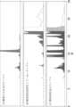
- FIG. 1shows the design and naming rules for trivalent antibodies in the DUAL/LINC (1+2) format.
- FIG. 1shows an electropherogram by CE-SDS. This is a chromatogram including peak names obtained by HIS analysis. The standard chromatogram (upper figure) and the enlarged chromatogram (lower figure) are shown.
- amino acids at positions 33, 55, and/or 96 are substitutedincludes the following variations of amino acid modification: (a) 33, (b) 55, (c) 96, (d) 33 and 55, (e) 33 and 96, (f) 55 and 96, and (g) 33, 55, and 96 amino acids.
- amino acid residues in the light chain constant regionare numbered herein according to Kabat et al., and numbering of amino acid residues in the heavy chain constant region is according to the EU numbering system, also known as the EU index, as described in Kabat et al., Sequences of Proteins of Immunological Interest, 5th Ed. Public Health Service, National Institutes of Health, Bethesda, MD, 1991.
- amino acidsare described by the one-letter or three-letter code or both, e.g., Ala/A, Leu/L, Arg/R, Lys/K, Asn/N, Met/M, Asp/D, Phe/F, Cys/C, Pro/P, Gln/Q, Ser/S, Glu/E, Thr/T, Gly/G, Trp/W, His/H, Tyr/Y, Ile/I, or Val/V.
- amino acid modification in the amino acid sequence of an antigen-binding moleculealso referred to herein as "amino acid substitution” or "amino acid mutation"
- known methodssuch as site-directed mutagenesis (Kunkel et al. (Proc. Natl. Acad. Sci. USA (1985) 82, 488-492)) and overlap extension PCR may be appropriately employed.
- site-directed mutagenesisKunkel et al. (Proc. Natl. Acad. Sci. USA (1985) 82, 488-492)
- overlap extension PCRmay be appropriately employed.
- amino acid modification method for substituting an unnatural amino acidseveral known methods may also be employed (Annu. Rev. Biophys. Biomol. Struct. (2006) 35, 225-249; and Proc. Natl. Acad. Sci. USA (2003) 100 (11), 6353-6357).
- a cell-free translation system(Clover Direct (Protein Express)) containing a tRNA in which an unnatural amino acid is bound to a complementary amber suppressor tRNA of the UAG codon (amber codon), which is one of the stop codons.
- an expression indicating an amino acid modificationan expression indicating the one-letter code or three-letter code of the amino acid before and after the modification, respectively, before and after the number indicating a specific position
- the modification N100bL or Asn100bLeu used when replacing an amino acid contained in an antibody variable regionrepresents the replacement of Asn at position 100b (according to Kabat numbering) with Leu. That is, the number indicates the position of the amino acid according to Kabat numbering, the one-letter or three-letter amino acid code written before the number indicates the amino acid before the replacement, and the one-letter or three-letter amino acid code written after the number indicates the amino acid after the replacement.
- the modification P238D or Pro238Asp used when replacing an amino acid in the Fc region contained in an antibody constant regionrepresents the replacement of Pro at position 238 (according to EU numbering) with Asp. That is, the number indicates the position of the amino acid according to the EU numbering system, the one-letter or three-letter amino acid code written before the number indicates the amino acid before substitution, and the one-letter or three-letter amino acid code written after the number indicates the amino acid after substitution.
- polypeptiderefers to a molecule composed of monomers (amino acids) linked in a linear chain by amide bonds (also known as peptide bonds).
- polypeptiderefers to any chain of two or more amino acids and does not refer to a specific length of the product.
- peptides, dipeptides, tripeptides, oligopeptides, "proteins,"”amino acid chains,” or any other terms used to refer to a chain of two or more amino acidsare included within the definition of "polypeptide,” and the term “polypeptide” may be used in place of or interchangeably with any of these terms.
- polypeptideis also intended to refer to the products of post-expression modifications of the polypeptide, including, but not limited to, glycosylation, acetylation, phosphorylation, amidation, derivatization with known protecting/blocking groups, proteolytic cleavage, or modification with non-natural amino acids.
- a polypeptidemay be derived from a natural biological source or produced by recombinant technology, but need not necessarily be translated from a specified nucleic acid. It may be generated in any manner, including chemical synthesis.
- polypeptides described hereinmay be of a size of about 3 or more amino acids, 5 or more amino acids, 10 or more amino acids, 20 or more amino acids, 25 or more amino acids, 50 or more amino acids, 75 or more amino acids, 100 or more amino acids, 200 or more amino acids, 500 or more amino acids, 1,000 or more amino acids, or 2,000 or more amino acids.
- Polypeptidesmay have a defined three-dimensional structure, but they do not necessarily have to have such a structure. Polypeptides that have a defined three-dimensional structure are said to be folded, and polypeptides that do not have a defined three-dimensional structure but can adopt a number of different conformations are said to be unfolded.
- Percent (%) amino acid sequence identityto a reference polypeptide sequence is defined as the percentage of amino acid residues in a candidate sequence that are identical to amino acid residues in the reference polypeptide sequence after aligning the sequences to obtain the maximum percent sequence identity and introducing gaps, if necessary, and not considering any conservative substitutions as part of the sequence identity. Alignment for purposes of determining percent amino acid sequence identity can be accomplished by a variety of methods within the skill of the art, for example, using publicly available computer software such as BLAST, BLAST-2, ALIGN, or Megalign (DNASTAR) software.
- ALIGN-2a sequence comparison computer program.
- the ALIGN-2 sequence comparison computer programis the copyright of Genentech, Inc., and its source code has been submitted with user documentation to the US Copyright Office, Washington DC, 20559, where it is registered under US Copyright Registration No. TXU510087.
- the ALIGN-2 programis publicly available from Genentech, Inc., South San Francisco, California, or may be compiled from the source code.
- the ALIGN-2 programis compiled for use on UNIX operating systems, including Digital UNIX V4.0D.
- % amino acid sequence identity of a given amino acid sequence A to, with, or against a given amino acid sequence Bis calculated as follows: 100 times the fraction X/Y, where X is the number of amino acid residues scored as identical matches by the sequence alignment program ALIGN-2 in said program's alignment of A and B, and Y is the total number of amino acid residues in B.
- Antigen-binding moleculerefers to any molecule that specifically binds to an antigenic determinant (epitope), and includes an antigen-binding domain, or any molecule that has binding activity to an antigen, and may further refer to molecules such as peptides or proteins having a length of about 5 amino acids or more.
- Peptides and proteinsare not limited to those derived from organisms, and for example, they may be polypeptides produced from artificially designed sequences. They may be either natural polypeptides, synthetic polypeptides, recombinant polypeptides, etc.
- a scaffold molecule that includes a known stable three-dimensional structure such as an ⁇ / ⁇ barrel as a scaffold, and a part of the molecule becomes an antigen-binding domainis also an embodiment of the antigen-binding molecule described herein.
- the antigen-binding moleculemay include two or more (e.g., 2, 3, 4, 5, or more) polypeptide chains.
- the polypeptide chain constituting the antigen-binding moleculemay be an antibody heavy chain or an antibody light chain. That is, in one embodiment, the antigen-binding molecule is an antibody, an antibody fragment, or an antibody derivative.
- the antigen-binding moleculeis a trivalent antibody including chains 1 to 5 as shown in FIG. 3 of the present application.
- the antigen-binding moleculeis a non-antibody protein, or a fragment or derivative thereof.
- Multispecific antigen-binding moleculerefers to an antigen-binding molecule that specifically binds to two or more antigens.
- the term "bispecific”means that an antigen-binding molecule can specifically bind to at least two different antigenic determinants.
- the term “trispecific”means that an antigen-binding molecule can specifically bind to at least three different antigenic determinants.
- the multispecific antigen-binding molecule of the present applicationis a trispecific antigen-binding molecule that can bind to either CD3 or CD137, but not both antigens simultaneously, and can specifically bind to DLL3.
- Antigen-binding domainrefers to a region that specifically binds to and is complementary to a part or all of an antigen.
- an antigen-binding moleculeincludes an antigen-binding domain. When the molecular weight of an antigen is large, the antigen-binding domain can bind only to a specific part of the antigen. The specific part is called an epitope.
- the antigen-binding domainincludes an antibody fragment that binds to a specific antigen.
- the antigen-binding domaincan be provided by one or more antibody variable domains.
- the antigen-binding domainincludes an antibody light chain variable region (VL) and an antibody heavy chain variable region (VH).
- VLantibody light chain variable region
- VHantibody heavy chain variable region
- antigen-binding domainsinclude "single chain Fv (scFv)", “single chain antibody”, “Fv”, “single chain Fv 2 (scFv2)", “Fab", and "Fab'”.
- the antigen-binding domainincludes a non-antibody protein or a fragment thereof that binds to a specific antigen.
- the antigen-binding domainincludes one or two Fabs that include a CH1 region.
- the antigen-binding domainincludes a hinge region.
- the term "antigen-binding domain”can direct the entity to which it is bound to a target site, e.g., a specific type of tumor cell expressing a cancer antigen (DLL3).
- the antigen-binding domaincan activate signaling through its target antigen, e.g., a T-cell receptor complex antigen (particularly CD3) and/or a costimulatory receptor (CD137).
- first, second, and thirdused herein with respect to antigen-binding domains and the like are used for the convenience of distinguishing between two or more different types of moieties and the like when they are present. The use of these terms is not intended to confer a particular order or orientation of the antigen-binding molecules, unless otherwise specified.
- telomere binding domainAs used herein, "specifically binds" means that one of the molecules involved in specific binding binds without showing any significant binding to molecules other than the one or more binding partner molecules. This expression is also used when an antigen-binding domain is specific to a specific epitope among multiple epitopes contained in an antigen. When the epitope to which the antigen-binding domain binds is contained in multiple different antigens, an antigen-binding molecule containing the antigen-binding domain can bind to various antigens containing that epitope.
- binding to the same epitopemeans that the epitopes bound by the two antigen-binding domains overlap at least partially.
- the degree of overlapis not limited, but is at least 10% or more, preferably 20% or more, 30% or more, 40% or more, 50% or more, 60% or more, 70% or more, 80% or more, particularly preferably 90% or more, and most preferably 100%.
- antibodyis used herein in the broadest sense and encompasses a variety of antibody structures, including, but not limited to, monoclonal antibodies, polyclonal antibodies, multispecific antibodies (e.g., bispecific or trispecific antibodies), and antibody fragments, so long as they exhibit the desired antigen-binding activity.
- immunoglobulin moleculerefers to a protein having the structure of a naturally occurring antibody.
- immunoglobulins of the IgG classare heterotetrameric glycoproteins of about 150,000 daltons, composed of two light chains and two heavy chains linked by disulfide bonds. Each heavy chain has, from N-terminus to C-terminus, a variable region (VH), also called the variable heavy domain or heavy chain variable domain, followed by three constant domains (CH1, CH2, and CH3), also called the heavy chain constant region.
- VHvariable region
- CH1, CH2, and CH3constant domains
- each light chainhas, from N-terminus to C-terminus, a variable region (VL), also called the variable light domain or light chain variable domain, followed by a constant light (CL) domain, also called the light chain constant region.
- VLvariable region
- CLconstant light
- the heavy chains of an immunoglobulinmay be assigned to one of five types, called ⁇ (IgA), ⁇ (IgD), ⁇ (IgE), ⁇ (IgG), or ⁇ (IgM), some of which may be further classified into subtypes, e.g., ⁇ 1 (IgG1), ⁇ 2 (IgG2), ⁇ 3 (IgG3), ⁇ 4 (IgG4), ⁇ 1 (IgA1), and ⁇ 2 (IgA2).
- the light chains of an immunoglobulinmay be assigned to one of two types, called kappa and lambda, based on the amino acid sequence of their constant domains.
- Immunoglobulinsessentially consist of two Fab molecules and an Fc domain linked via an immunoglobulin hinge region.
- the term "monoclonal antibody”refers to an antibody obtained from a population of substantially homogeneous antibodies; that is, the individual antibodies that make up the population are identical and/or bind the same epitope, except for possible variant antibodies (e.g., variant antibodies containing naturally occurring mutations or that arise during the manufacture of the monoclonal antibody preparation, which are usually present in small amounts).
- variant antibodiese.g., variant antibodies containing naturally occurring mutations or that arise during the manufacture of the monoclonal antibody preparation, which are usually present in small amounts.
- polyclonal antibody preparationswhich typically contain different antibodies directed against different determinants (epitopes)
- each monoclonal antibody of a monoclonal antibody preparationis directed against a single determinant on an antigen.
- monoclonalindicates the character of the antibody as being obtained from a population of substantially homogeneous antibodies and should not be construed as requiring production of the antibody by any particular method.
- monoclonal antibodiesmay be made by a variety of techniques, including, but not limited to, hybridoma techniques, recombinant DNA techniques, phage display techniques, and methods utilizing transgenic animals containing all or part of the human immunoglobulin loci, and such and other exemplary methods for making monoclonal antibodies are described herein.
- full length antibody“full length antibody,” “complete antibody,” and “whole antibody” are used interchangeably herein and refer to an antibody having a structure substantially similar to a native antibody structure or having a heavy chain that includes an Fc region as defined herein.
- the "class" of an antibodyrefers to the type of constant domain or constant region present in the antibody's heavy chain.
- the heavy chain constant domains that correspond to the different classes of immunoglobulinsare called ⁇ , ⁇ , ⁇ , ⁇ , and ⁇ , respectively.
- variable regionrefers to the domains of the heavy or light chain of an antibody that are involved in binding the antibody to an antigen.
- the heavy and light chain variable domains (VH and VL, respectively) of natural antibodiesusually have a similar structure, with each domain containing four conserved framework regions (FR) and three hypervariable regions (HVR) (see, for example, Kindt et al. Kuby Immunology, 6th ed., W.H. Freeman and Co., page 91 (2007)).
- FRconserved framework regions
- HVRhypervariable regions
- antibodies that bind to a particular antigenmay be isolated by screening a complementary library of VL or VH domains, respectively, with a VH or VL domain from an antibody that binds to that antigen. See, e.g., Portolano et al., J. Immunol. 150:880-887 (1993); Clarkson et al., Nature 352:624-628 (1991).
- hypervariable regionrefers to each region of an antibody variable domain that is hypervariable in sequence (the “complementarity determining region” or “CDR") and/or forms structurally defined loops (the “hypervariable loops") and/or contains antigen contact residues (the “antigen contacts”).
- CDRcomplementarity determining region
- hypervariable loopsforms structurally defined loops
- antigen contact residuesthe "antigen contacts”
- antibodiestypically contain six HVRs: three in the VH (H1, H2, H3) and three in the VL (L1, L2, L3).
- Exemplary HVRs hereininclude the following: (a) hypervariable loops occurring at amino acid residues 26-32 (L1), 50-52 (L2), 91-96 (L3), 26-32 (H1), 53-55 (H2), and 96-101 (H3) (Chothia and Lesk, J. Mol. Biol. 196:901-917 (1987)); (b) the CDRs occurring at amino acid residues 24-34 (L1), 50-56 (L2), 89-97 (L3), 31-35b (H1), 50-65 (H2), and 95-102 (H3) (Kabat et al., Sequences of Proteins of Immunological Interest, 5th Ed.
- HVR residues and other residues in the variable domainare numbered herein according to Kabat et al., supra.
- HVR-H1, HVR-H2, HVR-H3, HVR-L1, HVR-L2, and HVR-L3are also referred to as "H-CDR1", “H-CDR2", “H-CDR3”, “L-CDR1”, “L-CDR2”, and “L-CDR3”, respectively.
- FRFramework or "FR” refers to variable domain residues other than hypervariable region (HVR) residues.
- the FR of a variable domaintypically consists of four FR domains: FR1, FR2, FR3, and FR4. Accordingly, the HVR and FR sequences typically appear in the VH (or VL) in the following order: FR1-H1(L1)-FR2-H2(L2)-FR3-H3(L3)-FR4.
- a "human consensus framework”is a framework that represents the most commonly occurring amino acid residues in a selection of human immunoglobulin VL or VH framework sequences.
- the selection of human immunoglobulin VL or VH sequencesis from a subgroup of variable domain sequences.
- the subgroup of sequencesis a subgroup in Kabat et al., Sequences of Proteins of Immunological Interest, Fifth Edition, NIH Publication 91-3242, Bethesda MD (1991), vols. 1-3.
- the subgroupis subgroup kappa I by Kabat et al., supra.
- the subgroupis subgroup III by Kabat et al., supra.
- the term "hinge region”refers to the portion of an antibody heavy chain polypeptide that links the CH1 domain and the CH2 domain in a wild-type antibody heavy chain, for example, from about position 216 to about position 230 according to the EU numbering system, or from about position 226 to about position 243 according to the Kabat numbering system.
- the cysteine residue at EU numbering position 220 in the hinge regionis known to form a disulfide bond with the cysteine residue at EU numbering position 214 in the antibody light chain.
- the cysteine residues at EU numbering positions 226 and 229 in the hinge regionform disulfide bonds between two antibody heavy chains.
- the "hinge region”is defined as extending from positions 216 to 238 (EU numbering) or positions 226 to 251 (Kabat numbering) of human IgG1.
- the hingecan be further divided into three distinct regions, the upper hinge, the middle hinge, and the lower hinge.
- these regionsare generally defined as follows: Upper hinge: 216-225 (EU numbering) or 226-238 (Kabat numbering), Central hinge: 226-230 (EU numbering) or 239-243 (Kabat numbering), Lower hinge: 231-238 (EU numbering) or 244-251 (Kabat numbering).
- Hinge regions of other IgG isotypescan be aligned with the IgG1 sequence by placing the first and last cysteine residues that form inter-heavy chain S-S bonds in the same positions (see, for example, Brekke et al., 1995, Immunol (Table 1 of Today 16: 85-90)).
- Hinge regions hereininclude wild-type hinge regions as well as variants in which amino acid residues in the wild-type hinge region have been altered by substitution, addition, or deletion.
- capable of being linkedamino acid residues
- capable of being formedinclude cases where disulfide bonds have already formed, and cases where disulfide bonds have not formed but can be formed later under appropriate conditions.
- disulfide bonds formed between amino acids not in the hinge regionrefers to disulfide bonds formed, connected, or linked through amino acids located in any region of the antibody other than the "hinge region” defined above.
- disulfide bondsare formed, connected, or linked through amino acids located at any position in the antibody other than the hinge region (e.g., from about position 216 to about position 230 according to the EU numbering system, or from about position 226 to about position 243 according to the Kabat numbering system).
- such disulfide bondsare formed, connected, or linked through amino acids located in the CH1 region, CL region, VL region, VH region, and/or VHH region. In some embodiments, such disulfide bonds are formed, connected, or linked through amino acids located at EU numbering positions 119-123, 131-140, 148-150, 155-167, 174-178, 188-197, and 201-214 in the CH1 region.
- such disulfide bondsare formed, connected, or linked through amino acids located at EU numbering positions 119, 122, 123, 131, 132, 133, 134, 135, 136, 137, 138, 139, 140, 148, 150, 155, 156, 157, 159, 160, 161, 162, 163, and 164 in the CH1 region. , 165, 167, 174, 176, 177, 178, 188, 189, 190, 191, 192, 193, 194, 195, 196, 197, 201, 203, 205, 206, 207, 208, 211, 212, 213, 214.
- such disulfide bondsare formed, connected, or linked through amino acids located at EU numbering positions 188, 189, 190, 191, 192, 193, 194, 195, 196, and 197 in the CH1 region.
- such a disulfide bondis formed, connected, or linked through the amino acid located at EU numbering position 191 in the CH1 region.
- mismatched moleculerefers to an antigen-binding molecule in which disulfide bonds are formed between amino acid residues different from those desired.
- an antigen-binding moleculecontains a disulfide bond formed between desired amino acid residues and a disulfide bond formed between amino acid residues different from those desired, such an antigen-binding molecule is included in the mismatched molecule of the present specification.
- abent entirelyrefers to an antigen-binding molecule in which cysteine residues remain free, or in which cysteine residues form disulfide bonds with molecules (e.g., impurity molecules) that are different from the molecules that form the antigen-binding molecules.
- abent entirelymay refer to any molecule that has such characteristics, and even if it includes, for example, disulfide bonds (desired disulfide bonds) formed between amino acid residues in regions other than the hinge region, or disulfide bonds (mismatch portions) that are different from the desired disulfide bonds, it is included in “absent entirely” in this specification as long as it has the above characteristics.
- the constant regionis preferably an antibody constant region, more preferably an IgG1, IgG2, IgG3, and IgG4 type antibody constant region, and even more preferably an antibody constant region of human IgG1, IgG2, IgG3, and IgG4 type.
- the constant regionis preferably a heavy chain constant region, more preferably an IgG1, IgG2, IgG3, and IgG4 type heavy chain constant region, and even more preferably an IgG1, IgG2, IgG3, and IgG4 type heavy chain constant region.
- the amino acid sequences of the human IgG1 constant region, the human IgG2 constant region, the human IgG3 constant region, and the human IgG4 constant regionare publicly known.
- constant regions of human IgG1, human IgG2, human IgG3, and human IgG4multiple allotype sequences due to genetic polymorphisms are described in Sequences of proteins of immunological interest, NIH Publication No. 91-3242, and any of them can be used in the present invention.
- the constant region with altered amino acidsmay contain other amino acid mutations or modifications as long as it contains the amino acid mutation of the present invention.
- Fc regionrefers to a region in an antibody molecule that includes a hinge or a part thereof, and a fragment consisting of the CH2 and CH3 domains.
- the Fc region of the IgG classrefers to, for example, but is not limited to, a region from cysteine 226 (EU numbering (also referred to herein as EU index)) to the C-terminus, or from proline 230 (EU numbering) to the C-terminus.
- EU indexcysteine 226
- EU indexproline 230
- the Fc regioncan be preferably obtained, for example, by partially digesting an IgG1, IgG2, IgG3, or IgG4 monoclonal antibody with a protease such as pepsin, and then re-eluting the fraction adsorbed to a protein A column or a protein G column.
- a proteasesuch as pepsin
- Such a proteaseis not particularly limited as long as it can digest a full-length antibody to form Fab or F(ab') 2 in a limited manner under appropriately set enzyme reaction conditions (e.g., pH).
- proteasesinclude pepsin and papain.
- Natural IgGmeans a polypeptide that contains the same amino acid sequence as IgG found in nature and belongs to the class of antibodies substantially encoded by the immunoglobulin gamma gene.
- Natural human IgGmeans, for example, natural human IgG1, natural human IgG2, natural human IgG3, or natural human IgG4. Natural IgG also includes naturally occurring variants thereof. Multiple allotype sequences based on genetic polymorphisms are described in Sequences of proteins of immunological interest, NIH Publication No.
- 91-3242as the constant regions of human IgG1, human IgG2, human IgG3, and human IgG4 antibodies, and any of them can be used in the present invention.
- the sequence of human IgG1may have DEL or EEM as the amino acid sequence at EU numbering positions 356 to 358.
- the Fc domain of an antigen-binding moleculeconsists of a pair of polypeptide chains that comprise the heavy chain domains of an immunoglobulin molecule.
- the Fc domain of an immunoglobulin G (IgG) moleculeis a dimer, each subunit of which contains the CH2 and CH3 IgG heavy chain constant domains.
- the two subunits of an Fc domaincan stably associate with each other.
- the antigen-binding molecules described hereincomprise no more than one Fc domain.
- the Fc domain of the antigen-binding moleculeis an IgG Fc domain.
- the Fc domainis an IgG1 Fc domain.
- the Fc domainis an IgG1 Fc domain.
- the Fc domainis a human IgG1 Fc region.
- Chimeric Antibodiesrefers to an antibody in which a portion of the heavy and/or light chain is derived from a particular source or species, while the remaining portions of the heavy and/or light chain are derived from a different source or species.
- chimeric antibody variable domainrefers to an antibody variable region in which a portion of the heavy and/or light chain variable region is derived from a particular source or species, while the remaining portions of the heavy and/or light chain variable region are derived from a different source or species.
- Humanized antibodyrefers to a chimeric antibody that contains amino acid residues from non-human HVRs and amino acid residues from human FRs.
- a humanized antibodycontains substantially all of at least one, typically two, variable domains, in which all or substantially all HVRs (e.g., CDRs) correspond to those of a non-human antibody and all or substantially all FRs correspond to those of a human antibody.
- a humanized antibodymay optionally contain at least a portion of an antibody constant region derived from a human antibody.
- a "humanized form" of an antibody(e.g., a non-human antibody) refers to an antibody that has undergone humanization.
- a “humanized antibody variable region”refers to the variable region of a humanized antibody.
- Antibody fragmentrefers to a molecule other than a complete antibody that contains a portion of the complete antibody that binds to the antigen to which the complete antibody binds.
- Examples of antibody fragmentsinclude, but are not limited to, Fv, Fab, Fab', Fab'-SH, F(ab')2, diabodies, linear antibodies, single-chain antibody molecules (e.g., scFv), and single-domain antibodies.
- scFvsingle-domain antibodies.
- Diabodiesare antibody fragments with two antigen binding sites that may be bivalent or bispecific.
- Single domain antibodiesare antibody fragments that contain all or a portion of the heavy chain variable domain or all or a portion of the light chain variable domain of an antibody.
- the single domain antibodyis a human single domain antibody (Domantis, Inc., Waltham, MA; see, e.g., U.S. Patent No. 6,248,516 B1).
- Antibody fragmentscan be produced by a variety of techniques, including, but not limited to, proteolytic digestion of whole antibodies, production by recombinant host cells (e.g., E. coli or phage), as described herein.
- variable fragmentrefers to the minimum unit of an antibody-derived antigen-binding domain consisting of a pair of an antibody light chain variable region (VL) and an antibody heavy chain variable region (VH).
- VLantibody light chain variable region
- VHantibody heavy chain variable region
- single chain antibodiesscFv, single chain antibodies, and sc(Fv)2
- single chain antibodiesall refer to antibody fragments that contain variable regions from both heavy and light chains in a single polypeptide chain, but lack constant regions.
- single chain antibodiesfurther comprise a polypeptide linker between the VH and VL domains that allows them to form the desired structure that would allow antigen binding.
- Single chain antibodiesare discussed in detail by Pluckthun in The Pharmacology of Monoclonal Antibodies, Vol. 113, Rosenburg, and Moore, Eds., Springer-Verlag, New York, pp. 269-315 (1994). See also International Patent Application Publication No. WO 1988/001649; U.S. Patent Nos. 4,946,778 and 5,260,203.
- single chain antibodiescan be bispecific and/or humanized.
- scFvis an antigen-binding domain in which the VH and VL that make up the Fv are linked by a peptide linker (Proc. Natl. Acad. Sci. U.S.A. (1988) 85 (16), 5879-5883).
- the peptide linkerallows the VH and VL to be held in close proximity.
- sc(Fv)2is a single-chain antibody in which four variable regions, two VL and two VH, are linked by a linker such as a peptide linker to form a single chain (J Immunol. Methods (1999) 231 (1-2), 177-189). The two VH and VL may be derived from different monoclonal antibodies.
- Such sc(Fv)2preferably includes bispecific sc(Fv)2 that recognizes two different epitopes present in the same antigen, as disclosed in, for example, Journal of Immunology (1994) 152 (11), 5368-5374.
- sc(Fv)2can be prepared by methods known to those skilled in the art. For example, sc(Fv)2 can be prepared by linking scFvs with a linker such as a peptide linker.
- the form of the antigen-binding domain constituting sc(Fv)2includes an antibody characterized in that two VH units and two VL units are arranged in the following order starting from the N-terminus of a single-chain polypeptide: VH, VL, VH, VL ([VH]-linker-[VL]-linker-[VH]-linker-[VL]), but the order of the two VH units and the two VL units is not limited to the above configuration and may be arranged in any order.
- sc(Fv) 2The molecular form of sc(Fv) 2 is also described in detail in WO2006/132352. Those skilled in the art can follow these descriptions to appropriately prepare a desired sc(Fv) 2 for producing the polypeptide complexes disclosed herein.
- a carrier polymersuch as PEG or an organic compound such as an anticancer drug may be conjugated to the antigen-binding molecule or antibody of the present disclosure.
- a glycosylation sequenceis suitably inserted into the antigen-binding molecule or antibody so that the glycosylation produces the desired effect.
- Linkers used to link the variable regions of antibodiesinclude any peptide linker that can be introduced by genetic engineering, synthetic linkers, and linkers disclosed in, for example, Protein Engineering, 9 (3), 299-305, 1996. However, in the present disclosure, peptide linkers are preferred.
- the length of the peptide linkeris not particularly limited, and can be appropriately selected by those skilled in the art depending on the purpose. The length is preferably 5 amino acids or more (although not particularly limited, the upper limit is usually 30 amino acids or less, preferably 20 amino acids or less), and is particularly preferably 15 amino acids. When sc(Fv) 2 contains three peptide linkers, their lengths may all be the same or different.
- such peptide linkersinclude: Ser, Gly-Ser, Gly-Gly-Ser, Ser-Gly-Gly, Gly-Gly-Gly-Ser (SEQ ID NO: 5), Ser-Gly-Gly-Gly (SEQ ID NO: 6), Gly-Gly-Gly-Gly-Ser (SEQ ID NO: 7), Ser-Gly-Gly-Gly-Gly (SEQ ID NO: 8), Gly-Gly-Gly-Gly-Gly-Ser (SEQ ID NO: 9), Ser-Gly-Gly-Gly-Gly-Gly (SEQ ID NO: 10), Gly-Gly-Gly-Gly-Gly-Gly-Gly-Ser (SEQ ID NO: 11), Ser-Gly-Gly-Gly-Gly-Gly-Gly-Gly (SEQ ID NO: 12), (Gly-Gly-Gly-Gly-Gly-Gly-Gly (SEQ ID NO: 7)), and
- Synthetic linkersare commonly used to cross-link peptides, including N-hydroxysuccinimide (NHS), disuccinimidyl suberate (DSS), bis(sulfosuccinimidyl)suberate (BS3), dithiobis(succinimidyl propionate) (DSP), dithiobis(sulfosuccinimidyl propionate) (DTSSP), ethylene glycol bis(succinimidyl succinate) (EGS), ethylene glycol bis(sulfosuccinimidyl succinate) (sulfo-EGS), disuccinimidyl tartrate (DST), disulfosuccinimidyl tartrate (sulfo-DST), bis[2-(succinimidooxycarbonyloxy)ethyl]sulfone (BSOCOES), and bis[2-(sulfosuccinimidooxycarbonyloxy)ethyl]sulfone
- linkersTo link four antibody variable regions, three linkers are usually required.
- the linkers usedmay be of the same type or different types.
- Fab, F(ab')2, and Fab'refers to a protein consisting of the VH and CH1 domains of an immunoglobulin heavy chain ("Fab heavy chain”) and the VL and CL domains of a light chain (“Fab light chain”).
- the heavy chain of a wild-type Fab moleculecannot form disulfide bonds with another heavy chain molecule.
- Fab variants in which amino acid residues in the wild-type Fab are altered by substitution, addition, or deletionare also encompassed herein.
- the Fab variantscontain mutated amino acid residues (e.g., substituted, added, or inserted cysteine or lysine residues) that can form disulfide bonds with another heavy chain molecule or portion thereof (e.g., a Fab molecule).
- mutated amino acid residuese.g., substituted, added, or inserted cysteine or lysine residues
- scFabis an antigen-binding domain in which one light chain and one heavy chain CH1 region and variable region that make up the Fab are linked by a peptide linker.
- the light chain and the heavy chain CH1 region and variable regioncan be held in close proximity by the peptide linker.
- F(ab')2 and “Fab'”refer to antibody fragments produced by treating immunoglobulins (monoclonal antibodies) with proteases such as pepsin and papain, and digesting the immunoglobulins (monoclonal antibodies) near the disulfide bond between the hinge regions of the two H chains.
- papaincan cleave IgG upstream of the disulfide bond between the hinge regions of the two H chains, producing two homologous antibody fragments in which an L chain containing VL (light chain variable region) and CL (light chain constant region) is bound to an H chain fragment containing VH (light chain variable region) and CH ⁇ 1 ( ⁇ 1 region in the heavy chain constant region) by a disulfide bond at the C-terminal region.
- L chain containing VL (light chain variable region) and CLlight chain constant region
- H chain fragment containing VHlight chain variable region
- CH ⁇ 1⁇ 1 region in the heavy chain constant region
- F(ab')2is composed of two light chains and two heavy chains that contain constant regions of the CH1 domain and a portion of the CH2 domain such that a disulfide bond is formed between the two heavy chains.
- the F(ab')2 disclosed in this specificationcan be suitably prepared by partially digesting a full-length monoclonal antibody or the like having a desired antigen-binding domain with a protease such as pepsin, and then removing the Fc fragment by adsorption onto a protein A column.
- proteasethere are no particular limitations on such a protease, so long as it can cleave a full-length antibody to generate F(ab')2 in a limited manner by appropriately setting the reaction conditions of the enzyme, such as pH, and such proteases include, for example, pepsin and ficin.
- Fusedmeans that the components (e.g., a Fab molecule and an Fc domain subunit) are linked by a peptide bond, either directly or via one or more peptide linkers.
- Crossover Fab moleculerefers to a Fab molecule in which either the variable or constant regions of the Fab heavy and Fab light chains are exchanged, i.e., the crossover Fab molecule comprises a peptide chain composed of a light chain variable region and a heavy chain constant region, and a peptide chain composed of a heavy chain variable region and a light chain constant region.
- the crossover Fab moleculecomprises a peptide chain composed of a light chain variable region and a heavy chain constant region, and a peptide chain composed of a heavy chain variable region and a light chain constant region.
- the peptide chain comprising the heavy chain constant regionis referred to herein as the "heavy chain” of the crossover Fab molecule.
- the peptide chain comprising the heavy chain variable regionis referred to herein as the "heavy chain” of the crossover Fab molecule.
- a "conventional" Fab moleculerefers to a Fab molecule in its native format, i.e., a Fab molecule comprising a heavy chain (VH-CH1) made up of the variable and constant regions of the heavy chain, and a light chain (VL-CL) made up of the variable and constant regions of the light chain.
- VH-CH1heavy chain
- VL-CLlight chain
- single domain antibodyis not particularly limited in structure, so long as the domain alone can exhibit antigen-binding activity.
- Conventional antibodiessuch as IgG antibodies, exhibit antigen-binding activity when the variable region is formed by pairing VH and VL, whereas single domain antibodies are known to be able to exhibit antigen-binding activity by the domain structure of the single domain antibody alone, without pairing with other domains.
- Single domain antibodiesusually have a relatively low molecular weight and exist in the form of a monomer.
- single domain antibodiesinclude, but are not limited to, antigen-binding molecules that naturally lack light chains, such as camelid VHH and shark VNAR , and antibody fragments that contain all or a part of the VH domain or all or a part of the VL domain of an antibody.
- single domain antibodieswhich are antibody fragments that contain all or a part of the VH/VL domains of an antibody, include, but are not limited to, single domain antibodies artificially produced starting from human antibody VH or human antibody VL, such as those described in U.S. Patent No. 6,248,516 B1.
- a single domain antibodyhas three CDRs (CDR1, CDR2, and CDR3).
- Single domain antibodiescan be obtained from animals capable of producing single domain antibodies or by immunizing animals capable of producing single domain antibodies.
- animals capable of producing single domain antibodiesinclude, but are not limited to, camelids and transgenic animals into which a gene capable of producing single domain antibodies has been introduced.
- Camelidsinclude camels, llamas, alpacas, dromedaries, and guanacos.
- transgenic animals into which a gene capable of producing single domain antibodies has been introducedinclude, but are not limited to, the transgenic animals described in International Publication No. WO2015/143414 and US Patent Publication No. US2011/0123527 A1.
- Humanized single chain antibodiescan also be obtained by replacing the framework sequences of single domain antibodies obtained from animals with human germline sequences or sequences similar thereto.
- Humanized single domain antibodiese.g., humanized VHHs are one embodiment of single domain antibodies.
- Single domain antibodiescan also be obtained from a polypeptide library containing single domain antibodies by ELISA, panning, etc.
- polypeptide libraries containing single domain antibodiesinclude, but are not limited to, naive antibody libraries obtained from various animals or humans (e.g., Methods in Molecular Biology 2012 911 (65-78), Biochimica et Biophysica Acta - Proteins and Proteomics 2006 1764:8 (1307-1319)), antibody libraries obtained by immunizing various animals (e.g., Jou journal of Applied Microbiology 2014 117:2 (528-536)), and synthetic antibody libraries made from various animal or human antibody genes (e.g., Journal of Biomolecular Screening 2016 21:1 (35-43), Journal of Biological Chemistry 2016 291:24 (12641-12657), AIDS 2016 30:11 (1691-1701)).
- an "agonist” antigen-binding molecule or an “agonist” antibodyis an antigen-binding molecule or antibody that significantly enhances the biological activity of the antigen to which it binds.
- a "blocking" antigen-binding molecule or antibody or an “antagonist” antigen-binding molecule or antibodyis an antigen-binding molecule or antibody that significantly inhibits (either partially or completely) the biological activity of the antigen to which it binds.
- Antigensrefers to the site on a polypeptide macromolecule to which an antigen-binding moiety binds (e.g., a three-dimensional structure composed of a continuous stretch of amino acids or a discrete region of non-contiguous amino acids) to form an antigen-binding moiety-antigen complex.
- an antigen-binding moietye.g., a three-dimensional structure composed of a continuous stretch of amino acids or a discrete region of non-contiguous amino acids
- Useful antigenic determinantscan be found, for example, on the surface of tumor cells, on the surface of virus-infected cells, on the surface of other diseased cells, on the surface of immune cells, free in serum, and/or in the extracellular matrix (ECM).
- ECMextracellular matrix
- the proteins referred to herein as antigenscan be any native form of the protein from any vertebrate source, including mammals such as primates (e.g., humans) and rodents (e.g., mice and rats).
- the antigenis human CD3, human CD137, or human DLL3.
- the termencompasses the "full-length", unprocessed protein, as well as any form of the protein that results from processing in the cell.
- the termalso encompasses naturally occurring variants of the protein, such as splice variants or allelic variants.
- reduced Fc ⁇ receptor binding activitymeans that the competitive activity of a test antigen-binding molecule or antibody is 50% or less, preferably 45% or less, 40% or less, 35% or less, 30% or less, 20% or less, or 15% or less, and particularly preferably 10% or less, 9% or less, 8% or less, 7% or less, 6% or less, 5% or less, 4% or less, 3% or less, 2% or less, or 1% or less of the competitive activity of a control antigen-binding molecule or antibody, for example, based on the analytical method described above.
- An antigen-binding molecule or antibody containing an Fc domain of a monoclonal IgG1, IgG2, IgG3, or IgG4 antibodycan be used as a control antigen-binding molecule or antibody as appropriate.
- the Fc domain structureis shown in SEQ ID NO: 1 (RefSeq Accession No. AAC82527.1 with A added to the N-terminus), SEQ ID NO: 2 (RefSeq Accession No. AAB59393.1 with A added to the N-terminus), SEQ ID NO: 3 (RefSeq Accession No. CAA27268.1 with A added to the N-terminus), and SEQ ID NO: 4 (RefSeq Accession No.
- an antigen-binding molecule or antibody containing an Fc domain mutant of an antibody of a particular isotypeis used as a test substance
- the effect of the mutation of the mutant on the Fc ⁇ receptor binding activityis evaluated using an antigen-binding molecule or antibody containing an Fc domain of the same isotype as a control.
- an antigen-binding molecule or antibody containing an Fc domain mutant whose Fc ⁇ receptor binding activity has been determined to be reducedis appropriately prepared.
- Such known mutantsinclude, for example, a mutant having a deletion of amino acids 231A to 238S (EU numbering) (WO 2009/011941), as well as the mutants C226S, C229S, P238S, (C220S) (J. Rheumatol (2007) 34, 11); C226S and C229S ((Hum. Antibod. Hybridomas (1990) 1(1), 47-54); C226S, C229S, E233P, L234V, and L235A (Blood (2007) 109, 1185-1192).
- preferred antigen-binding molecules or antibodiesinclude those that contain an Fc domain having at least one amino acid mutation (e.g., substitution) selected from the following amino acid positions forming the Fc domain of an antibody of a particular isotype: 220, 226, 229, 231, 232, 233, 234, 235, 236, 237, 238, 239, 240, 264, 265, 266, 267, 269, 270, 295, 296, 297, 298, 299, 300, 325, 327, 328, 329, 330, 331, 332, 439, 445, 449, or 451 (EU numbering).
- amino acid mutatione.g., substitution
- the antibody isotype from which the Fc domain originatesis not particularly limited, and an appropriate Fc domain derived from a monoclonal IgG1, IgG2, IgG3, or IgG4 antibody can be used. It is preferable to use an Fc domain derived from an IgG1 antibody.
- Preferred antigen-binding molecules or antibodiesinclude, for example, the following substitutions, whose positions are designated by EU numbering in amino acids forming the Fc domain of an IgG1 antibody (each number represents the position of an amino acid residue in EU numbering; and the one-letter amino acid code preceding the number represents the amino acid residue before substitution, and the one-letter amino acid code following the number represents the amino acid residue after substitution): (a) L234F, L235E, P331S; (b) C226S, C229S, P238S; (c) C226S, C229S; or (d) C226S, C229S, E233P, L234V, L235A.
- preferred antigen binding molecules or antibodiesalso include the substitutions shown below, whose positions are designated by EU numbering in the amino acids forming the Fc domain of IgG2 antibodies: (e) H268Q, V309L, A330S, and P331S; (f) V234A; (g) G237A; (h) V234A and G237A; (i) A235E and G237A; or (j) V234A, A235E, and G237A.
- Each numberrepresents the position of an amino acid residue in the EU numbering system; the one-letter amino acid code preceding the number represents the amino acid residue before substitution, and the one-letter amino acid code following the number represents the amino acid residue after substitution.
- preferred antigen binding molecules or antibodiesalso include the substitutions shown below, whose positions are designated by EU numbering in the amino acids forming the Fc domain of IgG3 antibodies: (k) F241A; (l) D265A; or (m) V264A.
- Each numberrepresents the position of an amino acid residue in the EU numbering system; the one-letter amino acid code preceding the number represents the amino acid residue before substitution, and the one-letter amino acid code following the number represents the amino acid residue after substitution.
- preferred antigen binding molecules or antibodiesalso include the substitutions shown below, whose positions are designated by EU numbering in the amino acids forming the Fc domain of IgG4 antibodies: (n) L235A, G237A, and E318A; (o) L235E; or (p) F234A and L235A.
- Each numberrepresents the position of an amino acid residue in the EU numbering system; the one-letter amino acid code preceding the number represents the amino acid residue before substitution, and the one-letter amino acid code following the number represents the amino acid residue after substitution.
- antigen-binding molecules or antibodiesinclude, for example, those that contain an Fc domain in which any amino acid at positions 233, 234, 235, 236, 237, 327, 330, or 331 (EU numbering) forming the Fc domain of an IgG1 antibody is substituted with an amino acid at the corresponding position (EU numbering) in the corresponding IgG2 or IgG4.
- Preferred antigen-binding molecules or antibodiesalso include, for example, those that contain an Fc domain in which any one or more of the amino acids at positions 234, 235, and 297 (EU numbering) in the amino acid sequence that forms the Fc domain of an IgG1 antibody have been substituted with other amino acids.
- the type of amino acid after substitutionis not particularly limited; however, antigen-binding molecules or antibodies that contain an Fc domain in which any one or more of the amino acids at positions 234, 235, and 297 have been substituted with alanine are particularly preferred.
- Preferred antigen-binding molecules or antibodiesalso include, for example, those that contain an Fc domain in which the amino acid at position 265 (EU numbering) in the amino acids that form the Fc domain of an IgG1 antibody has been replaced with another amino acid.
- the type of amino acid following replacementis not particularly limited; however, antigen-binding molecules or antibodies that contain an Fc domain in which the amino acid at position 265 has been replaced with alanine are particularly preferred.
- (Preferential) enrichmentrefers to an increase in the relative abundance of a desired form, or an increase in the relative proportion of a desired form, or an increase in the population of a desired form (structural isoform).
- the methods described hereinincrease the relative abundance of structural isoforms of an antibody, such as an antibody having at least one disulfide bond formed between amino acid residues outside the hinge region.
- the at least one disulfide bondis formed between amino acid residues at position 191 according to EU numbering in the CH1 region of each of the first antigen-binding domain and the second antigen-binding domain.
- the methodsproduce homogeneous antibody preparations having at least 50%, 60%, 70%, 80%, 90%, preferably at least 95% molar ratio of the antibody having at least one disulfide bond formed outside the hinge region.
- a “homogeneous" population of homogeneous antibodiesrefers to an antibody population that comprises primarily a single form of antibody, e.g., at least 50%, 60%, 70%, 80% or more, preferably at least 90%, 95%, 96%, 97%, 99%, or 100% of the antibodies in a solution or composition are in a properly folded form.
- a "homogeneous" population of antibodies having at least one disulfide bond formed outside the hinge regionrefers to a population of said antibodies that comprises primarily a single properly folded form, e.g., at least 50%, 60%, 70%, 80% or more, preferably at least 90%, 95%, 96%, 97%, 99%, or 100% of the molar ratio of said antibodies having at least one disulfide bond formed outside the hinge region.
- said "homogeneous" population of antibodiescomprises at least one disulfide bond formed between the amino acid residue at position 191 according to EU numbering in the CH1 region of each of the first and second antigen-binding domains (i.e., the "paired cysteines" at position 191 according to EU numbering in the CH1 region).
- Determining whether an antibody population is homogenous and the relative abundance or proportion of protein/antibody conformations in a mixturecan be done using any of a variety of analytical and/or qualitative techniques. If the two conformations are differently resolved during a separation technique, e.g., chromatography, electrophoresis, filtration, or other purification technique, the relative proportion of the conformations in the mixture can be determined using such purification techniques. For example, at least two different conformations of a recombinant IgG can be separated by hydrophobic interaction chromatography. Furthermore, since far-UV circular dichroism has been used to estimate the secondary structural organization of proteins (Perczel et al., 1991, Protein Engrg.
- such techniquescan determine whether alternative conformations of a protein exist.
- fluorescence spectroscopywhich can be utilized to identify complementary differences in tertiary structure that can be assigned to tryptophan and tyrosine fluorescence.
- Other techniques that can be used to determine conformational differences and therefore the relative proportions of conformationsare online SEC to measure aggregation state, differential scanning calorimetry to measure melting transitions (Tm) and component enthalpies, and chaotropic unfolding.
- Yet another technique that can be used to determine conformational differences and therefore the relative proportions of conformationsis LC/MS detection to determine protein heterogeneity.
- determining the relative proportions of conformations in the mixturecan be done by activity assays (e.g., binding to ligand, enzymatic activity, biological activity, etc.).
- activity assayse.g., binding to ligand, enzymatic activity, biological activity, etc.
- the biological activity of the proteincan also be used.
- binding assayscan be used where activity is expressed as activity units/mg protein.
- IEC chromatographyis used to determine antibody/protein heterogeneity.
- the antibodyis purified or considered to be "homogeneous,” meaning that no polypeptide peaks or fractions corresponding to other polypeptides are detected upon analysis by IEC chromatography.
- the antibodyis purified or considered to be "homogeneous,” meaning that no polypeptide bands corresponding to other polypeptides are detected upon analysis by SDS-polyacrylamide gel electrophoresis (SDS-PAGE).
- SDS-PAGESDS-polyacrylamide gel electrophoresis
- the polypeptideis purified to substantial homogeneity, as indicated by a single polypeptide band upon analysis by SDS-PAGE.
- the polypeptide bandcan be visualized by silver staining, Coomassie blue staining, and/or (if the polypeptide is radiolabeled) autoradiography.
- Non-reducing SDS-PAGEis performed using 4-20% Mini-PROTEAN® TGX Stain-FreeTM Precast Gels (Bio-Rad) with 1x Tris/Glycine/SDS running buffer (Bio-Rad). Monoclonal antibody samples are heated at 70°C for 10 minutes. 0.2 micrograms are loaded and electrophoresis is run at 200 V for 90 minutes. Proteins are visualized with a Chemidoc Imaging System (Bio-Rad).
- the percentage of individual bandsis analyzed by Image Lab software version 6.0 (Bio-Rad) and the % intensity of individual bands (e.g., fast running (lower band) and slow running (upper band)) is calculated by dividing the band intensity by the sum of these two bands.
- the gelmay then be stained with CBB, a gel image may be captured, and the bands may be quantified using an imaging device.
- multiple, e.g., two bands, i.e., an "upper band” and a "lower band”may be observed in the antibody variant sample.
- the molecular weight of the upper bandmay correspond to that of the parent antibody (before modification).
- Structural changes such as cross-linking through disulfide bonds of Fabmay be caused by the cysteine substitution, which may result in changes in electrophoretic mobility.
- the lower bandmay be considered to correspond to the antibody with one or more engineered disulfide bonds formed between the CH1 regions.
- the antibody variant sample with additional cysteine substitutionsmay show a high ratio of lower band to upper band compared to the control sample.
- the additional cysteine residuesmay enhance/promote disulfide bond cross-linking of Fab; increase the percentage or structural uniformity of antibody preparations with engineered disulfide bonds formed at the mutated position; decrease the percentage of antibody preparations without engineered disulfide bonds formed at the mutated position.
- the term "ratio of lower band to upper band”refers to the ratio between the amount/intensity of the upper band and the amount/intensity of the lower band that can be quantified during the SDS-PAGE test described above.
- pharmaceutical formulationand “pharmaceutical composition” refer to a preparation in which the biological activity of the active ingredient contained therein is in a form that allows it to be effective and which does not contain additional components that are unacceptably toxic to the subject to whom the formulation is administered.
- “Pharmaceutically acceptable carrier”refers to an ingredient, other than an active ingredient, in a pharmaceutical formulation that is non-toxic to a subject.
- Pharmaceutically acceptable carriersinclude, but are not limited to, buffers, excipients, stabilizers, or preservatives.
- mammalsinclude, but are not limited to, domestic animals (e.g., cows, sheep, cats, dogs, horses), primates (e.g., humans and non-human primates such as monkeys), rabbits, and rodents (e.g., mice and rats).
- domestic animalse.g., cows, sheep, cats, dogs, horses
- primatese.g., humans and non-human primates such as monkeys
- rabbitse.g., mice and rats
- rodentse.g., mice and rats.
- the individual or subjectis a human.
- Method for producing a preparation containing an antigen-binding moleculeThe present disclosure relates to a method for producing a preparation containing an antigen-binding molecule having at least one disulfide bond formed between amino acid residues in a region other than the hinge region.
- the method of the present disclosureincludes a step of subjecting a mixture containing an antigen-binding molecule having at least one disulfide bond formed between amino acid residues in a region other than the hinge region and a mismatched and/or incomplete disulfide bond of the antigen-binding molecule (hereinafter, sometimes referred to as a "mixture containing antigen-binding molecules" or “mixture of antigen-binding molecules” in the present specification) to chromatography in the presence of a reducing agent.
- the mixture containing the antigen-binding moleculecan be subjected to one or more treatments for one type of chromatography.
- the mixture containing the antigen-binding moleculecan be subjected to one or more treatments (e.g., two, three, or more) for each of multiple types of chromatography (e.g., two, three, or more).
- treatmentse.g., two, three, or more
- chromatography treatmentsare performed multiple times, they may be consecutive or not consecutive.
- chromatographythat is well known to those skilled in the art can be used. Examples include, but are not limited to, column chromatography, membrane chromatography, and thin layer chromatography.
- the step of subjecting a mixture containing antigen-binding molecules to chromatographyis carried out in the presence of a reducing agent.
- the form of the reducing agentis not important as long as the reducing agent is present in the step of subjecting a mixture containing antigen-binding molecules to chromatography.
- the antigen-binding moleculesmay be supported on a chromatography carrier in advance and then contacted with the reducing agent, or a mixture containing antigen-binding molecules and a reducing agent may be mixed and passed through chromatography.
- the method of the present disclosurecan include a step of contacting a chromatography carrier filled in a column of the column chromatography with a mixture containing an antigen-binding molecule having at least one disulfide bond formed between amino acid residues in a region other than the hinge region and a mismatched and/or incomplete disulfide bond of the antigen-binding molecule.
- the method of the present disclosurecan include a step of contacting a chromatography carrier applied to a membrane of the membrane chromatography with a mixture containing an antigen-binding molecule having at least one disulfide bond formed between amino acid residues in a region other than the hinge region and a mismatched and/or incomplete disulfide bond of the antigen-binding molecule.
- the mixture containing the antigen-binding moleculemay have been contacted with a reducing agent in advance.
- the step of subjecting a mixture containing an antigen-binding molecule having at least one disulfide bond formed between amino acid residues in a region other than the hinge region and a mismatched and/or incomplete disulfide bond of the antigen-binding molecule to chromatographycan include the following steps. (a) contacting a mixture containing an antigen-binding molecule having at least one disulfide bond formed between amino acid residues in a region other than the hinge region and a mismatched and/or incomplete disulfide bond of the antigen-binding molecule with a solution containing a reducing agent; and (b) removing the reducing agent.
- the method of the present disclosureproduces or purifies a population of homogenous antigen-binding molecules or a preparation or concentrate of homogenous antigen-binding molecules by the steps described herein.
- the method of the present disclosurecan increase the proportion of antigen-binding molecules that have at least one disulfide bond between amino acid residues in a region other than the hinge region, relative to antigen-binding molecules supported on a carrier and having amino acid residues capable of forming at least one disulfide bond between amino acid residues in a region other than the hinge region (such antigen-binding molecules include antigen-binding molecules in which at least one disulfide bond is properly formed between amino acid residues in a region other than the hinge region, and antigen-binding molecules in which at least one disulfide bond is not properly formed between amino acid residues in a region other than the hinge region, i.e., disulfide bond mismatches and/or complete incompletion of the antigen-binding molecules).
- the method of the present disclosureproduces antigen-binding molecules having at least one disulfide bond between amino acid residues in a region other than the hinge region that are supported on a carrier (such antigen-binding molecules include antigen-binding molecules in which at least one disulfide bond is properly formed between amino acid residues in a region other than the hinge region, and antigen-binding molecules in which at least one disulfide bond is not properly formed between amino acid residues in a region other than the hinge region, i.e., disulfide bond mismatches and/or complete incompletion of the antigen-binding molecules).
- the present disclosurerelates to a method for producing a preparation comprising an increased concentration of an antigen-binding molecule having at least one disulfide bond formed between amino acid residues in a region other than the hinge region.
- the preparation produced by the method of the present disclosurecan be a concentrate of antigen-binding molecules having at least one disulfide bond formed between amino acid residues in a region other than the hinge region.
- the present disclosurealso relates to a method for increasing the concentration of an antigen-binding molecule having at least one disulfide bond formed between amino acid residues in a region other than the hinge region in a mixture of antigen-binding molecules.
- the present disclosurerelates to a method for purifying a preparation comprising an antigen-binding molecule having at least one disulfide bond formed between amino acid residues in a region other than the hinge region from a mixture containing an antigen-binding molecule having at least one disulfide bond formed between amino acid residues in a region other than the hinge region and a mismatched and/or incomplete disulfide bond of the antigen-binding molecule.
- antigen-binding molecules that do not have disulfide bonds formed between amino acid residues in a region other than the hinge regioninclude those in which the cysteine residues in the region other than the hinge region are capped, those in which the cysteine residues remain as cysteines, etc.
- a mixture of antigen-binding moleculesis contacted with a solution containing a reducing agent.
- a mixture of antigen-binding molecules supported on a carrieris contacted with a solution containing a reducing agent in a chromatography.
- the chromatography carrier in the present disclosureincludes, but is not limited to, an affinity chromatography carrier, an ion exchange chromatography carrier, a hydrophobic interaction chromatography carrier, a multimode chromatography carrier including both ion exchange chromatography and hydrophobic interaction chromatography, and a hydroxyapatite carrier.
- the terms "carrier,” “resin,” “ligand,” and “substrate”are used interchangeably.
- affinity chromatography carrieris not limited as long as it exhibits the action of affinity chromatography.
- affinity chromatography carriersinclude, but are not limited to, protein A carriers, protein G carriers, protein L carriers, sequence-selective peptide carriers, and carriers that selectively bind to antigen-binding molecules.
- the affinity chromatography supportmay be a commercially available one.
- the affinity chromatography supportmay be MabSelect SuRe (registered trademark) (Cytiva), MabSelect Xtra (registered trademark) (Cytiva), MabSelect SuRe® LX (Cytiva), MabSelect SuRe® pcc (Cytiva), MabSelect PrismA® (Cytiva), MabSpeed® rP202 (Cytiva), TOYOPEARL (registered trademark) rProtein A HC-650F (TOSOH Corporation), Praesto® Jetted A50 (Purolite), Amsphere (registered trademark) A3 (JSR Corporation), Amsphere® Protein A JWT203 (JSR Corporation), Eshmuno A® (Merck Millipore), Capto L® (Cytiva), and MabSelect® VL (Cytiva) Examples include, but are not
- the ion exchange chromatography carrier in the present disclosureincludes an anion exchange resin (anion exchange ligand) or a cation exchange resin (cation exchange ligand).
- anion exchange resin and the cation exchange resin in the present disclosureare not limited as long as they exhibit anion exchange activity and cation exchange activity, respectively.
- the anion exchange chromatography supportis YMC-BioPro (YMC Corporation), Q Sepharose® High Performance (Cytiva), Q Sepharose® Fast Flow (Cytiva), Q Sepharose® XL (Cytiva), Q Sepharose® Big Beads (Cytiva), Capto® Q ImpRes (Cytiva), Capto® Q (Cytiva), Capto® Q XP (Cytiva), Capto® DEAE (Cytiva), SOURCE® 30Q (Cytiva), SOURCE® 15Q (Cytiva), DEAE Sepharose (registered trademark) Fast Flow (Cytiva), ANX Sepharose® 4 Fast Flow (Cytiva), POROS® 50 PI (Thermo Fisher), POROS® 50 HQ (Thermo Fisher), POROS® HQ (Thermo Fisher Scientific),
- the cation exchange chromatography supportis Capto (registered trademark) S (Cytiva), Capto® SP ImpRes (Cytiva), Capto® S ImpaAct (Cytiva), SP Sepharose (registered trademark) Fast Flow (Cytiva), CM Sepharose (registered trademark) Fast Flow (Cytiva), SP Sepharose (registered trademark) High Performance (Cytiva), CM Sepharose (registered trademark) High Performance (Cytiva), SP Sepharose (registered trademark) XL (Cytiva), SP Sepharose® Big Beads (Cytiva), Eshumuno® CPX (Merck Millipore), Eshumuno® CP-FT (Merck Millipore), POROS® 50HS (Thermo Fisher), and POROS® XS (Thermo Fisher) Examples include, but are not limited to, the
- the support for hydrophobic interaction chromatographyis not limited as long as it exhibits hydrophobic interaction chromatography activity.
- the hydrophobic interaction chromatography supportincludes a hydrophobic ligand.
- the hydrophobic interaction chromatography supportmay be a commercially available one.
- the hydrophobic interaction chromatography supportmay be Phenyl Sepharose® High Performance (Cytiva), Butyl Sepharose® High Performance (Cytiva), Phenyl Sepharose® 6 Fast Flow (Cytiva), Butyl-S Sepharose® 6 Fast Flow (Cytiva), Butyl Sepharose® 4 Fast Flow (Cytiva), Octyl Sepharose® 4 Fast Flow (Cytiva), Capto® Phenyl ImpRes (Cytiva), Capto® Phenyl (Cytiva), Capto® Phenyl (High Sub) (Cytiva), Capto® Butyl (Cytiva), Capto® Butyl ImpRes (Cytiva), Capto® Octyl (Cytiva), Phenyl Sepharose® 6 Fast Flow (Low Sub) (Cytiva), Phenyl Sepharose® 6 Fast Flow (
- the multi-modal chromatography support of the present disclosurecan include a support in which a cation exchange ligand and a hydrophobic ligand are combined, or a support in which an anion exchange ligand and a hydrophobic ligand are combined.
- the multi-modal chromatography supportcan be a commercially available one.
- the hydroxyapatite chromatography supportcan include hydroxyapatite or a conversion thereof (such as fluoroapatite).
- hydroxyapatite chromatography supportscan also be used.
- supports for hydroxyapatite chromatographyinclude: Ceramic Hydroxyapatite (registered trademark) Type I (Bio-Rad Laboratories), Ceramic Hydroxyapatite (registered trademark) Type II (Bio-Rad Laboratories), Ceramic Fluoroapatite® (Bio-Rad Laboratories), MPC (registered trademark) Ceramic Hydroxyfluoroapatite (Bio-Rad Laboratories), HA Ultrogel® (PALL), and Ca++Pure-HA® (TOSOH) Examples include, but are not limited to, the following.
- products for membrane chromatographyinclude: Mustang® Q (PALL), Mustang (registered trademark) S (PALL Corporation), Sartobind® Q (Sartorius), Sartobind® S (Sartorius), Sartobind® STIC (Sartorius), Sartobind® protein A (Sartorius), Fibro (Cytiva), Protein capture (GORE), Natrix® Q (Merck Millipore), Purexa®-MQ (Purilogics), Purexa®-A (Purilogics), and Purexa®-DMAE (Purilogics), Examples include, but are not limited to, the following.
- the terms "reducing reagent,” “reducing agent,” and “solution containing a reducing agent”are used interchangeably.
- the reducing agentis a free thiol.
- the reducing agent in this disclosurecan be a monothiol, a dithiol, a phosphine, or an inorganic reagent.
- monothiols in this disclosureinclude, but are not limited to, glutathione, cysteine, lipoic acid, 2-mercaptoethanol, 2-MEA, and the like.
- dithiols in this disclosureinclude, but are not limited to, DTT (dithiothreitol), DTE, DTBA, and the like.
- Examples of phosphines in this disclosureinclude, but are not limited to, TCEP (tris(2-carboxyethyl)phosphine), THPP, and the like.
- examples of inorganic reagents in this disclosureinclude, but are not limited to, sodium sulfite, sodium pyrosulfite, and the like.
- the reducing reagentis preferably comprised of a compound selected from the group consisting of glutathione (GSH), dithiothreitol (DTT), 2-mercaptoethanol, 2-aminoethanethiol (2-MEA), TCEP (tris(2-carboxyethyl)phosphine), dithionitrobenzoate, cysteine, sodium sulfite , sodium pyrosulfite, and Na2SO3 .
- GSHglutathione
- DTTdithiothreitol
- 2-mercaptoethanol2-aminoethanethiol
- TCEPtris(2-carboxyethyl)phosphine
- dithionitrobenzoatecysteine, sodium sulfite , sodium pyrosulfite, and Na2SO3 .
- TCEP, 2-MEA, DTT, cysteine, GSH, or Na2SO3can be used.
- cysteinecan be used.
- the reducing agentcan be in the form of a solution, preferably a buffer solution.
- buffer solutionsinclude, but are not limited to, buffer solutions used in chromatography techniques, such as phosphate buffer, Tris buffer, acetate buffer, citrate buffer, tartrate buffer, and borate buffer.
- basesinclude sodium hydroxide, potassium hydroxide, lithium hydroxide, Tris, and the like.
- contactingmeans subjecting or exposing the mixture of antigen-binding molecules to a solution containing a reducing agent, or mixing the mixture of antigen-binding molecules with a solution containing a reducing agent.
- the mixture of antigen-binding moleculescan be contacted with a solution containing a reducing agent while bound to a solid support (e.g., a column in column chromatography or a membrane in membrane chromatography).
- unLINC formatsi.e., bivalent or trivalent antigen-binding molecules without engineered disulfide bonds or "paired cysteines”
- unLINC formatsi.e., bivalent or trivalent antigen-binding molecules without engineered disulfide bonds or "paired cysteines”
- unLINC formatsmay be due to unpaired Cys residues that often form disulfide bonds with molecules containing free thiol groups, such as cysteinylation and glutathionylation, which "cap” the unpaired Cys residues and prevent LINC formation (the formation of engineered disulfide bonds in regions other than the hinge region).
- reducing agentscan help decapping of surface cysteines, and further reoxidation of the decapped antigen-binding molecules (e.g., by removal of reducing reagents) can promote disulfide bond formation between the decapped cysteines for LINC formation.
- removal of cysteinylation from unpaired sulfhydryls in unLINC formats by reduction and reoxidationcan remove unLINC formats and improve antibody homogeneity.
- disulfide bonds formed between amino acid residues capable of forming disulfide bonds in the antigen-binding moleculesare cleaved by contacting the mixture of antigen-binding molecules with a solution containing a reducing agent.
- capped cysteine residues in the antigen-binding moleculesare uncapped by contacting the mixture of antigen-binding molecules with a solution containing a reducing agent.
- removal of the reducing agentresults in the formation of disulfide bonds between amino acid residues capable of forming disulfide bonds in the antigen-binding molecule.
- Any relative increase in proportionis desirable, including, for example, conversion of at least 10%, 20%, 30%, 40%, 50%, 60%, 70%, 80%, 90%, 95%, 96%, 97%, 98%, 99% of the antigen-binding molecules having an undesired conformation to antigen-binding molecules having a desired conformation.
- a solution containing a reducing agentis passed through a chromatography including a carrier carrying an antigen-binding molecule.
- the antigen-binding moleculeconstitutes a stationary phase immobilized on the chromatography carrier, and the solution containing a reducing agent is part of the mobile phase.
- the contactingmay be performed as part of a chromatography purification technique.
- the reducing agentis present at a concentration sufficient to increase the relative proportion of the desired conformation (e.g., an antigen-binding molecule in a "paired cysteine" form with one or more engineered disulfide bonds between the two Fabs of an antibody, e.g., between a region other than the hinge region).
- the optimal absolute concentration and molar ratio of the reducing agentdepends on the concentration of the total antigen-binding molecules and, in some circumstances, the specific antigen-binding molecule.
- the reducing agent concentrationalso depends on the number and accessibility of unpaired cysteines in the antigen-binding molecule.
- the concentration of the reducing agentcan be exemplified by a range that can be specified by any combination of a lower limit selected from the group consisting of about 0.0001 mM, about 0.0005 mM, about 0.001 mM, about 0.005 mM, about 0.01 mM, about 0.05 mM, about 0.1 mM, about 0.5 mM, and about 1.0 mM, and an upper limit selected from the group consisting of about 100.0 mM, about 75.0 mM, about 50.0 mM, about 25.0 mM, about 10.0 mM, about 5.0 mM, about 1.0 mM, about 0.5 mM, about 0.1 mM, and about 0.01 mM.
- the concentration of the reducing agentmay be in the range of about 0.00001 mM to about 10.0 mM, about 0.00005 mM to about 5.0 mM, about 0.0001 mM to about 1.0 mM, about 0.0005 mM to about 0.5 mM, about 0.001 mM to about 0.1 mM, or about 0.001 mM to about 0.01 mM.
- the concentration of the reducing agentmay be in the range of about 0.001 mM to about 100.0 mM, about 0.005 mM to about 75.0 mM, about 0.01 mM to about 50.0 mM, about 0.05 mM to about 25.0 mM, or about 0.1 mM to about 10 mM.
- the concentration of the reducing agentcan be in the range of about 0.0001 mM to about 100.0 mM, about 0.0005 mM to about 50.0 mM, about 0.001 mM to about 10.0 mM, about 0.005 mM to about 5.0 mM, or about 0.01 mM to about 1.0 mM.
- the concentration of the reducing agentcan be about 0.001 mM, about 0.01 mM, about 0.1 mM, about 0.15 mM, about 1.0 mM, or about 10.0 mM.
- the concentration of the reducing agentcan be about 0.01 mM to about 100.0 mM, about 0.05 mM to about 50.0 mM, about 0.1 mM to about 10.0 mM, or about 0.5 mM to about 5.0 mM. In further preferred embodiments, when the reducing agent is cysteine, the concentration of the reducing agent can be about 0.001 mM to about 100.0 mM, about 0.005 mM to about 75.0 mM, about 0.01 mM to about 50.0 mM, about 0.05 mM to about 25.0 mM, or about 0.1 mM to about 10 mM.
- the concentration of the reducing agentcan be from about 0.0001 mM to about 100.0 mM, from about 0.001 mM to about 75.0 mM, from about 0.01 mM to about 50.0 mM, or from about 0.1 mM to about 10.0 mM.
- the concentration of the reducing agentcan be from about 0.001 mM to about 10 mM.
- the concentration of the reducing agentcan be from about 0.0001 mM to about 1 mM or from about 0.001 mM to about 0.01 mM.
- the concentration of the reducing agentcan be about 0.00001 mM to about 10.0 mM, about 0.00005 mM to about 5.0 mM, about 0.0001 mM to about 1 mM, about 0.0005 mM to about 0.5 mM, about 0.001 mM to about 0.1 mM, or about 0.001 mM to about 0.01 mM.
- the concentration of the reducing agentcan be about 0.0001 mM to about 10 mM or about 0.001 mM to about 0.1 mM.
- the concentration of the reducing agentwhen the reducing agent is cysteine, can be about 0.1 mM, about 0.15 mM, about 1.0 mM, about 10.0 mM, or about 100 mM. In certain embodiments, when the reducing agent is TCEP, the concentration of the reducing agent can be about 0.001 mM, about 0.01 mM, about 0.1 mM, or about 1.0 mM.
- the pH of the solution containing the reducing agentis selected to protect the stability of the antigen-binding molecules and optimize disulfide exchange.
- the pH of the solution containing the reducing agent of the present disclosurecan be about 4.5 to about 10.0, about 5.0 to about 9.0, about 6.5 to about 8.5, or about 7.0 to about 8.0.
- the optimal pHwas found to be about 7.0, about 7.5, or about 8.0.
- the optimal pH for a particular embodiment of the present inventioncan be readily determined experimentally by one of ordinary skill in the art.
- the chromatography carrier, the antigen-binding molecule, and the reducing agentare contacted within the chromatography.
- the antigen-binding molecule supported on the carrieris contacted with the reducing agent within the chromatography.
- the contact between the reducing agent and the antigen-binding moleculecan be achieved by passing a solution containing the reducing agent through a column of column chromatography or a device of membrane chromatography that contains the antigen-binding molecule supported on the carrier.
- the solution containing the reducing agentis passed through the column of column chromatography or the device of membrane chromatography with a passage time of about 2 seconds to about 80 minutes or about 3 seconds to about 24 minutes. In certain embodiments, the solution containing the reducing agent is passed through the column of column chromatography or the device of membrane chromatography with a passage time of about 12 minutes or about 30 seconds. In some embodiments, the passage may be temporarily stopped while the solution containing the reducing agent is filled in the column or device.
- the solution containing the reducing agentis passed through the column with a passage time of about 2 minutes to about 80 minutes or about 4 minutes to about 24 minutes. In a particular aspect, when column chromatography is used, the solution containing the reducing agent is passed through the column with a passage time of about 12 minutes. In one aspect, the passage may be temporarily stopped when the column is filled with the solution containing the reducing agent.
- the solution containing the reducing agent of the present disclosuremay be passed through the column at a speed of about 25 cm/hour to about 500 cm/hour, about 50 cm/hour to about 400 cm/hour, about 75 cm/hour to about 350 cm/hour, or about 100 cm/hour to about 300 cm/hour. In some aspects, the solution containing the reducing agent of the present disclosure may be passed through the column at a speed of about 100 cm/hour. In one aspect, the passage may be temporarily stopped when the column is filled with the solution containing the reducing agent.
- the solution containing the reducing agentis passed through the membrane device with a passage time of about 2 seconds to about 60 minutes, or about 3 seconds to about 6 minutes, for example, about 30 seconds. In some embodiments, the passage may be temporarily stopped when the membrane device is filled with the solution containing the reducing agent. In one aspect, when membrane chromatography is used, the solution containing the reducing agent of the present disclosure may be passed through the membrane device at a rate of about 0.017 MV/min to about 30 MV/min, about 0.17 MV/min to about 20 MV/min, about 1 MV/min to about 10 MV/min, or about 2 MV/min to about 5 MV/min. In certain embodiments, the solution containing the reducing agent of the present disclosure may be passed through the membrane device at a rate of about 2 MV/min. The passage may be temporarily stopped when the membrane device is filled with the solution.
- the amount of the solution containing the reducing agent passing through the columnis not particularly limited, but for example, when column chromatography is used, the amount of the solution containing the reducing agent passing through the column can be about 0.1 times (about 0.1 CV (column volume)) to about 100 times (about 100 CV), about 1 time (about 1 CV) to about 20 times (about 20 CV), or about 3 times (about 3 CV) to about 10 times (about 10 CV) of the column volume. In some embodiments, the amount of the solution containing the reducing agent passing through the column can be about 10 times (about 10 CV) of the column volume.
- the amount of the solution containing the reducing agent passing through the columncan be about 1 time (about 1 MV (Membrane volume)) to about 500 times (about 500 MV), or about 2 times (about 2 MV) to about 100 times (about 100 MV) of the membrane device volume. In some embodiments, the amount of solution containing the reducing agent passing through the column can be about 15 times the column volume (about 15 MV).
- the reaction time (contact time) between the mixture of antigen-binding molecules and the solution containing a reducing agentis not particularly limited, but may be, for example, about 6 seconds to about 1440 minutes, about 18 seconds to about 300 minutes, about 5 minutes to about 180 minutes, about 10 minutes to about 150 minutes, or about 12 minutes to about 120 minutes.
- the contact time between the mixture of antigen-binding molecules and the solution containing a reducing agentmay be about 120 minutes or about 7.5 minutes.
- the contact time between the mixture of antigen-binding molecules and the solution containing a reducing agentcan be about 4 minutes to about 1440 minutes, about 8 minutes to about 300 minutes, about 30 minutes to about 240 minutes, about 60 minutes to about 180 minutes, or about 120 minutes. In a particular embodiment, the contact time between the mixture of antigen-binding molecules and the solution containing a reducing agent can be about 120 minutes.
- the contact time between the mixture of antigen-binding molecules and the solution containing the reducing agentcan be from about 6 seconds to about 600 minutes, or from about 18 seconds to about 120 minutes, for example, about 7.5 minutes.
- the antigen-binding moleculecan be contacted with the reducing agent in various amounts, such as analytical laboratory scale (1-50 mL), preparative scale (50 mL-10 L), and manufacturing scale (10 L or more), as needed.
- the amount of antigen-binding molecule supported on the substratecan be about 5-100 g, about 7-70 g, about 10-40 g, e.g., about 10 g, or about 25 g per L of chromatography support containing the substrate.
- the amount of antigen-binding molecule supported on the substratecan be about 5 g to about 80 g, about 7 g to about 70 g, or about 10 g to about 40 g, for example about 10 g, per 1 L of chromatography carrier containing the substrate.
- the amount of antigen-binding molecules supported on the membranecan be about 5 g to about 100 g, about 7 g to about 50 g, or about 10 g to about 40 g, for example about 25 g, per 1 L of membrane device volume.
- the method of the present disclosureincludes a step of removing the reducing agent.
- removing the reducing agentincludes completely removing the reducing agent, reducing the concentration of the reducing agent, and chemically inactivating the reducing agent. Removal of the reducing agent promotes reoxidation of disulfide bonds in regions other than the hinge region between the two Fabs that constitute the antigen-binding molecule.
- the reducing agentcan be removed by contacting the antigen-binding molecule with a solution that does not contain a reducing agent. In one aspect, the reducing agent can be removed by passing a solution that does not contain a reducing agent through a column in column chromatography or through a device in membrane chromatography.
- the solution without the reducing agentis passed through the column of column chromatography or the device of membrane chromatography with a passage time of about 2 seconds to about 80 minutes or about 3 seconds to about 24 minutes. In certain embodiments, the solution without the reducing agent is passed through the column of column chromatography or the device of membrane chromatography with a passage time of about 4 minutes. In some embodiments, the passage may be temporarily stopped while the solution is filled in the column or the device.
- the solution without the reducing agentis passed through the column with a passage time of about 2 minutes to about 80 minutes, or about 4 minutes to about 24 minutes. In certain embodiments, when column chromatography is used, the solution without the reducing agent is passed through the column with a passage time of about 4 minutes. In some embodiments, the passage may be temporarily stopped while the solution is filled in the column.
- the solution without reducing agentwhen column chromatography is used, can be passed through the column at a rate of about 25 cm/hr to about 500 cm/hr, about 50 cm/hr to about 450 cm/hr, about 200 cm/hr to about 400 cm/hr, or about 250 cm/hr to about 350 cm/hr. In certain embodiments, the solution without reducing agent of the present disclosure can be passed through the column at a rate of about 300 cm/hr. The flow can be temporarily stopped when the solution is filled in the column.
- the solution without reducing agentis passed through the membrane device with a transit time of about 2 seconds to about 60 minutes, or about 3 seconds to about 6 minutes, e.g., about 30 seconds. In some embodiments, the passage may be temporarily stopped while the device is filled with the solution.
- the reducing agent-free solutionwhen membrane chromatography is used, can be passed through the membrane device at a rate of about 0.017 MV/min to about 30 MV/min, about 0.17 MV/min to about 20 MV/min, about 1 MV/min to about 10 MV/min, or about 2 MV/min to about 5 MV/min. In certain embodiments, the reducing agent-free solution of the present disclosure can be passed through the membrane device at a rate of about 2 MV/min. The flow can be temporarily stopped while the membrane device is filled with the solution.
- the solution not containing a reducing agentis not particularly limited, but as an example, a buffer solution, more specifically, a buffer solution commonly used in chromatography techniques, such as a phosphate buffer, a Tris buffer, an acetate buffer, a citrate buffer, a tartrate buffer, or a borate buffer, can be used.
- basesinclude sodium hydroxide, potassium hydroxide, lithium hydroxide, and Tris.
- the solution not containing a reducing agentcan be the same type of solution as the solution containing a reducing agent used in the contact step, except that it does not contain a reducing agent.
- the flow rate of the solution not containing a reducing agentcan be about 0.1 times (about 0.1 CV) to about 100 times (about 100 CV), about 1 time (about 1 CV) to about 20 times (about 20 CV), or about 3 times (about 3 CV) to about 10 times (about 10 CV) the column volume. In some embodiments, the flow rate of the solution not containing a reducing agent can be about 5.0 times (about 5.0 CV) the column volume.
- the flow rate of the solution not containing a reducing agentcan be about 1 time (1 MV) to about 500 times (500 MV) the volume of the membrane device, or about 2 times (2 MV) to about 100 times (100 MV), for example about 15 times (15 MV).
- the reaction time (contact time) between the mixture of antigen-binding molecules and the solution not containing a reducing agentcan be about 6 seconds to about 1440 minutes, about 18 seconds to about 300 minutes, about 5 minutes to about 60 minutes, about 10 minutes to about 50 minutes, or about 12 minutes to about 40 minutes. In a particular embodiment, the reaction time (contact time) between the mixture of antigen-binding molecules and the solution not containing a reducing agent can be about 20 minutes.
- the reaction time (contact time) between the mixture of antigen-binding molecules and a solution not containing a reducing agentcan be about 4 minutes to about 1440 minutes, or about 8 minutes to about 300 minutes. In a particular embodiment, when column chromatography is used, the reaction time (contact time) between the mixture of antigen-binding molecules and a solution not containing a reducing agent can be about 20 minutes.
- reaction time (contact time) between the mixture of antigen-binding molecules and the solution not containing a reducing agentcan be from about 6 seconds to about 600 minutes, or from about 18 seconds to about 120 minutes, for example, about 7.5 minutes.
- the methods of the present inventioncan be carried out over a wide range of temperatures.
- the methods of the present inventioncan be carried out at about 4°C to about 37°C, or about 15°C to about 37°C.
- Typical temperatures for contacting partially or fully purified preparations of antigen-binding moleculesare about 4°C to about 25°C (ambient temperature), or preferably 23°C, although lower and higher temperatures can also be carried out.
- the methods of the present disclosurecan be carried out at temperatures of about 20°C to 37°C, preferably at 23°C, 25°C, or 37°C, and more preferably at 23°C.
- the optimum pHcan be easily determined experimentally by one of ordinary skill in the art. For example, it can be about 4.5 to about 10.0, about 5.0 to about 9.0, about 6.5 to about 8.5, or about 7.0 to about 8.0. In certain embodiments, the optimum pH can be about 7.1, about 7.2, about 7.3, about 7.4, about 7.5, about 7.6, about 7.7, about 7.8, or about 7.9.
- the method of the present disclosuremay include a step of contacting the mixture of antigen-binding molecules with a chromatography carrier and packing the antigen-binding molecules in a column or immobilizing the antigen-binding molecules on a membrane before the step of contacting the mixture of antigen-binding molecules with a solution containing a reducing agent.
- a step of contacting the cell culture fluid with a chromatography carrier and packing the antigen-binding molecules in a column or immobilizing the antigen-binding molecules on a membrane before the step of contacting the cell culture fluid with a solution containing a reducing agentmay be included.
- the mixture of antigen-binding moleculesincludes antigen-binding molecules having at least one disulfide bond in a region other than the hinge region (i.e., LINC format) and antigen-binding molecules having no disulfide bond in a region other than the hinge region (i.e., unLINC format).
- the antigen-binding moleculemay be at least partially purified prior to the step of contacting with a solution containing a reducing agent.
- the production method of the present disclosuremay include a step of subjecting the cell culture solution containing the antigen-binding molecule to affinity chromatography (preferably Protein A chromatography) prior to the step of contacting.
- the method of the present disclosuremay include a step of removing impurities in the chromatography prior to the contacting step. Removal of impurities may be performed by a method well known to those skilled in the art, such as passing a buffer solution through a column containing a substrate to which an antigen-binding molecule is adsorbed, as described in the Examples as an example.
- the method of the present disclosurecan further include a step of recovering or eluting an antigen-binding molecule having at least one disulfide bond in a region other than the hinge region, and/or purifying the antigen-binding molecule, before or after the step of removing the reducing agent.
- the recovery and elution of the antigen-binding moleculecan be performed by eluting the antigen-binding molecule from the chromatography column by a method well known to those skilled in the art, such as using an elution buffer.
- Those skilled in the artcan adjust the pH, composition, etc. of the elution buffer as appropriate depending on the properties of the antigen-binding molecule to be purified.
- preparations of antigen-binding molecules produced as described hereinmay be further purified by techniques known in the art, such as, for example, high performance liquid chromatography, ion exchange chromatography, gel electrophoresis, affinity chromatography, and size exclusion chromatography.
- the actual conditions used to purify a particular antigen-binding moleculewill depend in part on factors such as net charge, hydrophobicity, hydrophilicity, and the like, and will be apparent to one of skill in the art.
- Affinity chromatography purificationcan use an antibody, ligand, receptor, or antigen to which the antigen-binding molecule binds.
- affinity chromatography purification of antigen-binding moleculescan use a matrix with protein A or protein G.
- Sequential protein A or G affinity chromatography and size exclusion chromatographycan be used to isolate the antigen-binding molecule.
- the purity of the antigen-binding moleculecan be determined by any of a variety of well-known analytical methods, including, for example, gel electrophoresis and high pressure liquid chromatography.
- antigen-binding molecules having at least one disulfide bond in a region other than the hinge region produced or purified by the methods described hereinmay be further treated with a separate processing step utilizing a chaotropic denaturing agent, such as, for example, sodium dodecyl sulfate (SDS), urea, or guanidinium hydrochloride (GuHCl).
- a chaotropic denaturing agentsuch as, for example, sodium dodecyl sulfate (SDS), urea, or guanidinium hydrochloride (GuHCl).
- SDSsodium dodecyl sulfate
- ureaurea
- guanidinium hydrochlorideguanidinium hydrochloride
- oxidative refoldingis achieved in the presence of approximately 1.0 M guanidine hydrochloride, or an amount of other chaotropic agent that produces the same or similar amount of refolding as 1 M guanidine hydrochloride. In some embodiments, between about 1.5 M and 0.5 M of a chaotrope is used. The amount of chaotropic agent used is based on the structural stability of the antigen-binding molecule in the presence of the chaotrope. A chaotrope should be present that is sufficient to disrupt the local tertiary and/or quaternary structure of the domain interactions of the antigen-binding molecule, but less than that required to completely unfold the secondary structure of the molecule and/or individual domains.
- the antigen-binding moleculebegins to unfold by equilibrium denaturation, one skilled in the art can titrate the chaotrope into a solution containing the antigen-binding molecule and monitor the structure by techniques such as circular dichroism or fluorescence.
- Temperature and pressureare two basic parameters that have been used to change the structure of antigen-binding molecules and can be used instead of a chaotropic agent during contact with an oxidizing agent and/or a reducing agent.
- the inventorscontemplate that any parameter that has been shown to denature or disrupt the structure of an antigen-binding molecule can be used by one skilled in the art instead of a chaotropic agent.
- the method of the present disclosureincreases the ratio of antigen-binding molecules in the LINC format to antigen-binding molecules supported on a carrier (i.e., the sum of antigen-binding molecules in the LINC format having at least one disulfide bond in a region other than the hinge region and antigen-binding molecules in the unLINC format).
- the LINC ratiocan be calculated by separating the cross-linked LINC format and the uncross-linked unLINC format (open type) by non-reducing SDS-PAGE and comparing the abundance ratio of each, as described in WO2021/157679, for example.
- a preparation of antigen-binding molecules produced as described hereinmay be further subjected to techniques such as, for example, electrophoresis, chromatography, etc., to determine the LINC ratio. That is, the method of the present disclosure may include, following a purification step by chromatography, a step of quantifying the LINC ratio in the preparation obtained by the purification step, i.e., the ratio of antigen-binding molecules having at least one disulfide bond in a region other than the hinge region (LINC bodies) to the sum of (i) antigen-binding molecules having at least one disulfide bond formed between amino acid residues in a region other than the hinge region (LINC bodies), and (ii) disulfide bond mismatches and/or unformed bodies (unLINC bodies) of the antigen-binding molecules.
- a purification step by chromatographya step of quantifying the LINC ratio in the preparation obtained by the purification step, i.e., the ratio of antigen-bind
- the method of the present disclosuremay include (i) quantifying the proportion of antigen-binding molecules (LINC bodies) that have at least one disulfide bond formed between amino acid residues in a region other than the hinge region, and (ii) quantifying the proportion of antigen-binding molecules (LINC bodies) that have at least one disulfide bond in a region other than the hinge region relative to the total number of disulfide bonds not formed in the antigen-binding molecules.
- the step of quantifying the LINC rateincludes electrophoresis, chromatography, and the like.
- electrophoresisinclude non-reducing SDS-polyacrylamide gel electrophoresis (SDS-PAGE) and non-reducing capillary SDS gel electrophoresis (CE-SDS).
- examples of chromatographyinclude hydrophobic interaction chromatography (HIC).
- the method of the present disclosuremay include adding a protease to a preparation of the antigen-binding molecule produced as described herein before quantifying the LINC rate.
- the protease in the present disclosuremay be, for example, IgdE, preferably IgdE from Streptococcus agalactiae.
- IgdEis a protease that recognizes the upper hinge region (KSCDKT / HTCPPCP) of human IgG1 antibody and is an enzyme that digests the antibody between T and H. Such an enzyme can degrade the antibody to generate Fab and Fc regions.
- IgdE from Streptococcus agalactiaeis an enzyme disclosed in literature Spoerry C et al., (2016) PLoS ONE 11(10): e0164809. doi:10.1371/journal.pone.0164809, etc., and can be easily obtained by a person skilled in the art using well-known techniques.
- IgdE derived from Streptococcus agalactiaeis commercially available, for example, as "FabALACTICA TM (IgdE)" from Genovis, Inc.
- the present inventorshave found that it is possible to determine the LINC rate in a preparation of an antigen-binding molecule produced as described herein by adding IgdE derived from Streptococcus agalactiae to the preparation and subjecting the preparation containing IgdE to electrophoresis, chromatography, or the like.
- the concentration of protease added to the preparationis not particularly limited, but in one embodiment, it can be 0.6 - 1.8 Units-enzyme/ ⁇ g-protein, etc.
- the method of the present inventionwhen the method of the present invention includes the step of adding a protease to the preparation of antigen-binding molecules produced as described herein, non-reducing capillary SDS gel electrophoresis (CE-SDS) or hydrophobic interaction chromatography (HIC) can be used in the step of quantifying the LINC rate.
- CE-SDScapillary SDS gel electrophoresis
- HIChydrophobic interaction chromatography
- the chromatography for obtaining the preparation of antigen-binding moleculescan be membrane chromatography.
- the LINC ratecan be calculated as follows. For example, when non-reducing capillary SDS gel electrophoresis (CE-SDS) is used in the quantification step, electropherograms are prepared for a preparation to which protease has been added, a preparation to which protease has not been added, and a sample containing only protease.
- CE-SDScapillary SDS gel electrophoresis
- the LINC rateis calculated as the ratio of the area of the peak derived from the LINC body to the area obtained by subtracting the area of the peak derived from the preparation to which protease has not been added, which does not include a peak derived from the unLINC body, and the area of the peak derived from the sample containing only protease, from the area of the peak derived from the preparation to which protease has been added, which includes a peak derived from the unLINC body and a peak derived from the protease, plus the area of the peak derived from the LINC body.
- the sample containing only proteasepreferably contains the same components as the preparation to which protease has been added, except that it does not contain a preparation of an antigen-binding molecule obtained by the method of the present disclosure.
- the LINC ratecan also be calculated as follows.
- HIChydrophobic interaction chromatography
- the LINC rateis calculated as the ratio of peaks derived from LINC bodies to the sum of peaks derived from LINC bodies and peaks derived from all unLINC bodies.
- Mobile phase A and mobile phase B in hydrophobic interaction chromatography (HIC)can be prepared appropriately by a person skilled in the art based on common technical knowledge.
- a person skilled in the artcan also select a chromatography carrier appropriately, and for example, one described in this specification can be used.
- the method of the present disclosurecan further include a step of culturing the preparation after adding the protease.
- the culturing conditionsare not particularly limited, but can be, for example, incubation in a phosphate buffer at a pH of 6-8, for example at 37°C, for 16 to 20 hours, for example 18 hours.
- the antigen-binding molecule subjected to chromatography for purificationmay contain a signal sequence.
- the antigen-binding moleculewhen an antigen-binding molecule having amino acid residues capable of forming at least one disulfide bond between amino acid residues in a region other than the hinge region is recombinantly expressed in a cell, the antigen-binding molecule may have a signal sequence to facilitate expression and/or secretion.
- the method of the present disclosuremay include a step of cleaving the signal sequence from the antigen-binding molecule.
- the cleavage of the signal sequencemay be performed before or after the step of subjecting the antigen-binding molecule to chromatography for purification, or before or after the step of quantifying the LINC rate in the product purified by chromatography.
- the cleavage of the signal sequencemay be performed by a method well known to those skilled in the art.
- the amino acid residue capable of forming a disulfide bondis a mutated, substituted, introduced or engineered cysteine residue.
- At least one disulfide bond in a region other than the hinge regionis formed between polypeptides that constitute the antigen-binding molecule. That is, in certain embodiments, at least one disulfide bond in a region other than the hinge region in the present disclosure is an interchain disulfide bond. In some embodiments, at least one disulfide bond in a region other than the hinge region is one, two, three, four, five, or more interchain disulfide bonds.
- At least one disulfide bond in a region other than the hinge regionis an engineered disulfide bond that is not present in wild-type IgG.
- the antigen-binding molecule of the present disclosurecomprises a first antigen-binding domain and a second antigen-binding domain that can be linked to each other via at least one disulfide bond.
- the first antigen-binding domain and the second antigen-binding domainare linked via at least one disulfide bond.
- the first antigen-binding domain and the second antigen-binding domainare linked via two, three, four or more disulfide bonds.
- At least one of the first and second antigen-binding domainshas antigen-binding activity alone (i.e., one antigen-binding domain has antigen-binding activity by itself). In a particular embodiment, both the first and second antigen-binding domains have antigen-binding activity alone.
- each of the first and second antigen-binding domains of the present disclosurecan have a Fab, Fab', scFab, Fv, scFv, or VHH structure.
- each of the first and second antigen-binding domainsis a Fab molecule
- the antigen-binding moleculeincludes at least one disulfide bond formed between the first and second antigen-binding domains, and preferably, the at least one disulfide bond is formed between amino acid residues (cysteines) that are not in the hinge region, and preferably between amino acid residues (cysteines) in the CH1 region of each antigen-binding domain.
- both the first and second antigen-binding domainsinclude a Fab and a hinge region.
- At least one of the amino acid residues that form a disulfide bond between the antigen-binding domainsis a mutant amino acid residue that is not present in the wild-type Fab or hinge region, for example, a cysteine residue that is not present in the wild-type Fab or hinge region.
- a mutant amino acid residuecan be introduced into the wild-type Fab or hinge region by using a technique such as amino acid substitution.
- amino acid residues present in the wild-type Fab or hinge region that may be involved in disulfide bonds between antigen-binding domainsmay be substituted with other amino acid residues or deleted.
- cysteine residuesinclude the cysteine residues at positions 220, 226, and 229 (EU numbering) in the hinge region, and the cysteine residue at position 214 in the CL region.
- At least one of the first and second antigen-binding domainscomprises an antibody fragment that binds to a specific antigen.
- the antibody fragmentis a Fab, Fab', scFab, Fv, scFv, or a single domain antibody.
- the first and/or second antigen-binding domaincomprises a hinge region. Amino acid residues that form disulfide bonds are present in the first and second antigen-binding domains, respectively, and bonds between the antigen-binding domains are formed by linking those amino acid residues. In certain embodiments, at least one of the amino acid residues that form disulfide bonds between the antigen-binding domains is present in the antibody fragment.
- each of the first and second antigen-binding domains of the present disclosuremay include a hinge region or may not include a hinge region.
- each of the first and second antigen-binding domains of the present disclosuremay include a Fab and a hinge region that forms an F(ab')2 structure.
- the first and second antigen-binding domainsboth bind to the same antigen.
- the first and second antigen-binding domainsbind to the same epitope on the same antigen.
- each of the first and second antigen-binding domainsbinds to a different epitope on the same antigen.
- the antigen-binding molecule of the present disclosureis a biparatopic antigen-binding molecule (e.g., a biparatopic antibody) that targets one specific antigen.
- each of the first and second antigen-binding domainsbinds to a different antigen.
- the antigen-binding molecule of the present disclosureis a clamping antigen-binding molecule (e.g., a clamping antibody).
- a clamping antigen-binding moleculerefers to an antigen-binding molecule that specifically binds to an antigen-antigen-binding molecule complex formed from a certain antigen A and an antigen-binding molecule that binds to the antigen A, thereby increasing the binding activity of the antigen-binding molecule that binds to the antigen A to the antigen A (or stabilizing the antigen-antigen-binding molecule complex formed from the antigen A and the antigen-binding molecule that binds to the antigen A).
- a CD3 clamping antibodycan specifically bind to an antigen-antibody complex formed from CD3 and an antibody with reduced binding ability to CD3 (a CD3 antibody with reduced binding), thereby increasing the binding activity of the CD3 antibody with reduced binding to CD3 (or stabilizing the antigen-antibody complex formed from CD3 and the CD3 antibody with reduced binding).
- the first and/or second antigen-binding domain in the antigen-binding molecule of the present disclosurecan be an antigen-binding domain derived from a clamping antigen-binding molecule (clamping antigen-binding domain).
- first and second antigen-binding domainseach have the same amino acid sequence. In another embodiment, the first and second antigen-binding domains each have a different amino acid sequence.
- the antigen-binding molecule of the present disclosurehas an activity of controlling the interaction between two antigen molecules. Without being bound by a particular theory, it is believed that the activity of controlling the interaction is brought about as a result of the antigen-binding molecule of the present disclosure holding two antigen molecules in close spatial proximity and reducing their mobility.
- the antigen-binding molecule of the present disclosurecan enhance or attenuate the interaction between two antigen molecules compared to a control antigen-binding molecule.
- the control antigen-binding moleculediffers from the antigen-binding molecule of the present disclosure only in that the number of disulfide bonds between the two antigen-binding domains is one less.
- the one less disulfide bondcan be selected from bonds derived from a mutant amino acid residue not present in the wild-type Fab or hinge region (e.g., a cysteine residue not present in the wild-type Fab or hinge region).
- the mutant amino acid residueis, for example, a cysteine residue that has been artificially mutated, substituted, introduced, or engineered.
- the antigen moleculeis selected from the group consisting of receptors belonging to the cytokine receptor superfamily, G protein-coupled receptors, ion channel receptors, tyrosine kinase receptors, immune checkpoint receptors, antigen receptors, CD antigens, costimulatory molecules, and cell adhesion molecules.
- the two antigen molecules bound by the antigen-binding molecule of the present disclosurecan be a ligand and its receptor, respectively.
- the antigen-binding molecule of the present disclosurehas an activity to promote activation of the receptor by the ligand.
- the two antigen molecules bound by the antigen-binding molecule of the present disclosurecan be an enzyme and its substrate, respectively.
- the antigen-binding molecule of the present disclosurehas an activity to promote the catalytic reaction of the enzyme with the substrate.
- the two antigen molecules bound by the antigen-binding molecule of the present disclosurecan both be antigens (e.g., proteins) present on the cell surface.
- the antigen-binding molecule of the present disclosurehas an activity of promoting interaction between a cell expressing a first antigen and a cell expressing a second antigen.
- the cell expressing a first antigen and the cell expressing a second antigencan be a cell having cytotoxic activity and a target cell thereof, respectively.
- the antigen-binding molecule of the present disclosurepromotes damage to the target cell by the cell having cytotoxic activity.
- the cell having cytotoxic activityis, for example, a T cell, a NK cell, a monocyte, or a macrophage.
- the antigen-binding molecule of the present disclosureis resistant to protease cleavage.
- the antigen-binding molecule of the present disclosurehas increased resistance to protease cleavage compared to a control antigen-binding molecule.
- the antigen-binding molecule of the present disclosurehas an increased proportion of full-length molecules (e.g., full-length IgG molecules) remaining after protease treatment compared to a control antigen-binding molecule.
- the antigen-binding molecule of the present disclosurehas a decreased proportion of specific fragments (e.g., Fab monomers) generated after protease treatment compared to a control antigen-binding molecule.
- the antigen-binding molecule of the present disclosurewhen the antigen-binding molecule of the present disclosure is treated with a protease, a dimer of the antigen-binding domain or a fragment thereof (e.g., a cross-linked Fab dimer) is excised.
- the proteasecan cleave the hinge region of the antigen-binding molecule.
- At least one of the first and second antigen-binding domains of the present disclosurecan bind to a soluble protein. In another aspect, at least one of the first and second antigen-binding domains of the present disclosure can bind to a membrane protein.
- the first antigen-binding domain and the second antigen-binding domain in the present disclosurecan bind to the first antigen and the second antigen, respectively.
- the first antigen and the second antigenare derived from a human, a mouse, a rat, a monkey, a rabbit, or a dog.
- examples of the first antigen and the second antigeninclude, but are not limited to, immune cell surface molecules (e.g., T cell surface molecules, NK cell surface molecules, dendritic cell surface molecules, B cell surface molecules, NKT cell surface molecules, MDSC cell surface molecules, and macrophage surface molecules), or antigens expressed not only on tumor cells, tumor blood vessels, and stromal cells, but also on normal tissues (integrins, tissue factors, VEGFR, PDGFR, EGFR, IGFR, MET chemokine receptor, heparan sulfate proteoglycan, CD44, fibronectin, DR5, TNFRSF, etc.), receptors belonging to the cytokine receptor superfamily, G protein-coupled receptors, ion channel receptors, tyrosine kinase receptors, immune checkpoint receptors, antigen receptors, CD antigens, costimulatory molecules, and cell adhesion molecules.
- immune cell surface moleculese.g., T cell surface molecules, NK cell
- either the first antigen or the second antigencan be, for example, a molecule specifically expressed on T cells, and the other antigen can be a molecule expressed on the surface of T cells or any other immune cell.
- the combination of the first antigen and the second antigenpreferably either the first antigen or the second antigen is, for example, a molecule specifically expressed on T cells, and the other antigen is a molecule expressed on an immune cell and is different from the preselected antigen.
- CD3is preferred.
- the site of CD3 to which the antigen-binding molecule of the present disclosure bindsmay be any epitope present in the sequence of the gamma, delta, or epsilon chain constituting human CD3.
- an epitope present in the extracellular region of the epsilon chain in the human CD3 complexis preferred.
- polynucleotide sequences of the gamma, delta, and epsilon chain structures constituting CD3are NM_000073.2, NM_000732.4, and NM_000733.3, and the polypeptide sequences thereof are NP_000064.1, NP_000723.1, and NP_000724.1 (RefSeq accession numbers).
- antigensinclude Fc ⁇ receptors, TLRs, lectins, IgA, immune checkpoint molecules, TNF superfamily molecules, TNFR superfamily molecules, and NK receptor molecules.
- the first antigencan be a molecule specifically expressed on T cells, preferably a T cell receptor complex molecule such as CD3, more preferably human CD3.
- the second antigencan be a molecule expressed on T cells or any other immune cell, preferably a cell surface regulator on an immune cell, more preferably a costimulatory molecule expressed on a T cell, even more preferably a protein of the "TNF superfamily" or "TNF receptor superfamily", including but not limited to human CD137 (4-1BB), CD137L, CD40, CD40L, OX40, OX40L, CD27, CD70, HVEM, LIGHT, RANK, RANKL, CD30, CD153, GITR, and GITRL.
- the first antigencan be CD3 and the second antigen can be CD137.
- the first antigen and the second antigenare defined interchangeably herein.
- the antigen binding molecule of the present disclosurespecifically binds to all or a portion of a partial peptide of CD3.
- the CD3is human CD3 or cynomolgus CD3, most typically human CD3.
- the antigen binding moleculeis cross-reactive with (i.e., specifically binds to) human CD3 and cynomolgus CD3.
- the antigen binding moleculecan specifically bind to the ⁇ subunit of CD3, in particular the human CD3 ⁇ subunit of CD3 set forth in SEQ ID NO: 13 (NP_000724.1) (RefSeq accession number in parentheses).
- the antigen binding moleculecan specifically bind to the CD3 ⁇ chain expressed on the surface of a eukaryotic cell. In some embodiments, the antigen binding molecule binds to the CD3 ⁇ chain expressed on the surface of a T cell.
- the antigen binding molecule of the present disclosurespecifically binds to all or a portion of a partial peptide of CD137.
- CD137is also referred to as 4-1BB and is a member of the tumor necrosis factor (TNF) receptor family.
- TNFtumor necrosis factor
- factors belonging to the TNF superfamily or TNF receptor superfamilyinclude CD137, CD137L, CD40, CD40L, OX40, OX40L, CD27, CD70, HVEM, LIGHT, RANK, RANKL, CD30, CD153, GITR, and GITRL.
- CD137is human CD137.
- suitable examples of antigen-binding molecules in the present disclosureinclude antigen-binding molecules that bind to the same epitope as the human CD137 epitope bound by an antibody selected from the group consisting of: an antibody recognizing a region including the sequence SPCPPNSFSSAGGQRTCDICRQCKGVFRTRKECSSTSNAECDCTPGFHCLGAGCSMCEQDCKQGQELTKKGC (SEQ ID NO: 14); An antibody recognizing a region including the sequence DCTPGFHCLGAGCSMCEQDCKQGQELTKKGC (SEQ ID NO: 15); An antibody that recognizes a region including the sequence LQDPCSNCPAGTFCDNNRNQICSPCPPNSFSSAGGQRTCDICRQCKGVFRTRKECSSTSNAEC (sequence number: 16), and an antibody that recognizes a region including the sequence LQDPCSNCPAGTFCDNNRNQIC (sequence number: 17) in human CD137 protein.
- At least one of the first and second antigen-binding domainscomprises a non-antibody protein or fragment thereof that binds to a specific antigen.
- the non-antibody proteinis one of a pair of a ligand and a receptor that specifically bind to each other.
- receptorsinclude, for example, receptors belonging to the cytokine receptor superfamily, G protein-coupled receptors, ionotropic receptors, tyrosine kinase receptors, immune checkpoint receptors, antigen receptors, CD4 antigens, costimulatory molecules, and cell adhesion molecules.
- the antigen-binding molecule of the present disclosuremay further include a third antigen-binding domain.
- the third antigen-binding domainmay be fused to either the first antigen-binding domain or the second antigen-binding domain.
- the third antigen-binding domaincan be a Fab or scFv.
- the third antigen-binding domainmay be fused at its C-terminus to the N-terminus of the Fab heavy chain (VH region) of either the first antigen-binding domain or the second antigen-binding domain, optionally via a peptide linker.
- each of the first, second, and third antigen-binding domains of the present disclosurecan be a Fab molecule.
- the third antigen-binding domainmay be fused at the C-terminus of its Fab heavy chain (CH1 region) to the N-terminus of the Fab heavy chain (VH region) of either the first antigen-binding domain or the second antigen-binding domain, optionally via a peptide linker.
- the peptide linker in the present disclosuremay be, but is not limited to, one selected from the group consisting of the amino acid sequences set forth in SEQ ID NO:18, SEQ ID NO:19, and SEQ ID NO:20.
- the third antigen-binding domain of the present disclosureis a crossover Fab molecule in which the variable regions of the Fab light chain and the Fab heavy chain are exchanged, and each of the first antigen-binding domain and the second antigen-binding domain can be a conventional Fab molecule.
- the third antigen-binding domain of the present disclosurecan bind to a third antigen different from the first and second antigens described above.
- the third antigen-binding domain that binds to the third antigencan be an antigen-binding domain that recognizes any antigen.
- the third antigen-binding domain that binds to the third antigen of the present disclosurecan be an antigen-binding domain that recognizes a molecule that is specifically expressed in cancer cells or cancer tissues.
- the third antigenis derived from a human, mouse, rat, monkey, rabbit, or dog. In some embodiments, the third antigen is a molecule that is specifically expressed on a cell or organ derived from a human, mouse, rat, monkey, rabbit, or dog. The third antigen is preferably a molecule that is not systemically expressed on a cell or organ. The third antigen is preferably, for example, a tumor cell specific antigen, including antigens that are expressed in association with malignant transformation of a cell, as well as abnormal glycans that appear on the cell surface or on protein molecules during malignant transformation of a cell.
- ALK receptorpleiotrophin receptor
- pleiotrophinpleiotrophin
- KS 1/4 pancreatic cancer antigenovarian cancer antigen (CA125), prostatic acid phosphate, prostate specific antigen (PSA), melanoma associated antigen p97, melanoma antigen gp75, high molecular weight melanoma antigen (HMW-MAA), prostate specific membrane antigen, carcinoembryonic antigen (CEA), polymorphic epithelial mucin antigen, human milk fat globule antigen, colorectal tumor associated antigens (e.g., CEA, TAG-72, CO17-1A, GICA 19-9, CTA-1, and LEA), Burkitt's lymphoma antigen 38.13, CD19, human B lymphoma antigen CD20, CD33, melanoma-specific antigens (e.g., ganglioside GD2, ganglioside GD3, ganglioside
- I antigen found in fetal erythrocytesI (Ma) found in preimplantation embryos or gastric cancer, M18, M39 found in mammary epithelium, SSEA-1, VEP8, VEP9, Myl, VIM-D5 found in bone marrow cells, D156-22 found in colorectal cancer, TRA-1-85 (blood type H), found in testicular and ovarian cancer SCP-1, C14 found in colon cancer, F3 found in lung cancer, AH6 found in gastric cancer, Y hapten, Ley found in embryonic carcinoma cells, TL5 (blood type A), EGF receptor found in A431 cells, E1 series found in pancreatic cancer (blood type B), FC10.2 found in embryonic carcinoma cells, gastric cancer antigen, CO-514 (blood type Lea) found in adenocarcinoma, NS-10 found in adenocarcinoma, CO-43 (blood type Leb), E
- the third antigenis glypican-3 (GPC3). In yet another embodiment, the third antigen is DLL3 (delta-like 3). In certain embodiments, the third antigen binding domain can be a "DLL3 antigen binding domain" that binds to DLL3.
- DLL3refers to any native DLL3 (delta-like 3) from any vertebrate source, including mammals such as primates (e.g., humans) and rodents (e.g., mice and rats).
- the termencompasses unprocessed "full-length" DLL3 as well as any form of DLL3 resulting from cellular processing.
- the termalso encompasses naturally occurring variants of DLL3, such as splice variants or allelic variants.
- An exemplary human DLL3 amino acid sequenceis known as NCBI Reference Sequence (RefSeq) NM_016941.3
- an exemplary cynomolgus DLL3 amino acid sequenceis known as NCBI Reference Sequence XP_005589253.1
- an exemplary mouse DLL3 amino acid sequenceis known as NCBI Reference Sequence NM_007866.2.
- Human DLL3 proteincontains a transmembrane (TM) region and an intracellular domain at the C-terminus, and a DSL (Notch) domain at the N-terminus.
- TMtransmembrane
- DSLNotch
- DLL3has six regions from the N-terminus to the C-terminus, including EGF domains EGF1 to EGF6.
- the DLL3 antigen binding domainspecifically binds to an extracellular domain of DLL3. In some embodiments, the DLL3 antigen binding domain specifically binds to an epitope within the extracellular domain of DLL3. In some embodiments, the DLL3 antigen binding domain binds to a DLL3 protein expressed on the surface of a eukaryotic cell. In some embodiments, the DLL3 antigen binding domain binds to a DLL3 protein expressed on the surface of a cancer cell.
- the DLL3 antigen binding domain of the present disclosurebinds to an epitope within the extracellular domain (ECD), i.e., the domain from the N-terminus to just before the TM region, but not to the TM region or the C-terminal intracellular domain.
- ECDextracellular domain
- the DLL3 antigen binding domain of the present disclosuremay bind to an epitope within any of the above-mentioned domains/regions within the ECD.
- the DLL3 antigen binding domain of the present disclosurebinds to an epitope within the region from EGF6 to just before the TM region.
- the DLL3 antigen binding domain of the present disclosuremay bind to an epitope within the region defined by SEQ ID NO:21 in human DLL3.
- the DLL3 antigen binding domain of the present disclosurebinds to an epitope within the EGF1, EGF2, EGF3, EGF4, EGF5, or EGF6 region of human DLL3, or the region from EGF6 to just before the TM region, or the EGF1, EGF2, EGF3, EGF4, EGF5, or EGF6 region of human DLL3, or the region from EGF6 to just before the TM region.
- the antigen binding molecule or DLL3 antigen binding domaincan be derived from previously reported anti-DLL3 antibodies (e.g., WO2019131988 and WO2011093097) in which the DLL3 epitope that it binds has been characterized.
- Extracellular domain(ECD): amino acid residues located at positions 1 to 492; DSL domain: amino acid residues located at positions 176 to 215; EGF domain: amino acid residues located at positions 216 to 465; EGF1 region: amino acid residues at positions 216 to 249; EGF2 region: amino acid residues at positions 274 to 310; EGF3 region: amino acid residues at positions 312 to 351; EGF4 region: amino acid residues at positions 353 to 389; EGF5 region: amino acid residues at positions 391 to 427; EGF6 region: amino acid residues at positions 429 to 465; The region from EGF6 to just before the TM region: amino acid residues at positions 429 to 492; TM region: amino acid residues at positions 493 to 513; and
- the antigen-binding molecules of the present disclosurecan bind to the above-mentioned regions/domains having amino acid residues at the above-mentioned positions in human DLL3. That is, the antigen-binding molecules of the present disclosure can bind to epitopes within the above-mentioned regions/domains having amino acid residues at the above-mentioned positions in human DLL3.
- the DLL3 protein used in this disclosureis not limited by its origin, and is preferably a human or cynomolgus DLL3 protein.
- the DLL3 proteinmay be a DLL3 ECD fragment protein (or ECD variant).
- the fragment/variantmay comprise, from N-terminus to C-terminus, DSL domain to EGF6, EGF1 to EGF6, EGF2 to EGF6, EGF3 to EGF6, EGF4 to EGF6, EGF5 and EGF6, or EGF6.
- the fragment/variantmay further comprise a region extending from just after the EGF6 region to just before the TM region.
- a Flag tagmay be attached to the C-terminus of the fragment/variant using techniques well known in the art.
- the CD3, CD137, or DLL3 protein of the present disclosuremay be a protein having the sequence described above, or may be a modified protein having a sequence derived from the sequence described above by modifying one or more amino acids.
- modified proteins having a sequence derived from the sequence described above by modifying one or more amino acidsinclude polypeptides having 70% or more, preferably 80% or more, more preferably 90% or more, and even more preferably 95% or more identity to the amino acid sequence described above.
- partial peptides of these CD3, CD137, or DLL3 proteinsmay be used.
- the antigen binding molecules described hereinbind to epitopes of CD3, CD137, or DLL3 that are conserved among different species of CD3, CD137, or DLL3.
- the antigen binding molecules of the present disclosureare trispecific antigen binding molecules, i.e., trispecific antigen binding molecules that can specifically bind to three different antigens, i.e., can bind to either CD3 or CD137, but not both antigens simultaneously, and can specifically bind to DLL3.
- the third antigen binding domainis a conventional Fab
- the first polypeptidecomprises (from N-terminus to C-terminus) the VH, heavy chain constant region (CH1) of the third antigen binding domain; and the VH, heavy chain constant region (CH1) of the first antigen binding domain; and optionally a hinge region and/or an Fc region (CH2 and CH3)
- the second polypeptidecomprises (from N-terminus to C-terminus) a third antigen-binding domain, VL, and a light chain constant region (CL);
- a third polypeptidecomprising (from N-terminus to C-terminus) a VH of a second antigen-binding domain, a heavy chain constant region (CH1); and optionally a hinge region and/or an Fc region (CH2 and CH3)
- a fourth polypeptidecomprises (from N-terminus to C-terminus) the VL of the second antigen-binding domain, and a light chain constant region
- the third antigen-binding domainis a VH/VL crossover Fab (in which the variable regions of the Fab light chain and the Fab heavy chain have been swapped), and (a) the first polypeptide comprises (from N-terminus to C-terminus) the VL, heavy chain constant region (CH1) of the third antigen-binding domain; and the VH, heavy chain constant region (CH1) of the first antigen-binding domain; and optionally a hinge region and/or an Fc region (CH2 and CH3); (b) the second polypeptide comprises (from N-terminus to C-terminus) a third antigen-binding domain, VH, and a light chain constant region (CL); (c) a third polypeptide comprising (from N-terminus to C-terminus) a VH of a second antigen-binding domain, a heavy chain constant region (CH1); and optionally a hinge region and/or an Fc region (CH2 and CH3); (d) a fourth polypeptide comprises
- the antigen-binding molecule of the present disclosurecan further comprise an Fc region.
- the Fc regionis composed of first and second Fc region subunits capable of stable association.
- each of the first antigen-binding domain and the second antigen-binding domainis a Fab
- the first antigen-binding domainis fused at the C-terminus of the Fab heavy chain to the N-terminus of either the first or second subunit of the Fc region
- the second antigen-binding domainis fused at the C-terminus of the Fab heavy chain to the N-terminus of the other subunit of the Fc region.
- the Fc region of the present disclosurecan be of human origin.
- the Fc region of the present disclosurecan be an IgG Fc region, preferably a human IgG Fc region, and more preferably a human IgG1 Fc region.
- the Fc region of the antigen-binding moleculeis composed of a first and a second Fc region subunit capable of stable association, and the Fc region exhibits reduced binding affinity for human Fc ⁇ receptors compared to native human IgG1 Fc region.
- the Fc region of the antigen-binding molecules described hereincomprises a modification that promotes association of the first and second subunits of the Fc region.
- the modificationis a so-called "knob-into-hole” modification that comprises a "knob” modification in one of the two subunits of the Fc region and a "hole” modification in the other of the two subunits of the Fc region, as described in more detail below.
- Knob-into-hole technologyhas been described, for example, in U.S. Pat. No. 5,731,168; U.S. Pat. No. 7,695,936; Ridgway et al., Prot Eng 9, 617-621 (1996); and Carter, J Immunol Meth 248, 7-15 (2001).
- the methodinvolves introducing a protrusion ("knob") into the interface of a first polypeptide and a corresponding cavity ("hole”) into the interface of a second polypeptide, such that the protrusion can be positioned in the cavity (“hole”) and promote heterodimer formation and prevent homodimer formation.
- the protrusionis constructed by replacing a small amino acid side chain at the interface of the first polypeptide with a larger side chain (e.g., tyrosine or tryptophan).
- a larger side chaine.g., tyrosine or tryptophan.
- Compensatory cavities of the same or similar size as the protrusionsare created at the interface of the second polypeptide by replacing large amino acid side chains with smaller ones (e.g., alanine or threonine).
- an amino acid residuein the CH3 domain of a first subunit of an Fc domain of an antigen-binding molecule, is replaced with an amino acid residue having a larger side chain volume, thereby generating a protrusion in the CH3 domain of the first subunit that can be positioned in a cavity in the CH3 domain of the second subunit, and in the CH3 domain of a second subunit of an Fc domain, an amino acid residue is replaced with an amino acid residue having a smaller side chain volume, thereby generating a cavity in the CH3 domain of the second subunit in which the protrusion in the CH3 domain of the first subunit can be positioned.
- the protrusions and cavitiescan be created by modifying the nucleic acid encoding the polypeptide, for example by site-directed mutagenesis, or by peptide synthesis.
- the threonine residue at position 366in the CH3 domain of the first subunit of the Fc domain, is replaced with a tryptophan residue (T366W), and in the CH3 domain of the second subunit of the Fc domain, the tyrosine residue at position 407 is replaced with a valine residue (Y407V).
- the threonine residue at position 366in the second subunit of the Fc domain, is further replaced with a serine residue (T366S), and the leucine residue at position 368 is replaced with an alanine residue (L368A).
- the serine residue at position 354is further replaced by a cysteine residue (S354C) and in the second subunit of the Fc domain, the tyrosine residue at position 349 is further replaced by a cysteine residue (Y349C).
- S354Ccysteine residue
- Y349Ccysteine residue
- the first Fc region subunitcomprises: (a1) an Fc region polypeptide comprising the mutations L234A, L235A; (a2) an Fc region polypeptide comprising the mutations L234A, L235A, N297A; and (a3) an Fc region polypeptide comprising the mutations L234A, L235A, N297A, S354C, T366W, and wherein the second Fc region polypeptide is selected from the group consisting of: (a4) an Fc region polypeptide comprising the mutations L234A, L235A; (a5) an Fc region polypeptide comprising the mutations L234A, L235A, N297A; and (a6) an Fc region polypeptide comprising the mutations L234A, L
- the Fc region of the antigen-binding molecules described hereinexhibits enhanced FcRn binding activity under acidic pH conditions (e.g., pH 5.8) compared to that of the Fc region of native IgG.
- acidic pH conditionse.g., pH 5.8
- Such an Fc regioncomprises, for example, Ala at position 434; Glu, Arg, Ser, or Lys at position 438; and Glu, Asp, or Gln at position 440, according to EU numbering.
- the Fc regioncomprises Ala at position 434; Arg or Lys at position 438; and Glu or Asp at position 440, according to EU numbering.
- the Fc regionfurther comprises Ile or Leu at position 428; and/or Ile, Leu, Val, Thr, or Phe at position 436, according to EU numbering.
- the Fc regioncomprises the following, according to EU numbering: (a) N434A/Q438R/S440E; (b) N434A/Q438R/S440D; (c) N434A/Q438K/S440E; (d) N434A/Q438K/S440D; (e) N434A/Y436T/Q438R/S440E; (f) N434A/Y436T/Q438R/S440D; (g) N434A/Y436T/Q438K/S440E; (h) N434A/Y436T/Q438K/S440D; (i) N434A/Y436V/Q438R/S440E; (j) N434A/Y436V/Q438R/S440D; (k) N434A/Y436V/Q438K/S440E; (l) N434A/Y436V/Q
- the Fc region of the present disclosuremay include a combination of one or more amino acid substitutions that promote multimerization of the Fc region.
- amino acid substitutions that promote multimerizationinclude amino acid substitutions at at least one site selected from the group consisting of EU numbering positions 247, 248, 253, 254, 310, 311, 338, 345, 356, 359, 382, 385, 386, 430, 433, 434, 436, 437, 438, 439, 440, and 447 (see, e.g., WO2016/164480).
- multimersinclude, but are not limited to, dimers, trimers, and tetramers.
- the antigen-binding molecule of the present disclosuremay have an amino acid residue in its hinge region in which at least one of the cysteine residues has been substituted.
- the cysteine residuemay be present at EU position 226 and/or 229 in the hinge region.
- the Fc regioncomprises the following: (a) a first Fc subunit comprising the amino acid sequence set forth in SEQ ID NO: 23 and a second Fc subunit comprising the amino acid sequence set forth in SEQ ID NO: 24; (b) a first Fc subunit comprising the amino acid sequence set forth in SEQ ID NO: 25 and a second Fc subunit comprising the amino acid sequence set forth in SEQ ID NO: 26; or (c) a first Fc region subunit comprising the amino acid sequence set forth in SEQ ID NO: 58 and a second Fc region subunit comprising the amino acid sequence set forth in SEQ ID NO: 59.
- the antigen-binding molecule of the present disclosureis a multispecific antigen-binding molecule.
- the multispecific antigen-binding moleculeis a bispecific antigen-binding molecule or a trispecific antigen-binding molecule.
- the antigen-binding molecule of the present disclosureis an antibody.
- the antibody of the present disclosureis an IgG antibody, preferably an IgG1, IgG2, IgG3, or IgG4 antibody.
- Disulfide bondsIn the present disclosure, at least one disulfide bond in a region other than the hinge region may be described by the abbreviation "LINC". Using this abbreviation, in some embodiments, an antigen-binding molecule in the present disclosure may be displayed as, for example, “Dual/LINC”, “DLL3-Dual/LINC”, or "paired cysteine form", etc. An antigen-binding molecule in which the first antigen-binding domain and the second antigen-binding domain are not/are not yet linked via at least one disulfide bond may be described by the abbreviation "unLINC” or “Dual-LINC-Ig with unpaired cysteine", etc.
- each of the first and second antigen-binding domainsincludes (via mutation, substitution, or insertion) at least one cysteine residue in a region other than the hinge region.
- the at least one cysteine residueforms at least one disulfide bond between the first and second antigen-binding domains.
- At least one disulfide bond linking the first and second antigen-binding domainscan hold the two antigen-binding domains (i.e., the first and second antigen-binding domains as described above) in a spatially closer position. Due to the link between the first and second antigen-binding domains via a disulfide bond, the antigen-binding molecule of the present disclosure can hold the two antigen-binding domains in a closer position than a control antigen-binding molecule that differs from the antigen-binding molecule of the present disclosure only in that it does not have an additional bond introduced between the two antigen-binding domains.
- the term "spatially closer position" or "closer position”includes the meaning that the first and second antigen-binding domains described above are held at a shortened distance and/or with reduced mobility.
- the two antigen-binding domains (i.e., the first antigen-binding domain and the second antigen-binding domain as described above) of the antigen-binding molecule of the present disclosurecan bind to antigens expressed on the same single cell.
- each of the two antigen-binding domains (i.e., the first antigen-binding domain and the second antigen-binding domain as described above) of the antigen-binding molecule of the present disclosuredoes not bind to antigens expressed on different cells so as to result in cross-linking of different cells.
- Such an antigen-binding mode of the antigen-binding molecule of the present disclosurecan be referred to as "cis-binding", whereas an antigen-binding mode of the antigen-binding molecule in which each of the two antigen-binding domains of the antigen-binding molecule binds to antigens expressed on different cells to result in cross-linking of different cells can be referred to as "trans-binding".
- the antigen-binding molecule of the present disclosuremainly binds to antigens expressed on the same single cell in a "cis-binding" manner.
- the antigen-binding molecule of the present disclosurecan reduce and/or prevent undesired cross-linking and activation of immune cells (e.g., T cells, NK cells, or DC cells, etc.).
- immune cellse.g., T cells, NK cells, or DC cells, etc.
- the first antigen-binding domain of the antigen-binding molecule of the present disclosurebinds to any signaling molecule (e.g., a first antigen) expressed on an immune cell such as a T cell
- the second antigen-binding domain of the antigen-binding molecule of the present disclosurealso binds to any signaling molecule (e.g., a first antigen, or a second antigen different from the first antigen) expressed on an immune cell such as a T cell.
- the first antigen-binding domain and the second antigen-binding domain of the antigen-binding molecule of the present disclosurecan bind to either the first or second signaling molecule expressed on the same single immune cell, e.g., a T cell (i.e., in a cis-binding manner) or on different immune cells, e.g., a T cell (i.e., in a trans-binding manner).
- At least one disulfide bond linking the first and second antigen-binding domainsmay be formed by linking together amino acid residues located at the same positions in the first and second antigen-binding domains, respectively, or may be formed by linking together amino acid residues located at different positions in the first and second antigen-binding domains, respectively.
- the positions of amino acid residues in the antigen-binding domainscan be indicated according to the Kabat numbering or EU numbering system (also called the EU index) as described in Kabat et al., Sequences of Proteins of Immunological Interest, 5th Ed. Public Health Service, National Institutes of Health, Bethesda, MD 1991.
- EU indexalso called the EU index
- the amino acid residues involved in disulfide bond formation between the first and second antigen-binding domainsare present at the same corresponding positions in each antigen-binding domain, the positions of those amino acid residues can be indicated by the same numbers according to the Kabat numbering or EU numbering system.
- the positions of those amino acid residuescan be indicated by different numbers according to the Kabat numbering or EU numbering system.
- the amino acid residues that are the origin of the disulfide bond between the antigen-binding domainsare present in each of the first and second antigen-binding domains, and the bond between the antigen-binding domains is formed by linking these amino acid residues.
- at least one of the amino acid residues that are the origin of the disulfide bond between the antigen-binding domainsis a mutant amino acid residue that has been artificially mutated, substituted, introduced, or manipulated, for example, an artificially introduced cysteine residue.
- Such a mutant amino acid residuecan be introduced into the wild-type antigen-binding domain by using a technique such as amino acid substitution.
- At least one disulfide bondis formed between an amino acid residue in the heavy chain of the first antigen-binding domain and an amino acid residue in the heavy chain of the second antigen-binding domain, or between an amino acid residue in the light chain of the first antigen-binding domain and an amino acid residue in the light chain of the second antigen-binding domain. In a further embodiment, it is formed between amino acid residues in any combination of the CH1 region, CL region, VH region, VL region, and VHH region in the first antigen-binding domain and the CH1 region, CL region, VH region, VL region, and VHH region in the second antigen-binding domain.
- At least one disulfide bondis formed between an amino acid residue in the CH1 region of the first antigen-binding domain and an amino acid residue in the CH1 region of the second antigen-binding domain.
- the amino acid residues in the first antigen-binding domain and the second antigen-binding domainare, for example, located at any of EU numbering positions 119 to 123, 131 to 140, 148 to 150, 155 to 167, 174 to 178, 188 to 197, 201 to 214, and 218 to 219 in the CH1 region.
- the amino acid residuesare located at positions 119, 122, 123, 131, 132, 133, 134, 135, 136, 137, 138, 139, 140, 148, 150, 155, 156, 157, 159, 160, 161, 162, 163, 164, 165, 166, 167, 168, 169, 170, 171, 172, 173, 174, 175, 176, 177, 178, 179, 180, 181, 182, 183, 184, 185, 186, 187, 188, 189, 190, 191, 192, 193, 194, 195, 196, 197, 198, 199, 200, 201, 202, 203, 204, 205, 206, 207, 208, 209, 300, 301, 302, 303, 304, 305, 306, 307, 308, 309, 400, 401, 402, 403, 404, 405, 406, 407, 408, 409,
- the amino acid residueis present at EU numbering positions 134, 135, 136, 137, 191, 192, 193, 194, 195, or 196 in the CH1 region. In a specific embodiment, the amino acid residue is present at EU numbering positions 135, 136, or 191 in the CH1 region.
- At least one disulfide bondis formed between an amino acid residue in the CH1 region of the first antigen-binding domain and an amino acid residue in the CH1 region of the second antigen-binding domain.
- the amino acid residues in the first antigen-binding domain and the second antigen-binding domainare independently selected from the group consisting of EU numbering positions 119, 120, 121, 122, and 123, respectively.
- the amino acid residues in the first antigen-binding domain and the second antigen-binding domainare independently selected from the group consisting of EU numbering positions 131, 132, 133, 134, 135, 136, 137, 138, 139, and 140, respectively.
- the amino acid residues in the first antigen-binding domain and the second antigen-binding domainare independently selected from the group consisting of EU numbering positions 148, 149, and 150, respectively.
- the amino acid residues in the first antigen-binding domain and the second antigen-binding domain, respectivelyare independently selected from the group consisting of EU numbering positions 155, 156, 157, 158, 159, 160, 161, 162, 163, 164, 165, 166, and 167.
- the amino acid residues in the first antigen-binding domain and the second antigen-binding domain, respectivelyare independently selected from the group consisting of EU numbering positions 174, 175, 176, 177, and 178.
- the amino acid residues in the first antigen-binding domain and the second antigen-binding domainare independently selected from the group consisting of EU numbering positions 188, 189, 190, 191, 192, 193, 194, 195, 196, and 197.
- the amino acid residues in the first antigen-binding domain and the second antigen-binding domainare each independently selected from the group consisting of EU numbering positions 201, 202, 203, 204, 205, 206, 207, 208, 209, 210, 211, 212, 213, and 214.
- the amino acid residues in the first antigen-binding domain and the second antigen-binding domainare each independently selected from the group consisting of EU numbering positions 218 and 219.
- the difference between the positions of the amino acid residues that form disulfide bonds in the first antigen-binding domain and the second antigen-binding domainis within 3 amino acids (i.e., 3 amino acids, 2 amino acids, or 1 amino acid). This means that when comparing the positions of the amino acid residues that form disulfide bonds in the CH1 region of the first antigen-binding domain and the positions of the amino acid residues that form disulfide bonds in the CH1 region of the second antigen-binding domain in EU numbering, the difference (i.e., the distance) is within 3 amino acids.
- At least one disulfide bond between the first antigen-binding domain and the second antigen-binding domainis formed between the amino acid residue at EU numbering position 135 in the CH1 region of the first antigen-binding domain and any amino acid residue at EU numbering positions 132 to 138 in the CH1 region of the second antigen-binding domain.
- at least one disulfide bond between the first antigen-binding domain and the second antigen-binding domainis formed between an amino acid residue at EU numbering position 136 in the CH1 region of the first antigen-binding domain and an amino acid residue at any of EU numbering positions 133 to 139 in the CH1 region of the second antigen-binding domain.
- At least one disulfide bond between the first antigen-binding domain and the second antigen-binding domainis formed between an amino acid residue at EU numbering position 191 in the CH1 region of the first antigen-binding domain and any amino acid residue at EU numbering positions 188 to 194 in the CH1 region of the second antigen-binding domain.
- at least one disulfide bond between the first antigen-binding domain and the second antigen-binding domainis formed between an amino acid residue at EU numbering position 135 in the CH1 region of the two antigen-binding domains.
- At least one disulfide bond between the first antigen-binding domain and the second antigen-binding domainis formed between an amino acid residue at EU numbering position 136 in the CH1 region of the two antigen-binding domains. In an exemplary embodiment, at least one disulfide bond between the first antigen-binding domain and the second antigen-binding domain is formed between an amino acid residue at EU numbering position 191 in the CH1 region of the two antigen-binding domains.
- the antigen-binding molecule of the present disclosurecomprises one, two or more additional disulfide bonds between the first and second antigen-binding domains, the additional one, two or more disulfide bonds being formed between the first and second antigen-binding domains via amino acid residues at the following positions according to EU numbering in each of the CH1 regions of the first and second antigen-binding domains: (a) between amino acid residues at any of positions 131 to 138, 194, and 195 in each of the two antigen-binding domains; (b) between the amino acid residue at position 131 in each of the two antigen-binding domains and between the amino acid residue at position 194 in each of the two antigen-binding domains; (c) between the amino acid residue at position 132 in each of the two antigen-binding domains and between the amino acid residue at position 194 in each of the two antigen-binding domains; (d) between the amino acid residue at position 133 in each of the two antigen-binding domain
- one of the first and second antigen-binding domainscomprises one, two or more charged amino acid residues at positions 136-138 (EU numbering) in each CH1 region; and the other of the first and second antigen-binding domains comprises one, two or more oppositely charged amino acid residues at positions 193-195 (EU numbering) in each CH1 region.
- one of the first and second antigen-binding domainscomprises one, two or more positively charged amino acid residues at positions 136-138 (EU numbering) in the respective CH1 region; and the other of the first and second antigen-binding domains comprises one, two or more negatively charged amino acid residues at positions 193-195 (EU numbering) in the respective CH1 region.
- one of the first and second antigen-binding domainscomprises one, two or more negatively charged amino acid residues at positions 136-138 (EU numbering) in the respective CH1 regions; and the other of the first and second antigen-binding domains comprises one, two or more positively charged amino acid residues at positions 193-195 (EU numbering) in the respective CH1 regions.
- either one of the first and second antigen binding domainscomprises the following amino acid residues (according to EU numbering) in each CH1 region: (a) the amino acid residue at position 136 which is glutamic acid (E) or aspartic acid (D); (b) the amino acid residue at position 137 which is glutamic acid (E) or aspartic acid (D); (c) one, two or more amino acid residues at position 138 that are glutamic acid (E) or aspartic acid (D); and the other of the first and second antigen-binding domains comprises the following amino acid residues (according to EU numbering) in each CH1 region: (d) the amino acid residue at position 193 being lysine (K), arginine (R), or histidine (H); (e) an amino acid residue at position 194 that is lysine (K), arginine (R), or histidine (H); and (f) containing one, two or more amino acid residues at position 195 that are
- either one of the first and second antigen binding domainscomprises the following amino acid residues (according to EU numbering) in each CH1 region: (a) the amino acid residue at position 136 being lysine (K), arginine (R), or histidine (H); (b) the amino acid residue at position 137 being lysine (K), arginine (R), or histidine (H); (c) one or more of the amino acid residues at position 138 that are lysine (K), arginine (R), or histidine (H); and the other of the first and second antigen-binding domains comprises the following amino acid residues (according to EU numbering) in each CH1 region: (d) the amino acid residue at position 193 which is glutamic acid (E) or aspartic acid (D); (e) the amino acid residue at position 194 which is glutamic acid (E) or aspartic acid (D); and (f) containing one or more amino acid residues at position
- each of the first and second antigen binding domainscomprises any of the combinations of specific charge mutations (according to EU numbering) in each CH1 region listed in Table 1, Table 2, or Table 3.
- one of the first and second antigen-binding domainscomprises one, two or more hydrophobic amino acid residues at positions 136-138 (EU numbering) in each CH1 region; and the other of the first and second antigen-binding domains comprises one, two or more hydrophobic amino acid residues at positions 193-195 (EU numbering) in each CH1 region.
- the hydrophobic amino acid residuesare alanine (Ala), valine (Val), leucine (Leu), isoleucine (Ile), phenylalanine (Phe), and/or tryptophan (Trp).
- each of the first and second antigen-binding domainscomprises any of the combinations of specific hydrophobic amino acid mutations (according to EU numbering) in each CH1 region listed in Table 4.
- one of the first and second antigen-binding domainscomprises one "knob” amino acid residue at positions 136-138 (EU numbering) in each CH1 region; and the other of the first and second antigen-binding domains comprises one, two or more "hole” amino acid residues at positions 193-195 (EU numbering) in each CH1 region.
- one of the first and second antigen-binding domainscomprises one, two or more "hole” amino acid residues at positions 136-138 (EU numbering) in each CH1 region; and the other of the first and second antigen-binding domains comprises one "knob” amino acid residue at positions 193-195 (EU numbering) in each CH1 region.
- the "knob” amino acid residueis selected from the group consisting of tryptophan (Trp) and phenylalanine (Phe); and the "hole” amino acid residue is selected from the group consisting of alanine (Ala), valine (Val), threonine (Thr), and serine (Ser).
- one of the first and second antigen-binding domainscomprises one, two or more aromatic amino acid residues at positions 136-138 (EU numbering) in each CH1 region; and the other of the first and second antigen-binding domains comprises one, two or more positively charged amino acid residues at positions 193-195 (EU numbering) in each CH1 region.
- one of the first and second antigen-binding domainscomprises one, two or more positively charged amino acid residues at positions 136-138 (EU numbering) in the respective CH1 regions; and the other of the first and second antigen-binding domains comprises one, two or more aromatic amino acid residues at positions 193-195 (EU numbering) in the respective CH1 regions.
- the aromatic amino acid residueis selected from the group consisting of tryptophan (Trp), tyrosine (Tyr), histidine (His), and phenylalanine (Phe); and the positively charged amino acid residue is selected from the group consisting of lysine (Lys), arginine (Arg), and histidine (His).
- At least one disulfide bondis formed between an amino acid residue in the CL region of the first antigen-binding domain and an amino acid residue in the CL region of the second antigen-binding domain.
- the amino acid residueis, for example, at any of positions 108 to 112, 121 to 128, 151 to 156, 184 to 190, 195 to 196, 200 to 203, and 208 to 213 according to the Kabat numbering in the CL region.
- the amino acid residueis present at any one of the following positions in the Kabat numbering of the CL region: 108, 109, 112, 121, 123, 126, 128, 151, 152, 153, 156, 184, 186, 188, 189, 190, 195, 196, 200, 201, 202, 203, 208, 210, 211, 212, and 213.
- the amino acid residueis present at position 126 in the Kabat numbering of the CL region.
- At least one disulfide bondis formed between an amino acid residue in the CL region of the first antigen-binding domain and an amino acid residue in the CL region of the second antigen-binding domain.
- the amino acid residues in the first antigen-binding domain and the second antigen-binding domainare independently selected from the group consisting of positions 108, 109, 110, 111, and 112 of the Kabat numbering.
- the amino acid residues in the first antigen-binding domain and the second antigen-binding domainare independently selected from the group consisting of positions 121, 122, 123, 124, 125, 126, 127, and 128 of the Kabat numbering.
- the amino acid residues in the first antigen-binding domain and the second antigen-binding domainare independently selected from the group consisting of positions 151, 152, 153, 154, 155, and 156 of the Kabat numbering. In certain embodiments, the amino acid residues in the first antigen-binding domain and the second antigen-binding domain are independently selected from the group consisting of positions 184, 185, 186, 187, 188, 189, and 190 of the Kabat numbering. In certain embodiments, the amino acid residues in the first antigen-binding domain and the second antigen-binding domain are independently selected from the group consisting of positions 195 and 196 of the Kabat numbering.
- the amino acid residues in the first antigen-binding domain and the second antigen-binding domainare independently selected from the group consisting of positions 200, 201, 202, and 203 of the Kabat numbering. In certain embodiments, the amino acid residues in the first antigen-binding domain and the second antigen-binding domain are independently selected from the group consisting of positions 208, 209, 210, 211, 212, and 213 of the Kabat numbering.
- the difference between the positions of amino acid residues forming disulfide bonds in the first antigen-binding domain and the second antigen-binding domainis within 3 amino acids (i.e., 3 amino acids, 2 amino acids, or 1 amino acid). This means that when comparing the positions of amino acid residues forming disulfide bonds in the CL region of the first antigen-binding domain and the positions of amino acid residues forming disulfide bonds in the CL region of the second antigen-binding domain using EU numbering, the difference (i.e., the distance) is within 3 amino acids.
- At least one disulfide bond between the first antigen-binding domain and the second antigen-binding domainis formed by linking the amino acid residues at position 126 in the Kabat numbering in the CL regions of the two antigen-binding domains.
- At least one disulfide bondis formed between an amino acid residue in the CH1 region of the first antigen-binding domain and an amino acid residue in the CL region of the second antigen-binding domain.
- the amino acid residue in the CH1 region of the first antigen-binding domainis selected from the group consisting of positions 188, 189, 190, 191, 192, 193, 194, 195, 196, and 197 in the EU numbering system
- the amino acid residue in the CL region of the second antigen-binding domainis selected from the group consisting of positions 121, 122, 123, 124, 125, 126, 127, and 128 in the Kabat numbering system.
- At least one disulfide bondis formed between an amino acid residue at position 191 (EU numbering) in the CH1 region of the first antigen-binding domain and an amino acid residue at position 126 (Kabat numbering) in the CL region of the second antigen-binding domain.
- the constant regionis of human origin.
- the subclass of the heavy chain constant regionis any of IgG1, IgG2, IgG3, IgG4, IgA1, IgA2, IgM, IgD, and IgE.
- the subclass of the CH1 regionis any of ⁇ 1, ⁇ 2, ⁇ 3, ⁇ 4, ⁇ 1, ⁇ 2, ⁇ , ⁇ , and ⁇ .
- the antigen-binding molecule of the present disclosuremay have the amino acids at positions 123 and/or 124 in the constant domain CL of the light chain of each of the first and second antigen-binding moieties independently substituted with lysine (K), arginine (R), or histidine (H) (Kabat numbering).
- the antigen-binding molecule of the present disclosuremay have the amino acids at positions 147 and/or 213 in the constant domain CH1 of the heavy chain of each of the first and second antigen-binding moieties independently substituted with glutamic acid (E) or aspartic acid (D) (EU numbering).
- the antigen-binding molecule of the present disclosuremay have the amino acids at positions 123 and 124 in the constant domain CL of the light chain of each of the first and second antigen-binding moieties be arginine (R) and lysine (K), respectively (Kabat numbering).
- the antigen-binding molecule of the present disclosuremay have the amino acids at positions 147 and 213 in the constant domain CH1 of the heavy chain of each of the first and second antigen-binding moieties be glutamic acid (E) (EU numbering).
- the constant regionis of human origin.
- the subclass of the CL regionis ⁇ or ⁇ .
- At least one disulfide bondis formed between an amino acid residue in the variable region of the first antigen-binding domain and an amino acid residue in the variable region of the second antigen-binding domain.
- at least one disulfide bondis formed between an amino acid residue in the VH region of the first antigen-binding domain and an amino acid residue in the VH region of the second antigen-binding domain.
- the amino acid residueis present, for example, at any one selected from the group consisting of positions 6, 8, 16, 20, 25, 26, 28, 74, and 82b in the EU numbering of the VH region.
- At least one disulfide bondis formed between an amino acid residue in the VL region of the first antigen-binding domain and an amino acid residue in the VL region of the second antigen-binding domain.
- the amino acid residueis present, for example, at any one selected from the group consisting of positions 21, 27, 58, 77, 100, 105, and 107 of the Kabat numbering of the VL domain (subclass ⁇ ), and positions 6, 19, 33, and 34 of the Kabat numbering of the VL domain (subclass ⁇ ).
- At least one disulfide bondis formed between an amino acid residue in the VHH region of the first antigen-binding domain and an amino acid residue in the VHH region of the second antigen-binding domain.
- the amino acid residueis present at any one of positions selected from the group consisting of, for example, 4, 6, 7, 8, 9, 10, 11, 12, 14, 15, 17, 20, 24, 27, 29, 38, 39, 40, 41, 43, 44, 45, 46, 47, 48, 49, 67, 69, 71, 78, 80, 82, 82c, 85, 88, 91, 93, 94, and 107 of the Kabat numbering of the VHH region.
- a techniquecan be applied to suppress undesired association of H chains by introducing electrostatic repulsion to the interface of the second or third constant region (CH2 or CH3) of the antibody H chain (WO2006/106905).
- examples of amino acid residues that contact the interface of the other constant region of the H-chaininclude regions corresponding to residues at EU numbering positions 356, 439, 357, 370, 399, and 409 in the CH3 region.
- examplesinclude antibodies that contain two types of H chain CH3 regions, in which one to three pairs of amino acid residues selected from the pairs of amino acid residues shown in (1) to (3) below in the first H chain CH3 region have the same type of charge: (1) amino acid residues at EU numbering positions 356 and 439 in the H chain CH3 region, (2) amino acid residues at EU numbering positions 357 and 370 in the H chain CH3 region, and (3) amino acid residues at EU numbering positions 399 and 409 in the H chain CH3 region.
- the antibodymay be an antibody in which pairs of amino acid residues in a second H chain CH3 region different from the first H chain CH3 region are selected from the pairs of amino acid residues (1) to (3) above, and 1 to 3 pairs of amino acid residues corresponding to the pairs of amino acid residues (1) to (3) having the same charge in the first H chain CH3 region have an opposite charge to the corresponding amino acid residues in the first H chain CH3 region.
- amino acid residues shown in (1) to (3) aboveare close to each other when associated.
- a person skilled in the artcan find positions corresponding to the amino acid residues in (1) to (3) above in the desired H chain CH3 region or H chain constant region by homology modeling using commercially available software, and can appropriately modify the amino acid residues at these positions.
- the "charged amino acid residue”is preferably selected from, for example, any one of the following amino acid residues: (a) glutamic acid (E) and aspartic acid (D), and (b) lysine (K), arginine (R), and histidine (H).
- the phrase "having the same charge”means, for example, that two or more amino acid residues are all selected from amino acid residues included in any one of the above groups (a) and (b).
- the phrase "having opposite charges”means, for example, that when at least one amino acid residue out of two or more amino acid residues is selected from amino acid residues included in any one of the above groups (a) and (b), the remaining amino acid residue is selected from amino acid residues included in the other group.
- the antigen-binding moleculemay have a first H chain CH3 region and a second H chain CH3 region cross-linked by a disulfide bond.
- amino acid residues to be modifiedare not limited to those in the antibody variable region or antibody constant region described above.
- a person skilled in the artcan identify amino acid residues that form an interface in a mutant polypeptide or heteromultimer by homology modeling using commercially available software, and can then modify the amino acid residues at these positions to control the association.
- antigen-binding moleculescan be achieved by using antibody production techniques utilizing the association of antibody CH1 and CL and VH and VL as described in WO2011/028952, WO2014/018572 and Nat Biotechnol. 2014 Feb;32(2):191-8, techniques for producing bispecific antibodies by combining separately prepared monoclonal antibodies (Fab Arm Exchange) as described in WO2008/119353 and WO2011/131746, and techniques for producing bispecific antibodies by combining separately prepared monoclonal antibodies as described in WO2012/058 Alternatively, a technique for controlling the association between antibody heavy chain CH3s as described in WO2013/063702 and WO2013/063702, a technique for producing an antigen-binding molecule composed of two types of light chains and one type of heavy chain as described in WO2012/023053, or a technique for producing an antigen-binding molecule using two bacterial cell lines each expressing one antibody chain including one H chain and one L chain as described by Christoph et al. (Nature
- the antigen-binding molecule of the present disclosureby separating and purifying the desired antigen-binding molecule from the produced antibody.
- a methodhas been reported in which an amino acid substitution is introduced into the variable regions of two types of H chains to impart a difference in isoelectric point, thereby enabling purification of two types of homoantibodies and the desired heteroantibody by ion exchange chromatography (WO2007114325).
- Antigen-binding moleculecomprising a first antigen-binding domain and a second antigen-binding domain, each capable of binding to CD3 and CD137 but not simultaneously, and a third antigen-binding domain capable of binding to DLL3
- the antigen-binding domain of the present disclosurecan be a first antigen-binding domain and a second antigen-binding domain, each capable of binding to CD3 and CD137 but not simultaneously, and a third antigen-binding domain capable of binding to a third antigen, preferably an antigen expressed on a cancer cell/tissue.
- the antigen-binding molecule of the present disclosurecan be an antigen-binding molecule comprising a first antigen-binding domain and a second antigen-binding domain, each capable of binding to CD3 and CD137 but not simultaneously, and a third antigen-binding domain capable of binding to a third antigen, preferably an antigen expressed on a cancer cell/tissue.
- each of the first antigen-binding domain and the second antigen-binding domainmay be a "dual antigen-binding domain" that can bind to both CD3 and CD137, but does not bind to both CD3 and CD137 simultaneously.
- the third antigen-binding domainmay be a "DLL3 antigen-binding domain.”
- each of the first antigen-binding domain and the second antigen-binding domainis a Fab molecule
- the antigen-binding moleculecomprises at least one disulfide bond formed between the CH1 region of the first antigen-binding domain and the CH1 region of the second antigen-binding domain.
- the disulfide bondcan be formed between the amino acid residue at position 191 (a cysteine residue) according to the EU numbering in the CH1 region of each of the first antigen-binding domain and the second antigen-binding domain.
- the third antigen-binding domainmay be a Fab or scFv and is fused to either the first antigen-binding domain or the second antigen-binding domain.
- each of the first, second, and third antigen-binding domainsis a Fab molecule comprising a heavy chain comprising a VH region and a CH1 region and a light chain comprising a VL region and a CL region
- the third antigen-binding domainmay be fused at the C-terminus (CH1) of the Fab heavy chain to the N-terminus of the Fab heavy chain of either the first antigen-binding domain or the second antigen-binding domain, either directly or via a peptide linker.
- Exemplary peptide linkersinclude those consisting of the amino acid sequence set forth in SEQ ID NO:18, SEQ ID NO:19, or SEQ ID NO:20.
- the first antigen-binding domainis identical to the second antigen-binding domain.
- the third antigen-binding domainis a crossover Fab molecule in which the variable regions of the Fab light chain and the Fab heavy chain are exchanged, and each of the first and second antigen-binding domains is a conventional Fab molecule.
- the Dual antigen-binding domain(“first antigen-binding domain” or “second antigen-binding domain” or “Dual-Fab”) specifically binds to all of CD3 or a portion of a partial peptide thereof.
- the Dual antigen-binding domain(the "first antigen-binding domain” or the “second antigen-binding domain” or the “Dual-Fab”) specifically binds to all of CD137 or a portion of a partial peptide thereof.
- the antigen-binding molecule of the present disclosurecomprises an antibody variable region, wherein each of the first and second antigen-binding domains comprises an antibody variable region that may be the same or different, and has one of the following (a1) to (a4): (a1) a heavy chain complementarity determining region (CDR) 1 comprising the amino acid sequence set forth in SEQ ID NO: 27; Heavy chain CDR 2 comprising the amino acid sequence set forth in SEQ ID NO: 28, and heavy chain CDR 3 comprising the amino acid sequence set forth in SEQ ID NO: 29.
- CDRheavy chain complementarity determining region
- a light chain variable regioncomprising an antibody variable region comprising: (a2) a heavy chain complementarity determining region (CDR) 1 comprising the amino acid sequence set forth in SEQ ID NO: 33; Heavy chain CDR 2 comprising the amino acid sequence set forth in SEQ ID NO: 34, and heavy chain CDR 3 comprising the amino acid sequence set forth in SEQ ID NO: 35.
- an antibody variable regioncomprising a light chain variable region comprising: (a3) an antibody variable region that binds to the same epitope as the antibody variable region described in (a1) or (a2); and (a4) an antibody variable region that competes with the antibody variable region described in (a1) or (a2) for antigen binding.
- the antigen-binding molecule of the present disclosurecomprises an antibody variable region, wherein each of the first and second antigen-binding domains comprises an antibody variable region that may be the same or different, and has one of the following (a1) to (a4): (a1) a heavy chain variable region comprising the amino acid sequence set forth in SEQ ID NO: 36; and an antibody variable region comprising a light chain variable region comprising the amino acid sequence set forth in SEQ ID NO: 37; (a2) a heavy chain variable region comprising the amino acid sequence set forth in SEQ ID NO: 38; and an antibody variable region comprising a light chain variable region comprising the amino acid sequence set forth in SEQ ID NO: 37; (a3) an antibody variable region that binds to the same epitope as the antibody variable region described in (a1) or (a2); and (a4) an antibody variable region that competes with the antibody variable region described in (a1) or (a2) for antigen binding.
- the Dual antigen-binding domain(“First antigen-binding domain” or “Second antigen-binding domain” or “Dual-Fab”) comprises a heavy chain variable region sequence comprising an amino acid sequence at least about 95%, about 96%, about 97%, about 98%, about 99%, or about 100% identical to the amino acid sequence set forth in SEQ ID NO:36, and a light chain variable region sequence comprising an amino acid sequence at least about 95%, about 96%, about 97%, about 98%, about 99%, or about 100% identical to the amino acid sequence set forth in SEQ ID NO:37.
- the Dual antigen-binding domain(“First antigen-binding domain” or “Second antigen-binding domain” or “Dual-Fab”) comprises a heavy chain variable region comprising an amino acid sequence set forth in SEQ ID NO:36 and a light chain variable region comprising an amino acid sequence set forth in SEQ ID NO:37.
- the Dual antigen-binding domain(“First antigen-binding domain” or “Second antigen-binding domain” or “Dual-Fab”) comprises a heavy chain variable region sequence comprising an amino acid sequence at least about 95%, about 96%, about 97%, about 98%, about 99%, or about 100% identical to the amino acid sequence set forth in SEQ ID NO:38, and a light chain variable region sequence comprising an amino acid sequence at least about 95%, about 96%, about 97%, about 98%, about 99%, or about 100% identical to the amino acid sequence set forth in SEQ ID NO:37.
- the Dual antigen-binding domain(“First antigen-binding domain” or “Second antigen-binding domain” or “Dual-Fab”) comprises a heavy chain variable region comprising an amino acid sequence set forth in SEQ ID NO:38 and a light chain variable region comprising an amino acid sequence set forth in SEQ ID NO:37.
- the antigen-binding molecules described hereincontain at least one antigen-binding domain capable of binding to delta-like 3 (DLL3) (also referred to herein as a "DLL3 antigen-binding domain,” a “third antigen-binding domain,” or an "antigen-binding domain capable of binding to DLL3").
- DLL3delta-like 3
- the antigen binding moleculecomprises one antigen binding domain capable of binding to DLL3. In certain embodiments, the antigen binding molecule comprises two antigen binding domains capable of binding to DLL3. In certain such embodiments, each of these antigen binding domains specifically binds to the same epitope on DLL3. In even more specific embodiments, all of these "DLL3 antigen binding domains" are identical. In one embodiment, the antigen binding molecule comprises an immunoglobulin molecule capable of specifically binding to DLL3. In one embodiment, the antigen binding molecule comprises no more than two antigen binding domains capable of binding to DLL3.
- the DLL3 antigen-binding domainis a crossover Fab molecule, i.e., a DLL3 molecule in which either the variable or constant regions of the Fab heavy and light chains have been swapped.
- the DLL3 antigen-binding domainis a crossover Fab molecule in which the variable regions of the Fab light and Fab heavy chains have been swapped.
- the antigen-binding molecule of the present disclosurecomprises, as a third antigen-binding domain, any one of the following (a1) to (a4): (a1) a heavy chain complementarity determining region (CDR) 1 comprising the amino acid sequence set forth in SEQ ID NO: 46; Heavy chain CDR 2 comprising the amino acid sequence set forth in SEQ ID NO: 47, and heavy chain CDR 3 comprising the amino acid sequence set forth in SEQ ID NO: 48. and a light chain CDR 1 comprising the amino acid sequence set forth in SEQ ID NO: 49; Light chain CDR 2 comprising the amino acid sequence set forth in SEQ ID NO: 50, and light chain CDR 3 comprising the amino acid sequence set forth in SEQ ID NO: 51.
- CDRheavy chain complementarity determining region
- an antibody variable regioncomprising a light chain variable region comprising: (a2) an antibody variable region comprising: a heavy chain variable region comprising the amino acid sequence set forth in SEQ ID NO: 52; and a light chain variable region comprising the amino acid sequence set forth in SEQ ID NO: 53; (a3) an antibody variable region that binds to the same epitope as the antibody variable region described in (a1) or (a2); and (a4) an antibody variable region that competes with the antibody variable region described in (a1) or (a2) for antigen binding.
- the DLL3 antigen binding domaincomprises a heavy chain variable region sequence comprising an amino acid sequence at least about 95%, about 96%, about 97%, about 98%, about 99%, or about 100% identical to the amino acid sequence set forth in SEQ ID NO:52, and a light chain variable region sequence comprising an amino acid sequence at least about 95%, about 96%, about 97%, about 98%, about 99%, or about 100% identical to the amino acid sequence set forth in SEQ ID NO:53.
- the DLL3 antigen binding domaincomprises a heavy chain variable region comprising the amino acid sequence set forth in SEQ ID NO:52 and a light chain variable region comprising the amino acid sequence set forth in SEQ ID NO:53.
- the antigen-binding molecule of the present disclosuremay further comprise an Fc region.
- the first antigen-binding domainmay be fused at the C-terminus of the Fab heavy chain to the N-terminus of the first or second subunit of the Fc region
- the second antigen-binding domainmay be fused at the C-terminus of the Fab heavy chain to the N-terminus of the remaining subunit of the Fc domain.
- the third antigen-binding domainis fused at the C-terminus to the N-terminus of the Fab heavy chain of either the first or second antigen-binding domain, optionally via a peptide linker.
- the components of the antigen-binding moleculemay be fused directly or through various linkers, particularly peptide linkers comprising one or more amino acids, typically about 2-20 amino acids, as described herein or known in the art.
- Suitable non-immunogenic peptide linkersinclude, for example, (G4S)n, (SG4)n, G4(G4S)n, or G4(SG4)n peptide linkers, where n is generally a number between 1 and 10, typically between 2 and 4.
- the Fc regioncomprises a first Fc region subunit and a second Fc region subunit;
- the first Fc region subunitcomprises: an Fc region polypeptide comprising an alanine at each of positions 234 and 235; an Fc region polypeptide comprising an alanine at each of positions 234, 235, and 297; and an Fc region polypeptide comprising an alanine at each of positions 234, 235, and 297, a cysteine at position 354, and a tryptophan at position 366
- the second Fc region subunitis selected from the group consisting of: an Fc region polypeptide comprising an alanine at each of positions 234 and 235; an Fc region polypeptide comprising an alanine at each of positions 234, 235, and 297; and an Fc region polypeptide comprising an alanine at each of positions 234, 235, and 297, a cysteine at position 349, a serine at position 366, an
- the Fc regioncomprises the following: (a) a first Fc subunit comprising the amino acid sequence set forth in SEQ ID NO: 23 and a second Fc subunit comprising the amino acid sequence set forth in SEQ ID NO: 24; It is preferred that the Fc region comprises either (b) a first Fc subunit comprising the amino acid sequence set forth in SEQ ID NO: 25 and a second Fc subunit comprising the amino acid sequence set forth in SEQ ID NO: 26, or (c) a first Fc region subunit comprising the amino acid sequence set forth in SEQ ID NO: 58 and a second Fc region subunit comprising the amino acid sequence set forth in SEQ ID NO: 59.
- the antigen-binding molecule of the present disclosuremay include a polypeptide chain (chain 1) that includes an amino acid sequence at least about 95%, about 96%, about 97%, about 98%, about 99%, or about 100% identical to the amino acid sequence set forth in SEQ ID NO: 39, a polypeptide chain (chain 2) that includes an amino acid sequence at least about 95%, about 96%, about 97%, about 98%, about 99%, or about 100% identical to the amino acid sequence set forth in SEQ ID NO: 40, a polypeptide chain (chain 3) that includes an amino acid sequence at least about 95%, about 96%, about 97%, about 98%, about 99%, or about 100% identical to the amino acid sequence set forth in SEQ ID NO: 41, and two polypeptide chains (chains 4 and 5) that include amino acid sequences at least about 95%, about 96%, about 97%, about 98%, about 99%, or about 100% identical to the amino acid sequence set forth in SEQ ID NO: 42.
- chain 1that includes an amino acid sequence at
- the antigen-binding molecule of the present disclosuremay include a polypeptide chain (chain 1) that includes an amino acid sequence at least about 95%, about 96%, about 97%, about 98%, about 99%, or about 100% identical to the amino acid sequence set forth in SEQ ID NO: 43, a polypeptide chain (chain 2) that includes an amino acid sequence at least about 95%, about 96%, about 97%, about 98%, about 99%, or about 100% identical to the amino acid sequence set forth in SEQ ID NO: 40, a polypeptide chain (chain 3) that includes an amino acid sequence at least about 95%, about 96%, about 97%, about 98%, about 99%, or about 100% identical to the amino acid sequence set forth in SEQ ID NO: 44, and two polypeptide chains (chains 4 and 5) that include amino acid sequences at least about 95%, about 96%, about 97%, about 98%, about 99%, or about 100% identical to the amino acid sequence set forth in SEQ ID NO: 42.
- chain 1that includes an amino acid sequence at
- the antigen-binding molecule of the present disclosuremay include a polypeptide chain (chain 1) that includes an amino acid sequence at least about 95%, about 96%, about 97%, about 98%, about 99%, or about 100% identical to the amino acid sequence set forth in SEQ ID NO:45, a polypeptide chain (chain 2) that includes an amino acid sequence at least about 95%, about 96%, about 97%, about 98%, about 99%, or about 100% identical to the amino acid sequence set forth in SEQ ID NO:40, a polypeptide chain (chain 3) that includes an amino acid sequence at least about 95%, about 96%, about 97%, about 98%, about 99%, or about 100% identical to the amino acid sequence set forth in SEQ ID NO:44, and two polypeptide chains (chains 4 and 5) that include amino acid sequences at least about 95%, about 96%, about 97%, about 98%, about 99%, or about 100% identical to the amino acid sequence set forth in SEQ ID NO:42.
- chain 1that includes an amino acid sequence at
- the fourth polypeptide (chain 4) and the fifth polypeptide (chain 5)are identical.
- the antigen binding molecule of the present disclosurecomprises: (a1) a polypeptide chain (chain 1) comprising the amino acid sequence set forth in SEQ ID NO: 39; A polypeptide chain (chain 2) comprising the amino acid sequence set forth in SEQ ID NO: 40; a polypeptide chain (chain 3) comprising the amino acid sequence set forth in SEQ ID NO: 41, and two polypeptide chains (chain 4 and chain 5), each comprising the amino acid sequence set forth in SEQ ID NO: 42; (a2) a polypeptide chain (chain 1) comprising the amino acid sequence set forth in SEQ ID NO: 43; A polypeptide chain (chain 2) comprising the amino acid sequence set forth in SEQ ID NO: 40; a polypeptide chain (chain 3) comprising the amino acid sequence set forth in SEQ ID NO: 44, and two polypeptide chains (chain 4 and chain 5), each comprising the amino acid sequence set forth in SEQ ID NO: 42; and (a3) a polypeptide chain (chain 1) comprising the amino acid sequence set forth in SEQ ID NO
- the antigen binding molecule of the present disclosurecomprises: A polypeptide chain (chain 1) comprising the amino acid sequence set forth in SEQ ID NO: 54; A polypeptide chain (chain 2) comprising the amino acid sequence set forth in SEQ ID NO: 55; A polypeptide chain (chain 3) comprising the amino acid sequence set forth in SEQ ID NO: 56, and two polypeptide chains (chain 4 and chain 5), each comprising the amino acid sequence set forth in SEQ ID NO: 57.
- the five polypeptide chains (chain 1 to chain 5)are linked and/or associated with each other according to the orientation shown in FIG.
- the antigen binding molecule of the present disclosurecomprises: A polypeptide chain (chain 1) comprising the amino acid sequence set forth in SEQ ID NO: 54; A polypeptide chain (chain 2) comprising the amino acid sequence set forth in SEQ ID NO: 55; A polypeptide chain (chain 3) comprising the amino acid sequence set forth in SEQ ID NO:60, and two polypeptide chains (chains 4 and 5), each comprising the amino acid sequence set forth in SEQ ID NO:57.
- the five polypeptide chains (chain 1 to chain 5)are linked and/or associated with each other according to the orientation shown in FIG.
- the antigen binding molecules described hereinbind to epitopes of CD3, CD137, or DLL3 that are conserved among different species of CD3, CD137, or DLL3.
- the antigen binding molecules of the present disclosureare trispecific antigen binding molecules, i.e., trispecific antigen binding molecules that can specifically bind to three different antigens, i.e., can bind to either CD3 or CD137, but not both antigens simultaneously, and can specifically bind to DLL3.
- ECLelectrochemiluminescence
- the assaymay be performed by ELISA, FACS (fluorescence activated cell sorting), ALPHAScreen (amplified luminescence proximity homogeneous assay screen), BIACORE method based on the surface plasmon resonance (SPR) phenomenon, etc. (Proc. Natl. Acad. Sci. USA (2006) 103 (11), 4005-4010).
- the assaycan be performed using, for example, Biacore (Cytiva Japan Corp.), an interaction analysis instrument based on the surface plasmon resonance (SPR) phenomenon.
- Biacore analysis instrumentsinclude any model such as Biacore T100, T200, X100, A100, 4000, 3000, 2000, 1000, or C.
- Any Biacore sensor chipsuch as CM7, CM5, CM4, CM3, C1, SA, NTA, L1, HPA, or Au chip, can be used as the sensor chip.
- Proteins for capturing antigen-binding molecules in the present disclosureare immobilized on the sensor chip by a coupling method such as amine coupling, disulfide coupling, or aldehyde coupling.
- a coupling methodsuch as amine coupling, disulfide coupling, or aldehyde coupling.
- An antigenis injected onto the sensor chip as an analyte, and the interaction is measured to obtain a sensorgram.
- the antigen concentrationcan be selected within the range of several ⁇ M to several pM according to the strength of the interaction (e.g., KD) of the assay sample.
- an antigenmay be immobilized on a sensor chip instead of the antigen-binding molecule, and then an antibody sample to be evaluated may be allowed to interact with the antigen. Whether or not the antibody variable region of the antigen-binding molecule of the present disclosure has binding activity to an antigen can be confirmed based on the dissociation constant (KD) value calculated from the sensorgram of the interaction, or based on the degree of increase in the sensorgram after the action of the antigen-binding molecule sample, which exceeds the level before the action.
- KDdissociation constant
- the binding activity or affinity of the antibody variable region in the present disclosure to an antigen of interestis evaluated at 37°C (e.g., for CD137) or 25°C (e.g., for CD3) using, for example, a Biacore T200 instrument (Cytiva) or a Biacore 8K instrument (Cytiva).
- Anti-human Fce.g., Cytiva
- CM4 sensor chipusing an amine coupling kit (e.g., Cytiva).
- the antigen-binding molecule or antibody variable regionis captured on the anti-Fc sensor surface, and then the antigen is injected onto the flow cell.
- the capture level of the antigen-binding molecule or antibody variable regionmay aim for 200 resonance units (RU).
- Recombinant human antigenmay be injected at 2000-125 nM prepared by two-fold serial dilution, followed by dissociation. All antigen-binding molecules or antibody variable regions and analytes are prepared in ACES pH 7.4 containing 20 mM ACES, 150 mM NaCl, 0.05% Tween 20, 0.005% NaN3.
- the sensor surfaceis regenerated with 3M MgCl2 every cycle.
- the binding affinityis determined by processing data and fitting to a 1:1 binding model, for example, using Biacore Insight Evaluation software, version 2.0 (Cytiva) or Biacore 8K Evaluation software (Cytiva). To evaluate the specific binding activity or affinity of the antigen-binding domain in the present disclosure, the KD value is calculated.
- ALPHAScreenis implemented by ALPHA technology using two types of beads (donor and acceptor) based on the following principle: A luminescence signal is detected only when a biological interaction between a molecule bound to a donor bead and a molecule bound to an acceptor bead brings these two beads into close proximity. A photosensitizer in the donor bead excited by a laser converts the surrounding oxygen into excited singlet oxygen. The singlet oxygen diffuses around the donor bead and reaches the acceptor bead located nearby, thereby triggering a chemiluminescence reaction in the bead, which ultimately emits light. In the absence of an interaction between the molecules bound to the donor bead and the molecules bound to the acceptor bead, the singlet oxygen produced by the donor bead does not reach the acceptor bead. Therefore, no chemiluminescence reaction occurs.
- One of the substances (ligand) whose interaction is to be observedis immobilized on the gold film of the sensor chip. Light is applied from the back of the sensor chip so that total reflection occurs at the interface between the gold film and the glass. As a result, a part of the reflected light has a region of reduced reflection intensity (SPR signal).
- the other substance (analyte) whose interaction is to be observedis injected onto the surface of the sensor chip. When the analyte binds to the ligand, the mass of the immobilized ligand molecule increases, changing the refractive index of the solvent on the sensor chip surface. This change in refractive index shifts the position of the SPR signal (conversely, when the bound molecule dissociates, the signal returns to its original position).
- the Biacore systemplots the amount of shift, i.e., the change in mass on the sensor chip surface, on the ordinate, and displays the time-dependent change in mass as assay data (sensorgram).
- the amount of analyte bound to the ligand captured on the sensor chip surfacecan be determined from the sensorgram.
- the amount of bindingalso depends on the amount of ligand, so comparisons must be made under conditions where substantially the same amount of ligand is used.
- the kineticsi.e., the association rate constant (ka) and the dissociation rate constant (kd), can be determined from the sensorgram curve, while the affinity (KD) can be determined from the ratio of these constants.
- Inhibition assaysare also suitable for use in the BIACORE method. An example of an inhibition assay is described in Proc. Natl. Acad. Sci. USA (2006) 103 (11), 4005-4010.
- does not bind to CD3 and CD137 (4-1BB) at the same timeor “does not bind to CD3 and CD137 (4-1BB) simultaneously” mean that an antigen-binding domain or antibody variable region in the present disclosure cannot bind to CD137 when bound to CD3, and conversely, an antigen-binding domain or antibody variable region cannot bind to CD3 when bound to CD137.
- the phrase “does not bind to CD3 and CD137 simultaneously”also includes not cross-linking cells expressing CD3 with cells expressing CD137, or not simultaneously binding to CD3 and CD137, each expressed on different cells.
- variable regioncan simultaneously bind to both CD3 and CD137 when CD3 and CD137 are not expressed on the cell membrane as soluble proteins or when both are present on the same cell, but cannot simultaneously bind to CD3 and CD137 when each is expressed on a different cell.
- antibody variable regionsare not particularly limited as long as they have these functions. Examples include variable regions derived from IgG-type antibody variable regions in which some of the amino acids have been modified so as to bind to a desired antigen. The modified amino acids are selected from, for example, amino acids in antibody variable regions that bind to CD3 or CD137, whose modification does not abolish binding to the antigen.
- the phrase "expressed on different cells”simply means that the antigens are expressed on separate cells.
- Such a combination of cellsmay be cells of the same type, for example, a T cell and another T cell, or may be cells of different types, for example, a T cell and an NK cell.
- an antigen-binding molecule in the present disclosure"does not bind to CD3 and CD137 simultaneously" can be confirmed by confirming that the antigen-binding molecule has binding activity to both CD3 and CD137; then, pre-binding either CD3 or CD137 to an antigen-binding molecule containing a variable region having this binding activity; and then determining the presence or absence of its binding activity to the other by the above-mentioned method. Alternatively, this can also be confirmed by determining whether the binding of the antigen-binding molecule to either CD3 or CD137 immobilized on an ELISA plate or a sensor chip is inhibited by the addition of the other to the solution.
- the binding of the antigen-binding molecule in the present disclosure to either CD3 or CD137is inhibited by the binding of the antigen-binding molecule to the other by at least 50%, preferably 60% or more, more preferably 70% or more, more preferably 80% or more, even more preferably 90% or more, or even more preferably 95% or more.
- inhibition of binding of the antigen-binding molecule to CD3can be determined in the presence of another antigen (e.g., CD137) by methods known in the prior art (i.e., ELISA, BIACORE, etc.).
- another antigene.g., CD137
- inhibition of binding of the antigen-binding molecule to CD137can also be determined in the presence of CD3.
- bindingis inhibited by at least 50%, preferably 60% or more, preferably 70% or more, more preferably 80% or more, even more preferably 90% or more, or even more preferably 95% or more, the antigen-binding molecule of the present disclosure is determined not to bind to CD3 and CD137 simultaneously.
- the concentration of the antigen injected as the analyteis at least 1-fold, 2-fold, 5-fold, 10-fold, 30-fold, 50-fold, or 100-fold higher than the concentration of the other antigen that is immobilized.
- the concentration of the antigen injected as the analyteis 100 times higher than the concentration of the other antigen immobilized, and binding is inhibited by at least 80%.
- the ratio of the KD value of the CD3 (analyte) binding activity of the antigen-binding molecule to the KD value of the CD137 (immobilized) binding activity of the antigen-binding moleculeis calculated, and a CD3 (analyte) concentration that is 10 times, 50 times, 100 times, or 200 times higher than the CD137 (immobilized) concentration of the KD value ratio (KD(CD3)/KD(CD137)) can be used for the above competitive measurement.
- a concentration that is 1 times, 5 times, 10 times, or 20 times highercan be selected.
- the KD value ratiois 10 a concentration that is 100 times, 500 times, 1000 times, or 2000 times higher can be selected.
- the attenuation of the binding signal of the antigen-binding molecule to CD3can be determined in the presence of another antigen (e.g., CD137) by a method known in the prior art (i.e., ELISA, ECL, etc.).
- another antigene.g., CD137
- the attenuation of the binding signal of the antigen-binding molecule to CD137can also be determined in the presence of CD3.
- the binding signalis attenuated by at least 50%, preferably 60% or more, preferably 70% or more, more preferably 80% or more, even more preferably 90% or more, or even more preferably 95% or more when any one of the above two aspects is performed, the antigen-binding molecule of the present disclosure is determined not to bind to CD3 and CD137 simultaneously.
- the concentration of the antigen injected as the analyteis at least 1-fold, 2-fold, 5-fold, 10-fold, 30-fold, 50-fold, or 100-fold higher than the concentration of the other antigen that is immobilized.
- the concentration of the antigen injected as the analyteis 100 times higher than the concentration of the other antigen immobilized, and binding is inhibited by at least 80%.
- the ratio of the KD value for the CD3 (analyte) binding activity of the antigen-binding molecule to the KD value for the CD137 (immobilized) binding activity of the antigen-binding moleculeis calculated, and a CD3 (analyte) concentration that is 10 times, 50 times, 100 times, or 200 times higher than the CD137 (immobilized) concentration can be used for the above measurement.
- a CD3 (analyte) concentrationthat is 10 times, 50 times, 100 times, or 200 times higher than the CD137 (immobilized) concentration can be used for the above measurement.
- a concentration that is 1 times, 5 times, 10 times, or 20 times highercan be selected.
- the KD value ratiois 10 a concentration that is 100 times, 500 times, 1000 times, or 2000 times higher can be selected.
- a biotin-labeled test antigen-binding molecule, CD3 labeled with a sulfo-tag (Ru complex), and unlabeled CD137are prepared. If the test antigen-binding molecule can bind to CD3 and CD137 but does not bind to CD3 and CD137 simultaneously, a mixture of the test antigen-binding molecule and labeled CD3 is added onto a streptavidin-immobilized plate, and the luminescence signal of the sulfo-tag is detected in the absence of unlabeled CD137 by subsequent light emission. In contrast, the luminescence signal decreases in the presence of unlabeled CD137. The decrease in the luminescence signal can be quantified to determine the relative binding activity. This analysis can be performed similarly using labeled CD137 and unlabeled CD3.
- test antigen-binding moleculeinteracts with CD3 in the absence of competing CD137 to generate a signal at 520-620 nm.
- Untagged CD137competes with CD3 for interaction with the test antigen-binding molecule.
- the resulting decrease in fluorescencecan be quantified to determine relative binding activity.
- Biotinylation of polypeptides, such as with sulfo-NHS-biotin,is known in the art.
- CD3can be tagged with GST by any suitable method, including, for example, fusing a polynucleotide encoding CD3 in frame with a polynucleotide encoding GST; and expressing the resulting fusion gene, such as by a cell carrying a vector capable of expressing it, followed by purification using a glutathione column.
- the resulting signalis preferably analyzed using, for example, the software GRAPHPAD PRISM (GraphPad Software, Inc., San Diego) adapted to a one-site competition model based on nonlinear regression analysis. This analysis can be performed similarly using tagged CD137 and untagged CD3.
- FRETfluorescence resonance energy transfer
- a biotin-labeled test antigen-binding moleculeis bound to streptavidin on donor beads, while CD3 tagged with glutathione S-transferase (GST) is bound to acceptor beads.
- the test antigen-binding moleculeinteracts with CD3 in the absence of a competing second antigen to generate a signal at 520-620 nm.
- the untagged second antigencompetes with CD3 for interaction with the test antigen-binding molecule.
- the decrease in fluorescence caused as a result of competitioncan be quantified, thereby determining relative binding activity.
- Biotinylation of polypeptides using sulfo-NHS-biotin or the likeis known in the art.
- CD3can be tagged with GST by any suitable method, including fusing a polynucleotide encoding CD3 in frame with a polynucleotide encoding GST; expressing the resulting fusion gene, such as by a cell carrying a vector capable of expressing it, followed by purification using a glutathione column.
- the resulting signalsare preferably analyzed using, for example, the software GRAPHPAD PRISM (GraphPad Software, Inc., San Diego) fitted with a one-site competition model based on nonlinear regression analysis.
- Taggingis not limited to GST tagging, and may be performed with any tag, including but not limited to histidine tag, MBP, CBP, Flag tag, HA tag, V5 tag, c-myc tag, etc.
- Binding of the test antigen-binding molecule to the donor beadsis not limited to binding using a biotin-streptavidin reaction.
- possible methodsinclude binding the test antigen-binding molecule via an Fc-recognizing protein, such as protein A or protein G, on the donor beads.
- variable regioncan bind to CD3 and CD137 simultaneously, but cannot bind to CD3 and CD137 that are each expressed on different cells simultaneously, which can also be assayed by methods known in the art.
- a test antigen-binding molecule that has been confirmed to be positive in an ECL-ELISA for detecting simultaneous binding to CD3 and CD137is also mixed with cells expressing CD3 and cells expressing CD137. It can be shown that the test antigen-binding molecule cannot simultaneously bind to CD3 and CD137 expressed on different cells unless the antigen-binding molecule and these cells simultaneously bind to each other.
- This assaycan be performed, for example, by cell-based ECL-ELISA. Cells expressing CD3 are immobilized on a plate in advance. After the test antigen-binding molecule is bound to it, cells expressing CD137 are added to the plate.
- a different antigen expressed only on cells expressing CD137is detected using an antibody labeled with a sulfo-tag against this antigen. If the antigen-binding molecule simultaneously binds to two antigens expressed on two cells, respectively, a signal is observed. If the antigen-binding molecule does not simultaneously bind to these antigens, no signal is observed.
- this assaymay be performed by the ALPHAScreen method.
- a test antigen-binding moleculeis mixed with cells expressing CD3 bound to donor beads and cells expressing CD137 bound to acceptor beads. If the antigen-binding molecule simultaneously binds to two antigens expressed on two cells, respectively, a signal is observed. If the antigen-binding molecule does not simultaneously bind to these antigens, no signal is observed.
- this assaymay be performed by the Octet interaction analysis method.
- cells expressing peptide-tagged CD3are bound to a biosensor that recognizes the peptide tag.
- Cells expressing CD137 and a test antigen-binding moleculeare placed in a well and analyzed for interaction. If the antigen-binding molecule simultaneously binds to two antigens expressed on two cells, respectively, a large wavelength shift caused by the binding of the test antigen-binding molecule and the cells expressing CD137 to the biosensor is observed. If the antigen-binding molecule does not simultaneously bind to these antigens, a small wavelength shift caused by the binding of only the test antigen-binding molecule to the biosensor is observed.
- an assay based on biological activitymay be performed.
- cells expressing CD3 and cells expressing CD137are mixed and cultured with a test antigen-binding molecule.
- the antigen-binding moleculesimultaneously binds to these two antigens, the two antigens expressed on the two cells are mutually activated via the test antigen-binding molecule. Therefore, a change in activation signal, such as an increase in phosphorylation level downstream of each of the antigens, can be detected.
- the production of cytokinesis induced as a result of activation. Therefore, the amount of cytokines produced can be measured, and it can be confirmed whether or not they bind to the two cells simultaneously.
- cytotoxic activity against cells expressing CD137is induced as a result of activation.
- the expression of a reporter geneis induced by a promoter that is activated downstream of the signaling pathway of CD137 or CD3 as a result of activation. Therefore, the cytotoxic activity or the amount of reporter protein produced can be measured, and it can be confirmed whether or not they bind to the two cells simultaneously.
- a competitive assaycan be used to identify an antigen-binding molecule that competes with a reference antigen-binding molecule for binding to CD3 and CD137.
- an antigen-binding moleculebinds to the same site (epitope, e.g., linear or conformational epitope) on CD3 and CD137 as that bound by the reference antigen-binding molecule.
- epitopee.g., linear or conformational epitope
- mapping epitopes to which a polypeptide bindsare provided in Morris (1996) "Epitope Mapping Protocols," in Methods in Molecular Biology vol. 66 (Humana Press, Totowa, NJ).
- an antigen-binding molecule that binds to the reference antigen-binding moleculecan be obtained that binds to the same site on an antigen as that bound by the reference antigen-binding molecule.
- An exemplary competitive assayis described below.
- An immobilized antigene.g., CD3 and CD137
- the immobilized antigenis incubated in a solution containing the first labeled substance but not the second unlabeled substance. After incubation under conditions that allow binding of the first substance to the antigen, excess unbound substance is removed and the amount of label bound to the immobilized antigen is measured.
- Another exemplary competitive assayuses BIACORE® analysis to determine the ability of a test substance to compete with the binding of a second (reference) substance to an antigen.
- a BIACORE® instrumente.g., BIACORE® 3000
- antigenis captured onto a CM5 BIACORE® chip using standard techniques known in the art to create an antigen-coated surface.
- 200-800 resonance units of antigenare coupled to the chip (an amount that provides a readily measurable level of binding, but is readily saturable with the test substance concentration used).
- the two substances to be evaluated for their ability to compete with each otherare mixed in a suitable buffer in a 1:1 molar ratio of binding sites to form a mixture.
- concentration based on binding sitesthe molecular weight of the test substance or reference substance is calculated as the total molecular weight of the corresponding substance divided by the number of antigen binding sites on that substance.
- concentration of each of the substances (i.e., test substance and reference substance) in the mixtureshould be high enough to easily saturate the binding sites of the substance on the antigen molecules captured on the BIACORE® chip.
- the test substance and reference substance in the mixtureare at the same molar concentration (based on binding), typically 1.00-1.5 micromolar (based on binding sites).
- test substance and reference substances in these solutionscan be in the same buffer as the mixture and at the same concentrations and conditions.
- the mixture containing the test substance and reference substanceis passed over a BIACORE® chip coated with antigen and the total amount of binding is recorded.
- the chipis then treated to remove any bound test substance or reference substance without damaging the antigen bound to the chip. Typically, this is done by treating the chip with 30 mM HCl for 60 seconds.
- a solution of only the test substanceis then passed over the antigen-coated surface and the amount of binding is recorded.
- the chipis again treated to remove all bound material without damaging the antigen bound to the chip. Then, a solution of only the reference material is passed over the antigen-coated surface, and the amount of binding is recorded.
- the theoretical maximum binding of the mixture of the test material and the reference materialis then calculated, which is the sum of the binding of each material (i.e., test and reference) when passed alone over the antigen surface. If the actual recorded binding of the mixture is less than this theoretical maximum, then the test material and the reference material are competing with each other for binding to the antigen.
- a competing test substanceis one that binds to the antigen in the BIACORE® blocking assay described above, such that in the presence of a reference substance, the binding recorded during the assay is between 80% and 0.1% (e.g., 80% > 4%) of the theoretical maximum binding (as defined above) for the combined test substance and reference substance, in particular between 75% and 0.1% (e.g., 75% - 4%) of the theoretical maximum binding, more particularly between 70% and 0.1% (e.g., 70% - 4%) of the theoretical maximum binding.
- BIACORE® analysisis used to determine the ability of a test substance to compete with the binding of a second (reference) substance to an antigen.
- a BIACORE® instrumente.g., BIACORE® 3000
- a reference substanceis captured on a BIACORE® chip using standard techniques known in the art to create a surface coated with the reference substance.
- 200-800 resonance units of the reference substanceare coupled to the chip (an amount that results in an easily measurable level of antigen binding).
- the test substance to be evaluated for its ability to compete with the reference substance and the antigenare mixed in an appropriate buffer to form a mixture. Separate solutions containing only the antigen are also prepared.
- the antigen in these solutionscan be in the same buffer and at the same concentration and conditions as the mixture.
- the solution containing only the antigenis passed over the BIACORE® chip coated with the reference substance and the total amount of binding is recorded.
- the chipis treated to remove the reference substance coated on the chip surface. For example, if a reference substance is coated via an antibody onto a BIACORE® chip on which Protein A is covalently immobilized, the antibody bound to Protein A is removed by treating with 10 mM Glycine-HCl.
- the mixture containing the test substance and antigenis then passed over the BIACORE® chip coated with the reference substance, and the total amount of binding is recorded.
- test substance and the reference substanceare competing substances.
- a competition between the test substance and the reference substancemay mean that the value obtained by dividing (the total amount of binding when a test mixture containing the test substance and antigen is passed over) by (the total amount of binding when a solution containing only the antigen is passed over) is 0.8 or less, 0.7 or less, 0.6 or less, or 0.5 or less.
- the antigen-binding molecule supported on the substrateincludes an embodiment prepared by methods well known to those skilled in the art, such as those described below.
- the antigen-binding moleculecan be prepared using recombinant co-expression of two immunoglobulin heavy-light chain pairs with different specificities (see Milstein and Cuello, Nature 305: 537 (1983), WO93/08829, and Traunecker et al., EMBO J. 10: 3655 (1991)), and "knob-in-hole” (also called “knobs-in-holes” or "KiH”) technology (see, e.g., U.S. Pat. No. 5,731,168).
- Antigen-binding moleculescan be engineered using a variety of techniques, including manipulating electrostatic steering effects to create Fc heterodimeric molecules (WO2009/089004A1); cross-linking two or more antibodies or fragments (see U.S. Pat. No. 4,676,980 and Brennan et al., Science, 229: 81 (1985)); and using leucine zippers to create bispecific antibodies (Kostelny et al., J. Immunol., 148(5):1547-1553 (1992)). see); by using "diabody” technology to produce bispecific antibody fragments (see Hollinger et al., Proc. Natl. Acad. Sci.
- Modified antibodies with three or more functional antigen-binding sitesare also included in the antigen-binding molecules of this specification (see, for example, U.S. Patent Application Publication No. 2006/0025576 A1).
- amino acid sequence variants of the antigen-binding molecules of the present disclosureare also contemplated. For example, it may be desirable to improve the binding affinity and/or other biological properties of the antigen-binding molecule.
- the amino acid sequence variants of the antigen-binding moleculemay be prepared by introducing appropriate modifications into the nucleotide sequence encoding the antigen-binding molecule or by peptide synthesis. Such modifications include, for example, deletions from the amino acid sequence of the antigen-binding molecule and/or insertions into the amino acid sequence of the antibody and/or substitutions of residues in the amino acid sequence of the antibody. Any combination of deletions, insertions, and substitutions may be made to arrive at the final construct, provided that the final construct has the desired characteristics (e.g., antigen binding).
- the antigen-binding molecules of the present disclosureinclude antigen-binding molecule variants with one or more amino acid substitutions.
- Target sites for substitutional mutagenesisinclude HVR and FR.
- Conservative substitutionsare shown in the table below under the heading of "preferred substitutions”. More substantial changes are provided in the table below under the heading of "exemplary substitutions" and are detailed below with reference to classes of amino acid side chains.
- Amino acid substitutionsmay be introduced into the antigen-binding molecule of interest, and the product may be screened for desired activity, such as, for example, retained/improved antigen binding, reduced immunogenicity, or improved ADCC or CDC.
- Amino acidscan be divided into groups according to common side chain properties: (1) Hydrophobic: norleucine, methionine (Met), alanine (Ala), valine (Val), leucine (Leu), isoleucine (Ile); (2) Neutral hydrophilic: cysteine (Cys), serine (Ser), threonine (Thr), asparagine (Asn), glutamine (Gln); (3) Acidic: aspartic acid (Asp), glutamic acid (Glu); (4) Basic: histidine (His), lysine (Lys), arginine (Arg); (5) Residues that affect chain orientation: glycine (Gly), proline (Pro); (6) Aromatic: tryptophan (Trp), tyrosine (Tyr), phenylalanine (Phe).
- Non-conservative substitutionsrefer to the exchange of a member of one of these classes for one from another class.
- substitutional variantinvolves the substitution of one or more hypervariable region residues of a parent antigen-binding molecule (e.g., a humanized or human antibody).
- a parent antigen-binding moleculee.g., a humanized or human antibody.
- the resulting variant selected for further studywill have a modification (e.g., an improvement) in a particular biological property (e.g., increased affinity, decreased immunogenicity) compared to the parent antigen-binding molecule and/or will substantially retain a particular biological property of the parent antigen-binding molecule.
- An exemplary substitutional variantis an affinity matured antibody, which may be conveniently generated, for example, using phage display-based affinity maturation techniques (e.g., those described herein). Briefly, one or more HVR residues are mutated and the variant antibodies are displayed on phage and screened for a particular biological activity (e.g., binding affinity).
- Modificationscan be made in HVRs, for example to improve the affinity of the antigen-binding molecule.
- modificationscan be made in "hot spots" of the HVRs, i.e., residues encoded by codons that undergo high frequency of mutation during the somatic maturation process (see, e.g., Chowdhury, Methods Mol. Biol. 207:179-196 (2008)) and/or residues that contact the antigen, and the resulting variant VH or VL can be tested for binding affinity.
- Affinity maturation by construction and reselection from secondary librariesis described, for example, in Hoogenboom et al.
- affinity maturationdiversity is introduced into the variable genes selected for maturation by any of a variety of methods (e.g., error-prone PCR, chain shuffling, or oligonucleotide-directed mutagenesis).
- a secondary libraryis then created. This library is then screened to identify any antigen-binding molecule variants with the desired affinity.
- Another method of introducing diversityincludes HVR-directed approaches, in which several HVR residues (e.g., 4-6 residues at a time) are randomized. HVR residues involved in antigen binding can be specifically identified, for example, using alanine scanning mutagenesis or modeling. In particular, CDR-H3 and CDR-L3 are often targeted.
- substitutions, insertions, or deletionsmay be made within one or more HVRs so long as such modifications do not substantially reduce the ability of the antigen binding molecule to bind to the antigen.
- conservative modificationse.g., conservative substitutions described herein
- modificationsmay, for example, be outside the antigen contact residues of the HVRs.
- each HVRis unaltered or contains only one, two, or three amino acid substitutions.
- a useful method for identifying residues or regions of an antigen-binding molecule that can be targeted for mutagenesisis that described by Cunningham and Wells (1989) Science, 244:1081-1085, called "alanine scanning mutagenesis.”
- a residue or group of target residuese.g., charged residues such as arginine, aspartic acid, histidine, lysine, and glutamic acid
- neutral or negatively charged amino acidse.g., alanine or polyalanine
- Further substitutionscan be introduced at amino acid positions that show functional sensitivity to this initial substitution.
- a crystal structure of the antigen-antigen binding molecule complexcan be analyzed to identify contact points between the antigen-binding molecule and the antigen. Such contact residues and neighboring residues can be targeted as candidates for substitution or can be excluded from the list of substitution candidates. Variants can be screened to determine whether they contain the desired properties.
- desired propertiesinclude, but are not limited to, binding activity, neutralizing activity, cytotoxic activity, agonistic activity, antagonistic activity, and enzymatic activity.
- Agonistic activityis an activity in which, when an antibody binds to an antigen such as a receptor, a signal is transmitted into the cell, inducing a change in some physiological activity.
- physiological activitiesinclude, but are not limited to, proliferation activity, survival activity, differentiation activity, transcription activity, membrane transport activity, binding activity, proteolytic activity, phosphorylation/dephosphorylation activity, redox activity, metastasis activity, nucleic acid degradation activity, dehydration activity, cell death-inducing activity, and apoptosis-inducing activity.
- Amino acid sequence insertionsinclude fusions at the amino and/or carboxy termini ranging in length from one residue to polypeptides containing 100 or more residues, as well as insertions of single or multiple amino acid residues within the sequence.
- An example of a terminal insertionincludes an antigen-binding molecule with a methionyl residue at the N-terminus.
- Other insertion variants of antigen-binding moleculesinclude fusions to the N- or C-terminus of the antigen-binding molecule of enzymes (e.g., for ADEPT) or polypeptides that increase the plasma half-life of the antigen-binding molecule.
- the antigen-binding molecules hereinare modified to increase or decrease the degree to which the antigen-binding molecules are glycosylated. Addition or deletion of glycosylation sites to an antigen-binding molecule can be conveniently achieved by modifying the amino acid sequence to create or remove one or more glycosylation sites.
- the antigen-binding moleculecomprises an Fc region
- the carbohydrate attached theretomay be modified.
- Natural antibodies produced by mammalian cellstypically contain branched, biantennary oligosaccharides, which are usually attached by N-linkage to Asn297 in the CH2 domain of the Fc region. See, e.g., Wright et al. TIBTECH 15:26-32 (1997).
- Oligosaccharidesinclude, for example, various carbohydrates such as mannose, N-acetylglucosamine (GlcNAc), galactose, and sialic acid, as well as fucose attached to the GlcNAc in the "stem" of the biantennary oligosaccharide structure.
- modifications of oligosaccharides in the antigen-binding molecules of the present disclosuremay be made to generate antigen-binding molecule variants with specific improved properties.
- antigen-binding molecule variants having carbohydrate structures lacking fucose added (directly or indirectly) to the Fc regionare also suitable for use in the antigen-binding molecules of the present disclosure.
- the amount of fucose in such antigen-binding moleculescan be 1%-80%, 1%-65%, 5%-65% or 20%-40%.
- the amount of fucoseis determined by calculating the average amount of fucose in the glycan at Asn297 relative to the sum of all glycostructures (e.g., complex, hybrid, and high mannose structures) added to Asn297, as measured by MALDI-TOF mass spectrometry, for example, as described in WO2008/077546.
- Asn297represents an asparagine residue located around position 297 of the Fc region (EU numbering of Fc region residues). However, due to slight sequence diversity between multiple antigen-binding molecules, Asn297 can also be located ⁇ 3 amino acids upstream or downstream of position 297, i.e., between positions 294 and 300. Such fucosylation variants may have improved ADCC function. See, e.g., U.S. Patent Application Publication Nos. 2003/0157108 (Presta, L.); 2004/0093621 (Kyowa Hakko Kogyo Co., Ltd).
- Examples of cell lines capable of producing defucosylated antigen binding moleculesare Lec13 CHO cells, which lack protein fucosylation (Ripka et al. Arch. Biochem. Biophys. 249:533-545 (1986); U.S. Patent Application Publication No. US2003/0157108 A1, Presta, L; and WO2004/056312A1, Adams et al., especially Example 11) and Knockout cells, which lack protein fucosylation.
- knockout cell linessuch as alpha-1,6-fucosyltransferase gene FUT8 knockout CHO cells (see, e.g., Yamane-Ohnuki et al. Biotech. Bioeng. 87: 614 (2004); Kanda, Y. et al., Biotechnol. Bioeng., 94(4):680-688 (2006); and WO2003/085107).
- antigen-binding molecule variantshaving bisected oligosaccharides, for example, biantennary oligosaccharides added to the Fc region of the antigen-binding molecule are bisected by GlcNAc.
- Such antigen-binding molecule variantsmay have reduced fucosylation and/or improved ADCC function. Examples of such antibody variants are described, for example, in WO2003/011878 (Jean-Mairet et al.); U.S. Patent No. 6,602,684 (Umana et al.); and US2005/0123546 (Umana et al.).
- antigen-binding molecule variantshaving at least one galactose residue in the oligosaccharide added to the Fc region.
- antigen-binding molecule variantsmay have improved CDC function.
- antigen-binding molecule variantsare described, for example, in WO1997/30087 (Patel et al.); WO1998/58964 (Raju, S.); and WO1999/22764 (Raju, S.).
- Fc Region Variantsmay be introduced into the Fc region of the antigen-binding molecules herein, thereby generating an Fc region variant.
- the Fc region variantmay comprise a human Fc region sequence (e.g., a human IgG1, IgG2, IgG3, or IgG4 Fc region) that comprises an amino acid modification (e.g., substitution) at one or more amino acid positions.
- antigen-binding molecule variants with some, but not all, effector functionsare contemplated by the present invention that make the antigen-binding molecule a desirable candidate for applications where its in vivo half-life is important, but certain effector functions (such as complement and ADCC) are unnecessary or detrimental.
- In vitro and/or in vivo cytotoxicity assayscan be performed to confirm reduced/lack of CDC and/or ADCC activity.
- Fc receptor (FcR) binding assayscan be performed to confirm that the antigen-binding molecule lacks Fc ⁇ R binding (and thus likely lacks ADCC activity) while retaining FcRn binding.
- NK cellsthe primary cells mediating ADCC, express only Fc ⁇ RIII, whereas monocytes express Fc ⁇ RI, Fc ⁇ RII, and Fc ⁇ RIII.
- FcRFcR on hematopoietic cells
- Table 3Non-limiting examples of in vitro assays to assess ADCC activity of a molecule of interest are described in U.S. Pat. No. 5,500,362 (see, e.g., Hellstrom, I. et al. Proc. Nat'l Acad. Sci. USA 83:7059-7063 (1986)) and Hellstrom, I. et al., Proc.
- non-radioactive assaysmay be used (see, e.g., ACT1TM non-radioactive cytotoxicity assay for flow cytometry (CellTechnology, Inc. Mountain View, CA); and CytoTox 96® non-radioactive cytotoxicity assays (Promega, Madison, WI)). Effector cells useful for such assays include peripheral blood mononuclear cells (PBMC) and natural killer (NK) cells.
- PBMCperipheral blood mononuclear cells
- NKnatural killer
- ADCC activity of the molecule of interestmay be assessed in vivo in an animal model such as that described in Clynes et al. Proc. Nat'l Acad. Sci. USA 95:652-656 (1998).
- C1q binding assaysmay also be performed to confirm that the antigen-binding molecule is unable to bind C1q and therefore lacks CDC activity (see, e.g., the C1q and C3c binding ELISAs in WO2006/029879 and WO2005/100402).
- CDC assaysmay also be performed to assess complement activation (see, e.g., Gazzano-Santoro et al., J. Immunol.
- Antigen-binding molecules with reduced effector functioninclude those with one or more substitutions at Fc region residues 238, 265, 269, 270, 297, 327, and 329 (U.S. Patent No. 6,737,056).
- Such Fc variantsinclude Fc variants with two or more substitutions at amino acid positions 265, 269, 270, 297, and 327, including the so-called "DANA" Fc variant with substitutions of residues 265 and 297 to alanine (U.S. Patent No. 7,332,581).
- the antigen-binding molecule variantcomprises an Fc region with one or more amino acid substitutions that improve ADCC (e.g., substitutions at positions 298, 333, and/or 334 (residues in EU numbering) of the Fc region).
- modificationsare made in the Fc region that result in altered (i.e., either increased or decreased) C1q binding and/or complement dependent cytotoxicity (CDC), e.g., as described in U.S. Pat. No. 6,194,551, WO 99/51642, and Idusogie et al. J. Immunol. 164: 4178-4184 (2000).
- CDCcomplement dependent cytotoxicity
- Antibodies with increased half-life and increased binding to the neonatal Fc receptor(FcRn, which is responsible for the transfer of maternal IgGs to the fetus (Guyer et al., J. Immunol. 117:587 (1976) and Kim et al., J. Immunol. 24:249 (1994)) have been described in U.S. Patent Application Publication No. 2005/0014934 A1 (Hinton et al.). These antibodies contain an Fc region with one or more substitutions therein that increase binding of the Fc region to FcRn.
- Such Fc variantsinclude those with substitutions at one or more of the following Fc region residues: 238, 256, 265, 272, 286, 303, 305, 307, 311, 312, 317, 340, 356, 360, 362, 376, 378, 380, 382, 413, 424, or 434 (e.g., substitution at Fc region residue 434 (U.S. Patent No. 7,371,826)).
- Fc region variantssee also Duncan & Winter, Nature 322:738-40 (1988); U.S. Patent No. 5,648,260; U.S. Patent No. 5,624,821; and WO94/29351.
- cysteine engineered antigen binding moleculese.g., "thioMAbs”
- one or more residues of the antigen binding moleculeare replaced with cysteine residues.
- the residues to be replacedoccur at accessible sites of the antigen binding molecule.
- reactive thiol groupsare placed at accessible sites of the antigen binding molecule, which may be used to conjugate the antigen binding molecule to other moieties (such as drug moieties or linker-drug moieties) to create immunoconjugates as further detailed herein.
- any one or more of the following residuesmay be replaced with cysteine: V205 (Kabat numbering) of the light chain; A118 (EU numbering) of the heavy chain; and S400 (EU numbering) of the heavy chain Fc region.
- Cysteine engineered antigen binding moleculesmay be generated, for example, as described in U.S. Pat. No. 7,521,541.
- Antigen-binding molecule derivativesmay be further modified to include additional non-protein moieties that are known in the art and readily available.
- Moieties suitable for derivatization of antigen-binding moleculesinclude, but are not limited to, water-soluble polymers.
- Non-limiting examples of water-soluble polymersinclude, but are not limited to, polyethylene glycol (PEG), ethylene glycol/propylene glycol copolymers, carboxymethylcellulose, dextran, polyvinyl alcohol, polyvinylpyrrolidone, poly 1,3 dioxolane, poly 1,3,6 trioxane, ethylene/maleic anhydride copolymers, polyamino acids (either homopolymers or random copolymers), and dextran or poly(n-vinylpyrrolidone) polyethylene glycol, polypropylene glycol homopolymer, polypropylene oxide/ethylene oxide copolymer, polyoxyethylated polyols (e.g., glycerol), polyvinyl alcohol, and mixtures thereof.
- PEGpolyethylene glycol
- ethylene glycol/propylene glycol copolymerscarboxymethylcellulose
- dextranpolyvinyl alcohol
- polyvinylpyrrolidonepoly 1,3 dioxolane
- Polyethylene glycol propionaldehydemay be advantageous in manufacturing due to its stability in water.
- the polymermay be of any molecular weight and may be branched or unbranched.
- the number of polymers added to the antigen-binding moleculemay vary, and if one or more polymers are added, they may be the same molecule or different molecules. In general, the number and/or type of polymers used for derivatization can be determined based on considerations such as, but not limited to, the specific properties or functions of the antigen-binding molecule to be improved, whether the antigen-binding molecule derivative is to be used in therapy under specified conditions, etc.
- the antigen-binding moleculemay be conjugated with a non-protein moiety that can be selectively heated by exposure to radiation.
- the non-protein moietyis a carbon nanotube (Kam et al., Proc. Natl. Acad. Sci. USA 102: 11600-11605 (2005)).
- the radiationmay be of any wavelength, including but not limited to wavelengths that heat the non-protein moiety to a temperature that is not harmful to normal cells but that kills cells in close proximity to the antigen-binding molecule-non-protein moiety.
- an isolated nucleic acidthat encodes an antigen-binding molecule of the present disclosure (a polypeptide comprising an antigen-binding domain described herein).
- a nucleic acidmay encode an amino acid sequence comprising the VL and/or an amino acid sequence comprising the VH of the antigen-binding molecule (e.g., the light chain and/or the heavy chain of the antigen-binding molecule).
- one or more vectorscomprising such a nucleic acid are provided.
- a host cellcomprising such a nucleic acid.
- the host cellcomprises (e.g., is transformed with) (1) a vector comprising a nucleic acid encoding an amino acid sequence comprising the VL of the antigen-binding molecule and an amino acid sequence comprising the VH of the antigen-binding molecule, or (2) a first vector comprising a nucleic acid encoding an amino acid sequence comprising the VL of the antigen-binding molecule and a second vector comprising a nucleic acid encoding an amino acid sequence comprising the VH of the antigen-binding molecule.
- the host cellis eukaryotic (e.g., Chinese hamster ovary (CHO) cells) or lymphoid cells (e.g., Y0, NS0, Sp2/0 cells).
- the antigen-binding moleculecan be obtained by culturing a host cell comprising a nucleic acid encoding the antigen-binding molecule as described above under conditions suitable for expression of the antigen-binding molecule, and optionally recovering the antigen-binding molecule from the host cell (or host cell culture medium).
- nucleic acid encoding the antigen-binding moleculeis isolated and inserted into one or more vectors for further cloning and/or expression in a host cell.
- nucleic acidmay be readily isolated and sequenced using conventional procedures (e.g., by using oligonucleotide probes capable of specifically binding to genes encoding the heavy and light chains of the antigen-binding molecule).
- Suitable host cells for cloning or expressing vectors encoding antigen-binding moleculesinclude prokaryotic or eukaryotic cells as described herein.
- antigen-binding moleculesmay be produced in bacteria, particularly if glycosylation and Fc effector functions are not required. See, e.g., U.S. Patent Nos. 5,648,237, 5,789,199, and 5,840,523 for expression of antibody fragments and polypeptides in bacteria. (See also Charlton, Methods in Molecular Biology, Vol. 248 (B.K.C. Lo, ed., Humana Press, Totowa, NJ, 2003), pp. 245-254, which describes the expression of antibody fragments in E. coli.) After expression, the antigen-binding molecules may be isolated in a soluble fraction from the bacterial cell paste and may be further purified.
- eukaryotic microbessuch as filamentous fungi or yeast, including fungal and yeast strains in which the glycosylation pathway has been "humanized," resulting in the production of antigen-binding molecules with partial or fully human glycosylation patterns, are suitable cloning or expression hosts for antigen-binding molecule-encoding vectors. See Gerngross, Nat. Biotech. 22:1409-1414 (2004) and Li et al., Nat. Biotech. 24:210-215 (2006).
- invertebrate and vertebrateare also suitable host cells for the expression of glycosylated antigen-binding molecules.
- invertebrate cellsinclude plant and insect cells. Numerous baculovirus strains have been identified for conjugation with insect cells, particularly for transformation of Spodoptera frugiperda cells.
- Plant cell culturescan also be used as hosts. See, e.g., U.S. Patent Nos. 5,959,177, 6,040,498, 6,420,548, 7,125,978, and 6,417,429 (describing PLANTIBODIESTM technology for producing antigen-binding molecules in transgenic plants).
- Vertebrate cellscan also be used as hosts.
- mammalian cell lines adapted to grow in suspensionmay be useful.
- Other examples of useful mammalian host cell linesare the SV40 transformed monkey kidney CV1 line (COS-7); human embryonic kidney lines (293 or 293 cells, as described, e.g., in Graham et al., J. Gen Virol. 36:59 (1977)); baby hamster kidney cells (BHK); mouse Sertoli cells (TM4 cells, as described, e.g., in Mather, Biol. Reprod.
- monkey kidney cells(CV1); African mycorrhizal cell line (ACN) (SEQ ID NO: 1); These include Dorizal kidney cells (VERO-76); human cervical carcinoma cells (HELA); canine kidney cells (MDCK); Buffalo rat hepatocytes (BRL 3A); human lung cells (W138); human hepatocytes (Hep G2); mouse mammary carcinoma (MMT 060562); TRI cells (described, for example, in Mather et al., Annals N.Y. Acad. Sci. 383:44-68 (1982)); MRC5 cells; and FS4 cells.
- CV1monkey kidney cells
- ACNAfrican mycorrhizal cell line
- CHOChinese hamster ovary
- DHFR- CHO cellsUrlaub et al., Proc. Natl. Acad. Sci. USA 77:4216 (1980)
- myeloma cell linessuch as Y0, NS0, and Sp2/0.
- Recombinant production of the antigen-binding molecules described hereincan be performed in a manner similar to that described above by using a host cell that contains (e.g., is transformed by) one or more vectors that contain a nucleic acid encoding an amino acid sequence that comprises the entire antigen-binding molecule or a portion of the antigen-binding molecule.
- Antigen-binding molecules of the present disclosuremay be identified, screened, or characterized for their physical/chemical properties and/or biological activity by a variety of assays known in the art.
- an antigen-binding molecule of the present disclosureis tested for its antigen-binding activity by known methods, such as ELISA, Western blot, and the like.
- the antigen-binding moleculemay exhibit any biological activity.
- Biological activitiesmay include, for example, an activity of holding two antigen molecules in close spatial proximity, an activity of controlling the interaction between two antigen molecules, an activity of promoting the activation of a receptor by a ligand, an activity of promoting the catalytic reaction of an enzyme with a substrate, an activity of promoting the interaction between a cell expressing a first antigen and a cell expressing a second antigen, an activity of promoting the damage of a target cell by a cell having cytotoxic activity (e.g., T cells, NK cells, monocytes, macrophages, etc.), an activity of controlling the activation of two antigen molecules that are activated by association with each other, and resistance to protease cleavage.
- cytotoxic activitye.g., T cells, NK cells, monocytes, macrophages, etc.
- antigen-binding molecules having such biological activities in vivo and/or in vitromay also be used suit
- the antigen-binding molecule of the present disclosurecan exert various biological activities depending on the type of antigen molecule to which it binds.
- antigen-binding moleculesinclude antigen-binding molecules that bind to the T cell receptor (TCR) complex (e.g., CD3) and have an activity of inducing T cell activation (agonist activity); antigen-binding molecules that bind to molecules of the TNF receptor superfamily (e.g., OX40 or 4-1BB) or other costimulatory molecules (e.g., CD28 or ICOS) and have an activity of promoting the above-mentioned activation (agonist activity); and the like.
- TCRT cell receptor
- the biological activity exerted through binding with such antigen moleculesis enhanced or attenuated by the linkage of two or more antigen-binding domains contained in the antigen-binding molecule of the present disclosure.
- such enhancement or attenuationcan be achieved because the interaction between two or more antigen molecules is controlled by binding with the antigen-binding molecule of the present disclosure (e.g., the association of two or more antigen molecules is promoted).
- the antigen-binding molecules of the present disclosureare tested for such biological activity. Whether two antigen molecules are held in close spatial proximity can be evaluated using techniques such as crystal structure analysis of a complex consisting of an antigen and an antigen-binding molecule, observation under an electron microscope, and structural analysis by electron tomography. Whether two antigen-binding domains are in close spatial proximity to each other or whether the mobility of the two antigen-binding domains is reduced can also be evaluated using the above techniques. In particular, for a method of analyzing the three-dimensional structure of an IgG molecule using electron tomography, see, for example, Zhang et al., Sci. Rep. 5:9803 (2015).
- the frequency of occurrence of structures that the molecule to be measured can takecan be represented as a histogram, so that structural changes such as reduced domain mobility can be evaluated by distribution. For example, when the relationship between the possible values of structural parameters such as the distance and angle between two domains and their occurrence frequency is represented in a histogram, if the distribution range decreases, it can be determined that the mobility of the two domains has also decreased.
- the activity exhibited by the interaction between two antigen moleculescan be evaluated using an appropriate activity measurement system selected from among known systems depending on the type of target antigen molecule.
- the effect on protease cleavagecan be evaluated using a method known to those skilled in the art or the method described in the Examples below.
- compositionscomprising antigen-binding molecules having at least one disulfide bond formed between amino acid residues in a region other than the hinge region, produced by the methods described herein.
- the preparations of antigen-binding molecules described hereincan be in the form of pharmaceutical compositions or pharmaceutical formulations by mixing antigen-binding molecules having a desired purity with one or more pharma- ceutically acceptable carriers (Remington's Pharmaceutical Sciences 16th edition, Osol, A. Ed. (1980)).
- compositions or pharmaceutical formulationsincluding preparations comprising antigen-binding molecules having at least one disulfide bond formed between amino acid residues in a region other than the hinge region, produced by the methods described herein.
- the pharmaceutical compositions or pharmaceutical formulations of the present disclosureare prepared in the form of lyophilized preparations or aqueous solutions.
- Pharmaceutically acceptable carriersare generally nontoxic to recipients at the dosages and concentrations employed, and include, but are not limited to, buffers such as phosphate, citrate, and other organic acids; antioxidants, including ascorbic acid and methionine; preservatives (octadecyldimethylbenzyl ammonium chloride; hexamethonium chloride; benzalkonium chloride; benzethonium chloride; phenol, butyl, or benzyl alcohol; alkyl parabens such as methyl or propyl paraben; catechol; resorcinol; cyclohexanol; 3-pentanol; and m-cresol); small (less than about 10 residues) polypeptides; proteins such as serum albumin, gelatin, or immunoglobulins; hydrophilic polymers such as polyvinylpyrrolidone; amino acids such as glycine, glutamine, asparagine, histidine, arginine, or ly
- Exemplary pharmacologic carriers hereinfurther include interstitial drug dispersing agents, such as soluble neutral activated hyaluronidase glycoproteins (sHASEGPs) (e.g., human soluble PH-20 hyaluronidase glycoproteins, such as rHuPH20 (HYLENEX®, Baxter International, Inc.)).
- sHASEGPssoluble neutral activated hyaluronidase glycoproteins
- rHuPH20HYLENEX®, Baxter International, Inc.
- Certain exemplary sHASEGPs and methods of their useare described in U.S. Patent Application Publication Nos. 2005/0260186 and 2006/0104968.
- a sHASEGPis combined with one or more additional glycosaminoglycanases, such as chondroitinases.
- Exemplary lyophilized antigen-binding molecule formulationsare described in U.S. Pat. No. 6,267,958.
- Aqueous antigen-binding molecule formulationsinclude those described in U.S. Pat. No. 6,171,586 and WO2006/044908, the latter formulations including a histidine-acetate buffer.
- compositions or formulations hereinmay contain more than one active ingredient as necessary for the particular indication being treated, preferably with complementary activities that do not adversely affect each other.
- active ingredientsare present in any suitable combination and in amounts that are effective for the intended purpose.
- the active ingredientmay be incorporated into microcapsules (e.g., hydroxymethylcellulose or gelatin microcapsules, and poly(methyl methacrylate) microcapsules, respectively) prepared, for example, by droplet formation (coacervation) techniques or by interfacial polymerization, into colloidal drug delivery systems (e.g., liposomes, albumin microspheres, microemulsions, nanoparticles, and nanocapsules), or into macroemulsions.
- colloidal drug delivery systemse.g., liposomes, albumin microspheres, microemulsions, nanoparticles, and nanocapsules
- Sustained-release formulationsmay also be prepared. Suitable examples of sustained-release formulations include semipermeable matrices of solid hydrophobic polymers containing the antigen-binding molecules, the matrices being in the form of shaped articles, e.g., films or microcapsules.
- Formulations used for in vivo administrationare typically sterile. Sterility is readily achieved, for example, by filtration through sterile filtration membranes.
- the percentage of antibodies in a sample whose Fabs were cross-linked to form the LINC formatwas defined as the "LINC rate.”
- the LINC ratiowas calculated in the same manner as in WO2021157679. Specifically, the cross-linked LINC format and the uncross-linked unLINC format (open type) were separated by non-reducing SDS-PAGE, and the abundance ratios of each were compared.
- the procedure for non-reducing SDS-PAGEis as follows. 1) A sample diluted to 0.1 mg/mL with PBS was mixed with an equal amount of sample buffer (2ME-) (x2) and heated at 70°C for 10 minutes. 2) 10 ⁇ L was loaded onto a 4% SDS-PAGE gel and electrophoresed in Tris/Glycine/SDS buffer at 125 V for 90 minutes. 3) The sections were fixed with 40% MeOH, 10% AcOH for 10 minutes, stained with CBB solution for 1 minute, and destained with MilliQ for 10 minutes. 4) The ChemiDoc Touch MP Imaging system (Bio-Rad) was used to obtain image data and quantify each band. Specifically, image data of the destained gel was obtained using the device itself, and each band was quantified using the included image analysis software, ImageQuant LAS400.
- sequences of amino acid residues 1 to 22 of SEQ ID NO:54, 1 to 19 of SEQ ID NO:55, 1 to 19 of SEQ ID NO:56, and 1 to 19 of SEQ ID NO:57are expression signal sequences.
- a disulfide bondis formed between the amino acid residue at position 191 (EU numbering) of chain 1 and the amino acid residue at position 191 (EU numbering) of chain 3.
- the amino acid residue at position 191 (EU numbering) of chain 1corresponds to the amino acid residue at position 444 of SEQ ID NO:54.
- the amino acid residue at position 191 (EU numbering) of chain 3corresponds to the amino acid residue at position 221 of SEQ ID NO:56.
- a solution containing cysteine (Cys) or Tris(2-carboxyethyl)phosphine (TCEP)was passed through.
- the respective concentrations, buffer composition, and pHare shown in Table 7.
- 10 times the column volume of the solutionwas passed through the column at a rate of 12 minutes, and the reaction was allowed to proceed for 120 minutes.
- the method of the present inventionis a technique that can obtain LINC formats quickly and in high yields, and it has been found to be applicable under a wide range of conditions (reducing agent types ranging from Cys, which has weak reducing power, to TCEP, which has extremely strong reducing power, reducing agent concentrations ranging from 1 ⁇ mol/L to 100 mmol/L, and a wide range of other pH values, reaction times, etc.).
- FabALACTICAGenovis protease IdgE
- CE-SDSCapillary SDS gel electrophoresis
- HIChydrophobic interaction chromatography
- HIC 3The samples after enzymatic digestion were analyzed by HIC.
- Mobile phase A used for HICwas 20 mM phosphate buffer, 1.5 M ammonium sulfate, pH 7.0, and mobile phase B was 20 mM phosphate buffer, pH 7.0.
- the gradient of the liquid chromatographywas such that mobile phase B increased from 2% to 100% in 15 minutes.
- the column temperaturewas 25 ⁇ 5°C, and the UV detection wavelength was 220 nm. 4)
- the ratio of the peak area derived from LINC bodies to the peak area on the obtained UV chromatogramwas calculated, and this value was taken as the LINC rate (see Figure 5).
- sequences of amino acid residues 1 to 22 of SEQ ID NO: 54, amino acid residues 1 to 19 of SEQ ID NO: 55, amino acid residues 1 to 19 of SEQ ID NO: 60, and amino acid residues 1 to 19 of SEQ ID NO: 57are expression signal sequences.
- Mab2 from which the expression signal sequence has been removedhas chain 1 having the amino acid sequence set forth in SEQ ID NO: 45, chain 2 having the amino acid sequence set forth in SEQ ID NO: 40, chain 3 having the amino acid sequence set forth in SEQ ID NO: 44, and chains 4 and 5 having the amino acid sequences set forth in SEQ ID NO: 42.
- a disulfide bondis formed between the amino acid residue at EU numbering position 191 of chain 1 and the amino acid residue at EU numbering position 191 of chain 3.
- the amino acid residue at EU numbering position 191 of chain 1corresponds to the amino acid residue at EU numbering position 444 in SEQ ID NO:54 and the amino acid residue at EU numbering position 422 in SEQ ID NO:45.
- the amino acid residue at EU numbering position 191 of chain 3corresponds to the amino acid residue at EU numbering position 221 in SEQ ID NO:60 and the amino acid residue at EU numbering position 202 in SEQ ID NO:44.
- a solution containing 0.15 mmol/L of reducing agent (Cys)was passed through the device.
- the buffer compositionwas 0.15 mmol/L L-Cysteine, 50 mmol/L Tris-HCl, pH 8.0.
- the solutionpassed through the device in 30 seconds, and 15 times the device volume was passed through and allowed to react for 7.5 minutes.
- the method of the present inventionis a technique that can obtain LINC format simply, quickly, and in high yield, and it has been confirmed that it can be applied not only to column chromatography but also to chromatography in the form of membranes.
- the method disclosed hereinis useful for the rapid, efficient, and high-yield production and purification of antigen-binding molecules that have suitable disulfide bonds in regions other than the hinge region.
Landscapes
- Chemical & Material Sciences (AREA)
- Health & Medical Sciences (AREA)
- Organic Chemistry (AREA)
- Life Sciences & Earth Sciences (AREA)
- Medicinal Chemistry (AREA)
- General Health & Medical Sciences (AREA)
- Immunology (AREA)
- Biochemistry (AREA)
- Biophysics (AREA)
- Genetics & Genomics (AREA)
- Molecular Biology (AREA)
- Proteomics, Peptides & Aminoacids (AREA)
- Public Health (AREA)
- Veterinary Medicine (AREA)
- Animal Behavior & Ethology (AREA)
- Pharmacology & Pharmacy (AREA)
- Nuclear Medicine, Radiotherapy & Molecular Imaging (AREA)
- General Chemical & Material Sciences (AREA)
- Analytical Chemistry (AREA)
- Chemical Kinetics & Catalysis (AREA)
- Engineering & Computer Science (AREA)
- Bioinformatics & Cheminformatics (AREA)
- Microbiology (AREA)
- Mycology (AREA)
- Epidemiology (AREA)
- Peptides Or Proteins (AREA)
- Medicines Containing Antibodies Or Antigens For Use As Internal Diagnostic Agents (AREA)
Abstract
Description
Translated fromJapanese本開示は、ヒンジ領域以外の領域におけるアミノ酸残基間に形成される少なくとも1つのジスルフィド結合を有する抗原結合分子の製造方法に関する。また本開示は、そのような抗原結合分子を増加させるまたは濃縮するための方法、および抗原結合分子のジスルフィド結合の不均一性を除去するための方法等に関する。The present disclosure relates to a method for producing an antigen-binding molecule having at least one disulfide bond formed between amino acid residues in a region other than the hinge region. The present disclosure also relates to a method for increasing or concentrating such antigen-binding molecules, and a method for removing heterogeneity in disulfide bonds of antigen-binding molecules.
ヒンジ領域外の領域におけるアミノ酸残基間に人工的なジスルフィド結合(「LINC」)を有する抗原結合分子(LINC-Ig)が知られている(特許文献1~4)。LINC-Igは、例えば、抗原結合分子を構成する2つの抗体重鎖の一方のCH1領域と他方のCH1領域との間にジスルフィド結合を有する。しかしながら、このような抗原結合分子を取得するために当該分子を細胞培養液中で発現させた場合、細胞培養液中において、重鎖間に適切なジスルフィド結合を有する分子(LINCフォーマット)と当該分子のミスマッチ体や形成不全体などの適切なジスルフィド結合を有さない分子(unLINCフォーマット)の二種類の形態が混在する。LINC-Igの調製においては、細胞培養液に含まれる、硫黄原子がキャップ化されたオープン型分子など重鎖間に適切なジスルフィド結合が形成されていない分子を、適切なジスルフィド結合を有する分子に変換する必要がある。An antigen-binding molecule (LINC-Ig) having an artificial disulfide bond ("LINC") between amino acid residues in a region outside the hinge region is known (
ヒンジ領域以外の領域のアミノ酸残基を介して、重鎖間に適切なジスルフィド結合が形成されたLINC-Igを取得する方法として、以下が知られている。
特許文献2、4には、重鎖間にジスルフィド結合を有する分子とジスルフィド結合が適切に形成されていない分子を含む調製物を還元剤で処理し、その後、バッファー交換などによる再酸化を行うことでジスルフィド結合を形成させ、LINCを有する抗体を効率よく取得する方法が開示されている。The following methods are known for obtaining LINC-Ig in which appropriate disulfide bonds are formed between heavy chains via amino acid residues in regions other than the hinge region.
特許文献5には、抗体の断片化を防ぐ目的で還元-酸化反応をDown Stream Processにて行うことができるようレドックス緩衝液(例:Cysteine/Cystine)を利用することが開示されている。
特許文献6には、酸化還元カップリング試薬との接触により組み換え抗体をリフォールディングする方法について開示されている。Patent document 6 discloses a method for refolding a recombinant antibody by contact with a redox coupling reagent.
非特許文献1には、レドックス緩衝液を用いて、特定の抗体(MAB007)においてCysteinylationによるロット間の不均一性を抑制したことが開示されている。Non-Patent
上述の通り、重鎖間に適切なジスルフィド結合を有する抗原結合分子とジスルフィド結合が適切に形成されていない抗原結合分子(例えば、キャッピング分子など)を含む混合物を還元剤で処理し、その後、再酸化を行うことで、LINCを有する抗原結合分子を効率よく取得する方法が知られている。しかしながら、この方法には、収率が低い、反応時間が長いといった生産性や製造容易性の観点から課題があった。As mentioned above, a method is known in which a mixture containing antigen-binding molecules with appropriate disulfide bonds between heavy chains and antigen-binding molecules with inappropriately formed disulfide bonds (e.g., capping molecules) is treated with a reducing agent, followed by reoxidation, to efficiently obtain antigen-binding molecules with LINCs. However, this method has issues in terms of productivity and ease of production, such as low yields and long reaction times.
本開示は、上述の課題を解決するために、抗体重鎖間に適切なジスルフィド結合を有する抗原結合分子の効率的かつ容易な製造および精製方法を提供することを目的とする。本開示は、2つの抗体重鎖のそれぞれのヒンジ領域以外の領域におけるアミノ酸残基間で形成される1つまたは複数のジスルフィド結合を有する抗原結合分子の構造均一性および相対的存在量を増加させるための方法に関する。換言すると、本開示は、ヒンジ領域以外の領域におけるアミノ酸残基間に適切なジスルフィド結合を有さない抗原結合分子の相対的存在量を減少させるための方法に関する。The present disclosure aims to provide an efficient and easy method for producing and purifying antigen-binding molecules having suitable disulfide bonds between antibody heavy chains in order to solve the above-mentioned problems. The present disclosure relates to a method for increasing the structural uniformity and relative abundance of antigen-binding molecules having one or more disulfide bonds formed between amino acid residues in a region other than the hinge region of each of two antibody heavy chains. In other words, the present disclosure relates to a method for decreasing the relative abundance of antigen-binding molecules that do not have suitable disulfide bonds between amino acid residues in a region other than the hinge region.
本発明者らは、上記課題を解決すべく検討を行った。その結果本発明者らは、アフィニティカラムに結合した、ヒンジ領域以外の領域におけるアミノ酸残基間に少なくとも1つのジスルフィド結合を形成しうるアミノ酸残基を有する抗原結合分子(これらの抗原結合分子は、ヒンジ領域以外の領域におけるアミノ酸残基間にジスルフィド結合が適切に形成されている抗原結合分子、および、ヒンジ領域以外の領域におけるアミノ酸残基間に少なくとも1つのジスルフィド結合が適切に形成されていない抗原結合分子を含む)に還元剤を接触させ、その後還元剤を除去することにより、より効率よく、かつ再現性高く、ヒンジ領域以外の領域におけるアミノ酸残基間において少なくとも1つのジスルフィド結合を形成させることが可能であることを見出した。具体的には、本発明者らは、細胞培養液(Harvested cell culture fluid: HCCF)からの一次精製として汎用的に用いられる手法であるProtein Aアフィニティクロマトグラフィーによる精製工程の中に、還元剤を含む溶液の通液、ならびに、還元剤の除去の2つの工程を挿入することで、高収率かつ安定的に、ヒンジ領域以外の領域におけるアミノ酸残基間に少なくとも1つのジスルフィド結合を有する抗原結合分子(LINC-Igフォーマット)を得る手法を開発した。The present inventors have conducted studies to solve the above problems. As a result, the present inventors have found that it is possible to more efficiently and reproducibly form at least one disulfide bond between amino acid residues in a region other than the hinge region by contacting an antigen-binding molecule having amino acid residues capable of forming at least one disulfide bond between amino acid residues in a region other than the hinge region, which is bound to an affinity column (these antigen-binding molecules include antigen-binding molecules in which a disulfide bond is appropriately formed between amino acid residues in a region other than the hinge region, and antigen-binding molecules in which at least one disulfide bond is not appropriately formed between amino acid residues in a region other than the hinge region), with a reducing agent, and then removing the reducing agent. Specifically, the inventors have developed a method for obtaining antigen-binding molecules (LINC-Ig format) that have at least one disulfide bond between amino acid residues in a region other than the hinge region in a high yield and stable manner by inserting two steps, namely, passing a solution containing a reducing agent and removing the reducing agent, into the purification process using Protein A affinity chromatography, a method commonly used for primary purification from cell culture fluid (Harvested cell culture fluid: HCCF).
本開示はこのような知見に基づくものであり、具体的には以下に関する。
〔1〕ヒンジ領域以外の領域におけるアミノ酸残基間で形成される少なくとも1つのジスルフィド結合を有する抗原結合分子を含む調製物の製造方法であって、還元剤存在下でヒンジ領域以外の領域におけるアミノ酸残基間に少なくとも1つのジスルフィド結合を形成しうるアミノ酸残基を有する抗原結合分子をクロマトグラフィーに供する工程を含む、方法。
〔2〕ヒンジ領域以外の領域におけるアミノ酸残基間で形成される少なくとも1つのジスルフィド結合を有する抗原結合分子を含む調製物の製造方法であって、以下の工程を含む、方法;
(a)クロマトグラフィー内で、ヒンジ領域以外の領域におけるアミノ酸残基間に少なくとも1つのジスルフィド結合を形成しうるアミノ酸残基を有する抗原結合分子と、還元剤を含む溶液とを接触させる工程、および
(b)還元剤を除去する工程。
〔3〕ヒンジ領域以外の領域におけるアミノ酸残基間で形成される少なくとも1つのジスルフィド結合を有する抗原結合分子を含む調製物の製造方法であって、還元剤存在下で、ヒンジ領域以外の領域におけるアミノ酸残基間で形成される少なくとも1つのジスルフィド結合を有する抗原結合分子と当該抗原結合分子のジスルフィド結合のミスマッチ体および/または形成不全体を含む混合物をクロマトグラフィーに供する工程を含む、方法。
〔4〕ヒンジ領域以外の領域におけるアミノ酸残基間で形成される少なくとも1つのジスルフィド結合を有する抗原結合分子を含む調製物の製造方法であって、以下の工程を含む、方法:
(a)クロマトグラフィー内で、ヒンジ領域以外の領域におけるアミノ酸残基間で形成される少なくとも1つのジスルフィド結合を有する抗原結合分子と当該抗原結合分子のジスルフィド結合のミスマッチ体および/または形成不全体を含む混合物と、還元剤を含む溶液とを接触させる工程、および
(b)還元剤を除去する工程。
〔5〕抗原結合分子が2以上のポリペプチド鎖を含み、前記ヒンジ領域以外の領域におけるアミノ酸残基間で形成される少なくとも1つのジスルフィド結合がポリペプチド鎖間で形成されていることを特徴とする、〔1〕~〔4〕のいずれかに記載の方法。
〔6〕前記ポリペプチド鎖の片方もしくは双方が、ヒンジ領域以外の領域において、変異、置換、または導入されたシステイン残基を有する、〔5〕に記載の方法。
〔7〕抗原結合分子が第一の抗原結合ドメインおよび第二の抗原結合ドメインを含み、前記ヒンジ領域以外の領域におけるアミノ酸残基間で形成される少なくとも1つのジスルフィド結合が第一の抗原結合ドメインと第二の抗原結合ドメインとの間で形成されている、〔1〕~〔6〕のいずれかに記載の方法。
〔8〕抗原結合分子が第一の抗原結合ドメインおよび第二の抗原結合ドメインを含み、前記ヒンジ領域以外の領域におけるアミノ酸残基間で形成される少なくとも1つのジスルフィド結合が第一の抗原結合ドメインの重鎖と第二の抗原結合ドメインの重鎖との間で形成されている、〔1〕~〔7〕のいずれかに記載の方法。
〔9〕抗原結合分子が第一の抗原結合ドメインおよび第二の抗原結合ドメインを含み、前記ヒンジ領域以外の領域におけるアミノ酸残基間で形成される少なくとも1つのジスルフィド結合が第一の抗原結合ドメインのCH1領域と第二の抗原結合ドメインのCH1領域との間で形成されている、〔1〕~〔8〕のいずれかに記載の方法。
〔10〕抗原結合分子が第一の抗原結合ドメインおよび第二の抗原結合ドメインを含み、前記ヒンジ領域以外の領域におけるアミノ酸残基間で形成される少なくとも1つのジスルフィド結合が第一の抗原結合ドメインの重鎖のEUナンバリング191位のアミノ酸残基と第二の抗原結合ドメインの重鎖のEUナンバリング191位のアミノ酸残基との間で形成されている、〔1〕~〔9〕のいずれかに記載の方法。
〔11〕前記クロマトグラフィーが、アフィニティクロマトグラフィー担体、イオン交換クロマトグラフィー担体、疎水性相互作用クロマトグラフィー担体、イオン交換クロマトグラフィーおよび疎水性相互作用クロマトグラフィーの両方を含むマルチモードクロマトグラフィー担体、またはハイドロキシアパタイト担体を含む、〔1〕~〔10〕のいずれかに記載の方法。
〔12〕アフィニティクロマトグラフィー担体が、プロテインA担体、プロテインG担体、プロテインL担体、配列選択的なペプチド担体、および抗原結合分子に選択的に結合する担体からなる群より選択される、〔11〕に記載の方法。
〔13〕イオン交換クロマトグラフィー担体が、陽イオン交換リガンド、または陰イオン交換リガンドである、〔11〕に記載の方法。
〔14〕疎水性相互作用クロマトグラフィー担体が、疎水性リガンドである、〔11〕に記載の方法。
〔15〕マルチモードクロマトグラフィー担体が、陽イオン交換リガンドと疎水性リガンドを組み合わせた担体、または陰イオン交換リガンドと疎水性リガンドを組み合わせた担体である、〔11〕に記載の方法。
〔16〕ハイドロキシアパタイト担体が、ハイドロキシアパタイトまたはその転換体(フルオロアパタイトなど)である、〔11〕に記載の方法。
〔17〕カラムクロマトグラフィーのカラム内に充填または膜クロマトグラフィーの膜上に塗布されたクロマトグラフィー担体と、ヒンジ領域以外の領域におけるアミノ酸残基間で形成される少なくとも1つのジスルフィド結合を有する抗原結合分子と当該抗原結合分子のジスルフィド結合のミスマッチ体および/または形成不全体を含む混合物とを接触させる工程を含む、〔1〕から〔16〕のいずれか1つに記載の方法。
〔18〕還元剤がモノチオール、ジチオール、ホスフィン、および無機試薬、ならびにこれらの2以上の組み合わせからなる群より選択される、〔1〕から〔17〕のいずれか1つに記載の方法。
〔19〕還元剤がシステインまたはTCEPである、〔1〕から〔18〕のいずれか1つに記載の方法。
〔20〕還元剤がモノチオールであり、還元剤の濃度が約0.01mM~約100mMである、〔1〕から〔18〕のいずれか1つに記載の方法。
〔21〕還元剤がシステインであり、還元剤の濃度が約0.01mM~約100mM、約0.0001mM~約100.0mM、約0.001mM~約100.0mM、約0.005mM~約75.0mM、約0.01mM~約50.0mM、約0.05mM~約25.0mM、または約0.1mM~約10mMである、〔1〕から〔19〕のいずれか1つに記載の方法。
〔22〕還元剤がジチオールであり、還元剤の濃度が約0.001mM~約10mMである、〔1〕から〔18〕のいずれか1つに記載の方法。
〔23〕還元剤がホスフィンであり、還元剤の濃度が約0.0001mM~約1mMまたは約0.001mM~約0.01mMである、〔1〕から〔18〕のいずれか1つに記載の方法。
〔24〕還元剤がTCEPであり、還元剤の濃度が約0.00001mM~約10.0mM、約0.00005mM~約5.0mM、約0.0001mM~約1mM、約0.0005mM~約0.5mM、約0.001mM~約0.1mM、または約0.001mM~約0.01mMである、〔1〕から〔19〕のいずれか1つに記載の方法。
〔25〕還元剤が無機試薬であり、還元剤の濃度が約0.0001mM~約10mMまたは約0.001mM~約0.1mMである、〔1〕から〔18〕のいずれか1つに記載の方法。
〔26〕還元剤がシステインであり、還元剤の濃度が、約0.1mM、約0.15mM、約1.0mM、約10.0mM、または約100mMである、〔1〕から〔19〕のいずれか1つに記載の方法。
〔27〕還元剤がTCEPであり、還元剤の濃度が、約0.001mM、約0.01mM、約0.1mM、または約1.0mMである、〔1〕から〔19〕のいずれか1つに記載の方法。
〔28〕還元剤を含む溶液のpHが、約4.5~約10.0、約5.0~約9.0、約6.5~約8.5、または約7.0~約8.0である、〔2〕、および〔4〕から〔27〕のいずれか1つに記載の方法。
〔29〕還元剤を含む溶液のpHが約7.0、約7.5、または約8.0である、〔2〕、および〔4〕から〔28〕のいずれか1つに記載の方法。
〔30〕クロマトグラフィーがカラムクロマトグラフィーまたは膜クロマトグラフィーである、〔1〕から〔29〕のいずれか1つに記載の方法。
〔31〕還元剤を含む溶液が、約2秒~約80分もしくは約3秒~約24分の通過時間でカラムクロマトグラフィーのカラム内もしくは膜クロマトグラフィーのデバイス内を通液される、または、カラム内もしくはデバイス内に還元剤が充填された状態で一時的に通液が停止される、〔30〕に記載の方法。
〔32〕還元剤を含む溶液が、約12分または約30秒の通過時間でカラムクロマトグラフィーのカラム内もしくは膜クロマトグラフィーのデバイス内を通液される、または、カラム内もしくはデバイス内に還元剤を含む溶液が充填された状態で一時的に通液が停止される、〔30〕または〔31〕に記載の方法。
〔33〕前記混合物と還元剤を含む溶液とが、約6秒~約1440分、または約18秒~約300分接触される、〔2〕、および〔4〕から〔32〕のいずれか1つに記載の方法。
〔34〕前記混合物と還元剤を含む溶液とが、約120分または約7.5分接触される、〔2〕、および〔4〕から〔33〕のいずれか1つに記載の方法。
〔35〕還元剤の除去が、抗原結合分子と還元剤を含まない溶液とを接触させることを含む、〔1〕から〔34〕のいずれか1つに記載の方法。
〔36〕クロマトグラフィーがカラムクロマトグラフィーまたは膜クロマトグラフィーであり、前記混合物と還元剤を含まない溶液とを接触させることが、還元剤を含まない溶液をカラムクロマトグラフィーのカラム内または膜クロマトグラフィーのデバイス内に通液させることを含む、〔35〕に記載の方法。
〔37〕還元剤を含まない溶液が、約2秒~約80分もしくは約3秒~約24分の通過時間でカラムクロマトグラフィーのカラム内もしくは膜クロマトグラフィーのデバイス内を通液される、および任意で、カラム内もしくはデバイス内に該溶液が充填された状態で一時的に通液が停止される、〔35〕または〔36〕に記載の方法。
〔38〕還元剤を含まない溶液が、約4分または約30秒の通過時間でカラムクロマトグラフィーのカラム内もしくは膜クロマトグラフィーのデバイス内を通液される、および任意で、カラム内もしくはデバイス内に該溶液が充填された状態で一時的に通液が停止される、〔35〕~〔37〕のいずれかに記載の方法。
〔39〕前記混合物と還元剤を含まない溶液とが、約6秒~約1440分、または約18秒~約300分接触される、〔35〕から〔38〕のいずれか1つに記載の方法。
〔40〕前記混合物と還元剤を含まない溶液とが、約20分または約7.5分接触される、〔35〕から〔39〕のいずれか1つに記載の方法。
〔41〕前記混合物と還元剤を含む溶液とが、約4℃~約37℃、好ましくは約15℃~約37℃で接触される、〔1〕から〔40〕のいずれか1つに記載の方法。
〔42〕工程(a)の前に、ヒンジ領域以外の領域におけるアミノ酸残基間で形成される少なくとも1つのジスルフィド結合を有する抗原結合分子と当該抗原結合分子のジスルフィド結合のミスマッチ体および/または形成不全体を含む混合物とクロマトグラフィーの担体とを接触させ、抗原結合分子をカラムクロマトグラフィーのカラム内に充填させるまたは膜クロマトグラフィーの膜上に固定させる工程をさらに含む、〔1〕から〔41〕のいずれか1つに記載の方法。
〔43〕工程(a)の前に、クロマトグラフィー内の不純物を除去する工程をさらに含む、〔1〕から〔42〕のいずれか1つに記載の方法。
〔44〕工程(b)の前または後に、ヒンジ領域以外の領域におけるアミノ酸残基間で形成される少なくとも1つのジスルフィド結合を有する抗原結合分子を回収する工程をさらに含む、〔1〕から〔43〕のいずれか1つに記載の方法。
〔45〕抗原結合分子と還元剤を含む溶液との接触により、ジスルフィド結合を形成しうるアミノ酸残基間で形成されていたジスルフィド結合が切断される、および/または、ジスルフィド結合を形成しうるアミノ酸残基における硫黄原子が脱キャップされる、〔1〕から〔44〕のいずれか1つに記載の方法。
〔46〕還元剤の除去により、ジスルフィド結合を形成しうるアミノ酸残基間でジスルフィド結合が形成される、〔2〕、および〔4〕から〔45〕のいずれか1つに記載の方法。
〔47〕ジスルフィド結合を形成しうるアミノ酸残基が、導入されたまたは操作されたシステイン残基である、〔1〕から〔46〕のいずれか1つに記載の方法。
〔48〕ヒンジ領域以外の領域における少なくとも1つのジスルフィド結合が鎖間ジスルフィド結合である、〔1〕から〔47〕のいずれか1つに記載の方法。
〔49〕ヒンジ領域以外の領域におけるアミノ酸残基間で形成される少なくとも1つのジスルフィド結合が、1つ、2つ、3つ、4つ、またはそれ以上の鎖間ジスルフィド結合である、〔1〕から〔48〕のいずれか1つに記載の方法。
〔50〕ヒンジ領域以外の領域におけるアミノ酸残基間で形成される少なくとも1つのジスルフィド結合が、野生型IgGには存在しない操作されたジスルフィド結合である、〔1〕から〔49〕のいずれか1つに記載の方法。
〔51〕(i) ヒンジ領域以外の領域におけるアミノ酸残基間で形成される少なくとも1つのジスルフィド結合を有する抗原結合分子(LINC体)、および(ii) 当該抗原結合分子のジスルフィド結合のミスマッチ体および/または形成不全体(unLINC体)の合計に対する、ヒンジ領域以外の領域に少なくとも1つのジスルフィド結合を有する抗原結合分子(LINC体)の割合(LINC率)を増加させるための、〔1〕から〔50〕のいずれか1つに記載の方法。
〔52〕(i) ヒンジ領域以外の領域におけるアミノ酸残基間で形成される少なくとも1つのジスルフィド結合を有する抗原結合分子(LINC体)、および(ii) 当該抗原結合分子のジスルフィド結合のミスマッチ体および/または形成不全体(unLINC体)の合計に対して、モル比で、少なくとも50%、少なくとも60%、少なくとも70%、少なくとも80%、少なくとも90%、または少なくとも95%の、ヒンジ領域以外の領域に少なくとも1つのジスルフィド結合を有する抗原結合分子(LINC体)を製造するための、〔1〕から〔51〕のいずれか1つに記載の方法。The present disclosure is based on these findings and specifically relates to the following:
[1] A method for producing a preparation containing an antigen-binding molecule having at least one disulfide bond formed between amino acid residues in a region other than the hinge region, the method comprising a step of subjecting an antigen-binding molecule having amino acid residues capable of forming at least one disulfide bond between amino acid residues in a region other than the hinge region to chromatography in the presence of a reducing agent.
[2] A method for producing a preparation comprising an antigen-binding molecule having at least one disulfide bond formed between amino acid residues in a region other than the hinge region, the method comprising the steps of:
(a) contacting an antigen-binding molecule having amino acid residues capable of forming at least one disulfide bond between amino acid residues in a region other than the hinge region with a solution containing a reducing agent during chromatography; and (b) removing the reducing agent.
[3] A method for producing a preparation containing an antigen-binding molecule having at least one disulfide bond formed between amino acid residues in a region other than the hinge region, the method comprising a step of subjecting a mixture containing the antigen-binding molecule having at least one disulfide bond formed between amino acid residues in a region other than the hinge region and a mismatched and/or incomplete disulfide bond of the antigen-binding molecule to chromatography in the presence of a reducing agent.
[4] A method for producing a preparation comprising an antigen-binding molecule having at least one disulfide bond formed between amino acid residues in a region other than the hinge region, the method comprising the steps of:
(a) contacting, during chromatography, a mixture containing an antigen-binding molecule having at least one disulfide bond formed between amino acid residues in a region other than the hinge region and a mismatched and/or incomplete disulfide bond of the antigen-binding molecule with a solution containing a reducing agent; and (b) removing the reducing agent.
[5] The method according to any of [1] to [4], wherein the antigen-binding molecule comprises two or more polypeptide chains, and at least one disulfide bond formed between amino acid residues in a region other than the hinge region is formed between the polypeptide chains.
[6] The method according to [5], wherein one or both of the polypeptide chains have a mutated, substituted or introduced cysteine residue in a region other than the hinge region.
[7] The method according to any of [1] to [6], wherein the antigen-binding molecule comprises a first antigen-binding domain and a second antigen-binding domain, and at least one disulfide bond formed between amino acid residues in a region other than the hinge region is formed between the first antigen-binding domain and the second antigen-binding domain.
[8] The method according to any of [1] to [7], wherein the antigen-binding molecule comprises a first antigen-binding domain and a second antigen-binding domain, and at least one disulfide bond formed between amino acid residues in a region other than the hinge region is formed between a heavy chain of the first antigen-binding domain and a heavy chain of the second antigen-binding domain.
[9] The method according to any of [1] to [8], wherein the antigen-binding molecule comprises a first antigen-binding domain and a second antigen-binding domain, and at least one disulfide bond formed between amino acid residues in a region other than the hinge region is formed between the CH1 region of the first antigen-binding domain and the CH1 region of the second antigen-binding domain.
[10] The method according to any of [1] to [9], wherein the antigen-binding molecule comprises a first antigen-binding domain and a second antigen-binding domain, and at least one disulfide bond formed between amino acid residues in a region other than the hinge region is formed between the amino acid residue at position 191 (EU numbering) of the heavy chain of the first antigen-binding domain and the amino acid residue at position 191 (EU numbering) of the heavy chain of the second antigen-binding domain.
[11] The method according to any one of [1] to [10], wherein the chromatography comprises an affinity chromatography carrier, an ion exchange chromatography carrier, a hydrophobic interaction chromatography carrier, a multi-mode chromatography carrier including both ion exchange chromatography and hydrophobic interaction chromatography, or a hydroxyapatite carrier.
[12] The method according to [11], wherein the affinity chromatography support is selected from the group consisting of a Protein A support, a Protein G support, a Protein L support, a sequence-selective peptide support, and a support that selectively binds to an antigen-binding molecule.
[13] The method according to [11], wherein the ion exchange chromatography support is a cation exchange ligand or an anion exchange ligand.
[14] The method according to [11], wherein the hydrophobic interaction chromatography support is a hydrophobic ligand.
[15] The method according to [11], wherein the multi-modal chromatography support is a support having a combination of a cation exchange ligand and a hydrophobic ligand, or a support having a combination of an anion exchange ligand and a hydrophobic ligand.
[16] The method according to [11], wherein the hydroxyapatite carrier is hydroxyapatite or a conversion product thereof (such as fluoroapatite).
[17] The method according to any one of [1] to [16], comprising a step of contacting a chromatography support packed in a column for column chromatography or coated on a membrane for membrane chromatography with a mixture containing an antigen-binding molecule having at least one disulfide bond formed between amino acid residues in a region other than the hinge region and a mismatched and/or incomplete disulfide bond of the antigen-binding molecule.
[18] The method according to any one of [1] to [17], wherein the reducing agent is selected from the group consisting of monothiols, dithiols, phosphines, and inorganic reagents, and combinations of two or more thereof.
[19] The method according to any one of [1] to [18], wherein the reducing agent is cysteine or TCEP.
[20] The method according to any one of [1] to [18], wherein the reducing agent is a monothiol and the concentration of the reducing agent is about 0.01 mM to about 100 mM.
[21] The method according to any one of [1] to [19], wherein the reducing agent is cysteine, and the concentration of the reducing agent is about 0.01 mM to about 100 mM, about 0.0001 mM to about 100.0 mM, about 0.001 mM to about 100.0 mM, about 0.005 mM to about 75.0 mM, about 0.01 mM to about 50.0 mM, about 0.05 mM to about 25.0 mM, or about 0.1 mM to about 10 mM.
[22] The method according to any one of [1] to [18], wherein the reducing agent is a dithiol and the concentration of the reducing agent is about 0.001 mM to about 10 mM.
[23] The method according to any one of [1] to [18], wherein the reducing agent is a phosphine and the concentration of the reducing agent is about 0.0001 mM to about 1 mM or about 0.001 mM to about 0.01 mM.
[24] The method according to any one of [1] to [19], wherein the reducing agent is TCEP, and the concentration of the reducing agent is about 0.00001 mM to about 10.0 mM, about 0.00005 mM to about 5.0 mM, about 0.0001 mM to about 1 mM, about 0.0005 mM to about 0.5 mM, about 0.001 mM to about 0.1 mM, or about 0.001 mM to about 0.01 mM.
[25] The method according to any one of [1] to [18], wherein the reducing agent is an inorganic reagent and the concentration of the reducing agent is about 0.0001 mM to about 10 mM or about 0.001 mM to about 0.1 mM.
[26] The method according to any one of [1] to [19], wherein the reducing agent is cysteine and the concentration of the reducing agent is about 0.1 mM, about 0.15 mM, about 1.0 mM, about 10.0 mM, or about 100 mM.
[27] The method according to any one of [1] to [19], wherein the reducing agent is TCEP and the concentration of the reducing agent is about 0.001 mM, about 0.01 mM, about 0.1 mM, or about 1.0 mM.
[28] The method according to any one of [2] and [4] to [27], wherein the pH of the solution containing the reducing agent is about 4.5 to about 10.0, about 5.0 to about 9.0, about 6.5 to about 8.5, or about 7.0 to about 8.0.
[29] The method according to any one of [2] and [4] to [28], wherein the pH of the solution containing the reducing agent is about 7.0, about 7.5, or about 8.0.
[30] The method according to any one of [1] to [29], wherein the chromatography is column chromatography or membrane chromatography.
[31] The method according to [30], wherein a solution containing a reducing agent is passed through a column of column chromatography or a device of membrane chromatography for a passing time of about 2 seconds to about 80 minutes or about 3 seconds to about 24 minutes, or the passing of the solution is temporarily stopped in a state where the column or device is filled with the reducing agent.
[32] The method according to [30] or [31], wherein a solution containing a reducing agent is passed through a column of column chromatography or a device of membrane chromatography with a passing time of about 12 minutes or about 30 seconds, or the passing of the solution is temporarily stopped while the column or device is filled with the solution containing the reducing agent.
[33] The method according to any one of [2] and [4] to [32], wherein the mixture is contacted with a solution containing a reducing agent for about 6 seconds to about 1440 minutes, or about 18 seconds to about 300 minutes.
[34] The method according to any one of [2] and [4] to [33], wherein the mixture is contacted with a solution containing a reducing agent for about 120 minutes or about 7.5 minutes.
[35] The method according to any one of [1] to [34], wherein removing the reducing agent comprises contacting the antigen-binding molecule with a solution containing no reducing agent.
[36] The method according to [35], wherein the chromatography is column chromatography or membrane chromatography, and contacting the mixture with a solution not containing a reducing agent comprises passing the solution not containing a reducing agent through a column of column chromatography or a device of membrane chromatography.
[37] The method according to [35] or [36], wherein a solution not containing a reducing agent is passed through a column of column chromatography or a device of membrane chromatography for a passage time of about 2 seconds to about 80 minutes or about 3 seconds to about 24 minutes, and optionally, the passage of the solution is temporarily stopped while the column or device is filled with the solution.
[38] The method according to any one of [35] to [37], wherein a solution not containing a reducing agent is passed through a column of column chromatography or a device of membrane chromatography with a passing time of about 4 minutes or about 30 seconds, and optionally, the passing of the solution is temporarily stopped while the column or device is filled with the solution.
[39] The method according to any one of [35] to [38], wherein the mixture is contacted with a solution not containing a reducing agent for about 6 seconds to about 1440 minutes, or about 18 seconds to about 300 minutes.
[40] The method according to any one of [35] to [39], wherein the mixture is contacted with a solution not containing a reducing agent for about 20 minutes or about 7.5 minutes.
[41] The method according to any one of [1] to [40], wherein the mixture is contacted with a solution containing a reducing agent at about 4°C to about 37°C, preferably about 15°C to about 37°C.
[42] The method of any one of [1] to [41], further comprising, prior to step (a), a step of contacting a mixture containing an antigen-binding molecule having at least one disulfide bond formed between amino acid residues in a region other than the hinge region and a mismatch and/or incomplete disulfide bond of the antigen-binding molecule with a chromatography carrier, and filling the antigen-binding molecule in a column for column chromatography or immobilizing the antigen-binding molecule on a membrane for membrane chromatography.
[43] The method according to any one of [1] to [42], further comprising a step of removing impurities during chromatography prior to step (a).
[44] The method according to any one of [1] to [43], further comprising, before or after step (b), recovering an antigen-binding molecule having at least one disulfide bond formed between amino acid residues in a region other than the hinge region.
[45] The method according to any one of [1] to [44], wherein a disulfide bond formed between amino acid residues capable of forming a disulfide bond is cleaved and/or a sulfur atom in an amino acid residue capable of forming a disulfide bond is decapped by contacting the antigen-binding molecule with a solution containing a reducing agent.
[46] The method according to any one of [2] and [4] to [45], wherein a disulfide bond is formed between amino acid residues capable of forming a disulfide bond by removal of the reducing agent.
[47] The method according to any one of [1] to [46], wherein the amino acid residue capable of forming a disulfide bond is an introduced or engineered cysteine residue.
[48] The method according to any one of [1] to [47], wherein at least one disulfide bond in a region other than the hinge region is an interchain disulfide bond.
[49] The method according to any one of [1] to [48], wherein at least one disulfide bond formed between amino acid residues in a region other than the hinge region is one, two, three, four, or more interchain disulfide bonds.
[50] The method according to any one of [1] to [49], wherein at least one disulfide bond formed between amino acid residues in a region other than the hinge region is an engineered disulfide bond that is not present in wild-type IgG.
[51] The method according to any one of [1] to [50], for increasing the ratio (LINC rate) of antigen-binding molecules having at least one disulfide bond in a region other than the hinge region (LINC bodies) to the total of (i) antigen-binding molecules having at least one disulfide bond formed between amino acid residues in a region other than the hinge region (LINC bodies), and (ii) mismatched and/or unformed disulfide bonds of the antigen-binding molecules (unLINC bodies).
[52] The method according to any one of [1] to [51], for producing (i) an antigen-binding molecule having at least one disulfide bond formed between amino acid residues in a region other than the hinge region (LINC body), and (ii) an antigen-binding molecule having at least one disulfide bond in a region other than the hinge region (LINC body), which is at least 50%, at least 60%, at least 70%, at least 80%, at least 90%, or at least 95% by molar ratio relative to the total of mismatched and/or unformed disulfide bonds (unLINC body) of the antigen-binding molecule.
また本開示は、以下の発明に関する。
〔A1〕クロマトグラフィーがカラムクロマトグラフィーである、〔1〕から〔52〕のいずれか1つに記載の方法。
〔A2〕カラム内に充填されたクロマトグラフィー担体と、ヒンジ領域以外の領域におけるアミノ酸残基間で形成される少なくとも1つのジスルフィド結合を有する抗原結合分子と当該抗原結合分子のジスルフィド結合のミスマッチ体および/または形成不全体を含む混合物とを接触させる工程を含む、〔A1〕に記載の方法。
〔A3〕還元剤を含む溶液が、約2分~約80分の通過時間でカラム内を通液される、および任意で、カラム内に還元剤を含む溶液が充填された状態で一時的に通液が停止される、〔A1〕または〔A2〕に記載の方法。
〔A4〕還元剤を含む溶液が、約4分~約24分の通過時間でカラム内を通液される、および任意で、カラム内に還元剤を含む溶液が充填された状態で一時的に通液が停止される、〔A1〕から〔A3〕のいずれか1つに記載の方法。
〔A5〕還元剤を含む溶液が、約12分の通過時間でカラム内を通液される、および任意で、カラム内に還元剤を含む溶液が充填された状態で一時的に通液が停止される、〔A1〕から〔A4〕のいずれか1つに記載の方法。
〔A6〕還元剤を含む溶液が、約25cm/時間~約500cm/時間、もしくは約50cm/時間~約400cm/時間の速度でカラムクロマトグラフィーのカラム内を通液される、および任意で、カラム内に還元剤を含む溶液が充填された状態で一時的に通液が停止される、〔A1〕から〔A5〕のいずれか1つに記載の方法。
〔A7〕還元剤を含む溶液が、約100cm/時間の速度でカラムクロマトグラフィーのカラム内を通液される、および任意で、カラム内に還元剤を含む溶液が充填された状態で一時的に通液が停止される、〔A1〕から〔A6〕のいずれか1つに記載の方法。
〔A8〕還元剤を含む溶液の通液量が、カラム体積の約0.1倍(約0.1CV(column volume))~約100倍(約100CV)である、〔A1〕から〔A7〕のいずれか1つに記載の方法。
〔A9〕還元剤を含む溶液の通液量が、カラム体積の約1倍(1CV)~約20倍(約20CV)である、〔A1〕から〔A8〕のいずれか1つに記載の方法。
〔A10〕還元剤を含む溶液の通液量が、カラム体積の約10.0倍(約10.0CV)である、〔A1〕から〔A9〕のいずれか1つに記載の方法。
〔A11〕前記混合物と還元剤を含む溶液とが、約4分~約1440分接触される、〔A1〕から〔A10〕のいずれか1つに記載の方法。
〔A12〕前記混合物と還元剤を含む溶液とが、約8分~約300分接触される、〔A1〕から〔A11〕のいずれか1つに記載の方法。
〔A13〕前記混合物と還元剤を含む溶液とが、約120分接触される、〔A1〕から〔A12〕のいずれか1つに記載の方法。
〔A14〕担体に担持された抗原結合分子の量が、担体1Lあたり、約5g~約80g、約7g~約50g、または約10g~約40gである、〔A1〕から〔A13〕のいずれか1つに記載の方法。
〔A15〕還元剤を含まない溶液が、約2分~約80分の通過時間でカラム内を通液される、または、カラム内に該溶液が充填された状態で一時的に通液が停止される、〔A1〕から〔A14〕のいずれか1つに記載の方法。
〔A16〕還元剤を含まない溶液が、約4分~約24分の通過時間でカラム内を通液される、または、カラム内に該溶液が充填された状態で一時的に通液が停止される、〔A1〕から〔A15〕のいずれか1つに記載の方法。
〔A17〕還元剤を含まない溶液が、約4分の通過時間でカラム内を通液される、または、カラム内に該溶液が充填された状態で一時的に通液が停止される、〔A1〕から〔A16〕のいずれか1つに記載の方法。
〔A18〕還元剤を含まない溶液が、約25cm/時間~約500cm/時間、もしくは約50cm/時間~約400cm/時間の速度でカラムクロマトグラフィーのカラム内を通液される、または、カラム内に該溶液が充填された状態で一時的に通液が停止される、〔A1〕から〔A17〕のいずれか1つに記載の方法。
〔A19〕還元剤を含まない溶液が、約300cm/時間の速度でカラムクロマトグラフィーのカラム内を通液される、または、カラム内に該溶液が充填された状態で一時的に通液が停止される、〔A1〕から〔A18〕のいずれか1つに記載の方法。
〔A20〕還元剤を含まない溶液の通液量が、カラム体積の約0.1倍(約0.1CV)~約100倍(約100CV)、または約1倍(約1CV)~約20倍(約20CV)である、〔A1〕から〔A19〕のいずれか1つに記載の方法。
〔A21〕還元剤を含まない溶液の通液量が、カラム体積の約5.0倍(約5.0CV)である、〔A1〕から〔A20〕のいずれか1つに記載の方法。
〔A22〕前記混合物と還元剤を含まない溶液とが、約4分~約1440分接触される、〔A1〕から〔A21〕のいずれか1つに記載の方法。
〔A23〕前記混合物と還元剤を含まない溶液とが、約8分~約300分接触される、〔A1〕から〔A22〕のいずれか1つに記載の方法。
〔A24〕前記混合物と還元剤を含まない溶液とが、約20分接触される、〔A1〕から〔A23〕のいずれか1つに記載の方法。The present disclosure also relates to the following inventions:
[A1] The method according to any one of [1] to [52], wherein the chromatography is column chromatography.
[A2] The method according to [A1], comprising the step of contacting a chromatography carrier packed in a column with a mixture containing an antigen-binding molecule having at least one disulfide bond formed between amino acid residues in a region other than the hinge region and a mismatched and/or incomplete disulfide bond of the antigen-binding molecule.
[A3] The method according to [A1] or [A2], wherein the solution containing the reducing agent is passed through the column for a passage time of about 2 minutes to about 80 minutes, and optionally, the passage of the solution is temporarily stopped in a state where the column is filled with the solution containing the reducing agent.
[A4] The method according to any one of [A1] to [A3], wherein the solution containing the reducing agent is passed through the column for a passage time of about 4 minutes to about 24 minutes, and optionally, the passage of the solution is temporarily stopped while the column is filled with the solution containing the reducing agent.
[A5] The method according to any one of [A1] to [A4], wherein a solution containing a reducing agent is passed through the column with a passing time of about 12 minutes, and optionally, the passing of the solution is temporarily stopped while the column is filled with the solution containing the reducing agent.
[A6] The method according to any one of [A1] to [A5], wherein a solution containing a reducing agent is passed through a column of column chromatography at a rate of about 25 cm/hour to about 500 cm/hour, or about 50 cm/hour to about 400 cm/hour, and optionally, the passing of the solution is temporarily stopped while the column is filled with the solution containing the reducing agent.
[A7] The method according to any one of [A1] to [A6], wherein a solution containing a reducing agent is passed through a column of column chromatography at a rate of about 100 cm/hour, and optionally, the passing of the solution is temporarily stopped while the column is filled with the solution containing the reducing agent.
[A8] The method according to any one of [A1] to [A7], wherein the amount of the solution containing the reducing agent passed through the column is about 0.1 times (about 0.1 CV (column volume)) to about 100 times (about 100 CV) the column volume.
[A9] The method according to any one of [A1] to [A8], wherein the amount of the solution containing the reducing agent passed through the column is about 1 time (1 CV) to about 20 times (about 20 CV) the column volume.
[A10] The method according to any one of [A1] to [A9], wherein the amount of the solution containing the reducing agent passed through the column is about 10.0 times the column volume (about 10.0 CV).
[A11] The method according to any one of [A1] to [A10], wherein the mixture is contacted with a solution containing a reducing agent for about 4 minutes to about 1,440 minutes.
[A12] The method according to any one of [A1] to [A11], wherein the mixture is contacted with a solution containing a reducing agent for about 8 minutes to about 300 minutes.
[A13] The method according to any one of [A1] to [A12], wherein the mixture is contacted with a solution containing a reducing agent for about 120 minutes.
[A14] The method according to any one of [A1] to [A13], wherein the amount of the antigen-binding molecule supported on the carrier is about 5 g to about 80 g, about 7 g to about 50 g, or about 10 g to about 40 g per 1 L of carrier.
[A15] The method according to any one of [A1] to [A14], wherein a solution not containing a reducing agent is passed through the column for a passage time of about 2 minutes to about 80 minutes, or the passage of the solution is temporarily stopped in a state where the column is filled with the solution.
[A16] The method according to any one of [A1] to [A15], wherein a solution not containing a reducing agent is passed through the column for a passage time of about 4 minutes to about 24 minutes, or the passage of the solution is temporarily stopped in a state where the column is filled with the solution.
[A17] The method according to any one of [A1] to [A16], wherein a solution not containing a reducing agent is passed through the column with a passing time of about 4 minutes, or the passing of the solution is temporarily stopped while the column is filled with the solution.
[A18] The method according to any one of [A1] to [A17], wherein a solution not containing a reducing agent is passed through a column of column chromatography at a rate of about 25 cm/hour to about 500 cm/hour, or about 50 cm/hour to about 400 cm/hour, or the passing of the solution is temporarily stopped in a state where the column is filled with the solution.
[A19] The method according to any one of [A1] to [A18], wherein a solution not containing a reducing agent is passed through a column of column chromatography at a rate of about 300 cm/hour, or the passing of the solution is temporarily stopped while the column is filled with the solution.
[A20] The method according to any one of [A1] to [A19], wherein the amount of the solution not containing a reducing agent passed through the column is about 0.1 times (about 0.1 CV) to about 100 times (about 100 CV), or about 1 time (about 1 CV) to about 20 times (about 20 CV) the column volume.
[A21] The method according to any one of [A1] to [A20], wherein the amount of the solution not containing a reducing agent passed through the column is about 5.0 times the column volume (about 5.0 CV).
[A22] The method according to any one of [A1] to [A21], wherein the mixture is contacted with a solution not containing a reducing agent for about 4 minutes to about 1440 minutes.
[A23] The method according to any one of [A1] to [A22], wherein the mixture is contacted with a solution not containing a reducing agent for about 8 minutes to about 300 minutes.
[A24] The method according to any one of [A1] to [A23], wherein the mixture is contacted with a solution not containing a reducing agent for about 20 minutes.
また本開示は、以下の発明に関する。
〔B1〕クロマトグラフィーが膜クロマトグラフィーである、〔1〕から〔52〕のいずれか1つに記載の方法。
〔B2〕膜クロマトグラフィーの膜上に塗布もしくは固定されたクロマトグラフィー担体と、ヒンジ領域以外の領域におけるアミノ酸残基間で形成される少なくとも1つのジスルフィド結合を有する抗原結合分子と当該抗原結合分子のジスルフィド結合のミスマッチ体および/または形成不全体を含む混合物とを接触させる工程を含む、〔B1〕に記載の方法。
〔B3〕還元剤を含む溶液が、約2秒~約60分の通過時間で膜デバイスを通液される、または、膜デバイス内に還元剤を含む溶液が充填された状態で一時的に通液が停止される、〔B1〕または〔B2〕に記載の方法。
〔B4〕還元剤を含む溶液が、約3秒~約6分、例えば約30秒の通過時間で膜デバイスを通液される、または、膜デバイス内に還元剤を含む溶液が充填された状態で一時的に通液が停止される、〔B1〕から〔B3〕のいずれか1つに記載の方法。
〔B5〕還元剤を含む溶液の通液量が、膜デバイス体積の約1倍(約1MV(Membrane volume))~約500倍(約500MV)である、〔B1〕から〔B4〕のいずれか1つに記載の方法。
〔B6〕還元剤を含む溶液の通液量が、膜デバイス体積の約2倍(約2MV)~約100倍(約100MV)、例えば約15倍(約15MV)である、〔B1〕から〔B5〕のいずれか1つに記載の方法。
〔B7〕前記混合物と還元剤を含む溶液とが、約6秒~約600分接触される、〔B1〕から〔B6〕のいずれか1つに記載の方法。
〔B8〕前記混合物と還元剤を含む溶液とが、約18秒~約120分、例えば約7.5分接触される、〔B1〕から〔B7〕のいずれか1つに記載の方法。
〔B9〕担体に担持された抗原結合分子の量が、膜デバイス体積1Lあたり、約5g~約100g、または約7g~約70g、例えば約25gである、〔B1〕から〔B8〕のいずれか1つに記載の方法。
〔B10〕還元剤を含まない溶液が、約2秒~約60分の通過時間で膜デバイスを通液される、または、膜デバイス内に該溶液が充填された状態で一時的に通液が停止される、〔B1〕から〔B9〕のいずれか1つに記載の方法。
〔B11〕還元剤を含まない溶液が、約3秒~約6分、例えば約30秒の通過時間で膜デバイスを通液される、または、膜デバイス内に該溶液が充填された状態で一時的に通液が停止される、〔B1〕から〔B10〕のいずれか1つに記載の方法。
〔B12〕還元剤を含まない溶液の通液量が、膜デバイスの体積の約1倍(約1MV)~約500倍(約500MV)である、〔B1〕から〔B11〕のいずれか1つに記載の方法。
〔B13〕還元剤を含まない溶液の通液量が、膜デバイス体積の約2倍(約2MV)~約100倍(約100MV)、例えば約15倍(約15MV)である、〔B1〕から〔B12〕のいずれか1つに記載の方法。
〔B14〕前記混合物と還元剤を含まない溶液とが、約6秒~約600分接触される、〔B1〕から〔B13〕のいずれか1つに記載の方法。
〔B15〕前記混合物と還元剤を含まない溶液とが、約18秒~約120分、例えば約7.5分接触される、〔B1〕から〔B14〕のいずれか1つに記載の方法。The present disclosure also relates to the following inventions:
[B1] The method according to any one of [1] to [52], wherein the chromatography is membrane chromatography.
[B2] The method according to [B1], comprising the step of contacting a chromatography carrier coated or immobilized on a membrane for membrane chromatography with a mixture containing an antigen-binding molecule having at least one disulfide bond formed between amino acid residues in a region other than the hinge region and a mismatched and/or incomplete disulfide bond of the antigen-binding molecule.
[B3] The method according to [B1] or [B2], wherein the solution containing the reducing agent is passed through the membrane device for a passage time of about 2 seconds to about 60 minutes, or the passage of the solution is temporarily stopped in a state where the membrane device is filled with the solution containing the reducing agent.
[B4] The method according to any one of [B1] to [B3], wherein the solution containing the reducing agent is passed through the membrane device with a passing time of about 3 seconds to about 6 minutes, for example about 30 seconds, or the passing of the solution is temporarily stopped while the membrane device is filled with the solution containing the reducing agent.
[B5] The method according to any one of [B1] to [B4], wherein the amount of the solution containing the reducing agent passing through the membrane device is about 1 time (about 1 MV (Membrane volume)) to about 500 times (about 500 MV) the volume of the membrane device.
[B6] The method according to any one of [B1] to [B5], wherein the amount of the solution containing the reducing agent passing through the membrane device is about 2 times (about 2 MV) to about 100 times (about 100 MV), for example about 15 times (about 15 MV), the volume of the membrane device.
[B7] The method according to any one of [B1] to [B6], wherein the mixture is contacted with a solution containing a reducing agent for about 6 seconds to about 600 minutes.
[B8] The method according to any one of [B1] to [B7], wherein the mixture is contacted with a solution containing a reducing agent for about 18 seconds to about 120 minutes, for example, about 7.5 minutes.
[B9] The method according to any one of [B1] to [B8], wherein the amount of the antigen-binding molecule supported on the carrier is about 5 g to about 100 g, or about 7 g to about 70 g, for example, about 25 g, per 1 L of membrane device volume.
[B10] The method according to any one of [B1] to [B9], wherein a solution not containing a reducing agent is passed through the membrane device with a passing time of about 2 seconds to about 60 minutes, or the passing of the solution is temporarily stopped in a state where the membrane device is filled with the solution.
[B11] The method according to any one of [B1] to [B10], wherein a solution not containing a reducing agent is passed through the membrane device with a passing time of about 3 seconds to about 6 minutes, for example about 30 seconds, or the passing of the solution is temporarily stopped in a state where the membrane device is filled with the solution.
[B12] The method according to any one of [B1] to [B11], wherein the amount of the solution not containing a reducing agent passing through the membrane device is about 1 time (about 1 MV) to about 500 times (about 500 MV) the volume of the membrane device.
[B13] The method according to any one of [B1] to [B12], wherein the amount of the solution not containing a reducing agent passing through the membrane device is about 2 times (about 2 MV) to about 100 times (about 100 MV), for example about 15 times (about 15 MV), the volume of the membrane device.
[B14] The method according to any one of [B1] to [B13], wherein the mixture is contacted with a solution not containing a reducing agent for about 6 seconds to about 600 minutes.
[B15] The method according to any one of [B1] to [B14], wherein the mixture is contacted with a solution not containing a reducing agent for about 18 seconds to about 120 minutes, for example, about 7.5 minutes.
また本開示は、以下の発明に関する。
〔101〕抗原結合分子が、少なくとも1つのジスルフィド結合を介して互いに連結されることができる第一の抗原結合ドメインおよび第二の抗原結合ドメインを含む、〔1〕から〔52〕、〔A1〕から〔A24〕、および〔B1〕から〔B15〕のいずれか1つに記載の方法。
〔102〕第一の抗原結合ドメインおよび第二の抗原結合ドメインのそれぞれが、Fab、Fab’、scFab、Fv、scFv、またはVHH構造を有する、〔101〕に記載の方法。
〔103〕第一の抗原結合ドメインおよび第二の抗原結合ドメインのそれぞれが、ヒンジ領域を含むか、またはヒンジ領域を含まない、〔101〕または〔102〕に記載の方法。
〔104〕第一の抗原結合ドメインおよび第二の抗原結合ドメインがそれぞれ、F(ab’)2構造を形成する、Fabおよびヒンジ領域を含む、〔101〕から〔103〕のいずれか1つに記載の方法。
〔105〕第一の抗原結合ドメインおよび第二の抗原結合ドメインがいずれも同じ抗原に結合する、〔101〕から〔104〕のいずれか1つに記載の方法。
〔106〕第一の抗原結合ドメインおよび第二の抗原結合ドメインがいずれも同じ抗原上の同じエピトープに結合する、〔101〕から〔105〕のいずれか1つに記載の方法。
〔107〕第一の抗原結合ドメインおよび第二の抗原結合ドメインの各々が同じ抗原上の異なるエピトープに結合する、〔101〕から〔105〕のいずれか1つに記載の方法。
〔108〕第一の抗原結合ドメインおよび第二の抗原結合ドメインの各々が異なる抗原に結合する、〔101〕から〔104〕のいずれか1つに記載の方法。
〔109〕第一の抗原結合ドメインおよび第二の抗原結合ドメインがいずれも同じアミノ酸配列を有する、〔101〕から〔106〕のいずれか1つに記載の方法。
〔110〕第一の抗原結合ドメインおよび第二の抗原結合ドメインの各々が異なるアミノ酸配列を有する、〔101〕から〔108〕のいずれか1つに記載の方法。
〔111〕第一の抗原結合ドメインおよび第二の抗原結合ドメインの少なくとも1つが可溶性タンパク質に結合する、〔101〕から〔110〕のいずれか1つに記載の方法。
〔112〕第一の抗原結合ドメインおよび第二の抗原結合ドメインの少なくとも1つが膜タンパク質に結合する、〔101〕から〔111〕のいずれか1つに記載の方法。
〔113〕抗原結合分子が、2つの抗原分子間の相互作用を調節する活性を有する、〔1〕から〔52〕、〔A1〕から〔A24〕、〔B1〕から〔B15〕、および〔101〕から〔112〕のいずれか1つに記載の方法。
〔114〕第一の抗原結合ドメインおよび第二の抗原結合ドメインがそれぞれリガンドおよびその受容体に結合し、かつ抗原結合分子が、リガンドによる受容体の活性化を促進する活性を有する、〔101〕から〔104〕のいずれか1つに記載の方法。
〔115〕第一の抗原結合ドメインおよび第二の抗原結合ドメインがそれぞれ酵素とその基質に結合し、かつ抗原結合分子が、酵素と基質との触媒反応を促進する活性を有する、〔101〕から〔104〕のいずれか1つに記載の方法。
〔116〕第一の抗原結合ドメインおよび第二の抗原結合ドメインがいずれも細胞表面上に存在するタンパク質(それぞれ、第一の抗原、および第二の抗原)に結合し、かつ抗原結合分子が、第一の抗原を発現する細胞と第二の抗原を発現する細胞との間の相互作用を促進する活性を有する、〔101〕から〔104〕のいずれか1つに記載の方法。
〔117〕第一の抗原を発現する細胞が細胞傷害活性を有する細胞であり、第二の抗原を発現する細胞がその標的となる細胞であり、かつ抗原結合分子が、前記細胞傷害活性を有する細胞による前記標的細胞の損傷を促進する、〔116〕に記載の方法。
〔118〕細胞傷害活性を有する細胞がT細胞、NK細胞、単球、またはマクロファージである、〔117〕に記載の方法。
〔119〕抗原が、サイトカイン受容体スーパーファミリーに属する受容体、Gタンパク質結合型受容体、イオンチャネル型受容体、チロシンキナーゼ型受容体、免疫チェックポイント受容体、抗原受容体、CD抗原、共刺激分子、および細胞接着分子からなる群より選択される、〔105〕から〔118〕のいずれか1つに記載の方法。
〔120〕第一の抗原結合ドメインおよび第二の抗原結合ドメインがそれぞれ、CD3および/またはCD137に結合することができる、〔101〕から〔104〕のいずれか1つに記載の方法。
〔121〕抗原結合分子が、第三の抗原結合ドメインをさらに含む、〔1〕から〔52〕、〔A1〕から〔A24〕、〔B1〕から〔B15〕、および〔101〕から〔120〕のいずれか1つに記載の方法。
〔122〕第三の抗原結合ドメインが、第一の抗原結合ドメインまたは第二の抗原結合ドメインのいずれか一方に融合されている、〔101〕から〔121〕のいずれか1つに記載の方法。
〔123〕第三の抗原結合ドメインがFabまたはscFvである、〔121〕または〔122〕に記載の方法。
〔124〕第三の抗原結合ドメインが、そのC末端で、第一の抗原結合ドメインまたは第二の抗原結合ドメインのいずれか一方のFab重鎖(VH領域)のN末端に、任意でペプチドリンカーを介して、融合されている、〔102〕から〔123〕のいずれか1つに記載の方法。
〔125〕第一の抗原結合ドメイン、第二の抗原結合ドメイン、および第三の抗原結合ドメインのそれぞれがFab分子であり、第三の抗原結合ドメインが、そのFab重鎖(CH1領域)のC末端で、第一の抗原結合ドメインまたは第二の抗原結合ドメインのいずれか一方のFab重鎖(VH領域)のN末端に、任意でペプチドリンカーを介して、融合されている、〔121〕から〔124〕のいずれか1つに記載の方法。
〔126〕ペプチドリンカーが、配列番号:18、配列番号:19、および配列番号:20に記載のアミノ酸配列からなる群より選択されるアミノ酸配列を含む、〔124〕または〔125〕に記載の方法。
〔127〕第三の抗原結合ドメインが、Fab軽鎖およびFab重鎖の可変領域が交換されているクロスオーバーFab分子であり、かつ第一の抗原結合ドメインおよび第二の抗原結合ドメインが従来型のFab分子である、〔121〕から〔126〕のいずれか1つに記載の方法。
〔128〕第三の抗原結合ドメインが、がん細胞またはがん組織上に発現している抗原に結合することができる、〔121〕から〔127〕のいずれか1つに記載の方法。
〔129〕第三の抗原結合ドメインが、DLL3、好ましくはヒトDLL3に結合できる、〔121〕から〔128〕のいずれか1つに記載の方法。
〔130〕抗原結合分子がFc領域をさらに含む、〔1〕から〔52〕、〔A1〕から〔A24〕、〔B1〕から〔B15〕、および〔101〕から〔129〕のいずれか1つに記載の方法。
〔131〕Fc領域が、安定な会合が可能な第1のFc領域サブユニットおよび第2のFc領域サブユニットで構成される、〔130〕に記載の方法。
〔132〕第一の抗原結合ドメインおよび第二の抗原結合ドメインがFabであり、第一の抗原結合ドメインが、Fab重鎖のC末端で、Fc領域の第1のFc領域サブユニットおよび第2のFc領域サブユニットのいずれか一方のN末端に融合され、かつ第二の抗原結合ドメインが、Fab重鎖のC末端で、Fc領域の他方のサブユニットのN末端に融合されている、〔131〕に記載の方法。
〔133〕Fc領域がヒト由来である、〔130〕から〔132〕のいずれか1つに記載の方法。
〔134〕Fc領域が、IgG Fc領域、好ましくはヒトIgG Fc領域、より好ましくはヒトIgG1 Fc領域である、〔130〕から〔133〕のいずれか1つに記載の方法。
〔135〕Fc領域が、天然型ヒトIgG1 Fc領域と比較して、ヒトFcγ受容体に対して低下した結合アフィニティを示す、〔130〕から〔134〕のいずれか1つに記載の方法。
〔136〕Fc領域が、天然型IgGのFc領域と比較して、酸性pH条件(例えば、pH 5.8)下で増強されたFcRn結合活性を示す、〔130〕から〔135〕のいずれか1つに記載の方法。
〔137〕Fc領域が、EUナンバリングによる、434位にAla;438位にGlu、Arg、Ser、またはLys;および440位にGlu、Asp、またはGlnを含む、〔130〕から〔136〕のいずれか1つに記載の方法。
〔138〕Fc領域が、EUナンバリングによる、434位にAla;438位にArgまたはLys;および440位にGluまたはAspを含む、〔130〕から〔137〕のいずれか1つに記載の方法。
〔139〕Fc領域が、EUナンバリングによる、428位にIleもしくはLeu;および/または436位にIle、Leu、Val、Thr、もしくはPheをさらに含む、〔130〕から〔138〕のいずれか1つに記載の方法。
〔140〕Fc領域が、EUナンバリングによる、以下:
(a)N434A/Q438R/S440E;
(b)N434A/Q438R/S440D;
(c)N434A/Q438K/S440E;
(d)N434A/Q438K/S440D;
(e)N434A/Y436T/Q438R/S440E;
(f)N434A/Y436T/Q438R/S440D;
(g)N434A/Y436T/Q438K/S440E;
(h)N434A/Y436T/Q438K/S440D;
(i)N434A/Y436V/Q438R/S440E;
(j)N434A/Y436V/Q438R/S440D;
(k)N434A/Y436V/Q438K/S440E;
(l)N434A/Y436V/Q438K/S440D;
(m)N434A/R435H/F436T/Q438R/S440E;
(n)N434A/R435H/F436T/Q438R/S440D;
(o)N434A/R435H/F436T/Q438K/S440E;
(p)N434A/R435H/F436T/Q438K/S440D;
(q)N434A/R435H/F436V/Q438R/S440E;
(r)N434A/R435H/F436V/Q438R/S440D;
(s)N434A/R435H/F436V/Q438K/S440E;
(t)N434A/R435H/F436V/Q438K/S440D;
(u)M428L/N434A/Q438R/S440E;
(v)M428L/N434A/Q438R/S440D;
(w)M428L/N434A/Q438K/S440E;
(x)M428L/N434A/Q438K/S440D;
(y)M428L/N434A/Y436T/Q438R/S440E;
(z)M428L/N434A/Y436T/Q438R/S440D;
(aa)M428L/N434A/Y436T/Q438K/S440E;
(ab)M428L/N434A/Y436T/Q438K/S440D;
(ac)M428L/N434A/Y436V/Q438R/S440E;
(ad)M428L/N434A/Y436V/Q438R/S440D;
(ae)M428L/N434A/Y436V/Q438K/S440E;
(af)M428L/N434A/Y436V/Q438K/S440D;
(ag)L235R/G236R/S239K/M428L/N434A/Y436T/Q438R/S440E;および
(ah)L235R/G236R/A327G/A330S/P331S/M428L/N434A/Y436T/Q438R/S440E
からなる群より選択されるアミノ酸置換の組み合わせを含む、〔130〕から〔139〕のいずれか1つに記載の方法。
〔141〕Fc領域が、M428L/N434A/Q438R/S440Eのアミノ酸置換の組み合わせを含む、〔130〕から〔136〕のいずれか1つに記載の方法。
〔142〕Fc領域が、Fc領域の多量体化を促進する1つ以上のアミノ酸置換の組み合わせを含む、〔130〕から〔141〕のいずれか1つに記載の方法。
〔143〕多量体化を促進するアミノ酸置換が、EUナンバリング247位、248位、253位、254位、310位、311位、338位、345位、356位、359位、382位、385位、386位、430位、433位、434位、436位、437位、438位、439位、440位、および447位からなる群より選択される少なくとも1つの部位におけるアミノ酸置換を含む、〔142〕に記載の方法。
〔144〕多量体化が六量体化である、〔142〕または〔143〕に記載の方法。
〔145〕抗原結合分子が、そのヒンジ領域内において、システイン残基のうちの少なくとも1つが置換されたアミノ酸残基を有する、〔1〕から〔52〕、〔A1〕から〔A24〕、〔B1〕から〔B15〕、および〔101〕から〔144〕のいずれか1つに記載の方法。
〔146〕システイン残基がヒンジ領域のEUナンバリング226位および/または229位に存在する、〔145〕に記載の方法。
〔147〕抗原結合分子が多重特異性抗原結合分子である、〔1〕から〔52〕、〔A1〕から〔A24〕、〔B1〕から〔B15〕、および〔101〕から〔146〕のいずれか1つに記載の方法。
〔148〕多重特異性抗原結合分子が二重特異性抗原結合分子または三重特異性抗原結合分子である、〔147〕に記載の方法。
〔149〕抗原結合分子が抗体である、〔1〕から〔52〕、〔A1〕から〔A24〕、〔B1〕から〔B15〕、および〔101〕から〔148〕のいずれか1つに記載の方法。
〔150〕抗体がIgG抗体、好ましくはIgG1、IgG2、IgG3、またはIgG4抗体である、〔149〕に記載の方法。The present disclosure also relates to the following inventions:
[101] The method according to any one of [1] to [52], [A1] to [A24], and [B1] to [B15], wherein the antigen-binding molecule comprises a first antigen-binding domain and a second antigen-binding domain that can be linked to each other via at least one disulfide bond.
[102] The method according to [101], wherein each of the first antigen-binding domain and the second antigen-binding domain has a Fab, Fab', scFab, Fv, scFv, or VHH structure.
[103] The method according to [101] or [102], wherein each of the first antigen-binding domain and the second antigen-binding domain comprises or does not comprise a hinge region.
[104] The method according to any one of [101] to [103], wherein the first antigen-binding domain and the second antigen-binding domain each comprise a Fab and hinge region that form an F(ab')2 structure.
[105] The method according to any one of [101] to [104], wherein the first antigen-binding domain and the second antigen-binding domain both bind to the same antigen.
[106] The method according to any one of [101] to [105], wherein the first antigen-binding domain and the second antigen-binding domain both bind to the same epitope on the same antigen.
[107] The method according to any one of [101] to [105], wherein each of the first antigen-binding domain and the second antigen-binding domain binds to different epitopes on the same antigen.
[108] The method according to any one of [101] to [104], wherein each of the first antigen-binding domain and the second antigen-binding domain binds to a different antigen.
[109] The method according to any one of [101] to [106], wherein the first antigen-binding domain and the second antigen-binding domain both have the same amino acid sequence.
[110] The method according to any one of [101] to [108], wherein the first antigen-binding domain and the second antigen-binding domain each have a different amino acid sequence.
[111] The method according to any one of [101] to [110], wherein at least one of the first antigen-binding domain and the second antigen-binding domain binds to a soluble protein.
[112] The method according to any one of [101] to [111], wherein at least one of the first antigen-binding domain and the second antigen-binding domain binds to a membrane protein.
[113] The method according to any one of [1] to [52], [A1] to [A24], [B1] to [B15], and [101] to [112], wherein the antigen-binding molecule has an activity that regulates the interaction between two antigen molecules.
[114] The method according to any one of [101] to [104], wherein the first antigen-binding domain and the second antigen-binding domain bind to a ligand and its receptor, respectively, and the antigen-binding molecule has activity to promote activation of the receptor by the ligand.
[115] The method according to any one of [101] to [104], wherein the first antigen-binding domain and the second antigen-binding domain bind to an enzyme and its substrate, respectively, and the antigen-binding molecule has an activity to promote a catalytic reaction between the enzyme and the substrate.
[116] The method according to any one of [101] to [104], wherein the first antigen-binding domain and the second antigen-binding domain both bind to proteins present on a cell surface (the first antigen and the second antigen, respectively), and the antigen-binding molecule has an activity to promote interaction between a cell expressing the first antigen and a cell expressing the second antigen.
[117] The method according to [116], wherein the cells expressing a first antigen are cells having cytotoxic activity, the cells expressing a second antigen are their target cells, and the antigen-binding molecule promotes damage to the target cells by the cells having cytotoxic activity.
[118] The method according to [117], wherein the cells having cytotoxic activity are T cells, NK cells, monocytes, or macrophages.
[119] The method according to any one of [105] to [118], wherein the antigen is selected from the group consisting of receptors belonging to the cytokine receptor superfamily, G protein-coupled receptors, ion channel receptors, tyrosine kinase receptors, immune checkpoint receptors, antigen receptors, CD antigens, costimulatory molecules, and cell adhesion molecules.
[120] The method according to any one of [101] to [104], wherein the first antigen-binding domain and the second antigen-binding domain are capable of binding to CD3 and/or CD137, respectively.
[121] The method according to any one of [1] to [52], [A1] to [A24], [B1] to [B15], and [101] to [120], wherein the antigen-binding molecule further comprises a third antigen-binding domain.
[122] The method according to any one of [101] to [121], wherein the third antigen-binding domain is fused to either the first antigen-binding domain or the second antigen-binding domain.
[123] The method according to [121] or [122], wherein the third antigen-binding domain is Fab or scFv.
[124] The method according to any one of [102] to [123], wherein the third antigen-binding domain is fused at its C-terminus to the N-terminus of the Fab heavy chain (VH region) of either the first antigen-binding domain or the second antigen-binding domain, optionally via a peptide linker.
[125] The method according to any one of [121] to [124], wherein each of the first antigen-binding domain, the second antigen-binding domain, and the third antigen-binding domain is a Fab molecule, and the third antigen-binding domain is fused at the C-terminus of the Fab heavy chain (CH1 region) to the N-terminus of the Fab heavy chain (VH region) of either the first antigen-binding domain or the second antigen-binding domain, optionally via a peptide linker.
[126] The method according to [124] or [125], wherein the peptide linker comprises an amino acid sequence selected from the group consisting of the amino acid sequences set forth in SEQ ID NO: 18, SEQ ID NO: 19, and SEQ ID NO: 20.
[127] The method according to any one of [121] to [126], wherein the third antigen-binding domain is a crossover Fab molecule in which the variable regions of the Fab light chain and the Fab heavy chain are exchanged, and the first antigen-binding domain and the second antigen-binding domain are conventional Fab molecules.
[128] The method of any one of [121] to [127], wherein the third antigen-binding domain is capable of binding to an antigen expressed on a cancer cell or cancer tissue.
[129] The method of any one of [121] to [128], wherein the third antigen-binding domain is capable of binding to DLL3, preferably human DLL3.
[130] The method according to any one of [1] to [52], [A1] to [A24], [B1] to [B15], and [101] to [129], wherein the antigen-binding molecule further comprises an Fc region.
[131] The method according to [130], wherein the Fc region is composed of a first Fc region subunit and a second Fc region subunit capable of stable association.
[132] The method according to [131], wherein the first antigen-binding domain and the second antigen-binding domain are Fabs, the first antigen-binding domain is fused at the C-terminus of the Fab heavy chain to the N-terminus of either the first Fc region subunit or the second Fc region subunit of the Fc region, and the second antigen-binding domain is fused at the C-terminus of the Fab heavy chain to the N-terminus of the other subunit of the Fc region.
[133] The method according to any one of [130] to [132], wherein the Fc region is derived from a human.
[134] The method according to any one of [130] to [133], wherein the Fc region is an IgG Fc region, preferably a human IgG Fc region, more preferably a human IgG1 Fc region.
[135] The method according to any one of [130] to [134], wherein the Fc region exhibits reduced binding affinity to human Fcγ receptors compared to a native human IgG1 Fc region.
[136] The method according to any one of [130] to [135], wherein the Fc region exhibits enhanced FcRn binding activity under acidic pH conditions (e.g., pH 5.8) compared to the Fc region of a native IgG.
[137] The method according to any one of [130] to [136], wherein the Fc region comprises Ala at position 434; Glu, Arg, Ser, or Lys at position 438; and Glu, Asp, or Gln at position 440, according to EU numbering.
[138] The method according to any one of [130] to [137], wherein the Fc region comprises Ala at position 434; Arg or Lys at position 438; and Glu or Asp at position 440, according to the EU numbering system.
[139] The method of any one of [130] to [138], wherein the Fc region further comprises Ile or Leu at position 428; and/or Ile, Leu, Val, Thr, or Phe at position 436, according to the EU numbering system.
[140] The Fc region comprises the following according to EU numbering:
(a) N434A/Q438R/S440E;
(b) N434A/Q438R/S440D;
(c) N434A/Q438K/S440E;
(d) N434A/Q438K/S440D;
(e) N434A/Y436T/Q438R/S440E;
(f) N434A/Y436T/Q438R/S440D;
(g) N434A/Y436T/Q438K/S440E;
(h) N434A/Y436T/Q438K/S440D;
(i) N434A/Y436V/Q438R/S440E;
(j) N434A/Y436V/Q438R/S440D;
(k) N434A/Y436V/Q438K/S440E;
(l) N434A/Y436V/Q438K/S440D;
(m) N434A/R435H/F436T/Q438R/S440E;
(n) N434A/R435H/F436T/Q438R/S440D;
(o) N434A/R435H/F436T/Q438K/S440E;
(p) N434A/R435H/F436T/Q438K/S440D;
(q) N434A/R435H/F436V/Q438R/S440E;
(r) N434A/R435H/F436V/Q438R/S440D;
(s) N434A/R435H/F436V/Q438K/S440E;
(t) N434A/R435H/F436V/Q438K/S440D;
(u) M428L/N434A/Q438R/S440E;
(v) M428L/N434A/Q438R/S440D;
(w) M428L/N434A/Q438K/S440E;
(x) M428L/N434A/Q438K/S440D;
(y) M428L/N434A/Y436T/Q438R/S440E;
(z) M428L/N434A/Y436T/Q438R/S440D;
(aa) M428L/N434A/Y436T/Q438K/S440E;
(ab) M428L/N434A/Y436T/Q438K/S440D;
(ac) M428L/N434A/Y436V/Q438R/S440E;
(ad) M428L/N434A/Y436V/Q438R/S440D;
(ae) M428L/N434A/Y436V/Q438K/S440E;
(af) M428L/N434A/Y436V/Q438K/S440D;
(ag) L235R/G236R/S239K/M428L/N434A/Y436T/Q438R/S440E; and (ah) L235R/G236R/A327G/A330S/P331S/M428L/N434A/Y436T/Q438R/S440E
The method according to any one of [130] to [139], comprising a combination of amino acid substitutions selected from the group consisting of:
[141] The method according to any one of [130] to [136], wherein the Fc region comprises a combination of amino acid substitutions of M428L/N434A/Q438R/S440E.
[142] The method of any one of [130] to [141], wherein the Fc region comprises a combination of one or more amino acid substitutions that promote multimerization of the Fc region.
[143] The method according to [142], wherein the amino acid substitution that promotes multimerization comprises an amino acid substitution at at least one site selected from the group consisting of EU numbering positions 247, 248, 253, 254, 310, 311, 338, 345, 356, 359, 382, 385, 386, 430, 433, 434, 436, 437, 438, 439, 440, and 447.
[144] The method according to [142] or [143], wherein the multimerization is hexamerization.
[145] The method according to any one of [1] to [52], [A1] to [A24], [B1] to [B15], and [101] to [144], wherein the antigen-binding molecule has an amino acid residue in its hinge region in which at least one cysteine residue has been substituted.
[146] The method according to [145], wherein a cysteine residue is present at EU numbering position 226 and/or 229 in the hinge region.
[147] The method according to any one of [1] to [52], [A1] to [A24], [B1] to [B15], and [101] to [146], wherein the antigen-binding molecule is a multispecific antigen-binding molecule.
[148] The method according to [147], wherein the multispecific antigen-binding molecule is a bispecific antigen-binding molecule or a trispecific antigen-binding molecule.
[149] The method according to any one of [1] to [52], [A1] to [A24], [B1] to [B15], and [101] to [148], wherein the antigen-binding molecule is an antibody.
[150] The method according to [149], wherein the antibody is an IgG antibody, preferably an IgG1, IgG2, IgG3, or IgG4 antibody.
また本開示は、以下の発明に関する。
〔201〕ヒンジ領域以外の領域における少なくとも1つのジスルフィド結合が、第一の抗原結合ドメインおよび第二の抗原結合ドメインのそれぞれ同じ位置に存在するアミノ酸残基との間で形成される、〔101〕から〔150〕のいずれか1つに記載の方法。
〔202〕ヒンジ領域以外の領域における少なくとも1つのジスルフィド結合が、第一の抗原結合ドメインおよび第二の抗原結合ドメインのそれぞれ異なる位置に存在するアミノ酸残基との間で形成される、〔101〕から〔150〕のいずれか1つに記載の方法。
〔203〕ヒンジ領域以外の領域における少なくとも1つのジスルフィド結合が、第一の抗原結合ドメインの重鎖と第二の抗原結合ドメインの重鎖との間、第一の抗原結合ドメインの軽鎖と第二の抗原結合ドメインの軽鎖との間、または、第一の抗原結合ドメインのCH1領域、CL領域、VL領域、VH領域、もしくはVHH領域と、第二の抗原結合ドメインのCH1領域、CL領域、VL領域、VH領域、もしくはVHH領域の任意の組み合わせとの間で形成される、〔101〕から〔150〕、〔201〕および〔202〕のいずれか1つに記載の方法。
〔204〕少なくとも1つのジスルフィド結合が、第一の抗原結合ドメインのCH1領域と第二の抗原結合ドメインのCH1領域との間で形成される、〔203〕に記載の方法。
〔205〕第一の抗原結合ドメインおよび第二の抗原結合ドメインのCH1領域におけるアミノ酸残基の位置が、EUナンバリング119位から123位、131位から140位、148位から150位、155位から167位、174位から178位、188位から197位、201位から214位、および218位から219位からなる群よりそれぞれ独立に選択される、〔204〕に記載の方法。
〔206〕第一の抗原結合ドメインおよび第二の抗原結合ドメインのCH1領域におけるアミノ酸残基の位置が、EUナンバリング119位、122位、123位、131位、132位、133位、134位、135位、136位、137位、138位、139位、140位、148位、150位、155位、156位、157位、159位、160位、161位、162位、163位、164位、165位、167位、174位、176位、177位、178位、188位、189位、190位、191位、192位、193位、194位、195位、196位、197位、201位、203位、205位、206位、207位、208位、211位、212位、213位、214位、218位、および219位からなる群よりそれぞれ独立に選択される、〔204〕に記載の方法。
〔207〕第一の抗原結合ドメインおよび第二の抗原結合ドメインのCH1領域におけるアミノ酸残基の位置が、EUナンバリング134位、135位、136位、137位、191位、192位、193位、194位、195位、および196位からなる群よりそれぞれ独立に選択される、〔204〕に記載の方法。
〔208〕第一の抗原結合ドメインおよび第二の抗原結合ドメインのCH1領域におけるアミノ酸残基の位置が、EUナンバリング135位、136位、および191位からなる群よりそれぞれ独立に選択される、〔204〕に記載の方法。
〔209〕第一の抗原結合ドメインおよび第二の抗原結合ドメインのCH1領域におけるアミノ酸残基の位置が、EUナンバリング119位、120位、121位、122位、および123位からなる群よりそれぞれ独立に選択される、〔204〕に記載の方法。
〔210〕第一の抗原結合ドメインおよび第二の抗原結合ドメインのCH1領域におけるアミノ酸残基の位置が、EUナンバリング131位、132位、133位、134位、135位、136位、137位、138位、139位、および140位からなる群よりそれぞれ独立に選択される、〔204〕に記載の方法。
〔211〕第一の抗原結合ドメインおよび第二の抗原結合ドメインのCH1領域におけるアミノ酸残基の位置が、EUナンバリング148位、149位、および150位からなる群よりそれぞれ独立に選択される、〔204〕に記載の方法。
〔212〕第一の抗原結合ドメインおよび第二の抗原結合ドメインのCH1領域におけるアミノ酸残基の位置が、EUナンバリング155位、156位、157位、158位、159位、160位、161位、162位、163位、164位、165位、166位、および167位からなる群よりそれぞれ独立に選択される、〔204〕に記載の方法。
〔213〕第一の抗原結合ドメインおよび第二の抗原結合ドメインのCH1領域におけるアミノ酸残基の位置が、EUナンバリング174位、175位、176位、177位、および178位からなる群よりそれぞれ独立に選択される、〔204〕に記載の方法。
〔214〕第一の抗原結合ドメインおよび第二の抗原結合ドメインのCH1領域におけるアミノ酸残基の位置が、EUナンバリング188位、189位、190位、191位、192位、193位、194位、195位、196位、および197位からなる群よりそれぞれ独立に選択される、〔204〕に記載の方法。
〔215〕第一の抗原結合ドメインおよび第二の抗原結合ドメインのCH1領域におけるアミノ酸残基の位置が、EUナンバリング201位、202位、203位、204位、205位、206位、207位、208位、209位、210位、211位、212位、213位、および214位からなる群よりそれぞれ独立に選択される、〔204〕に記載の方法。
〔216〕第一の抗原結合ドメインおよび第二の抗原結合ドメインのCH1領域におけるアミノ酸残基の位置が、EUナンバリング218位および219位からなる群よりそれぞれ独立に選択される、〔204〕に記載の方法。
〔217〕第一の抗原結合ドメインおよび第二の抗原結合ドメインのCH1領域におけるアミノ酸残基の位置の差が3アミノ酸以内である、〔204〕から〔216〕のいずれか1つに記載の方法。
〔218〕第一の抗原結合ドメインのCH1領域におけるアミノ酸残基が、EUナンバリング135位にあるアミノ酸残基であり、第二の抗原結合ドメインのCH1領域におけるアミノ酸残基が、EUナンバリング132位から138位のいずれか1つにあるアミノ酸残基である、〔204〕に記載の方法。
〔219〕第一の抗原結合ドメインのCH1領域におけるアミノ酸残基が、EUナンバリング136位にあるアミノ酸残基であり、第二の抗原結合ドメインのCH1領域におけるアミノ酸残基が、EUナンバリング133位から139位のいずれか1つにあるアミノ酸残基である、〔204〕に記載の方法。
〔220〕第一の抗原結合ドメインのCH1領域におけるアミノ酸残基が、EUナンバリング191位にあるアミノ酸残基であり、第二の抗原結合ドメインのCH1領域におけるアミノ酸残基が、EUナンバリング188位から194位のいずれか1つにあるアミノ酸残基である、〔204〕に記載の方法。
〔221〕第一の抗原結合ドメインのCH1領域におけるアミノ酸残基が、EUナンバリング135位にあるアミノ酸残基であり、第二の抗原結合ドメインのCH1領域におけるアミノ酸残基が、EUナンバリング135位にあるアミノ酸残基である、〔204〕に記載の方法。
〔222〕第一の抗原結合ドメインのCH1領域におけるアミノ酸残基が、EUナンバリング136位にあるアミノ酸残基であり、第二の抗原結合ドメインのCH1領域におけるアミノ酸残基が、EUナンバリング136位にあるアミノ酸残基である、〔204〕に記載の方法。
〔223〕第一の抗原結合ドメインのCH1領域におけるアミノ酸残基が、EUナンバリング191位にあるアミノ酸残基であり、第二の抗原結合ドメインのCH1領域におけるアミノ酸残基が、EUナンバリング191位にあるアミノ酸残基である、〔204〕に記載の方法。
〔224〕CH1領域のサブクラスが、γ1、γ2、γ3、γ4、α1、α2、μ、δ、またはεである、〔204〕から〔223〕のいずれか1つに記載の方法。
〔225〕抗原結合分子が第一の抗原結合ドメインと第二の抗原結合ドメインとの間に1つ、2つまたはそれより多くの追加のジスルフィド結合を含み、前記追加のジスルフィド結合が、第一の抗原結合ドメインおよび第二の抗原結合ドメインの各CH1領域におけるEUナンバリングに従う以下の位置にあるアミノ酸残基を介して形成される、〔101〕から〔150〕、および〔201〕から〔224〕のいずれか1つに記載の方法:
(a)2つの抗原結合ドメインの各々における131位から138位、194位および195位のいずれかの位置にあるアミノ酸残基間;
(b)2つの抗原結合ドメインの各々における131位にあるアミノ酸残基間、および2つの抗原結合ドメインの各々における194位にあるアミノ酸残基間;
(c)2つの抗原結合ドメインの各々における132位にあるアミノ酸残基間、および2つの抗原結合ドメインの各々における194位にあるアミノ酸残基間;
(d)2つの抗原結合ドメインの各々における133位にあるアミノ酸残基間、および2つの抗原結合ドメインの各々における194位にあるアミノ酸残基間;
(e)2つの抗原結合ドメインの各々における134位にあるアミノ酸残基間、および2つの抗原結合ドメインの各々における194位にあるアミノ酸残基間;
(f)2つの抗原結合ドメインの各々における135位にあるアミノ酸残基間、および2つの抗原結合ドメインの各々における194位にあるアミノ酸残基間;
(g)2つの抗原結合ドメインの各々における136位にあるアミノ酸残基間、および2つの抗原結合ドメインの各々における194位にあるアミノ酸残基間;
(h)2つの抗原結合ドメインの各々における137位にあるアミノ酸残基間、および2つの抗原結合ドメインの各々における194位にあるアミノ酸残基間;
(i)2つの抗原結合ドメインの各々における138位にあるアミノ酸残基間、および2つの抗原結合ドメインの各々における194位にあるアミノ酸残基間;
(j)2つの抗原結合ドメインの各々における131位にあるアミノ酸残基間、および2つの抗原結合ドメインの各々における195位にあるアミノ酸残基間;
(k)2つの抗原結合ドメインの各々における132位にあるアミノ酸残基間、および2つの抗原結合ドメインの各々における195位にあるアミノ酸残基間;
(l)2つの抗原結合ドメインの各々における133位にあるアミノ酸残基間、および2つの抗原結合ドメインの各々における195位にあるアミノ酸残基間;
(m)2つの抗原結合ドメインの各々における134位にあるアミノ酸残基間、および2つの抗原結合ドメインの各々における195位にあるアミノ酸残基間;
(n)2つの抗原結合ドメインの各々における135位にあるアミノ酸残基間、および2つの抗原結合ドメインの各々における195位にあるアミノ酸残基間;
(o)2つの抗原結合ドメインの各々における136位にあるアミノ酸残基間、および2つの抗原結合ドメインの各々における195位にあるアミノ酸残基間;
(p)2つの抗原結合ドメインの各々における137位にあるアミノ酸残基間、および2つの抗原結合ドメインの各々における195位にあるアミノ酸残基間;ならびに
(q)2つの抗原結合ドメインの各々における138位にあるアミノ酸残基間、および2つの抗原結合ドメインの各々における195位にあるアミノ酸残基間。
〔226〕第一の抗原結合ドメインおよび第二の抗原結合ドメインのいずれか一方が、CH1領域におけるEUナンバリング136位~138位に1つ、2つまたはそれより多くの荷電アミノ酸残基を含み;かつ第一の抗原結合ドメインおよび第二の抗原結合ドメインの他方が、CH1領域におけるEUナンバリング193位~195位に1つ、2つまたはそれより多くの逆に荷電したアミノ酸残基を含む、〔101〕から〔150〕、および〔201〕から〔224〕のいずれか1つに記載の方法。
〔227〕第一の抗原結合ドメインおよび第二の抗原結合ドメインのいずれか一方が、CH1領域におけるEUナンバリング136位~138位に1つ、2つまたはそれより多くの正に荷電したアミノ酸残基を含み;かつ第一の抗原結合ドメインおよび第二の抗原結合ドメインの他方が、CH1領域におけるEUナンバリング193位~195位に1つ、2つまたはそれより多くの負に荷電したアミノ酸残基を含む、〔101〕から〔150〕、および〔201〕から〔224〕のいずれか1つに記載の方法。
〔228〕第一の抗原結合ドメインおよび第二の抗原結合ドメインのいずれか一方が、CH1領域におけるEUナンバリング136位~138位に1つ、2つまたはそれより多くの負に荷電したアミノ酸残基を含み;かつ第一の抗原結合ドメインおよび第二の抗原結合ドメインの他方が、CH1領域におけるEUナンバリング193位~195位に1つ、2つまたはそれより多くの正に荷電したアミノ酸残基を含む、〔101〕から〔150〕、および〔201〕から〔224〕のいずれか1つに記載の方法。
〔229〕第一の抗原結合ドメインおよび第二の抗原結合ドメインのいずれか一方が、CH1領域において以下のアミノ酸残基:
(a)EUナンバリング136位にあるアミノ酸残基がグルタミン酸(E)またはアスパラギン酸(D)である;
(b)EUナンバリング137位にあるアミノ酸残基がグルタミン酸(E)またはアスパラギン酸(D)である;
(c)EUナンバリング138位にあるアミノ酸残基がグルタミン酸(E)またはアスパラギン酸(D)である
の1つ、2つまたはそれより多くを含み;かつ
第一の抗原結合ドメインおよび第二の抗原結合ドメインの他方が、CH1領域において以下のアミノ酸残基:
(d)EUナンバリング193位にあるアミノ酸残基がリジン(K)、アルギニン(R)、またはヒスチジン(H)である;
(e)EUナンバリング194位にあるアミノ酸残基がリジン(K)、アルギニン(R)、またはヒスチジン(H)である;および
(f)EUナンバリング195位にあるアミノ酸残基がリジン(K)、アルギニン(R)、またはヒスチジン(H)である
の1つ、2つまたはそれより多くを含む、〔101〕から〔150〕、および〔201〕から〔224〕のいずれか1つに記載の方法。
〔230〕第一の抗原結合ドメインおよび第二の抗原結合ドメインのいずれか一方が、CH1領域において以下のアミノ酸残基:
(a)EUナンバリング136位にあるアミノ酸残基がリジン(K)、アルギニン(R)、またはヒスチジン(H)である;
(b)EUナンバリング137位にあるアミノ酸残基がリジン(K)、アルギニン(R)、またはヒスチジン(H)である;
(c)EUナンバリング138位にあるアミノ酸残基がリジン(K)、アルギニン(R)、またはヒスチジン(H)である
の1つ、2つまたはそれより多くを含み;かつ
第一の抗原結合ドメインおよび第二の抗原結合ドメインの他方が、CH1領域において以下のアミノ酸残基:
(d)EUナンバリング193位にあるアミノ酸残基がグルタミン酸(E)またはアスパラギン酸(D)である;
(e)EUナンバリング194位にあるアミノ酸残基がグルタミン酸(E)またはアスパラギン酸(D)である;および
(f)EUナンバリング195位にあるアミノ酸残基がグルタミン酸(E)またはアスパラギン酸(D)である
の1つ、2つまたはそれより多くを含む、〔101〕から〔150〕、および〔201〕から〔224〕のいずれか1つに記載の方法。
〔231〕第一の抗原結合ドメインおよび第二の抗原結合ドメインの各々が、表1、表2、または表3に記載された、CH1領域における(EUナンバリングに従う)特定の荷電アミノ酸の変異の組み合わせのいずれかを含む、〔101〕から〔150〕、および〔201〕から〔224〕のいずれか1つに記載の方法。
〔232〕第一の抗原結合ドメインおよび第二の抗原結合ドメインのいずれか一方が、CH1領域におけるEUナンバリング136位~138位に1つ、2つまたはそれより多くの疎水性アミノ酸残基を含み;かつ第一の抗原結合ドメインおよび第二の抗原結合ドメインの他方が、CH1領域におけるEUナンバリング193位~195位に1つ、2つまたはそれより多くの疎水性アミノ酸残基を含む、〔101〕から〔150〕、および〔201〕から〔224〕のいずれか1つに記載の方法。
〔233〕疎水性アミノ酸残基が、アラニン(Ala)、バリン(Val)、ロイシン(Leu)、イソロイシン(Ile)、フェニルアラニン(Phe)、および/またはトリプトファン(Trp)である、〔232〕に記載の方法。
〔234〕第一の抗原結合ドメインおよび第二の抗原結合ドメインの各々が、表4に記載された、CH1領域における(EUナンバリングに従う)特定の疎水性アミノ酸の変異の組み合わせのいずれかを含む、〔101〕から〔150〕、および〔201〕から〔224〕のいずれか1つに記載の方法。
〔235〕第一の抗原結合ドメインおよび第二の抗原結合ドメインのいずれか一方が、CH1領域におけるEUナンバリング136位~138位に1つの「ノブ(knob)」アミノ酸残基を含み;かつ第一の抗原結合ドメインおよび第二の抗原結合ドメインの他方が、CH1領域におけるEUナンバリング193位~195位に1つ、2つまたはそれより多くの「ホール(hole)」アミノ酸残基を含む、〔101〕から〔150〕、および〔201〕から〔224〕のいずれか1つに記載の方法。
〔236〕第一の抗原結合ドメインおよび第二の抗原結合ドメインのいずれか一方が、CH1領域におけるEUナンバリング136位~138位に1つ、2つまたはそれより多くの「ホール」アミノ酸残基を含み;かつ第一の抗原結合ドメインおよび第二の抗原結合ドメインの他方が、CH1領域におけるEUナンバリング193位~195位に1つの「ノブ」アミノ酸残基を含む、〔101〕から〔150〕、および〔201〕から〔224〕のいずれか1つに記載の方法。
〔237〕「ノブ」アミノ酸残基が、トリプトファン(Trp)およびフェニルアラニン(Phe)からなる群より選択され;かつ「ホール」アミノ酸残基が、アラニン(Ala)、バリン(Val)、トレオニン(Thr)、およびセリン(Ser)からなる群より選択される、〔236〕に記載の方法。
〔238〕第一の抗原結合ドメインおよび第二の抗原結合ドメインのいずれか一方が、CH1領域におけるEUナンバリング136位~138位に1つ、2つまたはそれより多くの芳香族アミノ酸残基を含み;かつ第一の抗原結合ドメインおよび第二の抗原結合ドメインの他方が、CH1領域におけるEUナンバリングの193位~195位に1つ、2つまたはそれより多くの正に荷電したアミノ酸残基を含む、〔101〕から〔150〕、および〔201〕から〔224〕のいずれか1つに記載の方法。
〔239〕第一の抗原結合ドメインおよび第二の抗原結合ドメインのいずれか一方が、CH1領域におけるEUナンバリング136位~138位に1つ、2つまたはそれより多くの正に荷電したアミノ酸残基を含み;かつ第一の抗原結合ドメインおよび第二の抗原結合ドメインの他方が、CH1領域におけるEUナンバリング193位~195位に1つ、2つまたはそれより多くの芳香族アミノ酸残基を含む、〔101〕から〔150〕、および〔201〕から〔224〕のいずれか1つに記載の方法。
〔240〕芳香族アミノ酸残基が、トリプトファン(Trp)、チロシン(Tyr)、ヒスチジン(His)、およびフェニルアラニン(Phe)からなる群より選択され;かつ正に荷電したアミノ酸残基が、リジン(Lys)、アルギニン(Arg)、およびヒスチジン(His)からなる群より選択される、〔101〕から〔150〕、および〔201〕から〔224〕のいずれか1つに記載の方法。
〔241〕ヒンジ領域以外の領域における少なくとも1つのジスルフィド結合が、第一の抗原結合ドメインのCL領域と第二の抗原結合ドメインのCL領域との間で形成される、〔203〕に記載の方法。
〔242〕第一の抗原結合ドメインおよび第二の抗原結合ドメインのCL領域におけるアミノ酸残基の位置が、Kabatナンバリング108位から112位、121位から128位、151位から156位、184位から190位、195位から196位、200位から203位、および208位から213位からなる群よりそれぞれ独立に選択される、〔241〕に記載の方法。
〔243〕第一の抗原結合ドメインおよび第二の抗原結合ドメインのCL領域におけるアミノ酸残基の位置が、Kabatナンバリング108位、109位、112位、121位、123位、126位、128位、151位、152位、153位、156位、184位、186位、188位、189位、190位、195位、196位、200位、201位、202位、203位、208位、210位、211位、212位、および213位からなる群よりそれぞれ独立に選択される、〔241〕に記載の方法。
〔244〕第一の抗原結合ドメインおよび第二の抗原結合ドメインのCL領域におけるアミノ酸残基の位置が、Kabatナンバリング108位、109位、110位、111位、および112位からなる群よりそれぞれ独立に選択される、〔241〕に記載の方法。
〔245〕第一の抗原結合ドメインおよび第二の抗原結合ドメインのCL領域におけるアミノ酸残基の位置が、Kabatナンバリング121位、122位、123位、124位、125位、126位、127位、および128位からなる群よりそれぞれ独立に選択される、〔241〕に記載の方法。
〔246〕第一の抗原結合ドメインおよび第二の抗原結合ドメインのCL領域におけるアミノ酸残基の位置が、Kabatナンバリング151位、152位、153位、154位、155位、および156位からなる群よりそれぞれ独立に選択される、〔241〕に記載の方法。
〔247〕第一の抗原結合ドメインおよび第二の抗原結合ドメインのCL領域におけるアミノ酸残基の位置が、Kabatナンバリング184位、185位、186位、187位、188位、189位、および190位からなる群よりそれぞれ独立に選択される、〔241〕に記載の方法。
〔248〕第一の抗原結合ドメインおよび第二の抗原結合ドメインのCL領域におけるアミノ酸残基の位置が、Kabatナンバリング195位および196位からなる群よりそれぞれ独立に選択される、〔241〕に記載の方法。
〔249〕第一の抗原結合ドメインおよび第二の抗原結合ドメインのCL領域におけるアミノ酸残基の位置が、Kabatナンバリング200位、201位、202位、および203位からなる群よりそれぞれ独立に選択される、〔241〕に記載の方法。
〔250〕第一の抗原結合ドメインおよび第二の抗原結合ドメインのCL領域におけるアミノ酸残基の位置が、Kabatナンバリング208位、209位、210位、211位、212位、および213位からなる群よりそれぞれ独立に選択される、〔241〕に記載の方法。
〔251〕第一の抗原結合ドメインおよび第二の抗原結合ドメインのCL領域におけるアミノ酸残基の位置の差が3アミノ酸以内である、〔241〕から〔250〕に記載の方法。
〔252〕第一の抗原結合ドメインのCL領域におけるアミノ酸残基が、Kabatナンバリング126位にあるアミノ酸残基であり、第二の抗原結合ドメインのCL領域におけるアミノ酸残基が、Kabatナンバリング126位にあるアミノ酸残基である、〔241〕に記載の方法。
〔253〕ヒンジ領域以外の領域における少なくとも1つのジスルフィド結合が、第一の抗原結合ドメインのCH1領域におけるアミノ酸残基と第二の抗原結合ドメインのCL領域におけるアミノ酸残基との間で形成される、〔203〕に記載の方法。
〔254〕CH1領域におけるアミノ酸残基の位置が、EUナンバリング188位、189位、190位、191位、192位、193位、194位、195位、196位、および197位からなる群より選択され、かつCL領域におけるアミノ酸残基の位置が、Kabatナンバリング121位、122位、123位、124位、125位、126位、127位、および128位からなる群より選択される、〔253〕に記載の方法。
〔255〕CH1領域におけるアミノ酸残基が、EUナンバリング191位にあるアミノ酸残基であり、CL領域におけるアミノ酸残基が、Kabatナンバリング126位にあるアミノ酸残基である、〔253〕に記載の方法。
〔256〕第一の抗原結合ドメインおよび第二の抗原結合ドメインのそれぞれが、CL領域のKabatナンバリング123位および/または124位において独立してリジン(K)、アルギニン(R)、またはヒスチジン(H)を有し、かつCH1領域のEUナンバリング147位および/または213位において独立してグルタミン酸(E)またはアスパラギン酸(D)を有する、〔101〕から〔150〕、および〔201〕から〔224〕のいずれか1つに記載の方法。
〔257〕第一の抗原結合ドメインおよび第二の抗原結合ドメインのそれぞれが、CL領域のKabatナンバリング123位および124位においてそれぞれアルギニン(R)およびリジン(K)を有し、かつCH1領域のEUナンバリング147位および213位においてグルタミン酸(E)を有する、〔255〕に記載の方法。
〔258〕CL領域のサブクラスがκまたはλである、〔241〕から〔257〕に記載の方法。
〔259〕ヒンジ領域以外の領域における少なくとも1つのジスルフィド結合が、第一の抗原結合ドメインのVH領域におけるアミノ酸残基と第二の抗原結合ドメインのVH領域におけるアミノ酸残基との間で形成される、〔203〕に記載の方法。
〔260〕第一の抗原結合ドメインおよび第二の抗原結合ドメインのVH領域におけるアミノ酸残基の位置が、EUナンバリング6位、8位、16位、20位、25位、26位、28位、74位、および82b位からなる群よりそれぞれ独立に選択される、〔259〕に記載の方法。
〔261〕ヒンジ領域以外の領域における少なくとも1つのジスルフィド結合が、第一の抗原結合ドメインのVL領域におけるアミノ酸残基と第二の抗原結合ドメインのVL領域におけるアミノ酸残基との間で形成される、〔203〕に記載の方法。
〔262〕第一の抗原結合ドメインおよび第二の抗原結合ドメインのVL領域(サブクラスκ)におけるアミノ酸残基の位置が、Kabatナンバリング21位、27位、58位、77位、100位、105位、および107位からなる群よりそれぞれ独立に選択される、〔261〕に記載の方法。
〔263〕第一の抗原結合ドメインおよび第二の抗原結合ドメインのVL領域(サブクラスλ)におけるアミノ酸残基の位置が、Kabatナンバリング6位、19位、33位、および34位からなる群よりそれぞれ独立に選択される、〔261〕に記載の方法。
〔264〕ヒンジ領域以外の領域における少なくとも1つのジスルフィド結合が、第一の抗原結合ドメインのVHH領域におけるアミノ酸残基と第二の抗原結合ドメインのVHH領域におけるアミノ酸残基との間で形成される、〔203〕に記載の方法。
〔265〕第一の抗原結合ドメインおよび第二の抗原結合ドメインのVHH領域におけるアミノ酸残基の位置が、Kabatナンバリング4位、6位、7位、8位、9位、10位、11位、12位、14位、15位、17位、20位、24位、27位、29位、38位、39位、40位、41位、43位、44位、45位、46位、47位、48位、49位、67位、69位、71位、78位、80位、82位、82c位、85位、88位、91位、93位、94位、および107位からなる群よりそれぞれ独立に選択される、〔264〕に記載の方法。The present disclosure also relates to the following inventions:
[201] The method according to any one of [101] to [150], wherein at least one disulfide bond in a region other than the hinge region is formed between amino acid residues located at the same positions in the first antigen-binding domain and the second antigen-binding domain.
[202] The method according to any one of [101] to [150], wherein at least one disulfide bond in a region other than the hinge region is formed between amino acid residues located at different positions in the first antigen-binding domain and the second antigen-binding domain.
[203] The method according to any one of [101] to [150], [201] and [202], wherein at least one disulfide bond in a region other than the hinge region is formed between the heavy chain of the first antigen-binding domain and the heavy chain of the second antigen-binding domain, between the light chain of the first antigen-binding domain and the light chain of the second antigen-binding domain, or between any combination of the CH1 region, CL region, VL region, VH region, or VHH region of the first antigen-binding domain and the CH1 region, CL region, VL region, VH region, or VHH region of the second antigen-binding domain.
[204] The method according to [203], wherein at least one disulfide bond is formed between the CH1 region of the first antigen-binding domain and the CH1 region of the second antigen-binding domain.
[205] The method according to [204], wherein the positions of amino acid residues in the CH1 region of the first antigen-binding domain and the second antigen-binding domain are each independently selected from the group consisting of EU numbering positions 119 to 123, 131 to 140, 148 to 150, 155 to 167, 174 to 178, 188 to 197, 201 to 214, and 218 to 219.
[206] The positions of amino acid residues in the CH1 region of the first antigen-binding domain and the second antigen-binding domain are 119, 122, 123, 131, 132, 133, 134, 135, 136, 137, 138, 139, 140, 148, 150, 155, 156, 157, 159, 160, 161, 162, 163, 164, 165, 166, 167, 168, 169, 170, 171, 172, 173, 174, 175, 176, 177, 178, 179, 180, 181, 182, 183, 184, 185, 186, 187, 188, 189, 190, 191, 192, 193, 194, 195, 196, 197, 198, 199, 200, 201, 202, 203, 204, 205, 206, 207, 208, 209, 300, 301, 302, 303, 304, 305, 306, 307, 308, 309, 400, 410, 411, 412, 413, 414, 415, 416, 417, 420, 421, 422, 423, 424, 425, 426, 427 The method of [204], wherein each of the amino acid residues is independently selected from the group consisting of: 4, 165, 167, 174, 176, 177, 178, 188, 189, 190, 191, 192, 193, 194, 195, 196, 197, 201, 203, 205, 206, 207, 208, 211, 212, 213, 214, 218, and 219.
[207] The method according to [204], wherein the positions of amino acid residues in the CH1 region of the first antigen-binding domain and the second antigen-binding domain are each independently selected from the group consisting of EU numbering positions 134, 135, 136, 137, 191, 192, 193, 194, 195, and 196.
[208] The method according to [204], wherein the positions of amino acid residues in the CH1 region of the first antigen-binding domain and the second antigen-binding domain are each independently selected from the group consisting of EU numbering positions 135, 136, and 191.
[209] The method according to [204], wherein the positions of amino acid residues in the CH1 region of the first antigen-binding domain and the second antigen-binding domain are each independently selected from the group consisting of EU numbering positions 119, 120, 121, 122, and 123.
[210] The method according to [204], wherein the positions of amino acid residues in the CH1 region of the first antigen-binding domain and the second antigen-binding domain are each independently selected from the group consisting of EU numbering positions 131, 132, 133, 134, 135, 136, 137, 138, 139, and 140.
[211] The method according to [204], wherein the positions of amino acid residues in the CH1 region of the first antigen-binding domain and the second antigen-binding domain are each independently selected from the group consisting of EU numbering positions 148, 149, and 150.
[212] The method according to [204], wherein the positions of amino acid residues in the CH1 region of the first antigen-binding domain and the second antigen-binding domain are each independently selected from the group consisting of EU numbering positions 155, 156, 157, 158, 159, 160, 161, 162, 163, 164, 165, 166, and 167.
[213] The method according to [204], wherein the positions of amino acid residues in the CH1 region of the first antigen-binding domain and the second antigen-binding domain are each independently selected from the group consisting of EU numbering positions 174, 175, 176, 177, and 178.
[214] The method according to [204], wherein the positions of amino acid residues in the CH1 region of the first antigen-binding domain and the second antigen-binding domain are each independently selected from the group consisting of EU numbering positions 188, 189, 190, 191, 192, 193, 194, 195, 196, and 197.
[215] The method according to [204], wherein the positions of amino acid residues in the CH1 region of the first antigen-binding domain and the second antigen-binding domain are each independently selected from the group consisting of EU numbering positions 201, 202, 203, 204, 205, 206, 207, 208, 209, 210, 211, 212, 213, and 214.
[216] The method according to [204], wherein the positions of amino acid residues in the CH1 region of the first antigen-binding domain and the second antigen-binding domain are independently selected from the group consisting of EU numbering positions 218 and 219.
[217] The method according to any one of [204] to [216], wherein the difference in amino acid residue positions in the CH1 region of the first antigen-binding domain and the second antigen-binding domain is within 3 amino acids.
[218] The method according to [204], wherein the amino acid residue in the CH1 region of the first antigen-binding domain is an amino acid residue at position 135 according to EU numbering, and the amino acid residue in the CH1 region of the second antigen-binding domain is an amino acid residue at any one of positions 132 to 138 according to EU numbering.
[219] The method according to [204], wherein the amino acid residue in the CH1 region of the first antigen-binding domain is an amino acid residue at position 136 according to EU numbering, and the amino acid residue in the CH1 region of the second antigen-binding domain is an amino acid residue at any one of positions 133 to 139 according to EU numbering.
[220] The method according to [204], wherein the amino acid residue in the CH1 region of the first antigen-binding domain is an amino acid residue at position 191 according to EU numbering, and the amino acid residue in the CH1 region of the second antigen-binding domain is an amino acid residue at any one of positions 188 to 194 according to EU numbering.
[221] The method according to [204], wherein the amino acid residue in the CH1 region of the first antigen-binding domain is the amino acid residue at position 135 of the EU numbering system, and the amino acid residue in the CH1 region of the second antigen-binding domain is the amino acid residue at position 135 of the EU numbering system.
[222] The method according to [204], wherein the amino acid residue in the CH1 region of the first antigen-binding domain is the amino acid residue at position 136 of the EU numbering system, and the amino acid residue in the CH1 region of the second antigen-binding domain is the amino acid residue at position 136 of the EU numbering system.
[223] The method according to [204], wherein the amino acid residue in the CH1 region of the first antigen-binding domain is the amino acid residue at position 191 of the EU numbering system, and the amino acid residue in the CH1 region of the second antigen-binding domain is the amino acid residue at position 191 of the EU numbering system.
[224] The method according to any one of [204] to [223], wherein the subclass of the CH1 region is γ1, γ2, γ3, γ4, α1, α2, μ, δ, or ε.
[225] The method of any one of [101] to [150] and [201] to [224], wherein the antigen-binding molecule comprises one, two or more additional disulfide bonds between the first antigen-binding domain and the second antigen-binding domain, and the additional disulfide bonds are formed via amino acid residues at the following positions according to EU numbering in each CH1 region of the first antigen-binding domain and the second antigen-binding domain:
(a) between amino acid residues at any of positions 131 to 138, 194 and 195 in each of the two antigen-binding domains;
(b) between the amino acid residue at position 131 in each of the two antigen-binding domains and between the amino acid residue at position 194 in each of the two antigen-binding domains;
(c) between the amino acid residue at position 132 in each of the two antigen-binding domains and between the amino acid residue at position 194 in each of the two antigen-binding domains;
(d) between the amino acid residue at position 133 in each of the two antigen-binding domains and between the amino acid residue at position 194 in each of the two antigen-binding domains;
(e) between the amino acid residue at position 134 in each of the two antigen-binding domains and between the amino acid residue at position 194 in each of the two antigen-binding domains;
(f) between the amino acid residue at position 135 in each of the two antigen-binding domains and between the amino acid residue at position 194 in each of the two antigen-binding domains;
(g) between the amino acid residue at position 136 in each of the two antigen-binding domains and between the amino acid residue at position 194 in each of the two antigen-binding domains;
(h) between the amino acid residue at position 137 in each of the two antigen-binding domains and between the amino acid residue at position 194 in each of the two antigen-binding domains;
(i) between the amino acid residue at position 138 in each of the two antigen-binding domains and between the amino acid residue at position 194 in each of the two antigen-binding domains;
(j) between the amino acid residue at position 131 in each of the two antigen-binding domains and between the amino acid residue at position 195 in each of the two antigen-binding domains;
(k) between the amino acid residue at position 132 in each of the two antigen-binding domains and between the amino acid residue at position 195 in each of the two antigen-binding domains;
(l) between the amino acid residue at position 133 in each of the two antigen-binding domains and between the amino acid residue at position 195 in each of the two antigen-binding domains;
(m) between the amino acid residue at position 134 in each of the two antigen-binding domains and between the amino acid residue at position 195 in each of the two antigen-binding domains;
(n) between the amino acid residue at position 135 in each of the two antigen-binding domains and between the amino acid residue at position 195 in each of the two antigen-binding domains;
(o) between the amino acid residue at position 136 in each of the two antigen-binding domains and between the amino acid residue at position 195 in each of the two antigen-binding domains;
(p) between the amino acid residue at position 137 in each of the two antigen-binding domains and between the amino acid residue at position 195 in each of the two antigen-binding domains; and (q) between the amino acid residue at position 138 in each of the two antigen-binding domains and between the amino acid residue at position 195 in each of the two antigen-binding domains.
[226] The method of any one of [101] to [150] and [201] to [224], wherein either one of the first antigen-binding domain and the second antigen-binding domain contains one, two or more charged amino acid residues at positions 136 to 138 (EU numbering) in the CH1 region; and the other of the first antigen-binding domain and the second antigen-binding domain contains one, two or more oppositely charged amino acid residues at positions 193 to 195 (EU numbering) in the CH1 region.
[227] The method of any one of [101] to [150] and [201] to [224], wherein either one of the first antigen-binding domain and the second antigen-binding domain contains one, two or more positively charged amino acid residues at positions 136 to 138 (EU numbering) in the CH1 region; and the other of the first antigen-binding domain and the second antigen-binding domain contains one, two or more negatively charged amino acid residues at positions 193 to 195 (EU numbering) in the CH1 region.
[228] The method of any one of [101] to [150] and [201] to [224], wherein either one of the first antigen-binding domain and the second antigen-binding domain contains one, two or more negatively charged amino acid residues at positions 136 to 138 (EU numbering) in the CH1 region; and the other of the first antigen-binding domain and the second antigen-binding domain contains one, two or more positively charged amino acid residues at positions 193 to 195 (EU numbering) in the CH1 region.
[229] Either the first antigen-binding domain or the second antigen-binding domain comprises the following amino acid residues in the CH1 region:
(a) the amino acid residue at position 136 (EU numbering) is glutamic acid (E) or aspartic acid (D);
(b) the amino acid residue at position 137 (EU numbering) is glutamic acid (E) or aspartic acid (D);
(c) one, two or more of the amino acid residues at EU numbering position 138 being glutamic acid (E) or aspartic acid (D); and the other of the first antigen-binding domain and the second antigen-binding domain has the following amino acid residues in the CH1 region:
(d) the amino acid residue at position 193 (EU numbering) is lysine (K), arginine (R), or histidine (H);
The method of any one of [101] to [150] and [201] to [224], comprising one, two or more of: (e) the amino acid residue at EU numbering position 194 is lysine (K), arginine (R), or histidine (H); and (f) the amino acid residue at EU numbering position 195 is lysine (K), arginine (R), or histidine (H).
[230] Either the first antigen-binding domain or the second antigen-binding domain comprises the following amino acid residues in the CH1 region:
(a) the amino acid residue at position 136 (EU numbering) is lysine (K), arginine (R), or histidine (H);
(b) the amino acid residue at position 137 (EU numbering) is lysine (K), arginine (R), or histidine (H);
(c) one, two or more of the amino acid residues at EU numbering position 138 being lysine (K), arginine (R), or histidine (H); and the other of the first antigen-binding domain and the second antigen-binding domain has the following amino acid residues in the CH1 region:
(d) the amino acid residue at position 193 (EU numbering) is glutamic acid (E) or aspartic acid (D);
The method of any one of [101] to [150] and [201] to [224], comprising one, two or more of: (e) the amino acid residue at EU numbering position 194 is glutamic acid (E) or aspartic acid (D); and (f) the amino acid residue at EU numbering position 195 is glutamic acid (E) or aspartic acid (D).
[231] The method according to any one of [101] to [150] and [201] to [224], wherein each of the first antigen-binding domain and the second antigen-binding domain comprises any combination of mutations of specific charged amino acids (according to EU numbering) in the CH1 region described in Table 1, Table 2, or Table 3.
[232] The method of any one of [101] to [150] and [201] to [224], wherein either one of the first antigen-binding domain and the second antigen-binding domain contains one, two or more hydrophobic amino acid residues at positions 136 to 138 (EU numbering) in the CH1 region; and the other of the first antigen-binding domain and the second antigen-binding domain contains one, two or more hydrophobic amino acid residues at positions 193 to 195 (EU numbering) in the CH1 region.
[233] The method according to [232], wherein the hydrophobic amino acid residue is alanine (Ala), valine (Val), leucine (Leu), isoleucine (Ile), phenylalanine (Phe), and/or tryptophan (Trp).
[234] The method according to any one of [101] to [150] and [201] to [224], wherein each of the first antigen-binding domain and the second antigen-binding domain comprises any combination of specific hydrophobic amino acid mutations (according to EU numbering) in the CH1 region as described in Table 4.
[235] The method of any one of [101] to [150] and [201] to [224], wherein either one of the first antigen-binding domain and the second antigen-binding domain contains one "knob" amino acid residue at positions 136 to 138 (EU numbering) in the CH1 region; and the other of the first antigen-binding domain and the second antigen-binding domain contains one, two or more "hole" amino acid residues at positions 193 to 195 (EU numbering) in the CH1 region.
[236] The method of any one of [101] to [150] and [201] to [224], wherein either one of the first antigen-binding domain and the second antigen-binding domain contains one, two or more "hole" amino acid residues at positions 136 to 138 (EU numbering) in the CH1 region; and the other of the first antigen-binding domain and the second antigen-binding domain contains one "knob" amino acid residue at positions 193 to 195 (EU numbering) in the CH1 region.
[237] The method according to [236], wherein the "knob" amino acid residue is selected from the group consisting of tryptophan (Trp) and phenylalanine (Phe); and the "hole" amino acid residue is selected from the group consisting of alanine (Ala), valine (Val), threonine (Thr), and serine (Ser).
[238] The method of any one of [101] to [150] and [201] to [224], wherein either one of the first antigen-binding domain and the second antigen-binding domain contains one, two or more aromatic amino acid residues at positions 136 to 138, EU numbering, in the CH1 region; and the other of the first antigen-binding domain and the second antigen-binding domain contains one, two or more positively charged amino acid residues at positions 193 to 195, EU numbering, in the CH1 region.
[239] The method of any one of [101] to [150] and [201] to [224], wherein either one of the first antigen-binding domain and the second antigen-binding domain contains one, two or more positively charged amino acid residues at positions 136 to 138 (EU numbering) in the CH1 region; and the other of the first antigen-binding domain and the second antigen-binding domain contains one, two or more aromatic amino acid residues at positions 193 to 195 (EU numbering) in the CH1 region.
[240] The method according to any one of [101] to [150] and [201] to [224], wherein the aromatic amino acid residue is selected from the group consisting of tryptophan (Trp), tyrosine (Tyr), histidine (His), and phenylalanine (Phe); and the positively charged amino acid residue is selected from the group consisting of lysine (Lys), arginine (Arg), and histidine (His).
[241] The method according to [203], wherein at least one disulfide bond in a region other than the hinge region is formed between the CL region of the first antigen-binding domain and the CL region of the second antigen-binding domain.
[242] The method of [241], wherein the positions of amino acid residues in the CL region of the first antigen-binding domain and the second antigen-binding domain are each independently selected from the group consisting of Kabat numbering positions 108 to 112, 121 to 128, 151 to 156, 184 to 190, 195 to 196, 200 to 203, and 208 to 213.
[243] The method of [241], wherein the positions of amino acid residues in the CL region of the first antigen-binding domain and the second antigen-binding domain are independently selected from the group consisting of Kabat numbering positions 108, 109, 112, 121, 123, 126, 128, 151, 152, 153, 156, 184, 186, 188, 189, 190, 195, 196, 200, 201, 202, 203, 208, 210, 211, 212, and 213.
[244] The method according to [241], wherein the positions of amino acid residues in the CL region of the first antigen-binding domain and the second antigen-binding domain are each independently selected from the group consisting of positions 108, 109, 110, 111, and 112 according to the Kabat numbering system.
[245] The method according to [241], wherein the positions of amino acid residues in the CL region of the first antigen-binding domain and the second antigen-binding domain are each independently selected from the group consisting of Kabat numbering positions 121, 122, 123, 124, 125, 126, 127, and 128.
[246] The method according to [241], wherein the positions of amino acid residues in the CL region of the first antigen-binding domain and the second antigen-binding domain are each independently selected from the group consisting of Kabat numbering positions 151, 152, 153, 154, 155, and 156.
[247] The method according to [241], wherein the positions of amino acid residues in the CL region of the first antigen-binding domain and the second antigen-binding domain are each independently selected from the group consisting of Kabat numbering positions 184, 185, 186, 187, 188, 189, and 190.
[248] The method according to [241], wherein the positions of amino acid residues in the CL region of the first antigen-binding domain and the second antigen-binding domain are independently selected from the group consisting of positions 195 and 196 according to the Kabat numbering system.
[249] The method according to [241], wherein the positions of amino acid residues in the CL region of the first antigen-binding domain and the second antigen-binding domain are each independently selected from the group consisting of positions 200, 201, 202, and 203 according to the Kabat numbering system.
[250] The method according to [241], wherein the positions of amino acid residues in the CL region of the first antigen-binding domain and the second antigen-binding domain are each independently selected from the group consisting of Kabat numbering positions 208, 209, 210, 211, 212, and 213.
[251] The method according to [241] to [250], wherein the difference in amino acid residue positions in the CL region of the first antigen-binding domain and the second antigen-binding domain is within 3 amino acids.
[252] The method according to [241], wherein the amino acid residue in the CL region of the first antigen-binding domain is the amino acid residue at position 126 of the Kabat numbering system, and the amino acid residue in the CL region of the second antigen-binding domain is the amino acid residue at position 126 of the Kabat numbering system.
[253] The method according to [203], wherein at least one disulfide bond in a region other than the hinge region is formed between an amino acid residue in the CH1 region of the first antigen-binding domain and an amino acid residue in the CL region of the second antigen-binding domain.
[254] The method according to [253], wherein the position of an amino acid residue in the CH1 region is selected from the group consisting of positions 188, 189, 190, 191, 192, 193, 194, 195, 196, and 197 according to EU numbering, and the position of an amino acid residue in the CL region is selected from the group consisting of positions 121, 122, 123, 124, 125, 126, 127, and 128 according to Kabat numbering.
[255] The method according to [253], wherein the amino acid residue in the CH1 region is the amino acid residue at position 191 according to the EU numbering system, and the amino acid residue in the CL region is the amino acid residue at position 126 according to the Kabat numbering system.
[256] The method according to any one of [101] to [150] and [201] to [224], wherein each of the first antigen-binding domain and the second antigen-binding domain independently has lysine (K), arginine (R), or histidine (H) at position 123 and/or position 124 (Kabat numbering) in the CL region, and independently has glutamic acid (E) or aspartic acid (D) at position 147 and/or position 213 (EU numbering) in the CH1 region.
[257] The method according to [255], wherein the first antigen-binding domain and the second antigen-binding domain each have arginine (R) and lysine (K), respectively, at positions 123 and 124 (Kabat numbering) in the CL region, and glutamic acid (E) at positions 147 and 213 (EU numbering) in the CH1 region.
[258] A method according to any one of [241] to [257], wherein the subclass of the CL region is κ or λ.
[259] The method according to [203], wherein at least one disulfide bond in a region other than the hinge region is formed between an amino acid residue in the VH region of the first antigen-binding domain and an amino acid residue in the VH region of the second antigen-binding domain.
[260] The method according to [259], wherein the positions of amino acid residues in the VH region of the first antigen-binding domain and the second antigen-binding domain are each independently selected from the group consisting of
[261] The method according to [203], wherein at least one disulfide bond in a region other than the hinge region is formed between an amino acid residue in the VL region of the first antigen-binding domain and an amino acid residue in the VL region of the second antigen-binding domain.
[262] The method according to [261], wherein the positions of amino acid residues in the VL region (subclass κ) of the first antigen-binding domain and the second antigen-binding domain are each independently selected from the group consisting of Kabat numbering positions 21, 27, 58, 77, 100, 105, and 107.
[263] The method according to [261], wherein the positions of amino acid residues in the VL region (subclass λ) of the first antigen-binding domain and the second antigen-binding domain are independently selected from the group consisting of positions 6, 19, 33, and 34 according to the Kabat numbering.
[264] The method according to [203], wherein at least one disulfide bond in a region other than the hinge region is formed between an amino acid residue in the VHH region of the first antigen-binding domain and an amino acid residue in the VHH region of the second antigen-binding domain.
[265] The method of [264], wherein the positions of amino acid residues in the VHH region of the first antigen-binding domain and the second antigen-binding domain are independently selected from the group consisting of: 4, 6, 7, 8, 9, 10, 11, 12, 14, 15, 17, 20, 24, 27, 29, 38, 39, 40, 41, 43, 44, 45, 46, 47, 48, 49, 67, 69, 71, 78, 80, 82, 82c, 85, 88, 91, 93, 94, and 107 according to the Kabat numbering system.
また本開示は、以下の発明に関する。
〔301〕抗原結合分子が、CD3およびCD137に結合できるが、CD3およびCD137に同時には結合しない、第一の抗原結合ドメインおよび第二の抗原結合ドメイン、およびDLL3、好ましくはヒトDLL3に結合できる第三の抗原結合ドメインを含む、〔1〕から〔52〕、〔A1〕から〔A24〕、〔B1〕から〔B15〕、〔101〕から〔150〕、および〔201〕から〔265〕のいずれか1つに記載の方法。
〔302〕第一の抗原結合ドメインおよび第二の抗原結合ドメインのそれぞれが、同じかまたは異なり得る抗体可変領域を含み、かつ、以下の(a1)~(a4):
(a1)配列番号:27に記載のアミノ酸配列を含む重鎖相補性決定領域(CDR)1、
配列番号:28に記載のアミノ酸配列を含む重鎖CDR 2、および
配列番号:29に記載のアミノ酸配列を含む重鎖CDR 3
を含む重鎖可変領域、および、
配列番号:30に記載のアミノ酸配列を含む軽鎖CDR 1、
配列番号:31に記載のアミノ酸配列を含む軽鎖CDR 2、および
配列番号:32に記載のアミノ酸配列を含む軽鎖CDR 3
を含む軽鎖可変領域
を含む、抗体可変領域;
(a2)配列番号:33に記載のアミノ酸配列を含む重鎖相補性決定領域(CDR)1、
配列番号:34に記載のアミノ酸配列を含む重鎖CDR 2、および
配列番号:35に記載のアミノ酸配列を含む重鎖CDR 3
を含む重鎖可変領域、および、
配列番号:30に記載のアミノ酸配列を含む軽鎖CDR 1、
配列番号:31に記載のアミノ酸配列を含む軽鎖CDR 2、および
配列番号:32に記載のアミノ酸配列を含む軽鎖CDR 3
を含む軽鎖可変領域
を含む、抗体可変領域;
(a3)(a1)または(a2)に記載の抗体可変領域が結合するエピトープと同じエピトープに結合する抗体可変領域;ならびに
(a4)(a1)または(a2)に記載の抗体可変領域と抗原に対する結合が競合する抗体可変領域
からなる群より独立して選択される抗体可変領域を含む、〔301〕に記載の方法。
〔303〕第一の抗原結合ドメインおよび第二の抗原結合ドメインのそれぞれが、同じかまたは異なり得る抗体可変領域を含み、かつ、以下の(a1)~(a4):
(a1)配列番号:36に記載のアミノ酸配列を含む重鎖可変領域、および
配列番号:37に記載のアミノ酸配列を含む軽鎖可変領域
を含む、抗体可変領域;および
(a2)配列番号:38に記載のアミノ酸配列を含む重鎖可変領域、および
配列番号:37に記載のアミノ酸配列を含む軽鎖可変領域
を含む、抗体可変領域;
(a3)(a1)または(a2)に記載の抗体可変領域が結合するエピトープと同じエピトープに結合する抗体可変領域;ならびに
(a4)(a1)または(a2)に記載の抗体可変領域と抗原に対する結合が競合する抗体可変領域
からなる群より独立して選択される抗体可変領域を含む、〔301〕または〔302〕に記載の方法。
〔304〕第三の抗原結合ドメインが、以下の(a1)~(a4)のいずれか1つ:
(a1)配列番号:46に記載のアミノ酸配列を含む重鎖相補性決定領域(CDR)1、
配列番号:47に記載のアミノ酸配列を含む重鎖CDR 2、および
配列番号:48に記載のアミノ酸配列を含む重鎖CDR 3
を含む重鎖可変領域、および、
配列番号:49に記載のアミノ酸配列を含む軽鎖CDR 1、
配列番号:50に記載のアミノ酸配列を含む軽鎖CDR 2、および
配列番号:51に記載のアミノ酸配列を含む軽鎖CDR 3
を含む軽鎖可変領域を含む、
抗体可変領域;
(a2)配列番号:52に記載のアミノ酸配列を含む重鎖可変領域、および、
配列番号:53に記載のアミノ酸配列を含む軽鎖可変領域
を含む、抗体可変領域;
(a3)(a1)または(a2)に記載の抗体可変領域が結合するエピトープと同じエピトープに結合する抗体可変領域;ならびに
(a4)(a1)または(a2)に記載の抗体可変領域と抗原に対する結合が競合する抗体可変領域
からなる群より独立して選択される抗体可変領域を含む、〔301〕から〔303〕のいずれか1つに記載の方法。
〔305〕第一の抗原結合ドメインおよび第二の抗原結合ドメインのそれぞれが、
配列番号:27に記載のアミノ酸配列を含む重鎖相補性決定領域(CDR)1、
配列番号:28に記載のアミノ酸配列を含む重鎖CDR 2、および
配列番号:29に記載のアミノ酸配列を含む重鎖CDR 3
を含む重鎖可変領域、ならびに、
配列番号:30に記載のアミノ酸配列を含む軽鎖CDR 1、
配列番号:31に記載のアミノ酸配列を含む軽鎖CDR 2、および
配列番号:32に記載のアミノ酸配列を含む軽鎖CDR 3
を含む軽鎖可変領域
を含む、抗体可変領域を含む、〔301〕から〔304〕のいずれか1つに記載の方法。
〔306〕第一の抗原結合ドメインおよび第二の抗原結合ドメインのそれぞれが、
配列番号:36に記載のアミノ酸配列を含む重鎖可変領域、および
配列番号:37に記載のアミノ酸配列を含む軽鎖可変領域
を含む、抗体可変領域を含む、〔301〕から〔305〕のいずれか1つに記載の方法。
〔307〕第三の抗原結合ドメインが、
配列番号:46に記載のアミノ酸配列を含む重鎖相補性決定領域(CDR)1、
配列番号:47に記載のアミノ酸配列を含む重鎖CDR 2、および
配列番号:48に記載のアミノ酸配列を含む重鎖CDR 3
を含む重鎖可変領域、ならびに、
配列番号:49に記載のアミノ酸配列を含む軽鎖CDR 1、
配列番号:50に記載のアミノ酸配列を含む軽鎖CDR 2、および
配列番号:51に記載のアミノ酸配列を含む軽鎖CDR 3
を含む軽鎖可変領域
を含む、抗体可変領域を含む、〔301〕から〔306〕のいずれか1つに記載の方法。
〔308〕第三の抗原結合ドメインが、
配列番号:52に記載のアミノ酸配列を含む重鎖可変領域、および
配列番号:53に記載のアミノ酸配列を含む軽鎖可変領域
を含む、抗体可変領域を含む、〔301〕から〔307〕のいずれか1つに記載の方法。
〔309〕第一の抗原結合ドメインおよび第二の抗原結合ドメインのそれぞれが、
配列番号:36のアミノ酸配列を含む重鎖可変領域、および
配列番号:37のアミノ酸配列を含む軽鎖可変領域
を含む、抗体可変領域を含み、かつ、
第三の抗原結合ドメインが、
配列番号:52を含む重鎖可変領域、および
配列番号:53を含む軽鎖可変領域
を含む、抗体可変領域を含む、〔301〕から〔308〕のいずれか1つに記載の方法。
〔310〕第一の抗原結合ドメインおよび第二の抗原結合ドメインのそれぞれが、重鎖のEUナンバリング191位にシステイン残基を有するFabであり、かつ2つのシステイン残基によって形成されたジスルフィド結合を有する、〔301〕から〔309〕のいずれか1つに記載の方法。
〔311〕第一の、第二の、および第三の抗原結合ドメインのそれぞれが、VH領域およびCH1領域を含む重鎖と、VL領域およびCL領域を含む軽鎖とを含むFabであり、第三の抗原結合ドメインの重鎖のCH1領域のC末端が、第一の抗原結合ドメインまたは第二の抗原結合ドメインのいずれか一方のFab重鎖のVH領域のN末端に、直接またはペプチドリンカーを介して、融合されている、〔301〕から〔310〕のいずれか1つに記載の方法。
〔312〕ペプチドリンカーが、配列番号:18、配列番号:19、および配列番号:20に記載のアミノ酸配列からなる群より選択されるアミノ酸配列を含む、〔311〕に記載の方法。
〔313〕第三の抗原結合ドメインが、VH領域がCL領域に連結されVL領域がCH1領域に連結されたクロスオーバーFabであり、かつ第一のおよび第二の抗原結合ドメインの各々が、VH領域がCH1領域に連結されVL領域がCL領域に連結された従来型のFabである、〔301〕から〔312〕のいずれか1つに記載の方法。
〔314〕第一の抗原結合ドメインおよび第二の抗原結合ドメインそれぞれのCL領域において、Kabatナンバリング123位および124位のアミノ酸残基がそれぞれ、アルギニンおよびリジンであり、第一のおよび第二の抗原結合ドメインそれぞれのCH1領域において、EUナンバリング147位および213位のアミノ酸残基がグルタミン酸である、〔301〕から〔313〕のいずれか1つに記載の方法。
〔315〕抗原結合分子がFc領域をさらに含む、〔301〕から〔314〕のいずれか1つに記載の方法。
〔316〕前記Fc領域が第1のFc領域サブユニットおよび第2のFc領域サブユニットを含み、
第1のFc領域サブユニットが、以下:
234位および235位の各々にアラニンを含む、Fc領域ポリペプチド;
234位、235位、および297位の各々にアラニンを含む、Fc領域ポリペプチド;ならびに
234位、235位、および297位の各々にアラニン、354位にシステイン、および366位にトリプトファンを含む、Fc領域ポリペプチド
からなる群より選択され、かつ
第2のFc領域サブユニットが、以下:
234位および235位の各々にアラニンを含む、Fc領域ポリペプチド;
234位、235位、および297位の各々にアラニンを含む、Fc領域ポリペプチド;ならびに
234位、235位、および297位の各々にアラニン、349位にシステイン、および366位にセリン、368位にアラニン、および407位にバリンを含む、Fc領域ポリペプチド
からなる群より選択され、
すべての位置がEUナンバリングによるものである、
〔315〕に記載の方法。
〔317〕Fc領域が、以下:
(a)配列番号:23に示されるアミノ酸配列を含む第1のFc領域サブユニットおよび配列番号:24に示されるアミノ酸配列を含む第2のFc領域サブユニット;
(b)配列番号:25に示されるアミノ酸配列を含む第1のFc領域サブユニットおよび配列番号:26に示されるアミノ酸配列を含む第2のFc領域サブユニット、または
(c)配列番号:58に示されるアミノ酸配列を含む第1のFc領域サブユニットおよび配列番号:59に示されるアミノ酸配列を含む第2のFc領域サブユニット
のいずれか1つを含む、〔316〕または〔317〕のいずれか1つに記載の方法。
〔318〕抗原結合分子が、
(a1)配列番号:39に記載のアミノ酸配列を含むポリペプチド鎖(鎖1)、
配列番号:40に記載のアミノ酸配列を含むポリペプチド鎖(鎖2)、
配列番号:41に記載のアミノ酸配列を含むポリペプチド鎖(鎖3)、および
配列番号:42に記載のアミノ酸配列をそれぞれが含む2本のポリペプチド鎖(鎖4および鎖5);
(a2)配列番号:43に記載のアミノ酸配列を含むポリペプチド鎖(鎖1)、
配列番号:40に記載のアミノ酸配列を含むポリペプチド鎖(鎖2)、
配列番号:44に記載のアミノ酸配列を含むポリペプチド鎖(鎖3)、および
配列番号:42に記載のアミノ酸配列をそれぞれが含む2本のポリペプチド鎖(鎖4および鎖5);ならびに
(a3)配列番号:45に記載のアミノ酸配列を含むポリペプチド鎖(鎖1)、
配列番号:40に記載のアミノ酸配列を含むポリペプチド鎖(鎖2)、
配列番号:44に記載のアミノ酸配列を含むポリペプチド鎖(鎖3)、および
配列番号:42に記載のアミノ酸配列をそれぞれが含む2本のポリペプチド鎖(鎖4および鎖5);
からなる群より選択される組み合わせのいずれか1つにおける5本のポリペプチド鎖を含み、好ましくは、5本のポリペプチド鎖(鎖1~鎖5)が、図3に示される配向性に従って相互に連結および/または会合されるものである、〔301〕から〔317〕のいずれか1つに記載の方法。
〔319〕抗原結合分子が、配列番号:54に記載のアミノ酸配列を含むポリペプチド鎖(鎖1)、配列番号:55に記載のアミノ酸配列を含むポリペプチド鎖(鎖2)、配列番号:56に記載のアミノ酸配列を含むポリペプチド鎖(鎖3)、および配列番号:57に記載のアミノ酸配列をそれぞれが含む2本のポリペプチド鎖(鎖4および鎖5)の5本のポリペプチド鎖を含み、好ましくは、5本のポリペプチド鎖(鎖1~鎖5)が、図3に示される配向性に従って相互に連結および/または会合されるものである、〔301〕に記載の方法。
〔320〕抗原結合分子が、配列番号:54に記載のアミノ酸配列を含むポリペプチド鎖(鎖1)、配列番号:55に記載のアミノ酸配列を含むポリペプチド鎖(鎖2)、配列番号:60に記載のアミノ酸配列を含むポリペプチド鎖(鎖3)、および配列番号:57に記載のアミノ酸配列をそれぞれが含む2本のポリペプチド鎖(鎖4および鎖5)の5本のポリペプチド鎖を含み、好ましくは、5本のポリペプチド鎖(鎖1~鎖5)が、図3に示される配向性に従って相互に連結および/または会合されるものである、〔301〕に記載の方法。The present disclosure also relates to the following inventions:
[301] The method according to any one of [1] to [52], [A1] to [A24], [B1] to [B15], [101] to [150], and [201] to [265], wherein the antigen-binding molecule comprises a first antigen-binding domain and a second antigen-binding domain that can bind to CD3 and CD137 but not simultaneously to CD3 and CD137, and a third antigen-binding domain that can bind to DLL3, preferably human DLL3.
[302] The first antigen-binding domain and the second antigen-binding domain each contain an antibody variable region which may be the same or different, and have one of the following (a1) to (a4):
(a1) a heavy chain complementarity determining region (CDR) 1 comprising the amino acid sequence set forth in SEQ ID NO: 27;
and a heavy chain variable region comprising
an antibody variable region comprising a light chain variable region comprising:
(a2) a heavy chain complementarity determining region (CDR) 1 comprising the amino acid sequence set forth in SEQ ID NO: 33;
and a heavy chain variable region comprising
an antibody variable region comprising a light chain variable region comprising:
The method according to [301], comprising an antibody variable region independently selected from the group consisting of: (a3) an antibody variable region that binds to the same epitope as the antibody variable region described in (a1) or (a2); and (a4) an antibody variable region that competes with the antibody variable region described in (a1) or (a2) for antigen binding.
[303] The first antigen-binding domain and the second antigen-binding domain each contain an antibody variable region which may be the same or different, and have one of the following (a1) to (a4):
(a1) an antibody variable region comprising a heavy chain variable region comprising the amino acid sequence set forth in SEQ ID NO: 36, and a light chain variable region comprising the amino acid sequence set forth in SEQ ID NO: 37; and (a2) an antibody variable region comprising a heavy chain variable region comprising the amino acid sequence set forth in SEQ ID NO: 38, and a light chain variable region comprising the amino acid sequence set forth in SEQ ID NO: 37;
The method according to [301] or [302], comprising an antibody variable region independently selected from the group consisting of: (a3) an antibody variable region that binds to the same epitope as the antibody variable region described in (a1) or (a2); and (a4) an antibody variable region that competes with the antibody variable region described in (a1) or (a2) for antigen binding.
[304] The third antigen-binding domain is any one of the following (a1) to (a4):
(a1) a heavy chain complementarity determining region (CDR) 1 comprising the amino acid sequence set forth in SEQ ID NO: 46;
and a heavy chain variable region comprising
a light chain variable region comprising
Antibody variable region;
(a2) a heavy chain variable region comprising the amino acid sequence set forth in SEQ ID NO: 52; and
an antibody variable region comprising a light chain variable region comprising the amino acid sequence set forth in SEQ ID NO: 53;
The method according to any one of [301] to [303], comprising an antibody variable region independently selected from the group consisting of: (a3) an antibody variable region that binds to the same epitope as the antibody variable region described in (a1) or (a2); and (a4) an antibody variable region that competes with the antibody variable region described in (a1) or (a2) for antigen binding.
[305] Each of the first antigen-binding domain and the second antigen-binding domain comprises:
Heavy chain complementarity determining region (CDR) 1 comprising the amino acid sequence set forth in SEQ ID NO: 27;
and a heavy chain variable region comprising
The method according to any one of [301] to [304], comprising an antibody variable region comprising a light chain variable region comprising:
[306] Each of the first antigen-binding domain and the second antigen-binding domain comprises:
The method according to any one of [301] to [305], comprising an antibody variable region comprising a heavy chain variable region comprising the amino acid sequence set forth in SEQ ID NO: 36, and a light chain variable region comprising the amino acid sequence set forth in SEQ ID NO: 37.
[307] The third antigen-binding domain:
Heavy chain complementarity determining region (CDR) 1 comprising the amino acid sequence set forth in SEQ ID NO: 46;
and a heavy chain variable region comprising
The method according to any one of [301] to [306], comprising an antibody variable region comprising a light chain variable region comprising:
[308] The third antigen-binding domain:
The method according to any one of [301] to [307], comprising an antibody variable region comprising a heavy chain variable region comprising the amino acid sequence set forth in SEQ ID NO: 52, and a light chain variable region comprising the amino acid sequence set forth in SEQ ID NO: 53.
[309] Each of the first antigen-binding domain and the second antigen-binding domain comprises:
An antibody variable region comprising a heavy chain variable region comprising the amino acid sequence of SEQ ID NO: 36 and a light chain variable region comprising the amino acid sequence of SEQ ID NO: 37; and
a third antigen-binding domain,
The method of any one of [301] to [308], comprising an antibody variable region comprising a heavy chain variable region comprising SEQ ID NO: 52, and a light chain variable region comprising SEQ ID NO: 53.
[310] The method according to any one of [301] to [309], wherein each of the first antigen-binding domain and the second antigen-binding domain is a Fab having a cysteine residue at position 191 (EU numbering) of the heavy chain and having a disulfide bond formed by the two cysteine residues.
[311] The method according to any one of [301] to [310], wherein each of the first, second, and third antigen-binding domains is a Fab comprising a heavy chain comprising a VH region and a CH1 region, and a light chain comprising a VL region and a CL region, and the C-terminus of the CH1 region of the heavy chain of the third antigen-binding domain is fused, directly or via a peptide linker, to the N-terminus of the VH region of the Fab heavy chain of either the first antigen-binding domain or the second antigen-binding domain.
[312] The method according to [311], wherein the peptide linker comprises an amino acid sequence selected from the group consisting of the amino acid sequences set forth in SEQ ID NO: 18, SEQ ID NO: 19, and SEQ ID NO: 20.
[313] The method according to any one of [301] to [312], wherein the third antigen-binding domain is a crossover Fab in which the VH domain is linked to the CL domain and the VL domain is linked to the CH1 domain, and each of the first and second antigen-binding domains is a conventional Fab in which the VH domain is linked to the CH1 domain and the VL domain is linked to the CL domain.
[314] The method according to any one of [301] to [313], wherein in the CL region of each of the first and second antigen-binding domains, the amino acid residues at positions 123 and 124 (Kabat numbering) are arginine and lysine, respectively, and in the CH1 region of each of the first and second antigen-binding domains, the amino acid residues at positions 147 and 213 (EU numbering) are glutamic acid.
[315] The method according to any one of [301] to [314], wherein the antigen-binding molecule further comprises an Fc region.
[316] The Fc region comprises a first Fc region subunit and a second Fc region subunit;
The first Fc region subunit comprises:
an Fc region polypeptide comprising an alanine at each of positions 234 and 235;
an Fc region polypeptide comprising an alanine at each of positions 234, 235, and 297; and
and wherein the second Fc region subunit is selected from the group consisting of an Fc region polypeptide comprising an alanine at each of positions 234, 235, and 297, a cysteine at position 354, and a tryptophan at position 366; and
an Fc region polypeptide comprising an alanine at each of positions 234 and 235;
an Fc region polypeptide comprising an alanine at each of positions 234, 235, and 297; and
an Fc region polypeptide comprising an alanine at each of positions 234, 235, and 297, a cysteine at position 349, and a serine at position 366, an alanine at position 368, and a valine at position 407;
All positions are according to EU numbering,
The method according to [315].
[317] The Fc region comprises the following:
(a) a first Fc region subunit comprising the amino acid sequence set forth in SEQ ID NO: 23 and a second Fc region subunit comprising the amino acid sequence set forth in SEQ ID NO: 24;
The method of any one of [316] or [317], comprising either (b) a first Fc region subunit comprising the amino acid sequence set forth in SEQ ID NO: 25 and a second Fc region subunit comprising the amino acid sequence set forth in SEQ ID NO: 26, or (c) a first Fc region subunit comprising the amino acid sequence set forth in SEQ ID NO: 58 and a second Fc region subunit comprising the amino acid sequence set forth in SEQ ID NO: 59.
[318] An antigen-binding molecule comprising:
(a1) a polypeptide chain (chain 1) comprising the amino acid sequence set forth in SEQ ID NO: 39;
A polypeptide chain (chain 2) comprising the amino acid sequence set forth in SEQ ID NO: 40;
a polypeptide chain (chain 3) comprising the amino acid sequence set forth in SEQ ID NO: 41, and two polypeptide chains (
(a2) a polypeptide chain (chain 1) comprising the amino acid sequence set forth in SEQ ID NO: 43;
A polypeptide chain (chain 2) comprising the amino acid sequence set forth in SEQ ID NO: 40;
a polypeptide chain (chain 3) comprising the amino acid sequence set forth in SEQ ID NO: 44, and two polypeptide chains (
A polypeptide chain (chain 2) comprising the amino acid sequence set forth in SEQ ID NO: 40;
a polypeptide chain (chain 3) comprising the amino acid sequence set forth in SEQ ID NO:44, and two polypeptide chains (
The method according to any one of [301] to [317], wherein the five polypeptide chains (
[319] The method according to [301], wherein the antigen-binding molecule comprises five polypeptide chains: a polypeptide chain (chain 1) comprising the amino acid sequence set forth in SEQ ID NO: 54, a polypeptide chain (chain 2) comprising the amino acid sequence set forth in SEQ ID NO: 55, a polypeptide chain (chain 3) comprising the amino acid sequence set forth in SEQ ID NO: 56, and two polypeptide chains (
[320] The method according to [301], wherein the antigen-binding molecule comprises five polypeptide chains: a polypeptide chain (chain 1) comprising the amino acid sequence set forth in SEQ ID NO: 54, a polypeptide chain (chain 2) comprising the amino acid sequence set forth in SEQ ID NO: 55, a polypeptide chain (chain 3) comprising the amino acid sequence set forth in SEQ ID NO: 60, and two polypeptide chains (
また本開示は、以下の発明に関する。
〔401〕前記調製物における、(i) ヒンジ領域以外の領域におけるアミノ酸残基間で形成される少なくとも1つのジスルフィド結合を有する抗原結合分子(LINC体)、および(ii) 当該抗原結合分子のジスルフィド結合のミスマッチ体および/または形成不全体(unLINC体)の合計に対する、ヒンジ領域以外の領域に少なくとも1つのジスルフィド結合を有する抗原結合分子(LINC体)の割合(LINC率)を定量する工程をさらに含む、〔1〕から〔52〕、〔A1〕から〔A24〕、〔B1〕から〔B15〕、〔101〕から〔150〕、〔201〕から〔265〕、〔301〕から〔320〕のいずれか1つに記載の方法。
〔402〕定量する工程が、電気泳動法、クロマトグラフィー法を含む、〔401〕に記載の方法。
〔403〕電気泳動法が、非還元SDS-ポリアクリルアミドゲル電気泳動法(SDS-PAGE)、および非還元キャピラリーSDSゲル電気泳動法(CE-SDS)からなる群より選択される、〔402〕に記載の方法。
〔404〕クロマトグラフィー法が、疎水性相互作用クロマトグラフィー法(HICである、〔402〕に記載の方法。
〔405〕定量する工程の前に、調製物にプロテアーゼを添加する工程を含む、〔401〕から〔404〕のいずれか1つに記載の方法。
〔406〕プロテアーゼが、抗原結合分子のアミノ酸配列KSCDKT/HTCPPCPのTH間で抗原結合分子を消化するプロテアーゼである、〔405〕に記載の方法。
〔407〕抗原結合分子がヒトIgG1抗体である、〔406〕に記載の方法。
〔408〕プロテアーゼがIdgEである、〔406〕または〔407〕に記載の方法。
〔409〕IdgEがStreptococcus agalactiaeに由来する、〔408〕に記載の方法。
〔410〕定量する工程が、非還元キャピラリーSDSゲル電気泳動法(CE-SDS)または疎水性相互作用クロマトグラフィー法(HIC)を含む、〔405〕から〔409〕のいずれかに記載の方法。
〔411〕定量する工程が、非還元キャピラリーSDSゲル電気泳動法(CE-SDS)を含み、プロテアーゼが添加されていない調製物、およびプロテアーゼのみを含む試料について非還元キャピラリーSDSゲル電気泳動法(CE-SDS)を行う工程をさらに含む、〔410〕に記載の方法。
〔412〕プロテアーゼが添加された調製物、プロテアーゼが添加されていない調製物、およびプロテアーゼのみを含む試料についてelectropherogramを調製する工程をさらに含む、〔411〕に記載の方法。
〔413〕LINC率が、(unLINC体由来のピークおよびプロテアーゼ由来のピークを含む、プロテアーゼが添加された調製物由来のピーク)-(unLINC体由来のピークを含まない、プロテアーゼが添加されていない調製物由来のピーク)-(プロテアーゼのみを含む試料由来のピーク)+(LINC体由来のピーク)に対する、(LINC体由来のピーク)の割合によって求められる、〔412〕に記載の方法。
〔414〕定量する工程が、疎水性相互作用クロマトグラフィー法(HIC)を含み、LINC率が、LINC体に由来するピークと全てのunLINC体に由来するピークの合計に対する、LINC体に由来するピークの割合によって求められる、〔410〕に記載の方法。
〔415〕〔1〕から〔52〕、〔B1〕から〔B15〕、〔101〕から〔150〕、〔201〕から〔265〕、〔301〕から〔320〕におけるクロマトグラフィーが膜クロマトグラフィーである、〔405〕から〔414〕のいずれか1つに記載の方法。
〔416〕プロテアーゼを添加した後、調製物を培養する工程をさらに含む、〔405〕から〔415〕のいずれか1つに記載の方法。The present disclosure also relates to the following inventions:
[401] The method according to any one of [1] to [52], [A1] to [A24], [B1] to [B15], [101] to [150], [201] to [265], and [301] to [320], further comprising a step of quantifying the ratio (LINC rate) of antigen-binding molecules having at least one disulfide bond in a region other than the hinge region (LINC bodies) to the total of (i) antigen-binding molecules having at least one disulfide bond formed between amino acid residues in a region other than the hinge region (LINC bodies), and (ii) mismatched and/or unformed disulfide bonds of the antigen-binding molecules (unLINC bodies) in the preparation.
[402] The method according to [401], wherein the quantification step comprises electrophoresis or chromatography.
[403] The method according to [402], wherein the electrophoresis method is selected from the group consisting of non-reducing SDS-polyacrylamide gel electrophoresis (SDS-PAGE) and non-reducing capillary SDS gel electrophoresis (CE-SDS).
[404] The method according to [402], wherein the chromatography method is hydrophobic interaction chromatography (HIC).
[405] The method according to any one of [401] to [404], comprising the step of adding a protease to the preparation prior to the quantification step.
[406] The method according to [405], wherein the protease is a protease that digests the antigen-binding molecule between the TH amino acid sequence of the antigen-binding molecule, KSCDKT/HTCPPCP.
[407] The method according to [406], wherein the antigen-binding molecule is a human IgG1 antibody.
[408] The method according to [406] or [407], wherein the protease is IdgE.
[409] The method according to [408], wherein IdgE is derived from Streptococcus agalactiae.
[410] The method according to any one of [405] to [409], wherein the quantification step comprises non-reducing capillary SDS gel electrophoresis (CE-SDS) or hydrophobic interaction chromatography (HIC).
[411] The method according to [410], wherein the quantification step includes non-reducing capillary SDS gel electrophoresis (CE-SDS) and further includes a step of performing non-reducing capillary SDS gel electrophoresis (CE-SDS) on a preparation to which no protease has been added and on a sample containing only protease.
[412] The method according to [411], further comprising the step of preparing electropherograms for a preparation to which protease has been added, a preparation to which protease has not been added, and a sample containing only protease.
[413] The method according to [412], wherein the LINC rate is calculated by the ratio of (peaks derived from LINC bodies) to (peaks derived from a preparation to which protease has been added, including peaks derived from unLINC bodies and peaks derived from proteases) - (peaks derived from a preparation to which protease has not been added, including peaks derived from unLINC bodies) - (peaks derived from a sample containing only protease) + (peaks derived from LINC bodies).
[414] The method according to [410], wherein the quantification step includes hydrophobic interaction chromatography (HIC), and the LINC ratio is determined by the ratio of peaks derived from LINC bodies to the sum of peaks derived from LINC bodies and all peaks derived from unLINC bodies.
[415] The method according to any one of [405] to [414], wherein the chromatography in [1] to [52], [B1] to [B15], [101] to [150], [201] to [265], or [301] to [320] is membrane chromatography.
[416] The method according to any one of [405] to [415], further comprising a step of culturing the preparation after adding the protease.
また本開示は、以下の発明に関する。
〔501〕〔1〕から〔52〕、〔A1〕から〔A24〕、〔B1〕から〔B15〕、〔101〕から〔150〕、〔201〕から〔265〕、〔301〕から〔320〕、および〔401〕から〔416〕のいずれか1つに記載の方法により製造された、ヒンジ領域以外の領域におけるアミノ酸残基間で形成される少なくとも1つのジスルフィド結合を有する抗原結合分子を含む調製物。
〔502〕〔1〕から〔52〕、〔A1〕から〔A24〕、〔B1〕から〔B15〕、〔101〕から〔150〕、〔201〕から〔265〕、〔301〕から〔320〕、および〔401〕から〔416〕のいずれか1つに記載の方法により製造された、ヒンジ領域以外の領域におけるアミノ酸残基間で形成される少なくとも1つのジスルフィド結合を有する抗原結合分子を含む調製物を含む、医薬組成物。The present disclosure also relates to the following inventions:
[501] A preparation comprising an antigen-binding molecule having at least one disulfide bond formed between amino acid residues in a region other than the hinge region, produced by the method according to any one of [1] to [52], [A1] to [A24], [B1] to [B15], [101] to [150], [201] to [265], [301] to [320], and [401] to [416].
[502] A pharmaceutical composition comprising a preparation containing an antigen-binding molecule having at least one disulfide bond formed between amino acid residues in a region other than the hinge region, the preparation being produced by a method according to any one of [1] to [52], [A1] to [A24], [B1] to [B15], [101] to [150], [201] to [265], [301] to [320], and [401] to [416].
本開示において、ヒンジ領域以外の領域に適切なジスルフィド結合を有する抗原結合分子の効率的かつ容易な製造方法および精製方法が提供された。本開示における方法により、簡便かつ迅速・高収率にLINC-Igフォーマットを取得することが可能である。本開示における方法は、還元剤の種類、濃度、pH、反応時間などに関して、幅広い範囲で適応可能であるという利点を有する。The present disclosure provides an efficient and easy method for producing and purifying an antigen-binding molecule having an appropriate disulfide bond in a region other than the hinge region. The method disclosed herein makes it possible to obtain the LINC-Ig format simply, quickly, and in high yield. The method disclosed herein has the advantage of being applicable to a wide range of reducing agent types, concentrations, pH, reaction times, etc.
また本開示における方法のようなカラム内におけるLINC-Igフォーマットの製造および精製は、タンク内におけるそれらと比較して、抗原結合分子の凝集を抑制できる、反応時間が短い、操作性が容易、プログラム化されたLC(液体クロマトグラフィー)により自動化が可能、反応中の還元剤の濃度を一定に保つことが可能、スケール間で再現しやすいなどの利点を有する。Furthermore, compared to those in tanks, the production and purification of LINC-Ig formats in a column as in the method disclosed herein has the following advantages: the aggregation of antigen-binding molecules can be suppressed, the reaction time is short, operation is easy, automation is possible using a programmed LC (liquid chromatography), the concentration of the reducing agent during the reaction can be kept constant, and the results are easily reproducible across scales.
態様の説明
本明細書において記載または参照される技法および手順は、概して十分に理解されており、例えば、Sambrook et al., Molecular Cloning: A Laboratory Manual 3d edition (2001) Cold Spring Harbor Laboratory Press, Cold Spring Harbor, N.Y.;Current Protocols in Molecular Biology (F.M. Ausubel, et al. eds., (2003));the series Methods in Enzymology (Academic Press, Inc.):PCR 2: A Practical Approach (M.J. MacPherson, B.D. Hames and G.R. Taylor eds. (1995))、Harlow and Lane, eds. (1988) Antibodies, A Laboratory Manual、およびAnimal Cell Culture (R.I. Freshney, ed. (1987));Oligonucleotide Synthesis (M.J. Gait, ed., 1984);Methods in Molecular Biology, Humana Press;Cell Biology: A Laboratory Notebook (J.E. Cellis, ed., 1998) Academic Press;Animal Cell Culture (R.I. Freshney), ed., 1987);Introduction to Cell and Tissue Culture (J. P. Mather and P.E. Roberts, 1998) Plenum Press;Cell and Tissue Culture: Laboratory Procedures (A. Doyle, J.B. Griffiths, and D.G. Newell, eds., 1993-8) J. Wiley and Sons;Handbook of Experimental Immunology (D.M. Weir and C.C. Blackwell, eds.);Gene Transfer Vectors for Mammalian Cells (J.M. Miller and M.P. Calos, eds., 1987);PCR: The Polymerase Chain Reaction, (Mullis et al., eds., 1994);Current Protocols in Immunology (J.E. Coligan et al., eds., 1991);Short Protocols in Molecular Biology (Wiley and Sons, 1999);Immunobiology (C.A. Janeway and P. Travers, 1997);Antibodies (P. Finch, 1997);Antibodies: A Practical Approach (D. Catty., ed., IRL Press, 1988-1989);Monoclonal Antibodies: A Practical Approach (P. Shepherd and C. Dean, eds., Oxford University Press, 2000);Using Antibodies: A Laboratory Manual (E. Harlow and D. Lane (Cold Spring Harbor Laboratory Press, 1999);The Antibodies (M. Zanetti and J. D. Capra, eds., Harwood Academic Publishers, 1995);ならびにCancer: Principles and Practice of Oncology (V.T. DeVita et al., eds., J.B. Lippincott Company, 1993) に記載される広く利用されている方法論などの従来の方法論を使用して当業者によって一般的に用いられるものである。DESCRIPTION OF EMBODIMENTS The techniques and procedures described or referenced herein are generally well understood and may be found in, for example, Sambrook et al., Molecular Cloning: A Laboratory Manual 3d edition (2001) Cold Spring Harbor Laboratory Press, Cold Spring Harbor, NY; Current Protocols in Molecular Biology (FM Ausubel, et al. eds., (2003)); the series Methods in Enzymology (Academic Press, Inc.): PCR 2: A Practical Approach (MJ MacPherson, BD Hames and GR Taylor eds. (1995)), Harlow and Lane, eds. (1988) Antibodies, A Laboratory Manual, and Animal Cell Culture (RI Freshney, ed. (1987)); Oligonucleotide Synthesis (MJ Gait, ed., 1984); Methods in Molecular Biology, Humana Press; Cell Biology: A Laboratory Notebook (JE Cellis, ed., 1998) Academic Press; Animal Cell Culture (RI Freshney), ed., 1987); Introduction to Cell and Tissue Culture (J. P. Mather and PE Roberts, 1998) Plenum Press; Cell and Tissue Culture: Laboratory Procedures (A. Doyle, JB Griffiths, and DG Newell, eds., 1993-8) J. Wiley and Sons; Handbook of Experimental Immunology (DM Weir and CC Blackwell, eds.); Gene Transfer Vectors for Mammalian Cells (JM Miller and MP Calos, eds., 1987); PCR: The Polymerase Chain Reaction, (Mullis et al., eds., 1994); Current Protocols in Immunology (JE Coligan et al., eds., 1991); Short Protocols in Molecular Biology (Wiley and Sons, 1999); Immunobiology (CA Janeway and P. Travers, 1997); Antibodies (P. Finch, 1997); Antibodies: A Practical Approach (D. Catty., ed., IRL Press, 1988-1989); Monoclonal Antibodies: A Practical Approach (P. Shepherd and C. Dean, eds., Oxford University Press, 2000); Using Antibodies: A Laboratory Manual (E. Harlow and D. Lane (Cold Spring Harbor Laboratory Press, 1999); The Antibodies (M. Zanetti and J. D. Capra, eds., Harwood Academic Publishers, 1995); and Cancer: Principles and Practice of Oncology (VT DeVita et al., eds., JB Lippincott Company, 1993). These methods are commonly employed by those skilled in the art using conventional methodologies, such as the widely used methodologies described in.
以下の定義および詳細な説明は、本明細書において説明する本開示の理解を容易にするために提供される。The following definitions and detailed descriptions are provided to facilitate understanding of the disclosure described herein.
I.定義
本明細書において、アミノ酸改変の部位を記載する際の用語「および/または」の意味は、「および」と「または」が適切に組み合わされたあらゆる組み合わせを含む。具体的には、例えば、「33位、55位、および/または96位のアミノ酸が置換されている」は、アミノ酸改変の以下のバリエーションを含む:(a) 33位、(b) 55位、(c) 96位、(d) 33位および55位、(e) 33位および96位、(f) 55位および96位、ならびに (g) 33位、55位、および96位のアミノ酸。I. Definitions In the present specification, the meaning of the term "and/or" when describing the site of amino acid modification includes any combination of "and" and "or" appropriately combined. Specifically, for example, "amino acids at positions 33, 55, and/or 96 are substituted" includes the following variations of amino acid modification: (a) 33, (b) 55, (c) 96, (d) 33 and 55, (e) 33 and 96, (f) 55 and 96, and (g) 33, 55, and 96 amino acids.
別段示さない限り、軽鎖定常領域中のアミノ酸残基は、本明細書ではKabatらに従ってナンバリングされ、重鎖定常領域中のアミノ酸残基のナンバリングは、Kabat et al., Sequences of Proteins of Immunological Interest, 5th Ed. Public Health Service, National Institutes of Health, Bethesda, MD, 1991に記載されているような、EUインデックスとも呼ばれる、EUナンバリングシステムに従う。Unless otherwise indicated, amino acid residues in the light chain constant region are numbered herein according to Kabat et al., and numbering of amino acid residues in the heavy chain constant region is according to the EU numbering system, also known as the EU index, as described in Kabat et al., Sequences of Proteins of Immunological Interest, 5th Ed. Public Health Service, National Institutes of Health, Bethesda, MD, 1991.
アミノ酸
本明細書において、アミノ酸は、1文字コードもしくは3文字コードまたはその両方、例えば、Ala/A、Leu/L、Arg/R、Lys/K、Asn/N、Met/M、Asp/D、Phe/F、Cys/C、Pro/P、Gln/Q、Ser/S、Glu/E、Thr/T、Gly/G、Trp/W、His/H、Tyr/Y、Ile/I、またはVal/Vによって記載される。Amino Acids As used herein, amino acids are described by the one-letter or three-letter code or both, e.g., Ala/A, Leu/L, Arg/R, Lys/K, Asn/N, Met/M, Asp/D, Phe/F, Cys/C, Pro/P, Gln/Q, Ser/S, Glu/E, Thr/T, Gly/G, Trp/W, His/H, Tyr/Y, Ile/I, or Val/V.
アミノ酸の改変
抗原結合分子のアミノ酸配列中のアミノ酸改変(本明細書において「アミノ酸置換」または「アミノ酸変異」とも記載される)のためには、部位特異的変異誘発法(Kunkelら(Proc. Natl. Acad. Sci. USA (1985) 82, 488-492))および重複伸長PCRなどの公知の方法が適宜採用され得る。さらに、非天然アミノ酸に置換するためのアミノ酸改変方法として、いくつかの公知の方法もまた採用され得る(Annu. Rev. Biophys. Biomol. Struct. (2006) 35, 225-249;およびProc. Natl. Acad. Sci. U.S.A. (2003) 100 (11), 6353-6357)。例えば、終止コドンの1つであるUAGコドン(アンバーコドン)の相補的アンバーサプレッサーtRNAに非天然アミノ酸が結合したtRNAを含む無細胞翻訳系 (Clover Direct (Protein Express)) を用いることが適切である。Amino acid modification For amino acid modification in the amino acid sequence of an antigen-binding molecule (also referred to herein as "amino acid substitution" or "amino acid mutation"), known methods such as site-directed mutagenesis (Kunkel et al. (Proc. Natl. Acad. Sci. USA (1985) 82, 488-492)) and overlap extension PCR may be appropriately employed. Furthermore, as an amino acid modification method for substituting an unnatural amino acid, several known methods may also be employed (Annu. Rev. Biophys. Biomol. Struct. (2006) 35, 225-249; and Proc. Natl. Acad. Sci. USA (2003) 100 (11), 6353-6357). For example, it is appropriate to use a cell-free translation system (Clover Direct (Protein Express)) containing a tRNA in which an unnatural amino acid is bound to a complementary amber suppressor tRNA of the UAG codon (amber codon), which is one of the stop codons.
さらに、本明細書において、アミノ酸の改変を示す表現として、特定の位置を表す数字の前および後に、それぞれ改変前および改変後のアミノ酸の1文字コードまたは3文字コードを示す表現が、適宜使用され得る。例えば、抗体可変領域中に含まれるアミノ酸を置換する際に用いられるN100bLまたはAsn100bLeuという改変は、100b位(Kabatナンバリングによる)におけるAsnの、Leuによる置換を表す。すなわち、数字はKabatナンバリングによるアミノ酸の位置を示し、数字の前に記載される1文字または3文字のアミノ酸コードは置換前のアミノ酸を示し、数字の後に記載される1文字または3文字のアミノ酸コードは置換後のアミノ酸を示す。同様に、抗体定常領域中に含まれるFc領域のアミノ酸を置換する際に用いられるP238DまたはPro238Aspという改変は、238位(EUナンバリングによる)におけるProの、Aspによる置換を表す。すなわち、数字はEUナンバリングによるアミノ酸の位置を示し、数字の前に記載される1文字または3文字のアミノ酸コードは置換前のアミノ酸を示し、数字の後に記載される1文字または3文字のアミノ酸コードは置換後のアミノ酸を示す。Furthermore, in the present specification, as an expression indicating an amino acid modification, an expression indicating the one-letter code or three-letter code of the amino acid before and after the modification, respectively, before and after the number indicating a specific position, may be appropriately used. For example, the modification N100bL or Asn100bLeu used when replacing an amino acid contained in an antibody variable region represents the replacement of Asn at position 100b (according to Kabat numbering) with Leu. That is, the number indicates the position of the amino acid according to Kabat numbering, the one-letter or three-letter amino acid code written before the number indicates the amino acid before the replacement, and the one-letter or three-letter amino acid code written after the number indicates the amino acid after the replacement. Similarly, the modification P238D or Pro238Asp used when replacing an amino acid in the Fc region contained in an antibody constant region represents the replacement of Pro at position 238 (according to EU numbering) with Asp. That is, the number indicates the position of the amino acid according to the EU numbering system, the one-letter or three-letter amino acid code written before the number indicates the amino acid before substitution, and the one-letter or three-letter amino acid code written after the number indicates the amino acid after substitution.
ポリペプチド
本明細書で用いられる用語「ポリペプチド」は、アミド結合(ペプチド結合としても知られる)によって直鎖状に連結された単量体(アミノ酸)で構成される分子を指す。用語「ポリペプチド」は、2アミノ酸以上の任意の鎖を指し、特定の長さの産物を指すものではない。よって、ペプチド、ジペプチド、トリペプチド、オリゴペプチド、「タンパク質」、「アミノ酸鎖」、または2アミノ酸以上の鎖を指すために用いられる任意の他の用語は、「ポリペプチド」の定義内に含まれ、用語「ポリペプチド」は、これらの用語のいずれかの代わりにまたは相互に交換可能に用いられてもよい。用語「ポリペプチド」はまた、限定されないが、グリコシル化、アセチル化、リン酸化、アミド化、公知の保護基/ブロッキング基による誘導体化、タンパク質切断、または非天然アミノ酸による修飾を含む、ポリペプチドの発現後修飾の産物を指すことも意図する。ポリペプチドは、天然の生物学的供給源に由来してもよく、または組換え技術によって産生されてもよいが、必ずしも指定の核酸から翻訳される必要はない。それは、化学合成を含む、任意の方法で生成されてもよい。本明細書において記載されるポリペプチドは、約3アミノ酸以上、5アミノ酸以上、10アミノ酸以上、20アミノ酸以上、25アミノ酸以上、50アミノ酸以上、75アミノ酸以上、100アミノ酸以上、200アミノ酸以上、500アミノ酸以上、1,000アミノ酸以上、または2,000アミノ酸以上のサイズのものであってもよい。ポリペプチドは規定された三次元構造を有し得るが、それらは必ずしもそのような構造を有する必要はない。規定された三次元構造を有するポリペプチドは、折り畳まれたと称され、規定された三次元構造をもたないが多数の異なる立体構造をとり得るポリペプチドは、折り畳まれていないと称される。Polypeptides The term "polypeptide" as used herein refers to a molecule composed of monomers (amino acids) linked in a linear chain by amide bonds (also known as peptide bonds). The term "polypeptide" refers to any chain of two or more amino acids and does not refer to a specific length of the product. Thus, peptides, dipeptides, tripeptides, oligopeptides, "proteins,""amino acid chains," or any other terms used to refer to a chain of two or more amino acids are included within the definition of "polypeptide," and the term "polypeptide" may be used in place of or interchangeably with any of these terms. The term "polypeptide" is also intended to refer to the products of post-expression modifications of the polypeptide, including, but not limited to, glycosylation, acetylation, phosphorylation, amidation, derivatization with known protecting/blocking groups, proteolytic cleavage, or modification with non-natural amino acids. A polypeptide may be derived from a natural biological source or produced by recombinant technology, but need not necessarily be translated from a specified nucleic acid. It may be generated in any manner, including chemical synthesis. The polypeptides described herein may be of a size of about 3 or more amino acids, 5 or more amino acids, 10 or more amino acids, 20 or more amino acids, 25 or more amino acids, 50 or more amino acids, 75 or more amino acids, 100 or more amino acids, 200 or more amino acids, 500 or more amino acids, 1,000 or more amino acids, or 2,000 or more amino acids. Polypeptides may have a defined three-dimensional structure, but they do not necessarily have to have such a structure. Polypeptides that have a defined three-dimensional structure are said to be folded, and polypeptides that do not have a defined three-dimensional structure but can adopt a number of different conformations are said to be unfolded.
パーセント (%) アミノ酸配列同一性
参照ポリペプチド配列に対する「パーセント (%) アミノ酸配列同一性」は、最大のパーセント配列同一性を得るように配列を整列させてかつ必要ならギャップを導入した後の、かつ、いかなる保存的置換も配列同一性の一部と考えないとした時の、参照ポリペプチド配列中のアミノ酸残基と同一である候補配列中のアミノ酸残基の、百分率比として定義される。%アミノ酸配列同一性を決める目的のアラインメントは、当該技術分野における技術の範囲内にある種々の方法、例えば、BLAST、BLAST-2、ALIGN、またはMegalign (DNASTAR) ソフトウェアなどの、公に入手可能なコンピュータソフトウェアを使用することにより達成することができる。当業者は、比較される配列の全長にわたって最大のアラインメントを達成するために必要な任意のアルゴリズムを含む、配列のアラインメントをとるための適切なパラメーターを決定することができる。しかしながら、本明細書における目的のために、%アミノ酸配列同一性値は、配列比較コンピュータプログラムであるALIGN-2を使用して生成される。ALIGN-2配列比較コンピュータプログラムは、ジェネンテック社の著作であり、そのソースコードは米国著作権庁 (U.S. Copyright Office, Wasington D.C., 20559) に使用者用書類と共に提出され、米国著作権登録番号TXU510087として登録されている。ALIGN-2プログラムは、ジェネンテック社 (Genentech, Inc., South San Francisco, California) から公に入手可能であるし、ソースコードからコンパイルしてもよい。ALIGN-2プログラムは、Digital UNIX V4.0Dを含むUNIXオペレーティングシステム上での使用のためにコンパイルされる。すべての配列比較パラメーターは、ALIGN-2プログラムによって設定され、変動しない。アミノ酸配列比較にALIGN-2が用いられる状況では、所与のアミノ酸配列Aの、所与のアミノ酸配列Bへの、またはそれとの、またはそれに対する%アミノ酸配列同一性(あるいは、所与のアミノ酸配列Bへの、またはそれとの、またはそれに対する、ある%アミノ酸配列同一性を有するまたは含む所与のアミノ酸配列A、ということもできる)は、次のように計算される:
分率X/Yの100倍
ここで、Xは配列アラインメントプログラムALIGN-2によって、当該プログラムのAおよびBのアラインメントにおいて同一である一致としてスコアされたアミノ酸残基の数であり、YはB中のアミノ酸残基の全数である。アミノ酸配列Aの長さがアミノ酸配列Bの長さと等しくない場合、AのBへの%アミノ酸配列同一性は、BのAへの%アミノ酸配列同一性と等しくないことが、理解されるであろう。別段特に明示しない限り、本明細書で用いられるすべての%アミノ酸配列同一性値は、直前の段落で述べたとおりALIGN-2コンピュータプログラムを用いて得られるものである。Percent (%) Amino Acid Sequence Identity "Percent (%) amino acid sequence identity" to a reference polypeptide sequence is defined as the percentage of amino acid residues in a candidate sequence that are identical to amino acid residues in the reference polypeptide sequence after aligning the sequences to obtain the maximum percent sequence identity and introducing gaps, if necessary, and not considering any conservative substitutions as part of the sequence identity. Alignment for purposes of determining percent amino acid sequence identity can be accomplished by a variety of methods within the skill of the art, for example, using publicly available computer software such as BLAST, BLAST-2, ALIGN, or Megalign (DNASTAR) software. Those skilled in the art can determine appropriate parameters for aligning sequences, including any algorithms necessary to achieve maximum alignment over the entire length of the sequences being compared. However, for purposes herein, percent amino acid sequence identity values are generated using ALIGN-2, a sequence comparison computer program. The ALIGN-2 sequence comparison computer program is the copyright of Genentech, Inc., and its source code has been submitted with user documentation to the US Copyright Office, Washington DC, 20559, where it is registered under US Copyright Registration No. TXU510087. The ALIGN-2 program is publicly available from Genentech, Inc., South San Francisco, California, or may be compiled from the source code. The ALIGN-2 program is compiled for use on UNIX operating systems, including Digital UNIX V4.0D. All sequence comparison parameters are set by the ALIGN-2 program and do not vary. In the context of using ALIGN-2 for amino acid sequence comparison, the % amino acid sequence identity of a given amino acid sequence A to, with, or against a given amino acid sequence B (alternatively, it can be said that a given amino acid sequence A has or contains a certain % amino acid sequence identity to, with, or against a given amino acid sequence B) is calculated as follows:
100 times the fraction X/Y, where X is the number of amino acid residues scored as identical matches by the sequence alignment program ALIGN-2 in said program's alignment of A and B, and Y is the total number of amino acid residues in B. It will be understood that if the length of amino acid sequence A is not equal to the length of amino acid sequence B, then the % amino acid sequence identity of A to B is not equal to the % amino acid sequence identity of B to A. Unless otherwise specified, all % amino acid sequence identity values used herein are obtained using the ALIGN-2 computer program as described in the immediately preceding paragraph.
抗原結合分子
本明細書で用いられる用語「抗原結合分子」は、その最も広い意味において、抗原決定基(エピトープ)に特異的に結合する分子であって、抗原結合ドメインを含む任意の分子、または抗原に対する結合活性を有する任意の分子を指し、約5アミノ酸以上の長さを有するペプチドまたはタンパク質などの分子をさらに指し得る。ペプチドおよびタンパク質は、生物に由来するものに限定されず、例えば、それらは、人工的に設計された配列から産生されたポリペプチドであってもよい。それらは、天然ポリペプチド、合成ポリペプチド、組換えポリペプチド等のいずれかであってもよい。スキャフォールドとしてα/βバレルなどの公知の安定な立体構造を含み、分子の一部が抗原結合ドメインになる、スキャフォールド分子もまた、本明細書において記載される抗原結合分子の一態様である。一態様において、抗原結合分子は2以上(例えば、2、3、4、5、またはそれ以上)のポリペプチド鎖を含み得る。一態様において、抗原結合分子を構成するポリペプチド鎖は抗体重鎖または抗体軽鎖であってもよい。すなわち、一態様において、抗原結合分子は、抗体、抗体断片、または抗体誘導体である。一態様において、抗原結合分子は、本願の図3に示されるような鎖1~鎖5を含む3価抗体である。一態様において、抗原結合分子は、非抗体タンパク質、またはその断片、もしくはその誘導体である。Antigen-binding molecule The term "antigen-binding molecule" as used herein, in its broadest sense, refers to any molecule that specifically binds to an antigenic determinant (epitope), and includes an antigen-binding domain, or any molecule that has binding activity to an antigen, and may further refer to molecules such as peptides or proteins having a length of about 5 amino acids or more. Peptides and proteins are not limited to those derived from organisms, and for example, they may be polypeptides produced from artificially designed sequences. They may be either natural polypeptides, synthetic polypeptides, recombinant polypeptides, etc. A scaffold molecule that includes a known stable three-dimensional structure such as an α/β barrel as a scaffold, and a part of the molecule becomes an antigen-binding domain, is also an embodiment of the antigen-binding molecule described herein. In one embodiment, the antigen-binding molecule may include two or more (e.g., 2, 3, 4, 5, or more) polypeptide chains. In one embodiment, the polypeptide chain constituting the antigen-binding molecule may be an antibody heavy chain or an antibody light chain. That is, in one embodiment, the antigen-binding molecule is an antibody, an antibody fragment, or an antibody derivative. In one embodiment, the antigen-binding molecule is a trivalent
多重特異性抗原結合分子
「多重特異性抗原結合分子」は、2つ以上の抗原に特異的に結合する抗原結合分子を指す。用語「二重特異性」は、抗原結合分子が、少なくとも2種類の異なる抗原決定基に特異的に結合することができることを意味する。用語「三重特異性」は、抗原結合分子が少なくとも3種類の異なる抗原決定基に特異的に結合することができることを意味する。
特定の態様において、本出願の多重特異性抗原結合分子は、CD3またはCD137のいずれか一方に結合することができるが、両方の抗原に同時には結合せず、かつDLL3に特異的に結合することができる、三重特異性抗原結合分子である。Multispecific antigen-binding molecule "Multispecific antigen-binding molecule" refers to an antigen-binding molecule that specifically binds to two or more antigens. The term "bispecific" means that an antigen-binding molecule can specifically bind to at least two different antigenic determinants. The term "trispecific" means that an antigen-binding molecule can specifically bind to at least three different antigenic determinants.
In certain embodiments, the multispecific antigen-binding molecule of the present application is a trispecific antigen-binding molecule that can bind to either CD3 or CD137, but not both antigens simultaneously, and can specifically bind to DLL3.
抗原結合ドメイン
本明細書において「抗原結合ドメイン」とは、抗原の一部または全部に特異的に結合しかつ相補的である領域をいう。本明細書において、抗原結合分子は抗原結合ドメインを含む。抗原の分子量が大きい場合、抗原結合ドメインは抗原の特定部分にのみ結合することができる。当該特定部分はエピトープと呼ばれる。一態様において、抗原結合ドメインは特定の抗原に結合する抗体断片を含む。抗原結合ドメインは一つまたは複数の抗体可変ドメインより提供され得る。非限定的な一態様において、抗原結合ドメインは抗体軽鎖可変領域(VL)と抗体重鎖可変領域(VH)とを含む。こうした抗原結合ドメインの例としては、「scFv(single chain Fv)」、「単鎖抗体(single chain antibody)」、「Fv」、「scFv2(single chain Fv 2)」、「Fab」、および「Fab'」等が挙げられる。別の態様において、抗原結合ドメインは特定の抗原に結合する非抗体タンパク質またはその断片を含む。特定の態様において、抗原結合ドメインは、CH1領域を含むFabの1つまたは2つを含む。特定の態様において、抗原結合ドメインはヒンジ領域を含む。Antigen-binding domain: As used herein, the term "antigen-binding domain" refers to a region that specifically binds to and is complementary to a part or all of an antigen. As used herein, an antigen-binding molecule includes an antigen-binding domain. When the molecular weight of an antigen is large, the antigen-binding domain can bind only to a specific part of the antigen. The specific part is called an epitope. In one embodiment, the antigen-binding domain includes an antibody fragment that binds to a specific antigen. The antigen-binding domain can be provided by one or more antibody variable domains. In a non-limiting embodiment, the antigen-binding domain includes an antibody light chain variable region (VL) and an antibody heavy chain variable region (VH). Examples of such antigen-binding domains include "single chain Fv (scFv)", "single chain antibody", "Fv", "single chain Fv 2 (scFv2)", "Fab", and "Fab'". In another embodiment, the antigen-binding domain includes a non-antibody protein or a fragment thereof that binds to a specific antigen. In a specific embodiment, the antigen-binding domain includes one or two Fabs that include a CH1 region. In a specific embodiment, the antigen-binding domain includes a hinge region.
本明細書で用いられる用語「抗原結合ドメイン」は、それが結合している実体を、標的部位、例えばがん抗原(DLL3)を発現する特定の種類の腫瘍細胞へと方向付けることができる。抗原結合ドメインは、その標的抗原、例えばT細胞受容体複合体抗原(特に、CD3)および/または共刺激受容体(CD137)を通じてシグナル伝達を活性化することができる。As used herein, the term "antigen-binding domain" can direct the entity to which it is bound to a target site, e.g., a specific type of tumor cell expressing a cancer antigen (DLL3). The antigen-binding domain can activate signaling through its target antigen, e.g., a T-cell receptor complex antigen (particularly CD3) and/or a costimulatory receptor (CD137).
抗原結合ドメイン等に関して、本明細書で用いられる用語「第一の」、「第二の」、および「第三の」は、2種類以上の種類別の部分等が存在する場合に区別するという利便性のために用いられる。これらの用語の使用は、別段明示しない限り、抗原結合分子の特定の順序または向きを付与することを意図しない。The terms "first," "second," and "third" used herein with respect to antigen-binding domains and the like are used for the convenience of distinguishing between two or more different types of moieties and the like when they are present. The use of these terms is not intended to confer a particular order or orientation of the antigen-binding molecules, unless otherwise specified.
本明細書において「特異的に結合する」とは、特異的結合に関与する分子の一方の分子がその一つまたは複数の結合パートナー分子以外の分子に対しては何ら有意な結合を示さない状態で結合することを意味する。また、この表現は、抗原結合ドメインが、ある抗原中に含まれる複数のエピトープのうち特定のエピトープに対して特異的である場合にも用いられる。抗原結合ドメインが結合するエピトープが複数の異なる抗原に含まれる場合、当該抗原結合ドメインを含む抗原結合分子は当該エピトープを含む様々な抗原と結合することができる。As used herein, "specifically binds" means that one of the molecules involved in specific binding binds without showing any significant binding to molecules other than the one or more binding partner molecules. This expression is also used when an antigen-binding domain is specific to a specific epitope among multiple epitopes contained in an antigen. When the epitope to which the antigen-binding domain binds is contained in multiple different antigens, an antigen-binding molecule containing the antigen-binding domain can bind to various antigens containing that epitope.
本開示において、「同じエピトープに結合する」とは、2つの抗原結合ドメインが結合するエピトープが少なくとも一部重複することを意味する。重複する程度は、限定されないが、少なくとも10%以上、好ましくは20%以上、30%以上、40%以上、50%以上、60%以上、70%以上、80%以上、特に好ましくは90%以上、最も好ましくは100%である。In the present disclosure, "bind to the same epitope" means that the epitopes bound by the two antigen-binding domains overlap at least partially. The degree of overlap is not limited, but is at least 10% or more, preferably 20% or more, 30% or more, 40% or more, 50% or more, 60% or more, 70% or more, 80% or more, particularly preferably 90% or more, and most preferably 100%.
抗体
本明細書で用語「抗体」は、最も広い意味で使用され、所望の抗原結合活性を示す限りは、これらに限定されるものではないが、モノクローナル抗体、ポリクローナル抗体、多重特異性抗体(例えば、二重特異性抗体または三重特異性抗体)および抗体断片を含む、種々の抗体構造を包含する。Antibodies The term "antibody" is used herein in the broadest sense and encompasses a variety of antibody structures, including, but not limited to, monoclonal antibodies, polyclonal antibodies, multispecific antibodies (e.g., bispecific or trispecific antibodies), and antibody fragments, so long as they exhibit the desired antigen-binding activity.
用語「免疫グロブリン分子」は、天然に存在する抗体の構造を有するタンパク質を指す。例えば、IgGクラスの免疫グロブリンは、ジスルフィド結合で結合された2つの軽鎖および2つの重鎖で構成された、約150,000ダルトンのヘテロ四量体糖タンパク質である。各重鎖は、N末端からC末端へ、可変重鎖ドメインまたは重鎖可変ドメインとも呼ばれる可変領域(VH)と、その後に続く重鎖定常領域とも呼ばれる3種類の定常ドメイン(CH1、CH2、およびCH3)とを有する。同様に、各軽鎖は、N末端からC末端へ、可変軽鎖ドメインまたは軽鎖可変ドメインとも呼ばれる可変領域(VL)と、その後に続く軽鎖定常領域とも呼ばれる定常軽鎖(CL)ドメインとを有する。免疫グロブリンの重鎖は、α(IgA)、δ(IgD)、ε(IgE)、γ(IgG)、またはμ(IgM)と呼ばれる5つのタイプのうちの1つに割り当てられてもよく、それらの一部は、サブタイプ、例えば、γ1(IgG1)、γ2(IgG2)、γ3(IgG3)、γ4(IgG4)、α1(IgA1)、およびα2(IgA2)にさらに分類されてもよい。免疫グロブリンの軽鎖は、その定常ドメインのアミノ酸配列に基づき、カッパおよびラムダと呼ばれる2つのタイプのうちの1つに割り当てられもよい。免疫グロブリンは、免疫グロブリンヒンジ領域を介して連結された、2つのFab分子およびFcドメインから本質的になる。The term "immunoglobulin molecule" refers to a protein having the structure of a naturally occurring antibody. For example, immunoglobulins of the IgG class are heterotetrameric glycoproteins of about 150,000 daltons, composed of two light chains and two heavy chains linked by disulfide bonds. Each heavy chain has, from N-terminus to C-terminus, a variable region (VH), also called the variable heavy domain or heavy chain variable domain, followed by three constant domains (CH1, CH2, and CH3), also called the heavy chain constant region. Similarly, each light chain has, from N-terminus to C-terminus, a variable region (VL), also called the variable light domain or light chain variable domain, followed by a constant light (CL) domain, also called the light chain constant region. The heavy chains of an immunoglobulin may be assigned to one of five types, called α (IgA), δ (IgD), ε (IgE), γ (IgG), or μ (IgM), some of which may be further classified into subtypes, e.g., γ1 (IgG1), γ2 (IgG2), γ3 (IgG3), γ4 (IgG4), α1 (IgA1), and α2 (IgA2). The light chains of an immunoglobulin may be assigned to one of two types, called kappa and lambda, based on the amino acid sequence of their constant domains. Immunoglobulins essentially consist of two Fab molecules and an Fc domain linked via an immunoglobulin hinge region.
本明細書でいう用語「モノクローナル抗体」は、実質的に均一な抗体の集団から得られる抗体のことをいう。すなわち、その集団を構成する個々の抗体は、生じ得るバリアント抗体(例えば、自然に生じる変異を含むバリアント抗体、またはモノクローナル抗体調製物の製造中に発生するバリアント抗体。そのようなバリアントは通常若干量存在している。)を除いて、同一でありおよび/または同じエピトープに結合する。異なる決定基(エピトープ)に対する異なる抗体を典型的に含むポリクローナル抗体調製物とは対照的に、モノクローナル抗体調製物の各モノクローナル抗体は、抗原上の単一の決定基に対するものである。したがって、修飾語「モノクローナル」は、実質的に均一な抗体の集団から得られるものである、という抗体の特徴を示し、何らかの特定の方法による抗体の製造を求めるものと解釈されるべきではない。例えばモノクローナル抗体は、これらに限定されるものではないが、ハイブリドーマ法、組換えDNA法、ファージディスプレイ法、ヒト免疫グロブリン遺伝子座の全部または一部を含んだトランスジェニック動物を利用する方法を含む、様々な手法によって作成されてよく、モノクローナル抗体を作製するためのそのような方法および他の例示的な方法は、本明細書に記載されている。As used herein, the term "monoclonal antibody" refers to an antibody obtained from a population of substantially homogeneous antibodies; that is, the individual antibodies that make up the population are identical and/or bind the same epitope, except for possible variant antibodies (e.g., variant antibodies containing naturally occurring mutations or that arise during the manufacture of the monoclonal antibody preparation, which are usually present in small amounts). In contrast to polyclonal antibody preparations, which typically contain different antibodies directed against different determinants (epitopes), each monoclonal antibody of a monoclonal antibody preparation is directed against a single determinant on an antigen. Thus, the modifier "monoclonal" indicates the character of the antibody as being obtained from a population of substantially homogeneous antibodies and should not be construed as requiring production of the antibody by any particular method. For example, monoclonal antibodies may be made by a variety of techniques, including, but not limited to, hybridoma techniques, recombinant DNA techniques, phage display techniques, and methods utilizing transgenic animals containing all or part of the human immunoglobulin loci, and such and other exemplary methods for making monoclonal antibodies are described herein.
用語「全長抗体」、「完全抗体」、および「全部抗体」は、本明細書では相互に交換可能に用いられ、天然型抗体構造に実質的に類似した構造を有する、または本明細書で定義するFc領域を含む重鎖を有する抗体のことをいう。The terms "full length antibody," "complete antibody," and "whole antibody" are used interchangeably herein and refer to an antibody having a structure substantially similar to a native antibody structure or having a heavy chain that includes an Fc region as defined herein.
抗体の「クラス」は、抗体の重鎖に備わる定常ドメインまたは定常領域のタイプのことをいう。抗体には5つの主要なクラスがある:IgA、IgD、IgE、IgG、およびIgMである。そして、このうちいくつかはさらにサブクラス(アイソタイプ)に分けられてもよい。例えば、IgG1、IgG2、IgG3、IgG4、IgA1、およびIgA2である。異なるクラスの免疫グロブリンに対応する重鎖定常ドメインを、それぞれ、α、δ、ε、γ、およびμと呼ぶ。The "class" of an antibody refers to the type of constant domain or constant region present in the antibody's heavy chain. There are five major classes of antibodies: IgA, IgD, IgE, IgG, and IgM. Some of these may be further divided into subclasses (isotypes), e.g., IgG1, IgG2, IgG3, IgG4, IgA1, and IgA2. The heavy chain constant domains that correspond to the different classes of immunoglobulins are called α, δ, ε, γ, and μ, respectively.
用語「可変領域」または「可変ドメイン」は、抗体を抗原へと結合させることに関与する、抗体の重鎖または軽鎖のドメインのことをいう。天然型抗体の重鎖および軽鎖の可変ドメイン(それぞれVHおよびVL)は、通常、各ドメインが4つの保存されたフレームワーク領域 (FR) および3つの超可変領域 (HVR) を含む、類似の構造を有する(例えば、Kindt et al. Kuby Immunology, 6th ed., W.H. Freeman and Co., page 91 (2007) 参照)。1つのVHまたはVLドメインで、抗原結合特異性を与えるに充分であろう。さらに、ある特定の抗原に結合する抗体は、当該抗原に結合する抗体からのVHまたはVLドメインを使ってそれぞれVLまたはVHドメインの相補的ライブラリをスクリーニングして、単離されてもよい。例えばPortolano et al., J. Immunol. 150:880-887 (1993); Clarkson et al., Nature 352:624-628 (1991) 参照。The term "variable region" or "variable domain" refers to the domains of the heavy or light chain of an antibody that are involved in binding the antibody to an antigen. The heavy and light chain variable domains (VH and VL, respectively) of natural antibodies usually have a similar structure, with each domain containing four conserved framework regions (FR) and three hypervariable regions (HVR) (see, for example, Kindt et al. Kuby Immunology, 6th ed., W.H. Freeman and Co., page 91 (2007)). A single VH or VL domain may be sufficient to confer antigen-binding specificity. Furthermore, antibodies that bind to a particular antigen may be isolated by screening a complementary library of VL or VH domains, respectively, with a VH or VL domain from an antibody that binds to that antigen. See, e.g., Portolano et al., J. Immunol. 150:880-887 (1993); Clarkson et al., Nature 352:624-628 (1991).
本明細書で用いられる用語「超可変領域」または「HVR」は、配列において超可変であり(「相補性決定領域」または「CDR」(complementarity determining region))、および/または構造的に定まったループ(「超可変ループ」)を形成し、および/または抗原接触残基(「抗原接触」)を含む、抗体の可変ドメインの各領域のことをいう。通常、抗体は6つのHVRを含む:VHに3つ(H1、H2、H3)、およびVLに3つ(L1、L2、L3)である。本明細書での例示的なHVRは、以下のものを含む:
(a) アミノ酸残基26-32 (L1)、50-52 (L2)、91-96 (L3)、26-32 (H1)、53-55 (H2)、および96-101 (H3)のところで生じる超可変ループ (Chothia and Lesk, J. Mol. Biol. 196:901-917 (1987));
(b) アミノ酸残基24-34 (L1)、50-56 (L2)、89-97 (L3)、31-35b (H1)、50-65 (H2)、 および95-102 (H3)のところで生じるCDR (Kabat et al., Sequences of Proteins of Immunological Interest, 5th Ed. Public Health Service, National Institutes of Health, Bethesda, MD (1991));
(c) アミノ酸残基27c-36 (L1)、46-55 (L2)、89-96 (L3)、30-35b (H1)、47-58 (H2)、および93-101 (H3) のところで生じる抗原接触 (MacCallum et al. J. Mol. Biol. 262: 732-745 (1996));ならびに、
(d) HVRアミノ酸残基46-56 (L2)、47-56 (L2)、48-56 (L2)、49-56 (L2)、26-35 (H1)、26-35b (H1)、49-65 (H2)、93-102 (H3)、および94-102 (H3)を含む、(a)、(b)、および/または(c)の組合せ。
別段示さない限り、HVR残基および可変ドメイン中の他の残基(例えば、FR残基)は、本明細書では上記のKabatらにしたがって番号付けされる。
HVR-H1、HVR-H2、HVR-H3、HVR-L1、HVR-L2、およびHVR-L3は、それぞれ、「H-CDR1」、「H-CDR2」、「H-CDR3」、「L-CDR1」、「L-CDR2」、および「L-CDR3」とも記載される。The term "hypervariable region" or "HVR" as used herein refers to each region of an antibody variable domain that is hypervariable in sequence (the "complementarity determining region" or "CDR") and/or forms structurally defined loops (the "hypervariable loops") and/or contains antigen contact residues (the "antigen contacts"). Typically, antibodies contain six HVRs: three in the VH (H1, H2, H3) and three in the VL (L1, L2, L3). Exemplary HVRs herein include the following:
(a) hypervariable loops occurring at amino acid residues 26-32 (L1), 50-52 (L2), 91-96 (L3), 26-32 (H1), 53-55 (H2), and 96-101 (H3) (Chothia and Lesk, J. Mol. Biol. 196:901-917 (1987));
(b) the CDRs occurring at amino acid residues 24-34 (L1), 50-56 (L2), 89-97 (L3), 31-35b (H1), 50-65 (H2), and 95-102 (H3) (Kabat et al., Sequences of Proteins of Immunological Interest, 5th Ed. Public Health Service, National Institutes of Health, Bethesda, MD (1991));
(c) antigenic contacts occurring at amino acid residues 27c-36 (L1), 46-55 (L2), 89-96 (L3), 30-35b (H1), 47-58 (H2), and 93-101 (H3) (MacCallum et al. J. Mol. Biol. 262: 732-745 (1996)); and
(d) combinations of (a), (b), and/or (c), comprising HVR amino acid residues 46-56 (L2), 47-56 (L2), 48-56 (L2), 49-56 (L2), 26-35 (H1), 26-35b (H1), 49-65 (H2), 93-102 (H3), and 94-102 (H3).
Unless otherwise indicated, HVR residues and other residues in the variable domain (eg, FR residues) are numbered herein according to Kabat et al., supra.
HVR-H1, HVR-H2, HVR-H3, HVR-L1, HVR-L2, and HVR-L3 are also referred to as "H-CDR1", "H-CDR2", "H-CDR3", "L-CDR1", "L-CDR2", and "L-CDR3", respectively.
「フレームワーク」または「FR」は、超可変領域 (HVR) 残基以外の、可変ドメイン残基のことをいう。可変ドメインのFRは、通常4つのFRドメイン:FR1、FR2、FR3、およびFR4からなる。それに応じて、HVRおよびFRの配列は、通常次の順序でVH(またはVL)に現れる:FR1-H1(L1)-FR2-H2(L2)-FR3-H3(L3)-FR4。"Framework" or "FR" refers to variable domain residues other than hypervariable region (HVR) residues. The FR of a variable domain typically consists of four FR domains: FR1, FR2, FR3, and FR4. Accordingly, the HVR and FR sequences typically appear in the VH (or VL) in the following order: FR1-H1(L1)-FR2-H2(L2)-FR3-H3(L3)-FR4.
「ヒトコンセンサスフレームワーク」は、ヒト免疫グロブリンVLまたはVHフレームワーク配列の選択群において最も共通して生じるアミノ酸残基を示すフレームワークである。通常、ヒト免疫グロブリンVLまたはVH配列の選択は、可変ドメイン配列のサブグループからである。通常、配列のサブグループは、Kabat et al., Sequences of Proteins of Immunological Interest, Fifth Edition, NIH Publication 91-3242, Bethesda MD (1991), vols. 1-3におけるサブグループである。1つの態様において、VLについて、サブグループは上記のKabatらによるサブグループκIである。1つの態様において、VHについて、サブグループは上記のKabatらによるサブグループIIIである。A "human consensus framework" is a framework that represents the most commonly occurring amino acid residues in a selection of human immunoglobulin VL or VH framework sequences. Typically, the selection of human immunoglobulin VL or VH sequences is from a subgroup of variable domain sequences. Typically, the subgroup of sequences is a subgroup in Kabat et al., Sequences of Proteins of Immunological Interest, Fifth Edition, NIH Publication 91-3242, Bethesda MD (1991), vols. 1-3. In one embodiment, for VL, the subgroup is subgroup kappa I by Kabat et al., supra. In one embodiment, for VH, the subgroup is subgroup III by Kabat et al., supra.
「ヒンジ領域」という用語は、野生型抗体重鎖においてCH1ドメインおよびCH2ドメインを連結する、例えばEUナンバリングシステムによれば216位あたりから230位あたりまでの、またはKabatナンバリングシステムによれば226位あたりから243位あたりまでの、抗体重鎖ポリペプチド部分を意味する。天然型IgG抗体において、ヒンジ領域におけるEUナンバリング220位のシステイン残基は、抗体軽鎖における214位のシステイン残基とジスルフィド結合を形成することが知られている。さらに、2つの抗体重鎖の間では、ヒンジ領域におけるEUナンバリング226位のシステイン残基どうし、および229位のシステイン残基どうしがジスルフィド結合を形成することが知られている。一般に、「ヒンジ領域」は、ヒトIgG1の216位~238位(EUナンバリング)または226位~251位(Kabatナンバリング)に及ぶとして定義される。このヒンジは、3つの異なる領域、上部ヒンジ、中央ヒンジ、および下部ヒンジにさらに分けることができる。ヒトIgG1抗体では、これらの領域は概して、以下のように定義される:
上部ヒンジ:216位~225位(EUナンバリング)または 226位~238位(Kabatナンバリング)、
中央ヒンジ:226位~230位(EUナンバリング)または 239位~243位(Kabatナンバリング)、
下部ヒンジ:231位~238位(EUナンバリング)または 244位~251位(Kabatナンバリング)。
他のIgGアイソタイプのヒンジ領域は、重鎖間SS結合を形成する最初と最後のシステイン残基を同じ位置に配置することによって、IgG1配列と整列させることができる(例えば、Brekke et al., 1995, Immunol (Table 1 of Today 16: 85-90を参照))。本明細書におけるヒンジ領域としては、野生型ヒンジ領域、ならびに野生型ヒンジ領域中のアミノ酸残基が置換、付加、または欠失によって変化しているバリアントが挙げられる。The term "hinge region" refers to the portion of an antibody heavy chain polypeptide that links the CH1 domain and the CH2 domain in a wild-type antibody heavy chain, for example, from about position 216 to about position 230 according to the EU numbering system, or from about position 226 to about position 243 according to the Kabat numbering system. In a native IgG antibody, the cysteine residue at EU numbering position 220 in the hinge region is known to form a disulfide bond with the cysteine residue at EU numbering position 214 in the antibody light chain. Furthermore, it is known that the cysteine residues at EU numbering positions 226 and 229 in the hinge region form disulfide bonds between two antibody heavy chains. In general, the "hinge region" is defined as extending from positions 216 to 238 (EU numbering) or positions 226 to 251 (Kabat numbering) of human IgG1. The hinge can be further divided into three distinct regions, the upper hinge, the middle hinge, and the lower hinge. In human IgG1 antibodies, these regions are generally defined as follows:
Upper hinge: 216-225 (EU numbering) or 226-238 (Kabat numbering),
Central hinge: 226-230 (EU numbering) or 239-243 (Kabat numbering),
Lower hinge: 231-238 (EU numbering) or 244-251 (Kabat numbering).
Hinge regions of other IgG isotypes can be aligned with the IgG1 sequence by placing the first and last cysteine residues that form inter-heavy chain S-S bonds in the same positions (see, for example, Brekke et al., 1995, Immunol (Table 1 of Today 16: 85-90)). Hinge regions herein include wild-type hinge regions as well as variants in which amino acid residues in the wild-type hinge region have been altered by substitution, addition, or deletion.
用語(アミノ酸残基が)「連結されることができる」および(ジスルフィド結合が)「形成されることができる」には、ジスルフィド結合が既に形成されている場合、およびジスルフィド結合が形成されていないが適切な条件下で後に形成され得る場合が含まれる。The terms "capable of being linked" (amino acid residues) and "capable of being formed" (disulfide bonds) include cases where disulfide bonds have already formed, and cases where disulfide bonds have not formed but can be formed later under appropriate conditions.
用語「ヒンジ領域内にはないアミノ酸間で形成されるジスルフィド結合」(または「ヒンジ領域以外の領域におけるアミノ酸残基間で形成されるジスルフィド結合」)は、上記で定義した「ヒンジ領域」以外のいずれかの抗体領域中に位置するアミノ酸を通じて形成される、接続される、または連結されるジスルフィド結合を意味する。例えば、そのようなジスルフィド結合は、抗体中の、ヒンジ領域(例えば、EUナンバリングシステムによる約216位~約230位、またはKabatナンバリングシステムによる約226位~約243位)以外の任意の位置にあるアミノ酸を通じて形成される、接続される、または連結される。いくつかの態様において、そのようなジスルフィド結合は、CH1領域、CL領域、VL領域、VH領域、および/またはVHH領域中に位置するアミノ酸を通じて形成される、接続される、または連結される。いくつかの態様において、そのようなジスルフィド結合は、CH1領域におけるEUナンバリング119位~123位、131位~140位、148位~150位、155位~167位、174位~178位、188位~197位、201位~214位に位置するアミノ酸を通じて形成される、接続される、または連結される。いくつかの態様において、そのようなジスルフィド結合は、CH1領域におけるEUナンバリング119位、122位、123位、131位、132位、133位、134位、135位、136位、137位、138位、139位、140位、148位、150位、155位、156位、157位、159位、160位、161位、162位、163位、164位、165位、167位、174位、176位、177位、178位、188位、189位、190位、191位、192位、193位、194位、195位、196位、197位、201位、203位、205位、206位、207位、208位、211位、212位、213位、214位に位置するアミノ酸を通じて形成される、接続される、または連結される。いくつかの態様において、そのようなジスルフィド結合は、CH1領域におけるEUナンバリング188位、189位、190位、191位、192位、193位、194位、195位、196位、および197位に位置するアミノ酸を通じて形成される、接続される、または連結される。1つの好ましい態様において、そのようなジスルフィド結合は、CH1領域におけるEUナンバリング191位に位置するアミノ酸を通じて形成される、接続される、または連結される。The term "disulfide bonds formed between amino acids not in the hinge region" (or "disulfide bonds formed between amino acid residues in a region other than the hinge region") refers to disulfide bonds formed, connected, or linked through amino acids located in any region of the antibody other than the "hinge region" defined above. For example, such disulfide bonds are formed, connected, or linked through amino acids located at any position in the antibody other than the hinge region (e.g., from about position 216 to about position 230 according to the EU numbering system, or from about position 226 to about position 243 according to the Kabat numbering system). In some embodiments, such disulfide bonds are formed, connected, or linked through amino acids located in the CH1 region, CL region, VL region, VH region, and/or VHH region. In some embodiments, such disulfide bonds are formed, connected, or linked through amino acids located at EU numbering positions 119-123, 131-140, 148-150, 155-167, 174-178, 188-197, and 201-214 in the CH1 region. In some embodiments, such disulfide bonds are formed, connected, or linked through amino acids located at EU numbering positions 119, 122, 123, 131, 132, 133, 134, 135, 136, 137, 138, 139, 140, 148, 150, 155, 156, 157, 159, 160, 161, 162, 163, and 164 in the CH1 region. , 165, 167, 174, 176, 177, 178, 188, 189, 190, 191, 192, 193, 194, 195, 196, 197, 201, 203, 205, 206, 207, 208, 211, 212, 213, 214. In some embodiments, such disulfide bonds are formed, connected, or linked through amino acids located at EU numbering positions 188, 189, 190, 191, 192, 193, 194, 195, 196, and 197 in the CH1 region. In one preferred embodiment, such a disulfide bond is formed, connected, or linked through the amino acid located at EU numbering position 191 in the CH1 region.
用語「ミスマッチ体」は、ジスルフィド結合が所望のものと異なるアミノ酸残基間で形成されている抗原結合分子を意味する。抗原結合分子が所望のアミノ酸残基間で形成されているジスルフィド結合と所望のものと異なるアミノ酸残基間で形成されているジスルフィド結合を含む場合、そのような抗原結合分子は、本明細書におけるミスマッチ体に含まれる。The term "mismatched molecule" refers to an antigen-binding molecule in which disulfide bonds are formed between amino acid residues different from those desired. When an antigen-binding molecule contains a disulfide bond formed between desired amino acid residues and a disulfide bond formed between amino acid residues different from those desired, such an antigen-binding molecule is included in the mismatched molecule of the present specification.
用語「形成不全体」はシステイン残基がフリーの状態で残存している抗原結合分子や、システイン残基が抗原結合分子を形成する分子とは異なる分子(例えば不純物分子等)とジスルフィド結合を形成しているものをいう。本明細書における「形成不全体」はこのような特徴を有すればよく、例えば、ヒンジ領域以外の領域におけるアミノ酸残基間で形成されるジスルフィド結合(所望のジスルフィド結合)や、所望のジスルフィド結合とは異なるジスルフィド結合(ミスマッチ部分)も含むものであっても、上記特徴を有する限り、本明細書における「形成不全体」に含まれる。The term "absent entirely" refers to an antigen-binding molecule in which cysteine residues remain free, or in which cysteine residues form disulfide bonds with molecules (e.g., impurity molecules) that are different from the molecules that form the antigen-binding molecules. In this specification, "absent entirely" may refer to any molecule that has such characteristics, and even if it includes, for example, disulfide bonds (desired disulfide bonds) formed between amino acid residues in regions other than the hinge region, or disulfide bonds (mismatch portions) that are different from the desired disulfide bonds, it is included in "absent entirely" in this specification as long as it has the above characteristics.
定常領域は、好ましくは抗体定常領域であり、より好ましくはIgG1、IgG2、IgG3、およびIgG4型の抗体定常領域であり、さらにより好ましくはヒトIgG1、IgG2、IgG3、およびIgG4型の抗体定常領域である。また定常領域は、好ましくは重鎖定常領域であり、より好ましくはIgG1、IgG2、IgG3、およびIgG4型の重鎖定常領域であり、さらにより好ましくはヒトIgG1、IgG2、IgG3、およびIgG4型の重鎖定常領域である。ヒトIgG1定常領域、ヒトIgG2定常領域、ヒトIgG3定常領域、およびヒトIgG4定常領域のアミノ酸配列は公知である。ヒトIgG1、ヒトIgG2、ヒトIgG3、およびヒトIgG4の定常領域としては、遺伝子多型による複数のアロタイプ配列がSequences of proteins of immunological interest, NIH Publication No.91-3242に記載されており、そのいずれも本発明において使用することができる。アミノ酸が改変された定常領域は、本発明のアミノ酸変異を含むものである限り、他のアミノ酸変異や修飾を含んでもよい。The constant region is preferably an antibody constant region, more preferably an IgG1, IgG2, IgG3, and IgG4 type antibody constant region, and even more preferably an antibody constant region of human IgG1, IgG2, IgG3, and IgG4 type. The constant region is preferably a heavy chain constant region, more preferably an IgG1, IgG2, IgG3, and IgG4 type heavy chain constant region, and even more preferably an IgG1, IgG2, IgG3, and IgG4 type heavy chain constant region. The amino acid sequences of the human IgG1 constant region, the human IgG2 constant region, the human IgG3 constant region, and the human IgG4 constant region are publicly known. For the constant regions of human IgG1, human IgG2, human IgG3, and human IgG4, multiple allotype sequences due to genetic polymorphisms are described in Sequences of proteins of immunological interest, NIH Publication No. 91-3242, and any of them can be used in the present invention. The constant region with altered amino acids may contain other amino acid mutations or modifications as long as it contains the amino acid mutation of the present invention.
本明細書において、用語「Fc領域」または「Fcドメイン」は、抗体分子中の、ヒンジまたはその一部、ならびにCH2およびCH3ドメインからなる断片を含む領域を指す。IgGクラスのFc領域は、例えば、システイン226(EU ナンバリング(本明細書においてEUインデックスとも称される))からC末端まで、またはプロリン230(EUナンバリング)からC末端までの領域を意味するが、それに限定されない。Fc領域は、好ましくは、例えば、IgG1、IgG2、IgG3、またはIgG4モノクローナル抗体を、ペプシンなどのタンパク質分解酵素で部分消化した後に、プロテインAカラムまたはプロテインGカラムに吸着された画分を再溶出することによって取得することができる。そのようなタンパク質分解酵素は、適宜設定される酵素の反応条件(例えば、pH)の下で制限的にFabまたはF(ab')2を形成するように全長抗体を消化することができる限り、特に限定されない。その例には、ペプシンおよびパパインが含まれ得る。As used herein, the term "Fc region" or "Fc domain" refers to a region in an antibody molecule that includes a hinge or a part thereof, and a fragment consisting of the CH2 and CH3 domains. The Fc region of the IgG class refers to, for example, but is not limited to, a region from cysteine 226 (EU numbering (also referred to herein as EU index)) to the C-terminus, or from proline 230 (EU numbering) to the C-terminus. The Fc region can be preferably obtained, for example, by partially digesting an IgG1, IgG2, IgG3, or IgG4 monoclonal antibody with a protease such as pepsin, and then re-eluting the fraction adsorbed to a protein A column or a protein G column. Such a protease is not particularly limited as long as it can digest a full-length antibody to form Fab or F(ab')2 in a limited manner under appropriately set enzyme reaction conditions (e.g., pH). Examples of such proteases include pepsin and papain.
例えば、天然型IgGに由来するFc領域を「Fc領域」として用いることができる。ここで、天然型IgGとは、天然に見出されるIgGと同一のアミノ酸配列を含有し、免疫グロブリンγ遺伝子により実質的にコードされる抗体のクラスに属するポリペプチドを意味する。天然型ヒトIgGとは、例えば、天然型ヒトIgG1、天然型ヒトIgG2、天然型ヒトIgG3、または天然型ヒトIgG4を意味する。天然型IgGにはまた、自然発生的にそれに由来するバリアントなども含まれる。遺伝子多型に基づく複数のアロタイプ配列が、ヒトIgG1、ヒトIgG2、ヒトIgG3、およびヒトIgG4抗体の定常領域としてSequences of proteins of immunological interest, NIH Publication No.91-3242に記載されており、それらはいずれも、本発明において用いることができる。特に、ヒトIgG1の配列は、EUナンバリング356~358位のアミノ酸配列として、DELまたはEEMを有してもよい。For example, an Fc region derived from natural IgG can be used as the "Fc region". Natural IgG means a polypeptide that contains the same amino acid sequence as IgG found in nature and belongs to the class of antibodies substantially encoded by the immunoglobulin gamma gene. Natural human IgG means, for example, natural human IgG1, natural human IgG2, natural human IgG3, or natural human IgG4. Natural IgG also includes naturally occurring variants thereof. Multiple allotype sequences based on genetic polymorphisms are described in Sequences of proteins of immunological interest, NIH Publication No. 91-3242 as the constant regions of human IgG1, human IgG2, human IgG3, and human IgG4 antibodies, and any of them can be used in the present invention. In particular, the sequence of human IgG1 may have DEL or EEM as the amino acid sequence at EU numbering positions 356 to 358.
抗原結合分子のFcドメインは、免疫グロブリン分子の重鎖ドメインを含むポリペプチド鎖の対からなる。例えば、免疫グロブリンG(IgG)分子のFcドメインは、その各サブユニットがCH2およびCH3 IgG重鎖定常ドメインを含む、二量体である。Fcドメインの2つのサブユニットは、相互に安定的に会合することができる。1つの態様において、本明細書において記載される抗原結合分子は、1つ以下のFcドメインを含む。The Fc domain of an antigen-binding molecule consists of a pair of polypeptide chains that comprise the heavy chain domains of an immunoglobulin molecule. For example, the Fc domain of an immunoglobulin G (IgG) molecule is a dimer, each subunit of which contains the CH2 and CH3 IgG heavy chain constant domains. The two subunits of an Fc domain can stably associate with each other. In one embodiment, the antigen-binding molecules described herein comprise no more than one Fc domain.
本明細書において、抗原結合分子のFcドメインはIgG Fcドメインである。特定の態様において、FcドメインはIgG1 Fcドメインである。別の態様において、FcドメインはIgG1 Fcドメインである。さらなる特定の態様において、FcドメインはヒトIgG1 Fc領域である。As used herein, the Fc domain of the antigen-binding molecule is an IgG Fc domain. In a particular embodiment, the Fc domain is an IgG1 Fc domain. In another embodiment, the Fc domain is an IgG1 Fc domain. In a further particular embodiment, the Fc domain is a human IgG1 Fc region.
キメラ抗体
用語「キメラ」抗体は、重鎖および/または軽鎖の一部分が特定の供給源または種に由来する一方で、重鎖および/または軽鎖の残りの部分が異なった供給源または種に由来する抗体を指す。同様に、用語「キメラ抗体可変ドメイン」は、重鎖および/または軽鎖可変領域の一部分が特定の供給源または種に由来する一方で、重鎖および/または軽鎖可変領域の残りの部分が異なった供給源または種に由来する抗体可変領域を指す。Chimeric Antibodies The term "chimeric" antibody refers to an antibody in which a portion of the heavy and/or light chain is derived from a particular source or species, while the remaining portions of the heavy and/or light chain are derived from a different source or species. Similarly, the term "chimeric antibody variable domain" refers to an antibody variable region in which a portion of the heavy and/or light chain variable region is derived from a particular source or species, while the remaining portions of the heavy and/or light chain variable region are derived from a different source or species.
ヒト化抗体
「ヒト化」抗体は、非ヒトHVRからのアミノ酸残基およびヒトFRからのアミノ酸残基を含む、キメラ抗体を指す。ある態様では、ヒト化抗体は、少なくとも1つ、典型的には2つの可変ドメインの実質的にすべてを含み、当該可変領域においては、すべてのもしくは実質的にすべてのHVR(例えばCDR)は非ヒト抗体のものに対応し、かつ、すべてのもしくは実質的にすべてのFRはヒト抗体のものに対応する。ヒト化抗体は、任意で、ヒト抗体に由来する抗体定常領域の少なくとも一部分を含んでもよい。抗体(例えば、非ヒト抗体)の「ヒト化された形態」は、ヒト化を経た抗体を指す。「ヒト化抗体可変領域」は、ヒト化抗体の可変領域を指す。Humanized antibody A "humanized" antibody refers to a chimeric antibody that contains amino acid residues from non-human HVRs and amino acid residues from human FRs. In certain embodiments, a humanized antibody contains substantially all of at least one, typically two, variable domains, in which all or substantially all HVRs (e.g., CDRs) correspond to those of a non-human antibody and all or substantially all FRs correspond to those of a human antibody. A humanized antibody may optionally contain at least a portion of an antibody constant region derived from a human antibody. A "humanized form" of an antibody (e.g., a non-human antibody) refers to an antibody that has undergone humanization. A "humanized antibody variable region" refers to the variable region of a humanized antibody.
抗体断片
「抗体断片」は、完全抗体が結合する抗原に結合する当該完全抗体の一部分を含む、当該完全抗体以外の分子を指す。抗体断片の例は、これらに限定されるものではないが、Fv、Fab、Fab'、Fab’-SH、F(ab')2、ダイアボディ、線状抗体、単鎖抗体分子(例えば、scFv)、およびシングルドメイン抗体を含む。特定の抗体断片についての総説として、Hudson et al., Nat Med 9, 129-134 (2003) を参照のこと。scFv断片の総説として、例えば、Pluckthun, in The Pharmacology of Monoclonal Antibodies, vol. 113, Rosenburg and Moore eds., Springer-Verlag, New York, pp.269-315 (1994);加えて、WO93/16185;ならびに米国特許第5,571,894号および第5,587,458号を参照のこと。サルベージ受容体結合エピトープ残基を含みin vivoにおける半減期の長くなったFabおよびF(ab')2断片についての論説として、米国特許第5,869,046号を参照のこと。ダイアボディは、二価または二重特異的であってよい、抗原結合部位を2つ伴う抗体断片である。例えば、EP404,097; WO1993/01161; Hudson et al., Nat Med 9, 129-134 (2003); Hollinger et al., Proc Natl Acad Sci USA 90, 6444-6448 (1993) 参照。トリアボディ (triabody) やテトラボディ (tetrabody) も、Hudson et al., Nat Med 9, 129-134 (2003) に記載されている。シングルドメイン抗体は、抗体の重鎖可変ドメインのすべてもしくは一部分、または軽鎖可変ドメインのすべてもしくは一部分を含む、抗体断片である。特定の態様において、シングルドメイン抗体は、ヒトシングルドメイン抗体である(Domantis, Inc., Waltham, MA;例えば、米国特許第6,248,516号B1参照)。抗体断片は、これらに限定されるものではないが、本明細書に記載の、完全抗体のタンパク質分解的消化、組換え宿主細胞(例えば、大腸菌またはファージ)による産生を含む、種々の手法により作ることができる。Antibody Fragment "Antibody fragment" refers to a molecule other than a complete antibody that contains a portion of the complete antibody that binds to the antigen to which the complete antibody binds. Examples of antibody fragments include, but are not limited to, Fv, Fab, Fab', Fab'-SH, F(ab')2, diabodies, linear antibodies, single-chain antibody molecules (e.g., scFv), and single-domain antibodies. For a review of specific antibody fragments, see Hudson et al., Nat Med 9, 129-134 (2003). For a review of scFv fragments, see, e.g., Pluckthun, in The Pharmacology of Monoclonal Antibodies, vol. 113, Rosenburg and Moore eds., Springer-Verlag, New York, pp.269-315 (1994); in addition, WO93/16185; and U.S. Patent Nos. 5,571,894 and 5,587,458. See U.S. Patent No. 5,869,046 for a discussion of Fab and F(ab')2 fragments that contain salvage receptor binding epitope residues and have increased half-life in vivo. Diabodies are antibody fragments with two antigen binding sites that may be bivalent or bispecific. See, e.g., EP 404,097; WO 1993/01161; Hudson et al., Nat Med 9, 129-134 (2003); Hollinger et al., Proc Natl Acad Sci USA 90, 6444-6448 (1993). Triabodies and tetrabodies are also described in Hudson et al., Nat Med 9, 129-134 (2003). Single domain antibodies are antibody fragments that contain all or a portion of the heavy chain variable domain or all or a portion of the light chain variable domain of an antibody. In certain embodiments, the single domain antibody is a human single domain antibody (Domantis, Inc., Waltham, MA; see, e.g., U.S. Patent No. 6,248,516 B1). Antibody fragments can be produced by a variety of techniques, including, but not limited to, proteolytic digestion of whole antibodies, production by recombinant host cells (e.g., E. coli or phage), as described herein.
Fv(variable fragment)
本明細書において、「Fv(variable fragment)」という用語は、抗体の軽鎖可変領域(VL(light chain variable region))と抗体の重鎖可変領域(VH(heavy chain variable region))とのペアから構成されている抗体由来の抗原結合ドメインの最小単位を意味する。1988年にSkerraとPluckthunは、バクテリアのシグナル配列の下流に抗体の遺伝子を挿入し大腸菌中で当該遺伝子の発現を誘導することによって、均一でかつ活性を保持した抗体が大腸菌のペリプラズム画分から調製されることを見出した(Science (1988) 240 (4855), 1038-1041)。ペリプラズム画分から調製されたFvは、抗原に対する結合を有する態様でVHとVLが会合していた。Fv (variable fragment)
As used herein, the term "variable fragment (Fv)" refers to the minimum unit of an antibody-derived antigen-binding domain consisting of a pair of an antibody light chain variable region (VL) and an antibody heavy chain variable region (VH). In 1988, Skerra and Pluckthun found that a homogeneous and active antibody could be prepared from the periplasmic fraction of E. coli by inserting an antibody gene downstream of a bacterial signal sequence and inducing expression of the gene in E. coli (Science (1988) 240 (4855), 1038-1041). In the Fv prepared from the periplasmic fraction, VH and VL were associated in a manner that allowed binding to an antigen.
scFv、単鎖抗体、およびsc(Fv)2
本明細書において、「scFv」、「単鎖抗体」、または「sc(Fv)2」という用語はすべて、単一のポリペプチド鎖内に、重鎖および軽鎖の両方に由来する可変領域を含むが、定常領域を欠いている抗体断片を意味する。一般に、単鎖抗体は、抗原結合を可能にすると思われる所望の構造を形成するのを可能にする、VHドメインとVLドメインの間のポリペプチドリンカーをさらに含む。単鎖抗体は、The Pharmacology of Monoclonal Antibodies, 113巻, Rosenburg、および、Moore編, Springer-Verlag, New York, 269~315(1994)においてPluckthunによって詳細に考察されている。同様に、国際特許出願公開WO 1988/001649; 米国特許第4,946,778号および同第5,260,203号を参照。特定の態様において、単鎖抗体は二重特異性であり、かつ/またはヒト化され得る。scFv, single chain antibodies, and sc(Fv)2
As used herein, the terms "scFv", "single chain antibody", or "sc(Fv)2" all refer to antibody fragments that contain variable regions from both heavy and light chains in a single polypeptide chain, but lack constant regions. Generally, single chain antibodies further comprise a polypeptide linker between the VH and VL domains that allows them to form the desired structure that would allow antigen binding. Single chain antibodies are discussed in detail by Pluckthun in The Pharmacology of Monoclonal Antibodies, Vol. 113, Rosenburg, and Moore, Eds., Springer-Verlag, New York, pp. 269-315 (1994). See also International Patent Application Publication No. WO 1988/001649; U.S. Patent Nos. 4,946,778 and 5,260,203. In certain embodiments, single chain antibodies can be bispecific and/or humanized.
scFvはFvを構成するVHとVLとがペプチドリンカーによって連結された抗原結合ドメインである(Proc. Natl. Acad. Sci. U.S.A. (1988) 85 (16), 5879-5883)。当該ペプチドリンカーによってVHとVLとが近接した状態に保持され得る。 scFv is an antigen-binding domain in which the VH and VL that make up the Fv are linked by a peptide linker (Proc. Natl. Acad. Sci. U.S.A. (1988) 85 (16), 5879-5883). The peptide linker allows the VH and VL to be held in close proximity.
sc(Fv)2は2つのVLと2つのVHの4つの可変領域がペプチドリンカー等のリンカーによって連結され一本鎖を構成する単鎖抗体である(J Immunol. Methods (1999) 231 (1-2), 177-189)。この2つのVHとVLは異なるモノクローナル抗体から由来することもあり得る。このようなsc(Fv)2は好ましくは、例えば、Journal of Immunology (1994) 152 (11), 5368-5374に開示されるような同一抗原中に存在する二種類のエピトープを認識する二重特異性sc(Fv)2を含む。sc(Fv)2は、当業者に公知の方法によって作製され得る。例えば、sc(Fv)2は、scFvをペプチドリンカー等のリンカーで連結することによって作製され得る。sc(Fv)2 is a single-chain antibody in which four variable regions, two VL and two VH, are linked by a linker such as a peptide linker to form a single chain (J Immunol. Methods (1999) 231 (1-2), 177-189). The two VH and VL may be derived from different monoclonal antibodies. Such sc(Fv)2 preferably includes bispecific sc(Fv)2 that recognizes two different epitopes present in the same antigen, as disclosed in, for example, Journal of Immunology (1994) 152 (11), 5368-5374. sc(Fv)2 can be prepared by methods known to those skilled in the art. For example, sc(Fv)2 can be prepared by linking scFvs with a linker such as a peptide linker.
本明細書におけるsc(Fv)2を構成する抗原結合ドメインの形態としては、2つのVH単位および2つのVL単位が、一本鎖ポリペプチドのN末端を起点としてVH、VL、VH、VL([VH]-リンカー-[VL]-リンカー-[VH]-リンカー-[VL])の順に並んでいることを特徴とする抗体が挙げられるが、2つのVH単位と2つのVL単位の順序は特に上記の構成に限定されず、どのような順序で並べられていてもよい。例えば以下のような、順序の構成も挙げることができる。
[VL]-リンカー-[VH]-リンカー-[VH]-リンカー-[VL]
[VH]-リンカー-[VL]-リンカー-[VL]-リンカー-[VH]
[VH]-リンカー-[VH]-リンカー-[VL]-リンカー-[VL]
[VL]-リンカー-[VL]-リンカー-[VH]-リンカー-[VH]
[VL]-リンカー-[VH]-リンカー-[VL]-リンカー-[VH]As used herein, the form of the antigen-binding domain constituting sc(Fv)2 includes an antibody characterized in that two VH units and two VL units are arranged in the following order starting from the N-terminus of a single-chain polypeptide: VH, VL, VH, VL ([VH]-linker-[VL]-linker-[VH]-linker-[VL]), but the order of the two VH units and the two VL units is not limited to the above configuration and may be arranged in any order. For example, the following order configurations are also possible:
[VL]-linker-[VH]-linker-[VH]-linker-[VL]
[VH]-linker-[VL]-linker-[VL]-linker-[VH]
[VH]-linker-[VH]-linker-[VL]-linker-[VL]
[VL]-linker-[VL]-linker-[VH]-linker-[VH]
[VL]-linker-[VH]-linker-[VL]-linker-[VH]
sc(Fv)2の分子形態についてはWO2006/132352においても詳細に記載されている。当業者であればこれらの記載に従って、本明細書で開示されるポリペプチド複合体を製造するために所望のsc(Fv)2を適宜調製することが可能である。The molecular form of sc(Fv)2 is also described in detail in WO2006/132352. Those skilled in the art can follow these descriptions to appropriately prepare a desired sc(Fv)2 for producing the polypeptide complexes disclosed herein.
さらに本開示における抗原結合分子または抗体に、PEG等の担体高分子や抗がん剤等の有機化合物をコンジュゲートしてもよい。あるいは、糖鎖が所望の効果をもたらすように、糖鎖付加配列が抗原結合分子または抗体に好適に挿入される。Furthermore, a carrier polymer such as PEG or an organic compound such as an anticancer drug may be conjugated to the antigen-binding molecule or antibody of the present disclosure. Alternatively, a glycosylation sequence is suitably inserted into the antigen-binding molecule or antibody so that the glycosylation produces the desired effect.
抗体の可変領域の連結に使用するリンカーは、遺伝子工学により導入され得る任意のペプチドリンカー、合成リンカー、および例えばProtein Engineering, 9 (3), 299-305, 1996に開示されるリンカーを含む。しかしながら、本開示においてはペプチドリンカーが好ましい。ペプチドリンカーの長さは特に限定されず、目的に応じて当業者が適宜選択することが可能である。長さは、好ましくは5アミノ酸以上(特に限定されないが、上限は通常、30アミノ酸以下、好ましくは20アミノ酸以下)であり、特に好ましくは15アミノ酸である。sc(Fv)2に3つのペプチドリンカーが含まれる場合には、それらの長さはすべて同じであってもよいし異なってもよい。Linkers used to link the variable regions of antibodies include any peptide linker that can be introduced by genetic engineering, synthetic linkers, and linkers disclosed in, for example, Protein Engineering, 9 (3), 299-305, 1996. However, in the present disclosure, peptide linkers are preferred. The length of the peptide linker is not particularly limited, and can be appropriately selected by those skilled in the art depending on the purpose. The length is preferably 5 amino acids or more (although not particularly limited, the upper limit is usually 30 amino acids or less, preferably 20 amino acids or less), and is particularly preferably 15 amino acids. When sc(Fv)2 contains three peptide linkers, their lengths may all be the same or different.
例えば、そのようなペプチドリンカーには以下のものが含まれる:
Ser、
Gly-Ser、
Gly-Gly-Ser、
Ser-Gly-Gly、
Gly-Gly-Gly-Ser(配列番号:5)、
Ser-Gly-Gly-Gly(配列番号:6)、
Gly-Gly-Gly-Gly-Ser(配列番号:7)、
Ser-Gly-Gly-Gly-Gly(配列番号:8)、
Gly-Gly-Gly-Gly-Gly-Ser(配列番号:9)、
Ser-Gly-Gly-Gly-Gly-Gly(配列番号:10)、
Gly-Gly-Gly-Gly-Gly-Gly-Ser(配列番号:11)、
Ser-Gly-Gly-Gly-Gly-Gly-Gly(配列番号:12)、
(Gly-Gly-Gly-Gly-Ser(配列番号:7))n、および
(Ser-Gly-Gly-Gly-Gly(配列番号:8))n。
ここでnは1以上の整数である。ペプチドリンカーの長さや配列は目的に応じて当業者が適宜選択することができる。For example, such peptide linkers include:
Ser,
Gly-Ser,
Gly-Gly-Ser,
Ser-Gly-Gly,
Gly-Gly-Gly-Ser (SEQ ID NO: 5),
Ser-Gly-Gly-Gly (SEQ ID NO: 6),
Gly-Gly-Gly-Gly-Ser (SEQ ID NO: 7),
Ser-Gly-Gly-Gly-Gly (SEQ ID NO: 8),
Gly-Gly-Gly-Gly-Gly-Ser (SEQ ID NO: 9),
Ser-Gly-Gly-Gly-Gly-Gly (SEQ ID NO: 10),
Gly-Gly-Gly-Gly-Gly-Gly-Gly-Ser (SEQ ID NO: 11),
Ser-Gly-Gly-Gly-Gly-Gly-Gly-Gly (SEQ ID NO: 12),
(Gly-Gly-Gly-Gly-Ser (SEQ ID NO: 7)), and
(Ser-Gly-Gly-Gly-Gly (SEQ ID NO:8)).
Here, n is an integer equal to or greater than 1. The length and sequence of the peptide linker can be appropriately selected by those skilled in the art depending on the purpose.
合成リンカー(化学架橋剤)は、ペプチドの架橋に通常用いられており、例としては、N-ヒドロキシスクシンイミド(NHS)、ジスクシンイミジルスベレート(DSS)、ビス(スルホスクシンイミジル)スベレート(BS3)、ジチオビス(スクシンイミジルプロピオネート)(DSP)、ジチオビス(スルホスクシンイミジルプロピオネート)(DTSSP)、エチレングリコールビス(スクシンイミジルスクシネート)(EGS)、エチレングリコールビス(スルホスクシンイミジルスクシネート)(スルホ-EGS)、ジスクシンイミジル酒石酸塩(DST)、ジスルホスクシンイミジル酒石酸塩(スルホ-DST)、ビス[2-(スクシンイミドオキシカルボニルオキシ)エチル]スルホン(BSOCOES)、およびビス[2-(スルホスクシンイミドオキシカルボニルオキシ)エチル]スルホン(スルホ-BSOCOES)が挙げられる。これらの架橋剤は市販されている。Synthetic linkers (chemical cross-linkers) are commonly used to cross-link peptides, including N-hydroxysuccinimide (NHS), disuccinimidyl suberate (DSS), bis(sulfosuccinimidyl)suberate (BS3), dithiobis(succinimidyl propionate) (DSP), dithiobis(sulfosuccinimidyl propionate) (DTSSP), ethylene glycol bis(succinimidyl succinate) (EGS), ethylene glycol bis(sulfosuccinimidyl succinate) (sulfo-EGS), disuccinimidyl tartrate (DST), disulfosuccinimidyl tartrate (sulfo-DST), bis[2-(succinimidooxycarbonyloxy)ethyl]sulfone (BSOCOES), and bis[2-(sulfosuccinimidooxycarbonyloxy)ethyl]sulfone (sulfo-BSOCOES). These cross-linkers are commercially available.
4つの抗体可変領域を連結するには、通常、3つのリンカーが必要となる。用いられるリンカーは、同じ種類のものであっても異なる種類のものであってもよい。To link four antibody variable regions, three linkers are usually required. The linkers used may be of the same type or different types.
Fab、F(ab’)2、およびFab’
「Fab分子」は、免疫グロブリンの重鎖のVHおよびCH1ドメイン(「Fab重鎖」)ならびに軽鎖のVLおよびCLドメイン(「Fab軽鎖」)からなるタンパク質を指す。野生型のFab分子の重鎖は、別の重鎖分子とのジスルフィド結合を形成できない。本明細書では野生型のFab分子のほか、野生型Fabにおけるアミノ酸残基が置換、付加、または欠失により変更されたFabバリアントも包含する。特定の態様において、Fabバリアントに含まれる変異アミノ酸残基(例えば、置換、付加、または挿入されたシステイン残基やリジン残基)は、別の重鎖分子またはその一部(例えばFab分子)とジスルフィド結合を形成できる。Fab, F(ab')2, and Fab'
"Fab molecule" refers to a protein consisting of the VH and CH1 domains of an immunoglobulin heavy chain ("Fab heavy chain") and the VL and CL domains of a light chain ("Fab light chain"). The heavy chain of a wild-type Fab molecule cannot form disulfide bonds with another heavy chain molecule. In addition to wild-type Fab molecules, Fab variants in which amino acid residues in the wild-type Fab are altered by substitution, addition, or deletion are also encompassed herein. In certain embodiments, the Fab variants contain mutated amino acid residues (e.g., substituted, added, or inserted cysteine or lysine residues) that can form disulfide bonds with another heavy chain molecule or portion thereof (e.g., a Fab molecule).
scFabはFabを構成する一本の軽鎖と一本の重鎖のCH1領域および可変領域とがペプチドリンカーによって連結された抗原結合ドメインである。当該ペプチドリンカーによって軽鎖と重鎖のCH1領域および可変領域とが近接した状態に保持され得る。 scFab is an antigen-binding domain in which one light chain and one heavy chain CH1 region and variable region that make up the Fab are linked by a peptide linker. The light chain and the heavy chain CH1 region and variable region can be held in close proximity by the peptide linker.
「F(ab’)2」および「Fab’」とは、イムノグロブリン(モノクローナル抗体)をプロテアーゼであるペプシンおよびパパイン等で処理することにより作製され、イムノグロブリン(モノクローナル抗体)を2本のH鎖のヒンジ領域間に存在するジスルフィド結合の近くで消化して生成される抗体断片を意味する。例えば、パパインは、2本のH鎖のヒンジ領域間に存在するジスルフィド結合の上流でIgGを切断し、VL(L鎖可変領域)およびCL(L鎖定常領域)を含むL鎖が、VH(H鎖可変領域)およびCHγ1(H鎖定常領域中のγ1領域)を含むH鎖断片に、C末端領域でジスルフィド結合により結合した、相同な2つの抗体断片が製造され得る。これら2つの相同な抗体断片はそれぞれFab'と呼ばれる。"F(ab')2" and "Fab'" refer to antibody fragments produced by treating immunoglobulins (monoclonal antibodies) with proteases such as pepsin and papain, and digesting the immunoglobulins (monoclonal antibodies) near the disulfide bond between the hinge regions of the two H chains. For example, papain can cleave IgG upstream of the disulfide bond between the hinge regions of the two H chains, producing two homologous antibody fragments in which an L chain containing VL (light chain variable region) and CL (light chain constant region) is bound to an H chain fragment containing VH (light chain variable region) and CHγ1 (γ1 region in the heavy chain constant region) by a disulfide bond at the C-terminal region. These two homologous antibody fragments are each called Fab'.
「F(ab’)2」は、二本の軽鎖、ならびに、ジスルフィド結合が2つの重鎖間で形成されるようにCH1ドメインおよびCH2ドメインの一部分の定常領域を含む二本の重鎖から構成される。本明細書において開示されるF(ab’)2は、所望の抗原結合ドメインを有する全長モノクローナル抗体等をペプシン等のプロテアーゼにて部分消化した後に、Fc断片をプロテインAカラムに吸着させて除去することにより、好適に作製され得る。かかるプロテアーゼとしては、pH等の酵素の反応条件を適切に設定することにより制限的にF(ab’)2を生じるように全長抗体を切断し得るものであれば特段の限定はされず、このようなプロテアーゼには、例えば、ペプシンやフィシン等が含まれる。"F(ab')2" is composed of two light chains and two heavy chains that contain constant regions of the CH1 domain and a portion of the CH2 domain such that a disulfide bond is formed between the two heavy chains. The F(ab')2 disclosed in this specification can be suitably prepared by partially digesting a full-length monoclonal antibody or the like having a desired antigen-binding domain with a protease such as pepsin, and then removing the Fc fragment by adsorption onto a protein A column. There are no particular limitations on such a protease, so long as it can cleave a full-length antibody to generate F(ab')2 in a limited manner by appropriately setting the reaction conditions of the enzyme, such as pH, and such proteases include, for example, pepsin and ficin.
融合される
「融合される」は、構成成分(例えば、Fab分子およびFcドメインサブユニット)が、直接的に、または1つもしくは複数のペプチドリンカーを介して、ペプチド結合によって連結されることを意味する。Fused "Fused" means that the components (e.g., a Fab molecule and an Fc domain subunit) are linked by a peptide bond, either directly or via one or more peptide linkers.
「クロスオーバー」Fab
「クロスオーバー」Fab分子(「Crossfab」とも呼ばれる)は、Fab重鎖およびFab軽鎖の可変領域または定常領域のいずれかが交換されている、Fab分子を意味し、すなわち、クロスオーバーFab分子は、軽鎖可変領域および重鎖定常領域で構成されたペプチド鎖と、重鎖可変領域および軽鎖定常領域で構成されたペプチド鎖とを含む。明確にするために記すと、Fab軽鎖およびFab重鎖の可変領域が交換されているクロスオーバーFab分子では、重鎖定常領域を含むペプチド鎖が、本明細書において、クロスオーバーFab分子の「重鎖」と称される。反対に、Fab軽鎖およびFab重鎖の定常領域が交換されているクロスオーバーFab分子では、重鎖可変領域を含むペプチド鎖が、本明細書において、クロスオーバーFab分子の「重鎖」と称される。"Crossover" Fab
A "crossover" Fab molecule (also called "Crossfab") refers to a Fab molecule in which either the variable or constant regions of the Fab heavy and Fab light chains are exchanged, i.e., the crossover Fab molecule comprises a peptide chain composed of a light chain variable region and a heavy chain constant region, and a peptide chain composed of a heavy chain variable region and a light chain constant region. For clarity, in a crossover Fab molecule in which the variable regions of the Fab light and Fab heavy chains are exchanged, the peptide chain comprising the heavy chain constant region is referred to herein as the "heavy chain" of the crossover Fab molecule. Conversely, in a crossover Fab molecule in which the constant regions of the Fab light and Fab heavy chains are exchanged, the peptide chain comprising the heavy chain variable region is referred to herein as the "heavy chain" of the crossover Fab molecule.
「従来の」Fab
それに対して、「従来の」Fab分子は、その天然のフォーマットでのFab分子、すなわち、重鎖の可変領域および定常領域で構成された重鎖(VH-CH1)、ならびに軽鎖の可変領域および定常領域で構成された軽鎖(VL-CL)を含むFab分子を意味する。"Traditional" Fab
In contrast, a "conventional" Fab molecule refers to a Fab molecule in its native format, i.e., a Fab molecule comprising a heavy chain (VH-CH1) made up of the variable and constant regions of the heavy chain, and a light chain (VL-CL) made up of the variable and constant regions of the light chain.
シングルドメイン抗体
本明細書で「シングルドメイン抗体」という用語により言及されるものは、そのドメイン単独で抗原結合活性を発揮できるかぎり、その構造は特に限定されない。IgG抗体等で例示される通常の抗体は、VHとVLのペアリングにより可変領域を形成された状態では抗原結合活性を示すのに対し、シングルドメイン抗体は、他のドメインとペアリングすることなくシングルドメイン抗体自身のドメイン構造単独で抗原結合活性を発揮できることが知られている。シングルドメイン抗体は通常比較的に低い分子量を有し、単量体の形態で存在する。As used herein, the term "single domainantibody " is not particularly limited in structure, so long as the domain alone can exhibit antigen-binding activity. Conventional antibodies, such as IgG antibodies, exhibit antigen-binding activity when the variable region is formed by pairing VH and VL, whereas single domain antibodies are known to be able to exhibit antigen-binding activity by the domain structure of the single domain antibody alone, without pairing with other domains. Single domain antibodies usually have a relatively low molecular weight and exist in the form of a monomer.
シングルドメイン抗体の例として、それだけに限定されないが、例えば、ラクダ科の動物のVHH、サメのVNARのような天然に軽鎖を欠如する抗原結合分子、および抗体のVHドメインのすべてもしくは一部分またはVLドメインのすべてもしくは一部分を含む抗体断片が挙げられる。抗体のVH/VLドメインのすべてもしくは一部分を含む抗体断片であるシングルドメイン抗体の例として、それだけに限定されないが、例えば、米国特許第6,248,516号B1等に記載されているようなヒト抗体VHまたはヒト抗体VLから出発して人工的に作製されたシングルドメイン抗体が挙げられる。1つのシングルドメイン抗体は3つのCDR(CDR1、CDR2およびCDR3)を有する。Examples of single domain antibodies include, but are not limited to, antigen-binding molecules that naturally lack light chains, such as camelid VHH and sharkVNAR , and antibody fragments that contain all or a part of the VH domain or all or a part of the VL domain of an antibody. Examples of single domain antibodies, which are antibody fragments that contain all or a part of the VH/VL domains of an antibody, include, but are not limited to, single domain antibodies artificially produced starting from human antibody VH or human antibody VL, such as those described in U.S. Patent No. 6,248,516 B1. A single domain antibody has three CDRs (CDR1, CDR2, and CDR3).
シングルドメイン抗体は、シングルドメイン抗体を産生できる動物から、またはシングルドメイン抗体を産生できる動物を免疫することにより取得され得る。シングルドメイン抗体を産生できる動物の例として、それだけに限定されないが、例えば、ラクダ科動物、シングルドメイン抗体を産生できる遺伝子が導入された遺伝子導入動物(transgenic animals)が挙げられる。ラクダ科動物はラクダ、ラマ、アルパカ、ヒトコブラクダおよびグアナコ等を含む。シングルドメイン抗体を産生できる遺伝子が導入された遺伝子導入動物の例として、それだけに限定されないが、国際公開WO2015/143414号、米国特許公開US2011/0123527号A1に記載の遺伝子導入動物が挙げられる。動物から取得したシングルドメイン抗体のフレームワーク配列をヒト生殖系列配列またはそれに類似した配列に置き換えることで、ヒト化した単鎖抗体を取得することも出来る。ヒト化したシングルドメイン抗体(例えば、ヒト化VHH)は、シングルドメイン抗体の一実施態様である。Single domain antibodies can be obtained from animals capable of producing single domain antibodies or by immunizing animals capable of producing single domain antibodies. Examples of animals capable of producing single domain antibodies include, but are not limited to, camelids and transgenic animals into which a gene capable of producing single domain antibodies has been introduced. Camelids include camels, llamas, alpacas, dromedaries, and guanacos. Examples of transgenic animals into which a gene capable of producing single domain antibodies has been introduced include, but are not limited to, the transgenic animals described in International Publication No. WO2015/143414 and US Patent Publication No. US2011/0123527 A1. Humanized single chain antibodies can also be obtained by replacing the framework sequences of single domain antibodies obtained from animals with human germline sequences or sequences similar thereto. Humanized single domain antibodies (e.g., humanized VHHs) are one embodiment of single domain antibodies.
また、シングルドメイン抗体は、シングルドメイン抗体を含むポリペプチドライブラリから、ELISA、パニング等により取得され得る。シングルドメイン抗体を含むポリペプチドライブラリの例として、それだけに限定されないが、例えば、各種動物またはヒトから取得したナイーブ抗体ライブラリ(例:Methods in Molecular Biology 2012 911 (65-78)、Biochimica et Biophysica Acta - Proteins and Proteomics 2006 1764:8 (1307-1319))、各種動物を免疫することで取得した抗体ライブラリ(例:Journal of Applied Microbiology 2014 117:2 (528-536))、および各種動物またはヒトの抗体遺伝子より作成した合成抗体ライブラリ(例:Journal of Biomolecular Screening 2016 21:1 (35-43)、Journal of Biological Chemistry 2016 291:24 (12641-12657)、AIDS 2016 30:11 (1691-1701))が挙げられる。Single domain antibodies can also be obtained from a polypeptide library containing single domain antibodies by ELISA, panning, etc. Examples of polypeptide libraries containing single domain antibodies include, but are not limited to, naive antibody libraries obtained from various animals or humans (e.g., Methods in Molecular Biology 2012 911 (65-78), Biochimica et Biophysica Acta - Proteins and Proteomics 2006 1764:8 (1307-1319)), antibody libraries obtained by immunizing various animals (e.g., Jou journal of Applied Microbiology 2014 117:2 (528-536)), and synthetic antibody libraries made from various animal or human antibody genes (e.g., Journal of Biomolecular Screening 2016 21:1 (35-43), Journal of Biological Chemistry 2016 291:24 (12641-12657), AIDS 2016 30:11 (1691-1701)).
本明細書で用いられる「アゴニスト」抗原結合分子または「アゴニスト」抗体は、それが結合する抗原の生物学的活性を有意に増強する抗原結合分子または抗体である。As used herein, an "agonist" antigen-binding molecule or an "agonist" antibody is an antigen-binding molecule or antibody that significantly enhances the biological activity of the antigen to which it binds.
本明細書で用いられる「阻止」抗原結合分子もしくは「阻止」抗体または「アンタゴニスト」抗原結合分子もしくは「アンタゴニスト」抗体は、それが結合する抗原の生物学的活性を(部分的にまたは完全にのいずれでも)有意に阻害する抗原結合分子または抗体である。As used herein, a "blocking" antigen-binding molecule or antibody or an "antagonist" antigen-binding molecule or antibody is an antigen-binding molecule or antibody that significantly inhibits (either partially or completely) the biological activity of the antigen to which it binds.
抗原
本明細書で用いられる用語「抗原」は、抗原結合部分が結合するポリペプチド巨大分子上の部位(例えば、連続した一続きのアミノ酸、または非連続アミノ酸の別々の領域から構成される立体構造)を指し、抗原結合部分-抗原複合体を形成する。有用な抗原決定基は、例えば、腫瘍細胞の表面上に、ウイルス感染細胞の表面上に、他の疾患細胞の表面上に、免疫細胞の表面上に、血清中に遊離した状態で、および/または細胞外基質(ECM)中に見出すことができる。別段示さない限り、本明細書において抗原と称されるタンパク質(例えば、CD3、CD137、DLL3)は、霊長類(例えば、ヒト)およびげっ歯類(例えば、マウスおよびラット)などの哺乳動物を含む、任意の脊椎動物供給源由来のタンパク質の任意の天然形態であり得る。特定の態様において、抗原はヒトCD3、ヒトCD137、またはヒトDLL3である。本明細書において特定のタンパク質への言及がなされる場合、該用語は、「全長」の、プロセシングを受けていないタンパク質、ならびに細胞中でのプロセシングによって生じるタンパク質の任意の形態を包含する。該用語はまた、タンパク質の天然に存在するバリアント、例えば、スプライスバリアントまたは対立遺伝子バリアントも包含する。Antigens The term "antigen" as used herein refers to the site on a polypeptide macromolecule to which an antigen-binding moiety binds (e.g., a three-dimensional structure composed of a continuous stretch of amino acids or a discrete region of non-contiguous amino acids) to form an antigen-binding moiety-antigen complex. Useful antigenic determinants can be found, for example, on the surface of tumor cells, on the surface of virus-infected cells, on the surface of other diseased cells, on the surface of immune cells, free in serum, and/or in the extracellular matrix (ECM). Unless otherwise indicated, the proteins referred to herein as antigens (e.g., CD3, CD137, DLL3) can be any native form of the protein from any vertebrate source, including mammals such as primates (e.g., humans) and rodents (e.g., mice and rats). In certain embodiments, the antigen is human CD3, human CD137, or human DLL3. When a reference is made herein to a particular protein, the term encompasses the "full-length", unprocessed protein, as well as any form of the protein that results from processing in the cell. The term also encompasses naturally occurring variants of the protein, such as splice variants or allelic variants.
低下したFcγ受容体結合活性を有するFc領域
本明細書において、「低下したFcγ受容体結合活性」は、例えば、上記に記載される分析方法に基づき、試験抗原結合分子または抗体の競合活性が、対照抗原結合分子または抗体の競合活性の、50%以下、好ましくは45%以下、40%以下、35%以下、30%以下、20%以下、または15%以下、特に好ましくは10%以下、9%以下、8%以下、7%以下、6%以下、5%以下、4%以下、3%以下、2%以下、または1%以下であることを意味する。Fc Region with Reduced Fcγ Receptor Binding Activity As used herein, "reduced Fcγ receptor binding activity" means that the competitive activity of a test antigen-binding molecule or antibody is 50% or less, preferably 45% or less, 40% or less, 35% or less, 30% or less, 20% or less, or 15% or less, and particularly preferably 10% or less, 9% or less, 8% or less, 7% or less, 6% or less, 5% or less, 4% or less, 3% or less, 2% or less, or 1% or less of the competitive activity of a control antigen-binding molecule or antibody, for example, based on the analytical method described above.
モノクローナルIgG1、IgG2、IgG3、またはIgG4抗体のFcドメインを含む抗原結合分子または抗体は、対照抗原結合分子または抗体として適宜用いることができる。Fcドメイン構造は、配列番号:1(RefSeqアクセッション番号AAC82527.1のN末端にAが付加される)、配列番号:2(RefSeqアクセッション番号AAB59393.1のN末端にAが付加される)、配列番号:3(RefSeqアクセッション番号CAA27268.1のN末端にAが付加される)、および配列番号:4(RefSeqアクセッション番号AAB59394.1のN末端にAが付加される)に示される。さらに、特定のアイソタイプの抗体のFcドメイン変異体を含む抗原結合分子または抗体が試験物質として用いられる場合、Fcγ受容体結合活性に対する変異体の変異の作用は、同じアイソタイプのFcドメインを含む抗原結合分子または抗体を対照として用いて評価される。上記に記載されるように、そのFcγ受容体結合活性が低下していると判断されているFcドメイン変異体を含む抗原結合分子または抗体が、適切に調製される。An antigen-binding molecule or antibody containing an Fc domain of a monoclonal IgG1, IgG2, IgG3, or IgG4 antibody can be used as a control antigen-binding molecule or antibody as appropriate. The Fc domain structure is shown in SEQ ID NO: 1 (RefSeq Accession No. AAC82527.1 with A added to the N-terminus), SEQ ID NO: 2 (RefSeq Accession No. AAB59393.1 with A added to the N-terminus), SEQ ID NO: 3 (RefSeq Accession No. CAA27268.1 with A added to the N-terminus), and SEQ ID NO: 4 (RefSeq Accession No. AAB59394.1 with A added to the N-terminus). Furthermore, when an antigen-binding molecule or antibody containing an Fc domain mutant of an antibody of a particular isotype is used as a test substance, the effect of the mutation of the mutant on the Fcγ receptor binding activity is evaluated using an antigen-binding molecule or antibody containing an Fc domain of the same isotype as a control. As described above, an antigen-binding molecule or antibody containing an Fc domain mutant whose Fcγ receptor binding activity has been determined to be reduced is appropriately prepared.
そのような公知の変異体としては、例えば、アミノ酸231A~238S(EUナンバリング)の欠失を有する変異体(WO 2009/011941)、ならびに変異体 C226S、C229S、P238S、(C220S)(J. Rheumatol (2007) 34, 11);C226SおよびC229S((Hum. Antibod. Hybridomas (1990) 1(1), 47-54);C226S、C229S、E233P、L234V、およびL235A(Blood (2007) 109, 1185-1192)が挙げられる。Such known mutants include, for example, a mutant having a deletion of amino acids 231A to 238S (EU numbering) (WO 2009/011941), as well as the mutants C226S, C229S, P238S, (C220S) (J. Rheumatol (2007) 34, 11); C226S and C229S ((Hum. Antibod. Hybridomas (1990) 1(1), 47-54); C226S, C229S, E233P, L234V, and L235A (Blood (2007) 109, 1185-1192).
具体的には、好ましい抗原結合分子または抗体には、特定のアイソタイプの抗体のFcドメインを形成するアミノ酸において、以下のアミノ酸位置:220位、226位、229位、231位、232位、233位、234位、235位、236位、237位、238位、239位、240位、264位、265位、266位、267位、269位、270位、295位、296位、297位、298位、299位、300位、325位、327位、328位、329位、330位、331位、332位、439位、445位、449位、または451位(EUナンバリング)から選択される少なくとも1つのアミノ酸の変異(例えば、置換)を有するFcドメインを含むものが含まれる。Fcドメインの起源である抗体のアイソタイプは特に限定されず、モノクローナルIgG1、IgG2、IgG3、またはIgG4抗体に由来する適切なFcドメインを用いることが可能である。IgG1抗体に由来するFcドメインを用いることが好ましい。Specifically, preferred antigen-binding molecules or antibodies include those that contain an Fc domain having at least one amino acid mutation (e.g., substitution) selected from the following amino acid positions forming the Fc domain of an antibody of a particular isotype: 220, 226, 229, 231, 232, 233, 234, 235, 236, 237, 238, 239, 240, 264, 265, 266, 267, 269, 270, 295, 296, 297, 298, 299, 300, 325, 327, 328, 329, 330, 331, 332, 439, 445, 449, or 451 (EU numbering). The antibody isotype from which the Fc domain originates is not particularly limited, and an appropriate Fc domain derived from a monoclonal IgG1, IgG2, IgG3, or IgG4 antibody can be used. It is preferable to use an Fc domain derived from an IgG1 antibody.
好ましい抗原結合分子または抗体には、例えば、その位置が、IgG1抗体のFcドメインを形成するアミノ酸におけるEUナンバリングにより指定されている(各数字がEUナンバリングでのアミノ酸残基の位置を表し;かつ数字の前にある1文字アミノ酸記号が置換前のアミノ酸残基を表し、数字の後にある1文字アミノ酸記号が置換後のアミノ酸残基を表す)、以下に示される置換:
(a)L234F、L235E、P331S;
(b)C226S、C229S、P238S;
(c)C226S、C229S;または
(d)C226S、C229S、E233P、L234V、L235A
(e)L439M、A445N、R449Q、E451S
(f)M428L、N434A、Q438R、S440E
のいずれか1つを有するFcドメインを含むもの、ならびに231位から238位にあるアミノ酸配列の欠失を有するFcドメインを有するものが含まれる。Preferred antigen-binding molecules or antibodies include, for example, the following substitutions, whose positions are designated by EU numbering in amino acids forming the Fc domain of an IgG1 antibody (each number represents the position of an amino acid residue in EU numbering; and the one-letter amino acid code preceding the number represents the amino acid residue before substitution, and the one-letter amino acid code following the number represents the amino acid residue after substitution):
(a) L234F, L235E, P331S;
(b) C226S, C229S, P238S;
(c) C226S, C229S; or (d) C226S, C229S, E233P, L234V, L235A.
(e) L439M, A445N, R449Q, E451S
(f) M428L, N434A, Q438R, S440E
as well as those having an Fc domain with a deletion of the amino acid sequence at positions 231 to 238.
さらに、好ましい抗原結合分子または抗体にはまた、その位置が、IgG2抗体のFcドメインを形成するアミノ酸におけるEUナンバリングにより指定されている、以下に示される置換:
(e)H268Q、V309L、A330S、およびP331S;
(f)V234A;
(g)G237A;
(h)V234AおよびG237A;
(i)A235EおよびG237A;または
(j)V234A、A235E、およびG237A
のいずれか1つを有するFcドメインを含むものも含まれる。各数字はEUナンバリングでのアミノ酸残基の位置を表し;かつ数字の前にある1文字アミノ酸記号は置換前のアミノ酸残基を表し、数字の後にある1文字アミノ酸記号は置換後のアミノ酸残基を表す。Further, preferred antigen binding molecules or antibodies also include the substitutions shown below, whose positions are designated by EU numbering in the amino acids forming the Fc domain of IgG2 antibodies:
(e) H268Q, V309L, A330S, and P331S;
(f) V234A;
(g) G237A;
(h) V234A and G237A;
(i) A235E and G237A; or (j) V234A, A235E, and G237A.
Each number represents the position of an amino acid residue in the EU numbering system; the one-letter amino acid code preceding the number represents the amino acid residue before substitution, and the one-letter amino acid code following the number represents the amino acid residue after substitution.
さらに、好ましい抗原結合分子または抗体にはまた、その位置が、IgG3抗体のFcドメインを形成するアミノ酸におけるEUナンバリングにより指定されている、以下に示される置換:
(k)F241A;
(l)D265A;または
(m)V264A
のいずれか1つを有するFcドメインを含むものも含まれる。各数字はEUナンバリングでのアミノ酸残基の位置を表し;かつ数字の前にある1文字アミノ酸記号は置換前のアミノ酸残基を表し、数字の後にある1文字アミノ酸記号は置換後のアミノ酸残基を表す。Further, preferred antigen binding molecules or antibodies also include the substitutions shown below, whose positions are designated by EU numbering in the amino acids forming the Fc domain of IgG3 antibodies:
(k) F241A;
(l) D265A; or (m) V264A.
Each number represents the position of an amino acid residue in the EU numbering system; the one-letter amino acid code preceding the number represents the amino acid residue before substitution, and the one-letter amino acid code following the number represents the amino acid residue after substitution.
さらに、好ましい抗原結合分子または抗体にはまた、その位置が、IgG4抗体のFcドメインを形成するアミノ酸におけるEUナンバリングにより指定されている、以下に示される置換:
(n)L235A、G237A、およびE318A;
(o)L235E;または
(p)F234AおよびL235A
のいずれか1つを有するFcドメインを含むものも含まれる。各数字はEUナンバリングでのアミノ酸残基の位置を表し;かつ数字の前にある1文字アミノ酸記号は置換前のアミノ酸残基を表し、数字の後にある1文字アミノ酸記号は置換後のアミノ酸残基を表す。Furthermore, preferred antigen binding molecules or antibodies also include the substitutions shown below, whose positions are designated by EU numbering in the amino acids forming the Fc domain of IgG4 antibodies:
(n) L235A, G237A, and E318A;
(o) L235E; or (p) F234A and L235A.
Each number represents the position of an amino acid residue in the EU numbering system; the one-letter amino acid code preceding the number represents the amino acid residue before substitution, and the one-letter amino acid code following the number represents the amino acid residue after substitution.
他の好ましい抗原結合分子または抗体には、例えば、IgG1抗体のFcドメインを形成するアミノ酸における233位、234位、235位、236位、237位、327位、330位、または331位(EUナンバリング)にあるいずれかのアミノ酸が、対応するIgG2またはIgG4における対応するEUナンバリングでの位置のアミノ酸に置換されているFcドメインを含むものが含まれる。Other preferred antigen-binding molecules or antibodies include, for example, those that contain an Fc domain in which any amino acid at positions 233, 234, 235, 236, 237, 327, 330, or 331 (EU numbering) forming the Fc domain of an IgG1 antibody is substituted with an amino acid at the corresponding position (EU numbering) in the corresponding IgG2 or IgG4.
好ましい抗原結合分子または抗体にはまた、例えば、IgG1抗体のFcドメインを形成するアミノ酸における234位、235位、および297位(EUナンバリング)にあるアミノ酸のいずれか1つまたは複数が他のアミノ酸に置換されているFcドメインを含むものも含まれる。置換後のアミノ酸の種類は特に限定されない;しかしながら、234位、235位、および297位にあるアミノ酸のいずれか1つまたは複数がアラニンに置換されているFcドメインを含む抗原結合分子または抗体が特に好ましい。Preferred antigen-binding molecules or antibodies also include, for example, those that contain an Fc domain in which any one or more of the amino acids at positions 234, 235, and 297 (EU numbering) in the amino acid sequence that forms the Fc domain of an IgG1 antibody have been substituted with other amino acids. The type of amino acid after substitution is not particularly limited; however, antigen-binding molecules or antibodies that contain an Fc domain in which any one or more of the amino acids at positions 234, 235, and 297 have been substituted with alanine are particularly preferred.
好ましい抗原結合分子または抗体にはまた、例えば、IgG1抗体のFcドメインを形成するアミノ酸における265位(EUナンバリング)にあるアミノ酸が別のアミノ酸に置換されているFcドメインを含むものも含まれる。置換後のアミノ酸の種類は特に限定されない;しかしながら、265位にあるアミノ酸がアラニンに置換されているFcドメインを含む抗原結合分子または抗体が特に好ましい。Preferred antigen-binding molecules or antibodies also include, for example, those that contain an Fc domain in which the amino acid at position 265 (EU numbering) in the amino acids that form the Fc domain of an IgG1 antibody has been replaced with another amino acid. The type of amino acid following replacement is not particularly limited; however, antigen-binding molecules or antibodies that contain an Fc domain in which the amino acid at position 265 has been replaced with alanine are particularly preferred.
(優先的)濃縮(または増加)
用語「(優先的に)濃縮させる(または増加させる)」は、望ましい形態の相対的存在量の増加、または望ましい形態の相対的割合の増加、または望ましい形態(構造アイソフォーム)の集団の増加を意味する。いくつかの態様において、本明細書に記載される方法は、抗体の構造アイソフォーム、例えば、ヒンジ領域外のアミノ酸残基間に形成される少なくとも1つのジスルフィド結合を有する抗体などの相対的存在量を増加させる。1つの態様において、前記少なくとも1つのジスルフィド結合は、第一の抗原結合ドメインおよび第二の抗原結合ドメインの各々のCH1領域におけるEUナンバリングによる191位にあるアミノ酸残基間に形成される。ある特定の態様において、前記方法は、少なくとも50%、60%、70%、80%、90%、好ましくは少なくとも95%のモル比の、ヒンジ領域外に形成される少なくとも1つのジスルフィド結合を有する前記抗体を有する、均一な抗体調製物を製造する。(Preferential) enrichment (or increase)
The term "(preferentially) enriched" refers to an increase in the relative abundance of a desired form, or an increase in the relative proportion of a desired form, or an increase in the population of a desired form (structural isoform). In some embodiments, the methods described herein increase the relative abundance of structural isoforms of an antibody, such as an antibody having at least one disulfide bond formed between amino acid residues outside the hinge region. In one embodiment, the at least one disulfide bond is formed between amino acid residues at position 191 according to EU numbering in the CH1 region of each of the first antigen-binding domain and the second antigen-binding domain. In certain embodiments, the methods produce homogeneous antibody preparations having at least 50%, 60%, 70%, 80%, 90%, preferably at least 95% molar ratio of the antibody having at least one disulfide bond formed outside the hinge region.
均一性
抗体の「均一な」集団は、主に単一形態の抗体を含む抗体集団を意味し、例えば、溶液または組成物中の少なくとも50%、60%、70%、80%またはそれを上回る、好ましくは少なくとも90%、95%、96%、97%、99%、または100%の抗体が、適正にフォールディングされた形態をとる。同様に、ヒンジ領域外に形成される少なくとも1つのジスルフィド結合を有する抗体の「均一な」集団は、主に単一の適正にフォールディングされた形態を含む前記抗体の集団、例えば、少なくとも50%、60%、70%、80%またはそれを上回る、好ましくは少なくとも90%、95%、96%、97%、99%、または100%のモル比の、ヒンジ領域外に形成された少なくとも1つのジスルフィド結合を有する前記抗体を意味する。1つの好ましい態様において、抗体の前記「均一な」集団は、第一の抗原結合ドメインおよび第二の抗原結合ドメインの各々のCH1領域におけるEUナンバリングによる191位にあるアミノ酸残基(すなわち、CH1領域におけるEUナンバリングによる191位にある「対をなすシステイン」)間に形成される少なくとも1つのジスルフィド結合を含む。A "homogeneous" population ofhomogeneous antibodies refers to an antibody population that comprises primarily a single form of antibody, e.g., at least 50%, 60%, 70%, 80% or more, preferably at least 90%, 95%, 96%, 97%, 99%, or 100% of the antibodies in a solution or composition are in a properly folded form. Similarly, a "homogeneous" population of antibodies having at least one disulfide bond formed outside the hinge region refers to a population of said antibodies that comprises primarily a single properly folded form, e.g., at least 50%, 60%, 70%, 80% or more, preferably at least 90%, 95%, 96%, 97%, 99%, or 100% of the molar ratio of said antibodies having at least one disulfide bond formed outside the hinge region. In a preferred embodiment, said "homogeneous" population of antibodies comprises at least one disulfide bond formed between the amino acid residue at position 191 according to EU numbering in the CH1 region of each of the first and second antigen-binding domains (i.e., the "paired cysteines" at position 191 according to EU numbering in the CH1 region).
抗体集団が均一であるかどうか、および混合物中のタンパク質/抗体の立体構造の相対的な存在量または割合の決定は、種々の分析技術および/または定性技術のいずれかを用いて行うことができる。2つの立体構造が分離技術、例えば、クロマトグラフィー、電気泳動、濾過、または他の精製技術の間に異なって分離される場合、混合物中の立体構造の相対的割合は、そのような精製技術を用いて決定することができる。例えば、組換えIgGの少なくとも2つの異なる立体構造は、疎水性相互作用クロマトグラフィーによって分離することができる。さらに、遠紫外域円偏光二色性はタンパク質の二次構造の構成を推定するために用いられている(Perczel et al., 1 991, Protein Engrg. 4:669-679)ことから、そのような技術は、タンパク質の代替立体構造が存在するかどうかを決定することができる。立体構造を決定するのに用いられるさらに別の技術は、トリプトファンおよびチロシン蛍光に割り当て可能な、三次構造における相補的な相違を確認するために利用することができる蛍光分光法である。立体構造の相違、その結果、立体構造の相対的割合を決定するために用いることができる他の技術は、凝集状態を測定するためのオンラインSEC、融解転移(Tm)および成分エンタルピーを測定するための示差走査熱量測定、およびカオトロープアンフォールディングである。立体構造の相違、その結果、立体構造の相対的割合を決定するために用いることができるさらに別の技術は、タンパク質の不均一性を決定するためのLC/MS検出である。Determining whether an antibody population is homogenous and the relative abundance or proportion of protein/antibody conformations in a mixture can be done using any of a variety of analytical and/or qualitative techniques. If the two conformations are differently resolved during a separation technique, e.g., chromatography, electrophoresis, filtration, or other purification technique, the relative proportion of the conformations in the mixture can be determined using such purification techniques. For example, at least two different conformations of a recombinant IgG can be separated by hydrophobic interaction chromatography. Furthermore, since far-UV circular dichroism has been used to estimate the secondary structural organization of proteins (Perczel et al., 1991, Protein Engrg. 4:669-679), such techniques can determine whether alternative conformations of a protein exist. Yet another technique used to determine conformation is fluorescence spectroscopy, which can be utilized to identify complementary differences in tertiary structure that can be assigned to tryptophan and tyrosine fluorescence. Other techniques that can be used to determine conformational differences and therefore the relative proportions of conformations are online SEC to measure aggregation state, differential scanning calorimetry to measure melting transitions (Tm) and component enthalpies, and chaotropic unfolding. Yet another technique that can be used to determine conformational differences and therefore the relative proportions of conformations is LC/MS detection to determine protein heterogeneity.
あるいは、抗体/タンパク質の立体構造間で活性の相違が存在する場合、混合物中の立体構造の相対的割合の決定は、活性アッセイ(例えば、リガンドへの結合、酵素活性、生物学的活性等)によって行うことができる。タンパク質の生物学的活性もまた、用いることができる。あるいは、活性単位/ mgタンパク質として活性が表される結合アッセイを用いることができる。Alternatively, if there are activity differences between antibody/protein conformations, determining the relative proportions of conformations in the mixture can be done by activity assays (e.g., binding to ligand, enzymatic activity, biological activity, etc.). The biological activity of the protein can also be used. Alternatively, binding assays can be used where activity is expressed as activity units/mg protein.
抗体/タンパク質の不均一性を決定するために、IECクロマトグラフィーが用いられる。そのような場合、抗体は、精製されるか、または他のポリペプチドに対応するポリペプチドのピークまたは画分がIECクロマトグラフィーによる分析時に検出されないことを意味する、「均一」であるとみなされる。ある特定の態様において、抗体は、精製されるか、または「均一」であるとみなされ、その場合、他のポリペプチドに対応するポリペプチドのバンドがSDS-ポリアクリルアミドゲル電気泳動(SDS-PAGE)による分析時に検出されない。関連する分野における当業者は、差次的なグリコシル化および差次的な翻訳後プロセシング等に起因して、ポリペプチドに対応する複数のバンドがSDS-PAGEによって可視化できることを認識している。最も好ましくは、ポリペプチドは、SDS-PAGEによる分析時に単一のポリペプチドのバンドによって示されるように、実質的に均一に精製される。ポリペプチドのバンドは、銀染色、クーマシーブルー染色、および/または(ポリペプチドが放射標識される場合には)オートラジオグラフィーによって可視化できる。IEC chromatography is used to determine antibody/protein heterogeneity. In such cases, the antibody is purified or considered to be "homogeneous," meaning that no polypeptide peaks or fractions corresponding to other polypeptides are detected upon analysis by IEC chromatography. In certain embodiments, the antibody is purified or considered to be "homogeneous," meaning that no polypeptide bands corresponding to other polypeptides are detected upon analysis by SDS-polyacrylamide gel electrophoresis (SDS-PAGE). Those skilled in the relevant art will recognize that multiple bands corresponding to a polypeptide can be visualized by SDS-PAGE due to differential glycosylation, differential post-translational processing, and the like. Most preferably, the polypeptide is purified to substantial homogeneity, as indicated by a single polypeptide band upon analysis by SDS-PAGE. The polypeptide band can be visualized by silver staining, Coomassie blue staining, and/or (if the polypeptide is radiolabeled) autoradiography.
本明細書において、SDS-PAGE分析の条件の例は以下のとおりである。非還元SDS-PAGEを、4~20% Mini-PROTEAN(登録商標)TGX Stain-Free(商標)Precast Gels(Bio-Rad)を1×Tris/グリシン/SDSランニングバッファー(Bio-Rad)と共に用いて実施する。モノクローナル抗体試料を70℃にて10分間加熱する。0.2マイクログラムをロードし、電気泳動を200 Vにて90分間実行する。タンパク質をChemidoc Imaging System(Bio-Rad)で可視化する。個々のバンドのパーセンテージを、Image Labソフトウェアバージョン6.0(Bio-Rad)によって分析し、個々のバンド(例えば、早い泳動(下側のバンド)および遅い泳動(上側のバンド))の%強度を、バンドの強度をこれら2つのバンドの合計で割ることによって算出する。次に、ゲルをCBBで染色してもよく、ゲル画像を取り込んでもよく、バンドを画像化デバイスを用いて定量化してもよい。ゲル画像において、複数、例えば2本のバンド、すなわち、「上側のバンド」および「下側のバンド」が、抗体バリアント試料で観察されてもよい。この場合では、上側のバンドの分子量は、親抗体(改変前)のものに対応し得る。Fabのジスルフィド結合を介する架橋形成などの構造変化がシステイン置換によって引き起こされる可能性があり、これは電気泳動移動度の変化をもたらし得る。この場合では、下側のバンドは、CH1領域間に形成される1つまたは複数の操作されたジスルフィド結合を有する抗体に対応するとみなされ得る。追加のシステイン置換を有する抗体バリアント試料は、対照試料と比較して、高い上側のバンドに対する下側バンドの比率を示し得る。追加のシステイン残基は、Fabのジスルフィド結合架橋形成を増強/促進し得;変異位置に形成される操作されたジスルフィド結合を有する抗体調製物のパーセンテージまたは構造均一性を増加させ得;変異位置に形成される操作されたジスルフィド結合を有さない抗体調製のパーセンテージを減少させ得る。本明細書において、用語「上側のバンドに対する下側のバンドの比率」は、上述のSDS-PAGE試験時に定量化され得る上側のバンドの量/強度と下側のバンドの量/強度との間の比率を指す。As used herein, examples of conditions for SDS-PAGE analysis are as follows: Non-reducing SDS-PAGE is performed using 4-20% Mini-PROTEAN® TGX Stain-Free™ Precast Gels (Bio-Rad) with 1x Tris/Glycine/SDS running buffer (Bio-Rad). Monoclonal antibody samples are heated at 70°C for 10 minutes. 0.2 micrograms are loaded and electrophoresis is run at 200 V for 90 minutes. Proteins are visualized with a Chemidoc Imaging System (Bio-Rad). The percentage of individual bands is analyzed by Image Lab software version 6.0 (Bio-Rad) and the % intensity of individual bands (e.g., fast running (lower band) and slow running (upper band)) is calculated by dividing the band intensity by the sum of these two bands. The gel may then be stained with CBB, a gel image may be captured, and the bands may be quantified using an imaging device. In the gel image, multiple, e.g., two bands, i.e., an "upper band" and a "lower band", may be observed in the antibody variant sample. In this case, the molecular weight of the upper band may correspond to that of the parent antibody (before modification). Structural changes such as cross-linking through disulfide bonds of Fab may be caused by the cysteine substitution, which may result in changes in electrophoretic mobility. In this case, the lower band may be considered to correspond to the antibody with one or more engineered disulfide bonds formed between the CH1 regions. The antibody variant sample with additional cysteine substitutions may show a high ratio of lower band to upper band compared to the control sample. The additional cysteine residues may enhance/promote disulfide bond cross-linking of Fab; increase the percentage or structural uniformity of antibody preparations with engineered disulfide bonds formed at the mutated position; decrease the percentage of antibody preparations without engineered disulfide bonds formed at the mutated position. As used herein, the term "ratio of lower band to upper band" refers to the ratio between the amount/intensity of the upper band and the amount/intensity of the lower band that can be quantified during the SDS-PAGE test described above.
用語「薬学的製剤」および「医薬組成物」は、その中に含まれる有効成分の生物学的活性が効果を発揮し得るような形態にある調製物であって、かつ製剤が投与される被験体に許容できない程度に毒性のある追加の要素を含んでいない、調製物のことをいう。The terms "pharmaceutical formulation" and "pharmaceutical composition" refer to a preparation in which the biological activity of the active ingredient contained therein is in a form that allows it to be effective and which does not contain additional components that are unacceptably toxic to the subject to whom the formulation is administered.
「薬学的に許容される担体」は、被験体に対して無毒な、薬学的製剤中の有効成分以外の成分のことをいう。薬学的に許容される担体は、これらに限定されるものではないが、緩衝液、賦形剤、安定化剤、または保存剤を含む。"Pharmaceutically acceptable carrier" refers to an ingredient, other than an active ingredient, in a pharmaceutical formulation that is non-toxic to a subject. Pharmaceutically acceptable carriers include, but are not limited to, buffers, excipients, stabilizers, or preservatives.
「個体」または「被験体」は哺乳動物である。哺乳動物は、これらに限定されるものではないが、飼育動物(例えば、ウシ、ヒツジ、ネコ、イヌ、ウマ)、霊長類(例えば、ヒト、およびサルなどの非ヒト霊長類)、ウサギ、ならびに、げっ歯類(例えば、マウスおよびラット)を含む。特定の態様では、個体または被験体は、ヒトである。An "individual" or "subject" is a mammal. Mammals include, but are not limited to, domestic animals (e.g., cows, sheep, cats, dogs, horses), primates (e.g., humans and non-human primates such as monkeys), rabbits, and rodents (e.g., mice and rats). In certain embodiments, the individual or subject is a human.
II.抗原結合分子を含む調製物の製造方法
本開示は、ヒンジ領域以外の領域におけるアミノ酸残基間で形成される少なくとも1つのジスルフィド結合を有する抗原結合分子を含む調製物の製造方法に関する。本開示における方法は、還元剤存在下で、ヒンジ領域以外の領域におけるアミノ酸残基間で形成される少なくとも1つのジスルフィド結合を有する抗原結合分子と当該抗原結合分子のジスルフィド結合のミスマッチ体および/または形成不全体を含む混合物(以下、本明細書において、「抗原結合分子を含む混合物」、「抗原結合分子の混合物」等と称される場合がある)を、クロマトグラフィーに供する工程を含む。本開示における方法においては、抗原結合分子を含む混合物を、1種類のクロマトグラフィーについて1回または複数回の処理に供することができる。あるいは、抗原結合分子を含む混合物を、複数種類(例えば、2種類、3種類、またはそれ以上)のクロマトグラフィーのそれぞれについて、1回または複数回(例えば、2回、3回、またはそれ以上)の処理に供することができる。クロマトグラフィーの処理を複数回行う場合には、それらは連続していても、連続していなくてもよい。II. Method for producing a preparation containing an antigen-binding molecule The present disclosure relates to a method for producing a preparation containing an antigen-binding molecule having at least one disulfide bond formed between amino acid residues in a region other than the hinge region. The method of the present disclosure includes a step of subjecting a mixture containing an antigen-binding molecule having at least one disulfide bond formed between amino acid residues in a region other than the hinge region and a mismatched and/or incomplete disulfide bond of the antigen-binding molecule (hereinafter, sometimes referred to as a "mixture containing antigen-binding molecules" or "mixture of antigen-binding molecules" in the present specification) to chromatography in the presence of a reducing agent. In the method of the present disclosure, the mixture containing the antigen-binding molecule can be subjected to one or more treatments for one type of chromatography. Alternatively, the mixture containing the antigen-binding molecule can be subjected to one or more treatments (e.g., two, three, or more) for each of multiple types of chromatography (e.g., two, three, or more). When the chromatography treatments are performed multiple times, they may be consecutive or not consecutive.
本開示における方法において、クロマトグラフィーは当業者に周知のものを用いることができる。例えばカラムクロマトグラフィー、膜クロマトグラフィー、薄層クロマトグラフィーなどを例示することができるがこれらに限定されない。In the method disclosed herein, chromatography that is well known to those skilled in the art can be used. Examples include, but are not limited to, column chromatography, membrane chromatography, and thin layer chromatography.
本開示の製造方法において、抗原結合分子を含む混合物をクロマトグラフィーに供する工程は、還元剤存在下において行われる。本開示の製造方法においては、抗原結合分子を含む混合物をクロマトグラフィーに供する工程において還元剤が存在している限り、その態様は問わない。例えば、あらかじめ抗原結合分子をクロマトグラフィーの担体に担持させた後、抗原結合分子と還元剤とを接触させてもよいし、抗原結合分子を含む混合物と還元剤とを混ぜ合わせたものをクロマトグラフィーに通液させてもよい。In the production method of the present disclosure, the step of subjecting a mixture containing antigen-binding molecules to chromatography is carried out in the presence of a reducing agent. In the production method of the present disclosure, the form of the reducing agent is not important as long as the reducing agent is present in the step of subjecting a mixture containing antigen-binding molecules to chromatography. For example, the antigen-binding molecules may be supported on a chromatography carrier in advance and then contacted with the reducing agent, or a mixture containing antigen-binding molecules and a reducing agent may be mixed and passed through chromatography.
一局面において、カラムクロマトグラフィーが用いられる場合、本開示の方法は、カラムクロマトグラフィーのカラム内に充填されたクロマトグラフィー担体と、ヒンジ領域以外の領域におけるアミノ酸残基間で形成される少なくとも1つのジスルフィド結合を有する抗原結合分子と当該抗原結合分子のジスルフィド結合のミスマッチ体および/または形成不全体を含む混合物とを接触させる工程を含むことができる。また一局面において、膜クロマトグラフィーが用いられる場合、本開示の方法は、膜クロマトグラフィーの膜上に塗布されたクロマトグラフィー担体と、ヒンジ領域以外の領域におけるアミノ酸残基間で形成される少なくとも1つのジスルフィド結合を有する抗原結合分子と当該抗原結合分子のジスルフィド結合のミスマッチ体および/または形成不全体を含む混合物とを接触させる工程を含むことができる。抗原結合分子を含む混合物はあらかじめ還元剤と接触されたものであってもよい。In one aspect, when column chromatography is used, the method of the present disclosure can include a step of contacting a chromatography carrier filled in a column of the column chromatography with a mixture containing an antigen-binding molecule having at least one disulfide bond formed between amino acid residues in a region other than the hinge region and a mismatched and/or incomplete disulfide bond of the antigen-binding molecule. In another aspect, when membrane chromatography is used, the method of the present disclosure can include a step of contacting a chromatography carrier applied to a membrane of the membrane chromatography with a mixture containing an antigen-binding molecule having at least one disulfide bond formed between amino acid residues in a region other than the hinge region and a mismatched and/or incomplete disulfide bond of the antigen-binding molecule. The mixture containing the antigen-binding molecule may have been contacted with a reducing agent in advance.
本開示において、ヒンジ領域以外の領域におけるアミノ酸残基間で形成される少なくとも1つのジスルフィド結合を有する抗原結合分子と当該抗原結合分子のジスルフィド結合のミスマッチ体および/または形成不全体を含む混合物をクロマトグラフィーに供する工程は、以下の工程を含むことができる。
(a)ヒンジ領域以外の領域におけるアミノ酸残基間で形成される少なくとも1つのジスルフィド結合を有する抗原結合分子と当該抗原結合分子のジスルフィド結合のミスマッチ体および/または形成不全体を含む混合物と、還元剤を含む溶液とを接触させる工程、および
(b)還元剤を除去する工程。In the present disclosure, the step of subjecting a mixture containing an antigen-binding molecule having at least one disulfide bond formed between amino acid residues in a region other than the hinge region and a mismatched and/or incomplete disulfide bond of the antigen-binding molecule to chromatography can include the following steps.
(a) contacting a mixture containing an antigen-binding molecule having at least one disulfide bond formed between amino acid residues in a region other than the hinge region and a mismatched and/or incomplete disulfide bond of the antigen-binding molecule with a solution containing a reducing agent; and (b) removing the reducing agent.
一局面において、本開示の方法により、本明細書において記載される段階によって、均一な抗原結合分子の集団または均一な抗原結合分子の調製物または濃縮物が製造または精製される。In one aspect, the method of the present disclosure produces or purifies a population of homogenous antigen-binding molecules or a preparation or concentrate of homogenous antigen-binding molecules by the steps described herein.
一局面において、本開示の方法により、担体に担持された、ヒンジ領域以外の領域におけるアミノ酸残基間で少なくとも1つのジスルフィド結合を形成しうるアミノ酸残基を有する抗原結合分子(このような抗原結合分子は、ヒンジ領域以外の領域におけるアミノ酸残基間に少なくとも1つのジスルフィド結合が適切に形成されている抗原結合分子、および、ヒンジ領域以外の領域におけるアミノ酸残基間に少なくとも1つのジスルフィド結合が適切に形成されていない抗原結合分子、つまり当該抗原結合分子のジスルフィド結合のミスマッチ体および/または形成不全体を含む)に対する、ヒンジ領域以外の領域におけるアミノ酸残基間に少なくとも1つのジスルフィド結合を有する抗原結合分子の割合を増加させることができる。In one aspect, the method of the present disclosure can increase the proportion of antigen-binding molecules that have at least one disulfide bond between amino acid residues in a region other than the hinge region, relative to antigen-binding molecules supported on a carrier and having amino acid residues capable of forming at least one disulfide bond between amino acid residues in a region other than the hinge region (such antigen-binding molecules include antigen-binding molecules in which at least one disulfide bond is properly formed between amino acid residues in a region other than the hinge region, and antigen-binding molecules in which at least one disulfide bond is not properly formed between amino acid residues in a region other than the hinge region, i.e., disulfide bond mismatches and/or complete incompletion of the antigen-binding molecules).
一局面において、本開示の方法により、担体に担持された、ヒンジ領域以外の領域におけるアミノ酸残基間で少なくとも1つのジスルフィド結合を形成しうるアミノ酸残基を有する抗原結合分子(このような抗原結合分子は、ヒンジ領域以外の領域におけるアミノ酸残基間に少なくとも1つのジスルフィド結合が適切に形成されている抗原結合分子、および、ヒンジ領域以外の領域におけるアミノ酸残基間で少なくとも1つのジスルフィド結合が適切に形成されていない抗原結合分子、つまり当該抗原結合分子のジスルフィド結合のミスマッチ体および/または形成不全体を含む)に対して、モル比で、少なくとも50%、少なくとも60%、少なくとも70%、少なくとも80%、少なくとも90%、少なくとも95%、少なくとも96%、少なくとも97%、少なくとも98%、または少なくとも99%の、ヒンジ領域以外の領域におけるアミノ酸残基間に少なくとも1つのジスルフィド結合を有する抗原結合分子が製造される。In one aspect, the method of the present disclosure produces antigen-binding molecules having at least one disulfide bond between amino acid residues in a region other than the hinge region that are supported on a carrier (such antigen-binding molecules include antigen-binding molecules in which at least one disulfide bond is properly formed between amino acid residues in a region other than the hinge region, and antigen-binding molecules in which at least one disulfide bond is not properly formed between amino acid residues in a region other than the hinge region, i.e., disulfide bond mismatches and/or complete incompletion of the antigen-binding molecules).
したがって、一態様において本開示は、増加した濃度の、ヒンジ領域以外の領域におけるアミノ酸残基間で形成される少なくとも1つのジスルフィド結合を有する抗原結合分子を含む調製物の製造方法に関する。一態様において、本開示の方法によって製造される調製物は、ヒンジ領域以外の領域におけるアミノ酸残基間で形成される少なくとも1つのジスルフィド結合を有する抗原結合分子の濃縮物であることができる。また、一態様において本開示は、抗原結合分子の混合物における、ヒンジ領域以外の領域におけるアミノ酸残基間で形成される少なくとも1つのジスルフィド結合を有する抗原結合分子の濃度を増加させる方法に関する。さらなる態様において、本開示は、ヒンジ領域以外の領域におけるアミノ酸残基間で形成される少なくとも1つのジスルフィド結合を有する抗原結合分子および当該抗原結合分子のジスルフィド結合のミスマッチ体および/または形成不全体を含む混合物から、ヒンジ領域以外の領域におけるアミノ酸残基間で形成される少なくとも1つのジスルフィド結合を有する抗原結合分子を含む調製物を精製する方法に関する。本開示において、ヒンジ領域以外の領域におけるアミノ酸残基間で形成されるジスルフィド結合を有さない抗原結合分子には、ヒンジ領域以外の領域におけるシステイン残基がキャップされたもの、システインのまま存在するもの等が含まれる。Thus, in one aspect, the present disclosure relates to a method for producing a preparation comprising an increased concentration of an antigen-binding molecule having at least one disulfide bond formed between amino acid residues in a region other than the hinge region. In one aspect, the preparation produced by the method of the present disclosure can be a concentrate of antigen-binding molecules having at least one disulfide bond formed between amino acid residues in a region other than the hinge region. In one aspect, the present disclosure also relates to a method for increasing the concentration of an antigen-binding molecule having at least one disulfide bond formed between amino acid residues in a region other than the hinge region in a mixture of antigen-binding molecules. In a further aspect, the present disclosure relates to a method for purifying a preparation comprising an antigen-binding molecule having at least one disulfide bond formed between amino acid residues in a region other than the hinge region from a mixture containing an antigen-binding molecule having at least one disulfide bond formed between amino acid residues in a region other than the hinge region and a mismatched and/or incomplete disulfide bond of the antigen-binding molecule. In the present disclosure, antigen-binding molecules that do not have disulfide bonds formed between amino acid residues in a region other than the hinge region include those in which the cysteine residues in the region other than the hinge region are capped, those in which the cysteine residues remain as cysteines, etc.
本開示の方法においては、抗原結合分子の混合物と還元剤を含む溶液とが接触される。特定の態様において、クロマトグラフィー内において、担体に担持された抗原結合分子の混合物と還元剤を含む溶液とが接触される。一局面において、本開示におけるクロマトグラフィー担体としてアフィニティクロマトグラフィー担体、イオン交換クロマトグラフィー担体、疎水性相互作用クロマトグラフィー担体、イオン交換クロマトグラフィーおよび疎水性相互作用クロマトグラフィーの両方を含むマルチモードクロマトグラフィー担体、およびハイドロキシアパタイト担体等が挙げられるがこれらに限定されない。本開示において、「担体」、「樹脂」、「リガンド」、「基質」は互換的に用いられる。In the method of the present disclosure, a mixture of antigen-binding molecules is contacted with a solution containing a reducing agent. In a specific embodiment, a mixture of antigen-binding molecules supported on a carrier is contacted with a solution containing a reducing agent in a chromatography. In one aspect, the chromatography carrier in the present disclosure includes, but is not limited to, an affinity chromatography carrier, an ion exchange chromatography carrier, a hydrophobic interaction chromatography carrier, a multimode chromatography carrier including both ion exchange chromatography and hydrophobic interaction chromatography, and a hydroxyapatite carrier. In the present disclosure, the terms "carrier," "resin," "ligand," and "substrate" are used interchangeably.
本開示においてアフィニティクロマトグラフィー担体は、アフィニティクロマトグラフィーの作用を示す限り限定されない。一局面において、アフィニティクロマトグラフィー担体として、プロテインA担体、プロテインG担体、プロテインL担体、配列選択的なペプチド担体、または抗原結合分子に選択的に結合する担体等を例示することができるがこれらに限定されない。In the present disclosure, the affinity chromatography carrier is not limited as long as it exhibits the action of affinity chromatography. In one aspect, examples of affinity chromatography carriers include, but are not limited to, protein A carriers, protein G carriers, protein L carriers, sequence-selective peptide carriers, and carriers that selectively bind to antigen-binding molecules.
本開示の方法においては、アフィニティクロマトグラフィー担体は、商業的に入手可能なものを用いることができる。具体的な局面において、アフィニティクロマトグラフィー担体として、
MabSelect SuRe(登録商標)(Cytiva社)、
MabSelect Xtra(登録商標)(Cytiva社)、
MabSelect SuRe(登録商標)LX(Cytiva社)、
MabSelect SuRe(登録商標)pcc(Cytiva社)、
MabSelect PrismA(登録商標)(Cytiva社)、
MabSpeed(登録商標) rP202(Cytiva社)、
TOYOPEARL(登録商標) rProtein A HC-650F(TOSOH社)、
Praesto(登録商標) Jetted A50(Purolite社)、
Amsphere(登録商標) A3 (JSR社)、
Amsphere(登録商標) Protein A JWT203 (JSR社)、
Eshmuno A(登録商標) (Merck Millipore社)、
Capto L(登録商標) (Cytiva社)、および
MabSelect(登録商標) VL(Cytiva社)
等を例示することができるが、これらに限定されない。In the method of the present disclosure, the affinity chromatography support may be a commercially available one. In a specific aspect, the affinity chromatography support may be
MabSelect SuRe (registered trademark) (Cytiva),
MabSelect Xtra (registered trademark) (Cytiva),
MabSelect SuRe® LX (Cytiva),
MabSelect SuRe® pcc (Cytiva),
MabSelect PrismA® (Cytiva),
MabSpeed® rP202 (Cytiva),
TOYOPEARL (registered trademark) rProtein A HC-650F (TOSOH Corporation),
Praesto® Jetted A50 (Purolite),
Amsphere (registered trademark) A3 (JSR Corporation),
Amsphere® Protein A JWT203 (JSR Corporation),
Eshmuno A® (Merck Millipore),
Capto L® (Cytiva), and MabSelect® VL (Cytiva)
Examples include, but are not limited to, the following.
一局面において、本開示におけるイオン交換クロマトグラフィー担体は、陰イオン交換樹脂(陰イオン交換リガンド)または陽イオン交換樹脂(陽イオン交換リガンド)を含む。本開示における陰イオン交換樹脂および陽イオン交換樹脂は、それぞれ陰イオン交換作用および陽イオン交換作用を示す限り限定されない。In one aspect, the ion exchange chromatography carrier in the present disclosure includes an anion exchange resin (anion exchange ligand) or a cation exchange resin (cation exchange ligand). The anion exchange resin and the cation exchange resin in the present disclosure are not limited as long as they exhibit anion exchange activity and cation exchange activity, respectively.
本開示の方法においては、陰イオン交換クロマトグラフィー担体は、商業的に入手可能なものを用いることができる。具体的な局面において、陰イオン交換クロマトグラフィー担体として、
YMC-BioPro(ワイエムシィ社)、
Q Sepharose(登録商標)High Performance(Cytiva社)、
Q Sepharose(登録商標) Fast Flow(Cytiva社)、
Q Sepharose(登録商標) XL(Cytiva社)、
Q Sepharose(登録商標) Big Beads(Cytiva社)、
Capto(登録商標) Q ImpRes(Cytiva社)、
Capto(登録商標) Q(Cytiva社)、
Capto(登録商標) Q XP(Cytiva社)、
Capto(登録商標) DEAE(Cytiva社)、
SOURCE(登録商標) 30Q(Cytiva社)、
SOURCE(登録商標) 15Q(Cytiva社)、
DEAE Sepharose(登録商標) Fast Flow(Cytiva社)、
ANX Sepharose(登録商標) 4 Fast Flow(Cytiva社)、
POROS(登録商標) 50 PI(Thermo Fisher社)、
POROS(登録商標) 50 HQ(Thermo Fisher社)、
POROS(登録商標) HQ(Thermo Fisher社)、
POROS(登録商標) D(Thermo Fisher社)、
POROS(登録商標) PI(Thermo Fisher社)、
Eshumuno(登録商標) Q(Merck Millipore社)、
Fractogel(登録商標) TMAE(Merck Millipore社)、
Fractogel(登録商標) DEAE(Merck Millipore社)、
Macro-Prep(登録商標) Q(Bio-Rad Laboratories社)、
Macro-Prep(登録商標) DEAE(Bio-Rad Laboratories社)、
Giga Cap(登録商標) Q-650M(TOSOH社)、
Giga Cap(登録商標) DEAE-650M(TOSOH社)、および
Q HyperCel(登録商標)(PALL社)
等を例示することができるが、これらに限定されない。In the method of the present disclosure, commercially available anion exchange chromatography supports can be used. In a specific aspect, the anion exchange chromatography support is
YMC-BioPro (YMC Corporation),
Q Sepharose® High Performance (Cytiva),
Q Sepharose® Fast Flow (Cytiva),
Q Sepharose® XL (Cytiva),
Q Sepharose® Big Beads (Cytiva),
Capto® Q ImpRes (Cytiva),
Capto® Q (Cytiva),
Capto® Q XP (Cytiva),
Capto® DEAE (Cytiva),
SOURCE® 30Q (Cytiva),
SOURCE® 15Q (Cytiva),
DEAE Sepharose (registered trademark) Fast Flow (Cytiva),
POROS® 50 PI (Thermo Fisher),
POROS® 50 HQ (Thermo Fisher),
POROS® HQ (Thermo Fisher Scientific),
POROS® D (Thermo Fisher Scientific),
POROS® PI (Thermo Fisher Scientific),
Eshumuno® Q (Merck Millipore),
Fractogel® TMAE (Merck Millipore),
Fractogel® DEAE (Merck Millipore),
Macro-Prep® Q (Bio-Rad Laboratories),
Macro-Prep® DEAE (Bio-Rad Laboratories),
Giga Cap (registered trademark) Q-650M (TOSOH Corporation),
Giga Cap (registered trademark) DEAE-650M (TOSOH), and Q HyperCel (registered trademark) (PALL)
Examples include, but are not limited to, the following.
また陽イオン交換クロマトグラフィー担体も商業的に入手可能なものを用いることができる。具体的な局面において、陽イオン交換クロマトグラフィー担体として、
Capto(登録商標) S(Cytiva社)、
Capto(登録商標)SP ImpRes(Cytiva社)、
Capto(登録商標) S ImpaAct(Cytiva社)、
SP Sepharose(登録商標) Fast Flow(Cytiva社)、
CM Sepharose(登録商標) Fast Flow(Cytiva社)、
SP Sepharose(登録商標) High Performance(Cytiva社)、
CM Sepharose(登録商標) High Performance(Cytiva社)、
SP Sepharose(登録商標) XL(Cytiva社)、
SP Sepharose(登録商標) Big Beads(Cytiva社)、
Eshumuno(登録商標) CPX(Merck Millipore社)、
Eshumuno(登録商標) CP-FT(Merck Millipore社)、
POROS(登録商標) 50HS(Thermo Fisher社)、および
POROS(登録商標) XS(Thermo Fisher社)
等を例示することができるが、これらに限定されない。In addition, commercially available cation exchange chromatography supports can be used. In a specific aspect, the cation exchange chromatography support is
Capto (registered trademark) S (Cytiva),
Capto® SP ImpRes (Cytiva),
Capto® S ImpaAct (Cytiva),
SP Sepharose (registered trademark) Fast Flow (Cytiva),
CM Sepharose (registered trademark) Fast Flow (Cytiva),
SP Sepharose (registered trademark) High Performance (Cytiva),
CM Sepharose (registered trademark) High Performance (Cytiva),
SP Sepharose (registered trademark) XL (Cytiva),
SP Sepharose® Big Beads (Cytiva),
Eshumuno® CPX (Merck Millipore),
Eshumuno® CP-FT (Merck Millipore),
POROS® 50HS (Thermo Fisher), and POROS® XS (Thermo Fisher)
Examples include, but are not limited to, the following.
本開示において疎水性相互作用クロマトグラフィーのための担体は、疎水性相互作用クロマトグラフィー作用を示す限り限定されない。一局面において、疎水性相互作用クロマトグラフィー担体は、疎水性リガンドを含む。In the present disclosure, the support for hydrophobic interaction chromatography is not limited as long as it exhibits hydrophobic interaction chromatography activity. In one aspect, the hydrophobic interaction chromatography support includes a hydrophobic ligand.
本開示の方法においては、疎水性相互作用クロマトグラフィー担体は、商業的に入手可能なものを用いることができる。特定の局面において、疎水性相互作用クロマトグラフィー担体として、
Phenyl Sepharose(登録商標) High Performance(Cytiva社)、
Butyl Sepharose(登録商標) High Performance(Cytiva社)、
Phenyl Sepharose(登録商標) 6 Fast Flow(Cytiva社)、
Butyl-S Sepharose(登録商標) 6 Fast Flow(Cytiva社)、
Butyl Sepharose(登録商標) 4 Fast Flow(Cytiva社)、
Octyl Sepharose(登録商標) 4 Fast Flow(Cytiva社)、
Capto(登録商標) Phenyl ImpRes(Cytiva社)、
Capto(登録商標) Phenyl(Cytiva社)、
Capto(登録商標) Phenyl (High Sub)(Cytiva社)、
Capto(登録商標) Butyl(Cytiva社)、
Capto(登録商標) Butyl ImpRes(Cytiva社)、
Capto(登録商標) Octyl(Cytiva社)、
Phenyl Sepharose(登録商標) 6 Fast Flow (Low Sub)(Cytiva社)、
Phenyl Sepharose(登録商標) 6 Fast Flow (High Sub)(Cytiva社)、
POROS(登録商標) Ethyl(Thermo Fisher社)、
Fractogel(登録商標) Phenyl(Merck Millipore社)、
Fractogel(登録商標) Propyl(Merck Millipore社)、
TOYOPEARL(登録商標) Butyl(TOSOH社)、
TOYOPEARL(登録商標) Ether(TOSOH社)、
TOYOPEARL(登録商標) Hexyl(TOSOH社)、
TOYOPEARL(登録商標) Phenyl(TOSOH社)、
TOYOPEARL(登録商標) PPG(TOSOH社)、
TOYOPEARL(登録商標) Super Butyl(TOSOH社)、
TOYOPEARL(登録商標) Butyl-600(TOSOH社)、
TOYOPEARL(登録商標) Phenyl-650C(TOSOH社)、
TOYOPEARL(登録商標) Phenyl-650M(TOSOH社)、
TOYOPEARL(登録商標) Phenyl-650S(TOSOH社)、
TOYOPEARL(登録商標) Phenyl-600M(TOSOH社)、および
Macro-Prep(登録商標) HIC(Bio-RadLaboratories社)
等を例示することができるが、これらに限定されない。In the method of the present disclosure, the hydrophobic interaction chromatography support may be a commercially available one. In a specific aspect, the hydrophobic interaction chromatography support may be
Phenyl Sepharose® High Performance (Cytiva),
Butyl Sepharose® High Performance (Cytiva),
Phenyl Sepharose® 6 Fast Flow (Cytiva),
Butyl-S Sepharose® 6 Fast Flow (Cytiva),
Capto® Phenyl ImpRes (Cytiva),
Capto® Phenyl (Cytiva),
Capto® Phenyl (High Sub) (Cytiva),
Capto® Butyl (Cytiva),
Capto® Butyl ImpRes (Cytiva),
Capto® Octyl (Cytiva),
Phenyl Sepharose® 6 Fast Flow (Low Sub) (Cytiva),
Phenyl Sepharose® 6 Fast Flow (High Sub) (Cytiva),
POROS® Ethyl (Thermo Fisher);
Fractogel® Phenyl (Merck Millipore),
Fractogel® Propyl (Merck Millipore),
TOYOPEARL (registered trademark) Butyl (TOSOH Corporation),
TOYOPEARL (registered trademark) Ether (TOSOH Corporation),
TOYOPEARL (registered trademark) Hexyl (TOSOH Corporation),
TOYOPEARL (registered trademark) Phenyl (TOSOH Corporation),
TOYOPEARL (registered trademark) PPG (TOSOH Corporation),
TOYOPEARL (registered trademark) Super Butyl (TOSOH Corporation),
TOYOPEARL (registered trademark) Butyl-600 (TOSOH Corporation),
TOYOPEARL (registered trademark) Phenyl-650C (TOSOH Corporation),
TOYOPEARL (registered trademark) Phenyl-650M (TOSOH Corporation),
TOYOPEARL (registered trademark) Phenyl-650S (TOSOH Corporation),
TOYOPEARL® Phenyl-600M (TOSOH Corporation), and Macro-Prep® HIC (Bio-Rad Laboratories, Inc.)
Examples include, but are not limited to, the following.
一局面において、本開示におけるマルチモードクロマトグラフィー担体は、陽イオン交換リガンドと疎水性リガンドを組み合わせた担体、または陰イオン交換リガンドと疎水性リガンドを組み合わせた担体を含むことができる。本開示の方法においては、マルチモードクロマトグラフィー担体は商業的に入手可能なものを用いることができる。例えば、マルチモードクロマトグラフィーのための担体として、
Capto(登録商標) adhere(Cytiva社)、
Capto(登録商標) adhere ImpRes(Cytiva社)、
Capto(登録商標) MMC(Cytiva社)、
Capto(登録商標) MMC ImpRes(Cytiva社)、および
Eshmuno(登録商標) CMX(Merck Millipore社)
等を例示することができるが、これらに限定されない。In one aspect, the multi-modal chromatography support of the present disclosure can include a support in which a cation exchange ligand and a hydrophobic ligand are combined, or a support in which an anion exchange ligand and a hydrophobic ligand are combined. In the method of the present disclosure, the multi-modal chromatography support can be a commercially available one. For example, as a support for multi-modal chromatography,
Capto® adhere (Cytiva),
Capto® adhere ImpRes (Cytiva),
Capto® MMC (Cytiva),
Capto® MMC ImpRes (Cytiva), and Eshmuno® CMX (Merck Millipore)
Examples include, but are not limited to, the following.
一局面において、ハイドロキシアパタイトクロマトグラフィー担体は、ハイドロキシアパタイトもしくはその転換体(フルオロアパタイトなど)を含むことができる。本開示の方法においては、ハイドロキシアパタイトクロマトグラフィー担体も商業的に入手可能なものを用いることができる。特定の態様において、ハイドロキシアパタイトクロマトグラフィーのための担体としては、
Ceramic Hydroxyapatite(登録商標) Type I(Bio-Rad Laboratories社)、
Ceramic Hydroxyapatite(登録商標) Type II(Bio-Rad Laboratories社)、
Ceramic Fluoroapatite(登録商標) (Bio-Rad Laboratories社)、
MPC(登録商標) Ceramic Hydroxyfluoroapatite(Bio-Rad Laboratories社)、
HA Ultrogel(登録商標)(PALL社)、および
Ca++Pure-HA(登録商標)(TOSOH社)
などを例示することができるがこれらに限定されない。In one aspect, the hydroxyapatite chromatography support can include hydroxyapatite or a conversion thereof (such as fluoroapatite). In the method of the present disclosure, commercially available hydroxyapatite chromatography supports can also be used. In a particular embodiment, supports for hydroxyapatite chromatography include:
Ceramic Hydroxyapatite (registered trademark) Type I (Bio-Rad Laboratories),
Ceramic Hydroxyapatite (registered trademark) Type II (Bio-Rad Laboratories),
Ceramic Fluoroapatite® (Bio-Rad Laboratories),
MPC (registered trademark) Ceramic Hydroxyfluoroapatite (Bio-Rad Laboratories),
HA Ultrogel® (PALL), and Ca++Pure-HA® (TOSOH)
Examples include, but are not limited to, the following.
本開示の方法においては、膜クロマトグラフィーも商業的に入手可能なものを用いることができる。特定の態様において、膜クロマトグラフィーのための製品として、
Mustang(登録商標) Q(PALL社)、
Mustang(登録商標) S(PALL社)、
Sartobind(登録商標) Q(Sartorius社)、
Sartobind(登録商標) S(Sartorius社)、
Sartobind(登録商標) STIC(Sartorius社)、
Sartobind(登録商標) protein A(Sartorius社)、
Fibro(Cytiva社)、
Protein capture(GORE社)、
Natrix(登録商標) Q(Merck Millipore社)、
Purexa(登録商標)-MQ (Purilogics社)、
Purexa(登録商標)-A (Purilogics社)、および
Purexa(登録商標)-DMAE (Purilogics社)、
などを例示することができるがこれらに限定されない。Commercially available membrane chromatography may also be used in the methods of the present disclosure. In certain embodiments, products for membrane chromatography include:
Mustang® Q (PALL),
Mustang (registered trademark) S (PALL Corporation),
Sartobind® Q (Sartorius),
Sartobind® S (Sartorius),
Sartobind® STIC (Sartorius),
Sartobind® protein A (Sartorius),
Fibro (Cytiva),
Protein capture (GORE),
Natrix® Q (Merck Millipore),
Purexa®-MQ (Purilogics),
Purexa®-A (Purilogics), and Purexa®-DMAE (Purilogics),
Examples include, but are not limited to, the following.
本開示において、用語「還元試薬」、「還元剤」、および「還元剤を含む溶液」は互換的に用いられる。いくつかの態様において、前記還元剤は遊離チオールである。また一局面において、本開示における還元剤はモノチオール、ジチオール、ホスフィン、または無機試薬であることができる。本開示におけるモノチオールとしてグルタチオン、システイン、リポ酸、2-メルカプトエタノール、2-MEA等を例示することができるが、これらに限定されない。本開示におけるジチオールとしてDTT(ジチオトレイトール)、DTE、DTBA等を例示することができるが、これらに限定されない。本開示におけるホスフィンとしてTCEP(トリス(2-カルボキシエチル)ホスフィン)、THPP等を例示することができるが、これらに限定されない。本開示における無機試薬として亜硫酸ナトリウム、ピロ亜硫酸ナトリウム等を例示することができるが、これらに限定されない。In this disclosure, the terms "reducing reagent," "reducing agent," and "solution containing a reducing agent" are used interchangeably. In some embodiments, the reducing agent is a free thiol. In one aspect, the reducing agent in this disclosure can be a monothiol, a dithiol, a phosphine, or an inorganic reagent. Examples of monothiols in this disclosure include, but are not limited to, glutathione, cysteine, lipoic acid, 2-mercaptoethanol, 2-MEA, and the like. Examples of dithiols in this disclosure include, but are not limited to, DTT (dithiothreitol), DTE, DTBA, and the like. Examples of phosphines in this disclosure include, but are not limited to, TCEP (tris(2-carboxyethyl)phosphine), THPP, and the like. Examples of inorganic reagents in this disclosure include, but are not limited to, sodium sulfite, sodium pyrosulfite, and the like.
還元試薬は好ましくは、グルタチオン(GSH)、ジチオスレイトール(DTT)、2-メルカプトエタノール、2-アミノエタンチオール(2-MEA)、TCEP(トリス(2-カルボキシエチル)ホスフィン)、ジチオニトロベンゾアート、システイン、亜硫酸ナトリウム、ピロ亜硫酸ナトリウムおよびNa2SO3からなる群より選択される化合物で構成される。いくつかの態様において、TCEP、2-MEA、DTT、システイン、GSH、またはNa2SO3を用いることができる。いくつかの好ましい態様において、システインを用いることができる。いくつかの好ましい態様において、TCEPを用いることができる。本開示の方法においては、還元剤を1種類または複数種類(例えば2種類、3種類、またはそれ以上)を使用してもよい。The reducing reagent is preferably comprised of a compound selected from the group consisting of glutathione (GSH), dithiothreitol (DTT), 2-mercaptoethanol, 2-aminoethanethiol (2-MEA), TCEP (tris(2-carboxyethyl)phosphine), dithionitrobenzoate, cysteine, sodium sulfite, sodium pyrosulfite, andNa2SO3 . In some embodiments, TCEP, 2-MEA, DTT, cysteine, GSH, orNa2SO3 can be used. In some preferred embodiments,cysteine can be used. In some preferred embodiments, TCEP can be used. In the methods of the present disclosure, one or more reducing agents (e.g., two, three, or more) can be used.
一態様において、還元剤は溶液、好ましくは緩衝液の態様であることができる。緩衝液として、リン酸緩衝液、トリス緩衝液、酢酸緩衝液、クエン酸緩衝液、酒石酸緩衝液、ホウ酸緩衝液等、クロマトグラフィー技術において用いられる緩衝液を例示することができるが、これらに限定されない。また塩基として、水酸化ナトリウム、水酸化カリウム、水酸化リチウム、Trisなどを例示することができる。In one embodiment, the reducing agent can be in the form of a solution, preferably a buffer solution. Examples of buffer solutions include, but are not limited to, buffer solutions used in chromatography techniques, such as phosphate buffer, Tris buffer, acetate buffer, citrate buffer, tartrate buffer, and borate buffer. Examples of bases include sodium hydroxide, potassium hydroxide, lithium hydroxide, Tris, and the like.
本開示において「接触させる」とは、抗原結合分子の混合物を、還元剤を含む溶液に供すること、曝露すること、あるいは抗原結合分子の混合物と還元剤を含む溶液とを混合することを意味する。本開示における方法においては、抗原結合分子の混合物は、固体支持体(例えば、カラムクロマトグラフィーのカラムまたは膜クロマトグラフィーの膜)に結合させた状態で、還元剤を含む溶液と接触させることができる。In the present disclosure, "contacting" means subjecting or exposing the mixture of antigen-binding molecules to a solution containing a reducing agent, or mixing the mixture of antigen-binding molecules with a solution containing a reducing agent. In the method of the present disclosure, the mixture of antigen-binding molecules can be contacted with a solution containing a reducing agent while bound to a solid support (e.g., a column in column chromatography or a membrane in membrane chromatography).
以下の理論に拘束されるものではないが、unLINCフォーマット(すなわち、操作されたジスルフィド結合または「対をなすシステイン」を有さない二価または三価の抗原結合分子)の存在は、対をなさないCys残基を「キャップ」してLINC形成(ヒンジ領域以外の領域における操作されたジスルフィド結合の形成)を妨げるシステイン化(cysteinylation)およびグルタチオン化(glutathionylation)など、遊離チオール基を含有する分子とジスルフィド結合を多くの場合形成する、対をなさないCys残基に起因する可能性があると考えられる。対をなさないシステインのキャップされた分子を除去するために、還元剤は表面システインの脱キャップを助けることができ、脱キャップされた抗原結合分子のさらなる再酸化(例えば、還元試薬の除去による)は、LINC形成のための脱キャップされたシステイン間のジスルフィド結合形成を促進することができる。したがって、還元および再酸化によるunLINCフォーマットにおける対をなさないスルフヒドリルからのシステイン化の除去は、unLINCフォーマットを除去し、抗体の均一性を向上させることができる。Without being bound by the following theory, it is believed that the presence of unLINC formats (i.e., bivalent or trivalent antigen-binding molecules without engineered disulfide bonds or "paired cysteines") may be due to unpaired Cys residues that often form disulfide bonds with molecules containing free thiol groups, such as cysteinylation and glutathionylation, which "cap" the unpaired Cys residues and prevent LINC formation (the formation of engineered disulfide bonds in regions other than the hinge region). To remove unpaired cysteine capped molecules, reducing agents can help decapping of surface cysteines, and further reoxidation of the decapped antigen-binding molecules (e.g., by removal of reducing reagents) can promote disulfide bond formation between the decapped cysteines for LINC formation. Thus, removal of cysteinylation from unpaired sulfhydryls in unLINC formats by reduction and reoxidation can remove unLINC formats and improve antibody homogeneity.
一局面において、本開示の方法においては、抗原結合分子の混合物と還元剤を含む溶液との接触により、抗原結合分子において、ジスルフィド結合を形成しうるアミノ酸残基間で形成されていたジスルフィド結合が切断される。一局面において、抗原結合分子の混合物と還元剤を含む溶液との接触により、抗原結合分子において、キャップされたシステイン残基が脱キャップされる。In one aspect, in the method of the present disclosure, disulfide bonds formed between amino acid residues capable of forming disulfide bonds in the antigen-binding molecules are cleaved by contacting the mixture of antigen-binding molecules with a solution containing a reducing agent. In one aspect, capped cysteine residues in the antigen-binding molecules are uncapped by contacting the mixture of antigen-binding molecules with a solution containing a reducing agent.
一局面において、本開示の方法においては、還元剤の除去により、抗原結合分子において、ジスルフィド結合を形成しうるアミノ酸残基間でジスルフィド結合が形成される。In one aspect, in the method of the present disclosure, removal of the reducing agent results in the formation of disulfide bonds between amino acid residues capable of forming disulfide bonds in the antigen-binding molecule.
抗原結合分子の混合物と還元剤を含む溶液との接触は、望ましい立体構造の相対的割合を増加させるのに十分な時間にわたって実施される。割合のいかなる相対的増加でも望ましく、例えば、望ましくない立体構造を有する抗原結合分子の少なくとも10%、20%、30%、40%、50%、60%、70%、80%、90%、95%、96%、97%、98%、99%が、望ましい立体構造を有する抗原結合分子に変換されることを含む。Contacting the mixture of antigen-binding molecules with the solution containing the reducing agent is performed for a time sufficient to increase the relative proportion of the desired conformation. Any relative increase in proportion is desirable, including, for example, conversion of at least 10%, 20%, 30%, 40%, 50%, 60%, 70%, 80%, 90%, 95%, 96%, 97%, 98%, 99% of the antigen-binding molecules having an undesired conformation to antigen-binding molecules having a desired conformation.
一局面において、本開示の方法においては、還元剤を含む溶液は、抗原結合分子が担持された担体を含むクロマトグラフィー内を通液される。ある特定の態様において、抗原結合分子はクロマトグラフィーの担体に固定化された固定相を構成し、還元剤を含む溶液は移動相の一部である。この場合、接触は、クロマトグラフィー精製手法の一部として実施され得る。In one aspect, in the method of the present disclosure, a solution containing a reducing agent is passed through a chromatography including a carrier carrying an antigen-binding molecule. In a particular embodiment, the antigen-binding molecule constitutes a stationary phase immobilized on the chromatography carrier, and the solution containing a reducing agent is part of the mobile phase. In this case, the contacting may be performed as part of a chromatography purification technique.
還元剤は、望ましい立体構造(例えば、抗体の2つのFab間、例えばヒンジ領域以外の領域間に1つまたは複数の操作されたジスルフィド結合を有する「対をなすシステイン」形態の抗原結合分子)の相対的割合を増加させるのに十分な濃度で存在する。還元剤の最適絶対濃度およびモル比は、総抗原結合分子および一部の状況では特定の抗原結合分子の濃度によって決まる。また還元剤の濃度は、抗原結合分子中の対をなさないシステインの数およびアクセスのし易さによっても決まる。The reducing agent is present at a concentration sufficient to increase the relative proportion of the desired conformation (e.g., an antigen-binding molecule in a "paired cysteine" form with one or more engineered disulfide bonds between the two Fabs of an antibody, e.g., between a region other than the hinge region). The optimal absolute concentration and molar ratio of the reducing agent depends on the concentration of the total antigen-binding molecules and, in some circumstances, the specific antigen-binding molecule. The reducing agent concentration also depends on the number and accessibility of unpaired cysteines in the antigen-binding molecule.
一局面において、還元剤の濃度は、約0.0001mM、約0.0005mM、約0.001mM、約0.005mM、約0.01mM、約0.05mM、約0.1mM、約0.5mM、および約1.0mMからなる群より選択される下限と、約100.0mM、約75.0mM、約50.0mM、約25.0mM、約10.0mM、約5.0mM、約1.0mM、約0.5mM、約0.1mM、および約0.01mMからなる群より選択される上限の任意の組み合わせによって特定することが可能な範囲を例示することができる。いくつかの態様において、還元剤の濃度として、約0.00001mM~約10.0mM、約0.00005mM~約5.0mM、約0.0001mM~約1.0mM、約0.0005mM~約0.5mM、約0.001mM~約0.1mM、または約0.001mM~約0.01mMの範囲を例示することができる。更なる態様において、還元剤の濃度として、約0.001mM~約100.0mM、約0.005mM~約75.0mM、約0.01mM~約50.0mM、約0.05mM~約25.0mM、または約0.1mM~約10mMの範囲を例示することができる。更なる態様において、還元剤の濃度として、約0.0001mM~約100.0mM、約0.0005mM~約50.0mM、約0.001mM~約10.0mM、約0.005mM~約5.0mM、約0.01mM~約1.0mMの範囲を例示することができる。特定の態様において、還元剤の濃度は、約0.001mM、約0.01mM、約0.1mM、約0.15mM、約1.0mM、または約10.0mMであることができる。In one aspect, the concentration of the reducing agent can be exemplified by a range that can be specified by any combination of a lower limit selected from the group consisting of about 0.0001 mM, about 0.0005 mM, about 0.001 mM, about 0.005 mM, about 0.01 mM, about 0.05 mM, about 0.1 mM, about 0.5 mM, and about 1.0 mM, and an upper limit selected from the group consisting of about 100.0 mM, about 75.0 mM, about 50.0 mM, about 25.0 mM, about 10.0 mM, about 5.0 mM, about 1.0 mM, about 0.5 mM, about 0.1 mM, and about 0.01 mM. In some embodiments, the concentration of the reducing agent may be in the range of about 0.00001 mM to about 10.0 mM, about 0.00005 mM to about 5.0 mM, about 0.0001 mM to about 1.0 mM, about 0.0005 mM to about 0.5 mM, about 0.001 mM to about 0.1 mM, or about 0.001 mM to about 0.01 mM. In further embodiments, the concentration of the reducing agent may be in the range of about 0.001 mM to about 100.0 mM, about 0.005 mM to about 75.0 mM, about 0.01 mM to about 50.0 mM, about 0.05 mM to about 25.0 mM, or about 0.1 mM to about 10 mM. In further embodiments, the concentration of the reducing agent can be in the range of about 0.0001 mM to about 100.0 mM, about 0.0005 mM to about 50.0 mM, about 0.001 mM to about 10.0 mM, about 0.005 mM to about 5.0 mM, or about 0.01 mM to about 1.0 mM. In certain embodiments, the concentration of the reducing agent can be about 0.001 mM, about 0.01 mM, about 0.1 mM, about 0.15 mM, about 1.0 mM, or about 10.0 mM.
いくつかの好ましい態様において、還元剤がシステインの場合、還元剤の濃度は、約0.01mM~約100.0mM、約0.05mM~約50.0mM、約0.1mM~約10.0mM、または約0.5mM~約5.0mMであることができる。更なる好ましい態様において、還元剤がシステインの場合、還元剤の濃度は、約0.001mM~約100.0mM、約0.005mM~約75.0mM、約0.01mM~約50.0mM、約0.05mM~約25.0mM、または約0.1mM~約10mMであることができる。更なる好ましい態様において、還元剤がシステインの場合、還元剤の濃度は、約0.0001mM~約100.0mM、約0.001mM~約75.0mM、約0.01mM~約50.0mM、または約0.1mM~約10.0mMであることができる。いくつかの好ましい態様において、還元剤がジチオールの場合、還元剤の濃度は、約0.001mM~約10mMであることができる。いくつかの好ましい態様において、還元剤がホスフィンの場合、還元剤の濃度は、約0.0001mM~約1mMまたは約0.001mM~約0.01mMであることができる。いくつかの好ましい態様において、還元剤がTCEPの場合、還元剤の濃度は、約0.00001mM~約10.0mM、約0.00005mM~約5.0mM、約0.0001mM~約1mM、約0.0005mM~約0.5mM、約0.001mM~約0.1mM、または約0.001mM~約0.01mMであることができる。いくつかの好ましい態様において、還元剤が無機試薬の場合、還元剤の濃度は、約0.0001mM~約10mMまたは約0.001mM~約0.1mMであることができる。In some preferred embodiments, when the reducing agent is cysteine, the concentration of the reducing agent can be about 0.01 mM to about 100.0 mM, about 0.05 mM to about 50.0 mM, about 0.1 mM to about 10.0 mM, or about 0.5 mM to about 5.0 mM. In further preferred embodiments, when the reducing agent is cysteine, the concentration of the reducing agent can be about 0.001 mM to about 100.0 mM, about 0.005 mM to about 75.0 mM, about 0.01 mM to about 50.0 mM, about 0.05 mM to about 25.0 mM, or about 0.1 mM to about 10 mM. In further preferred embodiments, when the reducing agent is cysteine, the concentration of the reducing agent can be from about 0.0001 mM to about 100.0 mM, from about 0.001 mM to about 75.0 mM, from about 0.01 mM to about 50.0 mM, or from about 0.1 mM to about 10.0 mM. In some preferred embodiments, when the reducing agent is a dithiol, the concentration of the reducing agent can be from about 0.001 mM to about 10 mM. In some preferred embodiments, when the reducing agent is a phosphine, the concentration of the reducing agent can be from about 0.0001 mM to about 1 mM or from about 0.001 mM to about 0.01 mM. In some preferred embodiments, when the reducing agent is TCEP, the concentration of the reducing agent can be about 0.00001 mM to about 10.0 mM, about 0.00005 mM to about 5.0 mM, about 0.0001 mM to about 1 mM, about 0.0005 mM to about 0.5 mM, about 0.001 mM to about 0.1 mM, or about 0.001 mM to about 0.01 mM. In some preferred embodiments, when the reducing agent is an inorganic reagent, the concentration of the reducing agent can be about 0.0001 mM to about 10 mM or about 0.001 mM to about 0.1 mM.
特定の態様において、還元剤がシステインの場合、還元剤の濃度は、約0.1mM、約0.15mM、約1.0mM、約10.0mM、または約100mMであることができる。特定の態様において、還元剤がTCEPの場合、還元剤の濃度は、約0.001mM、約0.01mM、約0.1mM、または約1.0mMであることができる。In certain embodiments, when the reducing agent is cysteine, the concentration of the reducing agent can be about 0.1 mM, about 0.15 mM, about 1.0 mM, about 10.0 mM, or about 100 mM. In certain embodiments, when the reducing agent is TCEP, the concentration of the reducing agent can be about 0.001 mM, about 0.01 mM, about 0.1 mM, or about 1.0 mM.
望ましい立体構造を有する抗原結合分子の収量を最大化するために、還元剤を含む溶液のpHは、抗原結合分子の安定性を保護し、ジスルフィド交換を最適化するように選択される。一局面において、本開示における還元剤を含む溶液のpHは、約4.5~約10.0、約5.0~約9.0、約6.5~約8.5、または約7.0~約8.0であることができる。本発明の1つの非限定的な態様において、最適なpHは約7.0、約7.5、または約8.0であることが見出された。しかしながら、本発明の特定の態様での最適pHは、当業者によって実験的に容易に決定することができる。To maximize the yield of antigen-binding molecules having the desired conformation, the pH of the solution containing the reducing agent is selected to protect the stability of the antigen-binding molecules and optimize disulfide exchange. In one aspect, the pH of the solution containing the reducing agent of the present disclosure can be about 4.5 to about 10.0, about 5.0 to about 9.0, about 6.5 to about 8.5, or about 7.0 to about 8.0. In one non-limiting embodiment of the present invention, the optimal pH was found to be about 7.0, about 7.5, or about 8.0. However, the optimal pH for a particular embodiment of the present invention can be readily determined experimentally by one of ordinary skill in the art.
本開示の方法においては、クロマトグラフィー担体、抗原結合分子、および還元剤とが、クロマトグラフィー内において接触される。一態様において本開示の方法においては、担体に担持された抗原結合分子と還元剤とがクロマトグラフィー内において接触される。言い換えると、本開示の方法においては、還元剤と抗原結合分子の接触は、還元剤を含む溶液を、担体に担持された抗原結合分子を含むカラムクロマトグラフィーのカラム内または膜クロマトグラフィーのデバイス内を通液させることによって行うことができる。In the method of the present disclosure, the chromatography carrier, the antigen-binding molecule, and the reducing agent are contacted within the chromatography. In one aspect, in the method of the present disclosure, the antigen-binding molecule supported on the carrier is contacted with the reducing agent within the chromatography. In other words, in the method of the present disclosure, the contact between the reducing agent and the antigen-binding molecule can be achieved by passing a solution containing the reducing agent through a column of column chromatography or a device of membrane chromatography that contains the antigen-binding molecule supported on the carrier.
いくつかの態様において、還元剤を含む溶液は、約2秒~約80分もしくは約3秒~約24分の通過時間でカラムクロマトグラフィーのカラム内もしくは膜クロマトグラフィーのデバイス内を通液される。特定の態様において、還元剤を含む溶液は、約12分または約30秒の通過時間でカラムクロマトグラフィーのカラム内もしくは膜クロマトグラフィーのデバイス内を通液される。いくつかの態様において、通液は、カラム内もしくはデバイス内に還元剤を含む溶液が充填された状態で一時的に停止されてもよい。In some embodiments, the solution containing the reducing agent is passed through the column of column chromatography or the device of membrane chromatography with a passage time of about 2 seconds to about 80 minutes or about 3 seconds to about 24 minutes. In certain embodiments, the solution containing the reducing agent is passed through the column of column chromatography or the device of membrane chromatography with a passage time of about 12 minutes or about 30 seconds. In some embodiments, the passage may be temporarily stopped while the solution containing the reducing agent is filled in the column or device.
一局面において、カラムクロマトグラフィーが用いられる場合、還元剤を含む溶液は、約2分~約80分または約4分~約24分の通過時間でカラム内を通液される。特定の態様において、カラムクロマトグラフィーが用いられる場合、還元剤を含む溶液は、約12分の通過時間でカラム内を通液される。一局面において、通液は、カラム内に還元剤を含む溶液が充填された状態で一時的に停止されてもよい。一局面において、カラムクロマトグラフィーが用いられる場合、本開示における還元剤を含む溶液は、約25cm/時間~約500cm/時間、約50cm/時間~約400cm/時間、約75cm/時間~約350cm/時間、または約100cm/時間~約300cm/時間の速度でカラム内を通液させることができる。いくつかの態様において、本開示における還元剤を含む溶液は、約100cm/時間の速度でカラム内を通液させることができる。一局面において、通液は、カラム内に還元剤を含む溶液が充填された状態で一時的に停止されてもよい。In one aspect, when column chromatography is used, the solution containing the reducing agent is passed through the column with a passage time of about 2 minutes to about 80 minutes or about 4 minutes to about 24 minutes. In a particular aspect, when column chromatography is used, the solution containing the reducing agent is passed through the column with a passage time of about 12 minutes. In one aspect, the passage may be temporarily stopped when the column is filled with the solution containing the reducing agent. In one aspect, when column chromatography is used, the solution containing the reducing agent of the present disclosure may be passed through the column at a speed of about 25 cm/hour to about 500 cm/hour, about 50 cm/hour to about 400 cm/hour, about 75 cm/hour to about 350 cm/hour, or about 100 cm/hour to about 300 cm/hour. In some aspects, the solution containing the reducing agent of the present disclosure may be passed through the column at a speed of about 100 cm/hour. In one aspect, the passage may be temporarily stopped when the column is filled with the solution containing the reducing agent.
一局面において、膜クロマトグラフィーが用いられる場合、還元剤を含む溶液は、約2秒~約60分、または約3秒~約6分、例えば約30秒の通過時間で膜デバイスを通液される。いくつかの態様において、通液は、膜デバイス内に還元剤を含む溶液が充填された状態で一時的に停止されてもよい。一局面において、膜クロマトグラフィーが用いられる場合、本開示における還元剤を含む溶液は、約0.017MV/分~約30MV/分、約0.17MV/分~約20MV/分、約1MV/分~約10MV/分または約2MV/分~約5MV/分の速度で膜デバイス内を通液させることができる。また特定の態様において、本開示における還元剤を含む溶液は、約2MV/分の速度で膜デバイス内を通液させることができる。通液は、膜デバイス内に該溶液が充填された状態で一時的に停止されてもよい。In one aspect, when membrane chromatography is used, the solution containing the reducing agent is passed through the membrane device with a passage time of about 2 seconds to about 60 minutes, or about 3 seconds to about 6 minutes, for example, about 30 seconds. In some embodiments, the passage may be temporarily stopped when the membrane device is filled with the solution containing the reducing agent. In one aspect, when membrane chromatography is used, the solution containing the reducing agent of the present disclosure may be passed through the membrane device at a rate of about 0.017 MV/min to about 30 MV/min, about 0.17 MV/min to about 20 MV/min, about 1 MV/min to about 10 MV/min, or about 2 MV/min to about 5 MV/min. In certain embodiments, the solution containing the reducing agent of the present disclosure may be passed through the membrane device at a rate of about 2 MV/min. The passage may be temporarily stopped when the membrane device is filled with the solution.
還元剤を含む溶液のカラム通液量は特に限定されるものではないが、例えばカラムクロマトグラフィーが用いられる場合、還元剤を含む溶液の通液量は、カラム体積の約0.1倍(約0.1CV(column volume))~約100倍(約100CV)、約1倍(約1CV)~約20倍(約20CV)、または約3倍(約3CV)~約10倍(約10CV)であることができる。いくつかの態様において、還元剤を含む溶液のカラム通液量は、カラム体積の約10倍(約10CV)であることができる。また膜クロマトグラフィーが用いられる場合、還元剤を含む溶液の通液量は、膜デバイス体積の約1倍(約1MV(Membrane volume))~約500倍(約500MV)、または約2倍(約2MV)~約100倍(約100MV)であることができる。いくつかの態様において、還元剤を含む溶液のカラム通液量は、カラム体積の約15倍(約15MV)であることができる。The amount of the solution containing the reducing agent passing through the column is not particularly limited, but for example, when column chromatography is used, the amount of the solution containing the reducing agent passing through the column can be about 0.1 times (about 0.1 CV (column volume)) to about 100 times (about 100 CV), about 1 time (about 1 CV) to about 20 times (about 20 CV), or about 3 times (about 3 CV) to about 10 times (about 10 CV) of the column volume. In some embodiments, the amount of the solution containing the reducing agent passing through the column can be about 10 times (about 10 CV) of the column volume. Also, when membrane chromatography is used, the amount of the solution containing the reducing agent passing through the column can be about 1 time (about 1 MV (Membrane volume)) to about 500 times (about 500 MV), or about 2 times (about 2 MV) to about 100 times (about 100 MV) of the membrane device volume. In some embodiments, the amount of solution containing the reducing agent passing through the column can be about 15 times the column volume (about 15 MV).
抗原結合分子の混合物と還元剤を含む溶液との反応時間(接触時間)は特に限定されるものではないが、例えば約6秒~約1440分、約18秒~約300分、約5分~約180分、約10分~約150分、または約12分~約120分を例示することができる。特定の態様において、抗原結合分子の混合物と還元剤を含む溶液との接触時間は、約120分または約7.5分とすることができる。The reaction time (contact time) between the mixture of antigen-binding molecules and the solution containing a reducing agent is not particularly limited, but may be, for example, about 6 seconds to about 1440 minutes, about 18 seconds to about 300 minutes, about 5 minutes to about 180 minutes, about 10 minutes to about 150 minutes, or about 12 minutes to about 120 minutes. In a specific embodiment, the contact time between the mixture of antigen-binding molecules and the solution containing a reducing agent may be about 120 minutes or about 7.5 minutes.
一局面において、カラムクロマトグラフィーが用いられる場合、抗原結合分子の混合物と還元剤を含む溶液との接触時間は約4分~約1440分、約8分~約300分、約30分~約240分、約60分~約180分、または約120分であることができる。特定の態様において、抗原結合分子の混合物と還元剤を含む溶液との接触時間は約120分であることができる。In one aspect, when column chromatography is used, the contact time between the mixture of antigen-binding molecules and the solution containing a reducing agent can be about 4 minutes to about 1440 minutes, about 8 minutes to about 300 minutes, about 30 minutes to about 240 minutes, about 60 minutes to about 180 minutes, or about 120 minutes. In a particular embodiment, the contact time between the mixture of antigen-binding molecules and the solution containing a reducing agent can be about 120 minutes.
一局面において、膜クロマトグラフィーが用いられる場合、抗原結合分子の混合物と還元剤を含む溶液との接触時間は約6秒~約600分、または約18秒~約120分、例えば約7.5分であることができる。In one aspect, when membrane chromatography is used, the contact time between the mixture of antigen-binding molecules and the solution containing the reducing agent can be from about 6 seconds to about 600 minutes, or from about 18 seconds to about 120 minutes, for example, about 7.5 minutes.
本開示の方法において、抗原結合分子は、必要に応じて、分析的実験スケール(1~50 mL)、調製スケール(50 mL~10 L)、および製造スケール(10 L以上)等、種々の量で還元剤と接触させることができる。一局面において、基質に担持された抗原結合分子の量は、基質を含有するクロマトグラフィー担体1Lあたり約5~100g、約7~70g、約10~40g、例えば約10g、または約25gであることができる。In the disclosed methods, the antigen-binding molecule can be contacted with the reducing agent in various amounts, such as analytical laboratory scale (1-50 mL), preparative scale (50 mL-10 L), and manufacturing scale (10 L or more), as needed. In one aspect, the amount of antigen-binding molecule supported on the substrate can be about 5-100 g, about 7-70 g, about 10-40 g, e.g., about 10 g, or about 25 g per L of chromatography support containing the substrate.
一局面において、カラムクロマトグラフィーが用いられる場合、基質に担持される抗原結合分子の量は、基質を含有するクロマトグラフィー担体1Lあたり、約5g~約80g、約7g~約70g、または約10g~約40g、例えば約10gであることができる。In one aspect, when column chromatography is used, the amount of antigen-binding molecule supported on the substrate can be about 5 g to about 80 g, about 7 g to about 70 g, or about 10 g to about 40 g, for example about 10 g, per 1 L of chromatography carrier containing the substrate.
一局面において、膜クロマトグラフィーが用いられる場合、膜に担持される抗原結合分子の量は、膜デバイス体積1Lあたり、約5g~約100g、約7g~約50g、または約10g~約40g、例えば約25gであることができる。In one aspect, when membrane chromatography is used, the amount of antigen-binding molecules supported on the membrane can be about 5 g to about 100 g, about 7 g to about 50 g, or about 10 g to about 40 g, for example about 25 g, per 1 L of membrane device volume.
本開示における方法は、還元剤を除去する工程を含む。本開示において還元剤の除去は、還元剤を完全に取り除くこと、還元剤の濃度を低下させること、還元剤を化学的に不活性化させることを含む。還元剤の除去により、抗原結合分子を構成する2つのFab間のヒンジ領域以外の領域におけるジスルフィド結合の再酸化が促進される。The method of the present disclosure includes a step of removing the reducing agent. In the present disclosure, removing the reducing agent includes completely removing the reducing agent, reducing the concentration of the reducing agent, and chemically inactivating the reducing agent. Removal of the reducing agent promotes reoxidation of disulfide bonds in regions other than the hinge region between the two Fabs that constitute the antigen-binding molecule.
一局面において、還元剤の除去は、抗原結合分子と還元剤を含まない溶液とを接触させることにより行うことができる。一局面において、還元剤の除去は、還元剤を含まない溶液をカラムクロマトグラフィーのカラム内または膜クロマトグラフィーのデバイス内に通液させることにより行うことができる。In one aspect, the reducing agent can be removed by contacting the antigen-binding molecule with a solution that does not contain a reducing agent. In one aspect, the reducing agent can be removed by passing a solution that does not contain a reducing agent through a column in column chromatography or through a device in membrane chromatography.
いくつかの態様において、還元剤を含まない溶液は、約2秒~約80分もしくは約3秒~約24分の通過時間でカラムクロマトグラフィーのカラム内もしくは膜クロマトグラフィーのデバイス内を通液される。特定の態様において、還元剤を含まない溶液は、約4分の通過時間でカラムクロマトグラフィーのカラム内もしくは膜クロマトグラフィーのデバイス内を通液される。いくつかの態様において、通液は、カラム内もしくはデバイス内に該溶液が充填された状態で一時的に停止されてもよい。In some embodiments, the solution without the reducing agent is passed through the column of column chromatography or the device of membrane chromatography with a passage time of about 2 seconds to about 80 minutes or about 3 seconds to about 24 minutes. In certain embodiments, the solution without the reducing agent is passed through the column of column chromatography or the device of membrane chromatography with a passage time of about 4 minutes. In some embodiments, the passage may be temporarily stopped while the solution is filled in the column or the device.
いくつかの態様において、カラムクロマトグラフィーが用いられる場合、還元剤を含まない溶液は、約2分~約80分、または約4分~約24分の通過時間でカラム内を通液される。特定の態様において、カラムクロマトグラフィーが用いられる場合、還元剤を含まない溶液は、約4分の通過時間でカラム内を通液される。いくつかの態様において、通液は、カラム内に該溶液が充填された状態で一時的に停止されてもよい。In some embodiments, when column chromatography is used, the solution without the reducing agent is passed through the column with a passage time of about 2 minutes to about 80 minutes, or about 4 minutes to about 24 minutes. In certain embodiments, when column chromatography is used, the solution without the reducing agent is passed through the column with a passage time of about 4 minutes. In some embodiments, the passage may be temporarily stopped while the solution is filled in the column.
いくつかの態様において、カラムクロマトグラフィーが用いられる場合、還元剤を含まない溶液は、約25cm/時間~約500cm/時間、約50cm/時間~約450cm/時間、約200cm/時間~約400cm/時間、または約250cm/時間~約350cm/時間の速度でカラム内を通液させることができる。また特定の態様において、本開示における還元剤を含まない溶液は、約300cm/時間の速度でカラム内を通液させることができる。通液は、カラム内に該溶液が充填された状態で一時的に停止されてもよい。In some embodiments, when column chromatography is used, the solution without reducing agent can be passed through the column at a rate of about 25 cm/hr to about 500 cm/hr, about 50 cm/hr to about 450 cm/hr, about 200 cm/hr to about 400 cm/hr, or about 250 cm/hr to about 350 cm/hr. In certain embodiments, the solution without reducing agent of the present disclosure can be passed through the column at a rate of about 300 cm/hr. The flow can be temporarily stopped when the solution is filled in the column.
いくつかの態様において、膜クロマトグラフィーが用いられる場合、還元剤を含まない溶液は、約2秒~約60分、または約3秒~約6分、例えば約30秒の通過時間で膜デバイスを通液される。いくつかの態様において、通液は、デバイス内に該溶液が充填された状態で一時的に停止されてもよい。In some embodiments, when membrane chromatography is used, the solution without reducing agent is passed through the membrane device with a transit time of about 2 seconds to about 60 minutes, or about 3 seconds to about 6 minutes, e.g., about 30 seconds. In some embodiments, the passage may be temporarily stopped while the device is filled with the solution.
いくつかの態様において、膜クロマトグラフィーが用いられる場合、還元剤を含まない溶液は、約0.017MV/分~約30MV/分、約0.17MV/分~約20MV/分、約1MV/分~約10MV/分、または約2MV/分~約5MV/分の速度で膜デバイス内を通液させることができる。また特定の態様において、本開示における還元剤を含まない溶液は、約2MV/分の速度で膜デバイス内を通液させることができる。通液は、膜デバイス内に該溶液が充填された状態で一時的に停止されてもよい。In some embodiments, when membrane chromatography is used, the reducing agent-free solution can be passed through the membrane device at a rate of about 0.017 MV/min to about 30 MV/min, about 0.17 MV/min to about 20 MV/min, about 1 MV/min to about 10 MV/min, or about 2 MV/min to about 5 MV/min. In certain embodiments, the reducing agent-free solution of the present disclosure can be passed through the membrane device at a rate of about 2 MV/min. The flow can be temporarily stopped while the membrane device is filled with the solution.
還元剤を含まない溶液も特に限定されるものではないが、一例として緩衝液、より具体的にはリン酸緩衝液、トリス緩衝液、酢酸緩衝液、クエン酸緩衝液、酒石酸緩衝液、ホウ酸緩衝液等、クロマトグラフィー技術において通常用いられる緩衝液を使用することができる。また塩基として、水酸化ナトリウム、水酸化カリウム、水酸化リチウム、Trisなどを例示することができる。一局面において、本開示の方法においては、還元剤を含まない溶液は、還元剤を含まない点を除き接触工程において用いられる還元剤を含む溶液と同じ種類の溶液を使用することができる。The solution not containing a reducing agent is not particularly limited, but as an example, a buffer solution, more specifically, a buffer solution commonly used in chromatography techniques, such as a phosphate buffer, a Tris buffer, an acetate buffer, a citrate buffer, a tartrate buffer, or a borate buffer, can be used. Examples of bases include sodium hydroxide, potassium hydroxide, lithium hydroxide, and Tris. In one aspect, in the method disclosed herein, the solution not containing a reducing agent can be the same type of solution as the solution containing a reducing agent used in the contact step, except that it does not contain a reducing agent.
一局面において、カラムクロマトグラフィーが用いられる場合、還元剤を含まない溶液の通液量は、カラム体積の約0.1倍(約0.1CV)~約100倍(約100CV)、約1倍(約1CV)~約20倍(約20CV)、または約3倍(約3CV)~約10倍(約10CV)であることができる。いくつかの態様において、還元剤を含まない溶液の通液量は、カラム体積の約5.0倍(約5.0CV)であることができる。In one aspect, when column chromatography is used, the flow rate of the solution not containing a reducing agent can be about 0.1 times (about 0.1 CV) to about 100 times (about 100 CV), about 1 time (about 1 CV) to about 20 times (about 20 CV), or about 3 times (about 3 CV) to about 10 times (about 10 CV) the column volume. In some embodiments, the flow rate of the solution not containing a reducing agent can be about 5.0 times (about 5.0 CV) the column volume.
一局面において、膜クロマトグラフィーが用いられる場合、還元剤を含まない溶液の通液量は、膜デバイスの体積の約1倍(1MV)~約500倍(500MV)、または約2倍(2MV)~約100倍(100MV)、例えば約15倍(15MV)であることができる。In one aspect, when membrane chromatography is used, the flow rate of the solution not containing a reducing agent can be about 1 time (1 MV) to about 500 times (500 MV) the volume of the membrane device, or about 2 times (2 MV) to about 100 times (100 MV), for example about 15 times (15 MV).
一局面において、抗原結合分子の混合物と還元剤を含まない溶液との反応時間(接触時間)は、約6秒~約1440分、約18秒~約300分、約5分~約60分、約10分~約50分、または約12分~約40分であることができる。特定の態様において、抗原結合分子の混合物と還元剤を含まない溶液との反応時間(接触時間)は、約20分であることができる。In one aspect, the reaction time (contact time) between the mixture of antigen-binding molecules and the solution not containing a reducing agent can be about 6 seconds to about 1440 minutes, about 18 seconds to about 300 minutes, about 5 minutes to about 60 minutes, about 10 minutes to about 50 minutes, or about 12 minutes to about 40 minutes. In a particular embodiment, the reaction time (contact time) between the mixture of antigen-binding molecules and the solution not containing a reducing agent can be about 20 minutes.
一局面において、カラムクロマトグラフィーが用いられる場合、抗原結合分子の混合物と還元剤を含まない溶液との反応時間(接触時間)は、約4分~約1440分、約8分~約300分であることができる。特定の態様において、カラムクロマトグラフィーが用いられる場合、抗原結合分子の混合物と還元剤を含まない溶液との反応時間(接触時間)は、約20分であることができる。In one aspect, when column chromatography is used, the reaction time (contact time) between the mixture of antigen-binding molecules and a solution not containing a reducing agent can be about 4 minutes to about 1440 minutes, or about 8 minutes to about 300 minutes. In a particular embodiment, when column chromatography is used, the reaction time (contact time) between the mixture of antigen-binding molecules and a solution not containing a reducing agent can be about 20 minutes.
一局面において、膜クロマトグラフィーが用いられる場合、抗原結合分子の混合物と還元剤を含まない溶液との反応時間(接触時間)は、約6秒~約600分、または約18秒~約120分、例えば約7.5分であることができる。In one aspect, when membrane chromatography is used, the reaction time (contact time) between the mixture of antigen-binding molecules and the solution not containing a reducing agent can be from about 6 seconds to about 600 minutes, or from about 18 seconds to about 120 minutes, for example, about 7.5 minutes.
本発明の方法は、広い温度範囲にわたって実施することができる。例えば、本発明の方法は、約4℃~約37℃、または約15℃~約37℃で実施することができる。抗原結合分子の部分的にまたは完全に精製された調製物を接触させるための典型的な温度は約4℃から約25℃(周囲温度)、または好ましくは23℃であるが、より低い温度およびより高い温度でも実施することができる。いくつかの態様において、本開示の方法は、約20℃から37℃の温度で、好ましくは23℃、25℃、または37℃で、より好ましくは23℃で実施することができる。The methods of the present invention can be carried out over a wide range of temperatures. For example, the methods of the present invention can be carried out at about 4°C to about 37°C, or about 15°C to about 37°C. Typical temperatures for contacting partially or fully purified preparations of antigen-binding molecules are about 4°C to about 25°C (ambient temperature), or preferably 23°C, although lower and higher temperatures can also be carried out. In some embodiments, the methods of the present disclosure can be carried out at temperatures of about 20°C to 37°C, preferably at 23°C, 25°C, or 37°C, and more preferably at 23°C.
還元剤を含まない溶液についても、最適pHは、当業者によって実験的に容易に決定することができる。例えば、約4.5~約10.0、約5.0~約9.0、約6.5~約8.5、または約7.0~約8.0であることができる。また特定の態様において、最適pHは、約7.1、約7.2、約7.3、約7.4、約7.5、約7.6、約7.7、約7.8、または約7.9であることができる。For solutions that do not contain a reducing agent, the optimum pH can be easily determined experimentally by one of ordinary skill in the art. For example, it can be about 4.5 to about 10.0, about 5.0 to about 9.0, about 6.5 to about 8.5, or about 7.0 to about 8.0. In certain embodiments, the optimum pH can be about 7.1, about 7.2, about 7.3, about 7.4, about 7.5, about 7.6, about 7.7, about 7.8, or about 7.9.
一局面において、本開示の方法においては、抗原結合分子の混合物と還元剤を含む溶液とを接触させる工程の前に、該混合物とクロマトグラフィー担体とを接触させ、抗原結合分子をカラム内に充填させるまたは膜上に固定させる工程を含むことができる。特定の態様において、混合物が抗原結合分子を含む細胞培養液(Harvested cell culture fluid: HCCF)である場合、細胞培養液と還元剤を含む溶液とを接触させる工程の前に、該細胞培養液とクロマトグラフィー担体とを接触させ、抗原結合分子をカラム内に充填させるまたは膜上に固定させる工程を含むことができる。一局面において、抗原結合分子の混合物は、ヒンジ領域以外の領域に少なくとも1つのジスルフィド結合を有する抗原結合分子(すなわち、LINCフォーマット)、および、ヒンジ領域以外の領域にジスルフィド結合を有さない抗原結合分子(すなわち、unLINCフォーマット)を含む。In one aspect, the method of the present disclosure may include a step of contacting the mixture of antigen-binding molecules with a chromatography carrier and packing the antigen-binding molecules in a column or immobilizing the antigen-binding molecules on a membrane before the step of contacting the mixture of antigen-binding molecules with a solution containing a reducing agent. In a specific embodiment, when the mixture is a harvested cell culture fluid (HCCF) containing antigen-binding molecules, a step of contacting the cell culture fluid with a chromatography carrier and packing the antigen-binding molecules in a column or immobilizing the antigen-binding molecules on a membrane before the step of contacting the cell culture fluid with a solution containing a reducing agent may be included. In one aspect, the mixture of antigen-binding molecules includes antigen-binding molecules having at least one disulfide bond in a region other than the hinge region (i.e., LINC format) and antigen-binding molecules having no disulfide bond in a region other than the hinge region (i.e., unLINC format).
ある局面において、抗原結合分子は、還元剤を含む溶液と接触させる工程の前に、少なくとも部分的に精製されたものであってもよい。いくつかの態様において、本開示における製造方法は、接触させる工程の前に、抗原結合分子を含む細胞培養液をアフィニティクロマトグラフィー(好ましくはプロテインAクロマトグラフィー)に供する工程を含むことができる。In some aspects, the antigen-binding molecule may be at least partially purified prior to the step of contacting with a solution containing a reducing agent. In some embodiments, the production method of the present disclosure may include a step of subjecting the cell culture solution containing the antigen-binding molecule to affinity chromatography (preferably Protein A chromatography) prior to the step of contacting.
一局面において、本開示の方法は、接触させる工程の前に、クロマトグラフィー内の不純物を除去する工程を含むことができる。不純物の除去は、一例として実施例に記載されているように、抗原結合分子が吸着された基質を含むカラムに緩衝液を通液させるなど、当業者に周知の方法によって行うことができる。In one aspect, the method of the present disclosure may include a step of removing impurities in the chromatography prior to the contacting step. Removal of impurities may be performed by a method well known to those skilled in the art, such as passing a buffer solution through a column containing a substrate to which an antigen-binding molecule is adsorbed, as described in the Examples as an example.
一局面において、本開示の方法は、還元剤を除去する工程の前または後に、ヒンジ領域以外の領域に少なくとも1つのジスルフィド結合を有する抗原結合分子を回収もしくは溶出する、および/または当該抗原結合分子を精製する工程さらに含むことができる。一態様において、抗原結合分子の回収および溶出は、溶出バッファーを用いるなど、当業者に周知の方法によって抗原結合分子をクロマトグラフィーカラムから溶出することにより行うことができる。当業者であれば、精製しようとする抗原結合分子の性質に応じて、適宜、溶出バッファーのpHや組成などを調整することが可能である。In one aspect, the method of the present disclosure can further include a step of recovering or eluting an antigen-binding molecule having at least one disulfide bond in a region other than the hinge region, and/or purifying the antigen-binding molecule, before or after the step of removing the reducing agent. In one embodiment, the recovery and elution of the antigen-binding molecule can be performed by eluting the antigen-binding molecule from the chromatography column by a method well known to those skilled in the art, such as using an elution buffer. Those skilled in the art can adjust the pH, composition, etc. of the elution buffer as appropriate depending on the properties of the antigen-binding molecule to be purified.
一態様において、本明細書において記載されるように製造された抗原結合分子の調製物は、例えば、高速液体クロマトグラフィー、イオン交換クロマトグラフィー、ゲル電気泳動、アフィニティクロマトグラフィー、およびサイズ排除クロマトグラフィー等の当技術分野において公知の技術によって、さらに精製されてもよい。特定の抗原結合分子を精製するために用いられる実際の条件は、正味電荷、疎水性、親水性等の因子に一部依存し、当業者に明らかである。アフィニティクロマトグラフィー精製では、抗原結合分子が結合する、抗体、リガンド、受容体、または抗原を用いることができる。例えば、抗原結合分子のアフィニティクロマトグラフィー精製では、プロテインAまたはプロテインGを有するマトリックスが用いられてもよい。抗原結合分子を単離するために、連続したプロテインAまたはGアフィニティクロマトグラフィーおよびサイズ排除クロマトグラフィーを用いることができる。抗原結合分子の純度は、ゲル電気泳動および高圧液体クロマトグラフィー等を含む、様々な周知の分析方法のいずれかによって決定することができる。In one embodiment, preparations of antigen-binding molecules produced as described herein may be further purified by techniques known in the art, such as, for example, high performance liquid chromatography, ion exchange chromatography, gel electrophoresis, affinity chromatography, and size exclusion chromatography. The actual conditions used to purify a particular antigen-binding molecule will depend in part on factors such as net charge, hydrophobicity, hydrophilicity, and the like, and will be apparent to one of skill in the art. Affinity chromatography purification can use an antibody, ligand, receptor, or antigen to which the antigen-binding molecule binds. For example, affinity chromatography purification of antigen-binding molecules can use a matrix with protein A or protein G. Sequential protein A or G affinity chromatography and size exclusion chromatography can be used to isolate the antigen-binding molecule. The purity of the antigen-binding molecule can be determined by any of a variety of well-known analytical methods, including, for example, gel electrophoresis and high pressure liquid chromatography.
ある特定の態様において、本明細書に記載の方法によって製造または精製されるヒンジ領域以外の領域に少なくとも1つのジスルフィド結合を有する抗原結合分子は、例えば、ドデシル硫酸ナトリウム(SDS)、尿素、または塩酸グアニジウム(GuHCl)などのカオトロピック変性剤を利用する分離処理段階でさらに処理されてもよい。検知できるアンフォールディングを観察するためには、相当量のカオトロピック剤が必要とされる。いくつかの態様において、処理段階は、0.1 Mから2 Mの塩酸グアニジンの使用と同等の効果を生じさせる、0.1 Mから2 Mの間のカオトロープを用いる。特定の態様において、酸化的リフォールディングは、およそ1.0 M塩酸グアニジン、または1 M塩酸グアニジンと同じまたは同様の量のリフォールディングを生じさせる量の他のカオトロピック剤の存在下で達成される。いくつかの態様において、約1.5 Mから0.5 Mの間のカオトロープが用いられる。用いられるカオトロピック剤の量は、該カオトロープの存在下での抗原結合分子の構造的安定性に基づく。抗原結合分子のドメイン相互作用の局所的な三次構造および/または四次構造を乱すのに十分だが、分子および/または個々のドメインの二次構造を完全にアンフォールドするのに必要とされるものより少ない、カオトロープを存在させる必要がある。抗原結合分子が平衡変性によってアンフォールドし始めるポイントを決定するために、当業者は、抗原結合分子を含有する溶液にカオトロープを滴定し、円偏光二色性または蛍光などの技術によって構造をモニターし得る。カオトロープの代わりに用いられ得る、抗原結合分子の構造をアンフォールドするかまたはわずかに乱すために用いることができる他のパラメーターが存在する。温度および圧力は、抗原結合分子の構造を変化させるためにこれまでに用いられている2つの基本的パラメーターであり、酸化剤および/または還元剤と接触させる間にカオトロピック剤の代わりに用いられ得る。本発明者らは、抗原結合分子の構造を変性させるまたは乱すことが示されている任意のパラメーターが、カオトロピック剤の代わりに当業者によって用いられ得ることを企図する。In certain embodiments, antigen-binding molecules having at least one disulfide bond in a region other than the hinge region produced or purified by the methods described herein may be further treated with a separate processing step utilizing a chaotropic denaturing agent, such as, for example, sodium dodecyl sulfate (SDS), urea, or guanidinium hydrochloride (GuHCl). A significant amount of chaotropic agent is required to observe detectable unfolding. In some embodiments, the processing step uses between 0.1 M and 2 M of a chaotrope, which produces an effect equivalent to the use of 0.1 M to 2 M of guanidine hydrochloride. In certain embodiments, oxidative refolding is achieved in the presence of approximately 1.0 M guanidine hydrochloride, or an amount of other chaotropic agent that produces the same or similar amount of refolding as 1 M guanidine hydrochloride. In some embodiments, between about 1.5 M and 0.5 M of a chaotrope is used. The amount of chaotropic agent used is based on the structural stability of the antigen-binding molecule in the presence of the chaotrope. A chaotrope should be present that is sufficient to disrupt the local tertiary and/or quaternary structure of the domain interactions of the antigen-binding molecule, but less than that required to completely unfold the secondary structure of the molecule and/or individual domains. To determine the point at which the antigen-binding molecule begins to unfold by equilibrium denaturation, one skilled in the art can titrate the chaotrope into a solution containing the antigen-binding molecule and monitor the structure by techniques such as circular dichroism or fluorescence. There are other parameters that can be used to unfold or slightly disrupt the structure of the antigen-binding molecule that can be used instead of a chaotrope. Temperature and pressure are two basic parameters that have been used to change the structure of antigen-binding molecules and can be used instead of a chaotropic agent during contact with an oxidizing agent and/or a reducing agent. The inventors contemplate that any parameter that has been shown to denature or disrupt the structure of an antigen-binding molecule can be used by one skilled in the art instead of a chaotropic agent.
本開示の方法により、担体に担持された抗原結合分子(すなわち、LINCフォーマットである、ヒンジ領域以外の領域に少なくとも1つのジスルフィド結合を有する抗原結合分子およびunLINCフォーマットの抗原結合分子の合計)に対する、LINCフォーマットの抗原結合分子の割合、すなわち「LINC率」が増加する。LINC率は、例えばWO2021/157679に記載のように、非還元SDS-PAGEによって、架橋したLINCフォーマットと架橋していないunLINCフォーマット(オープン型)を分離させ、それぞれの存在比を比較することにより算出することができる。The method of the present disclosure increases the ratio of antigen-binding molecules in the LINC format to antigen-binding molecules supported on a carrier (i.e., the sum of antigen-binding molecules in the LINC format having at least one disulfide bond in a region other than the hinge region and antigen-binding molecules in the unLINC format). The LINC ratio can be calculated by separating the cross-linked LINC format and the uncross-linked unLINC format (open type) by non-reducing SDS-PAGE and comparing the abundance ratio of each, as described in WO2021/157679, for example.
一態様において、本明細書において記載されるように製造された抗原結合分子の調製物は、LINC率を求めるために、例えば、電気泳動法、クロマトグラフィー法等の技術にさらに供されてもよい。すなわち、本開示の方法は、クロマトグラフィーによる精製工程に続いて、精製工程により得られる調製物におけるLINC率、すなわち、(i) ヒンジ領域以外の領域におけるアミノ酸残基間で形成される少なくとも1つのジスルフィド結合を有する抗原結合分子(LINC体)、および(ii) 当該抗原結合分子のジスルフィド結合のミスマッチ体および/または形成不全体(unLINC体)の合計に対する、ヒンジ領域以外の領域に少なくとも1つのジスルフィド結合を有する抗原結合分子(LINC体)の割合を定量する工程を含むことができる。特定の態様において、本開示の方法は、(i) ヒンジ領域以外の領域におけるアミノ酸残基間で形成される少なくとも1つのジスルフィド結合を有する抗原結合分子(LINC体)、および(ii) 当該抗原結合分子のジスルフィド結合の形成不全体の合計に対する、ヒンジ領域以外の領域に少なくとも1つのジスルフィド結合を有する抗原結合分子(LINC体)の割合を定量する工程を含むことができる。In one embodiment, a preparation of antigen-binding molecules produced as described herein may be further subjected to techniques such as, for example, electrophoresis, chromatography, etc., to determine the LINC ratio. That is, the method of the present disclosure may include, following a purification step by chromatography, a step of quantifying the LINC ratio in the preparation obtained by the purification step, i.e., the ratio of antigen-binding molecules having at least one disulfide bond in a region other than the hinge region (LINC bodies) to the sum of (i) antigen-binding molecules having at least one disulfide bond formed between amino acid residues in a region other than the hinge region (LINC bodies), and (ii) disulfide bond mismatches and/or unformed bodies (unLINC bodies) of the antigen-binding molecules. In certain embodiments, the method of the present disclosure may include (i) quantifying the proportion of antigen-binding molecules (LINC bodies) that have at least one disulfide bond formed between amino acid residues in a region other than the hinge region, and (ii) quantifying the proportion of antigen-binding molecules (LINC bodies) that have at least one disulfide bond in a region other than the hinge region relative to the total number of disulfide bonds not formed in the antigen-binding molecules.
一態様において、LINC率を定量化する工程は、電気泳動法、クロマトグラフィー法等を含む。一態様において、電気泳動法として、非還元SDS-ポリアクリルアミドゲル電気泳動法(SDS-PAGE)、非還元キャピラリーSDSゲル電気泳動法(CE-SDS)等を例示することができる。また一態様において、クロマトグラフィー法として、疎水性相互作用クロマトグラフィー法(HIC)等を例示することができる。In one embodiment, the step of quantifying the LINC rate includes electrophoresis, chromatography, and the like. In one embodiment, examples of electrophoresis include non-reducing SDS-polyacrylamide gel electrophoresis (SDS-PAGE) and non-reducing capillary SDS gel electrophoresis (CE-SDS). In another embodiment, examples of chromatography include hydrophobic interaction chromatography (HIC).
一態様において、本開示の方法は、LINC率を定量する工程の前に、本明細書において記載されるように製造された抗原結合分子の調製物に、プロテアーゼを添加する工程を含んでもよい。一態様において、本開示におけるプロテアーゼは、例えばIgdE、好ましくはStreptococcus agalactiae由来のIgdEであることができる。IgdEは、ヒトIgG1抗体のヒンジ領域上部(KSCDKT / HTCPPCP)を認識するプロテアーゼであり、TとHの間で抗体を消化する酵素である。このような酵素は抗体を分解し、FabとFc領域を生成させることができる。Streptococcus agalactiae由来のIgdEは、文献Spoerry C et al., (2016) PLoS ONE 11(10): e0164809. doi:10.1371/journal.pone.0164809等に開示されている酵素であり、当業者であれば周知技術を用いることにより容易に取得することが可能である。またStreptococcus agalactiae由来のIgdEは、例えばGenovis社より「FabALACTICATM(IgdE)」として市販されている。本願発明者らは、本明細書において記載されるように製造された抗原結合分子の調製物にStreptococcus agalactiae由来のIgdEを添加し、IgdEを含む調製物を電気泳動法、クロマトグラフィー法等に供することによって、該調製物におけるLINC率を求めることが可能なことを見出した。In one embodiment, the method of the present disclosure may include adding a protease to a preparation of the antigen-binding molecule produced as described herein before quantifying the LINC rate. In one embodiment, the protease in the present disclosure may be, for example, IgdE, preferably IgdE from Streptococcus agalactiae. IgdE is a protease that recognizes the upper hinge region (KSCDKT / HTCPPCP) of human IgG1 antibody and is an enzyme that digests the antibody between T and H. Such an enzyme can degrade the antibody to generate Fab and Fc regions. IgdE from Streptococcus agalactiae is an enzyme disclosed in literature Spoerry C et al., (2016) PLoS ONE 11(10): e0164809. doi:10.1371/journal.pone.0164809, etc., and can be easily obtained by a person skilled in the art using well-known techniques. In addition, IgdE derived from Streptococcus agalactiae is commercially available, for example, as "FabALACTICA™ (IgdE)" from Genovis, Inc. The present inventors have found that it is possible to determine the LINC rate in a preparation of an antigen-binding molecule produced as described herein by adding IgdE derived from Streptococcus agalactiae to the preparation and subjecting the preparation containing IgdE to electrophoresis, chromatography, or the like.
調製物に添加されるプロテアーゼの濃度は特に限定されるものではないが、一態様において、0.6 - 1.8 Units-enzyme / μg-protein等を例示することができる。The concentration of protease added to the preparation is not particularly limited, but in one embodiment, it can be 0.6 - 1.8 Units-enzyme/μg-protein, etc.
特定の態様において、本発明の方法が、本明細書において記載されるように製造された抗原結合分子の調製物にプロテアーゼを添加する工程を含む場合、LINC率を定量化する工程において、非還元キャピラリーSDSゲル電気泳動法(CE-SDS)または疎水性相互作用クロマトグラフィー法(HIC)を使用することできる。さらに、特定の態様において、抗原結合分子の調製物を取得するためのクロマトグラフィーは、膜クロマトグラフィーであってもよい。In certain embodiments, when the method of the present invention includes the step of adding a protease to the preparation of antigen-binding molecules produced as described herein, non-reducing capillary SDS gel electrophoresis (CE-SDS) or hydrophobic interaction chromatography (HIC) can be used in the step of quantifying the LINC rate. Furthermore, in certain embodiments, the chromatography for obtaining the preparation of antigen-binding molecules can be membrane chromatography.
一態様において、本開示の方法においては、LINC率を次のように計算することができる。例えば定量化する工程において非還元キャピラリーSDSゲル電気泳動法(CE-SDS)が用いられる場合、プロテアーゼが添加された調製物、プロテアーゼが添加されていない調製物、およびプロテアーゼのみを含む試料についてelectropherogramを調製する。この場合、LINC率は、「unLINC体由来のピークおよびプロテアーゼ由来のピークを含む、プロテアーゼが添加された調製物由来のピークの面積」から、「unLINC体由来のピークを含まない、プロテアーゼが添加されていない調製物由来のピークの面積」および「プロテアーゼのみを含む試料由来のピークの面積」を減じたものに、「LINC体由来のピークの面積」を加えたものに対する、LINC体由来のピークの面積の比率として求められる。なお、プロテアーゼのみを含む試料は、本開示の方法によって得られる抗原結合分子の調製物を含まない点を除き、プロテアーゼが添加された調製物と同一の成分を含むものであることが好ましい。In one embodiment, in the method of the present disclosure, the LINC rate can be calculated as follows. For example, when non-reducing capillary SDS gel electrophoresis (CE-SDS) is used in the quantification step, electropherograms are prepared for a preparation to which protease has been added, a preparation to which protease has not been added, and a sample containing only protease. In this case, the LINC rate is calculated as the ratio of the area of the peak derived from the LINC body to the area obtained by subtracting the area of the peak derived from the preparation to which protease has not been added, which does not include a peak derived from the unLINC body, and the area of the peak derived from the sample containing only protease, from the area of the peak derived from the preparation to which protease has been added, which includes a peak derived from the unLINC body and a peak derived from the protease, plus the area of the peak derived from the LINC body. Note that the sample containing only protease preferably contains the same components as the preparation to which protease has been added, except that it does not contain a preparation of an antigen-binding molecule obtained by the method of the present disclosure.
異なる一態様において、本開示の方法においては、LINC率を次のように計算することもできる。例えば定量化する工程において疎水性相互作用クロマトグラフィー法(HIC)が用いられる場合、LINC率は、LINC体に由来するピークと全てのunLINC体に由来するピークの合計に対する、LINC体に由来するピークの割合によって求められる。疎水性相互作用クロマトグラフィー法(HIC)における移動相Aおよび移動相Bは、当業者であれば適宜技術常識に基づいて調製することが可能である。またクロマトグラフィー担体も当業者であれば適宜選択することができ、例えば本明細書に記載のものを使用することができる。In a different embodiment, in the method of the present disclosure, the LINC rate can also be calculated as follows. For example, when hydrophobic interaction chromatography (HIC) is used in the quantification step, the LINC rate is calculated as the ratio of peaks derived from LINC bodies to the sum of peaks derived from LINC bodies and peaks derived from all unLINC bodies. Mobile phase A and mobile phase B in hydrophobic interaction chromatography (HIC) can be prepared appropriately by a person skilled in the art based on common technical knowledge. In addition, a person skilled in the art can also select a chromatography carrier appropriately, and for example, one described in this specification can be used.
一態様において、本開示の方法は、プロテアーゼを添加した後、調製物を培養する工程をさらに含むことができる。培養条件は特に限定されるものではないが、例えばリン酸緩衝液中、6-8のpHで、例えば37℃で、16~20時間、例えば18時間インキュベートすることができる。In one embodiment, the method of the present disclosure can further include a step of culturing the preparation after adding the protease. The culturing conditions are not particularly limited, but can be, for example, incubation in a phosphate buffer at a pH of 6-8, for example at 37°C, for 16 to 20 hours, for example 18 hours.
なお本開示において、精製のためにクロマトグラフィーに供される抗原結合分子はシグナル配列を含むものであってもよい。特に、ヒンジ領域以外の領域におけるアミノ酸残基間に少なくとも1つのジスルフィド結合を形成しうるアミノ酸残基を有する抗原結合分子を細胞で組み換え発現させる場合、発現および/または分泌を容易にするために、抗原結合分子がシグナル配列を有する場合がある。一態様において、抗原結合分子を組み換え発現させるための核酸配列の翻訳産物がシグナル配列を含む場合、本開示の方法は、シグナル配列を抗原結合分子から切断する工程を含むことができる。シグナル配列の切断は、抗原結合分子を精製のためにクロマトグラフィーに供する工程の前または後に、もしくは、クロマトグラフィーによる精製物におけるLINC率を定量する工程の前または後に行うことができる。シグナル配列の切断は、当業者に周知の方法によって行うことができる。In the present disclosure, the antigen-binding molecule subjected to chromatography for purification may contain a signal sequence. In particular, when an antigen-binding molecule having amino acid residues capable of forming at least one disulfide bond between amino acid residues in a region other than the hinge region is recombinantly expressed in a cell, the antigen-binding molecule may have a signal sequence to facilitate expression and/or secretion. In one embodiment, when the translation product of a nucleic acid sequence for recombinantly expressing an antigen-binding molecule contains a signal sequence, the method of the present disclosure may include a step of cleaving the signal sequence from the antigen-binding molecule. The cleavage of the signal sequence may be performed before or after the step of subjecting the antigen-binding molecule to chromatography for purification, or before or after the step of quantifying the LINC rate in the product purified by chromatography. The cleavage of the signal sequence may be performed by a method well known to those skilled in the art.
一局面において、ジスルフィド結合を形成しうるアミノ酸残基は、変異、置換、導入または操作されたシステイン残基である。In one aspect, the amino acid residue capable of forming a disulfide bond is a mutated, substituted, introduced or engineered cysteine residue.
一局面において、ヒンジ領域以外の領域における少なくとも1つのジスルフィド結合は、抗原結合分子を構成するポリペプチド間で形成される。すなわち、特定の態様において、本開示におけるヒンジ領域以外の領域における少なくとも1つのジスルフィド結合は、鎖間ジスルフィド結合である。いくつかの態様において、ヒンジ領域以外の領域における少なくとも1つのジスルフィド結合は、1つ、2つ、3つ、4つ、5つ、またはそれ以上の鎖間ジスルフィド結合である。In one aspect, at least one disulfide bond in a region other than the hinge region is formed between polypeptides that constitute the antigen-binding molecule. That is, in certain embodiments, at least one disulfide bond in a region other than the hinge region in the present disclosure is an interchain disulfide bond. In some embodiments, at least one disulfide bond in a region other than the hinge region is one, two, three, four, five, or more interchain disulfide bonds.
一局面において、ヒンジ領域以外の領域における少なくとも1つのジスルフィド結合は、野生型IgGには存在しない操作されたジスルフィド結合である。In one aspect, at least one disulfide bond in a region other than the hinge region is an engineered disulfide bond that is not present in wild-type IgG.
III.抗原結合分子
一局面において、本開示における抗原結合分子は、少なくとも1つのジスルフィド結合を介して互いに連結されることができる第一の抗原結合ドメインおよび第二の抗原結合ドメインを含む。一局面において、本開示の方法により製造または精製される抗原結合分子においては、第一の抗原結合ドメインと第二の抗原結合ドメインとが少なくとも1つのジスルフィド結合を介して連結されている。前記局面の一態様において、第一の抗原結合ドメインおよび第二の抗原結合ドメインは、2つ、3つ、4つまたはそれ以上のジスルフィド結合を介して連結されている。III. Antigen-binding molecule In one aspect, the antigen-binding molecule of the present disclosure comprises a first antigen-binding domain and a second antigen-binding domain that can be linked to each other via at least one disulfide bond. In one aspect, in the antigen-binding molecule produced or purified by the method of the present disclosure, the first antigen-binding domain and the second antigen-binding domain are linked via at least one disulfide bond. In one embodiment of the above aspect, the first antigen-binding domain and the second antigen-binding domain are linked via two, three, four or more disulfide bonds.
一態様において、第一および第二の抗原結合ドメインのうちの少なくとも一方は、単体で抗原に結合する活性を有している(すなわち、1つの抗原結合ドメイン単独で抗原結合活性を有する)。特定の態様において、第一および第二の抗原結合ドメインはいずれも、単体で抗原に結合する活性を有している。In one embodiment, at least one of the first and second antigen-binding domains has antigen-binding activity alone (i.e., one antigen-binding domain has antigen-binding activity by itself). In a particular embodiment, both the first and second antigen-binding domains have antigen-binding activity alone.
一局面において、本開示における第一の抗原結合ドメインおよび第二の抗原結合ドメインのそれぞれは、Fab、Fab’、scFab、Fv、scFv、またはVHH構造を有することができる。In one aspect, each of the first and second antigen-binding domains of the present disclosure can have a Fab, Fab', scFab, Fv, scFv, or VHH structure.
ある局面において、第一の抗原結合ドメインおよび第二の抗原結合ドメインのそれぞれは、Fab分子であり、かつ、抗原結合分子は、第一の抗原結合ドメインと第二の抗原結合ドメインとの間に形成される少なくとも1つのジスルフィド結合を含み、好ましくは、少なくとも1つのジスルフィド結合は、ヒンジ領域内にはないアミノ酸残基(システイン)間に、好ましくは、各抗原結合ドメインのCH1領域内のアミノ酸残基(システイン)間に形成される。In one aspect, each of the first and second antigen-binding domains is a Fab molecule, and the antigen-binding molecule includes at least one disulfide bond formed between the first and second antigen-binding domains, and preferably, the at least one disulfide bond is formed between amino acid residues (cysteines) that are not in the hinge region, and preferably between amino acid residues (cysteines) in the CH1 region of each antigen-binding domain.
前記局面の一態様において、第一および第二の抗原結合ドメインはいずれもFabおよびヒンジ領域を含む。In one embodiment of the above aspect, both the first and second antigen-binding domains include a Fab and a hinge region.
特定の態様において、抗原結合ドメイン間にジスルフィド結合を形成するアミノ酸残基のうちの少なくとも1つは、野生型のFabまたはヒンジ領域に存在しない変異アミノ酸残基であり、例えば、野生型のFabまたはヒンジ領域に存在しないシステイン残基である。そのような変異アミノ酸残基は、野生型のFabまたはヒンジ領域に対して、例えばアミノ酸置換などの手法を用いることにより導入することができる。In a particular embodiment, at least one of the amino acid residues that form a disulfide bond between the antigen-binding domains is a mutant amino acid residue that is not present in the wild-type Fab or hinge region, for example, a cysteine residue that is not present in the wild-type Fab or hinge region. Such a mutant amino acid residue can be introduced into the wild-type Fab or hinge region by using a technique such as amino acid substitution.
あるいは別の態様において、本開示における抗原結合分子においては、野生型のFabまたはヒンジ領域に存在するアミノ酸残基であって、抗原結合ドメイン間のジスルフィド結合に関与し得るアミノ酸残基(例えばシステイン残基)は、他のアミノ酸残基に置換または欠失されてもよい。そのようなシステイン残基として、例えば、ヒンジ領域におけるEUナンバリング220位、226位、229位のシステイン残基、CL領域における214位のシステイン残基などを挙げることができる。In another embodiment, in the antigen-binding molecule of the present disclosure, amino acid residues present in the wild-type Fab or hinge region that may be involved in disulfide bonds between antigen-binding domains (e.g., cysteine residues) may be substituted with other amino acid residues or deleted. Examples of such cysteine residues include the cysteine residues at positions 220, 226, and 229 (EU numbering) in the hinge region, and the cysteine residue at position 214 in the CL region.
前記局面の一態様において、第一および第二の抗原結合ドメインのうちの少なくとも一方は、特定の抗原に結合する抗体断片を含む。特定の態様において、抗体断片は、Fab、Fab’、scFab、Fv、scFv、またはシングルドメイン抗体である。特定の態様において、第一および/または第二の抗原結合ドメインは、ヒンジ領域を含む。ジスルフィド結合を形成するアミノ酸残基はそれぞれ第一および第二の抗原結合ドメインに存在し、抗原結合ドメイン間の結合は、それらのアミノ酸残基の間を連結する形で形成される。特定の態様において、抗原結合ドメイン間でジスルフィド結合を形成するアミノ酸残基のうちの少なくとも1つは、抗体断片内に存在する。In one embodiment of the above aspect, at least one of the first and second antigen-binding domains comprises an antibody fragment that binds to a specific antigen. In certain embodiments, the antibody fragment is a Fab, Fab', scFab, Fv, scFv, or a single domain antibody. In certain embodiments, the first and/or second antigen-binding domain comprises a hinge region. Amino acid residues that form disulfide bonds are present in the first and second antigen-binding domains, respectively, and bonds between the antigen-binding domains are formed by linking those amino acid residues. In certain embodiments, at least one of the amino acid residues that form disulfide bonds between the antigen-binding domains is present in the antibody fragment.
一局面において、本開示における第一の抗原結合ドメインおよび第二の抗原結合ドメインのそれぞれは、ヒンジ領域を含むものであってもよく、またはヒンジ領域を含まないものであってもよい。特定の態様において、本開示における第一および第二の抗原結合ドメインはそれぞれ、F(ab’)2構造を形成する、Fabおよびヒンジ領域を含むものであることができる。In one aspect, each of the first and second antigen-binding domains of the present disclosure may include a hinge region or may not include a hinge region. In a particular embodiment, each of the first and second antigen-binding domains of the present disclosure may include a Fab and a hinge region that forms an F(ab')2 structure.
前記局面の一態様において、第一および第二の抗原結合ドメインは、いずれも同じ抗原に結合する。特定の態様において、第一および第二の抗原結合ドメインは、同じ抗原上の同じエピトープに結合する。別の特定の態様において、第一および第二の抗原結合ドメインの各々は、同じ抗原上の異なるエピトープに結合する。特定の態様において、本開示における抗原結合分子は、1種類の特定の抗原を標的とするバイパラトピック(biparatopic)抗原結合分子(例えばバイパラトピック抗体)である。In one embodiment of the above aspect, the first and second antigen-binding domains both bind to the same antigen. In a particular embodiment, the first and second antigen-binding domains bind to the same epitope on the same antigen. In another particular embodiment, each of the first and second antigen-binding domains binds to a different epitope on the same antigen. In a particular embodiment, the antigen-binding molecule of the present disclosure is a biparatopic antigen-binding molecule (e.g., a biparatopic antibody) that targets one specific antigen.
前記局面の別の一態様において、第一および第二の抗原結合ドメインの各々は、異なる抗原に結合する。In another embodiment of the above aspect, each of the first and second antigen-binding domains binds to a different antigen.
前記局面の別の一態様において、本開示における抗原結合分子はクランピング(clamping)抗原結合分子(例えばクランピング抗体)である。本明細書においてクランピング抗原結合分子とは、ある抗原Aおよび抗原Aに結合する抗原結合分子から形成される抗原-抗原結合分子複合体に特異的に結合することによって、当該抗原Aに結合する抗原結合分子の当該抗原Aに対する結合活性を増加させる(あるいは、当該抗原Aおよび当該抗原Aに結合する抗原結合分子から形成される抗原-抗原結合分子複合体を安定化させる)抗原結合分子を意味する。例えば、CD3クランピング抗体は、CD3およびCD3への結合能が低下した抗体(結合減弱CD3抗体)から形成される抗原-抗体複合体に特異的に結合することによって、当該結合減弱CD3抗体のCD3に対する結合活性を増加させる(あるいは、CD3および当該結合減弱CD3抗体から形成される抗原-抗体複合体を安定化させる)ことができる。特定の態様において、本開示における抗原結合分子における第一および/または第二の抗原結合ドメインは、クランピング抗原結合分子に由来する抗原結合ドメイン(クランピング抗原結合ドメイン)であることができる。In another embodiment of the above aspect, the antigen-binding molecule of the present disclosure is a clamping antigen-binding molecule (e.g., a clamping antibody). As used herein, a clamping antigen-binding molecule refers to an antigen-binding molecule that specifically binds to an antigen-antigen-binding molecule complex formed from a certain antigen A and an antigen-binding molecule that binds to the antigen A, thereby increasing the binding activity of the antigen-binding molecule that binds to the antigen A to the antigen A (or stabilizing the antigen-antigen-binding molecule complex formed from the antigen A and the antigen-binding molecule that binds to the antigen A). For example, a CD3 clamping antibody can specifically bind to an antigen-antibody complex formed from CD3 and an antibody with reduced binding ability to CD3 (a CD3 antibody with reduced binding), thereby increasing the binding activity of the CD3 antibody with reduced binding to CD3 (or stabilizing the antigen-antibody complex formed from CD3 and the CD3 antibody with reduced binding). In a specific embodiment, the first and/or second antigen-binding domain in the antigen-binding molecule of the present disclosure can be an antigen-binding domain derived from a clamping antigen-binding molecule (clamping antigen-binding domain).
前記局面の一態様において、第一および第二の抗原結合ドメインは、いずれも同じアミノ酸配列を有する。別の一態様において、第一および第二の抗原結合ドメインの各々は、異なるアミノ酸配列を有する。In one embodiment of the above aspect, the first and second antigen-binding domains each have the same amino acid sequence. In another embodiment, the first and second antigen-binding domains each have a different amino acid sequence.
前記局面の別の態様において、本開示における抗原結合分子は、2つの抗原分子間での相互作用を制御する活性を有する。特定の理論に拘束されるわけではないが、当該相互作用を制御する活性は、本開示における抗原結合分子が2つの抗原分子を空間的に近接した位置に保持され、可動性が減少する結果としてもたらされるものと考えられる。特定の態様において、本開示における抗原結合分子は、対照となる抗原結合分子に比べて、2つの抗原分子間での相互作用を増強または減弱させることができる。当該対照となる抗原結合分子は、2つの抗原結合ドメイン間のジスルフィド結合の数が1カ所少ないという点でのみ、本開示における抗原結合分子と異なる。さらなる態様において、当該1カ所少ないジスルフィド結合は、野生型のFabまたはヒンジ領域に存在しない変異アミノ酸残基(例えば、野生型のFabまたはヒンジ領域に存在しないシステイン残基)に由来する結合の中から選択することができる。当該変異アミノ酸残基は、例えば人工的に変異、置換、導入、または操作されたシステイン残基である。In another embodiment of the above aspect, the antigen-binding molecule of the present disclosure has an activity of controlling the interaction between two antigen molecules. Without being bound by a particular theory, it is believed that the activity of controlling the interaction is brought about as a result of the antigen-binding molecule of the present disclosure holding two antigen molecules in close spatial proximity and reducing their mobility. In a particular embodiment, the antigen-binding molecule of the present disclosure can enhance or attenuate the interaction between two antigen molecules compared to a control antigen-binding molecule. The control antigen-binding molecule differs from the antigen-binding molecule of the present disclosure only in that the number of disulfide bonds between the two antigen-binding domains is one less. In a further embodiment, the one less disulfide bond can be selected from bonds derived from a mutant amino acid residue not present in the wild-type Fab or hinge region (e.g., a cysteine residue not present in the wild-type Fab or hinge region). The mutant amino acid residue is, for example, a cysteine residue that has been artificially mutated, substituted, introduced, or engineered.
一態様において、抗原分子は、サイトカイン受容体スーパーファミリーに属する受容体、Gタンパク質結合型受容体、イオンチャネル型受容体、チロシンキナーゼ型受容体、免疫チェックポイント受容体、抗原受容体、CD抗原、共刺激分子、および細胞接着分子からなる群より選択される。In one embodiment, the antigen molecule is selected from the group consisting of receptors belonging to the cytokine receptor superfamily, G protein-coupled receptors, ion channel receptors, tyrosine kinase receptors, immune checkpoint receptors, antigen receptors, CD antigens, costimulatory molecules, and cell adhesion molecules.
特定の態様において、本開示における抗原結合分子が結合する2つの抗原分子は、それぞれリガンドとその受容体であることができる。本開示における抗原結合分子は、当該リガンドによる当該受容体の活性化を促進する活性を有する。別の特定の態様において、本開示における抗原結合分子が結合する2つの抗原分子は、それぞれ酵素とその基質あることができる。本開示における抗原結合分子は、当該酵素の当該基質に対する触媒反応を促進する活性を有する。In a specific embodiment, the two antigen molecules bound by the antigen-binding molecule of the present disclosure can be a ligand and its receptor, respectively. The antigen-binding molecule of the present disclosure has an activity to promote activation of the receptor by the ligand. In another specific embodiment, the two antigen molecules bound by the antigen-binding molecule of the present disclosure can be an enzyme and its substrate, respectively. The antigen-binding molecule of the present disclosure has an activity to promote the catalytic reaction of the enzyme with the substrate.
また別の特定の態様において、本開示における抗原結合分子が結合する2つの抗原分子は、ともに細胞表面に存在する抗原(例えばタンパク質)であることができる。本開示における抗原結合分子は、第一の抗原を発現する細胞と第二の抗原を発現する細胞との間での相互作用を促進する活性を有する。例えば、第一の抗原を発現する細胞および第二の抗原を発現する細胞はそれぞれ、細胞傷害活性を有する細胞およびその標的細胞であることができる。本開示における抗原結合分子によって、当該細胞傷害活性を有する細胞による当該標的細胞の損傷が促進される。細胞傷害活性を有する細胞は、例えば、T細胞、NK細胞、単球、またはマクロファージである。In another specific embodiment, the two antigen molecules bound by the antigen-binding molecule of the present disclosure can both be antigens (e.g., proteins) present on the cell surface. The antigen-binding molecule of the present disclosure has an activity of promoting interaction between a cell expressing a first antigen and a cell expressing a second antigen. For example, the cell expressing a first antigen and the cell expressing a second antigen can be a cell having cytotoxic activity and a target cell thereof, respectively. The antigen-binding molecule of the present disclosure promotes damage to the target cell by the cell having cytotoxic activity. The cell having cytotoxic activity is, for example, a T cell, a NK cell, a monocyte, or a macrophage.
前記局面の一態様において、本開示における抗原結合分子は、プロテアーゼ切断に対して抵抗性を有する。特定の態様において、本開示における抗原結合分子は、対照となる抗原結合分子に比べて、プロテアーゼ切断に対する抵抗性が増大している。特定の態様において、本開示における抗原結合分子は、対照となる抗原結合分子に比べて、プロテアーゼ処理後に残存する全長分子(例えば、全長IgG分子)の割合が増加している。特定の態様において、本開示における抗原結合分子は、対照となる抗原結合分子に比べて、プロテアーゼ処理後に生成される特定の断片(例えば、Fab単量体)の割合が減少している。In one embodiment of the above aspect, the antigen-binding molecule of the present disclosure is resistant to protease cleavage. In a particular embodiment, the antigen-binding molecule of the present disclosure has increased resistance to protease cleavage compared to a control antigen-binding molecule. In a particular embodiment, the antigen-binding molecule of the present disclosure has an increased proportion of full-length molecules (e.g., full-length IgG molecules) remaining after protease treatment compared to a control antigen-binding molecule. In a particular embodiment, the antigen-binding molecule of the present disclosure has a decreased proportion of specific fragments (e.g., Fab monomers) generated after protease treatment compared to a control antigen-binding molecule.
前記局面の一態様において、本開示における抗原結合分子をプロテアーゼで処理した場合、抗原結合ドメインまたはその断片の二量体(例えば、架橋されたFab二量体)が切り出される。一態様において、プロテアーゼは抗原結合分子のヒンジ領域を切断し得る。In one embodiment of the above aspect, when the antigen-binding molecule of the present disclosure is treated with a protease, a dimer of the antigen-binding domain or a fragment thereof (e.g., a cross-linked Fab dimer) is excised. In one embodiment, the protease can cleave the hinge region of the antigen-binding molecule.
一局面において、本開示における第一および第二の抗原結合ドメインの少なくとも1つは、可溶性タンパク質に結合するものであることができる。他の態様において、本開示における第一および第二の抗原結合ドメインの少なくとも1つは、膜タンパク質に結合するものであることができる。In one aspect, at least one of the first and second antigen-binding domains of the present disclosure can bind to a soluble protein. In another aspect, at least one of the first and second antigen-binding domains of the present disclosure can bind to a membrane protein.
本開示における第一の抗原結合ドメインおよび第二の抗原結合ドメインはそれぞれ、第一の抗原および第二の抗原に結合することができる。いくつかの態様において、第一の抗原および第二の抗原は、ヒト、マウス、ラット、サル、ウサギ、またはイヌに由来する。いくつかの態様において、第一の抗原および第二の抗原として、例えば、免疫細胞表面分子(例えば、T細胞表面分子、NK細胞表面分子、樹状細胞表面分子、B細胞表面分子、NKT細胞表面分子、MDSC細胞表面分子、およびマクロファージ表面分子)、または腫瘍細胞、腫瘍血管、および間質細胞等だけでなく、正常組織の上にも発現している抗原(インテグリン、組織因子、VEGFR、PDGFR、EGFR、IGFR、METケモカイン受容体、ヘパラン硫酸プロテオグリカン、CD44、フィブロネクチン、DR5、TNFRSF等)、サイトカイン受容体スーパーファミリーに属する受容体、Gタンパク質結合型受容体、イオンチャネル型受容体、チロシンキナーゼ型受容体、免疫チェックポイント受容体、抗原受容体、CD抗原、共刺激分子、および細胞接着分子等を例示することができるがこれらに限定されない。The first antigen-binding domain and the second antigen-binding domain in the present disclosure can bind to the first antigen and the second antigen, respectively. In some embodiments, the first antigen and the second antigen are derived from a human, a mouse, a rat, a monkey, a rabbit, or a dog. In some embodiments, examples of the first antigen and the second antigen include, but are not limited to, immune cell surface molecules (e.g., T cell surface molecules, NK cell surface molecules, dendritic cell surface molecules, B cell surface molecules, NKT cell surface molecules, MDSC cell surface molecules, and macrophage surface molecules), or antigens expressed not only on tumor cells, tumor blood vessels, and stromal cells, but also on normal tissues (integrins, tissue factors, VEGFR, PDGFR, EGFR, IGFR, MET chemokine receptor, heparan sulfate proteoglycan, CD44, fibronectin, DR5, TNFRSF, etc.), receptors belonging to the cytokine receptor superfamily, G protein-coupled receptors, ion channel receptors, tyrosine kinase receptors, immune checkpoint receptors, antigen receptors, CD antigens, costimulatory molecules, and cell adhesion molecules.
いくつかの態様において、第一の抗原および第二の抗原のいずれか一方は、例えば、T細胞上に特異的に発現している分子であり、もう一方の抗原は、T細胞または任意の他の免疫細胞の表面上に発現している分子であることができる。第一の抗原および第二の抗原の組み合わせの別の態様において、好ましくは、第一の抗原および第二の抗原のいずれか一方は、例えば、T細胞上に特異的に発現している分子であり、もう一方の抗原は、免疫細胞上に発現している分子であり、かつ予め選択された抗原とは異なっている。In some embodiments, either the first antigen or the second antigen can be, for example, a molecule specifically expressed on T cells, and the other antigen can be a molecule expressed on the surface of T cells or any other immune cell. In another embodiment of the combination of the first antigen and the second antigen, preferably either the first antigen or the second antigen is, for example, a molecule specifically expressed on T cells, and the other antigen is a molecule expressed on an immune cell and is different from the preselected antigen.
T細胞上に特異的に発現している分子の具体的な例としては、CD3およびT細胞受容体が挙げられる。特に、CD3が好ましい。例えばヒトCD3の場合には、本開示における抗原結合分子が結合するCD3の部位は、ヒトCD3を構成するγ鎖、δ鎖、またはε鎖の配列中に存在する任意のエピトープであり得る。特に、ヒトCD3複合体中のε鎖の細胞外領域中に存在するエピトープが好ましい。CD3を構成するγ鎖、δ鎖、およびε鎖構造のポリヌクレオチド配列は、NM_000073.2、NM_000732.4、およびNM_000733.3であり、かつそのポリペプチド配列は、NP_000064.1、NP_000723.1、およびNP_000724.1(RefSeq登録番号)である。他の抗原の例としては、Fcγ受容体、TLR、レクチン、IgA、免疫チェックポイント分子、TNFスーパーファミリー分子、TNFRスーパーファミリー分子、およびNK受容体分子が挙げられる。Specific examples of molecules specifically expressed on T cells include CD3 and T cell receptors. In particular, CD3 is preferred. For example, in the case of human CD3, the site of CD3 to which the antigen-binding molecule of the present disclosure binds may be any epitope present in the sequence of the gamma, delta, or epsilon chain constituting human CD3. In particular, an epitope present in the extracellular region of the epsilon chain in the human CD3 complex is preferred. The polynucleotide sequences of the gamma, delta, and epsilon chain structures constituting CD3 are NM_000073.2, NM_000732.4, and NM_000733.3, and the polypeptide sequences thereof are NP_000064.1, NP_000723.1, and NP_000724.1 (RefSeq accession numbers). Examples of other antigens include Fcγ receptors, TLRs, lectins, IgA, immune checkpoint molecules, TNF superfamily molecules, TNFR superfamily molecules, and NK receptor molecules.
1つの態様において、第一の抗原は、T細胞上に特異的に発現している分子、好ましくはCD3などのT細胞受容体複合体分子、より好ましくはヒトCD3であることができる。別の態様において、第二の抗原は、T細胞または任意の他の免疫細胞上に発現している分子、好ましくは免疫細胞上の細胞表面調節因子、より好ましくはT細胞上に発現している共刺激分子、さらにより好ましくはヒトCD137(4-1BB)、CD137L、CD40、CD40L、OX40、OX40L、CD27、CD70、HVEM、LIGHT、RANK、RANKL、CD30、CD153、GITR、およびGITRLを含むがこれに限定されない「TNFスーパーファミリー」または「TNF受容体スーパーファミリー」のタンパク質であることができる。1つの好ましい態様において、第一の抗原はCD3であり、かつ第二の抗原はCD137であることができる。本明細書において、第一の抗原および第二の抗原互換的に定義される。In one embodiment, the first antigen can be a molecule specifically expressed on T cells, preferably a T cell receptor complex molecule such as CD3, more preferably human CD3. In another embodiment, the second antigen can be a molecule expressed on T cells or any other immune cell, preferably a cell surface regulator on an immune cell, more preferably a costimulatory molecule expressed on a T cell, even more preferably a protein of the "TNF superfamily" or "TNF receptor superfamily", including but not limited to human CD137 (4-1BB), CD137L, CD40, CD40L, OX40, OX40L, CD27, CD70, HVEM, LIGHT, RANK, RANKL, CD30, CD153, GITR, and GITRL. In one preferred embodiment, the first antigen can be CD3 and the second antigen can be CD137. The first antigen and the second antigen are defined interchangeably herein.
特定の態様において、本開示における抗原結合分子は、CD3の部分ペプチドの全体または一部に特異的に結合する。特定の態様において、CD3はヒトCD3またはカニクイザルCD3、最も典型的にはヒトCD3である。特定の態様において、抗原結合分子は、ヒトCD3およびカニクイザルCD3に対して交差反応性である(すなわち、それらに特異的に結合する)。いくつかの態様において、抗原結合分子は、CD3のεサブユニット、特に、配列番号:13(NP_000724.1)(括弧内にRefSeq登録番号を示す)に示されるCD3のヒトCD3εサブユニットに特異的に結合することができる。いくつかの態様において、抗原結合分子は、真核細胞の表面上に発現しているCD3ε鎖に特異的に結合することができる。いくつかの態様において、抗原結合分子は、T細胞の表面上に発現しているCD3ε鎖に結合する。In certain embodiments, the antigen binding molecule of the present disclosure specifically binds to all or a portion of a partial peptide of CD3. In certain embodiments, the CD3 is human CD3 or cynomolgus CD3, most typically human CD3. In certain embodiments, the antigen binding molecule is cross-reactive with (i.e., specifically binds to) human CD3 and cynomolgus CD3. In some embodiments, the antigen binding molecule can specifically bind to the ε subunit of CD3, in particular the human CD3ε subunit of CD3 set forth in SEQ ID NO: 13 (NP_000724.1) (RefSeq accession number in parentheses). In some embodiments, the antigen binding molecule can specifically bind to the CD3ε chain expressed on the surface of a eukaryotic cell. In some embodiments, the antigen binding molecule binds to the CD3ε chain expressed on the surface of a T cell.
特定の態様において、本開示における抗原結合分子は、CD137の部分ペプチドの全体または一部に特異的に結合する。本明細書において用語「CD137」は、4-1BBとも呼ばれ、腫瘍壊死因子(TNF)受容体ファミリーのメンバーである。TNFスーパーファミリーまたはTNF受容体スーパーファミリーに属する因子の例としては、CD137、CD137L、CD40、CD40L、OX40、OX40L、CD27、CD70、HVEM、LIGHT、RANK、RANKL、CD30、CD153、GITR、およびGITRLが挙げられる。In certain embodiments, the antigen binding molecule of the present disclosure specifically binds to all or a portion of a partial peptide of CD137. As used herein, the term "CD137" is also referred to as 4-1BB and is a member of the tumor necrosis factor (TNF) receptor family. Examples of factors belonging to the TNF superfamily or TNF receptor superfamily include CD137, CD137L, CD40, CD40L, OX40, OX40L, CD27, CD70, HVEM, LIGHT, RANK, RANKL, CD30, CD153, GITR, and GITRL.
特定の態様において、CD137はヒトCD137である。いくつかの態様において、本開示における抗原結合分子の好適な例には、以下からなる群より選択される抗体によって結合されるヒトCD137エピトープと同じエピトープに結合する抗原結合分子が含まれる:
SPCPPNSFSSAGGQRTCDICRQCKGVFRTRKECSSTSNAECDCTPGFHCLGAGCSMCEQDCKQGQELTKKGC配列(配列番号:14)を含む領域を認識する抗体、
DCTPGFHCLGAGCSMCEQDCKQGQELTKKGC配列(配列番号:15)を含む領域を認識する抗体、
LQDPCSNCPAGTFCDNNRNQICSPCPPNSFSSAGGQRTCDICRQCKGVFRTRKECSSTSNAEC配列(配列番号:16)を含む領域を認識する抗体、および
ヒトCD137タンパク質中のLQDPCSNCPAGTFCDNNRNQIC配列(配列番号:17)を含む領域を認識する抗体。In certain embodiments, CD137 is human CD137. In some embodiments, suitable examples of antigen-binding molecules in the present disclosure include antigen-binding molecules that bind to the same epitope as the human CD137 epitope bound by an antibody selected from the group consisting of:
an antibody recognizing a region including the sequence SPCPPNSFSSAGGQRTCDICRQCKGVFRTRKECSSTSNAECDCTPGFHCLGAGCSMCEQDCKQGQELTKKGC (SEQ ID NO: 14);
An antibody recognizing a region including the sequence DCTPGFHCLGAGCSMCEQDCKQGQELTKKGC (SEQ ID NO: 15);
An antibody that recognizes a region including the sequence LQDPCSNCPAGTFCDNNRNQICSPCPPNSFSSAGGQRTCDICRQCKGVFRTRKECSSTSNAEC (sequence number: 16), and an antibody that recognizes a region including the sequence LQDPCSNCPAGTFCDNNRNQIC (sequence number: 17) in human CD137 protein.
前記局面の一態様において、第一および第二の抗原結合ドメインのうちの少なくとも一方は、特定の抗原に結合する非抗体タンパク質またはその断片を含む。特定の態様において、前記非抗体タンパク質は、互いに特異的に結合する一組のリガンドおよび受容体のどちらか一方である。そのような受容体は、例えば、サイトカイン受容体スーパーファミリーに属する受容体、Gタンパク質結合型受容体、イオンチャネル型受容体、チロシンキナーゼ型受容体、免疫チェックポイント受容体、抗原受容体、CD抗原、共刺激分子、および細胞接着分子などを含む。In one embodiment of the above aspect, at least one of the first and second antigen-binding domains comprises a non-antibody protein or fragment thereof that binds to a specific antigen. In a particular embodiment, the non-antibody protein is one of a pair of a ligand and a receptor that specifically bind to each other. Such receptors include, for example, receptors belonging to the cytokine receptor superfamily, G protein-coupled receptors, ionotropic receptors, tyrosine kinase receptors, immune checkpoint receptors, antigen receptors, CD4 antigens, costimulatory molecules, and cell adhesion molecules.
一局面において、本開示における抗原結合分子は、第三の抗原結合ドメインをさらに含むものであってもよい。一局面において、第三の抗原結合ドメインは、第一の抗原結合ドメインまたは第二の抗原結合ドメインのいずれか一方に融合されていることができる。In one aspect, the antigen-binding molecule of the present disclosure may further include a third antigen-binding domain. In one aspect, the third antigen-binding domain may be fused to either the first antigen-binding domain or the second antigen-binding domain.
一局面において、第三の抗原結合ドメインはFabまたはscFvであることができる。その場合、第三の抗原結合ドメインは、そのC末端で、第一の抗原結合ドメインまたは第二の抗原結合ドメインのいずれか一方のFab重鎖(VH領域)のN末端に、任意でペプチドリンカーを介して、融合されているものであってもよい。In one aspect, the third antigen-binding domain can be a Fab or scFv. In that case, the third antigen-binding domain may be fused at its C-terminus to the N-terminus of the Fab heavy chain (VH region) of either the first antigen-binding domain or the second antigen-binding domain, optionally via a peptide linker.
特定の態様において、本開示における第一の、第二の、および第三の抗原結合ドメインのそれぞれはFab分子であることができる。その場合、第三の抗原結合ドメインは、そのFab重鎖(CH1領域)のC末端で、第一の抗原結合ドメインまたは第二の抗原結合ドメインのいずれか一方のFab重鎖(VH領域)のN末端に、任意でペプチドリンカーを介して、融合されているものであってもよい。In certain embodiments, each of the first, second, and third antigen-binding domains of the present disclosure can be a Fab molecule. In that case, the third antigen-binding domain may be fused at the C-terminus of its Fab heavy chain (CH1 region) to the N-terminus of the Fab heavy chain (VH region) of either the first antigen-binding domain or the second antigen-binding domain, optionally via a peptide linker.
一局面において、本開示におけるペプチドリンカーとして、配列番号:18、配列番号:19、および配列番号:20に記載のアミノ酸配列からなる群より選択されるものを例示することができるがこれらに限定されない。In one aspect, the peptide linker in the present disclosure may be, but is not limited to, one selected from the group consisting of the amino acid sequences set forth in SEQ ID NO:18, SEQ ID NO:19, and SEQ ID NO:20.
一局面において、本開示における第三の抗原結合ドメインは、Fab軽鎖およびFab重鎖の可変領域が交換されているクロスオーバーFab分子であり、かつ第一の抗原結合ドメインおよび第二の抗原結合ドメインのそれぞれが従来型のFab分子であることができる。In one aspect, the third antigen-binding domain of the present disclosure is a crossover Fab molecule in which the variable regions of the Fab light chain and the Fab heavy chain are exchanged, and each of the first antigen-binding domain and the second antigen-binding domain can be a conventional Fab molecule.
一局面において、本開示における第三の抗原結合ドメインは、上述の第一の抗原および第二の抗原とは異なる第三の抗原に結合することができる。第三の抗原に結合する第三の抗原結合ドメインは、任意の抗原を認識する抗原結合ドメインであり得る。本開示における第三の抗原に結合する第三の抗原結合ドメインは、がん細胞またはがん組織において特異的に発現している分子を認識する抗原結合ドメインであり得る。In one aspect, the third antigen-binding domain of the present disclosure can bind to a third antigen different from the first and second antigens described above. The third antigen-binding domain that binds to the third antigen can be an antigen-binding domain that recognizes any antigen. The third antigen-binding domain that binds to the third antigen of the present disclosure can be an antigen-binding domain that recognizes a molecule that is specifically expressed in cancer cells or cancer tissues.
いくつかの態様において、第三の抗原は、ヒト、マウス、ラット、サル、ウサギ、またはイヌに由来する。いくつかの態様において、第三の抗原は、ヒト、マウス、ラット、サル、ウサギ、またはイヌに由来する細胞または器官上に特異的に発現している分子である。第三の抗原は好ましくは、細胞または器官上に全身的に発現していない分子である。第三の抗原は好ましくは、例えば腫瘍細胞特異的抗原であり、これらには、細胞の悪性化に付随して発現する抗原、ならびに細胞の悪性形質転換時に細胞表面またはタンパク質分子上に現れる異常な糖鎖も含まれる。その具体例としては、ALK受容体(プレイオトロフィン受容体)、プレイオトロフィン、KS 1/4膵臓がん抗原、卵巣がん抗原(CA125)、前立腺酸性リン酸(prostatic acid phosphate)、前立腺特異的抗原(PSA)、黒色腫関連抗原p97、黒色腫抗原gp75、高分子量黒色腫抗原(HMW-MAA)、前立腺特異的膜抗原、がん胎児性抗原(CEA)、多型上皮ムチン抗原、ヒト乳脂肪球抗原、結腸直腸腫瘍関連抗原(例えば、CEA、TAG-72、CO17-1A、GICA 19-9、CTA-1、およびLEA)、バーキットリンパ腫抗原 38.13、CD19、ヒトBリンパ腫抗原 CD20、CD33、黒色腫特異的抗原(例えば、ガングリオシドGD2、ガングリオシドGD3、ガングリオシドGM2、およびガングリオシドGM3)、腫瘍特異的移植抗原(TSTA)、T抗原、ウイルスにより誘導される腫瘍抗原(例えばDNA腫瘍ウイルスおよびRNA腫瘍ウイルスのエンベロープ抗原)、結腸CEA、がん胎児性抗原α-フェトプロテイン(例えば、がん胎児性栄養膜糖タンパク質 5T4およびがん胎児性膀胱腫瘍抗原)、分化抗原(例えば、ヒト肺がん抗原L6およびL20)、線維肉腫抗原、ヒトT細胞白血病関連抗原 Gp37、新生糖タンパク質、スフィンゴ脂質、乳がん抗原(例えば、EGFR(上皮成長因子受容体))、NY-BR-16、NY-BR-16およびHER2抗原(p185HER2)、多型上皮ムチン(PEM)、悪性ヒトリンパ球抗原 APO-1、分化抗原 例えば、胎児赤血球に認められるI抗原、成人赤血球に認められる初期内胚葉I抗原、移植の前の胚または胃がんに認められるI(Ma)、乳腺上皮に認められるM18、M39、骨髄細胞に認められるSSEA-1、VEP8、VEP9、Myl、VIM-D5、結腸直腸がんに認められるD156-22、TRA-1-85(血液型H)、精巣および卵巣がんに認められるSCP-1、結腸がんに認められるC14、肺がんに認められるF3、胃がんに認められるAH6、Yハプテン、胚性がん細胞に認められるLey、TL5(血液型A)、A431細胞に認められるEGF受容体、膵臓がんに認められるE1シリーズ(血液型B)、胚性がん細胞に認められるFC10.2、胃がん抗原、腺がんに認められるCO-514(血液型Lea)、腺がんに認められるNS-10、CO-43(血液型Leb)、A431細胞のEGF受容体に認められるG49、結腸がんに認められるMH2(血液型ALeb/Ley)、結腸がんに認められる19.9、胃がんムチン、骨髄細胞に認められるT5A7、黒色腫に認められるR24、胚性がん細胞に認められる4.2、GD3、D1.1、OFA-1、GM2、OFA-2、GD2、およびM1:22:25:8、4細胞期~8細胞期の胚に認められるSSEA-3およびSSEA-4、皮膚T細胞リンパ腫関連抗原、MART-1抗原、シアリルTn(STn)抗原、結腸がん抗原NY-CO-45、肺がん抗原NY-LU-12バリアントA、腺がん抗原 ART1、腫瘍随伴性関連脳-精巣がん抗原(腫瘍神経抗原 MA2および腫瘍随伴性神経抗原)、神経腫瘍学的腹側(neuro-oncological ventral)抗原 2(NOVA2)、血液細胞がん抗原遺伝子520、腫瘍関連抗原 CO-029、腫瘍関連抗原 MAGE-C1(がん/精巣抗原 CT7)、MAGE-B1(MAGE-XP抗原)、MAGE-B2(DAM6)、MAGE-2、MAGE-4a、MAGE-4b MAGE-X2、がん-精巣抗原(NY-EOS-1)、YKL-40、ならびにこれらのポリペプチドの任意の断片、ならびにその改変された構造(前述の改変されたリン酸基、糖鎖等)、EpCAM、EREG、CA19-9、CA15-3、シアリルSSEA-1(SLX)、HER2、PSMA、CEA、ならびにCLEC12Aが挙げられる。In some embodiments, the third antigen is derived from a human, mouse, rat, monkey, rabbit, or dog. In some embodiments, the third antigen is a molecule that is specifically expressed on a cell or organ derived from a human, mouse, rat, monkey, rabbit, or dog. The third antigen is preferably a molecule that is not systemically expressed on a cell or organ. The third antigen is preferably, for example, a tumor cell specific antigen, including antigens that are expressed in association with malignant transformation of a cell, as well as abnormal glycans that appear on the cell surface or on protein molecules during malignant transformation of a cell. Specific examples include ALK receptor (pleiotrophin receptor), pleiotrophin, KS 1/4 pancreatic cancer antigen, ovarian cancer antigen (CA125), prostatic acid phosphate, prostate specific antigen (PSA), melanoma associated antigen p97, melanoma antigen gp75, high molecular weight melanoma antigen (HMW-MAA), prostate specific membrane antigen, carcinoembryonic antigen (CEA), polymorphic epithelial mucin antigen, human milk fat globule antigen, colorectal tumor associated antigens (e.g., CEA, TAG-72, CO17-1A, GICA 19-9, CTA-1, and LEA), Burkitt's lymphoma antigen 38.13, CD19, human B lymphoma antigen CD20, CD33, melanoma-specific antigens (e.g., ganglioside GD2, ganglioside GD3, ganglioside GM2, and ganglioside GM3), tumor-specific transplantation antigens (TSTA), T antigens, virally induced tumor antigens (e.g., DNA tumor viruses and RN A tumor virus envelope antigen), colon CEA, carcinoembryonic antigen α-fetoprotein (e.g., carcinoembryonic trophoblast glycoprotein 5T4 and carcinoembryonic bladder tumor antigen), differentiation antigens (e.g., human lung cancer antigens L6 and L20), fibrosarcoma antigen, human T-cell leukemia-associated antigen Gp37, neoglycoproteins, sphingolipids, breast cancer antigens (e.g., EGFR (epidermal growth factor receptor)), NY-BR-16, NY-BR-16 and HER2 antigen (p185HE R2), polymorphic epithelial mucin (PEM), malignant human lymphocyte antigen APO-1, differentiation antigens e.g. I antigen found in fetal erythrocytes, early endodermal I antigen found in adult erythrocytes, I (Ma) found in preimplantation embryos or gastric cancer, M18, M39 found in mammary epithelium, SSEA-1, VEP8, VEP9, Myl, VIM-D5 found in bone marrow cells, D156-22 found in colorectal cancer, TRA-1-85 (blood type H), found in testicular and ovarian cancer SCP-1, C14 found in colon cancer, F3 found in lung cancer, AH6 found in gastric cancer, Y hapten, Ley found in embryonic carcinoma cells, TL5 (blood type A), EGF receptor found in A431 cells, E1 series found in pancreatic cancer (blood type B), FC10.2 found in embryonic carcinoma cells, gastric cancer antigen, CO-514 (blood type Lea) found in adenocarcinoma, NS-10 found in adenocarcinoma, CO-43 (blood type Leb), EGF receptor found in A431 cells G49 in the blood, MH2 (ALeb/Ley blood type) in colon cancer, 19.9 in colon cancer, gastric cancer mucin, T5A7 in bone marrow cells, R24 in melanoma, 4.2, GD3, D1.1, OFA-1, GM2, OFA-2, GD2, and M1:22:25:8 in embryonic carcinoma cells, SSEA-3 and SSEA-4 in 4- to 8-cell embryos, cutaneous T-cell lymphoma-associated antigen, MART-1 antigen, sialyltransferase Tn (STn) antigen, colon cancer antigen NY-CO-45, lung cancer antigen NY-LU-12 variant A, adenocarcinoma antigen ART1, paraneoplastic associated brain-testis cancer antigen (tumor neural antigen MA2 and paraneoplastic neural antigen), neuro-oncological ventral antigen 2 (NOVA2), blood cell cancer antigen gene 520, tumor associated antigen CO-029, tumor associated antigen MAGE-C1 (cancer/testis antigen CT7), MAGE-B1 ( MAGE-XP antigen), MAGE-B2 (DAM6), MAGE-2, MAGE-4a, MAGE-4b MAGE-X2, cancer-testis antigen (NY-EOS-1), YKL-40, and any fragments of these polypeptides, as well as modified structures thereof (such as the modified phosphate groups, glycans, etc. mentioned above), EpCAM, EREG, CA19-9, CA15-3, sialyl SSEA-1 (SLX), HER2, PSMA, CEA, and CLEC12A.
1つの好ましい態様において、第三の抗原はグリピカン-3(GPC3)である。さらに別の態様において、第三の抗原はDLL3(デルタ様3)である。特定の態様において、第三の抗原結合ドメインは、DLL3に結合する「DLL3抗原結合ドメイン」であることができる。In one preferred embodiment, the third antigen is glypican-3 (GPC3). In yet another embodiment, the third antigen is DLL3 (delta-like 3). In certain embodiments, the third antigen binding domain can be a "DLL3 antigen binding domain" that binds to DLL3.
用語「DLL3」は、本明細書において用いる場合、別段の指示がない限り、霊長類(例えば、ヒト)およびげっ歯類(例えば、マウスおよびラット)などの哺乳動物を含む、任意の脊椎動物供給源由来の任意の天然型 DLL3(デルタ様3)を指す。該用語は、プロセシングを受けていない「全長」のDLL3ならびに細胞でのプロセシングによって生じる任意の形態のDLL3を包含する。該用語はまた、DLL3の天然に生じるバリアント、例えば、スプライスバリアントまたは対立遺伝子バリアントも包含する。例示的なヒトDLL3のアミノ酸配列はNCBI参照配列(RefSeq)NM_016941.3として公知であり、例示的なカニクイザルDLL3のアミノ酸配列はNCBI参照配列XP_005589253.1として公知であり、例示的なマウスDLL3のアミノ酸配列はNCBI参照配列NM_007866.2として公知である。The term "DLL3," as used herein, unless otherwise indicated, refers to any native DLL3 (delta-like 3) from any vertebrate source, including mammals such as primates (e.g., humans) and rodents (e.g., mice and rats). The term encompasses unprocessed "full-length" DLL3 as well as any form of DLL3 resulting from cellular processing. The term also encompasses naturally occurring variants of DLL3, such as splice variants or allelic variants. An exemplary human DLL3 amino acid sequence is known as NCBI Reference Sequence (RefSeq) NM_016941.3, an exemplary cynomolgus DLL3 amino acid sequence is known as NCBI Reference Sequence XP_005589253.1, and an exemplary mouse DLL3 amino acid sequence is known as NCBI Reference Sequence NM_007866.2.
ヒトDLL3タンパク質は、C末端側に膜貫通(TM)領域および細胞内ドメイン、ならびにN末端側にDSL(Notch)ドメインを含む。加えて、DLL3は、N末端側からC末端側へ6つの領域、EGF1からEGF6を含むEGFドメインを有する。Human DLL3 protein contains a transmembrane (TM) region and an intracellular domain at the C-terminus, and a DSL (Notch) domain at the N-terminus. In addition, DLL3 has six regions from the N-terminus to the C-terminus, including EGF domains EGF1 to EGF6.
いくつかの態様において、DLL3抗原結合ドメインは、DLL3の細胞外ドメインに特異的に結合する。いくつかの態様において、DLL3抗原結合ドメインは、DLL3の細胞外ドメイン内のエピトープに特異的に結合する。いくつかの態様において、DLL3抗原結合ドメインは、真核細胞の表面上に発現しているDLL3タンパク質に結合する。いくつかの態様において、DLL3抗原結合ドメインは、がん細胞の表面上に発現しているDLL3タンパク質に結合する。In some embodiments, the DLL3 antigen binding domain specifically binds to an extracellular domain of DLL3. In some embodiments, the DLL3 antigen binding domain specifically binds to an epitope within the extracellular domain of DLL3. In some embodiments, the DLL3 antigen binding domain binds to a DLL3 protein expressed on the surface of a eukaryotic cell. In some embodiments, the DLL3 antigen binding domain binds to a DLL3 protein expressed on the surface of a cancer cell.
いくつかの態様において、本開示におけるDLL3抗原結合ドメインは、細胞外ドメイン(ECD)内、すなわち、N末端からTM領域の直前までだが、TM領域またはC末端細胞内ドメインまでではないドメイン内のエピトープに結合する。本開示におけるDLL3抗原結合ドメインは、ECD内の上述のドメイン/領域のいずれか内のエピトープに結合し得る。好ましい態様において、本開示におけるDLL3抗原結合ドメインは、EGF6からTM領域の直前までの領域内のエピトープに結合する。より具体的には、本開示におけるDLL3抗原結合ドメインは、ヒトDLL3における配列番号:21で定義される領域内のエピトープに結合し得る。いくつかの態様において、本開示におけるDLL3抗原結合ドメインは、ヒトDLL3のEGF1、EGF2、EGF3、EGF4、EGF5、もしくはEGF6領域、またはEGF6からTM領域の直前までの領域、またはヒトDLL3のEGF1、EGF2、EGF3、EGF4、EGF5、もしくはEGF6領域、またはEGF6からTM領域の直前までの領域内のエピトープに結合する。いくつかの態様において、抗原結合分子またはDLL3抗原結合ドメインは、結合されるDLL3エピトープが特性解析されているこれまでの報告された抗DLL3抗体(例えば、WO2019131988およびWO2011093097)に由来し得る。In some embodiments, the DLL3 antigen binding domain of the present disclosure binds to an epitope within the extracellular domain (ECD), i.e., the domain from the N-terminus to just before the TM region, but not to the TM region or the C-terminal intracellular domain. The DLL3 antigen binding domain of the present disclosure may bind to an epitope within any of the above-mentioned domains/regions within the ECD. In a preferred embodiment, the DLL3 antigen binding domain of the present disclosure binds to an epitope within the region from EGF6 to just before the TM region. More specifically, the DLL3 antigen binding domain of the present disclosure may bind to an epitope within the region defined by SEQ ID NO:21 in human DLL3. In some embodiments, the DLL3 antigen binding domain of the present disclosure binds to an epitope within the EGF1, EGF2, EGF3, EGF4, EGF5, or EGF6 region of human DLL3, or the region from EGF6 to just before the TM region, or the EGF1, EGF2, EGF3, EGF4, EGF5, or EGF6 region of human DLL3, or the region from EGF6 to just before the TM region. In some embodiments, the antigen binding molecule or DLL3 antigen binding domain can be derived from previously reported anti-DLL3 antibodies (e.g., WO2019131988 and WO2011093097) in which the DLL3 epitope that it binds has been characterized.
ヒトDLL3では、上述のドメイン/領域は、以下のアミノ酸残基(例えば、www.uniprot.org/uniprot/Q9NYJ7またはWO2013/126746を参照):
細胞外ドメイン(ECD):1位から492位にあるアミノ酸残基;
DSLドメイン:176位から215位にあるアミノ酸残基;
EGFドメイン:216位から465位にあるアミノ酸残基;
EGF1領域:216位から249位にあるアミノ酸残基;
EGF2領域:274位から310位にあるアミノ酸残基;
EGF3領域:312位から351位にあるアミノ酸残基;
EGF4領域:353位から389位にあるアミノ酸残基;
EGF5領域:391位から427位にあるアミノ酸残基;
EGF6領域:429位から465位にあるアミノ酸残基;
EGF6からTM領域の直前までの領域: 429位から492位にあるアミノ酸残基;
TM領域:493位から513位にあるアミノ酸残基;および
C末端細胞内ドメイン: 516位から618位(または一部のアイソフォームでは516位から587位)にあるアミノ酸残基
を有する。上記に記述されるアミノ酸位置はまた、配列番号:22に示されるアミノ酸配列におけるアミノ酸位置も指す。In human DLL3, the above mentioned domains/regions are represented by the following amino acid residues (see, for example, www.uniprot.org/uniprot/Q9NYJ7 or WO2013/126746):
Extracellular domain (ECD): amino acid residues located at
DSL domain: amino acid residues located at positions 176 to 215;
EGF domain: amino acid residues located at positions 216 to 465;
EGF1 region: amino acid residues at positions 216 to 249;
EGF2 region: amino acid residues at positions 274 to 310;
EGF3 region: amino acid residues at positions 312 to 351;
EGF4 region: amino acid residues at positions 353 to 389;
EGF5 region: amino acid residues at positions 391 to 427;
EGF6 region: amino acid residues at positions 429 to 465;
The region from EGF6 to just before the TM region: amino acid residues at positions 429 to 492;
TM region: amino acid residues at positions 493 to 513; and
C-terminal intracellular domain: having amino acid residues located at positions 516 to 618 (or in some isoforms, positions 516 to 587). The amino acid positions described above also refer to the amino acid positions in the amino acid sequence shown in SEQ ID NO:22.
したがって、本開示における抗原結合分子は、ヒトDLL3における上述の位置にあるアミノ酸残基を有する上述の領域/ドメインに結合し得る。すなわち、本開示における抗原結合分子は、ヒトDLL3における上述の位置にあるアミノ酸残基を有する上述の領域/ドメイン内のエピトープに結合し得る。Accordingly, the antigen-binding molecules of the present disclosure can bind to the above-mentioned regions/domains having amino acid residues at the above-mentioned positions in human DLL3. That is, the antigen-binding molecules of the present disclosure can bind to epitopes within the above-mentioned regions/domains having amino acid residues at the above-mentioned positions in human DLL3.
本開示において用いられるDLL3タンパク質は、その起源によって限定されず、好ましくはヒトまたはカニクイザルDLL3タンパク質である。The DLL3 protein used in this disclosure is not limited by its origin, and is preferably a human or cynomolgus DLL3 protein.
いくつかの態様において、DLL3タンパク質として、DLL3 ECD断片タンパク質(またはECDバリアント)が用いられ得る。切断の部位に応じて、断片/バリアントは、N末端側からC末端側へ、DSLドメインからEGF6、EGF1からEGF6、EGF2からEGF6、EGF3からEGF6、EGF4からEGF6、EGF5およびEGF6、またはEGF6を含んでもよい。断片/バリアントは、EGF6領域の直後からTM領域の直前までに及ぶ領域をさらに含んでもよい。Flagタグを、当技術分野において周知の技術を用いて断片/バリアントのC末端に付着させてもよい。In some embodiments, the DLL3 protein may be a DLL3 ECD fragment protein (or ECD variant). Depending on the site of cleavage, the fragment/variant may comprise, from N-terminus to C-terminus, DSL domain to EGF6, EGF1 to EGF6, EGF2 to EGF6, EGF3 to EGF6, EGF4 to EGF6, EGF5 and EGF6, or EGF6. The fragment/variant may further comprise a region extending from just after the EGF6 region to just before the TM region. A Flag tag may be attached to the C-terminus of the fragment/variant using techniques well known in the art.
本開示におけるCD3、CD137、またはDLL3タンパク質は、上記に記載される配列を有するタンパク質であってもよく、または1つまたは複数のアミノ酸の改変によって上記に記載される配列から導き出される配列を有する改変タンパク質であってもよい。1つまたは複数のアミノ酸の改変によって上記に記載される配列から導き出される配列を有する改変タンパク質の例としては、上記に記載されるアミノ酸配列に対して、70%以上、好ましくは80%以上、より好ましくは90%以上、さらにより好ましくは95%以上の同一性を有するポリペプチドを挙げることができる。あるいは、これらのCD3、CD137、またはDLL3タンパク質の部分ペプチドが用いられてもよい。The CD3, CD137, or DLL3 protein of the present disclosure may be a protein having the sequence described above, or may be a modified protein having a sequence derived from the sequence described above by modifying one or more amino acids. Examples of modified proteins having a sequence derived from the sequence described above by modifying one or more amino acids include polypeptides having 70% or more, preferably 80% or more, more preferably 90% or more, and even more preferably 95% or more identity to the amino acid sequence described above. Alternatively, partial peptides of these CD3, CD137, or DLL3 proteins may be used.
特定の態様において、本明細書において記載される抗原結合分子は、異なる種のCD3、CD137、またはDLL3の間で保存されているCD3、CD137、またはDLL3のエピトープに結合する。特定の態様において、本開示における抗原結合分子は、三重特異性抗原結合分子、すなわち、3種類の異なる抗原に特異的に結合することができる、つまり、CD3またはCD137のいずれか一方に結合することができるが、両方の抗原に同時には結合せず、かつDLL3に特異的に結合することができる、三重特異性抗原結合分子である。In certain embodiments, the antigen binding molecules described herein bind to epitopes of CD3, CD137, or DLL3 that are conserved among different species of CD3, CD137, or DLL3. In certain embodiments, the antigen binding molecules of the present disclosure are trispecific antigen binding molecules, i.e., trispecific antigen binding molecules that can specifically bind to three different antigens, i.e., can bind to either CD3 or CD137, but not both antigens simultaneously, and can specifically bind to DLL3.
ある局面において、第三の抗原結合ドメインは従来型のFabであり、かつ
(a)第1のポリペプチドは、(N末端からC末端へ)第三の抗原結合ドメインのVH、重鎖定常領域(CH1);および第1の抗原結合ドメインのVH、重鎖定常領域(CH1);および任意でヒンジ領域および/またはFc領域(CH2およびCH3)を含み;
(b)第2のポリペプチドは、(N末端からC末端へ)第三の抗原結合ドメインのVL、および軽鎖定常領域(CL)を含み;
(c)第3のポリペプチドは、(N末端からC末端へ)第二の抗原結合ドメインのVH、重鎖定常領域(CH1);および任意でヒンジ領域および/またはFc領域(CH2およびCH3)を含み;
(d)第4のポリペプチドは、(N末端からC末端へ)第二の抗原結合ドメインのVL、および軽鎖定常領域(CL)を含み;かつ
(e)第5のポリペプチドは、(N末端からC末端へ)第一の抗原結合ドメインのVL、および軽鎖定常領域(CL)を含む。In one aspect, the third antigen binding domain is a conventional Fab, and (a) the first polypeptide comprises (from N-terminus to C-terminus) the VH, heavy chain constant region (CH1) of the third antigen binding domain; and the VH, heavy chain constant region (CH1) of the first antigen binding domain; and optionally a hinge region and/or an Fc region (CH2 and CH3);
(b) the second polypeptide comprises (from N-terminus to C-terminus) a third antigen-binding domain, VL, and a light chain constant region (CL);
(c) a third polypeptide comprising (from N-terminus to C-terminus) a VH of a second antigen-binding domain, a heavy chain constant region (CH1); and optionally a hinge region and/or an Fc region (CH2 and CH3);
(d) a fourth polypeptide comprises (from N-terminus to C-terminus) the VL of the second antigen-binding domain, and a light chain constant region (CL); and (e) a fifth polypeptide comprises (from N-terminus to C-terminus) the VL of the first antigen-binding domain, and a light chain constant region (CL).
ある局面において、第三の抗原結合ドメインは、(Fab軽鎖およびFab重鎖の可変領域が交換されている)VH/VLクロスオーバーFabであり、かつ
(a)第1のポリペプチドは、(N末端からC末端へ)第三の抗原結合ドメインのVL、重鎖定常領域(CH1);および第1の抗原結合ドメインのVH、重鎖定常領域(CH1);および任意でヒンジ領域および/またはFc領域(CH2およびCH3)を含み;
(b)第2のポリペプチドは、(N末端からC末端へ)第三の抗原結合ドメインのVH、および軽鎖定常領域(CL)を含み;
(c)第3のポリペプチドは、(N末端からC末端へ)第二の抗原結合ドメインのVH、重鎖定常領域(CH1);および任意でヒンジ領域および/またはFc領域(CH2およびCH3)を含み;
(d)第4のポリペプチドは、(N末端からC末端へ)第二の抗原結合ドメインのVL、および軽鎖定常領域(CL)を含み;かつ
(e)第5のポリペプチドは、(N末端からC末端へ)第一の抗原結合ドメインのVL、および軽鎖定常領域(CL)を含む。In one aspect, the third antigen-binding domain is a VH/VL crossover Fab (in which the variable regions of the Fab light chain and the Fab heavy chain have been swapped), and (a) the first polypeptide comprises (from N-terminus to C-terminus) the VL, heavy chain constant region (CH1) of the third antigen-binding domain; and the VH, heavy chain constant region (CH1) of the first antigen-binding domain; and optionally a hinge region and/or an Fc region (CH2 and CH3);
(b) the second polypeptide comprises (from N-terminus to C-terminus) a third antigen-binding domain, VH, and a light chain constant region (CL);
(c) a third polypeptide comprising (from N-terminus to C-terminus) a VH of a second antigen-binding domain, a heavy chain constant region (CH1); and optionally a hinge region and/or an Fc region (CH2 and CH3);
(d) a fourth polypeptide comprises (from N-terminus to C-terminus) the VL of the second antigen-binding domain, and a light chain constant region (CL); and (e) a fifth polypeptide comprises (from N-terminus to C-terminus) the VL of the first antigen-binding domain, and a light chain constant region (CL).
一局面において、本開示における抗原結合分子はFc領域をさらに含むものであることができる。一局面においてFc領域は、安定な会合が可能な第1のおよび第2のFc領域サブユニットで構成される。In one aspect, the antigen-binding molecule of the present disclosure can further comprise an Fc region. In one aspect, the Fc region is composed of first and second Fc region subunits capable of stable association.
一局面において、本開示における抗原結合分子においては、第一の抗原結合ドメインおよび第二の抗原結合ドメインのそれぞれがFabであり、第一の抗原結合ドメインが、Fab重鎖のC末端で、Fc領域の第1のまたは第2のサブユニットのいずれか一方のN末端に融合され、かつ第二の抗原結合ドメインが、Fab重鎖のC末端で、Fc領域の他方のサブユニットのN末端に融合されている。In one aspect, in the antigen-binding molecule of the present disclosure, each of the first antigen-binding domain and the second antigen-binding domain is a Fab, the first antigen-binding domain is fused at the C-terminus of the Fab heavy chain to the N-terminus of either the first or second subunit of the Fc region, and the second antigen-binding domain is fused at the C-terminus of the Fab heavy chain to the N-terminus of the other subunit of the Fc region.
一局面において、本開示におけるFc領域はヒト由来であることができる。いくつかの態様において、本開示におけるFc領域は、IgG Fc領域、好ましくはヒトIgG Fc領域、より好ましくはヒトIgG1 Fc領域であることができる。In one aspect, the Fc region of the present disclosure can be of human origin. In some embodiments, the Fc region of the present disclosure can be an IgG Fc region, preferably a human IgG Fc region, and more preferably a human IgG1 Fc region.
ある特定の態様において、抗原結合分子のFc領域は、安定な会合が可能な第1のおよび第2のFc領域サブユニットで構成され、かつFc領域は、天然型ヒトIgG1 Fc領域と比較して、ヒトFcγ受容体に対して低下した結合アフィニティを示す。In certain embodiments, the Fc region of the antigen-binding molecule is composed of a first and a second Fc region subunit capable of stable association, and the Fc region exhibits reduced binding affinity for human Fcγ receptors compared to native human IgG1 Fc region.
ある特定の態様において、本明細書において記載される抗原結合分子のFc領域は、Fc領域の第1のおよび第2のサブユニットの会合を促進する改変を含む。特定の態様において、該改変は、Fc領域の2つのサブユニットの一方に「ノブ(knob)」改変およびFc領域の2つのサブユニットのもう一方に「ホール(hole)」改変を含む、いわゆる「knob-into-hole」改変であり、以下においてより詳細に記載される。In certain embodiments, the Fc region of the antigen-binding molecules described herein comprises a modification that promotes association of the first and second subunits of the Fc region. In certain embodiments, the modification is a so-called "knob-into-hole" modification that comprises a "knob" modification in one of the two subunits of the Fc region and a "hole" modification in the other of the two subunits of the Fc region, as described in more detail below.
knob-into-holeテクノロジーは、例えば、米国特許第5,731,168号;米国特許第7,695,936号;Ridgway et al., Prot Eng 9, 617-621 (1996);およびCarter, J Immunol Meth 248, 7-15 (2001)に記載されている。一般に、方法は、突起(「knob」)が空隙(「hole」)中に位置し、ヘテロ二量体の形成を促進しかつホモ二量体の形成を妨げることができるように、突起を第1のポリペプチドの界面に、および対応する空隙を第2のポリペプチドの界面に導入する段階を伴う。突起は、第1のポリペプチドの界面の小さなアミノ酸側鎖を、より大きな側鎖(例えば、チロシンまたはトリプトファン)で置き換えることによって構築される。突起と同じまたは同様のサイズの補償的な空隙は、大きなアミノ酸側鎖をより小さなもの(例えば、アラニンまたはスレオニン)で置き換えることによって第2のポリペプチドの界面に作出される。Knob-into-hole technology has been described, for example, in U.S. Pat. No. 5,731,168; U.S. Pat. No. 7,695,936; Ridgway et al., Prot Eng 9, 617-621 (1996); and Carter, J Immunol Meth 248, 7-15 (2001). In general, the method involves introducing a protrusion ("knob") into the interface of a first polypeptide and a corresponding cavity ("hole") into the interface of a second polypeptide, such that the protrusion can be positioned in the cavity ("hole") and promote heterodimer formation and prevent homodimer formation. The protrusion is constructed by replacing a small amino acid side chain at the interface of the first polypeptide with a larger side chain (e.g., tyrosine or tryptophan). Compensatory cavities of the same or similar size as the protrusions are created at the interface of the second polypeptide by replacing large amino acid side chains with smaller ones (e.g., alanine or threonine).
したがって、特定の態様においては、抗原結合分子のFcドメインの第1のサブユニットのCH3ドメインにおいて、アミノ酸残基は、より大きな側鎖体積を有するアミノ酸残基で置き換えられ、それによって、第2のサブユニットのCH3ドメイン内の空隙中に位置することができる第1のサブユニットのCH3ドメイン内の突起が生成され、Fcドメインの第2のサブユニットのCH3ドメインにおいて、アミノ酸残基は、より小さな側鎖体積を有するアミノ酸残基で置き換えられ、それによって、第1のサブユニットのCH3ドメイン内の突起が位置することができる第2のサブユニットのCH3ドメイン内の空隙が生成される。 Thus, in a particular embodiment, in the CH3 domain of a first subunit of an Fc domain of an antigen-binding molecule, an amino acid residue is replaced with an amino acid residue having a larger side chain volume, thereby generating a protrusion in the CH3 domain of the first subunit that can be positioned in a cavity in the CH3 domain of the second subunit, and in the CH3 domain of a second subunit of an Fc domain, an amino acid residue is replaced with an amino acid residue having a smaller side chain volume, thereby generating a cavity in the CH3 domain of the second subunit in which the protrusion in the CH3 domain of the first subunit can be positioned.
突起および空隙は、ポリペプチドをコードする核酸を例えば部位特異的変異誘発によって変更することによって、またはペプチド合成によって、作ることができる。The protrusions and cavities can be created by modifying the nucleic acid encoding the polypeptide, for example by site-directed mutagenesis, or by peptide synthesis.
特定の態様において、Fcドメインの第1のサブユニットのCH3ドメインにおいて、366位のスレオニン残基は、トリプトファン残基で置き換えられ(T366W)、Fcドメインの第2のサブユニットのCH3ドメインにおいて、407位のチロシン残基は、バリン残基で置き換えられる(Y407V)。1つの態様において、Fcドメインの第2のサブユニットにおいて、さらに、366位のスレオニン残基は、セリン残基で置き換えられ(T366S)、368位のロイシン残基は、アラニン残基で置き換えられる(L368A)。In a particular embodiment, in the CH3 domain of the first subunit of the Fc domain, the threonine residue at position 366 is replaced with a tryptophan residue (T366W), and in the CH3 domain of the second subunit of the Fc domain, the tyrosine residue at position 407 is replaced with a valine residue (Y407V). In one embodiment, in the second subunit of the Fc domain, the threonine residue at position 366 is further replaced with a serine residue (T366S), and the leucine residue at position 368 is replaced with an alanine residue (L368A).
なおさらなる態様において、Fcドメインの第1のサブユニットにおいて、さらに、354位のセリン残基は、システイン残基で置き換えられ(S354C)、Fcドメインの第2のサブユニットにおいて、さらに、349位のチロシン残基は、システイン残基によって置き換えられる(Y349C)。これらの2つのシステイン残基の導入は、Fcドメインの2つのサブユニット間にジスルフィド架橋の形成をもたらし、二量体をさらに安定化する(Carter, J Immunol Methods 248, 7-15 (2001))。In yet a further embodiment, in the first subunit of the Fc domain, the serine residue at position 354 is further replaced by a cysteine residue (S354C) and in the second subunit of the Fc domain, the tyrosine residue at position 349 is further replaced by a cysteine residue (Y349C). The introduction of these two cysteine residues results in the formation of a disulfide bridge between the two subunits of the Fc domain, further stabilizing the dimer (Carter, J Immunol Methods 248, 7-15 (2001)).
特定の態様において、安定な会合が可能な第1のおよび第2のFc領域サブユニットで構成され、かつ天然型ヒトIgG1 Fc領域と比較してヒトFcγ受容体に対して低下した結合アフィニティを示す、Fc領域において、第1のFc領域サブユニットは、以下:
(a1)変異L234A、L235Aを含むFc領域ポリペプチド;
(a2)変異L234A、L235A、N297Aを含むFc領域ポリペプチド;および
(a3)変異L234A、L235A、N297A、S354C、T366Wを含むFc領域ポリペプチド
からなる群より選択され、かつ
第2のFc領域ポリペプチドは、以下:
(a4)変異L234A、L235Aを含むFc領域ポリペプチド;
(a5)変異L234A、L235A、N297Aを含むFc領域ポリペプチド;および
(a6)変異L234A、L235A、N297A、Y349C、T366S、L368A、Y407Vを含むFc領域ポリペプチド
からなる群より選択される(アミノ酸位置はEUインデックスナンバリングを用いてナンバリングされる)。In a particular embodiment, in an Fc region comprised of a first and a second Fc region subunit capable of stable association and exhibiting reduced binding affinity for human Fcγ receptors compared to a native human IgG1 Fc region, the first Fc region subunit comprises:
(a1) an Fc region polypeptide comprising the mutations L234A, L235A;
(a2) an Fc region polypeptide comprising the mutations L234A, L235A, N297A; and
(a3) an Fc region polypeptide comprising the mutations L234A, L235A, N297A, S354C, T366W, and wherein the second Fc region polypeptide is selected from the group consisting of:
(a4) an Fc region polypeptide comprising the mutations L234A, L235A;
(a5) an Fc region polypeptide comprising the mutations L234A, L235A, N297A; and (a6) an Fc region polypeptide comprising the mutations L234A, L235A, N297A, Y349C, T366S, L368A, Y407V (amino acid positions are numbered using EU index numbering).
ある特定の態様において、本明細書において記載される抗原結合分子のFc領域は、天然型 IgGのFc領域のものと比較して酸性pH条件(例えば、pH 5.8)下で増強されたFcRn結合活性を示す。そのようなFc領域は、例えば、EUナンバリングによる、434位にAla;438位にGlu、Arg、Ser、またはLys;および440位にGlu、Asp、またはGlnを含む。In certain embodiments, the Fc region of the antigen-binding molecules described herein exhibits enhanced FcRn binding activity under acidic pH conditions (e.g., pH 5.8) compared to that of the Fc region of native IgG. Such an Fc region comprises, for example, Ala at position 434; Glu, Arg, Ser, or Lys at position 438; and Glu, Asp, or Gln at position 440, according to EU numbering.
いくつかの態様において、Fc領域は、EUナンバリングによる、434位にAla;438位にArgまたはLys;および440位にGluまたはAspを含む。In some embodiments, the Fc region comprises Ala at position 434; Arg or Lys at position 438; and Glu or Asp at position 440, according to EU numbering.
いくつかの態様において、Fc領域は、EUナンバリングによる、428位にIleもしくはLeu;および/または 436位にIle、Leu、Val、Thr、もしくはPheをさらに含む。In some embodiments, the Fc region further comprises Ile or Leu at position 428; and/or Ile, Leu, Val, Thr, or Phe at position 436, according to EU numbering.
いくつかの態様において、Fc領域は、EUナンバリングによる、以下:
(a)N434A/Q438R/S440E;
(b)N434A/Q438R/S440D;
(c)N434A/Q438K/S440E;
(d)N434A/Q438K/S440D;
(e)N434A/Y436T/Q438R/S440E;
(f)N434A/Y436T/Q438R/S440D;
(g)N434A/Y436T/Q438K/S440E;
(h)N434A/Y436T/Q438K/S440D;
(i)N434A/Y436V/Q438R/S440E;
(j)N434A/Y436V/Q438R/S440D;
(k)N434A/Y436V/Q438K/S440E;
(l)N434A/Y436V/Q438K/S440D;
(m)N434A/R435H/F436T/Q438R/S440E;
(n)N434A/R435H/F436T/Q438R/S440D;
(o)N434A/R435H/F436T/Q438K/S440E;
(p)N434A/R435H/F436T/Q438K/S440D;
(q)N434A/R435H/F436V/Q438R/S440E;
(r)N434A/R435H/F436V/Q438R/S440D;
(s)N434A/R435H/F436V/Q438K/S440E;
(t)N434A/R435H/F436V/Q438K/S440D;
(u)M428L/N434A/Q438R/S440E;
(v)M428L/N434A/Q438R/S440D;
(w)M428L/N434A/Q438K/S440E;
(x)M428L/N434A/Q438K/S440D;
(y)M428L/N434A/Y436T/Q438R/S440E;
(z)M428L/N434A/Y436T/Q438R/S440D;
(aa)M428L/N434A/Y436T/Q438K/S440E;
(ab)M428L/N434A/Y436T/Q438K/S440D;
(ac)M428L/N434A/Y436V/Q438R/S440E;
(ad)M428L/N434A/Y436V/Q438R/S440D;
(ae)M428L/N434A/Y436V/Q438K/S440E;
(af)M428L/N434A/Y436V/Q438K/S440D;
(ag)L235R/G236R/S239K/M428L/N434A/Y436T/Q438R/S440E;および
(ah)L235R/G236R/A327G/A330S/P331S/M428L/N434A/Y436T/Q438R/S440E
からなる群より選択されるアミノ酸置換の組み合わせを含む。In some embodiments, the Fc region comprises the following, according to EU numbering:
(a) N434A/Q438R/S440E;
(b) N434A/Q438R/S440D;
(c) N434A/Q438K/S440E;
(d) N434A/Q438K/S440D;
(e) N434A/Y436T/Q438R/S440E;
(f) N434A/Y436T/Q438R/S440D;
(g) N434A/Y436T/Q438K/S440E;
(h) N434A/Y436T/Q438K/S440D;
(i) N434A/Y436V/Q438R/S440E;
(j) N434A/Y436V/Q438R/S440D;
(k) N434A/Y436V/Q438K/S440E;
(l) N434A/Y436V/Q438K/S440D;
(m) N434A/R435H/F436T/Q438R/S440E;
(n) N434A/R435H/F436T/Q438R/S440D;
(o) N434A/R435H/F436T/Q438K/S440E;
(p) N434A/R435H/F436T/Q438K/S440D;
(q) N434A/R435H/F436V/Q438R/S440E;
(r) N434A/R435H/F436V/Q438R/S440D;
(s) N434A/R435H/F436V/Q438K/S440E;
(t) N434A/R435H/F436V/Q438K/S440D;
(u) M428L/N434A/Q438R/S440E;
(v) M428L/N434A/Q438R/S440D;
(w) M428L/N434A/Q438K/S440E;
(x) M428L/N434A/Q438K/S440D;
(y) M428L/N434A/Y436T/Q438R/S440E;
(z) M428L/N434A/Y436T/Q438R/S440D;
(aa) M428L/N434A/Y436T/Q438K/S440E;
(ab) M428L/N434A/Y436T/Q438K/S440D;
(ac) M428L/N434A/Y436V/Q438R/S440E;
(ad) M428L/N434A/Y436V/Q438R/S440D;
(ae) M428L/N434A/Y436V/Q438K/S440E;
(af) M428L/N434A/Y436V/Q438K/S440D;
(ag) L235R/G236R/S239K/M428L/N434A/Y436T/Q438R/S440E; and (ah) L235R/G236R/A327G/A330S/P331S/M428L/N434A/Y436T/Q438R/S440E
The amino acid substitutions include a combination of amino acid substitutions selected from the group consisting of:
いくつかの態様において、抗原結合分子のFc領域は、M428L/N434A/Q438R/S440Eのアミノ酸置換の組み合わせを含む。In some embodiments, the Fc region of the antigen binding molecule comprises the following combination of amino acid substitutions: M428L/N434A/Q438R/S440E.
一局面において、本開示におけるFc領域は、Fc領域の多量体化を促進する1つ以上のアミノ酸置換の組み合わせを含むものであってもよい。多量体化を促進するアミノ酸置換として、EUナンバリング247位、248位、253位、254位、310位、311位、338位、345位、356位、359位、382位、385位、386位、430位、433位、434位、436位、437位、438位、439位、440位、および447位からなる群より選択される少なくとも1つの部位におけるアミノ酸置換が挙げられる(例えばWO2016/164480などを参照)。特定の態様において、多量体として二量体、三量体、および四量体が挙げられるがこれらに限定されない。In one aspect, the Fc region of the present disclosure may include a combination of one or more amino acid substitutions that promote multimerization of the Fc region. Examples of amino acid substitutions that promote multimerization include amino acid substitutions at at least one site selected from the group consisting of EU numbering positions 247, 248, 253, 254, 310, 311, 338, 345, 356, 359, 382, 385, 386, 430, 433, 434, 436, 437, 438, 439, 440, and 447 (see, e.g., WO2016/164480). In certain embodiments, multimers include, but are not limited to, dimers, trimers, and tetramers.
一局面において、本開示における抗原結合分子は、そのヒンジ領域内において、システイン残基のうちの少なくとも1つが置換されたアミノ酸残基を有するものであってもよい。いくつかの態様において、当該システイン残基は、ヒンジ領域のEUナンバリング226位および/または229位に存在することができる。In one aspect, the antigen-binding molecule of the present disclosure may have an amino acid residue in its hinge region in which at least one of the cysteine residues has been substituted. In some embodiments, the cysteine residue may be present at EU position 226 and/or 229 in the hinge region.
ある態様において、Fc領域は、以下:
(a)配列番号:23に示されるアミノ酸配列を含む第1のFcサブユニットおよび配列番号:24に示されるアミノ酸配列を含む第2のFcサブユニット、
(b)配列番号:25に示されるアミノ酸配列を含む第1のFcサブユニットおよび配列番号:26に示されるアミノ酸配列を含む第2のFcサブユニット、または
(c)配列番号:58に示されるアミノ酸配列を含む第1のFc領域サブユニットおよび配列番号:59に示されるアミノ酸配列を含む第2のFc領域サブユニット
のいずれかを含む。In some embodiments, the Fc region comprises the following:
(a) a first Fc subunit comprising the amino acid sequence set forth in SEQ ID NO: 23 and a second Fc subunit comprising the amino acid sequence set forth in SEQ ID NO: 24;
(b) a first Fc subunit comprising the amino acid sequence set forth in SEQ ID NO: 25 and a second Fc subunit comprising the amino acid sequence set forth in SEQ ID NO: 26; or (c) a first Fc region subunit comprising the amino acid sequence set forth in SEQ ID NO: 58 and a second Fc region subunit comprising the amino acid sequence set forth in SEQ ID NO: 59.
一局面において、本開示における抗原結合分子は多重特異性抗原結合分子である。いくつかの態様において、多重特異性抗原結合分子は二重特異性抗原結合分子または三重特異性抗原結合分子である。In one aspect, the antigen-binding molecule of the present disclosure is a multispecific antigen-binding molecule. In some embodiments, the multispecific antigen-binding molecule is a bispecific antigen-binding molecule or a trispecific antigen-binding molecule.
一局面において、本開示における抗原結合分子は抗体である。特定の態様において、本開示における抗体はIgG抗体、好ましくはIgG1、IgG2、IgG3、またはIgG4抗体である。In one aspect, the antigen-binding molecule of the present disclosure is an antibody. In a particular embodiment, the antibody of the present disclosure is an IgG antibody, preferably an IgG1, IgG2, IgG3, or IgG4 antibody.
IV.ジスルフィド結合
本開示において、ヒンジ領域以外の領域における少なくとも1つのジスルフィド結合は、略語「LINC」で記載される場合がある。この略語を用いると、いくつかの態様において、本開示における抗原結合分子は、例えば、「Dual/LINC」、「DLL3-Dual/LINC」、または「対をなすシステイン形態」等として表示され得る。少なくとも1つのジスルフィド結合を介して第一の抗原結合ドメインおよび第二の抗原結合ドメインが連結されない/まだ連結されていない抗原結合分子は、略語「unLINC」または「対をなさないシステインを有するDual-LINC-Ig」等で記載され得る。IV. Disulfide bonds In the present disclosure, at least one disulfide bond in a region other than the hinge region may be described by the abbreviation "LINC". Using this abbreviation, in some embodiments, an antigen-binding molecule in the present disclosure may be displayed as, for example, "Dual/LINC", "DLL3-Dual/LINC", or "paired cysteine form", etc. An antigen-binding molecule in which the first antigen-binding domain and the second antigen-binding domain are not/are not yet linked via at least one disulfide bond may be described by the abbreviation "unLINC" or "Dual-LINC-Ig with unpaired cysteine", etc.
本発明の1つの局面において、第一の抗原結合ドメインおよび第二の抗原結合ドメインのそれぞれは、ヒンジ領域以外の領域において少なくとも1つのシステイン残基を(変異、置換、または挿入を介して)含む。本開示における「LINC」抗原結合分子においては、該少なくとも1つのシステイン残基は、第一の抗原結合ドメインと第二の抗原結合ドメインとの間に少なくとも1つのジスルフィド結合を形成する。In one aspect of the present invention, each of the first and second antigen-binding domains includes (via mutation, substitution, or insertion) at least one cysteine residue in a region other than the hinge region. In the "LINC" antigen-binding molecules of the present disclosure, the at least one cysteine residue forms at least one disulfide bond between the first and second antigen-binding domains.
上記局面のある態様において、第一の抗原結合ドメインおよび第二の抗原結合ドメインを連結する少なくとも1つのジスルフィド結合は、2つの抗原結合ドメイン(すなわち、上記に記載されるような第一の抗原結合ドメインおよび第二の抗原結合ドメイン)を空間的に近い位置に保持することができる。ジスルフィド結合を介する第一の抗原結合ドメインと第二の抗原結合ドメインとの間の連結のために、本開示における抗原結合分子は、2つの抗原結合ドメイン間に導入された追加の結合を有さないという点でのみ本開示における抗原結合分子と相違する対照抗原結合分子より近い位置で、2つの抗原結合分子を保持することができる。いくつかの態様において、用語「空間的に近い位置」または「より近い位置」には、上記に記載される第一の抗原結合ドメインおよび第二の抗原結合ドメインが、短縮された距離および/または低下した可動性で保持されるという意味が含まれる。In some embodiments of the above aspects, at least one disulfide bond linking the first and second antigen-binding domains can hold the two antigen-binding domains (i.e., the first and second antigen-binding domains as described above) in a spatially closer position. Due to the link between the first and second antigen-binding domains via a disulfide bond, the antigen-binding molecule of the present disclosure can hold the two antigen-binding domains in a closer position than a control antigen-binding molecule that differs from the antigen-binding molecule of the present disclosure only in that it does not have an additional bond introduced between the two antigen-binding domains. In some embodiments, the term "spatially closer position" or "closer position" includes the meaning that the first and second antigen-binding domains described above are held at a shortened distance and/or with reduced mobility.
結果として、本開示における抗原結合分子の2つの抗原結合ドメイン(すなわち、上記に記載されているような第一の抗原結合ドメインおよび第二の抗原結合ドメイン)は、同じ単一の細胞上に発現している抗原に結合することができる。換言すると、本開示における抗原結合分子の2つの抗原結合ドメイン(すなわち、上記に記載されるような第一の抗原結合ドメインおよび第二の抗原結合ドメイン)の各々は、異なる細胞の架橋をもたらすように、異なる細胞上に発現している抗原に結合しない。本開示における抗原結合分子のそのような抗原結合様式は「シス結合」と呼ぶことができ、これに対して、抗原結合分子の2つの抗原結合ドメインの各々が、異なる細胞上に発現している抗原に結合して、異なる細胞の架橋をもたらす、抗原結合分子の抗原結合様式は、「トランス結合」と呼ぶことができる。いくつかの態様において、本開示における抗原結合分子は主に、「シス結合」様式で同じ単一の細胞上に発現している抗原に結合する。As a result, the two antigen-binding domains (i.e., the first antigen-binding domain and the second antigen-binding domain as described above) of the antigen-binding molecule of the present disclosure can bind to antigens expressed on the same single cell. In other words, each of the two antigen-binding domains (i.e., the first antigen-binding domain and the second antigen-binding domain as described above) of the antigen-binding molecule of the present disclosure does not bind to antigens expressed on different cells so as to result in cross-linking of different cells. Such an antigen-binding mode of the antigen-binding molecule of the present disclosure can be referred to as "cis-binding", whereas an antigen-binding mode of the antigen-binding molecule in which each of the two antigen-binding domains of the antigen-binding molecule binds to antigens expressed on different cells to result in cross-linking of different cells can be referred to as "trans-binding". In some embodiments, the antigen-binding molecule of the present disclosure mainly binds to antigens expressed on the same single cell in a "cis-binding" manner.
上記局面のある態様において、上記に記載されるようなジスルフィド結合を介する第一の抗原結合ドメインと第二の抗原結合ドメインとの間のジスルフィド結合のために、本開示における抗原結合分子は、免疫細胞(例えば、T細胞、NK細胞、またはDC細胞等)の望ましくない架橋形成および活性化を低下させるおよび/または妨げることができる。すなわち、いくつかの態様において、本開示における抗原結合分子の第一の抗原結合ドメインは、T細胞などの免疫細胞上に発現している任意シグナル伝達分子(例えば、第1の抗原)に結合し、本開示における抗原結合分子の第二の抗原結合ドメインもまた、T細胞などの免疫細胞上に発現している任意のシグナル伝達分子(例えば、第1の抗原、または第1の抗原とは異なる第2の抗原)に結合する。したがって、本開示における抗原結合分子の第一の抗原結合ドメインおよび第二の抗原結合ドメインは、同じ単一の免疫細胞、例えばT細胞上(すなわち、シス結合様式)または異なる免疫細胞、例えばT細胞上(すなわち、トランス結合様式)に発現している第一のまたは第2のシグナル伝達分子のいずれかに結合できる。第一の抗原結合ドメインおよび第二の抗原結合ドメインが、トランス結合様式で異なる免疫細胞、例えばT細胞上に発現しているシグナル伝達分子に結合する場合、それらの異なる免疫細胞、例えばT細胞は架橋され、ある特定の状況において、T細胞などの免疫細胞のそのような架橋形成は、T細胞などの免疫細胞の望ましくない活性化を引き起こし得る。In some embodiments of the above aspects, due to the disulfide bond between the first and second antigen-binding domains via the disulfide bond as described above, the antigen-binding molecule of the present disclosure can reduce and/or prevent undesired cross-linking and activation of immune cells (e.g., T cells, NK cells, or DC cells, etc.). That is, in some embodiments, the first antigen-binding domain of the antigen-binding molecule of the present disclosure binds to any signaling molecule (e.g., a first antigen) expressed on an immune cell such as a T cell, and the second antigen-binding domain of the antigen-binding molecule of the present disclosure also binds to any signaling molecule (e.g., a first antigen, or a second antigen different from the first antigen) expressed on an immune cell such as a T cell. Thus, the first antigen-binding domain and the second antigen-binding domain of the antigen-binding molecule of the present disclosure can bind to either the first or second signaling molecule expressed on the same single immune cell, e.g., a T cell (i.e., in a cis-binding manner) or on different immune cells, e.g., a T cell (i.e., in a trans-binding manner). When the first antigen-binding domain and the second antigen-binding domain bind to signaling molecules expressed on different immune cells, e.g., T cells, in a trans-binding manner, the different immune cells, e.g., T cells, are cross-linked, and in certain circumstances, such cross-linking of immune cells, e.g., T cells, can cause undesired activation of immune cells, e.g., T cells.
一方、本開示における抗原結合分子の別の態様の場合において、第一の抗原結合ドメインおよび第二の抗原結合ドメインはどちらも、「シス結合」様式で同じ単一の免疫細胞、例えばT細胞上に発現しているシグナル伝達分子に結合することができ、これにより、抗原結合分子を介する異なる免疫細胞、例えばT細胞の架橋形成が低減して、免疫細胞の望ましくない活性化を回避できる。On the other hand, in another embodiment of the antigen-binding molecule of the present disclosure, both the first antigen-binding domain and the second antigen-binding domain can bind to a signaling molecule expressed on the same single immune cell, e.g., a T cell, in a "cis-binding" manner, thereby reducing cross-linking of different immune cells, e.g., T cells, via the antigen-binding molecule, thereby avoiding undesired activation of the immune cell.
一局面において、第一の抗原結合ドメインおよび第二の抗原結合ドメインを連結する少なくとも1つのジスルフィド結合は、第一の抗原結合ドメインおよび第二の抗原結合ドメインにおけるそれぞれ同じ位置に存在するアミノ酸残基を互いに連結させることによって形成されてもよく、またはそれぞれ異なる位置に存在するアミノ酸残基を互いに連結させることによって形成されてもよい。In one aspect, at least one disulfide bond linking the first and second antigen-binding domains may be formed by linking together amino acid residues located at the same positions in the first and second antigen-binding domains, respectively, or may be formed by linking together amino acid residues located at different positions in the first and second antigen-binding domains, respectively.
抗原結合ドメインにおけるアミノ酸残基の位置は、Kabat et al., Sequences of Proteins of Immunological Interest, 5th Ed. Public Health Service, National Institutes of Health, Bethesda, MD 1991 に記載のKabatナンバリングまたはEUナンバリングシステム(EUインデックスとも呼ばれる)にしたがって示すことができる。例えば、第一および第二の抗原結合ドメイン間でのジスルフィド結合の形成に関与するアミノ酸残基が、各抗原結合ドメインにおける対応する同じ位置に存在する場合、それらのアミノ酸残基の位置は、KabatナンバリングまたはEUナンバリングシステムにしたがって同じ番号で示すことができる。あるいは、第一および第二の抗原結合ドメイン間でのジスルフィド結合の形成に関与するアミノ酸残基が、各抗原結合ドメインにおける対応しない異なる位置に存在する場合、それらのアミノ酸残基の位置は、KabatナンバリングまたはEUナンバリングシステムにしたがって異なる番号で示すことができる。The positions of amino acid residues in the antigen-binding domains can be indicated according to the Kabat numbering or EU numbering system (also called the EU index) as described in Kabat et al., Sequences of Proteins of Immunological Interest, 5th Ed. Public Health Service, National Institutes of Health, Bethesda, MD 1991. For example, if the amino acid residues involved in disulfide bond formation between the first and second antigen-binding domains are present at the same corresponding positions in each antigen-binding domain, the positions of those amino acid residues can be indicated by the same numbers according to the Kabat numbering or EU numbering system. Alternatively, if the amino acid residues involved in disulfide bond formation between the first and second antigen-binding domains are present at different non-corresponding positions in each antigen-binding domain, the positions of those amino acid residues can be indicated by different numbers according to the Kabat numbering or EU numbering system.
一局面において、抗原結合ドメイン間のジスルフィド結合の起点となるアミノ酸残基は、第一および第二の抗原結合ドメインにそれぞれ存在し、それらの抗原結合ドメイン間の結合は、これらのアミノ酸残基を連結する形で形成される。前記局面の一態様において、抗原結合ドメイン間のジスルフィド結合の起点となるアミノ酸残基のうち少なくとも1つは、人工的に変異、置換、導入、または操作された変異アミノ酸残基であり、例えば、人工的に導入されたシステイン残基である。そのような変異アミノ酸残基は、野生型の抗原結合ドメインに対して、例えばアミノ酸置換などの手法を用いることにより導入することができる。いくつかの態様において、少なくとも1つのジスルフィド結合は、第一の抗原結合ドメインの重鎖おけるアミノ酸残基と第二の抗原結合ドメインの重鎖におけるアミノ酸残基との間、または、第一の抗原結合ドメインの軽鎖におけるアミノ酸残基と第二の抗原結合ドメインの軽鎖におけるアミノ酸残基との間で形成される。更なる態様において、第一の抗原結合ドメインにおけるCH1領域、CL領域、VH領域、VL領域、およびVHH領域と第二の抗原結合ドメインにおけるCH1領域、CL領域、VH領域、VL領域、およびVHH領域の任意の組み合わせにおけるアミノ酸残基間で形成される。In one aspect, the amino acid residues that are the origin of the disulfide bond between the antigen-binding domains are present in each of the first and second antigen-binding domains, and the bond between the antigen-binding domains is formed by linking these amino acid residues. In one embodiment of the above aspect, at least one of the amino acid residues that are the origin of the disulfide bond between the antigen-binding domains is a mutant amino acid residue that has been artificially mutated, substituted, introduced, or manipulated, for example, an artificially introduced cysteine residue. Such a mutant amino acid residue can be introduced into the wild-type antigen-binding domain by using a technique such as amino acid substitution. In some embodiments, at least one disulfide bond is formed between an amino acid residue in the heavy chain of the first antigen-binding domain and an amino acid residue in the heavy chain of the second antigen-binding domain, or between an amino acid residue in the light chain of the first antigen-binding domain and an amino acid residue in the light chain of the second antigen-binding domain. In a further embodiment, it is formed between amino acid residues in any combination of the CH1 region, CL region, VH region, VL region, and VHH region in the first antigen-binding domain and the CH1 region, CL region, VH region, VL region, and VHH region in the second antigen-binding domain.
一局面において、少なくとも1つのジスルフィド結合は、第一の抗原結合ドメインのCH1領域おけるアミノ酸残基と第二の抗原結合ドメインのCH1領域におけるアミノ酸残基との間で形成される。特定の態様において、第一の抗原結合ドメインおよび第二の抗原結合ドメインにおけるアミノ酸残基はそれぞれ、例えば、CH1領域のEUナンバリング119位から123位、131位から140位、148位から150位、155位から167位、174位から178位、188位から197位、201位から214位、218位から219位のいずれかに存在する。特定の態様において、当該アミノ酸残基は、CH1領域のEUナンバリング119位、122位、123位、131位、132位、133位、134位、135位、136位、137位、138位、139位、140位、148位、150位、155位、156位、157位、159位、160位、161位、162位、163位、164位、165位、167位、174位、176位、177位、178位、188位、189位、190位、191位、192位、193位、194位、195位、196位、197位、201位、203位、205位、206位、207位、208位、211位、212位、213位、214位、218位、および219位からなる群より選択されるいずれかに存在する。特定の態様において、当該アミノ酸残基は、CH1領域のEUナンバリング134位、135位、136位、137位、191位、192位、193位、194位、195位、または196位に存在する。特定の態様において、当該アミノ酸残基は、CH1領域のEUナンバリング135位、136位、または191位に存在する。In one aspect, at least one disulfide bond is formed between an amino acid residue in the CH1 region of the first antigen-binding domain and an amino acid residue in the CH1 region of the second antigen-binding domain. In a particular embodiment, the amino acid residues in the first antigen-binding domain and the second antigen-binding domain are, for example, located at any of EU numbering positions 119 to 123, 131 to 140, 148 to 150, 155 to 167, 174 to 178, 188 to 197, 201 to 214, and 218 to 219 in the CH1 region. In certain embodiments, the amino acid residues are located at positions 119, 122, 123, 131, 132, 133, 134, 135, 136, 137, 138, 139, 140, 148, 150, 155, 156, 157, 159, 160, 161, 162, 163, 164, 165, 166, 167, 168, 169, 170, 171, 172, 173, 174, 175, 176, 177, 178, 179, 180, 181, 182, 183, 184, 185, 186, 187, 188, 189, 190, 191, 192, 193, 194, 195, 196, 197, 198, 199, 200, 201, 202, 203, 204, 205, 206, 207, 208, 209, 300, 301, 302, 303, 304, 305, 306, 307, 308, 309, 400, 401, 402, 403, 404, 405, 406, 407, 408, 409, 410, 411, 412, 413, 414, 415, 416, 417, 418, 420, 42 The amino acid residue is present at any one of the positions selected from the group consisting of 167, 174, 176, 177, 178, 188, 189, 190, 191, 192, 193, 194, 195, 196, 197, 201, 203, 205, 206, 207, 208, 211, 212, 213, 214, 218, and 219. In a specific embodiment, the amino acid residue is present at EU numbering positions 134, 135, 136, 137, 191, 192, 193, 194, 195, or 196 in the CH1 region. In a specific embodiment, the amino acid residue is present at EU numbering positions 135, 136, or 191 in the CH1 region.
前記局面の一態様において、少なくとも1つのジスルフィド結合は、第一の抗原結合ドメインのCH1領域おけるアミノ酸残基と第二の抗原結合ドメインのCH1領域におけるアミノ酸残基との間で形成される。特定の態様において、第一の抗原結合ドメインおよび第二の抗原結合ドメインにおけるアミノ酸残基はそれぞれ、EUナンバリング119位、120位、121位、122位、および123位からなる群より独立して選択される。特定の態様において、第一の抗原結合ドメインおよび第二の抗原結合ドメインにおけるアミノ酸残基はそれぞれ、EUナンバリング131位、132位、133位、134位、135位、136位、137位、138位、139位、および140位からなる群より独立して選択される。特定の態様において、第一の抗原結合ドメインおよび第二の抗原結合ドメインにおけるアミノ酸残基はそれぞれ、EUナンバリング148位、149位、および150位からなる群より独立して選択される。特定の態様において、第一の抗原結合ドメインおよび第二の抗原結合ドメインにおけるアミノ酸残基はそれぞれ、EUナンバリング155位、156位、157位、158位、159位、160位、161位、162位、163位、164位、165位、166位、および167位からなる群より独立して選択される。特定の態様において、第一の抗原結合ドメインおよび第二の抗原結合ドメインにおけるアミノ酸残基はそれぞれ、EUナンバリング174位、175位、176位、177位、および178位からなる群より独立して選択される。特定の態様において、第一の抗原結合ドメインおよび第二の抗原結合ドメインにおけるアミノ酸残基はそれぞれ、EUナンバリング188位、189位、190位、191位、192位、193位、194位、195位、196位、および197位からなる群より独立して選択される。特定の態様において、第一の抗原結合ドメインおよび第二の抗原結合ドメインにおけるアミノ酸残基はそれぞれ、EUナンバリング201位、202位、203位、204位、205位、206位、207位、208位、209位、210位、211位、212位、213位、および214位からなる群より独立して選択される。特定の態様において、第一の抗原結合ドメインおよび第二の抗原結合ドメインにおけるアミノ酸残基はそれぞれ、EUナンバリング218位 および219位からなる群より独立して選択される。In one embodiment of the above aspect, at least one disulfide bond is formed between an amino acid residue in the CH1 region of the first antigen-binding domain and an amino acid residue in the CH1 region of the second antigen-binding domain. In a particular embodiment, the amino acid residues in the first antigen-binding domain and the second antigen-binding domain are independently selected from the group consisting of EU numbering positions 119, 120, 121, 122, and 123, respectively. In a particular embodiment, the amino acid residues in the first antigen-binding domain and the second antigen-binding domain are independently selected from the group consisting of EU numbering positions 131, 132, 133, 134, 135, 136, 137, 138, 139, and 140, respectively. In a particular embodiment, the amino acid residues in the first antigen-binding domain and the second antigen-binding domain are independently selected from the group consisting of EU numbering positions 148, 149, and 150, respectively. In certain embodiments, the amino acid residues in the first antigen-binding domain and the second antigen-binding domain, respectively, are independently selected from the group consisting of EU numbering positions 155, 156, 157, 158, 159, 160, 161, 162, 163, 164, 165, 166, and 167. In certain embodiments, the amino acid residues in the first antigen-binding domain and the second antigen-binding domain, respectively, are independently selected from the group consisting of EU numbering positions 174, 175, 176, 177, and 178. In certain embodiments, the amino acid residues in the first antigen-binding domain and the second antigen-binding domain, respectively, are independently selected from the group consisting of EU numbering positions 188, 189, 190, 191, 192, 193, 194, 195, 196, and 197. In certain embodiments, the amino acid residues in the first antigen-binding domain and the second antigen-binding domain are each independently selected from the group consisting of EU numbering positions 201, 202, 203, 204, 205, 206, 207, 208, 209, 210, 211, 212, 213, and 214. In certain embodiments, the amino acid residues in the first antigen-binding domain and the second antigen-binding domain are each independently selected from the group consisting of EU numbering positions 218 and 219.
前記局面の一態様において、第一の抗原結合ドメインおよび第二の抗原結合ドメインにおいてそれぞれジスルフィド結合を形成するアミノ酸残基の位置の差(すなわちそれらの間の距離)は、3アミノ酸以内(すなわち、3アミノ酸、2アミノ酸、または1アミノ酸)である。これは、第一の抗原結合ドメインのCH1領域においてジスフィド結合を形成するアミノ酸残基の位置と、第二の抗原結合ドメインのCH1領域においてジスフィド結合を形成するアミノ酸残基の位置とをEUナンバリングで比較した場合、その差(すなわち距離)が3アミノ酸以内となることを意味する。特定の態様において、第一の抗原結合ドメインおよび第二の抗原結合ドメインとの間の少なくとも1つのジスルフィド結合は、第一の抗原結合ドメインのCH1領域におけるEUナンバリング135位のアミノ酸残基と、第二の抗原結合ドメインのCH1領域におけるEUナンバリング132位から138位のいずれかのアミノ酸残基との間で形成される。特定の態様において、第一の抗原結合ドメインおよび第二の抗原結合ドメインとの間の少なくとも1つのジスルフィド結合は、第一の抗原結合ドメインのCH1領域におけるEUナンバリング136位のアミノ酸残基と、第二の抗原結合ドメインのCH1領域におけるEUナンバリング133位から139位のいずれかのアミノ酸残基と間で形成される。In one embodiment of the above aspect, the difference between the positions of the amino acid residues that form disulfide bonds in the first antigen-binding domain and the second antigen-binding domain (i.e., the distance between them) is within 3 amino acids (i.e., 3 amino acids, 2 amino acids, or 1 amino acid). This means that when comparing the positions of the amino acid residues that form disulfide bonds in the CH1 region of the first antigen-binding domain and the positions of the amino acid residues that form disulfide bonds in the CH1 region of the second antigen-binding domain in EU numbering, the difference (i.e., the distance) is within 3 amino acids. In a specific embodiment, at least one disulfide bond between the first antigen-binding domain and the second antigen-binding domain is formed between the amino acid residue at EU numbering position 135 in the CH1 region of the first antigen-binding domain and any amino acid residue at EU numbering positions 132 to 138 in the CH1 region of the second antigen-binding domain. In a specific embodiment, at least one disulfide bond between the first antigen-binding domain and the second antigen-binding domain is formed between an amino acid residue at EU numbering position 136 in the CH1 region of the first antigen-binding domain and an amino acid residue at any of EU numbering positions 133 to 139 in the CH1 region of the second antigen-binding domain.
特定の態様において、第一の抗原結合ドメインおよび第二の抗原結合ドメインとの間の少なくとも1つのジスルフィド結合は、第一の抗原結合ドメインのCH1領域におけるEUナンバリング191位のアミノ酸残基と、第二の抗原結合ドメインのCH1領域におけるEUナンバリング188位から194位のいずれかのアミノ酸残基と間で形成される。例示的な一態様において、第一の抗原結合ドメインおよび第二の抗原結合ドメインとの間の少なくとも1つのジスルフィド結合は、2つの抗原結合ドメインのCH1領域におけるEUナンバリング135位のアミノ酸残基との間で形成される。例示的な一態様において、第一の抗原結合ドメインおよび第二の抗原結合ドメインとの間の少なくとも1つのジスルフィド結合は、2つの抗原結合ドメインのCH1領域におけるEUナンバリング136位のアミノ酸残基との間で形成される。例示的な一態様において、第一の抗原結合ドメインおよび第二の抗原結合ドメインとの間の少なくとも1つのジスルフィド結合は、2つの抗原結合ドメインのCH1領域におけるEUナンバリング191位のアミノ酸残基との間で形成される。In a particular embodiment, at least one disulfide bond between the first antigen-binding domain and the second antigen-binding domain is formed between an amino acid residue at EU numbering position 191 in the CH1 region of the first antigen-binding domain and any amino acid residue at EU numbering positions 188 to 194 in the CH1 region of the second antigen-binding domain. In an exemplary embodiment, at least one disulfide bond between the first antigen-binding domain and the second antigen-binding domain is formed between an amino acid residue at EU numbering position 135 in the CH1 region of the two antigen-binding domains. In an exemplary embodiment, at least one disulfide bond between the first antigen-binding domain and the second antigen-binding domain is formed between an amino acid residue at EU numbering position 136 in the CH1 region of the two antigen-binding domains. In an exemplary embodiment, at least one disulfide bond between the first antigen-binding domain and the second antigen-binding domain is formed between an amino acid residue at EU numbering position 191 in the CH1 region of the two antigen-binding domains.
いくつかの態様において、本開示における抗原結合分子は第一の抗原結合ドメインと第二の抗原結合ドメインとの間に1つ、2つまたはそれより多くの追加のジスルフィド結合を含む。追加の1つ、2つまたはそれより多くのジスルフィド結合は、第一の抗原結合ドメインおよび第二の抗原結合ドメインの各CH1領域の各々におけるEUナンバリングに従う以下の位置にあるアミノ酸残基を介して第一の抗原結合ドメインと第二の抗原結合ドメインとの間で形成される:
(a)2つの抗原結合ドメインの各々における131位~138位、194位および195位のいずれかの位置にあるアミノ酸残基間;
(b)2つの抗原結合ドメインの各々における131位にあるアミノ酸残基間、および2つの抗原結合ドメインの各々における194位にあるアミノ酸残基間;
(c)2つの抗原結合ドメインの各々における132位にあるアミノ酸残基間、および2つの抗原結合ドメインの各々における194位にあるアミノ酸残基間;
(d)2つの抗原結合ドメインの各々における133位にあるアミノ酸残基間、および2つの抗原結合ドメインの各々における194位にあるアミノ酸残基間;
(e)2つの抗原結合ドメインの各々における134位にあるアミノ酸残基間、および2つの抗原結合ドメインの各々における194位にあるアミノ酸残基間;
(f)2つの抗原結合ドメインの各々における135位にあるアミノ酸残基間、および2つの抗原結合ドメインの各々における194位にあるアミノ酸残基間;
(g)2つの抗原結合ドメインの各々における136位にあるアミノ酸残基間、および2つの抗原結合ドメインの各々における194位にあるアミノ酸残基間;
(h)2つの抗原結合ドメインの各々における137位にあるアミノ酸残基間、および2つの抗原結合ドメインの各々における194位にあるアミノ酸残基間;
(i)2つの抗原結合ドメインの各々における138位にあるアミノ酸残基間、および2つの抗原結合ドメインの各々における194位にあるアミノ酸残基間;
(j)2つの抗原結合ドメインの各々における131位にあるアミノ酸残基間、および2つの抗原結合ドメインの各々における195位にあるアミノ酸残基間;
(k)2つの抗原結合ドメインの各々における132位にあるアミノ酸残基間、および2つの抗原結合ドメインの各々における195位にあるアミノ酸残基間;
(l)2つの抗原結合ドメインの各々における133位にあるアミノ酸残基間、および2つの抗原結合ドメインの各々における195位にあるアミノ酸残基間;
(m)2つの抗原結合ドメインの各々における134位にあるアミノ酸残基間、および2つの抗原結合ドメインの各々における195位にあるアミノ酸残基間;
(n)2つの抗原結合ドメインの各々における135位にあるアミノ酸残基間、および2つの抗原結合ドメインの各々における195位にあるアミノ酸残基間;
(o)2つの抗原結合ドメインの各々における136位にあるアミノ酸残基間、および2つの抗原結合ドメインの各々における195位にあるアミノ酸残基間;
(p)2つの抗原結合ドメインの各々における137位にあるアミノ酸残基間、および2つの抗原結合ドメインの各々における195位にあるアミノ酸残基間;ならびに
(q)2つの抗原結合ドメインの各々における138位にあるアミノ酸残基間、および2つの抗原結合ドメインの各々における195位にあるアミノ酸残基間。In some embodiments, the antigen-binding molecule of the present disclosure comprises one, two or more additional disulfide bonds between the first and second antigen-binding domains, the additional one, two or more disulfide bonds being formed between the first and second antigen-binding domains via amino acid residues at the following positions according to EU numbering in each of the CH1 regions of the first and second antigen-binding domains:
(a) between amino acid residues at any of positions 131 to 138, 194, and 195 in each of the two antigen-binding domains;
(b) between the amino acid residue at position 131 in each of the two antigen-binding domains and between the amino acid residue at position 194 in each of the two antigen-binding domains;
(c) between the amino acid residue at position 132 in each of the two antigen-binding domains and between the amino acid residue at position 194 in each of the two antigen-binding domains;
(d) between the amino acid residue at position 133 in each of the two antigen-binding domains and between the amino acid residue at position 194 in each of the two antigen-binding domains;
(e) between the amino acid residue at position 134 in each of the two antigen-binding domains and between the amino acid residue at position 194 in each of the two antigen-binding domains;
(f) between the amino acid residue at position 135 in each of the two antigen-binding domains and between the amino acid residue at position 194 in each of the two antigen-binding domains;
(g) between the amino acid residue at position 136 in each of the two antigen-binding domains and between the amino acid residue at position 194 in each of the two antigen-binding domains;
(h) between the amino acid residue at position 137 in each of the two antigen-binding domains and between the amino acid residue at position 194 in each of the two antigen-binding domains;
(i) between the amino acid residue at position 138 in each of the two antigen-binding domains and between the amino acid residue at position 194 in each of the two antigen-binding domains;
(j) between the amino acid residue at position 131 in each of the two antigen-binding domains and between the amino acid residue at position 195 in each of the two antigen-binding domains;
(k) between the amino acid residue at position 132 in each of the two antigen-binding domains and between the amino acid residue at position 195 in each of the two antigen-binding domains;
(l) between the amino acid residue at position 133 in each of the two antigen-binding domains and between the amino acid residue at position 195 in each of the two antigen-binding domains;
(m) between the amino acid residue at position 134 in each of the two antigen-binding domains and between the amino acid residue at position 195 in each of the two antigen-binding domains;
(n) between the amino acid residue at position 135 in each of the two antigen-binding domains and between the amino acid residue at position 195 in each of the two antigen-binding domains;
(o) between the amino acid residue at position 136 in each of the two antigen-binding domains and between the amino acid residue at position 195 in each of the two antigen-binding domains;
(p) between the amino acid residue at position 137 in each of the two antigen-binding domains and between the amino acid residue at position 195 in each of the two antigen-binding domains; and
(q) between the amino acid residue at position 138 in each of the two antigen-binding domains and between the amino acid residue at position 195 in each of the two antigen-binding domains.
前記局面のいくつかの態様において、第一のおよび第二の抗原結合ドメインのいずれか一方は、各CH1領域において(EUナンバリングの)136位~138位に1つ、2つまたはそれより多くの荷電アミノ酸残基を含み;かつ第一のおよび第二の抗原結合ドメインのうちもう一方の抗原結合ドメインは、各CH1領域において(EUナンバリングの)193位~195位に1つ、2つまたはそれより多くの逆に荷電したアミノ酸残基を含む。In some embodiments of the above aspects, one of the first and second antigen-binding domains comprises one, two or more charged amino acid residues at positions 136-138 (EU numbering) in each CH1 region; and the other of the first and second antigen-binding domains comprises one, two or more oppositely charged amino acid residues at positions 193-195 (EU numbering) in each CH1 region.
前記局面のいくつかの態様において第一のおよび第二の抗原結合ドメインのいずれか一方は、各CH1領域において(EUナンバリングの)136位~138位に1つ、2つまたはそれより多くの正に荷電したアミノ酸残基を含み;かつ第一のおよび第二の抗原結合ドメインのうちもう一方の抗原結合ドメインは、各CH1領域において(EUナンバリングの)193位~195位に1つ、2つまたはそれより多くの負に荷電したアミノ酸残基を含む。In some embodiments of the above aspects, one of the first and second antigen-binding domains comprises one, two or more positively charged amino acid residues at positions 136-138 (EU numbering) in the respective CH1 region; and the other of the first and second antigen-binding domains comprises one, two or more negatively charged amino acid residues at positions 193-195 (EU numbering) in the respective CH1 region.
前記局面のいくつかの態様において、第一のおよび第二の抗原結合ドメインのいずれか一方は、各CH1領域において(EUナンバリングの)136位~138位に1つ、2つまたはそれより多くの負に荷電したアミノ酸残基を含み;かつ第一のおよび第二の抗原結合ドメインのうちもう一方の抗原結合ドメインは、各CH1領域において(EUナンバリングの)193位~195位に1つ、2つまたはそれより多くの正に荷電したアミノ酸残基を含む。In some embodiments of the above aspects, one of the first and second antigen-binding domains comprises one, two or more negatively charged amino acid residues at positions 136-138 (EU numbering) in the respective CH1 regions; and the other of the first and second antigen-binding domains comprises one, two or more positively charged amino acid residues at positions 193-195 (EU numbering) in the respective CH1 regions.
前記局面のいくつかの態様において、第一のおよび第二の抗原結合ドメインのいずれか一方は、各CH1領域において(EUナンバリングに従う)以下のアミノ酸残基:
(a)グルタミン酸(E)またはアスパラギン酸(D)である136位にあるアミノ酸残基;
(b)グルタミン酸(E)またはアスパラギン酸(D)である137位にあるアミノ酸残基;
(c)グルタミン酸(E)またはアスパラギン酸(D)である138位にあるアミノ酸残基
の1つ、2つまたはそれより多くを含み;かつ
第一のおよび第二の抗原結合ドメインのもう一方の抗原結合ドメインは、各CH1領域において(EUナンバリングに従う)以下のアミノ酸残基:
(d)リジン(K)、アルギニン(R)、またはヒスチジン(H)である193位にあるアミノ酸残基;
(e)リジン(K)、アルギニン(R)、またはヒスチジン(H)である194位にあるアミノ酸残基;および
(f)リジン(K)、アルギニン(R)、またはヒスチジン(H)である195位にあるアミノ酸残基
の1つ、2つまたはそれより多くを含む。In some embodiments of the above aspects, either one of the first and second antigen binding domains comprises the following amino acid residues (according to EU numbering) in each CH1 region:
(a) the amino acid residue at position 136 which is glutamic acid (E) or aspartic acid (D);
(b) the amino acid residue at position 137 which is glutamic acid (E) or aspartic acid (D);
(c) one, two or more amino acid residues at position 138 that are glutamic acid (E) or aspartic acid (D); and the other of the first and second antigen-binding domains comprises the following amino acid residues (according to EU numbering) in each CH1 region:
(d) the amino acid residue at position 193 being lysine (K), arginine (R), or histidine (H);
(e) an amino acid residue at position 194 that is lysine (K), arginine (R), or histidine (H); and
(f) containing one, two or more amino acid residues at position 195 that are lysine (K), arginine (R), or histidine (H).
前記局面のいくつかの態様において、第一のおよび第二の抗原結合ドメインのいずれか一方は、各CH1領域において(EUナンバリングに従う)以下のアミノ酸残基:
(a)リジン(K)、アルギニン(R)、またはヒスチジン(H)である136位にあるアミノ酸残基;
(b)リジン(K)、アルギニン(R)、またはヒスチジン(H)である137位にあるアミノ酸残基;
(c)リジン(K)、アルギニン(R)、またはヒスチジン(H)である138位にあるアミノ酸残基
の1つまたは複数を含み;かつ
第一のおよび第二の抗原結合ドメインのもう一方の抗原結合ドメインは、各CH1領域において(EUナンバリングに従う)以下のアミノ酸残基:
(d)グルタミン酸(E)またはアスパラギン酸(D)である193位にあるアミノ酸残基;
(e)グルタミン酸(E)またはアスパラギン酸(D)である194位にあるアミノ酸残基;および
(f)グルタミン酸(E)またはアスパラギン酸(D)である195位にあるアミノ酸残基
の1つまたは複数を含む。In some embodiments of the above aspects, either one of the first and second antigen binding domains comprises the following amino acid residues (according to EU numbering) in each CH1 region:
(a) the amino acid residue at position 136 being lysine (K), arginine (R), or histidine (H);
(b) the amino acid residue at position 137 being lysine (K), arginine (R), or histidine (H);
(c) one or more of the amino acid residues at position 138 that are lysine (K), arginine (R), or histidine (H); and the other of the first and second antigen-binding domains comprises the following amino acid residues (according to EU numbering) in each CH1 region:
(d) the amino acid residue at position 193 which is glutamic acid (E) or aspartic acid (D);
(e) the amino acid residue at position 194 which is glutamic acid (E) or aspartic acid (D); and
(f) containing one or more amino acid residues at position 195 that are glutamic acid (E) or aspartic acid (D).
特定の態様において、第一および第二の抗原結合ドメインの各々は、表1、表2、または表3に列記した各CH1領域における(EUナンバリングに従う)特定の荷電変異の組み合わせのいずれかを含む。In certain embodiments, each of the first and second antigen binding domains comprises any of the combinations of specific charge mutations (according to EU numbering) in each CH1 region listed in Table 1, Table 2, or Table 3.


前記局面のいくつかの態様において、第一のおよび第二の抗原結合ドメインのいずれか一方は、各CH1領域において(EUナンバリングの)136位~138位に1つ、2つまたはそれより多くの疎水性アミノ酸残基を含み;かつ第一のおよび第二の抗原結合ドメインのうちもう一方の抗原結合ドメインは、各CH1領域において(EUナンバリングの)193位~195位に1つ、2つまたはそれより多くの疎水性アミノ酸残基を含む。In some embodiments of the above aspects, one of the first and second antigen-binding domains comprises one, two or more hydrophobic amino acid residues at positions 136-138 (EU numbering) in each CH1 region; and the other of the first and second antigen-binding domains comprises one, two or more hydrophobic amino acid residues at positions 193-195 (EU numbering) in each CH1 region.
前記局面のいくつかの態様において、疎水性アミノ酸残基は、アラニン(Ala)、バリン(Val)、ロイシン(Leu)、イソロイシン(Ile)、フェニルアラニン(Phe)、および/またはトリプトファン(Trp)である。In some embodiments of the above aspects, the hydrophobic amino acid residues are alanine (Ala), valine (Val), leucine (Leu), isoleucine (Ile), phenylalanine (Phe), and/or tryptophan (Trp).
特定の態様において、第一および第二の抗原結合ドメインの各々は、表4に列記した各CH1領域における(EUナンバリングに従う)特定の疎水性アミノ酸変異の組み合わせのいずれかを含む。In certain embodiments, each of the first and second antigen-binding domains comprises any of the combinations of specific hydrophobic amino acid mutations (according to EU numbering) in each CH1 region listed in Table 4.

前記局面のいくつかの態様において、第一のおよび第二の抗原結合ドメインのいずれか一方は、各CH1領域において(EUナンバリングの)136位~138位に1つの「ノブ」アミノ酸残基を含み;かつ第一のおよび第二の抗原結合ドメインのうちもう一方の抗原結合ドメインは、各CH1領域において(EUナンバリングの)193位~195位に1つ、2つまたはそれより多くの「ホール」アミノ酸残基を含む。In some embodiments of the above aspects, one of the first and second antigen-binding domains comprises one "knob" amino acid residue at positions 136-138 (EU numbering) in each CH1 region; and the other of the first and second antigen-binding domains comprises one, two or more "hole" amino acid residues at positions 193-195 (EU numbering) in each CH1 region.
前記局面のいくつかの態様において、第一のおよび第二の抗原結合ドメインのいずれか一方は、各CH1領域において(EUナンバリングの)136位~138位に1つ、2つまたはそれより多くの「ホール」アミノ酸残基を含み;かつ第一のおよび第二の抗原結合ドメインのうちもう一方の抗原結合ドメインは、各CH1領域において(EUナンバリングの)193位~195位に1つの「ノブ」アミノ酸残基を含む。In some embodiments of the above aspects, one of the first and second antigen-binding domains comprises one, two or more "hole" amino acid residues at positions 136-138 (EU numbering) in each CH1 region; and the other of the first and second antigen-binding domains comprises one "knob" amino acid residue at positions 193-195 (EU numbering) in each CH1 region.
いくつかの態様において、前記「ノブ」アミノ酸残基は、トリプトファン(Trp)およびフェニルアラニン(Phe)からなる群より選択され;かつ前記「ホール」アミノ酸残基は、アラニン(Ala)、バリン(Val)、トレオニン(Thr)、およびセリン(Ser)からなる群より選択される。In some embodiments, the "knob" amino acid residue is selected from the group consisting of tryptophan (Trp) and phenylalanine (Phe); and the "hole" amino acid residue is selected from the group consisting of alanine (Ala), valine (Val), threonine (Thr), and serine (Ser).
前記局面のいくつかの態様において、第一のおよび第二の抗原結合ドメインのいずれか一方は、各CH1領域において(EUナンバリングの)136位~138位に1つ、2つまたはそれより多くの芳香族アミノ酸残基を含み;かつ第一のおよび第二の抗原結合ドメインのうちもう一方の抗原結合ドメインは、各CH1領域において(EUナンバリングの)193位~195位に1つ、2つまたはそれより多くの正に荷電したアミノ酸残基を含む。In some embodiments of the above aspects, one of the first and second antigen-binding domains comprises one, two or more aromatic amino acid residues at positions 136-138 (EU numbering) in each CH1 region; and the other of the first and second antigen-binding domains comprises one, two or more positively charged amino acid residues at positions 193-195 (EU numbering) in each CH1 region.
前記局面のいくつかの態様において、第一のおよび第二の抗原結合ドメインのいずれか一方は、各CH1領域において(EUナンバリングの)136位~138位に1つ、2つまたはそれより多くの正に荷電したアミノ酸残基を含み;かつ第一のおよび第二の抗原結合ドメインのうちもう一方の抗原結合ドメインは、各CH1領域において(EUナンバリングの)193位~195位に1つ、2つまたはそれより多くの芳香族アミノ酸残基を含む。In some embodiments of the above aspects, one of the first and second antigen-binding domains comprises one, two or more positively charged amino acid residues at positions 136-138 (EU numbering) in the respective CH1 regions; and the other of the first and second antigen-binding domains comprises one, two or more aromatic amino acid residues at positions 193-195 (EU numbering) in the respective CH1 regions.
いくつかの態様において、前記芳香族アミノ酸残基は、トリプトファン(Trp)、チロシン(Tyr)、ヒスチジン(His)、およびフェニルアラニン(Phe)からなる群より選択され;かつ前記正に荷電したアミノ酸残基は、リジン(Lys)、アルギニン(Arg)、およびヒスチジン(His)からなる群より選択される。In some embodiments, the aromatic amino acid residue is selected from the group consisting of tryptophan (Trp), tyrosine (Tyr), histidine (His), and phenylalanine (Phe); and the positively charged amino acid residue is selected from the group consisting of lysine (Lys), arginine (Arg), and histidine (His).
一局面において、少なくとも1つのジスルフィド結合は、第一の抗原結合ドメインのCL領域おけるアミノ酸残基と第二の抗原結合ドメインのCL領域におけるアミノ酸残基との間で形成される。当該アミノ酸残基は、例えば、CL領域のKabatナンバリング108位から112位、121位から128位、151位から156位、184位から190位、195位から196位、200位から203位、208位から213位のいずれかに存在する。特定の態様において、当該アミノ酸残基は、CL領域のKabatナンバリング108位、109位、112位、121位、123位、126位、128位、151位、152位、153位、156位、184位、186位、188位、189位、190位、195位、196位、200位、201位、202位、203位、208位、210位、211位、212位、および213位からなる群より選択されるいずれかに存在する。特定の態様において、当該アミノ酸残基は、CL領域のKabatナンバリング126位に存在する。In one aspect, at least one disulfide bond is formed between an amino acid residue in the CL region of the first antigen-binding domain and an amino acid residue in the CL region of the second antigen-binding domain. The amino acid residue is, for example, at any of positions 108 to 112, 121 to 128, 151 to 156, 184 to 190, 195 to 196, 200 to 203, and 208 to 213 according to the Kabat numbering in the CL region. In a particular embodiment, the amino acid residue is present at any one of the following positions in the Kabat numbering of the CL region: 108, 109, 112, 121, 123, 126, 128, 151, 152, 153, 156, 184, 186, 188, 189, 190, 195, 196, 200, 201, 202, 203, 208, 210, 211, 212, and 213. In a particular embodiment, the amino acid residue is present at position 126 in the Kabat numbering of the CL region.
前記局面の一態様において、少なくとも1つのジスルフィド結合は、第一の抗原結合ドメインのCL領域おけるアミノ酸残基と第二の抗原結合ドメインのCL領域におけるアミノ酸残基との間で形成される。特定の態様において、第一の抗原結合ドメインおよび第二の抗原結合ドメインにおけるアミノ酸残基はそれぞれ、Kabatナンバリング108位、109位、110位、111位、および112位からなる群より独立して選択される。特定の態様において、第一の抗原結合ドメインおよび第二の抗原結合ドメインにおけるアミノ酸残基はそれぞれ、Kabatナンバリング121位、122位、123位、124位、125位、126位、127位、および 128位からなる群より独立して選択される。特定の態様において、第一の抗原結合ドメインおよび第二の抗原結合ドメインにおけるアミノ酸残基はそれぞれ、Kabatナンバリング151位、152位、153位、154位、155位、および 156位からなる群より独立して選択される。特定の態様において、第一の抗原結合ドメインおよび第二の抗原結合ドメインにおけるアミノ酸残基はそれぞれ、Kabatナンバリング184位、185位、186位、187位、188位、189位、および 190位からなる群より独立して選択される。特定の態様において、第一の抗原結合ドメインおよび第二の抗原結合ドメインにおけるアミノ酸残基はそれぞれ、Kabatナンバリング195位および196位からなる群より独立して選択される。特定の態様において、第一の抗原結合ドメインおよび第二の抗原結合ドメインにおけるアミノ酸残基はそれぞれ、Kabatナンバリング200位、201位、202位、および203位からなる群より独立して選択される。特定の態様において、第一の抗原結合ドメインおよび第二の抗原結合ドメインにおけるアミノ酸残基はそれぞれ、Kabatナンバリング208位、209位、210位、211位、212位、および213位からなる群より独立して選択される。In one embodiment of the above aspect, at least one disulfide bond is formed between an amino acid residue in the CL region of the first antigen-binding domain and an amino acid residue in the CL region of the second antigen-binding domain. In a particular embodiment, the amino acid residues in the first antigen-binding domain and the second antigen-binding domain are independently selected from the group consisting of positions 108, 109, 110, 111, and 112 of the Kabat numbering. In a particular embodiment, the amino acid residues in the first antigen-binding domain and the second antigen-binding domain are independently selected from the group consisting of positions 121, 122, 123, 124, 125, 126, 127, and 128 of the Kabat numbering. In a particular embodiment, the amino acid residues in the first antigen-binding domain and the second antigen-binding domain are independently selected from the group consisting of positions 151, 152, 153, 154, 155, and 156 of the Kabat numbering. In certain embodiments, the amino acid residues in the first antigen-binding domain and the second antigen-binding domain are independently selected from the group consisting of positions 184, 185, 186, 187, 188, 189, and 190 of the Kabat numbering. In certain embodiments, the amino acid residues in the first antigen-binding domain and the second antigen-binding domain are independently selected from the group consisting of positions 195 and 196 of the Kabat numbering. In certain embodiments, the amino acid residues in the first antigen-binding domain and the second antigen-binding domain are independently selected from the group consisting of positions 200, 201, 202, and 203 of the Kabat numbering. In certain embodiments, the amino acid residues in the first antigen-binding domain and the second antigen-binding domain are independently selected from the group consisting of positions 208, 209, 210, 211, 212, and 213 of the Kabat numbering.
前記局面の一態様において、第一の抗原結合ドメインおよび第二の抗原結合ドメインにおいてそれぞれジスルフィド結合を形成するアミノ酸残基の位置の差(すなわちそれらの間の距離)は、3アミノ酸以内(すなわち、3アミノ酸、2アミノ酸、または1アミノ酸)である。これは、第一の抗原結合ドメインのCL領域においてジスフィド結合を形成するアミノ酸残基の位置と、第二の抗原結合ドメインのCL領域においてジスフィド結合を形成するアミノ酸残基の位置とをEUナンバリングで比較した場合、その差(すなわち距離)が3アミノ酸以内となることを意味する。例示的な一態様において、第一の抗原結合ドメインおよび第二の抗原結合ドメインとの間の少なくとも1つのジスルフィド結合は、2つの抗原結合ドメインのCL領域におけるKabatナンバリング126位のアミノ酸残基どうしを連結させることによって形成される。In one embodiment of the above aspect, the difference between the positions of amino acid residues forming disulfide bonds in the first antigen-binding domain and the second antigen-binding domain (i.e., the distance between them) is within 3 amino acids (i.e., 3 amino acids, 2 amino acids, or 1 amino acid). This means that when comparing the positions of amino acid residues forming disulfide bonds in the CL region of the first antigen-binding domain and the positions of amino acid residues forming disulfide bonds in the CL region of the second antigen-binding domain using EU numbering, the difference (i.e., the distance) is within 3 amino acids. In an exemplary embodiment, at least one disulfide bond between the first antigen-binding domain and the second antigen-binding domain is formed by linking the amino acid residues at position 126 in the Kabat numbering in the CL regions of the two antigen-binding domains.
前記局面の一態様において、少なくとも1つのジスルフィド結合は、第一の抗原結合ドメインのCH1領域におけるアミノ酸残基と、第二の抗原結合ドメインのCL領域におけるアミノ酸残基との間で形成される。特定の態様において、第一の抗原結合ドメインのCH1領域におけるアミノ酸残基は、EUナンバリング188位、189位、190位、191位、192位、193位、194位、195位、196位、および197位からなる群より選択され、かつ第二の抗原結合ドメインのCL領域におけるアミノ酸残基は、Kabatナンバリング121位、122位、123位、124位、125位、126位、127位、および128位からなる群より選択される。例示的な一態様において、少なくとも1つのジスルフィド結合は、第一の抗原結合ドメインのCH1領域におけるEUナンバリング191位のアミノ酸残基と、第二の抗原結合ドメインのCL領域におけるKabatナンバリング126位のアミノ酸残基との間で形成される。In one embodiment of the above aspect, at least one disulfide bond is formed between an amino acid residue in the CH1 region of the first antigen-binding domain and an amino acid residue in the CL region of the second antigen-binding domain. In a particular embodiment, the amino acid residue in the CH1 region of the first antigen-binding domain is selected from the group consisting of positions 188, 189, 190, 191, 192, 193, 194, 195, 196, and 197 in the EU numbering system, and the amino acid residue in the CL region of the second antigen-binding domain is selected from the group consisting of positions 121, 122, 123, 124, 125, 126, 127, and 128 in the Kabat numbering system. In an exemplary embodiment, at least one disulfide bond is formed between an amino acid residue at position 191 (EU numbering) in the CH1 region of the first antigen-binding domain and an amino acid residue at position 126 (Kabat numbering) in the CL region of the second antigen-binding domain.
一局面において、定常領域はヒト由来である。特定の態様において、重鎖定常領域のサブクラスは、IgG1、IgG2、IgG3、IgG4、IgA1、IgA2、IgM、IgD、およびIgEのいずれかである。特定の態様において、CH1領域のサブクラスは、γ1、γ2、γ3、γ4、α1、α2、μ、δ、およびεのいずれかである。In one aspect, the constant region is of human origin. In a particular embodiment, the subclass of the heavy chain constant region is any of IgG1, IgG2, IgG3, IgG4, IgA1, IgA2, IgM, IgD, and IgE. In a particular embodiment, the subclass of the CH1 region is any of γ1, γ2, γ3, γ4, α1, α2, μ, δ, and ε.
ある局面において、本開示における抗原結合分子は、第一のおよび第二の抗原結合部分のそれぞれの軽鎖の定常ドメインCLにおいて、123位および/または124位にあるアミノ酸が独立して、リジン(K)、アルギニン(R)、またはヒスチジン(H)によって置換されたものであってもよい(Kabatによるナンバリング)。他の局面において、本開示における抗原結合分子は、第一のおよび第二の抗原結合部分のそれぞれの重鎖の定常ドメインCH1において、147位にあるアミノ酸および/または213位にあるアミノ酸が独立して、グルタミン酸(E)またはアスパラギン酸(D)によって置換されたものであってもよい(EUナンバリングによるナンバリング)。In one aspect, the antigen-binding molecule of the present disclosure may have the amino acids at positions 123 and/or 124 in the constant domain CL of the light chain of each of the first and second antigen-binding moieties independently substituted with lysine (K), arginine (R), or histidine (H) (Kabat numbering). In another aspect, the antigen-binding molecule of the present disclosure may have the amino acids at positions 147 and/or 213 in the constant domain CH1 of the heavy chain of each of the first and second antigen-binding moieties independently substituted with glutamic acid (E) or aspartic acid (D) (EU numbering).
ある局面において、本開示における抗原結合分子は、第一のおよび第二の抗原結合部分のそれぞれの軽鎖の定常ドメインCL、123位および124位にあるアミノ酸はそれぞれ、アルギニン(R)およびリジン(K)であることができる(Kabatによるナンバリング)。他の局面において、本開示における抗原結合分子は、第一のおよび第二の抗原結合部分のそれぞれの重鎖の定常ドメインCH1において、147位および213位にあるアミノ酸はグルタミン酸(E)であることができる(EUナンバリングによるナンバリング)。In one aspect, the antigen-binding molecule of the present disclosure may have the amino acids at positions 123 and 124 in the constant domain CL of the light chain of each of the first and second antigen-binding moieties be arginine (R) and lysine (K), respectively (Kabat numbering). In another aspect, the antigen-binding molecule of the present disclosure may have the amino acids at positions 147 and 213 in the constant domain CH1 of the heavy chain of each of the first and second antigen-binding moieties be glutamic acid (E) (EU numbering).
前記局面の一態様において、定常領域はヒト由来である。特定の態様において、CL領域のサブクラスは、κまたはλである。In one embodiment of the above aspect, the constant region is of human origin. In a particular embodiment, the subclass of the CL region is κ or λ.
前記局面の一態様において、少なくとも1つのジスルフィド結合は、第一の抗原結合ドメインの可変領域におけるアミノ酸残基と、第二の抗原結合ドメインの可変領域におけるアミノ酸残基との間で形成される。特定の態様において、少なくとも1つのジスルフィド結合は、第一の抗原結合ドメインのVH領域におけるアミノ酸残基と、第二の抗原結合ドメインのVH領域におけるアミノ酸残基との間で形成される。特定の態様において、当該アミノ酸残基は、例えば、VH領域のEUナンバリング6位、8位、16位、20位、25位、26位、28位、74位、および82b位からなる群より選択されるいずれかに存在する。特定の態様において、少なくとも1つのジスルフィド結合は、第一の抗原結合ドメインのVL領域におけるアミノ酸残基と、第二の抗原結合ドメインのVL領域におけるアミノ酸残基との間で形成される。特定の態様において、前記アミノ酸残基は、例えば、VL領域(サブクラスκ)のKabatナンバリング21位、27位、58位、77位、100位、105位、および107位、ならびにVL領域(サブクラスλ)のKabatナンバリング6位、19位、33位、および34位からなる群より選択されるいずれかに存在する。In one embodiment of the above aspect, at least one disulfide bond is formed between an amino acid residue in the variable region of the first antigen-binding domain and an amino acid residue in the variable region of the second antigen-binding domain. In a particular embodiment, at least one disulfide bond is formed between an amino acid residue in the VH region of the first antigen-binding domain and an amino acid residue in the VH region of the second antigen-binding domain. In a particular embodiment, the amino acid residue is present, for example, at any one selected from the group consisting of
前記局面の一態様において、少なくとも1つのジスルフィド結合は、第一の抗原結合ドメインのVHH領域におけるアミノ酸残基と、第二の抗原結合ドメインのVHH領域におけるアミノ酸残基との間で形成される。特定の態様において、当該アミノ酸残基は、例えば、VHH領域のKabatナンバリング4位、6位、7位、8位、9位、10位、11位、12位、14位、15位、17位、20位、24位、27位、29位、38位、39位、40位、41位、43位、44位、45位、46位、47位、48位、49位、67位、69位、71位、78位、80位、82位、82c位、85位、88位、91位、93位、94位、および107位からなる群より選択されるいずれかに存在する。In one embodiment of the above aspect, at least one disulfide bond is formed between an amino acid residue in the VHH region of the first antigen-binding domain and an amino acid residue in the VHH region of the second antigen-binding domain. In a particular embodiment, the amino acid residue is present at any one of positions selected from the group consisting of, for example, 4, 6, 7, 8, 9, 10, 11, 12, 14, 15, 17, 20, 24, 27, 29, 38, 39, 40, 41, 43, 44, 45, 46, 47, 48, 49, 67, 69, 71, 78, 80, 82, 82c, 85, 88, 91, 93, 94, and 107 of the Kabat numbering of the VHH region.
他の態様において、本開示における抗原結合分子には、所望の組み合わせを有するH鎖間およびL鎖H鎖間の会合を促進するための他の技術を適用することができる。In other embodiments, other techniques can be applied to the antigen-binding molecules of the present disclosure to promote association between H chains and L chains having desired combinations.
例えば、抗原結合分子の会合には、抗体H鎖の第2の定常領域または第3の定常領域(CH2またはCH3)の界面に静電気的反発を導入することにより望ましくないH鎖会合を抑制する技術を適用することができる(WO2006/106905)。For example, a technique can be applied to suppress undesired association of H chains by introducing electrostatic repulsion to the interface of the second or third constant region (CH2 or CH3) of the antibody H chain (WO2006/106905).
CH2またはCH3の界面に静電気的反発を導入することにより意図しないH鎖会合を抑制する技術において、H鎖の他方の定常領域の界面で接触するアミノ酸残基の例には、CH3領域におけるEUナンバリング位置356位、439位、357位、370位、399位、および409位の残基に対応する領域が含まれる。In a technique for suppressing unintended H-chain association by introducing electrostatic repulsion at the CH2 or CH3 interface, examples of amino acid residues that contact the interface of the other constant region of the H-chain include regions corresponding to residues at EU numbering positions 356, 439, 357, 370, 399, and 409 in the CH3 region.
より具体的には、2種のH鎖CH3領域を含む抗体であって、第1のH鎖CH3領域における、以下の(1)~(3)に示すアミノ酸残基の対から選択される1~3対のアミノ酸残基が同種の電荷を有する抗体が、例として挙げられる:(1)H鎖CH3領域に含まれる、EUナンバリング位置356位および439位のアミノ酸残基、(2)H鎖CH3領域に含まれる、EUナンバリング位置357位および370位のアミノ酸残基、ならびに(3)H鎖CH3領域に含まれる、EUナンバリング位置399位および409位のアミノ酸残基。More specifically, examples include antibodies that contain two types of H chain CH3 regions, in which one to three pairs of amino acid residues selected from the pairs of amino acid residues shown in (1) to (3) below in the first H chain CH3 region have the same type of charge: (1) amino acid residues at EU numbering positions 356 and 439 in the H chain CH3 region, (2) amino acid residues at EU numbering positions 357 and 370 in the H chain CH3 region, and (3) amino acid residues at EU numbering positions 399 and 409 in the H chain CH3 region.
さらに、該抗体は、上記第1のH鎖CH3領域とは異なる第2のH鎖CH3領域におけるアミノ酸残基の対が、前記(1)~(3)のアミノ酸残基の対から選択され、前記第1のH鎖CH3領域において同種の電荷を有する前記(1)~(3)のアミノ酸残基の対に対応する1~3対のアミノ酸残基が、前記第1のH鎖CH3領域における対応するアミノ酸残基とは反対の電荷を有する抗体であってもよい。Furthermore, the antibody may be an antibody in which pairs of amino acid residues in a second H chain CH3 region different from the first H chain CH3 region are selected from the pairs of amino acid residues (1) to (3) above, and 1 to 3 pairs of amino acid residues corresponding to the pairs of amino acid residues (1) to (3) having the same charge in the first H chain CH3 region have an opposite charge to the corresponding amino acid residues in the first H chain CH3 region.
上記(1)~(3)に示されるそれぞれのアミノ酸残基は、会合した際に互いに接近している。当業者であれば、所望のH鎖CH3領域またはH鎖定常領域において、市販のソフトウェアを用いたホモロジーモデリング等により、上記(1)~(3)のアミノ酸残基に対応する位置を見出すことができ、適宜、これらの位置のアミノ酸残基を改変に供することが可能である。The amino acid residues shown in (1) to (3) above are close to each other when associated. A person skilled in the art can find positions corresponding to the amino acid residues in (1) to (3) above in the desired H chain CH3 region or H chain constant region by homology modeling using commercially available software, and can appropriately modify the amino acid residues at these positions.
上記抗原結合分子において、「電荷を有するアミノ酸残基」は、例えば、以下の群のいずれか1つに含まれるアミノ酸残基から選択されることが好ましい:
(a) グルタミン酸(E)およびアスパラギン酸(D)、ならびに
(b) リジン(K)、アルギニン(R)、およびヒスチジン(H)。In the antigen-binding molecule, the "charged amino acid residue" is preferably selected from, for example, any one of the following amino acid residues:
(a) glutamic acid (E) and aspartic acid (D), and (b) lysine (K), arginine (R), and histidine (H).
上記抗原結合分子において、語句「同種の電荷を有する」とは、例えば、2つ以上のアミノ酸残基のいずれもが、上記の群(a)および(b)のうちいずれか1つに含まれるアミノ酸残基から選択されることを意味する。語句「反対の電荷を有する」とは、例えば、2つ以上のアミノ酸残基のうちの少なくとも1つのアミノ酸残基が、上記の群(a)および(b)のうちいずれか1つに含まれるアミノ酸残基から選択される場合に、残りのアミノ酸残基が他の群に含まれるアミノ酸残基から選択されることを意味する。In the above antigen-binding molecule, the phrase "having the same charge" means, for example, that two or more amino acid residues are all selected from amino acid residues included in any one of the above groups (a) and (b). The phrase "having opposite charges" means, for example, that when at least one amino acid residue out of two or more amino acid residues is selected from amino acid residues included in any one of the above groups (a) and (b), the remaining amino acid residue is selected from amino acid residues included in the other group.
好ましい態様において上記抗原結合分子は、その第1のH鎖CH3領域と第2のH鎖CH3領域がジスルフィド結合により架橋されていてもよい。In a preferred embodiment, the antigen-binding molecule may have a first H chain CH3 region and a second H chain CH3 region cross-linked by a disulfide bond.
本開示において、改変に供するアミノ酸残基は、上述した抗体可変領域または抗体定常領域のアミノ酸残基に限られない。当業者であれば、変異ポリペプチドまたは異種多量体において、市販のソフトウェアを用いたホモロジーモデリング等により、界面を形成するアミノ酸残基を同定することができ、次いでこれらの位置のアミノ酸残基を、会合を制御するように改変に供することが可能である。In the present disclosure, the amino acid residues to be modified are not limited to those in the antibody variable region or antibody constant region described above. A person skilled in the art can identify amino acid residues that form an interface in a mutant polypeptide or heteromultimer by homology modeling using commercially available software, and can then modify the amino acid residues at these positions to control the association.
加えて、本開示における抗原結合分子の形成には他の公知技術を用いることもできる。抗原結合分子の一方のH鎖CH3の一部を対応するIgA由来の配列に変え、対応するIgA由来の配列を他方のH鎖CH3の相補的な部分に導入することにより生成された鎖交換操作ドメインCH3(strand-exchange engineered domain CH3)を用いて、異なる配列を有するポリペプチドの会合をCH3の相補的な会合によって効率的に誘導することができる (Protein Engineering Design & Selection, 23; 195-202, 2010)。この公知技術を用いて効率的に目的の抗原結合分子を形成させることもできる。In addition, other known techniques can also be used to form the antigen-binding molecules of the present disclosure. Using a strand-exchange engineered domain CH3 generated by changing a portion of the H chain CH3 of one of the antigen-binding molecules to a corresponding IgA-derived sequence and introducing the corresponding IgA-derived sequence into the complementary portion of the H chain CH3 of the other antigen-binding molecule, association of polypeptides having different sequences can be efficiently induced by complementary association of CH3 (Protein Engineering Design & Selection, 23; 195-202, 2010). Using this known technique, the desired antigen-binding molecule can also be efficiently formed.
加えて、抗原結合分子の形成には、WO2011/028952、WO2014/018572およびNat Biotechnol. 2014 Feb;32(2):191-8に記載されるような抗体のCH1とCLの会合およびVHとVLの会合を利用した抗体製造技術、WO2008/119353およびWO2011/131746に記載されるような別々に調製したモノクローナル抗体を組み合わせて使用して二重特異性抗体を製造する技術(Fab Arm Exchange)、WO2012/058768およびWO2013/063702に記載されるような抗体重鎖CH3間の会合を制御する技術、WO2012/023053に記載されるような2種類の軽鎖と1種類の重鎖とから構成され抗原結合分子を製造する技術、Christophら(Nature Biotechnology Vol. 31, p 753-758 (2013))によって記載されるような1本のH鎖と1本のL鎖を含む抗体の片鎖をそれぞれ発現する2つの細菌細胞株を利用した抗原結合分子を製造する技術等を用いてもよい。In addition, the formation of antigen-binding molecules can be achieved by using antibody production techniques utilizing the association of antibody CH1 and CL and VH and VL as described in WO2011/028952, WO2014/018572 and Nat Biotechnol. 2014 Feb;32(2):191-8, techniques for producing bispecific antibodies by combining separately prepared monoclonal antibodies (Fab Arm Exchange) as described in WO2008/119353 and WO2011/131746, and techniques for producing bispecific antibodies by combining separately prepared monoclonal antibodies as described in WO2012/058 Alternatively, a technique for controlling the association between antibody heavy chain CH3s as described in WO2013/063702 and WO2013/063702, a technique for producing an antigen-binding molecule composed of two types of light chains and one type of heavy chain as described in WO2012/023053, or a technique for producing an antigen-binding molecule using two bacterial cell lines each expressing one antibody chain including one H chain and one L chain as described by Christoph et al. (Nature Biotechnology Vol. 31, p. 753-758 (2013)).
あるいは、目的の抗原結合分子を効率的に形成させることができない場合であっても、産生された抗体から目的の抗原結合分子を分離し精製することによって、本開示における抗原結合分子を得ることが可能である。例えば、2種類のH鎖の可変領域にアミノ酸置換を導入することにより等電点の差を付与することで、2種類のホモ体と目的のヘテロ抗体をイオン交換クロマトグラフィーで精製することを可能にする方法が報告されている(WO2007114325)。ヘテロ抗体を精製する方法として、これまでに、プロテインAに結合するマウスIgG2aのH鎖とプロテインAに結合しないラットIgG2bのH鎖とを含むヘテロ二量化抗体を、プロテインAを用いて精製する方法が報告されている(WO98050431およびWO95033844)。さらに、IgGとプロテインAの結合部位であるEUナンバリング位置435位および436位のアミノ酸残基を、異なるプロテインA親和性をもたらすアミノ酸であるTyr、Hisなどに置換したH鎖を用いて、あるいは、異なるプロテインA親和性を有するH鎖を用いて、各H鎖とプロテインAとの相互作用を変化させ、次いでプロテインAカラムを用いることにより、ヘテロ二量化抗体のみを効率的に精製することができる。Alternatively, even if the desired antigen-binding molecule cannot be efficiently formed, it is possible to obtain the antigen-binding molecule of the present disclosure by separating and purifying the desired antigen-binding molecule from the produced antibody. For example, a method has been reported in which an amino acid substitution is introduced into the variable regions of two types of H chains to impart a difference in isoelectric point, thereby enabling purification of two types of homoantibodies and the desired heteroantibody by ion exchange chromatography (WO2007114325). As a method for purifying a heteroantibody, a method has been reported in which a heterodimerized antibody containing a mouse IgG2a H chain that binds to protein A and a rat IgG2b H chain that does not bind to protein A is purified using protein A (WO98050431 and WO95033844). Furthermore, by using an H chain in which the amino acid residues at EU numbering positions 435 and 436, which are the binding sites between IgG and Protein A, are replaced with amino acids such as Tyr and His that confer different Protein A affinities, or by using H chains with different Protein A affinities, the interaction between each H chain and Protein A can be changed, and then using a Protein A column, it is possible to efficiently purify only the heterodimerized antibody.
V.それぞれが、CD3およびCD137に結合できるが、CD3およびCD137に同時には結合しない、第一の抗原結合ドメインおよび第二の抗原結合ドメインと、DLL3に結合できる第三の抗原結合ドメインを含む、抗原結合分子
一局面において、本開示における抗原結合ドメインは、それぞれが、CD3およびCD137に結合できるが、CD3およびCD137に同時には結合しない、第一の抗原結合ドメインおよび第二の抗原結合ドメインと、第三の抗原、好ましくはがん細胞/組織上に発現している抗原に結合できる第三の抗原結合ドメインであることができる。すなわち、一局面において、本開示における抗原結合分子は、それぞれが、CD3およびCD137に結合できるが、CD3およびCD137に同時には結合しない、第一の抗原結合ドメインおよび第二の抗原結合ドメインと、第三の抗原、好ましくはがん細胞/組織上に発現している抗原に結合できる第三の抗原結合ドメインを含む、抗原結合分子であることができる。V. Antigen-binding molecule comprising a first antigen-binding domain and a second antigen-binding domain, each capable of binding to CD3 and CD137 but not simultaneously, and a third antigen-binding domain capable of binding to DLL3 In one aspect, the antigen-binding domain of the present disclosure can be a first antigen-binding domain and a second antigen-binding domain, each capable of binding to CD3 and CD137 but not simultaneously, and a third antigen-binding domain capable of binding to a third antigen, preferably an antigen expressed on a cancer cell/tissue. That is, in one aspect, the antigen-binding molecule of the present disclosure can be an antigen-binding molecule comprising a first antigen-binding domain and a second antigen-binding domain, each capable of binding to CD3 and CD137 but not simultaneously, and a third antigen-binding domain capable of binding to a third antigen, preferably an antigen expressed on a cancer cell/tissue.
ある局面において、第一の抗原結合ドメインおよび第2の抗原結合ドメインのそれぞれは、CD3およびCD137に結合できるが、CD3およびCD137の両方に同時には結合しない、「Dual抗原結合ドメイン」であってもよい。第三の抗原結合ドメインは、「DLL3抗原結合ドメイン」であってもよい。In one aspect, each of the first antigen-binding domain and the second antigen-binding domain may be a "dual antigen-binding domain" that can bind to both CD3 and CD137, but does not bind to both CD3 and CD137 simultaneously. The third antigen-binding domain may be a "DLL3 antigen-binding domain."
いくつかの態様において、第一の抗原結合ドメインおよび第二の抗原結合ドメインのそれぞれはFab分子であり、抗原結合分子は、第一の抗原結合ドメインのCH1領域と第二の抗原結合ドメインのCH1領域との間で形成される少なくとも1つのジスルフィド結合を含む。好ましい態様において、ジスルフィド結合は、第一の抗原結合ドメインおよび第二の抗原結合ドメインの各々のCH1領域中のEUナンバリングによる191位にあるアミノ酸残基(システイン残基)間で形成され得る。In some embodiments, each of the first antigen-binding domain and the second antigen-binding domain is a Fab molecule, and the antigen-binding molecule comprises at least one disulfide bond formed between the CH1 region of the first antigen-binding domain and the CH1 region of the second antigen-binding domain. In a preferred embodiment, the disulfide bond can be formed between the amino acid residue at position 191 (a cysteine residue) according to the EU numbering in the CH1 region of each of the first antigen-binding domain and the second antigen-binding domain.
いくつかの態様において、第三の抗原結合ドメインは、FabまたはscFvであってもよく、第一の抗原結合ドメインまたは第二の抗原結合ドメインのいずれか一方に融合される。第一の、第二の、および第三の抗原結合ドメインのそれぞれが、VH領域およびCH1領域を含む重鎖と、VL領域およびCL領域を含む軽鎖とを含むFab分子である場合、第三の抗原結合ドメインは、Fab重鎖のC末端(CH1)で、第一の抗原結合ドメインまたは第二の抗原結合ドメインのいずれか一方のFab重鎖のN末端に、直接、またはペプチドリンカーを介して、融合されてもよい。代表的なペプチドリンカーには、配列番号:18、配列番号:19、または配列番号:20に記載のアミノ酸配列からなるものが含まれる。ある特定の態様において、第一の抗原結合ドメインは第二の抗原結合ドメインと同一である。In some embodiments, the third antigen-binding domain may be a Fab or scFv and is fused to either the first antigen-binding domain or the second antigen-binding domain. When each of the first, second, and third antigen-binding domains is a Fab molecule comprising a heavy chain comprising a VH region and a CH1 region and a light chain comprising a VL region and a CL region, the third antigen-binding domain may be fused at the C-terminus (CH1) of the Fab heavy chain to the N-terminus of the Fab heavy chain of either the first antigen-binding domain or the second antigen-binding domain, either directly or via a peptide linker. Exemplary peptide linkers include those consisting of the amino acid sequence set forth in SEQ ID NO:18, SEQ ID NO:19, or SEQ ID NO:20. In certain embodiments, the first antigen-binding domain is identical to the second antigen-binding domain.
いくつかの態様において、第三の抗原結合ドメインは、Fab軽鎖およびFab重鎖の可変領域が交換されており、かつ第一のおよび第二の抗原結合ドメインのそれぞれが従来型のFab分子である、クロスオーバーFab分子である。In some embodiments, the third antigen-binding domain is a crossover Fab molecule in which the variable regions of the Fab light chain and the Fab heavy chain are exchanged, and each of the first and second antigen-binding domains is a conventional Fab molecule.
特定の態様において、Dual抗原結合ドメイン(「第一の抗原結合ドメイン」または「第二の抗原結合ドメイン」または「Dual-Fab」)は、CD3の全体もしくはその部分ペプチドの一部分に特異的に結合する。In certain embodiments, the Dual antigen-binding domain ("first antigen-binding domain" or "second antigen-binding domain" or "Dual-Fab") specifically binds to all of CD3 or a portion of a partial peptide thereof.
特定の態様において、Dual抗原結合ドメイン(「第一の抗原結合ドメイン」または「第二の抗原結合ドメイン」または「Dual-Fab」)は、CD137の全体もしくはその部分ペプチドの一部分に特異的に結合する。In certain embodiments, the Dual antigen-binding domain (the "first antigen-binding domain" or the "second antigen-binding domain" or the "Dual-Fab") specifically binds to all of CD137 or a portion of a partial peptide thereof.
特定の態様において、本開示における抗原結合分子は、第一および第二の抗原結合ドメインのそれぞれが、同じかまたは異なり得る抗体可変領域を含み、かつ、以下の(a1)~(a4):
(a1)配列番号:27に記載のアミノ酸配列を含む重鎖相補性決定領域(CDR)1、
配列番号:28に記載のアミノ酸配列を含む重鎖CDR 2、および
配列番号:29に記載のアミノ酸配列を含む重鎖CDR 3
を含む重鎖可変領域、および
配列番号:30に記載のアミノ酸配列を含む軽鎖CDR 1、
配列番号:31に記載のアミノ酸配列を含む軽鎖CDR 2、および
配列番号:32に記載のアミノ酸配列を含む軽鎖CDR 3
を含む軽鎖可変領域、
を含む、抗体可変領域;
(a2)配列番号:33に記載のアミノ酸配列を含む重鎖相補性決定領域(CDR)1、
配列番号:34に記載のアミノ酸配列を含む重鎖CDR 2、および
配列番号:35に記載のアミノ酸配列を含む重鎖CDR 3
を含む重鎖可変領域、および
配列番号:30に記載のアミノ酸配列を含む軽鎖CDR 1、
配列番号:31に記載のアミノ酸配列を含む軽鎖CDR 2、および
配列番号:32に記載のアミノ酸配列を含む軽鎖CDR 3
を含む軽鎖可変領域
を含む、抗体可変領域;
(a3)(a1)または(a2)に記載の抗体可変領域が結合するエピトープと同じエピトープに結合する抗体可変領域;ならびに
(a4)(a1)または(a2)に記載の抗体可変領域と抗原に対する結合が競合する抗体可変領域
からなる群より独立して選択される抗体可変領域を含むことができる。In certain embodiments, the antigen-binding molecule of the present disclosure comprises an antibody variable region, wherein each of the first and second antigen-binding domains comprises an antibody variable region that may be the same or different, and has one of the following (a1) to (a4):
(a1) a heavy chain complementarity determining region (CDR) 1 comprising the amino acid sequence set forth in SEQ ID NO: 27;
and a
a light chain variable region comprising
an antibody variable region comprising:
(a2) a heavy chain complementarity determining region (CDR) 1 comprising the amino acid sequence set forth in SEQ ID NO: 33;
and a
an antibody variable region comprising a light chain variable region comprising:
(a3) an antibody variable region that binds to the same epitope as the antibody variable region described in (a1) or (a2); and (a4) an antibody variable region that competes with the antibody variable region described in (a1) or (a2) for antigen binding.
特定の態様において、本開示における抗原結合分子は、第一および第二の抗原結合ドメインのそれぞれが、同じかまたは異なり得る抗体可変領域を含み、かつ、以下の(a1)~(a4):
(a1)配列番号:36に記載のアミノ酸配列を含む重鎖可変領域、および、
配列番号:37に記載のアミノ酸配列を含む軽鎖可変領域
を含む、抗体可変領域;
(a2)配列番号:38に記載のアミノ酸配列を含む重鎖可変領域、および、
配列番号:37に記載のアミノ酸配列を含む軽鎖可変領域
を含む、抗体可変領域;
(a3)(a1)または(a2)に記載の抗体可変領域が結合するエピトープと同じエピトープに結合する抗体可変領域;ならびに
(a4)(a1)または(a2)に記載の抗体可変領域と抗原に対する結合が競合する抗体可変領域
からなる群より独立して選択される抗体可変領域を含むことができる。In certain embodiments, the antigen-binding molecule of the present disclosure comprises an antibody variable region, wherein each of the first and second antigen-binding domains comprises an antibody variable region that may be the same or different, and has one of the following (a1) to (a4):
(a1) a heavy chain variable region comprising the amino acid sequence set forth in SEQ ID NO: 36; and
an antibody variable region comprising a light chain variable region comprising the amino acid sequence set forth in SEQ ID NO: 37;
(a2) a heavy chain variable region comprising the amino acid sequence set forth in SEQ ID NO: 38; and
an antibody variable region comprising a light chain variable region comprising the amino acid sequence set forth in SEQ ID NO: 37;
(a3) an antibody variable region that binds to the same epitope as the antibody variable region described in (a1) or (a2); and (a4) an antibody variable region that competes with the antibody variable region described in (a1) or (a2) for antigen binding.
1つの態様において、Dual抗原結合ドメイン(「第一の抗原結合ドメイン」または「第二の抗原結合ドメイン」または「Dual-Fab」)は、配列番号:36に記載のアミノ酸配列に対して少なくとも約95%、約96%、約97%、約98%、約99%、または約100%同一のアミノ酸配列を含む、重鎖可変領域配列、および配列番号:37に記載のアミノ酸配列に対して少なくとも約95%、約96%、約97%、約98%、約99%、または約100%同一のアミノ酸配列を含む、軽鎖可変領域配列を含む。1つの態様において、Dual抗原結合ドメイン(「第一の抗原結合ドメイン」または「第二の抗原結合ドメイン」または「Dual-Fab」)は、配列番号:36に記載のアミノ酸配列を含む重鎖可変領域および配列番号:37に記載のアミノ酸配列を含む軽鎖可変領域を含む。In one embodiment, the Dual antigen-binding domain ("First antigen-binding domain" or "Second antigen-binding domain" or "Dual-Fab") comprises a heavy chain variable region sequence comprising an amino acid sequence at least about 95%, about 96%, about 97%, about 98%, about 99%, or about 100% identical to the amino acid sequence set forth in SEQ ID NO:36, and a light chain variable region sequence comprising an amino acid sequence at least about 95%, about 96%, about 97%, about 98%, about 99%, or about 100% identical to the amino acid sequence set forth in SEQ ID NO:37. In one embodiment, the Dual antigen-binding domain ("First antigen-binding domain" or "Second antigen-binding domain" or "Dual-Fab") comprises a heavy chain variable region comprising an amino acid sequence set forth in SEQ ID NO:36 and a light chain variable region comprising an amino acid sequence set forth in SEQ ID NO:37.
1つの態様において、Dual抗原結合ドメイン(「第一の抗原結合ドメイン」または「第二の抗原結合ドメイン」または「Dual-Fab」)は、配列番号:38に記載のアミノ酸配列に対して少なくとも約95%、約96%、約97%、約98%、約99%、または約100%同一のアミノ酸配列を含む、重鎖可変領域配列、および配列番号:37に記載のアミノ酸配列に対して少なくとも約95%、約96%、約97%、約98%、約99%、または約100%同一のアミノ酸配列を含む、軽鎖可変領域配列を含む。1つの態様において、Dual抗原結合ドメイン(「第一の抗原結合ドメイン」または「第二の抗原結合ドメイン」または「Dual-Fab」)は、配列番号:38に記載のアミノ酸配列を含む重鎖可変領域および配列番号:37に記載のアミノ酸配列を含む軽鎖可変領域を含む。In one embodiment, the Dual antigen-binding domain ("First antigen-binding domain" or "Second antigen-binding domain" or "Dual-Fab") comprises a heavy chain variable region sequence comprising an amino acid sequence at least about 95%, about 96%, about 97%, about 98%, about 99%, or about 100% identical to the amino acid sequence set forth in SEQ ID NO:38, and a light chain variable region sequence comprising an amino acid sequence at least about 95%, about 96%, about 97%, about 98%, about 99%, or about 100% identical to the amino acid sequence set forth in SEQ ID NO:37. In one embodiment, the Dual antigen-binding domain ("First antigen-binding domain" or "Second antigen-binding domain" or "Dual-Fab") comprises a heavy chain variable region comprising an amino acid sequence set forth in SEQ ID NO:38 and a light chain variable region comprising an amino acid sequence set forth in SEQ ID NO:37.
本明細書において記載される抗原結合分子は、デルタ様3(DLL3)に結合できる抗原結合ドメイン(本明細書において「DLL3抗原結合ドメイン」、「第三の抗原結合ドメイン」または「DLL3に結合できる抗原結合ドメイン」とも呼ばれる)を少なくとも1つ含む。The antigen-binding molecules described herein contain at least one antigen-binding domain capable of binding to delta-like 3 (DLL3) (also referred to herein as a "DLL3 antigen-binding domain," a "third antigen-binding domain," or an "antigen-binding domain capable of binding to DLL3").
ある特定の態様において、抗原結合分子は、DLL3に結合できる抗原結合ドメインを1つ含む。ある特定の態様において、抗原結合分子は、DLL3に結合できる抗原結合ドメインを2つ含む。特定のそのような態様において、これらの抗原結合ドメインのそれぞれは、DLL3の同じエピトープに特異的に結合する。さらにより具体的な態様において、これらの「DLL3抗原結合ドメイン」はすべて、同一である。1つの態様において、抗原結合分子は、DLL3に特異的に結合できる免疫グロブリン分子を含む。1つの態様において、抗原結合分子は、DLL3に結合できる抗原結合ドメインを2つ以下含む。In certain embodiments, the antigen binding molecule comprises one antigen binding domain capable of binding to DLL3. In certain embodiments, the antigen binding molecule comprises two antigen binding domains capable of binding to DLL3. In certain such embodiments, each of these antigen binding domains specifically binds to the same epitope on DLL3. In even more specific embodiments, all of these "DLL3 antigen binding domains" are identical. In one embodiment, the antigen binding molecule comprises an immunoglobulin molecule capable of specifically binding to DLL3. In one embodiment, the antigen binding molecule comprises no more than two antigen binding domains capable of binding to DLL3.
ある特定の態様において、DLL3抗原結合ドメインは、クロスオーバーFab分子、すなわち、Fab重鎖および軽鎖の可変領域または定常領域のいずれかが交換されているDLL3分子である。ある特定の態様において、DLL3抗原結合ドメインは、Fab軽鎖およびFab重鎖の可変領域が交換されているクロスオーバーFab分子である。In certain embodiments, the DLL3 antigen-binding domain is a crossover Fab molecule, i.e., a DLL3 molecule in which either the variable or constant regions of the Fab heavy and light chains have been swapped. In certain embodiments, the DLL3 antigen-binding domain is a crossover Fab molecule in which the variable regions of the Fab light and Fab heavy chains have been swapped.
特定の態様において、本開示における抗原結合分子は、第三の抗原結合ドメインとして、以下の(a1)~(a4)のいずれか1つ:
(a1)配列番号:46に記載のアミノ酸配列を含む重鎖相補性決定領域(CDR)1、
配列番号:47に記載のアミノ酸配列を含む重鎖CDR 2、および
配列番号:48に記載のアミノ酸配列を含む重鎖CDR 3
を含む重鎖可変領域、および
配列番号:49に記載のアミノ酸配列を含む軽鎖CDR 1、
配列番号:50に記載のアミノ酸配列を含む軽鎖CDR 2、および
配列番号:51に記載のアミノ酸配列を含む軽鎖CDR 3
を含む軽鎖可変領域
を含む、抗体可変領域;
(a2)配列番号:52に記載のアミノ酸配列を含む重鎖可変領域、および
配列番号:53に記載のアミノ酸配列を含む軽鎖可変領域
を含む、抗体可変領域;
(a3)(a1)または(a2)に記載の抗体可変領域が結合するエピトープと同じエピトープに結合する抗体可変領域;ならびに
(a4)(a1)または(a2)に記載の抗体可変領域と抗原に対する結合が競合する抗体可変領域
からなる群より独立して選択される抗体可変領域を含むことができる。In certain embodiments, the antigen-binding molecule of the present disclosure comprises, as a third antigen-binding domain, any one of the following (a1) to (a4):
(a1) a heavy chain complementarity determining region (CDR) 1 comprising the amino acid sequence set forth in SEQ ID NO: 46;
and a
an antibody variable region comprising a light chain variable region comprising:
(a2) an antibody variable region comprising: a heavy chain variable region comprising the amino acid sequence set forth in SEQ ID NO: 52; and a light chain variable region comprising the amino acid sequence set forth in SEQ ID NO: 53;
(a3) an antibody variable region that binds to the same epitope as the antibody variable region described in (a1) or (a2); and (a4) an antibody variable region that competes with the antibody variable region described in (a1) or (a2) for antigen binding.
1つの態様において、DLL3抗原結合ドメインは、配列番号:52に記載のアミノ酸配列に対して少なくとも約95%、約96%、約97%、約98%、約99%、または約100%同一のアミノ酸配列を含む、重鎖可変領域配列、および配列番号:53に記載のアミノ酸配列に対して少なくとも約95%、約96%、約97%、約98%、約99%、または約100%同一のアミノ酸配列を含む、軽鎖可変領域配列を含む。1つの態様において、DLL3抗原結合ドメインは、配列番号:52に記載のアミノ酸配列を含む重鎖可変領域および配列番号:53に記載のアミノ酸配列を含む軽鎖可変領域を含む。In one embodiment, the DLL3 antigen binding domain comprises a heavy chain variable region sequence comprising an amino acid sequence at least about 95%, about 96%, about 97%, about 98%, about 99%, or about 100% identical to the amino acid sequence set forth in SEQ ID NO:52, and a light chain variable region sequence comprising an amino acid sequence at least about 95%, about 96%, about 97%, about 98%, about 99%, or about 100% identical to the amino acid sequence set forth in SEQ ID NO:53. In one embodiment, the DLL3 antigen binding domain comprises a heavy chain variable region comprising the amino acid sequence set forth in SEQ ID NO:52 and a light chain variable region comprising the amino acid sequence set forth in SEQ ID NO:53.
本開示における抗原結合分子は、Fc領域をさらに含むことができる。第一のおよび第二の抗原結合ドメインのそれぞれがFabである場合において、第一の抗原結合ドメインは、Fab重鎖のC末端でFc領域の第一のまたは第二のサブユニットのN末端に融合されてもよく、かつ第二の抗原結合ドメインは、Fab重鎖のC末端でFcドメインの残りのサブユニットのN末端に融合されてもよい。いくつかの態様において、第三の抗原結合ドメインは、C末端で第一の抗原結合ドメインまたは第二の抗原結合ドメインのいずれか一方のFab重鎖のN末端に、任意でペプチドリンカーを介して、融合される。The antigen-binding molecule of the present disclosure may further comprise an Fc region. When each of the first and second antigen-binding domains is a Fab, the first antigen-binding domain may be fused at the C-terminus of the Fab heavy chain to the N-terminus of the first or second subunit of the Fc region, and the second antigen-binding domain may be fused at the C-terminus of the Fab heavy chain to the N-terminus of the remaining subunit of the Fc domain. In some embodiments, the third antigen-binding domain is fused at the C-terminus to the N-terminus of the Fab heavy chain of either the first or second antigen-binding domain, optionally via a peptide linker.
上記態様のいずれかによれば、抗原結合分子の構成成分(例えば、抗原結合ドメイン、Fc領域)は、直接的に、または種々のリンカー、特に、本明細書において記載されているかまたは当技術分野において公知である1つもしくは複数のアミノ酸、典型的には約2~20個のアミノ酸を含むペプチドリンカーを通じて、融合させてもよい。適切な非免疫原性ペプチドリンカーには、例えば、(G4S)n、(SG4)n、G4(G4S)n、またはG4(SG4)nペプチドリンカーが含まれ、ここで、nは概して1~10、典型的には2~4の数である。According to any of the above aspects, the components of the antigen-binding molecule (e.g., antigen-binding domain, Fc region) may be fused directly or through various linkers, particularly peptide linkers comprising one or more amino acids, typically about 2-20 amino acids, as described herein or known in the art. Suitable non-immunogenic peptide linkers include, for example, (G4S)n, (SG4)n, G4(G4S)n, or G4(SG4)n peptide linkers, where n is generally a number between 1 and 10, typically between 2 and 4.
特定の態様において、Fc領域は第1のFc領域サブユニットおよび第2のFc領域サブユニットを含み、
第1のFc領域サブユニットが、以下:
234位および235位の各々にアラニンを含む、Fc領域ポリペプチド;
234位、235位、および297位の各々にアラニンを含む、Fc領域ポリペプチド;ならびに
234位、235位、および297位の各々にアラニン、354位にシステイン、および366位にトリプトファンを含む、Fc領域ポリペプチド
からなる群より選択され、かつ
第2のFc領域サブユニットが、以下:
234位および235位の各々にアラニンを含む、Fc領域ポリペプチド;
234位、235位、および297位の各々にアラニンを含む、Fc領域ポリペプチド;ならびに
234位、235位、および297位の各々にアラニン、349位にシステイン、および366位にセリン、368位にアラニン、および407位にバリンを含む、Fc領域ポリペプチド
からなる群より選択されるものであることができる。In certain embodiments, the Fc region comprises a first Fc region subunit and a second Fc region subunit;
The first Fc region subunit comprises:
an Fc region polypeptide comprising an alanine at each of positions 234 and 235;
an Fc region polypeptide comprising an alanine at each of positions 234, 235, and 297; and an Fc region polypeptide comprising an alanine at each of positions 234, 235, and 297, a cysteine at position 354, and a tryptophan at position 366, and wherein the second Fc region subunit is selected from the group consisting of:
an Fc region polypeptide comprising an alanine at each of positions 234 and 235;
an Fc region polypeptide comprising an alanine at each of positions 234, 235, and 297; and an Fc region polypeptide comprising an alanine at each of positions 234, 235, and 297, a cysteine at position 349, a serine at position 366, an alanine at position 368, and a valine at position 407.
ある特定の態様において、Fc領域は、以下:
(a)配列番号:23に示されるアミノ酸配列を含む第1のFcサブユニットおよび配列番号:24に示されるアミノ酸配列を含む第2のFcサブユニット;
(b)配列番号:25に示されるアミノ酸配列を含む第1のFcサブユニットおよび配列番号:26に示されるアミノ酸配列を含む第2のFcサブユニット、または
(c)配列番号:58に示されるアミノ酸配列を含む第1のFc領域サブユニットおよび配列番号:59に示されるアミノ酸配列を含む第2のFc領域サブユニット
のいずれかを含むことが好ましい。In certain embodiments, the Fc region comprises the following:
(a) a first Fc subunit comprising the amino acid sequence set forth in SEQ ID NO: 23 and a second Fc subunit comprising the amino acid sequence set forth in SEQ ID NO: 24;
It is preferred that the Fc region comprises either (b) a first Fc subunit comprising the amino acid sequence set forth in SEQ ID NO: 25 and a second Fc subunit comprising the amino acid sequence set forth in SEQ ID NO: 26, or (c) a first Fc region subunit comprising the amino acid sequence set forth in SEQ ID NO: 58 and a second Fc region subunit comprising the amino acid sequence set forth in SEQ ID NO: 59.
1つの態様において、本開示における抗原結合分子は、配列番号:39に記載のアミノ酸配列に対して少なくとも約95%、約96%、約97%、約98%、約99%、または約100%同一のアミノ酸配列を含む、ポリペプチド鎖(鎖1)、配列番号:40に記載のアミノ酸配列に対して少なくとも約95%、約96%、約97%、約98%、約99%、または約100%同一のアミノ酸配列を含む、ポリペプチド鎖(鎖2)、配列番号:41に記載のアミノ酸配列に対して少なくとも約95%、約96%、約97%、約98%、約99%、または約100%同一のアミノ酸配列を含む、ポリペプチド鎖(鎖3)、および配列番号:42に記載のアミノ酸配列に対して少なくとも約95%、約96%、約97%、約98%、約99%、または約100%同一のアミノ酸配列を含む、2本のポリペプチド鎖(鎖4および鎖5)を含むものであってもよい。In one embodiment, the antigen-binding molecule of the present disclosure may include a polypeptide chain (chain 1) that includes an amino acid sequence at least about 95%, about 96%, about 97%, about 98%, about 99%, or about 100% identical to the amino acid sequence set forth in SEQ ID NO: 39, a polypeptide chain (chain 2) that includes an amino acid sequence at least about 95%, about 96%, about 97%, about 98%, about 99%, or about 100% identical to the amino acid sequence set forth in SEQ ID NO: 40, a polypeptide chain (chain 3) that includes an amino acid sequence at least about 95%, about 96%, about 97%, about 98%, about 99%, or about 100% identical to the amino acid sequence set forth in SEQ ID NO: 41, and two polypeptide chains (
1つの態様において、本開示における抗原結合分子は、配列番号:43に記載のアミノ酸配列に対して少なくとも約95%、約96%、約97%、約98%、約99%、または約100%同一のアミノ酸配列を含む、ポリペプチド鎖(鎖1)、配列番号:40に記載のアミノ酸配列に対して少なくとも約95%、約96%、約97%、約98%、約99%、または約100%同一のアミノ酸配列を含む、ポリペプチド鎖(鎖2)、配列番号:44に記載のアミノ酸配列に対して少なくとも約95%、約96%、約97%、約98%、約99%、または約100%同一のアミノ酸配列を含む、ポリペプチド鎖(鎖3)、および配列番号:42に記載のアミノ酸配列に対して少なくとも約95%、約96%、約97%、約98%、約99%、または約100%同一のアミノ酸配列を含む、2本のポリペプチド鎖(鎖4および鎖5)を含むものであってもよい。In one embodiment, the antigen-binding molecule of the present disclosure may include a polypeptide chain (chain 1) that includes an amino acid sequence at least about 95%, about 96%, about 97%, about 98%, about 99%, or about 100% identical to the amino acid sequence set forth in SEQ ID NO: 43, a polypeptide chain (chain 2) that includes an amino acid sequence at least about 95%, about 96%, about 97%, about 98%, about 99%, or about 100% identical to the amino acid sequence set forth in SEQ ID NO: 40, a polypeptide chain (chain 3) that includes an amino acid sequence at least about 95%, about 96%, about 97%, about 98%, about 99%, or about 100% identical to the amino acid sequence set forth in SEQ ID NO: 44, and two polypeptide chains (
1つの態様において、本開示における抗原結合分子は、配列番号:45に記載のアミノ酸配列に対して少なくとも約95%、約96%、約97%、約98%、約99%、または約100%同一のアミノ酸配列を含む、ポリペプチド鎖(鎖1)、配列番号:40に記載のアミノ酸配列に対して少なくとも約95%、約96%、約97%、約98%、約99%、または約100%同一のアミノ酸配列を含む、ポリペプチド鎖(鎖2)、配列番号:44に記載のアミノ酸配列に対して少なくとも約95%、約96%、約97%、約98%、約99%、または約100%同一のアミノ酸配列を含む、ポリペプチド鎖(鎖3)、および配列番号:42に記載のアミノ酸配列に対して少なくとも約95%、約96%、約97%、約98%約、99%、または約100%同一のアミノ酸配列を含む、2本のポリペプチド鎖(鎖4および鎖5)を含むものであってもよい。In one embodiment, the antigen-binding molecule of the present disclosure may include a polypeptide chain (chain 1) that includes an amino acid sequence at least about 95%, about 96%, about 97%, about 98%, about 99%, or about 100% identical to the amino acid sequence set forth in SEQ ID NO:45, a polypeptide chain (chain 2) that includes an amino acid sequence at least about 95%, about 96%, about 97%, about 98%, about 99%, or about 100% identical to the amino acid sequence set forth in SEQ ID NO:40, a polypeptide chain (chain 3) that includes an amino acid sequence at least about 95%, about 96%, about 97%, about 98%, about 99%, or about 100% identical to the amino acid sequence set forth in SEQ ID NO:44, and two polypeptide chains (
ある局面において、第4のポリペプチド(鎖4)および第5のポリペプチド(鎖5)は同一である。In one aspect, the fourth polypeptide (chain 4) and the fifth polypeptide (chain 5) are identical.
特定の態様において、本開示における抗原結合分子は、
(a1)配列番号:39に記載のアミノ酸配列を含むポリペプチド鎖(鎖1)、
配列番号:40に記載のアミノ酸配列を含むポリペプチド鎖(鎖2)、
配列番号:41に記載のアミノ酸配列を含むポリペプチド鎖(鎖3)、および
配列番号:42に記載のアミノ酸配列をそれぞれが含む2本のポリペプチド鎖(鎖4および鎖5);
(a2)配列番号:43に記載のアミノ酸配列を含むポリペプチド鎖(鎖1)、
配列番号:40に記載のアミノ酸配列を含むポリペプチド鎖(鎖2)、
配列番号:44に記載のアミノ酸配列を含むポリペプチド鎖(鎖3)、および
配列番号:42に記載のアミノ酸配列をそれぞれが含む2本のポリペプチド鎖(鎖4および鎖5);ならびに
(a3)配列番号:45に記載のアミノ酸配列を含むポリペプチド鎖(鎖1)、
配列番号:40に記載のアミノ酸配列を含むポリペプチド鎖(鎖2)、
配列番号:44に記載のアミノ酸配列を含むポリペプチド鎖(鎖3)、および
配列番号:42に記載のアミノ酸配列をそれぞれが含む2本のポリペプチド鎖(鎖4および鎖5);
からなる群より選択される組み合わせのいずれか1つにおける5本のポリペプチド鎖を含み、好ましくは、5本のポリペプチド鎖(鎖1~鎖5)が、図3に示される配向性に従って相互に連結および/または会合される、ものであることができる。In certain embodiments, the antigen binding molecule of the present disclosure comprises:
(a1) a polypeptide chain (chain 1) comprising the amino acid sequence set forth in SEQ ID NO: 39;
A polypeptide chain (chain 2) comprising the amino acid sequence set forth in SEQ ID NO: 40;
a polypeptide chain (chain 3) comprising the amino acid sequence set forth in SEQ ID NO: 41, and two polypeptide chains (
(a2) a polypeptide chain (chain 1) comprising the amino acid sequence set forth in SEQ ID NO: 43;
A polypeptide chain (chain 2) comprising the amino acid sequence set forth in SEQ ID NO: 40;
a polypeptide chain (chain 3) comprising the amino acid sequence set forth in SEQ ID NO: 44, and two polypeptide chains (
A polypeptide chain (chain 2) comprising the amino acid sequence set forth in SEQ ID NO: 40;
a polypeptide chain (chain 3) comprising the amino acid sequence set forth in SEQ ID NO:44, and two polypeptide chains (
and preferably, the five polypeptide chains (
特定の態様において、本開示における抗原結合分子は、
配列番号:54に記載のアミノ酸配列を含むポリペプチド鎖(鎖1)、
配列番号:55に記載のアミノ酸配列を含むポリペプチド鎖(鎖2)、
配列番号:56に記載のアミノ酸配列を含むポリペプチド鎖(鎖3)、および
配列番号:57に記載のアミノ酸配列をそれぞれが含む2本のポリペプチド鎖(鎖4および鎖5)
の5本のポリペプチド鎖を含み、好ましくは、5本のポリペプチド鎖(鎖1~鎖5)が、図3に示される配向性に従って相互に連結および/または会合される、ものであることができる。In certain embodiments, the antigen binding molecule of the present disclosure comprises:
A polypeptide chain (chain 1) comprising the amino acid sequence set forth in SEQ ID NO: 54;
A polypeptide chain (chain 2) comprising the amino acid sequence set forth in SEQ ID NO: 55;
A polypeptide chain (chain 3) comprising the amino acid sequence set forth in SEQ ID NO: 56, and two polypeptide chains (
Preferably, the five polypeptide chains (
特定の態様において、本開示における抗原結合分子は、
配列番号:54に記載のアミノ酸配列を含むポリペプチド鎖(鎖1)、
配列番号:55に記載のアミノ酸配列を含むポリペプチド鎖(鎖2)、
配列番号:60に記載のアミノ酸配列を含むポリペプチド鎖(鎖3)、および
配列番号:57に記載のアミノ酸配列をそれぞれが含む2本のポリペプチド鎖(鎖4および鎖5)
の5本のポリペプチド鎖を含み、好ましくは、5本のポリペプチド鎖(鎖1~鎖5)が、図3に示される配向性に従って相互に連結および/または会合される、ものであることができる。In certain embodiments, the antigen binding molecule of the present disclosure comprises:
A polypeptide chain (chain 1) comprising the amino acid sequence set forth in SEQ ID NO: 54;
A polypeptide chain (chain 2) comprising the amino acid sequence set forth in SEQ ID NO: 55;
A polypeptide chain (chain 3) comprising the amino acid sequence set forth in SEQ ID NO:60, and two polypeptide chains (
Preferably, the five polypeptide chains (
特定の態様において、本明細書において記載される抗原結合分子は、異なる種のCD3、CD137、またはDLL3の間で保存されているCD3、CD137、またはDLL3のエピトープに結合する。特定の態様において、本開示における抗原結合分子は、三重特異性抗原結合分子、すなわち、3種類の異なる抗原に特異的に結合することができる、つまり、CD3またはCD137のいずれか一方に結合することができるが、両方の抗原に同時には結合せず、かつDLL3に特異的に結合することができる、三重特異性抗原結合分子である。In certain embodiments, the antigen binding molecules described herein bind to epitopes of CD3, CD137, or DLL3 that are conserved among different species of CD3, CD137, or DLL3. In certain embodiments, the antigen binding molecules of the present disclosure are trispecific antigen binding molecules, i.e., trispecific antigen binding molecules that can specifically bind to three different antigens, i.e., can bind to either CD3 or CD137, but not both antigens simultaneously, and can specifically bind to DLL3.
本開示における抗体可変領域がCD3、CD137、およびDLL3等の抗原に結合することができるかどうかは、当技術分野において公知の方法によって判定することができる。Whether the antibody variable regions of the present disclosure can bind to antigens such as CD3, CD137, and DLL3 can be determined by methods known in the art.
これは、例えば、電気化学発光法(ECL法)によって判定することができる(BMC Research Notes 2011, 4:281)。This can be determined, for example, by electrochemiluminescence (ECL) (BMC Research Notes 2011, 4:281).
具体的には、例えば、ビオチン標識された被験抗原結合分子の、抗原に結合することができる領域、例えば、Fab領域から構成される低分子抗体、またはその一価抗体(通常の抗体が有する2つのFab領域のうち1つが欠けている抗体)を、sulfo-tag(Ru錯体)で標識された抗原と混合し、混合物をストレプトアビジン固定化プレート上に添加する。この操作において、ビオチン標識された被験抗原結合分子は、プレート上のストレプトアビジンに結合する。sulfo-tagから光を発生させ、その発光シグナルを、Sector Imager 600または2400(MSD K.K.)などを用いて検出し、それによって、抗原に対する被験抗原結合分子の上述の領域の結合を確認することができる。Specifically, for example, a region of a biotin-labeled test antigen-binding molecule capable of binding to an antigen, such as a low molecular weight antibody composed of the Fab region, or a monovalent antibody thereof (an antibody lacking one of the two Fab regions that normal antibodies have) is mixed with an antigen labeled with a sulfo-tag (Ru complex), and the mixture is added to a streptavidin-immobilized plate. In this procedure, the biotin-labeled test antigen-binding molecule binds to the streptavidin on the plate. Light is generated from the sulfo-tag, and the luminescence signal is detected using a Sector Imager 600 or 2400 (MSD K.K.) or the like, thereby allowing the binding of the above-mentioned region of the test antigen-binding molecule to the antigen to be confirmed.
あるいは、このアッセイは、ELISA、FACS(蛍光活性化細胞選別)、ALPHAScreen(増幅発光近接ホモジニアスアッセイスクリーン)、表面プラズモン共鳴(SPR)現象に基づくBIACORE法などによって実施されてもよい(Proc. Natl. Acad. Sci. USA (2006) 103 (11), 4005-4010)。Alternatively, the assay may be performed by ELISA, FACS (fluorescence activated cell sorting), ALPHAScreen (amplified luminescence proximity homogeneous assay screen), BIACORE method based on the surface plasmon resonance (SPR) phenomenon, etc. (Proc. Natl. Acad. Sci. USA (2006) 103 (11), 4005-4010).
具体的には、アッセイは、例えば、表面プラズモン共鳴(SPR)現象に基づく相互作用解析機器であるBiacore(Cytiva Japan Corp.)を用いて実施することができる。Biacore解析機器には、Biacore T100、T200、X100、A100、4000、3000、2000、1000、またはCなどの任意の機種が含まれる。CM7、CM5、CM4、CM3、C1、SA、NTA、L1、HPA、またはAuチップなどの任意のBiacore用センサーチップを、センサーチップとして用いることができる。プロテインA、プロテインG、プロテインL、抗ヒトIgG抗体、抗ヒトIgG-Fab、抗ヒトL鎖抗体、抗ヒトFc抗体、抗原タンパク質、または抗原ペプチドなどの、本開示における抗原結合分子を捕捉するためのタンパク質を、アミンカップリング、ジスルフィドカップリング、またはアルデヒドカップリングなどのカップリング法によって、センサーチップ上に固定化する。その上に抗原をアナライトとしてインジェクトし、相互作用を測定してセンサーグラムを取得する。この操作において、抗原の濃度は、アッセイサンプルの相互作用の強さ(例えば、KD)に従って数μMから数pMの範囲内で選択することができる。Specifically, the assay can be performed using, for example, Biacore (Cytiva Japan Corp.), an interaction analysis instrument based on the surface plasmon resonance (SPR) phenomenon. Biacore analysis instruments include any model such as Biacore T100, T200, X100, A100, 4000, 3000, 2000, 1000, or C. Any Biacore sensor chip, such as CM7, CM5, CM4, CM3, C1, SA, NTA, L1, HPA, or Au chip, can be used as the sensor chip. Proteins for capturing antigen-binding molecules in the present disclosure, such as protein A, protein G, protein L, anti-human IgG antibody, anti-human IgG-Fab, anti-human L chain antibody, anti-human Fc antibody, antigen protein, or antigen peptide, are immobilized on the sensor chip by a coupling method such as amine coupling, disulfide coupling, or aldehyde coupling. An antigen is injected onto the sensor chip as an analyte, and the interaction is measured to obtain a sensorgram. In this procedure, the antigen concentration can be selected within the range of several μM to several pM according to the strength of the interaction (e.g., KD) of the assay sample.
あるいは、抗原を、抗原結合分子の代わりにセンサーチップ上に固定化して、次に、評価したい抗体サンプルを相互作用させてもよい。本開示における抗原結合分子の抗体可変領域が抗原に対する結合活性を有するかどうかを、相互作用のセンサーグラムから算出された解離定数(KD)値に基づいて、または抗原結合分子サンプルの作用前のレベルを上回る、作用後のセンサーグラムの増加の程度に基づいて、確認することができる。Alternatively, an antigen may be immobilized on a sensor chip instead of the antigen-binding molecule, and then an antibody sample to be evaluated may be allowed to interact with the antigen. Whether or not the antibody variable region of the antigen-binding molecule of the present disclosure has binding activity to an antigen can be confirmed based on the dissociation constant (KD) value calculated from the sensorgram of the interaction, or based on the degree of increase in the sensorgram after the action of the antigen-binding molecule sample, which exceeds the level before the action.
いくつかの態様において、目的の抗原に対する本開示における抗体可変領域の結合活性または親和性を、例えば、Biacore T200機器(Cytiva)またはBiacore 8K機器(Cytiva)を用いて、37℃(例えばCD137について)または25℃(例えばCD3について)で評価する。抗ヒトFc(例えば、Cytiva)を、アミンカップリングキット(例えば、Cytiva)を用いてCM4センサーチップのすべてのフローセル上に固定化する。抗原結合分子または抗体可変領域を、抗Fcセンサー表面上に捕捉し、次いで、抗原をフローセル上にインジェクトする。抗原結合分子または抗体可変領域の捕捉レベルは、200レゾナンスユニット(RU)を目指してもよい。組換えヒト抗原を、2倍段階希釈によって調製した2000~125 nMでインジェクトし、その後解離させてもよい。すべての抗原結合分子または抗体可変領域およびアナライトは、20 mM ACES、150 mM NaCl、0.05% Tween 20、0.005% NaN3を含有するACES pH 7.4において調製する。センサー表面は、サイクル毎に3M MgCl2で再生させる。結合親和性は、例えば、Biacore Insight Evaluation software, version 2.0(Cytiva)またはBiacore 8K Evaluation software(Cytiva)を用いて、データをプロセシングし、1:1結合モデルにフィットさせることによって決定する。本開示における抗原結合ドメインの特異的結合活性または親和性を評価するために、KD値を算出する。In some embodiments, the binding activity or affinity of the antibody variable region in the present disclosure to an antigen of interest is evaluated at 37°C (e.g., for CD137) or 25°C (e.g., for CD3) using, for example, a Biacore T200 instrument (Cytiva) or a Biacore 8K instrument (Cytiva). Anti-human Fc (e.g., Cytiva) is immobilized on all flow cells of a CM4 sensor chip using an amine coupling kit (e.g., Cytiva). The antigen-binding molecule or antibody variable region is captured on the anti-Fc sensor surface, and then the antigen is injected onto the flow cell. The capture level of the antigen-binding molecule or antibody variable region may aim for 200 resonance units (RU). Recombinant human antigen may be injected at 2000-125 nM prepared by two-fold serial dilution, followed by dissociation. All antigen-binding molecules or antibody variable regions and analytes are prepared in ACES pH 7.4 containing 20 mM ACES, 150 mM NaCl, 0.05
ALPHAScreenは、2種類のビーズ(ドナーおよびアクセプター)を用いるALPHAテクノロジーによって、以下の原理に基づいて実施される:ドナービーズに結合した分子とアクセプタービーズに結合した分子との間の生物学的相互作用によりこれら2つのビーズが近接して位置する時にのみ、発光シグナルが検出される。レーザーによって励起されたドナービーズ中のフォトセンシタイザーは、周辺の酸素を励起状態の一重項酸素に変換する。一重項酸素は、ドナービーズ周辺に拡散し、それに近接して位置するアクセプタービーズに到達すると、それによってビーズにおける化学発光反応を引き起こし、最終的に光が放出される。ドナービーズに結合した分子とアクセプタービーズに結合した分子との間の相互作用がない場合には、ドナービーズによって産生される一重項酸素がアクセプタービーズに到達しない。したがって、化学発光反応は起きない。ALPHAScreen is implemented by ALPHA technology using two types of beads (donor and acceptor) based on the following principle: A luminescence signal is detected only when a biological interaction between a molecule bound to a donor bead and a molecule bound to an acceptor bead brings these two beads into close proximity. A photosensitizer in the donor bead excited by a laser converts the surrounding oxygen into excited singlet oxygen. The singlet oxygen diffuses around the donor bead and reaches the acceptor bead located nearby, thereby triggering a chemiluminescence reaction in the bead, which ultimately emits light. In the absence of an interaction between the molecules bound to the donor bead and the molecules bound to the acceptor bead, the singlet oxygen produced by the donor bead does not reach the acceptor bead. Therefore, no chemiluminescence reaction occurs.
その間の相互作用を観察する物質の一方(リガンド)を、センサーチップの金薄膜上に固定する。金薄膜とガラスとの境界面で全反射するように、センサーチップの裏側から光を当てる。結果として、反射光の一部に、反射強度が低下した部位(SPRシグナル)が形成される。その間の相互作用を観察する物質の他方(アナライト)を、センサーチップの表面上にインジェクトする。アナライトがリガンドに結合すると、固定化されているリガンド分子の質量が増加して、センサーチップ表面上の溶媒の屈折率が変化する。この屈折率の変化により、SPRシグナルの位置がシフトする(逆に、結合した分子が解離すると、シグナルは元の位置に戻る)。Biacoreシステムは、シフトの量、すなわち、センサーチップ表面上の質量変化を縦座標にプロットし、質量の時間依存的変化をアッセイデータとして表示する(センサーグラム)。センサーチップ表面上に捕捉されたリガンドに結合したアナライトの量(アナライトを相互作用させた前後でのセンサーグラム上でのレスポンスの変化の量)を、センサーグラムから決定することができる。しかし、結合量はリガンドの量にも依存するため、比較は、実質的に同じ量のリガンドの量を用いる条件下で行わなければならない。キネティクス、すなわち、会合速度定数(ka)および解離速度定数(kd)は、センサーグラムの曲線から決定することができ、一方、親和性(KD)は、これらの定数の比から決定することができる。阻害アッセイもまた、BIACORE法において好適に用いられる。阻害アッセイの例は、Proc. Natl. Acad. Sci. USA (2006) 103 (11), 4005-4010に記載されている。One of the substances (ligand) whose interaction is to be observed is immobilized on the gold film of the sensor chip. Light is applied from the back of the sensor chip so that total reflection occurs at the interface between the gold film and the glass. As a result, a part of the reflected light has a region of reduced reflection intensity (SPR signal). The other substance (analyte) whose interaction is to be observed is injected onto the surface of the sensor chip. When the analyte binds to the ligand, the mass of the immobilized ligand molecule increases, changing the refractive index of the solvent on the sensor chip surface. This change in refractive index shifts the position of the SPR signal (conversely, when the bound molecule dissociates, the signal returns to its original position). The Biacore system plots the amount of shift, i.e., the change in mass on the sensor chip surface, on the ordinate, and displays the time-dependent change in mass as assay data (sensorgram). The amount of analyte bound to the ligand captured on the sensor chip surface (the amount of change in response on the sensorgram before and after the analyte is allowed to interact) can be determined from the sensorgram. However, the amount of binding also depends on the amount of ligand, so comparisons must be made under conditions where substantially the same amount of ligand is used. The kinetics, i.e., the association rate constant (ka) and the dissociation rate constant (kd), can be determined from the sensorgram curve, while the affinity (KD) can be determined from the ratio of these constants. Inhibition assays are also suitable for use in the BIACORE method. An example of an inhibition assay is described in Proc. Natl. Acad. Sci. USA (2006) 103 (11), 4005-4010.
用語「CD3とCD137(4-1BB)に同時(at the same time)には結合しない」または「CD3とCD137(4-1BB)に同時(simultaneously)には結合しない」は、本開示における抗原結合ドメインまたは抗体可変領域が、CD3と結合している状態ではCD137に結合できず、逆に、抗原結合ドメインまたは抗体可変領域が、CD137と結合している状態ではCD3に結合できないことを意味する。ここで、「CD3とCD137に同時には結合しない」という句には、CD3を発現する細胞とCD137を発現する細胞とを架橋しないこと、または、各々が異なる細胞上で発現しているCD3とCD137に同時には結合しないことも含まれる。この句にはさらに、CD3およびCD137が可溶性タンパク質のように細胞膜上に発現していないか、または両方が同じ細胞上に存在する時には、可変領域が、CD3とCD137の両方に同時に結合することができるが、各々が異なる細胞上で発現しているCD3とCD137には同時には結合することができない場合も含まれる。そのような抗体可変領域は、これらの機能を有している限り、特に限定されない。その例には、所望の抗原に結合するようにそのアミノ酸の一部を改変した、IgG型の抗体可変領域に由来する可変領域が含まれ得る。改変されるアミノ酸は、例えば、CD3またはCD137に結合する抗体可変領域における、その改変が抗原に対する結合を喪失させないアミノ酸から選択される。The terms "does not bind to CD3 and CD137 (4-1BB) at the same time" or "does not bind to CD3 and CD137 (4-1BB) simultaneously" mean that an antigen-binding domain or antibody variable region in the present disclosure cannot bind to CD137 when bound to CD3, and conversely, an antigen-binding domain or antibody variable region cannot bind to CD3 when bound to CD137. Here, the phrase "does not bind to CD3 and CD137 simultaneously" also includes not cross-linking cells expressing CD3 with cells expressing CD137, or not simultaneously binding to CD3 and CD137, each expressed on different cells. This phrase further includes cases where the variable region can simultaneously bind to both CD3 and CD137 when CD3 and CD137 are not expressed on the cell membrane as soluble proteins or when both are present on the same cell, but cannot simultaneously bind to CD3 and CD137 when each is expressed on a different cell. Such antibody variable regions are not particularly limited as long as they have these functions. Examples include variable regions derived from IgG-type antibody variable regions in which some of the amino acids have been modified so as to bind to a desired antigen. The modified amino acids are selected from, for example, amino acids in antibody variable regions that bind to CD3 or CD137, whose modification does not abolish binding to the antigen.
ここで、「異なる細胞上で発現している」という句は、単に、抗原が別々の細胞上で発現していることを意味する。そのような細胞の組み合わせは、例えば、T細胞と別のT細胞のように同じ種類の細胞であってもよいし、またはT細胞とNK細胞のように異なる種類の細胞であってもよい。Here, the phrase "expressed on different cells" simply means that the antigens are expressed on separate cells. Such a combination of cells may be cells of the same type, for example, a T cell and another T cell, or may be cells of different types, for example, a T cell and an NK cell.
本開示における抗原結合分子が「CD3とCD137に同時には結合しない」かどうかは、抗原結合分子がCD3およびCD137の両方に対する結合活性を有することを確認すること;次いで、この結合活性を有する可変領域を含む抗原結合分子に、CD3またはCD137のいずれかを予め結合させること;および次いで、上述の方法によってもう1つのものに対するその結合活性の有無を判定すること、によって確認することができる。あるいは、これはまた、ELISAプレート上またはセンサーチップ上に固定化されたCD3またはCD137のいずれかに対する抗原結合分子の結合が、溶液中へのもう1つのものの添加によって阻害されるかどうかを判定することによって、確認することもできる。いくつかの態様において、本開示における抗原結合分子のCD3またはCD137のいずれかに対する結合は、もう1つに対する抗原結合分子の結合によって、少なくとも50%、好ましくは60%以上、より好ましくは70%以上、より好ましくは80%以上、さらに好ましくは90%以上、またはいっそうより好ましくは95%以上阻害される。Whether or not an antigen-binding molecule in the present disclosure "does not bind to CD3 and CD137 simultaneously" can be confirmed by confirming that the antigen-binding molecule has binding activity to both CD3 and CD137; then, pre-binding either CD3 or CD137 to an antigen-binding molecule containing a variable region having this binding activity; and then determining the presence or absence of its binding activity to the other by the above-mentioned method. Alternatively, this can also be confirmed by determining whether the binding of the antigen-binding molecule to either CD3 or CD137 immobilized on an ELISA plate or a sensor chip is inhibited by the addition of the other to the solution. In some embodiments, the binding of the antigen-binding molecule in the present disclosure to either CD3 or CD137 is inhibited by the binding of the antigen-binding molecule to the other by at least 50%, preferably 60% or more, more preferably 70% or more, more preferably 80% or more, even more preferably 90% or more, or even more preferably 95% or more.
1つの局面において、1つの抗原(例えば、CD3)を固定化している間に、CD3に対する抗原結合分子の結合の阻害を、先行技術において公知の方法(すなわち、ELISA、BIACOREなど)によって、他の抗原(例えば、CD137)の存在下で決定することができる。別の局面において、CD137を固定化している間に、CD137に対する抗原結合分子の結合の阻害もまた、CD3の存在下で決定することができる。上述の2つの局面のうちのいずれか1つが実施される時に、結合が、少なくとも50%、好ましくは60%以上、好ましくは70%以上、さらに好ましくは80%以上、さらに好ましくは90%以上、またはいっそうより好ましくは95%以上阻害されるならば、本開示における抗原結合分子は、CD3とCD137に同時には結合しないと判定される。In one aspect, while one antigen (e.g., CD3) is immobilized, inhibition of binding of the antigen-binding molecule to CD3 can be determined in the presence of another antigen (e.g., CD137) by methods known in the prior art (i.e., ELISA, BIACORE, etc.). In another aspect, while CD137 is immobilized, inhibition of binding of the antigen-binding molecule to CD137 can also be determined in the presence of CD3. If, when any one of the above two aspects is performed, binding is inhibited by at least 50%, preferably 60% or more, preferably 70% or more, more preferably 80% or more, even more preferably 90% or more, or even more preferably 95% or more, the antigen-binding molecule of the present disclosure is determined not to bind to CD3 and CD137 simultaneously.
いくつかの態様において、アナライトとしてインジェクトされる抗原の濃度は、固定化される他の抗原の濃度よりも少なくとも1倍、2倍、5倍、10倍、30倍、50倍、または100倍高い。In some embodiments, the concentration of the antigen injected as the analyte is at least 1-fold, 2-fold, 5-fold, 10-fold, 30-fold, 50-fold, or 100-fold higher than the concentration of the other antigen that is immobilized.
好ましい様式において、アナライトとしてインジェクトされる抗原の濃度は、固定化される他の抗原の濃度よりも100倍高く、結合は、少なくとも80%阻害される。In a preferred mode, the concentration of the antigen injected as the analyte is 100 times higher than the concentration of the other antigen immobilized, and binding is inhibited by at least 80%.
1つの態様において、抗原結合分子のCD137(固定化)結合活性についてのKD値に対する抗原結合分子のCD3(アナライト)結合活性についてのKD値の比(KD(CD3)/ KD(CD137))を算出し、CD137(固定化)濃度よりもKD値の比(KD(CD3)/ KD(CD137))の10倍、50倍、100倍、または200倍高い、CD3(アナライト)濃度を、上記の競合測定のために用いることができる。(例えば、KD値の比が0.1である場合は、1倍、5倍、10倍、または20倍高い濃度を選択することができる。さらに、KD値の比が10である場合は、100倍、500倍、1000倍、または2000倍高い濃度を選択することができる。)In one embodiment, the ratio of the KD value of the CD3 (analyte) binding activity of the antigen-binding molecule to the KD value of the CD137 (immobilized) binding activity of the antigen-binding molecule (KD(CD3)/KD(CD137)) is calculated, and a CD3 (analyte) concentration that is 10 times, 50 times, 100 times, or 200 times higher than the CD137 (immobilized) concentration of the KD value ratio (KD(CD3)/KD(CD137)) can be used for the above competitive measurement. (For example, when the KD value ratio is 0.1, a concentration that is 1 times, 5 times, 10 times, or 20 times higher can be selected. Furthermore, when the KD value ratio is 10, a concentration that is 100 times, 500 times, 1000 times, or 2000 times higher can be selected.)
1つの局面において、1つの抗原(例えば、CD3)を固定化している間に、CD3に対する抗原結合分子の結合シグナルの減弱を、先行技術において公知の方法(すなわち、ELISA、ECLなど)によって、他の抗原(例えば、CD137)の存在下で決定することができる。別の局面において、CD137を固定化している間に、CD137に対する抗原結合分子の結合シグナルの減弱もまた、CD3の存在下で決定することができる。上述の2つの局面のうちのいずれか1つが実施される時に、結合シグナルが、少なくとも50%、好ましくは60%以上、好ましくは70%以上、さらに好ましくは80%以上、さらに好ましくは90%以上、またはいっそうより好ましくは95%以上減弱されるならば、本開示における抗原結合分子は、CD3とCD137に同時には結合しないと判定される。In one aspect, while one antigen (e.g., CD3) is immobilized, the attenuation of the binding signal of the antigen-binding molecule to CD3 can be determined in the presence of another antigen (e.g., CD137) by a method known in the prior art (i.e., ELISA, ECL, etc.). In another aspect, while CD137 is immobilized, the attenuation of the binding signal of the antigen-binding molecule to CD137 can also be determined in the presence of CD3. If the binding signal is attenuated by at least 50%, preferably 60% or more, preferably 70% or more, more preferably 80% or more, even more preferably 90% or more, or even more preferably 95% or more when any one of the above two aspects is performed, the antigen-binding molecule of the present disclosure is determined not to bind to CD3 and CD137 simultaneously.
いくつかの態様において、アナライトとしてインジェクトされる抗原の濃度は、固定化される他の抗原の濃度よりも少なくとも1倍、2倍、5倍、10倍、30倍、50倍、または100倍高い。In some embodiments, the concentration of the antigen injected as the analyte is at least 1-fold, 2-fold, 5-fold, 10-fold, 30-fold, 50-fold, or 100-fold higher than the concentration of the other antigen that is immobilized.
好ましい様式において、アナライトとしてインジェクトされる抗原の濃度は、固定化される他の抗原の濃度よりも100倍高く、結合は、少なくとも80%阻害される。In a preferred mode, the concentration of the antigen injected as the analyte is 100 times higher than the concentration of the other antigen immobilized, and binding is inhibited by at least 80%.
1つの態様において、抗原結合分子のCD137(固定化)結合活性についてのKD値に対する抗原結合分子のCD3(アナライト)結合活性についてのKD値の比(KD(CD3)/ KD(CD137))を算出し、CD137(固定化)濃度よりもKD値の比(KD(CD3)/ KD(CD137))の10倍、50倍、100倍、または200倍高い、CD3(アナライト)濃度を、上記の測定のために用いることができる。(例えば、KD値の比が0.1である場合は、1倍、5倍、10倍、または20倍高い濃度を選択することができる。さらに、KD値の比が10である場合は、100倍、500倍、1000倍、または2000倍高い濃度を選択することができる。)In one embodiment, the ratio of the KD value for the CD3 (analyte) binding activity of the antigen-binding molecule to the KD value for the CD137 (immobilized) binding activity of the antigen-binding molecule (KD(CD3)/KD(CD137)) is calculated, and a CD3 (analyte) concentration that is 10 times, 50 times, 100 times, or 200 times higher than the CD137 (immobilized) concentration can be used for the above measurement. (For example, when the KD value ratio is 0.1, a concentration that is 1 times, 5 times, 10 times, or 20 times higher can be selected. Furthermore, when the KD value ratio is 10, a concentration that is 100 times, 500 times, 1000 times, or 2000 times higher can be selected.)
具体的には、例えば、ECL法を用いる場合、ビオチン標識された被験抗原結合分子、sulfo-tag(Ru錯体)で標識されたCD3、および標識されていないCD137を準備する。被験抗原結合分子が、CD3およびCD137に結合することができるが、CD3とCD137に同時には結合しない場合、被験抗原結合分子と標識されたCD3との混合物をストレプトアビジン固定化プレート上に添加すること、およびその後の発光によって、sulfo-tagの発光シグナルが、標識されていないCD137の非存在下で検出される。対照的に、発光シグナルは、標識されていないCD137の存在下で減少する。この発光シグナルの減少を定量して、相対的な結合活性を決定することができる。この解析は、標識されたCD137および標識されていないCD3を用いて、同様に実施され得る。Specifically, for example, when using the ECL method, a biotin-labeled test antigen-binding molecule, CD3 labeled with a sulfo-tag (Ru complex), and unlabeled CD137 are prepared. If the test antigen-binding molecule can bind to CD3 and CD137 but does not bind to CD3 and CD137 simultaneously, a mixture of the test antigen-binding molecule and labeled CD3 is added onto a streptavidin-immobilized plate, and the luminescence signal of the sulfo-tag is detected in the absence of unlabeled CD137 by subsequent light emission. In contrast, the luminescence signal decreases in the presence of unlabeled CD137. The decrease in the luminescence signal can be quantified to determine the relative binding activity. This analysis can be performed similarly using labeled CD137 and unlabeled CD3.
ALPHAScreenの場合、被験抗原結合分子は、競合するCD137の非存在下でCD3と相互作用して、520~620 nmのシグナルを生成する。タグ付けされていないCD137は、被験抗原結合分子との相互作用についてCD3と競合する。競合の結果として引き起こされる蛍光の減少を定量し、それによって相対的な結合活性を決定することができる。スルホ-NHS-ビオチンなどを用いたポリペプチドのビオチン化は、当技術分野において公知である。例えば、CD3をコードするポリヌクレオチドとGSTをコードするポリヌクレオチドとをインフレームで融合すること;および、結果として生じた融合遺伝子を、その発現が可能なベクターを保有する細胞などによって発現させ、その後グルタチオンカラムを用いて精製することを含む、適宜採用される方法によって、CD3をGSTでタグ付けすることができる。得られたシグナルは、好ましくは、例えば、非線形回帰解析に基づく一部位競合(one-site competition)モデルに適合したソフトウェアGRAPHPAD PRISM(GraphPad Software, Inc., San Diego)用いて解析される。この解析は、タグ付けされたCD137およびタグ付けされていないCD3を用いて、同様に解析され得る。In the case of ALPHAScreen, the test antigen-binding molecule interacts with CD3 in the absence of competing CD137 to generate a signal at 520-620 nm. Untagged CD137 competes with CD3 for interaction with the test antigen-binding molecule. The resulting decrease in fluorescence can be quantified to determine relative binding activity. Biotinylation of polypeptides, such as with sulfo-NHS-biotin, is known in the art. CD3 can be tagged with GST by any suitable method, including, for example, fusing a polynucleotide encoding CD3 in frame with a polynucleotide encoding GST; and expressing the resulting fusion gene, such as by a cell carrying a vector capable of expressing it, followed by purification using a glutathione column. The resulting signal is preferably analyzed using, for example, the software GRAPHPAD PRISM (GraphPad Software, Inc., San Diego) adapted to a one-site competition model based on nonlinear regression analysis. This analysis can be performed similarly using tagged CD137 and untagged CD3.
あるいは、蛍光共鳴エネルギー移動(FRET)を用いた方法が用いられてもよい。FRETとは、励起エネルギーが、近接して位置する2つの蛍光分子の間で互いに、電子共鳴によって直接移動する現象である。FRETが起こると、ドナー(励起状態を有する蛍光分子)の励起エネルギーがアクセプター(ドナーの近くに位置するもう1つの蛍光分子)に移動するため、ドナーから放射される蛍光が消失し(正確には、蛍光の寿命が短縮し)、代わりにアクセプターから蛍光が放射される。この現象の使用によって、CD3とCD137に同時に結合するか否かを解析することができる。例えば、蛍光ドナーを有するCD3および蛍光アクセプターを有するCD137が、被験抗原結合分子に同時に結合すると、ドナーの蛍光は消失し、一方、アクセプターから蛍光が放射される。そのため、蛍光波長の変化が観察される。そのような抗体は、CD3とCD137に同時に結合するものと確認される。他方で、CD3、CD137、および被験抗原結合分子の混合が、CD3と結合した蛍光ドナーの蛍光波長を変化させないならば、この被験抗原結合分子は、CD3およびCD137に結合することができるが、CD3とCD137に同時には結合しない抗原結合ドメインとみなすことができる。Alternatively, a method using fluorescence resonance energy transfer (FRET) may be used. FRET is a phenomenon in which excitation energy is transferred directly between two fluorescent molecules located in close proximity to each other through electronic resonance. When FRET occurs, the excitation energy of the donor (a fluorescent molecule having an excited state) is transferred to the acceptor (another fluorescent molecule located near the donor), so that the fluorescence emitted from the donor disappears (more precisely, the fluorescence lifetime is shortened) and instead, fluorescence is emitted from the acceptor. By using this phenomenon, it is possible to analyze whether or not an antibody binds to CD3 and CD137 simultaneously. For example, when CD3 having a fluorescent donor and CD137 having a fluorescent acceptor simultaneously bind to a test antigen-binding molecule, the fluorescence of the donor disappears, while fluorescence is emitted from the acceptor. Therefore, a change in the fluorescence wavelength is observed. Such an antibody is confirmed to bind to CD3 and CD137 simultaneously. On the other hand, if mixing CD3, CD137, and the test antigen-binding molecule does not change the fluorescence wavelength of the fluorescent donor bound to CD3, the test antigen-binding molecule can be considered as an antigen-binding domain that can bind to CD3 and CD137, but does not bind to CD3 and CD137 simultaneously.
例えば、ビオチン標識された被験抗原結合分子を、ドナービーズ上のストレプトアビジンに結合させ、一方、グルタチオンSトランスフェラーゼ(GST)でタグ付けされたCD3を、アクセプタービーズに結合させる。被験抗原結合分子は、競合する第二の抗原の非存在下でCD3と相互作用して、520~620 nmのシグナルを生成する。タグ付けされていない第二の抗原は、被験抗原結合分子との相互作用についてCD3と競合する。競合の結果として引き起こされる蛍光の減少を定量し、それによって相対的な結合活性を決定することができる。スルホ-NHS-ビオチンなどを用いたポリペプチドのビオチン化は、当技術分野において公知である。例えば、CD3をコードするポリヌクレオチドとGSTをコードするポリヌクレオチドとをインフレームで融合すること;および、結果として生じた融合遺伝子を、その発現が可能なベクターを保有する細胞などによって発現させ、その後グルタチオンカラムを用いて精製することを含む、適宜採用される方法によって、CD3をGSTでタグ付けすることができる。得られたシグナルは、好ましくは、例えば、非線形回帰解析に基づく一部位競合モデルに適合したソフトウェアGRAPHPAD PRISM(GraphPad Software, Inc., San Diego)を用いて解析される。For example, a biotin-labeled test antigen-binding molecule is bound to streptavidin on donor beads, while CD3 tagged with glutathione S-transferase (GST) is bound to acceptor beads. The test antigen-binding molecule interacts with CD3 in the absence of a competing second antigen to generate a signal at 520-620 nm. The untagged second antigen competes with CD3 for interaction with the test antigen-binding molecule. The decrease in fluorescence caused as a result of competition can be quantified, thereby determining relative binding activity. Biotinylation of polypeptides using sulfo-NHS-biotin or the like is known in the art. For example, CD3 can be tagged with GST by any suitable method, including fusing a polynucleotide encoding CD3 in frame with a polynucleotide encoding GST; expressing the resulting fusion gene, such as by a cell carrying a vector capable of expressing it, followed by purification using a glutathione column. The resulting signals are preferably analyzed using, for example, the software GRAPHPAD PRISM (GraphPad Software, Inc., San Diego) fitted with a one-site competition model based on nonlinear regression analysis.
タグ付けは、GSTタグ付けに限定されず、ヒスチジンタグ、MBP、CBP、Flagタグ、HAタグ、V5タグ、c-mycタグなどであるがそれらに限定されない、任意のタグで実施されてもよい。被験抗原結合分子のドナービーズへの結合は、ビオチン-ストレプトアビジン反応を用いた結合に限定されない。特に、被験抗原結合分子がFcを含む場合、可能な方法には、ドナービーズ上のプロテインAまたはプロテインGなどのFc認識タンパク質を介して被験抗原結合分子を結合させることが含まれる。Tagging is not limited to GST tagging, and may be performed with any tag, including but not limited to histidine tag, MBP, CBP, Flag tag, HA tag, V5 tag, c-myc tag, etc. Binding of the test antigen-binding molecule to the donor beads is not limited to binding using a biotin-streptavidin reaction. In particular, when the test antigen-binding molecule contains Fc, possible methods include binding the test antigen-binding molecule via an Fc-recognizing protein, such as protein A or protein G, on the donor beads.
また、CD3およびCD137が、可溶性タンパク質のように細胞膜上に発現していない時、または両方が同じ細胞上に存在する時には、可変領域が、CD3とCD137に同時に結合することができるが、各々が異なる細胞上で発現しているCD3とCD137に同時には結合することができない場合もまた、当技術分野において公知の方法によってアッセイすることができる。Also, when CD3 and CD137 are not expressed on the cell membrane as soluble proteins, or when both are present on the same cell, the variable region can bind to CD3 and CD137 simultaneously, but cannot bind to CD3 and CD137 that are each expressed on different cells simultaneously, which can also be assayed by methods known in the art.
具体的には、CD3およびCD137に対する同時の結合を検出するためのECL-ELISAにおいて陽性であると確認されている被験抗原結合分子をまた、CD3を発現する細胞およびCD137を発現する細胞と混合する。被験抗原結合分子は、抗原結合分子およびこれらの細胞が互いに同時に結合しない限り、異なる細胞上で発現しているCD3とCD137に同時には結合することができないことが示され得る。このアッセイは、例えば、細胞ベースのECL-ELISAによって実施することができる。CD3を発現する細胞を、予めプレート上に固定化する。被験抗原結合分子をそれに結合させた後に、CD137を発現する細胞をプレートに添加する。CD137を発現する細胞上でのみ発現している異なる抗原は、この抗原に対するsulfo-tagで標識された抗体を用いて検出される。抗原結合分子が、それぞれ2つの細胞上で発現している2つの抗原に同時に結合する場合は、シグナルが観察される。抗原結合分子がこれらの抗原に同時には結合しない場合は、いかなるシグナルも観察されない。Specifically, a test antigen-binding molecule that has been confirmed to be positive in an ECL-ELISA for detecting simultaneous binding to CD3 and CD137 is also mixed with cells expressing CD3 and cells expressing CD137. It can be shown that the test antigen-binding molecule cannot simultaneously bind to CD3 and CD137 expressed on different cells unless the antigen-binding molecule and these cells simultaneously bind to each other. This assay can be performed, for example, by cell-based ECL-ELISA. Cells expressing CD3 are immobilized on a plate in advance. After the test antigen-binding molecule is bound to it, cells expressing CD137 are added to the plate. A different antigen expressed only on cells expressing CD137 is detected using an antibody labeled with a sulfo-tag against this antigen. If the antigen-binding molecule simultaneously binds to two antigens expressed on two cells, respectively, a signal is observed. If the antigen-binding molecule does not simultaneously bind to these antigens, no signal is observed.
あるいは、このアッセイは、ALPHAScreen法によって実施されてもよい。被験抗原結合分子を、ドナービーズに結合したCD3を発現する細胞およびアクセプタービーズに結合したCD137を発現する細胞と混合する。抗原結合分子が、それぞれ2つの細胞上で発現している2つの抗原に同時に結合する場合は、シグナルが観察される。抗原結合分子がこれらの抗原に同時には結合しない場合は、シグナルは観察されない。Alternatively, this assay may be performed by the ALPHAScreen method. A test antigen-binding molecule is mixed with cells expressing CD3 bound to donor beads and cells expressing CD137 bound to acceptor beads. If the antigen-binding molecule simultaneously binds to two antigens expressed on two cells, respectively, a signal is observed. If the antigen-binding molecule does not simultaneously bind to these antigens, no signal is observed.
あるいは、このアッセイは、Octet相互作用解析法によって実施されてもよい。最初に、ペプチドタグを付加したCD3を発現する細胞を、ペプチドタグを認識するバイオセンサーに結合させる。CD137を発現する細胞および被験抗原結合分子を、ウェルに入れて、相互作用について解析する。抗原結合分子が、それぞれ2つの細胞上で発現している2つの抗原に同時に結合する場合は、被験抗原結合分子およびCD137を発現する細胞のバイオセンサーへの結合によって引き起こされる大きな波長シフトが観察される。抗原結合分子がこれらの抗原に同時には結合しない場合は、被験抗原結合分子のみのバイオセンサーへの結合によって引き起こされる小さな波長シフトが観察される。Alternatively, this assay may be performed by the Octet interaction analysis method. First, cells expressing peptide-tagged CD3 are bound to a biosensor that recognizes the peptide tag. Cells expressing CD137 and a test antigen-binding molecule are placed in a well and analyzed for interaction. If the antigen-binding molecule simultaneously binds to two antigens expressed on two cells, respectively, a large wavelength shift caused by the binding of the test antigen-binding molecule and the cells expressing CD137 to the biosensor is observed. If the antigen-binding molecule does not simultaneously bind to these antigens, a small wavelength shift caused by the binding of only the test antigen-binding molecule to the biosensor is observed.
結合活性に基づくこれらの方法の代わりに、生物活性に基づくアッセイが実施されてもよい。例えば、CD3を発現する細胞およびCD137を発現する細胞を、被験抗原結合分子と混合して培養する。それぞれ2つの細胞上で発現している2つの抗原は、抗原結合分子がこれらの2つの抗原に同時に結合する場合は、被験抗原結合分子を介して相互に活性化される。そのため、抗原のそれぞれの下流のリン酸化レベルの増加などの活性化シグナルの変化を、検出することができる。あるいは、サイトカインの産生が、活性化の結果として誘導される。そのため、産生されるサイトカインの量を測定し、それによって、2つの細胞に同時に結合するか否かを確認することができる。あるいは、CD137を発現する細胞に対する細胞傷害活性が、活性化の結果として誘導される。あるいは、レポーター遺伝子の発現が、活性化の結果として、CD137またはCD3のシグナル伝達経路の下流で活性化されるプロモーターによって誘導される。そのため、細胞傷害活性または産生されるレポータータンパク質の量を測定し、それによって、2つの細胞に同時に結合するか否かを確認することができる。Instead of these methods based on binding activity, an assay based on biological activity may be performed. For example, cells expressing CD3 and cells expressing CD137 are mixed and cultured with a test antigen-binding molecule. When the antigen-binding molecule simultaneously binds to these two antigens, the two antigens expressed on the two cells are mutually activated via the test antigen-binding molecule. Therefore, a change in activation signal, such as an increase in phosphorylation level downstream of each of the antigens, can be detected. Alternatively, the production of cytokines is induced as a result of activation. Therefore, the amount of cytokines produced can be measured, and it can be confirmed whether or not they bind to the two cells simultaneously. Alternatively, cytotoxic activity against cells expressing CD137 is induced as a result of activation. Alternatively, the expression of a reporter gene is induced by a promoter that is activated downstream of the signaling pathway of CD137 or CD3 as a result of activation. Therefore, the cytotoxic activity or the amount of reporter protein produced can be measured, and it can be confirmed whether or not they bind to the two cells simultaneously.
別の局面において、CD3およびCD137への結合に関してある参照抗原結合分子と競合する抗原結合分子を同定するために、競合アッセイが使用され得る。特定の態様において、そのような抗原結合分子は、参照抗原結合分子によって結合されるのと同じCD3およびCD137上の部位(エピトープ、例えば、線状または立体構造エピトープ)に結合する。ポリペプチドが結合するエピトープをマッピングする、詳細な例示的方法は、Morris (1996) "Epitope Mapping Protocols," in Methods in Molecular Biology vol. 66 (Humana Press, Totowa, NJ)に提供されている。エピトープマッピング法により参照抗原結合分子が結合する抗原上の部位(エピトープ)を解析し、同定されたエピトープを含むペプチドを用いて、これに結合する抗原結合分子を調製することにより、参照抗原結合分子が結合する抗原上の部位と同じ部位に結合する抗原結合分子を得ることができる。In another aspect, a competitive assay can be used to identify an antigen-binding molecule that competes with a reference antigen-binding molecule for binding to CD3 and CD137. In a particular embodiment, such an antigen-binding molecule binds to the same site (epitope, e.g., linear or conformational epitope) on CD3 and CD137 as that bound by the reference antigen-binding molecule. Detailed exemplary methods for mapping epitopes to which a polypeptide binds are provided in Morris (1996) "Epitope Mapping Protocols," in Methods in Molecular Biology vol. 66 (Humana Press, Totowa, NJ). By analyzing the site (epitope) on an antigen to which a reference antigen-binding molecule binds by the epitope mapping method and preparing an antigen-binding molecule that binds to the reference antigen-binding molecule using a peptide containing the identified epitope, an antigen-binding molecule that binds to the reference antigen-binding molecule can be obtained that binds to the same site on an antigen as that bound by the reference antigen-binding molecule.
例示的な競合アッセイについて以下に記載される。固定化された抗原(例えば、CD3およびCD137)は、該抗原に結合する第1の標識された抗原結合分子(物質)および該抗原への結合に関して第1の物質と競合する能力に関して試験される第2の未標識の抗原結合分子(物質)を含む溶液中でインキュベートされる。対照として、固定化された抗原、第1の標識された物質を含むが第2の未標識の物質を含まない溶液中でインキュベートされる。第1の物質の抗原に対する結合を許容する条件下でのインキュベーションの後、余分な未結合の物質が除去され、固定化された抗原に結合した標識の量が測定される。固定化された抗原に結合した標識の量が対照サンプルと比較して試験サンプルにおいて実質的に減少している場合、それは第2の分子が該抗原への結合に関して第1の物質と競合していることを示す。Harlow and Lane (1988) Antibodies: A Laboratory Manual ch.14 (Cold Spring Harbor Laboratory, Cold Spring Harbor, NY) を参照のこと。An exemplary competitive assay is described below. An immobilized antigen (e.g., CD3 and CD137) is incubated in a solution containing a first labeled antigen-binding molecule (substance) that binds to the antigen and a second unlabeled antigen-binding molecule (substance) that is tested for its ability to compete with the first substance for binding to the antigen. As a control, the immobilized antigen is incubated in a solution containing the first labeled substance but not the second unlabeled substance. After incubation under conditions that allow binding of the first substance to the antigen, excess unbound substance is removed and the amount of label bound to the immobilized antigen is measured. If the amount of label bound to the immobilized antigen is substantially reduced in the test sample compared to the control sample, it indicates that the second molecule is competing with the first substance for binding to the antigen. See Harlow and Lane (1988) Antibodies: A Laboratory Manual ch.14 (Cold Spring Harbor Laboratory, Cold Spring Harbor, NY).
別の例示的な競合測定法では、BIACORE(登録商標)解析を用いて、被験物質が第2(参照)物質による抗原への結合と競合する能力を決定する。BIACORE(登録商標)機器(例えば、BIACORE(登録商標)3000)をメーカーの推奨にしたがって操作する更なる局面において、当技術分野で公知の標準的な技術を用いて抗原をCM5 BIACORE(登録商標)チップ上に捕捉し、抗原でコーティングされた表面を作成する。典型的には、200~800共鳴単位の抗原をチップに結合させる(容易に測定可能なレベルの結合をもたらすが、使用される被験物質の濃度で容易に飽和可能な量)。互いと競合する能力について評価される2つの物質(すなわち、被験物質と参照物質)を、適切な緩衝液中に結合部位のモル比1:1で混合して、混合物を生成する。結合部位に基づいて該濃度を算出する場合、被験物質または参照物質の分子量は、対応する物質の総分子量を該物質上の抗原結合部位の数で割ったものとする。該混合物中の各該物質(すなわち、被験物質と参照物質)の濃度は、BIACORE(登録商標)チップ上に捕捉された抗原分子上の該物質の結合部位を容易に飽和するように充分に高くなければならない。混合物中の被験物質と参照物質は、(結合に基づいて)同じモル濃度であり、典型的には(結合部位に基づいて)1.00~1.5マイクロモルである。被験物質だけ、および参照物質だけを含有する別個の溶液も調製する。これらの溶液中の被験物質および参照物質は、該混合物と同じ緩衝液中にあり、同じ濃度および条件とすることができる。被験物質と参照物質を含有する該混合物に、抗原でコーティングしたBIACORE(登録商標)チップ上を通過させて、結合の総量を記録する。次いで、チップに結合した抗原に損傷を与えることなく結合した被験物質または参照物質を除去するように、チップを処理する。典型的には、これは、チップを30mM HClで60秒間処理することによって行われる。その後、被験物質だけの溶液に、抗原でコーティングした表面上を通過させて、結合の量を記録する。再度、チップに結合させた抗原に損傷を与えることなく結合した物質の全てを除去するように、チップを処理する。その後、参照物質だけの溶液に、抗原でコーティングした表面上を通過させて、結合の量を記録する。次に、被験物質と参照物質の混合物の結合の理論上の最大値を算出するが、これは、抗原表面上を単独で通過したときの各物質(すなわち、試験および参照)の結合の合計である。該混合物の実際の記録された結合がこの理論上の最大値よりも小さい場合には、被験物質と参照物質が抗原への結合に関して互いと競合している。したがって、一般的に、競合する被験物質は、上記のBIACORE(登録商標)ブロッキング測定法の間に、参照物質の存在下で、記録される結合が、組み合わせた被験物質と参照物質の理論上の結合最大値(上記で定義した通り)の80%と0.1%の間(例えば、80%>~4%)、特に、理論上の結合最大値の75%と0.1%の間(例えば、75%~4%)、より具体的には理論上の結合最大値の70%と0.1%の間(例えば、70%~4%)となるように該測定法において抗原に結合する、物質である。Another exemplary competitive assay uses BIACORE® analysis to determine the ability of a test substance to compete with the binding of a second (reference) substance to an antigen. In a further aspect, a BIACORE® instrument (e.g., BIACORE® 3000) is operated according to the manufacturer's recommendations, antigen is captured onto a CM5 BIACORE® chip using standard techniques known in the art to create an antigen-coated surface. Typically, 200-800 resonance units of antigen are coupled to the chip (an amount that provides a readily measurable level of binding, but is readily saturable with the test substance concentration used). The two substances to be evaluated for their ability to compete with each other (i.e., test substance and reference substance) are mixed in a suitable buffer in a 1:1 molar ratio of binding sites to form a mixture. When calculating the concentration based on binding sites, the molecular weight of the test substance or reference substance is calculated as the total molecular weight of the corresponding substance divided by the number of antigen binding sites on that substance. The concentration of each of the substances (i.e., test substance and reference substance) in the mixture should be high enough to easily saturate the binding sites of the substance on the antigen molecules captured on the BIACORE® chip. The test substance and reference substance in the mixture are at the same molar concentration (based on binding), typically 1.00-1.5 micromolar (based on binding sites). Separate solutions containing only the test substance and only the reference substance are also prepared. The test substance and reference substances in these solutions can be in the same buffer as the mixture and at the same concentrations and conditions. The mixture containing the test substance and reference substance is passed over a BIACORE® chip coated with antigen and the total amount of binding is recorded. The chip is then treated to remove any bound test substance or reference substance without damaging the antigen bound to the chip. Typically, this is done by treating the chip with 30 mM HCl for 60 seconds. A solution of only the test substance is then passed over the antigen-coated surface and the amount of binding is recorded. The chip is again treated to remove all bound material without damaging the antigen bound to the chip.Then, a solution of only the reference material is passed over the antigen-coated surface, and the amount of binding is recorded.The theoretical maximum binding of the mixture of the test material and the reference material is then calculated, which is the sum of the binding of each material (i.e., test and reference) when passed alone over the antigen surface.If the actual recorded binding of the mixture is less than this theoretical maximum, then the test material and the reference material are competing with each other for binding to the antigen. Thus, in general, a competing test substance is one that binds to the antigen in the BIACORE® blocking assay described above, such that in the presence of a reference substance, the binding recorded during the assay is between 80% and 0.1% (e.g., 80% > 4%) of the theoretical maximum binding (as defined above) for the combined test substance and reference substance, in particular between 75% and 0.1% (e.g., 75% - 4%) of the theoretical maximum binding, more particularly between 70% and 0.1% (e.g., 70% - 4%) of the theoretical maximum binding.
別の例示的な競合測定法では、BIACORE(登録商標)解析を用いて、被験物質が第2(参照)物質による抗原への結合と競合する能力を決定する。BIACORE(登録商標)機器(例えば、BIACORE(登録商標)3000)をメーカーの推奨にしたがって操作する更なる局面において、当技術分野で公知の標準的な技術を用いて参照物質をBIACORE(登録商標)チップ上に捕捉し、参照物質でコーティングされた表面を作成する。典型的には、200~800共鳴単位の参照物質をチップに結合させる(容易に測定可能なレベルの抗原結合をもたらす量)。参照物質と競合する能力について評価される被験物質と抗原を、適切な緩衝液中に混合して、混合物を生成する。抗原だけを含有する別個の溶液も調製する。これらの溶液中抗原は、該混合物と同じ緩衝液中にあり、同じ濃度および条件とすることができる。抗原だけを含有する溶液に、参照物質でコーティングしたBIACORE(登録商標)チップ上を通過させて、結合の総量を記録する。チップの再生のためには、チップ表面にコーティングされた参照物質を除去するように、チップを処理する。例えば、Protein Aが共有結合にて固相化されたBIACORE(登録商標)チップに抗体を介して参照物質がコーティングされていた場合、10mM Glycine-HClで処理し、Protein Aに結合した抗体を除去することで行われる。その後、被験物質と抗原を含有する該混合物に、参照物質でコーティングしたBIACORE(登録商標)チップ上を通過させて、結合の総量を記録する。抗原だけを含有する溶液を通過させた際に記録した結合の総量と比較して、被験物質と抗原を含有する該混合物を通過させた際に記録した結合の総量の方が小さい場合には、その被験物質と参照物質は競合する物質である。限定はされないが、被験物質と参照物質が競合するとは、(抗原だけを含有する溶液を通過させた際の結合の総量)にて(被験物質と抗原を含有する試験混合物を通過させた際の結合の総量)を割った値が、0.8以下、0.7以下、0.6以下、又は0.5以下であることを意味してよい。In another exemplary competitive assay, BIACORE® analysis is used to determine the ability of a test substance to compete with the binding of a second (reference) substance to an antigen. In a further aspect, a BIACORE® instrument (e.g., BIACORE® 3000) is operated according to the manufacturer's recommendations, a reference substance is captured on a BIACORE® chip using standard techniques known in the art to create a surface coated with the reference substance. Typically, 200-800 resonance units of the reference substance are coupled to the chip (an amount that results in an easily measurable level of antigen binding). The test substance to be evaluated for its ability to compete with the reference substance and the antigen are mixed in an appropriate buffer to form a mixture. Separate solutions containing only the antigen are also prepared. The antigen in these solutions can be in the same buffer and at the same concentration and conditions as the mixture. The solution containing only the antigen is passed over the BIACORE® chip coated with the reference substance and the total amount of binding is recorded. For regeneration of the chip, the chip is treated to remove the reference substance coated on the chip surface. For example, if a reference substance is coated via an antibody onto a BIACORE® chip on which Protein A is covalently immobilized, the antibody bound to Protein A is removed by treating with 10 mM Glycine-HCl. The mixture containing the test substance and antigen is then passed over the BIACORE® chip coated with the reference substance, and the total amount of binding is recorded. If the total amount of binding recorded when the mixture containing the test substance and antigen is passed over is less than the total amount of binding recorded when a solution containing only the antigen is passed over, then the test substance and the reference substance are competing substances. Without being limited thereto, a competition between the test substance and the reference substance may mean that the value obtained by dividing (the total amount of binding when a test mixture containing the test substance and antigen is passed over) by (the total amount of binding when a solution containing only the antigen is passed over) is 0.8 or less, 0.7 or less, 0.6 or less, or 0.5 or less.
一局面において、基質に担持される抗原結合分子には、以下に記載するような、当業者に周知の方法により作製される態様も含まれる。これらに限定されるものではないが、抗原結合分子は、異なる特異性を有する2つの免疫グロブリン重鎖-軽鎖ペアの組み換え共発現(Milstein and Cuello, Nature 305: 537 (1983)、WO93/08829、およびTraunecker et al., EMBO J. 10: 3655 (1991) 参照)、および「knob-in-hole」(「knobs-in-holes」または「KiH」とも呼ばれる)技術(例えば、米国特許第5,731,168号参照)を使用して作製することができる。抗原結合分子は、Fcヘテロ二量体分子を作製するために静電ステアリング効果 (electrostatic steering effects) を操作すること (WO2009/089004A1);2つ以上の抗体または断片を架橋すること(米国特許第4,676,980号およびBrennan et al., Science, 229: 81 (1985)参照);ロイシンジッパーを用いて2つの特異性を有する抗体を作成すること(Kostelny et al., J. Immunol., 148(5):1547-1553 (1992) 参照);「ダイアボディ」技術を用いて二重特異性抗体断片を作製すること(Hollinger et al., Proc. Natl. Acad. Sci. USA, 90:6444-6448 (1993) 参照);および、単鎖Fv (scFv) 二量体を用いること(Gruber et al., J. Immunol., 152:5368 (1994) 参照);および、例えばTutt et al. J. Immunol. 147: 60 (1991) に記載されるように三重特異性抗体を調製すること、によって作製してもよい。In one aspect, the antigen-binding molecule supported on the substrate includes an embodiment prepared by methods well known to those skilled in the art, such as those described below. Without limitation, the antigen-binding molecule can be prepared using recombinant co-expression of two immunoglobulin heavy-light chain pairs with different specificities (see Milstein and Cuello, Nature 305: 537 (1983), WO93/08829, and Traunecker et al., EMBO J. 10: 3655 (1991)), and "knob-in-hole" (also called "knobs-in-holes" or "KiH") technology (see, e.g., U.S. Pat. No. 5,731,168). Antigen-binding molecules can be engineered using a variety of techniques, including manipulating electrostatic steering effects to create Fc heterodimeric molecules (WO2009/089004A1); cross-linking two or more antibodies or fragments (see U.S. Pat. No. 4,676,980 and Brennan et al., Science, 229: 81 (1985)); and using leucine zippers to create bispecific antibodies (Kostelny et al., J. Immunol., 148(5):1547-1553 (1992)). see); by using "diabody" technology to produce bispecific antibody fragments (see Hollinger et al., Proc. Natl. Acad. Sci. USA, 90:6444-6448 (1993)); and by using single-chain Fv (scFv) dimers (see Gruber et al., J. Immunol., 152:5368 (1994)); and by preparing trispecific antibodies as described, for example, in Tutt et al. J. Immunol. 147: 60 (1991).
「オクトパス抗体」を含む、3つ以上の機能的抗原結合部位を伴う改変抗体も、本明細書における抗原結合分子に含まれる(例えば、米国特許出願公開第2006/0025576号A1参照)。 Modified antibodies with three or more functional antigen-binding sites, including "Octopus antibodies," are also included in the antigen-binding molecules of this specification (see, for example, U.S. Patent Application Publication No. 2006/0025576 A1).
抗原結合分子バリアント
特定の態様において、本開示における抗原結合分子のアミノ酸配列バリアントも、考慮の内である。例えば、抗原結合分子の結合アフィニティおよび/または他の生物学的特性を改善することが、望ましいこともある。抗原結合分子のアミノ酸配列バリアントは、抗原結合分子をコードするヌクレオチド配列に適切な修飾を導入すること、または、ペプチド合成によって、調製されてもよい。そのような修飾は、例えば、抗原結合分子のアミノ酸配列からの欠失、および/または抗体のアミノ酸配列中への挿入、および/または抗体のアミノ酸配列中の残基の置換を含む。最終構築物が所望の特徴(例えば、抗原結合性)を備えることを前提に、欠失、挿入、および置換の任意の組合せが、最終構築物に至るために行われ得る。Antigen-binding molecule variants In certain embodiments, amino acid sequence variants of the antigen-binding molecules of the present disclosure are also contemplated. For example, it may be desirable to improve the binding affinity and/or other biological properties of the antigen-binding molecule. The amino acid sequence variants of the antigen-binding molecule may be prepared by introducing appropriate modifications into the nucleotide sequence encoding the antigen-binding molecule or by peptide synthesis. Such modifications include, for example, deletions from the amino acid sequence of the antigen-binding molecule and/or insertions into the amino acid sequence of the antibody and/or substitutions of residues in the amino acid sequence of the antibody. Any combination of deletions, insertions, and substitutions may be made to arrive at the final construct, provided that the final construct has the desired characteristics (e.g., antigen binding).
a)置換、挿入、および欠失バリアント
特定の態様において、本開示における抗原結合分子は、1つまたは複数のアミノ酸置換を有する抗原結合分子バリアントを含む。置換的変異導入の目的部位は、HVRおよびFRを含む。保存的置換を、以下の表の「好ましい置換」の見出しの下に示す。より実質的な変更を、この表の「例示的な置換」の見出しの下に提供するとともに、アミノ酸側鎖のクラスに言及しつつ下で詳述する。アミノ酸置換は目的の抗原結合分子に導入されてもよく、産物は、例えば、保持/改善された抗原結合性、減少した免疫原性、または改善したADCCまたはCDCなどの、所望の活性についてスクリーニングされてもよい。a) Substitution, insertion and deletion variants In certain embodiments, the antigen-binding molecules of the present disclosure include antigen-binding molecule variants with one or more amino acid substitutions. Target sites for substitutional mutagenesis include HVR and FR. Conservative substitutions are shown in the table below under the heading of "preferred substitutions". More substantial changes are provided in the table below under the heading of "exemplary substitutions" and are detailed below with reference to classes of amino acid side chains. Amino acid substitutions may be introduced into the antigen-binding molecule of interest, and the product may be screened for desired activity, such as, for example, retained/improved antigen binding, reduced immunogenicity, or improved ADCC or CDC.

アミノ酸は、共通の側鎖特性によって群に分けることができる:
(1) 疎水性:ノルロイシン、メチオニン (Met)、アラニン (Ala)、バリン (Val)、ロイシン (Leu)、イソロイシン (Ile);
(2) 中性の親水性:システイン (Cys)、セリン (Ser)、トレオニン (Thr)、アスパラギン (Asn)、グルタミン (Gln);
(3) 酸性:アスパラギン酸 (Asp)、グルタミン酸 (Glu);
(4) 塩基性:ヒスチジン (His)、リジン (Lys)、アルギニン (Arg);
(5) 鎖配向に影響する残基:グリシン (Gly)、プロリン (Pro);
(6) 芳香族性:トリプトファン (Trp)、チロシン (Tyr)、フェニルアラニン (Phe)。
非保存的置換は、これらのクラスの1つのメンバーを、別のクラスのものに交換することをいう。Amino acids can be divided into groups according to common side chain properties:
(1) Hydrophobic: norleucine, methionine (Met), alanine (Ala), valine (Val), leucine (Leu), isoleucine (Ile);
(2) Neutral hydrophilic: cysteine (Cys), serine (Ser), threonine (Thr), asparagine (Asn), glutamine (Gln);
(3) Acidic: aspartic acid (Asp), glutamic acid (Glu);
(4) Basic: histidine (His), lysine (Lys), arginine (Arg);
(5) Residues that affect chain orientation: glycine (Gly), proline (Pro);
(6) Aromatic: tryptophan (Trp), tyrosine (Tyr), phenylalanine (Phe).
Non-conservative substitutions refer to the exchange of a member of one of these classes for one from another class.
置換バリアントの1つのタイプは、親抗原結合分子(例えば、ヒト化またはヒト抗体)の1つまたは複数の超可変領域残基の置換を含む。通常、その結果として生じ、さらなる研究のために選ばれたバリアントは、親抗原結合分子と比較して特定の生物学的特性における修飾(例えば、改善)(例えば、増加したアフィニティ、減少した免疫原性)を有する、および/または親抗原結合分子の特定の生物学的特性を実質的に保持しているであろう。例示的な置換バリアントは、アフィニティ成熟抗体であり、これは、例えばファージディスプレイベースのアフィニティ成熟技術(例えば本明細書に記載されるもの)を用いて適宜作製され得る。簡潔に説明すると、1つまたは複数のHVR残基を変異させ、そしてバリアント抗体をファージ上に提示させ、特定の生物学的活性(例えば、結合アフィニティ)に関してスクリーニングを行う。One type of substitutional variant involves the substitution of one or more hypervariable region residues of a parent antigen-binding molecule (e.g., a humanized or human antibody). Typically, the resulting variant selected for further study will have a modification (e.g., an improvement) in a particular biological property (e.g., increased affinity, decreased immunogenicity) compared to the parent antigen-binding molecule and/or will substantially retain a particular biological property of the parent antigen-binding molecule. An exemplary substitutional variant is an affinity matured antibody, which may be conveniently generated, for example, using phage display-based affinity maturation techniques (e.g., those described herein). Briefly, one or more HVR residues are mutated and the variant antibodies are displayed on phage and screened for a particular biological activity (e.g., binding affinity).
改変(例えば、置換)は、例えば抗原結合分子のアフィニティを改善するために、HVRにおいて行われ得る。そのような改変は、HVRの「ホットスポット」、すなわち、体細胞成熟プロセスの間に高頻度で変異が起こるコドンによってコードされる残基(例えば、Chowdhury, Methods Mol. Biol. 207:179-196 (2008) を参照のこと)および/または抗原に接触する残基において行われ得、得られたバリアントVHまたはVLが結合アフィニティに関して試験され得る。二次ライブラリからの構築および再選択によるアフィニティ成熟が、例えば、Hoogenboom et al. in Methods in Molecular Biology 178:1-37 (O’Brien et al., ed., Human Press, Totowa, NJ, (2001)) に記載されている。アフィニティ成熟のいくつかの態様において、多様性は、任意の様々な方法(例えば、エラープローンPCR、チェーンシャッフリングまたはオリゴヌクレオチド指向変異導入)によって成熟のために選択された可変遺伝子に導入される。次いで、二次ライブラリが作製される。次いで、このライブラリは、所望のアフィニティを有する任意の抗原結合分子バリアントを同定するためにスクリーニングされる。多様性を導入する別の方法は、いくつかのHVR残基(例えば、一度に4~6残基)を無作為化するHVR指向アプローチを含む。抗原結合に関与するHVR残基は、例えばアラニンスキャニング変異導入またはモデリングを用いて、具体的に特定され得る。特に、CDR-H3およびCDR-L3がしばしば標的化される。Modifications (e.g., substitutions) can be made in HVRs, for example to improve the affinity of the antigen-binding molecule. Such modifications can be made in "hot spots" of the HVRs, i.e., residues encoded by codons that undergo high frequency of mutation during the somatic maturation process (see, e.g., Chowdhury, Methods Mol. Biol. 207:179-196 (2008)) and/or residues that contact the antigen, and the resulting variant VH or VL can be tested for binding affinity. Affinity maturation by construction and reselection from secondary libraries is described, for example, in Hoogenboom et al. in Methods in Molecular Biology 178:1-37 (O'Brien et al., ed., Human Press, Totowa, NJ, (2001)). In some embodiments of affinity maturation, diversity is introduced into the variable genes selected for maturation by any of a variety of methods (e.g., error-prone PCR, chain shuffling, or oligonucleotide-directed mutagenesis). A secondary library is then created. This library is then screened to identify any antigen-binding molecule variants with the desired affinity. Another method of introducing diversity includes HVR-directed approaches, in which several HVR residues (e.g., 4-6 residues at a time) are randomized. HVR residues involved in antigen binding can be specifically identified, for example, using alanine scanning mutagenesis or modeling. In particular, CDR-H3 and CDR-L3 are often targeted.
特定の態様において、置換、挿入、または欠失は、そのような改変が抗原に結合する抗原結合分子の能力を実質的に減少させない限り、1つまたは複数のHVR内で行われ得る。例えば、結合アフィニティを実質的に減少させない保存的改変(例えば、本明細書に記載の保存的置換)が、HVRにおいて行われ得る。そのような改変は、例えば、HVRの抗原接触残基の外側であり得る。上記のバリアントVHおよびVL配列の特定の態様において、各HVRは改変されていないか、わずか1つ、2つ、もしくは3つのアミノ酸置換を含む。In certain embodiments, substitutions, insertions, or deletions may be made within one or more HVRs so long as such modifications do not substantially reduce the ability of the antigen binding molecule to bind to the antigen. For example, conservative modifications (e.g., conservative substitutions described herein) that do not substantially reduce binding affinity may be made in the HVRs. Such modifications may, for example, be outside the antigen contact residues of the HVRs. In certain embodiments of the above variant VH and VL sequences, each HVR is unaltered or contains only one, two, or three amino acid substitutions.
変異導入のために標的化され得る抗原結合分子の残基または領域を同定するのに有用な方法は、Cunningham and Wells (1989) Science, 244:1081-1085によって記載される、「アラニンスキャニング変異導入」と呼ばれるものである。この方法において、一残基または一群の標的残基(例えば、荷電残基、例えばアルギニン、アスパラギン酸、ヒスチジン、リジン、およびグルタミン酸)が同定され、中性または負に荷電したアミノ酸(例えば、アラニンもしくはポリアラニン)で置き換えられ、抗原結合分子と抗原の相互作用が影響を受けるかどうかが決定される。この初期置換に対して機能的感受性を示したアミノ酸位置に、さらなる置換が導入され得る。あるいはまたは加えて、抗原結合分子と抗原の間の接触点を同定するために、抗原と抗原結合分子の複合体の結晶構造を解析してもよい。そのような接触残基および近隣の残基を、置換候補として標的化してもよく、または置換候補から除外してもよい。バリアントは、それらが所望の特性を含むかどうかを決定するためにスクリーニングされ得る。A useful method for identifying residues or regions of an antigen-binding molecule that can be targeted for mutagenesis is that described by Cunningham and Wells (1989) Science, 244:1081-1085, called "alanine scanning mutagenesis." In this method, a residue or group of target residues (e.g., charged residues such as arginine, aspartic acid, histidine, lysine, and glutamic acid) are identified and replaced with neutral or negatively charged amino acids (e.g., alanine or polyalanine), and it is determined whether the interaction of the antigen-binding molecule with the antigen is affected. Further substitutions can be introduced at amino acid positions that show functional sensitivity to this initial substitution. Alternatively or additionally, a crystal structure of the antigen-antigen binding molecule complex can be analyzed to identify contact points between the antigen-binding molecule and the antigen. Such contact residues and neighboring residues can be targeted as candidates for substitution or can be excluded from the list of substitution candidates. Variants can be screened to determine whether they contain the desired properties.
本開示における抗原結合分子との関連において、所望の特性(活性)の例には、特に限定されるものではないが、結合活性、中和活性、細胞傷害活性、アゴニスト活性、アンタゴニスト活性、および酵素活性を挙げることができる。アゴニスト活性とは、例えば受容体などの抗原に抗体が結合することにより、細胞内にシグナルが伝達されて、何らかの生理的活性の変化を誘導する活性である。生理的活性の例としては、増殖活性、生存活性、分化活性、転写活性、膜輸送活性、結合活性、タンパク質分解活性、リン酸化/脱リン酸化活性、酸化還元活性、転移活性、核酸分解活性、脱水活性、細胞死誘導活性、およびアポトーシス誘導活性等を挙げることができるが、これらに限定されない。In the context of the antigen-binding molecules of the present disclosure, examples of desired properties (activities) include, but are not limited to, binding activity, neutralizing activity, cytotoxic activity, agonistic activity, antagonistic activity, and enzymatic activity. Agonistic activity is an activity in which, when an antibody binds to an antigen such as a receptor, a signal is transmitted into the cell, inducing a change in some physiological activity. Examples of physiological activities include, but are not limited to, proliferation activity, survival activity, differentiation activity, transcription activity, membrane transport activity, binding activity, proteolytic activity, phosphorylation/dephosphorylation activity, redox activity, metastasis activity, nucleic acid degradation activity, dehydration activity, cell death-inducing activity, and apoptosis-inducing activity.
アミノ酸配列の挿入は、配列内部への単一または複数のアミノ酸残基の挿入と同様、アミノ末端および/またはカルボキシル末端における1残基から100残基以上を含むポリペプチドの長さの範囲での融合も含む。末端の挿入の例は、N末端にメチオニル残基を伴う抗原結合分子を含む。抗原結合分子の他の挿入バリアントは、抗原結合分子のN-またはC-末端に、酵素(例えば、ADEPTのための)または抗原結合分子の血漿半減期を増加させるポリペプチドを融合させたものを含む。Amino acid sequence insertions include fusions at the amino and/or carboxy termini ranging in length from one residue to polypeptides containing 100 or more residues, as well as insertions of single or multiple amino acid residues within the sequence. An example of a terminal insertion includes an antigen-binding molecule with a methionyl residue at the N-terminus. Other insertion variants of antigen-binding molecules include fusions to the N- or C-terminus of the antigen-binding molecule of enzymes (e.g., for ADEPT) or polypeptides that increase the plasma half-life of the antigen-binding molecule.
b)グリコシル化バリアント
特定の態様において、本明細書における抗原結合分子は、抗原結合分子がグリコシル化される程度を増加させるまたは減少させるように改変されている。抗原結合分子へのグリコシル化部位の追加または削除は、1つまたは複数のグリコシル化部位を作り出すまたは取り除くようにアミノ酸配列を改変することにより、簡便に達成可能である。b) Glycosylation variants In certain embodiments, the antigen-binding molecules herein are modified to increase or decrease the degree to which the antigen-binding molecules are glycosylated. Addition or deletion of glycosylation sites to an antigen-binding molecule can be conveniently achieved by modifying the amino acid sequence to create or remove one or more glycosylation sites.
抗原結合分子がFc領域を含む場合、そこに付加される炭水化物が改変されてもよい。哺乳動物細胞によって産生される天然型抗体は、典型的には、枝分かれした二分岐のオリゴ糖を含み、当該オリゴ糖は通常Fc領域のCH2ドメインのAsn297にN-リンケージによって付加されている。例えば、Wright et al. TIBTECH 15:26-32 (1997) 参照。オリゴ糖は、例えば、マンノース、N‐アセチルグルコサミン (GlcNAc)、ガラクトース、およびシアル酸などの種々の炭水化物、また、二分岐のオリゴ糖構造の「幹」中のGlcNAcに付加されたフコースを含む。いくつかの態様において、本開示における抗原結合分子中のオリゴ糖の修飾は、特定の改善された特性を伴う抗原結合分子バリアントを作り出すために行われてもよい。If the antigen-binding molecule comprises an Fc region, the carbohydrate attached thereto may be modified. Natural antibodies produced by mammalian cells typically contain branched, biantennary oligosaccharides, which are usually attached by N-linkage to Asn297 in the CH2 domain of the Fc region. See, e.g., Wright et al. TIBTECH 15:26-32 (1997). Oligosaccharides include, for example, various carbohydrates such as mannose, N-acetylglucosamine (GlcNAc), galactose, and sialic acid, as well as fucose attached to the GlcNAc in the "stem" of the biantennary oligosaccharide structure. In some embodiments, modifications of oligosaccharides in the antigen-binding molecules of the present disclosure may be made to generate antigen-binding molecule variants with specific improved properties.
一態様において、Fc領域に(直接的または間接的に)付加されたフコースを欠く炭水化物構造体を有する抗原結合分子バリアントも、本開示における抗原結合分子に好適に用いられる。例えば、そのような抗原結合分子におけるフコースの量は、1%~80%、1%~65%、5%~65%または20%~40%であり得る。フコースの量は、例えばWO2008/077546に記載されるようにMALDI-TOF質量分析によって測定される、Asn297に付加されたすべての糖構造体(例えば、複合、ハイブリッド、および高マンノース構造体)の和に対する、Asn297における糖鎖内のフコースの平均量を計算することによって決定される。Asn297は、Fc領域の297位のあたりに位置するアスパラギン残基を表す(Fc領域残基のEUナンバリング)。しかし、複数の抗原結合分子間のわずかな配列の多様性に起因して、Asn297は、297位の±3アミノ酸上流または下流、すなわち294位~300位の間に位置することもあり得る。そのようなフコシル化バリアントは、改善されたADCC機能を有し得る。例えば、米国特許出願公開第2003/0157108号 (Presta, L.) ;第2004/0093621号 (Kyowa Hakko Kogyo Co., Ltd) を参照のこと。「脱フコシル化」または「フコース欠損」抗原結合分子バリアントに関する刊行物の例は、US2003/0157108; WO2000/61739; WO2001/29246; US2003/0115614; US2002/0164328; US2004/0093621; US2004/0132140; US2004/0110704; US2004/0110282; US2004/0109865; WO2003/085119; WO2003/084570; WO2005/035586; WO2005/035778; WO2005/053742; WO2002/031140; Okazaki et al. J. Mol. Biol. 336:1239-1249 (2004); Yamane-Ohnuki et al. Biotech. Bioeng. 87: 614 (2004) を含む。脱フコシル化抗原結合分子を産生することができる細胞株の例は、タンパク質のフコシル化を欠くLec13 CHO細胞(Ripka et al. Arch. Biochem. Biophys. 249:533-545 (1986);米国特許出願公開第US2003/0157108号A1、Presta, L;およびWO2004/056312A1、Adams et al.、特に実施例11)およびノックアウト細胞株、例えばアルファ-1,6-フコシルトランスフェラーゼ遺伝子FUT8ノックアウトCHO細胞(例えば、Yamane-Ohnuki et al. Biotech. Bioeng. 87: 614 (2004);Kanda, Y. et al., Biotechnol. Bioeng., 94(4):680-688 (2006);およびWO2003/085107を参照のこと)を含む。In one embodiment, antigen-binding molecule variants having carbohydrate structures lacking fucose added (directly or indirectly) to the Fc region are also suitable for use in the antigen-binding molecules of the present disclosure. For example, the amount of fucose in such antigen-binding molecules can be 1%-80%, 1%-65%, 5%-65% or 20%-40%. The amount of fucose is determined by calculating the average amount of fucose in the glycan at Asn297 relative to the sum of all glycostructures (e.g., complex, hybrid, and high mannose structures) added to Asn297, as measured by MALDI-TOF mass spectrometry, for example, as described in WO2008/077546. Asn297 represents an asparagine residue located around position 297 of the Fc region (EU numbering of Fc region residues). However, due to slight sequence diversity between multiple antigen-binding molecules, Asn297 can also be located ±3 amino acids upstream or downstream of position 297, i.e., between positions 294 and 300. Such fucosylation variants may have improved ADCC function. See, e.g., U.S. Patent Application Publication Nos. 2003/0157108 (Presta, L.); 2004/0093621 (Kyowa Hakko Kogyo Co., Ltd). Examples of publications relating to "defucosylated" or "fucose-deficient" antigen binding molecule variants include US2003/0157108; WO2000/61739; WO2001/29246; US2003/0115614; US2002/0164328; US2004/0093621; US2004/0132140; US2004/0110704; US2004/0110282; US2004/0109865; Includes WO2003/085119; WO2003/084570; WO2005/035586; WO2005/035778; WO2005/053742; WO2002/031140; Okazaki et al. J. Mol. Biol. 336:1239-1249 (2004); Yamane-Ohnuki et al. Biotech. Bioeng. 87: 614 (2004). Examples of cell lines capable of producing defucosylated antigen binding molecules are Lec13 CHO cells, which lack protein fucosylation (Ripka et al. Arch. Biochem. Biophys. 249:533-545 (1986); U.S. Patent Application Publication No. US2003/0157108 A1, Presta, L; and WO2004/056312A1, Adams et al., especially Example 11) and Knockout cells, which lack protein fucosylation. knockout cell lines, such as alpha-1,6-fucosyltransferase gene FUT8 knockout CHO cells (see, e.g., Yamane-Ohnuki et al. Biotech. Bioeng. 87: 614 (2004); Kanda, Y. et al., Biotechnol. Bioeng., 94(4):680-688 (2006); and WO2003/085107).
例えば抗原結合分子のFc領域に付加された二分枝型オリゴ糖がGlcNAcによって二分されている、二分されたオリゴ糖を有する抗原結合分子バリアントもさらに本開示における抗原結合分子に包含される。そのような抗原結合分子バリアントは、減少したフコシル化および/または改善されたADCC機能を有し得る。そのような抗体バリアントの例は、例えば、WO2003/011878 (Jean-Mairet et al.) ;米国特許第6,602,684号 (Umana et al.);およびUS2005/0123546 (Umana et al.) に記載されている。Fc領域に付加されたオリゴ糖中に少なくとも1つのガラクトース残基を有する抗原結合分子バリアントも本開示における抗原結合分子に包含される。そのような抗原結合分子バリアントは、改善されたCDC機能を有し得る。そのような抗原結合分子バリアントは、例えば、WO1997/30087 (Patel et al.);WO1998/58964 (Raju, S.); およびWO1999/22764 (Raju, S.) に記載されている。Further included in the antigen-binding molecule of the present disclosure are antigen-binding molecule variants having bisected oligosaccharides, for example, biantennary oligosaccharides added to the Fc region of the antigen-binding molecule are bisected by GlcNAc. Such antigen-binding molecule variants may have reduced fucosylation and/or improved ADCC function. Examples of such antibody variants are described, for example, in WO2003/011878 (Jean-Mairet et al.); U.S. Patent No. 6,602,684 (Umana et al.); and US2005/0123546 (Umana et al.). Also included in the antigen-binding molecule of the present disclosure are antigen-binding molecule variants having at least one galactose residue in the oligosaccharide added to the Fc region. Such antigen-binding molecule variants may have improved CDC function. Such antigen-binding molecule variants are described, for example, in WO1997/30087 (Patel et al.); WO1998/58964 (Raju, S.); and WO1999/22764 (Raju, S.).
c)Fc領域バリアント
特定の態様において、本明細書における抗原結合分子のFc領域に1つまたは複数のアミノ酸修飾を導入して、それによりFc領域バリアントを生成してもよい。Fc領域バリアントは、1つまたは複数のアミノ酸ポジションのところでアミノ酸修飾(例えば、置換)を含む、ヒトFc領域配列(例えば、ヒトIgG1、IgG2、IgG3、またはIgG4のFc領域)を含んでもよい。c) Fc Region Variants In certain embodiments, one or more amino acid modifications may be introduced into the Fc region of the antigen-binding molecules herein, thereby generating an Fc region variant. The Fc region variant may comprise a human Fc region sequence (e.g., a human IgG1, IgG2, IgG3, or IgG4 Fc region) that comprises an amino acid modification (e.g., substitution) at one or more amino acid positions.
特定の態様において、すべてではないがいくつかのエフェクター機能を備える抗原結合分子バリアントも、本発明の考慮の内であり、当該エフェクター機能は、抗原結合分子を、そのインビボでの半減期が重要であるが、特定のエフェクター機能(補体およびADCCなど)は不要または有害である場合の適用に望ましい候補とするものである。CDCおよび/またはADCC活性の減少/欠乏を確認するために、インビトロ および/またはインビボ の細胞傷害測定を行うことができる。例えば、Fc受容体(FcR)結合測定は、抗原結合分子がFcγR結合性を欠く(よってADCC活性を欠く蓋然性が高い)一方でFcRn結合能を維持することを確かめるために行われ得る。ADCCを媒介するプライマリ細胞であるNK細胞はFcγRIIIのみを発現するが、一方単球はFcγRI、FcγRII、FcγRIIIを発現する。造血細胞上のFcRの発現は、Ravetch and Kinet, Annu. Rev. Immunol. 9:457-492 (1991) の第464頁のTable 3にまとめられている。目的の分子のADCC活性を評価するためのインビトロ測定法(アッセイ)の非限定的な例は、米国特許第5,500,362号(例えば、 Hellstrom, I. et al. Proc. Nat'l Acad. Sci. USA 83:7059-7063 (1986) 参照)および Hellstrom, I et al., Proc. Nat'l Acad. Sci. USA 82:1499-1502 (1985);米国特許第5,821,337号(Bruggemann, M. et al., J. Exp. Med. 166:1351-1361 (1987) 参照)に記載されている。あるいは、非放射性の測定法を用いてもよい(例えば、ACT1(商標)non-radioactive cytotoxicity assay for flow cytometry (CellTechnology, Inc. Mountain View, CA);および、CytoTox 96(登録商標)non-radioactive cytotoxicity assays 法 (Promega, Madison, WI) 参照)。このような測定法に有用なエフェクター細胞は、末梢血単核細胞 (peripheral blood mononuclear cell: PBMC) およびナチュラルキラー (natural killer: NK) 細胞を含む。あるいはまたは加えて、目的の分子のADCC活性は、例えば、Clynes et al. Proc. Nat'l Acad. Sci. USA 95:652-656 (1998) に記載されるような動物モデルにおいて、インビボで評価されてもよい。また、抗原結合分子がC1qに結合できないこと、よってCDC活性を欠くことを確認するために、C1q結合測定を行ってもよい。例えば、WO2006/029879 および WO2005/100402のC1qおよびC3c結合ELISAを参照のこと。また、補体活性化を評価するために、CDC測定を行ってもよい(例えば、Gazzano-Santoro et al., J. Immunol. Methods 202:163 (1996);Cragg, M.S. et al., Blood 101:1045-1052 (2003);およびCragg, M.S. and M.J. Glennie, Blood 103:2738-2743 (2004) 参照)。さらに、FcRn結合性およびインビボでのクリアランス/半減期の決定も、当該技術分野において知られた方法を用いて行い得る(例えばPetkova, S.B. et al., Int'l. Immunol. 18(12):1759-1769 (2006) 参照)。In certain embodiments, antigen-binding molecule variants with some, but not all, effector functions are contemplated by the present invention that make the antigen-binding molecule a desirable candidate for applications where its in vivo half-life is important, but certain effector functions (such as complement and ADCC) are unnecessary or detrimental. In vitro and/or in vivo cytotoxicity assays can be performed to confirm reduced/lack of CDC and/or ADCC activity. For example, Fc receptor (FcR) binding assays can be performed to confirm that the antigen-binding molecule lacks FcγR binding (and thus likely lacks ADCC activity) while retaining FcRn binding. NK cells, the primary cells mediating ADCC, express only FcγRIII, whereas monocytes express FcγRI, FcγRII, and FcγRIII. Expression of FcR on hematopoietic cells is summarized in Table 3 on page 464 of Ravetch and Kinet, Annu. Rev. Immunol. 9:457-492 (1991). Non-limiting examples of in vitro assays to assess ADCC activity of a molecule of interest are described in U.S. Pat. No. 5,500,362 (see, e.g., Hellstrom, I. et al. Proc. Nat'l Acad. Sci. USA 83:7059-7063 (1986)) and Hellstrom, I. et al., Proc. Nat'l Acad. Sci. USA 82:1499-1502 (1985); U.S. Pat. No. 5,821,337 (see, Bruggemann, M. et al., J. Exp. Med. 166:1351-1361 (1987)). Alternatively, non-radioactive assays may be used (see, e.g., ACT1™ non-radioactive cytotoxicity assay for flow cytometry (CellTechnology, Inc. Mountain View, CA); and CytoTox 96® non-radioactive cytotoxicity assays (Promega, Madison, WI)). Effector cells useful for such assays include peripheral blood mononuclear cells (PBMC) and natural killer (NK) cells. Alternatively or additionally, ADCC activity of the molecule of interest may be assessed in vivo in an animal model such as that described in Clynes et al. Proc. Nat'l Acad. Sci. USA 95:652-656 (1998). C1q binding assays may also be performed to confirm that the antigen-binding molecule is unable to bind C1q and therefore lacks CDC activity (see, e.g., the C1q and C3c binding ELISAs in WO2006/029879 and WO2005/100402). CDC assays may also be performed to assess complement activation (see, e.g., Gazzano-Santoro et al., J. Immunol. Methods 202:163 (1996); Cragg, M.S. et al., Blood 101:1045-1052 (2003); and Cragg, M.S. and M.J. Glennie, Blood 103:2738-2743 (2004)). Additionally, determination of FcRn binding and in vivo clearance/half-life may be performed using methods known in the art (see, e.g., Petkova, S.B. et al., Int'l. Immunol. 18(12):1759-1769 (2006)).
減少したエフェクター機能を伴う抗原結合分子は、Fc領域残基238、265、269、270、297、327、および329の1つまたは複数の置換を伴うものを含む(米国特許第6,737,056号)。このようなFc変異体は、残基265および297のアラニンへの置換を伴ういわゆる「DANA」Fc変異体(米国特許第7,332,581号)を含む、アミノ酸ポジション265、269、270、297、および327の2つ以上の置換を伴うFc変異体を含む。Antigen-binding molecules with reduced effector function include those with one or more substitutions at Fc region residues 238, 265, 269, 270, 297, 327, and 329 (U.S. Patent No. 6,737,056). Such Fc variants include Fc variants with two or more substitutions at amino acid positions 265, 269, 270, 297, and 327, including the so-called "DANA" Fc variant with substitutions of residues 265 and 297 to alanine (U.S. Patent No. 7,332,581).
FcRsへの増加または減少した結合性を伴う特定の抗原結合分子バリアントが、記述されている。(米国特許第6,737,056号;WO2004/056312、およびShields et al., J. Biol. Chem. 9(2): 6591-6604 (2001) を参照のこと。)Certain antigen-binding molecule variants with increased or decreased binding to FcRs have been described. (See U.S. Patent No. 6,737,056; WO2004/056312, and Shields et al., J. Biol. Chem. 9(2): 6591-6604 (2001).)
特定の態様において、抗原結合分子バリアントは、ADCCを改善する1つまたは複数のアミノ酸置換(例えば、Fc領域のポジション298、333、および/または334(EUナンバリングでの残基)のところでの置換)を伴うFc領域を含む。In certain embodiments, the antigen-binding molecule variant comprises an Fc region with one or more amino acid substitutions that improve ADCC (e.g., substitutions at positions 298, 333, and/or 334 (residues in EU numbering) of the Fc region).
いくつかの態様において、例えば米国特許第6,194,551号、WO99/51642、およびIdusogie et al. J. Immunol. 164: 4178-4184 (2000) に記載されるように、改変された(つまり、増加したか減少したかのいずれかである)C1q結合性および/または補体依存性細胞傷害 (CDC) をもたらす改変が、Fc領域においてなされる。In some embodiments, modifications are made in the Fc region that result in altered (i.e., either increased or decreased) C1q binding and/or complement dependent cytotoxicity (CDC), e.g., as described in U.S. Pat. No. 6,194,551, WO 99/51642, and Idusogie et al. J. Immunol. 164: 4178-4184 (2000).
増加した半減期、および新生児型Fc受容体(FcRn:母体のIgG類を胎児に移行させる役割を負う(Guyer et al., J. Immunol. 117:587 (1976) and Kim et al., J. Immunol. 24:249 (1994)))に対する増加した結合性を伴う抗体が、米国特許出願公開第2005/0014934号A1(Hinton et al.) に記載されている。これらの抗体は、Fc領域のFcRnへの結合性を増加する1つまたは複数の置換をその中に伴うFc領域を含む。このようなFcバリアントは、Fc領域残基:238、256、265、272、286、303、305、307、311、312、317、340、356、360、362、376、378、380、382、413、424、または434の1つまたは複数のところでの置換(例えば、Fc領域残基434の置換(米国特許第7,371,826号))を伴うものを含む。Antibodies with increased half-life and increased binding to the neonatal Fc receptor (FcRn, which is responsible for the transfer of maternal IgGs to the fetus (Guyer et al., J. Immunol. 117:587 (1976) and Kim et al., J. Immunol. 24:249 (1994)) have been described in U.S. Patent Application Publication No. 2005/0014934 A1 (Hinton et al.). These antibodies contain an Fc region with one or more substitutions therein that increase binding of the Fc region to FcRn. Such Fc variants include those with substitutions at one or more of the following Fc region residues: 238, 256, 265, 272, 286, 303, 305, 307, 311, 312, 317, 340, 356, 360, 362, 376, 378, 380, 382, 413, 424, or 434 (e.g., substitution at Fc region residue 434 (U.S. Patent No. 7,371,826)).
Fc領域バリアントの他の例については、Duncan & Winter, Nature 322:738-40 (1988);米国特許第5,648,260号;米国特許第5,624,821号;およびWO94/29351も参照のこと。For other examples of Fc region variants, see also Duncan & Winter, Nature 322:738-40 (1988); U.S. Patent No. 5,648,260; U.S. Patent No. 5,624,821; and WO94/29351.
d)システイン改変抗原結合分子バリアント
特定の態様において、抗原結合分子の1つまたは複数の残基がシステイン残基で置換された、システイン改変抗原結合分子(例えば、「thioMAbs」)を作り出すことが望ましいだろう。特定の態様において、置換を受ける残基は、抗原結合分子の、アクセス可能な部位に生じる。それらの残基をシステインで置換することによって、反応性のチオール基が抗原結合分子のアクセス可能な部位に配置され、当該反応性のチオール基は、当該抗原結合分子を他の部分(薬剤部分またはリンカー‐薬剤部分など)にコンジュゲートして本明細書でさらに詳述するようにイムノコンジュゲートを作り出すのに使用されてもよい。特定の態様において、以下の残基の任意の1つまたは複数が、システインに置換されてよい:軽鎖のV205(Kabatナンバリング);重鎖のA118(EUナンバリング);および重鎖Fc領域のS400(EUナンバリング)。システイン改変抗原結合分子は、例えば、米国特許第7,521,541号に記載されるようにして生成されてもよい。d) Cysteine Engineered Antigen Binding Molecule Variants In certain embodiments, it may be desirable to create cysteine engineered antigen binding molecules (e.g., "thioMAbs") in which one or more residues of the antigen binding molecule are replaced with cysteine residues. In certain embodiments, the residues to be replaced occur at accessible sites of the antigen binding molecule. By replacing those residues with cysteine, reactive thiol groups are placed at accessible sites of the antigen binding molecule, which may be used to conjugate the antigen binding molecule to other moieties (such as drug moieties or linker-drug moieties) to create immunoconjugates as further detailed herein. In certain embodiments, any one or more of the following residues may be replaced with cysteine: V205 (Kabat numbering) of the light chain; A118 (EU numbering) of the heavy chain; and S400 (EU numbering) of the heavy chain Fc region. Cysteine engineered antigen binding molecules may be generated, for example, as described in U.S. Pat. No. 7,521,541.
e)抗原結合分子誘導体
特定の態様において、抗原結合分子は、当該技術分野において知られておりかつ容易に入手可能な追加の非タンパク質部分を含むように、さらに修飾されてもよい。抗原結合分子の誘導体化に好適な部分は、これに限定されるものではないが、水溶性ポリマーを含む。水溶性ポリマーの非限定的な例は、これらに限定されるものではないが、ポリエチレングリコール (PEG)、エチレングリコール/プロピレングリコールのコポリマー、カルボキシメチルセルロース、デキストラン、ポリビニルアルコール、ポリビニルピロリドン、ポリ1,3ジオキソラン、ポリ1,3,6,トリオキサン、エチレン/無水マレイン酸コポリマー、ポリアミノ酸(ホモポリマーまたはランダムコポリマーのいずれでも)、および、デキストランまたはポリ(n-ビニルピロリドン)ポリエチレングリコール、ポリプロピレングリコールホモポリマー、ポリプロピレンオキシド/エチレンオキシドコポリマー、ポリオキシエチル化ポリオール類(例えばグリセロール)、ポリビニルアルコール、および、これらの混合物を含む。ポリエチレングリコールプロピオンアルデヒドは、その水に対する安定性のために、製造において有利であるだろう。ポリマーは、いかなる分子量でもよく、枝分かれしていてもしていなくてもよい。抗原結合分子に付加されるポリマーの数には幅があってよく、1つ以上のポリマーが付加されるならそれらは同じ分子であってもよいし、異なる分子であってもよい。一般的に、誘導体化に使用されるポリマーの数および/またはタイプは、これらに限定されるものではないが、改善されるべき抗原結合分子の特定の特性または機能、抗原結合分子誘導体が規定の条件下での療法に使用されるか否か、などへの考慮に基づいて、決定することができる。e) Antigen-binding molecule derivatives In certain embodiments, the antigen-binding molecule may be further modified to include additional non-protein moieties that are known in the art and readily available. Moieties suitable for derivatization of antigen-binding molecules include, but are not limited to, water-soluble polymers. Non-limiting examples of water-soluble polymers include, but are not limited to, polyethylene glycol (PEG), ethylene glycol/propylene glycol copolymers, carboxymethylcellulose, dextran, polyvinyl alcohol, polyvinylpyrrolidone,
別の態様において、抗原結合分子は、放射線に曝露することにより選択的に熱せられ得る非タンパク質部分との、コンジュゲートであってもよい。一態様において、非タンパク質部分は、カーボンナノチューブである(Kam et al., Proc. Natl. Acad. Sci. USA 102: 11600-11605 (2005))。放射線はいかなる波長でもよく、またこれらに限定されるものではないが、通常の細胞には害を与えないが抗原結合分子‐非タンパク質部分に近接した細胞を死滅させる温度まで非タンパク質部分を熱するような波長を含む。In another embodiment, the antigen-binding molecule may be conjugated with a non-protein moiety that can be selectively heated by exposure to radiation. In one embodiment, the non-protein moiety is a carbon nanotube (Kam et al., Proc. Natl. Acad. Sci. USA 102: 11600-11605 (2005)). The radiation may be of any wavelength, including but not limited to wavelengths that heat the non-protein moiety to a temperature that is not harmful to normal cells but that kills cells in close proximity to the antigen-binding molecule-non-protein moiety.
組み換えの方法および構成
例えば、米国特許第4,816,567号に記載されるとおり、抗原結合分子は組み換えの方法や構成を用いて製造することができる。一態様において、本開示における抗原結合分子(本明細書に記載の抗原結合ドメインを含むポリペプチド)をコードする、単離された核酸が提供される。そのような核酸は、抗原結合分子のVLを含むアミノ酸配列および/またはVHを含むアミノ酸配列(例えば、抗原結合分子の軽鎖および/または重鎖)をコードしてもよい。さらなる態様において、このような核酸を含む1つまたは複数のベクター(例えば、発現ベクター)が提供される。さらなる態様において、このような核酸を含む宿主細胞が提供される。このような態様の1つでは、宿主細胞は、(1)抗原結合分子のVLを含むアミノ酸配列および抗原結合分子のVHを含むアミノ酸配列をコードする核酸を含むベクター、または、(2)抗原結合分子のVLを含むアミノ酸配列をコードする核酸を含む第一のベクターと抗原結合分子のVHを含むアミノ酸配列をコードする核酸を含む第二のベクターを含む(例えば、形質転換されている)。一態様において、宿主細胞は、真核性である(例えば、チャイニーズハムスター卵巣 (CHO) 細胞)またはリンパ系の細胞(例えば、Y0、NS0、Sp2/0細胞))。一態様において、抗原結合分子は、抗原結合分子の発現に好適な条件下で、上述のとおり当該抗原結合分子をコードする核酸を含む宿主細胞を培養すること、および任意で、当該抗原結合分子を宿主細胞(または宿主細胞培養培地)から回収することを通して取得することができる。Recombinant methods and configurations Antigen-binding molecules can be produced using recombinant methods and configurations, for example, as described in U.S. Pat. No. 4,816,567. In one embodiment, an isolated nucleic acid is provided that encodes an antigen-binding molecule of the present disclosure (a polypeptide comprising an antigen-binding domain described herein). Such a nucleic acid may encode an amino acid sequence comprising the VL and/or an amino acid sequence comprising the VH of the antigen-binding molecule (e.g., the light chain and/or the heavy chain of the antigen-binding molecule). In a further embodiment, one or more vectors (e.g., expression vectors) comprising such a nucleic acid are provided. In a further embodiment, a host cell comprising such a nucleic acid is provided. In one such embodiment, the host cell comprises (e.g., is transformed with) (1) a vector comprising a nucleic acid encoding an amino acid sequence comprising the VL of the antigen-binding molecule and an amino acid sequence comprising the VH of the antigen-binding molecule, or (2) a first vector comprising a nucleic acid encoding an amino acid sequence comprising the VL of the antigen-binding molecule and a second vector comprising a nucleic acid encoding an amino acid sequence comprising the VH of the antigen-binding molecule. In one embodiment, the host cell is eukaryotic (e.g., Chinese hamster ovary (CHO) cells) or lymphoid cells (e.g., Y0, NS0, Sp2/0 cells). In one embodiment, the antigen-binding molecule can be obtained by culturing a host cell comprising a nucleic acid encoding the antigen-binding molecule as described above under conditions suitable for expression of the antigen-binding molecule, and optionally recovering the antigen-binding molecule from the host cell (or host cell culture medium).
本開示における抗原結合分子の組み換え製造のために、(例えば、上述したものなどの)抗原結合分子をコードする核酸を単離し、さらなるクローニングおよび/または宿主細胞中での発現のために、1つまたは複数のベクターに挿入する。そのような核酸は、従来の手順を用いて容易に単離および配列決定されるだろう(例えば、抗原結合分子の重鎖および軽鎖をコードする遺伝子に特異的に結合することができるオリゴヌクレオチドプローブを用いることで)。For recombinant production of an antigen-binding molecule of the present disclosure, nucleic acid encoding the antigen-binding molecule (e.g., as described above) is isolated and inserted into one or more vectors for further cloning and/or expression in a host cell. Such nucleic acid may be readily isolated and sequenced using conventional procedures (e.g., by using oligonucleotide probes capable of specifically binding to genes encoding the heavy and light chains of the antigen-binding molecule).
抗原結合分子をコードするベクターのクローニングまたは発現に好適な宿主細胞は、本明細書に記載の原核細胞または真核細胞を含む。例えば、抗原結合分子は、特にグリコシル化およびFcエフェクター機能が必要とされない場合は、細菌で作製してもよい。細菌での抗体断片およびポリペプチドの発現に関して、例えば、米国特許第5,648,237号、第5,789,199号、および第5,840,523号を参照のこと。(加えて、大腸菌における抗体断片の発現について記載したCharlton, Methods in Molecular Biology, Vol. 248 (B.K.C. Lo, ed., Humana Press, Totowa, NJ, 2003), pp.245-254も参照のこと。)発現後、抗原結合分子は細菌細胞ペーストから可溶性フラクション中に単離されてもよく、またさらに精製することができる。Suitable host cells for cloning or expressing vectors encoding antigen-binding molecules include prokaryotic or eukaryotic cells as described herein. For example, antigen-binding molecules may be produced in bacteria, particularly if glycosylation and Fc effector functions are not required. See, e.g., U.S. Patent Nos. 5,648,237, 5,789,199, and 5,840,523 for expression of antibody fragments and polypeptides in bacteria. (See also Charlton, Methods in Molecular Biology, Vol. 248 (B.K.C. Lo, ed., Humana Press, Totowa, NJ, 2003), pp. 245-254, which describes the expression of antibody fragments in E. coli.) After expression, the antigen-binding molecules may be isolated in a soluble fraction from the bacterial cell paste and may be further purified.
原核生物に加え、部分的なまたは完全なヒトのグリコシル化パターンを伴う抗原結合分子の産生をもたらす、グリコシル化経路が「ヒト化」されている菌類および酵母の株を含む、糸状菌または酵母などの真核性の微生物は、抗原結合分子コードベクターの好適なクローニングまたは発現宿主である。Gerngross, Nat. Biotech. 22:1409-1414 (2004)および Li et al., Nat. Biotech. 24:210-215 (2006) を参照のこと。In addition to prokaryotes, eukaryotic microbes such as filamentous fungi or yeast, including fungal and yeast strains in which the glycosylation pathway has been "humanized," resulting in the production of antigen-binding molecules with partial or fully human glycosylation patterns, are suitable cloning or expression hosts for antigen-binding molecule-encoding vectors. See Gerngross, Nat. Biotech. 22:1409-1414 (2004) and Li et al., Nat. Biotech. 24:210-215 (2006).
多細胞生物(無脊椎生物および脊椎生物)に由来するものもまた、グリコシル化された抗原結合分子の発現のために好適な宿主細胞である。無脊椎生物細胞の例は、植物および昆虫細胞を含む。昆虫細胞との接合、特にSpodoptera frugiperda細胞の形質転換に用いられる、数多くのバキュロウイルス株が同定されている。Those derived from multicellular organisms (invertebrate and vertebrate) are also suitable host cells for the expression of glycosylated antigen-binding molecules. Examples of invertebrate cells include plant and insect cells. Numerous baculovirus strains have been identified for conjugation with insect cells, particularly for transformation of Spodoptera frugiperda cells.
植物細胞培養物も、宿主として利用することができる。例えば、米国特許第5,959,177号、第6,040,498号、第6,420,548号、第7,125,978号、および第6,417,429号(トランスジェニック植物で抗原結合分子を産生するための、PLANTIBODIES(商標)技術を記載)を参照のこと。Plant cell cultures can also be used as hosts. See, e.g., U.S. Patent Nos. 5,959,177, 6,040,498, 6,420,548, 7,125,978, and 6,417,429 (describing PLANTIBODIES™ technology for producing antigen-binding molecules in transgenic plants).
脊椎動物細胞もまた宿主として使用できる。例えば、浮遊状態で増殖するように適応された哺乳動物細胞株は、有用であろう。有用な哺乳動物宿主細胞株の他の例は、SV40で形質転換されたサル腎CV1株 (COS-7);ヒト胎児性腎株(Graham et al., J. Gen Virol. 36:59 (1977) などに記載の293または293細胞);仔ハムスター腎細胞 (BHK);マウスセルトリ細胞(Mather, Biol. Reprod. 23:243-251 (1980) などに記載のTM4細胞);サル腎細胞 (CV1);アフリカミドリザル腎細胞 (VERO-76);ヒト子宮頸部癌細胞 (HELA);イヌ腎細胞 (MDCK);Buffalo系ラット肝細胞 (BRL 3A);ヒト肺細胞 (W138);ヒト肝細胞 (Hep G2);マウス乳癌 (MMT 060562);TRI細胞(例えば、Mather et al., Annals N.Y. Acad. Sci. 383:44-68 (1982) に記載);MRC5細胞;および、FS4細胞などである。他の有用な哺乳動物宿主細胞株は、DHFR- CHO細胞 (Urlaub et al., Proc. Natl. Acad. Sci. USA 77:4216 (1980)) を含むチャイニーズハムスター卵巣 (CHO) 細胞;およびY0、NS0、およびSp2/0などの骨髄腫細胞株を含む。抗原結合分子産生に好適な特定の哺乳動物宿主細胞株の総説として、例えば、Yazaki and Wu, Methods in Molecular Biology, Vol. 248 (B.K.C. Lo, ed., Humana Press, Totowa, NJ), pp. 255-268 (2003) を参照のこと。Vertebrate cells can also be used as hosts. For example, mammalian cell lines adapted to grow in suspension may be useful. Other examples of useful mammalian host cell lines are the SV40 transformed monkey kidney CV1 line (COS-7); human embryonic kidney lines (293 or 293 cells, as described, e.g., in Graham et al., J. Gen Virol. 36:59 (1977)); baby hamster kidney cells (BHK); mouse Sertoli cells (TM4 cells, as described, e.g., in Mather, Biol. Reprod. 23:243-251 (1980)); monkey kidney cells (CV1); African mycorrhizal cell line (ACN) (SEQ ID NO: 1); These include Dorizal kidney cells (VERO-76); human cervical carcinoma cells (HELA); canine kidney cells (MDCK); Buffalo rat hepatocytes (BRL 3A); human lung cells (W138); human hepatocytes (Hep G2); mouse mammary carcinoma (MMT 060562); TRI cells (described, for example, in Mather et al., Annals N.Y. Acad. Sci. 383:44-68 (1982)); MRC5 cells; and FS4 cells. Other useful mammalian host cell lines include Chinese hamster ovary (CHO) cells, including DHFR- CHO cells (Urlaub et al., Proc. Natl. Acad. Sci. USA 77:4216 (1980)); and myeloma cell lines such as Y0, NS0, and Sp2/0. For a review of specific mammalian host cell lines suitable for antigen-binding molecule production, see, e.g., Yazaki and Wu, Methods in Molecular Biology, Vol. 248 (B.K.C. Lo, ed., Humana Press, Totowa, NJ), pp. 255-268 (2003).
本明細書において記載される抗原結合分子の組換え産生は、抗原結合分子全体または抗原結合分子の一部を含むアミノ酸配列をコードする核酸を含む1つまたは複数のベクターを含む(例えば、それによって形質転換されている)宿主細胞を用いることによって、上記に記載されたものと同様の方法で行うことができる。Recombinant production of the antigen-binding molecules described herein can be performed in a manner similar to that described above by using a host cell that contains (e.g., is transformed by) one or more vectors that contain a nucleic acid encoding an amino acid sequence that comprises the entire antigen-binding molecule or a portion of the antigen-binding molecule.
測定法(アッセイ)
本開示における抗原結合分子は、当該技術分野において知られている種々の測定法によって、同定され、スクリーニングされ、または物理的/化学的特性および/または生物学的活性について明らかにされてもよい。Assay
Antigen-binding molecules of the present disclosure may be identified, screened, or characterized for their physical/chemical properties and/or biological activity by a variety of assays known in the art.
a)結合測定法およびその他の測定法
一局面において、本開示における抗原結合分子は、例えばELISA、ウエスタンブロット等の公知の方法によって、その抗原結合活性に関して試験される。a) Binding and Other Assays In one aspect, an antigen-binding molecule of the present disclosure is tested for its antigen-binding activity by known methods, such as ELISA, Western blot, and the like.
b)活性測定法
一局面において、抗原結合分子はいかなる生物学的活性を発揮するものであってもよい。生物学的活性は、例えば、2つの抗原分子を空間的に近接した位置に保持する活性、2つの抗原分子間での相互作用を制御する活性、リガンドによる受容体の活性化を促進する活性、酵素の基質に対する触媒反応を促進する活性、第一の抗原を発現する細胞と第二の抗原を発現する細胞との間での相互作用を促進する活性、細胞傷害活性を有する細胞(例えばT細胞、NK細胞、単球、マクロファージなど)による標的細胞の損傷を促進する活性、互いに会合することによって活性化される2つの抗原分子の活性化を制御する活性、およびプロテアーゼ切断に対する抵抗性を含んでよい。また、このような生物学的活性をインビボおよび/またはインビトロで有する抗原結合分子も、好適に用いられる。b) Activity Measurement Method In one aspect, the antigen-binding molecule may exhibit any biological activity. Biological activities may include, for example, an activity of holding two antigen molecules in close spatial proximity, an activity of controlling the interaction between two antigen molecules, an activity of promoting the activation of a receptor by a ligand, an activity of promoting the catalytic reaction of an enzyme with a substrate, an activity of promoting the interaction between a cell expressing a first antigen and a cell expressing a second antigen, an activity of promoting the damage of a target cell by a cell having cytotoxic activity (e.g., T cells, NK cells, monocytes, macrophages, etc.), an activity of controlling the activation of two antigen molecules that are activated by association with each other, and resistance to protease cleavage. In addition, antigen-binding molecules having such biological activities in vivo and/or in vitro may also be used suitably.
また、本開示における抗原結合分子は、それが結合する抗原分子の種類に依存して、種々の生物学的活性を発揮することができる。そのような抗原結合分子の例としては、T細胞受容体(TCR)複合体(例えばCD3)に結合する抗原結合分子であって、T細胞の活性化を誘導する活性(アゴニスト活性)を有する抗原結合分子;TNF受容体スーパーファミリー(例えば、OX40または4-1BB)の分子または他の共刺激分子(例えばCD28またはICOS)に結合する抗原結合分子であって、上記活性化を促進する活性(アゴニスト活性)を有する抗原結合分子;等が挙げられる。特定の態様において、そのような抗原分子との結合を介して発揮される生物学的活性は、本開示における抗原結合分子に含まれる2つ以上の抗原結合ドメインの連結によって増強または減弱される。理論によって限定されるものではないが、特定の態様において、そのような増強または減弱は、本開示における抗原結合分子との結合によって2つ以上の抗原分子間の相互作用が制御される(例えば、2つ以上の抗原分子の会合が促進される)ため達成されうる。The antigen-binding molecule of the present disclosure can exert various biological activities depending on the type of antigen molecule to which it binds. Examples of such antigen-binding molecules include antigen-binding molecules that bind to the T cell receptor (TCR) complex (e.g., CD3) and have an activity of inducing T cell activation (agonist activity); antigen-binding molecules that bind to molecules of the TNF receptor superfamily (e.g., OX40 or 4-1BB) or other costimulatory molecules (e.g., CD28 or ICOS) and have an activity of promoting the above-mentioned activation (agonist activity); and the like. In certain embodiments, the biological activity exerted through binding with such antigen molecules is enhanced or attenuated by the linkage of two or more antigen-binding domains contained in the antigen-binding molecule of the present disclosure. Without being limited by theory, in certain embodiments, such enhancement or attenuation can be achieved because the interaction between two or more antigen molecules is controlled by binding with the antigen-binding molecule of the present disclosure (e.g., the association of two or more antigen molecules is promoted).
特定の態様において、本開示における抗原結合分子は、このような生物学的活性について試験される。2つの抗原分子が空間的に近接して保持されているかどうかは、抗原および抗原結合分子からなる複合体の結晶構造解析や電子顕微鏡観察、電子線トモグラフィー(electron tomography)による構造解析などの手法を用いて評価を行うことができる。2つの抗原結合ドメインが互いに空間的に近接しているかどうか、あるいは2つの抗原結合ドメインの可動性が減少しているかどうかも上記の手法を用いて評価を行うことができる。特に、電子線トモグラフィーを用いたIgG分子の三次元構造の解析手法については、例えばZhang et al., Sci. Rep. 5:9803 (2015) などを参照されたい。電子線トモグラフィーでは、測定対象の分子が取り得る構造の出現頻度をヒストグラムで表すことが可能なため、ドメインの可動性の減少といった構造的変化を分布で評価することができる。例えば、2つのドメイン間の距離および角度といった構造に関するパラメーターが取り得る数値とその出現頻度との関係をヒストグラムで表した場合、その分布範囲が減少すれば、2つのドメインの可動性も減少したと判断することができる。2つの抗原分子が相互作用等することによって発揮される活性については、標的となる抗原分子の種類に応じて、公知の活性測定系の中から適切なものを選択し、それらを用いて評価を行うことができる。プロテアーゼ切断に対する影響については、当業者公知の方法、あるいは後述の実施例に記載の方法などを用いて評価を行うことができる。In certain embodiments, the antigen-binding molecules of the present disclosure are tested for such biological activity. Whether two antigen molecules are held in close spatial proximity can be evaluated using techniques such as crystal structure analysis of a complex consisting of an antigen and an antigen-binding molecule, observation under an electron microscope, and structural analysis by electron tomography. Whether two antigen-binding domains are in close spatial proximity to each other or whether the mobility of the two antigen-binding domains is reduced can also be evaluated using the above techniques. In particular, for a method of analyzing the three-dimensional structure of an IgG molecule using electron tomography, see, for example, Zhang et al., Sci. Rep. 5:9803 (2015). In electron tomography, the frequency of occurrence of structures that the molecule to be measured can take can be represented as a histogram, so that structural changes such as reduced domain mobility can be evaluated by distribution. For example, when the relationship between the possible values of structural parameters such as the distance and angle between two domains and their occurrence frequency is represented in a histogram, if the distribution range decreases, it can be determined that the mobility of the two domains has also decreased. The activity exhibited by the interaction between two antigen molecules can be evaluated using an appropriate activity measurement system selected from among known systems depending on the type of target antigen molecule. The effect on protease cleavage can be evaluated using a method known to those skilled in the art or the method described in the Examples below.
VI.調製物、医薬組成物、薬学的製剤
本開示は、本明細書に記載の方法によって製造される、ヒンジ領域以外の領域におけるアミノ酸残基間で形成される少なくとも1つのジスルフィド結合を有する抗原結合分子を含む調製物に関する。本明細書に記載の抗原結合分子の調製物は、所望の純度を有する抗原結合分子を、1つまたは複数の任意の薬学的に許容される担体 (Remington's Pharmaceutical Sciences 16th edition, Osol, A. Ed. (1980)) と混合することによって、医薬組成物または薬学的製剤の形態とすることができる。すなわち本開示は、本明細書に記載の方法によって製造される、ヒンジ領域以外の領域におけるアミノ酸残基間で形成される少なくとも1つのジスルフィド結合を有する抗原結合分子を含む調製物を含む、医薬組成物または薬学的製剤に関する。本開示における医薬組成物または薬学的製剤は、凍結乾燥製剤または水溶液の形態で、調製される。薬学的に許容される担体は、概して、用いられる際の用量および濃度ではレシピエントに対して非毒性であり、これらに限定されるものではないが、以下のものを含む:リン酸塩、クエン酸塩、および他の有機酸などの緩衝液;アスコルビン酸およびメチオニンを含む、抗酸化剤;保存料(オクタデシルジメチルベンジル塩化アンモニウム;塩化ヘキサメトニウム;塩化ベンザルコニウム;塩化ベンゼトニウム;フェノール、ブチル、またはベンジルアルコール;メチルまたはプロピルパラベンなどのアルキルパラベン;カテコール;レソルシノール;シクロヘキサノール;3-ペンタノール;およびm-クレゾールなど);低分子(約10残基未満)ポリペプチド;血清アルブミン、ゼラチン、または免疫グロブリンなどのタンパク質;ポリビニルピロリドンなどの親水性ポリマー;グリシン、グルタミン、アスパラギン、ヒスチジン、アルギニン、またはリジンなどのアミノ酸;グルコース、マンノース、またはデキストリンを含む、単糖、二糖、および他の炭水化物;EDTAなどのキレート剤;スクロース、マンニトール、トレハロース、ソルビトールなどの、砂糖類;ナトリウムなどの塩形成対イオン類;金属錯体(例えば、Zn-タンパク質錯体);および/またはポリエチレングリコール (PEG) などの非イオン系表面活性剤。本明細書の例示的な薬学的に許容される担体は、さらに、可溶性中性活性型ヒアルロニダーゼ糖タンパク質 (sHASEGP)(例えば、rHuPH20 (HYLENEX(登録商標)、Baxter International, Inc.) などのヒト可溶性PH-20ヒアルロニダーゼ糖タンパク質)などの間質性薬剤分散剤を含む。特定の例示的sHASEGPおよびその使用方法は(rHuPH20を含む)、米国特許出願公開第2005/0260186号および第2006/0104968号に記載されている。一局面において、sHASEGPは、コンドロイチナーゼなどの1つまたは複数の追加的なグリコサミノグリカナーゼと組み合わせられる。VI. Preparations, pharmaceutical compositions, pharmaceutical formulations The present disclosure relates to preparations comprising antigen-binding molecules having at least one disulfide bond formed between amino acid residues in a region other than the hinge region, produced by the methods described herein. The preparations of antigen-binding molecules described herein can be in the form of pharmaceutical compositions or pharmaceutical formulations by mixing antigen-binding molecules having a desired purity with one or more pharma- ceutically acceptable carriers (Remington's Pharmaceutical Sciences 16th edition, Osol, A. Ed. (1980)). That is, the present disclosure relates to pharmaceutical compositions or pharmaceutical formulations, including preparations comprising antigen-binding molecules having at least one disulfide bond formed between amino acid residues in a region other than the hinge region, produced by the methods described herein. The pharmaceutical compositions or pharmaceutical formulations of the present disclosure are prepared in the form of lyophilized preparations or aqueous solutions. Pharmaceutically acceptable carriers are generally nontoxic to recipients at the dosages and concentrations employed, and include, but are not limited to, buffers such as phosphate, citrate, and other organic acids; antioxidants, including ascorbic acid and methionine; preservatives (octadecyldimethylbenzyl ammonium chloride; hexamethonium chloride; benzalkonium chloride; benzethonium chloride; phenol, butyl, or benzyl alcohol; alkyl parabens such as methyl or propyl paraben; catechol; resorcinol; cyclohexanol; 3-pentanol; and m-cresol); small (less than about 10 residues) polypeptides; proteins such as serum albumin, gelatin, or immunoglobulins; hydrophilic polymers such as polyvinylpyrrolidone; amino acids such as glycine, glutamine, asparagine, histidine, arginine, or lysine; monosaccharides, disaccharides, and other carbohydrates, including glucose, mannose, or dextrin; chelating agents such as EDTA; sugars such as sucrose, mannitol, trehalose, sorbitol; salt-forming counterions such as sodium; metal complexes (e.g., Zn-protein complexes); and/or non-ionic surfactants such as polyethylene glycol (PEG). Exemplary pharmacologic carriers herein further include interstitial drug dispersing agents, such as soluble neutral activated hyaluronidase glycoproteins (sHASEGPs) (e.g., human soluble PH-20 hyaluronidase glycoproteins, such as rHuPH20 (HYLENEX®, Baxter International, Inc.)). Certain exemplary sHASEGPs and methods of their use (including rHuPH20) are described in U.S. Patent Application Publication Nos. 2005/0260186 and 2006/0104968. In one aspect, a sHASEGP is combined with one or more additional glycosaminoglycanases, such as chondroitinases.
例示的な凍結乾燥抗原結合分子製剤は、米国特許第6,267,958号に記載されている。水溶液抗原結合分子製剤は、米国特許第6,171,586号およびWO2006/044908に記載のものを含み、後者の製剤はヒスチジン-アセテート緩衝液を含んでいる。Exemplary lyophilized antigen-binding molecule formulations are described in U.S. Pat. No. 6,267,958. Aqueous antigen-binding molecule formulations include those described in U.S. Pat. No. 6,171,586 and WO2006/044908, the latter formulations including a histidine-acetate buffer.
本明細書の医薬組成物または薬学的製剤は、治療される特定の適応症のために必要であれば1つより多くの有効成分を含んでもよい。互いに悪影響を与えあわない相補的な活性を伴うものが好ましい。このような有効成分は、意図された目的のために有効である量で、好適に組み合わせられて存在する。The pharmaceutical compositions or formulations herein may contain more than one active ingredient as necessary for the particular indication being treated, preferably with complementary activities that do not adversely affect each other. Such active ingredients are present in any suitable combination and in amounts that are effective for the intended purpose.
有効成分は、例えば液滴形成(コアセルベーション)手法によってまたは界面重合によって調製されたマイクロカプセル(それぞれ、例えば、ヒドロキシメチルセルロースまたはゼラチンマイクロカプセル、およびポリ(メタクリル酸メチル)マイクロカプセル)に取り込まれてもよいし、コロイド状薬剤送達システム(例えば、リポソーム、アルブミン小球体、マイクロエマルション、ナノ粒子、およびナノカプセル)に取り込まれてもよいし、マクロエマルションに取り込まれてもよい。このような手法は、Remington's Pharmaceutical Sciences 16th edition, Osol, A. Ed. (1980) に開示されている。The active ingredient may be incorporated into microcapsules (e.g., hydroxymethylcellulose or gelatin microcapsules, and poly(methyl methacrylate) microcapsules, respectively) prepared, for example, by droplet formation (coacervation) techniques or by interfacial polymerization, into colloidal drug delivery systems (e.g., liposomes, albumin microspheres, microemulsions, nanoparticles, and nanocapsules), or into macroemulsions. Such techniques are disclosed in Remington's Pharmaceutical Sciences 16th edition, Osol, A. Ed. (1980).
徐放性製剤を調製してもよい。徐放性製剤の好適な例は、抗原結合分子を含んだ固体疎水性ポリマーの半透過性マトリクスを含み、当該マトリクスは例えばフィルムまたはマイクロカプセルなどの造形品の形態である。Sustained-release formulations may also be prepared. Suitable examples of sustained-release formulations include semipermeable matrices of solid hydrophobic polymers containing the antigen-binding molecules, the matrices being in the form of shaped articles, e.g., films or microcapsules.
生体内 (in vivo) 投与のために使用される製剤は、通常無菌である。無菌状態は、例えば滅菌ろ過膜を通して濾過することなどにより、容易に達成される。Formulations used for in vivo administration are typically sterile. Sterility is readily achieved, for example, by filtration through sterile filtration membranes.
本明細書において引用したすべての文献は、参照により本明細書に組み入れられる。
以下は、本開示の方法および組成物の実施例である。上述の一般的な記載に照らし、種々の他の態様が実施され得ることが、理解されるであろう。All documents cited herein are hereby incorporated by reference.
The following are examples of the methods and compositions of the present disclosure. In light of the general description above, it will be understood that various other embodiments may be practiced.
[実施例1] CysおよびTCEPの2種の還元剤を用いた有効性確認
[概要]
人工的なシステイン残基をFab領域に持つ抗原結合分子について、効率的に還元・酸化の反応を施すことで、ジスルフィド結合によりFabを架橋させ、LINCフォーマットの割合が高い抗体溶液を取得する方法を開発した。[Example 1] Confirmation of effectiveness using two reducing agents, Cys and TCEP
[overview]
We developed a method for obtaining an antibody solution with a high proportion of LINC format by efficiently performing reduction and oxidation reactions on antigen-binding molecules that have artificial cysteine residues in the Fab region to cross-link Fab through disulfide bonds.
[開発手法]
細胞培養液(Harvested cell culture fluid: HCCF)からの一次精製として汎用的に用いられる手法「Protein Aアフィニティクロマトグラフィー」の中に、「還元溶液の通液→還元剤の除去及び酸化」の2つの工程を挿入することで、高収率かつ安定的にLINCフォーマットを得る手法を開発した。表6に、本発明の方法におけるProtein Aアフィニティクロマトグラフィーの各工程の詳細を示す。[Development method]
We developed a method for obtaining LINC format stably and with high yield by inserting two steps, "passing reducing solution → removing reducing agent and oxidizing," into "Protein A affinity chromatography," a method commonly used for primary purification from harvested cell culture fluid (HCCF). Table 6 shows the details of each step of Protein A affinity chromatography in the method of the present invention.

[分析法]
サンプル中でFabが架橋されてLINCフォーマットとなっている抗体の割合を、「LINC率」と定義した。
LINC率の算出は、WO2021157679と同様に実施した。具体的には、非還元SDS-PAGEによって、架橋したLINCフォーマットと架橋していないunLINCフォーマット(オープン型)を分離させ、それぞれの存在比を比較した。[Analysis method]
The percentage of antibodies in a sample whose Fabs were cross-linked to form the LINC format was defined as the "LINC rate."
The LINC ratio was calculated in the same manner as in WO2021157679. Specifically, the cross-linked LINC format and the uncross-linked unLINC format (open type) were separated by non-reducing SDS-PAGE, and the abundance ratios of each were compared.
非還元SDS-PAGEの手順は以下の通りである。
1)PBSで0.1 mg/mLまで希釈したサンプルを、試料用緩衝液 (2ME-)(×2)と等量に混合し、70℃で10分加熱した。
2)4% SDS-PAGEゲルに10 μLロードし、Tris/ Glycine/ SDSバッファー内で125 V, 90分泳動させた。
3)40% MeOH, 10% AcOHで10分間固定し、CBB溶液で1分染色、MilliQで10分脱色させた。
4)画像データの取得、および各バンドの定量化には、ChemiDoc Touch MP Imaging system (Bio-Rad社)を用いた。具体的には脱色後のゲルの画像データを、機器本体を用いて取得し、付属品の画像解析ソフトImageQuant LAS400を用いて各バンドを定量化した。The procedure for non-reducing SDS-PAGE is as follows.
1) A sample diluted to 0.1 mg/mL with PBS was mixed with an equal amount of sample buffer (2ME-) (x2) and heated at 70°C for 10 minutes.
2) 10 μL was loaded onto a 4% SDS-PAGE gel and electrophoresed in Tris/Glycine/SDS buffer at 125 V for 90 minutes.
3) The sections were fixed with 40% MeOH, 10% AcOH for 10 minutes, stained with CBB solution for 1 minute, and destained with MilliQ for 10 minutes.
4) The ChemiDoc Touch MP Imaging system (Bio-Rad) was used to obtain image data and quantify each band. Specifically, image data of the destained gel was obtained using the device itself, and each band was quantified using the included image analysis software, ImageQuant LAS400.
Mab1(DUAL/LINC(1+2)フォーマット、三価抗体、鎖1:配列番号:54、鎖2:配列番号:55、鎖3:配列番号:56、鎖4および5:配列番号:57)について、ProteinAカラム(MabSelect SuRe, 1 mL)に抗体の培養液を10 g/L resinになるように負荷し、表6の順序で溶液を通してクロマトグラフィーを実施した。For Mab1 (DUAL/LINC (1+2) format, trivalent antibody, chain 1: SEQ ID NO: 54, chain 2: SEQ ID NO: 55, chain 3: SEQ ID NO: 56,
なお、配列番号:54の1~22番目のアミノ酸残基、配列番号:55の1~19番目のアミノ酸残基、配列番号:56の1~19番目のアミノ酸残基、および配列番号:57の1~19番目のアミノ酸残基における配列は、発現シグナル配列である。LINC体では、鎖1のEUナンバリング191位のアミノ酸残基と鎖3のEUナンバリング191位のアミノ酸残基との間にジスルフィド結合が形成される。鎖1のEUナンバリング191位のアミノ酸残基は、配列番号:54における444番目のアミノ酸残基に対応する。鎖3のEUナンバリング191位のアミノ酸残基は、配列番号:56における221番目のアミノ酸残基に対応する。The sequences of
還元工程では、Cysteine (Cys)もしくはTris(2-carboxyethyl)phosphine (TCEP)を含む溶液を通液した。それぞれの濃度、緩衝液組成、pHは表7の通りである。還元工程ではカラム通過時間12分の速度で溶液をカラム体積の10倍量流し、120分反応させた。In the reduction step, a solution containing cysteine (Cys) or Tris(2-carboxyethyl)phosphine (TCEP) was passed through. The respective concentrations, buffer composition, and pH are shown in Table 7. In the reduction step, 10 times the column volume of the solution was passed through the column at a rate of 12 minutes, and the reaction was allowed to proceed for 120 minutes.

還元剤除去/酸化工程では、酸化剤および還元剤を含まない25 mM NaCl, 25 mM Tris-HCl pH7.7を通液した。カラム通過時間4分の速度で溶液をカラム体積の5倍量流し、20分反応させた。In the reducing agent removal/oxidation process, 25 mM NaCl, 25 mM Tris-HCl pH 7.7 containing no oxidizing or reducing agents was passed through the column. Five times the column volume of solution was passed through the column at a rate of 4 minutes and the reaction was allowed to proceed for 20 minutes.
[結果]
結果、CysはpH 7.0, 8.0の環境下で濃度0.1-10 mmol/Lの濃度範囲でLINC率を向上させることが確認された(図1)。また、クロマトグラフィーとしての収率も還元酸化反応をスキップしたコントロール(92%)と同程度であることが確認された(85-104%)。
また、TCEPの場合にも、還元剤濃度0.001-0.01 mmol/Lの条件でLINC率を向上させることが確認された(図2)。[result]
As a result, it was confirmed that Cys improved the LINC rate at concentrations ranging from 0.1 to 10 mmol/L under pH 7.0 and 8.0 (Figure 1). In addition, the chromatographic yield was confirmed to be similar (85-104%) to the control (92%) in which the reduction-oxidation reaction was skipped.
In addition, it was confirmed that TCEP also improved the LINC rate when the reducing agent concentration was 0.001-0.01 mmol/L (Figure 2).
[結論]
本発明の方法は勘弁かつ迅速・高収率にLINCフォーマットを取得することができる手法であり、広い条件下(還元剤の種類は還元力が弱いCysから極めて強力なTCEPまで、還元剤濃度は1 μmol/L~100 mmol/Lまで、その他pH、反応時間なども広い範囲)で適応可能であることがわかった。[Conclusion]
The method of the present invention is a technique that can obtain LINC formats quickly and in high yields, and it has been found to be applicable under a wide range of conditions (reducing agent types ranging from Cys, which has weak reducing power, to TCEP, which has extremely strong reducing power, reducing agent concentrations ranging from 1 μmol/L to 100 mmol/L, and a wide range of other pH values, reaction times, etc.).
[実施例2] メンブレンクロマトグラフィーデバイスに対しての有効性確認
[概要]
本手法がメンブレンクロマトグラフィーデバイスを用いたProtein Aアフィニティクロマトグラフィーにも適用可能であることを確認した。
[開発手法]
表8に、本発明の方法におけるメンブレンProtein Aアフィニティクロマトグラフィーの各工程の詳細を示した。「還元溶液の通液→還元剤の除去及び酸化」の2つの工程を挿入することで、高収率かつ安定的にLINCフォーマットを得る手法を開発した。[Example 2] Confirmation of effectiveness against membrane chromatography device [Summary]
We confirmed that this method can also be applied to Protein A affinity chromatography using a membrane chromatography device.
[Development method]
Details of each step of membrane Protein A affinity chromatography in the method of the present invention are shown in Table 8. By inserting the two steps of "passing reducing solution → removing reducing agent and oxidation", a method was developed to obtain LINC format stably with high yield.

[分析法]
サンプル中でFabが架橋されてLINCフォーマットとなっている抗体の割合を、「LINC率」と定義した。
FabALACTICA (Genovis製プロテアーゼIdgE)(抗体のアミノ酸配列KSCDKT/HTCPPCPにおいてTH間で消化するプロテアーゼ(/は消化サイトを示す)によりFabが架橋したLINC体は消化されないが、架橋していないunLINC体(Open form)は消化されるという分子の特徴を利用した。またプロテアーゼ処理後サンプルの分析には、キャピラリーSDSゲル電気泳動法(CE-SDS)および疎水性相互作用クロマトグラフィー法(HIC)を使用した。具体的には、以下の手順でサンプルの分析、LINC率の算出を行った。
1)抗体を含むサンプル溶液にプロテアーゼIdgEを添加した。
2)混合サンプルを37℃で18時間インキュベーションした。[Analysis method]
The percentage of antibodies in a sample whose Fabs were cross-linked to form the LINC format was defined as the "LINC rate."
FabALACTICA (Genovis protease IdgE) (a protease that digests between the THs in the antibody amino acid sequence KSCDKT/HTCPPCP (/ indicates the digestion site)) does not digest LINC forms in which Fab is cross-linked, but does digest unLINC forms (open form) that are not cross-linked. Capillary SDS gel electrophoresis (CE-SDS) and hydrophobic interaction chromatography (HIC) were used to analyze samples after protease treatment. Specifically, the samples were analyzed and the LINC rate was calculated according to the following procedure.
1) Protease IdgE was added to a sample solution containing an antibody.
2) The mixed sample was incubated at 37°C for 18 hours.
CE-SDSによる分析の場合
3)酵素消化後のサンプル、酵素消化処理をしていないサンプル、および酵素のみを含むサンプルをCE-SDS法により分析した。
4)得られた(A)酵素のみを含むサンプル、(B)酵素消化処理をしていないサンプル、 (C)酵素消化後の各サンプルについて、electropherogramを用いて計算処理を行った。(C)から(A)、(B)を減算した(図4)。
5)4)において得られた減算後electropherogramにおいて検出される全てのピークエリアに対しLINC体由来のピークエリア割合を算出し、その値をLINC率とした。Analysis by CE-SDS 3) The sample after enzymatic digestion, the sample not subjected to enzymatic digestion, and the sample containing only the enzyme were analyzed by the CE-SDS method.
4) For the resulting (A) sample containing only the enzyme, (B) a sample not digested with the enzyme, and (C) a sample after digestion with the enzyme, calculations were performed using electropherograms. (A) and (B) were subtracted from (C) (Figure 4).
5) The ratio of the peak area derived from LINC bodies to all peak areas detected in the post-subtraction electropherogram obtained in 4) was calculated, and this value was defined as the LINC rate.
LINC率の計算式は以下の通りである。
HICによる分析の場合
3)酵素消化後のサンプルをHICにより分析した。HICに用いる移動相Aは20 mM リン酸バッファー, 1.5 M 硫酸アンモニウム、pH7.0、移動相Bは20 mM リン酸バッファー、pH7.0とした。液体クロマトグラフィーのグラジエントは移動相Bが2%から100%に15分で上昇するものとした。カラム温度は25±5℃、UV検出波長は220 nmとした。
4)得られたUVクロマトグラム上のピークエリアに対しLINC体由来のピークエリアの割合を算出し、その値をLINC率とした(図5参照)。Analysis by HIC 3) The samples after enzymatic digestion were analyzed by HIC. Mobile phase A used for HIC was 20 mM phosphate buffer, 1.5 M ammonium sulfate, pH 7.0, and mobile phase B was 20 mM phosphate buffer, pH 7.0. The gradient of the liquid chromatography was such that mobile phase B increased from 2% to 100% in 15 minutes. The column temperature was 25±5°C, and the UV detection wavelength was 220 nm.
4) The ratio of the peak area derived from LINC bodies to the peak area on the obtained UV chromatogram was calculated, and this value was taken as the LINC rate (see Figure 5).
LINC率の計算式は以下の通りである。
(LINC%) = (LINCの面積値)*100 / (Total)
(Total) = (LINCの面積値) + (unLINC-1の面積値) + (unLINC-2の面積値) + (unLINC-3の面積値) + (unLINC-4の面積値)The formula for calculating the LINC rate is as follows:
(LINC%) = (LINC area value)*100 / (Total)
(Total) = (area value of LINC) + (area value of unLINC-1) + (area value of unLINC-2) + (area value of unLINC-3) + (area value of unLINC-4)
[メンブレンクロマトグラフィーに対しての有効性確認]
Mab2(DUAL/LINC(1+2)フォーマット、三価抗体、鎖1:配列番号:54、鎖2:配列番号:55、鎖3:配列番号:60、鎖4および5:配列番号:57)について、メンブレンクロマトグラフィーデバイス(Sartbind A, 1.2 mL)に抗体の培養液を25 g/L resinになるように負荷し、クロマトグラフィーを実施した。[Validation against membrane chromatography]
For Mab2 (DUAL/LINC (1+2) format, trivalent antibody, chain 1: SEQ ID NO: 54, chain 2: SEQ ID NO: 55, chain 3: SEQ ID NO: 60,
なお、配列番号:54の1~22番目のアミノ酸残基、配列番号:55の1~19番目のアミノ酸残基、配列番号:60の1~19番目のアミノ酸残基、および配列番号:57の1~19番目のアミノ酸残基における配列は、発現シグナル配列である。発現シグナル配列が除去されたMab2は、配列番号:45に記載のアミノ酸配列を有する鎖1、配列番号:40に記載のアミノ酸配列を有する鎖2、配列番号:44に記載のアミノ酸配列を有する鎖3、ならびに配列番号:42に記載のアミノ酸配列を有する鎖4および鎖5を有する。The sequences of
LINC体では、鎖1のEUナンバリング191位のアミノ酸残基と鎖3のEUナンバリング191位のアミノ酸残基との間にジスルフィド結合が形成される。鎖1のEUナンバリング191位のアミノ酸残基は、配列番号:54における444番目のアミノ酸残基、配列番号:45における422番目のアミノ酸残基に対応する。鎖3のEUナンバリング191位のアミノ酸残基は、配列番号:60における221番目のアミノ酸残基、配列番号:44における202番目のアミノ酸残基に対応する。In the LINC form, a disulfide bond is formed between the amino acid residue at EU numbering position 191 of
還元工程では、0.15 mmol/Lの還元剤(Cys)を含む溶液を通液した。緩衝液組成は0.15 mmol/L L-Cysteine, 50 mmol/L Tris-HCl pH 8.0, 溶液のデバイス通過時間は30秒で、デバイス体積の15倍量流し7.5分反応させた。In the reduction step, a solution containing 0.15 mmol/L of reducing agent (Cys) was passed through the device. The buffer composition was 0.15 mmol/L L-Cysteine, 50 mmol/L Tris-HCl, pH 8.0. The solution passed through the device in 30 seconds, and 15 times the device volume was passed through and allowed to react for 7.5 minutes.
還元剤除去/酸化工程では、酸化剤および還元剤を含まない25 mM NaCl, 25 mM Tris-HCl pH7.7を通液した。デバイス通過時間30秒で溶液をデバイス体積の15倍量流し、7.5分反応させた。In the reducing agent removal/oxidation process, 25 mM NaCl, 25 mM Tris-HCl pH 7.7 containing no oxidizing or reducing agents was passed through the device. The solution was passed through the device in an
結果、設定した条件下でLINC率を向上させることが確認された(表9)。また、クロマトグラフィーによる精製の収率も還元酸化反応をスキップしたコントロールと同程度であることが確認された。As a result, it was confirmed that the LINC rate was improved under the set conditions (Table 9). In addition, it was confirmed that the purification yield by chromatography was comparable to that of the control in which the reduction-oxidation reaction was skipped.

[結論]
本発明の方法は簡便かつ迅速・高収率にLINCフォーマットを取得することができる手法であり、カラムだけでなく、メンブレンの形態でのクロマトグラフィーにも適用可能であることが確認された。[Conclusion]
The method of the present invention is a technique that can obtain LINC format simply, quickly, and in high yield, and it has been confirmed that it can be applied not only to column chromatography but also to chromatography in the form of membranes.
本開示における方法は、勘弁かつ迅速・高収率な、ヒンジ領域以外の領域に適切なジスルフィド結合を有する抗原結合分子の製造および精製において有用である。The method disclosed herein is useful for the rapid, efficient, and high-yield production and purification of antigen-binding molecules that have suitable disulfide bonds in regions other than the hinge region.
Claims (48)
Translated fromJapanese(a)クロマトグラフィー内で、ヒンジ領域以外の領域におけるアミノ酸残基間で形成される少なくとも1つのジスルフィド結合を有する抗原結合分子と当該抗原結合分子のジスルフィド結合のミスマッチ体および/または形成不全体を含む混合物と、還元剤とを接触させる工程、および
(b)還元剤を除去する工程。A method for producing a preparation comprising an antigen-binding molecule having at least one disulfide bond formed between amino acid residues in a region other than the hinge region, the method comprising the steps of:
(a) contacting, in chromatography, a mixture containing an antigen-binding molecule having at least one disulfide bond formed between amino acid residues in a region other than the hinge region and a mismatched and/or incomplete disulfide bond of the antigen-binding molecule with a reducing agent; and (b) removing the reducing agent.
(a1)配列番号:27に記載のアミノ酸配列を含む重鎖相補性決定領域(CDR)1、
配列番号:28に記載のアミノ酸配列を含む重鎖CDR 2、および
配列番号:29に記載のアミノ酸配列を含む重鎖CDR 3
を含む重鎖可変領域、および、
配列番号:30に記載のアミノ酸配列を含む軽鎖CDR 1、
配列番号:31に記載のアミノ酸配列を含む軽鎖CDR 2、および
配列番号:32に記載のアミノ酸配列を含む軽鎖CDR 3
を含む軽鎖可変領域
を含む、抗体可変領域;
(a2)配列番号:33に記載のアミノ酸配列を含む重鎖相補性決定領域(CDR)1、
配列番号:34に記載のアミノ酸配列を含む重鎖CDR 2、および
配列番号:35に記載のアミノ酸配列を含む重鎖CDR 3
を含む重鎖可変領域、および、
配列番号:30に記載のアミノ酸配列を含む軽鎖CDR 1、
配列番号:31に記載のアミノ酸配列を含む軽鎖CDR 2、および
配列番号:32に記載のアミノ酸配列を含む軽鎖CDR 3
を含む軽鎖可変領域
を含む、抗体可変領域;
(a3)(a1)または(a2)に記載の抗体可変領域が結合するエピトープと同じエピトープに結合する抗体可変領域;ならびに
(a4)(a1)または(a2)に記載の抗体可変領域と抗原に対する結合が競合する抗体可変領域
からなる群より独立して選択される抗体可変領域を含む、請求項17から36のいずれか一項に記載の方法。Each of the first antigen-binding domain and the second antigen-binding domain comprises an antibody variable region which may be the same or different, and has one of the following (a1) to (a4):
(a1) a heavy chain complementarity determining region (CDR) 1 comprising the amino acid sequence set forth in SEQ ID NO: 27;
Heavy chain CDR 2 comprising the amino acid sequence set forth in SEQ ID NO: 28, and heavy chain CDR 3 comprising the amino acid sequence set forth in SEQ ID NO: 29.
and a heavy chain variable region comprising
Light chain CDR 1 comprising the amino acid sequence set forth in SEQ ID NO: 30;
Light chain CDR 2 comprising the amino acid sequence set forth in SEQ ID NO: 31, and light chain CDR 3 comprising the amino acid sequence set forth in SEQ ID NO: 32.
an antibody variable region comprising a light chain variable region comprising:
(a2) a heavy chain complementarity determining region (CDR) 1 comprising the amino acid sequence set forth in SEQ ID NO: 33;
Heavy chain CDR 2 comprising the amino acid sequence set forth in SEQ ID NO: 34, and heavy chain CDR 3 comprising the amino acid sequence set forth in SEQ ID NO: 35.
and a heavy chain variable region comprising
Light chain CDR 1 comprising the amino acid sequence set forth in SEQ ID NO: 30;
Light chain CDR 2 comprising the amino acid sequence set forth in SEQ ID NO: 31, and light chain CDR 3 comprising the amino acid sequence set forth in SEQ ID NO: 32.
an antibody variable region comprising a light chain variable region comprising:
The method of any one of claims 17 to 36, comprising an antibody variable region independently selected from the group consisting of: (a3) an antibody variable region that binds to the same epitope as the antibody variable region of (a1) or (a2); and (a4) an antibody variable region that competes with the antibody variable region of (a1) or (a2) for antigen binding.
(a1)配列番号:36に記載のアミノ酸配列を含む重鎖可変領域、および、
配列番号:37に記載のアミノ酸配列を含む軽鎖可変領域
を含む、抗体可変領域;
(a2)配列番号:38に記載のアミノ酸配列を含む重鎖可変領域、および、
配列番号:37に記載のアミノ酸配列を含む軽鎖可変領域
を含む、抗体可変領域;
(a3)(a1)または(a2)に記載の抗体可変領域が結合するエピトープと同じエピトープに結合する抗体可変領域;ならびに
(a4)(a1)または(a2)に記載の抗体可変領域と抗原に対する結合が競合する抗体可変領域
からなる群より独立して選択される抗体可変領域を含む、請求項17から37のいずれか一項に記載の方法。Each of the first antigen-binding domain and the second antigen-binding domain comprises an antibody variable region which may be the same or different, and has one of the following (a1) to (a4):
(a1) a heavy chain variable region comprising the amino acid sequence set forth in SEQ ID NO: 36; and
an antibody variable region comprising a light chain variable region comprising the amino acid sequence set forth in SEQ ID NO: 37;
(a2) a heavy chain variable region comprising the amino acid sequence set forth in SEQ ID NO: 38; and
an antibody variable region comprising a light chain variable region comprising the amino acid sequence set forth in SEQ ID NO: 37;
The method of any one of claims 17 to 37, comprising an antibody variable region independently selected from the group consisting of: (a3) an antibody variable region that binds to the same epitope as the antibody variable region of (a1) or (a2); and (a4) an antibody variable region that competes with the antibody variable region of (a1) or (a2) for antigen binding.
(a1)配列番号:46に記載のアミノ酸配列を含む重鎖相補性決定領域(CDR)1、
配列番号:47に記載のアミノ酸配列を含む重鎖CDR 2、および
配列番号:48に記載のアミノ酸配列を含む重鎖CDR 3
を含む重鎖可変領域、および、
配列番号:49に記載のアミノ酸配列を含む軽鎖CDR 1、
配列番号:50に記載のアミノ酸配列を含む軽鎖CDR 2、および
配列番号:51に記載のアミノ酸配列を含む軽鎖CDR 3
を含む軽鎖可変領域を含む、
抗体可変領域;
(a2)配列番号:52に記載のアミノ酸配列を含む重鎖可変領域、および、
配列番号:53に記載のアミノ酸配列を含む軽鎖可変領域
を含む、抗体可変領域;
(a3)(a1)または(a2)に記載の抗体可変領域が結合するエピトープと同じエピトープに結合する抗体可変領域;ならびに
(a4)(a1)または(a2)に記載の抗体可変領域と抗原に対する結合が競合する抗体可変領域
からなる群より独立して選択される抗体可変領域を含む、請求項27から38のいずれか一項に記載の方法。The third antigen-binding domain has one of the following (a1) to (a4):
(a1) a heavy chain complementarity determining region (CDR) 1 comprising the amino acid sequence set forth in SEQ ID NO: 46;
Heavy chain CDR 2 comprising the amino acid sequence set forth in SEQ ID NO: 47, and heavy chain CDR 3 comprising the amino acid sequence set forth in SEQ ID NO: 48.
and a heavy chain variable region comprising
Light chain CDR 1 comprising the amino acid sequence set forth in SEQ ID NO: 49;
Light chain CDR 2 comprising the amino acid sequence set forth in SEQ ID NO: 50, and light chain CDR 3 comprising the amino acid sequence set forth in SEQ ID NO: 51.
a light chain variable region comprising
Antibody variable region;
(a2) a heavy chain variable region comprising the amino acid sequence set forth in SEQ ID NO: 52; and
an antibody variable region comprising a light chain variable region comprising the amino acid sequence set forth in SEQ ID NO: 53;
The method of any one of claims 27 to 38, comprising an antibody variable region independently selected from the group consisting of: (a3) an antibody variable region that binds to the same epitope as the antibody variable region of (a1) or (a2); and (a4) an antibody variable region that competes with the antibody variable region of (a1) or (a2) for antigen binding.
配列番号:27に記載のアミノ酸配列を含む重鎖相補性決定領域(CDR)1、
配列番号:28に記載のアミノ酸配列を含む重鎖CDR 2、および
配列番号:29に記載のアミノ酸配列を含む重鎖CDR 3
を含む重鎖可変領域、ならびに、
配列番号:30に記載のアミノ酸配列を含む軽鎖CDR 1、
配列番号:31に記載のアミノ酸配列を含む軽鎖CDR 2、および
配列番号:32に記載のアミノ酸配列を含む軽鎖CDR 3
を含む軽鎖可変領域
を含む、抗体可変領域を含む、請求項17から39のいずれか一項に記載の方法。Each of the first antigen-binding domain and the second antigen-binding domain comprises
Heavy chain complementarity determining region (CDR) 1 comprising the amino acid sequence set forth in SEQ ID NO: 27;
Heavy chain CDR 2 comprising the amino acid sequence set forth in SEQ ID NO: 28, and heavy chain CDR 3 comprising the amino acid sequence set forth in SEQ ID NO: 29.
and a heavy chain variable region comprising
Light chain CDR 1 comprising the amino acid sequence set forth in SEQ ID NO: 30;
Light chain CDR 2 comprising the amino acid sequence set forth in SEQ ID NO: 31, and light chain CDR 3 comprising the amino acid sequence set forth in SEQ ID NO: 32.
40. The method of any one of claims 17 to 39, comprising an antibody variable region comprising a light chain variable region comprising:
配列番号:36に記載のアミノ酸配列を含む重鎖可変領域、および
配列番号:37に記載のアミノ酸配列を含む軽鎖可変領域
を含む、抗体可変領域を含む、請求項17から40のいずれか一項に記載の方法。Each of the first antigen-binding domain and the second antigen-binding domain comprises
41. The method of any one of claims 17 to 40, comprising an antibody variable region comprising a heavy chain variable region comprising the amino acid sequence set forth in SEQ ID NO: 36, and a light chain variable region comprising the amino acid sequence set forth in SEQ ID NO: 37.
配列番号:46に記載のアミノ酸配列を含む重鎖相補性決定領域(CDR)1、
配列番号:47に記載のアミノ酸配列を含む重鎖CDR 2、および
配列番号:48に記載のアミノ酸配列を含む重鎖CDR 3
を含む重鎖可変領域、ならびに、
配列番号:49に記載のアミノ酸配列を含む軽鎖CDR 1、
配列番号:50に記載のアミノ酸配列を含む軽鎖CDR 2、および
配列番号:51に記載のアミノ酸配列を含む軽鎖CDR 3
を含む軽鎖可変領域
を含む、抗体可変領域を含む、請求項27から41のいずれか一項に記載の方法。a third antigen-binding domain,
Heavy chain complementarity determining region (CDR) 1 comprising the amino acid sequence set forth in SEQ ID NO: 46;
Heavy chain CDR 2 comprising the amino acid sequence set forth in SEQ ID NO: 47, and heavy chain CDR 3 comprising the amino acid sequence set forth in SEQ ID NO: 48.
and a heavy chain variable region comprising
Light chain CDR 1 comprising the amino acid sequence set forth in SEQ ID NO: 49;
Light chain CDR 2 comprising the amino acid sequence set forth in SEQ ID NO: 50, and light chain CDR 3 comprising the amino acid sequence set forth in SEQ ID NO: 51.
42. The method of any one of claims 27 to 41, comprising an antibody variable region comprising a light chain variable region comprising:
配列番号:52に記載のアミノ酸配列を含む重鎖可変領域、および
配列番号:53に記載のアミノ酸配列を含む軽鎖可変領域
を含む、抗体可変領域を含む、請求項27から42のいずれか一項に記載の方法。a third antigen-binding domain,
43. The method of any one of claims 27 to 42, comprising an antibody variable region comprising a heavy chain variable region comprising the amino acid sequence set forth in SEQ ID NO: 52, and a light chain variable region comprising the amino acid sequence set forth in SEQ ID NO: 53.
配列番号:36のアミノ酸配列を含む重鎖可変領域、および
配列番号:37のアミノ酸配列を含む軽鎖可変領域
を含む、抗体可変領域を含み、かつ、
第三の抗原結合ドメインが、
配列番号:52を含む重鎖可変領域、および
配列番号:53を含む軽鎖可変領域
を含む、抗体可変領域を含む、請求項27から43のいずれか一項に記載の方法。Each of the first antigen-binding domain and the second antigen-binding domain comprises
An antibody variable region comprising a heavy chain variable region comprising the amino acid sequence of SEQ ID NO: 36 and a light chain variable region comprising the amino acid sequence of SEQ ID NO: 37; and
a third antigen-binding domain,
44. The method of any one of claims 27 to 43, comprising an antibody variable region comprising a heavy chain variable region comprising SEQ ID NO: 52, and a light chain variable region comprising SEQ ID NO: 53.
(a1)配列番号:39に記載のアミノ酸配列を含むポリペプチド鎖(鎖1)、
配列番号:40に記載のアミノ酸配列を含むポリペプチド鎖(鎖2)、
配列番号:41に記載のアミノ酸配列を含むポリペプチド鎖(鎖3)、および
配列番号:42に記載のアミノ酸配列をそれぞれが含む2本のポリペプチド鎖(鎖4および鎖5);
(a2)配列番号:43に記載のアミノ酸配列を含むポリペプチド鎖(鎖1)、
配列番号:40に記載のアミノ酸配列を含むポリペプチド鎖(鎖2)、
配列番号:44に記載のアミノ酸配列を含むポリペプチド鎖(鎖3)、および
配列番号:42に記載のアミノ酸配列をそれぞれが含む2本のポリペプチド鎖(鎖4および鎖5);ならびに
(a3)配列番号:45に記載のアミノ酸配列を含むポリペプチド鎖(鎖1)、
配列番号:40に記載のアミノ酸配列を含むポリペプチド鎖(鎖2)、
配列番号:44に記載のアミノ酸配列を含むポリペプチド鎖(鎖3)、および
配列番号:42に記載のアミノ酸配列をそれぞれが含む2本のポリペプチド鎖(鎖4および鎖5);
からなる群より選択される組み合わせのいずれか1つにおける5本のポリペプチド鎖を含み、好ましくは、5本のポリペプチド鎖(鎖1~鎖5)が、図3に示される配向性に従って相互に連結および/または会合される、請求項1から45のいずれか一項に記載の方法。The antigen-binding molecule,
(a1) a polypeptide chain (chain 1) comprising the amino acid sequence set forth in SEQ ID NO: 39;
A polypeptide chain (chain 2) comprising the amino acid sequence set forth in SEQ ID NO: 40;
a polypeptide chain (chain 3) comprising the amino acid sequence set forth in SEQ ID NO: 41, and two polypeptide chains (chain 4 and chain 5), each comprising the amino acid sequence set forth in SEQ ID NO: 42;
(a2) a polypeptide chain (chain 1) comprising the amino acid sequence set forth in SEQ ID NO: 43;
A polypeptide chain (chain 2) comprising the amino acid sequence set forth in SEQ ID NO: 40;
a polypeptide chain (chain 3) comprising the amino acid sequence set forth in SEQ ID NO: 44, and two polypeptide chains (chain 4 and chain 5), each comprising the amino acid sequence set forth in SEQ ID NO: 42; and (a3) a polypeptide chain (chain 1) comprising the amino acid sequence set forth in SEQ ID NO: 45,
A polypeptide chain (chain 2) comprising the amino acid sequence set forth in SEQ ID NO: 40;
a polypeptide chain (chain 3) comprising the amino acid sequence set forth in SEQ ID NO:44, and two polypeptide chains (chain 4 and chain 5), each comprising the amino acid sequence set forth in SEQ ID NO:42;
46. The method of any one of claims 1 to 45, comprising five polypeptide chains in any one of the combinations selected from the group consisting of:
Priority Applications (8)
| Application Number | Priority Date | Filing Date | Title |
|---|---|---|---|
| CN202380081229.XACN120265651A (en) | 2022-11-25 | 2023-11-24 | Methods for producing proteins |
| EP23894661.0AEP4624489A1 (en) | 2022-11-25 | 2023-11-24 | Method for producing protein |
| JP2024560203AJPWO2024111657A1 (en) | 2022-11-25 | 2023-11-24 | |
| KR1020257019602AKR20250111141A (en) | 2022-11-25 | 2023-11-24 | Method for producing protein |
| AU2023387113AAU2023387113A1 (en) | 2022-11-25 | 2023-11-24 | Method for producing protein |
| PCT/JP2024/019137WO2025109783A1 (en) | 2022-11-25 | 2024-05-24 | Method for analyzing antibody |
| MX2025005439AMX2025005439A (en) | 2022-11-25 | 2025-05-09 | Method for producing protein |
| IL320881AIL320881A (en) | 2022-11-25 | 2025-05-13 | Method for producing proteins |
Applications Claiming Priority (2)
| Application Number | Priority Date | Filing Date | Title |
|---|---|---|---|
| JP2022-187883 | 2022-11-25 | ||
| JP2022187883 | 2022-11-25 |
Publications (1)
| Publication Number | Publication Date |
|---|---|
| WO2024111657A1true WO2024111657A1 (en) | 2024-05-30 |
Family
ID=91196134
Family Applications (2)
| Application Number | Title | Priority Date | Filing Date |
|---|---|---|---|
| PCT/JP2023/042131CeasedWO2024111657A1 (en) | 2022-11-25 | 2023-11-24 | Method for producing protein |
| PCT/JP2024/019137PendingWO2025109783A1 (en) | 2022-11-25 | 2024-05-24 | Method for analyzing antibody |
Family Applications After (1)
| Application Number | Title | Priority Date | Filing Date |
|---|---|---|---|
| PCT/JP2024/019137PendingWO2025109783A1 (en) | 2022-11-25 | 2024-05-24 | Method for analyzing antibody |
Country Status (10)
| Country | Link |
|---|---|
| EP (1) | EP4624489A1 (en) |
| JP (1) | JPWO2024111657A1 (en) |
| KR (1) | KR20250111141A (en) |
| CN (1) | CN120265651A (en) |
| AR (1) | AR131163A1 (en) |
| AU (1) | AU2023387113A1 (en) |
| IL (1) | IL320881A (en) |
| MX (1) | MX2025005439A (en) |
| TW (1) | TW202436348A (en) |
| WO (2) | WO2024111657A1 (en) |
Citations (83)
| Publication number | Priority date | Publication date | Assignee | Title |
|---|---|---|---|---|
| US4676980A (en) | 1985-09-23 | 1987-06-30 | The United States Of America As Represented By The Secretary Of The Department Of Health And Human Services | Target specific cross-linked heteroantibodies |
| WO1988001649A1 (en) | 1986-09-02 | 1988-03-10 | Genex Corporation | Single polypeptide chain binding molecules |
| US4816567A (en) | 1983-04-08 | 1989-03-28 | Genentech, Inc. | Recombinant immunoglobin preparations |
| US4946778A (en) | 1987-09-21 | 1990-08-07 | Genex Corporation | Single polypeptide chain binding molecules |
| EP0404097A2 (en) | 1989-06-22 | 1990-12-27 | BEHRINGWERKE Aktiengesellschaft | Bispecific and oligospecific, mono- and oligovalent receptors, production and applications thereof |
| WO1993001161A1 (en) | 1991-07-11 | 1993-01-21 | Pfizer Limited | Process for preparing sertraline intermediates |
| WO1993008829A1 (en) | 1991-11-04 | 1993-05-13 | The Regents Of The University Of California | Compositions that mediate killing of hiv-infected cells |
| WO1993016185A2 (en) | 1992-02-06 | 1993-08-19 | Creative Biomolecules, Inc. | Biosynthetic binding protein for cancer marker |
| US5260203A (en) | 1986-09-02 | 1993-11-09 | Enzon, Inc. | Single polypeptide chain binding molecules |
| WO1994029351A2 (en) | 1993-06-16 | 1994-12-22 | Celltech Limited | Antibodies |
| WO1995033844A1 (en) | 1994-06-03 | 1995-12-14 | GSF - Forschungszentrum für Umwelt und Gesundheit GmbH | Method for producing heterologous bispecific antibodies |
| US5500362A (en) | 1987-01-08 | 1996-03-19 | Xoma Corporation | Chimeric antibody with specificity to human B cell surface antigen |
| US5571894A (en) | 1991-02-05 | 1996-11-05 | Ciba-Geigy Corporation | Recombinant antibodies specific for a growth factor receptor |
| US5587458A (en) | 1991-10-07 | 1996-12-24 | Aronex Pharmaceuticals, Inc. | Anti-erbB-2 antibodies, combinations thereof, and therapeutic and diagnostic uses thereof |
| US5624821A (en) | 1987-03-18 | 1997-04-29 | Scotgen Biopharmaceuticals Incorporated | Antibodies with altered effector functions |
| US5648237A (en) | 1991-09-19 | 1997-07-15 | Genentech, Inc. | Expression of functional antibody fragments |
| WO1997030087A1 (en) | 1996-02-16 | 1997-08-21 | Glaxo Group Limited | Preparation of glycosylated antibodies |
| US5731168A (en) | 1995-03-01 | 1998-03-24 | Genentech, Inc. | Method for making heteromultimeric polypeptides |
| US5789199A (en) | 1994-11-03 | 1998-08-04 | Genentech, Inc. | Process for bacterial production of polypeptides |
| WO1998050431A2 (en) | 1997-05-02 | 1998-11-12 | Genentech, Inc. | A method for making multispecific antibodies having heteromultimeric and common components |
| US5840523A (en) | 1995-03-01 | 1998-11-24 | Genetech, Inc. | Methods and compositions for secretion of heterologous polypeptides |
| WO1998058964A1 (en) | 1997-06-24 | 1998-12-30 | Genentech, Inc. | Methods and compositions for galactosylated glycoproteins |
| US5869046A (en) | 1995-04-14 | 1999-02-09 | Genentech, Inc. | Altered polypeptides with increased half-life |
| WO1999022764A1 (en) | 1997-10-31 | 1999-05-14 | Genentech, Inc. | Methods and compositions comprising glycoprotein glycoforms |
| US5959177A (en) | 1989-10-27 | 1999-09-28 | The Scripps Research Institute | Transgenic plants expressing assembled secretory antibodies |
| WO1999051642A1 (en) | 1998-04-02 | 1999-10-14 | Genentech, Inc. | Antibody variants and fragments thereof |
| US6040498A (en) | 1998-08-11 | 2000-03-21 | North Caroline State University | Genetically engineered duckweed |
| WO2000061739A1 (en) | 1999-04-09 | 2000-10-19 | Kyowa Hakko Kogyo Co., Ltd. | Method for controlling the activity of immunologically functional molecule |
| US6171586B1 (en) | 1997-06-13 | 2001-01-09 | Genentech, Inc. | Antibody formulation |
| US6194551B1 (en) | 1998-04-02 | 2001-02-27 | Genentech, Inc. | Polypeptide variants |
| WO2001029246A1 (en) | 1999-10-19 | 2001-04-26 | Kyowa Hakko Kogyo Co., Ltd. | Process for producing polypeptide |
| US6248516B1 (en) | 1988-11-11 | 2001-06-19 | Medical Research Council | Single domain ligands, receptors comprising said ligands methods for their production, and use of said ligands and receptors |
| US6267958B1 (en) | 1995-07-27 | 2001-07-31 | Genentech, Inc. | Protein formulation |
| WO2002031140A1 (en) | 2000-10-06 | 2002-04-18 | Kyowa Hakko Kogyo Co., Ltd. | Cells producing antibody compositions |
| US6420548B1 (en) | 1999-10-04 | 2002-07-16 | Medicago Inc. | Method for regulating transcription of foreign genes |
| US20020164328A1 (en) | 2000-10-06 | 2002-11-07 | Toyohide Shinkawa | Process for purifying antibody |
| WO2003011878A2 (en) | 2001-08-03 | 2003-02-13 | Glycart Biotechnology Ag | Antibody glycosylation variants having increased antibody-dependent cellular cytotoxicity |
| US20030115614A1 (en) | 2000-10-06 | 2003-06-19 | Yutaka Kanda | Antibody composition-producing cell |
| US6602684B1 (en) | 1998-04-20 | 2003-08-05 | Glycart Biotechnology Ag | Glycosylation engineering of antibodies for improving antibody-dependent cellular cytotoxicity |
| US20030157108A1 (en) | 2001-10-25 | 2003-08-21 | Genentech, Inc. | Glycoprotein compositions |
| WO2003085107A1 (en) | 2002-04-09 | 2003-10-16 | Kyowa Hakko Kogyo Co., Ltd. | Cells with modified genome |
| WO2003084570A1 (en) | 2002-04-09 | 2003-10-16 | Kyowa Hakko Kogyo Co., Ltd. | DRUG CONTAINING ANTIBODY COMPOSITION APPROPRIATE FOR PATIENT SUFFERING FROM FcϜRIIIa POLYMORPHISM |
| WO2003085119A1 (en) | 2002-04-09 | 2003-10-16 | Kyowa Hakko Kogyo Co., Ltd. | METHOD OF ENHANCING ACTIVITY OF ANTIBODY COMPOSITION OF BINDING TO FcϜ RECEPTOR IIIa |
| US20040093621A1 (en) | 2001-12-25 | 2004-05-13 | Kyowa Hakko Kogyo Co., Ltd | Antibody composition which specifically binds to CD20 |
| US6737056B1 (en) | 1999-01-15 | 2004-05-18 | Genentech, Inc. | Polypeptide variants with altered effector function |
| US20040110282A1 (en) | 2002-04-09 | 2004-06-10 | Kyowa Hakko Kogyo Co., Ltd. | Cells in which activity of the protein involved in transportation of GDP-fucose is reduced or lost |
| US20040109865A1 (en) | 2002-04-09 | 2004-06-10 | Kyowa Hakko Kogyo Co., Ltd. | Antibody composition-containing medicament |
| US20040132140A1 (en) | 2002-04-09 | 2004-07-08 | Kyowa Hakko Kogyo Co., Ltd. | Production process for antibody composition |
| WO2004056312A2 (en) | 2002-12-16 | 2004-07-08 | Genentech, Inc. | Immunoglobulin variants and uses thereof |
| US20050014934A1 (en) | 2002-10-15 | 2005-01-20 | Hinton Paul R. | Alteration of FcRn binding affinities or serum half-lives of antibodies by mutagenesis |
| WO2005035586A1 (en) | 2003-10-08 | 2005-04-21 | Kyowa Hakko Kogyo Co., Ltd. | Fused protein composition |
| WO2005035778A1 (en) | 2003-10-09 | 2005-04-21 | Kyowa Hakko Kogyo Co., Ltd. | PROCESS FOR PRODUCING ANTIBODY COMPOSITION BY USING RNA INHIBITING THE FUNCTION OF α1,6-FUCOSYLTRANSFERASE |
| US20050123546A1 (en) | 2003-11-05 | 2005-06-09 | Glycart Biotechnology Ag | Antigen binding molecules with increased Fc receptor binding affinity and effector function |
| WO2005053742A1 (en) | 2003-12-04 | 2005-06-16 | Kyowa Hakko Kogyo Co., Ltd. | Medicine containing antibody composition |
| WO2005100402A1 (en) | 2004-04-13 | 2005-10-27 | F.Hoffmann-La Roche Ag | Anti-p-selectin antibodies |
| US20050260186A1 (en) | 2003-03-05 | 2005-11-24 | Halozyme, Inc. | Soluble glycosaminoglycanases and methods of preparing and using soluble glycosaminoglycanases |
| US20060025576A1 (en) | 2000-04-11 | 2006-02-02 | Genentech, Inc. | Multivalent antibodies and uses therefor |
| WO2006029879A2 (en) | 2004-09-17 | 2006-03-23 | F.Hoffmann-La Roche Ag | Anti-ox40l antibodies |
| WO2006044908A2 (en) | 2004-10-20 | 2006-04-27 | Genentech, Inc. | Antibody formulation in histidine-acetate buffer |
| WO2006106905A1 (en) | 2005-03-31 | 2006-10-12 | Chugai Seiyaku Kabushiki Kaisha | Process for production of polypeptide by regulation of assembly |
| US7125978B1 (en) | 1999-10-04 | 2006-10-24 | Medicago Inc. | Promoter for regulating expression of foreign genes |
| WO2006132352A1 (en) | 2005-06-10 | 2006-12-14 | Chugai Seiyaku Kabushiki Kaisha | Pharmaceutical composition containing sc(fv)2 |
| WO2007114325A1 (en) | 2006-03-31 | 2007-10-11 | Chugai Seiyaku Kabushiki Kaisha | Antibody modification method for purifying bispecific antibody |
| US7371826B2 (en) | 1999-01-15 | 2008-05-13 | Genentech, Inc. | Polypeptide variants with altered effector function |
| WO2008077546A1 (en) | 2006-12-22 | 2008-07-03 | F. Hoffmann-La Roche Ag | Antibodies against insulin-like growth factor i receptor and uses thereof |
| WO2008119353A1 (en) | 2007-03-29 | 2008-10-09 | Genmab A/S | Bispecific antibodies and methods for production thereof |
| WO2009011941A2 (en) | 2007-04-04 | 2009-01-22 | The Government Of U.S.A., As Represented By The Secretary, Departmetnt Of Health & Human Services | Monoclonal antibodies against dengue and other viruses with deletion in fc region |
| US7521541B2 (en) | 2004-09-23 | 2009-04-21 | Genetech Inc. | Cysteine engineered antibodies and conjugates |
| WO2009089004A1 (en) | 2008-01-07 | 2009-07-16 | Amgen Inc. | Method for making antibody fc-heterodimeric molecules using electrostatic steering effects |
| WO2011028952A1 (en) | 2009-09-02 | 2011-03-10 | Xencor, Inc. | Compositions and methods for simultaneous bivalent and monovalent co-engagement of antigens |
| US20110123527A1 (en) | 2008-05-23 | 2011-05-26 | Hiroaki Shizuya | Method of generating single vl domain antibodies in transgenic animals |
| WO2011093097A1 (en) | 2010-01-29 | 2011-08-04 | 株式会社未来創薬研究所 | Anti-dll3 antibody |
| WO2011131746A2 (en) | 2010-04-20 | 2011-10-27 | Genmab A/S | Heterodimeric antibody fc-containing proteins and methods for production thereof |
| WO2012023053A2 (en) | 2010-08-16 | 2012-02-23 | Novimmune S.A. | Methods for the generation of multispecific and multivalent antibodies |
| WO2012058768A1 (en) | 2010-11-05 | 2012-05-10 | Zymeworks Inc. | Stable heterodimeric antibody design with mutations in the fc domain |
| WO2013063702A1 (en) | 2011-11-04 | 2013-05-10 | Zymeworks Inc. | Stable heterodimeric antibody design with mutations in the fc domain |
| WO2013126746A2 (en) | 2012-02-24 | 2013-08-29 | Stem Centrx, Inc. | Novel modulators and methods of use |
| WO2014018572A2 (en) | 2012-07-23 | 2014-01-30 | Zymeworks Inc. | Immunoglobulin constructs comprising selective pairing of the light and heavy chains |
| WO2015143414A2 (en) | 2014-03-21 | 2015-09-24 | Regeneron Pharmaceuticals, Inc. | Non-human animals that make single domain binding proteins |
| WO2016164480A1 (en) | 2015-04-07 | 2016-10-13 | Genentech, Inc. | Antigen binding complex having agonistic activity and methods of use |
| WO2019131988A1 (en) | 2017-12-28 | 2019-07-04 | Chugai Seiyaku Kabushiki Kaisha | Cytotoxicity-inducing therapeutic agent |
| WO2021157679A1 (en) | 2020-02-05 | 2021-08-12 | Chugai Seiyaku Kabushiki Kaisha | Methods for producing and/or enriching recombinant antigen-binding molecules |
| WO2021201087A1 (en)* | 2020-03-31 | 2021-10-07 | Chugai Seiyaku Kabushiki Kaisha | Method for producing multispecific antigen-binding molecules |
Family Cites Families (7)
| Publication number | Priority date | Publication date | Assignee | Title |
|---|---|---|---|---|
| US6407213B1 (en) | 1991-06-14 | 2002-06-18 | Genentech, Inc. | Method for making humanized antibodies |
| BRPI0517457A (en) | 2004-10-22 | 2008-10-07 | Amgen Inc | methods for refolding recombinant antibodies |
| PT3411389T (en)* | 2016-02-04 | 2021-06-28 | Genovis Ab | New streptococcal proteases |
| WO2020027330A1 (en) | 2018-08-03 | 2020-02-06 | 中外製薬株式会社 | Antigen-binding molecule containing two antigen-binding domains that are linked to each other |
| CN112805300A (en) | 2018-08-15 | 2021-05-14 | 百时美施贵宝公司 | Protein fragmentation control strategy based on reoxidation in downstream chromatography |
| TWI799824B (en) | 2020-03-31 | 2023-04-21 | 日商中外製藥股份有限公司 | Dll3-targeting multispecific antigen-binding molecules and uses thereof |
| CN115380210A (en)* | 2020-04-02 | 2022-11-22 | 中外制药株式会社 | Method for the analysis of impurity molecules in compositions containing multispecific antigen-binding molecules |
- 2023
- 2023-11-24ARARP230103173Apatent/AR131163A1/enunknown
- 2023-11-24JPJP2024560203Apatent/JPWO2024111657A1/jaactivePending
- 2023-11-24WOPCT/JP2023/042131patent/WO2024111657A1/ennot_activeCeased
- 2023-11-24CNCN202380081229.XApatent/CN120265651A/enactivePending
- 2023-11-24KRKR1020257019602Apatent/KR20250111141A/enactivePending
- 2023-11-24AUAU2023387113Apatent/AU2023387113A1/enactivePending
- 2023-11-24TWTW112145514Apatent/TW202436348A/enunknown
- 2023-11-24EPEP23894661.0Apatent/EP4624489A1/enactivePending
- 2024
- 2024-05-24WOPCT/JP2024/019137patent/WO2025109783A1/enactivePending
- 2025
- 2025-05-09MXMX2025005439Apatent/MX2025005439A/enunknown
- 2025-05-13ILIL320881Apatent/IL320881A/enunknown
Patent Citations (88)
| Publication number | Priority date | Publication date | Assignee | Title |
|---|---|---|---|---|
| US4816567A (en) | 1983-04-08 | 1989-03-28 | Genentech, Inc. | Recombinant immunoglobin preparations |
| US4676980A (en) | 1985-09-23 | 1987-06-30 | The United States Of America As Represented By The Secretary Of The Department Of Health And Human Services | Target specific cross-linked heteroantibodies |
| US5260203A (en) | 1986-09-02 | 1993-11-09 | Enzon, Inc. | Single polypeptide chain binding molecules |
| WO1988001649A1 (en) | 1986-09-02 | 1988-03-10 | Genex Corporation | Single polypeptide chain binding molecules |
| US5500362A (en) | 1987-01-08 | 1996-03-19 | Xoma Corporation | Chimeric antibody with specificity to human B cell surface antigen |
| US5648260A (en) | 1987-03-18 | 1997-07-15 | Scotgen Biopharmaceuticals Incorporated | DNA encoding antibodies with altered effector functions |
| US5624821A (en) | 1987-03-18 | 1997-04-29 | Scotgen Biopharmaceuticals Incorporated | Antibodies with altered effector functions |
| US4946778A (en) | 1987-09-21 | 1990-08-07 | Genex Corporation | Single polypeptide chain binding molecules |
| US6248516B1 (en) | 1988-11-11 | 2001-06-19 | Medical Research Council | Single domain ligands, receptors comprising said ligands methods for their production, and use of said ligands and receptors |
| EP0404097A2 (en) | 1989-06-22 | 1990-12-27 | BEHRINGWERKE Aktiengesellschaft | Bispecific and oligospecific, mono- and oligovalent receptors, production and applications thereof |
| US6417429B1 (en) | 1989-10-27 | 2002-07-09 | The Scripps Research Institute | Transgenic plants expressing assembled secretory antibodies |
| US5959177A (en) | 1989-10-27 | 1999-09-28 | The Scripps Research Institute | Transgenic plants expressing assembled secretory antibodies |
| US5571894A (en) | 1991-02-05 | 1996-11-05 | Ciba-Geigy Corporation | Recombinant antibodies specific for a growth factor receptor |
| WO1993001161A1 (en) | 1991-07-11 | 1993-01-21 | Pfizer Limited | Process for preparing sertraline intermediates |
| US5648237A (en) | 1991-09-19 | 1997-07-15 | Genentech, Inc. | Expression of functional antibody fragments |
| US5587458A (en) | 1991-10-07 | 1996-12-24 | Aronex Pharmaceuticals, Inc. | Anti-erbB-2 antibodies, combinations thereof, and therapeutic and diagnostic uses thereof |
| WO1993008829A1 (en) | 1991-11-04 | 1993-05-13 | The Regents Of The University Of California | Compositions that mediate killing of hiv-infected cells |
| WO1993016185A2 (en) | 1992-02-06 | 1993-08-19 | Creative Biomolecules, Inc. | Biosynthetic binding protein for cancer marker |
| WO1994029351A2 (en) | 1993-06-16 | 1994-12-22 | Celltech Limited | Antibodies |
| WO1995033844A1 (en) | 1994-06-03 | 1995-12-14 | GSF - Forschungszentrum für Umwelt und Gesundheit GmbH | Method for producing heterologous bispecific antibodies |
| US5789199A (en) | 1994-11-03 | 1998-08-04 | Genentech, Inc. | Process for bacterial production of polypeptides |
| US5840523A (en) | 1995-03-01 | 1998-11-24 | Genetech, Inc. | Methods and compositions for secretion of heterologous polypeptides |
| US5731168A (en) | 1995-03-01 | 1998-03-24 | Genentech, Inc. | Method for making heteromultimeric polypeptides |
| US7695936B2 (en) | 1995-03-01 | 2010-04-13 | Genentech, Inc. | Knobs and holes heteromeric polypeptides |
| US5869046A (en) | 1995-04-14 | 1999-02-09 | Genentech, Inc. | Altered polypeptides with increased half-life |
| US6267958B1 (en) | 1995-07-27 | 2001-07-31 | Genentech, Inc. | Protein formulation |
| WO1997030087A1 (en) | 1996-02-16 | 1997-08-21 | Glaxo Group Limited | Preparation of glycosylated antibodies |
| WO1998050431A2 (en) | 1997-05-02 | 1998-11-12 | Genentech, Inc. | A method for making multispecific antibodies having heteromultimeric and common components |
| US6171586B1 (en) | 1997-06-13 | 2001-01-09 | Genentech, Inc. | Antibody formulation |
| WO1998058964A1 (en) | 1997-06-24 | 1998-12-30 | Genentech, Inc. | Methods and compositions for galactosylated glycoproteins |
| WO1999022764A1 (en) | 1997-10-31 | 1999-05-14 | Genentech, Inc. | Methods and compositions comprising glycoprotein glycoforms |
| US6194551B1 (en) | 1998-04-02 | 2001-02-27 | Genentech, Inc. | Polypeptide variants |
| WO1999051642A1 (en) | 1998-04-02 | 1999-10-14 | Genentech, Inc. | Antibody variants and fragments thereof |
| US6602684B1 (en) | 1998-04-20 | 2003-08-05 | Glycart Biotechnology Ag | Glycosylation engineering of antibodies for improving antibody-dependent cellular cytotoxicity |
| US6040498A (en) | 1998-08-11 | 2000-03-21 | North Caroline State University | Genetically engineered duckweed |
| US7371826B2 (en) | 1999-01-15 | 2008-05-13 | Genentech, Inc. | Polypeptide variants with altered effector function |
| US7332581B2 (en) | 1999-01-15 | 2008-02-19 | Genentech, Inc. | Polypeptide variants with altered effector function |
| US6737056B1 (en) | 1999-01-15 | 2004-05-18 | Genentech, Inc. | Polypeptide variants with altered effector function |
| WO2000061739A1 (en) | 1999-04-09 | 2000-10-19 | Kyowa Hakko Kogyo Co., Ltd. | Method for controlling the activity of immunologically functional molecule |
| US7125978B1 (en) | 1999-10-04 | 2006-10-24 | Medicago Inc. | Promoter for regulating expression of foreign genes |
| US6420548B1 (en) | 1999-10-04 | 2002-07-16 | Medicago Inc. | Method for regulating transcription of foreign genes |
| WO2001029246A1 (en) | 1999-10-19 | 2001-04-26 | Kyowa Hakko Kogyo Co., Ltd. | Process for producing polypeptide |
| US20060025576A1 (en) | 2000-04-11 | 2006-02-02 | Genentech, Inc. | Multivalent antibodies and uses therefor |
| US20030115614A1 (en) | 2000-10-06 | 2003-06-19 | Yutaka Kanda | Antibody composition-producing cell |
| WO2002031140A1 (en) | 2000-10-06 | 2002-04-18 | Kyowa Hakko Kogyo Co., Ltd. | Cells producing antibody compositions |
| US20020164328A1 (en) | 2000-10-06 | 2002-11-07 | Toyohide Shinkawa | Process for purifying antibody |
| WO2003011878A2 (en) | 2001-08-03 | 2003-02-13 | Glycart Biotechnology Ag | Antibody glycosylation variants having increased antibody-dependent cellular cytotoxicity |
| US20030157108A1 (en) | 2001-10-25 | 2003-08-21 | Genentech, Inc. | Glycoprotein compositions |
| US20040093621A1 (en) | 2001-12-25 | 2004-05-13 | Kyowa Hakko Kogyo Co., Ltd | Antibody composition which specifically binds to CD20 |
| US20040132140A1 (en) | 2002-04-09 | 2004-07-08 | Kyowa Hakko Kogyo Co., Ltd. | Production process for antibody composition |
| US20040110704A1 (en) | 2002-04-09 | 2004-06-10 | Kyowa Hakko Kogyo Co., Ltd. | Cells of which genome is modified |
| WO2003085107A1 (en) | 2002-04-09 | 2003-10-16 | Kyowa Hakko Kogyo Co., Ltd. | Cells with modified genome |
| WO2003084570A1 (en) | 2002-04-09 | 2003-10-16 | Kyowa Hakko Kogyo Co., Ltd. | DRUG CONTAINING ANTIBODY COMPOSITION APPROPRIATE FOR PATIENT SUFFERING FROM FcϜRIIIa POLYMORPHISM |
| WO2003085119A1 (en) | 2002-04-09 | 2003-10-16 | Kyowa Hakko Kogyo Co., Ltd. | METHOD OF ENHANCING ACTIVITY OF ANTIBODY COMPOSITION OF BINDING TO FcϜ RECEPTOR IIIa |
| US20040110282A1 (en) | 2002-04-09 | 2004-06-10 | Kyowa Hakko Kogyo Co., Ltd. | Cells in which activity of the protein involved in transportation of GDP-fucose is reduced or lost |
| US20040109865A1 (en) | 2002-04-09 | 2004-06-10 | Kyowa Hakko Kogyo Co., Ltd. | Antibody composition-containing medicament |
| US20050014934A1 (en) | 2002-10-15 | 2005-01-20 | Hinton Paul R. | Alteration of FcRn binding affinities or serum half-lives of antibodies by mutagenesis |
| WO2004056312A2 (en) | 2002-12-16 | 2004-07-08 | Genentech, Inc. | Immunoglobulin variants and uses thereof |
| US20050260186A1 (en) | 2003-03-05 | 2005-11-24 | Halozyme, Inc. | Soluble glycosaminoglycanases and methods of preparing and using soluble glycosaminoglycanases |
| WO2005035586A1 (en) | 2003-10-08 | 2005-04-21 | Kyowa Hakko Kogyo Co., Ltd. | Fused protein composition |
| WO2005035778A1 (en) | 2003-10-09 | 2005-04-21 | Kyowa Hakko Kogyo Co., Ltd. | PROCESS FOR PRODUCING ANTIBODY COMPOSITION BY USING RNA INHIBITING THE FUNCTION OF α1,6-FUCOSYLTRANSFERASE |
| US20050123546A1 (en) | 2003-11-05 | 2005-06-09 | Glycart Biotechnology Ag | Antigen binding molecules with increased Fc receptor binding affinity and effector function |
| WO2005053742A1 (en) | 2003-12-04 | 2005-06-16 | Kyowa Hakko Kogyo Co., Ltd. | Medicine containing antibody composition |
| WO2005100402A1 (en) | 2004-04-13 | 2005-10-27 | F.Hoffmann-La Roche Ag | Anti-p-selectin antibodies |
| WO2006029879A2 (en) | 2004-09-17 | 2006-03-23 | F.Hoffmann-La Roche Ag | Anti-ox40l antibodies |
| US7521541B2 (en) | 2004-09-23 | 2009-04-21 | Genetech Inc. | Cysteine engineered antibodies and conjugates |
| WO2006044908A2 (en) | 2004-10-20 | 2006-04-27 | Genentech, Inc. | Antibody formulation in histidine-acetate buffer |
| WO2006106905A1 (en) | 2005-03-31 | 2006-10-12 | Chugai Seiyaku Kabushiki Kaisha | Process for production of polypeptide by regulation of assembly |
| WO2006132352A1 (en) | 2005-06-10 | 2006-12-14 | Chugai Seiyaku Kabushiki Kaisha | Pharmaceutical composition containing sc(fv)2 |
| WO2007114325A1 (en) | 2006-03-31 | 2007-10-11 | Chugai Seiyaku Kabushiki Kaisha | Antibody modification method for purifying bispecific antibody |
| WO2008077546A1 (en) | 2006-12-22 | 2008-07-03 | F. Hoffmann-La Roche Ag | Antibodies against insulin-like growth factor i receptor and uses thereof |
| WO2008119353A1 (en) | 2007-03-29 | 2008-10-09 | Genmab A/S | Bispecific antibodies and methods for production thereof |
| WO2009011941A2 (en) | 2007-04-04 | 2009-01-22 | The Government Of U.S.A., As Represented By The Secretary, Departmetnt Of Health & Human Services | Monoclonal antibodies against dengue and other viruses with deletion in fc region |
| WO2009089004A1 (en) | 2008-01-07 | 2009-07-16 | Amgen Inc. | Method for making antibody fc-heterodimeric molecules using electrostatic steering effects |
| US20110123527A1 (en) | 2008-05-23 | 2011-05-26 | Hiroaki Shizuya | Method of generating single vl domain antibodies in transgenic animals |
| WO2011028952A1 (en) | 2009-09-02 | 2011-03-10 | Xencor, Inc. | Compositions and methods for simultaneous bivalent and monovalent co-engagement of antigens |
| WO2011093097A1 (en) | 2010-01-29 | 2011-08-04 | 株式会社未来創薬研究所 | Anti-dll3 antibody |
| WO2011131746A2 (en) | 2010-04-20 | 2011-10-27 | Genmab A/S | Heterodimeric antibody fc-containing proteins and methods for production thereof |
| WO2012023053A2 (en) | 2010-08-16 | 2012-02-23 | Novimmune S.A. | Methods for the generation of multispecific and multivalent antibodies |
| WO2012058768A1 (en) | 2010-11-05 | 2012-05-10 | Zymeworks Inc. | Stable heterodimeric antibody design with mutations in the fc domain |
| WO2013063702A1 (en) | 2011-11-04 | 2013-05-10 | Zymeworks Inc. | Stable heterodimeric antibody design with mutations in the fc domain |
| WO2013126746A2 (en) | 2012-02-24 | 2013-08-29 | Stem Centrx, Inc. | Novel modulators and methods of use |
| WO2014018572A2 (en) | 2012-07-23 | 2014-01-30 | Zymeworks Inc. | Immunoglobulin constructs comprising selective pairing of the light and heavy chains |
| WO2015143414A2 (en) | 2014-03-21 | 2015-09-24 | Regeneron Pharmaceuticals, Inc. | Non-human animals that make single domain binding proteins |
| WO2016164480A1 (en) | 2015-04-07 | 2016-10-13 | Genentech, Inc. | Antigen binding complex having agonistic activity and methods of use |
| WO2019131988A1 (en) | 2017-12-28 | 2019-07-04 | Chugai Seiyaku Kabushiki Kaisha | Cytotoxicity-inducing therapeutic agent |
| WO2021157679A1 (en) | 2020-02-05 | 2021-08-12 | Chugai Seiyaku Kabushiki Kaisha | Methods for producing and/or enriching recombinant antigen-binding molecules |
| WO2021201087A1 (en)* | 2020-03-31 | 2021-10-07 | Chugai Seiyaku Kabushiki Kaisha | Method for producing multispecific antigen-binding molecules |
Non-Patent Citations (75)
| Title |
|---|
| "Remington's Pharmaceutical Sciences", 1980 |
| AIDS, vol. 30, no. 1691-1701, 2016, pages 11 |
| ANNU REV. BIOPHYS. BIOMOL. STRUCT., vol. 35, 2006, pages 225 - 249 |
| AONO, H. ; WEN, D. ; ZANG, L. ; HOUDE, D. ; PEPINSKY, R.B. ; EVANS, D.R.H.: "Efficient on-column conversion of IgG"1 trisulfide linkages to native disulfides in tandem with Protein A affinity chromatography", JOURNAL OF CHROMATOGRAPHY A, ELSEVIER, AMSTERDAM, NL, vol. 1217, no. 32, 6 August 2010 (2010-08-06), AMSTERDAM, NL, pages 5225 - 5232, XP027142425, ISSN: 0021-9673* |
| BIOCHIMICA ET BIOPHYSICA ACTA - PROTEINS AND PROTEOMICS, vol. 1764, no. 1307-1319, 2006, pages 8 |
| BLOOD, vol. 109, 2007, pages 1185 - 1192 |
| BREKKE ET AL., IMMUNOL, 1995 |
| BRENNAN ET AL., SCIENCE, vol. 229, 1985, pages 81 |
| BRUGGEMANN, M. ET AL., J. EXP. MED., vol. 166, 1987, pages 1351 - 1361 |
| CARTER, J IMMUNOL METH, vol. 248, 2001, pages 7 - 15 |
| CARTER, J IMMUNOL METHODS, vol. 248, 2001, pages 7 - 15 |
| CHOTHIALESK, J. MOL. BIOL., vol. 196, 1987, pages 901 - 917 |
| CHOWDHURY, METHODS MOL. BIOL., vol. 207, 2008, pages 179 - 196 |
| CHRISTOPH ET AL., NATURE BIOTECHNOLOGY, vol. 31, 2013, pages 753 - 758 |
| CLARKSON ET AL., NATURE, vol. 352, 1991, pages 624 - 628 |
| CLYNES ET AL., PROC. NAT'L ACAD. SCI. USA, vol. 95, 1998, pages 652 - 656 |
| CRAGG, M.S. ET AL., BLOOD, vol. 101, 2003, pages 1045 - 1052 |
| CRAGG, M.S.M.J. GLENNIE, BLOOD, vol. 103, 2004, pages 2738 - 2743 |
| CUNNINGHAMWELLS, SCIENCE, vol. 244, 1989, pages 1081 - 1085 |
| DUNCANWINTER, NATURE, vol. 322, 1988, pages 738 - 40 |
| GAZZANO-SANTORO ET AL., J. IMMUNOL. METHODS, vol. 202, 1996, pages 163 |
| GERNGROSS, NAT. BIOTECH., vol. 22, 2004, pages 1409 - 1414 |
| GHOSE SANCHAYITA; RAJSHEKARAN RUPSHIKA; LABANCA MARISA; CONLEY LYNN: "Modeling on-column reduction of trisulfide bonds in monoclonal antibodies during protein A chromatography", JOURNAL OF CHROMATOGRAPHY A, ELSEVIER, AMSTERDAM, NL, vol. 1479, 14 November 2016 (2016-11-14), AMSTERDAM, NL, pages 81 - 86, XP029859303, ISSN: 0021-9673, DOI: 10.1016/j.chroma.2016.11.019* |
| GRAHAM ET AL., J. GEN VIROL., vol. 36, 1977, pages 59 |
| GRUBER ET AL., J. IMMUNOL., vol. 152, 1994, pages 5368 |
| GUYER ET AL., J. IMMUNOL., vol. 117, 1976, pages 587 |
| HELLSTROM, I ET AL., PROC. NAT'L ACAD. SCI. USA, vol. 82, 1985, pages 1499 - 1502 |
| HELLSTROM, I. ET AL., PROC. NAT'L ACAD. SCI. USA, vol. 83, 1986, pages 7059 - 7063 |
| HOLLINGER ET AL., PROC NATL ACAD SCI USA, vol. 90, 1993, pages 6444 - 6448 |
| HOLLINGER ET AL., PROC. NATL. ACAD. SCI. USA, vol. 90, 1993, pages 6444 - 6448 |
| HOOGENBOOM ET AL., METHODS IN MOLECULAR BIOLOGY, vol. 178, pages 1 - 37 |
| HUDSON ET AL., NAT MED, vol. 9, 2003, pages 129 - 134 |
| HUM. ANTIBOD. HYBRIDOMAS, vol. 1, no. 1, 1990, pages 47 - 54 |
| IDUSOGIE ET AL., J. IMMUNOL., vol. 164, 2000, pages 4178 - 4184 |
| J IMMUNOL. METHODS, vol. 231, no. 1-2, 1999, pages 177 - 189 |
| J. RHEUMATOL, vol. 34, 2007, pages 11 |
| JOURNAL OF APPLIED MICROBIOLOGY, vol. 117, no. 528-536, 2014, pages 2 |
| JOURNAL OF BIOLOGICAL CHEMISTRY, vol. 291, no. 12641-12657, 2016, pages 24 |
| JOURNAL OF BIOMOLECULAR SCREENING, vol. 21, no. 35-43, 2016, pages 1 |
| JOURNAL OF IMMUNOLOGY, vol. 152, no. 11, 1994, pages 5368 - 5374 |
| KAM ET AL., PROC. NATL. ACAD. SCI. USA, vol. 102, 2005, pages 11600 - 11605 |
| KANDA, Y. ET AL., BIOTECHNOL. BIOENG., vol. 94, no. 4, 2006, pages 680 - 688 |
| KOSTELNY ET AL., J. IMMUNOL., vol. 148, no. 5, 1992, pages 1547 - 1553 |
| KUNKEL ET AL., PROC. NATL. ACAD. SCI. USA, vol. 82, 1985, pages 488 - 492 |
| LI ET AL., NAT. BIOTECH., vol. 24, 2006, pages 210 - 215 |
| MACCALLUM ET AL., J. MOL. BIOL., vol. 262, 1996, pages 732 - 745 |
| MATHER ET AL., ANNALS N.Y. ACAD. SCI., vol. 383, 1982, pages 44 - 68 |
| MATHER, BIOL. REPROD., vol. 23, 1980, pages 243 - 251 |
| METHODS IN MOLECULAR BIOLOGY, vol. 911, 2012, pages 65 - 78 |
| MILSTEINCUELLO, NATURE, vol. 305, 1983, pages 537 |
| NAT BIOTECHNOL., vol. 32, no. 2, February 2014 (2014-02-01), pages 191 - 8 |
| OKAZAKI ET AL., J. MOL. BIOL., vol. 336, 2004, pages 1239 - 1249 |
| PERCZEL ET AL., PROTEIN ENGRG, vol. 1-3, 1991, pages 669 - 679 |
| PETKOVA, S.B. ET AL., INT'L. IMMUNOL., vol. 18, no. 12, 2006, pages 1759 - 1769 |
| PHARM SCI., vol. 97, no. 2, February 2008 (2008-02-01), pages 775 - 90 |
| PORTOLANO ET AL., J. IMMUNOL., vol. 150, 1993, pages 880 - 887 |
| PROC. NATL. ACAD. SCI. U.S.A., vol. 100, no. 11, 2003, pages 6353 - 6357 |
| PROC. NATL. ACAD. SCI. U.S.A., vol. 85, no. 16, 1988, pages 5879 - 5883 |
| PROC. NATL. ACAD. SCI. USA, vol. 103, no. 11, 2006, pages 4005 - 4010 |
| PROTEIN ENGINEERING DESIGN & SELECTION, vol. 23, 2010, pages 195 - 202 |
| PROTEIN ENGINEERING, vol. 9, no. 3, 1996, pages 299 - 305 |
| RAVETCHKINET, ANNU. REV. IMMUNOL., vol. 9, 1991, pages 457 - 492 |
| RIDGWAY ET AL., PROT ENG, vol. 9, 1996, pages 617 - 621 |
| RIPKA ET AL., ARCH. BIOCHEM. BIOPHYS., vol. 249, 1986, pages 533 - 545 |
| SCIENCE, vol. 240, no. 4855, 1988, pages 1038 - 1041 |
| See also references ofEP4624489A1 |
| SHIELDS ET AL., J. BIOL. CHEM., vol. 9, no. 2, 2001, pages 6591 - 6604 |
| SPOERRY C ET AL., PLOS ONE, vol. 11, no. 10, 2016, pages 0164809 |
| TRAUNECKER ET AL., EMBO J., vol. 10, 1991, pages 3655 |
| TUTT ET AL., J. IMMUNOL., vol. 147, 1991, pages 60 |
| URLAUB ET AL., PROC. NATL. ACAD. SCI. USA, vol. 77, 1980, pages 4216 |
| WRIGHT ET AL., TIBTECH, vol. 15, 1997, pages 26 - 32 |
| YAMANE-OHNUKI ET AL., BIOTECH. BIOENG., vol. 87, 2004, pages 614 |
| YAZAKIWU: "Methods in Molecular Biology", vol. 248, 2003, HUMANA PRESS, article "Epitope Mapping Protocols", pages: 255 - 268 |
| ZHANG ET AL., SCI. REP., vol. 5, 2015, pages 9803 |
Also Published As
| Publication number | Publication date |
|---|---|
| MX2025005439A (en) | 2025-06-02 |
| CN120265651A (en) | 2025-07-04 |
| TW202436348A (en) | 2024-09-16 |
| KR20250111141A (en) | 2025-07-22 |
| EP4624489A1 (en) | 2025-10-01 |
| IL320881A (en) | 2025-07-01 |
| AU2023387113A1 (en) | 2025-05-15 |
| WO2025109783A1 (en) | 2025-05-30 |
| JPWO2024111657A1 (en) | 2024-05-30 |
| AR131163A1 (en) | 2025-02-19 |
Similar Documents
| Publication | Publication Date | Title |
|---|---|---|
| JP7513521B2 (en) | Antibodies that bind to GPRC5D | |
| JP7504027B2 (en) | Antigen-binding molecules comprising two antigen-binding domains linked together | |
| AU2014247175B2 (en) | Bispecific antibodies specific for FAP and DR5, antibodies specific for DR5 and methods of use | |
| CN108602884B (en) | Method for screening multispecific antibodies | |
| JP7736688B2 (en) | Methods for producing multispecific antigen-binding molecules | |
| US12129306B2 (en) | Antibodies binding to STEAP-1 | |
| CA2962124A1 (en) | Combination therapy of bispecific antibodies specific for fap and dr5 and chemotherapeutic agents | |
| CN114174338A (en) | Antibodies that bind to GPRC5D | |
| WO2021157679A1 (en) | Methods for producing and/or enriching recombinant antigen-binding molecules | |
| TW201630934A (en) | Anti-PDGF-B antibodies and methods of use | |
| EP4624489A1 (en) | Method for producing protein | |
| HK40078069A (en) | Method for producing multispecific antigen binding molecules | |
| WO2025125386A1 (en) | Antibodies that bind to folr1 and methods of use | |
| HK40017330A (en) | Antibodies binding to steap-1 |
Legal Events
| Date | Code | Title | Description |
|---|---|---|---|
| 121 | Ep: the epo has been informed by wipo that ep was designated in this application | Ref document number:23894661 Country of ref document:EP Kind code of ref document:A1 | |
| WWE | Wipo information: entry into national phase | Ref document number:2024560203 Country of ref document:JP | |
| WWE | Wipo information: entry into national phase | Ref document number:AU2023387113 Country of ref document:AU | |
| REG | Reference to national code | Ref country code:BR Ref legal event code:B01A Ref document number:112025007514 Country of ref document:BR | |
| WWE | Wipo information: entry into national phase | Ref document number:MX/A/2025/005439 Country of ref document:MX | |
| WWE | Wipo information: entry into national phase | Ref document number:320881 Country of ref document:IL | |
| ENP | Entry into the national phase | Ref document number:2023387113 Country of ref document:AU Date of ref document:20231124 Kind code of ref document:A | |
| WWE | Wipo information: entry into national phase | Ref document number:202380081229.X Country of ref document:CN | |
| WWP | Wipo information: published in national office | Ref document number:MX/A/2025/005439 Country of ref document:MX | |
| WWE | Wipo information: entry into national phase | Ref document number:1020257019602 Country of ref document:KR | |
| WWE | Wipo information: entry into national phase | Ref document number:202591571 Country of ref document:EA | |
| WWE | Wipo information: entry into national phase | Ref document number:2023894661 Country of ref document:EP | |
| NENP | Non-entry into the national phase | Ref country code:DE | |
| WWE | Wipo information: entry into national phase | Ref document number:11202503455X Country of ref document:SG | |
| WWP | Wipo information: published in national office | Ref document number:11202503455X Country of ref document:SG | |
| ENP | Entry into the national phase | Ref document number:2023894661 Country of ref document:EP Effective date:20250625 | |
| WWP | Wipo information: published in national office | Ref document number:202380081229.X Country of ref document:CN | |
| WWP | Wipo information: published in national office | Ref document number:1020257019602 Country of ref document:KR |