WO2022270612A1 - Use of anti-ctla-4 antibody - Google Patents
Use of anti-ctla-4 antibodyDownload PDFInfo
- Publication number
- WO2022270612A1 WO2022270612A1PCT/JP2022/025222JP2022025222WWO2022270612A1WO 2022270612 A1WO2022270612 A1WO 2022270612A1JP 2022025222 WJP2022025222 WJP 2022025222WWO 2022270612 A1WO2022270612 A1WO 2022270612A1
- Authority
- WO
- WIPO (PCT)
- Prior art keywords
- seq
- sequence
- antibody
- ctla
- amino acid
- Prior art date
- Legal status (The legal status is an assumption and is not a legal conclusion. Google has not performed a legal analysis and makes no representation as to the accuracy of the status listed.)
- Ceased
Links
Images


Classifications
- C—CHEMISTRY; METALLURGY
- C07—ORGANIC CHEMISTRY
- C07K—PEPTIDES
- C07K16/00—Immunoglobulins [IGs], e.g. monoclonal or polyclonal antibodies
- C07K16/18—Immunoglobulins [IGs], e.g. monoclonal or polyclonal antibodies against material from animals or humans
- C07K16/28—Immunoglobulins [IGs], e.g. monoclonal or polyclonal antibodies against material from animals or humans against receptors, cell surface antigens or cell surface determinants
- C07K16/2803—Immunoglobulins [IGs], e.g. monoclonal or polyclonal antibodies against material from animals or humans against receptors, cell surface antigens or cell surface determinants against the immunoglobulin superfamily
- C07K16/2818—Immunoglobulins [IGs], e.g. monoclonal or polyclonal antibodies against material from animals or humans against receptors, cell surface antigens or cell surface determinants against the immunoglobulin superfamily against CD28 or CD152
- A—HUMAN NECESSITIES
- A61—MEDICAL OR VETERINARY SCIENCE; HYGIENE
- A61K—PREPARATIONS FOR MEDICAL, DENTAL OR TOILETRY PURPOSES
- A61K39/00—Medicinal preparations containing antigens or antibodies
- A61K39/395—Antibodies; Immunoglobulins; Immune serum, e.g. antilymphocytic serum
- A—HUMAN NECESSITIES
- A61—MEDICAL OR VETERINARY SCIENCE; HYGIENE
- A61K—PREPARATIONS FOR MEDICAL, DENTAL OR TOILETRY PURPOSES
- A61K31/00—Medicinal preparations containing organic active ingredients
- A61K31/33—Heterocyclic compounds
- A61K31/335—Heterocyclic compounds having oxygen as the only ring hetero atom, e.g. fungichromin
- A61K31/337—Heterocyclic compounds having oxygen as the only ring hetero atom, e.g. fungichromin having four-membered rings, e.g. taxol
- A—HUMAN NECESSITIES
- A61—MEDICAL OR VETERINARY SCIENCE; HYGIENE
- A61K—PREPARATIONS FOR MEDICAL, DENTAL OR TOILETRY PURPOSES
- A61K31/00—Medicinal preparations containing organic active ingredients
- A61K31/33—Heterocyclic compounds
- A61K31/555—Heterocyclic compounds containing heavy metals, e.g. hemin, hematin, melarsoprol
- A—HUMAN NECESSITIES
- A61—MEDICAL OR VETERINARY SCIENCE; HYGIENE
- A61K—PREPARATIONS FOR MEDICAL, DENTAL OR TOILETRY PURPOSES
- A61K31/00—Medicinal preparations containing organic active ingredients
- A61K31/70—Carbohydrates; Sugars; Derivatives thereof
- A61K31/7042—Compounds having saccharide radicals and heterocyclic rings
- A61K31/7052—Compounds having saccharide radicals and heterocyclic rings having nitrogen as a ring hetero atom, e.g. nucleosides, nucleotides
- A61K31/706—Compounds having saccharide radicals and heterocyclic rings having nitrogen as a ring hetero atom, e.g. nucleosides, nucleotides containing six-membered rings with nitrogen as a ring hetero atom
- A61K31/7064—Compounds having saccharide radicals and heterocyclic rings having nitrogen as a ring hetero atom, e.g. nucleosides, nucleotides containing six-membered rings with nitrogen as a ring hetero atom containing condensed or non-condensed pyrimidines
- A61K31/7068—Compounds having saccharide radicals and heterocyclic rings having nitrogen as a ring hetero atom, e.g. nucleosides, nucleotides containing six-membered rings with nitrogen as a ring hetero atom containing condensed or non-condensed pyrimidines having oxo groups directly attached to the pyrimidine ring, e.g. cytidine, cytidylic acid
- A—HUMAN NECESSITIES
- A61—MEDICAL OR VETERINARY SCIENCE; HYGIENE
- A61K—PREPARATIONS FOR MEDICAL, DENTAL OR TOILETRY PURPOSES
- A61K45/00—Medicinal preparations containing active ingredients not provided for in groups A61K31/00 - A61K41/00
- A—HUMAN NECESSITIES
- A61—MEDICAL OR VETERINARY SCIENCE; HYGIENE
- A61K—PREPARATIONS FOR MEDICAL, DENTAL OR TOILETRY PURPOSES
- A61K45/00—Medicinal preparations containing active ingredients not provided for in groups A61K31/00 - A61K41/00
- A61K45/06—Mixtures of active ingredients without chemical characterisation, e.g. antiphlogistics and cardiaca
- A—HUMAN NECESSITIES
- A61—MEDICAL OR VETERINARY SCIENCE; HYGIENE
- A61P—SPECIFIC THERAPEUTIC ACTIVITY OF CHEMICAL COMPOUNDS OR MEDICINAL PREPARATIONS
- A61P35/00—Antineoplastic agents
- A—HUMAN NECESSITIES
- A61—MEDICAL OR VETERINARY SCIENCE; HYGIENE
- A61P—SPECIFIC THERAPEUTIC ACTIVITY OF CHEMICAL COMPOUNDS OR MEDICINAL PREPARATIONS
- A61P43/00—Drugs for specific purposes, not provided for in groups A61P1/00-A61P41/00
- C—CHEMISTRY; METALLURGY
- C07—ORGANIC CHEMISTRY
- C07K—PEPTIDES
- C07K16/00—Immunoglobulins [IGs], e.g. monoclonal or polyclonal antibodies
- C—CHEMISTRY; METALLURGY
- C07—ORGANIC CHEMISTRY
- C07K—PEPTIDES
- C07K16/00—Immunoglobulins [IGs], e.g. monoclonal or polyclonal antibodies
- C07K16/18—Immunoglobulins [IGs], e.g. monoclonal or polyclonal antibodies against material from animals or humans
- C07K16/28—Immunoglobulins [IGs], e.g. monoclonal or polyclonal antibodies against material from animals or humans against receptors, cell surface antigens or cell surface determinants
- A—HUMAN NECESSITIES
- A61—MEDICAL OR VETERINARY SCIENCE; HYGIENE
- A61K—PREPARATIONS FOR MEDICAL, DENTAL OR TOILETRY PURPOSES
- A61K39/00—Medicinal preparations containing antigens or antibodies
- A61K2039/505—Medicinal preparations containing antigens or antibodies comprising antibodies
- A—HUMAN NECESSITIES
- A61—MEDICAL OR VETERINARY SCIENCE; HYGIENE
- A61K—PREPARATIONS FOR MEDICAL, DENTAL OR TOILETRY PURPOSES
- A61K39/00—Medicinal preparations containing antigens or antibodies
- A61K2039/545—Medicinal preparations containing antigens or antibodies characterised by the dose, timing or administration schedule
- C—CHEMISTRY; METALLURGY
- C07—ORGANIC CHEMISTRY
- C07K—PEPTIDES
- C07K2317/00—Immunoglobulins specific features
- C07K2317/50—Immunoglobulins specific features characterized by immunoglobulin fragments
- C07K2317/52—Constant or Fc region; Isotype
- C—CHEMISTRY; METALLURGY
- C07—ORGANIC CHEMISTRY
- C07K—PEPTIDES
- C07K2317/00—Immunoglobulins specific features
- C07K2317/50—Immunoglobulins specific features characterized by immunoglobulin fragments
- C07K2317/55—Fab or Fab'
- C—CHEMISTRY; METALLURGY
- C07—ORGANIC CHEMISTRY
- C07K—PEPTIDES
- C07K2317/00—Immunoglobulins specific features
- C07K2317/50—Immunoglobulins specific features characterized by immunoglobulin fragments
- C07K2317/56—Immunoglobulins specific features characterized by immunoglobulin fragments variable (Fv) region, i.e. VH and/or VL
- C—CHEMISTRY; METALLURGY
- C07—ORGANIC CHEMISTRY
- C07K—PEPTIDES
- C07K2317/00—Immunoglobulins specific features
- C07K2317/70—Immunoglobulins specific features characterized by effect upon binding to a cell or to an antigen
- C07K2317/72—Increased effector function due to an Fc-modification
- C—CHEMISTRY; METALLURGY
- C07—ORGANIC CHEMISTRY
- C07K—PEPTIDES
- C07K2317/00—Immunoglobulins specific features
- C07K2317/70—Immunoglobulins specific features characterized by effect upon binding to a cell or to an antigen
- C07K2317/73—Inducing cell death, e.g. apoptosis, necrosis or inhibition of cell proliferation
- C07K2317/732—Antibody-dependent cellular cytotoxicity [ADCC]
- C—CHEMISTRY; METALLURGY
- C07—ORGANIC CHEMISTRY
- C07K—PEPTIDES
- C07K2317/00—Immunoglobulins specific features
- C07K2317/70—Immunoglobulins specific features characterized by effect upon binding to a cell or to an antigen
- C07K2317/73—Inducing cell death, e.g. apoptosis, necrosis or inhibition of cell proliferation
- C07K2317/734—Complement-dependent cytotoxicity [CDC]
- C—CHEMISTRY; METALLURGY
- C07—ORGANIC CHEMISTRY
- C07K—PEPTIDES
- C07K2317/00—Immunoglobulins specific features
- C07K2317/70—Immunoglobulins specific features characterized by effect upon binding to a cell or to an antigen
- C07K2317/76—Antagonist effect on antigen, e.g. neutralization or inhibition of binding
- C—CHEMISTRY; METALLURGY
- C07—ORGANIC CHEMISTRY
- C07K—PEPTIDES
- C07K2317/00—Immunoglobulins specific features
- C07K2317/90—Immunoglobulins specific features characterized by (pharmaco)kinetic aspects or by stability of the immunoglobulin
- C07K2317/92—Affinity (KD), association rate (Ka), dissociation rate (Kd) or EC50 value
Definitions
- the present inventionrelates to anti-CTLA-4 antibodies and methods of using them.
- the present inventionalso relates to polypeptides comprising variant Fc regions containing amino acid alterations in the parental Fc region, and methods for producing said polypeptides.
- Immune checkpointsare thought to play an important role in maintaining homeostasis in the immune system. On the other hand, it has become clear that some tumors use immune checkpoints to evade the immune system.
- CTLA-4cytotoxic T-lymphocyte-associated antigen 4
- PD-1programmed cell death 1
- PD-L1programmed cell death ligand 1
- regulatory T cellswhich is thought to play an important role in the immunosuppressive function of regulatory T cells (e.g., non-regulatory T cells).
- regulatory T cellse.g., non-regulatory T cells.
- Patent Document 5The infiltration of regulatory T cells into tumor tissue is thought to result in the attenuation or inhibition of immune surveillance against tumors. In fact, it has been revealed that regulatory T cells are increased in many human carcinomas (see, for example, Non-Patent Document 6), and the local infiltration of regulatory T cells into tumors in cancer patients It has been reported that it can be a poor prognostic factor. Conversely, removal or reduction of regulatory T cells from tumor tissue is expected to lead to enhancement of anti-tumor immunity.
- ipilimumaban anti-CTLA-4 antibody
- ipilimumaban anti-CTLA-4 antibody
- autoimmune diseasesdevelop due to systemic enhancement of immune activity.
- 60% of patients treated with ipilimumabhad adverse events, most of which were skin or gastrointestinal autoimmune diseases.
- Other clinical trialshave also reported that about half of patients treated with ipilimumab developed similar autoimmune diseases. To reduce these side effects, some patients treated with ipilimumab are given immunosuppressants.
- Non-Patent Document 12Antibodies are attracting attention as pharmaceuticals because they are highly stable in the blood and have few side effects.
- Most antibody drugs currently on the marketare antibodies of the human IgG1 subclass.
- Numerous studieshave been conducted on antibody-dependent cytotoxicity (hereafter referred to as ADCC) and complement-dependent cytotoxicity (hereafter referred to as CDC), which are the effector functions of IgG class antibodies. It has been reported that, among human IgG classes, antibodies of the IgG1 subclass have the highest ADCC activity and CDC activity (Non-Patent Document 14).
- ADCPAntibody-dependent cell-mediated phagocytosis
- Fc ⁇ Ractivated macrophages
- isoforms of Fc ⁇ RIa, Fc ⁇ RIIa, Fc ⁇ RIIb, Fc ⁇ RIIIa, and Fc ⁇ RIIIbhave been reported in the Fc ⁇ R protein family, and their allotypes have also been reported (Non-Patent Document 17).
- Non-Patent Documents 7 and 8The importance of Fc ⁇ R-mediated effector functions for the antitumor effects of antibodies has been reported using mouse models (Non-Patent Documents 7 and 8). In addition, a correlation was observed between clinical efficacy in humans and the high-affinity polymorphic allotype (V158) and low-affinity polymorphic allotype (F158) of Fc ⁇ RIIIa (Non-Patent Document 18). Similarly, it has been shown that Fc ⁇ RIIa allotypes (H131 and R131) have different clinical effects (Non-Patent Document 19). These reports indicate that antibodies with Fc regions optimized for binding to specific Fc ⁇ Rs mediate stronger effector functions and thereby exert effective anti-tumor effects.
- the balance of binding activity of antibodies to activating receptors composed of Fc ⁇ RIa, Fc ⁇ RIIa, Fc ⁇ RIIIa, and Fc ⁇ RIIIb and inhibitory receptors composed of Fc ⁇ RIIbis an important factor in optimizing antibody effector functions.
- an Fc region with enhanced binding activity to activating receptors and reduced binding activity to inhibitory receptorsit may be possible to impart optimal effector functions to antibodies (Non-Patent Document 20).
- Non-Patent Document 20Regarding the binding of the Fc region and Fc ⁇ R, several amino acid residues in the hinge region and CH2 domain of the antibody and the sugar chain added to Asn at EU numbering 297 bound to the CH2 domain are important.
- Non-Patent Document 14Non-Patent Document 21, Non-Patent Document 22. Focusing on this binding site, Fc region mutants with various Fc ⁇ R-binding properties have been studied so far, and Fc region mutants with higher activating Fc ⁇ R-binding activity have been obtained (Patent Document 5, Patent Document 6, Non-Patent Document 9, Non-Patent Document 10). For example, Lazar et al., by replacing the 239th Ser, 330th Ala, and 332nd Ile of human IgG1 with Asp, Leu, and Glu, respectively, increased the binding to human Fc ⁇ RIIIa (V158) by about 370 times. (Non-Patent Document 9, Patent Document 6). Shinkawa et al.
- Non-Patent Document 23proposes the same modifications to the Fc regions of both antibody H chains, or introduce the same sugar chain modifications.
- the Fc of the antibodyis a homodimer, it has been reported that it binds to Fc ⁇ R at 1:1 and recognizes Fc ⁇ R asymmetrically in the lower hinge and CH2 regions (non Patent document 11). Considering that the Fc region interacts asymmetrically with Fc ⁇ R, introduction of different modifications to each H chain may enable more precise optimization of the interaction between IgG and Fc ⁇ R.
- Patent Document 7Patent Document 8
- Patent Document 9Patent Document 10
- ADCP activityis also an important effector function of antibodies, and has been reported to contribute to antitumor effects (Non-Patent Document 24).
- ADCP activitycan be enhanced by inhibiting the "Don't eat me" signal typified by CD47 (Non-Patent Document 24), and can also be enhanced by enhancing the binding ability to Fc ⁇ RIIa (Non-Patent Document 25).
- Fc ⁇ RIIawhich is an activating Fc ⁇ R
- inhibitory Fc ⁇ RIIbhave a very high homology in the amino acid sequence of the extracellular region, and it is difficult to selectively enhance the binding ability to Fc ⁇ RIIa (Non-Patent Document 26 ).
- the binding ability to Fc ⁇ RIIais enhanced, the binding ability to Fc ⁇ RIIb, which is an inhibitory receptor, is also enhanced, possibly resulting in weakening of effector function.
- variants with significantly improved Fc ⁇ RIIa-binding abilityalso have enhanced Fc ⁇ RIIb-binding ability as compared to native IgG1 (Patent Documents 9 and 10). Therefore, in order to exhibit high ADCC / ADCP activity, it is preferable that the binding ability to Fc ⁇ RIIb is not enhanced, and the binding to Fc ⁇ RIIIa and Fc ⁇ RIIa can be enhanced as much as possible, but such variants have not been reported. .
- the present inventionprovides anti-CTLA-4 antibodies and methods of use thereof.
- the inventionalso provides polypeptides comprising variant Fc regions and methods for their production.
- an anti-CTLA-4 antibody for use in treating a tumorcomprising (A) variable regions with CTLA-4 binding activity dependent on the concentration of adenosine-containing compounds, and (B) comprises a mutated Fc region containing multiple amino acid modifications in the parent Fc region, the parent Fc region is composed of two polypeptide chains, and the mutated Fc region contains amino acid modifications at the following positions:
- an anti-CTLA-4 antibody for use in cell injurycomprising (A) variable regions with CTLA-4 binding activity dependent on the concentration of adenosine-containing compounds, and (B) comprises a mutated Fc region containing multiple amino acid modifications in the parent Fc region, the parent Fc region is composed of two polypeptide chains, and the mutated Fc region contains amino acid modifications at the following positions:
- the anti-CTLA-4 antibody for use according to [3], wherein the cells to be injuredare Treg cells.
- the tumoris lung cancer, small cell lung cancer, non-small cell lung cancer, lung adenocarcinoma, lung squamous cell carcinoma, colon cancer, rectal cancer, colon cancer, breast cancer, liver cancer, stomach cancer, pancreatic cancer, renal cancer, prostate cancer, Ovarian cancer, thyroid cancer, cholangiocarcinoma, peritoneal cancer, mesothelioma, squamous cell carcinoma, cervical cancer, endometrial cancer, bladder cancer, esophageal cancer, head and neck cancer, nasopharyngeal cancer, salivary gland tumor, thymoma, Skin cancer, basal cell tumor, melanoma, malignant melanoma, anal cancer, penile cancer, testicular cancer, Wilms tumor, acute myeloid leukemia (acute myelocytic leukemia, acute myeloblastic leukemia, acute promyelocytic leukemia) , acute myelomonocytic leukemia,
- the tumoris at least one selected from the group consisting of lung cancer, colon cancer, colon cancer, breast cancer, liver cancer, pancreatic cancer, renal cancer, bladder cancer, head and neck cancer, and melanoma;
- An anti-CTLA-4 antibody for use as described in[10] The anti-CTLA-4 antibody for use according to any one of [1] to [9], wherein the anti-CTLA-4 antibody is an immunoconjugate.
- an anti-CTLA-4 antibodyfor use according to any one of [1] to [11], in combination with at least one additional therapeutic agent; [13] for the use of [12], wherein the additional therapeutic agent is at least one selected from the group consisting of immune checkpoint inhibitors, EGFR inhibitors, HER2 inhibitors, and chemotherapeutic agents; Anti-CTLA-4 antibody.
- variable regionhas at least one characteristic selected from the following (a) to (i): antibody: (a) the binding activity in the presence of 100 ⁇ M adenosine-containing compound is at least 2-fold higher than the binding activity in the absence of the adenosine-containing compound; (b) a KD value of 5 ⁇ 10 ⁇ 7 M or less in the presence of 100 ⁇ M adenosine-containing compound; (c) the KD value in the absence of an adenosine-containing compound is 1 ⁇ 10 -6 M or more; (d) forms a ternary complex with an adenosine-containing compound and CTLA-4, (e) binds to the region from amino acid 97 to amino acid 106 of human CTLA-4 (extracellular domain, SEQ ID NO: 28); (f) competes with ABAM004 (VH, SEQ ID NO: 10; and VL, SEQ ID NO: 11
- an anti-CTLA-4 antibodyfor use according to any one of [1] to [15], wherein the anti-CTLA-4 antibody is a human antibody, a humanized antibody, or a chimeric antibody; [17] the anti-CTLA-4 antibody comprises (a) HVR-H1 (SEQ ID NO: 223) comprising the amino acid sequence SX 1 TMN, where X 1 is H, A, R, or K; (b) the amino acid sequence SISX Contains 1 X 2 SX 3 YIYYAX 4 SVX 5 G, X 1 is S or T, X 2 is R or Q, X 3 is G or H, X 4 is D, E, or R, X 5 is K or R and (c) HVR-H3 (SEQ ID NO: 225) comprising the amino acid sequence YGX 1 REDMLWVFDY and X 1 is K or A, [1] to [16 ].An anti-CTLA-4 antibody for use according to any one of .
- the anti - CTLA - 4 antibodycomprises ( a ) the amino acid sequence X1GX2STX3VGDYX4X5VX6 , where X1 is T, D, Q, or E, X2 is T or P, and X HVR-L1 (SEQ ID NO: 226), wherein 3 is D or G, X 4 is N or T, X 5 is Y or W, and X 6 is S or H (b) amino acid sequence X 1 TX 2 X 3 KPX HVR - L2 (SEQ ID NO: 227), comprising 4 , wherein X1 is E, F, or Y, X2 is S or I , X3 is K or S, and X4 is S, E, or K; and (c) for use according to [17], further comprising HVR-L3 (SEQ ID NO: 228) comprising the amino acid sequence X 1 TYAAPLGPX 2 , wherein X 1 is S or Q, and X 2
- the anti-CTLA-4 antibodyhas a heavy chain variable domain FR1 comprising the amino acid sequence of any one of SEQ ID NOS: 229 to 232, FR2 comprising the amino acid sequence of SEQ ID NO: 233, and the amino acid sequence of SEQ ID NO: 234; and FR4 comprising the amino acid sequence of SEQ ID NO: 235, for use according to [17].
- the anti-CTLA-4 antibodyhas a light chain variable domain FR1 comprising the amino acid sequence of any one of SEQ ID NOs: 236 to 238, FR2 comprising the amino acid sequence of any one of SEQ ID NOs: 240 to 241, SEQ ID NO: : FR3 comprising any one amino acid sequence from 242 to 244, and SEQ ID NO: FR4 comprising any one amino acid sequence from 245 to 246, for use according to [18]. antibody.
- the anti-CTLA-4 antibodyhas (a) a VH sequence that has at least 95% sequence identity with any one of the amino acid sequences of SEQ ID NOs: 83-86, 98, 135-141; (b) SEQ ID NO: : a VL sequence having at least 95% sequence identity with any one of amino acid sequences 88-95, 97, 99, 134, 144-149; or (c) SEQ ID NOs: 83-86, 98, 135-141.
- Anti-CTLA-4 antibody for use according to any one of [22] the anti-CTLA-4 antibodyis (1) the VH sequence of SEQ ID NO:98 and the VL sequence of SEQ ID NO:99; (2) the VH sequence of SEQ ID NO:83 and the VL sequence of SEQ ID NO:88; (3) the VH sequence of SEQ ID NO:83 and the VL sequence of SEQ ID NO:89; (4) the VH sequence of SEQ ID NO:83 and the VL sequence of SEQ ID NO:90; (5) the VH sequence of SEQ ID NO:83 and the VL sequence of SEQ ID NO:91; (6) the VH sequence of SEQ ID NO:83 and the VL sequence of SEQ ID NO:92; (7) the VH sequence of SEQ ID NO:83 and the VL sequence of SEQ ID NO:93; (8) the VH sequence of SEQ ID NO:
- variant Fc regionfurther comprises an amino acid alteration at position 332 (EU numbering) in the second polypeptide of the parent Fc region; anti-CTLA-4 antibody for.
- variant Fc regionfurther comprises an amino acid alteration at position 236, represented by EU numbering, in the second polypeptide of the parent Fc region; anti-CTLA-4 antibody for.
- variant Fc regionfurther comprises amino acid alterations at positions 250 and 307, represented by EU numbering, in the first polypeptide of the parent Fc region; anti-CTLA-4 antibody for use in.
- the variant Fc regionfurther comprises amino acid alterations at positions 250 and 307, represented by EU numbering, in the second polypeptide of the parent Fc region; anti-CTLA-4 antibody for use in.
- the anti-CTLA- 4 Antibodies(i) Phe at position 234, Gln at position 235, Trp at position 236, Met at position 239, Val at position 250, Asp at position 268, represented by EU numbering, in the first polypeptide of the parent Fc region; Glu at position 270, Ala at position 298, Pro at position 307, Met at position 330, Glu at position 332, and (ii) Ala at position 236, Val at position 250, Glu at position 270, Ala at position 298, Pro at position 307, Asp at position 326, represented by EU numbering, in the second polypeptide of the parent Fc region; Met at position 330, Glu at position 332, Glu at position 334.
- anti-CTLA-4for use according to any one of [1] to [29], wherein the mutated Fc region further comprises any of the following amino acid alterations (a) to (f): antibody: (a) Lys at position 356, represented by EU numbering, in the first polypeptide of the parental Fc region, and Glu at position 439, represented by EU numbering, in the second polypeptide of the parental Fc region; (b) Glu at position 439, represented by EU numbering, in the first polypeptide of the parental Fc region, and Lys at position 356, represented by EU numbering, in the second polypeptide of the parental Fc region.
- Trp at position 366represented by EU numbering, in the first polypeptide of the parental Fc region
- Ser at position 366represented by EU numbering, position 368, in the second polypeptide of the parental Fc region.
- Ala at and Val at position 407(d) Ser at position 366, Ala at position 368, and Val at position 407 represented by EU numbering in the first polypeptide of the parent Fc region and EU numbering in the second polypeptide of the parent Fc region.
- Trp at position 366represented by (e) Cys at position 349, represented by EU numbering, and Trp at position 366, in the first polypeptide of the parent Fc region, and position 356, represented by EU numbering, in the second polypeptide of the parent Fc region. Cys at position 366, Ser at position 366, Ala at position 368, and Val at position 407, (f) Cys at position 356, Ser at position 366, Ala at position 368, and Val at position 407, represented by EU numbering, in the first polypeptide of the parent Fc region, and the second polypeptide of the parent Fc region; Cys at position 349 and Trp at position 366 in the peptide, represented by EU numbering.
- the variant Fc regionfurther comprises any of the following amino acid alterations (a) to (d) in the first polypeptide and/or the second polypeptide of the parent Fc region: Anti-CTLA-4 antibody for use according to any one of [30]: (a) Ala at position 434 represented by EU numbering, (b) Ala at position 434, Thr at position 436, Arg at position 438, and Glu at position 440, represented by EU numbering; (c) Leu at position 428, Ala at position 434, Thr at position 436, Arg at position 438, and Glu at position 440, represented by EU numbering; (d) Leu at position 428, Ala at position 434, Arg at position 438 and Glu at position 440, represented by EU numbering.
- an anti-CTLA-4 antibodyfor use according to any one of [1] to [31], which comprises a heavy chain constant region comprising a mutated Fc region; [33] the heavy chain constant region is (1) a first polypeptide of SEQ ID NO:350 and a second polypeptide of SEQ ID NO:351, or (2) the first polypeptide of SEQ ID NO:343 and the second polypeptide of SEQ ID NO:344;
- the disclosureprovides the following.
- a polypeptidecomprising a mutant Fc region containing amino acid alterations in the parent Fc region, wherein the parent Fc region is composed of two polypeptide chains, and the mutant Fc region contains amino acid alterations at the following positions: , a polypeptide: (i) positions 234, 235, 236, 239, 268, 270 and 298, represented by EU numbering, in the first polypeptide of the parent Fc region, and (ii) positions 270, 298, 326 and 334, represented by EU numbering, in the second polypeptide of the parent Fc region; [102] the polypeptide of [101], wherein the variant Fc region further comprises an amino acid alteration at position 326, represented by EU numbering, in the first polypeptide of the parent Fc region; [103] the polypeptide of [101] or [102], wherein the variant Fc region further comprises an amino acid alteration at position 236 (EU numbering) in the second polypeptide of the parent F
- variant Fc regionfurther comprises amino acid alterations at positions 250 and 307, represented by EU numbering, in the second polypeptide of the parent Fc region; peptide.
- mutant Fc regionfurther comprises any of the following amino acid alterations (a) to (f): (a) Lys at position 356, represented by EU numbering, in the first polypeptide of the parental Fc region, and Glu at position 439, represented by EU numbering, in the second polypeptide of the parental Fc region; (b) Glu at position 439, represented by EU numbering, in the first polypeptide of the parental Fc region, and Lys at position 356, represented by EU numbering, in the second polypeptide of the parental Fc region.
- Trp at position 366represented by EU numbering, in the first polypeptide of the parental Fc region
- Ser at position 366represented by EU numbering, position 368, in the second polypeptide of the parental Fc region.
- Ala at and Val at position 407(d) Ser at position 366, Ala at position 368, and Val at position 407 represented by EU numbering in the first polypeptide of the parent Fc region and EU numbering in the second polypeptide of the parent Fc region.
- Trp at position 366represented by (e) Cys at position 349, represented by EU numbering, and Trp at position 366, in the first polypeptide of the parent Fc region, and position 356, represented by EU numbering, in the second polypeptide of the parent Fc region. Cys at position 366, Ser at position 366, Ala at position 368, and Val at position 407, (f) Cys at position 356, Ser at position 366, Ala at position 368, and Val at position 407, represented by EU numbering, in the first polypeptide of the parent Fc region, and the second polypeptide of the parent Fc region; Cys at position 349 and Trp at position 366 in the peptide, represented by EU numbering.
- variant Fc regionfurther comprises any of the following amino acid alterations (a) to (d) in the first polypeptide and/or the second polypeptide of the parent Fc region:
- the mutant Fc regionhas enhanced binding activity to at least one Fc ⁇ receptor selected from the group consisting of Fc ⁇ RIa, Fc ⁇ RIIa, Fc ⁇ RIIb, and Fc ⁇ RIIIa compared to the parent Fc region, [101] to [117] ]
- the polypeptide of [118]wherein the mutant Fc region has enhanced binding activity to Fc ⁇ RIIa and Fc ⁇ RIIIa compared to the parent Fc region; [120] any one of [101] to [119], wherein the mutant Fc region has improved selectivity between activating Fc ⁇ receptors and inhibitory Fc ⁇ receptors compared to the parent Fc region of polypeptides.
- the binding activity to activating Fc ⁇ receptorsis selectively enhanced as compared to the binding activity to inhibitory Fc ⁇ receptors, compared to the parental Fc region, [101] to [119].
- the ratio of activating Fc ⁇ receptor-binding activity to inhibitory Fc ⁇ receptor-binding activityis greater in the mutant Fc region than in the parent Fc region, [101] to [119].
- the ratio (A/I ratio) in the polypeptide containing the mutant Fc regionis 1.1-fold or more, 1.2-fold or more, 1.3-fold or more, 1.4-fold or more compared to the polypeptide containing the parent Fc region, 1.5 times or more, 1.6 times or more, 1.7 times or more, 1.8 times or more, 1.9 times or more, 2 times or more, 3 times or more, 4 times or more, 5 times or more, 6 times or more, 7 times or more, 8 times or more, 9 times 10 times or more, 20 times or more, 30 times or more, 40 times or more, 50 times or more, 60 times or more, 70 times or more, 80 times or more, 90 times or more, 100 times or more, 200 times or more, 300 times or more, 400x or more, 500x or more, 600x or more, 700x or more, 800x or more, 900x or more, 1000x or more, 2000x or more, 3000x or more, 4000x or more, 5000x or more, 6000x
- the ratio (A/I ratio) in the polypeptide comprising a mutant Fc regionis 10 or more, 20 or more, 30 or more, 40 or more, 50 or more, 60 or more, 70 or more, 80 or more, 90 100 or more, 200 or more, 300 or more, 400 or more, 500 or more, 600 or more, 700 or more, 800 or more, 900 or more, 1000 or more, 2000 or more, 3000 or more, 4000 or more, 5000 or more, 6000 or more, 7000 or more,
- the polypeptide of [120-3]which is 8000 or more, 9000 or more, 10000 or more, 11000 or more, 12000 or more, 13000 or more, 14000 or more, or 15000 or more.
- the activating Fc ⁇ receptoris at least one Fc ⁇ receptor selected from the group consisting of Fc ⁇ RIa, Fc ⁇ RIIa, and Fc ⁇ RIIIa
- the inhibitory Fc ⁇ receptoris Fc ⁇ RIIb A polypeptide according to any of the preceding claims.
- a method for producing a polypeptide comprising a mutant Fc regioncomprising the step of introducing amino acid alterations into the parent Fc region, wherein the parent Fc region is composed of two polypeptide chains, and A method wherein an amino acid modification is introduced: (i) positions 234, 235, 236, 239, 268, 270 and 298, represented by EU numbering, in the first polypeptide of the parent Fc region, and (ii) positions 270, 298, 326 and 334, represented by EU numbering, in the second polypeptide of the parent Fc region; [124] A method for producing a polypeptide comprising a mutant Fc region, comprising the step of introducing amino acid alterations into the parent Fc region, wherein the parent Fc region is composed of two polypeptide chains, and A method wherein an amino acid modification is introduced: (i) positions 234, 235, 23
- the ratio (A/I ratio)is 1.1-fold or more, 1.2-fold or more, 1.3-fold or more, 1.4-fold or more, 1.5-fold or more, 1.6-fold or more, or 1.7-fold compared to the polypeptide comprising the parent Fc region 1.8 times or more, 1.9 times or more, 2 times or more, 3 times or more, 4 times or more, 5 times or more, 6 times or more, 7 times or more, 8 times or more, 9 times or more, 10 times or more, 20 times or more, 30 times or more, 40 times or more, 50 times or more, 60 times or more, 70 times or more, 80 times or more, 90 times or more, 100 times or more, 200 times or more, 300 times or more, 400 times or more, 500 times or more, 600
- the alteration of functionis enhancement of ADCC activity, CDC activity, or ADCP activity;
- Figure 1shows anti-CTLA-4 antibody mNS-mFa55 (control antibody) and SW1208-mFa55 (switch Antibody) shows the antitumor effect.
- Figure 2shows anti-CTLA-4 antibody mNS-mFa55 (control antibody) and SW1208-mFa55 (switch Antibody) shows the antitumor effect.
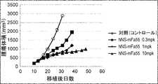
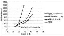
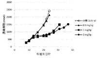
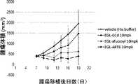
- Figure 4shows the antitumor effect of anti-CTLA-4 antibody SW1610-mFa55 (switch antibody) in a mouse model implanted with Colon38 cell line (colon cancer), as described in Example 2-3-5. It is a diagram. The switch antibody was administered via the tail vein at 0.3 mg/kg, 1 mg/kg, and 10 mg/kg.
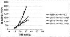
- Figure 6shows anti-CTLA-4 antibody hNS-mFa55 (control antibody) and SW1610-mFa55, 1389-
- FIG. 3shows the antitumor effect of mFa55 (switch antibody).
- Figure 7shows anti-CTLA-4 antibody hNS-mFa55 (control antibody) and SW1610-mFa55 in a mouse model implanted with the B16F1/OVA cell line (melanoma), as described in Example 2-5-5. , It is a figure showing the antitumor effect of 1389-mFa55 (switch antibody).
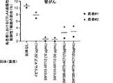
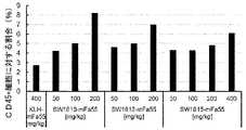
- FIG. 8shows Treg depletion activity by Ipilimumab and SW1610-ART12, SW1389-ART5+ACT1 (switch antibody) in colon cancer patient-derived PBMCs, as described in Example 3.
- Various antibodieswere treated at 10 ⁇ g/mL or 50 ⁇ g/mL. Each point represents a sample from a different patient, the mean value of which is indicated by a horizontal line.
- FIG. 9shows Treg depletion activity by Ipilimumab and SW1610-ART12, SW1389-ART5+ACT1 (switch antibody) in head and neck cancer patient-derived PBMCs, as described in Example 3.
- Various antibodieswere treated at 10 ⁇ g/mL or 50 ⁇ g/mL.
- FIG. 10shows Treg depletion activity by Ipilimumab and SW1610-ART12, SW1389-ART5+ACT1 (switch antibody) in pancreatic cancer patient-derived PBMCs, as described in Example 3.
- Various antibodieswere treated at 10 ⁇ g/mL or 50 ⁇ g/mL.
- Each pointrepresents a sample from a different patient, the mean value of which is indicated by a horizontal line.
- FIG. 11shows Treg depletion activity by Ipilimumab and SW1610-ART12, SW1389-ART5+ACT1 (switch antibody) in renal cancer patient-derived PBMCs, as described in Example 3.
- FIG. 12shows Treg depletion activity by Ipilimumab and SW1610-ART12, SW1389-ART5+ACT1 (switch antibody) in lung cancer patient-derived PBMCs, as described in Example 3.
- FIG. 13shows the effect of anti-CTLA-4 antibody SW1610-mFa55 (switch antibody) in combination with anti-PD-L1 antibody in a mouse model engrafted with the Colon38 cell line (colon cancer), as described in Example 5.
- FIG. 13shows the effect of anti-CTLA-4 antibody SW1610-mFa55 (switch antibody) in combination with anti-PD-L1 antibody in a mouse model engrafted with the Colon38 cell line (colon cancer), as described in Example 5.
- FIG. 3shows tumor effects.
- FIG. 14shows the effect of anti-CTLA-4 antibody SW1389-mFa55 (switch antibody) in combination with anti-PD-L1 antibody in a mouse model engrafted with the Colon38 cell line (colon cancer), as described in Example 5.
- FIG. 3shows tumor effects.
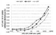
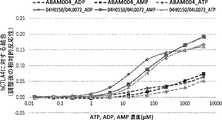
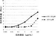
- FIG. 15shows the CTLA-4 binding activity of the anti-CTLA-4 antibody ABAM004 depending on the ATP, ADP or AMP concentrations, as described in Reference Examples 1-9.
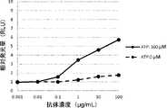
- FIG. 16shows the AMP concentration-dependent binding activity of ABAM004, an anti-CTLA-4 antibody, to CTLA-4-expressing cells, as described in Reference Example 1-10.
- FIG. 17shows the ADCC activity of the anti-CTLA-4 antibody ABAM004 against CTLA-4-expressing cells in the presence and absence of AMP, as described in Reference Example 1-11.
- FIG. 18shows the mode of binding of ABAM004 Fab fragment and AMP, as described in Reference Examples 2-13.
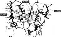
- FIG. 19shows the mode of binding of the ABAM004 Fab fragment, AMP, and human CTLA4 (hCTLA4), as described in Reference Examples 2-14.
- the heavy chain of the antibodyis shown in black, the light chain in gray, hCTLA4 in white, and AMP in a ball-and-stick model.
- FIG. 20is a diagram mapping the epitope of the ABAM004 Fab fragment into the amino acid sequence of hCTLA4, as described in Reference Examples 2-14.
- amino acid residues shown in blackindicate amino acid residues of hCTLA4 containing one or more non-hydrogen atoms located within 4.2 ⁇ of either portion of ABAM004 or AMP in the crystal structure.
- Amino acid residues shown in grayindicate residues for which no models were constructed because they were disordered in the crystal structure.
- FIG. 21shows the structures of the antibody and AMP extracted from the crystal structures of the complex of the ABAM004 Fab fragment alone and AMP, and the ternary complex of AMP and CTLA4, as described in Reference Example 2-15. is a superimposed diagram.
- the heavy chain of the antibodyis shown in black, the light chain in gray, and AMP in a ball-and-stick model.
- the thin lineindicates the structure of the ABAM004 Fab fragment alone
- the medium thick lineindicates the structure of the 2-part complex with AMP
- the thick lineindicates the structure of the 3-part complex.
- FIG. 22shows the CTLA-4 binding activity of the anti-CTLA-4 antibody ABAM004 and its variant 04H0150/04L0072 depending on the ATP, ADP or AMP concentrations, as described in Reference Example 3-2. It is a diagram. As notations in the figure, WT indicates ABAM004, and H150L072 indicates 04H0150/04L0072.
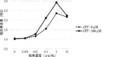
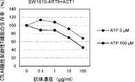
- FIG. 23shows the ATP concentration-dependent neutralizing activity of anti-CTLA-4 antibody SW1077 against CTLA-4, as described in Reference Example 3-6.
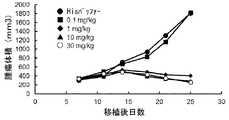
- FIG. 24is a diagram showing the antitumor effect of the anti-CTLA-4 antibody mNS-mFa55 (control antibody) in a mouse model transplanted with FM3A cell line, as described in Reference Example 3-7-4. .
- Figure 25shows the antitumor effect of anti-CTLA-4 antibody SW1208-mFa55 (switch antibody) in a mouse model implanted with FM3A cell line, as described in Reference Example 3-7-4. It is a diagram.
- FIG. 26shows anti-CTLA-4 antibody mNS-mFa55 (control antibody) and SW1208-mFa55 (switch antibody) administration in a mouse model transplanted with FM3A cell line, as described in Reference Example 3-7-7.
- FIG. 3shows changes in the ratio of effector Treg cells in tumors over time.
- mNS-mFa55was administered via the tail vein at 0.1 mg/kg, 1 mg/kg, 10 mg/kg and 100 mg/kg
- SW1208-mFa55was administered at 0.1 mg/kg, 1 mg/kg and 10 mg/kg. 100 mg/kg and 500 mg/kg were administered through the tail vein.
- Six days after administrationtumors were sampled and the increase or decrease in effector Tregs was evaluated by FACS analysis.
- Figure 27shows anti-CTLA-4 antibody mNS-mFa55 (control antibody) and SW1208-mFa55 (switch antibody) administration in a mouse model transplanted with FM3A cell line, as described in Reference Example 3-7-8.
- FIG. 10is a diagram showing changes in the ratio of activated helper T cells in the spleen over time.
- mNS-mFa55was administered at 0.1 mg/kg, 1 mg/kg, 10 mg/kg, and 100 mg/kg via the tail vein, and SW1208-mFa55 was administered at 0.1 mg/kg, 1 mg/kg, 10 mg/kg, 100 mg/kg and 500 mg/kg were administered through the tail vein.
- Six days after the administrationspleens were collected, and the increase or decrease in activated helper T cells was evaluated by FACS analysis.
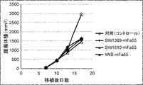
- FIG. 28shows the antitumor effect of the anti-CTLA-4 antibody SW1389-mFa55 (switch antibody) in a mouse model transplanted with Hepa1-6/hGPC3 cell lines, as described in Reference Example 4-3-5.
- Figure 29shows the antitumor effect of the anti-CTLA-4 antibody hNS-mFa55 (control antibody) in a mouse model transplanted with Hepa1-6/hGPC3 cell lines, as described in Reference Example 4-3-5.
- Figure 29shows the antitumor effect of the anti-
- Figure 30shows anti-CTLA-4 antibody hNS-mFa55 (control antibody) and SW1389-mFa55 ( FIG. 10 shows changes in the ratio of effector Treg cells in tumors upon administration of switch antibody).
- FIG. 10shows changes in the percentage of activated helper T cells in the spleen upon administration of switch antibody).
- hNS-mFa55at 0.1 mg/kg, 1 mg/kg, 10 mg/kg, 30 mg/kg, SW1389-mFa55 at 0.1 mg/kg, 1 mg/kg, 10 mg/kg, 100 mg/kg, 500 mg /kg via the tail vein.
- spleenswere collected, and the increase or decrease in activated helper T cells was evaluated by FACS analysis.
- FIG. 32shows the antitumor effect of the anti-CTLA-4 antibody SW1610-mFa55 (switch antibody) in a mouse model transplanted with Hepa1-6/hGPC3 cell lines, as described in Reference Example 5-4-5.
- Figure 33shows the anti-tumor effect of the anti-CTLA-4 antibody SW1612-mFa55 (switch antibody) in a mouse model transplanted with Hepa1-6/hGPC3 cell lines, as described in Reference Example 5-4-5.
- Figure 33shows the anti-tumor effect of the anti-CTLA-4 antibody SW1612-
- Figure 34shows the anti-tumor effect of the anti-CTLA-4 antibody SW1615-mFa55 (switch antibody) in a mouse model transplanted with Hepa1-6/hGPC3 cell lines, as described in Reference Example 5-4-5.
- FIG. 35shows anti-CTLA-4 antibodies SW1610-mFa55, SW1612-mFa55 and SW1615-mFa55 ( All of these figures show changes in the ratio of effector Treg cells in tumors upon administration of a switch antibody.
- negative control antibody KLH-mFa55 at 400 mg/kgwere administered through the tail vein.
- Six days after administrationtumors were sampled and the increase or decrease in effector Tregs was evaluated by FACS analysis.
- Figure 36shows anti-CTLA-4 antibodies SW1610-mFa55, SW1612-mFa55 and SW1615-mFa55 ( All of these figures show changes in the ratio of activated helper T cells in the spleen upon administration of switch antibody).
- SW1610-mFa55 at 50 mg/kg, 100 mg/kg and 200 mg/kg, SW1612-mFa55 at 50 mg/kg, 100 mg/kg and 200 mg/kg, SW1615-mFa55 at 50 mg/kg and 100 mg/kg kg, 200 mg/kg, 400 mg/kg, and negative control antibody KLH-mFa55 at 400 mg/kgwere administered through the tail vein.
- Six days after the administrationspleens were collected, and the increase or decrease in activated helper T cells was evaluated by FACS analysis.
- IgG1is MDX10D1H-G1m/MDX10D1L-k0MT
- GASDALIEis MDX10D1H-GASDALIE/MDX10D1L-k0MT
- ART6is MDX10D1H-Kn462/MDX10D1H-Hl445/MDX10D1L-k0MT
- ART8is MDX10D1H-KnDX10D461/MDX1HDX10D461 -Hl443/MDX10D1L-k0MT respectively.
- IgG1is an antibody having a control constant region
- GASDALIEis an antibody having a constant region described in the prior art
- ART6 and ART8are antibodies having a modified constant region produced in Reference Example 6-1. is.
- FIG. 38shows a comparison of in vitro ADCP activities of antibodies having various modified constant regions with enhanced binding to Fc ⁇ R, as described in Reference Example 6-3.
- IgG1is MDX10D1H-G1m/MDX10D1L-k0MT
- GASDIEis MDX10D1H-GASDIE/MDX10D1L-k0MT
- ART6is MDX10D1H-Kn462/MDX10D1H-Hl445/MDX10D1L-k0MT
- ART8is MDX10D1H-Kn461/MDX10MT. -Hl443/MDX10D1L-k0MT respectively.
- IgG1is an antibody having a control constant region
- GASDIEis an antibody having a constant region described in a prior document
- ART6 and ART8are antibodies having a modified constant region produced in Reference Example 6-1. is.
- FIG. 39shows in vitro ADCC activity of anti-CTLA4 switch antibody SW1389-ART6 having a modified constant region with enhanced binding to Fc ⁇ R, as described in Reference Example 6-4.
- FIG. 40shows in vitro ADCC activity of anti-CTLA4 switch antibody SW1610-ART6 having a modified constant region with enhanced binding to Fc ⁇ R, as described in Reference Example 6-4.
- FIG. 41shows in vitro ADCC activity of the anti-CTLA4 switch antibody SW1612-ART6, which has a modified constant region with enhanced binding to Fc ⁇ R, as described in Reference Example 6-4.
- FIG. 42shows the neutralizing activity of the anti-CTLA4 switch antibody SW1389 against CTLA4 (the activity of canceling the signal of CTLA4 that suppresses the activation of effector cells), as described in Reference Example 6-5. It is a figure which shows.
- FIG. 43shows the neutralizing activity of the anti-CTLA4 switch antibody SW1610 against CTLA4 (activity to release CTLA4 signaling that suppresses effector cell activation), as described in Reference Example 6-5. It is a figure which shows.
- Figure 44shows the neutralizing activity of the anti-CTLA4 switch antibody SW1612 against CTLA4 (activity to release CTLA4 signaling that suppresses effector cell activation), as described in Reference Example 6-5. It is a figure which shows.
- Figure 45shows the neutralizing activity of the anti-CTLA4 switch antibody SW1615 against CTLA4 (activity to release CTLA4 signaling that acts suppressively on effector cell activation), as described in Reference Example 6-5. It is a figure which shows.
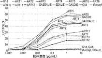
- Figure 46shows the in vitro cytotoxic activity of the anti-CTLA4 switch antibody SW1389-ART5+ACT1 against CTLA4-positive regulatory T cells, as described in Reference Example 6-6.
- Figure 47shows the in vitro cytotoxic activity of the anti-CTLA4 switch antibody SW1389-ART6+ACT1 against CTLA4-positive regulatory T cells, as described in Reference Example 6-6.
- Figure 48shows the in vitro cytotoxic activity of the anti-CTLA4 switch antibody SW1610-ART5+ACT1 against CTLA4-positive regulatory T cells, as described in Reference Example 6-6.
- Figure 49shows the in vitro cytotoxic activity of the anti-CTLA4 switch antibody SW1610-ART6+ACT1 against CTLA4-positive regulatory T cells, as described in Reference Example 6-6.
- FIG. 53is a diagram showing the hC1q-binding activity of each antibody having a modified Fc, as described in Reference Example 12.
- FIG. 54is a continuation showing the binding activity to hC1q of each antibody with modified Fc, as described in Reference Example 12.
- an "acceptor human framework” for the purposes of this specificationis a light chain variable domain (VL) framework or heavy chain variable domain (VH ) a framework containing the amino acid sequence of the framework.
- Acceptor human frameworks "derived from” human immunoglobulin frameworks or human consensus frameworksmay contain those same amino acid sequences or may contain alterations in the amino acid sequences. In some embodiments, the number of amino acid changes is 10 or less, 9 or less, 8 or less, 7 or less, 6 or less, 5 or less, 4 or less, 3 or less, or 2 or less.
- the VL acceptor human frameworkis identical in sequence to a VL human immunoglobulin framework sequence or a human consensus framework sequence.
- Antibody-dependent cell-mediated cytotoxicityrefers to the activity of secreted immunoglobulins on specific cytotoxic cells (e.g., NK cells, neutrophils and macrophages). binds to Fc receptors (FcR) present in the cytotoxic effector cells, thereby enabling these cytotoxic effector cells to specifically bind to antigen-bearing target cells and subsequently kill the target cells with cytotoxins
- FcRFc receptors
- FcR expression on hematopoietic cellsis summarized in Table 3 on page 464 of Ravetch and Kinet, Annu. Rev. Immunol 9: 457-92 (1991).
- an in vitro ADCC assaysuch as those described in US Pat. Nos. 5,500,362 or 5,821,337 or US Pat. No. 6,737,056 (Presta) can be performed.
- Useful effector cells for such assaysinclude PBMC and NK cells.
- ADCC activity of a molecule of interestmay be assessed in vivo in an animal model, such as that disclosed in Clynes et al. PNAS (USA) 95: 652-656 (1998). .
- cytotoxic activityexamples include, for example, the antibody-dependent cell-mediated cytotoxicity (ADCC) activity described above, the complement-dependent cytotoxicity (CDC) activity described below, and cytotoxic activity by T cells.
- CDC activitymeans cytotoxic activity by the complement system.
- ADCC activitymeans an activity in which an antibody binds to an antigen present on the cell surface of a target cell, and the effector cell binds to the antibody, causing the effector cell to damage the target cell.
- ADCC activitymeans an activity in which an antibody binds to an antigen present on the cell surface of a target cell, and the effector cell binds to the antibody, causing the effector cell to damage the target cell.
- ADCC activitymeans an activity in which an antibody binds to an antigen present on the cell surface of a target cell, and the effector cell binds to the antibody, causing the effector cell to damage the target cell.
- Whether the antibody of interest has ADCC activity or whether it has CDC activitycan be measured by a known
- Neutralizing activityrefers to the activity of inhibiting a biological activity by binding an antibody to a molecule involved in that biological activity.
- the biological activityis provided by binding of the ligand and receptor.
- binding of the antibody to the ligand or receptorinhibits binding of the ligand to the receptor.
- Antibodies with such neutralizing activityare called neutralizing antibodies.
- the neutralizing activity of a test substancecan be measured by comparing the biological activity in the presence of ligand between conditions in the presence or absence of the test substance.
- ADCPantibody-dependent cellular phagocytosis
- phagocytic immune cellse.g., macrophages, neutrophils, and dendritic cells.
- binding activityand “binding capacity” are used interchangeably herein and refer to one or more binding sites (e.g. , variable region or Fc region) and the binding partner of a molecule (eg, antigen or Fc ⁇ receptor).
- binding activityis not strictly limited to 1:1 interactions between members of a binding pair (eg, antibody and antigen, or Fc region and Fc ⁇ receptor).
- avidityrefers to the intrinsic binding affinity (“affinity”) when the members of a binding pair reflect a 1:1 interaction with monovalence. When members of a binding pair are capable of both monovalent and multivalent binding, avidity is the sum of their avidity.
- binding activity of a molecule X for its partner Ycan generally be expressed by the dissociation constant (KD) or "amount of analyte bound per unit amount of ligand". Binding activity can be measured by routine methods known in the art, including those described herein. Specific illustrative and exemplary embodiments for measuring binding activity are described below.
- An “affinity matured” antibodyhas one or more modifications in one or more hypervariable regions (HVRs) that confer improved affinity for the antibody's antigen as compared to a parent antibody that does not possess the modifications.
- HVRshypervariable regions
- anti-CTLA-4 antibodyor "antibody that binds CTLA-4" is an antibody capable of binding CTLA-4 with sufficient affinity such that the antibody targets CTLA-4.
- the extent of binding of the anti-CTLA-4 antibody to an unrelated non-CTLA-4 proteinis determined (e.g., by radioimmunoassay (RIA)) when the antibody's binding to CTLA-4 less than about 10% of
- the antibody that binds CTLA-4is ⁇ 1 ⁇ M, ⁇ 100 nM, ⁇ 10 nM, ⁇ 1 nM, ⁇ 0.1 nM, ⁇ 0.01 nM, or ⁇ 0.001 nM (e.g., 10 ⁇ 8 M or less , eg 10 ⁇ 8 M to 10 ⁇ 13 M, eg 10 ⁇ 9 M to 10 ⁇ 13 M).
- the anti-CTLA-4 antibodybinds to an epitope of CT
- antibodyis used in the broadest sense, and as long as it exhibits the desired antigen-binding activity, it is not limited to, but is not limited to, monoclonal antibodies, polyclonal antibodies, multispecific antibodies (e.g., It encompasses a variety of antibody structures, including bispecific antibodies) and antibody fragments.
- antibody fragmentrefers to a molecule other than an intact antibody that contains a portion of the intact antibody that binds to the antigen to which the intact antibody binds.
- antibody fragmentsinclude, but are not limited to, Fv, Fab, Fab', Fab'-SH, F(ab') 2 ; diabodies; linear antibodies; single chain antibody molecules (e.g., scFv ); and multispecific antibodies formed from antibody fragments.
- an "antibody that binds to the same epitope" as a reference antibodyrefers to an antibody that blocks the binding of the reference antibody to its own antigen in a competition assay, e.g. block the binding of the aforementioned antibody to its own antigen in, for example, 50% or more.
- a competition assaye.g. block the binding of the aforementioned antibody to its own antigen in, for example, 50% or more.
- autoimmune diseaserefers to a non-malignant disease or disorder arising from and directed against the individual's own tissues.
- autoimmune diseasespecifically excludes malignant or cancerous diseases or conditions, in particular B-cell lymphoma, acute lymphoblastic leukemia (ALL), chronic lymphocytic Exclude chronic lymphocytic leukemia (CLL), hairy cell leukemia, and chronic myeloblastic leukemia.
- ALLacute lymphoblastic leukemia
- CLLchronic lymphocytic leukemia
- hairy cell leukemiaand chronic myeloblastic leukemia.
- autoimmune diseases or disordersinclude, but are not limited to: inflammatory reactions such as inflammatory skin diseases including psoriasis and dermatitis (e.g., atopic dermatitis); scleroderma and sclerosis; inflammatory bowel disease-related reactions (e.g., Crohn's disease and ulcerative colitis); respiratory distress syndrome (including adult respiratory distress syndrome: ARDS); dermatitis; colitis; glomerulonephritis; allergic conditions such as eczema and asthma and other conditions with T-cell infiltration and chronic inflammatory responses; atherosclerosis; rheumatoid arthritis; systemic lupus erythematosus (SLE) (including but not limited to lupus nephritis, cutaneous lupus); diabetes (e.g., type I diabetes or insulin-dependent diabetes); multiple sclerosis; Raynaud's syndrome; allergic encephalomyelitis; Sjogren's syndrome; juvenile-onset diabetes; and by inflammatory
- cancerand “cancerous” refer to or describe the physiological condition in mammals that is typically characterized by unregulated cell growth/proliferation. Examples of cancer include breast cancer and liver cancer.
- complement dependent cytotoxicityrefers to the Fc effector domain of a target-bound antibody that activates a series of enzymatic reactions that result in the formation of holes in the target cell's membrane. means a mechanism for inducing Typically, antigen-antibody complexes formed on target cells bind and activate complement component C1q, which in turn activates the complement cascade leading to target cell death. bring. Complement activation may also lead to the deposition of complement components on the surface of target cells, which promote ADCC by binding complement receptors (e.g. CR3) on leukocytes. .
- complement receptorse.g. CR3
- “Chemotherapeutic agent”refers to a chemical compound useful in the treatment of cancer.
- chemotherapeutic agentsinclude: alkylating agents such as thiotepa and cyclophosphamide (CYTOXAN®); alkyl sulfonates such as busulfan, improsulfan, and piposulfan; benzodopa, carbocones aziridines such as , meturedopa, and uredopa; ethyleneimines and methylolmelamines, including altretamine, triethylenemelamine, triethylenephosphoramide, triethylenethiophosphoramide, and trimethylomelamine; delta-9-tetrahydrocannabinol (dronabinol, MARINOL®); beta-lapachone; lapachol; colchicine; betulinic acid; (irinotecan, CAMPTOSAR®), acetylcamptothecin, scopolectin, and 9-aminocamptothecin
- Nitrogen mustardssuch as carmustine, chlorozotocin, fotemustine, lomustine, nimustine, and ranimustine; enediyne antibiotics ⁇ e.g. et al., Angew. Chem Intl. Ed.
- purine analoguespyrimidine analogues, such as ancitabine, azacytidine, 6-azauridine, carmofur, cytarabine, dideoxyuridine, doxifluridine, enocitabine, floxuridine; androgens, such as carsterone, dromostanolone propionate, epithiostanol, mepithiostane, testolactone anti-adrenal agents such as aminoglutethimide, mitotane, trilostane; enals); folic acid supplements, such as folinic acid; acegratone; aldophosphamide glycosides; aminolevulinic acid; eniluracil; amsacrine; elliptinium acetate; epothilone; etogluside; gallium nitrate; hydrocyurea; lentinan; 2-Ethylhydrazide; Procarbazine; PSK
- celecoxib or etoricoxibinclude proteosome inhibitors (e.g. PS341); VELCADE®); CCI-779; tipifarnib (R11577); sorafenib, ABT510; Bcl-2 inhibitors such as oblimersen sodium (GENASENSE®); tyrosine kinase inhibitors (see definitions below); serine-threonine kinase inhibitors, such as rapamycin (sirolimus, RAPAMUNE®); lonafarnib (SCH 6636, Farnesyltransferase inhibitors, such as SARASARTM); and pharmaceutically acceptable salts, acids, or derivatives of any of the above; and combinations of two or more of the above, such as cyclophosphamide, doxorubicin.
- proteosome inhibitorse.g. PS341
- VELCADE®e.g. PS341
- CCI-779tipifarnib (R11577
- CHOPan abbreviation for combination therapy of vincristine and prednisolone
- FOLFOXan abbreviation for treatment regimens with oxaliplatin (ELOXANTINTM) in combination with 5-FU and leucovorin.
- chimeric antibodymeans that a portion of the heavy and/or light chains are derived from a particular source or species, while the remainder of the heavy and/or light chains are derived from a different source or species. Refers to antibodies.
- the "class" of an antibodyrefers to the type of constant domain or constant region found in the heavy chain of an antibody.
- the heavy-chain constant domains that correspond to the different classes of immunoglobulinsare called ⁇ , ⁇ , ⁇ , ⁇ , and ⁇ , respectively.
- cytotoxic agentrefers to a substance that inhibits or prevents the function of cells and/or causes the death or destruction of cells.
- Cytotoxic agentsinclude, but are not limited to, radioactive isotopes such as 211 At, 131 I, 125 I, 90 Y, 186 Re, 188 Re, 153 Sm, 212 Bi, 32 P, 212 Pb and radioisotopes of Lu); chemotherapeutic agents or agents (e.g., methotrexate, adriamycin, vinca alkaloids (vincristine, vinblastine, etoposide), doxorubicin, melphalan, mitomycin C, chlorambucil, daunorubicin, or other intercalating growth inhibitors; enzymes such as nucleases and fragments thereof; antibiotics; and the various chemotherapeutic agents disclosed above.
- radioactive isotopessuch as 211 At, 131 I, 125 I, 90 Y, 186 Re, 188 Re
- Effective cellsrefer to leukocytes that express one or more FcRs and exert effector functions. In certain embodiments, the cell expresses at least Fc ⁇ RIII and exerts ADCC effector function. Examples of leukocytes that mediate ADCC include peripheral blood mononuclear cells (PBMCs), natural killer (NK) cells, monocytes, cytotoxic T cells and neutrophils. Effector cells can be isolated from natural sources, such as blood. In certain embodiments, the effector cells can be human effector cells.
- “Effector function”refers to the biological activity that is attributed to the Fc region of an antibody and differs depending on the antibody isotype.
- Examples of antibody effector functionsinclude: C1q binding and complement-dependent cytotoxicity (CDC); Fc receptor binding; antibody-dependent cell-mediated cytotoxicity (CDC); cell-mediated cytotoxicity (ADCC); antibody-dependent cell-mediated phagocytosis (ADCP); downregulation of cell surface receptors (e.g., B cell receptors); and B cell activation .
- epitopeincludes any determinant capable of being bound by an antibody.
- An epitopeis the region of an antigen that is bound by an antibody that targets it, and includes specific amino acids that directly contact the antibody.
- Epitopic determinantscan include chemically active surface groupings of molecules such as amino acids, sugar side chains, phosphoryl or sulfonyl groups, and possess specific three dimensional structural characteristics, and/or specific charge characteristics. can be done.
- antibodies specific for a particular target antigenpreferentially recognize epitopes on that target antigen in a complex mixture of proteins and/or macromolecules.
- Fc receptorrefers to a receptor that binds to the Fc region of an antibody.
- the FcRis a native human FcR.
- the FcRis one that binds IgG antibodies (gamma receptors), and receptors of the Fc ⁇ RI, Fc ⁇ RII, and Fc ⁇ RIII subclasses are allelic variants and alternatively spliced forms of these receptors. including, including.
- Fc ⁇ RII receptorsinclude Fc ⁇ RIIA (an “activating receptor”) and Fc ⁇ RIIB (an “inhibiting receptor”), which have similar amino acid sequences that differ primarily in their cytoplasmic domains.
- Activating receptor Fc ⁇ RIIAcontains an immunoreceptor tyrosine-based activation motif (ITAM) in its cytoplasmic domain.
- the inhibitory receptor Fc ⁇ RIIBcontains an immunoreceptor tyrosine-based inhibition motif (ITIM) in its cytoplasmic domain (see, e.g., Daeron, Annu. Rev. Immunol. 15: 203-234 (1997)). matter).
- FcRfor example, Ravetch and Kinet, Annu. Rev. Immunol 9: 457-492 (1991); Capel et al., Immunomethods 4: 25-34 (1994); and de Haas et al., J. Lab. Clin Reviewed in Med 126: 330-341 (1995).
- Other FcRsincluding those identified in the future, are also encompassed by the term "FcR" herein.
- Fc receptoror “FcR” also refers to the transfer of maternal IgG to the fetus (Guyer et al., J. Immunol. 117: 587 (1976) and Kim et al., J. Immunol. 24: 249 (1994)) as well as the neonatal receptor FcRn, which is responsible for regulating immunoglobulin homeostasis. Methods for measuring binding to FcRn are known (e.g., Ghetie and Ward., Immunol. Today 18(12): 592-598 (1997); Ghetie et al., Nature Biotechnology, 15(7): 637- 640 (1997); Hinton et al., J. Biol. Chem. 279(8): 6213-6216 (2004); see WO2004/92219 (Hinton et al.)).
- Binding to human FcRn in vivo and serum half-life of human FcRn high-affinity binding polypeptidescan be determined, for example, in transgenic mice or transfected human cell lines expressing human FcRn, or in which polypeptides with mutated Fc regions are It can be measured in administered primates.
- WO2000/42072(Presta) describes antibody variants with improved or decreased binding to FcRs. See, for example, Shields et al. J. Biol. Chem. 9(2): 6591-6604 (2001).
- Fc regionis used herein to define the C-terminal region of an immunoglobulin heavy chain that includes at least a portion of the constant region.
- the termincludes native sequence Fc regions and variant Fc regions.
- the human IgG heavy chain Fc regionextends from Cys226 or from Pro230 to the carboxyl terminus of the heavy chain.
- lysine (Lys447) or glycine-lysine (Gly446-Lys447) at the C-terminus of the Fc regionmay or may not be present.
- the numbering of amino acid residues in the Fc region or constant regionis according to Kabat et al., Sequences of Proteins of Immunological Interest, 5th Ed. Public Health Service, National Institutes of Health, Bethesda, According to the EU numbering system (also known as the EU index), as described in MD 1991.
- Fc region-containing antibodyrefers to an antibody containing an Fc region.
- the C-terminal lysine of the Fc region (residue 447 according to the EU numbering system) or the C-terminal glycine-lysine of the Fc region (residues 446-447), for example during purification of the antibody or nucleic acids encoding the antibodycan be removed by recombinant manipulation of
- a composition comprising an antibody having an Fc region according to the present inventionmay be an antibody with G446-K447, an antibody with G446 without K447, an antibody with G446-K447 completely removed, or an antibody of the above three types may contain mixtures of
- variable regionrefers to the domains of the heavy or light chains of an antibody that are involved in binding the antibody to the antigen.
- the heavy and light chain variable domains (VH and VL, respectively) of native antibodiesare usually similar, with each domain containing four conserved framework regions (FR) and three hypervariable regions (HVR). structure (see, for example, Kindt et al. Kuby Immunology, 6th ed., W.H. Freeman and Co., page 91 (2007)).
- a single VH or VL domainmay be sufficient to confer antigen binding specificity.
- antibodies that bind a particular antigenmay be isolated using the VH or VL domains from the antibody that binds the antigen to screen a complementary library of VL or VH domains, respectively. See, e.g., Portolano et al., J. Immunol. 150: 880-887 (1993); Clarkson et al., Nature 352: 624-628 (1991).
- FRFramework or "FR” refers to variable domain residues other than hypervariable region (HVR) residues.
- the FRs of variable domainsusually consist of four FR domains: FR1, FR2, FR3 and FR4. Accordingly, HVR and FR sequences usually appear in VH (or VL) in the following order: FR1-H1(L1)-FR2-H2(L2)-FR3-H3(L3)-FR4.
- full length antibody“whole antibody” and “whole antibody” are used interchangeably herein and have a structure substantially similar to that of a naturally occurring antibody or as defined herein. It refers to an antibody having a heavy chain containing an Fc region that
- a “functional Fc region”has an "effector function” of a native-sequence Fc region.
- effector functionsinclude C1q binding; CDC; Fc receptor binding; ADCC; phagocytosis; Such effector functions generally require the Fc region to be combined with a binding domain (e.g., an antibody variable domain) and are assessed using, for example, various assays disclosed within the definitions herein. can be
- human antibodyis an antibody with an amino acid sequence that corresponds to that of an antibody produced by a human or human cells or derived from a human antibody repertoire or other non-human source using human antibody coding sequences. This definition of human antibody specifically excludes humanized antibodies that contain non-human antigen-binding residues.
- a "human consensus framework”is a framework that represents the most commonly occurring amino acid residues in a selected group of human immunoglobulin VL or VH framework sequences.
- the selection of human immunoglobulin VL or VH sequencesis from a subgroup of variable domain sequences.
- the subgroup of sequencesis the subgroup in Kabat et al., Sequences of Proteins of Immunological Interest, Fifth Edition, NIH Publication 91-3242, Bethesda MD (1991), vols. 1-3.
- the subgroupis subgroup ⁇ I according to Kabat et al., supra.
- the subgroupis subgroup III according to Kabat et al., supra.
- a “humanized” antibodyrefers to a chimeric antibody that contains amino acid residues from non-human HVRs and amino acid residues from human FRs.
- a humanized antibodycomprises substantially all of at least one, typically two, variable domains in which all or substantially all HVRs (e.g., CDRs) are non- Corresponds to that of a human antibody, and all or substantially all FRs correspond to those of a human antibody.
- a humanized antibodyoptionally may comprise at least a portion of an antibody constant region derived from a human antibody.
- a "humanized form" of an antibody(eg, a non-human antibody) refers to an antibody that has undergone humanization.
- hypervariable regionis hypervariable in sequence (“complementarity determining region” or “CDR”) and/or is structurally defined Refers to the regions of the variable domain of an antibody that form loops (“hypervariable loops”) and/or contain antigen-contacting residues (“antigen-contacting”).
- Antibodiestypically contain six HVRs: three in VH (H1, H2, H3) and three in VL (L1, L2, L3).
- Exemplary HVRs hereininclude: (a) at amino acid residues 26-32 (L1), 50-52 (L2), 91-96 (L3), 26-32 (H1), 53-55 (H2), and 96-101 (H3); resulting hypervariable loops (Chothia and Lesk, J. Mol. Biol. 196: 901-917 (1987)); (b) at amino acid residues 24-34 (L1), 50-56 (L2), 89-97 (L3), 31-35b (H1), 50-65 (H2), and 95-102 (H3); the resulting CDRs (Kabat et al., Sequences of Proteins of Immunological Interest, 5th Ed.
- HVR residues and other residues in the variable domainare numbered herein according to Kabat et al., supra.
- immunoconjugateis an antibody conjugated to one or more heterologous molecules (heterologous molecules include, but are not limited to, cytotoxic agents).
- an “isolated” antibodyis one that has been separated from a component of its original environment.
- antibodiesare analyzed electrophoretically (e.g., SDS-PAGE, isoelectric focusing (IEF), capillary electrophoresis) or chromatographically (e.g., ion-exchange or reverse-phase HPLC). Purified to greater than 95% or 99% purity as measured.
- electrophoreticallye.g., SDS-PAGE, isoelectric focusing (IEF), capillary electrophoresis
- chromatographicallye.g., ion-exchange or reverse-phase HPLC
- isolated nucleic acidrefers to a nucleic acid molecule that has been separated from components of its original environment.
- An isolated nucleic acidincludes a nucleic acid molecule contained within cells that normally contain the nucleic acid molecule, but the nucleic acid molecule is present extrachromosomally or on a chromosome other than its native chromosomal location. exists in position.
- isolated nucleic acid encoding an antibodyrefers to one or more nucleic acid molecules encoding the heavy and light chains (or fragments thereof) of an antibody, whether on a single vector or separate vectors. and nucleic acid molecules present in one or more locations in a host cell.
- first polypeptide and second polypeptiderefer to polypeptides that constitute the Fc region of an antibody.
- first polypeptide and second polypeptidemean that they differ in sequence from each other, preferably at least in the CH2 region. Furthermore, the CH3 regions may have different sequences.
- the polypeptidemay be, for example, a polypeptide that constitutes the Fc region of native IgG, or a polypeptide obtained by modifying the polypeptide that constitutes the Fc region of native IgG. .
- “Native IgG”means a polypeptide that includes the same amino acid sequence as IgG found in nature and belongs to the class of antibodies substantially encoded by the immunoglobulin gamma gene.
- natural human IgGmeans natural human IgG1, natural human IgG2, natural human IgG3, natural human IgG4, and the like.
- Natural-type IgGalso includes naturally occurring mutants and the like.
- Polypeptidein the present invention usually refers to peptides and proteins having a length of about 10 amino acids or more.
- the polypeptideis usually derived from a living organism, it is not particularly limited, and may be, for example, a polypeptide consisting of an artificially designed sequence. Also, it may be a natural polypeptide, a synthetic polypeptide, a recombinant polypeptide, or the like.
- the protein molecule in the present inventionrefers to a molecule containing the polypeptide.
- an antibodycan be mentioned as a preferred example of the polypeptide of the present invention. More preferred examples include natural IgG and antibodies obtained by modifying natural IgG.
- native IgGinclude, in particular, native human IgG.
- Native IgGrefers to a polypeptide that contains the same amino acid sequence as IgG found in nature and belongs to the class of antibodies substantially encoded by the immunoglobulin gamma gene.
- natural human IgGmeans natural human IgG1, natural human IgG2, natural human IgG3, natural human IgG4, and the like. Natural-type IgG also includes naturally occurring mutants and the like.
- polypeptide containing an Fc regionis not particularly limited as long as it is a polypeptide containing an Fc region, but for example, an antibody containing an Fc region.
- composition comprising a polypeptide having an Fc regionis a polypeptide comprising an Fc region with G446-K447, a polypeptide comprising an Fc region with G446 and without K447, a polypeptide comprising an Fc region with G446 and without K447, or a mixture of the above three types of polypeptides.
- an "isolated" polypeptideis one that has been separated from a component of its original environment.
- the polypeptideis electrophoretically (e.g., SDS-PAGE, isoelectric focusing (IEF), capillary electrophoresis) or chromatographically (e.g., ion exchange or reverse phase HPLC). Purified to greater than 95% or 99% purity as measured by For a review of methods for assessment of polypeptide purity, see, eg, Flatman et al., J. Chromatogr. B 848: 79-87 (2007).
- isolated nucleic acid encoding a polypeptiderefers to one or more nucleic acid molecules that encode the polypeptide (e.g., the Fc region of an antibody, or antibody heavy and light chains or fragments thereof). It includes nucleic acid molecules on one vector or on separate vectors and nucleic acid molecules that are present in one or more locations in a host cell.
- vectorrefers to a nucleic acid molecule capable of propagating another nucleic acid to which it has been linked.
- the termincludes vectors as self-replicating nucleic acid structures and vectors that integrate into the genome of a host cell into which they are introduced.
- Certain vectorsare capable of directing the expression of nucleic acids to which they are operably linked.
- Such vectorsare also referred to herein as "expression vectors".
- a vectorcan be introduced into a host cell by a method using a virus, an electroporation method, or the like, but the introduction of the vector is not limited to ex vivo introduction, and the vector can also be directly introduced into the living body. be.
- host cellrefers to a cell (including progeny of such a cell) into which exogenous nucleic acid has been introduced.
- a host cellincludes “transformants” and “transformed cells,” including the primary transformed cell and progeny derived therefrom at any passage number. Progeny may not be completely identical in nucleic acid content to the parent cell, but may contain mutations. Mutant progeny that have the same function or biological activity as with which the originally transformed cell was screened or selected are also included herein.
- the term "monoclonal antibody” as used hereinrefers to an antibody obtained from a substantially homogeneous population of antibodies. That is, the individual antibodies that make up the population are mutated antibodies that may arise (e.g., mutated antibodies that contain naturally occurring mutations or that arise during the manufacture of monoclonal antibody preparations. are identical and/or bind to the same epitope, except that they are present in greater amounts). In contrast to polyclonal antibody preparations, which typically include different antibodies directed against different determinants (epitopes), each monoclonal antibody of a monoclonal antibody preparation is directed against a single determinant on the antigen.
- monoclonalindicates the character of the antibody as being obtained from a population of substantially homogeneous antibodies and should not be construed as requiring production of the antibody by any particular method.
- monoclonal antibodies used in accordance with the present inventioninclude, but are not limited to, hybridoma technology, recombinant DNA technology, phage display technology, transgenic animals containing all or part of the human immunoglobulin locus. and such methods and other exemplary methods for making monoclonal antibodies are described herein.
- naked antibodyrefers to an antibody that is not conjugated to a heterologous moiety (eg, a cytotoxic moiety) or a radiolabel.
- a naked antibodymay be present in a pharmaceutical formulation.
- Native antibodiesrefer to immunoglobulin molecules with various structures occurring in nature.
- native IgG antibodiesare heterotetrameric glycoproteins of approximately 150,000 daltons composed of two identical light chains and two identical heavy chains that are disulfide-bonded. From N-terminus to C-terminus, each heavy chain has a variable region (VH), also called a variable heavy domain or heavy chain variable domain, followed by three constant domains (CH1, CH2 and CH3). Similarly, from N-terminus to C-terminus, each light chain has a variable region (VL), also called a variable light domain or light chain variable domain, followed by a constant light (CL) domain.
- VHvariable region
- VLvariable region
- CLconstant light domain
- the light chains of antibodiescan be assigned to one of two types, called kappa ( ⁇ ) and lambda ( ⁇ ), based on the amino acid sequences of their constant domains.
- a “native sequence Fc region”contains an amino acid sequence identical to the amino acid sequence of an Fc region found in nature.
- Native sequence human Fc regionsinclude native sequence human IgG1 Fc regions (non-A and A allotypes); native sequence human IgG2 Fc regions; native sequence human IgG3 Fc regions; including those variants present in
- a “variant Fc region”comprises an amino acid sequence that differs from that of a native sequence Fc region by at least one amino acid modification (alteration), preferably one or more amino acid substitutions.
- the variant Fc regionhas at least one amino acid substitution, such as about 1 to about 30 amino acid substitutions, in the native sequence Fc region or parental Fc region compared to the native sequence Fc region or parental Fc region. , preferably from about 1 to about 20 amino acid substitutions, more preferably from about 1 to about 10 amino acid substitutions, and most preferably from about 1 to about 5 amino acid substitutions.
- Variant Fc regions hereinare preferably at least about 80% homologous to native sequence Fc regions or parental Fc regions, preferably at least about 85% homologous thereto, more preferably at least about 90% homologous to them. % homology, most preferably at least about 95% homology therewith.
- a "percent (%) amino acid sequence identity" to a reference polypeptide sequenceis obtained after aligning the sequences to obtain the maximum percent sequence identity and introducing gaps if necessary, and without any conservative substitutions. Defined as the percentage of amino acid residues in a candidate sequence that are identical to amino acid residues in the reference polypeptide sequence, not considered part of the identity. Alignments for purposes of determining percent amino acid sequence identity may be performed by a variety of methods within the skill in the art, such as BLAST, BLAST-2, ALIGN, Megalign (DNASTAR) software, or GENETYX® (stock This can be accomplished by using publicly available computer software, such as the company Genetics. Those skilled in the art can determine appropriate parameters for aligning sequences, including any algorithms needed to achieve maximal alignment over the entire length of the sequences being compared.
- the ALIGN-2 sequence comparison computer programis the work of Genentech, Inc. and its source code has been filed with the US Copyright Office, Washington DC, 20559 with user documentation, under US Copyright Registration No. TXU510087. Registered.
- the ALIGN-2 programis publicly available from Genentech, Inc., South San Francisco, Calif., or may be compiled from source code.
- ALIGN-2 programsare compiled for use on UNIX operating systems, including Digital UNIX V4.0D. All sequence comparison parameters were set by the ALIGN-2 program and do not vary.
- the % amino acid sequence identity of a given amino acid sequence A to, with, or to a given amino acid sequence Bis calculated as follows: Hundredfold. where X is the number of amino acid residues scored as identical matches in the alignment of A and B by the sequence alignment program ALIGN-2, and Y is the total number of amino acid residues in B. . It is understood that the % amino acid sequence identity of A to B is not equal to the % amino acid sequence identity of B to A if the length of amino acid sequence A is not equal to the length of amino acid sequence B. deaf. Unless otherwise specified, all % amino acid sequence identity values used herein are obtained using the ALIGN-2 computer program as described in the immediately preceding paragraph.
- pharmaceutical formulationand “pharmaceutical composition” are used interchangeably and refer to a preparation in a form such that the biological activity of the active ingredients contained therein can be exerted. and that does not contain additional elements that are unacceptably toxic to the subjects to whom it is administered.
- “Individuals” or “subjects”are mammals. Mammals include, but are not limited to, domestic animals (eg, cows, sheep, cats, dogs, horses), primates (eg, humans and non-human primates such as monkeys), rabbits, and , including rodents (eg, mice and rats). In certain aspects, the individual or subject is human.
- “Pharmaceutically acceptable carrier”refers to an ingredient other than the active ingredient in pharmaceutical formulations and pharmaceutical compositions that is non-toxic to a subject.
- Pharmaceutically acceptable carriersinclude, but are not limited to, buffers, excipients, stabilizers, or preservatives.
- an “effective amount” of an agentrefers to that amount, at dosages necessary and for periods of time necessary, effective to achieve the desired therapeutic or prophylactic result. .
- package insertis commonly included in commercial packaging of therapeutic products and includes information about indications, usage, dosage, method of administration, concomitant therapies, contraindications, and/or warnings regarding the use of such therapeutic products. Used to refer to instructions for use.
- CTLA-4"may be from any vertebrate source, including mammals such as primates (e.g., humans) and rodents (e.g., mice and rats). Any native CTLA-4 of The term encompasses "full length" unprocessed CTLA-4 as well as any form of CTLA-4 that results from processing in the cell. The term also includes naturally occurring variants of CTLA-4, such as splice variants and allelic variants.
- Exemplary human CTLA-4 amino acid sequenceis SEQ ID NO: 214
- mouse CTLA-4 amino acid sequenceis SEQ ID NO: 247
- monkey CTLA-4 amino acid sequenceis SEQ ID NO: 248,
- human CTLA-4 amino acid sequenceis SEQ ID NO: 248.
- the amino acid sequence of the extracellular domainis shown in SEQ ID NO:28.
- CTLA-4is sometimes referred to as CTLA4.
- Treg cellsare a subpopulation of T cells that regulates the immune system, maintains tolerance to self-antigens, and prevents autoimmune diseases. These cells generally suppress or downregulate the induction and proliferation of effector T cells.
- the best understood Treg cellsare those that express CD4, CD25 and Foxp3 (CD4 + CD25 + Treg cells). These Tregs are different from helper T cells.
- Several different methodsare used to identify and monitor Treg cells. As defined by CD4 and CD25 expression (CD4 + CD25 + cells), Treg cells constitute approximately 5-10% of the mature CD4 + T cell subpopulation in mice and humans, whereas approximately 1-2% of Tregs It can be measured in whole blood.
- Foxp3 expressionmay additionally be measured (CD4 + CD25 + Foxp3 + cells). Absence or low level expression of CD127 may also be used in combination with the presence of CD4 and CD25 as another marker.
- Treg cellsalso express high levels of CTLA-4 and GITR. Tregs can also be identified by the methods described in the Examples below.
- the terms “substantially similar,” “substantially equal,” or “substantially the same”refer to between two numbers (e.g., with respect to the antibodies of the invention, see/compare The similarity between those two values (for example, the KD value) is defined by those skilled in the art as having little or no biological and/or similarity in terms of biological characteristics measured by the value (e.g., KD value). or high enough to be considered statistically insignificant.
- treatmentrefers to clinical treatments intended to alter the natural course of the individual being treated.
- interventionwhich can be performed both for prophylaxis and during the course of a clinical condition.
- Desirable effects of treatmentinclude, but are not limited to, prevention of disease onset or recurrence, relief of symptoms, attenuation of any direct or indirect pathological effects of the disease, prevention of metastasis, prevention of disease Includes reduced rate of progression, amelioration or amelioration of disease state, and remission or improved prognosis.
- the antibodies of the inventionare used to delay disease onset or slow disease progression.
- polypeptides comprising variant Fc regions of the inventionare used to delay disease onset or slow disease progression.
- tumorrefers to all neoplastic cell growth and proliferation and all precancerous and cancerous cells and tissues, whether malignant or benign.
- cancerrefers to all neoplastic cell growth and proliferation and all precancerous and cancerous cells and tissues, whether malignant or benign.
- cancerrefers to all neoplastic cell growth and proliferation and all precancerous and cancerous cells and tissues, whether malignant or benign.
- cancerrefers to all neoplastic cell growth and proliferation and all precancerous and cancerous cells and tissues, whether malignant or benign.
- tumor tissuemeans tissue containing at least one tumor cell.
- a tumor tissueis usually composed of a mass of tumor cells (parenchyma) that constitutes the main body of a tumor, and connective tissue and blood vessels (stroma) that exist between them and support the tumor. In some cases, the distinction between the two is clear, and in others, the two are mixed. Immune cells and the like may infiltrate into the tumor tissue.
- non-tumor tissuemeans tissue other than tumor tissue in vivo. Healthy/normal tissue without disease is a representative example of non-tumor tissue.
- the inventionis based in part on anti-CTLA-4 antibodies and their uses.
- antibodiesare provided that bind to CTLA-4.
- Antibodies of the inventionare useful, for example, for the diagnosis or treatment of cancer.
- the inventionprovides isolated antibodies that bind to CTLA-4.
- the anti-CTLA-4 antibodies of the inventionhave CTLA-4 binding activity that is dependent on the concentration of the adenosine-containing compound.
- the binding activity to CTLA-4is higher in the presence of the adenosine-containing compound than in the absence of the adenosine-containing compound.
- the binding activity to CTLA-4is higher in the presence of high concentrations of adenosine-containing compounds than in the presence of low concentrations of adenosine-containing compounds.
- the difference in binding activity to CTLA-4is, for example, 2-fold or more, 3-fold or more, 5-fold or more, 10-fold or more, 20-fold or more, 30-fold or more, 50-fold or more, 100-fold or more, 200-fold. 300 times or more, 500 times or more, 1 ⁇ 10 3 times or more, 2 ⁇ 10 3 times or more, 3 ⁇ 10 3 times or more, 5 ⁇ 10 3 times or more, 1 ⁇ 10 4 times or more, 2 ⁇ 10 4 times or more 3 ⁇ 10 4 times or more, 5 ⁇ 10 4 times or more, or 1 ⁇ 10 5 times or more.
- the binding activity of an anti-CTLA-4 antibodycan be expressed as a KD (Dissociation constant) value.
- the anti-CTLA-4 antibodyhas a lower KD value in the presence of the adenosine-containing compound than in the absence of the adenosine-containing compound.
- the anti-CTLA-4 antibodyhas a lower KD value in the presence of high concentrations of adenosine-containing compound than in the presence of low concentrations of adenosine-containing compound.
- the difference in KD values of anti-CTLA-4 antibodiesis, for example, 2-fold or more, 3-fold or more, 5-fold or more, 10-fold or more, 20-fold or more, 30-fold or more, 50-fold or more, 100-fold or more, 200-fold or more.
- the KD value of the anti-CTLA-4 antibody in the presence of an adenosine-containing compound or in the presence of a high concentration of an adenosine-containing compoundis, for example, 9 ⁇ 10 ⁇ 7 M or less, 8 ⁇ 10 ⁇ 7 M or less, 7 ⁇ 10 ⁇ 7 M or less, 6 ⁇ 10 -7 M or less, 5 ⁇ 10 -7 M or less, 4 ⁇ 10 -7 M or less, 3 ⁇ 10 -7 M or less, 2 ⁇ 10 -7 M or less, 1 ⁇ 10 -7 M or less , 9 ⁇ 10 -8 M or less, 8 ⁇ 10 -8 M or less, 7 ⁇ 10 -8 M or less, 6 ⁇ 10 -8 M or less, 5 ⁇ 10 -8 M or less, 4 ⁇ 10 -8 M or less, 3 ⁇ 10 -8 M or less, 2 ⁇ 10 -8 M or less, 1 ⁇ 10 -8 M or less, 9 ⁇ 10 -9 M or less, 8 ⁇ 10 -9 M or less, 7 ⁇ 10 -9 M or less, 6 ⁇ 10
- the KD value of the anti-CTLA-4 antibody in the absence of an adenosine-containing compound or in the presence of a low concentration of an adenosine-containing compoundis, for example, 1 ⁇ 10 ⁇ 8 M or more, 2 ⁇ 10 ⁇ 8 M or more, 3 ⁇ 10 ⁇ 8 M or higher, 4 ⁇ 10 -8 M or higher, 5 ⁇ 10 -8 M or higher, 6 ⁇ 10 -8 M or higher, 7 ⁇ 10 -8 M or higher, 8 ⁇ 10 -8 M or higher, 9 ⁇ 10 -8 M or higher 1 ⁇ 10 -7 M or more, 2 ⁇ 10 -7 M or more, 3 ⁇ 10 -7 M or more, 4 ⁇ 10 -7 M or more, 5 ⁇ 10 -7 M or more, 6 ⁇ 10 -7 M or more, 7 ⁇ 10 -7 M or more, 8 ⁇ 10 -7 M or more, 9 ⁇ 10 -7 M or more, 1 ⁇ 10 -6 M or more, 2 ⁇ 10 -6 M or more, 3 ⁇ 10 -6 M or more, 4 ⁇ 10 ⁇ 6 M or more, 5 ⁇
- the binding activity of an anti-CTLA-4 antibodymay be expressed as a kd (dissociation rate constant) value instead of a KD value.
- the binding activity of an anti-CTLA-4 antibodymay be expressed as the amount of CTLA-4 bound per unit amount of antibody.
- the binding amount of an antibody immobilized on a sensor chip and the binding amount of an antigen further bound thereonare each measured as a response unit (RU).
- a value obtained by dividing the antigen binding amount by the antibody binding amountcan be defined as the antigen binding amount per unit amount of antibody.
- CTLA-4binds more in the presence of the adenosine-containing compound than in the absence of the adenosine-containing compound.
- CTLA-4binds more in the presence of high concentrations of adenosine-containing compounds than in the presence of low concentrations of adenosine-containing compounds.
- the difference in the amount of CTLA-4 bindingis, for example, 2-fold or more, 3-fold or more, 5-fold or more, 10-fold or more, 20-fold or more, 30-fold or more, 50-fold or more, 100-fold or more, 200-fold or more.
- the value of the CTLA-4 binding amount in the presence of an adenosine-containing compound or in the presence of a high concentration of an adenosine-containing compoundis, for example, 0.01 or more, 0.02 or more, 0.03 or more, 0.04 or more, 0.05 or more, 0.06 or more, 0.07 or more, It can be 0.08 or greater, 0.09 or greater, 0.1 or greater, 0.2 or greater, 0.3 or greater, 0.4 or greater, 0.5 or greater, 0.6 or greater, 0.7 or greater, 0.8 or greater, 0.9 or greater, or 1 or greater.
- the value of CTLA-4 binding in the absence of an adenosine-containing compound or in the presence of a low concentration of an adenosine-containing compoundis, for example, 0.5 or less, 0.4 or less, 0.3 or less, 0.2 or less, 0.1 or less, 0.09 or less, or 0.08 or less.
- ⁇ 0.07, ⁇ 0.06, ⁇ 0.05, ⁇ 0.04, ⁇ 0.03, ⁇ 0.02, ⁇ 0.01, ⁇ 0.009, ⁇ 0.008, ⁇ 0.007, ⁇ 0.006, ⁇ 0.005, ⁇ 0.004, ⁇ 0.003, ⁇ 0.002, or ⁇ 0.001can be.
- the KD value, kd value, binding amount value, etc. referred to hereinare measured or calculated by performing a surface plasmon resonance assay at 25°C or 37°C (e.g., See Reference Example 3 herein).
- concentrationsinclude, for example, 1 nM or higher, 3 nM or higher, 10 nM or higher, 30 nM or higher, 100 nM or higher , 300 nM or higher, 1 ⁇ M or higher, 3 ⁇ M or higher, 10 ⁇ M or higher, 30 ⁇ M or higher, 100 ⁇ M or higher, 300 ⁇ M or higher, 1 mM or higher, 3 mM or higher, 10 mM or higher, 30 mM or higher, 100 mM or higher, 300 mM or Higher, can be mentioned.
- the high concentration herecan be sufficient for each anti-CTLA-4 antibody to exhibit maximal binding activity.
- 1 ⁇ M, 10 ⁇ M, 100 ⁇ M, 1 mM, or an amount sufficient for each anti-CTLA-4 antibody to exhibit maximal binding activitycan be selected as the high concentration herein.
- low concentrationsinclude, for example, 1 mM or lower concentrations, 300 ⁇ M or lower concentrations, 100 ⁇ M or lower concentrations, 30 ⁇ M or lower concentrations, 10 ⁇ M or lower concentrations , 3 ⁇ M or less, 1 ⁇ M or less, 300 nM or less, 100 nM or less, 30 nM or less, 10 nM or less, 3 nM or lower concentrations, 1 nM or lower concentrations, 300 pM or lower concentrations, 100 pM or lower concentrations, 30 pM or lower concentrations, 10 pM or lower concentrations, 3 pM or lower Lower Concentrs, such as 1 pM or lower, can be mentioned.
- the low concentrationcan be the concentration at which each anti-CTLA-4 antibody exhibits minimal binding activity.
- a substantially zero concentration(absence of an adenosine-containing compound) can also be selected as one aspect of low concentration. In one embodiment, 1 mM, 100 ⁇ M, 10 ⁇ M, 1 ⁇ M, the concentration at which the respective anti-CTLA-4 antibody exhibits minimal binding activity, or the absence of an adenosine compound is selected as the low concentration herein. be able to.
- the ratio of high concentration to low concentrationis, for example, 3-fold or more, 10-fold or more, 30-fold or more, 100-fold or more, 300-fold or more, 1 ⁇ 10 3 times or more, 3 ⁇ 10 3 times or more, 1 ⁇ 10 4 times or more, 3 ⁇ 10 4 times or more, 1 ⁇ 10 5 times or more, 3 ⁇ 10 5 times or more, 1 ⁇ 10 6 times or more, 3 ⁇ 10 6 times or more, 1 ⁇ 10 7 times or more, 3 ⁇ 10 7 times or more, 1 ⁇ 10 8 times or more, 3 ⁇ 10 8 times or more, 1 x 10 9 times or more, 3 x 10 9 times or more, 1 x 10 10 times or more, 3 x 10 10 times or more, 1 x 10 11 times or more, 3 Values of 1 ⁇ 10 11 times or more, 1 ⁇ 10 12 times or more can be selected.
- the anti-CTLA-4 antibody of the present inventionalso has binding activity for adenosine-containing compounds.
- the binding amount of the adenosine-containing compound per unit amount of antibodycan be calculated and used as the binding activity of the anti-CTLA-4 antibody to the adenosine-containing compound.
- a specific method for measuring and calculating such a binding amountis described in Examples below.
- the binding amount of the adenosine-containing compound per unit amount of the anti-CTLA-4 antibody of the present inventionis, for example, 0.0001 or more, 0.0002 or more, 0.0003 or more, 0.0004 or more, 0.0005 or more, 0.0006 or more, 0.0007 or more, 0.0008 or more, 0.0009. 0.001 or more, 0.002 or more, 0.003 or more, 0.004 or more, 0.005 or more, 0.006 or more, 0.007 or more, 0.008 or more, 0.009 or more, or 0.01 or more.
- an anti-CTLA-4 antibody of the inventionforms a ternary complex with an adenosine-containing compound and CTLA-4.
- the anti-CTLA-4 antibodybinds the adenosine-containing compound via heavy chain CDR1, CDR2, CDR3.
- the anti-CTLA-4 antibodyhas a binding motif for an adenosine-containing compound. Binding motifs for adenosine-containing compounds are present, for example, at positions 33, 52, 52a, 53, 56, 58, 95, 96, 100a, 100b, 100c as represented by Kabat numbering. It can consist of at least one amino acid.
- the anti-CTLA-4 antibodyis at positions 33, 52, 52a, 53, 56, 58, 95, 96, 100a, 100b, 100c, e.g., Kabat numbering. binds to the adenosine-containing compound through at least one amino acid selected from the group consisting of
- the anti-CTLA-4 antibodycomprises Thr at position 33, Ser at position 52, Ser at position 52a, Arg at position 53, Tyr at position 56, Tyr at position 58, Tyr at position 95, as represented by Kabat numbering.
- Tyr at position 96Gly at position 100a, Met at position 100a, Leu at position 100b, and Trp at position 100c.
- CTLA-4may further bind to a complex formed by binding an anti-CTLA-4 antibody and an adenosine-containing compound.
- the adenosine-containing compoundmay also be present at the interface where the anti-CTLA-4 antibody and CTLA-4 interact and bind to both of them.
- the fact that the anti-CTLA-4 antibody forms a ternary complex with an adenosine-containing compound and CTLA-4can be confirmed, for example, by techniques such as crystal structure analysis described below (see Examples).
- the anti-CTLA-4 antibody of the inventionis human CTLA-4 (extracellular domain, SEQ ID NO: 28) at amino acids 3 (Met), 33 (Glu), 35 (Arg), 53rd amino acid (Thr), 97th amino acid (Glu), 99th amino acid (Met), 100th amino acid (Tyr), 101st amino acid (Pro), 102nd amino acid (Pro ), 103rd amino acid (Pro), 104th amino acid (Tyr), 105th amino acid (Tyr), and 106th amino acid (Leu). These amino acids may constitute the epitope of the anti-CTLA-4 antibodies of the invention.
- the anti-CTLA-4 antibodies of the inventionbind to the region from amino acid 97 (Glu) to amino acid 106 (Leu) of human CTLA-4 (extracellular domain, SEQ ID NO:28). . In another embodiment, the anti-CTLA-4 antibodies of the invention bind to the region from amino acid 99 (Met) to amino acid 106 (Leu) of human CTLA-4 (extracellular domain, SEQ ID NO: 28). .
- the anti-CTLA-4 antibodies of the inventionare ABAM004 (VH, SEQ ID NO: 10; VL, SEQ ID NO: 11; HVR-H1, SEQ ID NO: 100; HVR- H2, SEQ ID NO: 101; HVR-H3, SEQ ID NO: 102; HVR-Ll, SEQ ID NO: 113; HVR-L2, SEQ ID NO: 114; HVR-L3, SEQ ID NO: 115).
- an anti-CTLA-4 antibody of the inventionbinds to the same epitope as ABAM004. When anti-CTLA-4 antibodies are present in excess, binding of ABAM004 to CTLA-4 is reduced, e.g.
- the anti-CTLA-4 antibody of the present inventionexhibits cytotoxic activity against CTLA-4-expressing cells.
- CTLA-4When CTLA-4 is expressed on the surface of a target cell and an anti-CTLA-4 antibody binds to it, the cell can be damaged.
- Cell damageis caused by antibody-bound effector cells, such as antibody-dependent cellular cytotoxicity (ADCC) and antibody-dependent cellular phagocytosis (ADCP). It can also be caused by antibody-bound complement, such as complement-dependent cytotoxicity (CDC).
- ADCCantibody-dependent cellular cytotoxicity
- ADCPantibody-dependent cellular phagocytosis
- CDCcomplement-dependent cytotoxicity
- itmay be induced by a cytotoxic agent (eg, radioisotope, chemotherapeutic agent, etc.) attached to an antibody, such as an immunoconjugate.
- a cytotoxic agenteg, radioisotope, chemotherapeutic agent, etc.
- cytotoxicitymay include an action of inducing cell death, an action of suppressing cell proliferation, an action of impairing cell function, and the like. If anti-CTLA-4 antibodies are present in sufficient amounts, e.g. , ⁇ 55%, ⁇ 60%, ⁇ 65%, ⁇ 70%, ⁇ 75%, ⁇ 80%, ⁇ 85%, ⁇ 90%, or ⁇ 95% of CTLA-4 expressing cells can be done. Measurement of such cytotoxic activity can be performed in comparison with measurement in the absence of antibody or in the presence of a negative control antibody. An exemplary cytotoxicity assay is provided herein.
- the anti-CTLA-4 antibody of the present inventionexhibits neutralizing activity against CTLA-4.
- CTLA-4is known to function by interacting with its ligand, CD80 (B7-1) or CD86 (B7-2).
- anti-CTLA-4 antibodiesinhibit the interaction of CTLA-4 with CD80 (B7-1) or CD86 (B7-2).
- anti-CTLA-4 antibodiesare present in sufficient amounts, the interaction of CTLA-4 with CD80 (B7-1) or CD86 (B7-2) is reduced e.g.
- Measurement of such inhibitory activitycan be performed in comparison with measurement in the absence of antibody or in the presence of a negative control antibody. Specific methods of measuring neutralizing activity are provided herein.
- the anti-CTLA-4 antibodies of the present inventionbind CTLA-4 from multiple animal species.
- animal speciesinclude mammals such as humans, monkeys, mice, rats, hamsters, guinea pigs, rabbits, pigs, cows, goats, horses, sheep, camels, dogs, cats, and the like.
- anti-CTLA-4 antibodiesbind CTLA-4 from humans and non-humans (eg, monkeys, mice, rats, etc.).
- the amino acid sequence of human CTLA-4is shown in SEQ ID NO:214, that of monkey CTLA-4 is shown in SEQ ID NO:247, and that of mouse CTLA-4 is shown in SEQ ID NO:248.
- Amino acid sequences of CTLA-4 derived from other animal speciescan also be appropriately determined by methods known to those skilled in the art.
- adenosine-containing compounds in the present inventioninclude, for example, adenosine (ADO), adenosine triphosphate (ATP), adenosine diphosphate (ADP), adenosine monophosphate (AMP), cyclic adenosine monophosphate (cAMP), deoxyadenosine (dADO), deoxyadenosine triphosphate (dATP), deoxyadenosine diphosphate (dADP), deoxyadenosine monophosphate (dAMP), adenosine gamma thiotriphosphate (ATP ⁇ S), etc.
- ADOadenosine
- ATPadenosine triphosphate
- ADPadenosine diphosphate
- AMPadenosine monophosphate
- cAMPcyclic adenosine monophosphate
- deoxyadenosine triphosphatedATP
- deoxyadenosine diphosphated
- the present inventionprovides (a) HVR-H1 comprising the amino acid sequence of SEQ ID NO:223; (b) HVR-H2 comprising the amino acid sequence of SEQ ID NO:224; Antibodies are provided comprising at least one, at least two, or all three VH HVR sequences selected from HVR-H3 comprising sequences.
- the antibodycomprises (a) HVR-H1 comprising the amino acid sequence of SEQ ID NO:223; (b) HVR-H2 comprising the amino acid sequence of SEQ ID NO:224; and (c) the amino acid sequence of SEQ ID NO:225. and HVR-H3 containing sequences.
- the present inventionprovides (a) HVR-L1 comprising the amino acid sequence of SEQ ID NO:226; (b) HVR-L2 comprising the amino acid sequence of SEQ ID NO:227; Antibodies are provided comprising at least one, at least two, or all three VL HVR sequences selected from HVR-L3 comprising amino acid sequences.
- the antibodycomprises (a) HVR-L1 comprising the amino acid sequence of SEQ ID NO:226; (b) HVR-L2 comprising the amino acid sequence of SEQ ID NO:227; and (c) the amino acid sequence of SEQ ID NO:228. and HVR-L3 containing sequences.
- the antibody of the inventioncomprises (a) a VH domain, (i) HVR-H1 comprising the amino acid sequence of SEQ ID NO:223, (ii) HVR-H1 comprising the amino acid sequence of SEQ ID NO:224 H2, and (iii) a VH domain comprising at least one, at least two, or all three VH HVR sequences selected from HVR-H3 comprising the amino acid sequence of SEQ ID NO: 225; and (b) VL a domain comprising (i) HVR-L1 comprising the amino acid sequence of SEQ ID NO:226, (ii) HVR-L2 comprising the amino acid sequence of SEQ ID NO:227, and (iii) the amino acid sequence of SEQ ID NO:228 and a VL domain comprising at least one, at least two, or all three VL HVR sequences selected from HVR-L3.
- the present inventionprovides (a) HVR-H1 comprising the amino acid sequence of SEQ ID NO:223; (b) HVR-H2 comprising the amino acid sequence of SEQ ID NO:224; and (c) SEQ ID NO:225. (d) HVR-L1 comprising the amino acid sequence of SEQ ID NO:226; (e) HVR-L2 comprising the amino acid sequence of SEQ ID NO:227; (f) SEQ ID NO: Antibodies are provided, including HVR-L3, which comprises a 228 amino acid sequence.
- the present inventionprovides (a) HVR-H1 comprising the amino acid sequence of SEQ ID NO:100; (b) HVR-H2 comprising the amino acid sequence of SEQ ID NO:101; Antibodies are provided comprising at least one, at least two, or all three VH HVR sequences selected from HVR-H3 comprising sequences.
- the antibodycomprises (a) HVR-H1 comprising the amino acid sequence of SEQ ID NO:100; (b) HVR-H2 comprising the amino acid sequence of SEQ ID NO:101; and (c) the amino acid sequence of SEQ ID NO:102. and HVR-H3 containing sequences.
- the present inventionprovides (a) HVR-L1 comprising the amino acid sequence of SEQ ID NO:113; (b) HVR-L2 comprising the amino acid sequence of SEQ ID NO:114; Antibodies are provided comprising at least one, at least two, or all three VL HVR sequences selected from HVR-L3 comprising amino acid sequences.
- the antibodycomprises (a) HVR-L1 comprising the amino acid sequence of SEQ ID NO:113; (b) HVR-L2 comprising the amino acid sequence of SEQ ID NO:114; and (c) the amino acid sequence of SEQ ID NO:115. and HVR-L3 containing sequences.
- the antibodies of the inventionare (a) VH domains, (i) HVR-H1 comprising the amino acid sequence of SEQ ID NO: 100, (ii) HVR-H1 comprising the amino acid sequence of SEQ ID NO: 101 H2, and (iii) a VH domain comprising at least one, at least two, or all three VH HVR sequences selected from HVR-H3 comprising the amino acid sequence of SEQ ID NO: 102; and (b) VL a domain comprising (i) HVR-L1 comprising the amino acid sequence of SEQ ID NO: 113, (ii) HVR-L2 comprising the amino acid sequence of SEQ ID NO: 114, and (iii) the amino acid sequence of SEQ ID NO: 115 and a VL domain comprising at least one, at least two, or all three VL HVR sequences selected from HVR-L3.
- the present inventionprovides (a) HVR-H1 comprising the amino acid sequence of SEQ ID NO:100; (b) HVR-H2 comprising the amino acid sequence of SEQ ID NO:101; and (c) SEQ ID NO:102. (d) HVR-L1 comprising the amino acid sequence of SEQ ID NO: 113; (e) HVR-L2 comprising the amino acid sequence of SEQ ID NO: 114; (f) SEQ ID NO: Antibodies are provided, including HVR-L3, which comprises a 115 amino acid sequence.
- the present inventionprovides (a) HVR-H1 comprising the amino acid sequence of SEQ ID NO: 100; (b) HVR-H2 comprising the amino acid sequence of SEQ ID NO: 104; Antibodies are provided comprising at least one, at least two, or all three VH HVR sequences selected from HVR-H3 comprising sequences.
- the antibodycomprises (a) HVR-H1 comprising the amino acid sequence of SEQ ID NO:100; (b) HVR-H2 comprising the amino acid sequence of SEQ ID NO:104; and (c) the amino acid sequence of SEQ ID NO:102. and HVR-H3 containing sequences.
- the present inventionprovides (a) HVR-L1 comprising the amino acid sequence of SEQ ID NO:116; (b) HVR-L2 comprising the amino acid sequence of SEQ ID NO:117; Antibodies are provided comprising at least one, at least two, or all three VL HVR sequences selected from HVR-L3 comprising amino acid sequences.
- the antibodycomprises (a) HVR-L1 comprising the amino acid sequence of SEQ ID NO:116; (b) HVR-L2 comprising the amino acid sequence of SEQ ID NO:117; and (c) the amino acid sequence of SEQ ID NO:115. and HVR-L3 containing sequences.
- the antibody of the inventioncomprises (a) a VH domain, (i) HVR-H1 comprising the amino acid sequence of SEQ ID NO:100, (ii) HVR-H1 comprising the amino acid sequence of SEQ ID NO:104 H2, and (iii) a VH domain comprising at least one, at least two, or all three VH HVR sequences selected from HVR-H3 comprising the amino acid sequence of SEQ ID NO: 102; and (b) VL a domain comprising (i) HVR-L1 comprising the amino acid sequence of SEQ ID NO: 116, (ii) HVR-L2 comprising the amino acid sequence of SEQ ID NO: 117, and (iii) the amino acid sequence of SEQ ID NO: 115 and a VL domain comprising at least one, at least two, or all three VL HVR sequences selected from HVR-L3.
- the present inventionprovides (a) HVR-H1 comprising the amino acid sequence of SEQ ID NO:100; (b) HVR-H2 comprising the amino acid sequence of SEQ ID NO:104; and (c) SEQ ID NO:102. (d) HVR-L1 comprising the amino acid sequence of SEQ ID NO: 116; (e) HVR-L2 comprising the amino acid sequence of SEQ ID NO: 117; (f) SEQ ID NO: Antibodies are provided, including HVR-L3, which comprises a 115 amino acid sequence.
- the present inventionprovides (a) HVR-H1 comprising the amino acid sequence of SEQ ID NO: 105; (b) HVR-H2 comprising the amino acid sequence of SEQ ID NO: 106; Antibodies are provided comprising at least one, at least two, or all three VH HVR sequences selected from HVR-H3 comprising sequences.
- the antibodycomprises (a) HVR-H1 comprising the amino acid sequence of SEQ ID NO:105; (b) HVR-H2 comprising the amino acid sequence of SEQ ID NO:106; and (c) the amino acid sequence of SEQ ID NO:102. and HVR-H3 containing sequences.
- the present inventionprovides a Antibodies comprising at least one, at least two, or all three VL HVR sequences selected from HVR-L3 comprising amino acid sequences.
- the antibodycomprises (a) HVR-L1 comprising the amino acid sequence of SEQ ID NO:122; (b) HVR-L2 comprising the amino acid sequence of SEQ ID NO:117; and (c) the amino acid sequence of SEQ ID NO:133. and HVR-L3 containing sequences.
- the antibody of the inventioncomprises (a) a VH domain, (i) HVR-H1 comprising the amino acid sequence of SEQ ID NO: 105, (ii) HVR-H1 comprising the amino acid sequence of SEQ ID NO: 106 H2, and (iii) a VH domain comprising at least one, at least two, or all three VH HVR sequences selected from HVR-H3 comprising the amino acid sequence of SEQ ID NO: 102; and (b) VL a domain comprising (i) HVR-L1 comprising the amino acid sequence of SEQ ID NO: 122, (ii) HVR-L2 comprising the amino acid sequence of SEQ ID NO: 117, and (iii) the amino acid sequence of SEQ ID NO: 133 and a VL domain comprising at least one, at least two, or all three VL HVR sequences selected from HVR-L3.
- the present inventionprovides (a) HVR-H1 comprising the amino acid sequence of SEQ ID NO:105; (b) HVR-H2 comprising the amino acid sequence of SEQ ID NO:106; and (c) SEQ ID NO:102. (d) HVR-L1 comprising the amino acid sequence of SEQ ID NO: 122; (e) HVR-L2 comprising the amino acid sequence of SEQ ID NO: 117; (f) SEQ ID NO: Antibodies are provided, including HVR-L3, which comprises a 133 amino acid sequence.
- the present inventionprovides (a) HVR-H1 comprising the amino acid sequence of SEQ ID NO: 107; (b) HVR-H2 comprising the amino acid sequence of SEQ ID NO: 108; Antibodies are provided comprising at least one, at least two, or all three VH HVR sequences selected from HVR-H3 comprising sequences.
- the antibodycomprises (a) HVR-H1 comprising the amino acid sequence of SEQ ID NO:107; (b) HVR-H2 comprising the amino acid sequence of SEQ ID NO:108; and (c) the amino acid sequence of SEQ ID NO:102. and HVR-H3 containing sequences.
- the present inventionprovides (a) HVR-L1 comprising the amino acid sequence of SEQ ID NO:121; (b) HVR-L2 comprising the amino acid sequence of SEQ ID NO:123; Antibodies are provided comprising at least one, at least two, or all three VL HVR sequences selected from HVR-L3 comprising amino acid sequences.
- the antibodycomprises (a) HVR-L1 comprising the amino acid sequence of SEQ ID NO:121; (b) HVR-L2 comprising the amino acid sequence of SEQ ID NO:123; and (c) the amino acid sequence of SEQ ID NO:153. and HVR-L3 containing sequences.
- the antibody of the inventioncomprises (a) a VH domain, (i) HVR-H1 comprising the amino acid sequence of SEQ ID NO: 107, (ii) HVR-H1 comprising the amino acid sequence of SEQ ID NO: 108 H2, and (iii) a VH domain comprising at least one, at least two, or all three VH HVR sequences selected from HVR-H3 comprising the amino acid sequence of SEQ ID NO: 102; and (b) VL a domain comprising (i) HVR-L1 comprising the amino acid sequence of SEQ ID NO: 121, (ii) HVR-L2 comprising the amino acid sequence of SEQ ID NO: 123, and (iii) the amino acid sequence of SEQ ID NO: 153 and a VL domain comprising at least one, at least two, or all three VL HVR sequences selected from HVR-L3.
- the present inventionprovides (a) HVR-H1 comprising the amino acid sequence of SEQ ID NO:107; (b) HVR-H2 comprising the amino acid sequence of SEQ ID NO:108; and (c) SEQ ID NO:102. (d) HVR-L1 comprising the amino acid sequence of SEQ ID NO: 121; (e) HVR-L2 comprising the amino acid sequence of SEQ ID NO: 123; (f) SEQ ID NO: and HVR-L3 comprising a 153 amino acid sequence.
- the present inventionprovides (a) HVR-H1 comprising the amino acid sequence of SEQ ID NO:107; (b) HVR-H2 comprising the amino acid sequence of SEQ ID NO:110; Antibodies are provided comprising at least one, at least two, or all three VH HVR sequences selected from HVR-H3 comprising sequences.
- the antibodycomprises (a) HVR-H1 comprising the amino acid sequence of SEQ ID NO:107; (b) HVR-H2 comprising the amino acid sequence of SEQ ID NO:110; and (c) the amino acid sequence of SEQ ID NO:102. and HVR-H3 containing sequences.
- the present inventionprovides a Antibodies comprising at least one, at least two, or all three VL HVR sequences selected from HVR-L3 comprising amino acid sequences.
- the antibodycomprises (a) HVR-L1 comprising the amino acid sequence of SEQ ID NO:122; (b) HVR-L2 comprising the amino acid sequence of SEQ ID NO:117; and (c) the amino acid sequence of SEQ ID NO:133. and HVR-L3 containing sequences.
- the antibody of the inventioncomprises (a) a VH domain, (i) HVR-H1 comprising the amino acid sequence of SEQ ID NO: 107, (ii) HVR-H1 comprising the amino acid sequence of SEQ ID NO: 110 H2, and (iii) a VH domain comprising at least one, at least two, or all three VH HVR sequences selected from HVR-H3 comprising the amino acid sequence of SEQ ID NO: 102; and (b) VL a domain comprising (i) HVR-L1 comprising the amino acid sequence of SEQ ID NO: 122, (ii) HVR-L2 comprising the amino acid sequence of SEQ ID NO: 117, and (iii) the amino acid sequence of SEQ ID NO: 133 and a VL domain comprising at least one, at least two, or all three VL HVR sequences selected from HVR-L3.
- the present inventionprovides (a) HVR-H1 comprising the amino acid sequence of SEQ ID NO:107; (b) HVR-H2 comprising the amino acid sequence of SEQ ID NO:110; and (c) SEQ ID NO:102. (d) HVR-L1 comprising the amino acid sequence of SEQ ID NO: 122; (e) HVR-L2 comprising the amino acid sequence of SEQ ID NO: 117; (f) SEQ ID NO: Antibodies are provided, including HVR-L3, which comprises a 133 amino acid sequence.
- the present inventionprovides (a) HVR-H1 comprising the amino acid sequence of SEQ ID NO:107; (b) HVR-H2 comprising the amino acid sequence of SEQ ID NO:112; Antibodies are provided comprising at least one, at least two, or all three VH HVR sequences selected from HVR-H3 comprising sequences.
- the antibodycomprises (a) HVR-H1 comprising the amino acid sequence of SEQ ID NO:107; (b) HVR-H2 comprising the amino acid sequence of SEQ ID NO:112; and (c) the amino acid sequence of SEQ ID NO:102. and HVR-H3 containing sequences.
- the present inventionprovides a Antibodies comprising at least one, at least two, or all three VL HVR sequences selected from HVR-L3 comprising amino acid sequences.
- the antibodycomprises (a) HVR-L1 comprising the amino acid sequence of SEQ ID NO:128; (b) HVR-L2 comprising the amino acid sequence of SEQ ID NO:117; and (c) the amino acid sequence of SEQ ID NO:133. and HVR-L3 containing sequences.
- the antibody of the inventioncomprises (a) a VH domain, (i) HVR-H1 comprising the amino acid sequence of SEQ ID NO: 107, (ii) HVR-H1 comprising the amino acid sequence of SEQ ID NO: 112 H2, and (iii) a VH domain comprising at least one, at least two, or all three VH HVR sequences selected from HVR-H3 comprising the amino acid sequence of SEQ ID NO: 102; and (b) VL a domain comprising (i) HVR-L1 comprising the amino acid sequence of SEQ ID NO: 128, (ii) HVR-L2 comprising the amino acid sequence of SEQ ID NO: 117, and (iii) the amino acid sequence of SEQ ID NO: 133 and a VL domain comprising at least one, at least two, or all three VL HVR sequences selected from HVR-L3.
- the present inventionprovides (a) HVR-H1 comprising the amino acid sequence of SEQ ID NO:107; (b) HVR-H2 comprising the amino acid sequence of SEQ ID NO:112; and (c) SEQ ID NO:102. (d) HVR-L1 comprising the amino acid sequence of SEQ ID NO: 128; (e) HVR-L2 comprising the amino acid sequence of SEQ ID NO: 117; (f) SEQ ID NO: Antibodies are provided, including HVR-L3, which comprises a 133 amino acid sequence.
- the present inventionprovides (a) HVR-H1 comprising the amino acid sequence of SEQ ID NO: 107; (b) HVR-H2 comprising the amino acid sequence of SEQ ID NO: 111; Antibodies are provided comprising at least one, at least two, or all three VH HVR sequences selected from HVR-H3 comprising sequences.
- the antibodycomprises (a) HVR-H1 comprising the amino acid sequence of SEQ ID NO:107; (b) HVR-H2 comprising the amino acid sequence of SEQ ID NO:111; and (c) the amino acid sequence of SEQ ID NO:152. and HVR-H3 containing sequences.
- the present inventionprovides a Antibodies comprising at least one, at least two, or all three VL HVR sequences selected from HVR-L3 comprising amino acid sequences.
- the antibodycomprises (a) HVR-L1 comprising the amino acid sequence of SEQ ID NO:128; (b) HVR-L2 comprising the amino acid sequence of SEQ ID NO:117; and (c) the amino acid sequence of SEQ ID NO:133. and HVR-L3 containing sequences.
- the antibody of the inventioncomprises (a) a VH domain, (i) HVR-H1 comprising the amino acid sequence of SEQ ID NO: 107, (ii) HVR-H1 comprising the amino acid sequence of SEQ ID NO: 111 H2, and (iii) a VH domain comprising at least one, at least two, or all three VH HVR sequences selected from HVR-H3 comprising the amino acid sequence of SEQ ID NO: 152; and (b) VL a domain comprising (i) HVR-L1 comprising the amino acid sequence of SEQ ID NO: 128, (ii) HVR-L2 comprising the amino acid sequence of SEQ ID NO: 117, and (iii) the amino acid sequence of SEQ ID NO: 133 and a VL domain comprising at least one, at least two, or all three VL HVR sequences selected from HVR-L3.
- the present inventionprovides (a) HVR-H1 comprising the amino acid sequence of SEQ ID NO: 107; (b) HVR-H2 comprising the amino acid sequence of SEQ ID NO: 111; and (c) SEQ ID NO: 152. (d) HVR-L1 comprising the amino acid sequence of SEQ ID NO: 128; (e) HVR-L2 comprising the amino acid sequence of SEQ ID NO: 117; (f) SEQ ID NO: Antibodies are provided, including HVR-L3, which comprises a 133 amino acid sequence.
- the present inventionprovides (a) HVR-H1 comprising the amino acid sequence of SEQ ID NO:107; (b) HVR-H2 comprising the amino acid sequence of SEQ ID NO:112; Antibodies are provided comprising at least one, at least two, or all three VH HVR sequences selected from HVR-H3 comprising sequences.
- the antibodycomprises (a) HVR-H1 comprising the amino acid sequence of SEQ ID NO:107; (b) HVR-H2 comprising the amino acid sequence of SEQ ID NO:112; and (c) the amino acid sequence of SEQ ID NO:102. and HVR-H3 containing sequences.
- the present inventionprovides a Antibodies comprising at least one, at least two, or all three VL HVR sequences selected from HVR-L3 comprising amino acid sequences.
- the antibodycomprises (a) HVR-L1 comprising the amino acid sequence of SEQ ID NO:129; (b) HVR-L2 comprising the amino acid sequence of SEQ ID NO:117; and (c) the amino acid sequence of SEQ ID NO:133. and HVR-L3 containing sequences.
- the antibody of the inventioncomprises (a) a VH domain, (i) HVR-H1 comprising the amino acid sequence of SEQ ID NO: 107, (ii) HVR-H1 comprising the amino acid sequence of SEQ ID NO: 112 H2, and (iii) a VH domain comprising at least one, at least two, or all three VH HVR sequences selected from HVR-H3 comprising the amino acid sequence of SEQ ID NO: 102; and (b) VL a domain comprising (i) HVR-L1 comprising the amino acid sequence of SEQ ID NO: 129, (ii) HVR-L2 comprising the amino acid sequence of SEQ ID NO: 117, and (iii) the amino acid sequence of SEQ ID NO: 133 and a VL domain comprising at least one, at least two, or all three VL HVR sequences selected from HVR-L3.
- the present inventionprovides (a) HVR-H1 comprising the amino acid sequence of SEQ ID NO:107; (b) HVR-H2 comprising the amino acid sequence of SEQ ID NO:112; and (c) SEQ ID NO:102. (d) HVR-L1 comprising the amino acid sequence of SEQ ID NO: 129; (e) HVR-L2 comprising the amino acid sequence of SEQ ID NO: 117; (f) SEQ ID NO: Antibodies are provided, including HVR-L3, which comprises a 133 amino acid sequence.
- the present inventionprovides (a) HVR-H1 comprising the amino acid sequence of SEQ ID NO: 107; (b) HVR-H2 comprising the amino acid sequence of SEQ ID NO: 111; Antibodies are provided comprising at least one, at least two, or all three VH HVR sequences selected from HVR-H3 comprising sequences.
- the antibodycomprises (a) HVR-H1 comprising the amino acid sequence of SEQ ID NO:107; (b) HVR-H2 comprising the amino acid sequence of SEQ ID NO:111; and (c) the amino acid sequence of SEQ ID NO:152. and HVR-H3 containing sequences.
- the present inventionprovides a Antibodies comprising at least one, at least two, or all three VL HVR sequences selected from HVR-L3 comprising amino acid sequences.
- the antibodycomprises (a) HVR-L1 comprising the amino acid sequence of SEQ ID NO:129; (b) HVR-L2 comprising the amino acid sequence of SEQ ID NO:117; and (c) the amino acid sequence of SEQ ID NO:133. and HVR-L3 containing sequences.
- the antibody of the inventioncomprises (a) a VH domain, (i) HVR-H1 comprising the amino acid sequence of SEQ ID NO: 107, (ii) HVR-H1 comprising the amino acid sequence of SEQ ID NO: 111 H2, and (iii) a VH domain comprising at least one, at least two, or all three VH HVR sequences selected from HVR-H3 comprising the amino acid sequence of SEQ ID NO: 152; and (b) VL a domain comprising (i) HVR-L1 comprising the amino acid sequence of SEQ ID NO: 129, (ii) HVR-L2 comprising the amino acid sequence of SEQ ID NO: 117, and (iii) the amino acid sequence of SEQ ID NO: 133 and a VL domain comprising at least one, at least two, or all three VL HVR sequences selected from HVR-L3.
- the present inventionprovides (a) HVR-H1 comprising the amino acid sequence of SEQ ID NO:107; (b) HVR-H2 comprising the amino acid sequence of SEQ ID NO:111; and (c) SEQ ID NO:152. (d) HVR-L1 comprising the amino acid sequence of SEQ ID NO: 129; (e) HVR-L2 comprising the amino acid sequence of SEQ ID NO: 117; (f) SEQ ID NO: Antibodies are provided, including HVR-L3, which comprises a 133 amino acid sequence.
- the present inventionprovides (a) HVR-H1 comprising the amino acid sequence of SEQ ID NO:107; (b) HVR-H2 comprising the amino acid sequence of SEQ ID NO:109; Antibodies are provided comprising at least one, at least two, or all three VH HVR sequences selected from HVR-H3 comprising sequences.
- the antibodycomprises (a) HVR-H1 comprising the amino acid sequence of SEQ ID NO:107; (b) HVR-H2 comprising the amino acid sequence of SEQ ID NO:109; and (c) the amino acid sequence of SEQ ID NO:102. and HVR-H3 containing sequences.
- the inventionprovides (a) HVR-L1 comprising the amino acid sequence of SEQ ID NO:130; (b) HVR-L2 comprising the amino acid sequence of SEQ ID NO:117; Antibodies are provided comprising at least one, at least two, or all three VL HVR sequences selected from HVR-L3 comprising amino acid sequences.
- the antibodycomprises (a) HVR-L1 comprising the amino acid sequence of SEQ ID NO:130; (b) HVR-L2 comprising the amino acid sequence of SEQ ID NO:117; and (c) the amino acid sequence of SEQ ID NO:133. and HVR-L3 containing sequences.
- the antibody of the inventioncomprises (a) a VH domain, (i) HVR-H1 comprising the amino acid sequence of SEQ ID NO: 107, (ii) HVR-H1 comprising the amino acid sequence of SEQ ID NO: 109 H2, and (iii) a VH domain comprising at least one, at least two, or all three VH HVR sequences selected from HVR-H3 comprising the amino acid sequence of SEQ ID NO: 102; and (b) VL a domain comprising (i) HVR-L1 comprising the amino acid sequence of SEQ ID NO: 130, (ii) HVR-L2 comprising the amino acid sequence of SEQ ID NO: 117, and (iii) the amino acid sequence of SEQ ID NO: 133 and a VL domain comprising at least one, at least two, or all three VL HVR sequences selected from HVR-L3.
- the present inventionprovides (a) HVR-H1 comprising the amino acid sequence of SEQ ID NO:107; (b) HVR-H2 comprising the amino acid sequence of SEQ ID NO:109; and (c) SEQ ID NO:102. (d) HVR-L1 comprising the amino acid sequence of SEQ ID NO: 130; (e) HVR-L2 comprising the amino acid sequence of SEQ ID NO: 117; (f) SEQ ID NO: Antibodies are provided, including HVR-L3, which comprises a 133 amino acid sequence.
- any one or more amino acids of the anti-CTLA-4 antibody described aboveare substituted at the following HVR positions: - in HVR-H1 (SEQ ID NO: 223): position 2 - in HVR-H2 (SEQ ID NO: 224): positions 4, 5, 7, 13, and 16 - in HVR-H3 (SEQ ID NO: 225): position 3 - in HVR-L1 (SEQ ID NO: 226): positions 1, 3, 6, 11, 12, and 14 - in HVR-L2 (SEQ ID NO: 227): positions 1, 3, 4, and 7 - in HVR-L3 (SEQ ID NO: 228): positions 1 and 10
- substitutions provided hereinare conservative substitutions.
- any one or more of the following substitutionsmay be made in any combination: -In HVR-H1 (SEQ ID NO: 100): H2A, R or K -In HVR-H2 (SEQ ID NO: 101): S4T; R5Q; G7H; D13E or R;
- the anti-CTLA-4 antibodyis humanized.
- the anti-CTLA-4 antibodycomprises the HVR of any of the above embodiments and further comprises an acceptor human framework (eg, human immunoglobulin framework or human consensus framework).
- the anti-CTLA-4 antibodycomprises the HVR of any of the above embodiments and further comprises a VH or VL comprising FR sequences.
- the anti-CTLA-4 antibodycomprises the following heavy and/or light chain variable domain FR sequences: For the heavy chain variable domain, FR1 has the amino acid sequence of any one of SEQ ID NOs: 229-232.
- FR2comprises the amino acid sequence of SEQ ID NO:233
- FR3comprises the amino acid sequence of SEQ ID NO:23
- FR4comprises the amino acid sequence of SEQ ID NO:235.
- FR1comprises the amino acid sequence of any one of SEQ ID NOs: 236-238
- FR2comprises the amino acid sequence of any one of SEQ ID NOs: 240-241
- FR3comprises SEQ ID NOs: 242-241.
- FR4comprises the amino acid sequence of any one of SEQ ID NOS:245-246.
- the anti-CTLA-4 antibodyis at least 90%, 91%, 92%, 93%, 94%, 95%, 96%, 97%, 98%, Contains heavy chain variable domain (VH) sequences with 99% or 100% sequence identity.
- VHheavy chain variable domain
- a total of 1 to 10, 11, 12, 13, 14, or 15 amino acidsare substituted, inserted, and/or deleted in SEQ ID NO:10.
- the anti-CTLA-4 antibodycomprises the VH sequence in SEQ ID NO: 10, including post-translational modifications of that sequence.
- the VHis (a) HVR-H1 comprising the amino acid sequence of SEQ ID NO: 100, (b) HVR-H2 comprising the amino acid sequence of SEQ ID NO: 101, and (c) SEQ ID NO: 102. 1, 2, or 3 HVRs selected from HVR-H3 containing amino acid sequences.
- Post-translational modificationsinclude, but are not limited to, modification of heavy or light chain N-terminal glutamine or glutamic acid to pyroglutamic acid by pyroglutamylation.
- an anti-CTLA-4 antibodycomprises a light chain variable domain (VL) with sequence identity.
- VLlight chain variable domain
- a VL sequence having at least 90%, 91%, 92%, 93%, 94%, 95%, 96%, 97%, 98%, or 99% identity to a reference sequenceis , substitutions (eg, conservative substitutions), insertions, or deletions, but anti-CTLA-4 antibodies containing such sequences retain the ability to bind CTLA-4.
- the anti-CTLA-4 antibodycomprises the VL sequence in SEQ ID NO: 11, including post-translational modifications of that sequence.
- the VLis (a) HVR-L1 comprising the amino acid sequence of SEQ ID NO: 113, (b) HVR-L2 comprising the amino acid sequence of SEQ ID NO: 114, and (c) SEQ ID NO: 115. 1, 2, or 3 HVRs selected from HVR-L3 containing amino acid sequences.
- Post-translational modificationsinclude, but are not limited to, modification of heavy or light chain N-terminal glutamine or glutamic acid to pyroglutamic acid by pyroglutamylation.
- an anti-CTLA-4 antibodycomprises a light chain variable domain (VL) with sequence identity.
- VLlight chain variable domain
- a VL sequence having at least 90%, 91%, 92%, 93%, 94%, 95%, 96%, 97%, 98%, or 99% identity to a reference sequenceis , substitutions (eg, conservative substitutions), insertions, or deletions, but anti-CTLA-4 antibodies containing such sequences retain the ability to bind CTLA-4.
- the anti-CTLA-4 antibodycomprises the VL sequence in SEQ ID NO: 149, including post-translational modifications of that sequence.
- the VLis (a) HVR-L1 comprising the amino acid sequence of SEQ ID NO: 130, (b) HVR-L2 comprising the amino acid sequence of SEQ ID NO: 117, and (c) SEQ ID NO: 133.
- HVRsselected from HVR-L3 containing amino acid sequences.
- Post-translational modificationsinclude, but are not limited to, modification of heavy or light chain N-terminal glutamine or glutamic acid to pyroglutamic acid by pyroglutamylation.
- an anti-CTLA-4 antibodycomprising a VH in any of the above embodiments and a VL in any of the above embodiments.
- the antibodycomprises the VH and VL sequences in SEQ ID NO:10 and SEQ ID NO:11, respectively, including post-translational modifications of said sequences.
- the antibodycomprises the VH and VL sequences in SEQ ID NO:98 and SEQ ID NO:99, respectively, including post-translational modifications of said sequences.
- the antibodycomprises the VH and VL sequences in SEQ ID NO:83 and SEQ ID NO:88, respectively, including post-translational modifications of said sequences.
- the antibodycomprises the VH and VL sequences in SEQ ID NO:83 and SEQ ID NO:89, respectively, including post-translational modifications of said sequences. In one embodiment, the antibody comprises the VH and VL sequences in SEQ ID NO:83 and SEQ ID NO:90, respectively, including post-translational modifications of the sequences. In one embodiment, the antibody comprises the VH and VL sequences in SEQ ID NO:83 and SEQ ID NO:91, respectively, including post-translational modifications of said sequences. In one embodiment, the antibody comprises the VH and VL sequences in SEQ ID NO:83 and SEQ ID NO:92, respectively, including post-translational modifications of said sequences.
- the antibodycomprises the VH and VL sequences in SEQ ID NO:83 and SEQ ID NO:93, respectively, including post-translational modifications of said sequences. In one embodiment, the antibody comprises the VH and VL sequences in SEQ ID NO:83 and SEQ ID NO:94, respectively, including post-translational modifications of said sequences. In one embodiment, the antibody comprises the VH and VL sequences in SEQ ID NO:83 and SEQ ID NO:97, respectively, including post-translational modifications of said sequences. In one embodiment, the antibody comprises the VH and VL sequences in SEQ ID NO:83 and SEQ ID NO:95, respectively, including post-translational modifications of said sequences.
- the antibodycomprises the VH and VL sequences in SEQ ID NO:84 and SEQ ID NO:97, respectively, including post-translational modifications of said sequences. In one embodiment, the antibody comprises the VH and VL sequences in SEQ ID NO:85 and SEQ ID NO:97, respectively, including post-translational modifications of said sequences. In one embodiment, the antibody comprises the VH and VL sequences in SEQ ID NO:86 and SEQ ID NO:97, respectively, including post-translational modifications of said sequences. In one embodiment, the antibody comprises the VH and VL sequences in SEQ ID NO:86 and SEQ ID NO:134, respectively, including post-translational modifications of said sequences.
- the antibodycomprises the VH and VL sequences in SEQ ID NO: 136 and SEQ ID NO: 97, respectively, including post-translational modifications of said sequences. In one embodiment, the antibody comprises the VH and VL sequences in SEQ ID NO: 135 and SEQ ID NO: 97, respectively, including post-translational modifications of said sequences. In one embodiment, the antibody comprises the VH and VL sequences in SEQ ID NO: 136 and SEQ ID NO: 95, respectively, including post-translational modifications of said sequences. In one embodiment, the antibody comprises the VH and VL sequences in SEQ ID NO: 137 and SEQ ID NO: 97, respectively, including post-translational modifications of said sequences.
- the antibodycomprises the VH and VL sequences in SEQ ID NO: 138 and SEQ ID NO: 97, respectively, including post-translational modifications of said sequences. In one embodiment, the antibody comprises the VH and VL sequences in SEQ ID NO:138 and SEQ ID NO:144, respectively, including post-translational modifications of said sequences. In one embodiment, the antibody comprises the VH and VL sequences in SEQ ID NO:138 and SEQ ID NO:145, respectively, including post-translational modifications of said sequences. In one embodiment, the antibody comprises the VH and VL sequences in SEQ ID NO:138 and SEQ ID NO:146, respectively, including post-translational modifications of said sequences.
- the antibodycomprises the VH and VL sequences in SEQ ID NO: 139 and SEQ ID NO: 146, respectively, including post-translational modifications of said sequences. In one embodiment, the antibody comprises the VH and VL sequences in SEQ ID NO: 140 and SEQ ID NO: 146, respectively, including post-translational modifications of said sequences. In one embodiment, the antibody comprises the VH and VL sequences in SEQ ID NO: 141 and SEQ ID NO: 146, respectively, including post-translational modifications of said sequences. In one embodiment, the antibody comprises the VH and VL sequences in SEQ ID NO:140 and SEQ ID NO:147, respectively, including post-translational modifications of said sequences.
- the antibodycomprises the VH and VL sequences in SEQ ID NO:141 and SEQ ID NO:147, respectively, including post-translational modifications of said sequences. In one embodiment, the antibody comprises the VH and VL sequences in SEQ ID NO:140 and SEQ ID NO:148, respectively, including post-translational modifications of said sequences. In one embodiment, the antibody comprises the VH and VL sequences in SEQ ID NO: 141 and SEQ ID NO: 148, respectively, including post-translational modifications of said sequences. In one embodiment, the antibody comprises the VH and VL sequences in SEQ ID NO:136 and SEQ ID NO:149, respectively, including post-translational modifications of the sequences.
- heterologous anti-CTLA-4 antibodiescomprise at least two different variable regions selected from among the variable regions comprising the VH and VL sequences described above.
- the antibodycomprised the VH and VL sequences in SEQ ID NO: 140 and SEQ ID NO: 146, respectively, and the VH and VL sequences in SEQ ID NO: 141 and SEQ ID NO: 146, and post-translational modifications of the sequences. Including things, including things.
- the antibodycomprised the VH and VL sequences in SEQ ID NO: 140 and SEQ ID NO: 147, respectively, and the VH and VL sequences in SEQ ID NO: 141 and SEQ ID NO: 147, and post-translational modifications of the sequences.
- Post-translational modificationsinclude, but are not limited to, modification of heavy or light chain N-terminal glutamine or glutamic acid to pyroglutamic acid by pyroglutamylation.
- the amino acidmay be substituted with glutamic acid.
- the amino acidmay be substituted with glutamine.
- the present inventionprovides antibodies that bind to the same epitope as the anti-CTLA-4 antibodies provided herein.
- antibodiesare provided that bind to the same epitope as the antibodies listed in Tables 17, 22, 27, and 32.
- an antibody that binds to an epitope in a fragment of CTLA-4comprising at least one amino acid selected from the group consisting of the amino acid at position (Tyr) and the amino acid at position 106 (Leu).
- antibodiesare provided that bind to an epitope in a fragment of CTLA-4 consisting of amino acids 97 (Glu) through 106 (Leu) of SEQ ID NO:28. In a specific embodiment, antibodies are provided that bind to an epitope in a fragment of CTLA-4 consisting of amino acids 99 (Met) to amino acids 106 (Leu) of SEQ ID NO:28.
- the anti-CTLA-4 antibodyis a monoclonal antibody, including chimeric, humanized or human antibodies.
- the anti-CTLA-4 antibodyis an antibody fragment, eg, an Fv, Fab, Fab', scFv, diabody, or F(ab') 2 fragment.
- the antibodyis a full-length antibody, eg, a complete IgG1 antibody, a complete IgG4 antibody, or other antibody classes or isotypes as defined herein.
- the anti-CTLA-4 antibody of the present inventioncomprises an Fc region.
- the anti-CTLA-4 antibodies of the inventioncomprise a constant region.
- the constant regionsmay be heavy chain constant regions (including Fc regions), light chain constant regions, or both.
- the Fc regionis a native sequence Fc region.
- Exemplary heavy chain constant regions derived from native antibodiesinclude human IgG1 (SEQ ID NO: 249), human IgG2 (SEQ ID NO: 250), human IgG3 (SEQ ID NO: 251), human IgG4 (SEQ ID NO: 252 ) and other heavy chain constant regions.
- heavy chain constant regionsinclude heavy chain constant regions such as SEQ ID NO:82, SEQ ID NO:158, SEQ ID NO:334.
- exemplary light chain constant regions derived from native antibodiesinclude human ⁇ chain (SEQ ID NO: 33, SEQ ID NO: 63, SEQ ID NO: 159), human ⁇ chain (SEQ ID NO: 53, SEQ ID NO: 87). and light chain constant regions such as
- the Fc regionis a mutant Fc region produced by amino acid alterations to the native-sequence Fc region.
- the variant Fc regionhas enhanced binding activity for at least one Fc ⁇ receptor selected from the group consisting of Fc ⁇ RIa, Fc ⁇ RIIa, Fc ⁇ RIIb, Fc ⁇ RIIIa, as compared to a native sequence Fc region.
- the variant Fc regionhas enhanced binding activity to Fc ⁇ RIIa and Fc ⁇ RIIIa compared to a native sequence Fc region.
- heavy chain constant regionscomprising such mutated Fc regions include heavy chain constant regions listed in Tables 39 to 43, SEQ ID NOs: 31, 32, 41-46, 65, 66, 81, 207, 239. , 253-271, 276, 277, 278, 308, 309, 311-333, 343-348, 350, 351, and the like.
- the Fc region of the native sequenceis usually constructed as a homodimer consisting of two identical polypeptide chains.
- the mutant Fc regionsmay be homodimers composed of polypeptide chains with the same sequence, or heterodimers composed of polypeptide chains with different sequences. good.
- the heavy chain constant region containing the Fc regionmay be a homodimer composed of polypeptide chains with the same sequence, or a heterodimer composed of polypeptide chains with different sequences. There may be.
- heterologous heavy chain constant regionsinclude, for example, a heavy chain constant region comprising the polypeptide chain of SEQ ID NO:31 and the polypeptide chain of SEQ ID NO:32, the polypeptide chain of SEQ ID NO:43 and the polypeptide chain of SEQ ID NO:44.
- a heavy chain constant regioncomprising a peptide chain, the polypeptide chain of SEQ ID NO:45 and a heavy chain constant region comprising the polypeptide chain of SEQ ID NO:46, the polypeptide chain of SEQ ID NO:254 and the polypeptide chain of SEQ ID NO:256 a heavy chain constant region comprising the polypeptide chain of SEQ ID NO:257 and a heavy chain constant region comprising the polypeptide chain of SEQ ID NO:258, the polypeptide chain of SEQ ID NO:259 and the polypeptide chain of SEQ ID NO:260
- a heavy chain constant regioncomprising the polypeptide chain of SEQ ID NO:261 and the polypeptide chain of SEQ ID NO:263, the polypeptide chain of SEQ ID NO:262 and the polypeptide chain of SEQ ID NO:264 a constant region, a heavy chain constant region comprising the polypeptide chain of SEQ ID NO:265 and the polypeptide chain of SEQ ID NO:267, a heavy chain constant region comprising
- an anti-CTLA-4 antibodymay incorporate any of the features listed in items 1-7 below.
- the binding activity of the antibodies provided hereinis ⁇ 10 ⁇ M, ⁇ 1 ⁇ M, ⁇ 100 nM, ⁇ 10 nM, ⁇ 1 nM, ⁇ 0.1 nM, A dissociation constant (KD) of ⁇ 0.01 nM, or ⁇ 0.001 nM (eg, 10 ⁇ 8 M or less, such as 10 ⁇ 8 M to 10 ⁇ 13 M, such as 10 ⁇ 9 M to 10 ⁇ 13 M).
- KDdissociation constant
- antibody binding activityis measured by a radiolabeled antigen binding assay (RIA).
- RIAradiolabeled antigen binding assay
- the RIAis performed using the Fab version of the antibody of interest and its antigen.
- the in-solution binding affinity of a Fab for an antigencan be determined by equilibrating the Fab with a minimal concentration of ( 125 I)-labeled antigen in the presence of an increasing dose series of unlabeled antigen and then coating the bound antigen with an anti-Fab antibody. 293:865-881 (1999)).
- the Fab of interestis then incubated overnight, although this incubation can be continued for a longer time (eg, about 65 hours) to ensure equilibrium is reached.
- the mixtureis then transferred to a capture plate for incubation (eg, 1 hour) at room temperature.
- the solutionis then removed and the plate washed 8 times with 0.1% polysorbate 20 (TWEEN-20®) in PBS. Once the plates are dry, 150 ⁇ l/well scintillant (MICROSCINT-20TM, Packard) is added and the plates are counted for 10 minutes in a TOPCOUNTTM gamma counter (Packard). Concentrations of each Fab that give 20% or less of maximal binding are selected for use in competitive binding assays.
- the binding activity of the antibodyis measured by surface plasmon resonance spectroscopy, for example, using BIACORE (trademark registered) T200 or BIACORE (trademark registered) 4000 (GE Healthcare, Uppsala, Sweden).
- a ligand capture methodis used.
- BIACORE® Control Softwareis used for instrument operation.
- ligand-capturing moleculese.g. Tag antibodies, anti-IgG antibodies, protein A, etc. are immobilized.
- Ligand capture moleculesare diluted with a 10 mM sodium acetate solution of appropriate pH and injected at appropriate flow rates and injection times.
- a buffer solution containing 0.05% polysorbate 20(other names are Tween (registered trademark)-20) is used as the measurement buffer, the flow rate is 10-30 ⁇ L/min, and the measurement temperature is preferably 25°C. Measured at 37°C.
- the measurement bufferis used to prepare Serial dilutions of antibody (analyte) are injected.
- the measurement resultsare analyzed using BIACORE® Evaluation Software. Calculation of the kinetics parameters was performed by simultaneously fitting the binding and dissociation sensorgrams using a model of 1:1 Binding, and the binding rate (kon or ka), dissociation rate (koff or kd), the equilibrium dissociation constant (KD) can be calculated. If the binding activity is weak, especially if the dissociation is rapid and calculation of kinetic parameters is difficult, the steady state model may be used to calculate the equilibrium dissociation constant (KD). As another parameter of avidity, the amount of analyte bound (RU) at a specific concentration is divided by the amount of ligand captured (RU) to calculate "amount of analyte bound per unit amount of ligand”. good.
- the antibodies provided hereinare antibody fragments.
- Antibody fragmentsinclude, but are not limited to, Fab, Fab', Fab'-SH, F(ab') 2 , Fv, and scFv fragments, as well as other fragments described below.
- Fabfragment antigen
- Fab'fragment antigen-specific antibody fragment
- Fab'-SHfragment antigen-specific antibody fragment
- Fvfragment antigen-specific antibody fragment
- scFv fragmentssee, for example, Pluckthun, in The Pharmacology of Monoclonal Antibodies, vol. 113, Rosenburg and Moore eds., (Springer-Verlag, New York), pp. 269-315 (1994); 16185; and U.S. Pat. Nos.
- a diabodyis an antibody fragment with two antigen-binding sites that may be bivalent or bispecific.
- WO1993/01161Hudson et al., Nat. Med. 9: 129-134 (2003); Hollinger et al., Proc. Natl. Acad. Sci. USA 90: 6444-6448 (1993 ) reference.
- Triabodies and tetrabodiesare also described in Hudson et al., Nat. Med. 9: 129-134 (2003).
- a single domain antibodyis an antibody fragment that contains all or part of the heavy chain variable domain or all or part of the light chain variable domain of an antibody.
- the single domain antibodyis a human single domain antibody (Domantis, Inc., Waltham, MA; see, eg, US Pat. No. 6,248,516 B1).
- Antibody fragmentsare produced in a variety of ways, including, but not limited to, proteolytic digestion of intact antibodies, production by recombinant host cells (e.g., E. coli or phage), as described herein. can be made by the method of
- the antibodies provided hereinare chimeric antibodies.
- Certain chimeric antibodiesare described, for example, in US Pat. No. 4,816,567; and Morrison et al., Proc. Natl. Acad. Sci. USA, 81: 6851-6855 (1984).
- a chimeric antibodycomprises a non-human variable region (eg, a variable region derived from a non-human primate such as mouse, rat, hamster, rabbit, or monkey) and a human constant region.
- a chimeric antibodyis a "class-switched" antibody that has an altered class or subclass from that of the parent antibody. Chimeric antibodies also include antigen-binding fragments thereof.
- the chimeric antibodyis a humanized antibody.
- non-human antibodiesare humanized to reduce immunogenicity in humans while retaining the specificity and affinity of the parent non-human antibody.
- a humanized antibodycomprises one or more variable domains in which HVRs (e.g. CDRs (or portions thereof)) are derived from non-human antibodies and FRs (or portions thereof) are derived from human antibody sequences.
- HVRse.g. CDRs (or portions thereof)
- FRsor portions thereof
- a humanized antibodyoptionally will also comprise at least a portion of a human constant region.
- a non-human antibodye.g., the antibody from which the HVR residues were derived
- Human framework regions that can be used for humanizationwere selected using, but not limited to: the "best fit” method (see Sims et al. J. Immunol. 151: 2296 (1993)) framework regions; framework regions derived from the consensus sequences of human antibodies of a particular subgroup of light or heavy chain variable regions (Carter et al. Proc. Natl. Acad. Sci. USA, 89: 4285 (1992) and Presta et al. J. Immunol., 151: 2623 (1993)); human mature (somatic mutation) framework regions or human germline framework regions (e.g., Almagro and Fransson, Front. Biosci.
- the antibodies provided hereinare human antibodies.
- Human antibodiescan be produced by various techniques known in the art. Human antibodies are reviewed in van Dijk and van de Winkel, Curr. Opin. Pharmacol. 5: 368-374 (2001) and Lonberg, Curr. Opin. Immunol. 20: 450-459 (2008).
- Human antibodiesmay be prepared by administering an immunogen to transgenic animals that have been engineered to produce fully human antibodies or fully antibodies with human variable regions in response to an antigenic challenge.
- Such animalstypically contain all or part of a human immunoglobulin locus, wherein all or part of the human immunoglobulin locus replaces the endogenous immunoglobulin locus, or is extrachromosomally or It exists in a state of being randomly incorporated into the chromosome of the animal.
- the endogenous immunoglobulin lociare usually inactivated.
- Human antibodiescan also be made by hybridoma-based methods. Human myeloma and mouse-human heteromyeloma cell lines for the production of human monoclonal antibodies have been previously described (e.g. Kozbor J. Immunol., 133: 3001 (1984); Brodeur et al., Monoclonal Antibody Production Techniques and Applications, pp.51-63 (Marcel Dekker, Inc., New York, 1987); and Boerner et al., J. Immunol., 147: 86 (1991)). Human antibodies generated via human B-cell hybridoma technology are also described in Li et al., Proc. Natl. Acad. Sci. USA, 103: 3557-3562 (2006).
- Additional methodsinclude, for example, US Pat. No. 7,189,826 (describing production of monoclonal human IgM antibodies from hybridoma cell lines) and Ni, Xiandai Mianyixue, 26(4): 265-268 (2006) (human - describing human hybridomas).
- Human hybridoma technology(trioma technology) is also described in Vollmers and Brandlein, Histology and Histopathology, 20(3): 927-937 (2005) and Vollmers and Brandlein, Methods and Findings in Experimental and Clinical Pharmacology, 27(3): 185-191 (2005) .
- Human antibodiescan also be generated by isolating selected Fv clone variable domain sequences from human-derived phage display libraries. Such variable domain sequences can then be combined with the desired human constant domain. Techniques for selecting human antibodies from antibody libraries are described below.
- Antibodies of the inventionmay be isolated by screening combinatorial libraries for antibodies with the desired activity or activities. For example, various methods are known in the art for generating phage display libraries and screening such libraries for antibodies with desired binding characteristics. Such methods are reviewed in Hoogenboom et al. in Methods in Molecular Biology 178: 1-37 (O'Brien et al., ed., Human Press, Totowa, NJ, 2001); et al., Nature 348: 552-554; Clackson et al., Nature 352: 624-628 (1991); Marks et al., J. Mol. Biol.
- repertoires of VH and VL genesare separately cloned by the polymerase chain reaction (PCR) and randomly recombined into a phage library, which is derived from Winter et al. ., Ann. Rev. Immunol., 12: 433-455 (1994).
- Phagetypically display antibody fragments, either as single-chain Fv (scFv) fragments or as Fab fragments.
- Libraries from immunized sourcesprovide high affinity antibodies to the immunogen without the need to construct hybridomas.
- a naive repertoirecan be cloned (e.g., from humans) to provide a wide range of non-self and It can also provide a single source of antibodies to self antigens.
- naive librarywas cloned from stem cells with pre-rearranged V-gene segments and hypervariable CDR3 as described in Hoogenboom and Winter, J. Mol. Biol., 227: 381-388 (1992). It can also be made synthetically by using PCR primers that encode the region and contain randomized sequences to accomplish the rearrangement in vitro.
- Patent literature describing human antibody phage librariesincludes, for example: U.S. Patent No. 5,750,373, and U.S. Patent Application Publication Nos. 2005/0079574, 2005/0119455, 2005/0266000, 2007/0117126, 2007 /0160598, 2007/0237764, 2007/0292936, and 2009/0002360.
- Antibodies or antibody fragments isolated from human antibody librariesare considered human antibodies or human antibody fragments herein.
- the antibodies provided hereinare multispecific antibodies (eg, bispecific antibodies).
- Multispecific antibodiesare monoclonal antibodies that have binding specificities for at least two different sites.
- one of the binding specificitiesis for CTLA-4 and the other is for any other antigen.
- bispecific antibodiesmay bind to two different epitopes of CTLA-4.
- Bispecific antibodiesmay be used to localize cytotoxic agents to cells that express CTLA-4.
- Bispecific antibodiescan be prepared as full length antibodies or as antibody fragments.
- Multispecific antibodiesinclude, but are not limited to, recombinant co-expression of two immunoglobulin heavy-light chain pairs with different specificities (Milstein and Cuello, Nature 305: 537 (1983), WO93/08829, and Traunecker et al., EMBO J. 10:3655 (1991)), and the knob-in-hole technique (see, e.g., U.S. Patent No. 5,731,168).
- Multispecific antibodiesare engineered by electrostatic steering effects to create Fc heterodimeric molecules (WO2009/089004A1); bridging two or more antibodies or fragments (U.S.
- Antibodies or fragments hereinalso include “dual-acting Fabs” or “DAFs”, which contain one antigen-binding site that binds CTLA-4 and another, different antigen (e.g., US Patent Application Publication No. 2008 /0069820).
- amino acid sequence variants of the antibodies provided hereinare also contemplated. For example, it may be desirable to improve the binding affinity and/or other biological properties of an antibody.
- Amino acid sequence variants of the antibodymay be prepared by introducing appropriate modifications into the nucleotide sequence encoding the antibody, or by peptide synthesis. Such modifications include, for example, deletions from and/or insertions into and/or substitutions of residues in the antibody amino acid sequence. Any combination of deletion, insertion, and substitution may be made to arrive at the final construct, provided that the final construct possesses the desired characteristics (eg, antigen binding).
- antibody variants with one or more amino acid substitutionsare provided.
- Target sites for substitutional mutagenesisinclude HVR and FR.
- Conservative substitutionsare shown in Table 1 under the heading of "preferred substitutions”. More substantial changes are provided in Table 1 under the heading "Exemplary Substitutions" and are detailed below with reference to classes of amino acid side chains.
- Amino acid substitutionsmay be introduced into the antibody of interest and the product screened for the desired activity, e.g., retained/improved antigen binding, decreased immunogenicity, or improved ADCC or CDC. good.
- Amino acidscan be divided into groups according to common side chain properties: (1) Hydrophobic: Norleucine, Methionine (Met), Alanine (Ala), Valine (Val), Leucine (Leu), Isoleucine (Ile); (2) Neutral hydrophilicity: Cysteine (Cys), Serine (Ser), Threonine (Thr), Asparagine (Asn), Glutamine (Gln); (3) acidic: aspartic acid (Asp), glutamic acid (Glu); (4) basic: histidine (His), lysine (Lys), arginine (Arg); (5) residues affecting chain orientation: glycine (Gly), proline (Pro); (6) Aromaticity: Tryptophan (Trp), Tyrosine (Tyr), Phenylalanine (Phe).
- Non-conservative substitutionsrefer to exchanging a member of one of these classes for another class.
- substitutional variantinvolves substituting one or more hypervariable region residues of a parent antibody (eg, a humanized or human antibody).
- a parent antibodyeg, a humanized or human antibody.
- the resulting variants selected for further studyexhibit modifications (e.g., improvements) in certain biological properties (e.g., increased affinity, decreased immunogenicity) compared to the parent antibody. ) and/or substantially retain certain biological properties of the parent antibody.
- Exemplary substitutional variantsare affinity-matured antibodies, which may suitably be generated using, for example, phage display-based affinity maturation techniques such as those described herein. Briefly, one or more HVR residues are mutated and the mutated antibodies are displayed on phage and screened for a particular biological activity (eg, binding affinity).
- HVRsModifications (eg, substitutions) can be made in HVRs, for example, to improve antibody affinity. Such alterations may occur at HVR "hotspots", i.e., residues encoded by codons that are frequently mutated during the somatic maturation process (e.g., Chowdhury, Methods Mol. Biol. 207: 179-196 (2008)) and/or at antigen-contacting residues, and the resulting mutated VH or VL can be tested for binding affinity.
- HVR "hotspots”i.e., residues encoded by codons that are frequently mutated during the somatic maturation process (e.g., Chowdhury, Methods Mol. Biol. 207: 179-196 (2008)) and/or at antigen-contacting residues, and the resulting mutated VH or VL can be tested for binding affinity.
- Affinity maturation by construction and reselection from secondary librarieshas been described, for example, by Hoogenboom
- affinity maturationdiversity is introduced into the variable gene selected for maturation by any of a variety of methods (eg, error-prone PCR, chain shuffling or oligonucleotide-directed mutagenesis).
- a secondary libraryis then created. This library is then screened to identify any antibody variants with the desired affinity.
- Another method of introducing diversityinvolves an HVR-directed approach that randomizes several HVR residues (eg, 4-6 residues at a time).
- HVR residues involved in antigen bindingcan be specifically identified using, for example, alanine scanning mutagenesis or modeling. In particular, CDR-H3 and CDR-L3 are often targeted.
- substitutions, insertions, or deletionsmay be made within one or more HVRs, so long as such modifications do not substantially reduce the ability of the antibody to bind antigen.
- conservative modificationseg, conservative substitutions as provided herein
- modificationscan be, for example, outside the antigen-contacting residues of the HVR.
- each HVRis unmodified or contains no more than 1, 2, or 3 amino acid substitutions.
- a useful method for identifying residues or regions of an antibody that can be targeted for mutagenesisis "alanine scanning mutagenesis," as described by Cunningham and Wells (1989) Science, 244: 1081-1085. is called.
- a residue or group of target residuese.g., charged residues such as arginine, aspartate, histidine, lysine, and glutamic acid
- neutral or negatively charged amino acidse.g., alanine or polyalanine
- Further substitutionsmay be introduced at amino acid positions that have shown functional sensitivity to this initial substitution.
- a crystal structure of the antigen-antibody complexcan be analyzed to identify contact points between the antibody and antigen. Such contact residues and neighboring residues may be targeted as replacement candidates or may be excluded from replacement candidates. Mutants can be screened to determine whether they contain the desired property.
- Amino acid sequence insertionsrange in length from one residue to more than 100 residues at the amino and/or carboxyl terminus of the polypeptide, as well as insertions of single or multiple amino acid residues within the sequence. Including fusion.
- Examples of terminal insertionsinclude antibodies with a methionyl residue at the N-terminus.
- Other insertional variants of antibody moleculesinclude fusions to the N- or C-terminus of the antibody with enzymes (eg, for ADEPT) or polypeptides that increase the plasma half-life of the antibody.
- the antibodies provided hereinhave been modified to increase or decrease the degree to which the antibody is glycosylated.
- the addition or deletion of glycosylation sites to the antibodycan be conveniently accomplished by altering the amino acid sequence to create or remove one or more glycosylation sites.
- the carbohydrate added to itmay be modified.
- Native antibodies produced by mammalian cellstypically contain branched, biantennary oligosaccharides, which are usually attached by N-linkage to Asn297 of the CH2 domain of the Fc region. See, for example, Wright et al. TIBTECH 15: 26-32 (1997).
- Oligosaccharidesinclude various carbohydrates such as mannose, N-acetylglucosamine (GlcNAc), galactose, and sialic acid, as well as fucose attached to the GlcNAc in the "stem" of the biantennary oligosaccharide structure.
- modifications of oligosaccharides in antibodies of the inventionmay be made to create antibody variants with certain improved properties.
- antibody variantsare provided that have carbohydrate structures that lack fucose attached (directly or indirectly) to the Fc region.
- the amount of fucose in such antibodiescan be 1%-80%, 1%-65%, 5%-65% or 20%-40%.
- the amount of fucoseis the sum of all sugar structures (e.g. complex, hybrid and high mannose structures) attached to Asn297 measured by MALDI-TOF mass spectrometry as described in e.g. WO2008/077546. is determined by calculating the average amount of fucose within the sugar chain at Asn297 relative to Asn297 represents the asparagine residue located around position 297 of the Fc region (EU numbering of Fc region residues).
- Asn297could be located ⁇ 3 amino acids upstream or downstream of position 297, ie between positions 294-300.
- Such fucosylation variantsmay have improved ADCC function. See, for example, U.S. Patent Application Publication Nos. 2003/0157108 (Presta, L.); 2004/0093621 (Kyowa Hakko Kogyo Co., Ltd).
- a cell line capable of producing defucosylated antibodiesis Lec13 CHO cells, which lack protein fucosylation (Ripka et al. Arch. Biochem. Biophys. 249: 533-545 (1986); US Patent Application Publication No. US2003/0157108 A1, Presta, L; and WO2004/056312 A1, Adams et al., especially Example 11) and knockout cell lines such as alpha-1,6-fucosyltransferase gene FUT8 knockout CHO cells (e.g. Yamane-Ohnuki). 87: 614 (2004); Kanda, Y. et al., Biotechnol. Bioeng., 94(4): 680-688 (2006); and WO2003/085107). .
- Antibody variants with bisected oligosaccharidesare further provided, for example, where a biantennary oligosaccharide attached to the Fc region of the antibody is bisected by GlcNAc. Such antibody variants may have reduced fucosylation and/or improved ADCC function. Examples of such antibody variants are described, for example, in WO2003/011878 (Jean-Mairet et al.); US Pat. No. 6,602,684 (Umana et al.); and US2005/0123546 (Umana et al.). there is Antibody variants with at least one galactose residue in the oligosaccharide attached to the Fc region are also provided. Such antibody variants may have improved CDC function. Such antibody variants are described, for example, in WO1997/30087 (Patel et al.); WO1998/58964 (Raju, S.); and WO1999/22764 (Raju, S.).
- Fc Region Variantsmay be introduced into the Fc region of the antibodies provided herein, thereby generating Fc region variants.
- An Fc region variantmay comprise a human Fc region sequence (eg, a human IgG1, IgG2, IgG3, or IgG4 Fc region) containing amino acid modifications (eg, substitutions) at one or more amino acid positions.
- antibody variants that possess some, but not all, effector functionsare also within the contemplation of the present invention, which effector functions are associated with the antibody, although its in vivo half-life is important, Certain effector functions (such as complement and ADCC) make them desirable candidates for applications where they are unnecessary or deleterious.
- In vitro and/or in vivo cytotoxicity assayscan be performed to confirm reduction/deficiency of CDC and/or ADCC activity.
- Fc receptor (FcR) binding assayscan be performed to confirm that the antibody lacks Fc ⁇ R binding (and thus likely lacks ADCC activity) while maintaining FcRn binding capacity.
- NK cellsthe primary cells that mediate ADCC, express only Fc ⁇ RIII, whereas monocytes express Fc ⁇ RI, Fc ⁇ RII, and Fc ⁇ RIII.
- FcR expression on hematopoietic cellsis summarized in Table 3 on page 464 of Ravetch and Kinet, Annu. Rev. Immunol. 9: 457-492 (1991).
- in vitro assaysfor assessing ADCC activity of a molecule of interest are described in US Pat. No. 5,500,362 (e.g., Hellstrom, I. et al. Proc. 83: 7059-7063 (1986)) and Hellstrom, I et al., Proc. Nat'l Acad. Sci.
- non-radioactive assaysmay be used (e.g., ACT1TM non-radioactive cytotoxicity assay for flow cytometry (CellTechnology, Inc. Mountain View, Calif.); and CytoTox 96TM non-radioactive cytotoxicity assays (Promega, Madison, WI)).
- Useful effector cells for such assaysinclude peripheral blood mononuclear cells (PBMC) and natural killer (NK) cells.
- PBMCperipheral blood mononuclear cells
- NKnatural killer cells.
- the ADCC activity of the molecule of interestis assessed in vivo, e.g., in an animal model as described in Clynes et al. Proc.
- Nat'l Acad. Sci.may be A C1q binding assay may also be performed to confirm that the antibody is incapable of binding C1q and thus lacks CDC activity. See, for example, C1q and C3c binding ELISAs in WO2006/029879 and WO2005/100402. CDC measurements may also be performed to assess complement activation (e.g., Gazzano-Santoro et al., J. Immunol. Methods 202: 163 (1996); Cragg, M.S. et al., Blood 101 : 1045-1052 (2003); and Cragg, M.S. and M.J. Glennie, Blood 103: 2738-2743 (2004)).
- complement activatione.g., Gazzano-Santoro et al., J. Immunol. Methods 202: 163 (1996); Cragg, M.S. et al., Blood 101 : 1045-1052 (2003); and Cragg, M.S. and M
- FcRn binding and in vivo clearance/half-lifecan also be performed using methods known in the art (e.g. Petkova, S.B. et al., Int'l. Immunol. 18(12): 1759-1769 (2006)).
- Antibodies with reduced effector functioninclude those with one or more substitutions of Fc region residues 238, 265, 269, 270, 297, 327, and 329 (US Pat. No. 6,737,056).
- Fc variantsinclude amino acid positions 265, 269, 270, 297, and 327, including the so-called "DANA" Fc variant with substitutions of residues 265 and 297 to alanine (U.S. Pat. No. 7,332,581).
- the antibody varianthas one or more amino acid substitutions that improve ADCC (e.g., substitutions at positions 298, 333, and/or 334 (residues by EU numbering) of the Fc region). Including the associated Fc region.
- modifiedi.e., increased or Modifications are made in the Fc region that result in C1q binding (either decreased) and/or complement dependent cytotoxicity (CDC).
- FcRnIncreased half-life and neonatal Fc receptors (FcRn: responsible for the transfer of maternal IgGs to the fetus (Guyer et al., J. Immunol. 117: 587 (1976); Kim et al., J. Immunol. 24: 249 (1994)))
- FcRnresponsible for the transfer of maternal IgGs to the fetus
- These antibodiescomprise an Fc region with one or more substitutions therein that increase the binding of the Fc region to FcRn.
- Such Fc variantshave the following Fc region residues: , 413, 424, or 434 (eg, substitution of Fc region residue 434 (US Pat. No. 7,371,826)).
- cysteine engineered antibodieseg, "thioMAbs”
- residues undergoing substitutionoccur at accessible sites of the antibody.
- reactive thiol groupsare placed at accessible sites of the antibody, which reactive thiol groups connect the antibody to other moieties (drug moieties or linker-drug moieties). etc.) to create immunoconjugates as further detailed herein.
- any one or more of the following residuesmay be replaced with cysteine: V205 of the light chain (Kabat numbering); A118 of the heavy chain (EU numbering); and S400 of the heavy chain Fc region. (EU numbering).
- Cysteine engineered antibodiesmay be generated, for example, as described in US Pat. No. 7,521,541.
- the antibodies provided hereinmay be further modified to contain additional non-protein moieties that are known in the art and readily available. Suitable moieties for antibody derivatization include, but are not limited to, water-soluble polymers.
- Non-limiting examples of water-soluble polymersinclude, but are not limited to, polyethylene glycol (PEG), copolymers of ethylene glycol/propylene glycol, carboxymethylcellulose, dextran, polyvinyl alcohol, polyvinylpyrrolidone, poly 1,3 Dioxolane, poly 1,3,6 trioxane, ethylene/maleic anhydride copolymer, polyamino acid (either homopolymer or random copolymer), and dextran or poly(n-vinylpyrrolidone) polyethylene glycol, polypropylene glycol homopolymer, polypropylene Including oxide/ethylene oxide copolymers, polyoxyethylated polyols (eg, glycerol), polyvinyl alcohol, and mixtures thereof.
- PEGpolyethylene glycol
- copolymers of ethylene glycol/propylene glycolcarboxymethylcellulose
- dextranpolyvinyl alcohol
- polyvinylpyrrolidonepoly 1,3 Diox
- Polyethylene glycol propionaldehydemay have manufacturing advantages due to its stability in water.
- the polymercan be of any molecular weight and can be branched or unbranched.
- the number of polymers attached to the antibodycan vary, and if more than one polymer is attached they can be the same or different molecules. Generally, the number and/or types of polymers used for derivatization include, but are not limited to, the specific property or function of the antibody to be improved, the The decision can be made based on considerations such as whether to be used for therapy.
- conjugates of antibodies and non-protein moieties that can be selectively heated by exposure to radiationare provided.
- the non-protein portionis a carbon nanotube (Kam et al., Proc. Natl. Acad. Sci. USA 102: 11600-11605 (2005)).
- the radiationcan be of any wavelength, including but not limited to, such that it heats the non-protein moiety to a temperature that kills cells in close proximity to the antibody-non-protein moiety while not harming normal cells. Including wavelength.
- the anti-CTLA-4 antibody of this specificationcan be combined with various existing technologies.
- One non-limiting embodiment of such a combination of techniquesis the generation of chimeric antigen receptor (CAR)-expressing cells using an anti-CTLA-4 antibody.
- CARchimeric antigen receptor
- Examples of cells hereinclude T cells, ⁇ T cells, NK cells, NKT cells, cytokine-induced killer (CIK) cells, and macrophages (Int J Mol Sci. (2019) 20(11), 2839, Nat Rev Drug Discov. (2020) 19(5), 308).
- CAR-TOne of the non-limiting methods for preparing T cells expressing the CAR (CAR-T) is the antigen-binding domain of an anti-CTLA-4 antibody (e.g., scFv), the transmembrane domain of TCR, and T cells.
- an anti-CTLA-4 antibodye.g., scFv
- TCRTCR
- a method of introducing a CAR containing a signal domain of a co-stimulatory molecule such as CD28 for enhancing activation into an effector cell such as a T cell by genetic modification technologycan be mentioned.
- Non-limiting examples of techniques that can be combined with the anti-CTLA-4 antibody of the present specificationinclude production of a T cell-redirecting antibody using an anti-CTLA-4 antibody (Nature (1985) 314 (6012) , 628-31, Int J Cancer (1988) 41 (4), 609-15, Proc Natl Acad Sci USA (1986) 83 (5), 1453-7).
- a non-limiting embodiment of the T cell redirecting antibodyincludes a binding domain for any of the constituent subunits of the T cell receptor (TCR) complex on T cells, particularly a binding domain for the CD3 epsilon chain among CD3, Bispecific antibodies comprising the antigen-binding domain of an anti-CTLA-4 antibody.
- Antibodiescan be produced using recombinant methods and constructions, for example, as described in US Pat. No. 4,816,567.
- an isolated nucleic acid encoding an anti-CTLA-4 antibody described hereinis provided.
- Such nucleic acidsmay encode VL-comprising amino acid sequences and/or VH-comprising amino acid sequences of an antibody (eg, antibody light and/or heavy chains).
- one or more vectorseg, expression vectors
- host cells containing such nucleic acidsare provided.
- the host cellcomprises (1) a vector comprising a nucleic acid encoding an amino acid sequence comprising the VL of the antibody and an amino acid sequence comprising the VH of the antibody; or (2) an amino acid sequence comprising the VL of the antibody.
- a first vector containing nucleic acid encoding the sequence and a second vector containing nucleic acid encoding the amino acid sequence comprising the VH of the antibodyare included (eg, transformed).
- the host cellsare eukaryotic (eg, Chinese Hamster Ovary (CHO) cells) or lymphoid cells (eg, Y0, NS0, Sp2/0 cells).
- Methods of making anti-CTLA-4 antibodiescomprising recovering from culture medium).
- the antibody-encoding nucleic acid(e.g., such as those described above) is isolated and subjected to one or more of the following for further cloning and/or expression in a host cell. Insert into a vector.
- nucleic acidsmay be readily isolated and sequenced using conventional procedures (e.g., oligonucleotide probes capable of specifically binding to the genes encoding the heavy and light chains of the antibody). by using).
- Suitable host cells for cloning or expression of antibody-encoding vectorsinclude prokaryotic or eukaryotic cells described herein.
- antibodiesmay be produced in bacteria, particularly if glycosylation and Fc effector functions are not required.
- For expression of antibody fragments and polypeptides in bacteriasee, e.g., U.S. Pat. Biology, Vol. 248 (see also B.K.C. Lo, ed., Humana Press, Totowa, NJ, 2003), pp.245-254). After expression, the antibody may be isolated from the bacterial cell paste in the soluble fraction and may be further purified.
- filamentous fungi or yeastincluding strains of fungi and yeast in which the glycosylation pathway has been "humanized", resulting in the production of antibodies with a partial or complete human glycosylation pattern.
- Nuclear microbesare preferred cloning or expression hosts for antibody-encoding vectors. See Gerngross, Nat. Biotech. 22: 1409-1414 (2004) and Li et al., Nat. Biotech. 24: 210-215 (2006).
- invertebrates and vertebratesare also suitable host cells for the expression of glycosylated antibodies.
- invertebrate cellsinclude plant and insect cells.
- a number of baculovirus strainshave been identified for use in conjugation with insect cells, particularly for transformation of Spodoptera frugiperda cells.
- Plant cell culturescan also be used as hosts. See, for example, U.S. Patent Nos. 5,959,177, 6,040,498, 6,420,548, 7,125,978, and 6,417,429 (describing PLANTIBODIESTM technology for producing antibodies in transgenic plants).
- Vertebrate cellscan also be used as hosts.
- mammalian cell lines adapted to grow in suspensionwould be useful.
- Other examples of useful mammalian host cell linesare the SV40 transformed monkey kidney CV1 strain (COS-7); the human embryonic kidney strain (Graham et al., J. Gen Virol. 36: 59 (1977 ) 293 or 293 cells described in Etc.); baby hamster kidney cells (BHK); mouse Sertoli cells (TM4 cells described in Mather, Biol. Reprod.
- monkey kidney cells(CV1 ); African green monkey kidney cells (VERO-76); human cervical cancer cells (HELA); canine kidney cells (MDCK); Buffalo rat hepatocytes (BRL 3A); Hep G2); mouse mammary carcinoma (MMT 060562); TRI cells (described, for example, in Mather et al., Annals N.Y. Acad. Sci. 383: 44-68 (1982)); MRC5 cells;
- Other useful mammalian host cell linesare Chinese Hamster Ovary (CHO) cells, including DHFR-CHO cells (Urlaub et al., Proc. Natl. Acad. Sci.
- Polyclonal antibodiesare preferably raised in animals by multiple subcutaneous (sc) or intraperitoneal (ip) injections of the relevant antigen and an adjuvant.
- the animal(usually a non-human mammal) is injected intradermally at multiple sites with, for example, 100 ⁇ g or 5 ⁇ g of protein or conjugate (for rabbits or mice, respectively) in combination with 3 volumes of Freund's complete adjuvant. to immunize against the antigen, immunogenic conjugate, or derivative.
- the animalsare boosted with 1/5 to 1/10 the original amount of peptide or conjugate in Freund's complete adjuvant by subcutaneous injection at multiple sites.
- the animalsare bled and the serum is assayed for antibody titer. Animals are boosted until titers plateau.
- the animalis boosted with a conjugate of the same antigen but conjugated to another protein and/or via another cross-linking reagent.
- Conjugatesalso can be made in recombinant cell culture as protein fusions. Aggregating agents such as alum are also preferably used to enhance the immune response.
- Monoclonal antibodiesare obtained from a population of substantially homogeneous antibodies, i.e., the individual antibodies that make up the population may be present in small amounts with potential naturally occurring mutations and/or post-translational modifications (e.g., isomerization, amidation).
- the modifier "monoclonal”indicates the character of the antibody as not being a mixture of separate antibodies.
- monoclonal antibodiescan be made using the hybridoma method first described in Kohler et al., Nature 256(5517): 495-497 (1975).
- a mouse or other suitable host animalsuch as a hamster, is immunized as described herein above to produce or have the ability to produce antibodies that specifically bind to the protein used for immunization.
- induce certain lymphocytesinduce certain lymphocytes.
- lymphocytescan be immunized in vitro.
- Immunizing agentstypically include antigen proteins or fusion variants thereof.
- PBLperipheral blood lymphocytes
- spleen cells or lymph node cellsare used when non-human mammalian sources are desired. Lymphocytes are then fused with an immortalized cell line using a suitable fusing agent such as polyethylene glycol to form hybridoma cells (Goding, Monoclonal Antibodies: Principles and Practice, Academic Press (1986), pp. 59- 103).
- Immortalized cell linesare usually transformed mammalian cells, especially myeloma cells of rodent, bovine and human origin. Usually, rat or mouse myeloma cell lines are utilized.
- the hybridoma cells thus producedare seeded and grown in a suitable culture medium that preferably contains one or more substances that inhibit the growth or survival of the unfused, parental myeloma cells.
- the culture medium for the hybridomastypically contains hypoxanthine, aminopterin, hypoxanthine, aminopterin, a substance that prevents the growth of HGPRT-deficient cells. and thymidine (HAT medium).
- hypoxanthine guanine phosphoribosyltransferaseHGPRT or HPRT
- the culture medium for the hybridomastypically contains hypoxanthine, aminopterin, hypoxanthine, aminopterin, a substance that prevents the growth of HGPRT-deficient cells. and thymidine (HAT medium).
- Preferred immortalized myeloma cellsare cells that fuse efficiently, support stable high-level production of antibody by the selected antibody-producing cells, and are sensitive to a medium such as HAT medium. .
- mouse myeloma strainssuch as those derived from MOPC-21 and MPC-11 mouse tumors available from the Salk Institute Cell Distribution Center, San Diego, CA, USA, and from the American Type Culture Collection, Manassas, VA, USA. SP-2 cells (and derivatives thereof such as X63-Ag8-653) are preferred.
- Human myeloma cell lines and mouse-human heteromyeloma cell lineshave also been described for the production of human monoclonal antibodies (Kozbor et al., J Immunol. 133(6): 3001-3005 (1984); Brodeur et al. ., Monoclonal Antibody Production Techniques and Applications, Marcel Dekker, Inc., New York, pp. 51-63 (1987)).
- the culture medium in which the hybridoma cells are growingis assayed for the production of monoclonal antibodies against the antigen.
- the binding specificity of monoclonal antibodies produced by hybridoma cellsis determined by immunoprecipitation or by an in vitro binding assay, such as a radioimmunoassay (RIA) or enzyme-linked immunosorbent assay (ELISA).
- RIAradioimmunoassay
- ELISAenzyme-linked immunosorbent assay
- the clonescan be subcloned by limiting dilution and grown by standard methods (Goding, supra). .
- Suitable culture media for this purposeinclude, for example, D-MEM or RPMI-1640 medium.
- Hybridoma cellscan also be grown in vivo as tumors in mammals.
- Monoclonal antibodies secreted by the subclonesare purified from culture medium, ascites fluid, or serum by conventional immunoglobulin purification methods such as protein A-Sepharose, hydroxyapatite chromatography, gel electrophoresis, dialysis, or affinity chromatography. , are properly separated.
- Antibodiesmay be produced by immunizing a suitable host animal against the antigen.
- the antigenis a polypeptide comprising full-length CTLA-4.
- the antigenis a polypeptide comprising soluble CTLA-4.
- the antigenis a polypeptide comprising a region corresponding to amino acids 97 (Glu) to 106 (Leu) of human CTLA-4 (extracellular domain, SEQ ID NO:28).
- the antigenis a polypeptide comprising a region corresponding to amino acids 99 (Met) to 106 (Leu) of human CTLA-4 (extracellular domain, SEQ ID NO:28).
- the inventionalso includes antibodies produced by immunizing an animal against an antigen. Antibodies may incorporate any of the features described above for exemplary anti-CTLA-4 antibodies, alone or in combination.
- Anti-CTLA-4 antibodies provided hereincan be identified, screened, or demonstrated for physical/chemical properties and/or biological activity by various assays known in the art. may be made
- the antibodies of the inventionare tested for their antigen-binding activity by known methods such as, for example, ELISA, Western blot, surface plasmon resonance assays, and the like.
- the antibodycompetes for binding to CTLA-4 with an anti-CTLA-4 antibody described herein (e.g., an anti-CTLA-4 antibody described in Tables 17, 22, 27, and 32) Competition assays can be used to identify antibodies that do.
- competition assayscan be used to identify antibodies that do.
- when such competing antibodies are present in excessat least 10%, 15%, 20%, 25%, 30%, 35%, 40%, 45%, 50%, 55%, 60% , 65%, 70%, 75% or more block (eg, reduce) binding of the reference antibody to CTLA-4.
- bindingis inhibited by at least 80%, 85%, 90%, 95%, or more.
- such competing antibodiesare bound by an anti-CTLA-4 antibody described herein (e.g., an anti-CTLA-4 antibody described in Tables 17, 22, 27, and 32). binds to the same epitope (eg, linear or conformational epitope).
- an anti-CTLA-4 antibody described hereine.g., an anti-CTLA-4 antibody described in Tables 17, 22, 27, and 32.
- binds to the same epitopeeg, linear or conformational epitope.
- mapping epitopes bound by antibodiesare provided in Morris (1996) "Epitope Mapping Protocols," in Methods in Molecular Biology vol. 66 (Humana Press, Totowa, NJ).
- immobilized CTLA-4is tested for its ability to compete with a first labeled antibody for binding to CTLA-4 and the first antibody for binding to CTLA-4. Incubate in a solution containing a second unlabeled antibody. A second antibody may be present in the hybridoma supernatant. As a control, immobilized CTLA-4 is incubated in a solution containing the first labeled antibody but no second unlabeled antibody. After incubation under conditions permissive for binding of the first antibody to CTLA-4, excess unbound antibody is removed and the amount of label bound to immobilized CTLA-4 is measured.
- assaysare provided for identifying those anti-CTLA-4 antibodies that have biological activity.
- Biological activitymay include, for example, cell growth inhibitory activity, cytotoxic activity (eg, ADCC/CDC activity, ADCP activity), immunostimulatory activity, CTLA-4 inhibitory activity.
- cytotoxic activityeg, ADCC/CDC activity, ADCP activity
- CTLA-4 inhibitory activitye.g., ADCC/CDC activity, ADCP activity
- antibodies that possess such biological activity in vivo and/or in vitroare also provided.
- antibodies of the inventionare tested for such biological activity.
- the antibodies of the inventionare tested for their ability to inhibit cell growth or proliferation in vitro.
- Methods for measuring inhibition of cell growth or proliferationare well known in the art.
- Certain cell proliferation assaysexemplified by the "cell killing” assays described herein, measure cell viability.
- One such assayis the CellTiter-GloTM Luminescent Cell Viability Assay, commercially available from Promega (Madison, Wis.). This assay determines the number of viable cells in culture based on the abundance of ATP, an indicator of metabolically active cells. See Crouch et al (1993) J. Immunol. Meth. 160: 81-88, U.S. Patent No. 6,602,677.
- Assaysmay be performed in 96- or 384-well format making them amenable to automated high-throughput screening (HTS). See Cree et al (1995) AntiCancer Drugs 6: 398-404.
- the assay procedureinvolves adding a single reagent (CellTiter-Glo® Reagent) directly to cultured cells. This lyses the cells and the luciferase reaction produces a luminescent signal.
- the luminescence signalis proportional to the abundance of ATP which is directly proportional to the number of viable cells present in the culture. Data can be recorded by luminometer or CCD camera imaging device. Luminescence values are represented by relative light units (RLU).
- Another cell proliferation assayis a colorimetric assay that measures the oxidation of 3-(4,5-dimethylthiazol-2-yl)-2,5-diphenyltetrazolium bromide to formazan by mitochondrial reductase, "MTT ” assay. Similar to the CellTiter-GloTM Assay, this assay indicates the number of metabolically active cells present in the cell culture. See, for example, Mosmann (1983) J. Immunol. Meth. 65: 55-63 and Zhang et al. (2005) Cancer Res. 65: 3877-3882.
- Cells used in any of the above in vitro assaysinclude cells or cell lines that naturally express CTLA-4 or that have been engineered to express CTLA-4. Such cells also include cell lines that express CTLA-4 and cell lines that do not normally express CTLA-4 but have been transfected with a nucleic acid encoding CTLA-4.
- anti-CTLA-4 antibodiesare tested for their ability to inhibit cell growth or proliferation in vivo.
- anti-CTLA-4 antibodiesare tested for their ability to inhibit tumor growth in vivo.
- In vivo model systemssuch as xenograft models, can be used for such testing.
- human tumor cellsare introduced into appropriately immunocompromised non-human animals, such as athymic "nude" mice.
- An antibody of the inventionis administered to this animal.
- the ability of antibodies to inhibit or reduce tumor growthis measured.
- the human tumor cellsare tumor cells derived from a human patient.
- human tumor cellsare introduced into a suitably immunocompromised non-human animal by subcutaneous injection or by implantation into a suitable site, such as the mammary fat pad.
- a representative assay for measuring ADCC activity of therapeutic antibodiesis based on the 51 Cr release assay and includes the steps of: labeling target cells with [ 51 Cr]Na 2 CrO 4 ; Opsonizing target cells that express antigen thereon with an antibody; combining opsonized radiolabeled target cells and effector cells in a suitable ratio in a microtiter plate in the presence or absence of a test antibody. incubating the mixture of cells, preferably for 16-18 hours, preferably at 37° C.; collecting the supernatant; and analyzing the radioactivity in the supernatant sample.
- Graphscan be generated by varying the target cell: effector cell ratio or antibody concentration.
- a complement dependent cytotoxicity (CDC) assayis performed, eg, as described in Gazzano-Santoro et al., J. Immunol. Methods 202: 163 (1996). can do. Briefly, various concentrations of the polypeptide variant and human complement are diluted in buffer. Cells expressing the antigen to which the polypeptide variant binds are diluted to a density of ⁇ 1 ⁇ 10 6 cells/ml. A mixture of polypeptide variants, diluted human complement and antigen-expressing cells is added to flat-bottom tissue culture 96-well plates and allowed to incubate at 37° C. and 5% CO 2 for 2 hours to promote complement-mediated cell lysis.
- CDCcomplement dependent cytotoxicity
- Exemplary assays for ADCP activitycan include: coating target bioparticles, such as E. coli-labeled FITC (Molecular Probes) or Staphylococcus aureus-FITC, with a test antibody; the opsonized particles described above to THP-1 effector cells (monocytic cell line, available from ATCC) at 1:1, 10:1, 30:1, 60:1, 75:1, or 100:1 to allow Fc ⁇ R-mediated phagocytosis to occur; preferably incubating the cells with E. coli-FITC/antibody at 37° C.
- target bioparticlessuch as E. coli-labeled FITC (Molecular Probes) or Staphylococcus aureus-FITC
- THP-1 effector cellsmonocytic cell line, available from ATCC
- the ADCP assaycomprises E. coli-FITC (control), E. coli-FITC and THP-1 cells (used as Fc ⁇ R-independent ADCP activity), E. coli-FITC, THP-1 cells and test It is performed using an antibody (used as Fc ⁇ R-dependent ADCP activity).
- the cytotoxic activity of antibodiesis usually accompanied by the binding of antibodies to the cell surface. Whether or not the antigen is expressed on the surface of the target cell can be appropriately confirmed by a technique known to those skilled in the art such as FACS.
- Activation of immunitycan be detected using cell-mediated immune reaction or humoral immune reaction as an index.
- cytokineseg, IL-6, G-CSF, IL-12, TNF ⁇ and IFN ⁇
- immune cellseg, B cells, T cells, NK cells
- macrophagese.g., monocytes, etc.
- enhancement of activation statee.g, enhancement of function, enhancement of cytotoxic activity, and the like.
- T cell activationcan be detected by measuring upregulation of activation markers such as CD25, CD69 and ICOS.
- activation markerssuch as CD25, CD69 and ICOS.
- patients treated with the anti-CTLA-4 antibody ipilimumabare known to have increased ICOS + CD4 + T cells in the peripheral blood after administration. It is thought to be an effect of activation of immune status (Cancer Immunol. Res. (2013) 1(4): 229-234).
- T cellsActivation of T cells requires not only antigen receptor (TCR)-mediated stimulation but also CD28-mediated co-stimulation.
- TCRantigen receptor
- CD28 on the surface of T cellsbinds to B7-1 (CD80) or B7-2 (CD86) present on the surface of antigen-presenting cells, an accessory signal is transmitted within the T cell and the T cell is activated.
- CTLA-4is expressed on the surface of activated T cells. Since CTLA-4 binds to CD80 and CD86 with stronger affinity than CD28, it preferentially interacts with CD80 and CD86 over CD28, resulting in suppression of T cell activation.
- an assay for measuring inhibitory activity against CTLA-4comprises the following steps: binding purified CTLA-4 protein to a support such as a microtiter plate or magnetic beads; adding CD80 or CD86, washing away unbound components, and quantifying the bound labeled CD80 or CD86. Whether a subject antibody cross-reacts with CD28 can be determined by performing a similar assay, substituting CD28 for CTLA-4.
- inhibitory activity against CTLA-4can also be measured using functional assays that detect T cell activation as described above. For example, in a system in which a T cell population is stimulated with cells expressing CD80 or CD86 and T cell activation is measured, when a test antibody having CTLA-4 inhibitory activity is added thereto, T cell A further enhancement of activation is provided.
- the present inventionalso includes one or more cytotoxic agents (e.g. chemotherapeutic agents or chemotherapeutic agents, growth inhibitory agents, toxins (e.g. protein toxins of bacterial, fungal, plant or animal origin, enzymatically active Immunoconjugates are provided comprising an anti-CTLA-4 antibody herein conjugated to a toxin (or fragment thereof) or radioisotope).
- cytotoxic agentse.g. chemotherapeutic agents or chemotherapeutic agents, growth inhibitory agents, toxins (e.g. protein toxins of bacterial, fungal, plant or animal origin, enzymatically active Immunoconjugates are provided comprising an anti-CTLA-4 antibody herein conjugated to a toxin (or fragment thereof) or radioisotope).
- the immunoconjugateis an antibody-drug conjugate (ADC), wherein the antibody is conjugated to one or more drugs, including but not limited to There are: maytansinoids (see U.S. Pat. Nos. 5,208,020, 5,416,064, and EP 0,425,235 B1); e.g. monomethylauristatin drug moieties DE and DF (MMAE and MMAF) (U.S. Pat. Nos. 5,635,483 and 5,780,588) dolastatin; calicheamicin or derivatives thereof (U.S. Pat. Nos.
- ADCantibody-drug conjugate
- the immunoconjugatecomprises an antibody described herein conjugated to an enzymatically active toxin or fragment thereof, including but not limited to: Diphtheria A chain , non-binding active fragment of diphtheria toxin, exotoxin A chain (from Pseudomonas aeruginosa), ricin A chain, abrin A chain, modeccin A chain, alpha-sarcin, Aleurites fordii protein, diantin Proteins, Phytolacca americana protein (PAPI, PAPII and PAP-S), momordica charantia inhibitor, curcin, crotin, saponaria officinalis inhibitor, gelonin, mitogellin, restrictocins, phenomycins, enomycins, and trichothecenes.
- Diphtheria A chainnon-binding active fragment of diphtheria toxin
- exotoxin A chainfrom Pseudomonas aeruginosa
- an immunoconjugatecomprises an antibody described herein conjugated to a radioactive atom to form a radioconjugate.
- radioisotopesare available for the production of radioconjugates. Examples include radioactive isotopes of 211 At, 131 I, 125 I, 90 Y, 186 Re, 188 Re, 153 Sm, 212 Bi, 32 P, 212 Pb and Lu.
- the radioactive conjugatecan be used for scintigraphic examination (e.g. Tc-99m or 123 I) or for nuclear magnetic resonance (NMR) imaging (magnetic resonance imaging, as MRI). (also known as .
- Conjugates of antibodies and cytotoxic agentscan be made using a variety of bifunctional protein linking agents. e.g. N-succinimidyl-3-(2-pyridyldithio)propionate (SPDP), succinimidyl-4-(N-maleimidomethyl)cyclohexane-1-carboxylate (SMCC), iminothiolane (IT), bifunctional derivatives of imidoesters (e.g. dimethyl adipimidate HCl), active esters (e.g. disuccinimidyl suberate), aldehydes (e.g. glutaraldehyde), bis-azido compounds (e.g.
- SPDPN-succinimidyl-3-(2-pyridyldithio)propionate
- SMCCsuccinimidyl-4-(N-maleimidomethyl)cyclohexane-1-carboxylate
- ITiminothiolane
- bis(p-azidobenzoyl)hexanediamine), bis- diazonium derivatives (e.g. bis-(p-diazoniumbenzoyl)-ethylenediamine), diisocyanates (e.g. toluene 2,6-diisocyanate), and bis-active fluorine compounds (e.g. 1,5-difluoro-2,4-dinitrobenzene)is.
- a ricin immunotoxincan be prepared as described in Vitetta et al., Science 238: 1098 (1987).
- Carbon-14 labeled 1-isothiocyanatobenzyl-3-methyldiethylenetriaminepentaacetic acidis an exemplary chelating agent for conjugation of the radionuclide to the antibody. See WO94/11026.
- the linkercan be a "cleavable linker" that facilitates intracellular release of the cytotoxic agent.
- acid-labile linkers, peptidase-sensitive linkers, photolabile linkers, dimethyl linkers, or disulfide-containing linkers(Chari et al., Cancer Res. 52: 127-131 (1992); U.S. Patent No. 5,208,020) have been used. obtain.
- Immunoconjugates or ADCs hereinare commercially available (e.g. from Pierce Biotechnology, Inc., Rockford, IL., U.S.A.) BMPS, EMCS, GMBS, HBVS, LC-SMCC, MBS, MPBH, SBAP, SIA, SIAB, SMCC, SMPB, SMPH, sulfo-EMCS, sulfo-GMBS, sulfo-KMUS, sulfo-MBS, sulfo-SIAB, sulfo-SMCC, and sulfo-SMPB, and SVSB (succinimidyl-(4-vinylsulfone) Conjugates prepared using cross-linking reagents including, but not limited to, benzoate) are expressly contemplated.
- cross-linking reagentsincluding, but not limited to, benzoate
- any of the anti-CTLA-4 antibodies provided hereinare useful for detecting the presence of CTLA-4 in a biological sample.
- the term "detection" as used hereinincludes quantitative or qualitative detection.
- the biological sampleis a cell or tissue, e.g., serum, whole blood, plasma, biopsy sample, tissue sample, cell suspension, saliva, sputum, oral fluid, cerebrospinal fluid, amniotic fluid, Contains ascites, milk, colostrum, mammary gland secretions, lymph, urine, sweat, tears, gastric fluid, synovial fluid, ascites, eye fluid, or mucus.
- anti-CTLA-4 antibodiesare provided for use in diagnostic or detection methods.
- methodsare provided for detecting the presence of CTLA-4 in a biological sample.
- the methodcomprises contacting the biological sample with an anti-CTLA-4 antibody described herein under conditions permissive for binding of the anti-CTLA-4 antibody to CTLA-4; and Including detecting whether a complex is formed between an anti-CTLA-4 antibody and CTLA-4.
- anti-CTLA-4 antibodiesare used to select subjects amenable to treatment with anti-CTLA-4 antibodies, eg, where CTLA-4 is a biomarker for selecting patients.
- the antibody of the present inventioncan be used, for example, to check the state of immune response and diagnose immune system dysfunction.
- Labelsinclude labels or moieties that are detected directly (e.g., fluorescent labels, chromogenic labels, electron-dense labels, chemiluminescent labels, and radioactive labels) and indirectly detected through, e.g., enzymatic reactions or intermolecular interactions. including, but not limited to, moieties such as enzymes or ligands.
- Exemplary labelsinclude, but are not limited to: the radioisotopes 32 P, 14 C, 125 I, 3 H and 131 I, fluorophores such as rare earth chelates or fluorescein and its derivatives.
- luciferasessuch as dansyl, umbelliferone, firefly luciferase and bacterial luciferase (U.S. Pat. No. 4,737,456), luciferin, 2,3-dihydrophthalazinedione, horseradish peroxidase (HRP), alkaline Phosphatases, ⁇ -galactosidase, glucoamylase, lysozyme, monosaccharide oxidases (e.g.
- glucose oxidasegalactose oxidase and glucose-6-phosphate dehydrogenase
- heterocyclic oxidasessuch as uricase and xanthine oxidase
- dye precursors using hydrogen peroxide biotin/avidinspin labels, bacteriophage labels, stable free radicals, and the like.
- compositions of the anti-CTLA-4 antibodies described hereincomprise antibodies of desired purity in one or more of any pharmaceutically acceptable carriers (Remington's Pharmaceutical Sciences 16th edition, Osol , A. Ed. (1980)) in the form of lyophilized formulations or aqueous solutions.
- Pharmaceutically acceptable carriersare generally nontoxic to recipients at the dosages and concentrations employed and include, but are not limited to: phosphates, citrates, buffers such as acid salts and other organic acids; antioxidants, including ascorbic acid and methionine; preservatives (octadecyldimethylbenzyl ammonium chloride; hexamethonium chloride; benzalkonium chloride; benzethonium chloride; phenol, butyl, or benzyl alcohol; alkylparabens such as methyl or propylparaben; catechol; resorcinol; cyclohexanol; 3-pentanol; and m-cresol); proteins such as immunoglobulins; hydrophilic polymers such as polyvinylpyrrolidone; amino acids such as glycine, glutamine, asparagine, histidine, arginine, or lysine; monosaccharides, disaccharides, and other carbohydrates,
- Exemplary pharmaceutically acceptable carriers hereinfurther include interstitial drug dispersing agents such as soluble neutrally active hyaluronidase glycoprotein (sHASEGP).
- interstitial drug dispersing agentssuch as soluble neutrally active hyaluronidase glycoprotein (sHASEGP).
- sHASEGPsoluble neutrally active hyaluronidase glycoprotein
- Certain exemplary sHASEGPs and methods of their useare described in US Patent Application Publication Nos. 2005/0260186 and 2006/0104968.
- sHASEGPis combined with one or more additional glycosaminoglycanase, such as chondroitinase.
- Aqueous antibody formulationsinclude those described in US Pat. No. 6,171,586 and WO2006/044908, the latter formulation containing a histidine-acetate buffer.
- compositions hereinmay contain more than one active ingredient if required for the particular indication being treated. Those with complementary activities that do not adversely affect each other are preferred. Such active ingredients are present in any suitable combination in amounts that are effective for the purpose intended.
- the active ingredientis incorporated into microcapsules prepared, for example, by droplet formation (coacervation) techniques or by interfacial polymerization (for example, hydroxymethylcellulose or gelatin microcapsules, and poly(methyl methacrylate) microcapsules, respectively).
- microcapsulesprepared, for example, by droplet formation (coacervation) techniques or by interfacial polymerization (for example, hydroxymethylcellulose or gelatin microcapsules, and poly(methyl methacrylate) microcapsules, respectively).
- colloidal drug delivery systemseg, liposomes, albumin microspheres, microemulsions, nanoparticles, and nanocapsules
- macroemulsionseg, incorporated into macroemulsions.
- sustained release formulationmay be prepared.
- sustained-release preparationsinclude semipermeable matrices of solid hydrophobic polymers containing the antibody, which matrices are in the form of shaped articles, eg films or microcapsules.
- the formulations used for in vivo administrationare usually sterile. Sterility is readily accomplished, for example, by filtration through sterile filtration membranes.
- any of the anti-CTLA-4 antibodies provided hereinmay be used in therapeutic methods.
- an anti-CTLA-4 antibodyis provided for use as a pharmaceutical.
- anti-CTLA-4 antibodiesare provided for use in treating tumors.
- anti-CTLA-4 antibodiesare provided for use in therapeutic methods.
- the inventionprovides an anti-CTLA-4 antibody for use in a method of treating an individual with a tumor comprising administering to the individual an effective amount of an anti-CTLA-4 antibody. I will provide a.
- the methodfurther comprises administering to the individual an effective amount of at least one additional therapeutic agent (eg, as described below).
- the inventionprovides anti-CTLA-4 antibodies for use in cell killing.
- the present inventionprovides for use in a method of injuring cells in an individual comprising administering to said individual an effective amount of an anti-CTLA-4 antibody to injure the cells.
- Anti-CTLA-4 antibodiesare provided.
- the inventionprovides anti-CTLA-4 antibodies for use in activating immunity.
- the present inventionis for use in a method of activating immunity in an individual comprising administering to said individual an effective amount of an anti-CTLA-4 antibody to activate immunity. of anti-CTLA-4 antibodies.
- An "individual" according to any of the above aspectsis preferably a human.
- the tumoris a solid tumor.
- tumor cellsusually proliferate into clusters, and they play a major role in forming tumor tissue.
- immune cellssuch as lymphocytes are infiltrated into tumor tissue in vivo, and they also constitute a part of the tumor tissue.
- the tumor tissueis infiltrated with immune cells, particularly regulatory T (Treg) cells.
- damage to cellsis caused by ADCC activity, CDC activity, or ADCP activity.
- cells that express CTLA-4 on their cell surfaceare injured.
- the injured cellsare Treg cells.
- Treg cells infiltrating within the tumor tissueare injured.
- immunityis activated by damaging Treg cells (immunosuppression by Treg cells is released).
- immunityparticularly anti-tumor immunity
- the immune activationis T cell activation.
- the degree of pharmaceutical effect brought about by the anti-CTLA-4 antibody of the present inventionvaries depending on the tissue within the individual. In certain embodiments, the degree varies depending on the concentration of the adenosine-containing compound in the tissue. In a further aspect, the effect is increased in tissues with high concentrations of adenosine-containing compounds relative to tissues with low concentrations of adenosine-containing compounds. Tissues with high concentrations of adenosine-containing compounds include, for example, tumor tissue. Tissues with low concentrations of adenosine-containing compounds include, for example, non-tumor tissues such as normal tissues. In some embodiments, immune activation is stronger in tumor tissue as compared to non-tumor tissue.
- Such differences in responseneed not be observed at all doses of the anti-CTLA-4 antibody, but may be observed within a certain range of doses.
- lower dosesactivate immunity in tumor tissue compared to non-tumor tissue.
- therapeutic effectsare observed at doses lower than those at which side effects are observed.
- the therapeutic effectis the expression of an anti-tumor effect (e.g., tumor regression, induction of cell death or growth inhibition of tumor cells, etc.), and the side effect is an autoimmune disease (normal tissue induced by excessive immune response). (including injury to the body).
- the degree of pharmaceutical efficacy provided by the anti-CTLA-4 antibodies of the present inventionis determined by the adenosine-containing compound-dependent (i.e., varying with the concentration of the adenosine-containing compound) binding activity to CTLA-4. It depends on whether you have it or not.
- the anti-CTLA-4 antibodies of the inventionare antibodies that exhibit increased binding activity to CTLA-4 as the concentration of the adenosine-containing compound increases.
- the control anti-CTLA-4 antibodyis an antibody that does not have CTLA-4 binding activity that is dependent on the concentration of the adenosine-containing compound.
- an antibody that does not have CTLA-4 binding activity dependent on the concentration of an adenosine-containing compoundhas a difference in CTLA-4 binding activity in the presence and absence of the compound, e.g., more than 2-fold. It means an antibody that is smaller, 1.8-fold smaller, 1.5-fold smaller, 1.3-fold smaller, 1.2-fold smaller, or 1.1-fold smaller.
- the anti-CTLA-4 antibody of the present invention and the control anti-CTLA-4 antibodyhave substantially the same CTLA-4 binding activity in the presence of a sufficient amount of adenosine-containing compound.
- the anti-CTLA-4 antibody of the present inventiondiffers from the control anti-CTLA-4 antibody in the effects of drugs brought about by each antibody.
- tissues with low concentrations of adenosine-containing compoundshave different pharmaceutical effects.
- Tissues with low concentrations of adenosine-containing compoundsinclude, for example, non-tumor tissues such as normal tissues.
- An anti-CTLA-4 antibodycan also be provided as a pharmaceutical formulation containing it.
- the anti-CTLA-4 antibodies of the inventionexhibit a lower level of immune activation compared to control anti-CTLA-4 antibodies.
- the dose required to activate immunity for an anti-CTLA-4 antibody of the invention compared to a control anti-CTLA-4 antibodyis is high.
- the anti-CTLA-4 antibodies of the inventionexhibit reduced levels of side effects compared to control anti-CTLA-4 antibodies.
- the side effectis an autoimmune disease (including damage to normal tissues by an excessive immune response).
- the anti-CTLA-4 antibody of the present invention and the control anti-CTLA-4 antibodyhave substantially the same pharmaceutical effect.
- tissue with high concentrations of adenosine-containing compoundshas substantially the same therapeutic effect.
- Tissues with high concentrations of adenosine-containing compoundsinclude, for example, tumor tissue.
- An anti-CTLA-4 antibodycan be provided as a pharmaceutical formulation containing it.
- anti-CTLA-4 antibodies of the invention and control anti-CTLA-4 antibodieshave substantially equal levels of immune activation.
- an anti-CTLA-4 antibody of the invention and a control anti-CTLA-4 antibodyprovide substantially the same doses required to activate immunity. essentially equal.
- anti-CTLA-4 antibodies of the invention and control anti-CTLA-4 antibodieshave substantially the same level of therapeutic effect.
- the anti-CTLA-4 antibody of the present invention and the control anti-CTLA-4 antibodyare at substantially the same dose at which therapeutic effects are observed. be equivalent to.
- the therapeutic effectis the development of an anti-tumor effect (eg, tumor regression, induction of cell death or inhibition of proliferation of tumor cells, etc.).
- the tumorcan be arbitrarily selected from the group consisting of carcinoma, lymphoma, blastoma, sarcoma, and leukemia.
- the tumoris lung cancer, small cell lung cancer, non-small cell lung cancer, lung adenocarcinoma, lung squamous cell carcinoma, colon cancer, rectal cancer, colon cancer, breast cancer, liver cancer, gastric cancer, pancreatic cancer, renal cancer, prostate cancer , ovarian cancer, thyroid cancer, cholangiocarcinoma, peritoneal cancer, mesothelioma, squamous cell carcinoma, cervical cancer, endometrial cancer, bladder cancer, esophageal cancer, head and neck cancer, nasopharyngeal cancer, salivary gland tumor, thymoma , skin cancer, basal cell tumor, melanoma, malignant melanoma, anal cancer, penile cancer, testicular cancer, Wilms tumor, acute myeloid leukemia (a)
- the present inventionprovides use of anti-CTLA-4 antibodies in the manufacture or preparation of medicaments.
- the medicamentis for treatment of a tumor.
- the medicamentis for use in a method of treating a tumor comprising administering to an individual having a tumor an effective amount of the medicament.
- the methodfurther comprises administering to the individual an effective amount of at least one additional therapeutic agent (eg, as described below).
- the medicamentis for cell injury.
- the medicamentis for use in a method of injuring cells in an individual comprising administering to said individual an effective amount of the medicament to injure the cells.
- the medicamentis for immune activation.
- the medicamentis for use in a method of activating immunity in an individual comprising administering to said individual an effective amount of the medicament to activate immunity.
- An "individual" according to any of the above aspectsmay be a human.
- the inventionprovides methods of treating tumors.
- the methodcomprises administering to an individual with such a tumor an effective amount of an anti-CTLA-4 antibody.
- the methodfurther comprises administering to the individual an effective amount of at least one additional therapeutic agent (as described below).
- An "individual" according to any of the above aspectsmay be a human.
- the inventionprovides methods for damaging cells in an individual.
- the methodcomprises administering to the individual an effective amount of an anti-CTLA-4 antibody to kill the cell.
- the inventionprovides methods for activating immunity in an individual.
- the methodcomprises administering to the individual an effective amount of an anti-CTLA-4 antibody to activate immunity.
- the "individual"is a human.
- the inventionprovides pharmaceutical formulations (pharmaceutical compositions) comprising any of the anti-CTLA-4 antibodies provided herein (e.g., in any of the therapeutic methods described above). for use).
- a pharmaceutical formulationcomprises any of the anti-CTLA-4 antibodies provided herein and a pharmaceutically acceptable carrier.
- the present inventionprovides pharmaceutical formulations (pharmaceutical compositions) for use in treating tumors.
- the present inventionprovides pharmaceutical formulations (pharmaceutical compositions) for use in cell injury.
- the present inventionprovides pharmaceutical formulations (pharmaceutical compositions) for use in activating immunity.
- a pharmaceutical formulation (pharmaceutical composition)comprises any of the anti-CTLA-4 antibodies provided herein and at least one additional therapeutic agent (eg, as described below).
- the inventionprovides a method for preparing a medicament or pharmaceutical formulation comprising mixing any of the anti-CTLA-4 antibodies provided herein with a pharmaceutically acceptable carrier. Methods are provided (eg, for use in any of the therapeutic methods described above). In one aspect, the method for preparing a medicament or pharmaceutical formulation further comprises adding at least one additional therapeutic agent to the medicament or pharmaceutical formulation.
- the antibodies of the present inventioncan be used either alone or in combination with other agents in therapy.
- an antibody of the inventionmay be co-administered with at least one additional therapeutic agent.
- the additional therapeutic agentis an immune checkpoint inhibitor, EGFR inhibitor, HER2 inhibitor, or chemotherapeutic agent.
- Immune checkpoint inhibitorsinclude, for example, anti-CTLA-4 inhibitors, anti-PD-1 inhibitors, anti-PD-L1 inhibitors, anti-PD-L2 inhibitors, anti-TIM-3 inhibitors, anti-LAG-3 inhibitors , anti-TIGIT inhibitors, anti-BTLA inhibitors, anti-VISTA inhibitors, and the like.
- anti-CTLA-4 inhibitorsinclude ipilimumab and tremelimumab.
- anti-PD-1 inhibitorsexamples include nivolumab and pembrolizumab.
- anti-PD-L1 inhibitorsinclude atezolizumab, durvalumab, avelumab and the like.
- anti-PD-L2 inhibitorsinclude anti-PD-L2 inhibitory antibodies.
- Anti-TIM-3 inhibitorsinclude, for example, anti-TIM-3 inhibitory antibodies.
- Anti-LAG-3 inhibitorsinclude, for example, anti-LAG-3 inhibitory antibodies.
- anti-TIGIT inhibitorsexamples include anti-TIGIT inhibitory antibodies.
- anti-BTLA inhibitorsexamples include anti-BTLA inhibitory antibodies.
- anti-VISTA inhibitorsexamples include anti-VISTA inhibitory antibodies.
- EGFR inhibitorsexamples include cetuximab, panitumumab, nimotuzumab, necitumumab, zalutumumab and the like.
- HER2 inhibitorsinclude, for example, trastuzumab, trastuzumab emtansine, pertuzumab and the like.
- Combination therapyincludes combined administration (where two or more therapeutic agents are contained in the same or separate formulations), and separate administration, where administration of an antibody of the invention is additional therapy. It can occur prior to administration of the agent, concurrently with administration of the additional therapeutic agent, and/or subsequent to administration of the additional therapeutic agent. In one embodiment, the administration of the anti-CTLA-4 antibody and the administration of the additional therapeutic agent are within about 1 month, or within about 1, 2, or 3 weeks, or within about 1, 2, 3, 4, 5, or or within 6 days. Antibodies of the invention can also be used in combination with radiation therapy.
- Antibodies of the inventionmay be administered by any suitable means, including parenteral, intrapulmonary, and nasal administration, and intralesional administration if desired for local treatment.
- can be administered by Parenteral injectionincludes intramuscular, intravenous, intraarterial, intraperitoneal, or subcutaneous administration.
- Dosingmay be by any suitable route, eg, by injection, such as intravenous or subcutaneous injection, depending in part on whether the administration is short or long term.
- Various dosing schedulesare contemplated herein, including, but not limited to, single or multiple doses over various time points, bolus doses, and pulse infusions.
- the antibodies of the inventionare formulated, dosed, and administered in a manner consistent with good medical practice. Factors to be considered in this regard are the particular disorder being treated, the particular mammal being treated, the clinical presentation of the individual patient, the cause of the disorder, the site of delivery of the agent, the method of administration, the schedule, and other factors known to health care practitioners.
- the antibodyis optionally, but not necessarily, formulated with one or more agents currently used to prevent or treat the disorder in question. Effective amounts of such other agents will depend on the amount of antibody present in the formulation, the type of disorder or treatment, and other factors discussed above. These will generally be at the same doses and routes of administration as stated herein, or at about 1 to 99% of the doses stated herein, or any dose determined empirically/clinically appropriate. and in any route, used.
- an antibody of the inventionwhen used alone or with one or more other additional therapeutic agents, will depend on the type of disease to be treated, the amount of antibody It will depend on the type, severity and course of the disease, whether the antibody is administered prophylactically or therapeutically, medication history, the patient's clinical history and response to the antibody, and the discretion of the attending physician. .
- the antibodyis suitably administered to the patient at one time or over a series of treatments. Depending on the type and severity of the disease, for example, about 1 ⁇ g/kg to 15 mg/kg (e.g., 0.1 mg/kg to 10 mg/kg) of antibody may be the first candidate dose for administration to a patient.
- One typical daily dosagemight range from about 1 ⁇ g/kg to 100 mg/kg or more, depending on the factors mentioned above. For repeated administrations over several days or longer, depending on the circumstances, treatment is usually maintained until a desired suppression of disease symptoms occurs.
- One exemplary dose of antibodyis within the range of about 0.05 mg/kg to about 10 mg/kg.
- one or more doses (or any combination thereof) of about 0.5 mg/kg, 2.0 mg/kg, 4.0 mg/kg, or 10 mg/kgmay be administered to the patient.
- Such dosesmay be administered intermittently, eg, every week or every three weeks (eg, such that the patient receives from about 2 to about 20, or eg, about 6 doses of the antibody).
- a high loading dosemay be followed by one or more lower doses. The progress of this therapy is easily monitored by conventional techniques and measurements.
- the anti-CTLA-4 antibody of the present specificationcan be administered by administering or incorporating a nucleic acid encoding the anti-CTLA-4 antibody into the body using a vector or the like, and directly expressing the anti-CTLA-4 antibody in the body. , may be administered without a vector.
- vectorsinclude virus vectors, plasmid vectors, and adenovirus vectors.
- a nucleic acid encoding an anti-CTLA-4 antibodymay be directly administered to a living body, or cells transfected with a nucleic acid encoding an anti-CTLA-4 antibody may be administered to a living body.
- a method in which mRNA encoding an anti-CTLA-4 antibody is chemically modified to increase the stability of the mRNA in vivo, and the mRNA is directly administered to humans to express an anti-CTLA-4 antibody in vivosee EP2101823B, WO2013/120629.
- B cells into which a nucleic acid encoding an anti-CTLA-4 antibody has been introducedmay be administered (Sci Immunol. (2019) 4(35), eaax0644).
- Bacteria into which a nucleic acid encoding an anti-CTLA-4 antibody has been introducedmay also be administered (Nature Reviews Cancer (2016) 18, 727-743).
- Non-limiting examples of techniques that can be combined with the anti-CTLA-4 antibody of the present specificationinclude generation of T cells that secrete T cell-redirecting antibodies using anti-CTLA-4 antibodies (Trends Immunol. (2019) 40(3) 243-257).
- One of the non-limiting preparation methodsis a binding domain for any of the constituent subunits of the T cell receptor (TCR) complex on T cells, particularly a binding domain for the CD3 epsilon chain among CD3, and anti-CTLA-4.
- TCRT cell receptor
- Examplesinclude a method of introducing a nucleic acid encoding a bispecific antibody containing an antigen-binding domain of an antibody into effector cells such as T cells by gene modification technology.
- an article of manufactureincludes a device useful for the treatment, prevention, and/or diagnosis of the disorders described above.
- the productincludes the container and any label on or accompanying the container.
- Preferred containersinclude, for example, bottles, vials, syringes, IV (intravenous) solution bags, and the like.
- Containersmay be formed from a variety of materials, such as glass or plastic.
- the containermay hold the composition alone or in combination with another composition effective for treating, preventing, and/or diagnosing a condition, and may have a sterile access port.
- the containermay be an intravenous solution bag or vial having a stopper pierceable by a hypodermic injection needle).
- At least one active ingredient in the compositionis an antibody of the invention.
- the label or package insertindicates that the composition is used for treating the condition of choice.
- the article of manufacturefurther comprises (a) a first container with a composition comprising an antibody of the invention contained therein; and (b) a second container, wherein A second container may be included with a composition containing an additional cytotoxic or otherwise therapeutic agent contained therein.
- Articles of manufacture in this aspect of the inventionmay further include a package insert indicating that the composition may be used to treat a particular condition.
- the productfurther comprises a second (or a third) container. It may further include other commercially desirable or user-desirable equipment such as other buffers, diluents, filters, needles, and syringes.
- any of the products described abovemay comprise an immunoconjugate of the invention instead of or in addition to an anti-CTLA-4 antibody.
- the inventionprovides an isolated polypeptide comprising a variant Fc region.
- the polypeptideis an antibody.
- the polypeptideis an Fc fusion protein.
- a variant Fc regionhas at least one Includes amino acid residue alterations (eg, substitutions).
- the native sequence Fc regionis typically organized as a homodimer composed of two identical polypeptide chains. Amino acid alterations in the variant Fc regions of the present invention may be introduced into either one of the two polypeptide chains of the parent Fc region, or into both of the two polypeptide chains.
- the inventionprovides variant Fc regions with altered function compared to the parental Fc region.
- the variant Fc regions of the inventionhave enhanced Fc ⁇ receptor binding activity compared to the parental Fc region.
- the variant Fc regions of the present inventionhave enhanced binding activity to at least one Fc ⁇ receptor selected from the group consisting of Fc ⁇ RIa, Fc ⁇ RIIa, Fc ⁇ RIIb, Fc ⁇ RIIIa, as compared to the parental Fc region.
- the variant Fc regions of the inventionhave enhanced binding activity to Fc ⁇ RIIa.
- the variant Fc regions of the inventionhave enhanced binding activity to Fc ⁇ RIIIa.
- variant Fc regions of the inventionhave enhanced binding activity to Fc ⁇ RIIa and Fc ⁇ RIIIa.
- the variant Fc regions of the inventionhave enhanced ADCC, CDC, or ADCP activity compared to the parental Fc region.
- the variant Fc regions of the inventionhave at least one position selected from the group consisting of positions 234, 235, 236, 298, 330, 332, and 334, as represented by EU numbering. Contains amino acid modifications. Alternatively, amino acid alterations described in International Publication WO2013/002362 and WO2014/104165 can be similarly used in the present invention.
- the binding activity of parental Fc regions and variant Fc regionscan be expressed as KD (Dissociation constant) values.
- the ratio of [KD value of parental Fc region for Fc ⁇ RIIa]/[KD value of mutant Fc region for Fc ⁇ RIIa]is, for example, 1.5 or more, 2 or more, 3 or more, 4 or more, 5 or more, 6 or more, 7 or more, 8 or more, 9 or more, 10 or more, 15 or more, 20 or more, 25 or more, 30 or more, 40 or more, or 50 or more.
- the Fc ⁇ RIIacan be Fc ⁇ RIIa R or Fc ⁇ RIIa H. Alternatively, it may be both.
- the Fc region KD value for Fc ⁇ RIIamay be the Fc region KD value for Fc ⁇ RIIa R or the Fc region KD value for Fc ⁇ RIIa H. Alternatively, it may be the sum or average value of both.
- the value of the ratio [binding activity of parental Fc region to Fc ⁇ RIIIa]/[binding activity of mutant Fc region to Fc ⁇ RIIIa]is, for example, 2 or more, 3 or more, 5 or more, 10 or more, 20 or more, 30 or more , 50 or more, 100 or more, 200 or more, 300 or more, 500 or more, 1 ⁇ 10 3 or more, 2 ⁇ 10 3 or more, 3 ⁇ 10 3 or more, or 5 ⁇ 10 3 or more.
- the Fc ⁇ RIIIacan be Fc ⁇ RIIIa F or Fc ⁇ RIIIa V. Alternatively, it may be both. Therefore, the Fc region KD value for Fc ⁇ RIIIa may be the Fc region KD value for Fc ⁇ RIIIa F or the Fc region KD value for Fc ⁇ RIIIa V. Alternatively, it may be the sum or average value of both.
- the KD value of the mutant Fc region for Fc ⁇ RIIais, for example, 1.0 ⁇ 10 ⁇ 6 M or less, 5.0 ⁇ 10 ⁇ 7 M or less, 3.0 ⁇ 10 ⁇ 7 M or less, 2.0 ⁇ 10 ⁇ 7 M or less, 1.0 ⁇ 10 -7 M or less, 5.0 ⁇ 10 -8 M or less, 3.0 ⁇ 10 -8 M or less, 2.0 ⁇ 10 -8 M or less, 1.0 ⁇ 10 -8 M or less, 5.0 ⁇ 10 -9 M or less, 3.0 ⁇ 10 - 9 M or less, 2.0 ⁇ 10 ⁇ 9 M or less, or 1.0 ⁇ 10 ⁇ 9 M or less.
- the Fc ⁇ RIIacan be Fc ⁇ RIIa R or Fc ⁇ RIIa H.
- the KD value of the mutant Fc region for Fc ⁇ RIIIais, for example, 1.0 ⁇ 10 ⁇ 6 M or less, 5.0 ⁇ 10 ⁇ 7 M or less, 3.0 ⁇ 10 ⁇ 7 M or less, 2.0 ⁇ 10 ⁇ 7 M or less, 1.0 ⁇ 10 -7 M or less, 5.0 ⁇ 10 -8 M or less, 3.0 ⁇ 10 -8 M or less, 2.0 ⁇ 10 -8 M or less, 1.0 ⁇ 10 -8 M or less, 5.0 ⁇ 10 -9 M or less, 3.0 ⁇ 10 - 9 M or less, 2.0 ⁇ 10 -9 M, 1.0 ⁇ 10 -9 M or less, 5.0 ⁇ 10 -10 M or less, 3.0 ⁇ 10 -10 M or less, 2.0 ⁇ 10 -10 M or 1.0 ⁇ 10 -10 M or less is.
- the Fc ⁇ RIIIacan be Fc ⁇ RIIIa F or Fc ⁇ RIIIa V. Alternatively, it may be both.
- the binding activity of the parental Fc region and the mutant Fc regionmay be represented by kd (Dissociation rate constant) values instead of KD values.
- the binding activity of the parental Fc region and the variant Fc regionmay be expressed as the amount of Fc ⁇ receptor binding of the Fc region per unit amount.
- the amount of binding of the Fc region immobilized on the sensor chip and the amount of binding of the Fc ⁇ receptor further bound theretoare each measured as a response unit (RU).
- the value obtained by dividing the binding amount of the Fc ⁇ receptor by the binding amount of the Fc regioncan be defined as the binding amount of the Fc region to the Fc ⁇ receptor per unit amount.
- a specific method for measuring and calculating such a binding amountis described in Examples below.
- the ratio [Fc ⁇ RIIa-binding amount of the mutant Fc region]/[Fc ⁇ RIIa-binding amount of the parental Fc region]is, for example, 1.5 or more, 2 or more, 3 or more, 4 or more, 5 or more, 6 7 or more, 8 or more, 9 or more, 10 or more, 15 or more, 20 or more, 25 or more, 30 or more, 40 or more, or 50 or more.
- the ratio of [Fc ⁇ RIIIa binding amount of mutant Fc region]/[Fc ⁇ RIIIa binding amount of parental Fc region]is, for example, 2 or more, 3 or more, 5 or more, 10 or more, 20 or more, 30 50 or more, 100 or more, 200 or more, 300 or more, 500 or more, 1 ⁇ 10 3 or more, 2 ⁇ 10 3 or more, 3 ⁇ 10 3 or more, or 5 ⁇ 10 3 or more.
- the KD value, kd value, binding amount value, and the like represented hereinare measured or calculated by performing a surface plasmon resonance assay at 25°C or 37°C (e.g., this See Reference Example 8 of the specification).
- the mutant Fc regions of the present inventionhave improved selectivity between activating and inhibitory Fc ⁇ receptors compared to the parental Fc region. In other words, the mutant Fc regions of the present invention have significantly enhanced activating Fc ⁇ receptor-binding activity over inhibitory Fc ⁇ receptor-binding activity compared to the parent Fc region.
- the activating Fc ⁇ receptoris at least one Fc ⁇ receptor selected from the group consisting of Fc ⁇ RIa, Fc ⁇ RIIa R, Fc ⁇ RIIa H, Fc ⁇ RIIIa F, Fc ⁇ RIIIa V, and the inhibitory Fc ⁇ receptor is Fc ⁇ RIIb. .
- the variant Fc regions of the inventionhave improved selectivity between Fc ⁇ RIIa and Fc ⁇ RIIb. In some embodiments, the variant Fc regions of the invention have improved selectivity between Fc ⁇ RIIIa and Fc ⁇ RIIb. In further embodiments, the variant Fc regions of the invention have improved selectivity between Fc ⁇ RIIa and Fc ⁇ RIIb and selectivity between Fc ⁇ RIIIa and Fc ⁇ RIIb.
- the variant Fc regions of the inventioncomprise at least one amino acid modification at at least one position selected from the group consisting of positions 236, 239, 268, 270, and 326, as represented by EU numbering. .
- amino acid alterations described in International Publication WO2013/002362 and WO2014/104165can also be used in the present invention.
- the binding activity of parental Fc regions and mutant Fc regionscan be expressed as KD (Dissociation constant) values. Aspects of binding activity to Fc ⁇ RIIa and Fc ⁇ RIIIa are as described above.
- the value of the ratio [KD value of parental Fc region for Fc ⁇ RIIb]/[KD value of mutant Fc region for Fc ⁇ RIIb]is, for example, 10 or less, 5 or less, 3 or less, 2 or less, 1 or less, 0.5 or less. , 0.3 or less, 0.2 or less, or 0.1 or less.
- the binding activity of the parental Fc region and the mutant Fc regionmay be expressed by kd (Dissociation rate constant) values instead of KD values.
- the binding activity of the parental Fc region and the mutant Fc regionmay be expressed as the amount of Fc ⁇ receptor binding of the Fc region per unit amount described above.
- the ratio of [Fc ⁇ RIIb binding amount of mutant Fc region]/[Fc ⁇ RIIb binding amount of parental Fc region]is, for example, 10 or less, 5 or less, 3 or less, 2 or less, 1 or less, 0.5 less than or equal to 0.3, less than or equal to 0.2, or less than or equal to 0.1.
- the binding amount of the mutant Fc region to Fc ⁇ RIIbis, for example, 0.5 or less, 0.3 or less, 0.2 or less, 0.1 or less, 0.05 or less, 0.03 or less, 0.02 or less, 0.01 or less, 0.005 or less, 0.003 or less, 0.002 or less, or less than 0.001.
- the improved selectivity between activating Fc ⁇ receptors and inhibitory Fc ⁇ receptorsis selective for binding activity to activating Fc ⁇ receptors compared to binding activity to inhibitory Fc ⁇ receptors. In other words, it is an increase in the ratio of binding activity to activating Fc ⁇ receptors to binding activity to inhibitory Fc ⁇ receptors (A/I ratio).
- A/I ratiois an index for exhibiting excellent effector functions, and polypeptides with a high A/I ratio can be evaluated as having excellent effector functions.
- the Fc ⁇ receptor-binding activity of the parental Fc region and the mutant Fc regioncan be expressed by the KD value, the kd value, or the Fc ⁇ receptor-binding amount of the Fc region per unit amount.
- the A / I ratiocan be expressed as follows using the KD value, kd value, or binding amount, respectively: [KD value for inhibitory Fc ⁇ receptor] / [KD value for activating Fc ⁇ receptor] , [kd value for inhibitory Fc ⁇ receptor]/[kd value for activating Fc ⁇ receptor], or [binding amount for activating Fc ⁇ receptor]/[binding amount for inhibitory Fc ⁇ receptor].
- the A/I ratio of the mutated Fc region of the present inventionis 1.1-fold or more, 1.2-fold or more, 1.3-fold or more, 1.4-fold or more, 1.5-fold or more, 1.6-fold or more, 1.7-fold compared to the parental Fc region.
- the A/I ratio value of the mutant Fc region of the present inventionis 300 or more, 400 or more, 500 or more, 600 or more, 700 or more, 800 or more, 900 or more, 1000 or more, 2000 or more, 3000 or more, 4000 or more, 5000 or more, 6000 or more, 7000 or more, 8000 or more, 9000 or more, 10000 or more, 11000 or more, 12000 or more, 13000 or more, 14000 or more, or 15000 or more.
- the A/I ratiois the ratio of Fc ⁇ RIa-binding activity to Fc ⁇ RIIb-binding activity, the ratio of Fc ⁇ RIIa-binding activity to Fc ⁇ RIIb-binding activity, and the ratio of Fc ⁇ RIIIa-binding activity to Fc ⁇ RIIb-binding activity. or the ratio of [the sum or average value of two or three of Fc ⁇ RIa-binding activity, Fc ⁇ RIIa-binding activity, and Fc ⁇ RIIIa-binding activity] to Fc ⁇ RIIb-binding activity.
- the Fc ⁇ RIIais Fc ⁇ RIIa R, Fc ⁇ RIIa H, or both, thus the avidity to Fc ⁇ RIIa is the sum or average of avidity to Fc ⁇ RIIa R, avidity to Fc ⁇ RIIa H, or both .
- Fc ⁇ RIIIais Fc ⁇ RIIIa F, Fc ⁇ RIIIa V, or both, and thus binding activity to Fc ⁇ RIIIa is the sum or average of binding activity to Fc ⁇ RIIIa F, binding activity to Fc ⁇ RIIIa V, or both .
- variant Fc regions of the inventioncomprise amino acid alterations at the following positions: (i) positions 234, 235, 236, 239, 268, 270 and 298, represented by EU numbering, in the first polypeptide of the parent Fc region, and (ii) positions 270, 298, 326 and 334, represented by EU numbering, in the second polypeptide of the parent Fc region;
- the variant Fc region of the inventionfurther comprises an amino acid modification at position 326, represented by EU numbering, in the first polypeptide of the parent Fc region.
- the variant Fc region of the inventionfurther comprises an amino acid modification at position 236, represented by EU numbering, in the second polypeptide of the parental Fc region.
- the variant Fc region of the inventionfurther comprises an amino acid alteration at position 332, represented by EU numbering, in the first polypeptide of the parent Fc region. In a particular embodiment, the variant Fc region of the invention further comprises an amino acid alteration at position 330, represented by EU numbering, in the first polypeptide of the parent Fc region. In a particular embodiment, the variant Fc region of the invention further comprises an amino acid modification at position 332, represented by EU numbering, in the second polypeptide of the parental Fc region. In certain embodiments, the variant Fc region of the invention further comprises an amino acid modification at position 330, represented by EU numbering, in the second polypeptide of the parental Fc region. Alternatively, amino acid alterations described in International Publication WO2013/002362 and WO2014/104165 can also be used in the present invention.
- variant Fc regions of the inventioncomprise amino acid alterations at the following positions: (i) positions 234, 235, 236, 239, 268, 270, 298, and 330, represented by EU numbering, in the first polypeptide of the parent Fc region, and (ii) positions 270, 298, 326, 330 and 334, represented by EU numbering, in the second polypeptide of the parent Fc region;
- the variant Fc region of the inventionfurther comprises an amino acid modification at position 326, represented by EU numbering, in the first polypeptide of the parent Fc region.
- the variant Fc region of the inventionfurther comprises an amino acid modification at position 236, represented by EU numbering, in the second polypeptide of the parental Fc region.
- the variant Fc region of the inventionfurther comprises an amino acid alteration at position 332, represented by EU numbering, in the first polypeptide of the parent Fc region.
- the variant Fc region of the inventionfurther comprises an amino acid modification at position 332, represented by EU numbering, in the second polypeptide of the parental Fc region.
- amino acid alterations described in International Publication WO2013/002362 and WO2014/104165can also be used in the present invention.
- the variant Fc regions of the present inventionhave improved stability compared to the parental Fc region.
- the stabilityis thermodynamic stability.
- Thermodynamic stability of a polypeptidecan be determined using, for example, the Tm value as an indicator.
- the Tm valuecan be measured using methods known to those skilled in the art, such as CD (circular dichroism), DSC (differential scanning calorimeter), and DSF (differential scanning fluorometry).
- the mutant Fc region of the present inventionhas a CH2 region Tm value of 0.1 degrees or more, 0.2 degrees or more, 0.3 degrees or more, 0.4 degrees or more, 0.5 degrees or more, 1 degree or more, 2 degrees or more, compared to the parent Fc region. 3 degrees or more, 4 degrees or more, 5 degrees or more, 10 degrees or more.
- variant Fc regions of the inventionare selected from the group consisting of positions 250 and 307, represented by EU numbering, in the first and/or second polypeptide of the parent Fc region. It contains at least one amino acid modification at at least one position.
- amino acid modifications described in International Publication WO2013/118858can also be used in the present invention.
- the mutant Fc regions of the present inventionare composed of two polypeptide chains that differ in sequence from each other.
- the variant Fc regions of the inventionhave enhanced heterodimerization between a first polypeptide and a second polypeptide.
- different peptide chainspreferentially associate to form heterodimers, rather than identical polypeptide chains to associate to form homodimers. is preferably formed.
- the heterodimerization of the mutated Fc regionis promoted, for example, homodimers and heterodimers are separated from the produced mutated Fc region by a technique such as chromatography, and the respective component ratios can be determined by asking for
- the variant Fc region of the inventionis at positions 349, 356, 366, 368, 407, represented by EU numbering, in the first polypeptide and/or the second polypeptide of the parent Fc region. and at least one amino acid modification at at least one position selected from the group consisting of 439.
- amino acid alterations described in International Publication WO2006/106905 and WO1996/027011can also be used in the present invention.
- the mutant Fc regions of the present inventionhave enhanced FcRn-binding activity under acidic pH.
- acidic pHmeans pH 4.0-6.5.
- the acidic pHis pH 4.0, 4.1, 4.2, 4.3, 4.4, 4.5, 4.6, 4.7, 4.8, 4.9, 5.0, 5.1, 5.2, 5.3, 5.4, 5.5, 5.6, 5.7, 5.8, 5.9, 6.0 , 6.1, 6.2, 6.3, 6.4, and 6.5.
- the acidic pHis pH 5.8.
- variant Fc regions of the inventionare located at positions 428, 434, 436, 438, and 440, represented by EU numbering, in the first and/or second polypeptide of the parental Fc region.
- the variant Fc regions of the inventionare at positions 234, 235, 236, 239, 250, 268, 270, 298, 307, 326, 330, 332, 334, 349, 356, 366 as represented by EU numbering. , 368, 407, 428, 434, 436, 438, 439, and 440.
- the variant Fc region of the present inventioncomprises amino acid alterations at positions 234, 235, 236, 239, 268, 270, 298, 326, and 334 represented by EU numbering.
- the variant Fc region of the inventioncomprises (i) positions 234, 235, 236, 239, 268, 270, and 298, represented by EU numbering, in the first polypeptide of the parental Fc region, and (ii) containing amino acid alterations at positions 270, 298, 326, and 334, represented by EU numbering, in the second polypeptide of the parental Fc region;
- the variant Fc region of the inventioncomprises (i) positions 234, 235, 236, 239, 268, 270, 298, represented by EU numbering, in the first polypeptide of the parental Fc region; and 326, and (ii) amino acid alterations at positions 236, 270, 298, 326, and 334, represented by EU numbering, in the second polypeptide of the parental Fc region.
- the variant Fc region of the present inventioncomprises amino acid alterations at positions 234, 235, 236, 239, 268, 270, 298, 326, 330, and 334 represented by EU numbering.
- the variant Fc region of the inventioncomprises (i) positions 234, 235, 236, 239, 268, 270, 298, and 330, represented by EU numbering, in the first polypeptide of the parental Fc region. and (ii) amino acid alterations at positions 270, 298, 326, 330, and 334, represented by EU numbering, in the second polypeptide of the parental Fc region.
- the variant Fc region of the present inventioncomprises amino acid alterations at positions 234, 235, 236, 239, 250, 268, 270, 298, 307, 326, and 334 represented by EU numbering.
- the variant Fc region of the inventioncomprises (i) positions 234, 235, 236, 239, 250, 268, 270, 298, represented by EU numbering, in the first polypeptide of the parent Fc region; and 307, and (ii) amino acid alterations at positions 250, 270, 298, 307, 326, and 334, represented by EU numbering, in the second polypeptide of the parental Fc region.
- the variant Fc region of the inventioncomprises (i) positions 234, 235, 236, 239, 250, 268, 270, represented by EU numbering, in the first polypeptide of the parental Fc region; 298, 307, and 326, and (ii) amino acid alterations at positions 236, 250, 270, 298, 307, 326, and 334, represented by EU numbering, in the second polypeptide of the parental Fc region.
- a variant Fc region of the inventioncomprises amino acid alterations at positions 234, 235, 236, 239, 268, 270, 298, 326, 330, 332, and 334 as indicated by EU numbering.
- the variant Fc region of the inventioncomprises (i) positions 234, 235, 236, 239, 268, 270, 298, 330, represented by EU numbering, in the first polypeptide of the parent Fc region; and 332, and (ii) amino acid alterations at positions 236, 270, 298, 326, 330, 332, and 334, represented by EU numbering, in the second polypeptide of the parental Fc region.
- a variant Fc region of the inventioncomprises amino acid alterations at positions 234, 235, 236, 239, 250, 268, 270, 298, 307, 326, 330, 332, and 334 as indicated by EU numbering.
- the variant Fc region of the inventioncomprises (i) positions 234, 235, 236, 239, 250, 268, 270, 298, represented by EU numbering, in the first polypeptide of the parent Fc region; Amino acid alterations at positions 236, 250, 270, 298, 307, 326, 330, 332, and 334, represented by EU numbering, in 307, 330, and 332 and (ii) the second polypeptide of the parental Fc region including.
- the variant Fc region of the inventioncomprises (i) positions 234, 235, 236, 239, 250, 268, 270, represented by EU numbering, in the first polypeptide of the parent Fc region; Amino acid alterations at positions 298, 307 and 326 and (ii) the second polypeptide of the parental Fc region at positions 236, 250, 270, 298, 307, 326, 330, 332 and 334 represented by EU numbering including.
- the variant Fc region of the present inventioncomprises amino acid alterations at positions 234, 235, 236, 239, 250, 268, 270, 298, 307, 326, 332, and 334, represented by EU numbering.
- the variant Fc region of the inventioncomprises (i) positions 234, 235, 236, 239, 250, 268, 270, 298, represented by EU numbering, in the first polypeptide of the parent Fc region; 307, 326, and 332 and (ii) amino acid alterations at positions 236, 250, 270, 298, 307, 326, 332, and 334, represented by EU numbering, in the second polypeptide of the parental Fc region.
- the variant Fc region of the present inventioncomprises amino acid alterations at positions 234, 235, 236, 239, 250, 268, 270, 298, 307, 326, 332, and 334, represented by EU numbering.
- the variant Fc region of the inventioncomprises (i) positions 234, 235, 236, 239, 250, 268, 270, 298, represented by EU numbering, in the first polypeptide of the parent Fc region; 307, and 332 and (ii) amino acid alterations at positions 250, 270, 298, 307, 326, 332, and 334, represented by EU numbering, in the second polypeptide of the parental Fc region.
- the variant Fc region of the present inventioncomprises amino acid alterations at positions 234, 235, 236, 239, 250, 268, 270, 298, 307, 326, 330, and 334 represented by EU numbering.
- the variant Fc region of the inventioncomprises (i) positions 234, 235, 236, 239, 250, 268, 270, 298, represented by EU numbering, in the first polypeptide of the parental Fc region; 307, and 330, and (ii) amino acid alterations at positions 250, 270, 298, 307, 326, 330, and 334, represented by EU numbering, in the second polypeptide of the parent Fc region.
- the variant Fc region of the inventioncomprises (i) Tyr or Phe at position 234, Gln or Tyr at position 235, position 236, as indicated by EU numbering, in the first polypeptide of the parent Fc region; Met at position 239, Val at position 250, Asp at position 268, Glu at position 270, Ala at position 298, Pro at position 307, Asp at position 326, Met at position 330, Glu at position 332, and ( ii) Ala at position 236, Val at position 250, Glu at position 270, Ala at position 298, Pro at position 307, Asp at position 326, represented by EU numbering, in the second polypeptide of the parent Fc region It contains at least one amino acid modification selected from the group consisting of Met or Lys at position 330, Asp or Glu at position 332, Glu at position 334.
- the variant Fc regions of the inventionfurther comprise any of the following amino acid alterations (a)-(f): (a) Lys at position 356, represented by EU numbering, in the first polypeptide of the parental Fc region, and Glu at position 439, represented by EU numbering, in the second polypeptide of the parental Fc region; (b) Glu at position 439, represented by EU numbering, in the first polypeptide of the parental Fc region, and Lys at position 356, represented by EU numbering, in the second polypeptide of the parental Fc region.
- Trp at position 366represented by EU numbering, in the first polypeptide of the parental Fc region
- Ser at position 366represented by EU numbering, position 368, in the second polypeptide of the parental Fc region.
- Ala at and Val at position 407(d) Ser at position 366, Ala at position 368, and Val at position 407 represented by EU numbering in the first polypeptide of the parent Fc region and EU numbering in the second polypeptide of the parent Fc region.
- Trp at position 366represented by (e) Cys at position 349, represented by EU numbering, and Trp at position 366, in the first polypeptide of the parent Fc region, and position 356, represented by EU numbering, in the second polypeptide of the parent Fc region. Cys at position 366, Ser at position 366, Ala at position 368, and Val at position 407, (f) Cys at position 356, Ser at position 366, Ala at position 368, and Val at position 407, represented by EU numbering, in the first polypeptide of the parent Fc region, and the second polypeptide of the parent Fc region; Cys at position 349 and Trp at position 366 in the peptide, represented by EU numbering.
- the variant Fc region of the present inventionfurther comprises any of the following amino acid alterations (a) to (d) in the first polypeptide and/or the second polypeptide of the parent Fc region: (a) Ala at position 434 represented by EU numbering, (b) Ala at position 434, Thr at position 436, Arg at position 438, Glu at position 440, represented by EU numbering; (c) Leu at position 428, Ala at position 434, Thr at position 436, Arg at position 438, Glu at position 440, represented by EU numbering; (d) Leu at position 428, Ala at position 434, Arg at position 438, Glu at position 440, represented by EU numbering.
- a polypeptide comprising a variant Fc region of the present inventionis an antibody heavy chain constant region.
- the present inventionprovides a polypeptide comprising the amino acid sequence of any one of SEQ ID NOs: 280, 281, 283-305, 308, 309, 311-333.
- the inventionprovides a heavy chain constant region comprising the polypeptide chain of SEQ ID NO:308 and the polypeptide chain of SEQ ID NO:309, the polypeptide chain of SEQ ID NO:311 and the polypeptide chain of SEQ ID NO:312.
- a heavy chain constant regioncomprising the polypeptide chain of SEQ ID NO:313 and a heavy chain constant region comprising the polypeptide chain of SEQ ID NO:314, the polypeptide chain of SEQ ID NO:315 and a heavy chain comprising the polypeptide chain of SEQ ID NO:316
- a heavy chain constant regioncomprising a chain constant region, the polypeptide chain of SEQ ID NO:317 and a heavy chain constant region comprising the polypeptide chain of SEQ ID NO:318, the polypeptide chain of SEQ ID NO:319 and the polypeptide chain of SEQ ID NO:320 a heavy chain constant region comprising the polypeptide chain of SEQ ID NO:321 and the polypeptide chain of SEQ ID NO:322, a heavy chain constant region comprising the polypeptide chain of SEQ ID NO:323 and the polypeptide chain of SEQ ID NO:324, a heavy chain constant region comprising the polypeptide chain of SEQ ID NO:325 and the polypeptide chain of SEQ ID NO:
- Fc ⁇ receptorrefers to a receptor that can bind to the Fc region of IgG1, IgG2, IgG3, and IgG4 monoclonal antibodies.
- Fc ⁇ receptorrefers to a receptor that can bind to the Fc region of IgG1, IgG2, IgG3, and IgG4 monoclonal antibodies.
- this familyincludes Fc ⁇ RI (CD64), which includes isoforms Fc ⁇ RIa, Fc ⁇ RIb, and Fc ⁇ RIc; -2), and Fc ⁇ RII (CD32), including Fc ⁇ RIIc; and Fc ⁇ RIII (CD16), including isoforms Fc ⁇ RIIIa (including allotypes V158 and F158) and Fc ⁇ RIIIb (including allotypes Fc ⁇ RIIIb-NA1 and Fc ⁇ RIIIb-NA2), and , any undiscovered human Fc ⁇ R, Fc ⁇ R isoform or allotype, including but not limited to.
- Fc ⁇ RIIb1 and Fc ⁇ RIIb2have been reported as splice variants of human Fc ⁇ RIIb.
- human Fc ⁇ RIIb3includes all splice variants registered with NCBI, NP_001002273.1, NP_001002274.1, NP_001002275.1, NP_001177757.1, and NP_003992.3.
- human Fc ⁇ RIIbin addition to Fc ⁇ RIIb, has all genetic polymorphisms reported in the past (Arthritis Rheum. 48: 3242-3252 (2003); Kono et al., Hum. Mol. Genet. 14: 2881- 2892 (2005); and Kyogoju et al., Arthritis Rheum. 46: 1242-1254 (2002)) and all genetic polymorphisms that may be reported in the future.
- Fc ⁇ RIIaThere are two allotypes of Fc ⁇ RIIa, one in which the amino acid at position 131 of Fc ⁇ RIIa is histidine (H form) and the other in which the amino acid at position 131 is substituted with arginine (R form) (Warrmerdam, J. .Exp. Med. 172: 19-25 (1990)).
- Fc ⁇ Rsinclude, but are not limited to, human, mouse, rat, rabbit, and monkey-derived Fc ⁇ Rs, and may be derived from any organism.
- Mouse Fc ⁇ Rsinclude, but are not limited to, Fc ⁇ RI (CD64), Fc ⁇ RII (CD32), Fc ⁇ RIII (CD16), and Fc ⁇ RIII-2 (CD16-2), and any mouse Fc ⁇ R or Fc ⁇ R isoform No.
- the amino acid sequence of human Fc ⁇ RIis set forth in SEQ ID NO: 131 (NP_000557.1); the amino acid sequence of human Fc ⁇ RIIa is SEQ ID NO: 132 (AAH20823.1), SEQ ID NO: 142, SEQ ID NO: 143, or SEQ ID NO: 150 the amino acid sequence of human Fc ⁇ RIIb is set forth in SEQ ID NO: 151 (AAI46679.1), SEQ ID NO: 169, or SEQ ID NO: 172; the amino acid sequence of human Fc ⁇ RIIIa is set forth in SEQ ID NO: 174 (AAH33678.1), set forth in SEQ ID NO:175, SEQ ID NO:176, or SEQ ID NO:177; and the amino acid sequence of human Fc ⁇ RIIIb is set forth in SEQ ID NO:178 (AAI28563.1).
- human FcRnis structurally similar to major histocompatibility complex (MHC) class I polypeptides, sharing 22-29% sequence identity with class I MHC molecules.
- MHCmajor histocompatibility complex
- FcRnis expressed as a heterodimer consisting of a transmembrane ⁇ or heavy chain complexed with a soluble ⁇ or light chain ( ⁇ 2 microglobulin).
- ⁇ -chain of FcRnconsists of three extracellular domains ( ⁇ 1, ⁇ 2, ⁇ 3) and a short cytoplasmic domain anchors the protein to the cell surface.
- the ⁇ 1 and ⁇ 2 domainsinteract with the FcRn binding domain in the Fc region of antibodies (Raghavan et al. (Immunity (1994) 1, 303-315).
- the amino acid sequence of human FcRnis SEQ ID NO: 179 (NP_004098.1),
- the amino acid sequence of ⁇ 2-microglobulinis set forth in SEQ ID NO: 180 (NP_004039.1).
- the parental Fc regionrefers to the Fc region prior to the introduction of the amino acid alterations described herein.
- the parental Fc regionis a native sequence Fc region (or a native antibody Fc region).
- Antibodiesinclude, for example, IgA (IgA1, IgA2), IgD, IgE, IgG (IgG1, IgG2, IgG3, IgG4), IgM, and the like.
- Antibodiescan be human or simian (eg, cynomolgus, rhesus, marmoset, chimpanzee, or baboon).
- a native antibodymay contain naturally occurring mutations.
- the amino acid sequence at positions 356-358can be either DEL or EEM.
- the amino acid at position 214can be either K or R.
- the parent Fc regioncomprises a heavy chain constant of human IgG1 (SEQ ID NO:249), human IgG2 (SEQ ID NO:250), human IgG3 (SEQ ID NO:251), or human IgG4 (SEQ ID NO:252).
- the parental Fc regionis an Fc region derived from the heavy chain constant region of SEQ ID NO:82, SEQ ID NO:158, or SEQ ID NO:334.
- the parental Fc regionis an Fc region (Fc region of the reference variant sequence) generated by making amino acid modifications other than those described herein to the Fc region of the native sequence (Fc region of the reference variant sequence) good too.
- the native sequence Fc regionis typically organized as a homodimer composed of two identical polypeptide chains.
- amino acid modifications performed for other purposescan be combined in the variant Fc regions described herein.
- amino acid substitutions that enhance FcRn binding activityHinton et al., J. Immunol. 176(1): 346-356 (2006); Dall'Acqua et al., J. Biol. Chem. 281(33): 23514-23524 (2006); Petkova et al., Intl. Immunol. 18(12): 1759-1769 (2006); Zalevsky et al., Nat. Biotechnol. /019447; WO2006/053301; and WO2009/086320) and amino acid substitutions to improve antibody heterogeneity or stability (WO2009/041613) may be added.
- Polypeptides having the property of repeatedly binding to multiple antigen molecules as described in WO2009/125825, WO2012/073992, or WO2013/047752can be combined with the variant Fc regions described herein.
- the amino acid modifications disclosed in EP1752471 and EP1772465may be combined in CH3 of the variant Fc regions described herein for the purpose of conferring the ability to bind to other antigens.
- amino acid modifications that lower the pI of the constant regionmay be combined in the variant Fc regions described herein for the purpose of increasing plasma retention.
- amino acid modifications that increase the pI of the constant regionmay be combined in the variant Fc regions described herein for the purpose of facilitating cellular uptake.
- amino acid alterations that increase the pI of the constant regionmay be combined in the variant Fc regions described herein for the purpose of promoting plasma clearance of the target molecule.
- such modificationsmay include substitutions at at least one position selected from the group consisting of positions 311, 343, 384, 399, 400, and 413, e.g., represented by EU numbering. .
- such substitutionsmay be replacement of the amino acid at each position with Lys or Arg.
- Techniques for producing heterodimeric antibodiesinclude, but are not limited to, the knob-in-hole technique (e.g., Nat. Biotechnol., (16); 677-681 (1998) and US Patent No. 5,731,168) and by manipulating electrostatic steering effects (WO2006/106905, WO2009/089004A1, J. Biol. Chem., (285), 19637-19646 (2010), etc.) good too.
- homologous polypeptides comprising variant Fc regionsFor binding heterologous polypeptides comprising variant Fc regions, homologous polypeptides comprising variant Fc regions by introducing electrostatic repulsion at the interface of the CH2 or CH3 domains of the Fc region, as described in WO2006/106905. It is possible to apply a technique for suppressing unintended binding of
- amino acid residues contacting the CH2 or CH3 domain interface of the Fc regioninclude residues at position 356 (EU numbering), residues at position 439 (EU numbering), residues at position 357 (EU numbering), residue at position 370 (EU numbering), residue at position 399 (EU numbering), and residue at position 409 (EU numbering).
- an Fc regioncan be generated in which 1 to 3 pairs of amino acid residues selected from (1) to (3) shown below have the same charge: (1) in the CH3 domain; (2) amino acid residues at positions 357 and 370 (EU numbering) in the CH3 domain; and (3) positions 399 and 409 (EU numbering) in the CH3 domain. amino acid residue.
- 1 to 3 pairs of amino acid residues selected from (1) to (3) abovehave the same charge in the CH3 domain of the first Fc region, and are selected in the first Fc region generating a heterologous polypeptide comprising a variant Fc region, wherein said pair of amino acid residues have the same charge in the CH3 domain of the second Fc region, but the charges of the first and second Fc regions are diametrically opposite can do.
- the negatively charged amino acid residuesare preferably selected from glutamic acid (E) and aspartic acid (D), and the positively charged amino acid residues are lysine (K), arginine (R ), and histidine (H).
- polypeptides comprising homologously variant Fc regionsare usually also present as impurities. , is produced.
- polypeptides comprising heterologous variant Fc regionscan be efficiently obtained by separation and purification from polypeptides comprising homologously variant Fc regions using known techniques. Homodimerization using ion exchange chromatography by introducing amino acid modifications that cause a difference in the isoelectric point between the homodimeric antibody and the heterodimeric antibody into the variable regions of the two antibody heavy chains A method for efficiently separating and purifying heterodimeric antibodies from antibodies has been reported (WO2007/114325).
- heterodimeric antibodiesusing protein A chromatography by constructing heterodimeric antibodies comprising two heavy chains derived from mouse IgG2a that binds to protein A and rat IgG2b that does not bind to protein A. Another method has been reported (WO1998/050431 and WO1995/033844).
- protein A bindingcan be achieved by replacing amino acid residues at positions 435 and 436 (EU numbering) located in the protein A binding site of the antibody heavy chain with amino acids such as Tyr or His to confer different protein A binding affinities. Chromatography can be used to efficiently purify heterodimeric antibodies.
- amino acid alterationmeans any substitution, deletion, addition, insertion and modification, or a combination thereof.
- amino acid modificationcan be rephrased as amino acid mutation.
- the number of amino acid modifications introduced into the Fc regionis not limited. In certain embodiments, the It can be less than or equal to 28, or less than or equal to 30.
- the present inventionprovides methods for producing polypeptides comprising mutant Fc regions.
- the inventionprovides a method of producing a polypeptide comprising a variant Fc region with altered function.
- the inventionprovides methods of modifying the function of a polypeptide comprising an Fc region.
- the polypeptideis an antibody.
- the polypeptideis an Fc fusion protein.
- those methodscomprise introducing at least one amino acid modification into the parental Fc region.
- those methodscomprise (i) providing a polypeptide comprising a parental Fc region, and (ii) introducing at least one amino acid modification into the parental Fc region.
- those methodsmay further comprise (iii) measuring the function of the polypeptide comprising the mutated Fc region.
- a native Fc regionis usually composed of two identical polypeptide chains. Amino acid alterations to the parental Fc region may be introduced into either one of the two polypeptide chains of the parental Fc region, or into both of the two polypeptide chains.
- a method of producing a polypeptide comprising a variant Fc regioncomprises the steps of: (i) providing one or more nucleic acids encoding a polypeptide comprising a parent Fc region; introducing at least one mutation in the coding region, (iii) introducing the nucleic acid produced in (ii) into a host cell, and (iv) expressing a polypeptide comprising the mutated Fc region (iii ) and culturing the cells described in ).
- the methodmay further comprise recovering the polypeptide comprising the mutated Fc region from the host cell culture of (v)(iv).
- the nucleic acid produced in (ii)may be contained in one or more vectors (eg, expression vectors, etc.).
- the amino acid modification used in the present production methodis any single modification selected from among the amino acid modifications that can be contained in the above-described mutant Fc region, a combination of single modifications, or Table 39 selected from the combination modifications listed in Table 44.
- the Fc regionmay be obtained by partially digesting IgG1, IgG2, IgG3, IgG4 monoclonal antibodies, etc. with a protease such as pepsin, and then re-eluting the fraction adsorbed to the protein A column.
- the proteaseis not particularly limited as long as it can digest the full-length antibody and thereby produce Fab and F(ab')2 in a restrictive manner by appropriately setting the enzymatic reaction conditions such as pH. , examples include pepsin and papain.
- a polypeptide comprising a mutant Fc region of the present inventionmay be produced by other methods known in the art, in addition to the production method described above. Polypeptides comprising variant Fc regions produced by the production methods described herein are also included in the present invention.
- an isolated nucleic acidthat encodes a polypeptide comprising a variant Fc region of the invention.
- Such nucleic acidsmay encode an amino acid sequence comprising the first polypeptide and/or an amino acid sequence comprising the second polypeptide of the variant Fc region.
- one or more vectorseg, expression vectors
- host cellscontaining such nucleic acids are provided.
- the host cellcomprises (1) a vector comprising a nucleic acid encoding an amino acid sequence comprising a first polypeptide of a variant Fc region and an amino acid sequence comprising a second polypeptide of a variant Fc region; or (2) a first vector comprising a nucleic acid encoding an amino acid sequence comprising a first polypeptide of a variant Fc region and a second vector comprising a nucleic acid encoding an amino acid sequence comprising a second polypeptide of a variant Fc region It contains (eg is transformed with) two vectors.
- host cellsare eukaryotic (eg, Chinese Hamster Ovary (CHO) cells) or lymphoid (eg, Y0, NS0, Sp2/0 cells).
- culturing a host cell containing a nucleic acid encoding a polypeptide comprising a variant Fc region of the invention under conditions suitable for expression of the polypeptide, and, optionally, expressing the polypeptide in the host cellor A method is provided for producing a polypeptide comprising a variant Fc region of the invention, comprising recovering from the host cell culture medium).
- nucleic acid encoding the polypeptideis isolated and placed into one or more vectors for further cloning and/or expression in a host cell. insert.
- nucleic acidsmay be readily isolated and sequenced using conventional procedures (e.g., using oligonucleotide probes capable of specifically binding to the gene encoding the Fc region of the antibody). ).
- Suitable host cells for cloning or expressing vectors encoding polypeptides comprising a mutated Fc region of the present inventioninclude prokaryotic or eukaryotic cells.
- filamentous fungi or yeastincluding strains of fungi and yeast in which the glycosylation pathway has been "humanized", resulting in the production of antibody Fc regions with a partially or fully human glycosylation pattern.
- Eukaryotic microbessuch as are suitable cloning or expression hosts for vectors encoding polypeptides comprising variant Fc regions of the invention. See Gerngross, Nat. Biotech. 22: 1409-1414 (2004) and Li et al., Nat. Biotech. 24: 210-215 (2006).
- invertebrates and vertebratesare also suitable host cells for the expression of glycosylated antibody Fc regions.
- invertebrate cellsinclude plant and insect cells.
- a number of baculovirus strainshave been identified for use in conjugation with insect cells, particularly for transformation of Spodoptera frugiperda cells.
- Plant cell culturescan also be used as hosts. See, for example, U.S. Patent Nos. 5,959,177, 6,040,498, 6,420,548, 7,125,978, and 6,417,429.
- Vertebrate cellscan also be used as hosts.
- mammalian cell lines adapted to grow in suspensionwould be useful.
- Other examples of useful mammalian host cell linesare the SV40 transformed monkey kidney CV1 strain (COS-7); the human embryonic kidney strain (Graham et al., J. Gen Virol. 36: 59 (1977 ) 293 or 293 cells described in Etc.); baby hamster kidney cells (BHK); mouse Sertoli cells (TM4 cells described in Mather, Biol. Reprod.
- monkey kidney cells(CV1 ); African green monkey kidney cells (VERO-76); human cervical cancer cells (HELA); canine kidney cells (MDCK); Buffalo rat hepatocytes (BRL 3A); Hep G2); mouse mammary carcinoma (MMT 060562); TRI cells (described, for example, in Mather et al., Annals N.Y. Acad. Sci. 383: 44-68 (1982)); MRC5 cells;
- Other useful mammalian host cell linesare Chinese Hamster Ovary (CHO) cells, including DHFR-CHO cells (Urlaub et al., Proc. Natl. Acad. Sci. USA 77: 4216 (1980)); Includes myeloma cell lines such as NS0, and Sp2/0.
- the assays or techniques described hereinFor the purpose of identifying or screening the mutant Fc regions provided herein, or for the purpose of determining their physical or chemical properties, or biological activity, the assays or techniques described herein Various measurement methods known in the art may be used.
- binding assaysfor determining the binding activity of a polypeptide comprising a variant Fc region to one or more FcR family members are described herein or known in the art.
- binding assaysinclude, but are not limited to, surface plasmon resonance assays, amplified luminescence proximity homogeneous assay (ALPHA) screening, ELISA, and fluorescence-activated cell sorting (FACS) (Lazar et al., Proc. Natl. Acad. Sci. USA (2006) 103(11): 4005-4010).
- the binding activity of a polypeptide comprising a mutated Fc region to FcR family memberscan be measured using a surface plasmon resonance assay.
- immobilization or Various FcRsare allowed to interact as analytes with the captured mutated Fc region-containing polypeptide.
- FcRsmay be immobilized or captured on a sensor chip and polypeptides containing variant Fc regions used as analytes. Such interactions result in binding sensorgrams that can be analyzed to calculate the dissociation constant (KD) value for the binding.
- KDdissociation constant
- the difference in the resonance unit (RU) value(that is, the amount of FcR binding) in the sensorgram before and after the interaction with the FcR was evaluated as the binding activity of the polypeptide containing the mutant Fc region to the FcR. It can also be used as an index.
- the difference in the RU value in the sensorgram(that is, the binding amount of the polypeptide containing the mutant Fc region)
- a correction value obtained by dividing the FcR binding amountthat is, the FcR binding amount per unit amount of the polypeptide containing the mutant Fc region) may be used as an indicator of the binding activity.
- polypeptides comprising variant Fc regionsmay be used in therapeutic methods.
- polypeptides comprising variant Fc regionsare provided for use as pharmaceuticals.
- polypeptides comprising mutated Fc regionsare provided for use in treating tumors.
- polypeptides comprising variant Fc regionsare provided for use in therapeutic methods.
- the inventionprovides a mutant Fc region for use in a method of treating an individual with a tumor comprising administering to the individual an effective amount of a polypeptide comprising the mutant Fc region.
- a polypeptideis provided comprising: In one such embodiment, the method further comprises administering to the individual an effective amount of at least one additional therapeutic agent.
- the inventionprovides polypeptides comprising variant Fc regions for use in cell killing.
- the present inventionis for use in a method of killing cells in an individual comprising administering to said individual an effective amount of a polypeptide comprising a mutated Fc region to injure the cells.
- a polypeptide comprising a mutated Fc region of An "individual" according to any of the above aspectsis preferably a human.
- the tumoris a solid tumor.
- tumor cellsusually proliferate into clusters, and they play a major role in forming tumor tissue.
- immune cellssuch as lymphocytes are infiltrated into tumor tissue in vivo, and they also constitute a part of the tumor tissue.
- damage to cellsis caused by ADCC activity, CDC activity, or ADCP activity.
- the present inventionprovides use of a polypeptide comprising a mutated Fc region in the manufacture or preparation of a medicament.
- the medicamentis for treatment of a tumor.
- the medicamentis for use in a method of treating a tumor comprising administering to an individual having a tumor an effective amount of the medicament.
- the methodfurther comprises administering to the individual an effective amount of at least one additional therapeutic agent.
- the medicamentis for cell injury.
- the medicamentis for use in a method of injuring cells in an individual comprising administering to said individual an effective amount of the medicament to injure the cells.
- An "individual" according to any of the above aspectsmay be a human.
- the inventionprovides methods of treating tumors.
- the methodcomprises administering to an individual with such a tumor an effective amount of a polypeptide comprising a mutated Fc region.
- the methodfurther comprises administering to the individual an effective amount of at least one additional therapeutic agent.
- An "individual" according to any of the above aspectsmay be a human.
- the inventionprovides methods for damaging cells in an individual.
- the methodcomprises administering to the individual an effective amount of a polypeptide comprising a mutated Fc region to kill the cell.
- the "individual"is a human.
- the inventionprovides pharmaceutical formulations (pharmaceutical compositions) comprising a polypeptide comprising a variant Fc region provided herein.
- the pharmaceutical formulation (pharmaceutical composition)further comprises a pharmaceutically acceptable carrier.
- the present inventionprovides pharmaceutical formulations (pharmaceutical compositions) for use in treating tumors.
- the present inventionprovides pharmaceutical formulations (pharmaceutical compositions) for use in cell injury.
- a pharmaceutical formulation (pharmaceutical composition)comprises a polypeptide comprising a variant Fc region provided herein and at least one additional therapeutic agent.
- the inventionprovides an isolated antibody that includes a variant Fc region and that binds CTLA-4.
- the anti-CTLA-4 antibodies of the inventioncomprise variable regions that have CTLA-4 binding activity that is dependent on the concentration of the adenosine-containing compound.
- the anti-CTLA-4 antibodies of the inventionare variant Fc regions comprising multiple amino acid alterations in the parental Fc region, at positions 234, 235, 236, 239, 268, as represented by EU numbering. Includes variant Fc regions containing amino acid alterations at 270, 298, 326, 330, and 334.
- the inventionprovides: (A) variable regions with CTLA-4 binding activity dependent on the concentration of adenosine-containing compounds, and (B) An anti-CTLA-4 antibody comprising a mutated Fc region containing multiple amino acid alterations in the parent Fc region, wherein the parent Fc region is composed of two polypeptide chains, and the mutated Fc region comprises: An anti-CTLA-4 antibody comprising amino acid alterations in: (i) positions 234, 235, 236, 239, 268, 270, 298, and 330, represented by EU numbering, in the first polypeptide of the parent Fc region, and (ii) positions 270, 298, 326, 330 and 334 represented by EU numbering in the second polypeptide of the parent Fc region I will provide a.
- the present inventionprovides an anti-CTLA-1 comprising a first heavy chain polypeptide of SEQ ID NO:352, a second heavy chain polypeptide of SEQ ID NO:353, and a light chain polypeptide of SEQ ID NO:161.
- a first heavy chain polypeptide of SEQ ID NO:352a second heavy chain polypeptide of SEQ ID NO:353, and a light chain polypeptide of SEQ ID NO:161.
- the present inventionprovides an anti-CTLA-1 comprising a first H chain polypeptide of SEQ ID NO:335, a second H chain polypeptide of SEQ ID NO:336, and a L chain polypeptide of SEQ ID NO:161.
- Example 0Concept of switch antibody that exerts antibody-dependent cytotoxic activity against cell surface markers of regulatory T cells only in cancer microenvironment Ipilimumab activates effector T cells by CTLA4 expressed on the surface of effector T cells It was thought that inhibition of CTLA4-expressing T cells exerted its antitumor effect, but recently, it has been reported that antibody-dependent cytotoxicity (ADCC activity) against CTLA4-expressing T cells is also important. Depletion of regulatory T cells and ADCC activity have been found to be key mechanisms of anti-tumor efficacy of anti-CTLA4 antibodies.
- ADCC activityantibody-dependent cytotoxicity
- the ADCC activity by IgG1 antibodyinduces cytotoxic activity by binding the constant region of the antibody to Fc ⁇ R of NK cells and macrophages, and antibodies with constant regions modified to enhance the binding are more It is also known to induce strong cytotoxic activity and exhibit antitumor effects.
- Example 1Preparation of anti-human CTLA4 switch antibody having modified Fc region used in various in vitro tests
- an anti-CTLA4 switch antibody(04H1654-KT498/04L1610-lam1//04H1656-HT518/04L1610-lam1 abbreviation: SW1610- ART12 and 04H1389-Kp473/04L1305-k0MT//04H1389-Hp451/04L1305-k0MT abbreviation SW1389-ART5+ACT1) were produced.
- the SW1610-ART12 antibodyhas the C-terminal Gly and Lys of human IgG1 (IGHG1*03) removed as one heavy chain, and the CH2 region has the same modification as Kn498 described in Reference Example 7, and the CH3 region has An antibody heavy chain 04H1654-KT498 (SEQ ID NO: 335) gene was constructed having the modification E356K that promotes heterodimerization described in WO2006/106905 and 04H1654 (SEQ ID NO: 140) as the heavy chain variable region. .
- the CH2 regionhas the same modification as Hl518 described in Reference Example 7, and the CH3 region has the heterodivision described in WO2006/106905.
- the antibody heavy chain 04H1656-HT518 (SEQ ID NO:336) genewas generated with the modification K439E to facilitate quantification and with 04H1656 (SEQ ID NO:141) as the heavy chain variable region.
- 04L1610-lam1 (SEQ ID NO: 161)was used as a light chain, expressed and purified by methods known to those skilled in the art.
- SW1389-ART5 + ACT1 antibody, human IgG1 (IGHG1 * 03) C-terminal Gly and Lysare removed as one heavy chain, the CH2 region has the same modification as Kn462 described in Reference Example 7, CH3
- An antibody heavy chain 04H1389-Kp462 (SEQ ID NO: 337) gene having a modified S354C / T366W that promotes heterodimerization in the region and 04H1389 (SEQ ID NO: 29) as a heavy chain variable regionis prepared, and A combination of modifications was introduced that improved the blood kinetics of the antibody as described in Mabs, 2017, 9, 844-853.
- N434A / Y436T / Q438R / S440Ewhich is a combination of modifications that enhance binding to human FcRn and reduce binding to Rheumatoid factor under acidic conditions, was introduced into the CH3 region of 04H1389-Kp462
- a gene of 04H1389-Kp473(SEQ ID NO: 338) was constructed.
- the CH2 regionhas the same modification as Hl441 described in Reference Example 7, and the modification Y349C that promotes heterodimerization in the CH3 region /T366S/L368A/Y407V, 04H1389-Hp451 ( SEQ ID NO:340) gene was constructed. Furthermore, 04L1305-k0MT (SEQ ID NO: 275) was used as a light chain, expressed and purified by methods known to those skilled in the art.
- Ipilimumabused MDX10D1H-G1m (SEQ ID NO: 210) as the heavy chain and MDX10D1L-k0MT (SEQ ID NO: 211) as the light chain, and was expressed and purified by methods known to those skilled in the art.
- KLH-G1mwas expressed and purified by methods known to those skilled in the art using IC17HdK-G1m (SEQ ID NO: 341) as the heavy chain and IC17L-k0 (SEQ ID NO: 342) as the light chain.
- IC17HdK-G1mSEQ ID NO: 341
- IC17L-k0SEQ ID NO: 342
- heterodimeric antibodiesantibodies having two different heavy chain polypeptides and/or two different light chain polypeptides
- heavy chain variable region- (first heavy chain constant region) / (first light chain variable region) - (first light chain constant region) / / (second heavy chain variable region) - (second two heavy chain constant regions)/(second light chain variable region)-(second light chain constant region).
- the first heavy chain variable region of this antibodyis 04H1654, the first heavy chain constant region is KT456, the first the light chain variable region is 04L1610, the first light chain constant region is lam1, the second heavy chain variable region is 04H1656, the second heavy chain constant region is HT446, the second light chain variable region is 04L1610, the second light chain constant region is lam1.
- the SW1610-ART4 antibodyhas the C-terminal Gly and Lys of human IgG1 (IGHG1*03) removed as one heavy chain, and the CH2 region has the same modification as Kn456 described in Reference Example 7, and the CH3 region has An antibody heavy chain 04H1654-KT456 (SEQ ID NO: 352) gene was constructed having the modification E356K that promotes heterodimerization described in WO2006/106905 and 04H1654 (SEQ ID NO: 140) as the heavy chain variable region. .
- the CH2 regionhas the same modification as Hl446 described in Reference Example 7, and the CH3 region has the heterodivision described in WO2006/106905.
- the antibody heavy chain 04H1656-HT446 (SEQ ID NO:353) genewas generated with the modification K439E to facilitate quantification and with 04H1656 (SEQ ID NO:141) as the heavy chain variable region.
- 04L1610-lam1 (SEQ ID NO: 161)was used as a light chain, expressed and purified by methods known to those skilled in the art.
- Example 2Evaluation of in vivo efficacy of anti-CTLA4 switch antibody in various syngeneic tumor strain-transplanted mouse models (2-1) Efficacy evaluation of anti-CTLA4 switch antibody in syngeneic tumor cell transplant model (colon cancer model) using wild-type mice (2-1-1) Preparation of Cell Line and Syngeneic Tumor Line Transplanted Mouse Model Mouse colon cancer strain CT26 cells were purchased from ATCC and used for experiments. CT26 cells were maintained and passaged in RPMI1640 medium (SIGMA) containing 10% FBS (SIGMA), 1 mM Sodium Pyruvate (GIBCO), 10 mM HEPES (SIGMA), and 0.45% D-glucose (SIGMA). The mice used were BALB/c mice (7 weeks old, female) purchased from Charles River Laboratories Japan.
- Tumor volumemajor axis x minor axis x minor axis/2
- TGITumor Growth Inhibition
- Tumor volumemajor axis x minor axis x minor axis/2
- TGITumor Growth Inhibition
- Tumor volumemajor axis x minor axis x minor axis/2
- TGItumor growth inhibition
- TGIwas shown to be 55% or higher 21 days after administration.
- the resultsare shown in Figure 3 for hMS-mFa55, Figure 4 for SW1610-mFa55, and Figure 5 for SW1612-mFa55, respectively.
- Tumor volumemajor axis x minor axis x minor axis/2
- the anti-CTLA4 switch antibodies(SW1610-mFa55, 1389-mFa55) prepared in Reference Examples 3-4 and 5-2, or Reference Examples
- the anti-human CTLA4 control antibody (hNS-mFa55) prepared in 4-2was administered to the A755 cell transplantation model.
- Switch antibody (SW1610-mFa55, 1389-mFa55) or control antibodywas diluted in His buffer (150 mM NaCl/20 mM His-HCl buffer pH 6.0) to 3 mg/mL, 3 mg/mL, and 1 mg/mL, respectively. ) was prepared using
- TGItumor growth inhibition
- TGIwas 46% or higher on day 10 after administration.
- Tumor volumemajor axis x minor axis x minor axis/2
- the anti-CTLA4 switch antibodies(SW1610-mFa55, 1389-mFa55) prepared in Reference Examples 3-4 and 5-2, or Reference Examples
- the anti-human CTLA4 control antibody (hNS-mFa55) prepared in 4-2was administered to the B16F1/OVA cell transplantation model.
- Switch antibody (SW1610-mFa55, 1389-mFa55) or control antibodywas diluted to 10 mg/mL, 10 mg/mL, and 1 mg/mL in His buffer (150 mM NaCl/20 mM His-HCl buffer pH 6.0). ) was prepared using
- TGItumor growth inhibition
- TGIwas 47% or higher on day 11 after administration ( Figure 7).
- Example 3Evaluation of in vitro Treg depletion activity of anti-CTLA4 switch antibodies with modified Fc regions using human patient PBMC
- Anti-CTLA4 switch antibodies with modified Fc prepared in Example 1SW1610-ART12 and SW1389-ART5+ACT1
- the in vitro cytotoxic activity of ipilimumab against CTLA4-positive regulatory T cellsCD3+ CD4+ FoxP3high CD45RA- CTLA4high
- cancer patient-derived PBMCsPeripheral Blood Mononuclear Cells, Discovery Life Sciences
- IL-2interleukin-2
- ATP solutionadjusted to 400 ⁇ M with RPMI/10% FBS was added, and after being well suspended, it was allowed to stand at 37°C for 6 hours in a CO 2 incubator. 5 ⁇ L of ATP solution adjusted to 4000 ⁇ M was added twice). After 6 hours, PBMC were collected, washed with auto MACS Rinsing Solution (Milteny), reacted with the following antibodies, and the fraction of immune cells present was analyzed by FACS analysis.
- anti-CTLA4 switch antibodies with modified Fchave cytotoxic activity against regulatory T cells with high expression of CTLA4 in the presence of ATP in cancer patient-derived PBMCs.
- Example 4Evaluation of in vivo combined drug efficacy of anti-CTLA4 switch antibody and various chemotherapeutic agents in various syngeneic tumor strain-transplanted mouse models (4-1) Efficacy evaluation of combined use of anti-CTLA4 switch antibody and various chemotherapeutic agents in a syngeneic tumor cell transplantation model (colon cancer model) using wild-type mice (4-1-1) Preparation of Cell Lines and Syngeneic Tumor Line Transplanted Mouse Model Mouse colon cancer strain CT26.WT cells were purchased from ATCC and used for experiments.
- CT26.WT cellswere maintained and passaged in RPMI1640 medium (Nacalai Tesque) containing 10% FBS (CORNING), 1 mM Sodium Pyruvate (GIBCO), 10 mM HEPES (SIGMA) and 0.45% D-glucose (SIGMA). .
- the anti-mouse CTLA4 switch antibody(SW1208-mFa55) prepared in Reference Example 3-4 and carboplatin, paclitaxel or gemcitabine as a chemotherapeutic agent were used in the CT26 cell transplantation model.
- Carboplatinwas adjusted to 8 mg/mL using saline, gemcitabine to 12 mg/mL using saline, and paclitaxel to ethanol, Cremophor EL and saline (1 :1:18) to 2 mg/mL.
- SW1208-mFa55was administered 10 days after transplantation.
- SW1208-mFa55was administered to mice at a dose of 100 mg/kg, carboplatin at a dose of 80 mg/kg, gemcitabine at a dose of 120 mg/kg, and paclitaxel at a dose of 20 mg/kg, respectively.
- the prepared dosing solutionwas administered via the tail vein at a volume of 10 mL/kg. Details regarding drug treatment during antitumor efficacy measurements are shown in Table 7.
- TGITumor Growth Inhibition
- B16F1/OVA cellswere maintained and passaged in D-MEM (high glucose) medium (Nacalai Tesque) containing 10% FBS (CORNING) and 0.5 mg/mL G-418 (Nacalai Tesque).
- the mice usedwere C57BL/6J (7 weeks old, female) purchased from Jackson Laboratory Japan.
- SW1208-mFa55the anti-mouse CTLA4 switch antibody prepared in Reference Example 3-4 and carboplatin, paclitaxel or gemcitabine as a chemotherapeutic agent are used in B16F1/OVA cells.
- SW1208-mFa55was prepared using D-PBS at 10 mg/mL.
- Carboplatinwas adjusted to 6 mg/mL using saline, gemcitabine to 12 mg/mL using saline, and paclitaxel to ethanol, Cremophor EL and saline (1 :1:18) to 2 mg/mL.
- SW1208-mFa55was administered to mice at a dose of 100 mg/kg, carboplatin at a dose of 60 mg/kg, gemcitabine at a dose of 120 mg/kg, and paclitaxel at a dose of 20 mg/kg, respectively.
- the prepared dosing solutionwas administered via the tail vein at a volume of 10 mL/kg. Details regarding drug treatment during antitumor efficacy measurements are shown in Table 9.
- TGITumor Growth Inhibition
- SW1208-mFa55anti-mouse CTLA4 switch antibody prepared in Reference Example 3-4 and carboplatin, paclitaxel or gemcitabine as a chemotherapeutic agent are used in LLC1/OVA cells.
- SW1208-mFa55was prepared using D-PBS at 10 mg/mL.
- Carboplatinwas adjusted to 8 mg/mL using saline, gemcitabine to 12 mg/mL using saline, and paclitaxel to ethanol, Cremophor EL and saline (1 :1:18) to 2 mg/mL.
- SW1208-mFa55was administered 12 days after transplantation.
- SW1208-mFa55was administered to mice at a dose of 100 mg/kg, carboplatin at a dose of 80 mg/kg, gemcitabine at a dose of 120 mg/kg, and paclitaxel at a dose of 20 mg/kg, respectively.
- the prepared dosing solutionwas administered via the tail vein at a volume of 10 mL/kg. Table 11 provides details regarding drug treatments during antitumor efficacy measurements.
- TGITumor Growth Inhibition
- Example 5Efficacy evaluation of in vivo combined use of anti-CTLA4 switch antibody and immune checkpoint inhibitor in syngeneic tumor line-transplanted mouse model (5-1) Efficacy evaluation of combined use of anti-CTLA4 switch antibody and anti-mouse PDL1 antibody in a syngeneic tumor cell transplantation model (colon cancer model) using human CTLA4 knock-in, human CD3 transgenic mice (5-1-1) Cell line Mouse colon cancer line Colon 38 cells were provided by the Cancer Research Foundation based on the approval of NCI (National Cancer Institute) and used for experiments. Colon 38 cells were maintained and subcultured in RPMI-1640 medium (Nacalai Tesque) containing 10% FBS (CORNING).
- the anti-CTLA4 switch antibody(SW1610-mFa55 or SW1389-mFa55) prepared in Reference Examples 3-4 and 5-2 was used in Colon 38 cell transplantation. administered to the model.
- Anti-mouse PDL1 antibodies(Bio X Cell, InVivoMAb anti-mouse PD-L1 (B7-H1), #BE0101) was prepared with D-PBS at 1 mg/mL.
- aPDL1 antibodywas administered to mice at 10 mg/kg on days 14, 17, and 20 after transplantation, or on days 14 and 17, for the anti-PDL1 antibody single-agent administration group and combination administration group. were administered via the tail vein.
- Tables 13 and 14show details of drug treatments for antitumor efficacy measurements.
- TGItumor growth inhibition
- Antigen proteinswere expressed and purified using the following method. Human embryonic kidney cell-derived FreeStyle 293-F strain (Invitrogen) suspended in FreeStyle 293 Expression Medium (Invitrogen) at a cell density of 1.33 ⁇ 10 6 cells/mL and seeded in flasks. Plasmids were introduced by the lipofection method. Biotin was added to a final concentration of 100 ⁇ M 3 hours after the introduction of the plasmid, and cultured in a CO 2 incubator (37°C, 8% CO 2 , 125 rpm) for 4 days. The antigen was purified in Absorbance at 280 nm of the purified antigen solution was measured using a spectrophotometer.
- the concentration of the purified antigenwas calculated from the measured values obtained using the extinction coefficient calculated by the PACE method (Protein Science (1995) 4, 2411-2423).
- mCTLA4-His(Sino Biologicals Inc. 50503-M08H, Accession No. NP_033973.2), in which a His tag is fused to the extracellular region of mCTLA4, and Abatacept (Alfresa Corporation), in which a human IgG1 constant region is fused to hCTLA4, Biotinylated by amine coupling method (PIERCE Cat.No.21329).
- Antibodies whose binding activity to mouse CTLA4 extracellular region (mCTLA4) changes in the presence and absence of small moleculeswere screened from the constructed naive human antibody phage display library. That is, phages displaying antibodies exhibiting binding activity to mCTLA4 captured on beads in the presence of small molecules were collected. Phages were recovered from the phage eluate eluted from the beads in the absence of small molecules. In this acquisition method, biotin-labeled mCTLA4 (mCTLA4-His-Biotin) was used as an antigen.
- the phage produced from E. coli retaining the constructed phage display phagemidwas purified by a general method. After that, a phage library solution dialyzed against TBS was obtained. Panning with antigen immobilized on magnetic beads was performed. NeutrAvidin coated beads (Sera-Mag SpeedBeads NeutrAvidin-coated) or Streptavidin coated beads (Dynabeads M-280 Streptavidin) were used as magnetic beads.
- Purified phage with TBS addedwas subjected to ELISA according to the following procedure. StreptaWell 96 microtiter plates (Roche) were coated overnight with 100 ⁇ L TBS containing mCTLA4-His-Biotin. After removing mCTLA4-His-Biotin not bound to the plate by washing each well of the plate with TBST, the wells were blocked with 250 ⁇ L of 2% skimmed milk-TBS for 1 hour or more. 2% skimmed milk-TBS was removed, and the plate, to which the prepared purified phage was added to each well, was then allowed to stand at room temperature for 1 hour, so that mCTLA4-His- Biotin was allowed to bind in the absence or presence of ATP.
- clones with an absorbance higher than 0.2 in the presence of ATPare determined to be positive, and clones with an absorbance ratio higher than 2 in the presence/absence of ATP have ATP-dependent antigen-binding ability (switch clones).
- switch clonesa small molecule such as ATP.
- Phageswere produced by a general method from E. coli harboring the constructed phage display phagemid.
- a phage library solutionwas obtained by diluting the phage population precipitated by adding 2.5 M NaCl/10% PEG to the E. coli culture medium in which the phage production was performed with TBS.
- BSAwas added to the phage library solution to a final concentration of 4%.
- Panningwas performed with antigen immobilized on magnetic beads.
- NeutrAvidin coated beadsSera-Mag SpeedBeads NeutrAvidin-coated
- Streptavidin coated beadsDynabeads M-280 Streptavidin
- As antigenbiotinylated Abatacept-Biotin was used.
- the NucleoFast 96 with 100 ⁇ L of H 2 O added to each wellwas washed again by centrifugation (4,500 g, 30 minutes). Finally, 100 ⁇ L of TBS was added, and the phage liquid contained in the supernatant of each well of the NucleoFast 96 was collected at room temperature for 5 minutes.
- Purified phages added with TBS or TBS containing ATP or its metaboliteswere subjected to ELISA by the following procedure.
- a StreptaWell 96 microtiter plate(Roche) was coated overnight with 100 ⁇ L of TBS containing the biotin-labeled antigen (Abatacept-Biotin) prepared in Reference Example 1-1. After removing free Abatacept-Biotin by washing each well of the plate with TBST, the wells were blocked with 250 ⁇ L of 2% SkimMilk-TBS for 1 hour or longer.
- clones with an absorbance S/N ratio higher than 2 in the presence of ATP or its metabolitesare determined positive, and those with an absorbance ratio higher than 2 in the presence/absence of ATP or its metabolites was determined to be a clone (switch clone) having an antigen-binding ability dependent on ATP or its metabolites.
- Antibodieswere purified from the culture supernatant after 4 days of culture in a CO 2 incubator (37°C, 8% CO 2 , 90 rpm) using rProtein A SepharoseTM Fast Flow (Amersham Biosciences) by a method known to those skilled in the art. Absorbance at 280 nm of the purified antibody solution was measured using a spectrophotometer. The concentration of the purified antibody was calculated using the extinction coefficient calculated by the PACE method from the measured values obtained (Protein Science (1995) 4, 2411-2423).
- a StreptaWell 96 microtiter plate(Roche) was coated with 100 ⁇ L of TBS containing hCTLA4-His-Biotin for over 1 hour at room temperature. After removing hCTLA4-His-Biotin not bound to the plate by washing each well of the plate with Wash buffer, the wells were blocked with 250 ⁇ L of Blocking Buffer for 1 hour or more. 100 ⁇ L each of purified IgG adjusted to 2.5 ⁇ g/mL in Sample Buffer containing AMP at a final concentration of 1 mM was added to each well from which Blocking Buffer was removed, and the plate was allowed to stand at room temperature for 1 hour.
- Each IgGwas allowed to bind to hCTLA4-His-Biotin present in each well. After washing with Wash Buffer containing AMP at a final concentration of 1 mM, HRP-conjugated anti-human IgG antibody (BIOSOURCE) diluted with Sample Buffer was added to each well and the plate was incubated for 1 hour. After washing with Wash Buffer containing each small molecule, the color development reaction of the solution in each well to which TMB single solution (ZYMED) was added was stopped by adding sulfuric acid, and the color development was measured by absorbance at 450 nm. was done. A buffer containing the composition shown in Table 18 was used as the buffer.
- BIOSOURCEHRP-conjugated anti-human IgG antibody
- Table 19shows the measured results. For the wells where the value overflowed, it was tentatively set to 5.00. All clones ABAM001, ABAM002, ABAM003, ABAM004, ABAM005 and ABAM006 showed significantly lower absorbance in the absence of AMP than in the presence of AMP. From these results, it was confirmed that all clones ABAM001, ABAM002, ABAM003, ABAM004, ABAM005, and ABAM006 have the property that antigen binding changes depending on the presence or absence of small molecules.
- ABAM004 suspended in TBS500 nM hCTLA4-His-BAP and 10 concentrations of ATP, ADP or AMP diluted by a factor of 4 from 4000 ⁇ M and 2 mM MgCl2 were added. containing solution was injected into each flow cell at a flow rate of 10 ⁇ L/min for 3 minutes. This 3-minute period was defined as the binding phase of hCTLA4-His-BAP, and after the completion of the binding phase, the 2-minute period during which the buffer was switched to the running buffer was defined as the dissociation phase of hCTLA4-His-BAP.
- the regeneration solutionwas injected for 30 seconds at a flow rate of 30 ⁇ l/min.
- the abovewas the binding activity measurement cycle of ABAM004.
- the amount of hCTLA-4-His-BAP that interacted with ABAM004 in the binding phasewas corrected for the amount of antibody captured.
- Biacore T200 Evaluation Software Version: 2.0 and Microsoft Excel 2013 (Microsoft)were used for data analysis and plotting.
- Fig. 15shows the binding amounts of ABAM004 and hCTLA4-His-BAP in the presence of ATP and its metabolites obtained in this measurement.
- ABAM004was confirmed to have the property of binding to hCTLA4 using not only ATP but also ATP metabolites as switches. In addition, it was shown to be an antibody with the strongest binding activity especially in the presence of AMP.
- AMPwas added to the FACS buffer at final concentrations of 0, 0.4, 4, 40, 200, and 1000 ⁇ M, and this was used as the wash buffer.
- An antibody(Goat F(ab'2) Anti-Human IgG Mouse ads-FITC, Beckman 732598) was added, and allowed to stand again at 4°C for 30 minutes in the dark. After another washing operation, measurement and analysis were performed using a flow cytometer (FACS CyAn TM ADP). The results are shown in FIG.
- a human PBMC solutionwas prepared. Using a syringe in which 200 ⁇ L of 1000 units/mL heparin solution (Novo Heparin Injection 5,000 Units, Novo Nordisk) was previously injected, 50 mL of peripheral blood was collected from a healthy volunteer (adult male). The peripheral blood diluted 2-fold with PBS (-) was divided into 4 equal parts, and 15 ml of Ficoll-Paque PLUS was pre-filled and centrifuged in Leucosep lymphocyte separation tubes (Greiner bio-one ). The separation tube into which the peripheral blood was dispensed was centrifuged at room temperature for 10 minutes at a speed of 2150 rpm, and then the mononuclear cell fraction layer was collected.
- heparin solutionNovo Heparin Injection 5,000 Units, Novo Nordisk
- the cellsAfter washing the cells contained in the fraction layer once with RPMI-1640 (Nacalai Tesque) containing 10% FBS (hereinafter referred to as 10% FBS/RPMI), the cells were diluted with 10% FBS/RPMI. Suspended to a cell density of 1 ⁇ 10 7 cells/mL. The cell suspension was used as a human PBMC solution for subsequent experiments.
- RPMI-1640Nacalai Tesque
- 10% FBS/RPMI10% FBS/RPMI
- hCTLA4-CHO cells in which the human CTLA4 extracellular region was forcedly expressed in CHO cells as target cellswere suspended in 10% FBS/RPMI at a concentration of 2 ⁇ 10 5 cells/mL. Furthermore, AMP (sigma) diluted to 4 mM using RPMI was used as an AMP solution for subsequent tests.
- ADCC activitywas assessed by LDH (lactate dehydrogenase) release.
- 50 ⁇ L of an antibody solution prepared at each concentration (0, 0.04, 0.4, 4, 40 ⁇ g/mL)was added to each well of a 96-well U-bottom plate, and 50 ⁇ L of target cells were seeded ( 1 ⁇ 10 4 cells/well).
- 50 ⁇ l of AMP solutionwas added to each plate and allowed to stand at room temperature for 15 minutes.
- Arepresents the average value of LDH activity (OD492nm) in wells to which each test antibody was added.
- Brepresents the average value of LDH activity (OD492 nm) in wells to which 10 ⁇ L of 20% Triton-X aqueous solution was added to wells after reaction.
- Crepresents the average value of LDH activity (OD492nm) in wells in which target cells were added with 150 ⁇ L of 10% FBS/RPMI or 100 ⁇ L of 10% FBS/RPMI and 50 ⁇ L of AMP solution.
- Drepresents the mean value of LDH activity (OD492nm) in wells containing only 10% FBS/RPMI. The test was performed in duplicate, and the average ADCC activity (%) in the test reflecting the ADCC activity of the test antibody was calculated. The results are shown in FIG.
- the antibody ABAM004has antigen-binding activity in the presence of AMP and has the ability to kill target cells by exhibiting ADCC activity.
- the structurewas determined by molecular replacement using the program Phaser (J. Appl. Cryst. (2007) 40, 658-674).
- the Fab fragment search modelwas derived from the published crystal structure of Fab (PDB code: 4NKI).
- the modelwas built with the Coot program (Acta Cryst. D66: 486-501 (2010)) and refined with the program Refmac5 (Acta Cryst. D67: 355-467 (2011)).
- the crystallographic reliability factor (R) of the diffraction intensity data from 52.92-1.70 ⁇was 16.92% and the Free R value was 21.22%. Structure refinement statistics are shown in Table 20.
- the structurewas determined by molecular replacement using the program Phaser (J. Appl. Cryst. (2007) 40, 658-674).
- the Fab fragment search modelwas derived from the published crystal structure of Fab (PDB code: 4NKI).
- the modelwas built with the Coot program (Acta Cryst. D66: 486-501 (2010)) and refined with the program Refmac5 (Acta Cryst. D67: 355-367 (2011)).
- the crystallographic reliability factor (R) of the diffraction intensity data from 47.33-2.89 ⁇was 19.97% and the Free R value was 25.62%. Structure refinement statistics are shown in Table 20.
- hCTLA4 Extracellular DomainThe extracellular domain of hCTLA4 was obtained by restriction digestion of abatacept with Endoproteinase Lys-C (Roche, Catalog No. 11047825001) followed by protein A column (MabSelect) to remove the Fc fragment. SuRe, GE Healthcare) and loading on a gel filtration column (Superdex200 10/300, GE Healthcare). Fractions containing the extracellular domain of hCTLA4 were pooled and stored at -80°C.
- the structurewas determined by molecular replacement using the program Phaser (J. Appl. Cryst. (2007) 40, 658-674).
- the Fab fragment search modelwas derived from the published Fab crystal structure (PDB code: 4NKI) and the hCTLA4 extracellular domain search model was derived from the published human CTLA4 crystal structure (PDB code: 3OSK, J. Biol. Chem. 286: 6685-6696 (2011)).
- the modelwas built with the Coot program (Acta Cryst. D66: 486-501 (2010)) and refined with the program Refmac5 (Acta Cryst. D67: 355-367 (2011)).
- the crystallographic reliability factor (R) of the diffraction intensity data from 52.81-3.09 ⁇was 23.49% and the Free R value was 31.02%. Structure refinement statistics are shown in Table 20.
- the adenine ring portion of AMPincludes T33 belonging to the heavy chain CDR1 of the antibody, Y95 belonging to CDR3, L100B and W100C side chains, and G96 and M100A main chains.
- the main chain carbonyl oxygens of G96 and M100Aform a hydrogen bond with the N at the 6-position of AMP
- the main chain amide NH group of W100Cforms a hydrogen bond with the N at the 1-position
- Y95, L100B, and W100CIt was found that the antibody strongly recognizes the adenine ring portion by forming an interaction using the pi electrons of the adenine ring portion in the side chain.
- the ribose moietyis recognized by the side chains of T33 belonging to heavy chain CDR1, Y56 belonging to CDR2, and Y58 through van der Waals interactions and interactions with pi electrons possessed by Y56.
- the phosphate group portionis recognized by the side chains of T33 belonging to heavy chain CDR1, S52, S52A and R53 belonging to CDR2, and the main chain of S52A.
- the hydrogen bond formed by the side chain of T33 and the backbone amide NH group of S52A with the phosphate moiety and the van der Waals interaction between S52 and R53play an important role in the recognition of the phosphate moiety. It is thought that there are Note that the amino acid residue numbering of Fabs is based on the Kabat numbering scheme.
- the epitopes of the ABAM004 Fab contact regionare mapped in the hCTLA4 crystal structure and amino acid sequence, respectively.
- Epitopesinclude those amino acid residues of hCTLA4 that contain one or more non-hydrogen atoms located within 4.2 ⁇ of either portion of the ABAM004 Fab or AMP in the crystal structure.
- At least antigens M3, E33, R35, T53, E97, M99, Y100, P101, P102, P103, Y104, Y105, L106are recognized by heavy chain CDR2, CDR3, light chain CDR1, CDR3 and AMP of the antibody. It was revealed from the crystal structure that In particular, the loop consisting of M99 to Y104 of the antigen is strongly recognized by the antibody as if buried in the CDR loop of the antibody, and is thought to play a major role in antigen recognition by the antibody.
- AMP-dependent antigen binding mechanismFig. 21 shows the crystal structure of ABAM004 Fab alone, the crystal structure of the complex of ABAM004 Fab and AMP, and the crystal of the tertiary complex composed of ABAM004 Fab, AMP and CTLA4. It is a drawing in which the variable region of the antibody is extracted from the structure and superimposed around the heavy chain.
- AMP-dependent antigen bindingnot only the direct interaction between AMP and CTLA4 shown in Reference Example 2-14 but also the conformational change of the antibody associated with AMP binding is considered to be important.
- the point mutantswere measured for human CTLA4 (Abatacept and hCTLA4-His-BAP) binding activity in the absence of ATP and in the presence of ATP, ADP, or AMP using Biacore T200 or Biacore 4000 (GE Healthcare), and the binding activity was improved.
- were screened for mutations that cause Mutantswere prepared by combining mutations with improved binding activity, and KD values were calculated using Biacore.
- H32A, S52aT for the heavy chain of ABAM004 and T24D, T26P, E50F for the light chain(the numbers are Kabat numbers) improved the binding properties of ABAM004.
- This variantis called 04H0150/04L0072 (VH SEQ ID NO: 47, VL SEQ ID NO: 48).
- Protein A/G(Pierce) suspended in 10 mM sodium acetate pH 4.0 was added at 10 ⁇ L/min for 30 minutes to bind. After that, excess active groups on the flow cells were blocked by adding 1 M ethanolamine-HCl at 10 ⁇ L/min for 10 minutes.
- the set temperaturewas 25°C and TBS was used as the running buffer. 10 mM Glycine-HCl (pH 1.5) was used as the regeneration solution. After the antibody suspended in TBS was captured, a TBS solution containing 500 nM hCTLA4-His-BAP, 10 concentrations of ATP, ADP or AMP diluted from 4000 ⁇ M with a common ratio of 4, and 2 mM MgCl was added to each It was injected into the flow cell at a flow rate of 10 ⁇ L/min for 3 minutes. This 3 minute period was defined as the hCTLA4-His-BAP binding phase.
- the dissociation phasewas set for 2 minutes after switching to the running buffer.
- the regeneration solutionwas injected at a flow rate of 30 ⁇ L/min for 30 seconds, and the above was defined as the binding activity measurement cycle.
- the amount of hCTLA4-His-BAP that interacted with ABAM004 and 04H0150/04L0072 in the binding phase, corrected for the amount of antibody captured,is shown in FIG.
- the small molecule concentration in the binding phasewas kept at 62.5 ⁇ M or 1 mM, and 8 concentrations of hCTLA4-His-BAP diluted by a factor of 2 from 2000 nM were used in the binding phase.
- Table 21shows the KD values obtained from the hCTLA4-His-BAP binding amount analysis.
- Biacore T200 Evaluation Software Version: 2.0 and Microsoft Excel 2013 (Microsoft)were used for data analysis and plotting.
- a steady state affinity modelwas used to calculate the KD value.
- SMrepresents the small molecule (ATP, ADP or AMP) used in each assay.
- a human CTLA4 solution with or without a small molecule(ATP, ADP, or AMP) was allowed to interact with the antibody in the presence and absence of the small molecule.
- the ability to bind to human CTLA4was evaluated. Measurement was performed at 25°C using Tris buffered saline, 0.02% PS20 as a running buffer.
- a combination of a modification that enhances binding to human CTLA4 in the presence of a small molecule and a modification that reduces binding to human CTLA4 in the absence of a small molecule, found using the above method, yields superior A profiled anti-human CTLA4 antibodywas generated.
- Gene of antibody heavy chain 04H0150-G1m(SEQ ID NO: 209) having 04H0150 as the heavy chain variable region and G1m (SEQ ID NO: 82) from which Gly and Lys at the C-terminal of human IgG1 are removed as the heavy chain constant region
- Antibody heavy chain geneswere constructed by combining modifications found by comprehensive modification introduction and modifications to Framework.
- the gene of antibody heavy chain MDX10D1H-G1m(SEQ ID NO: 210) having the heavy chain variable region MDX10D1H (SEQ ID NO: 154) of the existing anti-human CTLA4 antibody described in WO0114424 and the light chain variable region MDX10D1L (SEQ ID NO: 210)
- the gene for the antibody light chain MDX10D1L-k0MT(SEQ ID NO:211) with SEQ ID NO:155) was generated.
- antibodieswere expressed and purified by methods known to those skilled in the art to produce the desired anti-CTLA4 antibodies.
- Table 22is a list of sequence numbers of heavy chain variable regions, light chain variable regions, heavy chain constant regions, light chain constant regions, and hyper variable regions of the produced antibodies.
- the antibodies hereinare named according to the following convention: (heavy chain variable region)-(heavy chain constant region)/(light chain variable region)-(light chain constant region). For example, if the antibody name is 04H0150-G1m/04L0072-lam1, the heavy chain variable region of this antibody is 04H0150, the heavy chain constant region is G1m, the light chain variable region is 04L0072, and the light chain constant region is lam1. means.
- Binding assays of the produced antibodies to human CTLA4were performed using a Biacore T200. 20 mM ACES (pH 7.4), 150 mM NaCl, 2 mM MgCl 2 , 0.05% Tween20 with ATP added to the desired concentration was used as the running buffer, and the measurement was performed at 37°C. . First, antibodies were captured by allowing an antibody solution prepared in a running buffer containing no ATP to interact with Series S Sensor Chip CM3 (GE Healthcare) on which Protein G (CALBIOCHEM) was immobilized.
- Series S Sensor Chip CM3GE Healthcare
- human CTLA4 solution prepared in running buffer to which ATP was added to achieve the desired concentration, or human CTLA4 solution prepared in running buffer without ATPwas allowed to interact with the presence of ATP and The ability of the antibody to bind human CTLA4 in the absence of ATP was evaluated.
- the chipwas regenerated with 25 mM NaOH and 10 mM Glycine-HCl (pH 1.5), and repeated antibody captures and measurements were performed.
- the dissociation constant for CTLA4 of each antibodywas calculated using Biacore T200 Evaluation Software 2.0. Specifically, the binding rate constant ka (L/mol/s) and the dissociation rate constant kd (1/s) were calculated by global fitting the sensorgram obtained by the measurement with a 1:1 Langmuir binding model.
- the dissociation constant KD (mol/L)was calculated from the value.
- the dissociation constant KD (mol/L)was calculated using a steady state model.
- the amount of CTLA4 bound per unit amount of antibodywas calculated by correcting the amount of CTLA4 binding determined from the sensorgram obtained by measurement with the amount of antibody captured on the chip surface. Table 23 shows the results of these measurements.
- the value of "binding to human CTLA4" in the tableindicates the binding amount of human CTLA4 per unit amount of antibody when interacting with human CTLA4 at 1000 nM under each ATP concentration condition described.
- KD (M) for ”indicates the dissociation constant to human CTLA4 under each ATP concentration condition.
- the KD values marked with * in the tablewere calculated using the steady state model. All variants prepared using 04H0150-G1m/04L0072-lam1 as a parent antibody showed enhanced binding in the presence of ATP compared to 04H0150-G1m/04L0072-lam1.
- 04H0150-G1m / 04L0072-lam1 and their variantshad a higher binding amount in the presence of 10 ⁇ M ATP than in the presence of 1 ⁇ M, and a higher binding amount in the presence of 100 ⁇ M. It was shown to bind to human CTLA4 in an ATP concentration-dependent manner. On the other hand, MDX10D1H-G1m/MDX10D1L-k0MT for comparison did not exhibit such ATP concentration-dependent binding to human CTLA4.
- 04H1077-G1m/04L1305-k0MTin which the light chain framework and constant region of 04H1077-G1m/04L1086-lam1 are replaced with human ⁇ chain, binds to human CTLA4 in the absence of ATP compared to 04H1077-G1m/04L1086-lam1 was enhanced, but ATP concentration-dependent binding was also enhanced. These results indicated that the ATP-dependent binding to human CTLA4 was maintained even after substitution with the human ⁇ chain sequence.
- 04H1077-G1m/04L1066-lam1, 04H1077-G1m/04L1305-k0MT, and 04H1207-G1m/04L1086-lam1are existing anti-human CTLA4 antibodies in the presence of 100 ⁇ M ATP. It showed almost the same binding activity as MDX10D1H-G1m/MDX10D1L-k0MT, and 04H1208-G1m/04L1407-k0MT showed stronger binding activity than MDX10D1H-G1m/MDX10D1L-k0MT in the presence of 10 ⁇ M or more of ATP.
- Mouse CTLA4was prepared as follows. A mouse CTLA4 extracellular region ligated with a His tag (mCTLA4-His) (SEQ ID NO: 49) was gene-synthesized and inserted into an animal expression plasmid. Human embryonic kidney cell-derived FreeStyle 293-F strain (Invitrogen) suspended in FreeStyle 293 Expression Medium (Invitrogen) at a cell density of 1.33 ⁇ 10 6 cells/mL and seeded in flasks. Plasmids were introduced by the lipofection method.
- mCTLA4-HisHis tag
- binding to mouse CTLA4 in the tableindicates the binding amount of mouse CTLA4 per unit amount of antibody when interacting with mouse CTLA4 at 1000 nM under each ATP concentration condition described.
- KD (M) for CTLA4"indicates the dissociation constant for mouse CTLA4 under each ATP concentration condition.
- hUH02-G1d/hUL01-k0binds mouse CTLA4 to the same extent regardless of ATP concentration, whereas both 04H1077-G1m/04L1086-lam1 and 04H1208-G1m/04L1407-k0MT are ATP concentration dependent. was shown to bind to mouse CTLA4.
- Anti-mCTLA4 control antibody(hUH02-mFa55/hUL01-mk1 abbreviation: mNS-mFa55) and anti-CTLA4 switch (04H1077-mFa55/04L1086-ml0r abbreviation: SW1077-mFa55, 04H1208-mFa55/04L1407s-mk1 (abbreviation: SW1208-mFa55) were produced.
- the mNS-mFa55 antibodyuses the heavy chain variable region hUH02 (SEQ ID NO: 16) and the light chain variable region hUL01 (SEQ ID NO: 17), and the constant region is the mouse heavy chain constant region mFa55 (SEQ ID NO: 18) and the wild-type Mouse light chain constant region mk1 (SEQ ID NO: 19) was used.
- a mouse heavy chain constant region modified to enhance Fc ⁇ receptor bindingwas used and expressed and purified by a method known to those skilled in the art.
- the SW1077-mFa55 antibodyuses heavy chain variable region 04H1077 (SEQ ID NO: 20) and light chain variable region 04L1086 (SEQ ID NO: 21), and constant regions are mouse heavy chain constant region mFa55 (SEQ ID NO: 18) and wild type Mouse light chain constant region ml0r (SEQ ID NO: 22) was used.
- a mouse heavy chain constant region modified to enhance Fc ⁇ receptor bindingwas used and expressed and purified by a method known to those skilled in the art.
- the SW1208-mFa55 antibodyuses heavy chain variable region 04H1208 (SEQ ID NO: 23) and light chain variable region 04L1407s (SEQ ID NO: 24), and constant regions are mouse heavy chain constant region mFa55 (SEQ ID NO: 18) and wild-type Mouse light chain constant region mk1 (SEQ ID NO: 19) was used.
- a mouse heavy chain constant region modified to enhance Fc ⁇ receptor bindingwas used and expressed and purified by a method known to those skilled in the art.
- mCD86-Fc-His(Sino Biologics Inc. 50068-M03H, Accession No. NP_062261.3), mouse CD86 fused with human constant region and His tag diluted in TBS to a final concentration of 55 nM, final concentration SW1077-mFa55 antibody solutions diluted to 6.25, 1.56, 0.390, 0.0977, 0.0061, and 0 ⁇ g/mL, and ATP solutions diluted to final concentrations of 0, 1, 10, and 100 ⁇ M, respectively.
- Tumor volumemajor axis x minor axis x minor axis/2
- the drugs to be administered to the FM3A cell transplantation modelare anti-mouse CTLA4 control antibody (mNS-mFa55) and anti-CTLA4 switch antibody (SW1208-mFa55) prepared in Reference Example 3-4. ).
- SW1208-mFa55was 0.005 mg/mL , 0.05 mg/mL, 0.5 mg/mL, 5 mg/mL, and 25 mg/mL, respectively, using His-buffer (20 mM His-HCl, 150 mM NaCl, pH 6.0).
- mNS-mFa55was 0.01 mg/kg, 0.1 mg/kg, 0.25 mg/kg, 1 mg/kg, 10 mg/kg , 30 mg/kg, and 100 mg/kg, and SW1208-mFa55 was administered to mice at doses of 0.1 mg/kg, 1 mg/kg, 10 mg/kg, 100 mg/kg, and 500 mg/kg, respectively. did. Administration was performed via the tail vein at a dose of 20 mL/kg of the prepared administration solution. Table 25 provides details regarding drug treatments for antitumor efficacy measurements.
- TGITumor Growth Inhibition
- Anti-mouse CTLA4 control antibody(mNS-mFa55) at 0.1 mg/kg and 1 mg on day 7 after transplantation for evaluation of Treg cells in tumor and verification of systemic effect in spleen /kg, 10 mg/kg, 100 mg/kg were administered via the tail vein, and anti-CTLA4 switch antibody (SW1208-mFa55) was administered at 0.1 mg/kg, 1 mg/kg, 10 mg/kg, 100 mg/kg, It was administered through the tail vein at 500 mg/kg. Details regarding drug treatment during evaluation of Treg cells within the tumor and validation of systemic effects in the spleen are shown in Table 26.
- Anti-CD45 antibody(BD, clone: 30-F11), anti-CD3 antibody (BD, clone: 145-2C11), anti-CD4 antibody (BD, clone: RM4-5), anti-FoxP3 antibody (eBioscience, clone: FJK-16s ), anti-ICOS antibody (eBioscience, clone: 7E17G9), anti-KLRG1 antibody (Biolegend, clone: 2F1/KLRG1). FACS analysis was performed on a BD LSRFortessa X-20 (BD).
- 04H1389-G1m/04L1086-in which R53Q and G55H modifications were introduced into the heavy chain variable regions of 04H1207-G1m/04L1086-lam1 and 04H1208-G1m/04L1086-lam1 prepared in Reference Example 3 lam1 and 04H1382-G1m/04L1086-lam1 were generated.
- 04H1389-G1m/04L1305-k0MTwas prepared by replacing the light chain of 04H1389-G1m/04L1086-lam1 with the human ⁇ chain sequence.
- Table 27lists the SEQ ID NOs of the heavy chain variable regions, light chain variable regions, heavy chain constant regions, light chain constant regions, and Hyper Variable Regions of these antibodies.
- Binding measurements for ATPwere performed at 37° C. using 20 mM ACES (pH 7.4), 150 mM NaCl, 2 mM MgCl 2 , 0.05% Tween20 as running buffer.
- Second, the ability to bind to the antibodywas evaluated by allowing an ATP solution prepared with a running buffer to interact. The chip was regenerated with 25 mM NaOH and 10 mM Glycine-HCl (pH 1.5) and repeated antibody captures and measurements were performed.
- the binding amount of each antibody to ATPwas calculated by correcting the amount of binding when ATP was injected at a concentration of 100 nM with the amount of antibody captured on the chip surface, thereby calculating the amount of ATP binding per unit amount of antibody. rice field. Binding to human CTLA4 was measured using Biacore T200 by the method described in Reference Example 3-3. Table 28 shows the results of these measurements.
- 04H1389-G1m/04L1086-lam1 and 04H1382-G1m/04L1086-lam1have enhanced ATP-binding ability compared to parent antibodies 04H1207-G1m/04L1086-lam1 and 04H1208-G1m/04L1086-lam1 before introduction of R53Q/G55H It had been.
- the binding abilities of 04H1389-G1m/04L1086-lam1 and 04H1382-G1m/04L1086-lam1 to human CTLA4were compared with the parental antibodies 04H1207-G1m/04L1086-lam1 and 04H1208-G1m/04L1086-lam1 before introduction of R53Q/G55H.
- 04H1389-G1m/04L1305-k0MTin which the light chain of 04H1389-G1m/04L1086-lam1 is replaced with the human ⁇ chain sequence, also has the same ATP-binding ability as 04H1389-G1m/04L1086-lam1, and ATP-dependent human CTLA4 binding. was shown to have a binding capacity of
- a mouse heavy chain constant region modified to enhance Fc ⁇ receptor bindingwas used and expressed and purified by a method known to those skilled in the art.
- the SW1389-mFa55 antibodyuses heavy chain variable region 04H1389 (SEQ ID NO: 29) and light chain variable region 04L1305 (SEQ ID NO: 30), and constant regions are mouse heavy chain constant region mFa55 (SEQ ID NO: 18) and wild type Mouse light chain constant region mk1 (SEQ ID NO: 19) is used.
- a mouse heavy chain constant region modified to enhance Fc ⁇ receptor bindingwas used and expressed and purified by a method known to those skilled in the art.
- the binding of the produced hNS-mFa55 and SW1389-mFa55 to human CTLA4was evaluated.
- 20 mM ACES (pH 7.4), 150 mM NaCl, 2 mM MgCl 2 , 0.05% Tween20 with ATP added to the desired concentrationwas used as the running buffer, and the measurement was performed at 37°C. .
- SW1389-mFa55was also shown to bind human CTLA4 in an ATP-dependent manner comparable to 04H1389-G1m/04L1305-k0MT made with the same variable regions and human constant regions listed in Table 28.
- Tumor volumemajor axis x minor axis x minor axis/2
- TGItumor growth inhibition
- anti-CD45 antibody(BD, clone: 30-F11), anti-CD3 antibody (BD, clone: UCHT1), anti-CD4 antibody (BD, clone: RM4-5), anti-FoxP3 antibody (eBioscience, clone: FJK-16s), Anti-ICOS antibody (eBioscience, clone: 7E17G9), anti-CCR7 antibody (Biolegend, clone: 4B12), anti-KLRG1 antibody (Biolegend, clone: 2F1/KLRG1). FACS analysis was performed on a BD LSRFortessa X-20 (BD).
- Light chains 04L1594-lam1, 04L1581-lam1, 04L1610-lam1, 04L1612-lam1, and 04L1610-lam1 in Table 32have been introduced with CDR and framework modifications to the light chain 04L1086-lam1 of the parent antibody. It has a ⁇ chain germline sequence framework and a constant region.
- 04L1615-k0MT, 04L1616-k0MT, and 04L1617-k0MThave CDR modifications introduced to 04L1086-lam1 and have the framework and constant region of the germline sequence of human ⁇ chain.
- Heavy chain variable region 04H1389v373is a parent antibody heavy chain variable region 04H1389 CDR modified, 04H1637, 04H1643, 04H1654, 04H1656, 04H1642, 04H1735 are modified CDRs of 04H1389, Framework A heavy chain variable region in which the sequence is replaced with a different germline.
- the binding activity of the produced variants to human CTLA4was evaluated by the method described in Reference Example 3-3 (Table 33).
- the KD values marked with * in the tablewere calculated using a steady state model. All variants prepared using 04H1389-G1m/04L1086-lam1 as a parent antibody bind to human CTLA4 in an ATP-dependent manner and exhibit a KD of 3.7 ⁇ 10 ⁇ 8 M in the presence of 10 ⁇ M ATP. It was shown to have stronger binding ability than antibody. In addition, all of these antibodies were shown to have a stronger binding ability than the existing anti-human CTLA4 antibody MDX10D1H-G1m/MDX10D1L-k0MT in the presence of 10 ⁇ M ATP.
- the binding ability of the prepared variants to human CTLA4 in the presence of ADP and AMPwas evaluated using the Biacore T200 and compared with the binding ability in the presence of ATP.
- the binding ability to human CTLA4 in the presence of ADP and AMPwas performed using the method described in Reference Example 3-3, as in the evaluation of the binding ability in the presence of ATP, and ADP was used instead of ATP. , or with AMP (Table 34).
- ATP-dependent anti-CTLA4 antibodiesalso binds to human CTLA4 not only in the presence of ATP, but also in the presence of ADP and AMP. it was high. Therefore, these antibodies were shown to bind to CTLA4 in an ATP-, ADP-, and AMP-dependent manner. These antibodies had the highest binding capacity in the presence of ATP, followed by the highest binding capacity in the presence of ADP, and the lowest binding capacity in the presence of AMP.
- the binding ability in the presence of ADPwas about 3 times stronger than the binding ability in the presence of AMP, and the binding ability in the presence of ATP was about 5 times stronger than the binding ability in the presence of AMP.
- the ka valueswere similar in the presence of all small molecules, but the kd values differed. dissociation was rapid, indicating that the difference in KD values for different small molecules was due to the difference in dissociation rates.
- Cynomolgus CTLA4was prepared as follows. CyCTLA4-His-BAP (SEQ ID NO: 50), in which a His tag and a BAP tag are fused to the C-terminus of the cynomolgus CTLA4 extracellular region, was gene-synthesized and inserted into an animal expression plasmid.
- the SW1615-mFa55 antibodyuses heavy chain variable region 04H1389 (SEQ ID NO: 29) and light chain variable region 04L1615 (SEQ ID NO: 34), and constant regions are mouse heavy chain constant region mFa55 (SEQ ID NO: 18) and wild type Mouse light chain constant region mk1 (SEQ ID NO: 19) is used.
- a mouse heavy chain constant region modified to enhance Fc ⁇ receptor bindingwas used and expressed and purified by a method known to those skilled in the art.
- one heavy chain variable region 04H1654(SEQ ID NO: 35) has a mouse heavy chain constant region mFa55m2P1 (SEQ ID NO: 36) as a constant region, and the other heavy chain variable region 04H1656 (SEQ ID NO: 37 ) linked the mouse heavy chain constant region mFa55m2N1 (SEQ ID NO: 38), and the light chain variable region 04L1610 (SEQ ID NO: 39) used the wild-type mouse light chain constant region ml0r (SEQ ID NO: 22). It was expressed and purified by a method known to the trader.
- one heavy chain variable region 04H1654(SEQ ID NO: 35) has a mouse heavy chain constant region mFa55m2P1 (SEQ ID NO: 36) as a constant region, and the other heavy chain variable region 04H1656 (SEQ ID NO: 37 ) linked the mouse heavy chain constant region mFa55m2N1 (SEQ ID NO: 38), and the light chain variable region 04L1612 (SEQ ID NO: 40) used the wild-type mouse light chain constant region ml0r (SEQ ID NO: 22). It was expressed and purified by a method known to the trader.
- the heavy chain variable region IC17Hdk(SEQ ID NO: 51) was linked to the mouse heavy chain constant region mFa55 (SEQ ID NO: 18) as a constant region, and the light chain variable region IC17L (SEQ ID NO: 52) was wild-type.
- type mouse light chain constant region mk1(SEQ ID NO: 19) was used and expressed and purified by methods known to those skilled in the art.
- Tumor volumemajor axis x minor axis x minor axis/2
- the drug to be administered to the Hepa1-6/hGPC3 cell transplantation modelis the anti-CTLA4 switch antibody prepared in Reference Example 5-2 (SW1610-mFa55, SW1612-mFa55, SW1615- mFa55).
- the administered drugwas prepared using His-buffer (20 mM His-HCl, 150 mM NaCl, pH 6.0) so as to be 0.03 mg/mL, 0.1 mg/mL and 0.3 mg/mL.
- TGITumor Growth Inhibition
- anti-CD45 antibody(BD, clone: 30-F11), anti-CD3 antibody (BD, clone: UCHT1), anti-CD4 antibody (BD, clone: RM4-5), anti-FoxP3 antibody (eBioscience, clone: FJK-16s), Anti-ICOS antibody (eBioscience, clone: 7E17G9), anti-CCR7 antibody (Biolegend, clone: 4B12), anti-KLRG1 antibody (Biolegend, clone: 2F1/KLRG1). FACS analysis was performed on a BD LSRFortessa X-20 (BD).
- G1d(SEQ ID NO: 158) having 04H1637 (SEQ ID NO: 138) as one heavy chain variable region and having C-terminal Gly and Lys removed from the human IgG1 heavy chain constant region
- L234Y / L235Q / G236W / S239M / H268D / D270E / S298Aare introduced, and the antibody heavy chain 04H1637-Kn125 (SEQ ID NO: 162) gene having a modification Y349C / T366W that promotes heterodimerization in the CH3 region is created. was done.
- 04L1610-lam1(SEQ ID NO: 161) as the antibody light chain
- a heterodimer 04H1637-Kn125/04L1610-lam1//04H1637-Hl076/04L1610-lam1was prepared by a method known to those skilled in the art.
- L235Q, G236W, S239M, H268D, D270E, S298A, K326D, in addition to K334E, L234Freported as a modification that changes the binding to Fc ⁇ R in WO2013 / 002362 , A330K, Mol.
- the C-terminal Gly and Lys of human IgG1are removed, the CH2 region has the same modification as Kn462, and the CH3 region has the modification E356K that promotes heterodimerization described in WO2006/106905.
- a gene for antibody heavy chain 04H1654-KT462SEQ ID NO: 182 having 04H1654 (SEQ ID NO: 140) as the heavy chain variable region was constructed.
- the C-terminal Gly and Lys of human IgG1 (IGHG1 * 03)are removed, the CH2 region has the same modification as Hl441, and the CH3 region has the modification K439E that promotes heterodimerization described in WO2006/106905.
- a gene for antibody heavy chain 04H1656-HT441(SEQ ID NO: 170) was constructed having 04H1656 (SEQ ID NO: 141) as the heavy chain variable region.
- 04H1656-HT445(SEQ ID NO: 171)
- 04H1654-KT461(SEQ ID NO: 183)
- 04H1656-HT443(SEQ ID NO: 173) genes were constructed.
- combinations of modifications that improve blood kinetics of antibodies described in Mabs, 2017, 9, 844-853were investigated.
- N434A / Y436T /a combination of modifications that enhance binding to human FcRn under acidic conditions and modifications that reduce binding to Rheumatoid factor
- a gene of 04H1654-KT473 (SEQ ID NO: 184) into which Q438R/S440E was introducedwas constructed.
- a gene of 04H1656-HT482(SEQ ID NO: 185) was prepared by introducing N434A/Y436T/Q438R/S440E into 04H1656-HT445 (SEQ ID NO: 171).
- antibody heavy chain 04H1654-KT481(SEQ ID NO: 186) and antibody heavy chain 04H1656-HT498 (SEQ ID NO: 187) were prepared by introducing the same modification into 04H1654-KT461 and 04H1656-HT443. These heavy chains were combined and 04L1610-lam1 or 04L1612-lam1 (SEQ ID NO: 188) was used as the light chain to prepare the desired heterodimeric antibody.
- the extracellular domain of Fc ⁇ Rwas prepared by the following method. First, the Fc ⁇ R extracellular domain gene was synthesized by a method known to those skilled in the art. At that time, the sequence of each Fc ⁇ R was produced based on the information registered with NCBI. Specifically, the sequence of NCBI accession # NM_000566.3 for Fc ⁇ RI, the sequence of NCBI accession # NM_001136219.1 for Fc ⁇ RIIa, the sequence of NCBI accession # NM_004001.3 for Fc ⁇ RIIb, and the NCBI accession for Fc ⁇ RIIIa # Built based on the sequence of NM_001127593.1 with a C-terminal His tag added.
- 1st stepis cation exchange column chromatography (SP Sepharose FF)
- 2nd stepis affinity column chromatography against His tag (HisTrap HP)
- 3rd stepis gel filtration column chromatography (Superdex200)
- 4th stepis sterile Filtration was performed.
- anion exchange column chromatography using Q sepharose FFwas performed in the first step.
- the concentration of the purified proteinwas calculated by measuring the absorbance at 280 nm using a spectrophotometer and using the extinction coefficient calculated from the obtained value by a method such as PACE (Protein Science, 1995 , 4, 2411-2423).
- Human FcRnwas prepared by the method described in WO2010/107110.
- the chipwas regenerated with 10 mM Glycine-HCl (pH 1.5) and 25 mM NaOH, and repeated antibody captures and measurements were performed.
- the dissociation constant KD (mol/L) for Fc ⁇ R of each antibodywas calculated using Biacore T200 Evaluation Software 2.0, the dissociation constant for Fc ⁇ RIa and Fc ⁇ RIIIa was calculated using the 1:1 Langmuir binding model, and the dissociation constant for Fc ⁇ RIIa was calculated using the Steady state affinity model. was done.
- Fc ⁇ RIIbthe amount of Fc ⁇ RIIb bound per unit amount of antibody was calculated by correcting the amount of binding of Fc ⁇ RIIb determined from the sensorgram obtained by measurement with the amount of antibody captured on the chip surface.
- KD (M) for hFcRnand “KD (M) for hFc ⁇ Rs” in the table respectively indicate the dissociation constants for hFcRn and each Fc ⁇ R, and the "binding amount” is when Fc ⁇ RIIb is allowed to interact at 1000 nM shows the binding amount of Fc ⁇ RIIb per unit amount of antibody.
- G1m and hFcRn relative to KDand "G1m and hFc ⁇ Rs relative to KD” are the values obtained by dividing the KD value of 04H1637-G1m/04L1610-lam1 for hFcRn and each Fc ⁇ R by the KD value of each variant
- “relative binding amount”indicates the value obtained by dividing the binding amount of each variant to Fc ⁇ RIIb by the binding amount of 04H1637-G1m/04L1610-lam1.
- the amino acid sequences of antibody heavy chain 04H1637-G1m and antibody light chain 04L1610-lam1are shown in SEQ ID NOs: 160 and 161, respectively.
- 04H1656-HT441(sequence No.: 272) gene was constructed.
- the amino acid sequence of antibody heavy chain HT451is shown in SEQ ID NO:276.
- a heterodimeric antibodywas produced by combining 04H1654-KT473 and 04H1656-HT451 and using 04L1610-lam1 as the antibody light chain.
- Table 40shows the results of interaction analysis between the prepared antibodies and human FcRn and human Fc ⁇ R.
- the generated heterodimeric antibodies 04H1654-KT462/04L1610-lam1//04H1656-HT441/04L1610-lam1 and 04H1654-KT473/04L1610-lam1//04H1656-HT451/04L1610-lam1are both It was shown that binding to activating Fc ⁇ Rs Fc ⁇ RIIa and Fc ⁇ RIIIa was enhanced compared to 04H1656-G1m/04L1610-lam1 having a constant region. In addition, all of these antibodies maintained binding to the inhibitory Fc ⁇ R Fc ⁇ RIIb to the same extent as 04H1656-G1m/04L1610-lam1.
- 04H1654-KT462/04L1610-lam1//04H1656-HT441/04L1610-lam1introduced N434A/Y436T/Q438R/S440E for 04H1654-KT473/04L1610-lam1//04H1656-HT451/04L1610-lam1 before and It was shown that the binding ability to human FcRn was enhanced in comparison.
- Antibody heavy chain 04H1389-Ks462(SEQ ID NO: 191) in which T366W was introduced as a heterodimerization modification in the CH3 region, and 04H1389-Km462 (SEQ ID NO: 199) in which Y349C/T366W was used as a heterodimerization modification gene was created.
- Antibody heavy chain 04H1389-Ks473(SEQ ID NO: 195), 04H1389-Hs482 (SEQ ID NO: 196), 04H1389-Ks481 (SEQ ID NO: 197), 04H1389-Hs498 (SEQ ID NO: 198), 04H1389-Km473 (SEQ ID NO: 203), 04H1389-Hm482 (SEQ ID NO: 204), 04H1389-Km481 (SEQ ID NO: 205), and 04H1389-Hm498 (SEQ ID NO: 206) were constructed. 04L1615-k0MT (SEQ ID NO: 190) was used as the light chain to generate the desired heterodimer.
- a homodimer 04H1389-G1m/04L16150k0MT having 04H1389-G1mwas also prepared for comparison.
- Interaction analysis between the produced antibody and human Fc ⁇ Rwas performed using Biacore T200. 50 mM Na-Phosphate, 150 mM NaCl, 0.05% Tween20 (pH 7.4) was used as running buffer, and measurement was performed at 25°C.
- the sensor chip usedwas Series S SA (GE Healthcare) with CaptureSelect Human Fab-kappa Kinetics Biotin Conjugate (Thermo Fisher Scientific) immobilized thereon. Antibodies of interest were captured on this chip and allowed to interact with each Fc ⁇ R diluted in running buffer.
- the chipwas regenerated with 10 mM Glycine-HCl (pH 1.5) and 25 mM NaOH, and repeated antibody captures and measurements were performed.
- the dissociation constant KD (mol/L) for Fc ⁇ R of each antibodywas calculated using Biacore T200 Evaluation Software 2.0, the dissociation constant for Fc ⁇ RIa and Fc ⁇ RIIIa was calculated using the 1:1 Langmuir binding model, and the dissociation constant for Fc ⁇ RIIa was calculated using the Steady state affinity model. was done.
- Fc ⁇ RIIbthe amount of Fc ⁇ RIIb bound per unit amount of antibody was calculated by correcting the amount of binding of Fc ⁇ RIIb determined from the sensorgram obtained by measurement with the amount of antibody captured on the chip surface.
- FcRn bindingwas measured using 50 mM Na-Phosphate, 150 mM NaCl, 0.05% Tween20 (pH 6.0) as a running buffer, and the dissociation constant KD (mol/L) was calculated using a steady state model (Table 41 ).
- the values indicated with "*"are values calculated by the Steady state affinity model.
- All of the heterodimeric antibodies produced herehad enhanced binding activity to Fc ⁇ RIIa and Fc ⁇ RIIIa compared to 04H1389-G1m/04L1615-k0MT.
- 04H1389-KS473/04L1615-K0MT// 04H1389-HS482/04L1615-K0MT
- 04H1389-KS481/04L1615-K0MT// K0MT // 04H1389-HS4H1389-K04L1615-K0MT
- Km473/04L1615-k0MT//04H1389-Hm482/04L1615-k0MT and 04H1389-Km481/04L1615-k0MT//04H1389-Hm498/04L1615-k0MTbind to human FcRn compared to the parental antibody before modification It had enhanced activity and a binding profile similar to that of the parent antibody in terms of Fc ⁇ R binding activity.
- 04H1389(SEQ ID NO: 136) as a heavy chain variable region, the same modification as HT441 was introduced into the heavy chain CH2 region, and Y349C / T366W was used as a heterodimerization modification in the CH3 region 04H1389-Hm441 ( SEQ ID NO: 273) was generated.
- N434A / Y436T / Q438R / S440Ewhich is a combination of modifications that enhance binding to human FcRn under acidic conditions and modifications that reduce binding to Rheumatoid factor, was introduced into 04H1389-Hm451 (sequence Number: 274).
- the amino acid sequences of antibody heavy chains Hm441 and Hm451are shown in SEQ ID NOs: 277 and 278, respectively.
- a heterodimeric antibodywas prepared using 04H1389-Km473, 04H1389-Hm451 or 04H1389-Hm482 as the antibody heavy chain and 04L1305-k0MT as the antibody light chain.
- Table 42shows the results of interaction analysis between the prepared antibodies and human FcRn and human Fc ⁇ R.
- the generated heterodimeric antibodies 04H1389-Km473/04L1305-k0MT//04H1389-Hm451/04L1305-k0MT and 04H1389-Km473/04L1305-k0MT//04H1389-Hm482/04L1305-k0MTare both It was shown that binding to activating Fc ⁇ Rs Fc ⁇ RIIa and Fc ⁇ RIIIa was enhanced compared to 04H1389-G1m/04L1305-k0MT having a constant region. All of these antibodies were also shown to have enhanced human FcRn-binding ability compared to 04H1389-G1m/04L1305-k0MT.
- Antibody heavy chain MDX10D1H-Kn125(SEQ ID NO: 217), MDX10D1H-Hl076 (SEQ ID NO: 218) having MDX10D1H (SEQ ID NO: 154) as a heavy chain variable region and heavy chain constant regions listed in Table 39 , MDX10D1H-Kn462 (SEQ ID NO: 219), MDX10D1H-Hl445 (SEQ ID NO: 220), MDX10D1H-Kn461 (SEQ ID NO: 221), MDX10D1H-Hl443 (SEQ ID NO: 222), and the CH2 region of native human IgG1 A gene for MDX10D1H-G1m (SEQ ID NO: 210) was generated.
- Antibody heavy chain MDX10D1H-GASDIE(SEQ ID NO:: 215) genes were generated. Also, as a variant with enhanced binding to Fc ⁇ RIIIa, an antibody heavy chain MDX10D1H-GASDALIE ( SEQ ID NO: 216) was constructed. MDX10D1L-k0MT (SEQ ID NO: 211) was used as the antibody light chain to produce the antibody of interest. These human Fc ⁇ R-binding activities were measured by the method using the aforementioned CaptureSelect Human Fab-kappa Kinetics Biotin Conjugate (Table 43).
- KD (M) for hFc ⁇ Rindicates the dissociation constant for each Fc ⁇ R listed, and the "binding amount” is Fc ⁇ RIIb per unit amount of antibody when interacting with Fc ⁇ RIIb at 1000 nM. Indicates the amount of binding.
- Relative value for KD of G1m and hFc ⁇ Rsshows the value obtained by dividing the KD value of MDX10D1H-G1m / MDX10D1L-k0MT for each Fc ⁇ R by the KD value of each variant
- “relative binding amount”is each variant for Fc ⁇ RIIb The value obtained by dividing the binding amount of MDX10D1H-G1m/MDX10D1L-k0MT by the binding amount is shown.
- the generated heterodimersMDX10D1H-Kn125/MDX10D1H-Hl076/MDX10D1L-k0MT, MDX10D1H-Kn462/MDX10D1H-Hl445/MDX10D1L-k0MT, MDX10D1H-Kn461/MDX10D1H-Hl443/MDX10D1L-k0MT, none of the existing Fc ⁇ R binding
- the binding to Fc ⁇ RIIIawas enhanced compared to the enhanced antibodies MDX10D1H-GASDIE/MDX10D1L-k0MT and MDX10D1H-GASDALIE/MDX10D1L-k0MT.
- MDX10D1H-Kn462 / MDX10D1H-Hl445 / MDX10D1L-k0MTshowed that the binding to Fc ⁇ RIIaH was enhanced about twice as compared to MDX10D1H-GASDIE / MDX10D1L-k0MT, which is an existing Fc ⁇ RIIa-enhancing antibody.
- one heavy chain variable region 04H1654(SEQ ID NO: 35) has a human heavy chain constant region Kn462 (SEQ ID NO: 43) as a constant region, and the other heavy chain variable region 04H1656 (SEQ ID NO: 37 ) ligates the human heavy chain constant region Hl445 (SEQ ID NO: 44), and the light chain variable region 04L1610 (SEQ ID NO: 39) uses the wild-type human light chain constant region lam1 (SEQ ID NO: 53). It was expressed and purified by a method known to the trader.
- one heavy chain variable region 04H1654(SEQ ID NO: 35) has a human heavy chain constant region Kn462 (SEQ ID NO: 43) as a constant region, and the other heavy chain variable region 04H1656 (SEQ ID NO: 37 ) ligates the human heavy chain constant region Hl445 (SEQ ID NO: 44), and the light chain variable region 04L1612 (SEQ ID NO: 40) uses the wild-type human light chain constant region lam1 (SEQ ID NO: 53). It was expressed and purified by a method known to the trader.
- the SW1389-ART6 antibodyhas two heavy chain variable regions 04H1389 (SEQ ID NO: 29) with human heavy chain constant region Kn462 (SEQ ID NO: 43) and human heavy chain constant region Hl445 (SEQ ID NO: 44) as constant regions, respectively.
- the light chain variable region 04L1305(SEQ ID NO: 30) was expressed and purified using the wild-type human light chain constant region k0MT (SEQ ID NO: 33) by methods known to those skilled in the art.
- hFc ⁇ RIIIaV ADCC Reporter Bioassay, Core Kit(Promega) was used for in vitro ADCC activity measurement. 12.5 ⁇ L of hCTLA4-CHO cells adjusted to 2 ⁇ 10 6 /mL were added to each well of a 96-well plate as target cells, and assay buffer (4% Low IgG Serum in RPMI1640) was added to the medium. Used.
- ATP solution diluted with assay buffer to final concentrations of 0, 100 ⁇ M, SW1389-ART6 diluted with assay buffer to final concentrations of 0, 0.001, 0.01, 0.1, 1, 10 ⁇ g/mL , SW1610-ART6 and SW1612-ART6 antibody solutionswere sequentially added, and finally 25 ⁇ L of hFc ⁇ RIIIaV-expressing Jurkat cells (supplied with the kit) adjusted to 3 ⁇ 10 6 /mL in medium were added as an effector cell solution. After being mixed to 75 ⁇ L, it was allowed to stand at 37° C. for 6 hours in a 5% CO 2 incubator.
- SW1389 antibodyuses 04H1389 (SEQ ID NO: 29) as the heavy chain variable region and 04L1305 (SEQ ID NO: 30) as the light chain variable region, is ligated to a human constant region, and then expressed by a method known to those skilled in the art. refined.
- the SW1610 antibodyuses 04H1654 (SEQ ID NO: 35) and 04H1656 (SEQ ID NO: 37) as the heavy chain variable region and 04L1610 (SEQ ID NO: 39) as the light chain variable region, and is linked to the human constant region.
- the SW1612 antibodyuses 04H1654 (SEQ ID NO: 35) and 04H1656 (SEQ ID NO: 37) as the heavy chain variable region and 04L1612 (SEQ ID NO: 40) as the light chain variable region, and is linked to the human constant region. It was expressed and purified by methods known to those skilled in the art.
- the SW1615 antibodyuses 04H1389 (SEQ ID NO: 29) as the heavy chain variable region and 04L1615 (SEQ ID NO: 34) as the light chain variable region, is ligated to a human constant region, and then expressed by a method known to those skilled in the art. refined.
- CTLA-4 Blockade Bioassay(Promega) was used for in vitro neutralizing activity measurement.
- 25 ⁇ L of aAPC-Raji cells supplied with the kitadjusted to a concentration of 1 ⁇ 10 6 /mL with medium, were added as target cells, and assay buffer (10% FBS in RPMI1640) was added to the medium. Used.
- ATP solution diluted with assay buffer to final concentrations of 0, 100 ⁇ M, SW1389 and SW1610 diluted with assay buffer to final concentrations of 0, 0.001, 0.01, 0.1, 1, 10 ⁇ g/mL , SW1612 and SW1615 variable region-containing antibody solutionswere sequentially added, and finally 25 ⁇ L of IL2-luc2-CTLA4-Jurkat cells (supplied with the kit) adjusted to 2 ⁇ 10 6 /mL with medium were added as effector cell solutions. After being added and mixed to a total volume of 75 ⁇ L, it was allowed to stand at 37° C. for 6 hours in a 5% CO 2 incubator.
- the neutralizing activity of the anti-CTLA4 switch antibody against hCTLA4-expressing cellsdiffered in the presence and absence of ATP, confirming the existence of ATP-dependent neutralizing activity.
- the SW1610-ART5+ACT1 antibodyhas 04H1654-KT473 (SEQ ID NO: 184) as one heavy chain, 04H1656-HT451 (SEQ ID NO: 272) as the other heavy chain, and 04L1610-lam1 (SEQ ID NO: : 161), expressed and purified by methods known to those skilled in the art.
- the SW1610-ART6+ACT1 antibodyhas 04H1654-KT473 (SEQ ID NO: 184) as one heavy chain, 04H1656-HT482 (SEQ ID NO: 185) as the other heavy chain, and 04L1610-lam1 (SEQ ID NO: 185) as the light chain. 161), expressed and purified by methods known to those skilled in the art.
- the SW1389-ART5+ACT1 antibodyhas 04H1389-Km473 (SEQ ID NO: 203) as one heavy chain, 04H1389-Hm451 (SEQ ID NO: 274) as the other heavy chain, and 04L1305-k0MT (SEQ ID NO: : 275), expressed and purified by methods known to those skilled in the art.
- the SW1389-ART6+ACT1 antibodyhas 04H1389-Km473 (SEQ ID NO: 203) as one heavy chain, 04H1389-Hm482 (SEQ ID NO: 204) as the other heavy chain, and 04L1305-k0MT (SEQ ID NO: 204) as the light chain. 275), expressed and purified by methods known to those skilled in the art.
- ATP solutionadjusted to 0 or 400 ⁇ M with RPMI/10% FBS was added, well suspended, and then allowed to stand at 37°C for 6 hours in a CO 2 incubator (every 2 hours 5 ⁇ L of ATP solution adjusted to 0 or 4000 ⁇ M was added in total twice).
- PBMCswere collected, washed twice with auto MACS Rinsing Solution (Milteny), reacted with the following antibodies, and the fraction of immune cells present was analyzed by FACS analysis.
- Viability determination reagentBiolegend, Zombie Aqua
- anti-CD3 antibodyBD, clone: UCHT1
- anti-CD4 antibodyBD, clone: RPA-T4
- anti-CD8 antibodyBD, clone: SK1
- anti-CD45RA antibodyBiolegend, clone: HI100
- anti-CD25 antibodyBD, clone: 2A3
- anti-CD16 antibodyBiolegend, clone: 3G8
- anti-CD56 antibodyBiolegend, clone: HCD56
- anti-CTLA4 antibodyBiolegend, clone: BNI3
- the Fc region variant Kn125/Hl076(abbreviation in this specification: ART1) described in WO2013002362 and WO2014104165 has a strongly enhanced binding ability to Fc ⁇ RIIIa, but the binding to Fc ⁇ RIIa As compared with IgG1, the activity was enhanced only several times, and it was considered that further enhancement was necessary in order to exhibit strong ADCP activity.
- Kn120/Hl068(abbreviation in this specification: ART2) has enhanced binding to both Fc ⁇ RIIa and Fc ⁇ RIIIa, and strong activity is expected for both ADCC and ADCP, but it also has the ability to bind to inhibitory Fc ⁇ RIIb.
- ART1which is an Fc region variant with enhanced binding to Fc ⁇ RIIIa described in WO2013002362 and WO2014104165, was constructed as follows.
- Binding enhancement modifications to Fc ⁇ R for the CH2 region of H240-G1dby introducing L234Y / L235Q / G236W / S239M / H268D / D270E / S298A, by introducing Y349C / T366W for the CH3 region H240- Kn125 (SEQ ID NO:280) was generated.
- H240-Hl076(SEQ ID NO: 281) was prepared by introducing D270E/K326D/A330M/K334E into the CH2 region of H240-G1d and introducing D356C/T366S/L368A/Y407V into the CH3 region. rice field.
- H240-Kn125, H240-Hl076, and a plasmid having the gene of L73-k0(SEQ ID NO: 282), which is the light chain of anti-human Epiregulin antibody, were mixed, and human embryonic kidney cell-derived Expi 293 strain (Invitrogen) was subjected to lipofection.
- human embryonic kidney cell-derived Expi 293 strain(Invitrogen) was subjected to lipofection.
- an Fc modified antibody against human EpiregulinH240-Kn125/L73-k0//H240 -Hl076/L73-k0: Antibody abbreviation EGL-ART1
- Absorbance at 280 nm of the purified antibody solutionwas measured using a spectrophotometer.
- the concentration of the purified antibodywas calculated using the extinction coefficient calculated by the PACE method from the measured values obtained (Protein Science (1995) 4, 2411-2423).
- EGL-ART2H240-Kn120/L73-k0//H240-Hl068/L73-k0
- EGL-ART2H240-Kn120/L73-k0//H240-Hl068/L73-k0
- an Fc region variant with enhanced binding to both Fc ⁇ RIIa and Fc ⁇ RIIIa described in WO2013002362 and WO2014104165made.
- Antibodies lacking fucosecan also be obtained by producing antibodies in cells in which beta 1, 4-N-acetylglucosaminyltransferase III and Golgi alpha-mannosidae II are forcibly expressed (Biotechnol, Bioeng. (2006) 93 (5), 851-861).
- EGL-afucosylH240-G1d/L73-k_glycomab was prepared by these methods known to those skilled in the art.
- new Fc region variants ART3, ART4, ART5, ART6, ART8, ART10, ART11, ART12 listed in Table 44were generated. rice field. These variants commonly introduce L234F, L235Q, G236W, S239M, H268D, D270E, and S298A in one heavy chain and D270E, S298A, K326D, and K334E in the other heavy chain. It was prepared by introducing a combination of modifications that change the binding to Fc ⁇ R to the asymmetric modification group.
- Antibodies of interestwere captured on this chip and allowed to interact with each Fc ⁇ R diluted in running buffer.
- the chipwas regenerated with 10 mM Glycine-HCl (pH 1.5) and repeated antibody captures and measurements were performed.
- the dissociation constant KD (mol/L) of each antibody for Fc ⁇ Rwas calculated using Biacore Insight Evaluation Software, the dissociation constant for Fc ⁇ RIIb was calculated using the Steady state affinity model, and the dissociation constant for other Fc ⁇ Rs was calculated using the 1:1 Langmuir binding model. (Table 45).
- the "relative value for KD of G1d and hFc ⁇ Rs" in the tableis the value obtained by dividing the KD value for each Fc ⁇ R of G1d by the KD value for each Fc ⁇ R of each antibody, and how much each antibody is enhanced against G1d It is a value that indicates whether
- the "A / I ratio”is the value obtained by dividing the KD of each antibody for Fc ⁇ RIIb by the KD for each Fc ⁇ R, and how selectively binding to activating Fc ⁇ R is enhanced relative to binding to inhibitory Fc ⁇ R A value that indicates whether the
- ART3, ART4, ART5, ART6, ART8, ART10, ART11, and ART12 produced in the present inventionwere all enhanced against Fc ⁇ RIIIaF and Fc ⁇ RIIIaV than G1d.
- These variantsare symmetrically modified existing Fc ⁇ R-enhanced antibodies GASDALIE, SDALIE, GASDIE and Afucosyl antibodies compared to both Fc ⁇ RIIIaF and Fc ⁇ RIIIaV Fc ⁇ R It was enhanced more.
- ART42519.9-fold
- ART6986.7-fold
- ART81966.7-fold
- ART12577.5-fold
- ART4, ART8, and ART10had enhanced binding to Fc ⁇ RIIIaF even when compared with ART1 (1170.2-fold), which had stronger binding to Fc ⁇ RIIIaF than ART2.
- ART4462.2-fold
- ART6321.9-fold
- ART8694.9-fold
- ART10565.5-fold
- the binding of ART8 and ART10was further enhanced compared to ART1 (322.8-fold).
- ART6enhanced Fc ⁇ RIIIaV to the same extent as ART1.
- ART3(32.1 times), ART4 (6.3 times), ART5 (33.9 times), ART6 (118.0 times), ART8 (15.0 times), ART10 (2.7 times), ART11 (4.9 times), ART12 (3.1 times)
- Fc ⁇ RIIaHespecially ART3, ART5, ART6, and ART8 compared to ART2 (14.7-fold)
- ART3, ART5, and ART6are more strongly enhanced than GASDIE (16.4-fold), which is an Fc ⁇ RIIa-enhancing antibody that has been introduced with existing symmetrical modifications, and exhibits stronger ADCP activity than any existing variant. expected to show.
- GASDIEGAA
- ART3(13.7-fold), ART4 (3.2-fold), ART5 (10.4-fold), ART6 (24.1-fold), ART8 (13.2-fold), ART10 (1.5-fold), and ART11 (1.3-fold) was also enhanced compared to G1d, but the existing variants GASDIE (24.7-fold) and ART2 (49.0-fold) were more enhanced than these variants.
- Fc ⁇ RIIbwhich is an inhibitory receptor, induces intracellular signals that suppress immune responses in contrast to activating Fc ⁇ Rs, and thus is expected to inhibit signals from activating Fc ⁇ Rs. It has actually been reported that the antitumor effect of the antibody is enhanced in Fc ⁇ RIIb knockout mice (Nature Medicine 2000, 6, 443-436). A correlation has also been observed between the difference in antitumor effects of mouse IgG subclasses and the ratio of binding to activating Fc ⁇ R and inhibitory Fc ⁇ R (A/I ratio) (Science 2005, 310, 1510-1512).
- Fc ⁇ RIIaRhas high sequence homology with Fc ⁇ RIIb, it is difficult to confer selectivity, and it is difficult to say that the variants reported so far actually have excellent selectivity.
- ART10(A / I ratio 6.6), ART11 (A / I ratio 11.5), ART12 (A / I ratio 12.8), ART4 (A / I ratio 22.5), ART8 (A / I ratio 28.1), ART6 (A/I ratio 42.4), ART5 (A/I ratio 49.9), ART3 (A/I ratio 52.4) all had G1d (A/I ratio 6.1) Among them, ART4, ART8, ART6, ART5, and ART3 were shown to be superior to ART2 (A/I ratio 13.0) and GASDIE (A/I ratio 18.6).
- ART10A/I ratio 17.3
- ART8A/I ratio 47.2
- ART12A/I ratio 60.3
- ART11A/I ratio 63.3
- ART4A/I I ratio 65.2
- ART3A/I ratio 180.7
- ART5A/I ratio 240.1
- ART6A/I ratio 307.0
- ART4, ART8, ART3, ART5, and ART6are antibodies with a better A/I ratio than existing enhancing antibodies against Fc ⁇ RIIaR.
- ART3, ART5, and ART6can be said to be antibodies with binding ability and A/I ratio superior to existing enhancing antibodies against Fc ⁇ RIIaH.
- the A/I ratio of Fc ⁇ RIIIaFwas ART3 (A/I ratio 396.1), ART5 (A/I ratio 398.8), ART11 (A/I ratio 694.1), ART6 (A/I ratio 975.5), ART8 (A/I ratio 2350.3), ART10 (A/I ratio 3159.9), ART12 (A/I ratio 4309.9), and ART4 (A/I ratio 9943.2), all of which were superior to G1d (A/I ratio 3.4).
- ART4showed a better A/I ratio than ART1 (A/I ratio 4947.2), which is an Fc ⁇ RIIIa-specific enhanced variant described in WO2014104165.
- the A/I ratio of Fc ⁇ RIIIaVwas ART3 (A/I ratio 2003.7), ART5 (A/I ratio 2064.8), ART6 (A/I ratio 2625.7), ART11 (A/I ratio 3974.6), ART8 (A/I ratio ratio 6852.0), ART12 (A/I ratio 9721.0), ART10 (A/I ratio 11436.1), and ART4 (A/I ratio 15047.3), all of which were G1d (A/I ratio 28.1) and the existing enhanced variant Afucosyl ( A/I ratio of 298.1).
- ART4 and ART10showed a better A/I ratio than ART1 (A/I ratio 11261.3), which is an Fc ⁇ RIIIa-specific enhanced variant described in WO2014104165. From the above results, it can be said that ART4 is an antibody that has even better binding ability and A/I ratio than the existing enhancing antibody ART1 for both Fc ⁇ RIIIaF and Fc ⁇ RIIIaV.
- ADCC Reporter Bioassay hFc ⁇ RIIIaVADCC Reporter Bioassay, Effector cells, Propagation Model (Promega) was used for in vitro ADCC activity measurement. The concentration was adjusted to 5 ⁇ 10 5 /mL in each well of a 384-well plate with medium. 10 ⁇ L of Hepa1-6/hEREG cells were added as target cells, and Assay Buffer (96% RPMI, 4% FBS) was used as the medium.
- the antibody prepared in Reference Example 7 and EGL-G4d(heavy chain SEQ ID NO: 306, light chain SEQ ID NO: 282) having a human IgG4 sequence as a negative control are each at a final concentration of 1 ⁇ g / mL 10 ⁇ L was added after being diluted with assay buffer so that there were 11 points at a common ratio of 10, and finally 10 ⁇ L of hFc ⁇ RIIIaV-expressing Jurkat cells adjusted to 3 ⁇ 10 6 /mL with medium was added as an effector cell solution, After being mixed to a total volume of 30 ⁇ L, it was allowed to stand overnight at 37° C. in a 5% CO 2 incubator.
- Bio-Glo reagentused Bio-glo Luciferase Assay System (Buffer and Substrate). The luminescence of each well was then measured with a plate reader.
- the fold inductionwas obtained by dividing the luminescence value of each well by the luminescence value of the antibody-free well, and was used as an index for evaluating the ADCC of each antibody. The results obtained are shown in FIG.
- the EC50 value of each specimenwas calculated by JMP 11.2.1 (SAS Institute Inc.) and shown in Table 46.
- hFc ⁇ RIIaH-expressing Jurkat cellsAs a cell solution, 10 ⁇ L of hFc ⁇ RIIaH-expressing Jurkat cells attached to the kit were added, mixed to make a total of 30 ⁇ L, and then allowed to stand at 37° C. for 6 hours in a 5% CO 2 incubator.
- the hFc ⁇ RIIaH-expressing Jurkat cellshad a cell sap density of 9.68 ⁇ 10 5 /mL. After that, the plate was allowed to stand at room temperature for 15 minutes, and 30 ⁇ L of Bio-Glo reagent was added to each well. Bio-Glo reagent used Bio-glo Luciferase Assay System (Buffer and Substrate). The luminescence of each well was then measured with a plate reader.
- a value obtained by dividing the luminescence value of each well by the luminescence value of the antibody-free wellwas defined as fold induction, and was used as an index for evaluating the ADCP of each antibody.
- the results obtainedare shown in FIG.
- the EC50 value of each samplewas calculated using JMP 11.2.1 (SAS Institute Inc.) and shown in Table 47.
- the reporter gene induction activity for Hepa1-6/hEREG cells of the antibody with the modified Fc produced this timehas a stronger activity than that of the wild-type human IgG1 constant region.
- the results in Table 47showed that the antibody exhibited activity at a lower concentration than the variant with symmetrical engineering of the CH2 region and the low-fucose antibody produced by sugar chain modification.
- ART2, ART3, ART5, ART6, and ART8were shown to have activity at lower concentrations than ART1.
- ART3, ART6, and ART8showed activity at lower concentrations than ART2, which had enhanced binding to hFc ⁇ RIIaH.
- EGL-ART6Preparation of drug to be administered EGL-ART6 produced in the present invention was the strongest antibacterial agent from the results of A/I ratio in Reference Example 8 and the intensity of reporter gene induction activity in Reference Examples 9 and 10. Tumor activity was expected. Therefore, as drugs to be administered to the Hepa1-6/hEREG cell transplantation model, an anti-hEREG control antibody (EGL-G1d) prepared in the same manner as in Reference Example 7 and an anti-hEREG antibody having Fc with enhanced Fc ⁇ R binding (EGL-afucosyl, EGL-ART6) were each prepared using His buffer (150 mM NaCl, 20 mM His-HCl buffer pH 6.0) so as to be 1 mg/mL.
- His buffer150 mM NaCl, 20 mM His-HCl buffer pH 6.0
- TGItumor growth inhibition
- the anti-human Epiregulin antibodies produced in Reference Examples 7 and 9were subjected to ELISA. Buffers shown in Table 49 were prepared as appropriate. Human C1q protein (hC1q) was used as antigen.
- composition of buffer used for human C1q ELISAComposition of buffer used for human C1q ELISA
- 96-well maxisorp plates(Thermo fisher) were coated overnight at 4°C with 50 ⁇ L of each antibody prepared in PBS at 30, 10, 3, 1, 0.3, 0.1, 0.03 ⁇ g/mL. .
- the wellwas blocked with 200 ⁇ L of Blocking/dilution Buffer at room temperature for 2 hours or longer.
- 50 ⁇ L of hC1q(Calbiochem) adjusted to a final concentration of 3 ⁇ g/mL in Blocking/dilution Buffer was added.
- the platewas allowed to stand at room temperature for 1 hour to allow hC1q to bind to each antibody present in each well. After washing with Wash Buffer, 50 ⁇ L of HRP-conjugated anti-hC1q antibody (AbDSerotec) diluted with Blocking/dilution Buffer was added to each well, and the plate was incubated for 1 hour. After washing with Wash Buffer, TMB single solution (Invitrogen) was added. After the color development reaction of the solution in each well was stopped by adding Stop Buffer, the color development was measured by absorbance at 450 nm and 690 nm. A buffer containing the composition shown in Table 49 was used as the buffer. The measured results are shown in FIGS. 53 and 54. FIG.
- ART3, ART5, and ART11had the same binding ability as G1d.
- Afucosyl and ART8had the same binding ability as G1d.
- ART1, ART2, ART4, ART6, ART10, ART12, GASDALIE, SDALIE, and GASDIEhad lower binding to C1q than G1d.
- ART1, ART2, ART4, ART6, ART12, GASDALIE, SDALIE, and GASDIEhad their C1q-binding ability attenuated to the same level as that of G4d having a human IgG4 sequence.
- ART3, ART5, and ART11which had enhanced binding to C1q compared to G1d, did not introduce modifications at positions 330 and 332, and the S298A modification that is said to improve binding to C1q. It is thought that the binding was enhanced by the effect of modification to and position 326 (Science, 2018, 359, 794-797).
- the anti-CTLA-4 antibodies of the present disclosurehave immune cell activating action, cytotoxic activity, and/or antitumor activity, but have low action on non-tumor tissues such as normal tissues. , development, manufacture, provision, use, etc. of drugs with few side effects.
- the polypeptide comprising the mutant Fc region of the present disclosure and the method for producing and using the samestrongly binds to activating Fc ⁇ RIIa and Fc ⁇ RIIIa, and suppresses binding to inhibitory Fc ⁇ RIIb, resulting in high ADCC/ADCP activity. and/or development, manufacture, provision, use, etc. of a medicament having anti-tumor activity.
Landscapes
- Health & Medical Sciences (AREA)
- Chemical & Material Sciences (AREA)
- Life Sciences & Earth Sciences (AREA)
- Medicinal Chemistry (AREA)
- General Health & Medical Sciences (AREA)
- Organic Chemistry (AREA)
- Immunology (AREA)
- Animal Behavior & Ethology (AREA)
- Pharmacology & Pharmacy (AREA)
- Public Health (AREA)
- Veterinary Medicine (AREA)
- Molecular Biology (AREA)
- Nuclear Medicine, Radiotherapy & Molecular Imaging (AREA)
- General Chemical & Material Sciences (AREA)
- Chemical Kinetics & Catalysis (AREA)
- Epidemiology (AREA)
- Genetics & Genomics (AREA)
- Biophysics (AREA)
- Proteomics, Peptides & Aminoacids (AREA)
- Biochemistry (AREA)
- Engineering & Computer Science (AREA)
- Bioinformatics & Cheminformatics (AREA)
- Microbiology (AREA)
- Mycology (AREA)
- Peptides Or Proteins (AREA)
- Medicines Containing Antibodies Or Antigens For Use As Internal Diagnostic Agents (AREA)
- Preparation Of Compounds By Using Micro-Organisms (AREA)
- Micro-Organisms Or Cultivation Processes Thereof (AREA)
- Medicines That Contain Protein Lipid Enzymes And Other Medicines (AREA)
- Medicinal Preparation (AREA)
Abstract
Description
Translated fromJapanese本発明は、抗CTLA-4抗体およびその使用方法に関する。また、本発明は、親Fc領域においてアミノ酸改変を含む変異Fc領域を含むポリペプチド、および該ポリペプチドの製造方法に関する。The present invention relates to anti-CTLA-4 antibodies and methods of using them. The present invention also relates to polypeptides comprising variant Fc regions containing amino acid alterations in the parental Fc region, and methods for producing said polypeptides.
生体内で遺伝子変異などが原因となって変異した細胞は、免疫監視システムにより監視され排除されている。一方で、過剰な免疫応答が持続することは、自己免疫によって正常組織が傷害されるなど、自己に対しても有害となり得る。そこで、免疫システムには、いったん活性化された免疫反応を抑制するためのネガティブフィードバック機構(免疫チェックポイント)が備わっている(例えば非特許文献1を参照)。免疫チェックポイントは免疫系における恒常性の維持に重要な役割を果たしていると考えられる。その一方で、一部の腫瘍では免疫チェックポイントを利用して免疫逃避を行っていることが明らかになってきた。現在では、主要な免疫チェックポイント分子であるcytotoxic T-lymphocyte-associated antigen 4 (CTLA-4)やprogrammed cell death 1 (PD-1)、programmed cell death ligand 1 (PD-L1)などを介した免疫抑制機能の研究が広く進められている。
CTLA-4は1987年にマウスに由来するキラーT細胞クローンのcDNAライブラリから遺伝子がクローニングされた、免疫グロブリンスーパーファミリーに属する糖タンパク質である(例えば非特許文献2を参照)。CTLA-4を介してT細胞の免疫応答が抑制されることが知られている。CTLA-4の機能を抑制してT細胞の活性化を促進させることが癌の退縮につながるとの考えから、1996年には担癌マウスへの抗CTLA-4抗体の投与により腫瘍の退縮効果が観察されたことが報告されている(例えば非特許文献3を参照)。2000年から、ヒトにおける抗CTLA-4抗体の有効性の評価が進められ、2011年には抗ヒトCTLA-4モノクローナル抗体(イピリムマブ)が米国Food and Drug Administration(FDA,食品医薬品局)から世界初の免疫活性化抗体医薬として承認を受けた。イピリムマブ以外にも多数の抗CTLA-4モノクローナル抗体が作製され(例えば特許文献1、特許文献2、特許文献3、特許文献4を参照)、それらの医薬品としての開発が試みられている。免疫チェックポイントを阻害することでその免疫抑制機構を解除し、結果的に免疫活性を高めるこうした薬剤は、免疫チェックポイント阻害剤と呼ばれている。
一方で、T細胞の中には免疫抑制機能を有する細胞が一部存在することが以前より知られていたが、それが1995年にCD25陽性CD4陽性のT細胞として同定され、制御性T細胞と名付けられた(例えば非特許文献4を参照)。2003年には、制御性T細胞に特異的に発現して、その発生および機能を制御するマスター遺伝子であるFoxp3遺伝子が同定された。Foxp3は転写因子として様々な免疫応答関連遺伝子の発現を制御している。Foxp3は、なかでも、制御性T細胞におけるCTLA-4の恒常的な発現に関与しており、それが制御性T細胞による免疫抑制機能に重要な役割を果たしていると考えられている(例えば非特許文献5を参照)。
腫瘍組織に制御性T細胞が浸潤することで、それが腫瘍に対する免疫監視機構を減弱あるいは阻害する結果につながっていると考えられている。実際に、ヒトの多くの癌腫において制御性T細胞が増加していることが明らかにされており(例えば非特許文献6を参照)、制御性T細胞の腫瘍の局所への浸潤が癌患者の予後不良因子となり得ることが報告されている。逆に、腫瘍組織から制御性T細胞を除去あるいは減少させることができれば、抗腫瘍免疫の増強につながると期待される。現在、制御性T細胞を標的とした癌免疫療法の開発が精力的に進められつつある。
抗CTLA-4抗体であるイピリムマブの投与により抗腫瘍免疫は増強されるが、一方で、免疫活性を全身的に増強するために自己免疫疾患を発症することが報告されている。ある臨床試験においてはイピリムマブを投与した患者の60%に有害事象が見られ、その多くが皮膚あるいは消化管に関する自己免疫疾患であった。他の臨床試験においてもイピリムマブを投与した患者のうち約半数が同様の自己免疫疾患を発症したと報告されている。このような副作用を抑えるため、イピリムマブを投与した患者に免疫抑制剤が投与されるケースもある。こうした免疫チェックポイント阻害剤の副作用を抑えつつ抗腫瘍免疫応答を維持することが可能な新たな薬剤の開発が望まれている。
治療用抗体を生体内に投与した場合、その標的となる抗原が病変部位にのみ特異的に発現していることが望ましいが、多くの場合、非病変部位である正常組織にも同じ抗原が発現しており、それが治療の観点からは望ましくない副作用の原因となり得る。例えば、腫瘍抗原に対する抗体は、ADCC等によって腫瘍細胞に対する傷害活性を発揮し得る一方で、正常組織にも同じ抗原が発現していた場合、正常細胞をも傷害してしまう可能性がある。上記のような問題を解決するために、標的となる組織(例えば腫瘍組織)に特定の化合物が多量に存在する現象に着目し、そうした化合物の濃度に応じて抗原に対する結合活性が変化する抗原結合分子を創作する技術が開発された(例えば特許文献11を参照)。Cells mutated in vivo due to genetic mutation or the like are monitored and eliminated by an immune surveillance system. On the other hand, the continuation of an excessive immune response can also be harmful to the self, such as damage to normal tissues by autoimmunity. Therefore, the immune system is equipped with a negative feedback mechanism (immune checkpoint) for suppressing the once-activated immune response (see, for example, Non-Patent Document 1). Immune checkpoints are thought to play an important role in maintaining homeostasis in the immune system. On the other hand, it has become clear that some tumors use immune checkpoints to evade the immune system. Immunity mediated by major immune checkpoint molecules such as cytotoxic T-lymphocyte-associated antigen 4 (CTLA-4), programmed cell death 1 (PD-1), and programmed cell death ligand 1 (PD-L1) Research on the inhibitory function has been extensively advanced.
CTLA-4 is a glycoprotein belonging to the immunoglobulin superfamily, whose gene was cloned from a mouse-derived killer T cell clone cDNA library in 1987 (see, for example, Non-Patent Document 2). It is known that the immune response of T cells is suppressed via CTLA-4. Based on the idea that suppressing the function of CTLA-4 and promoting the activation of T cells leads to cancer regression, in 1996, administration of anti-CTLA-4 antibody to tumor-bearing mice had a tumor regression effect. was observed (see, for example, Non-Patent Document 3). Since 2000, the efficacy of anti-CTLA-4 antibodies in humans has been evaluated, and in 2011, the anti-human CTLA-4 monoclonal antibody (ipilimumab) was approved by the US Food and Drug Administration (FDA) for the first time in the world. Approved as an immunostimulatory antibody drug. In addition to ipilimumab, many anti-CTLA-4 monoclonal antibodies have been produced (see, for example,
On the other hand, it has been known for some time that some T cells have immunosuppressive functions. was named (see, for example, Non-Patent Document 4). In 2003, the Foxp3 gene, a master gene that is specifically expressed in regulatory T cells and controls their development and function, was identified. Foxp3 regulates the expression of various immune response-related genes as a transcription factor. Foxp3, among others, is involved in the constitutive expression of CTLA-4 in regulatory T cells, which is thought to play an important role in the immunosuppressive function of regulatory T cells (e.g., non-regulatory T cells). See Patent Document 5).
The infiltration of regulatory T cells into tumor tissue is thought to result in the attenuation or inhibition of immune surveillance against tumors. In fact, it has been revealed that regulatory T cells are increased in many human carcinomas (see, for example, Non-Patent Document 6), and the local infiltration of regulatory T cells into tumors in cancer patients It has been reported that it can be a poor prognostic factor. Conversely, removal or reduction of regulatory T cells from tumor tissue is expected to lead to enhancement of anti-tumor immunity. Currently, the development of cancer immunotherapy targeting regulatory T cells is being vigorously pursued.
Administration of ipilimumab, an anti-CTLA-4 antibody, enhances anti-tumor immunity, but on the other hand, it has been reported that autoimmune diseases develop due to systemic enhancement of immune activity. In one clinical trial, 60% of patients treated with ipilimumab had adverse events, most of which were skin or gastrointestinal autoimmune diseases. Other clinical trials have also reported that about half of patients treated with ipilimumab developed similar autoimmune diseases. To reduce these side effects, some patients treated with ipilimumab are given immunosuppressants. There is a demand for the development of new drugs that can maintain anti-tumor immune responses while suppressing the side effects of immune checkpoint inhibitors.
When a therapeutic antibody is administered in vivo, it is desirable that the target antigen is specifically expressed only in the lesion site, but in many cases, the same antigen is also expressed in normal tissue, which is a non-lesion site. , which can cause undesirable side effects from a therapeutic point of view. For example, an antibody against a tumor antigen can exert cytotoxic activity against tumor cells by ADCC or the like, but if the same antigen is also expressed in normal tissues, it may also damage normal cells. In order to solve the above problems, we focused on the phenomenon that specific compounds are present in large amounts in target tissues (e.g., tumor tissues). Techniques for creating molecules have been developed (see, for example, US Pat. No. 5,400,009).
抗体は血中での安定性が高く、副作用も少ないことから医薬品として注目されている(非特許文献12、非特許文献13)。現在上市されている抗体医薬のほとんどがヒトIgG1サブクラスの抗体である。IgGクラスの抗体のエフェクター機能である抗体依存性細胞障害活性(以下、ADCCと表記する)、補体依存性細胞障害活性(以下、CDCと表記する)については、これまでに多数の研究が行われ、ヒトIgGクラスでは、IgG1サブクラスの抗体が最も高いADCC活性、CDC活性を有することが報告されている(非特許文献14)。また、IgGクラスの抗体を介した標的細胞のファゴサイトーシスである抗体依存性細胞介在性ファゴサイトーシス(ADCP)も抗体のエフェクター機能の一つとして示されている(非特許文献15、非特許文献16)。Antibodies are attracting attention as pharmaceuticals because they are highly stable in the blood and have few side effects (Non-Patent
IgG抗体のADCC、CDC、ADCPの発現には、抗体Fc領域と、キラー細胞、ナチュラルキラー細胞、活性化されたマクロファージ等のエフェクター細胞表面上に存在する抗体レセプター(以下、FcγRと表記する)及び各種補体成分との結合が必要である。ヒトでは、FcγRのタンパク質ファミリーには、FcγRIa、FcγRIIa、FcγRIIb、FcγRIIIa、FcγRIIIbのアイソフォームが報告されており、それぞれのアロタイプも報告されている(非特許文献17)。ADCC, CDC, ADCP expression of IgG antibody, antibody Fc region, killer cells, natural killer cells, antibody receptors present on the surface of effector cells such as activated macrophages (hereinafter referred to as FcγR) and Binding with various complement components is required. In humans, isoforms of FcγRIa, FcγRIIa, FcγRIIb, FcγRIIIa, and FcγRIIIb have been reported in the FcγR protein family, and their allotypes have also been reported (Non-Patent Document 17).
ADCC、ADCPおよびCDCなどの細胞傷害性エフェクター機能の増強は、抗体の抗腫瘍効果を増強するための有望な手段として注目されている。抗体の抗腫瘍効果のためのFcγRを介したエフェクター機能の重要性は、マウスモデルを使って報告されている(非特許文献7、非特許文献8)。また、ヒトにおける臨床効果と、FcγRIIIaの高親和性多型アロタイプ(V158)と低親和性多型アロタイプ(F158)との間には相関が観察された(非特許文献18)。同様に、FcγRIIaのアロタイプ(H131とR131)によって臨床効果が異なることも示されている(非特許文献19)。これらの報告から、特定のFcγRに対する結合を最適化したFc領域を有する抗体は、より強力なエフェクター機能を媒介し、それにより効果的な抗腫瘍効果を発揮することが示される。 Enhancement of cytotoxic effector functions such as ADCC, ADCP and CDC is drawing attention as a promising means to enhance the antitumor effect of antibodies. The importance of FcγR-mediated effector functions for the antitumor effects of antibodies has been reported using mouse models (Non-Patent
FcγRIa、FcγRIIa、FcγRIIIa、FcγRIIIbからなる活性化受容体、FcγRIIbからなる阻害性受容体のそれぞれに対する抗体の結合活性のバランスは、抗体のエフェクター機能を最適化する上で重要な要素である。活性化受容体に対する結合活性を増強し、阻害性受容体に対しては結合活性を低減したFc領域を用いることで、最適なエフェクター機能を抗体に付与できる可能性がある(非特許文献20)。Fc領域とFcγRの結合については、抗体のヒンジ領域及びCH2ドメイン内のいくつかのアミノ酸残基およびCH2ドメインに結合しているEUナンバリング297番目のAsnに付加される糖鎖が重要であることが示されている(非特許文献14、非特許文献21、非特許文献22)。この結合箇所を中心に、これまでに様々なFcγR結合特性を持つFc領域の変異体が研究され、より高い活性化FcγR結合活性を有するFc領域変異体が得られている(特許文献5、特許文献6、非特許文献9、非特許文献10)。例えば、Lazarらは、ヒトIgG1のEUナンバリング239番目のSer、330番目のAla、332番目のIleをそれぞれAsp、Leu、Gluに置換することで、ヒトFcγRIIIa(V158)への結合を約370倍まで増加させることに成功している(非特許文献9、特許文献6)。ShinkawaらはEUナンバリング297番目のAsnに付加される糖鎖のフコースを欠損させることで、FcγRIIIaに対する結合を約100倍まで増加させることに成功している(非特許文献23)。これらの方法は抗体の両H鎖のFc領域に同じ改変を導入する、あるいは同じ糖鎖修飾を導入している。その一方で、抗体のFcはホモ二量化体であるにも関わらず、FcγRと1:1で結合し、FcγRをlower hingeおよびCH2領域で非対称に認識していることが報告されている(非特許文献11)。Fc領域がFcγRと非対称に相互作用することを考慮すると、各H鎖に異なる改変を導入した方がIgGとFcγRとの相互作用をより精密に最適化することが可能と考えられる。この着想に基づき、抗体の各H鎖のFc領域に異なる改変を加え、非対称にFcを修飾することにより、FcγRとの相互作用を最適化する方法も報告されている(特許文献7、特許文献8、特許文献9、特許文献10)。実際にFc領域を非対称に改変することで、既存のADCC増強抗体であるアフコシル化抗体と比較して高いADCC活性を示す改変体が得られている(特許文献9、特許文献10)。The balance of binding activity of antibodies to activating receptors composed of FcγRIa, FcγRIIa, FcγRIIIa, and FcγRIIIb and inhibitory receptors composed of FcγRIIb is an important factor in optimizing antibody effector functions. By using an Fc region with enhanced binding activity to activating receptors and reduced binding activity to inhibitory receptors, it may be possible to impart optimal effector functions to antibodies (Non-Patent Document 20). . Regarding the binding of the Fc region and FcγR, several amino acid residues in the hinge region and CH2 domain of the antibody and the sugar chain added to Asn at EU numbering 297 bound to the CH2 domain are important. (Non-Patent Document 14, Non-Patent Document 21, Non-Patent Document 22). Focusing on this binding site, Fc region mutants with various FcγR-binding properties have been studied so far, and Fc region mutants with higher activating FcγR-binding activity have been obtained (
ADCC活性に加えてADCP活性も抗体の重要なエフェクター機能であり、抗腫瘍効果に寄与することが報告されている(非特許文献24)。ADCP活性はCD47に代表される”Don’t eat me”シグナルを阻害することによって増強できる他(非特許文献24)、FcγRIIaへの結合能を強めることによっても増強できる(非特許文献25)。しかしながら、活性型FcγRであるFcγRIIaと抑制型FcγRIIbは細胞外領域のアミノ酸配列が非常に高い相同性を有しており、FcγRIIaへの結合能を選択的に増強することは難しい(非特許文献26)。従ってFcγRIIaへの結合能を増強すると、抑制型受容体であるFcγRIIbへの結合能も増強されてしまい、エフェクター機能が減弱される可能性がある。実際にFcγRIIaへの結合能を大きく向上させた改変体では、FcγRIIbへの結合能も天然型IgG1と比較して増強されてしまっている(特許文献9、特許文献10)。従って、高いADCC/ADCP活性を示すためには、FcγRIIbへの結合能は増強せず、FcγRIIIa、FcγRIIaへの結合を可能な限り増強できることが好ましいが、そのような改変体は報告されていなかった。In addition to ADCC activity, ADCP activity is also an important effector function of antibodies, and has been reported to contribute to antitumor effects (Non-Patent Document 24). ADCP activity can be enhanced by inhibiting the "Don't eat me" signal typified by CD47 (Non-Patent Document 24), and can also be enhanced by enhancing the binding ability to FcγRIIa (Non-Patent Document 25). However, FcγRIIa, which is an activating FcγR, and inhibitory FcγRIIb have a very high homology in the amino acid sequence of the extracellular region, and it is difficult to selectively enhance the binding ability to FcγRIIa (Non-Patent Document 26 ). Therefore, if the binding ability to FcγRIIa is enhanced, the binding ability to FcγRIIb, which is an inhibitory receptor, is also enhanced, possibly resulting in weakening of effector function. In fact, variants with significantly improved FcγRIIa-binding ability also have enhanced FcγRIIb-binding ability as compared to native IgG1 (
本発明は、抗CTLA-4抗体およびその使用方法を提供する。本発明はまた、変異Fc領域を含むポリペプチドおよびそれらを製造する方法を提供する。The present invention provides anti-CTLA-4 antibodies and methods of use thereof. The invention also provides polypeptides comprising variant Fc regions and methods for their production.
本発明は、より具体的には以下の〔1〕~〔34〕を提供する。
〔1〕 腫瘍の治療における使用のための抗CTLA-4抗体であって、抗CTLA-4抗体が、
(A) アデノシン含有化合物の濃度に依存したCTLA-4結合活性を有する可変領域、および
(B) 親Fc領域において複数のアミノ酸改変を含む変異Fc領域
を含み、親Fc領域が、二本のポリペプチド鎖によって構成され、かつ変異Fc領域が、以下の位置におけるアミノ酸改変を含む、前記使用のための抗CTLA-4抗体:
(i) 親Fc領域の第一のポリペプチドにおける、EUナンバリングで表される位置234、235、236、239、268、270、298、および330、ならびに
(ii) 親Fc領域の第二のポリペプチドにおける、EUナンバリングで表される位置270、298、326、330、および334。
〔2〕 腫瘍が、制御性T(Treg)細胞が浸潤している固形腫瘍である、〔1〕に記載の使用のための抗CTLA-4抗体。
〔3〕 細胞の傷害における使用のための抗CTLA-4抗体であって、抗CTLA-4抗体が、
(A) アデノシン含有化合物の濃度に依存したCTLA-4結合活性を有する可変領域、および
(B) 親Fc領域において複数のアミノ酸改変を含む変異Fc領域
を含み、親Fc領域が、二本のポリペプチド鎖によって構成され、かつ変異Fc領域が、以下の位置におけるアミノ酸改変を含む、前記使用のための抗CTLA-4抗体:
(i) 親Fc領域の第一のポリペプチドにおける、EUナンバリングで表される位置234、235、236、239、268、270、298、および330、ならびに
(ii) 親Fc領域の第二のポリペプチドにおける、EUナンバリングで表される位置270、298、326、330、および334。
〔4〕 傷害される細胞がTreg細胞である、〔3〕に記載の使用のための抗CTLA-4抗体。
〔5〕 細胞の傷害がADCC活性、CDC活性、またはADCP活性による、〔3〕に記載の使用のための抗CTLA-4抗体。
〔6〕 Treg細胞の傷害により免疫が活性化される、〔3〕または〔4〕に記載の使用のための抗CTLA-4抗体。
〔7〕 腫瘍が、癌腫、リンパ腫、芽細胞腫、肉腫、および白血病からなる群より選択される少なくとも一つである、〔1〕または〔2〕に記載の使用のための抗CTLA-4抗体。
〔8〕 腫瘍が、肺癌、小細胞肺癌、非小細胞肺癌、肺腺癌、肺扁平上皮癌、大腸癌、直腸癌、結腸癌、乳癌、肝臓癌、胃癌、膵癌、腎癌、前立腺癌、卵巣癌、甲状腺癌、胆管癌、腹膜癌、中皮腫、扁平上皮癌、子宮頚癌、子宮内膜癌、膀胱癌、食道癌、頭頸部癌、鼻咽腔癌、唾液腺腫瘍、胸腺腫、皮膚癌、基底細胞腫、黒色腫、悪性黒色腫、肛門癌、陰茎癌、精巣癌、ウィルムス腫瘍、急性骨髄性白血病(急性骨髄球性白血病、急性骨髄芽球性白血病、急性前骨髄球性白血病、急性骨髄単球性白血病、および急性単球性白血病を含む)、慢性骨髄性白血病、急性リンパ芽球白血病、慢性リンパ性白血病、ホジキンリンパ腫、非ホジキンリンパ腫(バーキットリンパ腫、慢性リンパ球性白血病、菌状息肉腫、マントル細胞リンパ腫、濾胞性リンパ腫、びまん性大細胞型リンパ腫、辺縁帯リンパ腫、毛様細胞白血病プラズマ細胞腫、末梢T細胞リンパ腫、および成人T細胞白血病/リンパ腫を含む)、ランゲルハンス細胞組織球症、多発性骨髄腫、骨髄異形成症候群、脳腫瘍(神経膠腫、星細胞腫、膠芽細胞腫、髄膜腫、および上衣細胞腫を含む)、神経芽細胞腫、網膜芽細胞腫、骨肉腫、カポジ肉腫、ユーイング肉腫、血管肉腫、ならびに血管外皮細胞腫からなる群より選択される少なくとも一つである、〔7〕に記載の使用のための抗CTLA-4抗体。
〔9〕 腫瘍が、肺癌、大腸癌、結腸癌、乳癌、肝臓癌、膵癌、腎癌、膀胱癌、頭頸部癌、ならびに黒色腫からなる群より選択される少なくとも一つである、〔8〕に記載の使用のための抗CTLA-4抗体。
〔10〕 抗CTLA-4抗体がイムノコンジュゲートである、〔1〕から〔9〕のいずれか一項に記載の使用のための抗CTLA-4抗体。
〔11〕 1つまたは複数の任意の薬学的に許容される担体と混合して用いられる、〔1〕から〔10〕のいずれか一項に記載の使用のための抗CTLA-4抗体。
〔12〕 少なくとも1つの追加の治療剤と組み合わせて用いられる、〔1〕から〔11〕のいずれか一項に記載の使用のための抗CTLA-4抗体。
〔13〕 追加の治療剤が、免疫チェックポイント阻害剤、EGFR阻害剤、HER2阻害剤、および化学療法剤からなる群より選択される少なくとも一つである、〔12〕に記載の使用のための抗CTLA-4抗体。
〔14〕 可変領域が、以下の(a)から(i)より選択される少なくとも一つの特徴を有する、〔1〕から〔13〕のいずれか一項に記載の使用のための抗CTLA-4抗体:
(a) 100 μMのアデノシン含有化合物の存在下での結合活性が、アデノシン含有化合物の非存在下での結合活性と比べて、2倍以上高い、
(b) 100 μMのアデノシン含有化合物の存在下でのKD値が5×10⁻⁷ M以下である、
(c) アデノシン含有化合物の非存在下でのKD値が1×10⁻⁶ M以上である、
(d) アデノシン含有化合物およびCTLA-4とともに三者複合体を形成する、
(e) ヒトCTLA-4(細胞外ドメイン、配列番号:28)の97番目のアミノ酸から106番目のアミノ酸の領域に結合する、
(f) CTLA-4への結合に関して、ABAM004(VH、配列番号:10;およびVL、配列番号:11)と競合する、
(g) ABAM004(VH、配列番号:10;およびVL、配列番号:11)によって結合されるのと同じエピトープに結合する、
(h) CTLA-4発現細胞に対して細胞傷害活性を示す、および
(i) ヒトおよびマウス由来のCTLA-4に結合する。
〔15〕 抗CTLA-4抗体がモノクローナル抗体である、〔1〕から〔14〕のいずれか一項に記載の使用のための抗CTLA-4抗体。
〔16〕 抗CTLA-4抗体がヒト抗体、ヒト化抗体、またはキメラ抗体である、〔1〕から〔15〕のいずれか一項に記載の使用のための抗CTLA-4抗体。
〔17〕 抗CTLA-4抗体が、(a) アミノ酸配列SX1TMNを含み、X1はH、A、R、またはKであるHVR-H1(配列番号:223)、(b) アミノ酸配列SISX1X2SX3YIYYAX4SVX5Gを含み、X1はSまたはT、X2はRまたはQ、X3はGまたはH、X4はD、E、またはR、X5はKまたはRであるHVR-H2(配列番号:224)、および (c) アミノ酸配列YGX1REDMLWVFDYを含み、X1はKまたはAであるHVR-H3(配列番号:225)を含む、〔1〕から〔16〕のいずれか一項に記載の使用のための抗CTLA-4抗体。
〔18〕 抗CTLA-4抗体が、(a) アミノ酸配列X1GX2STX3VGDYX4X5VX6を含み、X1はT、D、Q、またはE、X2はTまたはP、X3はDまたはG、X4はNまたはT、X5はYまたはW、X6はSまたはHであるHVR-L1(配列番号:226)、(b) アミノ酸配列X1TX2X3KPX4を含み、X1はE、F、またはY、X2はSまたはI、X3はKまたはS、X4はS、E、またはKであるHVR-L2(配列番号:227)、および (c) アミノ酸配列X1TYAAPLGPX2を含み、X1はSまたはQ、X2はMまたはTであるHVR-L3(配列番号:228)をさらに含む、〔17〕に記載の使用のための抗CTLA-4抗体。
〔19〕 抗CTLA-4抗体が、配列番号:229~232のいずれか1つのアミノ酸配列を含む重鎖可変ドメインFR1、配列番号:233のアミノ酸配列を含むFR2、配列番号:234のアミノ酸配列を含むFR3、および配列番号:235のアミノ酸配列を含むFR4をさらに含む、〔17〕に記載の使用のための抗CTLA-4抗体。
〔20〕 抗CTLA-4抗体が、配列番号:236~238のいずれか1つのアミノ酸配列を含む軽鎖可変ドメインFR1、配列番号:240~241のいずれか1つのアミノ酸配列を含むFR2、配列番号:242~244のいずれか1つのアミノ酸配列を含むFR3、および配列番号:245~246のいずれか1つのアミノ酸配列を含むFR4をさらに含む、〔18〕に記載の使用のための抗CTLA-4抗体。
〔21〕 抗CTLA-4抗体が、(a) 配列番号:83~86、98、135~141のいずれか1つのアミノ酸配列と少なくとも95%の配列同一性を有するVH配列;(b) 配列番号:88~95、97、99、134、144~149のいずれか1つのアミノ酸配列と少なくとも95%の配列同一性を有するVL配列;または (c) 配列番号:83~86、98、135~141のいずれか1つのアミノ酸配列を有するVH配列、および配列番号:88~95、97、99、134、144~149のいずれか1つのアミノ酸配列を有するVL配列を含む、〔1〕から〔16〕のいずれか一項に記載の使用のための抗CTLA-4抗体。
〔22〕 抗CTLA-4抗体が、
(1) 配列番号:98のVH配列および配列番号:99のVL配列、
(2) 配列番号:83のVH配列および配列番号:88のVL配列、
(3) 配列番号:83のVH配列および配列番号:89のVL配列、
(4) 配列番号:83のVH配列および配列番号:90のVL配列、
(5) 配列番号:83のVH配列および配列番号:91のVL配列、
(6) 配列番号:83のVH配列および配列番号:92のVL配列、
(7) 配列番号:83のVH配列および配列番号:93のVL配列、
(8) 配列番号:83のVH配列および配列番号:94のVL配列、
(9) 配列番号:83のVH配列および配列番号:97のVL配列、
(10) 配列番号:83のVH配列および配列番号:95のVL配列、
(11) 配列番号:84のVH配列および配列番号:97のVL配列、
(12) 配列番号:85のVH配列および配列番号:97のVL配列、
(13) 配列番号:86のVH配列および配列番号:97のVL配列、
(14) 配列番号:86のVH配列および配列番号:134のVL配列、
(15) 配列番号:136のVH配列および配列番号:97のVL配列、
(16) 配列番号:135のVH配列および配列番号:97のVL配列、
(17) 配列番号:136のVH配列および配列番号:95のVL配列、
(18) 配列番号:137のVH配列および配列番号:97のVL配列、
(19) 配列番号:138のVH配列および配列番号:97のVL配列、
(20) 配列番号:138のVH配列および配列番号:144のVL配列、
(21) 配列番号:138のVH配列および配列番号:145のVL配列、
(22) 配列番号:138のVH配列および配列番号:146のVL配列、
(23) 配列番号:139のVH配列および配列番号:146のVL配列、
(24) 配列番号:140のVH配列および配列番号:146のVL配列、
(25) 配列番号:141のVH配列および配列番号:146のVL配列、
(26) 配列番号:140のVH配列および配列番号:147のVL配列、
(27) 配列番号:141のVH配列および配列番号:147のVL配列、
(28) 配列番号:140のVH配列および配列番号:148のVL配列、
(29) 配列番号:141のVH配列および配列番号:148のVL配列、
(30) 配列番号:136のVH配列および配列番号:149のVL配列、
(31) 配列番号:140のVH配列および配列番号:146のVL配列を含む第一の可変領域、ならびに配列番号:141のVH配列および配列番号:146のVL配列を含む第二の可変領域、または
(32) 配列番号:140のVH配列および配列番号:147のVL配列を含む第一の可変領域、ならびに配列番号:141のVH配列および配列番号:147のVL配列を含む第二の可変領域、
を含む、〔21〕に記載の使用のための抗CTLA-4抗体。
〔23〕 抗CTLA-4抗体が全長IgG1抗体である、〔1〕から〔22〕のいずれか一項に記載の使用のための抗CTLA-4抗体。
〔24〕 変異Fc領域が、さらに親Fc領域の第一のポリペプチドにおける、EUナンバリングで表される位置332におけるアミノ酸改変を含む、〔1〕から〔23〕のいずれか一項に記載の使用のための抗CTLA-4抗体。
〔25〕 変異Fc領域が、さらに親Fc領域の第二のポリペプチドにおける、EUナンバリングで表される位置332におけるアミノ酸改変を含む、〔1〕から〔24〕のいずれか一項に記載の使用のための抗CTLA-4抗体。
〔26〕 変異Fc領域が、さらに親Fc領域の第二のポリペプチドにおける、EUナンバリングで表される位置236におけるアミノ酸改変を含む、〔1〕から〔25〕のいずれか一項に記載の使用のための抗CTLA-4抗体。
〔27〕 変異Fc領域が、さらに親Fc領域の第一のポリペプチドにおける、EUナンバリングで表される位置250および307におけるアミノ酸改変を含む、〔1〕から〔26〕のいずれか一項に記載の使用のための抗CTLA-4抗体。
〔28〕 変異Fc領域が、さらに親Fc領域の第二のポリペプチドにおける、EUナンバリングで表される位置250および307におけるアミノ酸改変を含む、〔1〕から〔27〕のいずれか一項に記載の使用のための抗CTLA-4抗体。
〔29〕 変異Fc領域が、以下に記載のアミノ酸改変の中から選択される少なくとも1つのアミノ酸改変を含む、〔1〕から〔28〕のいずれか一項に記載の使用のための抗CTLA-4抗体:
(i) 親Fc領域の第一のポリペプチドにおける、EUナンバリングで表される位置234におけるPhe、位置235におけるGln、位置236におけるTrp、位置239におけるMet、位置250におけるVal、位置268におけるAsp、位置270におけるGlu、位置298におけるAla、位置307におけるPro、位置330におけるMet、位置332におけるGlu、ならびに
(ii) 親Fc領域の第二のポリペプチドにおける、EUナンバリングで表される位置236におけるAla、位置250におけるVal、位置270におけるGlu、位置298におけるAla、位置307におけるPro、位置326におけるAsp、位置330におけるMet、位置332におけるGlu、位置334におけるGlu。
〔30〕 変異Fc領域が、さらに以下の(a)~(f)のいずれかのアミノ酸改変を含む、〔1〕から〔29〕のいずれか一項に記載の使用のための抗CTLA-4抗体:
(a) 親Fc領域の第一のポリペプチドにおける、EUナンバリングで表される位置356におけるLys、および親Fc領域の第二のポリペプチドにおける、EUナンバリングで表される位置439におけるGlu、
(b) 親Fc領域の第一のポリペプチドにおける、EUナンバリングで表される位置439におけるGlu、および親Fc領域の第二のポリペプチドにおける、EUナンバリングで表される位置356におけるLys。
(c) 親Fc領域の第一のポリペプチドにおける、EUナンバリングで表される位置366におけるTrp、ならびに親Fc領域の第二のポリペプチドにおける、EUナンバリングで表される位置366におけるSer、位置368におけるAla、および位置407におけるVal、
(d) 親Fc領域の第一のポリペプチドにおける、EUナンバリングで表される位置366におけるSer、位置368におけるAla、および位置407におけるVal、ならびに親Fc領域の第二のポリペプチドにおける、EUナンバリングで表される位置366におけるTrp、
(e) 親Fc領域の第一のポリペプチドにおける、EUナンバリングで表される位置349におけるCysおよび位置366におけるTrp、ならびに親Fc領域の第二のポリペプチドにおける、EUナンバリングで表される位置356におけるCys、位置366におけるSer、位置368におけるAla、および位置407におけるVal、
(f) 親Fc領域の第一のポリペプチドにおける、EUナンバリングで表される位置356におけるCys、位置366におけるSer、位置368におけるAla、および位置407におけるVal、ならびに親Fc領域の第二のポリペプチドにおける、EUナンバリングで表される位置349におけるCysおよび位置366におけるTrp。
〔31〕 変異Fc領域が、さらに親Fc領域の第一のポリペプチドおよび/または第二のポリペプチドにおける、以下の(a)~(d)のいずれかのアミノ酸改変を含む、〔1〕から〔30〕のいずれか一項に記載の使用のための抗CTLA-4抗体:
(a) EUナンバリングで表される位置434におけるAla、
(b) EUナンバリングで表される位置434におけるAla、位置436におけるThr、位置438におけるArg、および位置440におけるGlu、
(c) EUナンバリングで表される位置428におけるLeu、位置434におけるAla、位置436におけるThr、位置438におけるArg、および位置440におけるGlu、
(d) EUナンバリングで表される位置428におけるLeu、位置434におけるAla、位置438におけるArg、および位置440におけるGlu。
〔32〕 変異Fc領域を含む重鎖定常領域を含む、〔1〕から〔31〕のいずれか一項に記載の使用のための抗CTLA-4抗体。
〔33〕 重鎖定常領域が、
(1) 配列番号:350の第一のポリペプチド、および配列番号:351の第二のポリペプチド、または
(2) 配列番号:343の第一のポリペプチド、および配列番号:344の第二のポリペプチド、
を含む、〔32〕に記載の使用のための抗CTLA-4抗体。
〔34〕 腫瘍の治療における使用のための抗CTLA-4抗体であって、抗CTLA-4抗体が、
(1) 配列番号:352の第一のH鎖ポリペプチド、配列番号:353の第二のH鎖ポリペプチド、および配列番号:161のL鎖ポリペプチド、または
(2) 配列番号:335の第一のH鎖ポリペプチド、配列番号:336の第二のH鎖ポリペプチド、および配列番号:161のL鎖ポリペプチド、
を含む、前記使用のための抗CTLA-4抗体。More specifically, the present invention provides the following [1] to [34].
[1] an anti-CTLA-4 antibody for use in treating a tumor, the anti-CTLA-4 antibody comprising
(A) variable regions with CTLA-4 binding activity dependent on the concentration of adenosine-containing compounds, and
(B) comprises a mutated Fc region containing multiple amino acid modifications in the parent Fc region, the parent Fc region is composed of two polypeptide chains, and the mutated Fc region contains amino acid modifications at the following positions: Anti-CTLA-4 antibodies for use:
(i) positions 234, 235, 236, 239, 268, 270, 298, and 330, represented by EU numbering, in the first polypeptide of the parent Fc region, and
(ii) positions 270, 298, 326, 330 and 334, represented by EU numbering, in the second polypeptide of the parent Fc region;
[2] The anti-CTLA-4 antibody for use according to [1], wherein the tumor is a solid tumor infiltrating regulatory T (Treg) cells.
[3] an anti-CTLA-4 antibody for use in cell injury, the anti-CTLA-4 antibody comprising
(A) variable regions with CTLA-4 binding activity dependent on the concentration of adenosine-containing compounds, and
(B) comprises a mutated Fc region containing multiple amino acid modifications in the parent Fc region, the parent Fc region is composed of two polypeptide chains, and the mutated Fc region contains amino acid modifications at the following positions: Anti-CTLA-4 antibodies for use:
(i) positions 234, 235, 236, 239, 268, 270, 298, and 330, represented by EU numbering, in the first polypeptide of the parent Fc region, and
(ii) positions 270, 298, 326, 330 and 334, represented by EU numbering, in the second polypeptide of the parent Fc region;
[4] The anti-CTLA-4 antibody for use according to [3], wherein the cells to be injured are Treg cells.
[5] an anti-CTLA-4 antibody for use according to [3], wherein the cell injury is due to ADCC activity, CDC activity, or ADCP activity;
[6] The anti-CTLA-4 antibody for use according to [3] or [4], wherein immunity is activated by damaging Treg cells.
[7] the anti-CTLA-4 antibody for use according to [1] or [2], wherein the tumor is at least one selected from the group consisting of carcinoma, lymphoma, blastoma, sarcoma, and leukemia; .
[8] the tumor is lung cancer, small cell lung cancer, non-small cell lung cancer, lung adenocarcinoma, lung squamous cell carcinoma, colon cancer, rectal cancer, colon cancer, breast cancer, liver cancer, stomach cancer, pancreatic cancer, renal cancer, prostate cancer, Ovarian cancer, thyroid cancer, cholangiocarcinoma, peritoneal cancer, mesothelioma, squamous cell carcinoma, cervical cancer, endometrial cancer, bladder cancer, esophageal cancer, head and neck cancer, nasopharyngeal cancer, salivary gland tumor, thymoma, Skin cancer, basal cell tumor, melanoma, malignant melanoma, anal cancer, penile cancer, testicular cancer, Wilms tumor, acute myeloid leukemia (acute myelocytic leukemia, acute myeloblastic leukemia, acute promyelocytic leukemia) , acute myelomonocytic leukemia, and acute monocytic leukemia), chronic myelogenous leukemia, acute lymphoblastic leukemia, chronic lymphocytic leukemia, Hodgkin's lymphoma, non-Hodgkin's lymphoma (Burkitt's lymphoma, chronic lymphocytic leukemia) , mycosis fungoides, mantle cell lymphoma, follicular lymphoma, diffuse large cell lymphoma, marginal zone lymphoma, hairy cell leukemia plasmacytoma, peripheral T-cell lymphoma, and adult T-cell leukemia/lymphoma), Langerhans cell histiocytosis, multiple myeloma, myelodysplastic syndrome, brain tumors (including glioma, astrocytoma, glioblastoma, meningioma, and ependymoma), neuroblastoma, retinoblastoma The anti-CTLA-4 antibody for use according to [7], which is at least one selected from the group consisting of cell tumor, osteosarcoma, Kaposi's sarcoma, Ewing's sarcoma, angiosarcoma, and hemangiopericytoma.
[9] the tumor is at least one selected from the group consisting of lung cancer, colon cancer, colon cancer, breast cancer, liver cancer, pancreatic cancer, renal cancer, bladder cancer, head and neck cancer, and melanoma; [8] An anti-CTLA-4 antibody for use as described in .
[10] The anti-CTLA-4 antibody for use according to any one of [1] to [9], wherein the anti-CTLA-4 antibody is an immunoconjugate.
[11] An anti-CTLA-4 antibody for use according to any one of [1] to [10], which is used by mixing with one or more optional pharmaceutically acceptable carriers.
[12] an anti-CTLA-4 antibody for use according to any one of [1] to [11], in combination with at least one additional therapeutic agent;
[13] for the use of [12], wherein the additional therapeutic agent is at least one selected from the group consisting of immune checkpoint inhibitors, EGFR inhibitors, HER2 inhibitors, and chemotherapeutic agents; Anti-CTLA-4 antibody.
[14] anti-CTLA-4 for use according to any one of [1] to [13], wherein the variable region has at least one characteristic selected from the following (a) to (i): antibody:
(a) the binding activity in the presence of 100 μM adenosine-containing compound is at least 2-fold higher than the binding activity in the absence of the adenosine-containing compound;
(b) a KD value of 5×10⁻⁷ M or less in the presence of 100 μM adenosine-containing compound;
(c) the KD value in the absence of an adenosine-containing compound is 1×10⁻⁶ M or more;
(d) forms a ternary complex with an adenosine-containing compound and CTLA-4,
(e) binds to the region from amino acid 97 to amino acid 106 of human CTLA-4 (extracellular domain, SEQ ID NO: 28);
(f) competes with ABAM004 (VH, SEQ ID NO: 10; and VL, SEQ ID NO: 11) for binding to CTLA-4;
(g) binds to the same epitope as bound by ABAM004 (VH, SEQ ID NO: 10; and VL, SEQ ID NO: 11);
(h) exhibits cytotoxic activity against CTLA-4-expressing cells, and
(i) binds to CTLA-4 from humans and mice;
[15] The anti-CTLA-4 antibody for use according to any one of [1] to [14], wherein the anti-CTLA-4 antibody is a monoclonal antibody.
[16] an anti-CTLA-4 antibody for use according to any one of [1] to [15], wherein the anti-CTLA-4 antibody is a human antibody, a humanized antibody, or a chimeric antibody;
[17] the anti-CTLA-4 antibody comprises (a) HVR-H1 (SEQ ID NO: 223) comprising the amino acid sequence SX1 TMN, where X1 is H, A, R, or K; (b) the amino acid sequence SISX Contains1 X2 SX3 YIYYAX4 SVX5 G, X1 is S or T, X2 is R or Q, X3 is G or H, X4 is D, E, or R, X5 is K or R and (c) HVR-H3 (SEQ ID NO: 225) comprising the amino acid sequence YGX1 REDMLWVFDY and X1 is K or A, [1] to [16 ].An anti-CTLA-4 antibody for use according to any one of .
[18] the anti- CTLA-4 antibody comprises(a ) the aminoacid sequenceX1GX2STX3VGDYX4X5VX6, where X1 is T, D, Q, or E, X2 is T or P, and X HVR-L1 (SEQ ID NO: 226), wherein3 is D or G, X4 is N or T, X5 is Y or W, and X6 is S or H (b) amino acid sequence X1 TX2 X3 KPX HVR- L2 (SEQ ID NO: 227), comprising4 , wherein X1 is E, F, or Y, X2 is S orI, X3 is K or S, andX4 is S, E, or K; and (c) for use according to [17], further comprising HVR-L3 (SEQ ID NO: 228) comprising the amino acid sequence X1 TYAAPLGPX2 , wherein X1 is S or Q, and X2 is M or T Anti-CTLA-4 antibody.
[19] the anti-CTLA-4 antibody has a heavy chain variable domain FR1 comprising the amino acid sequence of any one of SEQ ID NOS: 229 to 232, FR2 comprising the amino acid sequence of SEQ ID NO: 233, and the amino acid sequence of SEQ ID NO: 234; and FR4 comprising the amino acid sequence of SEQ ID NO: 235, for use according to [17].
[20] the anti-CTLA-4 antibody has a light chain variable domain FR1 comprising the amino acid sequence of any one of SEQ ID NOs: 236 to 238, FR2 comprising the amino acid sequence of any one of SEQ ID NOs: 240 to 241, SEQ ID NO: : FR3 comprising any one amino acid sequence from 242 to 244, and SEQ ID NO: FR4 comprising any one amino acid sequence from 245 to 246, for use according to [18]. antibody.
[21] the anti-CTLA-4 antibody has (a) a VH sequence that has at least 95% sequence identity with any one of the amino acid sequences of SEQ ID NOs: 83-86, 98, 135-141; (b) SEQ ID NO: : a VL sequence having at least 95% sequence identity with any one of amino acid sequences 88-95, 97, 99, 134, 144-149; or (c) SEQ ID NOs: 83-86, 98, 135-141. and a VL sequence having any one of SEQ ID NOs: 88-95, 97, 99, 134, 144-149, [1] to [16] Anti-CTLA-4 antibody for use according to any one of
[22] the anti-CTLA-4 antibody is
(1) the VH sequence of SEQ ID NO:98 and the VL sequence of SEQ ID NO:99;
(2) the VH sequence of SEQ ID NO:83 and the VL sequence of SEQ ID NO:88;
(3) the VH sequence of SEQ ID NO:83 and the VL sequence of SEQ ID NO:89;
(4) the VH sequence of SEQ ID NO:83 and the VL sequence of SEQ ID NO:90;
(5) the VH sequence of SEQ ID NO:83 and the VL sequence of SEQ ID NO:91;
(6) the VH sequence of SEQ ID NO:83 and the VL sequence of SEQ ID NO:92;
(7) the VH sequence of SEQ ID NO:83 and the VL sequence of SEQ ID NO:93;
(8) the VH sequence of SEQ ID NO:83 and the VL sequence of SEQ ID NO:94;
(9) the VH sequence of SEQ ID NO:83 and the VL sequence of SEQ ID NO:97;
(10) the VH sequence of SEQ ID NO:83 and the VL sequence of SEQ ID NO:95;
(11) the VH sequence of SEQ ID NO:84 and the VL sequence of SEQ ID NO:97;
(12) the VH sequence of SEQ ID NO:85 and the VL sequence of SEQ ID NO:97;
(13) the VH sequence of SEQ ID NO:86 and the VL sequence of SEQ ID NO:97;
(14) the VH sequence of SEQ ID NO:86 and the VL sequence of SEQ ID NO:134;
(15) the VH sequence of SEQ ID NO: 136 and the VL sequence of SEQ ID NO: 97;
(16) the VH sequence of SEQ ID NO:135 and the VL sequence of SEQ ID NO:97;
(17) the VH sequence of SEQ ID NO: 136 and the VL sequence of SEQ ID NO: 95;
(18) the VH sequence of SEQ ID NO:137 and the VL sequence of SEQ ID NO:97;
(19) the VH sequence of SEQ ID NO: 138 and the VL sequence of SEQ ID NO: 97;
(20) the VH sequence of SEQ ID NO:138 and the VL sequence of SEQ ID NO:144;
(21) the VH sequence of SEQ ID NO:138 and the VL sequence of SEQ ID NO:145;
(22) the VH sequence of SEQ ID NO:138 and the VL sequence of SEQ ID NO:146;
(23) the VH sequence of SEQ ID NO:139 and the VL sequence of SEQ ID NO:146;
(24) the VH sequence of SEQ ID NO:140 and the VL sequence of SEQ ID NO:146;
(25) the VH sequence of SEQ ID NO:141 and the VL sequence of SEQ ID NO:146;
(26) the VH sequence of SEQ ID NO:140 and the VL sequence of SEQ ID NO:147;
(27) the VH sequence of SEQ ID NO:141 and the VL sequence of SEQ ID NO:147;
(28) the VH sequence of SEQ ID NO:140 and the VL sequence of SEQ ID NO:148;
(29) the VH sequence of SEQ ID NO:141 and the VL sequence of SEQ ID NO:148;
(30) the VH sequence of SEQ ID NO:136 and the VL sequence of SEQ ID NO:149;
(31) a first variable region comprising the VH sequence of SEQ ID NO:140 and the VL sequence of SEQ ID NO:146 and a second variable region comprising the VH sequence of SEQ ID NO:141 and the VL sequence of SEQ ID NO:146; or
(32) a first variable region comprising the VH sequence of SEQ ID NO:140 and the VL sequence of SEQ ID NO:147, and a second variable region comprising the VH sequence of SEQ ID NO:141 and the VL sequence of SEQ ID NO:147;
An anti-CTLA-4 antibody for use according to [21], comprising:
[23] an anti-CTLA-4 antibody for use according to any one of [1] to [22], wherein the anti-CTLA-4 antibody is a full-length IgG1 antibody;
[24] the use of any one of [1] to [23], wherein the variant Fc region further comprises an amino acid alteration at position 332, represented by EU numbering, in the first polypeptide of the parent Fc region; anti-CTLA-4 antibody for.
[25] the use of any one of [1] to [24], wherein the variant Fc region further comprises an amino acid alteration at position 332 (EU numbering) in the second polypeptide of the parent Fc region; anti-CTLA-4 antibody for.
[26] the use of any one of [1] to [25], wherein the variant Fc region further comprises an amino acid alteration at position 236, represented by EU numbering, in the second polypeptide of the parent Fc region; anti-CTLA-4 antibody for.
[27] any one of [1] to [26], wherein the variant Fc region further comprises amino acid alterations at positions 250 and 307, represented by EU numbering, in the first polypeptide of the parent Fc region; anti-CTLA-4 antibody for use in.
[28] any one of [1] to [27], wherein the variant Fc region further comprises amino acid alterations at positions 250 and 307, represented by EU numbering, in the second polypeptide of the parent Fc region; anti-CTLA-4 antibody for use in.
[29] the anti-CTLA- 4 Antibodies:
(i) Phe at position 234, Gln at position 235, Trp at position 236, Met at position 239, Val at position 250, Asp at position 268, represented by EU numbering, in the first polypeptide of the parent Fc region; Glu at position 270, Ala at position 298, Pro at position 307, Met at position 330, Glu at position 332, and
(ii) Ala at position 236, Val at position 250, Glu at position 270, Ala at position 298, Pro at position 307, Asp at position 326, represented by EU numbering, in the second polypeptide of the parent Fc region; Met at position 330, Glu at position 332, Glu at position 334.
[30] anti-CTLA-4 for use according to any one of [1] to [29], wherein the mutated Fc region further comprises any of the following amino acid alterations (a) to (f): antibody:
(a) Lys at position 356, represented by EU numbering, in the first polypeptide of the parental Fc region, and Glu at position 439, represented by EU numbering, in the second polypeptide of the parental Fc region;
(b) Glu at position 439, represented by EU numbering, in the first polypeptide of the parental Fc region, and Lys at position 356, represented by EU numbering, in the second polypeptide of the parental Fc region.
(c) Trp at position 366, represented by EU numbering, in the first polypeptide of the parental Fc region, and Ser at position 366, represented by EU numbering, position 368, in the second polypeptide of the parental Fc region. Ala at and Val at position 407,
(d) Ser at position 366, Ala at position 368, and Val at position 407 represented by EU numbering in the first polypeptide of the parent Fc region and EU numbering in the second polypeptide of the parent Fc region. Trp at position 366 represented by
(e) Cys at position 349, represented by EU numbering, and Trp at position 366, in the first polypeptide of the parent Fc region, and position 356, represented by EU numbering, in the second polypeptide of the parent Fc region. Cys at position 366, Ser at position 366, Ala at position 368, and Val at position 407,
(f) Cys at position 356, Ser at position 366, Ala at position 368, and Val at position 407, represented by EU numbering, in the first polypeptide of the parent Fc region, and the second polypeptide of the parent Fc region; Cys at position 349 and Trp at position 366 in the peptide, represented by EU numbering.
[31] from [1], wherein the variant Fc region further comprises any of the following amino acid alterations (a) to (d) in the first polypeptide and/or the second polypeptide of the parent Fc region: Anti-CTLA-4 antibody for use according to any one of [30]:
(a) Ala at position 434 represented by EU numbering,
(b) Ala at position 434, Thr at position 436, Arg at position 438, and Glu at position 440, represented by EU numbering;
(c) Leu at position 428, Ala at position 434, Thr at position 436, Arg at position 438, and Glu at position 440, represented by EU numbering;
(d) Leu at position 428, Ala at position 434, Arg at position 438 and Glu at position 440, represented by EU numbering.
[32] an anti-CTLA-4 antibody for use according to any one of [1] to [31], which comprises a heavy chain constant region comprising a mutated Fc region;
[33] the heavy chain constant region is
(1) a first polypeptide of SEQ ID NO:350 and a second polypeptide of SEQ ID NO:351, or
(2) the first polypeptide of SEQ ID NO:343 and the second polypeptide of SEQ ID NO:344;
An anti-CTLA-4 antibody for use according to [32], comprising:
[34] an anti-CTLA-4 antibody for use in treating a tumor, the anti-CTLA-4 antibody comprising
(1) the first H chain polypeptide of SEQ ID NO: 352, the second H chain polypeptide of SEQ ID NO: 353, and the L chain polypeptide of SEQ ID NO: 161, or
(2) the first H chain polypeptide of SEQ ID NO: 335, the second H chain polypeptide of SEQ ID NO: 336, and the L chain polypeptide of SEQ ID NO: 161;
An anti-CTLA-4 antibody for said use, comprising:
非限定的な一実施態様において本開示は以下を提供する。
〔101〕 親Fc領域においてアミノ酸改変を含む変異Fc領域を含むポリペプチドであって、親Fc領域が二本のポリペプチド鎖によって構成され、かつ変異Fc領域が、以下の位置におけるアミノ酸改変を含む、ポリペプチド:
(i) 親Fc領域の第一のポリペプチドにおける、EUナンバリングで表される位置234、235、236、239、268、270、および298、ならびに
(ii) 親Fc領域の第二のポリペプチドにおける、EUナンバリングで表される位置270、298、326、および334。
〔102〕 変異Fc領域が、さらに親Fc領域の第一のポリペプチドにおける、EUナンバリングで表される位置326におけるアミノ酸改変を含む、〔101〕に記載のポリペプチド。
〔103〕 変異Fc領域が、さらに親Fc領域の第二のポリペプチドにおける、EUナンバリングで表される位置236におけるアミノ酸改変を含む、〔101〕または〔102〕に記載のポリペプチド。
〔104〕 親Fc領域においてアミノ酸改変を含む変異Fc領域を含むポリペプチドであって、親Fc領域が二本のポリペプチド鎖によって構成され、かつ変異Fc領域が、以下の位置におけるアミノ酸改変を含む、ポリペプチド:
(i) 親Fc領域の第一のポリペプチドにおける、EUナンバリングで表される位置234、235、236、239、268、270、298、および326、ならびに
(ii) 親Fc領域の第二のポリペプチドにおける、EUナンバリングで表される位置236、270、298、326、および334。
〔105〕 変異Fc領域が、さらに親Fc領域の第一のポリペプチドにおける、EUナンバリングで表される位置332におけるアミノ酸改変を含む、〔101〕から〔104〕のいずれかに記載のポリペプチド。
〔106〕 変異Fc領域が、さらに親Fc領域の第一のポリペプチドにおける、EUナンバリングで表される位置330におけるアミノ酸改変を含む、〔101〕から〔105〕のいずれかに記載のポリペプチド。
〔107〕 変異Fc領域が、さらに親Fc領域の第二のポリペプチドにおける、EUナンバリングで表される位置332におけるアミノ酸改変を含む、〔101〕から〔106〕のいずれかに記載のポリペプチド。
〔108〕 変異Fc領域が、さらに親Fc領域の第二のポリペプチドにおける、EUナンバリングで表される位置330におけるアミノ酸改変を含む、〔101〕から〔107〕のいずれかに記載のポリペプチド。
〔109〕 親Fc領域においてアミノ酸改変を含む変異Fc領域を含むポリペプチドであって、親Fc領域が二本のポリペプチド鎖によって構成され、かつ変異Fc領域が、以下の位置におけるアミノ酸改変を含む、ポリペプチド:
(i) 親Fc領域の第一のポリペプチドにおける、EUナンバリングで表される位置234、235、236、239、268、270、298、330、および332、ならびに
(ii) 親Fc領域の第二のポリペプチドにおける、EUナンバリングで表される位置236、270、298、326、330、332、および334。
〔110〕 変異Fc領域が、さらに親Fc領域の第一のポリペプチドにおける、EUナンバリングで表される位置250および307におけるアミノ酸改変を含む、〔101〕から〔109〕のいずれかに記載のポリペプチド。
〔111〕 変異Fc領域が、さらに親Fc領域の第二のポリペプチドにおける、EUナンバリングで表される位置250および307におけるアミノ酸改変を含む、〔101〕から〔110〕のいずれかに記載のポリペプチド。
〔112〕 親Fc領域においてアミノ酸改変を含む変異Fc領域を含むポリペプチドであって、親Fc領域が二本のポリペプチド鎖によって構成され、かつ変異Fc領域が、以下の位置におけるアミノ酸改変を含む、ポリペプチド:
(i) 親Fc領域の第一のポリペプチドにおける、EUナンバリングで表される位置234、235、236、239、250、268、270、298、および307、ならびに
(ii) 親Fc領域の第二のポリペプチドにおける、EUナンバリングで表される位置250、270、298、307、326、および334。
〔113〕 親Fc領域においてアミノ酸改変を含む変異Fc領域を含むポリペプチドであって、親Fc領域が二本のポリペプチド鎖によって構成され、かつ変異Fc領域が、以下の位置におけるアミノ酸改変を含む、ポリペプチド:
(i) 親Fc領域の第一のポリペプチドにおける、EUナンバリングで表される位置234、235、236、239、250、268、270、298、307、および326、ならびに
(ii) 親Fc領域の第二のポリペプチドにおける、EUナンバリングで表される位置236、250、270、298、307、326、および334。
〔114〕 親Fc領域においてアミノ酸改変を含む変異Fc領域を含むポリペプチドであって、親Fc領域が二本のポリペプチド鎖によって構成され、かつ変異Fc領域が、以下の位置におけるアミノ酸改変を含む、ポリペプチド:
(i) 親Fc領域の第一のポリペプチドにおける、EUナンバリングで表される位置234、235、236、239、250、268、270、298、307、330、および332、ならびに
(ii) 親Fc領域の第二のポリペプチドにおける、EUナンバリングで表される位置236、250、270、298、307、326、330、332、および334。
〔115〕 以下に記載のアミノ酸改変の中から選択される少なくとも1つのアミノ酸改変を含む、〔101〕から〔114〕のいずれかに記載のポリペプチド:
(i) 親Fc領域の第一のポリペプチドにおける、EUナンバリングで表される位置234におけるTyr、もしくはPhe、位置235におけるGln、もしくはTyr、位置236におけるTrp、位置239におけるMet、位置250におけるVal、位置268におけるAsp、位置270におけるGlu、位置298におけるAla、位置307におけるPro、位置326におけるAsp、位置330におけるMet、位置332におけるGlu、ならびに
(ii) 親Fc領域の第二のポリペプチドにおける、EUナンバリングで表される位置236におけるAla、位置250におけるVal、位置270におけるGlu、位置298におけるAla、位置307におけるPro、位置326におけるAsp、位置330におけるMet、もしくはLys、位置332におけるAsp、もしくはGlu、位置334におけるGlu。
〔116〕 変異Fc領域が、さらに以下の(a)~(f)のいずれかのアミノ酸改変を含む、〔101〕から〔115〕のいずれかに記載のポリペプチド:
(a) 親Fc領域の第一のポリペプチドにおける、EUナンバリングで表される位置356におけるLys、および親Fc領域の第二のポリペプチドにおける、EUナンバリングで表される位置439におけるGlu、
(b) 親Fc領域の第一のポリペプチドにおける、EUナンバリングで表される位置439におけるGlu、および親Fc領域の第二のポリペプチドにおける、EUナンバリングで表される位置356におけるLys。
(c) 親Fc領域の第一のポリペプチドにおける、EUナンバリングで表される位置366におけるTrp、ならびに親Fc領域の第二のポリペプチドにおける、EUナンバリングで表される位置366におけるSer、位置368におけるAla、および位置407におけるVal、
(d) 親Fc領域の第一のポリペプチドにおける、EUナンバリングで表される位置366におけるSer、位置368におけるAla、および位置407におけるVal、ならびに親Fc領域の第二のポリペプチドにおける、EUナンバリングで表される位置366におけるTrp、
(e) 親Fc領域の第一のポリペプチドにおける、EUナンバリングで表される位置349におけるCysおよび位置366におけるTrp、ならびに親Fc領域の第二のポリペプチドにおける、EUナンバリングで表される位置356におけるCys、位置366におけるSer、位置368におけるAla、および位置407におけるVal、
(f) 親Fc領域の第一のポリペプチドにおける、EUナンバリングで表される位置356におけるCys、位置366におけるSer、位置368におけるAla、および位置407におけるVal、ならびに親Fc領域の第二のポリペプチドにおける、EUナンバリングで表される位置349におけるCysおよび位置366におけるTrp。
〔117〕 変異Fc領域が、さらに親Fc領域の第一のポリペプチドおよび/または第二のポリペプチドにおける、以下の(a)~(d)のいずれかのアミノ酸改変を含む、〔101〕から〔116〕のいずれかに記載のポリペプチド:
(a) EUナンバリングで表される位置434におけるAla、
(b) EUナンバリングで表される位置434におけるAla、位置436におけるThr、位置438におけるArg、および位置440におけるGlu、
(c) EUナンバリングで表される位置428におけるLeu、位置434におけるAla、位置436におけるThr、位置438におけるArg、および位置440におけるGlu、
(d) EUナンバリングで表される位置428におけるLeu、位置434におけるAla、位置438におけるArg、および位置440におけるGlu。
〔118〕 親Fc領域に比べて、変異Fc領域において、FcγRIa、FcγRIIa、FcγRIIb、FcγRIIIaからなる群より選択される少なくとも一つのFcγ受容体に対する結合活性が増強している、〔101〕から〔117〕のいずれかに記載のポリペプチド。
〔119〕 親Fc領域に比べて、変異Fc領域において、FcγRIIaおよびFcγRIIIaに対する結合活性が増強している、〔118〕に記載のポリペプチド。
〔120〕 親Fc領域に比べて、変異Fc領域において、活性型Fcγ受容体と抑制型Fcγ受容体との間の選択性が向上している、〔101〕から〔119〕のいずれかに記載のポリペプチド。
〔120-2〕 親Fc領域に比べて、変異Fc領域において、活性型Fcγ受容体に対する結合活性が、抑制型Fcγ受容体に対する結合活性に比べて、選択的に増強されている、〔101〕から〔119〕のいずれかに記載のポリペプチド。
〔120-3〕 親Fc領域に比べて、変異Fc領域において、抑制型Fcγ受容体への結合活性に対する活性型Fcγ受容体への結合活性の比率(A/I比)が大きい、〔101〕から〔119〕のいずれかに記載のポリペプチド。
〔120-4〕 変異Fc領域を含むポリペプチドにおける前記比率(A/I比)が、親Fc領域を含むポリペプチドと比べて、1.1倍以上、1.2倍以上、1.3倍以上、1.4倍以上、1.5倍以上、1.6倍以上、1.7倍以上、1.8倍以上、1.9倍以上、2倍以上、3倍以上、4倍以上、5倍以上、6倍以上、7倍以上、8倍以上、9倍以上、10倍以上、20倍以上、30倍以上、40倍以上、50倍以上、60倍以上、70倍以上、80倍以上、90倍以上、100倍以上、200倍以上、300倍以上、400倍以上、500倍以上、600倍以上、700倍以上、800倍以上、900倍以上、1000倍以上、2000倍以上、3000倍以上、4000倍以上、5000倍以上、6000倍以上、7000倍以上、8000倍以上、9000倍以上、又は10000倍以上、大きい、〔120-3〕に記載のポリペプチド。
〔120-5〕 変異Fc領域を含むポリペプチドにおける前記比率(A/I比)の値が、10以上、20以上、30以上、40以上、50以上、60以上、70以上、80以上、90以上、100以上、200以上、300以上、400以上、500以上、600以上、700以上、800以上、900以上、1000以上、2000以上、3000以上、4000以上、5000以上、6000以上、7000以上、8000以上、9000以上、10000以上、11000以上、12000以上、13000以上、14000以上、又は15000以上である、〔120-3〕に記載のポリペプチド。
〔121〕 活性型Fcγ受容体がFcγRIa、FcγRIIa、FcγRIIIaからなる群より選択される少なくとも1つのFcγ受容体であり、抑制型Fcγ受容体がFcγRIIbである、〔120〕~〔120-5〕のいずれかに記載のポリペプチド。
〔122〕 変異Fc領域を含むポリペプチドが抗体である、〔101〕から〔121〕のいずれかに記載のポリペプチド。
〔123〕 親Fc領域にアミノ酸改変を導入する工程を含む、変異Fc領域を含むポリペプチドを製造する方法であって、親Fc領域が二本のポリペプチド鎖によって構成され、かつ以下の位置にアミノ酸改変が導入される、方法:
(i) 親Fc領域の第一のポリペプチドにおける、EUナンバリングで表される位置234、235、236、239、268、270、および298、ならびに
(ii) 親Fc領域の第二のポリペプチドにおける、EUナンバリングで表される位置270、298、326、および334。
〔124〕 親Fc領域にアミノ酸改変を導入する工程を含む、変異Fc領域を含むポリペプチドを製造する方法であって、親Fc領域が二本のポリペプチド鎖によって構成され、かつ以下の位置にアミノ酸改変が導入される、方法:
(i) 親Fc領域の第一のポリペプチドにおける、EUナンバリングで表される位置234、235、236、239、268、270、298、および326、ならびに
(ii) 親Fc領域の第二のポリペプチドにおける、EUナンバリングで表される位置236、270、298、326、および334。
〔125〕 親Fc領域にアミノ酸改変を導入する工程を含む、変異Fc領域を含むポリペプチドを製造する方法であって、親Fc領域が二本のポリペプチド鎖によって構成され、かつ以下の位置にアミノ酸改変が導入される、方法:
(i) 親Fc領域の第一のポリペプチドにおける、EUナンバリングで表される位置234、235、236、239、268、270、298、330、および332、ならびに
(ii) 親Fc領域の第二のポリペプチドにおける、EUナンバリングで表される位置236、270、298、326、330、332、および334。In one non-limiting embodiment, the disclosure provides the following.
[101] a polypeptide comprising a mutant Fc region containing amino acid alterations in the parent Fc region, wherein the parent Fc region is composed of two polypeptide chains, and the mutant Fc region contains amino acid alterations at the following positions: , a polypeptide:
(i) positions 234, 235, 236, 239, 268, 270 and 298, represented by EU numbering, in the first polypeptide of the parent Fc region, and
(ii) positions 270, 298, 326 and 334, represented by EU numbering, in the second polypeptide of the parent Fc region;
[102] the polypeptide of [101], wherein the variant Fc region further comprises an amino acid alteration at position 326, represented by EU numbering, in the first polypeptide of the parent Fc region;
[103] the polypeptide of [101] or [102], wherein the variant Fc region further comprises an amino acid alteration at position 236 (EU numbering) in the second polypeptide of the parent Fc region;
[104] A polypeptide comprising a variant Fc region containing amino acid alterations in the parent Fc region, wherein the parent Fc region is composed of two polypeptide chains, and the variant Fc region comprises amino acid alterations at the following positions: , a polypeptide:
(i) positions 234, 235, 236, 239, 268, 270, 298, and 326, represented by EU numbering, in the first polypeptide of the parent Fc region, and
(ii) positions 236, 270, 298, 326 and 334, represented by EU numbering, in the second polypeptide of the parent Fc region;
[105] the polypeptide of any one of [101] to [104], wherein the variant Fc region further comprises an amino acid alteration at position 332 (EU numbering) in the first polypeptide of the parent Fc region;
[106] the polypeptide of any one of [101] to [105], wherein the variant Fc region further comprises an amino acid alteration at position 330 (EU numbering) in the first polypeptide of the parent Fc region;
[107] the polypeptide of any one of [101] to [106], wherein the variant Fc region further comprises an amino acid alteration at position 332 (EU numbering) in the second polypeptide of the parent Fc region;
[108] the polypeptide of any one of [101] to [107], wherein the variant Fc region further comprises an amino acid alteration at position 330 (EU numbering) in the second polypeptide of the parent Fc region;
[109] a polypeptide comprising a variant Fc region containing amino acid alterations in the parent Fc region, wherein the parent Fc region is composed of two polypeptide chains, and the variant Fc region comprises amino acid alterations at the following positions: , a polypeptide:
(i) positions 234, 235, 236, 239, 268, 270, 298, 330, and 332, represented by EU numbering, in the first polypeptide of the parent Fc region, and
(ii) positions 236, 270, 298, 326, 330, 332 and 334, represented by EU numbering, in the second polypeptide of the parent Fc region;
[110] the poly of any one of [101] to [109], wherein the variant Fc region further comprises amino acid alterations at positions 250 and 307, represented by EU numbering, in the first polypeptide of the parent Fc region; peptide.
[111] the poly of any one of [101] to [110], wherein the variant Fc region further comprises amino acid alterations at positions 250 and 307, represented by EU numbering, in the second polypeptide of the parent Fc region; peptide.
[112] A polypeptide comprising a variant Fc region containing amino acid alterations in the parent Fc region, wherein the parent Fc region is composed of two polypeptide chains, and the variant Fc region comprises amino acid alterations at the following positions: , a polypeptide:
(i) positions 234, 235, 236, 239, 250, 268, 270, 298, and 307, represented by EU numbering, in the first polypeptide of the parent Fc region, and
(ii) positions 250, 270, 298, 307, 326 and 334, represented by EU numbering, in the second polypeptide of the parent Fc region;
[113] a polypeptide comprising a variant Fc region containing amino acid alterations in the parent Fc region, wherein the parent Fc region is composed of two polypeptide chains, and the variant Fc region comprises amino acid alterations at the following positions: , a polypeptide:
(i) positions 234, 235, 236, 239, 250, 268, 270, 298, 307, and 326, represented by EU numbering, in the first polypeptide of the parent Fc region, and
(ii) positions 236, 250, 270, 298, 307, 326, and 334, represented by EU numbering, in the second polypeptide of the parent Fc region;
[114] a polypeptide comprising a variant Fc region containing amino acid alterations in the parent Fc region, wherein the parent Fc region is composed of two polypeptide chains, and the variant Fc region comprises amino acid alterations at the following positions: , a polypeptide:
(i) positions 234, 235, 236, 239, 250, 268, 270, 298, 307, 330, and 332, represented by EU numbering, in the first polypeptide of the parent Fc region, and
(ii) positions 236, 250, 270, 298, 307, 326, 330, 332, and 334, represented by EU numbering, in the second polypeptide of the parent Fc region;
[115] the polypeptide of any one of [101] to [114], comprising at least one amino acid modification selected from among the following amino acid modifications:
(i) Tyr at position 234 or Phe, Gln or Tyr at position 235, Trp at position 236, Met at position 239, Val at position 250, as indicated by EU numbering, in the first polypeptide of the parent Fc region , Asp at position 268, Glu at position 270, Ala at position 298, Pro at position 307, Asp at position 326, Met at position 330, Glu at position 332, and
(ii) Ala at position 236, Val at position 250, Glu at position 270, Ala at position 298, Pro at position 307, Asp at position 326, represented by EU numbering, in the second polypeptide of the parent Fc region; Met or Lys at position 330, Asp or Glu at position 332, Glu at position 334.
[116] the polypeptide of any one of [101] to [115], wherein the mutant Fc region further comprises any of the following amino acid alterations (a) to (f):
(a) Lys at position 356, represented by EU numbering, in the first polypeptide of the parental Fc region, and Glu at position 439, represented by EU numbering, in the second polypeptide of the parental Fc region;
(b) Glu at position 439, represented by EU numbering, in the first polypeptide of the parental Fc region, and Lys at position 356, represented by EU numbering, in the second polypeptide of the parental Fc region.
(c) Trp at position 366, represented by EU numbering, in the first polypeptide of the parental Fc region, and Ser at position 366, represented by EU numbering, position 368, in the second polypeptide of the parental Fc region. Ala at and Val at position 407,
(d) Ser at position 366, Ala at position 368, and Val at position 407 represented by EU numbering in the first polypeptide of the parent Fc region and EU numbering in the second polypeptide of the parent Fc region. Trp at position 366 represented by
(e) Cys at position 349, represented by EU numbering, and Trp at position 366, in the first polypeptide of the parent Fc region, and position 356, represented by EU numbering, in the second polypeptide of the parent Fc region. Cys at position 366, Ser at position 366, Ala at position 368, and Val at position 407,
(f) Cys at position 356, Ser at position 366, Ala at position 368, and Val at position 407, represented by EU numbering, in the first polypeptide of the parent Fc region, and the second polypeptide of the parent Fc region; Cys at position 349 and Trp at position 366 in the peptide, represented by EU numbering.
[117] from [101], wherein the variant Fc region further comprises any of the following amino acid alterations (a) to (d) in the first polypeptide and/or the second polypeptide of the parent Fc region: The polypeptide of any one of [116]:
(a) Ala at position 434 represented by EU numbering,
(b) Ala at position 434, Thr at position 436, Arg at position 438, and Glu at position 440, represented by EU numbering;
(c) Leu at position 428, Ala at position 434, Thr at position 436, Arg at position 438, and Glu at position 440, represented by EU numbering;
(d) Leu at position 428, Ala at position 434, Arg at position 438 and Glu at position 440, represented by EU numbering.
[118] the mutant Fc region has enhanced binding activity to at least one Fcγ receptor selected from the group consisting of FcγRIa, FcγRIIa, FcγRIIb, and FcγRIIIa compared to the parent Fc region, [101] to [117] ] The polypeptide according to any one of the above.
[119] the polypeptide of [118], wherein the mutant Fc region has enhanced binding activity to FcγRIIa and FcγRIIIa compared to the parent Fc region;
[120] any one of [101] to [119], wherein the mutant Fc region has improved selectivity between activating Fcγ receptors and inhibitory Fcγ receptors compared to the parent Fc region of polypeptides.
[120-2] In the mutant Fc region, the binding activity to activating Fcγ receptors is selectively enhanced as compared to the binding activity to inhibitory Fcγ receptors, compared to the parental Fc region, [101] to [119].
[120-3] the ratio of activating Fcγ receptor-binding activity to inhibitory Fcγ receptor-binding activity (A/I ratio) is greater in the mutant Fc region than in the parent Fc region, [101] to [119].
[120-4] the ratio (A/I ratio) in the polypeptide containing the mutant Fc region is 1.1-fold or more, 1.2-fold or more, 1.3-fold or more, 1.4-fold or more compared to the polypeptide containing the parent Fc region, 1.5 times or more, 1.6 times or more, 1.7 times or more, 1.8 times or more, 1.9 times or more, 2 times or more, 3 times or more, 4 times or more, 5 times or more, 6 times or more, 7 times or more, 8 times or more, 9
[120-5] the ratio (A/I ratio) in the polypeptide comprising a mutant Fc region is 10 or more, 20 or more, 30 or more, 40 or more, 50 or more, 60 or more, 70 or more, 80 or more, 90 100 or more, 200 or more, 300 or more, 400 or more, 500 or more, 600 or more, 700 or more, 800 or more, 900 or more, 1000 or more, 2000 or more, 3000 or more, 4000 or more, 5000 or more, 6000 or more, 7000 or more, The polypeptide of [120-3], which is 8000 or more, 9000 or more, 10000 or more, 11000 or more, 12000 or more, 13000 or more, 14000 or more, or 15000 or more.
[121] of [120] to [120-5], wherein the activating Fcγ receptor is at least one Fcγ receptor selected from the group consisting of FcγRIa, FcγRIIa, and FcγRIIIa, and the inhibitory Fcγ receptor is FcγRIIb A polypeptide according to any of the preceding claims.
[122] the polypeptide of any one of [101] to [121], wherein the polypeptide comprising the mutant Fc region is an antibody;
[123] A method for producing a polypeptide comprising a mutant Fc region, comprising the step of introducing amino acid alterations into the parent Fc region, wherein the parent Fc region is composed of two polypeptide chains, and A method wherein an amino acid modification is introduced:
(i) positions 234, 235, 236, 239, 268, 270 and 298, represented by EU numbering, in the first polypeptide of the parent Fc region, and
(ii) positions 270, 298, 326 and 334, represented by EU numbering, in the second polypeptide of the parent Fc region;
[124] A method for producing a polypeptide comprising a mutant Fc region, comprising the step of introducing amino acid alterations into the parent Fc region, wherein the parent Fc region is composed of two polypeptide chains, and A method wherein an amino acid modification is introduced:
(i) positions 234, 235, 236, 239, 268, 270, 298, and 326, represented by EU numbering, in the first polypeptide of the parent Fc region, and
(ii) positions 236, 270, 298, 326 and 334, represented by EU numbering, in the second polypeptide of the parent Fc region;
[125] A method for producing a polypeptide comprising a mutant Fc region, comprising the step of introducing amino acid alterations into the parent Fc region, wherein the parent Fc region is composed of two polypeptide chains, and A method wherein an amino acid modification is introduced:
(i) positions 234, 235, 236, 239, 268, 270, 298, 330, and 332, represented by EU numbering, in the first polypeptide of the parent Fc region, and
(ii) positions 236, 270, 298, 326, 330, 332 and 334, represented by EU numbering, in the second polypeptide of the parent Fc region;
〔126〕 〔101〕から〔122〕のいずれかに記載のポリペプチドをコードする、単離された核酸。
〔127〕 〔126〕に記載の核酸を含む、宿主細胞。
〔128〕 ポリペプチドを製造する方法であって、ポリペプチドが製造されるように〔127〕に記載の宿主細胞を培養することを含む、方法。
〔129〕 腫瘍の治療における使用のための、〔101〕から〔122〕のいずれかに記載のポリペプチド。
〔130〕 細胞の傷害における使用のための、〔101〕から〔122〕のいずれかに記載のポリペプチド。
〔131〕 細胞の傷害がADCC活性、CDC活性、またはADCP活性による、〔130〕に記載のポリペプチド。
〔132〕 〔101〕から〔122〕のいずれかに記載のポリペプチドおよび薬学的に許容される担体を含む、医薬組成物。
〔133〕 腫瘍治療用の医薬組成物である、〔132〕に記載の医薬組成物。
〔134〕 細胞傷害用の医薬組成物である、〔132〕に記載の医薬組成物。
〔135〕 細胞の傷害がADCC活性、CDC活性、またはADCP活性による、〔134〕に記載の医薬組成物。
〔136〕 〔101〕から〔122〕のいずれかに記載のポリペプチドまたは〔132〕に記載の医薬組成物を投与することを含む、腫瘍を治療する方法。
〔137〕 〔101〕から〔122〕のいずれかに記載のポリペプチドまたは〔132〕に記載の医薬組成物を投与することを含む、細胞を傷害する方法。
〔138〕 細胞の傷害がADCC活性、CDC活性、またはADCP活性による、〔137〕に記載の方法。
〔139〕 腫瘍治療剤の製造における、〔101〕から〔122〕のいずれかに記載のポリペプチドの使用。
〔140〕 細胞傷害剤の製造における、〔101〕から〔122〕のいずれかに記載のポリペプチドの使用。
〔141〕 細胞の傷害がADCC活性、CDC活性、またはADCP活性による、〔140〕に記載の使用。
〔142〕 親Fc領域にアミノ酸改変を導入する工程を含む、Fc領域を含むポリペプチドの機能を改変する方法であって、親Fc領域が二本のポリペプチド鎖によって構成され、かつ以下の位置にアミノ酸改変が導入される、方法:
(i) 親Fc領域の第一のポリペプチドにおける、EUナンバリングで表される位置234、235、236、239、268、270、および298、ならびに
(ii) 親Fc領域の第二のポリペプチドにおける、EUナンバリングで表される位置270、298、326、および334。
〔143〕 親Fc領域にアミノ酸改変を導入する工程を含む、Fc領域を含むポリペプチドの機能を改変する方法であって、親Fc領域が二本のポリペプチド鎖によって構成され、かつ以下の位置にアミノ酸改変が導入される、方法:
(i) 親Fc領域の第一のポリペプチドにおける、EUナンバリングで表される位置234、235、236、239、268、270、298、および326、ならびに
(ii) 親Fc領域の第二のポリペプチドにおける、EUナンバリングで表される位置236、270、298、326、および334。
〔144〕 親Fc領域にアミノ酸改変を導入する工程を含む、Fc領域を含むポリペプチドの機能を改変する方法であって、親Fc領域が二本のポリペプチド鎖によって構成され、かつ以下の位置にアミノ酸改変が導入される、方法:
(i) 親Fc領域の第一のポリペプチドにおける、EUナンバリングで表される位置234、235、236、239、268、270、298、330、および332、ならびに
(ii) 親Fc領域の第二のポリペプチドにおける、EUナンバリングで表される位置236、270、298、326、330、332、および334。
〔145〕 前記機能の改変が、FcγRIIaおよびFcγRIIIaに対する結合活性の増強である、〔142〕から〔144〕のいずれかに記載の方法。
〔146〕 前記機能の改変が、活性型Fcγ受容体と抑制型Fcγ受容体との間の選択性の向上である、〔142〕から〔144〕のいずれかに記載の方法。
〔147〕 前記機能の改変が、抑制型Fcγ受容体に対する結合活性に比べた場合の活性型Fcγ受容体に対する結合活性の選択的な増強である、〔142〕から〔144〕のいずれかに記載の方法。
〔148〕 前記機能の改変が、抑制型Fcγ受容体への結合活性に対する活性型Fcγ受容体への結合活性の比率(A/I比)の増大である、〔142〕から〔144〕のいずれかに記載の方法。
〔149〕 前記比率(A/I比)が、親Fc領域を含むポリペプチドと比べて、1.1倍以上、1.2倍以上、1.3倍以上、1.4倍以上、1.5倍以上、1.6倍以上、1.7倍以上、1.8倍以上、1.9倍以上、2倍以上、3倍以上、4倍以上、5倍以上、6倍以上、7倍以上、8倍以上、9倍以上、10倍以上、20倍以上、30倍以上、40倍以上、50倍以上、60倍以上、70倍以上、80倍以上、90倍以上、100倍以上、200倍以上、300倍以上、400倍以上、500倍以上、600倍以上、700倍以上、800倍以上、900倍以上、1000倍以上、2000倍以上、3000倍以上、4000倍以上、5000倍以上、6000倍以上、7000倍以上、8000倍以上、9000倍以上、又は10000倍以上、増大する、〔148〕に記載の方法。
〔150〕 活性型Fcγ受容体がFcγRIa、FcγRIIa、FcγRIIIaからなる群より選択される少なくとも1つのFcγ受容体であり、抑制型Fcγ受容体がFcγRIIbである、〔142〕から〔149〕のいずれかに記載の方法。
〔151〕 前記機能の改変が、ADCC活性、CDC活性、またはADCP活性の増強である、〔142〕から〔144〕のいずれかに記載の方法。[126] an isolated nucleic acid encoding the polypeptide of any one of [101] to [122];
[127] a host cell comprising the nucleic acid of [126];
[128] A method of producing a polypeptide, comprising culturing the host cell of [127] such that the polypeptide is produced.
[129] the polypeptide of any one of [101] to [122] for use in treating a tumor;
[130] the polypeptide of any one of [101] to [122] for use in cell injury;
[131] the polypeptide of [130], wherein the cellular injury is due to ADCC activity, CDC activity, or ADCP activity;
[132] a pharmaceutical composition comprising the polypeptide of any one of [101] to [122] and a pharmaceutically acceptable carrier;
[133] the pharmaceutical composition of [132], which is a pharmaceutical composition for tumor therapy;
[134] the pharmaceutical composition of [132], which is a pharmaceutical composition for cytotoxicity;
[135] the pharmaceutical composition of [134], wherein the cell injury is due to ADCC activity, CDC activity, or ADCP activity;
[136] a method of treating a tumor, comprising administering the polypeptide of any one of [101] to [122] or the pharmaceutical composition of [132];
[137] a method of damaging cells, which comprises administering the polypeptide of any one of [101] to [122] or the pharmaceutical composition of [132];
[138] the method of [137], wherein the cell injury is due to ADCC activity, CDC activity, or ADCP activity;
[139] use of the polypeptide of any one of [101] to [122] in the manufacture of a therapeutic agent for tumors;
[140] use of the polypeptide of any one of [101] to [122] in the production of a cytotoxic agent;
[141] the use of [140], wherein the cell injury is due to ADCC activity, CDC activity, or ADCP activity;
[142] A method for modifying the function of a polypeptide comprising an Fc region, comprising the step of introducing amino acid alterations into the parent Fc region, wherein the parent Fc region is composed of two polypeptide chains, and wherein an amino acid modification is introduced in the method:
(i) positions 234, 235, 236, 239, 268, 270 and 298, represented by EU numbering, in the first polypeptide of the parent Fc region, and
(ii) positions 270, 298, 326 and 334, represented by EU numbering, in the second polypeptide of the parent Fc region;
[143] A method for modifying the function of a polypeptide comprising an Fc region, comprising the step of introducing amino acid alterations into the parent Fc region, wherein the parent Fc region is composed of two polypeptide chains, and wherein an amino acid modification is introduced in the method:
(i) positions 234, 235, 236, 239, 268, 270, 298, and 326, represented by EU numbering, in the first polypeptide of the parent Fc region, and
(ii) positions 236, 270, 298, 326 and 334, represented by EU numbering, in the second polypeptide of the parent Fc region;
[144] A method for modifying the function of a polypeptide comprising an Fc region, comprising the step of introducing amino acid alterations into the parent Fc region, wherein the parent Fc region is composed of two polypeptide chains, and wherein an amino acid modification is introduced in the method:
(i) positions 234, 235, 236, 239, 268, 270, 298, 330, and 332, represented by EU numbering, in the first polypeptide of the parent Fc region, and
(ii) positions 236, 270, 298, 326, 330, 332 and 334, represented by EU numbering, in the second polypeptide of the parent Fc region;
[145] the method of any one of [142] to [144], wherein the alteration of function is enhancement of binding activity to FcγRIIa and FcγRIIIa;
[146] the method of any one of [142] to [144], wherein the alteration of function is an improvement in selectivity between activating Fcγ receptors and inhibitory Fcγ receptors;
[147] any one of [142] to [144], wherein the alteration of function is selective enhancement of binding activity to activating Fcγ receptors compared to binding activity to inhibitory Fcγ receptors; the method of.
[148] any of [142] to [144], wherein the alteration of function is an increase in the ratio of binding activity to activating Fcγ receptors to binding activity to inhibitory Fcγ receptors (A/I ratio) The method described in Crab.
[149] the ratio (A/I ratio) is 1.1-fold or more, 1.2-fold or more, 1.3-fold or more, 1.4-fold or more, 1.5-fold or more, 1.6-fold or more, or 1.7-fold compared to the polypeptide comprising the parent Fc region 1.8 times or more, 1.9 times or more, 2 times or more, 3 times or more, 4 times or more, 5 times or more, 6 times or more, 7 times or more, 8 times or more, 9 times or more, 10 times or more, 20 times or more, 30 times or more, 40 times or more, 50 times or more, 60 times or more, 70 times or more, 80 times or more, 90 times or more, 100 times or more, 200 times or more, 300 times or more, 400 times or more, 500 times or more, 600 times 700 times or more, 800 times or more, 900 times or more, 1000 times or more, 2000 times or more, 3000 times or more, 4000 times or more, 5000 times or more, 6000 times or more, 7000 times or more, 8000 times or more, 9000 times or more, Or the method of [148], which increases 10,000-fold or more.
[150] any one of [142] to [149], wherein the activating Fcγ receptor is at least one Fcγ receptor selected from the group consisting of FcγRIa, FcγRIIa, and FcγRIIIa, and the inhibitory Fcγ receptor is FcγRIIb The method described in .
[151] the method of any one of [142] to [144], wherein the alteration of function is enhancement of ADCC activity, CDC activity, or ADCP activity;
本明細書に記載または引用される手法および手順は、概して充分に理解されており、例えば、Sambrook et al., Molecular Cloning: A Laboratory Manual 3rd edition (2001) Cold Spring Harbor Laboratory Press, Cold Spring Harbor, N.Y.;Current Protocols in Molecular Biology (F.M. Ausubel, et al. eds., (2003));the series Methods in Enzymology (Academic Press, Inc.): PCR 2: A Practical Approach (M.J. MacPherson, B.D. Hames and G.R. Taylor eds. (1995)), Harlow and Lane, eds. (1988) Antibodies, A Laboratory Manual, and Animal Cell Culture (R.I. Freshney, ed. (1987));Oligonucleotide Synthesis (M.J. Gait, ed., 1984);Methods in Molecular Biology, Humana Press; Cell Biology: A Laboratory Notebook (J.E. Cellis, ed., 1998) Academic Press; Animal Cell Culture (R.I. Freshney), ed., 1987);Introduction to Cell and Tissue Culture (J. P. Mather and P.E. Roberts, 1998) Plenum Press; Cell and Tissue Culture: Laboratory Procedures (A. Doyle, J.B. Griffiths, and D.G. Newell, eds., 1993-8) J. Wiley and Sons; Handbook of Experimental Immunology (D.M. Weir and C.C. Blackwell, eds.);Gene Transfer Vectors for Mammalian Cells (J.M. Miller and M.P. Calos, eds., 1987);PCR: The Polymerase Chain Reaction, (Mullis et al., eds., 1994);Current Protocols in Immunology (J.E. Coligan et al., eds., 1991);Short Protocols in Molecular Biology (Wiley and Sons, 1999);Immunobiology (C.A. Janeway and P. Travers, 1997);Antibodies (P. Finch, 1997);Antibodies: A Practical Approach (D. Catty., ed., IRL Press, 1988-1989);Monoclonal Antibodies: A Practical Approach (P. Shepherd and C. Dean, eds., Oxford University Press, 2000);Using Antibodies: A Laboratory Manual (E. Harlow and D. Lane (Cold Spring Harbor Laboratory Press, 1999);The Antibodies (M. Zanetti and J. D. Capra, eds., Harwood Academic Publishers, 1995);およびCancer: Principles and Practice of Oncology (V.T. DeVita et al., eds., J.B. Lippincott Company, 1993)に記載された広範に利用されている技法などの従来の技法を用いて、当業者により一般的に使用される。The techniques and procedures described or cited herein are generally well understood, see, for example, Sambrook et al., Molecular Cloning: A Laboratory Manual 3rd edition (2001) Cold Spring Harbor Laboratory Press, Cold Spring Harbor, N.Y.; Current Protocols in Molecular Biology (F.M. Ausubel, et al. eds., (2003)); the series Methods in Enzymology (Academic Press, Inc.): PCR 2: A Practical Approach (M.J. MacPherson, B.D. Hames and G.R. Taylor) eds. (1995)), Harlow and Lane, eds. (1988) Antibodies, A Laboratory Manual, and Animal Cell Culture (R.I. Freshney, ed. (1987)); Oligonucleotide Synthesis (M.J. Gait, ed., 1984); Methods in Molecular Biology, Humana Press; Cell Biology: A Laboratory Notebook (J.E. Cellis, ed., 1998) Academic Press; Animal Cell Culture (R.I. Freshney), ed., 1987); Introduction to Cell and Tissue Culture (J. P. Mather and P.E. Roberts, 1998) Plenum Press; Cell and Tissue Culture: Laboratory Procedures (A. Doyle, J.B. Griffiths, and D.G. Newell, eds., 1993-8) J. Wiley and Sons; C. C. Blackwell, eds.); Gene Transfer Vectors for Mammalian Cells (J.M. Miller and M.P. Calos, eds., 1987); PCR: The Polymerase Chain Reaction, (Mullis et al., eds., 1994); Current Protocols in Immunology (J.E. Coligan et al., eds., 1991); Short Protocols in Molecular Biology (Wiley and Sons, 1999); Immunobiology (C.A. Janeway and P. Travers, 1997); Antibodies (P. Finch, 1997); Antibodies: A Practical Approach (D. Catty., ed., IRL Press, 1988-1989); Monoclonal Antibodies: A Practical Approach (P. Shepherd and C. Dean, eds., Oxford University Press, 2000); Using Antibodies: A Laboratory Manual (E. Harlow and D. Lane (Cold Spring Harbor Laboratory Press, 1999); The Antibodies (M. Zanetti and J. D. Capra, eds., Harwood Academic Publishers, 1995); and Cancer: Principles and Practice of Oncology ( DeVita et al., eds., J.B. Lippincott Company, 1993) using conventional techniques, such as those widely used and commonly used by those skilled in the art.
I.定義
別途定義しない限り、本明細書で使用される技術用語および科学用語は、本発明が属する技術分野の当業者によって一般的に理解されるのと同じ意味を有する。Singleton et al., Dictionary of Microbiology and Molecular Biology 2nd ed., J. Wiley & Sons (New York, N.Y. 1994)、およびMarch, Advanced Organic Chemistry Reactions, Mechanisms and Structure 4th ed., John Wiley & Sons (New York, N.Y. 1992)は、本出願において使用される用語の多くに対する一般的指針を当業者に提供する。特許出願および刊行物を含む、本明細書に引用される全ての参考文献は、その全体が参照により本明細書に組み入れられる。I. DEFINITIONS Unless defined otherwise, technical and scientific terms used herein have the same meaning as commonly understood by one of ordinary skill in the art to which this invention belongs. Singleton et al., Dictionary of Microbiology and Molecular Biology 2nd ed., J. Wiley & Sons (New York, NY 1994), and March, Advanced Organic Chemistry Reactions, Mechanisms and Structure 4th ed., John Wiley & Sons (New York , NY 1992) provides those skilled in the art with general guidance to many of the terms used in this application. All references cited herein, including patent applications and publications, are hereby incorporated by reference in their entirety.
本明細書を解釈する目的のために、以下の定義が適用され、該当する場合はいつでも、単数形で使用された用語は複数形をも含み、その逆もまた同様である。本明細書で使用される用語は、特定の態様を説明することのみを目的としており、限定を意図したものではないことが、理解されるべきである。下記の定義のいずれかが、参照により本明細書に組み入れられた任意の文書と矛盾する場合には、下記の定義が優先するものとする。For the purposes of interpreting this specification, the following definitions shall apply and whenever applicable, terms used in the singular also include the plural and vice versa. It is to be understood that the terminology used herein is for the purpose of describing particular aspects only and is not intended to be limiting. In the event that any of the definitions below conflict with any document incorporated herein by reference, the definitions below shall control.
本明細書で用語「および/または」は、「および/または」の前後に記載される各対象またはそれらの任意の組合せを示す。例えば、「A、Bおよび/またはC」には、「A」、「B」、「C」の各対象のほか、「AおよびB」、「AおよびC」、「BおよびC」、ならびに「AおよびBおよびC」の中から選択される任意の組合せも含む。The term "and/or" as used herein refers to each subject described before or after "and/or" or any combination thereof. For example, "A, B and/or C" includes each subject of "A", "B", "C", as well as "A and B", "A and C", "B and C", and Any combination selected from "A and B and C" is also included.
本明細書の趣旨での「アクセプターヒトフレームワーク」は、下で定義するヒト免疫グロブリンフレームワークまたはヒトコンセンサスフレームワークに由来する、軽鎖可変ドメイン (VL) フレームワークまたは重鎖可変ドメイン (VH) フレームワークのアミノ酸配列を含む、フレームワークである。ヒト免疫グロブリンフレームワークまたはヒトコンセンサスフレームワークに「由来する」アクセプターヒトフレームワークは、それらの同じアミノ酸配列を含んでもよいし、またはアミノ酸配列の変更を含んでいてもよい。いくつかの態様において、アミノ酸の変更の数は、10以下、9以下、8以下、7以下、6以下、5以下、4以下、3以下、または2以下である。いくつかの態様において、VLアクセプターヒトフレームワークは、VLヒト免疫グロブリンフレームワーク配列またはヒトコンセンサスフレームワーク配列と、配列が同一である。An "acceptor human framework" for the purposes of this specification is a light chain variable domain (VL) framework or heavy chain variable domain (VH ) a framework containing the amino acid sequence of the framework. Acceptor human frameworks "derived from" human immunoglobulin frameworks or human consensus frameworks may contain those same amino acid sequences or may contain alterations in the amino acid sequences. In some embodiments, the number of amino acid changes is 10 or less, 9 or less, 8 or less, 7 or less, 6 or less, 5 or less, 4 or less, 3 or less, or 2 or less. In some embodiments, the VL acceptor human framework is identical in sequence to a VL human immunoglobulin framework sequence or a human consensus framework sequence.
「抗体依存性細胞介在性細胞傷害」または「ADCC」(antibody-dependent cell-mediated cytotoxicity)は、分泌された免疫グロブリンが特定の細胞傷害性細胞(例えば、NK細胞、好中球およびマクロファージ)上に存在するFc受容体 (FcR) に結合し、それによってこれらの細胞傷害性エフェクター細胞が抗原を有する標的細胞に特異的に結合することができ、そしてその後にその標的細胞を細胞毒によって殺傷することができるようになる、細胞傷害の一形態のことをいう。ADCCを媒介するプライマリ細胞であるNK細胞はFcγRIIIのみを発現し、単球はFcγRI、FcγRII、およびFcγRIIIを発現する。造血細胞上のFcRの発現は、Ravetch and Kinet, Annu. Rev. Immunol 9: 457-92 (1991) の第464頁の表3にまとめられている。目的の分子のADCC活性を評価するために、インビトロADCC測定法、例えば米国特許第5,500,362号もしくは第5,821,337号または米国特許第6,737,056号 (Presta) に記載のものが実施され得る。そのような測定法に有用なエフェクター細胞は、PBMCおよびNK細胞を含む。あるいはまたは加えて、目的の分子のADCC活性は、例えばClynes et al. PNAS (USA) 95: 652-656 (1998)に開示される動物モデルのような動物モデルにおいて、インビボで評価されてもよい。"Antibody-dependent cell-mediated cytotoxicity" or "ADCC" (antibody-dependent cell-mediated cytotoxicity) refers to the activity of secreted immunoglobulins on specific cytotoxic cells (e.g., NK cells, neutrophils and macrophages). binds to Fc receptors (FcR) present in the cytotoxic effector cells, thereby enabling these cytotoxic effector cells to specifically bind to antigen-bearing target cells and subsequently kill the target cells with cytotoxins A form of cytotoxicity that allows NK cells, the primary cells that mediate ADCC, express only FcγRIII, while monocytes express FcγRI, FcγRII, and FcγRIII. FcR expression on hematopoietic cells is summarized in Table 3 on page 464 of Ravetch and Kinet, Annu. Rev. Immunol 9: 457-92 (1991). To assess the ADCC activity of a molecule of interest, an in vitro ADCC assay, such as those described in US Pat. Nos. 5,500,362 or 5,821,337 or US Pat. No. 6,737,056 (Presta), can be performed. Useful effector cells for such assays include PBMC and NK cells. Alternatively or additionally, ADCC activity of a molecule of interest may be assessed in vivo in an animal model, such as that disclosed in Clynes et al. PNAS (USA) 95: 652-656 (1998). .
「細胞傷害活性」としては、例えば上述した抗体依存性細胞介在性細胞傷害(antibody-dependent cell-mediated cytotoxicity:ADCC)活性、後述の補体依存性細胞傷害(complement-dependent cytotoxicity:CDC)活性、およびT細胞による細胞傷害活性等が挙げられる。CDC活性とは補体系による細胞傷害活性を意味する。一方、ADCC活性とは、標的細胞の細胞表面に存在する抗原に抗体が結合し、さらに当該抗体にエフェクター細胞が結合することによって、当該エフェクター細胞が標的細胞に傷害を与える活性を意味する。目的の抗体がADCC活性を有するか否か、又はCDC活性を有するか否かは公知の方法により測定され得る(例えば、Current Protocols in Immunology, Chapter 7. Immunologic studies in humans、Coliganら編(1993)等)。Examples of "cytotoxic activity" include, for example, the antibody-dependent cell-mediated cytotoxicity (ADCC) activity described above, the complement-dependent cytotoxicity (CDC) activity described below, and cytotoxic activity by T cells. CDC activity means cytotoxic activity by the complement system. On the other hand, ADCC activity means an activity in which an antibody binds to an antigen present on the cell surface of a target cell, and the effector cell binds to the antibody, causing the effector cell to damage the target cell. Whether the antibody of interest has ADCC activity or whether it has CDC activity can be measured by a known method (for example, Current Protocols in Immunology,
「中和活性」とは、何らかの生物学的活性に関与する分子に抗体が結合することによって、当該生物学的活性を阻害する活性をいう。いくつかの態様において、生物学的活性はリガンドとレセプターの結合によってもたらされる。特定の態様において、抗体が当該リガンドまたはレセプターに結合することによって、当該リガンドとレセプターの結合を阻害する。このような中和活性を有する抗体は中和抗体と呼ばれる。ある被検物質の中和活性は、リガンドの存在下における生物学的活性をその被検物質の存在又は非存在下の条件の間で比較することにより測定され得る。"Neutralizing activity" refers to the activity of inhibiting a biological activity by binding an antibody to a molecule involved in that biological activity. In some embodiments, the biological activity is provided by binding of the ligand and receptor. In certain embodiments, binding of the antibody to the ligand or receptor inhibits binding of the ligand to the receptor. Antibodies with such neutralizing activity are called neutralizing antibodies. The neutralizing activity of a test substance can be measured by comparing the biological activity in the presence of ligand between conditions in the presence or absence of the test substance.
「抗体依存性細胞性食作用」または「ADCP」という用語は、抗体で覆われた細胞の全体または一部のいずれかが、免疫グロブリンFc領域に結合する食作用性免疫細胞(例えば、マクロファージ、好中球、および樹状細胞)の内部に取り入れられるプロセスを意味する。The term "antibody-dependent cellular phagocytosis" or "ADCP" refers to phagocytic immune cells (e.g., macrophages, neutrophils, and dendritic cells).
用語「結合活性(binding activity)」および「結合能」は、本明細書では相互に交換可能に用いられ、分子(例えば、抗体もしくは他のポリペプチド)の1個またはそれ以上の結合部位(例えば、可変領域もしくはFc領域)と、分子の結合パートナー(例えば、抗原もしくはFcγ受容体)との間の、非共有結合的な相互作用の合計の強度のことをいう。ここで、「結合活性(binding activity)」は、ある結合対のメンバー(例えば、抗体と抗原、もしくはFc領域とFcγ受容体)の間の1:1相互作用に厳密に限定されない。例えば、結合対のメンバーが1価での1:1相互作用を反映する場合、結合活性は固有の結合アフィニティ(「アフィニティ」)のことをいう。結合対のメンバーが、1価での結合および多価での結合の両方が可能である場合、結合活性は、これらの結合力の総和となる。分子XのそのパートナーYに対する結合活性は、一般的に、解離定数 (KD) または「単位リガンド量当たりのアナライト結合量」により表すことができる。結合活性は、本明細書に記載のものを含む、当該技術分野において知られた通常の方法によって測定され得る。結合活性を測定するための具体的な実例となるおよび例示的な態様については、下で述べる。The terms "binding activity" and "binding capacity" are used interchangeably herein and refer to one or more binding sites (e.g. , variable region or Fc region) and the binding partner of a molecule (eg, antigen or Fcγ receptor). Here, "binding activity" is not strictly limited to 1:1 interactions between members of a binding pair (eg, antibody and antigen, or Fc region and Fcγ receptor). For example, avidity refers to the intrinsic binding affinity (“affinity”) when the members of a binding pair reflect a 1:1 interaction with monovalence. When members of a binding pair are capable of both monovalent and multivalent binding, avidity is the sum of their avidity. The binding activity of a molecule X for its partner Y can generally be expressed by the dissociation constant (KD) or "amount of analyte bound per unit amount of ligand". Binding activity can be measured by routine methods known in the art, including those described herein. Specific illustrative and exemplary embodiments for measuring binding activity are described below.
「アフィニティ成熟」抗体は、改変を備えていない親抗体と比較して、1つまたは複数の超可変領域 (hypervariable region: HVR) 中に抗体の抗原に対するアフィニティの改善をもたらす1つまたは複数の改変を伴う、抗体のことをいう。An "affinity matured" antibody has one or more modifications in one or more hypervariable regions (HVRs) that confer improved affinity for the antibody's antigen as compared to a parent antibody that does not possess the modifications. An antibody associated with
用語「抗CTLA-4抗体」または「CTLA-4に結合する抗体」は、充分なアフィニティでCTLA-4と結合することのできる抗体であって、その結果、その抗体がCTLA-4を標的化したときに診断剤および/または治療剤として有用であるような、抗体のことをいう。一態様において、無関係な非CTLA-4タンパク質への抗CTLA-4抗体の結合の程度は、(例えば、放射免疫測定法 (radioimmunoassay: RIA) により)測定したとき、抗体のCTLA-4への結合の約10%未満である。特定の態様において、CTLA-4に結合する抗体は、≦1 μM、≦100 nM、≦10 nM、≦1 nM、≦0.1 nM、≦0.01 nM、または≦0.001 nM(例えば、10-8M以下、例えば10-8M~10-13M、例えば、10-9M~10-13M)の解離定数 (KD) を有する。特定の態様において、抗CTLA-4抗体は、異なる種からのCTLA-4間で保存されているCTLA-4のエピトープに結合する。The term "anti-CTLA-4 antibody" or "antibody that binds CTLA-4" is an antibody capable of binding CTLA-4 with sufficient affinity such that the antibody targets CTLA-4. An antibody that is useful as a diagnostic and/or therapeutic agent when tested. In one embodiment, the extent of binding of the anti-CTLA-4 antibody to an unrelated non-CTLA-4 protein is determined (e.g., by radioimmunoassay (RIA)) when the antibody's binding to CTLA-4 less than about 10% of In certain embodiments, the antibody that binds CTLA-4 is <1 μM, <100 nM, <10 nM, <1 nM, <0.1 nM, <0.01 nM, or <0.001 nM (e.g., 10−8 M or less , eg 10−8 M to 10−13 M, eg 10−9 M to 10−13 M). In certain embodiments, the anti-CTLA-4 antibody binds to an epitope of CTLA-4 that is conserved among CTLA-4 from different species.
本明細書で用語「抗体」は、最も広い意味で使用され、所望の抗原結合活性を示す限りは、これらに限定されるものではないが、モノクローナル抗体、ポリクローナル抗体、多重特異性抗体(例えば、二重特異性抗体)および抗体断片を含む、種々の抗体構造を包含する。As used herein, the term "antibody" is used in the broadest sense, and as long as it exhibits the desired antigen-binding activity, it is not limited to, but is not limited to, monoclonal antibodies, polyclonal antibodies, multispecific antibodies (e.g., It encompasses a variety of antibody structures, including bispecific antibodies) and antibody fragments.
「抗体断片」は、完全抗体が結合する抗原に結合する当該完全抗体の一部分を含む、当該完全抗体以外の分子のことをいう。抗体断片の例は、これらに限定されるものではないが、Fv、Fab、Fab'、Fab'-SH、F(ab')2;ダイアボディ;線状抗体;単鎖抗体分子(例えば、scFv);および、抗体断片から形成された多重特異性抗体を含む。An "antibody fragment" refers to a molecule other than an intact antibody that contains a portion of the intact antibody that binds to the antigen to which the intact antibody binds. Examples of antibody fragments include, but are not limited to, Fv, Fab, Fab', Fab'-SH, F(ab')2 ; diabodies; linear antibodies; single chain antibody molecules (e.g., scFv ); and multispecific antibodies formed from antibody fragments.
参照抗体と「同じエピトープに結合する抗体」は、競合アッセイにおいてその参照抗体が自身の抗原へする結合を、例えば50%以上阻止する抗体のことをいい、および/または、参照抗体は、競合アッセイにおいて前述の抗体が自身の抗原へする結合を、例えば50%以上阻止する。例示的な競合アッセイが、本明細書で提供される。An "antibody that binds to the same epitope" as a reference antibody refers to an antibody that blocks the binding of the reference antibody to its own antigen in a competition assay, e.g. block the binding of the aforementioned antibody to its own antigen in, for example, 50% or more. An exemplary competition assay is provided herein.
「自己免疫疾患」は、その個体自身の組織から生じ、かつその個体自身の組織に対して向けられる非悪性疾患または障害のことをいう。本明細書で、自己免疫疾患は、悪性またはがん性の疾患または状態を明確に除外するものであり、特にB細胞リンパ腫、急性リンパ芽球性白血病 (acute lymphoblastic leukemia: ALL)、慢性リンパ球性白血病 (chronic lymphocytic leukemia: CLL)、ヘアリー細胞白血病、および慢性骨髄芽球性白血病を除外する。自己免疫疾患または障害の例は、これらに限定されるものではないが、以下のものを含む:乾癬および皮膚炎(例えば、アトピー性皮膚炎)を含む炎症性皮膚疾患などの炎症性反応;全身性強皮症および硬化症;炎症性腸疾患に関連する反応(例えば、クローン病および潰瘍性大腸炎);呼吸窮迫症候群(成人呼吸窮迫症候群;adult respiratory distress syndrome: ARDSを含む);皮膚炎;髄膜炎;脳炎;ブドウ膜炎;大腸炎;糸球体腎炎;例えば湿疹および喘息ならびにT細胞の浸潤および慢性炎症反応を伴う他の状態などのアレルギー性状態;アテローム硬化;白血球接着不全症;関節リウマチ;全身性エリテマトーデス (systemic lupus erythematosus: SLE) (ループス腎炎、皮膚ループスを含むがこれらに限定されない);糖尿病(例えば、I型糖尿病またはインスリン依存性糖尿病);多発性硬化症;レイノー症候群;自己免疫性甲状腺炎;橋本甲状腺炎;アレルギー性脳脊髄炎;シェーグレン症候群;若年発症糖尿病;ならびに典型的に結核、サルコイドーシス、多発性筋炎、肉芽腫症、および血管炎において見られるサイトカインおよびTリンパ球によって媒介される急性および遅延型過敏症に関連する免疫反応;悪性貧血(アジソン病);白血球の漏出を伴う疾患;中枢神経系 (central nervous system: CNS) 炎症性障害;多臓器損傷症候群;溶血性貧血(クリオグロブリン血症またはクームス陽性貧血を含むがこれらに限定されない);重症筋無力症;抗原‐抗体複合体介在性疾患;抗糸球体基底膜疾患;抗リン脂質症候群;アレルギー性神経炎;グレーブス病;ランバート‐イートン筋無力症候群;水疱性類天疱瘡;天疱瘡;自己免疫性多腺性内分泌障害;ライター病;スティフマン症候群;ベーチェット病;巨細胞性動脈炎;免疫複合体性腎炎;IgA腎症;IgM多発性ニューロパシー;免疫性血小板減少性紫斑病(immune thrombocytopenic purpura: ITP)または自己免疫性血小板減少症。"Autoimmune disease" refers to a non-malignant disease or disorder arising from and directed against the individual's own tissues. As used herein, autoimmune disease specifically excludes malignant or cancerous diseases or conditions, in particular B-cell lymphoma, acute lymphoblastic leukemia (ALL), chronic lymphocytic Exclude chronic lymphocytic leukemia (CLL), hairy cell leukemia, and chronic myeloblastic leukemia. Examples of autoimmune diseases or disorders include, but are not limited to: inflammatory reactions such as inflammatory skin diseases including psoriasis and dermatitis (e.g., atopic dermatitis); scleroderma and sclerosis; inflammatory bowel disease-related reactions (e.g., Crohn's disease and ulcerative colitis); respiratory distress syndrome (including adult respiratory distress syndrome: ARDS); dermatitis; colitis; glomerulonephritis; allergic conditions such as eczema and asthma and other conditions with T-cell infiltration and chronic inflammatory responses; atherosclerosis; rheumatoid arthritis; systemic lupus erythematosus (SLE) (including but not limited to lupus nephritis, cutaneous lupus); diabetes (e.g., type I diabetes or insulin-dependent diabetes); multiple sclerosis; Raynaud's syndrome; allergic encephalomyelitis; Sjogren's syndrome; juvenile-onset diabetes; and by cytokines and T lymphocytes typically seen in tuberculosis, sarcoidosis, polymyositis, granulomatosis, and vasculitis Immune reactions associated with mediated acute and delayed-type hypersensitivity; pernicious anemia (Addison's disease); diseases with extravasation of leukocytes; central nervous system (CNS) inflammatory disorders; multiorgan injury syndrome; anemia (including but not limited to cryoglobulinemia or Coombs' positive anemia); myasthenia gravis; antigen-antibody complex-mediated disease; antiglomerular basement membrane disease; antiphospholipid syndrome; allergic neuritis; Graves disease; Lambert-Eaton myasthenic syndrome; bullous pemphigoid; Nephropathy; IgM polyneuropathy; immune thrombocytopenic purpura (ITP) or autoimmune thrombocytopenia.
用語「癌」および「癌性」は、調節されない細胞成長/増殖によって典型的に特徴づけられる哺乳動物における生理学的状態のことをいう、または説明するものである。癌の例は、乳癌および肝癌を含む。The terms "cancer" and "cancerous" refer to or describe the physiological condition in mammals that is typically characterized by unregulated cell growth/proliferation. Examples of cancer include breast cancer and liver cancer.
「補体依存性細胞傷害」または「CDC」という用語は、標的に結合した抗体のFcエフェクタードメインが一連の酵素反応を活性化して、結果的に標的細胞の膜に穴を形成させる、細胞死を誘導するためのメカニズムを意味する。典型的には、標的細胞上で形成された抗原-抗体複合体が、補体成分C1qに結合し活性化し、次に、補体成分C1qが、補体カスケードを活性化して、標的細胞の死をもたらす。また、補体の活性化は、標的細胞の表面への補体成分の沈着ももたらす場合があり、これにより、白血球上の補体受容体(例えばCR3)に結合することによってADCCが促進される。The term "complement dependent cytotoxicity" or "CDC" refers to the Fc effector domain of a target-bound antibody that activates a series of enzymatic reactions that result in the formation of holes in the target cell's membrane. means a mechanism for inducing Typically, antigen-antibody complexes formed on target cells bind and activate complement component C1q, which in turn activates the complement cascade leading to target cell death. bring. Complement activation may also lead to the deposition of complement components on the surface of target cells, which promote ADCC by binding complement receptors (e.g. CR3) on leukocytes. .
「化学療法剤」は、癌の治療に有用な化学的化合物のことをいう。化学療法剤の例は、以下を含む:チオテパおよびシクロホスファミド(CYTOXAN(登録商標))などのアルキル化剤;ブスルファン、インプロスルファン、およびピポスルファンなどのスルホン酸アルキル;ベンゾドパ (benzodopa)、カルボコン、メツレドパ (meturedopa)、およびウレドパ (uredopa) などのアジリジン;アルトレタミン、トリエチレンメラミン、トリエチレンホスホルアミド、トリエチレンチオホスホルアミド、およびトリメチロメラミンを含むエチレンイミンおよびメチロールメラミン;アセトゲニン(特にブラタシンおよびブラタシノン);デルタ-9-テトラヒドロカンナビノール(ドロナビノール、MARINOL(登録商標));ベータ-ラパコン;ラパコール;コルヒチン;ベツリン酸;カンプトテシン(合成アナログであるトポテカン(HYCAMTIN(登録商標))、CPT-11(イリノテカン、CAMPTOSAR(登録商標)))、アセチルカンプトテシン、スコポレクチン (scopolectin)、および9-アミノカンプトテシンを含む);ブリオスタチン;カリスタチン;CC-1065(そのアドゼレシン、カルゼレシン、およびビゼレシン合成アナログを含む);ポドフィロトキシン;ポドフィリン酸;テニポシド;クリプトフィシン(特に、クリプトフィシン1およびクリプトフィシン8);ドラスタチン;デュオカルマイシン(合成アナログKW-2189およびCB1-TM1を含む);エロイテロビン;パンクラチスタチン;サルコジクチイン;スポンギスタチン;クロラムブシル、クロルナファジン、クロロホスファミド (chlorophosphamide)、エストラムスチン、イホスファミド、メクロレタミン、メクロレタミンオキシド塩酸塩、メルファラン、ノブエンビキン、フェネステリン、プレドニムスチン、トロホスファミド、ウラシルマスタードなどの、ナイトロジェンマスタード;カルムスチン、クロロゾトシン、ホテムスチン、ロムスチン、ニムスチン、およびラニムスチンなどの、ニトロソ尿素;エンジイン抗生物質{例えば、カリケアマイシン、特にカリケアマイシンガンマ1IおよびカリケアマイシンオメガI1(例えば、Nicolaou et al., Angew. Chem Intl. Ed. Engl., 33: 183-186 (1994) を参照のこと);経口アルファ-4インテグリン阻害剤である、CDP323;ジネマイシンAを含むジネマイシン;エスペラマイシン;ならびにネオカルジノスタチンクロモフォアおよび関連色素タンパク質エンジイン抗生物質クロモフォア、アクラシノマイシン、アクチノマイシン、アントラマイシン、アザセリン、ブレオマイシン、カクチノマイシン、カルビシン、カルミノマイシン、カルジノフィリン、クロモマイシン、ダクチノマイシン、ダウノルビシン、デトルビシン、6-ジアゾ-5-オキソ-L-ノルロイシン、ドキソルビシン(ADRIAMYCIN(登録商標))、モルホリノ-ドキソルビシン、シアノモルホリノ-ドキソルビシン、2-ピロリノ-ドキソルビシン、ドキソルビシンHClリポソーム注射用(DOXIL(登録商標))、リポソームドキソルビシンTCL D-99(MYOCET(登録商標))、ペグ化リポソームドキソルビシン(CAELYX(登録商標))、およびデオキシドキソルビシンを含む)、エピルビシン、エソルビシン、イダルビシン、マルセロマイシン、マイトマイシンCなどのマイトマイシン、ミコフェノール酸、ノガラマイシン、オリボマイシン、ペプロマイシン、ポルフィロマイシン、ピューロマイシン、クエラマイシン (quelamycin)、ロドルビシン (rodorubicin)、ストレプトニグリン、ストレプトゾシン、ツベルシジン、ウベニメクス、ジノスタチン、ゾルビシンなどの、抗生物質;メトトレキサート、ゲムシタビン(GEMZAR(登録商標))、テガフール(UFTORAL(登録商標))、カペシタビン(XELODA(登録商標))、エポチロン、および5-フルオロウラシル(5-FU)などの、代謝拮抗物質;デノプテリン、メトトレキサート、プテロプテリン、トリメトレキサートなどの、葉酸アナログ;フルダラビン、6-メルカプトプリン、チアミプリン、チオグアニンなどの、プリンアナログ;アンシタビン、アザシチジン、6-アザウリジン、カルモフール、シタラビン、ジデオキシウリジン、ドキシフルリジン、エノシタビン、フロクスウリジンなどの、ピリミジンアナログ;カルステロン、プロピオン酸ドロモスタノロン、エピチオスタノール、メピチオスタン、テストラクトンなどの、アンドロゲン;アミノグルテチミド、ミトタン、トリロスタンなどの、抗副腎物質 (anti-adrenals);フォリン酸などの、葉酸補給物質;アセグラトン;アルドホスファミドグリコシド;アミノレブリン酸;エニルウラシル;アムサクリン;ベストラブシル;ビサントレン;エダトレキサート;デフォファミン(defofamine);デメコルチン;ジアジクオン;エルフロルニチン(elfornithine);酢酸エリプチニウム;エポチロン;エトグルシド;硝酸ガリウム;ヒドロキウレア;レンチナン;ロニダミン;メイタンシンおよびアンサマイトシンなどの、メイタンシノイド;ミトグアゾン;ミトキサントロン;モピダモール;ニトラエリン(nitraerine);ペントスタチン;フェナメット;ピラルビシン;ロソキサントロン;2-エチルヒドラジド;プロカルバジン;PSK(登録商標)多糖複合体(JHS Natural Products, Eugene, OR);ラゾキサン;リゾキシン;シゾフィラン;スピロゲルマニウム;テヌアゾン酸;トリアジクオン;2,2',2'-トリクロロトリエチルアミン;トリコテセン(特にT-2毒素、ベラキュリンA(verracurin A)、ロリジンA、およびアングイジン);ウレタン;ビンデシン(ELDISINE(登録商標)、FILDESIN(登録商標));ダカルバジン;マンノムスチン;ミトブロニトール;ミトラクトール;ピポブロマン;ガシトシン(gacytosine);アラビノシド(「Ara-C」);チオテパ;タキソイド、例えばパクリタキセル(TAXOL(登録商標))、パクリタキセルのアルブミン改変ナノ粒子製剤(ABRAXANE(商標))、およびドセタキセル(TAXOTERE(登録商標));クロラムブシル;6-チオグアニン;メルカプトプリン;メトトレキサート;シスプラチン、オキサリプラチン(例えば、ELOXATIN(登録商標))、およびカルボプラチンなどの、白金剤;ビンブラスチン(VELBAN(登録商標))、ビンクリスチン(ONCOVIN(登録商標))、ビンデシン(ELDISINE(登録商標)、FILDESIN(登録商標))、およびビノレルビン(NAVELBINE(登録商標))を含む、チューブリン重合による微小管形成を妨げるビンカ類;エトポシド(VP-16);イホスファミド;ミトキサントロン;ロイコボリン;ノバントロン;エダトレキサート;ダウノマイシン;アミノプテリン;イバンドロネート;トポイソメラーゼ阻害剤RFS 2000;ジフルオロメチルオルニチン(difluoromethylornithine: DMFO);ベキサロテン(TARGRETIN(登録商標))を含むレチノイン酸などの、レチノイド;クロドロネート(例えば、BONEFOS(登録商標)またはOSTAC(登録商標))、エチドロネート(DIDROCAL(登録商標))、NE-58095、ゾレドロン酸/ゾレドロネート(ZOMETA(登録商標))、アレンドロネート(FOSAMAX(登録商標))、パミドロネート(AREDIA(登録商標))、チルドロネート(SKELID(登録商標))、またはリセドロネート(ACTONEL(登録商標))などの、ビスホスホネート;トロキサシタビン(a 1,3-ジオキソランヌクレオシドシトシンアナログ);アンチセンスオリゴヌクレオチド、特に、異常な細胞増殖に関連するシグナル伝達経路における遺伝子の発現を阻害するもの、例えばPKC-アルファ、Raf、H-Ras、および上皮成長因子受容体(epidermal growth factor receptor: EGF-R);THERATOPE(登録商標)ワクチンおよび遺伝子療法用ワクチン(例えば、ALLOVECTIN(登録商標)ワクチン、LEUVECTIN(登録商標)ワクチン、およびVAXID(登録商標)ワクチン)などの、ワクチン;トポイソメラーゼ1阻害剤(例えば、LURTOTECAN(登録商標));rmRH(例えば、ABARELIX(登録商標));BAY439006(ソラフェニブ;Bayer);SU-11248(スニチニブ、SUTENT(登録商標)、Pfizer);ペリホシン、COX-2阻害剤(例えば、セレコキシブまたはエトリコキシブ)、プロテオソーム阻害剤(例えば、PS341);ボルテゾミブ(VELCADE(登録商標));CCI-779;チピファルニブ(R11577);ソラフェニブ、ABT510;オブリメルセンナトリウム(GENASENSE(登録商標))などの、Bcl-2阻害剤;ピキサントロン;EGFR阻害剤(後述の定義を参照のこと);チロシンキナーゼ阻害剤(後述の定義を参照のこと);ラパマイシン(シロリムス、RAPAMUNE(登録商標))などの、セリン-スレオニンキナーゼ阻害剤;ロナファルニブ(SCH 6636、SARASAR(商標))などの、ファルネシルトランスフェラーゼ阻害剤;ならびに上記の任意のものの薬学的に許容される塩、酸、または誘導体;ならびに上記の2つまたはそれ以上の組み合わせ、例えばシクロホスファミド、ドキソルビシン、ビンクリスチンおよびプレドニゾロンの併用療法の略称であるCHOP;および5-FUおよびロイコボリンと組み合わせたオキサリプラチン(ELOXANTIN(商標))による処置レジメンの略称であるFOLFOX。"Chemotherapeutic agent" refers to a chemical compound useful in the treatment of cancer. Examples of chemotherapeutic agents include: alkylating agents such as thiotepa and cyclophosphamide (CYTOXAN®); alkyl sulfonates such as busulfan, improsulfan, and piposulfan; benzodopa, carbocones aziridines such as , meturedopa, and uredopa; ethyleneimines and methylolmelamines, including altretamine, triethylenemelamine, triethylenephosphoramide, triethylenethiophosphoramide, and trimethylomelamine; delta-9-tetrahydrocannabinol (dronabinol, MARINOL®); beta-lapachone; lapachol; colchicine; betulinic acid; (irinotecan, CAMPTOSAR®), acetylcamptothecin, scopolectin, and 9-aminocamptothecin); bryostatin; callistatin; CC-1065 (including its adzelesin, carzelesin, and vizelesin synthetic analogues); Podophyllotoxin; Podophyllic Acid; Teniposide; Cryptophycins (particularly Cryptophycin 1 and Cryptophycin 8); Dolastatin; cutin; spongistatin; chlorambucil, chlornafadine, chlorophosphamide, estramustine, ifosfamide, mechlorethamine, mechlorethamine oxide hydrochloride, melphalan, nobuenbiquin, phenesterine, prednimustine, trophosphamide, uracil mustard, etc. Nitrogen mustards, such as carmustine, chlorozotocin, fotemustine, lomustine, nimustine, and ranimustine; enediyne antibiotics {e.g. et al., Angew. Chem Intl. Ed. Engl., 33: 183-186 (1994) CDP323, an oral alpha-4 integrin inhibitor; ginemycins, including ginemycin A; esperamycin; anthramycin, azaserine, bleomycin, cactinomycin, carbicin, carminomycin, cardinophylline, chromomycin, dactinomycin, daunorubicin, detorubicin, 6-diazo-5-oxo-L-norleucine, doxorubicin (ADRIAMYCIN®) ), morpholino-doxorubicin, cyanomorpholino-doxorubicin, 2-pyrrolino-doxorubicin, doxorubicin HCl for liposomal injection (DOXIL®), liposomal doxorubicin TCL D-99 (MYOCET®), pegylated liposomal doxorubicin (CAELYX ®, and deoxydoxorubicin), mitomycins such as epirubicin, ethorubicin, idarubicin, marcellomycin, mitomycin C, mycophenolic acid, nogaramycin, olibomycin, peplomycin, porphyromycin, puromycin, quelamycin , rhodorubicin, streptonigrin, streptozocin, tubercidin, ubenimex, dinostatin, zorubicin; methotrexate, gemcitabine (GEMZAR®), tegafur (UFTORAL®), capecitabine (XELODA antimetabolites, such as epothilones, epothilones, and 5-fluorouracil (5-FU); folic acid analogs, such as denopterin, methotrexate, pteropterin, trimetrexate; fludarabine, 6-mercaptopurine, thiamipurine, thioguanine, etc. , purine analogues; pyrimidine analogues, such as ancitabine, azacytidine, 6-azauridine, carmofur, cytarabine, dideoxyuridine, doxifluridine, enocitabine, floxuridine; androgens, such as carsterone, dromostanolone propionate, epithiostanol, mepithiostane, testolactone anti-adrenal agents such as aminoglutethimide, mitotane, trilostane; enals); folic acid supplements, such as folinic acid; acegratone; aldophosphamide glycosides; aminolevulinic acid; eniluracil; amsacrine; elliptinium acetate; epothilone; etogluside; gallium nitrate; hydrocyurea; lentinan; 2-Ethylhydrazide; Procarbazine; PSK® Polysaccharide Complex (JHS Natural Products, Eugene, OR); Lazoxan; Rizoxin; Schizophyllan; Spirogermanium; triethylamine; trichothecenes (especially T-2 toxin, verracurin A, roridin A, and anguidin); urethane; vindesine (ELDISINE®, FILDESIN®); dacarbazine; gacytosine; arabinoside (“Ara-C”); thiotepa; taxoids such as paclitaxel (TAXOL®), albumin-modified nanoparticle formulations of paclitaxel (ABRAXANE®), and docetaxel (TAXOTERE®); chlorambucil; 6-thioguanine; mercaptopurine; methotrexate; platinum agents such as cisplatin, oxaliplatin (e.g., ELOXATIN®), and carboplatin; vincas that interfere with microtubule formation by tubulin polymerization, including vindesine (ELDISINE®, FILDESIN®), and vinorelbine (NAVELBINE®); etoposide (VP-16); Ifosfamide; Mitoxantrone; Leucovorin; Novantrone; Edatrexate; retinoids, such as undronates; the topoisomerase inhibitor RFS 2000; difluoromethylornithine (DMFO); retinoic acids, including bexarotene (TARGRETIN®); clodronates (e.g., BONEFOS® or OSTAC®) , etidronate (DIDROCAL®), NE-58095, zoledronic acid/zoledronate (ZOMETA®), alendronate (FOSAMAX®), pamidronate (AREDIA®), tiludronate (SKELID ( ), or bisphosphonates, such as risedronate (ACTONEL®); troxacitabine (a 1,3-dioxolane nucleoside cytosine analogue); antisense oligonucleotides, particularly in signaling pathways associated with abnormal cell proliferation those that inhibit gene expression, such as PKC-alpha, Raf, H-Ras, and epidermal growth factor receptor (EGF-R); THERATOPE vaccines and gene therapy vaccines (e.g., vaccines, such as ALLOVECTIN® vaccine, LEUVECTIN® vaccine, and VAXID® vaccine); topoisomerase 1 inhibitors (e.g., LURTOTECAN®); rmRH (e.g., ABARELIX®) BAY439006 (sorafenib; Bayer); SU-11248 (sunitinib, SUTENT®, Pfizer); perifosine, COX-2 inhibitors (e.g. celecoxib or etoricoxib), proteosome inhibitors (e.g. PS341); VELCADE®); CCI-779; tipifarnib (R11577); sorafenib, ABT510; Bcl-2 inhibitors such as oblimersen sodium (GENASENSE®); tyrosine kinase inhibitors (see definitions below); serine-threonine kinase inhibitors, such as rapamycin (sirolimus, RAPAMUNE®); lonafarnib (SCH 6636, Farnesyltransferase inhibitors, such as SARASAR™); and pharmaceutically acceptable salts, acids, or derivatives of any of the above; and combinations of two or more of the above, such as cyclophosphamide, doxorubicin. , CHOP, an abbreviation for combination therapy of vincristine and prednisolone; and FOLFOX, an abbreviation for treatment regimens with oxaliplatin (ELOXANTIN™) in combination with 5-FU and leucovorin.
用語「キメラ」抗体は、重鎖および/または軽鎖の一部分が特定の供給源または種に由来する一方で、重鎖および/または軽鎖の残りの部分が異なった供給源または種に由来する抗体のことをいう。The term "chimeric" antibody means that a portion of the heavy and/or light chains are derived from a particular source or species, while the remainder of the heavy and/or light chains are derived from a different source or species. Refers to antibodies.
抗体の「クラス」は、抗体の重鎖に備わる定常ドメインまたは定常領域のタイプのことをいう。抗体には5つの主要なクラスがある:IgA、IgD、IgE、IgG、およびIgMである。そして、このうちいくつかはさらにサブクラス(アイソタイプ)に分けられてもよい。例えば、IgG1、IgG2、IgG3、IgG4、IgA1、およびIgA2である。異なるクラスの免疫グロブリンに対応する重鎖定常ドメインを、それぞれ、α、δ、ε、γ、およびμと呼ぶ。The "class" of an antibody refers to the type of constant domain or constant region found in the heavy chain of an antibody. There are five major classes of antibodies: IgA, IgD, IgE, IgG and IgM. Some of these may be further divided into subclasses (isotypes). For example, IgG1, IgG2, IgG3, IgG4, IgA1, and IgA2. The heavy-chain constant domains that correspond to the different classes of immunoglobulins are called α, δ, ε, γ, and μ, respectively.
本明細書でいう用語「細胞傷害剤」は、細胞の機能を阻害するまたは妨げる、および/または細胞の死または破壊の原因となる物質のことをいう。細胞傷害剤は、これらに限定されるものではないが、放射性同位体(例えば、211At、131I、125I、90Y、186Re、188Re、153Sm、212Bi、32P、212PbおよびLuの放射性同位体);化学療法剤または化学療法薬(例えば、メトトレキサート、アドリアマイシン、ビンカアルカロイド類(ビンクリスチン、ビンブラスチン、エトポシド)、ドキソルビシン、メルファラン、マイトマイシンC、クロラムブシル、ダウノルビシン、または他のインターカレート剤);増殖阻害剤;核酸分解酵素などの酵素およびその断片;抗生物質;例えば、低分子毒素または細菌、真菌、植物、または動物起源の酵素的に活性な毒素(その断片および/または変異体を含む)などの、毒素;および、上記に開示される種々の化学療法剤を含む。As used herein, the term "cytotoxic agent" refers to a substance that inhibits or prevents the function of cells and/or causes the death or destruction of cells. Cytotoxic agents include, but are not limited to, radioactive isotopes such as211 At,131 I,125 I,90 Y,186 Re,188 Re,153 Sm,212 Bi,32 P,212 Pb and radioisotopes of Lu); chemotherapeutic agents or agents (e.g., methotrexate, adriamycin, vinca alkaloids (vincristine, vinblastine, etoposide), doxorubicin, melphalan, mitomycin C, chlorambucil, daunorubicin, or other intercalating growth inhibitors; enzymes such as nucleases and fragments thereof; antibiotics; and the various chemotherapeutic agents disclosed above.
「エフェクター細胞」は、1つまたは複数のFcRを発現し、エフェクター機能を発揮する白血球のことをいう。特定の態様において、この細胞は、少なくともFcγRIIIを発現し、ADCCエフェクター機能を発揮する。ADCCを媒介する白血球の例は、末梢血単核球(peripheral blood mononuclear cell: PBMC)、ナチュラルキラー(NK)細胞、単球、細胞傷害性T細胞および好中球を含む。エフェクター細胞は、天然の供給源から、例えば血液から単離され得る。特定の態様において、エフェクター細胞はヒトエフェクター細胞であり得る。"Effector cells" refer to leukocytes that express one or more FcRs and exert effector functions. In certain embodiments, the cell expresses at least FcγRIII and exerts ADCC effector function. Examples of leukocytes that mediate ADCC include peripheral blood mononuclear cells (PBMCs), natural killer (NK) cells, monocytes, cytotoxic T cells and neutrophils. Effector cells can be isolated from natural sources, such as blood. In certain embodiments, the effector cells can be human effector cells.
「エフェクター機能」は、抗体のFc領域に起因する、抗体のアイソタイプによって異なる生物学的活性のことをいう。抗体のエフェクター機能の例には次のものが含まれる:C1q結合および補体依存性細胞傷害(complement-dependent cytotoxicity:CDC);Fc受容体結合;抗体依存性細胞介在性細胞傷害(antibody-dependent cell-mediated cytotoxicity: ADCC);抗体依存性細胞介在性貪食作用(antibody-dependent cell-mediated phagocytosis: ADCP);細胞表面受容体(例えば、B細胞受容体)の下方制御;および、B細胞活性化。"Effector function" refers to the biological activity that is attributed to the Fc region of an antibody and differs depending on the antibody isotype. Examples of antibody effector functions include: C1q binding and complement-dependent cytotoxicity (CDC); Fc receptor binding; antibody-dependent cell-mediated cytotoxicity (CDC); cell-mediated cytotoxicity (ADCC); antibody-dependent cell-mediated phagocytosis (ADCP); downregulation of cell surface receptors (e.g., B cell receptors); and B cell activation .
用語「エピトープ」は、抗体によって結合され得る任意の決定基を含む。エピトープは、抗原を標的とする抗体によって結合される該抗原の領域であり、抗体に直接接触する特定のアミノ酸を含む。エピトープ決定基は、アミノ酸、糖側鎖、ホスホリル基またはスルホニル基などの化学的に活性な表面分子群を含むことができ、かつ特異的な三次元構造特性および/または特定の電荷特性を備えることができる。一般的に、特定の標的抗原に特異的な抗体は、タンパク質および/または巨大分子の複雑な混合物中でその標的抗原上のエピトープを優先的に認識する。The term "epitope" includes any determinant capable of being bound by an antibody. An epitope is the region of an antigen that is bound by an antibody that targets it, and includes specific amino acids that directly contact the antibody. Epitopic determinants can include chemically active surface groupings of molecules such as amino acids, sugar side chains, phosphoryl or sulfonyl groups, and possess specific three dimensional structural characteristics, and/or specific charge characteristics. can be done. Generally, antibodies specific for a particular target antigen preferentially recognize epitopes on that target antigen in a complex mixture of proteins and/or macromolecules.
「Fc受容体」または「FcR」は、抗体のFc領域に結合する受容体のことをいう。いくつかの態様において、FcRは、天然型ヒトFcRである。いくつかの態様において、FcRは、IgG抗体に結合するもの(ガンマ受容体)であり、FcγRI、FcγRII、およびFcγRIIIサブクラスの受容体を、これらの受容体の対立遺伝子変異体および選択的スプライシングによる形態を含めて、含む。FcγRII受容体は、FcγRIIA(「活性化受容体」)およびFcγRIIB(「阻害受容体」)を含み、これらは主としてその細胞質ドメインにおいて相違する類似のアミノ酸配列を有する。活性化受容体FcγRIIAは、その細胞質ドメインに免疫受容体チロシン活性化モチーフ (immunoreceptor tyrosine-based activation motif: ITAM) を含む。阻害受容体FcγRIIBは、その細胞質ドメインに免疫受容体チロシン阻害モチーフ(immunoreceptor tyrosine-based inhibition motif: ITIM)を含む(例えば、Daeron, Annu. Rev. Immunol. 15: 203-234 (1997) を参照のこと)。FcRは、例えば、Ravetch and Kinet, Annu. Rev. Immunol 9: 457-492 (1991);Capel et al., Immunomethods 4: 25-34 (1994);およびde Haas et al., J. Lab. Clin. Med 126: 330-341 (1995)において総説されている。将来同定されるものを含む他のFcRも、本明細書の用語「FcR」に包含される。"Fc receptor" or "FcR" refers to a receptor that binds to the Fc region of an antibody. In some embodiments, the FcR is a native human FcR. In some embodiments, the FcR is one that binds IgG antibodies (gamma receptors), and receptors of the FcγRI, FcγRII, and FcγRIII subclasses are allelic variants and alternatively spliced forms of these receptors. including, including. FcγRII receptors include FcγRIIA (an “activating receptor”) and FcγRIIB (an “inhibiting receptor”), which have similar amino acid sequences that differ primarily in their cytoplasmic domains. Activating receptor FcγRIIA contains an immunoreceptor tyrosine-based activation motif (ITAM) in its cytoplasmic domain. The inhibitory receptor FcγRIIB contains an immunoreceptor tyrosine-based inhibition motif (ITIM) in its cytoplasmic domain (see, e.g., Daeron, Annu. Rev. Immunol. 15: 203-234 (1997)). matter). FcR, for example, Ravetch and Kinet, Annu. Rev. Immunol 9: 457-492 (1991); Capel et al., Immunomethods 4: 25-34 (1994); and de Haas et al., J. Lab. Clin Reviewed in Med 126: 330-341 (1995). Other FcRs, including those identified in the future, are also encompassed by the term "FcR" herein.
用語「Fc受容体」または「FcR」はまた、母体のIgGの胎児への移動(Guyer et al., J. Immunol. 117: 587 (1976)およびKim et al., J. Immunol. 24: 249 (1994))ならびに免疫グロブリンのホメオスタシスの調節を担う、新生児型受容体FcRnを含む。FcRnへの結合を測定する方法は公知である(例えば、Ghetie and Ward., Immunol. Today 18(12): 592-598 (1997); Ghetie et al., Nature Biotechnology, 15(7): 637-640 (1997); Hinton et al., J. Biol. Chem. 279(8): 6213-6216 (2004); WO2004/92219 (Hinton et al.)を参照のこと)。The term "Fc receptor" or "FcR" also refers to the transfer of maternal IgG to the fetus (Guyer et al., J. Immunol. 117: 587 (1976) and Kim et al., J. Immunol. 24: 249 (1994)) as well as the neonatal receptor FcRn, which is responsible for regulating immunoglobulin homeostasis. Methods for measuring binding to FcRn are known (e.g., Ghetie and Ward., Immunol. Today 18(12): 592-598 (1997); Ghetie et al., Nature Biotechnology, 15(7): 637- 640 (1997); Hinton et al., J. Biol. Chem. 279(8): 6213-6216 (2004); see WO2004/92219 (Hinton et al.)).
インビボでのヒトFcRnへの結合およびヒトFcRn高アフィニティ結合ポリペプチドの血清半減期は、例えばヒトFcRnを発現するトランスジェニックマウスもしくはトランスフェクトされたヒト細胞株において、または変異Fc領域を伴うポリペプチドが投与される霊長類において測定され得る。WO2000/42072 (Presta) は、FcRに対する結合が改善された、または減少した抗体変異体を記載している。例えば、Shields et al. J. Biol. Chem. 9(2): 6591-6604 (2001) も参照のこと。Binding to human FcRn in vivo and serum half-life of human FcRn high-affinity binding polypeptides can be determined, for example, in transgenic mice or transfected human cell lines expressing human FcRn, or in which polypeptides with mutated Fc regions are It can be measured in administered primates. WO2000/42072 (Presta) describes antibody variants with improved or decreased binding to FcRs. See, for example, Shields et al. J. Biol. Chem. 9(2): 6591-6604 (2001).
本明細書で用語「Fc領域」は、少なくとも定常領域の一部分を含む免疫グロブリン重鎖のC末端領域を定義するために用いられる。この用語は、天然型配列のFc領域および変異体Fc領域を含む。一態様において、ヒトIgG重鎖Fc領域はCys226から、またはPro230から、重鎖のカルボキシル末端まで延びる。ただし、Fc領域のC末端のリジン (Lys447) またはグリシン‐リジン(Gly446-Lys447)は、存在していてもしていなくてもよい。本明細書では別段特定しない限り、Fc領域または定常領域中のアミノ酸残基の番号付けは、Kabat et al., Sequences of Proteins of Immunological Interest, 5th Ed. Public Health Service, National Institutes of Health, Bethesda, MD 1991 に記載の、EUナンバリングシステム(EUインデックスとも呼ばれる)にしたがう。The term "Fc region" is used herein to define the C-terminal region of an immunoglobulin heavy chain that includes at least a portion of the constant region. The term includes native sequence Fc regions and variant Fc regions. In one embodiment, the human IgG heavy chain Fc region extends from Cys226 or from Pro230 to the carboxyl terminus of the heavy chain. However, lysine (Lys447) or glycine-lysine (Gly446-Lys447) at the C-terminus of the Fc region may or may not be present. Unless otherwise specified herein, the numbering of amino acid residues in the Fc region or constant region is according to Kabat et al., Sequences of Proteins of Immunological Interest, 5th Ed. Public Health Service, National Institutes of Health, Bethesda, According to the EU numbering system (also known as the EU index), as described in MD 1991.
用語「Fc領域含有抗体」は、Fc領域を含む抗体のことをいう。Fc領域のC末端リジン(EUナンバリングシステムにしたがえば残基447)またはFc領域のC末端グリシン-リジン(残基446-447)は、例えば抗体の精製の間に、または抗体をコードする核酸の組み換え操作によって除去され得る。したがって、本発明によるFc領域を有する抗体を含む組成物は、G446-K447を伴う抗体、G446を伴いK447を伴わない抗体、G446-K447が完全に除去された抗体、または上記3つのタイプの抗体の混合物を含み得る。The term "Fc region-containing antibody" refers to an antibody containing an Fc region. The C-terminal lysine of the Fc region (residue 447 according to the EU numbering system) or the C-terminal glycine-lysine of the Fc region (residues 446-447), for example during purification of the antibody or nucleic acids encoding the antibody can be removed by recombinant manipulation of Thus, a composition comprising an antibody having an Fc region according to the present invention may be an antibody with G446-K447, an antibody with G446 without K447, an antibody with G446-K447 completely removed, or an antibody of the above three types may contain mixtures of
用語「可変領域」または「可変ドメイン」は、抗体を抗原へと結合させることに関与する、抗体の重鎖または軽鎖のドメインのことをいう。天然型抗体の重鎖および軽鎖の可変ドメイン(それぞれVHおよびVL)は、通常、各ドメインが4つの保存されたフレームワーク領域 (FR) および3つの超可変領域 (HVR) を含む、類似の構造を有する(例えば、Kindt et al. Kuby Immunology, 6th ed., W.H. Freeman and Co., page 91 (2007) 参照)。1つのVHまたはVLドメインで、抗原結合特異性を与えるに充分であろう。さらに、ある特定の抗原に結合する抗体は、当該抗原に結合する抗体からのVHまたはVLドメインを使ってそれぞれVLまたはVHドメインの相補的ライブラリをスクリーニングして、単離されてもよい。例えばPortolano et al., J. Immunol. 150: 880-887 (1993); Clarkson et al., Nature 352: 624-628 (1991) 参照。The term "variable region" or "variable domain" refers to the domains of the heavy or light chains of an antibody that are involved in binding the antibody to the antigen. The heavy and light chain variable domains (VH and VL, respectively) of native antibodies are usually similar, with each domain containing four conserved framework regions (FR) and three hypervariable regions (HVR). structure (see, for example, Kindt et al. Kuby Immunology, 6th ed., W.H. Freeman and Co., page 91 (2007)). A single VH or VL domain may be sufficient to confer antigen binding specificity. Additionally, antibodies that bind a particular antigen may be isolated using the VH or VL domains from the antibody that binds the antigen to screen a complementary library of VL or VH domains, respectively. See, e.g., Portolano et al., J. Immunol. 150: 880-887 (1993); Clarkson et al., Nature 352: 624-628 (1991).
「フレームワーク」または「FR」は、超可変領域 (HVR) 残基以外の、可変ドメイン残基のことをいう。可変ドメインのFRは、通常4つのFRドメイン:FR1、FR2、FR3、およびFR4からなる。それに応じて、HVRおよびFRの配列は、通常次の順序でVH(またはVL)に現れる:FR1-H1(L1)-FR2-H2(L2)-FR3-H3(L3)-FR4。"Framework" or "FR" refers to variable domain residues other than hypervariable region (HVR) residues. The FRs of variable domains usually consist of four FR domains: FR1, FR2, FR3 and FR4. Accordingly, HVR and FR sequences usually appear in VH (or VL) in the following order: FR1-H1(L1)-FR2-H2(L2)-FR3-H3(L3)-FR4.
用語「全長抗体」、「完全抗体」、および「全部抗体」は、本明細書では相互に交換可能に用いられ、天然型抗体構造に実質的に類似した構造を有する、または本明細書で定義するFc領域を含む重鎖を有する抗体のことをいう。The terms "full length antibody", "whole antibody" and "whole antibody" are used interchangeably herein and have a structure substantially similar to that of a naturally occurring antibody or as defined herein. It refers to an antibody having a heavy chain containing an Fc region that
「機能性Fc領域」は、天然型配列Fc領域の「エフェクター機能」を備える。例示的な「エフェクター機能」は、C1q結合;CDC;Fc受容体結合;ADCC;貪食作用;細胞表面受容体(例えば、B細胞受容体;B cell receptor: BCR)の下方制御等を含む。そのようなエフェクター機能は一般に、Fc領域が結合ドメイン(例えば、抗体可変ドメイン)と組み合わされることを必要とし、そして、例えば本明細書の定義の中で開示される様々な測定法を用いて評価され得る。A "functional Fc region" has an "effector function" of a native-sequence Fc region. Exemplary "effector functions" include C1q binding; CDC; Fc receptor binding; ADCC; phagocytosis; Such effector functions generally require the Fc region to be combined with a binding domain (e.g., an antibody variable domain) and are assessed using, for example, various assays disclosed within the definitions herein. can be
「ヒト抗体」は、ヒトもしくはヒト細胞によって産生された抗体またはヒト抗体レパートリーもしくは他のヒト抗体コード配列を用いる非ヒト供給源に由来する抗体のアミノ酸配列に対応するアミノ酸配列を備える抗体である。このヒト抗体の定義は、非ヒトの抗原結合残基を含むヒト化抗体を、明確に除外するものである。A "human antibody" is an antibody with an amino acid sequence that corresponds to that of an antibody produced by a human or human cells or derived from a human antibody repertoire or other non-human source using human antibody coding sequences. This definition of human antibody specifically excludes humanized antibodies that contain non-human antigen-binding residues.
「ヒトコンセンサスフレームワーク」は、ヒト免疫グロブリンVLまたはVHフレームワーク配列の選択群において最も共通して生じるアミノ酸残基を示すフレームワークである。通常、ヒト免疫グロブリンVLまたはVH配列の選択は、可変ドメイン配列のサブグループからである。通常、配列のサブグループは、Kabat et al., Sequences of Proteins of Immunological Interest, Fifth Edition, NIH Publication 91-3242, Bethesda MD (1991), vols. 1-3におけるサブグループである。一態様において、VLについて、サブグループは上記のKabatらによるサブグループκIである。一態様において、VHについて、サブグループは上記のKabatらによるサブグループIIIである。A "human consensus framework" is a framework that represents the most commonly occurring amino acid residues in a selected group of human immunoglobulin VL or VH framework sequences. Generally, the selection of human immunoglobulin VL or VH sequences is from a subgroup of variable domain sequences. Typically, the subgroup of sequences is the subgroup in Kabat et al., Sequences of Proteins of Immunological Interest, Fifth Edition, NIH Publication 91-3242, Bethesda MD (1991), vols. 1-3. In one embodiment, for VL, the subgroup is subgroup κI according to Kabat et al., supra. In one embodiment, for VH, the subgroup is subgroup III according to Kabat et al., supra.
「ヒト化」抗体は、非ヒトHVRからのアミノ酸残基およびヒトFRからのアミノ酸残基を含む、キメラ抗体のことをいう。ある態様では、ヒト化抗体は、少なくとも1つ、典型的には2つの可変ドメインの実質的にすべてを含み、当該可変領域においては、すべてのもしくは実質的にすべてのHVR(例えばCDR)は非ヒト抗体のものに対応し、かつ、すべてのもしくは実質的にすべてのFRはヒト抗体のものに対応する。ヒト化抗体は、任意で、ヒト抗体に由来する抗体定常領域の少なくとも一部分を含んでもよい。抗体(例えば、非ヒト抗体)の「ヒト化された形態」は、ヒト化を経た抗体のことをいう。A "humanized" antibody refers to a chimeric antibody that contains amino acid residues from non-human HVRs and amino acid residues from human FRs. In some embodiments, a humanized antibody comprises substantially all of at least one, typically two, variable domains in which all or substantially all HVRs (e.g., CDRs) are non- Corresponds to that of a human antibody, and all or substantially all FRs correspond to those of a human antibody. A humanized antibody optionally may comprise at least a portion of an antibody constant region derived from a human antibody. A "humanized form" of an antibody (eg, a non-human antibody) refers to an antibody that has undergone humanization.
本明細書で用いられる用語「超可変領域」または「HVR」は、配列において超可変であり(「相補性決定領域」または「CDR」(complementarity determining region))、および/または構造的に定まったループ(「超可変ループ」)を形成し、および/または抗原接触残基(「抗原接触」)を含む、抗体の可変ドメインの各領域のことをいう。通常、抗体は6つのHVRを含む:VHに3つ(H1、H2、H3)、およびVLに3つ(L1、L2、L3)である。本明細書での例示的なHVRは、以下のものを含む:
(a) アミノ酸残基26-32 (L1)、50-52 (L2)、91-96 (L3)、26-32 (H1)、53-55 (H2)、および96-101 (H3)のところで生じる超可変ループ (Chothia and Lesk, J. Mol. Biol. 196: 901-917 (1987));
(b) アミノ酸残基24-34 (L1)、50-56 (L2)、89-97 (L3)、31-35b (H1)、50-65 (H2)、および95-102 (H3)のところで生じるCDR (Kabat et al., Sequences of Proteins of Immunological Interest, 5th Ed. Public Health Service, National Institutes of Health, Bethesda, MD (1991));
(c) アミノ酸残基27c-36 (L1)、46-55 (L2)、89-96 (L3)、30-35b (H1)、47-58 (H2)、および93-101 (H3) のところで生じる抗原接触 (MacCallum et al. J. Mol. Biol. 262: 732-745 (1996));ならびに、
(d) HVRアミノ酸残基46-56 (L2)、47-56 (L2)、48-56 (L2)、49-56 (L2)、26-35 (H1)、26-35b (H1)、49-65 (H2)、93-102 (H3)、および94-102 (H3)を含む、(a)、(b)、および/または(c)の組合せ。
別段示さない限り、HVR残基および可変ドメイン中の他の残基(例えば、FR残基)は、本明細書では上記のKabatらにしたがって番号付けされる。As used herein, the term "hypervariable region" or "HVR" is hypervariable in sequence ("complementarity determining region" or "CDR") and/or is structurally defined Refers to the regions of the variable domain of an antibody that form loops (“hypervariable loops”) and/or contain antigen-contacting residues (“antigen-contacting”). Antibodies typically contain six HVRs: three in VH (H1, H2, H3) and three in VL (L1, L2, L3). Exemplary HVRs herein include:
(a) at amino acid residues 26-32 (L1), 50-52 (L2), 91-96 (L3), 26-32 (H1), 53-55 (H2), and 96-101 (H3); resulting hypervariable loops (Chothia and Lesk, J. Mol. Biol. 196: 901-917 (1987));
(b) at amino acid residues 24-34 (L1), 50-56 (L2), 89-97 (L3), 31-35b (H1), 50-65 (H2), and 95-102 (H3); the resulting CDRs (Kabat et al., Sequences of Proteins of Immunological Interest, 5th Ed. Public Health Service, National Institutes of Health, Bethesda, MD (1991));
(c) at amino acid residues 27c-36 (L1), 46-55 (L2), 89-96 (L3), 30-35b (H1), 47-58 (H2), and 93-101 (H3); antigen contact that occurs (MacCallum et al. J. Mol. Biol. 262: 732-745 (1996));
(d) HVR amino acid residues 46-56 (L2), 47-56 (L2), 48-56 (L2), 49-56 (L2), 26-35 (H1), 26-35b (H1), 49 Combinations of (a), (b), and/or (c), including -65 (H2), 93-102 (H3), and 94-102 (H3).
Unless otherwise indicated, HVR residues and other residues in the variable domain (eg, FR residues) are numbered herein according to Kabat et al., supra.
「イムノコンジュゲート」は、1つまたは複数の異種の分子にコンジュゲートされた抗体である(異種の分子は、これに限定されるものではないが、細胞傷害剤を含む)。An "immunoconjugate" is an antibody conjugated to one or more heterologous molecules (heterologous molecules include, but are not limited to, cytotoxic agents).
「単離された」抗体は、そのもともとの環境の成分から分離されたものである。いくつかの態様において、抗体は、例えば、電気泳動(例えば、SDS-PAGE、等電点分離法 (isoelectric focusing: IEF)、キャピラリー電気泳動)またはクロマトグラフ(例えば、イオン交換または逆相HPLC)で測定して、95%または99%を超える純度まで精製される。抗体の純度の評価のための方法の総説として、例えば、Flatman et al., J. Chromatogr. B 848: 79-87 (2007) を参照のこと。An "isolated" antibody is one that has been separated from a component of its original environment. In some embodiments, antibodies are analyzed electrophoretically (e.g., SDS-PAGE, isoelectric focusing (IEF), capillary electrophoresis) or chromatographically (e.g., ion-exchange or reverse-phase HPLC). Purified to greater than 95% or 99% purity as measured. For a review of methods for assessment of antibody purity, see, eg, Flatman et al., J. Chromatogr. B 848: 79-87 (2007).
「単離された」核酸は、そのもともとの環境の成分から分離された核酸分子のことをいう。単離された核酸は、その核酸分子を通常含む細胞の中に含まれた核酸分子を含むが、その核酸分子は染色体外に存在しているかまたは本来の染色体上の位置とは異なる染色体上の位置に存在している。"Isolated" nucleic acid refers to a nucleic acid molecule that has been separated from components of its original environment. An isolated nucleic acid includes a nucleic acid molecule contained within cells that normally contain the nucleic acid molecule, but the nucleic acid molecule is present extrachromosomally or on a chromosome other than its native chromosomal location. exists in position.
「抗体をコードする単離された核酸」は、抗体の重鎖および軽鎖(またはその断片)をコードする1つまたは複数の核酸分子のことをいい、1つのベクターまたは別々のベクターに乗っている核酸分子、および、宿主細胞中の1つまたは複数の位置に存在している核酸分子を含む。An "isolated nucleic acid encoding an antibody" refers to one or more nucleic acid molecules encoding the heavy and light chains (or fragments thereof) of an antibody, whether on a single vector or separate vectors. and nucleic acid molecules present in one or more locations in a host cell.
本明細書で「第一のポリペプチド」及び「第二のポリペプチド」とは、抗体のFc領域を構成するポリペプチドを意味する。「第一のポリペプチド」及び「第二のポリペプチド」は互いに配列が異なっていることを意味し、好ましくは少なくともCH2領域の配列が異なることを意味する。さらにCH3領域の配列が異なっていてもよい。当該ポリペプチドとしては、例えば、天然型IgGのFc領域を構成するポリペプチドであってもよく、また天然型IgGのFc領域を構成するポリペプチドに改変が加えられたポリペプチドであってもよい。As used herein, the terms "first polypeptide" and "second polypeptide" refer to polypeptides that constitute the Fc region of an antibody. The terms "first polypeptide" and "second polypeptide" mean that they differ in sequence from each other, preferably at least in the CH2 region. Furthermore, the CH3 regions may have different sequences. The polypeptide may be, for example, a polypeptide that constitutes the Fc region of native IgG, or a polypeptide obtained by modifying the polypeptide that constitutes the Fc region of native IgG. .
天然型IgGとは天然に見出されるIgGと同一のアミノ酸配列を包含し、免疫グロブリンガンマ遺伝子により実質的にコードされる抗体のクラスに属するポリペプチドを意味する。例えば天然型ヒトIgGとは天然型ヒトIgG1、天然型ヒトIgG2、天然型ヒトIgG3、天然型ヒトIgG4などを意味する。天然型IgGにはそれから自然に生じる変異体等も含まれる。"Native IgG" means a polypeptide that includes the same amino acid sequence as IgG found in nature and belongs to the class of antibodies substantially encoded by the immunoglobulin gamma gene. For example, natural human IgG means natural human IgG1, natural human IgG2, natural human IgG3, natural human IgG4, and the like. Natural-type IgG also includes naturally occurring mutants and the like.
本発明における「ポリペプチド」とは、通常、10アミノ酸程度以上の長さを有するペプチド、およびタンパク質を指す。また、通常、生物由来のポリペプチドであるが、特に限定されず、例えば、人工的に設計された配列からなるポリペプチドであってもよい。また、天然ポリペプチド、あるいは合成ポリペプチド、組換えポリペプチド等のいずれであってもよい。尚、本発明におけるタンパク質分子とは当該ポリペプチドを含む分子を指す。"Polypeptide" in the present invention usually refers to peptides and proteins having a length of about 10 amino acids or more. Moreover, although the polypeptide is usually derived from a living organism, it is not particularly limited, and may be, for example, a polypeptide consisting of an artificially designed sequence. Also, it may be a natural polypeptide, a synthetic polypeptide, a recombinant polypeptide, or the like. In addition, the protein molecule in the present invention refers to a molecule containing the polypeptide.
本発明のポリペプチドの好ましい例として、抗体を挙げることができる。更に好ましい例として、天然型IgGや天然型IgGに改変が加えられた抗体などを挙げることができる。天然型IgGの例として、特に天然型ヒトIgGを挙げることができる。天然型IgGとは、天然に見出されるIgGと同一のアミノ酸配列を包含し、免疫グロブリンガンマ遺伝子により実質的にコードされる抗体のクラスに属するポリペプチドを意味する。例えば天然型ヒトIgGとは天然型ヒトIgG1、天然型ヒトIgG2、天然型ヒトIgG3、天然型ヒトIgG4などを意味する。天然型IgGにはそれから自然に生じる変異体等も含まれる。An antibody can be mentioned as a preferred example of the polypeptide of the present invention. More preferred examples include natural IgG and antibodies obtained by modifying natural IgG. Examples of native IgG include, in particular, native human IgG. Native IgG refers to a polypeptide that contains the same amino acid sequence as IgG found in nature and belongs to the class of antibodies substantially encoded by the immunoglobulin gamma gene. For example, natural human IgG means natural human IgG1, natural human IgG2, natural human IgG3, natural human IgG4, and the like. Natural-type IgG also includes naturally occurring mutants and the like.
用語「Fc領域を含むポリペプチド」は、Fc領域を含有するポリペプチドであれば特に限定されないが、例えば、Fc領域を含む抗体である。Fc領域のC末端リジン(EUナンバリングシステムにしたがえば残基447)またはFc領域のC末端グリシン-リジン(残基446-447)は、例えばポリペプチド(例えば抗体)の精製の間に、またはポリペプチドをコードする核酸の組み換え操作によって除去され得る。したがって、本発明によるFc領域を有するポリペプチドを含む組成物は、G446-K447を伴うFc領域を含むポリペプチド、G446を伴いK447を伴わないFc領域を含むポリペプチド、G446-K447が完全に除去されたFc領域を含むポリペプチド、または上記3つのタイプのポリペプチドの混合物を含み得る。The term "polypeptide containing an Fc region" is not particularly limited as long as it is a polypeptide containing an Fc region, but for example, an antibody containing an Fc region. The C-terminal lysine of the Fc region (residue 447 according to the EU numbering system) or the C-terminal glycine-lysine of the Fc region (residues 446-447), for example during purification of a polypeptide (for example an antibody), or It can be removed by recombinant manipulation of the nucleic acid encoding the polypeptide. Thus, a composition comprising a polypeptide having an Fc region according to the present invention is a polypeptide comprising an Fc region with G446-K447, a polypeptide comprising an Fc region with G446 and without K447, a polypeptide comprising an Fc region with G446 and without K447, or a mixture of the above three types of polypeptides.
「単離された」ポリペプチドは、そのもともとの環境の成分から分離されたものである。いくつかの態様において、ポリペプチドは、例えば、電気泳動(例えば、SDS-PAGE、等電点分離法 (isoelectric focusing: IEF)、キャピラリー電気泳動)またはクロマトグラフ(例えば、イオン交換または逆相HPLC)で測定して、95%または99%を超える純度まで精製される。ポリペプチドの純度の評価のための方法の総説として、例えば、Flatman et al., J. Chromatogr. B 848: 79-87 (2007) を参照のこと。An "isolated" polypeptide is one that has been separated from a component of its original environment. In some embodiments, the polypeptide is electrophoretically (e.g., SDS-PAGE, isoelectric focusing (IEF), capillary electrophoresis) or chromatographically (e.g., ion exchange or reverse phase HPLC). Purified to greater than 95% or 99% purity as measured by For a review of methods for assessment of polypeptide purity, see, eg, Flatman et al., J. Chromatogr. B 848: 79-87 (2007).
「ポリペプチドをコードする単離された核酸」は、当該ポリペプチド(例えば、抗体のFc領域、または抗体の重鎖および軽鎖もしくはその断片)をコードする1つまたは複数の核酸分子のことをいい、1つのベクターまたは別々のベクターに乗っている核酸分子、および、宿主細胞中の1つまたは複数の位置に存在している核酸分子を含む。An "isolated nucleic acid encoding a polypeptide" refers to one or more nucleic acid molecules that encode the polypeptide (e.g., the Fc region of an antibody, or antibody heavy and light chains or fragments thereof). It includes nucleic acid molecules on one vector or on separate vectors and nucleic acid molecules that are present in one or more locations in a host cell.
本明細書で用いられる用語「ベクター」は、それが連結されたもう1つの核酸を増やすことができる、核酸分子のことをいう。この用語は、自己複製核酸構造としてのベクター、および、それが導入された宿主細胞のゲノム中に組み入れられるベクターを含む。あるベクターは、自身が動作的に連結された核酸の、発現をもたらすことができる。そのようなベクターは、本明細書では「発現ベクター」とも称される。ベクターはウイルスを用いる方法やエレクトロポレーション法などにより宿主細胞に導入することができるが、ベクターの導入は生体外での導入に限られるものではなく、ベクターを直接生体に導入することも可能である。The term "vector" as used herein refers to a nucleic acid molecule capable of propagating another nucleic acid to which it has been linked. The term includes vectors as self-replicating nucleic acid structures and vectors that integrate into the genome of a host cell into which they are introduced. Certain vectors are capable of directing the expression of nucleic acids to which they are operably linked. Such vectors are also referred to herein as "expression vectors". A vector can be introduced into a host cell by a method using a virus, an electroporation method, or the like, but the introduction of the vector is not limited to ex vivo introduction, and the vector can also be directly introduced into the living body. be.
用語「宿主細胞」、「宿主細胞株」、および「宿主細胞培養物」は、相互に交換可能に用いられ、外来核酸を導入された細胞(そのような細胞の子孫を含む)のことをいう。宿主細胞は「形質転換体」および「形質転換細胞」を含み、これには初代の形質転換細胞および継代数によらずその細胞に由来する子孫を含む。子孫は、親細胞と核酸の内容において完全に同一でなくてもよく、変異を含んでいてもよい。オリジナルの形質転換細胞がスクリーニングされた、または選択された際に用いられたものと同じ機能または生物学的活性を有する変異体子孫も、本明細書では含まれる。The terms "host cell," "host cell line," and "host cell culture" are used interchangeably and refer to a cell (including progeny of such a cell) into which exogenous nucleic acid has been introduced. . A host cell includes "transformants" and "transformed cells," including the primary transformed cell and progeny derived therefrom at any passage number. Progeny may not be completely identical in nucleic acid content to the parent cell, but may contain mutations. Mutant progeny that have the same function or biological activity as with which the originally transformed cell was screened or selected are also included herein.
本明細書でいう用語「モノクローナル抗体」は、実質的に均一な抗体の集団から得られる抗体のことをいう。すなわち、その集団を構成する個々の抗体は、生じ得る変異抗体(例えば、自然に生じる変異を含む変異抗体、またはモノクローナル抗体調製物の製造中に発生する変異抗体。そのような変異体は通常若干量存在している)を除いて、同一であり、および/または同じエピトープに結合する。異なる決定基(エピトープ)に対する異なる抗体を典型的に含むポリクローナル抗体調製物とは対照的に、モノクローナル抗体調製物の各モノクローナル抗体は、抗原上の単一の決定基に対するものである。したがって、修飾語「モノクローナル」は、実質的に均一な抗体の集団から得られるものである、という抗体の特徴を示し、何らかの特定の方法による抗体の製造を求めるものと解釈されるべきではない。例えば、本発明にしたがって用いられるモノクローナル抗体は、これらに限定されるものではないが、ハイブリドーマ法、組換えDNA法、ファージディスプレイ法、ヒト免疫グロブリン遺伝子座の全部または一部を含んだトランスジェニック動物を利用する方法を含む、様々な手法によって作成されてよく、モノクローナル抗体を作製するためのそのような方法および他の例示的な方法は、本明細書に記載されている。The term "monoclonal antibody" as used herein refers to an antibody obtained from a substantially homogeneous population of antibodies. That is, the individual antibodies that make up the population are mutated antibodies that may arise (e.g., mutated antibodies that contain naturally occurring mutations or that arise during the manufacture of monoclonal antibody preparations. are identical and/or bind to the same epitope, except that they are present in greater amounts). In contrast to polyclonal antibody preparations, which typically include different antibodies directed against different determinants (epitopes), each monoclonal antibody of a monoclonal antibody preparation is directed against a single determinant on the antigen. Thus, the modifier "monoclonal" indicates the character of the antibody as being obtained from a population of substantially homogeneous antibodies and should not be construed as requiring production of the antibody by any particular method. For example, monoclonal antibodies used in accordance with the present invention include, but are not limited to, hybridoma technology, recombinant DNA technology, phage display technology, transgenic animals containing all or part of the human immunoglobulin locus. and such methods and other exemplary methods for making monoclonal antibodies are described herein.
「裸抗体」は、異種の部分(例えば、細胞傷害部分)または放射性標識にコンジュゲートされていない抗体のことをいう。裸抗体は、薬学的製剤中に存在していてもよい。"Naked antibody" refers to an antibody that is not conjugated to a heterologous moiety (eg, a cytotoxic moiety) or a radiolabel. A naked antibody may be present in a pharmaceutical formulation.
「天然型抗体」は、天然に生じる様々な構造を伴う免疫グロブリン分子のことをいう。例えば、天然型IgG抗体は、ジスルフィド結合している2つの同一の軽鎖と2つの同一の重鎖から構成される約150,000ダルトンのヘテロ四量体糖タンパク質である。N末端からC末端に向かって、各重鎖は、可変重鎖ドメインまたは重鎖可変ドメインとも呼ばれる可変領域 (VH) を有し、それに3つの定常ドメイン(CH1、CH2、およびCH3)が続く。同様に、N末端からC末端に向かって、各軽鎖は、可変軽鎖ドメインまたは軽鎖可変ドメインとも呼ばれる可変領域 (VL) を有し、それに定常軽鎖 (CL) ドメインが続く。抗体の軽鎖は、その定常ドメインのアミノ酸配列に基づいて、カッパ(κ)およびラムダ(λ)と呼ばれる、2つのタイプの1つに帰属させられてよい。"Native antibodies" refer to immunoglobulin molecules with various structures occurring in nature. For example, native IgG antibodies are heterotetrameric glycoproteins of approximately 150,000 daltons composed of two identical light chains and two identical heavy chains that are disulfide-bonded. From N-terminus to C-terminus, each heavy chain has a variable region (VH), also called a variable heavy domain or heavy chain variable domain, followed by three constant domains (CH1, CH2 and CH3). Similarly, from N-terminus to C-terminus, each light chain has a variable region (VL), also called a variable light domain or light chain variable domain, followed by a constant light (CL) domain. The light chains of antibodies can be assigned to one of two types, called kappa (κ) and lambda (λ), based on the amino acid sequences of their constant domains.
「天然型配列Fc領域」は、自然界で見出されるFc領域のアミノ酸配列と同一のアミノ酸配列を含む。天然型配列ヒトFc領域は、天然型配列ヒトIgG1 Fc領域(非AおよびAアロタイプ);天然型配列ヒトIgG2 Fc領域;天然型配列ヒトIgG3 Fc領域;および天然型配列ヒトIgG4 Fc領域、ならびに天然に存在するそれらの変異体を含む。A "native sequence Fc region" contains an amino acid sequence identical to the amino acid sequence of an Fc region found in nature. Native sequence human Fc regions include native sequence human IgG1 Fc regions (non-A and A allotypes); native sequence human IgG2 Fc regions; native sequence human IgG3 Fc regions; including those variants present in
「変異Fc領域」は、少なくとも1つのアミノ酸修飾(改変)、好ましくは1つまたは複数のアミノ酸置換によって天然型配列Fc領域のそれと相違するアミノ酸配列を含む。好ましくは、変異Fc領域は、天然型配列Fc領域または親Fc領域と比較して、天然型配列Fc領域中または親Fc領域中に少なくとも1つのアミノ酸置換、例えば約1~約30個のアミノ酸置換、好ましくは約1~約20個のアミノ酸置換、より好ましくは約1~約10個のアミノ酸置換、最も好ましくは約1~約5個のアミノ酸置換を有する。本明細書の変異Fc領域は、好ましくは、天然型配列Fc領域または親Fc領域と少なくとも約80%の相同性、好ましくはそれらと少なくとも約85%の相同性、より好ましくはそれらと少なくとも約90%の相同性、最も好ましくはそれらと少なくとも約95%の相同性を備える。A "variant Fc region" comprises an amino acid sequence that differs from that of a native sequence Fc region by at least one amino acid modification (alteration), preferably one or more amino acid substitutions. Preferably, the variant Fc region has at least one amino acid substitution, such as about 1 to about 30 amino acid substitutions, in the native sequence Fc region or parental Fc region compared to the native sequence Fc region or parental Fc region. , preferably from about 1 to about 20 amino acid substitutions, more preferably from about 1 to about 10 amino acid substitutions, and most preferably from about 1 to about 5 amino acid substitutions. Variant Fc regions herein are preferably at least about 80% homologous to native sequence Fc regions or parental Fc regions, preferably at least about 85% homologous thereto, more preferably at least about 90% homologous to them. % homology, most preferably at least about 95% homology therewith.
参照ポリペプチド配列に対する「パーセント (%) アミノ酸配列同一性」は、最大のパーセント配列同一性を得るように配列を整列させてかつ必要ならギャップを導入した後の、かつ、いかなる保存的置換も配列同一性の一部と考えないとしたときの、参照ポリペプチド配列中のアミノ酸残基と同一である候補配列中のアミノ酸残基の、百分率比として定義される。パーセントアミノ酸配列同一性を決める目的のアラインメントは、当該技術分野における技術の範囲内にある種々の方法、例えば、BLAST、BLAST-2、ALIGN、Megalign (DNASTAR) ソフトウェア、またはGENETYX(登録商標)(株式会社ゼネティックス)などの、公に入手可能なコンピュータソフトウェアを使用することにより達成することができる。当業者は、比較される配列の全長にわたって最大のアラインメントを達成するために必要な任意のアルゴリズムを含む、配列のアラインメントをとるための適切なパラメーターを決定することができる。A "percent (%) amino acid sequence identity" to a reference polypeptide sequence is obtained after aligning the sequences to obtain the maximum percent sequence identity and introducing gaps if necessary, and without any conservative substitutions. Defined as the percentage of amino acid residues in a candidate sequence that are identical to amino acid residues in the reference polypeptide sequence, not considered part of the identity. Alignments for purposes of determining percent amino acid sequence identity may be performed by a variety of methods within the skill in the art, such as BLAST, BLAST-2, ALIGN, Megalign (DNASTAR) software, or GENETYX® (stock This can be accomplished by using publicly available computer software, such as the company Genetics. Those skilled in the art can determine appropriate parameters for aligning sequences, including any algorithms needed to achieve maximal alignment over the entire length of the sequences being compared.
ALIGN-2配列比較コンピュータプログラムは、ジェネンテック社の著作であり、そのソースコードは米国著作権庁 (U.S. Copyright Office, Wasington D.C., 20559) に使用者用書類とともに提出され、米国著作権登録番号TXU510087として登録されている。ALIGN-2プログラムは、ジェネンテック社 (Genentech, Inc., South San Francisco, California) から公に入手可能であるし、ソースコードからコンパイルしてもよい。ALIGN-2プログラムは、Digital UNIX V4.0Dを含むUNIXオペレーティングシステム上での使用のためにコンパイルされる。すべての配列比較パラメーターは、ALIGN-2プログラムによって設定され、変動しない。
アミノ酸配列比較にALIGN-2が用いられる状況では、所与のアミノ酸配列Aの、所与のアミノ酸配列Bへの、またはそれとの、またはそれに対する%アミノ酸配列同一性(あるいは、所与のアミノ酸配列Bへの、またはそれとの、またはそれに対する、ある%アミノ酸配列同一性を有するまたは含む所与のアミノ酸配列A、ということもできる)は、次のように計算される:分率X/Yの100倍。ここで、Xは配列アラインメントプログラムALIGN-2によって、当該プログラムのAおよびBのアラインメントにおいて同一である一致としてスコアされたアミノ酸残基の数であり、YはB中のアミノ酸残基の全数である。アミノ酸配列Aの長さがアミノ酸配列Bの長さと等しくない場合、AのBへの%アミノ酸配列同一性は、BのAへの%アミノ酸配列同一性と等しくないことが、理解されるであろう。別段特に明示しない限り、本明細書で用いられるすべての%アミノ酸配列同一性値は、直前の段落で述べたとおりALIGN-2コンピュータプログラムを用いて得られるものである。The ALIGN-2 sequence comparison computer program is the work of Genentech, Inc. and its source code has been filed with the US Copyright Office, Washington DC, 20559 with user documentation, under US Copyright Registration No. TXU510087. Registered. The ALIGN-2 program is publicly available from Genentech, Inc., South San Francisco, Calif., or may be compiled from source code. ALIGN-2 programs are compiled for use on UNIX operating systems, including Digital UNIX V4.0D. All sequence comparison parameters were set by the ALIGN-2 program and do not vary.
In situations where ALIGN-2 is used for amino acid sequence comparison, the % amino acid sequence identity of a given amino acid sequence A to, with, or to a given amino acid sequence B (or A given amino acid sequence A, which has or contains a certain % amino acid sequence identity to, with, or to B) is calculated as follows: Hundredfold. where X is the number of amino acid residues scored as identical matches in the alignment of A and B by the sequence alignment program ALIGN-2, and Y is the total number of amino acid residues in B. . It is understood that the % amino acid sequence identity of A to B is not equal to the % amino acid sequence identity of B to A if the length of amino acid sequence A is not equal to the length of amino acid sequence B. deaf. Unless otherwise specified, all % amino acid sequence identity values used herein are obtained using the ALIGN-2 computer program as described in the immediately preceding paragraph.
用語「薬学的製剤」および「医薬組成物」は、相互に交換可能に用いられ、その中に含まれた有効成分の生物学的活性が効果を発揮し得るような形態にある調製物であって、かつそれが投与される対象に許容できない程度に毒性のある追加の要素を含んでいない、調製物のことをいう。The terms "pharmaceutical formulation" and "pharmaceutical composition" are used interchangeably and refer to a preparation in a form such that the biological activity of the active ingredients contained therein can be exerted. and that does not contain additional elements that are unacceptably toxic to the subjects to whom it is administered.
「個体」または「対象」は哺乳動物である。哺乳動物は、これらに限定されるものではないが、飼育動物(例えば、ウシ、ヒツジ、ネコ、イヌ、ウマ)、霊長類(例えば、ヒト、およびサルなどの非ヒト霊長類)、ウサギ、ならびに、げっ歯類(例えば、マウスおよびラット)を含む。特定の態様では、個体または対象は、ヒトである。"Individuals" or "subjects" are mammals. Mammals include, but are not limited to, domestic animals (eg, cows, sheep, cats, dogs, horses), primates (eg, humans and non-human primates such as monkeys), rabbits, and , including rodents (eg, mice and rats). In certain aspects, the individual or subject is human.
「薬学的に許容される担体」は、対象に対して無毒な、薬学的製剤および医薬組成物中の有効成分以外の成分のことをいう。薬学的に許容される担体は、これらに限定されるものではないが、緩衝液、賦形剤、安定化剤、または保存剤を含む。"Pharmaceutically acceptable carrier" refers to an ingredient other than the active ingredient in pharmaceutical formulations and pharmaceutical compositions that is non-toxic to a subject. Pharmaceutically acceptable carriers include, but are not limited to, buffers, excipients, stabilizers, or preservatives.
ある剤(例えば、薬学的製剤)の「有効量」は、所望の治療的または予防的結果を達成するために有効である、必要な用量における、および必要な期間にわたっての、量のことをいう。An "effective amount" of an agent (e.g., pharmaceutical formulation) refers to that amount, at dosages necessary and for periods of time necessary, effective to achieve the desired therapeutic or prophylactic result. .
用語「添付文書」は、治療用品の商用パッケージに通常含まれ、そのような治療用品の使用に関する、適応症、用法、用量、投与方法、併用療法、禁忌、および/または警告についての情報を含む使用説明書のことをいうために用いられる。The term "package insert" is commonly included in commercial packaging of therapeutic products and includes information about indications, usage, dosage, method of administration, concomitant therapies, contraindications, and/or warnings regarding the use of such therapeutic products. Used to refer to instructions for use.
本明細書でいう用語「CTLA-4」は、別段示さない限り、霊長類(例えば、ヒト)およびげっ歯類(例えば、マウスおよびラット)などの哺乳動物を含む、任意の脊椎動物供給源からの任意の天然型CTLA-4のことをいう。この用語は、「全長」のプロセシングを受けていないCTLA-4も、細胞中でのプロセシングの結果生じるいかなる形態のCTLA-4も包含する。この用語はまた、自然に生じるCTLA-4の変異体、例えば、スプライス変異体や対立遺伝子変異体も包含する。例示的なヒトCTLA-4のアミノ酸配列を配列番号:214に、マウスCTLA-4のアミノ酸配列を配列番号:247に、サルCTLA-4のアミノ酸配列を配列番号:248に、ヒトCTLA-4の細胞外ドメインのアミノ酸配列を配列番号:28に示した。本明細書において、CTLA-4はCTLA4と表記されることもある。As used herein, unless otherwise indicated, the term "CTLA-4" may be from any vertebrate source, including mammals such as primates (e.g., humans) and rodents (e.g., mice and rats). Any native CTLA-4 of The term encompasses "full length" unprocessed CTLA-4 as well as any form of CTLA-4 that results from processing in the cell. The term also includes naturally occurring variants of CTLA-4, such as splice variants and allelic variants. Exemplary human CTLA-4 amino acid sequence is SEQ ID NO: 214, mouse CTLA-4 amino acid sequence is SEQ ID NO: 247, monkey CTLA-4 amino acid sequence is SEQ ID NO: 248, and human CTLA-4 amino acid sequence is SEQ ID NO: 248. The amino acid sequence of the extracellular domain is shown in SEQ ID NO:28. In this specification, CTLA-4 is sometimes referred to as CTLA4.
用語「制御性T(Treg)細胞」は、免疫系を調節し、自己抗原に対する寛容を維持し、自己免疫疾患を抑止するT細胞の亜集団である。これらの細胞は一般に、エフェクターT細胞の誘導および増殖を抑制し、またはダウンレギュレートする。Treg細胞として最もよく理解されているのは、CD4、CD25、およびFoxp3を発現するもの(CD4+ CD25+ Treg細胞)である。これらのTregはヘルパーT細胞とは異なる。Treg細胞の同定とモニタリングには、いくつかの異なる方法が使用される。CD4およびCD25発現によって規定した場合(CD4+ CD25+ 細胞)、Treg細胞はマウスおよびヒトにおける成熟CD4+ T細胞亜集団の約5~10%を構成し、一方、Tregの約1~2%は全血中に測定され得る。Foxp3の発現を追加して測定されることもある(CD4+ CD25+ Foxp3+ 細胞)。また、別のマーカーとして、CD127の非存在または低レベル発現が、CD4およびCD25の存在と組み合わせて使用されることもある。さらに、Treg細胞には高レベルのCTLA-4およびGITRも発現している。後述の実施例に記載の方法によってもTregを同定し得る。The term "regulatory T (Treg) cells" is a subpopulation of T cells that regulates the immune system, maintains tolerance to self-antigens, and prevents autoimmune diseases. These cells generally suppress or downregulate the induction and proliferation of effector T cells. The best understood Treg cells are those that express CD4, CD25 and Foxp3 (CD4+ CD25+ Treg cells). These Tregs are different from helper T cells. Several different methods are used to identify and monitor Treg cells. As defined by CD4 and CD25 expression (CD4+ CD25+ cells), Treg cells constitute approximately 5-10% of the mature CD4+ T cell subpopulation in mice and humans, whereas approximately 1-2% of Tregs It can be measured in whole blood. Foxp3 expression may additionally be measured (CD4+ CD25+ Foxp3+ cells). Absence or low level expression of CD127 may also be used in combination with the presence of CD4 and CD25 as another marker. In addition, Treg cells also express high levels of CTLA-4 and GITR. Tregs can also be identified by the methods described in the Examples below.
本明細書で用いられる用語「実質的に類似の」、「実質的に等しい」、または「実質的に同じ」は、2つの数値の間(例えば、本発明の抗体に関するものと、参照/比較用抗体に関するものの間)の類似性が、当業者がそれら2つの数値の間の差が該数値(例えば、KD値)によって測定される生物学的特徴の観点でほとんどまたはまったく生物学的および/または統計学的に有意性がないとみなす程度に、充分高いことをいう。As used herein, the terms "substantially similar," "substantially equal," or "substantially the same" refer to between two numbers (e.g., with respect to the antibodies of the invention, see/compare The similarity between those two values (for example, the KD value) is defined by those skilled in the art as having little or no biological and/or similarity in terms of biological characteristics measured by the value (e.g., KD value). or high enough to be considered statistically insignificant.
本明細書で用いられる「治療」(および、その文法上の派生語、例えば「治療する」、「治療すること」など)は、治療される個体の自然経過を改変することを企図した臨床的介入を意味し、予防のためにも、臨床的病態の経過の間にも実施され得る。治療の望ましい効果は、これらに限定されるものではないが、疾患の発生または再発の防止、症状の軽減、疾患による任意の直接的または間接的な病理的影響の減弱、転移の防止、疾患の進行速度の低減、疾患状態の回復または緩和、および寛解または改善された予後を含む。いくつかの態様において、本発明の抗体は、疾患の発症を遅らせる、または疾患の進行を遅くするために用いられる。いくつかの態様において、本発明の変異Fc領域を含むポリペプチドは、疾患の発症を遅らせる、または疾患の進行を遅くするために用いられる。As used herein, "treatment" (and its grammatical derivatives, such as "treat," "treating," etc.) refers to clinical treatments intended to alter the natural course of the individual being treated. Refers to intervention, which can be performed both for prophylaxis and during the course of a clinical condition. Desirable effects of treatment include, but are not limited to, prevention of disease onset or recurrence, relief of symptoms, attenuation of any direct or indirect pathological effects of the disease, prevention of metastasis, prevention of disease Includes reduced rate of progression, amelioration or amelioration of disease state, and remission or improved prognosis. In some embodiments, the antibodies of the invention are used to delay disease onset or slow disease progression. In some embodiments, polypeptides comprising variant Fc regions of the invention are used to delay disease onset or slow disease progression.
用語「腫瘍」は、悪性か良性かによらず、すべての新生物性細胞成長および増殖ならびにすべての前癌性および癌性細胞および組織のことをいう。用語「癌」、「癌性」、「細胞増殖性障害」、「増殖性障害」、および「腫瘍」は、本明細書でいう場合、相互に排他的でない。The term "tumor" refers to all neoplastic cell growth and proliferation and all precancerous and cancerous cells and tissues, whether malignant or benign. The terms "cancer", "cancerous", "cell proliferative disorder", "proliferative disorder" and "tumor" as used herein are not mutually exclusive.
用語「腫瘍組織」とは、少なくとも一つの腫瘍細胞を含む組織を意味する。腫瘍組織は、通常、腫瘍の主体をなす腫瘍細胞の集団(実質)と、これらの間に存在して腫瘍を支持する結合組織や血管(間質)とから成り立っている。両者の区別が明らかなものもあれば、両者が入り混じっているものもある。腫瘍組織内には免疫細胞などが浸潤していることもある。一方、「非腫瘍組織」とは、生体内における腫瘍組織以外の組織を意味する。疾患状態にない健常組織/正常組織は非腫瘍組織の代表的な例である。The term "tumor tissue" means tissue containing at least one tumor cell. A tumor tissue is usually composed of a mass of tumor cells (parenchyma) that constitutes the main body of a tumor, and connective tissue and blood vessels (stroma) that exist between them and support the tumor. In some cases, the distinction between the two is clear, and in others, the two are mixed. Immune cells and the like may infiltrate into the tumor tissue. On the other hand, "non-tumor tissue" means tissue other than tumor tissue in vivo. Healthy/normal tissue without disease is a representative example of non-tumor tissue.
II.組成物および方法
一局面において、本発明は、抗CTLA-4抗体およびそれらの使用に一部基づくものである。特定の態様において、CTLA-4に結合する抗体が提供される。本発明の抗体は、例えば、癌の診断または治療のために、有用である。II. Compositions and Methods In one aspect, the invention is based in part on anti-CTLA-4 antibodies and their uses. In certain embodiments, antibodies are provided that bind to CTLA-4. Antibodies of the invention are useful, for example, for the diagnosis or treatment of cancer.
A.例示的抗CTLA-4抗体
一局面において、本発明はCTLA-4に結合する、単離された抗体を提供する。特定の態様において、本発明の抗CTLA-4抗体は、アデノシン含有化合物の濃度に依存したCTLA-4結合活性を有する。いくつかの態様において、アデノシン含有化合物の非存在下に比べて、アデノシン含有化合物の存在下におけるCTLA-4への結合活性がより高い。あるいは別の態様において、低濃度のアデノシン含有化合物の存在下に比べて、高濃度のアデノシン含有化合物の存在下におけるCTLA-4への結合活性がより高い。さらなる態様において、CTLA-4への結合活性の差は、例えば2倍以上、3倍以上、5倍以上、10倍以上、20倍以上、30倍以上、50倍以上、100倍以上、200倍以上、300倍以上、500倍以上、1×10³倍以上、2×10³倍以上、3×10³倍以上、5×10³倍以上、1×10⁴倍以上、2×10⁴倍以上、3×10⁴倍以上、5×10⁴倍以上、または1×10⁵倍以上である。A. Exemplary Anti-CTLA-4 Antibodies In one aspect, the invention provides isolated antibodies that bind to CTLA-4. In certain embodiments, the anti-CTLA-4 antibodies of the invention have CTLA-4 binding activity that is dependent on the concentration of the adenosine-containing compound. In some embodiments, the binding activity to CTLA-4 is higher in the presence of the adenosine-containing compound than in the absence of the adenosine-containing compound. Alternatively, in another embodiment, the binding activity to CTLA-4 is higher in the presence of high concentrations of adenosine-containing compounds than in the presence of low concentrations of adenosine-containing compounds. In further embodiments, the difference in binding activity to CTLA-4 is, for example, 2-fold or more, 3-fold or more, 5-fold or more, 10-fold or more, 20-fold or more, 30-fold or more, 50-fold or more, 100-fold or more, 200-fold. 300 times or more, 500 times or more, 1×10³ times or more, 2×10³ times or more, 3×10³ times or more, 5×10³ times or more, 1×10⁴ times or more, 2×10⁴ times or more 3×10⁴ times or more, 5×10⁴ times or more, or 1×10⁵ times or more.
いくつかの態様において、抗CTLA-4抗体の結合活性はKD(Dissociation constant:解離定数)値で表すことができる。さらなる態様において、アデノシン含有化合物の非存在下に比べて、アデノシン含有化合物の存在下における抗CTLA-4抗体のKD値がより小さい。あるいは別の態様において、低濃度のアデノシン含有化合物の存在下に比べて、高濃度のアデノシン含有化合物の存在下における抗CTLA-4抗体のKD値がより小さい。さらなる態様において、抗CTLA-4抗体のKD値の差は、例えば2倍以上、3倍以上、5倍以上、10倍以上、20倍以上、30倍以上、50倍以上、100倍以上、200倍以上、300倍以上、500倍以上、1×103倍以上、2×103倍以上、3×103倍以上、5×103倍以上、1×104倍以上、2×104倍以上、3×104倍以上、5×104倍以上、または1×105倍以上である。アデノシン含有化合物の存在下、あるいは高濃度のアデノシン含有化合物の存在下における抗CTLA-4抗体のKD値は、例えば9×10-7M以下、8×10-7M以下、7×10-7M以下、6×10-7M以下、5×10-7M以下、4×10-7M以下、3×10-7M以下、2×10-7M以下、1×10-7M以下、9×10-8M以下、8×10-8M以下、7×10-8M以下、6×10-8M以下、5×10-8M以下、4×10-8M以下、3×10-8M以下、2×10-8M以下、1×10-8M以下、9×10-9M以下、8×10-9M以下、7×10-9M以下、6×10-9M以下、5×10-9M以下、4×10-9M以下、3×10-9M以下、2×10-9M以下、1×10-9M以下、9×10-10M以下、8×10-10M以下、7×10-10M以下、6×10-10M以下、5×10-10M以下、4×10-10M以下、3×10-10M以下、2×10-10M以下、または1×10-10M以下であることができる。アデノシン含有化合物の非存在下、あるいは低濃度のアデノシン含有化合物の存在下における抗CTLA-4抗体のKD値は、例えば1×10-8M以上、2×10-8M以上、3×10-8M以上、4×10-8M以上、5×10-8M以上、6×10-8M以上、7×10-8M以上、8×10-8M以上、9×10-8M以上、1×10-7M以上、2×10-7M以上、3×10-7M以上、4×10-7M以上、5×10-7M以上、6×10-7M以上、7×10-7M以上、8×10-7M以上、9×10-7M以上、1×10-6M以上、2×10-6M以上、3×10-6M以上、4×10-6M以上、5×10-6M以上、6×10-6M以上、7×10-6M以上、8×10-6M以上、または9×10-6M以上であることができる。In some embodiments, the binding activity of an anti-CTLA-4 antibody can be expressed as a KD (Dissociation constant) value. In a further embodiment, the anti-CTLA-4 antibody has a lower KD value in the presence of the adenosine-containing compound than in the absence of the adenosine-containing compound. Alternatively, in another embodiment, the anti-CTLA-4 antibody has a lower KD value in the presence of high concentrations of adenosine-containing compound than in the presence of low concentrations of adenosine-containing compound. In further embodiments, the difference in KD values of anti-CTLA-4 antibodies is, for example, 2-fold or more, 3-fold or more, 5-fold or more, 10-fold or more, 20-fold or more, 30-fold or more, 50-fold or more, 100-fold or more, 200-fold or more. 300 times or more, 500 times or more, 1×10³ times or more, 2×10³ times or more, 3×10³ times or more, 5×10³ times or more, 1×10⁴ times or more, 2×10⁴
別の態様において、抗CTLA-4抗体の結合活性を、KD値の代わりにkd(Dissociation rate constant:解離速度定数)値で表してもよい。In another embodiment, the binding activity of an anti-CTLA-4 antibody may be expressed as a kd (dissociation rate constant) value instead of a KD value.
別の態様において、抗CTLA-4抗体の結合活性は、単位抗体量あたりのCTLA-4の結合量で表してもよい。例えば、表面プラズモン共鳴アッセイにおいては、センサーチップ上に固定された抗体の結合量、およびさらにそこに結合した抗原の結合量がそれぞれ反応単位(resonance unit: RU)として測定される。そこでの抗原の結合量を抗体の結合量で割った値を、単位抗体量あたりの抗原の結合量として定義することができる。そうした結合量の測定および算出の具体的な方法は後述の実施例に記載されている。いくつかの態様において、アデノシン含有化合物の非存在下に比べて、アデノシン含有化合物の存在下におけるCTLA-4の結合量がより大きい。あるいは別の態様において、低濃度のアデノシン含有化合物の存在下に比べて、高濃度のアデノシン含有化合物の存在下におけるCTLA-4の結合量がより大きい。さらなる態様において、CTLA-4の結合量の差は、例えば2倍以上、3倍以上、5倍以上、10倍以上、20倍以上、30倍以上、50倍以上、100倍以上、200倍以上、300倍以上、500倍以上、1×103倍以上、2×103倍以上、3×10³倍以上、5×10³倍以上、1×10⁴倍以上、2×10⁴倍以上、3×10⁴倍以上、5×10⁴倍以上、または1×10⁵倍以上である。アデノシン含有化合物の存在下、あるいは高濃度のアデノシン含有化合物の存在下におけるCTLA-4の結合量の値は、例えば0.01以上、0.02以上、0.03以上、0.04以上、0.05以上、0.06以上、0.07以上、0.08以上、0.09以上、0.1以上、0.2以上、0.3以上、0.4以上、0.5以上、0.6以上、0.7以上、0.8以上、0.9以上、または1以上であることができる。アデノシン含有化合物の非存在下、あるいは低濃度のアデノシン含有化合物の存在下におけるCTLA-4の結合量の値は、例えば0.5以下、0.4以下、0.3以下、0.2以下、0.1以下、0.09以下、0.08以下、0.07以下、0.06以下、0.05以下、0.04以下、0.03以下、0.02以下、0.01以下、0.009以下、0.008以下、0.007以下、0.006以下、0.005以下、0.004以下、0.003以下、0.002以下、または0.001以下であることができる。In another embodiment, the binding activity of an anti-CTLA-4 antibody may be expressed as the amount of CTLA-4 bound per unit amount of antibody. For example, in a surface plasmon resonance assay, the binding amount of an antibody immobilized on a sensor chip and the binding amount of an antigen further bound thereon are each measured as a response unit (RU). A value obtained by dividing the antigen binding amount by the antibody binding amount can be defined as the antigen binding amount per unit amount of antibody. A specific method for measuring and calculating such a binding amount is described in Examples below. In some embodiments, CTLA-4 binds more in the presence of the adenosine-containing compound than in the absence of the adenosine-containing compound. Alternatively, in another embodiment, CTLA-4 binds more in the presence of high concentrations of adenosine-containing compounds than in the presence of low concentrations of adenosine-containing compounds. In further embodiments, the difference in the amount of CTLA-4 binding is, for example, 2-fold or more, 3-fold or more, 5-fold or more, 10-fold or more, 20-fold or more, 30-fold or more, 50-fold or more, 100-fold or more, 200-fold or more. , 300 times or more, 500 times or more, 1 x 10³ times or more, 2 x 10³ times or more, 3 x 10³ times or more, 5 x 10³ times or more, 1 x 10⁴ times or more, 2 x 10⁴ times or more , 3×10⁴ times or more, 5×10⁴ times or more, or 1×10⁵ times or more. The value of the CTLA-4 binding amount in the presence of an adenosine-containing compound or in the presence of a high concentration of an adenosine-containing compound is, for example, 0.01 or more, 0.02 or more, 0.03 or more, 0.04 or more, 0.05 or more, 0.06 or more, 0.07 or more, It can be 0.08 or greater, 0.09 or greater, 0.1 or greater, 0.2 or greater, 0.3 or greater, 0.4 or greater, 0.5 or greater, 0.6 or greater, 0.7 or greater, 0.8 or greater, 0.9 or greater, or 1 or greater. The value of CTLA-4 binding in the absence of an adenosine-containing compound or in the presence of a low concentration of an adenosine-containing compound is, for example, 0.5 or less, 0.4 or less, 0.3 or less, 0.2 or less, 0.1 or less, 0.09 or less, or 0.08 or less. , ≤ 0.07, ≤ 0.06, ≤ 0.05, ≤ 0.04, ≤ 0.03, ≤ 0.02, ≤ 0.01, ≤ 0.009, ≤ 0.008, ≤ 0.007, ≤ 0.006, ≤ 0.005, ≤ 0.004, ≤ 0.003, ≤ 0.002, or ≤ 0.001 can be.
いくつかの態様において、本明細書で表されるKD値やkd値、結合量の値などは、表面プラズモン共鳴アッセイを25℃または37℃で実施することにより、測定あるいは算出される(例えば、本明細書の参考実施例3を参照のこと)。In some embodiments, the KD value, kd value, binding amount value, etc. referred to herein are measured or calculated by performing a surface plasmon resonance assay at 25°C or 37°C (e.g., See Reference Example 3 herein).
アデノシン含有化合物の濃度としては、抗CTLA-4抗体の結合活性に差が検出される限り、任意の濃度を選択することができる。特定の態様において、高濃度としては、例えば1 nMまたはそれより高い濃度、3 nMまたはそれより高い濃度、10 nMまたはそれより高い濃度、30 nMまたはそれより高い濃度、100 nMまたはそれより高い濃度、300 nMまたはそれより高い濃度、1 μMまたはそれより高い濃度、3 μMまたはそれより高い濃度、10 μMまたはそれより高い濃度、30 μMまたはそれより高い濃度、100 μMまたはそれより高い濃度、300 μMまたはそれより高い濃度、1 mMまたはそれより高い濃度、3 mMまたはそれより高い濃度、10 mMまたはそれより高い濃度、30 mMまたはそれより高い濃度、100 mMまたはそれより高い濃度、300 mMまたはそれより高い濃度、1 Mまたはそれより高い濃度を挙げることができる。あるいは、それぞれの抗CTLA-4抗体が最大の結合活性を示すような十分量をここでの高濃度とすることもできる。一態様において、1 μM、10 μM、100 μM、1 mM、あるいは、それぞれの抗CTLA-4抗体が最大の結合活性を示すような十分量をここでの高濃度として選択することができる。特定の態様において、低濃度としては、例えば1 mMまたはそれより低い濃度、300 μMまたはそれより低い濃度、100 μMまたはそれより低い濃度、30 μMまたはそれより低い濃度、10 μMまたはそれより低い濃度、3 μMまたはそれより低い濃度、1 μMまたはそれより低い濃度、300 nMまたはそれより低い濃度、100 nMまたはそれより低い濃度、30 nMまたはそれより低い濃度、10 nMまたはそれより低い濃度、3 nMまたはそれより低い濃度、1 nMまたはそれより低い濃度、300 pMまたはそれより低い濃度、100 pMまたはそれより低い濃度、30 pMまたはそれより低い濃度、10 pMまたはそれより低い濃度、3 pMまたはそれより低い濃度、1 pMまたはそれより低い濃度などを挙げることができる。あるいは、それぞれの抗CTLA-4抗体が最小の結合活性を示す際の濃度をここでの低濃度とすることもできる。実質的な濃度がゼロ(アデノシン含有化合物の非存在下)の場合を、低濃度の一態様として選択することもできる。一態様において、1 mM、100 μM、10 μM、1 μM、それぞれの抗CTLA-4抗体が最小の結合活性を示す際の濃度、あるいはアデノシン化合物の非存在下をここでの低濃度として選択することができる。別の態様において、高濃度と低濃度の比としては、例えば3倍またはそれ以上、10倍またはそれ以上、30倍またはそれ以上、100倍またはそれ以上、300倍またはそれ以上、1×10³倍またはそれ以上、3×10³倍またはそれ以上、1×10⁴倍またはそれ以上、3×10⁴倍またはそれ以上、1×10⁵倍またはそれ以上、3×10⁵倍またはそれ以上、1×10⁶倍またはそれ以上、3×10⁶倍またはそれ以上、1×10⁷倍またはそれ以上、3×10⁷倍またはそれ以上、1×10⁸倍またはそれ以上、3×10⁸倍またはそれ以上、1×10⁹倍またはそれ以上、3×10⁹倍またはそれ以上、1×10¹⁰倍またはそれ以上、3×10¹⁰倍またはそれ以上、1×10¹¹倍またはそれ以上、3×10¹¹倍またはそれ以上、1×10¹²倍またはそれ以上の値を選択することができる。Any concentration can be selected as the concentration of the adenosine-containing compound as long as a difference in the binding activity of the anti-CTLA-4 antibody is detected. In certain embodiments, high concentrations include, for example, 1 nM or higher, 3 nM or higher, 10 nM or higher, 30 nM or higher, 100 nM or higher , 300 nM or higher, 1 μM or higher, 3 μM or higher, 10 μM or higher, 30 μM or higher, 100 μM or higher, 300 μM or higher, 1 mM or higher, 3 mM or higher, 10 mM or higher, 30 mM or higher, 100 mM or higher, 300 mM or Higher concentrations, 1 M or higher, can be mentioned. Alternatively, the high concentration here can be sufficient for each anti-CTLA-4 antibody to exhibit maximal binding activity. In one embodiment, 1 μM, 10 μM, 100 μM, 1 mM, or an amount sufficient for each anti-CTLA-4 antibody to exhibit maximal binding activity can be selected as the high concentration herein. In certain embodiments, low concentrations include, for example, 1 mM or lower concentrations, 300 μM or lower concentrations, 100 μM or lower concentrations, 30 μM or lower concentrations, 10 μM or lower concentrations , 3 μM or less, 1 μM or less, 300 nM or less, 100 nM or less, 30 nM or less, 10 nM or less, 3 nM or lower concentrations, 1 nM or lower concentrations, 300 pM or lower concentrations, 100 pM or lower concentrations, 30 pM or lower concentrations, 10 pM or lower concentrations, 3 pM or lower Lower concentrations, such as 1 pM or lower, can be mentioned. Alternatively, the low concentration can be the concentration at which each anti-CTLA-4 antibody exhibits minimal binding activity. A substantially zero concentration (absence of an adenosine-containing compound) can also be selected as one aspect of low concentration. In one embodiment, 1 mM, 100 μM, 10 μM, 1 μM, the concentration at which the respective anti-CTLA-4 antibody exhibits minimal binding activity, or the absence of an adenosine compound is selected as the low concentration herein. be able to. In another embodiment, the ratio of high concentration to low concentration is, for example, 3-fold or more, 10-fold or more, 30-fold or more, 100-fold or more, 300-fold or more, 1×10³ times or more, 3×10³ times or more, 1×10⁴ times or more, 3×10⁴ times or more, 1×10⁵ times or more, 3×10⁵ times or more, 1×10⁶ times or more, 3×10⁶ times or more, 1×10⁷ times or more, 3×10⁷ times or more, 1×10⁸ times or more, 3×10⁸ times or more, 1 x 10⁹ times or more, 3 x 10⁹ times or more, 1 x 10¹⁰ times or more, 3 x 10¹⁰ times or more, 1 x 10¹¹ times or more, 3 Values of 1×10¹¹ times or more, 1×10¹² times or more can be selected.
別の態様において、本発明の抗CTLA-4抗体は、アデノシン含有化合物に対しても結合活性を有する。上述の方法を用いて、単位抗体量あたりのアデノシン含有化合物の結合量を算出して、それを抗CTLA-4抗体のアデノシン含有化合物に対する結合活性とすることができる。そうした結合量の測定および算出の具体的な方法は後述の実施例に記載されている。本発明の抗CTLA-4抗体の単位抗体量あたりのアデノシン含有化合物の結合量の値は、例えば0.0001以上、0.0002以上、0.0003以上、0.0004以上、0.0005以上、0.0006以上、0.0007以上、0.0008以上、0.0009以上、0.001以上、0.002以上、0.003以上、0.004以上、0.005以上、0.006以上、0.007以上、0.008以上、0.009以上、または0.01以上であることができる。In another embodiment, the anti-CTLA-4 antibody of the present invention also has binding activity for adenosine-containing compounds. Using the method described above, the binding amount of the adenosine-containing compound per unit amount of antibody can be calculated and used as the binding activity of the anti-CTLA-4 antibody to the adenosine-containing compound. A specific method for measuring and calculating such a binding amount is described in Examples below. The binding amount of the adenosine-containing compound per unit amount of the anti-CTLA-4 antibody of the present invention is, for example, 0.0001 or more, 0.0002 or more, 0.0003 or more, 0.0004 or more, 0.0005 or more, 0.0006 or more, 0.0007 or more, 0.0008 or more, 0.0009. 0.001 or more, 0.002 or more, 0.003 or more, 0.004 or more, 0.005 or more, 0.006 or more, 0.007 or more, 0.008 or more, 0.009 or more, or 0.01 or more.
別の態様において、本発明の抗CTLA-4抗体は、アデノシン含有化合物およびCTLA-4とともに三者複合体を形成する。一態様において、抗CTLA-4抗体は、重鎖CDR1、CDR2、CDR3を介してアデノシン含有化合物に結合する。一態様において、抗CTLA-4抗体は、アデノシン含有化合物に対する結合モチーフを有する。アデノシン含有化合物に対する結合モチーフは、例えばKabatナンバリングで表される33位、52位、52a位、53位、56位、58位、95位、96位、100a位、100b位、100c位に存在する少なくとも一つのアミノ酸から構成され得る。さらなる態様において、抗CTLA-4抗体は、例えばKabatナンバリングで表される33位、52位、52a位、53位、56位、58位、95位、96位、100a位、100b位、100c位からなる群より選択される少なくとも一つのアミノ酸を介してアデノシン含有化合物に結合する。特定の態様において、抗CTLA-4抗体は、Kabatナンバリングで表される33位のThr、52位のSer、52a位のSer、53位のArg、56位のTyr、58位のTyr、95位のTyr、96位のGly、100a位のMet、100b位のLeu、100c位のTrpからなる群より選択される少なくとも一つのアミノ酸を有する。抗CTLA-4抗体とアデノシン含有化合物が結合して形成された複合体に対して、さらにCTLA-4が結合してもよい。また、アデノシン含有化合物は、抗CTLA-4抗体とCTLA-4が相互作用する界面に存在して、それらの両者に結合していてもよい。抗CTLA-4抗体が、アデノシン含有化合物およびCTLA-4とともに三者複合体を形成していることは、例えば後述の結晶構造解析などの手法により確認することができる(実施例参照のこと)。In another embodiment, an anti-CTLA-4 antibody of the invention forms a ternary complex with an adenosine-containing compound and CTLA-4. In one embodiment, the anti-CTLA-4 antibody binds the adenosine-containing compound via heavy chain CDR1, CDR2, CDR3. In one embodiment, the anti-CTLA-4 antibody has a binding motif for an adenosine-containing compound. Binding motifs for adenosine-containing compounds are present, for example, at positions 33, 52, 52a, 53, 56, 58, 95, 96, 100a, 100b, 100c as represented by Kabat numbering. It can consist of at least one amino acid. In further embodiments, the anti-CTLA-4 antibody is at positions 33, 52, 52a, 53, 56, 58, 95, 96, 100a, 100b, 100c, e.g., Kabat numbering. binds to the adenosine-containing compound through at least one amino acid selected from the group consisting of In a specific embodiment, the anti-CTLA-4 antibody comprises Thr at position 33, Ser at position 52, Ser at position 52a, Arg at position 53, Tyr at position 56, Tyr at position 58, Tyr at position 95, as represented by Kabat numbering. Tyr at position 96, Gly at position 100a, Met at position 100a, Leu at position 100b, and Trp at position 100c. CTLA-4 may further bind to a complex formed by binding an anti-CTLA-4 antibody and an adenosine-containing compound. The adenosine-containing compound may also be present at the interface where the anti-CTLA-4 antibody and CTLA-4 interact and bind to both of them. The fact that the anti-CTLA-4 antibody forms a ternary complex with an adenosine-containing compound and CTLA-4 can be confirmed, for example, by techniques such as crystal structure analysis described below (see Examples).
別の態様において、本発明の抗CTLA-4抗体は、ヒトCTLA-4(細胞外ドメイン、配列番号:28)の3番目のアミノ酸(Met)、33番目のアミノ酸(Glu)、35番目のアミノ酸(Arg)、53番目のアミノ酸(Thr)、97番目のアミノ酸(Glu)、99番目のアミノ酸(Met)、100番目のアミノ酸(Tyr)、101番目のアミノ酸(Pro)、102番目のアミノ酸(Pro)、103番目のアミノ酸(Pro)、104番目のアミノ酸(Tyr)、105番目のアミノ酸(Tyr)、および106番目のアミノ酸(Leu)からなる群より選択される少なくとも一つのアミノ酸に結合する。これらのアミノ酸は、本発明の抗CTLA-4抗体のエピトープを構成し得る。別の態様において、本発明の抗CTLA-4抗体は、ヒトCTLA-4(細胞外ドメイン、配列番号:28)の97番目のアミノ酸(Glu)から106番目のアミノ酸(Leu)の領域に結合する。別の態様において、本発明の抗CTLA-4抗体は、ヒトCTLA-4(細胞外ドメイン、配列番号:28)の99番目のアミノ酸(Met)から106番目のアミノ酸(Leu)の領域に結合する。In another embodiment, the anti-CTLA-4 antibody of the invention is human CTLA-4 (extracellular domain, SEQ ID NO: 28) at amino acids 3 (Met), 33 (Glu), 35 (Arg), 53rd amino acid (Thr), 97th amino acid (Glu), 99th amino acid (Met), 100th amino acid (Tyr), 101st amino acid (Pro), 102nd amino acid (Pro ), 103rd amino acid (Pro), 104th amino acid (Tyr), 105th amino acid (Tyr), and 106th amino acid (Leu). These amino acids may constitute the epitope of the anti-CTLA-4 antibodies of the invention. In another embodiment, the anti-CTLA-4 antibodies of the invention bind to the region from amino acid 97 (Glu) to amino acid 106 (Leu) of human CTLA-4 (extracellular domain, SEQ ID NO:28). . In another embodiment, the anti-CTLA-4 antibodies of the invention bind to the region from amino acid 99 (Met) to amino acid 106 (Leu) of human CTLA-4 (extracellular domain, SEQ ID NO: 28). .
別の態様において、本発明の抗CTLA-4抗体は、CTLA-4への結合に関して、ABAM004(VH、配列番号:10;VL、配列番号:11;HVR-H1、配列番号:100;HVR-H2、配列番号:101;HVR-H3、配列番号:102;HVR-L1、配列番号:113;HVR-L2、配列番号:114;HVR-L3、配列番号:115)と競合する。別の態様において、本発明の抗CTLA-4抗体は、ABAM004と同じエピトープに結合する。抗CTLA-4抗体が過剰に存在する場合、ABAM004のCTLA-4への結合を、例えば10%以上、15%以上、20%以上、25%以上、30%以上、35%以上、40%以上、45%以上、50%以上、55%以上、60%以上、65%以上、70%以上、75%以上、80%以上、85%以上、90%以上、または95%以上低減させることができる。例示的な競合アッセイが、本明細書で提供される。In another embodiment, the anti-CTLA-4 antibodies of the invention are ABAM004 (VH, SEQ ID NO: 10; VL, SEQ ID NO: 11; HVR-H1, SEQ ID NO: 100; HVR- H2, SEQ ID NO: 101; HVR-H3, SEQ ID NO: 102; HVR-Ll, SEQ ID NO: 113; HVR-L2, SEQ ID NO: 114; HVR-L3, SEQ ID NO: 115). In another embodiment, an anti-CTLA-4 antibody of the invention binds to the same epitope as ABAM004. When anti-CTLA-4 antibodies are present in excess, binding of ABAM004 to CTLA-4 is reduced, e.g. , 45% or more, 50% or more, 55% or more, 60% or more, 65% or more, 70% or more, 75% or more, 80% or more, 85% or more, 90% or more, or 95% or more . An exemplary competition assay is provided herein.
別の態様において、本発明の抗CTLA-4抗体は、CTLA-4発現細胞に対して細胞傷害活性を示す。標的となる細胞の表面にCTLA-4が発現していて、そこに抗CTLA-4抗体が結合した場合、当該細胞が傷害され得る。細胞への傷害は、抗体依存性細胞傷害活性(antibody-dependent cellular cytotoxicity; ADCC)や抗体依存性細胞貪食活性(antibody-dependent cellular phagocytosis; ADCP)など、抗体に結合したエフェクター細胞によって引き起こされるものであってもよいし、補体依存性細胞傷害活性(complement-dependent cytotoxicity; CDC)など、抗体に結合した補体によって引き起こされるものであってもよい。あるいはイムノコンジュゲートなどのように、抗体に結合した細胞傷害剤(例えば、放射性同位体や化学療法剤など)によって引き起こされるものであってもよい。ここでの細胞傷害は、細胞死を誘導する作用や細胞増殖を抑制する作用、細胞機能に障害を与える作用などを含み得る。抗CTLA-4抗体が十分な量で存在する場合、例えば10%以上、15%以上、20%以上、25%以上、30%以上、35%以上、40%以上、45%以上、50%以上、55%以上、60%以上、65%以上、70%以上、75%以上、80%以上、85%以上、90%以上、または95%以上のCTLA-4発現細胞に対して傷害を引き起こすことができる。そのような細胞傷害活性の測定は、抗体の非存在下あるいは陰性対照抗体の存在下での測定と比較して行うことができる。例示的な細胞傷害アッセイが、本明細書で提供される。In another embodiment, the anti-CTLA-4 antibody of the present invention exhibits cytotoxic activity against CTLA-4-expressing cells. When CTLA-4 is expressed on the surface of a target cell and an anti-CTLA-4 antibody binds to it, the cell can be damaged. Cell damage is caused by antibody-bound effector cells, such as antibody-dependent cellular cytotoxicity (ADCC) and antibody-dependent cellular phagocytosis (ADCP). It can also be caused by antibody-bound complement, such as complement-dependent cytotoxicity (CDC). Alternatively, it may be induced by a cytotoxic agent (eg, radioisotope, chemotherapeutic agent, etc.) attached to an antibody, such as an immunoconjugate. As used herein, cytotoxicity may include an action of inducing cell death, an action of suppressing cell proliferation, an action of impairing cell function, and the like. If anti-CTLA-4 antibodies are present in sufficient amounts, e.g. , ≥55%, ≥60%, ≥65%, ≥70%, ≥75%, ≥80%, ≥85%, ≥90%, or ≥95% of CTLA-4 expressing cells can be done. Measurement of such cytotoxic activity can be performed in comparison with measurement in the absence of antibody or in the presence of a negative control antibody. An exemplary cytotoxicity assay is provided herein.
別の態様において、本発明の抗CTLA-4抗体は、CTLA-4に対して中和活性を示す。CTLA-4は、そのリガンドであるCD80(B7-1)あるいはCD86(B7-2)と相互作用することにより機能することが知られている。特定の態様において、抗CTLA-4抗体は、CTLA-4とCD80(B7-1)あるいはCD86(B7-2)との相互作用を阻害する。抗CTLA-4抗体が十分な量で存在する場合、CTLA-4とCD80(B7-1)あるいはCD86(B7-2)との相互作用を例えば10%以上、15%以上、20%以上、25%以上、30%以上、35%以上、40%以上、45%以上、50%以上、55%以上、60%以上、65%以上、70%以上、75%以上、80%以上、85%以上、90%以上、または95%以上阻害し得る。そのような阻害活性の測定は、抗体の非存在下あるいは陰性対照抗体の存在下での測定と比較して行うことができる。中和活性の測定の具体的な方法が本明細書で提供される。In another embodiment, the anti-CTLA-4 antibody of the present invention exhibits neutralizing activity against CTLA-4. CTLA-4 is known to function by interacting with its ligand, CD80 (B7-1) or CD86 (B7-2). In certain embodiments, anti-CTLA-4 antibodies inhibit the interaction of CTLA-4 with CD80 (B7-1) or CD86 (B7-2). When anti-CTLA-4 antibodies are present in sufficient amounts, the interaction of CTLA-4 with CD80 (B7-1) or CD86 (B7-2) is reduced e.g. % or more, 30% or more, 35% or more, 40% or more, 45% or more, 50% or more, 55% or more, 60% or more, 65% or more, 70% or more, 75% or more, 80% or more, 85% or more , 90% or more, or 95% or more. Measurement of such inhibitory activity can be performed in comparison with measurement in the absence of antibody or in the presence of a negative control antibody. Specific methods of measuring neutralizing activity are provided herein.
別の態様において、本発明の抗CTLA-4抗体は、複数の動物種に由来するCTLA-4に結合する。例示的な動物種としては、哺乳動物、例えばヒト、サル 、マウス、ラット、ハムスター、モルモット、ウサギ、ブタ、ウシ、ヤギ、ウマ、ヒツジ、ラクダ、イヌ、ネコなどを挙げることができる。特定の態様において、抗CTLA-4抗体は、ヒトおよび非ヒト(例えば、サル、マウス、ラットなど)由来のCTLA-4に結合する。ヒトCTLA-4のアミノ酸配列は配列番号:214に、サルCTLA-4のアミノ酸配列は配列番号:247、マウスCTLA-4のアミノ酸配列は配列番号:248に示されている。その他の動物種に由来するCTLA-4のアミノ酸配列も、当業者に公知の方法により適宜決定することができる。In another embodiment, the anti-CTLA-4 antibodies of the present invention bind CTLA-4 from multiple animal species. Exemplary animal species include mammals such as humans, monkeys, mice, rats, hamsters, guinea pigs, rabbits, pigs, cows, goats, horses, sheep, camels, dogs, cats, and the like. In certain embodiments, anti-CTLA-4 antibodies bind CTLA-4 from humans and non-humans (eg, monkeys, mice, rats, etc.). The amino acid sequence of human CTLA-4 is shown in SEQ ID NO:214, that of monkey CTLA-4 is shown in SEQ ID NO:247, and that of mouse CTLA-4 is shown in SEQ ID NO:248. Amino acid sequences of CTLA-4 derived from other animal species can also be appropriately determined by methods known to those skilled in the art.
特定の態様において、本発明におけるアデノシン含有化合物としては、例えば、アデノシン(ADO)、アデノシン3リン酸(ATP)、アデノシン2リン酸(ADP)、アデノシン1リン酸(AMP)、環状アデノシン1リン酸(cAMP)、デオキシアデノシン(dADO)、デオキシアデノシン3リン酸(dATP)、デオキシアデノシン2リン酸(dADP)、デオキシアデノシン1リン酸(dAMP)、アデノシンγチオ三リン酸(ATPγS)などを挙げることができる。In specific embodiments, adenosine-containing compounds in the present invention include, for example, adenosine (ADO), adenosine triphosphate (ATP), adenosine diphosphate (ADP), adenosine monophosphate (AMP), cyclic adenosine monophosphate (cAMP), deoxyadenosine (dADO), deoxyadenosine triphosphate (dATP), deoxyadenosine diphosphate (dADP), deoxyadenosine monophosphate (dAMP), adenosine gamma thiotriphosphate (ATPγS), etc. can be done.
一局面において、本発明は、(a) 配列番号:223のアミノ酸配列を含むHVR-H1;(b) 配列番号:224のアミノ酸配列を含むHVR-H2;および (c) 配列番号:225のアミノ酸配列を含むHVR-H3より選択される、少なくとも1つ、少なくとも2つ、または3つすべてのVH HVR配列を含む、抗体を提供する。一態様において、抗体は、(a) 配列番号:223のアミノ酸配列を含むHVR-H1と;(b) 配列番号:224のアミノ酸配列を含むHVR-H2と;(c) 配列番号:225のアミノ酸配列を含むHVR-H3とを含む。In one aspect, the present invention provides (a) HVR-H1 comprising the amino acid sequence of SEQ ID NO:223; (b) HVR-H2 comprising the amino acid sequence of SEQ ID NO:224; Antibodies are provided comprising at least one, at least two, or all three VH HVR sequences selected from HVR-H3 comprising sequences. In one embodiment, the antibody comprises (a) HVR-H1 comprising the amino acid sequence of SEQ ID NO:223; (b) HVR-H2 comprising the amino acid sequence of SEQ ID NO:224; and (c) the amino acid sequence of SEQ ID NO:225. and HVR-H3 containing sequences.
別の局面において、本発明は、(a) 配列番号:226のアミノ酸配列を含むHVR-L1;(b) 配列番号:227のアミノ酸配列を含むHVR-L2;および (c) 配列番号:228のアミノ酸配列を含むHVR-L3より選択される、少なくとも1つ、少なくとも2つ、または3つすべてのVL HVR配列を含む、抗体を提供する。一態様において、抗体は、(a) 配列番号:226のアミノ酸配列を含むHVR-L1と;(b) 配列番号:227のアミノ酸配列を含むHVR-L2と;(c) 配列番号:228のアミノ酸配列を含むHVR-L3とを含む。In another aspect, the present invention provides (a) HVR-L1 comprising the amino acid sequence of SEQ ID NO:226; (b) HVR-L2 comprising the amino acid sequence of SEQ ID NO:227; Antibodies are provided comprising at least one, at least two, or all three VL HVR sequences selected from HVR-L3 comprising amino acid sequences. In one embodiment, the antibody comprises (a) HVR-L1 comprising the amino acid sequence of SEQ ID NO:226; (b) HVR-L2 comprising the amino acid sequence of SEQ ID NO:227; and (c) the amino acid sequence of SEQ ID NO:228. and HVR-L3 containing sequences.
別の局面において、本発明の抗体は、(a) VHドメインであって、(i) 配列番号:223のアミノ酸配列を含むHVR-H1、(ii) 配列番号:224のアミノ酸配列を含むHVR-H2、および (iii) 配列番号:225のアミノ酸配列を含むHVR-H3より選択される、少なくとも1つ、少なくとも2つ、または3つすべてのVH HVR配列を含む、VHドメインと;(b) VLドメインであって、(i) 配列番号:226のアミノ酸配列を含むHVR-L1、(ii) 配列番号:227のアミノ酸配列を含むHVR-L2、および (iii) 配列番号:228のアミノ酸配列を含むHVR-L3より選択される、少なくとも1つ、少なくとも2つ、または3つすべてのVL HVR配列を含む、VLドメインとを含む。In another aspect, the antibody of the invention comprises (a) a VH domain, (i) HVR-H1 comprising the amino acid sequence of SEQ ID NO:223, (ii) HVR-H1 comprising the amino acid sequence of SEQ ID NO:224 H2, and (iii) a VH domain comprising at least one, at least two, or all three VH HVR sequences selected from HVR-H3 comprising the amino acid sequence of SEQ ID NO: 225; and (b) VL a domain comprising (i) HVR-L1 comprising the amino acid sequence of SEQ ID NO:226, (ii) HVR-L2 comprising the amino acid sequence of SEQ ID NO:227, and (iii) the amino acid sequence of SEQ ID NO:228 and a VL domain comprising at least one, at least two, or all three VL HVR sequences selected from HVR-L3.
別の局面において、本発明は、(a) 配列番号:223のアミノ酸配列を含むHVR-H1と; (b) 配列番号:224のアミノ酸配列を含むHVR-H2と;(c) 配列番号:225のアミノ酸配列を含むHVR-H3と;(d) 配列番号:226のアミノ酸配列を含むHVR-L1と;(e) 配列番号:227のアミノ酸配列を含むHVR-L2と;(f) 配列番号:228のアミノ酸配列を含むHVR-L3とを含む、抗体を提供する。In another aspect, the present invention provides (a) HVR-H1 comprising the amino acid sequence of SEQ ID NO:223; (b) HVR-H2 comprising the amino acid sequence of SEQ ID NO:224; and (c) SEQ ID NO:225. (d) HVR-L1 comprising the amino acid sequence of SEQ ID NO:226; (e) HVR-L2 comprising the amino acid sequence of SEQ ID NO:227; (f) SEQ ID NO: Antibodies are provided, including HVR-L3, which comprises a 228 amino acid sequence.
一局面において、本発明は、(a) 配列番号:100のアミノ酸配列を含むHVR-H1;(b) 配列番号:101のアミノ酸配列を含むHVR-H2;および (c) 配列番号:102のアミノ酸配列を含むHVR-H3より選択される、少なくとも1つ、少なくとも2つ、または3つすべてのVH HVR配列を含む、抗体を提供する。一態様において、抗体は、(a) 配列番号:100のアミノ酸配列を含むHVR-H1と;(b) 配列番号:101のアミノ酸配列を含むHVR-H2と;(c) 配列番号:102のアミノ酸配列を含むHVR-H3とを含む。In one aspect, the present invention provides (a) HVR-H1 comprising the amino acid sequence of SEQ ID NO:100; (b) HVR-H2 comprising the amino acid sequence of SEQ ID NO:101; Antibodies are provided comprising at least one, at least two, or all three VH HVR sequences selected from HVR-H3 comprising sequences. In one embodiment, the antibody comprises (a) HVR-H1 comprising the amino acid sequence of SEQ ID NO:100; (b) HVR-H2 comprising the amino acid sequence of SEQ ID NO:101; and (c) the amino acid sequence of SEQ ID NO:102. and HVR-H3 containing sequences.
別の局面において、本発明は、(a) 配列番号:113のアミノ酸配列を含むHVR-L1;(b) 配列番号:114のアミノ酸配列を含むHVR-L2;および (c) 配列番号:115のアミノ酸配列を含むHVR-L3より選択される、少なくとも1つ、少なくとも2つ、または3つすべてのVL HVR配列を含む、抗体を提供する。一態様において、抗体は、(a) 配列番号:113のアミノ酸配列を含むHVR-L1と;(b) 配列番号:114のアミノ酸配列を含むHVR-L2と;(c) 配列番号:115のアミノ酸配列を含むHVR-L3とを含む。In another aspect, the present invention provides (a) HVR-L1 comprising the amino acid sequence of SEQ ID NO:113; (b) HVR-L2 comprising the amino acid sequence of SEQ ID NO:114; Antibodies are provided comprising at least one, at least two, or all three VL HVR sequences selected from HVR-L3 comprising amino acid sequences. In one embodiment, the antibody comprises (a) HVR-L1 comprising the amino acid sequence of SEQ ID NO:113; (b) HVR-L2 comprising the amino acid sequence of SEQ ID NO:114; and (c) the amino acid sequence of SEQ ID NO:115. and HVR-L3 containing sequences.
別の局面において、本発明の抗体は、(a) VHドメインであって、(i) 配列番号:100のアミノ酸配列を含むHVR-H1、(ii) 配列番号:101のアミノ酸配列を含むHVR-H2、および (iii) 配列番号:102のアミノ酸配列を含むHVR-H3より選択される、少なくとも1つ、少なくとも2つ、または3つすべてのVH HVR配列を含む、VHドメインと;(b) VLドメインであって、(i) 配列番号:113のアミノ酸配列を含むHVR-L1、(ii) 配列番号:114のアミノ酸配列を含むHVR-L2、および (iii) 配列番号:115のアミノ酸配列を含むHVR-L3より選択される、少なくとも1つ、少なくとも2つ、または3つすべてのVL HVR配列を含む、VLドメインとを含む。In another aspect, the antibodies of the invention are (a) VH domains, (i) HVR-H1 comprising the amino acid sequence of SEQ ID NO: 100, (ii) HVR-H1 comprising the amino acid sequence of SEQ ID NO: 101 H2, and (iii) a VH domain comprising at least one, at least two, or all three VH HVR sequences selected from HVR-H3 comprising the amino acid sequence of SEQ ID NO: 102; and (b) VL a domain comprising (i) HVR-L1 comprising the amino acid sequence of SEQ ID NO: 113, (ii) HVR-L2 comprising the amino acid sequence of SEQ ID NO: 114, and (iii) the amino acid sequence of SEQ ID NO: 115 and a VL domain comprising at least one, at least two, or all three VL HVR sequences selected from HVR-L3.
別の局面において、本発明は、(a) 配列番号:100のアミノ酸配列を含むHVR-H1と; (b) 配列番号:101のアミノ酸配列を含むHVR-H2と;(c) 配列番号:102のアミノ酸配列を含むHVR-H3と;(d) 配列番号:113のアミノ酸配列を含むHVR-L1と;(e) 配列番号:114のアミノ酸配列を含むHVR-L2と;(f) 配列番号:115のアミノ酸配列を含むHVR-L3とを含む、抗体を提供する。In another aspect, the present invention provides (a) HVR-H1 comprising the amino acid sequence of SEQ ID NO:100; (b) HVR-H2 comprising the amino acid sequence of SEQ ID NO:101; and (c) SEQ ID NO:102. (d) HVR-L1 comprising the amino acid sequence of SEQ ID NO: 113; (e) HVR-L2 comprising the amino acid sequence of SEQ ID NO: 114; (f) SEQ ID NO: Antibodies are provided, including HVR-L3, which comprises a 115 amino acid sequence.
一局面において、本発明は、(a) 配列番号:100のアミノ酸配列を含むHVR-H1;(b) 配列番号:104のアミノ酸配列を含むHVR-H2;および (c) 配列番号:102のアミノ酸配列を含むHVR-H3より選択される、少なくとも1つ、少なくとも2つ、または3つすべてのVH HVR配列を含む、抗体を提供する。一態様において、抗体は、(a) 配列番号:100のアミノ酸配列を含むHVR-H1と;(b) 配列番号:104のアミノ酸配列を含むHVR-H2と;(c) 配列番号:102のアミノ酸配列を含むHVR-H3とを含む。In one aspect, the present invention provides (a) HVR-H1 comprising the amino acid sequence of SEQ ID NO: 100; (b) HVR-H2 comprising the amino acid sequence of SEQ ID NO: 104; Antibodies are provided comprising at least one, at least two, or all three VH HVR sequences selected from HVR-H3 comprising sequences. In one embodiment, the antibody comprises (a) HVR-H1 comprising the amino acid sequence of SEQ ID NO:100; (b) HVR-H2 comprising the amino acid sequence of SEQ ID NO:104; and (c) the amino acid sequence of SEQ ID NO:102. and HVR-H3 containing sequences.
別の局面において、本発明は、(a) 配列番号:116のアミノ酸配列を含むHVR-L1;(b) 配列番号:117のアミノ酸配列を含むHVR-L2;および (c) 配列番号:115のアミノ酸配列を含むHVR-L3より選択される、少なくとも1つ、少なくとも2つ、または3つすべてのVL HVR配列を含む、抗体を提供する。一態様において、抗体は、(a) 配列番号:116のアミノ酸配列を含むHVR-L1と;(b) 配列番号:117のアミノ酸配列を含むHVR-L2と;(c) 配列番号:115のアミノ酸配列を含むHVR-L3とを含む。In another aspect, the present invention provides (a) HVR-L1 comprising the amino acid sequence of SEQ ID NO:116; (b) HVR-L2 comprising the amino acid sequence of SEQ ID NO:117; Antibodies are provided comprising at least one, at least two, or all three VL HVR sequences selected from HVR-L3 comprising amino acid sequences. In one embodiment, the antibody comprises (a) HVR-L1 comprising the amino acid sequence of SEQ ID NO:116; (b) HVR-L2 comprising the amino acid sequence of SEQ ID NO:117; and (c) the amino acid sequence of SEQ ID NO:115. and HVR-L3 containing sequences.
別の局面において、本発明の抗体は、(a) VHドメインであって、(i) 配列番号:100のアミノ酸配列を含むHVR-H1、(ii) 配列番号:104のアミノ酸配列を含むHVR-H2、および (iii) 配列番号:102のアミノ酸配列を含むHVR-H3より選択される、少なくとも1つ、少なくとも2つ、または3つすべてのVH HVR配列を含む、VHドメインと;(b) VLドメインであって、(i) 配列番号:116のアミノ酸配列を含むHVR-L1、(ii) 配列番号:117のアミノ酸配列を含むHVR-L2、および (iii) 配列番号:115のアミノ酸配列を含むHVR-L3より選択される、少なくとも1つ、少なくとも2つ、または3つすべてのVL HVR配列を含む、VLドメインとを含む。In another aspect, the antibody of the invention comprises (a) a VH domain, (i) HVR-H1 comprising the amino acid sequence of SEQ ID NO:100, (ii) HVR-H1 comprising the amino acid sequence of SEQ ID NO:104 H2, and (iii) a VH domain comprising at least one, at least two, or all three VH HVR sequences selected from HVR-H3 comprising the amino acid sequence of SEQ ID NO: 102; and (b) VL a domain comprising (i) HVR-L1 comprising the amino acid sequence of SEQ ID NO: 116, (ii) HVR-L2 comprising the amino acid sequence of SEQ ID NO: 117, and (iii) the amino acid sequence of SEQ ID NO: 115 and a VL domain comprising at least one, at least two, or all three VL HVR sequences selected from HVR-L3.
別の局面において、本発明は、(a) 配列番号:100のアミノ酸配列を含むHVR-H1と; (b) 配列番号:104のアミノ酸配列を含むHVR-H2と;(c) 配列番号:102のアミノ酸配列を含むHVR-H3と;(d) 配列番号:116のアミノ酸配列を含むHVR-L1と;(e) 配列番号:117のアミノ酸配列を含むHVR-L2と;(f) 配列番号:115のアミノ酸配列を含むHVR-L3とを含む、抗体を提供する。In another aspect, the present invention provides (a) HVR-H1 comprising the amino acid sequence of SEQ ID NO:100; (b) HVR-H2 comprising the amino acid sequence of SEQ ID NO:104; and (c) SEQ ID NO:102. (d) HVR-L1 comprising the amino acid sequence of SEQ ID NO: 116; (e) HVR-L2 comprising the amino acid sequence of SEQ ID NO: 117; (f) SEQ ID NO: Antibodies are provided, including HVR-L3, which comprises a 115 amino acid sequence.
一局面において、本発明は、(a) 配列番号:105のアミノ酸配列を含むHVR-H1;(b) 配列番号:106のアミノ酸配列を含むHVR-H2;および (c) 配列番号:102のアミノ酸配列を含むHVR-H3より選択される、少なくとも1つ、少なくとも2つ、または3つすべてのVH HVR配列を含む、抗体を提供する。一態様において、抗体は、(a) 配列番号:105のアミノ酸配列を含むHVR-H1と;(b) 配列番号:106のアミノ酸配列を含むHVR-H2と;(c) 配列番号:102のアミノ酸配列を含むHVR-H3とを含む。In one aspect, the present invention provides (a) HVR-H1 comprising the amino acid sequence of SEQ ID NO: 105; (b) HVR-H2 comprising the amino acid sequence of SEQ ID NO: 106; Antibodies are provided comprising at least one, at least two, or all three VH HVR sequences selected from HVR-H3 comprising sequences. In one embodiment, the antibody comprises (a) HVR-H1 comprising the amino acid sequence of SEQ ID NO:105; (b) HVR-H2 comprising the amino acid sequence of SEQ ID NO:106; and (c) the amino acid sequence of SEQ ID NO:102. and HVR-H3 containing sequences.
別の局面において、本発明は、(a) 配列番号:122のアミノ酸配列を含むHVR-L1;(b) 配列番号:117のアミノ酸配列を含むHVR-L2;および (c) 配列番号:133のアミノ酸配列を含むHVR-L3より選択される、少なくとも1つ、少なくとも2つ、または3つすべてのVL HVR配列を含む、抗体を提供する。一態様において、抗体は、(a) 配列番号:122のアミノ酸配列を含むHVR-L1と;(b) 配列番号:117のアミノ酸配列を含むHVR-L2と;(c) 配列番号:133のアミノ酸配列を含むHVR-L3とを含む。In another aspect, the present invention provides a Antibodies are provided comprising at least one, at least two, or all three VL HVR sequences selected from HVR-L3 comprising amino acid sequences. In one embodiment, the antibody comprises (a) HVR-L1 comprising the amino acid sequence of SEQ ID NO:122; (b) HVR-L2 comprising the amino acid sequence of SEQ ID NO:117; and (c) the amino acid sequence of SEQ ID NO:133. and HVR-L3 containing sequences.
別の局面において、本発明の抗体は、(a) VHドメインであって、(i) 配列番号:105のアミノ酸配列を含むHVR-H1、(ii) 配列番号:106のアミノ酸配列を含むHVR-H2、および (iii) 配列番号:102のアミノ酸配列を含むHVR-H3より選択される、少なくとも1つ、少なくとも2つ、または3つすべてのVH HVR配列を含む、VHドメインと;(b) VLドメインであって、(i) 配列番号:122のアミノ酸配列を含むHVR-L1、(ii) 配列番号:117のアミノ酸配列を含むHVR-L2、および (iii) 配列番号:133のアミノ酸配列を含むHVR-L3より選択される、少なくとも1つ、少なくとも2つ、または3つすべてのVL HVR配列を含む、VLドメインとを含む。In another aspect, the antibody of the invention comprises (a) a VH domain, (i) HVR-H1 comprising the amino acid sequence of SEQ ID NO: 105, (ii) HVR-H1 comprising the amino acid sequence of SEQ ID NO: 106 H2, and (iii) a VH domain comprising at least one, at least two, or all three VH HVR sequences selected from HVR-H3 comprising the amino acid sequence of SEQ ID NO: 102; and (b) VL a domain comprising (i) HVR-L1 comprising the amino acid sequence of SEQ ID NO: 122, (ii) HVR-L2 comprising the amino acid sequence of SEQ ID NO: 117, and (iii) the amino acid sequence of SEQ ID NO: 133 and a VL domain comprising at least one, at least two, or all three VL HVR sequences selected from HVR-L3.
別の局面において、本発明は、(a) 配列番号:105のアミノ酸配列を含むHVR-H1と; (b) 配列番号:106のアミノ酸配列を含むHVR-H2と;(c) 配列番号:102のアミノ酸配列を含むHVR-H3と;(d) 配列番号:122のアミノ酸配列を含むHVR-L1と;(e) 配列番号:117のアミノ酸配列を含むHVR-L2と;(f) 配列番号:133のアミノ酸配列を含むHVR-L3とを含む、抗体を提供する。In another aspect, the present invention provides (a) HVR-H1 comprising the amino acid sequence of SEQ ID NO:105; (b) HVR-H2 comprising the amino acid sequence of SEQ ID NO:106; and (c) SEQ ID NO:102. (d) HVR-L1 comprising the amino acid sequence of SEQ ID NO: 122; (e) HVR-L2 comprising the amino acid sequence of SEQ ID NO: 117; (f) SEQ ID NO: Antibodies are provided, including HVR-L3, which comprises a 133 amino acid sequence.
一局面において、本発明は、(a) 配列番号:107のアミノ酸配列を含むHVR-H1;(b) 配列番号:108のアミノ酸配列を含むHVR-H2;および (c) 配列番号:102のアミノ酸配列を含むHVR-H3より選択される、少なくとも1つ、少なくとも2つ、または3つすべてのVH HVR配列を含む、抗体を提供する。一態様において、抗体は、(a) 配列番号:107のアミノ酸配列を含むHVR-H1と;(b) 配列番号:108のアミノ酸配列を含むHVR-H2と;(c) 配列番号:102のアミノ酸配列を含むHVR-H3とを含む。In one aspect, the present invention provides (a) HVR-H1 comprising the amino acid sequence of SEQ ID NO: 107; (b) HVR-H2 comprising the amino acid sequence of SEQ ID NO: 108; Antibodies are provided comprising at least one, at least two, or all three VH HVR sequences selected from HVR-H3 comprising sequences. In one embodiment, the antibody comprises (a) HVR-H1 comprising the amino acid sequence of SEQ ID NO:107; (b) HVR-H2 comprising the amino acid sequence of SEQ ID NO:108; and (c) the amino acid sequence of SEQ ID NO:102. and HVR-H3 containing sequences.
別の局面において、本発明は、(a) 配列番号:121のアミノ酸配列を含むHVR-L1;(b) 配列番号:123のアミノ酸配列を含むHVR-L2;および (c) 配列番号:153のアミノ酸配列を含むHVR-L3より選択される、少なくとも1つ、少なくとも2つ、または3つすべてのVL HVR配列を含む、抗体を提供する。一態様において、抗体は、(a) 配列番号:121のアミノ酸配列を含むHVR-L1と;(b) 配列番号:123のアミノ酸配列を含むHVR-L2と;(c) 配列番号:153のアミノ酸配列を含むHVR-L3とを含む。In another aspect, the present invention provides (a) HVR-L1 comprising the amino acid sequence of SEQ ID NO:121; (b) HVR-L2 comprising the amino acid sequence of SEQ ID NO:123; Antibodies are provided comprising at least one, at least two, or all three VL HVR sequences selected from HVR-L3 comprising amino acid sequences. In one embodiment, the antibody comprises (a) HVR-L1 comprising the amino acid sequence of SEQ ID NO:121; (b) HVR-L2 comprising the amino acid sequence of SEQ ID NO:123; and (c) the amino acid sequence of SEQ ID NO:153. and HVR-L3 containing sequences.
別の局面において、本発明の抗体は、(a) VHドメインであって、(i) 配列番号:107のアミノ酸配列を含むHVR-H1、(ii) 配列番号:108のアミノ酸配列を含むHVR-H2、および (iii) 配列番号:102のアミノ酸配列を含むHVR-H3より選択される、少なくとも1つ、少なくとも2つ、または3つすべてのVH HVR配列を含む、VHドメインと;(b) VLドメインであって、(i) 配列番号:121のアミノ酸配列を含むHVR-L1、(ii) 配列番号:123のアミノ酸配列を含むHVR-L2、および (iii) 配列番号:153のアミノ酸配列を含むHVR-L3より選択される、少なくとも1つ、少なくとも2つ、または3つすべてのVL HVR配列を含む、VLドメインとを含む。In another aspect, the antibody of the invention comprises (a) a VH domain, (i) HVR-H1 comprising the amino acid sequence of SEQ ID NO: 107, (ii) HVR-H1 comprising the amino acid sequence of SEQ ID NO: 108 H2, and (iii) a VH domain comprising at least one, at least two, or all three VH HVR sequences selected from HVR-H3 comprising the amino acid sequence of SEQ ID NO: 102; and (b) VL a domain comprising (i) HVR-L1 comprising the amino acid sequence of SEQ ID NO: 121, (ii) HVR-L2 comprising the amino acid sequence of SEQ ID NO: 123, and (iii) the amino acid sequence of SEQ ID NO: 153 and a VL domain comprising at least one, at least two, or all three VL HVR sequences selected from HVR-L3.
別の局面において、本発明は、(a) 配列番号:107のアミノ酸配列を含むHVR-H1と; (b) 配列番号:108のアミノ酸配列を含むHVR-H2と;(c) 配列番号:102のアミノ酸配列を含むHVR-H3と;(d) 配列番号:121のアミノ酸配列を含むHVR-L1と;(e) 配列番号:123のアミノ酸配列を含むHVR-L2と;(f) 配列番号:153のアミノ酸配列を含むHVR-L3とを含む、抗体を提供する。In another aspect, the present invention provides (a) HVR-H1 comprising the amino acid sequence of SEQ ID NO:107; (b) HVR-H2 comprising the amino acid sequence of SEQ ID NO:108; and (c) SEQ ID NO:102. (d) HVR-L1 comprising the amino acid sequence of SEQ ID NO: 121; (e) HVR-L2 comprising the amino acid sequence of SEQ ID NO: 123; (f) SEQ ID NO: and HVR-L3 comprising a 153 amino acid sequence.
一局面において、本発明は、(a) 配列番号:107のアミノ酸配列を含むHVR-H1;(b) 配列番号:110のアミノ酸配列を含むHVR-H2;および (c) 配列番号:102のアミノ酸配列を含むHVR-H3より選択される、少なくとも1つ、少なくとも2つ、または3つすべてのVH HVR配列を含む、抗体を提供する。一態様において、抗体は、(a) 配列番号:107のアミノ酸配列を含むHVR-H1と;(b) 配列番号:110のアミノ酸配列を含むHVR-H2と;(c) 配列番号:102のアミノ酸配列を含むHVR-H3とを含む。In one aspect, the present invention provides (a) HVR-H1 comprising the amino acid sequence of SEQ ID NO:107; (b) HVR-H2 comprising the amino acid sequence of SEQ ID NO:110; Antibodies are provided comprising at least one, at least two, or all three VH HVR sequences selected from HVR-H3 comprising sequences. In one embodiment, the antibody comprises (a) HVR-H1 comprising the amino acid sequence of SEQ ID NO:107; (b) HVR-H2 comprising the amino acid sequence of SEQ ID NO:110; and (c) the amino acid sequence of SEQ ID NO:102. and HVR-H3 containing sequences.
別の局面において、本発明は、(a) 配列番号:122のアミノ酸配列を含むHVR-L1;(b) 配列番号:117のアミノ酸配列を含むHVR-L2;および (c) 配列番号:133のアミノ酸配列を含むHVR-L3より選択される、少なくとも1つ、少なくとも2つ、または3つすべてのVL HVR配列を含む、抗体を提供する。一態様において、抗体は、(a) 配列番号:122のアミノ酸配列を含むHVR-L1と;(b) 配列番号:117のアミノ酸配列を含むHVR-L2と;(c) 配列番号:133のアミノ酸配列を含むHVR-L3とを含む。In another aspect, the present invention provides a Antibodies are provided comprising at least one, at least two, or all three VL HVR sequences selected from HVR-L3 comprising amino acid sequences. In one embodiment, the antibody comprises (a) HVR-L1 comprising the amino acid sequence of SEQ ID NO:122; (b) HVR-L2 comprising the amino acid sequence of SEQ ID NO:117; and (c) the amino acid sequence of SEQ ID NO:133. and HVR-L3 containing sequences.
別の局面において、本発明の抗体は、(a) VHドメインであって、(i) 配列番号:107のアミノ酸配列を含むHVR-H1、(ii) 配列番号:110のアミノ酸配列を含むHVR-H2、および (iii) 配列番号:102のアミノ酸配列を含むHVR-H3より選択される、少なくとも1つ、少なくとも2つ、または3つすべてのVH HVR配列を含む、VHドメインと;(b) VLドメインであって、(i) 配列番号:122のアミノ酸配列を含むHVR-L1、(ii) 配列番号:117のアミノ酸配列を含むHVR-L2、および (iii) 配列番号:133のアミノ酸配列を含むHVR-L3より選択される、少なくとも1つ、少なくとも2つ、または3つすべてのVL HVR配列を含む、VLドメインとを含む。In another aspect, the antibody of the invention comprises (a) a VH domain, (i) HVR-H1 comprising the amino acid sequence of SEQ ID NO: 107, (ii) HVR-H1 comprising the amino acid sequence of SEQ ID NO: 110 H2, and (iii) a VH domain comprising at least one, at least two, or all three VH HVR sequences selected from HVR-H3 comprising the amino acid sequence of SEQ ID NO: 102; and (b) VL a domain comprising (i) HVR-L1 comprising the amino acid sequence of SEQ ID NO: 122, (ii) HVR-L2 comprising the amino acid sequence of SEQ ID NO: 117, and (iii) the amino acid sequence of SEQ ID NO: 133 and a VL domain comprising at least one, at least two, or all three VL HVR sequences selected from HVR-L3.
別の局面において、本発明は、(a) 配列番号:107のアミノ酸配列を含むHVR-H1と; (b) 配列番号:110のアミノ酸配列を含むHVR-H2と;(c) 配列番号:102のアミノ酸配列を含むHVR-H3と;(d) 配列番号:122のアミノ酸配列を含むHVR-L1と;(e) 配列番号:117のアミノ酸配列を含むHVR-L2と;(f) 配列番号:133のアミノ酸配列を含むHVR-L3とを含む、抗体を提供する。In another aspect, the present invention provides (a) HVR-H1 comprising the amino acid sequence of SEQ ID NO:107; (b) HVR-H2 comprising the amino acid sequence of SEQ ID NO:110; and (c) SEQ ID NO:102. (d) HVR-L1 comprising the amino acid sequence of SEQ ID NO: 122; (e) HVR-L2 comprising the amino acid sequence of SEQ ID NO: 117; (f) SEQ ID NO: Antibodies are provided, including HVR-L3, which comprises a 133 amino acid sequence.
一局面において、本発明は、(a) 配列番号:107のアミノ酸配列を含むHVR-H1;(b) 配列番号:112のアミノ酸配列を含むHVR-H2;および (c) 配列番号:102のアミノ酸配列を含むHVR-H3より選択される、少なくとも1つ、少なくとも2つ、または3つすべてのVH HVR配列を含む、抗体を提供する。一態様において、抗体は、(a) 配列番号:107のアミノ酸配列を含むHVR-H1と;(b) 配列番号:112のアミノ酸配列を含むHVR-H2と;(c) 配列番号:102のアミノ酸配列を含むHVR-H3とを含む。In one aspect, the present invention provides (a) HVR-H1 comprising the amino acid sequence of SEQ ID NO:107; (b) HVR-H2 comprising the amino acid sequence of SEQ ID NO:112; Antibodies are provided comprising at least one, at least two, or all three VH HVR sequences selected from HVR-H3 comprising sequences. In one embodiment, the antibody comprises (a) HVR-H1 comprising the amino acid sequence of SEQ ID NO:107; (b) HVR-H2 comprising the amino acid sequence of SEQ ID NO:112; and (c) the amino acid sequence of SEQ ID NO:102. and HVR-H3 containing sequences.
別の局面において、本発明は、(a) 配列番号:128のアミノ酸配列を含むHVR-L1;(b) 配列番号:117のアミノ酸配列を含むHVR-L2;および (c) 配列番号:133のアミノ酸配列を含むHVR-L3より選択される、少なくとも1つ、少なくとも2つ、または3つすべてのVL HVR配列を含む、抗体を提供する。一態様において、抗体は、(a) 配列番号:128のアミノ酸配列を含むHVR-L1と;(b) 配列番号:117のアミノ酸配列を含むHVR-L2と;(c) 配列番号:133のアミノ酸配列を含むHVR-L3とを含む。In another aspect, the present invention provides a Antibodies are provided comprising at least one, at least two, or all three VL HVR sequences selected from HVR-L3 comprising amino acid sequences. In one embodiment, the antibody comprises (a) HVR-L1 comprising the amino acid sequence of SEQ ID NO:128; (b) HVR-L2 comprising the amino acid sequence of SEQ ID NO:117; and (c) the amino acid sequence of SEQ ID NO:133. and HVR-L3 containing sequences.
別の局面において、本発明の抗体は、(a) VHドメインであって、(i) 配列番号:107のアミノ酸配列を含むHVR-H1、(ii) 配列番号:112のアミノ酸配列を含むHVR-H2、および (iii) 配列番号:102のアミノ酸配列を含むHVR-H3より選択される、少なくとも1つ、少なくとも2つ、または3つすべてのVH HVR配列を含む、VHドメインと;(b) VLドメインであって、(i) 配列番号:128のアミノ酸配列を含むHVR-L1、(ii) 配列番号:117のアミノ酸配列を含むHVR-L2、および (iii) 配列番号:133のアミノ酸配列を含むHVR-L3より選択される、少なくとも1つ、少なくとも2つ、または3つすべてのVL HVR配列を含む、VLドメインとを含む。In another aspect, the antibody of the invention comprises (a) a VH domain, (i) HVR-H1 comprising the amino acid sequence of SEQ ID NO: 107, (ii) HVR-H1 comprising the amino acid sequence of SEQ ID NO: 112 H2, and (iii) a VH domain comprising at least one, at least two, or all three VH HVR sequences selected from HVR-H3 comprising the amino acid sequence of SEQ ID NO: 102; and (b) VL a domain comprising (i) HVR-L1 comprising the amino acid sequence of SEQ ID NO: 128, (ii) HVR-L2 comprising the amino acid sequence of SEQ ID NO: 117, and (iii) the amino acid sequence of SEQ ID NO: 133 and a VL domain comprising at least one, at least two, or all three VL HVR sequences selected from HVR-L3.
別の局面において、本発明は、(a) 配列番号:107のアミノ酸配列を含むHVR-H1と; (b) 配列番号:112のアミノ酸配列を含むHVR-H2と;(c) 配列番号:102のアミノ酸配列を含むHVR-H3と;(d) 配列番号:128のアミノ酸配列を含むHVR-L1と;(e) 配列番号:117のアミノ酸配列を含むHVR-L2と;(f) 配列番号:133のアミノ酸配列を含むHVR-L3とを含む、抗体を提供する。In another aspect, the present invention provides (a) HVR-H1 comprising the amino acid sequence of SEQ ID NO:107; (b) HVR-H2 comprising the amino acid sequence of SEQ ID NO:112; and (c) SEQ ID NO:102. (d) HVR-L1 comprising the amino acid sequence of SEQ ID NO: 128; (e) HVR-L2 comprising the amino acid sequence of SEQ ID NO: 117; (f) SEQ ID NO: Antibodies are provided, including HVR-L3, which comprises a 133 amino acid sequence.
一局面において、本発明は、(a) 配列番号:107のアミノ酸配列を含むHVR-H1;(b) 配列番号:111のアミノ酸配列を含むHVR-H2;および (c) 配列番号:152のアミノ酸配列を含むHVR-H3より選択される、少なくとも1つ、少なくとも2つ、または3つすべてのVH HVR配列を含む、抗体を提供する。一態様において、抗体は、(a) 配列番号:107のアミノ酸配列を含むHVR-H1と;(b) 配列番号:111のアミノ酸配列を含むHVR-H2と;(c) 配列番号:152のアミノ酸配列を含むHVR-H3とを含む。In one aspect, the present invention provides (a) HVR-H1 comprising the amino acid sequence of SEQ ID NO: 107; (b) HVR-H2 comprising the amino acid sequence of SEQ ID NO: 111; Antibodies are provided comprising at least one, at least two, or all three VH HVR sequences selected from HVR-H3 comprising sequences. In one embodiment, the antibody comprises (a) HVR-H1 comprising the amino acid sequence of SEQ ID NO:107; (b) HVR-H2 comprising the amino acid sequence of SEQ ID NO:111; and (c) the amino acid sequence of SEQ ID NO:152. and HVR-H3 containing sequences.
別の局面において、本発明は、(a) 配列番号:128のアミノ酸配列を含むHVR-L1;(b) 配列番号:117のアミノ酸配列を含むHVR-L2;および (c) 配列番号:133のアミノ酸配列を含むHVR-L3より選択される、少なくとも1つ、少なくとも2つ、または3つすべてのVL HVR配列を含む、抗体を提供する。一態様において、抗体は、(a) 配列番号:128のアミノ酸配列を含むHVR-L1と;(b) 配列番号:117のアミノ酸配列を含むHVR-L2と;(c) 配列番号:133のアミノ酸配列を含むHVR-L3とを含む。In another aspect, the present invention provides a Antibodies are provided comprising at least one, at least two, or all three VL HVR sequences selected from HVR-L3 comprising amino acid sequences. In one embodiment, the antibody comprises (a) HVR-L1 comprising the amino acid sequence of SEQ ID NO:128; (b) HVR-L2 comprising the amino acid sequence of SEQ ID NO:117; and (c) the amino acid sequence of SEQ ID NO:133. and HVR-L3 containing sequences.
別の局面において、本発明の抗体は、(a) VHドメインであって、(i) 配列番号:107のアミノ酸配列を含むHVR-H1、(ii) 配列番号:111のアミノ酸配列を含むHVR-H2、および (iii) 配列番号:152のアミノ酸配列を含むHVR-H3より選択される、少なくとも1つ、少なくとも2つ、または3つすべてのVH HVR配列を含む、VHドメインと;(b) VLドメインであって、(i) 配列番号:128のアミノ酸配列を含むHVR-L1、(ii) 配列番号:117のアミノ酸配列を含むHVR-L2、および (iii) 配列番号:133のアミノ酸配列を含むHVR-L3より選択される、少なくとも1つ、少なくとも2つ、または3つすべてのVL HVR配列を含む、VLドメインとを含む。In another aspect, the antibody of the invention comprises (a) a VH domain, (i) HVR-H1 comprising the amino acid sequence of SEQ ID NO: 107, (ii) HVR-H1 comprising the amino acid sequence of SEQ ID NO: 111 H2, and (iii) a VH domain comprising at least one, at least two, or all three VH HVR sequences selected from HVR-H3 comprising the amino acid sequence of SEQ ID NO: 152; and (b) VL a domain comprising (i) HVR-L1 comprising the amino acid sequence of SEQ ID NO: 128, (ii) HVR-L2 comprising the amino acid sequence of SEQ ID NO: 117, and (iii) the amino acid sequence of SEQ ID NO: 133 and a VL domain comprising at least one, at least two, or all three VL HVR sequences selected from HVR-L3.
別の局面において、本発明は、(a) 配列番号:107のアミノ酸配列を含むHVR-H1と; (b) 配列番号:111のアミノ酸配列を含むHVR-H2と;(c) 配列番号:152のアミノ酸配列を含むHVR-H3と;(d) 配列番号:128のアミノ酸配列を含むHVR-L1と;(e) 配列番号:117のアミノ酸配列を含むHVR-L2と;(f) 配列番号:133のアミノ酸配列を含むHVR-L3とを含む、抗体を提供する。In another aspect, the present invention provides (a) HVR-H1 comprising the amino acid sequence of SEQ ID NO: 107; (b) HVR-H2 comprising the amino acid sequence of SEQ ID NO: 111; and (c) SEQ ID NO: 152. (d) HVR-L1 comprising the amino acid sequence of SEQ ID NO: 128; (e) HVR-L2 comprising the amino acid sequence of SEQ ID NO: 117; (f) SEQ ID NO: Antibodies are provided, including HVR-L3, which comprises a 133 amino acid sequence.
一局面において、本発明は、(a) 配列番号:107のアミノ酸配列を含むHVR-H1;(b) 配列番号:112のアミノ酸配列を含むHVR-H2;および (c) 配列番号:102のアミノ酸配列を含むHVR-H3より選択される、少なくとも1つ、少なくとも2つ、または3つすべてのVH HVR配列を含む、抗体を提供する。一態様において、抗体は、(a) 配列番号:107のアミノ酸配列を含むHVR-H1と;(b) 配列番号:112のアミノ酸配列を含むHVR-H2と;(c) 配列番号:102のアミノ酸配列を含むHVR-H3とを含む。In one aspect, the present invention provides (a) HVR-H1 comprising the amino acid sequence of SEQ ID NO:107; (b) HVR-H2 comprising the amino acid sequence of SEQ ID NO:112; Antibodies are provided comprising at least one, at least two, or all three VH HVR sequences selected from HVR-H3 comprising sequences. In one embodiment, the antibody comprises (a) HVR-H1 comprising the amino acid sequence of SEQ ID NO:107; (b) HVR-H2 comprising the amino acid sequence of SEQ ID NO:112; and (c) the amino acid sequence of SEQ ID NO:102. and HVR-H3 containing sequences.
別の局面において、本発明は、(a) 配列番号:129のアミノ酸配列を含むHVR-L1;(b) 配列番号:117のアミノ酸配列を含むHVR-L2;および (c) 配列番号:133のアミノ酸配列を含むHVR-L3より選択される、少なくとも1つ、少なくとも2つ、または3つすべてのVL HVR配列を含む、抗体を提供する。一態様において、抗体は、(a) 配列番号:129のアミノ酸配列を含むHVR-L1と;(b) 配列番号:117のアミノ酸配列を含むHVR-L2と;(c) 配列番号:133のアミノ酸配列を含むHVR-L3とを含む。In another aspect, the present invention provides a Antibodies are provided comprising at least one, at least two, or all three VL HVR sequences selected from HVR-L3 comprising amino acid sequences. In one embodiment, the antibody comprises (a) HVR-L1 comprising the amino acid sequence of SEQ ID NO:129; (b) HVR-L2 comprising the amino acid sequence of SEQ ID NO:117; and (c) the amino acid sequence of SEQ ID NO:133. and HVR-L3 containing sequences.
別の局面において、本発明の抗体は、(a) VHドメインであって、(i) 配列番号:107のアミノ酸配列を含むHVR-H1、(ii) 配列番号:112のアミノ酸配列を含むHVR-H2、および (iii) 配列番号:102のアミノ酸配列を含むHVR-H3より選択される、少なくとも1つ、少なくとも2つ、または3つすべてのVH HVR配列を含む、VHドメインと;(b) VLドメインであって、(i) 配列番号:129のアミノ酸配列を含むHVR-L1、(ii) 配列番号:117のアミノ酸配列を含むHVR-L2、および (iii) 配列番号:133のアミノ酸配列を含むHVR-L3より選択される、少なくとも1つ、少なくとも2つ、または3つすべてのVL HVR配列を含む、VLドメインとを含む。In another aspect, the antibody of the invention comprises (a) a VH domain, (i) HVR-H1 comprising the amino acid sequence of SEQ ID NO: 107, (ii) HVR-H1 comprising the amino acid sequence of SEQ ID NO: 112 H2, and (iii) a VH domain comprising at least one, at least two, or all three VH HVR sequences selected from HVR-H3 comprising the amino acid sequence of SEQ ID NO: 102; and (b) VL a domain comprising (i) HVR-L1 comprising the amino acid sequence of SEQ ID NO: 129, (ii) HVR-L2 comprising the amino acid sequence of SEQ ID NO: 117, and (iii) the amino acid sequence of SEQ ID NO: 133 and a VL domain comprising at least one, at least two, or all three VL HVR sequences selected from HVR-L3.
別の局面において、本発明は、(a) 配列番号:107のアミノ酸配列を含むHVR-H1と; (b) 配列番号:112のアミノ酸配列を含むHVR-H2と;(c) 配列番号:102のアミノ酸配列を含むHVR-H3と;(d) 配列番号:129のアミノ酸配列を含むHVR-L1と;(e) 配列番号:117のアミノ酸配列を含むHVR-L2と;(f) 配列番号:133のアミノ酸配列を含むHVR-L3とを含む、抗体を提供する。In another aspect, the present invention provides (a) HVR-H1 comprising the amino acid sequence of SEQ ID NO:107; (b) HVR-H2 comprising the amino acid sequence of SEQ ID NO:112; and (c) SEQ ID NO:102. (d) HVR-L1 comprising the amino acid sequence of SEQ ID NO: 129; (e) HVR-L2 comprising the amino acid sequence of SEQ ID NO: 117; (f) SEQ ID NO: Antibodies are provided, including HVR-L3, which comprises a 133 amino acid sequence.
一局面において、本発明は、(a) 配列番号:107のアミノ酸配列を含むHVR-H1;(b) 配列番号:111のアミノ酸配列を含むHVR-H2;および (c) 配列番号:152のアミノ酸配列を含むHVR-H3より選択される、少なくとも1つ、少なくとも2つ、または3つすべてのVH HVR配列を含む、抗体を提供する。一態様において、抗体は、(a) 配列番号:107のアミノ酸配列を含むHVR-H1と;(b) 配列番号:111のアミノ酸配列を含むHVR-H2と;(c) 配列番号:152のアミノ酸配列を含むHVR-H3とを含む。In one aspect, the present invention provides (a) HVR-H1 comprising the amino acid sequence of SEQ ID NO: 107; (b) HVR-H2 comprising the amino acid sequence of SEQ ID NO: 111; Antibodies are provided comprising at least one, at least two, or all three VH HVR sequences selected from HVR-H3 comprising sequences. In one embodiment, the antibody comprises (a) HVR-H1 comprising the amino acid sequence of SEQ ID NO:107; (b) HVR-H2 comprising the amino acid sequence of SEQ ID NO:111; and (c) the amino acid sequence of SEQ ID NO:152. and HVR-H3 containing sequences.
別の局面において、本発明は、(a) 配列番号:129のアミノ酸配列を含むHVR-L1;(b) 配列番号:117のアミノ酸配列を含むHVR-L2;および (c) 配列番号:133のアミノ酸配列を含むHVR-L3より選択される、少なくとも1つ、少なくとも2つ、または3つすべてのVL HVR配列を含む、抗体を提供する。一態様において、抗体は、(a) 配列番号:129のアミノ酸配列を含むHVR-L1と;(b) 配列番号:117のアミノ酸配列を含むHVR-L2と;(c) 配列番号:133のアミノ酸配列を含むHVR-L3とを含む。In another aspect, the present invention provides a Antibodies are provided comprising at least one, at least two, or all three VL HVR sequences selected from HVR-L3 comprising amino acid sequences. In one embodiment, the antibody comprises (a) HVR-L1 comprising the amino acid sequence of SEQ ID NO:129; (b) HVR-L2 comprising the amino acid sequence of SEQ ID NO:117; and (c) the amino acid sequence of SEQ ID NO:133. and HVR-L3 containing sequences.
別の局面において、本発明の抗体は、(a) VHドメインであって、(i) 配列番号:107のアミノ酸配列を含むHVR-H1、(ii) 配列番号:111のアミノ酸配列を含むHVR-H2、および (iii) 配列番号:152のアミノ酸配列を含むHVR-H3より選択される、少なくとも1つ、少なくとも2つ、または3つすべてのVH HVR配列を含む、VHドメインと;(b) VLドメインであって、(i) 配列番号:129のアミノ酸配列を含むHVR-L1、(ii) 配列番号:117のアミノ酸配列を含むHVR-L2、および (iii) 配列番号:133のアミノ酸配列を含むHVR-L3より選択される、少なくとも1つ、少なくとも2つ、または3つすべてのVL HVR配列を含む、VLドメインとを含む。In another aspect, the antibody of the invention comprises (a) a VH domain, (i) HVR-H1 comprising the amino acid sequence of SEQ ID NO: 107, (ii) HVR-H1 comprising the amino acid sequence of SEQ ID NO: 111 H2, and (iii) a VH domain comprising at least one, at least two, or all three VH HVR sequences selected from HVR-H3 comprising the amino acid sequence of SEQ ID NO: 152; and (b) VL a domain comprising (i) HVR-L1 comprising the amino acid sequence of SEQ ID NO: 129, (ii) HVR-L2 comprising the amino acid sequence of SEQ ID NO: 117, and (iii) the amino acid sequence of SEQ ID NO: 133 and a VL domain comprising at least one, at least two, or all three VL HVR sequences selected from HVR-L3.
別の局面において、本発明は、(a) 配列番号:107のアミノ酸配列を含むHVR-H1と; (b) 配列番号:111のアミノ酸配列を含むHVR-H2と;(c) 配列番号:152のアミノ酸配列を含むHVR-H3と;(d) 配列番号:129のアミノ酸配列を含むHVR-L1と;(e) 配列番号:117のアミノ酸配列を含むHVR-L2と;(f) 配列番号:133のアミノ酸配列を含むHVR-L3とを含む、抗体を提供する。In another aspect, the present invention provides (a) HVR-H1 comprising the amino acid sequence of SEQ ID NO:107; (b) HVR-H2 comprising the amino acid sequence of SEQ ID NO:111; and (c) SEQ ID NO:152. (d) HVR-L1 comprising the amino acid sequence of SEQ ID NO: 129; (e) HVR-L2 comprising the amino acid sequence of SEQ ID NO: 117; (f) SEQ ID NO: Antibodies are provided, including HVR-L3, which comprises a 133 amino acid sequence.
一局面において、本発明は、(a) 配列番号:107のアミノ酸配列を含むHVR-H1;(b) 配列番号:109のアミノ酸配列を含むHVR-H2;および (c) 配列番号:102のアミノ酸配列を含むHVR-H3より選択される、少なくとも1つ、少なくとも2つ、または3つすべてのVH HVR配列を含む、抗体を提供する。一態様において、抗体は、(a) 配列番号:107のアミノ酸配列を含むHVR-H1と;(b) 配列番号:109のアミノ酸配列を含むHVR-H2と;(c) 配列番号:102のアミノ酸配列を含むHVR-H3とを含む。In one aspect, the present invention provides (a) HVR-H1 comprising the amino acid sequence of SEQ ID NO:107; (b) HVR-H2 comprising the amino acid sequence of SEQ ID NO:109; Antibodies are provided comprising at least one, at least two, or all three VH HVR sequences selected from HVR-H3 comprising sequences. In one embodiment, the antibody comprises (a) HVR-H1 comprising the amino acid sequence of SEQ ID NO:107; (b) HVR-H2 comprising the amino acid sequence of SEQ ID NO:109; and (c) the amino acid sequence of SEQ ID NO:102. and HVR-H3 containing sequences.
別の局面において、本発明は、(a) 配列番号:130のアミノ酸配列を含むHVR-L1;(b) 配列番号:117のアミノ酸配列を含むHVR-L2;および (c) 配列番号:133のアミノ酸配列を含むHVR-L3より選択される、少なくとも1つ、少なくとも2つ、または3つすべてのVL HVR配列を含む、抗体を提供する。一態様において、抗体は、(a) 配列番号:130のアミノ酸配列を含むHVR-L1と;(b) 配列番号:117のアミノ酸配列を含むHVR-L2と;(c) 配列番号:133のアミノ酸配列を含むHVR-L3とを含む。In another aspect, the invention provides (a) HVR-L1 comprising the amino acid sequence of SEQ ID NO:130; (b) HVR-L2 comprising the amino acid sequence of SEQ ID NO:117; Antibodies are provided comprising at least one, at least two, or all three VL HVR sequences selected from HVR-L3 comprising amino acid sequences. In one embodiment, the antibody comprises (a) HVR-L1 comprising the amino acid sequence of SEQ ID NO:130; (b) HVR-L2 comprising the amino acid sequence of SEQ ID NO:117; and (c) the amino acid sequence of SEQ ID NO:133. and HVR-L3 containing sequences.
別の局面において、本発明の抗体は、(a) VHドメインであって、(i) 配列番号:107のアミノ酸配列を含むHVR-H1、(ii) 配列番号:109のアミノ酸配列を含むHVR-H2、および (iii) 配列番号:102のアミノ酸配列を含むHVR-H3より選択される、少なくとも1つ、少なくとも2つ、または3つすべてのVH HVR配列を含む、VHドメインと;(b) VLドメインであって、(i) 配列番号:130のアミノ酸配列を含むHVR-L1、(ii) 配列番号:117のアミノ酸配列を含むHVR-L2、および (iii) 配列番号:133のアミノ酸配列を含むHVR-L3より選択される、少なくとも1つ、少なくとも2つ、または3つすべてのVL HVR配列を含む、VLドメインとを含む。In another aspect, the antibody of the invention comprises (a) a VH domain, (i) HVR-H1 comprising the amino acid sequence of SEQ ID NO: 107, (ii) HVR-H1 comprising the amino acid sequence of SEQ ID NO: 109 H2, and (iii) a VH domain comprising at least one, at least two, or all three VH HVR sequences selected from HVR-H3 comprising the amino acid sequence of SEQ ID NO: 102; and (b) VL a domain comprising (i) HVR-L1 comprising the amino acid sequence of SEQ ID NO: 130, (ii) HVR-L2 comprising the amino acid sequence of SEQ ID NO: 117, and (iii) the amino acid sequence of SEQ ID NO: 133 and a VL domain comprising at least one, at least two, or all three VL HVR sequences selected from HVR-L3.
別の局面において、本発明は、(a) 配列番号:107のアミノ酸配列を含むHVR-H1と; (b) 配列番号:109のアミノ酸配列を含むHVR-H2と;(c) 配列番号:102のアミノ酸配列を含むHVR-H3と;(d) 配列番号:130のアミノ酸配列を含むHVR-L1と;(e) 配列番号:117のアミノ酸配列を含むHVR-L2と;(f) 配列番号:133のアミノ酸配列を含むHVR-L3とを含む、抗体を提供する。In another aspect, the present invention provides (a) HVR-H1 comprising the amino acid sequence of SEQ ID NO:107; (b) HVR-H2 comprising the amino acid sequence of SEQ ID NO:109; and (c) SEQ ID NO:102. (d) HVR-L1 comprising the amino acid sequence of SEQ ID NO: 130; (e) HVR-L2 comprising the amino acid sequence of SEQ ID NO: 117; (f) SEQ ID NO: Antibodies are provided, including HVR-L3, which comprises a 133 amino acid sequence.
特定の態様において、以下のHVRポジションのところで、上述の抗CTLA-4抗体の任意の1つまたは複数のアミノ酸が置換されている:
-HVR-H1(配列番号:223)における:ポジション2
-HVR-H2(配列番号:224)における:ポジション4、5、7、13、および16
-HVR-H3(配列番号:225)における:ポジション3
-HVR-L1(配列番号:226)における:ポジション1、3、6、11、12、および14
-HVR-L2(配列番号:227)における:ポジション1、3、4、および7
-HVR-L3(配列番号:228)における:ポジション1、および10In certain embodiments, any one or more amino acids of the anti-CTLA-4 antibody described above are substituted at the following HVR positions:
- in HVR-H1 (SEQ ID NO: 223):
- in HVR-H2 (SEQ ID NO: 224):
- in HVR-H3 (SEQ ID NO: 225):
- in HVR-L1 (SEQ ID NO: 226):
- in HVR-L2 (SEQ ID NO: 227):
- in HVR-L3 (SEQ ID NO: 228):
特定の態様において、本明細書で提供される置換は、保存的置換である。特定の態様において、以下のいずれか1つまたは複数の置換が、任意の組み合わせで行われてもよい:
-HVR-H1(配列番号:100)において: H2A、RまたはK
-HVR-H2(配列番号:101)において: S4T;R5Q;G7H;D13EまたはR;K16R
-HVR-H3(配列番号:102)において: K3A
-HVR-L1(配列番号:113)において: T1D、QまたはE;T3P;D6G;N11T;Y12W;S14H
-HVR-L2(配列番号:114)において: E1FまたはY;S3I;K4S;S7EまたはK
-HVR-L3(配列番号:115)において: S1Q;M10TIn certain aspects, the substitutions provided herein are conservative substitutions. In certain embodiments, any one or more of the following substitutions may be made in any combination:
-In HVR-H1 (SEQ ID NO: 100): H2A, R or K
-In HVR-H2 (SEQ ID NO: 101): S4T; R5Q; G7H; D13E or R;
In -HVR-H3 (SEQ ID NO: 102): K3A
-In HVR-L1 (SEQ ID NO: 113): T1D, Q or E; T3P; D6G; N11T; Y12W;
-In HVR-L2 (SEQ ID NO: 114): E1F or Y; S3I; K4S; S7E or K
-In HVR-L3 (SEQ ID NO: 115): S1Q; M10T
上述の置換の可能なすべての組み合わせは、HVR-H1、HVR-H2、HVR-H3、HVR-L1、HVR-L2、およびHVR-L3に関してそれぞれ、配列番号:223、224、225、226、227、および228のコンセンサス配列に包含される。All possible combinations of the above substitutions are shown in SEQ ID NOs: 223, 224, 225, 226, 227 for HVR-H1, HVR-H2, HVR-H3, HVR-L1, HVR-L2, and HVR-L3, respectively. , and 228 consensus sequences.
上述の態様の任意のものにおいて、抗CTLA-4抗体は、ヒト化されている。一態様において、抗CTLA-4抗体は、上述の態様の任意のものにおけるHVRを含み、かつさらに、アクセプターヒトフレームワーク(例えば、ヒト免疫グロブリンフレームワークまたはヒトコンセンサスフレームワーク)を含む。別の態様において、抗CTLA-4抗体は上述の態様の任意のものにおけるHVRを含み、かつさらに、FR配列を含むVHまたはVLを含む。さらなる態様において、抗CTLA-4抗体は、以下の重鎖および/または軽鎖可変ドメインFR配列を含む:重鎖可変ドメインについて、FR1は、配列番号:229~232のいずれか1つのアミノ酸配列を含み、FR2は配列番号:233のアミノ酸配列を含み、FR3は配列番号:234のアミノ酸配列を含み、FR4は配列番号:235のアミノ酸配列を含む。軽鎖可変ドメインについて、FR1は、配列番号:236~238のいずれか1つのアミノ酸配列を含み、FR2は配列番号:240~241のいずれか1つのアミノ酸配列を含み、FR3は配列番号:242~244のいずれか1つのアミノ酸配列を含み、FR4は配列番号:245~246のいずれか1つのアミノ酸配列を含む。In any of the above embodiments, the anti-CTLA-4 antibody is humanized. In one embodiment, the anti-CTLA-4 antibody comprises the HVR of any of the above embodiments and further comprises an acceptor human framework (eg, human immunoglobulin framework or human consensus framework). In another embodiment, the anti-CTLA-4 antibody comprises the HVR of any of the above embodiments and further comprises a VH or VL comprising FR sequences. In a further embodiment, the anti-CTLA-4 antibody comprises the following heavy and/or light chain variable domain FR sequences: For the heavy chain variable domain, FR1 has the amino acid sequence of any one of SEQ ID NOs: 229-232. FR2 comprises the amino acid sequence of SEQ ID NO:233, FR3 comprises the amino acid sequence of SEQ ID NO:234, and FR4 comprises the amino acid sequence of SEQ ID NO:235. For the light chain variable domain, FR1 comprises the amino acid sequence of any one of SEQ ID NOs: 236-238, FR2 comprises the amino acid sequence of any one of SEQ ID NOs: 240-241, and FR3 comprises SEQ ID NOs: 242-241. 244, and FR4 comprises the amino acid sequence of any one of SEQ ID NOS:245-246.
別の局面において、抗CTLA-4抗体は、配列番号:10のアミノ酸配列に対して少なくとも90%、91%、92%、93%、94%、95%、96%、97%、98%、99%、または100%の配列同一性を有する重鎖可変ドメイン (VH) 配列を含む。特定の態様において、少なくとも90%、91%、92%、93%、94%、95%、96%、97%、98%、または99%の同一性を有するVH配列は、参照配列に対して、置換(例えば、保存的置換)、挿入、または欠失を含むが、当該配列を含む抗CTLA-4抗体は、CTLA-4に結合する能力を保持する。特定の態様において、合計1個から10個、11個、12個、13個、14個、または15個のアミノ酸が、配列番号:10において、置換、挿入、および/または欠失される。特定の態様において、置換、挿入、または欠失は、HVRの外側の領域(すなわち、FRの中)で生じる。任意で、抗CTLA-4抗体は、配列番号:10におけるVH配列を、当該配列の翻訳後修飾を含んだものも含めて、含む。ある特定の態様では、VHは、(a) 配列番号:100のアミノ酸配列を含むHVR-H1、 (b) 配列番号:101のアミノ酸配列を含むHVR-H2、および (c) 配列番号:102のアミノ酸配列を含むHVR-H3より選択される、1つ、2つ、または3つのHVRを含む。翻訳後修飾は、重鎖又は軽鎖N末端のグルタミン又はグルタミン酸のピログルタミル化によるピログルタミン酸への修飾を含むが、これに限定されない。In another aspect, the anti-CTLA-4 antibody is at least 90%, 91%, 92%, 93%, 94%, 95%, 96%, 97%, 98%, Contains heavy chain variable domain (VH) sequences with 99% or 100% sequence identity. In certain embodiments, a VH sequence having at least 90%, 91%, 92%, 93%, 94%, 95%, 96%, 97%, 98%, or 99% identity to a reference sequence , substitutions (eg, conservative substitutions), insertions, or deletions, but anti-CTLA-4 antibodies containing such sequences retain the ability to bind CTLA-4. In certain embodiments, a total of 1 to 10, 11, 12, 13, 14, or 15 amino acids are substituted, inserted, and/or deleted in SEQ ID NO:10. In certain embodiments, substitutions, insertions, or deletions occur in regions outside the HVR (ie, within the FRs). Optionally, the anti-CTLA-4 antibody comprises the VH sequence in SEQ ID NO: 10, including post-translational modifications of that sequence. In certain embodiments, the VH is (a) HVR-H1 comprising the amino acid sequence of SEQ ID NO: 100, (b) HVR-H2 comprising the amino acid sequence of SEQ ID NO: 101, and (c) SEQ ID NO: 102. 1, 2, or 3 HVRs selected from HVR-H3 containing amino acid sequences. Post-translational modifications include, but are not limited to, modification of heavy or light chain N-terminal glutamine or glutamic acid to pyroglutamic acid by pyroglutamylation.
別の局面において、配列番号:11のアミノ酸配列に対して少なくとも90%、91%、92%、93%、94%、95%、96%、97%、98%、99%、または100%の配列同一性を有する軽鎖可変ドメイン (VL) を含む、抗CTLA-4抗体が提供される。特定の態様において、少なくとも90%、91%、92%、93%、94%、95%、96%、97%、98%、または99%の同一性を有するVL配列は、参照配列に対して、置換(例えば、保存的置換)、挿入、または欠失を含むが、当該配列を含む抗CTLA-4抗体は、CTLA-4に結合する能力を保持する。特定の態様において、合計1個から10個、11個、12個、13個、14個、または15個のアミノ酸が、配列番号:11において、置換、挿入、および/または欠失される。特定の態様において、置換、挿入、または欠失は、HVRの外側の領域(すなわち、FRの中)で生じる。任意で、抗CTLA-4抗体は、配列番号:11におけるVL配列を、当該配列の翻訳後修飾を含んだものも含めて、含む。ある特定の態様では、VLは、(a) 配列番号:113のアミノ酸配列を含むHVR-L1、(b) 配列番号:114のアミノ酸配列を含むHVR-L2、および (c) 配列番号:115のアミノ酸配列を含むHVR-L3より選択される、1つ、2つ、または3つのHVRを含む。翻訳後修飾は、重鎖又は軽鎖N末端のグルタミン又はグルタミン酸のピログルタミル化によるピログルタミン酸への修飾を含むが、これに限定されない。In another aspect, at least 90%, 91%, 92%, 93%, 94%, 95%, 96%, 97%, 98%, 99%, or 100% of the An anti-CTLA-4 antibody is provided that comprises a light chain variable domain (VL) with sequence identity. In certain embodiments, a VL sequence having at least 90%, 91%, 92%, 93%, 94%, 95%, 96%, 97%, 98%, or 99% identity to a reference sequence is , substitutions (eg, conservative substitutions), insertions, or deletions, but anti-CTLA-4 antibodies containing such sequences retain the ability to bind CTLA-4. In certain embodiments, a total of 1 to 10, 11, 12, 13, 14, or 15 amino acids are substituted, inserted, and/or deleted in SEQ ID NO:11. In certain embodiments, substitutions, insertions, or deletions occur in regions outside the HVR (ie, within the FRs). Optionally, the anti-CTLA-4 antibody comprises the VL sequence in SEQ ID NO: 11, including post-translational modifications of that sequence. In certain embodiments, the VL is (a) HVR-L1 comprising the amino acid sequence of SEQ ID NO: 113, (b) HVR-L2 comprising the amino acid sequence of SEQ ID NO: 114, and (c) SEQ ID NO: 115. 1, 2, or 3 HVRs selected from HVR-L3 containing amino acid sequences. Post-translational modifications include, but are not limited to, modification of heavy or light chain N-terminal glutamine or glutamic acid to pyroglutamic acid by pyroglutamylation.
別の局面において、配列番号:149のアミノ酸配列に対して少なくとも90%、91%、92%、93%、94%、95%、96%、97%、98%、99%、または100%の配列同一性を有する軽鎖可変ドメイン (VL) を含む、抗CTLA-4抗体が提供される。特定の態様において、少なくとも90%、91%、92%、93%、94%、95%、96%、97%、98%、または99%の同一性を有するVL配列は、参照配列に対して、置換(例えば、保存的置換)、挿入、または欠失を含むが、当該配列を含む抗CTLA-4抗体は、CTLA-4に結合する能力を保持する。特定の態様において、合計1個から10個、11個、12個、13個、14個、または15個のアミノ酸が、配列番号:149において、置換、挿入、および/または欠失される。特定の態様において、置換、挿入、または欠失は、HVRの外側の領域(すなわち、FRの中)で生じる。任意で、抗CTLA-4抗体は、配列番号:149におけるVL配列を、当該配列の翻訳後修飾を含んだものも含めて、含む。ある特定の態様では、VLは、(a) 配列番号:130のアミノ酸配列を含むHVR-L1、(b) 配列番号:117のアミノ酸配列を含むHVR-L2、および (c) 配列番号:133のアミノ酸配列を含むHVR-L3より選択される、1つ、2つ、または3つのHVRを含む。翻訳後修飾は、重鎖又は軽鎖N末端のグルタミン又はグルタミン酸のピログルタミル化によるピログルタミン酸への修飾を含むが、これに限定されない。In another aspect, at least 90%, 91%, 92%, 93%, 94%, 95%, 96%, 97%, 98%, 99%, or 100% of the amino acid sequence of SEQ ID NO:149 An anti-CTLA-4 antibody is provided that comprises a light chain variable domain (VL) with sequence identity. In certain embodiments, a VL sequence having at least 90%, 91%, 92%, 93%, 94%, 95%, 96%, 97%, 98%, or 99% identity to a reference sequence is , substitutions (eg, conservative substitutions), insertions, or deletions, but anti-CTLA-4 antibodies containing such sequences retain the ability to bind CTLA-4. In certain embodiments, a total of 1 to 10, 11, 12, 13, 14, or 15 amino acids are substituted, inserted, and/or deleted in SEQ ID NO:149. In certain embodiments, substitutions, insertions, or deletions occur in regions outside the HVR (ie, within the FRs). Optionally, the anti-CTLA-4 antibody comprises the VL sequence in SEQ ID NO: 149, including post-translational modifications of that sequence. In certain embodiments, the VL is (a) HVR-L1 comprising the amino acid sequence of SEQ ID NO: 130, (b) HVR-L2 comprising the amino acid sequence of SEQ ID NO: 117, and (c) SEQ ID NO: 133. 1, 2, or 3 HVRs selected from HVR-L3 containing amino acid sequences. Post-translational modifications include, but are not limited to, modification of heavy or light chain N-terminal glutamine or glutamic acid to pyroglutamic acid by pyroglutamylation.
別の局面において、上述の態様の任意のものにおけるVH、および上述の態様の任意のものにおけるVLを含む、抗CTLA-4抗体が提供される。一態様において、抗体はそれぞれ配列番号:10および配列番号:11中のVHおよびVL配列を、当該配列の翻訳後修飾を含んだものも含めて、含む。一態様において、抗体はそれぞれ配列番号:98および配列番号:99中のVHおよびVL配列を、当該配列の翻訳後修飾を含んだものも含めて、含む。一態様において、抗体はそれぞれ配列番号:83および配列番号:88中のVHおよびVL配列を、当該配列の翻訳後修飾を含んだものも含めて、含む。一態様において、抗体はそれぞれ配列番号:83および配列番号:89中のVHおよびVL配列を、当該配列の翻訳後修飾を含んだものも含めて、含む。一態様において、抗体はそれぞれ配列番号:83および配列番号:90中のVHおよびVL配列を、当該配列の翻訳後修飾を含んだものも含めて、含む。一態様において、抗体はそれぞれ配列番号:83および配列番号:91中のVHおよびVL配列を、当該配列の翻訳後修飾を含んだものも含めて、含む。一態様において、抗体はそれぞれ配列番号:83および配列番号:92中のVHおよびVL配列を、当該配列の翻訳後修飾を含んだものも含めて、含む。一態様において、抗体はそれぞれ配列番号:83および配列番号:93中のVHおよびVL配列を、当該配列の翻訳後修飾を含んだものも含めて、含む。一態様において、抗体はそれぞれ配列番号:83および配列番号:94中のVHおよびVL配列を、当該配列の翻訳後修飾を含んだものも含めて、含む。一態様において、抗体はそれぞれ配列番号:83および配列番号:97中のVHおよびVL配列を、当該配列の翻訳後修飾を含んだものも含めて、含む。一態様において、抗体はそれぞれ配列番号:83および配列番号:95中のVHおよびVL配列を、当該配列の翻訳後修飾を含んだものも含めて、含む。一態様において、抗体はそれぞれ配列番号:84および配列番号:97中のVHおよびVL配列を、当該配列の翻訳後修飾を含んだものも含めて、含む。一態様において、抗体はそれぞれ配列番号:85および配列番号:97中のVHおよびVL配列を、当該配列の翻訳後修飾を含んだものも含めて、含む。一態様において、抗体はそれぞれ配列番号:86および配列番号:97中のVHおよびVL配列を、当該配列の翻訳後修飾を含んだものも含めて、含む。一態様において、抗体はそれぞれ配列番号:86および配列番号:134中のVHおよびVL配列を、当該配列の翻訳後修飾を含んだものも含めて、含む。一態様において、抗体はそれぞれ配列番号:136および配列番号:97中のVHおよびVL配列を、当該配列の翻訳後修飾を含んだものも含めて、含む。一態様において、抗体はそれぞれ配列番号:135および配列番号:97中のVHおよびVL配列を、当該配列の翻訳後修飾を含んだものも含めて、含む。一態様において、抗体はそれぞれ配列番号:136および配列番号:95中のVHおよびVL配列を、当該配列の翻訳後修飾を含んだものも含めて、含む。一態様において、抗体はそれぞれ配列番号:137および配列番号:97中のVHおよびVL配列を、当該配列の翻訳後修飾を含んだものも含めて、含む。一態様において、抗体はそれぞれ配列番号:138および配列番号:97中のVHおよびVL配列を、当該配列の翻訳後修飾を含んだものも含めて、含む。一態様において、抗体はそれぞれ配列番号:138および配列番号:144中のVHおよびVL配列を、当該配列の翻訳後修飾を含んだものも含めて、含む。一態様において、抗体はそれぞれ配列番号:138および配列番号:145中のVHおよびVL配列を、当該配列の翻訳後修飾を含んだものも含めて、含む。一態様において、抗体はそれぞれ配列番号:138および配列番号:146中のVHおよびVL配列を、当該配列の翻訳後修飾を含んだものも含めて、含む。一態様において、抗体はそれぞれ配列番号:139および配列番号:146中のVHおよびVL配列を、当該配列の翻訳後修飾を含んだものも含めて、含む。一態様において、抗体はそれぞれ配列番号:140および配列番号:146中のVHおよびVL配列を、当該配列の翻訳後修飾を含んだものも含めて、含む。一態様において、抗体はそれぞれ配列番号:141および配列番号:146中のVHおよびVL配列を、当該配列の翻訳後修飾を含んだものも含めて、含む。一態様において、抗体はそれぞれ配列番号:140および配列番号:147中のVHおよびVL配列を、当該配列の翻訳後修飾を含んだものも含めて、含む。一態様において、抗体はそれぞれ配列番号:141および配列番号:147中のVHおよびVL配列を、当該配列の翻訳後修飾を含んだものも含めて、含む。一態様において、抗体はそれぞれ配列番号:140および配列番号:148中のVHおよびVL配列を、当該配列の翻訳後修飾を含んだものも含めて、含む。一態様において、抗体はそれぞれ配列番号:141および配列番号:148中のVHおよびVL配列を、当該配列の翻訳後修飾を含んだものも含めて、含む。一態様において、抗体はそれぞれ配列番号:136および配列番号:149中のVHおよびVL配列を、当該配列の翻訳後修飾を含んだものも含めて、含む。さらなる局面において、上述のVHおよびVL配列を含む可変領域の中から選択される、少なくとも2つの異なる可変領域を含むヘテロな抗CTLA-4抗体が提供される。一態様において、抗体はそれぞれ配列番号:140および配列番号:146中のVHおよびVL配列、ならびに配列番号:141および配列番号:146中のVHおよびVL配列を、当該配列の翻訳後修飾を含んだものも含めて、含む。一態様において、抗体はそれぞれ配列番号:140および配列番号:147中のVHおよびVL配列、ならびに配列番号:141および配列番号:147中のVHおよびVL配列を、当該配列の翻訳後修飾を含んだものも含めて、含む。翻訳後修飾は、重鎖又は軽鎖N末端のグルタミン又はグルタミン酸のピログルタミル化によるピログルタミン酸への修飾を含むが、これに限定されない。In another aspect, an anti-CTLA-4 antibody is provided comprising a VH in any of the above embodiments and a VL in any of the above embodiments. In one embodiment, the antibody comprises the VH and VL sequences in SEQ ID NO:10 and SEQ ID NO:11, respectively, including post-translational modifications of said sequences. In one embodiment, the antibody comprises the VH and VL sequences in SEQ ID NO:98 and SEQ ID NO:99, respectively, including post-translational modifications of said sequences. In one embodiment, the antibody comprises the VH and VL sequences in SEQ ID NO:83 and SEQ ID NO:88, respectively, including post-translational modifications of said sequences. In one embodiment, the antibody comprises the VH and VL sequences in SEQ ID NO:83 and SEQ ID NO:89, respectively, including post-translational modifications of said sequences. In one embodiment, the antibody comprises the VH and VL sequences in SEQ ID NO:83 and SEQ ID NO:90, respectively, including post-translational modifications of the sequences. In one embodiment, the antibody comprises the VH and VL sequences in SEQ ID NO:83 and SEQ ID NO:91, respectively, including post-translational modifications of said sequences. In one embodiment, the antibody comprises the VH and VL sequences in SEQ ID NO:83 and SEQ ID NO:92, respectively, including post-translational modifications of said sequences. In one embodiment, the antibody comprises the VH and VL sequences in SEQ ID NO:83 and SEQ ID NO:93, respectively, including post-translational modifications of said sequences. In one embodiment, the antibody comprises the VH and VL sequences in SEQ ID NO:83 and SEQ ID NO:94, respectively, including post-translational modifications of said sequences. In one embodiment, the antibody comprises the VH and VL sequences in SEQ ID NO:83 and SEQ ID NO:97, respectively, including post-translational modifications of said sequences. In one embodiment, the antibody comprises the VH and VL sequences in SEQ ID NO:83 and SEQ ID NO:95, respectively, including post-translational modifications of said sequences. In one embodiment, the antibody comprises the VH and VL sequences in SEQ ID NO:84 and SEQ ID NO:97, respectively, including post-translational modifications of said sequences. In one embodiment, the antibody comprises the VH and VL sequences in SEQ ID NO:85 and SEQ ID NO:97, respectively, including post-translational modifications of said sequences. In one embodiment, the antibody comprises the VH and VL sequences in SEQ ID NO:86 and SEQ ID NO:97, respectively, including post-translational modifications of said sequences. In one embodiment, the antibody comprises the VH and VL sequences in SEQ ID NO:86 and SEQ ID NO:134, respectively, including post-translational modifications of said sequences. In one embodiment, the antibody comprises the VH and VL sequences in SEQ ID NO: 136 and SEQ ID NO: 97, respectively, including post-translational modifications of said sequences. In one embodiment, the antibody comprises the VH and VL sequences in SEQ ID NO: 135 and SEQ ID NO: 97, respectively, including post-translational modifications of said sequences. In one embodiment, the antibody comprises the VH and VL sequences in SEQ ID NO: 136 and SEQ ID NO: 95, respectively, including post-translational modifications of said sequences. In one embodiment, the antibody comprises the VH and VL sequences in SEQ ID NO: 137 and SEQ ID NO: 97, respectively, including post-translational modifications of said sequences. In one embodiment, the antibody comprises the VH and VL sequences in SEQ ID NO: 138 and SEQ ID NO: 97, respectively, including post-translational modifications of said sequences. In one embodiment, the antibody comprises the VH and VL sequences in SEQ ID NO:138 and SEQ ID NO:144, respectively, including post-translational modifications of said sequences. In one embodiment, the antibody comprises the VH and VL sequences in SEQ ID NO:138 and SEQ ID NO:145, respectively, including post-translational modifications of said sequences. In one embodiment, the antibody comprises the VH and VL sequences in SEQ ID NO:138 and SEQ ID NO:146, respectively, including post-translational modifications of said sequences. In one embodiment, the antibody comprises the VH and VL sequences in SEQ ID NO: 139 and SEQ ID NO: 146, respectively, including post-translational modifications of said sequences. In one embodiment, the antibody comprises the VH and VL sequences in SEQ ID NO: 140 and SEQ ID NO: 146, respectively, including post-translational modifications of said sequences. In one embodiment, the antibody comprises the VH and VL sequences in SEQ ID NO: 141 and SEQ ID NO: 146, respectively, including post-translational modifications of said sequences. In one embodiment, the antibody comprises the VH and VL sequences in SEQ ID NO:140 and SEQ ID NO:147, respectively, including post-translational modifications of said sequences. In one embodiment, the antibody comprises the VH and VL sequences in SEQ ID NO:141 and SEQ ID NO:147, respectively, including post-translational modifications of said sequences. In one embodiment, the antibody comprises the VH and VL sequences in SEQ ID NO:140 and SEQ ID NO:148, respectively, including post-translational modifications of said sequences. In one embodiment, the antibody comprises the VH and VL sequences in SEQ ID NO: 141 and SEQ ID NO: 148, respectively, including post-translational modifications of said sequences. In one embodiment, the antibody comprises the VH and VL sequences in SEQ ID NO:136 and SEQ ID NO:149, respectively, including post-translational modifications of the sequences. In a further aspect, heterologous anti-CTLA-4 antibodies are provided that comprise at least two different variable regions selected from among the variable regions comprising the VH and VL sequences described above. In one embodiment, the antibody comprised the VH and VL sequences in SEQ ID NO: 140 and SEQ ID NO: 146, respectively, and the VH and VL sequences in SEQ ID NO: 141 and SEQ ID NO: 146, and post-translational modifications of the sequences. Including things, including things. In one embodiment, the antibody comprised the VH and VL sequences in SEQ ID NO: 140 and SEQ ID NO: 147, respectively, and the VH and VL sequences in SEQ ID NO: 141 and SEQ ID NO: 147, and post-translational modifications of the sequences. Including things, including things. Post-translational modifications include, but are not limited to, modification of heavy or light chain N-terminal glutamine or glutamic acid to pyroglutamic acid by pyroglutamylation.
本明細書で提供される抗CTLA-4抗体の重鎖又は軽鎖N末端のアミノ酸がグルタミンの場合、当該アミノ酸はグルタミン酸に置換されてもよい。本明細書で提供される抗CTLA-4抗体の重鎖又は軽鎖N末端のアミノ酸がグルタミン酸の場合、当該アミノ酸はグルタミンに置換されてもよい。When the N-terminal amino acid of the heavy chain or light chain of the anti-CTLA-4 antibody provided herein is glutamine, the amino acid may be substituted with glutamic acid. When the N-terminal amino acid of the heavy chain or light chain of the anti-CTLA-4 antibody provided herein is glutamic acid, the amino acid may be substituted with glutamine.
さらなる局面において、本発明は、本明細書で提供される抗CTLA-4抗体と同じエピトープに結合する抗体を提供する。例えば、特定の態様において、表17、表22、表27、および表32に記載の抗体と同じエピトープに結合する抗体が提供される。特定の態様において、配列番号:28の3番目のアミノ酸(Met)、33番目のアミノ酸(Glu)、35番目のアミノ酸(Arg)、53番目のアミノ酸(Thr)、97番目のアミノ酸(Glu)、99番目のアミノ酸(Met)、100番目のアミノ酸(Tyr)、101番目のアミノ酸(Pro)、102番目のアミノ酸(Pro)、103番目のアミノ酸(Pro)、104番目のアミノ酸(Tyr)、105番目のアミノ酸(Tyr)、および106番目のアミノ酸(Leu)からなる群より選択される少なくとも一つのアミノ酸を含むCTLA-4の断片中のエピトープに結合する抗体が提供される。特定の態様において、配列番号:28の97番目のアミノ酸(Glu)から106番目のアミノ酸(Leu)からなるCTLA-4の断片中のエピトープに結合する抗体が提供される。特定の態様において、配列番号:28の99番目のアミノ酸(Met)から106番目のアミノ酸(Leu)からなるCTLA-4の断片中のエピトープに結合する抗体が提供される。In a further aspect, the present invention provides antibodies that bind to the same epitope as the anti-CTLA-4 antibodies provided herein. For example, in certain embodiments, antibodies are provided that bind to the same epitope as the antibodies listed in Tables 17, 22, 27, and 32. In certain embodiments, amino acid 3 (Met), amino acid 33 (Glu), amino acid 35 (Arg), amino acid 53 (Thr), amino acid 97 (Glu) of SEQ ID NO: 28, 99th amino acid (Met), 100th amino acid (Tyr), 101st amino acid (Pro), 102nd amino acid (Pro), 103rd amino acid (Pro), 104th amino acid (Tyr), 105th There is provided an antibody that binds to an epitope in a fragment of CTLA-4 comprising at least one amino acid selected from the group consisting of the amino acid at position (Tyr) and the amino acid at position 106 (Leu). In a specific embodiment, antibodies are provided that bind to an epitope in a fragment of CTLA-4 consisting of amino acids 97 (Glu) through 106 (Leu) of SEQ ID NO:28. In a specific embodiment, antibodies are provided that bind to an epitope in a fragment of CTLA-4 consisting of amino acids 99 (Met) to amino acids 106 (Leu) of SEQ ID NO:28.
本発明のさらなる局面において、上述の態様の任意のものによる抗CTLA-4抗体は、キメラ、ヒト化、またはヒト抗体を含む、モノクローナル抗体である。一態様において、抗CTLA-4抗体は、例えば、Fv、Fab、Fab'、scFv、ダイアボディ、またはF(ab')2断片などの、抗体断片である。別の態様において、抗体は、例えば、完全IgG1抗体や完全IgG4抗体、本明細書で定義された他の抗体クラスまたはアイソタイプなどの、全長抗体である。In a further aspect of the invention, the anti-CTLA-4 antibody according to any of the above embodiments is a monoclonal antibody, including chimeric, humanized or human antibodies. In one embodiment, the anti-CTLA-4 antibody is an antibody fragment, eg, an Fv, Fab, Fab', scFv, diabody, or F(ab')2 fragment. In another embodiment, the antibody is a full-length antibody, eg, a complete IgG1 antibody, a complete IgG4 antibody, or other antibody classes or isotypes as defined herein.
さらなる局面において、本発明の抗CTLA-4抗体はFc領域を含む。さらなる局面において、本発明の抗CTLA-4抗体は定常領域を含む。定常領域は、重鎖定常領域(Fc領域を含む)、軽鎖定常領域、あるいはその両者であってもよい。いくつかの態様において、Fc領域は、天然型配列のFc領域である。天然型抗体に由来する例示的な重鎖定常領域として、例えばヒトIgG1(配列番号:249)、ヒトIgG2(配列番号:250)、ヒトIgG3(配列番号:251)、ヒトIgG4(配列番号:252)などの重鎖定常領域を挙げることができる。また、他の例示的な重鎖定常領域として、配列番号:82、配列番号:158、配列番号:334などの重鎖定常領域を挙げることができる。天然型抗体に由来する例示的な軽鎖定常領域として、例えばヒトκ鎖(配列番号:33、配列番号:63、配列番号:159)、ヒトλ鎖(配列番号:53、配列番号:87)などの軽鎖定常領域を挙げることができる。In a further aspect, the anti-CTLA-4 antibody of the present invention comprises an Fc region. In a further aspect, the anti-CTLA-4 antibodies of the invention comprise a constant region. The constant regions may be heavy chain constant regions (including Fc regions), light chain constant regions, or both. In some embodiments, the Fc region is a native sequence Fc region. Exemplary heavy chain constant regions derived from native antibodies include human IgG1 (SEQ ID NO: 249), human IgG2 (SEQ ID NO: 250), human IgG3 (SEQ ID NO: 251), human IgG4 (SEQ ID NO: 252 ) and other heavy chain constant regions. In addition, other exemplary heavy chain constant regions include heavy chain constant regions such as SEQ ID NO:82, SEQ ID NO:158, SEQ ID NO:334. Exemplary light chain constant regions derived from native antibodies include human κ chain (SEQ ID NO: 33, SEQ ID NO: 63, SEQ ID NO: 159), human λ chain (SEQ ID NO: 53, SEQ ID NO: 87). and light chain constant regions such as
別の態様において、Fc領域は、天然型配列のFc領域にアミノ酸改変を加えて作製された変異Fc領域である。特定の態様において、変異Fc領域は、天然型配列のFc領域に比べて、FcγRIa、FcγRIIa、FcγRIIb、FcγRIIIaからなる群より選択される少なくとも一つのFcγ受容体に対する結合活性が増強している。さらなる態様において、変異Fc領域は、天然型配列のFc領域に比べて、FcγRIIaおよびFcγRIIIaに対する結合活性が増強している。そのような変異Fc領域を含む重鎖定常領域の例として、例えば表39~表43に記載の重鎖定常領域、配列番号:31、32、41~46、65、66、81、207、239、253~271、276、277、278、308、309、311~333、343~348、350、351に記載の重鎖定常領域などを挙げることができる。In another embodiment, the Fc region is a mutant Fc region produced by amino acid alterations to the native-sequence Fc region. In certain embodiments, the variant Fc region has enhanced binding activity for at least one Fcγ receptor selected from the group consisting of FcγRIa, FcγRIIa, FcγRIIb, FcγRIIIa, as compared to a native sequence Fc region. In a further embodiment, the variant Fc region has enhanced binding activity to FcγRIIa and FcγRIIIa compared to a native sequence Fc region. Examples of heavy chain constant regions comprising such mutated Fc regions include heavy chain constant regions listed in Tables 39 to 43, SEQ ID NOs: 31, 32, 41-46, 65, 66, 81, 207, 239. , 253-271, 276, 277, 278, 308, 309, 311-333, 343-348, 350, 351, and the like.
天然型配列のFc領域は、通常、二本の同一のポリペプチド鎖からなるホモ二量体として構成される。特定の態様において、変異Fc領域は、同じ配列のポリペプチド鎖から構成されるホモ二量体であってもよいし、互いに配列の異なるポリペプチド鎖から構成されるヘテロ二量体であってもよい。同様に、Fc領域を含む重鎖定常領域も、同じ配列のポリペプチド鎖から構成されるホモ二量体であってもよいし、互いに配列の異なるポリペプチド鎖から構成されるヘテロ二量体であってもよい。ヘテロな重鎖定常領域の例として、例えば配列番号:31のポリペプチド鎖および配列番号:32のポリペプチド鎖を含む重鎖定常領域、配列番号:43のポリペプチド鎖および配列番号:44のポリペプチド鎖を含む重鎖定常領域、配列番号:45のポリペプチド鎖および配列番号:46のポリペプチド鎖を含む重鎖定常領域、配列番号:254のポリペプチド鎖および配列番号:256のポリペプチド鎖を含む重鎖定常領域、配列番号:257のポリペプチド鎖および配列番号:258のポリペプチド鎖を含む重鎖定常領域、配列番号:259のポリペプチド鎖および配列番号:260のポリペプチド鎖を含む重鎖定常領域、配列番号:261のポリペプチド鎖および配列番号:263のポリペプチド鎖を含む重鎖定常領域、配列番号:262のポリペプチド鎖および配列番号:264のポリペプチド鎖を含む重鎖定常領域、配列番号:265のポリペプチド鎖および配列番号:267のポリペプチド鎖を含む重鎖定常領域、配列番号:266のポリペプチド鎖および配列番号:268のポリペプチド鎖を含む重鎖定常領域、配列番号:269のポリペプチド鎖および配列番号:270のポリペプチド鎖を含む重鎖定常領域、配列番号:271のポリペプチド鎖および配列番号:81のポリペプチド鎖を含む重鎖定常領域、配列番号:65のポリペプチド鎖および配列番号:66のポリペプチド鎖を含む重鎖定常領域、配列番号:239のポリペプチド鎖および配列番号:207のポリペプチド鎖を含む重鎖定常領域、配列番号:259のポリペプチド鎖および配列番号:276のポリペプチド鎖を含む重鎖定常領域、配列番号:65のポリペプチド鎖および配列番号:278のポリペプチド鎖を含む重鎖定常領域、配列番号:308のポリペプチド鎖および配列番号:309のポリペプチド鎖を含む重鎖定常領域、配列番号:311のポリペプチド鎖および配列番号:312のポリペプチド鎖を含む重鎖定常領域、配列番号:313のポリペプチド鎖および配列番号:314のポリペプチド鎖を含む重鎖定常領域、配列番号:315のポリペプチド鎖および配列番号:316のポリペプチド鎖を含む重鎖定常領域、配列番号:317のポリペプチド鎖および配列番号:318のポリペプチド鎖を含む重鎖定常領域、配列番号:319のポリペプチド鎖および配列番号:320のポリペプチド鎖を含む重鎖定常領域、配列番号:321のポリペプチド鎖および配列番号:322のポリペプチド鎖を含む重鎖定常領域、配列番号:323のポリペプチド鎖および配列番号:324のポリペプチド鎖を含む重鎖定常領域、配列番号:325のポリペプチド鎖および配列番号:326のポリペプチド鎖を含む重鎖定常領域、配列番号:327のポリペプチド鎖および配列番号:328のポリペプチド鎖を含む重鎖定常領域、配列番号:330のポリペプチド鎖および配列番号:331のポリペプチド鎖を含む重鎖定常領域、配列番号:332のポリペプチド鎖および配列番号:333のポリペプチド鎖を含む重鎖定常領域、 配列番号:343のポリペプチド鎖および配列番号:344のポリペプチド鎖を含む重鎖定常領域、配列番号:345のポリペプチド鎖および配列番号:347のポリペプチド鎖を含む重鎖定常領域、配列番号:346のポリペプチド鎖および配列番号:348のポリペプチド鎖を含む重鎖定常領域、配列番号:350のポリペプチド鎖および配列番号:351のポリペプチド鎖を含む重鎖定常領域などを挙げることができる。The Fc region of the native sequence is usually constructed as a homodimer consisting of two identical polypeptide chains. In certain embodiments, the mutant Fc regions may be homodimers composed of polypeptide chains with the same sequence, or heterodimers composed of polypeptide chains with different sequences. good. Similarly, the heavy chain constant region containing the Fc region may be a homodimer composed of polypeptide chains with the same sequence, or a heterodimer composed of polypeptide chains with different sequences. There may be. Examples of heterologous heavy chain constant regions include, for example, a heavy chain constant region comprising the polypeptide chain of SEQ ID NO:31 and the polypeptide chain of SEQ ID NO:32, the polypeptide chain of SEQ ID NO:43 and the polypeptide chain of SEQ ID NO:44. A heavy chain constant region comprising a peptide chain, the polypeptide chain of SEQ ID NO:45 and a heavy chain constant region comprising the polypeptide chain of SEQ ID NO:46, the polypeptide chain of SEQ ID NO:254 and the polypeptide chain of SEQ ID NO:256 a heavy chain constant region comprising the polypeptide chain of SEQ ID NO:257 and a heavy chain constant region comprising the polypeptide chain of SEQ ID NO:258, the polypeptide chain of SEQ ID NO:259 and the polypeptide chain of SEQ ID NO:260 A heavy chain constant region comprising the polypeptide chain of SEQ ID NO:261 and the polypeptide chain of SEQ ID NO:263, the polypeptide chain of SEQ ID NO:262 and the polypeptide chain of SEQ ID NO:264 a constant region, a heavy chain constant region comprising the polypeptide chain of SEQ ID NO:265 and the polypeptide chain of SEQ ID NO:267, a heavy chain constant region comprising the polypeptide chain of SEQ ID NO:266 and the polypeptide chain of SEQ ID NO:268 , a heavy chain constant region comprising the polypeptide chain of SEQ ID NO:269 and the polypeptide chain of SEQ ID NO:270, a heavy chain constant region comprising the polypeptide chain of SEQ ID NO:271 and the polypeptide chain of SEQ ID NO:81, sequence a heavy chain constant region comprising the polypeptide chain of SEQ ID NO: 65 and the polypeptide chain of SEQ ID NO: 66, a heavy chain constant region comprising the polypeptide chain of SEQ ID NO: 239 and the polypeptide chain of SEQ ID NO: 207, SEQ ID NO: 259 and a heavy chain constant region comprising the polypeptide chain of SEQ ID NO:276, a heavy chain constant region comprising the polypeptide chain of SEQ ID NO:65 and the polypeptide chain of SEQ ID NO:278, SEQ ID NO:308 a heavy chain constant region comprising the polypeptide chain and the polypeptide chain of SEQ ID NO:309, a heavy chain constant region comprising the polypeptide chain of SEQ ID NO:311 and the polypeptide chain of SEQ ID NO:312, the polypeptide of SEQ ID NO:313 chains and a heavy chain constant region comprising the polypeptide chain of SEQ ID NO:314, a heavy chain constant region comprising the polypeptide chain of SEQ ID NO:315 and the polypeptide chain of SEQ ID NO:316, the polypeptide chain of SEQ ID NO:317 and a heavy chain constant region comprising the polypeptide chain of SEQ ID NO:318, a heavy chain constant region comprising the polypeptide chain of SEQ ID NO:319 and a heavy chain constant region comprising the polypeptide chain of SEQ ID NO:320, heavy chain constant region comprising the polypeptide chain of SEQ ID NO: 321 and the polypeptide chain of SEQ ID NO: 322, heavy chain constant region comprising the polypeptide chain of SEQ ID NO: 323 and the polypeptide chain of SEQ ID NO: 324, SEQ ID NO: A heavy chain constant region comprising the polypeptide chain of SEQ ID NO:325 and the polypeptide chain of SEQ ID NO:326, a heavy chain constant region comprising the polypeptide chain of SEQ ID NO:327 and the polypeptide chain of SEQ ID NO:328, SEQ ID NO:330 and a heavy chain constant region comprising the polypeptide chain of SEQ ID NO:331, a heavy chain constant region comprising the polypeptide chain of SEQ ID NO:332 and the polypeptide chain of SEQ ID NO:333, the polypeptide chain of SEQ ID NO:343 a heavy chain constant region comprising the peptide chain and the polypeptide chain of SEQ ID NO:344, a heavy chain constant region comprising the polypeptide chain of SEQ ID NO:345 and the polypeptide chain of SEQ ID NO:347, the polypeptide chain of SEQ ID NO:346 and a heavy chain constant region comprising the polypeptide chain of SEQ ID NO:348, a heavy chain constant region comprising the polypeptide chain of SEQ ID NO:350 and the polypeptide chain of SEQ ID NO:351, and the like.
さらなる局面において、上述の態様の任意のものによる抗CTLA-4抗体は、単独または組み合わせで、以下の項目1~7に記載の任意の特徴を取り込んでもよい。In a further aspect, an anti-CTLA-4 antibody according to any of the above embodiments, alone or in combination, may incorporate any of the features listed in items 1-7 below.
1.抗体の結合活性
特定の態様において、本明細書で提供される抗体の結合活性(binding activity)は、≦10 μM、≦1 μM、≦100 nM、≦10 nM、≦1 nM、≦0.1 nM、≦0.01 nM、または≦0.001 nM(例えば、10-8 M以下、例えば10-8 M~10-13 M、例えば10-9 M~10-13 M)の解離定数 (KD) である。1. Binding Activity of Antibodies In certain embodiments, the binding activity of the antibodies provided herein is <10 μM, <1 μM, <100 nM, <10 nM, <1 nM, <0.1 nM, A dissociation constant (KD) of ≦0.01 nM, or ≦0.001 nM (eg, 10−8 M or less, such as 10−8 M to 10−13 M, such as 10−9 M to 10−13 M).
一態様において、抗体の結合活性(binding activity)は、放射性標識抗原結合測定法 (radiolabeled antigen binding assay: RIA) によって測定される。一態様において、RIAは、目的の抗体のFabバージョンおよびその抗原を用いて実施される。例えば、抗原に対するFabの溶液中結合アフィニティは、非標識抗原の漸増量系列の存在下で最小濃度の (125I) 標識抗原によりFabを平衡化させ、次いで結合した抗原を抗Fab抗体でコーティングされたプレートにより捕捉することによって測定される(例えば、Chen et al., J. Mol. Biol. 293: 865-881(1999) を参照のこと)。測定条件を構築するために、MICROTITER(登録商標)マルチウェルプレート (Thermo Scientific) を50 mM炭酸ナトリウム (pH 9.6) 中5 μg/mlの捕捉用抗Fab抗体 (Cappel Labs) で一晩コーティングし、その後に室温(およそ23℃)で2~5時間、PBS中2% (w/v) ウシ血清アルブミンでブロックする。非吸着プレート (Nunc #269620) において、100 pMまたは26 pMの [125I]-抗原を、(例えば、Presta et al., Cancer Res. 57: 4593-4599 (1997) における抗VEGF抗体、Fab-12の評価と同じように)目的のFabの段階希釈物と混合する。次いで、目的のFabを一晩インキュベートするが、このインキュベーションは、平衡が確実に達成されるよう、より長時間(例えば、約65時間)継続され得る。その後、混合物を、室温でのインキュベーション(例えば、1時間)のために捕捉プレートに移す。次いで溶液を除去し、プレートをPBS中0.1%のポリソルベート20(TWEEN-20(登録商標))で8回洗浄する。プレートが乾燥したら、150 μl/ウェルのシンチラント(MICROSCINT-20(商標)、Packard)を添加し、TOPCOUNT(商標)ガンマカウンター (Packard) においてプレートを10分間カウントする。最大結合の20%以下を与える各Fabの濃度を、競合結合アッセイにおいて使用するために選択する。In one embodiment, antibody binding activity is measured by a radiolabeled antigen binding assay (RIA). In one embodiment, the RIA is performed using the Fab version of the antibody of interest and its antigen. For example, the in-solution binding affinity of a Fab for an antigen can be determined by equilibrating the Fab with a minimal concentration of (125 I)-labeled antigen in the presence of an increasing dose series of unlabeled antigen and then coating the bound antigen with an anti-Fab antibody. 293:865-881 (1999)). To establish the assay conditions, MICROTITER® multiwell plates (Thermo Scientific) were coated overnight with 5 μg/ml capture anti-Fab antibody (Cappel Labs) in 50 mM sodium carbonate (pH 9.6), This is followed by blocking with 2% (w/v) bovine serum albumin in PBS for 2-5 hours at room temperature (approximately 23°C). In non-adsorbing plates (Nunc #269620), 100 pM or 26 pM of [125 I]-antigen (e.g., anti-VEGF antibody, Fab- 12)) with serial dilutions of the Fab of interest. The Fab of interest is then incubated overnight, although this incubation can be continued for a longer time (eg, about 65 hours) to ensure equilibrium is reached. The mixture is then transferred to a capture plate for incubation (eg, 1 hour) at room temperature. The solution is then removed and the plate washed 8 times with 0.1% polysorbate 20 (TWEEN-20®) in PBS. Once the plates are dry, 150 μl/well scintillant (MICROSCINT-20™, Packard) is added and the plates are counted for 10 minutes in a TOPCOUNT™ gamma counter (Packard). Concentrations of each Fab that give 20% or less of maximal binding are selected for use in competitive binding assays.
一態様において、抗体の結合活性(binding activity)は、表面プラズモン共鳴分析法を測定原理とする、例えばBIACORE(商標登録)T200またはBIACORE(商標登録)4000(GE Healthcare, Uppsala, Sweden)を用いたリガンド捕捉法が用いられる。機器操作にはBIACORE(商標登録)Control Softwareが用いられる。一態様において、アミンカップリングキット(GE Healthcare, Uppsala, Sweden)を供給元の指示にしたがって使用し、カルボキシメチルデキストランをコーティングしたセンサーチップ(GE Healthcare, Uppsala, Sweden)にリガンド捕捉用分子、例えば抗タグ抗体、抗IgG抗体、プロテインAなどを固相化する。リガンド捕捉分子は適切なpHの10 mM酢酸ナトリウム溶液を用いて希釈され、適切な流速および注入時間で注入される。結合活性測定は0.05%ポリソルベート20(その他の名称としてTween(商標登録)-20)含有緩衝液を測定用緩衝液として使用し、流速は10- 30 μL/分、測定温度は好ましくは25℃や37℃で測定される。リガンド捕捉用分子に抗体をリガンドとして捕捉させて測定を実施する場合は、抗体を注入して目的量を捕捉させたのち、測定用緩衝液を用いて調製された抗原またはFc受容体の段階希釈物(アナライト)が注入される。リガンド捕捉用分子に抗原またはFc受容体をリガンドとして捕捉させて測定を実施する場合は、抗原またはFc受容体を注入して目的量を捕捉させたのち、測定用緩衝液を用いて調製された抗体の段階希釈物(アナライト)が注入される。In one embodiment, the binding activity of the antibody is measured by surface plasmon resonance spectroscopy, for example, using BIACORE (trademark registered) T200 or BIACORE (trademark registered) 4000 (GE Healthcare, Uppsala, Sweden). A ligand capture method is used. BIACORE® Control Software is used for instrument operation. In one embodiment, ligand-capturing molecules, e.g. Tag antibodies, anti-IgG antibodies, protein A, etc. are immobilized. Ligand capture molecules are diluted with a 10 mM sodium acetate solution of appropriate pH and injected at appropriate flow rates and injection times. For binding activity measurement, a buffer solution containing 0.05% polysorbate 20 (other names are Tween (registered trademark)-20) is used as the measurement buffer, the flow rate is 10-30 µL/min, and the measurement temperature is preferably 25°C. Measured at 37°C. When performing measurement by capturing an antibody as a ligand with a ligand-capturing molecule, inject the antibody to capture the desired amount, and then serially dilute the antigen or Fc receptor prepared using the measurement buffer. An object (analyte) is injected. When performing measurement by capturing an antigen or Fc receptor as a ligand with a ligand-capturing molecule, after injecting the antigen or Fc receptor to capture the target amount, the measurement buffer is used to prepare Serial dilutions of antibody (analyte) are injected.
一態様において、測定結果は、BIACORE(登録商標)Evaluation Softwareを用いて解析される。速度論的パラメータ(kinetics parameter)の算出は、1:1 Bindingのモデルを用いて、結合および解離のセンサーグラムを同時にフィッティングすることによって実施され、結合速度 (konもしくはka) 、解離速度 (koffもしくはkd) 、平衡解離定数 (KD)が計算され得る。結合活性が弱い、特に解離が早く速度論的パラメータの算出が困難な場合は、Steady stateモデルを用いて平衡解離定数 (KD)を計算しても良い。結合活性の他のパラメータとしては、特定の濃度のアナライトの結合量(RU)をリガンドの捕捉量(RU)で除して、「単位リガンド量当たりのアナライト結合量」を算出しても良い。In one aspect, the measurement results are analyzed using BIACORE® Evaluation Software. Calculation of the kinetics parameters was performed by simultaneously fitting the binding and dissociation sensorgrams using a model of 1:1 Binding, and the binding rate (kon or ka), dissociation rate (koff or kd), the equilibrium dissociation constant (KD) can be calculated. If the binding activity is weak, especially if the dissociation is rapid and calculation of kinetic parameters is difficult, the steady state model may be used to calculate the equilibrium dissociation constant (KD). As another parameter of avidity, the amount of analyte bound (RU) at a specific concentration is divided by the amount of ligand captured (RU) to calculate "amount of analyte bound per unit amount of ligand". good.
2.抗体断片
特定の態様において、本明細書で提供される抗体は、抗体断片である。抗体断片は、これらに限定されるものではないが、Fab、Fab'、Fab'-SH、F(ab')2、Fv、および scFv断片、ならびに、後述する他の断片を含む。特定の抗体断片についての総説として、Hudson et al. Nat. Med. 9: 129-134 (2003) を参照のこと。scFv断片の総説として、例えば、Pluckthun, in The Pharmacology of Monoclonal Antibodies, vol. 113, Rosenburg and Moore eds., (Springer-Verlag, New York), pp. 269-315 (1994);加えて、WO93/16185;ならびに米国特許第5,571,894号および第5,587,458号を参照のこと。サルベージ受容体結合エピトープ残基を含みインビボ (in vivo) における半減期の長くなったFabおよびF(ab')2断片についての論説として、米国特許第5,869,046号を参照のこと。2. Antibody Fragments In certain embodiments, the antibodies provided herein are antibody fragments. Antibody fragments include, but are not limited to, Fab, Fab', Fab'-SH, F(ab')2 , Fv, and scFv fragments, as well as other fragments described below. For a review of specific antibody fragments, see Hudson et al. Nat. Med. 9: 129-134 (2003). For reviews of scFv fragments, see, for example, Pluckthun, in The Pharmacology of Monoclonal Antibodies, vol. 113, Rosenburg and Moore eds., (Springer-Verlag, New York), pp. 269-315 (1994); 16185; and U.S. Pat. Nos. 5,571,894 and 5,587,458. See US Pat. No. 5,869,046 for a discussion of Fab and F(ab′)2 fragments containing salvage receptor binding epitope residues and having increased in vivo half-lives.
ダイアボディは、二価または二重特異的であってよい、抗原結合部位を2つ伴う抗体断片である。例えば、EP404,097号; WO1993/01161; Hudson et al., Nat. Med. 9: 129-134 (2003); Hollinger et al., Proc. Natl. Acad. Sci. USA 90: 6444-6448 (1993) 参照。トリアボディ (triabody) やテトラボディ (tetrabody) も、Hudson et al., Nat. Med. 9: 129-134 (2003) に記載されている。A diabody is an antibody fragment with two antigen-binding sites that may be bivalent or bispecific. WO1993/01161; Hudson et al., Nat. Med. 9: 129-134 (2003); Hollinger et al., Proc. Natl. Acad. Sci. USA 90: 6444-6448 (1993 ) reference. Triabodies and tetrabodies are also described in Hudson et al., Nat. Med. 9: 129-134 (2003).
シングルドメイン抗体は、抗体の重鎖可変ドメインのすべてもしくは一部分、または軽鎖可変ドメインのすべてもしくは一部分を含む、抗体断片である。特定の態様において、シングルドメイン抗体は、ヒトシングルドメイン抗体である(Domantis, Inc., Waltham, MA;例えば、米国特許第6,248,516号B1参照)。A single domain antibody is an antibody fragment that contains all or part of the heavy chain variable domain or all or part of the light chain variable domain of an antibody. In certain embodiments, the single domain antibody is a human single domain antibody (Domantis, Inc., Waltham, MA; see, eg, US Pat. No. 6,248,516 B1).
抗体断片は、これらに限定されるものではないが、本明細書に記載の、完全抗体のタンパク質分解的消化、組み換え宿主細胞(例えば、大腸菌 (E. coli) またはファージ)による産生を含む、種々の手法により作ることができる。Antibody fragments are produced in a variety of ways, including, but not limited to, proteolytic digestion of intact antibodies, production by recombinant host cells (e.g., E. coli or phage), as described herein. can be made by the method of
3.キメラおよびヒト化抗体
特定の態様において、本明細書で提供される抗体は、キメラ抗体である。特定のキメラ抗体が、例えば、米国特許第4,816,567号;および、Morrison et al., Proc. Natl. Acad. Sci. USA, 81: 6851-6855 (1984) に記載されている。一例では、キメラ抗体は、非ヒト可変領域(例えば、マウス、ラット、ハムスター、ウサギ、またはサルなどの非ヒト霊長類に由来する可変領域)およびヒト定常領域を含む。さらなる例において、キメラ抗体は、親抗体のものからクラスまたはサブクラスが変更された「クラススイッチ」抗体である。キメラ抗体は、その抗原結合断片も含む。3. Chimeric and Humanized Antibodies In certain embodiments, the antibodies provided herein are chimeric antibodies. Certain chimeric antibodies are described, for example, in US Pat. No. 4,816,567; and Morrison et al., Proc. Natl. Acad. Sci. USA, 81: 6851-6855 (1984). In one example, a chimeric antibody comprises a non-human variable region (eg, a variable region derived from a non-human primate such as mouse, rat, hamster, rabbit, or monkey) and a human constant region. In a further example, a chimeric antibody is a "class-switched" antibody that has an altered class or subclass from that of the parent antibody. Chimeric antibodies also include antigen-binding fragments thereof.
特定の態様において、キメラ抗体は、ヒト化抗体である。典型的には、非ヒト抗体は、親非ヒト抗体の特異性およびアフィニティを維持したままでヒトへの免疫原性を減少させるために、ヒト化される。通常、ヒト化抗体は1つまたは複数の可変ドメインを含み、当該可変ドメイン中、HVR(例えばCDR(またはその部分))は非ヒト抗体に由来し、FR(またはその部分)はヒト抗体配列に由来する。ヒト化抗体は、任意で、ヒト定常領域の少なくとも一部分を含む。いくつかの態様において、ヒト化抗体中のいくつかのFR残基は、例えば、抗体の特異性またはアフィニティを回復または改善するために、非ヒト抗体(例えば、HVR残基の由来となった抗体)からの対応する残基で置換されている。In certain embodiments, the chimeric antibody is a humanized antibody. Typically, non-human antibodies are humanized to reduce immunogenicity in humans while retaining the specificity and affinity of the parent non-human antibody. Generally, a humanized antibody comprises one or more variable domains in which HVRs (e.g. CDRs (or portions thereof)) are derived from non-human antibodies and FRs (or portions thereof) are derived from human antibody sequences. derived from A humanized antibody optionally will also comprise at least a portion of a human constant region. In some embodiments, some FR residues in the humanized antibody are used in a non-human antibody (e.g., the antibody from which the HVR residues were derived), e.g., to restore or improve antibody specificity or affinity. ) with the corresponding residue from
ヒト化抗体およびその作製方法は、Almagro and Fransson, Front. Biosci. 13: 1619-1633 (2008)において総説されており、また、例えば、Riechmann et al., Nature 332: 323-329 (1988); Queen et al., Proc. Natl. Acad. Sci. USA 86: 10029-10033 (1989);米国特許第5,821,337号、第7,527,791号、第6,982,321号、および第7,087,409号;Kashmiri et al., Methods 36: 25-34 (2005)(特異性決定領域 (specificity determining region: SDR) グラフティングを記載);Padlan, Mol. Immunol. 28: 489-498 (1991) (リサーフェイシングを記載); Dall'Acqua et al., Methods 36: 43-60 (2005) (FRシャッフリングを記載);ならびに、Osbourn et al., Methods 36: 61-68 (2005) およびKlimka et al., Br. J. Cancer, 83: 252-260 (2000) (FRシャッフリングのための「ガイドセレクション」アプローチを記載)において、さらに記載されている。Humanized antibodies and methods for their production are reviewed in Almagro and Fransson, Front. Biosci. 13: 1619-1633 (2008) and also, for example, Riechmann et al., Nature 332: 323-329 (1988); Queen et al., Proc. Natl. Acad. Sci. USA 86: 10029-10033 (1989); U.S. Patent Nos. 5,821,337, 7,527,791, 6,982,321, and 7,087,409; 25-34 (2005) (describes specificity determining region (SDR) grafting); Padlan, Mol. Immunol. 28: 489-498 (1991) (describes resurfacing); Dall'Acqua et al., Methods 36: 43-60 (2005) (denoting FR shuffling); and Osbourn et al., Methods 36: 61-68 (2005) and Klimka et al., Br. J. Cancer, 83: 252 -260 (2000) (describing a "guided selection" approach for FR shuffling).
ヒト化に使われ得るヒトフレームワーク領域は、これらに限定されるものではないが:「ベストフィット」法(Sims et al. J. Immunol. 151: 2296 (1993) 参照)を用いて選択されたフレームワーク領域;軽鎖または重鎖可変領域の特定のサブグループのヒト抗体のコンセンサス配列に由来するフレームワーク領域(Carter et al. Proc. Natl. Acad. Sci. USA, 89: 4285 (1992) および Presta et al. J. Immunol., 151: 2623 (1993) 参照);ヒト成熟(体細胞変異)フレームワーク領域またはヒト生殖細胞系フレームワーク領域(例えば、Almagro and Fransson, Front. Biosci. 13: 1619-1633 (2008) 参照);および、FRライブラリのスクリーニングに由来するフレームワーク領域(Baca et al., J. Biol. Chem. 272: 10678-10684 (1997) および Rosok et al., J. Biol. Chem. 271: 22611-22618 (1996) 参照)を含む。Human framework regions that can be used for humanization were selected using, but not limited to: the "best fit" method (see Sims et al. J. Immunol. 151: 2296 (1993)) framework regions; framework regions derived from the consensus sequences of human antibodies of a particular subgroup of light or heavy chain variable regions (Carter et al. Proc. Natl. Acad. Sci. USA, 89: 4285 (1992) and Presta et al. J. Immunol., 151: 2623 (1993)); human mature (somatic mutation) framework regions or human germline framework regions (e.g., Almagro and Fransson, Front. Biosci. 13: 1619) -1633 (2008)); and framework regions derived from screening FR libraries (Baca et al., J. Biol. Chem. 272: 10678-10684 (1997) and Rosok et al., J. Biol. Chem. 271: 22611-22618 (1996)).
4.ヒト抗体
特定の態様において、本明細書で提供される抗体は、ヒト抗体である。ヒト抗体は、当該技術分野において知られる種々の手法によって製造され得る。ヒト抗体は、van Dijk and van de Winkel, Curr. Opin. Pharmacol. 5: 368-374 (2001) および Lonberg, Curr. Opin. Immunol. 20: 450-459 (2008) に、概説されている。4. Human Antibodies In certain embodiments, the antibodies provided herein are human antibodies. Human antibodies can be produced by various techniques known in the art. Human antibodies are reviewed in van Dijk and van de Winkel, Curr. Opin. Pharmacol. 5: 368-374 (2001) and Lonberg, Curr. Opin. Immunol. 20: 450-459 (2008).
ヒト抗体は、抗原チャレンジ(負荷)に応答して完全ヒト抗体またはヒト可変領域を伴う完全抗体を産生するように改変されたトランスジェニック動物へ免疫原を投与することにより、調製されてもよい。そのような動物は、典型的にはヒト免疫グロブリン遺伝子座の全部もしくは一部分を含み、ヒト免疫グロブリン遺伝子座の全部もしくは一部分は、内因性の免疫グロブリン遺伝子座を置き換えるか、または、染色体外にもしくは当該動物の染色体内にランダムに取り込まれた状態で存在する。そのようなトランスジェニックマウスにおいて、内因性の免疫グロブリン遺伝子座は、通常不活性化されている。トランスジェニック動物からヒト抗体を得る方法の総説として、Lonberg, Nat. Biotech. 23: 1117-1125 (2005) を参照のこと。また、例えば、XENOMOUSE(商標)技術を記載した米国特許第6,075,181号および第6,150,584号;HUMAB(登録商標)技術を記載した米国特許第5,770,429号;K-M MOUSE(登録商標)技術を記載した米国特許第7,041,870号;ならびに、VELOCIMOUSE(登録商標)技術を記載した米国特許出願公開第2007/0061900号を、併せて参照のこと。このような動物によって生成された完全抗体からのヒト可変領域は、例えば、異なるヒト定常領域と組み合わせるなどして、さらに修飾されてもよい。Human antibodies may be prepared by administering an immunogen to transgenic animals that have been engineered to produce fully human antibodies or fully antibodies with human variable regions in response to an antigenic challenge. Such animals typically contain all or part of a human immunoglobulin locus, wherein all or part of the human immunoglobulin locus replaces the endogenous immunoglobulin locus, or is extrachromosomally or It exists in a state of being randomly incorporated into the chromosome of the animal. In such transgenic mice, the endogenous immunoglobulin loci are usually inactivated. For a review of methods for obtaining human antibodies from transgenic animals, see Lonberg, Nat. Biotech. 23: 1117-1125 (2005). See also, for example, U.S. Patent Nos. 6,075,181 and 6,150,584 describing XENOMOUSE™ technology; U.S. Patent No. 5,770,429 describing HUMAB® technology; 7,041,870; and US Patent Application Publication No. 2007/0061900, which describes VELOCIMOUSE® technology. The human variable regions from whole antibodies generated by such animals may be further modified, eg, by combining with a different human constant region.
ヒト抗体は、ハイブリドーマに基づいた方法でも作ることができる。ヒトモノクローナル抗体の製造のための、ヒトミエローマおよびマウス‐ヒトヘテロミエローマ細胞株は、既に記述されている(例えば、Kozbor J. Immunol., 133: 3001 (1984);Brodeur et al., Monoclonal Antibody Production Techniques and Applications, pp.51-63 (Marcel Dekker, Inc., New York, 1987);およびBoerner et al., J. Immunol., 147: 86 (1991) 参照)。ヒトB細胞ハイブリドーマ技術を介して生成されたヒト抗体も、Li et al., Proc. Natl. Acad. Sci. USA, 103: 3557-3562 (2006) に述べられている。追加的な方法としては、例えば、米国特許第7,189,826号(ハイブリドーマ細胞株からのモノクローナルヒトIgM抗体の製造を記載)、および、Ni, Xiandai Mianyixue, 26(4): 265-268 (2006)(ヒト-ヒトハイブリドーマを記載)に記載されたものを含む。ヒトハイブリドーマ技術(トリオーマ技術)も、Vollmers and Brandlein, Histology and Histopathology, 20(3): 927-937 (2005) およびVollmers and Brandlein, Methods and Findings in Experimental and Clinical Pharmacology, 27(3): 185-191 (2005) に記載されている。Human antibodies can also be made by hybridoma-based methods. Human myeloma and mouse-human heteromyeloma cell lines for the production of human monoclonal antibodies have been previously described (e.g. Kozbor J. Immunol., 133: 3001 (1984); Brodeur et al., Monoclonal Antibody Production Techniques and Applications, pp.51-63 (Marcel Dekker, Inc., New York, 1987); and Boerner et al., J. Immunol., 147: 86 (1991)). Human antibodies generated via human B-cell hybridoma technology are also described in Li et al., Proc. Natl. Acad. Sci. USA, 103: 3557-3562 (2006). Additional methods include, for example, US Pat. No. 7,189,826 (describing production of monoclonal human IgM antibodies from hybridoma cell lines) and Ni, Xiandai Mianyixue, 26(4): 265-268 (2006) (human - describing human hybridomas). Human hybridoma technology (trioma technology) is also described in Vollmers and Brandlein, Histology and Histopathology, 20(3): 927-937 (2005) and Vollmers and Brandlein, Methods and Findings in Experimental and Clinical Pharmacology, 27(3): 185-191 (2005) .
ヒト抗体は、ヒト由来ファージディスプレイライブラリから選択されたFvクローン可変ドメイン配列を単離することでも生成できる。このような可変ドメイン配列は、次に所望のヒト定常ドメインと組み合わせることができる。抗体ライブラリからヒト抗体を選択する手法を、以下に述べる。Human antibodies can also be generated by isolating selected Fv clone variable domain sequences from human-derived phage display libraries. Such variable domain sequences can then be combined with the desired human constant domain. Techniques for selecting human antibodies from antibody libraries are described below.
5.ライブラリ由来抗体
本発明の抗体は、所望の1つまたは複数の活性を伴う抗体についてコンビナトリアルライブラリをスクリーニングすることによって単離してもよい。例えば、ファージディスプレイライブラリの生成や、所望の結合特性を備える抗体についてそのようなライブラリをスクリーニングするための、様々な方法が当該技術分野において知られている。そのような方法は、Hoogenboom et al. in Methods in Molecular Biology 178: 1-37 (O'Brien et al., ed., Human Press, Totowa, NJ, 2001) において総説されており、さらに例えば、McCafferty et al., Nature 348: 552-554;Clackson et al., Nature 352: 624-628 (1991);Marks et al., J. Mol. Biol. 222: 581-597 (1992);Marks and Bradbury, in Methods in Molecular Biology 248: 161-175 (Lo, ed., Human Press, Totowa, NJ, 2003);Sidhu et al., J. Mol. Biol. 338(2): 299-310 (2004);Lee et al., J. Mol. Biol. 340(5): 1073-1093 (2004);Fellouse, Proc. Natl. Acad. Sci. USA 101(34): 12467-12472 (2004);およびLee et al., J. Immunol. Methods 284(1-2): 119-132 (2004) に記載されている。5. Library-Derived Antibodies Antibodies of the invention may be isolated by screening combinatorial libraries for antibodies with the desired activity or activities. For example, various methods are known in the art for generating phage display libraries and screening such libraries for antibodies with desired binding characteristics. Such methods are reviewed in Hoogenboom et al. in Methods in Molecular Biology 178: 1-37 (O'Brien et al., ed., Human Press, Totowa, NJ, 2001); et al., Nature 348: 552-554; Clackson et al., Nature 352: 624-628 (1991); Marks et al., J. Mol. Biol. 222: 581-597 (1992); in Methods in Molecular Biology 248: 161-175 (Lo, ed., Human Press, Totowa, NJ, 2003); Sidhu et al., J. Mol. Biol. 338(2): 299-310 (2004); et al., J. Mol. Biol. 340(5): 1073-1093 (2004); Fellouse, Proc. Natl. Acad. , J. Immunol. Methods 284(1-2): 119-132 (2004).
特定のファージディスプレイ法において、VHおよびVL遺伝子のレパートリーは、ポリメラーゼ連鎖反応 (polymerase chain reaction: PCR) により別々にクローニングされ、無作為にファージライブラリ中で再結合され、当該ファージライブラリは、Winter et al., Ann. Rev. Immunol., 12: 433-455 (1994) に述べられているようにして、抗原結合ファージについてスクリーニングされ得る。ファージは、典型的には、単鎖Fv (scFv) 断片としてまたはFab断片としてのいずれかで、抗体断片を提示する。免疫化された供給源からのライブラリは、ハイブリドーマを構築することを要さずに、免疫源に対する高アフィニティ抗体を提供する。あるいは、Griffiths et al., EMBO J, 12: 725-734 (1993) に記載されるように、ナイーブレパートリーを(例えば、ヒトから)クローニングして、免疫化することなしに、広範な非自己および自己抗原への抗体の単一の供給源を提供することもできる。最後に、ナイーブライブラリは、Hoogenboom and Winter, J. Mol. Biol., 227: 381-388 (1992) に記載されるように、幹細胞から再編成前のV-遺伝子セグメントをクローニングし、超可変CDR3領域をコードしかつインビトロ (in vitro) で再構成を達成するための無作為配列を含んだPCRプライマーを用いることにより、合成的に作ることもできる。ヒト抗体ファージライブラリを記載した特許文献は、例えば:米国特許第5,750,373号、ならびに、米国特許出願公開第2005/0079574号、2005/0119455号、第2005/0266000号、第2007/0117126号、第2007/0160598号、第2007/0237764号、第2007/0292936号、および第2009/0002360号を含む。In a particular phage display method, repertoires of VH and VL genes are separately cloned by the polymerase chain reaction (PCR) and randomly recombined into a phage library, which is derived from Winter et al. ., Ann. Rev. Immunol., 12: 433-455 (1994). Phage typically display antibody fragments, either as single-chain Fv (scFv) fragments or as Fab fragments. Libraries from immunized sources provide high affinity antibodies to the immunogen without the need to construct hybridomas. Alternatively, a naive repertoire can be cloned (e.g., from humans) to provide a wide range of non-self and It can also provide a single source of antibodies to self antigens. Finally, the naive library was cloned from stem cells with pre-rearranged V-gene segments and hypervariable CDR3 as described in Hoogenboom and Winter, J. Mol. Biol., 227: 381-388 (1992). It can also be made synthetically by using PCR primers that encode the region and contain randomized sequences to accomplish the rearrangement in vitro. Patent literature describing human antibody phage libraries includes, for example: U.S. Patent No. 5,750,373, and U.S. Patent Application Publication Nos. 2005/0079574, 2005/0119455, 2005/0266000, 2007/0117126, 2007 /0160598, 2007/0237764, 2007/0292936, and 2009/0002360.
ヒト抗体ライブラリから単離された抗体または抗体断片は、本明細書ではヒト抗体またはヒト抗体断片と見なす。Antibodies or antibody fragments isolated from human antibody libraries are considered human antibodies or human antibody fragments herein.
6.多重特異性抗体
特定の態様において、本明細書で提供される抗体は、多重特異性抗体(例えば、二重特異性抗体)である。多重特異性抗体は、少なくとも2つの異なる部位に結合特異性を有する、モノクローナル抗体である。特定の態様において、結合特異性の1つは、CTLA-4に対するものであり、もう1つは他の任意の抗原へのものである。特定の態様において、二重特異性抗体は、CTLA-4の異なった2つのエピトープに結合してもよい。二重特異性抗体は、CTLA-4を発現する細胞に細胞傷害剤を局在化するために使用されてもよい。二重特異性抗体は、全長抗体としてまたは抗体断片として調製され得る。6. Multispecific Antibodies In certain embodiments, the antibodies provided herein are multispecific antibodies (eg, bispecific antibodies). Multispecific antibodies are monoclonal antibodies that have binding specificities for at least two different sites. In certain embodiments, one of the binding specificities is for CTLA-4 and the other is for any other antigen. In certain embodiments, bispecific antibodies may bind to two different epitopes of CTLA-4. Bispecific antibodies may be used to localize cytotoxic agents to cells that express CTLA-4. Bispecific antibodies can be prepared as full length antibodies or as antibody fragments.
多重特異性抗体を作製するための手法は、これらに限定されるものではないが、異なる特異性を有する2つの免疫グロブリン重鎖-軽鎖ペアの組み換え共発現(Milstein and Cuello, Nature 305: 537 (1983)、WO93/08829、およびTraunecker et al., EMBO J. 10: 3655 (1991) 参照)、およびknob-in-hole技術(例えば、米国特許第5,731,168号参照)を含む。多重特異性抗体は、Fcヘテロ二量体分子を作製するために静電ステアリング効果 (electrostatic steering effects) を操作すること (WO2009/089004A1);2つ以上の抗体または断片を架橋すること(米国特許第4,676,980号およびBrennan et al., Science, 229: 81 (1985) 参照);ロイシンジッパーを用いて2つの特異性を有する抗体を作成すること(Kostelny et al., J. Immunol., 148(5): 1547-1553 (1992) 参照);「ダイアボディ」技術を用いて二重特異性抗体断片を作製すること(Hollinger et al., Proc. Natl. Acad. Sci. USA, 90: 6444-6448 (1993) 参照);および、単鎖Fv (scFv) 二量体を用いること(Gruber et al., J. Immunol., 152: 5368 (1994) 参照);および、例えばTutt et al. J. Immunol. 147: 60 (1991) に記載されるように三重特異性抗体を調製すること、によって作製してもよい。Techniques for making multispecific antibodies include, but are not limited to, recombinant co-expression of two immunoglobulin heavy-light chain pairs with different specificities (Milstein and Cuello, Nature 305: 537 (1983), WO93/08829, and Traunecker et al., EMBO J. 10:3655 (1991)), and the knob-in-hole technique (see, e.g., U.S. Patent No. 5,731,168). Multispecific antibodies are engineered by electrostatic steering effects to create Fc heterodimeric molecules (WO2009/089004A1); bridging two or more antibodies or fragments (U.S. Patent 4,676,980 and Brennan et al., Science, 229: 81 (1985)); using leucine zippers to generate antibodies with dual specificities (Kostelny et al., J. Immunol., 148(5) ): 1547-1553 (1992)); using the "diabody" technology to generate bispecific antibody fragments (Hollinger et al., Proc. Natl. Acad. Sci. USA, 90: 6444-6448 (1993)); and using single-chain Fv (scFv) dimers (see Gruber et al., J. Immunol., 152: 5368 (1994)); 147:60 (1991), by preparing triantibodies.
「オクトパス抗体」を含む、3つ以上の機能的抗原結合部位を伴う改変抗体も、本明細書では含まれる(例えば、米国特許出願公開第2006/0025576号A1参照)。Engineered antibodies with three or more functional antigen-binding sites, including "Octopus antibodies," are also included herein (see, eg, US Patent Application Publication No. 2006/0025576 A1).
本明細書で抗体または断片は、CTLA-4と別の異なる抗原とに結合する1つの抗原結合部位を含む、「デュアルアクティングFab」または「DAF」も含む(例えば、米国特許出願公開第2008/0069820号参照)。Antibodies or fragments herein also include "dual-acting Fabs" or "DAFs", which contain one antigen-binding site that binds CTLA-4 and another, different antigen (e.g., US Patent Application Publication No. 2008 /0069820).
7.抗体変異体
特定の態様において、本明細書で提供される抗体のアミノ酸配列変異体も、考慮の内である。例えば、抗体の結合アフィニティおよび/または他の生物学的特性を改善することが、望ましいこともある。抗体のアミノ酸配列変異体は、抗体をコードするヌクレオチド配列に適切な修飾を導入すること、または、ペプチド合成によって、調製されてもよい。そのような修飾は、例えば、抗体のアミノ酸配列からの欠失、および/または抗体のアミノ酸配列中への挿入、および/または抗体のアミノ酸配列中の残基の置換を含む。最終構築物が所望の特徴(例えば、抗原結合性)を備えることを前提に、欠失、挿入、および置換の任意の組合せが、最終構築物に至るために行われ得る。7. Antibody Variants In certain embodiments, amino acid sequence variants of the antibodies provided herein are also contemplated. For example, it may be desirable to improve the binding affinity and/or other biological properties of an antibody. Amino acid sequence variants of the antibody may be prepared by introducing appropriate modifications into the nucleotide sequence encoding the antibody, or by peptide synthesis. Such modifications include, for example, deletions from and/or insertions into and/or substitutions of residues in the antibody amino acid sequence. Any combination of deletion, insertion, and substitution may be made to arrive at the final construct, provided that the final construct possesses the desired characteristics (eg, antigen binding).
a)置換、挿入、および欠失変異体
特定の態様において、1つまたは複数のアミノ酸置換を有する抗体変異体が提供される。置換的変異導入の目的部位は、HVRおよびFRを含む。保存的置換を、表1の「好ましい置換」の見出しの下に示す。より実質的な変更を、表1の「例示的な置換」の見出しの下に提供するとともに、アミノ酸側鎖のクラスに言及しつつ下で詳述する。アミノ酸置換は目的の抗体に導入されてもよく、産物は、例えば、保持/改善された抗原結合性、減少した免疫原性、または改善したADCCまたはCDCなどの、所望の活性についてスクリーニングされてもよい。a) Substitution, Insertion, and Deletion Variants In certain embodiments, antibody variants with one or more amino acid substitutions are provided. Target sites for substitutional mutagenesis include HVR and FR. Conservative substitutions are shown in Table 1 under the heading of "preferred substitutions". More substantial changes are provided in Table 1 under the heading "Exemplary Substitutions" and are detailed below with reference to classes of amino acid side chains. Amino acid substitutions may be introduced into the antibody of interest and the product screened for the desired activity, e.g., retained/improved antigen binding, decreased immunogenicity, or improved ADCC or CDC. good.

アミノ酸は、共通の側鎖特性によって群に分けることができる:
(1) 疎水性:ノルロイシン、メチオニン (Met)、アラニン (Ala)、バリン (Val)、ロイシン (Leu)、イソロイシン (Ile);
(2) 中性の親水性:システイン (Cys)、セリン (Ser)、トレオニン (Thr)、アスパラギン (Asn)、グルタミン (Gln);
(3) 酸性:アスパラギン酸 (Asp)、グルタミン酸 (Glu);
(4) 塩基性:ヒスチジン (His)、リジン (Lys)、アルギニン (Arg);
(5) 鎖配向に影響する残基:グリシン (Gly)、プロリン (Pro);
(6) 芳香族性:トリプトファン (Trp)、チロシン (Tyr)、フェニルアラニン (Phe)。
非保存的置換は、これらのクラスの1つのメンバーを、別のクラスのものに交換することをいう。Amino acids can be divided into groups according to common side chain properties:
(1) Hydrophobic: Norleucine, Methionine (Met), Alanine (Ala), Valine (Val), Leucine (Leu), Isoleucine (Ile);
(2) Neutral hydrophilicity: Cysteine (Cys), Serine (Ser), Threonine (Thr), Asparagine (Asn), Glutamine (Gln);
(3) acidic: aspartic acid (Asp), glutamic acid (Glu);
(4) basic: histidine (His), lysine (Lys), arginine (Arg);
(5) residues affecting chain orientation: glycine (Gly), proline (Pro);
(6) Aromaticity: Tryptophan (Trp), Tyrosine (Tyr), Phenylalanine (Phe).
Non-conservative substitutions refer to exchanging a member of one of these classes for another class.
置換変異体の1つのタイプは、親抗体(例えば、ヒト化またはヒト抗体)の1つまたは複数の超可変領域残基の置換を含む。通常、その結果として生じ、さらなる研究のために選ばれた変異体は、親抗体と比較して特定の生物学的特性における修飾(例えば、改善)(例えば、増加したアフィニティ、減少した免疫原性)を有する、および/または親抗体の特定の生物学的特性を実質的に保持しているであろう。例示的な置換変異体は、アフィニティ成熟抗体であり、これは、例えばファージディスプレイベースのアフィニティ成熟技術(例えば本明細書に記載されるもの)を用いて適宜作製され得る。簡潔に説明すると、1つまたは複数のHVR残基を変異させ、そして変異抗体をファージ上に提示させ、特定の生物学的活性(例えば、結合アフィニティ)に関してスクリーニングを行う。One type of substitutional variant involves substituting one or more hypervariable region residues of a parent antibody (eg, a humanized or human antibody). Usually, the resulting variants selected for further study exhibit modifications (e.g., improvements) in certain biological properties (e.g., increased affinity, decreased immunogenicity) compared to the parent antibody. ) and/or substantially retain certain biological properties of the parent antibody. Exemplary substitutional variants are affinity-matured antibodies, which may suitably be generated using, for example, phage display-based affinity maturation techniques such as those described herein. Briefly, one or more HVR residues are mutated and the mutated antibodies are displayed on phage and screened for a particular biological activity (eg, binding affinity).
改変(例えば、置換)は、例えば抗体のアフィニティを改善するために、HVRにおいて行われ得る。そのような改変は、HVRの「ホットスポット」、すなわち、体細胞成熟プロセスの間に高頻度で変異が起こるコドンによってコードされる残基(例えば、Chowdhury, Methods Mol. Biol. 207: 179-196 (2008) を参照のこと)および/または抗原に接触する残基において行われ得、得られた変異VHまたはVLが結合アフィニティに関して試験され得る。二次ライブラリからの構築および再選択によるアフィニティ成熟が、例えば、Hoogenboom et al. in Methods in Molecular Biology 178: 1-37 (O'Brien et al., ed., Human Press, Totowa, NJ, (2001)) に記載されている。アフィニティ成熟のいくつかの態様において、多様性は、任意の様々な方法(例えば、エラープローンPCR、チェーンシャッフリングまたはオリゴヌクレオチド指向変異導入)によって成熟のために選択された可変遺伝子に導入される。次いで、二次ライブラリが作製される。次いで、このライブラリは、所望のアフィニティを有する任意の抗体変異体を同定するためにスクリーニングされる。多様性を導入する別の方法は、いくつかのHVR残基(例えば、一度に4~6残基)を無作為化するHVR指向アプローチを含む。抗原結合に関与するHVR残基は、例えばアラニンスキャニング変異導入またはモデリングを用いて、具体的に特定され得る。特に、CDR-H3およびCDR-L3がしばしば標的化される。Modifications (eg, substitutions) can be made in HVRs, for example, to improve antibody affinity. Such alterations may occur at HVR "hotspots", i.e., residues encoded by codons that are frequently mutated during the somatic maturation process (e.g., Chowdhury, Methods Mol. Biol. 207: 179-196 (2008)) and/or at antigen-contacting residues, and the resulting mutated VH or VL can be tested for binding affinity. Affinity maturation by construction and reselection from secondary libraries has been described, for example, by Hoogenboom et al. in Methods in Molecular Biology 178: 1-37 (O'Brien et al., ed., Human Press, Totowa, NJ, (2001 )) It is described in. In some aspects of affinity maturation, diversity is introduced into the variable gene selected for maturation by any of a variety of methods (eg, error-prone PCR, chain shuffling or oligonucleotide-directed mutagenesis). A secondary library is then created. This library is then screened to identify any antibody variants with the desired affinity. Another method of introducing diversity involves an HVR-directed approach that randomizes several HVR residues (eg, 4-6 residues at a time). HVR residues involved in antigen binding can be specifically identified using, for example, alanine scanning mutagenesis or modeling. In particular, CDR-H3 and CDR-L3 are often targeted.
特定の態様において、置換、挿入、または欠失は、そのような改変が抗原に結合する抗体の能力を実質的に減少させない限り、1つまたは複数のHVR内で行われ得る。例えば、結合アフィニティを実質的に減少させない保存的改変(例えば、本明細書で提供されるような保存的置換)が、HVRにおいて行われ得る。そのような改変は、例えば、HVRの抗原接触残基の外側であり得る。上記の変異VHおよびVL配列の特定の態様において、各HVRは改変されていないか、わずか1つ、2つ、もしくは3つのアミノ酸置換を含む。In certain embodiments, substitutions, insertions, or deletions may be made within one or more HVRs, so long as such modifications do not substantially reduce the ability of the antibody to bind antigen. For example, conservative modifications (eg, conservative substitutions as provided herein) that do not substantially reduce binding affinity can be made in HVRs. Such modifications can be, for example, outside the antigen-contacting residues of the HVR. In certain embodiments of the mutated VH and VL sequences described above, each HVR is unmodified or contains no more than 1, 2, or 3 amino acid substitutions.
変異導入のために標的化され得る抗体の残基または領域を同定するのに有用な方法は、Cunningham and Wells (1989) Science, 244: 1081-1085によって記載される、「アラニンスキャニング変異導入」と呼ばれるものである。この方法において、一残基または一群の標的残基(例えば、荷電残基、例えばアルギニン、アスパラギン酸、ヒスチジン、リジン、およびグルタミン酸)が同定され、中性または負に荷電したアミノ酸(例えば、アラニンもしくはポリアラニン)で置き換えられ、抗体と抗原の相互作用が影響を受けるかどうかが決定される。この初期置換に対して機能的感受性を示したアミノ酸位置に、さらなる置換が導入され得る。あるいはまたは加えて、抗体と抗原の間の接触点を同定するために、抗原抗体複合体の結晶構造を解析してもよい。そのような接触残基および近隣の残基を、置換候補として標的化してもよく、または置換候補から除外してもよい。変異体は、それらが所望の特性を含むかどうかを決定するためにスクリーニングされ得る。A useful method for identifying residues or regions of an antibody that can be targeted for mutagenesis is "alanine scanning mutagenesis," as described by Cunningham and Wells (1989) Science, 244: 1081-1085. is called. In this method, a residue or group of target residues (e.g., charged residues such as arginine, aspartate, histidine, lysine, and glutamic acid) are identified and neutral or negatively charged amino acids (e.g., alanine or polyalanine) to determine whether antibody-antigen interaction is affected. Further substitutions may be introduced at amino acid positions that have shown functional sensitivity to this initial substitution. Alternatively or additionally, a crystal structure of the antigen-antibody complex can be analyzed to identify contact points between the antibody and antigen. Such contact residues and neighboring residues may be targeted as replacement candidates or may be excluded from replacement candidates. Mutants can be screened to determine whether they contain the desired property.
アミノ酸配列の挿入は、配列内部への単一または複数のアミノ酸残基の挿入と同様、アミノ末端および/またはカルボキシル末端における1残基から100残基以上を含むポリペプチドの長さの範囲での融合も含む。末端の挿入の例は、N末端にメチオニル残基を伴う抗体を含む。抗体分子の他の挿入変異体は、抗体のN-またはC-末端に、酵素(例えば、ADEPTのための)または抗体の血漿半減期を増加させるポリペプチドを融合させたものを含む。Amino acid sequence insertions range in length from one residue to more than 100 residues at the amino and/or carboxyl terminus of the polypeptide, as well as insertions of single or multiple amino acid residues within the sequence. Including fusion. Examples of terminal insertions include antibodies with a methionyl residue at the N-terminus. Other insertional variants of antibody molecules include fusions to the N- or C-terminus of the antibody with enzymes (eg, for ADEPT) or polypeptides that increase the plasma half-life of the antibody.
b)グリコシル化変異体
特定の態様において、本明細書で提供される抗体は、抗体がグリコシル化される程度を増加させるまたは減少させるように改変されている。抗体へのグリコシル化部位の追加または削除は、1つまたは複数のグリコシル化部位を作り出すまたは取り除くようにアミノ酸配列を改変することにより、簡便に達成可能である。b) Glycosylation Variants In certain aspects, the antibodies provided herein have been modified to increase or decrease the degree to which the antibody is glycosylated. The addition or deletion of glycosylation sites to the antibody can be conveniently accomplished by altering the amino acid sequence to create or remove one or more glycosylation sites.
抗体がFc領域を含む場合、そこに付加される炭水化物が改変されてもよい。哺乳動物細胞によって産生される天然型抗体は、典型的には、枝分かれした二分岐のオリゴ糖を含み、当該オリゴ糖は通常Fc領域のCH2ドメインのAsn297にN-リンケージによって付加されている。例えば、Wright et al. TIBTECH 15: 26-32 (1997) 参照。オリゴ糖は、例えば、マンノース、N‐アセチルグルコサミン (GlcNAc)、ガラクトース、およびシアル酸などの種々の炭水化物、また、二分岐のオリゴ糖構造の「幹」中のGlcNAcに付加されたフコースを含む。いくつかの態様において、本発明の抗体中のオリゴ糖の修飾は、特定の改善された特性を伴う抗体変異体を作り出すために行われてもよい。If the antibody contains an Fc region, the carbohydrate added to it may be modified. Native antibodies produced by mammalian cells typically contain branched, biantennary oligosaccharides, which are usually attached by N-linkage to Asn297 of the CH2 domain of the Fc region. See, for example, Wright et al. TIBTECH 15: 26-32 (1997). Oligosaccharides include various carbohydrates such as mannose, N-acetylglucosamine (GlcNAc), galactose, and sialic acid, as well as fucose attached to the GlcNAc in the "stem" of the biantennary oligosaccharide structure. In some embodiments, modifications of oligosaccharides in antibodies of the invention may be made to create antibody variants with certain improved properties.
一態様において、Fc領域に(直接的または間接的に)付加されたフコースを欠く炭水化物構造体を有する抗体変異体が提供される。例えば、そのような抗体におけるフコースの量は、1%~80%、1%~65%、5%~65%または20%~40%であり得る。フコースの量は、例えばWO2008/077546に記載されるようにMALDI-TOF質量分析によって測定される、Asn297に付加されたすべての糖構造体(例えば、複合、ハイブリッド、および高マンノース構造体)の和に対する、Asn297における糖鎖内のフコースの平均量を計算することによって決定される。Asn297は、Fc領域の297位のあたりに位置するアスパラギン残基を表す(Fc領域残基のEUナンバリング)。しかし、複数の抗体間のわずかな配列の多様性に起因して、Asn297は、297位の±3アミノ酸上流または下流、すなわち294位~300位の間に位置することもあり得る。そのようなフコシル化変異体は、改善されたADCC機能を有し得る。例えば、米国特許出願公開第2003/0157108号 (Presta, L.) ;第2004/0093621号 (Kyowa Hakko Kogyo Co., Ltd) を参照のこと。「脱フコシル化」または「フコース欠損」抗体変異体に関する刊行物の例は、US2003/0157108; WO2000/61739; WO2001/29246; US2003/0115614; US2002/0164328; US2004/0093621; US2004/0132140; US2004/0110704; US2004/0110282; US2004/0109865; WO2003/085119; WO2003/084570; WO2005/035586; WO2005/035778; WO2005/053742; WO2002/031140; Okazaki et al. J. Mol. Biol. 336: 1239-1249 (2004); Yamane-Ohnuki et al. Biotech. Bioeng. 87: 614 (2004) を含む。脱フコシル化抗体を産生することができる細胞株の例は、タンパク質のフコシル化を欠くLec13 CHO細胞(Ripka et al. Arch. Biochem. Biophys. 249: 533-545 (1986);米国特許出願公開第US2003/0157108号A1、Presta, L;およびWO2004/056312A1、Adams et al.、特に実施例11)およびノックアウト細胞株、例えばアルファ-1, 6-フコシルトランスフェラーゼ遺伝子FUT8ノックアウトCHO細胞(例えば、Yamane-Ohnuki et al. Biotech. Bioeng. 87: 614 (2004);Kanda, Y. et al., Biotechnol. Bioeng., 94(4): 680-688 (2006);およびWO2003/085107を参照のこと)を含む。In one aspect, antibody variants are provided that have carbohydrate structures that lack fucose attached (directly or indirectly) to the Fc region. For example, the amount of fucose in such antibodies can be 1%-80%, 1%-65%, 5%-65% or 20%-40%. The amount of fucose is the sum of all sugar structures (e.g. complex, hybrid and high mannose structures) attached to Asn297 measured by MALDI-TOF mass spectrometry as described in e.g. WO2008/077546. is determined by calculating the average amount of fucose within the sugar chain at Asn297 relative to Asn297 represents the asparagine residue located around position 297 of the Fc region (EU numbering of Fc region residues). However, due to slight sequence diversity among multiple antibodies, Asn297 could be located ±3 amino acids upstream or downstream of position 297, ie between positions 294-300. Such fucosylation variants may have improved ADCC function. See, for example, U.S. Patent Application Publication Nos. 2003/0157108 (Presta, L.); 2004/0093621 (Kyowa Hakko Kogyo Co., Ltd). Examples of publications relating to "defucosylated" or "fucose-deficient" antibody variants are US2003/0157108; WO2000/61739; WO2001/29246; US2003/0115614; 0110704; US2004/0110282; US2004/0109865; WO2003/085119; WO2003/084570; WO2005/035586; WO2005/035778; WO2005/053742; WO2002/031140; Okazaki et al. J. Mol. Biol. 336: 1239-1249 ( 2004); including Yamane-Ohnuki et al. Biotech. Bioeng. 87: 614 (2004). An example of a cell line capable of producing defucosylated antibodies is Lec13 CHO cells, which lack protein fucosylation (Ripka et al. Arch. Biochem. Biophys. 249: 533-545 (1986); US Patent Application Publication No. US2003/0157108 A1, Presta, L; and WO2004/056312 A1, Adams et al., especially Example 11) and knockout cell lines such as alpha-1,6-fucosyltransferase gene FUT8 knockout CHO cells (e.g. Yamane-Ohnuki). 87: 614 (2004); Kanda, Y. et al., Biotechnol. Bioeng., 94(4): 680-688 (2006); and WO2003/085107). .
例えば抗体のFc領域に付加された二分枝型オリゴ糖がGlcNAcによって二分されている、二分されたオリゴ糖を有する抗体変異体がさらに提供される。そのような抗体変異体は、減少したフコシル化および/または改善されたADCC機能を有し得る。そのような抗体変異体の例は、例えば、WO2003/011878 (Jean-Mairet et al.) ;米国特許第6,602,684号 (Umana et al.);およびUS2005/0123546 (Umana et al.) に記載されている。Fc領域に付加されたオリゴ糖中に少なくとも1つのガラクトース残基を有する抗体変異体も提供される。そのような抗体変異体は、改善されたCDC機能を有し得る。そのような抗体変異体は、例えば、WO1997/30087 (Patel et al.);WO1998/58964 (Raju, S.);およびWO1999/22764 (Raju, S.) に記載されている。Antibody variants with bisected oligosaccharides are further provided, for example, where a biantennary oligosaccharide attached to the Fc region of the antibody is bisected by GlcNAc. Such antibody variants may have reduced fucosylation and/or improved ADCC function. Examples of such antibody variants are described, for example, in WO2003/011878 (Jean-Mairet et al.); US Pat. No. 6,602,684 (Umana et al.); and US2005/0123546 (Umana et al.). there is Antibody variants with at least one galactose residue in the oligosaccharide attached to the Fc region are also provided. Such antibody variants may have improved CDC function. Such antibody variants are described, for example, in WO1997/30087 (Patel et al.); WO1998/58964 (Raju, S.); and WO1999/22764 (Raju, S.).
c)Fc領域変異体
特定の態様において、本明細書で提供される抗体のFc領域に1つまたは複数のアミノ酸修飾を導入して、それによりFc領域変異体を生成してもよい。Fc領域変異体は、1つまたは複数のアミノ酸ポジションのところでアミノ酸修飾(例えば、置換)を含む、ヒトFc領域配列(例えば、ヒトIgG1、IgG2、IgG3、またはIgG4のFc領域)を含んでもよい。c) Fc Region Variants In certain embodiments, one or more amino acid modifications may be introduced into the Fc region of the antibodies provided herein, thereby generating Fc region variants. An Fc region variant may comprise a human Fc region sequence (eg, a human IgG1, IgG2, IgG3, or IgG4 Fc region) containing amino acid modifications (eg, substitutions) at one or more amino acid positions.
特定の態様において、すべてではないがいくつかのエフェクター機能を備える抗体変異体も、本発明の考慮の内であり、当該エフェクター機能は、抗体を、そのインビボでの半減期が重要であるが、特定のエフェクター機能(補体およびADCCなど)は不要または有害である場合の適用に望ましい候補とするものである。CDCおよび/またはADCC活性の減少/欠乏を確認するために、インビトロ および/またはインビボ の細胞傷害測定を行うことができる。例えば、Fc受容体(FcR)結合測定は、抗体がFcγR結合性を欠く(よってADCC活性を欠く蓋然性が高い)一方でFcRn結合能を維持することを確かめるために行われ得る。ADCCを媒介するプライマリ細胞であるNK細胞はFcγRIIIのみを発現するが、一方単球はFcγRI、FcγRII、FcγRIIIを発現する。造血細胞上のFcRの発現は、Ravetch and Kinet, Annu. Rev. Immunol. 9: 457-492 (1991) の第464頁のTable 3にまとめられている。目的の分子のADCC活性を評価するためのインビトロ測定法(アッセイ)の非限定的な例は、米国特許第5,500,362号(例えば、Hellstrom, I. et al. Proc. Nat'l Acad. Sci. USA 83: 7059-7063 (1986) 参照)および Hellstrom, I et al., Proc. Nat'l Acad. Sci. USA 82: 1499-1502 (1985);米国特許第5,821,337号(Bruggemann, M. et al., J. Exp. Med. 166: 1351-1361 (1987) 参照)に記載されている。あるいは、非放射性の測定法を用いてもよい(例えば、ACT1(商標)non-radioactive cytotoxicity assay for flow cytometry (CellTechnology, Inc. Mountain View, CA);および、CytoTox 96(登録商標)non-radioactive cytotoxicity assays 法 (Promega, Madison, WI) 参照)。このような測定法に有用なエフェクター細胞は、末梢血単核細胞 (peripheral blood mononuclear cell: PBMC) およびナチュラルキラー (natural killer: NK) 細胞を含む。あるいはまたは加えて、目的の分子のADCC活性は、例えば、Clynes et al. Proc. Nat'l Acad. Sci. USA 95: 652-656 (1998) に記載されるような動物モデルにおいて、インビボで評価されてもよい。また、抗体がC1qに結合できないこと、よってCDC活性を欠くことを確認するために、C1q結合測定を行ってもよい。例えば、WO2006/029879 および WO2005/100402のC1qおよびC3c結合ELISAを参照のこと。また、補体活性化を評価するために、CDC測定を行ってもよい(例えば、Gazzano-Santoro et al., J. Immunol. Methods 202: 163 (1996);Cragg, M.S. et al., Blood 101: 1045-1052 (2003);およびCragg, M.S. and M.J. Glennie, Blood 103: 2738-2743 (2004) 参照)。さらに、FcRn結合性およびインビボでのクリアランス/半減期の決定も、当該技術分野において知られた方法を用いて行い得る(例えばPetkova, S.B. et al., Int'l. Immunol. 18(12): 1759-1769 (2006) 参照)。In certain embodiments, antibody variants that possess some, but not all, effector functions are also within the contemplation of the present invention, which effector functions are associated with the antibody, although its in vivo half-life is important, Certain effector functions (such as complement and ADCC) make them desirable candidates for applications where they are unnecessary or deleterious. In vitro and/or in vivo cytotoxicity assays can be performed to confirm reduction/deficiency of CDC and/or ADCC activity. For example, Fc receptor (FcR) binding assays can be performed to confirm that the antibody lacks FcγR binding (and thus likely lacks ADCC activity) while maintaining FcRn binding capacity. NK cells, the primary cells that mediate ADCC, express only FcγRIII, whereas monocytes express FcγRI, FcγRII, and FcγRIII. FcR expression on hematopoietic cells is summarized in Table 3 on page 464 of Ravetch and Kinet, Annu. Rev. Immunol. 9: 457-492 (1991). Non-limiting examples of in vitro assays (assays) for assessing ADCC activity of a molecule of interest are described in US Pat. No. 5,500,362 (e.g., Hellstrom, I. et al. Proc. 83: 7059-7063 (1986)) and Hellstrom, I et al., Proc. Nat'l Acad. Sci. USA 82: 1499-1502 (1985); , J. Exp. Med. 166: 1351-1361 (1987)). Alternatively, non-radioactive assays may be used (e.g., ACT1™ non-radioactive cytotoxicity assay for flow cytometry (CellTechnology, Inc. Mountain View, Calif.); and CytoTox 96™ non-radioactive cytotoxicity assays (Promega, Madison, WI)). Useful effector cells for such assays include peripheral blood mononuclear cells (PBMC) and natural killer (NK) cells. Alternatively or additionally, the ADCC activity of the molecule of interest is assessed in vivo, e.g., in an animal model as described in Clynes et al. Proc. Nat'l Acad. Sci. may be A C1q binding assay may also be performed to confirm that the antibody is incapable of binding C1q and thus lacks CDC activity. See, for example, C1q and C3c binding ELISAs in WO2006/029879 and WO2005/100402. CDC measurements may also be performed to assess complement activation (e.g., Gazzano-Santoro et al., J. Immunol. Methods 202: 163 (1996); Cragg, M.S. et al., Blood 101 : 1045-1052 (2003); and Cragg, M.S. and M.J. Glennie, Blood 103: 2738-2743 (2004)). Furthermore, determination of FcRn binding and in vivo clearance/half-life can also be performed using methods known in the art (e.g. Petkova, S.B. et al., Int'l. Immunol. 18(12): 1759-1769 (2006)).
減少したエフェクター機能を伴う抗体は、Fc領域残基238、265、269、270、297、327、および329の1つまたは複数の置換を伴うものを含む(米国特許第6,737,056号)。このようなFc変異体は、残基265および297のアラニンへの置換を伴ういわゆる「DANA」Fc変異体(米国特許第7,332,581号)を含む、アミノ酸ポジション265、269、270、297、および327の2つ以上の置換を伴うFc変異体を含む。Antibodies with reduced effector function include those with one or more substitutions of Fc region residues 238, 265, 269, 270, 297, 327, and 329 (US Pat. No. 6,737,056). Such Fc variants include amino acid positions 265, 269, 270, 297, and 327, including the so-called "DANA" Fc variant with substitutions of residues 265 and 297 to alanine (U.S. Pat. No. 7,332,581). Includes Fc variants with more than one substitution.
FcRsへの増加または減少した結合性を伴う特定の抗体変異体が、記述されている(米国特許第6,737,056号;WO2004/056312、およびShields et al., J. Biol. Chem. 9(2): 6591-6604 (2001) を参照のこと)。Certain antibody variants with increased or decreased binding to FcRs have been described (U.S. Patent No. 6,737,056; WO2004/056312, and Shields et al., J. Biol. Chem. 9(2): 6591-6604 (2001)).
特定の態様において、抗体変異体は、ADCCを改善する1つまたは複数のアミノ酸置換(例えば、Fc領域のポジション298、333、および/または334(EUナンバリングでの残基)のところでの置換)を伴うFc領域を含む。In certain embodiments, the antibody variant has one or more amino acid substitutions that improve ADCC (e.g., substitutions at positions 298, 333, and/or 334 (residues by EU numbering) of the Fc region). Including the associated Fc region.
いくつかの態様において、例えば米国特許第6,194,551号、WO99/51642、およびIdusogie et al. J. Immunol. 164: 4178-4184 (2000) に記載されるように、改変された(つまり、増加したか減少したかのいずれかである)C1q結合性および/または補体依存性細胞傷害 (CDC) をもたらす改変が、Fc領域においてなされる。In some embodiments, modified (i.e., increased or Modifications are made in the Fc region that result in C1q binding (either decreased) and/or complement dependent cytotoxicity (CDC).
増加した半減期、および新生児型Fc受容体(FcRn:母体のIgG類を胎児に移行させる役割を負う(Guyer et al., J. Immunol. 117: 587 (1976);Kim et al., J. Immunol. 24: 249 (1994)))に対する増加した結合性を伴う抗体が、米国特許出願公開第2005/0014934号A1 (Hinton et al.) に記載されている。これらの抗体は、Fc領域のFcRnへの結合性を増加する1つまたは複数の置換をその中に伴うFc領域を含む。このようなFc変異体は、Fc領域残基:238、256、265、272、286、303、305、307、311、312、317、340、356、360、362、376、378、380、382、413、424、または434の1つまたは複数のところでの置換(例えば、Fc領域残基434の置換(米国特許第7,371,826号))を伴うものを含む。Increased half-life and neonatal Fc receptors (FcRn: Responsible for the transfer of maternal IgGs to the fetus (Guyer et al., J. Immunol. 117: 587 (1976); Kim et al., J. Immunol. 24: 249 (1994))) are described in US Patent Application Publication No. 2005/0014934 A1 (Hinton et al.). These antibodies comprise an Fc region with one or more substitutions therein that increase the binding of the Fc region to FcRn. Such Fc variants have the following Fc region residues: , 413, 424, or 434 (eg, substitution of Fc region residue 434 (US Pat. No. 7,371,826)).
Fc領域変異体の他の例については、Duncan & Winter, Nature 322: 738-740 (1988);米国特許第5,648,260号;米国特許第5,624,821号;およびWO94/29351も参照のこと。See also Duncan & Winter, Nature 322: 738-740 (1988); US Patent No. 5,648,260; US Patent No. 5,624,821; and WO94/29351 for other examples of Fc region variants.
d)システイン改変抗体変異体
特定の態様において、抗体の1つまたは複数の残基がシステイン残基で置換された、システイン改変抗体(例えば、「thioMAbs」)を作り出すことが望ましいだろう。特定の態様において、置換を受ける残基は、抗体の、アクセス可能な部位に生じる。それらの残基をシステインで置換することによって、反応性のチオール基が抗体のアクセス可能な部位に配置され、当該反応性のチオール基は、当該抗体を他の部分(薬剤部分またはリンカー‐薬剤部分など)にコンジュゲートして本明細書でさらに詳述するようにイムノコンジュゲートを作り出すのに使用されてもよい。特定の態様において、以下の残基の任意の1つまたは複数が、システインに置換されてよい:軽鎖のV205(Kabatナンバリング);重鎖のA118(EUナンバリング);および重鎖Fc領域のS400(EUナンバリング)。システイン改変抗体は、例えば、米国特許第7,521,541号に記載されるようにして生成されてもよい。d) Cysteine Engineered Antibody Variants In certain embodiments, it may be desirable to create cysteine engineered antibodies (eg, "thioMAbs") in which one or more residues of the antibody are replaced with cysteine residues. In certain embodiments, the residues undergoing substitution occur at accessible sites of the antibody. By replacing those residues with cysteines, reactive thiol groups are placed at accessible sites of the antibody, which reactive thiol groups connect the antibody to other moieties (drug moieties or linker-drug moieties). etc.) to create immunoconjugates as further detailed herein. In certain embodiments, any one or more of the following residues may be replaced with cysteine: V205 of the light chain (Kabat numbering); A118 of the heavy chain (EU numbering); and S400 of the heavy chain Fc region. (EU numbering). Cysteine engineered antibodies may be generated, for example, as described in US Pat. No. 7,521,541.
e)抗体誘導体
特定の態様において、本明細書で提供される抗体は、当該技術分野において知られておりかつ容易に入手可能な追加の非タンパク質部分を含むように、さらに修飾されてもよい。抗体の誘導体化に好適な部分は、これに限定されるものではないが、水溶性ポリマーを含む。水溶性ポリマーの非限定的な例は、これらに限定されるものではないが、ポリエチレングリコール (PEG)、エチレングリコール/プロピレングリコールのコポリマー、カルボキシメチルセルロース、デキストラン、ポリビニルアルコール、ポリビニルピロリドン、ポリ1, 3ジオキソラン、ポリ1, 3, 6トリオキサン、エチレン/無水マレイン酸コポリマー、ポリアミノ酸(ホモポリマーまたはランダムコポリマーのいずれでも)、および、デキストランまたはポリ(n-ビニルピロリドン)ポリエチレングリコール、ポリプロピレングリコールホモポリマー、ポリプロピレンオキシド/エチレンオキシドコポリマー、ポリオキシエチル化ポリオール類(例えばグリセロール)、ポリビニルアルコール、および、これらの混合物を含む。ポリエチレングリコールプロピオンアルデヒドは、その水に対する安定性のために、製造において有利であるだろう。ポリマーは、いかなる分子量でもよく、枝分かれしていてもしていなくてもよい。抗体に付加されるポリマーの数には幅があってよく、1つ以上のポリマーが付加されるならそれらは同じ分子であってもよいし、異なる分子であってもよい。一般的に、誘導体化に使用されるポリマーの数および/またはタイプは、これらに限定されるものではないが、改善されるべき抗体の特定の特性または機能、抗体誘導体が規定の条件下での療法に使用されるか否か、などへの考慮に基づいて、決定することができる。e) Antibody Derivatives In certain embodiments, the antibodies provided herein may be further modified to contain additional non-protein moieties that are known in the art and readily available. Suitable moieties for antibody derivatization include, but are not limited to, water-soluble polymers. Non-limiting examples of water-soluble polymers include, but are not limited to, polyethylene glycol (PEG), copolymers of ethylene glycol/propylene glycol, carboxymethylcellulose, dextran, polyvinyl alcohol, polyvinylpyrrolidone,
別の態様において、抗体と、放射線に曝露することにより選択的に熱せられ得る非タンパク質部分との、コンジュゲートが提供される。一態様において、非タンパク質部分は、カーボンナノチューブである(Kam et al., Proc. Natl. Acad. Sci. USA 102: 11600-11605 (2005))。放射線はいかなる波長でもよく、またこれらに限定されるものではないが、通常の細胞には害を与えないが抗体‐非タンパク質部分に近接した細胞を死滅させる温度まで非タンパク質部分を熱するような波長を含む。In another embodiment, conjugates of antibodies and non-protein moieties that can be selectively heated by exposure to radiation are provided. In one embodiment, the non-protein portion is a carbon nanotube (Kam et al., Proc. Natl. Acad. Sci. USA 102: 11600-11605 (2005)). The radiation can be of any wavelength, including but not limited to, such that it heats the non-protein moiety to a temperature that kills cells in close proximity to the antibody-non-protein moiety while not harming normal cells. Including wavelength.
本明細書の抗CTLA-4抗体は、様々な既存の技術と組み合わせることが可能である。そのような技術の組み合わせの非限定な一態様として、抗CTLA-4抗体を利用したキメラ抗原受容体(CAR)を発現させた細胞の作成が例示される。ここで細胞としては例えば、T細胞、γδT細胞、NK細胞、NKT細胞、サイトカイン誘導キラー(CIK)細胞,マクロファージが挙げられる(Int J Mol Sci. (2019) 20(11), 2839, Nat Rev Drug Discov. (2020) 19(5), 308)。当該CARを発現させたT細胞(CAR-T)の非限定な作成手法の一つとしては、抗CTLA-4抗体の抗原結合ドメイン(例えば、scFv)、TCRの膜貫通ドメイン、およびT細胞の活性化を増強するためのCD28等の共刺激分子のシグナルドメインを含むCARを遺伝子改変技術によりT細胞などのエフェクター細胞に導入する手法が挙げられる。The anti-CTLA-4 antibody of this specification can be combined with various existing technologies. One non-limiting embodiment of such a combination of techniques is the generation of chimeric antigen receptor (CAR)-expressing cells using an anti-CTLA-4 antibody. Examples of cells here include T cells, γδ T cells, NK cells, NKT cells, cytokine-induced killer (CIK) cells, and macrophages (Int J Mol Sci. (2019) 20(11), 2839, Nat Rev Drug Discov. (2020) 19(5), 308). One of the non-limiting methods for preparing T cells expressing the CAR (CAR-T) is the antigen-binding domain of an anti-CTLA-4 antibody (e.g., scFv), the transmembrane domain of TCR, and T cells. A method of introducing a CAR containing a signal domain of a co-stimulatory molecule such as CD28 for enhancing activation into an effector cell such as a T cell by genetic modification technology can be mentioned.
本明細書の抗CTLA-4抗体と組み合わせ可能な技術の非限定な例として、抗CTLA-4抗体を利用した、T細胞リダイレクティング抗体の作成が例示される(Nature (1985) 314 (6012), 628-31, Int J Cancer (1988) 41 (4), 609-15, Proc Natl Acad Sci USA (1986) 83 (5), 1453-7)。当該T細胞リダイレクティング抗体の非限定な一態様としては、T細胞上のT細胞レセプター(TCR)複合体の構成サブユニットのいずれかに対する結合ドメイン、特にCD3の中でもCD3イプシロン鎖に対する結合ドメインと、抗CTLA-4抗体の抗原結合ドメインとを含む二重特異性抗体が挙げられる。Non-limiting examples of techniques that can be combined with the anti-CTLA-4 antibody of the present specification include production of a T cell-redirecting antibody using an anti-CTLA-4 antibody (Nature (1985) 314 (6012) , 628-31, Int J Cancer (1988) 41 (4), 609-15, Proc Natl Acad Sci USA (1986) 83 (5), 1453-7). A non-limiting embodiment of the T cell redirecting antibody includes a binding domain for any of the constituent subunits of the T cell receptor (TCR) complex on T cells, particularly a binding domain for the CD3 epsilon chain among CD3, Bispecific antibodies comprising the antigen-binding domain of an anti-CTLA-4 antibody.
B.組み換えの方法および構成
例えば、米国特許第4,816,567号に記載されるとおり、抗体は組み換えの方法や構成を用いて製造することができる。一態様において、本明細書に記載の抗CTLA-4抗体をコードする、単離された核酸が提供される。そのような核酸は、抗体のVLを含むアミノ酸配列および/またはVHを含むアミノ酸配列(例えば、抗体の軽鎖および/または重鎖)をコードしてもよい。さらなる態様において、このような核酸を含む1つまたは複数のベクター(例えば、発現ベクター)が提供される。さらなる態様において、このような核酸を含む宿主細胞が提供される。このような態様の1つでは、宿主細胞は、(1) 抗体のVLを含むアミノ酸配列および抗体のVHを含むアミノ酸配列をコードする核酸を含むベクター、または、(2) 抗体のVLを含むアミノ酸配列をコードする核酸を含む第一のベクターと抗体のVHを含むアミノ酸配列をコードする核酸を含む第二のベクターを含む(例えば、形質転換されている)。一態様において、宿主細胞は、真核性である(例えば、チャイニーズハムスター卵巣 (CHO) 細胞)またはリンパ系の細胞(例えば、Y0、NS0、Sp2/0細胞))。一態様において、抗CTLA-4抗体の発現に好適な条件下で、上述のとおり当該抗体をコードする核酸を含む宿主細胞を培養すること、および任意で、当該抗体を宿主細胞(または宿主細胞培養培地)から回収することを含む、抗CTLA-4抗体を作製する方法が提供される。B. Recombinant Methods and Constructions Antibodies can be produced using recombinant methods and constructions, for example, as described in US Pat. No. 4,816,567. In one aspect, an isolated nucleic acid encoding an anti-CTLA-4 antibody described herein is provided. Such nucleic acids may encode VL-comprising amino acid sequences and/or VH-comprising amino acid sequences of an antibody (eg, antibody light and/or heavy chains). In further embodiments, one or more vectors (eg, expression vectors) containing such nucleic acids are provided. In a further aspect, host cells containing such nucleic acids are provided. In one such embodiment, the host cell comprises (1) a vector comprising a nucleic acid encoding an amino acid sequence comprising the VL of the antibody and an amino acid sequence comprising the VH of the antibody; or (2) an amino acid sequence comprising the VL of the antibody. A first vector containing nucleic acid encoding the sequence and a second vector containing nucleic acid encoding the amino acid sequence comprising the VH of the antibody are included (eg, transformed). In one embodiment, the host cells are eukaryotic (eg, Chinese Hamster Ovary (CHO) cells) or lymphoid cells (eg, Y0, NS0, Sp2/0 cells). In one embodiment, culturing a host cell containing the nucleic acid encoding the antibody as described above under conditions suitable for expression of the anti-CTLA-4 antibody, and optionally the antibody is expressed in the host cell (or host cell culture). Methods of making anti-CTLA-4 antibodies are provided, comprising recovering from culture medium).
抗CTLA-4抗体の組み換え製造のために、(例えば、上述したものなどの)抗体をコードする核酸を単離し、さらなるクローニングおよび/または宿主細胞中での発現のために、1つまたは複数のベクターに挿入する。そのような核酸は、従来の手順を用いて容易に単離および配列決定されるだろう(例えば、抗体の重鎖および軽鎖をコードする遺伝子に特異的に結合することができるオリゴヌクレオチドプローブを用いることで)。For recombinant production of an anti-CTLA-4 antibody, the antibody-encoding nucleic acid (e.g., such as those described above) is isolated and subjected to one or more of the following for further cloning and/or expression in a host cell. Insert into a vector. Such nucleic acids may be readily isolated and sequenced using conventional procedures (e.g., oligonucleotide probes capable of specifically binding to the genes encoding the heavy and light chains of the antibody). by using).
抗体をコードするベクターのクローニングまたは発現に好適な宿主細胞は、本明細書に記載の原核細胞または真核細胞を含む。例えば、抗体は、特にグリコシル化およびFcエフェクター機能が必要とされない場合は、細菌で製造してもよい。細菌での抗体断片およびポリペプチドの発現に関して、例えば、米国特許第5,648,237号、第5,789,199号、および第5,840,523号を参照のこと(加えて、大腸菌における抗体断片の発現について記載したCharlton, Methods in Molecular Biology, Vol. 248 (B.K.C. Lo, ed., Humana Press, Totowa, NJ, 2003), pp.245-254も参照のこと)。発現後、抗体は細菌細胞ペーストから可溶性フラクション中に単離されてもよく、またさらに精製することができる。Suitable host cells for cloning or expression of antibody-encoding vectors include prokaryotic or eukaryotic cells described herein. For example, antibodies may be produced in bacteria, particularly if glycosylation and Fc effector functions are not required. For expression of antibody fragments and polypeptides in bacteria, see, e.g., U.S. Pat. Biology, Vol. 248 (see also B.K.C. Lo, ed., Humana Press, Totowa, NJ, 2003), pp.245-254). After expression, the antibody may be isolated from the bacterial cell paste in the soluble fraction and may be further purified.
原核生物に加え、部分的なまたは完全なヒトのグリコシル化パターンを伴う抗体の産生をもたらす、グリコシル化経路が「ヒト化」されている菌類および酵母の株を含む、糸状菌または酵母などの真核性の微生物は、抗体コードベクターの好適なクローニングまたは発現宿主である。Gerngross, Nat. Biotech. 22: 1409-1414 (2004) および Li et al., Nat. Biotech. 24: 210-215 (2006) を参照のこと。In addition to prokaryotes, filamentous fungi or yeast, including strains of fungi and yeast in which the glycosylation pathway has been "humanized", resulting in the production of antibodies with a partial or complete human glycosylation pattern. Nuclear microbes are preferred cloning or expression hosts for antibody-encoding vectors. See Gerngross, Nat. Biotech. 22: 1409-1414 (2004) and Li et al., Nat. Biotech. 24: 210-215 (2006).
多細胞生物(無脊椎生物および脊椎生物)に由来するものもまた、グリコシル化された抗体の発現のために好適な宿主細胞である。無脊椎生物細胞の例は、植物および昆虫細胞を含む。昆虫細胞との接合、特にSpodoptera frugiperda細胞の形質転換に用いられる、数多くのバキュロウイルス株が同定されている。Those derived from multicellular organisms (invertebrates and vertebrates) are also suitable host cells for the expression of glycosylated antibodies. Examples of invertebrate cells include plant and insect cells. A number of baculovirus strains have been identified for use in conjugation with insect cells, particularly for transformation of Spodoptera frugiperda cells.
植物細胞培養物も、宿主として利用することができる。例えば、米国特許第5,959,177号、第6,040,498号、第6,420,548号、第7,125,978号、および第6,417,429号(トランスジェニック植物で抗体を産生するための、PLANTIBODIES(商標)技術を記載)を参照のこと。Plant cell cultures can also be used as hosts. See, for example, U.S. Patent Nos. 5,959,177, 6,040,498, 6,420,548, 7,125,978, and 6,417,429 (describing PLANTIBODIES™ technology for producing antibodies in transgenic plants).
脊椎動物細胞もまた宿主として使用できる。例えば、浮遊状態で増殖するように適応された哺乳動物細胞株は、有用であろう。有用な哺乳動物宿主細胞株の他の例は、SV40で形質転換されたサル腎CV1株 (COS-7);ヒト胎児性腎株(Graham et al., J. Gen Virol. 36: 59 (1977) などに記載の293または293細胞);仔ハムスター腎細胞 (BHK);マウスセルトリ細胞(Mather, Biol. Reprod. 23: 243-251 (1980) などに記載のTM4細胞);サル腎細胞 (CV1);アフリカミドリザル腎細胞 (VERO-76);ヒト子宮頸部癌細胞 (HELA);イヌ腎細胞 (MDCK);Buffalo系ラット肝細胞 (BRL 3A);ヒト肺細胞 (W138);ヒト肝細胞 (Hep G2);マウス乳癌 (MMT 060562);TRI細胞(例えば、Mather et al., Annals N.Y. Acad. Sci. 383: 44-68 (1982) に記載);MRC5細胞;および、FS4細胞などである。他の有用な哺乳動物宿主細胞株は、DHFR-CHO細胞 (Urlaub et al., Proc. Natl. Acad. Sci. USA 77: 4216 (1980)) を含むチャイニーズハムスター卵巣 (CHO) 細胞;およびY0、NS0、およびSp2/0などの骨髄腫細胞株を含む。抗体産生に好適な特定の哺乳動物宿主細胞株の総説として、例えば、Yazaki and Wu, Methods in Molecular Biology, Vol. 248 (B.K.C. Lo, ed., Humana Press, Totowa, NJ), pp. 255-268 (2003) を参照のこと。Vertebrate cells can also be used as hosts. For example, mammalian cell lines adapted to grow in suspension would be useful. Other examples of useful mammalian host cell lines are the SV40 transformed monkey kidney CV1 strain (COS-7); the human embryonic kidney strain (Graham et al., J. Gen Virol. 36: 59 (1977 ) 293 or 293 cells described in Etc.); baby hamster kidney cells (BHK); mouse Sertoli cells (TM4 cells described in Mather, Biol. Reprod. 23: 243-251 (1980)); monkey kidney cells (CV1 ); African green monkey kidney cells (VERO-76); human cervical cancer cells (HELA); canine kidney cells (MDCK); Buffalo rat hepatocytes (BRL 3A); Hep G2); mouse mammary carcinoma (MMT 060562); TRI cells (described, for example, in Mather et al., Annals N.Y. Acad. Sci. 383: 44-68 (1982)); MRC5 cells; Other useful mammalian host cell lines are Chinese Hamster Ovary (CHO) cells, including DHFR-CHO cells (Urlaub et al., Proc. Natl. Acad. Sci. USA 77: 4216 (1980)); Includes myeloma cell lines such as NS0, and Sp2/0. For reviews of particular mammalian host cell lines suitable for antibody production, see, for example, Yazaki and Wu, Methods in Molecular Biology, Vol. 248 (B.K.C. Lo, ed., Humana Press, Totowa, NJ), pp. 255-268. (2003).
ポリクローナル抗体は好ましくは、関連する抗原およびアジュバントの複数回の皮下(sc)または腹腔内(ip)注射により動物において産生される。関連する抗原を、免疫化される種において免疫原性であるタンパク質に、例えば、キーホールリンペットヘモシアニン、血清アルブミン、ウシサイログロブリン、またはダイズトリプシン阻害因子に、二官能性物質または誘導体化剤、例えば、マレイミドベンゾイルスルホスクシンイミドエステル(システイン残基を介するコンジュゲーション)、N-ヒドロキシスクシンイミド(リジン残基を介する)、グルタルアルデヒド、無水コハク酸、SOCl2、またはR1N=C=NR(ここでRおよびR1は異なるアルキル基である)を用いて、コンジュゲートすることが有用であり得る。Polyclonal antibodies are preferably raised in animals by multiple subcutaneous (sc) or intraperitoneal (ip) injections of the relevant antigen and an adjuvant. the relevant antigen to a protein that is immunogenic in the species to be immunized, e.g., keyhole limpet hemocyanin, serum albumin, bovine thyroglobulin, or soybean trypsin inhibitor; , maleimidobenzoyl sulfosuccinimide ester (conjugation via a cysteine residue), N-hydroxysuccinimide (via a lysine residue), glutaraldehyde, succinic anhydride, SOCl2 , or R1 N=C=NR (where R and R1 are different alkyl groups).
動物(通常は非ヒト哺乳動物)は、例えば、(ウサギまたはマウスについてそれぞれ) 100 μgまたは5 μgのタンパク質またはコンジュゲートを3倍容量のフロイント完全アジュバントと組み合わせてその溶液を複数部位に皮内注射することによって、抗原、免疫原性コンジュゲート、または誘導体に対して免疫化される。1ヶ月後、複数部位に皮下注射することによって、最初の量の1/5~1/10の、フロイント完全アジュバント中のペプチドまたはコンジュゲートで該動物を追加免疫する。7~14日後、該動物から採血し、血清を抗体力価についてアッセイする。力価がプラトーに達するまで動物を追加免疫する。好ましくは、同一抗原であるが別のタンパク質にコンジュゲートされたおよび/または別の架橋試薬を介してコンジュゲートされたコンジュゲートを用いて、該動物を追加免疫する。コンジュゲートは、組み換え細胞培養物中でタンパク質融合体として調製することも可能である。また、免疫応答を増強するために、ミョウバンなどの凝集剤も好適に使用される。The animal (usually a non-human mammal) is injected intradermally at multiple sites with, for example, 100 µg or 5 µg of protein or conjugate (for rabbits or mice, respectively) in combination with 3 volumes of Freund's complete adjuvant. to immunize against the antigen, immunogenic conjugate, or derivative. One month later, the animals are boosted with 1/5 to 1/10 the original amount of peptide or conjugate in Freund's complete adjuvant by subcutaneous injection at multiple sites. After 7-14 days, the animals are bled and the serum is assayed for antibody titer. Animals are boosted until titers plateau. Preferably, the animal is boosted with a conjugate of the same antigen but conjugated to another protein and/or via another cross-linking reagent. Conjugates also can be made in recombinant cell culture as protein fusions. Aggregating agents such as alum are also preferably used to enhance the immune response.
モノクローナル抗体は、実質的に均一な抗体の集団から得られ、すなわち、該集団を構成する個々の抗体は、若干量存在しうる自然に生じる潜在的な突然変異および/または翻訳後修飾(例えば、異性化、アミド化)を除いて同一である。したがって、修飾語「モノクローナル」は、別個の抗体の混合物ではないという抗体の特徴を示している。Monoclonal antibodies are obtained from a population of substantially homogeneous antibodies, i.e., the individual antibodies that make up the population may be present in small amounts with potential naturally occurring mutations and/or post-translational modifications (e.g., isomerization, amidation). Thus, the modifier "monoclonal" indicates the character of the antibody as not being a mixture of separate antibodies.
例えば、モノクローナル抗体は、Kohler et al., Nature 256(5517): 495-497 (1975)に最初に記載されたハイブリドーマ法を用いて作製することができる。ハイブリドーマ法では、マウスまたは他の適切な宿主動物、例えばハムスターを本明細書に上記したように免疫化して、免疫化に使用されたタンパク質に特異的に結合する抗体を製造するか製造する能力があるリンパ球を誘導する。あるいは、リンパ球をインビトロで免疫化することもできる。For example, monoclonal antibodies can be made using the hybridoma method first described in Kohler et al., Nature 256(5517): 495-497 (1975). In the hybridoma method, a mouse or other suitable host animal, such as a hamster, is immunized as described herein above to produce or have the ability to produce antibodies that specifically bind to the protein used for immunization. induce certain lymphocytes. Alternatively, lymphocytes can be immunized in vitro.
免疫化剤は、典型的には、抗原タンパク質またはその融合変異体を包含する。一般的に、ヒト起源の細胞が望まれる場合には末梢血リンパ球(PBL)が使用され、非ヒト哺乳動物源が望まれる場合には脾臓細胞またはリンパ節細胞が使用される。その後、リンパ球は、ポリエチレングリコールなどの適切な融合剤を用いて不死化細胞株と融合され、ハイブリドーマ細胞を形成する(Goding, Monoclonal Antibodies: Principles and Practice, Academic Press (1986), pp. 59-103)。Immunizing agents typically include antigen proteins or fusion variants thereof. Generally, peripheral blood lymphocytes (PBL) are used when cells of human origin are desired, and spleen cells or lymph node cells are used when non-human mammalian sources are desired. Lymphocytes are then fused with an immortalized cell line using a suitable fusing agent such as polyethylene glycol to form hybridoma cells (Goding, Monoclonal Antibodies: Principles and Practice, Academic Press (1986), pp. 59- 103).
不死化細胞株は通常、形質転換された哺乳動物細胞、特に、げっ歯類、ウシおよびヒト起源のミエローマ細胞である。通常は、ラットまたはマウスのミエローマ細胞株が利用される。このようにして作製されたハイブリドーマ細胞を、未融合の親ミエローマ細胞の増殖または生存を阻害する1種以上の物質を好ましくは含有する適切な培養培地中に播種して増殖させる。例えば、親ミエローマ細胞が酵素ヒポキサンチングアニンホスホリボシルトランスフェラーゼ(HGPRTまたはHPRT)を欠く場合、ハイブリドーマ用の培養培地は、典型的には、HGPRT欠損細胞の増殖を妨げる物質であるヒポキサンチン、アミノプテリン、およびチミジン(HAT培地)を含むであろう。Immortalized cell lines are usually transformed mammalian cells, especially myeloma cells of rodent, bovine and human origin. Usually, rat or mouse myeloma cell lines are utilized. The hybridoma cells thus produced are seeded and grown in a suitable culture medium that preferably contains one or more substances that inhibit the growth or survival of the unfused, parental myeloma cells. For example, if the parental myeloma cells lack the enzyme hypoxanthine guanine phosphoribosyltransferase (HGPRT or HPRT), the culture medium for the hybridomas typically contains hypoxanthine, aminopterin, hypoxanthine, aminopterin, a substance that prevents the growth of HGPRT-deficient cells. and thymidine (HAT medium).
好ましい不死化ミエローマ細胞とは、効率的に融合し、選択された抗体産生細胞による抗体の安定な高レベルの製造を補助し、かつHAT培地のような培地に対して感受性である、細胞である。これらの中でも、マウスミエローマ株、例えば、米国カリフォルニア州サンディエゴのSalk Institute Cell Distribution Centerから入手できるMOPC-21およびMPC-11マウス腫瘍に由来するもの、ならびに米国バージニア州マナッサスのAmerican Type Culture Collectionから入手できるSP-2細胞(およびその誘導体、例えばX63-Ag8-653)が好ましい。ヒトモノクローナル抗体の製造について、ヒトミエローマ細胞株およびマウス-ヒトヘテロミエローマ細胞株もまた、記載されている(Kozbor et al., J Immunol. 133(6): 3001-3005 (1984); Brodeur et al., Monoclonal Antibody Production Techniques and Applications, Marcel Dekker, Inc., New York, pp. 51-63 (1987))。Preferred immortalized myeloma cells are cells that fuse efficiently, support stable high-level production of antibody by the selected antibody-producing cells, and are sensitive to a medium such as HAT medium. . Among these are mouse myeloma strains such as those derived from MOPC-21 and MPC-11 mouse tumors available from the Salk Institute Cell Distribution Center, San Diego, CA, USA, and from the American Type Culture Collection, Manassas, VA, USA. SP-2 cells (and derivatives thereof such as X63-Ag8-653) are preferred. Human myeloma cell lines and mouse-human heteromyeloma cell lines have also been described for the production of human monoclonal antibodies (Kozbor et al., J Immunol. 133(6): 3001-3005 (1984); Brodeur et al. ., Monoclonal Antibody Production Techniques and Applications, Marcel Dekker, Inc., New York, pp. 51-63 (1987)).
ハイブリドーマ細胞が増殖している培養培地を、抗原に対するモノクローナル抗体の製造についてアッセイする。好ましくは、ハイブリドーマ細胞により製造されたモノクローナル抗体の結合特異性は、免疫沈降によって、またはインビトロ結合測定法、例えば放射免疫測定法(RIA)もしくは酵素結合免疫吸着測定法(ELISA)によって決定される。このような技術および測定法は当技術分野で公知である。例えば、結合アフィニティは、Munson, Anal Biochem. 107(1): 220-239 (1980)のスキャッチャード(Scatchard)解析によって求めることができる。The culture medium in which the hybridoma cells are growing is assayed for the production of monoclonal antibodies against the antigen. Preferably, the binding specificity of monoclonal antibodies produced by hybridoma cells is determined by immunoprecipitation or by an in vitro binding assay, such as a radioimmunoassay (RIA) or enzyme-linked immunosorbent assay (ELISA). Such techniques and measurements are known in the art. For example, binding affinity can be determined by Scatchard analysis of Munson, Anal Biochem. 107(1): 220-239 (1980).
所望の特異性、アフィニティ、および/または活性の抗体を製造するハイブリドーマ細胞が特定された後、該クローンを限界希釈法によりサブクローニングして、標準的な方法(Goding、前掲)により増殖させることができる。この目的のための適切な培養培地としては、例えば、D-MEMまたはRPMI-1640培地が挙げられる。また、ハイブリドーマ細胞は哺乳動物内の腫瘍としてインビボで増殖させることができる。After hybridoma cells that produce antibodies of the desired specificity, affinity, and/or activity are identified, the clones can be subcloned by limiting dilution and grown by standard methods (Goding, supra). . Suitable culture media for this purpose include, for example, D-MEM or RPMI-1640 medium. Hybridoma cells can also be grown in vivo as tumors in mammals.
サブクローンにより分泌されたモノクローナル抗体は、培養培地、腹水、または血清から、例えば、プロテインA-セファロース、ヒドロキシアパタイトクロマトグラフィー、ゲル電気泳動、透析、またはアフィニティークロマトグラフィーなどの従来の免疫グロブリン精製法によって、適切に分離される。Monoclonal antibodies secreted by the subclones are purified from culture medium, ascites fluid, or serum by conventional immunoglobulin purification methods such as protein A-Sepharose, hydroxyapatite chromatography, gel electrophoresis, dialysis, or affinity chromatography. , are properly separated.
抗体は、適切な宿主動物を抗原に対して免疫化することによって産生してもよい。一態様において、抗原は、全長CTLA-4を含むポリペプチドである。一態様において、抗原は、可溶型CTLA-4を含むポリペプチドである。一態様において、抗原は、ヒトCTLA-4(細胞外ドメイン、配列番号:28)の97番目のアミノ酸(Glu)から106番目のアミノ酸(Leu)に対応する領域を含むポリペプチドである。一態様において、抗原は、ヒトCTLA-4(細胞外ドメイン、配列番号:28)の99番目のアミノ酸(Met)から106番目のアミノ酸(Leu)に対応する領域を含むポリペプチドである。本発明にはまた、抗原に対して動物を免疫化することによって産生される抗体も包含される。抗体は、上述の例示的抗CTLA-4抗体に記載の任意の特徴を、単独または組み合わせで、組み込んでいてもよい。Antibodies may be produced by immunizing a suitable host animal against the antigen. In one embodiment, the antigen is a polypeptide comprising full-length CTLA-4. In one aspect, the antigen is a polypeptide comprising soluble CTLA-4. In one embodiment, the antigen is a polypeptide comprising a region corresponding to amino acids 97 (Glu) to 106 (Leu) of human CTLA-4 (extracellular domain, SEQ ID NO:28). In one embodiment, the antigen is a polypeptide comprising a region corresponding to amino acids 99 (Met) to 106 (Leu) of human CTLA-4 (extracellular domain, SEQ ID NO:28). The invention also includes antibodies produced by immunizing an animal against an antigen. Antibodies may incorporate any of the features described above for exemplary anti-CTLA-4 antibodies, alone or in combination.
C.測定法(アッセイ)
本明細書で提供される抗CTLA-4抗体は、当該技術分野において知られている種々の測定法によって、同定され、スクリーニングされ、または物理的/化学的特性および/または生物学的活性について明らかにされてもよい。C. Measurement method (assay)
Anti-CTLA-4 antibodies provided herein can be identified, screened, or demonstrated for physical/chemical properties and/or biological activity by various assays known in the art. may be made
1.結合測定法およびその他の測定法
一局面において、本発明の抗体は、例えばELISA、ウエスタンブロット、表面プラズモン共鳴アッセイ等の公知の方法によって、その抗原結合活性に関して試験される。1. Binding Assays and Other Assays In one aspect, the antibodies of the invention are tested for their antigen-binding activity by known methods such as, for example, ELISA, Western blot, surface plasmon resonance assays, and the like.
別の局面において、CTLA-4への結合に関して、本明細書に記載の抗CTLA-4抗体(例えば、表17、表22、表27、および表32に記載の抗CTLA-4抗体)と競合する抗体を同定するために、競合アッセイが使用され得る。特定の態様において、そのような競合抗体が過剰に存在する場合、少なくとも10%、15%、20%、25%、30%、35%、40%、45%、50%、55%、60%、65%、70%、75%、またはそれを超えて、CTLA-4への参照抗体の結合が阻止される(例えば、低減する)。いくつかの例において、結合は、少なくとも80%、85%、90%、95%、またはそれを超えて阻害される。特定の態様において、そのような競合抗体は、本明細書に記載の抗CTLA-4抗体(例えば、表17、表22、表27、および表32に記載の抗CTLA-4抗体)によって結合されるのと同じエピトープ(例えば、線状または立体構造エピトープ)に結合する。抗体が結合するエピトープをマッピングする、詳細な例示的方法は、Morris (1996) "Epitope Mapping Protocols," in Methods in Molecular Biology vol. 66 (Humana Press, Totowa, NJ)に提供されている。In another aspect, the antibody competes for binding to CTLA-4 with an anti-CTLA-4 antibody described herein (e.g., an anti-CTLA-4 antibody described in Tables 17, 22, 27, and 32) Competition assays can be used to identify antibodies that do. In certain embodiments, when such competing antibodies are present in excess, at least 10%, 15%, 20%, 25%, 30%, 35%, 40%, 45%, 50%, 55%, 60% , 65%, 70%, 75% or more block (eg, reduce) binding of the reference antibody to CTLA-4. In some examples, binding is inhibited by at least 80%, 85%, 90%, 95%, or more. In certain embodiments, such competing antibodies are bound by an anti-CTLA-4 antibody described herein (e.g., an anti-CTLA-4 antibody described in Tables 17, 22, 27, and 32). binds to the same epitope (eg, linear or conformational epitope). Detailed exemplary methods of mapping epitopes bound by antibodies are provided in Morris (1996) "Epitope Mapping Protocols," in Methods in Molecular Biology vol. 66 (Humana Press, Totowa, NJ).
例示的な競合アッセイにおいて、固定化されたCTLA-4は、CTLA-4に結合する第1の標識された抗体、およびCTLA-4への結合に関して第1の抗体と競合する能力に関して試験される第2の未標識の抗体を含む溶液中でインキュベートされる。第2の抗体は、ハイブリドーマ上清に存在し得る。対照として、固定化されたCTLA-4が、第1の標識された抗体を含むが第2の未標識の抗体を含まない溶液中でインキュベートされる。第1の抗体のCTLA-4に対する結合を許容する条件下でのインキュベーションの後、余分な未結合の抗体が除去され、固定化されたCTLA-4に結合した標識の量が測定される。固定化されたCTLA-4に結合した標識の量が対照サンプルと比較して試験サンプルにおいて実質的に減少している場合、それは第2の抗体がCTLA-4への結合に関して第1の抗体と競合していることを示す。Harlow and Lane (1988) Antibodies: A Laboratory Manual ch.14 (Cold Spring Harbor Laboratory, Cold Spring Harbor, NY) を参照のこと。In an exemplary competition assay, immobilized CTLA-4 is tested for its ability to compete with a first labeled antibody for binding to CTLA-4 and the first antibody for binding to CTLA-4. Incubate in a solution containing a second unlabeled antibody. A second antibody may be present in the hybridoma supernatant. As a control, immobilized CTLA-4 is incubated in a solution containing the first labeled antibody but no second unlabeled antibody. After incubation under conditions permissive for binding of the first antibody to CTLA-4, excess unbound antibody is removed and the amount of label bound to immobilized CTLA-4 is measured. If the amount of label bound to immobilized CTLA-4 is substantially reduced in the test sample compared to the control sample, it indicates that the second antibody is superior to the first antibody in binding to CTLA-4. Indicates conflict. See Harlow and Lane (1988) Antibodies: A Laboratory Manual ch.14 (Cold Spring Harbor Laboratory, Cold Spring Harbor, NY).
2.活性測定法
一局面において、生物学的活性を有する抗CTLA-4抗体のそれを同定するための測定法が提供される。生物学的活性は、例えば、細胞増殖阻害活性、細胞傷害活性(例えば、ADCC/CDC活性、ADCP活性)、免疫賦活化活性、CTLA-4阻害活性を含んでよい。また、このような生物学的活性をインビボおよび/またはインビトロで有する抗体が、提供される。2. Activity Assays In one aspect, assays are provided for identifying those anti-CTLA-4 antibodies that have biological activity. Biological activity may include, for example, cell growth inhibitory activity, cytotoxic activity (eg, ADCC/CDC activity, ADCP activity), immunostimulatory activity, CTLA-4 inhibitory activity. Also provided are antibodies that possess such biological activity in vivo and/or in vitro.
特定の態様において、本発明の抗体は、このような生物学的活性について試験される。In certain embodiments, antibodies of the invention are tested for such biological activity.
特定の態様において、本発明の抗体は、インビトロで細胞成長または増殖を阻害するその能力に関して試験される。細胞成長または増殖の阻害の測定法は、当技術分野で周知である。本明細書に記載される「細胞殺傷」測定法によって例示される、特定の細胞増殖測定法は、細胞生存度を測定する。1つのそのような測定法は、Promega(Madison, WI)から市販されているCellTiter-Glo(商標)Luminescent Cell Viability Assayである。この測定法は、代謝的に活性な細胞の指標であるATPの存在量に基づき、培養物中の生細胞の数を決定する。Crouch et al (1993) J. Immunol. Meth. 160: 81-88、米国特許第6,602,677号を参照のこと。測定法を、自動化されたハイスループットスクリーニング(high-throughput screening: HTS)に対応可能となる96または384ウェル形式で行ってもよい。Cree et al (1995) AntiCancer Drugs 6: 398-404を参照のこと。アッセイ手順は、培養細胞に直接、単一の試薬(CellTiter-Glo(登録商標)試薬)を添加することを含む。これにより細胞が溶解し、ルシフェラーゼ反応により発光シグナルが生じる。発光シグナルは、培養物中に存在する生細胞の数に正比例するATPの存在量に比例する。データは、ルミノメーターまたはCCDカメラ撮像装置によって記録され得る。発光値は、相対発光量(relative light unit: RLU)によって表される。In certain embodiments, the antibodies of the invention are tested for their ability to inhibit cell growth or proliferation in vitro. Methods for measuring inhibition of cell growth or proliferation are well known in the art. Certain cell proliferation assays, exemplified by the "cell killing" assays described herein, measure cell viability. One such assay is the CellTiter-Glo™ Luminescent Cell Viability Assay, commercially available from Promega (Madison, Wis.). This assay determines the number of viable cells in culture based on the abundance of ATP, an indicator of metabolically active cells. See Crouch et al (1993) J. Immunol. Meth. 160: 81-88, U.S. Patent No. 6,602,677. Assays may be performed in 96- or 384-well format making them amenable to automated high-throughput screening (HTS). See Cree et al (1995) AntiCancer Drugs 6: 398-404. The assay procedure involves adding a single reagent (CellTiter-Glo® Reagent) directly to cultured cells. This lyses the cells and the luciferase reaction produces a luminescent signal. The luminescence signal is proportional to the abundance of ATP which is directly proportional to the number of viable cells present in the culture. Data can be recorded by luminometer or CCD camera imaging device. Luminescence values are represented by relative light units (RLU).
別の細胞増殖測定法は、ミトコンドリアレダクターゼによる3-(4, 5-ジメチルチアゾール-2-イル)-2, 5-ジフェニルテトラゾリウムブロミドからホルマザンへの酸化を測定する比色測定法である、「MTT」アッセイである。CellTiter-Glo(商標)アッセイと同様、この測定法は、細胞培養物中に存在する代謝的に活性な細胞の数を示す。例えば、Mosmann (1983) J. Immunol. Meth. 65: 55-63およびZhang et al. (2005) Cancer Res. 65: 3877-3882を参照のこと。Another cell proliferation assay is a colorimetric assay that measures the oxidation of 3-(4,5-dimethylthiazol-2-yl)-2,5-diphenyltetrazolium bromide to formazan by mitochondrial reductase, "MTT ” assay. Similar to the CellTiter-Glo™ Assay, this assay indicates the number of metabolically active cells present in the cell culture. See, for example, Mosmann (1983) J. Immunol. Meth. 65: 55-63 and Zhang et al. (2005) Cancer Res. 65: 3877-3882.
上記のインビトロ測定法の任意のものにおいて使用する細胞は、天然にCTLA-4を発現するまたはCTLA-4を発現するよう操作された細胞または細胞株を含む。そのような細胞はまた、CTLA-4を発現する細胞株および通常はCTLA-4を発現しないがCTLA-4をコードする核酸でトランスフェクトされた細胞株を含む。Cells used in any of the above in vitro assays include cells or cell lines that naturally express CTLA-4 or that have been engineered to express CTLA-4. Such cells also include cell lines that express CTLA-4 and cell lines that do not normally express CTLA-4 but have been transfected with a nucleic acid encoding CTLA-4.
一局面において、抗CTLA-4抗体は、インビボで細胞成長または増殖を阻害するその能力に関して試験される。特定の態様において、抗CTLA-4抗体は、インビボで腫瘍成長を阻害するその能力に関して試験される。異種移植モデルなどのインビボモデルシステムが、そのような試験に使用され得る。例示的な異種移植システムにおいて、ヒト腫瘍細胞を、適切に免疫不全化された非ヒト動物、例えば無胸腺「ヌード」マウスに導入する。本発明の抗体を、この動物に投与する。腫瘍成長を阻害するまたは減少させる抗体の能力を測定する。上記の異種移植システムの特定の態様において、ヒト腫瘍細胞は、ヒト患者由来の腫瘍細胞である。そのような異種移植モデルは、Oncotest GmbH(Frieberg, Germany)から市販されている。特定の態様において、ヒト腫瘍細胞は、皮下注射によってまたは乳房脂肪パッドなどの適切な部位への移植によって、適切に免疫不全化された非ヒト動物に導入される。In one aspect, anti-CTLA-4 antibodies are tested for their ability to inhibit cell growth or proliferation in vivo. In certain embodiments, anti-CTLA-4 antibodies are tested for their ability to inhibit tumor growth in vivo. In vivo model systems, such as xenograft models, can be used for such testing. In an exemplary xenograft system, human tumor cells are introduced into appropriately immunocompromised non-human animals, such as athymic "nude" mice. An antibody of the invention is administered to this animal. The ability of antibodies to inhibit or reduce tumor growth is measured. In certain embodiments of the xenograft systems described above, the human tumor cells are tumor cells derived from a human patient. Such xenograft models are commercially available from Oncotest GmbH (Frieberg, Germany). In certain embodiments, human tumor cells are introduced into a suitably immunocompromised non-human animal by subcutaneous injection or by implantation into a suitable site, such as the mammary fat pad.
上記の測定法のいずれのものも、抗CTLA-4抗体の代わりにまたは抗CTLA-4抗体に加えて本発明のイムノコンジュゲートを用いて実施され得ることが理解される。It is understood that any of the above assays can be performed using the immunoconjugates of the present invention instead of or in addition to anti-CTLA-4 antibodies.
治療用抗体のADCC活性を測定するための代表的アッセイは、51Cr放出アッセイに基づくものであり、次のステップを含む:標的細胞を[51Cr]Na2CrO4で標識するステップ;細胞表面上に抗原を発現する標的細胞を抗体によりオプソニン化するステップ;被験抗体の存在下または非存在下で、マイクロタイタープレートにおいて、オプソニン化された放射性標識標的細胞とエフェクター細胞を好適な比で組み合わせるステップ;細胞の混合物を好ましくは16~18時間、好ましくは37℃でインキュベートするステップ;上清を回収するステップ;および上清サンプル中の放射活性を分析するステップ。その後、被験抗体の細胞傷害性を、例えば、次式により決定する:比細胞傷害率(%)=(抗体存在下での放射活性-抗体非存在下での放射活性)/(最大放射活性-抗体非存在下での放射活性)×100。標的細胞:エフェクター細胞比または抗体濃度を変えることによってグラフを作成することができる。A representative assay for measuring ADCC activity of therapeutic antibodies is based on the51 Cr release assay and includes the steps of: labeling target cells with [51 Cr]Na2 CrO4 ; Opsonizing target cells that express antigen thereon with an antibody; combining opsonized radiolabeled target cells and effector cells in a suitable ratio in a microtiter plate in the presence or absence of a test antibody. incubating the mixture of cells, preferably for 16-18 hours, preferably at 37° C.; collecting the supernatant; and analyzing the radioactivity in the supernatant sample. The cytotoxicity of the test antibody is then determined, for example, by the formula: Specific Cytotoxicity (%) = (radioactivity in the presence of antibody - radioactivity in the absence of antibody) / (maximum radioactivity - radioactivity in the absence of antibody) x 100. Graphs can be generated by varying the target cell: effector cell ratio or antibody concentration.
補体の活性化を評価するために、補体依存性細胞毒性(CDC)アッセイを、例えばGazzano-Santoro et al., J. Immunol. Methods 202: 163 (1996) に記載されているように実施することができる。簡潔に言えば、様々の濃度のポリペプチド変異体とヒト補体がバッファーで希釈される。ポリペプチド変異体が結合する抗原を発現する細胞を~1×10⁶細胞/mlの密度まで希釈する。ポリペプチド変異体、希釈ヒト補体及び抗原発現細胞の混合物を平底組織培養96ウェルプレートに添加し、37℃と5%のCO2で2時間インキュベートさせ、補体媒介細胞溶解を促進する。ついで、50 μlのアラマーブルー(Accumed International)を各ウェルに添加して37℃で一晩インキュベートする。530nmでの励起と590nmでの発光を伴う96ウェルのフルオロメータを使用して吸光度を測定する。結果は相対蛍光単位(RFU)で表現される。試料濃度は標準曲線から算定することができ、非変異体ポリペプチドと比較した活性パーセントが興味あるポリペプチド変異体に対して報告される。To assess complement activation, a complement dependent cytotoxicity (CDC) assay is performed, eg, as described in Gazzano-Santoro et al., J. Immunol. Methods 202: 163 (1996). can do. Briefly, various concentrations of the polypeptide variant and human complement are diluted in buffer. Cells expressing the antigen to which the polypeptide variant binds are diluted to a density of ˜1×10⁶ cells/ml. A mixture of polypeptide variants, diluted human complement and antigen-expressing cells is added to flat-bottom tissue culture 96-well plates and allowed to incubate at 37° C. and 5% CO2 for 2 hours to promote complement-mediated cell lysis. 50 μl of Alamar Blue (Accumed International) is then added to each well and incubated overnight at 37°C. Absorbance is measured using a 96-well fluorometer with excitation at 530 nm and emission at 590 nm. Results are expressed in relative fluorescence units (RFU). Sample concentrations can be calculated from the standard curve and the percent activity compared to the non-mutant polypeptide is reported for the polypeptide variant of interest.
ADCP活性の例示的なアッセイ法は、以下を含むことができる:大腸菌-標識FITC(Molecular Probes)又は黄色ブドウ球菌-FITCなどの標的生体粒子を、被験抗体で被覆し;オプソニン化した粒子を形成し;上述のオプソニン化粒子をTHP-1エフェクター細胞(単球細胞系、ATCCより入手可能)に1:1、10:1、30:1、60:1、75:1、又は100:1の比で添加して、FcγR介在食作用を発生させ;好ましくは該細胞と大腸菌-FITC/抗体を37℃で1.5時間インキュベートし;インキュベーション後、細胞にトリパンブルーを添加して(好ましくは室温で2~3分)、内部に取り込まれないで細胞表面の外側に付着した細菌の蛍光を消失させ;細胞をFACSバッファー(例えば、PBS中の0.1% BSA、0.1% アジ化ナトリウム)中に移し、FACS(例えば、BD FACS Calibur)を用いてTHP-1細胞の蛍光をアッセイする。ADCPの程度をアッセイするため、ゲートを好ましくはTHP-1細胞上に設定し、蛍光強度の中央値を測定する。最も好ましい実施形態では、ADCPアッセイを、培地中の大腸菌-FITC(対照)、大腸菌-FITC及びTHP-1細胞(FcγR非依存性ADCP活性として使用する)、大腸菌-FITC、THP-1細胞及び被験抗体(FcγR依存性ADCP活性として使用する)を用いて実施する。Exemplary assays for ADCP activity can include: coating target bioparticles, such as E. coli-labeled FITC (Molecular Probes) or Staphylococcus aureus-FITC, with a test antibody; the opsonized particles described above to THP-1 effector cells (monocytic cell line, available from ATCC) at 1:1, 10:1, 30:1, 60:1, 75:1, or 100:1 to allow FcγR-mediated phagocytosis to occur; preferably incubating the cells with E. coli-FITC/antibody at 37° C. for 1.5 hours; after incubation, adding trypan blue to the cells (preferably 2 ~3 min) to quench the fluorescence of bacteria attached to the outside of the cell surface without being internalized; Assay fluorescence of THP-1 cells using (eg, BD FACS Calibur). To assay the extent of ADCP, a gate is preferably set on THP-1 cells and the median fluorescence intensity is measured. In a most preferred embodiment, the ADCP assay comprises E. coli-FITC (control), E. coli-FITC and THP-1 cells (used as FcγR-independent ADCP activity), E. coli-FITC, THP-1 cells and test It is performed using an antibody (used as FcγR-dependent ADCP activity).
抗体による細胞傷害活性には、通常、細胞表面への抗体の結合が伴う。標的となる細胞の表面に抗原が発現しているかどうかは、FACSなど当業者公知の手法により適宜確認することができる。The cytotoxic activity of antibodies is usually accompanied by the binding of antibodies to the cell surface. Whether or not the antigen is expressed on the surface of the target cell can be appropriately confirmed by a technique known to those skilled in the art such as FACS.
免疫の活性化は、細胞性免疫反応あるいは体液性免疫反応を指標に検出することができる。具体的には、サイトカイン(例えば、IL-6、G-CSF、IL-12、TNFαおよびIFNγなど)あるいはそれらの受容体の発現量の増大、免疫細胞(例えば、B細胞、T細胞、NK細胞、マクロファージ、単球など)の増殖促進、活性化状態の亢進、機能亢進、または細胞傷害活性の増強などを含む。特に、T細胞の活性化は、CD25、CD69およびICOSなどの活性化マーカーの発現亢進を測定することにより検出され得る。例えば、抗CTLA-4抗体であるipilimumabを投与された患者は、投与後に末梢血中のICOS+ CD4+ T細胞が増加する事が知られており、これは抗CTLA-4抗体投与による全身の免疫状態の活性化の影響と考えられる(Cancer Immunol. Res. (2013) 1(4): 229-234)。Activation of immunity can be detected using cell-mediated immune reaction or humoral immune reaction as an index. Specifically, increased expression of cytokines (eg, IL-6, G-CSF, IL-12, TNFα and IFNγ) or their receptors, immune cells (eg, B cells, T cells, NK cells) , macrophages, monocytes, etc.), enhancement of activation state, enhancement of function, enhancement of cytotoxic activity, and the like. In particular, T cell activation can be detected by measuring upregulation of activation markers such as CD25, CD69 and ICOS. For example, patients treated with the anti-CTLA-4 antibody ipilimumab are known to have increased ICOS+ CD4+ T cells in the peripheral blood after administration. It is thought to be an effect of activation of immune status (Cancer Immunol. Res. (2013) 1(4): 229-234).
T細胞の活性化には、抗原レセプター(TCR)を介した刺激だけでなく、CD28を介した補助刺激が必要となる。T細胞表面のCD28が、抗原提示細胞の表面に存在するB7-1(CD80)あるいはB7-2(CD86)と結合すると、T細胞内に補助シグナルが伝えられて、T細胞は活性化される。一方で,活性化したT細胞の表面にはCTLA-4が発現する。CTLA-4はCD28 より強い親和性をもってCD80やCD86 と結合するため、CD28よりも優先的にCD80やCD86と相互作用し,結果的にT細胞の活性化を抑制する。 Activation of T cells requires not only antigen receptor (TCR)-mediated stimulation but also CD28-mediated co-stimulation. When CD28 on the surface of T cells binds to B7-1 (CD80) or B7-2 (CD86) present on the surface of antigen-presenting cells, an accessory signal is transmitted within the T cell and the T cell is activated. . On the other hand, CTLA-4 is expressed on the surface of activated T cells. Since CTLA-4 binds to CD80 and CD86 with stronger affinity than CD28, it preferentially interacts with CD80 and CD86 over CD28, resulting in suppression of T cell activation.
そのような作用メカニズムから、CTLA-4に対する阻害活性は、CTLA-4とCD80あるいはCD86との結合を阻害する活性として測定することができる。一態様において、CTLA-4に対する阻害活性を測定するためのアッセイは以下のステップを含む:精製CTLA-4タンパク質をマイクロタイタープレートや磁性ビーズなどの支持体に結合させる工程、被験抗体および標識した可溶性CD80またはCD86を添加するステップ、未結合成分を洗い流すステップ、および結合した標識CD80またはCD86を定量するステップ。被験抗体がCD28と交叉反応するか否かは、CTLA-4をCD28で置き換えた同様なアッセイを実施して確認することができる。また、別の態様において、前述したようなT細胞の活性化を検出する機能アッセイを用いても、CTLA-4に対する阻害活性を測定することができる。例えば、CD80またはCD86を発現している細胞でT細胞集団を刺激してT細胞の活性化を測定する系において、そこにCTLA-4阻害活性を有する被験抗体が添加された場合、T細胞の活性化のさらなる増強がもたらされる。From such a mechanism of action, the inhibitory activity against CTLA-4 can be measured as the activity that inhibits the binding of CTLA-4 to CD80 or CD86. In one embodiment, an assay for measuring inhibitory activity against CTLA-4 comprises the following steps: binding purified CTLA-4 protein to a support such as a microtiter plate or magnetic beads; adding CD80 or CD86, washing away unbound components, and quantifying the bound labeled CD80 or CD86. Whether a subject antibody cross-reacts with CD28 can be determined by performing a similar assay, substituting CD28 for CTLA-4. In another embodiment, inhibitory activity against CTLA-4 can also be measured using functional assays that detect T cell activation as described above. For example, in a system in which a T cell population is stimulated with cells expressing CD80 or CD86 and T cell activation is measured, when a test antibody having CTLA-4 inhibitory activity is added thereto, T cell A further enhancement of activation is provided.
D.イムノコンジュゲート
本発明はまた、1つまたは複数の細胞傷害剤(例えば化学療法剤または化学療法薬、増殖阻害剤、毒素(例えば細菌、真菌、植物もしくは動物起源のタンパク質毒素、酵素的に活性な毒素、もしくはそれらの断片)または放射性同位体)にコンジュゲートされた本明細書の抗CTLA-4抗体を含むイムノコンジュゲートを提供する。D. Immunoconjugates The present invention also includes one or more cytotoxic agents (e.g. chemotherapeutic agents or chemotherapeutic agents, growth inhibitory agents, toxins (e.g. protein toxins of bacterial, fungal, plant or animal origin, enzymatically active Immunoconjugates are provided comprising an anti-CTLA-4 antibody herein conjugated to a toxin (or fragment thereof) or radioisotope).
一態様において、イムノコンジュゲートは、抗体が、これらに限定されるものではないが以下を含む1つまたは複数の薬剤にコンジュゲートされた、抗体-薬剤コンジュゲート (antibody-drug conjugate: ADC) である:メイタンシノイド(米国特許第5,208,020号、第5,416,064号、および欧州特許第0,425,235号B1参照);例えばモノメチルオーリスタチン薬剤部分DEおよびDF(MMAEおよびMMAF)(米国特許第5,635,483号および第5,780,588号および第7,498,298号参照)などのオーリスタチン;ドラスタチン;カリケアマイシンまたはその誘導体(米国特許第5,712,374号、第5,714,586号、第5,739,116号、第5,767,285号、第5,770,701号、第5,770,710号、第5,773,001号、および第5,877,296号;Hinman et al., Cancer Res. 53: 3336-3342 (1993);ならびにLode et al., Cancer Res. 58: 2925-2928 (1998) 参照);ダウノマイシンまたはドキソルビシンなどのアントラサイクリン(Kratz et al., Current Med. Chem. 13: 477-523 (2006);Jeffrey et al., Bioorganic & Med. Chem. Letters 16: 358-362 (2006);Torgov et al., Bioconj. Chem. 16: 717-721 (2005);Nagy et al., Proc. Natl. Acad. Sci. USA 97: 829-834 (2000);Dubowchik et al., Bioorg. & Med. Chem. Letters 12: 1529-1532 (2002);King et al., J. Med. Chem. 45: 4336-4343 (2002);および米国特許第6,630,579号参照);メトトレキサート;ビンデシン;ドセタキセル、パクリタキセル、ラロタキセル、テセタキセル、およびオルタタキセルなどのタキサン;トリコテセン;ならびにCC1065。In one embodiment, the immunoconjugate is an antibody-drug conjugate (ADC), wherein the antibody is conjugated to one or more drugs, including but not limited to There are: maytansinoids (see U.S. Pat. Nos. 5,208,020, 5,416,064, and EP 0,425,235 B1); e.g. monomethylauristatin drug moieties DE and DF (MMAE and MMAF) (U.S. Pat. Nos. 5,635,483 and 5,780,588) dolastatin; calicheamicin or derivatives thereof (U.S. Pat. Nos. 5,712,374, 5,714,586, 5,739,116, 5,767,285, 5,770,701, 5,770,710, 5,773,001; and 5,877,296; Hinman et al., Cancer Res. 53: 3336-3342 (1993); and Lode et al., Cancer Res. 58: 2925-2928 (1998)); Kratz et al., Current Med. Chem. 13: 477-523 (2006);Jeffrey et al., Bioorganic & Med. Chem. Letters 16: 358-362 (2006);Torgov et al., Bioconj. Chem. 16 Nagy et al., Proc. Natl. Acad. Sci. USA 97: 829-834 (2000) Dubowchik et al., Bioorg. & Med. Chem. Letters 12: 1529-1532( 2002); King et al., J. Med. Chem. 45: 4336-4343 (2002); and see U.S. Pat. No. 6,630,579); methotrexate; vindesine; trichothecenes; and CC1065.
別の態様において、イムノコンジュゲートは、これらに限定されるものではないが以下を含む酵素的に活性な毒素またはその断片にコンジュゲートされた、本明細書に記載の抗体を含む:ジフテリアA鎖、ジフテリア毒素の非結合活性断片、外毒素A鎖(緑膿菌 (Pseudomonas aeruginosa) 由来)、リシンA鎖、アブリンA鎖、モデシンA鎖、アルファ-サルシン、シナアブラギリ (Aleurites fordii) タンパク質、ジアンチンタンパク質、ヨウシュヤマゴボウ (Phytolacca americana) タンパク質(PAPI、PAPIIおよびPAP-S)、ツルレイシ (momordica charantia) 阻害剤、クルシン (curcin)、クロチン、サボンソウ (saponaria officinalis) 阻害剤、ゲロニン、ミトゲリン (mitogellin)、レストリクトシン、フェノマイシン、エノマイシン、ならびにトリコテセン。In another embodiment, the immunoconjugate comprises an antibody described herein conjugated to an enzymatically active toxin or fragment thereof, including but not limited to: Diphtheria A chain , non-binding active fragment of diphtheria toxin, exotoxin A chain (from Pseudomonas aeruginosa), ricin A chain, abrin A chain, modeccin A chain, alpha-sarcin, Aleurites fordii protein, diantin Proteins, Phytolacca americana protein (PAPI, PAPII and PAP-S), momordica charantia inhibitor, curcin, crotin, saponaria officinalis inhibitor, gelonin, mitogellin, restrictocins, phenomycins, enomycins, and trichothecenes.
別の態様において、イムノコンジュゲートは、放射性コンジュゲートを形成するために放射性原子にコンジュゲートされた本明細書に記載の抗体を含む。様々な放射性同位体が放射性コンジュゲートの製造に利用可能である。例は、211At、131I、125I、90Y、186Re、188Re、153Sm、212Bi、32P、212PbおよびLuの放射性同位体を含む。放射性コンジュゲートを検出のために使用する場合、放射性コンジュゲートは、シンチグラフィー検査用の放射性原子(例えばTc-99mもしくは123I)、または、核磁気共鳴 (NMR) イメージング(磁気共鳴イメージング、MRIとしても知られる)用のスピン標識(例えばここでもヨウ素-123、ヨウ素-131、インジウム-111、フッ素-19、炭素-13、窒素-15、酸素-17、ガドリニウム、マンガン、または鉄)を含み得る。In another embodiment, an immunoconjugate comprises an antibody described herein conjugated to a radioactive atom to form a radioconjugate. A variety of radioisotopes are available for the production of radioconjugates. Examples include radioactive isotopes of211 At,131 I,125 I,90 Y,186 Re,188 Re,153 Sm,212 Bi,32 P,212 Pb and Lu. When a radioactive conjugate is used for detection, the radioactive conjugate can be used for scintigraphic examination (e.g. Tc-99m or123 I) or for nuclear magnetic resonance (NMR) imaging (magnetic resonance imaging, as MRI). (also known as .
抗体および細胞傷害剤のコンジュゲートは、様々な二官能性タンパク質連結剤を用いて作製され得る。例えばN-スクシンイミジル-3-(2-ピリジルジチオ)プロピオネート (SPDP)、スクシンイミジル-4-(N-マレイミドメチル)シクロヘキサン-1-カルボキシレート (SMCC)、イミノチオラン (IT)、イミドエステルの二官能性誘導体(例えば、アジプイミド酸ジメチルHCl)、活性エステル(例えば、スベリン酸ジスクシンイミジル)、アルデヒド(例えば、グルタルアルデヒド)、ビス-アジド化合物(例えば、ビス(p-アジドベンゾイル)ヘキサンジアミン)、ビス-ジアゾニウム誘導体(例えば、ビス-(p-ジアゾニウムベンゾイル)-エチレンジアミン)、ジイソシアネート(例えば、トルエン2, 6-ジイソシアネート)、およびビス活性フッ素化合物(例えば、1, 5-ジフルオロ-2, 4-ジニトロベンゼン)である。例えば、リシン免疫毒素は、Vitetta et al., Science 238: 1098 (1987) に記載されるようにして調製され得る。炭素-14標識された1-イソチオシアナトベンジル-3-メチルジエチレントリアミン五酢酸 (MX-DTPA) は、抗体への放射性核種のコンジュゲーションのための例示的なキレート剤である。WO94/11026を参照のこと。リンカーは、細胞内での細胞傷害薬の放出を促進する「切断可能なリンカー」であり得る。例えば、酸不安定性リンカー、ペプチダーゼ感受性リンカー、光不安定性リンカー、ジメチルリンカー、またはジスルフィド含有リンカー(Chari et al., Cancer Res. 52: 127-131 (1992);米国特許第5,208,020号)が使用され得る。Conjugates of antibodies and cytotoxic agents can be made using a variety of bifunctional protein linking agents. e.g. N-succinimidyl-3-(2-pyridyldithio)propionate (SPDP), succinimidyl-4-(N-maleimidomethyl)cyclohexane-1-carboxylate (SMCC), iminothiolane (IT), bifunctional derivatives of imidoesters (e.g. dimethyl adipimidate HCl), active esters (e.g. disuccinimidyl suberate), aldehydes (e.g. glutaraldehyde), bis-azido compounds (e.g. bis(p-azidobenzoyl)hexanediamine), bis- diazonium derivatives (e.g. bis-(p-diazoniumbenzoyl)-ethylenediamine), diisocyanates (e.g.
本明細書のイムノコンジュゲートまたはADCは、(例えば、Pierce Biotechnology, Inc., Rockford, IL., U.S.Aから)市販されているBMPS、EMCS、GMBS、HBVS、LC-SMCC、MBS、MPBH、SBAP、SIA、SIAB、SMCC、SMPB、SMPH、スルホ-EMCS、スルホ-GMBS、スルホ-KMUS、スルホ-MBS、スルホ-SIAB、スルホ-SMCC、およびスルホ-SMPB、ならびにSVSB(スクシンイミジル-(4-ビニルスルホン)ベンゾエート)を含むがこれらに限定されない架橋試薬を用いて調製されるコンジュゲートを明示的に考慮するが、これらに限定されない。Immunoconjugates or ADCs herein are commercially available (e.g. from Pierce Biotechnology, Inc., Rockford, IL., U.S.A.) BMPS, EMCS, GMBS, HBVS, LC-SMCC, MBS, MPBH, SBAP, SIA, SIAB, SMCC, SMPB, SMPH, sulfo-EMCS, sulfo-GMBS, sulfo-KMUS, sulfo-MBS, sulfo-SIAB, sulfo-SMCC, and sulfo-SMPB, and SVSB (succinimidyl-(4-vinylsulfone) Conjugates prepared using cross-linking reagents including, but not limited to, benzoate) are expressly contemplated.
E.診断および検出のための方法および組成物
特定の態様において、本明細書で提供される抗CTLA-4抗体のいずれも、生物学的サンプルにおけるCTLA-4の存在を検出するのに有用である。本明細書で用いられる用語「検出」は、定量的または定性的な検出を包含する。特定の態様において、生物学的サンプルは、細胞または組織、例えば、血清、全血、血奬、生検試料、組織試料、細胞懸濁液、唾液、痰、口腔液、脳脊髄液、羊水、腹水、乳汁、初乳、乳腺分泌物、リンパ液、尿、汗、涙液、胃液、関節液、腹水、眼液、または粘液を含む。E. Methods and Compositions for Diagnosis and Detection In certain embodiments, any of the anti-CTLA-4 antibodies provided herein are useful for detecting the presence of CTLA-4 in a biological sample. The term "detection" as used herein includes quantitative or qualitative detection. In certain embodiments, the biological sample is a cell or tissue, e.g., serum, whole blood, plasma, biopsy sample, tissue sample, cell suspension, saliva, sputum, oral fluid, cerebrospinal fluid, amniotic fluid, Contains ascites, milk, colostrum, mammary gland secretions, lymph, urine, sweat, tears, gastric fluid, synovial fluid, ascites, eye fluid, or mucus.
一態様において、診断方法または検出方法において使用するための抗CTLA-4抗体が提供される。さらなる局面において、生物学的サンプル中のCTLA-4の存在を検出する方法が提供される。特定の態様において、この方法は、CTLA-4への抗CTLA-4抗体の結合が許容される条件下で本明細書に記載の抗CTLA-4抗体と生物学的サンプルを接触させること、および抗CTLA-4抗体とCTLA-4の間で複合体が形成されたかどうかを検出することを含む。そのような方法は、インビトロの方法またはインビボの方法であり得る。一態様において、抗CTLA-4抗体は、例えばCTLA-4が患者を選択するためのバイオマーカーである場合、抗CTLA-4抗体を用いる治療に適合する対象を選択するために使用される。In one aspect, anti-CTLA-4 antibodies are provided for use in diagnostic or detection methods. In a further aspect, methods are provided for detecting the presence of CTLA-4 in a biological sample. In certain embodiments, the method comprises contacting the biological sample with an anti-CTLA-4 antibody described herein under conditions permissive for binding of the anti-CTLA-4 antibody to CTLA-4; and Including detecting whether a complex is formed between an anti-CTLA-4 antibody and CTLA-4. Such methods can be in vitro methods or in vivo methods. In one embodiment, anti-CTLA-4 antibodies are used to select subjects amenable to treatment with anti-CTLA-4 antibodies, eg, where CTLA-4 is a biomarker for selecting patients.
本発明の抗体を、例えば免疫応答の状態のチェックや免疫系の機能不全の診断などに用い得る。The antibody of the present invention can be used, for example, to check the state of immune response and diagnose immune system dysfunction.
特定の態様において、標識された抗CTLA-4抗体が提供される。標識は、直接的に検出される標識または部分(例えば、蛍光標識、発色標識、高電子密度標識、化学発光標識、および放射性標識)ならびに、例えば酵素反応または分子間相互作用を通じて間接的に検出される部分(例えば酵素またはリガンド)を含むが、これらに限定されない。例示的な標識は、これらに限定されるものではないが、以下を含む:放射性同位体32P、14C、125I、3Hおよび131I、希土類キレートなどの発蛍光団またはフルオレセインおよびその誘導体、ローダミンおよびその誘導体、ダンシル、ウンベリフェロン、ホタルルシフェラーゼおよび細菌ルシフェラーゼ(米国特許第4,737,456号)などのルシフェラーゼ、ルシフェリン、2, 3-ジヒドロフタラジンジオン、西洋ワサビペルオキシダーゼ (horseradish peroxidase: HRP)、アルカリホスファターゼ、β-ガラクトシダーゼ、グルコアミラーゼ、リゾチーム、単糖オキシダーゼ(例えばグルコースオキシダーゼ、ガラクトースオキシダーゼおよびグルコース-6-リン酸デヒドロゲナーゼ)、ウリカーゼおよびキサンチンオキシダーゼなどの複素環オキシダーゼ、過酸化水素を用いて色素前駆体を酸化する酵素(例えばHRP、ラクトペルオキシダーゼ、またはミクロペルオキシダーゼ)と連結されたもの、ビオチン/アビジン、スピン標識、バクテリオファージ標識、安定なフリーラジカル類、ならびにこれらに類するもの。In certain embodiments, labeled anti-CTLA-4 antibodies are provided. Labels include labels or moieties that are detected directly (e.g., fluorescent labels, chromogenic labels, electron-dense labels, chemiluminescent labels, and radioactive labels) and indirectly detected through, e.g., enzymatic reactions or intermolecular interactions. including, but not limited to, moieties such as enzymes or ligands. Exemplary labels include, but are not limited to: the radioisotopes32 P,14 C,125 I,3 H and131 I, fluorophores such as rare earth chelates or fluorescein and its derivatives. , rhodamine and its derivatives, luciferases such as dansyl, umbelliferone, firefly luciferase and bacterial luciferase (U.S. Pat. No. 4,737,456), luciferin, 2,3-dihydrophthalazinedione, horseradish peroxidase (HRP), alkaline Phosphatases, β-galactosidase, glucoamylase, lysozyme, monosaccharide oxidases (e.g. glucose oxidase, galactose oxidase and glucose-6-phosphate dehydrogenase), heterocyclic oxidases such as uricase and xanthine oxidase, dye precursors using hydrogen peroxide biotin/avidin, spin labels, bacteriophage labels, stable free radicals, and the like.
F.薬学的製剤
本明細書に記載の抗CTLA-4抗体の薬学的製剤は、所望の純度を有する抗体を、1つまたは複数の任意の薬学的に許容される担体 (Remington's Pharmaceutical Sciences 16th edition, Osol, A. Ed. (1980)) と混合することによって、凍結乾燥製剤または水溶液の形態で、調製される。薬学的に許容される担体は、概して、用いられる際の用量および濃度ではレシピエントに対して非毒性であり、これらに限定されるものではないが、以下のものを含む:リン酸塩、クエン酸塩、および他の有機酸などの緩衝液;アスコルビン酸およびメチオニンを含む、抗酸化剤;保存料(オクタデシルジメチルベンジル塩化アンモニウム;塩化ヘキサメトニウム;塩化ベンザルコニウム;塩化ベンゼトニウム;フェノール、ブチル、またはベンジルアルコール;メチルまたはプロピルパラベンなどのアルキルパラベン;カテコール;レソルシノール;シクロヘキサノール;3-ペンタノール;およびm-クレゾールなど);低分子(約10残基未満)ポリペプチド;血清アルブミン、ゼラチン、または免疫グロブリンなどのタンパク質;ポリビニルピロリドンなどの親水性ポリマー;グリシン、グルタミン、アスパラギン、ヒスチジン、アルギニン、またはリジンなどのアミノ酸;グルコース、マンノース、またはデキストリンを含む、単糖、二糖、および他の炭水化物;EDTAなどのキレート剤;スクロース、マンニトール、トレハロース、ソルビトールなどの、砂糖類;ナトリウムなどの塩形成対イオン類;金属錯体(例えば、Zn-タンパク質錯体);および/またはポリエチレングリコール (PEG) などの非イオン系表面活性剤。本明細書の例示的な薬学的に許容される担体は、さらに、可溶性中性活性型ヒアルロニダーゼ糖タンパク質 (sHASEGP)などの間質性薬剤分散剤を含む。特定の例示的sHASEGPおよびその使用方法は、米国特許出願公開第2005/0260186号および第2006/0104968号に記載されている。一局面において、sHASEGPは、コンドロイチナーゼなどの1つまたは複数の追加的なグリコサミノグリカナーゼと組み合わせられる。F. Pharmaceutical Formulations Pharmaceutical formulations of the anti-CTLA-4 antibodies described herein comprise antibodies of desired purity in one or more of any pharmaceutically acceptable carriers (Remington's Pharmaceutical Sciences 16th edition, Osol , A. Ed. (1980)) in the form of lyophilized formulations or aqueous solutions. Pharmaceutically acceptable carriers are generally nontoxic to recipients at the dosages and concentrations employed and include, but are not limited to: phosphates, citrates, buffers such as acid salts and other organic acids; antioxidants, including ascorbic acid and methionine; preservatives (octadecyldimethylbenzyl ammonium chloride; hexamethonium chloride; benzalkonium chloride; benzethonium chloride; phenol, butyl, or benzyl alcohol; alkylparabens such as methyl or propylparaben; catechol; resorcinol; cyclohexanol; 3-pentanol; and m-cresol); proteins such as immunoglobulins; hydrophilic polymers such as polyvinylpyrrolidone; amino acids such as glycine, glutamine, asparagine, histidine, arginine, or lysine; monosaccharides, disaccharides, and other carbohydrates, including glucose, mannose, or dextrins; chelating agents such as EDTA; sugars such as sucrose, mannitol, trehalose, sorbitol; salt-forming counterions such as sodium; metal complexes (eg, Zn-protein complexes); Ionic surfactant. Exemplary pharmaceutically acceptable carriers herein further include interstitial drug dispersing agents such as soluble neutrally active hyaluronidase glycoprotein (sHASEGP). Certain exemplary sHASEGPs and methods of their use are described in US Patent Application Publication Nos. 2005/0260186 and 2006/0104968. In one aspect, sHASEGP is combined with one or more additional glycosaminoglycanase, such as chondroitinase.
例示的な凍結乾燥抗体製剤は、米国特許第6,267,958号に記載されている。水溶液抗体製剤は、米国特許第6,171,586号およびWO2006/044908に記載のものを含み、後者の製剤はヒスチジン-アセテート緩衝液を含んでいる。An exemplary lyophilized antibody formulation is described in US Pat. No. 6,267,958. Aqueous antibody formulations include those described in US Pat. No. 6,171,586 and WO2006/044908, the latter formulation containing a histidine-acetate buffer.
本明細書の製剤は、治療される特定の適応症のために必要であれば1つより多くの有効成分を含んでもよい。互いに悪影響を与えあわない相補的な活性を伴うものが好ましい。このような有効成分は、意図された目的のために有効である量で、好適に組み合わせられて存在する。The formulations herein may contain more than one active ingredient if required for the particular indication being treated. Those with complementary activities that do not adversely affect each other are preferred. Such active ingredients are present in any suitable combination in amounts that are effective for the purpose intended.
有効成分は、例えば液滴形成(コアセルベーション)手法によってまたは界面重合によって調製されたマイクロカプセル(それぞれ、例えば、ヒドロキシメチルセルロースまたはゼラチンマイクロカプセル、およびポリ(メタクリル酸メチル)マイクロカプセル)に取り込まれてもよいし、コロイド状薬剤送達システム(例えば、リポソーム、アルブミン小球体、マイクロエマルション、ナノ粒子、およびナノカプセル)に取り込まれてもよいし、マクロエマルションに取り込まれてもよい。このような手法は、Remington's Pharmaceutical Sciences 16th edition, Osol, A. Ed. (1980) に開示されている。The active ingredient is incorporated into microcapsules prepared, for example, by droplet formation (coacervation) techniques or by interfacial polymerization (for example, hydroxymethylcellulose or gelatin microcapsules, and poly(methyl methacrylate) microcapsules, respectively). can be incorporated into colloidal drug delivery systems (eg, liposomes, albumin microspheres, microemulsions, nanoparticles, and nanocapsules) or incorporated into macroemulsions. Such techniques are disclosed in Remington's Pharmaceutical Sciences 16th edition, Osol, A. Ed. (1980).
徐放性製剤を調製してもよい。徐放性製剤の好適な例は、抗体を含んだ固体疎水性ポリマーの半透過性マトリクスを含み、当該マトリクスは例えばフィルムまたはマイクロカプセルなどの造形品の形態である。A sustained release formulation may be prepared. Suitable examples of sustained-release preparations include semipermeable matrices of solid hydrophobic polymers containing the antibody, which matrices are in the form of shaped articles, eg films or microcapsules.
生体内 (in vivo) 投与のために使用される製剤は、通常無菌である。無菌状態は、例えば滅菌ろ過膜を通して濾過することなどにより、容易に達成される。The formulations used for in vivo administration are usually sterile. Sterility is readily accomplished, for example, by filtration through sterile filtration membranes.
G.治療的方法および治療用組成物
本明細書で提供される抗CTLA-4抗体のいずれも、治療的な方法において使用されてよい。
一局面において、医薬品としての使用のための、抗CTLA-4抗体が提供される。さらなる局面において、腫瘍の治療における使用のための、抗CTLA-4抗体が提供される。特定の態様において、治療方法における使用のための、抗CTLA-4抗体が提供される。特定の態様において、本発明は、腫瘍を有する個体を治療する方法であって、当該個体に抗CTLA-4抗体の有効量を投与する工程を含む方法における使用のための、抗CTLA-4抗体を提供する。このような態様の1つにおいて、方法は、当該個体に少なくとも1つの(例えば後述するような)追加治療剤の有効量を投与する工程を、さらに含む。さらなる態様において、本発明は、細胞の傷害における使用のための抗CTLA-4抗体を提供する。特定の態様において、本発明は、個体において細胞を傷害する方法であって、細胞を傷害するために当該個体に抗CTLA-4抗体の有効量を投与する工程を含む方法における使用のための、抗CTLA-4抗体を提供する。さらなる態様において、本発明は、免疫の活性化における使用のための抗CTLA-4抗体を提供する。特定の態様において、本発明は、個体において免疫を活性化する方法であって、免疫を活性化するために当該個体に抗CTLA-4抗体の有効量を投与する工程を含む方法における使用のための、抗CTLA-4抗体を提供する。上記態様の任意のものによる「個体」は、好適にはヒトである。G. Therapeutic Methods and Compositions Any of the anti-CTLA-4 antibodies provided herein may be used in therapeutic methods.
In one aspect, an anti-CTLA-4 antibody is provided for use as a pharmaceutical. In a further aspect, anti-CTLA-4 antibodies are provided for use in treating tumors. In certain embodiments, anti-CTLA-4 antibodies are provided for use in therapeutic methods. In certain embodiments, the invention provides an anti-CTLA-4 antibody for use in a method of treating an individual with a tumor comprising administering to the individual an effective amount of an anti-CTLA-4 antibody. I will provide a. In one such embodiment, the method further comprises administering to the individual an effective amount of at least one additional therapeutic agent (eg, as described below). In a further aspect, the invention provides anti-CTLA-4 antibodies for use in cell killing. In certain embodiments, the present invention provides for use in a method of injuring cells in an individual comprising administering to said individual an effective amount of an anti-CTLA-4 antibody to injure the cells. Anti-CTLA-4 antibodies are provided. In a further aspect, the invention provides anti-CTLA-4 antibodies for use in activating immunity. In certain embodiments, the present invention is for use in a method of activating immunity in an individual comprising administering to said individual an effective amount of an anti-CTLA-4 antibody to activate immunity. of anti-CTLA-4 antibodies. An "individual" according to any of the above aspects is preferably a human.
いくつかの態様において、腫瘍は固形腫瘍である。固形腫瘍においては、通常、腫瘍細胞が増殖して集団となり、それらが主体となって腫瘍組織が形成される。また、生体内の腫瘍組織には、多くの場合、リンパ球などの免疫細胞が浸潤しており、それらもまた腫瘍組織の一部を構成している。いくつかの態様において、腫瘍組織内に免疫細胞、特に制御性T(Treg)細胞が浸潤している。一態様において、細胞への傷害はADCC活性、CDC活性、またはADCP活性により惹起される。一態様において、CTLA-4を細胞表面に発現する細胞が傷害される。さらなる態様において、傷害される細胞はTreg細胞である。特定の態様において、腫瘍組織内に浸潤しているTreg細胞が傷害される。一態様において、Treg細胞が傷害されることにより免疫が活性化される(Treg細胞による免疫抑制が解除される)。さらなる態様において、腫瘍組織における免疫(特に抗腫瘍免疫)が活性化される。いくつかの態様において、免疫の活性化はT細胞の活性化である。In some embodiments, the tumor is a solid tumor. In solid tumors, tumor cells usually proliferate into clusters, and they play a major role in forming tumor tissue. In many cases, immune cells such as lymphocytes are infiltrated into tumor tissue in vivo, and they also constitute a part of the tumor tissue. In some embodiments, the tumor tissue is infiltrated with immune cells, particularly regulatory T (Treg) cells. In one embodiment, damage to cells is caused by ADCC activity, CDC activity, or ADCP activity. In one embodiment, cells that express CTLA-4 on their cell surface are injured. In a further embodiment, the injured cells are Treg cells. In certain embodiments, Treg cells infiltrating within the tumor tissue are injured. In one embodiment, immunity is activated by damaging Treg cells (immunosuppression by Treg cells is released). In a further embodiment, immunity (particularly anti-tumor immunity) in tumor tissue is activated. In some embodiments, the immune activation is T cell activation.
さらなる局面において、本発明の抗CTLA-4抗体によってもたらされる医薬品としての効果の程度は、個体内の組織によって異なる。特定の態様において、組織内のアデノシン含有化合物の濃度に応じて、その程度が変化する。さらなる態様において、アデノシン含有化合物の濃度が低い組織に比べて、アデノシン含有化合物の濃度が高い組織において、その効果が増大する。アデノシン含有化合物の濃度が高い組織として、例えば腫瘍組織が挙げられる。アデノシン含有化合物の濃度が低い組織として、例えば正常組織などの非腫瘍組織が挙げられる。いくつかの態様において、非腫瘍組織に比べて、腫瘍組織においてより強く免疫が活性化される。このような反応の違いは、抗CTLA-4抗体の全ての用量で観察される必要はなく、ある特定の範囲の用量で観察されればよい。別の態様において、非腫瘍組織に比べて、腫瘍組織においてより低い用量で免疫が活性化される。また、別の態様において、副作用が観察されるよりも低い用量で、治療効果が観察される。特定の態様において、治療効果とは抗腫瘍効果(例えば、腫瘍の退縮、腫瘍細胞に対する細胞死の誘導あるいは増殖抑制など)の発現であり、副作用とは自己免疫疾患(過剰な免疫応答による正常組織への傷害を含む)の発症である。In a further aspect, the degree of pharmaceutical effect brought about by the anti-CTLA-4 antibody of the present invention varies depending on the tissue within the individual. In certain embodiments, the degree varies depending on the concentration of the adenosine-containing compound in the tissue. In a further aspect, the effect is increased in tissues with high concentrations of adenosine-containing compounds relative to tissues with low concentrations of adenosine-containing compounds. Tissues with high concentrations of adenosine-containing compounds include, for example, tumor tissue. Tissues with low concentrations of adenosine-containing compounds include, for example, non-tumor tissues such as normal tissues. In some embodiments, immune activation is stronger in tumor tissue as compared to non-tumor tissue. Such differences in response need not be observed at all doses of the anti-CTLA-4 antibody, but may be observed within a certain range of doses. In another embodiment, lower doses activate immunity in tumor tissue compared to non-tumor tissue. Also, in another embodiment, therapeutic effects are observed at doses lower than those at which side effects are observed. In a specific embodiment, the therapeutic effect is the expression of an anti-tumor effect (e.g., tumor regression, induction of cell death or growth inhibition of tumor cells, etc.), and the side effect is an autoimmune disease (normal tissue induced by excessive immune response). (including injury to the body).
さらなる局面において、本発明の抗CTLA-4抗体によってもたらされる医薬品としての効果の程度は、アデノシン含有化合物依存的な(すなわち、アデノシン含有化合物の濃度に応じて変化する)CTLA-4に対する結合活性を有しているか否かによって異なる。いくつかの態様において、本発明の抗CTLA-4抗体は、アデノシン含有化合物の濃度が高まるにつれてCTLA-4に対する結合活性が増大する抗体である。いくつかの態様において、対照となる抗CTLA-4抗体は、アデノシン含有化合物の濃度に依存したCTLA-4結合活性を有していない抗体である。特定の態様において、アデノシン含有化合物の濃度に依存したCTLA-4結合活性を有していない抗体とは、当該化合物の存在下と非存在下におけるCTLA-4結合活性の差が、例えば2倍より小さい、1.8倍より小さい、1.5倍より小さい、1.3倍より小さい、1.2倍より小さい、または1.1倍より小さい抗体を意味する。本発明の抗CTLA-4抗体と、対照となる抗CTLA-4抗体とでは、十分量のアデノシン含有化合物の存在下におけるCTLA-4結合活性が互いに実質的に等しいことが望ましい。In a further aspect, the degree of pharmaceutical efficacy provided by the anti-CTLA-4 antibodies of the present invention is determined by the adenosine-containing compound-dependent (i.e., varying with the concentration of the adenosine-containing compound) binding activity to CTLA-4. It depends on whether you have it or not. In some embodiments, the anti-CTLA-4 antibodies of the invention are antibodies that exhibit increased binding activity to CTLA-4 as the concentration of the adenosine-containing compound increases. In some embodiments, the control anti-CTLA-4 antibody is an antibody that does not have CTLA-4 binding activity that is dependent on the concentration of the adenosine-containing compound. In certain embodiments, an antibody that does not have CTLA-4 binding activity dependent on the concentration of an adenosine-containing compound has a difference in CTLA-4 binding activity in the presence and absence of the compound, e.g., more than 2-fold. It means an antibody that is smaller, 1.8-fold smaller, 1.5-fold smaller, 1.3-fold smaller, 1.2-fold smaller, or 1.1-fold smaller. Desirably, the anti-CTLA-4 antibody of the present invention and the control anti-CTLA-4 antibody have substantially the same CTLA-4 binding activity in the presence of a sufficient amount of adenosine-containing compound.
特定の局面において、本発明の抗CTLA-4抗体と、対照となる抗CTLA-4抗体とでは、各抗体によってもたらされる医薬品としての効果が異なる。特定の態様において、アデノシン含有化合物の濃度が低い組織において、医薬品としての効果が異なる。アデノシン含有化合物の濃度が低い組織として、例えば正常組織などの非腫瘍組織が挙げられる。抗CTLA-4抗体は、それを含む薬学的製剤としても提供され得る。いくつかの態様において、アデノシン含有化合物の濃度が低い組織においては、対照となる抗CTLA-4抗体と比較して、本発明の抗CTLA-4抗体では、免疫の活性化のレベルが低い。いくつかの態様において、アデノシン含有化合物の濃度が低い組織においては、対照となる抗CTLA-4抗体と比較して、本発明の抗CTLA-4抗体では、免疫を活性化するために必要な用量が高い。いくつかの態様において、アデノシン含有化合物の濃度が低い組織においては、対照となる抗CTLA-4抗体と比較して、本発明の抗CTLA-4抗体では、副作用のレベルが低い。いくつかの態様において、アデノシン含有化合物の濃度が低い組織においては、対照となる抗CTLA-4抗体と比較して、本発明の抗CTLA-4抗体では、副作用が観察される際の用量が高い。このような反応の違いは、全ての組織(例えば、アデノシン含有化合物の濃度が低い全ての組織)で観察される必要はなく、いくつかの組織で観察されればよい。特定の態様において、副作用は自己免疫疾患(過剰な免疫応答による正常組織への傷害を含む)である。In a specific aspect, the anti-CTLA-4 antibody of the present invention differs from the control anti-CTLA-4 antibody in the effects of drugs brought about by each antibody. In certain embodiments, tissues with low concentrations of adenosine-containing compounds have different pharmaceutical effects. Tissues with low concentrations of adenosine-containing compounds include, for example, non-tumor tissues such as normal tissues. An anti-CTLA-4 antibody can also be provided as a pharmaceutical formulation containing it. In some embodiments, in tissues with low concentrations of adenosine-containing compounds, the anti-CTLA-4 antibodies of the invention exhibit a lower level of immune activation compared to control anti-CTLA-4 antibodies. In some embodiments, in tissues with low concentrations of adenosine-containing compounds, the dose required to activate immunity for an anti-CTLA-4 antibody of the invention compared to a control anti-CTLA-4 antibody is is high. In some embodiments, in tissues with low concentrations of adenosine-containing compounds, the anti-CTLA-4 antibodies of the invention exhibit reduced levels of side effects compared to control anti-CTLA-4 antibodies. In some embodiments, higher doses at which side effects are observed for anti-CTLA-4 antibodies of the invention compared to control anti-CTLA-4 antibodies in tissues with low concentrations of adenosine-containing compounds. . Such differences in response need not be observed in all tissues (eg, all tissues with low concentrations of adenosine-containing compounds), but may be observed in some tissues. In certain embodiments, the side effect is an autoimmune disease (including damage to normal tissues by an excessive immune response).
特定の局面において、本発明の抗CTLA-4抗体と、対照となる抗CTLA-4抗体とでは、各抗体によってもたらされる医薬品としての効果が実質的に等しい。特定の態様において、アデノシン含有化合物の濃度が高い組織において、医薬品としての効果が実質的に等しい。アデノシン含有化合物の濃度が高い組織として、例えば腫瘍組織が挙げられる。抗CTLA-4抗体は、それを含む薬学的製剤として提供され得る。いくつかの態様において、アデノシン含有化合物の濃度が高い組織においては、本発明の抗CTLA-4抗体と、対照となる抗CTLA-4抗体とでは、免疫の活性化のレベルが実質的に等しい。いくつかの態様において、アデノシン含有化合物の濃度が高い組織においては、本発明の抗CTLA-4抗体と、対照となる抗CTLA-4抗体とでは、免疫を活性化するために必要な用量が実質的に等しい。いくつかの態様において、アデノシン含有化合物の濃度が高い組織においては、本発明の抗CTLA-4抗体と、対照となる抗CTLA-4抗体とでは、治療効果のレベルが実質的に等しい。いくつかの態様において、アデノシン含有化合物の濃度が高い組織においては、本発明の抗CTLA-4抗体と、対照となる抗CTLA-4抗体とでは、治療効果が観察される際の用量が実質的に等しい。特定の態様において、治療効果とは抗腫瘍効果(例えば、腫瘍の退縮、腫瘍細胞に対する細胞死の誘導あるいは増殖抑制など)の発現である。In certain aspects, the anti-CTLA-4 antibody of the present invention and the control anti-CTLA-4 antibody have substantially the same pharmaceutical effect. In certain embodiments, tissue with high concentrations of adenosine-containing compounds has substantially the same therapeutic effect. Tissues with high concentrations of adenosine-containing compounds include, for example, tumor tissue. An anti-CTLA-4 antibody can be provided as a pharmaceutical formulation containing it. In some embodiments, in tissues with high concentrations of adenosine-containing compounds, anti-CTLA-4 antibodies of the invention and control anti-CTLA-4 antibodies have substantially equal levels of immune activation. In some embodiments, in tissues with high concentrations of adenosine-containing compounds, an anti-CTLA-4 antibody of the invention and a control anti-CTLA-4 antibody provide substantially the same doses required to activate immunity. essentially equal. In some embodiments, in tissues with high concentrations of adenosine-containing compounds, anti-CTLA-4 antibodies of the invention and control anti-CTLA-4 antibodies have substantially the same level of therapeutic effect. In some embodiments, in tissues with high concentrations of adenosine-containing compounds, the anti-CTLA-4 antibody of the present invention and the control anti-CTLA-4 antibody are at substantially the same dose at which therapeutic effects are observed. be equivalent to. In certain embodiments, the therapeutic effect is the development of an anti-tumor effect (eg, tumor regression, induction of cell death or inhibition of proliferation of tumor cells, etc.).
いくつかの態様において、腫瘍は、癌腫、リンパ腫、芽細胞腫、肉腫、および白血病からなる群より任意に選択することができる。さらなる態様において、腫瘍は、肺癌、小細胞肺癌、非小細胞肺癌、肺腺癌、肺扁平上皮癌、大腸癌、直腸癌、結腸癌、乳癌、肝臓癌、胃癌、膵癌、腎癌、前立腺癌、卵巣癌、甲状腺癌、胆管癌、腹膜癌、中皮腫、扁平上皮癌、子宮頚癌、子宮内膜癌、膀胱癌、食道癌、頭頸部癌、鼻咽腔癌、唾液腺腫瘍、胸腺腫、皮膚癌、基底細胞腫、黒色腫、悪性黒色腫、肛門癌、陰茎癌、精巣癌、ウィルムス腫瘍、急性骨髄性白血病(急性骨髄球性白血病、急性骨髄芽球性白血病、急性前骨髄球性白血病、急性骨髄単球性白血病、および急性単球性白血病を含む)、慢性骨髄性白血病、急性リンパ芽球白血病、慢性リンパ性白血病、ホジキンリンパ腫、非ホジキンリンパ腫(バーキットリンパ腫、慢性リンパ球性白血病、菌状息肉腫、マントル細胞リンパ腫、濾胞性リンパ腫、びまん性大細胞型リンパ腫、辺縁帯リンパ腫、毛様細胞白血病プラズマ細胞腫、末梢T細胞リンパ腫、および成人T細胞白血病/リンパ腫を含む)、ランゲルハンス細胞組織球症、多発性骨髄腫、骨髄異形成症候群、脳腫瘍(神経膠腫、星細胞腫、膠芽細胞腫、髄膜腫、および上衣細胞腫を含む)、神経芽細胞腫、網膜芽細胞腫、骨肉腫、カポジ肉腫、ユーイング肉腫、血管肉腫、ならびに血管外皮細胞腫からなる群より任意に選択することができる。特定の態様において、腫瘍は、肺癌、大腸癌、結腸癌、乳癌、肝臓癌、膵癌、腎癌、膀胱癌、頭頸部癌、ならびに黒色腫からなる群より任意に選択することができる。In some embodiments, the tumor can be arbitrarily selected from the group consisting of carcinoma, lymphoma, blastoma, sarcoma, and leukemia. In further embodiments, the tumor is lung cancer, small cell lung cancer, non-small cell lung cancer, lung adenocarcinoma, lung squamous cell carcinoma, colon cancer, rectal cancer, colon cancer, breast cancer, liver cancer, gastric cancer, pancreatic cancer, renal cancer, prostate cancer , ovarian cancer, thyroid cancer, cholangiocarcinoma, peritoneal cancer, mesothelioma, squamous cell carcinoma, cervical cancer, endometrial cancer, bladder cancer, esophageal cancer, head and neck cancer, nasopharyngeal cancer, salivary gland tumor, thymoma , skin cancer, basal cell tumor, melanoma, malignant melanoma, anal cancer, penile cancer, testicular cancer, Wilms tumor, acute myeloid leukemia (acute myelocytic leukemia, acute myeloblastic leukemia, acute promyelocytic leukemia, acute myelomonocytic leukemia, and acute monocytic leukemia), chronic myelogenous leukemia, acute lymphoblastic leukemia, chronic lymphocytic leukemia, Hodgkin lymphoma, non-Hodgkin lymphoma (Burkitt lymphoma, chronic lymphocytic leukemia, mycosis fungoides, mantle cell lymphoma, follicular lymphoma, diffuse large cell lymphoma, marginal zone lymphoma, hairy cell leukemia plasmacytoma, peripheral T-cell lymphoma, and adult T-cell leukemia/lymphoma) , Langerhans cell histiocytosis, multiple myeloma, myelodysplastic syndrome, brain tumors (including glioma, astrocytoma, glioblastoma, meningioma, and ependymoma), neuroblastoma, retina It can be arbitrarily selected from the group consisting of blastoma, osteosarcoma, Kaposi's sarcoma, Ewing's sarcoma, angiosarcoma, and hemangiopericytoma. In certain embodiments, the tumor can be optionally selected from the group consisting of lung cancer, colon cancer, colon cancer, breast cancer, liver cancer, pancreatic cancer, kidney cancer, bladder cancer, head and neck cancer, and melanoma.
さらなる局面において、本発明は医薬品の製造または調製における抗CTLA-4抗体の使用を提供する。一態様において、医薬品は、腫瘍の治療のためのものである。さらなる態様において、医薬品は、腫瘍を治療する方法であって、腫瘍を有する個体に医薬品の有効量を投与する工程を含む方法における使用のためのものである。このような態様の1つにおいて、方法は、当該個体に少なくとも1つの(例えば後述するような)追加治療剤の有効量を投与する工程を、さらに含む。さらなる態様において、医薬品は、細胞の傷害のためのものである。さらなる態様において、医薬品は、個体において細胞を傷害する方法であって、細胞を傷害するために当該個体に医薬品の有効量を投与する工程を含む方法における使用のためのものである。さらなる態様において、医薬品は、免疫の活性化のためのものである。さらなる態様において、医薬品は、個体において免疫を活性化する方法であって、免疫を活性化するために当該個体に医薬品の有効量を投与する工程を含む方法における使用のためのものである。上記態様の任意のものによる「個体」は、ヒトであってもよい。In a further aspect, the present invention provides use of anti-CTLA-4 antibodies in the manufacture or preparation of medicaments. In one aspect, the medicament is for treatment of a tumor. In a further aspect, the medicament is for use in a method of treating a tumor comprising administering to an individual having a tumor an effective amount of the medicament. In one such embodiment, the method further comprises administering to the individual an effective amount of at least one additional therapeutic agent (eg, as described below). In a further embodiment, the medicament is for cell injury. In a further aspect, the medicament is for use in a method of injuring cells in an individual comprising administering to said individual an effective amount of the medicament to injure the cells. In a further aspect, the medicament is for immune activation. In a further aspect, the medicament is for use in a method of activating immunity in an individual comprising administering to said individual an effective amount of the medicament to activate immunity. An "individual" according to any of the above aspects may be a human.
さらなる局面において、本発明は腫瘍を治療する方法を提供する。一態様において、方法は、そのような腫瘍を有する個体に抗CTLA-4抗体の有効量を投与する工程を含む。そのような態様の1つにおいて、方法は、当該個体に少なくとも1つの(後述するような)追加治療剤の有効量を投与する工程を、さらに含む。上記態様の任意のものによる「個体」は、ヒトであってもよい。In a further aspect, the invention provides methods of treating tumors. In one embodiment, the method comprises administering to an individual with such a tumor an effective amount of an anti-CTLA-4 antibody. In one such embodiment, the method further comprises administering to the individual an effective amount of at least one additional therapeutic agent (as described below). An "individual" according to any of the above aspects may be a human.
さらなる局面において、本発明は個体において細胞を傷害するための方法を提供する。一態様において、方法は、細胞を傷害するために個体に抗CTLA-4抗体の有効量を投与する工程を含む。さらなる局面において、本発明は個体において免疫を活性化するための方法を提供する。一態様において、方法は、免疫を活性化するために個体に抗CTLA-4抗体の有効量を投与する工程を含む。一態様において、「個体」は、ヒトである。In a further aspect, the invention provides methods for damaging cells in an individual. In one embodiment, the method comprises administering to the individual an effective amount of an anti-CTLA-4 antibody to kill the cell. In a further aspect, the invention provides methods for activating immunity in an individual. In one aspect, the method comprises administering to the individual an effective amount of an anti-CTLA-4 antibody to activate immunity. In one aspect, the "individual" is a human.
さらなる局面において、本発明は、本明細書で提供される抗CTLA-4抗体の任意のものを含む、薬学的製剤(医薬組成物)を提供する(例えば上述の治療的方法の任意のものにおける使用のための)。一態様において、薬学的製剤(医薬組成物)は、本明細書で提供される抗CTLA-4抗体の任意のものと、薬学的に許容される担体とを含む。一態様において、本発明は、腫瘍の治療における使用のための薬学的製剤(医薬組成物)を提供する。一態様において、本発明は、細胞の傷害における使用のための薬学的製剤(医薬組成物)を提供する。一態様において、本発明は、免疫の活性化における使用のための薬学的製剤(医薬組成物)を提供する。別の態様において、薬学的製剤(医薬組成物)は、本明細書で提供される抗CTLA-4抗体の任意のものと、少なくとも1つの(例えば後述するような)追加治療剤とを含む。In a further aspect, the invention provides pharmaceutical formulations (pharmaceutical compositions) comprising any of the anti-CTLA-4 antibodies provided herein (e.g., in any of the therapeutic methods described above). for use). In one aspect, a pharmaceutical formulation (pharmaceutical composition) comprises any of the anti-CTLA-4 antibodies provided herein and a pharmaceutically acceptable carrier. In one aspect, the present invention provides pharmaceutical formulations (pharmaceutical compositions) for use in treating tumors. In one aspect, the present invention provides pharmaceutical formulations (pharmaceutical compositions) for use in cell injury. In one aspect, the present invention provides pharmaceutical formulations (pharmaceutical compositions) for use in activating immunity. In another embodiment, a pharmaceutical formulation (pharmaceutical composition) comprises any of the anti-CTLA-4 antibodies provided herein and at least one additional therapeutic agent (eg, as described below).
さらなる局面において、本発明は、本明細書で提供される抗CTLA-4抗体の任意のものを、薬学的に許容される担体と混合する工程を含む、医薬品または薬学的製剤を調製するための方法を提供する(例えば上述の治療的方法の任意のものにおける使用のための)。一態様において、医薬品または薬学的製剤を調製するための方法は、少なくとも1種の追加の治療剤を医薬品または薬学的製剤に添加する工程を、さらに含む。In a further aspect, the invention provides a method for preparing a medicament or pharmaceutical formulation comprising mixing any of the anti-CTLA-4 antibodies provided herein with a pharmaceutically acceptable carrier. Methods are provided (eg, for use in any of the therapeutic methods described above). In one aspect, the method for preparing a medicament or pharmaceutical formulation further comprises adding at least one additional therapeutic agent to the medicament or pharmaceutical formulation.
本発明の抗体は、療法において、単独または他の剤との組み合わせのどちらでも使用され得る。例えば、本発明の抗体は、少なくとも1つの追加の治療剤と同時投与されてもよい。特定の態様において、追加治療剤は、免疫チェックポイント阻害剤、EGFR阻害剤、HER2阻害剤、または化学療法剤である。免疫チェックポイント阻害剤としては、例えば抗CTLA-4阻害剤、抗PD-1阻害剤、抗PD-L1阻害剤、抗PD-L2阻害剤、抗TIM-3阻害剤、抗LAG-3阻害剤、抗TIGIT阻害剤、抗BTLA阻害剤、抗VISTA阻害剤などを挙げることができる。抗CTLA-4阻害剤としては、例えばipilimumab、tremelimumabなどを挙げることができる。抗PD-1阻害剤としては、例えばnivolumab、pembrolizumabなどを挙げることができる。抗PD-L1阻害剤としては、例えばatezolizumab、durvalumab、avelumabなどを挙げることができる。抗PD-L2阻害剤としては、例えば抗PD-L2阻害抗体を挙げることができる。抗TIM-3阻害剤としては、例えば抗TIM-3阻害抗体を挙げることができる。抗LAG-3阻害剤としては、例えば抗LAG-3阻害抗体を挙げることができる。抗TIGIT阻害剤としては、例えば抗TIGIT阻害抗体を挙げることができる。抗BTLA阻害剤としては、例えば抗BTLA阻害抗体を挙げることができる。抗VISTA阻害剤としては、例えば抗VISTA阻害抗体を挙げることができる。EGFR阻害剤としては、例えばcetuximab、panitumumab、nimotuzumab、necitumumab、zalutumumabなどを挙げることができる。HER2阻害剤としては、例えばtrastuzumab、trastuzumab emtansine、pertuzumabなどを挙げることができる。The antibodies of the present invention can be used either alone or in combination with other agents in therapy. For example, an antibody of the invention may be co-administered with at least one additional therapeutic agent. In certain embodiments, the additional therapeutic agent is an immune checkpoint inhibitor, EGFR inhibitor, HER2 inhibitor, or chemotherapeutic agent. Immune checkpoint inhibitors include, for example, anti-CTLA-4 inhibitors, anti-PD-1 inhibitors, anti-PD-L1 inhibitors, anti-PD-L2 inhibitors, anti-TIM-3 inhibitors, anti-LAG-3 inhibitors , anti-TIGIT inhibitors, anti-BTLA inhibitors, anti-VISTA inhibitors, and the like. Examples of anti-CTLA-4 inhibitors include ipilimumab and tremelimumab. Examples of anti-PD-1 inhibitors include nivolumab and pembrolizumab. Examples of anti-PD-L1 inhibitors include atezolizumab, durvalumab, avelumab and the like. Examples of anti-PD-L2 inhibitors include anti-PD-L2 inhibitory antibodies. Anti-TIM-3 inhibitors include, for example, anti-TIM-3 inhibitory antibodies. Anti-LAG-3 inhibitors include, for example, anti-LAG-3 inhibitory antibodies. Examples of anti-TIGIT inhibitors include anti-TIGIT inhibitory antibodies. Examples of anti-BTLA inhibitors include anti-BTLA inhibitory antibodies. Examples of anti-VISTA inhibitors include anti-VISTA inhibitory antibodies. Examples of EGFR inhibitors include cetuximab, panitumumab, nimotuzumab, necitumumab, zalutumumab and the like. HER2 inhibitors include, for example, trastuzumab, trastuzumab emtansine, pertuzumab and the like.
上述したような併用療法は、併用投与(2つ以上の治療剤が、同じまたは別々の製剤に含まれる)、および個別投与を包含し、個別投与の場合、本発明の抗体の投与が追加治療剤の投与に先立って、追加治療剤の投与と同時に、および/または、追加治療剤の投与に続いて、行われ得る。一態様において、抗CTLA-4抗体の投与および追加治療剤の投与は、それぞれの約1か月以内、または約1、2、または3週間以内、または約1、2、3、4、5、または6日以内に行われる。本発明の抗体は、放射線療法と組み合わせて使用されることもできる。Combination therapy, as described above, includes combined administration (where two or more therapeutic agents are contained in the same or separate formulations), and separate administration, where administration of an antibody of the invention is additional therapy. It can occur prior to administration of the agent, concurrently with administration of the additional therapeutic agent, and/or subsequent to administration of the additional therapeutic agent. In one embodiment, the administration of the anti-CTLA-4 antibody and the administration of the additional therapeutic agent are within about 1 month, or within about 1, 2, or 3 weeks, or within about 1, 2, 3, 4, 5, or or within 6 days. Antibodies of the invention can also be used in combination with radiation therapy.
本発明の抗体(および、任意の追加治療剤)は、非経口投与、肺内投与、および経鼻投与、また局所的処置のために望まれる場合は病巣内投与を含む、任意の好適な手段によって投与され得る。非経口注入は、筋肉内、静脈内、動脈内、腹腔内、または皮下投与を含む。投薬は、投与が短期か長期かに一部応じて、例えば、静脈内注射または皮下注射などの注射によるなど、任意の好適な経路によってなされ得る。これらに限定されるものではないが、単回投与または種々の時点にわたる反復投与、ボーラス投与、および、パルス注入を含む、種々の投薬スケジュールが本明細書の考慮の内である。Antibodies of the invention (and any additional therapeutic agents) may be administered by any suitable means, including parenteral, intrapulmonary, and nasal administration, and intralesional administration if desired for local treatment. can be administered by Parenteral injection includes intramuscular, intravenous, intraarterial, intraperitoneal, or subcutaneous administration. Dosing may be by any suitable route, eg, by injection, such as intravenous or subcutaneous injection, depending in part on whether the administration is short or long term. Various dosing schedules are contemplated herein, including, but not limited to, single or multiple doses over various time points, bolus doses, and pulse infusions.
本発明の抗体は、優良医療規範 (good medical practice) に一致したやり方で、製剤化され、投薬され、また投与される。この観点から考慮されるべきファクターは、治療されているその特定の障害、治療されているその特定の哺乳動物、個々の患者の臨床症状、障害の原因、剤を送達する部位、投与方法、投与のスケジュール、および医療従事者に公知の他のファクターを含む。抗体は、必ずしもそうでなくてもよいが、任意で、問題の障害を予防するまたは治療するために現に使用されている1つまたは複数の剤とともに、製剤化される。そのような他の剤の有効量は、製剤中に存在する抗体の量、障害または治療のタイプ、および上で論じた他のファクターに依存する。これらは通常、本明細書で述べたのと同じ用量および投与経路で、または本明細書で述べた用量の約1から99%で、または経験的/臨床的に適切と判断される任意の用量および任意の経路で、使用される。The antibodies of the invention are formulated, dosed, and administered in a manner consistent with good medical practice. Factors to be considered in this regard are the particular disorder being treated, the particular mammal being treated, the clinical presentation of the individual patient, the cause of the disorder, the site of delivery of the agent, the method of administration, the schedule, and other factors known to health care practitioners. The antibody is optionally, but not necessarily, formulated with one or more agents currently used to prevent or treat the disorder in question. Effective amounts of such other agents will depend on the amount of antibody present in the formulation, the type of disorder or treatment, and other factors discussed above. These will generally be at the same doses and routes of administration as stated herein, or at about 1 to 99% of the doses stated herein, or any dose determined empirically/clinically appropriate. and in any route, used.
疾患の予防または治療のために、本発明の抗体の適切な用量(単独で用いられるときまたは1つまたは複数の他の追加治療剤とともに用いられるとき)は、治療される疾患のタイプ、抗体のタイプ、疾患の重症度および経過、抗体が予防的目的で投与されるのか治療的目的で投与されるのか、薬歴、患者の臨床歴および抗体に対する応答、ならびに、主治医の裁量に依存するだろう。抗体は、患者に対して、1回で、または一連の処置にわたって、好適に投与される。疾患のタイプおよび重症度に応じて、例えば、1回または複数回の別々の投与によるにしても連続注入によるにしても、約1 μg/kgから15 mg/kg(例えば、0.1 mg/kg~10 mg/kg)の抗体が、患者に対する投与のための最初の候補用量とされ得る。1つの典型的な1日用量は、上述したファクターに依存して、約1 μg/kgから100 mg/kg以上まで、幅があってもよい。数日またはより長くにわたる繰り返しの投与の場合、状況に応じて、治療は通常疾患症状の所望の抑制が起きるまで維持される。抗体の1つの例示的な用量は、約0.05 mg/kg から約10 mg/kgの範囲内である。よって、約0.5 mg/kg、2.0 mg/kg、4.0 mg/kg、もしくは10 mg/kgの1つまたは複数の用量(またはこれらの任意の組み合わせ)が、患者に投与されてもよい。このような用量は、断続的に、例えば1週間毎にまたは3週間毎に(例えば、患者が約2から約20、または例えば約6用量の抗体を受けるように)、投与されてもよい。高い初回負荷用量の後に、1回または複数回の低用量が投与されてもよい。この療法の経過は、従来の手法および測定法によって、容易にモニタリングされる。For the prevention or treatment of disease, the appropriate dose of an antibody of the invention (when used alone or with one or more other additional therapeutic agents) will depend on the type of disease to be treated, the amount of antibody It will depend on the type, severity and course of the disease, whether the antibody is administered prophylactically or therapeutically, medication history, the patient's clinical history and response to the antibody, and the discretion of the attending physician. . The antibody is suitably administered to the patient at one time or over a series of treatments. Depending on the type and severity of the disease, for example, about 1 μg/kg to 15 mg/kg (e.g., 0.1 mg/kg to 10 mg/kg) of antibody may be the first candidate dose for administration to a patient. One typical daily dosage might range from about 1 μg/kg to 100 mg/kg or more, depending on the factors mentioned above. For repeated administrations over several days or longer, depending on the circumstances, treatment is usually maintained until a desired suppression of disease symptoms occurs. One exemplary dose of antibody is within the range of about 0.05 mg/kg to about 10 mg/kg. Thus, one or more doses (or any combination thereof) of about 0.5 mg/kg, 2.0 mg/kg, 4.0 mg/kg, or 10 mg/kg may be administered to the patient. Such doses may be administered intermittently, eg, every week or every three weeks (eg, such that the patient receives from about 2 to about 20, or eg, about 6 doses of the antibody). A high loading dose may be followed by one or more lower doses. The progress of this therapy is easily monitored by conventional techniques and measurements.
上述の製剤または治療的方法のいずれについても、抗CTLA-4抗体の代わりにまたはそれに追加して、本発明のイムノコンジュゲートを用いて実施してもよいことが、理解されよう。It will be appreciated that any of the formulations or therapeutic methods described above may be practiced using the immunoconjugates of the present invention instead of or in addition to anti-CTLA-4 antibodies.
本明細書の抗CTLA-4抗体はベクター等を用いて抗CTLA-4抗体をコードする核酸を生体内に投与あるいは組み込み、抗CTLA-4抗体を直接生体内で発現させる方法によって投与され得るが、ベクターを用いることなく投与されてもよい。ベクターとして、ウイルスベクター、プラスミドベクターが例示され、さらにはアデノウイルスベクターが例示される。抗CTLA-4抗体をコードする核酸は直接生体に投与してもよく、また抗CTLA-4抗体をコードする核酸を導入した細胞を生体に投与してもよい。例えば、抗CTLA-4抗体をコードするmRNAに生体内でのmRNAの安定性を高めるための化学修飾を施し、当該mRNAをヒトに直接投与し、生体内で抗CTLA-4抗体を発現させる方法により抗CTLA-4抗体を投与しうる(EP2101823B、WO2013/120629参照)。また、抗CTLA-4抗体をコードする核酸を導入したB細胞を投与してもよい(Sci Immunol. (2019) 4(35), eaax0644)。また、抗CTLA-4抗体をコードする核酸を導入した細菌を投与してもよい(Nature Reviews Cancer (2018) 18, 727-743)。The anti-CTLA-4 antibody of the present specification can be administered by administering or incorporating a nucleic acid encoding the anti-CTLA-4 antibody into the body using a vector or the like, and directly expressing the anti-CTLA-4 antibody in the body. , may be administered without a vector. Examples of vectors include virus vectors, plasmid vectors, and adenovirus vectors. A nucleic acid encoding an anti-CTLA-4 antibody may be directly administered to a living body, or cells transfected with a nucleic acid encoding an anti-CTLA-4 antibody may be administered to a living body. For example, a method in which mRNA encoding an anti-CTLA-4 antibody is chemically modified to increase the stability of the mRNA in vivo, and the mRNA is directly administered to humans to express an anti-CTLA-4 antibody in vivo. (see EP2101823B, WO2013/120629). Alternatively, B cells into which a nucleic acid encoding an anti-CTLA-4 antibody has been introduced may be administered (Sci Immunol. (2019) 4(35), eaax0644). Bacteria into which a nucleic acid encoding an anti-CTLA-4 antibody has been introduced may also be administered (Nature Reviews Cancer (2018) 18, 727-743).
本明細書の抗CTLA-4抗体と組み合わせ可能な技術の非限定な例として、抗CTLA-4抗体を利用した、T細胞リダイレクティング抗体を分泌するT細胞の作成が例示される(Trends Immunol.(2019) 40(3) 243-257)。非限定な作成手法の一つとしては、T細胞上のT細胞レセプター(TCR)複合体の構成サブユニットのいずれかに対する結合ドメイン、特にCD3の中でもCD3イプシロン鎖に対する結合ドメインと、抗CTLA-4抗体の抗原結合ドメインとを含む二重特異性抗体をコードする核酸を遺伝子改変技術によりT細胞などのエフェクター細胞に導入する手法が挙げられる。Non-limiting examples of techniques that can be combined with the anti-CTLA-4 antibody of the present specification include generation of T cells that secrete T cell-redirecting antibodies using anti-CTLA-4 antibodies (Trends Immunol. (2019) 40(3) 243-257). One of the non-limiting preparation methods is a binding domain for any of the constituent subunits of the T cell receptor (TCR) complex on T cells, particularly a binding domain for the CD3 epsilon chain among CD3, and anti-CTLA-4. Examples include a method of introducing a nucleic acid encoding a bispecific antibody containing an antigen-binding domain of an antibody into effector cells such as T cells by gene modification technology.
H.製品
本発明の別の局面において、上述の障害の治療、予防、および/または診断に有用な器材を含んだ製品が、提供される。製品は、容器、および当該容器上のラベルまたは当該容器に付属する添付文書を含む。好ましい容器としては、例えば、ボトル、バイアル、シリンジ、IV(intravenous)溶液バッグなどが含まれる。容器類は、ガラスやプラスチックなどの、様々な材料から形成されていてよい。容器は組成物を単体で保持してもよいし、症状の治療、予防、および/または診断のために有効な別の組成物と組み合わせて保持してもよく、また、無菌的なアクセスポートを有していてもよい(例えば、容器は、皮下注射針によって突き通すことのできるストッパーを有する静脈内投与用溶液バッグまたはバイアルであってよい)。組成物中の少なくとも1つの有効成分は、本発明の抗体である。ラベルまたは添付文書は、組成物が選ばれた症状を治療するために使用されるものであることを示す。さらに製品は、(a) 第一の容器であって、その中に収められた本発明の抗体を含む組成物を伴う、第一の容器;および、(b) 第二の容器であって、その中に収められたさらなる細胞傷害剤またはそれ以外で治療的な剤を含む組成物を伴う、第二の容器を含んでもよい。本発明のこの態様における製品は、さらに、組成物が特定の症状を治療するために使用され得ることを示す、添付文書を含んでもよい。あるいはまたは加えて、製品はさらに、注射用制菌水 (BWFI)、リン酸緩衝生理食塩水、リンガー溶液、およびデキストロース溶液などの、薬学的に許容される緩衝液を含む、第二の(または第三の)容器を含んでもよい。他の緩衝液、希釈剤、フィルター、針、およびシリンジなどの、他の商業的観点またはユーザの立場から望ましい器材をさらに含んでもよい。H. Articles of Manufacture In another aspect of the invention, an article of manufacture is provided that includes a device useful for the treatment, prevention, and/or diagnosis of the disorders described above. The product includes the container and any label on or accompanying the container. Preferred containers include, for example, bottles, vials, syringes, IV (intravenous) solution bags, and the like. Containers may be formed from a variety of materials, such as glass or plastic. The container may hold the composition alone or in combination with another composition effective for treating, preventing, and/or diagnosing a condition, and may have a sterile access port. (For example, the container may be an intravenous solution bag or vial having a stopper pierceable by a hypodermic injection needle). At least one active ingredient in the composition is an antibody of the invention. The label or package insert indicates that the composition is used for treating the condition of choice. The article of manufacture further comprises (a) a first container with a composition comprising an antibody of the invention contained therein; and (b) a second container, wherein A second container may be included with a composition containing an additional cytotoxic or otherwise therapeutic agent contained therein. Articles of manufacture in this aspect of the invention may further include a package insert indicating that the composition may be used to treat a particular condition. Alternatively or additionally, the product further comprises a second (or a third) container. It may further include other commercially desirable or user-desirable equipment such as other buffers, diluents, filters, needles, and syringes.
上述の製品のいずれについても、抗CTLA-4抗体の代わりにまたはそれに追加して、本発明のイムノコンジュゲートを含んでもよいことが、理解されよう。
<変異Fc領域を含むポリペプチド>It will be appreciated that any of the products described above may comprise an immunoconjugate of the invention instead of or in addition to an anti-CTLA-4 antibody.
<Polypeptides containing mutated Fc regions>
一局面において、本発明は、変異Fc領域を含む単離されたポリペプチドを提供する。いくつかの局面において、ポリペプチドは抗体である。いくつかの局面において、ポリペプチドはFc融合タンパク質である。特定の態様において、変異Fc領域は、天然型配列または参照変異体配列(本明細書においては「親」Fc領域と総称することがある)のFc領域における対応配列と比較して、少なくとも1つのアミノ酸残基改変(例えば置換)を含む。天然型配列のFc領域は、通常、二本の同一のポリペプチド鎖からなるホモ二量体として構成される。本発明の変異Fc領域におけるアミノ酸改変は、親Fc領域の二本のポリペプチド鎖のいずれか一方に導入されてもよいし、二本のポリペプチド鎖の両方に導入されてもよい。In one aspect, the invention provides an isolated polypeptide comprising a variant Fc region. In some aspects the polypeptide is an antibody. In some aspects the polypeptide is an Fc fusion protein. In certain embodiments, a variant Fc region has at least one Includes amino acid residue alterations (eg, substitutions). The native sequence Fc region is typically organized as a homodimer composed of two identical polypeptide chains. Amino acid alterations in the variant Fc regions of the present invention may be introduced into either one of the two polypeptide chains of the parent Fc region, or into both of the two polypeptide chains.
いくつかの局面において、本発明は、親Fc領域と比べて、機能が改変された変異Fc領域を提供する。特定の局面において、本発明の変異Fc領域は、親Fc領域に比べて、Fcγ受容体に対する結合活性が増強している。特定の態様において、本発明の変異Fc領域は、親Fc領域に比べて、FcγRIa、FcγRIIa、FcγRIIb、FcγRIIIaからなる群より選択される少なくとも一つのFcγ受容体に対する結合活性が増強している。いくつかの態様において、本発明の変異Fc領域は、FcγRIIaに対する結合活性が増強している。いくつかの態様において、本発明の変異Fc領域は、FcγRIIIaに対する結合活性が増強している。さらなる態様において、本発明の変異Fc領域は、FcγRIIaおよびFcγRIIIaに対する結合活性が増強している。別の局面において、本発明の変異Fc領域は、親Fc領域に比べて、ADCC活性、CDC活性、またはADCP活性が増強している。In some aspects, the invention provides variant Fc regions with altered function compared to the parental Fc region. In certain aspects, the variant Fc regions of the invention have enhanced Fcγ receptor binding activity compared to the parental Fc region. In certain embodiments, the variant Fc regions of the present invention have enhanced binding activity to at least one Fcγ receptor selected from the group consisting of FcγRIa, FcγRIIa, FcγRIIb, FcγRIIIa, as compared to the parental Fc region. In some embodiments, the variant Fc regions of the invention have enhanced binding activity to FcγRIIa. In some embodiments, the variant Fc regions of the invention have enhanced binding activity to FcγRIIIa. In a further embodiment, the variant Fc regions of the invention have enhanced binding activity to FcγRIIa and FcγRIIIa. In another aspect, the variant Fc regions of the invention have enhanced ADCC, CDC, or ADCP activity compared to the parental Fc region.
いくつかの態様において、本発明の変異Fc領域は、EUナンバリングで表される位置234、235、236、298、330、332、および334からなる群より選択される少なくとも1つの位置の少なくとも1つのアミノ酸改変を含む。あるいは、国際公開WO2013/002362やWO2014/104165に記載のアミノ酸改変なども本発明において同様に用いられ得る。In some embodiments, the variant Fc regions of the invention have at least one position selected from the group consisting of positions 234, 235, 236, 298, 330, 332, and 334, as represented by EU numbering. Contains amino acid modifications. Alternatively, amino acid alterations described in International Publication WO2013/002362 and WO2014/104165 can be similarly used in the present invention.
特定の態様において、親Fc領域および変異Fc領域の結合活性はKD(Dissociation constant:解離定数)値で表すことができる。一態様において、[FcγRIIaに対する親Fc領域のKD値]/[FcγRIIaに対する変異Fc領域のKD値]の比の値は、例えば1.5以上、2以上、3以上、4以上、5以上、6以上、7以上、8以上、9以上、10以上、15以上、20以上、25以上、30以上、40以上、または50以上である。さらなる態様において、FcγRIIaは、FcγRIIa Rであってもよいし、FcγRIIa Hであってもよい。またその両方であってもよい。したがって、FcγRIIaに対するFc領域のKD値は、FcγRIIa Rに対するFc領域のKD値であってもよいし、FcγRIIa Hに対するFc領域のKD値であってもよい。またその両方の合計もしくは平均値であってもよい。一態様において、[FcγRIIIaに対する親Fc領域の結合活性]/[FcγRIIIaに対する変異Fc領域の結合活性]の比の値は、例えば、2以上、3以上、5以上、10以上、20以上、30以上、50以上、100以上、200以上、300以上、500以上、1×103以上、2×103以上、3×103以上、または5×103以上である。さらなる態様において、FcγRIIIaは、FcγRIIIa Fであってもよいし、FcγRIIIa Vであってもよい。またその両方であってもよい。したがって、FcγRIIIaに対するFc領域のKD値は、FcγRIIIa Fに対するFc領域のKD値であってもよいし、FcγRIIIa Vに対するFc領域のKD値であってもよい。またその両方の合計もしくは平均値であってもよい。In certain embodiments, the binding activity of parental Fc regions and variant Fc regions can be expressed as KD (Dissociation constant) values. In one embodiment, the ratio of [KD value of parental Fc region for FcγRIIa]/[KD value of mutant Fc region for FcγRIIa] is, for example, 1.5 or more, 2 or more, 3 or more, 4 or more, 5 or more, 6 or more, 7 or more, 8 or more, 9 or more, 10 or more, 15 or more, 20 or more, 25 or more, 30 or more, 40 or more, or 50 or more. In a further embodiment, the FcγRIIa can be FcγRIIa R or FcγRIIa H. Alternatively, it may be both. Therefore, the Fc region KD value for FcγRIIa may be the Fc region KD value for FcγRIIa R or the Fc region KD value for FcγRIIa H. Alternatively, it may be the sum or average value of both. In one embodiment, the value of the ratio [binding activity of parental Fc region to FcγRIIIa]/[binding activity of mutant Fc region to FcγRIIIa] is, for example, 2 or more, 3 or more, 5 or more, 10 or more, 20 or more, 30 or more , 50 or more, 100 or more, 200 or more, 300 or more, 500 or more, 1×10³ or more, 2×10³ or more, 3×10³ or more, or 5×10³ or more. In a further embodiment, the FcγRIIIa can be FcγRIIIa F or FcγRIIIa V. Alternatively, it may be both. Therefore, the Fc region KD value for FcγRIIIa may be the Fc region KD value for FcγRIIIa F or the Fc region KD value for FcγRIIIa V. Alternatively, it may be the sum or average value of both.
一態様において、FcγRIIaに対する変異Fc領域のKD値は、例えば、1.0×10⁻⁶ M以下、5.0×10⁻⁷ M以下、3.0×10⁻⁷ M以下、2.0×10⁻⁷ M以下、1.0×10⁻⁷ M以下、5.0×10⁻⁸ M以下、3.0×10⁻⁸ M以下、2.0×10⁻⁸ M以下、1.0×10⁻⁸ M以下、5.0×10⁻⁹ M以下、3.0×10⁻⁹ M以下、2.0×10⁻⁹ M以下、または1.0×10⁻⁹ M以下である。さらなる態様において、FcγRIIaは、FcγRIIa Rであってもよいし、FcγRIIa Hであってもよい。またその両方であってもよい。一態様において、FcγRIIIaに対する変異Fc領域のKD値は、例えば、1.0×10⁻⁶ M以下、5.0×10⁻⁷ M以下、3.0×10⁻⁷ M以下、2.0×10⁻⁷ M以下、1.0×10⁻⁷ M以下、5.0×10⁻⁸ M以下、3.0×10⁻⁸ M以下、2.0×10⁻⁸ M以下、1.0×10⁻⁸ M以下、5.0×10⁻⁹ M以下、3.0×10⁻⁹ M以下、2.0×10⁻⁹ M、1.0×10⁻⁹ M以下、5.0×10⁻¹⁰ M以下、3.0×10⁻¹⁰ M以下、2.0×10⁻¹⁰ M、または1.0×10⁻¹⁰ M以下である。さらなる態様において、FcγRIIIaは、FcγRIIIa Fであってもよいし、FcγRIIIa Vであってもよい。またその両方であってもよい。In one embodiment, the KD value of the mutant Fc region for FcγRIIa is, for example, 1.0×10⁻⁶ M or less, 5.0×10⁻⁷ M or less, 3.0×10⁻⁷ M or less, 2.0×10⁻⁷ M or less, 1.0× 10⁻⁷ M or less, 5.0×10⁻⁸ M or less, 3.0×10⁻⁸ M or less, 2.0×10⁻⁸ M or less, 1.0×10⁻⁸ M or less, 5.0×10⁻⁹ M or less, 3.0×10- 9 M or less, 2.0×10⁻⁹ M or less, or 1.0×10⁻⁹ M or less. In a further embodiment, the FcγRIIa can be FcγRIIa R or FcγRIIa H. Alternatively, it may be both. In one embodiment, the KD value of the mutant Fc region for FcγRIIIa is, for example, 1.0×10⁻⁶ M or less, 5.0×10⁻⁷ M or less, 3.0×10⁻⁷ M or less, 2.0×10⁻⁷ M or less, 1.0× 10⁻⁷ M or less, 5.0×10⁻⁸ M or less, 3.0×10⁻⁸ M or less, 2.0×10⁻⁸ M or less, 1.0×10⁻⁸ M or less, 5.0×10⁻⁹ M or less, 3.0×10- 9 M or less, 2.0×10⁻⁹ M, 1.0×10⁻⁹ M or less, 5.0×10⁻¹⁰ M or less, 3.0×10⁻¹⁰ M or less, 2.0×10⁻¹⁰ M or 1.0×10⁻¹⁰ M or less is. In a further embodiment, the FcγRIIIa can be FcγRIIIa F or FcγRIIIa V. Alternatively, it may be both.
別の態様において、親Fc領域および変異Fc領域の結合活性を、KD値の代わりにkd(Dissociation rate constant:解離速度定数)値で表してもよい。In another embodiment, the binding activity of the parental Fc region and the mutant Fc region may be represented by kd (Dissociation rate constant) values instead of KD values.
別の態様において、親Fc領域および変異Fc領域の結合活性は、単位量あたりのFc領域のFcγ受容体に対する結合量で表してもよい。例えば、表面プラズモン共鳴アッセイにおいては、センサーチップ上に固定されたFc領域の結合量、およびさらにそこに結合したFcγ受容体の結合量がそれぞれ反応単位(resonance unit: RU)として測定される。そこでのFcγ受容体の結合量をFc領域の結合量で割った値を、単位量あたりのFc領域のFcγ受容体に対する結合量として定義することができる。そうした結合量の測定および算出の具体的な方法は後述の実施例に記載されている。いくつかの態様において、[変異Fc領域のFcγRIIaに対する結合量]/[親Fc領域のFcγRIIaに対する結合量]の比の値が、例えば1.5以上、2以上、3以上、4以上、5以上、6以上、7以上、8以上、9以上、10以上、15以上、20以上、25以上、30以上、40以上、または50以上である。いくつかの態様において、[変異Fc領域のFcγRIIIaに対する結合量]/[親Fc領域のFcγRIIIaに対する結合量]の比の値が、例えば2以上、3以上、5以上、10以上、20以上、30以上、50以上、100以上、200以上、300以上、500以上、1×103以上、2×103以上、3×103以上、または5×103以上である。In another embodiment, the binding activity of the parental Fc region and the variant Fc region may be expressed as the amount of Fcγ receptor binding of the Fc region per unit amount. For example, in a surface plasmon resonance assay, the amount of binding of the Fc region immobilized on the sensor chip and the amount of binding of the Fcγ receptor further bound thereto are each measured as a response unit (RU). The value obtained by dividing the binding amount of the Fcγ receptor by the binding amount of the Fc region can be defined as the binding amount of the Fc region to the Fcγ receptor per unit amount. A specific method for measuring and calculating such a binding amount is described in Examples below. In some embodiments, the ratio [FcγRIIa-binding amount of the mutant Fc region]/[FcγRIIa-binding amount of the parental Fc region] is, for example, 1.5 or more, 2 or more, 3 or more, 4 or more, 5 or more, 6 7 or more, 8 or more, 9 or more, 10 or more, 15 or more, 20 or more, 25 or more, 30 or more, 40 or more, or 50 or more. In some embodiments, the ratio of [FcγRIIIa binding amount of mutant Fc region]/[FcγRIIIa binding amount of parental Fc region] is, for example, 2 or more, 3 or more, 5 or more, 10 or more, 20 or more, 30 50 or more, 100 or more, 200 or more, 300 or more, 500 or more, 1×10³ or more, 2×10³ or more, 3×10³ or more, or 5×10³ or more.
特定の態様において、本明細書で表されるKD値やkd値、結合量の値などは、表面プラズモン共鳴アッセイを25℃または37℃で実施することにより、測定あるいは算出される(例えば、本明細書の参考実施例8を参照のこと)。In certain embodiments, the KD value, kd value, binding amount value, and the like represented herein are measured or calculated by performing a surface plasmon resonance assay at 25°C or 37°C (e.g., this See Reference Example 8 of the specification).
特定の局面において、本発明の変異Fc領域は、親Fc領域に比べて、活性型Fcγ受容体と抑制型Fcγ受容体との間の選択性が向上している。言い換えると、本発明の変異Fc領域は、親Fc領域に比べて、活性型Fcγ受容体に対する結合活性が抑制型Fcγ受容体に対する結合活性よりも大きく増強している。特定の態様において、活性型Fcγ受容体は、FcγRIa、FcγRIIa R、FcγRIIa H、FcγRIIIa F、FcγRIIIa Vからなる群より選択される少なくとも1つのFcγ受容体であり、抑制型Fcγ受容体はFcγRIIbである。いくつかの態様において、本発明の変異Fc領域は、FcγRIIaとFcγRIIbとの間の選択性が向上している。いくつかの態様において、本発明の変異Fc領域は、FcγRIIIaとFcγRIIbとの間の選択性が向上している。さらなる態様において、本発明の変異Fc領域は、FcγRIIaとFcγRIIbとの間の選択性およびFcγRIIIaとFcγRIIbとの間の選択性が向上している。In certain aspects, the mutant Fc regions of the present invention have improved selectivity between activating and inhibitory Fcγ receptors compared to the parental Fc region. In other words, the mutant Fc regions of the present invention have significantly enhanced activating Fcγ receptor-binding activity over inhibitory Fcγ receptor-binding activity compared to the parent Fc region. In certain embodiments, the activating Fcγ receptor is at least one Fcγ receptor selected from the group consisting of FcγRIa, FcγRIIa R, FcγRIIa H, FcγRIIIa F, FcγRIIIa V, and the inhibitory Fcγ receptor is FcγRIIb. . In some embodiments, the variant Fc regions of the invention have improved selectivity between FcγRIIa and FcγRIIb. In some embodiments, the variant Fc regions of the invention have improved selectivity between FcγRIIIa and FcγRIIb. In further embodiments, the variant Fc regions of the invention have improved selectivity between FcγRIIa and FcγRIIb and selectivity between FcγRIIIa and FcγRIIb.
いくつかの態様において、本発明の変異Fc領域は、EUナンバリングで表される位置236、239、268、270、および326からなる群より選択される少なくとも1つの位置の少なくとも1つのアミノ酸改変を含む。あるいは、国際公開WO2013/002362やWO2014/104165に記載のアミノ酸改変も本発明において同様に用いられ得る。In some embodiments, the variant Fc regions of the invention comprise at least one amino acid modification at at least one position selected from the group consisting of positions 236, 239, 268, 270, and 326, as represented by EU numbering. . Alternatively, amino acid alterations described in International Publication WO2013/002362 and WO2014/104165 can also be used in the present invention.
特定の態様において、親Fc領域および変異Fc領域の結合活性はKD(Dissociation constant:解離定数)値で表すことができる。FcγRIIaおよびFcγRIIIaに対する結合活性の態様については上述の通りである。一態様において、[FcγRIIbに対する親Fc領域のKD値]/[FcγRIIbに対する変異Fc領域のKD値]の比の値は、例えば、10以下、5以下、3以下、2以下、1以下、0.5以下、0.3以下、0.2以下、または0.1以下である。別の態様において、親Fc領域および変異Fc領域の結合活性を、KD値の代わりにkd(Dissociation rate constant:解離速度定数)値で表してもよい。In certain embodiments, the binding activity of parental Fc regions and mutant Fc regions can be expressed as KD (Dissociation constant) values. Aspects of binding activity to FcγRIIa and FcγRIIIa are as described above. In one embodiment, the value of the ratio [KD value of parental Fc region for FcγRIIb]/[KD value of mutant Fc region for FcγRIIb] is, for example, 10 or less, 5 or less, 3 or less, 2 or less, 1 or less, 0.5 or less. , 0.3 or less, 0.2 or less, or 0.1 or less. In another embodiment, the binding activity of the parental Fc region and the mutant Fc region may be expressed by kd (Dissociation rate constant) values instead of KD values.
別の態様において、親Fc領域および変異Fc領域の結合活性は、上述された単位量あたりのFc領域のFcγ受容体に対する結合量で表してもよい。いくつかの態様において、[変異Fc領域のFcγRIIbに対する結合量]/[親Fc領域のFcγRIIbに対する結合量]の比の値が、例えば10以下、5以下、3以下、2以下、1以下、0.5以下、0.3以下、0.2以下、または0.1以下である。いくつかの態様において、変異Fc領域のFcγRIIbに対する結合量は、例えば0.5以下、0.3以下、0.2以下、0.1以下、0.05以下、0.03以下、0.02以下、0.01以下、0.005以下、0.003以下、0.002以下、または0.001以下である。In another embodiment, the binding activity of the parental Fc region and the mutant Fc region may be expressed as the amount of Fcγ receptor binding of the Fc region per unit amount described above. In some embodiments, the ratio of [FcγRIIb binding amount of mutant Fc region]/[FcγRIIb binding amount of parental Fc region] is, for example, 10 or less, 5 or less, 3 or less, 2 or less, 1 or less, 0.5 less than or equal to 0.3, less than or equal to 0.2, or less than or equal to 0.1. In some embodiments, the binding amount of the mutant Fc region to FcγRIIb is, for example, 0.5 or less, 0.3 or less, 0.2 or less, 0.1 or less, 0.05 or less, 0.03 or less, 0.02 or less, 0.01 or less, 0.005 or less, 0.003 or less, 0.002 or less, or less than 0.001.
特定の態様において、活性型Fcγ受容体と抑制型Fcγ受容体との間の選択性の向上は、抑制型Fcγ受容体に対する結合活性に比べた場合の活性型Fcγ受容体に対する結合活性の選択的な増強であり、言い換えると、抑制型Fcγ受容体への結合活性に対する活性型Fcγ受容体への結合活性の比率(A/I比)の増大である。このような比率(A/I比)は、優れたエフェクター機能を発揮するための指標であり、A/I比が大きいポリペプチドは優れたエフェクター機能を有すると評価することができる。親Fc領域および変異Fc領域のFcγ受容体に対する結合活性は、KD値、kd値、または単位量あたりのFc領域のFcγ受容体に対する結合量で表すことができる。A/I比は、KD値、kd値、または結合量を用いて、それぞれ以下のように表すことができる:[抑制型Fcγ受容体に対するKD値]/[活性型Fcγ受容体に対するKD値]、[抑制型Fcγ受容体に対するkd値]/[活性型Fcγ受容体に対するkd値]、または[活性型Fcγ受容体に対する結合量]/[抑制型Fcγ受容体に対する結合量]。In certain embodiments, the improved selectivity between activating Fcγ receptors and inhibitory Fcγ receptors is selective for binding activity to activating Fcγ receptors compared to binding activity to inhibitory Fcγ receptors. In other words, it is an increase in the ratio of binding activity to activating Fcγ receptors to binding activity to inhibitory Fcγ receptors (A/I ratio). Such a ratio (A/I ratio) is an index for exhibiting excellent effector functions, and polypeptides with a high A/I ratio can be evaluated as having excellent effector functions. The Fcγ receptor-binding activity of the parental Fc region and the mutant Fc region can be expressed by the KD value, the kd value, or the Fcγ receptor-binding amount of the Fc region per unit amount. The A / I ratio can be expressed as follows using the KD value, kd value, or binding amount, respectively: [KD value for inhibitory Fcγ receptor] / [KD value for activating Fcγ receptor] , [kd value for inhibitory Fcγ receptor]/[kd value for activating Fcγ receptor], or [binding amount for activating Fcγ receptor]/[binding amount for inhibitory Fcγ receptor].
一態様において、本発明の変異Fc領域のA/I比は、親Fc領域に比べて、1.1倍以上、1.2倍以上、1.3倍以上、1.4倍以上、1.5倍以上、1.6倍以上、1.7倍以上、1.8倍以上、1.9倍以上、2倍以上、3倍以上、4倍以上、5倍以上、6倍以上、7倍以上、8倍以上、9倍以上、10倍以上、20倍以上、30倍以上、40倍以上、50倍以上、60倍以上、70倍以上、80倍以上、90倍以上、100倍以上、200倍以上、300倍以上、400倍以上、500倍以上、600倍以上、700倍以上、800倍以上、900倍以上、1000倍以上、2000倍以上、3000倍以上、4000倍以上、5000倍以上、6000倍以上、7000倍以上、8000倍以上、9000倍以上、又は10000倍以上、増大している。一態様において、本発明の変異Fc領域のA/I比の値は、10以上、20以上、30以上、40以上、50以上、60以上、70以上、80以上、90以上、100以上、200以上、300以上、400以上、500以上、600以上、700以上、800以上、900以上、1000以上、2000以上、3000以上、4000以上、5000以上、6000以上、7000以上、8000以上、9000以上、10000以上、11000以上、12000以上、13000以上、14000以上、又は15000以上である。一態様において、A/I比は、FcγRIIbへの結合活性に対するFcγRIaへの結合活性の比率、FcγRIIbへの結合活性に対するFcγRIIaへの結合活性の比率、FcγRIIbへの結合活性に対するFcγRIIIaへの結合活性の比率、またはFcγRIIbへの結合活性に対する[FcγRIaに対する結合活性、FcγRIIaに対する結合活性、およびFcγRIIIaに対する結合活性のうちの2つもしくは3つの合計もしくは平均値]の比率である。特定の態様において、FcγRIIaは、FcγRIIa R、FcγRIIa H、またはその両方であり、したがって、FcγRIIaに対する結合活性は、FcγRIIa Rに対する結合活性、FcγRIIa Hに対する結合活性、またはその両方の合計もしくは平均値である。特定の態様において、FcγRIIIaは、FcγRIIIa F、FcγRIIIa V、またはその両方であり、したがって、FcγRIIIaに対する結合活性は、FcγRIIIa Fに対する結合活性、FcγRIIIa Vに対する結合活性、またはその両方の合計もしくは平均値である。In one aspect, the A/I ratio of the mutated Fc region of the present invention is 1.1-fold or more, 1.2-fold or more, 1.3-fold or more, 1.4-fold or more, 1.5-fold or more, 1.6-fold or more, 1.7-fold compared to the parental Fc region. 1.8 times or more, 1.9 times or more, 2 times or more, 3 times or more, 4 times or more, 5 times or more, 6 times or more, 7 times or more, 8 times or more, 9 times or more, 10 times or more, 20 times or more, 30 times or more, 40 times or more, 50 times or more, 60 times or more, 70 times or more, 80 times or more, 90 times or more, 100 times or more, 200 times or more, 300 times or more, 400 times or more, 500 times or more, 600 times 700 times or more, 800 times or more, 900 times or more, 1000 times or more, 2000 times or more, 3000 times or more, 4000 times or more, 5000 times or more, 6000 times or more, 7000 times or more, 8000 times or more, 9000 times or more, Or more than 10000 times, it has increased. In one aspect, the A/I ratio value of the mutant Fc region of the present invention is 300 or more, 400 or more, 500 or more, 600 or more, 700 or more, 800 or more, 900 or more, 1000 or more, 2000 or more, 3000 or more, 4000 or more, 5000 or more, 6000 or more, 7000 or more, 8000 or more, 9000 or more, 10000 or more, 11000 or more, 12000 or more, 13000 or more, 14000 or more, or 15000 or more. In one embodiment, the A/I ratio is the ratio of FcγRIa-binding activity to FcγRIIb-binding activity, the ratio of FcγRIIa-binding activity to FcγRIIb-binding activity, and the ratio of FcγRIIIa-binding activity to FcγRIIb-binding activity. or the ratio of [the sum or average value of two or three of FcγRIa-binding activity, FcγRIIa-binding activity, and FcγRIIIa-binding activity] to FcγRIIb-binding activity. In certain embodiments, the FcγRIIa is FcγRIIa R, FcγRIIa H, or both, thus the avidity to FcγRIIa is the sum or average of avidity to FcγRIIa R, avidity to FcγRIIa H, or both . In certain embodiments, FcγRIIIa is FcγRIIIa F, FcγRIIIa V, or both, and thus binding activity to FcγRIIIa is the sum or average of binding activity to FcγRIIIa F, binding activity to FcγRIIIa V, or both .
いくつかの態様において、本発明の変異Fc領域は、以下の位置におけるアミノ酸改変を含む:
(i) 親Fc領域の第一のポリペプチドにおける、EUナンバリングで表される位置234、235、236、239、268、270、および298、ならびに
(ii) 親Fc領域の第二のポリペプチドにおける、EUナンバリングで表される位置270、298、326、および334。
特定の態様において、本発明の変異Fc領域は、さらに親Fc領域の第一のポリペプチドにおける、EUナンバリングで表される位置326におけるアミノ酸改変を含む。特定の態様において、本発明の変異Fc領域は、さらに親Fc領域の第二のポリペプチドにおける、EUナンバリングで表される位置236におけるアミノ酸改変を含む。特定の態様において、本発明の変異Fc領域は、さらに親Fc領域の第一のポリペプチドにおける、EUナンバリングで表される位置332におけるアミノ酸改変を含む。特定の態様において、本発明の変異Fc領域は、さらに親Fc領域の第一のポリペプチドにおける、EUナンバリングで表される位置330におけるアミノ酸改変を含む。特定の態様において、本発明の変異Fc領域は、さらに親Fc領域の第二のポリペプチドにおける、EUナンバリングで表される位置332におけるアミノ酸改変を含む。特定の態様において、本発明の変異Fc領域は、さらに親Fc領域の第二のポリペプチドにおける、EUナンバリングで表される位置330におけるアミノ酸改変を含む。あるいは、国際公開WO2013/002362やWO2014/104165に記載のアミノ酸改変も本発明において同様に用いられ得る。In some embodiments, variant Fc regions of the invention comprise amino acid alterations at the following positions:
(i) positions 234, 235, 236, 239, 268, 270 and 298, represented by EU numbering, in the first polypeptide of the parent Fc region, and
(ii) positions 270, 298, 326 and 334, represented by EU numbering, in the second polypeptide of the parent Fc region;
In a particular embodiment, the variant Fc region of the invention further comprises an amino acid modification at position 326, represented by EU numbering, in the first polypeptide of the parent Fc region. In a particular embodiment, the variant Fc region of the invention further comprises an amino acid modification at position 236, represented by EU numbering, in the second polypeptide of the parental Fc region. In certain embodiments, the variant Fc region of the invention further comprises an amino acid alteration at position 332, represented by EU numbering, in the first polypeptide of the parent Fc region. In a particular embodiment, the variant Fc region of the invention further comprises an amino acid alteration at position 330, represented by EU numbering, in the first polypeptide of the parent Fc region. In a particular embodiment, the variant Fc region of the invention further comprises an amino acid modification at position 332, represented by EU numbering, in the second polypeptide of the parental Fc region. In certain embodiments, the variant Fc region of the invention further comprises an amino acid modification at position 330, represented by EU numbering, in the second polypeptide of the parental Fc region. Alternatively, amino acid alterations described in International Publication WO2013/002362 and WO2014/104165 can also be used in the present invention.
いくつかの態様において、本発明の変異Fc領域は、以下の位置におけるアミノ酸改変を含む:
(i) 親Fc領域の第一のポリペプチドにおける、EUナンバリングで表される位置234、235、236、239、268、270、298、および330、ならびに
(ii) 親Fc領域の第二のポリペプチドにおける、EUナンバリングで表される位置270、298、326、330、および334。
特定の態様において、本発明の変異Fc領域は、さらに親Fc領域の第一のポリペプチドにおける、EUナンバリングで表される位置326におけるアミノ酸改変を含む。特定の態様において、本発明の変異Fc領域は、さらに親Fc領域の第二のポリペプチドにおける、EUナンバリングで表される位置236におけるアミノ酸改変を含む。特定の態様において、本発明の変異Fc領域は、さらに親Fc領域の第一のポリペプチドにおける、EUナンバリングで表される位置332におけるアミノ酸改変を含む。特定の態様において、本発明の変異Fc領域は、さらに親Fc領域の第二のポリペプチドにおける、EUナンバリングで表される位置332におけるアミノ酸改変を含む。特定の態様において、あるいは、国際公開WO2013/002362やWO2014/104165に記載のアミノ酸改変も本発明において同様に用いられ得る。In some embodiments, variant Fc regions of the invention comprise amino acid alterations at the following positions:
(i) positions 234, 235, 236, 239, 268, 270, 298, and 330, represented by EU numbering, in the first polypeptide of the parent Fc region, and
(ii) positions 270, 298, 326, 330 and 334, represented by EU numbering, in the second polypeptide of the parent Fc region;
In a particular embodiment, the variant Fc region of the invention further comprises an amino acid modification at position 326, represented by EU numbering, in the first polypeptide of the parent Fc region. In a particular embodiment, the variant Fc region of the invention further comprises an amino acid modification at position 236, represented by EU numbering, in the second polypeptide of the parental Fc region. In certain embodiments, the variant Fc region of the invention further comprises an amino acid alteration at position 332, represented by EU numbering, in the first polypeptide of the parent Fc region. In a particular embodiment, the variant Fc region of the invention further comprises an amino acid modification at position 332, represented by EU numbering, in the second polypeptide of the parental Fc region. In certain embodiments, alternatively, amino acid alterations described in International Publication WO2013/002362 and WO2014/104165 can also be used in the present invention.
特定の局面において、本発明の変異Fc領域は、親Fc領域に比べて安定性が向上されている。特定の態様において、安定性は熱力学的な安定性である。ポリペプチドの熱力学的な安定性は、例えばTm値などを指標として判断することができる。Tm値は、例えばCD(円二色性)やDSC(示査走査型熱量計)、DSF(示査走査型蛍光定量法)などの当業者に公知の手法を用いて測定することができる。一態様において、本発明の変異Fc領域は、親Fc領域に比べて、CH2領域のTm値が0.1度以上、0.2度以上、0.3度以上、0.4度以上、0.5度以上、1度以上、2度以上、3度以上、4度以上、5度以上、10度以上上昇している。In certain aspects, the variant Fc regions of the present invention have improved stability compared to the parental Fc region. In certain embodiments, the stability is thermodynamic stability. Thermodynamic stability of a polypeptide can be determined using, for example, the Tm value as an indicator. The Tm value can be measured using methods known to those skilled in the art, such as CD (circular dichroism), DSC (differential scanning calorimeter), and DSF (differential scanning fluorometry). In one aspect, the mutant Fc region of the present invention has a CH2 region Tm value of 0.1 degrees or more, 0.2 degrees or more, 0.3 degrees or more, 0.4 degrees or more, 0.5 degrees or more, 1 degree or more, 2 degrees or more, compared to the parent Fc region. 3 degrees or more, 4 degrees or more, 5 degrees or more, 10 degrees or more.
いくつかの態様において、本発明の変異Fc領域は、親Fc領域の第一のポリペプチドおよび/または第二のポリペプチドにおける、EUナンバリングで表される位置250および307からなる群より選択される少なくとも1つの位置の少なくとも1つのアミノ酸改変を含む。あるいは、国際公開WO2013/118858に記載のアミノ酸改変も本発明において同様に用いられ得る。In some embodiments, variant Fc regions of the invention are selected from the group consisting of positions 250 and 307, represented by EU numbering, in the first and/or second polypeptide of the parent Fc region. It contains at least one amino acid modification at at least one position. Alternatively, amino acid modifications described in International Publication WO2013/118858 can also be used in the present invention.
特定の局面において、本発明の変異Fc領域は、互いに配列の異なる二本のポリペプチド鎖から構成されている。さらなる局面において、本発明の変異Fc領域は、第一のポリペプチドと第二のポリペプチドとの間のヘテロ二量化が促進されている。組み換えの方法を用いてヘテロ二量体タンパク質を製造する場合、同一のポリペプチド鎖が会合してホモ二量体を形成するよりも、互いに異なるペプチド鎖が優先的に会合してヘテロ二量体を形成することが好ましい。変異Fc領域のヘテロ二量化が促進されているかどうかは、例えば、製造された変異Fc領域の中からホモ二量体とヘテロ二量体をクロマトグラフィーなどの手法により分離して、それぞれの成分比を求めることで判断し得る。In certain aspects, the mutant Fc regions of the present invention are composed of two polypeptide chains that differ in sequence from each other. In a further aspect, the variant Fc regions of the invention have enhanced heterodimerization between a first polypeptide and a second polypeptide. When recombinant methods are used to produce heterodimeric proteins, different peptide chains preferentially associate to form heterodimers, rather than identical polypeptide chains to associate to form homodimers. is preferably formed. Whether the heterodimerization of the mutated Fc region is promoted, for example, homodimers and heterodimers are separated from the produced mutated Fc region by a technique such as chromatography, and the respective component ratios can be determined by asking for
いくつかの態様において、本発明の変異Fc領域は、親Fc領域の第一のポリペプチドおよび/または第二のポリペプチドにおける、EUナンバリングで表される位置349、356、366、368、407、および439からなる群より選択される少なくとも1つの位置の少なくとも1つのアミノ酸改変を含む。あるいは、国際公開WO2006/106905やWO1996/027011に記載のアミノ酸改変も本発明において同様に用いられ得る。In some embodiments, the variant Fc region of the invention is at positions 349, 356, 366, 368, 407, represented by EU numbering, in the first polypeptide and/or the second polypeptide of the parent Fc region. and at least one amino acid modification at at least one position selected from the group consisting of 439. Alternatively, amino acid alterations described in International Publication WO2006/106905 and WO1996/027011 can also be used in the present invention.
特定の局面において、本発明の変異Fc領域は、酸性pH下でのFcRnに対する結合活性が増強されている。いくつかの態様において、酸性pHはpH 4.0~6.5を意味する。さらなる態様において、酸性pHは、pH 4.0、4.1、4.2、4.3、4.4、4.5、4.6、4.7、4.8、4.9、5.0、5.1、5.2、5.3、5.4、5.5、5.6、5.7、5.8、5.9、6.0、6.1、6.2、6.3、6.4、および6.5からなる群より選択される少なくとも1つである。特定の態様において、酸性pHはpH 5.8である。In certain aspects, the mutant Fc regions of the present invention have enhanced FcRn-binding activity under acidic pH. In some embodiments, acidic pH means pH 4.0-6.5. In a further embodiment, the acidic pH is pH 4.0, 4.1, 4.2, 4.3, 4.4, 4.5, 4.6, 4.7, 4.8, 4.9, 5.0, 5.1, 5.2, 5.3, 5.4, 5.5, 5.6, 5.7, 5.8, 5.9, 6.0 , 6.1, 6.2, 6.3, 6.4, and 6.5. In certain embodiments, the acidic pH is pH 5.8.
いくつかの態様において、本発明の変異Fc領域は、親Fc領域の第一のポリペプチドおよび/または第二のポリペプチドにおける、EUナンバリングで表される位置428、434、436、438、および440からなる群より選択される少なくとも1つの位置の少なくとも1つのアミノ酸改変を含む。あるいは、国際公開WO2016/125495に記載のアミノ酸改変も本発明において同様に用いられ得る。In some embodiments, variant Fc regions of the invention are located at positions 428, 434, 436, 438, and 440, represented by EU numbering, in the first and/or second polypeptide of the parental Fc region. at least one amino acid modification at at least one position selected from the group consisting of Alternatively, amino acid modifications described in International Publication WO2016/125495 can also be used in the present invention.
一局面において、本発明の変異Fc領域は、EUナンバリングで表される位置234、235、236、239、250、268、270、298、307、326、330、332、334、349、356、366、368、407、428、434、436、438、439、および440からなる群より選択される少なくとも1つの位置の少なくとも1つのアミノ酸改変を含む。In one aspect, the variant Fc regions of the invention are at positions 234, 235, 236, 239, 250, 268, 270, 298, 307, 326, 330, 332, 334, 349, 356, 366 as represented by EU numbering. , 368, 407, 428, 434, 436, 438, 439, and 440.
一態様において、本発明の変異Fc領域は、EUナンバリングで表される位置234、235、236、239、268、270、298、326、および334におけるアミノ酸改変を含む。特定の態様において、本発明の変異Fc領域は、(i) 親Fc領域の第一のポリペプチドにおける、EUナンバリングで表される位置234、235、236、239、268、270、および298、ならびに(ii) 親Fc領域の第二のポリペプチドにおける、EUナンバリングで表される位置270、298、326、および334におけるアミノ酸改変を含む。別の特定の態様において、本発明の変異Fc領域は、(i) 親Fc領域の第一のポリペプチドにおける、EUナンバリングで表される位置234、235、236、239、268、270、298、および326、ならびに(ii) 親Fc領域の第二のポリペプチドにおける、EUナンバリングで表される位置236、270、298、326、および334におけるアミノ酸改変を含む。In one aspect, the variant Fc region of the present invention comprises amino acid alterations at positions 234, 235, 236, 239, 268, 270, 298, 326, and 334 represented by EU numbering. In certain embodiments, the variant Fc region of the invention comprises (i) positions 234, 235, 236, 239, 268, 270, and 298, represented by EU numbering, in the first polypeptide of the parental Fc region, and (ii) containing amino acid alterations at positions 270, 298, 326, and 334, represented by EU numbering, in the second polypeptide of the parental Fc region; In another specific embodiment, the variant Fc region of the invention comprises (i) positions 234, 235, 236, 239, 268, 270, 298, represented by EU numbering, in the first polypeptide of the parental Fc region; and 326, and (ii) amino acid alterations at positions 236, 270, 298, 326, and 334, represented by EU numbering, in the second polypeptide of the parental Fc region.
一態様において、本発明の変異Fc領域は、EUナンバリングで表される位置234、235、236、239、268、270、298、326、330、および334におけるアミノ酸改変を含む。特定の態様において、本発明の変異Fc領域は、(i) 親Fc領域の第一のポリペプチドにおける、EUナンバリングで表される位置234、235、236、239、268、270、298、および330、ならびに(ii) 親Fc領域の第二のポリペプチドにおける、EUナンバリングで表される位置270、298、326、330、および334におけるアミノ酸改変を含む。In one aspect, the variant Fc region of the present invention comprises amino acid alterations at positions 234, 235, 236, 239, 268, 270, 298, 326, 330, and 334 represented by EU numbering. In a specific embodiment, the variant Fc region of the invention comprises (i) positions 234, 235, 236, 239, 268, 270, 298, and 330, represented by EU numbering, in the first polypeptide of the parental Fc region. and (ii) amino acid alterations at positions 270, 298, 326, 330, and 334, represented by EU numbering, in the second polypeptide of the parental Fc region.
一態様において、本発明の変異Fc領域は、EUナンバリングで表される位置234、235、236、239、250、268、270、298、307、326、および334におけるアミノ酸改変を含む。特定の態様において、本発明の変異Fc領域は、(i) 親Fc領域の第一のポリペプチドにおける、EUナンバリングで表される位置234、235、236、239、250、268、270、298、および307、ならびに(ii) 親Fc領域の第二のポリペプチドにおける、EUナンバリングで表される位置250、270、298、307、326、および334におけるアミノ酸改変を含む。別の特定の態様において、本発明の変異Fc領域は、(i) 親Fc領域の第一のポリペプチドにおける、EUナンバリングで表される位置234、235、236、239、250、268、270、298、307、および326、ならびに(ii) 親Fc領域の第二のポリペプチドにおける、EUナンバリングで表される位置236、250、270、298、307、326、および334におけるアミノ酸改変を含む。In one aspect, the variant Fc region of the present invention comprises amino acid alterations at positions 234, 235, 236, 239, 250, 268, 270, 298, 307, 326, and 334 represented by EU numbering. In certain embodiments, the variant Fc region of the invention comprises (i) positions 234, 235, 236, 239, 250, 268, 270, 298, represented by EU numbering, in the first polypeptide of the parent Fc region; and 307, and (ii) amino acid alterations at positions 250, 270, 298, 307, 326, and 334, represented by EU numbering, in the second polypeptide of the parental Fc region. In another specific embodiment, the variant Fc region of the invention comprises (i) positions 234, 235, 236, 239, 250, 268, 270, represented by EU numbering, in the first polypeptide of the parental Fc region; 298, 307, and 326, and (ii) amino acid alterations at positions 236, 250, 270, 298, 307, 326, and 334, represented by EU numbering, in the second polypeptide of the parental Fc region.
一態様において、本発明の変異Fc領域は、EUナンバリングで表される位置234、235、236、239、268、270、298、326、330、332、および334におけるアミノ酸改変を含む。特定の態様において、本発明の変異Fc領域は、(i) 親Fc領域の第一のポリペプチドにおける、EUナンバリングで表される位置234、235、236、239、268、270、298、330、および332、ならびに(ii) 親Fc領域の第二のポリペプチドにおける、EUナンバリングで表される位置236、270、298、326、330、332、および334におけるアミノ酸改変を含む。
一態様において、本発明の変異Fc領域は、EUナンバリングで表される位置234、235、236、239、250、268、270、298、307、326、330、332、および334におけるアミノ酸改変を含む。特定の態様において、本発明の変異Fc領域は、(i) 親Fc領域の第一のポリペプチドにおける、EUナンバリングで表される位置234、235、236、239、250、268、270、298、307、330、および332、ならびに(ii) 親Fc領域の第二のポリペプチドにおける、EUナンバリングで表される位置236、250、270、298、307、326、330、332、および334におけるアミノ酸改変を含む。別の特定の態様において、本発明の変異Fc領域は、(i) 親Fc領域の第一のポリペプチドにおける、EUナンバリングで表される位置234、235、236、239、250、268、270、298、307、および326、ならびに(ii) 親Fc領域の第二のポリペプチドにおける、EUナンバリングで表される位置236、250、270、298、307、326、330、332、および334におけるアミノ酸改変を含む。In one aspect, a variant Fc region of the invention comprises amino acid alterations at positions 234, 235, 236, 239, 268, 270, 298, 326, 330, 332, and 334 as indicated by EU numbering. In certain embodiments, the variant Fc region of the invention comprises (i) positions 234, 235, 236, 239, 268, 270, 298, 330, represented by EU numbering, in the first polypeptide of the parent Fc region; and 332, and (ii) amino acid alterations at positions 236, 270, 298, 326, 330, 332, and 334, represented by EU numbering, in the second polypeptide of the parental Fc region.
In one aspect, a variant Fc region of the invention comprises amino acid alterations at positions 234, 235, 236, 239, 250, 268, 270, 298, 307, 326, 330, 332, and 334 as indicated by EU numbering. . In certain embodiments, the variant Fc region of the invention comprises (i) positions 234, 235, 236, 239, 250, 268, 270, 298, represented by EU numbering, in the first polypeptide of the parent Fc region; Amino acid alterations at positions 236, 250, 270, 298, 307, 326, 330, 332, and 334, represented by EU numbering, in 307, 330, and 332 and (ii) the second polypeptide of the parental Fc region including. In another specific embodiment, the variant Fc region of the invention comprises (i) positions 234, 235, 236, 239, 250, 268, 270, represented by EU numbering, in the first polypeptide of the parent Fc region; Amino acid alterations at positions 298, 307 and 326 and (ii) the second polypeptide of the parental Fc region at positions 236, 250, 270, 298, 307, 326, 330, 332 and 334 represented by EU numbering including.
一態様において、本発明の変異Fc領域は、EUナンバリングで表される位置234、235、236、239、250、268、270、298、307、326、332、および334におけるアミノ酸改変を含む。特定の態様において、本発明の変異Fc領域は、(i) 親Fc領域の第一のポリペプチドにおける、EUナンバリングで表される位置234、235、236、239、250、268、270、298、307、326、および332ならびに(ii) 親Fc領域の第二のポリペプチドにおける、EUナンバリングで表される位置236、250、270、298、307、326、332、および334におけるアミノ酸改変を含む。In one aspect, the variant Fc region of the present invention comprises amino acid alterations at positions 234, 235, 236, 239, 250, 268, 270, 298, 307, 326, 332, and 334, represented by EU numbering. In certain embodiments, the variant Fc region of the invention comprises (i) positions 234, 235, 236, 239, 250, 268, 270, 298, represented by EU numbering, in the first polypeptide of the parent Fc region; 307, 326, and 332 and (ii) amino acid alterations at positions 236, 250, 270, 298, 307, 326, 332, and 334, represented by EU numbering, in the second polypeptide of the parental Fc region.
一態様において、本発明の変異Fc領域は、EUナンバリングで表される位置234、235、236、239、250、268、270、298、307、326、332、および334におけるアミノ酸改変を含む。特定の態様において、本発明の変異Fc領域は、(i) 親Fc領域の第一のポリペプチドにおける、EUナンバリングで表される位置234、235、236、239、250、268、270、298、307、および332ならびに(ii) 親Fc領域の第二のポリペプチドにおける、EUナンバリングで表される位置250、270、298、307、326、332、および334におけるアミノ酸改変を含む。In one aspect, the variant Fc region of the present invention comprises amino acid alterations at positions 234, 235, 236, 239, 250, 268, 270, 298, 307, 326, 332, and 334, represented by EU numbering. In certain embodiments, the variant Fc region of the invention comprises (i) positions 234, 235, 236, 239, 250, 268, 270, 298, represented by EU numbering, in the first polypeptide of the parent Fc region; 307, and 332 and (ii) amino acid alterations at positions 250, 270, 298, 307, 326, 332, and 334, represented by EU numbering, in the second polypeptide of the parental Fc region.
一態様において、本発明の変異Fc領域は、EUナンバリングで表される位置234、235、236、239、250、268、270、298、307、326、330、および334におけるアミノ酸改変を含む。特定の態様において、本発明の変異Fc領域は、(i) 親Fc領域の第一のポリペプチドにおける、EUナンバリングで表される位置234、235、236、239、250、268、270、298、307、および330、ならびに(ii) 親Fc領域の第二のポリペプチドにおける、EUナンバリングで表される位置250、270、298、307、326、330、および334におけるアミノ酸改変を含む。In one aspect, the variant Fc region of the present invention comprises amino acid alterations at positions 234, 235, 236, 239, 250, 268, 270, 298, 307, 326, 330, and 334 represented by EU numbering. In certain embodiments, the variant Fc region of the invention comprises (i) positions 234, 235, 236, 239, 250, 268, 270, 298, represented by EU numbering, in the first polypeptide of the parental Fc region; 307, and 330, and (ii) amino acid alterations at positions 250, 270, 298, 307, 326, 330, and 334, represented by EU numbering, in the second polypeptide of the parent Fc region.
さらなる態様において、本発明の変異Fc領域は、(i) 親Fc領域の第一のポリペプチドにおける、EUナンバリングで表される位置234におけるTyr、もしくはPhe、位置235におけるGln、もしくはTyr、位置236におけるTrp、位置239におけるMet、位置250におけるVal、位置268におけるAsp、位置270におけるGlu、位置298におけるAla、位置307におけるPro、位置326におけるAsp、位置330におけるMet、位置332におけるGlu、ならびに(ii) 親Fc領域の第二のポリペプチドにおける、EUナンバリングで表される位置236におけるAla、位置250におけるVal、位置270におけるGlu、位置298におけるAla、位置307におけるPro、位置326におけるAsp、位置330におけるMet、もしくはLys、位置332におけるAsp、もしくはGlu、位置334におけるGluからなる群より選択される少なくとも1つのアミノ酸改変を含む。In a further embodiment, the variant Fc region of the invention comprises (i) Tyr or Phe at position 234, Gln or Tyr at position 235, position 236, as indicated by EU numbering, in the first polypeptide of the parent Fc region; Met at position 239, Val at position 250, Asp at position 268, Glu at position 270, Ala at position 298, Pro at position 307, Asp at position 326, Met at position 330, Glu at position 332, and ( ii) Ala at position 236, Val at position 250, Glu at position 270, Ala at position 298, Pro at position 307, Asp at position 326, represented by EU numbering, in the second polypeptide of the parent Fc region It contains at least one amino acid modification selected from the group consisting of Met or Lys at position 330, Asp or Glu at position 332, Glu at position 334.
さらなる態様において、本発明の変異Fc領域は、さらに以下の(a)~(f)のいずれかのアミノ酸改変を含む:
(a) 親Fc領域の第一のポリペプチドにおける、EUナンバリングで表される位置356におけるLys、および親Fc領域の第二のポリペプチドにおける、EUナンバリングで表される位置439におけるGlu、
(b) 親Fc領域の第一のポリペプチドにおける、EUナンバリングで表される位置439におけるGlu、および親Fc領域の第二のポリペプチドにおける、EUナンバリングで表される位置356におけるLys。
(c) 親Fc領域の第一のポリペプチドにおける、EUナンバリングで表される位置366におけるTrp、ならびに親Fc領域の第二のポリペプチドにおける、EUナンバリングで表される位置366におけるSer、位置368におけるAla、および位置407におけるVal、
(d) 親Fc領域の第一のポリペプチドにおける、EUナンバリングで表される位置366におけるSer、位置368におけるAla、および位置407におけるVal、ならびに親Fc領域の第二のポリペプチドにおける、EUナンバリングで表される位置366におけるTrp、
(e) 親Fc領域の第一のポリペプチドにおける、EUナンバリングで表される位置349におけるCysおよび位置366におけるTrp、ならびに親Fc領域の第二のポリペプチドにおける、EUナンバリングで表される位置356におけるCys、位置366におけるSer、位置368におけるAla、および位置407におけるVal、
(f) 親Fc領域の第一のポリペプチドにおける、EUナンバリングで表される位置356におけるCys、位置366におけるSer、位置368におけるAla、および位置407におけるVal、ならびに親Fc領域の第二のポリペプチドにおける、EUナンバリングで表される位置349におけるCysおよび位置366におけるTrp。In further embodiments, the variant Fc regions of the invention further comprise any of the following amino acid alterations (a)-(f):
(a) Lys at position 356, represented by EU numbering, in the first polypeptide of the parental Fc region, and Glu at position 439, represented by EU numbering, in the second polypeptide of the parental Fc region;
(b) Glu at position 439, represented by EU numbering, in the first polypeptide of the parental Fc region, and Lys at position 356, represented by EU numbering, in the second polypeptide of the parental Fc region.
(c) Trp at position 366, represented by EU numbering, in the first polypeptide of the parental Fc region, and Ser at position 366, represented by EU numbering, position 368, in the second polypeptide of the parental Fc region. Ala at and Val at position 407,
(d) Ser at position 366, Ala at position 368, and Val at position 407 represented by EU numbering in the first polypeptide of the parent Fc region and EU numbering in the second polypeptide of the parent Fc region. Trp at position 366 represented by
(e) Cys at position 349, represented by EU numbering, and Trp at position 366, in the first polypeptide of the parent Fc region, and position 356, represented by EU numbering, in the second polypeptide of the parent Fc region. Cys at position 366, Ser at position 366, Ala at position 368, and Val at position 407,
(f) Cys at position 356, Ser at position 366, Ala at position 368, and Val at position 407, represented by EU numbering, in the first polypeptide of the parent Fc region, and the second polypeptide of the parent Fc region; Cys at position 349 and Trp at position 366 in the peptide, represented by EU numbering.
さらなる局面において、本発明の変異Fc領域は、さらに親Fc領域の第一のポリペプチドおよび/または第二のポリペプチドにおける、以下の (a)~(d) のいずれかのアミノ酸改変を含む:
(a) EUナンバリングで表される位置434におけるAla、
(b) EUナンバリングで表される位置434におけるAla、位置436におけるThr、位置438におけるArg、位置440におけるGlu、
(c) EUナンバリングで表される位置428におけるLeu、位置434におけるAla、位置436におけるThr、位置438におけるArg、位置440におけるGlu、
(d) EUナンバリングで表される位置428におけるLeu、位置434におけるAla、位置438におけるArg、位置440におけるGlu。In a further aspect, the variant Fc region of the present invention further comprises any of the following amino acid alterations (a) to (d) in the first polypeptide and/or the second polypeptide of the parent Fc region:
(a) Ala at position 434 represented by EU numbering,
(b) Ala at position 434, Thr at position 436, Arg at position 438, Glu at position 440, represented by EU numbering;
(c) Leu at position 428, Ala at position 434, Thr at position 436, Arg at position 438, Glu at position 440, represented by EU numbering;
(d) Leu at position 428, Ala at position 434, Arg at position 438, Glu at position 440, represented by EU numbering.
特定の態様において、本発明の変異Fc領域を含むポリペプチドは、抗体の重鎖定常領域である。In certain embodiments, a polypeptide comprising a variant Fc region of the present invention is an antibody heavy chain constant region.
さらなる態様において、本発明は、配列番号:280、281、283~305、308、309、311~333のいずれか1つのアミノ酸配列を含むポリペプチドを提供する。In a further aspect, the present invention provides a polypeptide comprising the amino acid sequence of any one of SEQ ID NOs: 280, 281, 283-305, 308, 309, 311-333.
さらなる態様において、本発明は、配列番号:308のポリペプチド鎖および配列番号:309のポリペプチド鎖を含む重鎖定常領域、配列番号:311のポリペプチド鎖および配列番号:312のポリペプチド鎖を含む重鎖定常領域、配列番号:313のポリペプチド鎖および配列番号:314のポリペプチド鎖を含む重鎖定常領域、配列番号:315のポリペプチド鎖および配列番号:316のポリペプチド鎖を含む重鎖定常領域、配列番号:317のポリペプチド鎖および配列番号:318のポリペプチド鎖を含む重鎖定常領域、配列番号:319のポリペプチド鎖および配列番号:320のポリペプチド鎖を含む重鎖定常領域、配列番号:321のポリペプチド鎖および配列番号:322のポリペプチド鎖を含む重鎖定常領域、配列番号:323のポリペプチド鎖および配列番号:324のポリペプチド鎖を含む重鎖定常領域、配列番号:325のポリペプチド鎖および配列番号:326のポリペプチド鎖を含む重鎖定常領域、配列番号:327のポリペプチド鎖および配列番号:328のポリペプチド鎖を含む重鎖定常領域、配列番号:330のポリペプチド鎖および配列番号:331のポリペプチド鎖を含む重鎖定常領域、配列番号:332のポリペプチド鎖および配列番号:333のポリペプチド鎖を含む重鎖定常領域、配列番号:343のポリペプチド鎖および配列番号:344のポリペプチド鎖を含む重鎖定常領域、配列番号:345のポリペプチド鎖および配列番号:347のポリペプチド鎖を含む重鎖定常領域、配列番号:346のポリペプチド鎖および配列番号:348のポリペプチド鎖を含む重鎖定常領域、配列番号:350のポリペプチド鎖および配列番号:351のポリペプチド鎖を含む重鎖定常領域を提供する。In a further embodiment, the invention provides a heavy chain constant region comprising the polypeptide chain of SEQ ID NO:308 and the polypeptide chain of SEQ ID NO:309, the polypeptide chain of SEQ ID NO:311 and the polypeptide chain of SEQ ID NO:312. a heavy chain constant region comprising the polypeptide chain of SEQ ID NO:313 and a heavy chain constant region comprising the polypeptide chain of SEQ ID NO:314, the polypeptide chain of SEQ ID NO:315 and a heavy chain comprising the polypeptide chain of SEQ ID NO:316 A heavy chain constant region comprising a chain constant region, the polypeptide chain of SEQ ID NO:317 and a heavy chain constant region comprising the polypeptide chain of SEQ ID NO:318, the polypeptide chain of SEQ ID NO:319 and the polypeptide chain of SEQ ID NO:320 a heavy chain constant region comprising the polypeptide chain of SEQ ID NO:321 and the polypeptide chain of SEQ ID NO:322, a heavy chain constant region comprising the polypeptide chain of SEQ ID NO:323 and the polypeptide chain of SEQ ID NO:324, a heavy chain constant region comprising the polypeptide chain of SEQ ID NO:325 and the polypeptide chain of SEQ ID NO:326, a heavy chain constant region comprising the polypeptide chain of SEQ ID NO:327 and the polypeptide chain of SEQ ID NO:328, SEQ ID NO: a heavy chain constant region comprising the polypeptide chain of SEQ ID NO:330 and the polypeptide chain of SEQ ID NO:331, a heavy chain constant region comprising the polypeptide chain of SEQ ID NO:332 and the polypeptide chain of SEQ ID NO:333, SEQ ID NO:343 and a heavy chain constant region comprising the polypeptide chain of SEQ ID NO:344, a heavy chain constant region comprising the polypeptide chain of SEQ ID NO:345 and the polypeptide chain of SEQ ID NO:347, the polypeptide chain of SEQ ID NO:346 A peptide chain and a heavy chain constant region comprising the polypeptide chain of SEQ ID NO:348, a heavy chain constant region comprising the polypeptide chain of SEQ ID NO:350 and the polypeptide chain of SEQ ID NO:351 are provided.
「Fcγ受容体」(本明細書において、Fcγ受容体、FcγR、またはFcgRと称する)とは、IgG1、IgG2、IgG3、およびIgG4モノクローナル抗体のFc領域に結合し得る受容体を指し、Fcγ受容体遺伝子にコードされるタンパク質ファミリーの任意のメンバーを事実上意味する。ヒトでは、このファミリーには、アイソフォームFcγRIa、FcγRIb、およびFcγRIcを含むFcγRI(CD64);アイソフォームFcγRIIa(アロタイプH131(H型)とR131(R型)を含む)、FcγRIIb(FcγRIIb-1とFcγRIIb-2を含む)、およびFcγRIIcを含むFcγRII(CD32);ならびに、アイソフォームFcγRIIIa(アロタイプV158とF158を含む)およびFcγRIIIb(アロタイプFcγRIIIb-NA1とFcγRIIIb-NA2を含む)を含むFcγRIII(CD16)、さらに、未発見のあらゆるヒトFcγR、FcγRアイソフォームまたはアロタイプが含まれるが、これらに限定されるわけではない。FcγRIIb1およびFcγRIIb2は、ヒトFcγRIIbのスプライシング変異体として報告されている。さらに、FcγRIIb3と称するスプライシング変異体が報告されている(J Exp Med, 1989, 170: 1369-1385)。これらのスプライシング変異体に加えて、ヒトFcγRIIbは、NCBIに登録された全てのスプライシング変異体、NP_001002273.1、NP_001002274.1、NP_001002275.1、NP_001177757.1、およびNP_003992.3を含む。さらに、ヒトFcγRIIbは、FcγRIIbに加えて、過去に報告された全ての遺伝的多型(Arthritis Rheum. 48: 3242-3252(2003);Kono et al., Hum. Mol. Genet. 14: 2881-2892(2005);およびKyogoju et al., Arthritis Rheum. 46: 1242-1254(2002))と、将来報告されるであろう全ての遺伝的多型とを含む。"Fcγ receptor" (herein, referred to as Fcγ receptor, FcγR, or FcgR) refers to a receptor that can bind to the Fc region of IgG1, IgG2, IgG3, and IgG4 monoclonal antibodies. By virtually any member of a gene-encoded protein family is meant. In humans, this family includes FcγRI (CD64), which includes isoforms FcγRIa, FcγRIb, and FcγRIc; -2), and FcγRII (CD32), including FcγRIIc; and FcγRIII (CD16), including isoforms FcγRIIIa (including allotypes V158 and F158) and FcγRIIIb (including allotypes FcγRIIIb-NA1 and FcγRIIIb-NA2), and , any undiscovered human FcγR, FcγR isoform or allotype, including but not limited to. FcγRIIb1 and FcγRIIb2 have been reported as splice variants of human FcγRIIb. In addition, a splice variant designated FcγRIIb3 has been reported (J Exp Med, 1989, 170: 1369-1385). In addition to these splice variants, human FcγRIIb includes all splice variants registered with NCBI, NP_001002273.1, NP_001002274.1, NP_001002275.1, NP_001177757.1, and NP_003992.3. Furthermore, human FcγRIIb, in addition to FcγRIIb, has all genetic polymorphisms reported in the past (Arthritis Rheum. 48: 3242-3252 (2003); Kono et al., Hum. Mol. Genet. 14: 2881- 2892 (2005); and Kyogoju et al., Arthritis Rheum. 46: 1242-1254 (2002)) and all genetic polymorphisms that may be reported in the future.
FcγRIIaには2つのアロタイプが存在し、一方は、FcγRIIaの位置131のアミノ酸がヒスチジンであり(H型)、他方は、位置131のアミノ酸がアルギニンで置換されている(R型)(Warrmerdam, J. Exp. Med. 172: 19-25(1990))。There are two allotypes of FcγRIIa, one in which the amino acid at position 131 of FcγRIIa is histidine (H form) and the other in which the amino acid at position 131 is substituted with arginine (R form) (Warrmerdam, J. .Exp. Med. 172: 19-25 (1990)).
FcγRは、ヒト、マウス、ラット、ウサギ、およびサル由来のFcγRを含むが、これらに限定されるわけではなく、任意の生物体に由来してよい。マウスFcγRは、FcγRI(CD64)、FcγRII(CD32)、FcγRIII(CD16)、およびFcγRIII-2(CD16-2)、ならびに、任意のマウスFcγRまたはFcγRアイソフォームを含むが、これらに限定されるわけではない。FcγRs include, but are not limited to, human, mouse, rat, rabbit, and monkey-derived FcγRs, and may be derived from any organism. Mouse FcγRs include, but are not limited to, FcγRI (CD64), FcγRII (CD32), FcγRIII (CD16), and FcγRIII-2 (CD16-2), and any mouse FcγR or FcγR isoform No.
ヒトFcγRIのアミノ酸配列は配列番号:131(NP_000557.1)に記載され;ヒトFcγRIIaのアミノ酸配列は配列番号:132(AAH20823.1)、配列番号:142、配列番号:143、または配列番号:150に記載され;ヒトFcγRIIbのアミノ酸配列は配列番号:151(AAI46679.1)、配列番号:169、または配列番号:172に記載され;ヒトFcγRIIIaのアミノ酸配列は配列番号:174(AAH33678.1)、配列番号:175、配列番号:176、または配列番号:177に記載され;かつヒトFcγRIIIbのアミノ酸配列は配列番号:178(AAI28563.1)に記載されている。The amino acid sequence of human FcγRI is set forth in SEQ ID NO: 131 (NP_000557.1); the amino acid sequence of human FcγRIIa is SEQ ID NO: 132 (AAH20823.1), SEQ ID NO: 142, SEQ ID NO: 143, or SEQ ID NO: 150 the amino acid sequence of human FcγRIIb is set forth in SEQ ID NO: 151 (AAI46679.1), SEQ ID NO: 169, or SEQ ID NO: 172; the amino acid sequence of human FcγRIIIa is set forth in SEQ ID NO: 174 (AAH33678.1), set forth in SEQ ID NO:175, SEQ ID NO:176, or SEQ ID NO:177; and the amino acid sequence of human FcγRIIIb is set forth in SEQ ID NO:178 (AAI28563.1).
免疫グロブリンスーパーファミリーに属するFcγレセプターと異なり、ヒトFcRnは主要組織適合性複合体(MHC)クラスIのポリペプチドに構造的に類似し、クラスIのMHC分子と22~29%の配列同一性を有する(Ghetieら,Immunol. Today (1997) 18(12), 592-598)。FcRnは、可溶性β鎖または軽鎖(β2マイクログロブリン)と複合体化された膜貫通α鎖または重鎖よりなるヘテロダイマーとして発現される。MHCのように、FcRnのα鎖は3つの細胞外ドメイン(α1、α2、α3)よりなり、短い細胞質ドメインはタンパク質を細胞表面に繋留する。α1およびα2ドメインが抗体のFc領域中のFcRn結合ドメインと相互作用する(Raghavanら(Immunity (1994) 1, 303-315)。ヒトFcRnのアミノ酸配列は配列番号:179(NP_004098.1)に、β2マイクログロブリンのアミノ酸配列は配列番号:180(NP_004039.1)に記載されている。Unlike the Fcγ receptors, which belong to the immunoglobulin superfamily, human FcRn is structurally similar to major histocompatibility complex (MHC) class I polypeptides, sharing 22-29% sequence identity with class I MHC molecules. (Ghetie et al., Immunol. Today (1997) 18(12), 592-598). FcRn is expressed as a heterodimer consisting of a transmembrane α or heavy chain complexed with a soluble β or light chain (β2 microglobulin). Like MHC, the α-chain of FcRn consists of three extracellular domains (α1, α2, α3) and a short cytoplasmic domain anchors the protein to the cell surface. The α1 and α2 domains interact with the FcRn binding domain in the Fc region of antibodies (Raghavan et al. (Immunity (1994) 1, 303-315). The amino acid sequence of human FcRn is SEQ ID NO: 179 (NP_004098.1), The amino acid sequence of β2-microglobulin is set forth in SEQ ID NO: 180 (NP_004039.1).
本明細書で用いられる「親Fc領域」とは、本明細書に記載のアミノ酸改変の導入前のFc領域を指す。いくつかの態様において、親Fc領域は、天然型配列のFc領域(あるいは天然型抗体のFc領域)である。抗体は、例えば、IgA(IgA1、IgA2)、IgD、IgE、IgG(IgG1、IgG2、IgG3、IgG4)、およびIgMなどを含む。抗体は、ヒトまたはサル(例えば、カニクイザル、アカゲザル、マーモセット、チンパンジー、またはヒヒ)に由来し得る。天然型抗体は、天然に存在する変異を含んでいてもよい。遺伝子多型によるIgGの複数のアロタイプ配列が、「Sequences of proteins of immunological interest」、NIH Publication No. 91-3242に記載されているが、そのいずれも、本発明において使用され得る。特に、ヒトIgG1については、位置356~358(EUナンバリング)のアミノ酸配列はDELまたはEEMのいずれかであり得る。また、ヒトIgG1については、位置214(EUナンバリング)のアミノ酸はKまたはRのいずれかであり得る。特定の態様において、親Fc領域は、ヒトIgG1(配列番号:249)、ヒトIgG2(配列番号:250)、ヒトIgG3(配列番号:251)、またはヒトIgG4(配列番号:252)の重鎖定常領域に由来するFc領域である。別の態様において、親Fc領域は、配列番号:82、配列番号:158、または配列番号:334の重鎖定常領域に由来するFc領域である。さらなる態様において、親Fc領域は、本明細書に記載されるアミノ酸改変以外のアミノ酸改変を天然型配列のFc領域に加えることによって作製されるFc領域(参照変異体配列のFc領域)であってもよい。天然型配列のFc領域は、通常、二本の同一のポリペプチド鎖からなるホモ二量体として構成される。As used herein, the "parental Fc region" refers to the Fc region prior to the introduction of the amino acid alterations described herein. In some embodiments, the parental Fc region is a native sequence Fc region (or a native antibody Fc region). Antibodies include, for example, IgA (IgA1, IgA2), IgD, IgE, IgG (IgG1, IgG2, IgG3, IgG4), IgM, and the like. Antibodies can be human or simian (eg, cynomolgus, rhesus, marmoset, chimpanzee, or baboon). A native antibody may contain naturally occurring mutations. Multiple allotypic sequences of IgG due to genetic polymorphisms are described in "Sequences of proteins of immunological interest," NIH Publication No. 91-3242, any of which can be used in the present invention. Specifically, for human IgG1, the amino acid sequence at positions 356-358 (EU numbering) can be either DEL or EEM. Also, for human IgG1, the amino acid at position 214 (EU numbering) can be either K or R. In certain embodiments, the parent Fc region comprises a heavy chain constant of human IgG1 (SEQ ID NO:249), human IgG2 (SEQ ID NO:250), human IgG3 (SEQ ID NO:251), or human IgG4 (SEQ ID NO:252). The Fc region from the region. In another embodiment, the parental Fc region is an Fc region derived from the heavy chain constant region of SEQ ID NO:82, SEQ ID NO:158, or SEQ ID NO:334. In a further embodiment, the parental Fc region is an Fc region (Fc region of the reference variant sequence) generated by making amino acid modifications other than those described herein to the Fc region of the native sequence (Fc region of the reference variant sequence) good too. The native sequence Fc region is typically organized as a homodimer composed of two identical polypeptide chains.
さらに、他の目的のために実施されるアミノ酸改変を、本明細書に記載の変異Fc領域において組み合わせることができる。例えば、FcRn結合活性を増強させるアミノ酸置換(Hinton et al., J. Immunol. 176(1): 346-356 (2006);Dall'Acqua et al., J. Biol. Chem. 281(33): 23514-23524 (2006);Petkova et al., Intl. Immunol. 18(12): 1759-1769 (2006);Zalevsky et al., Nat. Biotechnol. 28(2): 157-159 (2010);WO2006/019447; WO2006/053301;およびWO2009/086320)、および、抗体のヘテロジェニティーまたは安定性を改善するためのアミノ酸置換(WO2009/041613)を加えてもよい。あるいは、WO2011/122011、WO2012/132067、WO2013/046704、またはWO2013/180201に記載される抗原クリアランスを促進する特性を有するポリペプチド、WO2013/180200に記載される標的組織への特異的結合特性を有するポリペプチド、WO2009/125825、WO2012/073992、またはWO2013/047752に記載される複数の抗原分子に対して繰り返し結合する特性を有するポリペプチドを、本明細書に記載の変異Fc領域と組み合わせることができる。あるいは、他の抗原に対する結合能を付与する目的で、EP1752471およびEP1772465に開示されたアミノ酸改変を、本明細書に記載の変異Fc領域のCH3において組み合わせてもよい。あるいは、血漿中滞留性を増大させる目的で、定常領域のpIを低下させるアミノ酸改変(WO2012/016227)を、本明細書に記載の変異Fc領域において組み合わせてもよい。あるいは、細胞への取り込みを促進する目的で、定常領域のpIを上昇させるアミノ酸改変(WO2014/145159)を、本明細書に記載の変異Fc領域において組み合わせてもよい。あるいは、標的分子の血漿からの消失を促進する目的で、定常領域のpIを上昇させるアミノ酸改変(WO2016/125495)を、本明細書に記載の変異Fc領域において組み合わせてもよい。一態様において、そのような改変は、例えば、EUナンバリングで表される、位置311、343、384、399、400、および413からなる群より選択される少なくとも1つの位置での置換を含んでよい。さらなる態様において、そのような置換は、各位置のアミノ酸のLysまたはArgによる置換であり得る。Furthermore, amino acid modifications performed for other purposes can be combined in the variant Fc regions described herein. For example, amino acid substitutions that enhance FcRn binding activity (Hinton et al., J. Immunol. 176(1): 346-356 (2006); Dall'Acqua et al., J. Biol. Chem. 281(33): 23514-23524 (2006); Petkova et al., Intl. Immunol. 18(12): 1759-1769 (2006); Zalevsky et al., Nat. Biotechnol. /019447; WO2006/053301; and WO2009/086320) and amino acid substitutions to improve antibody heterogeneity or stability (WO2009/041613) may be added. Alternatively, a polypeptide having properties of promoting antigen clearance as described in WO2011/122011, WO2012/132067, WO2013/046704, or WO2013/180201, having specific binding properties to target tissues as described in WO2013/180200 Polypeptides having the property of repeatedly binding to multiple antigen molecules as described in WO2009/125825, WO2012/073992, or WO2013/047752 can be combined with the variant Fc regions described herein. . Alternatively, the amino acid modifications disclosed in EP1752471 and EP1772465 may be combined in CH3 of the variant Fc regions described herein for the purpose of conferring the ability to bind to other antigens. Alternatively, amino acid modifications that lower the pI of the constant region (WO2012/016227) may be combined in the variant Fc regions described herein for the purpose of increasing plasma retention. Alternatively, amino acid modifications that increase the pI of the constant region (WO2014/145159) may be combined in the variant Fc regions described herein for the purpose of facilitating cellular uptake. Alternatively, amino acid alterations that increase the pI of the constant region (WO2016/125495) may be combined in the variant Fc regions described herein for the purpose of promoting plasma clearance of the target molecule. In one embodiment, such modifications may include substitutions at at least one position selected from the group consisting of
ヘテロ二量化抗体を作製するための手法は、これらに限定されるものではないが、knob-in-hole技術(例えば、Nat. Biotechnol., (16);677-681 (1998)や米国特許第5,731,168号参照)や静電ステアリング効果 (electrostatic steering effects) を操作すること (WO2006/106905、WO2009/089004A1、J. Biol. Chem., (285), 19637-19646 (2010)等)によって作製してもよい。Techniques for producing heterodimeric antibodies include, but are not limited to, the knob-in-hole technique (e.g., Nat. Biotechnol., (16); 677-681 (1998) and US Patent No. 5,731,168) and by manipulating electrostatic steering effects (WO2006/106905, WO2009/089004A1, J. Biol. Chem., (285), 19637-19646 (2010), etc.) good too.
変異Fc領域を含む非相同ポリペプチドの結合に関して、WO2006/106905に記載されるような、Fc領域のCH2またはCH3ドメインの界面に静電反発力を導入することによって変異Fc領域を含む相同ポリペプチドの意図しない結合を抑制する技術を適用することができる。For binding heterologous polypeptides comprising variant Fc regions, homologous polypeptides comprising variant Fc regions by introducing electrostatic repulsion at the interface of the CH2 or CH3 domains of the Fc region, as described in WO2006/106905. It is possible to apply a technique for suppressing unintended binding of
Fc領域のCH2またはCH3ドメインの界面と接触しているアミノ酸残基の例には、CH3ドメイン中の位置356(EUナンバリング)の残基、位置439(EUナンバリング)の残基、位置357(EUナンバリング)の残基、位置370(EUナンバリング)の残基、位置399(EUナンバリング)の残基、および位置409(EUナンバリング)の残基が含まれる。Examples of amino acid residues contacting the CH2 or CH3 domain interface of the Fc region include residues at position 356 (EU numbering), residues at position 439 (EU numbering), residues at position 357 (EU numbering), residue at position 370 (EU numbering), residue at position 399 (EU numbering), and residue at position 409 (EU numbering).
より具体的には、例えば、以下に示す(1)~(3)より選択される1~3対のアミノ酸残基が同じ電荷を有するFc領域を作製することができる:(1)CH3ドメイン中の位置356および439(EUナンバリング)のアミノ酸残基;(2)CH3ドメイン中の位置357および370(EUナンバリング)のアミノ酸残基;ならびに(3)CH3ドメイン中の位置399および409(EUナンバリング)のアミノ酸残基。More specifically, for example, an Fc region can be generated in which 1 to 3 pairs of amino acid residues selected from (1) to (3) shown below have the same charge: (1) in the CH3 domain; (2) amino acid residues at positions 357 and 370 (EU numbering) in the CH3 domain; and (3) positions 399 and 409 (EU numbering) in the CH3 domain. amino acid residue.
さらに、上述の(1)~(3)より選択される1~3対のアミノ酸残基が第一のFc領域のCH3ドメイン中で同じ電荷を有し、上記の第一のFc領域において選択されたアミノ酸残基の該対が第二のFc領域のCH3ドメイン中でも同じ電荷を有するが、但し第一と第二のFc領域の電荷は正反対である、変異Fc領域を含む非相同ポリペプチドを作製することができる。Furthermore, 1 to 3 pairs of amino acid residues selected from (1) to (3) above have the same charge in the CH3 domain of the first Fc region, and are selected in the first Fc region generating a heterologous polypeptide comprising a variant Fc region, wherein said pair of amino acid residues have the same charge in the CH3 domain of the second Fc region, but the charges of the first and second Fc regions are diametrically opposite can do.
上記のFc領域において、例えば、負電荷を有するアミノ酸残基は、グルタミン酸(E)およびアスパラギン酸(D)から好ましくは選択され、正電荷を有するアミノ酸残基は、リジン(K)、アルギニン(R)、およびヒスチジン(H)から好ましくは選択される。In the above Fc region, for example, the negatively charged amino acid residues are preferably selected from glutamic acid (E) and aspartic acid (D), and the positively charged amino acid residues are lysine (K), arginine (R ), and histidine (H).
変異Fc領域を含む非相同ポリペプチドの結合のために、他の公知の技術をさらに使用することができる。具体的には、そのような技術は、一方のFc領域に存在するアミノ酸側鎖をより大きな側鎖(knob;「突起」の意)で置換し、かつFc領域に存在するアミノ酸側鎖をより小さな側鎖(hole;「空隙」の意)で置換して、knobをhole内に配置することによって実施される。これにより、互いに異なるアミノ酸配列を有するFc領域含有ポリペプチドの間の効率的な結合が促進される(WO1996/027011;Ridgway et al., Prot. Eng. 9:617-621(1996);Merchant et al., Nat.Biotech. 16, 677-681(1998))。Other known techniques can additionally be used for binding heterologous polypeptides containing variant Fc regions. Specifically, such techniques replace amino acid side chains present in one Fc region with larger side chains (knobs; meaning "protrusion"), and amino acid side chains present in the Fc region are replaced with larger side chains. This is done by placing the knob in the hole by replacing it with a small side chain (hole; meaning "void"). This promotes efficient binding between Fc region-containing polypeptides having amino acid sequences that differ from each other (WO1996/027011; Ridgway et al., Prot. Eng. 9:617-621 (1996); Merchant et al., Prot. Eng. 9:617-621 (1996); al., Nat. Biotech. 16, 677-681 (1998)).
さらに、変異Fc領域を含むポリペプチドの非相同結合のために、他の公知の技術を使用することもできる。Fc領域を含むポリペプチドの結合は、鎖交換組換えドメインCH3ヘテロ二量体を用いて、効率的に誘発することができる(Davis et al., Prot. Eng. Des. & Sel., 23:195-202(2010))。この技術もまた、異なるアミノ酸配列を有するFc領域含有ポリペプチドの間の結合を効率的に誘発するために使用することができる。Furthermore, other known techniques can also be used for non-homologous binding of polypeptides containing mutant Fc regions. Binding of Fc region-containing polypeptides can be efficiently induced using a chain-exchange recombination domain CH3 heterodimer (Davis et al., Prot. Eng. Des. & Sel., 23: 195-202 (2010)). This technique can also be used to efficiently induce binding between Fc region-containing polypeptides having different amino acid sequences.
加えて、WO2011/028952に記載される、抗体CH1とCLの結合およびVHとVLの結合を使用するヘテロ二量化抗体作製技術も使用することができる。In addition, a heterodimeric antibody production technique using binding of antibodies CH1 and CL and binding of VH and VL, described in WO2011/028952, can also be used.
WO2008/119353およびWO2011/131746に記載される方法と同様に、あらかじめ二種類のホモ二量化抗体を作製し、該抗体を還元条件下でインキュベートして解離させ、それらを再度結合させることによる、ヘテロ二量化抗体作製技術を使用することも可能である。Similar to the methods described in WO2008/119353 and WO2011/131746, two types of homodimeric antibodies are prepared in advance, the antibodies are incubated under reducing conditions to dissociate, and they are bound again, thereby heterozygous It is also possible to use dimerized antibody production techniques.
さらに、WO2012/058768に記載される方法と同様に、CH2およびCH3ドメインに改変を加えることによるヘテロ二量化抗体作製技術を使用することも可能である。Furthermore, similarly to the method described in WO2012/058768, it is also possible to use heterodimeric antibody production technology by modifying the CH2 and CH3 domains.
非相同変異Fc領域を含むポリペプチドを作製するために、異なるアミノ酸配列を有する変異Fc領域を含む2つのポリペプチドを同時に発現させる場合、通常、不純物としての相同変異Fc領域を含むポリペプチドもまた、作製される。そのような場合、非相同変異Fc領域を含むポリペプチドを、公知の技術を用いて、相同変異Fc領域を含むポリペプチドから分離および精製することによって、効率的に入手することができる。ホモ二量化抗体とヘテロ二量化抗体の間で等電点に違いを生じるようなアミノ酸改変を該二種類の抗体重鎖の可変領域に導入することにより、イオン交換クロマトグラフィーを用いてホモ二量化抗体からヘテロ二量化抗体を効率的に分離および精製するための方法が報告されている(WO2007/114325)。プロテインAに結合するマウスIgG2aおよびプロテインAに結合しないラットIgG2bに由来する二種類の重鎖を含むヘテロ二量化抗体を構築することにより、プロテインAクロマトグラフィーを用いてヘテロ二量化抗体を精製するための別の方法が報告されている(WO1998/050431およびWO1995/033844)。When two polypeptides comprising variant Fc regions with different amino acid sequences are co-expressed in order to generate polypeptides comprising heterologous variant Fc regions, polypeptides comprising homologously variant Fc regions are usually also present as impurities. , is produced. In such cases, polypeptides comprising heterologous variant Fc regions can be efficiently obtained by separation and purification from polypeptides comprising homologously variant Fc regions using known techniques. Homodimerization using ion exchange chromatography by introducing amino acid modifications that cause a difference in the isoelectric point between the homodimeric antibody and the heterodimeric antibody into the variable regions of the two antibody heavy chains A method for efficiently separating and purifying heterodimeric antibodies from antibodies has been reported (WO2007/114325). To purify heterodimeric antibodies using protein A chromatography by constructing heterodimeric antibodies comprising two heavy chains derived from mouse IgG2a that binds to protein A and rat IgG2b that does not bind to protein A. Another method has been reported (WO1998/050431 and WO1995/033844).
さらに、異なるプロテインA結合アフィニティをもたらすように、抗体重鎖のプロテインA結合部位に位置する位置435および436(EUナンバリング)のアミノ酸残基をTyrまたはHisなどのアミノ酸で置換することにより、プロテインAクロマトグラフィーを用いてヘテロ二量化抗体を効率的に精製することができる。Furthermore, protein A binding can be achieved by replacing amino acid residues at positions 435 and 436 (EU numbering) located in the protein A binding site of the antibody heavy chain with amino acids such as Tyr or His to confer different protein A binding affinities. Chromatography can be used to efficiently purify heterodimeric antibodies.
本発明において、アミノ酸改変とは、任意の置換、欠失、付加、挿入、および修飾、またはそれらの組み合わせを意味する。本発明において、アミノ酸改変は、アミノ酸変異と言い換えることができる。In the present invention, amino acid alteration means any substitution, deletion, addition, insertion and modification, or a combination thereof. In the present invention, amino acid modification can be rephrased as amino acid mutation.
Fc領域に導入されるアミノ酸改変の数は限定されない。特定の態様において、1、2以下、3以下、4以下、5以下、6以下、8以下、10以下、12以下、14以下、16以下、18以下、20以下、22以下、24以下、26以下、28以下、または30以下であり得る。The number of amino acid modifications introduced into the Fc region is not limited. In certain embodiments, the It can be less than or equal to 28, or less than or equal to 30.
一局面において、本発明は、変異Fc領域を含むポリペプチドを製造する方法を提供する。さらなる局面において、本発明は、機能が改変された変異Fc領域を含むポリペプチドを製造する方法を提供する。さらなる局面において、本発明は、Fc領域を含むポリペプチドの機能を改変する方法を提供する。いくつかの局面において、ポリペプチドは抗体である。いくつかの局面において、ポリペプチドはFc融合タンパク質である。特定の態様において、それらの方法は、親Fc領域に少なくとも1つのアミノ酸改変を導入する工程を含む。特定の態様において、それらの方法は、(i) 親Fc領域を含むポリペプチドを提供する工程、および (ii) 親Fc領域に少なくとも1つのアミノ酸改変を導入する工程を含む。特定の態様において、それらの方法は、さらに (iii) 変異Fc領域を含むポリペプチドの機能を測定する工程を含んでいてもよい。天然型のFc領域は通常、二本の同一のポリペプチド鎖によって構成されている。親Fc領域に対するアミノ酸改変は、親Fc領域の二本のポリペプチド鎖のいずれか一方に導入されてもよいし、二本のポリペプチド鎖の両方に導入されてもよい。In one aspect, the present invention provides methods for producing polypeptides comprising mutant Fc regions. In a further aspect, the invention provides a method of producing a polypeptide comprising a variant Fc region with altered function. In a further aspect, the invention provides methods of modifying the function of a polypeptide comprising an Fc region. In some aspects the polypeptide is an antibody. In some aspects the polypeptide is an Fc fusion protein. In certain embodiments, those methods comprise introducing at least one amino acid modification into the parental Fc region. In certain embodiments, those methods comprise (i) providing a polypeptide comprising a parental Fc region, and (ii) introducing at least one amino acid modification into the parental Fc region. In certain embodiments, those methods may further comprise (iii) measuring the function of the polypeptide comprising the mutated Fc region. A native Fc region is usually composed of two identical polypeptide chains. Amino acid alterations to the parental Fc region may be introduced into either one of the two polypeptide chains of the parental Fc region, or into both of the two polypeptide chains.
別の態様において、変異Fc領域を含むポリペプチドを製造する方法は、(i) 親Fc領域を含むポリペプチドをコードする一以上の核酸を提供する工程、(ii) 当該核酸の親Fc領域をコードする領域に少なくとも1つの変異を導入する工程、(iii) (ii)で作製された核酸を宿主細胞に導入する工程、および (iv) 変異Fc領域を含むポリペプチドが発現するように(iii)に記載の細胞を培養する工程を含む。特定の態様において、前記の方法は、さらに (v) (iv) に記載の宿主細胞培養物から変異Fc領域を含むポリペプチドを回収する工程を含んでいてもよい。In another embodiment, a method of producing a polypeptide comprising a variant Fc region comprises the steps of: (i) providing one or more nucleic acids encoding a polypeptide comprising a parent Fc region; introducing at least one mutation in the coding region, (iii) introducing the nucleic acid produced in (ii) into a host cell, and (iv) expressing a polypeptide comprising the mutated Fc region (iii ) and culturing the cells described in ). In certain embodiments, the method may further comprise recovering the polypeptide comprising the mutated Fc region from the host cell culture of (v)(iv).
特定の態様において、(ii)で作製された核酸は一以上のベクター(例えば、発現ベクターなど)に含まれていてもよい。In certain embodiments, the nucleic acid produced in (ii) may be contained in one or more vectors (eg, expression vectors, etc.).
いくつかの態様において、本製造方法において用いられるアミノ酸改変は、上述の変異Fc領域に含まれ得るアミノ酸改変の中から選択される任意の単一の改変、単一の改変の組み合わせ、または表39~表44に記載の組み合わせ改変より選択される。In some embodiments, the amino acid modification used in the present production method is any single modification selected from among the amino acid modifications that can be contained in the above-described mutant Fc region, a combination of single modifications, or Table 39 selected from the combination modifications listed in Table 44.
Fc領域は、ペプシンなどのプロテアーゼを用いて、IgG1、IgG2、IgG3、IgG4モノクローナル抗体などを部分的に消化した後に、プロテインAカラムに吸着したフラクションを再度溶出することによって、得てもよい。プロテアーゼは、それが全長抗体を消化でき、それによってFabおよびF(ab')2が、pHなどの酵素反応条件を適切に設定することによって制限的な様式で産生される限り、特に限定されず、例としてペプシンおよびパパインが含まれる。The Fc region may be obtained by partially digesting IgG1, IgG2, IgG3, IgG4 monoclonal antibodies, etc. with a protease such as pepsin, and then re-eluting the fraction adsorbed to the protein A column. The protease is not particularly limited as long as it can digest the full-length antibody and thereby produce Fab and F(ab')2 in a restrictive manner by appropriately setting the enzymatic reaction conditions such as pH. , examples include pepsin and papain.
本発明の変異Fc領域を含むポリペプチドは、上述の製造方法以外にも、当該技術分野で公知の他の方法によって製造されてもよい。本明細書に記載の製造方法によって製造された変異Fc領域を含むポリペプチドも、本発明に含まれる。A polypeptide comprising a mutant Fc region of the present invention may be produced by other methods known in the art, in addition to the production method described above. Polypeptides comprising variant Fc regions produced by the production methods described herein are also included in the present invention.
一態様において、本発明の変異Fc領域を含むポリペプチドをコードする、単離された核酸が提供される。そのような核酸は、変異Fc領域の第一のポリペプチドを含むアミノ酸配列および/または第二のポリペプチドを含むアミノ酸配列をコードしてもよい。さらなる態様において、このような核酸を含む1つまたは複数のベクター(例えば、発現ベクター)が提供される。さらなる態様において、このような核酸を含む宿主細胞が提供される。このような態様の1つでは、宿主細胞は、(1) 変異Fc領域の第一のポリペプチドを含むアミノ酸配列および変異Fc領域の第二のポリペプチドを含むアミノ酸配列をコードする核酸を含むベクター、または、(2) 変異Fc領域の第一のポリペプチドを含むアミノ酸配列をコードする核酸を含む第一のベクターと変異Fc領域の第二のポリペプチドを含むアミノ酸配列をコードする核酸を含む第二のベクターを含む(例えば、形質転換されている)。一態様において、宿主細胞は、真核性である(例えば、チャイニーズハムスター卵巣 (CHO) 細胞)またはリンパ系の細胞(例えば、Y0、NS0、Sp2/0細胞)。一態様において、ポリペプチドの発現に好適な条件下で、本発明の変異Fc領域を含むポリペプチドをコードする核酸を含む宿主細胞を培養すること、および任意で、当該ポリペプチドを宿主細胞(または宿主細胞培養培地)から回収することを含む、本発明の変異Fc領域を含むポリペプチドを製造する方法が提供される。In one aspect, an isolated nucleic acid is provided that encodes a polypeptide comprising a variant Fc region of the invention. Such nucleic acids may encode an amino acid sequence comprising the first polypeptide and/or an amino acid sequence comprising the second polypeptide of the variant Fc region. In further embodiments, one or more vectors (eg, expression vectors) containing such nucleic acids are provided. In a further aspect, host cells containing such nucleic acids are provided. In one such embodiment, the host cell comprises (1) a vector comprising a nucleic acid encoding an amino acid sequence comprising a first polypeptide of a variant Fc region and an amino acid sequence comprising a second polypeptide of a variant Fc region; or (2) a first vector comprising a nucleic acid encoding an amino acid sequence comprising a first polypeptide of a variant Fc region and a second vector comprising a nucleic acid encoding an amino acid sequence comprising a second polypeptide of a variant Fc region It contains (eg is transformed with) two vectors. In one embodiment, host cells are eukaryotic (eg, Chinese Hamster Ovary (CHO) cells) or lymphoid (eg, Y0, NS0, Sp2/0 cells). In one embodiment, culturing a host cell containing a nucleic acid encoding a polypeptide comprising a variant Fc region of the invention under conditions suitable for expression of the polypeptide, and, optionally, expressing the polypeptide in the host cell (or A method is provided for producing a polypeptide comprising a variant Fc region of the invention, comprising recovering from the host cell culture medium).
本発明の変異Fc領域を含むポリペプチドの組み換え製造のために、当該ポリペプチドをコードする核酸を単離し、さらなるクローニングおよび/または宿主細胞中での発現のために、1つまたは複数のベクターに挿入する。そのような核酸は、従来の手順を用いて容易に単離および配列決定されるだろう(例えば、抗体のFc領域をコードする遺伝子に特異的に結合することができるオリゴヌクレオチドプローブを用いることで)。For recombinant production of a polypeptide comprising a variant Fc region of the invention, the nucleic acid encoding the polypeptide is isolated and placed into one or more vectors for further cloning and/or expression in a host cell. insert. Such nucleic acids may be readily isolated and sequenced using conventional procedures (e.g., using oligonucleotide probes capable of specifically binding to the gene encoding the Fc region of the antibody). ).
本発明の変異Fc領域を含むポリペプチドをコードするベクターのクローニングまたは発現に好適な宿主細胞は、原核細胞または真核細胞を含む。Suitable host cells for cloning or expressing vectors encoding polypeptides comprising a mutated Fc region of the present invention include prokaryotic or eukaryotic cells.
原核生物に加え、部分的なまたは完全なヒトのグリコシル化パターンを伴う抗体のFc領域の産生をもたらす、グリコシル化経路が「ヒト化」されている菌類および酵母の株を含む、糸状菌または酵母などの真核性の微生物は、本発明の変異Fc領域を含むポリペプチドをコードするベクターの好適なクローニングまたは発現宿主である。Gerngross, Nat. Biotech. 22: 1409-1414 (2004) および Li et al., Nat. Biotech. 24: 210-215 (2006) を参照のこと。In addition to prokaryotes, filamentous fungi or yeast, including strains of fungi and yeast in which the glycosylation pathway has been "humanized", resulting in the production of antibody Fc regions with a partially or fully human glycosylation pattern. Eukaryotic microbes such as are suitable cloning or expression hosts for vectors encoding polypeptides comprising variant Fc regions of the invention. See Gerngross, Nat. Biotech. 22: 1409-1414 (2004) and Li et al., Nat. Biotech. 24: 210-215 (2006).
多細胞生物(無脊椎生物および脊椎生物)に由来するものもまた、グリコシル化された抗体のFc領域の発現のために好適な宿主細胞である。無脊椎生物細胞の例は、植物および昆虫細胞を含む。昆虫細胞との接合、特にSpodoptera frugiperda細胞の形質転換に用いられる、数多くのバキュロウイルス株が同定されている。Those derived from multicellular organisms (invertebrates and vertebrates) are also suitable host cells for the expression of glycosylated antibody Fc regions. Examples of invertebrate cells include plant and insect cells. A number of baculovirus strains have been identified for use in conjugation with insect cells, particularly for transformation of Spodoptera frugiperda cells.
植物細胞培養物も、宿主として利用することができる。例えば、米国特許第5,959,177号、第6,040,498号、第6,420,548号、第7,125,978号、および第6,417,429号を参照のこと。Plant cell cultures can also be used as hosts. See, for example, U.S. Patent Nos. 5,959,177, 6,040,498, 6,420,548, 7,125,978, and 6,417,429.
脊椎動物細胞もまた宿主として使用できる。例えば、浮遊状態で増殖するように適応された哺乳動物細胞株は、有用であろう。有用な哺乳動物宿主細胞株の他の例は、SV40で形質転換されたサル腎CV1株 (COS-7);ヒト胎児性腎株(Graham et al., J. Gen Virol. 36: 59 (1977) などに記載の293または293細胞);仔ハムスター腎細胞 (BHK);マウスセルトリ細胞(Mather, Biol. Reprod. 23: 243-251 (1980) などに記載のTM4細胞);サル腎細胞 (CV1);アフリカミドリザル腎細胞 (VERO-76);ヒト子宮頸部癌細胞 (HELA);イヌ腎細胞 (MDCK);Buffalo系ラット肝細胞 (BRL 3A);ヒト肺細胞 (W138);ヒト肝細胞 (Hep G2);マウス乳癌 (MMT 060562);TRI細胞(例えば、Mather et al., Annals N.Y. Acad. Sci. 383: 44-68 (1982) に記載);MRC5細胞;および、FS4細胞などである。他の有用な哺乳動物宿主細胞株は、DHFR-CHO細胞 (Urlaub et al., Proc. Natl. Acad. Sci. USA 77: 4216 (1980)) を含むチャイニーズハムスター卵巣 (CHO) 細胞;およびY0、NS0、およびSp2/0などの骨髄腫細胞株を含む。Vertebrate cells can also be used as hosts. For example, mammalian cell lines adapted to grow in suspension would be useful. Other examples of useful mammalian host cell lines are the SV40 transformed monkey kidney CV1 strain (COS-7); the human embryonic kidney strain (Graham et al., J. Gen Virol. 36: 59 (1977 ) 293 or 293 cells described in Etc.); baby hamster kidney cells (BHK); mouse Sertoli cells (TM4 cells described in Mather, Biol. Reprod. 23: 243-251 (1980)); monkey kidney cells (CV1 ); African green monkey kidney cells (VERO-76); human cervical cancer cells (HELA); canine kidney cells (MDCK); Buffalo rat hepatocytes (BRL 3A); Hep G2); mouse mammary carcinoma (MMT 060562); TRI cells (described, for example, in Mather et al., Annals N.Y. Acad. Sci. 383: 44-68 (1982)); MRC5 cells; Other useful mammalian host cell lines are Chinese Hamster Ovary (CHO) cells, including DHFR-CHO cells (Urlaub et al., Proc. Natl. Acad. Sci. USA 77: 4216 (1980)); Includes myeloma cell lines such as NS0, and Sp2/0.
本明細書で提供される変異Fc領域を同定もしくはスクリーニングする目的で、あるいはその物理的もしくは化学的特性、または生物学的活性を明らかにする目的で、本明細書に記載の測定法または当該技術分野で公知の種々の測定法を用いてもよい。For the purpose of identifying or screening the mutant Fc regions provided herein, or for the purpose of determining their physical or chemical properties, or biological activity, the assays or techniques described herein Various measurement methods known in the art may be used.
変異Fc領域を含むポリペプチドの、1つまたは複数のFcRファミリーメンバーに対する結合活性を決定するための測定法が、本明細書に記載されている、あるいは当該技術分野において知られている。そのような結合測定法は、以下に限定されないが、表面プラズモン共鳴アッセイ、増幅ルミネッセンス近接ホモジニアスアッセイ(ALPHA)スクリーニング、ELISA、および蛍光活性化細胞選別(FACS)などを含む(Lazar et al., Proc. Natl. Acad. Sci. USA (2006) 103(11): 4005-4010)。Assays for determining the binding activity of a polypeptide comprising a variant Fc region to one or more FcR family members are described herein or known in the art. Such binding assays include, but are not limited to, surface plasmon resonance assays, amplified luminescence proximity homogeneous assay (ALPHA) screening, ELISA, and fluorescence-activated cell sorting (FACS) (Lazar et al., Proc. Natl. Acad. Sci. USA (2006) 103(11): 4005-4010).
一態様において、変異Fc領域を含むポリペプチドのFcRファミリーメンバーに対する結合活性を、表面プラズモン共鳴アッセイを用いて測定することができる。例えば、公知の方法および試薬(例えばプロテインA、プロテインL、プロテインA/G、プロテインG、抗λ鎖抗体、抗κ鎖抗体、抗原ペプチド、抗原タンパク質など)を用いてセンサーチップ上に固定化または捕捉された変異Fc領域を含むポリペプチドに対して、分析物として様々なFcRを相互作用させる。あるいは、FcRをセンサーチップ上に固定化または捕捉して、変異Fc領域を含むポリペプチドを分析物として使用してもよい。そのような相互作用の結果、結合のセンサーグラムが取得され、それらを分析することによって、当該結合の解離定数(KD)値を算出することができる。また、FcRとの相互作用に供する前と後での、センサーグラムにおけるレゾナンスユニット(RU)値の差(すなわち、FcRの結合量)を、変異Fc領域を含むポリペプチドの当該FcRに対する結合活性の指標とすることもできる。さらに、変異Fc領域を含むポリペプチドがセンサーチップ上に固定化または捕捉される前と後での、センサーグラムにおけるRU値の差(すなわち、変異Fc領域を含むポリペプチドの結合量)で前記のFcRの結合量を除算して得られた補正値(すなわち、変異Fc領域を含むポリペプチドの単位量あたりのFcRの結合量)を結合活性の指標としてもよい。In one embodiment, the binding activity of a polypeptide comprising a mutated Fc region to FcR family members can be measured using a surface plasmon resonance assay. For example, immobilization or Various FcRs are allowed to interact as analytes with the captured mutated Fc region-containing polypeptide. Alternatively, FcRs may be immobilized or captured on a sensor chip and polypeptides containing variant Fc regions used as analytes. Such interactions result in binding sensorgrams that can be analyzed to calculate the dissociation constant (KD) value for the binding. In addition, the difference in the resonance unit (RU) value (that is, the amount of FcR binding) in the sensorgram before and after the interaction with the FcR was evaluated as the binding activity of the polypeptide containing the mutant Fc region to the FcR. It can also be used as an index. Furthermore, before and after the polypeptide containing the mutant Fc region is immobilized or captured on the sensor chip, the difference in the RU value in the sensorgram (that is, the binding amount of the polypeptide containing the mutant Fc region) A correction value obtained by dividing the FcR binding amount (that is, the FcR binding amount per unit amount of the polypeptide containing the mutant Fc region) may be used as an indicator of the binding activity.
本明細書で提供される変異Fc領域を含むポリペプチドのいずれも、治療的な方法において使用されてよい。
一局面において、医薬品としての使用のための、変異Fc領域を含むポリペプチドが提供される。さらなる局面において、腫瘍の治療における使用のための、変異Fc領域を含むポリペプチドが提供される。特定の態様において、治療方法における使用のための、変異Fc領域を含むポリペプチドが提供される。特定の態様において、本発明は、腫瘍を有する個体を治療する方法であって、当該個体に変異Fc領域を含むポリペプチドの有効量を投与する工程を含む方法における使用のための、変異Fc領域を含むポリペプチドを提供する。このような態様の1つにおいて、方法は、当該個体に少なくとも1つの追加治療剤の有効量を投与する工程を、さらに含む。さらなる態様において、本発明は、細胞の傷害における使用のための、変異Fc領域を含むポリペプチドを提供する。特定の態様において、本発明は、個体において細胞を傷害する方法であって、細胞を傷害するために当該個体に変異Fc領域を含むポリペプチドの有効量を投与する工程を含む方法における使用のための、変異Fc領域を含むポリペプチドを提供する。上記態様の任意のものによる「個体」は、好適にはヒトである。Any of the polypeptides comprising variant Fc regions provided herein may be used in therapeutic methods.
In one aspect, polypeptides comprising variant Fc regions are provided for use as pharmaceuticals. In a further aspect, polypeptides comprising mutated Fc regions are provided for use in treating tumors. In certain embodiments, polypeptides comprising variant Fc regions are provided for use in therapeutic methods. In certain embodiments, the invention provides a mutant Fc region for use in a method of treating an individual with a tumor comprising administering to the individual an effective amount of a polypeptide comprising the mutant Fc region. A polypeptide is provided comprising: In one such embodiment, the method further comprises administering to the individual an effective amount of at least one additional therapeutic agent. In a further aspect, the invention provides polypeptides comprising variant Fc regions for use in cell killing. In certain embodiments, the present invention is for use in a method of killing cells in an individual comprising administering to said individual an effective amount of a polypeptide comprising a mutated Fc region to injure the cells. A polypeptide comprising a mutated Fc region of An "individual" according to any of the above aspects is preferably a human.
いくつかの態様において、腫瘍は固形腫瘍である。固形腫瘍においては、通常、腫瘍細胞が増殖して集団となり、それらが主体となって腫瘍組織が形成される。また、生体内の腫瘍組織には、多くの場合、リンパ球などの免疫細胞が浸潤しており、それらもまた腫瘍組織の一部を構成している。一態様において、細胞への傷害はADCC活性、CDC活性、またはADCP活性により惹起される。In some embodiments, the tumor is a solid tumor. In solid tumors, tumor cells usually proliferate into clusters, and they play a major role in forming tumor tissue. In many cases, immune cells such as lymphocytes are infiltrated into tumor tissue in vivo, and they also constitute a part of the tumor tissue. In one embodiment, damage to cells is caused by ADCC activity, CDC activity, or ADCP activity.
さらなる局面において、本発明は医薬品の製造または調製における変異Fc領域を含むポリペプチドの使用を提供する。一態様において、医薬品は、腫瘍の治療のためのものである。さらなる態様において、医薬品は、腫瘍を治療する方法であって、腫瘍を有する個体に医薬品の有効量を投与する工程を含む方法における使用のためのものである。このような態様の1つにおいて、方法は、当該個体に少なくとも1つの追加治療剤の有効量を投与する工程を、さらに含む。さらなる態様において、医薬品は、細胞の傷害のためのものである。さらなる態様において、医薬品は、個体において細胞を傷害する方法であって、細胞を傷害するために当該個体に医薬品の有効量を投与する工程を含む方法における使用のためのものである。上記態様の任意のものによる「個体」は、ヒトであってもよい。In a further aspect, the present invention provides use of a polypeptide comprising a mutated Fc region in the manufacture or preparation of a medicament. In one aspect, the medicament is for treatment of a tumor. In a further aspect, the medicament is for use in a method of treating a tumor comprising administering to an individual having a tumor an effective amount of the medicament. In one such embodiment, the method further comprises administering to the individual an effective amount of at least one additional therapeutic agent. In a further embodiment, the medicament is for cell injury. In a further aspect, the medicament is for use in a method of injuring cells in an individual comprising administering to said individual an effective amount of the medicament to injure the cells. An "individual" according to any of the above aspects may be a human.
さらなる局面において、本発明は腫瘍を治療する方法を提供する。一態様において、方法は、そのような腫瘍を有する個体に変異Fc領域を含むポリペプチドの有効量を投与する工程を含む。そのような態様の1つにおいて、方法は、当該個体に少なくとも1つの追加治療剤の有効量を投与する工程を、さらに含む。上記態様の任意のものによる「個体」は、ヒトであってもよい。In a further aspect, the invention provides methods of treating tumors. In one embodiment, the method comprises administering to an individual with such a tumor an effective amount of a polypeptide comprising a mutated Fc region. In one such embodiment, the method further comprises administering to the individual an effective amount of at least one additional therapeutic agent. An "individual" according to any of the above aspects may be a human.
さらなる局面において、本発明は個体において細胞を傷害するための方法を提供する。一態様において、方法は、細胞を傷害するために個体に変異Fc領域を含むポリペプチドの有効量を投与する工程を含む。一態様において、「個体」は、ヒトである。In a further aspect, the invention provides methods for damaging cells in an individual. In one embodiment, the method comprises administering to the individual an effective amount of a polypeptide comprising a mutated Fc region to kill the cell. In one aspect, the "individual" is a human.
さらなる局面において、本発明は、本明細書で提供される変異Fc領域を含むポリペプチドを含む、薬学的製剤(医薬組成物)を提供する。一態様において、前記の薬学的製剤(医薬組成物)は、さらに薬学的に許容される担体を含む。一態様において、本発明は、腫瘍の治療における使用のための薬学的製剤(医薬組成物)を提供する。一態様において、本発明は、細胞の傷害における使用のための薬学的製剤(医薬組成物)を提供する。別の態様において、薬学的製剤(医薬組成物)は、本明細書で提供される変異Fc領域を含むポリペプチドと、少なくとも1つの追加治療剤とを含む。
<変異Fc領域を含む抗CTLA-4抗体>In a further aspect, the invention provides pharmaceutical formulations (pharmaceutical compositions) comprising a polypeptide comprising a variant Fc region provided herein. In one aspect, the pharmaceutical formulation (pharmaceutical composition) further comprises a pharmaceutically acceptable carrier. In one aspect, the present invention provides pharmaceutical formulations (pharmaceutical compositions) for use in treating tumors. In one aspect, the present invention provides pharmaceutical formulations (pharmaceutical compositions) for use in cell injury. In another embodiment, a pharmaceutical formulation (pharmaceutical composition) comprises a polypeptide comprising a variant Fc region provided herein and at least one additional therapeutic agent.
<Anti-CTLA-4 antibody containing a mutated Fc region>
一局面において、本発明は、変異Fc領域を含み、CTLA-4に結合する、単離された抗体を提供する。いくつかの態様において、本発明の抗CTLA-4抗体は、アデノシン含有化合物の濃度に依存したCTLA-4結合活性を有する可変領域を含む。いくつかの態様において、本発明の抗CTLA-4抗体は、親Fc領域において複数のアミノ酸改変を含む変異Fc領域であって、EUナンバリングで表される位置234、235、236、239、268、270、298、326、330、および334におけるアミノ酸改変を含む変異Fc領域を含む。特定の態様において、本発明は、
(A) アデノシン含有化合物の濃度に依存したCTLA-4結合活性を有する可変領域、および
(B) 親Fc領域において複数のアミノ酸改変を含む変異Fc領域
を含む抗CTLA-4抗体であって、親Fc領域が二本のポリペプチド鎖によって構成され、かつ変異Fc領域が、以下の位置におけるアミノ酸改変を含む、抗CTLA-4抗体:
(i) 親Fc領域の第一のポリペプチドにおける、EUナンバリングで表される位置234、235、236、239、268、270、298、および330、ならびに
(ii) 親Fc領域の第二のポリペプチドにおける、EUナンバリングで表される位置270、298、326、330、および334
を提供する。In one aspect, the invention provides an isolated antibody that includes a variant Fc region and that binds CTLA-4. In some embodiments, the anti-CTLA-4 antibodies of the invention comprise variable regions that have CTLA-4 binding activity that is dependent on the concentration of the adenosine-containing compound. In some embodiments, the anti-CTLA-4 antibodies of the invention are variant Fc regions comprising multiple amino acid alterations in the parental Fc region, at positions 234, 235, 236, 239, 268, as represented by EU numbering. Includes variant Fc regions containing amino acid alterations at 270, 298, 326, 330, and 334. In certain aspects, the invention provides:
(A) variable regions with CTLA-4 binding activity dependent on the concentration of adenosine-containing compounds, and
(B) An anti-CTLA-4 antibody comprising a mutated Fc region containing multiple amino acid alterations in the parent Fc region, wherein the parent Fc region is composed of two polypeptide chains, and the mutated Fc region comprises: An anti-CTLA-4 antibody comprising amino acid alterations in:
(i) positions 234, 235, 236, 239, 268, 270, 298, and 330, represented by EU numbering, in the first polypeptide of the parent Fc region, and
(ii) positions 270, 298, 326, 330 and 334 represented by EU numbering in the second polypeptide of the parent Fc region
I will provide a.
一態様において、本発明は、配列番号:352の第一のH鎖ポリペプチド、配列番号:353の第二のH鎖ポリペプチド、および配列番号:161のL鎖ポリペプチドを含む、抗CTLA-4抗体を提供する。一態様において、本発明は、配列番号:335の第一のH鎖ポリペプチド、配列番号:336の第二のH鎖ポリペプチド、および配列番号:161のL鎖ポリペプチドを含む、抗CTLA-4抗体を提供する。In one aspect, the present invention provides an anti-CTLA-1 comprising a first heavy chain polypeptide of SEQ ID NO:352, a second heavy chain polypeptide of SEQ ID NO:353, and a light chain polypeptide of SEQ ID NO:161. We provide 4 antibodies. In one aspect, the present invention provides an anti-CTLA-1 comprising a first H chain polypeptide of SEQ ID NO:335, a second H chain polypeptide of SEQ ID NO:336, and a L chain polypeptide of SEQ ID NO:161. We provide 4 antibodies.
以下は、本発明に包含される態様を例示的に示す実施例である。上述の一般的な記載に照らし、種々の他の態様が実施され得ることが、理解されるであろう。The following are examples that exemplify aspects included in the present invention. It is understood that various other aspects may be practiced in light of the above general description.
〔実施例0〕癌微小環境下でのみ制御性T細胞の細胞表面マーカーに対する抗体依存的細胞傷害活性を発揮するスイッチ抗体のコンセプト
イピリムマブはエフェクターT細胞表面に発現するCTLA4によるエフェクターT細胞の活性化抑制を阻害することで抗腫瘍効果が発揮されていると考えられていたが、最近、CTLA4発現T細胞に対する抗体依存的細胞傷害活性(ADCC活性)も重要であることが報告され、腫瘍中の制御性T細胞の除去とADCC活性が抗CTLA4抗体の抗腫瘍効果の重要な作用機序であることが見出されている。[Example 0] Concept of switch antibody that exerts antibody-dependent cytotoxic activity against cell surface markers of regulatory T cells only in cancer microenvironment Ipilimumab activates effector T cells by CTLA4 expressed on the surface of effector T cells It was thought that inhibition of CTLA4-expressing T cells exerted its antitumor effect, but recently, it has been reported that antibody-dependent cytotoxicity (ADCC activity) against CTLA4-expressing T cells is also important. Depletion of regulatory T cells and ADCC activity have been found to be key mechanisms of anti-tumor efficacy of anti-CTLA4 antibodies.
また、IgG1抗体によるADCC活性は抗体の定常領域がNK細胞やマクロファージのFcγRと結合することで細胞傷害活性が誘導され、その結合を増強するような改変を加えた定常領域を有する抗体は、より強い細胞傷害活性を誘導し、抗腫瘍効果を発揮することも知られている。In addition, the ADCC activity by IgG1 antibody induces cytotoxic activity by binding the constant region of the antibody to FcγR of NK cells and macrophages, and antibodies with constant regions modified to enhance the binding are more It is also known to induce strong cytotoxic activity and exhibit antitumor effects.
一方で、全身における制御性T細胞を除去することで、自己免疫疾患様の全身反応を引き起こすことも報告されており、抗腫瘍効果を発揮するための細胞障害活性と全身反応の制御バランスが非常に重要であると考えられる。On the other hand, it has also been reported that elimination of regulatory T cells throughout the body induces systemic reactions similar to those of autoimmune diseases. considered important for
すなわち癌微小環境において制御性T細胞あるいは疲弊T細胞に強力に結合し、細胞傷害活性により制御性T細胞あるいは疲弊T細胞を除去することで強い抗腫瘍効果を発揮でき、かつその反応を癌微小環境下のみに制限することが可能となれば、より強い細胞傷害活性を発揮し、全身反応を抑制できることが期待される。このような作用機序の抗体はこれまでに報告がない。そこで我々は実際にCTLA4に対する腫瘍局所でのみ作用する抗体(CTLA4スイッチ抗体)でかつ、NK細胞およびマクロファージ上に発現するFcγRの結合を増強した定常領域を有する抗体を作製し検証した。In other words, it strongly binds to regulatory T cells or exhausted T cells in the cancer microenvironment, eliminates regulatory T cells or exhausted T cells by cytotoxic activity, and exerts a strong antitumor effect. If it becomes possible to restrict it to only the environment, it is expected that it will exert stronger cytotoxic activity and suppress systemic reactions. Antibodies with such a mechanism of action have not been reported so far. Therefore, we actually produced and verified an antibody (CTLA4 switch antibody) that acts only in the tumor locality against CTLA4 and has a constant region that enhances the binding of FcγR expressed on NK cells and macrophages.
〔実施例1〕各種in vitro試験に用いられた、改変されたFc領域を有する抗ヒトCTLA4スイッチ抗体の調製
参考実施例6および7で見出された改変を導入したFc領域と、参考実施例4で作製された抗CTLA4スイッチ抗体の可変領域を組み合わせることで、改変されたFc領域を有する抗CTLA4スイッチ抗体(04H1654-KT498/04L1610-lam1//04H1656-HT518/04L1610-lam1 略名:SW1610-ART12および04H1389-Kp473/04L1305-k0MT//04H1389-Hp451/04L1305-k0MT 略名SW1389-ART5+ACT1)が各種作製された。[Example 1] Preparation of anti-human CTLA4 switch antibody having modified Fc region used in various in vitro tests By combining the variable regions of the anti-CTLA4 switch antibody prepared in 4, an anti-CTLA4 switch antibody (04H1654-KT498/04L1610-lam1//04H1656-HT518/04L1610-lam1 abbreviation: SW1610- ART12 and 04H1389-Kp473/04L1305-k0MT//04H1389-Hp451/04L1305-k0MT abbreviation SW1389-ART5+ACT1) were produced.
SW1610-ART12抗体は、一方の重鎖としてヒトIgG1(IGHG1*03)のC末端のGlyおよびLysが除去され、CH2領域に参考実施例7に記載のKn498と同じ改変を有し、CH3領域にWO2006/106905に記載のヘテロ二量化を促進する改変E356Kを有し、重鎖可変領域として04H1654(配列番号:140)をもつ抗体重鎖04H1654-KT498(配列番号:335)の遺伝子が作製された。同様に、ヒトIgG1(IGHG1*03)のC末端のGlyおよびLysが除去され、CH2領域に参考実施例7に記載のHl518と同じ改変を有し、CH3領域にWO2006/106905に記載のヘテロ二量化を促進する改変K439Eを有し、重鎖可変領域として04H1656(配列番号:141)をもつ抗体重鎖04H1656-HT518(配列番号:336)の遺伝子が作製された。さらに軽鎖として04L1610-lam1(配列番号:161)を使用し、当業者公知の方法で発現、精製された。
SW1389-ART5+ACT1抗体は、一方の重鎖としてヒトIgG1(IGHG1*03)のC末端のGlyおよびLysが除去され、CH2領域に参考実施例7に記載のKn462と同じ改変を有し、CH3領域にヘテロ二量化を促進する改変S354C/T366Wを有し、重鎖可変領域として04H1389(配列番号:29)をもつ抗体重鎖04H1389-Kp462(配列番号:337)の遺伝子が作製され、さらに、Mabs, 2017, 9, 844-853に記載されている抗体の血中動態を改善する改変の組み合わせが導入された。具体的には04H1389-Kp462のCH3領域に対して,酸性条件下においてヒトFcRnに対する結合を増強する改変およびRheumatoid factorに対する結合を低減する改変の組み合わせであるN434A/Y436T/Q438R/S440Eが導入された04H1389-Kp473(配列番号:338)の遺伝子が作製された。同様に、ヒトIgG1(IGHG1*03)のC末端のGlyおよびLysが除去され、CH2領域に参考実施例7に記載のHl441と同じ改変を有し、CH3領域にヘテロ二量化を促進する改変Y349C/T366S/L368A/Y407Vを有し、重鎖可変領域として04H1389(配列番号:29)をもつ抗体重鎖04H1389-Hp441(配列番号:339)にも同様の改変が導入された、04H1389-Hp451(配列番号:340)の遺伝子が作製された。さらに軽鎖として04L1305-k0MT(配列番号:275)を使用し、当業者公知の方法で発現、精製された。
またコンロトール抗体としてMDX10D1H-G1m/MDX10D1L-k0MT 略名Ipilimumabおよび生体内のIgG1を模したIC17HdK-G1m/IC17L-k0 略名KLH-G1mが作成された。Ipilimumabは重鎖としてMDX10D1H-G1m(配列番号:210)、および軽鎖としてMDX10D1L-k0MT(配列番号:211)を使用し、当業者公知の方法で発現、精製された。 KLH-G1mは重鎖としてIC17HdK-G1m(配列番号:341)、および軽鎖としてIC17L-k0(配列番号:342)を使用し、当業者公知の方法で発現、精製された。
なお、本明細書におけるヘテロ二量化抗体(二本の異なる重鎖ポリペプチド、および/または二本の異なる軽鎖ポリペプチドを有する、抗体)は、以下の規則に従い命名されている;(第一の重鎖可変領域)-(第一の重鎖定常領域)/(第一の軽鎖可変領域)-(第一の軽鎖定常領域)//(第二の重鎖可変領域)-(第二の重鎖定常領域)/(第二の軽鎖可変領域)-(第二の軽鎖定常領域)。例えば、04H1654-KT456/04L1610-lam1//04H1656-HT446/04L1610-lam1という抗体名であれば、本抗体の第一の重鎖可変領域は04H1654、第一の重鎖定常領域はKT456、第一の軽鎖可変領域は04L1610、第一の軽鎖定常領域はlam1、第二の重鎖可変領域は04H1656、第二の重鎖定常領域はHT446、第二の軽鎖可変領域は04L1610、第二の軽鎖定常領域はlam1であることを意味する。The SW1610-ART12 antibody has the C-terminal Gly and Lys of human IgG1 (IGHG1*03) removed as one heavy chain, and the CH2 region has the same modification as Kn498 described in Reference Example 7, and the CH3 region has An antibody heavy chain 04H1654-KT498 (SEQ ID NO: 335) gene was constructed having the modification E356K that promotes heterodimerization described in WO2006/106905 and 04H1654 (SEQ ID NO: 140) as the heavy chain variable region. . Similarly, the C-terminal Gly and Lys of human IgG1 (IGHG1*03) are removed, the CH2 region has the same modification as Hl518 described in Reference Example 7, and the CH3 region has the heterodivision described in WO2006/106905. The antibody heavy chain 04H1656-HT518 (SEQ ID NO:336) gene was generated with the modification K439E to facilitate quantification and with 04H1656 (SEQ ID NO:141) as the heavy chain variable region. Furthermore, 04L1610-lam1 (SEQ ID NO: 161) was used as a light chain, expressed and purified by methods known to those skilled in the art.
SW1389-ART5 + ACT1 antibody, human IgG1 (IGHG1 * 03) C-terminal Gly and Lys are removed as one heavy chain, the CH2 region has the same modification as Kn462 described in Reference Example 7, CH3 An antibody heavy chain 04H1389-Kp462 (SEQ ID NO: 337) gene having a modified S354C / T366W that promotes heterodimerization in the region and 04H1389 (SEQ ID NO: 29) as a heavy chain variable region is prepared, and A combination of modifications was introduced that improved the blood kinetics of the antibody as described in Mabs, 2017, 9, 844-853. Specifically, N434A / Y436T / Q438R / S440E, which is a combination of modifications that enhance binding to human FcRn and reduce binding to Rheumatoid factor under acidic conditions, was introduced into the CH3 region of 04H1389-Kp462 A gene of 04H1389-Kp473 (SEQ ID NO: 338) was constructed. Similarly, the C-terminal Gly and Lys of human IgG1 (IGHG1 * 03) are removed, the CH2 region has the same modification as Hl441 described in Reference Example 7, and the modification Y349C that promotes heterodimerization in the CH3 region /T366S/L368A/Y407V, 04H1389-Hp451 ( SEQ ID NO:340) gene was constructed. Furthermore, 04L1305-k0MT (SEQ ID NO: 275) was used as a light chain, expressed and purified by methods known to those skilled in the art.
In addition, MDX10D1H-G1m/MDX10D1L-k0MT abbreviation Ipilimumab and IC17HdK-G1m/IC17L-k0 abbreviation KLH-G1m mimicking in vivo IgG1 were prepared as control antibodies. Ipilimumab used MDX10D1H-G1m (SEQ ID NO: 210) as the heavy chain and MDX10D1L-k0MT (SEQ ID NO: 211) as the light chain, and was expressed and purified by methods known to those skilled in the art. KLH-G1m was expressed and purified by methods known to those skilled in the art using IC17HdK-G1m (SEQ ID NO: 341) as the heavy chain and IC17L-k0 (SEQ ID NO: 342) as the light chain.
It should be noted that heterodimeric antibodies (antibodies having two different heavy chain polypeptides and/or two different light chain polypeptides) herein are named according to the following rules; heavy chain variable region) - (first heavy chain constant region) / (first light chain variable region) - (first light chain constant region) / / (second heavy chain variable region) - (second two heavy chain constant regions)/(second light chain variable region)-(second light chain constant region). For example, if the antibody name is 04H1654-KT456/04L1610-lam1//04H1656-HT446/04L1610-lam1, the first heavy chain variable region of this antibody is 04H1654, the first heavy chain constant region is KT456, the first the light chain variable region is 04L1610, the first light chain constant region is lam1, the second heavy chain variable region is 04H1656, the second heavy chain constant region is HT446, the second light chain variable region is 04L1610, the second light chain constant region is lam1.
ここで用いられる各重鎖定常領域の名称と配列番号との対応は以下の通りである:KT498(配列番号:343)、HT518(配列番号:344)、Kp462(配列番号:345)、Kp473(配列番号:346)、Hp441(配列番号:347)、Hp451(配列番号:348)、KT456(配列番号:350)、HT446(配列番号:351)The correspondence between the name of each heavy chain constant region and the sequence number used here is as follows: KT498 (SEQ ID NO: 343), HT518 (SEQ ID NO: 344), Kp462 (SEQ ID NO: 345), Kp473 ( SEQ ID NO: 346), Hp441 (SEQ ID NO: 347), Hp451 (SEQ ID NO: 348), KT456 (SEQ ID NO: 350), HT446 (SEQ ID NO: 351)
SW1610-ART4抗体は、一方の重鎖としてヒトIgG1(IGHG1*03)のC末端のGlyおよびLysが除去され、CH2領域に参考実施例7に記載のKn456と同じ改変を有し、CH3領域にWO2006/106905に記載のヘテロ二量化を促進する改変E356Kを有し、重鎖可変領域として04H1654(配列番号:140)をもつ抗体重鎖04H1654-KT456(配列番号:352)の遺伝子が作製された。同様に、ヒトIgG1(IGHG1*03)のC末端のGlyおよびLysが除去され、CH2領域に参考実施例7に記載のHl446と同じ改変を有し、CH3領域にWO2006/106905に記載のヘテロ二量化を促進する改変K439Eを有し、重鎖可変領域として04H1656(配列番号:141)をもつ抗体重鎖04H1656-HT446(配列番号:353)の遺伝子が作製された。さらに軽鎖として04L1610-lam1(配列番号:161)を使用し、当業者公知の方法で発現、精製された。The SW1610-ART4 antibody has the C-terminal Gly and Lys of human IgG1 (IGHG1*03) removed as one heavy chain, and the CH2 region has the same modification as Kn456 described in Reference Example 7, and the CH3 region has An antibody heavy chain 04H1654-KT456 (SEQ ID NO: 352) gene was constructed having the modification E356K that promotes heterodimerization described in WO2006/106905 and 04H1654 (SEQ ID NO: 140) as the heavy chain variable region. . Similarly, the C-terminal Gly and Lys of human IgG1 (IGHG1*03) are removed, the CH2 region has the same modification as Hl446 described in Reference Example 7, and the CH3 region has the heterodivision described in WO2006/106905. The antibody heavy chain 04H1656-HT446 (SEQ ID NO:353) gene was generated with the modification K439E to facilitate quantification and with 04H1656 (SEQ ID NO:141) as the heavy chain variable region. Furthermore, 04L1610-lam1 (SEQ ID NO: 161) was used as a light chain, expressed and purified by methods known to those skilled in the art.
〔実施例2〕抗CTLA4スイッチ抗体の各種同系腫瘍株移植マウスモデルにおけるin vivo薬効評価
(2-1)野生型マウスを用いた同系腫瘍細胞移植モデル(結腸癌モデル)における抗CTLA4スイッチ抗体の薬効評価
(2-1-1)細胞株および同系腫瘍株移植マウスモデルの作製
マウス結腸癌株CT26細胞はATCCより購入され、実験に使用された。CT26細胞は10% FBS(SIGMA)、1 mM Sodium Pyruvate (GIBCO)、10 mM HEPES (SIGMA)、および0.45% D-glucose(SIGMA)を含むRPMI1640培地(SIGMA)にて維持継代された。マウスは日本チャールス・リバー社から購入したBALB/cマウス(7週齢、♀)を用いた。[Example 2] Evaluation of in vivo efficacy of anti-CTLA4 switch antibody in various syngeneic tumor strain-transplanted mouse models
(2-1) Efficacy evaluation of anti-CTLA4 switch antibody in syngeneic tumor cell transplant model (colon cancer model) using wild-type mice
(2-1-1) Preparation of Cell Line and Syngeneic Tumor Line Transplanted Mouse Model Mouse colon cancer strain CT26 cells were purchased from ATCC and used for experiments. CT26 cells were maintained and passaged in RPMI1640 medium (SIGMA) containing 10% FBS (SIGMA), 1 mM Sodium Pyruvate (GIBCO), 10 mM HEPES (SIGMA), and 0.45% D-glucose (SIGMA). The mice used were BALB/c mice (7 weeks old, female) purchased from Charles River Laboratories Japan.
移植腫瘍の体積は以下の式にて算出された。
腫瘍体積=長径×短径×短径/2The volume of the transplanted tumor was calculated by the following formula.
Tumor volume = major axis x minor axis x minor axis/2
(2-1-2)投与薬剤の調製
投与薬剤として、参考実施例3-4で調製された抗マウスCTLA4コントロール抗体(mNS-mFa55)および抗CTLA4スイッチ抗体(SW1208-mFa55)がCT26細胞移植モデルへ投与された。mNS-mFa55は0.1 mg/mL、0.2 mg/mL、0.4 mg/mL、0.8 mg/mLになるよう、SW1208-mFa55は3 mg/mLになるよう、それぞれHis-buffer(20 mM His-HCl, 150 mM NaCl, pH 6.0)を用いて調製された。(2-1-2) Preparation of drugs for administration As drugs for administration, the anti-mouse CTLA4 control antibody (mNS-mFa55) and the anti-CTLA4 switch antibody (SW1208-mFa55) prepared in Reference Example 3-4 were used in the CT26 cell transplantation model. was administered to His-buffer (20 mM His-HCl, 150 mM NaCl, pH 6.0).
(2-1-3)抗腫瘍効果測定に際しての薬剤投与
移植後8日目に、mNS-mFa55は1 mg/kg、2 mg/kg、4 mg/kg、8 mg/kgの用量で、SW1208-mFa55は30 mg/kgの用量で、それぞれマウスに投与された。調製された投与液は、20 mL/kgの容量で、尾静脈投与された。
抗腫瘍効果測定に際しての薬剤処置に関する詳細が表2に示された。(2-1-3) Drug administration for measuring antitumor effect Eight days after transplantation, mNS-mFa55 was administered at doses of 1 mg/kg, 2 mg/kg, 4 mg/kg, and 8 mg/kg, and SW1208 -mFa55 was administered to each mouse at a dose of 30 mg/kg. The prepared dosing solution was administered via the tail vein at a volume of 20 mL/kg.
Details regarding drug treatment during antitumor efficacy measurements are shown in Table 2.

(2-1-5)抗腫瘍効果の評価
抗腫瘍効果については、実施例2-1-1に記した計算式にて算出された腫瘍体積で評価された。(2-1-5) Evaluation of antitumor effect
The antitumor effect was evaluated by the tumor volume calculated by the formula described in Example 2-1-1.
腫瘍増殖抑制率(TGI : Tumor Growth Inhibition)値については、以下の計算式より算出された。
TGI(%)=(1-(測定時における対象群の腫瘍体積の平均値-初回投与時における対象群の腫瘍体積の平均値)÷(測定時におけるcontrol群の腫瘍体積の平均値-初回投与時のcontrol群の腫瘍体積の平均値))×100The tumor growth inhibition rate (TGI: Tumor Growth Inhibition) value was calculated from the following formula.
TGI (%) = (1 - (mean tumor volume of the control group at the time of measurement - mean tumor volume of the control group at the time of the first administration) / (mean tumor volume of the control group at the time of the first administration - first administration mean value of tumor volume in the control group at time)) × 100
結果、mNS-mFa55は1, 2, 4, 8 mg/kg、SW1208-mFa55 30 mg/kgのdoseにおいて、投与後13日目でTGI=50%以上の薬効が示された(図1)。As a result, at doses of 1, 2, 4, and 8 mg/kg for mNS-mFa55 and 30 mg/kg for SW1208-mFa55, efficacy of TGI = 50% or more was shown 13 days after administration (Fig. 1).
(2-2)野生型マウスを用いた同系腫瘍細胞移植モデル(膀胱癌モデル)における抗CTLA4スイッチ抗体の薬効評価
(2-2-1)細胞株および同系腫瘍株移植マウスモデルの作製
マウス膀胱癌株MBT2細胞はJCRB細胞バンクより購入され、実験に使用された。MBT2細胞は10% FBS(SIGMA)を含むE-MEM培地(SIGMA)にて維持継代された。マウスは日本チャールス・リバー社から購入されたC3H/HeN(7週齢、♀)を用いられた。(2-2) Efficacy evaluation of anti-CTLA4 switch antibody in syngeneic tumor cell transplant model (bladder cancer model) using wild-type mice
(2-2-1) Preparation of cell line and syngeneic tumor line-implanted mouse model
Mouse bladder cancer strain MBT2 cells were purchased from the JCRB cell bank and used for experiments. MBT2 cells were maintained and subcultured in E-MEM medium (SIGMA) containing 10% FBS (SIGMA). The mice used were C3H/HeN (7 weeks old, ♀) purchased from Charles River Laboratories Japan.
移植腫瘍の体積は以下の式にて算出された。
腫瘍体積=長径×短径×短径/2The volume of the transplanted tumor was calculated by the following formula.
Tumor volume = major axis x minor axis x minor axis/2
(2-2-2)投与薬剤の調製
投与薬剤として、参考実施例3-4で調製された抗マウスCTLA4コントロール抗体(mNS-mFa55)および抗CTLA4スイッチ抗体(SW1208-mFa55)がMBT2細胞移植モデルへ投与された。mNS-mFa55は0.1 mg/mLになるよう、SW1208-mFa55は3 mg/mLになるよう、それぞれHis-buffer(20 mM His-HCl, 150 mM NaCl, pH 6.0)を用いて調製された。(2-2-2) Preparation of drugs for administration As drugs for administration, the anti-mouse CTLA4 control antibody (mNS-mFa55) and the anti-CTLA4 switch antibody (SW1208-mFa55) prepared in Reference Example 3-4 were used in the MBT2 cell transplantation model. was administered to mNS-mFa55 and SW1208-mFa55 were prepared to 0.1 mg/mL and 3 mg/mL, respectively, using His-buffer (20 mM His-HCl, 150 mM NaCl, pH 6.0).
(2-2-3)抗腫瘍効果測定に際しての薬剤投与
移植後8日目に、mNS-mFa55は1 mg/kgの用量で、SW1208-mFa55は30 mg/kgの用量で、それぞれマウスに投与された。調製された投与液は、20 mL/kgの容量で、尾静脈投与された。
抗腫瘍効果測定に際しての薬剤処置に関する詳細が表3に示された。(2-2-3) Drug administration for measuring antitumor effect Eight days after transplantation, mNS-mFa55 and SW1208-mFa55 were administered to mice at a dose of 1 mg/kg and 30 mg/kg, respectively. was done. The prepared dosing solution was administered at a volume of 20 mL/kg through the tail vein.
Details regarding drug treatment during antitumor efficacy measurements are shown in Table 3.

(2-2-4)抗腫瘍効果の評価
抗腫瘍効果については、実施例2-2-1に記した計算式にて算出された腫瘍体積で評価された。(2-2-4) Evaluation of antitumor effect The antitumor effect was evaluated by the tumor volume calculated by the formula described in Example 2-2-1.
腫瘍増殖抑制率(TGI : Tumor Growth Inhibition)値については、以下の計算式より算出された。
TGI(%)=(1-(測定時における対象群の腫瘍体積の平均値-初回投与時における対象群の腫瘍体積の平均値)÷(測定時におけるcontrol群の腫瘍体積の平均値-初回投与時のcontrol群の腫瘍体積の平均値))×100The tumor growth inhibition rate (TGI: Tumor Growth Inhibition) value was calculated from the following formula.
TGI (%) = (1 - (mean tumor volume of the control group at the time of measurement - mean tumor volume of the control group at the time of the first administration) / (mean tumor volume of the control group at the time of the first administration - first administration mean value of tumor volume in the control group at time)) × 100
結果、mNS-mFa55 1 mg/kgおよびSW1208-mFa55 30 mg/kgのdoseにおいて、投与後9日目でTGI=51%および37%の薬効が示された(図2)。As a result, the doses of mNS-
(2-3)Human CTLA4 knock-in, human CD3 transgenicマウスを用いた同系腫瘍細胞移植モデル(結腸癌モデル)における抗CTLA4スイッチ抗体の薬効評価
(2-3-1)細胞株
マウス結腸癌株Colon 38細胞はNCI (National Cancer Institute)からの承諾に基づき、公益財団法人 がん研究会より供与され、実験に使用された。Colon 38細胞は10% FBS(SIGMA)を含むRPMI-1640培地(SIGMA)にて維持継代された。(2-3) Efficacy evaluation of anti-CTLA4 switch antibody in a syngeneic tumor cell transplantation model (colon cancer model) using human CTLA4 knock-in, human CD3 transgenic mice
(2-3-1) Cell line Mouse colon cancer strain Colon 38 cells were donated by the Cancer Research Foundation based on the approval of NCI (National Cancer Institute) and used for experiments. Colon 38 cells were maintained and subcultured in RPMI-1640 medium (SIGMA) containing 10% FBS (SIGMA).
(2-3-2)同系腫瘍株移植マウスモデルの作製
Human CTLA4 knock-in マウス(Blood (2005) 106(9): 3127-3133)と社内で作製されたhuman CD3 EDG replaced mouse(Sci Rep (2017) 7: 45839)の交雑系統である、human CTLA4 KI, human CD3 EDG replacedマウス(hCTLA4 KI hCD3 EDG replacedマウス)(12週齢、♂)が用いられた。(2-3-2) Production of syngeneic tumor strain transplanted mouse model Human CTLA4 knock-in mouse (Blood (2005) 106(9): 3127-3133) and in-house produced human CD3 EDG replaced mouse (Sci Rep ( 2017) 7: 45839), human CTLA4 KI, human CD3 EDG replaced mice (hCTLA4 KI hCD3 EDG replaced mice) (12 weeks old, ♂) were used.
移植腫瘍の体積は以下の式にて算出された。
腫瘍体積=長径×短径×短径/2The volume of the transplanted tumor was calculated by the following formula.
Tumor volume = major axis x minor axis x minor axis/2
(2-3-3)投与薬剤の調製
投与薬剤として、参考実施例5-2で調製された抗CTLA4スイッチ抗体(SW1610-mFa55, 1612-mFa55)、または参考実施例4-2で調製された抗ヒトCTLA4コントロール抗体(hNS-mFa55)がColon 38細胞移植モデルへ投与された。スイッチ抗体(SW1610-mFa55, 1612-mFa55)またはコントロール抗体はそれぞれ 0.03, 0.1, 1 mg/mL、0.03, 0.1, 1 mg/mL、および0.01, 0.03, 0.1 mg/mLになるようにHis buffer(150 mM NaCl/ 20 mM His-HCl buffer pH 6.0)を用いて調製された。(2-3-3) Preparation of administration drug As the administration drug, the anti-CTLA4 switch antibody (SW1610-mFa55, 1612-mFa55) prepared in Reference Example 5-2, or prepared in Reference Example 4-2 An anti-human CTLA4 control antibody (hNS-mFa55) was administered to a Colon 38 cell transplantation model. Switch antibodies (SW1610-mFa55, 1612-mFa55) or control antibodies were 0.03, 0.1, 1 mg/mL, 0.03, 0.1, 1 mg/mL, and His buffer ( 150 mM NaCl/20 mM His-HCl buffer pH 6.0).
(2-3-4)抗腫瘍効果測定に際しての薬剤投与
移植後10日目にSW1610-mFa55を0.3 mg/kg, 1 mg/kg, 10 mg/kg、SW1612-mFa55を0.3 mg/kg, 1 mg/kg, 10 mg/kg、hNS-mFa55を0.1 mg/kg, 0.3 mg/kg, 1 mg/kgにてマウスに尾静脈投与された。抗腫瘍効果測定に際しての薬剤処置に関する詳細が表4に示された。(2-3-4) Drug administration for measuring

(2-3-5)抗腫瘍効果の評価
抗腫瘍効果については、実施例2-3-2に記した計算式にて算出された腫瘍体積で評価された。(2-3-5) Evaluation of antitumor effect The antitumor effect was evaluated by the tumor volume calculated by the formula described in Example 2-3-2.
TGI(tumor growth inhibition)値については、以下の計算式より算出された。
TGI=(1-(測定時における対象群の腫瘍体積の平均値-抗体投与前の時点での腫瘍体積の平均値)÷(測定時におけるcontrol群腫瘍体積の平均値-抗体投与前の時点での腫瘍体積の平均値))×100The TGI (tumor growth inhibition) value was calculated using the following formula.
TGI = (1 - (mean tumor volume of the control group at the time of measurement - mean tumor volume at the time before antibody administration) ÷ (mean value of the control group tumor volume at the time of measurement - before antibody administration mean tumor volume)) × 100
結果、hNS-mFa55は0.3 mg/kg、SW1610-mFa55、1612-mFa55双方ともに1 mg/kg以上のdoseにおいて、投与後21日目でTGI=55%以上の薬効が示された。結果はそれぞれhMS-mFa55が図3、SW1610-mFa55が図4、SW1612-mFa55が図5に示された。As a result, at a dose of 0.3 mg/kg for hNS-mFa55 and 1 mg/kg or higher for both SW1610-mFa55 and 1612-mFa55, TGI was shown to be 55% or higher 21 days after administration. The results are shown in Figure 3 for hMS-mFa55, Figure 4 for SW1610-mFa55, and Figure 5 for SW1612-mFa55, respectively.
(2-4)Human CTLA4 knock-in, human CD3 transgenicマウスを用いた同系腫瘍細胞移植モデル(乳がんモデル)における抗CTLA4スイッチ抗体の薬効評価
(2-4-1)細胞株
マウス乳がん癌株A755細胞は国立がんセンター 研飯郷先生より供与され、実験に使用された。A755細胞はin vivo継代にて維持継代された。(2-4) Efficacy evaluation of anti-CTLA4 switch antibody in a syngeneic tumor cell transplantation model (breast cancer model) using human CTLA4 knock-in, human CD3 transgenic mice
(2-4-1) Cell line
Mouse breast cancer cell line A755 was provided by Dr. Iigo, National Cancer Center, and used for the experiment. A755 cells were maintained and passaged in vivo.
(2-4-2)同系腫瘍株移植マウスモデルの作製
Human CTLA4 knock-in マウス(Blood (2005) 106(9): 3127-3133)と社内で作製したhuman CD3 EDG replaced mouse(Sci Rep (2017) 7: 45839)の交雑系統である、human CTLA4 KI, human CD3 EDG replacedマウス(hCTLA4 KI hCD3 EDG replacedマウス)(19週齢、♀)を用いられた。(2-4-2) Production of syngeneic tumor strain transplanted mouse model Human CTLA4 knock-in mouse (Blood (2005) 106(9): 3127-3133) and human CD3 EDG replaced mouse produced in-house (Sci Rep (2017 ) 7: 45839), human CTLA4 KI, human CD3 EDG replaced mice (hCTLA4 KI hCD3 EDG replaced mice) (19 weeks old, female) were used.
移植腫瘍の体積は以下の式にて算出された。
腫瘍体積=長径×短径×短径/2The volume of the transplanted tumor was calculated by the following formula.
Tumor volume = major axis x minor axis x minor axis/2
(2-4-3)投与薬剤の調製
投与薬剤として、参考実施例3-4および参考実施例5-2で調製された抗CTLA4スイッチ抗体(SW1610-mFa55, 1389-mFa55)または、参考実施例4-2で調製された抗ヒトCTLA4コントロール抗体(hNS-mFa55)がA755細胞移植モデルへ投与された。スイッチ抗体(SW1610-mFa55, 1389-mFa55)またはコントロール抗体はそれぞれ 3 mg/mL、3 mg/mL、および1 mg/mLになるようにHis buffer(150 mM NaCl/ 20 mM His-HCl buffer pH 6.0)を用いて調製された。(2-4-3) Preparation of administration drug As an administration drug, the anti-CTLA4 switch antibodies (SW1610-mFa55, 1389-mFa55) prepared in Reference Examples 3-4 and 5-2, or Reference Examples The anti-human CTLA4 control antibody (hNS-mFa55) prepared in 4-2 was administered to the A755 cell transplantation model. Switch antibody (SW1610-mFa55, 1389-mFa55) or control antibody was diluted in His buffer (150 mM NaCl/20 mM His-HCl buffer pH 6.0) to 3 mg/mL, 3 mg/mL, and 1 mg/mL, respectively. ) was prepared using
(2-4-4)抗腫瘍効果測定に際しての薬剤投与
移植後7日目にSW1610-mFa55を30 mg/kg、SW1389-mFa55を10 mg/kg、hNS-mFa55を10 mg/kgにてマウスに尾静脈投与された。抗腫瘍効果測定に際しての薬剤処置に関する詳細が表5に示された。(2-4-4) Drug administration for measurement of

(2-4-5)抗腫瘍効果の評価
抗腫瘍効果は、実施例2-4-2に記した計算式にて算出された腫瘍体積で評価された。(2-4-5) Evaluation of antitumor effect The antitumor effect was evaluated by the tumor volume calculated by the formula described in Example 2-4-2.
TGI(tumor growth inhibition)値については、以下の計算式より算出された。
TGI=(1-(測定時における対象群の腫瘍体積の平均値-抗体投与前の時点での腫瘍体積の平均値)÷(測定時におけるcontrol群腫瘍体積の平均値-抗体投与前の時点での腫瘍体積の平均値))×100The TGI (tumor growth inhibition) value was calculated using the following formula.
TGI = (1 - (mean tumor volume of the control group at the time of measurement - mean tumor volume at the time before antibody administration) ÷ (mean value of the control group tumor volume at the time of measurement - before antibody administration mean tumor volume)) × 100
結果、hNS-mFa55は10 mg/kg、SW1610-mFa55 30 mg/kg、および1389-mFa55はそれぞれ30 mg/kgのdoseにおいて、投与後10日目でTGI=46%以上の薬効が示された(図6)。As a result, at a dose of 10 mg/kg for hNS-mFa55, 30 mg/kg for SW1610-mFa55, and 30 mg/kg for 1389-mFa55, TGI was 46% or higher on
(2-5)Human CTLA4 knock-in, human CD3 transgenicマウスを用いた同系腫瘍細胞移植モデル(黒色腫モデル)における抗CTLA4スイッチ抗体の薬効評価
(2-5-1)細胞株
マウス黒色腫株B16F1はATCC(CRL-6323)より購入され、ニワトリOvalbumin (OVA、配列番号:349)遺伝子をトランスフェクションによって恒常発現させ、クローン化されたものが実験に使用された。B16F1/OVA細胞は10% FBS(SIGMA)、0.5mg/mL G-418(ナカライテスク)を含むD-MEM (high glucose)培地(SIGMA)にて維持継代された。(2-5) Efficacy evaluation of anti-CTLA4 switch antibody in a syngeneic tumor cell transplantation model (melanoma model) using human CTLA4 knock-in, human CD3 transgenic mice
(2-5-1) Cell line Mouse melanoma strain B16F1 was purchased from ATCC (CRL-6323), the chicken Ovalbumin (OVA, SEQ ID NO: 349) gene was constantly expressed by transfection, and cloned. used for the experiment. B16F1/OVA cells were maintained and passaged in D-MEM (high glucose) medium (SIGMA) containing 10% FBS (SIGMA) and 0.5 mg/mL G-418 (Nacalai Tesque).
(2-5-2)同系腫瘍株移植マウスモデルの作製
Human CTLA4 knock-in マウス(Blood (2005) 106(9): 3127-3133)と社内で作製したhuman CD3 EDG replaced mouse(Sci Rep (2017) 7: 45839)の交雑系統である、human CTLA4 KI, human CD3 EDG replacedマウス(hCTLA4 KI hCD3 EDG replacedマウス)(17週齢、♀)を用いられた。(2-5-2) Production of syngeneic tumor strain transplanted mouse model Human CTLA4 knock-in mouse (Blood (2005) 106(9): 3127-3133) and in-house produced human CD3 EDG replaced mouse (Sci Rep (2017 ) 7: 45839), human CTLA4 KI, human CD3 EDG replaced mice (hCTLA4 KI hCD3 EDG replaced mice) (17 weeks old, female) were used.
移植腫瘍の体積は以下の式にて算出された。
腫瘍体積=長径×短径×短径/2The volume of the transplanted tumor was calculated by the following formula.
Tumor volume = major axis x minor axis x minor axis/2
(2-5-3)投与薬剤の調製
投与薬剤として、参考実施例3-4および参考実施例5-2で調製された抗CTLA4スイッチ抗体(SW1610-mFa55, 1389-mFa55)、または参考実施例4-2で調製された抗ヒトCTLA4コントロール抗体(hNS-mFa55)がB16F1/OVA細胞移植モデルへ投与された。スイッチ抗体(SW1610-mFa55, 1389-mFa55)またはコントロール抗体はそれぞれ 10 mg/mL、10 mg/mL、および1 mg/mLになるようにHis buffer(150 mM NaCl/ 20 mM His-HCl buffer pH 6.0)を用いて調製された。(2-5-3) Preparation of administration drug As the administration drug, the anti-CTLA4 switch antibodies (SW1610-mFa55, 1389-mFa55) prepared in Reference Examples 3-4 and 5-2, or Reference Examples The anti-human CTLA4 control antibody (hNS-mFa55) prepared in 4-2 was administered to the B16F1/OVA cell transplantation model. Switch antibody (SW1610-mFa55, 1389-mFa55) or control antibody was diluted to 10 mg/mL, 10 mg/mL, and 1 mg/mL in His buffer (150 mM NaCl/20 mM His-HCl buffer pH 6.0). ) was prepared using
(2-5-4)抗腫瘍効果測定に際しての薬剤投与
移植後7日目にSW1610-mFa55を100 mg/kg、SW1389-mFa55を100 mg/kg、hNS-mFa55を10 mg/kgにてマウスに尾静脈投与された。抗腫瘍効果測定に際しての薬剤処置に関する詳細が表6に示された。(2-5-4) Drug administration for measurement of

(2-5-5)抗腫瘍効果の評価
抗腫瘍効果は、実施例2-5-2に記した計算式にて算出された腫瘍体積で評価された。 (2-5-5) Evaluation of antitumor effect The antitumor effect was evaluated by the tumor volume calculated by the formula described in Example 2-5-2.
TGI(tumor growth inhibition)値については、以下の計算式より算出された。
TGI=(1-(測定時における対象群の腫瘍体積の平均値-抗体投与前の時点での腫瘍体積の平均値)÷(測定時におけるcontrol群腫瘍体積の平均値-抗体投与前の時点での腫瘍体積の平均値))×100The TGI (tumor growth inhibition) value was calculated using the following formula.
TGI = (1 - (mean tumor volume of the control group at the time of measurement - mean tumor volume at the time before antibody administration) ÷ (mean value of the control group tumor volume at the time of measurement - before antibody administration mean tumor volume)) × 100
結果、hNS-mFa55は10 mg/kg、SW1610-mFa55 30 mg/kg、および1389-mFa55は30 mg/kgのdoseにおいて、投与後11日目でTGI=47%以上の薬効が示された(図7)。As a result, at a dose of 10 mg/kg for hNS-mFa55, 30 mg/kg for SW1610-mFa55, and 30 mg/kg for 1389-mFa55, TGI was 47% or higher on
〔実施例3〕改変されたFc領域を有する抗CTLA4スイッチ抗体の、ヒト患者PBMCを用いたin vitro Treg depletion活性評価
実施例1で作製された改変Fcを有する抗CTLA4スイッチ抗体(SW1610-ART12およびSW1389-ART5+ACT1)、またはIpilimumabのCTLA4陽性制御性T細胞(CD3+ CD4+ FoxP3high CD45RA- CTLA4high)に対するIn vitro細胞傷害活性が評価された。まず、がん患者由来PBMC(Peripheral Blood Mononuclear Cells, Discovery Life Sciences)が凍結融解され、200 U/ml インターロイキン2 (IL-2)/ RPMI/ 10 % FBS中に細胞密度が5×10⁶細胞/ mLとなるように懸濁され、5 % CO2インキュベーター内にて4日間37 ℃で培養された。4日後、細胞を回収しRPMI/ 10 % FBSに再懸濁した後に、96ウェルV底プレートの各ウェル中に50 μLずつ播種され(3-10×105 細胞/ウェル)、4 mg/mlにRPMI/ 10 % FBSで調整された抗KLH抗体(KLH-G1m)溶液が各ウェルに50 μLずつ添加され、続いて各濃度(0, 40, 200 μg/mL)にRPMI/ 10 % FBSで調製した抗体溶液が96ウェルV底プレートの各ウェル中に50 μLずつ添加された。さらに、400 μMにRPMI/ 10 % FBSで調整されたATP溶液が50 μLずつ添加され、よく懸濁された後にCO2インキュベーター内にて6時間37 ℃で静置された(2時間置きに計2回、4000 μMに調整されたATP溶液が5 μLずつ添加された)。6時間後にPBMCを回収しauto MACS Rinsing Solution(Milteny)で洗浄した後に、以下の抗体と反応させ、FACS解析によって存在する免疫細胞の画分が解析された。生死判定用試薬(Biolegend、Zombie NIR)、抗CD3抗体(BD、クローン: UCHT1)、抗CD4抗体(BD、クローン: RPA-T4)、抗CD45RA抗体(Biolegend、クローン:HI100)、抗FoxP3抗体(eBioscience、クローン: 236A/E7)、抗CTLA4抗体(BD、クローン:BNI3)。FACS解析はBD FACSCelesta(BD)にて行われた。それぞれの抗体濃度での生細胞中CD45RA-FoxP3high画分に相当する制御性T細胞のうちCTLA4発現強度の高い上位約10%がゲーティングされ、各抗体の細胞傷害活性を評価する際の指標とした。得られた結果を図8(大腸がん患者)、図9(頭頚部がん患者)、図10(膵がん患者)、図11(腎がん患者)、図12(肺がん患者)に示す。[Example 3] Evaluation of in vitro Treg depletion activity of anti-CTLA4 switch antibodies with modified Fc regions using human patient PBMC Anti-CTLA4 switch antibodies with modified Fc prepared in Example 1 (SW1610-ART12 and SW1389-ART5+ACT1), or the in vitro cytotoxic activity of ipilimumab against CTLA4-positive regulatory T cells (CD3+ CD4+ FoxP3high CD45RA- CTLA4high) was assessed. First, cancer patient-derived PBMCs (Peripheral Blood Mononuclear Cells, Discovery Life Sciences) were frozen and thawed to a cell density of 5×10⁶ cells in 200 U/ml interleukin-2 (IL-2)/RPMI/10% FBS. / mL, and cultured at 37°C for 4 days in a 5% CO2 incubator. After 4 days, cells were harvested and resuspended in RPMI/10% FBS before seeding 50 μL in each well of a 96-well V-bottom plate (3-10×105 cells/well) at 4 mg/ml. 50 μL of anti-KLH antibody (KLH-G1m) solution prepared in RPMI/10% FBS was added to each well, followed by each concentration (0, 40, 200 μg/mL) in RPMI/10% FBS. 50 μL of the prepared antibody solution was added into each well of a 96-well V-bottom plate. Furthermore, 50 μL of ATP solution adjusted to 400 μM with RPMI/10% FBS was added, and after being well suspended, it was allowed to stand at 37°C for 6 hours in a CO2 incubator. 5 μL of ATP solution adjusted to 4000 μM was added twice). After 6 hours, PBMC were collected, washed with auto MACS Rinsing Solution (Milteny), reacted with the following antibodies, and the fraction of immune cells present was analyzed by FACS analysis. Viability determination reagent (Biolegend, Zombie NIR), anti-CD3 antibody (BD, clone: UCHT1), anti-CD4 antibody (BD, clone: RPA-T4), anti-CD45RA antibody (Biolegend, clone: HI100), anti-FoxP3 antibody ( eBioscience, clone: 236A/E7), anti-CTLA4 antibody (BD, clone: BNI3). FACS analysis was performed on a BD FACSCelesta (BD). Among the regulatory T cells corresponding to the CD45RA-FoxP3high fraction in living cells at each antibody concentration, the top 10% with high CTLA4 expression intensity were gated, and used as an index for evaluating the cytotoxic activity of each antibody. did. The results obtained are shown in Figure 8 (colorectal cancer patient), Figure 9 (head and neck cancer patient), Figure 10 (pancreatic cancer patient), Figure 11 (kidney cancer patient), and Figure 12 (lung cancer patient). .
この結果から、がん患者由来PBMCにおいてATP存在下にて、改変されたFcを有する抗CTLA4スイッチ抗体はCTLA4高発現制御性T細胞に対する細胞傷害活性を有することが確認された。From these results, it was confirmed that anti-CTLA4 switch antibodies with modified Fc have cytotoxic activity against regulatory T cells with high expression of CTLA4 in the presence of ATP in cancer patient-derived PBMCs.
〔実施例4〕抗CTLA4スイッチ抗体と各種化学療法剤の各種同系腫瘍株移植マウスモデルにおけるin vivo併用薬効評価
(4-1)野生型マウスを用いた同系腫瘍細胞移植モデル(結腸癌モデル)における抗CTLA4スイッチ抗体と各種化学療法剤との併用薬効評価
(4-1-1)細胞株および同系腫瘍株移植マウスモデルの作製
マウス結腸癌株CT26.WT細胞はATCCより購入され、実験に使用された。CT26.WT細胞は10% FBS(CORNING)、1 mM Sodium Pyruvate (GIBCO)、10 mM HEPES (SIGMA)および0.45% D-glucose(SIGMA)を含むRPMI1640培地(ナカライテスク)にて維持継代された。マウスはジャクソン・ラボラトリー・ジャパン社から購入したBALB/cマウス(7週齢、♀)を用いた。
移植腫瘍の体積は以下の式にて算出された。
腫瘍体積=長径×短径×短径/2[Example 4] Evaluation of in vivo combined drug efficacy of anti-CTLA4 switch antibody and various chemotherapeutic agents in various syngeneic tumor strain-transplanted mouse models
(4-1) Efficacy evaluation of combined use of anti-CTLA4 switch antibody and various chemotherapeutic agents in a syngeneic tumor cell transplantation model (colon cancer model) using wild-type mice
(4-1-1) Preparation of Cell Lines and Syngeneic Tumor Line Transplanted Mouse Model Mouse colon cancer strain CT26.WT cells were purchased from ATCC and used for experiments. CT26.WT cells were maintained and passaged in RPMI1640 medium (Nacalai Tesque) containing 10% FBS (CORNING), 1 mM Sodium Pyruvate (GIBCO), 10 mM HEPES (SIGMA) and 0.45% D-glucose (SIGMA). . BALB/c mice (7 weeks old, female) purchased from Jackson Laboratory Japan were used as mice.
The volume of the transplanted tumor was calculated by the following formula.
Tumor volume = major axis x minor axis x minor axis/2
(4-1-2)投与薬剤の調製
投与薬剤として、参考実施例3-4で調製された抗マウスCTLA4スイッチ抗体(SW1208-mFa55)と化学療法剤としてカルボプラチン、パクリタキセルあるいはゲムシタビンがCT26細胞移植モデルへ投与された。SW1208-mFa55は10 mg/mLになるよう、D-PBSを用いて調製された。カルボプラチンは、生理食塩水を用いて8 mg/mLになるように、ゲムシタビンは、生理食塩水を用いて12 mg/mLになるように、パクリタキセルは、エタノール、クレモフォール EL及び生理食塩水(1:1:18)を用いて2 mg/mLになるように調製された。(4-1-2) Preparation of drug for administration As the drug for administration, the anti-mouse CTLA4 switch antibody (SW1208-mFa55) prepared in Reference Example 3-4 and carboplatin, paclitaxel or gemcitabine as a chemotherapeutic agent were used in the CT26 cell transplantation model. was administered to SW1208-mFa55 was prepared using D-PBS at 10 mg/mL. Carboplatin was adjusted to 8 mg/mL using saline, gemcitabine to 12 mg/mL using saline, and paclitaxel to ethanol, Cremophor EL and saline (1 :1:18) to 2 mg/mL.
(4-1-3)抗腫瘍効果測定に際しての薬剤投与
移植後9日目に、化学療法剤が投与され、移植後10日後にSW1208-mFa55が投与された。SW1208-mFa55は100 mg/kgの用量で、カルボプラチンは80 mg/kgの用量で、ゲムシタビンは120 mg/kgの用量で、パクリタキセルは20 mg/kgの用量で、それぞれマウスに投与された。調製された投与液は、10 mL/kgの容量で、尾静脈投与された。
抗腫瘍効果測定に際しての薬剤処置に関する詳細が表7に示された。(4-1-3) Drug administration for measurement of antitumor effect A chemotherapeutic agent was administered 9 days after transplantation, and SW1208-mFa55 was administered 10 days after transplantation. SW1208-mFa55 was administered to mice at a dose of 100 mg/kg, carboplatin at a dose of 80 mg/kg, gemcitabine at a dose of 120 mg/kg, and paclitaxel at a dose of 20 mg/kg, respectively. The prepared dosing solution was administered via the tail vein at a volume of 10 mL/kg.
Details regarding drug treatment during antitumor efficacy measurements are shown in Table 7.

(4-1-4)抗腫瘍効果の評価
抗腫瘍効果については、実施例4-1-1に記した計算式にて算出された腫瘍体積で評価された。
腫瘍増殖抑制率(TGI : Tumor Growth Inhibition)値については、以下の計算式より算出された。
TGI(%)=(1-(測定時における対象群の腫瘍体積の平均値-初回投与時における対象群の腫瘍体積の平均値)÷(測定時におけるcontrol群の腫瘍体積の平均値-初回投与時のcontrol群の腫瘍体積の平均値))×100
結果、SW1208-mFa55 100 mg/kgとカルボプラチン 80 mg/kg、ゲムシタビン 120 mg/kg及びパクリタキセル 20 mg/kgとの併用療法によって単剤療法よりも強い抗腫瘍効果が示された(表8)。(4-1-4) Evaluation of Antitumor Effect The antitumor effect was evaluated by the tumor volume calculated by the formula described in Example 4-1-1.
The tumor growth inhibition rate (TGI: Tumor Growth Inhibition) value was calculated from the following formula.
TGI (%) = (1 - (mean tumor volume of the control group at the time of measurement - mean tumor volume of the control group at the time of the first administration) / (mean tumor volume of the control group at the time of the first administration - first administration mean value of tumor volume in the control group at time)) × 100
As a result, combination therapy with SW1208-

(4-2)野生型マウスを用いた同系腫瘍細胞移植モデル(黒色腫モデル)における抗CTLA4スイッチ抗体と各種化学療法剤との併用薬効評価
(4-2-1)細胞株および同系腫瘍株移植マウスモデルの作製
マウス黒色腫株B16F1はATCC(CRL-6323)より購入され、ニワトリOvalbumin (OVA、配列番号:349)遺伝子をtransfectionによって恒常発現させ、クローン化されたものが実験に使用された。B16F1/OVA細胞は10% FBS(CORNING)、0.5 mg/mL G-418(ナカライテスク)を含むD-MEM (high glucose)培地(ナカライテスク)にて維持継代された。マウスはジャクソン・ラボラトリー・ジャパン社から購入したC57BL/6J(7週齢、♀)を用いた。
移植腫瘍の体積は以下の式にて算出された。
腫瘍体積=長径×短径×短径/2(4-2) Efficacy evaluation of combined use of anti-CTLA4 switch antibody and various chemotherapeutic agents in a syngeneic tumor cell transplantation model (melanoma model) using wild-type mice
(4-2-1) Preparation of cell line and syngeneic tumor strain-implanted mouse model Mouse melanoma strain B16F1 was purchased from ATCC (CRL-6323), and the chicken Ovalbumin (OVA, SEQ ID NO: 349) gene was constantly expressed by transfection. and the cloned one was used for the experiment. B16F1/OVA cells were maintained and passaged in D-MEM (high glucose) medium (Nacalai Tesque) containing 10% FBS (CORNING) and 0.5 mg/mL G-418 (Nacalai Tesque). The mice used were C57BL/6J (7 weeks old, female) purchased from Jackson Laboratory Japan.
The volume of the transplanted tumor was calculated by the following formula.
Tumor volume = major axis x minor axis x minor axis/2
(4-2-2)投与薬剤の調製
投与薬剤として、参考実施例3-4で調製された抗マウスCTLA4スイッチ抗体(SW1208-mFa55)と化学療法剤としてカルボプラチン、パクリタキセルあるいはゲムシタビンがB16F1/OVA細胞移植モデルへ投与された。SW1208-mFa55は10 mg/mLになるよう、D-PBSを用いて調製された。カルボプラチンは、生理食塩水を用いて6 mg/mLになるように、ゲムシタビンは、生理食塩水を用いて12 mg/mLになるように、パクリタキセルは、エタノール、クレモフォール EL及び生理食塩水(1:1:18)を用いて2 mg/mLになるように調製された。(4-2-2) Preparation of drug for administration As the drug for administration, the anti-mouse CTLA4 switch antibody (SW1208-mFa55) prepared in Reference Example 3-4 and carboplatin, paclitaxel or gemcitabine as a chemotherapeutic agent are used in B16F1/OVA cells. administered to a transplant model. SW1208-mFa55 was prepared using D-PBS at 10 mg/mL. Carboplatin was adjusted to 6 mg/mL using saline, gemcitabine to 12 mg/mL using saline, and paclitaxel to ethanol, Cremophor EL and saline (1 :1:18) to 2 mg/mL.
(4-2-3)抗腫瘍効果測定に際しての薬剤投与
移植後8日目に、化学療法剤が投与され、移植後9日後にSW1208-mFa55が投与された。SW1208-mFa55は100 mg/kgの用量で、カルボプラチンは60 mg/kgの用量で、ゲムシタビンは120 mg/kgの用量で、パクリタキセルは20 mg/kgの用量で、それぞれマウスに投与された。調製された投与液は、10 mL/kgの容量で、尾静脈投与された。
抗腫瘍効果測定に際しての薬剤処置に関する詳細が表9に示された。(4-2-3) Drug administration for measurement of antitumor effect Eight days after transplantation, a chemotherapeutic agent was administered, and nine days after transplantation, SW1208-mFa55 was administered. SW1208-mFa55 was administered to mice at a dose of 100 mg/kg, carboplatin at a dose of 60 mg/kg, gemcitabine at a dose of 120 mg/kg, and paclitaxel at a dose of 20 mg/kg, respectively. The prepared dosing solution was administered via the tail vein at a volume of 10 mL/kg.
Details regarding drug treatment during antitumor efficacy measurements are shown in Table 9.

(4-2-4)抗腫瘍効果の評価
抗腫瘍効果については、実施例4-2-1に記した計算式にて算出された腫瘍体積で評価された。
腫瘍増殖抑制率(TGI : Tumor Growth Inhibition)値については、以下の計算式より算出された。
TGI(%)=(1-(測定時における対象群の腫瘍体積の平均値-初回投与時における対象群の腫瘍体積の平均値)÷(測定時におけるcontrol群の腫瘍体積の平均値-初回投与時のcontrol群の腫瘍体積の平均値))×100
結果、SW1208-mFa55 100 mg/kgとゲムシタビン 120 mg/kgとの併用療法によって単剤療法よりも強い抗腫瘍効果が示された(表10)。(4-2-4) Evaluation of antitumor effect The antitumor effect was evaluated by the tumor volume calculated by the formula described in Example 4-2-1.
The tumor growth inhibition rate (TGI: Tumor Growth Inhibition) value was calculated from the following formula.
TGI (%) = (1 - (mean tumor volume of the control group at the time of measurement - mean tumor volume of the control group at the time of the first administration) / (mean tumor volume of the control group at the time of the first administration - first administration mean value of tumor volume in the control group at time)) × 100
As a result, combination therapy with SW1208-

(4-3)野生型マウスを用いた同系腫瘍細胞移植モデル(肺癌モデル)における抗CTLA4スイッチ抗体と各種化学療法剤との併用薬効評価
(4-3-1)細胞株および同系腫瘍株移植マウスモデルの作製
マウス肺癌株LL/2 (LLC1)はATCC(CRL-1642)より購入され、ニワトリOvalbumin (OVA、配列番号:349)遺伝子をtransfectionによって恒常発現させ、クローン化されたものが実験に使用された。LLC1/OVA細胞は10% FBS(CORNING)、0.5 mg/mL G-418(ナカライテスク)を含むD-MEM (high glucose)培地(ナカライテスク)にて維持継代された。マウスはジャクソン・ラボラトリー・ジャパン社から購入したC57BL/6J(6週齢、♀)を用いた。
移植腫瘍の体積は以下の式にて算出された。
腫瘍体積=長径×短径×短径/2(4-3) Efficacy evaluation of combined use of anti-CTLA4 switch antibody and various chemotherapeutic agents in a syngeneic tumor cell transplantation model (lung cancer model) using wild-type mice
(4-3-1) Preparation of Cell Line and Syngeneic Tumor Line-Transplanted Mouse Model Mouse lung cancer strain LL/2 (LLC1) was purchased from ATCC (CRL-1642), and had chicken Ovalbumin (OVA, SEQ ID NO: 349) gene. Constitutively expressed by transfection and cloned was used for experiments. LLC1/OVA cells were maintained and passaged in D-MEM (high glucose) medium (Nacalai Tesque) containing 10% FBS (CORNING) and 0.5 mg/mL G-418 (Nacalai Tesque). The mice used were C57BL/6J (6 weeks old, ♀) purchased from Jackson Laboratory Japan.
The volume of the transplanted tumor was calculated by the following formula.
Tumor volume = major axis x minor axis x minor axis/2
(4-3-2)投与薬剤の調製
投与薬剤として、参考実施例3-4で調製された抗マウスCTLA4スイッチ抗体(SW1208-mFa55)と化学療法剤としてカルボプラチン、パクリタキセルあるいはゲムシタビンがLLC1/OVA細胞移植モデルへ投与された。SW1208-mFa55は10 mg/mLになるよう、D-PBSを用いて調製された。カルボプラチンは、生理食塩水を用いて8 mg/mLになるように、ゲムシタビンは、生理食塩水を用いて12 mg/mLになるように、パクリタキセルは、エタノール、クレモフォール EL及び生理食塩水(1:1:18)を用いて2 mg/mLになるように調製された。(4-3-2) Preparation of drug for administration As the drug for administration, the anti-mouse CTLA4 switch antibody (SW1208-mFa55) prepared in Reference Example 3-4 and carboplatin, paclitaxel or gemcitabine as a chemotherapeutic agent are used in LLC1/OVA cells. administered to a transplant model. SW1208-mFa55 was prepared using D-PBS at 10 mg/mL. Carboplatin was adjusted to 8 mg/mL using saline, gemcitabine to 12 mg/mL using saline, and paclitaxel to ethanol, Cremophor EL and saline (1 :1:18) to 2 mg/mL.
(4-3-3)抗腫瘍効果測定に際しての薬剤投与
移植後11日目に、化学療法剤が投与され、移植後12日後にSW1208-mFa55が投与された。SW1208-mFa55は100 mg/kgの用量で、カルボプラチンは80 mg/kgの用量で、ゲムシタビンは120 mg/kgの用量で、パクリタキセルは20 mg/kgの用量で、それぞれマウスに投与された。調製された投与液は、10 mL/kgの容量で、尾静脈投与された。
抗腫瘍効果測定に際しての薬剤処置に関する詳細が表11に示された。(4-3-3) Drug administration for measurement of antitumor effect A chemotherapeutic agent was administered 11 days after transplantation, and SW1208-mFa55 was administered 12 days after transplantation. SW1208-mFa55 was administered to mice at a dose of 100 mg/kg, carboplatin at a dose of 80 mg/kg, gemcitabine at a dose of 120 mg/kg, and paclitaxel at a dose of 20 mg/kg, respectively. The prepared dosing solution was administered via the tail vein at a volume of 10 mL/kg.
Table 11 provides details regarding drug treatments during antitumor efficacy measurements.

(4-3-4)抗腫瘍効果の評価
抗腫瘍効果については、実施例4-3-1に記した計算式にて算出された腫瘍体積で評価された。
腫瘍増殖抑制率(TGI : Tumor Growth Inhibition)値については、以下の計算式より算出された。
TGI(%)=(1-(測定時における対象群の腫瘍体積の平均値-初回投与時における対象群の腫瘍体積の平均値)÷(測定時におけるcontrol群の腫瘍体積の平均値-初回投与時のcontrol群の腫瘍体積の平均値))×100
結果、SW1208-mFa55 100 mg/kgとカルボプラチン 80 mg/kg、ゲムシタビン 120 mg/kgとの併用療法によって単剤療法よりも強い抗腫瘍効果が示された(表12)。(4-3-4) Evaluation of antitumor effect The antitumor effect was evaluated by the tumor volume calculated by the formula described in Example 4-3-1.
The tumor growth inhibition rate (TGI: Tumor Growth Inhibition) value was calculated from the following formula.
TGI (%) = (1 - (mean tumor volume of the control group at the time of measurement - mean tumor volume of the control group at the time of the first administration) / (mean tumor volume of the control group at the time of the first administration - first administration mean value of tumor volume in the control group at time)) × 100
As a result, combination therapy with SW1208-

〔実施例5〕抗CTLA4スイッチ抗体と免疫チェックポイント阻害剤の同系腫瘍株移植マウスモデルにおけるin vivo併用薬効評価
(5-1)Human CTLA4 knock-in, human CD3 transgenicマウスを用いた同系腫瘍細胞移植モデル(結腸癌モデル)における抗CTLA4スイッチ抗体と抗マウスPDL1抗体の併用薬効評価
(5-1-1)細胞株
マウス結腸癌株Colon 38細胞はNCI (National Cancer Institute)からの承諾に基づき、公益財団法人 がん研究会より供与され、実験に使用された。Colon 38細胞は10% FBS(CORNING)を含むRPMI-1640培地(ナカライテスク)にて維持継代された。[Example 5] Efficacy evaluation of in vivo combined use of anti-CTLA4 switch antibody and immune checkpoint inhibitor in syngeneic tumor line-transplanted mouse model
(5-1) Efficacy evaluation of combined use of anti-CTLA4 switch antibody and anti-mouse PDL1 antibody in a syngeneic tumor cell transplantation model (colon cancer model) using human CTLA4 knock-in, human CD3 transgenic mice
(5-1-1) Cell line Mouse colon cancer line Colon 38 cells were provided by the Cancer Research Foundation based on the approval of NCI (National Cancer Institute) and used for experiments. Colon 38 cells were maintained and subcultured in RPMI-1640 medium (Nacalai Tesque) containing 10% FBS (CORNING).
(5-1-2)同系腫瘍株移植マウスモデルの作製
Human CTLA4 knock-in マウス(Blood (2005) 106(9): 3127-3133)と社内で作製されたhuman CD3 EDG replaced mouse(Sci Rep (2017) 7: 45839)の交雑系統である、human CTLA4 KI, human CD3 EDG replacedマウス(hCTLA4 KI hCD3 EDG replacedマウス)(12週齢、♂あるいは7週齢、♀)が用いられた。
移植腫瘍の体積は以下の式にて算出された。
腫瘍体積=長径×短径×短径/2(5-1-2) Production of syngeneic tumor strain transplanted mouse model Human CTLA4 knock-in mouse (Blood (2005) 106(9): 3127-3133) and in-house produced human CD3 EDG replaced mouse (Sci Rep ( 2017) 7: 45839), human CTLA4 KI, human CD3 EDG replaced mice (hCTLA4 KI hCD3 EDG replaced mice) (12-week-old, ♂ or 7-week-old, ♀) were used.
The volume of the transplanted tumor was calculated by the following formula.
Tumor volume = major axis x minor axis x minor axis/2
(5-1-3)投与薬剤の調製
投与薬剤として、参考実施例3-4および参考実施例5-2で調製された抗CTLA4スイッチ抗体(SW1610-mFa55またはSW1389-mFa55)がColon 38細胞移植モデルへ投与された。スイッチ抗体(SW1610-mFa55, SW1389-mFa55)はそれぞれ0.03 mg/mL および0.1 mg/mL になるように、anti-mouse PDL1抗体(Bio X Cell, InVivoMAb anti-mouse PD-L1 (B7-H1), #BE0101)は1 mg/mLになるようにD-PBSを用いて調製された。(5-1-3) Preparation of administration drug As an administration drug, the anti-CTLA4 switch antibody (SW1610-mFa55 or SW1389-mFa55) prepared in Reference Examples 3-4 and 5-2 was used in Colon 38 cell transplantation. administered to the model. Anti-mouse PDL1 antibodies (Bio X Cell, InVivoMAb anti-mouse PD-L1 (B7-H1), #BE0101) was prepared with D-PBS at 1 mg/mL.
(5-1-4)抗腫瘍効果測定に際しての薬剤投与
移植後14日目にSW1610-mFa55が0.3 mg/kg、SW1389-mFa55が1 mg/kgにてマウスに尾静脈投与された。移植後14日目、17日目及び20日目あるいは14日目及び17日目にAnti-PDL1抗体単剤投与群および併用投与群はAnti-PDL1抗体(aPDL1)が10 mg/kgにてマウスに尾静脈投与された。
抗腫瘍効果測定に際しての薬剤処置に関する詳細が表13、14に示された。(5-1-4) Administration of drug for measurement of antitumor effect On day 14 after transplantation, 0.3 mg/kg of SW1610-mFa55 and 1 mg/kg of SW1389-mFa55 were administered to mice via the tail vein. Anti-PDL1 antibody (aPDL1) was administered to mice at 10 mg/kg on
Tables 13 and 14 show details of drug treatments for antitumor efficacy measurements.


(5-1-5)抗腫瘍効果の評価
抗腫瘍効果については、実施例5-1-2に記した計算式にて算出された腫瘍体積で評価された。
TGI(tumor growth inhibition)値については、以下の計算式より算出された。
TGI=(1-(測定時における対象群の腫瘍体積の平均値-抗体投与前の時点での腫瘍体積の平均値)÷(測定時におけるcontrol群腫瘍体積の平均値-抗体投与前の時点での腫瘍体積の平均値))×100
結果、SW1610-mFa55、1389-mFa55双方ともにanti-PDL1抗体との併用療法で単剤療法よりも強い抗腫瘍効果が示された。結果はそれぞれSW1610-mFa55が図13, SW1389-mFa55が図14に示された。(5-1-5) Evaluation of Antitumor Effect The antitumor effect was evaluated by the tumor volume calculated by the formula described in Example 5-1-2.
The TGI (tumor growth inhibition) value was calculated using the following formula.
TGI = (1 - (mean tumor volume of the control group at the time of measurement - mean tumor volume at the time before antibody administration) ÷ (mean value of the control group tumor volume at the time of measurement - before antibody administration mean tumor volume)) × 100
As a result, both SW1610-mFa55 and 1389-mFa55 showed stronger anti-tumor effects in combination therapy with anti-PDL1 antibody than in monotherapy. The results are shown in FIG. 13 for SW1610-mFa55 and in FIG. 14 for SW1389-mFa55.
〔参考実施例1〕ファージディスプレイ技術を用いたナイーブライブラリおよびラショナルデザイン抗体ライブラリからのATPまたはその代謝物存在下において抗原に結合する抗体の取得
(1-1)低分子存在下において抗原に結合する抗体の取得のための抗原調製
抗原として、ビオチン化されたマウスCTLA4細胞外領域(mCTLA4)、ヒトCTLA4細胞外領域(hCTLA4)、アバタセプトが調製された。具体的にはhCTLA4細胞外領域はC末端にHisタグおよびBAPタグが融合したものhCTLA4-His-BAP(配列番号:1)が遺伝子合成され、動物発現用プラスミドへ挿入された。抗原タンパク質は以下の方法を用いて発現、精製された。FreeStyle 293 Expression Medium培地(Invitrogen)に1.33×10⁶細胞/mLの細胞密度で懸濁されて、フラスコに播種されたヒト胎児腎細胞由来FreeStyle 293-F株(Invitrogen)に対して、調製されたプラスミドがリポフェクション法により導入された。プラスミド導入後3時間後にビオチンが終濃度100 μMとなるように添加され、CO2インキュベーター(37℃、8% CO2、125 rpm)で4日間培養された培養上清から、当業者公知の方法で抗原が精製された。分光光度計を用いて、精製された抗原溶液の280 nmでの吸光度が測定された。得られた測定値からPACE法により算出された吸光係数を用いて精製された抗原の濃度が算出された(Protein Science (1995) 4, 2411-2423)。一方mCTLA4の細胞外領域にHisタグが融合されたmCTLA4-His(Sino Biologics Inc. 50503-M08H, Accession No. NP_033973.2)およびhCTLA4にヒトIgG1定常領域が融合したアバタセプト(アルフレッサ株式会社)は、アミンカップリング法によりビオチン化された(PIERCE Cat.No.21329)。[Reference Example 1] Acquisition of antibodies that bind to antigens in the presence of ATP or its metabolites from naive libraries and rationally designed antibody libraries using phage display technology
(1-1) Antigen preparation for obtaining antibodies that bind to antigens in the presence of small molecules As antigens, biotinylated mouse CTLA4 extracellular domain (mCTLA4), human CTLA4 extracellular domain (hCTLA4), and abatacept are prepared. was done. Specifically, hCTLA4-His-BAP (SEQ ID NO: 1) was gene-synthesized by fusing a His tag and a BAP tag to the C-terminus of the hCTLA4 extracellular region and inserted into an animal expression plasmid. Antigen proteins were expressed and purified using the following method. Human embryonic kidney cell-derived FreeStyle 293-F strain (Invitrogen) suspended in FreeStyle 293 Expression Medium (Invitrogen) at a cell density of 1.33 × 10⁶ cells/mL and seeded in flasks. Plasmids were introduced by the lipofection method. Biotin was added to a final concentration of 100
(1-2)ビーズパンニングによるナイーブヒト抗体ライブラリからの低分子存在下においてマウスCTLA4に結合する抗体の取得
ヒトPBMCから作成したポリA RNAや、市販されているヒトポリA RNAなどを鋳型として当業者に公知な方法に従い、互いに異なるヒト抗体配列のFabドメインを提示する複数のファージからなるヒト抗体ファージディスプレイライブラリが構築された。(1-2) Acquisition of Antibodies that Bind to Mouse CTLA4 in the Presence of Small Molecules from a Naive Human Antibody Library by Bead Panning A person skilled in the art uses poly A RNA prepared from human PBMCs, commercially available human poly A RNA, etc. as templates. A human antibody phage display library consisting of a plurality of phage displaying the Fab domains of human antibody sequences that differ from each other was constructed according to methods known in .
構築されたナイーブヒト抗体ファージディスプレイライブラリから、低分子存在下および非存在下でマウスCTLA4細胞外領域(mCTLA4)に対する結合活性が変化する抗体がスクリーニングされた。すなわち、ビーズにキャプチャーされたmCTLA4に対して低分子存在下で結合活性を示す抗体を提示しているファージが集められた。低分子非存在の条件でビーズから溶出されたファージ溶出液からファージが回収された。本取得方法では、抗原としてビオチン標識されたmCTLA4(mCTLA4-His-Biotin)が用いられた。Antibodies whose binding activity to mouse CTLA4 extracellular region (mCTLA4) changes in the presence and absence of small molecules were screened from the constructed naive human antibody phage display library. That is, phages displaying antibodies exhibiting binding activity to mCTLA4 captured on beads in the presence of small molecules were collected. Phages were recovered from the phage eluate eluted from the beads in the absence of small molecules. In this acquisition method, biotin-labeled mCTLA4 (mCTLA4-His-Biotin) was used as an antigen.
構築されたファージディスプレイ用ファージミドを保持した大腸菌から産生されたファージは一般的な方法により精製された。その後TBSで透析処理されたファージライブラリ液が得られた。磁気ビーズに固定化された抗原を用いたパンニングが実施された。磁気ビーズとして、NeutrAvidin coated beads(Sera-Mag SpeedBeads NeutrAvidin-coated)もしくはStreptavidin coated beads(Dynabeads M-280 Streptavidin)が用いられた。The phage produced from E. coli retaining the constructed phage display phagemid was purified by a general method. After that, a phage library solution dialyzed against TBS was obtained. Panning with antigen immobilized on magnetic beads was performed. NeutrAvidin coated beads (Sera-Mag SpeedBeads NeutrAvidin-coated) or Streptavidin coated beads (Dynabeads M-280 Streptavidin) were used as magnetic beads.
癌組織においてスイッチの役割を果たすことができる低分子に依存的な低分子スイッチ抗体を効率的に取得するために、アデノシン3リン酸(adenosine 5'-triphosphate; ATP)およびATP代謝物存在下で抗原に結合し、ATP非存在下では抗原に結合しない抗体を濃縮するパンニングが先行特許WO2013/180200で示された方法を参考に実施された。in the presence of adenosine 5'-triphosphate (ATP) and ATP metabolites to efficiently obtain small molecule-dependent small molecule switch antibodies that can play a switch role in cancer tissues. Panning for concentrating antibodies that bind to the antigen but do not bind to the antigen in the absence of ATP was performed with reference to the method described in prior patent WO2013/180200.
(1-3)ファージELISAによる低分子存在下および非存在下における結合活性の評価
(1-2)で得られた大腸菌のシングルコロニーから、常法(Methods Mol. Biol. (2002) 178, 133-145)に習い、ファージ含有培養上清が回収された。NucleoFast 96 (MACHERY-NAGEL)を用いて回収された培養上清が限外濾過された。培養上清各100 μLがNucleoFast 96の各ウェルにアプライされ、4,500 g、45分間遠心分離を行いフロースルーが除去された。H2O 100 μLを加え、再度4,500 g、30分間遠心分離による洗浄が行われた。その後、TBS 100 μLを加え、室温で5分間静置した後、上清に含まれるファージ液が回収された。(1-3) Evaluation of binding activity in the presence and absence of small molecules by phage ELISA From a single colony of E. coli obtained in (1-2), a conventional method (Methods Mol. Biol. (2002) 178, 133 -145), the phage-containing culture supernatant was collected. The harvested culture supernatant was ultrafiltered using NucleoFast 96 (MACHERY-NAGEL). 100 μL of culture supernatant was applied to each well of NucleoFast 96 and centrifuged at 4,500 g for 45 minutes to remove flow-through. 100 μL of H2 O was added, and washing was performed again by centrifugation at 4,500 g for 30 minutes. After that, 100 µL of TBS was added, and the plate was allowed to stand at room temperature for 5 minutes, after which the phage liquid contained in the supernatant was recovered.
TBSが加えられた精製ファージが以下の手順でELISAに供された。StreptaWell 96マイクロタイタープレート(Roche)がmCTLA4-His-Biotinを含む100 μLのTBSにて一晩コートされた。当該プレートの各ウェルをTBSTにて洗浄することによってプレートへ結合していないmCTLA4-His-Biotinが除かれた後、当該ウェルが250 μLの2 %スキムミルク-TBSにて1時間以上ブロッキングされた。2 %スキムミルク-TBSを除き、その後、各ウェルに調製された精製ファージが加えられた当該プレートを室温で1時間静置することによって、抗体を提示するファージを各ウェルに存在するmCTLA4-His-BiotinにATP非存在もしくは存在下において結合させた。TBSTもしくはATP/TBSTにて洗浄された各ウェルに、TBSもしくはATP/TBSによって希釈されたHRP結合抗M13抗体(Amersham Pharmacia Biotech)が添加されたプレートを1時間インキュベートさせた。TBSTもしくはATP/TBSTにて洗浄後、TMB single溶液(ZYMED)が添加された各ウェル中の溶液の発色反応が硫酸の添加により停止された後、450 nmの吸光度によって当該発色が測定された。その結果、mCTLA4に対して、ATP存在下でのみ結合する抗体が複数確認された。ファージELISAの結果を表15に示した。ここで、ATP存在下における吸光度が0.2より高いクローンが陽性と判定され、ATP存在下/非存在下における吸光度の比が2より高いものがATP依存的な抗原結合能を有するクローン(スイッチクローン)と判定された。なお、本実施例において、SMはATPなどの小分子/低分子(Small Molecule)を表す略称として用いられることがある。Purified phage with TBS added was subjected to ELISA according to the following procedure. StreptaWell 96 microtiter plates (Roche) were coated overnight with 100 μL TBS containing mCTLA4-His-Biotin. After removing mCTLA4-His-Biotin not bound to the plate by washing each well of the plate with TBST, the wells were blocked with 250 μL of 2% skimmed milk-TBS for 1 hour or more. 2% skimmed milk-TBS was removed, and the plate, to which the prepared purified phage was added to each well, was then allowed to stand at room temperature for 1 hour, so that mCTLA4-His- Biotin was allowed to bind in the absence or presence of ATP. A plate to which HRP-conjugated anti-M13 antibody (Amersham Pharmacia Biotech) diluted with TBS or ATP/TBS was added to each well washed with TBST or ATP/TBST was incubated for 1 hour. After washing with TBST or ATP/TBST, the color development reaction of the solution in each well to which TMB single solution (ZYMED) was added was stopped by adding sulfuric acid, and the color development was measured by absorbance at 450 nm. As a result, multiple antibodies were confirmed that bind to mCTLA4 only in the presence of ATP. Results of phage ELISA are shown in Table 15. Here, clones with an absorbance higher than 0.2 in the presence of ATP are determined to be positive, and clones with an absorbance ratio higher than 2 in the presence/absence of ATP have ATP-dependent antigen-binding ability (switch clones). was determined. In this example, SM may be used as an abbreviation representing a small molecule such as ATP.

(1-4)ATPまたはその代謝物を利用したラショナルデザインライブラリからの低分子存在下において抗原に結合する抗体の取得
先行特許WO2015/083764において構築されたラショナルデザイン抗体ファージディスプレイライブラリから、ATPまたはATP代謝物(たとえばADP、AMP、アデノシン(ADO)など)存在条件下で抗原に対する結合活性を示す抗体が取得された。取得のために、ATPまたはATP代謝物存在下でビーズにキャプチャーされた抗原に対して結合能を示す抗体を提示しているファージが回収され、その後ATPまたはATP代謝物の非存在条件下でビーズから溶出された溶出液からファージが回収された。(1-4) Acquisition of antibodies that bind to antigens in the presence of small molecules from a rational design library using ATP or its metabolites From the rational design antibody phage display library constructed in prior patent WO2015/083764, ATP or ATP Antibodies exhibiting antigen-binding activity in the presence of metabolites (eg, ADP, AMP, adenosine (ADO), etc.) were obtained. For retrieval, phage displaying antibodies exhibiting the ability to bind to the bead-captured antigen in the presence of ATP or ATP metabolites are recovered and then bound to the beads in the absence of ATP or ATP metabolites. Phages were recovered from the eluate eluted from.
構築されたファージディスプレイ用ファージミドを保持する大腸菌からファージが一般的な方法で産生された。ファージ産生が行われた大腸菌の培養液に2.5 M NaCl/10 % PEGを添加することによって沈殿させたファージの集団をTBSにて希釈することによってファージライブラリ液が得られた。次に、当該ファージライブラリ液に終濃度4%となるようにBSAが添加された。磁気ビーズに固定化された抗原を用いてパンニングが実施された。磁気ビーズとして、NeutrAvidin coated beads(Sera-Mag SpeedBeads NeutrAvidin-coated)もしくはStreptavidin coated beads(Dynabeads M-280 Streptavidin)が用いられた。抗原として、ビオチン化されたアバタセプト(Abatacept-Biotin)が使用された。Phages were produced by a general method from E. coli harboring the constructed phage display phagemid. A phage library solution was obtained by diluting the phage population precipitated by adding 2.5 M NaCl/10% PEG to the E. coli culture medium in which the phage production was performed with TBS. Next, BSA was added to the phage library solution to a final concentration of 4%. Panning was performed with antigen immobilized on magnetic beads. NeutrAvidin coated beads (Sera-Mag SpeedBeads NeutrAvidin-coated) or Streptavidin coated beads (Dynabeads M-280 Streptavidin) were used as magnetic beads. As antigen, biotinylated Abatacept-Biotin was used.
癌組織においてスイッチの役割を果たすことができる低分子に依存的な低分子スイッチ抗体を効率的に取得するために、アデノシン3リン酸(adenosine 5'-triphosphate; ATP)もしくはATP代謝物存在下で抗原に結合し、ATPもしくはATP代謝物非存在下では抗原に結合しない抗体を濃縮するパンニングが先行特許WO2015/083764で示された方法を参考に実施された。In the presence of adenosine 5'-triphosphate (ATP) or ATP metabolites to efficiently obtain small molecule-dependent small molecule switch antibodies that can play a switch role in cancer tissues Panning for concentrating antibodies that bind to the antigen but do not bind to the antigen in the absence of ATP or ATP metabolites was performed with reference to the method described in prior patent WO2015/083764.
(1-5)ファージELISAによるATPもしくはその代謝物の存在下および非存在下における結合活性評価
上記の方法によって得られた大腸菌のシングルコロニーから、常法(Methods Mol. Biol. (2002) 178, 133-145)に倣い、ファージ含有培養上清が回収された。NucleoFast 96(MACHEREY-NAGEL)を用いて、回収された培養上清が限外ろ過された。培養上清各100 μLが各ウェルにアプライされたNucleoFast 96を遠心分離(4,500 g、45分間)することによってフロースルーが除去された。100 μLのH2Oが各ウェルに加えられた当該NucleoFast 96が、再度遠心分離(4,500 g、30分間)によって洗浄された。最後にTBS 100 μLが加えられ、室温で5分間静置された当該NucleoFast 96の各ウェルの上清に含まれるファージ液が回収された。(1-5) Evaluation of binding activity in the presence and absence of ATP or its metabolites by phage ELISA From a single colony of E. coli obtained by the above method, a conventional method (Methods Mol. Biol. (2002) 178, 133-145), phage-containing culture supernatants were collected. The harvested culture supernatant was ultrafiltered using NucleoFast 96 (MACHEREY-NAGEL). Flow-through was removed by centrifugation (4,500 g, 45 min) of NucleoFast 96 with 100 μL of culture supernatant applied to each well. The NucleoFast 96 with 100 μL of H2 O added to each well was washed again by centrifugation (4,500 g, 30 minutes). Finally, 100 µL of TBS was added, and the phage liquid contained in the supernatant of each well of the NucleoFast 96 was collected at room temperature for 5 minutes.
TBS、またはATPもしくはその代謝物入りのTBS(SM/TBS)が加えられた精製ファージが以下の手順でELISAに供された。StreptaWell 96マイクロタイタープレート(Roche)が参考実施例1-1で作製されたビオチン標識抗原(Abatacept-Biotin)を含む100 μLのTBSにて一晩コートされた。当該プレートの各ウェルをTBSTにて洗浄することによってフリーのAbatacept-Biotinが除かれた後、当該ウェルが250 μLの2 % SkimMilk-TBSにて1時間以上ブロッキングされた。2 % SkimMilk-TBSを除き、その後、各ウェルに調製された精製ファージが加えられた当該プレートを37℃で1時間静置することによって、抗体を提示したファージを各ウェルに存在するAbatacept-BiotinにATPもしくはその代謝物の非存在下および存在下において結合させた。TBSTまたはATPもしくはその代謝物入りのTBST(SM/TBST)にて洗浄された各ウェルに、TBSまたはSM/TBSによって希釈されたHRP結合抗M13抗体(Amersham Pharmacia Biotech)が添加されたプレートを1時間インキュベートさせた。TBSTまたはSM/TBSTにて洗浄後、TMB single溶液(ZYMED)が添加された各ウェル中の溶液の発色反応が硫酸の添加により停止された後、450 nmの吸光度によって当該発色が測定された。その結果、Abataceptに対して、ATPまたはその代謝物存在下および非存在下で結合活性が変化する抗体が複数確認された。ファージELISAの結果を表16に示した。ここで、ATPまたはその代謝物の存在下における吸光度のS/N比が2より高いクローンが陽性と判定され、ATPまたはその代謝物の存在下/非存在下における吸光度の比が2より高いものがATPまたはその代謝物に依存的な抗原結合能を有するクローン(スイッチクローン)と判定された。Purified phages added with TBS or TBS containing ATP or its metabolites (SM/TBS) were subjected to ELISA by the following procedure. A StreptaWell 96 microtiter plate (Roche) was coated overnight with 100 μL of TBS containing the biotin-labeled antigen (Abatacept-Biotin) prepared in Reference Example 1-1. After removing free Abatacept-Biotin by washing each well of the plate with TBST, the wells were blocked with 250 μL of 2% SkimMilk-TBS for 1 hour or longer. 2% SkimMilk-TBS was removed, and then the plate to which the purified phage prepared in each well was added was allowed to stand at 37°C for 1 hour, so that Abatacept-Biotin present in each well was removed. was bound in the absence and presence of ATP or its metabolites. Each well washed with TBST or TBST containing ATP or its metabolites (SM/TBST) was added with HRP-conjugated anti-M13 antibody (Amersham Pharmacia Biotech) diluted with TBS or SM/TBS for 1 hour. allowed to incubate. After washing with TBST or SM/TBST, the color development reaction of the solution in each well to which TMB single solution (ZYMED) was added was stopped by adding sulfuric acid, and the color development was measured by absorbance at 450 nm. As a result, multiple Abatacept antibodies with different binding activities were confirmed in the presence and absence of ATP or its metabolites. Results of phage ELISA are shown in Table 16. Here, clones with an absorbance S/N ratio higher than 2 in the presence of ATP or its metabolites are determined positive, and those with an absorbance ratio higher than 2 in the presence/absence of ATP or its metabolites was determined to be a clone (switch clone) having an antigen-binding ability dependent on ATP or its metabolites.

(1-6)ATPおよびその代謝物の有無によって抗原に対する結合活性が変化するスイッチ抗体の配列解析
ファージELISAの結果、ATPおよびその代謝物が存在する条件下で抗原に対する結合活性があると判断されたクローンから特異的なプライマーlacPF(配列番号:2), G1seqR(配列番号:3)を用いて増幅された遺伝子の塩基配列が解析された。解析の結果、ATPおよびその代謝物存在下でビオチン標識アバタセプトに対する結合活性を有すると判断されたクローンABADh11-4_020、ABADh11-4_086、ABADh12-4_014、ABADh12-5_001、ABADh12-5_046、ABADh5-5_041が取得され、それぞれABAM001、ABAM002、ABAM003、ABAM004、ABAM005、ABAM006とクローン名が再付与された(表17)。(1-6) As a result of sequence analysis phage ELISA of a switch antibody whose antigen-binding activity changes depending on the presence or absence of ATP and its metabolites, it was determined that it has antigen-binding activity in the presence of ATP and its metabolites. The base sequence of the amplified gene was analyzed using specific primers lacPF (SEQ ID NO: 2) and G1seqR (SEQ ID NO: 3) from the obtained clone. Clones ABADh11-4_020, ABADh11-4_086, ABADh12-4_014, ABADh12-5_001, ABADh12-5_046, and ABADh5-5_041, which were determined to have binding activity to biotin-labeled abatacept in the presence of ATP and its metabolites as a result of analysis, were obtained. and reassigned clone names to ABAM001, ABAM002, ABAM003, ABAM004, ABAM005, and ABAM006, respectively (Table 17).

(1-7)ATPおよびその代謝物の有無によって抗原に対する結合活性が変化するスイッチ抗体の発現と精製
ヒトラショナルデザインファージライブラリから取得されたABAM001、ABAM002、ABAM003、ABAM004、ABAM005、ABAM006の可変領域をコードする遺伝子はヒトIgG1/Lambdaの動物発現用プラスミドへ挿入された。以下の方法を用いて抗体が発現された。FreeStyle 293 Expression Medium培地(Invitrogen)に1.33×10⁶細胞/mLの細胞密度で懸濁されて、6well plateの各ウェルへ3 mLずつ播種されたヒト胎児腎細胞由来FreeStyle 293-F株(Invitrogen)に対して、調製されたプラスミドがリポフェクション法により導入された。CO2インキュベーター(37℃、8 % CO2、90 rpm)で4日間培養された培養上清から、rProtein A SepharoseTM Fast Flow(Amersham Biosciences)を用いて当業者公知の方法で抗体が精製された。分光光度計を用いて、精製された抗体溶液の280 nmでの吸光度が測定された。得られた測定値からPACE法により算出された吸光係数を用いて精製された抗体の濃度が算出された(Protein Science (1995) 4, 2411-2423)。(1-7) Expression and Purification of Switch Antibodies whose Antigen-Binding Activity Changes Depending on the Presence or Absence of ATP and its Metabolites The encoding gene was inserted into an animal expression plasmid for human IgG1/Lambda. Antibodies were expressed using the following method. Human embryonic kidney cell-derived FreeStyle 293-F strain (Invitrogen) suspended in FreeStyle 293 Expression Medium (Invitrogen) at a cell density of 1.33×10⁶ cells/mL and seeded at 3 mL per well of a 6-well plate. , the prepared plasmid was introduced by the lipofection method. Antibodies were purified from the culture supernatant after 4 days of culture in a CO2 incubator (37°C, 8% CO2 , 90 rpm) using rProtein A Sepharose™ Fast Flow (Amersham Biosciences) by a method known to those skilled in the art. Absorbance at 280 nm of the purified antibody solution was measured using a spectrophotometer. The concentration of the purified antibody was calculated using the extinction coefficient calculated by the PACE method from the measured values obtained (Protein Science (1995) 4, 2411-2423).
(1-8)取得された抗体のhCTLA4に対するIgG ELISAによるAMPの存在下および非存在下における結合活性評価
取得されたABAM001、ABAM002、ABAM003、ABAM004、ABAM005、ABAM006の6抗体がIgG ELISAに供された。また表18に示すBufferが適宜調製された。抗原としてビオチン標識されたヒトCTLA4(hCTLA4-His-Biotin)が用いられた。(1-8) Evaluation of binding activity of obtained antibodies to hCTLA4 in the presence and absence of AMP by IgG ELISA rice field. Buffers shown in Table 18 were prepared as appropriate. Biotin-labeled human CTLA4 (hCTLA4-His-Biotin) was used as an antigen.

はじめに、StreptaWell 96マイクロタイタープレート(Roche)がhCTLA4-His-Biotinを含む100 μLのTBSにて室温で1時間以上コートされた。当該プレートの各ウェルをWash bufferにて洗浄することによってプレートへ結合していないhCTLA4-His-Biotinが除かれた後、当該ウェルがBlocking Buffer 250 μLにて1時間以上ブロッキングされた。Blocking Bufferが除かれた各ウェルに、1 mM の終濃度でAMPを含むSample Bufferにて2.5 μg/mLに調製された精製IgGの各100 μLが加えられた当該プレートを室温で1時間静置することによって、各IgGを各ウェルに存在するhCTLA4-His-Biotinに結合させた。1mMの終濃度でAMPを含むWash Bufferにて洗浄された後に、Sample Bufferによって希釈されたHRP結合抗ヒトIgG抗体(BIOSOURCE)が各ウェルに添加されたプレートが1時間インキュベートされた。各低分子を含むWash Bufferにて洗浄後、TMB single溶液(ZYMED)が添加された各ウェル中の溶液の発色反応が、硫酸の添加により停止された後、450 nmの吸光度によって当該発色が測定された。なおBufferとしては表18に記載の組成を含むBufferが使用された。First, a StreptaWell 96 microtiter plate (Roche) was coated with 100 μL of TBS containing hCTLA4-His-Biotin for over 1 hour at room temperature. After removing hCTLA4-His-Biotin not bound to the plate by washing each well of the plate with Wash buffer, the wells were blocked with 250 µL of Blocking Buffer for 1 hour or more. 100 μL each of purified IgG adjusted to 2.5 μg/mL in Sample Buffer containing AMP at a final concentration of 1 mM was added to each well from which Blocking Buffer was removed, and the plate was allowed to stand at room temperature for 1 hour. Each IgG was allowed to bind to hCTLA4-His-Biotin present in each well. After washing with Wash Buffer containing AMP at a final concentration of 1 mM, HRP-conjugated anti-human IgG antibody (BIOSOURCE) diluted with Sample Buffer was added to each well and the plate was incubated for 1 hour. After washing with Wash Buffer containing each small molecule, the color development reaction of the solution in each well to which TMB single solution (ZYMED) was added was stopped by adding sulfuric acid, and the color development was measured by absorbance at 450 nm. was done. A buffer containing the composition shown in Table 18 was used as the buffer.
測定された結果を表19に示した。なお値がOverflowしたウェルに関しては仮に5.00とした。ABAM001、ABAM002、ABAM003、ABAM004、ABAM005、ABAM006のすべてのクローンにおいて、AMP存在下における吸光度と比較して、AMP非存在下における吸光度は、顕著に低いという結果が得られた。この結果から、ABAM001、ABAM002、ABAM003、ABAM004、ABAM005、ABAM006のすべてのクローンは低分子の有無によって抗原との結合が変化する性質を有することが確認された。Table 19 shows the measured results. For the wells where the value overflowed, it was tentatively set to 5.00. All clones ABAM001, ABAM002, ABAM003, ABAM004, ABAM005 and ABAM006 showed significantly lower absorbance in the absence of AMP than in the presence of AMP. From these results, it was confirmed that all clones ABAM001, ABAM002, ABAM003, ABAM004, ABAM005, and ABAM006 have the property that antigen binding changes depending on the presence or absence of small molecules.

(1-9)表面プラズモン共鳴によるヒトCTLA4に対する結合のATPおよびその代謝物の影響の評価
CTLA4スイッチ抗体としてABAM004の更なる評価が行われた。
Biacore T200(GE Healthcare)を用いて、ABAM004とhCTLA4-His-BAPとの抗原抗体反応の相互作用が解析された。アミンカップリング法でprotein A/G(Pierce)が適当量固定化されたSensor chip CM5(GE Healthcare)にABAM004をキャプチャーさせ、抗原である参考実施例1-1で調製されたhCTLA4-His-BAPを相互作用させた。ランニングバッファーにはTBSが用いられ、再生溶液としては10 mM Glycine-HCl (pH 1.5)が用いられた。(1-9) Evaluation of effects of ATP and its metabolites on binding to human CTLA4 by surface plasmon resonance ABAM004 was further evaluated as a CTLA4 switch antibody.
Antigen-antibody interaction between ABAM004 and hCTLA4-His-BAP was analyzed using Biacore T200 (GE Healthcare). ABAM004 was captured by Sensor chip CM5 (GE Healthcare) on which an appropriate amount of protein A/G (Pierce) was immobilized by the amine coupling method, and hCTLA4-His-BAP prepared in Reference Example 1-1 was used as the antigen. interacted. TBS was used as the running buffer, and 10 mM Glycine-HCl (pH 1.5) was used as the regeneration solution.
TBSに懸濁した1 μg/mLのABAM004がキャプチャーされた後に、500 nMのhCTLA4-His-BAPおよび4000 μMから公比4で希釈した10濃度のATP、ADPまたはAMP、および2 mM MgCl2を含む溶液が各flow cellに流速10 μL/minで3分間インジェクトされた。この3分間がhCTLA4-His-BAPの結合相とされ、結合相終了後、ランニングバッファーに切り替えられた2分間がhCTLA4-His-BAPの解離相とされた。解離相終了後、再生溶液が流速30 μl/minで30秒間インジェクトされた。以上がABAM004の結合活性測定サイクルとされた。結合相においてABAM004に相互作用したhCTLA-4-His-BAPの結合量はキャプチャーされた抗体量に対して補正された。データの解析および作図にはBiacore T200 Evaluation Software Version:2.0およびMicrosoft Excel2013(Microsoft)が使用された。After 1 µg/mL ABAM004 suspended in TBS was captured, 500 nM hCTLA4-His-BAP and 10 concentrations of ATP, ADP or AMP diluted by a factor of 4 from 4000 µM and2 mM MgCl2 were added. containing solution was injected into each flow cell at a flow rate of 10 μL/min for 3 minutes. This 3-minute period was defined as the binding phase of hCTLA4-His-BAP, and after the completion of the binding phase, the 2-minute period during which the buffer was switched to the running buffer was defined as the dissociation phase of hCTLA4-His-BAP. After the dissociation phase ended, the regeneration solution was injected for 30 seconds at a flow rate of 30 μl/min. The above was the binding activity measurement cycle of ABAM004. The amount of hCTLA-4-His-BAP that interacted with ABAM004 in the binding phase was corrected for the amount of antibody captured. Biacore T200 Evaluation Software Version: 2.0 and Microsoft Excel 2013 (Microsoft) were used for data analysis and plotting.
この測定で取得されたATPおよびその代謝物存在下におけるABAM004とhCTLA4-His-BAPの結合量を図15に示した。Fig. 15 shows the binding amounts of ABAM004 and hCTLA4-His-BAP in the presence of ATP and its metabolites obtained in this measurement.
図15に示されたように、ABAM004はATPのみならず、ATP代謝物をスイッチとしてhCTLA4に結合する性質を有することが確認された。また、中でも特にAMP存在下での結合活性が最も強い抗体であることが示された。As shown in Figure 15, ABAM004 was confirmed to have the property of binding to hCTLA4 using not only ATP but also ATP metabolites as switches. In addition, it was shown to be an antibody with the strongest binding activity especially in the presence of AMP.
(1-10)ヒトCTLA4発現細胞に対する抗体結合性の評価
Flow cytometerを用いて、ABAM004とヒトCTLA4との抗原抗体相互作用がAMP存在下、非存在下でどのように変化するのか評価された。ヒトCTLA4を安定発現するCHO細胞(hCTLA4-CHO細胞)が適切な濃度で準備された。この時懸濁には0.1 % BSA入りPBS(FACS Buffer)が用いられた。当該細胞溶液100 μLに対し、終濃度10 mg/mLとなるように抗体が添加されたのち、AMPが終濃度0, 0.4, 4, 40, 200, 1000 μMになるように添加され、4℃で30分間静置された。その後FACS BufferにAMPが終濃度0, 0.4, 4, 40, 200, 1000 μMになるように添加されたものをWash Bufferとして使用し、当該細胞株が洗浄されたのち、FITC標識された二次抗体(Goat F(ab'2) Anti-Human IgG Mouse ads-FITC、Beckman 732598)が添加され、再び4℃で30分間遮光下に静置された。再度洗浄操作が行われた後、Flow cytometer (FACS CyAnTM ADP)で測定、解析された。結果を図16に示す。(1-10) Evaluation of antibody binding to human CTLA4-expressing cells Flow cytometer was used to evaluate how the antigen-antibody interaction between ABAM004 and human CTLA4 changes in the presence or absence of AMP. CHO cells stably expressing human CTLA4 (hCTLA4-CHO cells) were prepared at appropriate concentrations. At this time, 0.1% BSA-containing PBS (FACS Buffer) was used for suspension. Antibody was added to 100 μL of the cell solution to a final concentration of 10 mg/mL, then AMP was added to a final concentration of 0, 0.4, 4, 40, 200, and 1000 μM, and incubated at 4°C. was allowed to stand for 30 minutes. After that, AMP was added to the FACS buffer at final concentrations of 0, 0.4, 4, 40, 200, and 1000 μM, and this was used as the wash buffer. An antibody (Goat F(ab'2) Anti-Human IgG Mouse ads-FITC, Beckman 732598) was added, and allowed to stand again at 4°C for 30 minutes in the dark. After another washing operation, measurement and analysis were performed using a flow cytometer (FACS CyAn™ ADP). The results are shown in FIG.
以上の結果より、ABAM004はAMPの濃度依存的なhCTLA4発現細胞への結合活性を示し、可溶型抗原のみならず膜型抗原にもAMP濃度依存的な結合活性を示すことが示された。The above results demonstrate that ABAM004 exhibits AMP concentration-dependent binding activity to hCTLA4-expressing cells, and that it exhibits AMP concentration-dependent binding activity not only to soluble antigens but also to membrane antigens.
(1-11)ヒト末梢血単核球をエフェクター細胞として用いた被験抗体のADCC活性
ATP依存的に抗原に結合する抗体は以下の方法に従って抗体濃度依存的なADCC活性が測定された。この時、ヒト末梢血単核球(以下、ヒトPBMCと指称する。)をエフェクター細胞として用いて被験抗体のADCC活性が以下のように測定された。(1-11) ADCC Activity of Test Antibodies Using Human Peripheral Blood Mononuclear Cells as Effector Cells Antibodies that bind to antigens in an ATP-dependent manner were assayed for antibody concentration-dependent ADCC activity according to the following method. At this time, human peripheral blood mononuclear cells (hereinafter referred to as human PBMC) were used as effector cells, and the ADCC activity of the test antibody was measured as follows.
まず初めにヒトPBMC溶液が調製された。1000単位/mLのヘパリン溶液(ノボ・ヘパリン注5千単位、ノボ・ノルディスク)が予め200 μL注入された注射器を用い、健常人ボランティア(成人男性)より末梢血50 mLを採取した。PBS(-)を用いて2倍に希釈された当該末梢血を4等分し、15 mlのFicoll-Paque PLUSが予め注入されて遠心操作が行なわれたLeucosepリンパ球分離管(Greiner bio-one)に加えた。当該末梢血を分注した分離管を2150 rpmの速度によって10分間室温にて遠心分離の操作をした後、単核球画分層を分取した。10 % FBSを含むRPMI-1640(ナカライテスク)(以下10 % FBS/RPMIと称する。)によって1回当該画分層に含まれる細胞を洗浄した後、当該細胞が10 % FBS/ RPMI中にその細胞密度が1×10⁷細胞/ mLとなるように懸濁した。当該細胞懸濁液をヒトPBMC溶液として以後の実験に供した。First, a human PBMC solution was prepared. Using a syringe in which 200 μL of 1000 units/mL heparin solution (Novo Heparin Injection 5,000 Units, Novo Nordisk) was previously injected, 50 mL of peripheral blood was collected from a healthy volunteer (adult male). The peripheral blood diluted 2-fold with PBS (-) was divided into 4 equal parts, and 15 ml of Ficoll-Paque PLUS was pre-filled and centrifuged in Leucosep lymphocyte separation tubes (Greiner bio-one ). The separation tube into which the peripheral blood was dispensed was centrifuged at room temperature for 10 minutes at a speed of 2150 rpm, and then the mononuclear cell fraction layer was collected. After washing the cells contained in the fraction layer once with RPMI-1640 (Nacalai Tesque) containing 10% FBS (hereinafter referred to as 10% FBS/RPMI), the cells were diluted with 10% FBS/RPMI. Suspended to a cell density of 1×10⁷ cells/mL. The cell suspension was used as a human PBMC solution for subsequent experiments.
次に標的細胞としてCHO細胞にヒトCTLA4細胞外領域を強制発現させたhCTLA4-CHO細胞が10 % FBS/ RPMI中に2×10⁵細胞/mLとなるように懸濁され調製された。さらにRPMIを用いて4 mMに希釈したAMP(sigma)をAMP溶液として以後の試験に供した。Next, hCTLA4-CHO cells in which the human CTLA4 extracellular region was forcedly expressed in CHO cells as target cells were suspended in 10% FBS/RPMI at a concentration of 2×10⁵ cells/mL. Furthermore, AMP (sigma) diluted to 4 mM using RPMI was used as an AMP solution for subsequent tests.
ADCC活性はLDH(lactate dehydrogenase;乳酸脱水素酵素)リリースにて評価された。まず、各濃度(0、0.04、0.4、4、40 μg/mL)に調製した抗体溶液が96ウェルU底プレートの各ウェル中に50 μLずつ添加され、標的細胞が50 μLずつ播種された(1×10⁴ cells/ウェル)。さらに、AMP溶液が50 μlずつ添加され、室温にて15分間静置された。各ウェル中にヒトPBMC溶液各50 μL(5×10⁵ cells/ウェル)を加えた当該プレートが、遠心操作されたのち5 %炭酸ガスインキュベータ中において37℃で4時間静置された。反応終了後、培養上清が100 μL回収され、測定用の96ウェルプレートへ移された後、LDH detection Kit(TaKaRa)付属のCatalyst(C)およびDye solution(D)を1:45で混合したものが100 μL添加された。室温で15分反応された後、1NのHClが50 μL添加され反応が停止された。492 nmの吸光度が測定されLDHリリースによるADCC活性が測定された。ADCC活性は下式に基づいて求められた。
ADCC活性(%)={(A-D)-(C-D)}×100/{(B-D)-(C-D)}ADCC activity was assessed by LDH (lactate dehydrogenase) release. First, 50 μL of an antibody solution prepared at each concentration (0, 0.04, 0.4, 4, 40 μg/mL) was added to each well of a 96-well U-bottom plate, and 50 μL of target cells were seeded ( 1×10⁴ cells/well). Further, 50 µl of AMP solution was added to each plate and allowed to stand at room temperature for 15 minutes. The plate, to which 50 μL of human PBMC solution (5×10⁵ cells/well) was added to each well, was centrifuged and then allowed to stand at 37° C. for 4 hours in a 5% carbon dioxide gas incubator. After completion of the reaction, 100 μL of the culture supernatant was collected, transferred to a 96-well plate for measurement, and Catalyst (C) and Dye solution (D) attached to the LDH detection Kit (TaKaRa) were mixed at a ratio of 1:45. 100 μL of After reacting for 15 minutes at room temperature, 50 μL of 1N HCl was added to stop the reaction. Absorbance at 492 nm was measured to measure ADCC activity by LDH release. ADCC activity was determined based on the following formula.
ADCC activity (%) = {(AD)-(CD)} x 100/{(BD)-(CD)}
上式において、Aは各被験抗体が添加されたウェル中のLDH活性(OD492nm)の平均値を表す。また、Bは反応後のウェルに10 μLの20 % Triton-X水溶液を添加したウェル中のLDH活性(OD492nm)の平均値を表す。さらに、Cは標的細胞に150 μLの10 % FBS/RPMI又は100 μLの10 % FBS/RPMI 及び50 μLのAMP溶液を添加したウェル中のLDH活性(OD492nm)の平均値を表す。Dは10 % FBS/ RPMIのみを入れたウェル中のLDH活性(OD492nm)の平均値を表す。試験はduplicateにて実施し、被験抗体のADCC活性が反映される前記試験におけるADCC活性(%)の平均値を算出した。結果を図17に示す。In the above formula, A represents the average value of LDH activity (OD492nm) in wells to which each test antibody was added. B represents the average value of LDH activity (OD492 nm) in wells to which 10 μL of 20% Triton-X aqueous solution was added to wells after reaction. Furthermore, C represents the average value of LDH activity (OD492nm) in wells in which target cells were added with 150 μL of 10% FBS/RPMI or 100 μL of 10% FBS/RPMI and 50 μL of AMP solution. D represents the mean value of LDH activity (OD492nm) in wells containing only 10% FBS/RPMI. The test was performed in duplicate, and the average ADCC activity (%) in the test reflecting the ADCC activity of the test antibody was calculated. The results are shown in FIG.
以上の結果から、当該抗体ABAM004はAMP存在下において抗原結合活性を有し、かつADCC活性を発揮することで標的細胞を殺傷する能力があることが示された。From the above results, it was shown that the antibody ABAM004 has antigen-binding activity in the presence of AMP and has the ability to kill target cells by exhibiting ADCC activity.
〔参考実施例2〕ATP依存的結合特性を有する抗CTLA4抗体の結晶構造解析
(2-1)AMP をスイッチとする抗CTLA4結合抗体ABAM004のX線結晶構造解析
参考実施例1においてライブラリより取得されたAMPをスイッチとするhCTLA4結合抗体ABAM004のFab断片単独、ABAM004のFab断片とAMPの複合体、並びにABAM004のFab断片とAMP、hCTLA4の細胞外ドメインの複合体の結晶構造が解析された。[Reference Example 2] Crystal structure analysis of anti-CTLA4 antibody with ATP-dependent binding properties
(2-1) X-ray crystal structure analysis of AMP-switching anti-CTLA4-binding antibody ABAM004 Fab fragment of AMP-switching hCTLA4-binding antibody ABAM004 obtained from the library in Reference Example 1 alone, and Fab fragment of ABAM004 The crystal structures of the AMP complex and the complex of the Fab fragment of ABAM004 with AMP and the extracellular domain of hCTLA4 were analyzed.
(2-2)結晶化用ABAM004全長抗体の調製
結晶化用ABAM004全長抗体の調製及び精製は当業者公知の方法により行われた。(2-2) Preparation of ABAM004 full-length antibody for crystallization Preparation and purification of ABAM004 full-length antibody for crystallization were performed by methods known to those skilled in the art.
(2-3)ABAM004 Fab断片の結晶構造解析のためのFab断片の調製
ABAM004のFab断片を、rLys-C(Promega、カタログ番号V1671)による制限消化、続いて、Fc断片を除去するためのプロテインAカラム(MabSelect SuRe、GE Healthcare)、カチオン交換カラム(HiTrap SP HP、GE Healthcare)、およびゲル濾過カラム(Superdex200 16/60、GE Healthcare)へのローディングを用いるという、従来の方法により調製された。Fab断片を含む画分はプールされ、-80℃で保存された。(2-3) Preparation of Fab fragment for crystal structure analysis of ABAM004 Fab fragment The Fab fragment of ABAM004 was subjected to restriction digestion with rLys-C (Promega, Catalog No. V1671), followed by protein to remove the Fc fragment. It was prepared by conventional methods using loading on an A column (MabSelect SuRe, GE Healthcare), a cation exchange column (HiTrap SP HP, GE Healthcare), and a gel filtration column (
(2-4)ABAM004 Fab断片の結晶の作製
2-3の方法で精製した結晶化用ABAM004のFab断片を約13 mg/mLに濃縮し、シッティングドロップ蒸気拡散法により20℃で結晶化を行った。リザーバー溶液は、0.1 M MES pH 6.5、25 %w/v ポリエチレングリコール4000から成るものであった。これにより得られた結晶を0.08 M MES pH 6.5、20% w/v ポリエチレングリコール4000、20% エチレングリコールの溶液に浸漬した。(2-4) Preparation of crystals of ABAM004 Fab fragment The Fab fragment of ABAM004 for crystallization purified by the method of 2-3 was concentrated to about 13 mg/mL and crystallized at 20°C by the sitting drop vapor diffusion method. rice field. The reservoir solution consisted of 0.1 M MES pH 6.5, 25% w/
(2-5)ABAM004 Fab断片の結晶からのX線回折データの収集および構造決定
X線回折データを、高エネルギー加速器研究機構の放射光施設フォトンファクトリーのBL-17Aで測定した。測定中、結晶を常に-178℃の窒素流下に置いて凍結状態を維持し、ビームラインに接続されたQuantum 270 CCD検出器(ADSC)を用いて、結晶を1回に0.5°回転させながら、合計360枚のX線回折画像を収集した。セルパラメータの決定、回折スポットのインデックス付け、および回折像から得られた回折データの処理を、Xia2プログラム(J. Appl. Cryst. 43: 186-190 (2010))、XDSパッケージ(Acta. Cryst. D66: 125-132 (2010))、およびScala(Acta. Cryst. D62: 72-82 (2006))を用いて行い、最終的に分解能1.70Åまでの回折強度データを取得した。結晶学的データ統計値が表20に示される。(2-5) Collection of X-ray diffraction data from crystals of ABAM004 Fab fragment and structure determination X-ray diffraction data were measured with BL-17A at Photon Factory, a synchrotron radiation facility of High Energy Accelerator Research Organization. During the measurement, the crystal was kept frozen under a stream of nitrogen at -178°C, and a Quantum 270 CCD detector (ADSC) connected to the beamline was used to rotate the crystal 0.5° at a time. A total of 360 X-ray diffraction images were collected. Determination of cell parameters, indexing of diffraction spots, and processing of diffraction data obtained from diffraction images were performed using the Xia2 program (J. Appl. Cryst. 43: 186-190 (2010)), the XDS package (Acta. Cryst. D66: 125-132 (2010)) and Scala (Acta. Cryst. D62: 72-82 (2006)), and finally obtained diffraction intensity data up to a resolution of 1.70 Å. Crystallographic data statistics are shown in Table 20.
構造を、プログラムPhaser(J. Appl. Cryst. (2007) 40, 658-674)を用いて分子置換法により決定した。Fab断片のサーチモデルは、公開されたFabの結晶構造(PDBコード:4NKI)に由来するものであった。モデルをCootプログラム(Acta Cryst. D66: 486-501 (2010))で構築して、プログラムRefmac5(Acta Cryst. D67: 355-467 (2011))で精密化した。52.92-1.70Åからの回折強度データの結晶学的信頼度因子(R)は16.92%であり、Free R値は21.22%であった。構造精密化統計値は、表20に示される。The structure was determined by molecular replacement using the program Phaser (J. Appl. Cryst. (2007) 40, 658-674). The Fab fragment search model was derived from the published crystal structure of Fab (PDB code: 4NKI). The model was built with the Coot program (Acta Cryst. D66: 486-501 (2010)) and refined with the program Refmac5 (Acta Cryst. D67: 355-467 (2011)). The crystallographic reliability factor (R) of the diffraction intensity data from 52.92-1.70 Å was 16.92% and the Free R value was 21.22%. Structure refinement statistics are shown in Table 20.

(2-6)ABAM004 Fab断片とAMP複合体および、AMP、hCTLA4複合体の結晶構造解析のための全長抗体からのABAM004 Fab断片の調製
ABAM004のFab断片を、パパイン(Roche Diagnostics、カタログ番号1047825)による制限消化、続いて、Fc断片を除去するためのプロテインAカラム(MabSelect SuRe、GE Healthcare)、カチオン交換カラム(HiTrap SP HP、GE Healthcare)、およびゲル濾過カラム(Superdex200 16/60、GE Healthcare)へのローディングを用いるという、従来の方法により調製した。Fab断片を含む画分をプールして、-80℃で保存した。(2-6) Preparation of ABAM004 Fab fragment from full-length antibody for crystal structure analysis of ABAM004 Fab fragment and AMP complex and AMP, hCTLA4 complex Fab fragment of ABAM004 was added to papain (Roche Diagnostics, catalog number 1047825). followed by a protein A column (MabSelect SuRe, GE Healthcare), a cation exchange column (HiTrap SP HP, GE Healthcare), and a gel filtration column (
(2-7)ABAM004 Fab断片とAMP複合体の結晶の作製
2-6の方法で精製した結晶化用ABAM004のFab断片を約13 mg/mLに濃縮したものに対して、終濃度が2 mMになるようにAMPを添加し、シッティングドロップ蒸気拡散法により20℃で結晶化を行った。リザーバー溶液は、0.1 M Morpheus buffer 2 pH 7.5、37.5% w/v MPD_P1K_P3350、10% Morpheus Carboxylic acids(Morpheus、Molecular Dimensions)から成るものであった。(2-7) Preparation of crystals of ABAM004 Fab fragment and AMP complex ABAM004 Fab fragment for crystallization purified by the method of 2-6 was concentrated to about 13 mg / mL, and the final concentration was 2 mM AMP was added so as to obtain 100°C, and crystallization was performed at 20°C by the sitting drop vapor diffusion method. The reservoir solution consisted of 0.1
(2-8)ABAM004 Fab断片とAMP複合体の結晶からのX線回折データの収集および構造決定
X線回折データを、高エネルギー加速器研究機構の放射光施設フォトンファクトリーのBL-1Aで測定した。測定中、結晶を常に-178℃の窒素流下に置いて凍結状態を維持し、ビームラインに接続されたPilatus 2M 検出器(DECTRIS)を用いて、結晶を1回に0.25°回転させながら、合計720枚のX線回折画像を収集した。セルパラメータの決定、回折スポットのインデックス付け、および回折像から得られた回折データの処理を、Xia2プログラム(J. Appl. Cryst. 43: 186-190 (2010))、XDSパッケージ(Acta. Cryst. D66: 125-132 (2010))、およびScala(Acta. Cryst. D62: 72-82 (2006))を用いて行い、最終的に分解能1.70Åまでの回折強度データを取得した。結晶学的データ統計値が表20に示される。(2-8) Collection of X-ray diffraction data from crystals of ABAM004 Fab fragment and AMP complex and structure determination X-ray diffraction data were measured at BL-1A at Photon Factory, a synchrotron radiation facility of High Energy Accelerator Research Organization. During the measurement, the crystal was kept frozen under a stream of nitrogen at -178°C, and a Pilatus 2M detector (DECTRIS) connected to the beamline was used to rotate the crystal 0.25° at a time to obtain a total 720 X-ray diffraction images were collected. Determination of cell parameters, indexing of diffraction spots, and processing of diffraction data obtained from diffraction images were performed using the Xia2 program (J. Appl. Cryst. 43: 186-190 (2010)), the XDS package (Acta. Cryst. D66: 125-132 (2010)) and Scala (Acta. Cryst. D62: 72-82 (2006)), and finally obtained diffraction intensity data up to a resolution of 1.70 Å. Crystallographic data statistics are shown in Table 20.
構造を、プログラムPhaser(J. Appl. Cryst. (2007) 40, 658-674)を用いて分子置換法により決定した。Fab断片のサーチモデルは、公開されたFabの結晶構造(PDBコード:4NKI)に由来するものであった。モデルをCootプログラム(Acta Cryst. D66: 486-501 (2010))で構築して、プログラムRefmac5(Acta Cryst. D67: 355-367 (2011))で精密化した。47.33-2.89Åからの回折強度データの結晶学的信頼度因子(R)は19.97%であり、Free R値は25.62%であった。構造精密化統計値は、表20に示される。The structure was determined by molecular replacement using the program Phaser (J. Appl. Cryst. (2007) 40, 658-674). The Fab fragment search model was derived from the published crystal structure of Fab (PDB code: 4NKI). The model was built with the Coot program (Acta Cryst. D66: 486-501 (2010)) and refined with the program Refmac5 (Acta Cryst. D67: 355-367 (2011)). The crystallographic reliability factor (R) of the diffraction intensity data from 47.33-2.89 Å was 19.97% and the Free R value was 25.62%. Structure refinement statistics are shown in Table 20.
(2-9)hCTLA4細胞外ドメインの調製
hCTLA4の細胞外ドメインは、アバタセプトにEndoproteinase Lys-C(Roche、カタログ番号11047825001)による制限消化、続いて、Fc断片を除去するためのプロテインAカラム(MabSelect SuRe、GE Healthcare)、およびゲル濾過カラム(Superdex200 10/300、GE Healthcare)へのローディングを用いるという方法により調製した。hCTLA4の細胞外ドメインを含む画分をプールして、-80℃で保存した。(2-9) Preparation of hCTLA4 Extracellular Domain The extracellular domain of hCTLA4 was obtained by restriction digestion of abatacept with Endoproteinase Lys-C (Roche, Catalog No. 11047825001) followed by protein A column (MabSelect) to remove the Fc fragment. SuRe, GE Healthcare) and loading on a gel filtration column (
(2-10)ABAM004 Fab断片とAMPおよびhCTLA4細胞外ドメイン複合体の調製
2-9の方法で精製したhCTLA4細胞外ドメインを、2-6の方法で精製したABAM004のFab断片と1.5:1のモル比で混合し、終濃度が2 mMになるようにAMPを添加した。複合体を、25 mM HEPES pH7.5、100 mM NaCl、2 mM AMPで平衡化したカラムを用いたゲル濾過クロマトグラフィー(Superdex200 10/300、GE Healthcare)により精製した。(2-10) Preparation of ABAM004 Fab fragment, AMP and hCTLA4 extracellular domain complex The hCTLA4 extracellular domain purified by the method of 2-9 was combined with the ABAM004 Fab fragment purified by the method of 2-6 at a ratio of 1.5: 1. They were mixed at a molar ratio and AMP was added to a final concentration of 2 mM. The conjugate was purified by gel filtration chromatography (
(2-11)ABAM004 Fab断片とAMPおよびhCTLA4細胞外ドメイン複合体の結晶の作製
精製した複合体を約8 mg/mLに濃縮し、シーディング法と組み合わせたシッティングドロップ蒸気拡散法により20℃で結晶化を行った。リザーバー溶液は、0.1 M Morpheus buffer 1 pH 6.5、37.5% w/v M1K3350、10% halogens(Morpheus、Molecular Dimensions)から成るものであった。(2-11) Preparation of crystals of ABAM004 Fab fragment and AMP and hCTLA4 extracellular domain complex The purified complex was concentrated to about 8 mg/mL and subjected to sitting drop vapor diffusion method combined with seeding method at 20°C. Crystallization was performed. The reservoir solution consisted of 0.1
(2-12)ABAM004 Fab断片とAMPおよびhCTLA4細胞外ドメイン複合体の結晶からのX線回折データの収集および構造決定
X線回折データを、SPring-8のBL32XUで測定した。測定中、結晶を常に-178℃の窒素流下に置いて凍結状態を維持し、ビームラインに接続されたMX-225HS CCD検出器(RAYONIX)を用いて、結晶を1回に1.0°回転させながら、合計180枚のX線回折画像を収集した。セルパラメータの決定、回折スポットのインデックス付け、および回折像から得られた回折データの処理を、Xia2プログラム(J. Appl. Cryst. 43: 186-190 (2010))、XDSパッケージ(Acta. Cryst. D66: 125-132 (2010))、およびScala(Acta. Cryst. D62: 72-82 (2006))を用いて行い、最終的に分解能1.70Åまでの回折強度データを取得した。結晶学的データ統計値が表20に示される。(2-12) Collection and Structure Determination of X-ray Diffraction Data from Crystals of ABAM004 Fab Fragment and AMP and hCTLA4 Extracellular Domain Complexes X-ray diffraction data were measured at BL32XU at SPring-8. During the measurement, the crystal was kept frozen at -178°C under a stream of nitrogen, and rotated 1.0° at a time using an MX-225HS CCD detector (RAYONIX) connected to the beamline. , a total of 180 X-ray diffraction images were collected. Determination of cell parameters, indexing of diffraction spots, and processing of diffraction data obtained from diffraction images were performed using the Xia2 program (J. Appl. Cryst. 43: 186-190 (2010)), the XDS package (Acta. Cryst. D66: 125-132 (2010)) and Scala (Acta. Cryst. D62: 72-82 (2006)), and finally obtained diffraction intensity data up to a resolution of 1.70 Å. Crystallographic data statistics are shown in Table 20.
構造を、プログラムPhaser(J. Appl. Cryst. (2007) 40, 658-674)を用いて分子置換法により決定した。Fab断片のサーチモデルは、公開されたFabの結晶構造(PDBコード:4NKI)に由来し、hCTLA4の細胞外ドメインのサーチモデルは、公開されたヒトCTLA4の結晶構造(PDBコード:3OSK, J. Biol. Chem. 286: 6685-6696 (2011))に由来するものであった。モデルをCootプログラム(Acta Cryst. D66: 486-501 (2010))で構築して、プログラムRefmac5(Acta Cryst. D67: 355-367 (2011))で精密化した。52.81-3.09Åからの回折強度データの結晶学的信頼度因子(R)は23.49%であり、Free R値は31.02%であった。構造精密化統計値は、表20に示される。The structure was determined by molecular replacement using the program Phaser (J. Appl. Cryst. (2007) 40, 658-674). The Fab fragment search model was derived from the published Fab crystal structure (PDB code: 4NKI) and the hCTLA4 extracellular domain search model was derived from the published human CTLA4 crystal structure (PDB code: 3OSK, J. Biol. Chem. 286: 6685-6696 (2011)). The model was built with the Coot program (Acta Cryst. D66: 486-501 (2010)) and refined with the program Refmac5 (Acta Cryst. D67: 355-367 (2011)). The crystallographic reliability factor (R) of the diffraction intensity data from 52.81-3.09 Å was 23.49% and the Free R value was 31.02%. Structure refinement statistics are shown in Table 20.
(2-13)ABAM004とAMPの相互作用部位の同定
本結晶構造より、AMPは、主に当該抗体の重鎖によって認識されることがわかった。
AMPのアデニン環部分は重鎖CDR1、3によって、リボース部分並びにリン酸基部分は、CDR1、2によって認識される。(2-13) Identification of interaction site between ABAM004 and AMP From this crystal structure, it was found that AMP is mainly recognized by the heavy chain of the antibody.
The adenine ring portion of AMP is recognized by
具体的には、図18に示すように、AMPのアデニン環部分は、当該抗体の重鎖CDR1に属するT33、CDR3に属するY95、L100B、W100Cの各側鎖ならびに、G96、M100Aの各主鎖により認識される。特に、G96ならびにM100Aの主鎖のカルボニル酸素はAMPの6位のNと水素結合を形成し、W100Cの主鎖アミドNH基は1位のNと水素結合を形成し、Y95、L100B、W100Cの側鎖はアデニン環部分の有するパイ電子を利用した相互作用を形成することで、当該抗体は強固にアデニン環部分を認識することがわかった。リボース部分は重鎖CDR1に属するT33、CDR2に属するY56並びにY58の各側鎖によってファンデルワールス相互作用やY56の有するパイ電子による相互作用により認識される。また、リン酸基部分は重鎖CDR1に属するT33、CDR2に属するS52、S52A、R53の各側鎖ならびに、S52Aの主鎖によって認識される。特に、T33の側鎖、およびS52Aの主鎖アミドNH基がリン酸基部分と形成する水素結合と、S52とR53によるファンデルワールス相互作用が、リン酸基部分の認識に重要な役割を果たしていると考えられる。なお、Fabのアミノ酸の残基番号付けは、Kabat番号付けスキームに基づく。Specifically, as shown in FIG. 18, the adenine ring portion of AMP includes T33 belonging to the heavy chain CDR1 of the antibody, Y95 belonging to CDR3, L100B and W100C side chains, and G96 and M100A main chains. recognized by In particular, the main chain carbonyl oxygens of G96 and M100A form a hydrogen bond with the N at the 6-position of AMP, the main chain amide NH group of W100C forms a hydrogen bond with the N at the 1-position, and Y95, L100B, and W100C It was found that the antibody strongly recognizes the adenine ring portion by forming an interaction using the pi electrons of the adenine ring portion in the side chain. The ribose moiety is recognized by the side chains of T33 belonging to heavy chain CDR1, Y56 belonging to CDR2, and Y58 through van der Waals interactions and interactions with pi electrons possessed by Y56. In addition, the phosphate group portion is recognized by the side chains of T33 belonging to heavy chain CDR1, S52, S52A and R53 belonging to CDR2, and the main chain of S52A. In particular, the hydrogen bond formed by the side chain of T33 and the backbone amide NH group of S52A with the phosphate moiety and the van der Waals interaction between S52 and R53 play an important role in the recognition of the phosphate moiety. It is thought that there are Note that the amino acid residue numbering of Fabs is based on the Kabat numbering scheme.
(2-14)ABAM004のエピトープの同定
図19および、図20では、ABAM004 Fab接触領域のエピトープが、それぞれ、hCTLA4結晶構造中およびアミノ酸配列中にマッピングされている。エピトープは、結晶構造においてABAM004 FabまたはAMPのいずれかの部分から4.2Å以内の距離に位置する非水素原子を1個以上含むhCTLA4のアミノ酸残基を含んでいる。
少なくとも、抗原のM3、E33、R35、T53、E97、M99、Y100、P101、P102、P103、Y104、Y105、L106は、当該抗体の重鎖CDR2、CDR3、軽鎖CDR1、CDR3ならびに、AMPによって認識されることが結晶構造から明らかになった。特に、抗原のM99からY104から成るループが、当該抗体のCDRループの中に埋もれるように当該抗体に強く認識されており、抗体による抗原認識に大きな役割を果たしていると考えられる。(2-14) Identification of ABAM004 epitopes In Figures 19 and 20, the epitopes of the ABAM004 Fab contact region are mapped in the hCTLA4 crystal structure and amino acid sequence, respectively. Epitopes include those amino acid residues of hCTLA4 that contain one or more non-hydrogen atoms located within 4.2 Å of either portion of the ABAM004 Fab or AMP in the crystal structure.
At least antigens M3, E33, R35, T53, E97, M99, Y100, P101, P102, P103, Y104, Y105, L106 are recognized by heavy chain CDR2, CDR3, light chain CDR1, CDR3 and AMP of the antibody. It was revealed from the crystal structure that In particular, the loop consisting of M99 to Y104 of the antigen is strongly recognized by the antibody as if buried in the CDR loop of the antibody, and is thought to play a major role in antigen recognition by the antibody.
(2-15)AMP依存的な抗原結合の機構
図21は、ABAM004 Fab単独の結晶構造、ABAM004 FabとAMPの複合体の結晶構造、並びにABAM004 FabとAMP及びCTLA4から成る3者複合体の結晶構造から、当該抗体の可変領域を抽出し、重鎖を中心にして重ね合わせた図である。AMP依存的な抗原結合は、参考実施例2-14で示したAMPとCTLA4の間の直接的な相互作用だけではなく、AMPの結合に伴う当該抗体の構造変化が重要であると考えられる。(2-15) AMP-dependent antigen binding mechanism Fig. 21 shows the crystal structure of ABAM004 Fab alone, the crystal structure of the complex of ABAM004 Fab and AMP, and the crystal of the tertiary complex composed of ABAM004 Fab, AMP and CTLA4. It is a drawing in which the variable region of the antibody is extracted from the structure and superimposed around the heavy chain. For AMP-dependent antigen binding, not only the direct interaction between AMP and CTLA4 shown in Reference Example 2-14 but also the conformational change of the antibody associated with AMP binding is considered to be important.
図21で示すように、当該抗体の単独の結晶構造と、当該抗体にAMPが結合した結晶構造を比較することで、重鎖CDR3と軽鎖CDR3のループ構造、並びに、当該抗体の可変領域における重鎖と軽鎖の間のねじれ角も変化していることが明らかになった。さらに、当該抗体にAMPが結合した結晶構造と、当該抗体とAMPとCTLA4から成る3者複合体の結晶構造を比較すると、重鎖CDR3並びに軽鎖CDR1は、抗原の結合によってさらに構造が変化しており、抗原依存的な構造変化も認められる。一方で、軽鎖CDR3のループ構造及び、重鎖と軽鎖の間のねじれ角には変化がないことから、AMPの結合が当該抗体の構造を、抗原結合時の構造に近い状態に変化させたのではないかと考えられる。従って、AMPの結合に伴う構造変化は、抗原の結合に備えた適切な構造を形成するために必要であり、AMP依存的な抗原結合に重要な役割を果たしていると考えられる。As shown in FIG. 21, by comparing the crystal structure of the antibody alone and the crystal structure of the antibody bound to AMP, the loop structures of the heavy chain CDR3 and light chain CDR3, and the variable region of the antibody It was found that the twist angle between the heavy and light chains was also changed. Furthermore, when comparing the crystal structure of AMP bound to the antibody and the crystal structure of the tripartite complex consisting of the antibody, AMP and CTLA4, the structures of heavy chain CDR3 and light chain CDR1 further changed upon antigen binding. and antigen-dependent structural changes are also observed. On the other hand, since the loop structure of the light chain CDR3 and the torsion angle between the heavy chain and the light chain did not change, the binding of AMP changed the structure of the antibody to a state close to that upon antigen binding. It is thought that Therefore, the structural change associated with AMP binding is necessary for forming an appropriate structure ready for antigen binding, and is thought to play an important role in AMP-dependent antigen binding.
〔参考実施例3〕改変されたCTLA4抗体の作製、およびその活性評価
(3-1)ABAM004抗体のCTLA4結合活性増強改変体の作製
参考実施例1に記載のヒトラショナルデザインファージライブラリから取得されたABAM004(VH配列番号:10、VL配列番号:11)はアミノ酸配列が改変され、当該配列とATP類似体非存在下のCTLA4結合活性が低下され、ATP類似体存在下でのヒトCTLA4結合活性が向上され、ATPおよびATP類似体への結合が向上された。このために参考実施例2に記載の方法で取得されたABAM004とAMPの共結晶構造及びABAM004とAMP、ヒトCTLA4の共結晶構造を基に当該結合に関与することが予想される残基の点変異体が作製された。さらにCDRに含まれる全アミノ酸をAlaあるいはProで置換した改変体も作製された。当該点変異体はBiacore T200あるいはBiacore 4000(GE Healthcare)によりATP非存在下およびATP、ADPあるいはAMP存在下のヒトCTLA4(AbataceptとhCTLA4-His-BAP)結合活性が測定され、当該結合活性を向上させる変異がスクリーニングされた。結合活性を向上させた変異を組み合わせた変異体が作製され、BiacoreによりKD値算出が行われた。その結果、ABAM004の重鎖をH32A、S52aT、軽鎖をT24D、T26P、E50F(番号はKabat番号)に置換するとABAM004の当該結合特性を向上させることが明らかになった。当該変異体を04H0150/04L0072(VH配列番号:47、VL配列番号:48)と呼称する。[Reference Example 3] Production of modified CTLA4 antibody and evaluation of its activity
(3-1) Preparation of CTLA4 binding activity enhanced variant of ABAM004 antibody The sequence was modified to reduce CTLA4 binding activity in the absence of the sequence and ATP analogues, enhance human CTLA4 binding activity in the presence of ATP analogues, and enhance binding to ATP and ATP analogues. Therefore, based on the co-crystal structure of ABAM004 and AMP obtained by the method described in Reference Example 2 and the co-crystal structure of ABAM004, AMP, and human CTLA4, the residues predicted to be involved in the binding Mutants were made. Furthermore, variants were also prepared in which all amino acids contained in the CDRs were replaced with Ala or Pro. The point mutants were measured for human CTLA4 (Abatacept and hCTLA4-His-BAP) binding activity in the absence of ATP and in the presence of ATP, ADP, or AMP using Biacore T200 or Biacore 4000 (GE Healthcare), and the binding activity was improved. were screened for mutations that cause Mutants were prepared by combining mutations with improved binding activity, and KD values were calculated using Biacore. As a result, it was revealed that substituting H32A, S52aT for the heavy chain of ABAM004 and T24D, T26P, E50F for the light chain (the numbers are Kabat numbers) improved the binding properties of ABAM004. This variant is called 04H0150/04L0072 (VH SEQ ID NO: 47, VL SEQ ID NO: 48).
(3-2)表面プラズモン共鳴によるABAM004および04H0150/04L0072のヒトCTLA4結合活性に対するATPおよびその代謝物の影響の測定
最初にBiacore T200測定用にチップが作成された。Biacore T200の設定温度は25℃、流速は10 μL/minとされた。ランニングバッファーにはHBS-EP+が用いられた。Sensor chip CM5(GE Healthcare)に対し、NHS(N-hydroxysuccinimide)とEDC(N-ethyl-N'-(dimethylaminopropyl) carbodiimide)の等量混合液が流速10 μL/minで10分間添加されflow cellが活性化された。次に10 mM sodium acetate pH 4.0に懸濁した25 μg/mLのProtein A/G(Pierce)が10 μL/minで30分間添加され結合された。その後、flow cell上の過剰の活性基は1 M ethanolamine-HClが10 μL/minで10分間添加されブロックされた。(3-2) Measurement of influence of ATP and its metabolites on human CTLA4 binding activity of ABAM004 and 04H0150/04L0072 by surface plasmon resonance First, a chip was prepared for Biacore T200 measurement. The Biacore T200 was set at a temperature of 25°C and a flow rate of 10 μL/min. HBS-EP+ was used as running buffer. An equal volume mixture of NHS (N-hydroxysuccinimide) and EDC (N-ethyl-N'-(dimethylaminopropyl) carbodiimide) was added to the sensor chip CM5 (GE Healthcare) at a flow rate of 10 μL/min for 10 minutes to form a flow cell. Activated. Next, 25 μg/mL Protein A/G (Pierce) suspended in 10 mM sodium acetate pH 4.0 was added at 10 μL/min for 30 minutes to bind. After that, excess active groups on the flow cells were blocked by adding 1 M ethanolamine-HCl at 10 μL/min for 10 minutes.
次に目的抗体のヒトCTLA4に対する結合におけるATPおよびその代謝物の影響が測定された。設定温度は25℃で、ランニングバッファーにはTBSが用いられた。再生溶液には10 mM Glycine-HCl (pH 1.5)が用いられた。TBSに懸濁した抗体がキャプチャーされた後に、500 nMのhCTLA4-His-BAP、4000 μMから公比4で希釈した10濃度のATP、ADPまたはAMP、および2 mM MgCl2を含むTBS溶液が各flow cellに流速10 μL/minで3分間インジェクトされた。この3分間をhCTLA4-His-BAPの結合相とされた。結合相終了後、ランニングバッファーに切り替えた2分間が解離相とされた。解離相終了後、再生溶液が流速30 μL/minで30秒間インジェクトされ、以上が結合活性測定サイクルとされた。結合相においてABAM004及び04H0150/04L0072に相互作用したhCTLA4-His-BAPの結合量はキャプチャーされた抗体量で補正され図22に示された。また、ABAM004及び04H0150/04L0072の結合測定において結合相における小分子濃度が62.5 μMあるいは1 mMに保たれ、2000 nMから公比2で希釈した8濃度のhCTLA4-His-BAPが結合相に用いられた。hCTLA4-His-BAPの結合量の解析から得られたKD値は表21に示される。データの解析および作図にはBiacore T200 Evaluation Software Version:2.0およびMicrosoft Excel 2013(Microsoft)が使用された。KD値の算出にはsteady state affinityモデルが用いられた。Next, the effects of ATP and its metabolites on the binding of the target antibody to human CTLA4 were measured. The set temperature was 25°C and TBS was used as the running buffer. 10 mM Glycine-HCl (pH 1.5) was used as the regeneration solution. After the antibody suspended in TBS was captured, a TBS solution containing 500 nM hCTLA4-His-BAP, 10 concentrations of ATP, ADP or AMP diluted from 4000 μM with a common ratio of 4, and2 mM MgCl was added to each It was injected into the flow cell at a flow rate of 10 μL/min for 3 minutes. This 3 minute period was defined as the hCTLA4-His-BAP binding phase. After the end of the binding phase, the dissociation phase was set for 2 minutes after switching to the running buffer. After completion of the dissociation phase, the regeneration solution was injected at a flow rate of 30 μL/min for 30 seconds, and the above was defined as the binding activity measurement cycle. The amount of hCTLA4-His-BAP that interacted with ABAM004 and 04H0150/04L0072 in the binding phase, corrected for the amount of antibody captured, is shown in FIG. Also, in the binding assays of ABAM004 and 04H0150/04L0072, the small molecule concentration in the binding phase was kept at 62.5 μM or 1 mM, and 8 concentrations of hCTLA4-His-BAP diluted by a factor of 2 from 2000 nM were used in the binding phase. rice field. Table 21 shows the KD values obtained from the hCTLA4-His-BAP binding amount analysis. Biacore T200 Evaluation Software Version: 2.0 and Microsoft Excel 2013 (Microsoft) were used for data analysis and plotting. A steady state affinity model was used to calculate the KD value.

(3-3) 網羅的改変の導入による結合能の増強
より優れた抗CTLA4抗体を作製するため、参考実施例3-1において作製された抗ヒトCTLA4抗体の可変領域である04H0150/04L0072に対してアミノ酸改変が網羅的に導入された。PCR等当業者公知の方法により04H0150および04L0072の全てのCDRに対してシステインを除く全18アミノ酸への置換体が作製された。作製された約1200個の改変体のヒトCTLA4に対する結合測定がBiacore 4000を用いて実施された。Series S Sensor Chip CM5(GE Healthcare)にProtein A/G(Thermo Fisher Scientific)を固定化したチップに対して、抗体改変体の培養上清を相互作用させて抗体がキャプチャーされた。次に小分子(ATP、ADP、あるいはAMP)を添加したヒトCTLA4溶液、あるいは小分子を添加していないヒトCTLA4溶液を相互作用させ、小分子存在下、および小分子非存在下での抗体とヒトCTLA4との結合能が評価された。ランニングバッファーとしてTris buffered saline, 0.02% PS20を用い、25℃で測定が行われた。(3-3) In order to produce an anti-CTLA4 antibody superior in binding ability by introduction of comprehensive modifications, 04H0150/04L0072, which is the variable region of the anti-human CTLA4 antibody produced in Reference Example 3-1, Amino acid modifications were introduced comprehensively. All 18 amino acid substitutions, excluding cysteine, were prepared for all CDRs of 04H0150 and 04L0072 by methods known to those skilled in the art such as PCR. About 1,200 of the prepared variants were assayed for binding to human
上記の方法を用いて見いだされた、小分子存在下でのヒトCTLA4に対する結合を増強する改変、および小分子が存在しない条件下でのヒトCTLA4に対する結合を低減する改変が組み合わされ、より優れたプロファイルを示す抗ヒトCTLA4抗体が作製された。重鎖可変領域として04H0150を有し、重鎖定常領域としてヒトIgG1のC末端のGlyおよびLysを除去したG1m(配列番号:82)を有する抗体重鎖04H0150-G1m(配列番号:209)の遺伝子に対して、網羅的改変導入により見いだされた改変およびFrameworkへの改変を組み合わせて抗体重鎖遺伝子が作製された。また軽鎖可変領域として04L0072を有し、軽鎖定常領域としてヒトλ鎖lam1(配列番号:87)を有する抗体軽鎖04L0072-lam1(配列番号:208)に対して見いだされた改変を組み合わせた抗体軽鎖遺伝子が作製された。さらに軽鎖可変領域のFrameworkおよび定常領域をヒトκ鎖の配列に置換した改変体も併せて作製された。また比較対象としてWO0114424に記載の既存の抗ヒトCTLA4抗体の重鎖可変領域MDX10D1H(配列番号:154)を有する抗体重鎖MDX10D1H-G1m(配列番号:210)の遺伝子と、軽鎖可変領域MDX10D1L(配列番号:155)を有する抗体軽鎖MDX10D1L-k0MT(配列番号:211)の遺伝子が作製された。これらの遺伝子を組み合わせて当業者公知の方法で抗体が発現、精製され、目的とする抗CTLA4抗体が作製された。表22は作製された抗体の重鎖可変領域、軽鎖可変領域、重鎖定常領域、軽鎖定常領域、および超可変領域(Hyper Variable Region)の配列番号の一覧である。
なお、本明細書における抗体は、以下の規則に従い命名されている;(重鎖可変領域)-(重鎖定常領域)/(軽鎖可変領域)-(軽鎖定常領域)。例えば、04H0150-G1m/04L0072-lam1という抗体名であれば、本抗体の重鎖可変領域は04H0150、重鎖定常領域はG1m、軽鎖可変領域は04L0072、軽鎖定常領域はlam1であることを意味する。A combination of a modification that enhances binding to human CTLA4 in the presence of a small molecule and a modification that reduces binding to human CTLA4 in the absence of a small molecule, found using the above method, yields superior A profiled anti-human CTLA4 antibody was generated. Gene of antibody heavy chain 04H0150-G1m (SEQ ID NO: 209) having 04H0150 as the heavy chain variable region and G1m (SEQ ID NO: 82) from which Gly and Lys at the C-terminal of human IgG1 are removed as the heavy chain constant region Antibody heavy chain genes were constructed by combining modifications found by comprehensive modification introduction and modifications to Framework. It also has 04L0072 as a light chain variable region and combines modifications found in antibody light chain 04L0072-lam1 (SEQ ID NO: 208) having human λ chain lam1 (SEQ ID NO: 87) as a light chain constant region. An antibody light chain gene was constructed. Furthermore, a variant was also prepared in which the framework and constant region of the light chain variable region were replaced with the sequences of the human κ chain. For comparison, the gene of antibody heavy chain MDX10D1H-G1m (SEQ ID NO: 210) having the heavy chain variable region MDX10D1H (SEQ ID NO: 154) of the existing anti-human CTLA4 antibody described in WO0114424 and the light chain variable region MDX10D1L (SEQ ID NO: 210) The gene for the antibody light chain MDX10D1L-k0MT (SEQ ID NO:211) with SEQ ID NO:155) was generated. By combining these genes, antibodies were expressed and purified by methods known to those skilled in the art to produce the desired anti-CTLA4 antibodies. Table 22 is a list of sequence numbers of heavy chain variable regions, light chain variable regions, heavy chain constant regions, light chain constant regions, and hyper variable regions of the produced antibodies.
The antibodies herein are named according to the following convention: (heavy chain variable region)-(heavy chain constant region)/(light chain variable region)-(light chain constant region). For example, if the antibody name is 04H0150-G1m/04L0072-lam1, the heavy chain variable region of this antibody is 04H0150, the heavy chain constant region is G1m, the light chain variable region is 04L0072, and the light chain constant region is lam1. means.
重鎖、軽鎖、およびそれらの超可変領域のアミノ酸配列(配列番号で示す)
作製された抗体のヒトCTLA4に対する結合測定がBiacore T200を用いて実施された。ランニングバッファーとして20 mM ACES (pH 7.4), 150 mM NaCl, 2 mM MgCl2, 0.05% Tween20に対して目的の濃度になるようにATPを添加したものが用いられ、37℃で測定が行われた。まずSeries S Sensor Chip CM3(GE Healthcare)にProtein G(CALBIOCHEM)を固定化したチップに対して、ATPを含まないランニングバッファーで調製した抗体溶液を相互作用させることで、抗体がキャプチャーされた。次に目的の濃度になるようにATPが添加されたランニングバッファーで調製されたヒトCTLA4溶液、あるいはATPを含まないランニングバッファーで調製されたヒトCTLA4溶液を相互作用させることで、ATP存在下、およびATP非存在下での抗体とヒトCTLA4との結合能が評価された。チップは25 mM NaOHと10 mM Glycine-HCl (pH 1.5)によって再生され、繰り返し抗体をキャプチャーして測定が行われた。各抗体のCTLA4に対する解離定数は、Biacore T200 Evaluation Software 2.0を用いて算出された。具体的には、測定により得られたセンサーグラムを1:1 Langmuir binding modelでglobal fittingさせることで結合速度定数ka (L/mol/s)、解離速度定数kd(1/s)が算出され、その値から解離定数KD (mol/L)が算出された。あるいはsteady state modelにより解離定数KD (mol/L)が算出された。また測定により得られたセンサーグラムから求められるCTLA4の結合量をチップ表面に捕捉した抗体の量で補正することで、単位抗体量あたりのCTLA4の結合量が算出された。表23はこれらの測定結果を示す。Binding assays of the produced antibodies to human CTLA4 were performed using a Biacore T200. 20 mM ACES (pH 7.4), 150 mM NaCl, 2 mM MgCl2 , 0.05% Tween20 with ATP added to the desired concentration was used as the running buffer, and the measurement was performed at 37°C. . First, antibodies were captured by allowing an antibody solution prepared in a running buffer containing no ATP to interact with Series S Sensor Chip CM3 (GE Healthcare) on which Protein G (CALBIOCHEM) was immobilized. Next, human CTLA4 solution prepared in running buffer to which ATP was added to achieve the desired concentration, or human CTLA4 solution prepared in running buffer without ATP was allowed to interact with the presence of ATP and The ability of the antibody to bind human CTLA4 in the absence of ATP was evaluated. The chip was regenerated with 25 mM NaOH and 10 mM Glycine-HCl (pH 1.5), and repeated antibody captures and measurements were performed. The dissociation constant for CTLA4 of each antibody was calculated using Biacore T200 Evaluation Software 2.0. Specifically, the binding rate constant ka (L/mol/s) and the dissociation rate constant kd (1/s) were calculated by global fitting the sensorgram obtained by the measurement with a 1:1 Langmuir binding model. The dissociation constant KD (mol/L) was calculated from the value. Alternatively, the dissociation constant KD (mol/L) was calculated using a steady state model. The amount of CTLA4 bound per unit amount of antibody was calculated by correcting the amount of CTLA4 binding determined from the sensorgram obtained by measurement with the amount of antibody captured on the chip surface. Table 23 shows the results of these measurements.
改変抗体のヒトCTLA4に対する結合解析
表中の「ヒトCTLA4に対する結合」の値は、記載されている各ATP濃度条件においてヒトCTLA4を1000 nMで相互作用させた際の単位抗体量あたりのヒトCTLA4の結合量を示し、「ヒトCTLA4に対するKD(M)」は各ATP濃度条件におけるヒトCTLA4への解離定数を示す。表中の*印を付したKD値は、steady state modelで算出された。04H0150-G1m/04L0072-lam1を親抗体として作製された改変体はいずれも、ATP存在下での結合が04H0150-G1m/04L0072-lam1と比較して増強されていることが示された。また04H0150-G1m/04L0072-lam1およびこれらの改変体は、ATPが1 μMで存在する条件下よりも10 μM存在下における結合量が多く、さらに100 μM存在下における結合量が多かったことから、ATP濃度依存的にヒトCTLA4に結合することが示された。一方、比較対象であるMDX10D1H-G1m/MDX10D1L-k0MT はそのようなATP濃度依存的なヒトCTLA4への結合を示さなかった。04H1077-G1m/04L1086-lam1の軽鎖Frameworkおよび定常領域をヒトκ鎖に置換した04H1077-G1m/04L1305-k0MTは04H1077-G1m/04L1086-lam1と比較してATP非存在下でのヒトCTLA4に対する結合が増強されていたものの、ATP濃度依存的な結合も増強されていた。これらの結果から、ヒトκ鎖配列への置換を行ってもATP依存的にヒトCTLA4に結合する性質が維持されることが示された。ここで作製された抗体のうち、04H1077-G1m/04L1066-lam1、04H1077-G1m/04L1305-k0MT、04H1207-G1m/04L1086-lam1は、ATPが100 μM存在する条件において既存の抗ヒトCTLA4抗体であるMDX10D1H-G1m/MDX10D1L-k0MTとほぼ同等の結合活性を示し、04H1208-G1m/04L1407-k0MTはATPが10 μM以上存在する条件下でMDX10D1H-G1m/MDX10D1L-k0MTよりも強い結合活性を示した。The value of "binding to human CTLA4" in the table indicates the binding amount of human CTLA4 per unit amount of antibody when interacting with human CTLA4 at 1000 nM under each ATP concentration condition described. KD (M) for ” indicates the dissociation constant to human CTLA4 under each ATP concentration condition. The KD values marked with * in the table were calculated using the steady state model. All variants prepared using 04H0150-G1m/04L0072-lam1 as a parent antibody showed enhanced binding in the presence of ATP compared to 04H0150-G1m/04L0072-lam1. In addition, 04H0150-G1m / 04L0072-lam1 and their variants had a higher binding amount in the presence of 10 μM ATP than in the presence of 1 μM, and a higher binding amount in the presence of 100 μM. It was shown to bind to human CTLA4 in an ATP concentration-dependent manner. On the other hand, MDX10D1H-G1m/MDX10D1L-k0MT for comparison did not exhibit such ATP concentration-dependent binding to human CTLA4. 04H1077-G1m/04L1305-k0MT, in which the light chain framework and constant region of 04H1077-G1m/04L1086-lam1 are replaced with human κ chain, binds to human CTLA4 in the absence of ATP compared to 04H1077-G1m/04L1086-lam1 was enhanced, but ATP concentration-dependent binding was also enhanced. These results indicated that the ATP-dependent binding to human CTLA4 was maintained even after substitution with the human κ chain sequence. Among the antibodies produced here, 04H1077-G1m/04L1066-lam1, 04H1077-G1m/04L1305-k0MT, and 04H1207-G1m/04L1086-lam1 are existing anti-human CTLA4 antibodies in the presence of 100 μM ATP. It showed almost the same binding activity as MDX10D1H-G1m/MDX10D1L-k0MT, and 04H1208-G1m/04L1407-k0MT showed stronger binding activity than MDX10D1H-G1m/MDX10D1L-k0MT in the presence of 10 μM or more of ATP.
次に、表23において作製された抗体の中で04H1077-G1m/04L1086-lam1と04H1208-G1m/04L1407-k0MTのマウスCTLA4への結合が評価された。比較対象として抗マウスCTLA4抗体の重鎖可変領域hUH02(配列番号:156)を有する抗体重鎖hUH02-G1d(配列番号:212)の遺伝子と、軽鎖可変領域hUL01(配列番号:157)を有する抗体軽鎖hUL01-k0(配列番号:213)の遺伝子が作製され、発現、精製して用いられた。ヒトCTLA4への結合測定と同様の条件で、サンプルとしてマウスCTLA4を用い、Biacore T200を用いて測定が実施された(表24)。マウスCTLA4の調製は以下のように行われた。
マウスCTLA4細胞外領域にHisタグが連結されたもの(mCTLA4-His)(配列番号:49)が遺伝子合成され、動物発現用プラスミドへ挿入された。FreeStyle 293 Expression Medium培地(Invitrogen)に1.33×10⁶細胞/mLの細胞密度で懸濁されて、フラスコに播種されたヒト胎児腎細胞由来FreeStyle 293-F株(Invitrogen)に対して、調製されたプラスミドがリポフェクション法により導入された。分光光度計を用いて、精製された抗原溶液の280 nmでの吸光度が測定された。得られた測定値からPACE法により算出された吸光係数を用いて精製された抗原の濃度が算出された(Protein Science (1995) 4, 2411-2423)。Next, among the antibodies generated in Table 23, 04H1077-G1m/04L1086-lam1 and 04H1208-G1m/04L1407-k0MT were evaluated for binding to mouse CTLA4. For comparison, the antibody heavy chain hUH02-G1d (SEQ ID NO: 212) gene having the heavy chain variable region hUH02 (SEQ ID NO: 156) of the anti-mouse CTLA4 antibody and the light chain variable region hUL01 (SEQ ID NO: 157) are included. The antibody light chain hUL01-k0 (SEQ ID NO: 213) gene was constructed, expressed, purified and used. Measurement was performed using mouse CTLA4 as a sample and Biacore T200 under the same conditions as the measurement of binding to human CTLA4 (Table 24). Mouse CTLA4 was prepared as follows.
A mouse CTLA4 extracellular region ligated with a His tag (mCTLA4-His) (SEQ ID NO: 49) was gene-synthesized and inserted into an animal expression plasmid. Human embryonic kidney cell-derived FreeStyle 293-F strain (Invitrogen) suspended in FreeStyle 293 Expression Medium (Invitrogen) at a cell density of 1.33 × 10⁶ cells/mL and seeded in flasks. Plasmids were introduced by the lipofection method. Absorbance at 280 nm of the purified antigen solution was measured using a spectrophotometer. The concentration of the purified antigen was calculated from the measured values obtained using the extinction coefficient calculated by the PACE method (Protein Science (1995) 4, 2411-2423).
改変抗体のマウスCTLA4に対する結合解析
表中の「マウスCTLA4への結合」の値は、記載されている各ATP濃度条件においてマウスCTLA4を1000 nMで相互作用させた際の単位抗体量あたりのマウスCTLA4の結合量を示し、「マウスCTLA4に対するKD(M)」は各ATP濃度条件におけるマウスCTLA4への解離定数を示す。hUH02-G1d/hUL01-k0がATPの濃度に関わらずマウスCTLA4に対して同程度に結合するのに対し、04H1077-G1m/04L1086-lam1と04H1208-G1m/04L1407-k0MTはどちらもATP濃度依存的にマウスCTLA4に結合することが示された。表23に示したヒトCTLA4への結合能と比較すると、100 μMのATP存在下で、4H1077-G1m/04L1086-lam1のマウスCTLA4への結合能はヒトCTLA4への結合能と比較して約5倍弱く、04H1208-G1m/04L1407-k0MTのマウスCTLA4への結合能はヒトCTLA4への結合能と比較して約2倍弱かった。The value of "binding to mouse CTLA4" in the table indicates the binding amount of mouse CTLA4 per unit amount of antibody when interacting with mouse CTLA4 at 1000 nM under each ATP concentration condition described. "KD (M) for CTLA4" indicates the dissociation constant for mouse CTLA4 under each ATP concentration condition. hUH02-G1d/hUL01-k0 binds mouse CTLA4 to the same extent regardless of ATP concentration, whereas both 04H1077-G1m/04L1086-lam1 and 04H1208-G1m/04L1407-k0MT are ATP concentration dependent. was shown to bind to mouse CTLA4. Comparing the binding capacities to human CTLA4 shown in Table 23, in the presence of 100 μM ATP, the binding capacity of 4H1077-G1m/04L1086-lam1 to mouse CTLA4 is about 5% lower than that to human CTLA4. The binding ability of 04H1208-G1m/04L1407-k0MT to mouse CTLA4 was approximately two times weaker than that to human CTLA4.
(3-4)抗mCTLA4コントロール抗体および抗mCTLA4スイッチ抗体の作製
抗mCTLA4コントロール抗体(hUH02-mFa55/hUL01-mk1 略名:mNS-mFa55)および抗CTLA4スイッチ(04H1077-mFa55/04L1086-ml0r 略名:SW1077-mFa55、04H1208-mFa55/04L1407s-mk1 略名:SW1208-mFa55)が作製された。mNS-mFa55抗体は、重鎖可変領域hUH02(配列番号:16)および軽鎖可変領域hUL01(配列番号:17)を用い、定常領域はマウス重鎖定常領域mFa55(配列番号:18)および野生型マウス軽鎖定常領域mk1(配列番号:19)を使用した。この時Fcγ受容体への結合を増強するよう改変を加えたマウス重鎖定常領域を使用し、当業者公知の方法で発現、精製された。
SW1077-mFa55抗体は、重鎖可変領域04H1077(配列番号:20)および軽鎖可変領域04L1086(配列番号:21)を用い、定常領域はマウス重鎖定常領域mFa55(配列番号:18)および野生型マウス軽鎖定常領域ml0r(配列番号:22)を使用した。この時Fcγ受容体への結合を増強するよう改変を加えたマウス重鎖定常領域を使用し、当業者公知の方法で発現、精製された。
SW1208-mFa55抗体は、重鎖可変領域04H1208(配列番号:23)および軽鎖可変領域04L1407s(配列番号:24)を用い、定常領域はマウス重鎖定常領域mFa55(配列番号:18)および野生型マウス軽鎖定常領域mk1(配列番号:19)を使用した。この時Fcγ受容体への結合を増強するよう改変を加えたマウス重鎖定常領域を使用し、当業者公知の方法で発現、精製された。(3-4) Preparation of anti-mCTLA4 control antibody and anti-mCTLA4 switch antibody Anti-mCTLA4 control antibody (hUH02-mFa55/hUL01-mk1 abbreviation: mNS-mFa55) and anti-CTLA4 switch (04H1077-mFa55/04L1086-ml0r abbreviation: SW1077-mFa55, 04H1208-mFa55/04L1407s-mk1 (abbreviation: SW1208-mFa55) were produced. The mNS-mFa55 antibody uses the heavy chain variable region hUH02 (SEQ ID NO: 16) and the light chain variable region hUL01 (SEQ ID NO: 17), and the constant region is the mouse heavy chain constant region mFa55 (SEQ ID NO: 18) and the wild-type Mouse light chain constant region mk1 (SEQ ID NO: 19) was used. At this time, a mouse heavy chain constant region modified to enhance Fcγ receptor binding was used and expressed and purified by a method known to those skilled in the art.
The SW1077-mFa55 antibody uses heavy chain variable region 04H1077 (SEQ ID NO: 20) and light chain variable region 04L1086 (SEQ ID NO: 21), and constant regions are mouse heavy chain constant region mFa55 (SEQ ID NO: 18) and wild type Mouse light chain constant region ml0r (SEQ ID NO: 22) was used. At this time, a mouse heavy chain constant region modified to enhance Fcγ receptor binding was used and expressed and purified by a method known to those skilled in the art.
The SW1208-mFa55 antibody uses heavy chain variable region 04H1208 (SEQ ID NO: 23) and light chain variable region 04L1407s (SEQ ID NO: 24), and constant regions are mouse heavy chain constant region mFa55 (SEQ ID NO: 18) and wild-type Mouse light chain constant region mk1 (SEQ ID NO: 19) was used. At this time, a mouse heavy chain constant region modified to enhance Fcγ receptor binding was used and expressed and purified by a method known to those skilled in the art.
(3-6)CTLA4スイッチ抗体の中和活性評価
参考実施例3-4で調製された抗CTLA4スイッチ抗体(SW1077-mFa55)の中和活性は競合ELISA法により評価された。96ウェルのプレートに、mCTLA4にヒト定常領域を連結したmCTLA4-Fc(配列番号:25)を0.1 M NaHCO3, 0.05% NaN3で5 μg/mL (55 nM) に希釈し、作製されたmCTLA4-Fc溶液を100 μLずつ加え、4℃で一晩静置し、プレート表面に固定化した。TBS, 0.1% Tween20で3回washしたのちに、TBSで2%に希釈したBSA溶液を各ウェルに250 μL加え、プレート表面をブロッキングした。その後、3回washした。最終濃度が55 nMになるようにTBSで希釈したマウスCD86にヒト定常領域およびHisタグが融合されたmCD86-Fc-His(Sino Biologics Inc. 50068-M03H, Accession No. NP_062261.3)、最終濃度が6.25, 1.56, 0.390, 0.0977, 0.0061, 0 μg/mLとなるように希釈したSW1077-mFa55抗体溶液、および最終濃度が0, 1, 10, 100 μMとなるように希釈したATP溶液とをそれぞれ合計100 μLとなるように混合し、各ウェルに加えて、1時間37℃で静置した。その後、各ウェルに加えられた溶液のATP濃度と同じATP濃度を含むように調製されたTBS, 0.1% Tween20で各ウェルを3回washした。各ウェルに加えられた溶液のATP濃度と同じATP濃度を含むようにブロッキングバッファーで10000倍に希釈したanti-His-tag mAb-HRP-Direct(MBLライフサイエンス)を各ウェルに100 μL加え1時間37℃で静置した。その後、各ウェルに加えられた溶液のATP濃度と同じATP濃度を含むように調製されたTBS, 0.1% Tween20で各ウェルを3回washした。そこへTMB溶液を各ウェルに100 μL加え1時間37℃で静置し、1M H2SO4を各ウェルに50 μL加え反応を停止し、吸光マイクロプレートリーダー(Wako Sunrise)で450 nmの吸光度を検出した。
同ATP濃度条件下での、抗体未添加ウェルの吸光度の値をmCTLA4-mCD86結合率100%とし、抗体添加でその結合率がどの程度低下するかを評価した。その結果を図23に示す。(3-6) Evaluation of Neutralizing Activity of CTLA4 Switch Antibody The neutralizing activity of the anti-CTLA4 switch antibody (SW1077-mFa55) prepared in Reference Example 3-4 was evaluated by competitive ELISA. In a 96-well plate, mCTLA4-Fc (SEQ ID NO: 25), which is a human constant region linked to mCTLA4, was diluted with 0.1 M NaHCO3 and 0.05% NaN3 to 5 μg/mL (55 nM), and mCTLA4 prepared The -Fc solution was added in 100 µL portions and allowed to stand overnight at 4°C for immobilization on the plate surface. After washing three times with TBS and 0.1% Tween20, 250 μL of BSA solution diluted to 2% with TBS was added to each well to block the plate surface. After that, I washed 3 times. mCD86-Fc-His (Sino Biologics Inc. 50068-M03H, Accession No. NP_062261.3), mouse CD86 fused with human constant region and His tag diluted in TBS to a final concentration of 55 nM, final concentration SW1077-mFa55 antibody solutions diluted to 6.25, 1.56, 0.390, 0.0977, 0.0061, and 0 μg/mL, and ATP solutions diluted to final concentrations of 0, 1, 10, and 100 μM, respectively. A total of 100 μL was mixed, added to each well, and allowed to stand at 37° C. for 1 hour. After that, each well was washed three times with TBS, 0.1% Tween20 prepared to contain the same ATP concentration as the ATP concentration of the solution added to each well. Add 100 μL of anti-His-tag mAb-HRP-Direct (MBL Life Science) diluted 10000-fold with blocking buffer to contain the same ATP concentration as the solution added to each well for 1 hour. Set aside at 37°C. After that, each well was washed three times with TBS, 0.1% Tween20 prepared to contain the same ATP concentration as the ATP concentration of the solution added to each well. Add 100 μL of TMB solution to each well and allow to stand at 37°C for 1 hour. Add 50 μL of 1M H2 SO4 to each well to stop the reaction. detected.
The mCTLA4-mCD86 binding rate of 100% was taken as the absorbance value of antibody-free wells under the same ATP concentration conditions, and how much the binding rate decreased with the addition of antibody was evaluated. The results are shown in FIG.
この結果からSW1077抗体のmCTLA4-mCD86の相互作用に対する中和活性はアッセイ中におけるATP濃度が高ければ高いほど強くなることが示された。この結果から、SW1077抗体にはATP依存的な中和活性があることが確認された。 These results showed that the higher the ATP concentration in the assay, the stronger the neutralizing activity of the SW1077 antibody against the mCTLA4-mCD86 interaction. These results confirmed that the SW1077 antibody has an ATP-dependent neutralizing activity.
(3-7)同系腫瘍細胞移植マウスモデルにおける抗CTLA4スイッチ抗体の薬効、腫瘍内の制御性T(Treg)細胞の増減、および脾臓における全身反応マーカーの動き
(3-7-1)細胞株および同系腫瘍株移植マウスモデルの作製
細胞は理化学研究所より購入したマウス乳癌株FM3A細胞を用いた。FM3A細胞は10% Bovine serum(Thermo Fisher Scientific)を含むRPMI1640培地(SIGMA)にて維持継代した。マウスは日本チャールス・リバー社から購入したC3H/HeNマウス(7週齢、♀)を用いた。マウスの腹部皮下にFM3A細胞を移植し、移植腫瘍の体積がおよそ150 mm3から300 mm3になった時点でモデル成立とした。(3-7) Efficacy of anti-CTLA4 switch antibody in syngeneic tumor cell transplantation mouse model, increase/decrease of regulatory T (Treg) cells in tumor, and movement of systemic response marker in spleen
(3-7-1) Preparation of Cell Strain and Syngeneic Tumor Strain Transplanted Mouse Model Cells used were mouse mammary cancer strain FM3A cells purchased from RIKEN. FM3A cells were maintained and subcultured in RPMI1640 medium (SIGMA) containing 10% bovine serum (Thermo Fisher Scientific). The mice used were C3H/HeN mice (7 weeks old, female) purchased from Charles River Laboratories Japan. FM3A cells were implanted subcutaneously in the abdomen of mice, and the model was established when the volume of the implanted tumor reached approximately 150 mm3 to 300 mm3 .
移植腫瘍の体積は以下の式にて算出した。
腫瘍体積=長径×短径×短径/2The volume of the transplanted tumor was calculated by the following formula.
Tumor volume = major axis x minor axis x minor axis/2
(3-7-2)投与薬剤の調製
FM3A細胞移植モデルへの投与薬剤は、参考実施例3-4で調製された抗マウスCTLA4コントロール抗体(mNS-mFa55)および抗CTLA4スイッチ抗体(SW1208-mFa55)とした。mNS-mFa55は0.0005 mg/mL, 0.005 mg/mL, 0.0125 mg/mL, 0.05 mg/mL, 0.5 mg/mL, 1.5 mg/mL, 5 mg/mLになるよう、SW1208-mFa55は0.005 mg/mL, 0.05 mg/mL, 0.5 mg/mL, 5 mg/mL, 25 mg/mLになるよう、それぞれHis-buffer(20 mM His-HCl, 150 mM NaCl, pH 6.0)を用いて調製した。(3-7-2) Preparation of drugs to be administered The drugs to be administered to the FM3A cell transplantation model are anti-mouse CTLA4 control antibody (mNS-mFa55) and anti-CTLA4 switch antibody (SW1208-mFa55) prepared in Reference Example 3-4. ). SW1208-mFa55 was 0.005 mg/mL , 0.05 mg/mL, 0.5 mg/mL, 5 mg/mL, and 25 mg/mL, respectively, using His-buffer (20 mM His-HCl, 150 mM NaCl, pH 6.0).
(3-7-3)抗腫瘍効果測定に際しての薬剤投与
移植後9日目に、mNS-mFa55は0.01 mg/kg, 0.1 mg/kg, 0.25 mg/kg, 1 mg/kg, 10 mg/kg, 30 mg/kg, 100 mg/kgの用量で、SW1208-mFa55は0.1 mg/kg, 1 mg/kg, 10 mg/kg, 100 mg/kg, 500 mg/kgの用量で、それぞれマウスに投与した。投与は、調製した投与液を20 mL/kgの用量で、尾静脈より行った。
抗腫瘍効果測定に際しての薬剤処置に関する詳細を表25に示した。(3-7-3) Drug administration for measuring antitumor effect Nine days after transplantation, mNS-mFa55 was 0.01 mg/kg, 0.1 mg/kg, 0.25 mg/kg, 1 mg/kg, 10 mg/kg , 30 mg/kg, and 100 mg/kg, and SW1208-mFa55 was administered to mice at doses of 0.1 mg/kg, 1 mg/kg, 10 mg/kg, 100 mg/kg, and 500 mg/kg, respectively. did. Administration was performed via the tail vein at a dose of 20 mL/kg of the prepared administration solution.
Table 25 provides details regarding drug treatments for antitumor efficacy measurements.
FM3A細胞移植モデルでの抗腫瘍効果測定
(3-7-4)抗腫瘍効果の評価
抗腫瘍効果については、(3-7-1)に記した計算式にて算出した腫瘍体積で評価した。(3-7-4) Evaluation of antitumor effect The antitumor effect was evaluated by the tumor volume calculated by the formula described in (3-7-1).
腫瘍増殖抑制率(TGI : Tumor Growth Inhibition)値については、以下の計算式より算出した。
TGI(%)=(1-(測定時における着目する群の腫瘍体積の平均値-初回投与時における着目する群の腫瘍体積の平均値)÷(測定時におけるcontrol群の腫瘍体積の平均値-初回投与時のcontrol群の腫瘍体積の平均値))×100The tumor growth inhibition rate (TGI: Tumor Growth Inhibition) value was calculated from the following formula.
TGI (%) = (1 - (average tumor volume of the group of interest at the time of measurement - average tumor volume of the group of interest at the time of the first administration) / (mean tumor volume of the control group at the time of measurement - Mean value of tumor volume in the control group at the time of initial administration)) × 100
結果、mNS-mFa55は0.1 mg/kg以上の用量において、SW1208-mFa55は1 mg/kg以上の用量において、投与後13日目でTGI=60%以上の薬効を示した(図24および図25)。As a result, mNS-mFa55 at a dose of 0.1 mg/kg or higher and SW1208-mFa55 at a dose of 1 mg/kg or higher showed efficacy of TGI = 60% or higher on
(3-7-5)腫瘍内のTreg細胞の評価及び脾臓での全身性作用検証に際しての薬剤投与
移植後7日目に抗マウスCTLA4コントロール抗体(mNS-mFa55)を0.1 mg/kg, 1 mg/kg, 10 mg/kg, 100 mg/kgにて尾静脈より投与し、抗CTLA4スイッチ抗体(SW1208-mFa55)を0.1 mg/kg, 1 mg/kg, 10 mg/kg, 100 mg/kg, 500 mg/kgにて尾静脈より投与した。腫瘍内のTreg細胞の評価及び脾臓での全身性作用検証に際しての薬剤処置に関する詳細を表26に示した。(3-7-5) Anti-mouse CTLA4 control antibody (mNS-mFa55) at 0.1 mg/kg and 1 mg on
FM3A細胞移植モデルでの腫瘍内・全身性作用検証(mNS-mFa55及びSW1208-mFa55)
(3-7-6)FM3A細胞移植モデルマウスからの腫瘍・脾臓の摘出
抗体投与後6日目に、麻酔下でマウスを安楽死処置し、腫瘍と脾臓を摘出した。摘出した脾臓より、10% FBS(SIGMA)を含むRPMI-1640培地(SIGMA)を用いて細胞懸濁液を調製した後、Mouse Erythrocyte Lysing kit(R&D)を用いて溶血処理し、脾臓細胞を調製した。摘出した腫瘍は、Tumor dissociation kit, mouse(miltenyi)を用いて破砕した。脾臓細胞、腫瘍を破砕したものをともに以下の抗体と反応させ、FACS解析によって存在する免疫細胞の画分を解析した。抗CD45抗体(BD、クローン: 30-F11)、抗CD3抗体(BD、クローン: 145-2C11)、抗CD4抗体(BD、クローン: RM4-5)、抗FoxP3抗体(eBioscience、クローン: FJK-16s)、抗ICOS抗体(eBioscience、クローン: 7E17G9)、抗KLRG1抗体(Biolegend、クローン: 2F1/KLRG1)。FACS解析はBD LSRFortessa X-20(BD)にて行った。 (3-7-6) Excision of Tumor and Spleen from FM3A Cell Transplanted Model Mouse Six days after administration of the antibody, the mouse was euthanized under anesthesia, and the tumor and spleen were excised. A cell suspension was prepared from the excised spleen using RPMI-1640 medium (SIGMA) containing 10% FBS (SIGMA), followed by hemolysis using Mouse Erythrocyte Lysing kit (R&D) to prepare splenocytes. did. The excised tumor was disrupted using Tumor dissociation kit, mouse (miltenyi). Both spleen cells and tumor lysates were reacted with the following antibodies, and the fraction of immune cells present was analyzed by FACS analysis. Anti-CD45 antibody (BD, clone: 30-F11), anti-CD3 antibody (BD, clone: 145-2C11), anti-CD4 antibody (BD, clone: RM4-5), anti-FoxP3 antibody (eBioscience, clone: FJK-16s ), anti-ICOS antibody (eBioscience, clone: 7E17G9), anti-KLRG1 antibody (Biolegend, clone: 2F1/KLRG1). FACS analysis was performed on a BD LSRFortessa X-20 (BD).
(3-7-7)FM3A細胞移植モデルでの腫瘍のTreg評価
抗マウスCTLA4コントロール抗体(mNS-mFa55)及び抗CTLA4スイッチ抗体(SW1208-mFa55)投与時の腫瘍内のeffector Treg細胞(CD4+ FoxP3+ KLRG1+)の変化を評価した。結果、mNS-mFa55もSW1208-mFa55も、1 mg/kg以上のdoseにおいて、effector Tregの割合がCD45陽性細胞の0.2%未満まで減少した(図26)。(3-7-7) Evaluation of tumor Treg in FM3A cell transplantation model Effector Treg cells (CD4+ FoxP3+ KLRG1+ ) were evaluated. As a result, both mNS-mFa55 and SW1208-mFa55 reduced the ratio of effector Tregs to less than 0.2% of CD45-positive cells at doses of 1 mg/kg or higher (Fig. 26).
(3-7-8)FM3A細胞移植モデルでの脾臓の全身作用評価
mNS-mFa55及びSW1208-mFa55投与時の脾臓内の活性化helper T 細胞(CD4+ Foxp3- ICOS+)の変化を、FACS解析にて評価した。結果、mNS-mFa55投与時には脾臓での活性化helper T 細胞のCD45陽性細胞に対する割合が顕著に増加したが、SW1208-mFa55は投与doseを上げていっても、脾臓での活性化helper T 細胞のCD45陽性細胞に対する割合を顕著に増加させることは無かった(図27)。当該スイッチ抗体はコントロール抗体と同等の薬効を示す一方で、腫瘍以外の組織では反応を起こさないことが確認され、腫瘍局所でのみ活性を示すというコンセプトがマウス生体内で証明された。(3-7-8) Evaluation of splenic systemic effect in FM3A cell transplantation model Changes in activated helper T cells (CD4+ Foxp3- ICOS+ ) in the spleen during mNS-mFa55 and SW1208-mFa55 administration were analyzed by FACS analysis. was evaluated by As a result, mNS-mFa55 administration significantly increased the ratio of activated helper T cells in the spleen to CD45-positive cells. It did not significantly increase the ratio to CD45-positive cells (Fig. 27). While the switch antibody exhibits efficacy equivalent to that of the control antibody, it was confirmed that it does not cause a reaction in tissues other than the tumor, proving the concept of showing activity only in the tumor region in vivo in mice.
〔参考実施例4〕改変されたCTLA4抗体の作製、およびその活性評価
参考実施例3において作製された抗CTLA4スイッチ抗体の更なる改変と評価が実施された。[Reference Example 4] Production of Modified CTLA4 Antibody and Evaluation of Its Activity The anti-CTLA4 switch antibody produced in Reference Example 3 was further modified and evaluated.
(4-1)ATPへの結合増強改変によるCTLA4への結合能の増強
参考実施例2-1において実施された構造解析の結果より、抗体重鎖のCDR2がAMPのリン酸基と相互作用していることが示された。小分子がATPの場合には、γリン酸基が重鎖CDR2と立体障害を起こす可能性が考えられたため、この領域のアミノ酸を置換し、ATPとの結合能の増強が検討された。具体的には、参考実施例3において作製された04H1207-G1m/04L1086-lam1および04H1208-G1m/04L1086-lam1の重鎖可変領域に対してR53QとG55Hの改変を導入した04H1389-G1m/04L1086-lam1と、04H1382-G1m/04L1086-lam1が作製された。また04H1389-G1m/04L1086-lam1の軽鎖をヒトκ鎖の配列に置換した04H1389-G1m/04L1305-k0MTが作製された。表27はこれらの抗体の重鎖可変領域、軽鎖可変領域、重鎖定常領域、軽鎖定常領域、および超可変領域(Hyper Variable Region)の配列番号の一覧を示す。(4-1) Enhancement of CTLA4-binding ability by modification to enhance binding to ATP From the results of the structural analysis performed in Reference Example 2-1, CDR2 of the antibody heavy chain interacts with the phosphate group of AMP. It was shown that When the small molecule is ATP, the gamma phosphate group may cause steric hindrance with the heavy chain CDR2. Therefore, amino acid substitutions in this region were investigated to enhance the binding ability to ATP. Specifically, 04H1389-G1m/04L1086- in which R53Q and G55H modifications were introduced into the heavy chain variable regions of 04H1207-G1m/04L1086-lam1 and 04H1208-G1m/04L1086-lam1 prepared in Reference Example 3 lam1 and 04H1382-G1m/04L1086-lam1 were generated. In addition, 04H1389-G1m/04L1305-k0MT was prepared by replacing the light chain of 04H1389-G1m/04L1086-lam1 with the human κ chain sequence. Table 27 lists the SEQ ID NOs of the heavy chain variable regions, light chain variable regions, heavy chain constant regions, light chain constant regions, and Hyper Variable Regions of these antibodies.
重鎖、軽鎖、およびそれらの超可変領域のアミノ酸配列(配列番号で示す)
作製された改変体のATPに対する結合およびヒトCTLA4に対する結合がBiacore T200で評価された。ATPに対する結合測定は、ランニングバッファーとして20 mM ACES (pH 7.4), 150 mM NaCl, 2 mM MgCl2, 0.05% Tween20が用いられ、37℃で実施された。まずSeries S Sensor Chip CM3(GE Healthcare)にSure Protein A(GE Healthcare)を固定化したチップに対して、ランニングバッファーで調製した抗体溶液を相互作用させることで、抗体がキャプチャーされた。次にランニングバッファーで調製されたATP溶液を相互作用させることで、抗体との結合能が評価された。チップは25 mM NaOHと10 mM Glycine-HCl (pH 1.5)を用いて再生され、繰り返し抗体をキャプチャーして測定が行われた。各抗体のATPに対する結合量は、ATPを100 nMの濃度でインジェクトした際の結合量をチップ表面に捕捉した抗体の量で補正することで、単位抗体量あたりのATPの結合量が算出された。ヒトCTLA4に対する結合測定は参考実施例3-3に記載の方法でBiacore T200を用いて実施された。表28はこれらの測定結果を示す。The binding of the prepared variants to ATP and binding to human CTLA4 was evaluated with Biacore T200. Binding measurements for ATP were performed at 37° C. using 20 mM ACES (pH 7.4), 150 mM NaCl, 2 mM MgCl2 , 0.05% Tween20 as running buffer. First, antibodies were captured by allowing an antibody solution prepared with a running buffer to interact with Series S Sensor Chip CM3 (GE Healthcare) immobilized with Sure Protein A (GE Healthcare). Next, the ability to bind to the antibody was evaluated by allowing an ATP solution prepared with a running buffer to interact. The chip was regenerated with 25 mM NaOH and 10 mM Glycine-HCl (pH 1.5) and repeated antibody captures and measurements were performed. The binding amount of each antibody to ATP was calculated by correcting the amount of binding when ATP was injected at a concentration of 100 nM with the amount of antibody captured on the chip surface, thereby calculating the amount of ATP binding per unit amount of antibody. rice field. Binding to human CTLA4 was measured using Biacore T200 by the method described in Reference Example 3-3. Table 28 shows the results of these measurements.
ATPおよびヒトCTLA4に対する結合解析
04H1389-G1m/04L1086-lam1と、04H1382-G1m/04L1086-lam1はR53Q/G55H導入前の親抗体04H1207-G1m/04L1086-lam1および04H1208-G1m/04L1086-lam1と比較してATPに対する結合能が増強されていた。04H1389-G1m/04L1086-lam1と、04H1382-G1m/04L1086-lam1のヒトCTLA4に対する結合能は、R53Q/G55H導入前の親抗体04H1207-G1m/04L1086-lam1および04H1208-G1m/04L1086-lam1と比較して10 μMのATP存在下で約10倍増強されていた。結合量での比較から、より低いATP濃度でのヒトCTLA4への結合能が増強されていることが示された。04H1389-G1m/04L1086-lam1の軽鎖をヒトκ鎖の配列に置換した04H1389-G1m/04L1305-k0MTも、04H1389-G1m/04L1086-lam1と同等のATP結合能、およびATP依存的なヒトCTLA4への結合能を有することが示された。04H1389-G1m/04L1086-lam1 and 04H1382-G1m/04L1086-lam1 have enhanced ATP-binding ability compared to parent antibodies 04H1207-G1m/04L1086-lam1 and 04H1208-G1m/04L1086-lam1 before introduction of R53Q/G55H It had been. The binding abilities of 04H1389-G1m/04L1086-lam1 and 04H1382-G1m/04L1086-lam1 to human CTLA4 were compared with the parental antibodies 04H1207-G1m/04L1086-lam1 and 04H1208-G1m/04L1086-lam1 before introduction of R53Q/G55H. was enhanced approximately 10-fold in the presence of 10 μM ATP. A comparison of the amount of binding indicated that the ability to bind to human CTLA4 was enhanced at lower ATP concentrations. 04H1389-G1m/04L1305-k0MT, in which the light chain of 04H1389-G1m/04L1086-lam1 is replaced with the human κ chain sequence, also has the same ATP-binding ability as 04H1389-G1m/04L1086-lam1, and ATP-dependent human CTLA4 binding. was shown to have a binding capacity of
(4-2)抗ヒトCTLA4コントロール抗体および抗CTLA4スイッチ抗体の作製と結合能の評価
抗ヒトCTLA4コントロール抗体(MDX10D1H-mFa55/MDX10D1L-mk1 略名:hNS-mFa55)および抗CTLA4スイッチ抗体(04H1389-mFa55/04L1305-mk1 略名:SW1389-mFa55)が作製された。hNS-mFa55抗体は、重鎖可変領域MDX10D1H(配列番号:26)および軽鎖可変領域MDX10D1L(配列番号:27)を用い、定常領域はマウス重鎖定常領域mFa55(配列番号:18)および野生型マウス軽鎖定常領域mk1(配列番号:19)を使用している。この時Fcγ受容体への結合を増強するよう改変を加えたマウス重鎖定常領域を使用し、当業者公知の方法で発現、精製された。
SW1389-mFa55抗体は、重鎖可変領域04H1389(配列番号:29)および軽鎖可変領域04L1305(配列番号:30)を用い、定常領域はマウス重鎖定常領域mFa55(配列番号:18)および野生型マウス軽鎖定常領域mk1(配列番号:19)を使用している。この時Fcγ受容体への結合を増強するよう改変を加えたマウス重鎖定常領域を使用し、当業者公知の方法で発現、精製された。(4-2) Preparation of anti-human CTLA4 control antibody and anti-CTLA4 switch antibody and evaluation of binding ability mFa55/04L1305-mk1 (abbreviation: SW1389-mFa55) was constructed. The hNS-mFa55 antibody uses the heavy chain variable region MDX10D1H (SEQ ID NO: 26) and the light chain variable region MDX10D1L (SEQ ID NO: 27), and the constant region is the mouse heavy chain constant region mFa55 (SEQ ID NO: 18) and the wild-type Mouse light chain constant region mk1 (SEQ ID NO: 19) is used. At this time, a mouse heavy chain constant region modified to enhance Fcγ receptor binding was used and expressed and purified by a method known to those skilled in the art.
The SW1389-mFa55 antibody uses heavy chain variable region 04H1389 (SEQ ID NO: 29) and light chain variable region 04L1305 (SEQ ID NO: 30), and constant regions are mouse heavy chain constant region mFa55 (SEQ ID NO: 18) and wild type Mouse light chain constant region mk1 (SEQ ID NO: 19) is used. At this time, a mouse heavy chain constant region modified to enhance Fcγ receptor binding was used and expressed and purified by a method known to those skilled in the art.
作製されたhNS-mFa55、SW1389-mFa55のヒトCTLA4に対する結合が評価された。ランニングバッファーとして20 mM ACES (pH 7.4), 150 mM NaCl, 2 mM MgCl2, 0.05% Tween20に対して目的の濃度になるようにATPを添加したものが用いられ、37℃で測定が行われた。まずSeries S Sensor Chip CM5(GE Healthcare)にRabbit Anti-Mouse IgG(Thermo Fisher Scientific)を固定化したチップに対して、ATPを含まないランニングバッファーで調製した抗体溶液を相互作用させることで、抗体がキャプチャーされた。次に目的の濃度になるようにATPが添加されたランニングバッファーで調製されたヒトCTLA4溶液、あるいはATPを含まないランニングバッファーで調製されたヒトCTLA4溶液を相互作用させることで、ATP存在下、およびATP非存在下での抗体とヒトCTLA4との結合能が評価された。チップは25 mM NaOHと10 mM Glycine-HCl (pH 1.5)によって再生され、繰り返し抗体をキャプチャーして測定が行われた。各抗体のCTLA4に対する解離定数は、Biacore T200 Evaluation Software 2.0を用いて算出された。具体的には、測定により得られたセンサーグラムを1:1 Langmuir binding modelでglobal fittingさせることで結合速度定数ka (L/mol/s)、解離速度定数kd(1/s)が算出され、その値から解離定数KD (mol/L)が算出された。表29はこれらの測定結果を示す。The binding of the produced hNS-mFa55 and SW1389-mFa55 to human CTLA4 was evaluated. 20 mM ACES (pH 7.4), 150 mM NaCl, 2 mM MgCl2 , 0.05% Tween20 with ATP added to the desired concentration was used as the running buffer, and the measurement was performed at 37°C. . First, Series S Sensor Chip CM5 (GE Healthcare) immobilized with Rabbit Anti-Mouse IgG (Thermo Fisher Scientific) was allowed to interact with an antibody solution prepared in a running buffer containing no ATP. captured. Next, human CTLA4 solution prepared in running buffer to which ATP was added to achieve the desired concentration, or human CTLA4 solution prepared in running buffer without ATP was allowed to interact with the presence of ATP and The ability of the antibody to bind human CTLA4 in the absence of ATP was evaluated. The chip was regenerated with 25 mM NaOH and 10 mM Glycine-HCl (pH 1.5), and repeated antibody captures and measurements were performed. The dissociation constant for CTLA4 of each antibody was calculated using Biacore T200 Evaluation Software 2.0. Specifically, the binding rate constant ka (L/mol/s) and the dissociation rate constant kd (1/s) were calculated by global fitting the sensorgram obtained by the measurement with a 1:1 Langmuir binding model. The dissociation constant KD (mol/L) was calculated from the value. Table 29 shows the results of these measurements.
マウス定常領域を有する改変体のヒトCTLA4に対する結合解析
マウス定常領域で作製された2抗体はいずれも、ヒトCTLA4に結合することが確認された。またSW1389-mFa55は、表28に記載されている同じ可変領域とヒト定常領域で作製された04H1389-G1m/04L1305-k0MTと同等に、ATP依存的にヒトCTLA4に結合することが示された。Both of the two antibodies made with mouse constant regions were confirmed to bind to human CTLA4. SW1389-mFa55 was also shown to bind human CTLA4 in an ATP-dependent manner comparable to 04H1389-G1m/04L1305-k0MT made with the same variable regions and human constant regions listed in Table 28.
(4-3)Human CTLA4 knock-in, human CD3 transgenicマウスを用いた同系腫瘍細胞移植モデルにおける抗CTLA4スイッチ抗体、抗CTLA4非スイッチ(non-switch)抗体それぞれの薬効、腫瘍内のTreg細胞の増減、および脾臓における全身反応マーカーの動き
(4-3-1)細胞株
Hepa1-6/hGPC3細胞を用いた。この細胞株は、マウス肝癌株Hepa1-6細胞をATCCより購入し、human Glypican 3(hGPC3、配列番号:181)遺伝子をtransfectionによって恒常発現させ、クローン化したものである。Hepa1-6/hGPC3細胞を10% FBS(SIGMA)および600 μg/mL GENETICIN(gibco)を含むD-MEM(high glucose)培地(SIGMA)にて維持継代した。(4-3) Efficacy of anti-CTLA4 switch antibody and anti-CTLA4 non-switch antibody in a syngeneic tumor cell transplantation model using human CTLA4 knock-in, human CD3 transgenic mice, increase and decrease of Treg cells in tumor , and Movement of Systemic Response Markers in the Spleen (4-3-1) Cell line Hepa1-6/hGPC3 cells were used. This cell line was obtained by purchasing mouse hepatocellular carcinoma Hepa1-6 cells from ATCC, constitutively expressing the human Glypican 3 (hGPC3, SEQ ID NO: 181) gene by transfection, and cloning the cells. Hepa1-6/hGPC3 cells were maintained and subcultured in D-MEM (high glucose) medium (SIGMA) containing 10% FBS (SIGMA) and 600 μg/mL GENETICIN (gibco).
(4-3-2)同系腫瘍株移植マウスモデルの作製
Human CTLA4 knock-in マウス(Blood (2005) 106(9): 3127-3133)と社内で作製したhuman CD3 EDG replaced mouse(Sci Rep (2017) 7: 45839)の交雑系統である、human CTLA4 KI, human CD3 EDG replacedマウス(hCTLA4 KI hCD3 EDG replacedマウス)を用いた。hCTLA4 KI hCD3 EDG replacedマウスの皮下にHepa1-6/hGPC3細胞を移植し、移植腫瘍の体積の平均がおよそ200 mm3から400 mm3になった時点でモデル成立とした。(4-3-2) Generation of syngeneic tumor strain transplanted mouse model Human CTLA4 knock-in mouse (Blood (2005) 106(9): 3127-3133) and human CD3 EDG replaced mouse generated in-house (Sci Rep (2017 ) 7: 45839), human CTLA4 KI, human CD3 EDG replaced mice (hCTLA4 KI hCD3 EDG replaced mice) were used. Hepa1-6/hGPC3 cells were subcutaneously transplanted into hCTLA4 KI hCD3 EDG replaced mice, and the model was established when the average volume of the transplanted tumor reached approximately 200 mm3 to 400 mm3 .
移植腫瘍の体積は以下の式にて算出した。
腫瘍体積=長径×短径×短径/2The volume of the transplanted tumor was calculated by the following formula.
Tumor volume = major axis x minor axis x minor axis/2
(4-3-3)投与薬剤の調製
Hepa1-6/hGPC3細胞移植モデルへの投与薬剤としては、参考実施例4-2で調製された抗CTLA4スイッチ抗体(SW1389-mFa55)または、抗ヒトCTLA4コントロール抗体(hNS-mFa55)をそれぞれ 0.01, 0.1, 1, 5, 10, 20 mg/mL, 0.01, 0.1, 1, 3 mg/mLになるようにHis buffer(150 mM NaCl/ 20 mM His-HCl buffer pH 6.0)を用いて調製した。(4-3-3) Preparation of drug to be administered As a drug to be administered to the Hepa1-6/hGPC3 cell transplantation model, the anti-CTLA4 switch antibody (SW1389-mFa55) prepared in Reference Example 4-2 or anti-human CTLA4 Control antibody (hNS-mFa55) was diluted with His buffer (150 mM NaCl/20 mM His-HCl buffer pH 6.0).
(4-3-4)抗腫瘍効果測定に際しての薬剤投与
移植後7日目にSW1389-mFa55を0.1 mg/kg, 1 mg/kg, 10 mg/kg, 100 mg/kg、hNS-mFa55を0.1 mg/kg, 1 mg/kg, 10 mg/kg, 30 mg/kgにてマウスに尾静脈より投与した。抗腫瘍効果測定に際しての薬剤処置に関する詳細を表30に示した。(4-3-4) Drug administration for antitumor effect measurement On
Hepa1-6/hGPC3細胞移植モデルでの抗腫瘍効果測定
(4-3-5)抗腫瘍効果の評価
抗腫瘍効果については、(4-3-2)に記した計算式にて算出した腫瘍体積で評価した。統計解析にはJMP 11.2.1(SAS Institute Inc.)を用いた。(4-3-5) Evaluation of Antitumor Effect The antitumor effect was evaluated by the tumor volume calculated by the formula described in (4-3-2). JMP 11.2.1 (SAS Institute Inc.) was used for statistical analysis.
TGI(tumor growth inhibition)値については、以下の計算式より算出した。
TGI=(1-(測定時における着目する群の腫瘍体積の平均値-抗体投与前の時点での腫瘍体積の平均値)÷(測定時におけるcontrol群腫瘍体積の平均値-抗体投与前の時点での腫瘍体積の平均値))×100The TGI (tumor growth inhibition) value was calculated using the following formula.
TGI = (1 - (mean tumor volume of the group of interest at the time of measurement - mean tumor volume at the time before antibody administration) / (mean control group tumor volume at the time of measurement - time before antibody administration mean tumor volume at )) × 100
結果、hNS-mFa55、SW1389-mFa55双方ともに1 mg/kg以上のdoseにおいて、投与後18日目でTGI=60以上の薬効を示した(図28および図29)。As a result, both hNS-mFa55 and SW1389-mFa55 showed efficacy of TGI = 60 or higher 18 days after administration at a dose of 1 mg/kg or higher (Fig. 28 and Fig. 29).
(4-3-6)腫瘍内のTreg細胞の評価及び脾臓での全身性作用検証に際しての薬剤投与
移植後7日目にSW1389-mFa55を0.1 mg/kg, 1 mg/kg, 10 mg/kg, 100 mg/kg, 500 mg/kgにてマウスに尾静脈より投与し、hNS-mFa55を0.1 mg/kg, 1 mg/kg, 10 mg/kg, 30 mg/kgにて尾静脈より投与した。腫瘍内のTreg細胞の評価及び脾臓での全身性作用検証に際しての薬剤処置に関する詳細を表31に示した。(4-3-6) Drug administration for evaluating intratumoral Treg cells and verifying systemic effects in the spleen SW1389-mFa55 at 0.1 mg/kg, 1 mg/kg, and 10 mg/kg on
Hepa1-6/hGPC3細胞移植モデルでの腫瘍内・全身性作用検証(hNS-mFa55およびSW1389-mFa55)
(4-3-7)Hepa1-6/hGPC3細胞移植モデルマウスからの腫瘍・脾臓の摘出
抗体投与後6日目に、麻酔下でマウスを安楽死処置し、腫瘍と脾臓を摘出した。摘出した脾臓より、10% FBS(SIGMA)を含むRPMI-1640培地(SIGMA)を用いて細胞懸濁液を調製した後、Mouse Erythrocyte Lysing kit(R&D)を用いて溶血処理し、脾臓細胞を調製した。摘出した腫瘍は、Tumor dissociation kit, mouse(miltenyi)を用いて破砕した。脾臓細胞、腫瘍を破砕したものをともに以下の抗体と反応させ、FACS解析によって存在する免疫細胞の画分を解析した。抗CD45抗体(BD、クローン: 30-F11)、抗CD3抗体(BD、クローン: UCHT1)、抗CD4抗体(BD、クローン: RM4-5)、抗FoxP3抗体(eBioscience、クローン: FJK-16s)、抗ICOS抗体(eBioscience、クローン: 7E17G9)、抗CCR7抗体(Biolegend、クローン: 4B12)、抗KLRG1抗体(Biolegend、クローン: 2F1/KLRG1)。FACS解析はBD LSRFortessa X-20(BD)にて行った。(4-3-7) Excision of Tumor and Spleen from Hepa1-6/hGPC3 Cell-Transplanted Model Mouse Six days after antibody administration, the mouse was euthanized under anesthesia, and the tumor and spleen were excised. A cell suspension was prepared from the excised spleen using RPMI-1640 medium (SIGMA) containing 10% FBS (SIGMA), followed by hemolysis using Mouse Erythrocyte Lysing kit (R&D) to prepare splenocytes. did. The excised tumor was disrupted using Tumor dissociation kit, mouse (miltenyi). Both spleen cells and tumor lysates were reacted with the following antibodies, and the fraction of immune cells present was analyzed by FACS analysis. anti-CD45 antibody (BD, clone: 30-F11), anti-CD3 antibody (BD, clone: UCHT1), anti-CD4 antibody (BD, clone: RM4-5), anti-FoxP3 antibody (eBioscience, clone: FJK-16s), Anti-ICOS antibody (eBioscience, clone: 7E17G9), anti-CCR7 antibody (Biolegend, clone: 4B12), anti-KLRG1 antibody (Biolegend, clone: 2F1/KLRG1). FACS analysis was performed on a BD LSRFortessa X-20 (BD).
(4-3-8)Hepa1-6/hGPC3細胞移植モデルでの腫瘍のTreg評価
SW1389-mFa55投与時および、hNS-mFa55投与時の腫瘍内のeffector Treg細胞(CD4+ FoxP3+ CCR7lowKLRG1+)の変化を評価した。結果、hNS-mFa55 もSW1389-mFa55も、1 mg/kg以上のdoseにおいて、effector Tregの割合がCD45陽性細胞の0.2%未満まで減少した(図30)。(4-3-8) Tumor Treg Evaluation in Hepa1-6/hGPC3 Cell Transplantation Model Effector Treg cells (CD4+ FoxP3+ CCR7low KLRG1+ ) in tumors upon administration of SW1389-mFa55 and hNS-mFa55 was evaluated. As a result, both hNS-mFa55 and SW1389-mFa55 reduced the ratio of effector Tregs to less than 0.2% of CD45-positive cells at doses of 1 mg/kg or higher (Fig. 30).
(4-3-9)Hepa1-6/hGPC3細胞移植モデルでの脾臓の全身作用評価
hNS-mFa55またはSW1389-mFa55投与時の、脾臓内の活性化helper T細胞(CD4+ Foxp3- ICOS+)の変化を、FACS解析にて評価した。結果、hNS-mFa55投与時には脾臓での活性化helper T細胞のCD45陽性細胞に対する割合が顕著に増加したが、SW1389-mFa55投与時には投与doseを上げていっても脾臓における活性化helper T細胞の顕著な増加は見られなかった(図31)。当該スイッチ抗体はコントロール抗体と同等の薬効を示す一方で、腫瘍以外の組織では反応を起こさないことが確認され、腫瘍局所でのみ活性を示すというコンセプトがヒトCTLA4 KIマウス生体内で証明された。(4-3-9) Evaluation of systemic effect of spleen in Hepa1-6/hGPC3 cell transplant model Changes were assessed by FACS analysis. As a result, when hNS-mFa55 was administered, the ratio of activated helper T cells to CD45-positive cells in the spleen increased significantly, but when SW1389-mFa55 was administered, the number of activated helper T cells in the spleen was marked even with increasing doses. no significant increase was seen (Fig. 31). While the switch antibody exhibited efficacy equivalent to that of the control antibody, it was confirmed that it did not react in tissues other than the tumor, demonstrating the concept of showing activity only in the tumor region in vivo in human CTLA4 KI mice.
〔参考実施例5〕改変されたCTLA4抗体の作製、およびその活性評価
参考実施例4において作製された抗CTLA4スイッチ抗体の更なる改変と評価が実施された。[Reference Example 5] Production of Modified CTLA4 Antibody and Evaluation of Its Activity The anti-CTLA4 switch antibody produced in Reference Example 4 was further modified and evaluated.
(5-1)網羅的改変の導入およびFrameworkの置換による抗体の最適化
04H1389-G1m/04L1086-lam1のCDRに対して、アミノ酸改変が網羅的に導入され、より優れたプロファイルを有する改変が探索された。アミノ酸改変の網羅的導入および評価は参考実施例3-3に記載の方法を用いて行われた。ここで見出された改変の組み合わせおよびFrameworkを置換した改変体が作製された。表32はこれらの抗体の重鎖可変領域、軽鎖可変領域、重鎖定常領域、軽鎖定常領域、および超可変領域(Hyper Variable Region)の配列番号の一覧を示す。表32中の軽鎖04L1594-lam1、04L1581-lam1、04L1610-lam1、04L1612-lam1、04L1610-lam1は親抗体の軽鎖04L1086-lam1に対してCDRおよびframeworkへの改変が導入されており、ヒトλ鎖のgermline配列のframeworkと定常領域を有する。また04L1615-k0MT、 04L1616-k0MT、 04L1617-k0MTは、04L1086-lam1に対してCDRへの改変が導入され、かつヒトκ鎖のgermline配列のframeworkと定常領域を有する。重鎖可変領域04H1389v373は親抗体の重鎖可変領域04H1389のCDRへの改変が導入されたもの、04H1637、04H1643、04H1654、04H1656、04H1642、04H1735は04H1389のCDRに改変が導入されるとともに、Frameworkの配列を異なるgermlineに置換した重鎖可変領域である。(5-1) Introduction of exhaustive modifications and optimization of antibodies by substitution of framework Amino acid modifications are exhaustively introduced into the CDRs of 04H1389-G1m/04L1086-lam1 to search for modifications with better profiles was done. Comprehensive introduction and evaluation of amino acid modifications were performed using the method described in Reference Example 3-3. A combination of the alterations found here and a variant replacing the Framework were produced. Table 32 lists the SEQ ID NOs of the heavy chain variable regions, light chain variable regions, heavy chain constant regions, light chain constant regions, and Hyper Variable Regions of these antibodies. Light chains 04L1594-lam1, 04L1581-lam1, 04L1610-lam1, 04L1612-lam1, and 04L1610-lam1 in Table 32 have been introduced with CDR and framework modifications to the light chain 04L1086-lam1 of the parent antibody. It has a λ chain germline sequence framework and a constant region. In addition, 04L1615-k0MT, 04L1616-k0MT, and 04L1617-k0MT have CDR modifications introduced to 04L1086-lam1 and have the framework and constant region of the germline sequence of human κ chain. Heavy chain variable region 04H1389v373 is a parent antibody heavy chain variable region 04H1389 CDR modified, 04H1637, 04H1643, 04H1654, 04H1656, 04H1642, 04H1735 are modified CDRs of 04H1389, Framework A heavy chain variable region in which the sequence is replaced with a different germline.
重鎖、軽鎖、およびそれらの超可変領域のアミノ酸配列(配列番号で示す)
作製された改変体のヒトCTLA4への結合活性が参考実施例3-3に記載の方法で評価された(表33)。The binding activity of the produced variants to human CTLA4 was evaluated by the method described in Reference Example 3-3 (Table 33).
ヒトCTLA4に対する結合解析
表中の*印を付したKD値は、steady state modelで算出された。04H1389-G1m/04L1086-lam1を親抗体として作製された改変体はいずれも、ATP依存的にヒトCTLA4に結合し、ATPが10 μM存在する条件下で3.7×10⁻⁸ MのKDを示す親抗体よりも強い結合能を有することが示された。またこれらの抗体はいずれもATPが10 μM存在する条件下で既存の抗ヒトCTLA4抗体MDX10D1H-G1m/MDX10D1L-k0MTよりも強い結合能を有することが示された。The KD values marked with * in the table were calculated using a steady state model. All variants prepared using 04H1389-G1m/04L1086-lam1 as a parent antibody bind to human CTLA4 in an ATP-dependent manner and exhibit a KD of 3.7×10⁻⁸ M in the presence of 10 μM ATP. It was shown to have stronger binding ability than antibody. In addition, all of these antibodies were shown to have a stronger binding ability than the existing anti-human CTLA4 antibody MDX10D1H-G1m/MDX10D1L-k0MT in the presence of 10 μM ATP.
作製された改変体の、ADP、AMP存在下でのヒトCTLA4への結合能がBiacore T200で評価され、ATP存在下での結合能と比較された。ADP、AMP存在下でのヒトCTLA4への結合能は、ATP存在下での結合能の評価と同じく、参考実施例3-3に記載されている方法を用いて行われ、ATPの代わりにADP、あるいはAMPを用いて実施された(表34)。The binding ability of the prepared variants to human CTLA4 in the presence of ADP and AMP was evaluated using the Biacore T200 and compared with the binding ability in the presence of ATP. The binding ability to human CTLA4 in the presence of ADP and AMP was performed using the method described in Reference Example 3-3, as in the evaluation of the binding ability in the presence of ATP, and ADP was used instead of ATP. , or with AMP (Table 34).
ATP、ADPおよびAMP依存性の評価
既存のヒトCTLA4抗体であるMDX10D1H-G1m/MDX10D1L-k0MTが小分子の種類や有無にかかわらず同程度の速度論的パラメータ(kinetic parameter)を示したのに対し、ATP依存性抗CTLA4抗体はいずれもATPだけではなくADP、AMP存在下でもヒトCTLA4に対して結合し、これらの小分子が存在する条件での結合能は、表33に示される小分子が存在しない条件での結合能よりも高かった。したがってこれらの抗体はATP、ADP、AMP依存的にCTLA4に結合する抗体であることが示された。これらの抗体はATP存在下で最も結合能が高く、続いてADP存在下での結合能が高く、AMP存在下での結合能が最も低かった。いずれもAMP存在下での結合能よりもADP存在下での結合能の方が約3倍強く、AMP存在下での結合能よりもATP存在下での結合能の方が約5倍強かった。いずれの小分子存在下でもkaの値は同程度であったが、kdの値に差が見られ、ATP存在下よりもADP存在下での解離が、さらにADP存在下よりもAMP存在下での解離が速かったことから、小分子の種類によるKD値の差は解離速度の差に由来することが示された。Whereas existing human CTLA4 antibodies MDX10D1H-G1m/MDX10D1L-k0MT showed similar kinetic parameters regardless of small molecule type or absence, ATP-dependent anti-CTLA4 antibodies also binds to human CTLA4 not only in the presence of ATP, but also in the presence of ADP and AMP. it was high. Therefore, these antibodies were shown to bind to CTLA4 in an ATP-, ADP-, and AMP-dependent manner. These antibodies had the highest binding capacity in the presence of ATP, followed by the highest binding capacity in the presence of ADP, and the lowest binding capacity in the presence of AMP. In both cases, the binding ability in the presence of ADP was about 3 times stronger than the binding ability in the presence of AMP, and the binding ability in the presence of ATP was about 5 times stronger than the binding ability in the presence of AMP. . The ka values were similar in the presence of all small molecules, but the kd values differed. dissociation was rapid, indicating that the difference in KD values for different small molecules was due to the difference in dissociation rates.
次にいくつかの改変体についてマウスCTLA4およびカニクイザルCTLA4への結合能が評価された。参考実施例3-3に記載の方法で、Biacore T200を用いてヒトCTLA4、マウスCTLA4、カニクイザルCTLA4に対する結合活性が評価された(表35)。カニクイザルCTLA4は以下の方法で調製された。
カニクイザルCTLA4細胞外領域C末端にHisタグおよびBAPタグが融合したものcyCTLA4-His-BAP(配列番号:50)が遺伝子合成され、動物発現用プラスミドへ挿入された。FreeStyle 293 Expression Medium培地(Invitrogen)に1.33×10⁶細胞/mLの細胞密度で懸濁されて、フラスコに播種されたヒト胎児腎細胞由来FreeStyle 293-F株(Invitrogen)に対して、調製されたプラスミドがリポフェクション法により導入され、プラスミド導入後3時間後にビオチンが終濃度100 μMとなるように添加され、CO2インキュベーター(37℃、8% CO2, 125 rpm)で4日間培養された。各検体の培養上清から、当業者公知の方法で抗原が精製された。分光光度計を用いて、精製された抗原溶液の280 nmでの吸光度が測定された。得られた測定値からPACE法により算出された吸光係数を用いて精製された抗原の濃度が算出された(Protein Science (1995) 4, 2411-2423)。Next, several variants were evaluated for binding ability to mouse CTLA4 and cynomolgus monkey CTLA4. The binding activity to human CTLA4, mouse CTLA4, and cynomolgus monkey CTLA4 was evaluated using Biacore T200 by the method described in Reference Example 3-3 (Table 35). Cynomolgus CTLA4 was prepared as follows.
CyCTLA4-His-BAP (SEQ ID NO: 50), in which a His tag and a BAP tag are fused to the C-terminus of the cynomolgus CTLA4 extracellular region, was gene-synthesized and inserted into an animal expression plasmid. Human embryonic kidney cell-derived FreeStyle 293-F strain (Invitrogen) suspended in FreeStyle 293 Expression Medium (Invitrogen) at a cell density of 1.33 × 10⁶ cells/mL and seeded in flasks. A plasmid was introduced by the lipofection method, biotin was added to a final concentration of 100
ヒト、マウス、およびカニクイザルCTLA4に対する結合解析
評価された6種類の小分子依存性CTLA4抗体はいずれも、ヒトCTLA4だけではなくマウスCTLA4およびカニクイザルCTLA4に対してもATP依存的に結合することが示された。All of the six evaluated small molecule-dependent CTLA4 antibodies were shown to bind not only to human CTLA4 but also to mouse CTLA4 and cynomolgus monkey CTLA4 in an ATP-dependent manner.
(5-2)改変された抗CTLA4スイッチ抗体および陰性コントロール抗体の作製
改変された抗CTLA4スイッチ抗体(04H1654-mFa55m2P1/04L1610-ml0r//04H1656-mFa55m2N1/04L1610-ml0r 略名:SW1610-mFa55、04H1654-mFa55m2P1/04L1612-ml0r//04H1656-mFa55m2N1/04L1612-ml0r 略名:SW1612-mFa55、および04H1389-mFa55/04L1615-mk1 略名:SW1615-mFa55)、および陰性コントロール抗体(IC17Hdk-mFa55/IC17L-mk1 略名:KLH-mFa55)が作製された。
SW1615-mFa55抗体は、重鎖可変領域04H1389(配列番号:29)および軽鎖可変領域04L1615(配列番号:34)を用い、定常領域はマウス重鎖定常領域mFa55(配列番号:18)および野生型マウス軽鎖定常領域mk1(配列番号:19)を使用している。この時Fcγ受容体への結合を増強するよう改変を加えたマウス重鎖定常領域を使用し、当業者公知の方法で発現、精製された。
SW1610-mFa55抗体は、一方の重鎖可変領域04H1654(配列番号:35)は定常領域としてマウス重鎖定常領域mFa55m2P1(配列番号:36)を、もう一方の重鎖可変領域04H1656(配列番号:37)はマウス重鎖定常領域mFa55m2N1(配列番号:38)を連結し、さらに軽鎖可変領域04L1610(配列番号:39)は野生型マウス軽鎖定常領域ml0r(配列番号:22)を使用し、当業者公知の方法で発現、精製された。
SW1612-mFa55抗体は、一方の重鎖可変領域04H1654(配列番号:35)は定常領域としてマウス重鎖定常領域mFa55m2P1(配列番号:36)を、もう一方の重鎖可変領域04H1656(配列番号:37)はマウス重鎖定常領域mFa55m2N1(配列番号:38)を連結し、さらに軽鎖可変領域04L1612(配列番号:40)は野生型マウス軽鎖定常領域ml0r(配列番号:22)を使用し、当業者公知の方法で発現、精製された。
陰性コントロール抗体は、重鎖可変領域IC17Hdk(配列番号:51)は定常領域としてマウス重鎖定常領域mFa55(配列番号:18)を連結し、さらに軽鎖可変領域IC17L(配列番号:52)は野生型マウス軽鎖定常領域mk1(配列番号:19)を使用し、当業者公知の方法で発現、精製された。(5-2) Preparation of modified anti-CTLA4 switch antibody and negative control antibody -mFa55m2P1/04L1612-ml0r//04H1656-mFa55m2N1/04L1612-ml0r abbreviation: SW1612-mFa55 and 04H1389-mFa55/04L1615-mk1 abbreviation: SW1615-mFa55, and negative control antibody (IC17HdkL5/IC17a) Abbreviated name: KLH-mFa55) was produced.
The SW1615-mFa55 antibody uses heavy chain variable region 04H1389 (SEQ ID NO: 29) and light chain variable region 04L1615 (SEQ ID NO: 34), and constant regions are mouse heavy chain constant region mFa55 (SEQ ID NO: 18) and wild type Mouse light chain constant region mk1 (SEQ ID NO: 19) is used. At this time, a mouse heavy chain constant region modified to enhance Fcγ receptor binding was used and expressed and purified by a method known to those skilled in the art.
In the SW1610-mFa55 antibody, one heavy chain variable region 04H1654 (SEQ ID NO: 35) has a mouse heavy chain constant region mFa55m2P1 (SEQ ID NO: 36) as a constant region, and the other heavy chain variable region 04H1656 (SEQ ID NO: 37 ) linked the mouse heavy chain constant region mFa55m2N1 (SEQ ID NO: 38), and the light chain variable region 04L1610 (SEQ ID NO: 39) used the wild-type mouse light chain constant region ml0r (SEQ ID NO: 22). It was expressed and purified by a method known to the trader.
In the SW1612-mFa55 antibody, one heavy chain variable region 04H1654 (SEQ ID NO: 35) has a mouse heavy chain constant region mFa55m2P1 (SEQ ID NO: 36) as a constant region, and the other heavy chain variable region 04H1656 (SEQ ID NO: 37 ) linked the mouse heavy chain constant region mFa55m2N1 (SEQ ID NO: 38), and the light chain variable region 04L1612 (SEQ ID NO: 40) used the wild-type mouse light chain constant region ml0r (SEQ ID NO: 22). It was expressed and purified by a method known to the trader.
In the negative control antibody, the heavy chain variable region IC17Hdk (SEQ ID NO: 51) was linked to the mouse heavy chain constant region mFa55 (SEQ ID NO: 18) as a constant region, and the light chain variable region IC17L (SEQ ID NO: 52) was wild-type. type mouse light chain constant region mk1 (SEQ ID NO: 19) was used and expressed and purified by methods known to those skilled in the art.
(5-3)マウス定常領域を有する抗体のヒトCTLA4への結合能の評価
マウス定常領域を有する抗CTLA4抗体の抗原への結合能が参考実施例4-2に記載の方法で評価された(表36)。マウス定常領域を有するこれらの抗体はいずれも、同じ可変領域でかつヒト定常領域を有する表35に記載の抗体と同等のATP依存的なヒトCTLA4への結合能を有することが示された。(5-3) Evaluation of human CTLA4-binding ability of antibodies having mouse constant regions The antigen-binding ability of anti-CTLA4 antibodies having mouse constant regions was evaluated by the method described in Reference Example 4-2 ( Table 36). All of these antibodies with mouse constant regions were shown to have the same ATP-dependent human CTLA4-binding ability as the antibodies listed in Table 35 with the same variable regions and human constant regions.
マウス定常領域を有する改変体のヒトCTLA4に対する結合解析
(5-4)Human CTLA4 knock-in, human CD3 transgenicマウスを用いた同系腫瘍細胞移植モデルにおける抗CTLA4スイッチ抗体の薬効、腫瘍内のTreg細胞の増減、および脾臓における全身反応マーカーの動き
(5-4-1)細胞株
Hepa1-6/hGPC3細胞を用いた。この細胞株は、マウス肝癌株Hepa1-6細胞をATCCより購入し、human Glypican 3(hGPC3)遺伝子をtransfectionによって恒常発現させ、クローン化したものである。Hepa1-6/hGPC3細胞は10% FBS(SIGMA)および0.6 mg/mL G418(ナカライテスク)を含むD-MEM(high glucose)培地(SIGMA)にて維持継代した。(5-4) Drug efficacy of anti-CTLA4 switch antibody in syngeneic tumor cell transplantation model using human CTLA4 knock-in, human CD3 transgenic mouse, increase or decrease of Treg cells in tumor, and movement of systemic response marker in spleen (5- 4-1) Cell line Hepa1-6/hGPC3 cells were used. This cell line was obtained by purchasing mouse hepatocellular carcinoma Hepa1-6 cells from ATCC, constitutively expressing the human Glypican 3 (hGPC3) gene by transfection, and cloning the cells. Hepa1-6/hGPC3 cells were maintained and subcultured in D-MEM (high glucose) medium (SIGMA) containing 10% FBS (SIGMA) and 0.6 mg/mL G418 (Nacalai Tesque).
(5-4-2)同系腫瘍株移植マウスモデルの作製
Human CTLA4 knock-in マウス(Blood (2005) 106(9): 3127-3133)と社内で作製したhuman CD3 EDG replaced mouse(Sci Rep (2017) 7: 45839)の交雑系統である、human CTLA4 KI, human CD3 EDG replacedマウス(hCTLA4 KI hCD3 EDG replacedマウス)を用いた。hCTLA4 KI hCD3 EDG replacedマウスの皮下にHepa1-6/hGPC3細胞を移植し、移植腫瘍の体積の平均がおよそ200 mm3から400 mm3になった時点でモデル成立とした。(5-4-2) Generation of syngeneic tumor strain transplanted mouse model Human CTLA4 knock-in mouse (Blood (2005) 106(9): 3127-3133) and human CD3 EDG replaced mouse generated in-house (Sci Rep (2017 ) 7: 45839), human CTLA4 KI, human CD3 EDG replaced mice (hCTLA4 KI hCD3 EDG replaced mice) were used. Hepa1-6/hGPC3 cells were subcutaneously transplanted into hCTLA4 KI hCD3 EDG replaced mice, and the model was established when the average volume of the transplanted tumor reached approximately 200 mm3 to 400 mm3 .
移植腫瘍の体積は以下の式にて算出した。
腫瘍体積=長径×短径×短径/2The volume of the transplanted tumor was calculated by the following formula.
Tumor volume = major axis x minor axis x minor axis/2
(5-4-3)投与薬剤の調製
Hepa1-6/hGPC3細胞移植モデルへの投与薬剤は、参考実施例5-2で調製された抗CTLA4スイッチ抗体(SW1610-mFa55、SW1612-mFa55、SW1615-mFa55)とした。投与薬剤は0.03 mg/mL, 0.1 mg/mL, 0.3 mg/mLになるようにHis-buffer(20 mM His-HCl, 150 mM NaCl, pH 6.0)を用いて調製した。(5-4-3) Preparation of drug to be administered The drug to be administered to the Hepa1-6/hGPC3 cell transplantation model is the anti-CTLA4 switch antibody prepared in Reference Example 5-2 (SW1610-mFa55, SW1612-mFa55, SW1615- mFa55). The administered drug was prepared using His-buffer (20 mM His-HCl, 150 mM NaCl, pH 6.0) so as to be 0.03 mg/mL, 0.1 mg/mL and 0.3 mg/mL.
(5-4-4)抗腫瘍効果測定に際しての薬剤投与
移植後8日目に抗CTLA4スイッチ抗体3検体をそれぞれ0.3 mg/kg, 1 mg/kg, 3 mg/kgにてマウスに尾静脈より投与した。抗腫瘍効果測定に際しての薬剤処置に関する詳細を表37に示した。(5-4-4) Drug administration for antitumor effect measurement On
Hepa1-6/hGPC3細胞移植モデルでの抗腫瘍効果測定(抗CTLA4スイッチ抗体)
(5-4-5)抗腫瘍効果の評価
抗腫瘍効果については、(5-4-2)に記した計算式にて算出した腫瘍体積で評価した。(5-4-5) Evaluation of antitumor effect The antitumor effect was evaluated by the tumor volume calculated by the formula described in (5-4-2).
腫瘍増殖抑制率(TGI : Tumor Growth Inhibition)値については、以下の計算式より算出した。
TGI(%)=(1-(測定時における着目する群の腫瘍体積の平均値-初回投与時における着目する群の腫瘍体積の平均値)÷(測定時におけるcontrol群の腫瘍体積の平均値-初回投与時のcontrol群の腫瘍体積の平均値))×100The tumor growth inhibition rate (TGI: Tumor Growth Inhibition) value was calculated from the following formula.
TGI (%) = (1 - (average tumor volume of the group of interest at the time of measurement - average tumor volume of the group of interest at the time of the first administration) / (mean tumor volume of the control group at the time of measurement - Mean value of tumor volume in the control group at the time of initial administration)) × 100
結果、SW1610-mFa55およびSW1612-mFa55は1 mg/kg以上のdoseにおいて、SW1615-mFa55は3 mg/kg以上のdoseにおいて、投与後16日目でTGI=60%以上の薬効を示した(図32~図34)。As a result, SW1610-mFa55 and SW1612-mFa55 at doses of 1 mg/kg or higher, and SW1615-mFa55 at doses of 3 mg/kg or higher showed efficacy of TGI = 60% or higher 16 days after administration (Fig. 32-Fig. 34).
(5-4-6)腫瘍内のTreg細胞の評価及び脾臓での全身性作用検証に際しての薬剤投与
移植後10日目にSW1610-mFa55を50 mg/kg, 100 mg/kg, 200 mg/kg、SW1612-mFa55を50 mg/kg, 100 mg/kg, 200 mg/kg、SW1615-mFa55を50 mg/kg, 100 mg/kg, 200 mg/kg, 400mg/kgにてマウスに尾静脈より投与した。また、コントロール群には、陰性コントロール抗体であるIC17Hdk-mFa55/IC17L-mk1(略名:KLH-mFa55)を400 mg/kgにて尾静脈より投与した。腫瘍内のTreg細胞の評価及び脾臓での全身性作用検証に際しての薬剤処置に関する詳細を表38に示した。(5-4-6) Drug administration for evaluation of intratumoral Treg cells and verification of systemic effects in the spleen SW1610-mFa55 at 50 mg/kg, 100 mg/kg, and 200 mg/kg on
Hepa1-6/hGPC3細胞移植モデルでの腫瘍内・全身性作用検証(抗CTLA4スイッチ抗体)
(5-4-7)Hepa1-6/hGPC3細胞移植モデルマウスからの腫瘍・脾臓の摘出
抗体投与後6日目に、麻酔下でマウスを安楽死処置し、腫瘍と脾臓を摘出した。摘出した脾臓より、10% FBS(SIGMA)を含むRPMI-1640培地(SIGMA)を用いて細胞懸濁液を調製した後、Mouse Erythrocyte Lysing kit(R&D)を用いて溶血処理し、脾臓細胞を調製した。摘出した腫瘍は、Tumor dissociation kit, mouse(miltenyi)を用いて破砕した。脾臓細胞、腫瘍を破砕したものをともに以下の抗体と反応させ、FACS解析によって存在する免疫細胞の画分を解析した。抗CD45抗体(BD、クローン: 30-F11)、抗CD3抗体(BD、クローン: UCHT1)、抗CD4抗体(BD、クローン: RM4-5)、抗FoxP3抗体(eBioscience、クローン: FJK-16s)、抗ICOS抗体(eBioscience、クローン: 7E17G9)、抗CCR7抗体(Biolegend、クローン: 4B12)、抗KLRG1抗体(Biolegend、クローン: 2F1/KLRG1)。FACS解析はBD LSRFortessa X-20(BD)にて行った。 (5-4-7) Excision of Tumor and Spleen from Hepa1-6/hGPC3 Cell-Transplanted Model Mouse Six days after antibody administration, the mouse was euthanized under anesthesia, and the tumor and spleen were excised. A cell suspension was prepared from the excised spleen using RPMI-1640 medium (SIGMA) containing 10% FBS (SIGMA), followed by hemolysis using Mouse Erythrocyte Lysing kit (R&D) to prepare splenocytes. did. The excised tumor was disrupted using Tumor dissociation kit, mouse (miltenyi). Both spleen cells and tumor lysates were reacted with the following antibodies, and the fraction of immune cells present was analyzed by FACS analysis. anti-CD45 antibody (BD, clone: 30-F11), anti-CD3 antibody (BD, clone: UCHT1), anti-CD4 antibody (BD, clone: RM4-5), anti-FoxP3 antibody (eBioscience, clone: FJK-16s), Anti-ICOS antibody (eBioscience, clone: 7E17G9), anti-CCR7 antibody (Biolegend, clone: 4B12), anti-KLRG1 antibody (Biolegend, clone: 2F1/KLRG1). FACS analysis was performed on a BD LSRFortessa X-20 (BD).
(5-4-8)Hepa1-6/hGPC3細胞移植モデルでの腫瘍のTreg評価
抗CTLA4スイッチ抗体投与時の腫瘍内のeffector Treg細胞(CD4+ FoxP3+ CCR7lowKLRG1+)の変化を評価した。結果、SW1610-mFa55、SW1612-mFa55およびSW1615-mFa55は投与した全てのdoseにおいて、effector Tregの割合がCD45陽性細胞の0.2%未満まで減少した(図35)。(5-4-8) Evaluation of Tumor Treg in Hepa1-6/hGPC3 Cell Transplantation Model Changes in intratumoral effector Treg cells (CD4+ FoxP3+ CCR7low KLRG1+ ) upon administration of anti-CTLA4 switch antibody were evaluated. As a result, SW1610-mFa55, SW1612-mFa55 and SW1615-mFa55 reduced the proportion of effector Tregs to less than 0.2% of CD45-positive cells at all doses administered (Fig. 35).
(5-4-9)Hepa1-6/hGPC3細胞移植モデルでの脾臓の全身作用評価
抗CTLA4スイッチ抗体投与時の脾臓内の活性化helper T細胞(CD4+ Foxp3- ICOS+)の変化を、FACS解析にて評価した。結果、SW1610-mFa55およびSW1612-mFa55は評価したdoseのうち、50 mg/kgのdoseにおいて、SW1615-mFa55は評価したdoseのうち、200 mg/kg以下のdoseにおいて脾臓での活性化helper T細胞のCD45陽性細胞に対する割合を、有意に増加させることは無かった。有意差検定はJMP 11.2.1(SAS Institute Inc.)を用い、KLH-mFa55投与群に対してDunnettの検定を行った(図36)。当該スイッチ抗体はいずれも薬効を示す一方で、腫瘍以外の組織では反応を起こさない事が確認され、腫瘍局所でのみ活性を示すという性質を有することが確認された。(5-4-9) Evaluation of splenic systemic effect in Hepa1-6/hGPC3 cell transplantation model Changes in activated helper T cells (CD4+ Foxp3- ICOS+ ) in the spleen upon administration of anti-CTLA4 switch antibody were analyzed by FACS. Evaluated by analysis. As a result, SW1610-mFa55 and SW1612-mFa55 at a dose of 50 mg/kg among the doses evaluated, and activated helper T cells in the spleen at a dose of 200 mg/kg or less for SW1615-mFa55 among the doses evaluated did not significantly increase the percentage of CD45-positive cells. For the significant difference test, JMP 11.2.1 (SAS Institute Inc.) was used, and Dunnett's test was performed for the KLH-mFa55 administration group (Fig. 36). While all of the switch antibodies show efficacy, they were confirmed not to cause reactions in tissues other than tumors, and were confirmed to have the property of exhibiting activity only locally in tumors.
〔参考実施例6〕ADCC/ADCP活性増強を可能にする改変Fcの作製
細胞傷害性エフェクター機能であるADCCおよびADCPを増強した抗体を作製するため、活性型FcγRであるFcγRIIIaおよびFcγRIIaに対する結合能を増強したFc領域改変体の作製が検討された。[Reference Example 6] Production of Modified Fc Enabling Enhanced ADCC/ADCP Activity Generation of enhanced Fc region variants was investigated.
(6-1)FcγRへの結合を増強した改変体の作製と評価
WO2013/002362に記載されているFcγRへの結合能を増強した重鎖定常領域Kn125およびHl076を有し、重鎖可変領域として04H1637を、軽鎖として04L1610-lam1を有するヘテロ二量化抗体、04H1637-Kn125/04L1610-lam1//04H1637-Hl076/04L1610-lam1が作製された。具体的には、一方の重鎖可変領域として04H1637(配列番号:138)を有し、ヒトIgG1重鎖定常領域のC末端のGlyおよびLysが除去されたG1d(配列番号:158)に対して、L234Y/L235Q/G236W/S239M/H268D/D270E/S298Aが導入され、さらにCH3領域にヘテロ二量化を促進する改変Y349C/T366Wを有する抗体重鎖04H1637-Kn125(配列番号:162)の遺伝子が作製された。同様に、もう一方の重鎖可変領域として04H1637(配列番号:138)を有し、ヒトIgG1重鎖定常領域G1d(配列番号:158)に対して、D270E/K326D/A330M/K334Eが導入され、さらにCH3領域にヘテロ二量化を促進する改変D356C/T366S/L368A/Y407Vを有する抗体重鎖04H1637-Hl076(配列番号:163)の遺伝子が作製された。抗体軽鎖として04L1610-lam1(配列番号:161)を用い、当業者公知の方法によりヘテロ二量体04H1637-Kn125/04L1610-lam1//04H1637-Hl076/04L1610-lam1が作製された。ここでCH2領域に導入されている改変のうち、L235Q、G236W、S239M、H268D、D270E、S298A、K326D、K334Eに加えて、WO2013/002362においてFcγRへの結合を変化させる改変として報告されているL234F、A330K、Mol. Cancer Ther., 2008, 7, 2517-2527およびWO2004/029207に報告されているG236A、I332E、I332D、WO2013/118858において安定性を向上させる改変として報告されているT250V、T307Pを組み合わせた抗体重鎖04H1637-Kn462(配列番号:164)、04H1637-Hl441(配列番号:165)、04H1637-Hl445(配列番号:166)、04H1637-Kn461(配列番号:167)、04H1637-Hl443(配列番号:168)の遺伝子が作製された。また、ヒトIgG1(IGHG1*03)のC末端のGlyおよびLysが除去され、CH2領域にKn462と同じ改変を有し、CH3領域にWO2006/106905に記載のヘテロ二量化を促進する改変E356Kを有し、重鎖可変領域として04H1654(配列番号:140)をもつ抗体重鎖04H1654-KT462(配列番号:182)の遺伝子が作製された。同様に、ヒトIgG1(IGHG1*03)のC末端のGlyおよびLysが除去され、CH2領域にHl441と同じ改変を有し、CH3領域にWO2006/106905に記載のヘテロ二量化を促進する改変K439Eを有し、重鎖可変領域として04H1656(配列番号:141)をもつ抗体重鎖04H1656-HT441(配列番号:170)の遺伝子が作製された。以下同様に、04H1656-HT445(配列番号:171)、04H1654-KT461(配列番号:183)、04H1656-HT443(配列番号:173)の遺伝子が作製された。さらに、Mabs, 2017, 9, 844-853に記載されている抗体の血中動態を改善する改変の組み合わせが検討された。具体的には、04H1654-KT462(配列番号:182)のCH3領域に対して、酸性条件下においてヒトFcRnに対する結合を増強する改変およびRheumatoid factorに対する結合を低減する改変の組み合わせであるN434A/Y436T/Q438R/S440Eが導入された04H1654-KT473(配列番号:184)の遺伝子が作製された。同様に04H1656-HT445(配列番号:171)に対して、N434A/Y436T/Q438R/S440Eが導入された04H1656-HT482(配列番号:185)の遺伝子が作製された。同様に、04H1654-KT461, 04H1656-HT443に対して同じ改変を導入した抗体重鎖04H1654-KT481(配列番号:186)、抗体重鎖04H1656-HT498(配列番号:187)がそれぞれ作製された。これらの重鎖を組み合わせ、04L1610-lam1あるいは04L1612-lam1(配列番号:188)が軽鎖として用いられ、目的とするヘテロ二量化抗体が作製された。(6-1) Preparation and evaluation of variants with enhanced binding to FcγR Having heavy chain constant regions Kn125 and Hl076 with enhanced binding ability to FcγR described in WO2013/002362, as a heavy chain variable region A heterodimeric antibody 04H1637-Kn125/04L1610-lam1//04H1637-Hl076/04L1610-lam1 was generated with 04H1637 and 04L1610-lam1 as the light chain. Specifically, for G1d (SEQ ID NO: 158) having 04H1637 (SEQ ID NO: 138) as one heavy chain variable region and having C-terminal Gly and Lys removed from the human IgG1 heavy chain constant region , L234Y / L235Q / G236W / S239M / H268D / D270E / S298A are introduced, and the antibody heavy chain 04H1637-Kn125 (SEQ ID NO: 162) gene having a modification Y349C / T366W that promotes heterodimerization in the CH3 region is created. was done. Similarly, having 04H1637 (SEQ ID NO: 138) as the other heavy chain variable region, and introducing D270E/K326D/A330M/K334E into the human IgG1 heavy chain constant region G1d (SEQ ID NO: 158), Furthermore, the gene for antibody heavy chain 04H1637-Hl076 (SEQ ID NO: 163) was constructed having modifications D356C/T366S/L368A/Y407V promoting heterodimerization in the CH3 region. Using 04L1610-lam1 (SEQ ID NO: 161) as the antibody light chain, a heterodimer 04H1637-Kn125/04L1610-lam1//04H1637-Hl076/04L1610-lam1 was prepared by a method known to those skilled in the art. Among the modifications introduced into the CH2 region here, L235Q, G236W, S239M, H268D, D270E, S298A, K326D, in addition to K334E, L234F reported as a modification that changes the binding to FcγR in WO2013 / 002362 , A330K, Mol. Cancer Ther., 2008, 7, 2517-2527 and G236A reported in WO2004/029207, I332E, I332D, T250V and T307P reported as modifications to improve stability in WO2013/118858 Combined antibody heavy chains 04H1637-Kn462 (SEQ ID NO: 164), 04H1637-Hl441 (SEQ ID NO: 165), 04H1637-Hl445 (SEQ ID NO: 166), 04H1637-Kn461 (SEQ ID NO: 167), 04H1637-Hl443 (SEQ ID NO: 167) No.: 168) gene was constructed. In addition, the C-terminal Gly and Lys of human IgG1 (IGHG1*03) are removed, the CH2 region has the same modification as Kn462, and the CH3 region has the modification E356K that promotes heterodimerization described in WO2006/106905. Then, a gene for antibody heavy chain 04H1654-KT462 (SEQ ID NO: 182) having 04H1654 (SEQ ID NO: 140) as the heavy chain variable region was constructed. Similarly, the C-terminal Gly and Lys of human IgG1 (IGHG1 * 03) are removed, the CH2 region has the same modification as Hl441, and the CH3 region has the modification K439E that promotes heterodimerization described in WO2006/106905. A gene for antibody heavy chain 04H1656-HT441 (SEQ ID NO: 170) was constructed having 04H1656 (SEQ ID NO: 141) as the heavy chain variable region. Similarly, 04H1656-HT445 (SEQ ID NO: 171), 04H1654-KT461 (SEQ ID NO: 183), and 04H1656-HT443 (SEQ ID NO: 173) genes were constructed. Furthermore, combinations of modifications that improve blood kinetics of antibodies described in Mabs, 2017, 9, 844-853 were investigated. Specifically, for the CH3 region of 04H1654-KT462 (SEQ ID NO: 182), N434A / Y436T / a combination of modifications that enhance binding to human FcRn under acidic conditions and modifications that reduce binding to Rheumatoid factor A gene of 04H1654-KT473 (SEQ ID NO: 184) into which Q438R/S440E was introduced was constructed. Similarly, a gene of 04H1656-HT482 (SEQ ID NO: 185) was prepared by introducing N434A/Y436T/Q438R/S440E into 04H1656-HT445 (SEQ ID NO: 171). Similarly, antibody heavy chain 04H1654-KT481 (SEQ ID NO: 186) and antibody heavy chain 04H1656-HT498 (SEQ ID NO: 187) were prepared by introducing the same modification into 04H1654-KT461 and 04H1656-HT443. These heavy chains were combined and 04L1610-lam1 or 04L1612-lam1 (SEQ ID NO: 188) was used as the light chain to prepare the desired heterodimeric antibody.
FcγRの細胞外ドメインが以下の方法で調製された。まずFcγRの細胞外ドメインの遺伝子の合成が当業者公知の方法で実施された。その際、各FcγRの配列はNCBIに登録されている情報に基づき作製された。具体的には、FcγRIについてはNCBIのaccession # NM_000566.3の配列、FcγRIIaについてはNCBIのaccession # NM_001136219.1の配列、FcγRIIbについてはNCBIのaccession # NM_004001.3の配列、FcγRIIIaについてはNCBIのaccession # NM_001127593.1の配列に基づいて作製され、C末端にHisタグが付加された。またFcγRIIaの多型部位についてはJ. Exp. Med., 1990, 172, 19-25を、FcγRIIIaの多型部位についてはJ. Clin. Invest., 1997, 100, 1059-1070を参考にして作製された。得られた遺伝子断片を動物細胞発現ベクターに挿入することで、発現ベクターが作製された。作製された発現ベクターがヒト胎児腎癌細胞由来FreeStyle293細胞(Invitrogen)に、一過性に導入され、目的タンパク質が発現された。培養上清は回収された後、0.22 μmフィルターに通され、原則として次の4ステップで精製された。第1ステップは陽イオン交換カラムクロマトグラフィー(SP Sepharose FF)、第2ステップはHisタグに対するアフィニティカラムクロマトグラフィー(HisTrap HP)、第3ステップはゲルろ過カラムクロマトグラフィー(Superdex200)、第4ステップは無菌ろ過が実施された。ただし、FcγRIについては、第1ステップにQ sepharose FFを用いた陰イオン交換カラムクロマトグラフィーが実施された。精製されたタンパク質の濃度は、分光光度計を用いて280 nmでの吸光度を測定し、得られた値からPACE等の方法により算出された吸光係数を用いることで算出された(Protein Science, 1995, 4, 2411-2423)。ヒトFcRnはWO2010/107110に記載の方法で調製された。The extracellular domain of FcγR was prepared by the following method. First, the FcγR extracellular domain gene was synthesized by a method known to those skilled in the art. At that time, the sequence of each FcγR was produced based on the information registered with NCBI. Specifically, the sequence of NCBI accession # NM_000566.3 for FcγRI, the sequence of NCBI accession # NM_001136219.1 for FcγRIIa, the sequence of NCBI accession # NM_004001.3 for FcγRIIb, and the NCBI accession for FcγRIIIa # Built based on the sequence of NM_001127593.1 with a C-terminal His tag added. Also, for the FcγRIIa polymorphic site, J. Exp. Med., 1990, 172, 19-25, and for the FcγRIIIa polymorphic site, J. Clin. Invest., 1997, 100, 1059-1070. was done. An expression vector was constructed by inserting the obtained gene fragment into an animal cell expression vector. The constructed expression vector was transiently introduced into human embryonic renal carcinoma cell-derived FreeStyle293 cells (Invitrogen) to express the target protein. After the culture supernatant was collected, it was passed through a 0.22 μm filter and purified in principle in the following four steps. 1st step is cation exchange column chromatography (SP Sepharose FF), 2nd step is affinity column chromatography against His tag (HisTrap HP), 3rd step is gel filtration column chromatography (Superdex200), 4th step is sterile Filtration was performed. However, for FcγRI, anion exchange column chromatography using Q sepharose FF was performed in the first step. The concentration of the purified protein was calculated by measuring the absorbance at 280 nm using a spectrophotometer and using the extinction coefficient calculated from the obtained value by a method such as PACE (Protein Science, 1995 , 4, 2411-2423). Human FcRn was prepared by the method described in WO2010/107110.
作製された抗体とヒトFcγRとの相互作用解析はBiacore T200を用いて以下の方法で実施された。ランニングバッファーには50 mM Na-Phosphate、150 mM NaCl、0.05% Tween20 (pH 7.4)が用いられ、25℃で測定が実施された。センサーチップにはSeries S SA(GE Healthcare)に対し、CaptureSelect Human Fab-lambda Kinetics Biotin Conjugate(Thermo Fisher Scientific)が固相されたチップが用いられた。このチップに対して目的の抗体がキャプチャーされ、ランニングバッファーで希釈した各FcγRが相互作用させられた。チップは10 mM Glycine-HCl (pH 1.5)と25 mM NaOHを用いて再生され、繰り返し抗体をキャプチャーして測定が行われた。各抗体のFcγRに対する解離定数KD (mol/L)は、Biacore T200 Evaluation Software 2.0を用い、FcγRIaとFcγRIIIaに対する解離定数は1:1 Langmuir binding model、FcγRIIaに対する解離定数はSteady state affinity modelを用いて算出された。またFcγRIIbに対しては測定により得られたセンサーグラムから求められるFcγRIIbの結合量をチップ表面に捕捉した抗体の量で補正することで、単位抗体量あたりのFcγRIIbの結合量が算出された。Analysis of the interaction between the produced antibody and human FcγR was performed using the Biacore T200 in the following manner. 50 mM Na-Phosphate, 150 mM NaCl, 0.05% Tween20 (pH 7.4) was used as running buffer, and measurement was performed at 25°C. For the sensor chip, a chip in which CaptureSelect Human Fab-lambda Kinetics Biotin Conjugate (Thermo Fisher Scientific) was immobilized on Series S SA (GE Healthcare) was used. Antibodies of interest were captured on this chip and allowed to interact with each FcγR diluted in running buffer. The chip was regenerated with 10 mM Glycine-HCl (pH 1.5) and 25 mM NaOH, and repeated antibody captures and measurements were performed. The dissociation constant KD (mol/L) for FcγR of each antibody was calculated using Biacore T200 Evaluation Software 2.0, the dissociation constant for FcγRIa and FcγRIIIa was calculated using the 1:1 Langmuir binding model, and the dissociation constant for FcγRIIa was calculated using the Steady state affinity model. was done. For FcγRIIb, the amount of FcγRIIb bound per unit amount of antibody was calculated by correcting the amount of binding of FcγRIIb determined from the sensorgram obtained by measurement with the amount of antibody captured on the chip surface.
作製された抗体とヒトFcRnとの相互作用解析はBiacore T200を用いて以下の方法で実施された。ランニングバッファーには50 mM Na-Phosphate、150 mM NaCl、0.05% Tween20 (pH 6.0)が用いられ、25℃で測定が実施された。センサーチップにはSeries S SA(GE Healthcare)に対し、CaptureSelect Human Fab-lambda Kinetics Biotin Conjugate(Thermo Fisher Scientific)が固相されたチップが用いられた。このチップに対して目的の抗体がキャプチャーされ、ランニングバッファーで希釈されたFcRnが相互作用させられた。チップは10 mM Glycine-HCl (pH 1.5)と25 mM NaOHを用いて再生され、繰り返し抗体をキャプチャーして測定された。各抗体のFcRnに対する解離定数は、Biacore T200 Evaluation Software 2.0を用いてSteady state modelで算出された。
表39はこれらの測定結果を示す。Interaction analysis between the prepared antibody and human FcRn was performed using Biacore T200 by the following method. 50 mM Na-Phosphate, 150 mM NaCl, 0.05% Tween20 (pH 6.0) was used as running buffer, and measurement was performed at 25°C. As the sensor chip, a chip on which CaptureSelect Human Fab-lambda Kinetics Biotin Conjugate (Thermo Fisher Scientific) was immobilized was used for Series S SA (GE Healthcare). Antibodies of interest were captured on this chip and interacted with FcRn diluted in running buffer. The chip was regenerated with 10 mM Glycine-HCl (pH 1.5) and 25 mM NaOH and the antibody was repeatedly captured and measured. The dissociation constant for FcRn of each antibody was calculated with the Steady state model using Biacore T200 Evaluation Software 2.0.
Table 39 shows the results of these measurements.
Fc領域改変体のヒトFcγRおよびFcRnに対する結合解析
表中の「hFcRnに対するKD(M)」および「hFcγRsに対するKD(M)」の値はそれぞれ、hFcRnおよび各FcγRに対する解離定数を示し、「結合量」はFcγRIIbを1000 nMで相互作用させた際の単位抗体量あたりのFcγRIIbの結合量を示す。「G1mとhFcRnのKDに対する相対値」および「G1mとhFcγRsのKDに対する相対値」はそれぞれ、hFcRnおよび各FcγRに対する04H1637-G1m/04L1610-lam1のKD値を各改変体のKD値で割った値を示し、「相対的結合量」はFcγRIIbに対する各改変体の結合量を04H1637-G1m/04L1610-lam1の結合量で割った値を示す。抗体重鎖04H1637-G1mおよび抗体軽鎖04L1610-lam1のアミノ酸配列を配列番号:160および161にそれぞれ示す。作製されたヘテロ二量化抗体はいずれも天然型ヒトIgG1の定常領域を持つ04H1637-G1m/04L1610-lam1と比較してFcγRIIa、FcγRIIIaへの結合が増強されていることが示された。また04H1637-Kn462/04L1610-lam1//04H1637-Hl441/04L1610-lam1、04H1637-Kn462/04L1610-lam1//04H1637-Hl445/04L1610-lam1、04H1637-Kn461/04L1610-lam1//04H1637-Hl443/04L1610-lam1はいずれもWO2013/002362において報告されているFc領域改変体を有する04H1637-Kn125/04L1610-lam1//04H1637-Hl076/04L1610-lam1と比較して、FcγRIIaへの結合が増強されていることが示された。また04H1637-Kn462/04L1610-lam1//04H1637-Hl445/04L1610-lam1のFcγRIIIaへの結合能は04H1637-Kn125/04L1610-lam1//04H1637-Hl076/04L1610-lam1と同等であり、04H1637-Kn461/04L1610-lam1//04H1637-Hl443/04L1610-lam1のFcγRIIIaへの結合能は04H1637-Kn125/04L1610-lam1//04H1637-Hl076/04L1610-lam1よりも増強されていることが示された。同様に、IGHG1*03の定常領域およびCH3領域に異なるヘテロ二量化改変を有する04H1654-KT462/04L1610-lam1//04H1656-HT445/04L1610-lam1および04H1654-KT462/04L1612-lam1//04H1656-HT445/04L1610-lam1は04H1637-Kn462/04L1610-lam1//04H1637-Hl445/04L1610-lam1と、04H1654-KT461/04L1610-lam1//04H1656-HT443/04L1610-lam1および04H1654-KT461/04L1612-lam1//04H1656-HT443/04L1610-lam1は04H1637-Kn461/04L1610-lam1//04H1637-Hl443/04L1610-lam1と同等のFcγRへの結合プロファイルを有することが示された。また血中動態を改善する改変が導入された04H1654-KT473/04L1610-lam1//04H1656-HT482/04L1610-lam1、04H1654-KT481/04L1610-lam1//04H1656-HT498/04L1610-lam1、04H1654-KT473/04L1612-lam1//04H1656-HT482/04L1612-lam1、04H1654-KT481/04L1612-lam1//04H1656-HT498/04L1612-lam1は、血中動態を改善する改変が導入される前の抗体と比較してヒトFcRnに対する結合能が向上しており、かつFcγRに対する結合能は同等であることが示された。The values of "KD (M) for hFcRn" and "KD (M) for hFcγRs" in the table respectively indicate the dissociation constants for hFcRn and each FcγR, and the "binding amount" is when FcγRIIb is allowed to interact at 1000 nM shows the binding amount of FcγRIIb per unit amount of antibody. "G1m and hFcRn relative to KD" and "G1m and hFcγRs relative to KD" are the values obtained by dividing the KD value of 04H1637-G1m/04L1610-lam1 for hFcRn and each FcγR by the KD value of each variant , and “relative binding amount” indicates the value obtained by dividing the binding amount of each variant to FcγRIIb by the binding amount of 04H1637-G1m/04L1610-lam1. The amino acid sequences of antibody heavy chain 04H1637-G1m and antibody light chain 04L1610-lam1 are shown in SEQ ID NOs: 160 and 161, respectively. All of the produced heterodimeric antibodies were shown to have enhanced binding to FcγRIIa and FcγRIIIa compared to 04H1637-G1m/04L1610-lam1 having the constant region of native human IgG1.また04H1637-Kn462/04L1610-lam1//04H1637-Hl441/04L1610-lam1、04H1637-Kn462/04L1610-lam1//04H1637-Hl445/04L1610-lam1、04H1637-Kn461/04L1610-lam1//04H1637-Hl443/04L1610- Both lam1 have enhanced binding to FcγRIIa compared to 04H1637-Kn125/04L1610-lam1//04H1637-Hl076/04L1610-lam1 having Fc region variants reported in WO2013/002362. shown. In addition, the binding ability of 04H1637-Kn462/04L1610-lam1//04H1637-Hl445/04L1610-lam1 to FcγRIIIa is equivalent to that of 04H1637-Kn125/04L1610-lam1//04H1637-Hl076/04L1610-lam1, and 04H1637-Hl076/04L1610-lam1 -lam1//04H1637-Hl443/04L1610-lam1 was shown to have a stronger binding ability to FcγRIIIa than 04H1637-Kn125/04L1610-lam1//04H1637-Hl076/04L1610-lam1. Similarly, 04H1654-KT462/04L1610-lam1//04H1656-HT445/04L1610-lam1 and 04H1654-KT462/04L1612-lam1//04H1656-HT445/ with different heterodimerization modifications in the constant and CH3 regions of IGHG1*03 04L1610-lam1 with 04H1637-Kn462/04L1610-lam1//04H1637-Hl445/04L1610-lam1, 04H1654-KT461/04L1610-lam1//04H1656-HT443/04L1610-lam1 and 04H1654-lam16/164-KT461/04 HT443/04L1610-lam1 was shown to have a binding profile to FcγR comparable to 04H1637-Kn461/04L1610-lam1//04H1637-Hl443/04L1610-lam1. 04H1654-KT473/04L1610-lam1//04H1656-HT482/04L1610-lam1, 04H1654-KT481/04L1610-lam1//04H1656-HT498/04L1610-lam1, 04H1654-KT473/04H1654-KT473/04L1610-lam1//04H1656-HT482/04L1610-lam1/04H1654-KT481/04L1610-lam1//04H1656-HT498/04L1610-lam1/04H1654-KT473/ 04L1612-lam1//04H1656-HT482/04L1612-lam1, 04H1654-KT481/04L1612-lam1//04H1656-HT498/04L1612-lam1 compared to the antibody prior to the introduction of modifications that improve blood kinetics It was shown that the binding ability to FcRn is improved and the binding ability to FcγR is equivalent.
さらに、04H1656-HT441に対して、酸性条件下においてヒトFcRnに対する結合を増強する改変およびRheumatoid factorに対する結合を低減する改変の組み合わせであるN434A/Y436T/Q438R/S440Eが導入された04H1656-HT451(配列番号:272)の遺伝子が作製された。抗体重鎖HT451のアミノ酸配列を配列番号:276に示す。04H1654-KT473と04H1656-HT451を組み合わせ、抗体軽鎖として04L1610-lam1を用いてヘテロ二量化抗体が作製された。作製された抗体とヒトFcRnおよびヒトFcγRとの相互作用解析結果を表40に示す。Furthermore, for 04H1656-HT441, 04H1656-HT451 (sequence No.: 272) gene was constructed. The amino acid sequence of antibody heavy chain HT451 is shown in SEQ ID NO:276. A heterodimeric antibody was produced by combining 04H1654-KT473 and 04H1656-HT451 and using 04L1610-lam1 as the antibody light chain. Table 40 shows the results of interaction analysis between the prepared antibodies and human FcRn and human FcγR.
Fc領域改変体のヒトFcγRおよびFcRnに対する結合解析
作製されたヘテロ二量化抗体04H1654-KT462/04L1610-lam1//04H1656-HT441/04L1610-lam1、および04H1654-KT473/04L1610-lam1//04H1656-HT451/04L1610-lam1はいずれも、天然型ヒトIgG1の定常領域を有する04H1656-G1m/04L1610-lam1と比較して活性型FcγRであるFcγRIIaとFcγRIIIaに対する結合が増強されていることが示された。またこれらの抗体はいずれも抑制型FcγRであるFcγRIIbに対する結合が、04H1656-G1m/04L1610-lam1と同程度に維持されていた。04H1654-KT462/04L1610-lam1//04H1656-HT441/04L1610-lam1に対してN434A/Y436T/Q438R/S440Eが導入された04H1654-KT473/04L1610-lam1//04H1656-HT451/04L1610-lam1は導入前と比較してヒトFcRnに対する結合能が増強されていることが示された。The generated heterodimeric antibodies 04H1654-KT462/04L1610-lam1//04H1656-HT441/04L1610-lam1 and 04H1654-KT473/04L1610-lam1//04H1656-HT451/04L1610-lam1 are both It was shown that binding to activating FcγRs FcγRIIa and FcγRIIIa was enhanced compared to 04H1656-G1m/04L1610-lam1 having a constant region. In addition, all of these antibodies maintained binding to the inhibitory FcγR FcγRIIb to the same extent as 04H1656-G1m/04L1610-lam1. 04H1654-KT462/04L1610-lam1//04H1656-HT441/04L1610-lam1 introduced N434A/Y436T/Q438R/S440E for 04H1654-KT473/04L1610-lam1//04H1656-HT451/04L1610-lam1 before and It was shown that the binding ability to human FcRn was enhanced in comparison.
次に、Nat. Biotechnol., 1998, 16, 677-681に記載されている異なるヘテロ二量化改変を用いて作製されたFcγR結合増強改変体について、ヒトFcRnおよびFcγRに対する結合活性が評価された。重鎖可変領域として04H1389(配列番号:136)を有し、重鎖定常領域としてヒトIgG1(IGHG1*03)のC末端のGlyおよびLysが除去された定常領域のCH2領域にKT462と同じ改変が導入され、CH3領域にヘテロ二量化改変としてT366Wが導入された抗体重鎖04H1389-Ks462(配列番号:191)、ヘテロ二量化改変としてY349C/T366Wが用いられた04H1389-Km462(配列番号:199)の遺伝子が作製された。またCH2領域にHT445と同じ改変が導入され、CH3領域にヘテロ二量化改変としてT366S/L368A/Y407Vが導入された抗体重鎖04H1389-Hs445(配列番号:192)、ヘテロ二量化改変としてE356C/T366S/L368A/Y407Vが用いられた04H1389-Hm445(配列番号:200)の遺伝子が作製された。同様にCH2領域の改変としてKT461およびHT443と同じ改変を有する04H1389-Ks461(配列番号:193)、04H1389-Km461(配列番号:201)、04H1389-Hs443(配列番号:194)、04H1389-Hm443(配列番号:202)が作製された。またこれらの抗体重鎖定常領域Ks462、Hs445、Ks461、Hs443、Km462、Hm445、Km461、Hm443に対して血中動態を改善する改変N434A/Y436T/Q438R/S440Eが導入され、可変領域として04H1389を有する抗体重鎖04H1389-Ks473(配列番号:195)、04H1389-Hs482(配列番号:196)、04H1389-Ks481(配列番号:197)、04H1389-Hs498(配列番号:198)、04H1389-Km473(配列番号:203)、04H1389-Hm482(配列番号:204)、04H1389-Km481(配列番号:205)、04H1389-Hm498(配列番号:206)の遺伝子が作製された。04L1615-k0MT(配列番号:190)が軽鎖として用いられ、目的のヘテロ二量体が作製された。また比較対象として04H1389-G1m(配列番号:189)を有するホモ二量体04H1389-G1m/04L16150k0MTが作製された。作製した抗体とヒトFcγRとの相互作用解析がBiacore T200を用いて実施された。ランニングバッファーには50 mM Na-Phosphate、150 mM NaCl、0.05% Tween20 (pH 7.4)が用いられ、25℃で測定が実施された。センサーチップにはSeries S SA(GE Healthcare)に対し、CaptureSelect Human Fab-kappa Kinetics Biotin Conjugate(Thermo Fisher Scientific)が固相されたチップが用いられた。このチップに対して目的の抗体がキャプチャーされ、ランニングバッファーで希釈された各FcγRが相互作用させられた。チップは10 mM Glycine-HCl (pH 1.5)と25 mM NaOHを用いて再生され、繰り返し抗体をキャプチャーして測定が行われた。各抗体のFcγRに対する解離定数KD (mol/L)は、Biacore T200 Evaluation Software 2.0を用い、FcγRIaとFcγRIIIaに対する解離定数は1:1 Langmuir binding model、FcγRIIaに対する解離定数はSteady state affinity modelを用いて算出された。またFcγRIIbに対しては測定により得られたセンサーグラムから求められるFcγRIIbの結合量をチップ表面に捕捉した抗体の量で補正することで、単位抗体量あたりのFcγRIIbの結合量が算出された。FcRnへの結合測定は、ランニングバッファーとして50 mM Na-Phosphate、150 mM NaCl、0.05% Tween20 (pH 6.0)が用いられ、steady state modelにより解離定数KD(mol/L)が算出された(表41)。なお、表41中のFcγRIIIaに対する解離定数のうち、「*」で示した値はSteady state affinity modelで算出された値である。Next, the binding activity to human FcRn and FcγR was evaluated for FcγR binding-enhancing variants produced using different heterodimerization modifications described in Nat. Biotechnol., 1998, 16, 677-681. It has 04H1389 (SEQ ID NO: 136) as a heavy chain variable region, and has the same modification as KT462 in the CH2 region of the constant region in which C-terminal Gly and Lys of human IgG1 (IGHG1*03) are removed as the heavy chain constant region. Antibody heavy chain 04H1389-Ks462 (SEQ ID NO: 191) in which T366W was introduced as a heterodimerization modification in the CH3 region, and 04H1389-Km462 (SEQ ID NO: 199) in which Y349C/T366W was used as a heterodimerization modification gene was created. In addition, the same modification as HT445 was introduced in the CH2 region, and the antibody heavy chain 04H1389-Hs445 (SEQ ID NO: 192) in which T366S / L368A / Y407V was introduced as a heterodimerization modification in the CH3 region, and E356C / T366S as a heterodimerization modification A gene of 04H1389-Hm445 (SEQ ID NO: 200) was constructed using /L368A/Y407V. Similarly, 04H1389-Ks461 (SEQ ID NO: 193), 04H1389-Km461 (SEQ ID NO: 201), 04H1389-Hs443 (SEQ ID NO: 194), 04H1389-Hm443 (sequence No.: 202) was produced. In addition, modifications N434A/Y436T/Q438R/S440E that improve blood kinetics are introduced into these antibody heavy chain constant regions Ks462, Hs445, Ks461, Hs443, Km462, Hm445, Km461, and Hm443, and have 04H1389 as the variable region. Antibody heavy chain 04H1389-Ks473 (SEQ ID NO: 195), 04H1389-Hs482 (SEQ ID NO: 196), 04H1389-Ks481 (SEQ ID NO: 197), 04H1389-Hs498 (SEQ ID NO: 198), 04H1389-Km473 (SEQ ID NO: 203), 04H1389-Hm482 (SEQ ID NO: 204), 04H1389-Km481 (SEQ ID NO: 205), and 04H1389-Hm498 (SEQ ID NO: 206) were constructed. 04L1615-k0MT (SEQ ID NO: 190) was used as the light chain to generate the desired heterodimer. A homodimer 04H1389-G1m/04L16150k0MT having 04H1389-G1m (SEQ ID NO: 189) was also prepared for comparison. Interaction analysis between the produced antibody and human FcγR was performed using Biacore T200. 50 mM Na-Phosphate, 150 mM NaCl, 0.05% Tween20 (pH 7.4) was used as running buffer, and measurement was performed at 25°C. The sensor chip used was Series S SA (GE Healthcare) with CaptureSelect Human Fab-kappa Kinetics Biotin Conjugate (Thermo Fisher Scientific) immobilized thereon. Antibodies of interest were captured on this chip and allowed to interact with each FcγR diluted in running buffer. The chip was regenerated with 10 mM Glycine-HCl (pH 1.5) and 25 mM NaOH, and repeated antibody captures and measurements were performed. The dissociation constant KD (mol/L) for FcγR of each antibody was calculated using Biacore T200 Evaluation Software 2.0, the dissociation constant for FcγRIa and FcγRIIIa was calculated using the 1:1 Langmuir binding model, and the dissociation constant for FcγRIIa was calculated using the Steady state affinity model. was done. For FcγRIIb, the amount of FcγRIIb bound per unit amount of antibody was calculated by correcting the amount of binding of FcγRIIb determined from the sensorgram obtained by measurement with the amount of antibody captured on the chip surface. FcRn binding was measured using 50 mM Na-Phosphate, 150 mM NaCl, 0.05% Tween20 (pH 6.0) as a running buffer, and the dissociation constant KD (mol/L) was calculated using a steady state model (Table 41 ). Among the dissociation constants for FcγRIIIa in Table 41, the values indicated with "*" are values calculated by the Steady state affinity model.
Fc領域改変体のヒトFcγRおよびFcRnに対する結合解析
ここで作製されたヘテロ二量化抗体はいずれも04H1389-G1m/04L1615-k0MTと比較してFcγRIIa、FcγRIIIaへの結合活性が増強されていた。また抗体の血中動態を改善する改変が導入された04H1389-Ks473/04L1615-k0MT//04H1389-Hs482/04L1615-k0MT、04H1389-Ks481/04L1615-k0MT//04H1389-Hs498/04L1615-k0MT、04H1389-Km473/04L1615-k0MT//04H1389-Hm482/04L1615-k0MT、04H1389-Km481/04L1615-k0MT//04H1389-Hm498/04L1615-k0MTはいずれも、改変導入前の親抗体と比較してヒトFcRnへの結合活性が増強されており、FcγR結合活性については親抗体と同等の結合プロファイルであった。All of the heterodimeric antibodies produced here had enhanced binding activity to FcγRIIa and FcγRIIIa compared to 04H1389-G1m/04L1615-k0MT. In addition, 04H1389-KS473/04L1615-K0MT // 04H1389-HS482/04L1615-K0MT, 04H1389-KS481/04L1615-K0MT // K0MT // 04H1389-HS4H1389-K04L1615-K0MT Both Km473/04L1615-k0MT//04H1389-Hm482/04L1615-k0MT and 04H1389-Km481/04L1615-k0MT//04H1389-Hm498/04L1615-k0MT bind to human FcRn compared to the parental antibody before modification It had enhanced activity and a binding profile similar to that of the parent antibody in terms of FcγR binding activity.
さらに、重鎖可変領域として04H1389(配列番号:136)を有し、重鎖CH2領域にHT441と同じ改変が導入され、CH3領域にヘテロ二量化改変としてY349C/T366Wが用いられた04H1389-Hm441(配列番号:273)の遺伝子が作製された。これに対して、酸性条件下においてヒトFcRnに対する結合を増強する改変およびRheumatoid factorに対する結合を低減する改変の組み合わせであるN434A/Y436T/Q438R/S440Eが導入された04H1389-Hm451が作製された(配列番号:274)。抗体重鎖Hm441およびHm451のアミノ酸配列を配列番号:277および278にそれぞれ示す。抗体重鎖として04H1389-Km473、04H1389-Hm451あるいは04H1389-Hm482を、抗体軽鎖として04L1305-k0MTを用いてヘテロ二量化抗体を作製した。作製された抗体とヒトFcRnおよびヒトFcγRとの相互作用解析結果を表42に示す。Furthermore, 04H1389 (SEQ ID NO: 136) as a heavy chain variable region, the same modification as HT441 was introduced into the heavy chain CH2 region, and Y349C / T366W was used as a heterodimerization modification in the CH3 region 04H1389-Hm441 ( SEQ ID NO: 273) was generated. In contrast, N434A / Y436T / Q438R / S440E, which is a combination of modifications that enhance binding to human FcRn under acidic conditions and modifications that reduce binding to Rheumatoid factor, was introduced into 04H1389-Hm451 (sequence Number: 274). The amino acid sequences of antibody heavy chains Hm441 and Hm451 are shown in SEQ ID NOs: 277 and 278, respectively. A heterodimeric antibody was prepared using 04H1389-Km473, 04H1389-Hm451 or 04H1389-Hm482 as the antibody heavy chain and 04L1305-k0MT as the antibody light chain. Table 42 shows the results of interaction analysis between the prepared antibodies and human FcRn and human FcγR.
Fc領域改変体のヒトFcγRおよびFcRnに対する結合解析
作製されたヘテロ二量化抗体04H1389-Km473/04L1305-k0MT//04H1389-Hm451/04L1305-k0MT、および04H1389-Km473/04L1305-k0MT//04H1389-Hm482/04L1305-k0MTはいずれも、天然型ヒトIgG1の定常領域を有する04H1389-G1m/04L1305-k0MTと比較して活性型FcγRであるFcγRIIaとFcγRIIIaに対する結合が増強されていることが示された。またこれらの抗体はいずれも04H1389-G1m/04L1305-k0MTと比較してヒトFcRnに対する結合能が増強されていることが示された。The generated heterodimeric antibodies 04H1389-Km473/04L1305-k0MT//04H1389-Hm451/04L1305-k0MT and 04H1389-Km473/04L1305-k0MT//04H1389-Hm482/04L1305-k0MT are both It was shown that binding to activating FcγRs FcγRIIa and FcγRIIIa was enhanced compared to 04H1389-G1m/04L1305-k0MT having a constant region. All of these antibodies were also shown to have enhanced human FcRn-binding ability compared to 04H1389-G1m/04L1305-k0MT.
次に、ここで見いだされたFcγRへの結合を増強した定常領域改変体と、既存のFcγR結合増強改変体との比較を行った。重鎖可変領域としてMDX10D1H(配列番号:154)を有し表39に記載されている重鎖定常領域を有する抗体重鎖MDX10D1H-Kn125(配列番号:217)、MDX10D1H-Hl076(配列番号:218)、MDX10D1H-Kn462(配列番号:219)、MDX10D1H-Hl445(配列番号:220)、MDX10D1H-Kn461(配列番号:221)、MDX10D1H-Hl443(配列番号:222)、および天然型ヒトIgG1のCH2領域を有するMDX10D1H-G1m(配列番号:210)の遺伝子が作製された。FcγRIIaへの結合が増強された改変体としてMol. Cancer Ther., 2008, 7, 2517-2527に記載されている改変G236A/S239D/I332EをCH2領域に有する抗体重鎖MDX10D1H-GASDIE(配列番号:215)の遺伝子が作製された。またFcγRIIIaへの結合が増強された改変体としてJ. Struct. Biol., 2016, 194, 78-89に記載されているG236A/S239D/A330L/I332EをCH2領域に有する抗体重鎖MDX10D1H-GASDALIE(配列番号:216)の遺伝子が作製された。抗体軽鎖としてMDX10D1L-k0MT(配列番号:211)が用いられ、目的とする抗体が作製された。これらのヒトFcγRに対する結合活性は前述のCaptureSelect Human Fab-kappa Kinetics Biotin Conjugateを用いた方法で測定された(表43)。Next, we compared the constant region variants with enhanced binding to FcγR found here and existing variants with enhanced FcγR binding. Antibody heavy chain MDX10D1H-Kn125 (SEQ ID NO: 217), MDX10D1H-Hl076 (SEQ ID NO: 218) having MDX10D1H (SEQ ID NO: 154) as a heavy chain variable region and heavy chain constant regions listed in Table 39 , MDX10D1H-Kn462 (SEQ ID NO: 219), MDX10D1H-Hl445 (SEQ ID NO: 220), MDX10D1H-Kn461 (SEQ ID NO: 221), MDX10D1H-Hl443 (SEQ ID NO: 222), and the CH2 region of native human IgG1 A gene for MDX10D1H-G1m (SEQ ID NO: 210) was generated. Antibody heavy chain MDX10D1H-GASDIE (SEQ ID NO:: 215) genes were generated. Also, as a variant with enhanced binding to FcγRIIIa, an antibody heavy chain MDX10D1H-GASDALIE ( SEQ ID NO: 216) was constructed. MDX10D1L-k0MT (SEQ ID NO: 211) was used as the antibody light chain to produce the antibody of interest. These human FcγR-binding activities were measured by the method using the aforementioned CaptureSelect Human Fab-kappa Kinetics Biotin Conjugate (Table 43).
Fc領域改変体のヒトFcγRに対する結合解析
表中の「hFcγRに対するKD(M)」の値は、記載されている各FcγRに対する解離定数を示し、「結合量」はFcγRIIbを1000 nMで相互作用させた際の単位抗体量あたりのFcγRIIbの結合量を示す。「G1mとhFcγRsのKDに対する相対値」は各FcγRに対するMDX10D1H-G1m/MDX10D1L-k0MTのKD値を各改変体のKD値で割った値を示し、「相対的結合量」はFcγRIIbに対する各改変体の結合量をMDX10D1H-G1m/MDX10D1L-k0MTの結合量で割った値を示す。
作製されたヘテロ二量体、MDX10D1H-Kn125/MDX10D1H-Hl076/MDX10D1L-k0MT、MDX10D1H-Kn462/MDX10D1H-Hl445/MDX10D1L-k0MT、MDX10D1H-Kn461/MDX10D1H-Hl443/MDX10D1L-k0MTはいずれも既存のFcγR結合増強抗体であるMDX10D1H-GASDIE/MDX10D1L-k0MT、MDX10D1H-GASDALIE/MDX10D1L-k0MTと比較してFcγRIIIaへの結合が増強されていた。またMDX10D1H-Kn462/MDX10D1H-Hl445/MDX10D1L-k0MTは既存のFcγRIIa増強抗体であるMDX10D1H-GASDIE/MDX10D1L-k0MTと比較して、FcγRIIaHへの結合が約2倍増強されていることが示された。The value of "KD (M) for hFcγR" in the table indicates the dissociation constant for each FcγR listed, and the "binding amount" is FcγRIIb per unit amount of antibody when interacting with FcγRIIb at 1000 nM. Indicates the amount of binding. "Relative value for KD of G1m and hFcγRs" shows the value obtained by dividing the KD value of MDX10D1H-G1m / MDX10D1L-k0MT for each FcγR by the KD value of each variant, "relative binding amount" is each variant for FcγRIIb The value obtained by dividing the binding amount of MDX10D1H-G1m/MDX10D1L-k0MT by the binding amount is shown.
The generated heterodimers, MDX10D1H-Kn125/MDX10D1H-Hl076/MDX10D1L-k0MT, MDX10D1H-Kn462/MDX10D1H-Hl445/MDX10D1L-k0MT, MDX10D1H-Kn461/MDX10D1H-Hl443/MDX10D1L-k0MT, none of the existing FcγR binding The binding to FcγRIIIa was enhanced compared to the enhanced antibodies MDX10D1H-GASDIE/MDX10D1L-k0MT and MDX10D1H-GASDALIE/MDX10D1L-k0MT. In addition, MDX10D1H-Kn462 / MDX10D1H-Hl445 / MDX10D1L-k0MT showed that the binding to FcγRIIaH was enhanced about twice as compared to MDX10D1H-GASDIE / MDX10D1L-k0MT, which is an existing FcγRIIa-enhancing antibody.
(6-2)改変された定常領域を有する各種抗体のin vitro ADCC活性の評価
In vitro ADCC活性測定にはhFcγRIIIaV ADCC Reporter Bioassay, Core Kit(Promega)が用いられた。96ウェルプレートの各ウェルに、培地により2×10⁶/mLに濃度を調製した hCTLA4-CHO細胞をターゲット細胞として25 μLずつ加えられ、培地にはAssay Buffer(90% RPMI1640, 10% FBS)が使用された。次に最終濃度が0, 0.001, 0.01, 0.1, 1 μg/mLとなるようにassay bufferで希釈した各抗体溶液がそれぞれ25 μL加えられ、最後にエフェクター細胞溶液として培地により6×10⁶/mLに調製されたhFcγRIIIaV 発現Jurkat細胞(キットに付属)が25 μL加えられ、合計75 μLとなるように混合された後、5% CO2インキュベーター内にて37℃で一晩静置された。その後プレートは室温で15分静置され、各ウェルにBio-Glo reagentが75 μLずつ加えられた。Bio-Glo reagentにはBio-glo Luciferase Assay System (Buffer and Substrate)が使用された。その後各ウェルの発光はプレートリーダーで測定された。各ウェルの発光の値を抗体未添加ウェルの発光の値で割った値をFold inductionとし、各抗体のADCCを評価する指標とした。得られた結果を図37に示す。図中では、Fold inductionを相対発光量(RLU)と表記する。(6-2) Evaluation of in vitro ADCC activity of various antibodies having modified constant regions hFcγRIIIaV ADCC Reporter Bioassay, Core Kit (Promega) was used for in vitro ADCC activity measurement. 25 μL of hCTLA4-CHO cells adjusted to 2×10⁶ /mL were added to each well of a 96-well plate as target cells, and assay buffer (90% RPMI1640, 10% FBS) was added to the medium. Used. Next, 25 μL of each antibody solution diluted with assay buffer to a final concentration of 0, 0.001, 0.01, 0.1,1 μg/mL was added. 25 μL of hFcγRIIIaV-expressing Jurkat cells (attached to the kit) prepared in 1. were added, mixed to make a total of 75 μL, and allowed to stand overnight at 37° C. in a 5% CO2 incubator. After that, the plate was allowed to stand at room temperature for 15 minutes, and 75 μL of Bio-Glo reagent was added to each well. Bio-Glo reagent used Bio-glo Luciferase Assay System (Buffer and Substrate). The luminescence of each well was then measured with a plate reader. A value obtained by dividing the luminescence value of each well by the luminescence value of the antibody-free well was defined as fold induction and used as an index for evaluating the ADCC of each antibody. The results obtained are shown in FIG. In the figure, fold induction is expressed as relative luminescence unit (RLU).
この結果から改変されたFcを有する抗体のhCTLA4-CHO細胞に対するADCC活性は野生型のヒトIgG1定常領域に比較して強力なADCC活性を有することが示された。The results showed that the antibody with modified Fc has stronger ADCC activity against hCTLA4-CHO cells than the wild-type human IgG1 constant region.
(6-3)改変された定常領域を有する各種抗体のin vitro ADCP活性の評価
In vitro ADCP活性測定にはhFcγRIIaH ADCP Reporter Bioassay, Core Kit(Promega)を用いた。96ウェルプレートの各ウェルに、培地により1×10⁶/mLに濃度を調製した hCTLA4-CHO細胞をターゲット細胞として25 μLずつ加えられ、培地にはAssay Buffer(4% Low IgG serum in RPMI1640)が使用された。次に最終濃度が0, 0.001, 0.01, 0.1, 1 μg/mLとなるようにassay bufferで希釈した各抗体溶液がそれぞれ25 μL加えられ、最後にエフェクター細胞溶液としてキットに付属のhFcγRIIaH 発現Jurkat細胞が25 μL加えられ、合計75 μLとなるように混合された後、5% CO2インキュベーター内にて37℃で一晩静置された。hFcγRIIaH 発現Jurkat細胞はその細胞液密度が8.25×10⁵/mLであった。その後プレートは室温で15分静置され、各ウェルにBio-Glo reagentが75 μlずつ加えられた。Bio-Glo reagentにはBio-glo Luciferase Assay System (Buffer and Substrate)が使用された。その後各ウェルの発光はプレートリーダーで測定された。各ウェルの発光の値を抗体未添加ウェルの発光の値で割った値をFold inductionとし、各抗体のADCPを評価する指標とした。得られた結果を図38に示す。図中では、Fold inductionを相対発光量(RLU)と表記する。(6-3) Evaluation of in vitro ADCP activity of various antibodies having modified constant regions hFcγRIIaH ADCP Reporter Bioassay, Core Kit (Promega) was used for in vitro ADCP activity measurement. 25 μL of hCTLA4-CHO cells adjusted to 1×10⁶ /mL were added to each well of a 96-well plate as target cells, and assay buffer (4% Low IgG serum in RPMI1640) was added to the medium. Used. Next, 25 μL of each antibody solution diluted with assay buffer to a final concentration of 0, 0.001, 0.01, 0.1, 1 μg/mL was added. was added and mixed to make a total of 75 μL, and then allowed to stand at 37° C. overnight in a 5% CO2 incubator. The hFcγRIIaH-expressing Jurkat cells had a cell sap density of 8.25×10⁵ /mL. After that, the plate was allowed to stand at room temperature for 15 minutes, and 75 μl of Bio-Glo reagent was added to each well. Bio-Glo reagent used Bio-glo Luciferase Assay System (Buffer and Substrate). The luminescence of each well was then measured with a plate reader. A value obtained by dividing the luminescence value of each well by the luminescence value of the antibody-free well was defined as fold induction and used as an index for evaluating the ADCP of each antibody. The results obtained are shown in FIG. In the figure, fold induction is expressed as relative luminescence unit (RLU).
この結果から改変されたFcを有する抗体のhCTLA4-CHO細胞に対するADCP活性は野生型のヒトIgG1定常領域に比較して強力なADCP活性を有することが示された。The results showed that the antibody with modified Fc has stronger ADCP activity against hCTLA4-CHO cells than wild-type human IgG1 constant region.
(6-4)改変されたFcを有する抗CTLA4スイッチ抗体のin vitro ADCC活性の評価
改変されたFcを有する抗CTLA4スイッチ抗体(04H1654-Kn462/04L1610-lam1//04H1656-Hl445/04L1610-lam1 略名:SW1610-ART6、04H1654-Kn462/04L1612-lam1//04H1656-Hl445/04L1612-lam1 略名:SW1612-ART6、および04H1389-Kn462/04L1305-k0MT//04H1389-Hl445/04L1305-k0MT 略名:SW1389-ART6)が作製された。
SW1610-ART6抗体は、一方の重鎖可変領域04H1654(配列番号:35)は定常領域としてヒト重鎖定常領域Kn462(配列番号:43)を、もう一方の重鎖可変領域04H1656(配列番号:37)はヒト重鎖定常領域Hl445(配列番号:44)を連結し、さらに軽鎖可変領域04L1610(配列番号:39)は野生型ヒト軽鎖定常領域lam1(配列番号:53)を使用し、当業者公知の方法で発現、精製された。
SW1612-ART6抗体は、一方の重鎖可変領域04H1654(配列番号:35)は定常領域としてヒト重鎖定常領域Kn462(配列番号:43)を、もう一方の重鎖可変領域04H1656(配列番号:37)はヒト重鎖定常領域Hl445(配列番号:44)を連結し、さらに軽鎖可変領域04L1612(配列番号:40)は野生型ヒト軽鎖定常領域lam1(配列番号:53)を使用し、当業者公知の方法で発現、精製された。
SW1389-ART6抗体は、二つの重鎖可変領域04H1389(配列番号:29)は定常領域としてヒト重鎖定常領域Kn462(配列番号:43)およびヒト重鎖定常領域Hl445(配列番号:44)をそれぞれ連結し、さらに軽鎖可変領域04L1305(配列番号:30)は野生型ヒト軽鎖定常領域k0MT(配列番号:33)を使用し、当業者公知の方法で発現、精製された。(6-4) Evaluation of in vitro ADCC activity of anti -CTLA4 switch antibody with altered Fc Name: SW1610-ART6, 04H1654-Kn462/04L1612-lam1//04H1656-Hl445/04L1612-lam1 Short name: SW1612-ART6 and 04H1389-Kn462/04L1305-k0MT//04H1389-Hl445/04L1305-k0MT18 -ART6) was produced.
In the SW1610-ART6 antibody, one heavy chain variable region 04H1654 (SEQ ID NO: 35) has a human heavy chain constant region Kn462 (SEQ ID NO: 43) as a constant region, and the other heavy chain variable region 04H1656 (SEQ ID NO: 37 ) ligates the human heavy chain constant region Hl445 (SEQ ID NO: 44), and the light chain variable region 04L1610 (SEQ ID NO: 39) uses the wild-type human light chain constant region lam1 (SEQ ID NO: 53). It was expressed and purified by a method known to the trader.
In the SW1612-ART6 antibody, one heavy chain variable region 04H1654 (SEQ ID NO: 35) has a human heavy chain constant region Kn462 (SEQ ID NO: 43) as a constant region, and the other heavy chain variable region 04H1656 (SEQ ID NO: 37 ) ligates the human heavy chain constant region Hl445 (SEQ ID NO: 44), and the light chain variable region 04L1612 (SEQ ID NO: 40) uses the wild-type human light chain constant region lam1 (SEQ ID NO: 53). It was expressed and purified by a method known to the trader.
The SW1389-ART6 antibody has two heavy chain variable regions 04H1389 (SEQ ID NO: 29) with human heavy chain constant region Kn462 (SEQ ID NO: 43) and human heavy chain constant region Hl445 (SEQ ID NO: 44) as constant regions, respectively. After ligation, the light chain variable region 04L1305 (SEQ ID NO: 30) was expressed and purified using the wild-type human light chain constant region k0MT (SEQ ID NO: 33) by methods known to those skilled in the art.
In vitro ADCC活性測定にはhFcγRIIIaV ADCC Reporter Bioassay, Core Kit(Promega)が用いられた。96ウェルプレートの各ウェルに、培地により2×10⁶/mLに濃度を調製した hCTLA4-CHO細胞をターゲット細胞として12.5 μLずつ加えられ、培地にはAssay Buffer(4% Low IgG Serum in RPMI1640)が使用された。次に最終濃度が0, 100 μMとなるようにassay bufferで希釈したATP溶液、最終濃度が0, 0.001, 0.01, 0.1, 1 ,10 μg/mLとなるようにassay bufferで希釈したSW1389-ART6、SW1610-ART6およびSW1612-ART6 の抗体溶液が順次加えられ、最後にエフェクター細胞溶液として培地により3×10⁶/mLに調製されたhFcγRIIIaV 発現Jurkat細胞(キットに付属)が25 μL加えられ、合計75 μLとなるように混合された後、5% CO2インキュベーター内にて6時間37℃で静置された。その後プレートは室温で15分静置され、各ウェルにBio-Glo reagentが75 μLずつ加えられた。Bio-Glo reagentにはBio-glo Luciferase Assay System (Buffer and Substrate)が使用された。その後各ウェルの発光はプレートリーダーで測定された。各ウェルの発光の値を抗体未添加ウェルの発光の値で割った値をFold inductionとし、各抗体のADCCを評価する指標とした。得られた結果を図39(SW1389-ART6)、図40(SW1610-ART6)、および図41(SW1612-ART6)に示す。図中では、Fold inductionを相対発光量(RLU)と表記する。hFcγRIIIaV ADCC Reporter Bioassay, Core Kit (Promega) was used for in vitro ADCC activity measurement. 12.5 μL of hCTLA4-CHO cells adjusted to 2×10⁶ /mL were added to each well of a 96-well plate as target cells, and assay buffer (4% Low IgG Serum in RPMI1640) was added to the medium. Used. Next, ATP solution diluted with assay buffer to final concentrations of 0, 100 μM, SW1389-ART6 diluted with assay buffer to final concentrations of 0, 0.001, 0.01, 0.1, 1, 10 μg/mL , SW1610-ART6 and SW1612-ART6 antibody solutions were sequentially added, and finally 25 μL of hFcγRIIIaV-expressing Jurkat cells (supplied with the kit) adjusted to 3 × 10⁶ /mL in medium were added as an effector cell solution. After being mixed to 75 μL, it was allowed to stand at 37° C. for 6 hours in a 5% CO2 incubator. After that, the plate was allowed to stand at room temperature for 15 minutes, and 75 μL of Bio-Glo reagent was added to each well. Bio-Glo reagent used Bio-glo Luciferase Assay System (Buffer and Substrate). The luminescence of each well was then measured with a plate reader. A value obtained by dividing the luminescence value of each well by the luminescence value of the antibody-free well was defined as fold induction and used as an index for evaluating the ADCC of each antibody. The results obtained are shown in Figure 39 (SW1389-ART6), Figure 40 (SW1610-ART6) and Figure 41 (SW1612-ART6). In the figure, fold induction is expressed as relative luminescence unit (RLU).
この結果から改変されたFcを有する抗CTLA4スイッチ抗体のhCTLA4-CHO細胞に対するADCC活性はATP存在下・非存在下で異なり、ATP依存的なhCTLA4-CHO細胞に対する細胞傷害活性が存在することが確認された。These results confirm that the ADCC activity of anti-CTLA4 switch antibodies with modified Fc against hCTLA4-CHO cells differs in the presence and absence of ATP, and that there is ATP-dependent cytotoxic activity against hCTLA4-CHO cells. was done.
(6-5)抗CTLA4スイッチ抗体のin vitro 中和活性の評価
改変された抗CTLA4スイッチ抗体として、SW1389, SW1610, SW1612, SW1615の可変領域を持つヒト抗体が作成された。
SW1389抗体は、重鎖可変領域として04H1389(配列番号:29)、軽鎖可変領域として04L1305(配列番号:30)が使用され、ヒト定常領域に連結されたのち、当業者公知の方法で発現、精製された。
SW1610抗体は、重鎖可変領域として04H1654(配列番号:35)および04H1656(配列番号:37)、軽鎖可変領域として04L1610(配列番号:39)が使用され、ヒト定常領域に連結されたのち、当業者公知の方法で発現、精製された。
SW1612抗体は、重鎖可変領域として04H1654(配列番号:35)および04H1656(配列番号:37)、軽鎖可変領域として04L1612(配列番号:40)が使用され、ヒト定常領域に連結されたのち、当業者公知の方法で発現、精製された。
SW1615抗体は、重鎖可変領域として04H1389(配列番号:29)、軽鎖可変領域として04L1615(配列番号:34)が使用され、ヒト定常領域に連結されたのち、当業者公知の方法で発現、精製された。
In vitro 中和活性測定にはCTLA-4 Blockade Bioassay(Promega)が用いられた。96ウェルプレートの各ウェルに、培地により1×10⁶/mLに濃度を調製した Kit付属のaAPC-Raji細胞がターゲット細胞として25μLずつ加えられ、培地にはAssay Buffer(10% FBS in RPMI1640)が使用された。次に最終濃度が0, 100 μMとなるようにassay bufferで希釈したATP溶液、最終濃度が0, 0.001, 0.01, 0.1, 1 ,10 μg/mLとなるようにassay bufferで希釈したSW1389、SW1610, SW1612およびSW1615の可変領域を持つ抗体溶液が順次加えられ、最後にエフェクター細胞溶液として培地により2×10⁶/mLに調製されたIL2-luc2-CTLA4-Jurkat細胞(キットに付属)が25 μL加えられ、合計75 μLとなるように混合された後、5% CO2インキュベーター内にて6時間37℃で静置された。その後プレートは室温で15分静置され、各ウェルにBio-Glo reagentが75 μLずつ加えられた。Bio-Glo reagentにはBio-glo Luciferase Assay System (Buffer and Substrate)が使用された。その後各ウェルの発光はプレートリーダーで測定された。各ウェルの発光の値を抗体未添加ウェルの発光の値で割った値をFold inductionとし、各抗体の中和活性を評価する指標とした。得られた結果を図42(SW1389)、図43(SW1610)、図44(SW1612)および図45(SW1615)に示す。図中では、Fold inductionを相対発光量(RLU)と表記する。(6-5) Evaluation of in vitro neutralizing activity of anti-CTLA4 switch antibody As a modified anti-CTLA4 switch antibody, human antibodies having SW1389, SW1610, SW1612 and SW1615 variable regions were prepared.
The SW1389 antibody uses 04H1389 (SEQ ID NO: 29) as the heavy chain variable region and 04L1305 (SEQ ID NO: 30) as the light chain variable region, is ligated to a human constant region, and then expressed by a method known to those skilled in the art. refined.
The SW1610 antibody uses 04H1654 (SEQ ID NO: 35) and 04H1656 (SEQ ID NO: 37) as the heavy chain variable region and 04L1610 (SEQ ID NO: 39) as the light chain variable region, and is linked to the human constant region. It was expressed and purified by methods known to those skilled in the art.
The SW1612 antibody uses 04H1654 (SEQ ID NO: 35) and 04H1656 (SEQ ID NO: 37) as the heavy chain variable region and 04L1612 (SEQ ID NO: 40) as the light chain variable region, and is linked to the human constant region. It was expressed and purified by methods known to those skilled in the art.
The SW1615 antibody uses 04H1389 (SEQ ID NO: 29) as the heavy chain variable region and 04L1615 (SEQ ID NO: 34) as the light chain variable region, is ligated to a human constant region, and then expressed by a method known to those skilled in the art. refined.
CTLA-4 Blockade Bioassay (Promega) was used for in vitro neutralizing activity measurement. To each well of a 96-well plate, 25 μL of aAPC-Raji cells supplied with the kit, adjusted to a concentration of 1×10⁶ /mL with medium, were added as target cells, and assay buffer (10% FBS in RPMI1640) was added to the medium. Used. Next, ATP solution diluted with assay buffer to final concentrations of 0, 100 μM, SW1389 and SW1610 diluted with assay buffer to final concentrations of 0, 0.001, 0.01, 0.1, 1, 10 μg/mL , SW1612 and SW1615 variable region-containing antibody solutions were sequentially added, and finally 25 μL of IL2-luc2-CTLA4-Jurkat cells (supplied with the kit) adjusted to 2×10⁶ /mL with medium were added as effector cell solutions. After being added and mixed to a total volume of 75 μL, it was allowed to stand at 37° C. for 6 hours in a 5% CO2 incubator. After that, the plate was allowed to stand at room temperature for 15 minutes, and 75 μL of Bio-Glo reagent was added to each well. Bio-Glo reagent used Bio-glo Luciferase Assay System (Buffer and Substrate). The luminescence of each well was then measured with a plate reader. A value obtained by dividing the luminescence value of each well by the luminescence value of the antibody-free well was defined as fold induction and used as an index for evaluating the neutralizing activity of each antibody. The obtained results are shown in FIG. 42 (SW1389), FIG. 43 (SW1610), FIG. 44 (SW1612) and FIG. 45 (SW1615). In the figure, fold induction is expressed as relative luminescence unit (RLU).
この結果から抗CTLA4スイッチ抗体のhCTLA4発現細胞に対する中和活性はATP存在下・非存在下で異なり、ATP依存的な中和活性が存在することが確認された。From these results, the neutralizing activity of the anti-CTLA4 switch antibody against hCTLA4-expressing cells differed in the presence and absence of ATP, confirming the existence of ATP-dependent neutralizing activity.
(6-6)抗CTLA4スイッチ抗体のCTLA4陽性制御性T細胞に対するIn vitro 細胞傷害活性評価
改変されたFcを有する抗CTLA4スイッチ抗体(04H1654-KT473/04L1610-lam1//04H1656-HT451/04L1610-lam1 略名:SW1610-ART5+ACT1、04H1654-KT473/04L1610-lam1//04H1656-HT482/04L1610-lam1 略名:SW1610-ART6+ACT1、04H1389-Km473/04L1305-k0MT//04H1389-Hm451/04L1305-k0MT 略名:SW1389-ART5+ACT1)、04H1389-Km473/04L1305-k0MT//04H1389-Hm482/04L1305-k0MT 略名:SW1389-ART6+ACT1)が作製された。
SW1610-ART5+ACT1抗体は、一方の重鎖として04H1654-KT473(配列番号:184)、もう一方の重鎖として04H1656-HT451(配列番号:272)、さらに軽鎖として04L1610-lam1(配列番号:161)を使用し、当業者公知の方法で発現、精製された。
SW1610-ART6+ACT1抗体は、一方の重鎖として04H1654-KT473(配列番号:184)、もう一方の重鎖として04H1656-HT482(配列番号:185)、さらに軽鎖として04L1610-lam1(配列番号:161)を使用し、当業者公知の方法で発現、精製された。
SW1389-ART5+ACT1抗体は、一方の重鎖として04H1389-Km473(配列番号:203)、もう一方の重鎖として04H1389-Hm451(配列番号:274)、さらに軽鎖として04L1305-k0MT(配列番号:275)を使用し、当業者公知の方法で発現、精製された。
SW1389-ART6+ACT1抗体は、一方の重鎖として04H1389-Km473(配列番号:203)、もう一方の重鎖として04H1389-Hm482(配列番号:204)、さらに軽鎖として04L1305-k0MT(配列番号:275)を使用し、当業者公知の方法で発現、精製された。(6-6) Evaluation of in vitro cytotoxic activity of anti-CTLA4 switch antibody against CTLA4-positive regulatory T cells Abbreviations: SW1610-ART5+ACT1, 04H1654-KT473/04L1610-lam1//04H1656-HT482/04L1610-lam1 Abbreviations: SW1610-ART6+ACT1, 04H1389-Km473/04L1305-k0MT//04H1389-Hm451/04L030 Abbreviated name: SW1389-ART5+ACT1), 04H1389-Km473/04L1305-k0MT//04H1389-Hm482/04L1305-k0MT Abbreviated name: SW1389-ART6+ACT1).
The SW1610-ART5+ACT1 antibody has 04H1654-KT473 (SEQ ID NO: 184) as one heavy chain, 04H1656-HT451 (SEQ ID NO: 272) as the other heavy chain, and 04L1610-lam1 (SEQ ID NO: : 161), expressed and purified by methods known to those skilled in the art.
The SW1610-ART6+ACT1 antibody has 04H1654-KT473 (SEQ ID NO: 184) as one heavy chain, 04H1656-HT482 (SEQ ID NO: 185) as the other heavy chain, and 04L1610-lam1 (SEQ ID NO: 185) as the light chain. 161), expressed and purified by methods known to those skilled in the art.
The SW1389-ART5+ACT1 antibody has 04H1389-Km473 (SEQ ID NO: 203) as one heavy chain, 04H1389-Hm451 (SEQ ID NO: 274) as the other heavy chain, and 04L1305-k0MT (SEQ ID NO: : 275), expressed and purified by methods known to those skilled in the art.
The SW1389-ART6+ACT1 antibody has 04H1389-Km473 (SEQ ID NO: 203) as one heavy chain, 04H1389-Hm482 (SEQ ID NO: 204) as the other heavy chain, and 04L1305-k0MT (SEQ ID NO: 204) as the light chain. 275), expressed and purified by methods known to those skilled in the art.
作製された改変Fcを有する抗CTLA4スイッチ抗体のCTLA4陽性制御性T細胞(CD3+ CD4+ CD25+ CD45RA- CTLA4+)に対するIn vitro細胞傷害活性が評価された。まずヒトPBMC(CTL Cryopreserved Human PBMC, CTL)が凍結融解され、50 U/ml インターロイキン2 (IL-2)/ RPMI/ 10 % FBS中に細胞密度が2×10⁶細胞/ mLとなるように懸濁され、5 % CO2インキュベーター内にて4日間37 ℃で培養された。4日後、細胞を回収しRPMI/ 10 % FBSで2回洗浄された後に、96ウェルU底プレートの各ウェル中に100 μLずつ播種され(8×10⁵ cells/ウェル、もしくは5×105 cells/ウェル)、8 mg/mlにRPMI/ 10 % FBSで調整されたKLH-G1m溶液が各ウェルに25 μLずつ添加され、続いて各濃度(0, 2.4, 8, 24, 80 μg/mL、もしくは0、0.8、8、80、800 μg/mL)にRPMI/ 10 % FBSで調製した抗体溶液が96ウェルU底プレートの各ウェル中に25 μLずつ添加された。さらに、0あるいは400 μMにRPMI/ 10 % FBSで調整されたATP溶液が50 μLずつ添加され、よく懸濁された後にCO2インキュベーター内にて6時間37 ℃で静置された(2時間置きに計2回、0あるいは4000 μMに調整されたATP溶液が5 μLずつ添加された)。6時間後にPBMCを回収しauto MACS Rinsing Solution(Milteny)で2回洗浄した後に、以下の抗体と反応させ、FACS解析によって存在する免疫細胞の画分を解析された。生死判定用試薬(Biolegend、Zombie Aqua)、抗CD3抗体(BD、クローン: UCHT1)、抗CD4抗体(BD、クローン: RPA-T4)、抗CD8抗体(BD、クローン:SK1)、抗CD45RA抗体(Biolegend、クローン:HI100)、抗CD25抗体(BD、クローン: 2A3)、抗CD16抗体(Biolegend、クローン:3G8)、抗CD56抗体(Biolegend、クローン:HCD56、抗CTLA4抗体(Biolegend、クローン:BNI3)。FACS解析はBD LSR Fortessa X-20(BD)にて行われた。それぞれの抗体濃度での生細胞中のCTLA4陽性制御性T細胞の割合を算出し、抗体濃度0の時の値を100 %とした相対値をCTLA4陽性制御性T細胞の生存率(%) と定義し、各抗体の細胞傷害活性を評価する際の指標とした。得られた結果を図46(SW1389-ART5+ACT1)、図47(SW1389-ART6+ACT1)、図48(SW1610-ART5+ACT1)および図49(SW1610-ART6+ACT1)に示す。
この結果から改変されたFcを有する抗CTLA4スイッチ抗体のCTLA4陽性制御性T細胞に対する細胞傷害活性はATP存在下・非存在下で異なり、ATP依存的なCTLA4陽性制御性T細胞に対する細胞傷害活性が存在することが確認された。In vitro cytotoxic activity against CTLA4-positive regulatory T cells (CD3+ CD4+ CD25+ CD45RA− CTLA4+ ) of the prepared anti-CTLA4 switch antibodies having modified Fc was evaluated. First, human PBMC (CTL Cryopreserved Human PBMC, CTL) were freeze-thawed and adjusted to a cell density of 2×10⁶ cells/mL in 50 U/ml interleukin-2 (IL-2)/RPMI/10% FBS. Suspended and cultured at 37° C. for 4 days in a 5% CO2 incubator. After 4 days, cells were harvested, washed twice with RPMI/10% FBS, and plated at 100 μL per well of a 96-well U-bottom plate (8 x 10⁵ cells/well, or 5 x 10⁵ cells/well). KLH-G1m solution adjusted to 8 mg/ml in RPMI/10% FBS was added to each well at 25 μL, followed by each concentration (0, 2.4, 8, 24, 80 μg/mL, or 0, 0.8, 8, 80, 800 μg/mL) in RPMI/10% FBS was added 25 μL into each well of a 96-well U-bottom plate. In addition, 50 μL of ATP solution adjusted to 0 or 400 μM with RPMI/10% FBS was added, well suspended, and then allowed to stand at 37°C for 6 hours in a CO2 incubator (every 2
Based on these results, the cytotoxic activity of anti-CTLA4 switch antibodies with modified Fc against CTLA4-positive regulatory T cells differed in the presence and absence of ATP, and the ATP-dependent cytotoxic activity against CTLA4-positive regulatory T cells was confirmed to exist.
〔参考実施例7〕FcγRへの結合能を増強したFc領域改変体の作製
これまでに細胞傷害性エフェクター機能であるADCCおよびADCPを増強するFc改変体は報告されているが、CH2領域を対称にエンジニアリングした改変体や、糖鎖改変によって作られた低フコース抗体はいずれもFcγRへの結合をさらに増強する余地が残されていた。またWO2013002362、WO2014104165に記載されている、CH2領域が非対称にエンジニアリングされた改変体は、対称にエンジニアリングされたFc領域改変体と比較して大きくFcγRへの結合能が増強されていたが、更なる改善の余地があった。具体的には、WO2013002362、WO2014104165に記載されているFc領域改変体Kn125/Hl076(本明細書中での略称:ART1)は、FcγRIIIaへの結合能が強く増強されているが、FcγRIIaへの結合能がIgG1と比較して数倍しか増強されておらず、強いADCP活性を示すためにはさらなる増強が必要であると考えられた。またKn120/Hl068(本明細書中での略称:ART2)はFcγRIIa、FcγRIIIa両方への結合が増強されており、ADCC、ADCP共に強い活性が期待されるが、抑制型のFcγRIIbへの結合能も増強されており、優れたエフェクター機能を発揮するための指標であるA/I比が小さいという課題があった。つまり既存の方法では、最も理想的なプロファイルである、「より強く活性型FcγRIIaおよびFcγRIIIaに結合し、かつ抑制型FcγRIIbへの結合が抑えられた抗体改変技術」は達成されていない。そこで本発明ではさらなる組み合わせ改変を検討し、これらの課題を克服する優れたプロファイルのFc領域改変体の作製が検討された。[Reference Example 7] Preparation of Fc region variant with enhanced binding ability to FcγR Fc variants that enhance ADCC and ADCP, which are cytotoxic effector functions, have been reported so far. Both engineered variants and low-fucose antibodies produced by glycosylation still have room to further enhance binding to FcγR. WO2013002362, described in WO2014104165, asymmetrically engineered variants of the CH2 region had significantly enhanced binding ability to FcγR compared to the symmetrically engineered Fc region variants, but further There was room for improvement. Specifically, the Fc region variant Kn125/Hl076 (abbreviation in this specification: ART1) described in WO2013002362 and WO2014104165 has a strongly enhanced binding ability to FcγRIIIa, but the binding to FcγRIIa As compared with IgG1, the activity was enhanced only several times, and it was considered that further enhancement was necessary in order to exhibit strong ADCP activity. In addition, Kn120/Hl068 (abbreviation in this specification: ART2) has enhanced binding to both FcγRIIa and FcγRIIIa, and strong activity is expected for both ADCC and ADCP, but it also has the ability to bind to inhibitory FcγRIIb. There was a problem that the A/I ratio, which is an index for exhibiting an excellent effector function, is small. In other words, existing methods have not achieved the most ideal profile, ``antibody modification technology that binds more strongly to activating FcγRIIa and FcγRIIIa and suppresses binding to inhibitory FcγRIIb''. Therefore, in the present invention, further combinatorial modifications were investigated, and preparation of Fc region variants with excellent profiles to overcome these problems was investigated.
比較対象となる既存のFc領域改変体は次のように作製された。まずWO2014104165に記載されているヒトEpiregulinに対する重鎖可変領域と、ヒトIgG1の重鎖定常領域配列とを有する抗体重鎖遺伝子H240-G1d(配列番号:279)が作製された。G1dは天然型ヒトIgG1の重鎖定常領域配列からC末端のLysとGlyを除去した配列である。H240-G1dのCH3領域に対してヘテロ二量化を促進するための改変であるKnobs-into-holes改変(Nat. Biotechnol., 1998, 16, 677)を導入し、CH2領域に対して非対称にFcγRへの結合増強改変を導入することで、比較対象となるFc領域改変体ART1およびART2が作製された。WO2013002362、WO2014104165に記載の、FcγRIIIaへの結合が増強されたFc領域改変体であるART1は、以下のように作製された。H240-G1dのCH2領域に対してFcγRへの結合増強改変である、L234Y/L235Q/G236W/S239M/H268D/D270E/S298Aを導入し、CH3領域に対してY349C/T366Wを導入することでH240-Kn125(配列番号:280)が作製された。またH240-G1dのCH2領域に対してD270E/K326D/A330M/K334Eを導入し、CH3領域に対してD356C/T366S/L368A/Y407Vを導入することでH240-Hl076(配列番号:281)が作製された。H240-Kn125とH240-Hl076、および抗ヒトEpiregulin抗体の軽鎖であるL73-k0(配列番号:282)の遺伝子を有するプラスミドを混合し、リポフェクション法によりヒト胎児腎細胞由来Expi 293株(Invitrogen)に導入した。4日間培養された上清から、rProtein A SepharoseTM Fast Flow(Amersham Biosciences)を用いて当業者公知の方法で精製することで、ヒトEpiregulinに対するFc改変抗体(H240-Kn125/L73-k0//H240-Hl076/L73-k0: 抗体略称EGL-ART1)が得られた。分光光度計を用いて、精製された抗体溶液の280 nmでの吸光度が測定された。得られた測定値からPACE法により算出された吸光係数を用いて精製された抗体の濃度が算出された(Protein Science (1995) 4, 2411-2423)Existing Fc region variants for comparison were produced as follows. First, an antibody heavy chain gene H240-G1d (SEQ ID NO: 279) having the heavy chain variable region for human epiregulin described in WO2014104165 and the heavy chain constant region sequence of human IgG1 was constructed. G1d is a sequence obtained by removing C-terminal Lys and Gly from the heavy chain constant region sequence of natural human IgG1. Introduction of Knobs-into-holes modification (Nat. Biotechnol., 1998, 16, 677), which is a modification to promote heterodimerization for the CH3 region of H240-G1d, FcγR asymmetrically to the CH2 region Fc region variants ART1 and ART2 for comparison were prepared by introducing binding-enhancing alterations to . ART1, which is an Fc region variant with enhanced binding to FcγRIIIa described in WO2013002362 and WO2014104165, was constructed as follows. Binding enhancement modifications to FcγR for the CH2 region of H240-G1d, by introducing L234Y / L235Q / G236W / S239M / H268D / D270E / S298A, by introducing Y349C / T366W for the CH3 region H240- Kn125 (SEQ ID NO:280) was generated. In addition, H240-Hl076 (SEQ ID NO: 281) was prepared by introducing D270E/K326D/A330M/K334E into the CH2 region of H240-G1d and introducing D356C/T366S/L368A/Y407V into the CH3 region. rice field. H240-Kn125, H240-Hl076, and a plasmid having the gene of L73-k0 (SEQ ID NO: 282), which is the light chain of anti-human Epiregulin antibody, were mixed, and human embryonic kidney cell-derived Expi 293 strain (Invitrogen) was subjected to lipofection. introduced into From the supernatant cultured for 4 days, an Fc modified antibody against humanEpiregulin (H240-Kn125/L73-k0//H240 -Hl076/L73-k0: Antibody abbreviation EGL-ART1) was obtained. Absorbance at 280 nm of the purified antibody solution was measured using a spectrophotometer. The concentration of the purified antibody was calculated using the extinction coefficient calculated by the PACE method from the measured values obtained (Protein Science (1995) 4, 2411-2423).
同様に、WO2013002362、WO2014104165に記載されているFcγRIIaおよびFcγRIIIaの両方に対する結合が増強されたFc領域改変体であるEGL-ART2(H240-Kn120/L73-k0//H240-Hl068/L73-k0)が作製された。またFcγRへの結合増強改変であるG236A, S239D, A330L, I332Eを異なる組み合わせでCH2領域に対称に導入することで、既存のFcγR結合増強抗体EGL-SDALIE(H240-Kn032/L73-k0//H240-Hl032/L73-k0)、EGL-GASDIE(H240-Kn037/L73-k0//H240-Hl036/L73-k0)、EGL-GASDALIE(H240-GASDALIE/L73-k0)が作製された。これらに加えて、FcγRIIIaに対する結合を増強する(Glycobiol. Vol17 no1 pp. 104-118 (2006)など)と報告のあるアフコシル化抗体EGL-afucosylを比較対象として作製した。相同染色体上の両方のフコーストランスポーター遺伝子の発現が人為的に抑制されている細胞ではフコーストランスポーターの機能が阻害される。この細胞を用いることでフコースが欠損した抗体を得ることが可能である(WO2006/067913等)。またbeta 1, 4-N-acetylglucosaminyltransferaze IIIとGolgi alpha-mannosidae IIが強制発現される細胞で抗体を産生させてもフコースが欠損した抗体を得ることが可能である(Biotechnol, Bioeng. (2006) 93 (5), 851-861)。これら当業者公知の方法によりEGL-afucosyl(H240-G1d/L73-k_glycomab)が作製された。Similarly, EGL-ART2 (H240-Kn120/L73-k0//H240-Hl068/L73-k0), an Fc region variant with enhanced binding to both FcγRIIa and FcγRIIIa described in WO2013002362 and WO2014104165, made. In addition, by symmetrically introducing G236A, S239D, A330L, and I332E, which are binding-enhancing modifications to FcγR, into the CH2 region in different combinations, the existing FcγR-binding-enhancing antibody EGL-SDALIE (H240-Kn032/L73-k0//H240 -Hl032/L73-k0), EGL-GASDIE (H240-Kn037/L73-k0//H240-Hl036/L73-k0), and EGL-GASDALIE (H240-GASDALIE/L73-k0). In addition to these, an afucosylated antibody EGL-afucosyl reported to enhance binding to FcγRIIIa (Glycobiol. Vol17 no1 pp. 104-118 (2006), etc.) was prepared for comparison. Fucose transporter function is inhibited in cells in which the expression of both fucose transporter genes on the homologous chromosome is artificially suppressed. Antibodies lacking fucose can be obtained by using these cells (WO2006/067913, etc.). Antibodies lacking fucose can also be obtained by producing antibodies in cells in which
これらの既存のFc領域改変体よりも優れた改変体を作製するため、表44に記載されている新たなFc領域改変体ART3、ART4、ART5、ART6、ART8、ART10、ART11、ART12が作製された。これらの改変体は、一方の重鎖にL234F、L235Q、G236W、S239M、H268D、D270E、S298Aが、もう一方の重鎖にD270E、S298A、K326D、K334Eが共通して導入されており、このコアとなる非対称改変群に対してさらにFcγRへの結合を変化させる改変を組み合わせて導入することで作製された。具体的には、L234F/L235Q/G236W/S239M/H268D/D270E/S298Aが導入された方の鎖に対してさらにK326D, A330M, I332Eの導入が検討された。またD270E/S298A/K326D/K334Eが導入された方の鎖に対してさらに、G236A、I332E、I332D、A330Mの導入が検討された。またこれらのFcγR結合増強改変に加えて、ART4、ART5、ART6、ART8、ART10、ART11、ART12の両鎖には、WO2013118858に記載されている抗体の安定性を向上させる改変であるT250V、T307Pが導入された。To generate variants superior to these existing Fc region variants, new Fc region variants ART3, ART4, ART5, ART6, ART8, ART10, ART11, ART12 listed in Table 44 were generated. rice field. These variants commonly introduce L234F, L235Q, G236W, S239M, H268D, D270E, and S298A in one heavy chain and D270E, S298A, K326D, and K334E in the other heavy chain. It was prepared by introducing a combination of modifications that change the binding to FcγR to the asymmetric modification group. Specifically, the introduction of K326D, A330M, and I332E into the strand to which L234F/L235Q/G236W/S239M/H268D/D270E/S298A had been introduced was investigated. In addition, the introduction of G236A, I332E, I332D, and A330M into the strand into which D270E/S298A/K326D/K334E had been introduced was further examined. In addition to these FcγR binding enhancement modifications, both chains of ART4, ART5, ART6, ART8, ART10, ART11, and ART12 have T250V and T307P, which are modifications that improve antibody stability described in WO2013118858. introduced.
ここで用いられる各重鎖定常領域の名称と配列番号との対応は以下の通りである:G1d(配列番号:307)、Kn125(配列番号:308)、Hl076(配列番号:309)、k0(配列番号:310)、Kn120(配列番号:311)、Hl068(配列番号:312)、Kn443(配列番号:313)、Hl408(配列番号:314)、Kn456(配列番号:315)、Hl446(配列番号:316)、Kn462(配列番号:317)、Hl441(配列番号:318)、Kn462(配列番号:319)、Hl445(配列番号:320)、Kn461(配列番号:321)、Hl443(配列番号:322)、Kn494(配列番号:323)、Hl514(配列番号:324)、Kn496(配列番号:325)、Hl516(配列番号:326)、Kn498(配列番号:327)、Hl518(配列番号:328)、GASDALIE(配列番号:329)、Kn032(配列番号:330)、Hl032(配列番号:331)、Kn037(配列番号:332)、Hl036(配列番号:333)、G4d(配列番号:334)The correspondence between the name of each heavy chain constant region and the sequence number used here is as follows: G1d (SEQ ID NO: 307), Kn125 (SEQ ID NO: 308), Hl076 (SEQ ID NO: 309), k0 ( SEQ ID NO: 310), Kn120 (SEQ ID NO: 311), Hl068 (SEQ ID NO: 312), Kn443 (SEQ ID NO: 313), Hl408 (SEQ ID NO: 314), Kn456 (SEQ ID NO: 315), Hl446 (SEQ ID NO: 315) : 316), Kn462 (SEQ ID NO: 317), Hl441 (SEQ ID NO: 318), Kn462 (SEQ ID NO: 319), Hl445 (SEQ ID NO: 320), Kn461 (SEQ ID NO: 321), Hl443 (SEQ ID NO: 322 ), Kn494 (SEQ ID NO: 323), Hl514 (SEQ ID NO: 324), Kn496 (SEQ ID NO: 325), Hl516 (SEQ ID NO: 326), Kn498 (SEQ ID NO: 327), Hl518 (SEQ ID NO: 328), GASDALIE (SEQ ID NO: 329), Kn032 (SEQ ID NO: 330), Hl032 (SEQ ID NO: 331), Kn037 (SEQ ID NO: 332), Hl036 (SEQ ID NO: 333), G4d (SEQ ID NO: 334)
作製されたFc領域改変体と導入された改変

〔参考実施例8〕Fc領域改変体のFcγRへの結合評価
FcγRの細胞外ドメインはWO2014104165に記載の方法で調製された。作製された抗体とヒトFcγRとの相互作用解析はBiacore 8K+を用いて以下の方法で実施された。ランニングバッファーには50 mM Na-Phosphate、150 mM NaCl、0.05% Tween20 (pH 7.4)が用いられ、25℃で測定が実施された。センサーチップには、Series S SA(GE Healthcare)に対しCaptureSelect Human Fab-kappa Kinetics Biotin Conjugate(Thermo Fisher Scientific)が固定化されたチップが用いられた。このチップに対して目的の抗体がキャプチャーされ、ランニングバッファーで希釈した各FcγRが相互作用させられた。チップは10 mM Glycine-HCl (pH 1.5)を用いて再生され、繰り返し抗体をキャプチャーして測定が行われた。各抗体のFcγRに対する解離定数KD (mol/L)は、Biacore Insight Evaluation Softwareを用い、FcγRIIbに対する解離定数はSteady state affinity model、その他のFcγRに対する解離定数は1:1 Langmuir binding modelを用いて算出された(表45)。[Reference Example 8] Evaluation of binding of Fc region variant to FcγR The extracellular domain of FcγR was prepared by the method described in WO2014104165. Interaction analysis between the prepared antibody and human FcγR was performed using Biacore 8K+ by the following method. 50 mM Na-Phosphate, 150 mM NaCl, 0.05% Tween20 (pH 7.4) was used as running buffer, and measurement was carried out at 25°C. As the sensor chip, a chip on which CaptureSelect Human Fab-kappa Kinetics Biotin Conjugate (Thermo Fisher Scientific) was immobilized for Series S SA (GE Healthcare) was used. Antibodies of interest were captured on this chip and allowed to interact with each FcγR diluted in running buffer. The chip was regenerated with 10 mM Glycine-HCl (pH 1.5) and repeated antibody captures and measurements were performed. The dissociation constant KD (mol/L) of each antibody for FcγR was calculated using Biacore Insight Evaluation Software, the dissociation constant for FcγRIIb was calculated using the Steady state affinity model, and the dissociation constant for other FcγRs was calculated using the 1:1 Langmuir binding model. (Table 45).
作製された改変体とヒトFcγRとの結合測定

表中の「G1dとhFcγRsのKDに対する相対値」とは、G1dの各FcγRに対するKD値を各抗体の各FcγRに対するKD値で割った値であり、各抗体がG1dに対してどれだけ増強されているかを示す値である。また「A/I比」とは各抗体のFcγRIIbに対するKDを各FcγRへのKDで割った値であり、抑制型FcγRへの結合に対して活性型FcγRへの結合がどれだけ選択的に増強されているかを示す値である。The "relative value for KD of G1d and hFcγRs" in the table is the value obtained by dividing the KD value for each FcγR of G1d by the KD value for each FcγR of each antibody, and how much each antibody is enhanced against G1d It is a value that indicates whether In addition, the "A / I ratio" is the value obtained by dividing the KD of each antibody for FcγRIIb by the KD for each FcγR, and how selectively binding to activating FcγR is enhanced relative to binding to inhibitory FcγR A value that indicates whether the
本発明において作製されたART3、ART4、ART5、ART6、ART8、ART10、ART11、ART12はいずれもFcγRIIIaF、FcγRIIIaVに対してG1dよりも増強されていた。これらの改変体は、対称に改変が導入された既存のFcγR増強抗体であるGASDALIE、SDALIE、GASDIEおよびAfucosyl抗体と比較してもFcγRIIIaF、FcγRIIIaVの両方のFcγRに対してより増強されていた。またART4(2519.9倍)、ART6(986.7倍)、ART8(1966.7倍)、ART10(1289.2倍)、ART12(577.5倍)は、WO2014104165に記載されているART2(459.6倍)と比較してFcγRIIIaFへの結合が増強されていた。さらにART4、ART8、ART10は、ART2よりもFcγRIIIaFへの結合が強いART1(1170.2倍)と比較しても、FcγRIIIaFへの結合が増強されていた。同様にFcγRIIIaVに関しても、ART4(462.2倍)、ART6(321.9倍)、ART8(694.9倍)、ART10(565.5倍)は、ART2(214.6倍)と比較してより結合が増強されており、ART4、ART8、ART10はART1(322.8倍)と比較してもさらに結合が増強されていた。ART6はART1と同程度のFcγRIIIaVへの増強であった。ART3, ART4, ART5, ART6, ART8, ART10, ART11, and ART12 produced in the present invention were all enhanced against FcγRIIIaF and FcγRIIIaV than G1d. These variants are symmetrically modified existing FcγR-enhanced antibodies GASDALIE, SDALIE, GASDIE and Afucosyl antibodies compared to both FcγRIIIaF and FcγRIIIaV FcγR It was enhanced more. In addition, ART4 (2519.9-fold), ART6 (986.7-fold), ART8 (1966.7-fold), ART10 (1289.2-fold), and ART12 (577.5-fold) showed a Binding was enhanced. Furthermore, ART4, ART8, and ART10 had enhanced binding to FcγRIIIaF even when compared with ART1 (1170.2-fold), which had stronger binding to FcγRIIIaF than ART2. Similarly, with regard to FcγRIIIaV, ART4 (462.2-fold), ART6 (321.9-fold), ART8 (694.9-fold), and ART10 (565.5-fold) have enhanced binding compared to ART2 (214.6-fold), and ART4, The binding of ART8 and ART10 was further enhanced compared to ART1 (322.8-fold). ART6 enhanced FcγRIIIaV to the same extent as ART1.
ART3(32.1倍)、ART4(6.3倍)、ART5(33.9倍)、ART6(118.0倍)、ART8(15.0倍)、ART10(2.7倍)、ART11(4.9倍)、ART12(3.1倍)はいずれもG1dと比較してFcγRIIaHに対して増強されており、特にART3、ART5、ART6、ART8はWO2014104165に記載されているCH2領域を非対称に改変したFcγRIIa増強抗体であるART2(14.7倍)と比較してもさらに増強されていた。さらにART3、ART5、ART6は既存の対称に改変が導入されたFcγRIIa増強抗体であるGASDIE(16.4倍)と比較してもより強く増強されており、いずれの既存の改変体よりも強いADCP活性を示すことが期待される。FcγRIIaRへの結合については、ART3(13.7倍)、ART4(3.2倍)、ART5(10.4倍)、ART6(24.1倍)、ART8(13.2倍)、ART10(1.5倍)、ART11(1.3倍)はいずれもG1dと比較して増強されていたが、これらの改変体と比較して既存の改変体であるGASDIE(24.7倍)とART2(49.0倍)の方がより大きく増強されていた。しかしながらここで着目すべきは、活性型FcγRに対する選択性である。抑制型のレセプターであるFcγRIIbは、活性型FcγRとは対照的に免疫反応を抑制する細胞内シグナルを誘発することから、活性型FcγRからのシグナルを阻害することが予想される。実際にFcγRIIbをノックアウトしたマウスにおいては抗体の抗腫瘍効果が亢進することが報告されている(Nature Medicine 2000, 6, 443-436)。またマウスIgGサブクラスの抗腫瘍効果の違いと活性型FcγRと抑制型FcγRに対する結合の比率(A/I比)との間には相関が観察されている(Science 2005, 310, 1510-1512)。従ってより強いエフェクター機能を発揮するためには、FcγRIIbへの結合が低減されつつ、活性型FcγRへの結合が増強された抗体が必要であると考えられる。しかしながら、FcγRIIaRはFcγRIIbとの配列相同性が高いために選択性を付与することが難しく、実際にこれまでに報告されている改変体は優れた選択性を有しているとは言い難い。本発明において新たに作製された改変体ART10(A/I比6.6)、ART11(A/I比11.5)、ART12(A/I比12.8)、ART4(A/I比22.5)、ART8(A/I比28.1)、ART6(A/I比42.4)、ART5(A/I比49.9)、ART3(A/I比52.4)はいずれも、FcγRIIaRのA/I比に関してG1d(A/I比6.1)よりも優れており、その中でもART4、ART8、ART6、ART5、ART3はART2(A/I比13.0)およびGASDIE(A/I比18.6)よりも優れていることが示された。同様にFcγRIIaHのA/I比に関して、ART10(A/I比17.3)、ART8(A/I比47.2)、ART12(A/I比60.3)、ART11(A/I比63.3)、ART4(A/I比65.2)、ART3(A/I比180.7)、ART5(A/I比240.1)、ART6(A/I比307.0)はG1d(A/I比8.9)と比較して優れており、その中でもART8、ART12、ART11、ART4、ART3、ART5、ART6は、ART2(A/I比5.8)やGASDIE(A/I比18.2)と比較してより優れたA/I比を示した。以上の結果から、ART4、ART8、ART3、ART5、ART6は、既存のFcγRIIaRに対する増強抗体よりも優れたA/I比を有する抗体であると言える。またART3、ART5、ART6は、既存のFcγRIIaHに対する増強抗体よりも優れた結合能とA/I比を有する抗体であると言える。ART3 (32.1 times), ART4 (6.3 times), ART5 (33.9 times), ART6 (118.0 times), ART8 (15.0 times), ART10 (2.7 times), ART11 (4.9 times), ART12 (3.1 times) Compared to G1d, it is enhanced against FcγRIIaH, especially ART3, ART5, ART6, and ART8 compared to ART2 (14.7-fold), which is an FcγRIIa-enhanced antibody with an asymmetrically modified CH2 region described in WO2014104165 was also enhanced. In addition, ART3, ART5, and ART6 are more strongly enhanced than GASDIE (16.4-fold), which is an FcγRIIa-enhancing antibody that has been introduced with existing symmetrical modifications, and exhibits stronger ADCP activity than any existing variant. expected to show. Regarding binding to FcγRIIaR, ART3 (13.7-fold), ART4 (3.2-fold), ART5 (10.4-fold), ART6 (24.1-fold), ART8 (13.2-fold), ART10 (1.5-fold), and ART11 (1.3-fold) was also enhanced compared to G1d, but the existing variants GASDIE (24.7-fold) and ART2 (49.0-fold) were more enhanced than these variants. However, what should be noted here is the selectivity for activated FcγR. FcγRIIb, which is an inhibitory receptor, induces intracellular signals that suppress immune responses in contrast to activating FcγRs, and thus is expected to inhibit signals from activating FcγRs. It has actually been reported that the antitumor effect of the antibody is enhanced in FcγRIIb knockout mice (
FcγRIIIaFのA/I比では、ART3(A/I比396.1)、ART5(A/I比398.8)、ART11(A/I比694.1)、ART6(A/I比975.5)、ART8(A/I比2350.3)、ART10(A/I比3159.9)、ART12(A/I比4309.9)、ART4(A/I比9943.2)となりいずれもG1d(A/I比3.4)よりも優れていた。その中でもART4はWO2014104165に記載されているFcγRIIIa特異的増強改変体であるART1(A/I比4947.2)よりも優れたA/I比を示した。同様にFcγRIIIaVのA/I比ではART3(A/I比2003.7)、ART5(A/I比2064.8)、ART6(A/I比2625.7)、ART11(A/I比3974.6)、ART8(A/I比6852.0)、ART12(A/I比9721.0)、ART10(A/I比11436.1)、ART4(A/I比15047.3)となり、いずれもG1d(A/I比28.1)や既存の増強改変体Afucosyl(A/I比298.1)と比較して優れていた。その中でもART4とART10はWO2014104165に記載されているFcγRIIIa特異的増強改変体であるART1(A/I比11261.3)よりも優れたA/I比を示した。以上の結果からART4はFcγRIIIaF、FcγRIIIaVの両方に関して、既存の増強抗体ART1よりもさらに優れた結合能とA/I比を有する抗体であると言える。The A/I ratio of FcγRIIIaF was ART3 (A/I ratio 396.1), ART5 (A/I ratio 398.8), ART11 (A/I ratio 694.1), ART6 (A/I ratio 975.5), ART8 (A/I ratio 2350.3), ART10 (A/I ratio 3159.9), ART12 (A/I ratio 4309.9), and ART4 (A/I ratio 9943.2), all of which were superior to G1d (A/I ratio 3.4). Among them, ART4 showed a better A/I ratio than ART1 (A/I ratio 4947.2), which is an FcγRIIIa-specific enhanced variant described in WO2014104165. Similarly, the A/I ratio of FcγRIIIaV was ART3 (A/I ratio 2003.7), ART5 (A/I ratio 2064.8), ART6 (A/I ratio 2625.7), ART11 (A/I ratio 3974.6), ART8 (A/I ratio ratio 6852.0), ART12 (A/I ratio 9721.0), ART10 (A/I ratio 11436.1), and ART4 (A/I ratio 15047.3), all of which were G1d (A/I ratio 28.1) and the existing enhanced variant Afucosyl ( A/I ratio of 298.1). Among them, ART4 and ART10 showed a better A/I ratio than ART1 (A/I ratio 11261.3), which is an FcγRIIIa-specific enhanced variant described in WO2014104165. From the above results, it can be said that ART4 is an antibody that has even better binding ability and A/I ratio than the existing enhancing antibody ART1 for both FcγRIIIaF and FcγRIIIaV.
〔参考実施例9〕改変されたFc領域を有する抗体のADCC reporter bioassayによる評価
(9-1)ヒトEpiregulin発現細胞(Hepa1-6/hEREG)の作製
マウス肝癌株Hepa1-6細胞はATCC社より購入され、ヒトEREG(hEREG)遺伝子をトランスフェクションによって導入し、恒常発現するクローンが選抜された。hEREG遺伝子はZeocinにて選抜される。Hepa1-6 /hEREG細胞は10%FBS(SIGMA社製)、400 μg/mL Zeocinを含むD-MEM(high glucose)培地(SIGMA社製)にて維持継代された。[Reference Example 9] Evaluation of antibody with altered Fc region by ADCC reporter bioassay
(9-1) Preparation of Human Epiregulin-Expressing Cells (Hepa1-6/hEREG) Mouse hepatocellular carcinoma strain Hepa1-6 cells were purchased from ATCC, human EREG (hEREG) gene was introduced by transfection, and clones expressing constitutively were generated. selected. The hEREG gene is selected with Zeocin. Hepa1-6/hEREG cells were maintained and subcultured in D-MEM (high glucose) medium (manufactured by SIGMA) containing 10% FBS (manufactured by SIGMA) and 400 μg/mL Zeocin.
(9-2)ADCC Reporter Bioassayによる評価
In vitro ADCC活性測定にはhFcγRIIIaV ADCC Reporter Bioassay, Effector cells, Propagation Model(Promega)が用いられた。384ウェルプレートの各ウェルに、培地により5×10⁵/mLに濃度を調製した Hepa1-6/hEREG細胞をターゲット細胞として10 μLずつ加えられ、培地にはAssay Buffer (96% RPMI, 4% FBS)が使用された。次に参考実施例7で作製された抗体および陰性対照としてヒトIgG4の配列を有するEGL-G4d(重鎖配列番号:306、軽鎖配列番号:282)がそれぞれ,最終濃度が1 μg/mLから公比10で11点となるようにassay bufferで希釈された後10 μL加えられ、最後にエフェクター細胞溶液として培地により3×10⁶/mLに調製されたhFcγRIIIaV 発現Jurkat細胞が10 μL加えられ、合計30 μLとなるように混合された後、5% CO2インキュベーター内にて37℃で一晩静置された。その後プレートは室温で15分静置され、各ウェルにBio-Glo reagentが30 μLずつ加えられた。Bio-Glo reagentにはBio-glo Luciferase Assay System(Buffer and Substrate)が使用された。その後各ウェルの発光はプレートリーダーで測定された。(9-2) Evaluation by ADCC Reporter Bioassay hFcγRIIIaV ADCC Reporter Bioassay, Effector cells, Propagation Model (Promega) was used for in vitro ADCC activity measurement. The concentration was adjusted to 5×10⁵ /mL in each well of a 384-well plate with medium. 10 μL of Hepa1-6/hEREG cells were added as target cells, and Assay Buffer (96% RPMI, 4% FBS) was used as the medium. Next, the antibody prepared in Reference Example 7 and EGL-G4d (heavy chain SEQ ID NO: 306, light chain SEQ ID NO: 282) having a human IgG4 sequence as a negative control are each at a final concentration of 1 μg /
各ウェルの発光の値を抗体未添加ウェルの発光の値で割った値がFold inductionとし、各抗体のADCCを評価する指標とされた。得られた結果は図50に示された。また各検体のEC 50値はJMP 11.2.1(SAS Institute Inc.)にて算出され、表46に示された。The fold induction was obtained by dividing the luminescence value of each well by the luminescence value of the antibody-free well, and was used as an index for evaluating the ADCC of each antibody. The results obtained are shown in FIG. The EC50 value of each specimen was calculated by JMP 11.2.1 (SAS Institute Inc.) and shown in Table 46.
各改変Fcを有する抗体のhFcγRIIIaVを介したレポーター遺伝子誘導活性のEC50値
この結果から今回作製された改変Fcを有する抗体のHepa1-6/hEREG細胞に対するレポーター遺伝子誘導活性は、野生型のヒトIgG1定常領域に比較して強力な活性を有することが示された。また表46の結果から、これらの改変体はいずれもCH2領域を対称にエンジニアリングした改変体や、糖鎖改変によって作られた低フコース抗体に比較して、より低濃度で活性を示した。今回作製された改変体のうちART3、ART4、ART5、ART6、ART8、ART10、ART11、ART12はART2と比較して同程度以下の濃度で活性を有することが示された。そのなかでもART4、ART10、ART11、ART12は、ART2よりもさらにhFcγRIIIaVへの結合が増強されたART1と比較しても、より低濃度で活性を示した。From these results, it was shown that the reporter gene induction activity for Hepa1-6/hEREG cells of the antibody with the modified Fc produced this time has a stronger activity than that of the wild-type human IgG1 constant region. In addition, from the results in Table 46, all of these variants exhibited activity at lower concentrations than the variants engineered to symmetrically engineer the CH2 region and the low-fucose antibodies produced by sugar chain modification. Among the variants prepared this time, ART3, ART4, ART5, ART6, ART8, ART10, ART11, and ART12 were shown to have activity at concentrations below the same level as ART2. Among them, ART4, ART10, ART11, and ART12 showed activity at lower concentrations than ART1, which had enhanced binding to hFcγRIIIaV than ART2.
〔参考実施例10〕改変されたFc領域を有する抗体のADCP Reporter Bioassayによる評価
In vitro ADCP活性測定にはhFcγRIIaH ADCP Reporter Bioassay, Core Kit(Promega)を用いた。384ウェルプレートの各ウェルに、培地により1×10⁶/mLに濃度を調製した Hepa1-6/hEREG細胞をターゲット細胞として10 μLずつ加えられ、培地にはAssay Buffer (96% RPMI, 4% FBS)が使用された。次に参考実施例7で作製された抗体がそれぞれ,最終濃度が0、0.001、0.01、0.1、1、10 μg/mLとなるようにassay bufferで希釈された後10 μL加えられ、最後にエフェクター細胞溶液としてキットに付属のhFcγRIIaH 発現Jurkat細胞が10 μL加えられ、合計30 μLとなるように混合された後、5% CO2インキュベーター内にて37℃で6時間静置された。hFcγRIIaH 発現Jurkat細胞はその細胞液密度が9.68×10⁵/mLであった。その後プレートは室温で15分静置され、各ウェルにBio-Glo reagentが30 μLずつ加えられた。Bio-Glo reagentにはBio-glo Luciferase Assay System(Buffer and Substrate)が使用された。その後各ウェルの発光はプレートリーダーで測定された。各ウェルの発光の値を抗体未添加ウェルの発光の値で割った値をFold inductionとし、各抗体のADCPを評価する指標とされた。得られた結果は図51に示された。また各検体のEC 50値はJMP 11.2.1(SAS Institute Inc.)にて算出され、表47に示された。[Reference Example 10] Evaluation of antibody having modified Fc region by ADCP Reporter Bioassay hFcγRIIaH ADCP Reporter Bioassay, Core Kit (Promega) was used for in vitro ADCP activity measurement. The concentration was adjusted to 1×10⁶ /mL in each well of a 384-well plate with medium. 10 μL of Hepa1-6/hEREG cells were added as target cells, and Assay Buffer (96% RPMI, 4% FBS) was used as the medium. Next, each of the antibodies prepared in Reference Example 7 was diluted with assay buffer to a final concentration of 0, 0.001, 0.01, 0.1, 1, 10 μg/mL, and then 10 μL was added. As a cell solution, 10 μL of hFcγRIIaH-expressing Jurkat cells attached to the kit were added, mixed to make a total of 30 μL, and then allowed to stand at 37° C. for 6 hours in a 5% CO2 incubator. The hFcγRIIaH-expressing Jurkat cells had a cell sap density of 9.68×10⁵ /mL. After that, the plate was allowed to stand at room temperature for 15 minutes, and 30 µL of Bio-Glo reagent was added to each well. Bio-Glo reagent used Bio-glo Luciferase Assay System (Buffer and Substrate). The luminescence of each well was then measured with a plate reader. A value obtained by dividing the luminescence value of each well by the luminescence value of the antibody-free well was defined as fold induction, and was used as an index for evaluating the ADCP of each antibody. The results obtained are shown in FIG. The EC50 value of each sample was calculated using JMP 11.2.1 (SAS Institute Inc.) and shown in Table 47.
各改変Fcを有する抗体のhFcγRIIaHを介したレポーター遺伝子誘導活性のEC50値
この結果から今回作製された改変Fcを有する抗体のHepa1-6/hEREG細胞に対するレポーター遺伝子誘導活性は、野生型のヒトIgG1定常領域に比較して強力な活性を有することが示された。また表47の結果から、CH2領域を対称にエンジニアリングした改変体や、糖鎖改変によって作られた低フコース抗体と比較してより低濃度で活性を示すことが示された。さらに今回作製された改変体のうちART2、ART3、ART5、ART6、ART8はART1と比較してより低濃度で活性を有することが示された。その中でもART3、ART6、ART8は、hFcγRIIaHに対してさらに結合が増強されたART2と比較してもより低濃度で活性を示した。From these results, it was shown that the reporter gene induction activity for Hepa1-6/hEREG cells of the antibody with the modified Fc produced this time has a stronger activity than that of the wild-type human IgG1 constant region. In addition, the results in Table 47 showed that the antibody exhibited activity at a lower concentration than the variant with symmetrical engineering of the CH2 region and the low-fucose antibody produced by sugar chain modification. Furthermore, among the variants produced this time, ART2, ART3, ART5, ART6, and ART8 were shown to have activity at lower concentrations than ART1. Among them, ART3, ART6, and ART8 showed activity at lower concentrations than ART2, which had enhanced binding to hFcγRIIaH.
〔参考実施例11〕ヒトFcγR トランスジェニックマウスを用いた同系腫瘍細胞移植モデルにおける改変されたFc領域を有する抗体の抗腫瘍効果の評価
(11-1)細胞株
参考実施例9-1で作製されたHepa1-6/hEREG細胞を10%FBS(SIGMA)、400 μg/mL Zeocinを含むD-MEM(high glucose)培地(SIGMA)にて維持継代された。[Reference Example 11] Evaluation of anti-tumor effect of antibody with altered Fc region in syngeneic tumor cell transplantation model using human FcγR transgenic mice
(11-1) Cell line Hepa1-6/hEREG cells prepared in Reference Example 9-1 were added to D-MEM (high glucose) medium (SIGMA) containing 10% FBS (SIGMA) and 400 μg/mL Zeocin. were maintained and passaged.
(11-2)同系腫瘍株移植マウスモデルの作製
薬効試験にはヒトFcγRトランスジェニックマウス(Proc Natl Acad Sci U S A. 2012 Apr 17; 109(16): 6181-6186.)が用いられた。16週令の雄マウスに細胞の生着率を向上させる目的で抗アシアロGM1抗体(aGM1, WAKO)が100 μL/headで腹腔内投与された。aGM1投与翌日、皮下にHepa1-6/hEREG細胞とmatrigel(CORNING)が1:1で混合された細胞溶液が、細胞数として1×107 cells移植され、移植腫瘍の体積の平均がおよそ300 mm3から500 mm3になった時点でモデル成立とした。
移植腫瘍の体積は以下の式にて算出した。
腫瘍体積=長径×短径×短径/2(11-2) Preparation of Syngeneic Tumor Line Transplanted Mouse Model Human FcγR transgenic mice (Proc Natl Acad Sci USA. 2012
The volume of the transplanted tumor was calculated by the following formula.
Tumor volume = major axis x minor axis x minor axis/2
(11-3)投与薬剤の調製
本発明で作製されたEGL-ART6は参考実施例8のA/I比の結果および参考実施例9、10のレポーター遺伝子誘導活性の強さから、最も強い抗腫瘍活性が期待された。そこで、Hepa1-6/hEREG細胞移植モデルへの投与薬剤としては、参考実施例7と同一の方法で調製された抗hEREGコントロール抗体(EGL-G1d)およびFcγR結合を増強したFcを有する抗hEREG抗体(EGL-afucosyl、EGL-ART6)がそれぞれ1 mg/mLになるようにHis buffer (150 mM NaCl、20 mM His-HCl buffer pH6.0)を用いて調製された。(11-3) Preparation of drug to be administered EGL-ART6 produced in the present invention was the strongest antibacterial agent from the results of A/I ratio in Reference Example 8 and the intensity of reporter gene induction activity in Reference Examples 9 and 10. Tumor activity was expected. Therefore, as drugs to be administered to the Hepa1-6/hEREG cell transplantation model, an anti-hEREG control antibody (EGL-G1d) prepared in the same manner as in Reference Example 7 and an anti-hEREG antibody having Fc with enhanced FcγR binding (EGL-afucosyl, EGL-ART6) were each prepared using His buffer (150 mM NaCl, 20 mM His-HCl buffer pH 6.0) so as to be 1 mg/mL.
(11-4)抗腫瘍効果測定に際しての薬剤投与
移植後7日目にEGL-G1d、EGL-afucosyl、EGL-ART6が10 mg/kgになるように尾静脈より投与された。
抗腫瘍効果測定に際しての薬剤処置に関する詳細を表48に示した。(11-4) Drug administration for measurement of antitumor effect Seven days after transplantation, EGL-G1d, EGL-afucosyl and EGL-ART6 were administered through the tail vein at 10 mg/kg.
Table 48 shows details regarding drug treatments used to measure antitumor efficacy.
Hepa1-6/hEREG 細胞移植モデルでの抗腫瘍効果測定
(11-5)抗腫瘍効果の評価
抗腫瘍効果については、(11-2)に記した計算式にて算出した腫瘍体積で評価した。
TGI(tumor growth inhibition)値については、以下の計算式より算出した。
TGI=(1-(測定時における着目する群の腫瘍体積の平均値-抗体投与前の時点での腫瘍体積の平均値)÷(測定時におけるcontrol群腫瘍体積の平均値-抗体投与前の時点での腫瘍体積の平均値))×100(11-5) Evaluation of antitumor effect The antitumor effect was evaluated by the tumor volume calculated by the formula described in (11-2).
The TGI (tumor growth inhibition) value was calculated using the following formula.
TGI = (1 - (mean tumor volume of the group of interest at the time of measurement - mean tumor volume at the time before antibody administration) / (mean control group tumor volume at the time of measurement - time before antibody administration mean tumor volume at )) × 100
結果、EGL-afucosyl、EGL-ART6のFcγR結合増強両抗体は双方ともに10 mg/kgの投与で、投与後19日目でTGI=80以上の薬効を示した。一方で、コントロール抗体であるEGL-G1dはTGI=31であった(図52)。このことから、本発明で作製されたEGL-ART6は、EGL-afucosylに対し、生体内における抗腫瘍効果も期待通り増強されることが確認された。As a result, both EGL-afucosyl and EGL-ART6 FcγR binding-enhancing antibodies showed efficacy of TGI = 80 or more at 10 mg/
〔参考実施例12〕改変されたFc領域を有する抗体のC1qに対する結合活性評価
抗体のCDC活性は抗腫瘍効果に寄与するという報告がある一方で(Nat. Immunol., 2017, 18, 889)、infusion-related reactionに代表されるCDC活性由来の副作用を引き起こすことが知られている(J. Immunol. 2008, 180, 2294-2298およびBr. J. Haematol. 2001, 115, 807-811)。従ってADCC活性やADCP活性を増強した抗体医薬品を開発する場合であっても、標的疾患によってCDC活性の程度を選択できることが好ましい。補体とFcとの相互作用はC1qによって仲介される。C1qとFcの相互作用を解析した報告によると(Science, 2018, 359, 794-797およびMolecular Immunology 2012, 51, 66-72)、Fc領域上のFcγRとC1qの相互作用部位は一部重複していると考えられる。これらの文献において、Fc上のポジションのうち、EUナンバリング329番目、330番目、331番目の残基がC1qとの相互作用に重要であり、その他にもEUナンバリング268番目や270番目、298番目の残基もC1qとの結合に寄与していると報告されている。これらのポジションおよびその周辺残基は、本発明でFcγRへの結合を増強するために改変されている部位であることから、作製されたFc領域改変体はC1qへの結合も増強あるいは減弱されている可能性が高く、抗体医薬品の開発においてCDC活性を制御できる可能性がある。そこで作製されたFc領域改変体のC1qへの結合評価が実施された。[Reference Example 12] Evaluation of C1q-binding activity of antibody having modified Fc region It is known to cause CDC activity-derived side effects typified by infusion-related reactions (J. Immunol. 2008, 180, 2294-2298 and Br. J. Haematol. 2001, 115, 807-811). Therefore, even when developing an antibody drug with enhanced ADCC activity or ADCP activity, it is preferable to be able to select the degree of CDC activity depending on the target disease. The interaction of complement with Fc is mediated by C1q. According to a report analyzing the interaction between C1q and Fc (Science, 2018, 359, 794-797 and Molecular Immunology 2012, 51, 66-72), the interaction sites of FcγR and C1q on the Fc region partially overlap. It is thought that In these documents, among the positions on Fc, the EU numbering 329th, 330th, and 331st residues are important for interaction with C1q, and the EU numbering 268th, 270th, and 298th residues Residues have also been reported to contribute to binding to C1q. Since these positions and their surrounding residues are sites that have been modified to enhance binding to FcγR in the present invention, the prepared Fc region variants also have enhanced or attenuated binding to C1q. There is a high possibility that CDC activity can be controlled in the development of antibody drugs. The Fc region variant thus produced was evaluated for binding to C1q.
参考実施例7および参考実施例9で作製された抗ヒトEpiregulin抗体がELISAに供された。また表49に示すBufferが適宜調製された。抗原としてヒトC1qタンパク質(hC1q)が用いられた。The anti-human Epiregulin antibodies produced in Reference Examples 7 and 9 were subjected to ELISA. Buffers shown in Table 49 were prepared as appropriate. Human C1q protein (hC1q) was used as antigen.
ヒトC1q ELISAに使用されたbufferの組成
はじめに、96well maxisorpプレート(Thermo fisher)が30、10、3、1、0.3、0.1、0.03 μg/mLにPBSで調製された各抗体を含む50 μLの溶液にて4℃で一晩コートされた。当該プレートの各ウェルがWash bufferにて洗浄され、プレートへ結合していない各抗体が除かれた後、当該ウェルがBlocking/dilution Buffer 200 μLにて室温で2時間以上ブロッキングされた。Blocking/dilution Bufferが除かれた各ウェルに、Blocking/dilution Bufferで3 μg/mLの終濃度になるように調製されたhC1q(Calbiochem)の50 μLが加えられた。当該プレートを室温で1時間静置することによって、hC1qを各ウェルに存在する各抗体に結合させた。Wash Bufferにて洗浄された後に、Blocking/dilution Bufferによって希釈されたHRP結合抗hC1q抗体(AbDSerotec)が各ウェルに50 μL添加され、プレートが1時間静置にてインキュベートされた。Wash Bufferにて洗浄後、TMB single溶液(Invitrogen)が添加された。各ウェル中の溶液の発色反応がStop Bufferの添加により停止された後、450 nmおよび690nmの吸光度によって当該発色が測定された。なお、Bufferとしては表49に記載の組成を含むBufferが使用された。測定された結果を図53および図54に示した。First, 96-well maxisorp plates (Thermo fisher) were coated overnight at 4°C with 50 µL of each antibody prepared in PBS at 30, 10, 3, 1, 0.3, 0.1, 0.03 µg/mL. . After each well of the plate was washed with Wash buffer to remove each antibody not bound to the plate, the well was blocked with 200 µL of Blocking/dilution Buffer at room temperature for 2 hours or longer. To each well from which Blocking/dilution Buffer was removed, 50 μL of hC1q (Calbiochem) adjusted to a final concentration of 3 μg/mL in Blocking/dilution Buffer was added. The plate was allowed to stand at room temperature for 1 hour to allow hC1q to bind to each antibody present in each well. After washing with Wash Buffer, 50 μL of HRP-conjugated anti-hC1q antibody (AbDSerotec) diluted with Blocking/dilution Buffer was added to each well, and the plate was incubated for 1 hour. After washing with Wash Buffer, TMB single solution (Invitrogen) was added. After the color development reaction of the solution in each well was stopped by adding Stop Buffer, the color development was measured by absorbance at 450 nm and 690 nm. A buffer containing the composition shown in Table 49 was used as the buffer. The measured results are shown in FIGS. 53 and 54. FIG.
図53および図54に示されるように、評価された改変体のうち、ART3、ART5、ART11のC1qへの結合能はG1dよりも増強されていた。またAfucosylおよびART8はG1dと同程度の結合能であった。またART1、ART2、ART4、ART6、ART10、ART12、GASDALIE、SDALIE、GASDIEはC1qへの結合がG1dよりも低減されていた。これらの中でもART1、ART2、ART4、ART6、ART12、GASDALIE、SDALIE、GASDIEのC1qへの結合能は、ヒトIgG4の配列を持つG4dと同程度にまで減弱されていた。ヒトIgG4はCDC活性をほとんど持たないとされているため(J. Immunol. Methods 2005, 306, 151-160)、G4dと同程度にまでC1qへの結合が減弱されているこれらの改変体は、IgG4同様にCDC活性をほとんど有さないと考えられる。これらのC1qへの結合が大幅に低減された改変体に共通するアミノ酸改変として、Ala330への改変あるいはIle332への改変が挙げられる。これらの領域、特にAla330はC1qとの相互作用において極めて重要な領域であるため、この部位への改変導入がC1qとの結合を大幅に低減したことが考察された。一方で、G1dと比較してC1qへの結合が増強されていたART3、ART5、ART11は330位や332位への改変が導入されておらず、C1qへの結合を向上するとされているS298A改変や326位への改変(Science, 2018, 359, 794-797)の効果によって結合が増強されたものと考えられる。As shown in Figures 53 and 54, among the evaluated variants, the ability of ART3, ART5, and ART11 to bind to C1q was enhanced compared to G1d. In addition, Afucosyl and ART8 had the same binding ability as G1d. ART1, ART2, ART4, ART6, ART10, ART12, GASDALIE, SDALIE, and GASDIE had lower binding to C1q than G1d. Among these, ART1, ART2, ART4, ART6, ART12, GASDALIE, SDALIE, and GASDIE had their C1q-binding ability attenuated to the same level as that of G4d having a human IgG4 sequence. Since human IgG4 is said to have almost no CDC activity (J. Immunol. Methods 2005, 306, 151-160), these variants whose binding to C1q is attenuated to the same extent as G4d are Like IgG4, it is thought to have almost no CDC activity. Amino acid alterations common to these variants with greatly reduced binding to C1q include alterations to Ala330 and alterations to Ile332. Since these regions, particularly Ala330, are extremely important regions for interaction with C1q, it was considered that introduction of modifications to these regions significantly reduced binding to C1q. On the other hand, ART3, ART5, and ART11, which had enhanced binding to C1q compared to G1d, did not introduce modifications at positions 330 and 332, and the S298A modification that is said to improve binding to C1q. It is thought that the binding was enhanced by the effect of modification to and position 326 (Science, 2018, 359, 794-797).
前述の発明は、明確な理解を助ける目的のもと、実例および例示を用いて詳細に記載したが、本明細書における記載および例示は、本発明の範囲を限定するものと解釈されるべきではない。本明細書で引用したすべての特許文献および科学文献の開示は、その全体にわたって、参照により明示的に本明細書に組み入れられる。Although the foregoing invention has been described in detail by way of illustration and illustration for purposes of promoting clarity of understanding, the descriptions and illustrations herein should not be construed as limiting the scope of the invention. No. The disclosures of all patent and scientific literature cited herein are expressly incorporated herein by reference in their entireties.
本開示の抗CTLA-4抗体、およびそれらを使用する方法は、免疫細胞の活性化作用、細胞傷害活性、および/または抗腫瘍活性を有しながら、正常組織等の非腫瘍組織に対する作用が低く、副作用が少ない医薬の開発、製造、提供、使用、等において利用可能である。また、本開示の変異Fc領域を含むポリペプチドならびにその製造方法および使用方法は、活性型FcγRIIaおよびFcγRIIIaに強く結合し、かつ抑制型FcγRIIbへの結合が低く抑えられることで、高いADCC/ADCP活性および/または抗腫瘍活性を有する医薬の開発、製造、提供、使用、等において利用可能である。The anti-CTLA-4 antibodies of the present disclosure, and methods of using them, have immune cell activating action, cytotoxic activity, and/or antitumor activity, but have low action on non-tumor tissues such as normal tissues. , development, manufacture, provision, use, etc. of drugs with few side effects. In addition, the polypeptide comprising the mutant Fc region of the present disclosure and the method for producing and using the same strongly binds to activating FcγRIIa and FcγRIIIa, and suppresses binding to inhibitory FcγRIIb, resulting in high ADCC/ADCP activity. and/or development, manufacture, provision, use, etc. of a medicament having anti-tumor activity.
Claims (15)
Translated fromJapanese抗CTLA-4抗体が、
(A) アデノシン含有化合物の濃度に依存したCTLA-4結合活性を有する可変領域、および
(B) 親Fc領域において複数のアミノ酸改変を含む変異Fc領域
を含み、
親Fc領域が、二本のポリペプチド鎖によって構成され、かつ
変異Fc領域が、以下の位置におけるアミノ酸改変を含む、前記使用のための抗CTLA-4抗体:
(i) 親Fc領域の第一のポリペプチドにおける、EUナンバリングで表される位置234、235、236、239、268、270、298、および330、ならびに
(ii) 親Fc領域の第二のポリペプチドにおける、EUナンバリングで表される位置270、298、326、330、および334 An anti-CTLA-4 antibody for use in treating a tumor, comprising
anti-CTLA-4 antibody
(A) variable regions with CTLA-4 binding activity dependent on the concentration of adenosine-containing compounds, and
(B) comprises a variant Fc region comprising multiple amino acid alterations in the parental Fc region;
An anti-CTLA-4 antibody for said use, wherein the parental Fc region is composed of two polypeptide chains and the variant Fc region comprises amino acid alterations at the following positions:
(i) positions 234, 235, 236, 239, 268, 270, 298, and 330, represented by EU numbering, in the first polypeptide of the parent Fc region, and
(ii) positions 270, 298, 326, 330 and 334 represented by EU numbering in the second polypeptide of the parent Fc region
(a) 100 μMのアデノシン含有化合物の存在下での結合活性が、アデノシン含有化合物の非存在下での結合活性と比べて、2倍以上高い、
(b) 100 μMのアデノシン含有化合物の存在下でのKD値が5×10⁻⁷ M以下である、
(c) アデノシン含有化合物の非存在下でのKD値が1×10⁻⁶ M以上である、
(d) アデノシン含有化合物およびCTLA-4とともに三者複合体を形成する、
(e) ヒトCTLA-4(細胞外ドメイン、配列番号:28)の97番目のアミノ酸から106番目のアミノ酸の領域に結合する、
(f) CTLA-4への結合に関して、ABAM004(VH、配列番号:10;およびVL、配列番号:11)と競合する、
(g) ABAM004(VH、配列番号:10;およびVL、配列番号:11)によって結合されるのと同じエピトープに結合する、
(h) CTLA-4発現細胞に対して細胞傷害活性を示す、および
(i) ヒトおよびマウス由来のCTLA-4に結合する。Anti-CTLA-4 antibody for use according to any one of claims 1 to 5, wherein the variable region has at least one characteristic selected from (a) to (i):
(a) the binding activity in the presence of 100 μM adenosine-containing compound is at least 2-fold higher than the binding activity in the absence of the adenosine-containing compound;
(b) a KD value of 5×10⁻⁷ M or less in the presence of 100 μM adenosine-containing compound;
(c) the KD value in the absence of an adenosine-containing compound is 1×10⁻⁶ M or more;
(d) forms a ternary complex with an adenosine-containing compound and CTLA-4,
(e) binds to the region from amino acid 97 to amino acid 106 of human CTLA-4 (extracellular domain, SEQ ID NO: 28);
(f) competes with ABAM004 (VH, SEQ ID NO: 10; and VL, SEQ ID NO: 11) for binding to CTLA-4;
(g) binds to the same epitope as bound by ABAM004 (VH, SEQ ID NO: 10; and VL, SEQ ID NO: 11);
(h) exhibits cytotoxic activity against CTLA-4-expressing cells, and
(i) binds to CTLA-4 from humans and mice;
(1) 配列番号:98のVH配列および配列番号:99のVL配列、
(2) 配列番号:83のVH配列および配列番号:88のVL配列、
(3) 配列番号:83のVH配列および配列番号:89のVL配列、
(4) 配列番号:83のVH配列および配列番号:90のVL配列、
(5) 配列番号:83のVH配列および配列番号:91のVL配列、
(6) 配列番号:83のVH配列および配列番号:92のVL配列、
(7) 配列番号:83のVH配列および配列番号:93のVL配列、
(8) 配列番号:83のVH配列および配列番号:94のVL配列、
(9) 配列番号:83のVH配列および配列番号:97のVL配列、
(10) 配列番号:83のVH配列および配列番号:95のVL配列、
(11) 配列番号:84のVH配列および配列番号:97のVL配列、
(12) 配列番号:85のVH配列および配列番号:97のVL配列、
(13) 配列番号:86のVH配列および配列番号:97のVL配列、
(14) 配列番号:86のVH配列および配列番号:134のVL配列、
(15) 配列番号:136のVH配列および配列番号:97のVL配列、
(16) 配列番号:135のVH配列および配列番号:97のVL配列、
(17) 配列番号:136のVH配列および配列番号:95のVL配列、
(18) 配列番号:137のVH配列および配列番号:97のVL配列、
(19) 配列番号:138のVH配列および配列番号:97のVL配列、
(20) 配列番号:138のVH配列および配列番号:144のVL配列、
(21) 配列番号:138のVH配列および配列番号:145のVL配列、
(22) 配列番号:138のVH配列および配列番号:146のVL配列、
(23) 配列番号:139のVH配列および配列番号:146のVL配列、
(24) 配列番号:140のVH配列および配列番号:146のVL配列、
(25) 配列番号:141のVH配列および配列番号:146のVL配列、
(26) 配列番号:140のVH配列および配列番号:147のVL配列、
(27) 配列番号:141のVH配列および配列番号:147のVL配列、
(28) 配列番号:140のVH配列および配列番号:148のVL配列、
(29) 配列番号:141のVH配列および配列番号:148のVL配列、
(30) 配列番号:136のVH配列および配列番号:149のVL配列、
(31) 配列番号:140のVH配列および配列番号:146のVL配列を含む第一の可変領域、ならびに配列番号:141のVH配列および配列番号:146のVL配列を含む第二の可変領域、または
(32) 配列番号:140のVH配列および配列番号:147のVL配列を含む第一の可変領域、ならびに配列番号:141のVH配列および配列番号:147のVL配列を含む第二の可変領域、
を含む、請求項9に記載の使用のための抗CTLA-4抗体。anti-CTLA-4 antibody
(1) the VH sequence of SEQ ID NO:98 and the VL sequence of SEQ ID NO:99;
(2) the VH sequence of SEQ ID NO:83 and the VL sequence of SEQ ID NO:88;
(3) the VH sequence of SEQ ID NO:83 and the VL sequence of SEQ ID NO:89;
(4) the VH sequence of SEQ ID NO:83 and the VL sequence of SEQ ID NO:90;
(5) the VH sequence of SEQ ID NO:83 and the VL sequence of SEQ ID NO:91;
(6) the VH sequence of SEQ ID NO:83 and the VL sequence of SEQ ID NO:92;
(7) the VH sequence of SEQ ID NO:83 and the VL sequence of SEQ ID NO:93;
(8) the VH sequence of SEQ ID NO:83 and the VL sequence of SEQ ID NO:94;
(9) the VH sequence of SEQ ID NO:83 and the VL sequence of SEQ ID NO:97;
(10) the VH sequence of SEQ ID NO:83 and the VL sequence of SEQ ID NO:95;
(11) the VH sequence of SEQ ID NO:84 and the VL sequence of SEQ ID NO:97;
(12) the VH sequence of SEQ ID NO:85 and the VL sequence of SEQ ID NO:97;
(13) the VH sequence of SEQ ID NO:86 and the VL sequence of SEQ ID NO:97;
(14) the VH sequence of SEQ ID NO:86 and the VL sequence of SEQ ID NO:134;
(15) the VH sequence of SEQ ID NO: 136 and the VL sequence of SEQ ID NO: 97;
(16) the VH sequence of SEQ ID NO:135 and the VL sequence of SEQ ID NO:97;
(17) the VH sequence of SEQ ID NO: 136 and the VL sequence of SEQ ID NO: 95;
(18) the VH sequence of SEQ ID NO:137 and the VL sequence of SEQ ID NO:97;
(19) the VH sequence of SEQ ID NO: 138 and the VL sequence of SEQ ID NO: 97;
(20) the VH sequence of SEQ ID NO:138 and the VL sequence of SEQ ID NO:144;
(21) the VH sequence of SEQ ID NO:138 and the VL sequence of SEQ ID NO:145;
(22) the VH sequence of SEQ ID NO:138 and the VL sequence of SEQ ID NO:146;
(23) the VH sequence of SEQ ID NO:139 and the VL sequence of SEQ ID NO:146;
(24) the VH sequence of SEQ ID NO:140 and the VL sequence of SEQ ID NO:146;
(25) the VH sequence of SEQ ID NO:141 and the VL sequence of SEQ ID NO:146;
(26) the VH sequence of SEQ ID NO:140 and the VL sequence of SEQ ID NO:147;
(27) the VH sequence of SEQ ID NO:141 and the VL sequence of SEQ ID NO:147;
(28) the VH sequence of SEQ ID NO:140 and the VL sequence of SEQ ID NO:148;
(29) the VH sequence of SEQ ID NO:141 and the VL sequence of SEQ ID NO:148;
(30) the VH sequence of SEQ ID NO:136 and the VL sequence of SEQ ID NO:149;
(31) a first variable region comprising the VH sequence of SEQ ID NO:140 and the VL sequence of SEQ ID NO:146 and a second variable region comprising the VH sequence of SEQ ID NO:141 and the VL sequence of SEQ ID NO:146; or
(32) a first variable region comprising the VH sequence of SEQ ID NO:140 and the VL sequence of SEQ ID NO:147, and a second variable region comprising the VH sequence of SEQ ID NO:141 and the VL sequence of SEQ ID NO:147;
An anti-CTLA-4 antibody for use according to claim 9, comprising:
(1) 配列番号:350の第一のポリペプチド、および配列番号:351の第二のポリペプチド、または
(2) 配列番号:343の第一のポリペプチド、および配列番号:344の第二のポリペプチド、
を含む、請求項13に記載の使用のための抗CTLA-4抗体。the heavy chain constant region is
(1) a first polypeptide of SEQ ID NO:350 and a second polypeptide of SEQ ID NO:351, or
(2) the first polypeptide of SEQ ID NO:343 and the second polypeptide of SEQ ID NO:344;
An anti-CTLA-4 antibody for use according to claim 13, comprising:
(1) 配列番号:352の第一のH鎖ポリペプチド、配列番号:353の第二のH鎖ポリペプチド、および配列番号:161のL鎖ポリペプチド、または
(2) 配列番号:335の第一のH鎖ポリペプチド、配列番号:336の第二のH鎖ポリペプチド、および配列番号:161のL鎖ポリペプチド、
を含む、前記使用のための抗CTLA-4抗体。An anti-CTLA-4 antibody for use in treating a tumor, the anti-CTLA-4 antibody comprising
(1) the first H chain polypeptide of SEQ ID NO: 352, the second H chain polypeptide of SEQ ID NO: 353, and the L chain polypeptide of SEQ ID NO: 161, or
(2) the first H chain polypeptide of SEQ ID NO: 335, the second H chain polypeptide of SEQ ID NO: 336, and the L chain polypeptide of SEQ ID NO: 161;
An anti-CTLA-4 antibody for said use, comprising:
Priority Applications (11)
| Application Number | Priority Date | Filing Date | Title |
|---|---|---|---|
| KR1020247002732AKR102690145B1 (en) | 2021-06-25 | 2022-06-24 | Use of anti-CTLA-4 antibodies |
| JP2023530136AJP7477127B2 (en) | 2021-06-25 | 2022-06-24 | Uses of Anti-CTLA-4 Antibodies |
| IL308633AIL308633B1 (en) | 2021-06-25 | 2022-06-24 | Use of anti-ctla-4 antibodies |
| BR112023023480-8ABR112023023480B1 (en) | 2021-06-25 | 2022-06-24 | PHARMACEUTICAL COMPOSITIONS FOR USE IN THE TREATMENT OF A TUMOR COMPRISING AN ANTI-CTLA-4 ANTIBODY, AS WELL AS USES OF SAID ANTIBODY |
| MX2023014453AMX2023014453A (en) | 2021-06-25 | 2022-06-24 | Use of anti-ctla-4 antibody. |
| CA3220353ACA3220353A1 (en) | 2021-06-25 | 2022-06-24 | Use of anti-ctla-4 antibody |
| AU2022297107AAU2022297107B2 (en) | 2021-06-25 | 2022-06-24 | Use of anti-ctla-4 antibody |
| CN202280044888.1ACN117545779B (en) | 2021-06-25 | 2022-06-24 | Uses of anti-CTLA-4 antibodies |
| EP22828528.4AEP4361176A4 (en) | 2021-06-25 | 2022-06-24 | USE OF AN ANTI-CTLA-4 ANTIBODY |
| CN202510318486.1ACN119838007A (en) | 2021-06-25 | 2022-06-24 | Use of anti-CTLA-4 antibodies |
| JP2024063251AJP2024099585A (en) | 2021-06-25 | 2024-04-10 | Uses of Anti-CTLA-4 Antibodies |
Applications Claiming Priority (2)
| Application Number | Priority Date | Filing Date | Title |
|---|---|---|---|
| JP2021105823 | 2021-06-25 | ||
| JP2021-105823 | 2021-06-25 |
Publications (1)
| Publication Number | Publication Date |
|---|---|
| WO2022270612A1true WO2022270612A1 (en) | 2022-12-29 |
Family
ID=84544413
Family Applications (1)
| Application Number | Title | Priority Date | Filing Date |
|---|---|---|---|
| PCT/JP2022/025222CeasedWO2022270612A1 (en) | 2021-06-25 | 2022-06-24 | Use of anti-ctla-4 antibody |
Country Status (11)
| Country | Link |
|---|---|
| EP (1) | EP4361176A4 (en) |
| JP (2) | JP7477127B2 (en) |
| KR (1) | KR102690145B1 (en) |
| CN (2) | CN119838007A (en) |
| AR (1) | AR126236A1 (en) |
| AU (1) | AU2022297107B2 (en) |
| CA (1) | CA3220353A1 (en) |
| IL (1) | IL308633B1 (en) |
| MX (1) | MX2023014453A (en) |
| TW (3) | TWI879694B (en) |
| WO (1) | WO2022270612A1 (en) |
Cited By (4)
| Publication number | Priority date | Publication date | Assignee | Title |
|---|---|---|---|---|
| US11912989B2 (en) | 2013-12-04 | 2024-02-27 | Chugai Seiyaku Kabushiki Kaisha | Antigen-binding molecules, the antigen-binding activity of which varies according to the concentration of compounds, and libraries of said molecules |
| US12269876B2 (en) | 2012-02-09 | 2025-04-08 | Chugai Seiyaku Kabushiki Kaisha | Modified Fc region of antibody |
| WO2025119346A1 (en)* | 2023-12-07 | 2025-06-12 | LaNova Medicines Limited | Anp-dependent antibodies |
| US12415857B2 (en) | 2021-06-25 | 2025-09-16 | Chugai Seiyaku Kabushiki Kaisha | Anti-CTLA-4 antibody and use thereof |
Families Citing this family (1)
| Publication number | Priority date | Publication date | Assignee | Title |
|---|---|---|---|---|
| CA3162444C (en)* | 2019-12-27 | 2024-04-30 | Hitoshi KATADA | Anti-ctla-4 antibody and use thereof |
Citations (134)
| Publication number | Priority date | Publication date | Assignee | Title |
|---|---|---|---|---|
| US4676980A (en) | 1985-09-23 | 1987-06-30 | The United States Of America As Represented By The Secretary Of The Department Of Health And Human Services | Target specific cross-linked heteroantibodies |
| US4737456A (en) | 1985-05-09 | 1988-04-12 | Syntex (U.S.A.) Inc. | Reducing interference in ligand-receptor binding assays |
| US4816567A (en) | 1983-04-08 | 1989-03-28 | Genentech, Inc. | Recombinant immunoglobin preparations |
| EP0404097A2 (en) | 1989-06-22 | 1990-12-27 | BEHRINGWERKE Aktiengesellschaft | Bispecific and oligospecific, mono- and oligovalent receptors, production and applications thereof |
| EP0425235A2 (en) | 1989-10-25 | 1991-05-02 | Immunogen Inc | Cytotoxic agents comprising maytansinoids and their therapeutic use |
| WO1993001161A1 (en) | 1991-07-11 | 1993-01-21 | Pfizer Limited | Process for preparing sertraline intermediates |
| US5208020A (en) | 1989-10-25 | 1993-05-04 | Immunogen Inc. | Cytotoxic agents comprising maytansinoids and their therapeutic use |
| WO1993008829A1 (en) | 1991-11-04 | 1993-05-13 | The Regents Of The University Of California | Compositions that mediate killing of hiv-infected cells |
| WO1993016185A2 (en) | 1992-02-06 | 1993-08-19 | Creative Biomolecules, Inc. | Biosynthetic binding protein for cancer marker |
| WO1994011026A2 (en) | 1992-11-13 | 1994-05-26 | Idec Pharmaceuticals Corporation | Therapeutic application of chimeric and radiolabeled antibodies to human b lymphocyte restricted differentiation antigen for treatment of b cell lymphoma |
| WO1994029351A2 (en) | 1993-06-16 | 1994-12-22 | Celltech Limited | Antibodies |
| WO1995033844A1 (en) | 1994-06-03 | 1995-12-14 | GSF - Forschungszentrum für Umwelt und Gesundheit GmbH | Method for producing heterologous bispecific antibodies |
| US5500362A (en) | 1987-01-08 | 1996-03-19 | Xoma Corporation | Chimeric antibody with specificity to human B cell surface antigen |
| WO1996027011A1 (en) | 1995-03-01 | 1996-09-06 | Genentech, Inc. | A method for making heteromultimeric polypeptides |
| US5571894A (en) | 1991-02-05 | 1996-11-05 | Ciba-Geigy Corporation | Recombinant antibodies specific for a growth factor receptor |
| US5587458A (en) | 1991-10-07 | 1996-12-24 | Aronex Pharmaceuticals, Inc. | Anti-erbB-2 antibodies, combinations thereof, and therapeutic and diagnostic uses thereof |
| US5624821A (en) | 1987-03-18 | 1997-04-29 | Scotgen Biopharmaceuticals Incorporated | Antibodies with altered effector functions |
| US5635483A (en) | 1992-12-03 | 1997-06-03 | Arizona Board Of Regents Acting On Behalf Of Arizona State University | Tumor inhibiting tetrapeptide bearing modified phenethyl amides |
| US5648237A (en) | 1991-09-19 | 1997-07-15 | Genentech, Inc. | Expression of functional antibody fragments |
| WO1997030087A1 (en) | 1996-02-16 | 1997-08-21 | Glaxo Group Limited | Preparation of glycosylated antibodies |
| US5712374A (en) | 1995-06-07 | 1998-01-27 | American Cyanamid Company | Method for the preparation of substantiallly monomeric calicheamicin derivative/carrier conjugates |
| US5714586A (en) | 1995-06-07 | 1998-02-03 | American Cyanamid Company | Methods for the preparation of monomeric calicheamicin derivative/carrier conjugates |
| US5739116A (en) | 1994-06-03 | 1998-04-14 | American Cyanamid Company | Enediyne derivatives useful for the synthesis of conjugates of methyltrithio antitumor agents |
| US5750373A (en) | 1990-12-03 | 1998-05-12 | Genentech, Inc. | Enrichment method for variant proteins having altered binding properties, M13 phagemids, and growth hormone variants |
| US5770701A (en) | 1987-10-30 | 1998-06-23 | American Cyanamid Company | Process for preparing targeted forms of methyltrithio antitumor agents |
| US5770429A (en) | 1990-08-29 | 1998-06-23 | Genpharm International, Inc. | Transgenic non-human animals capable of producing heterologous antibodies |
| US5770710A (en) | 1987-10-30 | 1998-06-23 | American Cyanamid Company | Antitumor and antibacterial substituted disulfide derivatives prepared from compounds possessing a methlytrithio group |
| US5780588A (en) | 1993-01-26 | 1998-07-14 | Arizona Board Of Regents | Elucidation and synthesis of selected pentapeptides |
| US5789199A (en) | 1994-11-03 | 1998-08-04 | Genentech, Inc. | Process for bacterial production of polypeptides |
| US5821337A (en) | 1991-06-14 | 1998-10-13 | Genentech, Inc. | Immunoglobulin variants |
| WO1998050431A2 (en) | 1997-05-02 | 1998-11-12 | Genentech, Inc. | A method for making multispecific antibodies having heteromultimeric and common components |
| US5840523A (en) | 1995-03-01 | 1998-11-24 | Genetech, Inc. | Methods and compositions for secretion of heterologous polypeptides |
| WO1998058964A1 (en) | 1997-06-24 | 1998-12-30 | Genentech, Inc. | Methods and compositions for galactosylated glycoproteins |
| US5869046A (en) | 1995-04-14 | 1999-02-09 | Genentech, Inc. | Altered polypeptides with increased half-life |
| WO1999022764A1 (en) | 1997-10-31 | 1999-05-14 | Genentech, Inc. | Methods and compositions comprising glycoprotein glycoforms |
| US5959177A (en) | 1989-10-27 | 1999-09-28 | The Scripps Research Institute | Transgenic plants expressing assembled secretory antibodies |
| WO1999051642A1 (en) | 1998-04-02 | 1999-10-14 | Genentech, Inc. | Antibody variants and fragments thereof |
| US6040498A (en) | 1998-08-11 | 2000-03-21 | North Caroline State University | Genetically engineered duckweed |
| US6075181A (en) | 1990-01-12 | 2000-06-13 | Abgenix, Inc. | Human antibodies derived from immunized xenomice |
| WO2000061739A1 (en) | 1999-04-09 | 2000-10-19 | Kyowa Hakko Kogyo Co., Ltd. | Method for controlling the activity of immunologically functional molecule |
| US6150584A (en) | 1990-01-12 | 2000-11-21 | Abgenix, Inc. | Human antibodies derived from immunized xenomice |
| WO2000042072A3 (en) | 1999-01-15 | 2000-11-30 | Genentech Inc | Polypeptide variants with altered effector function |
| US6171586B1 (en) | 1997-06-13 | 2001-01-09 | Genentech, Inc. | Antibody formulation |
| US6194551B1 (en) | 1998-04-02 | 2001-02-27 | Genentech, Inc. | Polypeptide variants |
| WO2001029246A1 (en) | 1999-10-19 | 2001-04-26 | Kyowa Hakko Kogyo Co., Ltd. | Process for producing polypeptide |
| US6248516B1 (en) | 1988-11-11 | 2001-06-19 | Medical Research Council | Single domain ligands, receptors comprising said ligands methods for their production, and use of said ligands and receptors |
| US6267958B1 (en) | 1995-07-27 | 2001-07-31 | Genentech, Inc. | Protein formulation |
| WO2002031140A1 (en) | 2000-10-06 | 2002-04-18 | Kyowa Hakko Kogyo Co., Ltd. | Cells producing antibody compositions |
| US6420548B1 (en) | 1999-10-04 | 2002-07-16 | Medicago Inc. | Method for regulating transcription of foreign genes |
| US20020164328A1 (en) | 2000-10-06 | 2002-11-07 | Toyohide Shinkawa | Process for purifying antibody |
| WO2003011878A2 (en) | 2001-08-03 | 2003-02-13 | Glycart Biotechnology Ag | Antibody glycosylation variants having increased antibody-dependent cellular cytotoxicity |
| US20030115614A1 (en) | 2000-10-06 | 2003-06-19 | Yutaka Kanda | Antibody composition-producing cell |
| US6602677B1 (en) | 1997-09-19 | 2003-08-05 | Promega Corporation | Thermostable luciferases and methods of production |
| US6602684B1 (en) | 1998-04-20 | 2003-08-05 | Glycart Biotechnology Ag | Glycosylation engineering of antibodies for improving antibody-dependent cellular cytotoxicity |
| US20030157108A1 (en) | 2001-10-25 | 2003-08-21 | Genentech, Inc. | Glycoprotein compositions |
| US6630579B2 (en) | 1999-12-29 | 2003-10-07 | Immunogen Inc. | Cytotoxic agents comprising modified doxorubicins and daunorubicins and their therapeutic use |
| WO2003085107A1 (en) | 2002-04-09 | 2003-10-16 | Kyowa Hakko Kogyo Co., Ltd. | Cells with modified genome |
| WO2003085119A1 (en) | 2002-04-09 | 2003-10-16 | Kyowa Hakko Kogyo Co., Ltd. | METHOD OF ENHANCING ACTIVITY OF ANTIBODY COMPOSITION OF BINDING TO FcϜ RECEPTOR IIIa |
| WO2003084570A1 (en) | 2002-04-09 | 2003-10-16 | Kyowa Hakko Kogyo Co., Ltd. | DRUG CONTAINING ANTIBODY COMPOSITION APPROPRIATE FOR PATIENT SUFFERING FROM FcϜRIIIa POLYMORPHISM |
| WO2004029207A2 (en) | 2002-09-27 | 2004-04-08 | Xencor Inc. | Optimized fc variants and methods for their generation |
| US20040093621A1 (en) | 2001-12-25 | 2004-05-13 | Kyowa Hakko Kogyo Co., Ltd | Antibody composition which specifically binds to CD20 |
| US6737056B1 (en) | 1999-01-15 | 2004-05-18 | Genentech, Inc. | Polypeptide variants with altered effector function |
| US20040110282A1 (en) | 2002-04-09 | 2004-06-10 | Kyowa Hakko Kogyo Co., Ltd. | Cells in which activity of the protein involved in transportation of GDP-fucose is reduced or lost |
| US20040109865A1 (en) | 2002-04-09 | 2004-06-10 | Kyowa Hakko Kogyo Co., Ltd. | Antibody composition-containing medicament |
| WO2004056312A2 (en) | 2002-12-16 | 2004-07-08 | Genentech, Inc. | Immunoglobulin variants and uses thereof |
| US20040132140A1 (en) | 2002-04-09 | 2004-07-08 | Kyowa Hakko Kogyo Co., Ltd. | Production process for antibody composition |
| WO2004092219A2 (en) | 2003-04-10 | 2004-10-28 | Protein Design Labs, Inc | Alteration of fcrn binding affinities or serum half-lives of antibodies by mutagenesis |
| US20050014934A1 (en) | 2002-10-15 | 2005-01-20 | Hinton Paul R. | Alteration of FcRn binding affinities or serum half-lives of antibodies by mutagenesis |
| US20050079574A1 (en) | 2003-01-16 | 2005-04-14 | Genentech, Inc. | Synthetic antibody phage libraries |
| WO2005035778A1 (en) | 2003-10-09 | 2005-04-21 | Kyowa Hakko Kogyo Co., Ltd. | PROCESS FOR PRODUCING ANTIBODY COMPOSITION BY USING RNA INHIBITING THE FUNCTION OF α1,6-FUCOSYLTRANSFERASE |
| WO2005035586A1 (en) | 2003-10-08 | 2005-04-21 | Kyowa Hakko Kogyo Co., Ltd. | Fused protein composition |
| US20050119455A1 (en) | 2002-06-03 | 2005-06-02 | Genentech, Inc. | Synthetic antibody phage libraries |
| US20050123546A1 (en) | 2003-11-05 | 2005-06-09 | Glycart Biotechnology Ag | Antigen binding molecules with increased Fc receptor binding affinity and effector function |
| WO2005053742A1 (en) | 2003-12-04 | 2005-06-16 | Kyowa Hakko Kogyo Co., Ltd. | Medicine containing antibody composition |
| WO2005100402A1 (en) | 2004-04-13 | 2005-10-27 | F.Hoffmann-La Roche Ag | Anti-p-selectin antibodies |
| US20050260186A1 (en) | 2003-03-05 | 2005-11-24 | Halozyme, Inc. | Soluble glycosaminoglycanases and methods of preparing and using soluble glycosaminoglycanases |
| US20050266000A1 (en) | 2004-04-09 | 2005-12-01 | Genentech, Inc. | Variable domain library and uses |
| US6982321B2 (en) | 1986-03-27 | 2006-01-03 | Medical Research Council | Altered antibodies |
| US20060025576A1 (en) | 2000-04-11 | 2006-02-02 | Genentech, Inc. | Multivalent antibodies and uses therefor |
| WO2006019447A1 (en) | 2004-07-15 | 2006-02-23 | Xencor, Inc. | Optimized fc variants |
| WO2006029879A2 (en) | 2004-09-17 | 2006-03-23 | F.Hoffmann-La Roche Ag | Anti-ox40l antibodies |
| WO2006044908A2 (en) | 2004-10-20 | 2006-04-27 | Genentech, Inc. | Antibody formulation in histidine-acetate buffer |
| US7041870B2 (en) | 2000-11-30 | 2006-05-09 | Medarex, Inc. | Transgenic transchromosomal rodents for making human antibodies |
| US20060104968A1 (en) | 2003-03-05 | 2006-05-18 | Halozyme, Inc. | Soluble glycosaminoglycanases and methods of preparing and using soluble glycosaminogly ycanases |
| WO2006053301A2 (en) | 2004-11-12 | 2006-05-18 | Xencor, Inc. | Fc variants with altered binding to fcrn |
| WO2006067913A1 (en) | 2004-12-22 | 2006-06-29 | Chugai Seiyaku Kabushiki Kaisha | Method of preparing antibody by use of cell having its fucose transporter function inhibited |
| US7087409B2 (en) | 1997-12-05 | 2006-08-08 | The Scripps Research Institute | Humanization of murine antibody |
| WO2006106905A1 (en) | 2005-03-31 | 2006-10-12 | Chugai Seiyaku Kabushiki Kaisha | Process for production of polypeptide by regulation of assembly |
| US7125978B1 (en) | 1999-10-04 | 2006-10-24 | Medicago Inc. | Promoter for regulating expression of foreign genes |
| EP1752471A1 (en) | 2005-01-05 | 2007-02-14 | f-star Biotechnologische Forschungs- und Entwicklungsges.m.b.H. | Synthetic immunoglobulin domains with binding properties engineered in regions of the molecule different from the complementarity determining regions |
| US7189826B2 (en) | 1997-11-24 | 2007-03-13 | Institute For Human Genetics And Biochemistry | Monoclonal human natural antibodies |
| US20070061900A1 (en) | 2000-10-31 | 2007-03-15 | Murphy Andrew J | Methods of modifying eukaryotic cells |
| US20070117126A1 (en) | 1999-12-15 | 2007-05-24 | Genentech, Inc. | Shotgun scanning |
| US20070160598A1 (en) | 2005-11-07 | 2007-07-12 | Dennis Mark S | Binding polypeptides with diversified and consensus vh/vl hypervariable sequences |
| WO2007114325A1 (en) | 2006-03-31 | 2007-10-11 | Chugai Seiyaku Kabushiki Kaisha | Antibody modification method for purifying bispecific antibody |
| US20070237764A1 (en) | 2005-12-02 | 2007-10-11 | Genentech, Inc. | Binding polypeptides with restricted diversity sequences |
| US20070292936A1 (en) | 2006-05-09 | 2007-12-20 | Genentech, Inc. | Binding polypeptides with optimized scaffolds |
| US20080069820A1 (en) | 2006-08-30 | 2008-03-20 | Genentech, Inc. | Multispecific antibodies |
| WO2008077546A1 (en) | 2006-12-22 | 2008-07-03 | F. Hoffmann-La Roche Ag | Antibodies against insulin-like growth factor i receptor and uses thereof |
| WO2008119353A1 (en) | 2007-03-29 | 2008-10-09 | Genmab A/S | Bispecific antibodies and methods for production thereof |
| US20090002360A1 (en) | 2007-05-25 | 2009-01-01 | Innolux Display Corp. | Liquid crystal display device and method for driving same |
| US7498298B2 (en) | 2003-11-06 | 2009-03-03 | Seattle Genetics, Inc. | Monomethylvaline compounds capable of conjugation to ligands |
| WO2009041613A1 (en) | 2007-09-26 | 2009-04-02 | Chugai Seiyaku Kabushiki Kaisha | Modified antibody constant region |
| US7521541B2 (en) | 2004-09-23 | 2009-04-21 | Genetech Inc. | Cysteine engineered antibodies and conjugates |
| US7527791B2 (en) | 2004-03-31 | 2009-05-05 | Genentech, Inc. | Humanized anti-TGF-beta antibodies |
| WO2009086320A1 (en) | 2007-12-26 | 2009-07-09 | Xencor, Inc | Fc variants with altered binding to fcrn |
| WO2009089004A1 (en) | 2008-01-07 | 2009-07-16 | Amgen Inc. | Method for making antibody fc-heterodimeric molecules using electrostatic steering effects |
| WO2009125825A1 (en) | 2008-04-11 | 2009-10-15 | 中外製薬株式会社 | Antigen-binding molecule capable of binding to two or more antigen molecules repeatedly |
| WO2010107110A1 (en) | 2009-03-19 | 2010-09-23 | 中外製薬株式会社 | Antibody constant region variant |
| WO2011028952A1 (en) | 2009-09-02 | 2011-03-10 | Xencor, Inc. | Compositions and methods for simultaneous bivalent and monovalent co-engagement of antigens |
| WO2011122011A2 (en) | 2010-03-30 | 2011-10-06 | Chugai Seiyaku Kabushiki Kaisha | Antibodies with modified affinity to fcrn that promote antigen clearance |
| WO2011131746A2 (en) | 2010-04-20 | 2011-10-27 | Genmab A/S | Heterodimeric antibody fc-containing proteins and methods for production thereof |
| WO2012016227A2 (en) | 2010-07-29 | 2012-02-02 | Xencor, Inc. | Antibodies with modified isoelectric points |
| WO2012058768A1 (en) | 2010-11-05 | 2012-05-10 | Zymeworks Inc. | Stable heterodimeric antibody design with mutations in the fc domain |
| WO2012073992A1 (en) | 2010-11-30 | 2012-06-07 | 中外製薬株式会社 | Antigen-binding molecule capable of binding to plurality of antigen molecules repeatedly |
| WO2012132067A1 (en) | 2011-03-30 | 2012-10-04 | 中外製薬株式会社 | Retention of antigen-binding molecules in blood plasma and method for modifying immunogenicity |
| WO2013002362A1 (en) | 2011-06-30 | 2013-01-03 | 中外製薬株式会社 | Heterodimerized polypeptide |
| WO2013047752A1 (en) | 2011-09-30 | 2013-04-04 | 中外製薬株式会社 | Antigen-binding molecule for promoting loss of antigens |
| WO2013046704A2 (en) | 2011-09-30 | 2013-04-04 | Chugai Seiyaku Kabushiki Kaisha | THERAPEUTIC ANTIGEN-BINDING MOLECULE WITH A FcRn-BINDING DOMAIN THAT PROMOTES ANTIGEN CLEARANCE |
| WO2013118858A1 (en) | 2012-02-09 | 2013-08-15 | 中外製薬株式会社 | Modified fc region of antibody |
| WO2013120629A1 (en) | 2012-02-15 | 2013-08-22 | Curevac Gmbh | Nucleic acid comprising or coding for a histone stem-loop and a poly(a) sequence or a polyadenylation signal for increasing the expression of an encoded therapeutic protein |
| WO2013180200A1 (en) | 2012-05-30 | 2013-12-05 | 中外製薬株式会社 | Target-tissue-specific antigen-binding molecule |
| WO2013180201A1 (en) | 2012-05-30 | 2013-12-05 | 中外製薬株式会社 | Antigen-binding molecule for eliminating aggregated antigens |
| WO2013187495A1 (en)* | 2012-06-14 | 2013-12-19 | 中外製薬株式会社 | ANTIGEN-BINDING MOLECULE CONTAINING MODIFIED Fc REGION |
| WO2014104165A1 (en) | 2012-12-27 | 2014-07-03 | 中外製薬株式会社 | Heterodimerized polypeptide |
| WO2014145159A2 (en) | 2013-03-15 | 2014-09-18 | Permeon Biologics, Inc. | Charge-engineered antibodies or compositions of penetration-enhanced targeting proteins and methods of use |
| WO2015083764A1 (en) | 2013-12-04 | 2015-06-11 | 中外製薬株式会社 | Antigen-binding molecules, the antigen-binding activity of which varies according to the concentration of compounds, and libraries of said molecules |
| WO2015083674A1 (en) | 2013-12-06 | 2015-06-11 | 株式会社ニコン | Imaging element and imaging device |
| WO2016125495A1 (en) | 2015-02-05 | 2016-08-11 | Chugai Seiyaku Kabushiki Kaisha | Antibodies comprising an ion concentration dependent antigen-binding domain, fc region variants, il-8-binding antibodies, and uses therof |
| EP2101823B1 (en) | 2007-01-09 | 2016-11-23 | CureVac AG | Rna-coded antibody |
| WO2018155611A1 (en)* | 2017-02-24 | 2018-08-30 | 中外製薬株式会社 | Pharmaceutical composition, antigen-binding molecules, treatment method, and screening method |
| US10196445B1 (en)* | 2015-03-17 | 2019-02-05 | Bristol-Myers Squibb Company | Ipilimumab variant with enhanced ADCC |
| WO2020014413A2 (en)* | 2018-07-11 | 2020-01-16 | Momenta Pharmaceuticals, Inc. | Compositions and methods related to engineered fc-antigen binding domain constructs targeted to ctla-4 |
| JP2021511812A (en)* | 2018-02-02 | 2021-05-13 | オンコイミューン, インコーポレイテッド | How to Select and Design Safer and More Effective Anti-CTLA4 Antibodies for Cancer Treatment |
Family Cites Families (12)
| Publication number | Priority date | Publication date | Assignee | Title |
|---|---|---|---|---|
| HRP20010551B1 (en) | 1998-12-23 | 2013-05-31 | Pfizer Inc. | Human monoclonal antibodies to ctla-4 |
| KR20020047132A (en) | 1999-08-24 | 2002-06-21 | 메다렉스, 인코포레이티드 | Human ctla-4 antibodies and their uses |
| KR100704984B1 (en)* | 2005-09-27 | 2007-04-09 | 삼성전기주식회사 | Data processing system to secure image reliability |
| GB0624500D0 (en)* | 2006-12-07 | 2007-01-17 | Istituto Superiore Di Sanito | A novel passive vaccine for candida infections |
| GB201103955D0 (en) | 2011-03-09 | 2011-04-20 | Antitope Ltd | Antibodies |
| EP2686345B1 (en) | 2011-03-16 | 2018-04-25 | Amgen Inc. | Fc variants |
| CN104244980B (en) | 2012-02-24 | 2017-11-10 | 中外制药株式会社 | Antigen binding molecules that promote antigen elimination via Fc γ IIB |
| CA2874083C (en)* | 2014-12-05 | 2024-01-02 | Universite Laval | Tdp-43-binding polypeptides useful for the treatment of neurodegenerative diseases |
| SG10201913500TA (en) | 2015-05-29 | 2020-03-30 | Agenus Inc | Anti-ctla-4 antibodies and methods of use thereof |
| EA201891420A1 (en)* | 2015-12-18 | 2019-02-28 | Чугаи Сейяку Кабусики Кайся | ANTIBODIES TO MYOSTATIN, POLYPEPTIDES, CONTAINING OPTIONS OF FC-REGIONS, AND METHODS OF THEIR APPLICATION |
| WO2020092155A1 (en)* | 2018-10-31 | 2020-05-07 | Bioatla, Llc | Anti-ctla4 antibodies, antibody fragments, their immunoconjugates and uses thereof |
| CA3162444C (en)* | 2019-12-27 | 2024-04-30 | Hitoshi KATADA | Anti-ctla-4 antibody and use thereof |
- 2022
- 2022-06-24CACA3220353Apatent/CA3220353A1/enactivePending
- 2022-06-24CNCN202510318486.1Apatent/CN119838007A/enactivePending
- 2022-06-24KRKR1020247002732Apatent/KR102690145B1/enactiveActive
- 2022-06-24AUAU2022297107Apatent/AU2022297107B2/enactiveActive
- 2022-06-24EPEP22828528.4Apatent/EP4361176A4/enactivePending
- 2022-06-24JPJP2023530136Apatent/JP7477127B2/enactiveActive
- 2022-06-24WOPCT/JP2022/025222patent/WO2022270612A1/ennot_activeCeased
- 2022-06-24ILIL308633Apatent/IL308633B1/enunknown
- 2022-06-24TWTW113141213Apatent/TWI879694B/enactive
- 2022-06-24ARARP220101662Apatent/AR126236A1/enunknown
- 2022-06-24TWTW111123618Apatent/TWI864408B/enactive
- 2022-06-24TWTW114110947Apatent/TW202527987A/enunknown
- 2022-06-24CNCN202280044888.1Apatent/CN117545779B/enactiveActive
- 2022-06-24MXMX2023014453Apatent/MX2023014453A/enunknown
- 2024
- 2024-04-10JPJP2024063251Apatent/JP2024099585A/enactivePending
Patent Citations (145)
| Publication number | Priority date | Publication date | Assignee | Title |
|---|---|---|---|---|
| US4816567A (en) | 1983-04-08 | 1989-03-28 | Genentech, Inc. | Recombinant immunoglobin preparations |
| US4737456A (en) | 1985-05-09 | 1988-04-12 | Syntex (U.S.A.) Inc. | Reducing interference in ligand-receptor binding assays |
| US4676980A (en) | 1985-09-23 | 1987-06-30 | The United States Of America As Represented By The Secretary Of The Department Of Health And Human Services | Target specific cross-linked heteroantibodies |
| US6982321B2 (en) | 1986-03-27 | 2006-01-03 | Medical Research Council | Altered antibodies |
| US5500362A (en) | 1987-01-08 | 1996-03-19 | Xoma Corporation | Chimeric antibody with specificity to human B cell surface antigen |
| US5648260A (en) | 1987-03-18 | 1997-07-15 | Scotgen Biopharmaceuticals Incorporated | DNA encoding antibodies with altered effector functions |
| US5624821A (en) | 1987-03-18 | 1997-04-29 | Scotgen Biopharmaceuticals Incorporated | Antibodies with altered effector functions |
| US5770710A (en) | 1987-10-30 | 1998-06-23 | American Cyanamid Company | Antitumor and antibacterial substituted disulfide derivatives prepared from compounds possessing a methlytrithio group |
| US5770701A (en) | 1987-10-30 | 1998-06-23 | American Cyanamid Company | Process for preparing targeted forms of methyltrithio antitumor agents |
| US6248516B1 (en) | 1988-11-11 | 2001-06-19 | Medical Research Council | Single domain ligands, receptors comprising said ligands methods for their production, and use of said ligands and receptors |
| EP0404097A2 (en) | 1989-06-22 | 1990-12-27 | BEHRINGWERKE Aktiengesellschaft | Bispecific and oligospecific, mono- and oligovalent receptors, production and applications thereof |
| EP0425235A2 (en) | 1989-10-25 | 1991-05-02 | Immunogen Inc | Cytotoxic agents comprising maytansinoids and their therapeutic use |
| US5416064A (en) | 1989-10-25 | 1995-05-16 | Immunogen, Inc. | Cytotoxic agents comprising maytansinoids and their therapeutic use |
| US5208020A (en) | 1989-10-25 | 1993-05-04 | Immunogen Inc. | Cytotoxic agents comprising maytansinoids and their therapeutic use |
| US5959177A (en) | 1989-10-27 | 1999-09-28 | The Scripps Research Institute | Transgenic plants expressing assembled secretory antibodies |
| US6417429B1 (en) | 1989-10-27 | 2002-07-09 | The Scripps Research Institute | Transgenic plants expressing assembled secretory antibodies |
| US6075181A (en) | 1990-01-12 | 2000-06-13 | Abgenix, Inc. | Human antibodies derived from immunized xenomice |
| US6150584A (en) | 1990-01-12 | 2000-11-21 | Abgenix, Inc. | Human antibodies derived from immunized xenomice |
| US5770429A (en) | 1990-08-29 | 1998-06-23 | Genpharm International, Inc. | Transgenic non-human animals capable of producing heterologous antibodies |
| US5750373A (en) | 1990-12-03 | 1998-05-12 | Genentech, Inc. | Enrichment method for variant proteins having altered binding properties, M13 phagemids, and growth hormone variants |
| US5571894A (en) | 1991-02-05 | 1996-11-05 | Ciba-Geigy Corporation | Recombinant antibodies specific for a growth factor receptor |
| US5821337A (en) | 1991-06-14 | 1998-10-13 | Genentech, Inc. | Immunoglobulin variants |
| WO1993001161A1 (en) | 1991-07-11 | 1993-01-21 | Pfizer Limited | Process for preparing sertraline intermediates |
| US5648237A (en) | 1991-09-19 | 1997-07-15 | Genentech, Inc. | Expression of functional antibody fragments |
| US5587458A (en) | 1991-10-07 | 1996-12-24 | Aronex Pharmaceuticals, Inc. | Anti-erbB-2 antibodies, combinations thereof, and therapeutic and diagnostic uses thereof |
| WO1993008829A1 (en) | 1991-11-04 | 1993-05-13 | The Regents Of The University Of California | Compositions that mediate killing of hiv-infected cells |
| WO1993016185A2 (en) | 1992-02-06 | 1993-08-19 | Creative Biomolecules, Inc. | Biosynthetic binding protein for cancer marker |
| WO1994011026A2 (en) | 1992-11-13 | 1994-05-26 | Idec Pharmaceuticals Corporation | Therapeutic application of chimeric and radiolabeled antibodies to human b lymphocyte restricted differentiation antigen for treatment of b cell lymphoma |
| US5635483A (en) | 1992-12-03 | 1997-06-03 | Arizona Board Of Regents Acting On Behalf Of Arizona State University | Tumor inhibiting tetrapeptide bearing modified phenethyl amides |
| US5780588A (en) | 1993-01-26 | 1998-07-14 | Arizona Board Of Regents | Elucidation and synthesis of selected pentapeptides |
| WO1994029351A2 (en) | 1993-06-16 | 1994-12-22 | Celltech Limited | Antibodies |
| US5767285A (en) | 1994-06-03 | 1998-06-16 | American Cyanamid Company | Linkers useful for the synthesis of conjugates of methyltrithio antitumor agents |
| US5739116A (en) | 1994-06-03 | 1998-04-14 | American Cyanamid Company | Enediyne derivatives useful for the synthesis of conjugates of methyltrithio antitumor agents |
| US5773001A (en) | 1994-06-03 | 1998-06-30 | American Cyanamid Company | Conjugates of methyltrithio antitumor agents and intermediates for their synthesis |
| US5877296A (en) | 1994-06-03 | 1999-03-02 | American Cyanamid Company | Process for preparing conjugates of methyltrithio antitumor agents |
| WO1995033844A1 (en) | 1994-06-03 | 1995-12-14 | GSF - Forschungszentrum für Umwelt und Gesundheit GmbH | Method for producing heterologous bispecific antibodies |
| US5789199A (en) | 1994-11-03 | 1998-08-04 | Genentech, Inc. | Process for bacterial production of polypeptides |
| US5840523A (en) | 1995-03-01 | 1998-11-24 | Genetech, Inc. | Methods and compositions for secretion of heterologous polypeptides |
| US5731168A (en) | 1995-03-01 | 1998-03-24 | Genentech, Inc. | Method for making heteromultimeric polypeptides |
| WO1996027011A1 (en) | 1995-03-01 | 1996-09-06 | Genentech, Inc. | A method for making heteromultimeric polypeptides |
| US5869046A (en) | 1995-04-14 | 1999-02-09 | Genentech, Inc. | Altered polypeptides with increased half-life |
| US5714586A (en) | 1995-06-07 | 1998-02-03 | American Cyanamid Company | Methods for the preparation of monomeric calicheamicin derivative/carrier conjugates |
| US5712374A (en) | 1995-06-07 | 1998-01-27 | American Cyanamid Company | Method for the preparation of substantiallly monomeric calicheamicin derivative/carrier conjugates |
| US6267958B1 (en) | 1995-07-27 | 2001-07-31 | Genentech, Inc. | Protein formulation |
| WO1997030087A1 (en) | 1996-02-16 | 1997-08-21 | Glaxo Group Limited | Preparation of glycosylated antibodies |
| WO1998050431A2 (en) | 1997-05-02 | 1998-11-12 | Genentech, Inc. | A method for making multispecific antibodies having heteromultimeric and common components |
| US6171586B1 (en) | 1997-06-13 | 2001-01-09 | Genentech, Inc. | Antibody formulation |
| WO1998058964A1 (en) | 1997-06-24 | 1998-12-30 | Genentech, Inc. | Methods and compositions for galactosylated glycoproteins |
| US6602677B1 (en) | 1997-09-19 | 2003-08-05 | Promega Corporation | Thermostable luciferases and methods of production |
| WO1999022764A1 (en) | 1997-10-31 | 1999-05-14 | Genentech, Inc. | Methods and compositions comprising glycoprotein glycoforms |
| US7189826B2 (en) | 1997-11-24 | 2007-03-13 | Institute For Human Genetics And Biochemistry | Monoclonal human natural antibodies |
| US7087409B2 (en) | 1997-12-05 | 2006-08-08 | The Scripps Research Institute | Humanization of murine antibody |
| WO1999051642A1 (en) | 1998-04-02 | 1999-10-14 | Genentech, Inc. | Antibody variants and fragments thereof |
| US6194551B1 (en) | 1998-04-02 | 2001-02-27 | Genentech, Inc. | Polypeptide variants |
| US6602684B1 (en) | 1998-04-20 | 2003-08-05 | Glycart Biotechnology Ag | Glycosylation engineering of antibodies for improving antibody-dependent cellular cytotoxicity |
| US6040498A (en) | 1998-08-11 | 2000-03-21 | North Caroline State University | Genetically engineered duckweed |
| US6737056B1 (en) | 1999-01-15 | 2004-05-18 | Genentech, Inc. | Polypeptide variants with altered effector function |
| US7332581B2 (en) | 1999-01-15 | 2008-02-19 | Genentech, Inc. | Polypeptide variants with altered effector function |
| US7371826B2 (en) | 1999-01-15 | 2008-05-13 | Genentech, Inc. | Polypeptide variants with altered effector function |
| WO2000042072A3 (en) | 1999-01-15 | 2000-11-30 | Genentech Inc | Polypeptide variants with altered effector function |
| WO2000061739A1 (en) | 1999-04-09 | 2000-10-19 | Kyowa Hakko Kogyo Co., Ltd. | Method for controlling the activity of immunologically functional molecule |
| US7125978B1 (en) | 1999-10-04 | 2006-10-24 | Medicago Inc. | Promoter for regulating expression of foreign genes |
| US6420548B1 (en) | 1999-10-04 | 2002-07-16 | Medicago Inc. | Method for regulating transcription of foreign genes |
| WO2001029246A1 (en) | 1999-10-19 | 2001-04-26 | Kyowa Hakko Kogyo Co., Ltd. | Process for producing polypeptide |
| US20070117126A1 (en) | 1999-12-15 | 2007-05-24 | Genentech, Inc. | Shotgun scanning |
| US6630579B2 (en) | 1999-12-29 | 2003-10-07 | Immunogen Inc. | Cytotoxic agents comprising modified doxorubicins and daunorubicins and their therapeutic use |
| US20060025576A1 (en) | 2000-04-11 | 2006-02-02 | Genentech, Inc. | Multivalent antibodies and uses therefor |
| WO2002031140A1 (en) | 2000-10-06 | 2002-04-18 | Kyowa Hakko Kogyo Co., Ltd. | Cells producing antibody compositions |
| US20030115614A1 (en) | 2000-10-06 | 2003-06-19 | Yutaka Kanda | Antibody composition-producing cell |
| US20020164328A1 (en) | 2000-10-06 | 2002-11-07 | Toyohide Shinkawa | Process for purifying antibody |
| US20070061900A1 (en) | 2000-10-31 | 2007-03-15 | Murphy Andrew J | Methods of modifying eukaryotic cells |
| US7041870B2 (en) | 2000-11-30 | 2006-05-09 | Medarex, Inc. | Transgenic transchromosomal rodents for making human antibodies |
| WO2003011878A2 (en) | 2001-08-03 | 2003-02-13 | Glycart Biotechnology Ag | Antibody glycosylation variants having increased antibody-dependent cellular cytotoxicity |
| US20030157108A1 (en) | 2001-10-25 | 2003-08-21 | Genentech, Inc. | Glycoprotein compositions |
| US20040093621A1 (en) | 2001-12-25 | 2004-05-13 | Kyowa Hakko Kogyo Co., Ltd | Antibody composition which specifically binds to CD20 |
| US20040132140A1 (en) | 2002-04-09 | 2004-07-08 | Kyowa Hakko Kogyo Co., Ltd. | Production process for antibody composition |
| US20040109865A1 (en) | 2002-04-09 | 2004-06-10 | Kyowa Hakko Kogyo Co., Ltd. | Antibody composition-containing medicament |
| WO2003085107A1 (en) | 2002-04-09 | 2003-10-16 | Kyowa Hakko Kogyo Co., Ltd. | Cells with modified genome |
| WO2003085119A1 (en) | 2002-04-09 | 2003-10-16 | Kyowa Hakko Kogyo Co., Ltd. | METHOD OF ENHANCING ACTIVITY OF ANTIBODY COMPOSITION OF BINDING TO FcϜ RECEPTOR IIIa |
| WO2003084570A1 (en) | 2002-04-09 | 2003-10-16 | Kyowa Hakko Kogyo Co., Ltd. | DRUG CONTAINING ANTIBODY COMPOSITION APPROPRIATE FOR PATIENT SUFFERING FROM FcϜRIIIa POLYMORPHISM |
| US20040110704A1 (en) | 2002-04-09 | 2004-06-10 | Kyowa Hakko Kogyo Co., Ltd. | Cells of which genome is modified |
| US20040110282A1 (en) | 2002-04-09 | 2004-06-10 | Kyowa Hakko Kogyo Co., Ltd. | Cells in which activity of the protein involved in transportation of GDP-fucose is reduced or lost |
| US20050119455A1 (en) | 2002-06-03 | 2005-06-02 | Genentech, Inc. | Synthetic antibody phage libraries |
| WO2004029207A2 (en) | 2002-09-27 | 2004-04-08 | Xencor Inc. | Optimized fc variants and methods for their generation |
| US20050014934A1 (en) | 2002-10-15 | 2005-01-20 | Hinton Paul R. | Alteration of FcRn binding affinities or serum half-lives of antibodies by mutagenesis |
| WO2004056312A2 (en) | 2002-12-16 | 2004-07-08 | Genentech, Inc. | Immunoglobulin variants and uses thereof |
| US20050079574A1 (en) | 2003-01-16 | 2005-04-14 | Genentech, Inc. | Synthetic antibody phage libraries |
| US20050260186A1 (en) | 2003-03-05 | 2005-11-24 | Halozyme, Inc. | Soluble glycosaminoglycanases and methods of preparing and using soluble glycosaminoglycanases |
| US20060104968A1 (en) | 2003-03-05 | 2006-05-18 | Halozyme, Inc. | Soluble glycosaminoglycanases and methods of preparing and using soluble glycosaminogly ycanases |
| WO2004092219A2 (en) | 2003-04-10 | 2004-10-28 | Protein Design Labs, Inc | Alteration of fcrn binding affinities or serum half-lives of antibodies by mutagenesis |
| WO2005035586A1 (en) | 2003-10-08 | 2005-04-21 | Kyowa Hakko Kogyo Co., Ltd. | Fused protein composition |
| WO2005035778A1 (en) | 2003-10-09 | 2005-04-21 | Kyowa Hakko Kogyo Co., Ltd. | PROCESS FOR PRODUCING ANTIBODY COMPOSITION BY USING RNA INHIBITING THE FUNCTION OF α1,6-FUCOSYLTRANSFERASE |
| US20050123546A1 (en) | 2003-11-05 | 2005-06-09 | Glycart Biotechnology Ag | Antigen binding molecules with increased Fc receptor binding affinity and effector function |
| US7498298B2 (en) | 2003-11-06 | 2009-03-03 | Seattle Genetics, Inc. | Monomethylvaline compounds capable of conjugation to ligands |
| WO2005053742A1 (en) | 2003-12-04 | 2005-06-16 | Kyowa Hakko Kogyo Co., Ltd. | Medicine containing antibody composition |
| US7527791B2 (en) | 2004-03-31 | 2009-05-05 | Genentech, Inc. | Humanized anti-TGF-beta antibodies |
| US20050266000A1 (en) | 2004-04-09 | 2005-12-01 | Genentech, Inc. | Variable domain library and uses |
| WO2005100402A1 (en) | 2004-04-13 | 2005-10-27 | F.Hoffmann-La Roche Ag | Anti-p-selectin antibodies |
| WO2006019447A1 (en) | 2004-07-15 | 2006-02-23 | Xencor, Inc. | Optimized fc variants |
| WO2006029879A2 (en) | 2004-09-17 | 2006-03-23 | F.Hoffmann-La Roche Ag | Anti-ox40l antibodies |
| US7521541B2 (en) | 2004-09-23 | 2009-04-21 | Genetech Inc. | Cysteine engineered antibodies and conjugates |
| WO2006044908A2 (en) | 2004-10-20 | 2006-04-27 | Genentech, Inc. | Antibody formulation in histidine-acetate buffer |
| WO2006053301A2 (en) | 2004-11-12 | 2006-05-18 | Xencor, Inc. | Fc variants with altered binding to fcrn |
| WO2006067913A1 (en) | 2004-12-22 | 2006-06-29 | Chugai Seiyaku Kabushiki Kaisha | Method of preparing antibody by use of cell having its fucose transporter function inhibited |
| EP1772465A1 (en) | 2005-01-05 | 2007-04-11 | f-star Biotechnologische Forschungs- und Entwicklungsges.m.b.H. | Synthetic immunoglobulin domains with binding properties engineered in regions of the molecule different from the complementarity determining regions |
| EP1752471A1 (en) | 2005-01-05 | 2007-02-14 | f-star Biotechnologische Forschungs- und Entwicklungsges.m.b.H. | Synthetic immunoglobulin domains with binding properties engineered in regions of the molecule different from the complementarity determining regions |
| WO2006106905A1 (en) | 2005-03-31 | 2006-10-12 | Chugai Seiyaku Kabushiki Kaisha | Process for production of polypeptide by regulation of assembly |
| US20070160598A1 (en) | 2005-11-07 | 2007-07-12 | Dennis Mark S | Binding polypeptides with diversified and consensus vh/vl hypervariable sequences |
| US20070237764A1 (en) | 2005-12-02 | 2007-10-11 | Genentech, Inc. | Binding polypeptides with restricted diversity sequences |
| WO2007114325A1 (en) | 2006-03-31 | 2007-10-11 | Chugai Seiyaku Kabushiki Kaisha | Antibody modification method for purifying bispecific antibody |
| US20070292936A1 (en) | 2006-05-09 | 2007-12-20 | Genentech, Inc. | Binding polypeptides with optimized scaffolds |
| US20080069820A1 (en) | 2006-08-30 | 2008-03-20 | Genentech, Inc. | Multispecific antibodies |
| WO2008077546A1 (en) | 2006-12-22 | 2008-07-03 | F. Hoffmann-La Roche Ag | Antibodies against insulin-like growth factor i receptor and uses thereof |
| EP2101823B1 (en) | 2007-01-09 | 2016-11-23 | CureVac AG | Rna-coded antibody |
| WO2008119353A1 (en) | 2007-03-29 | 2008-10-09 | Genmab A/S | Bispecific antibodies and methods for production thereof |
| US20090002360A1 (en) | 2007-05-25 | 2009-01-01 | Innolux Display Corp. | Liquid crystal display device and method for driving same |
| WO2009041613A1 (en) | 2007-09-26 | 2009-04-02 | Chugai Seiyaku Kabushiki Kaisha | Modified antibody constant region |
| WO2009086320A1 (en) | 2007-12-26 | 2009-07-09 | Xencor, Inc | Fc variants with altered binding to fcrn |
| WO2009089004A1 (en) | 2008-01-07 | 2009-07-16 | Amgen Inc. | Method for making antibody fc-heterodimeric molecules using electrostatic steering effects |
| WO2009125825A1 (en) | 2008-04-11 | 2009-10-15 | 中外製薬株式会社 | Antigen-binding molecule capable of binding to two or more antigen molecules repeatedly |
| WO2010107110A1 (en) | 2009-03-19 | 2010-09-23 | 中外製薬株式会社 | Antibody constant region variant |
| WO2011028952A1 (en) | 2009-09-02 | 2011-03-10 | Xencor, Inc. | Compositions and methods for simultaneous bivalent and monovalent co-engagement of antigens |
| WO2011122011A2 (en) | 2010-03-30 | 2011-10-06 | Chugai Seiyaku Kabushiki Kaisha | Antibodies with modified affinity to fcrn that promote antigen clearance |
| WO2011131746A2 (en) | 2010-04-20 | 2011-10-27 | Genmab A/S | Heterodimeric antibody fc-containing proteins and methods for production thereof |
| WO2012016227A2 (en) | 2010-07-29 | 2012-02-02 | Xencor, Inc. | Antibodies with modified isoelectric points |
| WO2012058768A1 (en) | 2010-11-05 | 2012-05-10 | Zymeworks Inc. | Stable heterodimeric antibody design with mutations in the fc domain |
| WO2012073992A1 (en) | 2010-11-30 | 2012-06-07 | 中外製薬株式会社 | Antigen-binding molecule capable of binding to plurality of antigen molecules repeatedly |
| WO2012132067A1 (en) | 2011-03-30 | 2012-10-04 | 中外製薬株式会社 | Retention of antigen-binding molecules in blood plasma and method for modifying immunogenicity |
| WO2013002362A1 (en) | 2011-06-30 | 2013-01-03 | 中外製薬株式会社 | Heterodimerized polypeptide |
| WO2013046704A2 (en) | 2011-09-30 | 2013-04-04 | Chugai Seiyaku Kabushiki Kaisha | THERAPEUTIC ANTIGEN-BINDING MOLECULE WITH A FcRn-BINDING DOMAIN THAT PROMOTES ANTIGEN CLEARANCE |
| WO2013047752A1 (en) | 2011-09-30 | 2013-04-04 | 中外製薬株式会社 | Antigen-binding molecule for promoting loss of antigens |
| WO2013118858A1 (en) | 2012-02-09 | 2013-08-15 | 中外製薬株式会社 | Modified fc region of antibody |
| WO2013120629A1 (en) | 2012-02-15 | 2013-08-22 | Curevac Gmbh | Nucleic acid comprising or coding for a histone stem-loop and a poly(a) sequence or a polyadenylation signal for increasing the expression of an encoded therapeutic protein |
| WO2013180200A1 (en) | 2012-05-30 | 2013-12-05 | 中外製薬株式会社 | Target-tissue-specific antigen-binding molecule |
| WO2013180201A1 (en) | 2012-05-30 | 2013-12-05 | 中外製薬株式会社 | Antigen-binding molecule for eliminating aggregated antigens |
| WO2013187495A1 (en)* | 2012-06-14 | 2013-12-19 | 中外製薬株式会社 | ANTIGEN-BINDING MOLECULE CONTAINING MODIFIED Fc REGION |
| WO2014104165A1 (en) | 2012-12-27 | 2014-07-03 | 中外製薬株式会社 | Heterodimerized polypeptide |
| WO2014145159A2 (en) | 2013-03-15 | 2014-09-18 | Permeon Biologics, Inc. | Charge-engineered antibodies or compositions of penetration-enhanced targeting proteins and methods of use |
| WO2015083764A1 (en) | 2013-12-04 | 2015-06-11 | 中外製薬株式会社 | Antigen-binding molecules, the antigen-binding activity of which varies according to the concentration of compounds, and libraries of said molecules |
| WO2015083674A1 (en) | 2013-12-06 | 2015-06-11 | 株式会社ニコン | Imaging element and imaging device |
| WO2016125495A1 (en) | 2015-02-05 | 2016-08-11 | Chugai Seiyaku Kabushiki Kaisha | Antibodies comprising an ion concentration dependent antigen-binding domain, fc region variants, il-8-binding antibodies, and uses therof |
| US10196445B1 (en)* | 2015-03-17 | 2019-02-05 | Bristol-Myers Squibb Company | Ipilimumab variant with enhanced ADCC |
| WO2018155611A1 (en)* | 2017-02-24 | 2018-08-30 | 中外製薬株式会社 | Pharmaceutical composition, antigen-binding molecules, treatment method, and screening method |
| JP2021511812A (en)* | 2018-02-02 | 2021-05-13 | オンコイミューン, インコーポレイテッド | How to Select and Design Safer and More Effective Anti-CTLA4 Antibodies for Cancer Treatment |
| WO2020014413A2 (en)* | 2018-07-11 | 2020-01-16 | Momenta Pharmaceuticals, Inc. | Compositions and methods related to engineered fc-antigen binding domain constructs targeted to ctla-4 |
Non-Patent Citations (172)
| Title |
|---|
| "Remington's Pharmaceutical Sciences", 1980 |
| ACTA CRYST, vol. D67, 2011, pages 355 - 467 |
| ACTA CRYST., vol. D66, 2010, pages 486 - 501 |
| ACTA CRYST., vol. D67, 2011, pages 355 - 367 |
| ACTA. CRYST., vol. D62, 2006, pages 72 - 82 |
| ACTA. CRYST., vol. D66, 2010, pages 125 - 132 |
| ALMAGROFRANSSON, FRONT. BIOSCI., vol. 13, 2008, pages 1619 - 1633 |
| ARTHRITIS RHEUM., vol. 48, 2003, pages 3242 - 3252 |
| BACA ET AL., J. BIOL. CHEM., vol. 272, 1997, pages 10678 - 10684 |
| BIOTECHNOL, BIOENG., vol. 93, no. 5, 2006, pages 851 - 861 |
| BLOOD |
| BLOOD, vol. 106, no. 9, 2005, pages 3127 - 3133 |
| BOERNER ET AL., J. IMMUNOL., vol. 147, 1991, pages 3001 |
| BR. J. HAEMATOL., vol. 115, 2001, pages 807 - 811 |
| BRENNAN ET AL., SCIENCE, vol. 229, 1985, pages 81 |
| BRUGGEMANN, M. ET AL., J. EXP. MED., vol. 172, 1990, pages 1351 - 1361 |
| BRUNET ET AL., NATURE, vol. 328, 1987, pages 267 - 270 |
| CANCER IMMUNOL. RES. |
| CANCER RES., vol. 58, 1998, pages 2925 - 2928 |
| CAPEL ET AL., IMMUNOMETHODS, vol. 4, 1994, pages 25 - 34 |
| CARTER ET AL., PROC. NATL. ACAD. SCI. USA, vol. 89, 1992, pages 4285 |
| CARTON ET AL., BLOOD, vol. 99, 2002, pages 754 - 758 |
| CHARI ET AL., CANCER RES., vol. 52, 1992, pages 127 - 131 |
| CHEN ET AL., J. MOL. BIOL., vol. 293, 1999, pages 865 - 881 |
| CHOTHIALESK, J. MOL. BIOL., vol. 196, 1987, pages 901 - 917 |
| CHOWDHURY, METHODS MOL. BIOL., vol. 207, 2008, pages 179 - 196 |
| CHU ET AL., MOL IMMUNOL, vol. 45, 2008, pages 3926 - 3933 |
| CLACKSON ET AL., NATURE, vol. 352, 1991, pages 624 - 628 |
| CLARK, CHEM IMMUNOL, vol. 65, 1997, pages 88 - 110 |
| CLYNES ET AL., NAT MED, vol. 6, 2000, pages 443 - 446 |
| CLYNES ET AL., PNAS (USA), vol. 95, 1998, pages 652 - 656 |
| CLYNES ET AL., PROC NATL ACAD SCI U S A, vol. 95, 1998, pages 652 - 656 |
| CLYNES ET AL., PROC. NAT'LACAD. SCI. USA, vol. 95, 1998, pages 652 - 656 |
| CRAGG, M.S. ET AL., BLOOD, vol. 101, 2003, pages 1045 - 1052 |
| CRAGG, M.S.M.J. GLENNIE, BLOOD, vol. 103, 2004, pages 2738 - 2743 |
| CREE ET AL., ANTICANCER DRUGS, vol. 6, 1995, pages 398 - 404 |
| CROUCH ET AL., J. IMMUNOL. METH., vol. 160, 1993, pages 81 - 88 |
| CUNNINGHAMWELLS, SCIENCE, vol. 244, 1989, pages 1081 - 1085 |
| DAERON, ANNU. REV. IMMUNOL., vol. 15, 1997, pages 203 - 234 |
| DALL'ACQUA ET AL., J. BIOL. CHEM., vol. 281, no. 33, 2006, pages 23514 - 23524 |
| DALL'ACQUA ET AL., METHODS, vol. 36, 2005, pages 61 - 68 |
| DAVIS ET AL., PROT. ENG. DES. & SEL., vol. 23, 2010, pages 195 - 202 |
| DE HAAS ET AL., J. LAB. CLIN. MED., vol. 126, pages 330 - 341 |
| DUBOWCHIK ET AL., BIOORG. & MED. CHEM. LETTERS, vol. 12, 2002, pages 1529 - 1532 |
| FELLOUSE, PROC. NATL. ACAD. SCI. USA, vol. 101, no. 34, 2004, pages 12467 - 12472 |
| FLATMAN ET AL., J. CHROMATOGR. B, vol. 848, 2007, pages 79 - 87 |
| GAZZANO-SANTORO ET AL., J. IMMUNOL. METHODS, vol. 202, 1996, pages 163 |
| GERNGROSS, NAT. BIOTECH., vol. 22, 2004, pages 1409 - 1414 |
| GHETIE ET AL., IMMUNOL. TODAY, vol. 18, no. 12, 1997, pages 592 - 598 |
| GHETIE ET AL., NATURE BIOTECHNOLOGY, vol. 15, no. 7, 1997, pages 637 - 640 |
| GLYCOBIOL, vol. 7, no. l, 2006, pages 104 - 118 |
| GODING: "Monoclonal Antibodies: Principles and Practice", 1986, ACADEMIC PRESS, pages: 59 - 103 |
| GRAHAM ET AL., J. GEN VIROL., vol. 36, 1977, pages 59 |
| GREENWOOD ET AL., EUR J IMMUNOL, vol. 23, 1993, pages 1098 - 1104 |
| GRIFFITHS ET AL., EMBO J, vol. 12, 1993, pages 725 - 734 |
| GRUBER ET AL., J. IMMUNOL., vol. 152, 1994, pages 5368 |
| GUYER ET AL., J. IMMUNOL., vol. 117, 1976, pages 587 |
| HA DANBEE, TANAKA ATSUSHI, KIBAYASHI TATSUYA, TANEMURA ATSUSHI, SUGIYAMA DAISUKE, WING JAMES BADGER, LIM EE LYN, TENG KAREN WEI WE: "Differential control of human Treg and effector T cells in tumor immunity by Fc-engineered anti–CTLA-4 antibody", PROCEEDINGS OF THE NATIONAL ACADEMY OF SCIENCES, NATIONAL ACADEMY OF SCIENCES, vol. 116, no. 2, 8 January 2019 (2019-01-08), pages 609 - 618, XP055794945, ISSN: 0027-8424, DOI: 10.1073/pnas.1812186116* |
| HARLOWLANE: "Antibodies: A Laboratory Manual", 1988, COLD SPRING HARBOR LABORATORY |
| HELLSTROM, I ET AL., PROC. NAT'LACAD. SCI. USA, vol. 82, 1985, pages 1499 - 1502 |
| HELLSTROM, I. ET AL., PROC. NAT'L ACAD. |
| HINMAN ET AL., CANCER RES., vol. 53, 1993, pages 3336 - 3342 |
| HINTON ET AL., J. BIOL. CHEM., vol. 279, no. 8, 2004, pages 6213 - 6216 |
| HINTON ET AL., J. IMMUNOL., vol. 176, no. 1, 2006, pages 346 - 356 |
| HOLLINGER ET AL., PROC. NATL. ACAD. SCI. USA, vol. 102, 2005, pages 11600 - 11605 |
| HOLLINGER ET AL., PROC. NATL. ACAD. SCI. USA, vol. 90, 1993, pages 6444 - 6448 |
| HOOGENBOOMWINTER, J. MOL. BIOL., vol. 227, 1992, pages 381 - 388 |
| HORTON ET AL., CANCER RES, vol. 68, 2008, pages 8049 - 8057 |
| HUDSON ET AL., NAT. MED., vol. 9, 2003, pages 129 - 134 |
| IDUSOGIE ET AL., J. IMMUNOL., vol. 164, 2000, pages 4178 - 4184 |
| INT J CANCER, vol. 41, no. 4, 1988, pages 609 - 15 |
| INT J MOL SCI, vol. 20, no. 11, 2019, pages 2839 |
| J EXP MED, vol. 170, 1989, pages 1369 - 1385 |
| J. APPL. CRYST., vol. 40, 2007, pages 658 - 674 |
| J. APPL. CRYST., vol. 43, 2010, pages 186 - 190 |
| J. BIOL. CHEM., vol. 286, 2011, pages 6685 - 6696 |
| J. CLIN. INVEST., vol. 100, 1997, pages 1059 - 1070 |
| J. IMMUNOL. METHODS, vol. 306, 2005, pages 151 - 160 |
| J. IMMUNOL., vol. 180, 2008, pages 2294 - 2298 |
| J. STRUCT. BIOL., vol. 194, 2016, pages 78 - 89 |
| JEFFERISLUND, IMMUNOL LETT, vol. 82, 2002, pages 57 - 65 |
| JEFFREY ET AL., BIOORGANIC & MED. CHEM. LETTERS, vol. 16, 2006, pages 358 - 362 |
| KANDA, Y. ET AL., BIOTECHNOL. BIOENG., vol. 94, no. 4, 2006, pages 680 - 688 |
| KING ET AL., J. MED. CHEM., vol. 45, 2002, pages 4336 - 4343 |
| KLIMKA ET AL., BR. J. CANCER, vol. 83, 2000, pages 252 - 260 |
| KOHLER ET AL., NATURE, vol. 256, no. 5517, 1975, pages 495 - 497 |
| KONO ET AL., HUM. MOL. GENET., vol. 14, 2005, pages 2881 - 2892 |
| KOSTELNY ET AL., J. IMMUNOL., vol. 148, no. 5, 1992, pages 1547 - 1553 |
| KOZBOR ET AL., J. IMMUNOL., vol. 133, no. 6, 1984, pages 3001 - 3005 |
| KRATZ ET AL., CURRENT MED. CHEM., vol. 13, 2006, pages 477 - 523 |
| KYOGOJU ET AL., ARTHRITIS RHEUM, vol. 46, 2002, pages 1242 - 1254 |
| LAZAR ET AL., PROC NATL ACAD SCI U S A, vol. 103, 2006, pages 4005 - 4010 |
| LAZAR ET AL., PROC. NATL. ACAD. SCI. USA, vol. 103, no. 11, 2006, pages 4005 - 4010 |
| LEACH ET AL., SCIENCE, vol. 271, 1996, pages 1734 - 1736 |
| LEE ET AL., J. IMMUNOL. METHODS, vol. 284, no. 1-2, 2004, pages 119 - 132 |
| LI ET AL., NAT. BIOTECH., vol. 24, 2006, pages 210 - 215 |
| LONBERG, CURR. OPIN. IMMUNOL., vol. 20, 2008, pages 450 - 459 |
| LONBERG, NAT. BIOTECH., vol. 23, 2005, pages 1117 - 1125 |
| MABS, vol. 9, 2017, pages 844 - 853 |
| MACCALLUM ET AL., J. MOL. BIOL., vol. 262, 1996, pages 732 - 745 |
| MATHER ET AL., ANNALS N. Y. ACAD. SCI., vol. 383, 1982, pages 44 - 68 |
| MATHER ET AL., ANNALS N. Y. ACAD. SCI., vol. 383, pages 44 |
| MATHER, BIOL. REPROD., vol. 23, 1980, pages 243 - 251 |
| MCCAFFERTY ET AL., NATURE, vol. 305, 1983, pages 537 - 554 |
| MERCHANT ET AL., NAT.BIOTECH., vol. 16, 1998, pages 677 - 681 |
| METHODS MOL. BIOL., vol. 178, 2002, pages 133 - 145 |
| MIMOTO ET AL., PROTEIN ENG DES SEL, vol. 26, 2013, pages 589 - 598 |
| MOL. CANCER THER., vol. 7, 2008, pages 2517 - 2527 |
| MOLECULAR IMMUNOLOGY, vol. 51, 2012, pages 66 - 72 |
| MORGAN ET AL., IMMUNOLOGY, vol. 86, 1995, pages 319 - 324 |
| MORRISON ET AL., PROC. NATL. ACAD. SCI. USA, vol. 81, 1984, pages 6851 - 6855 |
| MOSMANN, J. IMMUNOL. METH., vol. 65, 1983, pages 55 - 63 |
| MUNSON, ANAL. BIOCHEM., vol. 107, no. 1, 1980, pages 220 - 239 |
| NAGY ET AL., PROC. NATL. ACAD. SCI. USA, vol. 97, 2000, pages 829 - 834 |
| NAT REV DRUG DISCOV, vol. 19, no. 5, 2020, pages 308 |
| NAT. BIOTECHNOL., vol. 16, 1998, pages 677 - 681 |
| NAT. IMMUNOL., vol. 18, 2017, pages 889 |
| NATURE MEDICINE, vol. 6, 2000, pages 443 - 436 |
| NATURE REVIEWS CANCER, vol. 18, 2018, pages 727 - 743 |
| NATURE, vol. 314, no. 6012, 1985, pages 628 - 31 |
| NI, XIANDAI MIANYIXUE, vol. 26, no. 4, 2006, pages 265 - 268 |
| NICOLAOU ET AL., ANGEW. CHEM, vol. 33, 1994, pages 183 - 186 |
| NIMMERJAHNRAVETCH, SCIENCE, vol. 310, 2005, pages 1510 - 1512 |
| NISHIKAWASAKAGUCHI, INT J CANCER, vol. 127, 2010, pages 759 - 767 |
| OKAZAKI ET AL., J. MOL. BIOL., vol. 336, no. 5, pages 1239 - 1249 |
| PADLAN, MOL. IMMUNOL., vol. 1-3, 1991, pages 489 - 498 |
| PARDOLL, NAT REV CANCER, vol. 12, 2012, pages 252 - 264 |
| PAVLOUBELSEY, EUR J PHARM BIOPHARM, vol. 59, 2005, pages 389 - 396 |
| PETKOVA ET AL., INTL. IMMUNOL., vol. 18, no. 12, 2006, pages 1759 - 1769 |
| PETKOVA, S.B. ET AL., INT'L. IMMUNOL., vol. 18, no. 12, 2006, pages 1759 - 1769 |
| PORTOLANO ET AL., J. IMMUNOL., vol. 151, 1993, pages 2623 - 887 |
| PRESTA ET AL., CANCER RES., vol. 57, 1997, pages 4593 - 4599 |
| PROC NATL ACAD SCI USA, vol. 83, no. 5, 1986, pages 1453 - 7 |
| PROC NATL ACAD SCI USA., vol. 109, no. 16, 17 April 2012 (2012-04-17), pages 6181 - 6186 |
| PROTEIN SCIENCE, vol. 4, 1995, pages 2411 - 2423 |
| QUEEN ET AL., PROC. NATL ACAD. SCI. USA, vol. 86, 1989, pages 10029 - 10033 |
| RADAEV ET AL., J BIOL CHEM, vol. 276, 2001, pages 16469 - 16477 |
| RAGHAVAN ET AL., IMMUNITY, vol. 1, 1994, pages 303 - 315 |
| RAVETCHKINET, ANNU. REV. IMMUNOL, vol. 9, 1991, pages 457 - 492 |
| RAVETCHKINET, ANNU. REV. IMMUNOL., vol. 9, 1991, pages 457 - 492 |
| REICHERT ET AL., NAT BIOTECHNOL, vol. 23, 2005, pages 1073 - 1078 |
| RICHARDS ET AL., MOL CANCER THER, vol. 7, 2008, pages 2517 - 2527 |
| RIDGWAY ET AL., PROT. ENG., vol. 9, 1996, pages 617 - 621 |
| RIECHMANN ET AL., NATURE, vol. 322, 1988, pages 738 - 740 |
| RIPKA ET AL., ARCH. BIOCHEM. BIOPHYS., vol. 249, 1986, pages 533 - 545 |
| ROSOK ET AL., J. BIOL. CHEM., vol. 271, 1996, pages 22611 - 22618 |
| SAKAGUCHI ET AL., J IMMUNOL, vol. 155, 1995, pages 1151 - 1164 |
| SCI REP, vol. 7, 2017, pages 45839 |
| SCIENCE, vol. 359, 2018, pages 794 - 797 |
| See also references ofEP4361176A4 |
| SHIELDS ET AL., J. BIOL. CHEM., vol. 9, no. 2, 2001, pages 6591 - 6604 |
| SHINKAWA ET AL., J BIOL CHEM, vol. 278, 2003, pages 3466 - 3473 |
| SIDHU ET AL., J. MOL. BIOL., vol. 338, no. 2, 2004, pages 299 - 310 |
| TAKAHASHI ET AL., J EXP MED, vol. 192, 2000, pages 303 - 310 |
| TAMURA ET AL., ANN ONCOL, vol. 22, 2011, pages 1302 - 1307 |
| TORGOV ET AL., BIOCONJ. CHEM., vol. 16, 2005, pages 717 - 721 |
| TRAUNECKER ET AL., EMBO J., vol. 10, 1991, pages 3655 |
| TRENDS IMMUNOL, vol. 40, no. 3, 2019, pages 243 - 257 |
| TSAO ET AL., JCI INSIGHT, vol. 4, 2019, pages e131882 |
| URLAUB ET AL., PROC. NATL. ACAD. SCI. USA, vol. 77, 1980, pages 4216 |
| VAN DIJKVAN DE WINKEL, CURR. OPIN. PHARMACOL., vol. 5, 2001, pages 368 - 374 |
| VITETTA ET AL., SCIENCE, vol. 238, 1987, pages 1098 - 63 |
| VOLLMERSBRANDLEIN, HISTOLOGY AND HISTOPATHOLOGY, vol. 20, no. 3, 2005, pages 927 - 937 |
| VOLLMERSBRANDLEIN, METHODS AND FINDINGS IN EXPERIMENTAL AND CLINICAL PHARMACOLOGY, vol. 27, no. 3, 2005, pages 185 - 191 |
| WINTER ET AL., ANN. REV. IMMUNOL., vol. 113, 1994, pages 433 - 455 |
| WRIGHT ET AL., TIBTECH, vol. 15, 1997, pages 26 - 32 |
| YAMANE-OHNUKI ET AL., BIOTECH. BIOENG., vol. 87, 2004, pages 614 |
| YAZAKIWU: "Methods in Molecular Biology", vol. 248, 1996, HUMANA PRESS, article "Epitope Mapping Protocols", pages: 255 - 268 |
| ZALEVSKY ET AL., BLOOD, vol. 113, 2009, pages 3735 - 3743 |
| ZALEVSKY ET AL., NAT. BIOTECHNOL., vol. 28, no. 2, 2010, pages 157 - 159 |
| ZHANG ET AL., CANCER RES., vol. 65, 2005, pages 3877 - 3882 |
| ZHANG PENG, XIONG XINXIN, ROLFO CHRISTIAN, DU XUEXIANG, ZHANG YAN, YANG HAN, RUSSO ALESSANDRO, DEVENPORT MARTIN, ZHOU PENGHUI, LIU: "Mechanism- and Immune Landscape-Based Ranking of Therapeutic Responsiveness of 22 Major Human Cancers to Next Generation Anti-CTLA-4 Antibodies", CANCERS, vol. 12, no. 284, 24 January 2020 (2020-01-24), XP093016640, DOI: 10.3390/cancers12020284* |
Cited By (4)
| Publication number | Priority date | Publication date | Assignee | Title |
|---|---|---|---|---|
| US12269876B2 (en) | 2012-02-09 | 2025-04-08 | Chugai Seiyaku Kabushiki Kaisha | Modified Fc region of antibody |
| US11912989B2 (en) | 2013-12-04 | 2024-02-27 | Chugai Seiyaku Kabushiki Kaisha | Antigen-binding molecules, the antigen-binding activity of which varies according to the concentration of compounds, and libraries of said molecules |
| US12415857B2 (en) | 2021-06-25 | 2025-09-16 | Chugai Seiyaku Kabushiki Kaisha | Anti-CTLA-4 antibody and use thereof |
| WO2025119346A1 (en)* | 2023-12-07 | 2025-06-12 | LaNova Medicines Limited | Anp-dependent antibodies |
Also Published As
| Publication number | Publication date |
|---|---|
| KR20240024255A (en) | 2024-02-23 |
| TW202506186A (en) | 2025-02-16 |
| JPWO2022270612A1 (en) | 2022-12-29 |
| JP7477127B2 (en) | 2024-05-01 |
| IL308633B1 (en) | 2025-08-01 |
| AU2022297107B2 (en) | 2024-08-01 |
| IL308633A (en) | 2024-01-01 |
| BR112023023480A2 (en) | 2024-01-30 |
| TW202311291A (en) | 2023-03-16 |
| EP4361176A1 (en) | 2024-05-01 |
| TW202527987A (en) | 2025-07-16 |
| TWI879694B (en) | 2025-04-01 |
| MX2023014453A (en) | 2024-01-31 |
| JP2024099585A (en) | 2024-07-25 |
| AR126236A1 (en) | 2023-10-04 |
| AU2022297107A1 (en) | 2024-01-18 |
| KR102690145B1 (en) | 2024-07-30 |
| CN117545779A (en) | 2024-02-09 |
| TWI864408B (en) | 2024-12-01 |
| CN119838007A (en) | 2025-04-18 |
| CA3220353A1 (en) | 2022-12-29 |
| EP4361176A4 (en) | 2025-03-12 |
| CN117545779B (en) | 2025-04-04 |
Similar Documents
| Publication | Publication Date | Title |
|---|---|---|
| JP7624493B2 (en) | Anti-CTLA-4 antibodies and uses thereof | |
| JP7472405B2 (en) | Anti-CTLA-4 antibody | |
| JP7477127B2 (en) | Uses of Anti-CTLA-4 Antibodies | |
| US12415857B2 (en) | Anti-CTLA-4 antibody and use thereof | |
| TWI861258B (en) | Anti-CTLA-4 antibodies and their uses | |
| HK40083599A (en) | Anti-ctla-4 antibody and use thereof | |
| EA045931B1 (en) | ANTI-CTLA-4 ANTIBODY AND ITS APPLICATION |
Legal Events
| Date | Code | Title | Description |
|---|---|---|---|
| 121 | Ep: the epo has been informed by wipo that ep was designated in this application | Ref document number:22828528 Country of ref document:EP Kind code of ref document:A1 | |
| WWE | Wipo information: entry into national phase | Ref document number:2023530136 Country of ref document:JP | |
| WWE | Wipo information: entry into national phase | Ref document number:308633 Country of ref document:IL | |
| REG | Reference to national code | Ref country code:BR Ref legal event code:B01A Ref document number:112023023480 Country of ref document:BR | |
| WWE | Wipo information: entry into national phase | Ref document number:3220353 Country of ref document:CA | |
| WWE | Wipo information: entry into national phase | Ref document number:MX/A/2023/014453 Country of ref document:MX | |
| WWE | Wipo information: entry into national phase | Ref document number:2022297107 Country of ref document:AU Ref document number:AU2022297107 Country of ref document:AU | |
| WWE | Wipo information: entry into national phase | Ref document number:202280044888.1 Country of ref document:CN | |
| WWE | Wipo information: entry into national phase | Ref document number:202393535 Country of ref document:EA | |
| ENP | Entry into the national phase | Ref document number:2022297107 Country of ref document:AU Date of ref document:20220624 Kind code of ref document:A | |
| ENP | Entry into the national phase | Ref document number:20247002732 Country of ref document:KR Kind code of ref document:A | |
| WWE | Wipo information: entry into national phase | Ref document number:1020247002732 Country of ref document:KR | |
| WWE | Wipo information: entry into national phase | Ref document number:2022828528 Country of ref document:EP | |
| NENP | Non-entry into the national phase | Ref country code:DE | |
| ENP | Entry into the national phase | Ref document number:112023023480 Country of ref document:BR Kind code of ref document:A2 Effective date:20231109 | |
| ENP | Entry into the national phase | Ref document number:2022828528 Country of ref document:EP Effective date:20240125 | |
| WWG | Wipo information: grant in national office | Ref document number:202280044888.1 Country of ref document:CN |