WO2022138904A1 - Arteriosclerosis measurement device, arteriosclerosis measurement program, and method for operating arteriosclerosis measurement device - Google Patents
Arteriosclerosis measurement device, arteriosclerosis measurement program, and method for operating arteriosclerosis measurement deviceDownload PDFInfo
- Publication number
- WO2022138904A1 WO2022138904A1PCT/JP2021/048134JP2021048134WWO2022138904A1WO 2022138904 A1WO2022138904 A1WO 2022138904A1JP 2021048134 WJP2021048134 WJP 2021048134WWO 2022138904 A1WO2022138904 A1WO 2022138904A1
- Authority
- WO
- WIPO (PCT)
- Prior art keywords
- waveform
- value
- differential
- light receiving
- standard deviation
- Prior art date
- Legal status (The legal status is an assumption and is not a legal conclusion. Google has not performed a legal analysis and makes no representation as to the accuracy of the status listed.)
- Ceased
Links
Images


Classifications
- A—HUMAN NECESSITIES
- A61—MEDICAL OR VETERINARY SCIENCE; HYGIENE
- A61B—DIAGNOSIS; SURGERY; IDENTIFICATION
- A61B10/00—Instruments for taking body samples for diagnostic purposes; Other methods or instruments for diagnosis, e.g. for vaccination diagnosis, sex determination or ovulation-period determination; Throat striking implements
- A—HUMAN NECESSITIES
- A61—MEDICAL OR VETERINARY SCIENCE; HYGIENE
- A61B—DIAGNOSIS; SURGERY; IDENTIFICATION
- A61B5/00—Measuring for diagnostic purposes; Identification of persons
- A61B5/02—Detecting, measuring or recording for evaluating the cardiovascular system, e.g. pulse, heart rate, blood pressure or blood flow
Definitions
- the embodiments described in this specificationrelate to an arteriosclerosis measuring device, an arteriosclerosis measuring program, and an operation method of the arteriosclerosis measuring device.
- the conventional arteriosclerosis diagnosis method based on blood flow datarequired measurement data as absolute values such as flow velocity and blood pressure (see, for example, Patent Document 1).
- Absolute value measurementis important information for obtaining the reliability of measurement results, but on the other hand, in the case of non-invasive measurement, the measurement hot water method such as using ultrasonic waves is limited, and the cost of the device is high. There are many cases where conversion becomes an issue.
- PWV etc.is used for the properties of blood vessels such as arteriosclerosis. This measures the hardness of an artery from the pulse wave propagation velocity. In this pulse wave propagation velocity measurement, the diagnostic system has been enhanced by using detailed waveform analysis and measured values as absolute values.
- the present inventionis an invention made to solve such a conventional problem, and is an apparatus, a program, and an operation of an apparatus capable of inspecting arteriosclerosis and aneurysm without using absolute value measurement. It provides a method.
- the arteriosclerosis measuring apparatus of the present inventiondifferentiates a light receiving portion that irradiates a subject with light, a light receiving portion that receives light emitted from the subject and detects a light receiving intensity, and a waveform due to a time change of the light receiving intensity. Then, the differential waveform is calculated, the average value and the standard deviation of the derivative waveform of the differential waveform in the predetermined analysis section are calculated, and the value is equal to or less than the average value of the waveform differential values minus the predetermined multiple of the standard deviation, and is predetermined.
- a computeris subjected to a process of irradiating a subject with light, a process of receiving light emitted from the subject to detect the light receiving intensity, and a waveform due to a change in the light receiving intensity over time.
- the process of differentiating to calculate the differential waveformthe process of calculating the mean and standard deviation of the derivative waveform of the differential waveform in a predetermined analysis section, and the value obtained by subtracting the predetermined multiple of the standard deviation from the mean value of the derivative values.
- the subjectis irradiated with light, the light emitted from the subject is received, the light receiving intensity is detected, and the waveform due to the time change of the light receiving intensity is differentiated and differentiated.
- the waveformis calculated, the average value and standard deviation of the derivative waveform of the differential waveform in the predetermined analysis section are calculated, and the value is equal to or less than the average value of the derivative values minus the predetermined multiple of the standard deviation, and in a predetermined cycle.
- the carotid arteryis thickened, and it is equal to or greater than the value obtained by adding a predetermined multiple of the standard deviation to the mean value of the derivative value of the waveform, and if there is a derivative value of the waveform that exists in a predetermined cycle. Is determined to be an aneurysm.
- FIG. 1is a diagram schematically showing a configuration example of the arteriosclerosis measuring device 1 of the embodiment.
- the arteriosclerosis measuring device 1has an irradiation unit 2, a light receiving unit 3, and a control unit 4. Further, the irradiation position on the living body (subject) by the irradiation unit 2 is set as the irradiation position 21, and the light receiving position on the living body by the light receiving unit 3 is set as the light receiving position 31.
- the irradiation unit 2irradiates the living body with the irradiation light.
- the wavelength and irradiation intensity of the light to be irradiatedmay be controlled by the control unit 4.
- the irradiation unit 2is an LED (Light Emitting Diode) (810 nm).
- the irradiation unit 2 of the embodimentcan arbitrarily adjust the length of time for irradiating light such as continuous irradiation of light and pulsed irradiation of light.
- the irradiation unit 2may use a light source having a fixed wavelength.
- the irradiation unit 2may be a mixture of a plurality of light sources having different wavelengths or light having a plurality of wavelengths.
- the irradiation unit 2is, for example, a fluorescent lamp, an LED, a laser, an incandescent lamp, a HID, a halogen lamp, or the like.
- the illuminance of the irradiation unit 2may be controlled by the control unit 4.
- the light receiving unit 3 of the embodimentreceives light emitted from the living body to the outside of the living body at the light receiving position 31, and detects the light intensity.
- the light receiving unit 3 of the embodimentis a photodiode.
- the light receiving unit 3is not limited to the photodiode, and may be a CCD or CMOS.
- the light receiving unit 3may be capable of receiving light by setting the wavelength to a possible incident wavelength.
- a photodiodeis used for the light receiving unit 3, and the sampling rate is set to 2 ms.
- FIG. 2is a block diagram of the arteriosclerosis measuring device 1 of the embodiment.
- CPUCentral Processing Unit
- ROMReadOnlyMemory
- RAMRandomAccessMemory
- storage unit 145external I / F (Interface) 146
- irradiation unit 2irradiation unit 2
- the light receiving unit 3is connected.
- the control unit 4is composed of the CPU 141, the ROM 143, and the RAM 144.
- ROM 143stores programs and thresholds executed by CPU 141 in advance.
- the RAM 144has various memory areas such as an area for developing a program executed by the CPU 141 and a work area for data processing by the program.
- the storage unit 145stores the data required for processing.
- the storage unit 145is, for example, an HDD (Hard Disk Drive) or the like.
- the external I / F 146is an interface for communicating with an external device such as a client terminal (PC).
- the external I / F 146may be an interface for data communication with an external device, for example, a device (USB memory or the like) locally connected to the external device, or a network for communication via a network. It may be an interface.
- the arteriosclerosis measuring device 1executes an arteriosclerosis measuring job based on a preset program.
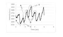
- FIG. 3shows the measurement results of the time change of the light receiving intensity in a total of 3 cases of 1 healthy person, 1 person with confirmed carotid artery thickening, and 1 person with suspected aneurysm.
- Ashows the waveform of the measurement result of a healthy person
- Bshows the waveform of the measurement result of a person with suspected aneurysm
- Cshows the waveform of the measurement result of a person with carotid artery thickening.
- the light receiving intensity at this timeis a voltage value obtained by receiving light. Since the value varies greatly depending on the depth and thickness of the blood vessel, it is difficult to judge whether it is a healthy person or a carotid artery thickening or aneurysm due to arteriosclerosis by comparing the absolute value of the light receiving intensity. Since the voltage value mentioned here is not a strict mv unit, it is expressed in a.u. units.
- the control unit 4differentiates the waveform due to the time change of the light receiving intensity and calculates the differential waveform.
- FIG. 4is an enlarged view of the waveform (A in FIG. 3) due to the time change of the light receiving intensity in the case of a healthy person.
- a strong riseappears (slope B of the pre-peak waveform in FIG. 4), reaches the peak top (A in FIG. 4), and then the value gradually decreases.
- Inclination C of the waveform after the peak in FIG. 4)is a waveform showing the time change of the light receiving intensity.
- the control unit 4determines that the person is a healthy person (not a carotid artery thickening or an aneurysm).
- FIG. 5shows a differential waveform obtained by differentially processing the waveform in the case of a healthy person (A in FIG. 3). As shown in FIG. 5, a peak with a period of 0.5 to 2.0 Hz cannot be confirmed in the differential waveform of a healthy person. Since the measurement principle is based on the pulse, the peak cycle is related to the pulse cycle of the subject.
- the control unit 4acquires the peak period using FFT analysis, wavelet transform, Stockwell transform, and the like.
- FIG. 6is an enlarged view of the waveform (C in FIG. 3) due to the time change of the light receiving intensity when there is carotid artery thickening.
- the control unit 4determines that the carotid artery is thickened when the differential waveform of the time change of the light receiving intensity has a downward peak (minus direction of the waveform differential value) of a predetermined period.
- FIG. 7shows a differential waveform obtained by differentially processing the waveform when there is carotid artery thickening (C in FIG. 3).
- the differential waveform obtained by differentiating the waveform when there is carotid artery thickeningthere is a periodic downward (minus) peak in the part corresponding to the slope C part of the post-peak waveform in which the value suddenly decreases. It can be confirmed (A in FIG. 7). Approximately once per second, that is, one signal is obtained for each beat. However, if there is noise during measurement, two or more may appear, so it may be necessary to infer from multiple shapes.
- the control unit 4determines the mean value and standard deviation ⁇ of the waveform differential values of the measured values of the time change of the light receiving intensity in the differential waveform section (analysis section in FIG. 7) for 1 second or longer (3 seconds in FIG. 7). calculate.
- the control unit 4has a value less than or equal to the value obtained by subtracting 2.6 ⁇ (-2.6SD in the figure) from the average value of the waveform differential values of the time change of the light receiving intensity, and is 0.5 to 0.5 detected by FFT analysis. It detects whether or not a waveform differential value exists with a period (interval) of 2.0 Hz (A in FIG. 7).
- the control unit 4determines that the carotid artery is thickened when there is a waveform differential value that meets the above conditions.
- FIG. 8is an enlarged view of the waveform (B in FIG. 3) due to the time change of the light receiving intensity when there is an aneurysm.
- a waveform with a peak top as shown in FIG. 8Ais obtained in the presence of an aneurysm. Comparing the slope of the peak rise (slope B of the pre-peak waveform) and the slope of the part where the peak falls (slope C of the post-peak waveform), the slope of the slope C of the post-peak waveform becomes steeper, resulting in a peak top. The position of A tends to shift behind the center of the peak.
- a bimodal peak(bimodal peak D in FIG. 8), which seems to be a disorder of blood flow, is observed in the presence of an aneurysm.
- a peak with a shoulderas shown in E in FIG. 8 (a peak with a shoulder in the figure) can be obtained.
- the control unit 4determines that the aneurysm is an aneurysm when the differential waveform of the time change of the light receiving intensity has a periodic upward (positive direction of the waveform differential value) peak.
- FIG. 9shows a differential waveform obtained by differentially processing the waveform when there is an aneurysm (B in FIG. 3).
- a periodic upward (plus direction) peakappears in the portion corresponding to the slope B portion of the waveform before the peak in FIG. 8 (FIG. 9).
- the control unit 4determines the mean value and standard deviation ⁇ of the waveform differential values of the measured values of the time change of the light receiving intensity in the differential waveform section (analysis section in FIG. 9) for 1 second or longer (3 seconds in FIG. 9). calculate.
- the control unit 4has a value equal to or higher than the value obtained by adding 2.6 ⁇ (line B in the figure) to the average value of the waveform differential values of the time change of the light receiving intensity, and is 0.5 to 2.0 Hz detected by FFT analysis. Whether or not a waveform derivative value exists is detected by the period (interval) of (A in FIG. 9).
- control unit 4When the control unit 4 detects the existence of a waveform differential value that meets the above conditions, it determines that the aneurysm is an aneurysm.
- the irradiation unit, the light receiving unit, and the control unitare configured as an integrated device, but the device is not limited to this, and for example, the irradiation unit is provided in a user device such as a mobile terminal (smartphone, tablet, mobile PC). Even if a light source is used, a sensor (CMOS, etc.) provided in a user device such as a mobile terminal (smartphone, tablet, mobile PC) is used as a light receiving unit, and the control unit is installed in a server device connected to the user device via a network. good.
- the arteriosclerosis measuring device of the embodimentis communicably connected to a user device having an irradiation unit that irradiates the subject with light and a light receiving unit that detects the light receiving intensity emitted from the subject.
- the arteriosclerosis measuring devicehas a control unit that determines arteriosclerosis (aneurysm, carotid artery thickening) in the subject from the light receiving intensity transmitted from the user device by the above processing.
- FIG. 10is a flowchart of an operation method (arteriosclerosis measurement processing) of the arteriosclerosis measuring device of the embodiment.
- the method of operating the arteriosclerosis measuring device of the embodimentis to irradiate the subject with light by the irradiation unit 2 having the above configuration (STEP101).
- the light receiving unit 3detects the light receiving intensity emitted from the subject (STEP102).
- the control unit 4calculates a differential waveform by differentiating the waveform due to the time change of the light receiving intensity (STEP103).
- the control unit 4calculates the mean value and the standard deviation of the waveform differential value of the differential waveform (STEP104).
- the control unit 4determines that the carotid artery is thickened when the average value of the waveform differential values is equal to or less than a predetermined multiple of the standard deviation and there is a waveform differential value existing in a predetermined cycle (STEP105).
- the control unit 4determines that the aneurysm is an aneurysm if the average value of the waveform differential values of the differential waveform is equal to or greater than the value obtained by adding a predetermined multiple of the standard deviation and there is a waveform differential value existing in a predetermined cycle (STEP 106). ).
- the programmay be stored in a storage medium.
- the computer of the apparatusis subjected to a process of irradiating the subject with light, a process of receiving the light emitted from the subject to detect the light receiving intensity, and a time change of the light receiving intensity.
- the process of differentiating the waveform to calculate the differential waveformthe process of calculating the mean value and the standard deviation of the waveform differential value, and the value obtained by subtracting the predetermined multiple of the standard deviation from the average value of the waveform differential value, which is a predetermined value.
- a derivative value that exists in a cycleit is a value that is equal to or greater than the process of determining carotid artery thickening and the mean value of the derivative value plus a predetermined multiple of the standard deviation, and the derivative that exists in a predetermined cycle. If there is a value, the process of determining an aneurysm is executed.
- Arteriosclerosis measuring device 2Irradiation unit 3: Light receiving unit 4: Control unit
Landscapes
- Health & Medical Sciences (AREA)
- Life Sciences & Earth Sciences (AREA)
- Surgery (AREA)
- Animal Behavior & Ethology (AREA)
- Biomedical Technology (AREA)
- Heart & Thoracic Surgery (AREA)
- Medical Informatics (AREA)
- Molecular Biology (AREA)
- Pathology (AREA)
- Engineering & Computer Science (AREA)
- General Health & Medical Sciences (AREA)
- Public Health (AREA)
- Veterinary Medicine (AREA)
- Cardiology (AREA)
- Physiology (AREA)
- Physics & Mathematics (AREA)
- Biophysics (AREA)
- Measuring Pulse, Heart Rate, Blood Pressure Or Blood Flow (AREA)
Abstract
Description
Translated fromJapaneseこの明細書に記載の実施形態は、動脈硬化計測装置、動脈硬化計測プログラム、及び、動脈硬化計測装置の作動方法に関する。The embodiments described in this specification relate to an arteriosclerosis measuring device, an arteriosclerosis measuring program, and an operation method of the arteriosclerosis measuring device.
従来の血流データに基づく動脈硬化診断方法は、流速や血圧などといった、絶対値としての計測データが必要であった(例えば、特許文献1参照)。The conventional arteriosclerosis diagnosis method based on blood flow data required measurement data as absolute values such as flow velocity and blood pressure (see, for example, Patent Document 1).
絶対値計測は、測定結果の信頼性を得るためには重要な情報であるが、一方で、非侵襲的に計測する場合において、超音波を用いるなどの計測湯法の限定や、装置の高額化が課題となるケースも多くみられる。Absolute value measurement is important information for obtaining the reliability of measurement results, but on the other hand, in the case of non-invasive measurement, the measurement hot water method such as using ultrasonic waves is limited, and the cost of the device is high. There are many cases where conversion becomes an issue.
また、動脈硬化などの血管の性状をPWVなどが用いられる。これは、脈波伝搬速度から動脈の硬さを計測するものである。この脈波伝搬速度計測では、詳細な波形解析や絶対値としての計測値を用いることで、その診断制度を高めてきた。In addition, PWV etc. is used for the properties of blood vessels such as arteriosclerosis. This measures the hardness of an artery from the pulse wave propagation velocity. In this pulse wave propagation velocity measurement, the diagnostic system has been enhanced by using detailed waveform analysis and measured values as absolute values.
しかしながら、このように高精度な性能を追求することにより、一般家庭でも安価かつ簡便に使える装置ではなくなってしまったのも事実である。However, by pursuing such high-precision performance, it is a fact that it is no longer an inexpensive and easy-to-use device even in ordinary households.
本発明は、このような従来の課題を解決するためになされた発明であって、絶対値計測を用いずとも、動脈硬化や動脈瘤の検査を可能とする装置、プログラム、及び、装置の作動方法を提供するものである。The present invention is an invention made to solve such a conventional problem, and is an apparatus, a program, and an operation of an apparatus capable of inspecting arteriosclerosis and aneurysm without using absolute value measurement. It provides a method.
本発明の動脈硬化計測装置は、被検体へ光を照射する照射部と、被検体から放出された光を受光して、受光強度を検出する受光部と、受光強度の時間変化による波形を微分して微分波形を算出し、所定の分析区間における微分波形の波形微分値の平均値と標準偏差を算出し、波形微分値の平均値から標準偏差の所定倍を引いた値以下であり、所定の周期で存在する波形微分値がある場合には、頸動脈肥厚と判定し、波形微分値の平均値に標準偏差の所定倍を加えた値以上であり、所定の周期で存在する波形微分値がある場合には、動脈瘤と判定する制御部と、を有する。The arteriosclerosis measuring apparatus of the present invention differentiates a light receiving portion that irradiates a subject with light, a light receiving portion that receives light emitted from the subject and detects a light receiving intensity, and a waveform due to a time change of the light receiving intensity. Then, the differential waveform is calculated, the average value and the standard deviation of the derivative waveform of the differential waveform in the predetermined analysis section are calculated, and the value is equal to or less than the average value of the waveform differential values minus the predetermined multiple of the standard deviation, and is predetermined. If there is a derivative value of the waveform that exists in the period of, it is judged that the carotid artery is thickened, and it is equal to or more than the mean value of the derivative value of the waveform plus a predetermined multiple of the standard deviation, and the derivative value of the waveform that exists in the predetermined period. If there is, it has a control unit for determining an aneurysm.
本発明の動脈硬化計測プログラムは、コンピュータに、被検体へ光を照射する処理と、被検体から放出された光を受光して、受光強度を検出する処理と、受光強度の時間変化による波形を微分して微分波形を算出する処理と、所定の分析区間における微分波形の波形微分値の平均値と標準偏差を算出する処理と、波形微分値の平均値から標準偏差の所定倍を引いた値以下であり、所定の周期で存在する波形微分値がある場合には、頸動脈肥厚と判定する処理と、波形微分値の平均値に標準偏差の所定倍を加えた値以上であり、所定の周期で存在する波形微分値がある場合には、動脈瘤と判定する処理とを実行させる。In the arteriosclerosis measurement program of the present invention, a computer is subjected to a process of irradiating a subject with light, a process of receiving light emitted from the subject to detect the light receiving intensity, and a waveform due to a change in the light receiving intensity over time. The process of differentiating to calculate the differential waveform, the process of calculating the mean and standard deviation of the derivative waveform of the differential waveform in a predetermined analysis section, and the value obtained by subtracting the predetermined multiple of the standard deviation from the mean value of the derivative values. If there is a derivative value of the waveform that exists in a predetermined period, it is equal to or greater than the process of determining carotid artery thickening and the value obtained by adding a predetermined multiple of the standard deviation to the mean value of the derivative value of the waveform. If there is a derivative value of the waveform that exists in the cycle, the process of determining that it is an aneurysm is executed.
本発明の動脈硬化計測装置の作動方法は、被検体へ光を照射し、被検体から放出された光を受光して、受光強度を検出し、受光強度の時間変化による波形を微分して微分波形を算出し、所定の分析区間における微分波形の波形微分値の平均値と標準偏差を算出し、波形微分値の平均値から標準偏差の所定倍を引いた値以下であり、所定の周期で存在する波形微分値がある場合には、頸動脈肥厚と判定し、波形微分値の平均値に標準偏差の所定倍を加えた値以上であり、所定の周期で存在する波形微分値がある場合には、動脈瘤と判定する。In the operation method of the arteriosclerosis measuring device of the present invention, the subject is irradiated with light, the light emitted from the subject is received, the light receiving intensity is detected, and the waveform due to the time change of the light receiving intensity is differentiated and differentiated. The waveform is calculated, the average value and standard deviation of the derivative waveform of the differential waveform in the predetermined analysis section are calculated, and the value is equal to or less than the average value of the derivative values minus the predetermined multiple of the standard deviation, and in a predetermined cycle. If there is a derivative value of the waveform that exists, it is determined that the carotid artery is thickened, and it is equal to or greater than the value obtained by adding a predetermined multiple of the standard deviation to the mean value of the derivative value of the waveform, and if there is a derivative value of the waveform that exists in a predetermined cycle. Is determined to be an aneurysm.
以下に実施形態を図面を用いて説明する。The embodiment will be described below with reference to the drawings.
図1は、実施形態の動脈硬化計測装置1の構成例を概略的に示す図である。図1に示すように動脈硬化計測装置1は、照射部2、受光部3、及び、制御部4を有する。また、照射部2による生体(被検体)上の照射位置を照射位置21とし、受光部3による生体上の受光位置を受光位置31とする。FIG. 1 is a diagram schematically showing a configuration example of the arteriosclerosis
照射部2は照射光を生体に照射する。照射部2は、制御部4により、照射する光の波長や照射強度が制御されてもよい。実施形態では、照射部2はLED(Light Emitting Diode)(810nm)である。The
実施形態の照射部2は、光の連続的な照射や光のパルス状の照射等の光を照射する時間の長さを任意に調整することができる。The
照射部2は、波長が固定された光源を用いてもよい。照射部2は、波長が異なる複数の光源あるいは複数の波長の光を混合したものであってもよい。照射部2は、例えば、蛍光灯、LED、レーザー、白熱灯、HID、ハロゲンランプ等である。照射部2の照度は、制御部4により制御されてもよい。The
実施形態の受光部3は、受光位置31において生体内から生体外に放出される光を受光して、光強度を検出する。実施形態の受光部3は、フォトダイオードである。受光部3は、フォトダイオードに限られず、CCDやCMOSでもよい。受光部3は、波長を可入射波長に設定し、その波長を受光できるものでもよい。実施形態では、受光部3にフォトダイオードを用い、サンプリングレートは2msに設定した。The light receiving
次に、動脈硬化計測装置1の制御系の構成について説明する。図2は実施形態の動脈硬化計測装置1のブロック図である。システムバス142を介して、CPU(Central Processing Unit)141、ROM(Read Only Memory)143、RAM(Random Access Memory)144、記憶部145、外部I/F(Interface)146、照射部2、及び、受光部3が接続される。CPU141とROM143とRAM144とで制御部4を構成する。Next, the configuration of the control system of the
ROM143は、CPU141により実行されるプログラムや閾値を予め記憶する。
RAM144は、CPU141が実行するプログラムを展開するエリアと、プログラムによるデータ処理の作業領域となるワークエリアなどの様々なメモリエリア等を有する。The
記憶部145は、処理に必要なデータを記憶する。記憶部145は、例えば、HDD(Hard Disk Drive)などである。The
外部I/F146は、例えばクライアント端末(PC)などの外部装置と通信するためのインターフェースである。外部I/F146は、外部装置とデータ通信を行うインターフェースであれば良く、たとえば、外部装置にローカルに接続する機器(USBメモリ等)であっても良いし、ネットワークを介して通信するためのネットワークインターフェースであっても良い。The external I /
以上のような構成を備える動脈硬化計測装置1において、予め設定されているプログラムに基づいて、動脈硬化計測装置1は動脈硬化計測ジョブを実行する。In the arteriosclerosis measuring
図3は、健常者1名、頸動脈肥厚が確認された人1名、及び、動脈瘤が疑われる人1名の計3症例の受光強度の時間変化の測定結果である。図3のAは健常者の測定結果の波形を示し、Bは、動脈瘤が疑われる人の測定結果の波形を示し、Cは、頸動脈肥厚がある人の測定結果の波形を示す。FIG. 3 shows the measurement results of the time change of the light receiving intensity in a total of 3 cases of 1 healthy person, 1 person with confirmed carotid artery thickening, and 1 person with suspected aneurysm. In FIG. 3, A shows the waveform of the measurement result of a healthy person, B shows the waveform of the measurement result of a person with suspected aneurysm, and C shows the waveform of the measurement result of a person with carotid artery thickening.
3症例の受光強度の時間変化を測定した結果、全症例とも、約1秒に1回の受光強度の拍動が確認できる。このときの受光強度は、受光により得られた電圧値である。血管の深さや太さにより値は大きく異なるため、受光強度の絶対値比較で、健常者か動脈硬化による頸動脈肥厚もしくは動脈瘤かの判断は難しいことがわかる。なお、ここで言う電圧値は厳格なmv単位ではないので、a.u.単位で表記した。As a result of measuring the time change of the light receiving intensity of 3 cases, it can be confirmed that the pulsation of the light receiving intensity is about once per second in all cases. The light receiving intensity at this time is a voltage value obtained by receiving light. Since the value varies greatly depending on the depth and thickness of the blood vessel, it is difficult to judge whether it is a healthy person or a carotid artery thickening or aneurysm due to arteriosclerosis by comparing the absolute value of the light receiving intensity. Since the voltage value mentioned here is not a strict mv unit, it is expressed in a.u. units.
ただし、受光強度の時間変化による波形を拡大することで、受光強度の時間変化による波形の特徴が確認できる。However, by enlarging the waveform due to the time change of the light receiving intensity, the characteristics of the waveform due to the time change of the light receiving intensity can be confirmed.
制御部4は、受光強度の時間変化による波形を微分し、微分波形を算出する。The
図4は、健常者の場合の受光強度の時間変化による波形(図3のA)を拡大した図である。図に示すように、健常者の場合には、立ち上がりが強く表れ(図4のピーク前波形の傾きB)、ピークトップに到達し(図4のA)、その後、ややなだらかに値が減少する(図4のピーク後波形の傾きC)という受光強度の時間変化を示す波形となる。FIG. 4 is an enlarged view of the waveform (A in FIG. 3) due to the time change of the light receiving intensity in the case of a healthy person. As shown in the figure, in the case of a healthy person, a strong rise appears (slope B of the pre-peak waveform in FIG. 4), reaches the peak top (A in FIG. 4), and then the value gradually decreases. (Inclination C of the waveform after the peak in FIG. 4) is a waveform showing the time change of the light receiving intensity.
制御部4は、受光強度の時間変化の微分波形に、所定周期のピークがない場合には、健常者(頸動脈肥厚や動脈瘤ではない)と判定する。If the differential waveform of the time change of the light receiving intensity does not have a peak of a predetermined cycle, the
図5は、健常者の場合(図3のA)の波形を微分処理した微分波形を示す。図5に示すように、健常者の微分波形に0.5~2.0Hzの周期のピークは確認できない。脈拍に基づく計測原理であることから、ピークの周期は被検体の脈の周期と関連性がある。制御部4は、FFT解析、ウェーブレット変換、Stockwell変換などを用いてピークの周期を取得する。FIG. 5 shows a differential waveform obtained by differentially processing the waveform in the case of a healthy person (A in FIG. 3). As shown in FIG. 5, a peak with a period of 0.5 to 2.0 Hz cannot be confirmed in the differential waveform of a healthy person. Since the measurement principle is based on the pulse, the peak cycle is related to the pulse cycle of the subject. The
図6は、頸動脈肥厚がある場合の受光強度の時間変化による波形(図3のC)を拡大した図である。FIG. 6 is an enlarged view of the waveform (C in FIG. 3) due to the time change of the light receiving intensity when there is carotid artery thickening.
図に示すように、頸動脈肥厚がある場合には、立ち上がりが緩やかになる傾向を示す(図6のピーク前波形の傾きB)。これは動脈が固くなることで、肥大しにくくなることで、立ち上がりが遅くなり、ピークトップ(図6のA)が波形の中央付近にシフトしたと考えられる。その後、急に値が減少する(図6のピーク後波形の傾きC)という受光強度の時間変化を示す波形となる。As shown in the figure, when there is carotid artery thickening, the rise tends to be gradual (slope B of the pre-peak waveform in FIG. 6). It is considered that this is because the arteries become stiff and difficult to enlarge, so that the rise is delayed and the peak top (A in FIG. 6) shifts to the vicinity of the center of the waveform. After that, the value suddenly decreases (the slope C of the waveform after the peak in FIG. 6), which is a waveform showing a time change of the light receiving intensity.
これらを特徴づけるため、受光強度の時間変化による波形の微分を行うと、図7のように特異的なピークが得られ、定性的な判断が可能となる。In order to characterize these, if the waveform is differentiated by the time change of the light receiving intensity, a specific peak is obtained as shown in FIG. 7, and a qualitative judgment is possible.
制御部4は、受光強度の時間変化の微分波形に所定周期の下向き(波形微分値のマイナス方向)のピークがある場合には、頸動脈肥厚と判定する。The
図7は、頸動脈肥厚がある場合(図3のC)の波形を微分処理した微分波形を示す。頸動脈肥厚がある場合の波形を微分した微分波形には、図6中の急に値が減少するピーク後波形の傾きC部に相当する部分に、周期的な下向き(マイナス方向)のピークが確認できる(図7中のA)。概ね1秒に1回すなわち拍動1回につき1個のシグナルが得られる。ただし、計測時にノイズがあると2個以上出てくることがあるので、複数個の形状から推測することも必要となる場合もある。FIG. 7 shows a differential waveform obtained by differentially processing the waveform when there is carotid artery thickening (C in FIG. 3). In the differential waveform obtained by differentiating the waveform when there is carotid artery thickening, there is a periodic downward (minus) peak in the part corresponding to the slope C part of the post-peak waveform in which the value suddenly decreases. It can be confirmed (A in FIG. 7). Approximately once per second, that is, one signal is obtained for each beat. However, if there is noise during measurement, two or more may appear, so it may be necessary to infer from multiple shapes.
制御部4は、1秒以上(図7では3秒)の微分波形の区間(図7中の分析区間)において、受光強度の時間変化の測定値の波形微分値の平均値と標準偏差σを算出する。制御部4は、受光強度の時間変化の波形微分値の平均値から、2.6σ(図中の-2.6SD)を引いた値以下の数値を有し、かつ、FFT解析により検出された0.5~2.0Hzの周期(の間隔)で、波形微分値が存在するか否かを検知する(図7中のA)。The
制御部4は、上記条件に当てはまる波形微分値がある場合には、頸動脈肥厚と判定する。The
図8は、動脈瘤がある場合の受光強度の時間変化による波形(図3のB)を拡大した図である。図に示すように、動脈瘤が存在すると、図8のAのようなピークトップを持つ波形が得られる。そして、ピークの立ち上がり(ピーク前波形の傾きB)と、ピークが下がる部分(ピーク後波形の傾きC)の傾きを比較すると、ピーク後波形の傾きCの傾きが急になることで、ピークトップAの位置が、ピークの中央より後ろにずれる傾向がある。動脈瘤が存在すると、血流の乱れと思われる二峰性のピーク(図8の二峰ピークD)がみられる。また図8のEのような肩を持ったピーク(図中の肩有ピーク)が得られることも確認できる。FIG. 8 is an enlarged view of the waveform (B in FIG. 3) due to the time change of the light receiving intensity when there is an aneurysm. As shown in the figure, in the presence of an aneurysm, a waveform with a peak top as shown in FIG. 8A is obtained. Comparing the slope of the peak rise (slope B of the pre-peak waveform) and the slope of the part where the peak falls (slope C of the post-peak waveform), the slope of the slope C of the post-peak waveform becomes steeper, resulting in a peak top. The position of A tends to shift behind the center of the peak. In the presence of an aneurysm, a bimodal peak (bimodal peak D in FIG. 8), which seems to be a disorder of blood flow, is observed. It can also be confirmed that a peak with a shoulder as shown in E in FIG. 8 (a peak with a shoulder in the figure) can be obtained.
これらを特徴づけるため、受光強度の時間変化による波形の微分を行うと、図9のように特異的なピークが得られ、定性的な判断が可能となる。In order to characterize these, if the waveform is differentiated by the time change of the light receiving intensity, a specific peak is obtained as shown in FIG. 9, and a qualitative judgment is possible.
制御部4は、受光強度の時間変化の微分波形に周期的な上向き(波形微分値のプラス方向)のピークがある場合には、動脈瘤と判定する。The
図9は、動脈瘤がある場合(図3のB)の波形を微分処理した微分波形を示す。動脈瘤がある場合の受光強度の時間変化を微分した微分波形には、図8のピーク前波形の傾きB部に相当する部分に、周期的な上向き(プラス方向)のピークが現れる(図9のA)。FIG. 9 shows a differential waveform obtained by differentially processing the waveform when there is an aneurysm (B in FIG. 3). In the differential waveform obtained by differentiating the time change of the light receiving intensity when there is an aneurysm, a periodic upward (plus direction) peak appears in the portion corresponding to the slope B portion of the waveform before the peak in FIG. 8 (FIG. 9). A).
制御部4は、1秒以上(図9では3秒)の微分波形の区間(図9中の分析区間)において、受光強度の時間変化の測定値の波形微分値の平均値と標準偏差σを算出する。制御部4は、受光強度の時間変化の波形微分値の平均値に2.6σ(図中の線B)を加えた値以上の数値を有し、かつ、FFT解析により検出された0.5~2.0Hzの周期(の間隔)で、波形微分値が存在するか否かを検知する(図9中のA)。The
制御部4は、上記条件に当てはまる波形微分値の存在を検知した場合には、動脈瘤と判定する。When the
なお、実施形態では、照射部と受光部と制御部を一体の装置として構成したが、これに限られず、例えば、照射部として、携帯端末(スマートフォン、タブレット、モバイルPC)などのユーザ装置に備わる光源を使用し、受光部として携帯端末(スマートフォン、タブレット、モバイルPC)などのユーザ装置に備わるセンサ(CMOS等)を使用し、制御部をユーザ装置とネットワーク接続したサーバー装置等に設置してもよい。In the embodiment, the irradiation unit, the light receiving unit, and the control unit are configured as an integrated device, but the device is not limited to this, and for example, the irradiation unit is provided in a user device such as a mobile terminal (smartphone, tablet, mobile PC). Even if a light source is used, a sensor (CMOS, etc.) provided in a user device such as a mobile terminal (smartphone, tablet, mobile PC) is used as a light receiving unit, and the control unit is installed in a server device connected to the user device via a network. good.
実施形態の動脈硬化計測装置は、被検体へ、光を照射する照射部と、被検体から放出される受光強度を検出する受光部を有するユーザ装置に、通信可能に接続する。動脈硬化計測装置は、ユーザ装置から送信された受光強度から被検体内の動脈硬化(動脈瘤、頸動脈肥厚)を上記処理で判定する制御部を有する。The arteriosclerosis measuring device of the embodiment is communicably connected to a user device having an irradiation unit that irradiates the subject with light and a light receiving unit that detects the light receiving intensity emitted from the subject. The arteriosclerosis measuring device has a control unit that determines arteriosclerosis (aneurysm, carotid artery thickening) in the subject from the light receiving intensity transmitted from the user device by the above processing.
次に、実施形態の動脈硬化計測装置の作動方法について説明する。図10は、実施形態の動脈硬化計測装置の作動方法(動脈硬化計測処理)のフローチャートである。Next, the operation method of the arteriosclerosis measuring device of the embodiment will be described. FIG. 10 is a flowchart of an operation method (arteriosclerosis measurement processing) of the arteriosclerosis measuring device of the embodiment.
実施形態の動脈硬化計測装置の作動方法は、上記構成を有する照射部2により、被検体へ光を照射する(STEP101)。受光部3が、被検体から放出される受光強度を検出する(STEP102)。制御部4は、受光強度の時間変化による波形を微分して微分波形を算出する(STEP103)。制御部4は、微分波形の波形微分値の平均値と標準偏差を算出する(STEP104)。制御部4は、波形微分値の平均値から標準偏差の所定倍を引いた値以下であり、所定の周期で存在する波形微分値がある場合には、頸動脈肥厚と判定する(STEP105)。制御部4は、微分波形の波形微分値の平均値に標準偏差の所定倍を加えた値以上であり、所定の周期で存在する波形微分値がある場合には、動脈瘤と判定する(STEP106)。The method of operating the arteriosclerosis measuring device of the embodiment is to irradiate the subject with light by the
次に、実施形態の動脈硬化計測プログラムについて説明する。なお、当該プログラムは記憶媒体に格納されてもよい。Next, the arteriosclerosis measurement program of the embodiment will be described. The program may be stored in a storage medium.
実施形態の動脈硬化計測プログラムは、装置のコンピュータに、被検体へ光を照射する処理と、被検体から放出された光を受光して、受光強度を検出する処理と、受光強度の時間変化による波形を微分して微分波形を算出する処理と、波形微分値の平均値と標準偏差を算出する処理と、波形微分値の平均値から標準偏差の所定倍を引いた値以下であり、所定の周期で存在する波形微分値がある場合には、頸動脈肥厚と判定する処理と、波形微分値の平均値に標準偏差の所定倍を加えた値以上であり、所定の周期で存在する波形微分値がある場合には、動脈瘤と判定する処理とを実行させる。In the arteriosclerosis measurement program of the embodiment, the computer of the apparatus is subjected to a process of irradiating the subject with light, a process of receiving the light emitted from the subject to detect the light receiving intensity, and a time change of the light receiving intensity. The process of differentiating the waveform to calculate the differential waveform, the process of calculating the mean value and the standard deviation of the waveform differential value, and the value obtained by subtracting the predetermined multiple of the standard deviation from the average value of the waveform differential value, which is a predetermined value. If there is a derivative value that exists in a cycle, it is a value that is equal to or greater than the process of determining carotid artery thickening and the mean value of the derivative value plus a predetermined multiple of the standard deviation, and the derivative that exists in a predetermined cycle. If there is a value, the process of determining an aneurysm is executed.
以上説明したように、本実施形態によれば、光学的手法を用い、絶対値計測を用いずとも、動脈硬化や動脈瘤の検査をすることができる。As described above, according to the present embodiment, it is possible to inspect arteriosclerosis and aneurysm by using an optical method and without using absolute value measurement.
以上、実施形態を説明したが、この実施形態は、例として提示したものであり、発明の範囲を限定することは意図していない。この新規な実施形態は、その他の様々な形態で実施されることが可能であり、発明の要旨を逸脱しない範囲で、種々の省略、置き換え、変更を行うことができる。この実施形態やその変形は、発明の範囲や要旨に含まれるとともに、特許請求の範囲に記載された発明とその均等の範囲に含まれる。Although the embodiment has been described above, this embodiment is presented as an example and is not intended to limit the scope of the invention. This novel embodiment can be implemented in various other embodiments, and various omissions, replacements, and changes can be made without departing from the gist of the invention. This embodiment and its modifications are included in the scope and gist of the invention, and are also included in the scope of the invention described in the claims and the equivalent scope thereof.
1:動脈硬化計測装置
2:照射部
3:受光部
4:制御部1: Arteriosclerosis measuring device 2: Irradiation unit 3: Light receiving unit 4: Control unit
Claims (5)
Translated fromJapanese前記被検体から放出された光を受光して、受光強度を検出する受光部と、
前記受光強度の時間変化による波形を微分して微分波形を算出し、
所定の分析区間における前記微分波形の波形微分値の平均値と標準偏差を算出し、
前記波形微分値の平均値から前記標準偏差の所定倍を引いた値以下であり、所定の周期で存在する前記波形微分値がある場合には、頸動脈肥厚と判定し、
前記波形微分値の平均値に前記標準偏差の所定倍を加えた値以上であり、所定の周期で存在する前記波形微分値がある場合には、動脈瘤と判定する、
制御部と、
を有する動脈硬化計測装置。An irradiation part that irradiates the subject with light,
A light receiving unit that receives light emitted from the subject and detects the light receiving intensity,
The differential waveform is calculated by differentiating the waveform due to the time change of the light receiving intensity.
The average value and standard deviation of the waveform differential values of the differential waveform in the predetermined analysis section are calculated.
If it is equal to or less than the average value of the waveform differential values minus a predetermined multiple of the standard deviation and there is the waveform differential values existing in a predetermined cycle, it is determined that the carotid artery is thickened.
If the average value of the waveform differential values is equal to or greater than the value obtained by adding a predetermined multiple of the standard deviation and there is the waveform differential value existing in a predetermined cycle, it is determined to be an aneurysm.
Control unit and
Arteriosclerosis measuring device with.
被検体へ光を照射する処理と、
前記被検体から放出された光を受光して、受光強度を検出する処理と、
前記受光強度の時間変化による波形を微分して微分波形を算出する処理と、
所定の分析区間における前記微分波形の波形微分値の平均値と標準偏差を算出する処理と、
前記波形微分値の平均値から前記標準偏差の所定倍を引いた値以下であり、所定の周期で存在する前記波形微分値がある場合には、頸動脈肥厚と判定する処理と、
前記波形微分値の平均値に前記標準偏差の所定倍を加えた値以上であり、所定の周期で存在する前記波形微分値がある場合には、動脈瘤と判定する処理と、
を実行させる動脈硬化計測プログラム。On the computer
The process of irradiating the subject with light and
A process of receiving the light emitted from the subject and detecting the light receiving intensity,
The process of differentiating the waveform due to the time change of the light receiving intensity to calculate the differential waveform,
Processing to calculate the mean value and standard deviation of the waveform differential value of the differential waveform in a predetermined analysis section,
If there is the waveform differential value that is equal to or less than the average value of the waveform differential values minus a predetermined multiple of the standard deviation and exists in a predetermined cycle, the process of determining carotid artery thickening is performed.
If the average value of the waveform differential value is equal to or greater than the value obtained by adding a predetermined multiple of the standard deviation and there is the waveform differential value existing in a predetermined cycle, the process of determining an aneurysm is performed.
Arteriosclerosis measurement program to run.
前記被検体から放出された光を受光して、受光強度を検出し、
前記受光強度の時間変化による波形を微分して微分波形を算出し、
所定の分析区間における前記微分波形の波形微分値の平均値と標準偏差を算出し、
前記波形微分値の平均値から前記標準偏差の所定倍を引いた値以下であり、所定の周期で存在する前記波形微分値がある場合には、頸動脈肥厚と判定し、
前記波形微分値の平均値に前記標準偏差の所定倍を加えた値以上であり、所定の周期で存在する前記波形微分値がある場合には、動脈瘤と判定する、
動脈硬化計測装置の作動方法。Irradiate the subject with light and
The light emitted from the subject is received, the light receiving intensity is detected, and the light is received.
The differential waveform is calculated by differentiating the waveform due to the time change of the light receiving intensity.
The average value and standard deviation of the waveform differential values of the differential waveform in the predetermined analysis section are calculated.
If it is equal to or less than the average value of the waveform differential values minus a predetermined multiple of the standard deviation and there is the waveform differential values existing in a predetermined cycle, it is determined that the carotid artery is thickened.
If the average value of the waveform differential values is equal to or greater than the value obtained by adding a predetermined multiple of the standard deviation and there is the waveform differential value existing in a predetermined cycle, it is determined to be an aneurysm.
How to operate the arteriosclerosis measuring device.
Applications Claiming Priority (4)
| Application Number | Priority Date | Filing Date | Title |
|---|---|---|---|
| JP2020215545 | 2020-12-24 | ||
| JP2020-215545 | 2020-12-24 | ||
| JP2021123374AJP6989192B1 (en) | 2020-12-24 | 2021-07-28 | Arteriosclerosis measuring device, arteriosclerosis measuring program, and how to operate the arteriosclerosis measuring device |
| JP2021-123374 | 2021-07-28 |
Publications (1)
| Publication Number | Publication Date |
|---|---|
| WO2022138904A1true WO2022138904A1 (en) | 2022-06-30 |
Family
ID=79239742
Family Applications (1)
| Application Number | Title | Priority Date | Filing Date |
|---|---|---|---|
| PCT/JP2021/048134CeasedWO2022138904A1 (en) | 2020-12-24 | 2021-12-24 | Arteriosclerosis measurement device, arteriosclerosis measurement program, and method for operating arteriosclerosis measurement device |
Country Status (2)
| Country | Link |
|---|---|
| JP (1) | JP6989192B1 (en) |
| WO (1) | WO2022138904A1 (en) |
Citations (10)
| Publication number | Priority date | Publication date | Assignee | Title |
|---|---|---|---|---|
| JP2002345787A (en)* | 2001-05-29 | 2002-12-03 | Institute Of Tsukuba Liaison Co Ltd | Thrombus measuring device |
| JP2006288842A (en)* | 2005-04-13 | 2006-10-26 | Kowa Co | Ophthalmic measuring device |
| WO2010131713A1 (en)* | 2009-05-13 | 2010-11-18 | 住友電気工業株式会社 | Blood vessel inner wall analyzing device and blood vessel inner wall analyzing method |
| JP2013198674A (en)* | 2012-03-26 | 2013-10-03 | Denso It Laboratory Inc | Apparatus, method and program for simplified blood flow examination |
| US20140073959A1 (en)* | 2012-09-11 | 2014-03-13 | Nellcor Puritan Bennett Llc | Methods and systems for determining when to output previously calculated values |
| US20140249424A1 (en)* | 2012-12-04 | 2014-09-04 | University Of Winnipeg | Cardiovascular pulse wave analysis method and system |
| JP2014188035A (en)* | 2013-03-26 | 2014-10-06 | Shisei Deetamu:Kk | Vascular viscoelasticity evaluation apparatus, method, and program |
| JP2020025732A (en)* | 2018-08-10 | 2020-02-20 | 株式会社東芝 | Pulse wave evaluation device and pulse wave evaluation method |
| US20200229714A1 (en)* | 2019-01-17 | 2020-07-23 | Grant Hocking | Method to Quantify the Hemodynamic and Vascular Properties in Vivo from Arterial Waveform Measurements |
| JP2020120839A (en)* | 2019-01-30 | 2020-08-13 | 潤一郎 橋本 | Disease assessment based on analysis of carotid artery blood flow waveform |
Family Cites Families (8)
| Publication number | Priority date | Publication date | Assignee | Title |
|---|---|---|---|---|
| JPH08583A (en)* | 1994-06-22 | 1996-01-09 | Minolta Co Ltd | Apparatus for monitoring pulse wave transmission time |
| JP2002010986A (en)* | 2000-06-29 | 2002-01-15 | Yoshinaga Kajimoto | Non-invasive measuring device for blood volume in brain |
| JP4992145B2 (en)* | 2009-11-26 | 2012-08-08 | 日本光電工業株式会社 | Blood vessel wall monitoring device, blood vessel wall monitoring program, and computer-readable recording medium |
| US20170277858A1 (en)* | 2014-09-19 | 2017-09-28 | Shinano Kenshi Co., Ltd. | System for predicting risk of onset of cerebrovascular disease |
| US9839365B1 (en)* | 2014-11-24 | 2017-12-12 | Verily Life Sciences Llc | Applications of vasculature mapping using laser speckle imaging |
| JP7045084B2 (en)* | 2016-03-28 | 2022-03-31 | ジェンドゥ・イノベイションズ・プライベイト・リミテッド | Systems and methods for monitoring vascular health |
| JP7129189B2 (en)* | 2018-03-27 | 2022-09-01 | キヤノン株式会社 | Biological measuring device and program |
| CA3126400A1 (en)* | 2019-01-17 | 2020-07-23 | Grant Hocking | Method to quantify hypertension, aging status and vascular properties in vivo from arterial optical plethysmograph waveform measurements |
- 2021
- 2021-07-28JPJP2021123374Apatent/JP6989192B1/ennot_activeExpired - Fee Related
- 2021-12-24WOPCT/JP2021/048134patent/WO2022138904A1/ennot_activeCeased
Patent Citations (10)
| Publication number | Priority date | Publication date | Assignee | Title |
|---|---|---|---|---|
| JP2002345787A (en)* | 2001-05-29 | 2002-12-03 | Institute Of Tsukuba Liaison Co Ltd | Thrombus measuring device |
| JP2006288842A (en)* | 2005-04-13 | 2006-10-26 | Kowa Co | Ophthalmic measuring device |
| WO2010131713A1 (en)* | 2009-05-13 | 2010-11-18 | 住友電気工業株式会社 | Blood vessel inner wall analyzing device and blood vessel inner wall analyzing method |
| JP2013198674A (en)* | 2012-03-26 | 2013-10-03 | Denso It Laboratory Inc | Apparatus, method and program for simplified blood flow examination |
| US20140073959A1 (en)* | 2012-09-11 | 2014-03-13 | Nellcor Puritan Bennett Llc | Methods and systems for determining when to output previously calculated values |
| US20140249424A1 (en)* | 2012-12-04 | 2014-09-04 | University Of Winnipeg | Cardiovascular pulse wave analysis method and system |
| JP2014188035A (en)* | 2013-03-26 | 2014-10-06 | Shisei Deetamu:Kk | Vascular viscoelasticity evaluation apparatus, method, and program |
| JP2020025732A (en)* | 2018-08-10 | 2020-02-20 | 株式会社東芝 | Pulse wave evaluation device and pulse wave evaluation method |
| US20200229714A1 (en)* | 2019-01-17 | 2020-07-23 | Grant Hocking | Method to Quantify the Hemodynamic and Vascular Properties in Vivo from Arterial Waveform Measurements |
| JP2020120839A (en)* | 2019-01-30 | 2020-08-13 | 潤一郎 橋本 | Disease assessment based on analysis of carotid artery blood flow waveform |
Also Published As
| Publication number | Publication date |
|---|---|
| JP2022101441A (en) | 2022-07-06 |
| JP6989192B1 (en) | 2022-01-05 |
Similar Documents
| Publication | Publication Date | Title |
|---|---|---|
| CN103300880B (en) | Subject information obtaining device and subject information obtaining method | |
| JP2007534421A5 (en) | ||
| US20090263759A1 (en) | Method and apparatus for detecting abnormality in tooth structure | |
| JP2008541823A (en) | Glucose sensor | |
| JPH1019766A (en) | Light scatterer measuring device | |
| JP6270724B2 (en) | Ophthalmic device and method for measuring an eye | |
| CN107991287B (en) | Raman spectrum detection equipment and method based on image grayscale recognition | |
| JP2018007894A (en) | Measuring device, measuring method, and measuring program | |
| JPWO2016151787A1 (en) | Blood flow measurement method for blood vessel recognition | |
| JP6989192B1 (en) | Arteriosclerosis measuring device, arteriosclerosis measuring program, and how to operate the arteriosclerosis measuring device | |
| JP2023004122A (en) | High-sensitivity particle concentration measurement device | |
| CN107907527B (en) | Raman spectrum detection equipment and method based on reflected light power and image recognition | |
| US20180289297A1 (en) | System and method for monitoring glucose level | |
| EP3730053A1 (en) | Lipid measurement device and method therefor | |
| US10278625B2 (en) | Blood measuring apparatus using spectroscope | |
| JP2022146042A (en) | Arteriosclerosis measuring apparatus, program and operating method of apparatus | |
| WO2024255902A1 (en) | Intraocular pressure measurement apparatus, method for identifying left and right eyes, and storage medium | |
| JP2018100968A (en) | Flow measuring device, flow measuring method, and flow measuring program | |
| JP2017109058A (en) | Biological information acquisition apparatus and biological information acquisition method | |
| CN112244780A (en) | A device and method for measuring bone density based on photoacoustic signals | |
| JP2010216854A (en) | Film thickness measuring apparatus | |
| CN110811592B (en) | Blood circulation detection device and blood circulation detection method | |
| JP6723572B1 (en) | Blood pressure measuring device and method | |
| WO2022220209A1 (en) | Lipid concentration measuring device, program, and method | |
| TWI890642B (en) | Non-invasive method for estimating blood glucose and the device thereof |
Legal Events
| Date | Code | Title | Description |
|---|---|---|---|
| 121 | Ep: the epo has been informed by wipo that ep was designated in this application | Ref document number:21911041 Country of ref document:EP Kind code of ref document:A1 | |
| NENP | Non-entry into the national phase | Ref country code:DE | |
| 122 | Ep: pct application non-entry in european phase | Ref document number:21911041 Country of ref document:EP Kind code of ref document:A1 |