WO2022106869A1 - Fusion proteins comprising an e2 ubiquitin or ubiquitin-like conjugating domain and a targeting domain for specific protein degradation - Google Patents
Fusion proteins comprising an e2 ubiquitin or ubiquitin-like conjugating domain and a targeting domain for specific protein degradationDownload PDFInfo
- Publication number
- WO2022106869A1 WO2022106869A1PCT/IB2020/060978IB2020060978WWO2022106869A1WO 2022106869 A1WO2022106869 A1WO 2022106869A1IB 2020060978 WIB2020060978 WIB 2020060978WWO 2022106869 A1WO2022106869 A1WO 2022106869A1
- Authority
- WO
- WIPO (PCT)
- Prior art keywords
- domain
- molecule
- ubiquitin
- substrate
- cell
- Prior art date
- Legal status (The legal status is an assumption and is not a legal conclusion. Google has not performed a legal analysis and makes no representation as to the accuracy of the status listed.)
- Ceased
Links
Classifications
- C—CHEMISTRY; METALLURGY
- C12—BIOCHEMISTRY; BEER; SPIRITS; WINE; VINEGAR; MICROBIOLOGY; ENZYMOLOGY; MUTATION OR GENETIC ENGINEERING
- C12N—MICROORGANISMS OR ENZYMES; COMPOSITIONS THEREOF; PROPAGATING, PRESERVING, OR MAINTAINING MICROORGANISMS; MUTATION OR GENETIC ENGINEERING; CULTURE MEDIA
- C12N9/00—Enzymes; Proenzymes; Compositions thereof; Processes for preparing, activating, inhibiting, separating or purifying enzymes
- C12N9/10—Transferases (2.)
- C12N9/1025—Acyltransferases (2.3)
- C12N9/104—Aminoacyltransferases (2.3.2)
- C—CHEMISTRY; METALLURGY
- C07—ORGANIC CHEMISTRY
- C07K—PEPTIDES
- C07K16/00—Immunoglobulins [IGs], e.g. monoclonal or polyclonal antibodies
- C07K16/18—Immunoglobulins [IGs], e.g. monoclonal or polyclonal antibodies against material from animals or humans
- C—CHEMISTRY; METALLURGY
- C07—ORGANIC CHEMISTRY
- C07K—PEPTIDES
- C07K16/00—Immunoglobulins [IGs], e.g. monoclonal or polyclonal antibodies
- C07K16/46—Hybrid immunoglobulins
- A—HUMAN NECESSITIES
- A61—MEDICAL OR VETERINARY SCIENCE; HYGIENE
- A61K—PREPARATIONS FOR MEDICAL, DENTAL OR TOILETRY PURPOSES
- A61K38/00—Medicinal preparations containing peptides
- C—CHEMISTRY; METALLURGY
- C07—ORGANIC CHEMISTRY
- C07K—PEPTIDES
- C07K2317/00—Immunoglobulins specific features
- C07K2317/50—Immunoglobulins specific features characterized by immunoglobulin fragments
- C07K2317/56—Immunoglobulins specific features characterized by immunoglobulin fragments variable (Fv) region, i.e. VH and/or VL
- C07K2317/569—Single domain, e.g. dAb, sdAb, VHH, VNAR or nanobody®
- C—CHEMISTRY; METALLURGY
- C07—ORGANIC CHEMISTRY
- C07K—PEPTIDES
- C07K2317/00—Immunoglobulins specific features
- C07K2317/90—Immunoglobulins specific features characterized by (pharmaco)kinetic aspects or by stability of the immunoglobulin
- C07K2317/92—Affinity (KD), association rate (Ka), dissociation rate (Kd) or EC50 value
- C—CHEMISTRY; METALLURGY
- C07—ORGANIC CHEMISTRY
- C07K—PEPTIDES
- C07K2319/00—Fusion polypeptide
- C07K2319/01—Fusion polypeptide containing a localisation/targetting motif
- C—CHEMISTRY; METALLURGY
- C07—ORGANIC CHEMISTRY
- C07K—PEPTIDES
- C07K2319/00—Fusion polypeptide
- C07K2319/01—Fusion polypeptide containing a localisation/targetting motif
- C07K2319/06—Fusion polypeptide containing a localisation/targetting motif containing a lysosomal/endosomal localisation signal
- C—CHEMISTRY; METALLURGY
- C07—ORGANIC CHEMISTRY
- C07K—PEPTIDES
- C07K2319/00—Fusion polypeptide
- C07K2319/01—Fusion polypeptide containing a localisation/targetting motif
- C07K2319/07—Fusion polypeptide containing a localisation/targetting motif containing a mitochondrial localisation signal
- C—CHEMISTRY; METALLURGY
- C07—ORGANIC CHEMISTRY
- C07K—PEPTIDES
- C07K2319/00—Fusion polypeptide
- C07K2319/01—Fusion polypeptide containing a localisation/targetting motif
- C07K2319/09—Fusion polypeptide containing a localisation/targetting motif containing a nuclear localisation signal
- C—CHEMISTRY; METALLURGY
- C07—ORGANIC CHEMISTRY
- C07K—PEPTIDES
- C07K2319/00—Fusion polypeptide
- C07K2319/40—Fusion polypeptide containing a tag for immunodetection, or an epitope for immunisation
- C07K2319/42—Fusion polypeptide containing a tag for immunodetection, or an epitope for immunisation containing a HA(hemagglutinin)-tag
- C—CHEMISTRY; METALLURGY
- C07—ORGANIC CHEMISTRY
- C07K—PEPTIDES
- C07K2319/00—Fusion polypeptide
- C07K2319/70—Fusion polypeptide containing domain for protein-protein interaction
- C—CHEMISTRY; METALLURGY
- C12—BIOCHEMISTRY; BEER; SPIRITS; WINE; VINEGAR; MICROBIOLOGY; ENZYMOLOGY; MUTATION OR GENETIC ENGINEERING
- C12Y—ENZYMES
- C12Y203/00—Acyltransferases (2.3)
- C12Y203/02—Aminoacyltransferases (2.3.2)
Definitions
- Ubiquitinationis characterised by a rapid and reversible posttranslational covalent binding of ubiquitin to proteins.
- the mechanismplays an important role in targeting proteins for degradation, and in modulating their sub-cellular localisation, intracellular signalling, and interactions with other proteins (Glickman and Ciechanover, Physiol Rev 2002, 82(2):373- 428; Mukhopadhyay and Riezman, Science 2007, 315(5809):201-5; and Schnell and Hicke, J Biol Chem 2003, 278(38):35857-60).
- Ubiquitin (Ub)is a small (76 amino acid; 8.6 kDa) regulatory protein.
- Ubiquitinationinvolves activation of the ubiquitin C-terminus. For this to occur, E1 Ub-activating enzymes form a thioester bond with Ub in an adenosine triphosphate–dependent reaction. This is followed by conjugation to an E2 ubiquitin-conjugating enzyme (and potentially to a HECT-style E3 ubiquitin ligase as an E3 ⁇ Ub intermediate), and ligation to the substrate protein.
- Ubiquitincan be bound to a target substrate by (i) lysine residues via an isopeptide bond, (ii) cysteine residues by a thioester bond, (iii) serine and threonine residues by an ester bond, or (iv) the amino group of the protein's N-terminus via a peptide bond.
- a single ubiquitin protein (monoubiquitination) or a chain of ubiquitin (polyubiquitination)can be added to the substrate.
- the secondary ubiquitin moleculesare linked to one of the seven lysine residues (for example K48 or K63) or the N-terminal methionine of the previous ubiquitin molecule.
- ubiquitin-like proteinscan be used in similar mechanisms.
- the human genomeencodes at least eight families of ubiquitin-like proteins, not including ubiquitin itself, that are considered Type I Ubls.
- Type I Ublsare capable of covalent conjugation. Covalent conjugation occurs through one to two glycine residues at the C-terminus. Humans also encode fau ubiquitin-like protein (FUBI), a Type 2 Ubl, which is not capable of covalent conjugation.
- E2 enzymesact to transfer ubiquitin to a target substrate and all share a core catalytic domain of around 150 amino acids called the ubiquitin core catalytic domain (UBC domain).
- UBC domainubiquitin core catalytic domain
- the domainadopts an ⁇ / ⁇ -fold typically with four ⁇ -helices and a 4 stranded ⁇ -sheet (Stewart et al., Cell Res 2016, 26:423).
- Important loop regionsform part of the E3-binding site and the E2 active site.
- UBC domainsare approximately 14-16 kDa and are ⁇ 35% conserved among different family members (Dikic et al., Nat Rev Mol Cell Biol 2009, 10:659-671).
- E2shave a common fold that has been adapted for specific systems. Although most E2s encompass only a single structural UBC domain, many have short N- and/or C-terminal extensions that can impart important E2-specific functionality, such as recognising bound ubiquitin for chain-building E2s.
- E2shave functionally important insertions, including Ube2R1 and Ube2G2 and some E2s have additional structured domain linked to the UBC domain (e.g. Ube2K) or are part of large multi domain proteins (Ube2O or BIRC6).
- E2 enzymesprimarily engage in two types of reactions for the transfer of ubiquitin from a E2 ⁇ Ub conjugate to the substrate: (1) transthiolation (transfer from a thioester to a thiol group – e.g. transfer of ubiquitin to the active site cysteine residue of a HECT-type E3 ligase) and (2) aminolysis (transfer from a thioester to an amino group), but others have also been reported.
- E2sinteract with an E1 enzyme and one or more E3s.
- E2smay directly engage a target protein (e.g. those termed E3/E2 hybrid such as BIRC6 and UBE2O are E3-independent, as are able to interact and ubiquitinate their substrates without assistance from E3s) and so play a role in the determination of where and how a target is modified by ubiquitin.
- a target proteine.g. those termed E3/E2 hybrid such as BIRC6 and UBE2O are E3-independent, as are able to interact and ubiquitinate their substrates without assistance from E3s
- E3shave been classified into three families, namely RING, HECT and RING-between-RINGS (RBR).
- RING/U-box E3 ubiquitin ligasesare able to bind to both a substrate and a E2-Ub conjugate.
- HECT/RBR domain E3 ligasesmust additionally be able to form an intermediate thioester with ubiquitin (E3-Ub).
- E2scan function with multiple types of E3.
- HECT domain-containing E3 ubiquitin ligasesform intermediate thioesters with Ub (E3 ⁇ Ub) at their active site cysteine before transferring Ub to substrates, whereas most RING finger domain-containing E3 enzymes act as scaffolds that bind to E2 enzymes and substrates simultaneously.
- RING E3s(most E3s) are not involved directly in the chemical transfer of Ub to substrates. They bind substrates and an E2 ⁇ Ub conjugate to facilitate Ub transfer directly from the E2 active site to the substrate.
- RING E3sfunction as a protein co-factor for E2 ⁇ Ub conjugates.
- RING E3/E2 ⁇ Ub complexesare dynamic; however, interactions with RING E3s increase the intrinsic reactivity of many (but not all) E2 ⁇ Ub conjugates towards aminolysis.
- the Ube2D family of E2sreact with lysine slowly in the absence of an E3, but rapidly in the presence of a RING domain.
- E2 ⁇ Ubgenerally adopt a “closed state” upon RING E3 binding.
- a conserved RING (allosteric linchpin) residueusually an arginine, lysine or asparagine, donates a hydrogen bond to an E2 backbone carbonyl in loop 7 and one or more backbone groups in the tail of Ub.
- E2 ⁇ Ub closed stateis thought of as an activated state for aminolysis. Transthiolation reactions can readily occur in absence of E3s. Therefore, it is hypothesised that E3s that progress with a E3 ⁇ Ub conjugate intermediate (e.g. HECT E3s) do not need to promote E2 ⁇ Ub closed states. It will be appreciated that the ability to regulate a specific target via its modification with ubiquitin or ubiquitin-like proteins has potential utility in studying protein function and in combating disease.
- PROteolysis-TArgeting Chimerasare engineered chemical entities that make use of the ubiquitin-proteasome pathway and allow for temporally controlled elimination of proteins in a post-translational manner, operating through simultaneous binding of a target protein and an E3 ligase.
- a PROTAC moleculebrings a target protein into contact with an E3 ubiquitin ligase, prompting transfer of ubiquitin from an E2 ubiquitin conjugating enzyme, leading to ubiquitination of the target protein and degradation by the proteasome.
- PROTACshave significant potential to target previously ‘undruggable’ proteins for applications in drug discovery and the development of new therapies (Schneekloth et al., Bioorg Med Chem Letter 2008, 18(22):5904-08).
- the first generation PROTACswere peptide-based PROTACs that contain a phosphopeptide that binds to the E3 ligase beta- TRCP, and a small-molecule Ovalicin that targets MetAP-2 (Schneekloth et al., Bioorg Med Chem Letter 2008, 18(22):5904-08).
- small-molecule PROTACsMDM2- based PROTACs, IAP-based PROTACs, CRBN-based PROTACs and VHL-based PROTACs have been developed. More than thirty small molecule PROTACs have been reported, which target, for example, the androgen receptor (Olson et al., Nat Chem Biol 2018, 14(2):163-70), cyclin dependent kinase 9 (Robb et al., Chem Commun 2017, 53(54):7577-80; and Burslem et al., Cell Chem Biol 2018, 25(1):67-77), and c-Met, with degradation of the target protein providing several advantages over inhibition, in terms of potency, selectivity and drug resistance (Pan et al., Oncotarget 2016, 7(28):44299-44309).
- ubiquibodieswhich are engineered protein chimeras that combine the activity of E3 ubiquitin ligase with designer binding proteins such as single-chain Fv intrabodies or a fibronectin type III domain (FN3) monobody.
- Pan et al(Oncotarget 7(28):44299-44309) have developed a recombinant chimeric protein that specifically induces mutant KRAS degradation and potently inhibits pancreatic tumour growth.
- the chimeric proteincomprises the Ras binding domain (RBD) of Raf1 and an E3 adaptor protein.
- Fulcher et al(Open Biol 7:170066) describe an affinity-directed protein missile (AdPROM system) that harbours the von Hippel-Lindau (VHL) protein, the substrate receptor of the Cullin2 (CUL2) E3 ligase complex, tethered to polypeptide binders that selectively bind and recruit endogenous target proteins to the CUL2-E3 ligase complex for ubiquitination and proteasomal degradation.
- AdPROM systemaffinity-directed protein missile
- VHLvon Hippel-Lindau
- CUL2Cullin2
- Another biological based degradation systemis the so-called Trim-Away technology developed by Clift et al (Cell 2017, 171(7):1692-1706), which involves TRIM21, an E3 ubiquitin ligase that binds with high affinity to the Fc domain of antibodies.
- a moleculecomprising (a) a regulation domain comprising an E2 ubiquitin or ubiquitin-like conjugating domain which has an amino acid sequence having at least 80% sequence identity to a human E2 enzyme or a functional part thereof, and (b) a targeting domain capable of targeting the regulation domain to a substrate.
- the moleculedoes not comprise an E3 ubiquitin or ubiquitin-like ligase or functional part thereof.
- the moleculeis a fusion polypeptide.
- the regulation domainis N-terminal to the targeting domain.
- the regulation domainis C-terminal to the targeting domain.
- a compoundcomprising the foregoing molecule and a targeting moiety capable of targeting the molecule to a cell.
- use of a compoundcomprising (i) the foregoing molecule and (ii) a targeting moiety capable of targeting the molecule to a cell, in the manufacture of a medicament for delivering the molecule in an individual.
- the present disclosurefurther provides a polynucleotide encoding the foregoing molecule or the foregoing compound.
- a vectorcomprising the foregoing polynucleotide, such as an adeno-associated virus (AAV) vector or a lentiviral vector.
- AAVadeno-associated virus
- a host cellcomprising the polynucleotide or the vector.
- a compositioncomprising the foregoing molecule or the foregoing compound and a further therapeutic agent.
- a pharmaceutical compositioncomprising the foregoing molecule, the foregoing compound, the foregoing polynucleotide, the foregoing vector, the foregoing host cell, or the foregoing composition, and one or more pharmaceutically acceptable carrier, diluent or excipient.
- Another aspect of the disclosureprovides a method of delivering the foregoing molecule to a cell in an individual, the method comprising: administering to the individual a compound comprising (i) the molecule and (ii) a targeting moiety capable of targeting the molecule to the cell; or administering to the individual the foregoing polynucleotide or the foregoing vector, wherein the polynucleotide or vector encodes the molecule in the cell.
- kits of partscomprising: (a) a regulation domain comprising an E2 ubiquitin or ubiquitin-like conjugating domain which has an amino acid sequence having at least 80% sequence identity to a human E2 enzyme or a functional part thereof, and (b) a targeting domain capable of targeting the regulation domain to the substrate; optionally wherein the kit does not comprise an E3 ubiquitin or ubiquitin-like ligase or a functional part thereof.
- a kit of partscomprising: (a) the foregoing molecule; and (b) a targeting moiety that is capable of targeting to cells that contain the substrate to be regulated, optionally wherein the targeting moiety is a binding partner such as an antibody.
- a method of preventing or treating a disease or condition mediated by an aberrant level of a substrate or form thereof in a subjectcomprising administering the foregoing molecule, the foregoing compound, the foregoing polynucleotide, the foregoing vector, the foregoing host cell, the foregoing pharmaceutical composition, or the foregoing composition to the subject.
- the foregoing molecule, the foregoing compound, the foregoing polynucleotide, the foregoing vector, the foregoing host cell, the foregoing pharmaceutical composition, or the foregoing compositionfor use in preventing or treating a disease or condition mediated by an aberrant level of a substrate or form thereof in a subject.
- the disease or conditionis cancer, diabetes, autoimmune disease, Alzheimer’s disease, Parkinson’s disease, pain, viral disease, bacterial disease, prionic disease, fungal disease, parasitic disease, arthritis, immunodeficiency, or inflammatory disease.
- a method of regulating a substratecomprising contacting the substrate with the foregoing molecule under conditions effective for the molecule to regulate the substrate.
- the regulatinginvolves the substrate being degraded, or the substrate being prevented from being degraded, or the subcellular location of the substrate being altered, or one or more activities of the substrate being modulated (e.g. increased or decreased), or the degree of post-translational modification of the substrate being modulated.
- a method of identifying a substrate as a potential drug targetcomprising: (a) providing a cell, tissue or organ comprising the substrate; (b) contacting the cell, tissue or organ with the foregoing molecule, the foregoing compound, the foregoing polynucleotide, or the foregoing vector; and (c) assessing the effect of the molecule, compound, polynucleotide or vector on one or more properties of the cell, tissue or organ, wherein identification of an effect that is correlated with a particular disease status is indicative that the substrate is a potential drug target for the particular disease.
- a method of assessing the function of a substratecomprising: (a) providing a cell, tissue or organ comprising the substrate; (b) contacting the cell, tissue or organ with the foregoing molecule, the foregoing compound, the foregoing polynucleotide, or the foregoing vector; and (c) assessing the effect of the molecule, compound, polynucleotide or vector on one or more properties of the cell, tissue or organ.
- a method of identifying a test agentthat may be useful in preventing or treating a disease or condition mediated by an aberrant level of a substrate or form thereof, the method comprising: providing the substrate; providing a test agent comprising (a) a regulation domain comprising an E2 ubiquitin or ubiquitin-like conjugating domain which has an amino acid sequence having at least 80% sequence identity to a human E2 ubiquitin or ubiquitin-like domain, and (b) a targeting domain capable of targeting the regulation domain to a substrate, optionally wherein the test agent does not comprise an E3 ubiquitin or ubiquitin-like ligase or part thereof; contacting the substrate and test agent under conditions effective for the test agent to facilitate regulation of the substrate; and determining whether the test agent regulates the substrate.
- the methodfurther comprises the step of testing the test agent in an assay of the disease or condition.
- FIG. 1AWestern blot of MDA-MB-231 cell lysates following lentiviral transduction of encoded control and fusion polypeptide constructs. SHP2 and loading control alpha tubulin are shown. Two replicate samples of E2 fusion polypeptide UBE2D1_aCS3, where SHP2 protein levels are reduced.
- the black boxidentifies lysates from cells transduced with E3 ligase fusion polypeptides and the grey box identifies E2 fusion polypeptide constructs.
- FIG.1BGraph displaying densitometry of western blot signals. Band density of SHP2 protein levels were normalised to alpha tubulin loading control band density and then represented as a percentage of control MDA- MB-231 cell SHP2 levels. Data is representative of multiple replicates in multiple cell lines.
- FIG. 2.Degradation of SHP2 protein in MDA-MB-231 cells comparing orientation and linker length in E3 ligase and E2 biological fusion polypeptides.
- FIG.2AWestern blot of MDA-MB-231 cell lysates following lentiviral transduction of encoded control and fusion polypeptide constructs. SHP2 protein and GAPDH loading control are shown. The black box identifies lysates from cells transduced with E3 ligase fusion polypeptides and the grey box identifies E2 fusion polypeptide constructs.
- FIG.2BGraph displaying densitometry of western blot signals. Band density of SHP2 protein levels were normalised to GAPDH loading control band density and then represented as a percentage of control MDA-MB- 231 cell SHP2 levels. The UBE2D1 regulation domain constructs have used the shorter name E2D1.
- FIG. 3Degradation of SHP2 protein in U20S cells comparing orientation and linker length in E3 ligase and E2 biological fusion polypeptides.
- FIG. 3AWestern blot of U20S cell lysates following lentiviral transduction of encoded control and fusion polypeptide constructs. SHP2 protein and GAPDH loading control are shown. The black box identifies lysates from cells transduced with E3 ligase fusion polypeptides and the grey box identifies E2 fusion polypeptide constructs.
- FIG.3BGraph displaying densitometry of western blot signals.
- FIG.4.Degradation of SHP2 protein in MDA-MB-231 cells comparing high and low affinity variants of the SHP2-binding monobody as binding domain.
- monobody aCS3is also referred to as CS3 herein.
- FIG. 4AWestern blot of MDA-MB-231 cell lysates following lentiviral transduction of encoded control and fusion polypeptide constructs with aCS3 and aCS3 V33R mutant binding domains.
- SHP2 protein and GAPDH loading controlare shown.
- the black boxidentifies lysates from cells transduced with E3 ligase fusion polypeptides and the grey box identifies E2 fusion polypeptide constructs.
- FIG.4BGraph displaying densitometry of western blot signals. Band density of SHP2 protein levels were normalised to GAPDH loading control band density and then represented as a percentage of control MDA-MB-231 cell SHP2 levels.
- the UBE2D1 regulation domain constructshave used the shorter name E2D1.
- ‘long’refers to the 19 amino acid linker between the regulation and binding domains.
- FIG.5AWestern blot of U20S cell lysates following lentiviral transduction of encoded control and fusion polypeptide constructs with aCS3 and aCS3 V33R mutant binding domains. SHP2 protein and GAPDH loading control are shown. The black box identifies lysates from cells transduced with E3 ligase fusion polypeptides and the grey box identifies E2 fusion polypeptide constructs.
- FIG.5BGraph displaying densitometry of western blot signals. Band density of SHP2 protein levels were normalised to GAPDH loading control band density and then represented as a percentage of control U20S cell SHP2 levels. The UBE2D1 regulation domain constructs have used the shorter name E2D1.
- FIG.6Comparison of the degradation of KRas using K19 DARPin_E2 fusion and K19 DARPin_E3 fusion polypeptides.
- DARPin K19binds to both GDP- and GTP- bound KRAS (Bery et al, Nat Commun 201910(1):2607) whereas E3_5 is a negative control (non- binding) DARPin.
- the DARPin fusion polypeptide constructswere tested in both MDA- MB-231 and Ad293 cell lines. (FIG.
- FIG. 6AWestern blot of MDA-MB-231 and Ad293 cell lysates following lentiviral transduction of encoded control and fusion polypeptide constructs. KRas protein and alpha tubulin loading control protein are shown. The black box identifies lysates from cells transduced with E3 ligase fusion polypeptides and the grey box identifies E2 fusion polypeptide constructs.
- FIG.6BGraph displaying densitometry of western blot signals. Band density of KRAS protein levels were normalised to alpha tubulin loading control band density and then represented as a percentage of control MDA- MB-231 or Ad293 cell KRAS levels respectively.
- FIG. 7AExample of an E3 biological fusion polypeptide comprising E3 ligase VHL fused to a binding domain via a linker.
- the binding domainfor example a monobody, nanobody or antibody mimetic
- the binding domaincan recruit a target protein (an endogenous protein or a non-endogenous or ectopically expressed protein such as a viral protein) in the cell to the EloB/C/CUL2/RBX1 E3 ligase machinery.
- This complexthen binds an E2 conjugating enzyme which allows the transfer of ubiquitin to the target protein.
- FIG.7BExample of an E2 biological fusion polypeptide comprising an E2 ubiquitin conjugating domain directly fused to a binding domain via a linker.
- the binding domain(for example a monobody, nanobody or antibody mimetic) can bind a target protein (an endogenous protein or a non-endogenous or ectopically expressed protein such as a viral protein) in the cell allowing the E2 ubiquitin conjugating domain to transfer of ubiquitin to the target protein. Polyubiquitination of the target protein will result in degradation of the target protein by the proteasome.
- a target proteinan endogenous protein or a non-endogenous or ectopically expressed protein such as a viral protein
- FIG. 8Investigating the effect on SHP2 protein expression of a panel of E2 ubiquitin conjugating enzyme and E2 ubiquitin-like conjugating enzyme core domains fused to the aCS3 binding domain, in MDA-MB-231 cells.
- FIG. 8AWestern blots of MDA-MB-231 cell lysates following lentiviral transduction of encoded control and panel of E2 fusion constructs. Western blots were probed with antibodies to SHP2, HA tag (indicating expression levels of fusion proteins) and alpha tubulin (as a loading control).
- FIG.8BGraph displaying the amount of SHP2 protein observed for each E2 core domain fusion protein relative to SHP2 degradation observed with UBE2D1_aCS3 fusion polypeptide. Values were calculated using densitometry of western blot signals. Band density of SHP2 protein levels were normalised to alpha tubulin loading control band density and then represented as a percentage of SHP2 levels observed for cells transduced with lentiviral particles encoding the UBE2D1_aCS3 fusion polypeptide.
- FIG. 9Investigating the effect on SHP2 protein expression of a panel of E2 ubiquitin conjugating enzyme and E2 ubiquitin-like conjugating enzyme core domains fused to the aCS3 binding domain, in U20S cells. 26 different E2 ubiquitin conjugating enzyme and E2 ubiquitin-like conjugating enzyme core domains were encoded on lentiviral plasmids as fusion proteins in the following format, HA tag_E2_linker_aCS3. Lentiviral particles were then produced and used to transduce U20S cells.
- FIG.9AWestern blots of U20S cell lysates following lentiviral transduction of encoded control and panel of E2 fusion constructs. Western blots were probed with antibodies to SHP2, HA tag (indicating expression levels of fusion proteins) and alpha tubulin (as a loading control). The protein lysate from U20S cells transduced with lentiviral particles encoding UBE2D1_aCS3 fusion protein were run on each separate gel for comparison purposes.
- FIG. 9BGraph displaying the amount of SHP2 protein observed for each E2 core domain fusion protein relative to SHP2 degradation observed with UBE2D1_aCS3 fusion polypeptide.
- the aCS3 monobodycontains 3 lysine residues (K7, K55 and K64), making it liable for (self) ubiquitination and degradation when expressed as a fusion protein with an E2 ubiquitin conjugating enzyme.
- the three aCS3 lysine residueswere mutated individually and in combination and expressed in cells as the binding domain of an UBE2D1 fusion polypeptide in the HA tag_E2D1_linker_aCS3 format. Structural modelling performed in- house indicated which amino acid residue changes should retain monobody stability. Lysine residue K7 was mutated to glutamine (K7Q).
- Lysine residue K55was mutated to tyrosine (K55Y) and Lysine residue K64 was mutated to histidine (K64H).
- the effects on SHP2 degradation and fusion polypeptide expression in cells expressing fusion polypeptides containing these aCS3 variantswas measured by western blot probing for SHP2 protein and HA tag expression levels, respectively.
- FIG.10AWestern blot of U20S cell lysates following lentiviral transduction of encoded control and lysine-mutated aCS3 variant fusion polypeptide constructs. Western blots were probed with antibodies to SHP2, HA tag (indicating expression levels of fusion proteins) and alpha tubulin (as a loading control).
- FIG.10BGraph displaying densitometry of western blot signals. Band density of SHP2 protein levels were normalised to alpha tubulin loading control band density and then represented as a percentage of control U20S cell SHP2 levels.
- FIG. 10CGraph displaying densitometry of western blot signals. Band density of HA-tagged fusion polypeptide levels were normalised to alpha tubulin loading control band density and then represented as a percentage of HA-tagged UBE2D1_aCS3 (WT) levels.
- FIG.10continued. Mutating the catalytic site of UBE2D1 or UBE2B Regulation domains of fusion polypeptides or reducing the Binding domain affinity to the target protein reduces target protein degradation.
- U20S cellswere transfected with mRNA encoding variants of SHP2 targeted fusion polypeptides (using the aCS3 binding domain with all lysines removed i.e. K7Q, K55Y, K64H) and target SHP2 protein levels were determined by Western blotting following 24 hours incubation.
- U20S cellswere transfected with mRNA encoding EGFP as a control (non-degrading mRNA).
- Variantsincluded (i) mutating the catalytic cysteine residue of regulation domains e.g.
- UBE2D1C85A and UBE2B (C88A), (ii) reducing binding domain affinity for SHP2 with V33R mutation of aCS3 and (iii) mutating UBE2D1 residue involved in interactions with E3 ligases to determine effects on activity (i.e. F62A).
- Quantitation of SHP2 expression levelsfrom densitometry measurements of Western blot band intensity relative to loading control levels and normalised to SHP2 levels in U20S cells transfected with EGFP for (FIG.10E) UBE2D1 and (FIG.10F) UBE2B fusion polyproteins.
- FIG.11Quantitation of SHP2 expression levels from densitometry measurements of Western blot band intensity relative to loading control levels and normalised to SHP2 levels in U20S cells transfected with EGFP for (FIG.10E) UBE2D1 and (FIG.10F) UBE2B fusion polyproteins.
- HuRhuman antigen R
- HuR8 and HuR17HuR is a predominantly nuclear protein.
- Control UBE2D1 fusion proteins with a Cas9 V HH nanobody binding domainare included.
- Cas9is a bacterial protein, and therefore is not endogenously expressed in mammalian cells.
- the Cas9 V HH nanobodyshould not selectively bind to any proteins in mammalian cells.
- the effects on HuR protein levelswas explored for fusion constructs in both orientations. (FIG.
- FIG. 11AWestern blot of MDA-MB-231 cell lysates following lentiviral transduction of encoded control (UBE2D1_Cas9 V HH ) and HuR-binding variant fusion polypeptide constructs, UBE2D1_HuR17 and UBE2D1_HuR8. Western blots were probed with antibodies to HuR and alpha tubulin (as a loading control).
- FIG. 11BGraph displaying densitometry of western blot signals. Band density of HuR protein levels were normalised to alpha tubulin loading control band density and then represented as a percentage of HuR levels observed for cells expressing UBE2D1_Cas9 V HH .
- FIG. 11CWestern blot of MDA-MB-231 cell lysates following lentiviral transduction of encoded control (Cas9 V HH _ UBE2D1) and HuR- binding variant fusion polypeptide constructs, HuR17_UBE2D1 and HuR8_UBE2D1.
- Western blotswere probed with antibodies to HuR, and alpha tubulin (as a loading control).
- FIG.11DGraph displaying densitometry of western blot signals. Band density of HuR protein levels were normalised to alpha tubulin loading control band density and then represented as a percentage of HuR levels observed for cells expressing Cas9 V HH _UBE2D1.
- HuRhuman antigen R
- UBE2D1 fusionscomprising of V HH nanobody (HuR8 and HuR17) binding domains targeting HuR.
- HuRis a predominantly nuclear protein.
- Control UBE2D1 fusion proteins with a Cas9 V HH nanobody binding domainare included.
- Cas9is a bacterial protein, and therefore is not endogenously expressed in mammalian cells.
- the Cas9 V HH nanobodyshould not selectively bind to any proteins in mammalian cells.
- the effects on HuR protein levelswas explored for fusion constructs in both orientations. (FIG.
- FIG. 12AWestern blot of U20S cell lysates following lentiviral transduction of encoded control (UBE2D1_Cas9 V HH ) and HuR-binding variant fusion polypeptide constructs, UBE2D1_HuR17 and UBE2D1_HuR8. Western blots were probed with antibodies to HuR and alpha tubulin (as a loading control).
- FIG. 12BGraph displaying densitometry of western blot signals. Band density of HuR protein levels were normalised to alpha tubulin loading control band density and then represented as a percentage of HuR levels observed for cells expressing UBE2D1 Cas9 V HH .
- FIG. 12CWestern blot of U20S cell lysates following lentiviral transduction of encoded control (Cas9 V HH _UBE2D1) and HuR-binding variant fusion polypeptide constructs, HuR17_UBE2D1 and HuR8_UBE2D1.
- Western blotswere probed with antibodies to HuR and alpha tubulin (as a loading control).
- FIG. 12DGraph displaying densitometry of western blot signals. Band density of HuR protein levels were normalised to alpha tubulin loading control band density and then represented as a percentage of HuR levels observed for cells expressing Cas9 V HH _UBE2D1.
- FIG.13Western blot of U20S cell lysates following lentiviral transduction of encoded control (Cas9 V HH _UBE2D1) and HuR-binding variant fusion polypeptide constructs, HuR17_UBE2D1 and HuR8_UBE2D
- HPAC pancreatic cancer cell lineswere transduced with lentivirus encoding PROTACs targeting KRas (using the KRas binding DARPin K19).
- PROTACs containing the following degradation domainswere investigated; UBE2D1 (E2D1), UBE2B (E2B) and VHL.
- KRas targeted PROTACswere tested in both “Binding domain_Regulation domain” and “Regulation domain_Binding domain” orientations.
- Negative control DARPin E3_5was used as a negative control binding domain in combination with the various degradation domains in the following orientation: E3_5_Regulation domain. (FIG.
- FIG.13AWestern blot of KRas and loading control alpha tubulin expression in cell lysates transduced with lentiviral PROTAC constructs.
- FIG.13BGraph quantitating KRas expression using western blot densitometry of KRas and alpha tubulin expression. Data is shown relative to untreated cells alone control and normalised to alpha tubulin loading control levels.
- DETAILED DESCRIPTIONThe present disclosure relates to targeted protein regulation using molecules comprising targeting moieties and regulation domains, and the use of such regulation to study protein function and to combat disease.
- a first aspect of the disclosureprovides a molecule comprising: (a) a regulation domain comprising an E2 ubiquitin or ubiquitin-like conjugating domain which has an amino acid sequence having at least 80%, 85%, 90%, 95%, or 98% sequence identity to a human E2 enzyme or a functional part thereof, and (b) a targeting domain capable of targeting the regulation domain to a substrate.
- a regulation domaincomprising an E2 ubiquitin or ubiquitin-like conjugating domain which has an amino acid sequence having at least 80%, 85%, 90%, 95%, or 98% sequence identity to a human E2 enzyme or a functional part thereof
- a targeting domaincapable of targeting the regulation domain to a substrate.
- the moleculeis a polypeptide, and the regulation domain and targeting domain are attached via a polypeptide linker, as further described herein.
- a regulation domain and a targeting domainsimply refers to discrete portions of the molecule having the respective functions of regulation and targeting as explained herein.
- the molecules of the disclosurecan be considered to be a bifunctional molecule, which typically comprises two protein binding domains joined by a linker of appropriate length. Given the different respective functions of the domains, it will be understood that the molecule is generally heterobifunctional.
- regulation domainwe include the meaning of the portion of the molecule of the disclosure that is capable of facilitating the regulation of a target substrate, such as regulating one or more activities of a target substrate and/or regulating the cellular location of a target substrate and/or regulating the stability of a target substrate and/or regulating the degree of post-translational modification of a target substrate.
- a regulation domainmay result in regulation of the target by any means; however, since the regulation domain contains an E2 ubiquitin or ubiquitin-like conjugating domain, it will be appreciated that the regulation is typically mediated by conjugating a ubiquitin or ubiquitin-like protein to the target substrate.
- ubiquitin or ubiquitin-like proteins conjugated to a target substratewill exert different effects on one or more activities of a target substrate and/or on the cellular location of a target substrate and/or on the stability of a target substrate, depending on which ubiquitin or ubiquitin-like protein is conjugated to it. Such effects are reviewed in Herrman et al (Circ Res 2007, 100(9):1276- 1291). Also, the conjugation of a ubiquitin or ubiquitin-like protein to a target substrate may regulate the target substrate’s activity by steric effects, such as slowing the rate of a chemical reaction and/or preventing downstream signalling, for example by steric hindrance.
- Adding ubiquitin or ubiquitin-like-molecule to a substratemay directly impede the interaction of the substrate with a binding partner due to the size of the ubiquitin/ubiquitin-like protein addition (e.g. RAS:RAF binding could be blocked, thereby halting signalling).
- the regulationincludes the target substrate being degraded, or the increased stability of the target substrate, or the subcellular location of the target substrate being altered, or one or more activities of the target substrate being modulated (e.g. increased or decreased), or the degree of post-translational modification of the target substrate being modulated.
- the regulation domaincomprises an E2 ubiquitin-conjugating domain which is capable of conjugating ubiquitin to the target substrate, such that the target substrate is thereby degraded.
- the regulation domaincomprises an E2 ubiquitin-like conjugating domain which is capable of modulating the subcellular location of a target substrate or modulating one or more activities of the target substrate.
- the regulation domaincan be considered to be a degradation domain, a localisation domain, an activation domain or a deactivation domain.
- the regulation domainis a degradation domain.
- the regulation domainacts to degrade the target substrate by virtue of it containing an E2 ubiquitin conjugating domain, in which case it may be termed a degradation domain.
- degradationwe include the meaning that the amount of the target substrate is decreased by virtue of the target substrate being degraded in the proteasome.
- the amount of target substratemay be decreased when in the presence of the molecule of the disclosure compared to the amount of the target substrate when in the absence of the molecule of the disclosure.
- the amount of the target substratemay be decreased by at least 10%, 20%, 30%, 40%, 50%, 60%, 70%, 80%, 90%, 95%, or 99% compared to the amount of the target substrate in the absence of the molecule of the disclosure.
- the moleculemay decrease the amount of target substrate in the cell, for example, by at least 10%, 20%, 30%, 40%, 50%, 60%, 70%, 80%, 90%, 95% or 99% compared to the level of the target substrate in the cell in the absence of the molecule of the disclosure.
- the amount of the target substrateis decreased to an undetectable level.
- the molecule of the present disclosureresults in the degradation of 30-100% of the amount of target substrate in the absence of the molecule of the disclosure, such as 40-100%, 50-100%, 60-100%, 70- 100%, 80-100% or 90-100%.
- substratesmay be degraded in the absence of the molecule of the disclosure at a background level, for example as part of normal protein turnover.
- the regulationmay act to degrade the target substrate to a greater extent than the background degradation rate.
- the extent of degradationcan be assessed by measuring the level of the target substrates in a cell that contains the molecule of the disclosure, and measuring the level of the target substrate in a cell that is otherwise substantially the same but does not contain a molecule of the disclosure.
- the cellsare of the same type (e.g. express substantially the same cell surface markers), and/or are from the same tissue, and/or are in the same stage of the cell cycle.
- onemay measure the starting amount of target substrate in the cell in the absence of the molecule of the disclosure, and then measure the amount of target substrate following addition of the molecule of the disclosure to the cell.
- the amount of the target substrate in a cell in which a molecule of the disclosure is presentmay also be compared to a negative control.
- a “negative control”we include the meaning of a cell in which an inactive version of the molecule of the disclosure is present, for example a molecule lacking a targeting domain and/or a regulation domain, or including a non-functional targeting and/or regulation domain.
- the inactive versionmay lack a binding domain for the substrate or may have an irrelevant binding domain.
- the inactive versionmay contain an inactive regulation domain, for example one that cannot interact with one or more binding partners that are required to mediate the regulation.
- UBE2D1a variant of the E2 enzyme, UBE2D1, containing the mutation F62A, completely abrogated regulation activity.
- the negative controlmay be one in which the E2 protein is unable to interact with an E3 protein, such as one comprising a mutation at the position corresponding to F62 in the E2 protein UBE2D1 (e.g. F62A).
- the inactive regulation domainmay comprise one or more mutations at the position of a catalytic cysteine residue, thereby abrogating its catalytic activity.
- Example mutations in the catalytic cysteine residue of regulation domainsinclude C85A for UBE2D1 and C88A for UBE2B, which abrogate regulation activity.
- the disclosureprovides examples of such negative control fusion proteins in Table 12A below. Again, it is preferred if the cell of the negative control is otherwise substantially the same as the cell containing the molecule of the disclosure.
- references to at least 10%, 20%, 30%, 40%, 50%, 60%, 70%, 80%, or 90% etc degradation compared to the amount of substrate in the absence of the molecule of the disclosureincludes the meaning of compared to the amount of the substrate in a cell that is otherwise substantially the same but which does not contain a molecule of the disclosure, or compared to the amount of the substrate in the cell prior to the addition of the molecule of the disclosure in the cell, or compared to the amount of the substrate in a cell containing an inactive version of the molecule of the disclosure. Assessing the level of target substrate in the presence and absence of the molecule of the disclosure can be done using well-known techniques in the art.
- assessing the levels of proteinis standard practice in the art and any suitable method may be used.
- immunoassayssuch as ELISA or a radio-immunoassay, immunofluorescence, HPLC, gel electrophoresis and capillary electrophoresis (followed, for example, by UV or fluorescent detection), may be used to detect and quantify a target substrate.
- Methods of measuring levels of target substrates by mass spectrometryare well known in the art and any suitable form of mass spectrometry may be used.
- Western blotting, immunoprecipitation, immunohistochemistry on paraffin, immunofluorescence, fluorescence in situ hybridisation and flow cytometrymay also be used.
- the regulation domainmay be considered a localisation domain, an activation domain or a deactivation domain.
- a localisation domainwe include the meaning of a domain that acts to direct the target substrate so that it preferentially resides in a particular cellular location (e.g. one or more particular subcellular locations such as organelles).
- the localisation domain of the moleculemay result in a higher proportion of the target substrate within the cell residing in particular subcellular locations, compared to the proportion of target substrate that would residue in those particular subcellular locations in the absence of the molecule of the disclosure.
- Methods of assessing cellular localisation of target substratesare well known in the art and any suitable method may be used such as immunohistochemistry techniques.
- Examples of such localisation domains and the effects of the conjugation of ubiquitin-like proteins to the target substrate in the context of localisationinclude those demonstrated by Embabe et al (“Mdm2-mediated NEDDylation of HuR controls the nuclear localization of HuR and protects it from degradation,” Hepatology 2012, 55(4):1237-48), Wen et al (“SUMOylation Promotes Nuclear Import and Stabilization of Polo-like Kinase 1 to Support Its Mitotic Function,” Cell Rep 201721, 2147–59), Matunis et al (“A novel ubiquitin-like modification modulates the partitioning of the Ran-GTPase-activating protein RanGAP1 between the cytosol and the nuclear pore complex,” J Cell Biol 1996, 135(6 Pt 1):1457-70), and Mahajan et al (“A small ubiquitin-related polypeptide involved in targeting RanGAP1 to nuclear pore complex protein RanBP2,” Cell 1997 88(1):97-107), all of which are
- an activation domainwe include the meaning of a domain that acts to increase one or more activities of the target substrate compared to a reference level of the one or more activities in the absence of the molecule of the disclosure.
- the one or more activitiesmay include binding interactions with cellular entities such as proteins and/or nucleic acids, or enzymatic activities or signalling activities.
- the one or more activitiesmay be increased by 1.5-fold, 2-fold, 3-fold, 4-fold, 5-fold, 6-fold, 7-fold, 8-fold, 9-fold or 10-fold relative to the one or more activities in the absence of the molecule of the disclosure.
- Such activitiescan be assayed using well-known techniques in the art, such as ELISA, and the skilled person would be able to tailor the assays to the target substrate in question by interrogating the scientific literature.
- Examples of such activation domains and the effects of the conjugation of ubiquitin-like proteins to the target substrate in the context of activationinclude those demonstrated by Soucy et al (“Cullin-RING ubiquitin E3 ligases require NEDD8 modification to be activated,” Clin Cancer Res 2009, 15(12):3912-16) and Noh et al (“NEDDylation increases RCAN1 binding to calcineurin,” PLoS ONE 2012, 7(10):e48315), all of which are incorporated herein by reference.
- a deactivation domainwe include the meaning of a domain that acts to decrease one or more activities of the target substrate compared to a reference level of the one or more activities in the absence of the molecule of the disclosure.
- the one or more activitiesmay include binding interactions with cellular entities such as proteins and/or nucleic acids, or enzymatic activities or signalling activities.
- the one or more activitiesmay be decreased by 1.5-fold, 2-fold, 3-fold, 4-fold, 5-fold, 6-fold, 7-fold, 8-fold, 9-fold or 10-fold relative to the one or more activities in the absence of the molecule of the disclosure, and are preferably decreased to an undetectable level.
- deactivation domains and the effects of the conjugation of ubiquitin-like proteins to the target substrate in the context of deactivationinclude those demonstrated by Kamynina and Stover (“SREBP SUMOylation inhibits SREBP transcriptional activity indirectly through the recruitment of a co-repressor complex that includes histone deacetylase 3 (HDAC3),” Adv Exp Med Biol 2017, 963:143- 68) and Yang et al (“SUMOylation Inhibits SF-1 Activity by Reducing CDK7-Mediated Serine 203 Phosphorylation,” Mol Cell Biol 2009, 29(3):613-25), all of which are incorporated herein by reference.
- SREBP SUMOylationinhibits SREBP transcriptional activity indirectly through the recruitment of a co-repressor complex that includes histone deacetylase 3 (HDAC3)
- HDAC3histone deacetylase 3
- Yang et alSUMOylation Inhibits SF-1 Activity by Reducing CDK7-Media
- an ‘E2 ubiquitin or ubiquitin-like conjugating domain’we include the meaning of a domain which is capable of conjugating ubiquitin or a ubiquitin-like protein to a target substrate.
- the regulation domainmay contain an E2 ubiquitin conjugating domain that is capable of binding to ubiquitin and transferring ubiquitin to the target substrate.
- the regulation domainmay contain an E2 ubiquitin-like conjugating domain that is capable of binding to a ubiquitin-like protein and transferring the ubiquitin-like protein to a target substrate, such as any one of SUMO, NEDD8, ATG8, ATG12, ISG15, UFM1, FAT10, URM1, and FUBI.
- the capability of the E2 ubiquitin or ubiquitin-like conjugating domain to conjugate a target substratemay be assessed when the E2 ubiquitin or ubiquitin-conjugating domain is part of the molecule of the disclosure alongside the targeting domain that selectively targets the target substrate in question.
- the E2 ubiquitin or ubiquitin-like conjugating domainwithin the molecule of the disclosure, is capable of conjugating ubiquitin or a ubiquitin-like protein to the target substrate, such that at least 10% of the target substrate is conjugated to the ubiquitin or ubiquitin-like protein.
- the target substrateis conjugated to the ubiquitin or ubiquitin-like protein.
- the assessmentmay be carried out in vivo or in vitro.
- the assessmentmay be carried out by a recombinant biochemical assay or in a cell. It will be appreciated that the conjugating of ubiquitin or ubiquitin-like protein to a target substrate may be assessed either directly or indirectly using routine methods in the art.
- the conjugation for ubiquitin or a ubiquitin-like protein to a target substratemay be measured directly by detecting changes to the molecular weight of the target substrate as a marker of ubiquitin or ubiquitin-like protein conjugation (e.g. by SDS PAGE separation), or by using western blots and immunoassays e.g. based on antibodies that are specific for ubiquitin or ubiquitin-like protein.
- the conjugation of ubiquitin or ubiquitin-like protein to a target substratemay be measured indirectly, for example by assessing the downstream effect of the conjugation, namely degradation of the target substrate or another regulation as described herein.
- any suitable techniquecan be used for such indirect measurement as are well-known in the art, and as described herein and in the Examples. It will be appreciated that such assays may be in vivo or in vitro.
- Specific examples of ways of measuring ubiquitin or ubiquitin-like conjugationinclude a cell assay, such as a quantitative live-cell assay (see, for example, Richting et al (“Quantitative live-cell kinetic degradation and mechanistic profiling of PROTAC mode of action,” ACS Chem Biol 2018, 13(9):2758-70), a biotinylation assay, such as an in vivo biotinylation assay (see, for example, Pirone et al “A comprehensive platform for the analysis of ubiquitin-like protein modifications using in vivo biotinylation,” Sci Rep 2017, 7:40756), mass spectrometry and/or immunostaining.
- a cell assaysuch as a quantitative live-cell assay (see, for example, Richting et al
- Activitymay also be measured using recombinant assays, such as a recombinant assay (see, for example, those provided by Abcam (Cambridge, UK)).
- Humanshave ⁇ 41 E2 enzymes, and the amino acid sequences (and the nucleotide sequences of the cDNAs which encode them) are available by reference to GenBank or UniProt. The amino acid sequences and nucleotide sequences encoding various human E2 enzymes are also included in Tables 7-9 below. It will be appreciated that human E2s will be compatible with therapeutic use in human cells, and are unlikely to elicit immunogenic responses in humans. Various classifications exist for E2 enzymes.
- E2 enzyme described hereinwe include the meaning of any of an E2 enzyme selected from any one of a Family 1 E2 enzyme, a Family 2 E2 enzyme, a Family 3 E2 enzyme, a Family 4 E2 enzyme, a Family 5 E2 enzyme, a Family 6 E2 enzyme, a Family 7 E2 enzyme, a Family 8 E2 enzyme, a Family 9 E2 enzyme ⁇ a Family 10 E2 enzyme ⁇ a Family 11 E2 enzyme, a Family 12 E2 enzyme, a Family 13 E2 enzyme, a Family 14 E2 enzyme, a Family 15 E2 enzyme, a Family 16 E2 enzyme, and a Family 17 E2 enzyme.
- Hormaechea-Agulla et alhave classified the enzymes into four classes, Classes I-IV.
- Class Icontains only the UBC domain
- Classes II and IIIhave either N- or C–terminal extensions, respectively
- Class IV E2shave both N- and C- terminal extensions.
- all such classes of E2are included in the scope of the disclosure, and so by E2 enzyme described herein, we include the meaning of a Class I E2, a Class II E2, a Class III E2 and a Class IV E2.
- the E2 ubiquitin or ubiquitin-like conjugation domainmust have an amino acid sequence that has at least 80% sequence identity to any human E2 enzyme or a functional part thereof (such as any of the human E2 enzymes listed in Tables 3-9) for example at least 81%, 82%, 83%, 84%, 85%, 86%, 87%, 88%, 89%, 90%, 91%, 92%, 93%, 94%, 95%, 96%, 97%, 98%, or 99% sequence identity to any human E2 enzyme or a functional part thereof (such as any of the human E2 enzymes listed in Tables 3-9).
- a “functional part”we include the meaning of a portion of the human E2 enzyme that has the ubiquitin or ubiquitin-like conjugating capacity, for example as described above.
- the functional partis at least 20 amino acids in length, such as at least 30, 40, 50, 60, 70, 80, 90, 100, 110, 120, 130, 140 or 150 amino acids in length.
- the functional partis 50-150 or 80-150 amino acids in length such as 100-150 amino acids in length.
- a functional part of a human E2 enzymeincludes a portion of the human E2 enzyme which is capable of conjugating ubiquitin or ubiquitin-like protein to a target substrate, for example when the functional part is part of the molecule of the disclosure alongside the targeting domain that selectively targets the target substrate in question.
- the functional partis preferably the UBC domain, but it will be appreciated that it may even be a portion of the UBC domain provided that the portion is still capable of conjugating ubiquitin or ubiquitin-like protein to a target substrate.
- the E2 ubiquitin or ubiquitin-like conjugating domainis derived from an E2 enzyme or a functional part thereof or is synthetic.
- the E2 ubiquitin or ubiquitin-like conjugating domaincomprises a ubiquitin core catalytic (UBC) domain of a human E2 enzyme, or a variant of a UBC domain of a human E2 enzyme that still nevertheless has the ubiquitin or ubiquitin-like conjugating capacity, for example as described above.
- UBC domain used in the present disclosuremay be naturally occurring, for example derived from a human E2 enzyme, or it may be synthetic. Synthetic variants may be designed according to the consensus sequences within the UBC domain, as described in further detail below.
- the amino acid sequences of the UBC domains of human E2 enzymesare provided in Table 8 below. It will be appreciated that the amino acid sequences of UBC domains of further E2 enzymes can also be identified by the skilled person, for example by searching for the sequence corresponding to one of the UBC domains in Table 8 using standard alignment techniques such as MacVector and Clustal W.
- the UBC domainsare generally composed of four alpha helices and a four stranded beta-sheet.
- the length of the UBC in human E2 enzymesranges from 117 amino acids to 284 amino acids, and so in an embodiment, the UBC domain comprises 110-290 amino acids, such as 117-284 amino acids or 140-192 amino acids.
- the general signature motifis a HxN tripeptide (e.g. HPN or histidine-proline-asparagine) and an active cysteine residue generally located at the eighth amino acid on the C-terminal side of this canonical motif.
- the E2 ubiquitin or ubiquitin-like conjugating domaincomprises a UBC domain that contains a conserved catalytic cysteine residue. It will, however, be understood that the UBC domain does not necessarily require the catalytic cysteine residue.
- UBE2V1 and UBE2V2lack the conserved cysteine residue, but they nevertheless interact with Ube2N to allow lysine 63 (K63) polyubiquitin chain formation.
- the UBC domainmay be one that becomes active in a cellular environment, for example through interaction with other E2 proteins. Nevertheless, it is preferred that the E2 ubiquitin or ubiquitin-like conjugating domain is one that is catalytic and has the conserved cysteine residue.
- the E2 ubiquitin or ubiquitin-like conjugating domaincomprises a UBC domain that comprises a HxN peptide motif such as a HPN tripeptide. It will be appreciated, therefore, that the UBC domain may contain a HxN peptide motif (a HPN tripeptide) and a conserved cysteine residue generally located at the eighth amino acid on the C-terminal side of this canonical motif.
- the UBC domainmay contain a TxNGRF (SEQ ID NO: 210) peptide motif, for example TPNGRF (SEQ ID NO: 208) or TANGRF (SEQ ID NO: 209), in place of the HxN motif.
- the UBCmay comprise a TxNGRF (SEQ ID NO: 210) peptide motif, for example TPNGRF (SEQ ID NO: 208) or TANGRF (SEQ ID NO: 209), and a conserved cysteine residue.
- the E2 ubiquitin or ubiquitin-like conjugating domaincomprises a UBC domain that comprises a PxxxP (SEQ ID NO: 206) peptide motif, such as a PxxPP (SEQ ID NO: 207) motif.
- the E2 ubiquitin or ubiquitin-like conjugating domaincomprises a UBC domain that comprises a conserved tryptophan residue.
- the E2 ubiquitin or ubiquitin-like conjugating domaincomprises a UBC domain that comprises (i) a conserved cysteine residue; and/or (ii) a HxN peptide motif such as HPN, or a TxNGRF (SEQ ID NO: 210) peptide motif, for example TPNGRF (SEQ ID NO: 208) or TANGRF (SEQ ID NO: 209); and/or (iii) a PxxxP (SEQ ID NO: 206) peptide motif, such as a PxxPP (SEQ ID NO: 207) motif; and/or (iv) a conserved tryptophan residue.
- the E2 ubiquitin or ubiquitin-like conjugating domaincomprises a UBC domain that comprises (i) a conserved cysteine residue; (ii) a HxN peptide motif such as HPN, or a TxNGRF (SEQ ID NO: 210) peptide motif, for example TPNGRF (SEQ ID NO: 208) or TANGRF (SEQ ID NO: 209); (iii) a PxxxP (SEQ ID NO: 206) peptide motif, such as a PxxPP (SEQ ID NO: 207) motif; and (iv) a conserved tryptophan residue, wherein the conserved tryptophan residue is 26-34 amino acids from the C-terminal end of the PxxxP motif (SEQ ID NO: 2206), and the conserved cysteine residue is within eight amino acids to the C-terminus of the HxN or TxNGRF motif.
- a conserved cysteine residuesuch as HPN, or a TxNGRF (SEQ ID
- the ubiquitin or ubiquitin-like conjugating domaincomprises a UBC domain that is a variant of a UBC of a human E2 enzyme, which variant shares at least 80% sequence identity with the UBC of a human E2 enzyme.
- the variantmay have an amino acid sequence of at least 80% sequence identity (such as at least 81%, 82%, 83%, 84%, 85%, 86%, 87%, 88%, 89%, 90%, 91%, 92%, 93%, 94%, 95%, 96%, 97%, 98%, or 99% sequence identity) to the UBC domain of any one of UBE2A (hHR6A), UBE2B (hHR6B), UBE2C (UbcH10), UBE2D1 (UbcH5A), UBE2D2 (UbcH5B), UBE2D3 (UbcH5C), UBE2D4 (HBUCE1), UBE2E1 (UbcH6), UBE2E2, UBE2E3 (UbcH9), UBE2F (NCE2), UBE2G1 (UBE2G), UBE2G2 (UBC7), UBE2H (UBCH), UBE2I (Ubc9), UBE2J1 (
- the percent sequence identity between two polypeptidesmay be determined using any suitable computer program, for example the GAP program of the University of Wisconsin Genetic Computing Group and it will be appreciated that percent identity is calculated in relation to polypeptides whose sequence has been aligned optimally.
- the alignmentmay alternatively be carried out using the Clustal W program (Thompson et al Nucleic Acids Res 1994, 22(22):4673-80).
- the parameters usedmay be as follows: Fast pairwise alignment parameters: K-tuple(word) size; 1, window size; 5, gap penalty; 3, number of top diagonals; 5. Scoring method: x percent. Multiple alignment parameters: gap open penalty; 10, gap extension penalty; 0.05. Scoring matrix: BLOSUM.
- the variant of a UBC of a human E2 enzymepreferably comprises (i) a conserved cysteine residue; and/or (ii) a HxN peptide motif such as HPN, or a TxNGRF (SEQ ID NO: 210) peptide motif, for example TPNGRF (SEQ ID NO: 208) or TANGRF (SEQ ID NO: 209); and/or (iii) a PxxxP (SEQ ID NO: 206) peptide motif, such as a PxxPP (SEQ ID NO: 207) motif; and/or (iv) a conserved tryptophan residue.
- the variant of a UBC of a human E2 enzymecomprises (i) a conserved cysteine residue; (ii) a HxN peptide motif such as HPN, or a TxNGRF (SEQ ID NO: 210) peptide motif, for example TPNGRF (SEQ ID NO: 208) or TANGRF (SEQ ID NO: 209); (iii) a PxxxP (SEQ ID NO: 206) peptide motif, such as a PxxPP (SEQ ID NO: 207) motif; and (iv) a conserved tryptophan residue, wherein the conserved tryptophan residue is 26-34 amino acids from the C-terminal end of the PxxxP motif (SEQ ID NO: 206), and the conserved cysteine residue is within eight amino acids to the C-terminus of the HxN or TxNGRF motif (SEQ ID NO: 210).
- a conserved cysteine residuesuch as HPN, or a TxNGRF (SEQ ID NO:
- variants of the UBC domainwhose amino acid sequence comprises one or more deletions; and/or one or more amino acid substitutions; and/or one or more insertions compared to the amino acid sequence of the parent human E2 enzyme UBC.
- the variantsmay be produced in any suitable way. Conventional site-directed mutagenesis may be employed, or polymerase chain reaction-based procedures well known in the art may be used. Typically, it is preferred that the amino acid substitutions of the variants disclosed herein are conservative amino acid substitutions, for example where an amino acid residue is replaced with an amino acid residue having a similar side chain.
- Conservative amino acid substitutionsare well known in the art and include (original residue ⁇ Substitution) Ala (A) ⁇ Val, Gly or Pro; Arg (R) ⁇ Lys or His; Asn (N) ⁇ Gln; Asp (D) ⁇ Glu; Cys (C) ⁇ Ser; Gln (Q) ⁇ Asn; Glu (G) ⁇ Asp; Gly (G) ⁇ Ala; His (H) ⁇ Arg; Ile (I) ⁇ Leu; Leu (L) ⁇ Ile, Val or Met; Lys (K) ⁇ Arg; Met (M) ⁇ Leu; Phe (F) ⁇ Tyr; Pro (P) ⁇ Ala; Ser (S) ⁇ Thr or Cys; Thr (T) ⁇ Ser; Trp (W) ⁇ Tyr; Tyr (Y) ⁇ Phe or Trp; and Val (V) ⁇ Leu or Ala.
- the ubiquitin or ubiquitin-like conjugating domaincomprises the UBC domain of a human E2 enzyme such as any one of UBE2A (hHR6A), UBE2B (hHR6B), UBE2C (UbcH10), UBE2D1 (UbcH5A), UBE2D2 (UbcH5B), UBE2D3 (UbcH5C), UBE2D4 (HBUCE1), UBE2E1 (UbcH6), UBE2E2, UBE2E3 (UbcH9), UBE2F (NCE2), UBE2G1 (UBE2G), UBE2G2 (UBC7), UBE2H (UBCH), UBE2I (Ubc9), UBE2J1 (NCUBE1), UBE2J2 (NCUBE2), UBE2K (HIP2), UBE2L3 (UbcH7), UBE2L6 (UbcH8), UBE2M
- E2/E3 hybrid enzymesAs they are E3-independent E2 ubiquitin-conjugating enzymes (see Bartke et al Mol Cell 2004 and Ullah et al FEBS J 2018).
- E2 enzymesare included in the definition of E2 enzyme herein.
- human E2we include the meaning of “derived from” human E2 such that the cDNA or gene expressing the enzyme was originally obtained using genetic material from human, but that the protein may be expressed in any host cell subsequently.
- a human E2may be expressed in a prokaryotic host cell, such as E.
- the regulation domaincomprises an E2 enzyme, which in turn comprises the E2 ubiquitin or ubiquitin-like conjugating domain.
- the regulation domainmay contain a full-length E2 enzyme, and not just the UBC domain thereof or another functional part thereof.
- the E2 enzymeis one that has an amino acid sequence having at least 80% sequence identity to any human E2 enzyme, such as one listed in Tables 3-8 below, for example an amino acid sequence with at least 81%, 82%, 83%, 84%, 85%, 86%, 87%, 88%, 89%, 90%, 91%, 92%, 93%, 94%, 95%, 96%, 97%, 98%, or 99% sequence identity to a human E2 enzyme.
- the E2 enzymehas at least 80% sequence identity to a human E2 enzyme selected from the group consisting of UBE2A (hHR6A), UBE2B (hHR6B), UBE2C (UbcH10), UBE2D1 (UbcH5A), UBE2D2 (UbcH5B), UBE2D3 (UbcH5C), UBE2D4 (HBUCE1), UBE2E1 (UbcH6), UBE2E2, UBE2E3 (UbcH9), UBE2F (NCE2), UBE2G1 (UBE2G), UBE2G2 (UBC7), UBE2H (UBCH), UBE2I (Ubc9), UBE2J1 (NCUBE1), UBE2J2 (NCUBE2), UBE2K (HIP2), UBE2L3 (UbcH7), UBE2L6 (UbcH8), UBE2M (Ubc12), UBE2
- the E2 enzymeis UBE2D1 (UbcH5A), UBE2E2, UBE2L3 (UbcH7), UBE2O (E2-230K), UBE2Q2, or UBE2R2.
- the E2 enzymemay be a variant form of any of the human E2 enzymes described herein (see, for example Tables 3-9) having at least 80% sequence identity to any one of the human E2 enzymes, e.g. as provided in SEQ ID NOs: 1-41.
- variantwe include the meaning of the amino acid sequence of the human E2 enzyme containing one or more deletions; and/or one or more amino acid substitutions; and/or one or more insertions.
- the regulation domainmay comprise a human E2 enzyme or a UBC domain of a human E2 enzyme (such as any of those described herein, for example in Tables 3-9) wherein up to 1, 2, 3, 4, 5, 6, 7, 8, 9, 10, 15 or 20, 25 or up to 30 amino acids are added, deleted and/or substituted (e.g. conservative substitutions) by other amino acids.
- the variationswill be confined to outside the conserved regions described herein, including the cysteine residue important for catalytic activity, the HxN motif, the PxxxP motif (SEQ ID NO: 206) as being important for catalytic activity, such as the cysteine residue.
- the variationswill generally not interfere with an interaction between the E2 enzyme and one or more binding partners which interaction is involved in mediating the regulation function of the E2 enzyme.
- a variant of the E2 enzyme, UBE2D1, containing the mutation F62Acompletely abrogated regulation activity, which was thought to be due to the F62 residue being involved in the interaction between UBE2D1 and endogenous RING-type E3 ligases, such as RNF4.
- the variant form of the E2 enzymewill still be able to interact with an E3 enzyme (e.g. the E3 protein that it naturally binds to in order to carry out the desired regulatory function).
- the variant form of the E2 enzymedemonstrates at least 50% of the binding to the E3 enzyme, such as 60%, 70%, 80% or 90% of the binding, and more preferably 95% or 99% of the binding to the E3 enzyme, as the level of binding between the E2 enzyme without the variation and the E3 enzyme.
- Methods for assessing protein-protein interactionsare standard practice in the art, including for E2:E3 binding pairs (see, for example, Gundogdu and Walden, Protein Science. 2019; 28:1758-1770; Ning Zheng and Nitzan Shabek. Annual Rev Biochemistry, Vol.86:129-157, 2017; and Turek et al., JBC 293, 16324-16336, 2018).
- human E2 enzymes or functional parts thereofe.g. UBC domains
- modificationwe include the meaning of one or more amino acid substitutions (e.g. conservative substitutions), and/or deletions and/or additions. This can be done using standard recombinant technology such as conventional site-directed mutagenesis or by use of PCR.
- modified human E2 enzymesmay also be considered as variants. Such modifications (e.g.
- lysine residuesmay also increase the stability of the resulting protein, for example when the modification is a stabilising modification, e.g. based on modelling predictions from protein crystal structures.
- it may be desirable to modify human E2 enzymes or functional parts thereofe.g. UBC domains
- modificationwe again include the meaning of one or more amino acid substitutions (e.g. conservative substitutions), and/or deletions and/or additions.
- the regulation domainmay be one that comprises a variant of one of the amino acid sequences of any one of SEQ ID NOs: 1-82 (i.e. any of the human E2 enzymes or UBC domains thereof in Tables 3-9) which contains up to 30 amino acid modifications, for example 1, or up to 2, 3, 4, 5, 6, 7, 8, 9, 10, 11, 12, 13, 14 or up to 15, 20, 25 or up to 30 amino acid modifications.
- the modificationsmay, for example, be ones that minimise auto-ubiquitination and/or increase stability.
- the regulation domaincomprises an E2 enzyme selected from the group consisting of UBE2A (hHR6A), UBE2B (hHR6B), UBE2C (UbcH10), UBE2D1 (UbcH5A), UBE2D2 (UbcH5B), UBE2D3 (UbcH5C), UBE2D4 (HBUCE1), UBE2E1 (UbcH6), UBE2E2, UBE2E3 (UbcH9), UBE2F (NCE2), UBE2G1 (UBE2G), UBE2G2 (UBC7), UBE2H (UBCH), UBE2I (Ubc9), UBE2J1 (NCUBE1), UBE2J2 (NCUBE2), UBE2K (HIP2), UBE2L3 (UbcH7), UBE2L6 (UbcH8), UBE2M (Ubc12), UBE2N (Ubc13), U2E2 enzyme selected
- any of the examples of possible regulation domains described above and listed in SEQ ID NOs: 1-82may contain up to 5 amino acids at either or both termini (e.g. up to 2 amino acids) which may arise from the cloning strategy adopted to express them.
- these amino acidsshould not alter the function of the regulation domain.
- the regulation domain von Hippel Lindau (VHL) protein in SEQ ID NO: 147contains the two amino acids alanine and methionine at the N-terminus which derive from the cloning strategy, while in SEQ ID NO: 199, the VHL regulation domain is absent these two amino acids. In both cases, however, the regulation domains possess the relevant functionality.
- targeting domainwe include the meaning of any domain or moiety that is capable of targeting to a target substrate.
- the targeting domainis capable of targeting selectively to the substrate.
- the targeting domaintargets the substrate to a greater extent than any other substrate, and preferably only targets the substrate.
- the targeting domainbinds to the substrate, and preferably binds to the substrate specifically.
- the targeting domainbinds to the substrate to a greater extent than any other substrate in a cell in which the molecule of the disclosure is intended to be used (e.g. the cell containing the substrate to be regulated).
- the targeting domainhas a Kd value (dissociation constant) which is at least five or ten times lower (i.e. higher affinity) than for at least one other substrate within the cell, and preferably more than 100 or 500 times lower. More preferably, the targeting domain of the substrate has a Kd value more than 1000 or 5000 times lower than for at least one other substrate within the cell. Kd values can be determined readily using methods well known in the art.
- the targeting domainis typically a polypeptide (e.g.
- the term “antibody”includes but is not limited to polyclonal, monoclonal, chimeric, single chain, Fab fragments, fragments produced by a Fab expression library and bispecific antibodies.
- Such fragmentsinclude fragments of whole antibodies which retain their binding activity for a target substance, Fv, F(ab') and F(ab')2 fragments, as well as single chain antibodies (scFv), fusion proteins and other synthetic proteins which comprise the antigen-binding site of the antibody.
- a targeting domain comprising only part of an antibodymay be advantageous by virtue of optimising the rate of clearance from the blood and may be less likely to undergo non-specific binding due to the Fc part.
- domain antibodiesdAbs
- diabodiescamelid antibodies and engineered camelid antibodies.
- the antibodies and fragments thereofmay be humanised antibodies, which are now well known in the art (Janeway et al., 2001, Immunobiology., 5th ed., Garland Publishing; An et al., 2009, Therapeutic Monoclonal Antibodies: From Bench to Clinic, ISBN: 978-0-470-11791-0).
- asymmetric IgG-like antibodieseg triomab/quadroma, Trion Pharma/Fresenius Biotech; knobs-into-holes, Genentech; Cross MAbs, Roche; electrostatically matched antibodies, AMGEN; LUZ-Y, Genentech; strand exchange engineered domain (SEED) body, EMD Serono; biolonic, Merus; and Fab-exchanged antibodies, Genmab
- symmetric IgG-like antibodieseg dual targeting (DT)-Ig, GSK/Domantis; two-in-one antibody, Genentech; crosslinked MAbs, karmanos cancer center; mAb2, F-star; and Cov X-body, Cov X/Pfizer
- IgG fusionseg dual variable domain (DVD)-Ig, Abbott; IgG-like bispecific antibodies, Eli Lilly; Ts2Ab, Medimmune/
- the antibodymay possess any of the antibody-like scaffolds described by Carter (“Potent antibody therapeutics by design”, Nat Rev Immunol 2006, 6(5):343-57, and Carter (“Introduction to current and future protein therapeutics: a protein engineering perspective”, Exp Cell Res 2011, 317(9):1261-9), incorporated herein by reference, together with the specificity determining regions described herein.
- the term “antibody”also includes affibodies and non-immunoglobulin-based frameworks.
- Suitable targeting domains for a given target substratecan be made by the skilled person using technology long-established in the art.
- the targeting domains of the present disclosurecan be monospecific, bispecific, trispecific or of greater multispecificity.
- Multispecific targeting domainscan be specific for different epitopes of a substrate or can be specific for both a substrate polypeptide of the present disclosure as well as for heterologous compositions, such as a heterologous polypeptide or solid support material. It will be appreciated that such multispecific targeting domains may have value for targeting more complex multidomain substrates.
- the targeting domainmay be modified by replacing lysine amino acids with, for example, arginine residues. Techniques for doing so are well known in the art.
- Suitable targeting domainsinclude the monobody aCS3 which selectively binds to the C-SH2 domain of Src- homology 2 (SH2) domain-containing phosphatase 2 (SHP2), HuR8 and HuR17 which are nanobodies that bind to human antigen R, the DARPin K19 which binds to KRas protein, and Cas9 which selectively binds to the bacterial Cas9 protein utilised herein as an example of a negative control (as it is not expressed in mammals).
- SH2Src- homology 2
- SHP2Src- homology 2
- HuR8 and HuR17which are nanobodies that bind to human antigen R
- the DARPin K19which binds to KRas protein
- Cas9which selectively binds to the bacterial Cas9 protein utilised herein as an example of a negative control (as it is not expressed in mammals).
- the targeting domainhas the amino acid sequence of any one of SEQ ID NOs: 126-135, 138-139, 257 or a variant thereof having up to 20 amino acid modifications, for example up to 1, 2, 3, 4, 5, 6, 7, 8, 9, or up to 10, 15, or 20 amino acid modifications.
- modificationwe include the meaning of one or more amino acid substitutions (e.g. conservative substitutions), and/or additions and/or deletions.
- the targeting domainhas the amino acid sequence of any one of SEQ ID NOs: 126-135, 138-139, 257 or a variant thereof having at least 80% sequence identity to any one of SEQ ID NOs: 126-135, 138-139, 257 for example at least 85% or 90% sequence identity, for example at least 91%, 92%, 93%, 94%, 95%, 96%, 97%, 98%, or 99% sequence identity to any one of SEQ ID NOs: 126-135, 138-139, 257.
- the variants of the targeting domainmay be ones which have been modified to minimise ubiquitination of the targeting domain, for example by modifying one or more lysine residues (e.g.
- the present disclosureprovides a molecule wherein: the targeting domain is a variant of the amino acid sequence of any one of SEQ ID NOs: 126-135, 138- 139, 257 in which one or more of the lysine residues has been substituted with another amino acid and/or deleted; and/or the regulation domain is a variant of the amino acid sequence of any one of SEQ ID NOs: 42-82 in which one or more lysine residues has been substituted with another amino acid and/or deleted.
- substratewe include the meaning of any substrate that can be targeted by the molecule of the disclosure, and thereby become conjugated to ubiquitin or a ubiquitin-like protein and thereby be regulated (e.g. degraded).
- the target substrateis a polypeptide, and is typically an intracellular polypeptide by which we include the meaning of any polypeptide with at least one portion being within the cell.
- the substratemay be an intracellular polypeptide that resides in the cytosol and/or an organelle within the cell, or it may be a membrane polypeptide, such as a transmembrane polypeptide (e.g. GPCR) that has at least an intracellular portion.
- ubiquitinis found both in intra- and extracellular fluids and is involved in regulation of numerous cellular processes.
- Extracellular ubiquitinhas been linked to the modulation of immune response (Sujashvili, “Advantages of extracellular ubiquitin in modulation of immune responses,” Mediators Inflamm 2016, Epub 2016:4190390); and Baska et al suggest that constituents of ubiquitin-proteasome pathway are secreted in the mammalian epididymal fluid (EF) (Baska et al., “Mechanism of extracellular ubiquitination in the mammalian epididymis,” J Cell Physiol 2008, 215(3):684-96).
- EFmammalian epididymal fluid
- the molecule of the disclosurecan be used to regulate extracellular target substrates.
- the substrateis an intracellular polypeptide.
- the substrateis localised in one or more of the plasma membrane, cytoplasm, nucleus, mitochondria, endosome, endoplasmic reticulum, mitochondria and Golgi apparatus.
- target substratesexamples include an oncogenic protein, a signalling protein, a GPCR, a post-translationally modified protein, an adhesion protein, a receptor, a cell- cycle protein, a checkpoint protein, a viral protein, a prion protein, a bacterial protein, a parasitic protein, a fungal protein, a DNA binding protein, a structural protein, an enzyme, an immunogen, an antigen, and a pathogenic protein.
- the target substratemay be any potential therapeutic target, whether conventionally druggable or presently undruggable.
- the substrateis selected from the group consisting of Ras, KRas and SHP2.
- the regulation domain and the targeting domainare joined by a linker.
- linkerwe include the meaning of a chemical moiety that attaches the regulation domain to the targeting domain. It is preferred if the regulation domain is covalently bound to the targeting domain, for example by a linker.
- regulation domain and targeting domainmay be linked by any of the conventional ways of cross-linking molecules, such as those generally described in O'Sullivan et al (“Comparison of two methods of preparing enzyme-antibody conjugates: Application of these conjugates for enzyme immunoassay,” Anal Biochem 1979, 100:100-8).
- one of the regulation domain or targeting domainmay be enriched with thiol groups and the other reacted with a bifunctional agent capable of reacting with those thiol groups, for example the N-hydroxysuccinimide ester of iodoacetic acid (NHIA) or N- succinimidyl-3-(2-pyridyldithio)propionate (SPDP), a heterobifunctional cross-linking agent which incorporates a disulphide bridge between the conjugated species.
- NHSiodoacetic acid
- SPDPN- succinimidyl-3-(2-pyridyldithio)propionate
- Amide and thioether bondsfor example achieved with m-maleimidobenzoyl-N-hydroxysuccinimide ester, are generally more stable in vivo than disulphide bonds.
- bis- maleimide reagentsallow the attachment of a thiol group (e.g. thiol group of a cysteine residue of an antibody) to another thiol-containing moiety (e.g. thiol group of a T cell antigen or a linker intermediate), in a sequential or concurrent fashion.
- thiol groupe.g. thiol group of a cysteine residue of an antibody
- thiol group of a T cell antigen or a linker intermediatee.g. thiol group of a T cell antigen or a linker intermediate
- Other functional groups besides maleimide, which are reactive with a thiol groupinclude iodoacetamide, bromoacetamide, vinyl pyridine, disulfide, pyridyl disulfide, isocyanate, and isothiocyanate.
- the regulation domain and the targeting domainare polypeptides and the regulation domain is attached to the targeting domain, either directly without a linker, or indirectly through a linker.
- the regulation domain and targeting domainmay be constituent parts of a fusion polypeptide that may be encoded by a nucleic acid molecule.
- the molecule of the disclosureis a fusion polypeptide comprising (a) a regulation domain comprising an E2 ubiquitin or ubiquitin-like conjugating domain which has an amino acid sequence having at least 80% sequence identity to a human E2 enzyme or a functional part thereof, and (b) a targeting domain capable of targeting the regulation domain to a substrate.
- the regulation domainmay be at the N-terminus of the targeting domain, or the targeting domain may be at the N- terminus of the regulation domain.
- fusion polypeptidewe include the meaning of a protein or polypeptide that has an amino acid sequence derived from two or more proteins, for example two heterologous domains as indicated above, namely the regulation domain and the targeting domain.
- the fusion proteinmay also include linking regions of amino acids between amino acid portions derived from separate proteins.
- the regulation domain and the targeting domainare joined so that both domains retain their respective activities such that the molecule can be targeted to a target substrate, and the substrate can be consequentially regulated.
- the fusion polypeptidemay, therefore, be desirable for the fusion polypeptide to contain a peptide linker between the regulation domain and the targeting domain, for example so as to prevent steric disruption between the targeting substrate and the target substrate.
- Suitable linker peptidesare those that typically adopt a random coil conformation, and so the linkers may comprise glycine, serine or a mixture of glycine plus serine residues.
- Other amino acids that the linkers may containinclude any one or more of leucine, glutamate, arginine, proline, alanine, asparagine, tyrosine, aspartate, valine and threonine.
- the linkercontains between from 1 and 45 amino acid residues, such as between from 5 and 28 amino acid residues in length, more preferably from 1 and 20 amino acid residues, or from 4 and 20 amino acid residues, such as from 5 and 19 amino acid residues in length. Most preferably, the linker contains between from 6 and 20 amino acid residues, such as between from 9 and 19 amino acid residues in length. Particular lengths of linker include lengths of 5, 6, 7, 8, 9, 10, 11, 12, 13, 14, 15, 16, 17, 18, 19, 20, 21, 22, 23, 24, 25, 26, 27 or 28 amino acid residues.
- linkersexamples include the peptide GGGGS (SEQ ID NO: 146) or GGGGSGGGGSGGGGS (SEQ ID NO: 145) or LEGGGGSSR (SEQ ID NO: 141) or LEGGGGSGGGGSGGGGSSR (SEQ ID NO: 142) AAAGGGGSGGGGSGGGGSGT (SEQ ID NO: 143) or GGGGG (SEQ ID NO: 144) or LEGGSR (SEQ ID NO: 211) or LEGGGSGGSSR (SEQ ID NO: 212) or LEGGGGSGGGSSR (SEQ ID NO: 213) or LEGGGSGGGSGGGSSR (SEQ ID NO: 214) or LEGGGGSGPSGGGGPSGSR (SEQ ID NO: 215) or LESNGGGGSPAPAPGGGGSGSSR (SEQ ID NO: 216) or LEGGGGSYPYDVPDYASGGGGSSR (SEQ ID NO: 217) or TGGSAGGSGGSAGGSGGSAGGSGGSA (SEQ ID NO: 146) or LEGGGGSYPYDV
- polynucleotides which encode suitable linker peptidescan readily be designed from linker peptide sequences and made.
- polynucleotides which encode the fusion polypeptides of the disclosurecan readily be constructed using well known genetic engineering techniques.
- the nucleic acidis then expressed in a suitable host to produce a molecule of the disclosure, e.g. fusion polypeptide.
- the nucleic acid encoding the fusion polypeptide of the disclosuremay be used in accordance with known techniques, appropriately modified in view of the teachings contained herein, to construct an expression vector, which is then used to transform an appropriate host cell for the expression and production of the fusion polypeptide of the disclosure.
- nucleic acid encoding the polypeptide of the disclosuremay be joined to a wide variety of other nucleic acid sequences for introduction into an appropriate host.
- the companion nucleic acidwill depend upon the nature of the host, the manner of the introduction of the nucleic acid into the host, and whether episomal maintenance or integration is desired, as is well known in the art.
- the inventorshave found that it is possible to provide for targeted regulation of target substrates by molecules comprising a regulation domain that contains an E2 ubiquitin or ubiquitin-like conjugating domain, and not an E3 ligase.
- the molecule or fusion polypeptide of the disclosuredoes not comprise an E3 ubiquitin or a ubiquitin-like ligase or a functional part thereof.
- a functional part of a E3 ubiquitin or a ubiquitin-like ligasewe include a part of a E3 ubiquitin or ubiquitin-like ligase that is still capable of assisting in the transfer of ubiquitin or a ubiquitin-like protein to a substrate, for example either directly (as with HECT E3 ubiquitin ligases) or indirectly (as with RING E3 ubiquitin ligases).
- Assaying E3 ubiquitin or ubiquitin-like ligase activitycan be done using any suitable technique known in the art, and may involve testing whether the E3 ubiquitin or ubiquitin-like ligase is capable of binding to the substrate and to E2-Ub or E2-Ubl (see, for example, the ternary complex formation assays described by Richting et al. (“Quantitative live-cell kinetic degradation and mechanistic profiling of PROTAC mode of action,” ACS Chem Biol 2018, 13(9):2758-70)).
- does not comprise an E3 ubiquitin or ubiquitin-like ligasewe include the meaning that the molecule or polypeptide of the disclosure is not covalently attached to a E3 ubiquitin or ubiquitin-like ligase.
- the nucleotide sequence encoding that fusion polypeptidedoes not also encode a E3 ubiquitin or ubiquitin-like ligase.
- the molecule of the disclosuree.g.
- polypeptide of the disclosuredoes not comprise a E3 ubiquitin or ubiquitin-like ligase or functional part thereof, which E3 ubiquitin or ubiquitin-like ligase or functional part thereof is one that comprises one or more domains selected from the group consisting of a RING (Really Interesting New Gene) domain, a U-box domain, a HECT (homologous to E6-AP carboxyl terminus) domain, and an RBR domain.
- the molecule of the disclosurecomprises a subcellular localisation signal, such as a nuclear localisation signal, a mitochondrial localisation signal or an endosomal localisation signal.
- fusion polypeptides of the disclosureinclude those listed in Table 12A, and so in a preferred embodiment, the molecule of the disclosure is any one of the fusion polypeptides listed in Table 12A, having respective SEQ ID NOs: 156-167, 170-195, 202- 205, 236-248, 253-256 and 266-275, more preferably wherein the molecules of the disclosure have the amino acid sequence of any one of SEQ ID NOs: 156-167, 171-195, 202-204, 236-248, 253-256, 267, 270 and 272.
- variants of the polypeptides of SEQ ID NOs: 156-167, 170-195, 202-205, 236-248, 253-256 and 266-275preferably variants of the polypeptides of any one of SEQ ID NOs: 156-167, 171-195, 202- 204, 236-248, 253-256, 267, 270 and 272, for example variants having up to 50 amino acid modifications (e.g.
- amino acid substitutionspreferably conservative substitutions
- additions and/or deletionssuch as up to 45, 40, 35, 30, 25, or 20 modifications, for example up to 19, 18, 17, 16, 15, 14, 13, 12, 11, 10, 9, 8, 7, 6, 5, 4, 3, 2 or 1 amino acid modification(s), or variants having at least 50% sequence identity to any one of the fusion polypeptides listed in Table 12A having respectively SEQ ID NOs: 156-167, 170-195, 202- 205, 236-248, 253-256 and 266-275, preferably SEQ ID NOs: 156-167, 171-195, 202-204, 236-248, 253-256, 267, 270 and 272, for example at least 60%, 65%, 70%, 75%, 80%, 81%, 82%, 83%, 84%, 85%, 86%, 87%, 88%, 89%, 90%, 91%, 92%, 93%, 94%, 95%, 96%, 97%, 98% or 99% sequence identity.
- the variantshould be one in which the regulation domain and the targeting domain are still able to perform their respective functions, such that the molecule can be targeted to a target substrate, and the substrate can be consequentially regulated. It will also be appreciated that the E2 ubiquitin or ubiquitin-like conjugating domain of the regulation domain within the variant must still have at least 80% sequence identity to a human E2 enzyme or a functional part thereof. It is yet further appreciated that the variants of the fusion polypeptides listed in Table 12A may be ones in which the targeting domain (e.g.
- the molecule of the disclosureincludes a detectable marker, for example to allow the identification or selection of cells that contain the molecule of the disclosure.
- a detectable markerwe include the meaning of a marker which, when within the molecule of the disclosure, may be detected either directly or indirectly, so that the presence of the molecule can be likewise detected.
- the molecule of the disclosurefurther comprises a detectable marker.
- detectable markersinclude affinity tags, such as a hemagglutinin A epitope tag (YPYDVPDYA; SEQ ID NO: 124), the Glu-Glu tag (CEEEEYMPME; SEQ ID NO: 125), and the FLAG tag (which binds with anti-FLAG antibodies).
- affinity tagssuch as a hemagglutinin A epitope tag (YPYDVPDYA; SEQ ID NO: 124), the Glu-Glu tag (CEEEEYMPME; SEQ ID NO: 125), and the FLAG tag (which binds with anti-FLAG antibodies).
- markers that may be usedare radiolabels, fluorescent labels, enzymatic labels or other amino-acid based labels. Any suitable marker may be used, although to minimise self-ubiquitination which may result in proteolytic degradation of the molecule of the disclosure, markers that do not contain lysine residues are preferred.
- nucleic acid molecules encoding such peptide markersare available, for example, from Sigma-Aldrich Corporation (St. Louis, Mo., USA). It will be appreciated that the detectable marker may be present at the N- terminus of the fusion polypeptide or at the C-terminus of the fusion polypeptide, or if a peptide linker is present between the regulation domain and the targeting domain, the detectable marker may be present within the linker of the fusion polypeptide. Examples of constructs with a detectable marker in various positions can be found in Table 12A.
- the molecule of the disclosuremay comprise an additional localisation moiety that may be used to direct the molecule of the disclosure to a particular subcellular location.
- localisation moietywe include the meaning of a moiety that targets the molecule of the disclosure to a particular subcellular location and thereby increases the concentration the molecule of the disclosure at that subcellular location, as compared to the concentration of the molecule of the disclosure at that subcellular location in the absence of the localisation moiety.
- the subcellular locationmay be one in which the target substrate predominantly resides.
- the molecule of the disclosuremay be used to regulate (e.g. degrade) target substrates selectively in particular subcellular locations.
- the moleculemay be used to regulate (e.g.
- target substratesthat reside in the mitochondria but not those same substrates that reside in the nucleus.
- Means of assessing subcellular localisationare well known to those skilled in the art. For example, this could be tested by immunofluorescent staining and high content imaging.
- the target proteincould be stained for using a specific antibody and the presence of the molecule of the disclosure by staining with an anti-tag antibody. By using fluorescently tagged secondary antibodies, this may be detected by high-content confocal imaging.
- the cell nucleus, mitochondria or other organellescan be stained with specific dyes.
- NLSnuclear localisation sequences
- CAAXmotifs or palmitoylation sites to localise to the plasma membrane
- Any such localisation motifmay be included in the molecule of the disclosure.
- fusion polypeptides listed in Table 12Aare shown in a particular orientation (e.g. ‘regulation domain - linker - targeting domain’ from N-terminus to C-terminus), for the avoidance of doubt the reverse orientation is also included in the scope of the disclosure.
- the fusion polypeptide of SEQ ID NO: 193(HA_UFC1_Linker 2_aCS3) is in the orientation ‘regulation domain - linker - targeting domain’, however, it will be understood that the reverse orientation ‘targeting domain - linker - regulation domain’ is also included in the scope of the disclosure.
- the disclosureprovides a fusion polypeptide comprising a regulation domain, a targeting domain, optionally a peptide linker between the regulation domain and targeting domain, and optionally a detectable marker and/or a localisation domain.
- the disclosureincludes a fusion polypeptide comprising a regulation domain, a targeting domain, a peptide linker between the regulation domain and targeting domain, and optionally a detectable marker and/or a localisation domain.
- the first aspect of the disclosureincludes a fusion polypeptide comprising an E2 enzyme having an amino acid sequence with at least 80% sequence identity to a human E2 enzyme (e.g. as listed in any of Tables 3-9 below) and a targeting domain (e.g.
- the first aspect of the disclosureincludes a fusion polypeptide comprising a E2 ubiquitin or ubiquitin-like conjugation domain having an amino acid sequence with at least 80% sequence identity to a functional part of a human E2 enzyme (e.g. as listed in any of Tables 3-9 below) and a targeting domain (e.g. a monobody or nanobody) capable of targeting the E2 enzyme to a substrate.
- the functional partis the UBC domain.
- a second aspect of the disclosureprovides a compound comprising (i) a molecule according to the first aspect of the disclosure, and (ii) a targeting moiety capable of targeting the molecule to a cell.
- the compoundmay comprise a fusion polypeptide of the first aspect of the disclosure and a targeting moiety capable of targeting the molecule to the cell.
- the cellis one which contains the target substrate that the molecule or polypeptide of the disclosure is capable of regulating.
- the cellmay contain a substrate which is desirable to degrade, and the molecule of the disclosure comprises a regulation domain that is a degradation domain.
- targeting moietywe include the meaning of any moiety that is capable of targeting to a cell that contains a substrate which is desirable to regulate (e.g. degrade).
- a cell that contains a substrate which is desirable to regulatee.g.
- the targeting domainis capable of targeting selectively to the cell that contains the substrate which is desirable to regulate (e.g. degrade).
- the targeting moietytargets the cell to a great extent than it does any other type of cell, and most preferably only targets the cell that contains the substrate which is desirable to regulate (e.g. degrade).
- the targeting moietyis a specific binding partner of an entity expressed by or associated with the cell that contains the substrate which is desirable to regulate (e.g. degrade).
- the expressed entityis expressed selectively on the cell.
- the abundance of the expressed entityis typically 10 or 100 or 500 or 1000 or 5000 or 10000 higher on the cell that contains the substrate which is desirable to regulate (e.g. degrade) than on other cells, for example within the individual to be treated.
- binding partnerwe include the meaning of a molecule that binds to an entity expressed by a particular cell.
- the binding partnerbinds selectively to that entity.
- the binding partnerhas a Kd value (dissociation constant) which is at least five or ten times lower (i.e. higher affinity) than for at least one other entity expressed by another cell (e.g. cell not containing a substrate that is desirable to regulate (e.g.
- the binding partner of that entityhas a Kd value more than 1000 or 5000 times lower than for at least one other entity expressed by another cell (e.g. cell not containing a substrate that is desirable to regulate (e.g. degrade) or a cell that contains the substrate but wherein it is not desirable to regulate (e.g. degrade) it in that cell).
- the binding partneris one that binds to an entity that is present or accessible to the binding partner in significantly greater concentrations in or on a cell that contains the substrate which is desirable to regulate (e.g.
- the binding partnermay bind to a surface molecule or antigen on the cell that contains the substrate which is desirable to regulate (e.g. degrade), that is expressed in considerably higher amounts than on other cells.
- the binding partnermay bind to an entity that has been secreted into the extracellular fluid by the cells that contain the substrate which is desirable to regulate (e.g. degrade) to a greater extent than by other cells. For example, if the target substrate resides in a cancer cell, the binding partner may bind to a tumour associated antigen which is expressed on the cell membrane or which has been secreted into tumour extracellular fluid.
- the binding partneris one that binds to an entity that is present or accessible to the cell that contains the substrate which is desirable to regulate (e.g. degrade).
- the entityis one which when bound by the binding partner is one that leads to the internalisation of the binding partner (and any associated molecule, e.g. the compound of the second aspect of the disclosure) into the cell.
- the compounde.g. the molecule of the first aspect of the disclosure
- the compound of the second aspect of the disclosure(and/or the molecule of the first aspect of the disclosure) may contain an endosomal escape domain.
- the targeting moietymay be any of a polypeptide, a peptide, a small molecule or a peptidomimetic.
- the targeting moietyis a polypeptide such as any one of a monobody, nanobody, antibody, antibody fragment, scFv, intrabody, minibody, novel scaffold, peptide binder, or ligand binding domain.
- the targeting moietyis a binding partner such as an antibody.
- the antibodymay be one that binds to an antigen expressed by the cell that contains the substrate which is desirable to regulate (e.g. degrade), for example an antigen expressed on the surface of the cell.
- the antigenis one which when bound by the antibody leads to the internalisation of the compound of the second aspect of the disclosure into the cell, for example by receptor-mediated endocytosis.
- the compound of the second aspect of the disclosuremay also constitute a fusion polypeptide comprising a regulation domain, a targeting domain and a targeting moiety.
- the targeting moietymay be a polypeptide that itself is fused to the fusion polypeptide comprising the targeting domain and regulation domain. It is appreciated that a person skilled in the art can readily select suitable binding partners for any given cell, for example by identifying surface antigens or molecules specific for that cell and finding a binding partner for that antigen or molecule. Considerable research has already been carried out on antibodies and fragments thereof to tumour-associated antigens, immune cell antigens and infectious agents.
- selecting an appropriate targeting moiety for a given cell typetypically involves searching the literature, guided by, for example, the reviews of Muro (“Challenges in design and characterisation of ligand-targeted drug delivery systems,” J Control Release 2012, 164(2):125-37) and Carter et al. (“Identification and validation of cell surface antigens for antibody targeting in oncology,” Endocr-relat Cancer 2004, 11:659-87).
- a cellmay be taken from a patient (e.g. by biopsy), and antibodies directed against the cell prepared.
- Such ‘tailor-made’ antibodiesare already known. It has been demonstrated that antibodies confer binding to tumour cells not only from the patient they have been obtained from but also for a large number of other patients.
- a plurality of such antibodieshas become commercially available.
- Other methods of identifying suitable binding partners for a given unwanted cellinclude genetic approaches (e.g. microarray), proteomic approaches (e.g. differential Mass spectrometry), immunological approaches (e.g. immunising animals with tumour cells and identifying antibody-secreting clones which specifically target malignant cells), phage display selections using antibody libraries on the diseased cell itself (phenotypic screening; see Rust et al., Mol Cancer 2013, 12:11, Sandercock et al., Mol Cancer 2015, 14:147 and Williams et al., Oncotarget 2016, 7(42)68278-91) and in silico approaches wherein targets are identified using a systems biology approach.
- the targeting domainis one that typically functions inside a cell to direct the regulation domain to a target substrate (e.g. intracellular polypeptide), whereas the targeting moiety typically functions outside of the cell to target the regulation domain and targeting domain to that cell.
- Antibody-drug conjugatessuch as for cancer therapy are reviewed by Carter & Senter (Cancer J 2008, 14(3):154-69), and Chari et al (Angewandte Chemie International Edition 2014, 53:3751), and it will be appreciated that the compounds of this aspect of the disclosure may considered such antibody drug conjugates (see also US 5,773,001; US 5,767,285; US 5,739,116; US 5,693,762; US 5,585,089; US 2006/0088522; US 2011/0008840; US 7,659,241; Hughes 2010 Nat Drug Discov 9: 665, Lash 2010; In vivo: The Business & Medicine Report 32-38; Mahato et al 2011, Adv Drug Deliv Rev 63:659; Jeffrey e
- ADCsgenerally comprise a monoclonal antibody against a target present on a tumour cell, a cytotoxic drug, and a linker that attaches the antibody to the drug.
- the compound of the second aspect of the disclosuremay be an ADC comprising a targeting moiety that is an antibody, a regulation domain and a targeting domain. Preferences for the regulation domain and targeting domain include those described above in relation to the first aspect of the disclosure.
- the targeting moietymay be attached to the molecule of the first aspect of the disclosure in known ways.
- the targeting moietyis a polypeptide such as an antibody
- the molecule of the first aspect of the disclosureis a fusion polypeptide
- the targeting moiety, regulation domain and targeting domaincan be expressed as a fusion polypeptide, as is well known in the art and as described above.
- the targeting moietymay be attached to the molecule of the first aspect of the disclosure by any other known means, either covalently or non-covalently.
- the targeting moietyis joined to the molecule of the disclosure by a linker.
- linkerwe include the meaning of a chemical moiety that attaches the targeting moiety to the molecule of the first aspect of the disclosure.
- the attachmentmay be covalent or non-covalent. Preferably, it is covalent.
- the targeting moiety and molecule of the first aspect of the disclosuremay be linked by any of the conventional ways of cross-linking molecules, for example as described above in relation to the first aspect of the disclosure. It will be understood that a large number of homobifunctional and heterobifunctional crosslinking chemistries would be appropriate to join the targeting moiety to the T cell antigen, and any such chemistry may be used.
- the molecule of the first aspect of the disclosure and the compound of the second aspect of the disclosureis encoded by a suitable nucleic acid molecule and expressed in a suitable host cell. Accordingly, a third aspect of the disclosure provides a polynucleotide encoding a molecule of the first aspect of the disclosure or the compound of the second aspect of the disclosure.
- the disclosureincludes polynucleotides encoding such fusion polypeptides.
- Preferences for the molecule of the first aspect of the disclosure and the compound of the second aspect of the disclosureinclude those described above in relation to their respective aspects of the disclosure.
- the polynucleotidemay be DNA or it may be RNA. It may comprise deoxyribonucleotides, ribonucleotides, modified nucleotides or bases, and/or their analogues, or any substrate that can be incorporated into a polymer by DNA or RNA polymerase, or by a synthetic reaction.
- a polynucleotidemay comprise modified nucleotides, such as methylated nucleotides and their analogues. If present, modification to the nucleotide structure may be imparted before or after assembly of the polymer.
- the sequence of nucleotidesmay be interrupted by non-nucleotide components.
- Suitable nucleic acid molecules encoding the molecule of the first aspect of the disclosure or the compound of the second aspect of the disclosuremay be made using standard cloning techniques, site-directed mutagenesis and PCR as is well known in the art.
- a fourth aspect of the disclosureprovides a vector comprising the polynucleotide of the third aspect of the disclosure.
- the vectorcan be of any type, for example a recombinant vector such as an expression vector.
- the expression vectorscontain elements (e.g., promoter, signals of initiation and termination of translation, as well as appropriate regions of regulation of transcription) which allow the expression and/or the secretion of the polypeptides in a host cell. Suitable expression systems include constitutive or inducible expression systems.
- the vectormay be a viral vector, e.g. a lentivirus or adenovirus or retrovirus.
- the vectormay be a lentivirus or adeno- associated virus (AAV) vector.
- Other vectorsinclude oncolytic viruses.
- a fifth aspect of the disclosureprovides a host cell comprising the polynucleotide of the third aspect of the disclosure or a polynucleotide of the fourth aspect of the disclosure.
- a host cellcomprising the polynucleotide of the third aspect of the disclosure or a polynucleotide of the fourth aspect of the disclosure.
- Any of a variety of host cellscan be used, such as a prokaryotic cell, for example, E. coli, or a eukaryotic cell, for example a mammalian cell, a human cell, a yeast, insect or plant cell.
- the host cellmay be a cell line, such as a cancer cell line.
- Suitable examples of cellsinclude Ad293, MDA-MB-231, U20S, HCT116, HeLa and HEK 293 cells.
- Many suitable vectors and host cellsare very well known in the art.
- the host cellis a stable cell line.
- the host cellmay be a cell obtained from a patient.
- the disclosurealso includes methods for making a molecule of the first aspect of the disclosure or compound of the second aspect of the disclosure.
- the disclosurecomprises expressing in a suitable host cell a recombinant vector encoding the molecule or the first aspect of the disclosure or the compound of the second aspect of the disclosure, and recovering the molecule or compound. Methods for expressing and purifying polypeptides are very well known in the art.
- the disclosurealso provides a method of producing a cell comprising introducing a polynucleotide molecule according to the third aspect of the disclosure or a vector according to the fourth aspect of the disclosure.
- Suitable methods of introducing polynucleotide molecules and/or vectorsinclude those described above, and are generally known in the art.
- the host cellitself may be used directly in therapy, for example in cell mediated therapy.
- itmay be useful to selectively modulate or degrade disease-causing proteins in particular post-translational states (e.g. phosphorylation states) while still maintaining the expression of other post-translational states.
- the disclosureprovides a method of treatment, comprising administering a host cell according to the disclosure to the subject, for example for use in medicine or for preventing or treating a disease or condition mediated by an aberrant level of a substrate or form thereof in a subject.
- the disclosurealso provides a host cell according to the fifth aspect of the disclosure for use in medicine, for example for use in the prevention or treating of a disease or condition mediated by an aberrant level of a substrate or form thereof in a subject.
- the disclosurealso provides for the use of said host cell in the manufacture of a medicament for use in medicine, for example for use in the prevention or treating of a disease or condition mediated by an aberrant level of a substrate or form thereof in a subject.
- a sixth aspect of the disclosureprovides a composition comprising the first aspect of the disclosure, a compound according to the second aspect of the disclosure, a polynucleotide according to the third aspect of the disclosure, a vector according to the fourth aspect of the disclosure, or a cell according to the fifth aspect of the disclosure, and a further therapeutic agent.
- the further therapeutic agentis selected from the group consisting of an anti-cancer agent, an anti-viral agent, an anti-diabetic agent, an immunotherapeutic agent, an anti-inflammatory agent, an antibiotic, and any combination thereof. Examples of such agents are well known in the art and can readily be identified by the skilled person.
- the further therapeutic agentis an anti-cancer agent.
- the further anticancer agentmay be selected from alkylating agents including nitrogen mustards such as mechlorethamine (HN2), cyclophosphamide, ifosfamide, melphalan (L-sarcolysin) and chlorambucil; ethylenimines and methylmelamines such as hexamethylmelamine, thiotepa; alkyl sulphonates such as busulphan; nitrosoureas such as carmustine (BCNU), lomustine (CCNU), semustine (methyl-CCNU) and streptozocin (streptozotocin); and triazenes such as decarbazine (DTIC; dimethyltriazenoimidazole-carboxamide); antimetabolites including folic acid analogues such as methotrexate (amethopterin); pyrimidine analogues such as fluorouracil (5-fluorouracil; 5-FU), floxuridine (fluorode
- a seventh aspect of the disclosureprovide a molecule according to the first aspect of the disclosure, a compound according to the second aspect of the disclosure, a polynucleotide according to the third aspect of the disclosure, a vector according to the fourth aspect of the disclosure, a cell according to the fifth aspect of the disclosure or a composition according to the sixth aspect of the disclosure, for use in medicine.
- An eighth aspect of the disclosureprovides a pharmaceutical composition
- a pharmaceutical compositioncomprising a molecule according to the first aspect of the disclosure, a compound according to the second aspect of the disclosure, a polynucleotide according to the third aspect of the disclosure, a vector according to the fourth aspect of the disclosure, a cell according to the fifth aspect of the disclosure or a composition according to the sixth aspect of the disclosure, and one or more pharmaceutically acceptable carrier, diluent or excipient.
- a pharmaceutical formulationincluding one or more acceptable carriers, diluents or excipients.
- pharmaceutically acceptableis included that the formulation is sterile and pyrogen free. Suitable pharmaceutical carriers, diluents and excipients are well known in the art of pharmacy.
- the carrier(s)must be “acceptable” in the sense of being compatible with the inhibitor and not deleterious to the recipients thereof.
- the carrierswill be water or saline which will be sterile and pyrogen free; however, other acceptable carriers may be used.
- the formulationsmay be presented in unit dosage form and may be prepared by any of the methods well known in the art of pharmacy. Such methods include the step of bringing into association the active ingredient (e.g. molecule, compound, polynucleotide, vector, or composition of the disclosure) with the carrier which constitutes one or more accessory ingredients. In general, the formulations are prepared by uniformly and intimately bringing into association the active ingredient with liquid carriers or finely divided solid carriers or both, and then, if necessary, shaping the product.
- the active ingrediente.g. molecule, compound, polynucleotide, vector, or composition of the disclosure
- Formulations in accordance with the present disclosure suitable for oral administrationmay be presented as discrete units such as capsules, cachets or tablets, each containing a predetermined amount of the active ingredient; as a powder or granules; as a solution or a suspension in an aqueous liquid or a non-aqueous liquid; or as an oil-in-water liquid emulsion or a water-in-oil liquid emulsion.
- the active ingredientmay also be presented as a bolus, electuary or paste.
- the unit dosage formulationsare those containing a daily dose or unit, daily sub-dose or an appropriate fraction thereof, of an active ingredient.
- the formulations of this disclosuremay include other agents conventional in the art having regard to the type of formulation in question, for example those suitable for oral administration may include flavouring agents.
- the amount of the agent of the disclosure which is administered to the individualis an amount effective to combat the particular individual’s condition. The amount may be determined by the physician.
- the subject to be treatedis a human.
- the subjectmay be an animal, for example a domesticated animal (for example a dog or cat), laboratory animal (for example laboratory rodent, for example mouse, rat or rabbit) or an animal important in agriculture (i.e. livestock), for example horses, cattle, sheep or goats.
- the molecule of the disclosurecan be delivered to a cell (e.g. a cell that contains the substrate which is desirable to regulate (e.g. degrade)) in various ways.
- the molecule of the disclosuremay be a fusion polypeptide that can be targeted to a cell in an individual by virtue of it being attached to a separate targeting moiety as described above in relation to the compound of the second aspect of the disclosure.
- the fusion polypeptideis brought into the vicinity of the cell and may be delivered to the cell, for example by internalisation following the binding of the targeting moiety to an entity on the cell (e.g. on the cell surface).
- the molecule of the disclosuremay be a fusion polypeptide and it is delivered to a cell by introducing a polynucleotide or vector encoding the fusion polypeptide into the cell.
- a ninth aspect of the disclosureprovides a method of delivering a molecule according to the first aspect of the disclosure to a cell (e.g. a cell that contains the substrate which is desirable to regulate (e.g. degrade)) in an individual, the method comprising: administering to the individual a compound of the second aspect of the disclosure or administering to the individual a polynucleotide of the third aspect of the disclosure or a vector of the fourth aspect of the disclosure, wherein the polynucleotide or vector encodes the molecule in the cell.
- the molecule, compound, polynucleotide or vectormay be administered orally or by any parenteral route, for example in the form of a pharmaceutical formulation comprising the active ingredient, optionally in the form of a non-toxic organic, or inorganic, acid, or base, addition salt, in a pharmaceutically acceptable dosage form.
- the active ingredientmay be administered at varying doses.
- the disclosurealso provides a compound according to the second aspect of the disclosure, a polynucleotide according to the third aspect of the disclosure or a vector according to the fourth aspect of the disclosure for use in delivering a molecule according to the first aspect of the disclosure to a cell (e.g. a cell that contains the substrate which is desirable to regulate (e.g. degrade)) in an individual.
- the disclosurealso provides the use of a compound according to the second aspect of the disclosure, a polynucleotide according to the third aspect of the disclosure or a vector according to the fourth aspect of the disclosure, in the manufacture of a medicament for delivering a molecule according to the first aspect of the disclosure to a cell (e.g. a cell that contains the substrate which is desirable to regulate (e.g. degrade)) in an individual.
- a celle.g. a cell that contains the substrate which is desirable to regulate (e.g. degrade)
- a tenth aspect of the disclosureprovides a kit of parts comprising: (a) a regulation domain comprising an E2 ubiquitin or ubiquitin-like conjugating domain which has an amino acid sequence having at least 80% sequence identity to a human E2 enzyme or a functional part thereof, and (b) a targeting domain capable of targeting the regulation domain to a substrate; optionally wherein the kit does not comprise an E3 ubiquitin or ubiquitin-like ligase or a functional part thereof.
- Preferences for the regulation domain, E2 ubiquitin or ubiquitin-like conjugating domain, targeting domain, substrate and E3 ubiquitin or ubiquitin-like ligase or functional part thereofinclude those described above in relation to the first aspect of the disclosure.
- the kitfurther comprises a linking means suitable for linking the regulation domain to the targeting domain. Any suitable linking means including linkers as described elsewhere herein may be used.
- the kitmay further comprise a linker that is capable of joining the regulation domain to the targeting domain.
- the linkingmay be covalent or non-covalent.
- the kitfurther comprises a targeting moiety that is capable of targeting to a cell that contains a substrate to be regulated (e.g. degraded).
- the kitmay be useful in a “plug and play” context, wherein appropriate regulation domains, targeting domains and targeting moieties are selected and then combined to form a tailored treatment for a given individual.
- kitsare suitable for use in the treatment aspects of the disclosure described herein and below.
- oncogenecould be targeted for degradation or other regulation. This may be achieved using a promiscuous E2 enzyme or a functional part or variant thereof, but if the oncogene is known to be a substrate protein for a specific E2 enzyme, that E2 enzyme may be selected.
- Preferences for the targeting moietyinclude those described above in relation to the second aspect of the disclosure. It is preferred if the targeting moiety is an antibody.
- An eleventh aspect of the disclosureprovides a kit of parts comprising: (a) a molecule of the first aspect of the disclosure; and (b) a targeting moiety that is capable of targeting to cells that contain a substrate to be regulated (e.g. degraded).
- a molecule of the first aspect of the disclosure and substrateinclude those described above in relation to the first aspect of the disclosure, and preferences for the targeting moiety include those described above in relation to the second aspect of the disclosure. It is preferred if the targeting moiety is an antibody.
- the kitfurther comprises a linking means suitable for linking the molecule of the first aspect of the disclosure to the targeting moiety.
- a linking means suitable for linking the molecule of the first aspect of the disclosure to the targeting moietyAny suitable linking means including linkers as described elsewhere herein may be used.
- the kitmay further comprise a linker that is capable of joining the molecule of the first aspect of the disclosure to the targeting moiety.
- the linkingmay be covalent or non-covalent.
- a twelfth aspect of the disclosureprovides a kit of parts comprising: (a) a polynucleotide that encodes a regulation domain comprising an E2 ubiquitin or ubiquitin-like conjugating domain which has an amino acid sequence having at least 80% sequence identity to a a human E2 enzyme or a functional part thereof, and (b) a polynucleotide that encodes a targeting domain capable of targeting the regulation domain to a substrate; optionally wherein the kit does not comprise a polynucleotide that encodes an E3 ubiquitin or ubiquitin-like ligase or a functional part thereof.
- the kitcomprises one or more promoter sequences capable of directing expression of one or both of the polynucleotides in a cell that contains a substrate to be regulated.
- the promotersmay be constitutively active or they may be inducible, thereby allowing the temporal regulation of expression of the polynucleotides in cells. It may be useful to use tissue specific promoters, so as to target expression to particular cell types or tissues.
- a further kit of parts provided by the disclosurecomprises: (a) a polynucleotide encoding a molecule according to the first aspect of the disclosure, and (b) a targeting moiety that is capable of targeting to cells that contain a substrate to be regulated. Preferences of the molecule according to the first aspect of the disclosure and the targeting moiety include those described above.
- kitsmay also be used in a “plug and play” system as described above, wherein a polynucleotide encoding a molecule according to the first aspect of the disclosure may be used to express such a molecule that can then be attached to a suitable targeting moiety, for example depending upon the ultimate therapeutic application.
- the agents of the disclosurehave utility in preventing or treating a disease or condition mediated by an aberrant level of a substrate in a subject.
- it may be useful to confirm or else ascertain which substrate is at an aberrant level in a cellfor example a cell in a biopsy taken from a subject.
- any of the kit of parts described abovefurther comprised one or more reagents to assess the expression profile of a cell that contains a substrate to be regulated. Assessing the expression profile of the cell (e.g. in a biopsy sample) may be carried out using routine assays for measuring nucleic acid (e.g. DNA or RNA transcripts) or protein levels. For example, transcriptomic or proteomic techniques may be used. Any suitable reagents may be used, including binding partners of nucleic acid encoding the substrate, binding partners of the substrate itself, and PCR primers. Preferably, the reagent is an antibody that binds to the substrate.
- nucleic acide.g. DNA or RNA transcripts
- proteomic techniquesmay be used.
- Any suitable reagentsmay be used, including binding partners of nucleic acid encoding the substrate, binding partners of the substrate itself, and PCR primers.
- the reagentis an antibody that binds to the substrate.
- kits of parts described abovemay further comprise a means for assessing a property of a cell.
- the kitscan be used in a screening context, for example to identify substrates that have a particular effect on a given property of a cell.
- a property of the cellwe include any of survival, growth, proliferation, differentiation, migration, morphology, signalling, metabolic activity, gene expression, protein translation, and cell-cell interaction. Assessing one or more properties of a cell may be carried out using any suitable method known in the art.
- any of cell survival, growth, proliferation, differentiation, migration and morphologymay be assessed by microscopy or image analysis. Properties may also be detected using appropriate markers.
- expression of detectably-labelled proteins, reporters and/or single-step labelling of cell components and markerscan enable cell architecture, multicellular organisation and other readouts to be directly visualised, for example by fluorescence microscopy (e.g. E- cadherin staining to identify cell-cell contacts).
- Gene expressionmay be assessed by functional genomic (eg microarray) techniques, protein translation by proteomic techniques or immunohistochemical techniques, and so on. Any of immunofluorescence, Hoeschst staining or Annexin-V assays may be used.
- meansinclude any of antibodies, primers, enzymatic reagents, immunoassay reagents, detectable markers of entities of interest (e.g. proteins or nucleic acids).
- a thirteenth aspect of the disclosureprovides a method of preventing or treating a disease or condition mediated by an aberrant level of a substrate or form thereof in a subject, the method comprising administering the molecule of the first aspect of the disclosure, the compound of the second aspect of the disclosure, the polynucleotide of the third aspect of the disclosure, the vector of the fourth aspect of the disclosure, the cell according to the fifth aspect of the disclosure, the composition of the sixth aspect of the disclosure, and the pharmaceutical composition of the eighth aspect of the disclosure, to the subject.
- the disclosureprovides a molecule of the first aspect of the disclosure, a compound of the second aspect of the disclosure, a polynucleotide of the third aspect of the disclosure, a vector of the fourth aspect of the disclosure, a cell according to the fifth aspect of the disclosure, a composition of the sixth aspect of the disclosure, and a pharmaceutical composition of the eighth aspect of the disclosure for use in preventing or treating a disease or condition mediated by an aberrant level of a substrate or form thereof in a subject.
- the disclosureprovides the use of a molecule of the first aspect of the disclosure, a compound of the second aspect of the disclosure, a polynucleotide of the third aspect of the disclosure, a vector of the fourth aspect of the disclosure, a composition of the sixth aspect of the disclosure, and a pharmaceutical composition of the eighth aspect of the disclosure in the manufacture of a medicament for preventing or treating a disease or condition mediated by an aberrant level of a substrate or form thereof in a subject.
- preventing or treating a conditionwe include the meaning of reducing or alleviating symptoms in a patient (i.e. palliative use), preventing symptoms from worsening or progressing, treating the disorder (e.g.
- condition mediated by an aberrant level of a substrate or form thereofwe include the meaning of any biological or medical condition or disorder in which at least part of the pathology is mediated by an aberrant level of substrate or form thereof.
- the conditionmay be caused by the aberrant level of the substrate or form thereof or else the aberrant level of the substrate of form thereof may be an effect of the condition.
- aberrant levelwe include the meaning that the substrate or form thereof is present at a higher or lower level than the substrate or form thereof in a normal, non-pathological status.
- the amount of the substrate itselfmay remain the same as between a pathological and non-pathological condition, but the proportion of the amount of substrate residing in a particular form (e.g. a particular post-translational modified form) may be higher or lower in a pathological state.
- a particular forme.g. a particular post-translational modified form
- aberrant level of substratewe include the meaning of an aberrant level of a form of that substrate, such as a post-translational modified form (e.g. phosphorylated form).
- a post-translational modified forme.g. phosphorylated form
- particular conditionsinclude cancer, diabetes, autoimmune disease, Alzheimer’s disease, Parkinson’s disease, pain, viral disease, bacterial disease, prionic disease, fungal disease, parasitic disease, arthritis, immunodeficiency, and inflammatory disease.
- the agent of the disclosuree.g.
- a fourteenth aspect of the disclosureprovides a method of regulating a substrate, the method comprising contacting the substrate with the molecule of the first aspect of the disclosure under conditions effective for the molecule to regulate the substrate.
- Preferences for the molecule of the first aspect of the disclosure and the substrateinclude those described above.
- regulatingwe include the meaning of any of the possible types of regulation that can be mediated by ubiquitin or ubiquitin-like protein, including those described above, for example regulating one or more activities of a target substrate and/or regulating the cellular location of a target substrate and/or regulating the stability of a target substrate.
- the regulatinginvolves degrading the substrate.
- the regulatinginvolves the substrate being degraded, or the substrate being prevented from being degraded, or the subcellular location of the substrate being altered, or one or more activities of the substrate being modulated (e.g. increased or decreased), or the degree of post-translational modification of the substrate being modulated.
- the methodmay be performed in vivo or in vitro.
- under conditions effective for the molecule to regulate the substratewe include the meaning that the substrate is contacted with the molecule of the disclosure under conditions which allow formation of a complex between the substrate and the molecule, such that ubiquitin or ubiquitin-like protein can be conjugated to the substrate and the substrate thereby regulated.
- the minimal conditionswould be the presence of the E1 protein, ubiquitin or ubiquitin-like protein, and the cellular machinery for the particular regulation mediated by ubiquitin or ubiquitin-like protein.
- the conditions effective for the molecule to degrade the substratewould include the cellular machinery necessary for such degradation, e.g.
- the methodis carried out within a cell and so the cellular conditions are effective for the molecule to regulate the substrate.
- in vitro ubiquitination assaysare known, and so the method may be carried out in vitro, for example to further understanding of the mechanism, kinetics and location of ubiquitin or ubiquitin-like protein additions.
- the agents of the disclosurewill have utility in identifying and/or validating substrates as potential drug targets.
- the agents of the disclosureprovide for targeted regulation (e.g. degradation) of cellular substrates, including intracellular substrates, and so the effects of such regulation may be beneficial in a therapeutic setting.
- a fifteenth aspect of the disclosureprovides method of identifying a substrate as a potential drug target, the method comprising: (a) providing a cell, tissue or organ comprising the substrate (b) contacting the cell, tissue or organ with a molecule according to the first aspect of the disclosure or a compound according to the second aspect of the disclosure or a polynucleotide according to the third aspect of the disclosure or a vector according to the fourth aspect of the disclosure; and (c) assessing the effect of the molecule, compound, polynucleotide or vector on one or more properties of the cell, tissue or organ, wherein identification of an effect that is correlated with a particular disease status is indicative that the substrate is a potential drug target for the particular disease.
- Suitable cellsor the tissue/organs they can be derived from, include bone marrow, skin, cartilage, tendon, bone, muscle (including cardiac muscle), blood vessels, corneal, neural, brain, gastrointestinal, renal, liver, pancreatic (including islet cells), lung, pituitary, thyroid, adrenal, lymphatic, salivary, ovarian, testicular, cervical, bladder, endometrial, prostate, vulval and esophageal.
- various cells of the immune systemsuch as T lymphocytes, B lymphocytes, polymorphonuclear leukocytes, macrophages and dendritic cells.
- the cellsmay be stem cells, progenitor cells or somatic cells.
- the cellsare mammalian cells such as human cells or cells from animals such as mice, rats, rabbits, and the like. It is appreciated that the cells may be derived from a normal or healthy biological tissue, or from a biological tissue afflicted with a disease or illness, such as a tissue or fluid derived from a tumour. It will be appreciated that the method may be performed in vivo, ex vivo or in vitro. For example, the method may be carried out on tissues or organs ex vivo, in cell cultures in vitro, or on cells, tissue or organs when residing in their natural environment in vivo.
- the molecule of the first aspect of the disclosuremay be delivered to the cells, organ or tissue either by directly contacting with the molecule of the first aspect of the disclosure or compound of the second aspect of the disclosure (e.g. where the compound includes a targeting moiety that binds to an entity on the surface of a cell, leading to internalisation of the compound), or by expressing the polynucleotide of the third aspect of the disclosure or vector of the fourth aspect of the disclosure.
- the effect of the molecule, compound, polynucleotide or vector on one or more properties of the cell, tissue or organwe include the meaning of assessing the effect on any one or more properties of a cell, tissue or organ, which properties are known to be correlated with a particular disease state.
- the one or more propertiesmay be any of the properties of a cell described above in relation to the twelfth aspect of the disclosure, for example properties selected from the group consisting of survival, growth, proliferation, differentiation, migration, morphology, signalling, metabolic activity, gene expression, protein translation, and cell-cell interaction.
- Properties of tissues and organsinclude morphology and multicellular organisation.
- the properties to be assessedmay include any one or more of cell growth, proliferation, differentiation and migration. Assessing one or more properties of a cell, tissue or organ may be carried out using any suitable method known in the art, for example as described above in relation to the twelfth aspect of the disclosure. In some embodiments, the method may be carried out using one of the kits of parts of the disclosure described above. In a similar way to the fifteenth aspect of the disclosure, it will be understood that the agents of the disclosure may be useful in assessing the function of substrates, for example by degrading substrates and assessing the effect, or by otherwise regulating substrates, and assessing the effect of their upregulation.
- a sixteenth aspect of the disclosureprovides a method of assessing the function of a substrate, the method comprising: (a) providing a cell, tissue or organ comprising the substrate (b) contacting the cell, tissue or organ with a molecule according to the first aspect of the disclosure or a compound according to the second aspect of the disclosure or a polynucleotide according to the third aspect of the disclosure or a vector according to the fourth aspect of the disclosure; and (c) assessing the effect of the molecule, compound, polynucleotide or vector on one or more properties of the cell, tissue or organ.
- Preferences for the cell, tissue and organ, and the one or more properties of the cell, tissue or organinclude those described above in relation to the fifteenth aspect of the disclosure.
- the methodmay be performed in vivo, ex vivo or in vitro.
- the methodmay be carried out on tissues or organs ex vivo, in cell cultures in vitro, or on cells, tissue or organs when residing in their natural environment in vivo.
- this methodallows the assessment of the function of cellular gene or protein, for example when the substrate is a protein encoded by a gene.
- the methodmay be carried out using one of the kits of parts of the disclosure described above.
- a seventeenth aspect of the disclosureprovides a method of identifying an agent that may be useful in preventing or treating a disease or condition mediated by an aberrant level of a substrate or form thereof, the method comprising: providing the substrate; providing a test agent comprising (a) a regulation domain comprising an E2 ubiquitin or ubiquitin-like conjugating domain which has an amino acid sequence having at least 80% sequence identity to a human E2 ubiquitin or ubiquitin-like domain, and (b) a targeting domain capable of targeting the regulation domain to a substrate, optionally wherein the test agent does not comprise an E3 ubiquitin or ubiquitin-like ligase or part thereof; contacting the substrate and test agent under conditions effective for the test agent to facilitate regulation of the substrate; and determining whether the test agent regulates the substrate.
- the substratee.g. protein
- the substratemay be an intracellular protein that itself, or a form thereof (e.g. post-translational modified form such as a phosphorylated form), is implicated in a particular disease or condition
- the test agentmay be a molecule according to the first aspect of the disclosure.
- the methodmay be used to assess the efficacy of a candidate molecule of the first aspect of the disclosure to regulate the substrate (e.g. degrade the substrate), and thereby identify the molecule as one that may be useful in combating a disease or condition mediated by an aberrant level of a substrate or form thereof.
- the methodmay be performed in vivo, ex vivo or in vitro.
- the methodmay be carried out on tissues or organs ex vivo, in cell cultures in vitro, or on cells, tissue or organs when residing in their natural environment in vivo.
- conditions effective for the test agent to facilitate regulation of the substratewe include the meaning that the substrate is contacted with the molecule of the disclosure under conditions which allow formation of a complex between the substrate and the molecule, such that ubiquitin or ubiquitin-like protein can be conjugated to the substrate and the substrate thereby regulated.
- the minimal conditionsinclude those defined above in relation to the fourteenth aspect of the disclosure.
- the methodis carried out within a cell and so the cellular conditions are effective for the test agent to facilitate regulation of the substrate.
- the test agentis one that degrades the substrate.
- high throughput screening of test agentsis preferred and that the method may be used as a “library screening” method, a term well known to those skilled in the art.
- the test agentmay be a library of test agents. Methodologies for preparing and screening such libraries are known in the art. The disclosure includes screening methods to identify drugs or lead compounds for use in treating a disease or condition.
- test agent that regulates (e.g. degrades) a substratemay be an initial step in a drug screening pathway, and the agent may be further selected e.g. based on its efficacy in an assay of the disease or condition in question, and/or further modified.
- the methodmay further comprise the step of testing the test agent in an assay of the disease or condition in questions. Assays for various diseases and conditions are known in the art.
- the methodmay comprise the further step of synthesising and/or purifying the identified agent or the modified agent.
- the disclosuremay further comprise the step of synthesising, purifying and/or formulating the identified test agent.
- Agentsmay also be subjected to other tests, for example toxicology or metabolism tests, as is well known to those skilled in the art.
- the disclosureincludes the use of a molecule of the first aspect of the disclosure or the compound of the second aspect of the disclosure or the polynucleotide of the third aspect of the disclosure or the vector of the fourth aspect of the disclosure in drug target validation or in drug discovery.
- particular embodimentsmay be described in isolation for clarity. Unless otherwise expressly specified that the features of a particular embodiment are incompatible with the features of another embodiment, certain embodiments can include a combination of compatible features described herein in connection with one or more embodiments.
- MDA-MB-231 breast cancer cellswere transduced with lentiviral constructs encoding fusion polypeptides and control proteins.
- the UBE2D1 E2 ubiquitin conjugating enzymewas selected to be incorporated into a fusion protein as an N-terminal ‘degradation’ domain upstream of a linker and SHP2-binding monobody aCS3 (Sha et al., Proc Natl Acad Sci U S A, 2013110(37):14924-9).
- Controlsinclude an N-terminal and C- terminal E3 ligase (VHL; von Hippel–Lindau) polypeptide fusion to aCS3, aCS3 monobody alone, VHL alone, UBE2D1 alone and un-transduced control cells.
- VHLN-terminal and C- terminal E3 ligase
- the degree of target SHP2 degradationwill be determined by western blot analysis and by densitometry of western blot bands.
- Material and Methods Lentiviral particleswere produced as described in ‘Generation of lentiviral particles’ section of the main ‘Methods’ section.
- Lentiviral particles encoding the following fusion polypeptides (or individual components)were produced in HEK293FT cells: HA_aCS3 (SEQ ID NO: 149), HA_VHL (SEQ ID NO: 168), HA_VHL_Linker 4_aCS3 (SEQ ID NO: 154), HA_aCS3_Linker 4_VHL (SEQ ID NO: 200), HA_UBE2D1 (SEQ ID NO: 169), and HA_UBE2D1_Linker 4_aCS3 (SEQ ID NO: 194).
- MDA-MB-231 cellswere transduced according to the methods described in ‘Transduction of cells with Lentivirus’ and prepared for Western blot analysis as described in ‘Western blot analysis and quantification’ within the main Methods section.
- An MDA-MB-231 un- transduced control (‘cells’) lysateis also included.
- Western blot analysis of sample lysatesis performed using rabbit anti-SHP2 (CST#3397; 1:1000 dilution) with secondary Goat anti-Rabbit IRDye800 (Licor # 925-32211; 1:15,000 dilution); and mouse anti-alpha tubulin (Licor # 926-42213; 1:10,000 dilution) with secondary Goat anti-mouse IRDye680RD (Licor # 926-68070; 1:15,000 dilution). Blots are then visualised on the Odyssey system and densitometry of western blot bands is performed using Image Studio software.
- FIG.1Ashows a western blot with SHP2 protein and alpha-tubulin loading control.
- the two replicate samples for HA_UBE2D1_Linker 4_aCS3(labelled in the figure as “UBE2D1_aCS3”) indicate that SHP2 protein levels are reduced as compared to control samples.
- the densitometry quantificationsuggests SHP2 protein levels are reduced by 90% (FIG. 1B).
- HA_VHL_Linker 4_aCS3(labelled in the figure as “VHL_aCS3”) expressing sample demonstrates no detectable SHP2, whilst the inverse orientation HA_aCS3_Linker 4_VHL samples (labelled in the figure as “aCS3_VHL”) resulted in approximately 70-80% reduction in SHP2 levels (FIG. 1A and 1B).
- HA_VHL alone(labelled in the figure as “VHL”) and HA_UBE2D1 (labelled in the figure as “UBE2D1”) alone controls did not appear to negatively affect SHP2 expression levels; however, the HA_aCS3 monobody sample replicates did demonstrate some variability in SHP2 protein levels.
- MDA-MB-231 breast cancer and U20S bone osteosarcoma cellswere transduced with lentiviral constructs encoding fusion polypeptide and control proteins comparing a short (9 amino acid linker) to a long (19 amino acid linker) in both orientations.
- the UBE2D1 E2 ubiquitin conjugating enzymewas selected as an E2 fusion polypeptide ‘regulation/degradation’ domain and VHL as an E3 fusion polypeptide ‘degradation domain’.
- SHP2-binding monobody aCS3(Sha et al., Proc Natl Acad Sci U S A, 2013110(37):14924-9) was used as the ‘binding’ domain in all fusion polypeptide constructs.
- Controlsinclude aCS3 monobody alone, VHL alone, UBE2D1 alone and un-transduced control cells.
- the degree of target SHP2 degradationwas determined by western blot analysis and quantified by densitometry of western blot bands.
- Materials and methodsLentiviral particles encoding the following fusion polypeptides (or individual components) were produced in HEK293FT cells: HA_aCS3 (SEQ ID NO: 149), HA_UBE2D1 (SEQ ID NO: 169), HA_UBE2D1_Linker 2_aCS3 (SEQ ID NO: 159), HA_UBE2D1_Linker 1_aCS3 (SEQ ID NO: 158), HA_aCS3_Linker 2_UBE2D1 (SEQ ID NO: 203), HA_aCS3_Linker 1_UBE2D1 (SEQ ID NO: 202), HA_VHL (SEQ ID NO: 168), HA_VHL_Link
- MDA-MB-231 and U20S cellswere transduced according to the methods described in ‘Transduction of cells with lentivirus’ and prepared for western blot analysis as described in ‘western blot analysis and quantification’ within the main methods section.
- MDA-MB- 231 and U20S un-transduced control (‘cells’) lysateswere also included.
- FIGS. 2A and 3Ashow western blots with SHP2 protein and GAPDH loading control bands.
- the UBE2D1 ‘regulation/degradation’ domain constructshave used the shorter name E2D1 in FIGS. 2 and 3.
- the UBE2D1 (E2D1) fusion polypeptide constructsresulted in 60-90% reduction in SHP2 protein levels relative to control cells (FIG. 2 and FIG. 3).
- Example 2BFurther investigating fusion polypeptide domain linker length in E2 fusion polypeptides Introduction Following a study of the effect of orientation on PROTAC activity, the aim of this experiment was to further investigate the length of the linker between the ‘targeting’ domain and the ‘regulation/degradation’ domain to determine how varying linker length affected fusion polypeptide-mediated changes in target expression. Data shown in FIGS. 2A, 2B, 3A and 3B demonstrate that fusion polypeptides comprising an E2 ubiquitin conjugating enzyme with a 9 amino acid linker or 19 amino acid linker were both capable of reducing target SHP2 protein levels in MDA-MB-231 and U20S cells.
- the fusion polypeptideswere created with the following arrangement: UBE2D1_Linker_aCS3_HA, using wild-type UBE2D1 and wherein the linkers used correspond to the following sequences:
- the fusion polypeptides used in these experimentscorrespond to the nucleic acid sequences of SEQ ID NOs: 223-235, which encode the amino acid sequences of SEQ ID NOs: 236-248.
- Transfection of cells with mRNAU20S cells were transfected with mRNA using RNAiMAX (Invitrogen) according to manufacturer’s instructions. 4x10 3 U2OS cells per well were aliquoted onto collagen-coated 96-well plates and incubated for 48 hours at 37°C.
- FIG. 3Cshows normalised fluorescence intensity for SHP2 protein for constructs comprising linkers of varying amino acid lengths.
- Example 3Investigating the effect of binding domain affinity on the activity of E3 ligase and E2 fusion polypeptides.
- the aim of this experimentwas to investigate the activity of the biological fusion polypeptides, as measured by reduction in target protein levels, using an aCS3 monobody as the binding domain or mutant aCS3 V33R.
- the fusion polypeptide variantswere tested in MDA-MB-231 and U20S cells.
- the aCS3 monobodywas referred to as CS3.
- Fusion polypeptides with N- terminal and C-terminal regulation/degradation domains of UBE2D1 or VHLwere tested with either the standard aCS3 binding domain or aCS3 V33R binding domain. All fusion polypeptide constructs tested had a 19 amino acid ‘long’ linker. The degree of target SHP2 degradation was determined by western blot analysis and quantified by densitometry of western blot bands.
- HA_aCS3(SEQ ID NO: 149), HA_UBE2D1 (SEQ ID NO: 169), HA_UBE2D1_Linker 2_aCS3 (SEQ ID NO: 159), HA_UBE2D1_Linker 2_aCS3(V33R) (SEQ ID NO: 160), HA_aCS3_Linker 2_UBE2D1 (SEQ ID NO: 203), HA_aCS3(V33R)_Linker 2_UBE2D1 (SEQ ID NO: 195), HA_VHL (SEQ ID NO: 168), HA_VHL_Linker 2_aCS3 (SEQ ID NO: 153), HA_VHL_Linker 2_aCS3(V33R) (SEQ ID NO: 155), HA_aCS3_Linker 2_VHL (SEQ ID NO: 149), HA_UBE2D1 (SEQ ID NO: 169), HA_UBE2D1_Linker
- MDA-MB-231 and U20S cellswere transduced according to the methods described in ‘transduction of cells with lentivirus’ and prepared for western blot analysis as described in ‘western blot analysis and quantification’ within the main methods section.
- MDA-MB-231 and U20S un-transduced control (‘cells’) lysateswere also included.
- FIG.S 4A and 5Ashow western blots with SHP2 protein and GAPDH loading control bands.
- the UBE2D1 ‘regulation/degradation’ domain constructsuse the shorter name “E2D1” in FIGS.4 and 5.
- E2D1the shorter name “E2D1” in FIGS.4 and 5.
- all samples with the standard aCS3 binding domaindemonstrated greater reductions in SHP2 protein levels than the mutated, lower affinity variant aCS3(V33R). (FIG.4 and FIG. 5).
- the UBE2D1 (E2D1) fusion polypeptide constructs (in both orientations) with the standard aCS3 binding domaindemonstrated approximately 80-90% reductions in SHP2 protein (FIG. 4B).
- the, UBE2D1 (E2D1) fusion polypeptide constructs (in both orientations) with the mutated aCS3(V33R) binding domaindemonstrated approximately 35-60% reductions in SHP2 protein (FIG. 4B).
- the E2 ubiquitin conjugating ‘regulation/degradation’ domain constructs using UBE2D1 and aCS3demonstrate fairly comparable activity in both orientations tested to date. Some variability was observed between examples which may have been caused by differences in transduction efficiency and lentiviral titre.
- Example 4Degradation of endogenous KRas protein using an E2 fusion polypeptide. Introduction The aim of this experiment was to determine if an alternative endogenous target protein could be degraded using a fusion polypeptide comprising an E2 ubiquitin conjugating enzyme as the ‘regulation/degradation’ domain. This was tested in two different cell lines MDA-MB-231 and Ad293 cells.
- the binding domains of the fusion polypeptide constructs testedwere either Designed ankyrin repeat protein (DARPin) K19 or E3_5.
- K19binds both GTP- and GDP-bound KRas (Bery et al., Nat Commun. 201910(1):2607).
- E3_5acted as a negative control unselected DARPin (Binz et al., J Mol Biol, 2003332(2):489- 503).
- All fusion polypeptidescomprised an N-terminal ‘binding domain’ of either DARPin K19 or E3_5 and a C-terminal ‘regulation/degradation’ domain of UBE2D1 or VHL.
- the domains in the constructswere joined by a 20 amino acid linker (‘Linker 3’).
- the degree of target KRas degradationwas determined by western blot analysis and quantified by densitometry of western blot bands.
- Lentiviral particles encoding the following fusion polypeptides (or individual components)were produced in HEK293FT cells: HA_VHL (SEQ ID NO: 168), HA_E3_5 (SEQ ID NO: 151), HA_K19_Linker 3_VHL (SEQ ID NO: 198), HA_E3_5_Linker 3_VHL (SEQ ID NO: 199), HA_K19_Linker 3_UBE2D1 (SEQ ID NO: 204), and HA_E3_5_Linker 3_UBE2D1 (SEQ ID NO: 205).
- MDA-MB-231 and Ad293 cellswere transduced according to the methods described in ‘transduction of cells with lentivirus’ and prepared for western blot analysis as described in ‘western blot analysis and quantification’ within the main methods section.
- MDA-MB-231 and Ad293 un-transduced control (‘cells’) lysateswere also included.
- the densitometry values for KRas protein bandsare divided by the respective densitometry value for the loading control (alpha-tubulin). These values are then given as a percentage of the KRas/alpha-tubulin value observed for control (un-transduced) MDA-MB-231 and Ad293 cells respectively. Results Degradation of endogenous KRas by greater than 80% was observed in FIG. 6 western blot in both MDA-MB-231 and Ad293 cells using the E2 ubiquitin conjugating enzyme fusion polypeptide K19_E2D1 (HA_K19_Linker 3_UBE2D1).
- the UBE2D1 ‘regulation/degradation’ domain constructsuse the shorter name “E2D1” in FIG.6.
- the negative control fusion polypeptide E3_5_E2D1(HA_E3_5_Linker 3_UBE2D1) did not result in any KRas degradation in these cells (FIGS. 6A and 6B).
- E2 fusion polypeptides(using UBE2D1) were more effective than E3 ligase fusion polypeptide (using VHL) at reducing KRas protein levels in both MDA-MB-231 and Ad293 cells.
- the K19_VHL E3 ligase fusion polypeptideonly demonstrated a reduction in KRas protein levels in MDA-MB-231 cells, but not Ad293 cells.
- Conclusionsdemonstrate the E2 ubiquitin conjugating enzyme ‘regulation/degradation’ domains are capable of modulating targets other than SHP2.
- the binding domain(DARPin K19) recruited endogenous KRas, resulting in downstream reductions in KRas protein levels.
- the KRas-targeted E2 fusion polypeptideswere able to demonstrate activity in both MDA-MB-231 cells and Ad293 cells.
- E2 fusion polypeptidesare less reliant on the expression of multiple endogenous proteins to result in target binding and ubiquitin transfer, this is clearly an advantage of using an E2 fusion polypeptide and may allow activity in a larger panel of cell types.
- Example 5AInvestigating a panel of core E2 ubiquitin and ubiquitin-like conjugating enzymes as ‘regulation/degradation’ domains in SHP2-targeted fusion polypeptide. Introduction The aim of this experiment was to determine which core E2 ubiquitin or ubiquitin-like conjugating enzyme sequences are able to reduce target protein expression by the greatest extent when expressed in an E2 fusion polypeptide format i.e. core E2_Linker 2_aCS3.
- the core E2 domains testedwere UBE2D1, UBE2B, UBE2C, UBE2D2, UBE2D3, UBE2E1, UBE2F, UBE2G1, UBE2G2, UBE2H, UBE2I, UBE2J2, UBE2K, UBE2L3, UBEL6, UBE2M, UBE2O, UBE2Q1, UBE2Q2, UBE2R1, UBE2S, UBE2T, UBE2U, UBE2W, BIRC6 and UFC1.
- these sampleswere shown by the shorter nomenclature missing out the first letters ‘UB’ such that UBE2D1 is shown as E2D1 (FIGS.8A and 9A).
- Controlsincluded aCS3 monobody alone and un-transduced control cells.
- the degree of target SHP2 degradationwas determined by western blot analysis and quantified by densitometry of western blot bands.
- Materials and methodsLentiviral particles encoding the following fusion polypeptides (or individual components) were produced in HEK293FT cells: HA_aCS3 (SEQ ID NO: 149), HA_UBE2D1_Linker 2_aCS3 (SEQ ID NO: 159), HA_UBE2B_Linker 2_aCS3 (SEQ ID NO: 156), HA_UBE2C_Linker 2_aCS3 (SEQ ID NO: 157), HA_UBE2D2_Linker 2_aCS3 (SEQ ID NO: 171), HA_UBE2D3_Linker 2_aCS3 (SEQ ID NO: 172), HA_UBE2E1_Linker 2_aCS3 (SEQ ID NO: 173),
- MDA-MB-231 and U20S cellswere transduced according to the methods described in ‘transduction of cells with lentivirus’ and prepared for western blot analysis as described in ‘western blot analysis and quantification’ within the main methods section.
- MDA-MB-231 and U20S un-transduced control (‘cells’) lysateswere also included.
- HA_UBE2B_Linker 2_aCS3(labelled in the figure as “E2B_aCS3”) and to a lesser extent HA_UBE2D2_Linker 2_aCS3 (labelled in the figure as “E2D2_aCS3”) reduced SHP2 protein levels to a greater extent than HA_UBED1_Linker 2_aCS3 when the western blot band densitometry was quantified and normalised to the loading control (FIGS.8B and 9B, respectively).
- the HA western blotindicates the relative expression levels and/or stability of these HA-tagged fusion polypeptide constructs.
- Both HA_UBE2D1_Linker 2_aCS3 and HA_UBE2D2_Linker 2_aCS3showed minimal HA bands, indicating poor construct expression or poor stability of the expressed constructs in the cells.
- the HA_UBE2B_Linker 2_aCS3 constructdemonstrated higher HA-tagged protein expression levels than HA_UBE2D1_Linker 2_aCS3 and HA_UBE2D2_Linker 2_aCS3 (FIGS. 8A and 9A).
- a large proportion of the tested constructs with a diverse array of core E2 ‘regulation/degradation’ domainsdemonstrated high HA band intensities by western blot indicting high levels of construct expression and/or stability inside the cell.
- E2 ubiquitin-like conjugating enzyme core fusion polypeptidesmay post- translationally modify or regulate a target protein in other ways, for example the transfer of ubiquitin-like molecules in the absence of ubiquitin transfer itself.
- Example 5BComparing core E2 ubiquitin and ubiquitin-like conjugating enzymes as ‘regulation/degradation’ domains in K19-targeted fusion polypeptide. Introduction Following the determination that UBE2D1 (E2D1) functions in either orientation for the degradation of different endogenous target proteins (e.g.
- Lentiviral particles encoding the following fusion polypeptideswere produced in HEK293FT cells: HA_K19_Linker 2_UBE2D1 (SEQ ID NO: 253), HA_UBE2D1_Linker 2_K19 (SEQ ID NO: 254), HA_K19_Linker 2_UBE2B (SEQ ID NO: 255), and HA_UBE2B_Linker 2_K19 (SEQ ID NO: 256).
- PROTACs containing the following degradation domainswere investigated; UBE2D1 (E2D1), UBE2B (E2B) and VHL.
- KRas targeted PROTACswere tested in both “Binding domain_Degradation domain” and “Degradation domain_Binding domain” orientations.
- Negative control DARPin E3_5was used as a negative control binding domain in combination with the various degradation domains in both orientations (SEQ ID NO: 274-276 and 278).
- Fusion polypeptides with E3 Degradation domainswere also included as controls, as follows: HA_VHL_Linker 2_K19 (SEQ ID NO: 277), and HA_K19_Linker 2_VHL (SEQ ID NO: 279).
- HPAC pancreatic cancer cellswere transduced according to the methods described in ‘transduction of cells with lentivirus’ and prepared for western blot analysis as described in ‘western blot analysis and quantification’ within the main methods section.
- HPAC un- transduced control (‘cells’) lysateswere also included.
- FIG.13shows western blots with K19 protein and ⁇ -tubulin loading control bands.
- the UBE2D1 and UBE2B ‘regulation/degradation’ domain constructshave used the shorter name E2D1 and E2B in FIG.13.
- E2D1 and E2Bboth orientations of the UBE2D1 (E2D1) fusion polypeptide constructs resulted in reduction in K19 protein levels relative to controls (FIG.13A).
- UBE2B (E2B) fusion polypeptide constructsare compared with UBE2D1 (E2D1) fusion polypeptide constructs (in both orientations), all of which resulted in 70-90% reduction in K19 protein levels (K19_E2D187% reduction; E2D1_K1986% reduction; K19_E2B 85% reduction; and E2B_K1979% reduction).
- E2B degradation domaincan result in degradation of KRas protein expression, as well as degradation of SHP2 expression in the alternative construct of Example 5A.
- both orientations of the PROTAC fusion polypeptidesare capable of resulting in target degradation.
- Example 6AMutating the lysine residues in aCS3 binding domain to determine if this will improve fusion polypeptide activity and stability in cells.
- the aim of this experimentwas to determine whether the three lysine residues present in the aCS3 monobody (K7, K55 and K64) ‘binding’ domain within a fusion polypeptide are liable to self-ubiquitination. If these lysine residues are ubiquitinated, this could result in fusion polypeptide degradation, poor stability and reduced activity in cells.
- lysine residueswere mutated individually and in combination as part of a UBE2D1_aCS3 construct. Structural modelling indicated which amino acid residue changes should retain monobody stability. Lysine residue K7 was mutated to glutamine (K7Q). Lysine residue K55 was mutated to tyrosine (K55Y) and lysine residue K64 was mutated to histidine (K64H). The effects on SHP2 degradation and fusion polypeptide expression in U20S cells expressing fusion polypeptides containing these aCS3 variants was measured by western blot probing for SHP2 protein and HA tag expression levels, respectively. Alpha- tubulin expression levels were determined by western blot as a loading control.
- Control samplesincluded aCS3 monobody alone, UBE2D1_aCS3 (WT) and un-transduced control cells.
- the degree of target SHP2 degradationwas determined by western blot analysis and quantified by densitometry of western blot bands.
- Lentiviral particles encoding the following fusion polypeptides (or individual components)were produced in HEK293FT cells: HA_aCS3 (SEQ ID NO: 149), UBE2D1_Linker 2_aCS3 (SEQ ID NO: 159), UBE2D1_Linker 2_aCS3 (K7Q) (SEQ ID NO: 161), UBE2D1_Linker 2_aCS3 (K55Y) (SEQ ID NO: 162), UBE2D1_Linker 2_aCS3 (K64H) (SEQ ID NO: 163), UBE2D1_Linker 2_aCS3 (K7Q, K55Y) (SEQ ID NO: 164), UBE2D1_Linker 2_aCS3 (K7Q, K64H) (SEQ ID NO: 165), UBE2D1_Linker 2_aCS3 (K55Y, K64H) (SEQ ID NO: 166), and UBE2D1_Linker
- U20S cellswere transduced according to the methods described in ‘transduction of cells with lentivirus’ and prepared for western blot analysis as described in ‘western blot analysis and quantification’ within the main methods section.
- U20S un-transduced control (‘cells’) lysateswere also included.
- Blotswere then visualised on the Odyssey system and densitometry of western blot bands was performed using Image Studio software.
- the densitometry values for SHP2 protein bandswere divided by the respective densitometry value for the loading control (alpha tubulin). These values are provided as a percentage of the SHP2/alpha tubulin value observed for U20S control cells.
- the densitometry values for HA-tagged protein bandswere divided by the respective densitometry value for the loading control (alpha tubulin).
- RNAiMAXInvitrogen
- 3.5 x 10 5 U2OS cells per wellwere seeded into 6 well plates and incubated for 24 hours at 37°C.
- Cellswere then transfected with 3 ⁇ g of each mRNA encoded fusion polypeptide per well (using RNAiMAX as a transfection reagent) and incubated for 24 hours at 37°C.
- Western blot analysis and quantificationMedia was removed from the cells, prior to washing with PBS. Cells were harvested using accutase (Sigma) and incubating at 37°C for 3 minutes.
- the accutasewas then neutralised with the addition of complete media.
- the cell suspensionwas then collected and centrifuged at 1200 rpm (300 x g) for 5 minutes to pellet cells.
- the cell pelletswere washed in PBS and transferred to 1.5 mL Eppendorf tubes. These tubes were then centrifuged at 1200 rpm (300 x g) for 5 minutes and the supernatant discarded.
- the cell pelletsare lysed in RIPA Lysis buffer (Thermo Fisher Scientific) containing a 1:100 dilution of protease and phosphatase inhibitor cocktail (Cell Signalling Technology).
- Lysateswere incubated on ice for 30 minutes prior to clarifying, by centrifuging at 15,000 rpm (17,000 x g) for 10 minutes at 4°C.
- the protein concentration of each lysatewas determined by BCA assay (Pierce / Thermo Fisher Scientific according to manufacturer’s instructions).
- 40 ⁇ g lysate from each cell linethen loaded per well on 4- 12% BOLT gels (Thermo Fisher Scientific) and run at 200V for 25 minutes prior to transferring to membrane using the iBlot according to manufacturer’s instructions (Thermo Fisher Scientific).
- Membraneswere then blocked in Odyssey blocking buffer (Li-cor) and western blot analysis performed using appropriate antibodies (see Table 2 below).
- the Binding domains for the fusion polypeptide variants used in this examplecontained K7Q, K55Y and K64H point mutations to increase fusion polypeptide expression relative to the non-mutated aCS3 Binding domain (as shown in FIG.10C).
- Example 7Degradation of a nuclear target using E2 ubiquitin conjugating enzyme fusion polypeptides.
- Human antigen Rwas the selected target as single domain antibody/nanobody sequences were available to this target.
- Human antigen R(HuR/ELAVL1) is an RNA binding protein involved in the stabilisation and translational upregulation of target mRNAs.
- HuRis predominantly localized in the nucleus but, in response to different stimuli, is exported to the cytoplasm, a process modulated by several posttranslational modifications, which also affect its binding to target mRNAs (Doller et al., Cell Signal., 200820:2165–2173).
- Two separate nanobody sequenceswere selected for this experiment: HuR8 and HuR17.
- the binding affinity of HuR8 for target Human antigen Ris 2100 nM and that of HuR17 is 30nM.
- the control Cas9 V HH nanobody binding domain targeting Cas9 proteinwas included.
- Cas9is a bacterial protein, and therefore is not endogenously expressed in mammalian cells.
- the Cas9 V HH nanobodyshould not selectively bind to any proteins in mammalian cells.
- These three V HH nanobodieswere cloned into UBE2D1 fusions in both orientations: (UB)E2D1_Linker_V HH and V HH _Linker_(UB)E2D1.
- the linker usedwas the 19 amino acid linker 2 (SEQ ID NO: 142). Lentiviral particles encoding these constructs were transduced into 2 different cell lines (MDA-MB-231 and U20S) and the resulting effects on HuR expression studied by western blot analysis.
- Lentiviral particles encoding the following fusion polypeptides (or individual components)were produced in HEK293FT cells: HA_UBE2D1_Linker 2_Cas9, HA_UBE2D1_Linker 2_HuR8, HA_UBE2D1_Linker 2_HuR17, HA_Cas9_Linker 2_UBE2D1, HA_HuR8_Linker 2_UBE2D1, and HA_HuR17_Linker 2_UBE2D1.
- MDA-MB-231 and U20S cellswere transduced according to the methods described in ‘transduction of cells with lentivirus’ and prepared for western blot analysis as described in ‘western blot analysis and quantification’ within the main methods section.
- Western blot analysis of sample lysateswas performed using rabbit anti-HuR/ELAVL1 (CST #12582; 1:1000 dilution) with secondary Goat anti-Rabbit IRDye800 (Licor #925-32211; 1:15,000 dilution); and mouse anti-alpha tubulin (Licor #926-42213; 1:10,000 dilution) with secondary Goat anti-mouse IRDye680RD (Licor #926-68070; 1:15,000 dilution.
- Blotswere then visualised on the Odyssey system and densitometry of western blot bands was performed using Image Studio software. For each sample, the densitometry values for HuR protein bands were divided by the respective densitometry value for the loading control (alpha tubulin). These values are then provided as a percentage of the HuR/alpha tubulin value observed for each respective control lysate (e.g. HA_Cas9_Linker 2_UBE2D1 or HA_UBE2D1_Linker 2_Cas9 depending on the fusion protein orientation tested).
- the HuR levelswere reduced by as much as 90% compared to control levels observed for cells expressing HA_UBE2D1_Linker 2_Cas9 (labelled in the figure as “UBE2D1_Cas9”) and HA_Cas9_Linker 2_UBE2D1 (labelled in the figure as “Cas9_UBE2D1”) (FIGS.11B and 12D respectively).
- UBE2D1 fusion constructs containing V HH single domain antibody (nanobody) binding domainscan successfully degrade target HuR (a predominantly nuclear target).
- the quantification of target HuR degradation shown in FIGS.11B, 11D, 12B, and 12Dsuggest 65-90% of HuR protein was degraded.
- lentiviral particles HEK293FT cellswere seeded into T25 flasks at 5 x 10 5 cells per flask or into 6 well plates at 1 x 10 5 cells per well in complete media comprising: Dulbecco’s modified Eagle’s medium (Invitrogen) supplemented with 10% v/v heat inactivated and gamma-irradiated foetal bovine serum (FBS; SAFC), 1% v/v sodium pyruvate (x100; Sigma), 1% v/v non- essential amino acids (x100; Invitrogen), 1% v/v Glutamax-1 (x100; Invitrogen) and Geneticin (G418) (final concentration 0.35 mg/mL; Invitrogen).
- Dulbecco’s modified Eagle’s mediumInvitrogen
- FBSgamma-irradiated foetal bovine serum
- FBSgamma-irradiated foetal bovine serum
- x1001% v/v
- OptiMEMLipofectamine 2000 (Invitrogen) for 5 minutes at room temperature.
- the diluted plasmid mixwas then combined with the diluted Lipofectamine 2000 mix and incubated at room temperature for 20 minutes prior to addition the HEK293FT cells and incubation at 37°C and 5% CO 2 for 48 hours.
- the supernatants from each sample of cellswas collected and the presence of lentiviral particles confirmed using a Lenti-XTM GoStixTM Plus according to manufacturer’s instructions (Takara Bio).
- Supernatants containing lentiviral particleswere then filtered through 0.22 ⁇ m pore filters in sterile Steriflip (Millipore) tubes prior to use.
- Transduction of cells with lentivirus Ad293, MDA-MB-231, U20S, HCT116, HeLa and HPAC cellsare grown in 6-well plates in 2 mL of appropriate media to achieve approximately 60-80% confluence.
- Prior to lentiviral transductionall media was removed and replaced with 2 mL of RPMI (Invitrogen) with 10% FBS, containing 16 ⁇ g/mL polybrene (final concentration 8 ⁇ g/mL; Sigma-Aldrich). 2 mL of supernatant containing lentiviral particles generated as described above was added to each well.
- Cellswere then incubated at 37°C and 5% CO 2 for 24 hours prior to replacing the media with fresh complete media for each cell type. Cells were then incubated at 37°C and 5% CO 2 for a further 24 hours prior to the addition of selection antibiotic (puromycin; Thermo-Fisher Scientific). Puromycin was added at 2 ⁇ g/mL for Ad293, MDA-MB- 231,U20S and HPAC cells; 4 ⁇ g/mL for HCT116 cells and 10 ⁇ g/mL HeLa cells). The cells were maintained in the relevant media containing antibiotics, at 37°C, 5% CO 2 until sufficient cells could be harvested for western blot analysis. The cell samples comprised of a pool of transduced cells.
- the cell pelletsare lysed in RIPA Lysis buffer (Thermo Fisher Scientific) containing a 1:100 dilution of protease and phosphatase inhibitor cocktail (Cell Signalling Technology). Lysates were incubated on ice for 30 minutes prior to clarifying, by centrifuging at 10,000 rpm (17,000 x g) for 10 minutes at 4°C. The supernatant was collected in a fresh 1.5 mL Eppendorf tube and stored at -80°C.
- the protein concentration of each lysatewas determined by BCA assay (Pierce / Thermo Fisher Scientific according to manufacturer’s instructions).40 ⁇ g lysate from each cell line then loaded per well on 4-12% BOLT gels (Thermo Fisher Scientific) and run at 200V for 25 minutes prior to transferring to membrane using the iBlot according to manufacturer’s instructions (Thermo Fisher Scientific). Membranes were then blocked in Odyssey blocking buffer (Li-cor) and western blot analysis performed using appropriate antibodies (see Table 2 below): Table 2. Antibody suppliers and dilutions
Landscapes
- Chemical & Material Sciences (AREA)
- Health & Medical Sciences (AREA)
- Organic Chemistry (AREA)
- Life Sciences & Earth Sciences (AREA)
- Genetics & Genomics (AREA)
- General Health & Medical Sciences (AREA)
- Zoology (AREA)
- Biochemistry (AREA)
- Medicinal Chemistry (AREA)
- Molecular Biology (AREA)
- Wood Science & Technology (AREA)
- Engineering & Computer Science (AREA)
- Bioinformatics & Cheminformatics (AREA)
- Immunology (AREA)
- Biomedical Technology (AREA)
- Biotechnology (AREA)
- Microbiology (AREA)
- Proteomics, Peptides & Aminoacids (AREA)
- Biophysics (AREA)
- General Engineering & Computer Science (AREA)
- Peptides Or Proteins (AREA)
- Micro-Organisms Or Cultivation Processes Thereof (AREA)
- Enzymes And Modification Thereof (AREA)
- Medicines That Contain Protein Lipid Enzymes And Other Medicines (AREA)
- Investigating Or Analysing Biological Materials (AREA)
- Measuring Or Testing Involving Enzymes Or Micro-Organisms (AREA)
- Medicinal Preparation (AREA)
- Medicines Containing Antibodies Or Antigens For Use As Internal Diagnostic Agents (AREA)
- Preparation Of Compounds By Using Micro-Organisms (AREA)
- Medicines Containing Material From Animals Or Micro-Organisms (AREA)
Abstract
Description
MOLECULES BACKGROUND Ubiquitination is characterised by a rapid and reversible posttranslational covalent binding of ubiquitin to proteins. The mechanism plays an important role in targeting proteins for degradation, and in modulating their sub-cellular localisation, intracellular signalling, and interactions with other proteins (Glickman and Ciechanover, Physiol Rev 2002, 82(2):373- 428; Mukhopadhyay and Riezman, Science 2007, 315(5809):201-5; and Schnell and Hicke, J Biol Chem 2003, 278(38):35857-60). Ubiquitin (Ub) is a small (76 amino acid; 8.6 kDa) regulatory protein. The addition of ubiquitin to a protein is called ubiquitination. Ubiquitination involves activation of the ubiquitin C-terminus. For this to occur, E1 Ub-activating enzymes form a thioester bond with Ub in an adenosine triphosphate–dependent reaction. This is followed by conjugation to an E2 ubiquitin-conjugating enzyme (and potentially to a HECT-style E3 ubiquitin ligase as an E3~Ub intermediate), and ligation to the substrate protein. Ubiquitin can be bound to a target substrate by (i) lysine residues via an isopeptide bond, (ii) cysteine residues by a thioester bond, (iii) serine and threonine residues by an ester bond, or (iv) the amino group of the protein's N-terminus via a peptide bond. A single ubiquitin protein (monoubiquitination) or a chain of ubiquitin (polyubiquitination) can be added to the substrate. In polyubiquitin chains, the secondary ubiquitin molecules are linked to one of the seven lysine residues (for example K48 or K63) or the N-terminal methionine of the previous ubiquitin molecule. The addition of ubiquitin to proteins allows modulation of proteins by either labelling them for degradation via the proteasome, altering their cellular location, affecting their activity, and promoting or preventing protein interactions. Several proteins similar to ubiquitin, termed ubiquitin-like proteins (Ubls), can be used in similar mechanisms. The human genome encodes at least eight families of ubiquitin-like proteins, not including ubiquitin itself, that are considered Type I Ubls. These are small ubiquitin-like modifier (SUMO), neural precursor cell expressed developmentally down- regulated 8 (NEDD8), autophagy-related protein 8 (ATG8), autophagy-related protein 12 (ATG12), ubiquitin-related modifier 1 (URM1), ubiquitin-fold modifier 1 (UFM1), ubiquitin- like protein FAT10, and interferon-stimulated gene 15 (ISG15). Type I Ubls are capable of covalent conjugation. Covalent conjugation occurs through one to two glycine residues at the C-terminus. Humans also encode fau ubiquitin-like protein (FUBI), a Type 2 Ubl, which is not capable of covalent conjugation. Proteins tagged by SUMO or NEDD8 are not recognised for degradation; however, they play a role in gene transcription activation, protein localisation and stabilisation. Each target-function combination has its own unique combination of E1, E2 and E3 enzymes. E2 enzymes act to transfer ubiquitin to a target substrate and all share a core catalytic domain of around 150 amino acids called the ubiquitin core catalytic domain (UBC domain). Generally, the domain adopts an α/β-fold typically with four α-helices and a 4 stranded β-sheet (Stewart et al., Cell Res 2016, 26:423). Important loop regions form part of the E3-binding site and the E2 active site. This surface is involved in binding to both RING-E3 and HECT-E3 domains and overlaps with the region recognised by E1 enzymes. UBC domains are approximately 14-16 kDa and are ~35% conserved among different family members (Dikic et al., Nat Rev Mol Cell Biol 2009, 10:659-671). E2s have a common fold that has been adapted for specific systems. Although most E2s encompass only a single structural UBC domain, many have short N- and/or C-terminal extensions that can impart important E2-specific functionality, such as recognising bound ubiquitin for chain-building E2s. Some E2s have functionally important insertions, including Ube2R1 and Ube2G2 and some E2s have additional structured domain linked to the UBC domain (e.g. Ube2K) or are part of large multi domain proteins (Ube2O or BIRC6). E2 enzymes primarily engage in two types of reactions for the transfer of ubiquitin from a E2~Ub conjugate to the substrate: (1) transthiolation (transfer from a thioester to a thiol group – e.g. transfer of ubiquitin to the active site cysteine residue of a HECT-type E3 ligase) and (2) aminolysis (transfer from a thioester to an amino group), but others have also been reported. (Stewart et al., Cell Res 2016, 26: 423). All E2s interact with an E1 enzyme and one or more E3s. In addition, E2s may directly engage a target protein (e.g. those termed E3/E2 hybrid such as BIRC6 and UBE2O are E3-independent, as are able to interact and ubiquitinate their substrates without assistance from E3s) and so play a role in the determination of where and how a target is modified by ubiquitin. To date, there are no examples where an E3 alters the chemical reactivity profile of an E2, so the intrinsic reactivity of a given E2 will likely be predictive of the nature of its products. On the basis of their mechanistic strategies, E3s have been classified into three families, namely RING, HECT and RING-between-RINGS (RBR). RING/U-box E3 ubiquitin ligases are able to bind to both a substrate and a E2-Ub conjugate. HECT/RBR domain E3 ligases must additionally be able to form an intermediate thioester with ubiquitin (E3-Ub). Some E2s can function with multiple types of E3. HECT domain-containing E3 ubiquitin ligases form intermediate thioesters with Ub (E3~Ub) at their active site cysteine before transferring Ub to substrates, whereas most RING finger domain-containing E3 enzymes act as scaffolds that bind to E2 enzymes and substrates simultaneously. RING E3s (most E3s) are not involved directly in the chemical transfer of Ub to substrates. They bind substrates and an E2~Ub conjugate to facilitate Ub transfer directly from the E2 active site to the substrate. RING E3s function as a protein co-factor for E2~Ub conjugates. RING E3/E2~Ub complexes are dynamic; however, interactions with RING E3s increase the intrinsic reactivity of many (but not all) E2~Ub conjugates towards aminolysis. For example, the Ube2D family of E2s react with lysine slowly in the absence of an E3, but rapidly in the presence of a RING domain. E2~Ub generally adopt a “closed state” upon RING E3 binding. A conserved RING (allosteric linchpin) residue, usually an arginine, lysine or asparagine, donates a hydrogen bond to an E2 backbone carbonyl in loop 7 and one or more backbone groups in the tail of Ub. This E2~Ub closed state is thought of as an activated state for aminolysis. Transthiolation reactions can readily occur in absence of E3s. Therefore, it is hypothesised that E3s that progress with a E3~Ub conjugate intermediate (e.g. HECT E3s) do not need to promote E2~Ub closed states. It will be appreciated that the ability to regulate a specific target via its modification with ubiquitin or ubiquitin-like proteins has potential utility in studying protein function and in combating disease. PROteolysis-TArgeting Chimeras (PROTACs) are engineered chemical entities that make use of the ubiquitin-proteasome pathway and allow for temporally controlled elimination of proteins in a post-translational manner, operating through simultaneous binding of a target protein and an E3 ligase. A PROTAC molecule brings a target protein into contact with an E3 ubiquitin ligase, prompting transfer of ubiquitin from an E2 ubiquitin conjugating enzyme, leading to ubiquitination of the target protein and degradation by the proteasome. PROTACs have significant potential to target previously ‘undruggable’ proteins for applications in drug discovery and the development of new therapies (Schneekloth et al., Bioorg Med Chem Letter 2008, 18(22):5904-08). The first generation PROTACs were peptide-based PROTACs that contain a phosphopeptide that binds to the E3 ligase beta- TRCP, and a small-molecule Ovalicin that targets MetAP-2 (Schneekloth et al., Bioorg Med Chem Letter 2008, 18(22):5904-08). Since then, small-molecule PROTACs, MDM2- based PROTACs, IAP-based PROTACs, CRBN-based PROTACs and VHL-based PROTACs have been developed. More than thirty small molecule PROTACs have been reported, which target, for example, the androgen receptor (Olson et al., Nat Chem Biol 2018, 14(2):163-70), cyclin dependent kinase 9 (Robb et al., Chem Commun 2017, 53(54):7577-80; and Burslem et al., Cell Chem Biol 2018, 25(1):67-77), and c-Met, with degradation of the target protein providing several advantages over inhibition, in terms of potency, selectivity and drug resistance (Pan et al., Oncotarget 2016, 7(28):44299-44309). A range of biological PROTACs has also been developed. For example, Portnoff et al (J Biol Chem, 289(11):7844-5) describe so-called ubiquibodies, which are engineered protein chimeras that combine the activity of E3 ubiquitin ligase with designer binding proteins such as single-chain Fv intrabodies or a fibronectin type III domain (FN3) monobody. Pan et al (Oncotarget 7(28):44299-44309) have developed a recombinant chimeric protein that specifically induces mutant KRAS degradation and potently inhibits pancreatic tumour growth. The chimeric protein comprises the Ras binding domain (RBD) of Raf1 and an E3 adaptor protein. Fulcher et al (Open Biol 7:170066) describe an affinity-directed protein missile (AdPROM system) that harbours the von Hippel-Lindau (VHL) protein, the substrate receptor of the Cullin2 (CUL2) E3 ligase complex, tethered to polypeptide binders that selectively bind and recruit endogenous target proteins to the CUL2-E3 ligase complex for ubiquitination and proteasomal degradation. Another biological based degradation system is the so-called Trim-Away technology developed by Clift et al (Cell 2017, 171(7):1692-1706), which involves TRIM21, an E3 ubiquitin ligase that binds with high affinity to the Fc domain of antibodies. However, various problems remain in existence before PROTAC technology can mature for clinical applications such as off-target effects, in vivo metabolic stability, cellular permeability, and large molecular weight. It is also difficult to synthesise and optimise such bifunctional molecules, which are important obstacles in research and manufacture. Thus, there remains a need for further such bifunctional molecules. SUMMARY Despite all known biological PROTACs requiring a E3 ubiquitin ligase, surprisingly and unexpectedly, the present inventors have identified a novel class of molecules that contain E2 enzymes which can nevertheless provide for targeted degradation or regulation of proteins via the ubiquitin or ubiquitin-like protein pathways. Such molecules are simpler to produce, smaller, easier to deliver, less reliant on endogenous proteins, easier to modularise and may reach a broader set of targets. Provided herein is a molecule comprising (a) a regulation domain comprising an E2 ubiquitin or ubiquitin-like conjugating domain which has an amino acid sequence having at least 80% sequence identity to a human E2 enzyme or a functional part thereof, and (b) a targeting domain capable of targeting the regulation domain to a substrate. In certain embodiments, the molecule does not comprise an E3 ubiquitin or ubiquitin-like ligase or functional part thereof. In some embodiments, the molecule is a fusion polypeptide. In some embodiments, the regulation domain is N-terminal to the targeting domain. In other embodiments, the regulation domain is C-terminal to the targeting domain. Further provided herein is a compound comprising the foregoing molecule and a targeting moiety capable of targeting the molecule to a cell. Further provided herein is use of a compound comprising (i) the foregoing molecule and (ii) a targeting moiety capable of targeting the molecule to a cell, in the manufacture of a medicament for delivering the molecule in an individual. The present disclosure further provides a polynucleotide encoding the foregoing molecule or the foregoing compound. Further provided herein is a vector comprising the foregoing polynucleotide, such as an adeno-associated virus (AAV) vector or a lentiviral vector. Further provided herein is a host cell comprising the polynucleotide or the vector. Further provided herein is a composition comprising the foregoing molecule or the foregoing compound and a further therapeutic agent. Further provided herein is a pharmaceutical composition comprising the foregoing molecule, the foregoing compound, the foregoing polynucleotide, the foregoing vector, the foregoing host cell, or the foregoing composition, and one or more pharmaceutically acceptable carrier, diluent or excipient. Another aspect of the disclosure provides a method of delivering the foregoing molecule to a cell in an individual, the method comprising: administering to the individual a compound comprising (i) the molecule and (ii) a targeting moiety capable of targeting the molecule to the cell; or administering to the individual the foregoing polynucleotide or the foregoing vector, wherein the polynucleotide or vector encodes the molecule in the cell. Another aspect of the disclosure provides a kit of parts comprising: (a) a regulation domain comprising an E2 ubiquitin or ubiquitin-like conjugating domain which has an amino acid sequence having at least 80% sequence identity to a human E2 enzyme or a functional part thereof, and (b) a targeting domain capable of targeting the regulation domain to the substrate; optionally wherein the kit does not comprise an E3 ubiquitin or ubiquitin-like ligase or a functional part thereof. Further provided herein is a kit of parts comprising: (a) the foregoing molecule; and (b) a targeting moiety that is capable of targeting to cells that contain the substrate to be regulated, optionally wherein the targeting moiety is a binding partner such as an antibody. Further provided herein is a method of preventing or treating a disease or condition mediated by an aberrant level of a substrate or form thereof in a subject, the method comprising administering the foregoing molecule, the foregoing compound, the foregoing polynucleotide, the foregoing vector, the foregoing host cell, the foregoing pharmaceutical composition, or the foregoing composition to the subject. Further provided herein is the foregoing molecule, the foregoing compound, the foregoing polynucleotide, the foregoing vector, the foregoing host cell, the foregoing pharmaceutical composition, or the foregoing composition for use in preventing or treating a disease or condition mediated by an aberrant level of a substrate or form thereof in a subject. In some embodiments, the disease or condition is cancer, diabetes, autoimmune disease, Alzheimer’s disease, Parkinson’s disease, pain, viral disease, bacterial disease, prionic disease, fungal disease, parasitic disease, arthritis, immunodeficiency, or inflammatory disease. Further provided herein is a method of regulating a substrate, the method comprising contacting the substrate with the foregoing molecule under conditions effective for the molecule to regulate the substrate. In some embodiments, the regulating involves the substrate being degraded, or the substrate being prevented from being degraded, or the subcellular location of the substrate being altered, or one or more activities of the substrate being modulated (e.g. increased or decreased), or the degree of post-translational modification of the substrate being modulated. Further provided herein is a method of identifying a substrate as a potential drug target, the method comprising: (a) providing a cell, tissue or organ comprising the substrate; (b) contacting the cell, tissue or organ with the foregoing molecule, the foregoing compound, the foregoing polynucleotide, or the foregoing vector; and (c) assessing the effect of the molecule, compound, polynucleotide or vector on one or more properties of the cell, tissue or organ, wherein identification of an effect that is correlated with a particular disease status is indicative that the substrate is a potential drug target for the particular disease. Further provided herein is a method of assessing the function of a substrate, the method comprising: (a) providing a cell, tissue or organ comprising the substrate; (b) contacting the cell, tissue or organ with the foregoing molecule, the foregoing compound, the foregoing polynucleotide, or the foregoing vector; and (c) assessing the effect of the molecule, compound, polynucleotide or vector on one or more properties of the cell, tissue or organ. Further provided herein is a method of identifying a test agent that may be useful in preventing or treating a disease or condition mediated by an aberrant level of a substrate or form thereof, the method comprising: providing the substrate; providing a test agent comprising (a) a regulation domain comprising an E2 ubiquitin or ubiquitin-like conjugating domain which has an amino acid sequence having at least 80% sequence identity to a human E2 ubiquitin or ubiquitin-like domain, and (b) a targeting domain capable of targeting the regulation domain to a substrate, optionally wherein the test agent does not comprise an E3 ubiquitin or ubiquitin-like ligase or part thereof; contacting the substrate and test agent under conditions effective for the test agent to facilitate regulation of the substrate; and determining whether the test agent regulates the substrate. In some embodiments, the method further comprises the step of testing the test agent in an assay of the disease or condition. BRIEF DESCRIPTION OF THE DRAWINGS FIG. 1. Degradation of SHP2 protein in MDA-MB-231 cells comparing an E3 fusion polypeptide and an E2 fusion polypeptide. (FIG. 1A) Western blot of MDA-MB-231 cell lysates following lentiviral transduction of encoded control and fusion polypeptide constructs. SHP2 and loading control alpha tubulin are shown. Two replicate samples of E2 fusion polypeptide UBE2D1_aCS3, where SHP2 protein levels are reduced. The black box identifies lysates from cells transduced with E3 ligase fusion polypeptides and the grey box identifies E2 fusion polypeptide constructs. (FIG.1B) Graph displaying densitometry of western blot signals. Band density of SHP2 protein levels were normalised to alpha tubulin loading control band density and then represented as a percentage of control MDA- MB-231 cell SHP2 levels. Data is representative of multiple replicates in multiple cell lines. FIG. 2. Degradation of SHP2 protein in MDA-MB-231 cells comparing orientation and linker length in E3 ligase and E2 biological fusion polypeptides. (FIG.2A) Western blot of MDA-MB-231 cell lysates following lentiviral transduction of encoded control and fusion polypeptide constructs. SHP2 protein and GAPDH loading control are shown. The black box identifies lysates from cells transduced with E3 ligase fusion polypeptides and the grey box identifies E2 fusion polypeptide constructs. (FIG.2B) Graph displaying densitometry of western blot signals. Band density of SHP2 protein levels were normalised to GAPDH loading control band density and then represented as a percentage of control MDA-MB- 231 cell SHP2 levels. The UBE2D1 regulation domain constructs have used the shorter name E2D1. Within the sample names, ‘short’ and ‘long’ refer to the different linker lengths of 9 and 19 amino acids respectively. FIG.3. Degradation of SHP2 protein in U20S cells comparing orientation and linker length in E3 ligase and E2 biological fusion polypeptides. (FIG. 3A) Western blot of U20S cell lysates following lentiviral transduction of encoded control and fusion polypeptide constructs. SHP2 protein and GAPDH loading control are shown. The black box identifies lysates from cells transduced with E3 ligase fusion polypeptides and the grey box identifies E2 fusion polypeptide constructs. (FIG.3B) Graph displaying densitometry of western blot signals. Band density of SHP2 protein levels were normalised to GAPDH loading control band density and then represented as a percentage of control U20S cell SHP2 levels. The UBE2D1 regulation domain constructs have used the shorter name E2D1. Within the sample names, ‘short’ and ‘long’ refer to the different linker lengths of 9 and 19 amino acids respectively. (FIG. 3C) Graph comparing different linker lengths and efficiency of SHP2 degradation using E2D1_Linker_aCS3 (UBE2D1_Linker_aCS3) constructs based on fluorescence signals following imaging with a Cytation 5 (Biotek®). Fluorescence intensity of SHP2 and HA tag protein levels were probed using antibodies specific for these epitopes, and SHP2 levels were normalised to the range 0-100% based on SHP2 levels found within untreated cells within each experiment. Data correspond to n=3 or more biological replicates. The number of residues for the linker length refers to the number of amino acids in the linker sequence. FIG.4. Degradation of SHP2 protein in MDA-MB-231 cells comparing high and low affinity variants of the SHP2-binding monobody as binding domain. Standard aCS3 monobody with high affinity to SHP2 (SHP2 C-SH2 domain Kd = 4 - 9.1nM) was compared to V33R aCS3 mutant with lower affinity (SHP2 C-SH2 domain Kd = 1.2uM; Sha et al., Proc. Natl. Acad. Sci. U S A, 2013110(37):14924-9 and supplementary information). Note: monobody aCS3 is also referred to as CS3 herein. (FIG. 4A) Western blot of MDA-MB-231 cell lysates following lentiviral transduction of encoded control and fusion polypeptide constructs with aCS3 and aCS3 V33R mutant binding domains. SHP2 protein and GAPDH loading control are shown. The black box identifies lysates from cells transduced with E3 ligase fusion polypeptides and the grey box identifies E2 fusion polypeptide constructs. There are two replicate samples of E2D1_long_aCS3 in the western blot. (FIG.4B) Graph displaying densitometry of western blot signals. Band density of SHP2 protein levels were normalised to GAPDH loading control band density and then represented as a percentage of control MDA-MB-231 cell SHP2 levels. The UBE2D1 regulation domain constructs have used the shorter name E2D1. Within the sample names, ‘long’ refers to the 19 amino acid linker between the regulation and binding domains. FIG.5. Degradation of SHP2 protein in U20S cells comparing high and low affinity variants of the SHP2-binding monobody as binding domain. Standard aCS3 monobody with high affinity to SHP2 (SHP2 C-SH2 domain Kd = 4 - 9.1nM) was compared to V33R aCS3 mutant with lower affinity (SHP2 C-SH2 domain Kd = 1.2uM; Sha et al., Proc. Natl. Acad. Sci. U S A, 2013 110(37):14924-9 and supplementary information). Note: monobody aCS3 is also referred to as CS3 herein. (FIG. 5A) Western blot of U20S cell lysates following lentiviral transduction of encoded control and fusion polypeptide constructs with aCS3 and aCS3 V33R mutant binding domains. SHP2 protein and GAPDH loading control are shown. The black box identifies lysates from cells transduced with E3 ligase fusion polypeptides and the grey box identifies E2 fusion polypeptide constructs. (FIG.5B) Graph displaying densitometry of western blot signals. Band density of SHP2 protein levels were normalised to GAPDH loading control band density and then represented as a percentage of control U20S cell SHP2 levels. The UBE2D1 regulation domain constructs have used the shorter name E2D1. Within the sample names, ‘long’ refers to the 19 amino acid linker between the regulation and binding domains. FIG.6. Comparison of the degradation of KRas using K19 DARPin_E2 fusion and K19 DARPin_E3 fusion polypeptides. DARPin K19 binds to both GDP- and GTP- bound KRAS (Bery et al, Nat Commun 201910(1):2607) whereas E3_5 is a negative control (non- binding) DARPin. The DARPin fusion polypeptide constructs were tested in both MDA- MB-231 and Ad293 cell lines. (FIG. 6A) Western blot of MDA-MB-231 and Ad293 cell lysates following lentiviral transduction of encoded control and fusion polypeptide constructs. KRas protein and alpha tubulin loading control protein are shown. The black box identifies lysates from cells transduced with E3 ligase fusion polypeptides and the grey box identifies E2 fusion polypeptide constructs. (FIG.6B) Graph displaying densitometry of western blot signals. Band density of KRAS protein levels were normalised to alpha tubulin loading control band density and then represented as a percentage of control MDA- MB-231 or Ad293 cell KRAS levels respectively. FIG. 7. Schematic of E3 and E2 fusion polypeptides. (FIG. 7A) Example of an E3 biological fusion polypeptide comprising E3 ligase VHL fused to a binding domain via a linker. As previously described by Fulcher et al (Fulcher et al, 2017, Open Biol 7:170066). The binding domain (for example a monobody, nanobody or antibody mimetic) can recruit a target protein (an endogenous protein or a non-endogenous or ectopically expressed protein such as a viral protein) in the cell to the EloB/C/CUL2/RBX1 E3 ligase machinery. This complex then binds an E2 conjugating enzyme which allows the transfer of ubiquitin to the target protein. The addition of multiple ubiquitin molecules, forming a chain, is termed polyubiquitination and labels the target protein for degradation by the proteasome. Alternative E3 ligase fusion polypeptides may require the involvement of different proteins to allow ubiquitination of the target protein. (FIG.7B) Example of an E2 biological fusion polypeptide comprising an E2 ubiquitin conjugating domain directly fused to a binding domain via a linker. The binding domain (for example a monobody, nanobody or antibody mimetic) can bind a target protein (an endogenous protein or a non-endogenous or ectopically expressed protein such as a viral protein) in the cell allowing the E2 ubiquitin conjugating domain to transfer of ubiquitin to the target protein. Polyubiquitination of the target protein will result in degradation of the target protein by the proteasome. In these examples, if the E2 ubiquitin conjugating domain was replaced by a ubiquitin-like conjugating domain, then rather than ubiquitin transfer to the target protein, a ubiquitin-like molecule would be transferred to the target protein (for example, SUMO, NEDD8, RUB1, ATG8, ATG12, ISG15, FAU, or URM1). FIG. 8. Investigating the effect on SHP2 protein expression of a panel of E2 ubiquitin conjugating enzyme and E2 ubiquitin-like conjugating enzyme core domains fused to the aCS3 binding domain, in MDA-MB-231 cells. 26 different E2 ubiquitin conjugating enzyme and E2 ubiquitin-like conjugating enzyme core domains were encoded on lentiviral plasmids as fusion proteins in the following format, HA tag_E2_linker_aCS3. Lentiviral particles were then produced and used to transduce MDA-MB-231 cells. (FIG. 8A) Western blots of MDA-MB-231 cell lysates following lentiviral transduction of encoded control and panel of E2 fusion constructs. Western blots were probed with antibodies to SHP2, HA tag (indicating expression levels of fusion proteins) and alpha tubulin (as a loading control). The protein lysate from MDA-MB-231 cells transduced with lentiviral particles encoding UBE2D1_aCS3 fusion protein were run on each separate gel for comparison purposes. (FIG.8B) Graph displaying the amount of SHP2 protein observed for each E2 core domain fusion protein relative to SHP2 degradation observed with UBE2D1_aCS3 fusion polypeptide. Values were calculated using densitometry of western blot signals. Band density of SHP2 protein levels were normalised to alpha tubulin loading control band density and then represented as a percentage of SHP2 levels observed for cells transduced with lentiviral particles encoding the UBE2D1_aCS3 fusion polypeptide. Core domains of interest were those able to reduce SHP2 protein levels to a similar or greater extent than UBE2D1_aCS3. FIG. 9. Investigating the effect on SHP2 protein expression of a panel of E2 ubiquitin conjugating enzyme and E2 ubiquitin-like conjugating enzyme core domains fused to the aCS3 binding domain, in U20S cells. 26 different E2 ubiquitin conjugating enzyme and E2 ubiquitin-like conjugating enzyme core domains were encoded on lentiviral plasmids as fusion proteins in the following format, HA tag_E2_linker_aCS3. Lentiviral particles were then produced and used to transduce U20S cells. (FIG.9A) Western blots of U20S cell lysates following lentiviral transduction of encoded control and panel of E2 fusion constructs. Western blots were probed with antibodies to SHP2, HA tag (indicating expression levels of fusion proteins) and alpha tubulin (as a loading control). The protein lysate from U20S cells transduced with lentiviral particles encoding UBE2D1_aCS3 fusion protein were run on each separate gel for comparison purposes. (FIG. 9B) Graph displaying the amount of SHP2 protein observed for each E2 core domain fusion protein relative to SHP2 degradation observed with UBE2D1_aCS3 fusion polypeptide. Values were calculated using densitometry of western blot signals. Band density of SHP2 protein levels were normalised to alpha tubulin loading control band density and then represented as a percentage of SHP2 levels observed for cells transduced with lentiviral particles encoding the UBE2D1_aCS3 fusion polypeptide. Core domains of interest were those able to reduce SHP2 protein levels to a similar or greater extent than UBE2D1_aCS3. FIG.10. Investigating the effect of mutating the lysine residues within the aCS3 binding domain on SHP2 degradation and fusion polypeptide expression level. The aCS3 monobody contains 3 lysine residues (K7, K55 and K64), making it liable for (self) ubiquitination and degradation when expressed as a fusion protein with an E2 ubiquitin conjugating enzyme. The three aCS3 lysine residues were mutated individually and in combination and expressed in cells as the binding domain of an UBE2D1 fusion polypeptide in the HA tag_E2D1_linker_aCS3 format. Structural modelling performed in- house indicated which amino acid residue changes should retain monobody stability. Lysine residue K7 was mutated to glutamine (K7Q). Lysine residue K55 was mutated to tyrosine (K55Y) and Lysine residue K64 was mutated to histidine (K64H). The effects on SHP2 degradation and fusion polypeptide expression in cells expressing fusion polypeptides containing these aCS3 variants was measured by western blot probing for SHP2 protein and HA tag expression levels, respectively. (FIG.10A) Western blot of U20S cell lysates following lentiviral transduction of encoded control and lysine-mutated aCS3 variant fusion polypeptide constructs. Western blots were probed with antibodies to SHP2, HA tag (indicating expression levels of fusion proteins) and alpha tubulin (as a loading control). (FIG.10B) Graph displaying densitometry of western blot signals. Band density of SHP2 protein levels were normalised to alpha tubulin loading control band density and then represented as a percentage of control U20S cell SHP2 levels. (FIG. 10C) Graph displaying densitometry of western blot signals. Band density of HA-tagged fusion polypeptide levels were normalised to alpha tubulin loading control band density and then represented as a percentage of HA-tagged UBE2D1_aCS3 (WT) levels. FIG.10 continued. Mutating the catalytic site of UBE2D1 or UBE2B Regulation domains of fusion polypeptides or reducing the Binding domain affinity to the target protein reduces target protein degradation. (FIG.10D) U20S cells were transfected with mRNA encoding variants of SHP2 targeted fusion polypeptides (using the aCS3 binding domain with all lysines removed i.e. K7Q, K55Y, K64H) and target SHP2 protein levels were determined by Western blotting following 24 hours incubation. U20S cells were transfected with mRNA encoding EGFP as a control (non-degrading mRNA). Variants included (i) mutating the catalytic cysteine residue of regulation domains e.g. UBE2D1 (C85A) and UBE2B (C88A), (ii) reducing binding domain affinity for SHP2 with V33R mutation of aCS3 and (iii) mutating UBE2D1 residue involved in interactions with E3 ligases to determine effects on activity (i.e. F62A). Quantitation of SHP2 expression levels from densitometry measurements of Western blot band intensity relative to loading control levels and normalised to SHP2 levels in U20S cells transfected with EGFP for (FIG.10E) UBE2D1 and (FIG.10F) UBE2B fusion polyproteins. FIG.11. Investigating the degradation of human antigen R (HuR) in MDA-MB-231 cells using UBE2D1 fusions comprising VHH nanobody (HuR8 and HuR17) binding domains targeting HuR. HuR is a predominantly nuclear protein. Control UBE2D1 fusion proteins with a Cas9 VHH nanobody binding domain are included. Cas9 is a bacterial protein, and therefore is not endogenously expressed in mammalian cells. Hence, the Cas9 VHH nanobody should not selectively bind to any proteins in mammalian cells. The effects on HuR protein levels was explored for fusion constructs in both orientations. (FIG. 11A) Western blot of MDA-MB-231 cell lysates following lentiviral transduction of encoded control (UBE2D1_Cas9 VHH) and HuR-binding variant fusion polypeptide constructs, UBE2D1_HuR17 and UBE2D1_HuR8. Western blots were probed with antibodies to HuR and alpha tubulin (as a loading control). (FIG. 11B) Graph displaying densitometry of western blot signals. Band density of HuR protein levels were normalised to alpha tubulin loading control band density and then represented as a percentage of HuR levels observed for cells expressing UBE2D1_Cas9 VHH. (FIG. 11C) Western blot of MDA-MB-231 cell lysates following lentiviral transduction of encoded control (Cas9 VHH_ UBE2D1) and HuR- binding variant fusion polypeptide constructs, HuR17_UBE2D1 and HuR8_UBE2D1. Western blots were probed with antibodies to HuR, and alpha tubulin (as a loading control). (FIG.11D) Graph displaying densitometry of western blot signals. Band density of HuR protein levels were normalised to alpha tubulin loading control band density and then represented as a percentage of HuR levels observed for cells expressing Cas9 VHH_UBE2D1. FIG. 12. Investigating the degradation of human antigen R (HuR) in U20S cells using UBE2D1 fusions comprising of VHH nanobody (HuR8 and HuR17) binding domains targeting HuR. HuR is a predominantly nuclear protein. Control UBE2D1 fusion proteins with a Cas9 VHH nanobody binding domain are included. Cas9 is a bacterial protein, and therefore is not endogenously expressed in mammalian cells. Hence, the Cas9 VHH nanobody should not selectively bind to any proteins in mammalian cells. The effects on HuR protein levels was explored for fusion constructs in both orientations. (FIG. 12A) Western blot of U20S cell lysates following lentiviral transduction of encoded control (UBE2D1_Cas9 VHH) and HuR-binding variant fusion polypeptide constructs, UBE2D1_HuR17 and UBE2D1_HuR8. Western blots were probed with antibodies to HuR and alpha tubulin (as a loading control). (FIG. 12B) Graph displaying densitometry of western blot signals. Band density of HuR protein levels were normalised to alpha tubulin loading control band density and then represented as a percentage of HuR levels observed for cells expressing UBE2D1 Cas9 VHH. (FIG. 12C) Western blot of U20S cell lysates following lentiviral transduction of encoded control (Cas9 VHH_UBE2D1) and HuR-binding variant fusion polypeptide constructs, HuR17_UBE2D1 and HuR8_UBE2D1. Western blots were probed with antibodies to HuR and alpha tubulin (as a loading control). (FIG. 12D) Graph displaying densitometry of western blot signals. Band density of HuR protein levels were normalised to alpha tubulin loading control band density and then represented as a percentage of HuR levels observed for cells expressing Cas9 VHH_UBE2D1. FIG.13. Graph comparing different degradation domains for the degradation of KRas in HPAC cell line. HPAC pancreatic cancer cell lines were transduced with lentivirus encoding PROTACs targeting KRas (using the KRas binding DARPin K19). PROTACs containing the following degradation domains were investigated; UBE2D1 (E2D1), UBE2B (E2B) and VHL. KRas targeted PROTACs were tested in both “Binding domain_Regulation domain” and “Regulation domain_Binding domain” orientations. Negative control DARPin E3_5 was used as a negative control binding domain in combination with the various degradation domains in the following orientation: E3_5_Regulation domain. (FIG. 13A) Western blot of KRas and loading control alpha tubulin expression in cell lysates transduced with lentiviral PROTAC constructs. (FIG.13B) Graph quantitating KRas expression using western blot densitometry of KRas and alpha tubulin expression. Data is shown relative to untreated cells alone control and normalised to alpha tubulin loading control levels. DETAILED DESCRIPTION The present disclosure relates to targeted protein regulation using molecules comprising targeting moieties and regulation domains, and the use of such regulation to study protein function and to combat disease. In particular, a first aspect of the disclosure provides a molecule comprising: (a) a regulation domain comprising an E2 ubiquitin or ubiquitin-like conjugating domain which has an amino acid sequence having at least 80%, 85%, 90%, 95%, or 98% sequence identity to a human E2 enzyme or a functional part thereof, and (b) a targeting domain capable of targeting the regulation domain to a substrate. By the term ‘molecule’, we include the meaning of any entity possessing a regulation domain and a targeting domain as defined herein. In a preferred embodiment, the molecule is a polypeptide. In some embodiments, the regulation and targeting domains are attached via a polypeptide linker. Preferably, the molecule is a polypeptide, and the regulation domain and targeting domain are attached via a polypeptide linker, as further described herein. It will be appreciated that reference to a regulation domain and a targeting domain simply refers to discrete portions of the molecule having the respective functions of regulation and targeting as explained herein. In this way, the molecules of the disclosure can be considered to be a bifunctional molecule, which typically comprises two protein binding domains joined by a linker of appropriate length. Given the different respective functions of the domains, it will be understood that the molecule is generally heterobifunctional. By regulation domain, we include the meaning of the portion of the molecule of the disclosure that is capable of facilitating the regulation of a target substrate, such as regulating one or more activities of a target substrate and/or regulating the cellular location of a target substrate and/or regulating the stability of a target substrate and/or regulating the degree of post-translational modification of a target substrate. A regulation domain may result in regulation of the target by any means; however, since the regulation domain contains an E2 ubiquitin or ubiquitin-like conjugating domain, it will be appreciated that the regulation is typically mediated by conjugating a ubiquitin or ubiquitin-like protein to the target substrate. The skilled person will be aware that different ubiquitin or ubiquitin-like proteins conjugated to a target substrate will exert different effects on one or more activities of a target substrate and/or on the cellular location of a target substrate and/or on the stability of a target substrate, depending on which ubiquitin or ubiquitin-like protein is conjugated to it. Such effects are reviewed in Herrman et al (Circ Res 2007, 100(9):1276- 1291). Also, the conjugation of a ubiquitin or ubiquitin-like protein to a target substrate may regulate the target substrate’s activity by steric effects, such as slowing the rate of a chemical reaction and/or preventing downstream signalling, for example by steric hindrance. Adding ubiquitin or ubiquitin-like-molecule to a substrate may directly impede the interaction of the substrate with a binding partner due to the size of the ubiquitin/ubiquitin-like protein addition (e.g. RAS:RAF binding could be blocked, thereby halting signalling). For the avoidance of doubt, all such effects are encompassed by the term “regulation” as used herein. In one embodiment, the regulation includes the target substrate being degraded, or the increased stability of the target substrate, or the subcellular location of the target substrate being altered, or one or more activities of the target substrate being modulated (e.g. increased or decreased), or the degree of post-translational modification of the target substrate being modulated. In a specific embodiment, the regulation domain comprises an E2 ubiquitin-conjugating domain which is capable of conjugating ubiquitin to the target substrate, such that the target substrate is thereby degraded. In this way, when the regulation domain acts to degrade a target substrate, it will be appreciated that it may be referred to as a degradation domain. In another specific embodiment, the regulation comprises an E2 ubiquitin-like conjugating domain which is capable of modulating the subcellular location of a target substrate or modulating one or more activities of the target substrate. Thus, depending on the identity of the regulation that is exerted by the domain, it will be understood that the regulation domain can be considered to be a degradation domain, a localisation domain, an activation domain or a deactivation domain. In a preferred embodiment, the regulation domain is a degradation domain. In a preferred embodiment, the regulation domain acts to degrade the target substrate by virtue of it containing an E2 ubiquitin conjugating domain, in which case it may be termed a degradation domain. By degradation we include the meaning that the amount of the target substrate is decreased by virtue of the target substrate being degraded in the proteasome. The amount of target substrate may be decreased when in the presence of the molecule of the disclosure compared to the amount of the target substrate when in the absence of the molecule of the disclosure. For example, in the presence of the molecule of the disclosure, the amount of the target substrate may be decreased by at least 10%, 20%, 30%, 40%, 50%, 60%, 70%, 80%, 90%, 95%, or 99% compared to the amount of the target substrate in the absence of the molecule of the disclosure. Similarly, when a cell contains a molecule of the disclosure, it will be appreciated that the molecule may decrease the amount of target substrate in the cell, for example, by at least 10%, 20%, 30%, 40%, 50%, 60%, 70%, 80%, 90%, 95% or 99% compared to the level of the target substrate in the cell in the absence of the molecule of the disclosure. Preferably, the amount of the target substrate is decreased to an undetectable level. In an embodiment, the molecule of the present disclosure results in the degradation of 30-100% of the amount of target substrate in the absence of the molecule of the disclosure, such as 40-100%, 50-100%, 60-100%, 70- 100%, 80-100% or 90-100%. It is understood that substrates may be degraded in the absence of the molecule of the disclosure at a background level, for example as part of normal protein turnover. In this case, the regulation may act to degrade the target substrate to a greater extent than the background degradation rate. For intracellular target substrates, the extent of degradation can be assessed by measuring the level of the target substrates in a cell that contains the molecule of the disclosure, and measuring the level of the target substrate in a cell that is otherwise substantially the same but does not contain a molecule of the disclosure. By “substantially the same”, we include the meaning that the cells are of the same type (e.g. express substantially the same cell surface markers), and/or are from the same tissue, and/or are in the same stage of the cell cycle. Alternatively, one may measure the starting amount of target substrate in the cell in the absence of the molecule of the disclosure, and then measure the amount of target substrate following addition of the molecule of the disclosure to the cell. As yet another alternative, the amount of the target substrate in a cell in which a molecule of the disclosure is present may also be compared to a negative control. By a “negative control” we include the meaning of a cell in which an inactive version of the molecule of the disclosure is present, for example a molecule lacking a targeting domain and/or a regulation domain, or including a non-functional targeting and/or regulation domain. For example, the inactive version may lack a binding domain for the substrate or may have an irrelevant binding domain. Similarly, the inactive version may contain an inactive regulation domain, for example one that cannot interact with one or more binding partners that are required to mediate the regulation. For example, as described further below, including in Example 6, a variant of the E2 enzyme, UBE2D1, containing the mutation F62A, completely abrogated regulation activity. Without wishing to be bound by any theory, the inventors believe that this is due to the F62 residue being involved in the interaction between UBE2D1 and Ring-type E3 ligases, such as RNF4. Hence, it will be appreciated that the negative control may be one in which the E2 protein is unable to interact with an E3 protein, such as one comprising a mutation at the position corresponding to F62 in the E2 protein UBE2D1 (e.g. F62A). In another example, the inactive regulation domain may comprise one or more mutations at the position of a catalytic cysteine residue, thereby abrogating its catalytic activity. Example mutations in the catalytic cysteine residue of regulation domains include C85A for UBE2D1 and C88A for UBE2B, which abrogate regulation activity. The disclosure provides examples of such negative control fusion proteins in Table 12A below. Again, it is preferred if the cell of the negative control is otherwise substantially the same as the cell containing the molecule of the disclosure. It will be appreciated, therefore, that references to at least 10%, 20%, 30%, 40%, 50%, 60%, 70%, 80%, or 90% etc degradation compared to the amount of substrate in the absence of the molecule of the disclosure, includes the meaning of compared to the amount of the substrate in a cell that is otherwise substantially the same but which does not contain a molecule of the disclosure, or compared to the amount of the substrate in the cell prior to the addition of the molecule of the disclosure in the cell, or compared to the amount of the substrate in a cell containing an inactive version of the molecule of the disclosure. Assessing the level of target substrate in the presence and absence of the molecule of the disclosure can be done using well-known techniques in the art. For example, assessing the levels of protein is standard practice in the art and any suitable method may be used. For example, immunoassays such as ELISA or a radio-immunoassay, immunofluorescence, HPLC, gel electrophoresis and capillary electrophoresis (followed, for example, by UV or fluorescent detection), may be used to detect and quantify a target substrate. Methods of measuring levels of target substrates by mass spectrometry are well known in the art and any suitable form of mass spectrometry may be used. Western blotting, immunoprecipitation, immunohistochemistry on paraffin, immunofluorescence, fluorescence in situ hybridisation and flow cytometry may also be used. As described further in the Examples, western blotting and densitometry are a convenient way of detecting and quantifying target substrates. As described above, in some embodiments, depending on which ubiquitin or ubiquitin-like protein becomes attached to the target substrate, the regulation domain may be considered a localisation domain, an activation domain or a deactivation domain. By a localisation domain, we include the meaning of a domain that acts to direct the target substrate so that it preferentially resides in a particular cellular location (e.g. one or more particular subcellular locations such as organelles). For example, in the presence of the molecule of the disclosure, the localisation domain of the molecule may result in a higher proportion of the target substrate within the cell residing in particular subcellular locations, compared to the proportion of target substrate that would residue in those particular subcellular locations in the absence of the molecule of the disclosure. Methods of assessing cellular localisation of target substrates are well known in the art and any suitable method may be used such as immunohistochemistry techniques. Examples of such localisation domains and the effects of the conjugation of ubiquitin-like proteins to the target substrate in the context of localisation include those demonstrated by Embabe et al (“Mdm2-mediated NEDDylation of HuR controls the nuclear localization of HuR and protects it from degradation,” Hepatology 2012, 55(4):1237-48), Wen et al (“SUMOylation Promotes Nuclear Import and Stabilization of Polo-like Kinase 1 to Support Its Mitotic Function,” Cell Rep 201721, 2147–59), Matunis et al (“A novel ubiquitin-like modification modulates the partitioning of the Ran-GTPase-activating protein RanGAP1 between the cytosol and the nuclear pore complex,” J Cell Biol 1996, 135(6 Pt 1):1457-70), and Mahajan et al (“A small ubiquitin-related polypeptide involved in targeting RanGAP1 to nuclear pore complex protein RanBP2,” Cell 1997 88(1):97-107), all of which are incorporated herein by reference. By an activation domain, we include the meaning of a domain that acts to increase one or more activities of the target substrate compared to a reference level of the one or more activities in the absence of the molecule of the disclosure. The one or more activities may include binding interactions with cellular entities such as proteins and/or nucleic acids, or enzymatic activities or signalling activities. The one or more activities may be increased by 1.5-fold, 2-fold, 3-fold, 4-fold, 5-fold, 6-fold, 7-fold, 8-fold, 9-fold or 10-fold relative to the one or more activities in the absence of the molecule of the disclosure. Such activities can be assayed using well-known techniques in the art, such as ELISA, and the skilled person would be able to tailor the assays to the target substrate in question by interrogating the scientific literature. Examples of such activation domains and the effects of the conjugation of ubiquitin-like proteins to the target substrate in the context of activation include those demonstrated by Soucy et al (“Cullin-RING ubiquitin E3 ligases require NEDD8 modification to be activated,” Clin Cancer Res 2009, 15(12):3912-16) and Noh et al (“NEDDylation increases RCAN1 binding to calcineurin,” PLoS ONE 2012, 7(10):e48315), all of which are incorporated herein by reference. By a deactivation domain, we include the meaning of a domain that acts to decrease one or more activities of the target substrate compared to a reference level of the one or more activities in the absence of the molecule of the disclosure. The one or more activities may include binding interactions with cellular entities such as proteins and/or nucleic acids, or enzymatic activities or signalling activities. The one or more activities may be decreased by 1.5-fold, 2-fold, 3-fold, 4-fold, 5-fold, 6-fold, 7-fold, 8-fold, 9-fold or 10-fold relative to the one or more activities in the absence of the molecule of the disclosure, and are preferably decreased to an undetectable level. Examples of such deactivation domains and the effects of the conjugation of ubiquitin-like proteins to the target substrate in the context of deactivation include those demonstrated by Kamynina and Stover (“SREBP SUMOylation inhibits SREBP transcriptional activity indirectly through the recruitment of a co-repressor complex that includes histone deacetylase 3 (HDAC3),” Adv Exp Med Biol 2017, 963:143- 68) and Yang et al (“SUMOylation Inhibits SF-1 Activity by Reducing CDK7-Mediated Serine 203 Phosphorylation,” Mol Cell Biol 2009, 29(3):613-25), all of which are incorporated herein by reference. By an ‘E2 ubiquitin or ubiquitin-like conjugating domain’, we include the meaning of a domain which is capable of conjugating ubiquitin or a ubiquitin-like protein to a target substrate. For example, the regulation domain may contain an E2 ubiquitin conjugating domain that is capable of binding to ubiquitin and transferring ubiquitin to the target substrate. Alternatively, the regulation domain may contain an E2 ubiquitin-like conjugating domain that is capable of binding to a ubiquitin-like protein and transferring the ubiquitin-like protein to a target substrate, such as any one of SUMO, NEDD8, ATG8, ATG12, ISG15, UFM1, FAT10, URM1, and FUBI. In some embodiments, the capability of the E2 ubiquitin or ubiquitin-like conjugating domain to conjugate a target substrate, may be assessed when the E2 ubiquitin or ubiquitin-conjugating domain is part of the molecule of the disclosure alongside the targeting domain that selectively targets the target substrate in question. Hence, in an embodiment, the E2 ubiquitin or ubiquitin-like conjugating domain, within the molecule of the disclosure, is capable of conjugating ubiquitin or a ubiquitin-like protein to the target substrate, such that at least 10% of the target substrate is conjugated to the ubiquitin or ubiquitin-like protein. Preferably, at least 20%, 30%, 40%, 50%, 60%, 70%, 80%, 90%, 95% or 99% of the target substrate is conjugated to the ubiquitin or ubiquitin-like protein. The assessment may be carried out in vivo or in vitro. For example, the assessment may be carried out by a recombinant biochemical assay or in a cell. It will be appreciated that the conjugating of ubiquitin or ubiquitin-like protein to a target substrate may be assessed either directly or indirectly using routine methods in the art. For example, the conjugation for ubiquitin or a ubiquitin-like protein to a target substrate may be measured directly by detecting changes to the molecular weight of the target substrate as a marker of ubiquitin or ubiquitin-like protein conjugation (e.g. by SDS PAGE separation), or by using western blots and immunoassays e.g. based on antibodies that are specific for ubiquitin or ubiquitin-like protein. Alternatively, the conjugation of ubiquitin or ubiquitin-like protein to a target substrate may be measured indirectly, for example by assessing the downstream effect of the conjugation, namely degradation of the target substrate or another regulation as described herein. Again, any suitable technique can be used for such indirect measurement as are well-known in the art, and as described herein and in the Examples. It will be appreciated that such assays may be in vivo or in vitro. Specific examples of ways of measuring ubiquitin or ubiquitin-like conjugation include a cell assay, such as a quantitative live-cell assay (see, for example, Richting et al (“Quantitative live-cell kinetic degradation and mechanistic profiling of PROTAC mode of action,” ACS Chem Biol 2018, 13(9):2758-70), a biotinylation assay, such as an in vivo biotinylation assay (see, for example, Pirone et al “A comprehensive platform for the analysis of ubiquitin-like protein modifications using in vivo biotinylation,” Sci Rep 2017, 7:40756), mass spectrometry and/or immunostaining. Activity may also be measured using recombinant assays, such as a recombinant assay (see, for example, those provided by Abcam (Cambridge, UK)). Humans have ~41 E2 enzymes, and the amino acid sequences (and the nucleotide sequences of the cDNAs which encode them) are available by reference to GenBank or UniProt. The amino acid sequences and nucleotide sequences encoding various human E2 enzymes are also included in Tables 7-9 below. It will be appreciated that human E2s will be compatible with therapeutic use in human cells, and are unlikely to elicit immunogenic responses in humans. Various classifications exist for E2 enzymes. For example, Michelle et al (J Mol Evol 2009, 68:616-628) have classified the enzymes into 17 families, Families 1-17, based on a phylogenetic analysis, and all such families are included in the scope of this disclosure. Thus, by E2 enzyme described herein, we include the meaning of any of an E2 enzyme selected from any one of a Family 1 E2 enzyme, a Family 2 E2 enzyme, a Family 3 E2 enzyme, a Family 4 E2 enzyme, a Family 5 E2 enzyme, a Family 6 E2 enzyme, a Family 7 E2 enzyme, a Family 8 E2 enzyme, a Family 9 E2 enzyme¸ a Family 10 E2 enzyme¸ a Family 11 E2 enzyme, a Family 12 E2 enzyme, a Family 13 E2 enzyme, a Family 14 E2 enzyme, a Family 15 E2 enzyme, a Family 16 E2 enzyme, and a Family 17 E2 enzyme. Hormaechea-Agulla et al (Mol Cell 2018, 41(3):168-178) have classified the enzymes into four classes, Classes I-IV. Class I contains only the UBC domain, Classes II and III have either N- or C–terminal extensions, respectively, and Class IV E2s have both N- and C- terminal extensions. Again, for the avoidance of doubt, all such classes of E2 are included in the scope of the disclosure, and so by E2 enzyme described herein, we include the meaning of a Class I E2, a Class II E2, a Class III E2 and a Class IV E2. By “has an amino acid sequence having at least 80% sequence identity to a human E2 enzyme or a functional part thereof” we include the meaning that the E2 ubiquitin or ubiquitin-like conjugation domain must have an amino acid sequence that has at least 80% sequence identity to any human E2 enzyme or a functional part thereof (such as any of the human E2 enzymes listed in Tables 3-9) for example at least 81%, 82%, 83%, 84%, 85%, 86%, 87%, 88%, 89%, 90%, 91%, 92%, 93%, 94%, 95%, 96%, 97%, 98%, or 99% sequence identity to any human E2 enzyme or a functional part thereof (such as any of the human E2 enzymes listed in Tables 3-9). By a “functional part”, we include the meaning of a portion of the human E2 enzyme that has the ubiquitin or ubiquitin-like conjugating capacity, for example as described above. Typically, the functional part is at least 20 amino acids in length, such as at least 30, 40, 50, 60, 70, 80, 90, 100, 110, 120, 130, 140 or 150 amino acids in length. Preferably, the functional part is 50-150 or 80-150 amino acids in length such as 100-150 amino acids in length. For example, a functional part of a human E2 enzyme includes a portion of the human E2 enzyme which is capable of conjugating ubiquitin or ubiquitin-like protein to a target substrate, for example when the functional part is part of the molecule of the disclosure alongside the targeting domain that selectively targets the target substrate in question. As will become clearer below, the functional part is preferably the UBC domain, but it will be appreciated that it may even be a portion of the UBC domain provided that the portion is still capable of conjugating ubiquitin or ubiquitin-like protein to a target substrate. In an embodiment, the E2 ubiquitin or ubiquitin-like conjugating domain is derived from an E2 enzyme or a functional part thereof or is synthetic. All E2 enzymes have a core catalytic domain called the UBC domain. Thus, in one embodiment, the E2 ubiquitin or ubiquitin-like conjugating domain comprises a ubiquitin core catalytic (UBC) domain of a human E2 enzyme, or a variant of a UBC domain of a human E2 enzyme that still nevertheless has the ubiquitin or ubiquitin-like conjugating capacity, for example as described above. Thus, the UBC domain used in the present disclosure may be naturally occurring, for example derived from a human E2 enzyme, or it may be synthetic. Synthetic variants may be designed according to the consensus sequences within the UBC domain, as described in further detail below. The amino acid sequences of the UBC domains of human E2 enzymes are provided in Table 8 below. It will be appreciated that the amino acid sequences of UBC domains of further E2 enzymes can also be identified by the skilled person, for example by searching for the sequence corresponding to one of the UBC domains in Table 8 using standard alignment techniques such as MacVector and Clustal W. The UBC domains are generally composed of four alpha helices and a four stranded beta-sheet. The length of the UBC in human E2 enzymes ranges from 117 amino acids to 284 amino acids, and so in an embodiment, the UBC domain comprises 110-290 amino acids, such as 117-284 amino acids or 140-192 amino acids. Comparison of UBC domains has identified key conserved regions or consensus sequences within it, for example as described by Michelle et al (“What was the set of ubiquitin and ubiquitin-like conjugating enzymes in the eukaryotic common ancestor?” J Mol Evol 2009, 68:616-628; see, in particular, Figure 6 thereof). For instance, the general signature motif is a HxN tripeptide (e.g. HPN or histidine-proline-asparagine) and an active cysteine residue generally located at the eighth amino acid on the C-terminal side of this canonical motif. Also conserved is a PxxxP (SEQ ID NO: 206) motif and a tryptophan residue 26-43 amino acids from the C-terminal end of the PxxxP motif (SEQ ID NO: 206). Accordingly, in one embodiment the E2 ubiquitin or ubiquitin-like conjugating domain comprises a UBC domain that contains a conserved catalytic cysteine residue. It will, however, be understood that the UBC domain does not necessarily require the catalytic cysteine residue. For example, UBE2V1 and UBE2V2 lack the conserved cysteine residue, but they nevertheless interact with Ube2N to allow lysine 63 (K63) polyubiquitin chain formation. In other words, it will be appreciated that the UBC domain may be one that becomes active in a cellular environment, for example through interaction with other E2 proteins. Nevertheless, it is preferred that the E2 ubiquitin or ubiquitin-like conjugating domain is one that is catalytic and has the conserved cysteine residue. In another embodiment, the E2 ubiquitin or ubiquitin-like conjugating domain comprises a UBC domain that comprises a HxN peptide motif such as a HPN tripeptide. It will be appreciated, therefore, that the UBC domain may contain a HxN peptide motif (a HPN tripeptide) and a conserved cysteine residue generally located at the eighth amino acid on the C-terminal side of this canonical motif. Using the characterisation of E2s into 17 families as described by Michelle et al (J Mol Evol 2009, 68:616-628), Family 5 (human E2s in this family are UBE2J1 and UBE2J2) missed the canonical tripeptide HxN, which was replaced by TPNGRF (SEQ ID NO: 208) or TANGRF (SEQ ID NO: 209). Thus, in a further embodiment, the UBC domain may contain a TxNGRF (SEQ ID NO: 210) peptide motif, for example TPNGRF (SEQ ID NO: 208) or TANGRF (SEQ ID NO: 209), in place of the HxN motif. Hence, the UBC may comprise a TxNGRF (SEQ ID NO: 210) peptide motif, for example TPNGRF (SEQ ID NO: 208) or TANGRF (SEQ ID NO: 209), and a conserved cysteine residue. In a further embodiment, the E2 ubiquitin or ubiquitin-like conjugating domain comprises a UBC domain that comprises a PxxxP (SEQ ID NO: 206) peptide motif, such as a PxxPP (SEQ ID NO: 207) motif. In a further embodiment, the E2 ubiquitin or ubiquitin-like conjugating domain comprises a UBC domain that comprises a conserved tryptophan residue. Preferably, this is between 26-43 amino acids from the C-terminal end of the PxxxP motif (SEQ ID NO: 206), such as a PxxPP (SEQ ID NO: 207) motif. In yet a further embodiment, the E2 ubiquitin or ubiquitin-like conjugating domain comprises a UBC domain that comprises (i) a conserved cysteine residue; and/or (ii) a HxN peptide motif such as HPN, or a TxNGRF (SEQ ID NO: 210) peptide motif, for example TPNGRF (SEQ ID NO: 208) or TANGRF (SEQ ID NO: 209); and/or (iii) a PxxxP (SEQ ID NO: 206) peptide motif, such as a PxxPP (SEQ ID NO: 207) motif; and/or (iv) a conserved tryptophan residue. In still a further embodiment, the E2 ubiquitin or ubiquitin-like conjugating domain comprises a UBC domain that comprises (i) a conserved cysteine residue; (ii) a HxN peptide motif such as HPN, or a TxNGRF (SEQ ID NO: 210) peptide motif, for example TPNGRF (SEQ ID NO: 208) or TANGRF (SEQ ID NO: 209); (iii) a PxxxP (SEQ ID NO: 206) peptide motif, such as a PxxPP (SEQ ID NO: 207) motif; and (iv) a conserved tryptophan residue, wherein the conserved tryptophan residue is 26-34 amino acids from the C-terminal end of the PxxxP motif (SEQ ID NO: 2206), and the conserved cysteine residue is within eight amino acids to the C-terminus of the HxN or TxNGRF motif. In an embodiment, the ubiquitin or ubiquitin-like conjugating domain comprises a UBC domain that is a variant of a UBC of a human E2 enzyme, which variant shares at least 80% sequence identity with the UBC of a human E2 enzyme. For example, the variant may have an amino acid sequence of at least 80% sequence identity (such as at least 81%, 82%, 83%, 84%, 85%, 86%, 87%, 88%, 89%, 90%, 91%, 92%, 93%, 94%, 95%, 96%, 97%, 98%, or 99% sequence identity) to the UBC domain of any one of UBE2A (hHR6A), UBE2B (hHR6B), UBE2C (UbcH10), UBE2D1 (UbcH5A), UBE2D2 (UbcH5B), UBE2D3 (UbcH5C), UBE2D4 (HBUCE1), UBE2E1 (UbcH6), UBE2E2, UBE2E3 (UbcH9), UBE2F (NCE2), UBE2G1 (UBE2G), UBE2G2 (UBC7), UBE2H (UBCH), UBE2I (Ubc9), UBE2J1 (NCUBE1), UBE2J2 (NCUBE2), UBE2K (HIP2), UBE2L3 (UbcH7), UBE2L6 (UbcH8), UBE2M (Ubc12), UBE2N (Ubc13), UBE2NL, UBE2O (E2-230K), UBE2Q1 (NICE-5), UBE2Q2, UBE2QL, UBE2R1 (CDC34), UBE2R2 (CDC34B), UBE2S (E2-EPF), UBE2T (HSPC150), UBE2U, UBE2V1 (UEV-1A), UBE2V2 (MMS2), UBE2W, UBE2Z (Use1), UVELD (UEV3), BIRC6 (apollon), FTS (AKTIP), TSG101, and UFC1 (SEQ ID NOs: 42-82, respectively). The percent sequence identity between two polypeptides may be determined using any suitable computer program, for example the GAP program of the University of Wisconsin Genetic Computing Group and it will be appreciated that percent identity is calculated in relation to polypeptides whose sequence has been aligned optimally. The alignment may alternatively be carried out using the Clustal W program (Thompson et al Nucleic Acids Res 1994, 22(22):4673-80). The parameters used may be as follows: Fast pairwise alignment parameters: K-tuple(word) size; 1, window size; 5, gap penalty; 3, number of top diagonals; 5. Scoring method: x percent. Multiple alignment parameters: gap open penalty; 10, gap extension penalty; 0.05. Scoring matrix: BLOSUM. Typically, such variants sharing at least 80% sequence identity with the UBC of a human E2 enzyme would still retain the conserved sequences of the UBC domain as described above. Thus, the variant of a UBC of a human E2 enzyme preferably comprises (i) a conserved cysteine residue; and/or (ii) a HxN peptide motif such as HPN, or a TxNGRF (SEQ ID NO: 210) peptide motif, for example TPNGRF (SEQ ID NO: 208) or TANGRF (SEQ ID NO: 209); and/or (iii) a PxxxP (SEQ ID NO: 206) peptide motif, such as a PxxPP (SEQ ID NO: 207) motif; and/or (iv) a conserved tryptophan residue. Most preferably, the variant of a UBC of a human E2 enzyme comprises (i) a conserved cysteine residue; (ii) a HxN peptide motif such as HPN, or a TxNGRF (SEQ ID NO: 210) peptide motif, for example TPNGRF (SEQ ID NO: 208) or TANGRF (SEQ ID NO: 209); (iii) a PxxxP (SEQ ID NO: 206) peptide motif, such as a PxxPP (SEQ ID NO: 207) motif; and (iv) a conserved tryptophan residue, wherein the conserved tryptophan residue is 26-34 amino acids from the C-terminal end of the PxxxP motif (SEQ ID NO: 206), and the conserved cysteine residue is within eight amino acids to the C-terminus of the HxN or TxNGRF motif (SEQ ID NO: 210). By variant we include variants of the UBC domain whose amino acid sequence comprises one or more deletions; and/or one or more amino acid substitutions; and/or one or more insertions compared to the amino acid sequence of the parent human E2 enzyme UBC. The variants may be produced in any suitable way. Conventional site-directed mutagenesis may be employed, or polymerase chain reaction-based procedures well known in the art may be used. Typically, it is preferred that the amino acid substitutions of the variants disclosed herein are conservative amino acid substitutions, for example where an amino acid residue is replaced with an amino acid residue having a similar side chain. Conservative amino acid substitutions are well known in the art and include (original residue ↔ Substitution) Ala (A) ↔ Val, Gly or Pro; Arg (R) ↔ Lys or His; Asn (N) ↔ Gln; Asp (D) ↔ Glu; Cys (C) ↔ Ser; Gln (Q) ↔ Asn; Glu (G) ↔ Asp; Gly (G) ↔ Ala; His (H) ↔ Arg; Ile (I) ↔ Leu; Leu (L) ↔ Ile, Val or Met; Lys (K) ↔ Arg; Met (M) ↔ Leu; Phe (F) ↔ Tyr; Pro (P) ↔ Ala; Ser (S) ↔ Thr or Cys; Thr (T) ↔ Ser; Trp (W) ↔ Tyr; Tyr (Y) ↔ Phe or Trp; and Val (V) ↔ Leu or Ala. In a preferred embodiment, the ubiquitin or ubiquitin-like conjugating domain comprises the UBC domain of a human E2 enzyme such as any one of UBE2A (hHR6A), UBE2B (hHR6B), UBE2C (UbcH10), UBE2D1 (UbcH5A), UBE2D2 (UbcH5B), UBE2D3 (UbcH5C), UBE2D4 (HBUCE1), UBE2E1 (UbcH6), UBE2E2, UBE2E3 (UbcH9), UBE2F (NCE2), UBE2G1 (UBE2G), UBE2G2 (UBC7), UBE2H (UBCH), UBE2I (Ubc9), UBE2J1 (NCUBE1), UBE2J2 (NCUBE2), UBE2K (HIP2), UBE2L3 (UbcH7), UBE2L6 (UbcH8), UBE2M (Ubc12), UBE2N (Ubc13), UBE2NL, UBE2O (E2-230K), UBE2Q1 (NICE-5), UBE2Q2, UBE2QL, UBE2R1 (CDC34), UBE2R2 (CDC34B), UBE2S (E2-EPF), UBE2T (HSPC150), UBE2U, UBE2V1 (UEV-1A), UBE2V2 (MMS2), UBE2W, UBE2Z (Use1), UVELD (UEV3), BIRC6 (apollon), FTS (AKTIP), TSG101, and UFC1, the amino acid sequence of which UBC domains are specified in SEQ ID NOs: 42-82, respectively. It will be understood that BIRC6 and UBE2O have been described in the art as E2/E3 hybrid enzymes as they are E3-independent E2 ubiquitin-conjugating enzymes (see Bartke et al Mol Cell 2004 and Ullah et al FEBS J 2018). For the avoidance of doubt, such enzymes are included in the definition of E2 enzyme herein. For the avoidance of doubt by “human E2” herein, we include the meaning of “derived from” human E2 such that the cDNA or gene expressing the enzyme was originally obtained using genetic material from human, but that the protein may be expressed in any host cell subsequently. Thus, it will be plain that a human E2 may be expressed in a prokaryotic host cell, such as E. coli, but would nevertheless be considered to be a human E2. In a further embodiment, the regulation domain comprises an E2 enzyme, which in turn comprises the E2 ubiquitin or ubiquitin-like conjugating domain. Thus, it will be appreciated that the regulation domain may contain a full-length E2 enzyme, and not just the UBC domain thereof or another functional part thereof. When the regulation domain comprises an E2 enzyme, the E2 enzyme is one that has an amino acid sequence having at least 80% sequence identity to any human E2 enzyme, such as one listed in Tables 3-8 below, for example an amino acid sequence with at least 81%, 82%, 83%, 84%, 85%, 86%, 87%, 88%, 89%, 90%, 91%, 92%, 93%, 94%, 95%, 96%, 97%, 98%, or 99% sequence identity to a human E2 enzyme. Preferably, the E2 enzyme has at least 80% sequence identity to a human E2 enzyme selected from the group consisting of UBE2A (hHR6A), UBE2B (hHR6B), UBE2C (UbcH10), UBE2D1 (UbcH5A), UBE2D2 (UbcH5B), UBE2D3 (UbcH5C), UBE2D4 (HBUCE1), UBE2E1 (UbcH6), UBE2E2, UBE2E3 (UbcH9), UBE2F (NCE2), UBE2G1 (UBE2G), UBE2G2 (UBC7), UBE2H (UBCH), UBE2I (Ubc9), UBE2J1 (NCUBE1), UBE2J2 (NCUBE2), UBE2K (HIP2), UBE2L3 (UbcH7), UBE2L6 (UbcH8), UBE2M (Ubc12), UBE2N (Ubc13), UBE2NL, UBE2O (E2-230K), UBE2Q1 (NICE-5), UBE2Q2, UBE2QL, UBE2R1 (CDC34), UBE2R2 (CDC34B), UBE2S (E2-EPF), UBE2T (HSPC150), UBE2U, UBE2V1 (UEV-1A), UBE2V2 (MMS2), UBE2W, UBE2Z (Use1), UVELD (UEV3), BIRC6 (apollon), FTS (AKTIP), TSG101, and UFC1, the amino acid sequences of which are provided in SEQ ID NOs: 1- 41 respectively (see Table 7). Most preferably, the E2 enzyme is UBE2D1 (UbcH5A), UBE2E2, UBE2L3 (UbcH7), UBE2O (E2-230K), UBE2Q2, or UBE2R2. Hence, it will be appreciated that the E2 enzyme may be a variant form of any of the human E2 enzymes described herein (see, for example Tables 3-9) having at least 80% sequence identity to any one of the human E2 enzymes, e.g. as provided in SEQ ID NOs: 1-41. By variant we include the meaning of the amino acid sequence of the human E2 enzyme containing one or more deletions; and/or one or more amino acid substitutions; and/or one or more insertions. Amino acid substitutions are preferred, and include conservative amino acid substitutions as mentioned above. Thus, it will be appreciated that the regulation domain may comprise a human E2 enzyme or a UBC domain of a human E2 enzyme (such as any of those described herein, for example in Tables 3-9) wherein up to 1, 2, 3, 4, 5, 6, 7, 8, 9, 10, 15 or 20, 25 or up to 30 amino acids are added, deleted and/or substituted (e.g. conservative substitutions) by other amino acids. Generally, the variations will be confined to outside the conserved regions described herein, including the cysteine residue important for catalytic activity, the HxN motif, the PxxxP motif (SEQ ID NO: 206) as being important for catalytic activity, such as the cysteine residue. Similarly, the variations will generally not interfere with an interaction between the E2 enzyme and one or more binding partners which interaction is involved in mediating the regulation function of the E2 enzyme. For example, as described in Example 6, a variant of the E2 enzyme, UBE2D1, containing the mutation F62A, completely abrogated regulation activity, which was thought to be due to the F62 residue being involved in the interaction between UBE2D1 and endogenous RING-type E3 ligases, such as RNF4. Hence, typically, the variant form of the E2 enzyme will still be able to interact with an E3 enzyme (e.g. the E3 protein that it naturally binds to in order to carry out the desired regulatory function). By “still be able to interact with an E3 enzyme”, we include the meaning that the variant form of the E2 enzyme demonstrates at least 50% of the binding to the E3 enzyme, such as 60%, 70%, 80% or 90% of the binding, and more preferably 95% or 99% of the binding to the E3 enzyme, as the level of binding between the E2 enzyme without the variation and the E3 enzyme. Methods for assessing protein-protein interactions are standard practice in the art, including for E2:E3 binding pairs (see, for example, Gundogdu and Walden, Protein Science. 2019; 28:1758-1770; Ning Zheng and Nitzan Shabek. Annual Rev Biochemistry, Vol.86:129-157, 2017; and Turek et al., JBC 293, 16324-16336, 2018). Also, to minimise auto-ubiquitination of the molecule of the disclosure, it may be desirable to modify human E2 enzymes or functional parts thereof (e.g. UBC domains), for example by modifying any one or more lysine residues within the human E2 enzymes or functional parts thereof. By ‘modification’ we include the meaning of one or more amino acid substitutions (e.g. conservative substitutions), and/or deletions and/or additions. This can be done using standard recombinant technology such as conventional site-directed mutagenesis or by use of PCR. Such modified human E2 enzymes may also be considered as variants. Such modifications (e.g. where one or more lysine residues are substituted for another amino acid) may also increase the stability of the resulting protein, for example when the modification is a stabilising modification, e.g. based on modelling predictions from protein crystal structures. Additionally or alternatively, to increase the stability of the molecule of the disclosure, it may be desirable to modify human E2 enzymes or functional parts thereof (e.g. UBC domains), for example by modifying any one or more amino acid residues within the human E2 enzymes or functional parts thereof, which modifications are known to be stabilising, for example based on modelling predictions from protein crystal structures. By ‘modification’ we again include the meaning of one or more amino acid substitutions (e.g. conservative substitutions), and/or deletions and/or additions. Such modified human E2 enzymes may also be considered as variants. It will be appreciated, therefore, that the regulation domain may be one that comprises a variant of one of the amino acid sequences of any one of SEQ ID NOs: 1-82 (i.e. any of the human E2 enzymes or UBC domains thereof in Tables 3-9) which contains up to 30 amino acid modifications, for example 1, or up to 2, 3, 4, 5, 6, 7, 8, 9, 10, 11, 12, 13, 14 or up to 15, 20, 25 or up to 30 amino acid modifications. The modifications may, for example, be ones that minimise auto-ubiquitination and/or increase stability. In a preferred embodiment, the regulation domain comprises an E2 enzyme selected from the group consisting of UBE2A (hHR6A), UBE2B (hHR6B), UBE2C (UbcH10), UBE2D1 (UbcH5A), UBE2D2 (UbcH5B), UBE2D3 (UbcH5C), UBE2D4 (HBUCE1), UBE2E1 (UbcH6), UBE2E2, UBE2E3 (UbcH9), UBE2F (NCE2), UBE2G1 (UBE2G), UBE2G2 (UBC7), UBE2H (UBCH), UBE2I (Ubc9), UBE2J1 (NCUBE1), UBE2J2 (NCUBE2), UBE2K (HIP2), UBE2L3 (UbcH7), UBE2L6 (UbcH8), UBE2M (Ubc12), UBE2N (Ubc13), UBE2NL, UBE2O (E2-230K), UBE2Q1 (NICE-5), UBE2Q2, UBE2QL, UBE2R1 (CDC34), UBE2R2 (CDC34B), UBE2S (E2-EPF), UBE2T (HSPC150), UBE2U, UBE2V1 (UEV-1A), UBE2V2 (MMS2), UBE2W, UBE2Z (Use1), UVELD (UEV3), BIRC6 (apollon), FTS (AKTIP), TSG101, and UFC1, the amino acid sequences of which E2 enzymes are specified in SEQ ID NOs: 1-41, respectively. It will be appreciated that any of the examples of possible regulation domains described above and listed in SEQ ID NOs: 1-82 may contain up to 5 amino acids at either or both termini (e.g. up to 2 amino acids) which may arise from the cloning strategy adopted to express them. However, it will be understood that these amino acids should not alter the function of the regulation domain. For example, the regulation domain von Hippel Lindau (VHL) protein in SEQ ID NO: 147 contains the two amino acids alanine and methionine at the N-terminus which derive from the cloning strategy, while in SEQ ID NO: 199, the VHL regulation domain is absent these two amino acids. In both cases, however, the regulation domains possess the relevant functionality. By targeting domain we include the meaning of any domain or moiety that is capable of targeting to a target substrate. Preferably, the targeting domain is capable of targeting selectively to the substrate. For example, it is preferred if the targeting domain targets the substrate to a greater extent than any other substrate, and preferably only targets the substrate. In one embodiment, the targeting domain binds to the substrate, and preferably binds to the substrate specifically. For example, it is preferred if the targeting domain binds to the substrate to a greater extent than any other substrate in a cell in which the molecule of the disclosure is intended to be used (e.g. the cell containing the substrate to be regulated). For example, it is preferred if the targeting domain has a Kd value (dissociation constant) which is at least five or ten times lower (i.e. higher affinity) than for at least one other substrate within the cell, and preferably more than 100 or 500 times lower. More preferably, the targeting domain of the substrate has a Kd value more than 1000 or 5000 times lower than for at least one other substrate within the cell. Kd values can be determined readily using methods well known in the art. The targeting domain is typically a polypeptide (e.g. one that selectively binds to the substrate), such as any one of a monobody, nanobody, antibody, antibody fragment, scFv, intrabody, minibody, scaffold protein such as a designed ankyrin repeat protein (DARPin), peptide binder, or a ligand binding domain, preferably one that binds specifically (e.g. a ligand binding domain which can bind with high (e.g. nanomolar Kd or better) affinity to a target substrate). As used herein, the term “antibody” includes but is not limited to polyclonal, monoclonal, chimeric, single chain, Fab fragments, fragments produced by a Fab expression library and bispecific antibodies. Such fragments include fragments of whole antibodies which retain their binding activity for a target substance, Fv, F(ab') and F(ab')2 fragments, as well as single chain antibodies (scFv), fusion proteins and other synthetic proteins which comprise the antigen-binding site of the antibody. A targeting domain comprising only part of an antibody may be advantageous by virtue of optimising the rate of clearance from the blood and may be less likely to undergo non-specific binding due to the Fc part. Also included are domain antibodies (dAbs), diabodies, camelid antibodies and engineered camelid antibodies. Furthermore, for administration to humans, the antibodies and fragments thereof may be humanised antibodies, which are now well known in the art (Janeway et al., 2001, Immunobiology., 5th ed., Garland Publishing; An et al., 2009, Therapeutic Monoclonal Antibodies: From Bench to Clinic, ISBN: 978-0-470-11791-0). Also included are monobodies, nanobodies, intrabodies, unibodies, asymmetric IgG-like antibodies (eg triomab/quadroma, Trion Pharma/Fresenius Biotech; knobs-into-holes, Genentech; Cross MAbs, Roche; electrostatically matched antibodies, AMGEN; LUZ-Y, Genentech; strand exchange engineered domain (SEED) body, EMD Serono; biolonic, Merus; and Fab-exchanged antibodies, Genmab), symmetric IgG-like antibodies (eg dual targeting (DT)-Ig, GSK/Domantis; two-in-one antibody, Genentech; crosslinked MAbs, karmanos cancer center; mAb2, F-star; and Cov X-body, Cov X/Pfizer), IgG fusions (eg dual variable domain (DVD)-Ig, Abbott; IgG-like bispecific antibodies, Eli Lilly; Ts2Ab, Medimmune/AZ; BsAb, ZymoGenetics; HERCULES, Biogen Idec; TvAb, Roche) Fc fusions (eg ScFv/Fc fusions, Academic Institution; SCORPION, Emergent BioSolutions/Trubion, ZymoGenetics/BMS; dual affinity retargeting technology (Fc-DART), MacroGenics; dual (ScFv)2-Fab, National Research Center for Antibody Medicine) Fab fusions (eg F(ab)2, Medarex/AMGEN; dual-action or Bis-Fab, Genentech; Dock-and-Lock (DNL), ImmunoMedics; bivalent bispecific, Biotechnol; and Fab-Fv, UCB-Celltech), ScFv- and diabody-based antibodies (eg bispecific T cell engagers (BiTEs), Micromet; tandem diabodies (Tandab), Affimed; DARTs, MacroGenics; Single-chain diabody, Academic; TCR-like antibodies, AIT, Receptor Logics; human serum albumin ScFv fusion, Merrimack; and COMBODIES, Epigen Biotech), IgG/non-IgG fusions (eg immunocytokins, EMDSerono, Philogen, ImmunGene, ImmunoMedics; superantigen fusion protein, Active Biotech; and immune mobilising mTCR Against Cancer, ImmTAC) and oligoclonal antibodies (e.g. Symphogen and Merus). The antibody may possess any of the antibody-like scaffolds described by Carter (“Potent antibody therapeutics by design”, Nat Rev Immunol 2006, 6(5):343-57, and Carter (“Introduction to current and future protein therapeutics: a protein engineering perspective”, Exp Cell Res 2011, 317(9):1261-9), incorporated herein by reference, together with the specificity determining regions described herein. Thus, the term “antibody” also includes affibodies and non-immunoglobulin-based frameworks. Examples include adnectins, anticalins, affilins, trans-bodies, DARPins, Tn3 molecules, trimerX, microproteins, fynomers, avimers, centgrins and kalbitor (ecallantide). Suitable targeting domains for a given target substrate can be made by the skilled person using technology long-established in the art. For example, methods of preparation of monoclonal antibodies and antibody fragments are well known in the art and include hybridoma technology (Kohler & Milstein, “Continuous cultures of fused cells secreting antibody of predefined specificity” Nature 1975, 256:495–497); antibody phage display (Winter et al , “Making antibodies by phage display technology” Annu Rev Immunol 1994, 12:433–455); ribosome display (Schaffitzel et al., “Ribosome display: an in vitro method for selection and evolution of antibodies from libraries” J Immunol Methods 1999, 231:119– 135); and iterative colony filter screening (Giovannoni et al., “Isolation of anti-angiogenesis antibodies from a large combinatorial repertoire by colony filter screening” Nucleic Acids Res 2001, 29:E27). Further, antibodies and antibody fragments suitable for use in the present disclosure are described, for example, in the following publications: “Monoclonal Hybridoma Antibodies: Techniques and Application”, Hurrell (CRC Press, 1982); “Monoclonal Antibodies: A Manual of Techniques”, H. Zola, CRC Press, 1987, ISBN: 0- 84936-476-0; “Antibodies: A Laboratory Manual” 1st Edition, Harlow & Lane, Eds, Cold Spring Harbor Laboratory Press, New York, 1988. ISBN 0-87969-314-2; “Using Antibodies: A Laboratory Manual” 2nd Edition, Harlow & Lane, Eds, Cold Spring Harbor Laboratory Press, New York, 1999. ISBN 0-87969-543-9; and “Handbook of Therapeutic Antibodies” Stefan Dübel, Ed., 1st Edition, - Wiley-VCH, Weinheim, 2007. ISBN: 3-527- 31453-9. The targeting domains of the present disclosure can be monospecific, bispecific, trispecific or of greater multispecificity. Multispecific targeting domains can be specific for different epitopes of a substrate or can be specific for both a substrate polypeptide of the present disclosure as well as for heterologous compositions, such as a heterologous polypeptide or solid support material. It will be appreciated that such multispecific targeting domains may have value for targeting more complex multidomain substrates. To minimise ubiquitination of the targeting domain of the molecule of the disclosure, it may be desirable to minimise the number of lysine residues in the targeting domain. Thus, the targeting domain may be modified by replacing lysine amino acids with, for example, arginine residues. Techniques for doing so are well known in the art. Particular examples of suitable targeting domains have been exemplified in the Examples and include the monobody aCS3 which selectively binds to the C-SH2 domain of Src- homology 2 (SH2) domain-containing phosphatase 2 (SHP2), HuR8 and HuR17 which are nanobodies that bind to human antigen R, the DARPin K19 which binds to KRas protein, and Cas9 which selectively binds to the bacterial Cas9 protein utilised herein as an example of a negative control (as it is not expressed in mammals). The amino acid sequences of these targeting domains, as well as lysine variants of aCS3, are included in Table 10, and it will be appreciated that any such targeting domains may be used in the context of the disclosure. Thus, in one embodiment, the targeting domain has the amino acid sequence of any one of SEQ ID NOs: 126-135, 138-139, 257 or a variant thereof having up to 20 amino acid modifications, for example up to 1, 2, 3, 4, 5, 6, 7, 8, 9, or up to 10, 15, or 20 amino acid modifications. By ‘modification’ we include the meaning of one or more amino acid substitutions (e.g. conservative substitutions), and/or additions and/or deletions. In another embodiment, the targeting domain has the amino acid sequence of any one of SEQ ID NOs: 126-135, 138-139, 257 or a variant thereof having at least 80% sequence identity to any one of SEQ ID NOs: 126-135, 138-139, 257 for example at least 85% or 90% sequence identity, for example at least 91%, 92%, 93%, 94%, 95%, 96%, 97%, 98%, or 99% sequence identity to any one of SEQ ID NOs: 126-135, 138-139, 257. It will be appreciated that the variants of the targeting domain may be ones which have been modified to minimise ubiquitination of the targeting domain, for example by modifying one or more lysine residues (e.g. by substituting one or more of them to another amino acid residue and/or deleting one or more of them) and/or to increase the stability of the targeting domain, for example by making one or more modifications known to increase stability, for example based on modelling predictions from protein crystal structures. In some embodiments, the present disclosure provides a molecule wherein: the targeting domain is a variant of the amino acid sequence of any one of SEQ ID NOs: 126-135, 138- 139, 257 in which one or more of the lysine residues has been substituted with another amino acid and/or deleted; and/or the regulation domain is a variant of the amino acid sequence of any one of SEQ ID NOs: 42-82 in which one or more lysine residues has been substituted with another amino acid and/or deleted. By substrate (or target substrate) we include the meaning of any substrate that can be targeted by the molecule of the disclosure, and thereby become conjugated to ubiquitin or a ubiquitin-like protein and thereby be regulated (e.g. degraded). Preferably, the target substrate is a polypeptide, and is typically an intracellular polypeptide by which we include the meaning of any polypeptide with at least one portion being within the cell. Thus, the substrate may be an intracellular polypeptide that resides in the cytosol and/or an organelle within the cell, or it may be a membrane polypeptide, such as a transmembrane polypeptide (e.g. GPCR) that has at least an intracellular portion. However, ubiquitin is found both in intra- and extracellular fluids and is involved in regulation of numerous cellular processes. Extracellular ubiquitin has been linked to the modulation of immune response (Sujashvili, “Advantages of extracellular ubiquitin in modulation of immune responses,” Mediators Inflamm 2016, Epub 2016:4190390); and Baska et al suggest that constituents of ubiquitin-proteasome pathway are secreted in the mammalian epididymal fluid (EF) (Baska et al., “Mechanism of extracellular ubiquitination in the mammalian epididymis,” J Cell Physiol 2008, 215(3):684-96). Thus, it will be appreciated that in particular contexts the molecule of the disclosure can be used to regulate extracellular target substrates. Preferably, however, the substrate is an intracellular polypeptide. In an embodiment, the substrate is localised in one or more of the plasma membrane, cytoplasm, nucleus, mitochondria, endosome, endoplasmic reticulum, mitochondria and Golgi apparatus. Examples of possible target substrates include an oncogenic protein, a signalling protein, a GPCR, a post-translationally modified protein, an adhesion protein, a receptor, a cell- cycle protein, a checkpoint protein, a viral protein, a prion protein, a bacterial protein, a parasitic protein, a fungal protein, a DNA binding protein, a structural protein, an enzyme, an immunogen, an antigen, and a pathogenic protein. It will be appreciated that the target substrate may be any potential therapeutic target, whether conventionally druggable or presently undruggable. In a particular embodiment, the substrate is selected from the group consisting of Ras, KRas and SHP2. Other possible target substrates include human rhino virus (HRV) protease 3C, muscarinic acetylcholine receptor 2 (M2R), Beta-2 adrenergic receptor (β2- AR), Crossover junction endonuclease MUS81 (MUS81) and Human antigen R (HuR). In some embodiments, the regulation domain and the targeting domain are joined by a linker. By linker, we include the meaning of a chemical moiety that attaches the regulation domain to the targeting domain. It is preferred if the regulation domain is covalently bound to the targeting domain, for example by a linker. Thus, the regulation domain and targeting domain may be linked by any of the conventional ways of cross-linking molecules, such as those generally described in O'Sullivan et al (“Comparison of two methods of preparing enzyme-antibody conjugates: Application of these conjugates for enzyme immunoassay,” Anal Biochem 1979, 100:100-8). For example, one of the regulation domain or targeting domain may be enriched with thiol groups and the other reacted with a bifunctional agent capable of reacting with those thiol groups, for example the N-hydroxysuccinimide ester of iodoacetic acid (NHIA) or N- succinimidyl-3-(2-pyridyldithio)propionate (SPDP), a heterobifunctional cross-linking agent which incorporates a disulphide bridge between the conjugated species. Amide and thioether bonds, for example achieved with m-maleimidobenzoyl-N-hydroxysuccinimide ester, are generally more stable in vivo than disulphide bonds. It is known that bis- maleimide reagents allow the attachment of a thiol group (e.g. thiol group of a cysteine residue of an antibody) to another thiol-containing moiety (e.g. thiol group of a T cell antigen or a linker intermediate), in a sequential or concurrent fashion. Other functional groups besides maleimide, which are reactive with a thiol group include iodoacetamide, bromoacetamide, vinyl pyridine, disulfide, pyridyl disulfide, isocyanate, and isothiocyanate. In a particularly preferred embodiment, the regulation domain and the targeting domain are polypeptides and the regulation domain is attached to the targeting domain, either directly without a linker, or indirectly through a linker. Thus, it will be appreciated that the regulation domain and targeting domain may be constituent parts of a fusion polypeptide that may be encoded by a nucleic acid molecule. Hence, in a particularly preferred embodiment, the molecule of the disclosure is a fusion polypeptide comprising (a) a regulation domain comprising an E2 ubiquitin or ubiquitin-like conjugating domain which has an amino acid sequence having at least 80% sequence identity to a human E2 enzyme or a functional part thereof, and (b) a targeting domain capable of targeting the regulation domain to a substrate. The regulation domain may be at the N-terminus of the targeting domain, or the targeting domain may be at the N- terminus of the regulation domain. By fusion polypeptide, we include the meaning of a protein or polypeptide that has an amino acid sequence derived from two or more proteins, for example two heterologous domains as indicated above, namely the regulation domain and the targeting domain. The fusion protein may also include linking regions of amino acids between amino acid portions derived from separate proteins. Suitably, the regulation domain and the targeting domain are joined so that both domains retain their respective activities such that the molecule can be targeted to a target substrate, and the substrate can be consequentially regulated. It may, therefore, be desirable for the fusion polypeptide to contain a peptide linker between the regulation domain and the targeting domain, for example so as to prevent steric disruption between the targeting substrate and the target substrate. Suitable linker peptides are those that typically adopt a random coil conformation, and so the linkers may comprise glycine, serine or a mixture of glycine plus serine residues. Other amino acids that the linkers may contain include any one or more of leucine, glutamate, arginine, proline, alanine, asparagine, tyrosine, aspartate, valine and threonine. Preferably, the linker contains between from 1 and 45 amino acid residues, such as between from 5 and 28 amino acid residues in length, more preferably from 1 and 20 amino acid residues, or from 4 and 20 amino acid residues, such as from 5 and 19 amino acid residues in length. Most preferably, the linker contains between from 6 and 20 amino acid residues, such as between from 9 and 19 amino acid residues in length. Particular lengths of linker include lengths of 5, 6, 7, 8, 9, 10, 11, 12, 13, 14, 15, 16, 17, 18, 19, 20, 21, 22, 23, 24, 25, 26, 27 or 28 amino acid residues. Examples of particularly preferred linkers that may be used are listed in Table 10, which includes the peptide GGGGS (SEQ ID NO: 146) or GGGGSGGGGSGGGGS (SEQ ID NO: 145) or LEGGGGSSR (SEQ ID NO: 141) or LEGGGGSGGGGSGGGGSSR (SEQ ID NO: 142) AAAGGGGSGGGGSGGGGSGT (SEQ ID NO: 143) or GGGGG (SEQ ID NO: 144) or LEGGSR (SEQ ID NO: 211) or LEGGGSGGSSR (SEQ ID NO: 212) or LEGGGGSGGGSSR (SEQ ID NO: 213) or LEGGGSGGGSGGGSSR (SEQ ID NO: 214) or LEGGGGSGPSGGGGPSGSR (SEQ ID NO: 215) or LESNGGGGSPAPAPGGGGSGSSR (SEQ ID NO: 216) or LEGGGGSYPYDVPDYASGGGGSSR (SEQ ID NO: 217) or TGGSAGGSGGSAGGSGGSAGGSGGSA (SEQ ID NO: 218) or AGSGGSTGSGGSPTPSTSGGSTGSGGAS (SEQ ID NO: 219) or AGSGGSGGSGGSGNSSTSGGSGGSGGAS (SEQ ID NO: 220) or GGSPVPSTPGGGSGGGSGGSPVPSTPGS (SEQ ID NO: 221), or SPGTGSPGTGSPGTGSPGTGSPGTGSPG (SEQ ID NO: 222). It will be appreciated that “LE” and “SR” in these examples are present due to restriction cloning sites introduced into the nucleotide sequences. It will further be appreciated that the one or more serine residues in any of these example linkers may be substituted for a glycine residue. Polynucleotides which encode suitable targeting domains are known in the art or can be readily designed from known sequences such as from sequences of proteins known to interact with target substrates or contained in nucleotide sequence databases such as the GenBank, EMBL and dbEST databases. Polynucleotides which encode suitable regulation domains are known in the art or else can readily be designed from known E2 enzyme sequences and made. Polynucleotides which encode suitable linker peptides can readily be designed from linker peptide sequences and made. Thus, polynucleotides which encode the fusion polypeptides of the disclosure can readily be constructed using well known genetic engineering techniques. The nucleic acid is then expressed in a suitable host to produce a molecule of the disclosure, e.g. fusion polypeptide. Thus, the nucleic acid encoding the fusion polypeptide of the disclosure may be used in accordance with known techniques, appropriately modified in view of the teachings contained herein, to construct an expression vector, which is then used to transform an appropriate host cell for the expression and production of the fusion polypeptide of the disclosure. It is appreciated that the nucleic acid encoding the polypeptide of the disclosure may be joined to a wide variety of other nucleic acid sequences for introduction into an appropriate host. The companion nucleic acid will depend upon the nature of the host, the manner of the introduction of the nucleic acid into the host, and whether episomal maintenance or integration is desired, as is well known in the art. As mentioned above and demonstrated in the Examples, the inventors have found that it is possible to provide for targeted regulation of target substrates by molecules comprising a regulation domain that contains an E2 ubiquitin or ubiquitin-like conjugating domain, and not an E3 ligase. Hence, in one embodiment, the molecule or fusion polypeptide of the disclosure does not comprise an E3 ubiquitin or a ubiquitin-like ligase or a functional part thereof. By a functional part of a E3 ubiquitin or a ubiquitin-like ligase, we include a part of a E3 ubiquitin or ubiquitin-like ligase that is still capable of assisting in the transfer of ubiquitin or a ubiquitin-like protein to a substrate, for example either directly (as with HECT E3 ubiquitin ligases) or indirectly (as with RING E3 ubiquitin ligases). Assaying E3 ubiquitin or ubiquitin-like ligase activity can be done using any suitable technique known in the art, and may involve testing whether the E3 ubiquitin or ubiquitin-like ligase is capable of binding to the substrate and to E2-Ub or E2-Ubl (see, for example, the ternary complex formation assays described by Richting et al. (“Quantitative live-cell kinetic degradation and mechanistic profiling of PROTAC mode of action,” ACS Chem Biol 2018, 13(9):2758-70)).. By “does not comprise an E3 ubiquitin or ubiquitin-like ligase” we include the meaning that the molecule or polypeptide of the disclosure is not covalently attached to a E3 ubiquitin or ubiquitin-like ligase. For example, when the molecule of the disclosure is a fusion polypeptide, the nucleotide sequence encoding that fusion polypeptide does not also encode a E3 ubiquitin or ubiquitin-like ligase. In an embodiment, the molecule of the disclosure (e.g. polypeptide of the disclosure), does not comprise a E3 ubiquitin or ubiquitin-like ligase or functional part thereof, which E3 ubiquitin or ubiquitin-like ligase or functional part thereof is one that comprises one or more domains selected from the group consisting of a RING (Really Interesting New Gene) domain, a U-box domain, a HECT (homologous to E6-AP carboxyl terminus) domain, and an RBR domain. In an embodiment, the molecule of the disclosure comprises a subcellular localisation signal, such as a nuclear localisation signal, a mitochondrial localisation signal or an endosomal localisation signal. Examples of fusion polypeptides of the disclosure include those listed in Table 12A, and so in a preferred embodiment, the molecule of the disclosure is any one of the fusion polypeptides listed in Table 12A, having respective SEQ ID NOs: 156-167, 170-195, 202- 205, 236-248, 253-256 and 266-275, more preferably wherein the molecules of the disclosure have the amino acid sequence of any one of SEQ ID NOs: 156-167, 171-195, 202-204, 236-248, 253-256, 267, 270 and 272. Also included are variants of the polypeptides of SEQ ID NOs: 156-167, 170-195, 202-205, 236-248, 253-256 and 266-275, preferably variants of the polypeptides of any one of SEQ ID NOs: 156-167, 171-195, 202- 204, 236-248, 253-256, 267, 270 and 272, for example variants having up to 50 amino acid modifications (e.g. amino acid substitutions (preferably conservative substitutions) and/or additions and/or deletions, such as up to 45, 40, 35, 30, 25, or 20 modifications, for example up to 19, 18, 17, 16, 15, 14, 13, 12, 11, 10, 9, 8, 7, 6, 5, 4, 3, 2 or 1 amino acid modification(s), or variants having at least 50% sequence identity to any one of the fusion polypeptides listed in Table 12A having respectively SEQ ID NOs: 156-167, 170-195, 202- 205, 236-248, 253-256 and 266-275, preferably SEQ ID NOs: 156-167, 171-195, 202-204, 236-248, 253-256, 267, 270 and 272, for example at least 60%, 65%, 70%, 75%, 80%, 81%, 82%, 83%, 84%, 85%, 86%, 87%, 88%, 89%, 90%, 91%, 92%, 93%, 94%, 95%, 96%, 97%, 98% or 99% sequence identity. It will be appreciated that the variant should be one in which the regulation domain and the targeting domain are still able to perform their respective functions, such that the molecule can be targeted to a target substrate, and the substrate can be consequentially regulated. It will also be appreciated that the E2 ubiquitin or ubiquitin-like conjugating domain of the regulation domain within the variant must still have at least 80% sequence identity to a human E2 enzyme or a functional part thereof. It is yet further appreciated that the variants of the fusion polypeptides listed in Table 12A may be ones in which the targeting domain (e.g. aCS3 or K19 or variants thereof) are substituted for another targeting domain, such as aCS3 or K19 or a variant thereof as the case may be, and/or the regulation domain is substituted for another regulation domain. In some embodiments, the molecule of the disclosure includes a detectable marker, for example to allow the identification or selection of cells that contain the molecule of the disclosure. By a “detectable marker” we include the meaning of a marker which, when within the molecule of the disclosure, may be detected either directly or indirectly, so that the presence of the molecule can be likewise detected. For example, it may be desirable to include a detectable marker in the fusion polypeptide of the disclosure in order to determine if the fusion polypeptide is being expressed. Accordingly, in an embodiment, the molecule of the disclosure further comprises a detectable marker. Examples of detectable markers include affinity tags, such as a hemagglutinin A epitope tag (YPYDVPDYA; SEQ ID NO: 124), the Glu-Glu tag (CEEEEYMPME; SEQ ID NO: 125), and the FLAG tag (which binds with anti-FLAG antibodies). Other examples of markers that may be used are radiolabels, fluorescent labels, enzymatic labels or other amino-acid based labels. Any suitable marker may be used, although to minimise self-ubiquitination which may result in proteolytic degradation of the molecule of the disclosure, markers that do not contain lysine residues are preferred. Nucleic acid molecules encoding such peptide markers are available, for example, from Sigma-Aldrich Corporation (St. Louis, Mo., USA). It will be appreciated that the detectable marker may be present at the N- terminus of the fusion polypeptide or at the C-terminus of the fusion polypeptide, or if a peptide linker is present between the regulation domain and the targeting domain, the detectable marker may be present within the linker of the fusion polypeptide. Examples of constructs with a detectable marker in various positions can be found in Table 12A. In a further embodiment, the molecule of the disclosure may comprise an additional localisation moiety that may be used to direct the molecule of the disclosure to a particular subcellular location. By localisation moiety we include the meaning of a moiety that targets the molecule of the disclosure to a particular subcellular location and thereby increases the concentration the molecule of the disclosure at that subcellular location, as compared to the concentration of the molecule of the disclosure at that subcellular location in the absence of the localisation moiety. The subcellular location may be one in which the target substrate predominantly resides. For example, if the target substrate resides predominantly in the nucleus, it may be desirable to include a localisation moiety that directs the molecule to the nucleus. Equally, it will be appreciated that the molecule of the disclosure may be used to regulate (e.g. degrade) target substrates selectively in particular subcellular locations. For example, the molecule may be used to regulate (e.g. degrade) target substrates that reside in the mitochondria but not those same substrates that reside in the nucleus. Means of assessing subcellular localisation are well known to those skilled in the art. For example, this could be tested by immunofluorescent staining and high content imaging. The target protein could be stained for using a specific antibody and the presence of the molecule of the disclosure by staining with an anti-tag antibody. By using fluorescently tagged secondary antibodies, this may be detected by high-content confocal imaging. Additionally, the cell nucleus, mitochondria or other organelles can be stained with specific dyes. Various localisation motifs are well known to the skilled person, including nuclear localisation sequences (NLS) which localise to the nucleus (Lange et al., J Biol Chem 2007, 282(8):5101-05), and CAAX motifs or palmitoylation sites to localise to the plasma membrane (Michaelson et al., Mol Biol Cell 2005, 16:1606-16; Guan and Fierke, Sci China Chem 2011, 54(12):1888-97; Aicart-Ramos et al, Biochim Biophys Acta - Biomembranes 2011, 1808(12):298194). Any such localisation motif may be included in the molecule of the disclosure. While the fusion polypeptides listed in Table 12A are shown in a particular orientation (e.g. ‘regulation domain - linker - targeting domain’ from N-terminus to C-terminus), for the avoidance of doubt the reverse orientation is also included in the scope of the disclosure. For example, the fusion polypeptide of SEQ ID NO: 193 (HA_UFC1_Linker 2_aCS3) is in the orientation ‘regulation domain - linker - targeting domain’, however, it will be understood that the reverse orientation ‘targeting domain - linker - regulation domain’ is also included in the scope of the disclosure. Thus, it will be appreciated that the disclosure provides a fusion polypeptide comprising a regulation domain, a targeting domain, optionally a peptide linker between the regulation domain and targeting domain, and optionally a detectable marker and/or a localisation domain. For example, the disclosure includes a fusion polypeptide comprising a regulation domain, a targeting domain, a peptide linker between the regulation domain and targeting domain, and optionally a detectable marker and/or a localisation domain. In a preferred embodiment, the first aspect of the disclosure includes a fusion polypeptide comprising an E2 enzyme having an amino acid sequence with at least 80% sequence identity to a human E2 enzyme (e.g. as listed in any of Tables 3-9 below) and a targeting domain (e.g. a monobody or nanobody) capable of targeting the E2 enzyme to a substrate. In a preferred embodiment, the first aspect of the disclosure includes a fusion polypeptide comprising a E2 ubiquitin or ubiquitin-like conjugation domain having an amino acid sequence with at least 80% sequence identity to a functional part of a human E2 enzyme (e.g. as listed in any of Tables 3-9 below) and a targeting domain (e.g. a monobody or nanobody) capable of targeting the E2 enzyme to a substrate. Preferably, the functional part is the UBC domain. A second aspect of the disclosure provides a compound comprising (i) a molecule according to the first aspect of the disclosure, and (ii) a targeting moiety capable of targeting the molecule to a cell. Preferences for the molecule according to the first aspect of the disclosure include those described above. For example, the compound may comprise a fusion polypeptide of the first aspect of the disclosure and a targeting moiety capable of targeting the molecule to the cell. It will be appreciated that the cell is one which contains the target substrate that the molecule or polypeptide of the disclosure is capable of regulating. Thus, the cell may contain a substrate which is desirable to degrade, and the molecule of the disclosure comprises a regulation domain that is a degradation domain. By targeting moiety, we include the meaning of any moiety that is capable of targeting to a cell that contains a substrate which is desirable to regulate (e.g. degrade). By a cell that contains a substrate which is desirable to regulate (e.g. degrade), we include the meaning of all cells that contain that substrate, or a subset of cells that contain the substrate where it is desirable to regulate (e.g. degrade) the substrate only in that subset of cells. Preferably, the targeting domain is capable of targeting selectively to the cell that contains the substrate which is desirable to regulate (e.g. degrade). For example, it is preferred if the targeting moiety targets the cell to a great extent than it does any other type of cell, and most preferably only targets the cell that contains the substrate which is desirable to regulate (e.g. degrade). In one embodiment, the targeting moiety is a specific binding partner of an entity expressed by or associated with the cell that contains the substrate which is desirable to regulate (e.g. degrade). Typically, the expressed entity is expressed selectively on the cell. For example, the abundance of the expressed entity is typically 10 or 100 or 500 or 1000 or 5000 or 10000 higher on the cell that contains the substrate which is desirable to regulate (e.g. degrade) than on other cells, for example within the individual to be treated. By “binding partner” we include the meaning of a molecule that binds to an entity expressed by a particular cell. Preferably, the binding partner binds selectively to that entity. For example, it is preferred if the binding partner has a Kd value (dissociation constant) which is at least five or ten times lower (i.e. higher affinity) than for at least one other entity expressed by another cell (e.g. cell not containing a substrate that is desirable to regulate (e.g. degrade) or a cell that contains the substrate but wherein it is not desirable to regulate (e.g. degrade) it in that cell), and preferably more than 100 or 500 times lower. More preferably, the binding partner of that entity has a Kd value more than 1000 or 5000 times lower than for at least one other entity expressed by another cell (e.g. cell not containing a substrate that is desirable to regulate (e.g. degrade) or a cell that contains the substrate but wherein it is not desirable to regulate (e.g. degrade) it in that cell). Typically, the binding partner is one that binds to an entity that is present or accessible to the binding partner in significantly greater concentrations in or on a cell that contains the substrate which is desirable to regulate (e.g. degrade) than in any other cells of the host. Thus, the binding partner may bind to a surface molecule or antigen on the cell that contains the substrate which is desirable to regulate (e.g. degrade), that is expressed in considerably higher amounts than on other cells. Similarly, the binding partner may bind to an entity that has been secreted into the extracellular fluid by the cells that contain the substrate which is desirable to regulate (e.g. degrade) to a greater extent than by other cells. For example, if the target substrate resides in a cancer cell, the binding partner may bind to a tumour associated antigen which is expressed on the cell membrane or which has been secreted into tumour extracellular fluid. In a preferred embodiment, the binding partner is one that binds to an entity that is present or accessible to the cell that contains the substrate which is desirable to regulate (e.g. degrade). Preferably, the entity is one which when bound by the binding partner is one that leads to the internalisation of the binding partner (and any associated molecule, e.g. the compound of the second aspect of the disclosure) into the cell. It will be appreciated that when intracellular delivery relies on the endocytic pathway as the main uptake mechanism, it may be desirable to include within the compound (e.g. the molecule of the first aspect of the disclosure) a means to escape the endosome or lysosome, or any other vesicle it may be contained within, or otherwise engage another means (i.e. external to the compound) to mediate such escape. Methods for enhancing endosomal escape are well known to the skilled person and include those reviewed in Hum Gen Ther 2011, 22(10):A14-A14 and in Lönn et al (Sci Rep 2016, 6:32301). For example, the compound of the second aspect of the disclosure (and/or the molecule of the first aspect of the disclosure) may contain an endosomal escape domain. The targeting moiety may be any of a polypeptide, a peptide, a small molecule or a peptidomimetic. Typically, the targeting moiety is a polypeptide such as any one of a monobody, nanobody, antibody, antibody fragment, scFv, intrabody, minibody, novel scaffold, peptide binder, or ligand binding domain. In a preferred embodiment, the targeting moiety is a binding partner such as an antibody. The antibody may be one that binds to an antigen expressed by the cell that contains the substrate which is desirable to regulate (e.g. degrade), for example an antigen expressed on the surface of the cell. Preferably, the antigen is one which when bound by the antibody leads to the internalisation of the compound of the second aspect of the disclosure into the cell, for example by receptor-mediated endocytosis. Wherein the molecule of the disclosure and the targeting moiety are polypeptides, it will be appreciated that the compound of the second aspect of the disclosure may also constitute a fusion polypeptide comprising a regulation domain, a targeting domain and a targeting moiety. Thus, the targeting moiety may be a polypeptide that itself is fused to the fusion polypeptide comprising the targeting domain and regulation domain. It is appreciated that a person skilled in the art can readily select suitable binding partners for any given cell, for example by identifying surface antigens or molecules specific for that cell and finding a binding partner for that antigen or molecule. Considerable research has already been carried out on antibodies and fragments thereof to tumour-associated antigens, immune cell antigens and infectious agents. Thus, in some embodiments, selecting an appropriate targeting moiety for a given cell type typically involves searching the literature, guided by, for example, the reviews of Muro (“Challenges in design and characterisation of ligand-targeted drug delivery systems,” J Control Release 2012, 164(2):125-37) and Carter et al. (“Identification and validation of cell surface antigens for antibody targeting in oncology,” Endocr-relat Cancer 2004, 11:659-87). Alternatively, a cell may be taken from a patient (e.g. by biopsy), and antibodies directed against the cell prepared. Such ‘tailor-made’ antibodies are already known. It has been demonstrated that antibodies confer binding to tumour cells not only from the patient they have been obtained from but also for a large number of other patients. Thus, a plurality of such antibodies has become commercially available. Other methods of identifying suitable binding partners for a given unwanted cell include genetic approaches (e.g. microarray), proteomic approaches (e.g. differential Mass spectrometry), immunological approaches (e.g. immunising animals with tumour cells and identifying antibody-secreting clones which specifically target malignant cells), phage display selections using antibody libraries on the diseased cell itself (phenotypic screening; see Rust et al., Mol Cancer 2013, 12:11, Sandercock et al., Mol Cancer 2015, 14:147 and Williams et al., Oncotarget 2016, 7(42)68278-91) and in silico approaches wherein targets are identified using a systems biology approach. It will be understood that the targeting domain is one that typically functions inside a cell to direct the regulation domain to a target substrate (e.g. intracellular polypeptide), whereas the targeting moiety typically functions outside of the cell to target the regulation domain and targeting domain to that cell. Antibody-drug conjugates, such as for cancer therapy are reviewed by Carter & Senter (Cancer J 2008, 14(3):154-69), and Chari et al (Angewandte Chemie International Edition 2014, 53:3751), and it will be appreciated that the compounds of this aspect of the disclosure may considered such antibody drug conjugates (see also US 5,773,001; US 5,767,285; US 5,739,116; US 5,693,762; US 5,585,089; US 2006/0088522; US 2011/0008840; US 7,659,241; Hughes 2010 Nat Drug Discov 9: 665, Lash 2010; In vivo: The Business & Medicine Report 32-38; Mahato et al 2011, Adv Drug Deliv Rev 63:659; Jeffrey et al.2006, BMCL 16: 358; Drugs R D 11(1): 85-95). ADCs generally comprise a monoclonal antibody against a target present on a tumour cell, a cytotoxic drug, and a linker that attaches the antibody to the drug. Thus, the compound of the second aspect of the disclosure may be an ADC comprising a targeting moiety that is an antibody, a regulation domain and a targeting domain. Preferences for the regulation domain and targeting domain include those described above in relation to the first aspect of the disclosure. The targeting moiety may be attached to the molecule of the first aspect of the disclosure in known ways. For example, if the targeting moiety is a polypeptide such as an antibody, and the molecule of the first aspect of the disclosure is a fusion polypeptide, the targeting moiety, regulation domain and targeting domain can be expressed as a fusion polypeptide, as is well known in the art and as described above. Alternatively, the targeting moiety may be attached to the molecule of the first aspect of the disclosure by any other known means, either covalently or non-covalently. In some embodiments, the targeting moiety is joined to the molecule of the disclosure by a linker. By linker we include the meaning of a chemical moiety that attaches the targeting moiety to the molecule of the first aspect of the disclosure. The attachment may be covalent or non-covalent. Preferably, it is covalent. Thus, the targeting moiety and molecule of the first aspect of the disclosure may be linked by any of the conventional ways of cross-linking molecules, for example as described above in relation to the first aspect of the disclosure. It will be understood that a large number of homobifunctional and heterobifunctional crosslinking chemistries would be appropriate to join the targeting moiety to the T cell antigen, and any such chemistry may be used. In some embodiments, the molecule of the first aspect of the disclosure and the compound of the second aspect of the disclosure is encoded by a suitable nucleic acid molecule and expressed in a suitable host cell. Accordingly, a third aspect of the disclosure provides a polynucleotide encoding a molecule of the first aspect of the disclosure or the compound of the second aspect of the disclosure. Thus, when the molecule of the first aspect of the disclosure or the compound of second aspect of the disclosure are fusion polypeptides, it will be appreciated that the disclosure includes polynucleotides encoding such fusion polypeptides. Preferences for the molecule of the first aspect of the disclosure and the compound of the second aspect of the disclosure include those described above in relation to their respective aspects of the disclosure. The polynucleotide may be DNA or it may be RNA. It may comprise deoxyribonucleotides, ribonucleotides, modified nucleotides or bases, and/or their analogues, or any substrate that can be incorporated into a polymer by DNA or RNA polymerase, or by a synthetic reaction. A polynucleotide may comprise modified nucleotides, such as methylated nucleotides and their analogues. If present, modification to the nucleotide structure may be imparted before or after assembly of the polymer. The sequence of nucleotides may be interrupted by non-nucleotide components. Suitable nucleic acid molecules encoding the molecule of the first aspect of the disclosure or the compound of the second aspect of the disclosure may be made using standard cloning techniques, site-directed mutagenesis and PCR as is well known in the art. Molecular biological methods for cloning and engineering genes and cDNAs, for mutating DNA, and for expressing polypeptides from polynucleotides in host cells are well known in the art, as exemplified in “Molecular cloning, a laboratory manual”, third edition, Sambrook, J. & Russell, D.W. (eds), Cold Spring Harbor Laboratory Press, Cold Spring Harbor, NY, incorporated herein by reference. Examples of suitable polynucleotides include those in Table 12C below, which have been assigned SEQ ID NOs: 223-235, 249-252 and 258- 265, preferably wherein the polynucleotide is any one of SEQ ID NOs: 223-235, 249-252, 259, 262 and 264. A fourth aspect of the disclosure provides a vector comprising the polynucleotide of the third aspect of the disclosure. The vector can be of any type, for example a recombinant vector such as an expression vector. The expression vectors contain elements (e.g., promoter, signals of initiation and termination of translation, as well as appropriate regions of regulation of transcription) which allow the expression and/or the secretion of the polypeptides in a host cell. Suitable expression systems include constitutive or inducible expression systems. Particularly, the vector may be a viral vector, e.g. a lentivirus or adenovirus or retrovirus. Most particularly, the vector may be a lentivirus or adeno- associated virus (AAV) vector. Other vectors include oncolytic viruses. It is appreciated that in certain embodiments the nucleic acid molecule and the vector may be used in the treatment aspects of the disclosure via a gene therapy approach using formulations and methods described below and known in the art. A fifth aspect of the disclosure provides a host cell comprising the polynucleotide of the third aspect of the disclosure or a polynucleotide of the fourth aspect of the disclosure. Any of a variety of host cells can be used, such as a prokaryotic cell, for example, E. coli, or a eukaryotic cell, for example a mammalian cell, a human cell, a yeast, insect or plant cell. The host cell may be a cell line, such as a cancer cell line. Suitable examples of cells include Ad293, MDA-MB-231, U20S, HCT116, HeLa and HEK 293 cells. Many suitable vectors and host cells are very well known in the art. Preferably, the host cell is a stable cell line. Alternatively, the host cell may be a cell obtained from a patient. The disclosure also includes methods for making a molecule of the first aspect of the disclosure or compound of the second aspect of the disclosure. For example, the disclosure comprises expressing in a suitable host cell a recombinant vector encoding the molecule or the first aspect of the disclosure or the compound of the second aspect of the disclosure, and recovering the molecule or compound. Methods for expressing and purifying polypeptides are very well known in the art. The disclosure also provides a method of producing a cell comprising introducing a polynucleotide molecule according to the third aspect of the disclosure or a vector according to the fourth aspect of the disclosure. Suitable methods of introducing polynucleotide molecules and/or vectors include those described above, and are generally known in the art. In addition to a host cell being used in a method to produce a molecule or compound of the disclosure, the host cell itself may be used directly in therapy, for example in cell mediated therapy. For example, it may be useful to selectively modulate or degrade disease-causing proteins in particular post-translational states (e.g. phosphorylation states) while still maintaining the expression of other post-translational states. Thus, the disclosure provides a method of treatment, comprising administering a host cell according to the disclosure to the subject, for example for use in medicine or for preventing or treating a disease or condition mediated by an aberrant level of a substrate or form thereof in a subject. Accordingly, the disclosure also provides a host cell according to the fifth aspect of the disclosure for use in medicine, for example for use in the prevention or treating of a disease or condition mediated by an aberrant level of a substrate or form thereof in a subject. The disclosure also provides for the use of said host cell in the manufacture of a medicament for use in medicine, for example for use in the prevention or treating of a disease or condition mediated by an aberrant level of a substrate or form thereof in a subject. Foight et al, “Multi-input chemical control of protein dimerization for programming graded cellular responses,” Nat Biotechnol 2019, 37(10):1209-16 describes the use of PROTACs in cell therapy. Further discussion of how the various agents (e.g. molecules, compounds, polynucleotides, vectors, and compositions) of the disclosure may be used in therapy is provided below. As is explained below, while the molecule of the disclosure or compound of the disclosure may be clinically effective in the absence of any other therapeutic agent (e.g. anti-cancer compound), it may be advantageous to administer the molecule or compound (or polynucleotide encoding said molecule or compound) in conjunction with a further therapeutic agent. Accordingly, a sixth aspect of the disclosure provides a composition comprising the first aspect of the disclosure, a compound according to the second aspect of the disclosure, a polynucleotide according to the third aspect of the disclosure, a vector according to the fourth aspect of the disclosure, or a cell according to the fifth aspect of the disclosure, and a further therapeutic agent. In an embodiment, the further therapeutic agent is selected from the group consisting of an anti-cancer agent, an anti-viral agent, an anti-diabetic agent, an immunotherapeutic agent, an anti-inflammatory agent, an antibiotic, and any combination thereof. Examples of such agents are well known in the art and can readily be identified by the skilled person. Preferably, the further therapeutic agent is an anti-cancer agent. The further anticancer agent may be selected from alkylating agents including nitrogen mustards such as mechlorethamine (HN2), cyclophosphamide, ifosfamide, melphalan (L-sarcolysin) and chlorambucil; ethylenimines and methylmelamines such as hexamethylmelamine, thiotepa; alkyl sulphonates such as busulphan; nitrosoureas such as carmustine (BCNU), lomustine (CCNU), semustine (methyl-CCNU) and streptozocin (streptozotocin); and triazenes such as decarbazine (DTIC; dimethyltriazenoimidazole-carboxamide); antimetabolites including folic acid analogues such as methotrexate (amethopterin); pyrimidine analogues such as fluorouracil (5-fluorouracil; 5-FU), floxuridine (fluorodeoxyuridine; FUdR) and cytarabine (cytosine arabinoside); and purine analogues and related inhibitors such as mercaptopurine (6-mercaptopurine; 6-MP), thioguanine (6- thioguanine; TG) and pentostatin (2′-deoxycoformycin); natural products including vinca alkaloids such as vinblastine (VLB) and vincristine; epipodophyllotoxins such as etoposide and teniposide; antibiotics such as dactinomycin (actinomycin D), daunorubicin (daunomycin; rubidomycin), doxorubicin, bleomycin, plicamycin (mithramycin) and mitomycin (mitomycin C); enzymes such as L-asparaginase; and biological response modifiers such as interferon alphenomes; miscellaneous agents including platinum coordination complexes such as cisplatin (cis-DDP) and carboplatin; anthracenedione such as mitoxantrone and anthracycline; substituted urea such as hydroxyurea; methyl hydrazine derivative such as procarbazine (N-methylhydrazine, MIH); and adrenocortical suppressant such as mitotane (o,p′-DDD) and aminoglutethimide; taxol and analogues/derivatives; cell cycle inhibitors; proteosome inhibitors such as Bortezomib (Velcade®); signal transductase (e.g. tyrosine kinase) inhibitors such as Imatinib (Glivec®), COX-2 inhibitors, and hormone agonists/antagonists such as flutamide and tamoxifen. Particularly, tirapazamine may be utilised. A seventh aspect of the disclosure provide a molecule according to the first aspect of the disclosure, a compound according to the second aspect of the disclosure, a polynucleotide according to the third aspect of the disclosure, a vector according to the fourth aspect of the disclosure, a cell according to the fifth aspect of the disclosure or a composition according to the sixth aspect of the disclosure, for use in medicine. An eighth aspect of the disclosure provides a pharmaceutical composition comprising a molecule according to the first aspect of the disclosure, a compound according to the second aspect of the disclosure, a polynucleotide according to the third aspect of the disclosure, a vector according to the fourth aspect of the disclosure, a cell according to the fifth aspect of the disclosure or a composition according to the sixth aspect of the disclosure, and one or more pharmaceutically acceptable carrier, diluent or excipient. While it is possible for the molecule according to the first aspect of the disclosure, a compound according to the second aspect of the disclosure, a polynucleotide according to the third aspect of the disclosure, a vector according to the fourth aspect of the disclosure, a cell according to the fifth aspect of the disclosure or a composition according to the sixth aspect of the disclosure to be administered alone, it is preferable to present it as a pharmaceutical formulation, together with one or more acceptable carriers, diluents or excipients. By “pharmaceutically acceptable” is included that the formulation is sterile and pyrogen free. Suitable pharmaceutical carriers, diluents and excipients are well known in the art of pharmacy. The carrier(s) must be “acceptable” in the sense of being compatible with the inhibitor and not deleterious to the recipients thereof. Typically, the carriers will be water or saline which will be sterile and pyrogen free; however, other acceptable carriers may be used. Where appropriate, the formulations may be presented in unit dosage form and may be prepared by any of the methods well known in the art of pharmacy. Such methods include the step of bringing into association the active ingredient (e.g. molecule, compound, polynucleotide, vector, or composition of the disclosure) with the carrier which constitutes one or more accessory ingredients. In general, the formulations are prepared by uniformly and intimately bringing into association the active ingredient with liquid carriers or finely divided solid carriers or both, and then, if necessary, shaping the product. Formulations in accordance with the present disclosure suitable for oral administration may be presented as discrete units such as capsules, cachets or tablets, each containing a predetermined amount of the active ingredient; as a powder or granules; as a solution or a suspension in an aqueous liquid or a non-aqueous liquid; or as an oil-in-water liquid emulsion or a water-in-oil liquid emulsion. The active ingredient may also be presented as a bolus, electuary or paste. In some embodiments, the unit dosage formulations are those containing a daily dose or unit, daily sub-dose or an appropriate fraction thereof, of an active ingredient. It should be understood that in addition to the ingredients particularly mentioned above the formulations of this disclosure may include other agents conventional in the art having regard to the type of formulation in question, for example those suitable for oral administration may include flavouring agents. The amount of the agent of the disclosure which is administered to the individual is an amount effective to combat the particular individual’s condition. The amount may be determined by the physician. Preferably, in the context of any medical use described herein, the subject to be treated is a human. Alternatively, the subject may be an animal, for example a domesticated animal (for example a dog or cat), laboratory animal (for example laboratory rodent, for example mouse, rat or rabbit) or an animal important in agriculture (i.e. livestock), for example horses, cattle, sheep or goats. It will be appreciated that the molecule of the disclosure can be delivered to a cell (e.g. a cell that contains the substrate which is desirable to regulate (e.g. degrade)) in various ways. For example, the molecule of the disclosure may be a fusion polypeptide that can be targeted to a cell in an individual by virtue of it being attached to a separate targeting moiety as described above in relation to the compound of the second aspect of the disclosure. In this way, the fusion polypeptide is brought into the vicinity of the cell and may be delivered to the cell, for example by internalisation following the binding of the targeting moiety to an entity on the cell (e.g. on the cell surface). Alternatively, the molecule of the disclosure may be a fusion polypeptide and it is delivered to a cell by introducing a polynucleotide or vector encoding the fusion polypeptide into the cell. Accordingly, a ninth aspect of the disclosure provides a method of delivering a molecule according to the first aspect of the disclosure to a cell (e.g. a cell that contains the substrate which is desirable to regulate (e.g. degrade)) in an individual, the method comprising: administering to the individual a compound of the second aspect of the disclosure or administering to the individual a polynucleotide of the third aspect of the disclosure or a vector of the fourth aspect of the disclosure, wherein the polynucleotide or vector encodes the molecule in the cell. The molecule, compound, polynucleotide or vector may be administered orally or by any parenteral route, for example in the form of a pharmaceutical formulation comprising the active ingredient, optionally in the form of a non-toxic organic, or inorganic, acid, or base, addition salt, in a pharmaceutically acceptable dosage form. The active ingredient may be administered at varying doses. The disclosure also provides a compound according to the second aspect of the disclosure, a polynucleotide according to the third aspect of the disclosure or a vector according to the fourth aspect of the disclosure for use in delivering a molecule according to the first aspect of the disclosure to a cell (e.g. a cell that contains the substrate which is desirable to regulate (e.g. degrade)) in an individual. Similarly, the disclosure also provides the use of a compound according to the second aspect of the disclosure, a polynucleotide according to the third aspect of the disclosure or a vector according to the fourth aspect of the disclosure, in the manufacture of a medicament for delivering a molecule according to the first aspect of the disclosure to a cell (e.g. a cell that contains the substrate which is desirable to regulate (e.g. degrade)) in an individual. A tenth aspect of the disclosure provides a kit of parts comprising: (a) a regulation domain comprising an E2 ubiquitin or ubiquitin-like conjugating domain which has an amino acid sequence having at least 80% sequence identity to a human E2 enzyme or a functional part thereof, and (b) a targeting domain capable of targeting the regulation domain to a substrate; optionally wherein the kit does not comprise an E3 ubiquitin or ubiquitin-like ligase or a functional part thereof. Preferences for the regulation domain, E2 ubiquitin or ubiquitin-like conjugating domain, targeting domain, substrate and E3 ubiquitin or ubiquitin-like ligase or functional part thereof, include those described above in relation to the first aspect of the disclosure. In an embodiment, the kit further comprises a linking means suitable for linking the regulation domain to the targeting domain. Any suitable linking means including linkers as described elsewhere herein may be used. Thus, the kit may further comprise a linker that is capable of joining the regulation domain to the targeting domain. The linking may be covalent or non-covalent. In an additional embodiment, the kit further comprises a targeting moiety that is capable of targeting to a cell that contains a substrate to be regulated (e.g. degraded). Hence, it will be appreciated that the kit may be useful in a “plug and play” context, wherein appropriate regulation domains, targeting domains and targeting moieties are selected and then combined to form a tailored treatment for a given individual. It will be appreciated that such kits are suitable for use in the treatment aspects of the disclosure described herein and below. For example, if a cancer was shown to be dependent on the expression or activity of a particular oncogene, that oncogene could be targeted for degradation or other regulation. This may be achieved using a promiscuous E2 enzyme or a functional part or variant thereof, but if the oncogene is known to be a substrate protein for a specific E2 enzyme, that E2 enzyme may be selected. Preferences for the targeting moiety include those described above in relation to the second aspect of the disclosure. It is preferred if the targeting moiety is an antibody. An eleventh aspect of the disclosure provides a kit of parts comprising: (a) a molecule of the first aspect of the disclosure; and (b) a targeting moiety that is capable of targeting to cells that contain a substrate to be regulated (e.g. degraded). Preferences for the molecule of the first aspect of the disclosure and substrate include those described above in relation to the first aspect of the disclosure, and preferences for the targeting moiety include those described above in relation to the second aspect of the disclosure. It is preferred if the targeting moiety is an antibody. Again, it will be appreciated that such kits are suitable for use in the treatment aspects of the disclosure described herein and below, and may be useful in a “plug and play” context. In an embodiment, the kit further comprises a linking means suitable for linking the molecule of the first aspect of the disclosure to the targeting moiety. Any suitable linking means including linkers as described elsewhere herein may be used. Thus, the kit may further comprise a linker that is capable of joining the molecule of the first aspect of the disclosure to the targeting moiety. The linking may be covalent or non-covalent. A twelfth aspect of the disclosure provides a kit of parts comprising: (a) a polynucleotide that encodes a regulation domain comprising an E2 ubiquitin or ubiquitin-like conjugating domain which has an amino acid sequence having at least 80% sequence identity to a a human E2 enzyme or a functional part thereof, and (b) a polynucleotide that encodes a targeting domain capable of targeting the regulation domain to a substrate; optionally wherein the kit does not comprise a polynucleotide that encodes an E3 ubiquitin or ubiquitin-like ligase or a functional part thereof. Preferences for the regulation domain, E2 ubiquitin or ubiquitin-like conjugating domain, targeting domain, substrate, and E3 ubiquitin or ubiquitin-like ligase or functional part thereof, include those described above in relation to the first aspect of the disclosure. In an embodiment, the kit comprises one or more promoter sequences capable of directing expression of one or both of the polynucleotides in a cell that contains a substrate to be regulated. The promoters may be constitutively active or they may be inducible, thereby allowing the temporal regulation of expression of the polynucleotides in cells. It may be useful to use tissue specific promoters, so as to target expression to particular cell types or tissues. Such promoters are well known in the art and can be readily sourced or designed, for example based on consulting the scientific literature. A further kit of parts provided by the disclosure comprises: (a) a polynucleotide encoding a molecule according to the first aspect of the disclosure, and (b) a targeting moiety that is capable of targeting to cells that contain a substrate to be regulated. Preferences of the molecule according to the first aspect of the disclosure and the targeting moiety include those described above. It will be appreciated that such a kit may also be used in a “plug and play” system as described above, wherein a polynucleotide encoding a molecule according to the first aspect of the disclosure may be used to express such a molecule that can then be attached to a suitable targeting moiety, for example depending upon the ultimate therapeutic application. As discussed below, the agents of the disclosure have utility in preventing or treating a disease or condition mediated by an aberrant level of a substrate in a subject. Thus, it will be appreciated, that before treating the subject, it may be useful to confirm or else ascertain which substrate is at an aberrant level in a cell, for example a cell in a biopsy taken from a subject. Hence, it will be appreciated that it may be useful if any of the kit of parts described above further comprised one or more reagents to assess the expression profile of a cell that contains a substrate to be regulated. Assessing the expression profile of the cell (e.g. in a biopsy sample) may be carried out using routine assays for measuring nucleic acid (e.g. DNA or RNA transcripts) or protein levels. For example, transcriptomic or proteomic techniques may be used. Any suitable reagents may be used, including binding partners of nucleic acid encoding the substrate, binding partners of the substrate itself, and PCR primers. Preferably, the reagent is an antibody that binds to the substrate. As discussed further below, the agents of the disclosure have utility in assessing the function of a substrate by assessing the effect of the molecule of the disclosure on one or more properties of the cell, tissue or organ. In some embodiments, therefore any of the kits of parts described above may further comprise a means for assessing a property of a cell. In this way, the kits can be used in a screening context, for example to identify substrates that have a particular effect on a given property of a cell. By a property of the cell we include any of survival, growth, proliferation, differentiation, migration, morphology, signalling, metabolic activity, gene expression, protein translation, and cell-cell interaction. Assessing one or more properties of a cell may be carried out using any suitable method known in the art. For example, any of cell survival, growth, proliferation, differentiation, migration and morphology may be assessed by microscopy or image analysis. Properties may also be detected using appropriate markers. For example, expression of detectably-labelled proteins, reporters and/or single-step labelling of cell components and markers can enable cell architecture, multicellular organisation and other readouts to be directly visualised, for example by fluorescence microscopy (e.g. E- cadherin staining to identify cell-cell contacts). Gene expression may be assessed by functional genomic (eg microarray) techniques, protein translation by proteomic techniques or immunohistochemical techniques, and so on. Any of immunofluorescence, Hoeschst staining or Annexin-V assays may be used. It is appreciated, therefore, that the skilled person may select the appropriate technique to assess a given property, and thereby select an appropriate means. Examples of means include any of antibodies, primers, enzymatic reagents, immunoassay reagents, detectable markers of entities of interest (e.g. proteins or nucleic acids). A thirteenth aspect of the disclosure provides a method of preventing or treating a disease or condition mediated by an aberrant level of a substrate or form thereof in a subject, the method comprising administering the molecule of the first aspect of the disclosure, the compound of the second aspect of the disclosure, the polynucleotide of the third aspect of the disclosure, the vector of the fourth aspect of the disclosure, the cell according to the fifth aspect of the disclosure, the composition of the sixth aspect of the disclosure, and the pharmaceutical composition of the eighth aspect of the disclosure, to the subject. Similarly, the disclosure provides a molecule of the first aspect of the disclosure, a compound of the second aspect of the disclosure, a polynucleotide of the third aspect of the disclosure, a vector of the fourth aspect of the disclosure, a cell according to the fifth aspect of the disclosure, a composition of the sixth aspect of the disclosure, and a pharmaceutical composition of the eighth aspect of the disclosure for use in preventing or treating a disease or condition mediated by an aberrant level of a substrate or form thereof in a subject. Likewise, the disclosure provides the use of a molecule of the first aspect of the disclosure, a compound of the second aspect of the disclosure, a polynucleotide of the third aspect of the disclosure, a vector of the fourth aspect of the disclosure, a composition of the sixth aspect of the disclosure, and a pharmaceutical composition of the eighth aspect of the disclosure in the manufacture of a medicament for preventing or treating a disease or condition mediated by an aberrant level of a substrate or form thereof in a subject. By preventing or treating a condition we include the meaning of reducing or alleviating symptoms in a patient (i.e. palliative use), preventing symptoms from worsening or progressing, treating the disorder (e.g. by inhibition or elimination of the causative agent), or prevention of the condition or disorder in a subject who is free therefrom. By a ‘condition mediated by an aberrant level of a substrate or form thereof’, we include the meaning of any biological or medical condition or disorder in which at least part of the pathology is mediated by an aberrant level of substrate or form thereof. The condition may be caused by the aberrant level of the substrate or form thereof or else the aberrant level of the substrate of form thereof may be an effect of the condition. By aberrant level we include the meaning that the substrate or form thereof is present at a higher or lower level than the substrate or form thereof in a normal, non-pathological status. It will be appreciated that the amount of the substrate itself may remain the same as between a pathological and non-pathological condition, but the proportion of the amount of substrate residing in a particular form (e.g. a particular post-translational modified form) may be higher or lower in a pathological state. For the avoidance of doubt, by aberrant level of substrate, we include the meaning of an aberrant level of a form of that substrate, such as a post-translational modified form (e.g. phosphorylated form). Examples of particular conditions include cancer, diabetes, autoimmune disease, Alzheimer’s disease, Parkinson’s disease, pain, viral disease, bacterial disease, prionic disease, fungal disease, parasitic disease, arthritis, immunodeficiency, and inflammatory disease. The agent of the disclosure (e.g. a molecule of the first aspect of the disclosure, a compound of the second aspect of the disclosure, a polynucleotide of the third aspect of the disclosure, a vector of the fourth aspect of the disclosure, a cell of the fifth aspect of the disclosure, a composition of the sixth aspect of the disclosure, and a pharmaceutical composition of the eighth aspect of the disclosure) may be formulated in any suitable way, and/or administered to the individual by any suitable route of administration and/or administered to the individual at an appropriate dose, for example as described above, and as determined by a physician. A fourteenth aspect of the disclosure provides a method of regulating a substrate, the method comprising contacting the substrate with the molecule of the first aspect of the disclosure under conditions effective for the molecule to regulate the substrate. Preferences for the molecule of the first aspect of the disclosure and the substrate include those described above. By regulating we include the meaning of any of the possible types of regulation that can be mediated by ubiquitin or ubiquitin-like protein, including those described above, for example regulating one or more activities of a target substrate and/or regulating the cellular location of a target substrate and/or regulating the stability of a target substrate. Preferably, the regulating involves degrading the substrate. Thus, in an embodiment, the regulating involves the substrate being degraded, or the substrate being prevented from being degraded, or the subcellular location of the substrate being altered, or one or more activities of the substrate being modulated (e.g. increased or decreased), or the degree of post-translational modification of the substrate being modulated. The method may be performed in vivo or in vitro. By ‘under conditions effective for the molecule to regulate the substrate’ we include the meaning that the substrate is contacted with the molecule of the disclosure under conditions which allow formation of a complex between the substrate and the molecule, such that ubiquitin or ubiquitin-like protein can be conjugated to the substrate and the substrate thereby regulated. The minimal conditions would be the presence of the E1 protein, ubiquitin or ubiquitin-like protein, and the cellular machinery for the particular regulation mediated by ubiquitin or ubiquitin-like protein. For example, if the particular regulation is ubiquitin-mediated degradation, the conditions effective for the molecule to degrade the substrate would include the cellular machinery necessary for such degradation, e.g. the proteasome, and so on. Typically, the method is carried out within a cell and so the cellular conditions are effective for the molecule to regulate the substrate. However, in vitro ubiquitination assays are known, and so the method may be carried out in vitro, for example to further understanding of the mechanism, kinetics and location of ubiquitin or ubiquitin-like protein additions. It is appreciated that the agents of the disclosure will have utility in identifying and/or validating substrates as potential drug targets. The agents of the disclosure provide for targeted regulation (e.g. degradation) of cellular substrates, including intracellular substrates, and so the effects of such regulation may be beneficial in a therapeutic setting. Accordingly, a fifteenth aspect of the disclosure provides method of identifying a substrate as a potential drug target, the method comprising: (a) providing a cell, tissue or organ comprising the substrate (b) contacting the cell, tissue or organ with a molecule according to the first aspect of the disclosure or a compound according to the second aspect of the disclosure or a polynucleotide according to the third aspect of the disclosure or a vector according to the fourth aspect of the disclosure; and (c) assessing the effect of the molecule, compound, polynucleotide or vector on one or more properties of the cell, tissue or organ, wherein identification of an effect that is correlated with a particular disease status is indicative that the substrate is a potential drug target for the particular disease. Suitable cells, or the tissue/organs they can be derived from, include bone marrow, skin, cartilage, tendon, bone, muscle (including cardiac muscle), blood vessels, corneal, neural, brain, gastrointestinal, renal, liver, pancreatic (including islet cells), lung, pituitary, thyroid, adrenal, lymphatic, salivary, ovarian, testicular, cervical, bladder, endometrial, prostate, vulval and esophageal. Also included are the various cells of the immune system, such as T lymphocytes, B lymphocytes, polymorphonuclear leukocytes, macrophages and dendritic cells. The cells may be stem cells, progenitor cells or somatic cells. Preferably the cells are mammalian cells such as human cells or cells from animals such as mice, rats, rabbits, and the like. It is appreciated that the cells may be derived from a normal or healthy biological tissue, or from a biological tissue afflicted with a disease or illness, such as a tissue or fluid derived from a tumour. It will be appreciated that the method may be performed in vivo, ex vivo or in vitro. For example, the method may be carried out on tissues or organs ex vivo, in cell cultures in vitro, or on cells, tissue or organs when residing in their natural environment in vivo. It will be appreciated that the molecule of the first aspect of the disclosure may be delivered to the cells, organ or tissue either by directly contacting with the molecule of the first aspect of the disclosure or compound of the second aspect of the disclosure (e.g. where the compound includes a targeting moiety that binds to an entity on the surface of a cell, leading to internalisation of the compound), or by expressing the polynucleotide of the third aspect of the disclosure or vector of the fourth aspect of the disclosure. By assessing the effect of the molecule, compound, polynucleotide or vector on one or more properties of the cell, tissue or organ, we include the meaning of assessing the effect on any one or more properties of a cell, tissue or organ, which properties are known to be correlated with a particular disease state. In this way, knowing that regulating (e.g. degrading) a substrate has an effect on the one or more properties, it is possible to identify that substrate as a potential drug target for the particular disease. Any properties of a cell, tissue or organ can be assessed, and for a given disease or condition, the skilled person would readily be able to identify suitable properties to be assessed. Thus, the one or more properties may be any of the properties of a cell described above in relation to the twelfth aspect of the disclosure, for example properties selected from the group consisting of survival, growth, proliferation, differentiation, migration, morphology, signalling, metabolic activity, gene expression, protein translation, and cell-cell interaction. Properties of tissues and organs include morphology and multicellular organisation. In the context of cancer, the properties to be assessed may include any one or more of cell growth, proliferation, differentiation and migration. Assessing one or more properties of a cell, tissue or organ may be carried out using any suitable method known in the art, for example as described above in relation to the twelfth aspect of the disclosure. In some embodiments, the method may be carried out using one of the kits of parts of the disclosure described above. In a similar way to the fifteenth aspect of the disclosure, it will be understood that the agents of the disclosure may be useful in assessing the function of substrates, for example by degrading substrates and assessing the effect, or by otherwise regulating substrates, and assessing the effect of their upregulation. Accordingly, a sixteenth aspect of the disclosure provides a method of assessing the function of a substrate, the method comprising: (a) providing a cell, tissue or organ comprising the substrate (b) contacting the cell, tissue or organ with a molecule according to the first aspect of the disclosure or a compound according to the second aspect of the disclosure or a polynucleotide according to the third aspect of the disclosure or a vector according to the fourth aspect of the disclosure; and (c) assessing the effect of the molecule, compound, polynucleotide or vector on one or more properties of the cell, tissue or organ. Preferences for the cell, tissue and organ, and the one or more properties of the cell, tissue or organ, include those described above in relation to the fifteenth aspect of the disclosure. It will be appreciated that the method may be performed in vivo, ex vivo or in vitro. For example, the method may be carried out on tissues or organs ex vivo, in cell cultures in vitro, or on cells, tissue or organs when residing in their natural environment in vivo. It will also be appreciated that this method allows the assessment of the function of cellular gene or protein, for example when the substrate is a protein encoded by a gene. In some embodiments, the method may be carried out using one of the kits of parts of the disclosure described above. A seventeenth aspect of the disclosure provides a method of identifying an agent that may be useful in preventing or treating a disease or condition mediated by an aberrant level of a substrate or form thereof, the method comprising: providing the substrate; providing a test agent comprising (a) a regulation domain comprising an E2 ubiquitin or ubiquitin-like conjugating domain which has an amino acid sequence having at least 80% sequence identity to a human E2 ubiquitin or ubiquitin-like domain, and (b) a targeting domain capable of targeting the regulation domain to a substrate, optionally wherein the test agent does not comprise an E3 ubiquitin or ubiquitin-like ligase or part thereof; contacting the substrate and test agent under conditions effective for the test agent to facilitate regulation of the substrate; and determining whether the test agent regulates the substrate. Preferences for the substrate, and for the disease or condition mediated by an aberrant level of a substrate or form thereof, include those described above. For example, the substrate (e.g. protein) may be an intracellular protein that itself, or a form thereof (e.g. post-translational modified form such as a phosphorylated form), is implicated in a particular disease or condition It will be appreciated that the test agent may be a molecule according to the first aspect of the disclosure. Hence, the method may be used to assess the efficacy of a candidate molecule of the first aspect of the disclosure to regulate the substrate (e.g. degrade the substrate), and thereby identify the molecule as one that may be useful in combating a disease or condition mediated by an aberrant level of a substrate or form thereof. It will be appreciated that the method may be performed in vivo, ex vivo or in vitro. For example, the method may be carried out on tissues or organs ex vivo, in cell cultures in vitro, or on cells, tissue or organs when residing in their natural environment in vivo. By ‘conditions effective for the test agent to facilitate regulation of the substrate’, we include the meaning that the substrate is contacted with the molecule of the disclosure under conditions which allow formation of a complex between the substrate and the molecule, such that ubiquitin or ubiquitin-like protein can be conjugated to the substrate and the substrate thereby regulated. The minimal conditions include those defined above in relation to the fourteenth aspect of the disclosure. It is preferred if the method is carried out within a cell and so the cellular conditions are effective for the test agent to facilitate regulation of the substrate. However, in vitro ubiquitination assays are known, and so the method may be carried out in vitro. In a preferred embodiment, the test agent is one that degrades the substrate. It is appreciated that in some instances high throughput screening of test agents is preferred and that the method may be used as a “library screening” method, a term well known to those skilled in the art. Thus, the test agent may be a library of test agents. Methodologies for preparing and screening such libraries are known in the art. The disclosure includes screening methods to identify drugs or lead compounds for use in treating a disease or condition. It is appreciated that screening assays which are capable of high throughput operation are particularly preferred. It is appreciated that the identification of a test agent that regulates (e.g. degrades) a substrate may be an initial step in a drug screening pathway, and the agent may be further selected e.g. based on its efficacy in an assay of the disease or condition in question, and/or further modified. Thus, the method may further comprise the step of testing the test agent in an assay of the disease or condition in questions. Assays for various diseases and conditions are known in the art. The method may comprise the further step of synthesising and/or purifying the identified agent or the modified agent. The disclosure may further comprise the step of synthesising, purifying and/or formulating the identified test agent. Agents may also be subjected to other tests, for example toxicology or metabolism tests, as is well known to those skilled in the art. The disclosure includes the use of a molecule of the first aspect of the disclosure or the compound of the second aspect of the disclosure or the polynucleotide of the third aspect of the disclosure or the vector of the fourth aspect of the disclosure in drug target validation or in drug discovery. In the preceding description, particular embodiments may be described in isolation for clarity. Unless otherwise expressly specified that the features of a particular embodiment are incompatible with the features of another embodiment, certain embodiments can include a combination of compatible features described herein in connection with one or more embodiments. For any method disclosed herein that includes discrete steps, the steps may be conducted in any feasible order, and as appropriate, any combination of two or more steps may be conducted simultaneously. All of the documents referred to herein are incorporated herein, in their entirety, by reference. The listing or discussion of an apparently prior-published document in this specification should not necessarily be taken as an acknowledgement that the document is part of the state of the art or is common general knowledge. Examples Example 1 - Degradation of SHP2 protein in MDA-MB-231 cells comparing an E3 fusion polypeptide and an E2 fusion polypeptide. Introduction The aim of this experiment was to determine whether a biological PROTAC (herein referred to as a fusion polypeptide) capable of degrading a target protein could be produced by using an E2 ubiquitin conjugating enzyme as the ‘regulation’ or ‘degradation’ domain rather than the standard E3 ligase ‘degradation’ domain. Previous studies have demonstrated the ability of biological PROTACs using E3 ‘degradation’ domains to degrade target proteins (Portnoff et al, J. Biol. Chem., 2014289(11):7844-5; Pan et al, Oncotarget, 20167(28):44299-44309; Fulcher et al, Open Biol, 20177(5). pii:170066). For this experiment, MDA-MB-231 breast cancer cells were transduced with lentiviral constructs encoding fusion polypeptides and control proteins. The UBE2D1 E2 ubiquitin conjugating enzyme was selected to be incorporated into a fusion protein as an N-terminal ‘degradation’ domain upstream of a linker and SHP2-binding monobody aCS3 (Sha et al., Proc Natl Acad Sci U S A, 2013110(37):14924-9). Controls include an N-terminal and C- terminal E3 ligase (VHL; von Hippel–Lindau) polypeptide fusion to aCS3, aCS3 monobody alone, VHL alone, UBE2D1 alone and un-transduced control cells. The degree of target SHP2 degradation will be determined by western blot analysis and by densitometry of western blot bands. Material and Methods Lentiviral particles were produced as described in ‘Generation of lentiviral particles’ section of the main ‘Methods’ section. Lentiviral particles encoding the following fusion polypeptides (or individual components) were produced in HEK293FT cells: HA_aCS3 (SEQ ID NO: 149), HA_VHL (SEQ ID NO: 168), HA_VHL_Linker 4_aCS3 (SEQ ID NO: 154), HA_aCS3_Linker 4_VHL (SEQ ID NO: 200), HA_UBE2D1 (SEQ ID NO: 169), and HA_UBE2D1_Linker 4_aCS3 (SEQ ID NO: 194). MDA-MB-231 cells were transduced according to the methods described in ‘Transduction of cells with Lentivirus’ and prepared for Western blot analysis as described in ‘Western blot analysis and quantification’ within the main Methods section. An MDA-MB-231 un- transduced control (‘cells’) lysate is also included. Western blot analysis of sample lysates is performed using rabbit anti-SHP2 (CST#3397; 1:1000 dilution) with secondary Goat anti-Rabbit IRDye800 (Licor # 925-32211; 1:15,000 dilution); and mouse anti-alpha tubulin (Licor # 926-42213; 1:10,000 dilution) with secondary Goat anti-mouse IRDye680RD (Licor # 926-68070; 1:15,000 dilution). Blots are then visualised on the Odyssey system and densitometry of western blot bands is performed using Image Studio software. For each sample, the densitometry values for SHP2 protein bands are divided by the respective densitometry value for the loading control (alpha tubulin). These values are then given as a percentage of the SHP2/alpha-tubulin value observed for control (un- transduced) MDA-MB-231 cells. Results FIG.1A shows a western blot with SHP2 protein and alpha-tubulin loading control. The two replicate samples for HA_UBE2D1_Linker 4_aCS3 (labelled in the figure as “UBE2D1_aCS3”) indicate that SHP2 protein levels are reduced as compared to control samples. The densitometry quantification suggests SHP2 protein levels are reduced by 90% (FIG. 1B). The HA_VHL_Linker 4_aCS3 (labelled in the figure as “VHL_aCS3”) expressing sample demonstrates no detectable SHP2, whilst the inverse orientation HA_aCS3_Linker 4_VHL samples (labelled in the figure as “aCS3_VHL”) resulted in approximately 70-80% reduction in SHP2 levels (FIG. 1A and 1B). HA_VHL alone (labelled in the figure as “VHL”) and HA_UBE2D1 (labelled in the figure as “UBE2D1”) alone controls did not appear to negatively affect SHP2 expression levels; however, the HA_aCS3 monobody sample replicates did demonstrate some variability in SHP2 protein levels. Conclusion These data suggest a fusion polypeptide comprising an E2 ubiquitin conjugating enzyme is capable of reducing target protein expression. The most likely method for this reduction is by target ubiquitination and subsequent proteasomal degradation. The data observed with examining the VHL fusion constructs in different orientations suggest that the orientation of the binding and degradation domains relative to each other may influence the effectiveness of target ubiquitination and hence degradation with an E3 ligase. Example 2A - Investigating fusion polypeptide domain orientation and linker length in E3 ligase and E2 fusion polypeptides. Introduction The aim of this experiment was to investigate the length of the linker between the ‘targeting’ domain and the ‘regulation/degradation’ domain as well as the orientation of these domains with respect to each other and determine how these variables affected fusion polypeptide-mediated changes in target expression. Data shown in FIG. 1 suggest with the VHL (E3 ligase) degradation domain, an N-terminal position resulted in more target SHP2 degradation. This is the orientation reported by Fulcher et al (Open Biol, 2017 (5). pii: 170066). For this experiment, MDA-MB-231 breast cancer and U20S bone osteosarcoma cells were transduced with lentiviral constructs encoding fusion polypeptide and control proteins comparing a short (9 amino acid linker) to a long (19 amino acid linker) in both orientations. The UBE2D1 E2 ubiquitin conjugating enzyme was selected as an E2 fusion polypeptide ‘regulation/degradation’ domain and VHL as an E3 fusion polypeptide ‘degradation domain’. SHP2-binding monobody aCS3 (Sha et al., Proc Natl Acad Sci U S A, 2013110(37):14924-9) was used as the ‘binding’ domain in all fusion polypeptide constructs. Controls include aCS3 monobody alone, VHL alone, UBE2D1 alone and un-transduced control cells. The degree of target SHP2 degradation was determined by western blot analysis and quantified by densitometry of western blot bands. Materials and methods Lentiviral particles encoding the following fusion polypeptides (or individual components) were produced in HEK293FT cells: HA_aCS3 (SEQ ID NO: 149), HA_UBE2D1 (SEQ ID NO: 169), HA_UBE2D1_Linker 2_aCS3 (SEQ ID NO: 159), HA_UBE2D1_Linker 1_aCS3 (SEQ ID NO: 158), HA_aCS3_Linker 2_UBE2D1 (SEQ ID NO: 203), HA_aCS3_Linker 1_UBE2D1 (SEQ ID NO: 202), HA_VHL (SEQ ID NO: 168), HA_VHL_Linker 2_aCS3 (SEQ ID NO: 153), HA_VHL_Linker 1_aCS3 (SEQ ID NO: 152), HA_aCS3_Linker 2_VHL (SEQ ID NO: 197), and HA_aCS3_Linker 1_VHL (SEQ ID NO: 196). MDA-MB-231 and U20S cells were transduced according to the methods described in ‘Transduction of cells with lentivirus’ and prepared for western blot analysis as described in ‘western blot analysis and quantification’ within the main methods section. MDA-MB- 231 and U20S un-transduced control (‘cells’) lysates were also included. Western blot analysis of sample lysates was performed using rabbit anti-SHP2 (CST#3397; 1:1000 dilution) with secondary Goat anti-Rabbit IRDye800 (Licor # 925-32211; 1:15,000 dilution); and rabbit anti-GAPDH (CST#5174; 1:4,000) with secondary Goat anti-Rabbit IRDye800 (Licor # 925-32211; 1:15,000 dilution). Blots were visualised on the Odyssey system and densitometry of western blot bands was performed using Image Studio software. For each sample, the densitometry values for SHP2 protein bands are divided by the respective densitometry value for the loading control (GAPDH). These values are then given as a percentage of the SHP2/GAPDH value observed for control (un-transduced) MDA-MB-231 and U20S cells respectively. Results FIGS. 2A and 3A show western blots with SHP2 protein and GAPDH loading control bands. The UBE2D1 ‘regulation/degradation’ domain constructs have used the shorter name E2D1 in FIGS. 2 and 3. In both MDA-MB-231 and U20S cell lines, the UBE2D1 (E2D1) fusion polypeptide constructs resulted in 60-90% reduction in SHP2 protein levels relative to control cells (FIG. 2 and FIG. 3). In MDA-MB-231 cells there was not much variation in the reduction of SHP2 protein between the different linker lengths and orientations (FIG. 2). In U20S cells, there was slightly more variation with the HA_aCS3_Linker 1_UBE2D1 construct (labelled in the figure as “aCS3_short_E2D1”) appearing the least effective format (FIG. 3A). In both the MDA-MB-231 and U20S cells, having the VHL E3 ligase degradation domain in the N-terminal position resulted in the greatest reduction in SHP2 levels irrespective of linker length (FIG.2 and FIG.3). Having the aCS3 binding domain N-terminal to the VHL degradation domain resulted in less effective reduction in SHP2 protein levels in both cell lines (FIG.2 and FIG. 3). Conclusion These data suggest a fusion polypeptide comprising an E2 ubiquitin conjugating enzyme may be less affected by domain orientation than the E3 ligase fusion polypeptides. The data once again showed the E2 fusion polypeptides using UBE2D1 as a ‘regulation/degradation’ domain were capable of reducing target SHP2 protein levels. Some variability was observed between some of the results, which may indicate differences in construct activity or the amount of construct in each cell sample. Example 2B – Further investigating fusion polypeptide domain linker length in E2 fusion polypeptides Introduction Following a study of the effect of orientation on PROTAC activity, the aim of this experiment was to further investigate the length of the linker between the ‘targeting’ domain and the ‘regulation/degradation’ domain to determine how varying linker length affected fusion polypeptide-mediated changes in target expression. Data shown in FIGS. 2A, 2B, 3A and 3B demonstrate that fusion polypeptides comprising an E2 ubiquitin conjugating enzyme with a 9 amino acid linker or 19 amino acid linker were both capable of reducing target SHP2 protein levels in MDA-MB-231 and U20S cells. For this experiment, further linkers were tested with lengths of 6, 11, 13, 16, 19, 23, 24, 26 and 28 amino acids. The UBE2D1 E2 ubiquitin conjugating enzyme was again selected as an E2 fusion polypeptide ‘regulation/degradation’ domain. SHP2-binding monobody aCS3 (Sha et al., Proc Natl Acad Sci U S A, 2013110(37):14924-9) was again used as the ‘binding’ domain in all fusion polypeptide constructs. Controls include un-transduced control cells. The degree of target SHP2 degradation was determined by western blot analysis and quantified by densitometry of western blot bands. Materials and methods mRNA synthesis: Linear DNA templates encoding E2D1_aCS3_HA (UBE2D1_aCS3_HA) linker variants and consisting of a T7 promoter, a 5’ UTR, a fusion polypeptide-encoding open reading frame, a 3’ UTR and a polyA tail, were used for in vitro transcription of mRNA as described elsewhere (Vaidyanathan S, et al., Uridine Depletion and Chemical Modification Increase Cas9 mRNA Activity and Reduce Immunogenicity without HPLC Purification. Mol Ther Nucleic Acids 12, 530-542 (2018)). The fusion polypeptides were created with the following arrangement: UBE2D1_Linker_aCS3_HA, using wild-type UBE2D1 and wherein the linkers used correspond to the following sequences:
 The fusion polypeptides used in these experiments correspond to the nucleic acid sequences of SEQ ID NOs: 223-235, which encode the amino acid sequences of SEQ ID NOs: 236-248. Transfection of cells with mRNA: U20S cells were transfected with mRNA using RNAiMAX (Invitrogen) according to manufacturer’s instructions. 4x10³ U2OS cells per well were aliquoted onto collagen-coated 96-well plates and incubated for 48 hours at 37°C. Cells were then transfected with 100 ng of each mRNA encoded fusion polypeptide per well (using RNAiMAX as a transfection reagent) and incubated for 24 hours at 37°C. High content imaging cells to quantitative SHP2 degradation: The cells were then fixed using paraformaldehyde. The levels of SHP2 and HA tag were then probed using antibodies specific for these epitopes and detected using a Cytation 5 High content imaging system. SHP2 levels were normalised to the range 0-100% based on SHP2 levels found within untreated cells within each experiment. Data correspond to n=3 or more biological replicates. Results FIG. 3C shows normalised fluorescence intensity for SHP2 protein for constructs comprising linkers of varying amino acid lengths. The dependence of fusion polypeptide efficacy on the length of the linker between UBE2D1 and the aCS3 binding domain was investigated. In general, shorter linkers (6-20 amino acids in length) consistently showed higher SHP2 degradation activity, as indicated by the lower percentage of normalised SHP2 signal, then longer linkers. However, even longer linkers (e.g.24-28 amino acids in length) resulted in target degradation activity. Conclusion These data show that all tested lengths of linker resulted in successful target degradation. In addition, different linker compositions were tested for the linkers of length 19 (Linkers 2 and 11) and 28 (Linkers 15-18) amino acids residues long, demonstrating that varying the sequence of the linker does not abrogate target degradation activity. All tested compositions resulted in target degradation. Therefore, target degradation activity can be maintained irrespective of linker length and despite variation in the sequences of the linkers. Example 3 - Investigating the effect of binding domain affinity on the activity of E3 ligase and E2 fusion polypeptides. Introduction The aim of this experiment was to investigate the activity of the biological fusion polypeptides, as measured by reduction in target protein levels, using an aCS3 monobody as the binding domain or mutant aCS3 V33R. The fusion polypeptide variants were tested in MDA-MB-231 and U20S cells. Standard aCS3 monobody with a reported high affinity to SHP2 (SHP2 C-SH2 domain Kd = 4 - 9.1 nM) was compared to V33R aCS3 mutant with lower affinity (SHP2 C-SH2 domain Kd = 1.2 µM) (Sha et al., Proc Natl Acad Sci U S A, 2013110(37):14924-9 and supplementary information). In the publication by Sha et al., the aCS3 monobody was referred to as CS3. Controls included aCS3 monobody alone, VHL alone, UBE2D1 alone and un-transduced control cells. Fusion polypeptides with N- terminal and C-terminal regulation/degradation domains of UBE2D1 or VHL were tested with either the standard aCS3 binding domain or aCS3 V33R binding domain. All fusion polypeptide constructs tested had a 19 amino acid ‘long’ linker. The degree of target SHP2 degradation was determined by western blot analysis and quantified by densitometry of western blot bands. Materials and methods Lentiviral particles encoding the following fusion polypeptides (or individual components) were produced in HEK293FT cells: HA_aCS3 (SEQ ID NO: 149), HA_UBE2D1 (SEQ ID NO: 169), HA_UBE2D1_Linker 2_aCS3 (SEQ ID NO: 159), HA_UBE2D1_Linker 2_aCS3(V33R) (SEQ ID NO: 160), HA_aCS3_Linker 2_UBE2D1 (SEQ ID NO: 203), HA_aCS3(V33R)_Linker 2_UBE2D1 (SEQ ID NO: 195), HA_VHL (SEQ ID NO: 168), HA_VHL_Linker 2_aCS3 (SEQ ID NO: 153), HA_VHL_Linker 2_aCS3(V33R) (SEQ ID NO: 155), HA_aCS3_Linker 2_VHL (SEQ ID NO: 197), and HA_aCS3(V33R)_Linker 2_VHL (SEQ ID NO: 201). MDA-MB-231 and U20S cells were transduced according to the methods described in ‘transduction of cells with lentivirus’ and prepared for western blot analysis as described in ‘western blot analysis and quantification’ within the main methods section. MDA-MB-231 and U20S un-transduced control (‘cells’) lysates were also included. Western blot analysis of sample lysates was performed using rabbit anti-SHP2 (CST#3397; 1:1000 dilution) with secondary Goat anti-Rabbit IRDye800 (Licor # 925-32211; 1:15,000 dilution); and rabbit anti-GAPDH (CST#5174; 1:4,000) with secondary Goat anti-Rabbit IRDye800 (Licor # 925-32211; 1:15,000 dilution). Blots were then visualised on the Odyssey system and densitometry of western blot bands was performed using Image Studio software. For each sample, the densitometry values for SHP2 protein bands were divided by the respective densitometry value for the loading control (GAPDH). These values were then provided as a percentage of the SHP2/GAPDH value observed for control (un-transduced) MDA-MB- 231 and U20S cells respectively. Results FIG.S 4A and 5A show western blots with SHP2 protein and GAPDH loading control bands. The UBE2D1 ‘regulation/degradation’ domain constructs use the shorter name “E2D1” in FIGS.4 and 5. In both MDA-MB-231 and U20S, all samples with the standard aCS3 binding domain demonstrated greater reductions in SHP2 protein levels than the mutated, lower affinity variant aCS3(V33R). (FIG.4 and FIG. 5). In MDA-MB-231 cells, the UBE2D1 (E2D1) fusion polypeptide constructs (in both orientations) with the standard aCS3 binding domain demonstrated approximately 80-90% reductions in SHP2 protein (FIG. 4B). By comparison, the, UBE2D1 (E2D1) fusion polypeptide constructs (in both orientations) with the mutated aCS3(V33R) binding domain demonstrated approximately 35-60% reductions in SHP2 protein (FIG. 4B). These differences were even greater when examining the N-terminal VHL E3 ligase fusion polypeptides, where the SHP2 protein with aCS3 is not detectable. However, with the aCS3(V33R) variant, the reduction of SHP2 protein relative to control cells was 40% (FIG. 4B). In U20S cells (FIG.5), a similar pattern of results was observed to those described for MDA-MB-231 cells (FIG.4). Conclusion These data suggest that by reducing the binding affinity of the binding domain, the activity of the fusion polypeptide is reduced and more target protein remains undegraded in the cells. These data suggest that increasing the affinity of the binding domain could increase the amount of target protein degradation. Once again, the data show having an N-terminal VHL E3 ligase ‘degradation’ domain was the most active orientation for the E3 ligase fusion polypeptides tested. The E2 ubiquitin conjugating ‘regulation/degradation’ domain constructs using UBE2D1 and aCS3 demonstrate fairly comparable activity in both orientations tested to date. Some variability was observed between examples which may have been caused by differences in transduction efficiency and lentiviral titre. Example 4 - Degradation of endogenous KRas protein using an E2 fusion polypeptide. Introduction The aim of this experiment was to determine if an alternative endogenous target protein could be degraded using a fusion polypeptide comprising an E2 ubiquitin conjugating enzyme as the ‘regulation/degradation’ domain. This was tested in two different cell lines MDA-MB-231 and Ad293 cells. The binding domains of the fusion polypeptide constructs tested were either Designed ankyrin repeat protein (DARPin) K19 or E3_5. K19 binds both GTP- and GDP-bound KRas (Bery et al., Nat Commun. 201910(1):2607). E3_5 acted as a negative control unselected DARPin (Binz et al., J Mol Biol, 2003332(2):489- 503). Controls included DARPin E3_5 alone, VHL alone, and un-transduced control cells. All fusion polypeptides comprised an N-terminal ‘binding domain’ of either DARPin K19 or E3_5 and a C-terminal ‘regulation/degradation’ domain of UBE2D1 or VHL. The domains in the constructs were joined by a 20 amino acid linker (‘Linker 3’). The degree of target KRas degradation was determined by western blot analysis and quantified by densitometry of western blot bands. Materials and methods Lentiviral particles encoding the following fusion polypeptides (or individual components) were produced in HEK293FT cells: HA_VHL (SEQ ID NO: 168), HA_E3_5 (SEQ ID NO: 151), HA_K19_Linker 3_VHL (SEQ ID NO: 198), HA_E3_5_Linker 3_VHL (SEQ ID NO: 199), HA_K19_Linker 3_UBE2D1 (SEQ ID NO: 204), and HA_E3_5_Linker 3_UBE2D1 (SEQ ID NO: 205). MDA-MB-231 and Ad293 cells were transduced according to the methods described in ‘transduction of cells with lentivirus’ and prepared for western blot analysis as described in ‘western blot analysis and quantification’ within the main methods section. MDA-MB-231 and Ad293 un-transduced control (‘cells’) lysates were also included. Western blot analysis of sample lysates was performed using mouse anti-KRas (LS-Bioscience # LS- C175665; 1:2000 dilution) with secondary Goat anti-mouse IRDye680RD (Licor # 926- 68070; 1:15,000 dilution); and mouse anti-alpha tubulin (Licor # 926-42213; 1:10,000 dilution) with secondary Goat anti-mouse IRDye680RD (Licor # 926-68070; 1:15,000 dilution). Blots were then visualised on the Odyssey and densitometry of western blot bands is performed using Image Studio software. For each sample, the densitometry values for KRas protein bands are divided by the respective densitometry value for the loading control (alpha-tubulin). These values are then given as a percentage of the KRas/alpha-tubulin value observed for control (un-transduced) MDA-MB-231 and Ad293 cells respectively. Results Degradation of endogenous KRas by greater than 80% was observed in FIG. 6 western blot in both MDA-MB-231 and Ad293 cells using the E2 ubiquitin conjugating enzyme fusion polypeptide K19_E2D1 (HA_K19_Linker 3_UBE2D1). The UBE2D1 ‘regulation/degradation’ domain constructs use the shorter name “E2D1” in FIG.6. The negative control fusion polypeptide E3_5_E2D1 (HA_E3_5_Linker 3_UBE2D1) did not result in any KRas degradation in these cells (FIGS. 6A and 6B). In this format, E2 fusion polypeptides (using UBE2D1) were more effective than E3 ligase fusion polypeptide (using VHL) at reducing KRas protein levels in both MDA-MB-231 and Ad293 cells. The K19_VHL E3 ligase fusion polypeptide only demonstrated a reduction in KRas protein levels in MDA-MB-231 cells, but not Ad293 cells. Conclusion These data demonstrate the E2 ubiquitin conjugating enzyme ‘regulation/degradation’ domains are capable of modulating targets other than SHP2. In this case the binding domain (DARPin K19) recruited endogenous KRas, resulting in downstream reductions in KRas protein levels. In the linker and domain orientations tested, the KRas-targeted E2 fusion polypeptides were able to demonstrate activity in both MDA-MB-231 cells and Ad293 cells. Conversely the KRas-targeted E3 fusion polypeptides had some activity in MDA-MB-231 cells but no activity in Ad293 cells. These data suggest either (i) the format tested is sub-optimal for E3 fusion polypeptide activity (as this orientation has previously been less effective for the SHP2-targetted VHL fusion polypeptides; however despite this reduction in SHP2, protein levels were always detected) and/or (ii) the E3 ligase fusion polypeptides may be liable to variable activity depending on cell background, for example, due to the expression levels of certain adaptor proteins required for the EloB/C/CUL2/RBX1 E3 ligase machinery (see FIG. 7). As E2 fusion polypeptides are less reliant on the expression of multiple endogenous proteins to result in target binding and ubiquitin transfer, this is clearly an advantage of using an E2 fusion polypeptide and may allow activity in a larger panel of cell types. Example 5A - Investigating a panel of core E2 ubiquitin and ubiquitin-like conjugating enzymes as ‘regulation/degradation’ domains in SHP2-targeted fusion polypeptide. Introduction The aim of this experiment was to determine which core E2 ubiquitin or ubiquitin-like conjugating enzyme sequences are able to reduce target protein expression by the greatest extent when expressed in an E2 fusion polypeptide format i.e. core E2_Linker 2_aCS3. 26 different core E2 ubiquitin or ubiquitin-like conjugating enzyme sequences were tested and the expression of the fusion polypeptide constructs, and the resulting SHP2 protein levels were determined by western blot and compared to the E2D1_aCS3 fusion polypeptide used in previous examples. The panel of constructs were tested in MDA-MB-231 and U20S cells. The core E2 domains tested were UBE2D1, UBE2B, UBE2C, UBE2D2, UBE2D3, UBE2E1, UBE2F, UBE2G1, UBE2G2, UBE2H, UBE2I, UBE2J2, UBE2K, UBE2L3, UBEL6, UBE2M, UBE2O, UBE2Q1, UBE2Q2, UBE2R1, UBE2S, UBE2T, UBE2U, UBE2W, BIRC6 and UFC1. In western blots, these samples were shown by the shorter nomenclature missing out the first letters ‘UB’ such that UBE2D1 is shown as E2D1 (FIGS.8A and 9A). Controls included aCS3 monobody alone and un-transduced control cells. The degree of target SHP2 degradation was determined by western blot analysis and quantified by densitometry of western blot bands. Materials and methods Lentiviral particles encoding the following fusion polypeptides (or individual components) were produced in HEK293FT cells: HA_aCS3 (SEQ ID NO: 149), HA_UBE2D1_Linker 2_aCS3 (SEQ ID NO: 159), HA_UBE2B_Linker 2_aCS3 (SEQ ID NO: 156), HA_UBE2C_Linker 2_aCS3 (SEQ ID NO: 157), HA_UBE2D2_Linker 2_aCS3 (SEQ ID NO: 171), HA_UBE2D3_Linker 2_aCS3 (SEQ ID NO: 172), HA_UBE2E1_Linker 2_aCS3 (SEQ ID NO: 173), HA_UBE2F_Linker 2_aCS3 (SEQ ID NO: 174), HA_UBE2G1_Linker 2_aCS3 (SEQ ID NO: 175), HA_UBE2G2_Linker 2_aCS3 (SEQ ID NO: 176), HA_UBE2H_Linker 2_aCS3 (SEQ ID NO: 177), HA_UBE2I_Linker 2_aCS3 (SEQ ID NO: 178), HA_UBE2J2_Linker 2_aCS3 (SEQ ID NO: 179), HA_UBE2K_Linker 2_aCS3 (SEQ ID NO: 180), HA_UBE2L3_Linker 2_aCS3 (SEQ ID NO: 181), HA_UBEL6_Linker 2_aCS3 (SEQ ID NO: 182), HA_UBE2M_Linker 2_aCS3 (SEQ ID NO: 183), HA_UBE2O_Linker 2_aCS3 (SEQ ID NO: 184), HA_UBE2Q1_Linker 2_aCS3 (SEQ ID NO: 185), HA_UBE2Q2_Linker 2_aCS3 (SEQ ID NO: 186), HA_UBE2R1_Linker 2_aCS3 (SEQ ID NO: 187), HA_UBE2S_Linker 2_aCS3 (SEQ ID NO: 188), HA_UBE2T_Linker 2_aCS3 (SEQ ID NO: 189), HA_UBE2U_Linker 2_aCS3 (SEQ ID NO: 190), HA_UBE2W_Linker 2_aCS3 (SEQ ID NO: 191), HA_BIRC6_Linker 2_aCS3 (SEQ ID NO: 192), and HA_UFC1_Linker 2_aCS3 (SEQ ID NO: 193). MDA-MB-231 and U20S cells were transduced according to the methods described in ‘transduction of cells with lentivirus’ and prepared for western blot analysis as described in ‘western blot analysis and quantification’ within the main methods section. MDA-MB-231 and U20S un-transduced control (‘cells’) lysates were also included. Western blot analysis of sample lysates was performed using mouse anti-SHP2 (Abcam # ab76285; 1:1000 dilution) with secondary Goat anti-mouse IRDye680RD (Licor # 926-68070; 1:15,000 dilution); mouse anti-alpha tubulin (Licor # 926-42213; 1:10,000 dilution) with secondary Goat anti-mouse IRDye680RD (Licor # 926-68070; 1:15,000 dilution); and rabbit anti-HA tag (Abcam # ab137838; 1:1000) with secondary Goat anti-Rabbit IRDye800 (Licor # 925- 32211; 1:15,000 dilution). Blots were then visualised on the Odyssey system and densitometry of western blot bands was performed using Image Studio software. For each sample, the densitometry values for SHP2 protein bands were divided by the respective densitometry value for the loading control (alpha tubulin). These values are provided as a percentage of the SHP2/alpha tubulin value observed for UBE2D1_aCS3 E2 fusion polypeptide (FIGS.8B and 9B) to determine if any other core E2 was able to result in a greater effect on target protein levels in MDA-MB-231 and U20S cells respectively. Results In agreement with previous data, degradation of SHP2 protein was observed with HA_UBE2D1_Linker 2_aCS3 (labelled in the figure as “E2D1_aCS3”) in both MDA-MB- 231 and U20S cells (FIG.8 and FIG.9, respectively). Several of the core E2s tested in this format (N-terminal E2 core domain and C-terminal aCS3 binding domain) resulted in small decreases in SHP2 protein levels as determined by western blot (Figures 8A and 9A). Many tested constructs did not appear to reduce SHP2 protein levels. Consistently in both MDA-MB-231 and U20S cells, core HA_UBE2B_Linker 2_aCS3 (labelled in the figure as “E2B_aCS3”) and to a lesser extent HA_UBE2D2_Linker 2_aCS3 (labelled in the figure as “E2D2_aCS3”) reduced SHP2 protein levels to a greater extent than HA_UBED1_Linker 2_aCS3 when the western blot band densitometry was quantified and normalised to the loading control (FIGS.8B and 9B, respectively). The HA western blot indicates the relative expression levels and/or stability of these HA-tagged fusion polypeptide constructs. Both HA_UBE2D1_Linker 2_aCS3 and HA_UBE2D2_Linker 2_aCS3 showed minimal HA bands, indicating poor construct expression or poor stability of the expressed constructs in the cells. Whereas in both cell lines, the HA_UBE2B_Linker 2_aCS3 construct demonstrated higher HA-tagged protein expression levels than HA_UBE2D1_Linker 2_aCS3 and HA_UBE2D2_Linker 2_aCS3 (FIGS. 8A and 9A). A large proportion of the tested constructs with a diverse array of core E2 ‘regulation/degradation’ domains demonstrated high HA band intensities by western blot indicting high levels of construct expression and/or stability inside the cell. Conclusion These data show that for target regulation resulting in decreased cellular expression levels of target proteins, using HA_UBE2B_Linker 2_aCS3, HA_UBE2D1_Linker 2_aCS3 and HA_UBE2D2_Linker 2_aCS3 constructs resulted in the lowest SHP2 protein levels in both MDA-MB-231 cells. Many of the other core E2 constructs only decreased target expression minimally, if at all. For E2 ubiquitin conjugating enzyme core domain fusion polypeptides, target degradation is considered to be by a ubiquitin-mediated mechanism involving target ubiquitination, polyubiquitination and finally proteasomal degradation. E2 ubiquitin-like conjugating enzyme core fusion polypeptides (e.g. HA_UBE2F_Linker 2_aCS3, HA_UBE2I_Linker 2_aCS3 and HA_UBE2M_Linker 2_aCS3) may post- translationally modify or regulate a target protein in other ways, for example the transfer of ubiquitin-like molecules in the absence of ubiquitin transfer itself. Example 5B - Comparing core E2 ubiquitin and ubiquitin-like conjugating enzymes as ‘regulation/degradation’ domains in K19-targeted fusion polypeptide. Introduction Following the determination that UBE2D1 (E2D1) functions in either orientation for the degradation of different endogenous target proteins (e.g. SHP2 and K19), the aim of this experiment was to determine whether a different core E2 enzyme, UBE2B, is also able to reduce target protein expression irrespective of orientation. The constructs were tested in U20S cells. The core E2 domains tested were UBE2D1 (as a positive control) and UBE2B. In western blots, these samples were shown by the shorter nomenclature missing out the first letters ‘UB’ such that UBE2D1 is shown as E2D1 (FIG. 13). Controls included un- transduced control cells. The degree of target K19 degradation was determined by western blot analysis and quantified by densitometry of western blot bands. Materials and methods Lentiviral particles encoding the following fusion polypeptides were produced in HEK293FT cells: HA_K19_Linker 2_UBE2D1 (SEQ ID NO: 253), HA_UBE2D1_Linker 2_K19 (SEQ ID NO: 254), HA_K19_Linker 2_UBE2B (SEQ ID NO: 255), and HA_UBE2B_Linker 2_K19 (SEQ ID NO: 256). PROTACs containing the following degradation domains were investigated; UBE2D1 (E2D1), UBE2B (E2B) and VHL. KRas targeted PROTACs were tested in both “Binding domain_Degradation domain” and “Degradation domain_Binding domain” orientations. Negative control DARPin E3_5 was used as a negative control binding domain in combination with the various degradation domains in both orientations (SEQ ID NO: 274-276 and 278). Fusion polypeptides with E3 Degradation domains were also included as controls, as follows: HA_VHL_Linker 2_K19 (SEQ ID NO: 277), and HA_K19_Linker 2_VHL (SEQ ID NO: 279). HPAC pancreatic cancer cells were transduced according to the methods described in ‘transduction of cells with lentivirus’ and prepared for western blot analysis as described in ‘western blot analysis and quantification’ within the main methods section. HPAC un- transduced control (‘cells’) lysates were also included. Western blot analysis of sample lysates was performed using mouse anti-KRas (LS-Bioscience # LS-C175665; 1:2000 dilution) with secondary Goat anti-mouse IRDye680RD (Licor # 926-68070; 1:15,000 dilution); and mouse anti-alpha tubulin (Licor # 926-42213; 1:10,000 dilution) with secondary Goat anti-mouse IRDye680RD (Licor # 926-68070; 1:15,000 dilution). Blots were then visualised on the Odyssey and densitometry of western blot bands is performed using Image Studio software. For each sample, the densitometry values for KRas protein bands are divided by the respective densitometry value for the loading control (alpha- tubulin). These values are then given as a percentage of the KRas/alpha-tubulin value observed for control (un-transduced) HPAC cells. Results FIG.13 shows western blots with K19 protein and α-tubulin loading control bands. The UBE2D1 and UBE2B ‘regulation/degradation’ domain constructs have used the shorter name E2D1 and E2B in FIG.13. In HPAC cells, both orientations of the UBE2D1 (E2D1) fusion polypeptide constructs resulted in reduction in K19 protein levels relative to controls (FIG.13A). Similarly, both orientations of the VHL fusion polypeptide constructs resulted in reduction in K19 protein levels relative to controls, albeit with the K19_VHL orientation demonstrating a lower level of reduction. These data correlate with that seen in other cell lines, demonstrating that HPAC pancreatic cancer cells are an additional valid model for investigating PROTAC activity. Turning to FIG. 13B, UBE2B (E2B) fusion polypeptide constructs (in both orientations) are compared with UBE2D1 (E2D1) fusion polypeptide constructs (in both orientations), all of which resulted in 70-90% reduction in K19 protein levels (K19_E2D187% reduction; E2D1_K1986% reduction; K19_E2B 85% reduction; and E2B_K1979% reduction). Conclusion These data show that using the UBE2B degradation domain can result in degradation of KRas protein expression, as well as degradation of SHP2 expression in the alternative construct of Example 5A. Furthermore, both orientations of the PROTAC fusion polypeptides are capable of resulting in target degradation. Altogether, these data demonstrate that multiple E2 ubiquitin or ubiquitin-like conjugating domains fused to multiple target domains result in functional PROTACs irrespective of the orientation for these domains in the fusion polypeptides. Example 6A - Mutating the lysine residues in aCS3 binding domain to determine if this will improve fusion polypeptide activity and stability in cells. Introduction The aim of this experiment was to determine whether the three lysine residues present in the aCS3 monobody (K7, K55 and K64) ‘binding’ domain within a fusion polypeptide are liable to self-ubiquitination. If these lysine residues are ubiquitinated, this could result in fusion polypeptide degradation, poor stability and reduced activity in cells. The lysine residues were mutated individually and in combination as part of a UBE2D1_aCS3 construct. Structural modelling indicated which amino acid residue changes should retain monobody stability. Lysine residue K7 was mutated to glutamine (K7Q). Lysine residue K55 was mutated to tyrosine (K55Y) and lysine residue K64 was mutated to histidine (K64H). The effects on SHP2 degradation and fusion polypeptide expression in U20S cells expressing fusion polypeptides containing these aCS3 variants was measured by western blot probing for SHP2 protein and HA tag expression levels, respectively. Alpha- tubulin expression levels were determined by western blot as a loading control. Control samples included aCS3 monobody alone, UBE2D1_aCS3 (WT) and un-transduced control cells. The degree of target SHP2 degradation was determined by western blot analysis and quantified by densitometry of western blot bands. Materials and methods Lentiviral particles encoding the following fusion polypeptides (or individual components) were produced in HEK293FT cells: HA_aCS3 (SEQ ID NO: 149), UBE2D1_Linker 2_aCS3 (SEQ ID NO: 159), UBE2D1_Linker 2_aCS3 (K7Q) (SEQ ID NO: 161), UBE2D1_Linker 2_aCS3 (K55Y) (SEQ ID NO: 162), UBE2D1_Linker 2_aCS3 (K64H) (SEQ ID NO: 163), UBE2D1_Linker 2_aCS3 (K7Q, K55Y) (SEQ ID NO: 164), UBE2D1_Linker 2_aCS3 (K7Q, K64H) (SEQ ID NO: 165), UBE2D1_Linker 2_aCS3 (K55Y, K64H) (SEQ ID NO: 166), and UBE2D1_Linker 2_aCS3 (K7Q, K55Y, K64H) (SEQ ID NO: 167). U20S cells were transduced according to the methods described in ‘transduction of cells with lentivirus’ and prepared for western blot analysis as described in ‘western blot analysis and quantification’ within the main methods section. U20S un-transduced control (‘cells’) lysates were also included. Western blot analysis of sample lysates was performed using mouse anti-SHP2 (Abcam # ab76285; 1:1000 dilution) with secondary Goat anti-mouse IRDye680RD (Licor # 926-68070; 1:15,000 dilution); mouse anti-alpha tubulin (Licor # 926- 42213; 1:10,000 dilution) with secondary Goat anti-mouse IRDye680RD (Licor # 926- 68070; 1:15,000 dilution); and rabbit anti-HA tag (Abcam # ab137838; 1:1000) with secondary Goat anti-Rabbit IRDye800 (Licor # 925-32211; 1:15,000 dilution). Blots were then visualised on the Odyssey system and densitometry of western blot bands was performed using Image Studio software. For each sample, the densitometry values for SHP2 protein bands were divided by the respective densitometry value for the loading control (alpha tubulin). These values are provided as a percentage of the SHP2/alpha tubulin value observed for U20S control cells. In addition, for each sample, the densitometry values for HA-tagged protein bands were divided by the respective densitometry value for the loading control (alpha tubulin). These values are provided as a percentage of the HA/alpha tubulin value observed for UBE2D1_aCS3 (WT) to determine if fusion polypeptide protein expression in cells could be improved by removing binding domain lysine residues. Results These results show that all HA_UBE2D1_Linker 2_aCS3 (labelled in the figure as “E2D1_aCS3”) samples (wildtype and lysine mutants) were able to result in at least a 75% reduction in SHP2 protein expression (FIGS. 10A and 10B). These results appear comparable between all variants tested. All the lysine mutated variants also demonstrated increased HA expression levels relative to the wildtype aCS3 variant (FIG. 10C). The greatest increases in HA expression appeared to involve the mutation of the lysine at position 7 (K7Q) in isolation or in combination with other lysine mutations, with the highest levels observed for the triple mutant (K7Q, K55Y, K64H; FIG. 10C). Conclusions These data show that the removal of the lysine residues from the aCS3 monobody sequence appeared to increase the level of fusion polypeptide expression in cells while not negatively impacting the extent of target SHP2 degradation in cells. The key residue that appeared to increase HA expression while maintaining the fusion polypeptide’s ability to interact with target SHP2 was K7. All variants including this mutation, appeared to show an improved stability and activity profile compared to HA_UBE2D1_Linker 2_aCS3 WT. The triple mutant (K7Q, K55Y, K64H) demonstrated the greatest increase in HA expression and target degradation. These data suggest that, within the E2 fusion polypeptide construct, lysine residues represent a liability for self-ubiquitination which can be resolved by replacing these with alternative residues. In this case, an in-house structural modelling study was performed to select the best mutations to maintain aCS3 monobody stability. These data show that activity appears to be maintained as target SHP2 degradation is comparable or increased in the variants tested. Example 6B - Mutating the catalytic site of UBE2D1 or UBE2B Regulation domains of fusion polypeptides or reducing the Binding domain affinity to the target protein reduces target protein degradation. Introduction The aim of this experiment was to further explore the ability of point mutations to alter the activity of fusion polypeptides. These experiments aimed to determine whether the three lysine residues present in the aCS3 monobody (K7, K55 and K64) ‘binding’ domain within a fusion polypeptide are liable to self-ubiquitination in UBE2D1 and UBE2B, which have different cysteine catalytic sites. These mutants were also tested with the V33R mutation of aCS3 to reduce the affinity of the binding domain for target protein SHP2. Finally, UBE2D1 was further mutated at residue F62, which is involved with interactions with some E3 ligases, to determine its effects on activity. Materials and methods mRNA synthesis: Linear DNA templates encoding SHP2 targeted fusion polypeptides with various point mutations in Binding Domains and Regulation Domains and consisting of a T7 promoter, a 5’ UTR, a fusion polypeptide-encoding open reading frame, a 3’ UTR and a polyA tail, were used for in vitro transcription of mRNA as described elsewhere (Vaidyanathan S, et al., Uridine Depletion and Chemical Modification Increase Cas9 mRNA Activity and Reduce Immunogenicity without HPLC Purification. Mol Ther Nucleic Acids 12, 530-542 (2018)). The mRNA sequences used encode the following fusion polypeptides: UBE2D1_Linker2_aCS3(K7Q,K55Y,K64H)_HA (SEQ ID NO: 240), UBE2D1(C85A)_Linker2_aCS3(K7Q,K55Y,K64H)_HA (SEQ ID NO: 266), UBE2D1_Linker2_aCS3(K7Q,K55Y,K64H,V33R)_HA (SEQ ID NO: 267), UBE2D1(C85A)_Linker2_aCS3(K7Q,K55Y,K64H,V33R)_HA (SEQ ID NO: 268), UBE2D1(F62A)_Linker2_aCS3(K7Q,K55Y,K64H)_HA (SEQ ID NO: 269), UBE2B_Linker2_aCS3(K7Q,K55Y,K64H)_HA (SEQ ID NO: 270), UBE2B(C88A)_Linker2_aCS3(K7Q,K55Y,K64H)_HA (SEQ ID NO: 271), UBE2B_Linker2_aCS3(K7Q,K55Y,K64H,V33R)_HA (SEQ ID NO: 272), and UBE2B(C88A)_Linker2_aCS3(K7Q,K55Y,K64H,V33R)_HA (SEQ ID NO: 273). Transfection of cells with mRNA: U20S cells were transfected with mRNA using RNAiMAX (Invitrogen) according to manufacturer’s instructions. 3.5 x 10⁵ U2OS cells per well were seeded into 6 well plates and incubated for 24 hours at 37°C. Cells were then transfected with 3 µg of each mRNA encoded fusion polypeptide per well (using RNAiMAX as a transfection reagent) and incubated for 24 hours at 37°C. Western blot analysis and quantification: Media was removed from the cells, prior to washing with PBS. Cells were harvested using accutase (Sigma) and incubating at 37°C for 3 minutes. The accutase was then neutralised with the addition of complete media. The cell suspension was then collected and centrifuged at 1200 rpm (300 x g) for 5 minutes to pellet cells. The cell pellets were washed in PBS and transferred to 1.5 mL Eppendorf tubes. These tubes were then centrifuged at 1200 rpm (300 x g) for 5 minutes and the supernatant discarded. The cell pellets are lysed in RIPA Lysis buffer (Thermo Fisher Scientific) containing a 1:100 dilution of protease and phosphatase inhibitor cocktail (Cell Signalling Technology). Lysates were incubated on ice for 30 minutes prior to clarifying, by centrifuging at 15,000 rpm (17,000 x g) for 10 minutes at 4°C. The protein concentration of each lysate was determined by BCA assay (Pierce / Thermo Fisher Scientific according to manufacturer’s instructions). 40 µg lysate from each cell line then loaded per well on 4- 12% BOLT gels (Thermo Fisher Scientific) and run at 200V for 25 minutes prior to transferring to membrane using the iBlot according to manufacturer’s instructions (Thermo Fisher Scientific). Membranes were then blocked in Odyssey blocking buffer (Li-cor) and western blot analysis performed using appropriate antibodies (see Table 2 below). Blots were then visualised on the Odyssey system according to manufacturer’s instructions (Li-cor) and densitometry of western blot bands measured using Image Studio software according to manufacturer’s instructions (Li-cor). Results To further explore the ability of point mutations to alter the activity of fusion polypeptides, a panel of mutant Binding domain and Regulation domain fusion polypeptides were examined. U20S cells were transfected with mRNA encoding a panel of variant fusion polypeptides targeting SHP2 protein for ubiquitination and subsequent degradation. Cells were transfected using RNAiMAX, incubated for 24 hours and harvested and SHP2 protein levels analysed by Western blot. The Binding domains for the fusion polypeptide variants used in this example contained K7Q, K55Y and K64H point mutations to increase fusion polypeptide expression relative to the non-mutated aCS3 Binding domain (as shown in FIG.10C). The additional point mutations explored included: (i) mutating the catalytic cysteine residue of regulation domains, e.g. UBE2D1 (C85A) and UBE2B (C88A), which resulted in rescuing the SHP2 protein levels to normal (or near normal) by inactivating the catalytic reside of the Regulation domain (see FIGS.10D, 10E and 10F); (ii) reducing the affinity of the binding domain for target protein SHP2 by including an additional V33R mutation of aCS3, which resulted in rescuing the SHP2 protein levels to closer to normal expression levels. The SHP2 expression levels were not completely rescued. This may be because the V33R point mutation of aCS3 does not completely abolish target binding but does reduce affinity by approximately 100-fold (Sha et al., Proc. Natl. Acad. Sci. U S A, 2013 110(37):14924-9 and supplementary information; see FIGS. 10D, 10E and 10F); and (iii) mutating UBE2D1 residue F62, involved in interactions with E3 ligases, to determine effects on activity (i.e. F62A), which resulted in a complete rescue of SHP2 protein. Conclusion These data show that by mutating the catalytic cysteine of the E2 Regulation domain (e.g. UBE2D1 or UBE2B) to alanine, the Regulation domain is inactivated and target protein modifications (in this case, ubiquitination and degradation of SHP2) are inhibited. Additionally, by reducing the affinity of the Binding domain to the target protein, the degree of target protein modification is also reduced. In this example, the V33R mutation of aCS3 reduced binding affinity to SHP2 by approximately 100-fold (Sha et al., Proc. Natl. Acad. Sci. U S A, 2013110(37):14924-9 and supplementary information). Finally, these data suggest that the interaction of an E3 ligase with the E2 Regulation Domain may be key for Regulation domain activity, as the F62A mutation of UBE2D1 appears to abrogate SHP2 degradation. Residue F62 has been shown by structural studies to be involved in interactions between UBE2D1 and E3 ligase RNF4 (Gundogdu and Walden, Protein Science.2019; 28:1758-1770), which may be involved in catalysing ubiquitination. Mutation of this residue to alanine was hypothesized to prevent E3 interaction with UBE2D1, which appears to prevent the fusion polypeptide from resulting in target SHP2 degradation. Example 7 – Degradation of a nuclear target using E2 ubiquitin conjugating enzyme fusion polypeptides. Introduction The aim of this experiment was to determine whether the E2 fusion polypeptide format could be successful in degrading a predominantly nuclear target. Human antigen R was the selected target as single domain antibody/nanobody sequences were available to this target. Human antigen R (HuR/ELAVL1) is an RNA binding protein involved in the stabilisation and translational upregulation of target mRNAs. HuR is predominantly localized in the nucleus but, in response to different stimuli, is exported to the cytoplasm, a process modulated by several posttranslational modifications, which also affect its binding to target mRNAs (Doller et al., Cell Signal., 200820:2165–2173). Two separate nanobody sequences were selected for this experiment: HuR8 and HuR17. The binding affinity of HuR8 for target Human antigen R is 2100 nM and that of HuR17 is 30nM. The control Cas9 VHH nanobody binding domain targeting Cas9 protein was included. Cas9 is a bacterial protein, and therefore is not endogenously expressed in mammalian cells. Hence, the Cas9 VHH nanobody should not selectively bind to any proteins in mammalian cells. These three VHH nanobodies were cloned into UBE2D1 fusions in both orientations: (UB)E2D1_Linker_VHH and VHH_Linker_(UB)E2D1. The linker used was the 19 amino acid linker 2 (SEQ ID NO: 142). Lentiviral particles encoding these constructs were transduced into 2 different cell lines (MDA-MB-231 and U20S) and the resulting effects on HuR expression studied by western blot analysis. Materials and methods Lentiviral particles encoding the following fusion polypeptides (or individual components) were produced in HEK293FT cells: HA_UBE2D1_Linker 2_Cas9, HA_UBE2D1_Linker 2_HuR8, HA_UBE2D1_Linker 2_HuR17, HA_Cas9_Linker 2_UBE2D1, HA_HuR8_Linker 2_UBE2D1, and HA_HuR17_Linker 2_UBE2D1. MDA-MB-231 and U20S cells were transduced according to the methods described in ‘transduction of cells with lentivirus’ and prepared for western blot analysis as described in ‘western blot analysis and quantification’ within the main methods section. Western blot analysis of sample lysates was performed using rabbit anti-HuR/ELAVL1 (CST #12582; 1:1000 dilution) with secondary Goat anti-Rabbit IRDye800 (Licor #925-32211; 1:15,000 dilution); and mouse anti-alpha tubulin (Licor #926-42213; 1:10,000 dilution) with secondary Goat anti-mouse IRDye680RD (Licor #926-68070; 1:15,000 dilution. Blots were then visualised on the Odyssey system and densitometry of western blot bands was performed using Image Studio software. For each sample, the densitometry values for HuR protein bands were divided by the respective densitometry value for the loading control (alpha tubulin). These values are then provided as a percentage of the HuR/alpha tubulin value observed for each respective control lysate (e.g. HA_Cas9_Linker 2_UBE2D1 or HA_UBE2D1_Linker 2_Cas9 depending on the fusion protein orientation tested). Results These results show that HuR protein expression was reduced, relative to control levels, in MDA-MD-231 and U20S cell lines expressing HA_UBE2D1_Linker 2_HuR17 (labelled in the figure as “UBE2D1_HuR17”), HA_UBE2D1_Linker 2_HuR8 (labelled in the figure as “UBE2D1_HuR8”), HA_HuR17_Linker 2_UBE2D1 (labelled in the figure as “HuR17_ UBE2D1”) and HA_HuR8_Linker 2_UBE2D1 (labelled in the figure as “HuR8_ UBE2D1”) fusion proteins (FIGS.11 and 12). In certain examples, the HuR levels were reduced by as much as 90% compared to control levels observed for cells expressing HA_UBE2D1_Linker 2_Cas9 (labelled in the figure as “UBE2D1_Cas9”) and HA_Cas9_Linker 2_UBE2D1 (labelled in the figure as “Cas9_UBE2D1”) (FIGS.11B and 12D respectively). Conclusions These data show that UBE2D1 fusion constructs containing VHH single domain antibody (nanobody) binding domains can successfully degrade target HuR (a predominantly nuclear target). The quantification of target HuR degradation shown in FIGS.11B, 11D, 12B, and 12D suggest 65-90% of HuR protein was degraded. This implies that nuclear HuR would be included in the degraded fraction. Materials and methods of Examples 1-7 Generation of lentiviral particles HEK293FT cells were seeded into T25 flasks at 5 x 10⁵ cells per flask or into 6 well plates at 1 x 10⁵ cells per well in complete media comprising: Dulbecco’s modified Eagle’s medium (Invitrogen) supplemented with 10% v/v heat inactivated and gamma-irradiated foetal bovine serum (FBS; SAFC), 1% v/v sodium pyruvate (x100; Sigma), 1% v/v non- essential amino acids (x100; Invitrogen), 1% v/v Glutamax-1 (x100; Invitrogen) and Geneticin (G418) (final concentration 0.35 mg/mL; Invitrogen). Cells were incubated at 37°C and 5% CO2 for 3 days to allow adherence and 80% confluence. Following this incubation period, media was removed and replaced with Complete media in the absence of Geneticin. For each lentivirus generation the following was prepared: Table 1. Reagent volumes used in lentivirus generation
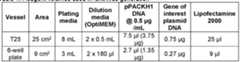 Depending on the scale of lentivirus production, different reagent volumes were used. Refer to table above for more details. One volume of diluting media (OptiMEM; Invitrogen) was combined with pPACKH1 DNA (Cambridge Bioscience) and gene of interest plasmid DNA (in pCDH_puro lentiviral plasmid vector). For each transfection, a second volume of OptiMEM was mixed with Lipofectamine 2000 (Invitrogen) for 5 minutes at room temperature. The diluted plasmid mix was then combined with the diluted Lipofectamine 2000 mix and incubated at room temperature for 20 minutes prior to addition the HEK293FT cells and incubation at 37°C and 5% CO2 for 48 hours. Following this incubation period, the supernatants from each sample of cells was collected and the presence of lentiviral particles confirmed using a Lenti-X™ GoStix™ Plus according to manufacturer’s instructions (Takara Bio). Supernatants containing lentiviral particles were then filtered through 0.22 µm pore filters in sterile Steriflip (Millipore) tubes prior to use. Transduction of cells with lentivirus Ad293, MDA-MB-231, U20S, HCT116, HeLa and HPAC cells are grown in 6-well plates in 2 mL of appropriate media to achieve approximately 60-80% confluence. Prior to lentiviral transduction all media was removed and replaced with 2 mL of RPMI (Invitrogen) with 10% FBS, containing 16 µg/mL polybrene (final concentration 8 µg/mL; Sigma-Aldrich). 2 mL of supernatant containing lentiviral particles generated as described above was added to each well. Cells were then incubated at 37°C and 5% CO2 for 24 hours prior to replacing the media with fresh complete media for each cell type. Cells were then incubated at 37°C and 5% CO2 for a further 24 hours prior to the addition of selection antibiotic (puromycin; Thermo-Fisher Scientific). Puromycin was added at 2 µg/mL for Ad293, MDA-MB- 231,U20S and HPAC cells; 4 µg/mL for HCT116 cells and 10 µg/mL HeLa cells). The cells were maintained in the relevant media containing antibiotics, at 37°C, 5% CO2 until sufficient cells could be harvested for western blot analysis. The cell samples comprised of a pool of transduced cells. Western blot analysis and quantification Media was removed from the transduced cells, prior to washing with PBS. Cells were harvested using accutase (Sigma) and incubating at 37°C for 3 minutes. The accutase was then neutralised with the addition of complete media. The cell suspension was then collected and centrifuged at 1200 rpm (300 x g) for 5 minutes to pellet cells. The cell pellets were washed in PBS and transferred to 1.5 mL Eppendorf tubes. These tubes were then centrifuged at 1200 rpm (300 x g) for 5 minutes and the supernatant discarded. The cell pellets are lysed in RIPA Lysis buffer (Thermo Fisher Scientific) containing a 1:100 dilution of protease and phosphatase inhibitor cocktail (Cell Signalling Technology). Lysates were incubated on ice for 30 minutes prior to clarifying, by centrifuging at 10,000 rpm (17,000 x g) for 10 minutes at 4°C. The supernatant was collected in a fresh 1.5 mL Eppendorf tube and stored at -80°C. The protein concentration of each lysate was determined by BCA assay (Pierce / Thermo Fisher Scientific according to manufacturer’s instructions).40 µg lysate from each cell line then loaded per well on 4-12% BOLT gels (Thermo Fisher Scientific) and run at 200V for 25 minutes prior to transferring to membrane using the iBlot according to manufacturer’s instructions (Thermo Fisher Scientific). Membranes were then blocked in Odyssey blocking buffer (Li-cor) and western blot analysis performed using appropriate antibodies (see Table 2 below): Table 2. Antibody suppliers and dilutions
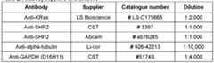


Claims
CLAIMS 1. A molecule comprising (a) a regulation domain comprising an E2 ubiquitin or ubiquitin-like conjugating domain which has an amino acid sequence having at least 80% sequence identity to a human E2 enzyme or a functional part thereof, and (b) a targeting domain capable of targeting the regulation domain to a substrate.
2. A molecule according to Claim 1, wherein the molecule does not comprise an E3 ubiquitin or ubiquitin-like ligase or functional part thereof.
3. A molecule according to Claim 1 or 2, wherein the regulation domain comprises an E2 ubiquitin conjugating domain that is capable of binding to ubiquitin and transferring the ubiquitin to the substrate, or wherein the regulation domain comprises an E2 ubiquitin-like conjugating domain that is capable of binding to a ubiquitin-like protein and transferring the ubiquitin-like protein to the substrate.
4. A molecule according to Claim 3, wherein the ubiquitin-like protein is SUMO, NEDD8, ATG8, ATG12, ISG15, UFM1, FAT10, URM1, or FUBI.
5. A molecule according to any one of Claims 1-4, wherein the E2 ubiquitin or ubiquitin-like conjugating domain comprises a ubiquitin core catalytic (UBC) domain.
6. A molecule according to Claim 5, wherein the UBC domain comprises 110-290 amino acids, such as 117-284 amino acids or 140-192 amino acids.
7. A molecule according to any one of Claims 1-6, wherein the UBC domain contains a catalytic cysteine residue.
8. A molecule according to any one of Claims 5-7, wherein the UBC domain comprises a PxxxP (SEQ ID NO: 206) peptide motif and a tryptophan residue located 26- 43 amino acids from the C-terminal end of the PxxxP motif, optionally wherein the PxxxP peptide motif is PxxPP (SEQ ID NO: 207).
9. A molecule according to any one of Claims 5-8, wherein (i) the UBC comprises a HxN peptide motif, optionally wherein the HxN motif is a HPN tripeptide or (ii) the UBC comprises a TxNGRF (SEQ ID NO: 210) peptide motif, optionally wherein the TxNGRF peptide motif is TPNGRF (SEQ ID NO: 208) or TANGRF(SEQ ID NO: 209).
10. A molecule according to any one of Claims 1-9, wherein the E2 ubiquitin or ubiquitin-like conjugating domain is derived from an E2 enzyme or is synthetic.
11. A molecule according to any one of Claims 1-10, wherein the regulation domain comprises an E2 enzyme, which comprises the E2 ubiquitin or ubiquitin-like conjugating domain.
12. A molecule according to Claim 10 or 11, wherein the E2 enzyme is a Family 1 E2 enzyme, a Family 2 E2 enzyme, a Family 3 E2 enzyme, a Family 4 E2 enzyme, a Family 5 E2 enzyme, a Family 6 E2 enzyme, a Family 7 E2 enzyme, a Family 8 E2 enzyme, a Family 9 E2 enzyme¸ a Family 10 E2 enzyme¸ a Family 11 E2 enzyme, a Family 12 E2 enzyme, a Family 13 E2 enzyme, a Family 14 E2 enzyme, a Family 15 E2 enzyme, a Family 16 E2 enzyme, or a Family 17 E2 enzyme.
13. A molecule according to any one of Claims 10-12, wherein the E2 enzyme is a Class I E2 enzyme, a Class II E2 enzyme, a Class III E2 enzyme, or a Class IV E2 enzyme.
14. A molecule according to any one of Claims 10-13, wherein the E2 enzyme has an amino acid sequence having at least 85%, 90%, 95%, 99%, or 100% sequence identity to a human E2 enzyme.
15. A molecule according to any one of Claims 10-14, wherein the E2 enzyme is UBE2A (hHR6A), UBE2B (hHR6B), UBE2C (UbcH10), UBE2D1 (UbcH5A), UBE2D2 (UbcH5B), UBE2D3 (UbcH5C), UBE2D4 (HBUCE1), UBE2E1 (UbcH6), UBE2E2, UBE2E3 (UbcH9), UBE2F (NCE2), UBE2G1 (UBE2G), UBE2G2 (UBC7), UBE2H (UBCH), UBE2I (Ubc9), UBE2J1 (NCUBE1), UBE2J2 (NCUBE2), UBE2K (HIP2), UBE2L3 (UbcH7), UBE2L6 (UbcH8), UBE2M (Ubc12), UBE2N (Ubc13), UBE2NL, UBE2O (E2- 230K), UBE2Q1 (NICE-5), UBE2Q2, UBE2QL, UBE2R1 (CDC34), UBE2R2 (CDC34B), UBE2S (E2-EPF), UBE2T (HSPC150), UBE2U, UBE2V1 (UEV-1A), UBE2V2 (MMS2), UBE2W, UBE2Z (Use1), UVELD (UEV3), BIRC6 (apollon), FTS (AKTIP), TSG101, or UFC1.
16. A molecule according to any one of Claims 10-15, wherein the E2 enzyme is UBE2D1 (UbcH5A), UBE2E2, UBE2L3 (UbcH7), UBE2O (E2-230K), UBE2Q2, or UBE2R2.
17. A molecule according to any one of Claims 1-16, wherein the E2 ubiquitin or ubiquitin-like conjugating domain comprises a UBC domain having an amino acid sequence of at least 80% sequence identity to the UBC domain of any one of UBE2A (hHR6A), UBE2B (hHR6B), UBE2C (UbcH10), UBE2D1 (UbcH5A), UBE2D2 (UbcH5B), UBE2D3 (UbcH5C), UBE2D4 (HBUCE1), UBE2E1 (UbcH6), UBE2E2, UBE2E3 (UbcH9), UBE2F (NCE2), UBE2G1 (UBE2G), UBE2G2 (UBC7), UBE2H (UBCH), UBE2I (Ubc9), UBE2J1 (NCUBE1), UBE2J2 (NCUBE2), UBE2K (HIP2), UBE2L3 (UbcH7), UBE2L6 (UbcH8), UBE2M (Ubc12), UBE2N (Ubc13), UBE2NL, UBE2O (E2-230K), UBE2Q1 (NICE-5), UBE2Q2, UBE2QL, UBE2R1 (CDC34), UBE2R2 (CDC34B), UBE2S (E2-EPF), UBE2T (HSPC150), UBE2U, UBE2V1 (UEV-1A), UBE2V2 (MMS2), UBE2W, UBE2Z (Use1), UVELD (UEV3), BIRC6 (apollon), FTS (AKTIP), TSG101, and UFC1 (SEQ ID NOs: 42-82, respectively).
18. A molecule according to any one of Claims 1-17, wherein the E2 ubiquitin or ubiquitin-like conjugating domain comprises the UBC domain of any one of UBE2A (hHR6A), UBE2B (hHR6B), UBE2C (UbcH10), UBE2D1 (UbcH5A), UBE2D2 (UbcH5B), UBE2D3 (UbcH5C), UBE2D4 (HBUCE1), UBE2E1 (UbcH6), UBE2E2, UBE2E3 (UbcH9), UBE2F (NCE2), UBE2G1 (UBE2G), UBE2G2 (UBC7), UBE2H (UBCH), UBE2I (Ubc9), UBE2J1 (NCUBE1), UBE2J2 (NCUBE2), UBE2K (HIP2), UBE2L3 (UbcH7), UBE2L6 (UbcH8), UBE2M (Ubc12), UBE2N (Ubc13), UBE2NL, UBE2O (E2-230K), UBE2Q1 (NICE-5), UBE2Q2, UBE2QL, UBE2R1 (CDC34), UBE2R2 (CDC34B), UBE2S (E2-EPF), UBE2T (HSPC150), UBE2U, UBE2V1 (UEV-1A), UBE2V2 (MMS2), UBE2W, UBE2Z (Use1), UVELD (UEV3), BIRC6 (apollon), FTS (AKTIP), TSG101, and UFC1, the amino acid sequence of which UBC domains are specified in SEQ ID NOs: 42-82, respectively.
19. A molecule according to any one of Claims 1-18, wherein the regulation domain comprises an E2 enzyme having an amino acid sequence of at least 80% sequence identity to any one of the E2 enzymes selected from the group consisting of UBE2A (hHR6A), UBE2B (hHR6B), UBE2C (UbcH10), UBE2D1 (UbcH5A), UBE2D2 (UbcH5B), UBE2D3 (UbcH5C), UBE2D4 (HBUCE1), UBE2E1 (UbcH6), UBE2E2, UBE2E3 (UbcH9), UBE2F (NCE2), UBE2G1 (UBE2G), UBE2G2 (UBC7), UBE2H (UBCH), UBE2I (Ubc9), UBE2J1 (NCUBE1), UBE2J2 (NCUBE2), UBE2K (HIP2), UBE2L3 (UbcH7), UBE2L6 (UbcH8), UBE2M (Ubc12), UBE2N (Ubc13), UBE2NL, UBE2O (E2-230K), UBE2Q1 (NICE-5), UBE2Q2, UBE2QL, UBE2R1 (CDC34), UBE2R2 (CDC34B), UBE2S (E2-EPF), UBE2T (HSPC150), UBE2U, UBE2V1 (UEV-1A), UBE2V2 (MMS2), UBE2W, UBE2Z (Use1), UVELD (UEV3), BIRC6 (apollon), FTS (AKTIP), TSG101, and UFC1 (SEQ ID NOs: 1-41, respectively).
20. A molecule according to any one of Claims 1-19, wherein the regulation domain comprises an E2 enzyme selected from the group consisting of UBE2A (hHR6A), UBE2B (hHR6B), UBE2C (UbcH10), UBE2D1 (UbcH5A), UBE2D2 (UbcH5B), UBE2D3 (UbcH5C), UBE2D4 (HBUCE1), UBE2E1 (UbcH6), UBE2E2, UBE2E3 (UbcH9), UBE2F (NCE2), UBE2G1 (UBE2G), UBE2G2 (UBC7), UBE2H (UBCH), UBE2I (Ubc9), UBE2J1 (NCUBE1), UBE2J2 (NCUBE2), UBE2K (HIP2), UBE2L3 (UbcH7), UBE2L6 (UbcH8), UBE2M (Ubc12), UBE2N (Ubc13), UBE2NL, UBE2O (E2-230K), UBE2Q1 (NICE-5), UBE2Q2, UBE2QL, UBE2R1 (CDC34), UBE2R2 (CDC34B), UBE2S (E2-EPF), UBE2T (HSPC150), UBE2U, UBE2V1 (UEV-1A), UBE2V2 (MMS2), UBE2W, UBE2Z (Use1), UVELD (UEV3), BIRC6 (apollon), FTS (AKTIP), TSG101, and UFC1, the amino acid sequences of which E2 enzymes are specified in SEQ ID NOs: 1-41, respectively.
21. A molecule according to any one of Claims 1-20, wherein the targeting domain binds to the substrate.
22. A molecule according to any one of Claims 1-21, wherein the targeting domain is any one of a monobody, nanobody, antibody, antibody fragment, scFv, intrabody, minibody, scaffold protein such as a designed ankyrin repeat protein (DARPin), peptide binder, and ligand binding domain.
23. A molecule according to any one of Claims 1-22, wherein the targeting domain and/or regulation domain does not contain a lysine residue.
24. A molecule according to any one of Claims 1-23, wherein the targeting domain has an amino acid sequence having at least 80% sequence identity to any one of SEQ ID NOs: 126-135, 138-139, 257 and/or the regulation domain has the amino acid sequence having at least 80% sequence identity to any one of SEQ ID NOs: 1-82.
25. A molecule according to any one of Claims 1-24, wherein the targeting domain has an amino acid sequence of any one of SEQ ID NOs: 126-135, 138-139, 257 or a variant thereof with up to 20 amino acid modifications, and/or wherein the regulation domain has an amino acid sequence of any one of SEQ ID NOs: 1-82 or a variant thereof with up to 30 amino acid modifications.
26. A molecule according to any one of Claims 23-25 wherein: the targeting domain is a variant of the amino acid sequence of any one of SEQ ID NOs: 126-135, 138-139, 257 in which one or more of the lysine residues has been substituted with another amino acid and/or deleted; and/or the regulation domain is a variant of the amino acid sequence of any one of SEQ ID NOs: 42-82 in which one or more lysine residues has been substituted with another amino acid and/or deleted.
27. A molecule according to any one of Claims 1-26, wherein the substrate is an intracellular polypeptide.
28. A molecule according to any one of Claims 1-27, wherein the substrate is localised in one or more of the plasma membrane, cytoplasm, nucleus, endosome, endoplasmic reticulum, mitochondria and Golgi apparatus.
29. A molecule according to any one of Claims 1-28, wherein the substrate is localised in the nucleus.
30. A molecule according to any one of Claims 1-28, wherein the substrate is an oncogenic protein, a signalling protein, a GPCR, a post-translationally modified protein, an adhesion protein, a receptor, a cell-cycle protein, a checkpoint protein, a viral protein, a prion protein, a bacterial protein, a parasitic protein, a fungal protein, a DNA binding protein, a structural protein, an enzyme, an immunogen, an antigen, and/or a pathogenic protein.
31. A molecule according to any one of Claims 1-30 wherein the substrate is selected from the group consisting of Ras, KRas, SHP2, human rhinovirus (HRV) protease 3C, muscarinic acetylcholine receptor 2 (M2R), beta-2 adrenergic receptor (β2-AR), crossover junction endonuclease MUS81 (MUS81) and human antigen R (HuR).
32. A molecule according to any of Claims 1-31, wherein the regulation domain and targeting domain are joined by a linker.
33. A molecule according to Claim 32, wherein the linker is a polypeptide linker, such as a polypeptide containing one or more glycine and/or serine amino acid residues.
34. A molecule according to Claim 31 or 32, wherein the linker is from 1 to 45 amino acids in length, such as from 6 to 20 amino acids in length or from 5 to 19 amino acids in length.
35. A molecule according to any one of Claims 32-34, wherein the linker comprises the peptide GGGGS (SEQ ID NO: 146), GGGGSGGGGSGGGGS (SEQ ID NO: 145), LEGGGGSSR (SEQ ID NO: 141), LEGGGGSGGGGSGGGGSSR (SEQ ID NO: 142), AAAGGGGSGGGGSGGGGSGT (SEQ ID NO: 143), GGGGG (SEQ ID NO: 144), LEGGSR (SEQ ID NO: 211), LEGGGSGGSSR (SEQ ID NO: 212), LEGGGGSGGGSSR (SEQ ID NO: 213), LEGGGSGGGSGGGSSR (SEQ ID NO: 214), LEGGGGSGPSGGGGPSGSR (SEQ ID NO: 215), LESNGGGGSPAPAPGGGGSGSSR (SEQ ID NO: 216), LEGGGGSYPYDVPDYASGGGGSSR (SEQ ID NO: 217), TGGSAGGSGGSAGGSGGSAGGSGGSA (SEQ ID NO: 218), AGSGGSTGSGGSPTPSTSGGSTGSGGAS (SEQ ID NO: 219), AGSGGSGGSGGSGNSSTSGGSGGSGGAS (SEQ ID NO: 220), GGSPVPSTPGGGSGGGSGGSPVPSTPGS (SEQ ID NO: 221), or SPGTGSPGTGSPGTGSPGTGSPGTGSPG (SEQ ID NO: 222).
36. A molecule according to any one of Claims 1-35, wherein the molecule is a fusion polypeptide.
37. A molecule according to any one of Claims 1-36, wherein the regulation domain is N-terminal to the targeting domain.
38. A molecule according to any one of Claims 1-36, wherein the regulation domain is C-terminal to the targeting domain.
39. A molecule according to any one of Claims 2-38, wherein the E3 ubiquitin or ubiquitin-like ligase or functional part thereof is one that comprises one or more domains selected from the group consisting of a RING (Really Interesting New Gene) domain, a U- box domain, a HECT (homologous to E6-AP carboxyl terminus) domain, and an RBR domain.
40. A molecule according to any one of Claims 1-39, further comprising a detectable marker.
41. A molecule according to Claim 40, wherein the detectable marker does not contain lysine residues, optionally wherein the detectable marker is a hemagglutinin tag or a Glu- Glu epitope tag.
42. A molecule according to any one of Claims 1-40, wherein the molecule is a protein having the amino acid sequence of any one of SEQ ID NOs: 156-167, 171-195, 202-204, 236-248, 253-256, 267, 270 and 272.
43. A molecule according to any one of Claims 1-42, wherein the molecule comprises a subcellular localisation signal, such as a nuclear localisation signal, a mitochondrial localisation signal or an endosomal localisation signal.
44. A molecule according to any one of Claims 1-43, wherein the molecule is capable of decreasing the amount of a substrate by at least 20% compared to the amount of the substrate in the absence of the molecule, optionally wherein the molecule decreases the amount of the substrate in a cell by at least 20% compared to the amount of the substrate in a cell that is otherwise substantially the same, but which does not contain the molecule.
45. A compound comprising (i) a molecule according to any one of Claims 1-44 and (ii) a targeting moiety capable of targeting the molecule to a cell.
46. A compound according to Claim 45, wherein the targeting moiety is a binding partner such as an antibody.
47. A compound according to Claim 45 or 46 wherein the targeting moiety is a polypeptide which is fused to the molecule.
48. A polynucleotide encoding a molecule according to any of Claims 1-44 or a compound of Claim 47, optionally wherein the polynucleotide comprises the nucleotide sequence of any one of SED ID NOs: 223-235, 249-252 and 259, 262 and 264.
49. A vector comprising the polynucleotide of Claim 48, such as an adeno-associated virus (AAV) vector or a lentiviral vector.
50. A host cell comprising the polynucleotide of Claim 48 or the vector of Claim 49.
51. A composition comprising a molecule of any of Claims 1-44 , a compound of any one of Claims 45-47, a polynucleotide according to Claim 48, a vector according to Claim 49, or a host cell according to Claim 50, and a further therapeutic agent.
52. A composition according to Claim 51, wherein the further therapeutic agent an anti- cancer agent, an anti-viral agent, an anti-diabetic agent, an immunotherapeutic agent, an anti-inflammatory agent, an antibiotic, or any combination thereof.
53. A molecule according to any of Claims 1-44, a compound according to any one of Claims 45-47, a polynucleotide according to Claim 48, a vector according to Claim 49, a host cell according to Claim 50, or a composition according to Claim 51 or 52 for use in medicine.
54. A pharmaceutical composition comprising a molecule according to any one of Claims 1-44, a compound according to any one of Claims 45-47, a polynucleotide according to Claim 48, a vector according to Claim 49, a host cell according to Claim 50, or a composition according to Claim 51 or 52, and one or more pharmaceutically acceptable carrier, diluent or excipient.
55. A method of delivering a molecule according to any one of Claims 1-44 to a cell in an individual, the method comprising: administering to the individual a compound comprising (i) a molecule according to any one of Claims 1-44 and (ii) a targeting moiety capable of targeting the molecule to the cell; or administering to the individual a polynucleotide of Claim 48 or a vector of Claim 49, wherein the polynucleotide or vector encodes the molecule in the cell.
56. A method according to Claim 55, wherein the molecule is capable of decreasing the amount of a substrate by at least 20% compared to the amount of the substrate in the absence of the molecule, optionally wherein the molecule decreases the amount of the substrate in a cell by at least 20% compared to the amount of the substrate in a cell that is otherwise substantially the same, but which does not contain the molecule.
57. A compound comprising (i) a molecule according to any one of Claims 1-44 and (ii) a targeting moiety capable of targeting the molecule to a cell, for use in delivering a molecule according to any one of Claims 1-44 to a cell in an individual.
58. Use of a compound comprising (i) a molecule according to any one of Claims 1-44 and (ii) a targeting moiety capable of targeting the molecule to a cell, in the manufacture of a medicament for delivering a molecule according to any one of Claims 1-44 to a cell in an individual.
59. A method according to Claim 58, wherein the molecule is capable of decreasing the amount of a substrate by at least 20% compared to the amount of the substrate in the absence of the molecule, optionally wherein the molecule decreases the amount of the substrate in a cell by at least 20% compared to the amount of the substrate in a cell that is otherwise substantially the same, but which does not contain the molecule.
60. A kit of parts comprising: (a) a regulation domain comprising an E2 ubiquitin or ubiquitin-like conjugating domain which has an amino acid sequence having at least 80% sequence identity to a human E2 enzyme or a functional part thereof, and (b) a targeting domain capable of targeting the regulation domain to the substrate; optionally wherein the kit does not comprise an E3 ubiquitin or ubiquitin-like ligase or a functional part thereof.
61. A kit of parts according to Claim 60, further comprising a linker that is capable of joining the regulation domain to the targeting domain.
62. A kit of parts comprising: (a) a molecule according to any one of Claims 1-44; and (b) a targeting moiety that is capable of targeting to cells that contain the substrate to be regulated, optionally wherein the targeting moiety is a binding partner such as an antibody.
63. A kit of parts according to Claim 62, further comprising a linker that is capable of joining the regulation domain to the targeting domain.
64. A kit of parts comprising: (a) a polynucleotide that encodes a regulation domain comprising an E2 ubiquitin or ubiquitin-like conjugating domain which has an amino acid sequence having at least 80% sequence identity to a human E2 enzyme or a functional part thereof, and (b) a polynucleotide that encodes a targeting domain capable of targeting the regulation domain to a substrate; optionally wherein the kit does not comprise a polynucleotide that encodes an E3 ubiquitin or ubiquitin-like ligase or a functional part thereof.
65. A kit of parts according to Claim 64 wherein the kit comprises one or more promoter sequences capable of directing expression of one or both of the polynucleotides in a cell that contains the substrate to be regulated.
66. A kit of parts comprising: (a) a polynucleotide encoding a molecule according to any one of Claims 1-44; and (b) a targeting moiety that is capable of targeting to cells that contain the substrate to be regulated.
67. A kit of parts according to any one of Claims 60-66, further comprising one or more reagents to assess the expression profile of a cell that contains the substrate to be regulated.
68. A kit of parts according to any one of Claims 60-67, further comprising a means for assessing a property of a cell.
69. A kit of parts according to any one of Claims 60-68 wherein the regulation domain is as defined in any one of Claims 1-20 and/or wherein the targeting domain is as defined in any one of Claims 21-26.
70. A kit of parts according to any one of Claims 60-69 wherein the substrate is as defined in any one of Claims 25-28.
71. A method of producing the molecule of any one of Claims 1-44, or the compound of any one of Claims 45-47, the method comprising expressing the polynucleotide of Claim 48 in a host cell.
72. A method according to Claim 71, wherein the method comprises introducing the polynucleotide of Claim 48 or the vector of Claim 49 into a host cell, and expressing the polynucleotide encoding the molecule.
73. A method of preventing or treating a disease or condition mediated by an aberrant level of a substrate or form thereof in a subject, the method comprising administering the molecule of any one of Claims 1-44, the compound of any one of Claims 45-47, the polynucleotide of Claim 48, the vector of Claim 49, the cell of Claim 50, the pharmaceutical composition of Claim 54, or the composition of Claim 51 or 52 to the subject.
74. A method according to Claim 73, wherein the molecule is capable of decreasing the amount of a substrate by at least 20% compared to the amount of the substrate in the absence of the molecule, optionally wherein the molecule decreases the amount of the substrate in a cell by at least 20% compared to the amount of the substrate in a cell that is otherwise substantially the same, but which does not contain the molecule.
75. A molecule of any one of Claims 1-44, a compound of any one of Claims 45-47, a polynucleotide of Claim 48, a vector of Claim 49, a cell of Claim 50, a pharmaceutical composition of Claim 54, or a composition of Claim 51 or 52 for use in preventing or treating a disease or condition mediated by an aberrant level of a substrate or form thereof in a subject.
76. Use of a molecule of any one of Claims 1-44, a compound of any one of Claims 45- 47, a polynucleotide of Claim 48, a vector of Claim 49, a cell of Claim 50, a pharmaceutical composition of Claim 54, or a composition of Claim 51 or 52, in the manufacture of a medicament for preventing or treating a disease or condition mediated by an aberrant level of a substrate or form thereof in a subject.
77. A method according to Claim 73 or 74 or a use according to Claim 75 or 76, wherein the disease or condition is cancer, diabetes, autoimmune disease, Alzheimer’s disease, Parkinson’s disease, pain, viral disease, bacterial disease, prionic disease, fungal disease, parasitic disease, arthritis, immunodeficiency, or inflammatory disease.
78. A method of regulating a substrate, the method comprising contacting the substrate with the molecule of any one of Claims 1-44 under conditions effective for the molecule to regulate the substrate.
79. A method according to Claim 78, wherein the regulating involves the substrate being degraded, or the substrate being prevented from being degraded, or the subcellular location of the substrate being altered, or one or more activities of the substrate being modulated (e.g. increased or decreased), or the degree of post-translational modification of the substrate being modulated.
80. A method of identifying a substrate as a potential drug target, the method comprising: (a) providing a cell, tissue or organ comprising the substrate (b) contacting the cell, tissue or organ with the molecule according to any one of Claims 1-44, the compound of any one of Claims 45-47, the polynucleotide of Claim 48, or the vector of Claim 49; and (c) assessing the effect of the molecule, compound, polynucleotide or vector on one or more properties of the cell, tissue or organ, wherein identification of an effect that is correlated with a particular disease status is indicative that the substrate is a potential drug target for the particular disease.
81. A method of assessing the function of a substrate, the method comprising: (a) providing a cell, tissue or organ comprising the substrate (b) contacting the cell, tissue or organ with the molecule according to any one of Claims 1-44, the compound of any one of Claims 45-47, the polynucleotide of Claim 48, or the vector of Claim 49; and (c) assessing the effect of the molecule, compound, polynucleotide or vector on one or more properties of the cell, tissue or organ.
82. A method of identifying a test agent that may be useful in preventing or treating a disease or condition mediated by an aberrant level of a substrate or form thereof, the method comprising: providing the substrate; providing a test agent comprising (a) a regulation domain comprising an E2 ubiquitin or ubiquitin-like conjugating domain which has an amino acid sequence having at least 80% sequence identity to a human E2 ubiquitin or ubiquitin-like domain, and (b) a targeting domain capable of targeting the regulation domain to a substrate, optionally wherein the test agent does not comprise an E3 ubiquitin or ubiquitin-like ligase or part thereof; contacting the substrate and test agent under conditions effective for the test agent to facilitate regulation of the substrate; and determining whether the test agent regulates the substrate.
83. A method according to Claim 82, further comprising the step of testing the test agent in an assay of the disease or condition.
84. A method according to Claim 81 or 82, further comprising the step of synthesising, purifying and/or formulating the test agent.
85. Use of a molecule according to any one of Claims 1-44 or the compound of any one of Claims 45-47 in drug target validation or in drug discovery.
Priority Applications (4)
| Application Number | Priority Date | Filing Date | Title |
|---|---|---|---|
| US18/253,665US20250197819A1 (en) | 2019-11-22 | 2020-11-20 | Fusion proteins comprising an e2 ubiquitin or ubiquitin-like conjugating domain and a targeting domain for specific protein degradation |
| CN202080107264.0ACN116670268A (en) | 2019-11-22 | 2020-11-20 | Fusion proteins comprising E2 ubiquitin or ubiquitin-like conjugation domains and targeting domains for specific protein degradation |
| EP20819874.7AEP4247957A1 (en) | 2019-11-22 | 2020-11-20 | Fusion proteins comprising an e2 ubiquitin or ubiquitin-like conjugating domain and a targeting domain for specific protein degradation |
| JP2023529985AJP2023550743A (en) | 2019-11-22 | 2020-11-20 | Fusion proteins containing E2 ubiquitin or ubiquitin-like conjugate domains and targeting the domain for specific proteolysis |
Applications Claiming Priority (1)
| Application Number | Priority Date | Filing Date | Title |
|---|---|---|---|
| US201962939234P | 2019-11-22 | 2019-11-22 |
Publications (1)
| Publication Number | Publication Date |
|---|---|
| WO2022106869A1true WO2022106869A1 (en) | 2022-05-27 |
Family
ID=73699170
Family Applications (1)
| Application Number | Title | Priority Date | Filing Date |
|---|---|---|---|
| PCT/IB2020/060978CeasedWO2022106869A1 (en) | 2019-11-22 | 2020-11-20 | Fusion proteins comprising an e2 ubiquitin or ubiquitin-like conjugating domain and a targeting domain for specific protein degradation |
Country Status (5)
| Country | Link |
|---|---|
| US (1) | US20250197819A1 (en) |
| EP (1) | EP4247957A1 (en) |
| JP (1) | JP2023550743A (en) |
| CN (1) | CN116670268A (en) |
| WO (1) | WO2022106869A1 (en) |
Cited By (2)
| Publication number | Priority date | Publication date | Assignee | Title |
|---|---|---|---|---|
| CN116731206A (en)* | 2023-05-24 | 2023-09-12 | 南方科技大学 | Chimeric ubiquitin ligase for targeted degradation of KRAS as well as preparation method and application thereof |
| WO2024192235A3 (en)* | 2023-03-14 | 2024-10-24 | Ohio State Innovation Foundation | Nanobody-based protein degraders and related methods |
Citations (6)
| Publication number | Priority date | Publication date | Assignee | Title |
|---|---|---|---|---|
| EP0626450A2 (en)* | 1993-05-28 | 1994-11-30 | Wisconsin Alumni Research Foundation | Ubiquitin conjugating enzyme (E2) fusion proteins |
| US5585089A (en) | 1988-12-28 | 1996-12-17 | Protein Design Labs, Inc. | Humanized immunoglobulins |
| US5739116A (en) | 1994-06-03 | 1998-04-14 | American Cyanamid Company | Enediyne derivatives useful for the synthesis of conjugates of methyltrithio antitumor agents |
| US20060088522A1 (en) | 2004-09-10 | 2006-04-27 | Wyeth | Humanized anti-5T4 antibodies and anti-5T4/calicheamicin conjugates |
| US7659241B2 (en) | 2002-07-31 | 2010-02-09 | Seattle Genetics, Inc. | Drug conjugates and their use for treating cancer, an autoimmune disease or an infectious disease |
| US20110008840A1 (en) | 2002-11-07 | 2011-01-13 | Immunogen, Inc. | Anti-cd33 antibodies and methods for treatment of acute myeloid leukemia using the same |
- 2020
- 2020-11-20USUS18/253,665patent/US20250197819A1/enactivePending
- 2020-11-20JPJP2023529985Apatent/JP2023550743A/enactivePending
- 2020-11-20WOPCT/IB2020/060978patent/WO2022106869A1/ennot_activeCeased
- 2020-11-20CNCN202080107264.0Apatent/CN116670268A/enactivePending
- 2020-11-20EPEP20819874.7Apatent/EP4247957A1/enactivePending
Patent Citations (9)
| Publication number | Priority date | Publication date | Assignee | Title |
|---|---|---|---|---|
| US5585089A (en) | 1988-12-28 | 1996-12-17 | Protein Design Labs, Inc. | Humanized immunoglobulins |
| US5693762A (en) | 1988-12-28 | 1997-12-02 | Protein Design Labs, Inc. | Humanized immunoglobulins |
| EP0626450A2 (en)* | 1993-05-28 | 1994-11-30 | Wisconsin Alumni Research Foundation | Ubiquitin conjugating enzyme (E2) fusion proteins |
| US5739116A (en) | 1994-06-03 | 1998-04-14 | American Cyanamid Company | Enediyne derivatives useful for the synthesis of conjugates of methyltrithio antitumor agents |
| US5767285A (en) | 1994-06-03 | 1998-06-16 | American Cyanamid Company | Linkers useful for the synthesis of conjugates of methyltrithio antitumor agents |
| US5773001A (en) | 1994-06-03 | 1998-06-30 | American Cyanamid Company | Conjugates of methyltrithio antitumor agents and intermediates for their synthesis |
| US7659241B2 (en) | 2002-07-31 | 2010-02-09 | Seattle Genetics, Inc. | Drug conjugates and their use for treating cancer, an autoimmune disease or an infectious disease |
| US20110008840A1 (en) | 2002-11-07 | 2011-01-13 | Immunogen, Inc. | Anti-cd33 antibodies and methods for treatment of acute myeloid leukemia using the same |
| US20060088522A1 (en) | 2004-09-10 | 2006-04-27 | Wyeth | Humanized anti-5T4 antibodies and anti-5T4/calicheamicin conjugates |
Non-Patent Citations (78)
| Title |
|---|
| "Antibodies: A Laboratory Manual", 1988, COLD SPRING HARBOR LABORATORY PRESS |
| "Handbook of Therapeutic Antibodies", 2007, WILEY-VCH |
| "Using Antibodies: A Laboratory Manual", 1999, COLD SPRING HARBOR LABORATORY PRESS |
| AICART-RAMOS ET AL., BIOCHIM BIOPHYS ACTA - BIOMEMBRANES, vol. 1808, no. 12, 2011, pages 298194 |
| AN ET AL., THERAPEUTIC MONOCLONAL ANTIBODIES: FROM BENCH TO CLINIC, 2009, ISBN: ISBN: 978-0-470-11791-0 |
| BARTKE ET AL., MOL CELL, 2004 |
| BASKA ET AL.: "Mechanism of extracellular ubiquitination in the mammalian epididymis", J CELL PHYSIOL, vol. 215, no. 3, 2008, pages 684 - 96 |
| BERY ET AL., NAT COMMUN, vol. 10, no. 1, 2019, pages 2607 |
| BINZ ET AL., J MOL BIOL, vol. 332, no. 2, 2003, pages 489 - 503 |
| BURSLEM, CELL CHEM BIOL, vol. 25, no. 1, 2018, pages 67 - 77 |
| CARTER ET AL.: "Identification and validation of cell surface antigens for antibody targeting in oncology", ENDOCR-RELAT CANCER, vol. 11, 2004, pages 659 - 87, XP009078100, DOI: 10.1677/erc.1.00766 |
| CARTER: "Introduction to current and future protein therapeutics: a protein engineering perspective", EXP CELL RES, vol. 317, no. 9, 2011, pages 1261 - 9, XP028205664, DOI: 10.1016/j.yexcr.2011.02.013 |
| CARTER: "Potent antibody therapeutics by design", NAT REV IMMUNOL, vol. 6, no. 5, 2006, pages 343 - 57, XP007901440, DOI: 10.1038/nri1837 |
| CARTERSENTER, CANCER J, vol. 14, no. 3, 2008, pages 154 - 69 |
| CHARI ET AL., ANGEWANDTE CHEMIE INTERNATIONAL EDITION, vol. 53, 2014, pages 3751 |
| CLIFT ET AL., CELL, vol. 171, no. 7, 2017, pages 1692 - 1706 |
| DIKIC ET AL., NAT REV MOL CELL BIOL, vol. 10, 2009, pages 659 - 671 |
| DOLLER ET AL., CELL SIGNAL., vol. 20, 2008, pages 2165 - 2173 |
| DRUGS R D, vol. 11, no. 1, pages 85 - 95 |
| ELEONORA TURCO ET AL: "Monoubiquitination of Histone H2B Is Intrinsic to the Bre1 RING Domain-Rad6 Interaction and Augmented by a Second Rad6-binding Site on Bre1", JOURNAL OF BIOLOGICAL CHEMISTRY, vol. 290, no. 9, 1 February 2015 (2015-02-01), pages 5298 - 5310, XP055768879, ISSN: 0021-9258, DOI: 10.1074/jbc.M114.626788* |
| EMBABE ET AL.: "Mdm2-mediated NEDDylation of HuR controls the nuclear localization of HuR and protects it from degradation", HEPATOLOGY, vol. 55, no. 4, 2012, pages 1237 - 48 |
| FOIGHT ET AL.: "Multi-input chemical control of protein dimerization for programming graded cellular responses", NAT BIOTECHNOL, vol. 37, no. 10, 2019, pages 1209 - 16, XP036897234, DOI: 10.1038/s41587-019-0242-8 |
| FULCHER ET AL., OPEN BIOL, vol. 7, no. 5, 2017, pages 170066 |
| GIOVANNONI ET AL.: "Isolation of anti-angiogenesis antibodies from a large combinatorial repertoire by colony filter screening", NUCLEIC ACIDS RES, vol. 29, 2001, pages E27 |
| GLICKMANCIECHANOVER, PHYSIOL REV, vol. 82, no. 2, 2002, pages 373 - 428 |
| GUANFIERKE, SCI CHINA CHEM, vol. 54, no. 12, 2011, pages 1888 - 97 |
| GUNDOGDUWALDEN, PROTEIN SCIENCE, vol. 28, 2019, pages 1758 - 1770 |
| H. ZOLA: "Monoclonal Antibodies: A Manual of Techniques", 1987, CRC PRESS |
| HERRMAN ET AL., CIRC RES, vol. 100, no. 9, 2007, pages 1276 - 1291 |
| HORMAECHEA-AGULLA ET AL., MOL CELL, vol. 41, no. 3, 2018, pages 168 - 178 |
| HUGHES, NAT DRUG DISCOV, vol. 9, 2010, pages 665 |
| HUM GEN THER, vol. 22, no. 10, 2011, pages A14 - A14 |
| HURRELL: "Monoclonal Hybridoma Antibodies: Techniques and Application", 1982, CRC PRESS |
| IN VIVO: THE BUSINESS & MEDICINE REPORT, pages 32 - 38 |
| JEFFREY ET AL., BMCL, vol. 16, 2006, pages 358 |
| KAMYNINASTOVER: "SREBP SUMOylation inhibits SREBP transcriptional activity indirectly through the recruitment of a co-repressor complex that includes histone deacetylase 3 (HDAC3", ADV EXP MED BIOL, vol. 963, 2017, pages 143 - 68 |
| KOHLERMILSTEIN: "Continuous cultures of fused cells secreting antibody of predefined specificity", NATURE, vol. 256, 1975, pages 495 - 497 |
| LONN ET AL., SCI REP, vol. 6, 2016, pages 32301 |
| MAHAJAN ET AL.: "A small ubiquitin-related polypeptide involved in targeting RanGAP1 to nuclear pore complex protein RanBP2", CELL, vol. 88, no. 1, 1997, pages 97 - 107, XP001084136, DOI: 10.1016/S0092-8674(00)81862-0 |
| MAHATO ET AL., ADV DRUG DELIV REV, vol. 63, 2011, pages 659 |
| MATUNIS ET AL.: "A novel ubiquitin-like modification modulates the partitioning of the Ran-GTPase-activating protein RanGAP1 between the cytosol and the nuclear pore complex", J CELL BIOL, vol. 135, 1996, pages 1457 - 70, XP002273973, DOI: 10.1083/jcb.135.6.1457 |
| MICHAELSON ET AL., MOL BIOL CELL, vol. 16, 2005, pages 1606 - 16 |
| MICHELLE ET AL.: "What was the set of ubiquitin and ubiquitin-like conjugating enzymes in the eukaryotic common ancestor?", J MOL EVOL, vol. 68, 2009, pages 616 - 628, XP019704499 |
| MIKAELA D STEWART ET AL: "E2 enzymes: more than just middle men", CELL RESEARCH, VOL. 26, N.4, 22 March 2016 (2016-03-22), pages 423 - 440, XP055687403, Retrieved from the Internet <URL:http://www.nature.com/articles/cr201635.pdf> [retrieved on 20200420], DOI: 10.1038/cr.2016.35* |
| MUKHOPADHYAYRIEZMAN, SCIENCE, vol. 315, no. 5809, 2007, pages 201 - 5 |
| MURO: "Challenges in design and characterisation of ligand-targeted drug delivery systems", J CONTROL RELEASE, vol. 164, no. 2, 2012, pages 125 - 37 |
| NING ZHENGNITZAN SHABEK, ANNUAL REV BIOCHEMISTRY, vol. 86, 2017, pages 129 - 157 |
| NOH ET AL.: "NEDDylation increases RCAN1 binding to calcineurin", PLOS ONE, vol. 7, no. 10, 2012, pages e48315 |
| OLSON ET AL., NAT CHEM BIOL, vol. 14, no. 2, 2018, pages 163 - 70 |
| O'SULLIVAN ET AL.: "Comparison of two methods of preparing enzyme-antibody conjugates: Application of these conjugates for enzyme immunoassay", ANAL BIOCHEM, vol. 100, 1979, pages 100 - 8, XP024822204, DOI: 10.1016/0003-2697(79)90117-9 |
| PAN ET AL., ONCOTARGET, vol. 7, no. 28, pages 44299 - 44309 |
| PIRONE ET AL.: "A comprehensive platform for the analysis of ubiquitin-like protein modifications using in vivo biotinylation", SCI REP, vol. 7, 2017, pages 40756 |
| PORTNOFF ET AL., J BIOL CHEM, vol. 282, no. 11, 2007, pages 5101 - 05 |
| PORTNOFF ET AL., J. BIOL. CHEM., vol. 289, no. 11, 2014, pages 7844 - 5 |
| RICHTING ET AL.: "Quantitative live-cell kinetic degradation and mechanistic profiling of PROTAC mode of action", ACS CHEM BIOL, vol. 13, no. 9, 2018, pages 2758 - 70, XP055703685, DOI: 10.1021/acschembio.8b00692 |
| ROBB ET AL., CHEM COMMUN, vol. 53, no. 54, 2017, pages 7577 - 80 |
| RUST ET AL., MOL CANCER, vol. 12, 2013, pages 11 |
| SANDERCOCK ET AL., MOL CANCER, vol. 14, 2015, pages 147 |
| SCHAFFITZEL ET AL.: "Ribosome display: an in vitro method for selection and evolution of antibodies from libraries", J IMMUNOL METHODS, vol. 231, 1999, pages 119 - 135, XP004186079, DOI: 10.1016/S0022-1759(99)00149-0 |
| SCHNEEKLOTH ET AL., BIOORG MED CHEM LETTER, vol. 18, no. 22, 2008, pages 5904 - 08 |
| SCHNEEKLOTH ET AL., BIOORG MED CHENI LETTER, vol. 18, no. 22, 2008, pages 5904 - 08 |
| SCHNELLHICKE, J BIOL CHEM, vol. 278, no. 38, 2003, pages 35857 - 60 |
| SHA ET AL., PROC NATL ACAD SCI U S A, vol. 110, no. 37, 2013, pages 14924 - 9 |
| SHA ET AL., PROC NATL ACAD SCI USA, vol. 110, no. 37, 2013, pages 14924 - 9 |
| SHA ET AL., PROC. NATL. ACAD. SCI. U S A, vol. 110, no. 37, 2013, pages 14924 - 9 |
| SHA ET AL., PROC. NATL. ACAD. SCI. USA, vol. 110, no. 37, 2013, pages 14924 - 9 |
| SOUCY ET AL.: "Cullin-RING ubiquitin E3 ligases require NEDD8 modification to be activated", CLIN CANCER RES, vol. 15, no. 12, 2009, pages 3912 - 16 |
| STEWART ET AL., CELL RES, vol. 26, 2016, pages 423 |
| SUJASHVILI: "Advantages of extracellular ubiquitin in modulation of immune responses", MEDIATORS INFLAMM, 2016 |
| THOMPSON ET AL., NUCLEIC ACIDS RES, vol. 22, no. 22, 1994, pages 4673 - 80 |
| TUREK ET AL., JBC, vol. 293, 2018, pages 16324 - 16336 |
| ULLAH ET AL., FEBS J, 2018 |
| VAIDYANATHAN S ET AL.: "Uridine Depletion and Chemical Modification Increase Cas9 mRNA Activity and Reduce Immunogenicity without HPLC Purification", MOL THER NUCLEIC ACIDS, vol. 12, 2018, pages 530 - 542, XP055531206, DOI: 10.1016/j.omtn.2018.06.010 |
| VEGGIANI GIANLUCA ET AL: "Emerging drug development technologies targeting ubiquitination for cancer therapeutics", PHARMACOLOGY AND THERAPEUTICS, vol. 199, 7 March 2019 (2019-03-07), pages 139 - 154, XP085709801, ISSN: 0163-7258, DOI: 10.1016/J.PHARMTHERA.2019.03.003* |
| WEN ET AL.: "SUMOylation Promotes Nuclear Import and Stabilization of Polo-like Kinase 1 to Support Its Mitotic Function", CELL REP, vol. 21, 2017, pages 2147 - 59 |
| WILLIAMS ET AL., ONCOTARGET, vol. 7, no. 42, 2016, pages 68278 - 44309 |
| WINTER ET AL.: "Making antibodies by phage display technology", ANNU REV IMMUNOL, vol. 12, 1994, pages 433 - 455, XP000564245, DOI: 10.1146/annurev.iy.12.040194.002245 |
| YANG ET AL.: "SUMOylation Inhibits SF-1 Activity by Reducing CDK7-Mediated Serine 203 Phosphorylation", MOL CELL BIOL, vol. 29, no. 3, 2009, pages 613 - 25 |
Cited By (3)
| Publication number | Priority date | Publication date | Assignee | Title |
|---|---|---|---|---|
| WO2024192235A3 (en)* | 2023-03-14 | 2024-10-24 | Ohio State Innovation Foundation | Nanobody-based protein degraders and related methods |
| CN116731206A (en)* | 2023-05-24 | 2023-09-12 | 南方科技大学 | Chimeric ubiquitin ligase for targeted degradation of KRAS as well as preparation method and application thereof |
| WO2024239392A1 (en)* | 2023-05-24 | 2024-11-28 | 南方科技大学 | Chimeric ubiquitin ligase capable of targeted degradation of kras, preparation method therefor and use thereof |
Also Published As
| Publication number | Publication date |
|---|---|
| JP2023550743A (en) | 2023-12-05 |
| US20250197819A1 (en) | 2025-06-19 |
| CN116670268A (en) | 2023-08-29 |
| EP4247957A1 (en) | 2023-09-27 |
Similar Documents
| Publication | Publication Date | Title |
|---|---|---|
| Worden et al. | Structural basis for COMPASS recognition of an H2B-ubiquitinated nucleosome | |
| JP2024059823A (en) | Broad-spectrum proteome editing with engineered bacterial ubiquitin ligase mimic | |
| Strop | Versatility of microbial transglutaminase | |
| Matsui et al. | The Rab interacting lysosomal protein (RILP) homology domain functions as a novel effector domain for small GTPase Rab36: Rab36 regulates retrograde melanosome transport in melanocytes | |
| KR20230028453A (en) | CCR8 Antibodies for Therapeutic Uses | |
| RU2762939C2 (en) | Immunoglobulins conjugated with lysine | |
| KR102256552B1 (en) | Ly75 as cancer therapeutic and diagnostic target | |
| Spidel et al. | Site-specific conjugation to native and engineered lysines in human immunoglobulins by microbial transglutaminase | |
| US20250197819A1 (en) | Fusion proteins comprising an e2 ubiquitin or ubiquitin-like conjugating domain and a targeting domain for specific protein degradation | |
| US20240132857A1 (en) | Fusion proteins comprising two ring domains | |
| CN113336852A (en) | anti-Aggrus monoclonal antibody and application thereof | |
| Dong et al. | Construction and application of a human scFv phage display library based on Cre-LoxP recombination for anti-PCSK9 antibody selection | |
| US20200330591A1 (en) | Treatment | |
| US9840539B2 (en) | High affinity digoxigenin binding proteins | |
| EP3760722A1 (en) | Anti-integrin alpha11 monoclonal antibody and use thereof | |
| Zou et al. | A stepwise mutagenesis approach using histidine and acidic amino acid to engineer highly pH-dependent protein switches | |
| WO2020049130A1 (en) | Methods | |
| Zhu et al. | SUMO modification through rapamycin-mediated heterodimerization reveals a dual role for Ubc9 in targeting RanGAP1 to nuclear pore complexes | |
| Levin et al. | Directed evolution of a soluble human DR3 receptor for the inhibition of TL1A induced cytokine secretion | |
| EP3097124B1 (en) | Means and methods for detecting activated malt1 | |
| EP2831092A1 (en) | Binding proteins to the constant region of immunoglobulin g | |
| JP6854343B2 (en) | Long-acting PCSK9-specific binding protein and its applications | |
| CN114790239B (en) | Antibody for resisting coronavirus N protein and application thereof | |
| US12404325B2 (en) | Anti IL-6 domain antibodies with fatty acid substituents | |
| US20250092142A1 (en) | Monoclonal antibody targeting fzd7, preparation method and use thereof |
Legal Events
| Date | Code | Title | Description |
|---|---|---|---|
| 121 | Ep: the epo has been informed by wipo that ep was designated in this application | Ref document number:20819874 Country of ref document:EP Kind code of ref document:A1 | |
| WWE | Wipo information: entry into national phase | Ref document number:2023529985 Country of ref document:JP Ref document number:202080107264.0 Country of ref document:CN | |
| NENP | Non-entry into the national phase | Ref country code:DE | |
| ENP | Entry into the national phase | Ref document number:2020819874 Country of ref document:EP Effective date:20230620 | |
| WWP | Wipo information: published in national office | Ref document number:18253665 Country of ref document:US |