WO2022044132A1 - Analysis device - Google Patents
Analysis deviceDownload PDFInfo
- Publication number
- WO2022044132A1 WO2022044132A1PCT/JP2020/032060JP2020032060WWO2022044132A1WO 2022044132 A1WO2022044132 A1WO 2022044132A1JP 2020032060 WJP2020032060 WJP 2020032060WWO 2022044132 A1WO2022044132 A1WO 2022044132A1
- Authority
- WO
- WIPO (PCT)
- Prior art keywords
- output
- information
- unit
- severity
- lung sound
- Prior art date
- Legal status (The legal status is an assumption and is not a legal conclusion. Google has not performed a legal analysis and makes no representation as to the accuracy of the status listed.)
- Ceased
Links
Images


Classifications
- A—HUMAN NECESSITIES
- A61—MEDICAL OR VETERINARY SCIENCE; HYGIENE
- A61B—DIAGNOSIS; SURGERY; IDENTIFICATION
- A61B7/00—Instruments for auscultation
- A61B7/003—Detecting lung or respiration noise
- A—HUMAN NECESSITIES
- A61—MEDICAL OR VETERINARY SCIENCE; HYGIENE
- A61B—DIAGNOSIS; SURGERY; IDENTIFICATION
- A61B7/00—Instruments for auscultation
- A61B7/02—Stethoscopes
- A61B7/04—Electric stethoscopes
- G—PHYSICS
- G16—INFORMATION AND COMMUNICATION TECHNOLOGY [ICT] SPECIALLY ADAPTED FOR SPECIFIC APPLICATION FIELDS
- G16H—HEALTHCARE INFORMATICS, i.e. INFORMATION AND COMMUNICATION TECHNOLOGY [ICT] SPECIALLY ADAPTED FOR THE HANDLING OR PROCESSING OF MEDICAL OR HEALTHCARE DATA
- G16H40/00—ICT specially adapted for the management or administration of healthcare resources or facilities; ICT specially adapted for the management or operation of medical equipment or devices
- G16H40/60—ICT specially adapted for the management or administration of healthcare resources or facilities; ICT specially adapted for the management or operation of medical equipment or devices for the operation of medical equipment or devices
- G16H40/63—ICT specially adapted for the management or administration of healthcare resources or facilities; ICT specially adapted for the management or operation of medical equipment or devices for the operation of medical equipment or devices for local operation
- G—PHYSICS
- G16—INFORMATION AND COMMUNICATION TECHNOLOGY [ICT] SPECIALLY ADAPTED FOR SPECIFIC APPLICATION FIELDS
- G16H—HEALTHCARE INFORMATICS, i.e. INFORMATION AND COMMUNICATION TECHNOLOGY [ICT] SPECIALLY ADAPTED FOR THE HANDLING OR PROCESSING OF MEDICAL OR HEALTHCARE DATA
- G16H40/00—ICT specially adapted for the management or administration of healthcare resources or facilities; ICT specially adapted for the management or operation of medical equipment or devices
- G16H40/60—ICT specially adapted for the management or administration of healthcare resources or facilities; ICT specially adapted for the management or operation of medical equipment or devices for the operation of medical equipment or devices
- G16H40/67—ICT specially adapted for the management or administration of healthcare resources or facilities; ICT specially adapted for the management or operation of medical equipment or devices for the operation of medical equipment or devices for remote operation
- G—PHYSICS
- G16—INFORMATION AND COMMUNICATION TECHNOLOGY [ICT] SPECIALLY ADAPTED FOR SPECIFIC APPLICATION FIELDS
- G16H—HEALTHCARE INFORMATICS, i.e. INFORMATION AND COMMUNICATION TECHNOLOGY [ICT] SPECIALLY ADAPTED FOR THE HANDLING OR PROCESSING OF MEDICAL OR HEALTHCARE DATA
- G16H50/00—ICT specially adapted for medical diagnosis, medical simulation or medical data mining; ICT specially adapted for detecting, monitoring or modelling epidemics or pandemics
- G16H50/20—ICT specially adapted for medical diagnosis, medical simulation or medical data mining; ICT specially adapted for detecting, monitoring or modelling epidemics or pandemics for computer-aided diagnosis, e.g. based on medical expert systems
- G—PHYSICS
- G16—INFORMATION AND COMMUNICATION TECHNOLOGY [ICT] SPECIALLY ADAPTED FOR SPECIFIC APPLICATION FIELDS
- G16H—HEALTHCARE INFORMATICS, i.e. INFORMATION AND COMMUNICATION TECHNOLOGY [ICT] SPECIALLY ADAPTED FOR THE HANDLING OR PROCESSING OF MEDICAL OR HEALTHCARE DATA
- G16H50/00—ICT specially adapted for medical diagnosis, medical simulation or medical data mining; ICT specially adapted for detecting, monitoring or modelling epidemics or pandemics
- G16H50/30—ICT specially adapted for medical diagnosis, medical simulation or medical data mining; ICT specially adapted for detecting, monitoring or modelling epidemics or pandemics for calculating health indices; for individual health risk assessment
- G—PHYSICS
- G16—INFORMATION AND COMMUNICATION TECHNOLOGY [ICT] SPECIALLY ADAPTED FOR SPECIFIC APPLICATION FIELDS
- G16H—HEALTHCARE INFORMATICS, i.e. INFORMATION AND COMMUNICATION TECHNOLOGY [ICT] SPECIALLY ADAPTED FOR THE HANDLING OR PROCESSING OF MEDICAL OR HEALTHCARE DATA
- G16H50/00—ICT specially adapted for medical diagnosis, medical simulation or medical data mining; ICT specially adapted for detecting, monitoring or modelling epidemics or pandemics
- G16H50/70—ICT specially adapted for medical diagnosis, medical simulation or medical data mining; ICT specially adapted for detecting, monitoring or modelling epidemics or pandemics for mining of medical data, e.g. analysing previous cases of other patients
Definitions
- the present inventionrelates to an analyzer, an analysis method, and a recording medium.
- Heart failureis some form of cardiac dysfunction, that is, dyspnea, malaise, and edema appear as a result of organic and / or functional abnormalities in the heart that disrupt the compensatory mechanism of cardiac pump function, resulting in exercise tolerance.
- a clinical syndromein which the ability is reduced.
- Patients with heart failureare always at risk of exacerbation, even if they are in remission with treatment. If an acute exacerbation occurs in a patient due to excessive intake of water or salt, forgetting to take medicine, excessive exercise, etc., the patient will be forced to be re-hospitalized. Therefore, it is important to prevent acute exacerbations by detecting the exacerbation of heart failure in discharged patients at an early stage and intervening in treatment.
- One of the methods for diagnosing heart failureis auscultation of lung sounds.
- Such a medical examinationis one of the safe and easy methods for diagnosing the health condition of the lungs and, by extension, heart failure.
- itis difficult to obtain detailed and accurate diagnostic results without a trained specialist. Therefore, it was not possible to make a detailed diagnosis at sites such as rounds and home-visit care by general nurses and long-term care workers.
- Patent Document 7describes a system for determining a medical doctor recommended for a patient based on medical condition information input by the patient and transmitting the information of the medical doctor to a patient terminal.
- Japanese Unexamined Patent Publication No. 2014-4018Special Table 2002-538921 Publication No. Special Table 2017-536905 WO2010 / 0444552 Japanese Unexamined Patent Publication No. 2008-11936 Patent No. 48494424 Japanese Unexamined Patent Publication No. 2019-79501 Japanese Unexamined Patent Publication No. 2007-19081
- an object of the present inventionis to provide an analyzer, an analysis method, and a recording medium that solve the problem that it is difficult to efficiently share test results with medical professionals.
- the analyzerwhich is one embodiment of the present invention, is A detection unit that detects abnormal lung sound at each auscultation position based on a time-series acoustic signal including lung sound at each auscultation position.
- a determination unitfor determining the severity of heart failure of the patient based on the detection result of the lung sound abnormality for each auscultation position detected by the detection unit and the state information indicating the patient's condition.
- An output unitthat determines whether or not to output predetermined information to an external device based on the result of the determination by the determination unit and outputs the output according to the result of the determination. It takes the configuration of having.
- the analysis methodwhich is another embodiment of the present invention is: Information processing equipment Based on the time-series acoustic signal including the lung sound at each auscultation position, the lung sound abnormality is detected at each auscultation position. The severity of heart failure in the patient is determined based on the detected result of detecting the abnormal lung sound for each auscultation position and the condition information indicating the condition of the patient. Based on the judgment result, it is judged whether or not to output the predetermined information to the external device, and the output is performed according to the judgment result.
- the recording medium which is another embodiment of the present inventionis A detection unit that detects abnormal lung sound at each auscultation position based on a time-series acoustic signal including lung sound at each auscultation position.
- a determination unitfor determining the severity of heart failure of the patient based on the detection result of the lung sound abnormality for each auscultation position detected by the detection unit and the state information indicating the patient's condition.
- An output unitthat determines whether or not to output predetermined information to an external device based on the result of the determination by the determination unit and outputs the output according to the result of the determination. It is a computer-readable recording medium on which a program for realizing the above is recorded.
- the present inventionmakes it possible to efficiently share test results with medical professionals by having the above-mentioned configuration.
- FIG. 1st Embodiment of this inventionIt is a figure which shows the whole structural example of the analysis system in 1st Embodiment of this invention. It is a block diagram which shows the structural example of the analyzer shown in FIG. It is a figure which shows an example of lung sound data. It is a figure for demonstrating an example of auscultation position. It is a figure for demonstrating an example of auscultation position. It is a figure which shows an example of analysis result information. It is a figure which shows an example of the information for severity determination. It is a figure which shows an example of severity information. It is a figure which shows the example of the information included in the personal state information. It is a figure which shows an example of output information. It is a block diagram which shows the structural example of the processing apparatus shown in FIG.
- FIG. 1is a diagram showing an overall configuration example of the analysis system 100.
- FIG. 2is a block diagram showing a configuration example of the analyzer 200.
- FIG. 3is a diagram showing an example of lung sound data 251. 4 and 5 are diagrams for explaining an example of the auscultation position.
- FIG. 6is a diagram showing an example of analysis result information 252.
- FIG. 7is a diagram showing an example of the severity determination information 253.
- FIG. 8is a diagram showing an example of severity information 254.
- FIG. 9is a diagram showing an example of information included in the personal status information 255.
- FIG. 10is a diagram showing an example of output information 256.
- FIG. 11is a block diagram showing a configuration example of the processing device 300.
- FIG. 12is a flowchart showing an operation example of the analyzer 200.
- the analysis system 100having the analysis device 200 for transmitting predetermined information to the processing device 300 according to the result of analyzing the lung sound will be described.
- the analyzer 200acquires lung sounds from a plurality of locations on the chest and back, and detects abnormalities at each location based on the acquired lung sounds. Further, the analyzer 200 determines the severity of heart failure based on the detected result. Then, the analyzer 200 determines whether or not to output the predetermined information to the processing device 300 based on the determined severity.
- the analyzer 200has output information 256 including the result output to the processing device 300 in the past.
- the analyzer 200can refer to the information included in the output information 256 when determining whether or not to output the information to the processing device 300.
- FIG. 1shows a configuration example of the entire analysis system 100.
- the analysis system 100includes an analysis device 200 and a processing device 300. As shown in FIG. 1, the analyzer 200 and the processing device 300 are connected so as to be able to communicate with each other via the network 400.
- the analysis system 100can have one or more arbitrary numbers of analysis devices 200 and processing devices 300.
- the analyzer 200is an information processing device that determines whether or not to transmit predetermined information to the processing device 300 according to the analysis result of the lung sound acquired from the patient.
- the analyzer 200is, for example, a smartphone, a tablet terminal, a PDA (Personal Digital Assistant), a notebook computer, or the like.
- the analyzer 200may be other than those illustrated above.
- FIG. 2shows a configuration example of the analyzer 200.
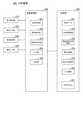
- the analyzer 200has, for example, an electronic stethoscope 210, an operation input unit 220, a screen display unit 230, a communication I / F unit 240, and a storage unit 250 as main components. It has an arithmetic processing unit 260 and.
- the electronic stethoscope 210acquires the patient's lung sound.
- the electronic stethoscope 210converts the patient's lung sound into a digital signal by applying the chest piece of the stethoscope to the chest or back of the patient, and transfers it to the arithmetic processing unit 260 wirelessly or by wire.
- the operation input unit 220includes an operation input device such as a keyboard and a mouse.
- the operation input unit 220detects the operation of the user who uses the analyzer 200 and outputs it to the arithmetic processing unit 260.
- the usermay include a medical worker such as a doctor or a nurse, a care worker such as a certified care worker, or a patient's family.
- the screen display unit 230includes a screen display device such as an LCD (Liquid Crystal Display).
- the screen display unit 230can display various information such as analysis results on the screen in response to an instruction from the arithmetic processing unit 260.
- the communication I / F unit 240is composed of a data communication circuit.
- the communication I / F unit 240performs data communication with various external devices such as a processing device 300 connected via wire or wireless.
- the storage unit 250is a storage device such as a hard disk or a memory.
- the storage unit 250stores processing information and a program 257 necessary for various processes in the arithmetic processing unit 260.
- the program 257realizes various processing units by being read and executed by the arithmetic processing unit 260.
- the program 257is read in advance from an external device or a recording medium via a data input / output function such as the communication I / F unit 240, and is stored in the storage unit 250.
- the main information stored in the storage unit 250includes, for example, lung sound data 251, analysis result information 252, severity determination information 253, severity information 254, personal condition information 255, output information 256, and the like.
- Lung sound data 251shows lung sound data for each auscultation position.
- FIG. 3shows an example of the information contained in the lung sound data 251.
- the lung sound data 251includes, for example, lung sound data for each auscultation position.
- the information included in the lung sound data 251 as illustrated in FIG. 3is created at the timing of, for example, every time an analysis is performed using the analyzer 200, or every time an auscultation using an electronic stethoscope 210 is performed.
- the lung sound data 251may be a combination of data identification information according to the date and time of auscultation and the information illustrated in FIG.
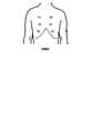
- the auscultation position item in the information contained in the lung sound data 251refers to the approximate location of the patient's body to which the chest piece of the electronic stethoscope 210 is applied to hear the lung sound. That is, the auscultation position is the acquisition site of the lung sound. For example, in the example of FIG. 3, a total of 12 auscultation positions from the auscultation position (1) to the auscultation position (12) are set (in FIG. 3, the auscultation positions (3) to (11) are omitted. Yes).
- FIG. 4is a schematic diagram for explaining an example from the auscultation positions (1) to (6)
- FIG. 5is a schematic diagram for explaining an example from the auscultation positions (7) to (12). be.
- the auscultation positions (1) and (2)are set, for example, to the left and right of the upper lung field of the precordium.
- the auscultation positions (3) and (4)are set, for example, to the left and right of the precordial midlung field.
- the auscultation positions (5) and (6)are set, for example, to the left and right of the lower lung field of the precordium.
- the auscultation positions (7) and (8)are set to the left and right of the upper lung field on the back, for example.
- the auscultation positions (9) and (10)are set, for example, to the left and right of the middle lung field on the back.
- the auscultation positions (11) and (12)are set, for example, to the left and right of the lower lung field on the back.
- the auscultation positionis preset.
- the auscultation positionis not limited to the number and location mentioned above.
- auscultation positionsmay be set not only in the precordium and the back but also in the upper lung field, the middle lung field, and the lower lung field of the left and right lateral chests, and a total of 18 auscultation positions may be set.
- some of the above auscultation positionsmay be excluded.
- the auscultation positions (3) to (6), (9), and (10)are excluded, and the total of the auscultation positions (1), (2), (7), (8), (11), and (12). It may be limited to 6 places.
- a digital time-series acoustic signal including the lung sound acquired by the electronic stethoscope 210 at the auscultation positionis recorded.
- the posture of the patient at the time of auscultationis roughly divided into the recumbent position and the sitting position, but the precordial and back auscultation is usually performed in the sitting position.
- the signal length of one lung sound data(for example, data 1) is arbitrary.
- One lung sound datamay be a signal of a patient's continuous N breaths.
- Nis a positive integer of 1 or more.
- the lung sound datais a signal obtained by processing the time-series acoustic signal acquired from the electronic stethoscope 210, such as removal of the time-series acoustic signal during the resting phase, noise removal, and addition of respiratory timing. It may be there.
- the analysis result information 252shows the result of the abnormality detection unit 262, which will be described later, detecting the abnormality based on the lung sound data 251.
- FIG. 6shows an example of the information included in the analysis result information 252.
- the analysis result information 252includes, for example, the analysis result for each auscultation position.
- the information included in the analysis result information 252 as illustrated in FIG. 6is, for example, timing such as every time an analysis is performed using the analyzer 200, or every time an abnormality detection unit 262 analyzes the lung sound data 251. Created with.
- the analysis result information 252may be a combination of the analysis result identification information according to the date and time when the abnormality detection unit 262 performed the analysis and the information as illustrated in FIG.
- the analysis result itemrecords the result of mechanical analysis of the lung sound data by the abnormality detection unit 262, which will be described later.
- a numerical value indicating whether or not the lung sound data is abnormal lung sound datais recorded.
- two values of a value 0 indicating that the lung sound is normal and a value 1 indicating that the lung sound is abnormalmay be recorded.
- a numerical value indicating the degree of abnormality of the lung sound datamay be recorded.
- the degree of abnormalityan abnormality degree below a preset threshold value indicates that the lung sound data is a normal lung sound, and an abnormality degree exceeding the threshold value indicates that the lung sound data is an abnormal lung sound.
- the severity determination information 253is information used when the severity determination unit 263, which will be described later, determines the severity.
- the severity determination information 253is read from an external device, a recording medium, or the like via a data input / output function such as the communication I / F unit 240, or the user operates the operation input unit 220 to input the information. It is acquired in advance by the method of the above and stored in the storage unit 250.
- the severity determination information 253may be created at a timing such as when the patient is discharged from the hospital. In other words, the severity determination information 253 may be created according to the patient's condition at the time of discharge. In general, many hospitalized patients with heart failure are discharged after receiving treatment for heart failure and in remission.
- the lung sounds of many patients at the time of dischargeare normal.
- the patientmay be discharged from the hospital in a mild condition.
- the severity determination information 253is made according to the condition of the patient at the time of discharge, such as a patient discharged in remission or a patient discharged in a mild condition. Is possible.
- the severity determination information 253may be created / updated at the timing of going to the hospital or the like.
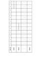
- FIG. 7shows an example of the severity determination information 253.
- the severity determination information 253has, for example, a column corresponding to one-to-one correspondence between auscultation positions (1) to (12) and a row corresponding to one-to-one correspondence to severity. It is a table to set a + symbol indicating that there is an abnormality in lung sound and a-symbol indicating that there is no abnormality in lung sound at the intersection of rows and columns.
- the table shown by the severity determination information 253shows that if there is no abnormality in lung sound at any auscultation position, the severity is determined to be 0.
- the severity determination information 253is a class of N + 1 from 0 to N in severity of heart failure depending on the combination of the presence or absence of abnormal lung sound at the auscultation positions (1) to (12). It is classified into.
- the severity 0is a state in which no abnormal lung sound is heard, and thus it can be said that the heart failure is in remission.
- severity 1is a state in which abnormal lung sound can be heard only in the lower lung field of the back, it cannot be said that heart failure is in remission, but it is mild and some patients are discharged in such a state. It is in a state of doing.
- Severity 2can be said to be more severe than severity 1 because abnormal lung sounds are produced in one of the lower lung fields in the precordium in addition to the lower lung field in the back. However, since it still belongs to mild illness, it can be said that there is a high probability that readmission can be prevented if appropriate measures are taken at this point.
- the information shown by the severity determination information 253is not limited to the case illustrated in FIG. 7.
- the severity determination information 253at least one of the auscultation positions (11) and (12) set in the lower lung field of the back has an abnormal lung sound, and the other auscultation positions (1) to (10).
- Example in FIG. 6shows that there is an abnormality in lung sound only in the case where there is no abnormality in lung sound in other auscultation positions (1) to (4) and (7) to (10), and the severity is 1 in both cases.
- Information other than the abovemay be shown.
- the number of columns of the severity determination information 253may correspond to the number of auscultation positions preset in the lung sound data 251.
- the ra soundis mild when it is heard only at the end of inspiration, and it is severe when it is heard immediately after the start of inspiration. Therefore, in addition to the presence or absence of abnormal lung sound for each auscultation position, the timing at which abnormal lung sound is heard is set in the judgment table, and the combination of the auscultation position, the presence or absence of abnormal lung sound, and the timing at which abnormal lung sound is heard causes heart failure.
- the severitymay be determined. Further, the severity determination information 253 may be set to a severity according to the type and number of abnormal sounds such as la sounds having different properties (rough intermittent crackles, fine crackles).
- the severity determination information 253may be set according to the information that can be included in the personal condition information 255, which will be described later, such as the amount of weight gain of the patient. Further, the severity determination information 253 may be information in which, for example, the number of auscultatory positions that have become abnormal lung sounds and the severity of heart failure of the patient are associated with each other. For example, the severity determination information 253 has a severity of 0, 1, and when the number of auscultatory positions resulting in abnormal lung sounds is 0, 1 or more and 2 or less, 3 or more and 4 or less, 5 or more and 8 or less, and 9 or more, respectively. It may be 2, 3, 4 (maximum).
- the severity information 254shows the result of determination by the severity determination unit 263, which will be described later, using the analysis result information 252 and the severity determination information 253.
- FIG. 8shows an example of analysis result information 252. Referring to FIG. 8, in the severity information 254, for example, the severity identification information according to the date and time when the severity determination unit 263 made the determination is associated with the severity.
- the result of determination by the severity determination unit 263 using the analysis result information 252 and the severity determination information 253is recorded. That is, information indicating the above-mentioned severity such as severity 0, severity 1, severity 2, ..., Etc. is recorded.
- Personal condition information 255is information indicating the condition of the patient.
- the information included in the personal condition information 255can be used when the severity determination unit 163 determines the severity.
- the personal status information 255is read from an external device, a recording medium, or the like via a data input / output function such as the communication I / F unit 240, or the user operates the operation input unit 220 to input the information. It has been acquired in advance by the storage unit 250 and stored in the storage unit 250.
- the personal state information 255may be updated as appropriate by performing an operation on the operation input unit 220 or the like each time the analysis is performed using the analyzer 200.
- FIG. 9shows an example of the information included in the personal status information 255.
- the personal state information 255for example, the state identification information according to the date and time when the state information is input and the state information are associated with each other.
- the item of the state information in the personal state information 255may include information indicating the weight of the patient, the medication status, and the like.
- the information indicating the medication statusmay include, for example, information such as the date and time of the previous medication, the type of the medication last taken, the frequency of medication, and the like.
- the item of state informationincludes information indicating blood pressure, pulse, subjective symptoms (shortness of breath when going out, swelling, coughing, loss of appetite, etc.), water intake, percutaneous arterial oxygen saturation (SPO2), etc. It may be included.
- the item of the status informationmay include matters to be contacted by the doctor at the time of discharge or visit.
- the output information 256indicates the content output by the output unit 264, the result output by the output unit 264, and the like.
- FIG. 10shows an example of the output information 256.
- the output information 256includes, for example, output identification information according to the date and time output by the output unit 264, severity, output content, and output result.
- the item of severityindicates the severity when it is determined that the output unit 264 outputs.
- the output content itemincludes content other than the severity output by the output unit 264.
- the output content itemmay include at least one of lung sound data, acquisition site, acquisition date and time, personal status information, and the like.
- the output result itemincludes information acquired from the processing device 300 as a result of output by the output unit 264.
- the output result itemincludes information indicating the result of diagnosis according to the output such as follow-up observation, medication instruction, consultation recommendation, and the like.
- the output result itemmay include information other than those illustrated above, such as a message from the family doctor.
- identification informationsuch as data identification information, analysis result identification information, severity identification information, state identification information, and output identification information may be different from each other. For example, information according to the date and time of the entire analysis. It does not matter if they are common.
- the arithmetic processing unit 260has a microprocessor such as a CPU and its peripheral circuits, and by reading and executing the program 257 from the storage unit 250, the hardware and the program 257 cooperate to realize various processing units. do.
- the main processing units realized by the arithmetic processing unit 260include a lung sound acquisition unit 261, an abnormality detection unit 262, a severity determination unit 263, an output unit 264, a reception unit 265, and the like.
- the lung sound acquisition unit 261acquires digital time-series acoustic signals including the patient's lung sound and other information.
- the lung sound acquisition unit 261acquires a digital time-series acoustic signal including the patient's lung sound from the electronic stethoscope 210 according to a user's instruction input from the operation input unit 220 or the like. Further, the lung sound acquisition unit 261 can acquire information indicating the date and time together with the digital time-series acoustic signal. Then, the lung sound acquisition unit 261 uses the acquired digital time-series acoustic signal and other information to generate lung sound data 251 as illustrated in FIG. 3 and stores it in the storage unit 250. As described above, the lung sound acquisition unit 261 may combine the data identification information with the information as illustrated in FIG.
- the method of acquiring the lung sound for each auscultation position of the patient with an electronic stethoscope and recording it in association with the auscultation positionis arbitrary.
- the lung sound acquisition unit 261may instruct the patient to breathe timing by a method as described in Patent Document 8.
- the lung sound acquisition unit 261may be configured to calculate an index value of lung sound quality and issue a warning based on the calculated index value to the screen display unit 230 or the like. By giving such a warning, the patient, the user, or the like can take measures to reduce the background noise and / or increase the lung sound, and then acquire the lung sound again.
- the index value calculation process for lung sound qualityis performed, for example, by applying a predetermined filter and then calculating and comparing the signal intensities.
- the lung sound acquisition unit 261uses a band-passing filter to obtain a time-series acoustic signal in a frequency band of 100 Hz to about 2 kHz including the patient's lung sound from the time-series acoustic signal output from the electronic stethoscope 210. To extract.
- the lung sound acquisition unit 261calculates the intensity of the lung sound and the intensity of the background noise in the extracted time-series acoustic signal, and calculates the degree of difference between them as an index value of the quality of the lung sound.
- the lung sound acquisition unit 261detects an inspiratory phase, an expiratory phase, and a resting phase from a time-series acoustic signal including lung sounds. Then, the lung sound acquisition unit 261 calculates the intensity of the time-series acoustic signal in the rest phase as the intensity of the background noise.
- the intensity of the time-series acoustic signalcan be, for example, the root mean square of the amplitude value, but is not limited to this, and may be an amplitude or the like. Further, the lung sound acquisition unit 261 calculates a value obtained by subtracting the background noise intensity from the time-series acoustic signal intensity in the inspiratory phase and / or the expiratory phase as the lung sound intensity.
- the lung sound acquisition unit 261uses the ratio of the intensity of the lung sound to the calculated intensity of the background noise as an index value of the quality of the lung sound.
- the index value of the quality of the lung soundis not limited to the above, and the S / N ratio calculated from the intensity of the lung sound and the intensity of the background noise may be used as the index value.
- the application of the filtermay be omitted.
- the lung sound acquisition unit 261can detect the expiratory phase and the inspiratory phase by comparing the time-series acoustic signal with a predetermined threshold value. In addition, the lung sound acquisition unit 261 can detect a predetermined period immediately before the start of the detected inspiration as a resting phase. The lung sound acquisition unit 261 may detect the inspiratory phase, the expiratory phase, and the resting phase by using a method other than those exemplified above. For example, the lung sound acquisition unit 261 performed machine learning to estimate which section of the time-series acoustic signal including the lung sound output from the electronic stethoscope 210 is the inspiratory phase, the expiratory phase, and the resting phase.
- the learning modelcan be pre-generated by machine learning using a machine learning algorithm such as a neural network, for example, using a time-series acoustic signal including various lung sounds as training data.
- the lung sound acquisition unit 261removes the period of the rest phase and the background noise from the digital time-series acoustic signal including the lung sound, and listens to the digital time-series acoustic signal after the period of the rest phase and the background noise are removed. It may be configured to be recorded in the lung sound data 251 in association with.
- the lung sound acquisition unit 261describes a digital time-series acoustic signal including lung sound in a section consisting of an inspiratory phase and an expiratory phase immediately after that (hereinafter referred to as an inspiratory / expiratory section) and a resting phase section (hereinafter referred to as pause). It is divided into two parts (referred to as a section).
- the lung sound acquisition unit 261calculates the frequency spectra of the inspiratory / expiratory section and the resting section by performing a fast Fourier transform (FFT) on the digital time-series acoustic signals of the inspiratory / expiring section and the resting section, respectively.
- FFTfast Fourier transform
- the lung sound acquisition unit 261subtracts the frequency spectrum of the rest section from the frequency spectrum of the inspiratory / expiratory section. This subtraction suppresses background noise contained in the inspiratory and expiratory phases.
- the lung sound acquisition unit 261generates a digital time-series acoustic signal after noise removal in the inspiratory / expiratory section by inversely converting the frequency spectrum of the inspiratory / expiratory section after the subtraction.
- the lung sound acquisition unit 261records the generated digital time-series acoustic signal after noise removal in the inspiratory / expiratory section in the lung sound data 251 in association with the auscultation position.
- the lung sound acquisition unit 261may remove the period of the rest phase from the digital time-series acoustic signal including the lung sound at the auscultation position, and may not remove the background noise.
- the lung sound acquisition unit 261divides the digital time-series acoustic signal including the lung sound at the auscultation position of interest into an inspiratory / expiratory section and a rest section, and the digital time-series acoustic signal in the inspiratory / expiratory section. Is recorded in the lung sound data 251 in association with the auscultation position.
- the abnormality detection unit 262detects an abnormality from the lung sound data of each auscultation position included in the lung sound data 251, associates the detection result with the auscultation position, and records it in the analysis result information 252.
- the abnormality detection unit 262inputs lung sound data into an abnormality detection model that is generated and stored in advance, and acquires the probability that the lung sound data is an abnormal lung sound from the abnormality detection model.
- the abnormality detection unit 262compares the probability of abnormal lung sound with a preset threshold value. Then, when the probability exceeds the threshold value, the abnormality detection unit 262 determines that it is an abnormal lung sound. That is, the abnormality detection unit 262 detects the abnormality. On the other hand, when it is equal to or less than the threshold value, the abnormality detection unit 262 determines that the sound is not an abnormal lung sound. After that, the abnormality detection unit 262 records the detection result in the analysis result information 252.
- teacher datais generated using a database that collects abnormal sounds, and deep learning is used to learn the characteristics and discrimination criteria of the input sound data (input data).
- deep learningis used to learn the characteristics and discrimination criteria of the input sound data (input data).
- the anomaly detection unit 262uses a spectrogram in which voices are arranged in chronological order by FFT (Fast Fourier Transform) or log-FFT for learning and input data at regular intervals, and RNN (recurrent neural network) is used for deep learning.
- FFTFast Fourier Transform
- log-FFTlog-FFT
- RNNrecurrent neural network
- a network) or CNNconvolutive neural network
- the abnormality detection unit 262may use a method of detecting an abnormal sound by machine learning by converting the lung sound wave type into a short-time feature amount such as a zero crossover coefficient or an MFCC (mel frequency cepstrum coefficient).
- the anomaly detection unit 262may model with a GMM (mixed Gaussian distribution) at the time of learning and check whether or not it fits the model at the time of detection.
- the abnormality detection unit 262may learn the identification surface of a classifier such as an SVM (support vector machine) and use the identification surface to identify whether the input data corresponds to an abnormal sound. ..
- the anomaly detection unit 262generates features using the data itself, such as NMF (non-negative matrix factorization) and PCA (principal component analysis), in addition to the method of directly obtaining such features as described above. You may try to do it.
- the abnormality detection unit 262detects abnormal sounds by a decision tree or the like using statistical characteristics of the input waveform such as the long-term power distribution of the input signal and the distribution of the component amount / component ratio in the specific frequency bin range. You may. In that case, the abnormality detection unit 262 sets the items of the decision tree as a direct value (for example, when the power exceeds 20 mW for 3 consecutive frames) and a statistical feature (for example, a process that approximates Gauss and is larger than 3 ⁇ ). When a frame occurs) may be used. Further, the abnormality detection unit 262 may detect not the input signal itself but the abnormal sound by modeling it in an AR (autoregressive) process or the like and some of its model parameters exceed a threshold value. Although these methods may not include the learning process, they are included in the supervised learning for convenience because they include the observation of the abnormal sound which is the target signal in the determination of the decision tree and the threshold value.
- the abnormality detection unit 262may be configured to learn the abnormality detection model by using the lung sound data of the past patient and the auscultatory findings, for example, when the patient is discharged from the hospital.
- the lung sound data used when learning the abnormality detection modelin addition to the lung sound data at the time of discharge of the patient, the normal lung sound data of the patient before that may be used, or a person other than the patient. You may use the normal lung sound data of.
- the abnormality detection modelmay be generated for each auscultation position, or may be common to a plurality of auscultation positions. Further, the anomaly detection model may be a plurality of models machine-learned from different viewpoints. For example, in the abnormality detection model, the lung sound at the same auscultation position is divided into the lung sound part of the inspiratory phase, the lung sound part of the expiratory phase, and the rest (that is, the resting phase) based on the breathing timing, and the inspiratory phase is taken. A model trained using the lung sound portion of the phase and a model trained using the lung sound portion of the expiratory phase may be included.
- the severity determination unit 263determines the severity based on the analysis result for each auscultation position indicated by the analysis result information 252 and the severity determination information 253. Then, the severity determination unit 263 associates the determined severity with the severity identification information and stores it in the storage unit 250 as the severity information 254.
- the severity determination unit 263identifies the auscultation position where the abnormality is detected by referring to the analysis result information 252. Then, the severity determination unit 263 determines the severity corresponding to the specified result with reference to the severity determination information 253.
- the severity determination information 253can be created according to the patient's condition at the time of discharge.
- the severity determination unit 263refers to the severity determination information 253 according to the condition of the patient at the time of discharge, whereby the information for determining the severity of the patient at the time of discharge is 253. It can also be said that the severity can be determined according to the condition.
- the severity determination unit 263can be configured to correct the determined severity by referring to the personal condition information 255 when determining the severity. For example, there is a finding that when heart failure is aggravated, water accumulates in the lungs, resulting in weight gain. Therefore, the severity determination unit 263 can correct the determined severity based on the weight of the patient included in the personal condition information 255. For example, when a predetermined condition such as an increase in body weight of 3 kg in one week is satisfied, the severity determination unit 263 can make a correction for a set value such as increasing the determined severity by one. The severity determination unit 263 may make corrections other than those exemplified above, such as increasing the severity by 2 when the weight increases by 5 kg in one week.
- the severity determination unit 263makes corrections using personal condition information 255 other than those exemplified above, such as increasing the severity when the blood pressure drops by a predetermined value or more or when the pulse exceeds a predetermined number. You can go.
- the severity determination unit 263directly determines the severity in consideration of the personal condition information 255 by referring to the severity determination information 253 in which the severity is set according to the information that can be included in the personal condition information 255. May be good.
- the output unit 264determines whether or not to output the predetermined information to the processing device 300 based on the result of the determination by the severity determination unit 263. Then, the output unit 264 outputs predetermined information according to the result of the determination. Further, the output unit 264 stores the output severity and other output contents in the storage unit 250 as the output information 256 in association with the output identification information.
- the output to the processing device 300 by the output unit 264may be realized by using at least one of any communication methods such as mail, a message function of groupware, and business chat.
- the output unit 264confirms whether or not the severity determined by the severity determination unit 263 exceeds the output threshold value. Then, the output unit 264 determines that when the severity exceeds the output threshold value, the output is output to the processing device 300. After that, the output unit 264 outputs predetermined information to the processing device 300.
- the information output by the output unit 264is, for example, predetermined.
- the information output by the output unit 264includes the severity determined by the severity determination unit 263, the lung sound data and listening position included in the lung sound data 251, the information indicating the date and time when the lung sound data was acquired, and the personal state.
- Information 255includes at least one of information such as weight and medication status.
- the output unit 264may output information other than those exemplified above, such as information included in the analysis result information 252 and a frequency spectrum calculated by performing a fast Fourier transform (FFT) on the lung sound data.
- FFTfast Fourier transform
- the information output by the output unit 264may be only the lung sound data in which the abnormality detection unit 262 detects an abnormality among the lung sound data included in the lung sound data 251 and is included in the lung sound data 251. It does not matter if it is all the lung sound data. In addition to the lung sound data acquired at the time of this analysis, the output unit 264 has past lung sound data such as lung sound data for the last two weeks and lung sound data when the abnormality detection unit 262 does not detect an abnormality. May be output. Even if the output unit 264 outputs a frequency spectrum calculated from the lung sound data acquired at the time of this analysis, a frequency spectrum calculated from the lung sound data when the abnormality detection unit 262 does not detect an abnormality, and the like. good.
- the output unit 264may output an average frequency spectrum of the patient whose diagnosis result is a consultation recommendation. Further, among the information output by the output unit 264, the information such as the weight and the medication status included in the personal condition information may include the past information such as the change in the weight in the last two weeks and the medication status in the last two weeks. I do not care.
- the output unit 264may change the information to be output according to the severity determined by the severity determination unit 263. For example, the output unit 264 increases the information to be output as the severity increases, outputs only the lung sound data when the severity is equal to or less than the transmission threshold, and outputs the lung sound data and the personal state when the severity exceeds the transmission threshold.
- the information to be outputmay be different depending on the severity, such as outputting the information included in the information 255.
- the processing device 300 to be the output destination by the output unit 264is defined in advance, for example.
- the output unit 264has information in which an identifier of a user or a patient of the analyzer 200 is associated with a processing device 300 owned by a family doctor or the like of the patient. Then, the output unit 264 outputs predetermined information to the processing device 300 owned by the patient's family doctor or the like by referring to the above information.
- the output unit 264may refer to the past output results included in the output information 256 when determining whether or not to output the predetermined information to the processing device 300. For example, when the output information 256 contains information that the severity is the same as the severity determined by the severity determination unit 263 (or the difference is less than or equal to a predetermined value) and the output result is follow-up, the output unit 264 is severe. Even if the degree exceeds the output threshold value, it can be determined that the output is not output to the processing device 300. When the output information 256 contains information whose output result is follow-up with the same severity as the severity determined by the severity determination unit 263, the output unit 264 outputs the information based on the result of spectrum analysis or the like. You may judge whether or not.
- the output unit 264calculates the frequency spectrum by performing a fast Fourier transform (FFT) on the lung sound data when the output result is follow-up observation and the lung sound data of this time. Then, the output unit 264 determines whether or not to transmit the predetermined information to the processing device 300 by comparing the calculated frequency spectra. For example, the output unit 264 determines that the predetermined information is transmitted to the processing device 300 when the difference in the frequency spectrum is equal to or less than the predetermined position. Further, when the output information 256 contains information that the output result is a follow-up observation with the same severity as the severity determined by the severity determination unit 263, the output unit 264 provides lung sound data such as the number and frequency of rales. It may be determined whether or not to output according to the contents of. For example, if the difference in lung sound data such as the number and frequency of rales is not more than a predetermined value, it may be determined that the output unit 264 does not output.
- FFTfast Fourier transform
- the receiving unit 265receives the result of the output from the output unit 264 from the processing device 300. Then, the receiving unit 265 updates the output result item in the output information 256 based on the received information.
- the receiving unit 265receives information from the processing device 300, such as follow-up observation, medication instruction, consultation recommendation, etc., indicating the result of diagnosis by a family doctor having the processing device 300 according to the content of the output by the output unit 264. .. Then, the receiving unit 265 updates the output information 256 by storing the received information in the output result item of the output information 256.
- the processing device 300is an information processing device that receives information output by the output unit 264 of the analyzer 200.
- the processing device 300is installed in, for example, a hospital where a family doctor works.
- the processing device 300is, for example, a smartphone, a tablet terminal, a PDA (Personal Digital Assistant), a notebook computer, or the like.
- the processing device 300may be other than those illustrated above.
- FIG. 11shows a configuration example of the processing device 300.
- the processing apparatus 300has, as main components, for example, an operation input unit 310, a screen display unit 320, a communication I / F unit 330, a storage unit 340, and an arithmetic processing unit 350. have.
- the configuration of the operation input unit 310, the screen display unit 320, and the communication I / F unit 330may be the same as the configurations of the analyzer 200. Therefore, the description thereof will be omitted.
- the storage unit 340is a storage device such as a hard disk or a memory.
- the storage unit 340stores processing information and a program 343 necessary for various processes in the arithmetic processing unit 350.
- the program 343realizes various processing units by being read and executed by the arithmetic processing unit 350.
- the program 343is read in advance from an external device or a recording medium via a data input / output function such as the communication I / F unit 330, and is stored in the storage unit 340.
- the main information stored in the storage unit 340includes, for example, the acquired information 341 and the diagnosis result 342.
- the acquired information 341includes the information output by the analyzer 200. That is, the acquired information 341 includes information corresponding to the output by the output unit 264. The information included in the acquired information 341 can be output to the screen display unit 320 or the like.
- the diagnosis result 342shows the result of diagnosis by a family doctor or the like according to the acquired information 341.
- the diagnosis result 342shows information indicating the result of diagnosis by a family doctor or the like having a processing device 300 according to the acquired information 341, such as follow-up observation, medication instruction, and consultation recommendation.
- the arithmetic processing unit 350has a microprocessor such as a CPU and its peripheral circuits, and by reading and executing the program 343 from the storage unit 340, the hardware and the program 343 cooperate to realize various processing units. do.
- the main processing units realized by the arithmetic processing unit 350include an acquisition unit 351, a diagnosis result input unit 352, an output unit 353, and the like.
- the acquisition unit 351acquires the information output by the output unit 264 of the analyzer 200. Then, the acquisition unit 351 stores the acquired information as the acquisition information 341 in the storage unit 340.
- the diagnosis result input unit 352acquires the diagnosis result of the family doctor or the like for the acquisition information 341 stored by the acquisition unit 351.
- the diagnosis result input unit 352acquires information indicating the result of diagnosis by a family doctor or the like according to the acquired information 341, such as follow-up observation, medication instruction, consultation recommendation, etc., in response to an operation on the operation input unit 310 or the like. Then, the diagnosis result input unit 352 stores the acquired information as the diagnosis result 342 in the storage unit 340.
- the output unit 353outputs the diagnosis result 342 to the analyzer 200. For example, when the diagnosis result input unit 352 stores the new diagnosis result 342, the output unit 353 transmits information indicating the new diagnosis result to the analyzer 200.
- the processing device 300acquires the information indicating the diagnosis result according to the output by the output unit 264, and sends back the information indicating the diagnosis result to the analyzer 200.
- the information indicating the diagnosis resultmay include information other than those exemplified above, such as a message from the family doctor.
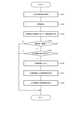
- the lung sound acquisition unit 261acquires a digital time-series acoustic signal including the patient's lung sound for each auscultation position (step S101). Then, the lung sound acquisition unit 261 uses the acquired digital time-series acoustic signal and other information to generate lung sound data 251 as illustrated in FIG. 3 and stores it in the storage unit 250. As described above, the lung sound acquisition unit 261 may combine the data identification information with the information as illustrated in FIG.
- the lung sound acquisition unit 261When the lung sound acquisition unit 261 acquires a digital time-series acoustic signal or the like, the lung sound quality index value may be calculated and a warning based on the calculated index value may be given to the screen display unit 230 or the like. Further, the lung sound acquisition unit 261 performs processing such as removal of time-series acoustic signals, noise removal, and addition of respiratory timing during the resting phase, and then generates lung sound data 251 using the processed data. And may be stored in the storage unit 250.
- the abnormality detection unit 262detects an abnormality from the lung sound data of each auscultation position included in the lung sound data 251 and records the detection result in the analysis result information 252 in association with the auscultation position (step S102). For example, the abnormality detection unit 262 inputs lung sound data into an abnormality detection model that is generated and stored in advance, and acquires the probability that the lung sound data is an abnormal lung sound from the abnormality detection model. Next, the abnormality detection unit 262 compares the probability of abnormal lung sound with a preset threshold value. Then, when the probability exceeds the threshold value, the abnormality detection unit 262 determines that it is an abnormal lung sound. That is, the abnormality detection unit 262 detects the abnormality. On the other hand, when it is equal to or less than the threshold value, the abnormality detection unit 262 determines that the sound is not an abnormal lung sound. After that, the abnormality detection unit 262 records the detection result in the analysis result information 252.
- the severity determination unit 263determines the severity based on the analysis result for each auscultation position indicated by the analysis result information 252 and the severity determination information 253 (step S103). Then, the severity determination unit 263 associates the determined severity with the severity identification information and stores it in the storage unit 250 as the severity information 254.
- the severity determination unit 263may determine the severity according to the condition of the patient at the time of discharge by referring to the information for severity determination 253 according to the condition of the patient at the time of discharge. Further, the severity determination unit 263 may correct the determined severity by referring to the personal condition information 255 when determining the severity. The severity determination unit 263 directly determines the severity in consideration of the personal condition information 156 by referring to the severity determination information 253 in which the severity is set according to the information that can be included in the personal condition information 255. May be good.
- the output unit 264determines whether or not to output the predetermined information to the processing device 300 based on the result of the determination by the severity determination unit 263. For example, the output unit 264 confirms whether or not the severity determined by the severity determination unit 263 exceeds the output threshold value (step S104).
- the output unit 264confirms whether or not the output information 256 satisfies the condition (step S105). Then, when the output information 256 satisfies the condition (step S105, Yes), the output unit 264 determines that the output is to be output to the processing device 300, and transmits predetermined information to the processing device 300 (step S106). .. Further, the output unit 264 stores the output information as output information 256 in the storage unit 250.
- the output result itemhas the same severity as the severity determined by the severity determination unit 263, and the output information includes follow-up information, or the difference in frequency spectrum when the above information is included. , Etc. can be included.
- the receiving unit 265receives the result of the output from the output unit 264 from the processing device 300 (step S107). Then, the receiving unit 265 saves the output result item in the output information 256 based on the received information (step S108).
- step S104If the severity is equal to or less than the output threshold value (step S104, No), or if the output information 256 does not satisfy the condition (step S105, No), it is determined that the output unit 264 does not output to the processing device 300. do.
- step S105is an operation example of the analyzer 200.
- the process of step S105may be omitted.
- the analyzer 200has an abnormality detection unit 262, a severity determination unit 263, and an output unit 264.
- the output unit 264can determine whether or not to output to the processing device 300 based on the severity determined by the severity determination unit 263 based on the detection result by the abnormality detection unit 262. I can.
- the abnormality detection unit 262only performs the analysis corresponding to the auscultation position where the lung sound data is acquired.
- the severity determination unit 263may be configured to determine whether or not to determine the severity based on the number of auscultation positions analyzed by the abnormality detection unit 262. For example, if the number of auscultatory positions for which lung sound data has not been acquired and whether or not it is abnormal lung sound has not been analyzed is less than a preset threshold value, the severity determination unit 263 calculates the severity. Instead, it is possible to display on the screen display unit 230 that the analysis has ended with an error.
- the severity determination unit 263assumes that no abnormal lung sound is detected at the auscultatory positions where the analysis of whether or not the lung sound is abnormal has not been performed. Then, the severity can be calculated. In this case, the severity determination unit 263 may keep the calculated severity as the most optimistic value. That is, when the calculated severity is severity 1, it can be retained as "severity 1 or higher” or "at least severity 1" instead of "severity 1".
- the threshold valuemay be set arbitrarily.
- the analyzer 200may be configured to determine which auscultation position to focus on based on the auscultation position where an abnormality has been detected in the patient's past.
- the lung sound acquisition unit 261 of the analyzer 200can calculate the abnormality detection frequency for each auscultation position based on the past analysis result information 252. Further, the lung sound acquisition unit 261 can guide the lung sound data to be acquired in descending order of the calculated abnormality detection frequency.
- the severity determination unit 263determines whether or not to calculate the severity, as compared with the case where the lung sound data is not acquired in the order of the abnormality detection frequency. The threshold used may be reduced.
- the analyzer 200may be configured to transmit the information indicating the confirmation to the processing device 300 when the patient or the like confirms the information indicating the diagnosis result received by the receiving unit 265. With this configuration, it is possible to easily confirm whether or not the patient or the like has confirmed the information indicating the diagnosis result from the family doctor or the like who operates the processing device 300.
- FIG. 13is a block diagram of the analysis system 500 according to the second embodiment of the present invention.
- the analysis system 500includes a plurality of analysis devices 510 and a server device 520. Further, the plurality of analysis devices 510 and the server device 520 are connected to each other so as to be able to communicate with each other through a network 530 such as the Internet.
- the analyzer 510is an information processing device that outputs an instruction according to the result of analyzing the lung sound.
- the analyzer 510may be, but is not limited to, a smartphone, a tablet terminal, a PDA, a notebook computer, or the like.
- the analyzer 510includes an electronic stethoscope (not shown), a communication I / F unit, an operation input unit, a screen display unit, a storage unit, and an arithmetic processing unit.
- the server device 520is a computer that provides various services necessary for lung sound analysis to a plurality of analysis devices 510 through the network 530.
- the server device 520has lung sound data 251 shown in FIG. 2, analysis result information 252, severity determination information 253, severity information 254, personal condition information 255, output information 256, and at least a part of the program 257. And provide them to the analyzer 510 through the network 530. Therefore, as compared with the analyzer 200 of FIG. 2, the analyzer 510 has lung sound data 251 in the storage unit, analysis result information 252, severity determination information 253, severity information 254, personal condition information 255, and output information. It is not necessary to store at least a part of 256 and the program 257, and the storage capacity can be reduced.
- the server device 520analyzes at least a part of the functions of the lung sound acquisition unit 261, the abnormality detection unit 262, the severity determination unit 263, the output unit 264, and the reception unit 265 shown in FIG. 2 through the network 530.
- the function as the analyzer 200may be realized by the analysis system 500 or the like.
- FIG. 14shows a hardware configuration example of the analyzer 600.
- the analyzer 600has the following hardware configuration as an example.
- -CPUCentral Processing Unit
- -ROMRead Only Memory
- RAMRandom Access Memory
- -Program group 604loaded in RAM 603
- a storage device 605for storing the program group 604.
- -Drive device 606that reads and writes the recording medium 610 external to the information processing device.
- -Communication interface 607that connects to the communication network 611 outside the information processing device.
- the analyzer 600can realize the functions as the detection unit 621, the determination unit 622, and the output unit 623 shown in FIG. 15 by the CPU 601 acquiring the program group 604 and executing the program group 601.
- the program group 604is stored in, for example, a storage device 605 or a ROM 602 in advance, and the CPU 601 loads the program group 604 into a RAM 603 or the like and executes the program group 604 as needed.
- the program group 604may be supplied to the CPU 601 via the communication network 611, or may be stored in the recording medium 610 in advance, and the drive device 606 may read the program and supply the program to the CPU 601.
- FIG. 14shows an example of the hardware configuration of the analyzer 600.
- the hardware configuration of the analyzer 600is not limited to the above case.
- the analyzer 600may be configured from a part of the above-mentioned configuration, such as not having the drive device 606.
- the detection unit 621detects lung sound abnormalities at each auscultation position based on a time-series acoustic signal including lung sounds at each auscultation position.
- the determination unit 622determines the severity of heart failure of the patient based on the detection result of the lung sound abnormality for each auscultation position detected by the detection unit 621 and the state information indicating the patient's condition.
- the output unit 623determines whether or not to output predetermined information to the external device based on the result determined by the determination unit 622, and outputs according to the result of the determination.
- the analyzer 600has a detection unit 621, a determination unit 622, and an output unit 623.
- the output unit 623can determine whether or not to output to the processing device 300 based on the severity determined by the determination unit 622 based on the detection result by the detection unit 621.
- the above-mentioned analyzer 600can be realized by incorporating a predetermined program into an information processing device such as the analyzer 600.
- the information processing apparatusincludes a detection unit that detects an abnormality in lung sound at each auscultation position based on a time-series acoustic signal including lung sound at each auscultation position.
- the judgment unit for determining the severity of the patient's heart failure and the judgment unit for the determination unitBased on this, it is a program for realizing an output unit that determines whether or not to output predetermined information to an external device and outputs according to the result of the determination.
- the information processing devicedetects the lung sound abnormality at each auscultation position based on the time-series acoustic signal including the lung sound at each auscultation position. Then, the severity of the patient's heart failure is determined based on the detection result of the lung sound abnormality for each detected auscultation position and the condition information indicating the patient's condition, and based on the determined result, the external device is used. It is a method of determining whether or not to output predetermined information and outputting according to the result of the determination.
- the present inventioncan be used for a device or system for analyzing human lung sound, and in particular, for a device or system for early detection of exacerbation of heart failure in a patient discharged from the hospital after receiving treatment for heart failure and preventing readmission.
- a detection unitthat detects abnormal lung sound at each auscultation position based on a time-series acoustic signal including lung sound at each auscultation position.
- a determination unitfor determining the severity of heart failure of the patient based on the detection result of the lung sound abnormality for each auscultation position detected by the detection unit and the state information indicating the patient's condition.
- An output unitthat determines whether or not to output predetermined information to an external device based on the result of the determination by the determination unit and outputs the output according to the result of the determination.
- An analyzerwith.
- the output unitdetermines whether or not to output predetermined information to the external device based on whether or not the severity determined by the determination unit exceeds a predetermined output threshold value. Described analyzer. (Appendix 3) The output unit determines whether or not to output predetermined information to the external device based on the information received from the external device according to the result output by the output unit in the past. Appendix 1 or Appendix 2. The analyzer described in. (Appendix 4) Whether the output unit outputs predetermined information to the external device based on the information received from the external device according to the result output in the past with the same severity as the severity determined by the determination unit. The analyzer according to Appendix 3 for determining whether or not.
- the output unithas a frequency calculated from the time-series acoustic signal when information including follow-up observation is received from the external device as a result of output at a severity corresponding to the severity determined by the determination unit.
- the analyzeraccording to Appendix 3 or Appendix 4, which determines whether or not to output predetermined information to the external device using a spectrum.
- the output unitis an external device based on a comparison result between a frequency spectrum calculated from the time-series acoustic signal used for detection by the detection unit and a frequency spectrum calculated from a past time-series acoustic signal.
- the analyzeraccording to Appendix 5, which determines whether or not to output predetermined information.
- the predetermined informationincludes the severity determined by the determination unit and the time-series acoustic signal used when the detection unit detects the information, whichever is one of Appendices 1 to 6.
- the analyzer described in.(Appendix 8) The analyzer according to Appendix 7, wherein the predetermined information includes at least one of information indicating the weight of the patient and information indicating the medication status.
- Appendix 9The analyzer according to Appendix 7 or Appendix 8, wherein the predetermined information includes a frequency spectrum calculated from the time-series acoustic signal used when the detection unit detects the signal.
- An analysis methodin which it is determined whether or not to output predetermined information to an external device based on the determined result, and the output is performed according to the determined result.
- Appendix 13For information processing equipment A detection unit that detects abnormal lung sound at each auscultation position based on a time-series acoustic signal including lung sound at each auscultation position. A determination unit for determining the severity of heart failure of the patient based on the detection result of the lung sound abnormality for each auscultation position detected by the detection unit and the state information indicating the patient's condition. An output unit that determines whether or not to output predetermined information to an external device based on the result of the determination by the determination unit and outputs the output according to the result of the determination. A computer-readable recording medium that records programs to achieve this.
- Analysis system 200Analysis device 210 Electronic hearing device 220 Operation input unit 230 Screen display unit 240 Communication I / F unit 250 Storage unit 151 Pulmonary sound data 152 Analysis result information 153 Severity determination information 154 Severity information 255 Personal condition information 256 Output information 257 Program 260 Calculation processing unit 261 Pulmonary sound acquisition unit 262 Abnormality detection unit 263 Severity determination unit 264 Output unit 265 Reception unit 300 Processing device 310 Operation input unit 320 Screen display unit 330 Communication I / F unit 340 Storage unit 341 Acquisition Information 342 Diagnosis result 350 Calculation processing unit 351 Acquisition unit 352 Diagnosis result input unit 353 Output unit 400 Network 500 Analysis system 510 Analysis device 520 Server device 530 Network 600 Analysis device 601 CPU 602 ROM 603 RAM 604 Program group 605 Storage device 606 Drive device 607 Communication interface 608 Input / output interface 609 Bus 610 Recording medium 611 Communication network 621 Detection unit 622 Judgment unit 623 Output unit
Landscapes
- Health & Medical Sciences (AREA)
- Engineering & Computer Science (AREA)
- Medical Informatics (AREA)
- Public Health (AREA)
- Biomedical Technology (AREA)
- General Health & Medical Sciences (AREA)
- Life Sciences & Earth Sciences (AREA)
- Primary Health Care (AREA)
- Epidemiology (AREA)
- Data Mining & Analysis (AREA)
- Pathology (AREA)
- Databases & Information Systems (AREA)
- Veterinary Medicine (AREA)
- Animal Behavior & Ethology (AREA)
- Surgery (AREA)
- Molecular Biology (AREA)
- Heart & Thoracic Surgery (AREA)
- Physics & Mathematics (AREA)
- Acoustics & Sound (AREA)
- Pulmonology (AREA)
- Business, Economics & Management (AREA)
- General Business, Economics & Management (AREA)
- Measurement Of The Respiration, Hearing Ability, Form, And Blood Characteristics Of Living Organisms (AREA)
- Measuring Pulse, Heart Rate, Blood Pressure Or Blood Flow (AREA)
- Measuring And Recording Apparatus For Diagnosis (AREA)
Abstract
Description
Translated fromJapanese本発明は、分析装置、分析方法、記録媒体に関する。The present invention relates to an analyzer, an analysis method, and a recording medium.
心不全とは、何らかの心臓機能障害、すなわち、心臓に器質的および/あるいは機能的異常が生じて心ポンプ機能の代償機転が破綻した結果、呼吸困難・倦怠感や浮腫が出現し、それに伴い運動耐容能が低下する臨床症候群のことをいう。心不全を患った患者は、治療により寛解しても、常に増悪のリスクがある。水分・塩分の過剰摂取、薬の服用忘れ、過度な運動などが原因で、患者に急性増悪が生じると、再入院を余儀なくされる。そのため、退院した患者の心不全増悪を早期に発見して治療介入することにより、急性増悪を防ぐことが重要である。Heart failure is some form of cardiac dysfunction, that is, dyspnea, malaise, and edema appear as a result of organic and / or functional abnormalities in the heart that disrupt the compensatory mechanism of cardiac pump function, resulting in exercise tolerance. A clinical syndrome in which the ability is reduced. Patients with heart failure are always at risk of exacerbation, even if they are in remission with treatment. If an acute exacerbation occurs in a patient due to excessive intake of water or salt, forgetting to take medicine, excessive exercise, etc., the patient will be forced to be re-hospitalized. Therefore, it is important to prevent acute exacerbations by detecting the exacerbation of heart failure in discharged patients at an early stage and intervening in treatment.
心不全を診断する方法の一つに、聴診による肺音の検診がある。かかる検診は、安全かつ簡便に肺の健康状態、ひいては心不全を診断できる方法の一つである。しかし、訓練を積んだ専門医でなければ詳細かつ正確な診断結果を得ることは困難である。そのため、一般の看護師や介護従事者による回診や訪問介護などの現場では、詳細な診断を下すことはできなかった。One of the methods for diagnosing heart failure is auscultation of lung sounds. Such a medical examination is one of the safe and easy methods for diagnosing the health condition of the lungs and, by extension, heart failure. However, it is difficult to obtain detailed and accurate diagnostic results without a trained specialist. Therefore, it was not possible to make a detailed diagnosis at sites such as rounds and home-visit care by general nurses and long-term care workers.
この問題に対処するため、電子聴診器により収集した肺音に対し、副雑音と呼ばれる異常音の有無を自動判別するシステムが提案されている(例えば特許文献1乃至4、6参照)。また、電子聴診器により収集した患者の生体音データを、予め取得してある患者の正常時データ、および異常時データと比較して、異常を検出するシステムが提案されている(例えば特許文献5参照)。In order to deal with this problem, a system has been proposed that automatically determines the presence or absence of abnormal sounds called secondary noises from lung sounds collected by an electronic stethoscope (see, for example,
また、関連する技術として、特許文献7がある。特許文献7には、患者により入力された病状情報に基づいて患者に推奨する診療医師を確定し、診療医師の情報を患者端末に送信するシステムが記載されている。Further, as a related technique, there is
特許文献1乃至6に記載されているような技術を用いて、患者本人などの専門知識を持たない人間が検査を行った場合、検査結果を医療従事者と効率的に共有することが難しい、という課題が生じる。また、特許文献7に記載されているように、患者により入力された病状情報を送信するという構成にした場合、検査結果が無差別に医療従事者に送信されることとなり、その結果として、検査結果の効率的な共有を難しくする。例えば、以上のように、検査結果を医療従事者と効率的に共有することが難しい、という課題が生じていた。When a person without specialized knowledge such as the patient himself / herself performs the test using the techniques described in
そこで、本発明の目的は、検査結果を医療従事者と効率的に共有することが難しい、という課題を解決する分析装置、分析方法、記録媒体を提供することにある。Therefore, an object of the present invention is to provide an analyzer, an analysis method, and a recording medium that solve the problem that it is difficult to efficiently share test results with medical professionals.
本発明の一形態である分析装置は、
聴診位置ごとの肺音を含む時系列音響信号に基づいて、前記聴診位置ごとに肺音異常を検知する検知部と、
前記検知部が検知した前記聴診位置ごとの肺音異常の検知結果と、患者の状態を示す状態情報と、に基づいて、前記患者の心不全の重症度を判定する判定部と、
前記判定部が判定した結果に基づいて、外部装置に対して所定の情報を出力するか否か判断して、判断の結果に応じた出力を行う出力部と、
を有する
という構成をとる。The analyzer, which is one embodiment of the present invention, is
A detection unit that detects abnormal lung sound at each auscultation position based on a time-series acoustic signal including lung sound at each auscultation position.
A determination unit for determining the severity of heart failure of the patient based on the detection result of the lung sound abnormality for each auscultation position detected by the detection unit and the state information indicating the patient's condition.
An output unit that determines whether or not to output predetermined information to an external device based on the result of the determination by the determination unit and outputs the output according to the result of the determination.
It takes the configuration of having.
また、本発明の他の形態である分析方法は、
情報処理装置が、
聴診位置ごとの肺音を含む時系列音響信号に基づいて、前記聴診位置ごとに肺音異常を検知し、
検知した前記聴診位置ごとの肺音異常の検知結果と、患者の状態を示す状態情報と、に基づいて、前記患者の心不全の重症度を判定し、
判定した結果に基づいて、外部装置に対して所定の情報を出力するか否か判断して、判断の結果に応じた出力を行う
という構成をとる。Further, the analysis method which is another embodiment of the present invention is:
Information processing equipment
Based on the time-series acoustic signal including the lung sound at each auscultation position, the lung sound abnormality is detected at each auscultation position.
The severity of heart failure in the patient is determined based on the detected result of detecting the abnormal lung sound for each auscultation position and the condition information indicating the condition of the patient.
Based on the judgment result, it is judged whether or not to output the predetermined information to the external device, and the output is performed according to the judgment result.
また、本発明の他の形態である記録媒体は、
聴診位置ごとの肺音を含む時系列音響信号に基づいて、前記聴診位置ごとに肺音異常を検知する検知部と、
前記検知部が検知した前記聴診位置ごとの肺音異常の検知結果と、患者の状態を示す状態情報と、に基づいて、前記患者の心不全の重症度を判定する判定部と、
前記判定部が判定した結果に基づいて、外部装置に対して所定の情報を出力するか否か判断して、判断の結果に応じた出力を行う出力部と、
を実現するためのプログラムを記録した、コンピュータが読み取り可能な記録媒体である。Further, the recording medium which is another embodiment of the present invention is
A detection unit that detects abnormal lung sound at each auscultation position based on a time-series acoustic signal including lung sound at each auscultation position.
A determination unit for determining the severity of heart failure of the patient based on the detection result of the lung sound abnormality for each auscultation position detected by the detection unit and the state information indicating the patient's condition.
An output unit that determines whether or not to output predetermined information to an external device based on the result of the determination by the determination unit and outputs the output according to the result of the determination.
It is a computer-readable recording medium on which a program for realizing the above is recorded.
本発明は、上述したような構成を有することにより、検査結果を医療従事者と効率的に共有することを可能とする。The present invention makes it possible to efficiently share test results with medical professionals by having the above-mentioned configuration.
[第1の実施形態]
本発明の第1の実施形態を図1から図12までを参照して説明する。図1は、分析システム100の全体の構成例を示す図である。図2は、分析装置200の構成例を示すブロック図である。図3は、肺音データ251の一例を示す図である。図4、図5は、聴診位置の一例を説明するための図である。図6は、分析結果情報252の一例を示す図である。図7は、重症度判定用情報253の一例を示す図である。図8は、重症度情報254の一例を示す図である。図9は、個人状態情報255に含まれる情報例を示す図である。図10は、出力情報256の一例を示す図である。図11は、処理装置300の構成例を示すブロック図である。図12は、分析装置200の動作例を示すフローチャートである。[First Embodiment]
The first embodiment of the present invention will be described with reference to FIGS. 1 to 12. FIG. 1 is a diagram showing an overall configuration example of the analysis system 100. FIG. 2 is a block diagram showing a configuration example of the
本発明の第1の実施形態では、肺音を分析した結果に応じて、所定の情報を処理装置300に対して送信する分析装置200を有する分析システム100について説明する。後述するように、分析装置200は、胸部や背部の複数個所から肺音を取得するとともに、取得した肺音に基づいて、それぞれの箇所における異常を検知する。また、分析装置200は、検知した結果に基づいて、心不全の重症度を判定する。そして、分析装置200は、判定した重症度に基づいて、所定の情報を処理装置300へと出力するか否か判断する。In the first embodiment of the present invention, the analysis system 100 having the
また、分析装置200は、過去に処理装置300へ対して出力した結果を含む出力情報256を有している。分析装置200は、処理装置300へ対して情報を出力するか否かを判断する際に、出力情報256に含まれる情報を参照することが出来る。Further, the
図1は、分析システム100全体の構成例を示している。図1を参照すると、分析システム100は、分析装置200と処理装置300とを有している。図1で示すように、分析装置200と処理装置300とは、ネットワーク400を介して互いに通信可能なよう接続されている。なお、分析システム100は、1以上の任意の数の分析装置200、処理装置300を有することが出来る。FIG. 1 shows a configuration example of the entire analysis system 100. Referring to FIG. 1, the analysis system 100 includes an
分析装置200は、患者から取得した肺音の分析結果に応じて、所定の情報を処理装置300に対して送信するか否か判断する情報処理装置である。分析装置200は、例えば、スマートフォン、タブレット型端末、PDA(Personal Digital Assistant)、ノートパソコンなどである。分析装置200は、上記例示した以外であっても構わない。The
図2は、分析装置200の構成例を示している。図2を参照すると、分析装置200は、主な構成要素として、例えば、電子聴診器210と、操作入力部220と、画面表示部230と、通信I/F部240と、記憶部250と、演算処理部260と、を有している。FIG. 2 shows a configuration example of the
電子聴診器210は、患者の肺音を取得する。例えば、電子聴診器210は、聴診器のチェストピースを患者の胸部または背部に当てることにより、患者の肺音をディジタル信号に変換し、無線あるいは有線により演算処理部260へ転送する。The
操作入力部220は、キーボードやマウスなどの操作入力装置からなる。操作入力部220は、分析装置200を利用する利用者の操作を検出して、演算処理部260に出力する。なお、利用者には、患者本人の他、医師や看護師などの医療従事者、介護福祉士などの介護従事者、あるいは患者の家族などが含まれて構わない。The
画面表示部230は、LCD(Liquid Crystal Display、液晶ディスプレイ)などの画面表示装置からなる。画面表示部230は、演算処理部260からの指示に応じて、分析結果などの各種情報を画面表示することが出来る。The
通信I/F部240は、データ通信回路からなる。通信I/F部240は、有線または無線を介して接続された処理装置300などの各種外部装置との間でデータ通信を行う。The communication I /
記憶部250は、ハードディスクやメモリなどの記憶装置である。記憶部250は、演算処理部260における各種処理に必要な処理情報やプログラム257を記憶する。プログラム257は、演算処理部260に読み込まれて実行されることにより各種処理部を実現する。プログラム257は、通信I/F部240などのデータ入出力機能を介して外部装置や記録媒体から予め読み込まれ、記憶部250に保存されている。記憶部250で記憶される主な情報としては、例えば、肺音データ251、分析結果情報252、重症度判定用情報253、重症度情報254、個人状態情報255、出力情報256などがある。The
肺音データ251は、聴診位置ごとの肺音データを示している。図3は、肺音データ251に含まれる情報の一例を示している。図3を参照すると、肺音データ251には、例えば、聴診位置ごとの肺音データが含まれている。
肺音データ251に含まれる図3で例示するような情報は、例えば、分析装置200を用いた分析を行うごと、または、電子聴診器210を用いた聴診を行うごと、などのタイミングで作成される。肺音データ251は、聴診日時などに応じたデータ識別情報と、図3で例示するような情報と、を組み合わせたものであっても構わないThe information included in the
ここで、肺音データ251に含まれる情報のうち聴診位置の項目は、肺音を聴診するために電子聴診器210のチェストピースを当てる患者の体のおおよその場所のことを指す。すなわち、聴診位置は、肺音の取得部位である。例えば、図3の例では、聴診位置(1)から聴診位置(12)までの合計12箇所の聴診位置が設定されている(図3では、聴診位置(3)~(11)は省略されている)。Here, the auscultation position item in the information contained in the
図4は、聴診位置(1)~(6)までの一例を説明するための模式図であり、図5は、聴診位置(7)~(12)までの一例を説明するための模式図である。図4を参照すると、聴診位置(1)、(2)は、例えば、前胸部の上肺野の左右に設定される。聴診位置(3)、(4)は、例えば、前胸部の中肺野の左右に設定される。聴診位置(5)、(6)は、例えば、前胸部の下肺野の左右に設定される。また、図5を参照すると、聴診位置(7)、(8)は、例えば、背部の上肺野の左右に設定される。聴診位置(9)、(10)は、例えば、背部の中肺野の左右に設定される。聴診位置(11)、(12)は、例えば、背部の下肺野の左右に設定される。例えば、以上のように、聴診位置は、予め設定されている。FIG. 4 is a schematic diagram for explaining an example from the auscultation positions (1) to (6), and FIG. 5 is a schematic diagram for explaining an example from the auscultation positions (7) to (12). be. Referring to FIG. 4, the auscultation positions (1) and (2) are set, for example, to the left and right of the upper lung field of the precordium. The auscultation positions (3) and (4) are set, for example, to the left and right of the precordial midlung field. The auscultation positions (5) and (6) are set, for example, to the left and right of the lower lung field of the precordium. Further, referring to FIG. 5, the auscultation positions (7) and (8) are set to the left and right of the upper lung field on the back, for example. The auscultation positions (9) and (10) are set, for example, to the left and right of the middle lung field on the back. The auscultation positions (11) and (12) are set, for example, to the left and right of the lower lung field on the back. For example, as described above, the auscultation position is preset.
なお、聴診位置は上述した個数と場所に限定されない。例えば、前胸部および背部だけでなく左右の側胸部の上肺野、中肺野、下肺野に聴診位置を設定し、合計18個としてもよい。あるいは、上記の聴診位置のうちの一部を除外してもよい。例えば、聴診位置(3)~(6)、(9)、(10)を除外し、聴診位置(1)、(2)、(7)、(8)、(11)、(12)の合計6箇所に限定してもよい。The auscultation position is not limited to the number and location mentioned above. For example, auscultation positions may be set not only in the precordium and the back but also in the upper lung field, the middle lung field, and the lower lung field of the left and right lateral chests, and a total of 18 auscultation positions may be set. Alternatively, some of the above auscultation positions may be excluded. For example, the auscultation positions (3) to (6), (9), and (10) are excluded, and the total of the auscultation positions (1), (2), (7), (8), (11), and (12). It may be limited to 6 places.
また、肺音データ251のうち肺音データの項目には、聴診位置において電子聴診器210によって取得された、肺音を含むディジタル時系列音響信号が記録される。聴診時の患者の姿勢は、臥位と座位に大別されるが、前胸部と背部聴診は通常、座位で行われる。なお、1つの肺音データ(例えばデータ1)の信号長は任意である。1つの肺音データは、患者の連続するN呼吸分の信号であってよい。ここで、Nは1以上の正の整数である。また、肺音データは、電子聴診器210から取得された時系列音響信号に対して、休止相の期間の時系列音響信号の除去、雑音除去、呼吸タイミングの付与などの加工を施した信号であってよい。Further, in the item of lung sound data in the
分析結果情報252は、後述する異常検知部262が肺音データ251に基づいて異常を検知した結果を示している。図6は、分析結果情報252に含まれる情報の一例を示している。図6を参照すると、分析結果情報252には、例えば、聴診位置ごとの分析結果が含まれている。The analysis result
分析結果情報252に含まれる図6で例示するような情報は、例えば、分析装置200を用いた分析を行うごと、または、異常検知部262による肺音データ251に対する分析を行うごと、などのタイミングで作成される。分析結果情報252は、異常検知部262が分析を行った日時などに応じた分析結果識別情報と、図6で例示するような情報と、を組み合わせたものであっても構わない。The information included in the analysis result
なお、分析結果情報252に含まれる情報のうち分析結果の項目には、肺音データを後述する異常検知部262が機械的に分析した結果が記録される。例えば、分析結果の項目には、肺音データが異常な肺音データであるか否かを表す数値が記録される。例えば、分析結果の項目には、正常肺音であることを示す値0、異常肺音であることを示す値1の二値が記録されていてよい。あるいは、分析結果の項目には、肺音データの異常度を表す数値が記録されていてよい。異常度は、事前に設定された閾値以下の異常度は、肺音データが正常肺音であることを表し、閾値を超える異常度は、肺音データが異常肺音であることを表す。Among the information included in the analysis result
重症度判定用情報253は、後述する重症度判定部263が重症度を判定する際に用いる情報である。重症度判定用情報253は、例えば、通信I/F部240などのデータ入出力機能を介して外部装置や記録媒体などから読み込まれる、利用者が操作入力部220を操作して入力する、などの方法により予め取得され、記憶部250に保存されている。なお、重症度判定用情報253は、患者が退院するときなどのタイミングで作成されてもよい。換言すると、重症度判定用情報253は、退院時の患者の状態に応じて作成されてもよい。一般に、入院した心不全患者の多くは、心不全治療を受けて寛解した状態で退院する。そのため、多くの患者の退院時点の肺音は正常である。ただし、患者の都合によっては、軽症の状態で退院するケースがある。退院時の状態に応じた重症度判定用情報253を作成しておくことで、寛解した状態で退院した患者や軽症の状態で退院した患者など、退院時の患者の状態に応じた判定を行うことが可能となる。重症度判定用情報253は、通院時などのタイミングで作成・更新されてもよい。The
図7は、重症度判定用情報253の一例を示している。図7を参照すると、重症度判定用情報253は、例えば、聴診位置(1)~(12)に1対1に対応する列と、重症度に1対1に対応する行とを有し、行と列の交点に肺音異常有りを示す+記号、および肺音異常無しを示す-記号を設定するテーブルである。例えば、図7で例示する場合、重症度判定用情報253が示すテーブルは、何れの聴診位置にも肺音異常がない場合、重症度0と判定することを示している。また、背部の下肺野に設定された聴診位置(11)、(12)の少なくとも一方に肺音異常があり、それ以外の聴診位置(1)~(10)に肺音異常がない場合、重症度1と判定することを示している。また、聴診位置(11)、(12)の双方に肺音異常があり、前胸部の下肺野に設定された聴診位置(5)、(6)の何れか一方のみに肺音異常があり、それ以外の聴診位置(1)~(4)、(7)~(10)に肺音異常がない場合、重症度2と判定することを示している。最終行に設定された重症度Nは、全ての聴診位置(1)~(12)に肺音異常があるものとされている。図6では、重症度2と重症度Nとの間の1以上の重症度は記載が省略されているが、それらに関しても肺音異常が有る聴診位置と肺音異常が無い聴診位置とが設定されている。重症度2と重症度Nとの間の1以上の重症度では、肺音異常があるとされる聴診位置の数は4以上、12未満であり、重症度Nに近づくにつれ、その数は増加する。FIG. 7 shows an example of the
例えば、以上のように、重症度判定用情報253は、聴診位置(1)~(12)における肺音異常の有無の組み合わせにより、心不全の重症度を重症度0から重症度NまでN+1のクラスに分類している。ここで、重症度0は、異常肺音が全く聞こえない状態であるため、心不全が寛解している状態であると言える。また、重症度1は、背部の下肺野のみで肺音異常が聞こえる状態であるため、心不全は寛解しているとは言えないが、軽症であり、このような状態で退院する患者も存在する状態である。重症度2は、背部の下肺野に加えて前胸部の下肺野の一方にも異常肺音が出ているため、重症度1よりは重症であると言える。但し、いまだ軽症に属するため、この時点で適切な対応を取れば再入院を防止できる確率が高いと言える。For example, as described above, the
なお、重症度判定用情報253が示す情報は、図7で例示する場合に限定されない。例えば、重症度判定用情報253は、背部の下肺野に設定された聴診位置(11)、(12)の少なくとも一方に肺音異常があり、それ以外の聴診位置(1)~(10)に肺音異常がない場合と、聴診位置(11)、(12)の双方に肺音異常があり、前胸部の下肺野に設定された聴診位置(5)、(6)の何れか一方のみに肺音異常があり、それ以外の聴診位置(1)~(4)、(7)~(10)に肺音異常がない場合と、をともに重症度1とするなど、図6で例示した以外の情報を示していても構わない。また、重症度判定用情報253が有する列数は、肺音データ251において予め設定された聴診位置の数に応じたものであって構わない。Note that the information shown by the
また、例えば、ラ音は、吸気の終末だけ聴こえるときは軽症、吸気開始直後から聞こえるときは重症といった知見がある。そのため、判定テーブルに、聴診位置毎の異常肺音の有無に加えて、異常肺音が聞こえるタイミングを設定し、聴診位置と異常肺音の有無と異常肺音が聞こえるタイミングの組み合わせによって、心不全の重症度を判定するようにしてもよい。また、重症度判定用情報253は、性状の異なるラ音(荒い断続性ラ音、細かい断続性ラ音)など異常音の種別や数に応じた重症度を設定してもよい。また、重症度判定用情報253は、患者の体重の増加量など後述する個人状態情報255に含まれうる情報に応じた重症度を設定してもよい。また、重症度判定用情報253は、例えば、異常肺音となった聴診位置の数と、患者の心不全の重症度と、を対応づけた情報であってもよい。例えば、重症度判定用情報253は、異常肺音となった聴診位置の数が0、1以上2以下、3以上4以下、5以上8以下、9以上のとき、それぞれ重症度0、1、2、3、4(最大)としてよい。Also, for example, there is a finding that the ra sound is mild when it is heard only at the end of inspiration, and it is severe when it is heard immediately after the start of inspiration. Therefore, in addition to the presence or absence of abnormal lung sound for each auscultation position, the timing at which abnormal lung sound is heard is set in the judgment table, and the combination of the auscultation position, the presence or absence of abnormal lung sound, and the timing at which abnormal lung sound is heard causes heart failure. The severity may be determined. Further, the
重症度情報254は、後述する重症度判定部263が分析結果情報252と重症度判定用情報253とを用いて判定した結果を示している。図8は、分析結果情報252の一例を示している。図8を参照すると、重症度情報254では、例えば、重症度判定部263が判定を行った日時などに応じた重症度識別情報と、重症度と、が対応付けられている。The
なお、重症度情報254中の重症度の項目には、重症度判定部263が分析結果情報252と重症度判定用情報253とを用いて判定した結果が記録される。つまり、重症度0、重症度1、重症度2、……、など、上述した重症度を示す情報が記録される。In the item of severity in the
個人状態情報255は、患者の状態を示す情報である。個人状態情報255に含まれる情報は、重症度判定部163が重症度の判定を行う際などに用いることが出来る。個人状態情報255は、例えば、通信I/F部240などのデータ入出力機能を介して外部装置や記録媒体などから読み込まれる、利用者が操作入力部220を操作して入力する、などの方法により予め取得され、記憶部250に保存されている。なお、個人状態情報255は、分析装置200を用いた分析を行うごとに操作入力部220に対する操作などを行うことなどにより、適宜更新されて構わない。
図9は、個人状態情報255に含まれる情報の一例を示している。図9を参照すると、個人状態情報255では、例えば、状態情報を入力された日時などに応じた状態識別情報と、状態情報と、が対応付けられている。FIG. 9 shows an example of the information included in the
なお、個人状態情報255中の状態情報の項目には、患者の体重や服薬状況などを示す情報が含まれうる。服薬状況を示す情報は、例えば、前回服薬した日時、前回飲んだ薬の種類、服薬頻度、などの情報を含んでよい。また、状態情報の項目には、血圧、脈拍、自覚症状(外出時などの息切れ、むくみ、せき、食欲低下など)、摂取水分量、経皮的動脈血酸素飽和度(SPO2)などを示す情報が含まれても構わない。状態情報の項目には、退院時や来院時などにおける医師からの連絡事項などが含まれても構わない。The item of the state information in the
出力情報256は、出力部264が出力した内容や、出力部264が出力した結果などを示している。図10は、出力情報256の一例を示している。図10を参照すると、出力情報256では、例えば、出力部264が出力した日時などに応じた出力識別情報と、重症度と、出力内容と、出力結果と、が含まれている。The
ここで、重症度の項目は、出力部264が出力すると判断した際の重症度を示している。また、出力内容の項目には、出力部264が出力した重症度以外の内容が含まれている。例えば、出力内容の項目には、肺音データ、取得部位、取得日時、個人状態情報、…などのうちの少なくとも1つが含まれうる。また、出力結果の項目には、出力部264による出力の結果として、処理装置300から取得した情報が含まれている。例えば、出力結果の項目には、経過観察、服薬指示、受診勧告、などの出力に応じて診断した結果を示す情報が含まれる。出力結果の項目には、かかりつけ医からのメッセージなど、上記例示した以外の情報が含まれても構わない。Here, the item of severity indicates the severity when it is determined that the
以上が、記憶部250で保存される主な情報の一例である。なお、データ識別情報、分析結果識別情報、重症度識別情報、状態識別情報、出力識別情報などの各種識別情報は、それぞれ異なるものであっても構わないし、例えば、分析全体の日時に応じた情報など、共通なものであっても構わない。The above is an example of the main information stored in the
演算処理部260は、CPUなどのマイクロプロセッサとその周辺回路を有し、記憶部250からプログラム257を読み込んで実行することにより、上記ハードウェアとプログラム257とを協働させて各種処理部を実現する。演算処理部260で実現される主な処理部には、肺音取得部261、異常検知部262、重症度判定部263、出力部264、受信部265などがある。The
肺音取得部261は、患者の肺音を含むディジタル時系列音響信号およびその他の情報を取得する。肺音取得部261は、操作入力部220などから入力される利用者の指示に従って、患者の肺音を含むディジタル時系列音響信号を、電子聴診器210から取得する。また、肺音取得部261は、上記ディジタル時系列音響信号とともに、日時を示す情報などを取得することが出来る。そして、肺音取得部261は、取得したディジタル時系列音響信号およびその他の情報を用いて、図3で例示したような肺音データ251を生成して、記憶部250に保存する。上述したように、肺音取得部261は、データ識別情報と、図2で例示するような情報と、を組み合わせても構わない。The lung
なお、患者の聴診位置毎の肺音を電子聴診器によって取得して聴診位置に対応付けて記録する方法は、任意である。例えば、特許文献1、4あるいは6などに記載されるように、電子聴診器210を用いる利用者に対して聴診位置をガイダンスするためのガイダンス画面を画面表示部230に表示して行う方法など、任意の方法を使用してよい。また、肺音取得部261は、特許文献8に記載されるような方法で、患者に対して呼吸タイミングを指示するようにしてもよい。The method of acquiring the lung sound for each auscultation position of the patient with an electronic stethoscope and recording it in association with the auscultation position is arbitrary. For example, as described in
また、肺音取得部261は、肺音品質の指標値を算出し、算出した指標値に基づく警告を画面表示部230などに行うよう構成しても構わない。このような警告を行うことで、患者や利用者などは、背景雑音を低減する対策または/および肺音を増大させる対策を講じた上で、再度肺音の取得を行うことが可能となる。Further, the lung
肺音品質の指標値算出処理は、例えば、所定のフィルタを適用した後、信号の強度を算出、比較することなどにより行われる。例えば、肺音取得部261は、帯域通過フィルタを使用して、電子聴診器210から出力される時系列音響信号から、患者の肺音が含まれる100Hz~約2kHzの周波数帯域の時系列音響信号を抽出する。次に、肺音取得部261は、抽出した時系列音響信号中の肺音の強度と背景雑音の強度とを算出し、それらの相違度を肺音の品質の指標値として算出する。例えば、肺音取得部261は、肺音を含む時系列音響信号から吸気相、呼気相、休止相を検出する。そして、肺音取得部261は、休止相における時系列音響信号の強度を背景雑音の強度として算出する。時系列音響信号の強度は、例えば振幅値の二乗平均平方根を使用できるが、それに限定されず、振幅などであってもよい。また、肺音取得部261は、吸気相または/および呼気相における時系列音響信号の強度から背景雑音の強度を減算した値を、肺音の強度として算出する。そして、肺音取得部261は、算出した背景雑音の強度に対する肺音の強度の比を、肺音の品質の指標値とする。なお、肺音の品質の指標値は上記したものに限定されず、肺音の強度と背景雑音の強度とから算出されるS/N比を指標値としてもよい。フィルタの適用は、省略しても構わない。The index value calculation process for lung sound quality is performed, for example, by applying a predetermined filter and then calculating and comparing the signal intensities. For example, the lung
なお、肺音取得部261は、時系列音響信号と予め定められた閾値とを比較することで呼気相や吸気相を検出することが出来る。また、肺音取得部261は、検出された吸気の開始時点の直前所定期間を休止相として検出することが出来る。肺音取得部261は、上記例示した以外の方法を用いて、吸気相、呼気相、および休止相を検出しても構わない。例えば、肺音取得部261は、電子聴診器210から出力される肺音を含む時系列音響信号のどの区間が吸気相、呼気相、休止相であるかを推定するための機械学習を行った学習済みの学習モデルに患者の肺音を含む時系列音響信号を入力することで、区間ごとに吸気相、呼気相、休止相の推定確率を当該学習モデルから取得するように構成されていてもよい。学習モデルは、例えば、様々な肺音を含む時系列音響信号を教師データとしてニューラルネットワークなどの機械学習アルゴリズムを用いた機械学習によって、事前に生成することができる。The lung
また、肺音取得部261は、肺音を含むディジタル時系列音響信号から休止相の期間および背景雑音を除去し、休止相の期間および背景雑音を除去した後のディジタル時系列音響信号を聴診位置に関連付けて肺音データ251に記録するよう構成しても構わない。例えば、肺音取得部261は、肺音を含むディジタル時系列音響信号を、吸気相とその直後の呼気相からなる区間(以下、吸気・呼気区間と記す)と休止相の区間(以下、休止区間と記す)とに2分割する。次に、肺音取得部261は、吸気・呼気区間と休止区間のディジタル時系列音響信号をそれぞれ高速フーリエ変換(FFT)して吸気・呼気区間と休止区間の周波数スペクトルを算出する。次に、肺音取得部261は、吸気・呼気区間の周波数スペクトルから休止区間の周波数スペクトルを減算する。この減算により、吸気相と呼気相に含まれている背景雑音が抑制される。次に、肺音取得部261は、上記減算後の吸気・呼気区間の周波数スペクトルを逆周波数変換することにより、吸気・呼気区間の雑音除去後のディジタル時系列音響信号を生成する。そして、肺音取得部261は、上記生成した吸気・呼気区間の雑音除去後のディジタル時系列音響信号を聴診位置に関連付けて肺音データ251に記録する。なお、肺音取得部261は、聴診位置の肺音を含むディジタル時系列音響信号から休止相の期間を除去し、背景雑音を除去しないようにしてもよい。その場合、肺音取得部261は、注目中の聴診位置の肺音を含むディジタル時系列音響信号を、吸気・呼気区間と休止区間とに2分割し、吸気・呼気区間のディジタル時系列音響信号を聴診位置に関連付けて肺音データ251に記録する。Further, the lung
異常検知部262は、肺音データ251に含まれる各聴診位置の肺音データから異常を検知し、検知結果を聴診位置に関連付けて分析結果情報252に記録する。例えば、異常検知部262は、事前に生成し記憶している異常検知用モデルに肺音データを入力し、肺音データが異常肺音である確率を異常検知用モデルから取得する。次に、異常検知部262は、異常肺音である確率を事前に設定された閾値と比較する。そして、確率が閾値を超えている場合、異常検知部262は、異常肺音であると判別する。つまり、異常検知部262は、異常を検知する。一方、閾値以下である場合、異常検知部262は、異常肺音ではないと判別する。その後、異常検知部262は、検知結果を分析結果情報252に記録する。The
なお、異常検知用モデルは、例えば、異常音を収集したデータベースを用いて教師データを生成し、ディープラーニングを用いて、入力された音データ(入力データ)の特徴および判別基準を学習することで、事前に生成することが出来る。例えば、異常検知部262は、学習および入力データには音声を一定の区間毎にFFT(高速フーリエ変換)やlog-FFTして時系列順に並べたスペクトログラムを用い、ディープラーニングにはRNN(リカレントニューラルネットワーク)やCNN(コンボリューティブニューラルネットワーク)を用いることができる。In the anomaly detection model, for example, teacher data is generated using a database that collects abnormal sounds, and deep learning is used to learn the characteristics and discrimination criteria of the input sound data (input data). , Can be generated in advance. For example, the
また、異常検知部262は、肺音波形をゼロ交差係数やMFCC(メル周波数ケプストラム係数)などの短時間特徴量に変換して、機械学習によって異常音を検知する方法を使用してもよい。例えば異常検知部262は、学習時にGMM(混合ガウス分布)でモデル化して、検知時に該当モデルに適合するか否かを調べるようにしてよい。また異常検知部262は、SVM(サポートベクターマシン)のような識別器の識別面を学習して、その識別面を用いて、入力されたデータが異常音に該当するかを識別してもよい。異常検知部262は、こうした特徴量を、前述のような直接求める方法以外にも、NMF(非負値行列因子分解)やPCA(主成分分析)のように、データそのものを用いて特徴量を生成するようにしてもよい。Further, the
また、異常検知部262は、入力信号の長時間パワー分布や、特定周波数ビン範囲の成分量・成分比率の分布など、入力波形の統計的特徴を用いて、決定木などにより異常音を検知してもよい。その場合、異常検知部262は、決定木の項目としては、直接の値(例えばパワーが3フレーム連続して20mWを超えた場合)の他、統計的特徴(例えばガウス近似して3σより大きい処理フレームが発生した場合)を用いてもよい。また、異常検知部262は、入力信号そのものではなく、それをAR(自己回帰)過程などでモデル化し、そのモデルパラメータの幾つかが閾値を超えることなどによって、異常音を検知してもよい。これらの方法は学習過程を含まない場合があるが、決定木の構成や閾値の決定などに対象信号である異常音の観察を含むため、便宜上教師有り学習に含める。In addition, the
また、異常検知部262は、例えば、患者が退院する時など過去の患者の肺音データと聴診所見とを用いて、異常検知用モデルを学習するよう構成しても構わない。異常検知用モデルを学習する際に用いる肺音データには、患者の退院時点の肺音データに加えて、それ以前の患者の正常な肺音データを使用してもよいし、患者以外の人の正常な肺音データなどを使用しても構わない。このように退院時の肺音データに基づいて異常検知用モデルを生成することで、退院時の患者の状態を考慮した検知を行うことが可能となる。Further, the
なお、異常検知用モデルは、聴診位置ごとに生成されていても構わないし、複数の聴診位置で共通であっても構わない。また、異常検知用モデルは、異なる観点で機械学習した複数のモデルであってもよい。例えば、異常検知用モデルには、同じ聴診位置の肺音を、呼吸タイミングに基づいて、吸気相の肺音部分と呼気相の肺音部分とそれ以外(即ち休止相)とに分割し、吸気相の肺音部分を使用して学習したモデルと、呼気相の肺音部分を使用して学習したモデルとが含まれていてもよい。The abnormality detection model may be generated for each auscultation position, or may be common to a plurality of auscultation positions. Further, the anomaly detection model may be a plurality of models machine-learned from different viewpoints. For example, in the abnormality detection model, the lung sound at the same auscultation position is divided into the lung sound part of the inspiratory phase, the lung sound part of the expiratory phase, and the rest (that is, the resting phase) based on the breathing timing, and the inspiratory phase is taken. A model trained using the lung sound portion of the phase and a model trained using the lung sound portion of the expiratory phase may be included.
重症度判定部263は、分析結果情報252が示す聴診位置ごとの分析結果と、重症度判定用情報253と、に基づいて、重症度を判定する。そして、重症度判定部263は、判定した重症度を重症度識別情報と対応付けて重症度情報254として記憶部250に保存する。The
例えば、重症度判定部263は、分析結果情報252を参照して、異常が検知された聴診位置を特定する。そして、重症度判定部263は、重症度判定用情報253を参照して、特定した結果に対応する重症度を判定する。For example, the
なお、上述したように、重症度判定用情報253は、退院時の患者の状態に応じて作成することが出来る。重症度判定用情報253が上記のように作成されている場合、重症度判定部263は、退院時の患者の状態に応じた重症度判定用情報253を参照することにより、退院時の患者の状態に応じた重症度の判定を行うことが出来る、ということも出来る。As described above, the
また、重症度判定部263は、重症度を判定する際に個人状態情報255を参照して、判定した重症度を補正するよう構成することが出来る。例えば、心不全が憎悪すると肺に水が溜まるため体重が増加する、という知見がある。そのため、重症度判定部263は、個人状態情報255に含まれる患者の体重に基づいて、判定した重症度を補正することが出来る。例えば、体重が1週間で3kg増加したなど、予め定められた条件を満たした場合、重症度判定部263は、判定した重症度を1上げるなど、設定値分の補正を行うことが出来る。なお、重症度判定部263は、1週間で5kg増加している場合は重症度を2上げるなど、上記例示した以外の補正を行っても構わない。また、重症度判定部263は、例えば、血圧が所定値以上低下した場合や脈拍が所定数を超えている場合に重症度を上げるなど、上記例示した以外の個人状態情報255を用いた補正を行っても構わない。重症度判定部263は、個人状態情報255に含まれうる情報に応じた重症度を設定した重症度判定用情報253を参照することにより、個人状態情報255を考慮した重症度を直接判定してもよい。Further, the
出力部264は、重症度判定部263による判定の結果に基づいて、所定の情報を処理装置300へと出力するか否か判断する。そして、出力部264は、判断の結果に応じて、所定の情報を出力する。また、出力部264は、出力した重症度やその他の出力内容などを出力識別情報と対応付けて、出力情報256として記憶部250に保存する。なお、出力部264による処理装置300に対する出力は、例えばメール、グループウェアのメッセージ機能、ビジネスチャットなど、任意のコミュニケーション方法のうちの少なくとも1つを用いて実現してよい。The
例えば、出力部264は、重症度判定部263が判定した重症度が出力閾値を超えているか否か確認する。そして、出力部264は、重症度が出力閾値を超えている場合、処理装置300へと出力すると判断する。その後、出力部264は、処理装置300に対して所定の情報を出力する。For example, the
ここで、出力部264が出力する情報は、例えば、予め定められている。例えば、出力部264が出力する情報には、重症度判定部263が判定した重症度、肺音データ251に含まれる肺音データや聴診位置、肺音データを取得した日時を示す情報、個人状態情報255に含まれる体重や服薬状況などの情報、などのうちの少なくとも一つが含まれる。出力部264は、分析結果情報252に含まれる情報、肺音データに対して高速フーリエ変換(FFT)することで算出した周波数スペクトルなど、上記例示した以外の情報を出力してもよい。なお、出力部264が出力する情報は、肺音データ251に含まれる肺音データのうち、異常検知部262が異常を検知した肺音データのみであっても構わないし、肺音データ251に含まれる肺音データすべてであっても構わない。また、出力部264は、今回の分析時に取得した肺音データの他、直近2週間の肺音データや異常検知部262が異常を検知しなかった際の肺音データなど、過去の肺音データを出力してもよい。出力部264は、今回の分析時に取得した肺音データから算出した周波数スペクトルと、異常検知部262が異常を検知しなかった際の肺音データから算出した周波数スペクトルと、などを出力してもよい。出力部264は、上記のような周波数スペクトルの他に、診断結果が受診勧告となる患者の平均的な周波数スペクトルなどを出力してもよい。また、出力部264が出力する情報のうち個人状態情報に含まれる体重や服薬状況などの情報も、直近2週間の体重の推移や直近2週間の服薬状況など、過去の情報を含んでいても構わない。Here, the information output by the
また、出力部264は、重症度判定部263が判定した重症度に応じて、出力する情報を変更しても構わない。例えば、出力部264は、重症度が上がるほど出力する情報を増やす、重症度が送信閾値以下の場合に肺音データのみを出力し重症度が送信閾値を超えた場合に肺音データと個人状態情報255に含まれる情報を出力する、など、重症度に応じて出力する情報に差を設けてもよい。Further, the
また、出力部264による出力先となる処理装置300は、例えば予め定められている。例えば、出力部264は、分析装置200の利用者や患者などの識別子と、当該患者のかかりつけ医などが有する処理装置300と、を対応付けた情報を有している。そして、出力部264は、上記情報を参照することで、患者のかかりつけ医などが有する処理装置300に対して所定の情報を出力する。Further, the
なお、出力部264は、所定の情報を処理装置300へと出力するか否か判断する際に、出力情報256中に含まれる過去の出力結果を参照しても構わない。例えば、重症度判定部263が判定した重症度と同じ(または、差が所定値以下)重症度で出力結果が経過観察であった情報が出力情報256に含まれる場合、出力部264は、重症度が出力閾値を超えていても処理装置300へと出力しないと判断することが出来る。出力部264は、重症度判定部263が判定した重症度と同じ重症度で出力結果が経過観察であった情報が出力情報256に含まれる場合、スペクトル解析を行った結果などに基づいて出力するか否か判断してもよい。例えば、出力部264は、出力結果が経過観察だった際の肺音データと、今回の肺音データと、に対して、高速フーリエ変換(FFT)することで周波数スペクトルを算出する。そして、出力部264は、算出した周波数スペクトルを比較することで、所定の情報を処理装置300に対して送信するか否か判断する。例えば、出力部264は、周波数スペクトルの差が所定位置以下の場合に所定の情報を処理装置300に対して送信すると判断する。また、出力部264は、重症度判定部263が判定した重症度と同じ重症度で出力結果が経過観察であった情報が出力情報256に含まれる場合、ラ音の数や頻度など肺音データの内容に応じて出力するか否か判断してもよい。例えば、ラ音の数や頻度など肺音データの差異が所定値以下の場合、出力部264は出力しないと判断してもよい。Note that the
受信部265は、出力部264による出力の結果を処理装置300から受信する。そして、受信部265は、受診した情報に基づいて出力情報256のうちの出力結果の項目を更新する。The receiving
例えば、受信部265は、経過観察、服薬指示、受診勧告など、出力部264による出力の内容に応じて処理装置300を有するかかりつけ医などが診断した結果を示す情報を、処理装置300から受信する。そして、受信部265は、受診した情報を出力情報256のうちの出力結果の項目に保存することで、出力情報256を更新する。For example, the receiving
処理装置300は、分析装置200の出力部264が出力した情報を受信する情報処理装置である。処理装置300は、例えば、かかりつけ医が勤務する病院などに設置されている。処理装置300は、例えば、スマートフォン、タブレット型端末、PDA(Personal Digital Assistant)、ノートパソコンなどである。処理装置300は、上記例示した以外であっても構わない。The
図11は、処理装置300の構成例を示している。図11を参照すると、処理装置300は、主な構成要素として、例えば、操作入力部310と、画面表示部320と、通信I/F部330と、記憶部340と、演算処理部350と、を有している。なお、操作入力部310、画面表示部320、通信I/F部330、の構成は、分析装置200が有する各構成と同様であって構わない。そのため、説明を省略する。FIG. 11 shows a configuration example of the
記憶部340は、ハードディスクやメモリなどの記憶装置である。記憶部340は、演算処理部350における各種処理に必要な処理情報やプログラム343を記憶する。プログラム343は、演算処理部350に読み込まれて実行されることにより各種処理部を実現する。プログラム343は、通信I/F部330などのデータ入出力機能を介して外部装置や記録媒体から予め読み込まれ、記憶部340に保存されている。記憶部340で記憶される主な情報としては、例えば、取得情報341と診断結果342となどがある。The
取得情報341は、分析装置200が出力した情報を含んでいる。つまり、取得情報341には、出力部264による出力に応じた情報が含まれる。取得情報341に含まれる情報は、画面表示部320などに出力することが出来る。The acquired
診断結果342は、取得情報341に応じて、かかりつけ医などが診断した結果を示している。診断結果342は、例えば、経過観察、服薬指示、受診勧告など、取得情報341に応じて処理装置300を有するかかりつけ医などが診断した結果を示す情報を示している。The
演算処理部350は、CPUなどのマイクロプロセッサとその周辺回路を有し、記憶部340からプログラム343を読み込んで実行することにより、上記ハードウェアとプログラム343とを協働させて各種処理部を実現する。演算処理部350で実現される主な処理部には、取得部351、診断結果入力部352、出力部353などがある。The
取得部351は、分析装置200の出力部264が出力した情報を取得する。そして、取得部351は、取得した情報を取得情報341として記憶部340に保存する。The
診断結果入力部352は、取得部351が保存した取得情報341に対するかかりつけ医などの診断結果を取得する。診断結果入力部352は、操作入力部310などに対する操作に応じて、経過観察、服薬指示、受診勧告など、取得情報341に応じてかかりつけ医などが診断した結果を示す情報を取得する。そして、診断結果入力部352は、取得した情報を診断結果342として記憶部340に保存する。The diagnosis
出力部353は、診断結果342を分析装置200に対して出力する。例えば、出力部353は、診断結果入力部352が新たな診断結果342を保存した際に、新たな診断結果を示す情報を分析装置200に対して送信する。The
例えば、以上のように、処理装置300は、出力部264による出力に応じた診断結果を示す情報を取得し、診断結果を示す情報を分析装置200に対して送り返す。なお、診断結果を示す情報には、かかりつけ医からのメッセージなど、上記例示した以外の情報が含まれてもよい。For example, as described above, the
続いて、図12を参照して、分析装置200の動作例について説明する。Subsequently, an operation example of the
図12を参照すると、肺音取得部261は、各聴診位置について、患者の肺音を含むディジタル時系列音響信号などを取得する(ステップS101)。そして、肺音取得部261は、取得したディジタル時系列音響信号およびその他の情報を用いて、図3で例示したような肺音データ251を生成して、記憶部250に保存する。上述したように、肺音取得部261は、データ識別情報と、図3で例示するような情報と、を組み合わせても構わない。Referring to FIG. 12, the lung
なお、肺音取得部261は、ディジタル時系列音響信号などを取得する際、肺音品質の指標値を算出し、算出した指標値に基づく警告を画面表示部230などに行っても構わない。また、肺音取得部261は、休止相の期間の時系列音響信号の除去、雑音除去、呼吸タイミングの付与などの加工を施した後、加工後のデータを用いて肺音データ251を生成して、記憶部250に保存してもよい。When the lung
異常検知部262は、肺音データ251に含まれる各聴診位置の肺音データから異常を検知し、検知結果を聴診位置に関連付けて分析結果情報252に記録する(ステップS102)。例えば、異常検知部262は、事前に生成し記憶している異常検知用モデルに肺音データを入力し、肺音データが異常肺音である確率を異常検知用モデルから取得する。次に、異常検知部262は、異常肺音である確率を事前に設定された閾値と比較する。そして、確率が閾値を超えている場合、異常検知部262は、異常肺音であると判別する。つまり、異常検知部262は、異常を検知する。一方、閾値以下である場合、異常検知部262は、異常肺音ではないと判別する。その後、異常検知部262は、検知結果を分析結果情報252に記録する。The
重症度判定部263は、分析結果情報252が示す聴診位置ごとの分析結果と、重症度判定用情報253と、に基づいて、重症度を判定する(ステップS103)。そして、重症度判定部263は、判定した重症度を重症度識別情報と対応付けて重症度情報254として記憶部250に保存する。The
なお、重症度判定部263は、退院時の患者の状態に応じた重症度判定用情報253を参照することにより、退院時の患者の状態に応じた重症度の判定を行ってもよい。また、重症度判定部263は、重症度を判定する際に個人状態情報255を参照して、判定した重症度を補正してもよい。重症度判定部263は、個人状態情報255に含まれうる情報に応じた重症度を設定した重症度判定用情報253を参照することにより、個人状態情報156を考慮した重症度を直接判定してもよい。The
出力部264は、重症度判定部263による判定の結果に基づいて、所定の情報を処理装置300へと出力するか否か判断する。例えば、出力部264は、重症度判定部263が判定した重症度が出力閾値を超えているか否か確認する(ステップS104)。The
重症度が出力閾値を超えている場合(ステップS104、Yes)、出力部264は、出力情報256が条件を満たすか否か確認する(ステップS105)。そして、出力情報256が条件を満たす場合(ステップS105、Yes)、出力部264は、処理装置300に対して出力すると判断して、所定の情報を処理装置300に対して送信する(ステップS106)。また、出力部264は、出力した情報を出力情報256として記憶部250に保存する。なお、上記条件には、重症度判定部263が判定した重症度と同じ重症度で出力結果の項目が経過観察の情報が出力情報に含まれるか、上記情報が含まれる場合の周波数スペクトルの差、などを含むことが出来る。When the severity exceeds the output threshold value (step S104, Yes), the
受信部265は、出力部264による出力の結果を処理装置300から受信する(ステップS107)。そして、受信部265は、受診した情報に基づいて出力情報256のうちの出力結果の項目を保存する(ステップS108)。The receiving
なお、重症度が出力閾値以下である場合(ステップS104、No)、または、出力情報256が条件を満たさない場合(ステップS105、No)、出力部264は処理装置300に対する出力を行わないと判断する。If the severity is equal to or less than the output threshold value (step S104, No), or if the
以上が、分析装置200の動作例である。なお、ステップS105の処理は省略しても構わない。The above is an operation example of the
このように、分析装置200は、異常検知部262と、重症度判定部263と、出力部264と、を有している。このような構成によると、出力部264は、異常検知部262による検知結果に基づいて重症度判定部263が判定した重症度に基づいて、処理装置300に対する出力を行うか否か判断することが出来る。その結果、重症度が高いなど情報共有の必要性が高い場合に検査結果を医療従事者と共有することが可能となる。つまり、上記構成によると、効率的な情報共有を実現することが出来る。As described above, the
なお、患者の都合などにより、電子聴診器210を用いた肺音データの取得が途中で終了してしまうことも想定される。このような場合、異常検知部262は、肺音データを取得した聴診位置に対応する分析のみを行うことになる。重症度判定部263は、異常検知部262が分析を行った聴診位置の数に基づいて、重症度の判定を行うか否か判断するよう構成してもよい。例えば、肺音データが取得されず異常肺音か否かの分析が行われていない聴診位置の数が、事前に設定された閾値未満である場合、重症度判定部263は、重症度を算出せず、分析がエラー終了した旨を画面表示部230に表示することが出来る。その理由は、誤った情報を操作者などに与えないようにするためである。一方、聴診位置の数が事前に設定された閾値以上である場合、重症度判定部263は、異常肺音か否かの分析が行われていない聴診位置において肺音異常が検知されなかったと仮定して、重症度を算出することが出来る。この場合、重症度判定部263は、算出した重症度を最も楽観的な値として保持してもよい。即ち、算出した重症度が重症度1であった場合、「重症度1」ではなく「重症度1以上」あるいは「最低でも重症度1」として保持することが出来る。なお、閾値は任意に設定して構わない。It is also assumed that the acquisition of lung sound data using the
また、分析装置200は、患者の過去に異常が検知された聴診位置に基づいて、どの聴診位置に注目するか判断するよう構成してもよい。例えば、分析装置200の肺音取得部261は、過去の分析結果情報252に基づいて、聴診位置ごとの異常検知頻度を算出することが出来る。また、肺音取得部261は、算出した異常検知頻度が高い順に、肺音データを取得するようガイドなどすることが出来る。なお、異常検知頻度が高い順に肺音データを取得する場合、異常検知頻度が高い順に肺音データを取得しない場合よりも、重症度判定部263が重症度を算出するか否か判断する際に用いる閾値を小さくしてもよい。Further, the
また、分析装置200は、受信部265が受信した診断の結果を示す情報を患者などが確認した際、確認した旨を示す情報を処理装置300に対して送信するよう構成してもよい。このように構成することで、処理装置300を操作するかかりつけ医などから、診断の結果を示す情報を患者などが確認したか否か容易に確認することが出来る。Further, the
[第2の実施の形態]
図13は、本発明の第2の実施形態に係る分析システム500のブロック図である。図13を参照すると、分析システム500は、複数の分析装置510と、サーバ装置520とから構成されている。また、複数の分析装置510とサーバ装置520とは、インターネットなどのネットワーク530を通じて相互に通信可能に接続されている。[Second Embodiment]
FIG. 13 is a block diagram of the analysis system 500 according to the second embodiment of the present invention. Referring to FIG. 13, the analysis system 500 includes a plurality of
分析装置510は、肺音を分析した結果に応じた指示を出力する情報処理装置である。分析装置510は、スマートフォン、タブレット型端末、PDA、ノートパソコンなどであってよいが、それらに限定されない。分析装置510は、図示しない電子聴診器、通信I/F部、操作入力部、画面表示部、記憶部、および、演算処理部を備えている。The
サーバ装置520は、複数の分析装置510に対して、肺音分析に必要な各種のサービスを、ネットワーク530を通じて提供するコンピュータである。例えば、サーバ装置520は、図2に示した肺音データ251、分析結果情報252、重症度判定用情報253、重症度情報254、個人状態情報255、出力情報256、およびプログラム257の少なくとも一部を記憶し、それらを、ネットワーク530を通じて分析装置510に提供する。そのため、分析装置510は、図2の分析装置200と比較して、記憶部に肺音データ251、分析結果情報252、重症度判定用情報253、重症度情報254、個人状態情報255、出力情報256、およびプログラム257の少なくとも一部を記憶する必要がなく、記憶容量を削減することができる。The
また、サーバ装置520は、図2に示した肺音取得部261、異常検知部262、重症度判定部263、出力部264、および受信部265の少なくとも一部の機能を、ネットワーク530を通じて分析装置510に提供することが出来る。即ち、サーバ装置520は、図12で示す各処理の少なくとも一部を、分析装置510に代わって実行する。そのため、分析装置510は、図2の分析装置200と比較して、演算処理部260の構成を簡素化することができる。Further, the
例えば、以上のように、分析装置200としての機能は、分析システム500などにより実現されてもよい。For example, as described above, the function as the
[第3の実施の形態]
次に、図14、図15を参照して、本発明の第3の実施形態について説明する。第3の実施形態では、分析装置600の構成の概要について説明する。[Third Embodiment]
Next, a third embodiment of the present invention will be described with reference to FIGS. 14 and 15. In the third embodiment, the outline of the configuration of the
図14は、分析装置600のハードウェア構成例を示している。図14を参照すると、分析装置600は、一例として、以下のようなハードウェア構成を有している。
・CPU(Central Processing Unit)601(演算装置)
・ROM(Read Only Memory)602(記憶装置)
・RAM(Random Access Memory)603(記憶装置)
・RAM603にロードされるプログラム群604
・プログラム群604を格納する記憶装置605
・情報処理装置外部の記録媒体610の読み書きを行うドライブ装置606
・情報処理装置外部の通信ネットワーク611と接続する通信インタフェース607
・データの入出力を行う入出力インタフェース608
・各構成要素を接続するバス609FIG. 14 shows a hardware configuration example of the
-CPU (Central Processing Unit) 601 (arithmetic unit)
-ROM (Read Only Memory) 602 (storage device)
-RAM (Random Access Memory) 603 (storage device)
-
A
-
-
-I /
-Bus 609 connecting each component
また、分析装置600は、プログラム群604をCPU601が取得して当該CPU601が実行することで、図15に示す検知部621、判定部622、出力部623としての機能を実現することが出来る。なお、プログラム群604は、例えば、予め記憶装置605やROM602に格納されており、必要に応じてCPU601がRAM603などにロードして実行する。また、プログラム群604は、通信ネットワーク611を介してCPU601に供給されてもよいし、予め記録媒体610に格納されており、ドライブ装置606が該プログラムを読み出してCPU601に供給してもよい。Further, the
なお、図14は、分析装置600のハードウェア構成例を示している。分析装置600のハードウェア構成は上述した場合に限定されない。例えば、分析装置600は、ドライブ装置606を有さないなど、上述した構成の一部から構成されてもよい。Note that FIG. 14 shows an example of the hardware configuration of the
検知部621は、聴診位置ごとの肺音を含む時系列音響信号に基づいて、聴診位置ごとに肺音異常を検知する。The
判定部622は、検知部621が検知した聴診位置ごとの肺音異常の検知結果と、患者の状態を示す状態情報と、に基づいて、患者の心不全の重症度を判定する。The
出力部623は、判定部622が判定した結果に基づいて、外部装置に対して所定の情報を出力するか否か判断して、判断の結果に応じた出力を行う。The
このように、分析装置600は、検知部621と、判定部622と、出力部623と、を有している。このような構成によると、出力部623は、検知部621による検知結果に基づいて判定部622が判定した重症度に基づいて、処理装置300に対する出力を行うか否か判断することが出来る。その結果、重症度が高いなど情報共有の必要性が高い場合に検査結果を医療従事者と共有することが可能となる。つまり、上記構成によると、効率的な情報共有を実現することが出来る。As described above, the
なお、上述した分析装置600は、分析装置600などの情報処理装置に所定のプログラムが組み込まれることで実現できる。具体的に、本発明の他の形態であるプログラムは、情報処理装置に、聴診位置ごとの肺音を含む時系列音響信号に基づいて、聴診位置ごとに肺音異常を検知する検知部と、検知部が検知した聴診位置ごとの肺音異常の検知結果と、患者の状態を示す状態情報と、に基づいて、患者の心不全の重症度を判定する判定部と、判定部が判定した結果に基づいて、外部装置に対して所定の情報を出力するか否か判断して、判断の結果に応じた出力を行う出力部と、を実現するためのプログラムである。The above-mentioned
また、上述した分析装置600などの情報処理装置により実現される分析方法は、情報処理装置が、聴診位置ごとの肺音を含む時系列音響信号に基づいて、聴診位置ごとに肺音異常を検知し、検知した聴診位置ごとの肺音異常の検知結果と、患者の状態を示す状態情報と、に基づいて、患者の心不全の重症度を判定し、判定した結果に基づいて、外部装置に対して所定の情報を出力するか否か判断して、判断の結果に応じた出力を行う、という方法である。Further, in the analysis method realized by the information processing device such as the above-mentioned
上述した構成を有する、プログラム(または記録媒体)、または、分析方法、の発明であっても、上述した分析装置600と同様の作用・効果を有するために、上述した本発明の目的を達成することが出来る。Even the invention of the program (or recording medium) or the analysis method having the above-mentioned configuration achieves the above-mentioned object of the present invention in order to have the same operation and effect as the above-mentioned
以上、上記各実施形態を参照して本発明を説明したが、本発明は、上述した実施形態に限定されるものではない。本発明の構成や詳細には、様々な変形例を組み合わせるなど、本発明の範囲内で当業者が理解しうる様々な変更をすることができる。Although the present invention has been described above with reference to each of the above embodiments, the present invention is not limited to the above-described embodiments. Various modifications that can be understood by those skilled in the art can be made within the scope of the present invention, such as combining various modifications to the configuration and details of the present invention.
本発明は、人の肺音を分析する装置やシステムに利用でき、特に心不全治療を受けて退院した患者の心不全増悪を早期に検出し再入院を防止する装置、システムに利用できる。The present invention can be used for a device or system for analyzing human lung sound, and in particular, for a device or system for early detection of exacerbation of heart failure in a patient discharged from the hospital after receiving treatment for heart failure and preventing readmission.
上記の実施形態の一部又は全部は、以下の付記のようにも記載され得るが、以下には限られない。
(付記1)
聴診位置ごとの肺音を含む時系列音響信号に基づいて、前記聴診位置ごとに肺音異常を検知する検知部と、
前記検知部が検知した前記聴診位置ごとの肺音異常の検知結果と、患者の状態を示す状態情報と、に基づいて、前記患者の心不全の重症度を判定する判定部と、
前記判定部が判定した結果に基づいて、外部装置に対して所定の情報を出力するか否か判断して、判断の結果に応じた出力を行う出力部と、
を有する
分析装置。
(付記2)
前記出力部は、前記判定部が判定した重症度が予め定められた出力閾値を超えているか否かに基づいて、前記外部装置に対して所定の情報を出力するか否か判断する
付記1に記載の分析装置。
(付記3)
前記出力部は、当該出力部が過去に出力した結果に応じて前記外部装置から受信した情報に基づいて、前記外部装置に対して所定の情報を出力するか否か判断する
付記1または付記2に記載の分析装置。
(付記4)
前記出力部は、前記判定部が判定した重症度と同じ重症度で過去に出力した結果に応じて前記外部装置から受信した情報に基づいて、前記外部装置に対して所定の情報を出力するか否か判断する
付記3に記載の分析装置。
(付記5)
前記出力部は、前記判定部が判定した重症度に応じた重症度において、前記外部装置から出力の結果として経過観察を含む情報を受信していた場合に、前記時系列音響信号から算出した周波数スペクトルを用いて前記外部装置に対して所定の情報を出力するか否か判断する
付記3または付記4に記載の分析装置。
(付記6)
前記出力部は、前記検知部が検知する際に用いた前記時系列音響信号から算出した周波数スペクトルと、過去の時系列音響信号から算出した周波数スペクトルと、の比較結果に基づいて、前記外部装置に対して所定の情報を出力するか否か判断する
付記5に記載の分析装置。
(付記7)
前記所定の情報には、前記判定部が判定した重症度と、前記検知部が検知する際に用いた前記時系列音響信号と、が含まれている
付記1から付記6までのいずれか1項に記載の分析装置。
(付記8)
前記所定の情報には、前記患者の体重を示す情報、服薬状況を示す情報、のうちの少なくとも一方が含まれている
付記7に記載の分析装置。
(付記9)
前記所定の情報には、前記検知部が検知する際に用いた前記時系列音響信号から算出した周波数スペクトルが含まれている
付記7または付記8に記載の分析装置。
(付記10)
前記出力部が出力した結果に応じて診断した結果を示す情報を受信する受信部を有する
付記1から付記9までのいずれか1項に記載の分析装置。
(付記11)
前記外部装置は、かかりつけ医が有する装置である
付記1から付記10までのいずれか1項に記載の分析装置。
(付記12)
情報処理装置が、
聴診位置ごとの肺音を含む時系列音響信号に基づいて、前記聴診位置ごとに肺音異常を検知し、
検知した前記聴診位置ごとの肺音異常の検知結果と、患者の状態を示す状態情報と、に基づいて、前記患者の心不全の重症度を判定し、
判定した結果に基づいて、外部装置に対して所定の情報を出力するか否か判断して、判断の結果に応じた出力を行う
分析方法。
(付記13)
情報処理装置に、
聴診位置ごとの肺音を含む時系列音響信号に基づいて、前記聴診位置ごとに肺音異常を検知する検知部と、
前記検知部が検知した前記聴診位置ごとの肺音異常の検知結果と、患者の状態を示す状態情報と、に基づいて、前記患者の心不全の重症度を判定する判定部と、
前記判定部が判定した結果に基づいて、外部装置に対して所定の情報を出力するか否か判断して、判断の結果に応じた出力を行う出力部と、
を実現するためのプログラムを記録した、コンピュータが読み取り可能な記録媒体。Some or all of the above embodiments may also be described, but not limited to:
(Appendix 1)
A detection unit that detects abnormal lung sound at each auscultation position based on a time-series acoustic signal including lung sound at each auscultation position.
A determination unit for determining the severity of heart failure of the patient based on the detection result of the lung sound abnormality for each auscultation position detected by the detection unit and the state information indicating the patient's condition.
An output unit that determines whether or not to output predetermined information to an external device based on the result of the determination by the determination unit and outputs the output according to the result of the determination.
An analyzer with.
(Appendix 2)
In
(Appendix 3)
The output unit determines whether or not to output predetermined information to the external device based on the information received from the external device according to the result output by the output unit in the past.
(Appendix 4)
Whether the output unit outputs predetermined information to the external device based on the information received from the external device according to the result output in the past with the same severity as the severity determined by the determination unit. The analyzer according to
(Appendix 5)
The output unit has a frequency calculated from the time-series acoustic signal when information including follow-up observation is received from the external device as a result of output at a severity corresponding to the severity determined by the determination unit. The analyzer according to
(Appendix 6)
The output unit is an external device based on a comparison result between a frequency spectrum calculated from the time-series acoustic signal used for detection by the detection unit and a frequency spectrum calculated from a past time-series acoustic signal. The analyzer according to
(Appendix 7)
The predetermined information includes the severity determined by the determination unit and the time-series acoustic signal used when the detection unit detects the information, whichever is one of
(Appendix 8)
The analyzer according to
(Appendix 9)
The analyzer according to
(Appendix 10)
The analyzer according to any one of
(Appendix 11)
The analyzer according to any one of
(Appendix 12)
Information processing equipment
Based on the time-series acoustic signal including the lung sound at each auscultation position, the lung sound abnormality is detected at each auscultation position.
The severity of heart failure in the patient is determined based on the detected result of detecting the abnormal lung sound for each auscultation position and the condition information indicating the condition of the patient.
An analysis method in which it is determined whether or not to output predetermined information to an external device based on the determined result, and the output is performed according to the determined result.
(Appendix 13)
For information processing equipment
A detection unit that detects abnormal lung sound at each auscultation position based on a time-series acoustic signal including lung sound at each auscultation position.
A determination unit for determining the severity of heart failure of the patient based on the detection result of the lung sound abnormality for each auscultation position detected by the detection unit and the state information indicating the patient's condition.
An output unit that determines whether or not to output predetermined information to an external device based on the result of the determination by the determination unit and outputs the output according to the result of the determination.
A computer-readable recording medium that records programs to achieve this.
100 分析システム
200 分析装置
210 電子聴診器
220 操作入力部
230 画面表示部
240 通信I/F部
250 記憶部
151 肺音データ
152 分析結果情報
153 重症度判定用情報
154 重症度情報
255 個人状態情報
256 出力情報
257 プログラム
260 演算処理部
261 肺音取得部
262 異常検知部
263 重症度判定部
264 出力部
265 受信部
300 処理装置
310 操作入力部
320 画面表示部
330 通信I/F部
340 記憶部
341 取得情報
342 診断結果
350 演算処理部
351 取得部
352 診断結果入力部
353 出力部
400 ネットワーク
500 分析システム
510 分析装置
520 サーバ装置
530 ネットワーク
600 分析装置
601 CPU
602 ROM
603 RAM
604 プログラム群
605 記憶装置
606 ドライブ装置
607 通信インタフェース
608 入出力インタフェース
609 バス
610 記録媒体
611 通信ネットワーク
621 検知部
622 判定部
623 出力部
100
602 ROM
603 RAM
604
Claims (13)
Translated fromJapanese前記検知部が検知した前記聴診位置ごとの肺音異常の検知結果と、患者の状態を示す状態情報と、に基づいて、前記患者の心不全の重症度を判定する判定部と、
前記判定部が判定した結果に基づいて、外部装置に対して所定の情報を出力するか否か判断して、判断の結果に応じた出力を行う出力部と、
を有する
分析装置。A detection unit that detects abnormal lung sound at each auscultation position based on a time-series acoustic signal including lung sound at each auscultation position.
A determination unit for determining the severity of heart failure of the patient based on the detection result of the lung sound abnormality for each auscultation position detected by the detection unit and the state information indicating the patient's condition.
An output unit that determines whether or not to output predetermined information to an external device based on the result of the determination by the determination unit and outputs the output according to the result of the determination.
An analyzer with.
請求項1に記載の分析装置。Claim 1 determines whether or not the output unit outputs predetermined information to the external device based on whether or not the severity determined by the determination unit exceeds a predetermined output threshold value. The analyzer described in.
請求項1または請求項2に記載の分析装置。Claim 1 or claim that the output unit determines whether or not to output predetermined information to the external device based on the information received from the external device according to the result output by the output unit in the past. Item 2. The analyzer according to item 2.
請求項3に記載の分析装置。Whether the output unit outputs predetermined information to the external device based on the information received from the external device according to the result output in the past with the same severity as the severity determined by the determination unit. The analyzer according to claim 3, wherein it is determined whether or not the device is used.
請求項3または請求項4に記載の分析装置。The output unit has a frequency calculated from the time-series acoustic signal when information including follow-up observation is received from the external device as a result of output at a severity corresponding to the severity determined by the determination unit. The analyzer according to claim 3 or 4, wherein it is determined whether or not to output predetermined information to the external device using a spectrum.
請求項5に記載の分析装置。The output unit is an external device based on a comparison result between a frequency spectrum calculated from the time-series acoustic signal used for detection by the detection unit and a frequency spectrum calculated from a past time-series acoustic signal. The analyzer according to claim 5, wherein it determines whether or not to output predetermined information.
請求項1から請求項6までのいずれか1項に記載の分析装置。Any of claims 1 to 6, wherein the predetermined information includes the severity determined by the determination unit and the time-series acoustic signal used when the detection unit detects the information. The analyzer according to item 1.
請求項7に記載の分析装置。The analyzer according to claim 7, wherein the predetermined information includes at least one of information indicating the weight of the patient and information indicating the medication status.
請求項7または請求項8に記載の分析装置。The analyzer according to claim 7 or 8, wherein the predetermined information includes a frequency spectrum calculated from the time-series acoustic signal used for detection by the detection unit.
請求項1から請求項9までのいずれか1項に記載の分析装置。The analyzer according to any one of claims 1 to 9, further comprising a receiving unit that receives information indicating a diagnosis result according to the result output by the output unit.
請求項1から請求項10までのいずれか1項に記載の分析装置。The analyzer according to any one of claims 1 to 10, wherein the external device is a device owned by a family doctor.
聴診位置ごとの肺音を含む時系列音響信号に基づいて、前記聴診位置ごとに肺音異常を検知し、
検知した前記聴診位置ごとの肺音異常の検知結果と、患者の状態を示す状態情報と、に基づいて、前記患者の心不全の重症度を判定し、
判定した結果に基づいて、外部装置に対して所定の情報を出力するか否か判断して、判断の結果に応じた出力を行う
分析方法。Information processing equipment
Based on the time-series acoustic signal including the lung sound at each auscultation position, the lung sound abnormality is detected at each auscultation position.
The severity of heart failure in the patient is determined based on the detected result of detecting the abnormal lung sound for each auscultation position and the condition information indicating the condition of the patient.
An analysis method in which it is determined whether or not to output predetermined information to an external device based on the determined result, and the output is performed according to the determined result.
聴診位置ごとの肺音を含む時系列音響信号に基づいて、前記聴診位置ごとに肺音異常を検知する検知部と、
前記検知部が検知した前記聴診位置ごとの肺音異常の検知結果と、患者の状態を示す状態情報と、に基づいて、前記患者の心不全の重症度を判定する判定部と、
前記判定部が判定した結果に基づいて、外部装置に対して所定の情報を出力するか否か判断して、判断の結果に応じた出力を行う出力部と、
を実現するためのプログラムを記録した、コンピュータが読み取り可能な記録媒体。
For information processing equipment
A detection unit that detects abnormal lung sound at each auscultation position based on a time-series acoustic signal including lung sound at each auscultation position.
A determination unit for determining the severity of heart failure of the patient based on the detection result of the lung sound abnormality for each auscultation position detected by the detection unit and the state information indicating the patient's condition.
An output unit that determines whether or not to output predetermined information to an external device based on the result of the determination by the determination unit and outputs the output according to the result of the determination.
A computer-readable recording medium that records programs to achieve this.
Priority Applications (3)
| Application Number | Priority Date | Filing Date | Title |
|---|---|---|---|
| JP2022544944AJP7420266B2 (en) | 2020-08-25 | 2020-08-25 | Analysis equipment |
| PCT/JP2020/032060WO2022044132A1 (en) | 2020-08-25 | 2020-08-25 | Analysis device |
| US18/021,430US20230320690A1 (en) | 2020-08-25 | 2020-08-25 | Analysis device |
Applications Claiming Priority (1)
| Application Number | Priority Date | Filing Date | Title |
|---|---|---|---|
| PCT/JP2020/032060WO2022044132A1 (en) | 2020-08-25 | 2020-08-25 | Analysis device |
Publications (1)
| Publication Number | Publication Date |
|---|---|
| WO2022044132A1true WO2022044132A1 (en) | 2022-03-03 |
Family
ID=80352826
Family Applications (1)
| Application Number | Title | Priority Date | Filing Date |
|---|---|---|---|
| PCT/JP2020/032060CeasedWO2022044132A1 (en) | 2020-08-25 | 2020-08-25 | Analysis device |
Country Status (3)
| Country | Link |
|---|---|
| US (1) | US20230320690A1 (en) |
| JP (1) | JP7420266B2 (en) |
| WO (1) | WO2022044132A1 (en) |
Citations (10)
| Publication number | Priority date | Publication date | Assignee | Title |
|---|---|---|---|---|
| JP2005027751A (en)* | 2003-07-08 | 2005-02-03 | Konica Minolta Medical & Graphic Inc | Organism sound signal processing system |
| JP2007029749A (en)* | 2006-09-26 | 2007-02-08 | Konica Minolta Medical & Graphic Inc | Diagnosis assisting system |
| JP2007190080A (en)* | 2006-01-17 | 2007-08-02 | Nagasaki Univ | Lung sound diagnostic apparatus and lung sound diagnostic method |
| WO2010044452A1 (en)* | 2008-10-16 | 2010-04-22 | 国立大学法人長崎大学 | Information judgment aiding method, sound information judging method, sound information judgment aiding device, sound information judging device, sound information judgment aiding system, and program |
| JP2014004018A (en)* | 2012-06-21 | 2014-01-16 | Sharp Corp | Information processor, information processing method, control program and recording medium |
| JP2014023715A (en)* | 2012-07-26 | 2014-02-06 | Sharp Corp | Measurement support device, measurement support method, control program, and recording medium |
| JP2017000198A (en)* | 2015-06-04 | 2017-01-05 | 日本光電工業株式会社 | Electronic auscultation system |
| JP2018516616A (en)* | 2015-04-16 | 2018-06-28 | コーニンクレッカ フィリップス エヌ ヴェKoninklijke Philips N.V. | Device, system and method for detecting heart and / or respiratory disease in a subject |
| JP2019010436A (en)* | 2017-06-30 | 2019-01-24 | ヤマハ株式会社 | Biological sensor and signal acquisition method of biological sensor |
| JP2020116301A (en)* | 2019-01-28 | 2020-08-06 | パイオニア株式会社 | Respiratory sound detector and program |
- 2020
- 2020-08-25USUS18/021,430patent/US20230320690A1/enactivePending
- 2020-08-25JPJP2022544944Apatent/JP7420266B2/enactiveActive
- 2020-08-25WOPCT/JP2020/032060patent/WO2022044132A1/ennot_activeCeased
Patent Citations (10)
| Publication number | Priority date | Publication date | Assignee | Title |
|---|---|---|---|---|
| JP2005027751A (en)* | 2003-07-08 | 2005-02-03 | Konica Minolta Medical & Graphic Inc | Organism sound signal processing system |
| JP2007190080A (en)* | 2006-01-17 | 2007-08-02 | Nagasaki Univ | Lung sound diagnostic apparatus and lung sound diagnostic method |
| JP2007029749A (en)* | 2006-09-26 | 2007-02-08 | Konica Minolta Medical & Graphic Inc | Diagnosis assisting system |
| WO2010044452A1 (en)* | 2008-10-16 | 2010-04-22 | 国立大学法人長崎大学 | Information judgment aiding method, sound information judging method, sound information judgment aiding device, sound information judging device, sound information judgment aiding system, and program |
| JP2014004018A (en)* | 2012-06-21 | 2014-01-16 | Sharp Corp | Information processor, information processing method, control program and recording medium |
| JP2014023715A (en)* | 2012-07-26 | 2014-02-06 | Sharp Corp | Measurement support device, measurement support method, control program, and recording medium |
| JP2018516616A (en)* | 2015-04-16 | 2018-06-28 | コーニンクレッカ フィリップス エヌ ヴェKoninklijke Philips N.V. | Device, system and method for detecting heart and / or respiratory disease in a subject |
| JP2017000198A (en)* | 2015-06-04 | 2017-01-05 | 日本光電工業株式会社 | Electronic auscultation system |
| JP2019010436A (en)* | 2017-06-30 | 2019-01-24 | ヤマハ株式会社 | Biological sensor and signal acquisition method of biological sensor |
| JP2020116301A (en)* | 2019-01-28 | 2020-08-06 | パイオニア株式会社 | Respiratory sound detector and program |
Also Published As
| Publication number | Publication date |
|---|---|
| US20230320690A1 (en) | 2023-10-12 |
| JP7420266B2 (en) | 2024-01-23 |
| JPWO2022044132A1 (en) | 2022-03-03 |
Similar Documents
| Publication | Publication Date | Title |
|---|---|---|
| JP7367099B2 (en) | System for screening for the presence of encephalopathy in delirium patients | |
| RU2728855C2 (en) | Quantitative indicator of cardiovascular deterioration warning | |
| CN108577830A (en) | A kind of user oriented sign information dynamic monitor method and dynamic monitor system | |
| CN114027842B (en) | Objective screening system, method and device for depression | |
| US11278246B1 (en) | Determining respiratory deterioration and decision support tool | |
| Shah et al. | Personalized alerts for patients with COPD using pulse oximetry and symptom scores | |
| JP7639827B2 (en) | Lung Sound Analysis System | |
| US11854699B1 (en) | Predicting respiratory distress | |
| JP7563459B2 (en) | Analytical Equipment | |
| Athaya et al. | An efficient fingertip photoplethysmographic signal artifact detection method: A machine learning approach | |
| JP7552705B2 (en) | Lung Sound Analysis System | |
| JP7533590B2 (en) | Lung Sound Analysis System | |
| Narváez et al. | Classification of heart sounds using linear prediction coefficients and mel-frequency cepstral coefficients as acoustic features | |
| Iskandar et al. | Design and implementation electronic stethoscope on smart chair for monitoring heart rate and stress levels driver | |
| US20060116878A1 (en) | Asthma diagnostic apparatus, asthma diagnostic method, and storage medium storing asthma diagnostic program | |
| JP7533589B2 (en) | Lung Sound Analysis System | |
| JP7420266B2 (en) | Analysis equipment | |
| JP7552704B2 (en) | Lung Sound Analysis System | |
| US20230290506A1 (en) | Systems and methods for rapidly screening for signs and symptoms of disorders | |
| Saif et al. | Smart Diagnostic System for Classification of COPD and Healthy Subjects from Lung Sound Analysis | |
| Bello et al. | A wearable, cloud-based system to enable Alzheimer's disease analysis, diagnosis, and progression monitoring | |
| Giacomelli et al. | Using soft computer techniques on smart devices for monitoring chronic diseases: the chronious case | |
| KC et al. | Smart Health Monitoring Device | |
| Gaiduk et al. | Conception of a home-based sleep apnoea identification and monitoring system | |
| Cholango et al. | Classification of Normal and Abnormal S3 Heart Sounds Using Spectral Features and LSTM Neural Networks |
Legal Events
| Date | Code | Title | Description |
|---|---|---|---|
| 121 | Ep: the epo has been informed by wipo that ep was designated in this application | Ref document number:20951386 Country of ref document:EP Kind code of ref document:A1 | |
| ENP | Entry into the national phase | Ref document number:2022544944 Country of ref document:JP Kind code of ref document:A | |
| NENP | Non-entry into the national phase | Ref country code:DE | |
| 122 | Ep: pct application non-entry in european phase | Ref document number:20951386 Country of ref document:EP Kind code of ref document:A1 |