WO2022044130A1 - Lung sound analysis system - Google Patents
Lung sound analysis systemDownload PDFInfo
- Publication number
- WO2022044130A1 WO2022044130A1PCT/JP2020/032058JP2020032058WWO2022044130A1WO 2022044130 A1WO2022044130 A1WO 2022044130A1JP 2020032058 WJP2020032058 WJP 2020032058WWO 2022044130 A1WO2022044130 A1WO 2022044130A1
- Authority
- WO
- WIPO (PCT)
- Prior art keywords
- lung sound
- lung
- time
- auscultation
- sound
- Prior art date
- Legal status (The legal status is an assumption and is not a legal conclusion. Google has not performed a legal analysis and makes no representation as to the accuracy of the status listed.)
- Ceased
Links
Images


Classifications
- A—HUMAN NECESSITIES
- A61—MEDICAL OR VETERINARY SCIENCE; HYGIENE
- A61B—DIAGNOSIS; SURGERY; IDENTIFICATION
- A61B7/00—Instruments for auscultation
- A61B7/003—Detecting lung or respiration noise
- A—HUMAN NECESSITIES
- A61—MEDICAL OR VETERINARY SCIENCE; HYGIENE
- A61B—DIAGNOSIS; SURGERY; IDENTIFICATION
- A61B7/00—Instruments for auscultation
- A61B7/02—Stethoscopes
- A61B7/04—Electric stethoscopes
Definitions
- the present inventionrelates to a lung sound analysis system, a lung sound analysis method, and a recording medium for supporting the diagnosis of heart failure.
- Heart failureis some form of cardiac dysfunction, that is, dyspnea, malaise, and edema appear as a result of organic and / or functional abnormalities in the heart that disrupt the compensatory mechanism of cardiac pump function, resulting in exercise tolerance.
- a clinical syndromein which the ability is reduced.
- Patients with heart failureare always at risk of exacerbation, even if they are in remission with treatment. If an acute exacerbation occurs in a patient due to excessive intake of water or salt, forgetting to take medicine, excessive exercise, etc., the patient will be forced to be re-hospitalized. Therefore, it is important to prevent acute exacerbations by detecting the exacerbation of heart failure in discharged patients at an early stage and intervening in treatment.
- One of the methods for diagnosing heart failureis auscultation of lung sounds.
- Such a medical examinationis one of the safe and easy methods for diagnosing the health condition of the lungs and, by extension, heart failure.
- itis difficult to obtain detailed and accurate diagnostic results without a trained specialist. Therefore, it was not possible to make a detailed diagnosis at sites such as rounds and home-visit care by general nurses and long-term care workers.
- the present inventionis to provide a lung sound analysis system that solves the above-mentioned problems.
- the lung sound analysis systemis Acquisition means for acquiring time-series acoustic signals including lung sounds of heart failure patients, A detection means for detecting abnormal lung sound from the acquired time-series acoustic signal, The analysis target lung sound information in which the acquired time-series acoustic signal is associated with the detection result is transmitted to the terminal device of a specialist, and the analysis target lung sound to which the expert's findings are added to the time-series acoustic signal.
- Finding acquisition meansfor receiving information from the terminal device, A generation means for generating learning data for detecting abnormal lung sound based on the analysis target lung sound information to which the findings of the specialist are added, and a generation means. It is configured to be equipped with.
- the lung sound analysis methodis: Acquire time-series acoustic signals including lung sounds of heart failure patients, Abnormal lung sound is detected from the acquired time-series acoustic signal, The analysis target lung sound information in which the acquired time-series acoustic signal is associated with the detection result is transmitted to the terminal device of a specialist, and the analysis target lung sound to which the expert's findings are added to the time-series acoustic signal. Information is received from the terminal device and It is configured to generate learning data for detecting abnormal lung sounds based on the analysis target lung sound information to which the findings of the specialist are added.
- the computer-readable recording mediumis On the computer Processing to acquire time-series acoustic signals including lung sounds of heart failure patients, The process of detecting abnormal lung sound from the acquired time-series acoustic signal, The analysis target lung sound information in which the acquired time-series acoustic signal is associated with the detection result is transmitted to the terminal device of a specialist, and the analysis target lung sound to which the expert's findings are added to the time-series acoustic signal. The process of receiving information from the terminal device and Processing to generate learning data to detect abnormal lung sounds based on the analysis target lung sound information to which the findings of the specialist are added, and It is configured to record a program to make it do.
- the present inventioncan efficiently collect learning data for detecting abnormal lung sounds in a clinic in a town without a specialist, a patient's home, or the like.
- FIG. 1It is a block diagram of the lung sound analyzer which concerns on 1st Embodiment of this invention. It is a figure which shows the structural example of the lung sound record stored in the lung sound analyzer which concerns on 1st Embodiment of this invention. It is explanatory drawing of the auscultation position (1)-(12) to auscultate with an electronic stethoscope in the lung sound analyzer which concerns on 1st Embodiment of this invention. It is a figure which shows the structural example of the analysis target lung sound information stored in the lung sound analyzer which concerns on 1st Embodiment of this invention. It is a figure which shows the structural example of the target personal information set in advance in the lung sound analyzer which concerns on 1st Embodiment of this invention.
- FIG. 1is a block diagram of the lung sound analyzer 10 according to the first embodiment of the present invention.
- the lung sound analyzer 10is an information processing device that acquires and analyzes lung sounds from a patient with heart failure.
- the lung sound analyzer 10may be, but is not limited to, a smartphone, a tablet terminal, a PDA (Personal Digital Assistant), a notebook computer, or the like.
- patient Athe patient who analyzes the lung sound using the lung sound analyzer 10 is referred to as patient A.
- the lung sound analyzer 10includes an electronic stethoscope 11, a communication I / F unit 12, an operation input unit 13, a screen display unit 14, a storage unit 15, and an arithmetic processing unit 16.
- the electronic stethoscope 11is configured to apply the chest piece of the stethoscope to the chest or back of the patient A to convert the lung sound of the patient A into a digital signal and transfer it to the arithmetic processing unit 16 wirelessly or by wire. ing.
- the communication I / F unit 12is composed of, for example, a dedicated data communication circuit, and is configured to perform data communication with various devices such as a server device connected via wired or wireless.
- the operation input unit 13is composed of an operation input device such as a keyboard and a mouse, and is configured to detect an operator's operation and output it to the arithmetic processing unit 16.
- the operatoris a person who performs the work of acquiring the lung sound of the patient A by using the lung sound analyzer 10.
- the operatormay be, for example, a doctor in a town clinic, a medical worker such as a nurse, a care worker such as a certified care worker, or a family member of patient A.
- the screen display unit 14is composed of screen display devices such as an LCD (Liquid Crystal Display) and a PDP (Plasma Display Panel), and displays various information such as analysis results on the screen in response to an instruction from the arithmetic processing unit 16. It is configured in.
- screen display devicessuch as an LCD (Liquid Crystal Display) and a PDP (Plasma Display Panel)
- LCDLiquid Crystal Display
- PDPPlasma Display Panel
- the storage unit 15is composed of a storage device such as a hard disk or a memory, and is configured to store processing information and a program 151 necessary for various processes in the arithmetic processing unit 16.
- the program 151is a program that realizes various processing units by being read and executed by the arithmetic processing unit 16.
- the program 151is read in advance from an external device (not shown) or a storage medium (not shown) via a data input / output function such as the communication I / F unit 12, and is stored in the storage unit 15.
- the main processing information stored in the storage unit 15includes the lung sound record 152, the analysis target lung sound information 153, and the learning data DB (database) 154.
- the lung sound record 152is a record of the lung sound of patient A.
- Pulmonary sound record 152is a record of medical practice including auscultation performed, for example, during hospitalization for patient A for the treatment of heart failure.
- FIG. 2is a configuration example of the lung sound recording 152.
- the lung sound record 152is composed of each item of patient ID 1521, one or more auscultation information 1527, personal information 1525, and contact email address 1526. In the item of patient ID 1521, an ID that uniquely identifies patient A is recorded.
- the items of auscultation information 1527are composed of each item of auscultation date and time 1522, doctor in charge 1523, and lung sound information 1524.
- the item of auscultation date and time 1522the date and time when the diagnosis including the auscultation was performed is recorded.
- the items of one or more auscultation information 1527are arranged in ascending order of the auscultation date and time 1522.
- the auscultation information 1527 at the bottom(the auscultation information immediately before the personal information 1525) is the latest.
- the name of the doctor who made the diagnosisis recorded.
- the item of lung sound information 1524is provided for each auscultation position.
- the auscultation positionis the location of the patient's body on which the stethoscope chestpiece is applied to hear the lung sounds. That is, the auscultation position is the acquisition site of the lung sound.
- a total of 12 auscultation positions from the auscultation position (1) to the auscultation position (12)are set (in FIG. 2, the auscultation positions (2) to (11) are omitted).
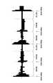
- FIG. 3is a schematic diagram for explaining the auscultation positions (1) to (12).
- auscultation positions (1) and (2)are set to the left and right of the upper lung field of the precordium.
- Auscultation positions (3) and (4)are set to the left and right of the precordial mid-lung field.
- Auscultation positions (5) and (6)are set to the left and right of the lower lung field of the precordium.
- the auscultation positions (7) and (8)are set to the left and right of the upper lung field on the back.
- the auscultation positions (9) and (10)are set to the left and right of the middle lung field on the back.
- Auscultation positions (11) and (12)are set to the left and right of the lower lung field on the back.
- the auscultation positionis not limited to the number and location mentioned above.
- auscultation positionsmay be set not only in the precordium and the back but also in the upper lung field, the middle lung field, and the lower lung field of the left and right lateral chests, and a total of 18 auscultation positions may be set.
- some of the above auscultation positionsmay be excluded.
- the auscultation positions (3) to (6), (9), and (10)are excluded, and the total of the auscultation positions (1), (2), (7), (8), (11), and (12). It may be limited to 6 places.
- the item of lung sound information 1524 for each auscultation positionincludes one or more sets composed of the item of lung sound data and the item of auscultation findings.
- a digital time-series acoustic signal including a lung sound acquired by an electronic stethoscope from the auscultation position of patient Ais recorded.
- the posture of the patient at the time of auscultationis roughly divided into the recumbent position and the sitting position, but the precordial and back auscultation is usually performed in the sitting position.
- the signal length of one lung sound data(for example, lung sound data 1) is arbitrary.
- one lung sound datamay be the signal of patient A's continuous N breaths.
- Nis a positive integer of 1 or more.
- the lung sound datais a signal obtained by processing the time-series acoustic signal acquired from the electronic stethoscope, such as removing the time-series acoustic signal during the resting phase, removing noise, and imparting respiratory timing. It's okay.
- the auscultation findings itemthe auscultation findings of a specialist for lung sound data are recorded.
- the auscultatory findingsthe presence or absence of abnormal sound of lung sound, and if there is abnormal sound, the type of abnormal sound (such as la sound) are recorded.
- informationsuch as gender, age, weight, body mass index (BMI), and medical history of patient A is recorded.
- BMIbody mass index
- the contact email address 1526 itemone or more email addresses of the recipients to whom the analysis results are sent are recorded.
- the contact e-mail addressmay be, for example, the e-mail address of the hospital in which the patient A was hospitalized, a heart failure specialist, the family doctor of the patient A, or the like.
- the method of transmitting the analysis resultis not limited to e-mail, and may be another communication method such as a message function of groupware or business chat.
- the lung sound information 153 to be analyzedrecords the lung sound information acquired from the patient A using the electronic stethoscope 11 and the analysis result thereof.
- FIG. 4is a configuration example of the lung sound information 153 to be analyzed.
- the lung sound information 153 to be analyzedis composed of the patient ID 1531, the analysis date and time 1532, the person in charge 1533, the lung sound information 1534, the urgency 1535, the communication item 1536 at the time of analysis, and the consent information 1537. There is.
- an ID that uniquely identifies the patient A recorded in the item of patient ID 1521 of the lung sound record 152is recorded.
- the item of analysis date and time 1532the date and time when the lung sound of patient A was acquired and analyzed is recorded.
- an ID that uniquely identifies the operator who performed the work of acquiring the lung sound of the patient Ais recorded.
- the item of lung sound information 1534is provided for each auscultation position.
- a total of 12 auscultation positions from the auscultation position (1) to the auscultation position (12) described with reference to FIG. 3are set (in FIG. 4, the auscultation positions (2) to (2) to ( 11) is omitted).
- the item of lung sound information 1534 for each auscultation positionincludes one or more sets composed of the item of lung sound data and the item of analysis result.
- a digital time-series acoustic signal including a lung sound acquired by an electronic stethoscope 11 from the auscultation position of patient Ais recorded.
- the signal length of one lung sound data(for example, lung sound data 1) is arbitrary.
- one lung sound datamay be the signal of patient A's continuous N breaths.
- Nis a positive integer of 1 or more.
- the lung sound datais a signal obtained by processing the time-series acoustic signal acquired from the electronic stethoscope 11 such as removal of the time-series acoustic signal during the resting phase, noise removal, and addition of respiratory timing. It may be there.
- the result of mechanical analysis of lung sound datais recorded.
- a numerical value indicating whether or not the lung sound data is abnormal lung sound datais recorded.
- two values of a value 0 indicating that the lung sound is normal and a value 1 indicating that the lung sound is abnormalmay be recorded.
- a numerical value indicating the degree of abnormality of the lung sound datamay be recorded.
- the degree of abnormalityan abnormality degree below a preset threshold value indicates that the lung sound data is a normal lung sound, and an abnormality degree exceeding the threshold value indicates that the lung sound data is an abnormal lung sound.
- the details of the detected abnormal sound(for example, type and properties, timing of hearing, etc.) may be recorded in the analysis result item.
- the urgency calculated by comprehensively judging each analysis result of the auscultation positions (1) to (12)is recorded.
- the degree of urgencyis an index showing how urgent the patient's condition is.
- urgencyis an indicator of the degree of time to avoid or reduce the risk of readmission due to acute exacerbations by providing appropriate treatment for heart failure within a certain period of time.
- Patient A's conditionincludes, for example, body weight, blood pressure, pulse, subjective symptoms (shortness of breath when going out, swelling, coughing, loss of appetite, etc.), medication status, water intake, and the like.
- the learning data DB 154records the learning data for detecting the abnormal lung sound from the lung sound data.
- the training datais composed of, for example, lung sound data and a correct answer label, and is also called teacher data.
- the learning data DB 154records a plurality of learning data for each auscultation position. Further, the plurality of training data of one auscultation position includes, for example, a plurality of training data including normal lung sound data and a plurality of training data including abnormal lung sound data.
- the individual learning data labelsindicate whether the corresponding lung sound data is an abnormal lung sound or a normal lung sound.
- the label indicating the abnormal lung soundmay further include information regarding the abnormal lung sound (eg, type, property, degree of abnormality, etc.).
- the arithmetic processing unit 16has a microprocessor such as a CPU and its peripheral circuits, and by reading and executing the program 151 from the storage unit 15, the hardware and the program 151 cooperate to realize various processing units. It is configured to do.
- the main processing units realized by the arithmetic processing unit 16include lung sound recording acquisition means 161, analysis target lung sound acquisition means 162, lung sound abnormality detection means 163, analysis result output means 164, learning data generation means 165, and , There is a learning means 166.
- the lung sound recording acquisition means 161acquires the lung sound recording 152 related to the patient A from an external device (not shown) or a storage medium (not shown) via a data input / output function such as the communication I / F unit 12. , Is configured to be recorded in the storage unit 15.
- the analysis target lung sound acquisition means 162is configured to acquire a digital time-series acoustic signal including the lung sound of patient A and other information.
- the analysis target lung sound acquisition means 162acquires the digital time-series acoustic signal including the lung sound of the patient A from the electronic stethoscope 11 according to the instruction of the operator input from the operation input unit 13 or the like. Further, the analysis target lung sound acquisition means 162 transmits the patient ID, the analysis date and time, the person in charge, the communication items at the time of analysis, and the consent information as other information from the operator through the operation input unit 13 or to the storage unit 15. Obtained from the stored lung sound record 152.
- the analysis target lung sound acquisition means 162generates the analysis target lung sound information 153 from the acquired digital time-series acoustic signal and other information, and stores it in the storage unit 15.
- the analysis target lung sound information 153 stored in the storage unit 15 by the analysis target lung sound acquisition means 162is configured in a format as shown in FIG. 4, for example.
- the item of each analysis result of the lung sound information 1534 and the item of the urgency degree 1535are NULL values.
- the lung sound abnormality detecting means 163is configured to detect whether or not the lung sound data is an abnormal lung sound. There are various methods for detecting abnormal lung sound. In the present embodiment, the lung sound abnormality detecting means 163 uses an abnormality detecting method by supervised learning. That is, the lung sound abnormality detecting means 163 includes intermittent crackles such as water crackles, crepitus, and continuous crackles such as whistles and rhonchi. Abnormal sounds are learned in advance, and abnormal sounds are detected based on the learning results.
- the lung sound abnormality detecting means 163learned the characteristics and discrimination criteria of the input sound data (input data) by using deep learning, for example, for learning data collected from abnormal sounds as supervised learning.
- a modelmay be created, and at the time of detection, detection may be performed by checking whether or not the input data conforms to the model.
- the lung sound abnormality detecting means 163uses, for example, a spectrogram in which voices are arranged in chronological order by FFT (Fast Fourier Transform) or log-FFT at regular intervals for learning and input data, and RNN (recurrent) for deep learning.
- Neural network) or CNNconvolutive neural network
- the lung sound abnormality detecting means 163is a method of detecting abnormal sounds by machine learning by converting the lung sound wave shape of learning and input data into short-time features such as zero crossing coefficient and MFCC (mel frequency cepstrum coefficient). May be used.
- the lung sound abnormality detecting means 163may be modeled by GMM (mixed Gaussian distribution) at the time of learning using the learning data, and it may be examined whether or not the input data fits the corresponding model at the time of detection. Further, the lung sound abnormality detecting means 163 learns the discriminating surface of a discriminator such as an SVM (support vector machine) using learning data, and the input data corresponds to the abnormal sound using the discriminating surface. You may identify whether to do it.
- the lung sound abnormality detecting means 163uses the data itself as a feature amount such as NMF (non-negative matrix factorization) and PCA (principal component analysis). May be generated.
- the lung sound abnormality detecting means 163uses statistical characteristics of the input waveform, such as the long-term power distribution of the input signal and the distribution of the component amount / component ratio in the specific frequency bin range, to generate an abnormal sound by a decision tree or the like. It may be detected. In that case, the lung sound abnormality detecting means 163 has a direct value (for example, when the power exceeds 20 mW for 3 consecutive frames) and a statistical feature (for example, Gauss approximation from 3 ⁇ ) as an item of the decision tree. (When a large processing frame occurs) may be used.
- a direct valuefor example, when the power exceeds 20 mW for 3 consecutive frames
- a statistical featurefor example, Gauss approximation from 3 ⁇
- the lung sound abnormality detecting means 163models not the input signal itself but an AR (autoregressive) process, and even if some of the model parameters exceed the threshold value, the abnormal sound is detected. good. Although these methods may not include the learning process, they are included in the supervised learning for convenience because they include the observation of the abnormal sound which is the target signal in the determination of the decision tree and the threshold value.
- the lung sound abnormality detecting means 163analyzes the lung sound data for each auscultation position of the patient A recorded in the analysis target lung sound information 153 by using the abnormality detecting method by supervised learning as described above, and the analysis result. Is recorded in the analysis result item of the lung sound record 152 for each auscultation position. Further, the lung sound abnormality detecting means 163 calculates the urgency based on the analysis result of the lung sound data for each auscultation position and records it in the item of urgency 1535.
- the analysis result output means 164is configured to output the analysis target lung sound information 153 for the purpose of notifying the persons concerned of the heart failure state of the patient A.
- the analysis result output means 164is configured to read the analysis target lung sound information 153 from the storage unit 15 and display the analysis target lung sound information 153 on the screen display unit 14. Further, the analysis result output means 164 sends an e-mail with the analysis target lung sound information 153 read from the storage unit 15 as a file according to the instruction from the operation input unit 13 or automatically through the communication I / F unit 12. It is configured to be sent to the contact email address 1526 of the lung sound record 152. At this time, the analysis result output means 164 may determine the destination of the analysis target lung sound information 153 based on the urgency 1535 of the analysis target lung sound information 153.
- the analysis result output means 164transmits the analysis target lung sound information 153 through the communication I / F unit 12 in order to acquire the findings of a heart failure specialist with respect to the lung sound data acquired from the patient A and create learning data. It is configured to send to the terminal device of a heart failure specialist.
- the analysis result output means 164may determine whether or not to transmit the analysis target lung sound information 153 to a heart failure specialist based on the analysis result for each auscultation position. For example, the analysis target lung sound information 153 does not transmit the analysis target lung sound information 153 in which the lung sound data of all the hearing positions is normal, and the analysis target lung sound information in which the lung sound data of one or more hearing positions is abnormal is abnormal. Information 153 may be transmitted. In general, learning data including normal lung sound data can be easily obtained from many normal people, whereas learning data including abnormal lung sound data can be obtained only from some people. .. Therefore, it is possible to reduce the burden on the heart failure specialist by transmitting only the analysis target lung sound information 153 in which abnormal lung sound data exists.
- the analysis target lung sound information 153is limited to the analysis target lung sound information 153 in which the type of abnormal lung sound recorded in the analysis result matches a predetermined type, and the analysis target lung sound information 153 is heart failure. You may send it to a specialist. As the type of abnormal lung sound defined in advance, the type of abnormal lung sound for which learning data is insufficient may be used. This makes it possible to create learning data for the missing types of abnormal lung sounds without unnecessarily increasing the burden on heart failure specialists.
- the analysis result output means 164may determine whether or not to transmit the analysis target lung sound information 153 to the heart failure specialist based on the consent information 1537.
- the analysis target lung sound information 153may transmit the analysis target lung sound information 153 to a heart failure specialist only when the patient consents to use the lung sound data for learning. This is to prevent the burden on the heart failure specialist from being unnecessarily increased because the lung sound data for which usage consent has not been obtained cannot be used for learning data.
- the analysis result output means 164may determine whether or not to transmit the analysis target lung sound information 153 to the heart failure specialist based on the personal information of the patient A.
- the analysis result output means 164is limited to the analysis target lung sound information of a patient having personal information matching the predetermined target personal information (gender, age group, BMI width, medical history, etc.), and is a heart failure specialist. May be sent to.
- the target personal information to be determined in advanceat least one of the lack of learning data, gender, age group, BMI width, medical history, etc., or a combination of two or more can be used.
- FIG. 5is a configuration example of the target personal information.
- the targetis a patient whose gender is female, whose age group is 40 to 60 years old, whose BMI is 20 or less, and whose medical history is hypertension.
- the analysis target lung sound information 153 sent to the heart failure specialistis analyzed by the heart failure specialist.
- a specialistreproduces the lung sound data for each auscultation position recorded in the lung sound information 153 to be analyzed by a personal computer or the like, and diagnoses whether or not secondary noise such as a rattling sound is heard from the lung sound of patient A. .. Then, the specialist creates auscultatory findings for the auscultation data for each auscultation position and records them in the analysis target lung sound information 153.
- the analysis target lung sound information 153 in which the auscultation findings of the specialist are recordedis returned to the transmission source lung sound analyzer 10 by a communication means such as an email.
- FIG. 6shows a configuration example of the lung sound information 153 to be analyzed with auscultatory findings.
- the learning data generation means 165When the learning data generation means 165 receives the auscultation finding analysis target lung sound information 153 from the heart failure specialist through the communication I / F unit 12 of the lung sound analyzer 10, the learning data generation means 165 is based on the received auscultation finding analysis target lung sound information 153. To generate training data. Further, the learning data generation means 165 records the generated learning data in the learning data DB 154.
- the learning means 166is configured to learn a model for detecting abnormal lung sounds by using the learning data recorded in the learning data DB 154. Further, the learning means 166 is configured to relearn a model for detecting abnormal lung sounds after new learning data is added to the learning data DB 154.
- the operation of the lung sound analyzer 10is roughly classified into a pre-operation, an analysis operation performed thereafter, a learning data creation and a learning operation.
- FIG. 7is a flowchart showing an example of the preliminary operation.
- the pre-operationis started, for example, by activating the lung sound recording acquisition means 161 by operating the start button of the pre-operation displayed on the screen display unit 14.
- the lung sound recording acquisition means 161when the lung sound recording acquisition means 161 is activated, it is transmitted from an external device (not shown) or a storage medium (not shown) via a data input / output function such as a communication I / F unit 12.
- the lung sound record 152 relating to the patient Ais acquired and recorded in the storage unit 15 (step S1).
- FIG. 2is a configuration example of the lung sound record 152 thus acquired.
- the lung sound record 152contains at least the past lung sound data of patient A and the history of auscultatory findings.
- the learning means 166When the above operation of the lung sound recording acquisition means 161 is completed, the learning means 166 is automatically activated or according to an instruction from the operation input unit 13.
- the learning means 166reads the learning data DB 154 from the storage unit 15, learns a model for detecting a lung sound abnormality based on the learning data recorded in the learning data DB 154, and uses the trained model as a lung sound abnormality detecting means.
- Store in 163step S2.
- FIG. 8is an explanatory diagram of model learning.
- the learning means 166reads the learning data of the auscultation position (1) from the learning data DB 154, and uses the read learning data to detect an abnormality in the lung sound data of the auscultation position (1). Create model 171-1.
- the learning means 166is a model for detecting an abnormality in the lung sound data of the auscultation positions (2) to (12) based on the learning data of the auscultation positions (2) to (12) recorded in the learning data DB 154. Produces 171-2 to 171-12.
- the learning means 166learned a model for detecting an abnormality in lung sound data for each listening position.
- the learning means 166may learn one common model for detecting abnormalities in lung sound data at a plurality of auscultation positions.
- the threshold value set when anomaly detection is performed using the modelis set to a plurality of auscultation positions. It may be set in common for each position, or it may be set for each individual auscultation position.
- FIG. 9is a flowchart showing an example of the analysis operation.
- the analysis operationis performed at a place other than a specialized hospital such as a clinic in the town or the home of patient A.
- the analysis operationmay be used to assist the doctor's diagnosis in a specialized hospital or the like.
- the analysis operationis started, for example, by operating the analysis operation start button displayed on the screen display unit 14 to activate the analysis target lung sound acquisition means 162.
- the analysis target lung sound acquisition means 162when the analysis target lung sound acquisition means 162 is activated, the necessary items for each item of the patient ID 1531, the analysis date / time 1532, the person in charge 1533, the communication item 1535 at the time of analysis, and the consent information 1537 are displayed.
- the analysis target lung sound information 153which is described and the other items are set to the NUML value, is created and recorded in the storage unit 15 (step S11).
- the analysis target lung sound acquisition means 162acquires the patient ID 1531 from the patient ID 1521 of the lung sound record 152 stored in the storage unit 15.
- the analysis target lung sound acquisition means 162acquires the analysis date / time 1532, the person in charge 1533, the communication item 1536 at the time of analysis, and the consent information 1537 from the operator through the operation input unit 13.
- the consent information 1537may include an electronic signature to the effect that patient A has agreed to use his lung sound data for learning.
- the analysis target lung sound acquisition means 162acquires a digital time-series acoustic signal including the lung sound for each auscultation position of the patient A from the electronic stethoscope 11 and associates it with the auscultation position to the analysis target lung sound information 153. Record (step S12).
- the method of acquiring the lung sound for each auscultation position of the patient with an electronic stethoscope and recording it in association with the auscultation positionis arbitrary. For example, as described in Patent Documents 1, 4 or 6, a method of displaying a guidance screen for guiding the auscultation position to the operator using the electronic stethoscope 11 on the screen display unit 14 is optional. Method may be used.
- the lung sound abnormality detecting means 163reads out the lung sound information 153 to be analyzed from the storage unit 15, and the lung for each auscultation position of the patient A recorded in the lung sound information 1534 of the lung sound information 153 to be analyzed. Sound data is analyzed using a model created in advance, and the analysis result is recorded in the item of analysis result for each auscultation position of lung sound information 1534. Further, in step S12, the analysis result output means 164 appropriately displays the analysis result of the lung sound abnormality detecting means 163 on the screen display unit 14.
- the lung sound abnormality detecting means 163calculates the urgency level 1535 based on the analysis result of the lung sound data for each auscultation position, and records it in the item of the urgency level 1535 of the analysis target lung sound information 153 (step S13). ).
- the analysis result output means 164reads the analysis target lung sound information 153 from the storage unit 15, displays the analysis target lung sound information 153 on the screen display unit 14, and attaches the analysis target lung sound information 153 as a file.
- the mailis sent to the contact mail address 1526 of the lung sound record 152 through the communication I / F unit 12 (step S14).
- step S14the analysis result output means 164 outputs the analysis target lung sound information 153 for the purpose of notifying the person concerned of the heart failure state of the patient A, and also outputs the lung sound data acquired from the patient A.
- the lung sound information 153 to be analyzedis transmitted to the terminal device of a specialist in heart failure for the purpose of using it for training data.
- step S12 for acquiring the lung sound to be analyzed and detecting the abnormalitywill be described with reference to the flowchart of FIG.
- FIG. 10is a flowchart showing an example of the detailed procedure of step S12 of FIG.
- the analysis target lung sound acquisition means 162is based on the presence or absence of abnormal sound recorded in the auscultatory findings for each auscultation position in one or more auscultation information 1527 recorded in the lung sound record 152 of the patient A.
- the frequency of appearance of abnormal sounds for each auscultation positionis calculated (step S21). Specifically, first, the analysis target lung sound acquisition means 162 initializes the frequency counter for each auscultation position (1) to (12) to 0. Next, the analysis target lung sound acquisition means 162 pays attention to the auscultation information 1527 whose auscultation date and time recorded in the lung sound record 152 is the latest.
- the analysis target lung sound acquisition means 162performs auscultation if at least one auscultation finding describing that there is an abnormal sound is present in one or more auscultation findings recorded at the auscultation position (1).
- the frequency count corresponding to the position (1)is incremented by 1.
- the analysis target lung sound acquisition means 162has at least one auscultatory finding in which it is described that there is an abnormal sound among one or more auscultatory findings recorded at the auscultation positions (2) to (12). Then, the frequency count corresponding to the auscultation positions (2) to (12) is incremented by one.
- the analysis target lung sound acquisition means 162pays attention to the auscultation information 1527 one before the latest auscultation date and time, and performs the same operation as the operation performed using the latest auscultation information 1527 at the auscultation position (1).
- ⁇ (12)Perform for each frequency count.
- the analysis target lung sound acquisition means 162either finishes processing up to a predetermined number of auscultation information 1527, or finishes processing up to the oldest auscultation information 1527 recorded in the lung sound record 152. The above operation is repeated until it is established quickly.
- the analysis target lung sound acquisition means 162sets the value of the frequency count for each auscultation position (1) to (12) as the frequency of appearance of abnormal sounds at the auscultation positions (1) to (12).
- the analysis target lung sound acquisition means 162determines the order (order) of the auscultation positions for auscultating the lung sounds from the patient A based on the abnormal frequency for each of the auscultation positions (1) to (12) of the patient A. (Step S22).
- the fact that there is a difference in the frequency of abnormal sounds such as rattling between the auscultation positions (1) to (12) of patient Ameans that patient A has auscultation positions where abnormal sounds are relatively likely to occur. It indicates that there is an auscultation position that is not.
- the auscultationis interrupted in the middle for some reason such as the convenience of the patient A. Even if the heart failure state of the patient A is determined based on the analysis result of the lung sound data of some of the auscultated positions so far, the probability of overlooking the exacerbation of the heart failure can be reduced.
- the analysis target lung sound acquisition means 162may determine the order of auscultation positions based only on the abnormal frequency of each auscultation position of patient A. In that case, the analysis target lung sound acquisition means 162 may determine, for example, the result of sorting the auscultation positions in the descending order of the abnormal frequency (the order of proceeding from the most frequent to the least) in the order of the auscultation positions.
- the auscultation frequency for each auscultation position (1) to (12) of patient Ais shown in FIG. 11, for example, an example of the order of auscultation positions based on the result of sorting the auscultation positions in descending order of the auscultation position is the auscultation order in FIG. It becomes as shown in 1.
- the auscultation position (11), which has the maximum abnormality frequency of 4,is auscultated.
- the auscultation positions with the next highest abnormality frequencyare the auscultation positions (6) and (12) of the abnormality frequency 3. Since there is no difference in the frequency of abnormalities, in the auscultation sequence 1, the auscultation position (12) on the same back as the first is the second, and the auscultation position (6) on the precordium is the third.
- the order of auscultation positions (5), (9), (10), (7), (1), (2), (3), (4), and (8)is used.
- lung sound datacan be acquired in order from the auscultation position having a higher probability of abnormal lung sound.
- the order of auscultation positionsmay be determined in consideration of not only the frequency of abnormalities for each auscultation position of the patient but also the reduction of the burden on the patient and the operator.
- the analysis target lung sound acquisition means 162determines the side of the precordium and the back where the auscultation position with the highest auscultation frequency exists first as the site to be auscultated, and the site opposite to the site to be auscultated next. Further, the analysis target lung sound acquisition means 162 determines the result of sorting all the auscultation positions of the site in descending order of the abnormal frequency for each site in the order of the auscultation positions of the site. An example of the auscultation order by this determination method is shown in FIG. 11 for auscultation order 2.
- the back where the auscultation position (11) having the maximum abnormality frequency of 4 is presentis determined as the site to be auscultated first, and the auscultation order of the auscultation positions (7) to (12) on the back is abnormal.
- the auscultation positions (11), (12), (9), (10), (7), and (8)are determined in the order of the results sorted in descending order of frequency.
- the auscultation order of the auscultation positions (1) to (6) of the anterior chestis in descending order of their abnormal frequencies. According to the sorted results, the auscultation positions (6), (5), (1), (2), (3), and (4) are determined in this order.
- the analysis target lung sound acquisition means 162pays attention to the auscultation position in the first order (step S23).
- the analysis target lung sound acquisition means 162acquires a digital time-series acoustic signal including the lung sound at the auscultation position of interest from the electronic stethoscope 11 (step S24).
- the analysis target lung sound acquisition means 162displays a guidance screen for guiding the auscultation position of interest to the operator using the electronic stethoscope 11 on the screen display unit 14 to support the lung sound acquisition. You may do it.
- the analysis target lung sound acquisition means 162supports the lung sound acquisition by reproducing the guidance voice for guiding the auscultation position of interest to the operator using the electronic stethoscope 11 from a speaker (not shown). You may do it.
- the analysis target lung sound acquisition means 162should apply the electronic auscultation device 11 to the operator using the electronic auscultation device 11 based on the order of the auscultation positions for auscultating the lung sounds determined for the patient A.
- the auscultation positionis guided using an image or voice, and a digital time-series acoustic signal including the lung sound of the auscultation position under the guidance is acquired from the electronic auscultation device 11.
- the analysis target lung sound acquisition means 162measures the quality of the acquired lung sound (step S25).
- the time-series acoustic signal output from the electronic stethoscope 11includes the lung sound of patient A in the frequency band of 100 Hz to about 2 kHz, and the background noise (stationary noise) in the same frequency band.
- the background noisestationary noise
- environmental sounds, human voices, metallic sounds, etc.that come in from the outside through the body of the patient A or through the gap between the skin of the patient A and the chestpiece are examples of background noise. If the intensity of the lung sound in the time-series acoustic signal is low and the intensity of the background noise is high, it becomes difficult to detect the abnormality of the lung sound.
- the analysis target lung sound acquisition means 162first uses a bandpass filter to extract a time-series acoustic signal in a frequency band of 100 Hz to about 2 kHz from the time-series acoustic signal output from the electronic stethoscope 11. Next, the analysis target lung sound acquisition means 162 calculates the intensity of the lung sound and the intensity of the background noise in the extracted time-series acoustic signal, and calculates the degree of difference between them as an index value of the quality of the lung sound.
- a method of calculating an index value of lung sound qualitywill be described.
- FIG. 12is a schematic diagram showing an example of a waveform of a time-series acoustic signal including a lung sound output from an electronic stethoscope 11.
- lung soundsthere are three types of lung sounds: bronchial breath sounds, bronchial alveolar breath sounds, bronchial breath sounds, and alveolar breath sounds.
- Itis a schematic diagram which shows an example of the alveolar breath sounds heard in (12).
- the amplitude of the time-series acoustic signal including the lung soundchanges greatly at the start of inspiration. Also, at the start of exhalation, the amplitude changes significantly, though not as much as at the start of inspiration.
- the analysis target lung sound acquisition means 162compares the time-series acoustic signal with the threshold value T1 capable of discriminating the amplitude change at the start of inspiration, and when the amplitude of the time-series acoustic signal becomes larger than the threshold value T1, the inspiration Detect as start time. Further, the analysis target lung sound acquisition means 162 has a section of one cycle of respiration from the start of one inspiration to the start of the next inspiration, and the amplitude of the time-series acoustic signal in that section and the amplitude change at the start of exhalation.
- the time when the amplitude of the time-series signal becomes larger than the threshold value T2is detected as the start time of exhalation by comparing with the threshold value T2 ( ⁇ T1) capable of discriminating.
- T2threshold value
- the start of exhalationis also detected in order to further divide the phase other than the resting phase into the inspiratory phase and the expiratory phase.
- human respirationgenerally consists of an inspiratory phase of about 1 second, an expiratory phase of about 1 second, and a resting phase of about 1 to 1.5 seconds until the next inspiration. That is, immediately before the start of inspiration, there is a dormant phase that is neither inspiring nor exhaling.
- the analysis target lung sound acquisition means 162detects a predetermined period (for example, 1 second) immediately before the start time of the detected inspiration as a resting phase. Then, the analysis target lung sound acquisition means 162 calculates the intensity of the time-series acoustic signal in the resting phase as the intensity of the background noise.
- the intensity of the time-series acoustic signalcan be, for example, the root mean square of the amplitude value, but is not limited to this, and may be an amplitude or the like.
- the analysis target lung sound acquisition means 162calculates a value obtained by subtracting the background noise intensity from the intensity of the time-series acoustic signal in the inspiratory phase and / or the expiratory phase as the lung sound intensity. Then, the analysis target lung sound acquisition means 162 uses the ratio of the lung sound intensity to the calculated background noise intensity as an index value of the lung sound quality.
- the index value of the quality of the lung soundis not limited to the above, and the S / N ratio calculated from the intensity of the lung sound and the intensity of the background noise may be used as the index value.
- the method of detecting the resting phase by taking the alveolar respiration sound as an examplehas been described, but the bronchial alveolar respiration sound is heard together with the alveolar respiration sound at the auscultation position of the middle lung field and the upper lung field.
- the inspiratory amplitude of the bronchial alveolar breathing soundis greater than or equal to the exhalation amplitude, even if the bronchoalveolar breathing sound is heard together with the alveolar alveolar breathing sound, the inspiratory and expiratory sounds are described by the method described in FIG. The start timing of can be detected.
- the amplitudemay be larger during exhalation than during inspiration. Therefore, when the bronchoalveolar breath sounds are close to the tracheal breath sounds, inspiration and expiration may be reversed in the method described with reference to FIG. Specifically, for example, it may be as follows. First, the frequency at which the amplitude of the frequency spectrum of the auscultated lung sound is maximized is compared with the preset threshold frequency.
- the frequency at which the amplitude of the frequency spectrum of the auscultated lung sound is maximumis equal to or higher than the threshold frequency, it is determined that the bronchial alveolar respiration sound contained in the lung sound is close to the tracheal respiration sound, which is described with reference to FIG.
- the inspiratory and expiratoryare reversed to detect the start timing of inspiratory and expiratory.
- the frequency at which the maximum amplitude of the frequency spectrum of the auscultated lung soundis less than the threshold frequency, it is determined that the bronchial alveolar respiration sound contained in the lung sound is not close to the bronchial respiration sound, which is described with reference to FIG.
- the start timing of inspiration and expirationis detected by the above method.
- the above threshold frequencyis a threshold at which it can be determined whether or not the bronchial alveolar respiration sound included in the lung sound is close to the bronchial respiration sound. It can be pre-determined from the frequency band between the frequency and the frequency at which the amplitude of the frequency spectrum of the high tracheal breathing sound is maximized. Further, instead of the above-mentioned "frequency at which the amplitude of the frequency spectrum is maximized", the "spectral centroid" used as a scale for expressing the shape of the spectrum may be used.
- the start time points of exhalation and inspirationwere detected from the time-series acoustic signal output from the electronic stethoscope 11, and the predetermined period immediately before the start time of the detected inspiration was detected as the rest phase.
- the method for detecting the inspiratory phase, the expiratory phase, and the resting phaseis not limited to the above.
- the analysis target lung sound acquisition means 162is machine learning for estimating which section of the time-series acoustic signal including the lung sound output from the electronic stethoscope is the inspiratory phase, the expiratory phase, and the resting phase.
- the estimated probabilities of the inspiratory phase, the expiratory phase, and the resting phaseare acquired from the trained model for each section. It may have been done.
- the learning modelcan be pre-generated by machine learning using a machine learning algorithm such as a neural network, for example, using a time-series acoustic signal including various lung sounds as training data.
- the lung sound acquisition means 162 to be analyzedmay detect the timing of respiration such as the start of inspiration and expiration of patient A from other than the time-series acoustic signal output from the electronic stethoscope.
- the analysis target lung sound acquisition means 162detects the breathing timing of the patient A by using a respiratory volume sensor such as a lung tachograph or a respiratory band that detects a change in the shape of the chest or abdomen due to respiratory activity by the sensor. You may.
- a respiratory volume sensorsuch as a lung tachograph or a respiratory band that detects a change in the shape of the chest or abdomen due to respiratory activity by the sensor. You may.
- the analysis target lung sound acquisition means 162compares the index value of the lung sound quality with the preset quality threshold value (step S26). Then, if the index value of the quality of the lung sound is smaller than the threshold value, the analysis target lung sound acquisition means 162 issues an alarm to the effect that the quality of the lung sound at the auscultated position, which is auscultated by the electronic stethoscope 11, is poor. Is displayed in (step S27). The operator who recognizes this alarm takes measures to reduce the background noise and / or to increase the lung sound, and then performs the work of reacquiring the lung sound at the auscultation position of interest by the electronic stethoscope 11. That is (step S28).
- the analysis target lung sound acquisition means 162returns to the process of step S25, and repeats the same process as the above-mentioned process.
- the analysis target lung sound acquisition means 162removes the pause phase period and background noise from the digital time-series acoustic signal including the lung sound at the listening position of interest. Then, the period of the resting phase and the digital time-series acoustic signal after removing the background noise are recorded in the lung sound information 153 to be analyzed in association with the listening position of interest (step S29). The period of the rest phase and the removal of background noise are performed as follows.
- the analysis target lung sound acquisition means 162sets a digital time-series acoustic signal including the lung sound at the auscultation position of interest as a section consisting of an inspiratory phase and an expiratory phase immediately after that (hereinafter referred to as an inspiratory / expiratory section). It is divided into two parts into a dormant phase section (hereinafter referred to as a dormant section).
- the analysis target lung sound acquisition means 162calculates the frequency spectra of the inspiratory / expiratory section and the resting section by performing a fast Fourier transform (FFT) on the digital time-series acoustic signals of the inspiratory / expiratory section and the resting section, respectively.
- FFTfast Fourier transform
- the analysis target lung sound acquisition means 162subtracts the frequency spectrum of the rest section from the frequency spectrum of the inspiratory / expiratory section. This subtraction suppresses background noise contained in the inspiratory and expiratory phases.
- the analysis target lung sound acquisition means 162generates a digital time-series acoustic signal after noise removal in the inspiratory / expiratory section by reverse frequency converting the frequency spectrum of the inspiratory / expiratory section after the subtraction.
- the analysis target lung sound acquisition means 162records the generated digital time-series acoustic signal after noise removal in the inspiratory / expiratory section in the analysis target lung sound information 153 in association with the auscultation position of interest.
- the lung sound acquisition means 162 to be analyzedmay remove the period of the rest phase from the digital time-series acoustic signal including the lung sound at the auscultation position of interest, and may not remove the background noise.
- the analysis target lung sound acquisition means 162divides the digital time-series acoustic signal including the lung sound at the auscultation position of interest into two into an inspiratory / expiratory section and a rest section, and the digital time-series of the inspiratory / expiratory section.
- the acoustic signalis associated with the auscultation position of interest and recorded in the lung sound information 153 to be analyzed.
- the lung sound abnormality detecting means 163detects the lung sound abnormality from the lung sound data recorded in the analysis target lung sound information 153 in association with the listening position of interest, and the detection result is set to the listening position of interest. It is recorded in the lung sound information 153 to be analyzed in association with each other (step S30). As described above, the detection of lung sound abnormality is performed by the abnormality detection method by supervised learning.
- the analysis result output means 164displays the abnormality detection result on the screen display unit 14 every time the lung sound abnormality detecting means 163 detects an abnormality in the lung sound data of the auscultation position being watched (step S31). As a result, the operator can immediately recognize whether or not the lung sound data at the auscultation position is an abnormal lung sound at the time of auscultation.
- the analysis target lung sound acquisition means 162determines whether or not the acquisition and analysis of the lung sound data of all the auscultation positions have been completed (step S32). .. When the auscultation position that has not been acquired remains, the analysis target lung sound acquisition means 162 shifts attention to the auscultation position in the next order (step S33), returns to step S24, and performs the same process as described above. repeat.
- the analysis target lung sound acquisition means 162ends the process of FIG. Further, the analysis target lung sound acquisition means 162 may end the process of FIG. 10 before the acquisition and analysis of the lung sound data of all the auscultation positions are completed due to the convenience of the patient A or the like.
- the lung sound information 1534 in the analysis target lung sound information 153 corresponding to the auscultation position where the lung sound data is not acquired and analyzedremains the NUML value.
- step S13 in FIG. 9 for calculating the urgency level 1535will be described.
- the lung sound abnormality detecting means 163determines the severity of heart failure of patient A based on the analysis result of lung sound data for each auscultation position, and calculates the urgency 1535 based on the determined severity. In determining the severity of heart failure, the lung sound abnormality detecting means 163 determines the severity of heart failure by referring to a determination table for determining the severity of heart failure from the analysis results of lung sound data for each auscultation position. ..
- FIG. 13is a diagram showing an example of the determination table.
- the determination table shown in FIG. 13has columns corresponding to one-to-one correspondence between auscultation positions (1) to (12) and rows corresponding to one-to-one correspondence in severity, and lung sounds at the intersections of rows and columns.
- a + symbol indicating that there is an abnormality and a-symbol indicating that there is no abnormality in lung soundare set.
- the severityis determined to be 0.
- the lung sound abnormalityin at least one of the auscultation positions (11) and (12) set in the lower lung field on the back, and the lung sound abnormality is in the other auscultation positions (1) to (10). If not, the severity is determined to be 1. Further, in the judgment table, there is an abnormality in lung sound at both the auscultation positions (11) and (12), and the lungs are located in only one of the auscultation positions (5) and (6) set in the lower lung field of the anterior chest. If there is a sound abnormality and there is no lung sound abnormality at the other auscultation positions (1) to (4) and (7) to (10), the severity is determined to be 2. Severity N set in the last row is assumed to have abnormal lung sound at all auscultation positions (1) to (12).
- the severity of heart failureis classified into N + 1 classes from severity 0 to severity N according to the combination of the presence or absence of abnormal lung sound at the auscultation positions (1) to (12).
- the severity 0is a state in which no abnormal lung sound is heard, and thus it can be said that the heart failure is in remission.
- severity 1is a state in which abnormal lung sound can be heard only in the lower lung field of the back, it cannot be said that heart failure is in remission, but it is mild and some patients are discharged in such a state. It is in a state of doing.
- Severity 2can be said to be more severe than severity 1 because abnormal lung sounds are produced in one of the lower lung fields in the precordium in addition to the lower lung field in the back. However, since it still belongs to mild illness, it can be said that there is a high probability that readmission can be prevented if appropriate measures are taken at this point.
- the determination table for determining the severity of heart failure from the analysis result of the auscultation positionis not limited to that shown in FIG.
- the ra soundis mild when it is heard only at the end of inspiration, and it is severe when it is heard immediately after the start of inspiration. Therefore, in addition to the presence or absence of abnormal lung sound for each auscultation position, the type of abnormal lung sound and the timing at which the abnormal lung sound is heard are set in the judgment table, and the auscultation position, the presence or absence of abnormal lung sound, and the type of abnormal lung sound are determined.
- the severity of heart failuremay be determined by a combination of timings at which abnormal lung sounds are heard.
- the lung sound abnormality detecting means 163may determine the severity of heart failure of patient A from the number of auscultation positions where abnormal lung sound has occurred, regardless of where the auscultation position is. For example, the lung sound abnormality detecting means 163 has a severity of 0, 1, and when the number of auscultatory positions resulting in abnormal lung sound is 0, 1 or more and 2 or less, 3 or more and 4 or less, 5 or more and 8 or less, and 9 or more, respectively. It may be 2, 3, 4 (maximum).
- the lung sound abnormality detecting means 163has the process shown in FIG. 10 terminated in the middle due to the convenience of the patient A or the like, and the analysis result of at least a part of the lung sound information 1534 for each auscultation position is a NULL value. Perform the following processing. First, the lung sound abnormality detecting means 163 sets in advance the number of auscultation positions where the analysis result is the NUML value, that is, the lung sound data is not acquired and the analysis of whether or not the lung sound is abnormal is not performed. It is determined whether or not the condition that the value is less than the first threshold value is satisfied.
- the lung sound abnormality detecting means 163is required to have the condition that the number of auscultation positions where the lung sound data is acquired and whether or not the lung sound is abnormal is equal to or more than a preset second threshold value. Is determined whether or not the above is satisfied.
- the first threshold value and the second threshold valuemay be fixed values or variable values according to the latest state of the patient. When it is a fixed value, for example, the first threshold value may be 4 or less and the second threshold value may be 8 or more.
- the first threshold valuein the case of a patient who has no abnormal lung sound at any auscultation position in the latest state, for example, the first threshold value may be 10 or less, the second threshold value may be 2 or more, and in the case of other patients. , May be the same as the fixed value. If the lung sound abnormality detecting means 163 does not satisfy the above conditions, the severity is not calculated (and therefore the urgency is not calculated), the lung sound analysis this time is terminated with an error, and the screen display unit 14 indicates that fact. Display on. The reason is to prevent incorrect information from being given to the operator or the like.
- the lung sound abnormality detecting means 163calculates the severity on the assumption that the lung sound abnormality is not detected at the auscultation position where the analysis of whether or not the lung sound is abnormal is not performed. .. Then, the lung sound abnormality detecting means 163 holds the calculated severity as the most optimistic value. That is, when the calculated severity is severity 1, it is retained as "severity 1 or higher” or “at least severity 1" instead of "severity 1". For example, for a patient who has no abnormal lung sound at any auscultation position in the latest state, lung sound data is acquired and analyzed only at two auscultation positions (11) and (12), and as a result, at least one of them is obtained.
- the lung sound abnormality detecting means 163assumes that the lung sound abnormality is not detected at the other auscultation positions (1) to (10), and determines the severity 1 based on the determination table of FIG. Based on the above, it is determined that the severity is 1 or higher.
- the urgency 1535is determined from the determined severity.
- the lung sound abnormality detecting means 163may determine the urgency level 1535 based on the severity 0 to N of heart failure and the condition of patient A. For example, as the condition of patient A, whether or not the body weight has increased by a certain amount in a unit period (for example, an increase of 3 kg or more in one week), the presence or absence of subjective symptoms such as swelling, coughing, and loss of appetite, and the pulse exceeding a predetermined number. Whether or not it is possible. Then, the lung sound abnormality detecting means 163 may set the urgency determined based on the severity of heart failure to be corrected so as to be larger according to the condition of the patient A as the final urgency. For example, the lung sound abnormality detecting means 163 may increase the urgency to 1 or 2 if there is weight gain, even if the urgency determined from the severity of heart failure is 0 or 1. .. However, the upper limit of the urgency after correction is N.
- the analysis target lung sound information 153 stored in the attached fileis analyzed by the heart failure specialist.
- the lung sound information 153 to be analyzedis not limited to the attachment of a file, and may be shared with a heart failure specialist in a SaaS format such as link entry.
- a specialistreproduces the lung sound data for each auscultation position recorded in the lung sound information 153 to be analyzed by a personal computer or the like, and diagnoses whether or not secondary noise such as a rattling sound is heard from the lung sound of patient A. ..
- the specialistcreates auscultatory findings for the auscultatory sound data for each auscultatory position and records them in the analysis target lung sound information 153 as shown in FIG.
- the analysis target lung sound information 153 in which the auscultation findings of the specialist are recordedis returned to the transmission source lung sound analyzer 10 by a communication means such as an email.
- the learning data generation means 165updates the original analysis target lung sound information 153 recorded in the storage unit 15 by the analysis target lung sound information 153 with auscultatory findings received through the communication I / F unit 12 of the lung sound analyzer 10. do.
- the learning data generation means 165creates learning data for each auscultatory position of the lung sound information 153 to be analyzed, and for each set of lung sound data and auscultatory findings recorded correspondingly.
- the learning data generation means 165creates learning data including a label indicating abnormal lung sound and lung sound data from a pair of auscultatory findings indicating abnormal lung sound and lung sound data.
- the learning data generation means 165creates learning data including a label indicating normal lung sound and lung sound data from the set of the auscultatory findings indicating that the lung sound is normal and the lung sound data. At this time, personal information of the patient may be added to the label. Further, the learning data generation means 165 records the created learning data in the learning data DB 154 in association with the auscultation position. At this time, the learning data generation means 165 may record the learning data separately from other learning data already recorded in the learning data DB 154 by adding a time stamp such as a recording date and time to the learning data.
- the learning data added to the learning data DB 154will be used when the learning means 166 relearns the model for detecting the lung sound abnormality from the next time onward. By doing so, the accuracy of the model for detecting lung sound abnormality can be gradually improved.
- learning data for detecting abnormal lung soundcan be efficiently collected at a clinic in a town without a specialist or at the home of a patient.
- the lung sound analyzer 10acquires a time-series acoustic signal including the lung sound of a heart failure patient, detects an abnormal lung sound from the acquired time-series acoustic signal, and uses the acquired time-series acoustic signal.
- the analysis target lung sound information associated with the detection resultis transmitted to the terminal device of the specialist and the analysis target lung sound information with the specialist's findings on the time-series acoustic signal is received, the specialist's findings are added. This is to generate learning data for detecting abnormal lung sounds based on the lung sound information to be analyzed.
- FIG. 14is a block diagram of the lung sound analysis system 20 according to the second embodiment of the present invention.
- the lung sound analysis system 20includes a plurality of lung sound analysis devices 21, a server device 22, and a terminal device 24. Further, the plurality of lung sound analyzers 21, the server device 22, and the terminal device 24 are connected to each other so as to be able to communicate with each other through a network such as the Internet.
- the lung sound analyzer 21is an information processing device that acquires and analyzes lung sounds from a patient discharged from the hospital after receiving treatment for heart failure.
- the terminal device 24is a terminal device used by a specialist in heart failure.
- the lung sound analyzer 21 and the terminal device 24may be, but are not limited to, a smartphone, a tablet terminal, a PDA, a laptop computer, and the like.
- the lung sound analyzer 21includes an electronic stethoscope (not shown), a communication I / F unit, an operation input unit, a screen display unit, a storage unit, and an arithmetic processing unit.
- the terminal device 24includes a communication I / F unit (not shown), an operation input unit, a screen display unit, a storage unit, and an arithmetic processing unit.
- the server device 22is a computer that provides various services necessary for lung sound analysis to a plurality of lung sound analyzers 21 through the network 23.
- the server device 22stores at least a part of the lung sound record 152, the analysis target lung sound information 153, the learning data DB 154, and the program 151 shown in FIG. 1, and stores them in the lung sound analyzer 21 through the network 23.
- the lung sound analyzer 21stores at least a part of the lung sound record 152, the analysis target lung sound information 153, the learning data DB 154, and the program 151 in the storage unit 15 as compared with the lung sound analyzer 10 of FIG. There is no need to store, and the storage capacity can be reduced.
- the server device 22includes the lung sound recording acquisition means 161 shown in FIG. 1, the analysis target lung sound acquisition means 162, the lung sound abnormality detection means 163, the analysis result output means 164, the learning data generation means 165, and the learning means 166. At least a part of the functions of the above are provided to the lung sound analyzer 21 through the network 23. For example, the server device 22 transfers at least a part of the processes of steps S1 to S2 of FIG. 7, steps S11 to S14 of FIG. 9, steps S21 to S33 of FIG. 10, or learning data generation processing to the lung sound analyzer 21. Do it on your behalf. Therefore, the lung sound analyzer 21 can simplify the configuration of the arithmetic processing unit 16 as compared with the lung sound analyzer 10 of FIG.
- the terminal device 24has a function of reproducing lung sound data for each auscultation position recorded in the analysis target lung sound information received from a plurality of lung sound analyzers 21 by e-mail or the like, and inputs the auscultation findings of a heart failure specialist. It has a function of transmitting the analysis target lung sound information with auscultatory findings to the lung sound analyzer 21 by e-mail or the like. Although only one terminal device 24 is shown in FIG. 14, there may be a number of terminal devices 24 according to the number of heart failure specialists.
- FIG. 15is a block diagram of the lung sound analysis system 30 according to the third embodiment of the present invention.
- the lung sound analysis system 30includes an acquisition unit 31, a detection unit 32, a finding acquisition unit 33, and a generation unit 34.
- the acquisition means 31is configured to acquire a time-series acoustic signal including lung sounds of a heart failure patient.
- the acquisition means 31can be configured in the same manner as, for example, in step S24 of FIG. 10, but is not limited thereto.
- the detecting means 32is configured to detect an abnormal lung sound from a time-series acoustic signal acquired by the acquiring means 31.
- the detection means 32can be configured in the same manner as, for example, in step S30 of FIG. 10, but is not limited thereto.
- the finding acquisition means 33transmits the analysis target lung sound information in which the time-series acoustic signal acquired by the acquisition means 31 and the detection result of the detection means 32 are associated with each other to the terminal device of a specialist, and the specialist for the time-series acoustic signal. It is configured to receive the analysis target lung sound information to which the findings of the above are added from the terminal device.
- the finding acquisition means 33can be configured in the same manner as, for example, the analysis result output means 164 of FIG. 1, but is not limited thereto.
- the generation means 34is configured to generate learning data in order to detect abnormal lung sounds based on the analysis target lung sound information to which the findings of the specialist received by the finding acquisition means 33 are added.
- the generation means 34can be configured in the same manner as, for example, the learning data generation means 165 of FIG. 1, but is not limited thereto.
- the lung sound analysis system 30configured as described above functions as follows. That is, first, the acquisition means 31 acquires a time-series acoustic signal including the lung sound of a heart failure patient. Next, the detecting means 32 detects the abnormal lung sound from the acquired time-series acoustic signal. Next, the finding acquisition means 33 transmits the analysis target lung sound information in which the time-series acoustic signal acquired by the acquisition means 31 and the detection result of the detection means 32 are associated with each other to the terminal device of a specialist, and the time-series acoustics are described above. The analysis target lung sound information to which the expert's findings on the signal are added is received from the terminal device. Next, the generation means 34 generates learning data for detecting the abnormal lung sound based on the analysis target lung sound information to which the findings of the specialist received by the finding acquisition means 33 are added.
- the lung sound analysis system 30configured and operating in this way, it is possible to efficiently collect learning data for detecting abnormal lung sound at a clinic in a town without a specialist or at the home of a patient.
- the lung sound analysis system 30acquires a time-series acoustic signal including the lung sound of a heart failure patient, detects an abnormal lung sound from the acquired time-series acoustic signal, and uses the acquired time-series acoustic signal.
- the analysis target lung sound information associated with the detection resultis transmitted to the terminal device of the specialist and the analysis target lung sound information with the specialist's findings on the time-series acoustic signal is received, the specialist's findings are added. This is to generate learning data for detecting abnormal lung sounds based on the lung sound information to be analyzed.
- the lung sound abnormality detecting means 163uses the abnormality detecting method by supervised learning to detect the abnormality of the patient's lung sound data acquired by the electronic stethoscope 11. Detected.
- the lung sound abnormality detecting means 163may detect the abnormal lung sound by using an abnormality detecting method other than the above, for example, an abnormality detecting method by unsupervised learning.
- an abnormality detection method by learning without a teacherthere is a method in which abnormal sounds are not used for learning, normal lung sound data is learned, and lung sounds deviating from the learned normal sounds are detected as abnormal lung sounds.
- the analysis target lung sound acquisition meansmay instruct the subject to take a larger breath when it is determined that the lung sound cannot be recorded correctly.
- the analysis target lung sound acquisition meansmay indicate the position to be auscultated to the operator by AR (Augmented Reality) display.
- the analysis target lung sound acquisition meansmay change the auscultation position based on the pre-registration information such as the gender of the subject.
- the analysis target lung sound acquisition meansmay start the breathing instruction when it detects that the stethoscope is applied, that is, the chest piece comes into contact with the subject's body.
- the analysis target lung sound acquisition meansmay give a breathing instruction by displaying or voice of an avatar designated by the subject.
- the analysis target lung sound acquisition meansmay urge the acquisition of lung sound by the display or voice of the avatar designated by the subject when the acquisition of lung sound is not performed within a predetermined period. ..
- the analysis result output meansmay display the analysis result, the lung sound record used for the analysis, and the stored information including the analysis target lung sound information on the screen display unit or the like in chronological order. Further, the analysis result output means may transmit information to the server even when an abnormality is not detected.
- the present inventioncan be used in a system for analyzing human lung sounds, and in particular, in a system for creating learning data for detecting abnormal lung sounds.
- Finding acquisition meansfor receiving information from the terminal device, A generation means for generating learning data for detecting abnormal lung sound based on the analysis target lung sound information to which the findings of the specialist are added, and a generation means.
- the finding acquisition meansdetermines whether or not an abnormal lung sound is detected as a result of the detection before performing the transmission, and performs the transmission only when the abnormal lung sound is detected.
- the lung sound analysis systemaccording to 1.
- the finding acquisition meansdetermines whether or not the consent to use the heart failure patient has been obtained for the time-series acoustic signal to be used for the learning data before the transmission, and the consent to use is obtained.
- the lung sound analysis systemaccording to Appendix 1 or 2, wherein the transmission is performed only when the above is performed.
- the finding acquisition meansdetermines whether or not the personal information of the heart failure patient matches the personal information of the target for which the lung sound data is collected before the transmission, and only if the personal information matches, the transmission is performed.
- the lung sound analysis systemaccording to any one of Supplementary note 1 to 3.
- the acquisition meansdetermines the respiratory rest phase of the heart failure patient, and according to the result of the determination, divides the time-series acoustic signal into a period of the rest phase of the heart failure patient and a period other than the rest phase.
- the lung sound analysis systemaccording to any one of Supplementary note 1 to 4.
- [Appendix 7]Acquire time-series acoustic signals including lung sounds of heart failure patients, Abnormal lung sound is detected from the acquired time-series acoustic signal, The analysis target lung sound information in which the acquired time-series acoustic signal is associated with the detection result is transmitted to the terminal device of a specialist, and the analysis target lung sound to which the expert's findings are added to the time-series acoustic signal.
- a lung sound analysis methodthat generates learning data for detecting abnormal lung sounds based on analysis target lung sound information to which the findings of the specialist are added.
- Appendix 8In the transmission, it is determined whether or not an abnormal lung sound is detected based on the detection result before the transmission is performed, and only when the abnormal lung sound is detected, the transmission is performed in Appendix 7. The described lung sound analysis method.
- Appendix 9In the transmission, before performing the transmission, it is determined whether or not the usage consent of the heart failure patient has been obtained for the time-series acoustic signal being used for the learning data, and the usage consent is obtained. The lung sound analysis method according to Appendix 7 or 8, wherein the transmission is performed only when the lung sound is analyzed.
- Pulmonary sound analyzer11
- Electronic stethoscope12
- Communication I / F unit13
- Operation input unit14
- Screen display unit15
- Arithmetic processing unit151
- Program152
- Pulmonary sound record153
- Analysis target Lung sound information154
- Learning data DB161
- Lung sound record acquisition means162
- Analysis target lung sound acquisition means163
- Pung sound abnormality detection means164
- Learning data generation means166 Learning means
Landscapes
- Health & Medical Sciences (AREA)
- Life Sciences & Earth Sciences (AREA)
- Surgery (AREA)
- Animal Behavior & Ethology (AREA)
- Biomedical Technology (AREA)
- Heart & Thoracic Surgery (AREA)
- Medical Informatics (AREA)
- Molecular Biology (AREA)
- Veterinary Medicine (AREA)
- Engineering & Computer Science (AREA)
- General Health & Medical Sciences (AREA)
- Public Health (AREA)
- Pulmonology (AREA)
- Physics & Mathematics (AREA)
- Acoustics & Sound (AREA)
- Measurement Of The Respiration, Hearing Ability, Form, And Blood Characteristics Of Living Organisms (AREA)
- Measuring Pulse, Heart Rate, Blood Pressure Or Blood Flow (AREA)
- Measuring And Recording Apparatus For Diagnosis (AREA)
Abstract
Description
Translated fromJapanese本発明は、心不全の診断を支援するための肺音分析システム、肺音分析方法、および記録媒体に関する。The present invention relates to a lung sound analysis system, a lung sound analysis method, and a recording medium for supporting the diagnosis of heart failure.
心不全とは、何らかの心臓機能障害、すなわち、心臓に器質的および/あるいは機能的異常が生じて心ポンプ機能の代償機転が破綻した結果、呼吸困難・倦怠感や浮腫が出現し、それに伴い運動耐容能が低下する臨床症候群のことをいう。心不全を患った患者は、治療により寛解しても、常に増悪のリスクがある。水分・塩分の過剰摂取、薬の服用忘れ、過度な運動などが原因で、患者に急性増悪が生じると、再入院を余儀なくされる。そのため、退院した患者の心不全増悪を早期に発見して治療介入することにより、急性増悪を防ぐことが重要である。Heart failure is some form of cardiac dysfunction, that is, dyspnea, malaise, and edema appear as a result of organic and / or functional abnormalities in the heart that disrupt the compensatory mechanism of cardiac pump function, resulting in exercise tolerance. A clinical syndrome in which the ability is reduced. Patients with heart failure are always at risk of exacerbation, even if they are in remission with treatment. If an acute exacerbation occurs in a patient due to excessive intake of water or salt, forgetting to take medicine, excessive exercise, etc., the patient will be forced to be re-hospitalized. Therefore, it is important to prevent acute exacerbations by detecting the exacerbation of heart failure in discharged patients at an early stage and intervening in treatment.
心不全を診断する方法の一つに、聴診による肺音の検診がある。かかる検診は、安全かつ簡便に肺の健康状態、ひいては心不全を診断できる方法の一つである。しかし、訓練を積んだ専門医でなければ詳細かつ正確な診断結果を得ることは困難である。そのため、一般の看護師や介護従事者による回診や訪問介護などの現場では、詳細な診断を下すことはできなかった。One of the methods for diagnosing heart failure is auscultation of lung sounds. Such a medical examination is one of the safe and easy methods for diagnosing the health condition of the lungs and, by extension, heart failure. However, it is difficult to obtain detailed and accurate diagnostic results without a trained specialist. Therefore, it was not possible to make a detailed diagnosis at sites such as rounds and home-visit care by general nurses and long-term care workers.
この問題に対処するため、電子聴診器により収集した肺音に対し、副雑音と呼ばれる異常音の有無を自動判別するシステムが提案されている(例えば特許文献1乃至6参照)。また、例えば特許文献10では、異なる種類の副雑音を含む複数の呼吸音データから作成された異常学習データと、副雑音を含まない複数の呼吸音データから作成された正常学習データを用いて、分析対象の呼吸音データから副雑音を検知している。また、例えば特許文献11には、呼吸音サンプルの各々に対して、教師信号として「正常な呼吸音である」または「異常な呼吸音である」旨の正解付与を行うこと、最急降下法やニュートン法などの最適化法を用いて呼吸の正常性と異常性の識別性能の最も高いモデルのパラメータを生成することが記載されている。In order to deal with this problem, a system has been proposed that automatically determines the presence or absence of abnormal sounds called secondary noises from lung sounds collected by an electronic stethoscope (see, for example,
ところで、異常肺音の検知精度を向上させるためには、学習データとして使用する異常な肺音データを数多く集める必要がある。しかしながら、訓練を積んだ専門医でなければ異常な肺音データであるか否かを正確に判断するのは難しい。そのため、専門医のいない町の診療所や患者の自宅などにおいて、異常な肺音データを効率的に集めるのは困難であった。By the way, in order to improve the detection accuracy of abnormal lung sound, it is necessary to collect a lot of abnormal lung sound data used as learning data. However, it is difficult to accurately determine whether or not the lung sound data is abnormal unless a trained specialist is used. Therefore, it has been difficult to efficiently collect abnormal lung sound data at clinics in towns without specialists or at patients' homes.
本発明は、上述した課題を解決する肺音分析システムを提供することにある。The present invention is to provide a lung sound analysis system that solves the above-mentioned problems.
本発明の一形態に係る肺音分析システムは、
心不全患者の肺音を含む時系列音響信号を取得する取得手段と、
前記取得された時系列音響信号から異常肺音を検知する検知手段と、
前記取得された時系列音響信号と前記検知の結果とを対応付けた分析対象肺音情報を専門医の端末装置へ送信し、前記時系列音響信号に対する前記専門医の所見が付加された分析対象肺音情報を前記端末装置から受信する所見獲得手段と、
前記専門医の所見が付加された分析対象肺音情報に基づいて、異常肺音を検知するための学習データを生成する生成手段と、
を備えるように構成されている。The lung sound analysis system according to one embodiment of the present invention is
Acquisition means for acquiring time-series acoustic signals including lung sounds of heart failure patients,
A detection means for detecting abnormal lung sound from the acquired time-series acoustic signal,
The analysis target lung sound information in which the acquired time-series acoustic signal is associated with the detection result is transmitted to the terminal device of a specialist, and the analysis target lung sound to which the expert's findings are added to the time-series acoustic signal. Finding acquisition means for receiving information from the terminal device,
A generation means for generating learning data for detecting abnormal lung sound based on the analysis target lung sound information to which the findings of the specialist are added, and a generation means.
It is configured to be equipped with.
また、本発明の他の形態に係る肺音分析方法は、
心不全患者の肺音を含む時系列音響信号を取得し、
前記取得された時系列音響信号から異常肺音を検知し、
前記取得された時系列音響信号と前記検知の結果とを対応付けた分析対象肺音情報を専門医の端末装置へ送信し、前記時系列音響信号に対する前記専門医の所見が付加された分析対象肺音情報を前記端末装置から受信し、
前記専門医の所見が付加された分析対象肺音情報に基づいて、異常肺音を検知するため学習データを生成する
ように構成されている。Further, the lung sound analysis method according to another embodiment of the present invention is:
Acquire time-series acoustic signals including lung sounds of heart failure patients,
Abnormal lung sound is detected from the acquired time-series acoustic signal,
The analysis target lung sound information in which the acquired time-series acoustic signal is associated with the detection result is transmitted to the terminal device of a specialist, and the analysis target lung sound to which the expert's findings are added to the time-series acoustic signal. Information is received from the terminal device and
It is configured to generate learning data for detecting abnormal lung sounds based on the analysis target lung sound information to which the findings of the specialist are added.
また、本発明の他の形態に係るコンピュータ読み取り可能な記録媒体は、
コンピュータに、
心不全患者の肺音を含む時系列音響信号を取得する処理と、
前記取得された時系列音響信号から異常肺音を検知する処理と、
前記取得された時系列音響信号と前記検知の結果とを対応付けた分析対象肺音情報を専門医の端末装置へ送信し、前記時系列音響信号に対する前記専門医の所見が付加された分析対象肺音情報を前記端末装置から受信する処理と、
前記専門医の所見が付加された分析対象肺音情報に基づいて、異常肺音を検知するため学習データを生成する処理と、
を行わせるためのプログラムを記録するように構成されている。Further, the computer-readable recording medium according to another embodiment of the present invention is
On the computer
Processing to acquire time-series acoustic signals including lung sounds of heart failure patients,
The process of detecting abnormal lung sound from the acquired time-series acoustic signal,
The analysis target lung sound information in which the acquired time-series acoustic signal is associated with the detection result is transmitted to the terminal device of a specialist, and the analysis target lung sound to which the expert's findings are added to the time-series acoustic signal. The process of receiving information from the terminal device and
Processing to generate learning data to detect abnormal lung sounds based on the analysis target lung sound information to which the findings of the specialist are added, and
It is configured to record a program to make it do.
本発明は、上述したような構成を有することにより、専門医のいない町の診療所や患者の自宅などにおいて、異常肺音を検知するための学習データを効率的に集めることができる。By having the above-mentioned configuration, the present invention can efficiently collect learning data for detecting abnormal lung sounds in a clinic in a town without a specialist, a patient's home, or the like.
次に、本発明の実施の形態について、図面を参照して詳細に説明する。
[第1の実施の形態]Next, embodiments of the present invention will be described in detail with reference to the drawings.
[First Embodiment]
図1は、本発明の第1の実施形態に係る肺音分析装置10のブロック図である。肺音分析装置10は、心不全患者から肺音を取得して分析する情報処理装置である。肺音分析装置10は、スマートフォン、タブレット型端末、PDA(Personal Digital Assistant)、ノートパソコンなどであってよいが、それらに限定されない。以下、肺音分析装置10を使用して肺音を分析する患者を患者Aとする。FIG. 1 is a block diagram of the
肺音分析装置10は、電子聴診器11、通信I/F部12、操作入力部13、画面表示部14、記憶部15、および、演算処理部16を備えている。The
電子聴診器11は、聴診器のチェストピースを患者Aの胸部または背部に当てることにより、患者Aの肺音をディジタル信号に変換し、無線あるいは有線により演算処理部16へ転送するように構成されている。The
通信I/F部12は、例えば、専用のデータ通信回路から構成され、有線または無線を介して接続されたサーバ装置などの各種装置との間でデータ通信を行うように構成されている。The communication I /
操作入力部13は、キーボードやマウスなどの操作入力装置から構成され、操作者の操作を検出して演算処理部16に出力するように構成されている。操作者とは、肺音分析装置10を使用して患者Aの肺音を取得する作業を行う者である。操作者は、例えば、町の診療所の医師、看護師などの医療従事者、介護福祉士などの介護従事者、あるいは患者Aの家族などであってよい。The
画面表示部14は、LCD(Liquid Crystal Display)やPDP(Plasma Display Panel)などの画面表示装置から構成され、演算処理部16からの指示に応じて、分析結果などの各種情報を画面表示するように構成されている。The
記憶部15は、ハードディスクやメモリなどの記憶装置から構成され、演算処理部16における各種処理に必要な処理情報およびプログラム151を記憶するように構成されている。The
プログラム151は、演算処理部16に読み込まれて実行されることにより各種処理部を実現するプログラムである。プログラム151は、通信I/F部12などのデータ入出力機能を介して外部装置(図示せず)や記憶媒体(図示せず)から予め読み込まれて記憶部15に保存される。The
記憶部15に記憶される主な処理情報には、肺音記録152、分析対象肺音情報153、および、学習データDB(データベース)154がある。The main processing information stored in the
肺音記録152は、患者Aの肺音の記録である。肺音記録152は、心不全治療のために患者Aに対して、例えば入院中に行われた聴診を含む診療行為の記録である。図2は、肺音記録152の構成例である。この例では、肺音記録152は、患者ID1521、1以上の聴診情報1527、個人情報1525、および、連絡先メールアドレス1526の各項目から構成されている。患者ID1521の項目には、患者Aを一意に識別するIDが記録される。The
聴診情報1527の項目は、聴診日時1522、担当医1523、および、肺音情報1524の各項目から構成されている。聴診日時1522の項目には、聴診を含む診断が行われた日時が記録される。1以上の聴診情報1527の項目は、聴診日時1522の昇順に並べられている。一番下の聴診情報1527(個人情報1525の直前の聴診情報)が、最新である。担当医1523の項目には、診断を行った医師の氏名が記録される。The items of
肺音情報1524の項目は、聴診位置毎に設けられている。聴診位置とは、肺音を聴診するために聴診器のチェストピースを当てる患者の体の場所である。すなわち、聴診位置は、肺音の取得部位である。図2の例では、聴診位置(1)から聴診位置(12)までの合計12箇所の聴診位置が設定されている(図2では、聴診位置(2)~(11)は省略されている)。図3は、聴診位置(1)~(12)を説明するための模式図である。The item of
図3を参照すると、聴診位置(1)、(2)は、前胸部の上肺野の左右に設定される。聴診位置(3)、(4)は、前胸部の中肺野の左右に設定される。聴診位置(5)、(6)は、前胸部の下肺野の左右に設定される。聴診位置(7)、(8)は、背部の上肺野の左右に設定される。聴診位置(9)、(10)は、背部の中肺野の左右に設定される。聴診位置(11)、(12)は、背部の下肺野の左右に設定される。聴診位置は上述した個数と場所に限定されない。例えば、前胸部および背部だけでなく左右の側胸部の上肺野、中肺野、下肺野に聴診位置を設定し、合計18個としてもよい。或いは、上記の聴診位置のうちの一部を除外してもよい。例えば、聴診位置(3)~(6)、(9)、(10)を除外し、聴診位置(1)、(2)、(7)、(8)、(11)、(12)の合計6箇所に限定してもよい。With reference to FIG. 3, auscultation positions (1) and (2) are set to the left and right of the upper lung field of the precordium. Auscultation positions (3) and (4) are set to the left and right of the precordial mid-lung field. Auscultation positions (5) and (6) are set to the left and right of the lower lung field of the precordium. The auscultation positions (7) and (8) are set to the left and right of the upper lung field on the back. The auscultation positions (9) and (10) are set to the left and right of the middle lung field on the back. Auscultation positions (11) and (12) are set to the left and right of the lower lung field on the back. The auscultation position is not limited to the number and location mentioned above. For example, auscultation positions may be set not only in the precordium and the back but also in the upper lung field, the middle lung field, and the lower lung field of the left and right lateral chests, and a total of 18 auscultation positions may be set. Alternatively, some of the above auscultation positions may be excluded. For example, the auscultation positions (3) to (6), (9), and (10) are excluded, and the total of the auscultation positions (1), (2), (7), (8), (11), and (12). It may be limited to 6 places.
再び図2を参照すると、聴診位置毎の肺音情報1524の項目は、肺音データの項目と聴診所見の項目とから構成される組を1組以上含む。肺音データの項目には、患者Aの聴診位置から電子聴診器によって取得された肺音を含むディジタル時系列音響信号が記録される。聴診時の患者の姿勢は、臥位と座位に大別されるが、前胸部と背部聴診は通常、座位で行われる。1つの肺音データ(例えば肺音データ1)の信号長は任意である。例えば、1つの肺音データは、患者Aの連続するN呼吸分の信号であってよい。ここで、Nは1以上の正の整数である。また、肺音データは、電子聴診器から取得された時系列音響信号に対して、休止相の期間の時系列音響信号の除去、雑音除去、呼吸タイミングの付与などの加工を施した信号であってよい。Referring to FIG. 2 again, the item of
聴診所見の項目には、肺音データに対する専門医の聴診所見が記録される。聴診所見には、肺音の異常音の有無、および、異常音がある場合には異常音の種類(ラ音など)などが記録されている。In the auscultation findings item, the auscultation findings of a specialist for lung sound data are recorded. In the auscultatory findings, the presence or absence of abnormal sound of lung sound, and if there is abnormal sound, the type of abnormal sound (such as la sound) are recorded.
個人情報1525の項目には、患者Aの性別、年齢、体重、体型(BMI)、既往歴などの情報が記録されている。In the item of
連絡先メールアドレス1526の項目には、分析結果を送信する相手先のメールアドレスが1以上記録されている。連絡先メールアドレスは、例えば、患者Aが入院していた病院、心不全の専門医、患者Aのかかりつけ医などのメールアドレスであってよい。なお、分析結果を送信する方法は、メールに限定されず、グループウェアのメッセージ機能、ビジネスチャットなどの他のコミュニケーション方法であってもよい。In the
再び図1を参照すると、分析対象肺音情報153は、電子聴診器11を使用して患者Aから取得された肺音情報およびその分析結果が記録される。図4は、分析対象肺音情報153の構成例である。この例では、分析対象肺音情報153は、患者ID1531、分析日時1532、担当者1533、肺音情報1534、緊急度1535、分析時連絡事項1536、および、同意情報1537の各項目から構成されている。Referring to FIG. 1 again, the
患者ID1531の項目には、肺音記録152の患者ID1521の項目に記録された患者Aを一意に識別するIDが記録される。分析日時1532の項目には、患者Aの肺音の取得と分析を行った日時が記録される。担当者1533の項目には、患者Aの肺音を取得する作業を行った操作者を一意に識別するIDが記録される。In the item of
肺音情報1534の項目は、聴診位置毎に設けられている。図4の例では、図3を参照して説明した聴診位置(1)から聴診位置(12)までの合計12箇所の聴診位置が設定されている(図4では、聴診位置(2)~(11)は省略されている)。聴診位置毎の肺音情報1534の項目は、肺音データの項目と分析結果の項目とから構成される組を1組以上含む。肺音データの項目には、患者Aの当該聴診位置から電子聴診器11によって取得された肺音を含むディジタル時系列音響信号が記録される。1つの肺音データ(例えば肺音データ1)の信号長は任意である。例えば、1つの肺音データは、患者Aの連続するN呼吸分の信号であってよい。ここで、Nは1以上の正の整数である。また、肺音データは、電子聴診器11から取得された時系列音響信号に対して、休止相の期間の時系列音響信号の除去、雑音除去、呼吸タイミングの付与などの加工を施した信号であってよい。The item of
分析結果の項目には、肺音データを機械的に分析した結果が記録される。分析結果には、肺音データが異常な肺音データであるか否かを表す数値が記録される。例えば、分析結果の項目には、正常肺音であることを示す値0、異常肺音であることを示す値1の二値が記録されていてよい。あるいは、分析結果の項目には、肺音データの異常度を表す数値が記録されていてよい。異常度は、事前に設定された閾値以下の異常度は、肺音データが正常肺音であることを表し、閾値を超える異常度は、肺音データが異常肺音であることを表す。あるいは、分析結果の項目には、検出された異常音の詳細(例えば種類や性状、聴取されたタイミングなど)が記録されていてよい。In the analysis result item, the result of mechanical analysis of lung sound data is recorded. In the analysis result, a numerical value indicating whether or not the lung sound data is abnormal lung sound data is recorded. For example, in the item of the analysis result, two values of a
緊急度1535の項目には、聴診位置(1)~(12)の各分析結果を総合的に判断して算出された緊急度が記録される。緊急度は、患者の状態がどれほど緊急を要するかを表す指標である。換言すれば、緊急度とは、ある時間内に適切な心不全治療を行うことで、急性増悪による再入院の危機を回避または減少できる時間的な余裕の程度を示す指標である。このような緊急度を分析結果に含めることにより、分析結果を認識した医療従事者などは、緊急度に応じた行動をとることが可能になる。In the item of
分析時連絡事項1536の項目には、分析当日の患者Aの状態などが記録される。患者Aの状態として、例えば、体重、血圧、脈拍、自覚症状(外出時などの息切れ、むくみ、せき、食欲低下など)、服薬状況、摂取水分量などがある。The condition of patient A on the day of analysis is recorded in the item of
同意情報1537の項目には、分析対象肺音情報153に記録された患者Aの肺音データの全部または一部が学習データに利用されることについて患者Aの利用同意が得られたか否かの情報が記録される。In the item of
再び図1を参照すると、学習データDB154は、肺音データから異常肺音を検出するための学習データを記録する。学習データは、例えば、肺音データおよび正解ラベルから構成され、教師データとも呼ばれる。学習データDB154は、聴診位置毎に複数の学習データを記録している。また、1つの聴診位置の複数の学習データは、例えば、正常な肺音データを含む複数の学習データと、異常な肺音データを含む複数の学習データとを含む。個々の学習データのラベルは、対応する肺音データが異常肺音であるか、正常肺音であるかを表す。異常肺音であることを表すラベルは、さらに、異常肺音に関する情報(例えば、種類、性状、異常度など)を含んでいてよい。Referring to FIG. 1 again, the learning
演算処理部16は、CPUなどのマイクロプロセッサとその周辺回路を有し、記憶部15からプログラム151を読み込んで実行することにより、上記ハードウェアとプログラム151とを協働させて各種処理部を実現するように構成されている。演算処理部16で実現される主な処理部には、肺音記録取得手段161、分析対象肺音取得手段162、肺音異常検知手段163、分析結果出力手段164、学習データ生成手段165、および、学習手段166がある。The
肺音記録取得手段161は、通信I/F部12などのデータ入出力機能を介して外部装置(図示せず)や記憶媒体(図示せず)から患者Aに係る肺音記録152を取得し、記憶部15に記録するように構成されている。The lung sound recording acquisition means 161 acquires the lung sound recording 152 related to the patient A from an external device (not shown) or a storage medium (not shown) via a data input / output function such as the communication I /
分析対象肺音取得手段162は、患者Aの肺音を含むディジタル時系列音響信号およびその他の情報を取得するように構成されている。分析対象肺音取得手段162は、患者Aの肺音を含むディジタル時系列音響信号は操作入力部13などから入力される操作者の指示に従って、電子聴診器11から取得する。また、分析対象肺音取得手段162は、その他の情報として、患者ID、分析日時、担当者、分析時連絡事項、および、同意情報を、操作入力部13を通じて操作者から、或いは記憶部15に記憶された肺音記録152から取得する。また、分析対象肺音取得手段162は、取得したディジタル時系列音響信号およびその他の情報から分析対象肺音情報153を生成し、記憶部15に保存する。分析対象肺音取得手段162によって記憶部15に保存される分析対象肺音情報153は例えば図4に示したようなフォーマットで構成されている。分析対象肺音取得手段162による保存時点では、肺音情報1534の各分析結果の項目と緊急度1535の項目はNULL値である。The analysis target lung sound acquisition means 162 is configured to acquire a digital time-series acoustic signal including the lung sound of patient A and other information. The analysis target lung sound acquisition means 162 acquires the digital time-series acoustic signal including the lung sound of the patient A from the
肺音異常検知手段163は、肺音データが異常な肺音か否かを検知するように構成されている。肺音異常を検知する手法は各種存在する。本実施形態では、肺音異常検知手段163は、教師有り学習による異常検知方法を使用する。即ち、肺音異常検知手段163は、断続性ラ音である水泡音(coarse crackles)や捻髪音(fine crackles)、連続性ラ音である笛音(wheezes)やいびき音(rhonchi)などの異常音を予め学習し、その学習結果に基づいて異常音を検知する。The lung sound abnormality detecting means 163 is configured to detect whether or not the lung sound data is an abnormal lung sound. There are various methods for detecting abnormal lung sound. In the present embodiment, the lung sound abnormality detecting means 163 uses an abnormality detecting method by supervised learning. That is, the lung sound abnormality detecting means 163 includes intermittent crackles such as water crackles, crepitus, and continuous crackles such as whistles and rhonchi. Abnormal sounds are learned in advance, and abnormal sounds are detected based on the learning results.
例えば、肺音異常検知手段163は、教師有り学習として、例えば異常音を収集した学習データを対象に、ディープラーニングを用いて、入力された音データ(入力データ)の特徴および判別基準を学習したモデルを作成し、検知時には入力データがそのモデルに適合するか否かを調べることで検知を行うようにしてよい。肺音異常検知手段163は、例えば学習および入力データには音声を一定の区間毎にFFT(高速フーリエ変換)やlog-FFTして時系列順に並べたスペクトログラムを用い、ディープラーニングにはRNN(リカレントニューラルネットワーク)やCNN(コンボリューティブニューラルネットワーク)を用いることができる。For example, the lung sound abnormality detecting means 163 learned the characteristics and discrimination criteria of the input sound data (input data) by using deep learning, for example, for learning data collected from abnormal sounds as supervised learning. A model may be created, and at the time of detection, detection may be performed by checking whether or not the input data conforms to the model. The lung sound abnormality detecting means 163 uses, for example, a spectrogram in which voices are arranged in chronological order by FFT (Fast Fourier Transform) or log-FFT at regular intervals for learning and input data, and RNN (recurrent) for deep learning. Neural network) or CNN (convolutive neural network) can be used.
また、肺音異常検知手段163は、学習および入力データの肺音波形をゼロ交差係数やMFCC(メル周波数ケプストラム係数)などの短時間特徴量に変換して、機械学習によって異常音を検知する方法を使用してもよい。例えば肺音異常検知手段163は、学習データを用いた学習時にGMM(混合ガウス分布)でモデル化して、検知時に入力データが該当モデルに適合するか否かを調べるようにしてよい。また肺音異常検知手段163は、SVM(サポートベクターマシン)のような識別器の識別面を、学習データを用いて学習して、その識別面を用いて、入力されたデータが異常音に該当するかを識別してもよい。肺音異常検知手段163は、こうした特徴量を、前述のような直接求める方法以外にも、NMF(非負値行列因子分解)やPCA(主成分分析)のように、データそのものを用いて特徴量を生成するようにしてもよい。Further, the lung sound abnormality detecting means 163 is a method of detecting abnormal sounds by machine learning by converting the lung sound wave shape of learning and input data into short-time features such as zero crossing coefficient and MFCC (mel frequency cepstrum coefficient). May be used. For example, the lung sound abnormality detecting means 163 may be modeled by GMM (mixed Gaussian distribution) at the time of learning using the learning data, and it may be examined whether or not the input data fits the corresponding model at the time of detection. Further, the lung sound abnormality detecting means 163 learns the discriminating surface of a discriminator such as an SVM (support vector machine) using learning data, and the input data corresponds to the abnormal sound using the discriminating surface. You may identify whether to do it. In addition to the method of directly obtaining such a feature amount, the lung sound abnormality detecting means 163 uses the data itself as a feature amount such as NMF (non-negative matrix factorization) and PCA (principal component analysis). May be generated.
また、肺音異常検知手段163は、入力信号の長時間パワー分布や、特定周波数ビン範囲の成分量・成分比率の分布など、入力波形の統計的特徴を用いて、決定木などにより異常音を検知してもよい。その場合、肺音異常検知手段163は、決定木の項目としては、直接の値(例えばパワーが3フレーム連続して20mWを超えた場合)の他、統計的特徴(例えばガウス近似して3σより大きい処理フレームが発生した場合)を用いてもよい。また、肺音異常検知手段163は、入力信号そのものではなく、それをAR(自己回帰)過程などでモデル化し、そのモデルパラメータの幾つかが閾値を超えることなどによって、異常音を検知してもよい。これらの方法は学習過程を含まない場合があるが、決定木の構成や閾値の決定などに対象信号である異常音の観察を含むため、便宜上教師有り学習に含める。In addition, the lung sound abnormality detecting means 163 uses statistical characteristics of the input waveform, such as the long-term power distribution of the input signal and the distribution of the component amount / component ratio in the specific frequency bin range, to generate an abnormal sound by a decision tree or the like. It may be detected. In that case, the lung sound abnormality detecting means 163 has a direct value (for example, when the power exceeds 20 mW for 3 consecutive frames) and a statistical feature (for example, Gauss approximation from 3σ) as an item of the decision tree. (When a large processing frame occurs) may be used. Further, the lung sound abnormality detecting means 163 models not the input signal itself but an AR (autoregressive) process, and even if some of the model parameters exceed the threshold value, the abnormal sound is detected. good. Although these methods may not include the learning process, they are included in the supervised learning for convenience because they include the observation of the abnormal sound which is the target signal in the determination of the decision tree and the threshold value.
肺音異常検知手段163は、上述したような教師有り学習による異常検知方法を使用して、分析対象肺音情報153に記録された患者Aの聴診位置毎の肺音データを分析し、分析結果を聴診位置毎の肺音記録152の分析結果の項目に記録する。また、肺音異常検知手段163は、聴診位置毎の肺音データの分析結果に基づいて、緊急度を算出し、緊急度1535の項目に記録する。The lung sound abnormality detecting means 163 analyzes the lung sound data for each auscultation position of the patient A recorded in the analysis target lung
分析結果出力手段164は、患者Aの心不全状態を関係者に通知する目的で分析対象肺音情報153を出力するように構成されている。例えば、分析結果出力手段164は、記憶部15から分析対象肺音情報153を読み出し、画面表示部14に分析対象肺音情報153を表示するように構成されている。また、分析結果出力手段164は、操作入力部13からの指示に従って或いは自動的に、記憶部15から読み出した分析対象肺音情報153をファイルとして添付したメールを、通信I/F部12を通じて、肺音記録152の連絡先メールアドレス1526宛てに送信するように構成されている。このとき、分析結果出力手段164は、分析対象肺音情報153の緊急度1535に基づいて、分析対象肺音情報153の送信先を決定してよい。The analysis result output means 164 is configured to output the analysis target lung
また、分析結果出力手段164は、患者Aから取得した肺音データに対する心不全の専門医の所見を獲得して学習データを作成するために、分析対象肺音情報153を、通信I/F部12を通じて心不全の専門医の端末装置へ送信するように構成されている。Further, the analysis result output means 164 transmits the analysis target lung
このとき、分析結果出力手段164は、聴診位置毎の分析結果に基づいて、分析対象肺音情報153を心不全の専門医へ送信するか否かを決定してよい。例えば、分析対象肺音情報153は、全ての聴診位置の肺音データが正常である分析対象肺音情報153は送信せず、1以上の聴診位置の肺音データが異常である分析対象肺音情報153は送信するようにしてよい。一般に、正常な肺音データを含む学習データは多くの正常人から容易に取得することができるのに対して、異常な肺音データを含む学習データは一部の人からしか取得することができない。そのため、異常な肺音データが存在する分析対象肺音情報153だけを送信対象とすることにより、心不全の専門医の負担を軽減することができる。また、分析対象肺音情報153は、分析結果に記録されている異常肺音の種類が事前に定められた種類に一致する分析対象肺音情報153に限って、分析対象肺音情報153を心不全専門医に送信してよい。事前に定める異常肺音の種類として、学習データが不足している種類の異常肺音としてよい。これにより、心不全の専門医の負担をいたずらに増加させずに、不足している種類の異常肺音の学習データを作成することができる。At this time, the analysis result output means 164 may determine whether or not to transmit the analysis target lung
また、分析結果出力手段164は、同意情報1537に基づいて分析対象肺音情報153を心不全専門医に送信するか否かを決定してよい。例えば、分析対象肺音情報153は、肺音データを学習に利用することに患者の同意が得られている場合に限り、分析対象肺音情報153を心不全専門医に送信してよい。利用同意が得られていない肺音データは学習データに利用できないため、心不全の専門医の負担をいたずらに増加させないようにするためである。Further, the analysis result output means 164 may determine whether or not to transmit the analysis target lung
また、分析結果出力手段164は、患者Aの個人情報に基づいて、分析対象肺音情報153を心不全専門医に送信するか否かを決定してよい。例えば、分析結果出力手段164は、事前に定められたターゲット個人情報(性別、年齢層、BMI幅、既往歴など)に合致する個人情報を有する患者の分析対象肺音情報に限って、心不全専門医に送信してよい。事前に定めるターゲット個人情報として、学習データが不足している、性別、年齢層、BMI幅、既往歴などのうちの少なくとも1つ、または2以上の組み合わせとすることができる。図5は、ターゲット個人情報の構成例である。この例では、性別が女性、年齢層が40~60歳、BMIが20以下、既往歴が高血圧である患者をターゲットとしている。このようにターゲットを絞ることにより、不足している学習データだけを効率良く集めることができる。Further, the analysis result output means 164 may determine whether or not to transmit the analysis target lung
心不全の専門医に送信された分析対象肺音情報153は、心不全の専門医によって分析される。例えば、専門医は、分析対象肺音情報153に記録された聴診位置毎の肺音データをパーソナルコンピュータなどによって再生し、患者Aの肺音からラ音などの副雑音が聴取されないか等を診断する。そして、専門医は、聴診位置毎の肺音データに対する聴診所見を作成し、分析対象肺音情報153に記録する。こうして専門医の聴診所見が記録された分析対象肺音情報153は、送信元の肺音分析装置10にメールなどの通信手段によって返送される。以下、専門医の聴診所見が記録された分析対象肺音情報を聴診所見付き分析対象肺音情報と記す。図6は、聴診所見付き分析対象肺音情報153の構成例を示す。The analysis target lung
学習データ生成手段165は、肺音分析装置10の通信I/F部12を通じて心不全の専門医から聴診所見付き分析対象肺音情報153を受信すると、受信した聴診所見付き分析対象肺音情報153に基づいて学習データを生成する。また、学習データ生成手段165は、生成した学習データを学習データDB154に記録する。When the learning data generation means 165 receives the auscultation finding analysis target lung
学習手段166は、学習データDB154に記録された学習データを用いて、異常肺音を検知するためのモデルを学習するように構成されている。また、学習手段166は、学習データDB154に新たな学習データが追加された後、異常肺音を検知するためのモデルを再学習するように構成されている。The learning means 166 is configured to learn a model for detecting abnormal lung sounds by using the learning data recorded in the
次に、肺音分析装置10の動作を説明する。肺音分析装置10の動作は、事前動作と、その後に行われる分析動作、学習データ作成および学習動作と、に大別される。Next, the operation of the
先ず、事前動作を説明する。図7は事前動作の一例を示すフローチャートである。事前動作は、例えば、画面表示部14に表示されている事前動作の開始ボタンの操作によって肺音記録取得手段161が起動されることにより開始される。First, the pre-operation will be explained. FIG. 7 is a flowchart showing an example of the preliminary operation. The pre-operation is started, for example, by activating the lung sound recording acquisition means 161 by operating the start button of the pre-operation displayed on the
図7を参照すると、肺音記録取得手段161は、起動されると、通信I/F部12などのデータ入出力機能を介して外部装置(図示せず)や記憶媒体(図示せず)から患者Aに係る肺音記録152を取得し、記憶部15に記録する(ステップS1)。図2は、このようにして取得された肺音記録152の構成例である。肺音記録152には、少なくとも、患者Aの過去の肺音データと聴診所見の履歴が含まれている。Referring to FIG. 7, when the lung sound recording acquisition means 161 is activated, it is transmitted from an external device (not shown) or a storage medium (not shown) via a data input / output function such as a communication I /
肺音記録取得手段161の上記動作が完了すると、自動的あるいは操作入力部13からの指示に従って、学習手段166が起動される。学習手段166は、記憶部15から学習データDB154を読み出し、学習データDB154に記録された学習データに基づいて肺音異常を検知するためのモデルを学習し、学習後のモデルを肺音異常検知手段163に記憶する(ステップS2)。When the above operation of the lung sound recording acquisition means 161 is completed, the learning means 166 is automatically activated or according to an instruction from the
図8は、モデル学習の説明図である。図8を参照すると、学習手段166は、学習データDB154から聴診位置(1)の学習データを読み出し、この読み出した学習データを使用して、聴診位置(1)の肺音データの異常を検知するモデル171-1を作成する。同様に、学習手段166は、学習データDB154に記録された聴診位置(2)~(12)の学習データに基づいて、聴診位置(2)~(12)の肺音データの異常を検知するモデル171-2~171-12を生成する。上記では、学習手段166は、肺音データの異常を検知するモデルを聴診位置ごとに学習した。しかし、学習手段166は、複数の聴診位置の肺音データの異常を検知するための共通な1つのモデルを学習してもよい。また、複数の聴診位置に共通なモデルを学習する場合、そのモデルを使用して異常検知を行う際に設定する閾値(例えば、決定木による異常検知の際に設定する閾値)は、複数の聴診位置で共通に設定してもよいし、個々の聴診位置ごとに設定してもよい。FIG. 8 is an explanatory diagram of model learning. Referring to FIG. 8, the learning means 166 reads the learning data of the auscultation position (1) from the learning
次に、分析動作を説明する。図9は分析動作の一例を示すフローチャートである。分析動作は、町の診療所や患者Aの自宅など専門病院以外の場所で実施される。但し、分析動作は、専門病院などで医師の診断の補助に用いてもよい。分析動作は、例えば、画面表示部14に表示されている分析動作の開始ボタンの操作によって分析対象肺音取得手段162が起動されることにより開始される。Next, the analysis operation will be explained. FIG. 9 is a flowchart showing an example of the analysis operation. The analysis operation is performed at a place other than a specialized hospital such as a clinic in the town or the home of patient A. However, the analysis operation may be used to assist the doctor's diagnosis in a specialized hospital or the like. The analysis operation is started, for example, by operating the analysis operation start button displayed on the
図9を参照すると、分析対象肺音取得手段162は、起動されると、患者ID1531、分析日時1532、担当者1533、分析時連絡事項1535、および、同意情報1537の各項目に必要な事項を記載し、その他の項目はNULL値とした分析対象肺音情報153を作成し、記憶部15に記録する(ステップS11)。例えば、分析対象肺音取得手段162は、患者ID1531を記憶部15に記憶された肺音記録152の患者ID1521から取得する。また、分析対象肺音取得手段162は、分析日時1532、担当者1533、分析時連絡事項1536、および同意情報1537を、操作入力部13を通じて操作者から取得する。同意情報1537には、患者Aが自身の肺音データを学習に利用することに同意した旨の電子署名を含めてもよい。Referring to FIG. 9, when the analysis target lung sound acquisition means 162 is activated, the necessary items for each item of the
次に、分析対象肺音取得手段162は、患者Aの聴診位置毎の肺音を含むディジタル時系列音響信号を電子聴診器11から取得し、聴診位置に対応付けて分析対象肺音情報153に記録する(ステップS12)。患者の聴診位置毎の肺音を電子聴診器によって取得して聴診位置に対応付けて記録する方法は、任意である。例えば、特許文献1、4あるいは6などに記載されるように、電子聴診器11を用いる操作者に対して聴診位置をガイダンスためのガイダンス画面を画面表示部14に表示して行う方法など、任意の方法を使用してよい。また、ステップS12において、肺音異常検知手段163は、記憶部15から分析対象肺音情報153を読み出し、分析対象肺音情報153の肺音情報1534に記録された患者Aの聴診位置毎の肺音データを事前に作成してあるモデルを用いて分析し、分析結果を肺音情報1534の聴診位置毎の分析結果の項目に記録する。また、ステップS12において、分析結果出力手段164は、肺音異常検知手段163の分析結果を、画面表示部14に適宜表示する。Next, the analysis target lung sound acquisition means 162 acquires a digital time-series acoustic signal including the lung sound for each auscultation position of the patient A from the
次に、肺音異常検知手段163は、聴診位置毎の肺音データの分析結果に基づいて、緊急度1535を算出し、分析対象肺音情報153の緊急度1535の項目に記録する(ステップS13)。次に、分析結果出力手段164は、記憶部15から分析対象肺音情報153を読み出し、画面表示部14に分析対象肺音情報153を表示し、また、分析対象肺音情報153をファイルとして添付したメールを、通信I/F部12を通じて、肺音記録152の連絡先メールアドレス1526宛てに送信する(ステップS14)。このステップS14では、前述したように、分析結果出力手段164は、患者Aの心不全状態を関係者に通知する目的で分析対象肺音情報153を出力すると共に、患者Aから取得した肺音データを学習データに利用する目的で分析対象肺音情報153を心不全の専門医の端末装置へ送信する。Next, the lung sound abnormality detecting means 163 calculates the
続いて、分析対象肺音の取得と異常検知を行うステップS12の詳細を、図10のフローチャートを参照して説明する。図10は図9のステップS12の詳細な手順の一例を示すフローチャートである。Subsequently, the details of step S12 for acquiring the lung sound to be analyzed and detecting the abnormality will be described with reference to the flowchart of FIG. FIG. 10 is a flowchart showing an example of the detailed procedure of step S12 of FIG.
図10を参照すると、分析対象肺音取得手段162は、患者Aの肺音記録152に記録された1以上の聴診情報1527における聴診位置毎の聴診所見に記録された異常音の有無に基づいて、聴診位置毎に異常音が出現した頻度を算出する(ステップS21)。具体的には、先ず、分析対象肺音取得手段162は、聴診位置(1)~(12)毎の頻度カウンタを0に初期化する。次に、分析対象肺音取得手段162は、肺音記録152に記録された聴診日時が最新である聴診情報1527に注目する。次に、分析対象肺音取得手段162は、聴診位置(1)に記録された1以上の聴診所見の中に、異常音が有ることが記載された聴診所見が少なくとも1件存在すれば、聴診位置(1)に対応する頻度カウントを1だけインクリメントする。分析対象肺音取得手段162は、同様に、聴診位置(2)~(12)に記録された1以上の聴診所見の中に、異常音が有ることが記載された聴診所見が少なくとも1件存在すれば、聴診位置(2)~(12)に対応する頻度カウントを1だけインクリメントする。次に、分析対象肺音取得手段162は、聴診日時が最新より1つ前の聴診情報1527に注目し、最新の聴診情報1527を使用して行った操作と同様の操作を聴診位置(1)~(12)毎の頻度カウントに対して実施する。以下、同様に、分析対象肺音取得手段162は、予め定められた数の聴診情報1527まで処理を終えるか、肺音記録152に記録された最も過去の聴診情報1527まで処理を終えるか、いずれか早く成立するまで、上記動作を繰り返す。そして、分析対象肺音取得手段162は、聴診位置(1)~(12)毎の頻度カウントの値を、聴診位置(1)~(12)の異常音出現頻度とする。Referring to FIG. 10, the analysis target lung sound acquisition means 162 is based on the presence or absence of abnormal sound recorded in the auscultatory findings for each auscultation position in one or
次に、分析対象肺音取得手段162は、患者Aの聴診位置(1)~(12)毎の異常頻度に基づいて、患者Aから肺音を聴診する聴診位置の順序(順番)を決定する(ステップS22)。患者Aの聴診位置(1)~(12)間にラ音などの異常音が発生した頻度に差があるということは、患者Aには、相対的に異常音が発生し易い聴診位置とそうでない聴診位置とが存在することを表している。そのため、患者Aの聴診位置(1)~(12)毎の過去の異常頻度に基づいて決定した聴診位置の順序に従って聴診を行うことにより、患者Aの都合など何らかの理由で聴診を途中で中断し、それまでに聴診した一部の聴診位置の肺音データの分析結果に基づいて患者Aの心不全状態を判断することになっても、心不全の増悪を見過ごす確率を低減することができる。Next, the analysis target lung sound acquisition means 162 determines the order (order) of the auscultation positions for auscultating the lung sounds from the patient A based on the abnormal frequency for each of the auscultation positions (1) to (12) of the patient A. (Step S22). The fact that there is a difference in the frequency of abnormal sounds such as rattling between the auscultation positions (1) to (12) of patient A means that patient A has auscultation positions where abnormal sounds are relatively likely to occur. It indicates that there is an auscultation position that is not. Therefore, by performing auscultation according to the order of the auscultation positions determined based on the past abnormal frequency of each of the auscultation positions (1) to (12) of the patient A, the auscultation is interrupted in the middle for some reason such as the convenience of the patient A. Even if the heart failure state of the patient A is determined based on the analysis result of the lung sound data of some of the auscultated positions so far, the probability of overlooking the exacerbation of the heart failure can be reduced.
分析対象肺音取得手段162は、患者Aの聴診位置毎の異常頻度のみに基づいて聴診位置の順序を決定してもよい。その場合、分析対象肺音取得手段162は、例えば、異常頻度の降順(多いものから少ないものへ進む順序)に聴診位置をソートした結果を、聴診位置の順序に決定してよい。患者Aの聴診位置(1)~(12)毎の異常頻度が例えば図11に示される場合、異常頻度の降順に聴診位置をソートした結果に基づく聴診位置の順序の一例は図11の聴診順序1に示すようになる。聴診順序1では、1番目に、異常頻度が最大の4である聴診位置(11)を聴診する。異常頻度が次に大きい聴診位置は、異常頻度3の聴診位置(6)、(12)である。異常頻度に差がないので、聴診順序1では、1番目と同じ背部にある聴診位置(12)を2番目とし、前胸部にある聴診位置(6)を3番目としている。以下、同様に、聴診位置(5)、(9)、(10)、(7)、(1)、(2)、(3)、(4)、(8)の順序とされている。The analysis target lung sound acquisition means 162 may determine the order of auscultation positions based only on the abnormal frequency of each auscultation position of patient A. In that case, the analysis target lung sound acquisition means 162 may determine, for example, the result of sorting the auscultation positions in the descending order of the abnormal frequency (the order of proceeding from the most frequent to the least) in the order of the auscultation positions. When the auscultation frequency for each auscultation position (1) to (12) of patient A is shown in FIG. 11, for example, an example of the order of auscultation positions based on the result of sorting the auscultation positions in descending order of the auscultation position is the auscultation order in FIG. It becomes as shown in 1. In the
上記のように患者の聴診位置毎の異常頻度のみに基づいて聴診位置の順序を決定することにより、異常肺音である確率がより高い聴診位置から順に肺音データを取得することができる。但し、異常頻度の分布状況によっては、背部の聴診と前胸部の聴診とを何度か切り替えなければならず、患者および操作者の負担が重くなる。By determining the order of auscultation positions based only on the frequency of abnormalities for each auscultation position of the patient as described above, lung sound data can be acquired in order from the auscultation position having a higher probability of abnormal lung sound. However, depending on the distribution of abnormal frequencies, it is necessary to switch between auscultation of the back and auscultation of the anterior chest several times, which increases the burden on the patient and the operator.
そのため、患者の聴診位置毎の異常頻度だけでなく患者および操作者の負担軽減を考慮して、聴診位置の順序を決定してもよい。例えば、分析対象肺音取得手段162は、前胸部および背部のうち異常頻度が最大の聴診位置が存在する側を最初に聴診する部位、その部位と反対側を次に聴診する部位に決定する。また、分析対象肺音取得手段162は、部位毎に、その部位の全ての聴診位置をそれらの異常頻度の降順にソートした結果を、その部位の聴診位置の順序に決定する。この決定方法による聴診順序の例を図11の聴診順序2に示す。Therefore, the order of auscultation positions may be determined in consideration of not only the frequency of abnormalities for each auscultation position of the patient but also the reduction of the burden on the patient and the operator. For example, the analysis target lung sound acquisition means 162 determines the side of the precordium and the back where the auscultation position with the highest auscultation frequency exists first as the site to be auscultated, and the site opposite to the site to be auscultated next. Further, the analysis target lung sound acquisition means 162 determines the result of sorting all the auscultation positions of the site in descending order of the abnormal frequency for each site in the order of the auscultation positions of the site. An example of the auscultation order by this determination method is shown in FIG. 11 for
聴診順序2では、異常頻度が最大の4である聴診位置(11)が存在する背部を最初に聴診する部位に決定し、背部の聴診位置(7)~(12)の聴診順序をそれらの異常頻度の降順にソートした結果に従って、聴診位置(11)、(12)、(9)、(10)、(7)、(8)の順序に決定している。また、聴診順序2では、背部の全ての聴診位置の聴診を終えた後、前胸部の聴診に切り替え、前胸部の聴診位置(1)~(6)の聴診順序をそれらの異常頻度の降順にソートした結果に従って、聴診位置(6)、(5)、(1)、(2)、(3)、(4)の順序に決定している。In the
再び図10を参照すると、分析対象肺音取得手段162は、最初の順序の聴診位置に注目する(ステップS23)。次に、分析対象肺音取得手段162は、注目中の聴診位置の肺音を含むディジタル時系列音響信号を電子聴診器11から取得する(ステップS24)。このとき、分析対象肺音取得手段162は、電子聴診器11を用いる操作者に対して注目中の聴診位置をガイダンスするためのガイダンス画面を画面表示部14に表示して肺音取得のサポートを行うようにしてもよい。また、分析対象肺音取得手段162は、電子聴診器11を用いる操作者に対して注目中の聴診位置をガイダンスするためのガイダンス音声を図示しないスピーカから再生して肺音取得のサポートを行うようにしてもよい。このように、分析対象肺音取得手段162は、患者Aについて決定した肺音を聴診する聴診位置の順序に基づいて、電子聴診器11を用いる操作者に対して、電子聴診器11を当てるべき聴診位置を画像または音声を使用してガイダンスし、前記ガイダンスしている聴診位置の肺音を含むディジタル時系列音響信号を電子聴診器11から取得する。With reference to FIG. 10 again, the analysis target lung sound acquisition means 162 pays attention to the auscultation position in the first order (step S23). Next, the analysis target lung sound acquisition means 162 acquires a digital time-series acoustic signal including the lung sound at the auscultation position of interest from the electronic stethoscope 11 (step S24). At this time, the analysis target lung sound acquisition means 162 displays a guidance screen for guiding the auscultation position of interest to the operator using the
次に、分析対象肺音取得手段162は、取得した肺音の品質を測定する(ステップS25)。通常、電子聴診器11から出力される時系列音響信号には、100Hz~約2kHzの周波数帯域に患者Aの肺音が含まれており、また同じ周波数帯域に背景雑音(定常雑音)が含まれている。例えば、患者Aの体を通じて或いは患者Aの皮膚とチェストピースの隙間を通じて外部から入ってくる環境音、人の声、金属音などが、背景雑音の一例である。時系列音響信号中の肺音の強度が小さく背景雑音の強度が大きいと、肺音異常を検出するのが難しくなる。そこで、分析対象肺音取得手段162は、先ず、帯域通過フィルタを使用して、電子聴診器11から出力される時系列音響信号から100Hz~約2kHzの周波数帯域の時系列音響信号を抽出する。次に、分析対象肺音取得手段162は、抽出した時系列音響信号中の肺音の強度と背景雑音の強度とを算出し、それらの相違度を肺音の品質の指標値として算出する。以下、肺音の品質の指標値を算出する方法について説明する。Next, the analysis target lung sound acquisition means 162 measures the quality of the acquired lung sound (step S25). Normally, the time-series acoustic signal output from the
図12は、電子聴診器11から出力される肺音を含む時系列音響信号の波形の一例を示す模式図である。一般に肺音には、気管呼吸音、気管支肺胞呼吸音、気管支呼吸音、肺胞呼吸音の種類があり、図12に示した肺音は大部分の胸壁上、従って全ての聴診位置(1)~(12)で聴こえる肺胞呼吸音の一例を示す模式図である。図12を参照すると、肺音を含む時系列音響信号は、吸気の開始時には、振幅が大きく変化する。また、呼気の開始時には、吸気の開始時ほどではないが、やはり振幅が大きく変化する。そのため、分析対象肺音取得手段162は、時系列音響信号と吸気の開始時の振幅変化を判別できる閾値T1とを比較し、時系列音響信号の振幅が閾値T1より大きくなった時点を吸気の開始時として検出する。また、分析対象肺音取得手段162は、ある吸気の開始時から次の吸気の開始までを呼吸の1周期の区間とし、その区間内の時系列音響信号の振幅と呼気の開始時の振幅変化を判別できる閾値T2(<T1)とを比較し、時系列信号の振幅が閾値T2より大きくなった時点を呼気の開始時として検出する。ここで、休止相とそれ以外の相とを区別するだけならば、吸気の開始のみ検出すればよい。但し、本実施形態では、休止相以外の相をさらに吸気相と呼気相に分割するために、呼気の開始も検出している。FIG. 12 is a schematic diagram showing an example of a waveform of a time-series acoustic signal including a lung sound output from an
また、一般に人の呼吸は、約1秒の吸気相と約1秒の呼気相と、次の吸気までの約1~1.5秒の休止相からなることが知られている。即ち、吸気の開始時点の直前には、吸気も呼気もしていない休止相がある。分析対象肺音取得手段162は、検出した吸気の開始時点の直前の所定期間(例えば1秒)を休止相として検出する。そして、分析対象肺音取得手段162は、休止相における時系列音響信号の強度を背景雑音の強度として算出する。時系列音響信号の強度は、例えば振幅値の二乗平均平方根を使用できるが、それに限定されず、振幅などであってもよい。また、分析対象肺音取得手段162は、吸気相または/および呼気相における時系列音響信号の強度から背景雑音の強度を減算した値を、肺音の強度として算出する。そして、分析対象肺音取得手段162は、算出した背景雑音の強度に対する肺音の強度の比を、肺音の品質の指標値とする。なお、肺音の品質の指標値は上記したものに限定されず、肺音の強度と背景雑音の強度とから算出されるS/N比を指標値としてもよい。It is also known that human respiration generally consists of an inspiratory phase of about 1 second, an expiratory phase of about 1 second, and a resting phase of about 1 to 1.5 seconds until the next inspiration. That is, immediately before the start of inspiration, there is a dormant phase that is neither inspiring nor exhaling. The analysis target lung sound acquisition means 162 detects a predetermined period (for example, 1 second) immediately before the start time of the detected inspiration as a resting phase. Then, the analysis target lung sound acquisition means 162 calculates the intensity of the time-series acoustic signal in the resting phase as the intensity of the background noise. The intensity of the time-series acoustic signal can be, for example, the root mean square of the amplitude value, but is not limited to this, and may be an amplitude or the like. Further, the analysis target lung sound acquisition means 162 calculates a value obtained by subtracting the background noise intensity from the intensity of the time-series acoustic signal in the inspiratory phase and / or the expiratory phase as the lung sound intensity. Then, the analysis target lung sound acquisition means 162 uses the ratio of the lung sound intensity to the calculated background noise intensity as an index value of the lung sound quality. The index value of the quality of the lung sound is not limited to the above, and the S / N ratio calculated from the intensity of the lung sound and the intensity of the background noise may be used as the index value.
以上の例では、肺胞呼吸音を例にして休止相を検出する方法を説明したが、中肺野や上肺野の聴診位置では肺胞呼吸音とともに気管支肺胞呼吸音が聴取される。しかし、気管支肺胞呼吸音は吸気の振幅が呼気の振幅以上であるため、肺胞呼吸音とともに気管支肺胞呼吸音が聴取された場合であっても、図12で説明した方法で吸気および呼気の開始タイミングを検出することができる。但し、気管支肺胞呼吸音が気管呼吸音に近い場合は、呼気時の方が吸気時よりも振幅が大きくなる場合がある。そのため、気管支肺胞呼吸音が気管呼吸音に近い場合は、図12で説明した方法において吸気と呼気を逆にしてよい。具体的には、例えば、以下のようにしてよい。
先ず、聴診された肺音の周波数スペクトルの振幅が最大となる周波数と事前に設定された閾値周波数とを比較する。次に、聴診された肺音の周波数スペクトルの振幅が最大となる周波数が閾値周波数以上であれば、肺音に含まれる気管支肺胞呼吸音が気管呼吸音に近いと判定し、図12で説明した方法において吸気と呼気を逆にして吸気および呼気の開始タイミングを検出する。一方、聴診された肺音の周波数スペクトルの振幅が最大となる周波数が閾値周波数未満であれば、肺音に含まれる気管支肺胞呼吸音が気管呼吸音に近くないと判定し、図12で説明した方法で吸気および呼気の開始タイミングを検出する。上記閾値周波数は、肺音に含まれる気管支肺胞呼吸音が気管呼吸音に近いか否かを判別できる閾値であり、例えば、肺胞呼吸音の周波数スペクトルの振幅が最大となる周波数とそれより高い気管呼吸音の周波数スペクトルの振幅が最大となる周波数との間の周波数帯域から事前に決定することができる。また、上記の「周波数スペクトルの振幅が最大となる周波数」の代わりに、スペクトルの形状を表現する尺度として使用される「スペクトル重心」を使用してもよい。
また、以上の例では、電子聴診器11から出力されている時系列音響信号から呼気および吸気の開始時点を検出し、検出された吸気の開始時点の直前所定期間を休止相として検出した。しかし、吸気相、呼気相、および休止相を検出する方法は上記に限定されない。例えば、分析対象肺音取得手段162は、例えば、電子聴診器から出力される肺音を含む時系列音響信号のどの区間が吸気相、呼気相、休止相であるかを推定するための機械学習を行った学習済みの学習モデルに患者Aの肺音を含む時系列音響信号を入力することで、区間毎に吸気相、呼気相、休止相の推定確率を当該学習モデルから取得するように構成されていてもよい。学習モデルは、例えば、様々な肺音を含む時系列音響信号を教師データとしてニューラルネットワークなどの機械学習アルゴリズムを用いた機械学習によって、事前に生成することができる。また、分析対象肺音取得手段162は、電子聴診器から出力される時系列音響信号以外から患者Aの吸気および呼気の開始などの呼吸のタイミングを検出してもよい。例えば、分析対象肺音取得手段162は、肺タコグラフなどの呼吸量センサや、呼吸活動による胸部または腹部の形状変化をセンサによって検出する呼吸バンドなどを使用して、患者Aの呼吸のタイミングを検出してもよい。In the above example, the method of detecting the resting phase by taking the alveolar respiration sound as an example has been described, but the bronchial alveolar respiration sound is heard together with the alveolar respiration sound at the auscultation position of the middle lung field and the upper lung field. However, since the inspiratory amplitude of the bronchial alveolar breathing sound is greater than or equal to the exhalation amplitude, even if the bronchoalveolar breathing sound is heard together with the alveolar alveolar breathing sound, the inspiratory and expiratory sounds are described by the method described in FIG. The start timing of can be detected. However, when the bronchoalveolar breath sounds are close to the tracheal breath sounds, the amplitude may be larger during exhalation than during inspiration. Therefore, when the bronchoalveolar breath sounds are close to the tracheal breath sounds, inspiration and expiration may be reversed in the method described with reference to FIG. Specifically, for example, it may be as follows.
First, the frequency at which the amplitude of the frequency spectrum of the auscultated lung sound is maximized is compared with the preset threshold frequency. Next, if the frequency at which the amplitude of the frequency spectrum of the auscultated lung sound is maximum is equal to or higher than the threshold frequency, it is determined that the bronchial alveolar respiration sound contained in the lung sound is close to the tracheal respiration sound, which is described with reference to FIG. In the method described above, the inspiratory and expiratory are reversed to detect the start timing of inspiratory and expiratory. On the other hand, if the frequency at which the maximum amplitude of the frequency spectrum of the auscultated lung sound is less than the threshold frequency, it is determined that the bronchial alveolar respiration sound contained in the lung sound is not close to the bronchial respiration sound, which is described with reference to FIG. The start timing of inspiration and expiration is detected by the above method. The above threshold frequency is a threshold at which it can be determined whether or not the bronchial alveolar respiration sound included in the lung sound is close to the bronchial respiration sound. It can be pre-determined from the frequency band between the frequency and the frequency at which the amplitude of the frequency spectrum of the high tracheal breathing sound is maximized. Further, instead of the above-mentioned "frequency at which the amplitude of the frequency spectrum is maximized", the "spectral centroid" used as a scale for expressing the shape of the spectrum may be used.
Further, in the above example, the start time points of exhalation and inspiration were detected from the time-series acoustic signal output from the
次に、分析対象肺音取得手段162は、肺音の品質の指標値を事前に設定された品質の閾値と比較する(ステップS26)。そして、分析対象肺音取得手段162は、肺音の品質の指標値が閾値より小さければ、電子聴診器11で聴診された当該聴診位置の肺音の品質が悪い旨の警報を画面表示部14に表示する(ステップS27)。この警報を認識した操作者は、背景雑音を低減する対策または/および肺音を増大させる対策を講じた上で、注目中の聴診位置の肺音を電子聴診器11により再度取得する作業を行うことになる(ステップS28)。背景雑音を低減する対策としては、室内を静音にするために窓を閉める、患者Aの皮膚とチェストピースの隙間から環境音などが入らないように、チェストピースを患者Aの皮膚にピッタリ押し当てるなどが考えられる。また、肺音を増大させる対策としては、患者Aにより大きく呼吸するように指示するなどが考えられる。このとき、例えば特許文献9に記載されるような方法で、患者Aに対して呼吸タイミングを指示するようにしてもよい。そして、分析対象肺音取得手段162は、ステップS25の処理に戻り、上述した処理と同様の処理を繰り返す。Next, the analysis target lung sound acquisition means 162 compares the index value of the lung sound quality with the preset quality threshold value (step S26). Then, if the index value of the quality of the lung sound is smaller than the threshold value, the analysis target lung sound acquisition means 162 issues an alarm to the effect that the quality of the lung sound at the auscultated position, which is auscultated by the
一方、分析対象肺音取得手段162は、肺音の品質の指標値が閾値以上であれば、注目中の聴診位置の肺音を含むディジタル時系列音響信号から休止相の期間および背景雑音を除去し、休止相の期間および背景雑音を除去した後のディジタル時系列音響信号を注目中の聴診位置に関連付けて分析対象肺音情報153に記録する(ステップS29)。休止相の期間および背景雑音の除去は、以下のように行われる。On the other hand, if the index value of the lung sound quality is equal to or higher than the threshold value, the analysis target lung sound acquisition means 162 removes the pause phase period and background noise from the digital time-series acoustic signal including the lung sound at the listening position of interest. Then, the period of the resting phase and the digital time-series acoustic signal after removing the background noise are recorded in the
先ず、分析対象肺音取得手段162は、注目中の聴診位置の肺音を含むディジタル時系列音響信号を、吸気相とその直後の呼気相からなる区間(以下、吸気・呼気区間と記す)と休止相の区間(以下、休止区間と記す)とに2分割する。次に、分析対象肺音取得手段162は、吸気・呼気区間と休止区間のディジタル時系列音響信号をそれぞれ高速フーリエ変換(FFT)して吸気・呼気区間と休止区間の周波数スペクトルを算出する。次に、分析対象肺音取得手段162は、吸気・呼気区間の周波数スペクトルから休止区間の周波数スペクトルを減算する。この減算により、吸気相と呼気相に含まれている背景雑音が抑制される。次に、分析対象肺音取得手段162は、上記減算後の吸気・呼気区間の周波数スペクトルを逆周波数変換することにより、吸気・呼気区間の雑音除去後のディジタル時系列音響信号を生成する。そして、分析対象肺音取得手段162は、上記生成した吸気・呼気区間の雑音除去後のディジタル時系列音響信号を注目中の聴診位置に関連付けて分析対象肺音情報153に記録する。なお、分析対象肺音取得手段162は、注目中の聴診位置の肺音を含むディジタル時系列音響信号から休止相の期間を除去し、背景雑音を除去しないようにしてもよい。その場合、分析対象肺音取得手段162は、注目中の聴診位置の肺音を含むディジタル時系列音響信号を、吸気・呼気区間と休止区間とに2分割し、吸気・呼気区間のディジタル時系列音響信号を注目中の聴診位置に関連付けて分析対象肺音情報153に記録する。First, the analysis target lung sound acquisition means 162 sets a digital time-series acoustic signal including the lung sound at the auscultation position of interest as a section consisting of an inspiratory phase and an expiratory phase immediately after that (hereinafter referred to as an inspiratory / expiratory section). It is divided into two parts into a dormant phase section (hereinafter referred to as a dormant section). Next, the analysis target lung sound acquisition means 162 calculates the frequency spectra of the inspiratory / expiratory section and the resting section by performing a fast Fourier transform (FFT) on the digital time-series acoustic signals of the inspiratory / expiratory section and the resting section, respectively. Next, the analysis target lung sound acquisition means 162 subtracts the frequency spectrum of the rest section from the frequency spectrum of the inspiratory / expiratory section. This subtraction suppresses background noise contained in the inspiratory and expiratory phases. Next, the analysis target lung sound acquisition means 162 generates a digital time-series acoustic signal after noise removal in the inspiratory / expiratory section by reverse frequency converting the frequency spectrum of the inspiratory / expiratory section after the subtraction. Then, the analysis target lung sound acquisition means 162 records the generated digital time-series acoustic signal after noise removal in the inspiratory / expiratory section in the analysis target lung
次に、肺音異常検知手段163は、注目中の聴診位置に関連付けて分析対象肺音情報153に記録されている肺音データから肺音異常を検知し、検知結果を注目中の聴診位置に関連付けて分析対象肺音情報153に記録する(ステップS30)。肺音異常の検知は、前述したように教師有り学習による異常検知方法で行われる。Next, the lung sound abnormality detecting means 163 detects the lung sound abnormality from the lung sound data recorded in the analysis target lung
分析結果出力手段164は、肺音異常検知手段163によって注目中の聴診位置の肺音データの異常検知が行われる毎に、その異常検知結果を画面表示部14に表示する(ステップS31)。これにより、操作者は、聴診位置の肺音データが異常肺音であるか否かを聴診時に直ちに認識することができる。The analysis result output means 164 displays the abnormality detection result on the
注目中の聴診位置の肺音データの取得と分析を終えると、分析対象肺音取得手段162は、全ての聴診位置の肺音データの取得と分析を終えたか否かを判定する(ステップS32)。取得を終えていない聴診位置が残っている場合、分析対象肺音取得手段162は、次の順序の聴診位置に注目を移し(ステップS33)、ステップS24に戻って、上述した処理と同様の処理を繰り返す。After completing the acquisition and analysis of the lung sound data of the auscultation position under attention, the analysis target lung sound acquisition means 162 determines whether or not the acquisition and analysis of the lung sound data of all the auscultation positions have been completed (step S32). .. When the auscultation position that has not been acquired remains, the analysis target lung sound acquisition means 162 shifts attention to the auscultation position in the next order (step S33), returns to step S24, and performs the same process as described above. repeat.
また、全ての聴診位置の肺音データの取得と分析を終えた場合、分析対象肺音取得手段162は図10の処理を終了する。また、分析対象肺音取得手段162は、患者Aの都合などにより、全ての聴診位置の肺音データの取得と分析が完了する前に図10の処理を終了することがある。図10の処理が途中終了した場合、肺音データの取得と分析が行われていない聴診位置に対応する分析対象肺音情報153中の肺音情報1534はNULL値のままである。Further, when the acquisition and analysis of the lung sound data of all the auscultation positions are completed, the analysis target lung sound acquisition means 162 ends the process of FIG. Further, the analysis target lung sound acquisition means 162 may end the process of FIG. 10 before the acquisition and analysis of the lung sound data of all the auscultation positions are completed due to the convenience of the patient A or the like. When the process of FIG. 10 is terminated halfway, the
続いて、緊急度1535の算出を行う図9のステップS13の詳細を説明する。Subsequently, the details of step S13 in FIG. 9 for calculating the
肺音異常検知手段163は、聴診位置毎の肺音データの分析結果に基づいて、患者Aの心不全の重症度を判定し、その判定した重症度に基づいて緊急度1535を算出する。肺音異常検知手段163は、心不全の重症度の判定では、聴診位置毎の肺音データの分析結果から心不全の重症度を判定するための判定テーブルを参照して、心不全の重症度を判定する。The lung sound abnormality detecting means 163 determines the severity of heart failure of patient A based on the analysis result of lung sound data for each auscultation position, and calculates the
図13は、上記判定テーブルの一例を示す図である。図13に示す判定テーブルは、聴診位置(1)~(12)に1対1に対応する列と、重症度に1対1に対応する行とを有し、行と列の交点に肺音異常有りを示す+記号、および肺音異常無しを示す-記号を設定している。図13を参照すると、判定テーブルでは、何れの聴診位置にも肺音異常がない場合、重症度0と判定される。また、判定テーブルでは、背部の下肺野に設定された聴診位置(11)、(12)の少なくとも一方に肺音異常があり、それ以外の聴診位置(1)~(10)に肺音異常がない場合、重症度1と判定される。また、判定テーブルでは、聴診位置(11)、(12)の双方に肺音異常があり、前胸部の下肺野に設定された聴診位置(5)、(6)の何れか一方のみに肺音異常があり、それ以外の聴診位置(1)~(4)、(7)~(10)に肺音異常がない場合、重症度2と判定される。最終行に設定された重症度Nは、全ての聴診位置(1)~(12)に肺音異常があるものとされている。図13では、重症度2と重症度Nとの間の1以上の重症度は記載が省略されているが、それらに関しても肺音異常が有る聴診位置と肺音異常が無い聴診位置とが設定されている。重症度2と重症度Nとの間の1以上の重症度では、肺音異常があるとされる聴診位置の数は4以上、12未満であり、重症度Nに近づくにつれ、その数は増加する。FIG. 13 is a diagram showing an example of the determination table. The determination table shown in FIG. 13 has columns corresponding to one-to-one correspondence between auscultation positions (1) to (12) and rows corresponding to one-to-one correspondence in severity, and lung sounds at the intersections of rows and columns. A + symbol indicating that there is an abnormality and a-symbol indicating that there is no abnormality in lung sound are set. Referring to FIG. 13, in the determination table, if there is no abnormality in lung sound at any of the auscultation positions, the severity is determined to be 0. Further, in the judgment table, there is a lung sound abnormality in at least one of the auscultation positions (11) and (12) set in the lower lung field on the back, and the lung sound abnormality is in the other auscultation positions (1) to (10). If not, the severity is determined to be 1. Further, in the judgment table, there is an abnormality in lung sound at both the auscultation positions (11) and (12), and the lungs are located in only one of the auscultation positions (5) and (6) set in the lower lung field of the anterior chest. If there is a sound abnormality and there is no lung sound abnormality at the other auscultation positions (1) to (4) and (7) to (10), the severity is determined to be 2. Severity N set in the last row is assumed to have abnormal lung sound at all auscultation positions (1) to (12). In FIG. 13, the description of one or more severity between
図13に示した判定テーブルでは、聴診位置(1)~(12)における肺音異常の有無の組み合わせにより、心不全の重症度を重症度0から重症度NまでN+1のクラスに分類している。ここで、重症度0は、異常肺音が全く聞こえない状態であるため、心不全が寛解している状態であると言える。また、重症度1は、背部の下肺野のみで肺音異常が聞こえる状態であるため、心不全は寛解しているとは言えないが、軽症であり、このような状態で退院する患者も存在する状態である。重症度2は、背部の下肺野に加えて前胸部の下肺野の一方にも異常肺音が出ているため、重症度1よりは重症であると言える。但し、いまだ軽症に属するため、この時点で適切な対応を取れば再入院を防止できる確率が高いと言える。In the determination table shown in FIG. 13, the severity of heart failure is classified into N + 1 classes from
聴診位置の分析結果から心不全の重症度を判定する判定テーブルは、図13に示したものに限定されない。例えば、ラ音は、吸気の終末だけ聴こえるときは軽症、吸気開始直後から聞こえるときは重症といった知見がある。そのため、判定テーブルに、聴診位置毎の異常肺音の有無に加えて、異常肺音の種類や異常肺音が聞こえるタイミングを設定し、聴診位置と異常肺音の有無と異常肺音の種類と異常肺音が聞こえるタイミングの組み合わせによって、心不全の重症度を判定するようにしてもよい。The determination table for determining the severity of heart failure from the analysis result of the auscultation position is not limited to that shown in FIG. For example, it is known that the ra sound is mild when it is heard only at the end of inspiration, and it is severe when it is heard immediately after the start of inspiration. Therefore, in addition to the presence or absence of abnormal lung sound for each auscultation position, the type of abnormal lung sound and the timing at which the abnormal lung sound is heard are set in the judgment table, and the auscultation position, the presence or absence of abnormal lung sound, and the type of abnormal lung sound are determined. The severity of heart failure may be determined by a combination of timings at which abnormal lung sounds are heard.
また、肺音異常検知手段163は、聴診位置がどこであるかを問わず、異常肺音となった聴診位置の数から患者Aの心不全の重症度を判定してもよい。例えば、肺音異常検知手段163は、異常肺音となった聴診位置の数が0、1以上2以下、3以上4以下、5以上8以下、9以上のとき、それぞれ重症度0、1、2、3、4(最大)としてよい。Further, the lung sound abnormality detecting means 163 may determine the severity of heart failure of patient A from the number of auscultation positions where abnormal lung sound has occurred, regardless of where the auscultation position is. For example, the lung sound abnormality detecting means 163 has a severity of 0, 1, and when the number of auscultatory positions resulting in abnormal lung sound is 0, 1 or more and 2 or less, 3 or more and 4 or less, 5 or more and 8 or less, and 9 or more, respectively. It may be 2, 3, 4 (maximum).
また、肺音異常検知手段163は、患者Aの都合などにより図10に示す処理が途中終了したことにより、聴診位置毎の肺音情報1534の少なくとも一部の分析結果がNULL値である場合、次のような処理を行う。先ず、肺音異常検知手段163は、分析結果がNULL値になっている、即ち肺音データが取得されず異常肺音か否かの分析が行われていない聴診位置の数が、事前に設定された第1の閾値未満であるという条件を満足する否かを判定する。換言すれば、肺音異常検知手段163は、肺音データが取得されて異常肺音か否かの分析が行われた聴診位置の数が事前に設定された第2の閾値以上であるという条件を満足するか否かを判定する。ここで、第1の閾値および第2の閾値は固定値であっても患者の最新状態に応じた可変値であってもよい。固定値とする場合、例えば第1の閾値は4以下、第2の閾値は8以上としてよい。また、可変値とする場合、最新状態では何れの聴診位置でも異常肺音のない患者の場合、例えば第1の閾値は10以下、第2の閾値は2以上としてよく、それ以外の患者の場合、固定値と同様としてよい。そして、肺音異常検知手段163は、上記条件を満足しない場合、重症度は算出せず(従って、緊急度も算出しない)、今回の肺音分析をエラー終了し、その旨を画面表示部14に表示する。その理由は、誤った情報を操作者などに与えないようにするためである。Further, when the lung sound abnormality detecting means 163 has the process shown in FIG. 10 terminated in the middle due to the convenience of the patient A or the like, and the analysis result of at least a part of the
一方、上記条件を満足する場合、肺音異常検知手段163は、異常肺音か否かの分析が行われていない聴診位置において肺音異常が検知されなかったと仮定して、重症度を算出する。そして、肺音異常検知手段163は、算出した重症度を最も楽観的な値として保持する。即ち、算出した重症度が重症度1であった場合、「重症度1」ではなく「重症度1以上」あるいは「最低でも重症度1」として保持する。例えば、最新状態では何れの聴診位置でも異常肺音のない患者に対して、聴診位置(11)、(12)の2か所のみ肺音データの取得と分析が行われ、その結果、少なくとも一方の聴診位置で異常肺音が検出されたとする。この場合、肺音異常検知手段163は、その他の聴診位置(1)~(10)では肺音異常は検知されなかったと仮定し、図13の判定テーブルに基づいて重症度1と判定し、それを踏まえて、「重症度1以上」と判定する。On the other hand, when the above conditions are satisfied, the lung sound abnormality detecting means 163 calculates the severity on the assumption that the lung sound abnormality is not detected at the auscultation position where the analysis of whether or not the lung sound is abnormal is not performed. .. Then, the lung sound abnormality detecting means 163 holds the calculated severity as the most optimistic value. That is, when the calculated severity is
肺音異常検知手段163は、上述のようにして聴診位置毎の肺音データの分析結果から心不全の重症度を判定すると、その判定した重症度から緊急度1535を決定する。例えば、肺音異常検知手段163は、心不全の重症度0~Nのみに基づいて、緊急度1535を決定してよい。即ち、肺音異常検知手段163は、緊急度1535の取りえる範囲を緊急度0から緊急度NまでのN+1クラスとし、決定した心不全の重症度i(i=0~N)と1対1に対応する緊急度iを決定するようにしてもよい。When the lung sound abnormality detecting means 163 determines the severity of heart failure from the analysis result of the lung sound data for each auscultation position as described above, the
また、肺音異常検知手段163は、心不全の重症度0~Nと患者Aの状態とに基づいて、緊急度1535を決定してもよい。例えば、患者Aの状態として、単位期間で体重が一定量増加(例えば1週間で3kg以上増加)したか否か、むくみ、せき、食欲低下などの自覚症状の有無、脈拍が所定数を超えているか否かなどが考えられる。そして、肺音異常検知手段163は、心不全の重症度に基づいて決定した緊急度を、患者Aの状態に応じて、より大きくなるように補正したものを最終的な緊急度としてもよい。例えば、肺音異常検知手段163は、心不全の重症度から決定した緊急度が緊急度0あるいは1であっても、体重増加があれば、緊急度を1あるいは2に増大するようにしてもよい。但し、補正後の緊急度の上限はNである。Further, the lung sound abnormality detecting means 163 may determine the
続いて、分析結果出力手段164が、分析対象肺音情報153をファイルとして添付したメールを、心不全の専門医の端末装置に送信した後の動作について説明する。Subsequently, the operation after the analysis result output means 164 sends an e-mail with the analysis target lung
上記メールを受信した心不全の専門医の端末装置では、添付されたファイルに格納された分析対象肺音情報153が心不全の専門医によって分析される。なお、分析対象肺音情報153は、ファイル添付に限らず、リンクの記入等のSaaS形式によって心不全の専門医と共有する形態であってもよい。例えば、専門医は、分析対象肺音情報153に記録された聴診位置毎の肺音データをパーソナルコンピュータなどによって再生し、患者Aの肺音からラ音などの副雑音が聴取されないか等を診断する。そして、専門医は、聴診位置毎の肺音データに対する聴診所見を作成し、図6に示したように、分析対象肺音情報153に記録する。こうして専門医の聴診所見が記録された分析対象肺音情報153は、送信元の肺音分析装置10にメールなどの通信手段によって返送される。In the terminal device of the heart failure specialist who received the above mail, the analysis target lung
学習データ生成手段165は、肺音分析装置10の通信I/F部12を通じて受信した聴診所見付き分析対象肺音情報153によって、記憶部15に記録された元の分析対象肺音情報153を更新する。次に、学習データ生成手段165は、分析対象肺音情報153の聴診位置毎に、それに対応して記録された肺音データと聴診所見の組毎に学習データを作成する。例えば、学習データ生成手段165は、異常肺音である旨の聴診所見と肺音データとの組からは、異常肺音であることを示すラベルと肺音データとを含む学習データを作成する。また、学習データ生成手段165は、正常肺音である旨の聴診所見と肺音データとの組からは、正常肺音であることを示すラベルと肺音データとを含む学習データを作成する。このとき、ラベルに当該患者の個人情報などを付加してもよい。また、学習データ生成手段165は、上記作成した学習データを、聴診位置に関連付けて、学習データDB154に記録する。このとき、学習データ生成手段165は、学習データに記録日時などのタイムスタンプを付けることにより、学習データDB154に既に記録されている他の学習データと区別して記録するようにしてよい。The learning data generation means 165 updates the original analysis target lung
以上のようにして、学習データDB154に追加された学習データは、次回以降、学習手段166が肺音異常を検知するモデルを再学習する際に利用される。このようにすることにより、肺音異常を検知するモデルの精度を徐々に高めていくことができる。As described above, the learning data added to the
以上説明したように、本実施形態によれば、専門医のいない町の診療所や患者の自宅などにおいて、異常肺音を検知するための学習データを効率的に集めることができる。その理由は、肺音分析装置10は、心不全患者の肺音を含む時系列音響信号を取得し、この取得された時系列音響信号から異常肺音を検知し、取得された時系列音響信号と検知結果とを対応付けた分析対象肺音情報を専門医の端末装置へ送信し、その時系列音響信号に対する専門医の所見が付加された分析対象肺音情報を受信すると、その専門医の所見が付加された分析対象肺音情報に基づいて、異常肺音を検知するための学習データを生成するためである。As described above, according to the present embodiment, learning data for detecting abnormal lung sound can be efficiently collected at a clinic in a town without a specialist or at the home of a patient. The reason is that the
[第2の実施の形態]
図14は、本発明の第2の実施形態に係る肺音分析システム20のブロック図である。図14を参照すると、肺音分析システム20は、複数の肺音分析装置21と、サーバ装置22と、端末装置24とから構成されている。また、複数の肺音分析装置21とサーバ装置22と端末装置24は、インターネットなどのネットワークを通じて相互に通信可能に接続されている。[Second Embodiment]
FIG. 14 is a block diagram of the lung sound analysis system 20 according to the second embodiment of the present invention. Referring to FIG. 14, the lung sound analysis system 20 includes a plurality of lung
肺音分析装置21は、心不全治療を受けて退院した患者から肺音を取得して分析する情報処理装置である。端末装置24は、心不全の専門医が使用する端末装置である。肺音分析装置21および端末装置24は、スマートフォン、タブレット型端末、PDA、ノートパソコンなどであってよいが、それらに限定されない。肺音分析装置21は、図示しない電子聴診器、通信I/F部、操作入力部、画面表示部、記憶部、および、演算処理部を備えている。また、端末装置24は、図示しない通信I/F部、操作入力部、画面表示部、記憶部、および、演算処理部を備えている。The
サーバ装置22は、複数の肺音分析装置21に対して、肺音分析に必要な各種のサービスを、ネットワーク23を通じて提供するコンピュータである。例えば、サーバ装置22は、図1に示した肺音記録152、分析対象肺音情報153、学習データDB154、およびプログラム151の少なくとも一部を記憶し、それらを、ネットワーク23を通じて肺音分析装置21に提供する。そのため、肺音分析装置21は、図1の肺音分析装置10と比較して、記憶部15に肺音記録152、分析対象肺音情報153、学習データDB154、およびプログラム151の少なくとも一部を記憶する必要がなく、記憶容量を削減することができる。The
また、サーバ装置22は、図1に示した肺音記録取得手段161、分析対象肺音取得手段162、肺音異常検知手段163、分析結果出力手段164、学習データ生成手段165、および学習手段166の少なくとも一部の機能を、ネットワーク23を通じて肺音分析装置21に提供する。例えば、サーバ装置22は、図7のステップS1~S2、図9のステップS11~S14、図10のステップS21~S33の処理の少なくとも一部、或いは学習データ生成処理を、肺音分析装置21に代わって実行する。そのため、肺音分析装置21は、図1の肺音分析装置10と比較して、演算処理部16の構成を簡素化することができる。Further, the
また、端末装置24は、複数の肺音分析装置21からメール等で受信した分析対象肺音情報に記録された聴診位置毎の肺音データを再生する機能、心不全の専門医の聴診所見を入力する機能、聴診所見付きの分析対象肺音情報をメール等で肺音分析装置21へ送信する機能を有している。図14には、端末装置24は1台のみ図示されているが、心不全の専門医の数に応じた台数の端末装置24が存在していてもよい。Further, the
[第3の実施形態]
図15は、本発明の第3の実施形態に係る肺音分析システム30のブロック図である。図14を参照すると、肺音分析システム30は、取得手段31と検知手段32と所見獲得手段33と生成手段34とから構成されている。[Third Embodiment]
FIG. 15 is a block diagram of the lung
取得手段31は、心不全患者の肺音を含む時系列音響信号を取得するように構成されている。取得手段31は、例えば、図10のステップS24と同様に構成することができるが、それに限定されない。The acquisition means 31 is configured to acquire a time-series acoustic signal including lung sounds of a heart failure patient. The acquisition means 31 can be configured in the same manner as, for example, in step S24 of FIG. 10, but is not limited thereto.
検知手段32は、取得手段31によって取得された時系列音響信号から異常肺音を検知するように構成されている。検知手段32は、例えば、図10のステップS30と同様に構成することができるが、それに限定されない。The detecting means 32 is configured to detect an abnormal lung sound from a time-series acoustic signal acquired by the acquiring
所見獲得手段33は、取得手段31によって取得された時系列音響信号と検知手段32の検知結果とを対応付けた分析対象肺音情報を専門医の端末装置へ送信し、上記時系列音響信号に対する専門医の所見が付加された分析対象肺音情報を上記端末装置から受信するように構成されている。所見獲得手段33は、例えば、図1の分析結果出力手段164と同様に構成することができるが、それに限定されない。The finding acquisition means 33 transmits the analysis target lung sound information in which the time-series acoustic signal acquired by the acquisition means 31 and the detection result of the detection means 32 are associated with each other to the terminal device of a specialist, and the specialist for the time-series acoustic signal. It is configured to receive the analysis target lung sound information to which the findings of the above are added from the terminal device. The finding acquisition means 33 can be configured in the same manner as, for example, the analysis result output means 164 of FIG. 1, but is not limited thereto.
生成手段34は、所見獲得手段33が受信した専門医の所見が付加された分析対象肺音情報に基づいて、異常肺音を検知するため学習データを生成するように構成されている。生成手段34は、例えば、図1の学習データ生成手段165と同様に構成することができるが、それに限定されない。The generation means 34 is configured to generate learning data in order to detect abnormal lung sounds based on the analysis target lung sound information to which the findings of the specialist received by the finding acquisition means 33 are added. The generation means 34 can be configured in the same manner as, for example, the learning data generation means 165 of FIG. 1, but is not limited thereto.
以上のように構成された肺音分析システム30は、以下のように機能する。即ち、先ず取得手段31は、心不全患者の肺音を含む時系列音響信号を取得する。次に、検知手段32は、取得された時系列音響信号から異常肺音を検知する。次に、所見獲得手段33は、取得手段31によって取得された時系列音響信号と検知手段32の検知結果とを対応付けた分析対象肺音情報を専門医の端末装置へ送信し、上記時系列音響信号に対する専門医の所見が付加された分析対象肺音情報を上記端末装置から受信する。次に、生成手段34は、所見獲得手段33が受信した専門医の所見が付加された分析対象肺音情報に基づいて、異常肺音を検知するため学習データを生成する。The lung
このように構成され動作する肺音分析システム30によれば、専門医のいない町の診療所や患者の自宅などにおいて、異常肺音を検知するための学習データを効率的に集めることができる。その理由は、肺音分析システム30は、心不全患者の肺音を含む時系列音響信号を取得し、この取得された時系列音響信号から異常肺音を検知し、取得された時系列音響信号と検知結果とを対応付けた分析対象肺音情報を専門医の端末装置へ送信し、その時系列音響信号に対する専門医の所見が付加された分析対象肺音情報を受信すると、その専門医の所見が付加された分析対象肺音情報に基づいて、異常肺音を検知するための学習データを生成するためである。According to the lung
以上、上記各実施形態を参照して本発明を説明したが、本発明は、上述した実施形態に限定されるものではない。本発明の構成や詳細には、本発明の範囲内で当業者が理解しうる様々な変更をすることができる。例えば、以下のような構成も本発明に含まれる。Although the present invention has been described above with reference to each of the above embodiments, the present invention is not limited to the above-described embodiments. Various modifications that can be understood by those skilled in the art can be made to the structure and details of the present invention within the scope of the present invention. For example, the following configurations are also included in the present invention.
例えば、図1に示した肺音分析装置10では、肺音異常検知手段163は、教師有り学習による異常検知方法を使用して、電子聴診器11で取得された患者の肺音データの異常を検知した。しかし、肺音異常検知手段163は、上記以外の異常検知方法、例えば教師無し学習による異常検知方法を使用して異常肺音を検知してもよい。教師無し学習による異常検知方法としては、異常音を学習に使用せず、正常は肺音データを学習し、学習した正常音から逸脱する肺音を異常肺音として検知する方法などがある。For example, in the
また、例えば、分析対象肺音取得手段は、肺音が正しく録音できていないと判定した場合に、さらに大きく呼吸をするよう被験者に指示してよい。また、分析対象肺音取得手段は、聴診する位置をAR(Augmented Reality:拡張現実)表示によって操作者に指示してよい。また、分析対象肺音取得手段は、被験者の性別等の事前登録情報に基づいて、聴診する位置を変更してよい。また、分析対象肺音取得手段は、聴診器が当てられたこと、すなわちチェストピースが被験者の体に接触したことを検出すると、呼吸指示を開始するようにしてよい。また、分析対象肺音取得手段は、呼吸指示は、被験者が指定したアバターの表示または声によって行ってよい。また、分析対象肺音取得手段は、予め定められた期間内に肺音の取得が行われなかったときに、被験者が指定したアバターの表示又は声によって肺音の取得を督促するようにしてよい。Further, for example, the analysis target lung sound acquisition means may instruct the subject to take a larger breath when it is determined that the lung sound cannot be recorded correctly. Further, the analysis target lung sound acquisition means may indicate the position to be auscultated to the operator by AR (Augmented Reality) display. Further, the analysis target lung sound acquisition means may change the auscultation position based on the pre-registration information such as the gender of the subject. Further, the analysis target lung sound acquisition means may start the breathing instruction when it detects that the stethoscope is applied, that is, the chest piece comes into contact with the subject's body. Further, the analysis target lung sound acquisition means may give a breathing instruction by displaying or voice of an avatar designated by the subject. Further, the analysis target lung sound acquisition means may urge the acquisition of lung sound by the display or voice of the avatar designated by the subject when the acquisition of lung sound is not performed within a predetermined period. ..
また、分析結果出力手段は、分析結果や分析に使用した肺音記録、分析対象肺音情報を含む記憶情報を、画面表示部などに時系列に表示してよい。また、分析結果出力手段は、異常が検知されない場合でもサーバに情報を送信してよい。Further, the analysis result output means may display the analysis result, the lung sound record used for the analysis, and the stored information including the analysis target lung sound information on the screen display unit or the like in chronological order. Further, the analysis result output means may transmit information to the server even when an abnormality is not detected.
本発明は、人の肺音を分析するシステムに利用でき、特に肺音異常を検知するための学習データを作成するシステムに利用できる。The present invention can be used in a system for analyzing human lung sounds, and in particular, in a system for creating learning data for detecting abnormal lung sounds.
上記の実施形態の一部又は全部は、以下の付記のようにも記載され得るが、以下には限られない。
[付記1]
心不全患者の肺音を含む時系列音響信号を取得する取得手段と、
前記取得された時系列音響信号から異常肺音を検知する検知手段と、
前記取得された時系列音響信号と前記検知の結果とを対応付けた分析対象肺音情報を専門医の端末装置へ送信し、前記時系列音響信号に対する前記専門医の所見が付加された分析対象肺音情報を前記端末装置から受信する所見獲得手段と、
前記専門医の所見が付加された分析対象肺音情報に基づいて、異常肺音を検知するため学習データを生成する生成手段と、
を備える肺音分析システム。
[付記2]
前記所見獲得手段は、前記送信を行う前に、前記検知の結果で異常肺音が検知されているか否かを判定し、異常肺音が検知されている場合に限って、前記送信を行う
付記1に記載の肺音分析システム。
[付記3]
前記所見獲得手段は、前記送信を行う前に、前記時系列音響信号が前記学習データに利用されることについて前記心不全患者の利用同意が得られているか否かを判定し、前記利用同意が得られている場合に限って、前記送信を行う
付記1または2に記載の肺音分析システム。
[付記4]
前記所見獲得手段は、前記送信を行う前に、前記心不全患者の個人情報が肺音データを収集するターゲットの個人情報に合致するか否かを判定し、合致する場合に限って、前記送信を行う
付記1乃至3の何れかに記載の肺音分析システム。
[付記5]
前記取得手段は、前記心不全患者の呼吸の休止相を判定し、前記判定の結果に従って、前記時系列音響信号を前記心不全患者の休止相の期間と休止相以外の期間とに分割する、
付記1乃至4の何れかに記載の肺音分析システム。
[付記6]
前記検知手段は、前記分割後の前記休止相以外の期間の前記時系列音響信号から肺音異常を検知する
付記5に記載の肺音分析システム。
[付記7]
心不全患者の肺音を含む時系列音響信号を取得し、
前記取得された時系列音響信号から異常肺音を検知し、
前記取得された時系列音響信号と前記検知の結果とを対応付けた分析対象肺音情報を専門医の端末装置へ送信し、前記時系列音響信号に対する前記専門医の所見が付加された分析対象肺音情報を前記端末装置から受信し、
前記専門医の所見が付加された分析対象肺音情報に基づいて、異常肺音を検知するため学習データを生成する
肺音分析方法。
[付記8]
前記送信では、前記送信を行う前に、前記検知の結果で異常肺音が検知されているか否かを判定し、異常肺音が検知されている場合に限って、前記送信を行う
付記7に記載の肺音分析方法。
[付記9]
前記送信では、前記送信を行う前に、前記時系列音響信号が前記学習データに利用されることについて前記心不全患者の利用同意が得られているか否かを判定し、前記利用同意が得られている場合に限って、前記送信を行う
付記7または8に記載の肺音分析方法。
[付記10]
前記送信では、前記送信を行う前に、前記心不全患者の個人情報が肺音データを収集するターゲットの個人情報に合致するか否かを判定し、合致する場合に限って、前記送信を行う
付記7乃至9の何れかに記載の肺音分析方法。
[付記11]
コンピュータに、
心不全患者の肺音を含む時系列音響信号を取得する処理と、
前記取得された時系列音響信号から異常肺音を検知する処理と、
前記取得された時系列音響信号と前記検知の結果とを対応付けた分析対象肺音情報を専門医の端末装置へ送信し、前記時系列音響信号に対する前記専門医の所見が付加された分析対象肺音情報を前記端末装置から受信する処理と、
前記専門医の所見が付加された分析対象肺音情報に基づいて、異常肺音を検知するため学習データを生成する処理と、
を行わせるためのプログラムを記録したコンピュータ読み取り可能な記録媒体。Some or all of the above embodiments may also be described, but not limited to:
[Appendix 1]
Acquisition means for acquiring time-series acoustic signals including lung sounds of heart failure patients,
A detection means for detecting abnormal lung sound from the acquired time-series acoustic signal,
The analysis target lung sound information in which the acquired time-series acoustic signal is associated with the detection result is transmitted to the terminal device of a specialist, and the analysis target lung sound to which the expert's findings are added to the time-series acoustic signal. Finding acquisition means for receiving information from the terminal device,
A generation means for generating learning data for detecting abnormal lung sound based on the analysis target lung sound information to which the findings of the specialist are added, and a generation means.
Lung sound analysis system equipped with.
[Appendix 2]
The finding acquisition means determines whether or not an abnormal lung sound is detected as a result of the detection before performing the transmission, and performs the transmission only when the abnormal lung sound is detected. The lung sound analysis system according to 1.
[Appendix 3]
The finding acquisition means determines whether or not the consent to use the heart failure patient has been obtained for the time-series acoustic signal to be used for the learning data before the transmission, and the consent to use is obtained. The lung sound analysis system according to
[Appendix 4]
The finding acquisition means determines whether or not the personal information of the heart failure patient matches the personal information of the target for which the lung sound data is collected before the transmission, and only if the personal information matches, the transmission is performed. The lung sound analysis system according to any one of
[Appendix 5]
The acquisition means determines the respiratory rest phase of the heart failure patient, and according to the result of the determination, divides the time-series acoustic signal into a period of the rest phase of the heart failure patient and a period other than the rest phase.
The lung sound analysis system according to any one of
[Appendix 6]
The lung sound analysis system according to
[Appendix 7]
Acquire time-series acoustic signals including lung sounds of heart failure patients,
Abnormal lung sound is detected from the acquired time-series acoustic signal,
The analysis target lung sound information in which the acquired time-series acoustic signal is associated with the detection result is transmitted to the terminal device of a specialist, and the analysis target lung sound to which the expert's findings are added to the time-series acoustic signal. Information is received from the terminal device and
A lung sound analysis method that generates learning data for detecting abnormal lung sounds based on analysis target lung sound information to which the findings of the specialist are added.
[Appendix 8]
In the transmission, it is determined whether or not an abnormal lung sound is detected based on the detection result before the transmission is performed, and only when the abnormal lung sound is detected, the transmission is performed in
[Appendix 9]
In the transmission, before performing the transmission, it is determined whether or not the usage consent of the heart failure patient has been obtained for the time-series acoustic signal being used for the learning data, and the usage consent is obtained. The lung sound analysis method according to
[Appendix 10]
In the transmission, before performing the transmission, it is determined whether or not the personal information of the heart failure patient matches the personal information of the target for which the lung sound data is collected, and only when the personal information matches, the transmission is performed. The lung sound analysis method according to any one of 7 to 9.
[Appendix 11]
On the computer
Processing to acquire time-series acoustic signals including lung sounds of heart failure patients,
The process of detecting abnormal lung sound from the acquired time-series acoustic signal,
The analysis target lung sound information in which the acquired time-series acoustic signal is associated with the detection result is transmitted to the terminal device of a specialist, and the analysis target lung sound to which the expert's findings are added to the time-series acoustic signal. The process of receiving information from the terminal device and
Processing to generate learning data to detect abnormal lung sounds based on the analysis target lung sound information to which the findings of the specialist are added, and
A computer-readable recording medium that records a program to make it work.
10 肺音分析装置
11 電子聴診器
12 通信I/F部
13 操作入力部
14 画面表示部
15 記憶部
16 演算処理部
151 プログラム
152 肺音記録
153 分析対象肺音情報
154 学習データDB
161 肺音記録取得手段
162 分析対象肺音取得手段
163 肺音異常検知手段
164 分析結果出力手段
165 学習データ生成手段
166 学習手段10
161 Lung sound record acquisition means 162 Analysis target lung sound acquisition means 163 Pung sound abnormality detection means 164 Analysis result output means 165 Learning data generation means 166 Learning means
Claims (11)
Translated fromJapanese前記取得された時系列音響信号から異常肺音を検知する検知手段と、
前記取得された時系列音響信号と前記検知の結果とを対応付けた分析対象肺音情報を専門医の端末装置へ送信し、前記時系列音響信号に対する前記専門医の所見が付加された分析対象肺音情報を前記端末装置から受信する所見獲得手段と、
前記専門医の所見が付加された分析対象肺音情報に基づいて、異常肺音を検知するため学習データを生成する生成手段と、
を備える肺音分析システム。Acquisition means for acquiring time-series acoustic signals including lung sounds of heart failure patients,
A detection means for detecting abnormal lung sound from the acquired time-series acoustic signal,
The analysis target lung sound information in which the acquired time-series acoustic signal is associated with the detection result is transmitted to the terminal device of a specialist, and the analysis target lung sound to which the expert's findings are added to the time-series acoustic signal. Finding acquisition means for receiving information from the terminal device,
A generation means for generating learning data for detecting abnormal lung sound based on the analysis target lung sound information to which the findings of the specialist are added, and a generation means.
Lung sound analysis system equipped with.
請求項1に記載の肺音分析システム。The finding acquisition means determines whether or not an abnormal lung sound is detected as a result of the detection before performing the transmission, and requests to perform the transmission only when the abnormal lung sound is detected. Item 1. The lung sound analysis system according to item 1.
請求項1または2に記載の肺音分析システム。The finding acquisition means determines whether or not the consent to use the heart failure patient has been obtained for the time-series acoustic signal to be used for the learning data before the transmission, and the consent to use is obtained. The lung sound analysis system according to claim 1 or 2, wherein the transmission is performed only when the above is performed.
請求項1乃至3の何れかに記載の肺音分析システム。The finding acquisition means determines whether or not the personal information of the heart failure patient matches the personal information of the target for which the lung sound data is collected before the transmission, and only if the personal information matches, the transmission is performed. The lung sound analysis system according to any one of claims 1 to 3.
請求項1乃至4の何れかに記載の肺音分析システム。The acquisition means determines the respiratory rest phase of the heart failure patient, and according to the result of the determination, divides the time-series acoustic signal into a period of the rest phase of the heart failure patient and a period other than the rest phase.
The lung sound analysis system according to any one of claims 1 to 4.
請求項5に記載の肺音分析システム。The lung sound analysis system according to claim 5, wherein the detection means detects an abnormality in lung sound from the time-series acoustic signal in a period other than the resting phase after the division.
前記取得された時系列音響信号から異常肺音を検知し、
前記取得された時系列音響信号と前記検知の結果とを対応付けた分析対象肺音情報を専門医の端末装置へ送信し、前記時系列音響信号に対する前記専門医の所見が付加された分析対象肺音情報を前記端末装置から受信し、
前記専門医の所見が付加された分析対象肺音情報に基づいて、異常肺音を検知するため学習データを生成する
肺音分析方法。Acquire time-series acoustic signals including lung sounds of heart failure patients,
Abnormal lung sound is detected from the acquired time-series acoustic signal,
The analysis target lung sound information in which the acquired time-series acoustic signal is associated with the detection result is transmitted to the terminal device of a specialist, and the analysis target lung sound to which the expert's findings are added to the time-series acoustic signal. Information is received from the terminal device and
A lung sound analysis method that generates learning data for detecting abnormal lung sounds based on analysis target lung sound information to which the findings of the specialist are added.
請求項7に記載の肺音分析方法。In the transmission, it is determined whether or not an abnormal lung sound is detected as a result of the detection before the transmission is performed, and the transmission is performed only when the abnormal lung sound is detected. The lung sound analysis method described in.
請求項7または8に記載の肺音分析方法。In the transmission, before performing the transmission, it is determined whether or not the usage consent of the heart failure patient has been obtained for the time-series acoustic signal being used for the learning data, and the usage consent is obtained. The lung sound analysis method according to claim 7 or 8, wherein the transmission is performed only when the lung sound is analyzed.
請求項7乃至9の何れかに記載の肺音分析方法。In the transmission, before performing the transmission, it is determined whether or not the personal information of the heart failure patient matches the personal information of the target for which the lung sound data is collected, and only when the personal information matches, the transmission is requested. Item 6. The lung sound analysis method according to any one of Items 7 to 9.
心不全患者の肺音を含む時系列音響信号を取得する処理と、
前記取得された時系列音響信号から異常肺音を検知する処理と、
前記取得された時系列音響信号と前記検知の結果とを対応付けた分析対象肺音情報を専門医の端末装置へ送信し、前記時系列音響信号に対する前記専門医の所見が付加された分析対象肺音情報を前記端末装置から受信する処理と、
前記専門医の所見が付加された分析対象肺音情報に基づいて、異常肺音を検知するため学習データを生成する処理と、
を行わせるためのプログラムを記録したコンピュータ読み取り可能な記録媒体。On the computer
Processing to acquire time-series acoustic signals including lung sounds of heart failure patients,
The process of detecting abnormal lung sound from the acquired time-series acoustic signal,
The analysis target lung sound information in which the acquired time-series acoustic signal is associated with the detection result is transmitted to the terminal device of a specialist, and the analysis target lung sound to which the expert's findings are added to the time-series acoustic signal. The process of receiving information from the terminal device and
Processing to generate learning data to detect abnormal lung sounds based on the analysis target lung sound information to which the findings of the specialist are added, and
A computer-readable recording medium that records a program to make it work.
Priority Applications (3)
| Application Number | Priority Date | Filing Date | Title |
|---|---|---|---|
| PCT/JP2020/032058WO2022044130A1 (en) | 2020-08-25 | 2020-08-25 | Lung sound analysis system |
| US18/021,366US20240032885A1 (en) | 2020-08-25 | 2020-08-25 | Lung sound analysis system |
| JP2022544942AJP7639827B2 (en) | 2020-08-25 | 2020-08-25 | Lung Sound Analysis System |
Applications Claiming Priority (1)
| Application Number | Priority Date | Filing Date | Title |
|---|---|---|---|
| PCT/JP2020/032058WO2022044130A1 (en) | 2020-08-25 | 2020-08-25 | Lung sound analysis system |
Publications (1)
| Publication Number | Publication Date |
|---|---|
| WO2022044130A1true WO2022044130A1 (en) | 2022-03-03 |
Family
ID=80354830
Family Applications (1)
| Application Number | Title | Priority Date | Filing Date |
|---|---|---|---|
| PCT/JP2020/032058CeasedWO2022044130A1 (en) | 2020-08-25 | 2020-08-25 | Lung sound analysis system |
Country Status (3)
| Country | Link |
|---|---|
| US (1) | US20240032885A1 (en) |
| JP (1) | JP7639827B2 (en) |
| WO (1) | WO2022044130A1 (en) |
Cited By (2)
| Publication number | Priority date | Publication date | Assignee | Title |
|---|---|---|---|---|
| WO2024170657A1 (en)* | 2023-02-16 | 2024-08-22 | Surag Medical Gmbh | Device and method for capturing and evaluating vibroacoustic signals |
| JP7698909B1 (en) | 2023-12-28 | 2025-06-26 | 株式会社テックドクター | Program, data processing system, data processing method and data processing terminal |
Families Citing this family (1)
| Publication number | Priority date | Publication date | Assignee | Title |
|---|---|---|---|---|
| CN119964602B (en)* | 2025-01-09 | 2025-09-12 | 合肥国研汉因检测科技有限公司 | Disease diagnosis auxiliary method and system of micro intelligent lung sound collector |
Citations (5)
| Publication number | Priority date | Publication date | Assignee | Title |
|---|---|---|---|---|
| JP2005304941A (en)* | 2004-04-23 | 2005-11-04 | Matsushita Electric Works Ltd | Sleep state detector |
| JP2013514122A (en)* | 2009-12-18 | 2013-04-25 | コーニンクレッカ フィリップス エレクトロニクス エヌ ヴィ | Signal processing apparatus and method for heart sound signal |
| JP2017536867A (en)* | 2014-10-14 | 2017-12-14 | フセイン・アルシル・ネイヤーHUSSAIN, Arsil, Nayyar | System, apparatus and method for capturing and outputting data relating to physical features |
| JP2018516616A (en)* | 2015-04-16 | 2018-06-28 | コーニンクレッカ フィリップス エヌ ヴェKoninklijke Philips N.V. | Device, system and method for detecting heart and / or respiratory disease in a subject |
| US20200046241A1 (en)* | 2017-04-07 | 2020-02-13 | Nanyang Polytechnic | Ecg and pcg monitoring system for detection of heart anomaly |
Family Cites Families (6)
| Publication number | Priority date | Publication date | Assignee | Title |
|---|---|---|---|---|
| JP4198435B2 (en)* | 2002-10-15 | 2008-12-17 | 富士電機エフテック株式会社 | Electric syringe for dental anesthesia |
| TW201700063A (en)* | 2015-06-22 | 2017-01-01 | Jon-Chao Hong | Heart-lung sound abnormality handling method and system by taking heart-lung sound abnormality events as a basis of emergency treatment notification so as to reduce a search time required by emergency treatment |
| US20170293913A1 (en)* | 2016-04-12 | 2017-10-12 | The Governing Council Of The University Of Toronto | System and methods for validating and performing operations on homomorphically encrypted data |
| CA3033504C (en)* | 2016-08-12 | 2023-04-18 | Francois GAND | Portable alzheimer detector |
| EA201800377A1 (en)* | 2018-05-29 | 2019-12-30 | Пт "Хэлси Нэтворкс" | METHOD FOR DIAGNOSTIC OF RESPIRATORY DISEASES AND SYSTEM FOR ITS IMPLEMENTATION |
| US12347557B2 (en)* | 2019-12-09 | 2025-07-01 | The Regents Of The University Of California | Deep neural network (DNN) assisted sensor for energy-efficient electrocardiogram (ECG) monitoring |
- 2020
- 2020-08-25WOPCT/JP2020/032058patent/WO2022044130A1/ennot_activeCeased
- 2020-08-25JPJP2022544942Apatent/JP7639827B2/enactiveActive
- 2020-08-25USUS18/021,366patent/US20240032885A1/enactivePending
Patent Citations (5)
| Publication number | Priority date | Publication date | Assignee | Title |
|---|---|---|---|---|
| JP2005304941A (en)* | 2004-04-23 | 2005-11-04 | Matsushita Electric Works Ltd | Sleep state detector |
| JP2013514122A (en)* | 2009-12-18 | 2013-04-25 | コーニンクレッカ フィリップス エレクトロニクス エヌ ヴィ | Signal processing apparatus and method for heart sound signal |
| JP2017536867A (en)* | 2014-10-14 | 2017-12-14 | フセイン・アルシル・ネイヤーHUSSAIN, Arsil, Nayyar | System, apparatus and method for capturing and outputting data relating to physical features |
| JP2018516616A (en)* | 2015-04-16 | 2018-06-28 | コーニンクレッカ フィリップス エヌ ヴェKoninklijke Philips N.V. | Device, system and method for detecting heart and / or respiratory disease in a subject |
| US20200046241A1 (en)* | 2017-04-07 | 2020-02-13 | Nanyang Polytechnic | Ecg and pcg monitoring system for detection of heart anomaly |
Cited By (3)
| Publication number | Priority date | Publication date | Assignee | Title |
|---|---|---|---|---|
| WO2024170657A1 (en)* | 2023-02-16 | 2024-08-22 | Surag Medical Gmbh | Device and method for capturing and evaluating vibroacoustic signals |
| JP7698909B1 (en) | 2023-12-28 | 2025-06-26 | 株式会社テックドクター | Program, data processing system, data processing method and data processing terminal |
| JP2025104433A (en)* | 2023-12-28 | 2025-07-10 | 株式会社テックドクター | Program, data processing system, data processing method and data processing terminal |
Also Published As
| Publication number | Publication date |
|---|---|
| US20240032885A1 (en) | 2024-02-01 |
| JP7639827B2 (en) | 2025-03-05 |
| JPWO2022044130A1 (en) | 2022-03-03 |
Similar Documents
| Publication | Publication Date | Title |
|---|---|---|
| EP3776586B1 (en) | Managing respiratory conditions based on sounds of the respiratory system | |
| US11948690B2 (en) | Pulmonary function estimation | |
| US6953436B2 (en) | Multi-modal cardiac diagnostic decision support system and method | |
| JP5093537B2 (en) | Sound information determination support method, sound information determination method, sound information determination support device, sound information determination device, sound information determination support system, and program | |
| US20040260188A1 (en) | Automated auscultation system | |
| CN108577830A (en) | A kind of user oriented sign information dynamic monitor method and dynamic monitor system | |
| JP7639827B2 (en) | Lung Sound Analysis System | |
| US10426426B2 (en) | Methods and apparatus for performing dynamic respiratory classification and tracking | |
| JP7552705B2 (en) | Lung Sound Analysis System | |
| JP7533590B2 (en) | Lung Sound Analysis System | |
| JP7563459B2 (en) | Analytical Equipment | |
| US20060116878A1 (en) | Asthma diagnostic apparatus, asthma diagnostic method, and storage medium storing asthma diagnostic program | |
| JP7533589B2 (en) | Lung Sound Analysis System | |
| Omarov et al. | Detection of heartbeat abnormalities from phonocardiography using machine learning | |
| JP7552704B2 (en) | Lung Sound Analysis System | |
| US20210282736A1 (en) | Respiration rate detection metholody for nebulizers | |
| EP4185937A1 (en) | Systems and methods for rapidly screening for signs and symptoms of disorders | |
| WO2022044132A1 (en) | Analysis device | |
| Saif et al. | Smart Diagnostic System for Classification of COPD and Healthy Subjects from Lung Sound Analysis | |
| JP7698909B1 (en) | Program, data processing system, data processing method and data processing terminal | |
| US20250082297A1 (en) | Respiration rate detection methodology for nebulizers | |
| Damsuvi et al. | Development of an Intelligent Software Solution for AI-Enabled Stethoscope: Accurate CAD Diagnosis and Real-time Feedback System | |
| KC et al. | Smart Health Monitoring Device |
Legal Events
| Date | Code | Title | Description |
|---|---|---|---|
| 121 | Ep: the epo has been informed by wipo that ep was designated in this application | Ref document number:20951384 Country of ref document:EP Kind code of ref document:A1 | |
| ENP | Entry into the national phase | Ref document number:2022544942 Country of ref document:JP Kind code of ref document:A | |
| WWE | Wipo information: entry into national phase | Ref document number:18021366 Country of ref document:US | |
| NENP | Non-entry into the national phase | Ref country code:DE | |
| 122 | Ep: pct application non-entry in european phase | Ref document number:20951384 Country of ref document:EP Kind code of ref document:A1 |