WO2021189855A1 - Image recognition method and apparatus based on ct sequence, and electronic device and medium - Google Patents
Image recognition method and apparatus based on ct sequence, and electronic device and mediumDownload PDFInfo
- Publication number
- WO2021189855A1 WO2021189855A1PCT/CN2020/125461CN2020125461WWO2021189855A1WO 2021189855 A1WO2021189855 A1WO 2021189855A1CN 2020125461 WCN2020125461 WCN 2020125461WWO 2021189855 A1WO2021189855 A1WO 2021189855A1
- Authority
- WO
- WIPO (PCT)
- Prior art keywords
- image
- lesion
- tissue
- sequence
- label
- Prior art date
- Legal status (The legal status is an assumption and is not a legal conclusion. Google has not performed a legal analysis and makes no representation as to the accuracy of the status listed.)
- Ceased
Links
Images


Classifications
- G—PHYSICS
- G06—COMPUTING OR CALCULATING; COUNTING
- G06T—IMAGE DATA PROCESSING OR GENERATION, IN GENERAL
- G06T7/00—Image analysis
- G06T7/10—Segmentation; Edge detection
- G06T7/11—Region-based segmentation
- G—PHYSICS
- G06—COMPUTING OR CALCULATING; COUNTING
- G06F—ELECTRIC DIGITAL DATA PROCESSING
- G06F16/00—Information retrieval; Database structures therefor; File system structures therefor
- G06F16/20—Information retrieval; Database structures therefor; File system structures therefor of structured data, e.g. relational data
- G06F16/27—Replication, distribution or synchronisation of data between databases or within a distributed database system; Distributed database system architectures therefor
- G—PHYSICS
- G06—COMPUTING OR CALCULATING; COUNTING
- G06F—ELECTRIC DIGITAL DATA PROCESSING
- G06F18/00—Pattern recognition
- G06F18/20—Analysing
- G06F18/21—Design or setup of recognition systems or techniques; Extraction of features in feature space; Blind source separation
- G06F18/211—Selection of the most significant subset of features
- G—PHYSICS
- G06—COMPUTING OR CALCULATING; COUNTING
- G06F—ELECTRIC DIGITAL DATA PROCESSING
- G06F18/00—Pattern recognition
- G06F18/20—Analysing
- G06F18/21—Design or setup of recognition systems or techniques; Extraction of features in feature space; Blind source separation
- G06F18/214—Generating training patterns; Bootstrap methods, e.g. bagging or boosting
- G—PHYSICS
- G06—COMPUTING OR CALCULATING; COUNTING
- G06F—ELECTRIC DIGITAL DATA PROCESSING
- G06F21/00—Security arrangements for protecting computers, components thereof, programs or data against unauthorised activity
- G06F21/60—Protecting data
- G06F21/602—Providing cryptographic facilities or services
- G—PHYSICS
- G06—COMPUTING OR CALCULATING; COUNTING
- G06F—ELECTRIC DIGITAL DATA PROCESSING
- G06F21/00—Security arrangements for protecting computers, components thereof, programs or data against unauthorised activity
- G06F21/60—Protecting data
- G06F21/64—Protecting data integrity, e.g. using checksums, certificates or signatures
- G—PHYSICS
- G06—COMPUTING OR CALCULATING; COUNTING
- G06N—COMPUTING ARRANGEMENTS BASED ON SPECIFIC COMPUTATIONAL MODELS
- G06N3/00—Computing arrangements based on biological models
- G06N3/02—Neural networks
- G06N3/04—Architecture, e.g. interconnection topology
- G06N3/045—Combinations of networks
- G—PHYSICS
- G06—COMPUTING OR CALCULATING; COUNTING
- G06T—IMAGE DATA PROCESSING OR GENERATION, IN GENERAL
- G06T3/00—Geometric image transformations in the plane of the image
- G06T3/40—Scaling of whole images or parts thereof, e.g. expanding or contracting
- G06T3/4038—Image mosaicing, e.g. composing plane images from plane sub-images
- G—PHYSICS
- G06—COMPUTING OR CALCULATING; COUNTING
- G06T—IMAGE DATA PROCESSING OR GENERATION, IN GENERAL
- G06T2200/00—Indexing scheme for image data processing or generation, in general
- G06T2200/32—Indexing scheme for image data processing or generation, in general involving image mosaicing
- G—PHYSICS
- G06—COMPUTING OR CALCULATING; COUNTING
- G06T—IMAGE DATA PROCESSING OR GENERATION, IN GENERAL
- G06T2207/00—Indexing scheme for image analysis or image enhancement
- G06T2207/30—Subject of image; Context of image processing
- G06T2207/30004—Biomedical image processing
- G06T2207/30061—Lung
Definitions
- This applicationrelates to the field of machine learning technology, and in particular to a CT sequence-based image recognition method, device, electronic equipment, and computer-readable storage medium.
- An image recognition method based on CT sequenceprovided by this application includes:
- the image sequence to be recognizedis acquired, and the target image recognition model is used to perform image recognition on the image sequence to be recognized to obtain a recognition result.
- An image recognition device based on CT sequencecomprising:
- the image acquisition moduleis used to acquire the tissue image sequence and the lesion image sequence of the target pathological tissue
- the first feature extraction moduleis configured to input all the images in the tissue image sequence to a pre-built feature extraction model for feature extraction to obtain a first feature image set;
- the second feature extraction moduleis configured to input all images in the lesion image sequence to the pre-built feature extraction model for feature extraction to obtain a second feature image set;
- the feature splicing moduleis used to splice the first feature map and the second feature map to obtain a lesion feature map
- the label prediction moduleis used to perform image recognition on the lesion feature map to obtain a predicted image label
- a model update moduleis used to calculate a loss value between the predicted image label and the preset target pathological label of the target pathological tissue, and update the feature extraction model according to the loss value to obtain a target image recognition model;
- the image recognition moduleis used to obtain the image sequence to be recognized, and use the target image recognition model to perform image recognition on the image sequence to be recognized to obtain the recognition result.
- An electronic devicewhich includes:
- At least one processorand,
- a memorycommunicatively connected with the at least one processor; wherein,
- the memorystores a computer program executable by the at least one processor, and the computer program is executed by the at least one processor, so that the at least one processor can execute the following steps:
- the image sequence to be recognizedis acquired, and the target image recognition model is used to perform image recognition on the image sequence to be recognized to obtain a recognition result.
- a computer-readable storage mediumincludes a storage data area and a storage program area, wherein the storage data area stores created data, and the storage program area stores a computer program; wherein, when the computer program is executed by a processor To achieve the following steps:
- the image sequence to be recognizedis acquired, and the target image recognition model is used to perform image recognition on the image sequence to be recognized to obtain a recognition result.
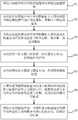
- FIG. 1is a schematic flowchart of an image recognition method based on CT sequence provided by an embodiment of this application;
- FIG. 2is a schematic diagram of a process for acquiring a tissue image sequence and a lesion image sequence of a target pathological tissue according to an embodiment of the application;
- FIG. 3is a schematic diagram of a process of inputting a tissue image sequence to a feature extraction model for feature extraction according to an embodiment of the application;
- FIG. 4is a schematic diagram of a process of generating a predicted image label according to an embodiment of the application
- FIG. 5is a schematic diagram of modules of a CT sequence-based image recognition device provided by an embodiment of this application.
- FIG. 6is a schematic diagram of the internal structure of an electronic device that implements a CT sequence-based image recognition method provided by an embodiment of the application;
- the execution subject of the CT sequence-based image recognition method provided in the embodiments of the present applicationincludes, but is not limited to, at least one of the electronic devices that can be configured to execute the method provided in the embodiments of the present application, such as a server and a terminal.
- the image recognition method based on the CT sequencemay be executed by software or hardware installed in the terminal device or the server device, and the software may be a blockchain platform.
- the serverincludes but is not limited to: a single server, a server cluster, a cloud server or a cloud server cluster, etc.
- This applicationprovides an image recognition method based on CT sequence.
- FIG. 1it is a schematic flowchart of a CT sequence-based image recognition method provided by an embodiment of this application.
- the methodcan be executed by a device, and the device can be implemented by software and/or hardware.
- the image recognition method based on CT sequenceincludes:
- the tissue image sequence and the lesion image sequence of the target pathological tissueare composed of multiple tissue images and multiple lesion images.
- the number of images in the tissue image sequence of the target pathological tissue and the lesion image sequencemay be the same or different.
- FIG. 2is a schematic flowchart of obtaining a tissue image sequence and a lesion image sequence of a target pathological tissue according to an embodiment of the application.
- the acquiring a tissue image sequence and a lesion image sequence of the target pathological tissueincludes:
- the embodiment of the present applicationuses a java sentence with a data capture function to capture the tissue segmentation grayscale image and the lesion segmentation grayscale image of the target pathological tissue from a pre-built database.
- the CT mask imageis an image that only contains 0 and 1 pixel values.
- the CT mask imageis multiplied by the tissue segmentation gray map to obtain multiple pieces of tissue that are completely black and only the tissue can be seen.
- the image ofis the organization image sequence. Multiply the CT mask image with the gray image of the lesion segmentation to obtain multiple black images outside the lesion where only the lesion can be seen, which is the lesion image sequence.
- the methodbefore the acquiring the tissue image sequence and the lesion image sequence of the target pathological tissue, the method further includes:
- the operation of acquiring the tissue image sequence and the lesion image sequence of the target pathological tissueis performed.
- the judging whether the user is an authorized user according to the unique identifier and password of the userincludes:
- the unique identifier and password of the usermatch the pre-stored information, determine that the user is an authorized user, and execute the operation of acquiring the tissue image sequence and the lesion image sequence of the target pathological tissue;
- the userWhen the unique identifier and password of the user do not match the pre-stored information, the user is determined to be an unauthorized user, and the operation of acquiring the tissue image sequence and the lesion image sequence of the target pathological tissue is not performed.
- the preferred embodiment of the present applicationis before acquiring the tissue image sequence and the lesion image sequence of the target pathological tissue. The verification of the user's identity improves the security of the tissue image sequence and the lesion image sequence of the target pathological tissue.
- FIG. 3is a schematic diagram of a process of inputting a tissue image sequence to a feature extraction model for feature extraction according to an embodiment of the application.
- inputting all the images in the tissue image sequence to a pre-built feature extraction model for feature extraction to obtain a first feature image setincludes:
- the use of the feature extraction model to perform convolution processing on all images in the tissue image sequenceincludes: using a preset convolution kernel matrix in the feature extraction model and each image sequence in the tissue image sequence. The pixels of the image are multiplied.
- performing pooling processing on the first convolutional image setincludes, but is not limited to, maximum pooling, minimum pooling, and average pooling.
- inputting all the images in the lesion image sequence to a pre-built feature extraction model for feature extraction to obtain the second feature image setincludes:
- Poolingis performed on the second convolutional image set to obtain a second characteristic image set.
- the tissue image sequence and the lesion image sequenceare respectively used to train the model, which enhances the diversity of training data and improves the accuracy of image recognition by the target image recognition model.
- the sizes of the first feature map and the second feature mapare the same.
- the first feature map and the second feature mapare geometrically spliced, for example, the first feature map and the second feature map are spliced together according to edges of the same length, and the lesion feature map is obtained after the splicing is completed.
- the predicted image labelis an image recognition model that recognizes the type of the lesion in the lesion feature map.
- FIG. 4is a schematic diagram of a process of generating a predicted image label according to an embodiment of the application.
- the performing image recognition on the lesion feature map to obtain a predicted image labelincludes:
- S50Calculate the probability value that the label of the lesion feature map is a preset label by using an activation function
- the activation functionincludes, but is not limited to, a sigmoid activation function.
- the activation functioncan be used to calculate the probability value that the label of the lesion feature map is a preset label.
- the predicted image label corresponding to the lesion feature mapis generated according to the probability value. For example, when the probability value is "X”, the predicted image label "The probability that the lesion feature map is the target lesion is X" is generated for the lesion feature map.
- the calculating the loss value between the predicted image label and the preset target pathological label of the target pathological tissueincludes:
- Mis the number of predicted image labels
- Nis the number of preset target pathological labels of the target pathological tissue
- y icis the sign indicator variable
- Wis the preset weight coefficient
- p icis the i-th lesion feature map is The probability of the target pathological label c is preset.
- the embodiment of the present applicationuses a gradient descent algorithm to update the parameters of the feature extraction model. If the loss value between the predicted image label and the preset target pathological label of the target pathological tissue is less than or equal to a preset error, a target image recognition model is obtained.
- the gradient descent algorithmincludes, but is not limited to: a large-batch gradient descent algorithm, a small-batch gradient descent algorithm, and a stochastic gradient descent algorithm.
- a python sentence with a data capture functionis used to obtain the image sequence to be identified from the blockchain node for storing the CT sequence.
- Using the high data throughput of the blockchaincan improve the efficiency of obtaining the image sequence to be identified.
- the target image recognition modelis used to perform image recognition on the image sequence to be recognized, and the recognition result is obtained.
- the recognition resultis the type of the lesion in the image in the image sequence to be recognized.
- the methodfurther includes:
- the push queue taskis implemented by subscriber notification message queue (MQ).
- MQsubscriber notification message queue
- multiple recognition results that need to be pushedare processed in batches, so as to ensure that the previous batch of recognition results is pushed and the next batch of recognition is continued to be processed. result.
- the queue of notification messages from the subscriberscan reduce the occupation of computing resources, cut a large amount of data and push them in batches, and avoid the occupation and waste of computing resources due to data congestion.
- the embodiment of the applicationobtains the tissue image sequence and the lesion image sequence of the target pathological tissue, and uses all the images in the tissue image sequence and the lesion image sequence as training data to train the feature extraction model to obtain the first feature map and the second feature Figure, stitching the first feature map and the second feature map into a lesion feature map and calculating the loss value of the lesion feature map, updating the feature extraction model according to the loss value to obtain the target image recognition model, using the tissue image sequence and the lesion image respectively
- the sequencetrains the model, which enhances the diversity of training data and improves the accuracy of image recognition by the target image recognition model; obtains the image sequence to be recognized, uses the target image recognition model to perform image recognition on the image sequence to be recognized, and obtains the recognition result. There is no need to manually detect and recognize images one by one, which improves the efficiency of image recognition. Therefore, the image recognition method based on CT sequence proposed in this application can improve the efficiency and accuracy of image recognition.
- FIG. 5it is a schematic diagram of the modules of the image recognition device based on the CT sequence of the present application.
- the CT sequence-based image recognition apparatus 100 described in this applicationcan be installed in an electronic device.
- the CT sequence-based image recognition devicemay include an image acquisition module 101, a first feature extraction module 102, a second feature extraction module 103, a feature splicing module 104, a label prediction module 105, a model update module 106, and Image recognition module 107.
- the module described in the present inventioncan also be called a unit, which refers to a series of computer program segments that can be executed by the processor of an electronic device and can complete fixed functions, and are stored in the memory of the electronic device.
- each module/unitis as follows:
- the image acquisition module 101is used to acquire a tissue image sequence and a lesion image sequence of a target pathological tissue;
- the first feature extraction module 102is configured to input all the images in the tissue image sequence to a pre-built feature extraction model for feature extraction to obtain a first feature image set;
- the second feature extraction module 103is configured to input all images in the lesion image sequence to the pre-built feature extraction model for feature extraction to obtain a second feature image set;
- the feature splicing module 104is configured to splice the first feature map and the second feature map to obtain a lesion feature map;
- the label prediction module 105is configured to perform image recognition on the lesion feature map to obtain a predicted image label
- the model update module 106is used to calculate the loss value between the predicted image label and the preset target pathological label of the target pathological tissue, and update the feature extraction model according to the loss value to obtain the target image Recognition model
- the image recognition module 107is configured to obtain an image sequence to be recognized, and use the target image recognition model to perform image recognition on the image sequence to be recognized to obtain a recognition result.
- each module of the CT sequence-based image recognition deviceis as follows:
- the image acquisition module 101is used to acquire a tissue image sequence and a lesion image sequence of a target pathological tissue.
- the tissue image sequence and the lesion image sequence of the target pathological tissueare composed of multiple tissue images and multiple lesion images.
- the number of images in the tissue image sequence of the target pathological tissue and the lesion image sequencemay be the same or different.
- the image acquisition module 101is specifically used for:
- the CT mask imageis multiplied by the lesion segmentation gray image to obtain a lesion image sequence.
- the embodiment of the present applicationuses a java sentence with a data capture function to capture the tissue segmentation grayscale image and the lesion segmentation grayscale image of the target pathological tissue from a pre-built database.
- the CT mask imageis an image that only contains 0 and 1 pixel values.
- the CT mask imageis multiplied by the tissue segmentation gray map to obtain multiple pieces of tissue that are completely black and only the tissue can be seen.
- the image ofis the organization image sequence. Multiply the CT mask image with the gray image of the lesion segmentation to obtain multiple black images outside the lesion where only the lesion can be seen, which is the lesion image sequence.
- the CT sequence-based image recognition devicefurther includes an identity verification module, and the identity verification module is specifically configured to:
- the operation of acquiring the tissue image sequence and the lesion image sequence of the target pathological tissueis performed.
- the judging whether the user is an authorized user according to the unique identifier and password of the userincludes:
- the unique identifier and password of the usermatch the pre-stored information, determine that the user is an authorized user, and execute the operation of acquiring the tissue image sequence and the lesion image sequence of the target pathological tissue;
- the userWhen the unique identifier and password of the user do not match the pre-stored information, the user is determined to be an unauthorized user, and the operation of acquiring the tissue image sequence and the lesion image sequence of the target pathological tissue is not performed.
- the preferred embodiment of the present applicationis before acquiring the tissue image sequence and the lesion image sequence of the target pathological tissue. The verification of the user's identity improves the security of the tissue image sequence and the lesion image sequence of the target pathological tissue.
- the first feature extraction module 102is configured to input all the images in the tissue image sequence to a pre-built feature extraction model for feature extraction to obtain a first feature image set.
- the first feature extraction module 102is specifically configured to:
- Pooling processingis performed on the first convolutional image set to obtain a first characteristic image set.
- the use of the feature extraction model to perform convolution processing on all images in the tissue image sequenceincludes: using a preset convolution kernel matrix in the feature extraction model and each image sequence in the tissue image sequence. The pixels of the image are multiplied.
- performing pooling processing on the first convolutional image setincludes, but is not limited to, maximum pooling, minimum pooling, and average pooling.
- the second feature extraction module 103is configured to input all images in the lesion image sequence to the pre-built feature extraction model for feature extraction to obtain a second feature image set.
- the second feature extraction module 103is specifically configured to:
- Poolingis performed on the second convolutional image set to obtain a second characteristic image set.
- the tissue image sequence and the lesion image sequenceare respectively used to train the model, which enhances the diversity of training data and improves the accuracy of image recognition by the target image recognition model.
- the feature splicing module 104is configured to splice the first feature map and the second feature map to obtain a lesion feature map.
- the sizes of the first feature map and the second feature mapare the same.
- the first feature map and the second feature mapare geometrically spliced, for example, the first feature map and the second feature map are spliced together according to edges of the same length, and the lesion feature map is obtained after the splicing is completed.
- the label prediction module 105is configured to perform image recognition on the lesion feature map to obtain a predicted image label.
- the predicted image labelis an image recognition model that recognizes the type of the lesion in the lesion feature map.
- the label prediction module 105is specifically configured to:
- the predicted image label corresponding to the lesion feature mapis generated according to the probability value.
- the activation functionincludes, but is not limited to, a sigmoid activation function.
- the activation functioncan be used to calculate the probability value that the label of the lesion feature map is a preset label.
- the predicted image label corresponding to the lesion feature mapis generated according to the probability value. For example, when the probability value is "X”, the predicted image label "The probability that the lesion feature map is the target lesion is X" is generated for the lesion feature map.
- the model update module 106is used to calculate the loss value between the predicted image label and the preset target pathological label of the target pathological tissue, and update the feature extraction model according to the loss value to obtain the target image Identify the model.
- model update module 106is specifically configured to:
- Mis the number of predicted image labels
- Nis the number of preset target pathological labels of the target pathological tissue
- y icis the sign indicator variable
- Wis the preset weight coefficient
- p icis the i-th lesion feature map is The probability of the target pathological label c is preset.
- the embodiment of the present applicationuses a gradient descent algorithm to update the parameters of the feature extraction model. If the loss value between the predicted image label and the preset target pathological label of the target pathological tissue is less than or equal to a preset error, a target image recognition model is obtained.
- the gradient descent algorithmincludes, but is not limited to: a large-batch gradient descent algorithm, a small-batch gradient descent algorithm, and a stochastic gradient descent algorithm.
- the image recognition module 107is configured to obtain an image sequence to be recognized, and use the target image recognition model to perform image recognition on the image sequence to be recognized to obtain a recognition result.
- a python sentence with a data capture functionis used to obtain the image sequence to be identified from the blockchain node for storing the CT sequence.
- Using the high data throughput of the blockchaincan improve the efficiency of obtaining the image sequence to be identified.
- the target image recognition modelis used to perform image recognition on the image sequence to be recognized, and the recognition result is obtained.
- the recognition resultis the type of the lesion in the image in the image sequence to be recognized.
- the CT sequence-based image recognition devicefurther includes a push module, and the push module is specifically configured to:
- the push queue taskis implemented by subscriber notification message queue (MQ).
- MQsubscriber notification message queue
- multiple recognition results that need to be pushedare processed in batches, so as to ensure that the previous batch of recognition results is pushed and the next batch of recognition is continued to be processed. result.
- the queue of notification messages from the subscriberscan reduce the occupation of computing resources, cut a large amount of data and push them in batches, and avoid the occupation and waste of computing resources due to data congestion.
- the embodiment of the applicationobtains the tissue image sequence and the lesion image sequence of the target pathological tissue, and uses all the images in the tissue image sequence and the lesion image sequence as training data to train the feature extraction model to obtain the first feature map and the second feature Figure, splicing the first feature map and the second feature map into a lesion feature map and calculating the loss value of the lesion feature map, updating the feature extraction model according to the loss value to obtain the target image recognition model, using the tissue image sequence and the lesion image respectively
- the sequencetrains the model, which enhances the diversity of training data and improves the accuracy of image recognition by the target image recognition model; obtains the image sequence to be recognized, uses the target image recognition model to perform image recognition on the image sequence to be recognized, and obtains the recognition result.
- the manual recognition of the imageis avoided, and the efficiency of image recognition is improved. Therefore, the image recognition method based on CT sequence proposed in this application can improve the efficiency and accuracy of image recognition.
- FIG. 6it is a schematic diagram of the structure of an electronic device that implements the image recognition method based on the CT sequence in this application.
- the electronic device 1may include a processor 10, a memory 11, and a bus, and may also include a computer program stored in the memory 11 and running on the processor 10, such as an image recognition program 12 based on a CT sequence.
- the memory 11includes at least one type of readable storage medium, and the readable storage medium includes flash memory, mobile hard disk, multimedia card, card-type memory (such as SD or DX memory, etc.), magnetic memory, magnetic disk, CD etc.
- the memory 11may be an internal storage unit of the electronic device 1 in some embodiments, for example, a mobile hard disk of the electronic device 1.
- the memory 11may also be an external storage device of the electronic device 1, such as a plug-in mobile hard disk, a smart media card (SMC), and a secure digital (Secure Digital) equipped on the electronic device 1. , SD) card, flash card (Flash Card), etc.
- the memory 11may also include both an internal storage unit of the electronic device 1 and an external storage device.
- the memory 11can be used not only to store application software and various data installed in the electronic device 1, such as the code of the image recognition program 12 based on the CT sequence, etc., but also to temporarily store data that has been output or will be output.
- the processor 10may be composed of integrated circuits in some embodiments, for example, may be composed of a single packaged integrated circuit, or may be composed of multiple integrated circuits with the same function or different functions, including one or more Combinations of central processing unit (CPU), microprocessor, digital processing chip, graphics processor, and various control chips, etc.
- the processor 10is the control unit of the electronic device, which uses various interfaces and lines to connect the various components of the entire electronic device, and runs or executes programs or modules stored in the memory 11 (such as executing Image recognition programs based on the CT sequence, etc.), and call data stored in the memory 11 to execute various functions of the electronic device 1 and process data.
- the busmay be a peripheral component interconnect standard (PCI) bus or an extended industry standard architecture (EISA) bus, etc.
- PCIperipheral component interconnect standard
- EISAextended industry standard architecture
- the buscan be divided into address bus, data bus, control bus and so on.
- the busis configured to implement connection and communication between the memory 11 and at least one processor 10 and the like.
- FIG. 6only shows an electronic device with components. Those skilled in the art can understand that the structure shown in FIG. 6 does not constitute a limitation on the electronic device 1, and may include fewer or more components than shown in the figure. Components, or a combination of certain components, or different component arrangements.
- the electronic device 1may also include a power source (such as a battery) for supplying power to various components.
- the power sourcemay be logically connected to the at least one processor 10 through a power management device, thereby controlling power
- the deviceimplements functions such as charge management, discharge management, and power consumption management.
- the power supplymay also include any components such as one or more DC or AC power supplies, recharging devices, power failure detection circuits, power converters or inverters, and power status indicators.
- the electronic device 1may also include various sensors, Bluetooth modules, Wi-Fi modules, etc., which will not be repeated here.
- the electronic device 1may also include a network interface.
- the network interfacemay include a wired interface and/or a wireless interface (such as a WI-FI interface, a Bluetooth interface, etc.), which is usually used in the electronic device 1 Establish a communication connection with other electronic devices.
- the electronic device 1may also include a user interface.
- the user interfacemay be a display (Display) and an input unit (such as a keyboard (Keyboard)).
- the user interfacemay also be a standard wired interface or a wireless interface.
- the displaymay be an LED display, a liquid crystal display, a touch-sensitive liquid crystal display, an OLED (Organic Light-Emitting Diode, organic light-emitting diode) touch device, etc.
- the displaycan also be appropriately called a display screen or a display unit, which is used to display the information processed in the electronic device 1 and to display a visualized user interface.
- the CT sequence-based image recognition program 12 stored in the memory 11 in the electronic device 1is a combination of multiple computer programs. When running in the processor 10, it can realize:
- the image sequence to be recognizedis acquired, and the target image recognition model is used to perform image recognition on the image sequence to be recognized to obtain a recognition result.
- the integrated module/unit of the electronic device 1can be stored in a computer readable storage medium. It can be non-volatile or volatile.
- the computer-readable mediummay include: any entity or device capable of carrying the computer program code, recording medium, U disk, mobile hard disk, magnetic disk, optical disk, computer memory, read-only memory (ROM, Read-Only Memory) .
- the computer usable storage mediummay mainly include a storage program area and a storage data area, wherein the storage program area may store an operating system, an application program required by at least one function, etc.; the storage data area may store a block chain node Use the created data, etc.
- modules described as separate componentsmay or may not be physically separated, and the components displayed as modules may or may not be physical units, that is, they may be located in one place, or they may be distributed on multiple network units. Some or all of the modules can be selected according to actual needs to achieve the objectives of the solutions of the embodiments.
- the functional modules in the various embodiments of the present applicationmay be integrated into one processing unit, or each unit may exist alone physically, or two or more units may be integrated into one unit.
- the above-mentioned integrated unitmay be implemented in the form of hardware, or may be implemented in the form of hardware plus software functional modules.
- the blockchain referred to in this applicationis a new application mode of computer technology such as distributed data storage, point-to-point transmission, consensus mechanism, and encryption algorithm.
- Blockchainessentially a decentralized database, is a series of data blocks associated with cryptographic methods. Each data block contains a batch of network transaction information for verification. The validity of the information (anti-counterfeiting) and the generation of the next block.
- the blockchaincan include the underlying platform of the blockchain, the platform product service layer, and the application service layer.
Landscapes
- Engineering & Computer Science (AREA)
- Theoretical Computer Science (AREA)
- Physics & Mathematics (AREA)
- General Physics & Mathematics (AREA)
- Data Mining & Analysis (AREA)
- General Engineering & Computer Science (AREA)
- Computer Vision & Pattern Recognition (AREA)
- General Health & Medical Sciences (AREA)
- Evolutionary Computation (AREA)
- Computer Security & Cryptography (AREA)
- Software Systems (AREA)
- Artificial Intelligence (AREA)
- Life Sciences & Earth Sciences (AREA)
- Health & Medical Sciences (AREA)
- Computer Hardware Design (AREA)
- Evolutionary Biology (AREA)
- Computing Systems (AREA)
- Bioinformatics & Cheminformatics (AREA)
- Bioethics (AREA)
- Bioinformatics & Computational Biology (AREA)
- Databases & Information Systems (AREA)
- Biophysics (AREA)
- Computational Linguistics (AREA)
- Molecular Biology (AREA)
- Mathematical Physics (AREA)
- Biomedical Technology (AREA)
- Apparatus For Radiation Diagnosis (AREA)
Abstract
Description
Translated fromChinese本申请要求于2020年9月22日提交中国专利局、申请号为202011001690.4,发明名称为“基于CT序列的图像识别方法、装置、电子设备及介质”的中国专利申请的优先权,其全部内容通过引用结合在本申请中。This application claims the priority of a Chinese patent application filed with the Chinese Patent Office on September 22, 2020, the application number is 202011001690.4, and the invention title is "CT Sequence-based Image Recognition Method, Apparatus, Electronic Equipment and Medium", and its entire content Incorporated in this application by reference.
本申请涉及机器学习技术领域,尤其涉及一种基于CT序列的图像识别方法、装置、电子设备及计算机可读存储介质。This application relates to the field of machine learning technology, and in particular to a CT sequence-based image recognition method, device, electronic equipment, and computer-readable storage medium.
2020年初新冠疫情突袭武汉,在二月份武汉医疗及检测资源极度紧张,且核酸检测的敏感度较低等情况下,为“早发现,早隔离”,使用CT影像诊断新冠肺炎被提出。In early 2020, the new crown epidemic raided Wuhan. In February, when Wuhan's medical and testing resources were extremely tight, and the sensitivity of nucleic acid testing was low, it was proposed to use CT imaging to diagnose new coronary pneumonia for "early detection and early isolation".
现有技术中,对病人的CT影像进行新冠检测时,需要利用整个CT的所有序列。对于一个CT序列,有薄层厚层之分,厚层五六十张图像,薄层多则达五百张图像,发明人意识到即使相同厚度的CT每个病人的层数也不相同,医生会耗费至少二三十分钟才能完成一次诊断,诊断效率不能满足实际情况的实时性。同同时,大量的时间也消耗医生大量的精力,容易出现漏诊误诊的情况,从而导致诊断的效率和准确度都不高。因此,如何提高利用CT鉴别新冠肺炎及其他肺炎的效率和精确度,成为了亟待解决的问题。In the prior art, when performing a new crown detection on a CT image of a patient, it is necessary to use all the sequences of the entire CT. For a CT sequence, there are thin and thick layers. There are fifty or sixty images in thick layers, and there are as many as 500 images in thin layers. The inventor realized that even the same thickness of CT has different layers for each patient. The doctor will spend at least twenty to thirty minutes to complete a diagnosis, and the diagnosis efficiency cannot meet the real-time nature of the actual situation. At the same time, a lot of time also consumes a lot of energy from doctors, and it is easy to miss diagnosis and misdiagnosis, which leads to low diagnosis efficiency and accuracy. Therefore, how to improve the efficiency and accuracy of using CT to identify new coronary pneumonia and other pneumonia has become an urgent problem to be solved.
发明内容Summary of the invention
本申请提供的一种基于CT序列的图像识别方法,包括:An image recognition method based on CT sequence provided by this application includes:
获取目标病理组织的组织图像序列和病灶图像序列;Obtain the tissue image sequence and the lesion image sequence of the target pathological tissue;
将所述组织图像序列中所有图像输入至预先构建的特征提取模型进行特征提取,得到第一特征图像集;Input all images in the tissue image sequence to a pre-built feature extraction model for feature extraction to obtain a first feature image set;
将所述病灶图像序列中所有图像输入至所述预先构建的特征提取模型进行特征提取,得到第二特征图像集;Input all images in the lesion image sequence to the pre-built feature extraction model for feature extraction to obtain a second feature image set;
将所述第一特征图与所述第二特征图进行拼接,得到病灶特征图;Splicing the first feature map and the second feature map to obtain a lesion feature map;
对所述病灶特征图进行图像识别,得到预测图像标签;Performing image recognition on the lesion feature map to obtain a predicted image label;
计算所述预测图像标签和所述目标病理组织的预置目标病理标签之间的损失值,根据所述损失值对所述特征提取模型进行更新,得到目标图像识别模型;Calculating a loss value between the predicted image label and the preset target pathological label of the target pathological tissue, and updating the feature extraction model according to the loss value to obtain a target image recognition model;
获取待识别图像序列,利用所述目标图像识别模型对所述待识别图像序列进行图像识别,得到识别结果。The image sequence to be recognized is acquired, and the target image recognition model is used to perform image recognition on the image sequence to be recognized to obtain a recognition result.
一种基于CT序列的图像识别装置,所述装置包括:An image recognition device based on CT sequence, said device comprising:
图像获取模块,用于获取目标病理组织的组织图像序列和病灶图像序列;The image acquisition module is used to acquire the tissue image sequence and the lesion image sequence of the target pathological tissue;
第一特征提取模块,用于将所述组织图像序列中所有图像输入至预先构建的特征提取模型进行特征提取,得到第一特征图像集;The first feature extraction module is configured to input all the images in the tissue image sequence to a pre-built feature extraction model for feature extraction to obtain a first feature image set;
第二特征提取模块,用于将所述病灶图像序列中所有图像输入至所述预先构建的特征提取模型进行特征提取,得到第二特征图像集;The second feature extraction module is configured to input all images in the lesion image sequence to the pre-built feature extraction model for feature extraction to obtain a second feature image set;
特征拼接模块,用于将所述第一特征图与所述第二特征图进行拼接,得到病灶特征图;The feature splicing module is used to splice the first feature map and the second feature map to obtain a lesion feature map;
标签预测模块,用于对所述病灶特征图进行图像识别,得到预测图像标签;The label prediction module is used to perform image recognition on the lesion feature map to obtain a predicted image label;
模型更新模块,用于计算所述预测图像标签和所述目标病理组织的预置目标病理标签之间的损失值,根据所述损失值对所述特征提取模型进行更新,得到目标图像识别模型;A model update module is used to calculate a loss value between the predicted image label and the preset target pathological label of the target pathological tissue, and update the feature extraction model according to the loss value to obtain a target image recognition model;
图像识别模块,用于获取待识别图像序列,利用所述目标图像识别模型对所述待识别图像序列进行图像识别,得到识别结果。The image recognition module is used to obtain the image sequence to be recognized, and use the target image recognition model to perform image recognition on the image sequence to be recognized to obtain the recognition result.
一种电子设备,所述电子设备包括:An electronic device, which includes:
至少一个处理器;以及,At least one processor; and,
与所述至少一个处理器通信连接的存储器;其中,A memory communicatively connected with the at least one processor; wherein,
所述存储器存储有可被所述至少一个处理器执行的计算机程序,所述计算机程序被所述至少一个处理器执行,以使所述至少一个处理器能够执行如下步骤:The memory stores a computer program executable by the at least one processor, and the computer program is executed by the at least one processor, so that the at least one processor can execute the following steps:
获取目标病理组织的组织图像序列和病灶图像序列;Obtain the tissue image sequence and the lesion image sequence of the target pathological tissue;
将所述组织图像序列中所有图像输入至预先构建的特征提取模型进行特征提取,得到第一特征图像集;Input all images in the tissue image sequence to a pre-built feature extraction model for feature extraction to obtain a first feature image set;
将所述病灶图像序列中所有图像输入至所述预先构建的特征提取模型进行特征提取,得到第二特征图像集;Input all images in the lesion image sequence to the pre-built feature extraction model for feature extraction to obtain a second feature image set;
将所述第一特征图与所述第二特征图进行拼接,得到病灶特征图;Splicing the first feature map and the second feature map to obtain a lesion feature map;
对所述病灶特征图进行图像识别,得到预测图像标签;Performing image recognition on the lesion feature map to obtain a predicted image label;
计算所述预测图像标签和所述目标病理组织的预置目标病理标签之间的损失值,根据所述损失值对所述特征提取模型进行更新,得到目标图像识别模型;Calculating a loss value between the predicted image label and the preset target pathological label of the target pathological tissue, and updating the feature extraction model according to the loss value to obtain a target image recognition model;
获取待识别图像序列,利用所述目标图像识别模型对所述待识别图像序列进行图像识别,得到识别结果。The image sequence to be recognized is acquired, and the target image recognition model is used to perform image recognition on the image sequence to be recognized to obtain a recognition result.
一种计算机可读存储介质,包括存储数据区和存储程序区,其中,所述存储数据区存储创建的数据,所述存储程序区存储有计算机程序;其中,所述计算机程序被处理器执行时实现如下步骤:A computer-readable storage medium includes a storage data area and a storage program area, wherein the storage data area stores created data, and the storage program area stores a computer program; wherein, when the computer program is executed by a processor To achieve the following steps:
获取目标病理组织的组织图像序列和病灶图像序列;Obtain the tissue image sequence and the lesion image sequence of the target pathological tissue;
将所述组织图像序列中所有图像输入至预先构建的特征提取模型进行特征提取,得到第一特征图像集;Input all images in the tissue image sequence to a pre-built feature extraction model for feature extraction to obtain a first feature image set;
将所述病灶图像序列中所有图像输入至所述预先构建的特征提取模型进行特征提取,得到第二特征图像集;Input all images in the lesion image sequence to the pre-built feature extraction model for feature extraction to obtain a second feature image set;
将所述第一特征图与所述第二特征图进行拼接,得到病灶特征图;Splicing the first feature map and the second feature map to obtain a lesion feature map;
对所述病灶特征图进行图像识别,得到预测图像标签;Performing image recognition on the lesion feature map to obtain a predicted image label;
计算所述预测图像标签和所述目标病理组织的预置目标病理标签之间的损失值,根据所述损失值对所述特征提取模型进行更新,得到目标图像识别模型;Calculating a loss value between the predicted image label and the preset target pathological label of the target pathological tissue, and updating the feature extraction model according to the loss value to obtain a target image recognition model;
获取待识别图像序列,利用所述目标图像识别模型对所述待识别图像序列进行图像识别,得到识别结果。The image sequence to be recognized is acquired, and the target image recognition model is used to perform image recognition on the image sequence to be recognized to obtain a recognition result.
图1为本申请一实施例提供的基于CT序列的图像识别方法的流程示意图;FIG. 1 is a schematic flowchart of an image recognition method based on CT sequence provided by an embodiment of this application;
图2为本申请一实施例提供的获取目标病理组织的组织图像序列和病灶图像序列的流程示意图;2 is a schematic diagram of a process for acquiring a tissue image sequence and a lesion image sequence of a target pathological tissue according to an embodiment of the application;
图3为本申请一实施例提供的将组织图像序列输入至特征提取模型进行特征提取的流程示意图;FIG. 3 is a schematic diagram of a process of inputting a tissue image sequence to a feature extraction model for feature extraction according to an embodiment of the application;
图4为本申请一实施例提供的生成预测图像标签的流程示意图;FIG. 4 is a schematic diagram of a process of generating a predicted image label according to an embodiment of the application;
图5为本申请一实施例提供的基于CT序列的图像识别装置的模块示意图;5 is a schematic diagram of modules of a CT sequence-based image recognition device provided by an embodiment of this application;
图6为本申请一实施例提供的实现基于CT序列的图像识别方法的电子设备的内部结构示意图;6 is a schematic diagram of the internal structure of an electronic device that implements a CT sequence-based image recognition method provided by an embodiment of the application;
本申请目的的实现、功能特点及优点将结合实施例,参照附图做进一步说明。The realization, functional characteristics, and advantages of the purpose of this application will be further described in conjunction with the embodiments and with reference to the accompanying drawings.
应当理解,此处所描述的具体实施例仅仅用以解释本申请,并不用于限定本申请。It should be understood that the specific embodiments described here are only used to explain the present application, and are not used to limit the present application.
本申请实施例提供的基于CT序列的图像识别方法的执行主体包括但不限于服务端、 终端等能够被配置为执行本申请实施例提供的该方法的电子设备中的至少一种。换言之,所述基于CT序列的图像识别方法可以由安装在终端设备或服务端设备的软件或硬件来执行,所述软件可以是区块链平台。所述服务端包括但不限于:单台服务器、服务器集群、云端服务器或云端服务器集群等。The execution subject of the CT sequence-based image recognition method provided in the embodiments of the present application includes, but is not limited to, at least one of the electronic devices that can be configured to execute the method provided in the embodiments of the present application, such as a server and a terminal. In other words, the image recognition method based on the CT sequence may be executed by software or hardware installed in the terminal device or the server device, and the software may be a blockchain platform. The server includes but is not limited to: a single server, a server cluster, a cloud server or a cloud server cluster, etc.
本申请提供一种基于CT序列的图像识别方法。参照图1所示,为本申请一实施例提供的基于CT序列的图像识别方法的流程示意图。该方法可以由一个装置执行,该装置可以由软件和/或硬件实现。This application provides an image recognition method based on CT sequence. Referring to FIG. 1, it is a schematic flowchart of a CT sequence-based image recognition method provided by an embodiment of this application. The method can be executed by a device, and the device can be implemented by software and/or hardware.
在本实施例中,基于CT序列的图像识别方法包括:In this embodiment, the image recognition method based on CT sequence includes:
S1、获取目标病理组织的组织图像序列和病灶图像序列。S1. Acquire a tissue image sequence and a lesion image sequence of the target pathological tissue.
本申请实施例中,所述目标病理组织的组织图像序列和病灶图像序列由多张组织图像和多张病灶图像组成。所述目标病理组织的组织图像序列和病灶图像序列中图像数量可以相同也可以不同。In the embodiment of the present application, the tissue image sequence and the lesion image sequence of the target pathological tissue are composed of multiple tissue images and multiple lesion images. The number of images in the tissue image sequence of the target pathological tissue and the lesion image sequence may be the same or different.
图2为本申请一实施例提供的获取目标病理组织的组织图像序列和病灶图像序列的流程示意图。FIG. 2 is a schematic flowchart of obtaining a tissue image sequence and a lesion image sequence of a target pathological tissue according to an embodiment of the application.
详细地,参见图2所示,所述获取目标病理组织的组织图像序列和病灶图像序列,包括:In detail, referring to FIG. 2, the acquiring a tissue image sequence and a lesion image sequence of the target pathological tissue includes:
S10、获取目标病理组织的组织分割灰度图和病灶分割灰度图;S10. Obtain a grayscale image of tissue segmentation and a grayscale image of lesion segmentation of the target pathological tissue;
S11、利用预设的CT掩膜图像与所述组织分割灰度图相乘,得到组织图像序列;S11. Multiply the preset CT mask image and the tissue segmentation gray image to obtain a tissue image sequence;
S12、利用所述CT掩膜图像与所述病灶分割灰度图相乘,得到病灶图像序列。S12. Multiply the CT mask image and the lesion segmentation grayscale image to obtain a lesion image sequence.
具体地,本申请实施例利用具有数据抓取功能的java语句从预先构建的数据库中抓取目标病理组织的组织分割灰度图和病灶分割灰度图。Specifically, the embodiment of the present application uses a java sentence with a data capture function to capture the tissue segmentation grayscale image and the lesion segmentation grayscale image of the target pathological tissue from a pre-built database.
本申请实施例中,所述CT掩膜图像为仅包含0、1像素值的图像,利用CT掩膜图像与组织分割灰度图相乘,得到多张组织外全黑的只能看到组织的图像,即为组织图像序列。利用CT掩膜图像与病灶分割灰度图相乘,得到多张病灶外全黑的只能看到病灶的图像,即为病灶图像序列。In the embodiment of the present application, the CT mask image is an image that only contains 0 and 1 pixel values. The CT mask image is multiplied by the tissue segmentation gray map to obtain multiple pieces of tissue that are completely black and only the tissue can be seen. The image of is the organization image sequence. Multiply the CT mask image with the gray image of the lesion segmentation to obtain multiple black images outside the lesion where only the lesion can be seen, which is the lesion image sequence.
本申请一可选实施例中,所述获取目标病理组织的组织图像序列和病灶图像序列之前,所述方法还包括:In an optional embodiment of the present application, before the acquiring the tissue image sequence and the lesion image sequence of the target pathological tissue, the method further includes:
接收识别所述目标病理组织的病灶的的图像识别请求;Receiving an image recognition request for recognizing the lesion of the target pathological tissue;
提取所述图像识别请求中的用户信息,所述用户信息包括用户的唯一标识和密码;Extracting user information in the image recognition request, where the user information includes the user's unique identifier and password;
根据所述用户的唯一标识和密码判断所述用户是否为授权用户;Judging whether the user is an authorized user according to the unique identifier and password of the user;
若所述用户为授权用户,执行所述获取目标病理组织的组织图像序列和病灶图像序列的操作。If the user is an authorized user, the operation of acquiring the tissue image sequence and the lesion image sequence of the target pathological tissue is performed.
详细地,所述根据所述用户的唯一标识和密码判断所述用户是否为授权用户,包括:In detail, the judging whether the user is an authorized user according to the unique identifier and password of the user includes:
在所述用户的唯一标识及密码与预先存储信息都匹配时,确定所述用户为授权用户,执行所述获取目标病理组织的组织图像序列和病灶图像序列的操作;When the unique identifier and password of the user match the pre-stored information, determine that the user is an authorized user, and execute the operation of acquiring the tissue image sequence and the lesion image sequence of the target pathological tissue;
在所述用户的唯一标识及密码与预先存储信息都不匹配时,确定所述用户为非授权用户,不执行所述获取目标病理组织的组织图像序列和病灶图像序列的操作。When the unique identifier and password of the user do not match the pre-stored information, the user is determined to be an unauthorized user, and the operation of acquiring the tissue image sequence and the lesion image sequence of the target pathological tissue is not performed.
由于目标病理组织的组织图像序列和病灶图像序列具有一定的隐私性,因此,通常这些数据的保密性需求较高,本申请较佳实施例在获取目标病理组织的组织图像序列和病灶图像序列之前对用户身份进行校验,提高了目标病理组织的组织图像序列和病灶图像序列的安全性。Since the tissue image sequence and the lesion image sequence of the target pathological tissue have a certain degree of privacy, the confidentiality of these data is usually required. The preferred embodiment of the present application is before acquiring the tissue image sequence and the lesion image sequence of the target pathological tissue The verification of the user's identity improves the security of the tissue image sequence and the lesion image sequence of the target pathological tissue.
S2、将所述组织图像序列中所有图像输入至预先构建的特征提取模型进行特征提取,得到第一特征图像集。S2. Input all the images in the tissue image sequence to a pre-built feature extraction model for feature extraction to obtain a first feature image set.
图3为本申请一实施例提供的将组织图像序列输入至特征提取模型进行特征提取的流程示意图。FIG. 3 is a schematic diagram of a process of inputting a tissue image sequence to a feature extraction model for feature extraction according to an embodiment of the application.
本申请实施例中,参图3所示,所述将所述组织图像序列中所有图像输入至预先构建的特征提取模型进行特征提取,得到第一特征图像集,包括:In the embodiment of the present application, as shown in FIG. 3, inputting all the images in the tissue image sequence to a pre-built feature extraction model for feature extraction to obtain a first feature image set includes:
S20、利用所述特征提取模型对所述组织图像序列中所有图像进行卷积处理,得到第一卷积图像集;S20. Use the feature extraction model to perform convolution processing on all images in the tissue image sequence to obtain a first convolution image set;
S21、对所述第一卷积图像集进行池化处理,得到第一特征图像集。S21. Perform pooling processing on the first convolutional image set to obtain a first feature image set.
详细地,本申请实施例中,所述利用所述特征提取模型对所述组织图像序列中所有图像进行卷积处理包括:利用特征提取模型中预设的卷积核矩阵与组织图像序列中每张图像的像素进行乘积。In detail, in the embodiment of the present application, the use of the feature extraction model to perform convolution processing on all images in the tissue image sequence includes: using a preset convolution kernel matrix in the feature extraction model and each image sequence in the tissue image sequence. The pixels of the image are multiplied.
具体地,对所述第一卷积图像集进行池化处理包括但不限于最大池化、最小池化和平均池化。Specifically, performing pooling processing on the first convolutional image set includes, but is not limited to, maximum pooling, minimum pooling, and average pooling.
S3、将所述病灶图像序列中所有图像输入至所述预先构建的特征提取模型进行特征提取,得到第二特征图像集。S3. Input all images in the lesion image sequence to the pre-built feature extraction model for feature extraction to obtain a second feature image set.
与得到第一特征图像集类似地,本申请实施例中,所述将所述病灶图像序列中所有图像输入至预先构建的特征提取模型进行特征提取,得到第二征图像集,包括:Similar to obtaining the first feature image set, in the embodiment of the present application, inputting all the images in the lesion image sequence to a pre-built feature extraction model for feature extraction to obtain the second feature image set includes:
利用所述特征提取模型对所述病灶图像序列中所有图像进行卷积处理,得到第二卷积图像集;Using the feature extraction model to perform convolution processing on all images in the lesion image sequence to obtain a second convolution image set;
对所述第二卷积图像集进行池化处理,得到第二特征图像集。Pooling is performed on the second convolutional image set to obtain a second characteristic image set.
本申请实施例中分别利用组织图像序列和病灶图像序列对模型进行训练,增强了训练数据的多样性,提高了目标图像识别模型对图像识别的精确度。In the embodiments of the present application, the tissue image sequence and the lesion image sequence are respectively used to train the model, which enhances the diversity of training data and improves the accuracy of image recognition by the target image recognition model.
S4、将所述第一特征图与所述第二特征图进行拼接,得到病灶特征图。S4. Splicing the first feature map and the second feature map to obtain a lesion feature map.
本申请实施例中,由于所述第一特征图与所述第二特征图为同一特征提取网络的输出,因此第一特征图与第二特征图的尺寸一致。In the embodiment of the present application, since the first feature map and the second feature map are outputs of the same feature extraction network, the sizes of the first feature map and the second feature map are the same.
本申请实施例将所述第一特征图与第二特征图进行几何拼接,例如,将第一特征图和第二特征图按照长度相同的边拼接在一起,拼接完成后得到病灶特征图。In the embodiment of the present application, the first feature map and the second feature map are geometrically spliced, for example, the first feature map and the second feature map are spliced together according to edges of the same length, and the lesion feature map is obtained after the splicing is completed.
S5、对所述病灶特征图进行图像识别,得到预测图像标签。S5. Perform image recognition on the lesion feature map to obtain a predicted image label.
本申请实施例中,所述预测图像标签是图像识别模型对病灶特征图中的病灶的类型进行识别。In the embodiment of the present application, the predicted image label is an image recognition model that recognizes the type of the lesion in the lesion feature map.
图4为本申请一实施例提供的生成预测图像标签的流程示意图。FIG. 4 is a schematic diagram of a process of generating a predicted image label according to an embodiment of the application.
具体的,参见图4所示,所述对所述病灶特征图进行图像识别,得到预测图像标签,包括:Specifically, referring to FIG. 4, the performing image recognition on the lesion feature map to obtain a predicted image label includes:
S50、利用激活函数计算所述病灶特征图的标签为预设标签的概率值;S50: Calculate the probability value that the label of the lesion feature map is a preset label by using an activation function;
S51、根据所述概率值生成所述病灶特征图对应的预测图像标签。S51. Generate a predicted image label corresponding to the lesion feature map according to the probability value.
详细地,所述激活函数包括但不限于sigmoid激活函数,利用激活函数可计算得到病灶特征图的标签为预设标签的概率值。In detail, the activation function includes, but is not limited to, a sigmoid activation function. The activation function can be used to calculate the probability value that the label of the lesion feature map is a preset label.
本申请实施例中,根据所述概率值生成所述病灶特征图对应的预测图像标签。例如,当概率值为“X”时,对所述病灶特征图生成预测图像标签“病灶特征图为目标病灶的概率为X”。In the embodiment of the present application, the predicted image label corresponding to the lesion feature map is generated according to the probability value. For example, when the probability value is "X", the predicted image label "The probability that the lesion feature map is the target lesion is X" is generated for the lesion feature map.
S6、计算所述预测图像标签和所述目标病理组织的预置目标病理标签之间的损失值,根据所述损失值对所述特征提取模型进行更新,得到目标图像识别模型。S6. Calculate a loss value between the predicted image label and the preset target pathological label of the target pathological tissue, and update the feature extraction model according to the loss value to obtain a target image recognition model.
本申请实施例中,所述计算所述预测图像标签和所述目标病理组织的预置目标病理标签之间的损失值,包括:In the embodiment of the present application, the calculating the loss value between the predicted image label and the preset target pathological label of the target pathological tissue includes:
利用如下损失函数计算所述预测图像标签和所述目标病理组织的预置目标病理标签之间的损失值Lcls:The following loss function is used to calculate the loss value L cls between the predicted image label and the preset target pathological label of the target pathological tissue:

其中,M为预测图像标签的数量,N为目标病理组织的预置目标病理标签的数量;yic是正负号指示变量,W为预设权重系数,pic为第i个病灶特征图为预置目标病理标签c的概率。Among them, M is the number of predicted image labels, N is the number of preset target pathological labels of the target pathological tissue; yic is the sign indicator variable, W is the preset weight coefficient, pic is the i-th lesion feature map is The probability of the target pathological label c is preset.
进一步地,若所述预测图像标签和所述目标病理组织的预置目标病理标签之间的损失值大于预设误差,本申请实施例利用梯度下降算法对所述特征提取模型的参数进行更新,若所述预测图像标签和所述目标病理组织的预置目标病理标签之间的损失值小于或等于预设误差,得到目标图像识别模型。Further, if the loss value between the predicted image label and the preset target pathological label of the target pathological tissue is greater than a preset error, the embodiment of the present application uses a gradient descent algorithm to update the parameters of the feature extraction model. If the loss value between the predicted image label and the preset target pathological label of the target pathological tissue is less than or equal to a preset error, a target image recognition model is obtained.
详细地,所述梯度下降算法包括但不限于:大批量梯度下降算法、小批量梯度下降算法、随机梯度下降算法。In detail, the gradient descent algorithm includes, but is not limited to: a large-batch gradient descent algorithm, a small-batch gradient descent algorithm, and a stochastic gradient descent algorithm.
S7、获取待识别图像序列,利用所述目标图像识别模型对所述待识别图像序列进行图像识别,得到识别结果。S7. Obtain an image sequence to be recognized, and perform image recognition on the image sequence to be recognized by using the target image recognition model to obtain a recognition result.
本申请实施例中,利用具有数据抓取功能的python语句从用于存储CT序列的区块链节点中获取待识别图像序列。利用区块链的数据高吞吐性,可提高获取待识别图像序列的效率。In the embodiment of the present application, a python sentence with a data capture function is used to obtain the image sequence to be identified from the blockchain node for storing the CT sequence. Using the high data throughput of the blockchain can improve the efficiency of obtaining the image sequence to be identified.
本申请实施例利用所述目标图像识别模型对所述待识别图像序列进行图像识别,得到识别结果。所述识别结果为待识别图像序列中的图像中的病灶类型。In the embodiment of the present application, the target image recognition model is used to perform image recognition on the image sequence to be recognized, and the recognition result is obtained. The recognition result is the type of the lesion in the image in the image sequence to be recognized.
本申请一较佳实施例中,所述得到识别结果之后,所述方法还包括:In a preferred embodiment of the present application, after the recognition result is obtained, the method further includes:
获取推送队列任务;Get the push queue task;
根据所述推送队列任务确定推送顺序;Determine the push sequence according to the push queue task;
根据所述推送顺序向用户推送所述识别结果。Push the recognition result to the user according to the push sequence.
实际应用中,需要对多个待识别图像序列进行图像识别,得到多个识别结果,在推送识别结果时,通过设置推送队列任务能够防止因同时对多个识别结果进行推送操作而造成的数据推送过程拥塞,提高了对多个识别结果进行推送处理的效率。In practical applications, it is necessary to perform image recognition on multiple image sequences to be recognized to obtain multiple recognition results. When pushing recognition results, setting a push queue task can prevent data push caused by pushing multiple recognition results at the same time. The process is congested, which improves the efficiency of pushing multiple recognition results.
优选地,所述推送队列任务采用订阅方通知消息列队(MQ)实现,具体的,通过分批处理多份需要推送的识别结果,从而确保前一批识别结果推送结束再继续处理后一批识别结果。Preferably, the push queue task is implemented by subscriber notification message queue (MQ). Specifically, multiple recognition results that need to be pushed are processed in batches, so as to ensure that the previous batch of recognition results is pushed and the next batch of recognition is continued to be processed. result.
通过订阅方通知消息列队可以降低计算资源占用,将大量的数据进行切割并分批进行推送,避免因为数据拥塞而导致计算资源的占用与浪费。The queue of notification messages from the subscribers can reduce the occupation of computing resources, cut a large amount of data and push them in batches, and avoid the occupation and waste of computing resources due to data congestion.
本申请实施例通过获取目标病理组织的组织图像序列和病灶图像序列,分别利用组织图像序列和病灶图像序列中的所有图像作为训练数据对特征提取模型进行训练,得到第一特征图和第二特征图,将第一特征图与第二特征图拼接为病灶特征图并计算病灶特征图的损失值,根据损失值对特征提取模型进行更新,得到目标图像识别模型,分别利用组织图像序列和病灶图像序列对模型进行训练,增强了训练数据的多样性,提高了目标图像识别模型对图像识别的精确度;获取待识别图像序列,利用目标图像识别模型对待识别图像序列进行图像识别,得到识别结果,无需人工对图像进行一一检测识别,提高了图像识别的效率。因此本申请提出的基于CT序列的图像识别方法,可以提高图像识别的效率和精确度。The embodiment of the application obtains the tissue image sequence and the lesion image sequence of the target pathological tissue, and uses all the images in the tissue image sequence and the lesion image sequence as training data to train the feature extraction model to obtain the first feature map and the second feature Figure, stitching the first feature map and the second feature map into a lesion feature map and calculating the loss value of the lesion feature map, updating the feature extraction model according to the loss value to obtain the target image recognition model, using the tissue image sequence and the lesion image respectively The sequence trains the model, which enhances the diversity of training data and improves the accuracy of image recognition by the target image recognition model; obtains the image sequence to be recognized, uses the target image recognition model to perform image recognition on the image sequence to be recognized, and obtains the recognition result. There is no need to manually detect and recognize images one by one, which improves the efficiency of image recognition. Therefore, the image recognition method based on CT sequence proposed in this application can improve the efficiency and accuracy of image recognition.
如图5所示,是本申请基于CT序列的图像识别装置的模块示意图。As shown in FIG. 5, it is a schematic diagram of the modules of the image recognition device based on the CT sequence of the present application.
本申请所述基于CT序列的图像识别装置100可以安装于电子设备中。根据实现的功能,所述基于CT序列的图像识别装置可以包括图像获取模块101、第一特征提取模块102、第二特征提取模块103、特征拼接模块104、标签预测模块105、模型更新模块106和图像识别模块107。本发所述模块也可以称之为单元,是指一种能够被电子设备处理器所执行,并且能够完成固定功能的一系列计算机程序段,其存储在电子设备的存储器中。The CT sequence-based image recognition apparatus 100 described in this application can be installed in an electronic device. According to the realized functions, the CT sequence-based image recognition device may include an image acquisition module 101, a first feature extraction module 102, a second feature extraction module 103, a feature splicing module 104, a label prediction module 105, a model update module 106, and Image recognition module 107. The module described in the present invention can also be called a unit, which refers to a series of computer program segments that can be executed by the processor of an electronic device and can complete fixed functions, and are stored in the memory of the electronic device.
在本实施例中,关于各模块/单元的功能如下:In this embodiment, the functions of each module/unit are as follows:
所述图像获取模块101,用于获取目标病理组织的组织图像序列和病灶图像序列;The image acquisition module 101 is used to acquire a tissue image sequence and a lesion image sequence of a target pathological tissue;
所述第一特征提取模块102,用于将所述组织图像序列中所有图像输入至预先构建的 特征提取模型进行特征提取,得到第一特征图像集;The first feature extraction module 102 is configured to input all the images in the tissue image sequence to a pre-built feature extraction model for feature extraction to obtain a first feature image set;
所述第二特征提取模块103,用于将所述病灶图像序列中所有图像输入至所述预先构建的特征提取模型进行特征提取,得到第二特征图像集;The second feature extraction module 103 is configured to input all images in the lesion image sequence to the pre-built feature extraction model for feature extraction to obtain a second feature image set;
所述特征拼接模块104,用于将所述第一特征图与所述第二特征图进行拼接,得到病灶特征图;The feature splicing module 104 is configured to splice the first feature map and the second feature map to obtain a lesion feature map;
所述标签预测模块105,用于对所述病灶特征图进行图像识别,得到预测图像标签;The label prediction module 105 is configured to perform image recognition on the lesion feature map to obtain a predicted image label;
所述模型更新模块106,用于计算所述预测图像标签和所述目标病理组织的预置目标病理标签之间的损失值,根据所述损失值对所述特征提取模型进行更新,得到目标图像识别模型;The model update module 106 is used to calculate the loss value between the predicted image label and the preset target pathological label of the target pathological tissue, and update the feature extraction model according to the loss value to obtain the target image Recognition model
所述图像识别模块107,用于获取待识别图像序列,利用所述目标图像识别模型对所述待识别图像序列进行图像识别,得到识别结果。The image recognition module 107 is configured to obtain an image sequence to be recognized, and use the target image recognition model to perform image recognition on the image sequence to be recognized to obtain a recognition result.
详细地,所述基于CT序列的图像识别装置各模块的具体实施方式如下:In detail, the specific implementation of each module of the CT sequence-based image recognition device is as follows:
所述图像获取模块101,用于获取目标病理组织的组织图像序列和病灶图像序列。The image acquisition module 101 is used to acquire a tissue image sequence and a lesion image sequence of a target pathological tissue.
本申请实施例中,所述目标病理组织的组织图像序列和病灶图像序列由多张组织图像和多张病灶图像组成。所述目标病理组织的组织图像序列和病灶图像序列中图像数量可以相同也可以不同。In the embodiment of the present application, the tissue image sequence and the lesion image sequence of the target pathological tissue are composed of multiple tissue images and multiple lesion images. The number of images in the tissue image sequence of the target pathological tissue and the lesion image sequence may be the same or different.
详细地,所述图像获取模块101具体用于:In detail, the image acquisition module 101 is specifically used for:
获取目标病理组织的组织分割灰度图和病灶分割灰度图;Obtain the grayscale image of tissue segmentation and lesion segmentation of the target pathological tissue;
利用预设的CT掩膜图像与所述组织分割灰度图相乘,得到组织图像序列;Multiplying the preset CT mask image and the tissue segmentation gray image to obtain a tissue image sequence;
利用所述CT掩膜图像与所述病灶分割灰度图相乘,得到病灶图像序列。The CT mask image is multiplied by the lesion segmentation gray image to obtain a lesion image sequence.
具体地,本申请实施例利用具有数据抓取功能的java语句从预先构建的数据库中抓取目标病理组织的组织分割灰度图和病灶分割灰度图。Specifically, the embodiment of the present application uses a java sentence with a data capture function to capture the tissue segmentation grayscale image and the lesion segmentation grayscale image of the target pathological tissue from a pre-built database.
本申请实施例中,所述CT掩膜图像为仅包含0、1像素值的图像,利用CT掩膜图像与组织分割灰度图相乘,得到多张组织外全黑的只能看到组织的图像,即为组织图像序列。利用CT掩膜图像与病灶分割灰度图相乘,得到多张病灶外全黑的只能看到病灶的图像,即为病灶图像序列。In the embodiment of the present application, the CT mask image is an image that only contains 0 and 1 pixel values. The CT mask image is multiplied by the tissue segmentation gray map to obtain multiple pieces of tissue that are completely black and only the tissue can be seen. The image of is the organization image sequence. Multiply the CT mask image with the gray image of the lesion segmentation to obtain multiple black images outside the lesion where only the lesion can be seen, which is the lesion image sequence.
本申请一可选实施例中,所述基于CT序列的图像识别装置还包括身份验证模块,所述身份验证模块具体用于:In an optional embodiment of the present application, the CT sequence-based image recognition device further includes an identity verification module, and the identity verification module is specifically configured to:
接收识别所述目标病理组织的病灶的的图像识别请求;Receiving an image recognition request for recognizing the lesion of the target pathological tissue;
提取所述图像识别请求中的用户信息,所述用户信息包括用户的唯一标识和密码;Extracting user information in the image recognition request, where the user information includes the user's unique identifier and password;
根据所述用户的唯一标识和密码判断所述用户是否为授权用户;Judging whether the user is an authorized user according to the unique identifier and password of the user;
若所述用户为授权用户,执行所述获取目标病理组织的组织图像序列和病灶图像序列的操作。If the user is an authorized user, the operation of acquiring the tissue image sequence and the lesion image sequence of the target pathological tissue is performed.
详细地,所述根据所述用户的唯一标识和密码判断所述用户是否为授权用户,包括:In detail, the judging whether the user is an authorized user according to the unique identifier and password of the user includes:
在所述用户的唯一标识及密码与预先存储信息都匹配时,确定所述用户为授权用户,执行所述获取目标病理组织的组织图像序列和病灶图像序列的操作;When the unique identifier and password of the user match the pre-stored information, determine that the user is an authorized user, and execute the operation of acquiring the tissue image sequence and the lesion image sequence of the target pathological tissue;
在所述用户的唯一标识及密码与预先存储信息都不匹配时,确定所述用户为非授权用户,不执行所述获取目标病理组织的组织图像序列和病灶图像序列的操作。When the unique identifier and password of the user do not match the pre-stored information, the user is determined to be an unauthorized user, and the operation of acquiring the tissue image sequence and the lesion image sequence of the target pathological tissue is not performed.
由于目标病理组织的组织图像序列和病灶图像序列具有一定的隐私性,因此,通常这些数据的保密性需求较高,本申请较佳实施例在获取目标病理组织的组织图像序列和病灶图像序列之前对用户身份进行校验,提高了目标病理组织的组织图像序列和病灶图像序列的安全性。Since the tissue image sequence and the lesion image sequence of the target pathological tissue have a certain degree of privacy, the confidentiality of these data is usually required. The preferred embodiment of the present application is before acquiring the tissue image sequence and the lesion image sequence of the target pathological tissue The verification of the user's identity improves the security of the tissue image sequence and the lesion image sequence of the target pathological tissue.
所述第一特征提取模块102,用于将所述组织图像序列中所有图像输入至预先构建的特征提取模型进行特征提取,得到第一特征图像集。The first feature extraction module 102 is configured to input all the images in the tissue image sequence to a pre-built feature extraction model for feature extraction to obtain a first feature image set.
本申请实施例中,所述第一特征提取模块102具体用于:In the embodiment of the present application, the first feature extraction module 102 is specifically configured to:
利用所述特征提取模型对所述组织图像序列中所有图像进行卷积处理,得到第一卷积图像集;Using the feature extraction model to perform convolution processing on all images in the tissue image sequence to obtain a first convolution image set;
对所述第一卷积图像集进行池化处理,得到第一特征图像集。Pooling processing is performed on the first convolutional image set to obtain a first characteristic image set.
详细地,本申请实施例中,所述利用所述特征提取模型对所述组织图像序列中所有图像进行卷积处理包括:利用特征提取模型中预设的卷积核矩阵与组织图像序列中每张图像的像素进行乘积。In detail, in the embodiment of the present application, the use of the feature extraction model to perform convolution processing on all images in the tissue image sequence includes: using a preset convolution kernel matrix in the feature extraction model and each image sequence in the tissue image sequence. The pixels of the image are multiplied.
具体地,对所述第一卷积图像集进行池化处理包括但不限于最大池化、最小池化和平均池化。Specifically, performing pooling processing on the first convolutional image set includes, but is not limited to, maximum pooling, minimum pooling, and average pooling.
所述第二特征提取模块103,用于将所述病灶图像序列中所有图像输入至所述预先构建的特征提取模型进行特征提取,得到第二特征图像集。The second feature extraction module 103 is configured to input all images in the lesion image sequence to the pre-built feature extraction model for feature extraction to obtain a second feature image set.
本申请实施例中,所述第二特征提取模块103具体用于:In the embodiment of the present application, the second feature extraction module 103 is specifically configured to:
利用所述特征提取模型对所述病灶图像序列中所有图像进行卷积处理,得到第二卷积图像集;Using the feature extraction model to perform convolution processing on all images in the lesion image sequence to obtain a second convolution image set;
对所述第二卷积图像集进行池化处理,得到第二特征图像集。Pooling is performed on the second convolutional image set to obtain a second characteristic image set.
本申请实施例中分别利用组织图像序列和病灶图像序列对模型进行训练,增强了训练数据的多样性,提高了目标图像识别模型对图像识别的精确度。In the embodiments of the present application, the tissue image sequence and the lesion image sequence are respectively used to train the model, which enhances the diversity of training data and improves the accuracy of image recognition by the target image recognition model.
所述特征拼接模块104,用于将所述第一特征图与所述第二特征图进行拼接,得到病灶特征图。The feature splicing module 104 is configured to splice the first feature map and the second feature map to obtain a lesion feature map.
本申请实施例中,由于所述第一特征图与所述第二特征图为同一特征提取网络的输出,因此第一特征图与第二特征图的尺寸一致。In the embodiment of the present application, since the first feature map and the second feature map are outputs of the same feature extraction network, the sizes of the first feature map and the second feature map are the same.
本申请实施例将所述第一特征图与第二特征图进行几何拼接,例如,将第一特征图和第二特征图按照长度相同的边拼接在一起,拼接完成后得到病灶特征图。In the embodiment of the present application, the first feature map and the second feature map are geometrically spliced, for example, the first feature map and the second feature map are spliced together according to edges of the same length, and the lesion feature map is obtained after the splicing is completed.
所述标签预测模块105,用于对所述病灶特征图进行图像识别,得到预测图像标签。The label prediction module 105 is configured to perform image recognition on the lesion feature map to obtain a predicted image label.
本申请实施例中,所述预测图像标签是图像识别模型对病灶特征图中的病灶的类型进行识别。In the embodiment of the present application, the predicted image label is an image recognition model that recognizes the type of the lesion in the lesion feature map.
本申请实施例中,所述标签预测模块105具体用于:In the embodiment of the present application, the label prediction module 105 is specifically configured to:
利用激活函数计算所述病灶特征图的标签为预设标签的概率值;Using an activation function to calculate the probability value that the label of the lesion feature map is a preset label;
根据所述概率值生成所述病灶特征图对应的预测图像标签。The predicted image label corresponding to the lesion feature map is generated according to the probability value.
详细地,所述激活函数包括但不限于sigmoid激活函数,利用激活函数可计算得到病灶特征图的标签为预设标签的概率值。In detail, the activation function includes, but is not limited to, a sigmoid activation function. The activation function can be used to calculate the probability value that the label of the lesion feature map is a preset label.
本申请实施例中,根据所述概率值生成所述病灶特征图对应的预测图像标签。例如,当概率值为“X”时,对所述病灶特征图生成预测图像标签“病灶特征图为目标病灶的概率为X”。In the embodiment of the present application, the predicted image label corresponding to the lesion feature map is generated according to the probability value. For example, when the probability value is "X", the predicted image label "The probability that the lesion feature map is the target lesion is X" is generated for the lesion feature map.
所述模型更新模块106,用于计算所述预测图像标签和所述目标病理组织的预置目标病理标签之间的损失值,根据所述损失值对所述特征提取模型进行更新,得到目标图像识别模型。The model update module 106 is used to calculate the loss value between the predicted image label and the preset target pathological label of the target pathological tissue, and update the feature extraction model according to the loss value to obtain the target image Identify the model.
本申请实施例中,所述模型更新模块106具体用于:In the embodiment of the present application, the model update module 106 is specifically configured to:
利用如下损失函数计算所述预测图像标签和所述目标病理组织的预置目标病理标签之间的损失值Lcls:The following loss function is used to calculate the loss value L cls between the predicted image label and the preset target pathological label of the target pathological tissue:

其中,M为预测图像标签的数量,N为目标病理组织的预置目标病理标签的数量;yic是正负号指示变量,W为预设权重系数,pic为第i个病灶特征图为预置目标病理标签c的概率。Among them, M is the number of predicted image labels, N is the number of preset target pathological labels of the target pathological tissue; yic is the sign indicator variable, W is the preset weight coefficient, pic is the i-th lesion feature map is The probability of the target pathological label c is preset.
进一步地,若所述预测图像标签和所述目标病理组织的预置目标病理标签之间的损失值大于预设误差,本申请实施例利用梯度下降算法对所述特征提取模型的参数进行更新, 若所述预测图像标签和所述目标病理组织的预置目标病理标签之间的损失值小于或等于预设误差,得到目标图像识别模型。Further, if the loss value between the predicted image label and the preset target pathological label of the target pathological tissue is greater than a preset error, the embodiment of the present application uses a gradient descent algorithm to update the parameters of the feature extraction model. If the loss value between the predicted image label and the preset target pathological label of the target pathological tissue is less than or equal to a preset error, a target image recognition model is obtained.
详细地,所述梯度下降算法包括但不限于:大批量梯度下降算法、小批量梯度下降算法、随机梯度下降算法。In detail, the gradient descent algorithm includes, but is not limited to: a large-batch gradient descent algorithm, a small-batch gradient descent algorithm, and a stochastic gradient descent algorithm.
所述图像识别模块107,用于获取待识别图像序列,利用所述目标图像识别模型对所述待识别图像序列进行图像识别,得到识别结果。The image recognition module 107 is configured to obtain an image sequence to be recognized, and use the target image recognition model to perform image recognition on the image sequence to be recognized to obtain a recognition result.
本申请实施例中,利用具有数据抓取功能的python语句从用于存储CT序列的区块链节点中获取待识别图像序列。利用区块链的数据高吞吐性,可提高获取待识别图像序列的效率。In the embodiment of the present application, a python sentence with a data capture function is used to obtain the image sequence to be identified from the blockchain node for storing the CT sequence. Using the high data throughput of the blockchain can improve the efficiency of obtaining the image sequence to be identified.
本申请实施例利用所述目标图像识别模型对所述待识别图像序列进行图像识别,得到识别结果。所述识别结果为待识别图像序列中的图像中的病灶类型。In the embodiment of the present application, the target image recognition model is used to perform image recognition on the image sequence to be recognized, and the recognition result is obtained. The recognition result is the type of the lesion in the image in the image sequence to be recognized.
本申请一较佳实施例中,所述基于CT序列的图像识别装置还包括推送模块,所述推送模块具体用于:In a preferred embodiment of the present application, the CT sequence-based image recognition device further includes a push module, and the push module is specifically configured to:
得到识别结果之后,获取推送队列任务;After getting the recognition result, get the push queue task;
根据所述推送队列任务确定推送顺序;Determine the push sequence according to the push queue task;
根据所述推送顺序向用户推送所述识别结果。Push the recognition result to the user according to the push sequence.
实际应用中,需要对多个待识别图像序列进行图像识别,得到多个识别结果,在推送识别结果时,通过设置推送队列任务能够防止因同时对多个识别结果进行推送操作而造成的数据推送过程拥塞,提高了对多个识别结果进行推送处理的效率。In practical applications, it is necessary to perform image recognition on multiple image sequences to be recognized to obtain multiple recognition results. When pushing recognition results, setting a push queue task can prevent data push caused by pushing multiple recognition results at the same time. The process is congested, which improves the efficiency of pushing multiple recognition results.
优选地,所述推送队列任务采用订阅方通知消息列队(MQ)实现,具体的,通过分批处理多份需要推送的识别结果,从而确保前一批识别结果推送结束再继续处理后一批识别结果。Preferably, the push queue task is implemented by subscriber notification message queue (MQ). Specifically, multiple recognition results that need to be pushed are processed in batches, so as to ensure that the previous batch of recognition results is pushed and the next batch of recognition is continued to be processed. result.
通过订阅方通知消息列队可以降低计算资源占用,将大量的数据进行切割并分批进行推送,避免因为数据拥塞而导致计算资源的占用与浪费。The queue of notification messages from the subscribers can reduce the occupation of computing resources, cut a large amount of data and push them in batches, and avoid the occupation and waste of computing resources due to data congestion.
本申请实施例通过获取目标病理组织的组织图像序列和病灶图像序列,分别利用组织图像序列和病灶图像序列中的所有图像作为训练数据对特征提取模型进行训练,得到第一特征图和第二特征图,将第一特征图与第二特征图拼接为病灶特征图并计算病灶特征图的损失值,根据损失值对特征提取模型进行更新,得到目标图像识别模型,分别利用组织图像序列和病灶图像序列对模型进行训练,增强了训练数据的多样性,提高了目标图像识别模型对图像识别的精确度;获取待识别图像序列,利用目标图像识别模型对待识别图像序列进行图像识别,得到识别结果,避免了人工对图像进行识别,提高了图像识别的效率。因此本申请提出的基于CT序列的图像识别方法,可以提高图像识别的效率和精确度。The embodiment of the application obtains the tissue image sequence and the lesion image sequence of the target pathological tissue, and uses all the images in the tissue image sequence and the lesion image sequence as training data to train the feature extraction model to obtain the first feature map and the second feature Figure, splicing the first feature map and the second feature map into a lesion feature map and calculating the loss value of the lesion feature map, updating the feature extraction model according to the loss value to obtain the target image recognition model, using the tissue image sequence and the lesion image respectively The sequence trains the model, which enhances the diversity of training data and improves the accuracy of image recognition by the target image recognition model; obtains the image sequence to be recognized, uses the target image recognition model to perform image recognition on the image sequence to be recognized, and obtains the recognition result. The manual recognition of the image is avoided, and the efficiency of image recognition is improved. Therefore, the image recognition method based on CT sequence proposed in this application can improve the efficiency and accuracy of image recognition.
如图6所示,是本申请实现基于CT序列的图像识别方法的电子设备的结构示意图。As shown in FIG. 6, it is a schematic diagram of the structure of an electronic device that implements the image recognition method based on the CT sequence in this application.
所述电子设备1可以包括处理器10、存储器11和总线,还可以包括存储在所述存储器11中并可在所述处理器10上运行的计算机程序,如基于CT序列的图像识别程序12。The electronic device 1 may include a processor 10, a memory 11, and a bus, and may also include a computer program stored in the memory 11 and running on the processor 10, such as an image recognition program 12 based on a CT sequence.
其中,所述存储器11至少包括一种类型的可读存储介质,所述可读存储介质包括闪存、移动硬盘、多媒体卡、卡型存储器(例如:SD或DX存储器等)、磁性存储器、磁盘、光盘等。所述存储器11在一些实施例中可以是电子设备1的内部存储单元,例如该电子设备1的移动硬盘。所述存储器11在另一些实施例中也可以是电子设备1的外部存储设备,例如电子设备1上配备的插接式移动硬盘、智能存储卡(Smart Media Card,SMC)、安全数字(Secure Digital,SD)卡、闪存卡(Flash Card)等。进一步地,所述存储器11还可以既包括电子设备1的内部存储单元也包括外部存储设备。所述存储器11不仅可以用于存储安装于电子设备1的应用软件及各类数据,例如基于CT序列的图像识别程序12的代码等,还可以用于暂时地存储已经输出或者将要输出的数据。Wherein, the memory 11 includes at least one type of readable storage medium, and the readable storage medium includes flash memory, mobile hard disk, multimedia card, card-type memory (such as SD or DX memory, etc.), magnetic memory, magnetic disk, CD etc. The memory 11 may be an internal storage unit of the electronic device 1 in some embodiments, for example, a mobile hard disk of the electronic device 1. In other embodiments, the memory 11 may also be an external storage device of the electronic device 1, such as a plug-in mobile hard disk, a smart media card (SMC), and a secure digital (Secure Digital) equipped on the electronic device 1. , SD) card, flash card (Flash Card), etc. Further, the memory 11 may also include both an internal storage unit of the electronic device 1 and an external storage device. The memory 11 can be used not only to store application software and various data installed in the electronic device 1, such as the code of the image recognition program 12 based on the CT sequence, etc., but also to temporarily store data that has been output or will be output.
所述处理器10在一些实施例中可以由集成电路组成,例如可以由单个封装的集成电 路所组成,也可以是由多个相同功能或不同功能封装的集成电路所组成,包括一个或者多个中央处理器(Central Processing unit,CPU)、微处理器、数字处理芯片、图形处理器及各种控制芯片的组合等。所述处理器10是所述电子设备的控制核心(Control Unit),利用各种接口和线路连接整个电子设备的各个部件,通过运行或执行存储在所述存储器11内的程序或者模块(例如执行基于CT序列的图像识别程序等),以及调用存储在所述存储器11内的数据,以执行电子设备1的各种功能和处理数据。The processor 10 may be composed of integrated circuits in some embodiments, for example, may be composed of a single packaged integrated circuit, or may be composed of multiple integrated circuits with the same function or different functions, including one or more Combinations of central processing unit (CPU), microprocessor, digital processing chip, graphics processor, and various control chips, etc. The processor 10 is the control unit of the electronic device, which uses various interfaces and lines to connect the various components of the entire electronic device, and runs or executes programs or modules stored in the memory 11 (such as executing Image recognition programs based on the CT sequence, etc.), and call data stored in the memory 11 to execute various functions of the electronic device 1 and process data.
所述总线可以是外设部件互连标准(peripheral component interconnect,简称PCI)总线或扩展工业标准结构(extended industry standard architecture,简称EISA)总线等。该总线可以分为地址总线、数据总线、控制总线等。所述总线被设置为实现所述存储器11以及至少一个处理器10等之间的连接通信。The bus may be a peripheral component interconnect standard (PCI) bus or an extended industry standard architecture (EISA) bus, etc. The bus can be divided into address bus, data bus, control bus and so on. The bus is configured to implement connection and communication between the memory 11 and at least one processor 10 and the like.
图6仅示出了具有部件的电子设备,本领域技术人员可以理解的是,图6示出的结构并不构成对所述电子设备1的限定,可以包括比图示更少或者更多的部件,或者组合某些部件,或者不同的部件布置。FIG. 6 only shows an electronic device with components. Those skilled in the art can understand that the structure shown in FIG. 6 does not constitute a limitation on the electronic device 1, and may include fewer or more components than shown in the figure. Components, or a combination of certain components, or different component arrangements.
例如,尽管未示出,所述电子设备1还可以包括给各个部件供电的电源(比如电池),优选地,电源可以通过电源管理装置与所述至少一个处理器10逻辑相连,从而通过电源管理装置实现充电管理、放电管理、以及功耗管理等功能。电源还可以包括一个或一个以上的直流或交流电源、再充电装置、电源故障检测电路、电源转换器或者逆变器、电源状态指示器等任意组件。所述电子设备1还可以包括多种传感器、蓝牙模块、Wi-Fi模块等,在此不再赘述。For example, although not shown, the electronic device 1 may also include a power source (such as a battery) for supplying power to various components. Preferably, the power source may be logically connected to the at least one processor 10 through a power management device, thereby controlling power The device implements functions such as charge management, discharge management, and power consumption management. The power supply may also include any components such as one or more DC or AC power supplies, recharging devices, power failure detection circuits, power converters or inverters, and power status indicators. The electronic device 1 may also include various sensors, Bluetooth modules, Wi-Fi modules, etc., which will not be repeated here.
进一步地,所述电子设备1还可以包括网络接口,可选地,所述网络接口可以包括有线接口和/或无线接口(如WI-FI接口、蓝牙接口等),通常用于在该电子设备1与其他电子设备之间建立通信连接。Further, the electronic device 1 may also include a network interface. Optionally, the network interface may include a wired interface and/or a wireless interface (such as a WI-FI interface, a Bluetooth interface, etc.), which is usually used in the electronic device 1 Establish a communication connection with other electronic devices.
可选地,该电子设备1还可以包括用户接口,用户接口可以是显示器(Display)、输入单元(比如键盘(Keyboard)),可选地,用户接口还可以是标准的有线接口、无线接口。可选地,在一些实施例中,显示器可以是LED显示器、液晶显示器、触控式液晶显示器以及OLED(Organic Light-Emitting Diode,有机发光二极管)触摸器等。其中,显示器也可以适当的称为显示屏或显示单元,用于显示在电子设备1中处理的信息以及用于显示可视化的用户界面。Optionally, the electronic device 1 may also include a user interface. The user interface may be a display (Display) and an input unit (such as a keyboard (Keyboard)). Optionally, the user interface may also be a standard wired interface or a wireless interface. Optionally, in some embodiments, the display may be an LED display, a liquid crystal display, a touch-sensitive liquid crystal display, an OLED (Organic Light-Emitting Diode, organic light-emitting diode) touch device, etc. Among them, the display can also be appropriately called a display screen or a display unit, which is used to display the information processed in the electronic device 1 and to display a visualized user interface.
应该了解,所述实施例仅为说明之用,在专利申请范围上并不受此结构的限制。It should be understood that the embodiments are only for illustrative purposes, and are not limited by this structure in the scope of the patent application.
所述电子设备1中的所述存储器11存储的基于CT序列的图像识别程序12是多个计算机程序的组合,在所述处理器10中运行时,可以实现:The CT sequence-based image recognition program 12 stored in the memory 11 in the electronic device 1 is a combination of multiple computer programs. When running in the processor 10, it can realize:
获取目标病理组织的组织图像序列和病灶图像序列;Obtain the tissue image sequence and the lesion image sequence of the target pathological tissue;
将所述组织图像序列中所有图像输入至预先构建的特征提取模型进行特征提取,得到第一特征图像集;Input all images in the tissue image sequence to a pre-built feature extraction model for feature extraction to obtain a first feature image set;
将所述病灶图像序列中所有图像输入至所述预先构建的特征提取模型进行特征提取,得到第二特征图像集;Input all images in the lesion image sequence to the pre-built feature extraction model for feature extraction to obtain a second feature image set;
将所述第一特征图与所述第二特征图进行拼接,得到病灶特征图;Splicing the first feature map and the second feature map to obtain a lesion feature map;
对所述病灶特征图进行图像识别,得到预测图像标签;Performing image recognition on the lesion feature map to obtain a predicted image label;
计算所述预测图像标签和所述目标病理组织的预置目标病理标签之间的损失值,根据所述损失值对所述特征提取模型进行更新,得到目标图像识别模型;Calculating a loss value between the predicted image label and the preset target pathological label of the target pathological tissue, and updating the feature extraction model according to the loss value to obtain a target image recognition model;
获取待识别图像序列,利用所述目标图像识别模型对所述待识别图像序列进行图像识别,得到识别结果。The image sequence to be recognized is acquired, and the target image recognition model is used to perform image recognition on the image sequence to be recognized to obtain a recognition result.
进一步地,所述电子设备1集成的模块/单元如果以软件功能单元的形式实现并作为独立的产品销售或使用时,可以存储在一个计算机可读取存储介质中,所述计算机可读存储介质可以是非易失性,也可以是易失性。所述计算机可读介质可以包括:能够携带所述计 算机程序代码的任何实体或装置、记录介质、U盘、移动硬盘、磁碟、光盘、计算机存储器、只读存储器(ROM,Read-Only Memory)。Further, if the integrated module/unit of the electronic device 1 is implemented in the form of a software functional unit and sold or used as an independent product, it can be stored in a computer readable storage medium. It can be non-volatile or volatile. The computer-readable medium may include: any entity or device capable of carrying the computer program code, recording medium, U disk, mobile hard disk, magnetic disk, optical disk, computer memory, read-only memory (ROM, Read-Only Memory) .
进一步地,所述计算机可用存储介质可主要包括存储程序区和存储数据区,其中,存储程序区可存储操作系统、至少一个功能所需的应用程序等;存储数据区可存储根据区块链节点的使用所创建的数据等。Further, the computer usable storage medium may mainly include a storage program area and a storage data area, wherein the storage program area may store an operating system, an application program required by at least one function, etc.; the storage data area may store a block chain node Use the created data, etc.
在本申请所提供的几个实施例中,应该理解到,所揭露的设备,装置和方法,可以通过其它的方式实现。例如,以上所描述的装置实施例仅仅是示意性的,例如,所述模块的划分,仅仅为一种逻辑功能划分,实际实现时可以有另外的划分方式。In the several embodiments provided in this application, it should be understood that the disclosed equipment, device, and method may be implemented in other ways. For example, the device embodiments described above are merely illustrative. For example, the division of the modules is only a logical function division, and there may be other division methods in actual implementation.
所述作为分离部件说明的模块可以是或者也可以不是物理上分开的,作为模块显示的部件可以是或者也可以不是物理单元,即可以位于一个地方,或者也可以分布到多个网络单元上。可以根据实际的需要选择其中的部分或者全部模块来实现本实施例方案的目的。The modules described as separate components may or may not be physically separated, and the components displayed as modules may or may not be physical units, that is, they may be located in one place, or they may be distributed on multiple network units. Some or all of the modules can be selected according to actual needs to achieve the objectives of the solutions of the embodiments.
另外,在本申请各个实施例中的各功能模块可以集成在一个处理单元中,也可以是各个单元单独物理存在,也可以两个或两个以上单元集成在一个单元中。上述集成的单元既可以采用硬件的形式实现,也可以采用硬件加软件功能模块的形式实现。In addition, the functional modules in the various embodiments of the present application may be integrated into one processing unit, or each unit may exist alone physically, or two or more units may be integrated into one unit. The above-mentioned integrated unit may be implemented in the form of hardware, or may be implemented in the form of hardware plus software functional modules.
对于本领域技术人员而言,显然本申请不限于上述示范性实施例的细节,而且在不背离本申请的精神或基本特征的情况下,能够以其他的具体形式实现本申请。For those skilled in the art, it is obvious that the present application is not limited to the details of the foregoing exemplary embodiments, and the present application can be implemented in other specific forms without departing from the spirit or basic characteristics of the application.
因此,无论从哪一点来看,均应将实施例看作是示范性的,而且是非限制性的,本申请的范围由所附权利要求而不是上述说明限定,因此旨在将落在权利要求的等同要件的含义和范围内的所有变化涵括在本申请内。不应将权利要求中的任何附关联图表记视为限制所涉及的权利要求。Therefore, no matter from which point of view, the embodiments should be regarded as exemplary and non-limiting. The scope of this application is defined by the appended claims rather than the above description, and therefore it is intended to fall into the claims. All changes in the meaning and scope of the equivalent elements of are included in this application. Any associated diagrams in the claims should not be regarded as limiting the claims involved.
本申请所指区块链是分布式数据存储、点对点传输、共识机制、加密算法等计算机技术的新型应用模式。区块链(Blockchain),本质上是一个去中心化的数据库,是一串使用密码学方法相关联产生的数据块,每一个数据块中包含了一批次网络交易的信息,用于验证其信息的有效性(防伪)和生成下一个区块。区块链可以包括区块链底层平台、平台产品服务层以及应用服务层等。The blockchain referred to in this application is a new application mode of computer technology such as distributed data storage, point-to-point transmission, consensus mechanism, and encryption algorithm. Blockchain, essentially a decentralized database, is a series of data blocks associated with cryptographic methods. Each data block contains a batch of network transaction information for verification. The validity of the information (anti-counterfeiting) and the generation of the next block. The blockchain can include the underlying platform of the blockchain, the platform product service layer, and the application service layer.
此外,显然“包括”一词不排除其他单元或步骤,单数不排除复数。系统权利要求中陈述的多个单元或装置也可以由一个单元或装置通过软件或者硬件来实现。第二等词语用来表示名称,而并不表示任何特定的顺序。In addition, it is obvious that the word "including" does not exclude other units or steps, and the singular does not exclude the plural. Multiple units or devices stated in the system claims can also be implemented by one unit or device through software or hardware. The second class words are used to indicate names, and do not indicate any specific order.
最后应说明的是,以上实施例仅用以说明本申请的技术方案而非限制,尽管参照较佳实施例对本申请进行了详细说明,本领域的普通技术人员应当理解,可以对本申请的技术方案进行修改或等同替换,而不脱离本申请技术方案的精神和范围。Finally, it should be noted that the above embodiments are only used to illustrate the technical solutions of the application and not to limit them. Although the application has been described in detail with reference to the preferred embodiments, those of ordinary skill in the art should understand that the technical solutions of the application can be Make modifications or equivalent replacements without departing from the spirit and scope of the technical solution of the present application.
Claims (20)
Translated fromChinese

Applications Claiming Priority (2)
| Application Number | Priority Date | Filing Date | Title |
|---|---|---|---|
| CN202011001690.4 | 2020-09-22 | ||
| CN202011001690.4ACN111932562B (en) | 2020-09-22 | 2020-09-22 | Image identification method and device based on CT sequence, electronic equipment and medium |
Publications (1)
| Publication Number | Publication Date |
|---|---|
| WO2021189855A1true WO2021189855A1 (en) | 2021-09-30 |
Family
ID=73334641
Family Applications (1)
| Application Number | Title | Priority Date | Filing Date |
|---|---|---|---|
| PCT/CN2020/125461CeasedWO2021189855A1 (en) | 2020-09-22 | 2020-10-30 | Image recognition method and apparatus based on ct sequence, and electronic device and medium |
Country Status (2)
| Country | Link |
|---|---|
| CN (1) | CN111932562B (en) |
| WO (1) | WO2021189855A1 (en) |
Cited By (9)
| Publication number | Priority date | Publication date | Assignee | Title |
|---|---|---|---|---|
| CN114429493A (en)* | 2022-01-26 | 2022-05-03 | 数坤(北京)网络科技股份有限公司 | Image sequence processing method and device, electronic equipment and storage medium |
| CN114495090A (en)* | 2022-01-18 | 2022-05-13 | 上海微创医疗机器人(集团)股份有限公司 | Target identification method, device, surgical system, computer equipment and storage medium |
| CN114494239A (en)* | 2022-02-17 | 2022-05-13 | 平安科技(深圳)有限公司 | Focus identification method and device, electronic equipment and computer storage medium |
| CN114722925A (en)* | 2022-03-22 | 2022-07-08 | 北京安德医智科技有限公司 | Lesion classification device and nonvolatile computer readable storage medium |
| CN114782321A (en)* | 2022-03-24 | 2022-07-22 | 北京医准智能科技有限公司 | Chest CT image selection method, device, equipment and storage medium |
| CN115018775A (en)* | 2022-05-24 | 2022-09-06 | 阿里巴巴(中国)有限公司 | Image detection method, apparatus, device and storage medium |
| CN116030201A (en)* | 2023-03-28 | 2023-04-28 | 美众(天津)科技有限公司 | Method, device, terminal and storage medium for generating multi-color hairstyle demonstration image |
| CN116486196A (en)* | 2023-03-17 | 2023-07-25 | 哈尔滨工业大学(深圳) | Focus segmentation model training method, focus segmentation method and apparatus |
| CN118512724A (en)* | 2024-06-05 | 2024-08-20 | 江苏大学附属医院 | Radiotherapy dose prediction system based on CT images |
Families Citing this family (2)
| Publication number | Priority date | Publication date | Assignee | Title |
|---|---|---|---|---|
| CN113724191A (en)* | 2021-03-19 | 2021-11-30 | 腾讯科技(深圳)有限公司 | Image recognition method, device, equipment and storage medium |
| CN113516210B (en)* | 2021-09-14 | 2021-12-17 | 之江实验室 | A PET/CT-based diagnostic model training method and device for lung adenocarcinoma squamous cell carcinoma |
Citations (4)
| Publication number | Priority date | Publication date | Assignee | Title |
|---|---|---|---|---|
| CN106780475A (en)* | 2016-12-27 | 2017-05-31 | 北京市计算中心 | A kind of image processing method and device based on histopathologic slide's image organizational region |
| US20170372471A1 (en)* | 2016-06-28 | 2017-12-28 | Contextvision Ab | Method and system for detecting pathological anomalies in a digital pathology image and method for annotating a tissue slide |
| CN108257674A (en)* | 2018-01-24 | 2018-07-06 | 龙马智芯(珠海横琴)科技有限公司 | Disease forecasting method and apparatus, equipment, computer readable storage medium |
| CN109671049A (en)* | 2018-11-07 | 2019-04-23 | 哈尔滨工业大学(深圳) | A kind of medical image processing method, system, equipment, storage medium |
Family Cites Families (3)
| Publication number | Priority date | Publication date | Assignee | Title |
|---|---|---|---|---|
| CN109685066B (en)* | 2018-12-24 | 2021-03-09 | 中国矿业大学(北京) | Mine target detection and identification method based on deep convolutional neural network |
| CN111414916B (en)* | 2020-02-29 | 2024-05-31 | 中国平安财产保险股份有限公司 | Method and device for extracting and generating text content in image and readable storage medium |
| CN111429460B (en)* | 2020-06-12 | 2020-09-22 | 腾讯科技(深圳)有限公司 | Image segmentation method, image segmentation model training method, device and storage medium |
- 2020
- 2020-09-22CNCN202011001690.4Apatent/CN111932562B/enactiveActive
- 2020-10-30WOPCT/CN2020/125461patent/WO2021189855A1/ennot_activeCeased
Patent Citations (4)
| Publication number | Priority date | Publication date | Assignee | Title |
|---|---|---|---|---|
| US20170372471A1 (en)* | 2016-06-28 | 2017-12-28 | Contextvision Ab | Method and system for detecting pathological anomalies in a digital pathology image and method for annotating a tissue slide |
| CN106780475A (en)* | 2016-12-27 | 2017-05-31 | 北京市计算中心 | A kind of image processing method and device based on histopathologic slide's image organizational region |
| CN108257674A (en)* | 2018-01-24 | 2018-07-06 | 龙马智芯(珠海横琴)科技有限公司 | Disease forecasting method and apparatus, equipment, computer readable storage medium |
| CN109671049A (en)* | 2018-11-07 | 2019-04-23 | 哈尔滨工业大学(深圳) | A kind of medical image processing method, system, equipment, storage medium |
Cited By (13)
| Publication number | Priority date | Publication date | Assignee | Title |
|---|---|---|---|---|
| CN114495090A (en)* | 2022-01-18 | 2022-05-13 | 上海微创医疗机器人(集团)股份有限公司 | Target identification method, device, surgical system, computer equipment and storage medium |
| CN114429493A (en)* | 2022-01-26 | 2022-05-03 | 数坤(北京)网络科技股份有限公司 | Image sequence processing method and device, electronic equipment and storage medium |
| CN114429493B (en)* | 2022-01-26 | 2023-05-09 | 数坤(北京)网络科技股份有限公司 | Image sequence processing method and device, electronic equipment and storage medium |
| CN114494239A (en)* | 2022-02-17 | 2022-05-13 | 平安科技(深圳)有限公司 | Focus identification method and device, electronic equipment and computer storage medium |
| CN114494239B (en)* | 2022-02-17 | 2024-07-19 | 平安科技(深圳)有限公司 | Focus recognition method, focus recognition device, electronic equipment and computer storage medium |
| CN114722925A (en)* | 2022-03-22 | 2022-07-08 | 北京安德医智科技有限公司 | Lesion classification device and nonvolatile computer readable storage medium |
| CN114782321B (en)* | 2022-03-24 | 2022-12-06 | 北京医准智能科技有限公司 | Chest CT image selection method, device, equipment and storage medium |
| CN114782321A (en)* | 2022-03-24 | 2022-07-22 | 北京医准智能科技有限公司 | Chest CT image selection method, device, equipment and storage medium |
| CN115018775A (en)* | 2022-05-24 | 2022-09-06 | 阿里巴巴(中国)有限公司 | Image detection method, apparatus, device and storage medium |
| CN116486196A (en)* | 2023-03-17 | 2023-07-25 | 哈尔滨工业大学(深圳) | Focus segmentation model training method, focus segmentation method and apparatus |
| CN116486196B (en)* | 2023-03-17 | 2024-01-23 | 哈尔滨工业大学(深圳) | Lesion segmentation model training method, lesion segmentation method and device |
| CN116030201A (en)* | 2023-03-28 | 2023-04-28 | 美众(天津)科技有限公司 | Method, device, terminal and storage medium for generating multi-color hairstyle demonstration image |
| CN118512724A (en)* | 2024-06-05 | 2024-08-20 | 江苏大学附属医院 | Radiotherapy dose prediction system based on CT images |
Also Published As
| Publication number | Publication date |
|---|---|
| CN111932562B (en) | 2021-01-19 |
| CN111932562A (en) | 2020-11-13 |
Similar Documents
| Publication | Publication Date | Title |
|---|---|---|
| WO2021189855A1 (en) | Image recognition method and apparatus based on ct sequence, and electronic device and medium | |
| WO2021189912A1 (en) | Method and apparatus for detecting target object in image, and electronic device and storage medium | |
| WO2022105179A1 (en) | Biological feature image recognition method and apparatus, and electronic device and readable storage medium | |
| CN112137591B (en) | Target object position detection method, device, equipment and medium based on video stream | |
| CN111862096B (en) | Image segmentation method and device, electronic equipment and storage medium | |
| CN111933274B (en) | Disease classification diagnosis method, device, electronic equipment and storage medium | |
| CN114708461B (en) | Classification method, device, equipment and storage medium based on multimodal learning model | |
| CN111694844B (en) | Enterprise operation data analysis method and device based on configuration algorithm and electronic equipment | |
| CN111696663B (en) | Disease risk analysis method, device, electronic device and computer storage medium | |
| CN114550076A (en) | Method, device and equipment for monitoring area abnormal behaviors and storage medium | |
| CN111738005B (en) | Named entity alignment method, device, electronic device and readable storage medium | |
| CN113887439A (en) | Automatic early warning method, device, equipment and storage medium based on image recognition | |
| CN111694843B (en) | Missing number detection method and device, electronic equipment and storage medium | |
| WO2023134080A1 (en) | Method and apparatus for identifying camera spoofing, device, and storage medium | |
| CN113869455B (en) | Unsupervised clustering method and device, electronic equipment and medium | |
| CN112233194B (en) | Medical picture optimization method, device, equipment and computer readable storage medium | |
| CN114864032B (en) | Clinical data acquisition method and device based on HIS system | |
| CN113705686B (en) | Image classification method, device, electronic equipment and readable storage medium | |
| CN112560721B (en) | Non-perception model switching method and device, electronic equipment and storage medium | |
| CN112580505B (en) | Method and device for identifying network point switch door state, electronic equipment and storage medium | |
| WO2022227191A1 (en) | Inactive living body detection method and apparatus, electronic device, and storage medium | |
| CN113284047A (en) | Target object segmentation method, device, equipment and storage medium based on multiple features | |
| CN114996386A (en) | Business role identification method, device, equipment and storage medium | |
| CN114900361A (en) | Network attack identification method, device, equipment and medium based on deep learning | |
| CN115809350B (en) | Contact search method, device, equipment and storage medium |
Legal Events
| Date | Code | Title | Description |
|---|---|---|---|
| 121 | Ep: the epo has been informed by wipo that ep was designated in this application | Ref document number:20926997 Country of ref document:EP Kind code of ref document:A1 | |
| NENP | Non-entry into the national phase | Ref country code:DE | |
| 122 | Ep: pct application non-entry in european phase | Ref document number:20926997 Country of ref document:EP Kind code of ref document:A1 |