WO2021112141A1 - Document creation assistance device, method, and program - Google Patents
Document creation assistance device, method, and programDownload PDFInfo
- Publication number
- WO2021112141A1 WO2021112141A1PCT/JP2020/044926JP2020044926WWO2021112141A1WO 2021112141 A1WO2021112141 A1WO 2021112141A1JP 2020044926 WJP2020044926 WJP 2020044926WWO 2021112141 A1WO2021112141 A1WO 2021112141A1
- Authority
- WO
- WIPO (PCT)
- Prior art keywords
- image
- medical
- sentence
- property information
- term
- Prior art date
- Legal status (The legal status is an assumption and is not a legal conclusion. Google has not performed a legal analysis and makes no representation as to the accuracy of the status listed.)
- Ceased
Links
Images


Classifications
- G—PHYSICS
- G06—COMPUTING OR CALCULATING; COUNTING
- G06F—ELECTRIC DIGITAL DATA PROCESSING
- G06F40/00—Handling natural language data
- G06F40/10—Text processing
- G06F40/166—Editing, e.g. inserting or deleting
- G06F40/169—Annotation, e.g. comment data or footnotes
- A—HUMAN NECESSITIES
- A61—MEDICAL OR VETERINARY SCIENCE; HYGIENE
- A61B—DIAGNOSIS; SURGERY; IDENTIFICATION
- A61B5/00—Measuring for diagnostic purposes; Identification of persons
- A61B5/05—Detecting, measuring or recording for diagnosis by means of electric currents or magnetic fields; Measuring using microwaves or radio waves
- A61B5/055—Detecting, measuring or recording for diagnosis by means of electric currents or magnetic fields; Measuring using microwaves or radio waves involving electronic [EMR] or nuclear [NMR] magnetic resonance, e.g. magnetic resonance imaging
- A—HUMAN NECESSITIES
- A61—MEDICAL OR VETERINARY SCIENCE; HYGIENE
- A61B—DIAGNOSIS; SURGERY; IDENTIFICATION
- A61B6/00—Apparatus or devices for radiation diagnosis; Apparatus or devices for radiation diagnosis combined with radiation therapy equipment
- A—HUMAN NECESSITIES
- A61—MEDICAL OR VETERINARY SCIENCE; HYGIENE
- A61B—DIAGNOSIS; SURGERY; IDENTIFICATION
- A61B6/00—Apparatus or devices for radiation diagnosis; Apparatus or devices for radiation diagnosis combined with radiation therapy equipment
- A61B6/02—Arrangements for diagnosis sequentially in different planes; Stereoscopic radiation diagnosis
- A61B6/03—Computed tomography [CT]
- G—PHYSICS
- G06—COMPUTING OR CALCULATING; COUNTING
- G06F—ELECTRIC DIGITAL DATA PROCESSING
- G06F40/00—Handling natural language data
- G06F40/10—Text processing
- G06F40/166—Editing, e.g. inserting or deleting
- G—PHYSICS
- G06—COMPUTING OR CALCULATING; COUNTING
- G06F—ELECTRIC DIGITAL DATA PROCESSING
- G06F40/00—Handling natural language data
- G06F40/20—Natural language analysis
- G—PHYSICS
- G06—COMPUTING OR CALCULATING; COUNTING
- G06F—ELECTRIC DIGITAL DATA PROCESSING
- G06F40/00—Handling natural language data
- G06F40/30—Semantic analysis
- G—PHYSICS
- G06—COMPUTING OR CALCULATING; COUNTING
- G06F—ELECTRIC DIGITAL DATA PROCESSING
- G06F40/00—Handling natural language data
- G06F40/40—Processing or translation of natural language
- G06F40/55—Rule-based translation
- G06F40/56—Natural language generation
- G—PHYSICS
- G06—COMPUTING OR CALCULATING; COUNTING
- G06T—IMAGE DATA PROCESSING OR GENERATION, IN GENERAL
- G06T7/00—Image analysis
- G06T7/0002—Inspection of images, e.g. flaw detection
- G06T7/0012—Biomedical image inspection
- G—PHYSICS
- G16—INFORMATION AND COMMUNICATION TECHNOLOGY [ICT] SPECIALLY ADAPTED FOR SPECIFIC APPLICATION FIELDS
- G16H—HEALTHCARE INFORMATICS, i.e. INFORMATION AND COMMUNICATION TECHNOLOGY [ICT] SPECIALLY ADAPTED FOR THE HANDLING OR PROCESSING OF MEDICAL OR HEALTHCARE DATA
- G16H15/00—ICT specially adapted for medical reports, e.g. generation or transmission thereof
- G—PHYSICS
- G16—INFORMATION AND COMMUNICATION TECHNOLOGY [ICT] SPECIALLY ADAPTED FOR SPECIFIC APPLICATION FIELDS
- G16H—HEALTHCARE INFORMATICS, i.e. INFORMATION AND COMMUNICATION TECHNOLOGY [ICT] SPECIALLY ADAPTED FOR THE HANDLING OR PROCESSING OF MEDICAL OR HEALTHCARE DATA
- G16H30/00—ICT specially adapted for the handling or processing of medical images
- G16H30/40—ICT specially adapted for the handling or processing of medical images for processing medical images, e.g. editing
- G—PHYSICS
- G16—INFORMATION AND COMMUNICATION TECHNOLOGY [ICT] SPECIALLY ADAPTED FOR SPECIFIC APPLICATION FIELDS
- G16H—HEALTHCARE INFORMATICS, i.e. INFORMATION AND COMMUNICATION TECHNOLOGY [ICT] SPECIALLY ADAPTED FOR THE HANDLING OR PROCESSING OF MEDICAL OR HEALTHCARE DATA
- G16H50/00—ICT specially adapted for medical diagnosis, medical simulation or medical data mining; ICT specially adapted for detecting, monitoring or modelling epidemics or pandemics
- G16H50/20—ICT specially adapted for medical diagnosis, medical simulation or medical data mining; ICT specially adapted for detecting, monitoring or modelling epidemics or pandemics for computer-aided diagnosis, e.g. based on medical expert systems
- G—PHYSICS
- G06—COMPUTING OR CALCULATING; COUNTING
- G06V—IMAGE OR VIDEO RECOGNITION OR UNDERSTANDING
- G06V2201/00—Indexing scheme relating to image or video recognition or understanding
- G06V2201/03—Recognition of patterns in medical or anatomical images
Definitions
- This disclosurerelates to a document creation support device, method and program that support the creation of documents such as medical documents.
- CTComputer Tomography
- MRIMagnetic Resonance Imaging
- medical imagesare analyzed by CAD (Computer-Aided Diagnosis) using a learning model that has been machine-learned by deep learning, etc., and the shape, density, and position of structures of interest such as abnormal shadow candidates included in the medical images. It is also practiced to discriminate properties such as size and size, and obtain these as analysis results.
- the analysis result acquired by CADis associated with the examination information such as the patient name, gender, age, and the modality from which the medical image was acquired, and is stored in the database.
- the medical image and the analysis resultare transmitted to the terminal of the image interpreting doctor who interprets the medical image.
- the image interpreting doctorinterprets the medical image by referring to the transmitted medical image and the analysis result on his / her terminal, and creates an image interpretation report.
- Japanese Patent Application Laid-Open No. 2019-153250provides an interpretation report based on information representing the properties of the structure of interest (hereinafter referred to as property information) included in the keyword input by the image interpreter and the analysis result of the medical image.
- property informationinformation representing the properties of the structure of interest
- 2019-153250has been proposed (see JP-A-2019-153250).
- a learning model in which machine learning such as a recurrent neural network is trained so as to generate a sentence from characters representing input property informationis used.
- Medical texts(hereinafter referred to as medical texts) are created.
- medical textsBy automatically generating medical texts as in the method described in JP-A-2019-153250, it is possible to reduce the burden on the interpreter when creating a medical document such as an image interpretation report.
- This disclosurehas been made in view of the above circumstances, and an object of the present disclosure is to generate a sentence related to a structure of interest contained in an image from an image with high accuracy, as in the case of generating a medical sentence from a medical image.
- the document creation support deviceincludes at least one processor.
- the processoris By analyzing the image, property information representing the properties of the structure of interest contained in the image can be derived.
- the processormay be configured to further display a sentence on the display.
- itmay be configured to display the collation result on a display.
- the processormay be configured to further display the collation result on the display.
- the processorwhen the term and the property information are different in the text, the processor is configured to display the collation result by highlighting the difference. You may.
- the processormay be configured to regenerate the sentence when the term and the property information are different in the sentence.
- the processormay be configured to further accept corrections of sentences.
- the processorgenerates a plurality of sentences related to the image based on the property information.
- the property information and termsare collated. It may be configured to select at least one sentence from a plurality of sentences based on the result of collation.
- the imagemay be a medical image
- the textmay be a medical text related to the structure of interest included in the medical image.
- the document creation support methodderives property information representing the properties of the structure of interest contained in the image by analyzing the image. Generate sentences related to images based on property information, By analyzing the text, we can identify the terms that describe the properties of the structure of interest contained in the text. Match property information with terms.
- Diagram showing an example of teacher data for learning the first learning modelThe figure which shows the schematic structure of the recurrent neural network
- the figure which shows the example of the teacher data for learning the 3rd learning modelThe figure which shows the example of the teacher data for learning the 4th learning model
- Diagramshowing an example of a medical text and collation result display screen Diagram showing an example of a medical text and collation result display screen Diagram showing an example of a medical text and collation result display screen Diagram showing an example of a medical text and collation result display screen
- Flowchart showing processing performed in this embodimentFlowchart showing processing performed in other embodiments
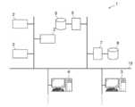
- FIG. 1is a diagram showing a schematic configuration of a medical information system to which the document creation support device according to the embodiment of the present disclosure is applied.
- the medical information system 1 shown in FIG. 1is based on an inspection order from a doctor in a clinical department using a known ordering system, photographs of a part to be inspected of a subject, storage of a medical image acquired by the imaging, and an image interpreter. It is a system for interpreting medical images and creating an interpretation report, and for viewing the interpretation report by the doctor of the requesting clinical department and observing the details of the medical image to be interpreted. As shown in FIG.
- the medical information system 1includes a plurality of modalities (imaging devices) 2, a plurality of image interpretation workstations (WS) 3 which are image interpretation terminals, a clinical department workstation (WS) 4, an image server 5, and an image.
- the database 6, the interpretation report server 7, and the interpretation report database 8are connected and configured so as to be able to communicate with each other via a wired or wireless network 10.
- Each deviceis a computer on which an application program for functioning as a component of the medical information system 1 is installed.
- the application programis stored in the storage device of the server computer connected to the network 10 or in the network storage in a state of being accessible from the outside, and is downloaded and installed in the computer upon request. Alternatively, it is recorded and distributed on a recording medium such as a DVD (Digital Versatile Disc) or a CD-ROM (Compact Disc Read Only Memory), and installed on a computer from the recording medium.
- Modality 2is a device that generates a medical image showing the diagnosis target part by photographing the diagnosis target part of the subject. Specifically, it is a simple X-ray imaging apparatus, a CT apparatus, an MRI apparatus, a PET (Positron Emission Tomography) apparatus, and the like.
- the medical image generated by the modality 2is transmitted to the image server 5 and stored.
- Interpretation WS3includes a document creation support device according to this embodiment. The configuration of the interpretation WS3 will be described later.
- the clinical department WS4is a computer used by doctors in the clinical department for detailed observation of images, viewing of interpretation reports, creation of electronic medical records, etc., and is composed of a processing device, a display, and input devices such as a keyboard and a mouse. ..
- a patient's medical recordelectronic medical record
- an image viewing requestis made to the image server 5
- an image received from the image server 5is displayed
- a lesion-like part in the imageis automatically detected or highlighted
- an image interpretation report serveris used.
- Each processsuch as the viewing request of the image interpretation report to the image interpretation report 7 and the display of the image interpretation report received from the image interpretation report server 7 is performed by executing the software program for each process.
- the image server 5is a general-purpose computer in which a software program that provides a database management system (DataBase Management System: DBMS) function is installed. Further, the image server 5 includes a storage in which the image database 6 is configured. This storage may be a hard disk device connected by the image server 5 and the data bus, or a disk device connected to NAS (Network Attached Storage) and SAN (Storage Area Network) connected to the network 10. It may be.
- NASNetwork Attached Storage
- SANStorage Area Network
- the image data and incidental information of the medical image acquired in the modality 2are registered in the image database 6.
- the incidental informationincludes, for example, an image ID (identification) for identifying an individual medical image, a patient ID for identifying a subject, an examination ID for identifying an examination, and a unique ID assigned to each medical image ( UID: unique identification), examination date when the medical image was generated, examination time, type of modality used in the examination to acquire the medical image, patient information such as patient name, age, gender, examination site (contrast image site) ), Imaging information (imaging protocol, imaging sequence, imaging method, imaging conditions, use of contrast medium, etc.), including information such as a series number or collection number when a plurality of medical images are acquired in one examination.
- the image server 5When the image server 5 receives the viewing request from the image interpretation WS3 via the network 10, the image server 5 searches for the medical image registered in the image database 6 and transmits the searched medical image to the requester's image interpretation WS3.
- the interpretation report server 7incorporates a software program that provides the functions of a database management system to a general-purpose computer.
- the image interpretation report server 7receives the image interpretation report registration request from the image interpretation WS3, the image interpretation report server 7 prepares the image interpretation report in a database format and registers it in the image interpretation report database 8.
- the image interpretation reportis searched from the image interpretation report database 8.
- the image interpretation report database 8contains, for example, an image ID for identifying a medical image to be interpreted, an image radiologist ID for identifying an image diagnostician who has performed image interpretation, a lesion name, lesion location information, findings, and confidence in the findings. An interpretation report in which information such as the degree is recorded is registered.
- the medical imageis a three-dimensional CT image composed of a plurality of tomographic images with the diagnosis target as the lung, and by interpreting the CT image, an interpretation report on abnormal shadows contained in the lung is obtained. It shall be created as a medical document.
- the medical imageis not limited to the CT image, and any medical image such as an MRI image and a simple two-dimensional image acquired by a simple X-ray imaging device can be used.
- Network 10is a wired or wireless network that connects various devices in the hospital.
- the network 10may be configured such that the local area networks of each hospital are connected to each other by the Internet or a dedicated line.
- the image interpretation WS3is a computer used by a medical image interpretation doctor to interpret a medical image and create an image interpretation report, and is composed of a processing device, a display, and an input device such as a keyboard and a mouse.
- the image server 5is requested to view the medical image, various image processes for the medical image received from the image server 5, the display of the medical image, the analysis process for the medical image, the highlighting of the medical image based on the analysis result, and the analysis result.
- Each processsuch as creating an image interpretation report based on the above, supporting the creation of an image interpretation report, requesting the image interpretation report server 7 to register and view the image interpretation report, and displaying the image interpretation report received from the image interpretation report server 7, is for each process. It is done by executing the software program of. Of these processes, processes other than those performed by the document creation support device of the present embodiment are performed by a well-known software program, and therefore detailed description thereof will be omitted here. Further, the image interpretation WS3 does not perform any process other than the process performed by the document creation support device of the present embodiment, and a computer that performs the process is connected to the network 10 separately, and the process is requested by the image interpretation WS3. The computer may perform the requested processing.
- the interpretation WS3includes a document creation support device according to the present embodiment. Therefore, the document creation support program according to the present embodiment is installed in the interpretation WS3.
- the document creation support programis stored in the storage device of the server computer connected to the network or in the network storage in a state of being accessible from the outside, and is downloaded and installed in the interpretation WS3 as requested. Alternatively, it is recorded on a recording medium such as a DVD or a CD-ROM, distributed, and installed on the interpretation WS3 from the recording medium.
- FIG. 2is a diagram showing a schematic configuration of a document creation support device according to the present embodiment, which is realized by installing a document creation support program on the interpretation WS3.
- the document creation support device 20includes a CPU (Central Processing Unit) 11, a memory 12, a storage 13, and a communication I / F (interface) 14 as a standard computer configuration. Further, a display 15 such as a liquid crystal display and an input device 16 such as a keyboard and a mouse are connected to the document creation support device 20.
- the CPU 11corresponds to the processor.
- the storage 13is composed of a hard disk or a storage device such as an SSD (Solid State Drive).
- the storage 13stores various information including information necessary for processing the medical image and the document creation support device 20 acquired from the image server 5 via the network 10.
- the communication I / F 14is a network interface that controls the transmission of various information between the external device and the document creation support device 20 via the network 10.
- the document creation support programis stored in the memory 12.
- the document creation support programis an image acquisition process for acquiring a medical image and an image analysis process for deriving property information representing the properties of the structure of interest contained in the medical image by analyzing the medical image as a process to be executed by the CPU 11.
- Sentence generation processingthat generates medical sentences related to medical images based on property information
- term identification processingthat identifies terms that represent properties related to structures of interest contained in medical sentences by analyzing medical sentences
- property information and termsA collation process for collating with and a medical sentence and a display control process for displaying the collation result on the display 15 are defined.
- the computerbecomes the image acquisition unit 21, the image analysis unit 22, the sentence generation unit 23, the term identification unit 24, the collation unit 25, and the display control unit 26. Function.
- the image acquisition unit 21is composed of an interface connected to the network 10, and acquires a medical image for creating an image interpretation report from the image server 5 according to an instruction from the input device 16 by the image interpretation doctor who is the operator.
- the image analysis unit 22analyzes the medical image to derive property information representing the properties of the structure of interest such as an abnormal shadow candidate included in the medical image.
- the image analysis unit 22has a first learning model 22A in which machine learning is performed so as to discriminate abnormal shadow candidates in a medical image and discriminate the properties of the discriminated abnormal shadow candidates.
- the first learning model 22Adetermines whether or not each pixel (voxel) in the medical image represents an abnormal shadow candidate, and if it is an abnormal shadow candidate, the property thereof is determined. It consists of a convolutional neural network (CNN (Convolutional Neural Network)) in which deep learning is performed using teacher data so as to discriminate.
- CNNConvolutional Neural Network
- FIG. 3is a diagram showing an example of teacher data for learning the first learning model.
- the teacher data 30includes a medical image 32 including the abnormal shadow 31 and property information 33 about the abnormal shadow.
- the abnormal shadow 31is a lung nodule
- the property information 33represents a plurality of properties of the lung nodule.
- the property information 33includes the location of the abnormal shadow, the size of the abnormal shadow, the shape of the boundary (clear and irregular), the type of absorption value (solid and suriglass type), the presence or absence of spicula, the mass or nodule, and the pleura.
- the presence or absence of contact, the presence or absence of pleural invasion, the presence or absence of pleural infiltration, the presence or absence of cavities, the presence or absence of calcification, etc.are used.
- the property information 33shows that the location of the abnormal shadow is under the left pulmonary pleura, the size of the abnormal shadow is 4.2 cm in diameter, and the boundary is defined. Irregular shape, full absorption, with spicula, mass, with pleural contact, with pleural infiltration, no pleural infiltration, no cavities, and no calcification.
- +is given when “yes” and ⁇ is given when there is no.
- the first learning model 22Ais constructed by learning a neural network using a large number of teacher data as shown in FIG. For example, by using the teacher data 30 shown in FIG. 3, the first learning model 22A determines the abnormal shadow 31 included in the medical image 32 when the medical image 32 shown in FIG. 3 is input, and determines the abnormal shadow 31. With respect to 31, learning is performed so as to output the property information 33 shown in FIG.
- any learning modelsuch as a support vector machine (SVM (Support Vector Machine)) can be used.
- SVMSupport Vector Machine
- the learning model for detecting the abnormal shadow candidate from the medical image and the learning model for detecting the property information of the abnormal shadow candidatemay be constructed separately.
- the sentence generation unit 23generates a medical sentence by using the property information derived by the image analysis unit 22.
- the sentence generation unit 23includes a second learning model 23A that has been trained to generate a sentence from the input information.
- a recurrent neural networkcan be used as the second learning model 23A.
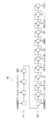
- FIG. 4is a diagram showing a schematic configuration of a recurrent neural network.
- the recurrent neural network 40includes an encoder 41 and a decoder 42.
- the property information derived by the image analysis unit 22is input to the encoder 41. For example, property information of "left pulmonary subpleural", "4.2 cm”, “Spicula +" and "mass" is input to the encoder 41.
- the decoder 42is learned so as to document the character information, and generates a sentence from the input property information. Specifically, from the above-mentioned property information of "left pulmonary subpleura”, “4.2 cm”, “spicula +” and “mass”, "a 4.2 cm diameter tumor having spicula under the left pulmonary pleura is recognized. Will be generated. " In FIG. 4, "EOS" indicates the end of the sentence (End Of Sentence).
- the recurrent neural network 40learns the encoder 41 and the decoder 42 using a large amount of teacher data composed of a combination of the property information and the medical text. Be built.
- the recurrent neural network 40it is possible to specify terms that should not be used in sentences and terms that should be used as parameters for sentence generation. This parameter is determined based on the collation result by the collation unit 25 described later.
- the term identification unit 24specifies a term representing a property included in the medical sentence generated by the sentence generation unit 23.
- the term identification unit 24has a third learning model 24A that has been machine-learned to identify terms that represent properties contained in the text.
- the third learning model 24Ais a convolutional neural network in which deep learning is performed using teacher data so that when a sentence is input, a term representing a property contained in the input sentence is discriminated. It consists of a network.
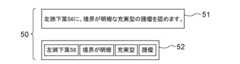
- FIG. 5is a diagram showing an example of teacher data for learning the third learning model.
- the teacher data 50includes the medical sentence 51 and the term 52 representing the property contained in the medical sentence 51.
- the medical sentence 51 shown in FIG. 5is "a solid mass with a clear boundary is found in the lower lobe S6 of the left lung", and the term 52 expressing the property is "under the left lung” included in the medical sentence 51. "Leaf S6", “clear border”, “solid” and “tumor”.
- the third learning model 24Ais constructed by learning a neural network using a large number of teacher data as shown in FIG. For example, by using the teacher data 50 shown in FIG. 5, the third learning model 24A is trained to output the term 52 shown in FIG. 5 when the medical sentence 51 shown in FIG. 5 is input. ..
- any learning modelsuch as a support vector machine and a recurrent neural network can be used.
- the collation unit 25collates the property information derived by the image analysis unit 22 with the terms representing the properties contained in the medical text specified by the term identification unit 24. Therefore, when the property information and the term representing the property are input, the collating unit 25 determines the difference between the term representing the property and the property information, and the property information and the property not included in the term representing the property. It has a fourth learning model 25A that has been machine-learned to discriminate terms that represent properties that are not included in the information. In the present embodiment, the fourth learning model 25A discriminates the difference between the term representing the property and the property information, and represents the property information not included in the term representing the property and the property not included in the property information. It consists of a convolutional neural network that has been deep-learned using teacher data to discriminate terms.
- FIG. 6is a diagram showing an example of teacher data for learning the fourth learning model.
- FIG. 6shows two types of teacher data 60 and 65.
- the teacher data 60includes the property information 61, the term 62 representing the property, and the excess / deficiency information 63 indicating the excess / deficiency of the property information.
- the teacher data 65includes the property information 66, the term 67 representing the property, and the excess / deficiency information 68 indicating the excess / deficiency of the property information.
- the property information 61is "left lower lobe S6", “boundary: clear”, “absorption value: solid”, “spicula +” and “tumor”.
- Term 62is "left lower lobe S6", “clear border”, “solid” and “mass”.
- the excess / deficiency information 63is “insufficient spicula”.
- the property information 66is the same as the property information 61, "left lower lung lower lobe S6", “boundary: clear”, “absorption value: solid type”, “spicula +” and “tumor”.
- Term 67is “left lower lobe S6", “clear border”, “solid”, “spicula”, “calcification” and “mass”.
- the excess / deficiency information 68is “excessive calcification”.
- the fourth learning modelis constructed by learning a neural network using a large amount of teacher data as shown in FIG.
- the fourth learning model 25Aoutputs the excess / deficiency information 63 when the property information 61 and the term 62 representing the property shown in FIG. 6 are input. Learning is done. Further, learning is also made so that a parameter indicating that the term "spicula" is deficient is generated as a parameter based on the collation result based on the excess / deficiency information 63.
- the fourth learning model 25Aoutputs the excess / deficiency information 68 when the property information 66 and the term 67 representing the property shown in FIG. 6 are input. Learning is done. Further, learning is also made so that a parameter indicating that the term "calcification" is excessive is generated as a parameter based on the collation result based on the excess / deficiency information 68.
- any machine learning modelsuch as a support vector machine and a recurrent neural network can be used.
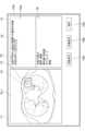
- FIG. 7is a diagram showing an example of a display screen of medical texts and collation results.
- the display screen 70includes an image display area 71 and a text display area 72.
- the slice image SL1that is most likely to identify the abnormal shadow candidate detected by the image analysis unit 22 is displayed.
- the slice image SL1includes an abnormal shadow candidate 73, and the abnormal shadow candidate 73 is surrounded by a rectangular region 74.
- the medical text 75 generated by the text generation unit 23 and the property information 76 derived by the image analysis unit 22are displayed.
- the property information 76does not have to be displayed.
- Medical text 75is "A well-defined, solid mass in the lower lobe S6 of the left lung. Spicula and calcification are found.”
- Property information 76is "left lower lobe S6", “boundary: clear”, “absorption value: solid type”, “spicula: +” and “tumor”.
- the term “calcification” contained in the medical sentence 75is given a solid line frame 77 indicating the excess. As a result, "calcification” is highlighted and the collation result is displayed.
- an automatic correction button 78Aa manual correction button 78B, and a confirmation button 78C are displayed.
- the image interpreting doctorinterprets the slice image SL1 displayed in the image display area 71 and determines the suitability of the medical sentence 75 displayed in the sentence display area 72.
- the interpretercan recognize that the term "calcification" contained in the medical text 75 is excessive by the solid line frame 77 given to "calcification”.
- the sentence generation unit 23regenerates the medical sentence using the property information derived by the image analysis unit 22.
- a parameter based on the collation resultthat is, a parameter for avoiding the use of the term "calcification” is input to the second learning model 23A of the sentence generation unit 23.
- the sentence generator 23generates a medical sentence so as not to use the term "calcification”, and as a result, for example, "a well-defined and solid mass in the lower lobe S6 of the left lung is recognized. It is possible to generate a medical sentence of "I admit.” In this case, the collation result shown in FIG. 9, which will be described later, will be displayed.
- the interpretation doctorcan manually correct the medical text 75 displayed in the text display area 72 by inputting from the input device 16 by selecting the manual correction button 78B. Further, by selecting the confirmation button 78C, the medical sentence 75 displayed in the sentence display area 72 can be confirmed with the content thereof. As a result, the medical text 75 is transcribed into the interpretation report, and the interpretation report to which the medical text 75 is transcribed is transmitted to the interpretation report server 7 together with the slice image SL1 and stored.
- FIG. 8is a diagram showing another example of the display screen of the medical text and the collation result.
- the same reference numberis assigned to the same configuration as in FIG. 7, and detailed description thereof will be omitted.
- the text display area 72 of the display screen 70A shown in FIG. 8the medical text 75A of "a solid mass with a clear boundary is observed in the lower lobe S6 of the left lung" is displayed.
- the property information 76is "left lower lobe S6", “boundary: clear”, “absorption value: solid type”, “spicula: +” and “tumor”. Further, since the medical sentence 75A lacks the term “spicula”, the term “spicula” is displayed in the sentence display area 72, and a broken line frame 79 indicating that the term is deficient is added. As a result, the collation result is displayed by highlighting the term "spicula”.
- the image interpreting doctorinterprets the abnormal shadow candidate 73 in the slice image SL1 displayed in the image display area 71, and determines the suitability of the medical sentence 75A displayed in the sentence display area 72. Further, the interpreting doctor can recognize that the term "spicula" included in the medical sentence 75A is insufficient by the broken line frame 79 given to the "spicula". In addition, instead of or in addition to displaying the term "spicula" in the text display area 72 and adding a broken line frame 79 indicating that the text is insufficient, it is displayed in the property information 76. Marks such as arrows or frames may be added to indicate the lack of the term "spicula". FIG. 9 shows a state in which the arrow 81 indicating that the medical text 75A is insufficient is further added as a mark to the “spicula” displayed in the property information 76.
- the sentence generation unit 23regenerates the medical sentence using the property information derived by the image analysis unit 22.
- a parameter based on the collation resultthat is, a parameter for using the term "spicula” is input to the learning model of the sentence generation unit 23.
- the sentence generator 23generates a medical sentence to use the term "spicula", and as a result, for example, "a well-defined and solid mass is found in the lower lobe S6 of the left lung. It is possible to generate a medical sentence of "I accept.” In this case, the collation result shown in FIG.
- the OK mark 80is displayed in the text display area 72, for example, as shown in the display screen 70B shown in FIG. To.
- the image interpreterwrote the medical text 75B displayed in the text display area 72 by the OK mark 80, "A solid mass with a clear boundary is found in the lower lobe S6 of the left lung. Spicula is recognized.” , It can be recognized that the property information derived from the medical image is used in just proportion.
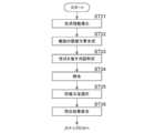
- FIG. 10is a flowchart showing the processing performed in the present embodiment. It is assumed that the medical image to be read is acquired from the image server 5 by the image acquisition unit 21 and stored in the storage 13. The process is started when the image interpretation doctor gives an instruction to create an image interpretation report, and the image analysis unit 22 analyzes the medical image to show the properties of the structure of interest such as an abnormal shadow candidate included in the medical image. Derivation of property information (step ST1). Next, the sentence generation unit 23 generates a medical sentence related to the medical image based on the property information (step ST2).
- the term identification unit 24analyzes the medical sentence generated by the sentence generation unit 23 to identify a term representing a property related to the structure of interest included in the medical sentence (step ST3). Further, the collating unit 25 collates the property information derived by the image analysis unit 22 with the term specified by the term specifying unit 24 (step ST4). Then, the display control unit 26 displays the medical text generated by the sentence generation unit 23 and the collation result by the collation unit 25 on the display 15 (step ST5).
- step ST6determines whether or not the automatic correction button 78A displayed on the collation result display screen is selected (step ST6).
- step ST6is affirmed, the process returns to step ST2, and the processes of steps ST2 to ST5 are repeated.
- the sentence generation unit 23regenerates the medical sentence according to the parameters based on the collation result.
- the term identification unit 24identifies terms in the regenerated medical text.
- the collation unit 25collates the property information with the re-identified term.
- the display control unit 26displays a display screen including the collation result again on the display 15.
- step ST6determines whether or not the manual correction button 78B displayed on the collation result display screen is selected (step ST7).
- step ST7is affirmed, the display control unit 26 accepts the correction of the medical text displayed in the text display area 72 using the input device 16 (step ST8).
- step ST9the display control unit 26 determines whether or not the confirmation button 78C is selected (step ST9). If step ST9 is denied, the process returns to step ST6. When step ST9 is affirmed, the display control unit 26 transfers the medical sentence to the image interpretation report, and transmits the image interpretation report to which the medical sentence is transcribed to the image interpretation report server 7 together with the slice image SL1 (interpretation report transmission: Step ST10), the process is terminated.
- the medical imageby analyzing the medical image, property information representing the properties of the structure of interest such as an abnormal shadow candidate included in the medical image is derived, and the medical image is related based on the property information. Medical text is generated.
- terms representing properties related to the structure of interest contained in the medical textare identified, and the property information and the terms are collated. Therefore, by referring to the collation result, it is possible to easily confirm whether the medical text is generated using all the property information and whether the text contains excessive property information.
- the medical textcan be corrected or the medical text can be regenerated, and the accuracy of the generated medical text can be improved. Therefore, according to the present embodiment, it is possible to generate a medical sentence regarding the structure of interest included in the medical image from the medical image with high accuracy.
- the configuration of the document creation support device according to the other embodimentis the same as the configuration of the document creation support device 20 shown in FIG. 2, and only the processing to be performed is different. Therefore, detailed description of the device is omitted here. ..
- the document creation support device according to the other embodimentis different from the above embodiment in that the sentence generation unit 23 generates a plurality of medical sentences based on the property information.
- FIG. 11is a flowchart showing the processing performed in the other embodiment. In the other embodiment, the processing up to step ST5 shown in FIG. 10 is different from the above embodiment. Therefore, in FIG. 11, only the difference from the flowchart shown in FIG. 10 will be described.
- the processis started when the image interpretation doctor gives an instruction to create an image interpretation report, and the image analysis unit 22 analyzes the medical image to show the properties of the structure of interest such as an abnormal shadow candidate included in the medical image. Derivation of property information (step ST21).
- the sentence generation unit 23generates a plurality of medical sentences related to the medical image based on the property information (step ST22).
- the beam search methodis a method of searching for a word that appears next to a certain word in consideration of the probability of occurrence of the word for a word that appears next to a certain word.
- the sentence generation unit 23applies the beam search method to the recurrent neural network 40 to generate a plurality of (for example, five) medical sentences having a high probability of word occurrence.
- the term identification unit 24analyzes each of the plurality of medical sentences generated by the sentence generation unit 23 to identify terms representing the properties of the structure of interest contained in each of the plurality of medical sentences (step). ST23). Further, the collation unit 25 collates the property information derived by the image analysis unit 22 with the term specified by the term identification unit 24 for each of the plurality of medical sentences (step ST24), and based on the collation result, the collation unit 25 collates the property information. Select one medical sentence from a plurality of medical sentences (step ST25). At this time, the collation unit 25 may select the medical sentence in which the property information and the term match best.
- the collation unit 25may select a medical sentence in which the order in which the property information is described in the sentence most closely matches the order of the property information derived by the image analysis unit 22. Then, the display control unit 26 displays the selected medical text and the collation result by the collation unit 25 on the display 15 (step ST26), and proceeds to the process of step ST6 shown in FIG.
- a plurality of medical sentencesare generated, and the medical sentence having the best match between the property information and the term is selected from the plurality of medical sentences. Therefore, it is possible to present a medical sentence in which there is little difference between the property information and the contained term to the image interpreter together with the collation result. Therefore, the frequency of correcting the medical text and regenerating the medical text can be reduced, and as a result, the burden on the interpreter who creates the interpretation report can be reduced.
- one medical sentenceis selected from a plurality of medical sentences, but two or more medical sentences may be selected. In this case, from among the plurality of medical sentences, it is sufficient to select a high-ranking predetermined number of medical sentences having a high degree of matching between the property information and the term. Further, when a plurality of medical sentences are selected, the collation results for each of the plurality of medical sentences and the plurality of medical sentences are displayed on the display 15 so that the image interpreter can select the desired medical sentences. Is preferable.
- a solid line frame 77is added to the excess term in the medical text 75 displayed in the text display area 72 of the display screen 70 shown in FIG. 7, and the text of the display screen 70A shown in FIG. 8 is provided.
- the missing terms in the medical text 75A displayed in the display area 72are displayed, and the missing terms are given a broken line frame 79, but the present invention is not limited to this. If it is possible to distinguish between excess terms and missing terms, it is possible to change the color of the characters, add different types of dashed lines, change the highlighting color, and so on. It is possible. In addition, the excess or deficiency of terms may be notified by voice.
- the collation unit 25is provided with the fourth learning model 25A, but the present invention is not limited to this. If it can be determined whether or not the property information derived by the image analysis unit 22 and the term representing the property contained in the medical sentence specified by the term identification unit 24 match, any method other than the learning model can be applied. It is possible.
- the medical text 75is recreated based on the selection of the automatic correction button 78A, but the present invention is not limited to this.
- the sentence generation unit 23selects the automatic correction button 78A.
- the medical text 75may be recreated based on the result of the collation without waiting for.
- the display control unit 26displays both the medical sentence generated by the sentence generation unit 23 and the collation result by the collation unit 25, but the present invention is not limited to this. Only medical texts may be displayed on the display screen 70 displayed on the display 15. In this case, for example, by instructing the display of the collation result from the input device 16, the excessive term or the missing term in the medical document is highlighted or the OK mark 80 is displayed as in each of the above embodiments. You may.
- medical textsare generated using medical images whose diagnosis target is the lungs to support the creation of medical documents such as interpretation reports, but the diagnosis target is limited to the lungs. It is not something that is done.
- diagnosis targetis limited to the lungs. It is not something that is done.
- any part of the human bodysuch as the heart, liver, brain, and limbs can be diagnosed.
- each learning model of the image analysis unit 22, the sentence generation unit 23, and the term identification unit 24is prepared to perform analysis processing, sentence generation processing, and term identification processing according to the diagnosis target, and corresponds to the diagnosis target.
- a learning model that performs analysis processing, sentence generation processing, and term identification processingis selected, and medical sentence generation processing is executed.
- the technique of the present disclosureis applied when creating an interpretation report as a medical document, but it is also possible to create a medical document other than the interpretation report such as an electronic medical record and a diagnostic report.
- a medical documentother than the interpretation report such as an electronic medical record and a diagnostic report.
- the technology of the present disclosurecan be applied.
- a processing unitthat executes various processes such as an image acquisition unit 21, an image analysis unit 22, a sentence generation unit 23, a term identification unit 24, a collation unit 25, and a display control unit 26.
- various processorsProcessors
- the various processorsinclude CPUs, which are general-purpose processors that execute software (programs) and function as various processing units, as well as circuits after manufacturing FPGAs (Field Programmable Gate Arrays) and the like.
- Dedicated electricitywhich is a processor with a circuit configuration specially designed to execute specific processing such as programmable logic device (PLD), ASIC (Application Specific Integrated Circuit), which is a processor whose configuration can be changed. Circuits and the like are included.
- One processing unitmay be composed of one of these various processors, or a combination of two or more processors of the same type or different types (for example, a combination of a plurality of FPGAs or a combination of a CPU and an FPGA). ) May be configured. Further, a plurality of processing units may be configured by one processor.
- one processoris configured by combining one or more CPUs and software. There is a form in which this processor functions as a plurality of processing units.
- SoCSystem On Chip
- the various processing unitsare configured by using one or more of the above-mentioned various processors as a hardware structure.
- circuitryin which circuit elements such as semiconductor elements are combined can be used.
Landscapes
- Engineering & Computer Science (AREA)
- Health & Medical Sciences (AREA)
- General Health & Medical Sciences (AREA)
- Theoretical Computer Science (AREA)
- Medical Informatics (AREA)
- Physics & Mathematics (AREA)
- Life Sciences & Earth Sciences (AREA)
- Public Health (AREA)
- General Physics & Mathematics (AREA)
- Nuclear Medicine, Radiotherapy & Molecular Imaging (AREA)
- Artificial Intelligence (AREA)
- Audiology, Speech & Language Pathology (AREA)
- Computational Linguistics (AREA)
- General Engineering & Computer Science (AREA)
- Radiology & Medical Imaging (AREA)
- Biomedical Technology (AREA)
- Primary Health Care (AREA)
- Epidemiology (AREA)
- Pathology (AREA)
- Biophysics (AREA)
- Veterinary Medicine (AREA)
- Animal Behavior & Ethology (AREA)
- High Energy & Nuclear Physics (AREA)
- Surgery (AREA)
- Molecular Biology (AREA)
- Heart & Thoracic Surgery (AREA)
- Optics & Photonics (AREA)
- Quality & Reliability (AREA)
- Computer Vision & Pattern Recognition (AREA)
- Data Mining & Analysis (AREA)
- Databases & Information Systems (AREA)
- Medical Treatment And Welfare Office Work (AREA)
- Measuring And Recording Apparatus For Diagnosis (AREA)
Abstract
Description
Translated fromJapanese本開示は、医療文書等の文書の作成を支援する文書作成支援装置、方法およびプログラムに関する。This disclosure relates to a document creation support device, method and program that support the creation of documents such as medical documents.
近年、CT(Computed Tomography)装置およびMRI(Magnetic Resonance Imaging)装置等の医療機器の進歩により、より質の高い高解像度の医用画像を用いての画像診断が可能となってきている。とくに、CT画像およびMRI画像等を用いた画像診断により、病変の領域を精度よく特定することができるため、特定した結果に基づいて適切な治療が行われるようになってきている。In recent years, advances in medical devices such as CT (Computed Tomography) devices and MRI (Magnetic Resonance Imaging) devices have made it possible to perform diagnostic imaging using higher quality medical images. In particular, since the lesion region can be accurately identified by diagnostic imaging using CT images, MRI images, and the like, appropriate treatment has come to be performed based on the identified results.
また、ディープラーニング等により機械学習がなされた学習モデルを用いたCAD(Computer-Aided Diagnosis)により医用画像を解析して、医用画像に含まれる異常陰影候補等の関心構造物の形状、濃度、位置および大きさ等の性状を判別し、これらを解析結果として取得することも行われている。CADにより取得された解析結果は、患者名、性別、年齢および医用画像を取得したモダリティ等の検査情報と対応づけられて、データベースに保存される。医用画像および解析結果は、医用画像の読影を行う読影医の端末に送信される。読影医は、自身の端末において、送信された医用画像および解析結果を参照して医用画像の読影を行い、読影レポートを作成する。In addition, medical images are analyzed by CAD (Computer-Aided Diagnosis) using a learning model that has been machine-learned by deep learning, etc., and the shape, density, and position of structures of interest such as abnormal shadow candidates included in the medical images. It is also practiced to discriminate properties such as size and size, and obtain these as analysis results. The analysis result acquired by CAD is associated with the examination information such as the patient name, gender, age, and the modality from which the medical image was acquired, and is stored in the database. The medical image and the analysis result are transmitted to the terminal of the image interpreting doctor who interprets the medical image. The image interpreting doctor interprets the medical image by referring to the transmitted medical image and the analysis result on his / her terminal, and creates an image interpretation report.
一方、上述したCT装置およびMRI装置の高性能化に伴い、読影を行う医用画像の数も増大している。しかしながら、読影医の数は医用画像の数に追いついていないことから、読影医の読影業務の負担を軽減することが望まれている。このため、読影レポート等の医療文書の作成を支援するための各種手法が提案されている。例えば、特開2019-153250号公報には、読影医が入力したキーワードおよび医用画像の解析結果に含まれる、関心構造物の性状を表す情報(以下、性状情報とする)に基づいて、読影レポートに記載するための文章を生成する手法が提案されている(特開2019-153250号公報参照)。特開2019-153250号公報に記載された手法においては、入力された性状情報を表す文字から文章を生成するように学習が行われたリカレントニューラルネットワーク等の機械学習がなされた学習モデルを用いて、医療用の文章(以下、医療文章とする)が作成される。特開2019-153250号公報に記載された手法のように、医療文章を自動で生成することにより、読影レポート等の医療文書を作成する際の読影医の負担を軽減することができる。On the other hand, the number of medical images to be interpreted is increasing with the improvement of the performance of the CT device and the MRI device described above. However, since the number of image interpreters has not kept up with the number of medical images, it is desired to reduce the burden of the image interpretation work of the image interpreters. For this reason, various methods have been proposed to support the creation of medical documents such as interpretation reports. For example, Japanese Patent Application Laid-Open No. 2019-153250 provides an interpretation report based on information representing the properties of the structure of interest (hereinafter referred to as property information) included in the keyword input by the image interpreter and the analysis result of the medical image. A method for generating a sentence to be described in Japanese Patent Application Laid-Open No. 2019-153250 has been proposed (see JP-A-2019-153250). In the method described in Japanese Patent Application Laid-Open No. 2019-153250, a learning model in which machine learning such as a recurrent neural network is trained so as to generate a sentence from characters representing input property information is used. , Medical texts (hereinafter referred to as medical texts) are created. By automatically generating medical texts as in the method described in JP-A-2019-153250, it is possible to reduce the burden on the interpreter when creating a medical document such as an image interpretation report.
一方、医用画像を解析することにより取得される性状情報のみを用いて、医療文章を生成する学習モデルを生成することが考えられる。しかしながら、学習モデルの学習に使用した教師データの内容に依存して、あるいは学習モデルの学習の限界に依存して、医用画像から取得したすべての性状情報が、生成された医療文章に含まれなくなる場合がある。また、医用画像から取得した性状情報以外の性状情報が、生成された医療文章に含まれてしまう場合もある。このような医療文章を読影レポートとして用いたのでは、読影レポートの精度が低下する。On the other hand, it is conceivable to generate a learning model that generates medical sentences by using only the property information acquired by analyzing the medical image. However, depending on the content of the teacher data used to train the learning model, or depending on the learning limits of the learning model, all property information obtained from the medical image will no longer be included in the generated medical text. In some cases. In addition, property information other than the property information acquired from the medical image may be included in the generated medical text. If such medical text is used as an interpretation report, the accuracy of the interpretation report will be reduced.
本開示は上記事情に鑑みなされたものであり、医用画像から医療文章を生成する場合のように、画像から画像に含まれる関心構造物に関する文章を高精度に生成することを目的とする。This disclosure has been made in view of the above circumstances, and an object of the present disclosure is to generate a sentence related to a structure of interest contained in an image from an image with high accuracy, as in the case of generating a medical sentence from a medical image.
本開示による文書作成支援装置は、少なくとも1つのプロセッサを備え、
プロセッサは、
画像を解析することにより、画像に含まれる関心構造物の性状を表す性状情報を導出し、The document creation support device according to the present disclosure includes at least one processor.
The processor is
By analyzing the image, property information representing the properties of the structure of interest contained in the image can be derived.
性状情報に基づいて、画像に関する文章を生成し、
文章を解析することにより、文章に含まれる関心構造物に関する性状を表す用語を特定し、
性状情報と用語とを照合するように構成される。Generate sentences related to images based on property information,
By analyzing the text, we can identify the terms that describe the properties of the structure of interest contained in the text.
It is configured to match property information with terms.
なお、本開示による文書作成支援装置においては、プロセッサは、さらに文章をディスプレイに表示するように構成されるものであってもよい。
く、さらに照合の結果をディスプレイに表示するように構成されるものであってもよい。In the document creation support device according to the present disclosure, the processor may be configured to further display a sentence on the display.
In addition, it may be configured to display the collation result on a display.
この場合、プロセッサは、さらに照合の結果をディスプレイに表示するように構成されるものであってもよい。In this case, the processor may be configured to further display the collation result on the display.
また、本開示による文書作成支援装置においては、プロセッサは、さらに文章において用語と性状情報とが相違する場合、相違箇所を強調表示することにより照合の結果を表示するように構成されるものであってもよい。Further, in the document creation support device according to the present disclosure, when the term and the property information are different in the text, the processor is configured to display the collation result by highlighting the difference. You may.
また、本開示による文書作成支援装置においては、プロセッサは、さらに文章において用語と性状情報とが相違する場合、文章を再生成するように構成されるものであってもよい。Further, in the document creation support device according to the present disclosure, the processor may be configured to regenerate the sentence when the term and the property information are different in the sentence.
また、本開示による文書作成支援装置においては、プロセッサは、さらに文章の修正を受け付けるように構成されるものであってもよい。Further, in the document creation support device according to the present disclosure, the processor may be configured to further accept corrections of sentences.
また、本開示による文書作成支援装置においては、プロセッサは、性状情報に基づいて、画像に関する文章を複数生成し、
複数の文章のそれぞれを解析することにより、複数の文章のそれぞれに含まれる関心構造物に関する性状を表す用語を特定し、
複数の文章のそれぞれについて、性状情報と用語とを照合し、
照合の結果に基づいて、複数の文章から少なくとも1つの文章を選択するように構成されるものであってもよい。Further, in the document creation support device according to the present disclosure, the processor generates a plurality of sentences related to the image based on the property information.
By analyzing each of the multiple sentences, we can identify the terms that describe the properties of the structure of interest contained in each of the multiple sentences.
For each of multiple sentences, the property information and terms are collated.
It may be configured to select at least one sentence from a plurality of sentences based on the result of collation.
また、本開示による文書作成支援装置においては、画像は医用画像であり、文章は、医用画像に含まれる関心構造物に関する医療文章であってもよい。Further, in the document creation support device according to the present disclosure, the image may be a medical image, and the text may be a medical text related to the structure of interest included in the medical image.
本開示による文書作成支援方法は、画像を解析することにより、画像に含まれる関心構造物の性状を表す性状情報を導出し、
性状情報に基づいて、画像に関する文章を生成し、
文章を解析することにより、文章に含まれる関心構造物に関する性状を表す用語を特定し、
性状情報と用語とを照合する。The document creation support method according to the present disclosure derives property information representing the properties of the structure of interest contained in the image by analyzing the image.
Generate sentences related to images based on property information,
By analyzing the text, we can identify the terms that describe the properties of the structure of interest contained in the text.
Match property information with terms.
なお、本開示による文書支援作成方法をコンピュータに実行させるためのプログラムとして提供してもよい。Note that it may be provided as a program for causing a computer to execute the document support creation method according to the present disclosure.
本開示によれば、画像から画像に含まれる関心構造物に関する文章を高精度に生成することができる。According to the present disclosure, it is possible to generate a sentence about a structure of interest contained in an image from an image with high accuracy.
以下、図面を参照して本開示の実施形態について説明する。図1は本開示の実施形態による文書作成支援装置を適用した医療情報システムの概略構成を示す図である。図1に示す医療情報システム1は、公知のオーダリングシステムを用いた診療科の医師からの検査オーダに基づいて、被写体の検査対象部位の撮影、撮影により取得された医用画像の保管、読影医による医用画像の読影と読影レポートの作成、および依頼元の診療科の医師による読影レポートの閲覧と読影対象の医用画像の詳細観察とを行うためのシステムである。図1に示すように、医療情報システム1は、複数のモダリティ(撮影装置)2、読影端末である複数の読影ワークステーション(WS)3、診療科ワークステーション(WS)4、画像サーバ5、画像データベース6、読影レポートサーバ7、および読影レポートデータベース8が、有線または無線のネットワーク10を介して互いに通信可能な状態で接続されて構成されている。Hereinafter, embodiments of the present disclosure will be described with reference to the drawings. FIG. 1 is a diagram showing a schematic configuration of a medical information system to which the document creation support device according to the embodiment of the present disclosure is applied. The
各機器は、医療情報システム1の構成要素として機能させるためのアプリケーションプログラムがインストールされたコンピュータである。アプリケーションプログラムは、ネットワーク10に接続されたサーバコンピュータの記憶装置、もしくはネットワークストレージに、外部からアクセス可能な状態で記憶され、要求に応じてコンピュータにダウンロードされ、インストールされる。または、DVD(Digital Versatile Disc)あるいはCD-ROM(Compact Disc Read Only Memory)等の記録媒体に記録されて配布され、その記録媒体からコンピュータにインストールされる。Each device is a computer on which an application program for functioning as a component of the
モダリティ2は、被写体の診断対象となる部位を撮影することにより、診断対象部位を表す医用画像を生成する装置である。具体的には、単純X線撮影装置、CT装置、MRI装置、およびPET(Positron Emission Tomography)装置等である。モダリティ2により生成された医用画像は画像サーバ5に送信され、保存される。
読影WS3は、本実施形態による文書作成支援装置を内包する。読影WS3の構成については後述する。Interpretation WS3 includes a document creation support device according to this embodiment. The configuration of the interpretation WS3 will be described later.
診療科WS4は、診療科の医師が画像の詳細観察、読影レポートの閲覧、および電子カルテの作成等に利用するコンピュータであり、処理装置、ディスプレイ、並びにキーボードおよびマウス等の入力デバイスにより構成される。診療科WS4では、患者のカルテ(電子カルテ)の作成、画像サーバ5に対する画像の閲覧要求、画像サーバ5から受信した画像の表示、画像中の病変らしき部分の自動検出または強調表示、読影レポートサーバ7に対する読影レポートの閲覧要求、および読影レポートサーバ7から受信した読影レポートの表示等の各処理が、各処理のためのソフトウェアプログラムを実行することにより行われる。The clinical department WS4 is a computer used by doctors in the clinical department for detailed observation of images, viewing of interpretation reports, creation of electronic medical records, etc., and is composed of a processing device, a display, and input devices such as a keyboard and a mouse. .. In the medical department WS4, a patient's medical record (electronic medical record) is created, an image viewing request is made to the
画像サーバ5は、汎用のコンピュータにデータベース管理システム(DataBase Management System: DBMS)の機能を提供するソフトウェアプログラムがインストールされたものである。また、画像サーバ5は画像データベース6が構成されるストレージを備えている。このストレージは、画像サーバ5とデータバスとによって接続されたハードディスク装置であってもよいし、ネットワーク10に接続されているNAS(Network Attached Storage)およびSAN(Storage Area Network)に接続されたディスク装置であってもよい。また、画像サーバ5は、モダリティ2からの医用画像の登録要求を受け付けると、その医用画像をデータベース用のフォーマットに整えて画像データベース6に登録する。The
画像データベース6には、モダリティ2において取得された医用画像の画像データと付帯情報とが登録される。付帯情報には、例えば、個々の医用画像を識別するための画像ID(identification)、被写体を識別するための患者ID、検査を識別するための検査ID、医用画像毎に割り振られるユニークなID(UID:unique identification)、医用画像が生成された検査日、検査時刻、医用画像を取得するための検査で使用されたモダリティの種類、患者氏名、年齢、性別等の患者情報、検査部位(撮影部位)、撮影情報(撮影プロトコル、撮影シーケンス、撮像手法、撮影条件および造影剤の使用等)、1回の検査で複数の医用画像を取得したときのシリーズ番号あるいは採取番号等の情報が含まれる。The image data and incidental information of the medical image acquired in the
また、画像サーバ5は、読影WS3からの閲覧要求をネットワーク10経由で受信すると、画像データベース6に登録されている医用画像を検索し、検索された医用画像を要求元の読影WS3に送信する。When the
読影レポートサーバ7には、汎用のコンピュータにデータベース管理システムの機能を提供するソフトウェアプログラムが組み込まれる。読影レポートサーバ7は、読影WS3からの読影レポートの登録要求を受け付けると、その読影レポートをデータベース用のフォーマットに整えて読影レポートデータベース8に登録する。また、読影レポートの検索要求を受け付けると、その読影レポートを読影レポートデータベース8から検索する。The
読影レポートデータベース8には、例えば、読影対象の医用画像を識別する画像ID、読影を行った画像診断医を識別するための読影医ID、病変名、病変の位置情報、所見、および所見の確信度等の情報が記録された読影レポートが登録される。The image
なお、本実施形態においては、医用画像は診断対象を肺とした、複数の断層画像からなる3次元のCT画像とし、CT画像を読影することにより、肺に含まれる異常陰影についての読影レポートを医療文書として作成するものとする。なお、医用画像はCT画像に限定されるものではなく、MRI画像および単純X線撮影装置により取得された単純2次元画像等の任意の医用画像を用いることができる。In the present embodiment, the medical image is a three-dimensional CT image composed of a plurality of tomographic images with the diagnosis target as the lung, and by interpreting the CT image, an interpretation report on abnormal shadows contained in the lung is obtained. It shall be created as a medical document. The medical image is not limited to the CT image, and any medical image such as an MRI image and a simple two-dimensional image acquired by a simple X-ray imaging device can be used.
ネットワーク10は、病院内の各種機器を接続する有線または無線のネットワークである。読影WS3が他の病院あるいは診療所に設置されている場合には、ネットワーク10は、各病院のローカルエリアネットワーク同士をインターネットまたは専用回線で接続した構成としてもよい。
以下、本実施形態による読影WS3について詳細に説明する。読影WS3は、医用画像の読影医が、医用画像の読影および読影レポートの作成に利用するコンピュータであり、処理装置、ディスプレイ、並びにキーボードおよびマウス等の入力デバイスにより構成される。読影WS3では、画像サーバ5に対する医用画像の閲覧要求、画像サーバ5から受信した医用画像に対する各種画像処理、医用画像の表示、医用画像に対する解析処理、解析結果に基づく医用画像の強調表示、解析結果に基づく読影レポートの作成、読影レポートの作成の支援、読影レポートサーバ7に対する読影レポートの登録要求と閲覧要求、並びに読影レポートサーバ7から受信した読影レポートの表示等の各処理が、各処理のためのソフトウェアプログラムを実行することにより行われる。なお、これらの処理のうち、本実施形態の文書作成支援装置が行う処理以外の処理は、周知のソフトウェアプログラムにより行われるため、ここでは詳細な説明は省略する。また、本実施形態の文書作成支援装置が行う処理以外の処理を読影WS3において行わず、別途その処理を行うコンピュータをネットワーク10に接続しておき、読影WS3からの処理の要求に応じて、そのコンピュータにおいて要求された処理を行うようにしてもよい。Hereinafter, the interpretation WS3 according to the present embodiment will be described in detail. The image interpretation WS3 is a computer used by a medical image interpretation doctor to interpret a medical image and create an image interpretation report, and is composed of a processing device, a display, and an input device such as a keyboard and a mouse. In the interpretation WS3, the
読影WS3は、本実施形態による文書作成支援装置が内包されている。このため、読影WS3には、本実施形態による文書作成支援プログラムがインストールされている。文書作成支援プログラムは、ネットワークに接続されたサーバコンピュータの記憶装置、もしくはネットワークストレージに、外部からアクセス可能な状態で記憶され、要求に応じて読影WS3にダウンロードされ、インストールされる。または、DVDあるいはCD-ROM等の記録媒体に記録されて配布され、その記録媒体から読影WS3にインストールされる。The interpretation WS3 includes a document creation support device according to the present embodiment. Therefore, the document creation support program according to the present embodiment is installed in the interpretation WS3. The document creation support program is stored in the storage device of the server computer connected to the network or in the network storage in a state of being accessible from the outside, and is downloaded and installed in the interpretation WS3 as requested. Alternatively, it is recorded on a recording medium such as a DVD or a CD-ROM, distributed, and installed on the interpretation WS3 from the recording medium.
図2は、文書作成支援プログラムを読影WS3にインストールすることにより実現される、本実施形態による文書作成支援装置の概略構成を示す図である。図2に示すように、文書作成支援装置20は、標準的なコンピュータの構成として、CPU(Central Processing Unit)11、メモリ12、ストレージ13および通信I/F(インターフェース)14を備える。また、文書作成支援装置20には、液晶ディスプレイ等のディスプレイ15、並びにキーボードおよびマウス等の入力デバイス16が接続されている。CPU11がプロセッサに相当する。FIG. 2 is a diagram showing a schematic configuration of a document creation support device according to the present embodiment, which is realized by installing a document creation support program on the interpretation WS3. As shown in FIG. 2, the document
ストレージ13は、ハードディスクまたはSSD(Solid State Drive)等のストレージデバイスからなる。ストレージ13には、ネットワーク10を経由して画像サーバ5から取得した、医用画像および文書作成支援装置20の処理に必要な情報を含む各種情報が記憶されている。The
通信I/F14は、ネットワーク10を介した外部装置と文書作成支援装置20との各種情報の伝送制御を行うネットワークインターフェースである。The communication I /
また、メモリ12には、文書作成支援プログラムが記憶されている。文書作成支援プログラムは、CPU11に実行させる処理として、医用画像を取得する画像取得処理、医用画像を解析することにより、医用画像に含まれる関心構造物の性状を表す性状情報を導出する画像解析処理、性状情報に基づいて医用画像に関する医療文章を生成する文章生成処理、医療文章を解析することにより、医療文章に含まれる関心構造物に関する性状を表す用語を特定する用語特定処理、性状情報と用語とを照合する照合処理、並びに医療文章および照合結果をディスプレイ15に表示する表示制御処理を規定する。In addition, the document creation support program is stored in the
そして、CPU11が文書作成支援プログラムに従いこれらの処理を実行することで、コンピュータは、画像取得部21、画像解析部22、文章生成部23、用語特定部24、照合部25および表示制御部26として機能する。Then, when the
画像取得部21は、ネットワーク10と接続されたインターフェースからなり、操作者である読影医による入力デバイス16からの指示により、画像サーバ5から読影レポートを作成するための医用画像を取得する。The
画像解析部22は、医用画像を解析することにより、医用画像に含まれる異常陰影候補等の関心構造物の性状を表す性状情報を導出する。このために、画像解析部22は、医用画像における異常陰影候補を判別し、判別した異常陰影候補の性状を判別するように機械学習がなされた第1の学習モデル22Aを有する。本実施形態においては、第1の学習モデル22Aは、医用画像における各画素(ボクセル)が異常陰影候補を表すものであるか否かを判別し、異常陰影候補である場合には、その性状を判別するように、教師データを用いてディープラーニング(深層学習)がなされた畳み込みニューラルネットワーク(CNN(Convolutional Neural Network))からなる。The
図3は第1の学習モデルを学習するための教師データの例を示す図である。図3に示すように、教師データ30は、異常陰影31が含まれる医用画像32および異常陰影についての性状情報33を含む。本実施形態においては、異常陰影31は肺結節であり、性状情報33は肺結節についての複数の性状を表すものとする。例えば、性状情報33としては、異常陰影の場所、異常陰影のサイズ、境界の形状(明瞭および不整形)、吸収値の種類(充実型およびスリガラス型)、スピキュラの有無、腫瘤か結節か、胸膜接触の有無、胸膜陥入の有無、胸膜浸潤の有無、空洞の有無、および石灰化の有無等が用いられる。図3に示す教師データ30に含まれる異常陰影31については、性状情報33は、図3に示すように、異常陰影の場所は左肺胸膜下、異常陰影のサイズは直径4.2cm、境界の形状は不整形、吸収値は充実型、スピキュラは有、腫瘤、胸膜接触は有、胸膜陥入は有、胸膜浸潤は無、空洞は無、および石灰化は無となっている。なお、図3においては、「有り」の場合は+、無しの場合は-を付与している。第1の学習モデル22Aは、図3に示すような教師データを多数用いてニューラルネットワークを学習することにより構築される。例えば、図3に示す教師データ30を用いることにより、第1の学習モデル22Aは、図3に示す医用画像32が入力されると、医用画像32に含まれる異常陰影31を判別し、異常陰影31に関して、図3に示す性状情報33を出力するように学習がなされる。FIG. 3 is a diagram showing an example of teacher data for learning the first learning model. As shown in FIG. 3, the
また、第1の学習モデル22Aとしては、畳み込みニューラルネットワークの他、例えばサポートベクタマシン(SVM(Support Vector Machine))等の任意の学習モデルを用いることができる。Further, as the
なお、医用画像から異常陰影候補を検出する学習モデルと、異常陰影候補の性状情報を検出する学習モデルとを別々に構築するようにしてもよい。It should be noted that the learning model for detecting the abnormal shadow candidate from the medical image and the learning model for detecting the property information of the abnormal shadow candidate may be constructed separately.
文章生成部23は、画像解析部22が導出した性状情報を用いて、医療文章を生成する。文章生成部23は、入力された情報から文章を生成するように学習が行われた第2の学習モデル23Aからなる。第2の学習モデル23Aとしては、例えばリカレントニューラルネットワークを用いることができる。図4はリカレントニューラルネットワークの模式的な構成を示す図である。図4に示すように、リカレントニューラルネットワーク40は、エンコーダ41およびデコーダ42からなる。エンコーダ41には、画像解析部22が導出した性状情報が入力される。例えば、エンコーダ41には、「左肺胸膜下」、「4.2cm」、「スピキュラ+」および「腫瘤」の性状情報が入力される。デコーダ42は、文字情報を文章化するように学習がなされており、入力された性状情報から文章を生成する。具体的には、上述した「左肺胸膜下」、「4.2cm」、「スピキュラ+」および「腫瘤」の性状情報から、「左肺胸膜下にスピキュラを有する4.2cm径の腫瘤が認められます。」の医療文章を生成する。なお、図4において「EOS」は文章の終わりを示す(End Of Sentence)。The
このように、性状情報の入力によって医療文章を出力するために、リカレントニューラルネットワーク40は、性状情報と医療文章との組み合わせからなる多数の教師データを用いてエンコーダ41およびデコーダ42を学習することにより構築されてなる。In this way, in order to output the medical text by inputting the property information, the recurrent
なお、リカレントニューラルネットワーク40には、文章生成のパラメータとして、文章に使用すべきでない用語および使用すべき用語を指定することが可能となっている。このパラメータは、後述する照合部25による照合結果に基づいて決定される。In the recurrent
用語特定部24は、文章生成部23が生成した医療文章に含まれる性状を表す用語を特定する。このために、用語特定部24は、文章に含まれる性状を表す用語を特定するように機械学習がなされた第3の学習モデル24Aを有する。本実施形態においては、第3の学習モデル24Aは、文章が入力されると、入力された文章に含まれる性状を表す用語を判別するように、教師データを用いてディープラーニングがなされた畳み込みニューラルネットワークからなる。The
図5は第3の学習モデルを学習するための教師データの例を示す図である。図5に示すように、教師データ50は、医療文章51および医療文章51に含まれる性状を表す用語52を含む。図5に示す医療文章51は、「左肺下葉S6に、境界が明瞭な充実型の腫瘤を認めます。」であり、性状を表す用語52は、医療文章51に含まれる「左肺下葉S6」、「境界が明瞭」、「充実型」および「腫瘤」である。第3の学習モデル24Aは、図5に示すような教師データを多数用いてニューラルネットワークを学習することにより構築される。例えば、図5に示す教師データ50を用いることにより、第3の学習モデル24Aは、図5に示す医療文章51が入力されると、図5に示す用語52を出力するように学習がなされる。FIG. 5 is a diagram showing an example of teacher data for learning the third learning model. As shown in FIG. 5, the
また、第3の学習モデル24Aとしては、畳み込みニューラルネットワークの他、例えばサポートベクタマシンおよびリカレントニューラルネットワーク等の任意の学習モデルを用いることができる。Further, as the
照合部25は、画像解析部22が導出した性状情報と、用語特定部24が特定した医療文章に含まれる性状を表す用語とを照合する。このために、照合部25は、性状情報および性状を表す用語が入力されると、性状を表す用語と性状情報との相違を判別して、性状を表す用語に含まれない性状情報、および性状情報に含まれない性状を表す用語を判別するように機械学習がなされた第4の学習モデル25Aを有する。本実施形態においては、第4の学習モデル25Aは、性状を表す用語と性状情報との相違を判別して、性状を表す用語に含まれない性状情報、および性状情報に含まれない性状を表す用語を判別するように、教師データを用いてディープラーニングがなされた畳み込みニューラルネットワークからなる。The
図6は第4の学習モデルを学習するための教師データの例を示す図である。図6には、2種類の教師データ60,65が示されている。図6に示すように、教師データ60は、性状情報61、性状を表す用語62および性状情報の過不足を表す過不足情報63を含む。また、教師データ65は、性状情報66、性状を表す用語67および性状情報の過不足を表す過不足情報68を含む。図6に示す教師データ60に関して、性状情報61は、「左肺下葉S6」、「境界:明瞭」、「吸収値:充実型」、「スピキュラ+」および「腫瘤」である。用語62は、「左肺下葉S6」、「境界が明瞭」、「充実型」および「腫瘤」である。過不足情報63は「スピキュラが不足」である。FIG. 6 is a diagram showing an example of teacher data for learning the fourth learning model. FIG. 6 shows two types of
図6に示す教師データ65に関して、性状情報66は性状情報61と同様に、「左肺下葉S6」、「境界:明瞭」、「吸収値:充実型」、「スピキュラ+」および「腫瘤」である。用語67は、「左肺下葉S6」、「境界が明瞭」、「充実型」、「スピキュラを認め」、「石灰化を認め」および「腫瘤」である。過不足情報68は「石灰化が過剰」である。Regarding the
第4の学習モデルは、図6に示すような教師データを多数用いてニューラルネットワークを学習することにより構築される。例えば、図6に示す教師データ60を用いることにより、第4の学習モデル25Aは、図6に示す性状情報61および性状を表す用語62が入力されると、過不足情報63を出力するように学習がなされる。また、過不足情報63に基づいて、「スピキュラ」の用語が不足していることを表すパラメータを、照合結果に基づくパラメータとして生成するようにも学習がなされる。The fourth learning model is constructed by learning a neural network using a large amount of teacher data as shown in FIG. For example, by using the teacher data 60 shown in FIG. 6, the
また、図6に示す教師データ65を用いることにより、第4の学習モデル25Aは、図6に示す性状情報66および性状を表す用語67が入力されると、過不足情報68を出力するように学習がなされる。また、過不足情報68に基づいて、「石灰化」の用語が過剰であることを表すパラメータを照合結果に基づくパラメータとして生成するようにも学習がなされる。Further, by using the
なお、第4の学習モデル25Aとしては、畳み込みニューラルネットワークの他、例えばサポートベクタマシンおよびリカレントニューラルネットワーク等の任意の機械学習モデルを用いることができる。As the
表示制御部26は、文章生成部23が生成した医療文章および照合部25による照合結果をディスプレイ15に表示する。図7は医療文章および照合結果の表示画面の例を示す図である。図7に示すように、表示画面70は画像表示領域71および文章表示領域72を含む。画像表示領域71には、画像解析部22が検出した異常陰影候補を最も特定しやすいスライス画像SL1が表示される。スライス画像SL1には異常陰影候補73が含まれ、異常陰影候補73は矩形領域74により囲まれている。The
文章表示領域72には、文章生成部23が生成した医療文章75および画像解析部22が導出した性状情報76が表示されている。なお、性状情報76は表示しなくてもよい。医療文章75は、「左肺下葉S6に境界が明瞭で充実型の腫瘤を認めます。スピキュラおよび石灰化を認めます。」である。性状情報76は、「左肺下葉S6」、「境界:明瞭」、「吸収値:充実型」、「スピキュラ:+」および「腫瘤」である。また、照合結果としては、医療文章75に含まれる「石灰化」が過剰であるため、医療文章75に含まれる「石灰化」の用語に、過剰であることを示す実線の枠77が付与されることにより、「石灰化」が強調表示されることにより、照合結果が表示されている。In the
文章表示領域72の下方には、自動修正ボタン78A、手動修正ボタン78Bおよび確定ボタン78Cが表示されている。Below the
読影医は、画像表示領域71に表示されたスライス画像SL1を読影し、文章表示領域72に表示された医療文章75の適否を判定する。また、読影医は、医療文章75に含まれる「石灰化」の用語が過剰であることを、「石灰化」に付与された実線の枠77により認識することができる。The image interpreting doctor interprets the slice image SL1 displayed in the
一方、読影医は医療文章75の再作成を所望する場合、自動修正ボタン78Aを入力デバイス16を用いて選択する。これにより、文章生成部23が画像解析部22が導出した性状情報を用いて医療文章を再度生成する。この際、文章生成部23の第2の学習モデル23Aには、照合結果に基づくパラメータ、すなわち「石灰化」の用語を使用しないようにするためのパラメータが入力される。これにより、文章生成部23は、「石灰化」の用語を使用しないように医療文章を生成し、その結果、例えば「左肺下葉S6に境界が明瞭で充実型の腫瘤を認めます。スピキュラを認めます。」の医療文章を生成することが可能となる。この場合、後述する図9に示す照合結果が表示されることとなる。On the other hand, when the interpreter wants to recreate the
一方、読影医は手動修正ボタン78Bを選択することにより、文章表示領域72に表示された医療文章75を、入力デバイス16からの入力により、手動で修正することが可能である。また、確定ボタン78Cを選択することにより、文章表示領域72に表示された医療文章75をその内容で確定することができる。これにより、医療文章75は読影レポートに転記され、医療文章75が転記された読影レポートはスライス画像SL1と併せて読影レポートサーバ7に送信されて保管される。On the other hand, the interpretation doctor can manually correct the
図8は医療文章および照合結果の表示画面の他の例を示す図である。なお、図8に示す表示画面において図7と同一の構成については同一の参照番号を付与し、詳細な説明は省略する。図8に示す表示画面70Aの文章表示領域72には、「左肺下葉S6に境界が明瞭で充実型の腫瘤を認めます。」の医療文章75Aが表示されている。FIG. 8 is a diagram showing another example of the display screen of the medical text and the collation result. In the display screen shown in FIG. 8, the same reference number is assigned to the same configuration as in FIG. 7, and detailed description thereof will be omitted. In the
ここで、性状情報76は、「左肺下葉S6」、「境界:明瞭」、「吸収値:充実型」、「スピキュラ:+」および「腫瘤」である。また、医療文章75Aには、「スピキュラ」の用語が不足しているため、文章表示領域72には、「スピキュラ」の用語が表示され、不足していることを表す破線の枠79が付与されることにより、「スピキュラ」の用語が強調表示されることにより、照合結果が表示されている。Here, the
図8に示す表示画面70Aに関して、読影医は、画像表示領域71に表示されたスライス画像SL1における異常陰影候補73を読影し、文章表示領域72に表示された医療文章75Aの適否を判定する。また、読影医は、医療文章75Aに含まれる「スピキュラ」の用語が不足していることを、「スピキュラ」に付与された破線の枠79により認識することができる。なお、文章表示領域72に「スピキュラ」の用語を表示し、さらに不足していることを表す破線の枠79を付与することに代えて、またはこれに加えて、性状情報76に表示されている「スピキュラ」の用語に不足していることを表す、矢印または枠等のマークを付与するようにしてもよい。図9においては、性状情報76に表示されている「スピキュラ」に対して、医療文章75Aにおいて不足していることを表す矢印81をマークとしてさらに付与した状態を示している。With respect to the
そして、読影医は医療文章75Aの再作成を所望する場合、自動修正ボタン78Aを入力デバイス16を用いて選択する。これにより、文章生成部23が画像解析部22が導出した性状情報を用いて医療文章を再度生成する。この際、文章生成部23の学習モデルには、照合結果に基づくパラメータ、すなわち「スピキュラ」の用語を使用するようにするためのパラメータが入力される。これにより、文章生成部23は、「スピキュラ」の用語を使用するように医療文章を生成し、その結果、例えば「左肺下葉S6に境界が明瞭で充実型の腫瘤を認めます。スピキュラを認めます。」の医療文章を生成することが可能となる。この場合、後述する図9に示す照合結果が表示されることとなる。なお、この場合、すでに作成されている医療文章、すなわち「左肺下葉S6に境界が明瞭で充実型の腫瘤を認めます。」に対して、「スピキュラを認めます。」の文章を追記することにより、医療文章が生成されることとなる。しかしながら、医療文章75の全体を再度生成してもよい。Then, when the interpreter wants to recreate the
なお、文章表示領域72に表示された医療文章において、性状情報が過不足なく表示されている場合、例えば、図9に示す表示画面70Bのように、文章表示領域72にOKマーク80が表示される。読影医は、OKマーク80により、文章表示領域72に表示された医療文章75Bである、「左肺下葉S6に境界が明瞭で充実型の腫瘤を認めます。スピキュラを認めます。」には、医用画像から導出された性状情報が過不足なく使用されていることを認識することができる。When the medical text displayed in the
次いで、本実施形態において行われる処理について説明する。図10は本実施形態において行われる処理を示すフローチャートである。なお、読影の対象となる医用画像は、画像取得部21により画像サーバ5から取得されて、ストレージ13に保存されているものとする。読影レポートの作成の指示が読影医により行われることにより処理が開始され、画像解析部22が、医用画像を解析することにより、医用画像に含まれる異常陰影候補等の関心構造物の性状を表す性状情報を導出する(ステップST1)。次いで、文章生成部23が、性状情報に基づいて医用画像に関する医療文章を生成する(ステップST2)。続いて、用語特定部24が、文章生成部23が生成した医療文章を解析することにより、医療文章に含まれる関心構造物に関する性状を表す用語を特定する(ステップST3)。さらに、照合部25が、画像解析部22が導出した性状情報と、用語特定部24が特定した用語とを照合する(ステップST4)。そして、表示制御部26が、文章生成部23が生成した医療文章および照合部25による照合結果をディスプレイ15に表示する(ステップST5)。Next, the processing performed in this embodiment will be described. FIG. 10 is a flowchart showing the processing performed in the present embodiment. It is assumed that the medical image to be read is acquired from the
次いで、表示制御部26は、照合結果の表示画面に表示された自動修正ボタン78Aが選択されたか否かを判定する(ステップST6)。ステップST6が肯定されると、ステップST2に戻り、ステップST2~ステップST5の処理が繰り返される。これにより、文章生成部23は、照合結果に基づくパラメータにしたがって、医療文章を再生成する。用語特定部24は、再生成された医療文章における用語を特定する。照合部25は、性状情報と再度特定された用語とを照合する。表示制御部26は、再度の照合結果を含む表示画面をディスプレイ15に表示する。Next, the
ステップST6が否定されると、表示制御部26は、照合結果の表示画面に表示された手動修正ボタン78Bが選択されたか否かを判定する(ステップST7)。ステップST7が肯定されると、表示制御部26は、文章表示領域72に表示された医療文章に対する、入力デバイス16を用いての修正を受け付ける(ステップST8)。When step ST6 is denied, the
ステップST7が否定された場合、およびステップST8に続いて、表示制御部26は、確定ボタン78Cが選択されたか否かを判定する(ステップST9)。ステップST9が否定されると、ステップST6に戻る。ステップST9が肯定されると、表示制御部26は、医療文章を読影レポートに転記し、医療文章が転記された読影レポートをスライス画像SL1と併せて読影レポートサーバ7に送信し(読影レポート送信:ステップST10)、処理を終了する。When step ST7 is denied, and following step ST8, the
このように、本実施形態においては、医用画像を解析することにより、医用画像に含まれる異常陰影候補等の関心構造物の性状を表す性状情報が導出され、性状情報に基づいて、医用画像に関する医療文章が生成される。また、医療文章を解析することにより、医療文章に含まれる関心構造物に関する性状を表す用語が特定され、性状情報と用語とが照合される。このため、照合結果を参照することにより、すべての性状情報を用いて医療文章が生成されているか、過剰な性状情報が文章に含まれていないかを、容易に確認することができる。これにより、照合結果を参照すれば、医療文章を修正したり、医療文章の生成をやり直したりして、生成される医療文章の精度を高めることができる。したがって、本実施形態によれば、医用画像から医用画像に含まれる関心構造物に関する医療文章を高精度に生成することができる。As described above, in the present embodiment, by analyzing the medical image, property information representing the properties of the structure of interest such as an abnormal shadow candidate included in the medical image is derived, and the medical image is related based on the property information. Medical text is generated. In addition, by analyzing the medical text, terms representing properties related to the structure of interest contained in the medical text are identified, and the property information and the terms are collated. Therefore, by referring to the collation result, it is possible to easily confirm whether the medical text is generated using all the property information and whether the text contains excessive property information. As a result, by referring to the collation result, the medical text can be corrected or the medical text can be regenerated, and the accuracy of the generated medical text can be improved. Therefore, according to the present embodiment, it is possible to generate a medical sentence regarding the structure of interest included in the medical image from the medical image with high accuracy.
次いで、本開示の他の実施形態について説明する。なお、他の実施形態による文書作成支援装置の構成は、図2に示す文書作成支援装置20の構成と同一であり、行われる処理のみが異なるため、ここでは装置についての詳細な説明は省略する。他の実施形態による文書作成支援装置は、文章生成部23において、性状情報に基づいて複数の医療文章を生成するようにした点が、上記実施形態と異なる。図11は、他の実施形態において行われる処理を示すフローチャートである。なお、他の実施形態においては、図10に示すステップST5までの処理が上記実施形態と異なる。このため、図11においては、図10に示すフローチャートとの相違点についてのみ説明する。Next, other embodiments of the present disclosure will be described. The configuration of the document creation support device according to the other embodiment is the same as the configuration of the document
読影レポートの作成の指示が読影医により行われることにより処理が開始され、画像解析部22が、医用画像を解析することにより、医用画像に含まれる異常陰影候補等の関心構造物の性状を表す性状情報を導出する(ステップST21)。次いで、文章生成部23が、性状情報に基づいて医用画像に関する複数の医療文章を生成する(ステップST22)。The process is started when the image interpretation doctor gives an instruction to create an image interpretation report, and the
複数の医療文章は、例えば、「https://geekyisawesome.blogspot.com/2016/10/using-beam-serach-to genrate-most.html」に記載された、ビームサーチの手法を、第2の学習モデル23Aを構成するリカレントニューラルネットワーク40に適用することにより、生成することができる。ビームサーチの手法は、ある単語の次に出現する単語について、単語の出現確率を考慮して、ある単語の次に出現する単語を探索する手法である。他の実施形態においては、文章生成部23は、ビームサーチの手法をリカレントニューラルネットワーク40に適用して、単語の出現確率が高い複数(例えば5つ)の医療文章を生成する。Multiple medical texts, for example, the beam search method described in "https://geekyisawesome.blogspot.com/2016/10/using-beam-serach-togenrate-most.html", second It can be generated by applying it to the recurrent
続いて、用語特定部24が、文章生成部23が生成した複数の医療文章のそれぞれを解析することにより、複数の医療文章のそれぞれに含まれる関心構造物に関する性状を表す用語を特定する(ステップST23)。さらに、照合部25が、複数の医療文章のそれぞれについて、画像解析部22が導出した性状情報と、用語特定部24が特定した用語とを照合し(ステップST24)、照合の結果に基づいて、複数の医療文章から1つの医療文章を選択する(ステップST25)。この際、照合部25は、性状情報と用語とが最も一致する医療文章を選択するようにすればよい。なお、照合部25は、文章において性状情報が記述される順序が、画像解析部22が導出した性状情報の順序と最も一致する医療文章を選択するものとしてもよい。そして、表示制御部26が、選択した医療文章および照合部25による照合結果をディスプレイ15に表示し(ステップST26)、図10に示すステップST6の処理に進む。Subsequently, the
このように、他の実施形態においては、複数の医療文章を生成し、複数の医療文章から、性状情報と用語とが最も一致する医療文章を選択するようにした。このため、性状情報と含まれる用語との相違が少ない医療文章を照合結果と併せて読影医に提示することができる。このため、医療文章を修正したり、医療文章の生成をやり直する頻度を低減することができ、その結果、読影レポートを作成する読影医の負担を軽減することができる。As described above, in other embodiments, a plurality of medical sentences are generated, and the medical sentence having the best match between the property information and the term is selected from the plurality of medical sentences. Therefore, it is possible to present a medical sentence in which there is little difference between the property information and the contained term to the image interpreter together with the collation result. Therefore, the frequency of correcting the medical text and regenerating the medical text can be reduced, and as a result, the burden on the interpreter who creates the interpretation report can be reduced.
なお、上記他の実施形態においては、複数の医療文章から1つの医療文章を選択しているが、2以上の医療文章を選択するようにしてもよい。この場合、複数の医療文章のうち、性状情報と用語とが一致する程度が高い上位予め定められた数の医療文章を選択するようにすればよい。また、複数の医療文章が選択された場合、複数の医療文章および複数の医療文章のそれぞれについての照合結果をディスプレイ15に表示し、所望とされる医療文章を読影医に選択させるようにすることが好ましい。In the other embodiment described above, one medical sentence is selected from a plurality of medical sentences, but two or more medical sentences may be selected. In this case, from among the plurality of medical sentences, it is sufficient to select a high-ranking predetermined number of medical sentences having a high degree of matching between the property information and the term. Further, when a plurality of medical sentences are selected, the collation results for each of the plurality of medical sentences and the plurality of medical sentences are displayed on the
また、上記各実施形態においては、図7に示す表示画面70の文章表示領域72に表示された医療文章75における過剰な用語に実線の枠77を付与し、図8に示す表示画面70Aの文章表示領域72に表示された医療文章75Aにおける不足している用語を表示し、かつ不足している用語に破線の枠79を付与しているが、これに限定されるものではない。過剰な用語および不足している用語を区別して強調表示できれば、文字の色を変更したり、異なる種類の破線を付与したり、ハイライトさせる色を変更する等、任意の表示態様とすることが可能である。また、音声により用語の過不足を報知するようにしてもよい。Further, in each of the above embodiments, a
また、上記各実施形態においては、照合部25を第4の学習モデル25Aを備えたものとしているが、これに限定されるものではない。画像解析部22が導出した性状情報と、用語特定部24が特定した医療文章に含まれる性状を表す用語とが一致するか否かを判定できれば、学習モデル以外の任意の手法を適用することが可能である。Further, in each of the above embodiments, the
また、上記各実施形態においては、自動修正ボタン78Aの選択に基づいて、医療文章75を再作成しているが、これに限定されるものではない。照合部25による照合の結果、画像解析部22が導出した性状情報と、用語特定部24が特定した医療文章に含まれる性状とが一致しない場合、文章生成部23が、自動修正ボタン78Aの選択を待たずに、照合の結果に基づいて医療文章75を再作成するようにしてもよい。Further, in each of the above embodiments, the
また、上記各実施形態においては、表示制御部26が、文章生成部23が生成した医療文章および照合部25による照合結果の双方を表示しているが,これに限定されるものではない。ディスプレイ15に表示される表示画面70には、医療文章のみを表示してもよい。この場合、例えば入力デバイス16からの照合結果の表示の指示により、上記各実施形態と同様に医療文書における過剰な用語または不足している用語を強調表示したり、OKマーク80を表示したりしてもよい。Further, in each of the above embodiments, the
また、上記各実施形態においては、診断対象を肺とした医用画像を用いて医療文章を生成することにより、読影レポート等の医療文書の作成支援処理を行っているが、診断対象は肺に限定されるものではない。肺の他に、心臓、肝臓、脳、および四肢等の人体の任意の部位を診断対象とすることができる。この場合、画像解析部22、文章生成部23および用語特定部24の各学習モデルは、診断対象に応じた解析処理、文章生成処理および用語特定処理を行うものが用意され、診断対象に応じた、解析処理、文章生成処理および用語特定処理を行う学習モデルが選択され、医療文章の生成処理が実行される。Further, in each of the above embodiments, medical texts are generated using medical images whose diagnosis target is the lungs to support the creation of medical documents such as interpretation reports, but the diagnosis target is limited to the lungs. It is not something that is done. In addition to the lungs, any part of the human body such as the heart, liver, brain, and limbs can be diagnosed. In this case, each learning model of the
また、上記各実施形態においては、医療文書として読影レポートを作成する際に、本開示の技術を適用しているが、電子カルテおよび診断レポート等の読影レポート以外の医療文書を作成する場合にも、本開示の技術を適用できることはもちろんである。Further, in each of the above embodiments, the technique of the present disclosure is applied when creating an interpretation report as a medical document, but it is also possible to create a medical document other than the interpretation report such as an electronic medical record and a diagnostic report. Of course, the technology of the present disclosure can be applied.
また、上記各実施形態においては、医用画像を用いて医療文章を生成しているが、これに限定されるものではない。医用画像以外の任意の画像を対象とした文章を生成する場合にも、本開示の技術を適用できることはもちろんである。Further, in each of the above embodiments, medical texts are generated using medical images, but the present invention is not limited to this. It goes without saying that the technique of the present disclosure can be applied even when a sentence targeting an arbitrary image other than a medical image is generated.
また、上記各実施形態において、例えば、画像取得部21、画像解析部22、文章生成部23、用語特定部24、照合部25および表示制御部26といった各種の処理を実行する処理部(Processing Unit)のハードウェア的な構造としては、次に示す各種のプロセッサ(Processor)を用いることができる。上記各種のプロセッサには、上述したように、ソフトウェア(プログラム)を実行して各種の処理部として機能する汎用的なプロセッサであるCPUに加えて、FPGA(Field Programmable Gate Array)等の製造後に回路構成を変更可能なプロセッサであるプログラマブルロジックデバイス(Programmable Logic Device :PLD)、ASIC(Application Specific Integrated Circuit)等の特定の処理を実行させるために専用に設計された回路構成を有するプロセッサである専用電気回路等が含まれる。Further, in each of the above embodiments, for example, a processing unit (Processing Unit) that executes various processes such as an
1つの処理部は、これらの各種のプロセッサのうちの1つで構成されてもよいし、同種または異種の2つ以上のプロセッサの組み合わせ(例えば、複数のFPGAの組み合わせまたはCPUとFPGAとの組み合わせ)で構成されてもよい。また、複数の処理部を1つのプロセッサで構成してもよい。One processing unit may be composed of one of these various processors, or a combination of two or more processors of the same type or different types (for example, a combination of a plurality of FPGAs or a combination of a CPU and an FPGA). ) May be configured. Further, a plurality of processing units may be configured by one processor.
複数の処理部を1つのプロセッサで構成する例としては、第1に、クライアントおよびサーバ等のコンピュータに代表されるように、1つ以上のCPUとソフトウェアとの組み合わせで1つのプロセッサを構成し、このプロセッサが複数の処理部として機能する形態がある。第2に、システムオンチップ(System On Chip:SoC)等に代表されるように、複数の処理部を含むシステム全体の機能を1つのIC(Integrated Circuit)チップで実現するプロセッサを使用する形態がある。このように、各種の処理部は、ハードウェア的な構造として、上記各種のプロセッサの1つ以上を用いて構成される。As an example of configuring a plurality of processing units with one processor, first, as represented by a computer such as a client and a server, one processor is configured by combining one or more CPUs and software. There is a form in which this processor functions as a plurality of processing units. Second, as typified by System On Chip (SoC), there is a form that uses a processor that realizes the functions of the entire system including multiple processing units with a single IC (Integrated Circuit) chip. is there. As described above, the various processing units are configured by using one or more of the above-mentioned various processors as a hardware structure.
さらに、これらの各種のプロセッサのハードウェア的な構造としては、より具体的には、半導体素子等の回路素子を組み合わせた電気回路(Circuitry)を用いることができる。Further, as the hardware structure of these various processors, more specifically, an electric circuit (Circuitry) in which circuit elements such as semiconductor elements are combined can be used.
1 医療情報システム
2 モダリティ
3 読影ワークステーション
4 診療科ワークステーション
5 画像サーバ
6 画像データベース
7 読影レポートサーバ
8 読影レポートデータベース
10 ネットワーク
11 CPU
12 メモリ
13 ストレージ
14 通信I/F
15 ディスプレイ
16 入力デバイス
20 文書作成支援装置
21 画像取得部
22 画像解析部
22A 第1の学習モデル
23 文章生成部
23A 第2の学習モデル
24 用語特定部
24A 第3の学習モデル
25 照合部
25A 第4の学習モデル
26 表示制御部
30 教師データ
31 異常陰影
32 医用画像
33 性状情報
40 リカレントニューラルネットワーク
41 エンコーダ
42 デコーダ
50 教師データ
51 医療文章
52 性状を表す用語
60,65 教師データ
61,66 性状情報
62,67 性状を表す用語
63,68 過不足情報
70,70A,70B 表示画面
71 画像表示領域
72 文章表示領域
73 異常陰影候補
74 矩形領域
75,75A,75B 医療文章
76 性状情報
77 実線の枠
78A 自動修正ボタン
78B 手動修正ボタン
78C 確定ボタン
79 破線の枠
80 OKマーク
81 矢印
SL1 スライス画像1
12
15
Claims (10)
Translated fromJapanese前記プロセッサは、
画像を解析することにより、該画像に含まれる関心構造物の性状を表す性状情報を導出し、
前記性状情報に基づいて、前記画像に関する文章を生成し、
前記文章を解析することにより、該文章に含まれる前記関心構造物に関する性状を表す用語を特定し、
前記性状情報と前記用語とを照合するように構成される文書作成支援装置。With at least one processor
The processor
By analyzing the image, property information representing the property of the structure of interest contained in the image can be derived.
Based on the property information, a sentence related to the image is generated.
By analyzing the sentence, a term expressing a property related to the structure of interest contained in the sentence is identified.
A document creation support device configured to collate the property information with the term.
前記複数の文章のそれぞれを解析することにより、該複数の文章のそれぞれに含まれる前記関心構造物に関する性状を表す用語を特定し、
前記複数の文章のそれぞれについて、前記性状情報と前記用語とを照合し、
前記照合の結果に基づいて、前記複数の文章から少なくとも1つの文章を選択するように構成される請求項1から6のいずれか1項に記載の文書作成支援装置。The processor generates a plurality of sentences related to the image based on the property information, and generates a plurality of sentences related to the image.
By analyzing each of the plurality of sentences, a term expressing a property related to the structure of interest contained in each of the plurality of sentences is identified.
For each of the plurality of sentences, the property information and the term are collated.
The document creation support device according to any one of claims 1 to 6, which is configured to select at least one sentence from the plurality of sentences based on the result of the collation.
前記性状情報に基づいて、前記画像に関する文章を生成し、
前記文章を解析することにより、該文章に含まれる前記関心構造物に関する性状を表す用語を特定し、
前記性状情報と前記用語とを照合する文書作成支援方法。By analyzing the image, property information representing the property of the structure of interest contained in the image can be derived.
Based on the property information, a sentence related to the image is generated.
By analyzing the sentence, a term expressing a property related to the structure of interest contained in the sentence is identified.
A document creation support method for collating the property information with the terms.
前記性状情報に基づいて、前記画像に関する文章を生成する手順と、
前記文章を解析することにより、該文章に含まれる前記関心構造物に関する性状を表す用語を特定する手順と、
前記性状情報と前記用語とを照合する手順とをコンピュータに実行させる文書作成支援プログラム。A procedure for deriving property information representing the properties of the structure of interest contained in the image by analyzing the image, and a procedure for deriving the property information.
A procedure for generating a sentence related to the image based on the property information, and
A procedure for identifying a term that expresses a property related to the structure of interest contained in the sentence by analyzing the sentence, and a procedure for identifying the term.
A document creation support program that causes a computer to execute a procedure for collating the property information with the terms.
Priority Applications (3)
| Application Number | Priority Date | Filing Date | Title |
|---|---|---|---|
| JP2021562692AJP7374215B2 (en) | 2019-12-03 | 2020-12-02 | Document creation support device, method and program |
| US17/739,158US11837346B2 (en) | 2019-12-03 | 2022-05-08 | Document creation support apparatus, method, and program |
| JP2023182360AJP7618003B2 (en) | 2019-12-03 | 2023-10-24 | Document creation support device, method, and program |
Applications Claiming Priority (2)
| Application Number | Priority Date | Filing Date | Title |
|---|---|---|---|
| JP2019218588 | 2019-12-03 | ||
| JP2019-218588 | 2019-12-03 |
Related Child Applications (1)
| Application Number | Title | Priority Date | Filing Date |
|---|---|---|---|
| US17/739,158ContinuationUS11837346B2 (en) | 2019-12-03 | 2022-05-08 | Document creation support apparatus, method, and program |
Publications (1)
| Publication Number | Publication Date |
|---|---|
| WO2021112141A1true WO2021112141A1 (en) | 2021-06-10 |
Family
ID=76222414
Family Applications (1)
| Application Number | Title | Priority Date | Filing Date |
|---|---|---|---|
| PCT/JP2020/044926CeasedWO2021112141A1 (en) | 2019-12-03 | 2020-12-02 | Document creation assistance device, method, and program |
Country Status (3)
| Country | Link |
|---|---|
| US (1) | US11837346B2 (en) |
| JP (2) | JP7374215B2 (en) |
| WO (1) | WO2021112141A1 (en) |
Cited By (2)
| Publication number | Priority date | Publication date | Assignee | Title |
|---|---|---|---|---|
| JP2023137049A (en)* | 2022-03-17 | 2023-09-29 | 富士フイルム株式会社 | Document creation apparatus, document creation method, and program |
| JP2025064907A (en)* | 2023-10-06 | 2025-04-17 | 株式会社電通総研 | DIALOG CREATION DEVICE, DIALOG CREATION METHOD, AND DIALOG CREATION PROGRAM |
Families Citing this family (5)
| Publication number | Priority date | Publication date | Assignee | Title |
|---|---|---|---|---|
| CN110309289B (en)* | 2019-08-23 | 2019-12-06 | 深圳市优必选科技股份有限公司 | Sentence generation method, sentence generation device and intelligent equipment |
| JPWO2021107098A1 (en)* | 2019-11-29 | 2021-06-03 | ||
| JP7706987B2 (en)* | 2021-08-17 | 2025-07-14 | 富士フイルム株式会社 | Learning device, method, and program, and information processing device, method, and program |
| JP7718915B2 (en)* | 2021-08-30 | 2025-08-05 | 富士フイルム株式会社 | Learning device, method, and program, and information processing device, method, and program |
| US20240380840A1 (en)* | 2023-05-10 | 2024-11-14 | Mezmo Corporation | Captioned telephone service system for user with speech disorder |
Citations (2)
| Publication number | Priority date | Publication date | Assignee | Title |
|---|---|---|---|---|
| JP2009082443A (en)* | 2007-09-28 | 2009-04-23 | Canon Inc | Diagnosis support apparatus and control method thereof |
| JP2019149130A (en)* | 2018-02-28 | 2019-09-05 | 富士フイルム株式会社 | Medical image display device, method, and program |
Family Cites Families (27)
| Publication number | Priority date | Publication date | Assignee | Title |
|---|---|---|---|---|
| JP2007287134A (en)* | 2006-03-20 | 2007-11-01 | Ricoh Co Ltd | Information extraction apparatus and information extraction method |
| US9098489B2 (en)* | 2006-10-10 | 2015-08-04 | Abbyy Infopoisk Llc | Method and system for semantic searching |
| US8239216B2 (en)* | 2009-01-09 | 2012-08-07 | Cerner Innovation, Inc. | Searching an electronic medical record |
| JP5308973B2 (en)* | 2009-09-16 | 2013-10-09 | 富士フイルム株式会社 | MEDICAL IMAGE INFORMATION DISPLAY DEVICE AND METHOD, AND PROGRAM |
| JP5553578B2 (en) | 2009-11-05 | 2014-07-16 | 株式会社日立メディコ | MEDICAL DIAGNOSTIC REPORT SYSTEM, PROGRAM FOR FUNCTIONING THE SYSTEM, AND MEDICAL DIAGNOSTIC REPORT GENERATION SUPPORT METHOD |
| US10176541B2 (en)* | 2010-09-01 | 2019-01-08 | Apixio, Inc. | Medical information navigation engine (MINE) system |
| JP5870790B2 (en)* | 2012-03-19 | 2016-03-01 | 富士通株式会社 | Sentence proofreading apparatus and proofreading method |
| US10482427B2 (en)* | 2013-03-14 | 2019-11-19 | Worldone, Inc. | System and method for concept discovery with online information environments |
| JP2015102944A (en) | 2013-11-22 | 2015-06-04 | コニカミノルタ株式会社 | Medical information processing device |
| JP6510196B2 (en)* | 2014-08-12 | 2019-05-08 | キヤノンメディカルシステムズ株式会社 | Image interpretation report creation support device |
| WO2016147276A1 (en)* | 2015-03-13 | 2016-09-22 | 株式会社Ubic | Data analysis system, data analysis method, and data analysis program |
| US10891444B2 (en)* | 2015-10-26 | 2021-01-12 | The Johns Hopkins University | Automated generation of sentence-based descriptors from imaging data |
| WO2017130089A1 (en)* | 2016-01-26 | 2017-08-03 | Koninklijke Philips N.V. | Systems and methods for neural clinical paraphrase generation |
| US10740678B2 (en)* | 2016-03-31 | 2020-08-11 | International Business Machines Corporation | Concept hierarchies |
| CN107704482A (en)* | 2016-08-09 | 2018-02-16 | 松下知识产权经营株式会社 | Method, apparatus and program |
| US10152571B1 (en)* | 2017-05-25 | 2018-12-11 | Enlitic, Inc. | Chest x-ray differential diagnosis system |
| US10679345B2 (en)* | 2017-12-20 | 2020-06-09 | International Business Machines Corporation | Automatic contour annotation of medical images based on correlations with medical reports |
| JP2019153250A (en)* | 2018-03-06 | 2019-09-12 | 富士フイルム株式会社 | Device, method, and program for supporting preparation of medical document |
| US10891352B1 (en)* | 2018-03-21 | 2021-01-12 | Optum, Inc. | Code vector embeddings for similarity metrics |
| JP2019169049A (en) | 2018-03-26 | 2019-10-03 | 富士フイルム株式会社 | Medical image identification apparatus, method and program |
| JP6912788B2 (en)* | 2018-11-09 | 2021-08-04 | ルニット・インコーポレイテッドLunit Inc. | How to manage annotation work, devices and systems that support it |
| US11322256B2 (en)* | 2018-11-30 | 2022-05-03 | International Business Machines Corporation | Automated labeling of images to train machine learning |
| US11928142B2 (en)* | 2019-02-18 | 2024-03-12 | Sony Group Corporation | Information processing apparatus and information processing method |
| US10910100B2 (en)* | 2019-03-14 | 2021-02-02 | Fuji Xerox Co., Ltd. | System and method for generating descriptions of abnormalities in medical images |
| CN109948166B (en)* | 2019-03-25 | 2021-03-02 | 腾讯科技(深圳)有限公司 | Text translation method, apparatus, storage medium and computer equipment |
| KR102140402B1 (en)* | 2019-09-05 | 2020-08-03 | 주식회사 루닛 | Apparatus for quality managment of medical image interpretation usnig machine learning, and method thereof |
| CN114930407A (en)* | 2019-11-08 | 2022-08-19 | 徕卡仪器(新加坡)有限公司 | System, microscope system, method and computer program for training or using machine learning models |
- 2020
- 2020-12-02WOPCT/JP2020/044926patent/WO2021112141A1/ennot_activeCeased
- 2020-12-02JPJP2021562692Apatent/JP7374215B2/enactiveActive
- 2022
- 2022-05-08USUS17/739,158patent/US11837346B2/enactiveActive
- 2023
- 2023-10-24JPJP2023182360Apatent/JP7618003B2/enactiveActive
Patent Citations (2)
| Publication number | Priority date | Publication date | Assignee | Title |
|---|---|---|---|---|
| JP2009082443A (en)* | 2007-09-28 | 2009-04-23 | Canon Inc | Diagnosis support apparatus and control method thereof |
| JP2019149130A (en)* | 2018-02-28 | 2019-09-05 | 富士フイルム株式会社 | Medical image display device, method, and program |
Cited By (2)
| Publication number | Priority date | Publication date | Assignee | Title |
|---|---|---|---|---|
| JP2023137049A (en)* | 2022-03-17 | 2023-09-29 | 富士フイルム株式会社 | Document creation apparatus, document creation method, and program |
| JP2025064907A (en)* | 2023-10-06 | 2025-04-17 | 株式会社電通総研 | DIALOG CREATION DEVICE, DIALOG CREATION METHOD, AND DIALOG CREATION PROGRAM |
Also Published As
| Publication number | Publication date |
|---|---|
| JPWO2021112141A1 (en) | 2021-06-10 |
| US20220262471A1 (en) | 2022-08-18 |
| JP7374215B2 (en) | 2023-11-06 |
| JP7618003B2 (en) | 2025-01-20 |
| US11837346B2 (en) | 2023-12-05 |
| JP2023175011A (en) | 2023-12-08 |
Similar Documents
| Publication | Publication Date | Title |
|---|---|---|
| JP7618003B2 (en) | Document creation support device, method, and program | |
| JP2019153250A (en) | Device, method, and program for supporting preparation of medical document | |
| US11093699B2 (en) | Medical image processing apparatus, medical image processing method, and medical image processing program | |
| JP7684374B2 (en) | Information storage device, method, and program, and analysis record generating device, method, and program | |
| US20230197253A1 (en) | Medical image processing apparatus, method, and program | |
| US12288611B2 (en) | Information processing apparatus, method, and program | |
| US20220366151A1 (en) | Document creation support apparatus, method, and program | |
| US12406755B2 (en) | Document creation support apparatus, method, and program | |
| WO2022215530A1 (en) | Medical image device, medical image method, and medical image program | |
| WO2021177357A1 (en) | Information processing device, information processing method, and information processing program | |
| JP7436636B2 (en) | Document creation support device, method and program | |
| JP7371220B2 (en) | Information processing device, information processing method, and information processing program | |
| WO2022196106A1 (en) | Document creation device, method, and program | |
| US11923069B2 (en) | Medical document creation support apparatus, method and program, learned model, and learning apparatus, method and program | |
| WO2021107098A1 (en) | Document creation assistance device, document creation assistance method, and document creation assistance program | |
| WO2020209382A1 (en) | Medical document generation device, method, and program | |
| WO2020202822A1 (en) | Medical document compilation supporting device, method, and program | |
| US20220076796A1 (en) | Medical document creation apparatus, method and program, learning device, method and program, and trained model | |
| WO2022230641A1 (en) | Document creation assisting device, document creation assisting method, and document creation assisting program | |
| US20230281810A1 (en) | Image display apparatus, method, and program | |
| US20220277134A1 (en) | Document creation support apparatus, method, and program | |
| WO2022153702A1 (en) | Medical image display device, method, and program | |
| WO2021172477A1 (en) | Document creation assistance device, method, and program | |
| WO2021246047A1 (en) | Progression prediction device, method, and program |
Legal Events
| Date | Code | Title | Description |
|---|---|---|---|
| 121 | Ep: the epo has been informed by wipo that ep was designated in this application | Ref document number:20897413 Country of ref document:EP Kind code of ref document:A1 | |
| ENP | Entry into the national phase | Ref document number:2021562692 Country of ref document:JP Kind code of ref document:A | |
| NENP | Non-entry into the national phase | Ref country code:DE | |
| 122 | Ep: pct application non-entry in european phase | Ref document number:20897413 Country of ref document:EP Kind code of ref document:A1 |