WO2021106156A1 - Method and device for predicting growth of microorganism - Google Patents
Method and device for predicting growth of microorganismDownload PDFInfo
- Publication number
- WO2021106156A1 WO2021106156A1PCT/JP2019/046613JP2019046613WWO2021106156A1WO 2021106156 A1WO2021106156 A1WO 2021106156A1JP 2019046613 WJP2019046613 WJP 2019046613WWO 2021106156 A1WO2021106156 A1WO 2021106156A1
- Authority
- WO
- WIPO (PCT)
- Prior art keywords
- light
- growth
- microorganisms
- microorganism
- transmitted
- Prior art date
- Legal status (The legal status is an assumption and is not a legal conclusion. Google has not performed a legal analysis and makes no representation as to the accuracy of the status listed.)
- Ceased
Links
Images


Classifications
- C—CHEMISTRY; METALLURGY
- C12—BIOCHEMISTRY; BEER; SPIRITS; WINE; VINEGAR; MICROBIOLOGY; ENZYMOLOGY; MUTATION OR GENETIC ENGINEERING
- C12N—MICROORGANISMS OR ENZYMES; COMPOSITIONS THEREOF; PROPAGATING, PRESERVING, OR MAINTAINING MICROORGANISMS; MUTATION OR GENETIC ENGINEERING; CULTURE MEDIA
- C12N1/00—Microorganisms, e.g. protozoa; Compositions thereof; Processes of propagating, maintaining or preserving microorganisms or compositions thereof; Processes of preparing or isolating a composition containing a microorganism; Culture media therefor
Definitions
- the present inventionuses visible light and near-infrared light to analyze the spectrum of transmitted light, reflected light, or transmitted reflected light of the object to be measured to identify the growth stage of microorganisms present in the object to be measured.
- the present inventionrelates to a method for predicting the growth of a microorganism and a device thereof for further predicting the growth rate later.
- microorganismsThere are many important industries that utilize microorganisms. In all of them, there is a culture process in which a microorganism is grown, and it is important to identify the growth stage of the microorganism and predict the subsequent growth rate. For example, in foods, the growth of microorganisms causes deterioration of quality and becomes a problem in food hygiene. Also, in the next-generation technology based on cell culture represented by recent regenerative medicine, it is important to identify the growth stage and predict the subsequent growth rate.
- the viable cell testsince the microorganisms in the sample are cultured in the solid medium, it takes several days until the test result is obtained, and only the number of microorganisms at the time of collecting the sample is known. It is necessary to collect a sample, and the subsequent growth rate is unpredictable.
- Fluorescence flow cytometrycan be performed in tens of minutes, but it requires pretreatment with reagents, and like the viable cell test, only the number of microorganisms at the time of sample collection is known, so it is in the growth stage. For identification, it is still necessary to collect and inspect over time, and the subsequent growth rate cannot be predicted.
- the ATP methodis a very simple method that can be carried out in a few minutes, but ATP is a substance that exists in all living organisms, and it is difficult to detect only ATP derived from the target microorganism, its accuracy is not high, and it is in the growth stage. It cannot be identified and the subsequent growth rate cannot be predicted.
- Patent Document 1Even if the method described in Patent Document 1 is intended to be applied to the identification of the growth stage of microorganisms in a sample and the prediction of the subsequent growth rate, it cannot be carried out easily and in a short required time, and cannot be carried out as appropriate.
- metabolomics and the likecan be mentioned as a technique for grasping the state of microorganisms, but the problems are that the measurement method is complicated, it takes time, and the cost is high.
- predictive microbiologyattempts to predict the growth of microorganisms in foods using a mathematical model, but it is necessary to measure a wide range of parameters such as pH, temperature, and salinity, and the scope of application of the mathematical model. The narrowness of the is also an issue.
- Aquaphotomicsnear-infrared spectroscopy
- multivariate analysisto non-destructively monitor the growth of microorganisms in mineral water to detect changes in the water molecular structure in mineral water.
- Aquaphotomicsis intended to capture the properties and changes of living organisms (for example, microorganisms) and aqueous solutions by the behavior of water molecules.
- analyzing the fluctuation of hydrogen bonds in water moleculesis the concept of aquaphotomics.
- water moleculesact as a mirror surface, and changes in the water molecule system can be grasped as if looking in a mirror, and the changes in the system can be grasped through the absorption spectrum of water.
- aquaphotomicsdoes not observe the absorption band of the target substance itself, but actively uses the absorption band of water molecules surrounding the target substance in order to extract information on the substance in the aqueous solution.
- itis a method of trying to understand the biosystem by showing the changes in its physicochemical properties in the near-infrared spectrum.
- the inventorirradiates the measurement object with the entire wavelength of visible light / near-infrared light of 600-1800 nm or a part of the measurement target including the absorption band in the near-infrared region of water, and the transmitted light from the measurement object is obtained.
- the spectrum of reflected light or transmitted reflected lightcan be used to identify the growth stage of microorganisms present in the object to be measured, and to identify the growth stage thereafter.
- the present inventionhas been made by focusing on the ability to predict the speed.
- the present inventionprovides a method for predicting the growth of a microorganism and an apparatus thereof, which can easily identify the growth stage of the microorganism and predict the growth rate in the object to be measured, have a short required time, have high accuracy, and can be appropriately carried out. To do.
- a measurement objectthat is a culture solution containing microorganisms or a supernatant liquid of the culture solution excluding the microorganisms is irradiated with light having a wavelength in the range of 600-1800 nm or a part thereof from the irradiation unit.
- a method for predicting the growth of a microorganismby detecting reflected light, transmitted light or transmitted reflected light, and measuring the absorbance spectrum by detecting the reflected light, transmitted light or transmitted reflected light. Later, by detecting the change in absorbance derived from the water molecular structure of the object to be measured and analyzing the change using an analysis model created in advance, the growth stage of the microorganism existing in the object to be measured can be identified.
- the featureis that the growth rate of the microorganism existing in the object to be measured is predicted.
- the near-infrared spectroscopy used in the present inventionis characterized by being inexpensive, quick, and simple. Nevertheless, by focusing on the single parameter of the water molecular structure captured in the near-infrared region, it is possible to grasp the state of microorganisms and predict their growth in real time, which is a great advantage. In this case, it is desirable that the measurement target is irradiated while perturbing the measurement target by repeated irradiation.
- the wavelength of the light irradiated to the measurement objectis the wavelength attributed to the water molecular structure derived from the proton, which is important for the metabolism of the microorganism, and the wavelength of the light irradiated to the measurement object. It is desirable that the wavelength is set in the range of 1110-1600 nm.
- the analysis modelcan be created by using multivariate analysis including qualitative analysis and quantitative analysis, and aquaphotomics that captures the properties and changes of microorganisms and aqueous solutions by the behavior of water molecules.
- the present inventionis an irradiation unit for predicting the growth of microorganisms, and an irradiation unit that irradiates a measurement object containing a microorganism and a liquid with light having a wavelength in the wavelength range of 600-1800 nm or a part thereof from the irradiation unit, and the irradiation. Obtained by a spectroscopic unit that disperses the light emitted from the unit before or after irradiation, a detection unit that detects reflected light, transmitted light, or transmitted reflected light of the light irradiated to the measurement object, and the detection unit.
- the change in absorbance derived from the water molecular structure of the object to be measuredis detected and analyzed using an analysis model prepared in advance to identify the growth stage of the microorganism existing in the object to be measured. It is characterized by having a data analysis unit for performing the above.
- the present inventionis an irradiation unit for predicting the growth of microorganisms, and an irradiation unit that irradiates a measurement object including a microorganism and a liquid with light having a wavelength in the wavelength range of 600-1800 nm or a part thereof.
- a spectroscopic unitthat disperses the light emitted from the irradiation unit before or after irradiation, a detection unit that detects reflected light, transmitted light, or transmitted reflected light of the light irradiated to the measurement object, and the detection unit.
- the change in absorbance derived from the water molecular structure of the object to be measuredwas detected from the absorbance spectrum data obtained in the above-mentioned method, and the change in absorbance was analyzed using an analysis model prepared in advance, and the growth rate of microorganisms present in the object to be measured was measured. It is characterized by having a data analysis unit that predicts the above.
- the present inventionmakes it possible to grasp the state of microorganisms and predict their growth in real time, and to predict the growth stage of microorganisms in the measurement target. Identification and subsequent prediction of growth rate can be performed easily, in a short time, and with high accuracy.
- Detect changes in absorbanceie, perform multivariate analysis based on Aquaphotomics to discover spectral patterns derived from characteristic water molecular structures by microorganisms
- pre-create based on those changesBy analyzing using the analysis model, the growth stage of the microorganism existing in the object to be measured is identified and the growth rate is predicted.
- the microorganism existing in the measurement targetis identified, the growth stage is identified by using the analysis model created in advance for the microorganism, and the microorganism and its growth stage are known, so that the subsequent growth rate can be predicted. ..
- a halogen lamp, an LED, or the likecan be used as the light source, but the light source is not particularly limited.
- the light emitted from the light sourceirradiates the sample to be measured directly or through a light projecting means such as a fiber probe.
- a pre-spectral method in which the sample is separated by a spectroscope before irradiationmay be adopted, or a post-spectral method in which the sample is separated after irradiation may be adopted (see FIG. 3).
- the reflected light, transmitted light or transmitted reflected light of the light applied to the sampleis detected by the detector, and the raw absorbance spectrum data can be obtained.
- the detectordetects the raw absorbance spectrum data.
- the raw absorbance spectrum datamay be used as it is for identification by an analysis model, but data conversion such as decomposing the peak in the obtained spectrum into element peaks by a spectroscopic method or a multivariate analysis method. It is also possible to perform the processing and use the absorbance spectrum data after conversion for identification by an analysis model.
- spectroscopic methodsinclude second-order differential processing and Fourier transform
- multivariate analysis methodsinclude wavelet transformation and neural network method, but the method is not particularly limited.
- this analysis modelis created in advance in order to make the final identification.
- this analysis modelmay be created at the time of spectrum measurement.
- the spectrum data acquired at the time of measurementis divided into two parts, one for analysis model creation and the other for identification / prediction, and the analysis model is obtained based on the analysis model creation data.
- Identification / predictionmay be performed using the analysis model obtained. It is compatible with both quantitative and qualitative models.
- the analysis modelcan be created by multivariate analysis. It is possible to create an analysis model that estimates the growth stage of microorganisms and an analysis model that predicts the growth rate from the absorbance spectrum pattern derived from the characteristic water molecular structure of microorganisms.
- the analysis modelcan be created by using self-made software or commercially available multivariate analysis software. In addition, by creating software specialized for the purpose of use, quick analysis becomes possible.
- An analysis model constructed using such multivariate analysis softwareis saved as a file, and this file is called when an unknown sample is identified, and a quantitative or qualitative analysis using the analysis model is performed on the unknown sample. I do. This makes it possible to identify the growth stage of microorganisms and predict the growth rate.
- the analysis modelit is preferable to save a plurality of analysis models such as a quantitative model and a qualitative model as a file, and update each model as appropriate.
- the wavelength light required for identifying the growth stage of microorganisms and predicting the growth rate by the analysis modelis determined.
- the device configurationcan be further simplified by irradiating the sample with one or more wavelength ranges determined in this way.
- Perturbationrefers to the acquisition of multiple spectral data that are different from each other by setting and measuring multiple types and conditions for a certain condition to bring about a change in the absorbance of the sample.
- Conditionsinclude changes in concentration (including dilution of concentration), repeated irradiation of light, extension of irradiation time, addition of electromagnetic force, change in optical path length, temperature, pH, pressure, mechanical vibration, and other physical or changes in the conditions. Any of those that bring about chemical changes or a combination thereof can be mentioned, and they are roughly classified into (1) those related to the method of light irradiation and (2) those related to the method of preparing and preparing a sample. ..
- Repeated irradiation of lightis a method of measuring the spectrum of a sample by repeatedly irradiating light continuously or at regular time intervals and giving a perturbation of multiple measurements. For example, by continuously irradiating light three times, the absorbance of the sample changes (fluctuations) slightly, and a plurality of spectral data different from each other can be obtained.
- the analysis accuracycan be improved, and highly accurate identification of the growth stage of microorganisms and prediction of the growth rate become possible.
- lightis irradiated a plurality of times to measure the spectrum, but this is for the purpose of obtaining an average value, which is different from the "perturbation" referred to here.
- the change in the absorbance of the sample due to perturbationis due to a change (fluctuation) in the absorption of water molecules in the sample. That is, it is considered that by repeatedly irradiating light three times as a perturbation, slightly different changes occur in the response and absorption of water in the first, second, and third times, and as a result, the spectrum fluctuates.
- each sampleis satisfactorily classified by classifying by the SIMCA method using at least two absorbance spectrum data out of the obtained three absorbance spectrum data. It is possible to identify with high accuracy.
- the number of times of light irradiationis not particularly limited to three, but it is preferably about three in consideration of the complexity of data analysis and the like.
- a plurality of types and conditionsmay be set for each condition and spectrum measurement may be performed so as to cause fluctuations in the acquired spectrum (see Japanese Patent No. 4710012). ..
- the measurement system of this deviceconsists of four elements: an irradiation unit 2, a spectroscopic / detection unit 3, a data analysis unit 4, and a result display unit 5. Can be configured in preparation. Each element will be described below.
- the irradiation unit 2has a function of guiding light from a light source such as a halogen lamp or an LED (the entire range of a wavelength of 400 nm to 2500 nm or a part of the range thereof) to a sample to be measured.
- a light sourcesuch as a halogen lamp or an LED (the entire range of a wavelength of 400 nm to 2500 nm or a part of the range thereof)
- a fiber probemay be used, and light may be projected onto a measurement object (sample) via a flexible optical fiber.
- the light emitted from the light sourcemay be directly projected onto the sample to be measured, but in that case, a probe is not required and the light source functions as a light projecting means.
- the wavelength light required for identifying the growth stage of the microorganism and predicting the growth rate by the analysis modelis determined.
- the device configurationcan be further simplified by irradiating the sample with one or more wavelength ranges determined in this way.
- this devicemeasures the spectrum while giving perturbation, it is preferable to appropriately provide a configuration necessary for giving perturbation.
- Spectroscopy / detection unit 3(spectroscopic means and detection means) The spectroscopy / detection unit 3 disperses the light radiated to the object to be measured and detects the reflected light, the transmitted light, or the transmitted reflected light from the object to be measured. Further, the absorbance of the detected light with respect to the incident light is measured for each wavelength.
- spectroscopyThere are two types of spectroscopy: pre-spectroscopy and post-spectroscopy (see Fig. 3). Pre-spectroscopy is spectroscopy before projecting light onto the object to be measured. Post-spectroscopy detects and disperses light from an object to be measured.
- the spectroscopic / detection unit 3 of the present apparatusmay adopt any spectroscopic method of pre-spectral and post-spectral.
- reflected light detectionIn the figure, in the reflected light detection and the transmitted light detection, the reflected light and the transmitted light from the measurement object are detected by the detector, respectively.
- the transmitted reflected light detectionthe refracted light that is incident on the object to be measured is reflected inside the object, and the light that is radiated outside the object again interferes with the reflected light.
- the spectroscopic / detection unit 3 of the present devicemay employ any detection method of reflected light detection, transmitted light detection, and transmitted reflected light detection.
- the detector in the spectroscopic / detection unit 3can be configured by, for example, a CCD (Charge Coupled Device) which is a semiconductor element, but the present invention is not limited to this, and other light receiving elements may be used. Good.
- the spectroscopecan also be configured by known means.
- Data analysis unit 4Absorbance for each wavelength, that is, absorbance spectrum data can be obtained from the spectroscopy / detection unit 3.
- the data analysis unit 4identifies the type of microorganism based on the absorbance spectrum data, and uses the analysis model of the type of microorganism prepared in advance as described above to identify and proliferate the growth stage of the microorganism. Predict speed. In other words, from the analysis model, it is possible to determine which stage of the growth process it is in, and it is possible to predict the growth rate from which stage of the growth stage it is in and the passage of time up to that point.
- analysis modela plurality of analysis models such as a quantitative model and a qualitative model may be prepared, and different ones may be used depending on whether the quantitative evaluation or the qualitative evaluation is performed.
- the data analysis unit 4can be composed of a storage unit that stores various data such as spectrum data, a multivariate analysis program, and an analysis model, and an arithmetic processing unit that performs arithmetic processing based on these data and the program. For example, it can be realized by an IC chip or the like. Therefore, since this device is portable, it is easy to miniaturize it.
- the above analysis modelis also written in a storage unit such as an IC chip.
- the result display unit 5displays the analysis result of the data analysis unit 4. Specifically, the prediction results of the growth stage and growth rate of microorganisms in the sample obtained as a result of analysis by the analysis model are displayed.
- the result display unit 5is a flat display such as a liquid crystal display.
- Example 1Three kinds of bacteria were touched on commercially available mineral water (2 CFU / mL), and after each elapsed time (minutes), spectroscopic measurement and viable cell count of the mineral water mixed with microorganisms were performed. After that, multivariate analysis and spectral analysis at a specific wavelength were performed.
- the microorganismswere touched on mineral water and the growth rate was monitored by near-infrared spectroscopy, and at the same time, the growth curve of the microorganisms was observed by the conventional agar medium method. Then, as shown in FIG. 6, the growth stage was divided into an induction stage, a transition stage, and a logarithmic growth stage, and the growth stage was identified by multivariate analysis using spectral data.
- the growth stage of microorganismsis monitored using near-infrared spectroscopy, and the aquagram (growth stage at 1300 to 1600 nm) is used in the two wavelength regions of 1110 to 1300 nm and 1300 to 1600 nm, which reflect the water molecular structure.
- the growth stagewas identified and the growth rate was predicted for each strain by the SIMCA method, which is one of the qualitative analysis methods for multivariate analysis.
- FIG. 7shows the prediction accuracy of the growth rate
- FIG. 8shows the identification accuracy of the growth stage.
- the analysis modelit is possible to predict the growth rate based on the analysis model even in the induction stage or the transition stage before the start of growth, and it is usually included in the division of the growth stage.
- the prediction accuracy of the growth rateis the highest in the transitional period.
- Example 2-(Purpose) -Observing the difference between normal and abnormal samples-Finding ways to predict abnormal growth at an early stage
- sampleAs a sample, a culture solution containing microorganisms (Broth) and a supernatant solution excluding microorganisms (Supernatant) were used.
- sample bacteriaare phagocytosed (CFU / mL) in commercially available mineral water, and after each measurement time elapses (minutes), spectroscopic measurement of mineral water mixed with microorganisms and measurement of viable cell count are performed. went. After that, multivariate analysis and spectral analysis at a specific wavelength were performed.
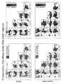
- Results Figure 9shows the raw data.
- 10 and 11show the results of the second and third principal component analyzes. It can be seen that the third time matches the second time.
- FIG. 12shows the results for the culture solution (Bross) containing microorganisms
- FIG. 13shows the results for the supernatant solution (Supernatant) excluding microorganisms. Similar results were obtained in the analysis using the culture solution containing microorganisms and the analysis using the supernatant solution of the culture solution.
- FIG. 14is a diagram showing a time scale
- FIGS. 15 and 16are diagrams showing analysis results by the SIMCA method for the second and third measurements.
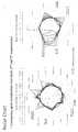
- FIG. 17is a radar chart for the second and third measurements.
Landscapes
- Health & Medical Sciences (AREA)
- Life Sciences & Earth Sciences (AREA)
- Chemical & Material Sciences (AREA)
- Engineering & Computer Science (AREA)
- Bioinformatics & Cheminformatics (AREA)
- Genetics & Genomics (AREA)
- Organic Chemistry (AREA)
- Zoology (AREA)
- Biotechnology (AREA)
- Wood Science & Technology (AREA)
- Microbiology (AREA)
- Medicinal Chemistry (AREA)
- Biomedical Technology (AREA)
- Virology (AREA)
- Biochemistry (AREA)
- General Engineering & Computer Science (AREA)
- General Health & Medical Sciences (AREA)
- Tropical Medicine & Parasitology (AREA)
- Investigating Or Analysing Materials By Optical Means (AREA)
Abstract
Description
Translated fromJapanese本発明は、可視光・近赤外光を利用し、測定対象物の透過光、反射光または透過反射光のスペクトルを解析して、測定対象物中に存在する微生物の増殖段階を識別したり、さらに後の増殖速度を予測したりする微生物の成長の予測方法及びその装置に関する。The present invention uses visible light and near-infrared light to analyze the spectrum of transmitted light, reflected light, or transmitted reflected light of the object to be measured to identify the growth stage of microorganisms present in the object to be measured. The present invention relates to a method for predicting the growth of a microorganism and a device thereof for further predicting the growth rate later.
微生物を利用した重要な産業は多数ある。それら全てにおいて、微生物を増殖させる培養過程があり、その微生物の増殖段階の識別や、後の増殖速度を予測することは重要である。たとえば、食品においては、微生物の増殖は、品質の低下を招き、食品衛生上問題になる。また、近年の再生医療等に代表される細胞培養を基にした次世代技術においても、その増殖段階の識別や後の増殖速度の予測は重要である。There are many important industries that utilize microorganisms. In all of them, there is a culture process in which a microorganism is grown, and it is important to identify the growth stage of the microorganism and predict the subsequent growth rate. For example, in foods, the growth of microorganisms causes deterioration of quality and becomes a problem in food hygiene. Also, in the next-generation technology based on cell culture represented by recent regenerative medicine, it is important to identify the growth stage and predict the subsequent growth rate.
生菌検査は、試料中微生物を固相培地で培養するため、検査結果が出るまで数日を要し、試料を採取した時点での微生物数しか分からないため、増殖段階の識別には経時的に試料を採取する必要があり、後の増殖速度については予測できない。In the viable cell test, since the microorganisms in the sample are cultured in the solid medium, it takes several days until the test result is obtained, and only the number of microorganisms at the time of collecting the sample is known. It is necessary to collect a sample, and the subsequent growth rate is unpredictable.
蛍光フローサイトメトリー法は、数十分で実施可能であるが、試薬を用いて前処理する必要があり、生菌検査と同様に試料を採取した時点での微生物数しかわからないため、増殖段階の識別にはやはり経時的に採取検査する必要があり、後の増殖速度についても予測できない。Fluorescence flow cytometry can be performed in tens of minutes, but it requires pretreatment with reagents, and like the viable cell test, only the number of microorganisms at the time of sample collection is known, so it is in the growth stage. For identification, it is still necessary to collect and inspect over time, and the subsequent growth rate cannot be predicted.
ATP法は、数分で実施できる大変簡便な方法であるが、ATPは全ての生物に存在する物質で、対象微生物由来のATPのみを検出するのは難しく、その精度は高くなく、増殖段階の識別および後の増殖速度の予測もできない。The ATP method is a very simple method that can be carried out in a few minutes, but ATP is a substance that exists in all living organisms, and it is difficult to detect only ATP derived from the target microorganism, its accuracy is not high, and it is in the growth stage. It cannot be identified and the subsequent growth rate cannot be predicted.
品質評価方法として、細胞塊に対して近赤外光を含む測定光を照射することにより、当該細胞塊からの透過光又は拡散反射光に係るスペクトルデータを取得する取得工程と、前記取得工程において取得された前記細胞塊のスペクトルデータに基づいて、前記細胞塊の品質を評価する評価工程とを有するものが提案されている(例えば、特許文献1参照)。As a quality evaluation method, in the acquisition step of acquiring spectral data related to transmitted light or diffused reflected light from the cell mass by irradiating the cell mass with measurement light including near infrared light, and in the acquisition step. A device having an evaluation step for evaluating the quality of the cell mass based on the acquired spectral data of the cell mass has been proposed (see, for example, Patent Document 1).
特許文献1記載の方法は、試料中の微生物の増殖段階の識別や、後の増殖速度の予測には適用しようとしても、簡便に、短い所要時間で実施できず、適宜実施することはできない。Even if the method described in
現在、微生物の状態を把握するための技術としてメタボロミクス等が挙げられるが、測定方法が煩雑で、時間を要することや、コストが高いことが課題となっている。また、予測微生物学では、数理モデルを用いて食品中の微生物の成長の予測を試みているが、pHや温度、塩分濃度などの多岐に渡るパラメーターを測定する必要があり、数理モデルの適用範囲の狭さも課題となっている。Currently, metabolomics and the like can be mentioned as a technique for grasping the state of microorganisms, but the problems are that the measurement method is complicated, it takes time, and the cost is high. In addition, predictive microbiology attempts to predict the growth of microorganisms in foods using a mathematical model, but it is necessary to measure a wide range of parameters such as pH, temperature, and salinity, and the scope of application of the mathematical model. The narrowness of the is also an issue.
したがって、簡易的に微生物の群集の成長や状態を把握し、予測するための技術が求められている。微生物群集の安定性や変化を検知及び予測できれば、食の安全性の向上やスマートセルインダストリーなどの幅広い分野に応用可能となることが考えられるからである。Therefore, there is a need for a technique for easily grasping and predicting the growth and state of microbial communities. This is because if the stability and changes of the microbial community can be detected and predicted, it will be applicable to a wide range of fields such as improvement of food safety and smart cell industry.
発明者は、ミネラルウォーター中の微生物の増殖の様子を近赤外分光法、「アクアフォトミクス(Aquaphotomics)」及び多変量解析を用いて非破壊モニタリングし、ミネラルウォーター中の水分子構造の変化に着目することで、微生物の増殖段階の識別及び増殖速度の予測が可能であることを見出した。ここで、「アクアフォトミクス(Aquaphotomics)」とは、生体(例えば、微生物)、水溶液の性質や変化を水分子の挙動によって捉えようとするものである。The inventor used near-infrared spectroscopy, "Aquaphotomics" and multivariate analysis to non-destructively monitor the growth of microorganisms in mineral water to detect changes in the water molecular structure in mineral water. By paying attention, it was found that it is possible to identify the growth stage of microorganisms and predict the growth rate. Here, "Aquaphotomics" is intended to capture the properties and changes of living organisms (for example, microorganisms) and aqueous solutions by the behavior of water molecules.
さらに説明すると、水分子の水素結合の変動を分析することが、アクアフォトミクスの概念である。アクアフォトミクスと近赤外分光法を組み合わせた分析では、水分子が鏡面の役割となり、鏡を見るように水分子システムの変化を把握することができ、そのシステムの変化を水の吸収スペクトルを通して解析することで、今まで困難とされてきた他の微量な成分や微小な変化を計測することが可能となる。To explain further, analyzing the fluctuation of hydrogen bonds in water molecules is the concept of aquaphotomics. In the analysis that combines aquaphotomics and near-infrared spectroscopy, water molecules act as a mirror surface, and changes in the water molecule system can be grasped as if looking in a mirror, and the changes in the system can be grasped through the absorption spectrum of water. By analyzing, it becomes possible to measure other minute components and minute changes that have been difficult until now.
また、アクアフォトミクスは、水溶液中における物質の情報を抽出するために、対象とする物質そのものの吸収バンドを観察するのではなく、対象とする物質を取り巻く水分子の吸収バンドを積極的に利用し、その物理化学的性質の変化を近赤外スペクトルに示し、バイオシステムを理解しようとする手法である。In addition, aquaphotomics does not observe the absorption band of the target substance itself, but actively uses the absorption band of water molecules surrounding the target substance in order to extract information on the substance in the aqueous solution. However, it is a method of trying to understand the biosystem by showing the changes in its physicochemical properties in the near-infrared spectrum.
また、従来法であるOD600では十分に捉えられないほど低濃度な生菌数であっても、さらに、増殖が開始される誘導期においてもミネラルウォーター中の微生物の状態の把握や成長の予測が可能であることも確認された。In addition, even if the viable cell count is too low to be captured by the conventional method OD600, the state of microorganisms in mineral water can be grasped and the growth can be predicted even in the induction period when the growth is started. It was also confirmed that it was possible.
発明者は、測定対象物に600-1800nmの可視光・近赤外光の全波長または水の近赤外領域の吸収帯を含むその一部を照射して得られる測定対象物からの透過光、反射光または透過反射光のスペクトルを、多変量解析あるいは特定波長あるいは特定波長領域におけるスペクトルパターンの変化を解析すれば、測定対象物中に存在する微生物の増殖段階を識別したり、その後の増殖速度を予測したりできることに着目し、本発明をなした。The inventor irradiates the measurement object with the entire wavelength of visible light / near-infrared light of 600-1800 nm or a part of the measurement target including the absorption band in the near-infrared region of water, and the transmitted light from the measurement object is obtained. By multivariate analysis or analysis of changes in the spectral pattern in a specific wavelength or a specific wavelength region, the spectrum of reflected light or transmitted reflected light can be used to identify the growth stage of microorganisms present in the object to be measured, and to identify the growth stage thereafter. The present invention has been made by focusing on the ability to predict the speed.
本発明は、測定対象物中の微生物の増殖段階の識別及び増殖速度の予測を、簡便で、所要時間が短く、精度が高く、適宜実施可能である微生物の成長の予測方法及びその装置を提供する。The present invention provides a method for predicting the growth of a microorganism and an apparatus thereof, which can easily identify the growth stage of the microorganism and predict the growth rate in the object to be measured, have a short required time, have high accuracy, and can be appropriately carried out. To do.
本発明は、波長600-1800nmの範囲またはその一部範囲の波長光を照射部から、微生物を含む培養液、又は前記微生物を除く前記培養液の上澄み液である測定対象物に照射し、その反射光、透過光または透過反射光を検出して前記微生物の成長を予測する微生物の成長の予測方法であって、前記反射光、透過光または透過反射光を検出して吸光度スペクトル測定を行った後、前記測定対象物の水分子構造に由来する吸光度の変化を検出し、その変化により予め作成した解析モデルを用いて解析することによって、前記測定対象物中に存在する微生物の増殖段階の識別を行ったり、前記測定対象物中に存在する微生物の増殖速度の予測を行うことを特徴とするものである。In the present invention, a measurement object that is a culture solution containing microorganisms or a supernatant liquid of the culture solution excluding the microorganisms is irradiated with light having a wavelength in the range of 600-1800 nm or a part thereof from the irradiation unit. A method for predicting the growth of a microorganism by detecting reflected light, transmitted light or transmitted reflected light, and measuring the absorbance spectrum by detecting the reflected light, transmitted light or transmitted reflected light. Later, by detecting the change in absorbance derived from the water molecular structure of the object to be measured and analyzing the change using an analysis model created in advance, the growth stage of the microorganism existing in the object to be measured can be identified. The feature is that the growth rate of the microorganism existing in the object to be measured is predicted.
このようにすれば、近赤外領域で捉えられる水分子構造という単一のパラメーターに着目することにより、リアルタイムで微生物の状態の把握や成長の予測が可能となり、測定対象物中に存在する微生物の増殖段階の識別及び後の増殖速度の予測が、簡便かつ、短時間に高い精度で実施可能となる。In this way, by focusing on the single parameter of the water molecular structure captured in the near-infrared region, it is possible to grasp the state of microorganisms and predict their growth in real time, and the microorganisms existing in the measurement target. It becomes possible to identify the growth stage of the microorganism and predict the subsequent growth rate easily and with high accuracy in a short time.
本発明で用いた近赤外分光法の特徴として安価、迅速、簡便という特徴が挙げられる。それにもかかわらず、近赤外領域で捉えられる水分子構造という単一のパラメーターに着目することにより、リアルタイムで微生物の状態の把握や成長の予測が可能となる点が大きな優位性となる。
この場合、前記測定対象物への照射は、前記測定対象物に繰り返し照射により摂動を与えながら行う、ことが望ましい。The near-infrared spectroscopy used in the present invention is characterized by being inexpensive, quick, and simple. Nevertheless, by focusing on the single parameter of the water molecular structure captured in the near-infrared region, it is possible to grasp the state of microorganisms and predict their growth in real time, which is a great advantage.
In this case, it is desirable that the measurement target is irradiated while perturbing the measurement target by repeated irradiation.
また、前記測定対象物に照射される光の波長は、前記微生物の代謝に重要なものであるプロトンに由来する水分子構造に帰属される波長であり、 前記測定対象物に照射される光の波長が、1110-1600nmの範囲に設定されている、ことが望ましい。Further, the wavelength of the light irradiated to the measurement object is the wavelength attributed to the water molecular structure derived from the proton, which is important for the metabolism of the microorganism, and the wavelength of the light irradiated to the measurement object. It is desirable that the wavelength is set in the range of 1110-1600 nm.
そして、前記解析モデルは、定性分析、定量分析を含む多変量解析と、微生物、水溶液の性質や変化を水分子の挙動によって捉えるアクアフォトミクスとを用いて作成する、ことができる。Then, the analysis model can be created by using multivariate analysis including qualitative analysis and quantitative analysis, and aquaphotomics that captures the properties and changes of microorganisms and aqueous solutions by the behavior of water molecules.
本発明は、微生物の成長の予測装置であって、波長600-1800nmの範囲またはその一部範囲の波長光を照射部から、微生物及び液体を含む測定対象物に照射する照射部と、前記照射部から照射される光を、照射前又は照射後に分光する分光部と、前記測定対象物に照射された光の反射光、透過光または透過反射光を検出する検出部と、前記検出部によって得られた吸光度スペクトルデータから、前記測定対象物の水分子構造に由来する吸光度の変化を検出し、予め作成した解析モデルを用いて解析し、前記測定対象物中に存在する微生物の増殖段階の識別を行うデータ解析部とを備えることを特徴とする。The present invention is an irradiation unit for predicting the growth of microorganisms, and an irradiation unit that irradiates a measurement object containing a microorganism and a liquid with light having a wavelength in the wavelength range of 600-1800 nm or a part thereof from the irradiation unit, and the irradiation. Obtained by a spectroscopic unit that disperses the light emitted from the unit before or after irradiation, a detection unit that detects reflected light, transmitted light, or transmitted reflected light of the light irradiated to the measurement object, and the detection unit. From the obtained absorbance spectrum data, the change in absorbance derived from the water molecular structure of the object to be measured is detected and analyzed using an analysis model prepared in advance to identify the growth stage of the microorganism existing in the object to be measured. It is characterized by having a data analysis unit for performing the above.
また、本発明は、微生物の成長の予測装置であって、波長600-1800nmの範囲またはその一部範囲の波長光を照射部から、微生物及び液体を含む測定対象物に照射する照射部と、前記照射部から照射される光を、照射前又は照射後に分光する分光部と、前記測定対象物に照射された光の反射光、透過光または透過反射光を検出する検出部と、前記検出部によって得られた吸光度スペクトルデータから、前記測定対象物の水分子構造に由来する吸光度の変化を検出し、予め作成した解析モデルを用いて解析し、前記測定対象物中に存在する微生物の増殖速度の予測を行うデータ解析部とを備えることを特徴とする。Further, the present invention is an irradiation unit for predicting the growth of microorganisms, and an irradiation unit that irradiates a measurement object including a microorganism and a liquid with light having a wavelength in the wavelength range of 600-1800 nm or a part thereof. A spectroscopic unit that disperses the light emitted from the irradiation unit before or after irradiation, a detection unit that detects reflected light, transmitted light, or transmitted reflected light of the light irradiated to the measurement object, and the detection unit. The change in absorbance derived from the water molecular structure of the object to be measured was detected from the absorbance spectrum data obtained in the above-mentioned method, and the change in absorbance was analyzed using an analysis model prepared in advance, and the growth rate of microorganisms present in the object to be measured was measured. It is characterized by having a data analysis unit that predicts the above.
本発明は、近赤外領域で捉えられる水分子構造という単一のパラメーターに着目することにより、リアルタイムで微生物の状態の把握や成長の予測が可能となり、測定対象物中の微生物の増殖段階の識別や後の増殖速度の予測を、簡便、短時間に高い精度で実施可能となる。By focusing on a single parameter of water molecular structure captured in the near-infrared region, the present invention makes it possible to grasp the state of microorganisms and predict their growth in real time, and to predict the growth stage of microorganisms in the measurement target. Identification and subsequent prediction of growth rate can be performed easily, in a short time, and with high accuracy.
以下、本発明の実施の形態を図面を参照しながら説明する。
(1)本装置による測定とデータの解析方法
(1-1)測定原理
図1に示すように、(a)波長600nm~1800nmの範囲またはその一部範囲の波長光を測定対象物(たとえば、微生物が混入したミネラルウォーター)に照射し、(b)その反射光、透過光または透過反射光を検出して吸光度スペクトル測定を行った後、(c)前記測定対象物の水分子構造に由来する吸光度の変化を検出し(つまり、アクアフォトミクス(Aquaphotomics)に基づく多変量解析を行って、微生物による特徴的な水分子構造に由来するスペクトルパターンを発見して)、その変化に基づいて予め作成した解析モデルを用いて解析することによって、測定対象物中に存在する微生物の増殖段階の識別や増殖速度の予測を行う。Hereinafter, embodiments of the present invention will be described with reference to the drawings.
(1) Measurement by this device and data analysis method (1-1) Measurement principle As shown in FIG. 1, (a) wavelength light in the wavelength range of 600 nm to 1800 nm or a part thereof is measured (for example, an object to be measured (for example). (Mineral water mixed with microorganisms) is irradiated, (b) the reflected light, transmitted light or transmitted reflected light is detected and the absorbance spectrum is measured, and then (c) it is derived from the water molecular structure of the object to be measured. Detect changes in absorbance (ie, perform multivariate analysis based on Aquaphotomics to discover spectral patterns derived from characteristic water molecular structures by microorganisms) and pre-create based on those changes By analyzing using the analysis model, the growth stage of the microorganism existing in the object to be measured is identified and the growth rate is predicted.
つまり、測定対象物中に存在する微生物を特定し、その微生物について予め作成した解析モデルを利用して増殖段階を識別し、また、微生物とその増殖段階がわかるので、その後の増殖速度を予測できる。That is, the microorganism existing in the measurement target is identified, the growth stage is identified by using the analysis model created in advance for the microorganism, and the microorganism and its growth stage are known, so that the subsequent growth rate can be predicted. ..
ここで、光源としては、ハロゲンランプ・LED等を使用できるが、特に限定されるものではない。光源から発せられた光は、直接またはファイバープローブ等の投光手段を介して測定対象物である試料に照射される。後述のように、試料に照射する前に分光器によって分光する前分光方式を採用してもよいし、照射後に分光する後分光方式を採用してもよい(図3参照)。Here, a halogen lamp, an LED, or the like can be used as the light source, but the light source is not particularly limited. The light emitted from the light source irradiates the sample to be measured directly or through a light projecting means such as a fiber probe. As will be described later, a pre-spectral method in which the sample is separated by a spectroscope before irradiation may be adopted, or a post-spectral method in which the sample is separated after irradiation may be adopted (see FIG. 3).
試料に照射された光の反射光、透過光または透過反射光が検出器により検出され、生の吸光度スペクトルデータが得られる。ここで、近赤外線領域で捉えられる水分子構造という単一のパラメーターに着目することにより、リアルタイムで微生物の増殖段階や増殖速度の予測が可能となる。The reflected light, transmitted light or transmitted reflected light of the light applied to the sample is detected by the detector, and the raw absorbance spectrum data can be obtained. Here, by focusing on a single parameter of the water molecular structure captured in the near-infrared region, it is possible to predict the growth stage and growth rate of microorganisms in real time.
また、生の吸光度スペクトルデータをそのまま使用して解析モデルによる識別を行ってもよいが、得られたスペクトル中のピークを分光学的手法あるいは多変量解析手法により要素ピークに分解するなどのデータ変換処理を行い、変換後の吸光度スペクトルデータを使用して解析モデルによる識別を行うこともできる。In addition, the raw absorbance spectrum data may be used as it is for identification by an analysis model, but data conversion such as decomposing the peak in the obtained spectrum into element peaks by a spectroscopic method or a multivariate analysis method. It is also possible to perform the processing and use the absorbance spectrum data after conversion for identification by an analysis model.
分光学的手法としては、例えば、2次微分処理やフーリエ変換があり、多変量解析手法としてはウェブレット変換、ニューラルネットワーク法等が例示されるが、特に限定されるものではない。なお、スペクトル測定においては、後述するように、試料に対し、所定の条件を付加することで摂動(perturbation)を与えることが好ましい。Examples of spectroscopic methods include second-order differential processing and Fourier transform, and examples of multivariate analysis methods include wavelet transformation and neural network method, but the method is not particularly limited. In the spectrum measurement, as will be described later, it is preferable to give a perturbation to the sample by adding a predetermined condition.
(1-2)データの解析方法(解析モデルの作成)
本装置は、上述のようにして得られた吸光度スペクトルデータの中の特定波長(または測定全波長)の吸光度スペクトルパターン、つまり微生物による特徴的な水分子構造に由来する吸光度スペクトルパターンを解析することによって、測定対象物中に存在する微生物の増殖段階の識別や増殖速度の予測を行う。(1-2) Data analysis method (creation of analysis model)
This device analyzes the absorbance spectrum pattern of a specific wavelength (or all measured wavelengths) in the absorbance spectrum data obtained as described above, that is, the absorbance spectrum pattern derived from the characteristic water molecular structure of the microorganism. To identify the growth stage and predict the growth rate of the microorganisms present in the object to be measured.
つまり、最終的な識別を行うには、解析モデルが予め作成されていることを要する。もっとも、この解析モデルはスペクトル測定時にあわせて作成することとしてもよい。In other words, it is necessary that the analysis model is created in advance in order to make the final identification. However, this analysis model may be created at the time of spectrum measurement.
すなわち、解析モデルは測定前に予め作成しておくことが望ましいが、測定時に取得するスペクトルデータを解析モデル作成用と識別・予測用とに2分割し、解析モデル作成用データをもとに得られた解析モデルを使用して識別・予測を行ってもよい。定量および定性モデルの両方に対応可能である。That is, it is desirable to create an analysis model in advance before measurement, but the spectrum data acquired at the time of measurement is divided into two parts, one for analysis model creation and the other for identification / prediction, and the analysis model is obtained based on the analysis model creation data. Identification / prediction may be performed using the analysis model obtained. It is compatible with both quantitative and qualitative models.
解析モデルは多変量解析によって作成可能である。微生物による特徴的な水分子構造に由来する吸光度スペクトルパターンから微生物の増殖段階を推定する解析モデルや増殖速度を予測する解析モデルを作成できる。解析モデルの作成は、自作ソフトや市販の多変量解析ソフトを用いて行うことができる。また、使用目的に特化したソフトの作成により、迅速な解析が可能になる。The analysis model can be created by multivariate analysis. It is possible to create an analysis model that estimates the growth stage of microorganisms and an analysis model that predicts the growth rate from the absorbance spectrum pattern derived from the characteristic water molecular structure of microorganisms. The analysis model can be created by using self-made software or commercially available multivariate analysis software. In addition, by creating software specialized for the purpose of use, quick analysis becomes possible.
このような多変量解析ソフトを用いて組み立てられた解析モデルをファイルとして保存しておき、未知試料の識別時にこのファイルを呼び出し、未知試料に対して解析モデルを用いた定量的または定性的な解析を行う。これにより、微生物の増殖段階を識別したりや増殖速度を予測したりすることが可能になる。なお、解析モデルは、定量モデル、定性モデルなど複数の解析モデルをファイルとして保存しておき、各モデルは適宜更新されることが好ましい。An analysis model constructed using such multivariate analysis software is saved as a file, and this file is called when an unknown sample is identified, and a quantitative or qualitative analysis using the analysis model is performed on the unknown sample. I do. This makes it possible to identify the growth stage of microorganisms and predict the growth rate. As the analysis model, it is preferable to save a plurality of analysis models such as a quantitative model and a qualitative model as a file, and update each model as appropriate.
解析モデルが作成されれば、当該解析モデルによる微生物の増殖段階の識別や増殖速度の予測に必要な波長光が決定される。本装置は、こうして決定された1又は複数の波長域を試料に照射する構成とすることで装置構成をより単純化することができる。Once the analysis model is created, the wavelength light required for identifying the growth stage of microorganisms and predicting the growth rate by the analysis model is determined. The device configuration can be further simplified by irradiating the sample with one or more wavelength ranges determined in this way.
(1-3)本装置による測定方法とデータ解析方法
本装置によるスペクトル測定においては、試料に対し、所定の条件を付加することで摂動(perturbation)を与えることが好ましく、また、本装置によるデータ解析においては、この摂動の効果を引き出すようなデータ解析が好ましいものとなる。(1-3) Measurement method and data analysis method by this device In spectrum measurement by this device, it is preferable to give perturbation to a sample by adding predetermined conditions, and data by this device. In the analysis, data analysis that brings out the effect of this perturbation is preferable.
摂動(perturbation)とは、ある条件について複数の種類・条件を設定し測定することで試料の吸光度変化をもたらし、互いに異なる複数のスペクトルデータを取得することをいう。条件としては、濃度変更(濃度希釈を含む)、光の繰り返し照射、照射時間の延長、電磁力付加、光路長変更、温度、pH、圧力、機械的振動、その他その条件の変更によって物理的または化学的な変化をもたらすもののいずれか、または、それらの組み合わせを挙げることができ、(1)光照射の仕方に関するものと、(2)試料の準備・調製の仕方に関するものとに大別される。Perturbation refers to the acquisition of multiple spectral data that are different from each other by setting and measuring multiple types and conditions for a certain condition to bring about a change in the absorbance of the sample. Conditions include changes in concentration (including dilution of concentration), repeated irradiation of light, extension of irradiation time, addition of electromagnetic force, change in optical path length, temperature, pH, pressure, mechanical vibration, and other physical or changes in the conditions. Any of those that bring about chemical changes or a combination thereof can be mentioned, and they are roughly classified into (1) those related to the method of light irradiation and (2) those related to the method of preparing and preparing a sample. ..
光の繰り返し照射は、連続して又は一定の時間間隔で光を繰り返し照射して複数回の測定という摂動を与えて試料のスペクトル測定を行う方法である。例えば、光を3回連続照射することにより、試料の吸光度が微妙に変化し(揺らぎ)、互いに異なる複数のスペクトルデータが得られる。Repeated irradiation of light is a method of measuring the spectrum of a sample by repeatedly irradiating light continuously or at regular time intervals and giving a perturbation of multiple measurements. For example, by continuously irradiating light three times, the absorbance of the sample changes (fluctuations) slightly, and a plurality of spectral data different from each other can be obtained.
これらのスペクトルデータをSIMCA法やPLS法等の多変量解析に用いることにより、解析精度を向上することができ、高精度な、微生物の増殖段階の識別や増殖速度の予測が可能になる。なお、通常スペクトルを測定するときは、光を複数回照射し測定するが、これは平均値を出すことが目的であり、ここでいう「摂動」とは異なる。By using these spectral data for multivariate analysis such as the SIMCA method and PLS method, the analysis accuracy can be improved, and highly accurate identification of the growth stage of microorganisms and prediction of the growth rate become possible. Normally, when measuring a spectrum, light is irradiated a plurality of times to measure the spectrum, but this is for the purpose of obtaining an average value, which is different from the "perturbation" referred to here.
摂動による試料の吸光度変化は、試料中の水分子の吸収に変化(揺らぎ)が生じるためと考えられる。すなわち摂動として光を3回繰り返し照射することによって、1回目、2回目、3回目それぞれ水の応答、吸収に微妙に異なる変化が起こり、その結果スペクトルに揺らぎが生じるものと考えられる。It is considered that the change in the absorbance of the sample due to perturbation is due to a change (fluctuation) in the absorption of water molecules in the sample. That is, it is considered that by repeatedly irradiating light three times as a perturbation, slightly different changes occur in the response and absorption of water in the first, second, and third times, and as a result, the spectrum fluctuates.
このように光を3回繰り返し照射した場合、得られた3回の吸光度スペクトルデータのうち少なくとも2回の吸光度スペクトルデータを使用してSIMCA法によるクラス判別を行うことによって、各試料を良好に分類することができ、高精度な識別が可能である。光照射回数は特に3回に制限されないが、データ解析の煩雑さ等を考慮すると、3回程度が好ましい。When light is repeatedly irradiated three times in this way, each sample is satisfactorily classified by classifying by the SIMCA method using at least two absorbance spectrum data out of the obtained three absorbance spectrum data. It is possible to identify with high accuracy. The number of times of light irradiation is not particularly limited to three, but it is preferably about three in consideration of the complexity of data analysis and the like.
光の繰り返し照射以外の摂動の条件についても同様に、取得するスペクトルに揺らぎを生じさせるよう、各条件について複数の種類・条件を設定し、スペクトル測定を行えばよい(特許第4710012号公報参照)。Similarly, for perturbation conditions other than repeated irradiation of light, a plurality of types and conditions may be set for each condition and spectrum measurement may be performed so as to cause fluctuations in the acquired spectrum (see Japanese Patent No. 4710012). ..
(2)本装置の具体的構成
本装置の測定システムの構成としては、図2に示すように、照射部2、分光・検出部3、データ解析部4および結果表示部5の4つの要素を備えて構成することができる。以下では、各要素について説明する。(2) Specific configuration of this device As shown in FIG. 2, the measurement system of this device consists of four elements: an
(2-1)照射部2
照射部2は、ハロゲンランプ・LED等の光源からの光(波長400nm~2500nmの全範囲またはその一部範囲)を測定対象物である試料に導く機能を有する。例えばファイバープローブとし、柔軟な光ファイバーを介して測定対象物(試料)に投光する構成が挙げられる。(2-1)
The
なお、光源から発せられた光を直接測定対象物である試料に投光する構成としてもよいが、その場合プローブは不要であり、光源が投光手段として機能する。Note that the light emitted from the light source may be directly projected onto the sample to be measured, but in that case, a probe is not required and the light source functions as a light projecting means.
前述のように、解析モデルが作成されれば、解析モデルによる微生物の増殖段階の識別や増殖速度の予測に必要な波長光が決定される。本装置は、こうして決定された1又は複数の波長域を試料に照射する構成とすることで装置構成をより単純化することができる。As described above, once the analysis model is created, the wavelength light required for identifying the growth stage of the microorganism and predicting the growth rate by the analysis model is determined. The device configuration can be further simplified by irradiating the sample with one or more wavelength ranges determined in this way.
また前述のように、本装置は、摂動を与えながらスペクトル測定を行うため、摂動付与に必要な構成を適宜備えることが好ましい。Further, as described above, since this device measures the spectrum while giving perturbation, it is preferable to appropriately provide a configuration necessary for giving perturbation.
(2-2)分光・検出部3(分光手段および検出手段)
分光・検出部3は、測定対象物に照射される光を分光して、この測定対象物からの反射光や透過光あるいは透過反射光を検出する。さらに、検出された光について波長別に入射光に対する吸光度が測定される。(2-2) Spectroscopy / detection unit 3 (spectroscopic means and detection means)
The spectroscopy /
分光方式には前分光と後分光とがある(図3参照)。前分光は、測定対象物に投光する前に分光する。後分光は、測定対象物からの光を検出し分光する。本装置の分光・検出部3は、前分光、後分光いずれの分光方式を採用するものであってもよい。There are two types of spectroscopy: pre-spectroscopy and post-spectroscopy (see Fig. 3). Pre-spectroscopy is spectroscopy before projecting light onto the object to be measured. Post-spectroscopy detects and disperses light from an object to be measured. The spectroscopic /
検出方法には3種類あり、反射光検出、透過光検出および透過反射光検出がある(図4参照)。同図に示すように、反射光検出および透過光検出は、それぞれ、測定対象物からの反射光と透過光とを検出器によって検出する。透過反射光検出は、入射光が測定対象物内に入射した屈折光が物体内で反射し、再び物体外に放射された光が反射光と干渉する光を検出する。本装置の分光・検出部3は、反射光検出、透過光検出および透過反射光検出のいずれの検出方式を採用するものであってもよい。There are three types of detection methods: reflected light detection, transmitted light detection, and transmitted reflected light detection (see Fig. 4). As shown in the figure, in the reflected light detection and the transmitted light detection, the reflected light and the transmitted light from the measurement object are detected by the detector, respectively. In the transmitted reflected light detection, the refracted light that is incident on the object to be measured is reflected inside the object, and the light that is radiated outside the object again interferes with the reflected light. The spectroscopic /
分光・検出部3内の検出器は、例えば半導体素子であるCCD(Charge Coupled Device)などによって構成することができるが、勿論これに限定されるものではなく、他の受光素子を使用してもよい。分光器についても公知の手段によって構成することができる。The detector in the spectroscopic /
(2-3)データ解析部4
分光・検出部3から波長別の吸光度、即ち吸光度スペクトルデータが得られる。データ解析部4は、この吸光度スペクトルデータをもとに、微生物の種類を特定し、前述のように予め作成した、前記微生物の種類の解析モデルを使用して、微生物の増殖段階の識別や増殖速度の予測を行う。つまり、解析モデルから、増殖過程のいずれの段階にあるかを判別でき、増殖段階のいずれの段階にあるかとそれまでの時間の経過から,増殖速度の予測を行うことができる。(2-3)
Absorbance for each wavelength, that is, absorbance spectrum data can be obtained from the spectroscopy /
解析モデルは、定量モデル、定性モデルなど複数の解析モデルを用意しておき、定量的評価を行うか、あるいは定性的評価を行うかに応じて、異なるものを使用してもよい。As the analysis model, a plurality of analysis models such as a quantitative model and a qualitative model may be prepared, and different ones may be used depending on whether the quantitative evaluation or the qualitative evaluation is performed.
データ解析部4は、スペクトルデータ、多変量解析用プログラム、解析モデルなどの各種データを記憶する記憶部と、これらのデータおよびプログラムに基づき演算処理を行う演算処理部とによって構成することができ、例えばICチップなどによって実現可能である。したがって、本装置を携帯型とするため小型化することも容易である。上記の解析モデルも、ICチップなどの記憶部に書き込まれる。The
(2-4)結果表示部5
結果表示部5は、データ解析部4における解析結果を表示する。具体的には、解析モデルによる解析の結果得られた試料中の微生物の増殖段階や増殖速度の予測結果を表示する。なお、本装置を携帯型とする場合は、結果表示部5を液晶等のフラットディスプレイとすることが好ましい。(2-4)
The
-実施例1-
市販ミネラルウォーターに3種類の菌を触菌し(2CFU/mL)、次の各経過時間後(分)に、微生物が混入したミネラルウォーターの分光測定と生菌数との測定を行った。その後、多変量解析と特定波長におけるスペクトル解析を行った。-Example 1-
Three kinds of bacteria were touched on commercially available mineral water (2 CFU / mL), and after each elapsed time (minutes), spectroscopic measurement and viable cell count of the mineral water mixed with microorganisms were performed. After that, multivariate analysis and spectral analysis at a specific wavelength were performed.
1.方法
(菌種)
Acidovorax
Pseudomonas
Sphingomonas1. 1. Method (bacterial species)
Acidovorax
Pseudomonas
Sphingomonas
(測定時間)
70, 90, 120, 150, 180, 190, 240, 300, 330, 340, 390, 420, 460, 480, 510, 540, 580, 650, 670, 720, 760, 770, 800, 840, 1440(24時間), 1740(29時間), 2880(48時間), 3180(53時間), 4320(72時間)(Measurement time)
70, 90, 120, 150, 180, 190, 240, 300, 330, 340, 390, 420, 460, 480, 510, 540, 580, 650, 670, 720, 760, 770, 800, 840, 1440 ( 24 hours), 1740 (29 hours), 2880 (48 hours), 3180 (53 hours), 4320 (72 hours)
(分光測定)
近赤外分光器:XDS - Rapid Liquid Analyzer (FOSS.Co.,Ltd)(Spectroscopic measurement)
Near-infrared spectrometer: XDS-Rapid Liquid Analyzer (FOSS.Co., Ltd)
(生菌測定)
平板法(Measurement of viable bacteria)
Flat plate method
(解析方法)
-多変量解析:アルゴリズム SIMCA
-1100~1300nm及び1300~1600nmにおけるスペクトル解析:(analysis method)
-Multivariate analysis: Algorithm SIMCA
-1100 to 1300 nm and 1300 to 1600 nm spectral analysis:
次式によりスペクトル解析を行い、結果はレーダーチャートで表示した。なお、1100~1300nmは水の結合音の第1倍音、1300~1600nmは水の第1倍音である。The spectrum was analyzed by the following formula, and the result was displayed on the radar chart. Note that 1100 to 1300 nm is the first harmonic overtone of water, and 1300 to 1600 nm is the first overtone of water.

2.結果
2-1増速曲線
図5(a)(b)に示すとおりであり、増殖速度が速いものや遅いものがあることがわかる。2. Result 2-1 Acceleration curve As shown in FIGS. 5 (a) and 5 (b), it can be seen that some have a high growth rate and some have a slow growth rate.
2-2増殖段階の識別
・SIMCAによる増殖段階2-2 Identification of growth stage ・ Growth stage by SIMCA
AcidovoraxとPseudomonasとについて、測定波長領域1110~1300nmと1300~1600nmにおいて、93%以上の識別精度で増殖段階の識別が可能であった。Regarding Acidovorax and Pseudomonas, it was possible to identify the growth stage with a discrimination accuracy of 93% or more in the measurement wavelength regions of 1110 to 1300 nm and 1300 to 1600 nm.
・1300~1600nmにおけるスペクトル解析
アクアグラム(Aquagram)による増殖段階の識別の結果は図6に示すとおりである。-Spectral analysis at 1300 to 1600 nm The results of identification of the growth stage by Aquagram are shown in FIG.
(i)誘導期は1451~1511nmにおける吸収が大きく(アクアグラム値≒0.3)、1342~1438nmにおける吸収はほとんどない。(I) In the induction period, absorption is large at 1451 to 1511 nm (aquagram value ≈ 0.3), and absorption is almost nonexistent at 1342 to 1438 nm.
(ii) 移行期では1451~1511nmにおける吸収が誘導期に比べて小さくなる(アクアグラム値≒0.2)。(Ii) Absorption in the transition period from 1451 to 1511 nm is smaller than in the induction period (aquagram value ≈ 0.2).
(iii)対数増殖期前半では、1451~1511nmにおける吸収は水とほとんど差がなくなる(アクアグラム値≒0)。(Iii) In the first half of the logarithmic growth phase, the absorption at 1451 to 1511 nm is almost the same as that of water (aquagram value ≈ 0).
(iv)対数増殖期後半では、1451~1511nmにおける吸収はほとんどなくなり(アクアグラム値≒0)、1342~1438nmにおける吸収が大きくなる(アクアグラム値≒0.2)(Iv) In the latter half of the logarithmic growth phase, absorption at 1451 to 1511 nm is almost eliminated (aquagram value ≈ 0), and absorption at 1342 to 1438 nm is increased (aquagram value ≈ 0.2).
(v)静止期では、1342~1438nmにおける吸収が、対数増殖期後半よりさらに大きくなる(アクアグラム値≒0.3)(V) In the stationary phase, absorption at 1342 to 1438 nm is even greater than in the latter half of the logarithmic growth phase (aquagram value ≈ 0.3).
前述したように、ミネラルウォーターに微生物を触菌し、増殖速度を近赤外分光法によりモニタリングすると同時に、従来法である寒天培地法により、微生物の増殖曲線の観察を行った。そして、図6に示すように、増殖段階を、誘導期、移行期及び対数増殖期に分割して、スペクトルデータを用いて多変量解析により、増殖段階の識別を行った。As described above, the microorganisms were touched on mineral water and the growth rate was monitored by near-infrared spectroscopy, and at the same time, the growth curve of the microorganisms was observed by the conventional agar medium method. Then, as shown in FIG. 6, the growth stage was divided into an induction stage, a transition stage, and a logarithmic growth stage, and the growth stage was identified by multivariate analysis using spectral data.
また、増殖曲線を観察する中で、図5(a)(b)に示すように、増殖速度が大きい菌株と小さい菌株とがあるが、それらのスペクトルデータにも、図6の増殖段階の分割を適用し、それぞれの増殖段階の識別をアクアグラム(吸光度を標準化しレーダーチャート化)及び多変量解析の定性分析の手法の1つであるSIMCA法により行った。In addition, while observing the growth curve, as shown in FIGS. 5 (a) and 5 (b), there are strains having a high growth rate and strains having a low growth rate. Was applied, and each growth stage was identified by the SIMCA method, which is one of the qualitative analysis methods of aquagram (standardizing absorbance and radar charting) and multivariate analysis.
近赤外分光法を用いて微生物の増殖段階のモニタリングを行い、水分子構造を反映する1110~1300 nm, 1300~1600 nmの2波長領域を用いて、アクアグラム(1300~1600 nmにおける増殖段階の識別のみ)及び多変量解析の定性分析の手法の1つであるSIMCA法により菌株毎の増殖段階の識別や増殖速度の予測を行った。図7に増殖速度の予測精度を、図8に増殖段階の識別精度を示す。The growth stage of microorganisms is monitored using near-infrared spectroscopy, and the aquagram (growth stage at 1300 to 1600 nm) is used in the two wavelength regions of 1110 to 1300 nm and 1300 to 1600 nm, which reflect the water molecular structure. The growth stage was identified and the growth rate was predicted for each strain by the SIMCA method, which is one of the qualitative analysis methods for multivariate analysis. FIG. 7 shows the prediction accuracy of the growth rate, and FIG. 8 shows the identification accuracy of the growth stage.
図6に示すように、アクアグラムでは無菌のミネラルウォーターと比較して、微生物を有するミネラルウォーターは、誘導期から対数増殖期までは長波長側に大きな吸収があり、対数増殖期の後半から短波長側に吸収が大きくなるという特徴的なスペクトルパターンが見られた。吸収される波長によって、微生物の増殖段階が識別できる、といえる。 図7及び図8より、増殖段階の識別及び増殖速度の予測が高い精度で行えることが確認された。As shown in FIG. 6, compared with sterile mineral water in Aquagram, mineral water having microorganisms has a large absorption on the long wavelength side from the induction phase to the logarithmic growth phase, and is shorter from the latter half of the logarithmic growth phase. A characteristic spectral pattern was observed in which absorption increased on the wavelength side. It can be said that the growth stage of microorganisms can be identified by the absorbed wavelength. From FIGS. 7 and 8, it was confirmed that the growth stage can be identified and the growth rate can be predicted with high accuracy.
そして、解析モデルによる増殖段階の識別の結果、増殖が開始する前の誘導期や移行期の段階においても、その解析モデルに基づき増殖速度の予測が可能であり、通常は増殖段階の分割に含まれない移行期において増殖速度の予測精度が最も高くなっている。Then, as a result of identifying the growth stage by the analysis model, it is possible to predict the growth rate based on the analysis model even in the induction stage or the transition stage before the start of growth, and it is usually included in the division of the growth stage. The prediction accuracy of the growth rate is the highest in the transitional period.
また、増殖段階の識別及び増殖速度の予測に有効な波長の探索を行った結果、プロトンに由来する水分子構造に帰属される波長が多く見られた。プロトンは微生物の代謝に重要なものであり、この現象が反映されたものを捉えた結果として、増殖段階の識別や増殖速度の予測ができた、といえる。In addition, as a result of searching for wavelengths that are effective in identifying the growth stage and predicting the growth rate, many wavelengths attributed to the water molecular structure derived from protons were found. Protons are important for the metabolism of microorganisms, and as a result of capturing the reflection of this phenomenon, it can be said that the growth stage can be identified and the growth rate can be predicted.
-実施例2-
(目的)
・正常な試料と異常な試料との相違を観察すること
・早期の段階で、異常な増殖を予測する方法を見つけること-Example 2-
(Purpose)
-Observing the difference between normal and abnormal samples-Finding ways to predict abnormal growth at an early stage
(試料)
試料は微生物を含む培養液(Broth)と、微生物を除く上澄み液(Supernatant)とを用いた。(sample)
As a sample, a culture solution containing microorganisms (Broth) and a supernatant solution excluding microorganisms (Supernatant) were used.
・正常な試料
2HN-2
2HN-3
(測定時間)
0,6,9,12,15,17,20,24,27,30,33,36,39,41,44,46,48,65h・ Normal sample 2HN-2
2HN-3
(Measurement time)
0,6,9,12,15,17,20,24,27,30,33,36,39,41,44,46,48,65h
・異常な試料
2HN-1
2HN-4
(測定時間)
0,6,9,12,15,17,20,24,25.5,27,30,33,36,39,41h・ Abnormal sample 2HN-1
2HN-4
(Measurement time)
0,6,9,12,15,17,20,24,25.5,27,30,33,36,39,41h
(測定機器)
・近接外分光器:XDS - Rapid Liquid Analyzer(FOSS.CO,.LTD)
・透過度:Path length 1mm
・キュベットセル:石英
・波長:400-2499.5nm(0.5nm step)
・連続獲得スペクトル:3
・レプリカ:1 繰り返し須:2(measuring equipment)
-Out-of-proximity spectroscope: XDS-Rapid Liquid Analyzer (FOSS.CO, .LTD)
-Transparency:
-Cuvette cell: Quartz-Wavelength: 400-2499.5 nm (0.5 nm step)
・ Continuous acquisition spectrum: 3
・ Replica: 1 Repeat Su: 2
市販ミネラルウォーターに,前述した試料である菌を食菌し(CFU/mL)、前記各測定時間経過後(分)に、微生物が混入したミネラルウォータ-の分光測定と生菌数との測定を行った。その後、多変量解析と特定波長におけるスペクトル解析を行った。The above-mentioned sample bacteria are phagocytosed (CFU / mL) in commercially available mineral water, and after each measurement time elapses (minutes), spectroscopic measurement of mineral water mixed with microorganisms and measurement of viable cell count are performed. went. After that, multivariate analysis and spectral analysis at a specific wavelength were performed.
・解析方法
-多変量解析:アルゴリズム SIMCA
-1100~1300nm及び1300~1600nmにおけるスペクトル解析:-Analysis method-Multivariate analysis: Algorithm SIMCA
-1100 to 1300 nm and 1300 to 1600 nm spectral analysis:
前記式(数1)によりスペクトル解析を行い、結果はレーダーチャートで表示した。なお、1100~1300は水の結合音の第1倍音、1300~1600は水の第1倍音である。The spectrum was analyzed by the above formula (Equation 1), and the result was displayed on the radar chart. Note that 1100 to 1300 are the first overtones of the water coupling sound, and 1300 to 1600 are the first overtones of water.
2.結果
図9に生のデータを示す。図10及び図11に2回目及び3回目の主成分解析の結果を示す。3回目が2回目と一致していることがわかる。2. Results Figure 9 shows the raw data. 10 and 11 show the results of the second and third principal component analyzes. It can be seen that the third time matches the second time.
近赤外分光法を用いて微生物の増殖段階のモニタリングを行い、水分子構造を反映する波長領域(1300~1600nm)を用いて、アクアグラム及び多変量解析の定性分析の手法の1つであるSIMCA法により増殖速度の予測を行った。図12に微生物を含む培養液(Broth)についての結果を、図13に微生物を除く上澄み液(Supernatant)についての結果を示す。微生物を含む培養液を利用した解析も、前記培養液の上澄み液を利用した解析も同様な結果が得られた。It is one of the qualitative analysis methods of aquagram and multivariate analysis by monitoring the growth stage of microorganisms using near-infrared spectroscopy and using the wavelength region (1300 to 1600 nm) that reflects the water molecular structure. The growth rate was predicted by the SIMCA method. FIG. 12 shows the results for the culture solution (Bross) containing microorganisms, and FIG. 13 shows the results for the supernatant solution (Supernatant) excluding microorganisms. Similar results were obtained in the analysis using the culture solution containing microorganisms and the analysis using the supernatant solution of the culture solution.
図14はタイムスケールを示す図、図15及び図16は2回目及び3回目の測定のついてのSIMCA法による解析結果を示す図である。図17は2回目及び3回目の測定のついてのレーダーチャートである。FIG. 14 is a diagram showing a time scale, and FIGS. 15 and 16 are diagrams showing analysis results by the SIMCA method for the second and third measurements. FIG. 17 is a radar chart for the second and third measurements.
食品の安全性検査の観点からは、現在、公定法として使用されている寒天培地法による微生物の混入及び増殖の把握には結果を得るために数日間を要するという課題があるが、本発明は迅速かつ簡便に微生物の成長(増殖段階、増殖速度)を把握できるため、安全性の担保に貢献できる。また、微生物の工業的応用の面からは初期段階で培養の成功・失敗を把握できるため、コストの削減に繋がる。From the viewpoint of food safety inspection, there is a problem that it takes several days to obtain a result for grasping the contamination and proliferation of microorganisms by the agar medium method currently used as an official method. Since the growth of microorganisms (growth stage, growth rate) can be grasped quickly and easily, it can contribute to ensuring safety. In addition, from the aspect of industrial application of microorganisms, the success or failure of culture can be grasped at the initial stage, which leads to cost reduction.
1 測定システム
2 照射部
3 分光・検出部
4 データ解析部
5 結果表示部1
Claims (8)
Translated fromJapanese前記反射光、透過光または透過反射光を検出して吸光度スペクトル測定を行った後、前記測定対象物の水分子構造に由来する吸光度の変化を検出し、その変化により予め作成した解析モデルを用いて解析することによって、前記測定対象物中に存在する微生物の増殖段階の識別を行うことを特徴とする、微生物の成長の予測方法。Light having a wavelength in the range of 600-1800 nm or a part thereof is irradiated from the irradiation portion to a culture solution containing microorganisms or a supernatant liquid of the culture solution excluding the microorganisms, and the reflected light and transmitted light thereof are transmitted. A method for predicting the growth of a microorganism by detecting light or transmitted reflected light to predict the growth of the microorganism.
After detecting the reflected light, transmitted light or transmitted reflected light and measuring the absorbance spectrum, the change in absorbance derived from the water molecular structure of the object to be measured is detected, and an analysis model prepared in advance based on the change is used. A method for predicting the growth of a microorganism, which comprises identifying the growth stage of the microorganism present in the object to be measured.
前記反射光、透過光または透過反射光を検出して吸光度スペクトル測定を行った後、前記測定対象物の水分子構造に由来する吸光度の変化を検出し、その変化により予め作成した解析モデルを用いて解析することによって、前記測定対象物中に存在する微生物の増殖速度の予測を行うことを特徴とする、微生物の成長の予測方法。Light having a wavelength in the range of 600-1800 nm or a part thereof is irradiated from the irradiation unit to a culture solution containing microorganisms or a measurement object which is a supernatant of the culture solution, and the reflected light, transmitted light or transmitted reflection thereof is applied. A method for predicting the growth of a microorganism that detects light and predicts the growth of the microorganism.
After detecting the reflected light, transmitted light or transmitted reflected light and measuring the absorbance spectrum, the change in absorbance derived from the water molecular structure of the object to be measured is detected, and an analysis model prepared in advance based on the change is used. A method for predicting the growth of a microorganism, which comprises predicting the growth rate of the microorganism existing in the object to be measured.
前記照射部から照射される光を、照射前又は照射後に分光する分光部と、
前記測定対象物に照射された光の反射光、透過光または透過反射光を検出する検出部と、
前記検出部によって得られた吸光度スペクトルデータから、 前記測定対象物の水分子構造に由来する吸光度の変化を検出し、予め作成した解析モデルを用いて解析し、前記測定対象物中に存在する微生物の増殖段階の識別を行うデータ解析部とを備えることを特徴とする、微生物の成長の予測装置。An irradiation unit that irradiates a measurement object containing microorganisms and liquids with wavelength light in the wavelength range of 600-1800 nm or a part thereof from the irradiation unit.
A spectroscopic unit that disperses the light emitted from the irradiation unit before or after irradiation.
A detection unit that detects reflected light, transmitted light, or transmitted reflected light of the light irradiated to the measurement object, and
From the absorbance spectrum data obtained by the detection unit, changes in absorbance derived from the water molecule structure of the measurement target are detected, analyzed using an analysis model created in advance, and microorganisms present in the measurement target. A device for predicting the growth of microorganisms, which comprises a data analysis unit for identifying the growth stage of the microorganism.
前記照射部から照射される光を、照射前又は照射後に分光する分光部と、
前記測定対象物に照射された光の反射光、透過光または透過反射光を検出する検出部と、
前記検出部によって得られた吸光度スペクトルデータから、 前記測定対象物の水分子構造に由来する吸光度の変化を検出し、予め作成した解析モデルを用いて解析し、前記測定対象物中に存在する微生物の増殖速度の予測を行うデータ解析部とを備えることを特徴とする、微生物の成長の予測装置。An irradiation unit that irradiates a measurement object containing microorganisms and liquids with wavelength light in the wavelength range of 600-1800 nm or a part thereof from the irradiation unit.
A spectroscopic unit that disperses the light emitted from the irradiation unit before or after irradiation.
A detection unit that detects reflected light, transmitted light, or transmitted reflected light of the light irradiated to the measurement object, and
From the absorbance spectrum data obtained by the detection unit, changes in absorbance derived from the water molecule structure of the measurement target are detected, analyzed using an analysis model created in advance, and microorganisms present in the measurement target. A device for predicting the growth of microorganisms, which comprises a data analysis unit for predicting the growth rate of the microorganism.
Priority Applications (1)
| Application Number | Priority Date | Filing Date | Title |
|---|---|---|---|
| PCT/JP2019/046613WO2021106156A1 (en) | 2019-11-28 | 2019-11-28 | Method and device for predicting growth of microorganism |
Applications Claiming Priority (1)
| Application Number | Priority Date | Filing Date | Title |
|---|---|---|---|
| PCT/JP2019/046613WO2021106156A1 (en) | 2019-11-28 | 2019-11-28 | Method and device for predicting growth of microorganism |
Publications (1)
| Publication Number | Publication Date |
|---|---|
| WO2021106156A1true WO2021106156A1 (en) | 2021-06-03 |
Family
ID=76128876
Family Applications (1)
| Application Number | Title | Priority Date | Filing Date |
|---|---|---|---|
| PCT/JP2019/046613CeasedWO2021106156A1 (en) | 2019-11-28 | 2019-11-28 | Method and device for predicting growth of microorganism |
Country Status (1)
| Country | Link |
|---|---|
| WO (1) | WO2021106156A1 (en) |
- 2019
- 2019-11-28WOPCT/JP2019/046613patent/WO2021106156A1/ennot_activeCeased
Non-Patent Citations (3)
| Title |
|---|
| NAKAGAWA YUKI, TSENKOVA R.: "O-10: To grasp water molecule structure reflecting bacterial growth probality by means of NIRS and Aquaphotomics", PROCEEDINGS OF THE 34TH NIR FORUM HELD UNDER JAPAN COUNCIL FOR NEAR INFRARED SPECTROSCOPY (JCNIRS); NOVEMBER 20-22, 2018, 20 November 2018 (2018-11-20) - 22 November 2018 (2018-11-22), JP, pages 116, XP009529150* |
| NAKAGAWA, YUKI; NUMATA, ATSUSHI; TSENKOVA, R.: "P-04: Development of a technology for detecting bacterial growth by means of NIRS and Aquaphotomics", PROCEEDINGS OF THE 33TH NIR FORUM HELD UNDER HELD UNDER JAPAN COUNCIL FOR NEAR INFRARED SPECTROSCOPY (JCNIRS); NOVEMBER 15-17, 2017, 15 November 2017 (2017-11-15) - 17 November 2017 (2017-11-17), JP, pages 116, XP009529149* |
| TSENKOVA ROUMIANA: "Elucidation of biological functions through the water by Aquaphototomics", GRANT-IN-AID FOR SCIENTIFIC RESEARCH , FINAL RESEARCH REPORT, 2 June 2016 (2016-06-02), pages 1 - 6, XP009529335* |
Similar Documents
| Publication | Publication Date | Title |
|---|---|---|
| Muro et al. | Forensic body fluid identification and differentiation by Raman spectroscopy | |
| Mistek et al. | Race differentiation by Raman spectroscopy of a bloodstain for forensic purposes | |
| Burgula et al. | Review of Mid‐infrared fourier transform‐infrared spectroscopy applications for bacterial detection | |
| JP4710012B2 (en) | Visible light / near infrared spectroscopic analysis method and apparatus thereof | |
| KR102763678B1 (en) | Method and system for identifying microorganisms | |
| CN102879353B (en) | The method of content of protein components near infrared detection peanut | |
| CN102590129B (en) | Method for detecting content of amino acid in peanuts by near infrared method | |
| Azemtsop Matanfack et al. | Monitoring deuterium uptake in single bacterial cells via two-dimensional Raman correlation spectroscopy | |
| CN111742055B (en) | Methods for identifying microorganisms using spectroscopy | |
| Yavari et al. | Internet of things milk spectrum profiling for industry 4.0 dairy and milk manufacturing | |
| Jaafreh et al. | Rapid poultry spoilage evaluation using portable fiber-optic Raman spectrometer | |
| CN105606584A (en) | Method and system for identifying consistency of articles by using Raman spectroscopy | |
| CN104297206A (en) | Quick liquid milk brand identifying method | |
| Zhou et al. | Applications of near infrared spectroscopy in cotton impurity and fiber quality detection: A review | |
| Woess et al. | Raman spectroscopy for postmortem interval estimation of human skeletal remains: A scoping review | |
| Bian et al. | Discrimination of human and nonhuman blood using Raman spectroscopy with self-reference algorithm | |
| Shirai et al. | Detection of fluorescence signals from ATP in the second derivative excitation–emission matrix of a pork meat surface for cleanliness evaluation | |
| Weber et al. | Detection of Oral Fluid Stains on Fabric via Solution Extraction Combined with Deep Ultraviolet Raman Spectroscopy | |
| JP2007285922A (en) | Clinical blood test using near infrared light | |
| CN106018328A (en) | Method for differentiating true and false edible bird's nests | |
| CN208239409U (en) | A kind of animal products quality inspection device | |
| Władziński et al. | Biomarker Detection in the Wastewater Phantom | |
| Kwee et al. | Practical application of microsphere samples for benchmarking a quantitative phase imaging system | |
| Hardy et al. | Freshness in Salmon by Hand-Held Devices: Methods in Feature Selection and Data Fusion for Spectroscopy | |
| CN115398206A (en) | IR Spectroscopy Cell Culture Analysis |
Legal Events
| Date | Code | Title | Description |
|---|---|---|---|
| 121 | Ep: the epo has been informed by wipo that ep was designated in this application | Ref document number:19953747 Country of ref document:EP Kind code of ref document:A1 | |
| NENP | Non-entry into the national phase | Ref country code:DE | |
| 122 | Ep: pct application non-entry in european phase | Ref document number:19953747 Country of ref document:EP Kind code of ref document:A1 | |
| NENP | Non-entry into the national phase | Ref country code:JP |