WO2020253508A1 - Abnormal cell detection method and apparatus, and computer readable storage medium - Google Patents
Abnormal cell detection method and apparatus, and computer readable storage mediumDownload PDFInfo
- Publication number
- WO2020253508A1 WO2020253508A1PCT/CN2020/093548CN2020093548WWO2020253508A1WO 2020253508 A1WO2020253508 A1WO 2020253508A1CN 2020093548 WCN2020093548 WCN 2020093548WWO 2020253508 A1WO2020253508 A1WO 2020253508A1
- Authority
- WO
- WIPO (PCT)
- Prior art keywords
- cell
- abnormal
- abnormal cell
- pixel
- cell detection
- Prior art date
- Legal status (The legal status is an assumption and is not a legal conclusion. Google has not performed a legal analysis and makes no representation as to the accuracy of the status listed.)
- Ceased
Links
Images


Classifications
- G—PHYSICS
- G06—COMPUTING OR CALCULATING; COUNTING
- G06F—ELECTRIC DIGITAL DATA PROCESSING
- G06F18/00—Pattern recognition
- G06F18/20—Analysing
- G06F18/21—Design or setup of recognition systems or techniques; Extraction of features in feature space; Blind source separation
- G06F18/214—Generating training patterns; Bootstrap methods, e.g. bagging or boosting
- G—PHYSICS
- G06—COMPUTING OR CALCULATING; COUNTING
- G06T—IMAGE DATA PROCESSING OR GENERATION, IN GENERAL
- G06T7/00—Image analysis
- G06T7/0002—Inspection of images, e.g. flaw detection
- G06T7/0012—Biomedical image inspection
- G—PHYSICS
- G06—COMPUTING OR CALCULATING; COUNTING
- G06V—IMAGE OR VIDEO RECOGNITION OR UNDERSTANDING
- G06V20/00—Scenes; Scene-specific elements
- G06V20/60—Type of objects
- G06V20/69—Microscopic objects, e.g. biological cells or cellular parts
- G06V20/695—Preprocessing, e.g. image segmentation
- G—PHYSICS
- G06—COMPUTING OR CALCULATING; COUNTING
- G06V—IMAGE OR VIDEO RECOGNITION OR UNDERSTANDING
- G06V20/00—Scenes; Scene-specific elements
- G06V20/60—Type of objects
- G06V20/69—Microscopic objects, e.g. biological cells or cellular parts
- G06V20/698—Matching; Classification
Definitions
- This applicationrelates to the field of artificial intelligence technology, and in particular to a method, device, and computer-readable storage medium for receiving cell pictures or videos input by a user, and detecting whether the cell pictures or videos have abnormal cells.
- Abnormal cellssuch as cancer cells, bone marrow fibrosis cells, lupus erythematosus cells, etc.
- Abnormal cellsare often the fuse for humans to produce major illnesses. According to surveys, there are 500,000 new cases and 274,000 deaths worldwide each year, of which 85% The new cases of the disease are due to the low recognition rate of abnormal cells in the early stage.
- cervical canceris currently the only cancer that can be detected and cured early, so early recognition is crucial for the treatment of the disease.
- the inventor found that the cell fluid inspection methodis currently the most commonly used method for identifying abnormal cells.
- This applicationprovides an abnormal cell detection method, device, and computer-readable storage medium, the main purpose of which is to accurately and quickly detect whether the cell picture or video has abnormal cells and output the detection result when the user inputs a cell picture or video.
- an abnormal cell detection method provided by this applicationincludes:
- an original cell set and a label setincluding a positive sample set and a negative sample set, and perform operations on the original cell set including Gaussian blur, grayscale, and Sobel operator to obtain a preliminary data set;
- the positive sample setincludes a color picture set of healthy cells
- the negative sample setincludes a color picture set of abnormal cells
- the label setrecords whether the pictures in the original cell set are pictures of healthy cells or abnormal cells image.
- the Gaussian blur processingincludes:
- the average pixel valueis substituted for each pixel.
- the gamma correctionis:
- V outV in ⁇ ,V:R,G,B
- V: R, G, Bare the three color channels that need to be processed for the gamma correction, and the three color channels are R, G, B, respectively, and Vin is the value of each pixel in the cell set , V out is the value of each pixel in the training set, and ⁇ is a gamma correction coefficient less than 1.
- the abnormal cell detection modelis a neural network model, including random segmentation operations and convolution operations;
- the random segmentation operationreceives the training set and randomly generates several cell candidate regions
- ⁇ 'is the output data
- ⁇is the input data of the convolution operation
- kis the size of the convolution kernel
- sis the step size of the convolution operation
- pis the data zero-filling matrix
- the present applicationalso provides an abnormal cell detection device, which includes a memory and a processor, and an abnormal cell detection program that can be run on the processor is stored in the memory. The following steps are implemented when the detection program is executed by the processor:
- an original cell set and a label setincluding a positive sample set and a negative sample set, and perform operations on the original cell set including Gaussian blur, grayscale, and Sobel operator to obtain a preliminary data set;
- the positive sample setincludes a color picture set of healthy cells
- the negative sample setincludes a color picture set of abnormal cells
- the label setrecords whether the pictures in the original cell set are pictures of healthy cells or abnormal cells image.
- the Gaussian blur processingincludes:
- the average pixel valueis substituted for each pixel.
- the gamma correctionis:
- V outV in ⁇ ,V:R,G,B
- V: R, G, Bare the three color channels that need to be processed for the gamma correction, and the three color channels are R, G, B, respectively, and Vin is the value of each pixel in the cell set , V out is the value of each pixel in the training set, and ⁇ is a gamma correction coefficient less than 1.
- the present applicationalso provides a computer-readable storage medium having an abnormal cell detection program stored on the computer-readable storage medium, and the abnormal cell detection program can be executed by one or more processors, To achieve the steps of the abnormal cell detection method described above.
- the abnormal cell detection method, device and computer-readable storage medium proposed in this applicationadopt Gaussian blur, grayscale and Sobel operator operations for processing to reduce the influence of noise on the data set, and at the same time, expand normal cells based on gamma correction
- the contrast with abnormal cellsis further conducive to the judgment and recognition of the later model.
- the modelis trained for many times, and the model can have more excellent deep features of automatic data extraction through the process of multiple training. Therefore, the application can realize the accurate detection function of abnormal cells.
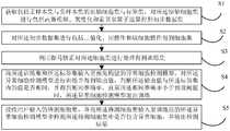
- FIG. 1is a schematic flowchart of a method for detecting abnormal cells provided by an embodiment of the application
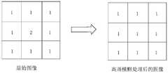
- 2is a schematic diagram of Gaussian blur processing provided by an embodiment of the application.
- FIG. 3is a schematic diagram of a Sobel operator operation process provided by an embodiment of the application.
- FIG. 4is a schematic diagram of expansion processing in a closing operation provided by an embodiment of the application.
- FIG. 5is a schematic diagram of corrosion treatment in a closed operation provided by an embodiment of the application.
- FIG. 6is a schematic diagram of the internal structure of an abnormal cell detection device provided by an embodiment of the application.
- FIG. 7is a schematic diagram of modules of an abnormal cell detection program in an abnormal cell detection device provided by an embodiment of the application.
- This applicationprovides a method for detecting abnormal cells.
- FIG. 1it is a schematic flowchart of an abnormal cell detection method provided by an embodiment of this application.
- the methodcan be executed by a device, and the device can be implemented by software and/or hardware.
- the method for detecting abnormal cellsincludes:
- a preferred embodiment of the present applicationobtains a color cell photo collection from the Internet or a preset database, and the color cell photo collection includes a large number of cells, that is, a cell group. Further, the cell population includes healthy cell populations, and also includes abnormal cell populations, such as cervical cancer cells, lymphatic disease cells, and the like. If the color cell photo includes only healthy cell populations, it is called a positive sample set. If the color cell photo includes abnormal cell populations, it is called a negative sample set. The positive sample set and the negative sample The collection is collectively called the primitive cell collection.
- each cellis divided into More difficult, it is necessary to perform operations including Gaussian blur, grayscale and Sobel operator on the original cell set to obtain a preliminary data set.
- the Gaussian bluris used to process the image noise of the original cell set, and the Gaussian blur is to sequentially traverse each pixel in the cell picture set in the original cell set, and calculate the A sum of the pixel values of eight adjacent pixels of the pixel, and dividing the sum by eight to obtain an average pixel value, and substituting the average pixel value for each pixel.
- the central number 2 in the left part of the Figure 2 of the specificationis the noise of the cells in the original cell set. 2
- Gaussian blur processingis to calculate the average value of all pixels around the center number 2 and the average value of all pixels around the center number 2 is 1. That is, after Gaussian blur processing, the result of the center number 2 becomes 1.
- the original cell setcan be converted from a color image to a grayscale image.
- This applicationuses the proportional method, that is, set the three components of the current pixel as R, G, B, and then use the following formula to obtain the converted pixel component value: 0.30*R+0.59*G+0.11*B, thus The grayscale image of the color image is obtained.
- the Sobel operator operationis to use a convolution template to obtain a first-order horizontal and vertical derivative of the grayscale image, and to enhance the pixel value of the cell edge according to the derivative to achieve cell-to-cell division More obvious purpose.
- the first-order horizontal and vertical derivativeis a weighted sum of peripheral values using pixels.
- the left sideis the convolution template
- the middleis the original image
- the rightis the convolutional image.
- the grayscale imageis subjected to the Sobel operator operation to obtain a preliminary data set, and the pixel difference between cells in the preliminary data set is more obvious.
- the value of each pixelis a number between 0-255, representing the degree of darkness.
- a threshold value Tis preset. When the pixel value of the image in the preliminary data set is greater than the threshold value T, the pixel becomes 1; when the pixel value of the image in the preliminary data set is less than the threshold value When T becomes 0, the binarization operation is completed.
- a morphological operationis applied to the preliminary data set, so as to achieve the purpose of dividing the cell population in the preliminary data set into single cells.
- some operation technologieswill change the morphology of the image. These operations are called the morphological operations, and the closing operation is one of the morphological operations.
- the closing operationfirst performs expansion processing on the preliminary data set, and then performs corrosion processing. As a result of the closing operation, many closely connected cells can be divided into individual cells without protrusions, that is, the cell group is cut Divided into the single cells.
- the expansion processingis to use a rectangle with a width m1 and a height n1 as the rectangular template A, and maximize each pixel X in the preliminary data set.
- the maximization processis to traverse the pixel values of all other pixels covered by the rectangular template A according to the width m1 and the height n1 of the rectangular template A when the pixel X is at the center of the rectangular template A, and modify the pixels
- the pixel value of Xis the largest pixel value among the other pixels. Assuming that after the expansion process is performed on the letter j, the shape of the letter j is reduced, as shown in FIG. 4 of the specification.
- this applicationperforms the expansion process on each cell in the cell group of the preliminary data set, and the volume of each cell becomes smaller. Therefore, the distance between the cells is larger, and it is convenient to separate individual cells. The cells get a single cell set.
- Corrosion treatmentis performed based on a single cell that has been segmented, so that the smaller cells are restored to their original size.
- the etching processuses a rectangle with a width m2 and a height n2 as the rectangular template B, and minimizes each pixel X in the image.
- the minimization processis when the pixel X is at the center of the rectangular template B According to the width m2 and the height n2 of the rectangular template B, all other pixels covered by the rectangular template B are traversed, and the value of the pixel X is modified to the smallest value among all pixels. Assuming that after the etching treatment is performed on the letter j, the volume of the letter j becomes larger, as shown in FIG. 5 of the specification.
- this applicationperforms the corrosion treatment on the single cell set, then the volume of each cell is restored to its original volume, and the single cell set is restored to its original gray value through the de-binarization operation , Get the cell set.
- the gamma correctioncan remove the non-linear mapping between the input radiation and the quantized pixel value, that is, edit the pixel value of the image, and detect the dark part and light in the image signal. Color part, and increase the ratio of the two, thereby improving the image contrast effect.
- V outV in ⁇ ,V:R,G,B
- V: R, G, Bare the three color channels that need to be processed for the gamma correction, and the three color channels are R, G, B, respectively, and Vin is the value of each pixel in the cell set , V out is the value of each pixel in the training set, and ⁇ is a gamma correction coefficient less than 1.
- the abnormal cell detection modelis a neural network model, including random segmentation operations, convolution operations, and activation operations.
- the random segmentation operationreceives the training set and randomly generates a number of cell candidate regions, such as receiving a picture including a cell set, and the random segmentation operation randomly generates a number of rectangular frames with different sizes in the picture;
- This applicationperforms the convolution operation on the several cell candidate regions, and the convolution operation is:
- ⁇ 'is the output data
- ⁇is the input data of the convolution operation
- kis the size of the convolution kernel
- sis the stride of the convolution operation
- pis the data zero-filling matrix
- the present applicationperforms the activation operation on the ⁇ ', and the activation operation is:
- yis the output value
- eis an infinite non-cyclic decimal.
- this applicationdetermines whether the output value is the same as the value in the label set, and calculates the final same accuracy rate. If the same accuracy rate is greater than a preset threshold, the abnormal cell detection model exits training, if When the same accuracy rate is less than a preset threshold, the abnormal cell detection model performs the random segmentation operation again.
- S5.Receive the test cell set input by the user, and input it into the trained abnormal cell detection model to detect whether the test cell set contains abnormal cells, and output the detection result.
- the abnormal cell detection modulecan automatically detect whether there are abnormal cells in the picture. If there are abnormal cells, the abnormal cells can be marked to complete the detection. result.
- the inventionalso provides an abnormal cell detection device.
- FIG. 6it is a schematic diagram of the internal structure of an abnormal cell detection device provided by an embodiment of this application.
- the abnormal cell detection device 1may be a PC (Personal Computer, personal computer), or a terminal device such as a smart phone, a tablet computer, or a portable computer, or a server.
- the abnormal cell detection device 1at least includes a memory 11, a processor 12, a communication bus 13, and a network interface 14.
- the memory 11includes at least one type of readable storage medium, and the readable storage medium includes flash memory, hard disk, multimedia card, card-type memory (for example, SD or DX memory, etc.), magnetic memory, magnetic disk, optical disk, etc.
- the memory 11may be an internal storage unit of the abnormal cell detection device 1 in some embodiments, such as a hard disk of the abnormal cell detection device 1.
- the memory 11may also be an external storage device of the abnormal cell detection device 1, such as a plug-in hard disk equipped on the abnormal cell detection device 1, a Smart Media Card (SMC), and a secure digital (Secure Digital). Digital, SD) card, flash card (Flash Card), etc.
- the memory 11may also include both an internal storage unit of the abnormal cell detection device 1 and an external storage device.
- the memory 11can be used not only to store application software and various data installed in the abnormal cell detection device 1, such as the code of the abnormal cell detection program 01, etc., but also to temporarily store data that has been output or will be output.
- the processor 12may be a central processing unit (CPU), controller, microcontroller, microprocessor, or other data processing chip, and is used to run the program code or processing stored in the memory 11 Data, such as execution of abnormal cell detection program 01, etc.
- CPUcentral processing unit
- controllermicrocontroller
- microprocessoror other data processing chip
- the communication bus 13is used to realize the connection and communication between these components.
- the network interface 14may optionally include a standard wired interface and a wireless interface (such as a WI-FI interface), and is usually used to establish a communication connection between the device 1 and other electronic devices.
- the device 1may also include a user interface.
- the user interfacemay include a display (Display) and an input unit such as a keyboard (Keyboard).
- the optional user interfacemay also include a standard wired interface and a wireless interface.
- the displaymay be an LED display, a liquid crystal display, a touch-sensitive liquid crystal display, an OLED (Organic Light-Emitting Diode, organic light emitting diode) touch device, etc.
- the displaymay also be appropriately called a display screen or a display unit, which is used to display the information processed in the abnormal cell detection device 1 and to display a visualized user interface.
- FIG. 6only shows the abnormal cell detection device 1 with components 11-14 and abnormal cell detection program 01.
- FIG. 1does not constitute a limitation on the abnormal cell detection device 1 It may include fewer or more components than shown, or a combination of some components, or a different component arrangement.
- the abnormal cell detection program 01is stored in the memory 11; when the processor 12 executes the abnormal cell detection program 01 stored in the memory 11, the following steps are implemented:
- Step 1Obtain an original cell set and a label set including a positive sample set and a negative sample set, and perform operations on the original cell set including Gaussian blur, grayscale, and Sobel operator to obtain a preliminary data set.
- a preferred embodiment of the present applicationobtains a color cell photo collection from the Internet or a preset database, and the color cell photo collection includes a large number of cells, that is, a cell group. Further, the cell population includes healthy cell populations, and also includes abnormal cell populations, such as cervical cancer cells, lymphatic disease cells, and the like. If the color cell photo includes only healthy cell populations, it is called a positive sample set. If the color cell photo includes abnormal cell populations, it is called a negative sample set. The positive sample set and the negative sample The collection is collectively called the primitive cell collection.
- each cellis divided into More difficult, it is necessary to perform operations including Gaussian blur, grayscale and Sobel operator on the original cell set to obtain a preliminary data set.
- the Gaussian bluris used to process the image noise of the original cell set, and the Gaussian blur is to sequentially traverse each pixel in the cell picture set in the original cell set, and calculate the A sum of the pixel values of eight adjacent pixels of the pixel, and dividing the sum by eight to obtain an average pixel value, and substituting the average pixel value for each pixel.
- the central number 2 in the left part of the Figure 2 of the specificationis the noise of the cells in the original cell set. 2
- Gaussian blur processingis to calculate the average value of all pixels around the center number 2 and the average value of all pixels around the center number 2 is 1. That is, after Gaussian blur processing, the result of the center number 2 becomes 1.
- the original cell setcan be converted from a color image to a grayscale image.
- This applicationuses the proportional method, that is, set the three components of the current pixel as R, G, B, and then use the following formula to obtain the converted pixel component value: 0.30*R+0.59*G+0.11*B
- the grayscale image of the color imageis obtained.
- the Sobel operator operationis to use a convolution template to obtain a first-order horizontal and vertical derivative of the grayscale image, and to enhance the pixel value of the cell edge according to the derivative to achieve cell-to-cell division More obvious purpose.
- the first-order horizontal and vertical derivativeis a weighted sum of peripheral values using pixels.
- the left sideis the convolution template
- the middleis the original image
- the rightis the convolutional image.
- the grayscale imageis subjected to the Sobel operator operation to obtain a preliminary data set, and the pixel difference between cells in the preliminary data set is more obvious.
- Step 2Perform binarization, closing operations and cell extraction operations on the preliminary data set to obtain a cell set.
- the value of each pixelis a number between 0-255, representing the degree of darkness.
- a threshold value Tis preset. When the pixel value of the image in the preliminary data set is greater than the threshold value T, the pixel becomes 1; when the pixel value of the image in the preliminary data set is less than the threshold value When T becomes 0, the binarization operation is completed.
- a morphological operationis applied to the preliminary data set to achieve the purpose of dividing the cell population in the preliminary data set into single cells.
- some operation technologieswill change the morphology of the image. These operations are called the morphological operations, and the closing operation is one of the morphological operations.
- the closing operationfirst performs expansion processing on the preliminary data set, and then performs corrosion processing. As a result of the closing operation, many closely connected cells can be divided into individual cells without protrusions, that is, the cell group is cut Divided into the single cells.
- the expansion processingis to use a rectangle with a width m1 and a height n1 as the rectangular template A, and maximize each pixel X in the preliminary data set.
- the maximization processis to traverse the pixel values of all other pixels covered by the rectangular template A according to the width m1 and the height n1 of the rectangular template A when the pixel X is at the center of the rectangular template A, and modify the pixels
- the pixel value of Xis the largest pixel value among the other pixels. Assuming that after the expansion process is performed on the letter j, the shape of the letter j is reduced, as shown in FIG. 4 of the specification.
- this applicationperforms the expansion process on each cell in the cell group of the preliminary data set, and the volume of each cell becomes smaller. Therefore, the distance between the cells is larger, and it is convenient to separate individual cells. The cells get a single cell set.
- Corrosion treatmentis performed based on a single cell that has been segmented, so that the smaller cells are restored to their original size.
- the etching processuses a rectangle with a width m2 and a height n2 as the rectangular template B, and minimizes each pixel X in the image.
- the minimization processis when the pixel X is at the center of the rectangular template B According to the width m2 and the height n2 of the rectangular template B, all other pixels covered by the rectangular template B are traversed, and the value of the pixel X is modified to the smallest value among all pixels. Assuming that after the etching treatment is performed on the letter j, the volume of the letter j becomes larger, as shown in FIG. 5 of the specification.
- this applicationperforms the corrosion treatment on the single cell set, then the volume of each cell is restored to its original volume, and the single cell set is restored to its original gray value through the de-binarization operation , Get the cell set.
- Step 3Using gamma correction to perform contrast enhancement processing on the cell set to obtain a training set.
- the gamma correctioncan remove the non-linear mapping between the input radiation and the quantized pixel value, that is, edit the pixel value of the image, and detect the dark part and light in the image signal. Color part, and increase the ratio of the two, thereby improving the image contrast effect.
- V outV in ⁇ ,V:R,G,B
- V: R, G, Bare the three color channels that need to be processed for the gamma correction, and the three color channels are R, G, B, respectively, and Vin is the value of each pixel in the cell set , V out is the value of each pixel in the training set, and ⁇ is a gamma correction coefficient less than 1.
- Step 4Input the training set and the label set to a pre-built abnormal cell detection model, train the abnormal cell detection model to obtain an output value, and determine the output value and the value in the label set Whether it is the same, and the same accuracy rate is obtained, until the same accuracy rate is less than a preset threshold, the abnormal cell detection model exits the training.
- the abnormal cell detection modelis a neural network model, including random segmentation operations, convolution operations, and activation operations.
- the random segmentation operationreceives the training set and randomly generates a number of cell candidate regions, such as receiving a picture including a cell set, and the random segmentation operation randomly generates a number of rectangular frames with different sizes in the picture;
- This applicationperforms the convolution operation on the several cell candidate regions, and the convolution operation is:
- ⁇ 'is the output data
- ⁇is the input data of the convolution operation
- kis the size of the convolution kernel
- sis the step size of the convolution operation
- pis the data zero-filling matrix
- the present applicationperforms the activation operation on the ⁇ ', and the activation operation is:
- yis the output value
- eis an infinite non-cyclic decimal.
- this applicationdetermines whether the output value is the same as the value in the label set, and calculates the final same accuracy rate. If the same accuracy rate is greater than a preset threshold, the abnormal cell detection model exits training, if When the same accuracy rate is less than a preset threshold, the abnormal cell detection model performs the random segmentation operation again.
- Step 5Receive the test cell set input by the user, and input it into the trained abnormal cell detection model to detect whether the test cell set contains abnormal cells, and output the detection result.
- the abnormal cell detection modulecan automatically detect whether there are abnormal cells in the picture. If there are abnormal cells, the abnormal cells can be marked to complete the detection. result.
- the abnormal cell detection programmay also be divided into one or more modules, and the one or more modules are stored in the memory 11 and run by one or more processors (in this embodiment, The processor 12) is executed to complete the application.
- the module referred to in the applicationrefers to a series of computer program instruction segments capable of completing specific functions, and is used to describe the execution process of the abnormal cell detection program in the abnormal cell detection device.
- FIG. 7is a schematic diagram of the program modules of the abnormal cell detection program in an embodiment of the abnormal cell detection device of the present application.
- the abnormal cell detection programcan be divided into a data receiving module 10 and a data
- the processing module 20, the model training module 30, and the abnormal cell detection output module 40are exemplary:
- the data receiving module 10is configured to: obtain an original cell set and a label set including a positive sample set and a negative sample set, and perform operations including Gaussian blur, grayscale, and Sobel operator on the original cell set to obtain a preliminary data set .
- the data processing module 20is configured to perform binarization, closing operations, and cell retrieval operations on the preliminary data set to obtain a cell set.
- the model training module 30is configured to: use gamma correction to perform contrast enhancement processing on the cell set to obtain a training set.
- the abnormal cell detection output module 40is configured to: receive the test cell set input by the user, and input the test cell set into the trained abnormal cell detection model to detect whether the test cell set contains abnormalities Cell and output the test results.
- the embodiment of the present applicationalso proposes a computer-readable storage medium.
- the computer-readable storage mediummay be non-volatile or volatile.
- An abnormal cell detection programis stored on the computer-readable storage medium, and the abnormal cell detection program can be executed by one or more processors to implement the following operations:
- an original cell set and a label setincluding a positive sample set and a negative sample set, and perform operations on the original cell set including Gaussian blur, grayscale, and Sobel operator to obtain a preliminary data set;
Landscapes
- Engineering & Computer Science (AREA)
- Theoretical Computer Science (AREA)
- Physics & Mathematics (AREA)
- General Physics & Mathematics (AREA)
- Life Sciences & Earth Sciences (AREA)
- General Health & Medical Sciences (AREA)
- Health & Medical Sciences (AREA)
- Multimedia (AREA)
- Molecular Biology (AREA)
- Biomedical Technology (AREA)
- Computer Vision & Pattern Recognition (AREA)
- Data Mining & Analysis (AREA)
- Quality & Reliability (AREA)
- Artificial Intelligence (AREA)
- Bioinformatics & Cheminformatics (AREA)
- Bioinformatics & Computational Biology (AREA)
- Evolutionary Biology (AREA)
- Evolutionary Computation (AREA)
- General Engineering & Computer Science (AREA)
- Radiology & Medical Imaging (AREA)
- Nuclear Medicine, Radiotherapy & Molecular Imaging (AREA)
- Medical Informatics (AREA)
- Image Processing (AREA)
- Investigating Or Analysing Biological Materials (AREA)
- Image Analysis (AREA)
Abstract
Description
Translated fromChinese本申请要求于2019年06月18日提交中国专利局、申请号为201910529319.6、发明名称为“异常细胞检测方法、装置及计算机可读存储介质”的中国专利申请的优先权,其全部内容通过引用结合在申请中。This application claims the priority of a Chinese patent application filed with the Chinese Patent Office on June 18, 2019, the application number is 201910529319.6, and the invention title is "abnormal cell detection method, device and computer readable storage medium", the entire content of which is incorporated by reference Incorporate in the application.
本申请涉及人工智能技术领域,尤其涉及一种接收用户输入的细胞图片或视频,检测所述细胞图片或视频是否有异常细胞的方法、装置及计算机可读存储介质。This application relates to the field of artificial intelligence technology, and in particular to a method, device, and computer-readable storage medium for receiving cell pictures or videos input by a user, and detecting whether the cell pictures or videos have abnormal cells.
异常细胞,如癌细胞、骨髓纤维化细胞、红斑狼疮细胞等,往往是人类产生重大病情的导火索,据调查显示,全世界每年有50万新发病例和27.4万死亡病例,其中85%的新发病例是由于前期识别异常细胞的识别率低下的原因。特别地,宫颈癌是目前唯一可以早发现并治愈的癌症,因此早期识别对于病情的治疗相当关键。发明人发现细胞液检查方法是目前最常用的异常细胞识别方法,但在中国由于缺乏病理医生和细胞检测设备,对异常的识别率很低;另外有各种人工辅助识别的设备系统,但多数辅助识别系统一般基于传统方法,所述传统方法依靠细胞质或者细胞核的精确分割和传统图像处理算法进行特征提取与选择,因此识别的前期处理繁琐,且识别率并不高。Abnormal cells, such as cancer cells, bone marrow fibrosis cells, lupus erythematosus cells, etc., are often the fuse for humans to produce major illnesses. According to surveys, there are 500,000 new cases and 274,000 deaths worldwide each year, of which 85% The new cases of the disease are due to the low recognition rate of abnormal cells in the early stage. In particular, cervical cancer is currently the only cancer that can be detected and cured early, so early recognition is crucial for the treatment of the disease. The inventor found that the cell fluid inspection method is currently the most commonly used method for identifying abnormal cells. However, due to the lack of pathologists and cell detection equipment in China, the recognition rate of abnormalities is very low; in addition, there are various artificial assisted identification equipment systems, but most Auxiliary recognition systems are generally based on traditional methods, which rely on precise segmentation of cytoplasm or nucleus and traditional image processing algorithms for feature extraction and selection, so the pre-processing of recognition is cumbersome and the recognition rate is not high.
发明内容Summary of the invention
本申请提供一种异常细胞检测方法、装置及计算机可读存储介质,其主要目的在于当用户输入细胞图片或视频时,精确快速的检测所述细胞图片或视频是否具有异常细胞并输出检测结果。This application provides an abnormal cell detection method, device, and computer-readable storage medium, the main purpose of which is to accurately and quickly detect whether the cell picture or video has abnormal cells and output the detection result when the user inputs a cell picture or video.
为实现上述目的,本申请提供的一种异常细胞检测方法,包括:In order to achieve the above objective, an abnormal cell detection method provided by this application includes:
获取包括正样本集与负样本集的原始细胞集与标签集,对所述原始细胞集进行包括高斯模糊、灰度化和索贝尔算子运算得到初步数据集;Obtain an original cell set and a label set including a positive sample set and a negative sample set, and perform operations on the original cell set including Gaussian blur, grayscale, and Sobel operator to obtain a preliminary data set;
对所述初步数据集进行包括二值化、闭操作和取细胞操作得到细胞集;Performing binarization, closing operations, and cell taking operations on the preliminary data set to obtain a cell set;
利用伽马矫正对所述细胞集进行对比度增强处理得到训练集;Using gamma correction to perform contrast enhancement processing on the cell set to obtain a training set;
将所述训练集和所述标签集输入至预先构建的异常细胞检测模型,对所述异常细胞检测模型进行训练并得到输出值,判断所述输出值与所述标签集内的值是否相同,并得到相同准确率,直至所述相同准确率小于预设阈值时,所述异常细胞检测模型退出训练;Input the training set and the label set to a pre-built abnormal cell detection model, train the abnormal cell detection model to obtain an output value, and determine whether the output value is the same as the value in the label set, And obtain the same accuracy rate, until the same accuracy rate is less than a preset threshold, the abnormal cell detection model exits training;
接收用户输入的待测细胞集,并将所述待测细胞集输入至训练后的所述异常细胞检测模型中检测所述待测细胞集中是否包含异常细胞,并输出检测结果。Receive the test cell set input by the user, and input the test cell set into the trained abnormal cell detection model to detect whether the test cell set contains abnormal cells, and output the detection result.
可选地,所述正样本集包括彩色的健康细胞图片集,所述负样本集包括彩色的异常细胞图片集,所述标签集记录所述原始细胞集内的图片是健康细胞图片还是异常细胞图片。Optionally, the positive sample set includes a color picture set of healthy cells, the negative sample set includes a color picture set of abnormal cells, and the label set records whether the pictures in the original cell set are pictures of healthy cells or abnormal cells image.
可选地,所述高斯模糊处理包括:Optionally, the Gaussian blur processing includes:
依次遍历所述原始细胞集内细胞图片集的每个像素;Sequentially traversing each pixel of the cell picture set in the original cell set;
计算所述每个像素相邻八个像素的像素值的和值,并将所述和值除以八得到平均像素值;Calculating the sum of the pixel values of the eight adjacent pixels of each pixel, and dividing the sum by eight to obtain an average pixel value;
将所述平均像素值代替所述每个像素。The average pixel value is substituted for each pixel.
可选地,所述伽马矫正为:Optionally, the gamma correction is:
Vout=Vinγ,V:R,G,BVout =Vinγ ,V:R,G,B
其中,V:R,G,B为所述伽马矫正所需处理的三个颜色通道,所述三个颜色通道分别为R,G,B,Vin是所述细胞集内每个像素值,Vout是所述训练集每个像素值,γ为小于1的伽 马矫正系数。Wherein, V: R, G, B are the three color channels that need to be processed for the gamma correction, and the three color channels are R, G, B, respectively, and Vin is the value of each pixelin the cell set , Vout is the value of each pixel in the training set, and γ is a gamma correction coefficient less than 1.
可选地,所述异常细胞检测模型是一种神经网络模型,包括随机分割操作、卷积操作;Optionally, the abnormal cell detection model is a neural network model, including random segmentation operations and convolution operations;
所述随机分割操作接收所述训练集并随机生成若干个细胞候选区域;The random segmentation operation receives the training set and randomly generates several cell candidate regions;
将所述若干个细胞候选区域进行所述卷积操作,所述卷积操作为:Perform the convolution operation on the several cell candidate regions, and the convolution operation is:

其中ω’为输出数据,ω为所述卷积操作的输入数据,k为卷积核的大小,s为所述卷积操作的步幅,p为数据补零矩阵。Where ω'is the output data, ω is the input data of the convolution operation, k is the size of the convolution kernel, s is the step size of the convolution operation, and p is the data zero-filling matrix.
此外,为实现上述目的,本申请还提供一种异常细胞检测装置,该装置包括存储器和处理器,所述存储器中存储有可在所述处理器上运行的异常细胞检测程序,所述异常细胞检测程序被所述处理器执行时实现如下步骤:In addition, in order to achieve the above object, the present application also provides an abnormal cell detection device, which includes a memory and a processor, and an abnormal cell detection program that can be run on the processor is stored in the memory. The following steps are implemented when the detection program is executed by the processor:
获取包括正样本集与负样本集的原始细胞集与标签集,对所述原始细胞集进行包括高斯模糊、灰度化和索贝尔算子运算得到初步数据集;Obtain an original cell set and a label set including a positive sample set and a negative sample set, and perform operations on the original cell set including Gaussian blur, grayscale, and Sobel operator to obtain a preliminary data set;
对所述初步数据集进行包括二值化、闭操作和取细胞操作得到细胞集;Performing binarization, closing operations, and cell taking operations on the preliminary data set to obtain a cell set;
利用伽马矫正对所述细胞集进行对比度增强处理得到训练集;Using gamma correction to perform contrast enhancement processing on the cell set to obtain a training set;
将所述训练集和所述标签集输入至预先构建的异常细胞检测模型,对所述异常细胞检测模型进行训练并得到输出值,判断所述输出值与所述标签集内的值是否相同,并得到相同准确率,直至所述相同准确率小于预设阈值时,所述异常细胞检测模型退出训练;Input the training set and the label set to a pre-built abnormal cell detection model, train the abnormal cell detection model to obtain an output value, and determine whether the output value is the same as the value in the label set, And obtain the same accuracy rate, until the same accuracy rate is less than a preset threshold, the abnormal cell detection model exits training;
接收用户输入的待测细胞集,并将所述待测细胞集输入至训练后的所述异常细胞检测模型中检测所述待测细胞集中是否包含异常细胞,并输出检测结果。Receive the test cell set input by the user, and input the test cell set into the trained abnormal cell detection model to detect whether the test cell set contains abnormal cells, and output the detection result.
可选地,所述正样本集包括彩色的健康细胞图片集,所述负样本集包括彩色的异常细胞图片集,所述标签集记录所述原始细胞集内的图片是健康细胞图片还是异常细胞图片。Optionally, the positive sample set includes a color picture set of healthy cells, the negative sample set includes a color picture set of abnormal cells, and the label set records whether the pictures in the original cell set are pictures of healthy cells or abnormal cells image.
可选地,所述高斯模糊处理包括:Optionally, the Gaussian blur processing includes:
依次遍历所述原始细胞集内细胞图片集的每个像素;Sequentially traversing each pixel of the cell picture set in the original cell set;
计算所述每个像素相邻八个像素的像素值的和值,并将所述和值除以八得到平均像素值;Calculating the sum of the pixel values of the eight adjacent pixels of each pixel, and dividing the sum by eight to obtain an average pixel value;
将所述平均像素值代替所述每个像素。The average pixel value is substituted for each pixel.
可选地,所述伽马矫正为:Optionally, the gamma correction is:
Vout=Vinγ,V:R,G,BVout =Vinγ ,V:R,G,B
其中,V:R,G,B为所述伽马矫正所需处理的三个颜色通道,所述三个颜色通道分别为R,G,B,Vin是所述细胞集内每个像素值,Vout是所述训练集每个像素值,γ为小于1的伽马矫正系数。Wherein, V: R, G, B are the three color channels that need to be processed for the gamma correction, and the three color channels are R, G, B, respectively, and Vin is the value of each pixelin the cell set , Vout is the value of each pixel in the training set, and γ is a gamma correction coefficient less than 1.
此外,为实现上述目的,本申请还提供一种计算机可读存储介质,所述计算机可读存储介质上存储有异常细胞检测程序,所述异常细胞检测程序可被一个或者多个处理器执行,以实现如上所述的异常细胞检测方法的步骤。In addition, in order to achieve the above-mentioned object, the present application also provides a computer-readable storage medium having an abnormal cell detection program stored on the computer-readable storage medium, and the abnormal cell detection program can be executed by one or more processors, To achieve the steps of the abnormal cell detection method described above.
本申请提出的异常细胞检测方法、装置及计算机可读存储介质,采用高斯模糊、灰度化和索贝尔算子运算进行处理,减少噪声对数据集的影响,同时,基于伽马矫正扩大正常细胞与异常细胞的对比度,更进一步的有利于后期模型的判断识别,同时对所述模型进行多次训练,并通过多次训练的过程中使所述模型可以具有更优异的自动提取数据的深层特征能力,从而大大提高对异常细胞的识别判断能力,因此本申请可以实现精准的异常细胞检测功能。The abnormal cell detection method, device and computer-readable storage medium proposed in this application adopt Gaussian blur, grayscale and Sobel operator operations for processing to reduce the influence of noise on the data set, and at the same time, expand normal cells based on gamma correction The contrast with abnormal cells is further conducive to the judgment and recognition of the later model. At the same time, the model is trained for many times, and the model can have more excellent deep features of automatic data extraction through the process of multiple training. Therefore, the application can realize the accurate detection function of abnormal cells.
图1为本申请一实施例提供的异常细胞检测方法的流程示意图;FIG. 1 is a schematic flowchart of a method for detecting abnormal cells provided by an embodiment of the application;
图2为本申请一实施例提供的高斯模糊处理的示意图;2 is a schematic diagram of Gaussian blur processing provided by an embodiment of the application;
图3为本申请一实施例提供的索贝尔算子运算过程示意图;FIG. 3 is a schematic diagram of a Sobel operator operation process provided by an embodiment of the application;
图4为本申请一实施例提供的闭操作中膨胀处理示意图;FIG. 4 is a schematic diagram of expansion processing in a closing operation provided by an embodiment of the application;
图5为本申请一实施例提供的闭操作中腐蚀处理示意图;FIG. 5 is a schematic diagram of corrosion treatment in a closed operation provided by an embodiment of the application;
图6为本申请一实施例提供的异常细胞检测装置的内部结构示意图;6 is a schematic diagram of the internal structure of an abnormal cell detection device provided by an embodiment of the application;
图7为本申请一实施例提供的异常细胞检测装置中异常细胞检测程序的模块示意图。FIG. 7 is a schematic diagram of modules of an abnormal cell detection program in an abnormal cell detection device provided by an embodiment of the application.
本申请目的的实现、功能特点及优点将结合实施例,参照附图做进一步说明。The realization, functional characteristics, and advantages of the purpose of this application will be further described in conjunction with the embodiments and with reference to the accompanying drawings.
应当理解,此处所描述的具体实施例仅仅用以解释本申请,并不用于限定本申请。It should be understood that the specific embodiments described here are only used to explain the application, and are not used to limit the application.
本申请提供一种异常细胞检测方法。参照图1所示,为本申请一实施例提供的异常细胞检测方法的流程示意图。该方法可以由一个装置执行,该装置可以由软件和/或硬件实现。This application provides a method for detecting abnormal cells. Referring to FIG. 1, it is a schematic flowchart of an abnormal cell detection method provided by an embodiment of this application. The method can be executed by a device, and the device can be implemented by software and/or hardware.
在本实施例中,异常细胞检测方法包括:In this embodiment, the method for detecting abnormal cells includes:
S1、获取包括正样本集与负样本集的原始细胞集与标签集,对所述原始细胞集进行包括高斯模糊、灰度化和索贝尔算子运算得到初步数据集。S1. Obtain an original cell set and a label set including a positive sample set and a negative sample set, and perform operations including Gaussian blur, grayscale, and Sobel operator on the original cell set to obtain a preliminary data set.
本申请较佳实施例从网络或者预设数据库中获取彩色细胞照片集,所述彩色细胞照片集包括大量的细胞,即细胞群。进一步地,所述细胞群包括健康的细胞群,也包括异常细胞群,所述异常细胞如宫颈癌细胞、淋巴病变细胞等。若所述彩色细胞照片中只包括健康细胞群,则称为正样本集,若所述彩色细胞照片中包括了异常细胞群,则称为负样本集,所述正样本集和所述负样本集统称为原始细胞集。A preferred embodiment of the present application obtains a color cell photo collection from the Internet or a preset database, and the color cell photo collection includes a large number of cells, that is, a cell group. Further, the cell population includes healthy cell populations, and also includes abnormal cell populations, such as cervical cancer cells, lymphatic disease cells, and the like. If the color cell photo includes only healthy cell populations, it is called a positive sample set. If the color cell photo includes abnormal cell populations, it is called a negative sample set. The positive sample set and the negative sample The collection is collectively called the primitive cell collection.
进一步地,由于获取到的原始细胞集内的细胞群具有大量噪点,且健康细胞与健康细胞、异常细胞与健康细胞、异常细胞与异常细胞之间连接紧密盘桓交错,因此将各个细胞分割出来具有较大难度,先需要对所述原始细胞集进行包括高斯模糊、灰度化和索贝尔算子运算从而得到初步数据集。Further, since the cell population in the obtained primitive cell set has a lot of noise, and the connections between healthy cells and healthy cells, abnormal cells and healthy cells, and abnormal cells and abnormal cells are tightly intertwined, each cell is divided into More difficult, it is necessary to perform operations including Gaussian blur, grayscale and Sobel operator on the original cell set to obtain a preliminary data set.
本申请较佳地,所述高斯模糊用来处理所述原始细胞集的图像噪声,所述高斯模糊是依次遍历所述原始细胞集内细胞图片集内的每个像素,计算与所述每个像素相邻的八个像素的像素值的和值,并将所述和值除以八得到平均像素值,将所述平均像素值代替所述每个像素。具体地,当所述原始细胞集内细胞的像素点如说明书附图2所述,其中所述说明书附图2左图部分中心数字2是所述原始细胞集细胞的噪点,对所述中心数字2做高斯模糊处理是计算所述中心数字2周边所有像素的平均值,所述中心数字2周边所有像素的平均值为1,即高斯模糊处理后,所述中心数字2的结果变为1。Preferably, the Gaussian blur is used to process the image noise of the original cell set, and the Gaussian blur is to sequentially traverse each pixel in the cell picture set in the original cell set, and calculate the A sum of the pixel values of eight adjacent pixels of the pixel, and dividing the sum by eight to obtain an average pixel value, and substituting the average pixel value for each pixel. Specifically, when the pixel points of the cells in the original cell set are as described in Figure 2 of the specification, the
进一地,为了减少整个过程的计算量,可以将所述原始细胞集从彩色图像转换为灰度图。本申请运用的是各比例法,即设当前像素的三分量分别为R,G,B,然后利用如下公式得到转换后的像素分量值:0.30*R+0.59*G+0.11*B,由此得到了彩色图像的灰度图。Further, in order to reduce the amount of calculation in the entire process, the original cell set can be converted from a color image to a grayscale image. This application uses the proportional method, that is, set the three components of the current pixel as R, G, B, and then use the following formula to obtain the converted pixel component value: 0.30*R+0.59*G+0.11*B, thus The grayscale image of the color image is obtained.
较佳地,所述索贝尔算子运算是使用卷积模板对所述灰度图求一阶的水平与垂直方向导数,根据所述导数增强细胞边缘的像素值,达到细胞与细胞之间分割更明显的目的。进一步地,所述一阶的水平与垂直方向导数是使用像素点进行周边值的加权和。如说明书附图3中左边为所述卷积模板,中间为原始图像,右边是卷积后的图像。例如,所述原始图像中间的像素点原本是5,经过与卷积模板的卷积计算(-1*3-2*3-1*4+1*2+2*7+1*6=9)后,所述中间的像素点的值变成了9,由于像素点由5变成了9,所以与周围像素点的像素差更大。Preferably, the Sobel operator operation is to use a convolution template to obtain a first-order horizontal and vertical derivative of the grayscale image, and to enhance the pixel value of the cell edge according to the derivative to achieve cell-to-cell division More obvious purpose. Further, the first-order horizontal and vertical derivative is a weighted sum of peripheral values using pixels. As shown in Figure 3 of the specification, the left side is the convolution template, the middle is the original image, and the right is the convolutional image. For example, the pixel point in the middle of the original image was originally 5, which was calculated by convolution with the convolution template (-1*3-2*3-1*4+1*2+2*7+1*6=9 After ), the value of the pixel in the middle becomes 9. Since the pixel is changed from 5 to 9, the pixel difference with the surrounding pixels is greater.
本申请将所述灰度图进行所述索贝尔算子运算得到初步数据集,所述初步数据集内的 细胞与细胞之间像素差更明显。In this application, the grayscale image is subjected to the Sobel operator operation to obtain a preliminary data set, and the pixel difference between cells in the preliminary data set is more obvious.
S2、对所述初步数据集进行包括二值化、闭操作和取细胞操作得到细胞集。S2. Perform binarization, closing operations, and cell taking operations on the preliminary data set to obtain a cell set.
在所述初步数据集中,每个像素的值是0-255之间的数字,代表灰暗的程度。本申请较佳实施例,预设一个阈值T,当所述初步数据集内图像的像素值大于所述阈值T时像素变为1,当所述初步数据集内图像的像素值小于所述阈值T时变为0,完成二值化操作。In the preliminary data set, the value of each pixel is a number between 0-255, representing the degree of darkness. In a preferred embodiment of the present application, a threshold value T is preset. When the pixel value of the image in the preliminary data set is greater than the threshold value T, the pixel becomes 1; when the pixel value of the image in the preliminary data set is less than the threshold value When T becomes 0, the binarization operation is completed.
较佳地,当完成所述二值化操作后,对所述初步数据集采用形态学操作,以达到可以将所述初步数据集内的细胞群切分为单个细胞的目的。在图像处理技术中,有些操作技术会对图像的形态发生改变,这些操作称为所述形态学操作,其中闭操作就是所述形态学操作的一种。所述闭操作先对所述初步数据集进行膨胀处理,然后再进行腐蚀处理,所述闭操作的结果可将许多连接紧密的细胞分割为一个个无突起的单个细胞,即将所述细胞群切分为所述单个细胞。Preferably, after the binarization operation is completed, a morphological operation is applied to the preliminary data set, so as to achieve the purpose of dividing the cell population in the preliminary data set into single cells. In the image processing technology, some operation technologies will change the morphology of the image. These operations are called the morphological operations, and the closing operation is one of the morphological operations. The closing operation first performs expansion processing on the preliminary data set, and then performs corrosion processing. As a result of the closing operation, many closely connected cells can be divided into individual cells without protrusions, that is, the cell group is cut Divided into the single cells.
进一步地,所述膨胀处理是使用一个宽m1、高n1的矩形作为矩形模板A,对所述初步数据集中的每一个像素X做最大化处理。所述最大化处理是当像素X至于所述矩形模板A的中心时,根据所述矩形模版A的宽m1、高n1,遍历所有被所述矩形模板A覆盖的其他像素的像素值,修改像素X的像素值为所述其他像素中最大的像素值。假设对字母j做所述膨胀处理后,则所述字母j的形状发生了缩小,如说明书附图4所示。Further, the expansion processing is to use a rectangle with a width m1 and a height n1 as the rectangular template A, and maximize each pixel X in the preliminary data set. The maximization process is to traverse the pixel values of all other pixels covered by the rectangular template A according to the width m1 and the height n1 of the rectangular template A when the pixel X is at the center of the rectangular template A, and modify the pixels The pixel value of X is the largest pixel value among the other pixels. Assuming that after the expansion process is performed on the letter j, the shape of the letter j is reduced, as shown in FIG. 4 of the specification.
较佳地,本申请对所述初步数据集的细胞群内每个细胞做所述膨胀处理,则每个细胞体积变小,因此细胞与细胞之间的距离更大,因此可方便分割出单个细胞得到单个细胞集。Preferably, this application performs the expansion process on each cell in the cell group of the preliminary data set, and the volume of each cell becomes smaller. Therefore, the distance between the cells is larger, and it is convenient to separate individual cells. The cells get a single cell set.
基于已分割完成的单个细胞做腐蚀处理,使得体积变小的细胞恢复回原来的大小。所述腐蚀处理是使用一个宽m2、高n2的矩形作为矩形模板B,对图像中的每一个像素X做最小化处理,所述最小化处理是当像素X至于所述矩形模板B的中心时,根据所述矩形模版B的宽m2、高n2,遍历所有被所述矩形模板B覆盖的其他像素,修改像素X的值为所有像素中最小的值。假设对字母j做所述腐蚀处理后,则所述字母j的体积变大,如说明书附图5所述。Corrosion treatment is performed based on a single cell that has been segmented, so that the smaller cells are restored to their original size. The etching process uses a rectangle with a width m2 and a height n2 as the rectangular template B, and minimizes each pixel X in the image. The minimization process is when the pixel X is at the center of the rectangular template B According to the width m2 and the height n2 of the rectangular template B, all other pixels covered by the rectangular template B are traversed, and the value of the pixel X is modified to the smallest value among all pixels. Assuming that after the etching treatment is performed on the letter j, the volume of the letter j becomes larger, as shown in FIG. 5 of the specification.
较佳地,本申请对所述单个细胞集做所述腐蚀处理,则每个细胞体积恢复回原来的体积,通过反二值化操作,将所述单个细胞集都恢复回原来的灰度值,得到细胞集。Preferably, this application performs the corrosion treatment on the single cell set, then the volume of each cell is restored to its original volume, and the single cell set is restored to its original gray value through the de-binarization operation , Get the cell set.
S3、利用伽马矫正对所述细胞集进行对比度增强处理得到训练集。S3, using gamma correction to perform contrast enhancement processing on the cell set to obtain a training set.
本申请较佳实施例中,所述伽马矫正可以去除输入辐射量和量化的像素值之间的非线性映射,即对图像的像素值进行编辑,检出图像信号中的深色部分和浅色部分,并使两者比例增大,从而提高图像对比度效果。In a preferred embodiment of the application, the gamma correction can remove the non-linear mapping between the input radiation and the quantized pixel value, that is, edit the pixel value of the image, and detect the dark part and light in the image signal. Color part, and increase the ratio of the two, thereby improving the image contrast effect.
进一步地,所述伽马矫正为:Further, the gamma correction is:
Vout=Vinγ,V:R,G,BVout =Vinγ ,V:R,G,B
其中,V:R,G,B为所述伽马矫正所需处理的三个颜色通道,所述三个颜色通道分别为R,G,B,Vin是所述细胞集内每个像素值,Vout是所述训练集每个像素值,γ为小于1的伽马矫正系数。Wherein, V: R, G, B are the three color channels that need to be processed for the gamma correction, and the three color channels are R, G, B, respectively, and Vin is the value of each pixelin the cell set , Vout is the value of each pixel in the training set, and γ is a gamma correction coefficient less than 1.
S4、将所述训练集和所述标签集输入至预先构建的异常细胞检测模型,对所述异常细胞检测模型进行训练并得到输出值,判断所述输出值与所述标签集内的值是否相同,并得到相同准确率,直至所述相同准确率小于预设阈值时,所述异常细胞检测模型退出训练。S4. Input the training set and the label set to a pre-built abnormal cell detection model, train the abnormal cell detection model to obtain an output value, and determine whether the output value and the value in the label set are The same, and the same accuracy rate is obtained, until the same accuracy rate is less than the preset threshold, the abnormal cell detection model exits the training.
本申请较佳实施例,所述异常细胞检测模型是一种神经网络模型,包括随机分割操作、卷积操作、激活操作。In a preferred embodiment of the present application, the abnormal cell detection model is a neural network model, including random segmentation operations, convolution operations, and activation operations.
所述随机分割操作接收所述训练集并随机生成若干个细胞候选区域,如接受一张包括细胞集的图片,所述随机分割操作在所述图片中随机生成若干个大小不一样的矩形框架;The random segmentation operation receives the training set and randomly generates a number of cell candidate regions, such as receiving a picture including a cell set, and the random segmentation operation randomly generates a number of rectangular frames with different sizes in the picture;
本申请将所述若干个细胞候选区域进行所述卷积操作,所述卷积操作为:This application performs the convolution operation on the several cell candidate regions, and the convolution operation is:

其中ω’为输出数据,ω为所述卷积操作的输入数据,k为卷积核的大小,s为所述卷积 操作的步幅,p为数据补零矩阵;Where ω'is the output data, ω is the input data of the convolution operation, k is the size of the convolution kernel, s is the stride of the convolution operation, and p is the data zero-filling matrix;
较佳地,本申请将所述ω’进行所述激活操作,所述激活操作为:Preferably, the present application performs the activation operation on the ω', and the activation operation is:

其中y为输出值,e为无限不循环小数。Among them, y is the output value, and e is an infinite non-cyclic decimal.
进一步,本申请判断所述输出值与所述标签集内的值是否相同,并计算最终的相同准确率,若所述相同准确率大于预设阈值时,所述异常细胞检测模型退出训练,若所述相同准确率小于预设阈值时,所述异常细胞检测模型重新进行所述随机分割操作。Further, this application determines whether the output value is the same as the value in the label set, and calculates the final same accuracy rate. If the same accuracy rate is greater than a preset threshold, the abnormal cell detection model exits training, if When the same accuracy rate is less than a preset threshold, the abnormal cell detection model performs the random segmentation operation again.
S5、接收用户输入的待测细胞集,并输入至训练后的所述异常细胞检测模型中检测所述待测细胞集中是否包含异常细胞,并输出检测结果。S5. Receive the test cell set input by the user, and input it into the trained abnormal cell detection model to detect whether the test cell set contains abnormal cells, and output the detection result.
本申请较佳地,如接受一张包括大量细胞的图片,所述异常细胞检测模可自动检测出所述图片是否有异常细胞,若有异常细胞,可将所述异常细胞标记出来,完成检测结果。Preferably, if a picture containing a large number of cells is accepted, the abnormal cell detection module can automatically detect whether there are abnormal cells in the picture. If there are abnormal cells, the abnormal cells can be marked to complete the detection. result.
发明还提供一种异常细胞检测装置。参照图6所示,为本申请一实施例提供的异常细胞检测装置的内部结构示意图。The invention also provides an abnormal cell detection device. Referring to FIG. 6, it is a schematic diagram of the internal structure of an abnormal cell detection device provided by an embodiment of this application.
在本实施例中,所述异常细胞检测装置1可以是PC(Personal Computer,个人电脑),或者是智能手机、平板电脑、便携计算机等终端设备,也可以是一种服务器等。该异常细胞检测装置1至少包括存储器11、处理器12,通信总线13,以及网络接口14。In this embodiment, the abnormal
其中,存储器11至少包括一种类型的可读存储介质,所述可读存储介质包括闪存、硬盘、多媒体卡、卡型存储器(例如,SD或DX存储器等)、磁性存储器、磁盘、光盘等。存储器11在一些实施例中可以是异常细胞检测装置1的内部存储单元,例如该异常细胞检测装置1的硬盘。存储器11在另一些实施例中也可以是异常细胞检测装置1的外部存储设备,例如异常细胞检测装置1上配备的插接式硬盘,智能存储卡(Smart Media Card,SMC),安全数字(Secure Digital,SD)卡,闪存卡(Flash Card)等。进一步地,存储器11还可以既包括异常细胞检测装置1的内部存储单元也包括外部存储设备。存储器11不仅可以用于存储安装于异常细胞检测装置1的应用软件及各类数据,例如异常细胞检测程序01的代码等,还可以用于暂时地存储已经输出或者将要输出的数据。Wherein, the
处理器12在一些实施例中可以是一中央处理器(Central Processing Unit,CPU)、控制器、微控制器、微处理器或其他数据处理芯片,用于运行存储器11中存储的程序代码或处理数据,例如执行异常细胞检测程序01等。In some embodiments, the
通信总线13用于实现这些组件之间的连接通信。The
网络接口14可选的可以包括标准的有线接口、无线接口(如WI-FI接口),通常用于在该装置1与其他电子设备之间建立通信连接。The
可选地,该装置1还可以包括用户接口,用户接口可以包括显示器(Display)、输入单元比如键盘(Keyboard),可选的用户接口还可以包括标准的有线接口、无线接口。可选地,在一些实施例中,显示器可以是LED显示器、液晶显示器、触控式液晶显示器以及OLED(Organic Light-Emitting Diode,有机发光二极管)触摸器等。其中,显示器也可以适当的称为显示屏或显示单元,用于显示在异常细胞检测装置1中处理的信息以及用于显示可视化的用户界面。Optionally, the
图6仅示出了具有组件11-14以及异常细胞检测程序01的异常细胞检测装置1,本领域技术人员可以理解的是,图1示出的结构并不构成对异常细胞检测装置1的限定,可以包括比图示更少或者更多的部件,或者组合某些部件,或者不同的部件布置。FIG. 6 only shows the abnormal
在图6所示的装置1实施例中,存储器11中存储有异常细胞检测程序01;处理器12执行存储器11中存储的异常细胞检测程序01时实现如下步骤:In the embodiment of the
步骤一、获取包括正样本集与负样本集的原始细胞集与标签集,对所述原始细胞集进行包括高斯模糊、灰度化和索贝尔算子运算得到初步数据集。Step 1: Obtain an original cell set and a label set including a positive sample set and a negative sample set, and perform operations on the original cell set including Gaussian blur, grayscale, and Sobel operator to obtain a preliminary data set.
本申请较佳实施例从网络或者预设数据库中获取彩色细胞照片集,所述彩色细胞照片集包括大量的细胞,即细胞群。进一步地,所述细胞群包括健康的细胞群,也包括异常细胞群,所述异常细胞如宫颈癌细胞、淋巴病变细胞等。若所述彩色细胞照片中只包括健康细胞群,则称为正样本集,若所述彩色细胞照片中包括了异常细胞群,则称为负样本集,所述正样本集和所述负样本集统称为原始细胞集。A preferred embodiment of the present application obtains a color cell photo collection from the Internet or a preset database, and the color cell photo collection includes a large number of cells, that is, a cell group. Further, the cell population includes healthy cell populations, and also includes abnormal cell populations, such as cervical cancer cells, lymphatic disease cells, and the like. If the color cell photo includes only healthy cell populations, it is called a positive sample set. If the color cell photo includes abnormal cell populations, it is called a negative sample set. The positive sample set and the negative sample The collection is collectively called the primitive cell collection.
进一步地,由于获取到的原始细胞集内的细胞群具有大量噪点,且健康细胞与健康细胞、异常细胞与健康细胞、异常细胞与异常细胞之间连接紧密盘桓交错,因此将各个细胞分割出来具有较大难度,先需要对所述原始细胞集进行包括高斯模糊、灰度化和索贝尔算子运算从而得到初步数据集。Further, since the cell population in the obtained primitive cell set has a lot of noise, and the connections between healthy cells and healthy cells, abnormal cells and healthy cells, and abnormal cells and abnormal cells are tightly intertwined, each cell is divided into More difficult, it is necessary to perform operations including Gaussian blur, grayscale and Sobel operator on the original cell set to obtain a preliminary data set.
本申请较佳地,所述高斯模糊用来处理所述原始细胞集的图像噪声,所述高斯模糊是依次遍历所述原始细胞集内细胞图片集内的每个像素,计算与所述每个像素相邻的八个像素的像素值的和值,并将所述和值除以八得到平均像素值,将所述平均像素值代替所述每个像素。具体地,当所述原始细胞集内细胞的像素点如说明书附图2所述,其中所述说明书附图2左图部分中心数字2是所述原始细胞集细胞的噪点,对所述中心数字2做高斯模糊处理是计算所述中心数字2周边所有像素的平均值,所述中心数字2周边所有像素的平均值为1,即高斯模糊处理后,所述中心数字2的结果变为1。Preferably, the Gaussian blur is used to process the image noise of the original cell set, and the Gaussian blur is to sequentially traverse each pixel in the cell picture set in the original cell set, and calculate the A sum of the pixel values of eight adjacent pixels of the pixel, and dividing the sum by eight to obtain an average pixel value, and substituting the average pixel value for each pixel. Specifically, when the pixel points of the cells in the original cell set are as described in Figure 2 of the specification, the
进一地,为了减少整个过程的计算量,可以将所述原始细胞集从彩色图像转换为灰度图。本申请运用的是各比例法,即设当前像素的三分量分别为R,G,B,然后利用如下公式得到转换后的像素分量值:0.30*R+0.59*G+0.11*B,由此得到了彩色图像的灰度图。Further, in order to reduce the amount of calculation in the entire process, the original cell set can be converted from a color image to a grayscale image. This application uses the proportional method, that is, set the three components of the current pixel as R, G, B, and then use the following formula to obtain the converted pixel component value: 0.30*R+0.59*G+0.11*B The grayscale image of the color image is obtained.
较佳地,所述索贝尔算子运算是使用卷积模板对所述灰度图求一阶的水平与垂直方向导数,根据所述导数增强细胞边缘的像素值,达到细胞与细胞之间分割更明显的目的。进一步地,所述一阶的水平与垂直方向导数是使用像素点进行周边值的加权和。如说明书附图3中左边为所述卷积模板,中间为原始图像,右边是卷积后的图像。例如,所述原始图像中间的像素点原本是5,经过与卷积模板的卷积计算(-1*3-2*3-1*4+1*2+2*7+1*6=9)后,所述中间的像素点的值变成了9,由于像素点由5变成了9,所以与周围像素点的像素差更大。Preferably, the Sobel operator operation is to use a convolution template to obtain a first-order horizontal and vertical derivative of the grayscale image, and to enhance the pixel value of the cell edge according to the derivative to achieve cell-to-cell division More obvious purpose. Further, the first-order horizontal and vertical derivative is a weighted sum of peripheral values using pixels. As shown in Figure 3 of the specification, the left side is the convolution template, the middle is the original image, and the right is the convolutional image. For example, the pixel point in the middle of the original image was originally 5, which was calculated by convolution with the convolution template (-1*3-2*3-1*4+1*2+2*7+1*6=9 After ), the value of the pixel in the middle becomes 9. Since the pixel is changed from 5 to 9, the pixel difference with the surrounding pixels is greater.
本申请将所述灰度图进行所述索贝尔算子运算得到初步数据集,所述初步数据集内的细胞与细胞之间像素差更明显。In this application, the grayscale image is subjected to the Sobel operator operation to obtain a preliminary data set, and the pixel difference between cells in the preliminary data set is more obvious.
步骤二、对所述初步数据集进行包括二值化、闭操作和取细胞操作得到细胞集。Step 2: Perform binarization, closing operations and cell extraction operations on the preliminary data set to obtain a cell set.
在所述初步数据集中,每个像素的值是0-255之间的数字,代表灰暗的程度。本申请较佳实施例,预设一个阈值T,当所述初步数据集内图像的像素值大于所述阈值T时像素变为1,当所述初步数据集内图像的像素值小于所述阈值T时变为0,完成二值化操作。In the preliminary data set, the value of each pixel is a number between 0-255, representing the degree of darkness. In a preferred embodiment of the present application, a threshold value T is preset. When the pixel value of the image in the preliminary data set is greater than the threshold value T, the pixel becomes 1; when the pixel value of the image in the preliminary data set is less than the threshold value When T becomes 0, the binarization operation is completed.
较佳地,当完成所述二值化操作后,对所述初步数据集采用形态学操作,以达到可以将所述初步数据集内的细胞群切分为单个细胞的目的。在图像处理技术中,有些操作技术会对图像的形态发生改变,这些操作称为所述形态学操作,其中闭操作就是所述形态学操作的一种。所述闭操作先对所述初步数据集进行膨胀处理,然后再进行腐蚀处理,所述闭操作的结果可将许多连接紧密的细胞分割为一个个无突起的单个细胞,即将所述细胞群切分为所述单个细胞。Preferably, after the binarization operation is completed, a morphological operation is applied to the preliminary data set to achieve the purpose of dividing the cell population in the preliminary data set into single cells. In the image processing technology, some operation technologies will change the morphology of the image. These operations are called the morphological operations, and the closing operation is one of the morphological operations. The closing operation first performs expansion processing on the preliminary data set, and then performs corrosion processing. As a result of the closing operation, many closely connected cells can be divided into individual cells without protrusions, that is, the cell group is cut Divided into the single cells.
进一步地,所述膨胀处理是使用一个宽m1、高n1的矩形作为矩形模板A,对所述初步数据集中的每一个像素X做最大化处理。所述最大化处理是当像素X至于所述矩形模板A的中心时,根据所述矩形模版A的宽m1、高n1,遍历所有被所述矩形模板A覆盖的其他像素的像素值,修改像素X的像素值为所述其他像素中最大的像素值。假设对字母j做所述膨胀处理后,则所述字母j的形状发生了缩小,如说明书附图4所示。Further, the expansion processing is to use a rectangle with a width m1 and a height n1 as the rectangular template A, and maximize each pixel X in the preliminary data set. The maximization process is to traverse the pixel values of all other pixels covered by the rectangular template A according to the width m1 and the height n1 of the rectangular template A when the pixel X is at the center of the rectangular template A, and modify the pixels The pixel value of X is the largest pixel value among the other pixels. Assuming that after the expansion process is performed on the letter j, the shape of the letter j is reduced, as shown in FIG. 4 of the specification.
较佳地,本申请对所述初步数据集的细胞群内每个细胞做所述膨胀处理,则每个细胞 体积变小,因此细胞与细胞之间的距离更大,因此可方便分割出单个细胞得到单个细胞集。Preferably, this application performs the expansion process on each cell in the cell group of the preliminary data set, and the volume of each cell becomes smaller. Therefore, the distance between the cells is larger, and it is convenient to separate individual cells. The cells get a single cell set.
基于已分割完成的单个细胞做腐蚀处理,使得体积变小的细胞恢复回原来的大小。所述腐蚀处理是使用一个宽m2、高n2的矩形作为矩形模板B,对图像中的每一个像素X做最小化处理,所述最小化处理是当像素X至于所述矩形模板B的中心时,根据所述矩形模版B的宽m2、高n2,遍历所有被所述矩形模板B覆盖的其他像素,修改像素X的值为所有像素中最小的值。假设对字母j做所述腐蚀处理后,则所述字母j的体积变大,如说明书附图5所述。Corrosion treatment is performed based on a single cell that has been segmented, so that the smaller cells are restored to their original size. The etching process uses a rectangle with a width m2 and a height n2 as the rectangular template B, and minimizes each pixel X in the image. The minimization process is when the pixel X is at the center of the rectangular template B According to the width m2 and the height n2 of the rectangular template B, all other pixels covered by the rectangular template B are traversed, and the value of the pixel X is modified to the smallest value among all pixels. Assuming that after the etching treatment is performed on the letter j, the volume of the letter j becomes larger, as shown in FIG. 5 of the specification.
较佳地,本申请对所述单个细胞集做所述腐蚀处理,则每个细胞体积恢复回原来的体积,通过反二值化操作,将所述单个细胞集都恢复回原来的灰度值,得到细胞集。Preferably, this application performs the corrosion treatment on the single cell set, then the volume of each cell is restored to its original volume, and the single cell set is restored to its original gray value through the de-binarization operation , Get the cell set.
步骤三、利用伽马矫正对所述细胞集进行对比度增强处理得到训练集。Step 3: Using gamma correction to perform contrast enhancement processing on the cell set to obtain a training set.
本申请较佳实施例中,所述伽马矫正可以去除输入辐射量和量化的像素值之间的非线性映射,即对图像的像素值进行编辑,检出图像信号中的深色部分和浅色部分,并使两者比例增大,从而提高图像对比度效果。In a preferred embodiment of the application, the gamma correction can remove the non-linear mapping between the input radiation and the quantized pixel value, that is, edit the pixel value of the image, and detect the dark part and light in the image signal. Color part, and increase the ratio of the two, thereby improving the image contrast effect.
进一步地,所述伽马矫正为:Further, the gamma correction is:
Vout=Vinγ,V:R,G,BVout =Vinγ ,V:R,G,B
其中,V:R,G,B为所述伽马矫正所需处理的三个颜色通道,所述三个颜色通道分别为R,G,B,Vin是所述细胞集内每个像素值,Vout是所述训练集每个像素值,γ为小于1的伽马矫正系数。Wherein, V: R, G, B are the three color channels that need to be processed for the gamma correction, and the three color channels are R, G, B, respectively, and Vin is the value of each pixelin the cell set , Vout is the value of each pixel in the training set, and γ is a gamma correction coefficient less than 1.
步骤四、将所述训练集和所述标签集输入至预先构建的异常细胞检测模型,对所述异常细胞检测模型进行训练并得到输出值,判断所述输出值与所述标签集内的值是否相同,并得到相同准确率,直至所述相同准确率小于预设阈值时,所述异常细胞检测模型退出训练。
本申请较佳实施例,所述异常细胞检测模型是一种神经网络模型,包括随机分割操作、卷积操作、激活操作。In a preferred embodiment of the present application, the abnormal cell detection model is a neural network model, including random segmentation operations, convolution operations, and activation operations.
所述随机分割操作接收所述训练集并随机生成若干个细胞候选区域,如接受一张包括细胞集的图片,所述随机分割操作在所述图片中随机生成若干个大小不一样的矩形框架;The random segmentation operation receives the training set and randomly generates a number of cell candidate regions, such as receiving a picture including a cell set, and the random segmentation operation randomly generates a number of rectangular frames with different sizes in the picture;
本申请将所述若干个细胞候选区域进行所述卷积操作,所述卷积操作为:This application performs the convolution operation on the several cell candidate regions, and the convolution operation is:

其中ω’为输出数据,ω为所述卷积操作的输入数据,k为卷积核的大小,s为所述卷积操作的步幅,p为数据补零矩阵;Where ω'is the output data, ω is the input data of the convolution operation, k is the size of the convolution kernel, s is the step size of the convolution operation, and p is the data zero-filling matrix;
较佳地,本申请将所述ω’进行所述激活操作,所述激活操作为:Preferably, the present application performs the activation operation on the ω', and the activation operation is:

其中y为输出值,e为无限不循环小数。Among them, y is the output value, and e is an infinite non-cyclic decimal.
进一步,本申请判断所述输出值与所述标签集内的值是否相同,并计算最终的相同准确率,若所述相同准确率大于预设阈值时,所述异常细胞检测模型退出训练,若所述相同准确率小于预设阈值时,所述异常细胞检测模型重新进行所述随机分割操作。Further, this application determines whether the output value is the same as the value in the label set, and calculates the final same accuracy rate. If the same accuracy rate is greater than a preset threshold, the abnormal cell detection model exits training, if When the same accuracy rate is less than a preset threshold, the abnormal cell detection model performs the random segmentation operation again.
步骤五、接收用户输入的待测细胞集,并输入至训练后的所述异常细胞检测模型中检测所述待测细胞集中是否包含异常细胞,并输出检测结果。Step 5: Receive the test cell set input by the user, and input it into the trained abnormal cell detection model to detect whether the test cell set contains abnormal cells, and output the detection result.
本申请较佳地,如接受一张包括大量细胞的图片,所述异常细胞检测模可自动检测出所述图片是否有异常细胞,若有异常细胞,可将所述异常细胞标记出来,完成检测结果。Preferably, if a picture containing a large number of cells is accepted, the abnormal cell detection module can automatically detect whether there are abnormal cells in the picture. If there are abnormal cells, the abnormal cells can be marked to complete the detection. result.
可选地,在其他实施例中,异常细胞检测程序还可以被分割为一个或者多个模块,一个或者多个模块被存储于存储器11中,并由一个或多个处理器(本实施例为处理器12)所执行以完成本申请,本申请所称的模块是指能够完成特定功能的一系列计算机程序指令 段,用于描述异常细胞检测程序在异常细胞检测装置中的执行过程。Optionally, in other embodiments, the abnormal cell detection program may also be divided into one or more modules, and the one or more modules are stored in the
例如,参照图7所示,为本申请异常细胞检测装置一实施例中的异常细胞检测程序的程序模块示意图,该实施例中,所述异常细胞检测程序可以被分割为数据接收模块10、数据处理模块20、模型训练模块30、异常细胞检测输出模块40示例性地:For example, referring to FIG. 7, which is a schematic diagram of the program modules of the abnormal cell detection program in an embodiment of the abnormal cell detection device of the present application. In this embodiment, the abnormal cell detection program can be divided into a
所述数据接收模块10用于:获取包括正样本集与负样本集的原始细胞集与标签集,对所述原始细胞集进行包括高斯模糊、灰度化和索贝尔算子运算得到初步数据集。The
所述数据处理模块20用于:对所述初步数据集进行包括二值化、闭操作和取细胞操作得到细胞集。The
所述模型训练模块30用于:利用伽马矫正对所述细胞集进行对比度增强处理得到训练集。The
所述异常细胞检测输出模块40用于:接收用户输入的待测细胞集,并将所述待测细胞集输入至训练后的所述异常细胞检测模型中检测所述待测细胞集中是否包含异常细胞,并输出检测结果。The abnormal cell
上述数据接收模块10、数据处理模块20、模型训练模块30、异常细胞检测输出模块40等程序模块被执行时所实现的功能或操作步骤与上述实施例大体相同,在此不再赘述。The functions or operation steps implemented by the program modules such as the
此外,本申请实施例还提出一种计算机可读存储介质,所述计算机可读存储介质可以是非易失性,也可以是易失性。所述计算机可读存储介质上存储有异常细胞检测程序,所述异常细胞检测程序可被一个或多个处理器执行,以实现如下操作:In addition, the embodiment of the present application also proposes a computer-readable storage medium. The computer-readable storage medium may be non-volatile or volatile. An abnormal cell detection program is stored on the computer-readable storage medium, and the abnormal cell detection program can be executed by one or more processors to implement the following operations:
获取包括正样本集与负样本集的原始细胞集与标签集,对所述原始细胞集进行包括高斯模糊、灰度化和索贝尔算子运算得到初步数据集;Obtain an original cell set and a label set including a positive sample set and a negative sample set, and perform operations on the original cell set including Gaussian blur, grayscale, and Sobel operator to obtain a preliminary data set;
对所述初步数据集进行包括二值化、闭操作和取细胞操作得到细胞集;Performing binarization, closing operations, and cell taking operations on the preliminary data set to obtain a cell set;
利用伽马矫正对所述细胞集进行对比度增强处理得到训练集;Using gamma correction to perform contrast enhancement processing on the cell set to obtain a training set;
将所述训练集和所述标签集输入至预先构建的异常细胞检测模型,对所述异常细胞检测模型进行训练并得到输出值,判断所述输出值与所述标签集内的值是否相同,并得到相同准确率,直至所述相同准确率小于预设阈值时,所述异常细胞检测模型退出训练;Input the training set and the label set to a pre-built abnormal cell detection model, train the abnormal cell detection model to obtain an output value, and determine whether the output value is the same as the value in the label set, And obtain the same accuracy rate, until the same accuracy rate is less than a preset threshold, the abnormal cell detection model exits training;
接收用户输入的待测细胞集,并将所述待测细胞集输入至训练后的所述异常细胞检测模型中检测所述待测细胞集中是否包含异常细胞,并输出检测结果。Receive the test cell set input by the user, and input the test cell set into the trained abnormal cell detection model to detect whether the test cell set contains abnormal cells, and output the detection result.
需要说明的是,上述本申请实施例序号仅仅为了描述,不代表实施例的优劣。并且本文中的术语“包括”、“包含”或者其任何其他变体意在涵盖非排他性的包含,从而使得包括一系列要素的过程、装置、物品或者方法不仅包括那些要素,而且还包括没有明确列出的其他要素,或者是还包括为这种过程、装置、物品或者方法所固有的要素。在没有更多限制的情况下,由语句“包括一个……”限定的要素,并不排除在包括该要素的过程、装置、物品或者方法中还存在另外的相同要素。It should be noted that the serial numbers of the above embodiments of the present application are only for description, and do not represent the advantages and disadvantages of the embodiments. And the terms "include", "include" or any other variants thereof in this article are intended to cover non-exclusive inclusion, so that a process, device, article or method including a series of elements not only includes those elements, but also includes The other elements listed may also include elements inherent to the process, device, article, or method. If there are no more restrictions, the element defined by the sentence "including a..." does not exclude the existence of other identical elements in the process, device, article or method that includes the element.
通过以上的实施方式的描述,本领域的技术人员可以清楚地了解到上述实施例方法可借助软件加必需的通用硬件平台的方式来实现,当然也可以通过硬件,但很多情况下前者是更佳的实施方式。基于这样的理解,本申请的技术方案本质上或者说对现有技术做出贡献的部分可以以软件产品的形式体现出来,该计算机软件产品存储在如上所述的一个存储介质(如ROM/RAM、磁碟、光盘)中,包括若干指令用以使得一台终端设备(可以是手机,计算机,服务器,或者网络设备等)执行本申请各个实施例所述的方法。Through the description of the above embodiments, those skilled in the art can clearly understand that the method of the above embodiments can be implemented by means of software plus the necessary general hardware platform. Of course, it can also be implemented by hardware, but in many cases the former is better.的实施方式。 Based on this understanding, the technical solution of this application essentially or the part that contributes to the existing technology can be embodied in the form of a software product, and the computer software product is stored in a storage medium (such as ROM/RAM) as described above. , Magnetic disks, optical disks), including several instructions to make a terminal device (which can be a mobile phone, a computer, a server, or a network device, etc.) execute the method described in each embodiment of the present application.
以上仅为本申请的优选实施例,并非因此限制本申请的专利范围,凡是利用本申请说明书及附图内容所作的等效结构或等效流程变换,或直接或间接运用在其他相关的技术领域,均同理包括在本申请的专利保护范围内。The above are only preferred embodiments of this application, and do not limit the scope of this application. Any equivalent structure or equivalent process transformation made using the content of the description and drawings of this application, or directly or indirectly used in other related technical fields , The same reason is included in the scope of patent protection of this application.
Claims (20)
Translated fromChinese


Applications Claiming Priority (2)
| Application Number | Priority Date | Filing Date | Title |
|---|---|---|---|
| CN201910529319.6 | 2019-06-18 | ||
| CN201910529319.6ACN110415212A (en) | 2019-06-18 | 2019-06-18 | Abnormal cell detection method, device and computer readable storage medium |
Publications (1)
| Publication Number | Publication Date |
|---|---|
| WO2020253508A1true WO2020253508A1 (en) | 2020-12-24 |
Family
ID=68359294
Family Applications (1)
| Application Number | Title | Priority Date | Filing Date |
|---|---|---|---|
| PCT/CN2020/093548CeasedWO2020253508A1 (en) | 2019-06-18 | 2020-05-29 | Abnormal cell detection method and apparatus, and computer readable storage medium |
Country Status (2)
| Country | Link |
|---|---|
| CN (1) | CN110415212A (en) |
| WO (1) | WO2020253508A1 (en) |
Cited By (4)
| Publication number | Priority date | Publication date | Assignee | Title |
|---|---|---|---|---|
| CN113130049A (en)* | 2021-04-15 | 2021-07-16 | 黑龙江机智通智能科技有限公司 | Intelligent pathological image diagnosis system based on cloud service |
| CN114399494A (en)* | 2022-01-14 | 2022-04-26 | 平安科技(深圳)有限公司 | Abnormal cell detection and segmentation method, device, equipment and storage medium |
| CN115564776A (en)* | 2022-12-05 | 2023-01-03 | 珠海圣美生物诊断技术有限公司 | Abnormal cell sample detection method and device based on machine learning |
| CN118967661A (en)* | 2024-10-10 | 2024-11-15 | 深圳市生强科技有限公司 | Cell growth monitoring method and device based on multi-band light source camera |
Families Citing this family (6)
| Publication number | Priority date | Publication date | Assignee | Title |
|---|---|---|---|---|
| CN110415212A (en)* | 2019-06-18 | 2019-11-05 | 平安科技(深圳)有限公司 | Abnormal cell detection method, device and computer readable storage medium |
| CN111652845B (en)* | 2020-04-27 | 2024-07-23 | 平安科技(深圳)有限公司 | Automatic labeling method and device for abnormal cells, electronic equipment and storage medium |
| CN111524137B (en)* | 2020-06-19 | 2024-04-05 | 平安科技(深圳)有限公司 | Cell identification counting method and device based on image identification and computer equipment |
| CN112184733B (en)* | 2020-09-30 | 2024-07-23 | 平安科技(深圳)有限公司 | Cervical abnormal cell detection device and method |
| CN114998647B (en)* | 2022-05-16 | 2024-05-07 | 大连民族大学 | Breast cancer full-size pathological image classification method based on attention multi-instance learning |
| CN117686411B (en)* | 2023-12-11 | 2025-07-11 | 青岛瑞斯凯尔生物科技股份有限公司 | Flow cytometer detection data analysis method, medium and system |
Citations (9)
| Publication number | Priority date | Publication date | Assignee | Title |
|---|---|---|---|---|
| US20140233826A1 (en)* | 2011-09-27 | 2014-08-21 | Board Of Regents Of The University Of Texas System | Systems and methods for automated screening and prognosis of cancer from whole-slide biopsy images |
| CN104156951A (en)* | 2014-07-30 | 2014-11-19 | 电子科技大学 | Leukocyte detecting method aiming at bronchoalveolar lavage smear |
| CN104933711A (en)* | 2015-06-10 | 2015-09-23 | 南通大学 | Automatic fast segmenting method of tumor pathological image |
| CN106097283A (en)* | 2016-07-20 | 2016-11-09 | 南京邮电大学 | A kind of multiple dimensioned X-ray image Enhancement Method based on human visual system's characteristic |
| CN109886928A (en)* | 2019-01-24 | 2019-06-14 | 平安科技(深圳)有限公司 | A kind of target cell labeling method, device, storage medium and terminal device |
| CN110009050A (en)* | 2019-04-10 | 2019-07-12 | 杭州智团信息技术有限公司 | A kind of classification method and device of cell |
| CN110363747A (en)* | 2019-06-14 | 2019-10-22 | 平安科技(深圳)有限公司 | Intelligent abnormal cell judgment method, device and computer readable storage medium |
| CN110415212A (en)* | 2019-06-18 | 2019-11-05 | 平安科技(深圳)有限公司 | Abnormal cell detection method, device and computer readable storage medium |
| CN110992303A (en)* | 2019-10-29 | 2020-04-10 | 平安科技(深圳)有限公司 | Abnormal cell screening method and device, electronic equipment and storage medium |
Family Cites Families (7)
| Publication number | Priority date | Publication date | Assignee | Title |
|---|---|---|---|---|
| CN103793709A (en)* | 2012-10-26 | 2014-05-14 | 西门子医疗保健诊断公司 | Cell recognition method and device, and urine analyzer |
| CN106408001B (en)* | 2016-08-26 | 2019-10-11 | 西安电子科技大学 | A fast detection method for regions of interest based on deep kernel hashing |
| US20180211380A1 (en)* | 2017-01-25 | 2018-07-26 | Athelas Inc. | Classifying biological samples using automated image analysis |
| CN108564114B (en)* | 2018-03-28 | 2022-05-27 | 电子科技大学 | A method for automatic identification of human fecal leukocytes based on machine learning |
| CN109190567A (en)* | 2018-09-10 | 2019-01-11 | 哈尔滨理工大学 | Abnormal cervical cells automatic testing method based on depth convolutional neural networks |
| CN109635637A (en)* | 2018-10-30 | 2019-04-16 | 深圳市航天华拓科技有限公司 | A kind of licence plate recognition method, device and calculate equipment |
| CN109614900B (en)* | 2018-11-29 | 2021-04-02 | 深圳和而泰数据资源与云技术有限公司 | Image detection method and device |
- 2019
- 2019-06-18CNCN201910529319.6Apatent/CN110415212A/enactivePending
- 2020
- 2020-05-29WOPCT/CN2020/093548patent/WO2020253508A1/ennot_activeCeased
Patent Citations (9)
| Publication number | Priority date | Publication date | Assignee | Title |
|---|---|---|---|---|
| US20140233826A1 (en)* | 2011-09-27 | 2014-08-21 | Board Of Regents Of The University Of Texas System | Systems and methods for automated screening and prognosis of cancer from whole-slide biopsy images |
| CN104156951A (en)* | 2014-07-30 | 2014-11-19 | 电子科技大学 | Leukocyte detecting method aiming at bronchoalveolar lavage smear |
| CN104933711A (en)* | 2015-06-10 | 2015-09-23 | 南通大学 | Automatic fast segmenting method of tumor pathological image |
| CN106097283A (en)* | 2016-07-20 | 2016-11-09 | 南京邮电大学 | A kind of multiple dimensioned X-ray image Enhancement Method based on human visual system's characteristic |
| CN109886928A (en)* | 2019-01-24 | 2019-06-14 | 平安科技(深圳)有限公司 | A kind of target cell labeling method, device, storage medium and terminal device |
| CN110009050A (en)* | 2019-04-10 | 2019-07-12 | 杭州智团信息技术有限公司 | A kind of classification method and device of cell |
| CN110363747A (en)* | 2019-06-14 | 2019-10-22 | 平安科技(深圳)有限公司 | Intelligent abnormal cell judgment method, device and computer readable storage medium |
| CN110415212A (en)* | 2019-06-18 | 2019-11-05 | 平安科技(深圳)有限公司 | Abnormal cell detection method, device and computer readable storage medium |
| CN110992303A (en)* | 2019-10-29 | 2020-04-10 | 平安科技(深圳)有限公司 | Abnormal cell screening method and device, electronic equipment and storage medium |
Cited By (6)
| Publication number | Priority date | Publication date | Assignee | Title |
|---|---|---|---|---|
| CN113130049A (en)* | 2021-04-15 | 2021-07-16 | 黑龙江机智通智能科技有限公司 | Intelligent pathological image diagnosis system based on cloud service |
| CN114399494A (en)* | 2022-01-14 | 2022-04-26 | 平安科技(深圳)有限公司 | Abnormal cell detection and segmentation method, device, equipment and storage medium |
| CN114399494B (en)* | 2022-01-14 | 2024-07-09 | 平安科技(深圳)有限公司 | Abnormal cell detection segmentation method, device, equipment and storage medium |
| CN115564776A (en)* | 2022-12-05 | 2023-01-03 | 珠海圣美生物诊断技术有限公司 | Abnormal cell sample detection method and device based on machine learning |
| CN115564776B (en)* | 2022-12-05 | 2023-03-10 | 珠海圣美生物诊断技术有限公司 | Abnormal cell sample detection method and device based on machine learning |
| CN118967661A (en)* | 2024-10-10 | 2024-11-15 | 深圳市生强科技有限公司 | Cell growth monitoring method and device based on multi-band light source camera |
Also Published As
| Publication number | Publication date |
|---|---|
| CN110415212A (en) | 2019-11-05 |
Similar Documents
| Publication | Publication Date | Title |
|---|---|---|
| WO2020253508A1 (en) | Abnormal cell detection method and apparatus, and computer readable storage medium | |
| CN113159147B (en) | Image recognition method and device based on neural network and electronic equipment | |
| CN111652845B (en) | Automatic labeling method and device for abnormal cells, electronic equipment and storage medium | |
| CN108229509B (en) | Method and device for recognizing object type, electronic device | |
| US20190050641A1 (en) | Methods and apparatus for capturing, processing, training, and detecting patterns using pattern recognition classifiers | |
| WO2019174130A1 (en) | Bill recognition method, server, and computer readable storage medium | |
| CN107944020A (en) | Facial image lookup method and device, computer installation and storage medium | |
| WO2021189912A1 (en) | Method and apparatus for detecting target object in image, and electronic device and storage medium | |
| WO2019109526A1 (en) | Method and device for age recognition of face image, storage medium | |
| WO2020248848A1 (en) | Intelligent abnormal cell determination method and device, and computer readable storage medium | |
| CN110705583A (en) | Cell detection model training method and device, computer equipment and storage medium | |
| CN113610934B (en) | Image brightness adjustment method, device, equipment and storage medium | |
| CN108229302A (en) | Feature extracting method, device, computer program, storage medium and electronic equipment | |
| CN114169381A (en) | Image annotation method, device, terminal device and storage medium | |
| CN108564102A (en) | Image clustering evaluation of result method and apparatus | |
| CN107209942A (en) | Method for checking object and image indexing system | |
| CN112101300A (en) | Medicinal material identification method and device and electronic equipment | |
| WO2021115130A1 (en) | Intelligent detection method and apparatus for cerebral hemorrhage spots, electronic device and storage medium | |
| CN114723636A (en) | Model generation method, device, device and storage medium based on multi-feature fusion | |
| CN112036295A (en) | Bill image processing method, bill image processing device, storage medium and electronic device | |
| WO2021168703A1 (en) | Character processing and identifying methods, storage medium, and terminal device | |
| CN110766007A (en) | Certificate shielding detection method, device and equipment and readable storage medium | |
| CN117218672A (en) | Deep learning-based medical records text recognition method and system | |
| CN113869317B (en) | License plate recognition method, device, electronic device and storage medium | |
| CN111444807A (en) | Object detection method, apparatus, electronic device and computer readable medium |
Legal Events
| Date | Code | Title | Description |
|---|---|---|---|
| 121 | Ep: the epo has been informed by wipo that ep was designated in this application | Ref document number:20826622 Country of ref document:EP Kind code of ref document:A1 | |
| NENP | Non-entry into the national phase | Ref country code:DE | |
| 122 | Ep: pct application non-entry in european phase | Ref document number:20826622 Country of ref document:EP Kind code of ref document:A1 |