WO2020082265A1 - Imaging method and imaging system - Google Patents
Imaging method and imaging systemDownload PDFInfo
- Publication number
- WO2020082265A1 WO2020082265A1PCT/CN2018/111659CN2018111659WWO2020082265A1WO 2020082265 A1WO2020082265 A1WO 2020082265A1CN 2018111659 WCN2018111659 WCN 2018111659WWO 2020082265 A1WO2020082265 A1WO 2020082265A1
- Authority
- WO
- WIPO (PCT)
- Prior art keywords
- image
- blood oxygen
- target tissue
- target body
- target
- Prior art date
- Legal status (The legal status is an assumption and is not a legal conclusion. Google has not performed a legal analysis and makes no representation as to the accuracy of the status listed.)
- Ceased
Links
Images


Classifications
- A—HUMAN NECESSITIES
- A61—MEDICAL OR VETERINARY SCIENCE; HYGIENE
- A61B—DIAGNOSIS; SURGERY; IDENTIFICATION
- A61B5/00—Measuring for diagnostic purposes; Identification of persons
- A61B5/15—Devices for taking samples of blood
- A61B5/150007—Details
- A61B5/150053—Details for enhanced collection of blood or interstitial fluid at the sample site, e.g. by applying compression, heat, vibration, ultrasound, suction or vacuum to tissue; for reduction of pain or discomfort; Skin piercing elements, e.g. blades, needles, lancets or canulas, with adjustable piercing speed
- A61B5/150061—Means for enhancing collection
- A61B5/150083—Means for enhancing collection by vibration, e.g. ultrasound
- A—HUMAN NECESSITIES
- A61—MEDICAL OR VETERINARY SCIENCE; HYGIENE
- A61B—DIAGNOSIS; SURGERY; IDENTIFICATION
- A61B8/00—Diagnosis using ultrasonic, sonic or infrasonic waves
- A61B8/44—Constructional features of the ultrasonic, sonic or infrasonic diagnostic device
- A61B8/4416—Constructional features of the ultrasonic, sonic or infrasonic diagnostic device related to combined acquisition of different diagnostic modalities, e.g. combination of ultrasound and X-ray acquisitions
- A—HUMAN NECESSITIES
- A61—MEDICAL OR VETERINARY SCIENCE; HYGIENE
- A61B—DIAGNOSIS; SURGERY; IDENTIFICATION
- A61B5/00—Measuring for diagnostic purposes; Identification of persons
- A—HUMAN NECESSITIES
- A61—MEDICAL OR VETERINARY SCIENCE; HYGIENE
- A61B—DIAGNOSIS; SURGERY; IDENTIFICATION
- A61B5/00—Measuring for diagnostic purposes; Identification of persons
- A61B5/0093—Detecting, measuring or recording by applying one single type of energy and measuring its conversion into another type of energy
- A61B5/0095—Detecting, measuring or recording by applying one single type of energy and measuring its conversion into another type of energy by applying light and detecting acoustic waves, i.e. photoacoustic measurements
- A—HUMAN NECESSITIES
- A61—MEDICAL OR VETERINARY SCIENCE; HYGIENE
- A61B—DIAGNOSIS; SURGERY; IDENTIFICATION
- A61B8/00—Diagnosis using ultrasonic, sonic or infrasonic waves
- A61B8/08—Clinical applications
- A61B8/0891—Clinical applications for diagnosis of blood vessels
- A—HUMAN NECESSITIES
- A61—MEDICAL OR VETERINARY SCIENCE; HYGIENE
- A61B—DIAGNOSIS; SURGERY; IDENTIFICATION
- A61B8/00—Diagnosis using ultrasonic, sonic or infrasonic waves
- A61B8/46—Ultrasonic, sonic or infrasonic diagnostic devices with special arrangements for interfacing with the operator or the patient
- A61B8/461—Displaying means of special interest
- A—HUMAN NECESSITIES
- A61—MEDICAL OR VETERINARY SCIENCE; HYGIENE
- A61B—DIAGNOSIS; SURGERY; IDENTIFICATION
- A61B8/00—Diagnosis using ultrasonic, sonic or infrasonic waves
- A61B8/06—Measuring blood flow
- A—HUMAN NECESSITIES
- A61—MEDICAL OR VETERINARY SCIENCE; HYGIENE
- A61B—DIAGNOSIS; SURGERY; IDENTIFICATION
- A61B8/00—Diagnosis using ultrasonic, sonic or infrasonic waves
- A61B8/48—Diagnostic techniques
- A61B8/488—Diagnostic techniques involving Doppler signals
Definitions
- This applicationrelates to the field of medical devices, in particular to an imaging method and imaging system.
- Photoacoustic imagingis a new type of biomedical imaging technology.
- the principle of PAIis based on the photoacoustic effect.
- biological tissuesare irradiated with short pulses of laser light, for example, on the order of nanoseconds (ns)
- Substances with strong optical absorption properties, such as bloodwill cause local heating and thermal expansion after absorbing light energy, thereby generating photoacoustic signals and propagating outward.
- the photoacoustic signal generated by the biological tissue irradiated by the short pulse lasercan be detected by the ultrasonic probe, and the photoacoustic signal is detected, and the corresponding reconstruction algorithm can be used to reconstruct the absorber, that is, the position of the substance with strong optical absorption characteristics And shape.
- Photoacoustic imagingcombines the advantages of optics and ultrasound. It has unique advantages in the early diagnosis and prognosis evaluation of some major diseases. It is a new imaging technology with huge clinical and industrial prospects. Limited by the ability of light to penetrate biological tissues, the application of photoacoustic imaging focuses on some shallow organs. Photoacoustic imaging reflects the functional information of organisms, while traditional ultrasound imaging reflects the structural information of organisms, effectively combining the two, that is, photoacoustic-ultrasonic dual-mode imaging overcomes the shortcomings of single-mode imaging. Can provide more comprehensive organizational structure and functional information.
- the image obtained by photoacoustic-ultrasound dual-modality imagingdoes not fully reflect the functional information of the target tissue, and cannot display a more comprehensive image. Therefore, how to display an image that more clearly reflects the characteristics of the target organization has become an urgent problem to be solved.
- the present applicationprovides an imaging method and imaging system for improving the comprehensiveness of image display.
- a first aspect of an embodiment of the present applicationprovides an imaging method, including: emitting a first laser to a target body within a first period, and receiving a first photoacoustic signal returned from the target body; according to the first photoacoustic The signal determines the first photoacoustic image of the target body; during the second period, a second laser light is emitted to the target body, and a second photoacoustic signal returned from the target body is received, wherein the second laser and The wavelengths of the first laser light are different; the second photoacoustic image of the target body is determined according to the second photoacoustic signal; the target is determined based on the first photoacoustic image and the second photoacoustic image A blood oxygen image of the body, wherein the blood oxygen image includes a blood vessel related parameter of the target tissue in the target body and a blood vessel related parameter within a preset range around the target tissue.
- a second aspect of an embodiment of the present applicationprovides an imaging system, including: a laser, a probe, and a transmitting circuit, a receiving circuit, and a processor; the laser is used to generate a first laser beam that irradiates a target within a first period, The first laser is coupled to the probe through an optical fiber bundle, and emits the first laser to the target body through the optical fiber bundle; the receiving circuit is used to control the probe to receive the first returned from the target body The photoacoustic signal; the processor is used to determine the first photoacoustic image of the target body according to the first photoacoustic signal; the laser is also used to generate a second laser light illuminating the target body in a second period, so The second laser is coupled to the probe through an optical fiber bundle, and emits the second laser to the target body through the optical fiber bundle; the receiving circuit is used to control the probe to receive the first laser beam returned from the target body Two photoacoustic signals; the processor is further used to determine the second photoacoustic image of the
- a third aspect of the embodiments of the present applicationprovides a computer-readable storage medium, in which instructions are stored in a computer-readable storage medium, which when executed on a computer, causes the computer to execute the imaging method provided in the first aspect.
- ultrasonic wavesare emitted to the target body and at least two lasers with different wavelengths are alternately emitted, namely the first laser light and the second laser light, and then the corresponding first photoacoustic signal and second photoacoustic signal returned from the target body are received And determine the first photoacoustic image of the target body based on the first photoacoustic signal, and determine the second photoacoustic image of the target body based on the second photoacoustic signal. And the blood oxygen image of the target body is calculated according to the first photoacoustic image and the second photoacoustic image.
- the photoacoustic imagemay include the function information of the target body, including the blood vessel-related parameters, etc., for example, the position and shape of the blood vessel, and the distribution of blood oxygen. Therefore, the blood oxygen image includes the blood vessel-related parameters of the target tissue in the target body and the blood vessel-related parameters of the peripheral area of the target tissue. It can be used to analyze the target organization, which can more clearly reflect the characteristics of the target organization, which is convenient for the operator to observe the target organization clearly.
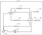
- FIG. 1is a schematic structural block diagram of a possible imaging system provided by an embodiment of the present application.
- FIG. 2is a schematic diagram of an application scenario of a possible ultrasound imaging method provided by an embodiment of the present application
- FIG. 3is a flowchart of a possible imaging method provided by an embodiment of the present application.
- FIG. 5is a schematic diagram of a possible mechanical scanner provided by an embodiment of the present application.
- FIG. 6is a schematic diagram of a possible probe provided by an embodiment of the present application.
- FIG. 7is a schematic diagram of a range of a surrounding area of a possible target tissue provided by an embodiment of the present application.
- the present applicationprovides an imaging method and imaging system for improving the comprehensiveness of image display.
- FIG. 1is a schematic structural block diagram of an imaging system 10 in an embodiment of the present application.
- the imaging system 10may include a probe 110, a laser 120, and a mechanical scanner 130, as well as a transmission circuit 101, a transmission / reception selection switch 102, a reception circuit 103, a processor 105, a display 106, and a memory 107.
- the imaging system 10may also include other devices or devices not shown in the figure.
- the transmitting circuit 101can excite the probe 110 to transmit ultrasonic waves to the target body.
- the receiving circuit 103may receive the ultrasonic echo returned from the target body through the probe 110, thereby obtaining ultrasonic echo signals / data.
- the ultrasonic echo signal / datais directly or through a beam synthesis circuit for beam synthesis processing, and then sent to the processor 105.
- the processor 105processes the ultrasound echo signal / data to obtain an ultrasound image of the target body.
- the ultrasound image obtained by the processor 105may be stored in the memory 107.
- the laser 120may generate laser light, which is coupled to the probe through an optical fiber bundle, and emits laser light to the target body through the optical fiber bundle coupled on the probe 110.
- the receiving circuit 103can also receive the photoacoustic signal / data returned by the target body under the excitation of the laser light through the probe 110.
- the photoacoustic signal / datais sent to the processor 105 directly or after processing, and the processor processes the photoacoustic signal / data to obtain a photoacoustic image of the target body.
- the mechanical scanner 130can drive the probe 110 to move.
- the aforementioned ultrasound image and photoacoustic imagemay be displayed on the display 106.
- the laseris emitted to the target body through the probe 110, specifically refers to the laser beam is emitted to the target body through the optical fiber bundle coupled to the probe 110, the optical fiber bundle may be provided outside the probe 110, or It is set inside the probe 110, and it can be adjusted according to the actual scene, which is not limited here.
- the laser 120may be connected to the transmission / reception selection switch 102, and the transmission / reception selection switch 102 controls the emission of laser light, or the laser 120 may be directly connected to the probe 110 through an optical transmission tool.
- the optical fiber bundleis coupled upward, and the laser beam is transmitted to both sides of the probe 110 by the optical fiber bundle, and the target body is irradiated by back-lighting.
- both the laser 120 and the fiber bundleare coupled into the probe, where the probe also includes an ultrasound transducer element for ultrasound imaging, so that the probe can be used not only for conventional ultrasound imaging but also for For photoacoustic imaging, it forms a probe integrating ultrasound imaging and photoacoustic imaging.
- the mechanical scanner 130enables the probe 110 to receive ultrasonic echo signals / data or photoacoustic signals / data from different orientations, and enables the processor 105 to process the received ultrasonic echo signals / data or photoacoustic signals / data To get an ultrasound image or a photoacoustic image.
- the mechanical scanner 130is an optional device.
- the mechanical scanner 130is coupled into the probe, that is, the probe integrates the function of mechanical scanning.
- the mechanical scanner 130may further include a motor controller and a motor, and the motor controller performs the motion trajectory, stroke, or speed of the motor in the mechanical scanner 130 according to the control signal sent by the processor. control.
- the probe 110may exist independently, or may be provided on the mechanical scanner 130, and the mechanical scanner 130 drives the probe 110 to move.
- the probe 110may specifically include an ultrasonic transducer, which has the functions of transmitting and receiving signals, and can perform various imaging such as gray-scale imaging and Doppler hemorrhage imaging.
- the optical fiber bundle and the ultrasonic transducerare coupled and surrounded by a housing to form a probe that integrates photoacoustic imaging and ultrasonic imaging functions. , And irradiate the laser to the target body through the probe, and receive the photoacoustic signal formed under the excitation of the laser returned from the target body through the probe.
- the probecan also be used for traditional ultrasound imaging, that is, transmitting ultrasound waves to the target body and receiving the ultrasonic echoes returned from the target body.
- the lasercan also be directly coupled with the ultrasonic transducer, and be completely or partially surrounded by the shell to form a probe that integrates photoacoustic imaging and ultrasonic imaging functions.
- the probecan be used for both photoacoustic imaging and Used for ultrasound imaging.
- the aforementioned display 106may be a touch display screen, a liquid crystal display screen, etc. built in the imaging system, or an independent display device such as a liquid crystal display, a television, etc., which is independent from the imaging system, or may be Display screens on electronic devices such as mobile phones and tablet computers, etc.
- the foregoing memory 107may be a flash memory card, a solid-state memory, a hard disk, or the like.
- a computer-readable storage mediumstores a plurality of program instructions. After the plurality of program instructions are called and executed by the processor 105, various implementations of the present application can be performed. Some or all of the steps in the ultrasound imaging method in the example or any combination of the steps therein.
- the computer-readable storage mediummay be the memory 107, which may be a non-volatile storage medium such as a flash memory card, solid state memory, or hard disk.
- the aforementioned processor 105may be implemented by software, hardware, firmware, or a combination thereof, and may use circuits, single or multiple application specific integrated circuits (application specific integrated circuits (ASIC), single or multiple general-purpose Integrated circuits, single or multiple microprocessors, single or multiple programmable logic devices, or combinations of the aforementioned circuits or devices, or other suitable circuits or devices, so that the processor 105 can execute various embodiments of the present application The corresponding steps in the imaging method.
- ASICapplication specific integrated circuits
- the imaging method provided in this embodiment of the present applicationcan be applied to the following application scenarios: for example, for specific application scenarios, refer to FIG. 2.
- the operatorscans the probe 110 on the target body 201, emits laser light from the optical fiber beams on both sides of the probe 110, and receives the returned photoacoustic signal through the probe 110. And the ultrasonic wave is transmitted from the probe, and the ultrasonic echo signal is received through the probe 110. Laser and ultrasound are not sent simultaneously. The operator can see the organization structure and the like through the display 106.
- an imaging method provided by an embodiment of the present applicationthe imaging method may be applied to the imaging system shown in FIG. 1, the imaging method embodiment includes:
- First laser lightis emitted to the target body within the first period, and the first photoacoustic signal returned from the target body is received.
- the first laser lightmay be emitted to the target body through the optical fiber bundle coupled on the probe 110 in the first period, and receive the first photoacoustic signal generated by the target body under the excitation of the first laser light.
- the first photoacoustic signal receivedmay also be different.
- the first laseris coupled to the probe through the fiber bundle, and then the first laser is emitted from the coupled fiber bundle on the probe to the target body. After the tissue in the target body absorbs the optical energy, it will cause temperature rise and thermal expansion, thereby generating a photoacoustic signal to propagate outward, and the corresponding first photoacoustic signal is detected by the probe 110.
- the first lasermay be generated by the processor 105 sending a first control signal to the laser 120, and the first control signal may include the wavelength, frequency, or timing of the first laser, and pass through the fiber bundle It is coupled to the probe 110, and then emits laser light to the target body through the optical fiber bundle.
- the position and angle of the lasercan be controlled by controlling the movement of the probe. After the tissue in the target body absorbs the light energy, it will cause temperature rise and thermal expansion, thereby generating a photoacoustic signal to propagate outward, and the corresponding photoacoustic signal is detected by the probe 110.
- the laser 120can return feedback information to the processor 105, which can include the actual sending time of the first laser, and the processor 105 can calculate the received photoacoustic signal according to a preset algorithm The interval is long, and the receiving circuit 103 controls the probe 110 to receive the first photoacoustic signal returned from the target body.
- the noise in the first photoacoustic signalcan be removed, and then subjected to processing such as beam synthesis and image reconstruction to obtain the first photoacoustic image of the target body.
- the second lasercan be emitted to the target body through the fiber bundle coupled to the probe 110 in the second period, and receive the second photoacoustic signal generated by the target under the excitation of the second laser .
- the second photoacoustic signal receivedmay also be different.
- the wavelengths of the first laser and the second laserare different.
- the first laserhas a short wavelength and the second laser has a long wavelength. And there is no overlap between the first cycle and the second cycle.
- the embodiment of the present applicationdoes not limit the firing order of the first laser and the second laser.
- the first lasermay be fired first, or the second laser may be fired first, which may be adjusted according to actual application scenarios.
- a second photoacoustic image of the target bodyAfter receiving the second photoacoustic signal, a second photoacoustic image of the target body can be obtained according to the second photoacoustic signal.
- the process of obtaining the second photoacoustic image by using the second photoacoustic signalis similar to the step of obtaining the first photoacoustic image by using the first photoacoustic signal in the foregoing step 302, and details are not repeated here.
- Step 302may be performed first, or step 304 may be performed first, which may be adjusted according to actual application scenarios.
- the blood vessel related parameters of the target bodysuch as the position, shape, etc. of the blood vessel can be determined according to the parameters included in the first photoacoustic image and the second photoacoustic image.
- Blood oxygen saturation, etc., and a blood oxygen image of the target bodycan be generated according to the blood vessel-related parameters.
- the blood oxygen image of the target bodycan be determined, and the photoacoustic images obtained by more lasers can also be used to determine the target For the blood oxygen image, this application only uses two lasers as an example to describe the determination process of the blood oxygen image, which can be adjusted according to actual needs, which is not limited in this application.
- the blood oxygen image of the target bodycan be calculated by the first photoacoustic image and the second photoacoustic image.
- the photoacoustic imagemay include the function information of the target body, including the blood vessel-related parameters, etc., for example, the position and shape of the blood vessel, and the distribution of blood oxygen. Therefore, the blood oxygen image includes the blood vessel-related parameters of the target tissue in the target body and the blood vessel-related parameters of the peripheral area of the target tissue.
- the blood oxygen imagecan be used to analyze the target tissue, can more clearly reflect the characteristics of the target tissue, and facilitate the operator to observe the target tissue clearly.
- Specific steps of the imaging method embodimentmay include:
- the first laseris emitted to the target body, and the first photoacoustic signal returned from the target body is received.
- the first laseris generated by the laser 130 and coupled to the probe 110 through the fiber bundle.
- the first lasercan be emitted to the target body through the fiber bundle by controlling the position and angle of the probe 110 and the like. After the tissue in the target body absorbs the optical energy, it will cause temperature rise and thermal expansion, thereby generating a photoacoustic signal to propagate outward, and the corresponding first photoacoustic signal is detected by the probe 110.
- an optical fiber bundleis coupled to the probe, and the laser beam is transmitted to both sides of the probe 110 using the optical fiber bundle to illuminate the target body in a back-illuminated manner.
- the probe 110includes an ultrasonic transducer.
- the ultrasonic transducerhas the function of transmitting and receiving signals. On the basis of ensuring the traditional ultrasonic image and Doppler blood flow imaging, it also has a large frequency bandwidth and high sensitivity. Improves the ability to detect photoacoustic signals, even weak signals can be detected.
- the first laser lightis transmitted to the target body through the optical fiber bundle coupled on the probe 110.
- the probe 110may be provided on the mechanical scanner 130, and then the processor 105 may send a control signal to the mechanical scanner 130 to control the motor in the mechanical scanner 130 to control the scanning speed and trajectory of the mechanical scanner 130.
- the probe 110can surround the target body and receive the first photoacoustic signal returned from the target body from different angles to perform photoacoustic imaging of the target body from different angles, which can form a multi-frame first photoacoustic image to make the target body
- the first photoacoustic imageis more three-dimensional.
- the mechanical scanner 130may be as shown in FIG. 5.
- the first photoacoustic imageis obtained according to the first photoacoustic signal.
- the first photoacoustic imagemay be displayed on the display 106.
- multiple first photoacoustic signalscan be acquired from multiple different angles, and multiple frames of light can be obtained through the multiple photoacoustic signals
- Acoustic imagescan then be synthesized through the multi-frame photoacoustic images to form a three-dimensional first photoacoustic image, so that the obtained first photoacoustic image can be displayed more three-dimensionally, allowing the operator to intuitively pass the first photoacoustic image The image observes the condition of the target.
- the first ultrasonic waveis also transmitted to the target body through the probe 110, and the corresponding first ultrasonic echo returned by the target body is received through the probe 110, and converted into the first ultrasonic wave according to the first ultrasonic echo Echo signal.
- the received ultrasound echo signalmay also be different.
- the first laser and the first ultrasonic waveare not transmitted at the same time.
- the first lasercan be sent first, or the first ultrasonic wave can be sent first.
- Step 401 or step 402can be executed first. It is adjusted according to the actual application scenario and is not limited here.
- the processor 105controls to open the transmit / receive selection switch 102, and controls the transmit circuit 101 to transmit ultrasonic waves to the target body through the probe 110 and receive ultrasonic waves through the probe 110 The wave is transmitted to the receiving circuit 103. It can be understood that the receiving circuit 103 can receive the ultrasonic echo returned from the target body through the probe 110 to obtain an ultrasonic echo signal.
- ultrasonic wavesare sent through the probe 110, and the probe 110 can be set on the mechanical scanner 130, and then the processor 105 can send a control signal to the mechanical scanner 130 to control the motor in the mechanical scanner 130, The scanning speed and trajectory of the mechanical scanner 130 are controlled so that the probe 110 can surround the target body, send ultrasonic waves from different angles, and receive ultrasonic echoes from different angles, so as to image the target body from different angles.
- the noise in the ultrasonic signalmay be removed.
- the ultrasonic echo signalis subjected to beam synthesis processing by a beam synthesis circuit, and then transmitted to the processor 105, and the processor 105 processes the ultrasonic echo signal to obtain an ultrasound image of the target body.
- the noise in the photoacoustic signalmay be removed, and then image reconstruction processing such as beam synthesis processing may be performed to obtain a photoacoustic image of the target body.
- the ultrasound imageis a grayscale image, which can reflect the structural information of the target tissue in the target body, and the photoacoustic image can reflect the functional information of the tissue in the target body.
- the first ultrasound image and the second photoacoustic imageare fused to obtain the first fused image, and displayed on the display 106.
- the pixel value of each pixel in the first photoacoustic imagemay be superimposed on the corresponding pixel in the first ultrasound image based on the first ultrasound image.
- the pixel value of each pixel in the first ultrasound imagemay be superimposed on the corresponding pixel in the first photoacoustic image based on the first photoacoustic image.
- the first fusion imagemay include not only the structure information of the target body displayed in the first ultrasound image, but also the function information of the target body displayed in the first photoacoustic image, so that the operator can perform a more comprehensive Observed.
- the probe 110is set to move in the mechanical scanner 130, multiple ultrasonic echo signals and photoacoustic signals of different angles can be acquired, and then multiple frames of corresponding ultrasonic images and photoacoustic images can also be obtained.
- the direction and angle of light projectioncan be changed, or the transparency of the object display can be adjusted to fully display the 3D structure of the target tissue, so that the operator can make certain observations through the ultrasound image and the photoacoustic image.
- Doppler frequency shiftcan be used to realize Doppler blood flow imaging, and blood flow with a certain flow rate can be imaged.
- Doppler blood flow imagingis too sensitive to movement, including tissue movement and probe movement, which makes it difficult to achieve three-dimensional Doppler imaging using mechanical scanners. In the process of mechanical probe driving probe scanning, it will introduce Artifacts.
- photoacoustic imagingdepends on the photoacoustic signal generated by the tissue's absorption of laser light at a specified wavelength, so it is not sensitive to the movement of the tissue or probe.
- a mechanical scannercan be used to acquire photoacoustic images and ultrasound images of the target body, display the collection of functional information of the target body through the photoacoustic image, and collect structural information of the target body through the ultrasound image. Therefore, it is not necessary to perform Doppler blood flow imaging, and 3D acquisition of the function and structure information of the tissue can also be achieved.
- the second laser beamis emitted to the target body through the optical fiber bundle coupled to the probe 110 during the second period, and the second photoacoustic signal returned from the target body is received through the probe 110.
- step 405 and step 406 in the embodiment of the present applicationare similar to the foregoing step 401 and step 402, and details are not repeated here.
- the specific operation steps of sending the first laser light and the second laser light to the target bodymay be consistent, and the steps of acquiring the first photoacoustic image and the second photoacoustic image may also be consistent.
- the angle between receiving the first photoacoustic signal and receiving the second photoacoustic signalmay be consistent, and processing the first photoacoustic signal to obtain a first photoacoustic image and processing the second photoacoustic signal to obtain a second photoacoustic signal
- the specific process of the imagecan be kept consistent.
- multiple second photoacoustic signalscan be acquired from multiple different angles and passed through the multiple second photoacoustic signals Multiple frames of photoacoustic images can be obtained, which can then be synthesized through the multiple frames of photoacoustic images to form a three-dimensional second photoacoustic image, so that the resulting second photoacoustic image can be displayed more three-dimensionally, which can make the operator more intuitive Observe the condition of the target body through the first photoacoustic image.
- the three-dimensional first photoacoustic image and the second photoacoustic imagecan be calculated to obtain a three-dimensional blood oxygen image, so that the operator can more comprehensively analyze the blood vessel related conditions of the target body. Observed.
- the probe 110also sends a second ultrasonic wave to the target body, and receives the second ultrasonic echo returned from the target body, and converts it into a second ultrasonic echo signal according to the second ultrasonic echo.
- the second ultrasonic wave and the second laserare transmitted at different times, that is, the second ultrasonic wave and the second laser are not transmitted at the same time.
- the second ultrasonic wavemay be sent first, or the second laser may be sent first, which may be adjusted according to actual application scenarios.
- the specific process of obtaining the second ultrasound image of the target body according to the second ultrasound echo signalis similar to the process of obtaining the first ultrasound image according to the first ultrasound echo signal in step 404, The details are not repeated here.
- the second ultrasound image and the second photoacoustic imageare fused to obtain the first
- the two fusion imagesare displayed on the display 106.
- the first fusion image and the second fusion imagemay be displayed on one screen at the same time, or one of them may be displayed, or the operator may choose to display one of the first fusion image and the second fusion image.
- the pixel value of each pixel in the second photoacoustic imagemay be superimposed on the corresponding pixel in the second ultrasound image based on the second ultrasound image.
- the pixel value of each pixel in the second ultrasound imagemay be superimposed on the corresponding pixel in the second photoacoustic image. It may also be that the operator chooses to specifically superimpose pixel values based on one of the second ultrasound image or the second photoacoustic image, which may be adjusted according to the actual application scenario, which is not limited here . Similar to the aforementioned first fusion image, the second fusion image may include not only the structural information of the target body displayed in the second ultrasound image, but also the functional information of the target body displayed in the second photoacoustic image. The operator makes a more comprehensive observation of the target body.
- the first ultrasound image and the first photoacoustic imagemay be acquired first, or the second ultrasound image and the second photoacoustic image may be acquired first, which may be adjusted according to actual application scenarios.
- the blood oxygen saturation of each pixel corresponding to the target bodymay be calculated according to the first photoacoustic image and the second photoacoustic image to obtain a blood oxygen image, and It is displayed on the display 106.
- the blood oxygen saturation of each pixelcan be calculated according to a preset formula, which can include:
- A1is related data of the first laser
- A2is related data of the second laser.
- the relevant data of the first lasermay be the amplitude of the corresponding pixel in the first photoacoustic signal, or the amplitude of any one of the multiple channels received by the ultrasound array probe, or each of the multiple channels The value of the amplitude received by each channel after beam synthesis processing and so on.
- the relevant data of the second lasermay be the amplitude of the corresponding pixel in the second photoacoustic signal, or the amplitude of any one of the multiple channels received by the ultrasound array probe, or each of the multiple channels. The value of the amplitude received by each channel after beam synthesis processing and so on.
- Hbis the content of deoxyhemoglobin in the target pixel
- HbO2is the content of oxyhemoglobin in the target pixel
- SO2is the blood oxygen saturation of the target pixel
- the target pixelis any one of each pixel.
- the blood oxygen saturation of each pixel of the target bodycan be calculated. Then use the blood oxygen saturation value of each pixel as the pixel value of the corresponding pixel, or calculate the blood oxygen saturation value of each pixel according to a preset algorithm to obtain the pixel of the corresponding pixel Value, according to the pixel value of each pixel, you can get the blood oxygen image of the target body.
- the first ultrasound image or the second ultrasound imagecan be displayed simultaneously on the same screen
- One of the ultrasound images, the first fusion image, the second fusion image and the blood oxygen imageso that the operator can observe the target body according to the combination of the ultrasound image, the first fusion image, the second fusion image and the blood oxygen image, Make the operator's observation of the target body more comprehensive.
- the target tissue and the surrounding area of the target tissueare determined in the blood oxygen image.
- the specific process of determining the target tissue in the ultrasound imagecan be:
- the target tissueis determined by comparing the parameter values of the target tissue with other tissues surrounding the target tissue in the ultrasound image.
- the parameter valuemay include at least one of a gray value, a brightness value, a pixel value, or a gradient value.
- this parameter valuemay also be other values that can be compared with the image, which can be adjusted according to the actual application, and is not limited here. It can also be manually selected by the operator to determine the target tissue in the ultrasound image.
- the processor 105receives input parameters for the ultrasound image, and determines the boundary of the target tissue according to the input parameters.
- any one of the first ultrasound image or the second ultrasound imagemay be displayed on the display 106, and the operator selects the boundary of the target tissue in the ultrasound image through the input device to generate the input parameter. Therefore, even when the contrast between the target tissue and the surrounding normal tissue is not significant, the target tissue can be manually delineated by the operator to obtain more accurate structure information of the target tissue in the ultrasound image.
- multiple frames of the first ultrasound image or multiple frames of the second ultrasound imagemay be fused and displayed as a 3D ultrasound image, and then the operator 3D ultrasound images are manually selected to determine the target tissue in the ultrasound images.
- the target tissueAfter the target tissue is determined in any one of the first ultrasound image or the second ultrasound image, the target tissue needs to be determined in blood oxygen. Specifically, the angle and resolution of the blood oxygen image can be adjusted to be the same as the angle and resolution of any ultrasound image. After the target tissue is determined in any ultrasound image, the target tissue can be determined in the ultrasound image. Include the position information of each pixel, and then determine the position corresponding to each pixel in the blood oxygen image according to the position information of each pixel to determine the target tissue in the blood oxygen image.
- a coordinate axiscan be established, and then each half axis of the target tissue can be extended to n times, n> 1.0, and the area where the target tissue is located can be obtained The surrounding area of the target organization.
- a two-dimensional coordinate axisis established in the blood oxygen image , Magnify each half axis of the target tissue by n times, and then remove the area after the target tissue, that is, 701 shown in FIG. 7 is the surrounding area of the target tissue.
- the obtained blood oxygen imageis a three-dimensional image
- the three-dimensional coordinate axiscan also be established to determine the surrounding area of the target tissue, which can be adjusted according to the actual application scenario today. , This application is not limited.
- the blood vessel density and blood oxygen ratio of the target tissue and the surrounding area of the target tissuecan be calculated.
- the blood vessel densityis the ratio of the volume occupied by the blood vessel to the tissue volume.
- the oxygen ratiois the ratio of the average value of blood oxygen saturation between the target tissue and the surrounding area of the target tissue.
- the specific method for calculating the blood oxygen ratiomay be to calculate the average value of the first blood oxygen saturation of the target tissue in the blood oxygen image and the second blood oxygen saturation of the surrounding area of the target tissue The average value, and then calculate the ratio of the first average blood oxygen saturation to the second average blood oxygen saturation.
- the specific method for calculating the average value of the first blood oxygen saturation of the target tissuemay be to determine the value of the blood oxygen saturation of each pixel according to the pixel value of each pixel in the blood oxygen image, and then the target in the blood oxygen image Sum the values of blood oxygen saturation corresponding to each pixel in the tissue to obtain the total value of blood oxygen saturation, and then use the total value of blood oxygen saturation to average each pixel in the target tissue to calculate The average first oxygen saturation of the target tissue. It can be understood that, after calculating the total value of the blood oxygen saturation of all pixels in the target tissue, divided by the number of pixels in the target tissue, the average value of the blood oxygen saturation of each pixel in the target tissue is obtained , Which is the mean value of the first blood oxygen saturation.
- the specific method for calculating the average value of the second blood oxygen saturation in the peripheral area of the target tissuemay be to determine the blood oxygen saturation value of each pixel according to the pixel value of each pixel in the blood oxygen image, and then The blood oxygen saturation value corresponding to each pixel in the peripheral area of the target tissue in the blood oxygen image is summed to obtain the total value of blood oxygen saturation, and then the total value of blood oxygen saturation is used The pixels are averaged, and the average value of the first blood oxygen saturation of the target tissue is calculated.
- the average per pixel of the surrounding area of the target tissueis obtained.
- the average value of the blood oxygen saturationis the average value of the second blood oxygen saturation.
- the specific method for calculating the blood vessel densitymay be that the pixel point whose blood oxygen saturation of each pixel point in the blood oxygen image is greater than the threshold value is a blood vessel pixel point. Then calculate the ratio of the number of blood vessel pixels to the number of pixels in the blood oxygen image, and calculate the ratio of the number of blood vessel pixels to the number of pixels in the blood oxygen image to obtain the blood vessel density.
- the part of the blood oxygen image whose blood oxygen saturation is greater than the thresholdis determined as the blood part, and then the number of pixels of the blood oxygen saturation greater than the threshold and the number of pixels of the blood oxygen image are calculated to calculate the blood oxygen saturation
- the ratio of the number of pixels in the part whose degree is greater than the threshold to the number of pixels in the blood oxygen imagecan be obtained as the blood vessel density.
- the ratio of the number of pixels in the part of the blood oxygen saturation greater than the threshold to the number of pixels in the part of the blood oxygen imagemay be calculated to obtain the blood vessel density.
- the blood vessel density inside the target tissuemay also be calculated, that is, determined by the ratio of the number of pixels in the portion of the target tissue whose blood oxygen saturation is greater than the threshold value to the number of all pixels in the target tissue.
- the blood vessel densitycan also be determined by the volume ratio, for example, the blood vessel density in the target tissue is determined by the volume corresponding to the blood vessel in the target tissue and the volume of the target tissue.
- the blood vessel density and blood oxygen ratiomay be displayed on the display 106 so that the operator can refer to the blood vessel density and blood oxygen ratio for the target body Comparative analysis of the target organization.
- the target tissuecan be analyzed with reference to the blood vessel density and blood oxygen ratio.
- the blood oxygen ratio range of the conventional tissuecan be compared with the blood oxygen ratio of the target body, and the The comparison result is displayed on the display 106. It is also possible to compare the blood vessel density range of normal tissue with the blood vessel density of the target body, and display the comparison result on the display 106. Therefore, when the operator observes the blood oxygen image on the display, he can refer to the analysis result to perform a more accurate and comprehensive analysis of the target tissue.
- the blood vessel related parameters of the target tissueare calculated based on the blood oxygen image, including blood vessel density, blood oxygen ratio, etc., and the fusion of the ultrasound image and the photoacoustic image can also be displayed.
- the fusion imagethat is, the first fusion image and the second fusion image, allows the operator to comprehensively observe the target tissue by combining the blood oxygen image, blood vessel related parameters, the first fusion image and the second fusion image, so that the operator can according to the target tissue
- the internal and external vascular related parameterscomprehensively analyze the state of the target tissue.
- the disclosed system, device, and methodmay be implemented in other ways.
- the device embodiments described aboveare only schematic.
- the division of the unitsis only a division of logical functions.
- there may be other divisionsfor example, multiple units or components may be combined or Can be integrated into another system, or some features can be ignored, or not implemented.
- the displayed or discussed mutual coupling or direct coupling or communication connectionmay be indirect coupling or communication connection through some interfaces, devices or units, and may be in electrical, mechanical or other forms.
- the units described as separate componentsmay or may not be physically separated, and the components displayed as units may or may not be physical units, that is, they may be located in one place, or may be distributed on multiple network units. Some or all of the units may be selected according to actual needs to achieve the purpose of the solution of this embodiment.
- each functional unit in each embodiment of the present applicationmay be integrated into one processing unit, or each unit may exist alone physically, or two or more units are integrated into one unit.
- the above integrated unitcan be implemented in the form of hardware or software function unit.
- the integrated unitis implemented in the form of a software functional unit and sold or used as an independent product, it may be stored in a computer-readable storage medium.
- the technical solution of the present applicationmay be essentially or part of the contribution to the existing technology or all or part of the technical solution may be embodied in the form of a software product, and the computer software product is stored in a storage medium , Including several instructions to enable a computer device (which may be a personal computer, server, or network device, etc.) to perform all or part of the steps of the methods described in the embodiments of the present application.
- the aforementioned storage mediainclude: U disk, mobile hard disk, read-only memory (Read-Only Memory, ROM), random access memory (Random Access Memory, RAM), magnetic disk or optical disk and other media that can store program code .
- the target bodymay be a human body, an animal, or the like.
- the target tissuemay be the face, spine, heart, uterus, or pelvic floor, or other parts of the human tissue, such as the brain, bones, liver, or kidney, which is not specifically limited here.
Landscapes
- Health & Medical Sciences (AREA)
- Life Sciences & Earth Sciences (AREA)
- Physics & Mathematics (AREA)
- Medical Informatics (AREA)
- Surgery (AREA)
- Pathology (AREA)
- Veterinary Medicine (AREA)
- Engineering & Computer Science (AREA)
- Biomedical Technology (AREA)
- Heart & Thoracic Surgery (AREA)
- Biophysics (AREA)
- Molecular Biology (AREA)
- Public Health (AREA)
- Animal Behavior & Ethology (AREA)
- General Health & Medical Sciences (AREA)
- Nuclear Medicine, Radiotherapy & Molecular Imaging (AREA)
- Radiology & Medical Imaging (AREA)
- Dermatology (AREA)
- Pain & Pain Management (AREA)
- Hematology (AREA)
- Vascular Medicine (AREA)
- Acoustics & Sound (AREA)
- Ultra Sonic Daignosis Equipment (AREA)
Abstract
Description
Translated fromChinese本申请涉及医疗器械领域,特别涉及一种成像方法以及成像系统。This application relates to the field of medical devices, in particular to an imaging method and imaging system.
光声成像(Photoacoustic Imaging,PAI)是新型的生物医疗成像技术,PAI的原理是基于光声效应,当生物组织受到短脉冲的激光照射时,例如,纳秒(ns)量级,生物组织中具有强光学吸收特性的物质,例如血液,在吸收光能量后,将引起局部升温和热膨胀,从而产生光声信号,并向外传播。可以通过超声探头检测到受短脉冲的激光照射后的生物组织产生的光声信号,探测到光声信号,利用相应的重建算法,即可重建吸收体,即具有强光学吸收特性的物质的位置和形态。光声成像结合了光学和超声的优点,对一些重大疾病的早期诊断与预后评估有独特的优势,是具有巨大临床和产业前景的新型影像技术。受限于光在生物组织中的穿透能力,光声成像应用重点集中于一些浅层的器官。光声成像体现了生物体的功能信息,而传统的超声成像反应了生物体的结构信息,将二者有效地结合起来,即光声-超声双模态成像克服了单一模态成像的不足,能够提供更全面的组织结构和功能信息。Photoacoustic imaging (PAI) is a new type of biomedical imaging technology. The principle of PAI is based on the photoacoustic effect. When biological tissues are irradiated with short pulses of laser light, for example, on the order of nanoseconds (ns), Substances with strong optical absorption properties, such as blood, will cause local heating and thermal expansion after absorbing light energy, thereby generating photoacoustic signals and propagating outward. The photoacoustic signal generated by the biological tissue irradiated by the short pulse laser can be detected by the ultrasonic probe, and the photoacoustic signal is detected, and the corresponding reconstruction algorithm can be used to reconstruct the absorber, that is, the position of the substance with strong optical absorption characteristics And shape. Photoacoustic imaging combines the advantages of optics and ultrasound. It has unique advantages in the early diagnosis and prognosis evaluation of some major diseases. It is a new imaging technology with huge clinical and industrial prospects. Limited by the ability of light to penetrate biological tissues, the application of photoacoustic imaging focuses on some shallow organs. Photoacoustic imaging reflects the functional information of organisms, while traditional ultrasound imaging reflects the structural information of organisms, effectively combining the two, that is, photoacoustic-ultrasonic dual-mode imaging overcomes the shortcomings of single-mode imaging. Can provide more comprehensive organizational structure and functional information.
然而,光声-超声双模态成像得到的图像,对于目标组织的功能信息的反映并不全面,无法显示更全面的图像。因此,如何显示更清楚地反映目标组织的特性的图像,成为亟待解决的问题。However, the image obtained by photoacoustic-ultrasound dual-modality imaging does not fully reflect the functional information of the target tissue, and cannot display a more comprehensive image. Therefore, how to display an image that more clearly reflects the characteristics of the target organization has become an urgent problem to be solved.
发明内容Summary of the invention
本申请提供一种成像方法以及成像系统,用于提高图像显示的全面性。The present application provides an imaging method and imaging system for improving the comprehensiveness of image display.
本申请实施例的第一方面提供一种成像方法,包括:在第一周期内向目标体发射第一激光,并接收从所述目标体返回的第一光声信号;根据所述第一光声信号确定所述目标体的第一光声图像;在第二周期内向所述目标体发射第二激光,并接收从所述目标体返回的第二光声信号,其中,所述第二激光和所述第一激光的波长不相同;根据所述第二光声信号确定所述目标体的第二光声图像;根据所述第一光声图像和所述第二光声图像确定所述目标体的血氧图像, 其中,所述血氧图像包括所述目标体内的目标组织的血管相关参数与所述目标组织周边预置范围内的血管相关参数。A first aspect of an embodiment of the present application provides an imaging method, including: emitting a first laser to a target body within a first period, and receiving a first photoacoustic signal returned from the target body; according to the first photoacoustic The signal determines the first photoacoustic image of the target body; during the second period, a second laser light is emitted to the target body, and a second photoacoustic signal returned from the target body is received, wherein the second laser and The wavelengths of the first laser light are different; the second photoacoustic image of the target body is determined according to the second photoacoustic signal; the target is determined based on the first photoacoustic image and the second photoacoustic image A blood oxygen image of the body, wherein the blood oxygen image includes a blood vessel related parameter of the target tissue in the target body and a blood vessel related parameter within a preset range around the target tissue.
本申请实施例的第二方面提供一种成像系统,包括:激光器、探头以及发射电路、接收电路以及处理器;所述激光器用于在第一周期内向产生照射目标体的第一激光,所述第一激光通过光纤束耦合至所述探头,并通过所述光纤束向所述目标体发射所述第一激光;所述接收电路用于控制所述探头接收从所述目标体返回的第一光声信号;所述处理器用于根据所述第一光声信号确定所述目标体的第一光声图像;所述激光器还用于在第二周期内产生照射目标体的第二激光,所述第二激光通过光纤束耦合至所述探头,并通过所述光纤束向所述目标体发射所述第二激光;所述接收电路用于控制所述探头接收从所述目标体返回的第二光声信号;所述处理器还用于根据所述第二光声信号确定所述目标体的第二光声图像;所述处理器还用于根据所述第一光声图像和所述第二光声图像确定所述目标体的血氧图像,其中,所述血氧图像包括所述目标体内的目标组织的血管相关参数与所述目标组织周边预置范围内的血管相关参数。A second aspect of an embodiment of the present application provides an imaging system, including: a laser, a probe, and a transmitting circuit, a receiving circuit, and a processor; the laser is used to generate a first laser beam that irradiates a target within a first period, The first laser is coupled to the probe through an optical fiber bundle, and emits the first laser to the target body through the optical fiber bundle; the receiving circuit is used to control the probe to receive the first returned from the target body The photoacoustic signal; the processor is used to determine the first photoacoustic image of the target body according to the first photoacoustic signal; the laser is also used to generate a second laser light illuminating the target body in a second period, so The second laser is coupled to the probe through an optical fiber bundle, and emits the second laser to the target body through the optical fiber bundle; the receiving circuit is used to control the probe to receive the first laser beam returned from the target body Two photoacoustic signals; the processor is further used to determine the second photoacoustic image of the target body according to the second photoacoustic signal; the processor is also used to determine the second photoacoustic image and the Determining the photoacoustic image of the second image of the target body oxygen, wherein said oxygen comprises image vascular parameters vascular parameters within a target tissue of the target body tissue surrounding the target preset range.
本申请实施例的第三方面提供了一种计算机可读存储介质,该计算机可读存储介质中存储有指令,当其在计算机上运行时,使得计算机执行上述第一方面提供的成像方法。A third aspect of the embodiments of the present application provides a computer-readable storage medium, in which instructions are stored in a computer-readable storage medium, which when executed on a computer, causes the computer to execute the imaging method provided in the first aspect.
在本申请中,向目标体发射超声波以及交替发射波长不同的至少两个激光,即第一激光与第二激光,然后接收从目标体返回的对应的第一光声信号与第二光声信号,并根据第一光声信号确定目标体的第一光声图像,根据第二光声信号确定目标体的第二光声图像。并根据第一光声图像与第二光声图像计算得到目标体的血氧图像。通常,光声图像中可以包括目标体的功能信息,包括血管相关参数等,例如,血管的位置与形态、血氧的分布情况等。因此,血氧图像包括目标体内的目标组织的血管相关参数,与目标组织的周边区域的血管相关参数。可以用于对目标组织进行分析,能更清楚地反映目标组织的特性,便于操作人员对目标组织进行清楚地观察。In the present application, ultrasonic waves are emitted to the target body and at least two lasers with different wavelengths are alternately emitted, namely the first laser light and the second laser light, and then the corresponding first photoacoustic signal and second photoacoustic signal returned from the target body are received And determine the first photoacoustic image of the target body based on the first photoacoustic signal, and determine the second photoacoustic image of the target body based on the second photoacoustic signal. And the blood oxygen image of the target body is calculated according to the first photoacoustic image and the second photoacoustic image. Generally, the photoacoustic image may include the function information of the target body, including the blood vessel-related parameters, etc., for example, the position and shape of the blood vessel, and the distribution of blood oxygen. Therefore, the blood oxygen image includes the blood vessel-related parameters of the target tissue in the target body and the blood vessel-related parameters of the peripheral area of the target tissue. It can be used to analyze the target organization, which can more clearly reflect the characteristics of the target organization, which is convenient for the operator to observe the target organization clearly.
图1为本申请实施例提供的一种可能的成像系统的结构框图示意图;1 is a schematic structural block diagram of a possible imaging system provided by an embodiment of the present application;
图2为本申请实施例提供的一种可能的超声成像方法的应用场景示意图;2 is a schematic diagram of an application scenario of a possible ultrasound imaging method provided by an embodiment of the present application;
图3为本申请实施例提供的一种可能的成像方法的流程图;3 is a flowchart of a possible imaging method provided by an embodiment of the present application;
图4为本申请实施例提供的另一种可能的成像方法的流程图;4 is a flowchart of another possible imaging method provided by an embodiment of the present application;
图5为本申请实施例提供的一种可能的机械扫描器示意图;5 is a schematic diagram of a possible mechanical scanner provided by an embodiment of the present application;
图6为本申请实施例提供的一种可能的探头示意图;6 is a schematic diagram of a possible probe provided by an embodiment of the present application;
图7为本申请实施例提供的一种可能的目标组织的周边区域的范围示意图。7 is a schematic diagram of a range of a surrounding area of a possible target tissue provided by an embodiment of the present application.
本申请提供一种成像方法以及成像系统,用于提高图像显示的全面性。The present application provides an imaging method and imaging system for improving the comprehensiveness of image display.
本申请的说明书和权利要求书及上述附图中的术语“第一”、“第二”、“第三”、“第四”等(如果存在)是用于区别类似的对象,而不必用于描述特定的顺序或先后次序。应该理解这样使用的数据在适当情况下可以互换,以便这里描述的实施例能够以除了在这里图示或描述的内容以外的顺序实施。此外,术语“包括”和“具有”以及他们的任何变形,意图在于覆盖不排他的包含,例如,包含了一系列步骤或单元的过程、方法、系统、产品或设备不必限于清楚地列出的那些步骤或单元,而是可包括没有清楚地列出的或对于这些过程、方法、产品或设备固有的其它步骤或单元。The terms "first", "second", "third", "fourth", etc. (if any) in the description and claims of the present application and the above drawings are used to distinguish similar objects without using To describe a specific order or sequence. It should be understood that the data used in this way can be interchanged under appropriate circumstances so that the embodiments described herein can be implemented in an order other than what is illustrated or described herein. In addition, the terms "including" and "having" and any variations thereof are intended to cover non-exclusive inclusions, for example, processes, methods, systems, products or devices that contain a series of steps or units need not be limited to those clearly listed Those steps or units, but may include other steps or units not explicitly listed or inherent to these processes, methods, products or equipment.
图1为本申请实施例中的成像系统10的结构框图示意图。该成像系统10可以包括探头110、激光器120以及机械扫描器130,以及发射电路101、发射/接收选择开关102、接收电路103、处理器105、显示器106以及存储器107。当然,该成像系统10还可以包括其他图中未示出的设备或器件等。FIG. 1 is a schematic structural block diagram of an
发射电路101可以激励探头110向目标体发射超声波。在探头110发射超声波后,接收电路103可以通过探头110接收从目标体返回的超声回波,从而获得超声回波信号/数据。该超声回波信号/数据直接或经过波束合成电路进行波束合成处理后,送入处理器105。处理器105对该超声回波信号/数据进行处理,以获得目标体的超声图像。处理器105获得的超声图像可以存储于存储器107中。激光器120可以产生激光,该激光通过光纤束耦合至探头,并通过探头110 上耦合的光纤束向目标体发射激光。在向目标体发射激光后,接收电路103还可以通过探头110接收目标体在激光的激励下返回的光声信号/数据。该光声信号/数据直接或经过处理后送入处理器105,处理器对该光声信号/数据进行处理,以得到目标体的光声图像。机械扫描器130可以带动探头110运动。前述的超声图像与光声图像可以在显示器106上显示。The transmitting
需要说明的是,本申请中,通过探头110向目标体发射激光,具体是指通过耦合在探头110上的光纤束向目标体发射激光,该光纤束可以是设置在探头110外部的,也可以是设置在探头110内部的,具体可以根据实际场景调整,此处并不作限定。It should be noted that in this application, the laser is emitted to the target body through the
本申请的一个实施例中,激光器120可以是与发射/接收选择开关102连接,由发射/接收选择开关102控制发射激光,也可以是激光器120直接通过光传导工具连接到探头110,在探头110上耦合光纤束,利用光纤束将激光传导至探头110的两侧,采用背向式打光的方式对目标体进行照射。在有些实现方式中,将激光器120和光纤束都耦合至探头内部,其中,探头内部还包括用于超声成像的超声换能器元件,这样,该探头不止可以用于常规的超声成像,也用于光声成像,即形成集超声成像和光声成像为一体的探头。In one embodiment of the present application, the
通过机械扫描器130可以使探头110从不同的方位接收超声回波信号/数据或光声信号/数据,可以使处理器105对接收到的超声回波信号/数据或光声信号/数据进行处理,得到超声图像或光声图像。The
其中,机械扫描器130为可选装置。在有些实现方式中,该机械扫描器130耦合至探头内,即该探头集合了机械扫描的功能。Among them, the
本申请的一个实施例中,机械扫描器130中还可以包括电机控制器与电机,由电机控制器根据处理器发送的控制信号,对机械扫描器130内电机的运动轨迹、行程或速度等进行控制。In an embodiment of the present application, the
本申请的一个实施例中,探头110可以是独立存在的,也可以是设置在机械扫描器130上,由机械扫描器130带动探头110运动。In an embodiment of the present application, the
本申请的一个实施例中,探头110上具体可以包括超声换能器,超声换能器具有发射和接收信号的功能,可以进行灰阶成像与多普勒流血成像等多种成像。另外,在有些实现方式中,光纤束和超声换能器耦合,并通过外壳包围, 形成一个集光声成像和超声成像功能为一体的探头,即,在这种结构的探头下,激光器发射激光,并通过探头将该激光照射到目标体上,并通过探头接收从该目标体返回的在激光激励下形成的光声信号。当然,该探头还可以用于传统的超声成像,即向目标体发射超声波,并接收从目标体返回的超声回波。当然,还可以将激光器直接和超声换能器耦合,并通过外壳全部包围或者部分包围,形成一个集光声成像和超声成像功能为一体的探头,该探头既可以用于光声成像,又可以用于超声成像。In an embodiment of the present application, the
本申请的一个实施例中,前述的显示器106可为成像系统内置的触摸显示屏、液晶显示屏等,也可以是独立于成像系统之外的液晶显示器、电视机等独立显示设备,也可为手机、平板电脑等电子设备上的显示屏,等等。In an embodiment of the present application, the
本申请的一个实施例中,前述的存储器107可为闪存卡、固态存储器、硬盘等。In an embodiment of the present application, the foregoing
本申请的一个实施例中,还提供一种计算机可读存储介质,该计算机可读存储介质存储有多条程序指令,该多条程序指令被处理器105调用执行后,可执行本申请各个实施例中的超声成像方法中的部分步骤或全部步骤或其中步骤的任意组合。In an embodiment of the present application, a computer-readable storage medium is also provided. The computer-readable storage medium stores a plurality of program instructions. After the plurality of program instructions are called and executed by the
本申请的一个实施例中,该计算机可读存储介质可为存储器107,其可以是闪存卡、固态存储器、硬盘等非易失性存储介质。In an embodiment of the present application, the computer-readable storage medium may be the
本申请的一个实施例中,前述的处理器105可以通过软件、硬件、固件或者其组合实现,可以使用电路、单个或多个专用集成电路(application specific integrated circuits,ASIC)、单个或多个通用集成电路、单个或多个微处理器、单个或多个可编程逻辑器件、或者前述电路或器件的组合、或者其他适合的电路或器件,从而使得该处理器105可以执行本申请的各个实施例中的成像方法的相应步骤。In an embodiment of the present application, the
下面基于前述的成像系统,对本申请中的成像方法进行详细描述。The imaging method in the present application will be described in detail based on the aforementioned imaging system.
需要说明的是,结合图1所示的成像系统的结构框图示意图,本申请实施例提供的成像方法可应用于如下应用场景:示例性地,具体应用场景可以参阅图2。操作人员将探头110在目标体201进行扫描,从探头110两侧的光纤束发射激光,并通过探头110接收返回的光声信号。以及从探头发射超声波,并通过 探头110接收超声回波信号。激光与超声波不同时发送。操作人员可以通过显示器106看到组织结构等。It should be noted that, in conjunction with the schematic structural block diagram of the imaging system shown in FIG. 1, the imaging method provided in this embodiment of the present application can be applied to the following application scenarios: for example, for specific application scenarios, refer to FIG. 2. The operator scans the
基于此,请参阅图3,本申请实施例提供的一种成像方法,该成像方法可以应用于前述图1所示的成像系统,该成像方法实施例包括:Based on this, please refer to FIG. 3, an imaging method provided by an embodiment of the present application, the imaging method may be applied to the imaging system shown in FIG. 1, the imaging method embodiment includes:
301、在第一周期内向目标体发射第一激光,并接收从目标体返回的第一光声信号。301: First laser light is emitted to the target body within the first period, and the first photoacoustic signal returned from the target body is received.
可以通过探头110上耦合的光纤束在第一周期内向目标体发射第一激光,并接收目标体在第一激光的激励下产生的第一光声信号。根据目标体的不同,接收到的第一光声信号也可能不同。The first laser light may be emitted to the target body through the optical fiber bundle coupled on the
具体地,第一激光通过光纤束耦合至探头,然后由探头上耦合的光纤束向目标体发射该第一激光。当目标体中的组织吸收光能量之后,将引起升温和热膨胀,从而产生光声信号向外传播,由探头110检测得到对应的第一光声信号。Specifically, the first laser is coupled to the probe through the fiber bundle, and then the first laser is emitted from the coupled fiber bundle on the probe to the target body. After the tissue in the target body absorbs the optical energy, it will cause temperature rise and thermal expansion, thereby generating a photoacoustic signal to propagate outward, and the corresponding first photoacoustic signal is detected by the
在本申请的一个实施例中,第一激光可以由处理器105向激光器120发送第一控制信号后产生,该第一控制信号可以包括第一激光的波长、频率或时序等,并过光纤束耦合至探头110,然后通过光纤束向目标体发射激光。可以通过控制探头的移动,以控制发射激光的位置与角度。当目标体中的组织吸收光能量之后,将引起升温和热膨胀,从而产生光声信号向外传播,由探头110检测得到对应的光声信号。通常,在激光器120产生第一激光后,可以向处理器105返回反馈信息,该反馈信息中可以包括第一激光实际的发送时间,处理器105可以按照预置的算法,计算出接收光声信号的间隔时长,并通过接收电路103控制探头110接收从目标体返回的第一光声信号。In an embodiment of the present application, the first laser may be generated by the
302、根据第一光声信号确定目标体的第一光声图像。302. Determine the first photoacoustic image of the target body according to the first photoacoustic signal.
在得到第一光声信号后,可以去除第一光声信号中的噪声,然后经过波束合成、图像重建等处理,得到目标体的第一光声图像。After the first photoacoustic signal is obtained, the noise in the first photoacoustic signal can be removed, and then subjected to processing such as beam synthesis and image reconstruction to obtain the first photoacoustic image of the target body.
303、在第二周期内向目标体发射第二激光,并接收从目标体返回的第二光声信号。303. Send a second laser to the target body within the second period and receive the second photoacoustic signal returned from the target body.
在发射第一激光的第一周期之后,可以通过探头110上耦合的光纤束在第二周期内向目标体发射第二激光,并接收目标体在第二激光的激励下产生的第二光声信号。根据目标体的不同,接收到的第二光声信号也可能不同。After the first period of emitting the first laser, the second laser can be emitted to the target body through the fiber bundle coupled to the
其中,第一激光与第二激光的波长不同,例如,第一激光为短波长,第二激光为长波长。且第一周期与第二周期没有重合部分。且本申请实施例对第一激光与第二激光的发射顺序不作限定,可以先发射第一激光,也可以先发射第二激光,具体可以根据实际应用场景调整。The wavelengths of the first laser and the second laser are different. For example, the first laser has a short wavelength and the second laser has a long wavelength. And there is no overlap between the first cycle and the second cycle. In addition, the embodiment of the present application does not limit the firing order of the first laser and the second laser. The first laser may be fired first, or the second laser may be fired first, which may be adjusted according to actual application scenarios.
发射第二激光与接收第二光声信号的具体步骤与前述步骤301中发射第一激光与接收第二光声信号的步骤类似,具体此处不再赘述。The specific steps of emitting the second laser and receiving the second photoacoustic signal are similar to the steps of emitting the first laser and receiving the second photoacoustic signal in the foregoing
304、根据第二光声信号确定目标体的第二光声图像。304. Determine a second photoacoustic image of the target body according to the second photoacoustic signal.
在接收第二光声信号后,可以根据第二光声信号得到目标体的第二光声图像。通过第二光声信号得到第二光声图像的过程与前述步骤302中通过第一光声信号得到第一光声图像的步骤类似,具体此处不再赘述。After receiving the second photoacoustic signal, a second photoacoustic image of the target body can be obtained according to the second photoacoustic signal. The process of obtaining the second photoacoustic image by using the second photoacoustic signal is similar to the step of obtaining the first photoacoustic image by using the first photoacoustic signal in the foregoing
需要说明的是,本申请对获取第一光声图像与第二光声图像的顺序不作限定,既可以先执行步骤302,也可以先执行步骤304,具体可以根据实际应用场景调整。It should be noted that the application does not limit the order in which the first photoacoustic image and the second photoacoustic image are acquired. Step 302 may be performed first, or step 304 may be performed first, which may be adjusted according to actual application scenarios.
305、根据第一光声图像和所述第二光声图像确定目标体的血氧图像。305. Determine the blood oxygen image of the target body according to the first photoacoustic image and the second photoacoustic image.
在得到第一光声图像与第二光声图像后,可以根据第一光声图像与第二光声图像中所包括的参数,确定目标体相关的血管相关参数,例如血管的位置、形态、血氧饱和度等,并可以根据该血管相关参数生成目标体的血氧图像。After the first photoacoustic image and the second photoacoustic image are obtained, the blood vessel related parameters of the target body, such as the position, shape, etc. of the blood vessel can be determined according to the parameters included in the first photoacoustic image and the second photoacoustic image. Blood oxygen saturation, etc., and a blood oxygen image of the target body can be generated according to the blood vessel-related parameters.
需要说明的是,除了可以通过两个激光,即第一激光与第二激光下得到光声图像确定目标体的血氧图像外,还可以通过更多激光下得到的光声图像确定目标体的血氧图像,本申请仅以两个激光为例,对血氧图像确定过程进行了说明,具体可以根据实际需求进行调整,本申请对此并不作限定。It should be noted that, in addition to the photoacoustic images obtained by the two lasers, that is, the first laser and the second laser, the blood oxygen image of the target body can be determined, and the photoacoustic images obtained by more lasers can also be used to determine the target For the blood oxygen image, this application only uses two lasers as an example to describe the determination process of the blood oxygen image, which can be adjusted according to actual needs, which is not limited in this application.
在本申请实施例中,可以通过第一光声图像与第二光声图像计算得到目标体的血氧图像。通常,光声图像中可以包括目标体的功能信息,包括血管相关参数等,例如,血管的位置与形态、血氧的分布情况等。因此,血氧图像包括目标体内的目标组织的血管相关参数,与目标组织的周边区域的血管相关参数。该血氧图像可以用于对目标组织进行分析,能更清楚地反映目标组织的特性,便于操作人员对目标组织进行清楚地观察。In the embodiment of the present application, the blood oxygen image of the target body can be calculated by the first photoacoustic image and the second photoacoustic image. Generally, the photoacoustic image may include the function information of the target body, including the blood vessel-related parameters, etc., for example, the position and shape of the blood vessel, and the distribution of blood oxygen. Therefore, the blood oxygen image includes the blood vessel-related parameters of the target tissue in the target body and the blood vessel-related parameters of the peripheral area of the target tissue. The blood oxygen image can be used to analyze the target tissue, can more clearly reflect the characteristics of the target tissue, and facilitate the operator to observe the target tissue clearly.
更进一步地,下面对本申请实施例提供的一种成像方法的具体流程进行更详细的说明,请参阅图4,该成像方法实施例的具体步骤可以包括:Furthermore, the following describes the specific process of an imaging method provided in the embodiments of the present application in more detail. Please refer to FIG. 4. Specific steps of the imaging method embodiment may include:
401、在第一周期内向目标体发射第一激光,并接收从目标体返回的第一光声信号。401. In the first period, the first laser is emitted to the target body, and the first photoacoustic signal returned from the target body is received.
与前述步骤301类似,第一激光由激光器130产生,并通过光纤束耦合至探头110,可以通过控制探头110的位置与角度等,并通过光纤束向目标体发射第一激光。当目标体中的组织吸收光能量之后,将引起升温和热膨胀,从而产生光声信号向外传播,由探头110检测得到对应的第一光声信号。Similar to the foregoing
本申请的一个实施例中,在探头上耦合光纤束,利用光纤束将激光传导至探头110的两侧采用背向式打光的方式对目标体进行照射。且探头110中包括超声换能器,超声换能器具有发射和接收信号的功能,在保证了传统的超声图像与多普勒血流成像的基础上,同时具有较大频率带宽以及高灵敏度,提升了对光声信号的检测能力,即使微弱的信号也能检测到。In an embodiment of the present application, an optical fiber bundle is coupled to the probe, and the laser beam is transmitted to both sides of the
在本申请的一个实施例中,第一激光通过探头110上耦合的光纤束向目标体发送。可以将探头110设置在机械扫描器130上,然后可以由处理器105向机械扫描器130发送控制信号,控制机械扫描器130内的电机,以控制机械扫描器130的扫描速度与轨迹等。使探头110可以围绕目标体,从不同的角度接收从目标体返回的第一光声信号,以从不同的角度对目标体进行光声成像,可以形成多帧第一光声图像,使目标体的第一光声图像更立体。In an embodiment of the present application, the first laser light is transmitted to the target body through the optical fiber bundle coupled on the
示例性地,机械扫描器130可以如图5所示。Exemplarily, the
402、根据第一光声信号确定目标体的第一光声图像。402. Determine the first photoacoustic image of the target body according to the first photoacoustic signal.
与前述步骤302类似,在得到第一光声信号后,根据第一光声信号得到第一光声图像。Similar to the foregoing
本申请的一个实施例中,在得到第一光声图像后,可以在显示器106中显示该第一光声图像。In an embodiment of the present application, after the first photoacoustic image is obtained, the first photoacoustic image may be displayed on the
在本申请的一个实施例中,当通过机械扫描器130带动探头110运动时,可以从多个不同的角度获取多个第一光声信号,并通过该多个光声信号可以得到多帧光声图像,然后可以通过该多帧光声图像进行合成,组成三维的第一光声图像,使得到的第一光声图像可以更立体地显示,可以使操作人员更直观地通过第一光声图像观察目标体的状况。In an embodiment of the present application, when the
403、在第一周期内向目标体发射第一超声波,并接收从目标体返回的第 一超声回波,获得第一超声回波信号。403. Transmit the first ultrasonic wave to the target body within the first period, and receive the first ultrasonic echo returned from the target body to obtain the first ultrasonic echo signal.
此外,在第一周期内,还通过探头110向目标体发射第一超声波,并通过探头110接收目标体返回的对应的第一超声回波,并根据该第一超声回波转换成第一超声回波信号。根据目标组织的不同,接收到的超声回波信号也可能不同。In addition, in the first period, the first ultrasonic wave is also transmitted to the target body through the
需要说明的是,第一激光与第一超声波不同时发送,可以是先发送第一激光,也可以是先发送第一超声波,既可以是先执行步骤401,也可以先执行步骤402,具体可以根据实际应用场景调整,此处不作限定。It should be noted that the first laser and the first ultrasonic wave are not transmitted at the same time. The first laser can be sent first, or the first ultrasonic wave can be sent first. Step 401 or step 402 can be executed first. It is adjusted according to the actual application scenario and is not limited here.
本申请的一个实施例中,如图6所示,具体可以是处理器105控制打开发射/接收选择开关102,并控制发射电路101通过探头110向目标体发射超声波,并通过探头110接收超声回波,并传送至接收电路103,既可以理解为接收电路103可以通过探头110接收从目标体返回的超声回波,从而获得超声回波信号。In an embodiment of the present application, as shown in FIG. 6, specifically, the
本申请的一个实施例中,超声波通过探头110发送,可以将探头110设置在机械扫描器130上,然后可以由处理器105向机械扫描器130发送控制信号,控制机械扫描器130内的电机,以控制机械扫描器130的扫描速度与轨迹等,以使探头110可以围绕目标体,从不同的角度发送超声波,并且不同的角度接收超声回波,以从不同的角度对目标体进行超声成像。In an embodiment of the present application, ultrasonic waves are sent through the
404、根据第一超声回波信号确定目标体的第一超声图像。404. Determine the first ultrasound image of the target body according to the first ultrasound echo signal.
具体地,在接收到超声回波信号后,可以去除超声信号中的噪声。超声回波信号经过波束合成电路进行波束合成处理后,传输至处理器105,处理器105对该超声回波信号进行处理,以获得目标体的超声图像。在获取光声信号后,也可以是去除光声信号中的噪声,然后可以波束合成处理等图像重建处理,以获得目标体的光声图像。通常,超声图像为灰度图像,可以体现目标体内的目标组织的结构信息,光声图像可以体现目标体内的组织的功能信息。Specifically, after receiving the ultrasonic echo signal, the noise in the ultrasonic signal may be removed. The ultrasonic echo signal is subjected to beam synthesis processing by a beam synthesis circuit, and then transmitted to the
在本申的一个实施例中,在得到第一超声图像与第一光声图像后,将第一超声图像与第二光声图像进行融合,得到第一融合图像,并在显示器106中显示。具体地,可以以第一超声图像为基础,将第一光声图像中每个像素点的像素值叠加至第一超声图像中对应的像素点中。当然,也可以是以第一光声图像为基础,将第一超声图像中每个像素点的像素值叠加至第一光声图像中对应的 像素点中。还可以是由操作人员选择具体以第一超声图像或第一光声图像中的其中一个图像为基础进行像素点的像素值的叠加,具体可以根据实际应用场景调整,此处并不对此作限定。该第一融合图像中既可以包括第一超声图像中显示的目标体的结构信息,也可以包括第一光声图像中显示的目标体的功能信息,可以使操作人员对目标体进行更全面的观察。In an embodiment of the present application, after obtaining the first ultrasound image and the first photoacoustic image, the first ultrasound image and the second photoacoustic image are fused to obtain the first fused image, and displayed on the
本申请的一个实施例中,若探头110设置在机械扫描器130运动,则可以获取多个不同角度超声回波信号与光声信号,那么也可以得到多帧对应的超声图像与光声图像。通常,可以改变光线投射方向与角度,或者调整物体显示的透明度,来全面显示目标组织的3D结构,以使操作人员可以通过超声图像与光声图像进行一定的观察。In an embodiment of the present application, if the
通常,可以利用多普勒频移实现多普勒流血成像,可以对具有一定流速的血流进行成像。但多普勒血流成像对于运动过于敏感,包括组织运动和探头运动,导致利用机械扫描器实现三维多普勒成像较难实现,在机械扫描器带动探头扫描的过程中,会引入因运动导致的伪像。但是光声成像依赖于组织对指定波长下的激光的吸收而产生的光声信号,因此,对于组织或探头的运动并不敏感。因此,本申请可以利用机械扫描器实现对目标体的光声图像与超声图像的获取,通过光声图像显示目标体的功能信息的采集,并通过超声图像实现对目标体的结构信息的采集,因此,可以无需进行多普勒血流成像,也可以实现对组织体的功能和结构信息的3D采集。Generally, Doppler frequency shift can be used to realize Doppler blood flow imaging, and blood flow with a certain flow rate can be imaged. However, Doppler blood flow imaging is too sensitive to movement, including tissue movement and probe movement, which makes it difficult to achieve three-dimensional Doppler imaging using mechanical scanners. In the process of mechanical probe driving probe scanning, it will introduce Artifacts. However, photoacoustic imaging depends on the photoacoustic signal generated by the tissue's absorption of laser light at a specified wavelength, so it is not sensitive to the movement of the tissue or probe. Therefore, in this application, a mechanical scanner can be used to acquire photoacoustic images and ultrasound images of the target body, display the collection of functional information of the target body through the photoacoustic image, and collect structural information of the target body through the ultrasound image. Therefore, it is not necessary to perform Doppler blood flow imaging, and 3D acquisition of the function and structure information of the tissue can also be achieved.
405、在第二周期内向目标体发射第二激光,并接收从目标体返回的第二光声信号。405. Send a second laser to the target body within the second period, and receive the second photoacoustic signal returned from the target body.
与前述步骤401类似,在第二周期内通过耦合在探头110上的光纤束向目标体发射第二激光,并通过探头110接收从目标体返回的第二光声信号。Similar to the foregoing
406、根据第二光声信号确定目标体的第二光声图像。406. Determine a second photoacoustic image of the target body according to the second photoacoustic signal.
需要说明的是,本申请实施例中的步骤405与步骤406与前述步骤401与步骤402类似,具体此处不再赘述。It should be noted that
此外,向目标体发送第一激光与第二激光的具体操作步骤可以一致,且获取第一光声图像与第二光声图像的步骤也可以保持一致。例如,接收第一光声信号与接收第二光声信号的角度可以保持一致,以及对第一光声信号进行处理 得到第一光声图像与对第二光声信号进行处理得到第二光声图像的具体过程可以保持一致。In addition, the specific operation steps of sending the first laser light and the second laser light to the target body may be consistent, and the steps of acquiring the first photoacoustic image and the second photoacoustic image may also be consistent. For example, the angle between receiving the first photoacoustic signal and receiving the second photoacoustic signal may be consistent, and processing the first photoacoustic signal to obtain a first photoacoustic image and processing the second photoacoustic signal to obtain a second photoacoustic signal The specific process of the image can be kept consistent.
类似地,在本申请的一个实施例中,当通过机械扫描器130带动探头110运动时,可以从多个不同的角度获取多个第二光声信号,并通过该多个第二光声信号可以得到多帧光声图像,然后可以通过该多帧光声图像进行合成,组成三维的第二光声图像,使得到的第二光声图像可以更立体地显示,可以使操作人员更直观地通过第一光声图像观察目标体的状况。并且,在计算得到血氧图像时,通过三维的第一光声图像与第二光声图像进行计算,可以得到三维的血氧图像,使操作人员可以更全面地对目标体的血管相关情况进行观察。Similarly, in an embodiment of the present application, when the
407、在第二周期内向目标体发射第二超声波,并接收从目标体返回的第二超声回波,获得第二超声回波信号。407. Transmit the second ultrasonic wave to the target body in the second period, and receive the second ultrasonic echo returned from the target body to obtain the second ultrasonic echo signal.
在第二周期内,还通过探头110向目标体发送第二超声波,并接收从目标体返回的第二超声回波,并根据该第二超声回波转换成第二超声回波信号。In the second period, the
第二超声波与第二激光为不同时间发送的,即不同时发送第二超声波与第二激光。可以是先发送第二超声波,也可以是先发送第二激光,具体可以根据实际应用场景调整。The second ultrasonic wave and the second laser are transmitted at different times, that is, the second ultrasonic wave and the second laser are not transmitted at the same time. The second ultrasonic wave may be sent first, or the second laser may be sent first, which may be adjusted according to actual application scenarios.
408、根据第二超声回波信号确定目标体的第二超声图像。408. Determine the second ultrasound image of the target body according to the second ultrasound echo signal.
在接收到第二超声回波信号后,根据第二超声回波信号得到目标体的第二超声图像的具体过程与前述步骤404中根据第一超声回波信号得到第一超声图像的过程类似,具体此处不再赘述。After receiving the second ultrasound echo signal, the specific process of obtaining the second ultrasound image of the target body according to the second ultrasound echo signal is similar to the process of obtaining the first ultrasound image according to the first ultrasound echo signal in
在本申的一个实施例中,除了可以显示第一融合图像外,还可以在得到第二超声图像与第二光声图像后,将第二超声图像与第二光声图像进行融合,得到第二融合图像,并在显示器106中显示。其中,第一融合图像与第二融合图像可以同时在一个画面中显示,也可以是显示其中一个,还可以是由操作人员选择显示第一融合图像与第二融合图像中的其中一个。具体地,可以以第二超声图像为基础,将第二光声图像中每个像素点的像素值叠加至第二超声图像中对应的像素点中。也可以是以第二光声图像为基础,将第二超声图像中每个像素点的像素值叠加至第二光声图像中对应的像素点中。还可以是由操作人员选择具体以第二超声图像或第二光声图像中的其中一个图像为基础进行像素点 的像素值的叠加,具体可以根据实际应用场景调整,此处并不对此作限定。与前述第一融合图像类似地,该第二融合图像中既可以包括第二超声图像中显示的目标体的结构信息,也可以包括第二光声图像中显示的目标体的功能信息,可以使操作人员对目标体进行更全面的观察。In an embodiment of the present application, in addition to displaying the first fusion image, after the second ultrasound image and the second photoacoustic image are obtained, the second ultrasound image and the second photoacoustic image are fused to obtain the first The two fusion images are displayed on the
需要说明的是,在本申请实施例中,可以先获取第一超声图像与第一光声图像,也可以先获取第二超声图像与第二光声图像,具体可以根据实际应用场景进行调整。It should be noted that, in the embodiment of the present application, the first ultrasound image and the first photoacoustic image may be acquired first, or the second ultrasound image and the second photoacoustic image may be acquired first, which may be adjusted according to actual application scenarios.
409、根据第一光声图像和第二光声图像确定目标体的血氧图像。409. Determine the blood oxygen image of the target body according to the first photoacoustic image and the second photoacoustic image.
在得到第一光声图像和第二光声图像之后,可以根据第一光声图像和第二光声图像计算目标体对应的每个像素点的血氧饱和度,以得到血氧图像,并且在显示器106中显示。After obtaining the first photoacoustic image and the second photoacoustic image, the blood oxygen saturation of each pixel corresponding to the target body may be calculated according to the first photoacoustic image and the second photoacoustic image to obtain a blood oxygen image, and It is displayed on the
具体地,可以根据预置公式计算每个像素点的血氧饱和度,该预置公式可以包括:Specifically, the blood oxygen saturation of each pixel can be calculated according to a preset formula, which can include:


其中,A1为第一激光的相关数据,A2为第二激光的相关数据。第一激光的相关数据可以是第一光声信号中对应的像素点的幅值,也可以是超声阵列探头接收到的多个通道的其中任意一个通道的幅值,或对多个通道中每个通道接收到的幅值进行波束合成处理后的值等。第二激光的相关数据可以是第二光声信号中对应的像素点的幅值,也可以是超声阵列探头接收到的多个通道的其中任意一个通道的幅值,或对多个通道中每个通道接收到的幅值进行波束合成处理后的值等。Among them, A1 is related data of the first laser, and A2 is related data of the second laser. The relevant data of the first laser may be the amplitude of the corresponding pixel in the first photoacoustic signal, or the amplitude of any one of the multiple channels received by the ultrasound array probe, or each of the multiple channels The value of the amplitude received by each channel after beam synthesis processing and so on. The relevant data of the second laser may be the amplitude of the corresponding pixel in the second photoacoustic signal, or the amplitude of any one of the multiple channels received by the ultrasound array probe, or each of the multiple channels. The value of the amplitude received by each channel after beam synthesis processing and so on.


根据上述公式,即可计算出目标体的每个像素点的血氧饱和度。然后将每个像素点的血氧饱和度的值作为对应的像素点的像素值,或者对每个像素点的血氧饱和度的值按照预置的算法进行计算,得到对应的像素点的像素值,根据每个像素点的像素值,即可得到目标体的血氧图像。According to the above formula, the blood oxygen saturation of each pixel of the target body can be calculated. Then use the blood oxygen saturation value of each pixel as the pixel value of the corresponding pixel, or calculate the blood oxygen saturation value of each pixel according to a preset algorithm to obtain the pixel of the corresponding pixel Value, according to the pixel value of each pixel, you can get the blood oxygen image of the target body.
在本申请的一个实施例中,在得到第一超声图像、第二超声图像、第一融合图像、第二融合图像以及血氧图像后,可以在同一画面中同时显示第一超声图像或第二超声图像中的其中一个、第一融合图像、第二融合图像以及血氧图像,以使操作人员可以根据超声图像、第一融合图像、第二融合图像与血氧图像结合对目标体进行观察,使操作人员对目标体的观察更全面。In an embodiment of the present application, after the first ultrasound image, the second ultrasound image, the first fusion image, the second fusion image, and the blood oxygen image are obtained, the first ultrasound image or the second ultrasound image can be displayed simultaneously on the same screen One of the ultrasound images, the first fusion image, the second fusion image and the blood oxygen image, so that the operator can observe the target body according to the combination of the ultrasound image, the first fusion image, the second fusion image and the blood oxygen image, Make the operator's observation of the target body more comprehensive.
410、在血氧图像中确定目标组织以及目标组织的周边区域。410. Determine the target tissue and the surrounding area of the target tissue in the blood oxygen image.
在得到血氧图像后,在血氧图像中确定目标组织与目标组织的周边区域。After the blood oxygen image is obtained, the target tissue and the surrounding area of the target tissue are determined in the blood oxygen image.
在此之前,需要在第一超声图像或第二超声图像中确定目标组织。在超声图像中确定目标组织的具体过程可以是:Before this, the target tissue needs to be determined in the first ultrasound image or the second ultrasound image. The specific process of determining the target tissue in the ultrasound image can be:
通过对比超声图像中,目标组织与目标组织周围的其他组织的参数值确定目标组织。其中,该参数值可以包括灰度值、亮度值、像素值或梯度值中的至少一个。当然,该参数值除了前述的灰度值、亮度值、像素值或梯度值外,也可以是其他可以进行图像对比的值,具体可以根据实际应用调整,此处并不作限定。还可以通过操作人员手动选择,确定超声图像中的目标组织。处理器105接收对超声图像的输入参数,并根据该输入参数确定出目标组织的边界。例如,可以在显示器106中显示第一超声图像或第二超声图像中的任意一个超声图像,操作人员通过输入设备,在超声图像中选定目标组织的边界,生成输入参数。因此,即使在目标组织与周围的正常组织的对比不显著时,也可以通过操作人员手动划定目标组织,以在超声图像中得到更准确目标组织的结构信息。The target tissue is determined by comparing the parameter values of the target tissue with other tissues surrounding the target tissue in the ultrasound image. The parameter value may include at least one of a gray value, a brightness value, a pixel value, or a gradient value. Of course, in addition to the aforementioned gray value, brightness value, pixel value or gradient value, this parameter value may also be other values that can be compared with the image, which can be adjusted according to the actual application, and is not limited here. It can also be manually selected by the operator to determine the target tissue in the ultrasound image. The
本申请的一个实施例中,当第一超声图像与第二超声图像有多帧时,可以将多帧第一超声图像或多帧第二超声图像融合显示为3D超声图像,然后由操作人员在3D超声图像中手动选择,确定超声图像中目标组织。In an embodiment of the present application, when the first ultrasound image and the second ultrasound image have multiple frames, multiple frames of the first ultrasound image or multiple frames of the second ultrasound image may be fused and displayed as a 3D ultrasound image, and then the operator 3D ultrasound images are manually selected to determine the target tissue in the ultrasound images.
在第一超声图像或第二超声图像中任一超声图像中确定目标组织后,还需要在血氧中确定目标组织。具体地,可以将血氧图像的角度与分辨率调整与该任一超声图像的角度与分辨率调整为相同,在该任一超声图像中确定目标组织后,可以确定目标组织在超声图像中所包括的每个像素点的位置信息,然后根据该每个像素点的位置信息在血氧图像中确定该每个像素点对应的位置,以在该血氧图像中确定目标组织。After the target tissue is determined in any one of the first ultrasound image or the second ultrasound image, the target tissue needs to be determined in blood oxygen. Specifically, the angle and resolution of the blood oxygen image can be adjusted to be the same as the angle and resolution of any ultrasound image. After the target tissue is determined in any ultrasound image, the target tissue can be determined in the ultrasound image. Include the position information of each pixel, and then determine the position corresponding to each pixel in the blood oxygen image according to the position information of each pixel to determine the target tissue in the blood oxygen image.
在血氧图像中确定目标组织后,然后确定目标组织的周边预置范围内为目标组织的周边区域。After determining the target tissue in the blood oxygen image, then determine the surrounding area of the target tissue within the preset range around the target tissue.
本申请的一个实施例中,在血氧图像中确定目标组织后,可以建立坐标轴,然后将目标组织的各半轴延伸至n倍,n>1.0,去除目标组织所在的区域,即可得到目标组织的周边区域。示例性地,以一个二维的坐标轴为例,如图7所示,在得到血氧图像700,并且在血氧图像700中确定目标组织701后,在血氧图像中建立二维坐标轴,将目标组织的各个半轴放大n倍,然后去除目标组织之后的区域,即图7中所示的701,即为目标组织的周边区域。应理解,本申请实施例中仅以二维坐标轴进行举例说明,当得到的血氧图像为三维图像时,也可以建立三维坐标轴确定目标组织的周边区域,具体可以根据实际应用场景今天调整,本申请对此并不作限定。In one embodiment of the present application, after determining the target tissue in the blood oxygen image, a coordinate axis can be established, and then each half axis of the target tissue can be extended to n times, n> 1.0, and the area where the target tissue is located can be obtained The surrounding area of the target organization. Exemplarily, taking a two-dimensional coordinate axis as an example, as shown in FIG. 7, after obtaining the
411、确定血管密度与血氧比。411. Determine the blood vessel density and blood oxygen ratio.
在血氧图像中确定目标组织以及目标组织的周边区域后,可以对目标组织与目标组织的周边区域的血管密度以及血氧比进行计算,血管密度即血管所占体积与组织体积的比例,血氧比即目标组织与目标组织的周边区域的血氧饱和度均值的比例。After determining the target tissue and the surrounding area of the target tissue in the blood oxygen image, the blood vessel density and blood oxygen ratio of the target tissue and the surrounding area of the target tissue can be calculated. The blood vessel density is the ratio of the volume occupied by the blood vessel to the tissue volume. The oxygen ratio is the ratio of the average value of blood oxygen saturation between the target tissue and the surrounding area of the target tissue.
具体地,在本申请的一个实施例中,计算血氧比的具体方式可以是,计算血氧图像中目标组织的第一血氧饱和度均值以及目标组织的周边区域的第二血氧饱和度均值,然后计算第一血氧饱和度均值与第二血氧饱和度均值的比例。计算目标组织的第一血氧饱和度均值的具体方式可以是,根据血氧图像中每个像素点的像素值确定每个像素点的血氧饱和度的值,然后将血氧图像中的目标组织内的每个像素点对应的血氧饱和度的值求和,得到血氧饱和度总值,然后使用该血氧饱和度总值对目标组织内的每个像素点进行求均值,计算得到 目标组织的第一血氧饱和度均值。可以理解为,计算得到目标组织内部的所有像素点的血氧饱和度的总值后,除以目标组织内的像素点的数量,得到目标组织内平均每个像素点的血氧饱和度的均值,即第一血氧饱和度均值。类似的,计算目标组织的周边区域的第二血氧饱和度均值的具体方式可以是,根据血氧图像中每个像素点的像素值确定每个像素点的血氧饱和度的值,然后将血氧图像中的目标组织的周边区域的每个像素点对应的血氧饱和度的值求和,得到血氧饱和度总值,然后使用该血氧饱和度总值对目标组织周边区域的每个像素点进行求均值,计算得到目标组织的第一血氧饱和度均值。可以理解为,计算得到目标组织的周边区域的所有像素点的血氧饱和度的总值后,除以目标组织的周边区域的像素点的数量,得到目标组织的周边区域的平均每个像素点的血氧饱和度的均值,即第二血氧饱和度均值。Specifically, in an embodiment of the present application, the specific method for calculating the blood oxygen ratio may be to calculate the average value of the first blood oxygen saturation of the target tissue in the blood oxygen image and the second blood oxygen saturation of the surrounding area of the target tissue The average value, and then calculate the ratio of the first average blood oxygen saturation to the second average blood oxygen saturation. The specific method for calculating the average value of the first blood oxygen saturation of the target tissue may be to determine the value of the blood oxygen saturation of each pixel according to the pixel value of each pixel in the blood oxygen image, and then the target in the blood oxygen image Sum the values of blood oxygen saturation corresponding to each pixel in the tissue to obtain the total value of blood oxygen saturation, and then use the total value of blood oxygen saturation to average each pixel in the target tissue to calculate The average first oxygen saturation of the target tissue. It can be understood that, after calculating the total value of the blood oxygen saturation of all pixels in the target tissue, divided by the number of pixels in the target tissue, the average value of the blood oxygen saturation of each pixel in the target tissue is obtained , Which is the mean value of the first blood oxygen saturation. Similarly, the specific method for calculating the average value of the second blood oxygen saturation in the peripheral area of the target tissue may be to determine the blood oxygen saturation value of each pixel according to the pixel value of each pixel in the blood oxygen image, and then The blood oxygen saturation value corresponding to each pixel in the peripheral area of the target tissue in the blood oxygen image is summed to obtain the total value of blood oxygen saturation, and then the total value of blood oxygen saturation is used The pixels are averaged, and the average value of the first blood oxygen saturation of the target tissue is calculated. It can be understood that, after calculating the total value of the blood oxygen saturation of all pixels in the surrounding area of the target tissue, divided by the number of pixels in the surrounding area of the target tissue, the average per pixel of the surrounding area of the target tissue is obtained The average value of the blood oxygen saturation is the average value of the second blood oxygen saturation.
具体地,在本申请的一个实施例中,计算血管密度的具体方式可以是,确定血氧图像中每个像素点的血氧饱和度大于阈值的像素点为血管像素点。然后计算血管像素点的数量与血氧图像中的像素点的数量,计算血管像素点的数量与血氧图像中的像素点的数量的比值,即得到血管密度。可以理解为,确定血氧图像中血氧饱和度大于阈值的部分为血液部分,然后计算血氧饱和度大于阈值的部分的像素点数量,以及血氧图像的像素点的数量,计算血氧饱和度大于阈值的部分的像素点数量与血氧图像的像素点的数量的比值,即可得到血管密度。此外,当血氧图像中仅有部分是显示目标体时,也可以是计算血氧饱和度大于阈值的部分的像素点数量与血氧图像中该部分的像素点的数量的比值,以得到血管密度。在一些可能的实现方式中,也可以计算目标组织内部的血管密度,即通过目标组织内部的血氧饱和度大于阈值的部分的像素点数量与目标组织内部的全部像素点的数量的比值确定。当然,还可以通过体积比确定血管密度,例如,通过目标组织内血管对应的体积与目标组织的体积确定目标组织内的血管密度。Specifically, in an embodiment of the present application, the specific method for calculating the blood vessel density may be that the pixel point whose blood oxygen saturation of each pixel point in the blood oxygen image is greater than the threshold value is a blood vessel pixel point. Then calculate the ratio of the number of blood vessel pixels to the number of pixels in the blood oxygen image, and calculate the ratio of the number of blood vessel pixels to the number of pixels in the blood oxygen image to obtain the blood vessel density. It can be understood that the part of the blood oxygen image whose blood oxygen saturation is greater than the threshold is determined as the blood part, and then the number of pixels of the blood oxygen saturation greater than the threshold and the number of pixels of the blood oxygen image are calculated to calculate the blood oxygen saturation The ratio of the number of pixels in the part whose degree is greater than the threshold to the number of pixels in the blood oxygen image can be obtained as the blood vessel density. In addition, when only part of the blood oxygen image is the display target, the ratio of the number of pixels in the part of the blood oxygen saturation greater than the threshold to the number of pixels in the part of the blood oxygen image may be calculated to obtain the blood vessel density. In some possible implementation manners, the blood vessel density inside the target tissue may also be calculated, that is, determined by the ratio of the number of pixels in the portion of the target tissue whose blood oxygen saturation is greater than the threshold value to the number of all pixels in the target tissue. Of course, the blood vessel density can also be determined by the volume ratio, for example, the blood vessel density in the target tissue is determined by the volume corresponding to the blood vessel in the target tissue and the volume of the target tissue.
在本申请的一个实施例中,在计算得到血管密度和血氧比后,可以在显示器106中显示该血管密度和血氧比,以使操作人员可参考该血管密度和血氧比对目标体内的目标组织进行对比分析。In one embodiment of the present application, after calculating the blood vessel density and blood oxygen ratio, the blood vessel density and blood oxygen ratio may be displayed on the
412、根据血管密度与血氧比,对目标组织进行分析,得到目标组织的分 析结果。412. Analyze the target tissue according to the blood vessel density and blood oxygen ratio, and obtain the analysis result of the target tissue.
在得到血管密度与血氧比后,可以参考该血管密度与血氧比对目标组织进行分析,例如,可以将常规的组织的血氧比范围相对于目标体的血氧比进行对比,并在显示器106中显示对比结果。还可以将正常组织的血管密度范围与该目标体的血管密度进行对比,并在显示器106中显示对比结果。因此,当操作人员在显示器上观察血氧图像时,可以参考该分析结果,以对目标组织进行更准确、全面的分析。After obtaining the blood vessel density and blood oxygen ratio, the target tissue can be analyzed with reference to the blood vessel density and blood oxygen ratio. For example, the blood oxygen ratio range of the conventional tissue can be compared with the blood oxygen ratio of the target body, and the The comparison result is displayed on the
在本申请实施例中,不仅获取了血氧图像,还根据血氧图像计算了目标组织的血管相关参数,包括血管密度、血氧比等,以及还可以显示超声图像与光声图像融合得到的融合图像,即第一融合图像与第二融合图像,使操作人员可以结合血氧图像、血管相关参数、第一融合图像与第二融合图像对目标组织进行全面观察,使操作人员可以根据目标组织内外的血管相关参数全面分析目标组织的状态。In the embodiment of the present application, not only the blood oxygen image is acquired, but also the blood vessel related parameters of the target tissue are calculated based on the blood oxygen image, including blood vessel density, blood oxygen ratio, etc., and the fusion of the ultrasound image and the photoacoustic image can also be displayed. The fusion image, that is, the first fusion image and the second fusion image, allows the operator to comprehensively observe the target tissue by combining the blood oxygen image, blood vessel related parameters, the first fusion image and the second fusion image, so that the operator can according to the target tissue The internal and external vascular related parameters comprehensively analyze the state of the target tissue.
在本申请所提供的几个实施例中,应该理解到,所揭露的系统,装置和方法,可以通过其它的方式实现。例如,以上所描述的装置实施例仅仅是示意性的,例如,所述单元的划分,仅仅为一种逻辑功能划分,实际实现时可以有另外的划分方式,例如多个单元或组件可以结合或者可以集成到另一个系统,或一些特征可以忽略,或不执行。另一点,所显示或讨论的相互之间的耦合或直接耦合或通信连接可以是通过一些接口,装置或单元的间接耦合或通信连接,可以是电性,机械或其它的形式。In the several embodiments provided in this application, it should be understood that the disclosed system, device, and method may be implemented in other ways. For example, the device embodiments described above are only schematic. For example, the division of the units is only a division of logical functions. In actual implementation, there may be other divisions, for example, multiple units or components may be combined or Can be integrated into another system, or some features can be ignored, or not implemented. In addition, the displayed or discussed mutual coupling or direct coupling or communication connection may be indirect coupling or communication connection through some interfaces, devices or units, and may be in electrical, mechanical or other forms.
所述作为分离部件说明的单元可以是或者也可以不是物理上分开的,作为单元显示的部件可以是或者也可以不是物理单元,即可以位于一个地方,或者也可以分布到多个网络单元上。可以根据实际的需要选择其中的部分或者全部单元来实现本实施例方案的目的。The units described as separate components may or may not be physically separated, and the components displayed as units may or may not be physical units, that is, they may be located in one place, or may be distributed on multiple network units. Some or all of the units may be selected according to actual needs to achieve the purpose of the solution of this embodiment.
另外,在本申请各个实施例中的各功能单元可以集成在一个处理单元中,也可以是各个单元单独物理存在,也可以两个或两个以上单元集成在一个单元中。上述集成的单元既可以采用硬件的形式实现,也可以采用软件功能单元的形式实现。In addition, each functional unit in each embodiment of the present application may be integrated into one processing unit, or each unit may exist alone physically, or two or more units are integrated into one unit. The above integrated unit can be implemented in the form of hardware or software function unit.
所述集成的单元如果以软件功能单元的形式实现并作为独立的产品销售 或使用时,可以存储在一个计算机可读取存储介质中。基于这样的理解,本申请的技术方案本质上或者说对现有技术做出贡献的部分或者该技术方案的全部或部分可以以软件产品的形式体现出来,该计算机软件产品存储在一个存储介质中,包括若干指令用以使得一台计算机设备(可以是个人计算机,服务器,或者网络设备等)执行本申请各个实施例所述方法的全部或部分步骤。而前述的存储介质包括:U盘、移动硬盘、只读存储器(Read‐Only Memory,ROM)、随机存取存储器(Random Access Memory,RAM)、磁碟或者光盘等各种可以存储程序代码的介质。If the integrated unit is implemented in the form of a software functional unit and sold or used as an independent product, it may be stored in a computer-readable storage medium. Based on such an understanding, the technical solution of the present application may be essentially or part of the contribution to the existing technology or all or part of the technical solution may be embodied in the form of a software product, and the computer software product is stored in a storage medium , Including several instructions to enable a computer device (which may be a personal computer, server, or network device, etc.) to perform all or part of the steps of the methods described in the embodiments of the present application. The aforementioned storage media include: U disk, mobile hard disk, read-only memory (Read-Only Memory, ROM), random access memory (Random Access Memory, RAM), magnetic disk or optical disk and other media that can store program code .
需要说明的是,实际应用中,该目标体可以是人体、动物等。目标组织可以为面部、脊柱、心脏、子宫或者盆底等,也可以是人体组织的其他部位,如脑部、骨骼、肝脏或者肾脏等,具体此处不做限定。It should be noted that in practical applications, the target body may be a human body, an animal, or the like. The target tissue may be the face, spine, heart, uterus, or pelvic floor, or other parts of the human tissue, such as the brain, bones, liver, or kidney, which is not specifically limited here.
以上所述,仅为本申请的具体实施方式,但本申请的保护范围并不局限于此,任何熟悉本技术领域的技术人员在本申请揭露的技术范围内,可轻易想到变化或替换,都应涵盖在本申请的保护范围之内。因此,本申请的保护范围应以所述权利要求的保护范围为准。The above is only the specific implementation of this application, but the scope of protection of this application is not limited to this, any person skilled in the art can easily think of changes or replacements within the technical scope disclosed in this application. It should be covered by the scope of protection of this application. Therefore, the protection scope of the present application shall be subject to the protection scope of the claims.
Claims (20)
Translated fromChinesePriority Applications (3)
| Application Number | Priority Date | Filing Date | Title |
|---|---|---|---|
| CN201880097432.5ACN112689477B (en) | 2018-10-24 | 2018-10-24 | Imaging method and imaging system |
| PCT/CN2018/111659WO2020082265A1 (en) | 2018-10-24 | 2018-10-24 | Imaging method and imaging system |
| US17/237,901US20210330226A1 (en) | 2018-10-24 | 2021-04-22 | Imaging method and imaging system |
Applications Claiming Priority (1)
| Application Number | Priority Date | Filing Date | Title |
|---|---|---|---|
| PCT/CN2018/111659WO2020082265A1 (en) | 2018-10-24 | 2018-10-24 | Imaging method and imaging system |
Related Child Applications (1)
| Application Number | Title | Priority Date | Filing Date |
|---|---|---|---|
| US17/237,901ContinuationUS20210330226A1 (en) | 2018-10-24 | 2021-04-22 | Imaging method and imaging system |
Publications (1)
| Publication Number | Publication Date |
|---|---|
| WO2020082265A1true WO2020082265A1 (en) | 2020-04-30 |
Family
ID=70330884
Family Applications (1)
| Application Number | Title | Priority Date | Filing Date |
|---|---|---|---|
| PCT/CN2018/111659CeasedWO2020082265A1 (en) | 2018-10-24 | 2018-10-24 | Imaging method and imaging system |
Country Status (3)
| Country | Link |
|---|---|
| US (1) | US20210330226A1 (en) |
| CN (1) | CN112689477B (en) |
| WO (1) | WO2020082265A1 (en) |
Cited By (1)
| Publication number | Priority date | Publication date | Assignee | Title |
|---|---|---|---|---|
| CN114727760A (en)* | 2020-12-29 | 2022-07-08 | 深圳迈瑞生物医疗电子股份有限公司 | Photoacoustic imaging method and photoacoustic imaging system |
Families Citing this family (3)
| Publication number | Priority date | Publication date | Assignee | Title |
|---|---|---|---|---|
| CN114947838B (en)* | 2022-04-28 | 2024-11-08 | 燕山大学 | An intracranial multi-point blood oxygen detection and closed-loop ultrasonic stimulation device |
| CN116138805B (en)* | 2022-12-30 | 2023-09-08 | 深圳开立生物医疗科技股份有限公司 | Photoacoustic ultrasound multi-modality imaging apparatus and method, electronic apparatus, and storage medium |
| CN119257594A (en)* | 2023-07-07 | 2025-01-07 | 深圳迈瑞生物医疗电子股份有限公司 | Photoacoustic imaging system, method for processing photoacoustic signals, and storage medium |
Citations (6)
| Publication number | Priority date | Publication date | Assignee | Title |
|---|---|---|---|---|
| US20130190594A1 (en)* | 2012-01-23 | 2013-07-25 | Alexander A. Oraevsky | Scanning Optoacoustic Imaging System with High Resolution and Improved Signal Collection Efficiency |
| CN103415257A (en)* | 2011-03-10 | 2013-11-27 | 富士胶片株式会社 | Tomographic image generation device and method |
| CN103637808A (en)* | 2013-11-18 | 2014-03-19 | 深圳先进技术研究院 | Photoacoustic imaging device |
| CN104027068A (en)* | 2014-05-28 | 2014-09-10 | 北京大学 | Real-time multi-mode photoacoustic human eye imaging system and imaging method thereof |
| CN104545811A (en)* | 2014-12-26 | 2015-04-29 | 深圳先进技术研究院 | Intravascular imaging system and method |
| CN105769128A (en)* | 2016-05-06 | 2016-07-20 | 华南师范大学 | Integrated opto-acoustic, ultrasonic and opto-acoustic elastic endoscopic imaging device and method thereof |
Family Cites Families (7)
| Publication number | Priority date | Publication date | Assignee | Title |
|---|---|---|---|---|
| US20140142404A1 (en)* | 2008-10-23 | 2014-05-22 | The Washington University | Single-cell label-free photoacoustic flowoxigraphy in vivo |
| US9757092B2 (en)* | 2011-11-02 | 2017-09-12 | Seno Medical Instruments, Inc. | Method for dual modality optoacoustic imaging |
| US9125606B2 (en)* | 2013-03-13 | 2015-09-08 | Koninklijke Philips N.V. | Device and method for determining the blood oxygen saturation of a subject |
| JP6858127B2 (en)* | 2015-02-17 | 2021-04-14 | コーニンクレッカ フィリップス エヌ ヴェKoninklijke Philips N.V. | Equipment and methods to support tissue ablation |
| NZ773836A (en)* | 2015-03-16 | 2022-07-01 | Magic Leap Inc | Methods and systems for diagnosing and treating health ailments |
| JP2018126454A (en)* | 2017-02-10 | 2018-08-16 | キヤノン株式会社 | SUBJECT INFORMATION ACQUISITION DEVICE AND DISPLAY METHOD |
| CN108403082A (en)* | 2018-01-24 | 2018-08-17 | 苏州中科先进技术研究院有限公司 | A kind of imaging in biological tissues system and imaging method |
- 2018
- 2018-10-24WOPCT/CN2018/111659patent/WO2020082265A1/ennot_activeCeased
- 2018-10-24CNCN201880097432.5Apatent/CN112689477B/enactiveActive
- 2021
- 2021-04-22USUS17/237,901patent/US20210330226A1/enactivePending
Patent Citations (6)
| Publication number | Priority date | Publication date | Assignee | Title |
|---|---|---|---|---|
| CN103415257A (en)* | 2011-03-10 | 2013-11-27 | 富士胶片株式会社 | Tomographic image generation device and method |
| US20130190594A1 (en)* | 2012-01-23 | 2013-07-25 | Alexander A. Oraevsky | Scanning Optoacoustic Imaging System with High Resolution and Improved Signal Collection Efficiency |
| CN103637808A (en)* | 2013-11-18 | 2014-03-19 | 深圳先进技术研究院 | Photoacoustic imaging device |
| CN104027068A (en)* | 2014-05-28 | 2014-09-10 | 北京大学 | Real-time multi-mode photoacoustic human eye imaging system and imaging method thereof |
| CN104545811A (en)* | 2014-12-26 | 2015-04-29 | 深圳先进技术研究院 | Intravascular imaging system and method |
| CN105769128A (en)* | 2016-05-06 | 2016-07-20 | 华南师范大学 | Integrated opto-acoustic, ultrasonic and opto-acoustic elastic endoscopic imaging device and method thereof |
Cited By (1)
| Publication number | Priority date | Publication date | Assignee | Title |
|---|---|---|---|---|
| CN114727760A (en)* | 2020-12-29 | 2022-07-08 | 深圳迈瑞生物医疗电子股份有限公司 | Photoacoustic imaging method and photoacoustic imaging system |
Also Published As
| Publication number | Publication date |
|---|---|
| US20210330226A1 (en) | 2021-10-28 |
| CN112689477A (en) | 2021-04-20 |
| CN112689477B (en) | 2024-05-28 |
Similar Documents
| Publication | Publication Date | Title |
|---|---|---|
| US11323625B2 (en) | Subject information obtaining apparatus, display method, program, and processing apparatus | |
| EP1614387B1 (en) | Ultrasonic diagnostic apparatus, image processing apparatus and image processing method | |
| US20210330226A1 (en) | Imaging method and imaging system | |
| WO2018008439A1 (en) | Apparatus, method and program for displaying ultrasound image and photoacoustic image | |
| US10123780B2 (en) | Medical image diagnosis apparatus, image processing apparatus, and image processing method | |
| US20150173721A1 (en) | Ultrasound diagnostic apparatus, medical image processing apparatus and image processing method | |
| US20180008235A1 (en) | Apparatus, method, and program for obtaining information derived from ultrasonic waves and photoacoustic waves | |
| US8454515B2 (en) | Ultrasonic diagnostic apparatus and ultrasonic diagnostic method | |
| CN111727013B (en) | Imaging method and imaging system | |
| EP3329843B1 (en) | Display control apparatus, display control method, and program | |
| WO2020082270A1 (en) | Imaging method and imaging system | |
| JP2014108311A (en) | Ultrasonic image display device and control program thereof | |
| US20200113437A1 (en) | Systems and methods for multi-modality imaging | |
| US20190209137A1 (en) | Information processing apparatus, information processing method, and storage medium | |
| CN106170254B (en) | Ultrasound observation apparatus | |
| KR101861842B1 (en) | Method for controlling high-intensity focused ultrasound by using plurality of frequencies, and high-intensity focused ultrasound treatment apparatus for same | |
| JP4909132B2 (en) | Optical tomography equipment | |
| JP2020018467A (en) | Information processing device, information processing method, and program | |
| JP2020036981A (en) | Subject information acquisition device and control method thereof | |
| JP2020018466A (en) | Information processing apparatus, information processing method, and program | |
| JP2020018468A (en) | Information processing device, information processing method, and program | |
| JP2003102727A (en) | Ultrasound diagnostic apparatus and image reproducing apparatus | |
| JP2020028671A (en) | Image processing device, image processing method, and program | |
| KR20200107494A (en) | Ultrasound image apparatus and method for controlling thereof | |
| JP2019092930A (en) | Photoacoustic apparatus |
Legal Events
| Date | Code | Title | Description |
|---|---|---|---|
| 121 | Ep: the epo has been informed by wipo that ep was designated in this application | Ref document number:18937599 Country of ref document:EP Kind code of ref document:A1 | |
| NENP | Non-entry into the national phase | Ref country code:DE | |
| 32PN | Ep: public notification in the ep bulletin as address of the adressee cannot be established | Free format text:NOTING OF LOSS OF RIGHTS PURSUANT TO RULE 112(1) EPC (EPO FORM 1205A DATED 10.09.2021) | |
| 122 | Ep: pct application non-entry in european phase | Ref document number:18937599 Country of ref document:EP Kind code of ref document:A1 |