WO2020012793A1 - Pulse-wave signal analysis device, pulse-wave signal analysis method and computer program - Google Patents
Pulse-wave signal analysis device, pulse-wave signal analysis method and computer programDownload PDFInfo
- Publication number
- WO2020012793A1 WO2020012793A1PCT/JP2019/020762JP2019020762WWO2020012793A1WO 2020012793 A1WO2020012793 A1WO 2020012793A1JP 2019020762 WJP2019020762 WJP 2019020762WWO 2020012793 A1WO2020012793 A1WO 2020012793A1
- Authority
- WO
- WIPO (PCT)
- Prior art keywords
- pulse wave
- wave signal
- atrial fibrillation
- state
- analyzing
- Prior art date
- Legal status (The legal status is an assumption and is not a legal conclusion. Google has not performed a legal analysis and makes no representation as to the accuracy of the status listed.)
- Ceased
Links
Images


Classifications
- A—HUMAN NECESSITIES
- A61—MEDICAL OR VETERINARY SCIENCE; HYGIENE
- A61B—DIAGNOSIS; SURGERY; IDENTIFICATION
- A61B5/00—Measuring for diagnostic purposes; Identification of persons
- A61B5/02—Detecting, measuring or recording for evaluating the cardiovascular system, e.g. pulse, heart rate, blood pressure or blood flow
- A61B5/024—Measuring pulse rate or heart rate
- A61B5/02405—Determining heart rate variability
- A—HUMAN NECESSITIES
- A61—MEDICAL OR VETERINARY SCIENCE; HYGIENE
- A61B—DIAGNOSIS; SURGERY; IDENTIFICATION
- A61B5/00—Measuring for diagnostic purposes; Identification of persons
- A61B5/02—Detecting, measuring or recording for evaluating the cardiovascular system, e.g. pulse, heart rate, blood pressure or blood flow
- A61B5/021—Measuring pressure in heart or blood vessels
- A61B5/02108—Measuring pressure in heart or blood vessels from analysis of pulse wave characteristics
- A—HUMAN NECESSITIES
- A61—MEDICAL OR VETERINARY SCIENCE; HYGIENE
- A61B—DIAGNOSIS; SURGERY; IDENTIFICATION
- A61B10/00—Instruments for taking body samples for diagnostic purposes; Other methods or instruments for diagnosis, e.g. for vaccination diagnosis, sex determination or ovulation-period determination; Throat striking implements
- A—HUMAN NECESSITIES
- A61—MEDICAL OR VETERINARY SCIENCE; HYGIENE
- A61B—DIAGNOSIS; SURGERY; IDENTIFICATION
- A61B5/00—Measuring for diagnostic purposes; Identification of persons
- A61B5/24—Detecting, measuring or recording bioelectric or biomagnetic signals of the body or parts thereof
- A61B5/316—Modalities, i.e. specific diagnostic methods
- A61B5/318—Heart-related electrical modalities, e.g. electrocardiography [ECG]
- A61B5/346—Analysis of electrocardiograms
- A61B5/349—Detecting specific parameters of the electrocardiograph cycle
- A61B5/352—Detecting R peaks, e.g. for synchronising diagnostic apparatus; Estimating R-R interval
- A—HUMAN NECESSITIES
- A61—MEDICAL OR VETERINARY SCIENCE; HYGIENE
- A61B—DIAGNOSIS; SURGERY; IDENTIFICATION
- A61B5/00—Measuring for diagnostic purposes; Identification of persons
- A61B5/24—Detecting, measuring or recording bioelectric or biomagnetic signals of the body or parts thereof
- A61B5/316—Modalities, i.e. specific diagnostic methods
- A61B5/318—Heart-related electrical modalities, e.g. electrocardiography [ECG]
- A61B5/346—Analysis of electrocardiograms
- A61B5/349—Detecting specific parameters of the electrocardiograph cycle
- A61B5/361—Detecting fibrillation
- A—HUMAN NECESSITIES
- A61—MEDICAL OR VETERINARY SCIENCE; HYGIENE
- A61B—DIAGNOSIS; SURGERY; IDENTIFICATION
- A61B5/00—Measuring for diagnostic purposes; Identification of persons
- A61B5/72—Signal processing specially adapted for physiological signals or for diagnostic purposes
- A61B5/7225—Details of analogue processing, e.g. isolation amplifier, gain or sensitivity adjustment, filtering, baseline or drift compensation
- A—HUMAN NECESSITIES
- A61—MEDICAL OR VETERINARY SCIENCE; HYGIENE
- A61B—DIAGNOSIS; SURGERY; IDENTIFICATION
- A61B5/00—Measuring for diagnostic purposes; Identification of persons
- A61B5/72—Signal processing specially adapted for physiological signals or for diagnostic purposes
- A61B5/7235—Details of waveform analysis
- A61B5/7253—Details of waveform analysis characterised by using transforms
- A61B5/7257—Details of waveform analysis characterised by using transforms using Fourier transforms
- G—PHYSICS
- G16—INFORMATION AND COMMUNICATION TECHNOLOGY [ICT] SPECIALLY ADAPTED FOR SPECIFIC APPLICATION FIELDS
- G16H—HEALTHCARE INFORMATICS, i.e. INFORMATION AND COMMUNICATION TECHNOLOGY [ICT] SPECIALLY ADAPTED FOR THE HANDLING OR PROCESSING OF MEDICAL OR HEALTHCARE DATA
- G16H40/00—ICT specially adapted for the management or administration of healthcare resources or facilities; ICT specially adapted for the management or operation of medical equipment or devices
- G16H40/60—ICT specially adapted for the management or administration of healthcare resources or facilities; ICT specially adapted for the management or operation of medical equipment or devices for the operation of medical equipment or devices
- G16H40/67—ICT specially adapted for the management or administration of healthcare resources or facilities; ICT specially adapted for the management or operation of medical equipment or devices for the operation of medical equipment or devices for remote operation
- G—PHYSICS
- G16—INFORMATION AND COMMUNICATION TECHNOLOGY [ICT] SPECIALLY ADAPTED FOR SPECIFIC APPLICATION FIELDS
- G16H—HEALTHCARE INFORMATICS, i.e. INFORMATION AND COMMUNICATION TECHNOLOGY [ICT] SPECIALLY ADAPTED FOR THE HANDLING OR PROCESSING OF MEDICAL OR HEALTHCARE DATA
- G16H50/00—ICT specially adapted for medical diagnosis, medical simulation or medical data mining; ICT specially adapted for detecting, monitoring or modelling epidemics or pandemics
- G16H50/70—ICT specially adapted for medical diagnosis, medical simulation or medical data mining; ICT specially adapted for detecting, monitoring or modelling epidemics or pandemics for mining of medical data, e.g. analysing previous cases of other patients
- A—HUMAN NECESSITIES
- A61—MEDICAL OR VETERINARY SCIENCE; HYGIENE
- A61B—DIAGNOSIS; SURGERY; IDENTIFICATION
- A61B5/00—Measuring for diagnostic purposes; Identification of persons
- A61B5/02—Detecting, measuring or recording for evaluating the cardiovascular system, e.g. pulse, heart rate, blood pressure or blood flow
- A61B5/021—Measuring pressure in heart or blood vessels
- A61B5/02141—Details of apparatus construction, e.g. pump units or housings therefor, cuff pressurising systems, arrangements of fluid conduits or circuits
- A—HUMAN NECESSITIES
- A61—MEDICAL OR VETERINARY SCIENCE; HYGIENE
- A61B—DIAGNOSIS; SURGERY; IDENTIFICATION
- A61B5/00—Measuring for diagnostic purposes; Identification of persons
- A61B5/02—Detecting, measuring or recording for evaluating the cardiovascular system, e.g. pulse, heart rate, blood pressure or blood flow
- A61B5/024—Measuring pulse rate or heart rate
- A61B5/02444—Details of sensor
Definitions
- the present inventionrelates to a pulse wave signal analyzing apparatus, a pulse wave signal analyzing method, and a computer program, and more particularly, to a pulse wave signal analyzing apparatus using a pulse wave detecting function attached to an automatic sphygmomanometer and the like, and a pulse wave signal analyzing method.
- the present inventionrelates to a method and a computer program.
- Atrial fibrillationis a type of arrhythmia in which the entire atria rapidly and rapidly excites and contracts, and the excitement is transmitted to the ventricle in a disordered manner, resulting in impaired contraction and expansion of the heart and loss of regular beats.
- Prolonged atrial fibrillationcan reduce heart function.
- atrial fibrillationoccurs, a thrombus is easily formed, the thrombus moves into the brain by blood flow, and the risk of developing cerebral infarction increases.
- Atrial fibrillationcan be detected by electrocardiography.
- Patent Literatures 1 and 2 and Non-Patent Literature 1describe the use of electrocardiography for detecting and classifying atrial arrhythmias.
- An electrocardiogramis a method of measuring a heart rate by measuring an electric pulse generated in a body for each cardiac cycle.
- an electrocardiogram testmust be performed in a health checkup, a medical checkup, and the like, and is time-consuming and complicated.
- the test described in Patent Document 1is invasive, it is necessary to take measures against infection and safety.
- Patent Literature 3discloses that atrial fibrillation is determined using arterial pulsation, that is, a pulse wave signal. An average pulse wave RR interval obtained by averaging the pulse wave signals is calculated, and frequency analysis is performed on the RR pulse wave interval. However, in this method, it is necessary to obtain the degree of variation with respect to the average pulse wave RR interval while smoothing out fine fluctuations, which requires a long time measurement and complicated calculations. Therefore, there is a need for a method for accurately determining atrial fibrillation in a short time.
- Atrial fibrillationthere are individual differences in the symptoms of atrial fibrillation, and it is estimated that 40% of those who suffer from atrial fibrillation have no subjective symptoms. In addition, there are those who do not mind the symptoms just occasionally palpitating. As described above, when there is no subjective symptom of atrial fibrillation or when the symptom is not worrisome, an opportunity to undergo a complicated test to find atrial fibrillation may be missed.
- Atrial fibrillationmay not be detected at the time of measurement due to a short period of atrial fibrillation. Therefore, a simple inspection method that can repeatedly perform the inspection is desirable.
- the present inventionhas been made in view of such a problem, and an object of the present invention is to provide a pulse wave signal analyzing apparatus capable of accurately determining the state of atrial fibrillation by a simple method, a pulse wave signal And a computer program.
- a signal detection unitconfigured to non-invasively detect a pulse wave signal according to a heartbeat of a living body, and a pulse wave detected by the signal detection unit.
- the generating meansprovides a pulse wave signal analyzing apparatus that repeatedly calculates a Fourier transform of the pulse wave signal for a predetermined frame time while shifting the pulse wave signal in a range of 0.005 seconds to 0.02 seconds.
- the apparatusmay further include a filter for filtering the pulse wave signal detected by the signal detection unit, and the generation unit may generate a frequency spectrum from the pulse wave signal output from the filter.
- the analysis meansmay detect the state of atrial fibrillation when the frequency spectrum does not have a plurality of peaks in a frequency component corresponding to the cycle of the heartbeat.
- the predetermined frame timemay be 2 to 4 seconds.
- the signal detecting meansmay detect a pulse wave signal of a pressure pulse wave.
- the analysis meansmay detect a state of atrial fibrillation based on a time change of a frequency spectrum.
- the analysis meansmay detect the state of atrial fibrillation based on the time during which a predetermined number of peaks continue.
- the analysis meansmay detect the state of atrial fibrillation further based on the amount by which the frequency of the peak fluctuates.
- the analyzing meansmay determine atrial fibrillation based on two or more of a time during which a peak of a predetermined frequency is maintained, a variation in frequency of the peak, and the number of times a peak having no temporal continuity appears. Can be detected.
- a method for detecting the state of atrial fibrillation based on a pulse wave signal according to the heartbeat of the living bodywherein the step of detecting the pulse wave signal non-invasively, Generating a frequency spectrum by performing a Fourier transform on the detected pulse wave signal; anddetecting a state of atrial fibrillation based on the generated frequency spectrum.
- a method for analyzing a pulse wave signalwhich includes a step of repeatedly calculating a Fourier transform of a wave signal for a predetermined period while shifting the wave signal within a range of 0.005 to 0.02 seconds.
- the methodmay further include a step of filtering the detected pulse wave signal, and the generating step may include a step of generating a frequency spectrum from the filtered pulse wave signal.
- the step of detecting the state of atrial fibrillationmay include the step of detecting the state of atrial fibrillation when the frequency spectrum does not have a plurality of peaks in a frequency component corresponding to the period of the heartbeat. it can.
- the predetermined periodcan be set to 2 to 4 seconds.
- the step of detecting the state of atrial fibrillationmay detect a pulse wave signal of a pressure pulse wave.
- the state of atrial fibrillationmay be detected based on a time change of a frequency spectrum.
- the step of detecting the state of atrial fibrillationmay detect the state of atrial fibrillation based on the time during which a predetermined number of peaks continue.
- the step of detecting the state of atrial fibrillationmay detect the state of atrial fibrillation further based on an amount by which the frequency of the peak fluctuates.
- the step of detecting the state of atrial fibrillationincludes two or more of a time during which a peak of a predetermined frequency is maintained, a change in the frequency of the peak, and the number of times a peak having no temporal continuity appears. May be used to detect the state of atrial fibrillation.
- a computer programthat causes a computer to function as the above-described pulse wave signal analysis device.
- a computer-readable storage mediumstoring the computer program.
- a pulse wave signalis non-invasively detected, and the detected pulse wave signal is repeatedly Fourier-transformed for a predetermined period while being shifted in a range of 0.005 seconds to 0.02 seconds to perform frequency conversion.
- a spectrumis generated, and a state of atrial fibrillation is detected based on the generated frequency spectrum. Therefore, the state of atrial fibrillation can be easily detected.
- the state of atrial fibrillationcan be detected by a simple method, the probability of finding paroxysmal atrial fibrillation is improved by repeating the examination.
- FIG. 4is a diagram showing an example of a value detected by the analysis device 100. It is a flowchart which shows the procedure of the detection method of atrial fibrillation which concerns on one Embodiment.
- Ais a measurement result pulse wave waveform according to an embodiment
- (b)is a diagram after fast Fourier transform
- (c)is a diagram showing a time change of a result of fast Fourier transform. It is a figure which shows the preparation method of the figure which shows the time change of the result of a Fourier transform, (a) is a pulse-wave waveform and (b) is the created figure.
- (A)is a pulse wave waveform of a measurement result according to one embodiment, and (b) is a diagram after fast Fourier transform.
- (A)is a pulse wave waveform of a measurement result according to one embodiment
- (b)is a diagram after fast Fourier transform
- (c)is a diagram showing a time change of a result of fast Fourier transform.
- (A)is a pulse wave waveform of a measurement result according to one embodiment
- (b)is a diagram after fast Fourier transform.
- (A)is a pulse wave waveform of a measurement result according to one embodiment
- (b)is a diagram after fast Fourier transform
- (c)is a diagram showing a time change of a result of fast Fourier transform. It is a figure showing the flow of the judging method of atrial fibrillation concerning one embodiment.
- a pulse waverefers to a waveform of a volume change of a body part caused by a heartbeat.
- pressure pulse wavesthose due to changes in blood pressure
- volume pulse wavesthose due to changes in volume
- the present embodimentincludes any of these.
- FIG. 1is a block diagram showing a functional configuration of a pulse wave analyzer according to an embodiment of the present invention.
- the analysis device 100functions as a diagnosis device for atrial fibrillation, and is configured so that the pulse wave detection unit 104 and the analysis unit 101 are connected and can communicate signals.
- the communication meansmay be wired, or may be based on a wireless communication standard such as Bluetooth (registered trademark).
- the pulse wave detection unit 104 and the analysis unit 101may be integrated into one device, may be configured separately, or may be configured to be detachable from each other.
- the analysis unit 101includes an arithmetic unit 102, a storage unit 114, an input unit 116, a notification unit 118, and a power supply 120.
- the pulse wave detector 104non-invasively detects a pulse wave signal according to the heartbeat of the living body, and may be configured as a contact-type or non-contact-type biological sensor.
- the analyzer 100can be configured as a sphygmomanometer, and in this case, the pulse wave detector 104 is configured to include a cuff wound around a predetermined part of the arm.
- the cuffis provided with an inflatable bag-like member.
- the sphygmomanometeris set in advance with a compression pressure for blocking blood flow in an artery located inside a cuff wound around an arm or the like.
- the blood pressureis measured by increasing the pressure inside the bag-shaped member to the compression pressure and then gradually decreasing the pressure at a predetermined speed. Since the blood vessel compressed by the cuff vibrates in accordance with the heartbeat, the pressure in the bag-shaped member is sequentially detected in the process of blood pressure reduction in blood pressure measurement, and a pulse wave is detected from the pressure change.
- the pulse wave detector 104may be configured as a combination of a sphygmomanometer and a wireless electrocardiographic transmitter, and the analyzer 100 may be configured as an information processing device such as a personal computer.
- the pulse wave detection unit 104may include an input unit such as a button for instructing execution of an operation for measuring blood pressure, and a display unit for displaying a measurement result.
- the pulse wave detection unit 104may be configured by any other sensor that measures a pulse wave signal by pressure, such as a tactile sensor.
- the analysis device 100may be configured as a fingertip pulse wave detection device including a pulse oximeter or the like that measures oxygen saturation using laser light.
- the pulse wave detection unit 104detects a pulse wave by irradiating an infrared laser to a fingertip of a living body's hand or toe and measuring a change in the volume of blood flowing through the fingertip.
- the analysis device 100may be configured as a non-contact type device such as a camera or a portable terminal.
- the pulse wave detection unit 104detects a pulse wave by measuring a luminance change caused by a blood flow based on a captured image.
- the analyzer 100may be configured as another optical heart rate measuring device.
- the pulse wave detecting unit 104includes an optical heart rate sensor.
- the pulse wave detection unit 104detects a pulse wave by irradiating light to a predetermined position such as an arm using an LED and measuring the amount of scattered light reflected by the blood flow.
- the arithmetic unit 102performs arithmetic processing based on the electric signal of the pulse wave output from the pulse wave detecting unit 104, and includes one or a plurality of processors.
- the operation unit 102is further communicably connected to the storage unit 114, the input unit 116, and the notification unit 118.
- the storage unit 114is configured to store a program for executing the processing according to the present embodiment, data used for arithmetic processing, or data of an arithmetic result.
- the storage unit 114can be configured as a USB flash drive, a removable hard disk, a read-only memory (ROM), a random access memory (RAM), a magnetic disk, an optical disk, or the like.
- the input unit 116is configured as a keyboard, a button, a dial or a switch for inputting a command for information processing or information of a set value from a user, or an input interface for inputting data.
- the notification unit 118is configured as a liquid crystal display, a lamp, a speaker, or the like for outputting notification information based on the calculation result by the calculation unit 102.
- the analysis device 100includes a power supply 120 for supplying power to each component in the device.
- the calculation unit 102includes a filter 105, a spectrum generation unit 108, and an atrial fibrillation detection unit 113.
- the filter 105is for passing a frequency related to the pulse wave.
- the frequency to be passedcan be a frequency lower than the audible frequency.
- filter 105may be configured as a low pass filter designed to remove high frequency pressure signals that may be associated with atrial flutter.
- the spectrum generation unit 108performs a Fourier transform on the time-series data of the pulse wave signal in order to perform a spectrum analysis.
- a Fourier transformfor example, a fast Fourier transform (FFT) may be performed.
- FFTfast Fourier transform
- the spectrum generator 108acquires the frequency spectrum of the signal waveform.
- the fast Fourier transformis an example, and another Fourier transform for generating a frequency spectrum can be used.
- the atrial fibrillation detector 113is configured to analyze a signal waveform, and detects at least the state of atrial fibrillation of a living body from at least the frequency spectrum acquired by the spectrum generator 108.
- one or more of the functions included in the calculation unit 102 in the analysis unit 101may be implemented in the pulse wave detection unit 104.
- the function of the filter 105may be provided in the pulse wave detection unit 104, and the pulse wave signal filtered by the pulse wave detection unit 104 may be supplied to the calculation unit 102.
- FIG. 2shows an example of a value detected by the analysis device 100.
- the horizontal axisrepresents time (s).
- a dashed curve 302indicates the cuff pressure (mmHg) of the automatic sphygmomanometer, and a solid curve 304 indicates the pressure pulse wave amplitude (mmHg) output from the electrocardiographic transmitter.
- the blood vesselexpands, so that a change in the volume of the blood vessel increases the internal pressure of the cuff.
- the blood pressureis obtained by observing a change in the cuff pressure synchronized with the heart beat in the process of depressurizing the cuff.
- the fluctuation of the cuff pressure observed hereis measured as a pressure pulse wave.
- Filter processingis performed on the pulse wave waveform obtained from the patient.
- the filtered waveformis Fourier-transformed. It is desirable that the section to be converted be approximately 2 to 20 seconds.
- data of a frame time (interval) of 2 to 4 secondsis acquired at a frequency of 50 to 200 times per second (0.005 to 0.02 seconds each) while shifting the time axis.
- Atrial fibrillation detector 113analyzes the value of the frequency spectrum output from spectrum generator 108. For example, as shown in FIG. 4B, when there are two or three distinct peaks at equal intervals on the horizontal axis, the absence of atrial fibrillation (negative) is detected.
- the “peak” of the frequency spectrumrefers to the peak of the pressure pulse wave amplitude. Specifically, the peak exists at the position of the fundamental frequency and the frequency component of the harmonic corresponding to the period of the heartbeat of the living body.
- FIG. 7Bwhen the frequency spectrum value does not have a clear peak at the position of the frequency component corresponding to the cycle of the heartbeat, it is detected that the patient is in the state of atrial fibrillation (positive).
- step S202the analysis unit 101 inputs a pulse wave signal of a living body from the pulse wave detection unit 104.
- the filter 105performs filtering for passing a frequency related to the pulse wave.
- step S206the spectrum generation unit 108 inputs the time-series data of the signal input from the filter 105, performs a fast Fourier transform, and acquires a frequency spectrum.
- the atrial fibrillation detection unit 113detects that the living body is in the state of atrial fibrillation (positive) from the frequency spectrum acquired by the spectrum generation unit 108. Specifically, when the frequency spectrum does not have a clear peak in the frequency component corresponding to the period of the heartbeat, it is detected that the patient is in the state of atrial fibrillation (positive), and the frequency component corresponding to the period of the heartbeat is clearly detected. If there is a large peak, the absence of atrial fibrillation (negative) is detected. Thereafter, when the state of atrial fibrillation is detected, the analysis unit 101 stores the detection result in the storage unit 114, notifies that the atrial fibrillation may have occurred through the notification unit 118, and ends the processing. .
- Example 1examples of the present invention will be described.
- patients with sinus rhythm, sustained atrial fibrillation or extrasystolewere targeted.
- a health patch MDelectronic transmitter
- HEM-6310F / M6automated sphygmomanometer
- a personal computerwas used as a configuration corresponding to the analysis unit 101. The simultaneous measurement of the electrocardiogram and the blood pressure described above with reference to FIG. 2 was performed on a total of 280 patients (sinus rhythm: 197, atrial fibrillation: 40, other arrhythmias: 43).
- a fast Fourier transformwas performed on the pulse wave waveform obtained from the patient to obtain a frequency spectrum. Since the automatic blood pressure monitor takes about 20 to 30 seconds for one measurement of blood pressure and pulse rate, the entirety of 20 to 30 seconds was analyzed by the fast Fourier transform.
- FIG. 4shows the measurement results obtained for a sinus rhythm patient.
- Ais a pulse waveform
- the horizontal axisis time ( ⁇ 0.01 second)
- the vertical axisis pressure pulse wave amplitude.
- Bshows a diagram after the Fast Fourier Transform, in which the horizontal axis represents the heart rate (beats / minute) and the vertical axis represents the pressure pulse wave amplitude. Since the number of samplings of the data measured by the automatic blood pressure monitor was small, the actual number of horizontal axis data was increased to about 100 times in order to match the characteristics of the electronic circuit of the fast Fourier transform. In the diagram of the frequency spectrum shown in FIG.
- an arrow 401indicates a peak of a fundamental wave
- arrows 402 and 403indicate peaks of a harmonic band.
- a sinus rhythm patientabout 65 (times / minute) corresponding to the cycle of the heartbeat, and its harmonic bands of about 130 (times / minute) and about 190 (times / minute).
- a sharp peak waveform having three peaks in frequencyis observed in FIG.
- FIG. 4Cis a diagram showing a time change as a result of the fast Fourier transform.
- FIG. 5Ashows a pulse waveform, in which the horizontal axis represents time (second / 20) and the vertical axis represents cuff pressure (mmHg / 2).
- a range of 20 secondswas designated in the pulse wave waveform, and the Fourier transform with a frame time of 3 seconds was repeated in this range while shifting by 0.01 seconds.
- 17 secondswere cut out of the obtained Fourier transform results, arranged on the time axis, and the peaks of the heart rate information were mapped to obtain a graph shown in FIG. 5B.
- FIG. 5Ashows a pulse waveform, in which the horizontal axis represents time (second / 20) and the vertical axis represents cuff pressure (mmHg / 2).
- a range of 20 secondswas designated in the pulse wave waveform, and the Fourier transform with a frame time of 3 seconds was repeated in this range while shifting by 0.01 seconds.
- 17 secondswere cut
- the vertical axisindicates frequency.
- the spectral intensity in the result of the Fourier transformis represented by the shading of the color, with the highest value being 1, and the range of 0.8 to 0.9 being represented by the light color.
- FIGS. 6A and 6Bshow measurement results obtained for another patient with sinus rhythm.
- FIG. 6Ashows a pulse waveform
- FIG. 6Bshows a diagram after fast Fourier transform.
- an arrow 501indicates a peak of a fundamental wave
- arrows 502 and 503indicate peaks of a harmonic band.
- FIG. 4about 55 (times / minute) corresponding to the period of the heartbeat of the patient, and its harmonic bands of about 110 (times / minute) and around 165 (times / minute)
- a sharp peak waveform having three peaks in frequencyis observed in FIG.
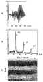
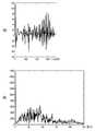
- FIG. 7A and 7Bshow measurement results obtained for a patient with atrial fibrillation, wherein FIG. 7A shows a pulse waveform, FIG. 7B shows a diagram after fast Fourier transform, and FIG. 7C shows a time change of the fast Fourier transform result. .
- FIG. 7Bshows a sharp peak waveform having three peaks disappeared.
- FIGS. 8A and 8Bshow measurement results obtained for another patient with atrial fibrillation, where FIG. 8A shows a pulse waveform and FIG. 8B shows a diagram after fast Fourier transform. In the frequency spectrum diagram shown in FIG. 8B, the sharp three-peaked peak waveform disappeared as in FIG.
- FIG. 9shows the measurement results obtained for a patient with extrasystole, (a) shows a pulse waveform, (b) shows a diagram after fast Fourier transform, and (c) shows a time change of the result of fast Fourier transform. .
- two peak waveformsare provided at about 70 (times / minute) corresponding to the period of the heartbeat of the patient and at about 140 (times / minute) which is a harmonic band thereof. Is recognized.
- the frequency componentdoes not have a clear peak in the frequency spectrum corresponding to the period of the heartbeat of the patient.
- the measurement result of a patient without atrial fibrillationhas a plurality of distinct peaks. The locations of these peaks correspond to the fundamental and harmonic frequency components corresponding to the period of the patient's heartbeat.
- Example 2The images shown in FIGS. 4 (c), 7 (c), and 9 (c) obtained by the above-described embodiment were analyzed, and atrial fibrillation was determined as shown in FIG.
- the determinationwas made based on the time during which a predetermined number of peaks continued and the amount by which the frequency of the peaks fluctuated. That is, it was determined whether three light-colored lines in the image were continuous horizontally over 3/4 or more of the screen width, and whether the vertical fluctuation was less than twice the line thickness (S902). When this condition is satisfied, it is determined that the sinus rhythm is present (S908).
- step S902the determination is made based on the time during which a predetermined number of peaks continue. Specifically, it was determined whether at least one of the light-colored lines is continuous over two-thirds or more of the horizontal width (S904). If this condition is met, it is determined that the arrhythmia is other than atrial fibrillation (S912).
- step S904when the condition of step S904 is not satisfied, based on two or more of the time during which the peak of the predetermined frequency is maintained, the fluctuation of the frequency of the peak, and the number of times that the peak having no temporal continuity appears. The judgment was made. Specifically, it was determined whether two or more of the following three conditions were satisfied (S906).
- Two or more of the light-colored linesremain in a continuation of ⁇ or less of the screen width, (2) Two or more of the light colored lines fluctuate up and down more than twice the thickness of the line, (3) There are two or more island-shaped light-colored areas without continuity on the screen.
- the measurement results of 280 patientswere observed, and when atrial fibrillation was determined at least twice during the three measurements, it was determined to be positive. The above characteristics were applied to all the measurement results.
- the device described in the present embodimentis merely an example, and can be implemented in other ways.
- the components described aboveare obtained by logically dividing the functions, and the components may be divided in another manner in implementation.
- two or more of the above-described componentsmay be integrated into one, and each of the components may be physically alone.
- a computer program for implementing the functionscan be stored in a computer-readable storage medium.
- the computer programincludes some instructions for instructing the computer to function as all or some of the components described in the embodiments.
Landscapes
- Health & Medical Sciences (AREA)
- Engineering & Computer Science (AREA)
- Life Sciences & Earth Sciences (AREA)
- Public Health (AREA)
- Medical Informatics (AREA)
- Biomedical Technology (AREA)
- General Health & Medical Sciences (AREA)
- Cardiology (AREA)
- Pathology (AREA)
- Physics & Mathematics (AREA)
- Heart & Thoracic Surgery (AREA)
- Veterinary Medicine (AREA)
- Molecular Biology (AREA)
- Surgery (AREA)
- Animal Behavior & Ethology (AREA)
- Biophysics (AREA)
- Physiology (AREA)
- Signal Processing (AREA)
- Epidemiology (AREA)
- Data Mining & Analysis (AREA)
- Primary Health Care (AREA)
- Psychiatry (AREA)
- Artificial Intelligence (AREA)
- Computer Vision & Pattern Recognition (AREA)
- Mathematical Physics (AREA)
- Business, Economics & Management (AREA)
- General Business, Economics & Management (AREA)
- Databases & Information Systems (AREA)
- Power Engineering (AREA)
- Vascular Medicine (AREA)
- Measuring Pulse, Heart Rate, Blood Pressure Or Blood Flow (AREA)
- Measurement And Recording Of Electrical Phenomena And Electrical Characteristics Of The Living Body (AREA)
Abstract
Description
Translated fromJapanese本発明は、脈波信号の解析装置、脈波信号の解析方法およびコンピュータプログラムに関し、特に、自動血圧計等に付帯した脈波検出機能を用いた脈波信号の解析装置、脈波信号の解析方法およびコンピュータプログラムに関する。The present invention relates to a pulse wave signal analyzing apparatus, a pulse wave signal analyzing method, and a computer program, and more particularly, to a pulse wave signal analyzing apparatus using a pulse wave detecting function attached to an automatic sphygmomanometer and the like, and a pulse wave signal analyzing method. The present invention relates to a method and a computer program.
心房細動は不整脈の一種であり、心房全体が非常に速く小刻みに興奮収縮し、その興奮が無秩序に心室に伝わることによって心臓の収縮および拡張が障害され、規則的な拍動が失われる状態をいう。 Atrial fibrillation is a type of arrhythmia in which the entire atria rapidly and rapidly excites and contracts, and the excitement is transmitted to the ventricle in a disordered manner, resulting in impaired contraction and expansion of the heart and loss of regular beats. Say.
心房細動が長期間続くと、心臓の機能が低下する可能性がある。また、心房細動になると血栓ができやすくなり、血栓が血流により脳内まで移動し、脳梗塞の発症リスクを増加させる。 Prolonged atrial fibrillation can reduce heart function. In addition, when atrial fibrillation occurs, a thrombus is easily formed, the thrombus moves into the brain by blood flow, and the risk of developing cerebral infarction increases.
統計上、心房細動の患者のうち、年間3~5%の患者に脳梗塞が発生しているといわれている。さらに、心房細動の有病率は加齢とともに高くなるため、心房細動は心臓病の中でも重要な病気と考えられている。したがって、心房細動を早期に発見し適切な治療を開始することは、心原性脳梗塞の予防のために重要である。 Statistically, it is said that cerebral infarction occurs in 3 to 5% of patients with atrial fibrillation annually. Furthermore, the prevalence of atrial fibrillation increases with age, so atrial fibrillation is considered an important disease among heart diseases. Therefore, early detection of atrial fibrillation and initiation of appropriate treatment is important for prevention of cardiogenic cerebral infarction.
心房細動は心電図検査により発見することができる。例えば、特許文献1および2、ならびに非特許文献1には、心電図検査を、心房不整脈の検出及び分類に使用することが記載されている。心電図検査は、心拍数を、体内で心周期ごとに生成される電気パルスを計測する方法である。しかしながら、心電図検査は健康診断や人間ドック等で行わなければならず、時間がかかり、煩雑である。また、特許文献1に記載のような検査は観血的であるため、感染対策や安全性への配慮が必要となる。Atrial fibrillation can be detected by electrocardiography. For example,
特許文献3には、動脈の拍動、すなわち脈波信号を使用して心房細動を判定することが開示されている。脈波信号を平均した平均脈波RR間隔が算出され、このRR脈波間隔について周波数解析が行われる。しかしながら、この手法では、細かい変動を均す一方で、および平均脈波RR間隔に対するばらつきの度合を求める必要があり、長時間の測定と複雑な計算が必要とされる。このため、短時間で精度よく心房細動を判定する手法が求められている。
また、心房細動の症状には個人差があり、心房細動に罹患している人のうち自覚症状のない人は40%と見積もられている。また、時々動悸がする程度で症状が気にならない人もいる。このように、心房細動の自覚症状がない場合、あるいは症状が気にならない場合、心房細動を見つけるために煩雑な検査を受ける機会を逃してしまうことがある。 In addition, there are individual differences in the symptoms of atrial fibrillation, and it is estimated that 40% of those who suffer from atrial fibrillation have no subjective symptoms. In addition, there are those who do not mind the symptoms just occasionally palpitating. As described above, when there is no subjective symptom of atrial fibrillation or when the symptom is not worrisome, an opportunity to undergo a complicated test to find atrial fibrillation may be missed.
また、医療サービスの乏しい地域では、このような検査を受けること自体が難しい。このような事情の結果として、症状が放置され、早期の発見・治療の機会を逸してしまうという問題がある。 In addition, in areas where medical services are scarce, it is difficult to take such a test. As a result of such circumstances, there is a problem that the symptoms are neglected and the opportunity for early detection and treatment is missed.
さらに、心房細動の初期にみられる発作性心房細動の場合は、心房細動の状態にある期間が短いため、測定時に心房細動を発見できないことがある。したがって、繰り返し検査を行うことができる簡易な検査方法が望ましい。 Furthermore, in the case of paroxysmal atrial fibrillation, which occurs early in atrial fibrillation, atrial fibrillation may not be detected at the time of measurement due to a short period of atrial fibrillation. Therefore, a simple inspection method that can repeatedly perform the inspection is desirable.
このため、心電図検査以外の簡易な方法を用いて心房細動を高い確率で判定する方法が望まれている。 For this reason, a method of determining atrial fibrillation with a high probability using a simple method other than an electrocardiogram test is desired.
本発明はこのような問題に鑑みてなされたものであり、その目的とするところは、簡易な方法で精度よく心房細動の状態を判定することができる脈波信号の解析装置、脈波信号の解析方法およびコンピュータプログラムを提供することにある。The present invention has been made in view of such a problem, and an object of the present invention is to provide a pulse wave signal analyzing apparatus capable of accurately determining the state of atrial fibrillation by a simple method, a pulse wave signal And a computer program.
上記の課題を解決するために、実施形態の一態様によれば、生体の心拍に応じて脈波信号を非観血的に検出する信号検出手段と、前記信号検出手段により検出された脈波信号をフーリエ変換して周波数スペクトルを生成する生成手段と、前記生成手段により生成された周波数スペクトルに基づいて心房細動の状態を検出する解析手段とを備え、
前記生成手段は、前記脈波信号の所定のフレーム時間のフーリエ変換を、0.005秒~0.02秒の範囲でずらしながら繰り返して演算する脈波信号の解析装置を提供する。 According to one embodiment of the present invention, there is provided a signal detection unit configured to non-invasively detect a pulse wave signal according to a heartbeat of a living body, and a pulse wave detected by the signal detection unit. Producing means for Fourier transforming the signal to generate a frequency spectrum, and analyzing means for detecting the state of atrial fibrillation based on the frequency spectrum generated by the generating means,
The generating means provides a pulse wave signal analyzing apparatus that repeatedly calculates a Fourier transform of the pulse wave signal for a predetermined frame time while shifting the pulse wave signal in a range of 0.005 seconds to 0.02 seconds.
ここで、前記信号検出手段により検出された脈波信号をフィルタ処理するフィルタをさらに備え、前記生成手段は前記フィルタから出力された前記脈波信号から周波数スペクトルを生成するものとすることができる。 Here, the apparatus may further include a filter for filtering the pulse wave signal detected by the signal detection unit, and the generation unit may generate a frequency spectrum from the pulse wave signal output from the filter.
また、前記解析手段は、前記周波数スペクトルにおいて前記心拍の周期に対応する周波数成分に複数のピークを有しない場合に心房細動の状態を検出するものとすることができる。 Further, the analysis means may detect the state of atrial fibrillation when the frequency spectrum does not have a plurality of peaks in a frequency component corresponding to the cycle of the heartbeat.
また、前記所定のフレーム時間は2秒から4秒とすることができる。 Further, the predetermined frame time may be 2 to 4 seconds.
また、前記信号検出手段は、圧脈波の脈波信号を検出するものとすることができる。 The signal detecting means may detect a pulse wave signal of a pressure pulse wave.
また、前記解析手段は、周波数スペクトルの時間変化に基づいて心房細動の状態を検出するものとすることができる。 Further, the analysis means may detect a state of atrial fibrillation based on a time change of a frequency spectrum.
ここで、前記解析手段は、所定の本数のピークが持続する時間に基づいて心房細動の状態を検出するものとすることができる。 Here, the analysis means may detect the state of atrial fibrillation based on the time during which a predetermined number of peaks continue.
ここで、前記解析手段は、前記ピークの周波数が変動する量にさらに基づいて心房細動の状態を検出するものとすることができる。 Here, the analysis means may detect the state of atrial fibrillation further based on the amount by which the frequency of the peak fluctuates.
ここで、前記解析手段は、所定の周波数のピークが持続する時間、ピークの周波数の変動、および時間的に連続性のないピークが出現する回数、のうちの2つ以上に基づいて心房細動の状態を検出するものとすることができる。 Here, the analyzing means may determine atrial fibrillation based on two or more of a time during which a peak of a predetermined frequency is maintained, a variation in frequency of the peak, and the number of times a peak having no temporal continuity appears. Can be detected.
実施形態の別の態様によれば、生体の心拍に応じて脈波信号に基づいて心房細動の状態を検出する方法であって、前記脈波信号を非観血的に検出するステップと、検出された前記脈波信号をフーリエ変換して周波数スペクトルを生成するステップと、生成された前記周波数スペクトルに基づいて心房細動の状態を検出するステップとを備え、前記生成するステップは、前記脈波信号の所定の期間のフーリエ変換を、0.005秒~0.02秒の範囲でずらしながら繰り返して演算するステップを含む脈波信号の解析方法を提供する。 According to another aspect of the embodiment, a method for detecting the state of atrial fibrillation based on a pulse wave signal according to the heartbeat of the living body, wherein the step of detecting the pulse wave signal non-invasively, Generating a frequency spectrum by performing a Fourier transform on the detected pulse wave signal; anddetecting a state of atrial fibrillation based on the generated frequency spectrum. Provided is a method for analyzing a pulse wave signal, which includes a step of repeatedly calculating a Fourier transform of a wave signal for a predetermined period while shifting the wave signal within a range of 0.005 to 0.02 seconds.
ここで、検出された前記脈波信号をフィルタ処理するステップをさらに備え、前記生成するステップは前記フィルタ処理された前記脈波信号から周波数スペクトルを生成するステップを含むものとすることができる。 Here, the method may further include a step of filtering the detected pulse wave signal, and the generating step may include a step of generating a frequency spectrum from the filtered pulse wave signal.
また、前記心房細動の状態を検出するステップは、前記周波数スペクトルにおいて前記心拍の周期に対応する周波数成分に複数のピークを有しない場合に心房細動の状態を検出するステップを含むものとすることができる。 Further, the step of detecting the state of atrial fibrillation may include the step of detecting the state of atrial fibrillation when the frequency spectrum does not have a plurality of peaks in a frequency component corresponding to the period of the heartbeat. it can.
また、前記所定の期間は2秒から4秒とすることができる。 Further, the predetermined period can be set to 2 to 4 seconds.
また、前記心房細動の状態を検出するステップは、圧脈波の脈波信号を検出するものとすることができる。 Further, the step of detecting the state of atrial fibrillation may detect a pulse wave signal of a pressure pulse wave.
また、前記心房細動の状態を検出するステップは、周波数スペクトルの時間変化に基づいて心房細動の状態を検出するものとすることができる。 In the detecting of the state of atrial fibrillation, the state of atrial fibrillation may be detected based on a time change of a frequency spectrum.
ここで、前記心房細動の状態を検出するステップは、所定の本数のピークが持続する時間に基づいて心房細動の状態を検出するものとすることができる。 Here, the step of detecting the state of atrial fibrillation may detect the state of atrial fibrillation based on the time during which a predetermined number of peaks continue.
また、前記心房細動の状態を検出するステップは、前記ピークの周波数が変動する量にさらに基づいて心房細動の状態を検出するものとすることができる。 In addition, the step of detecting the state of atrial fibrillation may detect the state of atrial fibrillation further based on an amount by which the frequency of the peak fluctuates.
また、前記心房細動の状態を検出するステップは、所定の周波数のピークが持続する時間、ピークの周波数の変動、および時間的に連続性のないピークが出現する回数、のうちの2つ以上に基づいて心房細動の状態を検出するものとすることができる。 Further, the step of detecting the state of atrial fibrillation includes two or more of a time during which a peak of a predetermined frequency is maintained, a change in the frequency of the peak, and the number of times a peak having no temporal continuity appears. May be used to detect the state of atrial fibrillation.
実施形態の他の態様によれば、コンピュータを、上記の脈波信号の解析装置として機能させるコンピュータプログラムを提供する。 According to another aspect of the embodiment, there is provided a computer program that causes a computer to function as the above-described pulse wave signal analysis device.
実施形態の他の態様によれば、上記のコンピュータプログラムを記憶したコンピュータ読み取り可能な記憶媒体を提供する。According to another aspect of the embodiment, there is provided a computer-readable storage medium storing the computer program.
本開示によれば、脈波信号を非観血的に検出し、検出された脈波信号を、0.005秒~0.02秒の範囲でずらしながら繰り返して所定の期間フーリエ変換して周波数スペクトルを生成し、生成された周波数スペクトルに基づいて心房細動の状態を検出する。したがって、簡易に心房細動の状態を検出することができる。 According to the present disclosure, a pulse wave signal is non-invasively detected, and the detected pulse wave signal is repeatedly Fourier-transformed for a predetermined period while being shifted in a range of 0.005 seconds to 0.02 seconds to perform frequency conversion. A spectrum is generated, and a state of atrial fibrillation is detected based on the generated frequency spectrum. Therefore, the state of atrial fibrillation can be easily detected.
また、簡易な方法で心房細動の状態を検出することができるため、繰り返し検査を行うことにより、発作性心房細動を発見する確率が向上する。 In addition, since the state of atrial fibrillation can be detected by a simple method, the probability of finding paroxysmal atrial fibrillation is improved by repeating the examination.
さらに、圧脈波を用いて脈波信号を検出することで、血圧計を用いて短時間で信頼度の高いデータを得ることができる。Furthermore, by detecting a pulse wave signal using a pressure pulse wave, highly reliable data can be obtained in a short time using a sphygmomanometer.
以下、図面を参照し、本発明の実施の形態について詳細に説明する。以下の説明において、脈波とは心臓の拍動によって生じる身体の部分の容積変化の波形のことを言う。脈波のうち、血圧の変化によるものを圧脈波、容積の変化によるものを容積脈波といい、本実施形態ではこれらのいずれをも含む。Hereinafter, embodiments of the present invention will be described in detail with reference to the drawings. In the following description, a pulse wave refers to a waveform of a volume change of a body part caused by a heartbeat. Of the pulse waves, those due to changes in blood pressure are referred to as pressure pulse waves, those due to changes in volume are referred to as volume pulse waves, and the present embodiment includes any of these.
図1は、本発明の実施形態に係る脈波の解析装置の機能構成を示すブロック図である。解析装置100は心房細動の診断装置として機能するものであり、脈波検出部104と解析部101とが接続されて信号を通信することができるように構成される。この場合、通信手段は有線でもよく、あるいはBluetooth(登録商標)等の無線通信規格に準拠したものであってもよい。脈波検出部104と解析部101とは一体として1つの装置に組み込まれてもよく、別個に構成されてもよく、また互いに着脱可能に構成されてもよい。 FIG. 1 is a block diagram showing a functional configuration of a pulse wave analyzer according to an embodiment of the present invention. The
解析部101は、演算部102、記憶部114、入力部116、通知部118および電源120を含んで構成される。 The
脈波検出部104は、生体の心拍に応じて脈波信号を非観血的に検出するものであり、接触型または非接触型の生体センサとして構成され得る。例えば解析装置100は血圧計として構成することができ、この場合、脈波検出部104は腕の所定部位に巻くカフを含むものとして構成される。カフには膨張可能な袋状部材が設けられる。血圧計には、腕等に巻かれたカフの内側に位置する動脈内の血流を遮断するための圧迫圧力が予め設定されている。脈波を検出する際には、この袋状部材内部の圧力を、当該圧迫圧力まで昇圧させた後に、所定の速度で徐々に降圧させることで血圧を測定する。カフで圧迫された血管は心臓の拍動に合わせて振動を起こすため、血圧測定における降圧の過程において、袋状部材内の圧力を逐次検出し、その圧力変化から脈波を検出する。 The
脈波検出部104を血圧計とワイヤレス型の心電送信機の組み合わせとして構成し、解析装置100をパーソナルコンピュータ等の情報処理装置として構成してもよい。この場合、脈波検出部104は血圧を計測する操作の実行を指示するボタン等の入力部、および計測結果を表示するための表示部を含むこととしてもよい。 The
あるいはまた、脈波検出部104は触覚センサ等の圧力により脈波信号を計測する他の任意のセンサにより構成してもよい。Alternatively, the pulse
代替として、解析装置100はレーザ光を用いて酸素飽和度を測定するパルスオキシメータ等を含む指尖脈波検出装置として構成してもよい。この場合、脈波検出部104は生体の手や足の指先に赤外線レーザを照射し、指先を流れる血液の容積の変化を測定することにより脈波を検出する。 Alternatively, the
あるいはまた、解析装置100はカメラや携帯端末等の非接触型デバイスとして構成してもよい。この場合、脈波検出部104は撮像された画像に基づいて血流によって生じる輝度変化を計測することにより脈波を検出する。 Alternatively, the
あるいはまた、解析装置100は他の光学式心拍計測装置として構成してもよい。この場合、脈波検出部104は光学式心拍センサを有して構成される。脈波検出部104はLEDを用いて腕等の所定の位置に対して光を照射し、血流によって反射される散乱光の量を測定することで、脈波を検出する。Alternatively, the
次に、解析部101が有する諸機能について説明する。演算部102は脈波検出部104から出力される脈波の電気信号に基づいて演算処理を行うものであり、1つまたは複数のプロセッサにより構成される。演算部102は、さらに記憶部114、入力部116、および通知部118に通信可能に接続される。記憶部114は、本実施形態に係る処理を実行するためのプログラムや演算処理に用いられるデータまたは演算結果のデータを記憶するように構成される。具体的には、記憶部114は、USBフラッシュドライブ、リムーバブルハードディスク、リードオンリーメモリ(ROM)、ランダムアクセスメモリ(RAM)、磁気ディスクまたは光ディスク等として構成することができる。 Next, various functions of the
入力部116は、ユーザから情報処理のための命令や設定値の情報を入力するためのキーボード、ボタン、ダイヤルもしくはスイッチ、またはデータを入力するための入力インタフェース等として構成される。通知部118は、演算部102による演算結果に基づく通知情報を出力するための液晶ディスプレイ、ランプ、またはスピーカ等として構成される。加えて、解析装置100は、当該装置内の各構成要素へ電源を供給するための電源120を含んでいる。 The
次に、演算部102に含まれる各構成要素について説明する。演算部102は、フィルタ105、スペクトル生成部108、および心房細動検出部113を含んでいる。 Next, each component included in the
フィルタ105は脈波に関連する周波数を通過させるためのものである。ここで通過させる周波数は、可聴周波数以下の周波数とすることができる。また、フィルタ105は心房粗動に関連すると考えられる高周波圧力信号を除去するように設計された低域フィルタとして構成してもよい。 The
スペクトル生成部108はスペクトル解析を行うために脈波信号の時系列データに対してフーリエ変換を行う。フーリエ変換として、例えば高速フーリエ変換(FFT)を行うこととしてもよい。結果として、スペクトル生成部108は信号波形の周波数スペクトルを取得する。なお、高速フーリエ変換は一例であり、周波数スペクトルを生成するための他のフーリエ変換を用いることができる。 The
心房細動検出部113は信号波形の解析を行うように構成されており、少なくとも、スペクトル生成部108により取得された周波数スペクトルから生体の心房細動の状態を検出する。 The
なお、解析部101内の演算部102に含まれている機能のうちの1つまたは複数を脈波検出部104に実装することとしてもよい。例えば、脈波検出部104にフィルタ105の機能を設け、脈波検出部104においてフィルタ処理された脈波信号を演算部102に供給することとしてもよい。 Note that one or more of the functions included in the
次に、心房細動の検出処理を具体的に説明する。一例として、脈波検出部104を心電送信機と自動血圧計との組み合わせとして構成し、心電図と血圧との同時測定を行う場合について説明する。図2は解析装置100により検出された値の例を示す。同図において、横軸は時間(s)を示す。また、破線の曲線302は自動血圧計のカフ圧(mmHg)を示し、実線の曲線304は心電送信機から出力される圧脈波振幅(mmHg)を示す。血管内圧がカフ圧を上回る期間においては血管が拡張することにより、血管の容積変化がカフの内圧を上昇させる。オシロメトリック法では、カフを減圧していく過程で心臓の拍動に同調したカフ圧の変動を観測することで血圧を求める。ここで観測されるカフ圧の変動が圧脈波として計測される。 Next, detection processing of atrial fibrillation will be specifically described. As an example, a case will be described in which the pulse
患者から得られた脈波波形について、フィルタ処理を行う。次いで、フィルタ処理された波形をフーリエ変換する。ここで変換する区間は、略2~20秒間とすることが望ましい。この区間において、2~4秒のフレーム時間(間隔)のデータを、1秒間に50回から200回の頻度(0.005秒~0.02秒ずつ)で、時間軸をずらしながら取得する。 Filter processing is performed on the pulse wave waveform obtained from the patient. Next, the filtered waveform is Fourier-transformed. It is desirable that the section to be converted be approximately 2 to 20 seconds. In this section, data of a frame time (interval) of 2 to 4 seconds is acquired at a frequency of 50 to 200 times per second (0.005 to 0.02 seconds each) while shifting the time axis.
心房細動検出部113は、スペクトル生成部108から出力された周波数スペクトルの値を解析する。例えば、図4(b)に示すように、横軸に等間隔に2本または3本の明確なピークを有する場合、心房細動の状態にないこと(陰性)を検出する。ここで、周波数スペクトルの「ピーク」とは、圧脈波振幅のピークを指す。このピークは、具体的には生体の心拍の周期に対応する基本周波数および高調波の周波数成分の位置に存在する。他方、図7(b)に示すように周波数スペクトルの値において心拍の周期に対応する周波数成分の位置に明確なピークを有しない場合、心房細動の状態にあること(陽性)を検出する。
次に、図3のフローチャートを参照し、本実施形態に係る解析装置によって実行される心房細動の検出方法について説明する。 Next, a method for detecting atrial fibrillation performed by the analyzer according to the present embodiment will be described with reference to the flowchart in FIG.
まず、ステップS202において、解析部101が、脈波検出部104から生体の脈波の信号を入力する。ステップS204において、フィルタ105が、脈波に関連する周波数を通過させるためのフィルタリングを行う。ステップS206において、スペクトル生成部108が、フィルタ105から入力される信号の時系列データを入力し、高速フーリエ変換を行い、周波数スペクトルを取得する。 First, in step S202, the
ステップS208において、心房細動検出部113が、スペクトル生成部108により取得された周波数スペクトルから、生体が心房細動の状態にあること(陽性)を検出する。具体的には、周波数スペクトルにおいて心拍の周期に対応する周波数成分に明確なピークを有しない場合、心房細動の状態にあること(陽性)を検出し、心拍の周期に対応する周波数成分に明確なピークを有する場合、心房細動の状態にないこと(陰性)を検出する。その後、心房細動の状態が検出された場合、解析部101は検出結果を記憶部114に記憶し、通知部118を通じて心房細動が発生した可能性があることを通知して処理を終了する。In step S208, the atrial
(実施例1)
次に、本発明の実施例について説明する。本実施例では、洞調律、持続性心房細動または期外収縮の患者を対象とした。脈波検出部104に相当する構成要素として、オムロンヘルスケア社製のヘルスパッチMD(心電送信機)、およびHEM-6310F/M6(自動血圧計)を使用した。また、解析部101に相当する構成としてパーソナルコンピュータを使用した。そして、図2を参照して上述した心電図と血圧との同時測定を、合計280名(洞調律:197名、心房細動:40名、他の不整脈:43名)に対して行った。 (Example 1)
Next, examples of the present invention will be described. In this example, patients with sinus rhythm, sustained atrial fibrillation or extrasystole were targeted. As components corresponding to the pulse
次に、患者から得られた脈波波形について高速フーリエ変換を行って、周波数スペクトルを取得した。上記自動血圧計は1回の血圧および脈拍数の計測に20~30秒ほどかかるため、20~30秒間全体を高速フーリエ変換の分析対象とした。 Next, a fast Fourier transform was performed on the pulse wave waveform obtained from the patient to obtain a frequency spectrum. Since the automatic blood pressure monitor takes about 20 to 30 seconds for one measurement of blood pressure and pulse rate, the entirety of 20 to 30 seconds was analyzed by the fast Fourier transform.
図4は洞調律の患者に関して得られた計測結果を示す。(a)は脈波波形であり、横軸は時間(×0.01秒)、縦軸は圧脈波振幅である。また、(b)は高速フーリエ変換後の図を示し、横軸は心拍数(回/分)、縦軸は圧脈波振幅を示す。なお、自動血圧計で計測したデータはサンプリング回数が少なかったので、高速フーリエ変換の電子回路の特性に合わせるため、実際の横軸データの数を100倍程度に増やしている。図4(b)に示す周波数スペクトルの図において、矢印401は基本波のピーク、矢印402および403は高調波帯域のピークを示している。同図に示すように、洞調律の患者では、心拍の周期に対応する約65(回/分)、ならびにその高調波帯域である約130(回/分)および約190(回/分)辺りに周波数の3峰性の鋭いピーク波形が認められる。 FIG. 4 shows the measurement results obtained for a sinus rhythm patient. (A) is a pulse waveform, the horizontal axis is time (× 0.01 second), and the vertical axis is pressure pulse wave amplitude. (B) shows a diagram after the Fast Fourier Transform, in which the horizontal axis represents the heart rate (beats / minute) and the vertical axis represents the pressure pulse wave amplitude. Since the number of samplings of the data measured by the automatic blood pressure monitor was small, the actual number of horizontal axis data was increased to about 100 times in order to match the characteristics of the electronic circuit of the fast Fourier transform. In the diagram of the frequency spectrum shown in FIG. 4B, an
図4(c)は高速フーリエ変換の結果の時間変化を示す図である。この図の作成方法を、図5を参照して説明する。図5(a)は脈波波形であり、横軸は時間(秒/20)、縦軸はカフ圧(mmHg/2)である。脈波波形のうち20秒の範囲を指定し、この範囲でフレーム時間3秒のフーリエ変換を、0.01秒ずつずらしながら繰り返した。次いで、得られたフーリエ変換の結果のうちから17秒を切り出して、時間軸上に並べて、心拍情報のピークをマッピングし、図5(b)に示すグラフを得た。図5(b)において、横軸は時間を示し、1700個(17秒/0.01秒=1700)のデータが並べられている。縦軸は周波数を示す。フーリエ変換の結果におけるスペクトル強度は色の濃淡で表しており、最高の値を1として、0.8~0.9の範囲を淡色で表している。 FIG. 4C is a diagram showing a time change as a result of the fast Fourier transform. A method for creating this diagram will be described with reference to FIG. FIG. 5A shows a pulse waveform, in which the horizontal axis represents time (second / 20) and the vertical axis represents cuff pressure (mmHg / 2). A range of 20 seconds was designated in the pulse wave waveform, and the Fourier transform with a frame time of 3 seconds was repeated in this range while shifting by 0.01 seconds. Next, 17 seconds were cut out of the obtained Fourier transform results, arranged on the time axis, and the peaks of the heart rate information were mapped to obtain a graph shown in FIG. 5B. In FIG. 5B, the horizontal axis represents time, and 1700 data (17 seconds / 0.01 seconds = 1700) are arranged. The vertical axis indicates frequency. The spectral intensity in the result of the Fourier transform is represented by the shading of the color, with the highest value being 1, and the range of 0.8 to 0.9 being represented by the light color.
図6は洞調律の別の患者に関して得られた計測結果を示し、(a)は脈波波形、(b)は高速フーリエ変換後の図を示す。図6(b)に示す周波数スペクトルの図において、矢印501は基本波のピーク、矢印502および503は高調波帯域のピークを示している。同図においても、図4と同様に、患者の心拍の周期に対応する約55(回/分)、ならびにその高調波帯域である約110(回/分)および約165(回/分)辺りに周波数の3峰性の鋭いピーク波形が認められる。 FIGS. 6A and 6B show measurement results obtained for another patient with sinus rhythm. FIG. 6A shows a pulse waveform, and FIG. 6B shows a diagram after fast Fourier transform. In the frequency spectrum diagram shown in FIG. 6B, an
図7は心房細動の患者に関して得られた計測結果を示し、(a)は脈波波形、(b)は高速フーリエ変換後の図、(c)は高速フーリエ変換の結果の時間変化を示す。図7(b)に示す周波数スペクトルの図において、3峰性の鋭いピーク波形が消失していた。 7A and 7B show measurement results obtained for a patient with atrial fibrillation, wherein FIG. 7A shows a pulse waveform, FIG. 7B shows a diagram after fast Fourier transform, and FIG. 7C shows a time change of the fast Fourier transform result. . In the frequency spectrum diagram shown in FIG. 7B, a sharp peak waveform having three peaks disappeared.
図8は心房細動の別の患者に関して得られた計測結果を示し、(a)は脈波波形、(b)は高速フーリエ変換後の図を示す。図8(b)に示す周波数スペクトルの図においても、図7と同様に3峰性の鋭いピーク波形が消失していた。 FIGS. 8A and 8B show measurement results obtained for another patient with atrial fibrillation, where FIG. 8A shows a pulse waveform and FIG. 8B shows a diagram after fast Fourier transform. In the frequency spectrum diagram shown in FIG. 8B, the sharp three-peaked peak waveform disappeared as in FIG.
図9は期外収縮の患者に関して得られた計測結果を示し、(a)は脈波波形、(b)は高速フーリエ変換後の図、(c)は高速フーリエ変換の結果の時間変化を示す。図9(b)に示す周波数スペクトルの図において、患者の心拍の周期に対応する約70(回/分)、およびその高調波帯域である約140(回/分)あたりに2本のピーク波形が認められる。 FIG. 9 shows the measurement results obtained for a patient with extrasystole, (a) shows a pulse waveform, (b) shows a diagram after fast Fourier transform, and (c) shows a time change of the result of fast Fourier transform. . In the frequency spectrum diagram shown in FIG. 9B, two peak waveforms are provided at about 70 (times / minute) corresponding to the period of the heartbeat of the patient and at about 140 (times / minute) which is a harmonic band thereof. Is recognized.
以上の考察を他の患者の計測結果に対しても行ったところ、次のように判定できることが分かった。すなわち、心房細動の患者の計測結果によれば、周波数スペクトルにおいて患者の心拍の周期に対応する周波数成分に明確なピークを有しない。他方、心房細動でない患者の計測結果では複数の明確なピークを有する。これらのピークの位置は、患者の心拍の周期に対応する基本周波数および高調波の周波数成分に対応する。The above considerations were also performed on the measurement results of other patients, and it was found that the following determination could be made. That is, according to the measurement result of the patient with atrial fibrillation, the frequency component does not have a clear peak in the frequency spectrum corresponding to the period of the heartbeat of the patient. On the other hand, the measurement result of a patient without atrial fibrillation has a plurality of distinct peaks. The locations of these peaks correspond to the fundamental and harmonic frequency components corresponding to the period of the patient's heartbeat.
(実施例2)
上述した実施例により得られた図4(c)、図7(c)、および図9(c)に示す画像を解析して、図10に示すように心房細動の判定を行った。 (Example 2)
The images shown in FIGS. 4 (c), 7 (c), and 9 (c) obtained by the above-described embodiment were analyzed, and atrial fibrillation was determined as shown in FIG.
まず、所定の本数のピークが持続する時間と、ピークの周波数が変動する量とに基づいて判定を行った。すなわち、画像の中で3本の淡色線が画面横幅の3/4以上に亘って横に連続し、かつ上下変動が線の太さの2倍未満であるかを判定した(S902)。この条件に該当する場合は洞調律と判定した(S908)。 First, the determination was made based on the time during which a predetermined number of peaks continued and the amount by which the frequency of the peaks fluctuated. That is, it was determined whether three light-colored lines in the image were continuous horizontally over 3/4 or more of the screen width, and whether the vertical fluctuation was less than twice the line thickness (S902). When this condition is satisfied, it is determined that the sinus rhythm is present (S908).
ステップS902の条件に該当しない場合、所定の本数のピークが持続する時間に基づいて判定を行った。具体的には、淡色線の1本以上が、横幅の2/3以上に亘って連続するかを判定した(S904)。この条件に該当する場合は、心房細動以外の不整脈と判定した(S912)。 If the condition of step S902 is not satisfied, the determination is made based on the time during which a predetermined number of peaks continue. Specifically, it was determined whether at least one of the light-colored lines is continuous over two-thirds or more of the horizontal width (S904). If this condition is met, it is determined that the arrhythmia is other than atrial fibrillation (S912).
次いで、ステップS904の条件に該当しない場合、所定の周波数のピークが持続する時間、ピークの周波数の変動、および時間的に連続性のないピークが出現する回数、のうちの2つ以上に基づいて判定を行った。具体的には、以下の3つの条件のうちの2つ以上を満たすかどうかを判定した(S906)。 Next, when the condition of step S904 is not satisfied, based on two or more of the time during which the peak of the predetermined frequency is maintained, the fluctuation of the frequency of the peak, and the number of times that the peak having no temporal continuity appears. The judgment was made. Specifically, it was determined whether two or more of the following three conditions were satisfied (S906).
(1)淡色線の2本以上が、画面横幅の1/2以下の連続にとどまる、
(2)淡色線の2本以上が、線の太さの2倍を超えて上下に変動する、
(3)連続性のない島状の淡色域が、画面上に2個以上ある。 (1) Two or more of the light-colored lines remain in a continuation of 以下 or less of the screen width,
(2) Two or more of the light colored lines fluctuate up and down more than twice the thickness of the line,
(3) There are two or more island-shaped light-colored areas without continuity on the screen.
S906の条件に該当した場合、心房細動と判定した。 If the condition of S906 is met, it is determined that the patient has atrial fibrillation.
本実施例において280名の測定結果を観察し、3回計測中2回以上で心房細動と判定された場合を陽性と判定したところ、計測結果の全てについて上記の特徴が当てはまった。 In the present example, the measurement results of 280 patients were observed, and when atrial fibrillation was determined at least twice during the three measurements, it was determined to be positive. The above characteristics were applied to all the measurement results.
以上、本発明の実施の形態について説明したが、本実施形態に係る装置、その構成要素、および上述した方法のステップは、ハードウェアによって、またはソフトウェアとハードウェアとの組合せによって実施することができる。装置またはその構成要素の諸機能がハードウェアによって実行されるかまたはソフトウェアによって実行されるかは本実施形態の設計における制約条件に従う。当業者は種々の方法を用いて特定の用途ごとに上述した構成要素の機能を実施することができるが、このような変更もまた、本発明の範囲内にある。 While the embodiments of the present invention have been described above, the apparatus, the components thereof, and the steps of the above-described method according to the present embodiment can be implemented by hardware or a combination of software and hardware. . Whether the functions of the device or its components are performed by hardware or software is subject to constraints in the design of the present embodiment. Those skilled in the art can use various methods to implement the functions of the components described above for a particular application, but such modifications are also within the scope of the present invention.
本実施形態において説明される装置は例示にすぎず、他の方式でも実施可能である。例えば、上述した構成要素は機能を論理的に分割したものであり、実装において他の方式で構成要素を分割してもよい。また、上述した構成要素のうちの2つ以上が1つに統合されてもよく、構成要素の各々が物理的に単独で存在してもよい。 The device described in the present embodiment is merely an example, and can be implemented in other ways. For example, the components described above are obtained by logically dividing the functions, and the components may be divided in another manner in implementation. Further, two or more of the above-described components may be integrated into one, and each of the components may be physically alone.
本実施形態で説明される機能がソフトウェアの形態で実施されるとき、その機能を実現するためのコンピュータプログラムはコンピュータ読み取り可能な記憶媒体に記憶することができる。コンピュータプログラムは、コンピュータを本実施形態において説明される構成要素の全てまたは一部として機能させるように指示するためのいくつかの命令を含む。When the functions described in the present embodiment are implemented in the form of software, a computer program for implementing the functions can be stored in a computer-readable storage medium. The computer program includes some instructions for instructing the computer to function as all or some of the components described in the embodiments.
Claims (20)
Translated fromJapanese前記信号検出手段により検出された脈波信号をフーリエ変換して周波数スペクトルを生成する生成手段と、
前記生成手段により生成された周波数スペクトルに基づいて心房細動の状態を検出する解析手段と
を備え、
前記生成手段は、前記脈波信号の所定のフレーム時間のフーリエ変換を、0.005秒~0.02秒の範囲でずらしながら繰り返して演算する脈波信号の解析装置。Signal detection means for non-invasively detecting a pulse wave signal according to the heartbeat of the living body,
Generating means for generating a frequency spectrum by Fourier transforming the pulse wave signal detected by the signal detecting means,
Analyzing means for detecting the state of atrial fibrillation based on the frequency spectrum generated by the generating means,
An apparatus for analyzing a pulse wave signal, wherein the generation means repeatedly calculates a Fourier transform of the pulse wave signal for a predetermined frame time while shifting the pulse wave signal in a range of 0.005 to 0.02 seconds.
前記脈波信号を非観血的に検出するステップと、
検出された前記脈波信号をフーリエ変換して周波数スペクトルを生成するステップと、
生成された前記周波数スペクトルに基づいて心房細動の状態を検出するステップと
を備え、
前記生成するステップは、前記脈波信号の所定の期間のフーリエ変換を、0.005秒~0.02秒の範囲でずらしながら繰り返して演算するステップを含む脈波信号の解析方法。A method for detecting the state of atrial fibrillation based on a pulse wave signal according to the heartbeat of the living body,
Non-invasively detecting the pulse wave signal;
Generating a frequency spectrum by performing a Fourier transform on the detected pulse wave signal;
Detecting a state of atrial fibrillation based on the generated frequency spectrum,
The method for analyzing a pulse wave signal includes a step of repeatedly performing a Fourier transform of the pulse wave signal for a predetermined period while shifting the pulse wave signal in a range of 0.005 to 0.02 seconds.
Priority Applications (2)
| Application Number | Priority Date | Filing Date | Title |
|---|---|---|---|
| JP2020530018AJP7327816B2 (en) | 2018-07-10 | 2019-05-24 | Pulse wave signal analysis device, pulse wave signal analysis method, and computer program |
| US17/258,582US12133721B2 (en) | 2018-07-10 | 2019-05-24 | Pulse-wave signal analysis device, pulse-wave signal analysis method and computer program |
Applications Claiming Priority (2)
| Application Number | Priority Date | Filing Date | Title |
|---|---|---|---|
| JP2018-131025 | 2018-07-10 | ||
| JP2018131025 | 2018-07-10 |
Publications (1)
| Publication Number | Publication Date |
|---|---|
| WO2020012793A1true WO2020012793A1 (en) | 2020-01-16 |
Family
ID=69141522
Family Applications (1)
| Application Number | Title | Priority Date | Filing Date |
|---|---|---|---|
| PCT/JP2019/020762CeasedWO2020012793A1 (en) | 2018-07-10 | 2019-05-24 | Pulse-wave signal analysis device, pulse-wave signal analysis method and computer program |
Country Status (3)
| Country | Link |
|---|---|
| US (1) | US12133721B2 (en) |
| JP (1) | JP7327816B2 (en) |
| WO (1) | WO2020012793A1 (en) |
Cited By (3)
| Publication number | Priority date | Publication date | Assignee | Title |
|---|---|---|---|---|
| CN116634947A (en)* | 2020-12-22 | 2023-08-22 | 欧姆龙健康医疗事业株式会社 | Electronic blood pressure meter and method for determining atrial fibrillation in electronic blood pressure meter |
| WO2024053166A1 (en)* | 2022-09-09 | 2024-03-14 | オムロンヘルスケア株式会社 | Sphygmomanometer |
| WO2024122104A1 (en)* | 2022-12-09 | 2024-06-13 | オムロンヘルスケア株式会社 | Sphygmomanometer |
Families Citing this family (1)
| Publication number | Priority date | Publication date | Assignee | Title |
|---|---|---|---|---|
| CN113558584B (en)* | 2021-06-22 | 2024-08-06 | 深圳市大数据研究院 | Pulse wave preprocessing method based on signal quality evaluation |
Citations (6)
| Publication number | Priority date | Publication date | Assignee | Title |
|---|---|---|---|---|
| JP2014042547A (en)* | 2012-08-24 | 2014-03-13 | Seiko Epson Corp | Atrial fibrillation determination device, and atrial fibrillation determination method and program |
| JP2014171660A (en)* | 2013-03-08 | 2014-09-22 | Seiko Epson Corp | Atrial fibrillation analyzation equipment, atrial fibrillation analysis system, atrial fibrillation analysis method and program |
| WO2015159692A1 (en)* | 2014-04-14 | 2015-10-22 | 株式会社村田製作所 | Pulse wave propagation time measurement device and biological state estimation device |
| JP2016083367A (en)* | 2014-10-28 | 2016-05-19 | サルーステック株式会社 | Subject information processing device, information processing method, information processing program, and computer readable recording medium with same program recorded thereon |
| JP2016202348A (en)* | 2015-04-17 | 2016-12-08 | セイコーエプソン株式会社 | Biological information processing system, terminal device, and biological information processing method |
| JP2017042386A (en)* | 2015-08-27 | 2017-03-02 | セイコーエプソン株式会社 | Biological information processing system and program |
Family Cites Families (7)
| Publication number | Priority date | Publication date | Assignee | Title |
|---|---|---|---|---|
| US7233822B2 (en) | 2004-06-29 | 2007-06-19 | Medtronic, Inc. | Combination of electrogram and intra-cardiac pressure to discriminate between fibrillation and tachycardia |
| US8834378B2 (en)* | 2010-07-30 | 2014-09-16 | Nellcor Puritan Bennett Ireland | Systems and methods for determining respiratory effort |
| US20130060154A1 (en) | 2011-09-07 | 2013-03-07 | Seiko Epson Corporation | Atrial fibrillation decision apparatus, and method and program for deciding presence of atrial fibrillation |
| JP2016154754A (en)* | 2015-02-25 | 2016-09-01 | セイコーエプソン株式会社 | Biological information measuring device |
| EP3081157A1 (en) | 2015-04-17 | 2016-10-19 | Seiko Epson Corporation | Biological information processing system, biological information processing device, terminal device, method for generating analysis result information, and biological information processing method |
| JP7018621B2 (en) | 2016-07-12 | 2022-02-14 | 国立大学法人秋田大学 | Biosignal analysis device, biosignal analysis method and biosignal analysis system |
| TWI619472B (en)* | 2016-12-12 | 2018-04-01 | 張國源 | Method of and apparatus for detecting atrial fibrillation |
- 2019
- 2019-05-24WOPCT/JP2019/020762patent/WO2020012793A1/ennot_activeCeased
- 2019-05-24USUS17/258,582patent/US12133721B2/enactiveActive
- 2019-05-24JPJP2020530018Apatent/JP7327816B2/enactiveActive
Patent Citations (6)
| Publication number | Priority date | Publication date | Assignee | Title |
|---|---|---|---|---|
| JP2014042547A (en)* | 2012-08-24 | 2014-03-13 | Seiko Epson Corp | Atrial fibrillation determination device, and atrial fibrillation determination method and program |
| JP2014171660A (en)* | 2013-03-08 | 2014-09-22 | Seiko Epson Corp | Atrial fibrillation analyzation equipment, atrial fibrillation analysis system, atrial fibrillation analysis method and program |
| WO2015159692A1 (en)* | 2014-04-14 | 2015-10-22 | 株式会社村田製作所 | Pulse wave propagation time measurement device and biological state estimation device |
| JP2016083367A (en)* | 2014-10-28 | 2016-05-19 | サルーステック株式会社 | Subject information processing device, information processing method, information processing program, and computer readable recording medium with same program recorded thereon |
| JP2016202348A (en)* | 2015-04-17 | 2016-12-08 | セイコーエプソン株式会社 | Biological information processing system, terminal device, and biological information processing method |
| JP2017042386A (en)* | 2015-08-27 | 2017-03-02 | セイコーエプソン株式会社 | Biological information processing system and program |
Cited By (3)
| Publication number | Priority date | Publication date | Assignee | Title |
|---|---|---|---|---|
| CN116634947A (en)* | 2020-12-22 | 2023-08-22 | 欧姆龙健康医疗事业株式会社 | Electronic blood pressure meter and method for determining atrial fibrillation in electronic blood pressure meter |
| WO2024053166A1 (en)* | 2022-09-09 | 2024-03-14 | オムロンヘルスケア株式会社 | Sphygmomanometer |
| WO2024122104A1 (en)* | 2022-12-09 | 2024-06-13 | オムロンヘルスケア株式会社 | Sphygmomanometer |
Also Published As
| Publication number | Publication date |
|---|---|
| US12133721B2 (en) | 2024-11-05 |
| JPWO2020012793A1 (en) | 2021-08-02 |
| US20210161412A1 (en) | 2021-06-03 |
| JP7327816B2 (en) | 2023-08-16 |
Similar Documents
| Publication | Publication Date | Title |
|---|---|---|
| US9713428B2 (en) | Physiological parameter monitoring with a mobile communication device | |
| KR102622403B1 (en) | Biological data processing | |
| KR20210005644A (en) | Method for estimating blood pressure and arterial stiffness based on light volumetric variability recording (PPG) signal | |
| US11903744B2 (en) | Blood pressure and autoregulation monitoring | |
| JP7327816B2 (en) | Pulse wave signal analysis device, pulse wave signal analysis method, and computer program | |
| EP3061391B1 (en) | Apparatus and method for determining blood pressure | |
| US9724046B2 (en) | Detecting apparatus for arrhythmia and detecting method of the detecting apparatus | |
| JP6522327B2 (en) | Pulse wave analyzer | |
| JP2009089883A (en) | Atrial fibrillation detector, system and method | |
| CN108289624B (en) | Blood pressure information calculation device, blood pressure information calculation method, blood pressure information calculation program, and storage medium storing the program | |
| EP4088653A1 (en) | Pulse wave analysis device, pulse wave analysis method, and pulse wave analysis program | |
| Daly | Video camera monitoring to detect changes in haemodynamics | |
| Snizhko et al. | Methods for increasing the accuracy of recording the parameters of the cardiovascular system in double-beam photoplethysmography | |
| Besleaga | Photoplethysmography use for Assessment of Mechanical Alternans in Human Cardiovascular Disease | |
| Tiwari et al. | Continuous and Cuffless Blood Pressure Estimation Using Photoplethysmography for Wearable Devices | |
| Lin et al. | Non-Interchangeability between Heart Rate Variability and Pulse Rate Variability During Supine-to-Stand Tests | |
| Di Biase | Non-invasive methods for continuous blood pressure estimation from sensor data | |
| Dinakaran et al. | Data Mining Techniques for the Detection of the Risk in Cardiovascular Diseases | |
| CN103596491B (en) | Utilize the physiological compensation effects method of mobile communications device | |
| JP6013111B2 (en) | Biological information processing device | |
| CN120152657A (en) | Diagnostic device for assessing endothelial function in human subjects | |
| HK1191533B (en) | Physiological parameter monitoring with a mobile communication device | |
| HK1191533A (en) | Physiological parameter monitoring with a mobile communication device |
Legal Events
| Date | Code | Title | Description |
|---|---|---|---|
| DPE2 | Request for preliminary examination filed before expiration of 19th month from priority date (pct application filed from 20040101) | ||
| 121 | Ep: the epo has been informed by wipo that ep was designated in this application | Ref document number:19834944 Country of ref document:EP Kind code of ref document:A1 | |
| ENP | Entry into the national phase | Ref document number:2020530018 Country of ref document:JP Kind code of ref document:A | |
| NENP | Non-entry into the national phase | Ref country code:DE | |
| 122 | Ep: pct application non-entry in european phase | Ref document number:19834944 Country of ref document:EP Kind code of ref document:A1 |