WO2019163890A1 - Medical system and medical system activation method - Google Patents
Medical system and medical system activation methodDownload PDFInfo
- Publication number
- WO2019163890A1 WO2019163890A1PCT/JP2019/006554JP2019006554WWO2019163890A1WO 2019163890 A1WO2019163890 A1WO 2019163890A1JP 2019006554 WJP2019006554 WJP 2019006554WWO 2019163890 A1WO2019163890 A1WO 2019163890A1
- Authority
- WO
- WIPO (PCT)
- Prior art keywords
- endoscope
- coordinate system
- site
- model
- interest
- Prior art date
- Legal status (The legal status is an assumption and is not a legal conclusion. Google has not performed a legal analysis and makes no representation as to the accuracy of the status listed.)
- Ceased
Links
Images


Classifications
- A—HUMAN NECESSITIES
- A61—MEDICAL OR VETERINARY SCIENCE; HYGIENE
- A61B—DIAGNOSIS; SURGERY; IDENTIFICATION
- A61B1/00—Instruments for performing medical examinations of the interior of cavities or tubes of the body by visual or photographical inspection, e.g. endoscopes; Illuminating arrangements therefor
- A61B1/00002—Operational features of endoscopes
- A61B1/00004—Operational features of endoscopes characterised by electronic signal processing
- A61B1/00006—Operational features of endoscopes characterised by electronic signal processing of control signals
- A—HUMAN NECESSITIES
- A61—MEDICAL OR VETERINARY SCIENCE; HYGIENE
- A61B—DIAGNOSIS; SURGERY; IDENTIFICATION
- A61B1/00—Instruments for performing medical examinations of the interior of cavities or tubes of the body by visual or photographical inspection, e.g. endoscopes; Illuminating arrangements therefor
- A61B1/00002—Operational features of endoscopes
- A61B1/00004—Operational features of endoscopes characterised by electronic signal processing
- A61B1/00009—Operational features of endoscopes characterised by electronic signal processing of image signals during a use of endoscope
- A61B1/000094—Operational features of endoscopes characterised by electronic signal processing of image signals during a use of endoscope extracting biological structures
- A—HUMAN NECESSITIES
- A61—MEDICAL OR VETERINARY SCIENCE; HYGIENE
- A61B—DIAGNOSIS; SURGERY; IDENTIFICATION
- A61B1/00—Instruments for performing medical examinations of the interior of cavities or tubes of the body by visual or photographical inspection, e.g. endoscopes; Illuminating arrangements therefor
- A61B1/00002—Operational features of endoscopes
- A61B1/00039—Operational features of endoscopes provided with input arrangements for the user
- A61B1/0004—Operational features of endoscopes provided with input arrangements for the user for electronic operation
- A—HUMAN NECESSITIES
- A61—MEDICAL OR VETERINARY SCIENCE; HYGIENE
- A61B—DIAGNOSIS; SURGERY; IDENTIFICATION
- A61B1/00—Instruments for performing medical examinations of the interior of cavities or tubes of the body by visual or photographical inspection, e.g. endoscopes; Illuminating arrangements therefor
- A61B1/00002—Operational features of endoscopes
- A61B1/00043—Operational features of endoscopes provided with output arrangements
- A61B1/00045—Display arrangement
- A—HUMAN NECESSITIES
- A61—MEDICAL OR VETERINARY SCIENCE; HYGIENE
- A61B—DIAGNOSIS; SURGERY; IDENTIFICATION
- A61B1/00—Instruments for performing medical examinations of the interior of cavities or tubes of the body by visual or photographical inspection, e.g. endoscopes; Illuminating arrangements therefor
- A61B1/00147—Holding or positioning arrangements
- A61B1/00149—Holding or positioning arrangements using articulated arms
- A—HUMAN NECESSITIES
- A61—MEDICAL OR VETERINARY SCIENCE; HYGIENE
- A61B—DIAGNOSIS; SURGERY; IDENTIFICATION
- A61B1/00—Instruments for performing medical examinations of the interior of cavities or tubes of the body by visual or photographical inspection, e.g. endoscopes; Illuminating arrangements therefor
- A61B1/00147—Holding or positioning arrangements
- A61B1/0016—Holding or positioning arrangements using motor drive units
- A—HUMAN NECESSITIES
- A61—MEDICAL OR VETERINARY SCIENCE; HYGIENE
- A61B—DIAGNOSIS; SURGERY; IDENTIFICATION
- A61B1/00—Instruments for performing medical examinations of the interior of cavities or tubes of the body by visual or photographical inspection, e.g. endoscopes; Illuminating arrangements therefor
- A61B1/04—Instruments for performing medical examinations of the interior of cavities or tubes of the body by visual or photographical inspection, e.g. endoscopes; Illuminating arrangements therefor combined with photographic or television appliances
- A61B1/045—Control thereof
- A—HUMAN NECESSITIES
- A61—MEDICAL OR VETERINARY SCIENCE; HYGIENE
- A61B—DIAGNOSIS; SURGERY; IDENTIFICATION
- A61B1/00—Instruments for performing medical examinations of the interior of cavities or tubes of the body by visual or photographical inspection, e.g. endoscopes; Illuminating arrangements therefor
- A61B1/313—Instruments for performing medical examinations of the interior of cavities or tubes of the body by visual or photographical inspection, e.g. endoscopes; Illuminating arrangements therefor for introducing through surgical openings, e.g. laparoscopes
- A61B1/3132—Instruments for performing medical examinations of the interior of cavities or tubes of the body by visual or photographical inspection, e.g. endoscopes; Illuminating arrangements therefor for introducing through surgical openings, e.g. laparoscopes for laparoscopy
- A—HUMAN NECESSITIES
- A61—MEDICAL OR VETERINARY SCIENCE; HYGIENE
- A61B—DIAGNOSIS; SURGERY; IDENTIFICATION
- A61B34/00—Computer-aided surgery; Manipulators or robots specially adapted for use in surgery
- A61B34/10—Computer-aided planning, simulation or modelling of surgical operations
- A—HUMAN NECESSITIES
- A61—MEDICAL OR VETERINARY SCIENCE; HYGIENE
- A61B—DIAGNOSIS; SURGERY; IDENTIFICATION
- A61B34/00—Computer-aided surgery; Manipulators or robots specially adapted for use in surgery
- A61B34/20—Surgical navigation systems; Devices for tracking or guiding surgical instruments, e.g. for frameless stereotaxis
- A—HUMAN NECESSITIES
- A61—MEDICAL OR VETERINARY SCIENCE; HYGIENE
- A61B—DIAGNOSIS; SURGERY; IDENTIFICATION
- A61B34/00—Computer-aided surgery; Manipulators or robots specially adapted for use in surgery
- A61B34/30—Surgical robots
- A—HUMAN NECESSITIES
- A61—MEDICAL OR VETERINARY SCIENCE; HYGIENE
- A61B—DIAGNOSIS; SURGERY; IDENTIFICATION
- A61B90/00—Instruments, implements or accessories specially adapted for surgery or diagnosis and not covered by any of the groups A61B1/00 - A61B50/00, e.g. for luxation treatment or for protecting wound edges
- A61B90/36—Image-producing devices or illumination devices not otherwise provided for
- A61B90/37—Surgical systems with images on a monitor during operation
- G—PHYSICS
- G16—INFORMATION AND COMMUNICATION TECHNOLOGY [ICT] SPECIALLY ADAPTED FOR SPECIFIC APPLICATION FIELDS
- G16H—HEALTHCARE INFORMATICS, i.e. INFORMATION AND COMMUNICATION TECHNOLOGY [ICT] SPECIALLY ADAPTED FOR THE HANDLING OR PROCESSING OF MEDICAL OR HEALTHCARE DATA
- G16H20/00—ICT specially adapted for therapies or health-improving plans, e.g. for handling prescriptions, for steering therapy or for monitoring patient compliance
- G16H20/40—ICT specially adapted for therapies or health-improving plans, e.g. for handling prescriptions, for steering therapy or for monitoring patient compliance relating to mechanical, radiation or invasive therapies, e.g. surgery, laser therapy, dialysis or acupuncture
- G—PHYSICS
- G16—INFORMATION AND COMMUNICATION TECHNOLOGY [ICT] SPECIALLY ADAPTED FOR SPECIFIC APPLICATION FIELDS
- G16H—HEALTHCARE INFORMATICS, i.e. INFORMATION AND COMMUNICATION TECHNOLOGY [ICT] SPECIALLY ADAPTED FOR THE HANDLING OR PROCESSING OF MEDICAL OR HEALTHCARE DATA
- G16H40/00—ICT specially adapted for the management or administration of healthcare resources or facilities; ICT specially adapted for the management or operation of medical equipment or devices
- G16H40/60—ICT specially adapted for the management or administration of healthcare resources or facilities; ICT specially adapted for the management or operation of medical equipment or devices for the operation of medical equipment or devices
- G16H40/63—ICT specially adapted for the management or administration of healthcare resources or facilities; ICT specially adapted for the management or operation of medical equipment or devices for the operation of medical equipment or devices for local operation
- G—PHYSICS
- G16—INFORMATION AND COMMUNICATION TECHNOLOGY [ICT] SPECIALLY ADAPTED FOR SPECIFIC APPLICATION FIELDS
- G16H—HEALTHCARE INFORMATICS, i.e. INFORMATION AND COMMUNICATION TECHNOLOGY [ICT] SPECIALLY ADAPTED FOR THE HANDLING OR PROCESSING OF MEDICAL OR HEALTHCARE DATA
- G16H50/00—ICT specially adapted for medical diagnosis, medical simulation or medical data mining; ICT specially adapted for detecting, monitoring or modelling epidemics or pandemics
- G16H50/50—ICT specially adapted for medical diagnosis, medical simulation or medical data mining; ICT specially adapted for detecting, monitoring or modelling epidemics or pandemics for simulation or modelling of medical disorders
- A—HUMAN NECESSITIES
- A61—MEDICAL OR VETERINARY SCIENCE; HYGIENE
- A61B—DIAGNOSIS; SURGERY; IDENTIFICATION
- A61B17/00—Surgical instruments, devices or methods
- A61B2017/00017—Electrical control of surgical instruments
- A61B2017/00203—Electrical control of surgical instruments with speech control or speech recognition
- A—HUMAN NECESSITIES
- A61—MEDICAL OR VETERINARY SCIENCE; HYGIENE
- A61B—DIAGNOSIS; SURGERY; IDENTIFICATION
- A61B34/00—Computer-aided surgery; Manipulators or robots specially adapted for use in surgery
- A61B34/10—Computer-aided planning, simulation or modelling of surgical operations
- A61B2034/101—Computer-aided simulation of surgical operations
- A61B2034/105—Modelling of the patient, e.g. for ligaments or bones
- A—HUMAN NECESSITIES
- A61—MEDICAL OR VETERINARY SCIENCE; HYGIENE
- A61B—DIAGNOSIS; SURGERY; IDENTIFICATION
- A61B34/00—Computer-aided surgery; Manipulators or robots specially adapted for use in surgery
- A61B34/10—Computer-aided planning, simulation or modelling of surgical operations
- A61B2034/107—Visualisation of planned trajectories or target regions
- A—HUMAN NECESSITIES
- A61—MEDICAL OR VETERINARY SCIENCE; HYGIENE
- A61B—DIAGNOSIS; SURGERY; IDENTIFICATION
- A61B34/00—Computer-aided surgery; Manipulators or robots specially adapted for use in surgery
- A61B34/20—Surgical navigation systems; Devices for tracking or guiding surgical instruments, e.g. for frameless stereotaxis
- A61B2034/2046—Tracking techniques
- A61B2034/2065—Tracking using image or pattern recognition
- A—HUMAN NECESSITIES
- A61—MEDICAL OR VETERINARY SCIENCE; HYGIENE
- A61B—DIAGNOSIS; SURGERY; IDENTIFICATION
- A61B34/00—Computer-aided surgery; Manipulators or robots specially adapted for use in surgery
- A61B34/30—Surgical robots
- A61B2034/301—Surgical robots for introducing or steering flexible instruments inserted into the body, e.g. catheters or endoscopes
- A—HUMAN NECESSITIES
- A61—MEDICAL OR VETERINARY SCIENCE; HYGIENE
- A61B—DIAGNOSIS; SURGERY; IDENTIFICATION
- A61B90/00—Instruments, implements or accessories specially adapted for surgery or diagnosis and not covered by any of the groups A61B1/00 - A61B50/00, e.g. for luxation treatment or for protecting wound edges
- A61B90/36—Image-producing devices or illumination devices not otherwise provided for
- A61B2090/364—Correlation of different images or relation of image positions in respect to the body
- A—HUMAN NECESSITIES
- A61—MEDICAL OR VETERINARY SCIENCE; HYGIENE
- A61B—DIAGNOSIS; SURGERY; IDENTIFICATION
- A61B90/00—Instruments, implements or accessories specially adapted for surgery or diagnosis and not covered by any of the groups A61B1/00 - A61B50/00, e.g. for luxation treatment or for protecting wound edges
- A61B90/36—Image-producing devices or illumination devices not otherwise provided for
- A61B90/37—Surgical systems with images on a monitor during operation
- A61B2090/372—Details of monitor hardware
- A—HUMAN NECESSITIES
- A61—MEDICAL OR VETERINARY SCIENCE; HYGIENE
- A61B—DIAGNOSIS; SURGERY; IDENTIFICATION
- A61B90/00—Instruments, implements or accessories specially adapted for surgery or diagnosis and not covered by any of the groups A61B1/00 - A61B50/00, e.g. for luxation treatment or for protecting wound edges
- A61B90/50—Supports for surgical instruments, e.g. articulated arms
- A61B2090/502—Headgear, e.g. helmet, spectacles
Definitions
- the present inventionrelates to a medical system that performs treatment through a hole formed in an abdominal wall or the like, and an operating method of the medical system.
- Patent Document 1discloses a medical observation support in which an operator or the like speaks the anatomical name of a target organ so that the device identifies the target organ from the input voice and displays the specified target organ on the display screen. An apparatus is described.
- the medical observation support apparatus described in Patent Document 1can move the endoscope with respect to the specified target organ. For example, a part of the target organ is enlarged and displayed. A specific work mode cannot be instructed.
- an object of the present inventionis to provide a medical system that supports a specific operation of the visual field of an endoscope with respect to a target organ and a method for operating the medical system.
- the medical systemwhich concerns on the 1st aspect of this invention has an imaging part, and operates the endoscope driven electrically and operates the said endoscope, A display image is produced
- a control devicefor displaying the display image, and an input device for inputting an instruction to the control device.
- the control deviceincludes a model of the target organ created in the preoperative plan and the target organ. The names of a plurality of parts in the first coordinate system of the model and the region in the first coordinate system of the model are recorded in association with each other.
- the medical system operating methodincludes an imaging unit, an endoscope that is electrically driven to operate, and the endoscope that operates, and a captured image of the imaging unit
- a method for operating a medical systemcomprising: a control device that generates a display image from a display device; a display device that displays the display image; and an input device that inputs an instruction to the control device.
- a model recording stepfor recording the model of the target organ in association with the names of the plurality of parts in the target organ and the region in the first coordinate system of the model, and paying attention from the input device among the plurality of parts in the target organ
- An instruction receiving stepfor receiving the name of the target region and the work mode for the target region, the second coordinate system of the display space in which the first coordinate system of the model and the display image are displayed
- An associating stepfor calculating calculating a position of the target site in the second coordinate system from a position of the target site in the first coordinate system, and the target site based on the work mode
- An endoscope operation stepof operating the endoscope.
- the medical system and the operation method of the medical system of the present inventionit is possible to support a specific operation of the visual field of the endoscope with respect to the target organ.
- FIG. 1is a diagram illustrating an overall configuration of a medical system 100 according to the present embodiment.
- the medical system 100includes a treatment instrument 1, an endoscope 2, a control device 3, a display device 4, and an input device 5.
- the medical system 100is a system that supports a treatment performed by inserting the treatment instrument 1, the endoscope 2, and the like through separate holes (openings) opened in the abdominal wall in laparoscopic surgery.
- the treatment instrument 1has a long insertion portion 10 that can be inserted into the abdominal cavity of a patient, and an operation portion 11 provided at a proximal end portion of the insertion portion 10.
- the operatorpasses the insertion portion 10 through a trocar punctured in the abdomen of the patient, and introduces the insertion portion 10 into the abdominal cavity.
- the operatormay introduce a plurality of treatment tools 1 into the abdominal cavity.
- the insertion part 10has a treatment part 12 for treating the affected part of the patient at the distal end part.
- the treatment section 12is a gripping mechanism configured by a pair of gripping members 12a.
- the operation unit 11is a member that operates the pair of gripping members 12a.
- the operation unit 11has a handle, and the pair of gripping members 12 a of the treatment unit 12 is opened and closed by moving the handle relative to other parts of the operation unit 11. The surgeon can operate the treatment section 12 while holding the operation section 11 with one hand.
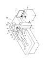
- FIG. 2is a hardware configuration diagram of the medical system 100 excluding the treatment instrument 1.
- the endoscope 2includes a long insertion portion 20 that can be inserted into the abdominal cavity of a patient, and an arm 21. The operator passes the insertion portion 20 through a trocar punctured in the abdomen of the patient, and introduces the insertion portion 20 into the abdominal cavity.
- the insertion unit 20is provided with an imaging unit 22 having a lens and an imaging device for imaging a state inside the abdomen of the patient at the distal end.
- the insertion unit 20 introduced into the abdominal cavityis disposed at a position where the imaging unit 22 can image the affected area to be treated in the abdomen.
- the imaging unit 22may have an optical zoom function or an electronic zoom function.
- the insertion part 20may further have an active bending part that bends actively. By curving the active bending portion provided in a part of the insertion portion 20, the orientation of the lens of the imaging portion 22 and the imaging element can be changed.
- the arm 21is an electrically driven robot arm having at least one joint 23 as shown in FIG.
- the distal end of the arm 21is connected to the proximal end portion of the insertion portion 20 of the endoscope, and the arm 21 can move the insertion portion 20.
- the joint 23is a part that bends around a rotation axis, and may be actively bent by a motor or the like, or passively bent by a forward / backward movement of a connected wire or the like. There may be.
- control signal lines and wires for controlling the bending operation of the joint 23are wired.
- a control signal line for controlling the imaging unit 22 and a transmission signal for transferring a captured image captured by the imaging unit 22are also wired inside the arm 21.
- the control device 3includes a drive unit 31, an image processing unit 32, and a control unit 33.
- the control device 3controls the arm 21 and the like based on the input from the input device 5.
- the control device 3generates a display image from the captured image captured by the imaging unit 22 of the endoscope 2 and transfers it to the display device 4.
- the driving unit 31drives the joint 23 and the insertion unit 20 of the arm 21.
- the control unit 33When the joint 23 actively bends, the control unit 33 generates a control signal to the drive unit 31 in order to operate the joint 23. As a result, the joint 23 can be bent by the drive unit 31.
- the control part 33when the insertion part 20 has an active bending part, the control part 33 produces
- the drive unit 31generates power for operating the active bending unit according to the generated control signal. As a result, the active bending portion can be bent by the power transmitted via the arm 21.
- the drive unit 31can change the visual field of the endoscope 2 by driving at least one of the arm 21 and the insertion unit 20.
- the image processing unit 32is connected to a transmission signal of a captured image captured by the imaging unit 22, and acquires a captured image via the transmission signal.
- the image processing unit 32generates a display image for display from the captured image.
- the image processing unit 32may perform image processing such as image format conversion and contrast adjustment on the captured image as necessary.
- the generated display imageis transferred to the display device 4 at a predetermined transfer timing.
- the image processing unit 32can generate a display image by replacing an image such as a figure or a character generated by the control unit 33 with a captured image or superimposing the captured image on the captured image.
- the image processing unit 32can generate a display image by superimposing a character image related to a warning to the surgeon or operation support on the captured image.
- the image processing unit 32not the control unit 33, may generate images such as graphics and characters based on instructions from the control unit 33.
- the control unit 33uses the operation of the input device 5 and the image acquired by the image processing unit 32 as inputs, and controls the drive unit 31 and the image processing unit 32 based on those inputs.
- the control unit 33has two types of operation modes, a manual mode and an automatic mode.
- the control unit 33controls the drive unit 31 and the image processing unit 32 based on one operation mode selected from the two operation modes.
- the manual modeis an operation mode in which the scopist operates the input device 5 to directly operate the joint 23 and the like of the arm 21 of the endoscope 2.
- the automatic modebased on the image acquired by the image processing unit 32, the joint 23 of the arm 21 of the endoscope 2 is automatically operated by the control unit 33, and the visual field of the endoscope 2 is automatically adjusted. It is an operation mode.
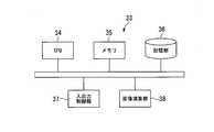
- FIG. 3 and 4are diagrams illustrating an example of the overall configuration of the control unit 33.
- the control unit 33includes a CPU (Central Processing Unit) 34, a memory 35 that can read a program, a storage unit 36, and an input / output control unit 37 that can execute a program ( Computer).
- the function of the control unit 33is realized by the CPU 34 executing a program provided to the control unit 33. Note that at least some of the functions of the control unit 33 may be configured by a dedicated logic circuit or the like.
- the storage unit 36is a non-volatile recording medium that stores the above-described program and necessary data.
- the storage unit 36is composed of, for example, a ROM or a hard disk.
- the program recorded in the storage unit 36is read into the memory 35 and executed by the CPU 34.
- the input / output control unit 37receives input data from the input device 5 and the image processing unit 32, and performs transfer to the CPU 34 and the like. Further, when the CPU 34 controls the drive unit 31 and the image processing unit 32, the input / output control unit 37 generates a control signal for the drive unit 31 and the image processing unit 32 based on an instruction from the CPU 34.
- control unit 33is not limited to a device provided in one piece of hardware.
- the control unit 33is configured by separating the CPU 34, the memory 35, the storage unit 36, and the input / output control unit 37 as separate hardware and connecting the hardware to each other via a communication line. May be.
- the control part 33may implement
- the memory 35 of the control unit 33may be used to store temporary data being processed. Further, the CPU 34 of the control unit 33 may execute part or all of the captured image processing performed by the image processing unit 32 by executing a program.
- control unit 33may further include components other than the CPU 34, the memory 35, the storage unit 36, and the input / output control unit 37 illustrated in FIG. .
- control unit 33may further include an image calculation unit 38 that performs part or all of specific image processing and image recognition processing. By further including the image calculation unit 38, the control unit 33 can execute specific image processing and image recognition processing at high speed.
- the display device 4is a device that displays the display image generated by the image processing unit 32.
- a known display devicesuch as an LCD display can be used.
- the display device 4may be a head mounted display or a projector.
- the input device 5includes an operation input unit 51, a mode selection unit 52, and a voice input unit 53, as shown in FIG.
- the input device 5is a device that inputs information necessary for the operation of the medical system 100.
- the operation input unit 51is a device that inputs an operation of the joint 23 of the arm 21 of the endoscope 2.
- the operation input unit 51can also operate the zoom function when the imaging unit 22 has a zoom function. Further, the scopist can operate the operation input unit 51 to operate the joint 23 of the arm 21 and the like.
- the operation input unit 51may be configured with a joystick or a touch panel.
- An arm-shaped operation input device similar to the arm 21may be used.
- the display device 4 of the LCD display and the operation input unit 51 of the touch panelmay be configured integrally.
- the operation contentis transferred to the control unit 33 by operating the operation input unit 51.
- the control unit 33calculates the movement amount of the joint 23 of the arm corresponding to the operation content.
- the control unit 33controls the drive unit 31 so that the joint 23 operates with the calculated operation amount.
- the joint 23 of the arm 21 of the endoscope 2is directly operated by the operation of the operation input unit 51.
- the operation mode of the control unit 33is the automatic mode
- the operation of the operation input unit 51is invalidated by the control unit 33 and the joint 23 of the arm 21 of the endoscope 2 cannot be operated.
- the joint 23 of the arm 21 of the endoscope 2is operated by the control unit 33.
- the mode selection unit 52is a device that selects which operation mode the control unit 33 operates from among the two operation modes of the control unit 33.
- the mode selection unit 52may be configured by a switch or a touch panel.
- the mode selection unit 52may be configured integrally with the operation input unit 51. The operation mode selection of the control unit 33 by the mode selection unit 52 can be performed at any time.
- the audio input unit 53has components for converting input audio, such as a microphone, and transfers the converted audio signal to the control unit 33.
- the target organis the liver L.
- the surgeon or the likeperforms a preoperative plan for creating a model (shape data or image) of the target organ using a known method before laparoscopic surgery. For example, the surgeon creates three-dimensional shape data of the target organ from a plurality of CT images.
- the three-dimensional coordinate system of the three-dimensional shape data generated in the preoperative planis referred to as “model coordinate system (first coordinate system) C1”.
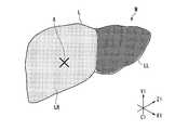
- FIG. 5is a model M of the target organ (liver L) created in the preoperative plan.
- the model Mis associated with three-dimensional coordinates (X1, Y1, and Z1 coordinates) in the model coordinate system C1, and the position of each part of the model M is specified by the three-dimensional coordinates in the model coordinate system C1. Can do.
- the model Mis configured such that the right lobe LR and the left lobe LL are distinguished.
- the surgeoncreates a “part database D1” in which (1) the name of the part in the model M and (2) the region in the model coordinate system C1 are registered in the preoperative plan.
- Table 1is an example of the site database D1.
- the name of the site in the site database D1may be an anatomical name or an abbreviated name.
- the region in the model coordinate system C1 of the part database D1is specified by the three-dimensional coordinates (X1 coordinate, Y1 coordinate, Z1 coordinate) in the model coordinate system C1.
- the region in the model coordinate system C1may specify a voxel region in detail using a plurality of three-dimensional coordinates, or may specify a rough region using two-dimensional three-dimensional coordinates, Only a point of interest may be specified by using only one three-dimensional coordinate.
- the surgeon or the likemay set a specific region to be treated or observed in laparoscopic surgery as a “region of interest” in the model M in the preoperative plan.
- the region of interestis a part of the tumor A of the right lobe LR of the target organ (liver L) as shown in FIG.
- the operator or the likeadds the tumor A that is the region of interest to the site database D1.
- Table 2is an example of the work mode database D2.
- the surgeoncreates the “work mode database D2” in the preoperative plan.
- the work mode database D2is a database in which, for example, the work mode of the endoscope 2 such as “enlarged display” and “whole display” and the operation content of the endoscope 2 corresponding to the work mode are registered.
- the operation content of the endoscope 2such as “close the imaging unit 22 of the endoscope 2 to the attention site” is registered corresponding to the work mode of “entire display”. .
- the target organ model M created in the preoperative planis associated with the names of a plurality of parts in the target organ and regions in the model coordinate system C1 of the model M, that is, together with the part database D1, the memory of the control unit 33 of the control device 3 Recorded in the unit 36 (model recording step).
- the work mode database D ⁇ b> 2is also recorded in the storage unit 36 of the control unit 33 of the control device 3.
- the control device 3extracts and stores a plurality of feature points F in the model M (feature point extraction step).
- the plurality of feature points Fare extracted using a known feature point extraction method.
- the plurality of feature points Fare specified together with the feature amount calculated according to a predetermined standard suitable for representing the feature, and the three-dimensional coordinates in the model coordinate system C1 are also specified and stored in the storage unit 36.
- the extraction and recording of the plurality of feature points Fmay be performed before or during the operation.
- the operatorprovides a plurality of holes (openings) for installing the trocar in the abdomen of the patient, and punctures the trocar into the hole.
- the operatorpasses the insertion part 10 of the treatment instrument 1 through a trocar punctured in the abdomen of the patient, and introduces the insertion part 10 into the abdominal cavity.
- the scopistoperates the mode selection unit 52 to set the operation mode of the control unit 33 to the manual mode.
- the scopistoperates the operation input unit 51 to operate the endoscope 2 so that the insertion unit 20 of the endoscope 2 is passed through the trocar punctured into the abdomen of the patient, and the insertion unit 20 is introduced into the abdominal cavity. .
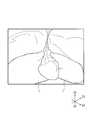
- FIG. 6is a display image generated by the control device 3 from a captured image captured by the imaging unit 22 of the endoscope 2 inserted into the abdominal cavity.
- the liver Lis displayed in the display image shown in FIG.
- the three-dimensional coordinate system of the display space in which the display image is displayedis referred to as “display coordinate system (second coordinate system) C2”.
- step S10when the operation mode of the control unit 33 is set to the automatic mode, the control unit 33 starts control of the automatic mode (step S10). Next, the control part 33 performs step S11.
- step S11the control unit 33 receives, from the voice input unit 53, the name of the site of interest of interest among the plurality of sites in the target organ and the work mode for the site of interest (instruction receiving step).
- the control unit 33activates the voice input unit 53 and receives an instruction by voice input from an operator or the like.
- the control unit 33performs voice recognition on the voice signal transferred from the voice input unit 53 by a known voice recognition method.
- the control part 33performs step S12 after progress of predetermined time.
- step S ⁇ b> 12the control unit 33 determines whether the speech-recognized content includes the “part name” registered in the part database D ⁇ b> 1 and the “work mode” registered in the work mode database D ⁇ b> 2. . When it determines with not being contained, the control part 33 performs step S11 again next. When it determines with being contained, the control part 33 performs step S13 next.
- the part name included in the speech-recognized contentis a part of interest that the operator or the like pays attention to, among the parts registered in the part database D1.
- the “work mode” included in the speech-recognized contentis a work mode for the attention site. In the present embodiment, it is assumed that “tumor A” is instructed as an attention site, and “enlarged display” is instructed as a work mode for the attention site.
- step S13the control unit 33 extracts a plurality of corresponding points A corresponding to the plurality of feature points F in the display image (corresponding point extraction step).
- the control unit 33extracts a plurality of corresponding points A in the display image based on the feature amounts of the plurality of feature points F stored in the storage unit 36 in advance.
- a method appropriately selected from known template matching methods and the likeis used.
- the control unit 33includes the image calculation unit 38 that performs part or all of the image matching processing at high speed, the above-described matching processing can be executed at high speed.
- the control part 33performs step S14.
- step S14the control unit 33 associates the model coordinate system C1 of the model M with the display coordinate system C2 of the display space displayed by the display image based on the plurality of feature points F and the plurality of corresponding points A ( Association step).
- Association stepFor the associating process, a method appropriately selected from known coordinate conversion methods or the like is used.
- the association stepis completed, the coordinate position that can be reached in the first coordinate system C1 of the model M can be converted into the display coordinate system C2 in the display space.
- FIG. 8is a display image after the association step is completed.
- the coordinate position that can be reached in the first coordinate system C1 of the model Mcan be converted into the display coordinate system C2 of the display space. Therefore, the region (right lobe LR, left lobe LL, tumor A) registered in the region database D1 shown in Table 1 can specify a region in the display coordinate system C2 of the display space.
- identification display imageshatchched images in which the positions of the right lobe LR, the left lobe LL, and the tumor A are known are superimposed.
- the control part 33performs step S15.
- step S15the control unit 33 calculates the position of the target region (tumor A) in the display coordinate system C2 from the position of the target region (tumor A) set in the model M in the model coordinate system C1 (relative position calculation). Process). Next, the control part 33 performs step S16.
- step S16the control unit 33 operates the endoscope 2 with respect to the site of interest (tumor A) based on the instructed work mode (endoscope operation step). Since the instructed work mode is “enlarged display”, the control unit 33 uses the position in the display coordinate system C2 of the target region obtained in step S15, and the corresponding “registered” in the work mode database D2 of D2. Based on the “operation details of the endoscope”, the arm 21 of the endoscope 2 is operated so that the imaging unit 22 approaches the position of the target region. Next, the control part 33 performs step S17 next.
- step S ⁇ b> 17the control unit 33 determines whether the work mode instructed for the attention site is completed. When it determines with having not completed, the control part 33 performs step S16 again next. When it determines with having completed, the control part 33 performs step S18 next, and complete
- FIG. 9is a display image after the automatic mode is completed. After the control in the automatic mode is completed, the work mode (enlarged display) instructed for the attention site (tumor A) designated in the model M created by the preoperative plan is performed.
- a specific operation of the visual field of the endoscope 2 with respect to the target organcan be supported using the model M created by the preoperative plan.
- the attention site of interest among the plurality of sites registered in the site database D1 before the operationis specified, and the specific work mode for the target site can be specified from the work mode registered in the work mode database D2 before the operation.
- the surgeonhas instructed the scopist to change the field of view of the endoscope 2 based on the display image (for example, an instruction “move the field of view range to the right”).
- the medical system 100can instruct the change of the visual field of the endoscope 2 using the name of the part.
- the part used for the instructionmay not be displayed on the display screen when the instruction is given. Therefore, the medical system 100 can quickly provide a visual field that meets the operator's wishes.
- the operatorcan set information (name, region) on the attention site for the model M before the operation, the visual field can be quickly secured during the operation. The burden can be reduced.
- the instructionis received by voice in the instruction receiving step, but the instruction receiving method is not limited to this.
- the medical systemregisters only one type of “part name” and “work mode” and receives the activation signal from a foot pedal or the like connected to the input device. The operation may be performed assuming that the contents of “part name” and “work mode” are instructed.
- the display imageis changed by operating the arm 21 of the endoscope 2 and changing the imaging position of the endoscope 2, but the method of changing the display image is not limited to this.
- the image processing unithas a function of generating a display image by cutting out a part of a region from a captured image of the endoscope, and the display image may be changed by changing a position where the image is cut out.
- the image processing unitmay change the display image by controlling the zoom function of the imaging unit. Even in the case of an endoscope that does not have an arm, the display image can be changed.
- FIG. 10is an overall configuration example showing an endoscope 2B which is a modified example of the endoscope 2.
- the endoscope 2 ⁇ / b> Bhas an active bending portion 23 ⁇ / b> B at the distal end of the insertion portion 20.
- the operatorcan change the position and orientation of the imaging unit 22 of the endoscope 2 by moving the endoscope 2B while holding the endoscope 2B. Further, the position and orientation of the imaging unit 22 can be changed by bending the active bending unit 23B. Even when the endoscope 2B is used instead of the endoscope 2, the control unit 33 can change the display image by driving the active bending portion 23B.
- the present inventioncan be applied to a medical system having an endoscope and a method for operating the medical system.
Landscapes
- Health & Medical Sciences (AREA)
- Life Sciences & Earth Sciences (AREA)
- Surgery (AREA)
- Engineering & Computer Science (AREA)
- Medical Informatics (AREA)
- Public Health (AREA)
- General Health & Medical Sciences (AREA)
- Biomedical Technology (AREA)
- Nuclear Medicine, Radiotherapy & Molecular Imaging (AREA)
- Animal Behavior & Ethology (AREA)
- Heart & Thoracic Surgery (AREA)
- Veterinary Medicine (AREA)
- Molecular Biology (AREA)
- Pathology (AREA)
- Radiology & Medical Imaging (AREA)
- Optics & Photonics (AREA)
- Physics & Mathematics (AREA)
- Biophysics (AREA)
- Robotics (AREA)
- Signal Processing (AREA)
- Primary Health Care (AREA)
- Epidemiology (AREA)
- Oral & Maxillofacial Surgery (AREA)
- Gynecology & Obstetrics (AREA)
- Databases & Information Systems (AREA)
- Data Mining & Analysis (AREA)
- Urology & Nephrology (AREA)
- Business, Economics & Management (AREA)
- General Business, Economics & Management (AREA)
- Endoscopes (AREA)
Abstract
Description
Translated fromJapanese 本発明は、腹壁等に形成した孔を通して処置を行う医療システムおよび同医療システムの作動方法に関する。
本願は、2018年2月21日に、アメリカ合衆国に仮出願された米国特許仮出願第62/633,190号に基づき優先権を主張し、その内容をここに援用する。The present invention relates to a medical system that performs treatment through a hole formed in an abdominal wall or the like, and an operating method of the medical system.
This application claims priority based on US Provisional Application No. 62 / 633,190 filed provisionally in the United States of America on February 21, 2018, the contents of which are incorporated herein by reference.
従来、腹腔鏡下手術において、腹壁に開けた別々の孔(開口)から処置具や内視鏡などを挿入して処置を行う手法が用いられている。内視鏡を操作するスコピストは、術者に対して処置に最適な内視鏡の視野、例えば対象臓器等の関心領域が含まれる視野を迅速に提供する必要がある。このような内視鏡の視野の操作を支援する医療システムが考案されている。Conventionally, in laparoscopic surgery, a technique has been used in which treatment is performed by inserting a treatment tool or an endoscope through separate holes (openings) opened in the abdominal wall. The scopist who operates the endoscope needs to quickly provide the operator with a field of view of the endoscope that is optimal for the treatment, for example, a field of view including a region of interest such as a target organ. A medical system that supports the operation of the visual field of such an endoscope has been devised.
特許文献1には、術者等が対象臓器の解剖学的名称を発話することで、装置が入力される音声から対象臓器を特定し、表示画面に特定された対象臓器を表示する医用観察支援装置が記載されている。
しかしながら、特許文献1に記載の医用観察支援装置は、特定された対象臓器に対して内視鏡を移動させることは可能であるが、例えば、対象臓器の一部を拡大して表示するなどの具体的な作業態様を指示することはできない。However, the medical observation support apparatus described in
上記事情を踏まえ、本発明は、対象臓器に対する内視鏡の視野の具体的な操作を支援する医療システムおよび医療システムの作動方法を提供することを目的とする。In view of the above circumstances, an object of the present invention is to provide a medical system that supports a specific operation of the visual field of an endoscope with respect to a target organ and a method for operating the medical system.
上記課題を解決するために、この発明は以下の手段を提案している。
本発明の第一の態様に係る医療システムは、撮像部を有し、電動で駆動されて作動する内視鏡と、前記内視鏡を作動させ、前記撮像部の撮像画像から表示画像を生成する制御装置と、前記表示画像を表示する表示装置と、前記制御装置に対する指示が入力される入力装置と、を備え、前記制御装置は、術前計画において作成した対象臓器のモデルと前記対象臓器における複数の部位の名称および前記モデルの第一座標系における領域とを関連付けて記録し、前記入力装置から前記対象臓器における複数の部位のうち注目する注目部位の名称と、前記注目部位に対する作業態様とを受信し、前記モデルの前記第一座標系と前記表示画像が表示する表示空間の第二座標系とを関連付け、前記注目部位の前記第一座標系における位置から、前記注目部位の前記第二座標系における位置を算出し、前記作業態様に基づいて前記注目部位に対して前記内視鏡を作動させる。In order to solve the above problems, the present invention proposes the following means.
The medical system which concerns on the 1st aspect of this invention has an imaging part, and operates the endoscope driven electrically and operates the said endoscope, A display image is produced | generated from the captured image of the said imaging part A control device for displaying the display image, and an input device for inputting an instruction to the control device. The control device includes a model of the target organ created in the preoperative plan and the target organ. The names of a plurality of parts in the first coordinate system of the model and the region in the first coordinate system of the model are recorded in association with each other. And associating the first coordinate system of the model with the second coordinate system of the display space displayed by the display image, and from the position of the target region in the first coordinate system, Calculating a position in position of the second coordinate system, operating the endoscope with respect to the attention area based on the working mode.
また、本発明の第二の態様に係る医療システムの作動方法は、撮像部を有し、電動で駆動されて作動する内視鏡と、前記内視鏡を作動させ、前記撮像部の撮像画像から表示画像を生成する制御装置と、前記表示画像を表示する表示装置と、前記制御装置に対する指示が入力される入力装置と、を備える医療システムの作動方法であって、術前計画において作成した対象臓器のモデルと前記対象臓器における複数の部位の名称および前記モデルの第一座標系における領域とを関連付けて記録するモデル記録工程と、前記入力装置から前記対象臓器における複数の部位のうち注目する注目部位の名称と、前記注目部位に対する作業態様とを受信する指示受信工程と、前記モデルの前記第一座標系と前記表示画像が表示する表示空間の第二座標系とを関連付ける関連付け工程と、前記注目部位の前記第一座標系における位置から、前記注目部位の前記第二座標系における位置を算出する相対位置算出工程と、前記作業態様に基づいて前記注目部位に対して前記内視鏡を作動させる内視鏡作動工程と、を備える。In addition, the medical system operating method according to the second aspect of the present invention includes an imaging unit, an endoscope that is electrically driven to operate, and the endoscope that operates, and a captured image of the imaging unit A method for operating a medical system, comprising: a control device that generates a display image from a display device; a display device that displays the display image; and an input device that inputs an instruction to the control device. A model recording step for recording the model of the target organ in association with the names of the plurality of parts in the target organ and the region in the first coordinate system of the model, and paying attention from the input device among the plurality of parts in the target organ An instruction receiving step for receiving the name of the target region and the work mode for the target region, the second coordinate system of the display space in which the first coordinate system of the model and the display image are displayed An associating step, a relative position calculating step of calculating a position of the target site in the second coordinate system from a position of the target site in the first coordinate system, and the target site based on the work mode An endoscope operation step of operating the endoscope.
本発明の医療システムおよび医療システムの作動方法によれば、対象臓器に対する内視鏡の視野の具体的な操作を支援できる。According to the medical system and the operation method of the medical system of the present invention, it is possible to support a specific operation of the visual field of the endoscope with respect to the target organ.
本発明の一実施形態について、図1から図9を参照して説明する。なお、図面を見やすくするため、各構成要素の寸法等は適宜調整されている。An embodiment of the present invention will be described with reference to FIGS. In addition, in order to make the drawings easy to see, the dimensions and the like of each component are appropriately adjusted.
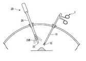
図1は、本実施形態に係る医療システム100の全体構成を示す図である。
医療システム100は、図1に示すように、処置具1と、内視鏡2と、制御装置3と、表示装置4と、入力装置5と、を備えている。医療システム100は、腹腔鏡下手術において、腹壁に開けた別々の孔(開口)から処置具1や内視鏡2などを挿入して行う処置を支援するシステムである。FIG. 1 is a diagram illustrating an overall configuration of a
As shown in FIG. 1, the
処置具1は、図1に示すように、患者の腹腔内に挿入可能な長尺の挿入部10と、挿入部10の基端部に設けられた操作部11と、を有する。術者は、患者の腹部に穿刺したトロッカに挿入部10を通し、挿入部10を腹腔内に導入する。処置の種類や患部の状況により、術者は複数の処置具1を腹腔内に導入する場合もある。As shown in FIG. 1, the
挿入部10は、図1に示すように、先端部に患者の患部を処置する処置部12を有する。本実施形態において処置部12は、一対の把持部材12aで構成された把持機構である。As shown in FIG. 1, the
操作部11は、一対の把持部材12aを操作する部材である。操作部11はハンドルを有しており、ハンドルを操作部11の他の部分に対して相対移動させることで、処置部12の一対の把持部材12aを開閉させる。術者は、片手で操作部11を保持して、処置部12を操作することができる。The operation unit 11 is a member that operates the pair of gripping
図2は、処置具1を除く医療システム100のハードウェア構成図である。
内視鏡2は、図1および図2に示すように、患者の腹腔内に挿入可能な長尺の挿入部20と、アーム21と、を有する。術者は、患者の腹部に穿刺したトロッカに挿入部20を通し、挿入部20を腹腔内に導入する。FIG. 2 is a hardware configuration diagram of the
As shown in FIGS. 1 and 2, the
挿入部20は先端部に、患者の腹部内の様子を撮影するためのレンズや撮像素子を有する撮像部22が設けられている。腹腔内に導入された挿入部20は、撮像部22が腹部内の処置対象の患部を撮影可能な位置に配置される。撮像部22は、光学ズームもしくは電子ズームの機能を有していてもよい。The
なお、挿入部20は、能動的に湾曲する能動湾曲部をさらに有してもよい。挿入部20の一部に設けられた能動湾曲部を湾曲させることで、撮像部22のレンズや撮像素子の向きを変更することができる。In addition, the
アーム21は、図1に示すように、少なくとも1以上の関節23を有する電動駆動のロボットアームである。アーム21の先端が内視鏡の挿入部20の基端部に接続されており、アーム21は、挿入部20を移動させることができる。The
関節23は、回動軸を中心に屈曲する部位であり、モータ等によって能動的に屈曲動作するものであってもよいし、接続されるワイヤ等の進退動作によって受動的に屈曲動作するものであってもよい。アーム21の内部には、関節23の屈曲動作を制御する制御信号線やワイヤ等が配線されている。また、アーム21の内部には、撮像部22を制御する制御信号線や撮像部22が撮像した撮像画像を転送する伝送信号も配線されている。The
制御装置3は、図2に示すように、駆動部31と、画像処理部32と、制御部33と、を有している。制御装置3は、入力装置5からの入力に基づいてアーム21等を制御する。また、制御装置3は、内視鏡2の撮像部22が撮像した撮像画像から表示画像を生成して、表示装置4に転送する。As shown in FIG. 2, the
駆動部31は、アーム21の関節23や挿入部20を駆動する。関節23が能動的に屈曲動作するものである場合、制御部33は関節23を動作させるために駆動部31への制御信号を生成する。その結果、関節23は駆動部31によって屈曲動作させることができる。別の態様として、挿入部20が能動湾曲部を有する場合、制御部33は能動湾曲部を制御する制御信号を生成する。駆動部31は、生成した制御信号に従い、能動湾曲部を動作させるための動力を発生させる。その結果、能動湾曲部は、アーム21を介して伝達された動力によって湾曲動作させることができる。The
すなわち、駆動部31は、アーム21および挿入部20の少なくとも一方を駆動することによって、内視鏡2の視野を変更させることができる。That is, the
画像処理部32は、撮像部22が撮像した撮像画像の伝送信号が接続されており、その伝送信号経由で、撮像画像を取得する。また、画像処理部32は、撮像画像から表示用の表示画像を生成する。画像処理部32は、撮像画像に対し、画像フォーマット変換やコントラスト調整などの画像処理を必要に応じて行ってもよい。生成された表示画像は、表示装置4に所定の転送タイミングで転送される。The
画像処理部32は、制御部33が生成した図形や文字などの画像を、撮像画像と差し替えて、もしくは、撮像画像に重畳して、表示画像を生成することができる。例えば、画像処理部32は、術者への警告や操作支援に関する文字の画像を、撮像画像に重畳して、表示画像を生成することができる。なお、上記の図形や文字などの画像は、制御部33ではなく画像処理部32が、制御部33の指示に基づいて生成してもよい。The
制御部33は、入力装置5の操作および画像処理部32が取得した画像を入力とし、それらの入力に基づいて駆動部31および画像処理部32の制御等を行う。
本実施形態において、制御部33は、マニュアルモードおよび自動モードの2種類の動作モードを有する。制御部33は、二つの動作モードから選択された一つの動作モードに基づいて、駆動部31および画像処理部32の制御等を行う。The
In the present embodiment, the
マニュアルモードは、スコピストが入力装置5を操作して、内視鏡2のアーム21の関節23等を直接操作する動作モードである。
自動モードは、画像処理部32が取得した画像に基づいて、内視鏡2のアーム21の関節23等が制御部33により自動で操作され、内視鏡2の視野の自動調整が実施される動作モードである。The manual mode is an operation mode in which the scopist operates the
In the automatic mode, based on the image acquired by the
図3および図4は、制御部33の全体構成例を示す図である。
制御部33は、図3に示すように、CPU(Central Processing Unit)34と、プログラムを読み込み可能なメモリ35と、記憶部36と、入出力制御部37と、を有するプログラム実行可能な装置(コンピュータ)である。
制御部33の機能は、制御部33に提供されたプログラムをCPU34が実行することにより実現される。なお、制御部33の少なくとも一部の機能を、専用の論理回路等によって構成してもよい。3 and 4 are diagrams illustrating an example of the overall configuration of the
As shown in FIG. 3, the
The function of the
記憶部36は、上述したプログラムや必要なデータを記憶する不揮発性の記録媒体である。記憶部36は、例えばROMやハードディスク等で構成される。記憶部36に記録されたプログラムは、メモリ35に読み込まれ、CPU34によって実行される。The
入出力制御部37は、入力装置5および画像処理部32からの入力データを受け取り、CPU34等への転送等を実施する。また、入出力制御部37は、CPU34が駆動部31や画像処理部32を制御する際に、CPU34の指示に基づき、駆動部31や画像処理部32に対する制御信号等を生成する。The input /
ここで、制御部33は、1つのハードウェアに備わる装置に限られない。例えば、制御部33は、CPU34と、メモリ35と、記憶部36と、入出力制御部37とをそれぞれ別体のハードウェアとして分離した上で、ハードウェア同士を通信回線で接続することで構成してもよい。あるいは、制御部33は、記憶部36を分離して、同様に通信回線で接続することで、制御部33をクラウドシステムとして実現してもよい。Here, the
ここで、画像処理部32が、撮像画像の処理を行う場合、処理中の一時的なデータを保管するため、制御部33のメモリ35を使用してもよい。また、画像処理部32が行う撮像画像の処理の一部もしくは全部を、制御部33のCPU34がプログラム実行により実施してもよい。Here, when the
なお、制御部33は、図3に示すCPU34、メモリ35、記憶部36、および入出力制御部37以外のもので、制御装置3の動作のために必要なものを、さらに有してもよい。例えば、図4に示すように、制御部33は、特定の画像処理や画像認識処理の一部もしくは全部を行う画像演算部38をさらに有してもよい。画像演算部38をさらに有することで、制御部33は、特定の画像処理や画像認識処理を高速に実行することができる。Note that the
表示装置4は、画像処理部32が生成した表示画像を表示する装置である。表示装置4は、LCDディスプレイ等の公知の表示装置を用いることができる。表示装置4は、ヘッドマウントディスプレイやプロジェクタであってもよい。The
入力装置5は、図2に示すように、操作入力部51と、モード選択部52と、音声入力部53と、を有する。入力装置5は、医療システム100の動作に必要な情報を入力する装置である。The
操作入力部51は、内視鏡2のアーム21の関節23の操作を入力する装置である。また、操作入力部51は、撮像部22がズーム機能を有している場合は、そのズーム機能を操作することもできる。また、スコピストは、操作入力部51を操作して、アーム21の関節23等を操作することができる。The
操作入力部51は、図1に示すように、ジョイスティックにより構成されていてもよいし、タッチパネルにより構成されていてもよい。アーム21と相似形状のアーム形状の操作入力デバイスであってもよい。LCDディスプレイの表示装置4と、タッチパネルの操作入力部51とが、一体に構成されていてもよい。As shown in FIG. 1, the
操作入力部51を操作することで、その操作内容が制御部33に転送される。制御部33は、操作内容に対応する、アームの関節23の動作量を算出する。制御部33は、算出した動作量で関節23が動作するように、駆動部31を制御する。The operation content is transferred to the
制御部33の動作モードがマニュアルモードである場合、操作入力部51の操作により、内視鏡2のアーム21の関節23等は直接操作される。
一方、制御部33の動作モードが自動モードである場合、操作入力部51の操作は、制御部33により無効化され、内視鏡2のアーム21の関節23等を操作することはできない。内視鏡2のアーム21の関節23等は制御部33によって操作される。When the operation mode of the
On the other hand, when the operation mode of the
モード選択部52は、制御部33が有する二つの動作モードのうち、どちらの動作モードで制御部33が動作するかを選択する装置である。モード選択部52は、スイッチにより構成されていてもよいし、タッチパネルにより構成されていてもよい。また、モード選択部52は、操作入力部51と一体に構成されていてもよい。モード選択部52による制御部33の動作モード選択は、いつでも行うことができる。The
音声入力部53は、マイクなどの入力される音声を信号化する部品を有しており、信号化された音声信号を制御部33に転送する。The
[医療システム100の動作]
次に、腹腔鏡下手術を例として、医療システム100の動作および作動方法を、図5から図9を参照して説明する。本実施形態では、対象臓器は肝臓Lとする。[Operation of Medical System 100]
Next, taking laparoscopic surgery as an example, the operation and operation method of the
術者等は、腹腔鏡下手術前に、公知の手法を用いて、対象臓器のモデル(形状データや画像)を作成する術前計画を行う。術者等は、例えば複数のCT画像から対象臓器の三次元形状データを作成する。術前計画にて生成した三次元形状データの三次元座標系を「モデル座標系(第一座標系)C1」と称する。The surgeon or the like performs a preoperative plan for creating a model (shape data or image) of the target organ using a known method before laparoscopic surgery. For example, the surgeon creates three-dimensional shape data of the target organ from a plurality of CT images. The three-dimensional coordinate system of the three-dimensional shape data generated in the preoperative plan is referred to as “model coordinate system (first coordinate system) C1”.
図5は、術前計画において作成した対象臓器(肝臓L)のモデルMである。モデルMは、モデル座標系C1における三次元座標(X1座標、Y1座標、Z1座標)に対応付けられており、モデルMの各部位の位置は、モデル座標系C1における三次元座標で特定することができる。モデルMは、図5に示すように、右葉LRと左葉LLが区別されるように構成されている。FIG. 5 is a model M of the target organ (liver L) created in the preoperative plan. The model M is associated with three-dimensional coordinates (X1, Y1, and Z1 coordinates) in the model coordinate system C1, and the position of each part of the model M is specified by the three-dimensional coordinates in the model coordinate system C1. Can do. As shown in FIG. 5, the model M is configured such that the right lobe LR and the left lobe LL are distinguished.
術者等は、術前計画において、モデルMにおける(1)部位の名称と、(2)部位のモデル座標系C1における領域とが登録された「部位データベースD1」を作成する。The surgeon creates a “part database D1” in which (1) the name of the part in the model M and (2) the region in the model coordinate system C1 are registered in the preoperative plan.
表1は、部位データベースD1の一例である。
部位データベースD1の部位の名称は、解剖学的名称であってもよく、略称であってもよい。部位データベースD1のモデル座標系C1における領域は、モデル座標系C1における三次元座標(X1座標、Y1座標、Z1座標)で特定される。モデル座標系C1における領域は、複数の三次元座標を使用して詳細にボクセル領域を特定してもよいし、二点の三次元座標を使用して大まかな領域を特定してもよいし、一点の三次元座標を使用するのみで注目点のみを特定してもよい。Table 1 is an example of the site database D1.
The name of the site in the site database D1 may be an anatomical name or an abbreviated name. The region in the model coordinate system C1 of the part database D1 is specified by the three-dimensional coordinates (X1 coordinate, Y1 coordinate, Z1 coordinate) in the model coordinate system C1. The region in the model coordinate system C1 may specify a voxel region in detail using a plurality of three-dimensional coordinates, or may specify a rough region using two-dimensional three-dimensional coordinates, Only a point of interest may be specified by using only one three-dimensional coordinate.

術者等は、術前計画において、腹腔鏡下手術における処置対象や観察対象となる特定の領域を、モデルMにおいて「関心領域」として設定してもよい。本実施形態において、関心領域は、図5に示すように、対象臓器(肝臓L)の右葉LRの一部の腫瘍Aである。関心領域を設定した場合、表1に示すように、術者等は関心領域である腫瘍Aを部位データベースD1に追加する。The surgeon or the like may set a specific region to be treated or observed in laparoscopic surgery as a “region of interest” in the model M in the preoperative plan. In the present embodiment, the region of interest is a part of the tumor A of the right lobe LR of the target organ (liver L) as shown in FIG. When the region of interest is set, as shown in Table 1, the operator or the like adds the tumor A that is the region of interest to the site database D1.
表2は、作業態様データベースD2の一例である。
術者等は、術前計画において、「作業態様データベースD2」を作成する。作業態様データベースD2は、例えば、「拡大表示」や「全体表示」といった内視鏡2の作業態様と、作業態様に対応した内視鏡2の作動内容が登録されたデータベースである。作業態様データベースD2には、例えば、「全体表示」の作業態様に対応して、「内視鏡2の撮像部22を注目部位に近付ける」等の内視鏡2の作動内容が登録されている。Table 2 is an example of the work mode database D2.
The surgeon creates the “work mode database D2” in the preoperative plan. The work mode database D2 is a database in which, for example, the work mode of the

術前計画において作成した対象臓器のモデルMは、対象臓器における複数の部位の名称およびモデルMのモデル座標系C1における領域と関連付けて、すなわち部位データベースD1とともに、制御装置3の制御部33の記憶部36に記録される(モデル記録工程)。作業態様データベースD2も、制御装置3の制御部33の記憶部36に記録される。The target organ model M created in the preoperative plan is associated with the names of a plurality of parts in the target organ and regions in the model coordinate system C1 of the model M, that is, together with the part database D1, the memory of the
次に、制御装置3は、モデルMにおいて複数の特徴点Fを抽出して記憶する(特徴点抽出工程)。複数の特徴点Fは、公知の特徴点の抽出方法を用いて抽出される。複数の特徴点Fは、特徴を表すことに適した所定の基準に従って算出された特徴量とともに、モデル座標系C1における三次元座標も合わせて特定されて記憶部36に記憶される。なお、複数の特徴点Fの抽出および記録は、術前に実施してもよく、術中に実施してもよい。Next, the
次に、腹腔鏡下手術中における医療システム100の動作を説明する。術者は、患者の腹部にトロッカを設置するための孔(開口)を複数設け、孔にトロッカを穿刺する。次に術者は、患者の腹部に穿刺したトロッカに処置具1の挿入部10を通し、挿入部10を腹腔内に導入する。Next, the operation of the
次に、スコピストは、モード選択部52を操作し、制御部33の動作モードをマニュアルモードに設定する。スコピストは、操作入力部51を操作して、内視鏡2を操作することで、内視鏡2の挿入部20を患者の腹部に穿刺したトロッカに通し、挿入部20を腹腔内に導入する。Next, the scopist operates the
図6は、腹腔内に挿入された内視鏡2の撮像部22が撮像した撮像画像から制御装置3が生成した表示画像である。図6に示す表示画像には肝臓Lが表示されている。表示画像が表示する表示空間の三次元座標系を「表示座標系(第二座標系)C2」と称する。FIG. 6 is a display image generated by the
術者は、関心領域である「腫瘍A」が表示画面に拡大された状態で表示されるように、内視鏡2を作動させたい。そこで、術者もしくはスコピスト等は、モード選択部52を操作し、制御部33の動作モードを自動モードに変更する。操作入力部51の操作は、制御部33により無効化され、スコピスト等は内視鏡2のアーム21の関節23等を操作することはできない。以降、図7に示す自動モードにおける制御フローチャートに沿って説明を行う。The surgeon wants to operate the
図7に示すように、制御部33の動作モードが自動モードに設定されると、制御部33は自動モードの制御を開始する(ステップS10)。次に、制御部33はステップS11を実行する。As shown in FIG. 7, when the operation mode of the
ステップS11において、制御部33は、音声入力部53から対象臓器における複数の部位のうち注目する注目部位の名称と、注目部位に対する作業態様とを受信する(指示受信工程)。制御部33は、音声入力部53を有効化し、術者等からの音声入力により指示を受け付ける。制御部33は、音声入力部53から転送される音声信号を、公知の音声認識手法により、音声認識を実施する。次に、制御部33は所定時間の経過後、ステップS12を実行する。In step S11, the
ステップS12において、制御部33は、音声認識した内容に、部位データベースD1に登録された「部位の名称」と、作業態様データベースD2に登録された「作業態様」とが含まれているかを判定する。
含まれないと判定した場合、制御部33は次にステップS11を再び実行する。
含まれると判定した場合、制御部33は次にステップS13を実行する。In step S <b> 12, the
When it determines with not being contained, the
When it determines with being contained, the
音声認識した内容に含まれる「部位の名称」は、部位データベースD1に登録された部位のうち、術者等が注目する注目部位である。音声認識した内容に含まれる「作業態様」は、注目部位に対する作業態様である。本実施形態においては、注目部位として「腫瘍A」、注目部位に対する作業態様とて「拡大表示」が指示されたとする。“The part name” included in the speech-recognized content is a part of interest that the operator or the like pays attention to, among the parts registered in the part database D1. The “work mode” included in the speech-recognized content is a work mode for the attention site. In the present embodiment, it is assumed that “tumor A” is instructed as an attention site, and “enlarged display” is instructed as a work mode for the attention site.
ステップS13において、制御部33は、表示画像において複数の特徴点Fに対応する複数の対応点Aを抽出する(対応点抽出工程)。制御部33は、予め記憶部36に記憶された複数の特徴点Fの特徴量に基づき、表示画像における複数の対応点Aを抽出する。抽出処理には、公知のテンプレートマッチング手法等から適宜選択した手法が用いられる。制御部33が画像のマッチング処理の一部もしくは全部を高速に行う画像演算部38を有している場合、上記のマッチング処理を高速に実行することができる。次に、制御部33はステップS14を実行する。In step S13, the
ステップS14において、制御部33は、複数の特徴点Fと複数の対応点Aとに基づいて、モデルMのモデル座標系C1と、表示画像が表示する表示空間の表示座標系C2とを関連付ける(関連付け工程)。関連付け処理には、公知の座標変換手法等から適宜選択した手法が用いられる。関連付け工程が完了すると、モデルMの第一座標系C1に行ける座標位置を、表示空間の表示座標系C2に変換することができる。In step S14, the
図8は、関連付け工程が完了した後の表示画像である。
関連付け工程が完了した後、モデルMの第一座標系C1に行ける座標位置を、表示空間の表示座標系C2に変換することができる。そのため、表1に示す部位データベースD1に登録された部位(右葉LR、左葉LL、腫瘍A)は、表示空間の表示座標系C2において領域を特定することができる。図8に示す表示画像では、右葉LR、左葉LL、腫瘍Aのそれぞれの位置がわかる識別表示画像(ハッチング画像)が重畳されている。次に、制御部33はステップS15を実行する。FIG. 8 is a display image after the association step is completed.
After the association step is completed, the coordinate position that can be reached in the first coordinate system C1 of the model M can be converted into the display coordinate system C2 of the display space. Therefore, the region (right lobe LR, left lobe LL, tumor A) registered in the region database D1 shown in Table 1 can specify a region in the display coordinate system C2 of the display space. In the display image shown in FIG. 8, identification display images (hatched images) in which the positions of the right lobe LR, the left lobe LL, and the tumor A are known are superimposed. Next, the
ステップS15において、制御部33は、モデルMに設定された注目部位(腫瘍A)のモデル座標系C1における位置から、注目部位(腫瘍A)の表示座標系C2における位置を算出する(相対位置算出工程)。次に、制御部33はステップS16を実行する。In step S15, the
ステップS16において、制御部33は、指示された作業態様に基づいて注目部位(腫瘍A)に対して内視鏡2を作動させる(内視鏡作動工程)。指示された作業態様は「拡大表示」であるため、制御部33は、ステップS15で求めた注目部位の表示座標系C2における位置を用いて、D2の作業態様データベースD2に登録された対応する「内視鏡の作動内容」に基づいて、注目部位の位置に撮像部22が近づくように、内視鏡2のアーム21を操作する。次に、制御部33は次にステップS17を実行する。In step S16, the
ステップS17において、制御部33は、注目部位に対して指示された作業態様が完了したかを判定する。
完了していないと判定した場合、制御部33は次にステップS16を再び実行する。
完了したと判定した場合、制御部33は次にステップS18を実行して自動モードを終了する。In step S <b> 17, the
When it determines with having not completed, the
When it determines with having completed, the
図9は、自動モードが完了した後の表示画像である。
自動モードの制御が完了した後、術前計画により作成したモデルMにおいて指定した注目部位(腫瘍A)に対して指示された作業態様(拡大表示)が実施されている。FIG. 9 is a display image after the automatic mode is completed.
After the control in the automatic mode is completed, the work mode (enlarged display) instructed for the attention site (tumor A) designated in the model M created by the preoperative plan is performed.
本実施形態の医療システム100によれば、術前計画により作成したモデルMを用いて、対象臓器に対する内視鏡2の視野の具体的な操作を支援できる。術前に部位データベースD1に登録した複数の部位のうち注目する注目部位を特定し、注目部位に対する具体的な作業態様を、術前に作業態様データベースD2に登録した作業態様から特定できる。According to the
従来であれば、術者はスコピストに対して、表示画像を基準に内視鏡2の視野の変更の指示(例えば、「視野範囲を右側に移動せよ」といった指示)を行っていた。
これに対して医療システム100は、内視鏡2の視野の変更を、部位の名称を用いて指示することができる。さらに、指示に用いる部位は、指示を行う際に表示画面に表示されていなくてもよい。そのため、医療システム100は、術者の希望に沿った視野を迅速に提供することができる。Conventionally, the surgeon has instructed the scopist to change the field of view of the
On the other hand, the
医療システム100は、術者が術前に注目部位に関する情報(名称、領域)をモデルMに対して設定しておけるため、術中における視野の確保を迅速に行うことができ、術者およびスコピストの負担を減らすことができる。In the
以上、本発明の一実施形態について図面を参照して詳述したが、具体的な構成はこの実施形態に限られるものではなく、本発明の要旨を逸脱しない範囲の設計変更等も含まれる。また、上述の一実施形態および以下で示す変形例において示した構成要素は適宜に組み合わせて構成することが可能である。As described above, the embodiment of the present invention has been described in detail with reference to the drawings. However, the specific configuration is not limited to this embodiment, and design changes and the like within the scope of the present invention are included. In addition, the constituent elements shown in the above-described embodiment and the modified examples described below can be appropriately combined.
(変形例1)
上記の実施形態では、指示受信工程において指示を音声により受信していたが、指示の受信方法はこれに限定されない。例えば、医療システムは、「部位の名称」と「作業態様」とをそれぞれ一種類のみ登録しておき、入力装置に接続されたフットペダル等からの起動信号を受信した際に、登録された「部位の名称」と「作業態様」の内容が指示されたとして動作してもよい。(Modification 1)
In the above embodiment, the instruction is received by voice in the instruction receiving step, but the instruction receiving method is not limited to this. For example, the medical system registers only one type of “part name” and “work mode” and receives the activation signal from a foot pedal or the like connected to the input device. The operation may be performed assuming that the contents of “part name” and “work mode” are instructed.
(変形例2)
上記の実施形態では、内視鏡2のアーム21を操作して内視鏡2の撮像位置を変更することで表示画像を変更していたが、表示画像の変更方法はこれに限定されない。画像処理部が、内視鏡の撮像画像から一部の領域を切り出して表示画像を生成する機能を有し、画像を切り出す位置を変更することで、表示画像を変更してもよい。また、画像処理部が、撮像部のズーム機能を制御して表示画像を変更してもよい。アームを有していない内視鏡の場合であっても、表示画像を変更することができる。(Modification 2)
In the above embodiment, the display image is changed by operating the
図10は、内視鏡2の変形例である内視鏡2Bを示す全体構成例である。内視鏡2Bは、挿入部20の先端に能動湾曲部23Bを有している。術者は、内視鏡2Bを持って内視鏡2Bを移動させることで、内視鏡2の撮像部22の位置や向きを変更することができる。また、能動湾曲部23Bを湾曲させることで、撮像部22の位置や向きを変更することができる。内視鏡2に代えて、内視鏡2Bを使用する場合であっても、制御部33は、能動湾曲部23Bを駆動することで、表示画像を変更することができる。FIG. 10 is an overall configuration example showing an
本発明は、内視鏡を有する医療システムおよび医療システムの作動方法に適用することができる。The present invention can be applied to a medical system having an endoscope and a method for operating the medical system.
100 医療システム
1 処置具
2、2B 内視鏡
20 挿入部
21 アーム
22 撮像部
23 関節
23B 能動湾曲部
3 制御装置
31 駆動部
32 画像処理部
33 制御部
35 メモリ
36 記憶部
37 入出力制御部
38 画像演算部
4 表示装置
5 入力装置
51 操作入力部
52 モード選択部
53 音声入力部DESCRIPTION OF
Claims (6)
Translated fromJapanese前記内視鏡を作動させ、前記撮像部の撮像画像から表示画像を生成する制御装置と、
前記表示画像を表示する表示装置と、
前記制御装置に対する指示が入力される入力装置と、
を備え、
前記制御装置は、
術前計画において作成した対象臓器のモデルと前記対象臓器における複数の部位の名称および前記モデルの第一座標系における領域とを関連付けて記録し、
前記入力装置から前記対象臓器における複数の部位のうち注目する注目部位の名称と、前記注目部位に対する作業態様とを受信し、
前記モデルの前記第一座標系と前記表示画像が表示する表示空間の第二座標系とを関連付け、
前記注目部位の前記第一座標系における位置から、前記注目部位の前記第二座標系における位置を算出し、
前記作業態様に基づいて前記注目部位に対して前記内視鏡を作動させる、
医療システム。An endoscope having an imaging unit and operated by being electrically driven;
A control device for operating the endoscope and generating a display image from a captured image of the imaging unit;
A display device for displaying the display image;
An input device for inputting an instruction to the control device;
With
The controller is
Record the target organ model created in the preoperative plan in association with the names of a plurality of parts in the target organ and the region in the first coordinate system of the model,
Receiving from the input device the name of the site of interest among the plurality of sites in the target organ, and the working mode for the site of interest;
Associating the first coordinate system of the model with a second coordinate system of a display space displayed by the display image;
From the position of the site of interest in the first coordinate system, the position of the site of interest in the second coordinate system is calculated,
Operating the endoscope with respect to the site of interest based on the working mode;
Medical system.
請求項1に記載の医療システム。The names of the plurality of parts in the target organ include names of specific areas set by the operator,
The medical system according to claim 1.
前記入力装置に入力される前記注目部位の前記名称と前記作業態様とは、音声入力により特定される、
請求項1または請求項2に記載の医療システム。The input device has a voice input unit,
The name of the site of interest and the work mode input to the input device are specified by voice input.
The medical system according to claim 1 or claim 2.
術前計画において作成した対象臓器のモデルと前記対象臓器における複数の部位の名称および前記モデルの第一座標系における領域とを関連付けて記録するモデル記録工程と、
前記入力装置から前記対象臓器における複数の部位のうち注目する注目部位の名称と、前記注目部位に対する作業態様とを受信する指示受信工程と、
前記モデルの前記第一座標系と前記表示画像が表示する表示空間の第二座標系とを関連付ける関連付け工程と、
前記注目部位の前記第一座標系における位置から、前記注目部位の前記第二座標系における位置を算出する相対位置算出工程と、
前記作業態様に基づいて前記注目部位に対して前記内視鏡を作動させる内視鏡作動工程と、
を備える、
医療システムの作動方法。An endoscope having an imaging unit and operated by being electrically driven, a control device that operates the endoscope and generates a display image from a captured image of the imaging unit, and a display device that displays the display image And an input device for inputting an instruction to the control device, and a method for operating a medical system,
A model recording step of associating and recording the model of the target organ created in the preoperative plan, the names of a plurality of parts in the target organ, and the region in the first coordinate system of the model;
An instruction receiving step of receiving from the input device a name of a target region of interest among a plurality of regions in the target organ, and a work mode for the target region;
Associating the first coordinate system of the model with a second coordinate system of a display space displayed by the display image;
A relative position calculating step of calculating a position of the target site in the second coordinate system from a position of the target site in the first coordinate system;
An endoscope operation step of operating the endoscope with respect to the site of interest based on the work mode;
Comprising
How the medical system operates.
請求項4に記載の医療システムの作動方法。In the model recording step, the names of the plurality of parts in the target organ include names of specific areas set by an operator.
The operating method of the medical system according to claim 4.
請求項4または請求項5に記載の医療システムの作動方法。The name of the site of interest and the work mode input to the input device in the instruction receiving step are specified by voice input.
6. A method of operating a medical system according to claim 4 or claim 5.
Priority Applications (3)
| Application Number | Priority Date | Filing Date | Title |
|---|---|---|---|
| JP2020501032AJP6990292B2 (en) | 2018-02-21 | 2019-02-21 | Medical system and how to operate the medical system |
| CN201980013735.9ACN111770716B (en) | 2018-02-21 | 2019-02-21 | Medical system and control method for medical system |
| US16/993,858US11800966B2 (en) | 2018-02-21 | 2020-08-14 | Medical system and medical system operating method |
Applications Claiming Priority (2)
| Application Number | Priority Date | Filing Date | Title |
|---|---|---|---|
| US201862633190P | 2018-02-21 | 2018-02-21 | |
| US62/633,190 | 2018-02-21 |
Related Child Applications (1)
| Application Number | Title | Priority Date | Filing Date |
|---|---|---|---|
| US16/993,858ContinuationUS11800966B2 (en) | 2018-02-21 | 2020-08-14 | Medical system and medical system operating method |
Publications (1)
| Publication Number | Publication Date |
|---|---|
| WO2019163890A1true WO2019163890A1 (en) | 2019-08-29 |
Family
ID=67687714
Family Applications (2)
| Application Number | Title | Priority Date | Filing Date |
|---|---|---|---|
| PCT/JP2019/006601CeasedWO2019163906A1 (en) | 2018-02-21 | 2019-02-21 | Medical system and operation method of medical system |
| PCT/JP2019/006554CeasedWO2019163890A1 (en) | 2018-02-21 | 2019-02-21 | Medical system and medical system activation method |
Family Applications Before (1)
| Application Number | Title | Priority Date | Filing Date |
|---|---|---|---|
| PCT/JP2019/006601CeasedWO2019163906A1 (en) | 2018-02-21 | 2019-02-21 | Medical system and operation method of medical system |
Country Status (4)
| Country | Link |
|---|---|
| US (2) | US11800966B2 (en) |
| JP (1) | JP6990292B2 (en) |
| CN (1) | CN111770716B (en) |
| WO (2) | WO2019163906A1 (en) |
Cited By (2)
| Publication number | Priority date | Publication date | Assignee | Title |
|---|---|---|---|---|
| JP2021013412A (en)* | 2019-07-10 | 2021-02-12 | ソニー株式会社 | Medical observation system, control device and control method |
| US12383126B2 (en) | 2021-02-22 | 2025-08-12 | Olympus Corporation | Surgery system and control method for surgery system to adjust position and orientation of imager |
Families Citing this family (5)
| Publication number | Priority date | Publication date | Assignee | Title |
|---|---|---|---|---|
| US12193750B2 (en)* | 2019-12-23 | 2025-01-14 | Mazor Robotics Ltd. | Multi-arm robotic system for spine surgery with imaging guidance |
| CA3218370A1 (en) | 2021-06-01 | 2022-12-08 | Forsight Robotics Ltd. | Kinematic structures and sterile drapes for robotic microsurgical procedures |
| EP4356814A4 (en)* | 2021-06-16 | 2024-09-18 | FUJIFILM Corporation | MEDICAL IMAGE PROCESSING DEVICE, MEDICAL IMAGE PROCESSING METHOD, AND PROGRAM |
| EP4280998A1 (en)* | 2021-10-17 | 2023-11-29 | Forsight Robotics Ltd. | One-sided robotic surgical procedure |
| CN115105202B (en)* | 2022-05-17 | 2025-07-18 | 湖州市中心医院 | Focus confirming method and system for endoscope operation |
Citations (7)
| Publication number | Priority date | Publication date | Assignee | Title |
|---|---|---|---|---|
| WO2007091464A1 (en)* | 2006-02-09 | 2007-08-16 | National University Corporation Hamamatsu University School Of Medicine | Surgery support device, method, and program |
| WO2007129493A1 (en)* | 2006-05-02 | 2007-11-15 | National University Corporation Nagoya University | Medical image observation support device |
| JP2009213613A (en)* | 2008-03-10 | 2009-09-24 | Olympus Medical Systems Corp | Capsule guide system |
| WO2015029970A1 (en)* | 2013-08-28 | 2015-03-05 | オリンパスメディカルシステムズ株式会社 | Capsular endoscopic system |
| JP2015514494A (en)* | 2012-04-17 | 2015-05-21 | カレッジ メディカル イメージング リミテッド | Organ mapping system using optical coherence tomography probe |
| JP2015530903A (en)* | 2012-08-14 | 2015-10-29 | インテュイティブ サージカル オペレーションズ, インコーポレイテッド | System and method for registration of multiple vision systems |
| WO2017006708A1 (en)* | 2015-07-06 | 2017-01-12 | オリンパス株式会社 | Medical device, medical image generating method, and medical image generating program |
Family Cites Families (39)
| Publication number | Priority date | Publication date | Assignee | Title |
|---|---|---|---|---|
| US5086401A (en)* | 1990-05-11 | 1992-02-04 | International Business Machines Corporation | Image-directed robotic system for precise robotic surgery including redundant consistency checking |
| US6463361B1 (en)* | 1994-09-22 | 2002-10-08 | Computer Motion, Inc. | Speech interface for an automated endoscopic system |
| US6346940B1 (en)* | 1997-02-27 | 2002-02-12 | Kabushiki Kaisha Toshiba | Virtualized endoscope system |
| AU2001260559A1 (en)* | 2000-05-19 | 2001-11-26 | Simbionics Ltd. | Endoscopic tutorial system for the pancreatic system |
| US7706860B2 (en)* | 2005-04-28 | 2010-04-27 | Boston Scientific Scimed, Inc. | Automated manipulation of imaging device field of view based on tracked medical device position |
| JP2008264313A (en)* | 2007-04-23 | 2008-11-06 | Olympus Medical Systems Corp | Endoscope system |
| US10004387B2 (en)* | 2009-03-26 | 2018-06-26 | Intuitive Surgical Operations, Inc. | Method and system for assisting an operator in endoscopic navigation |
| JP2010274044A (en)* | 2009-06-01 | 2010-12-09 | Olympus Corp | Surgery support apparatus, surgery support method, and surgery support program |
| WO2010144402A2 (en)* | 2009-06-08 | 2010-12-16 | Surgivision, Inc. | Mri-guided surgical systems with preset scan planes |
| JP5761900B2 (en)* | 2009-06-08 | 2015-08-12 | 三菱プレシジョン株式会社 | Method for generating surgical simulation model, surgical simulation method, and surgical simulator |
| KR20130129246A (en)* | 2010-12-17 | 2013-11-27 | 아브니르 메디컬 아이엔씨. | Method and system for aligning a prosthesis during surgery |
| JPWO2012108085A1 (en)* | 2011-02-08 | 2014-07-03 | オリンパスメディカルシステムズ株式会社 | Medical equipment |
| JP5918548B2 (en)* | 2012-01-24 | 2016-05-18 | 富士フイルム株式会社 | Endoscopic image diagnosis support apparatus, operation method thereof, and endoscopic image diagnosis support program |
| CN104244800B (en)* | 2012-04-19 | 2017-05-17 | 皇家飞利浦有限公司 | Guidance tools to manually steer endoscope using pre-operative and intra-operative 3d images |
| US9125556B2 (en)* | 2012-05-14 | 2015-09-08 | Mazor Robotics Ltd. | Robotic guided endoscope |
| JP5719819B2 (en)* | 2012-09-28 | 2015-05-20 | 日本光電工業株式会社 | Surgery support system |
| TWI504383B (en)* | 2012-11-27 | 2015-10-21 | Nat Univ Chung Cheng | Computer - aided positioning guidance system for dental implants |
| US9264801B2 (en)* | 2012-12-04 | 2016-02-16 | Storz Endoskop Produktions Gmbh | System and method for pairing a command device incorporating a microphone to a remotely controlled medical system |
| JP5718537B2 (en) | 2013-03-12 | 2015-05-13 | オリンパスメディカルシステムズ株式会社 | Endoscope system |
| JP6304737B2 (en) | 2013-08-30 | 2018-04-04 | 国立大学法人名古屋大学 | Medical observation support apparatus and medical observation support program |
| JP6165033B2 (en)* | 2013-11-14 | 2017-07-19 | オリンパス株式会社 | Medical system |
| JP6334714B2 (en)* | 2014-01-24 | 2018-05-30 | コーニンクレッカ フィリップス エヌ ヴェKoninklijke Philips N.V. | Control unit or robot guide system for continuous image integration for robotic surgery |
| CN106456267B (en)* | 2014-03-28 | 2020-04-03 | 直观外科手术操作公司 | Quantitative 3D visualization of instruments in the field of view |
| WO2015149044A1 (en)* | 2014-03-28 | 2015-10-01 | Dorin Panescu | Surgical system with haptic feedback based upon quantitative three-dimensional imaging |
| JP6629230B2 (en)* | 2014-04-02 | 2020-01-15 | インテュイティブ サージカル オペレーションズ, インコーポレイテッド | Minimal invasive system |
| US11547499B2 (en)* | 2014-04-04 | 2023-01-10 | Surgical Theater, Inc. | Dynamic and interactive navigation in a surgical environment |
| US20150305612A1 (en)* | 2014-04-23 | 2015-10-29 | Mark Hunter | Apparatuses and methods for registering a real-time image feed from an imaging device to a steerable catheter |
| KR101570857B1 (en)* | 2014-04-29 | 2015-11-24 | 큐렉소 주식회사 | Apparatus for adjusting robot surgery plans |
| KR102375662B1 (en)* | 2014-05-09 | 2022-03-16 | 엑스-바이오메디칼 | Portable surgical methods, systems, and apparatus |
| CN104055520B (en)* | 2014-06-11 | 2016-02-24 | 清华大学 | Human organ motion monitoring method and operation guiding system |
| CN106061354B (en)* | 2014-08-04 | 2018-12-21 | 奥林巴斯株式会社 | Medical system |
| WO2016064800A1 (en)* | 2014-10-20 | 2016-04-28 | Mayo Foundation For Medical Education And Research | Imaging data capture and video streaming system |
| US12178520B2 (en)* | 2014-11-30 | 2024-12-31 | Elbit Systems Ltd. | Model registration system and method |
| CN107105994B (en)* | 2015-10-16 | 2019-04-16 | 奥林巴斯株式会社 | Endoscopic device |
| US11484363B2 (en)* | 2015-12-28 | 2022-11-01 | Elbit Systems Ltd. | System and method for determining the position and orientation of a tool tip relative to eye tissue of interest |
| CN105769109B (en)* | 2016-04-28 | 2017-07-18 | 深圳市鹏瑞智能图像有限公司 | A kind of endoscope scan control method and system |
| JP2017205343A (en)* | 2016-05-19 | 2017-11-24 | オリンパス株式会社 | Endoscope device and method for operating endoscope device |
| US10028794B2 (en)* | 2016-12-19 | 2018-07-24 | Ethicon Llc | Surgical system with voice control |
| CN118806424A (en)* | 2017-10-23 | 2024-10-22 | 直观外科手术操作公司 | System and method for presenting augmented reality in a display of a remotely operated system |
- 2019
- 2019-02-21WOPCT/JP2019/006601patent/WO2019163906A1/ennot_activeCeased
- 2019-02-21CNCN201980013735.9Apatent/CN111770716B/enactiveActive
- 2019-02-21JPJP2020501032Apatent/JP6990292B2/enactiveActive
- 2019-02-21WOPCT/JP2019/006554patent/WO2019163890A1/ennot_activeCeased
- 2020
- 2020-08-14USUS16/993,858patent/US11800966B2/enactiveActive
- 2020-08-18USUS16/996,508patent/US20210030476A1/ennot_activeAbandoned
Patent Citations (7)
| Publication number | Priority date | Publication date | Assignee | Title |
|---|---|---|---|---|
| WO2007091464A1 (en)* | 2006-02-09 | 2007-08-16 | National University Corporation Hamamatsu University School Of Medicine | Surgery support device, method, and program |
| WO2007129493A1 (en)* | 2006-05-02 | 2007-11-15 | National University Corporation Nagoya University | Medical image observation support device |
| JP2009213613A (en)* | 2008-03-10 | 2009-09-24 | Olympus Medical Systems Corp | Capsule guide system |
| JP2015514494A (en)* | 2012-04-17 | 2015-05-21 | カレッジ メディカル イメージング リミテッド | Organ mapping system using optical coherence tomography probe |
| JP2015530903A (en)* | 2012-08-14 | 2015-10-29 | インテュイティブ サージカル オペレーションズ, インコーポレイテッド | System and method for registration of multiple vision systems |
| WO2015029970A1 (en)* | 2013-08-28 | 2015-03-05 | オリンパスメディカルシステムズ株式会社 | Capsular endoscopic system |
| WO2017006708A1 (en)* | 2015-07-06 | 2017-01-12 | オリンパス株式会社 | Medical device, medical image generating method, and medical image generating program |
Cited By (4)
| Publication number | Priority date | Publication date | Assignee | Title |
|---|---|---|---|---|
| JP2021013412A (en)* | 2019-07-10 | 2021-02-12 | ソニー株式会社 | Medical observation system, control device and control method |
| JP7480477B2 (en) | 2019-07-10 | 2024-05-10 | ソニーグループ株式会社 | Medical observation system, control device and control method |
| US12349860B2 (en) | 2019-07-10 | 2025-07-08 | Sony Group Corporation | Medical observation system, control device, and control method |
| US12383126B2 (en) | 2021-02-22 | 2025-08-12 | Olympus Corporation | Surgery system and control method for surgery system to adjust position and orientation of imager |
Also Published As
| Publication number | Publication date |
|---|---|
| CN111770716A (en) | 2020-10-13 |
| US11800966B2 (en) | 2023-10-31 |
| JPWO2019163890A1 (en) | 2021-02-04 |
| CN111770716B (en) | 2023-12-01 |
| WO2019163906A1 (en) | 2019-08-29 |
| US20200367733A1 (en) | 2020-11-26 |
| JP6990292B2 (en) | 2022-01-12 |
| US20210030476A1 (en) | 2021-02-04 |
Similar Documents
| Publication | Publication Date | Title |
|---|---|---|
| JP6990292B2 (en) | Medical system and how to operate the medical system | |
| US12357392B2 (en) | Navigational aid | |
| JP4220780B2 (en) | Surgery system | |
| KR102501099B1 (en) | Systems and methods for rendering on-screen identification of instruments in teleoperated medical systems | |
| EP3282994B1 (en) | Method and apparatus to provide updated patient images during robotic surgery | |
| CN108433809B (en) | Device for setting and retrieving a reference point during a surgical procedure | |
| CN113423358A (en) | Surgical system utilizing a combination of sensor-based navigation and endoscopy | |
| US9918798B2 (en) | Accurate three-dimensional instrument positioning | |
| WO2020070502A1 (en) | Feature identification | |
| US20250117073A1 (en) | Systems and methods for facilitating optimization of an imaging device viewpoint during an operating session of a computer-assisted operation system | |
| JP2004254899A (en) | Operation support system and operation support method | |
| JP2021166593A (en) | Robot surgery support system, robot surgery support method, and program | |
| JP6285618B1 (en) | Device and method for placing a marker in a 3D ultrasound image volume | |
| WO2018225132A1 (en) | Medical system and method for operating medical system | |
| US20250268666A1 (en) | Systems and methods for providing surgical assistance based on operational context | |
| CN114502092A (en) | Physical medical component sizing system and method | |
| CN115135270A (en) | Robotic surgical system and method for providing a stadium-style view with arm set guidance | |
| JP4526114B2 (en) | Luminal organ resection simulation method | |
| Black et al. | Auditory display for telerobotic transnasal surgery using a continuum robot | |
| US11850004B2 (en) | Systems and methods for determining an arrangement of explanted tissue and for displaying tissue information | |
| WO2021181502A1 (en) | Surgery assistance system, operating method for surgery assistance system, and control device for surgery assistance system | |
| CN114727860A (en) | Physical Medical Component Placement System | |
| JP2002017751A (en) | Surgical navigation device | |
| WO2025069575A1 (en) | Information processing device and control program | |
| JP2025154560A (en) | Surgery support system and surgery support method |
Legal Events
| Date | Code | Title | Description |
|---|---|---|---|
| 121 | Ep: the epo has been informed by wipo that ep was designated in this application | Ref document number:19757056 Country of ref document:EP Kind code of ref document:A1 | |
| ENP | Entry into the national phase | Ref document number:2020501032 Country of ref document:JP Kind code of ref document:A | |
| NENP | Non-entry into the national phase | Ref country code:DE | |
| 122 | Ep: pct application non-entry in european phase | Ref document number:19757056 Country of ref document:EP Kind code of ref document:A1 |