WO2019151444A1 - Therapeutic agent for intervertebral disc degeneration and material for culturing intervertebral disc cells - Google Patents
Therapeutic agent for intervertebral disc degeneration and material for culturing intervertebral disc cellsDownload PDFInfo
- Publication number
- WO2019151444A1 WO2019151444A1PCT/JP2019/003494JP2019003494WWO2019151444A1WO 2019151444 A1WO2019151444 A1WO 2019151444A1JP 2019003494 WJP2019003494 WJP 2019003494WWO 2019151444 A1WO2019151444 A1WO 2019151444A1
- Authority
- WO
- WIPO (PCT)
- Prior art keywords
- lascol
- collagen
- nucleus pulposus
- cells
- atelocollagen
- Prior art date
- Legal status (The legal status is an assumption and is not a legal conclusion. Google has not performed a legal analysis and makes no representation as to the accuracy of the status listed.)
- Ceased
Links
Images


Classifications
- A—HUMAN NECESSITIES
- A61—MEDICAL OR VETERINARY SCIENCE; HYGIENE
- A61L—METHODS OR APPARATUS FOR STERILISING MATERIALS OR OBJECTS IN GENERAL; DISINFECTION, STERILISATION OR DEODORISATION OF AIR; CHEMICAL ASPECTS OF BANDAGES, DRESSINGS, ABSORBENT PADS OR SURGICAL ARTICLES; MATERIALS FOR BANDAGES, DRESSINGS, ABSORBENT PADS OR SURGICAL ARTICLES
- A61L27/00—Materials for grafts or prostheses or for coating grafts or prostheses
- A61L27/50—Materials characterised by their function or physical properties, e.g. injectable or lubricating compositions, shape-memory materials, surface modified materials
- A61L27/54—Biologically active materials, e.g. therapeutic substances
- A—HUMAN NECESSITIES
- A61—MEDICAL OR VETERINARY SCIENCE; HYGIENE
- A61K—PREPARATIONS FOR MEDICAL, DENTAL OR TOILETRY PURPOSES
- A61K38/00—Medicinal preparations containing peptides
- A61K38/16—Peptides having more than 20 amino acids; Gastrins; Somatostatins; Melanotropins; Derivatives thereof
- A61K38/17—Peptides having more than 20 amino acids; Gastrins; Somatostatins; Melanotropins; Derivatives thereof from animals; from humans
- A61K38/39—Connective tissue peptides, e.g. collagen, elastin, laminin, fibronectin, vitronectin, cold insoluble globulin [CIG]
- A—HUMAN NECESSITIES
- A61—MEDICAL OR VETERINARY SCIENCE; HYGIENE
- A61L—METHODS OR APPARATUS FOR STERILISING MATERIALS OR OBJECTS IN GENERAL; DISINFECTION, STERILISATION OR DEODORISATION OF AIR; CHEMICAL ASPECTS OF BANDAGES, DRESSINGS, ABSORBENT PADS OR SURGICAL ARTICLES; MATERIALS FOR BANDAGES, DRESSINGS, ABSORBENT PADS OR SURGICAL ARTICLES
- A61L27/00—Materials for grafts or prostheses or for coating grafts or prostheses
- A61L27/14—Macromolecular materials
- A61L27/22—Polypeptides or derivatives thereof, e.g. degradation products
- A61L27/24—Collagen
- A—HUMAN NECESSITIES
- A61—MEDICAL OR VETERINARY SCIENCE; HYGIENE
- A61L—METHODS OR APPARATUS FOR STERILISING MATERIALS OR OBJECTS IN GENERAL; DISINFECTION, STERILISATION OR DEODORISATION OF AIR; CHEMICAL ASPECTS OF BANDAGES, DRESSINGS, ABSORBENT PADS OR SURGICAL ARTICLES; MATERIALS FOR BANDAGES, DRESSINGS, ABSORBENT PADS OR SURGICAL ARTICLES
- A61L27/00—Materials for grafts or prostheses or for coating grafts or prostheses
- A61L27/50—Materials characterised by their function or physical properties, e.g. injectable or lubricating compositions, shape-memory materials, surface modified materials
- A61L27/52—Hydrogels or hydrocolloids
- A—HUMAN NECESSITIES
- A61—MEDICAL OR VETERINARY SCIENCE; HYGIENE
- A61P—SPECIFIC THERAPEUTIC ACTIVITY OF CHEMICAL COMPOUNDS OR MEDICINAL PREPARATIONS
- A61P19/00—Drugs for skeletal disorders
- A61P19/08—Drugs for skeletal disorders for bone diseases, e.g. rachitism, Paget's disease
- A—HUMAN NECESSITIES
- A61—MEDICAL OR VETERINARY SCIENCE; HYGIENE
- A61P—SPECIFIC THERAPEUTIC ACTIVITY OF CHEMICAL COMPOUNDS OR MEDICINAL PREPARATIONS
- A61P43/00—Drugs for specific purposes, not provided for in groups A61P1/00-A61P41/00
- C—CHEMISTRY; METALLURGY
- C07—ORGANIC CHEMISTRY
- C07K—PEPTIDES
- C07K14/00—Peptides having more than 20 amino acids; Gastrins; Somatostatins; Melanotropins; Derivatives thereof
- C07K14/435—Peptides having more than 20 amino acids; Gastrins; Somatostatins; Melanotropins; Derivatives thereof from animals; from humans
- C07K14/78—Connective tissue peptides, e.g. collagen, elastin, laminin, fibronectin, vitronectin or cold insoluble globulin [CIG]
- C—CHEMISTRY; METALLURGY
- C12—BIOCHEMISTRY; BEER; SPIRITS; WINE; VINEGAR; MICROBIOLOGY; ENZYMOLOGY; MUTATION OR GENETIC ENGINEERING
- C12M—APPARATUS FOR ENZYMOLOGY OR MICROBIOLOGY; APPARATUS FOR CULTURING MICROORGANISMS FOR PRODUCING BIOMASS, FOR GROWING CELLS OR FOR OBTAINING FERMENTATION OR METABOLIC PRODUCTS, i.e. BIOREACTORS OR FERMENTERS
- C12M1/00—Apparatus for enzymology or microbiology
- C—CHEMISTRY; METALLURGY
- C12—BIOCHEMISTRY; BEER; SPIRITS; WINE; VINEGAR; MICROBIOLOGY; ENZYMOLOGY; MUTATION OR GENETIC ENGINEERING
- C12N—MICROORGANISMS OR ENZYMES; COMPOSITIONS THEREOF; PROPAGATING, PRESERVING, OR MAINTAINING MICROORGANISMS; MUTATION OR GENETIC ENGINEERING; CULTURE MEDIA
- C12N5/00—Undifferentiated human, animal or plant cells, e.g. cell lines; Tissues; Cultivation or maintenance thereof; Culture media therefor
- C12N5/06—Animal cells or tissues; Human cells or tissues
- C12N5/0602—Vertebrate cells
- C12N5/0652—Cells of skeletal and connective tissues; Mesenchyme
- C12N5/0655—Chondrocytes; Cartilage
- A—HUMAN NECESSITIES
- A61—MEDICAL OR VETERINARY SCIENCE; HYGIENE
- A61L—METHODS OR APPARATUS FOR STERILISING MATERIALS OR OBJECTS IN GENERAL; DISINFECTION, STERILISATION OR DEODORISATION OF AIR; CHEMICAL ASPECTS OF BANDAGES, DRESSINGS, ABSORBENT PADS OR SURGICAL ARTICLES; MATERIALS FOR BANDAGES, DRESSINGS, ABSORBENT PADS OR SURGICAL ARTICLES
- A61L2400/00—Materials characterised by their function or physical properties
- A61L2400/06—Flowable or injectable implant compositions
- A—HUMAN NECESSITIES
- A61—MEDICAL OR VETERINARY SCIENCE; HYGIENE
- A61L—METHODS OR APPARATUS FOR STERILISING MATERIALS OR OBJECTS IN GENERAL; DISINFECTION, STERILISATION OR DEODORISATION OF AIR; CHEMICAL ASPECTS OF BANDAGES, DRESSINGS, ABSORBENT PADS OR SURGICAL ARTICLES; MATERIALS FOR BANDAGES, DRESSINGS, ABSORBENT PADS OR SURGICAL ARTICLES
- A61L2430/00—Materials or treatment for tissue regeneration
- A61L2430/38—Materials or treatment for tissue regeneration for reconstruction of the spine, vertebrae or intervertebral discs
- C—CHEMISTRY; METALLURGY
- C07—ORGANIC CHEMISTRY
- C07K—PEPTIDES
- C07K1/00—General methods for the preparation of peptides, i.e. processes for the organic chemical preparation of peptides or proteins of any length
- C07K1/12—General methods for the preparation of peptides, i.e. processes for the organic chemical preparation of peptides or proteins of any length by hydrolysis, i.e. solvolysis in general
- C—CHEMISTRY; METALLURGY
- C12—BIOCHEMISTRY; BEER; SPIRITS; WINE; VINEGAR; MICROBIOLOGY; ENZYMOLOGY; MUTATION OR GENETIC ENGINEERING
- C12N—MICROORGANISMS OR ENZYMES; COMPOSITIONS THEREOF; PROPAGATING, PRESERVING, OR MAINTAINING MICROORGANISMS; MUTATION OR GENETIC ENGINEERING; CULTURE MEDIA
- C12N2533/00—Supports or coatings for cell culture, characterised by material
- C12N2533/50—Proteins
- C12N2533/54—Collagen; Gelatin
- C—CHEMISTRY; METALLURGY
- C12—BIOCHEMISTRY; BEER; SPIRITS; WINE; VINEGAR; MICROBIOLOGY; ENZYMOLOGY; MUTATION OR GENETIC ENGINEERING
- C12N—MICROORGANISMS OR ENZYMES; COMPOSITIONS THEREOF; PROPAGATING, PRESERVING, OR MAINTAINING MICROORGANISMS; MUTATION OR GENETIC ENGINEERING; CULTURE MEDIA
- C12N2533/00—Supports or coatings for cell culture, characterised by material
- C12N2533/70—Polysaccharides
- C12N2533/80—Hyaluronan
- C—CHEMISTRY; METALLURGY
- C12—BIOCHEMISTRY; BEER; SPIRITS; WINE; VINEGAR; MICROBIOLOGY; ENZYMOLOGY; MUTATION OR GENETIC ENGINEERING
- C12N—MICROORGANISMS OR ENZYMES; COMPOSITIONS THEREOF; PROPAGATING, PRESERVING, OR MAINTAINING MICROORGANISMS; MUTATION OR GENETIC ENGINEERING; CULTURE MEDIA
- C12N5/00—Undifferentiated human, animal or plant cells, e.g. cell lines; Tissues; Cultivation or maintenance thereof; Culture media therefor
- C12N5/0068—General culture methods using substrates
Definitions
- the present inventionrelates to a therapeutic agent and a method for use in treating spinal disc degeneration.
- the present inventionalso includes an intervertebral disc cell culture material.
- intervertebral discIn the human spine, vertebrae composed of vertebral bodies, vertebral arches, spinous processes and the like are vertically connected.
- An intervertebral discis disposed as a cushioning material between the vertebrae, and the entire spine can be bent, extended, and rotated.
- the intervertebral discis composed of a layered annular fiber called annulus fibrosus and a jelly-like nucleus pulposus that contains chondrocytes that produce collagen and proteoglycans.
- intervertebral disc herniationthe normal positional relationship between the nucleus pulposus and the annulus fibrosus for some reason is called intervertebral disc herniation. Loss of normal cushioning causes local pain such as back pain. Further, when the prolapsed nucleus pulposus presses the nearby nerve tissue, it causes radiation pain to the innervation region, and sciatica is a typical symptom.
- the extracellular matrix of nucleus pulposusis mainly aggrecan and type II collagen, and the constituent cells are derived from notochord.
- the extracellular matrix of the annulusis mainly aggrecan and type I collagen, and the constituent cells are derived from mesenchyme. Therefore, it has been conventionally performed to use collagen as a substitute for damaged nucleus pulposus (Patent Document 1).
- Patent Document 2is a matrix for treating patients with degenerative disc disease: injectable, containing digestible resistant and remodelable collagen crosslinked by photooxidation catalysis and irradiation with visible light A plurality of living cells having an inherent ability to synthesize proteoglycans in vivo dispersed within said injectable fluid to form an injectable cell matrix for treating degenerative disc disease; A matrix comprising is disclosed.
- the invention of Patent Document 2promotes regeneration of nucleus pulposus cells, not merely for maintaining the distance between vertebrae.
- Collagenis a material that is biocompatible and easily available. It is known that there are many types of collagen. Collagen has a triple helical structure of ⁇ chain. Patent Document 3 describes a low-adhesive collagen (Low Adhesive Scaffold Collagen: hereinafter referred to as “LASCol”) prepared by cleaving the end of this ⁇ chain with a predetermined enzyme. LASCol is known as a scaffold material for cell culture (Patent Document 4).
- LASColLow Adhesive Scaffold Collagen
- Patent Document 2it is assumed that the matrix to be injected instead of the nucleus pulposus is obtained from a donor vertebrate, for example, nucleus pulposus tissue aseptically excised from the intervertebral disc of a porcine spinal column.
- the matrixis also contaminated with live cells that produce proteoglycans.
- the nucleus pulposus tissue from the donorcannot deny the risk of virus and some other contamination the donor had.
- Patent Document 1uses collagen, which is a material that has biocompatibility and has been used for the human body, but has no effect of regeneration of nucleus pulposus cells. Therefore, a safer material capable of regenerating nucleus pulposus cells has been desired as a filler to be injected instead of the nucleus pulposus.
- the present inventionhas been conceived in view of the above problems, and provides a composition (a therapeutic agent for intervertebral disc degeneration) that is injected into the annulus instead of the nucleus pulposus for the treatment of intervertebral disc degeneration.
- disc degenerationincludes hernia. More specifically, it is a therapeutic agent used for the treatment of intervertebral disc degeneration including LASCol.
- the present inventionmay be referred to as a method for treating disc degeneration using LASCol.
- the present inventionalso provides an intervertebral disc cell culture material containing LASCol.
- the disc cell culture materialis a culture material capable of culturing nucleus pulposus cells and / or annulus fibrosus cells.
- the present inventionalso provides a method for producing a therapeutic agent and a culture material, a method for treating non-human animals using a therapeutic agent for intervertebral disc degeneration, and nucleus pulposus cells and annulus fibrosus cells regenerated by the therapeutic agent. .
- the therapeutic agent for intervertebral disc degenerationcontains LASCol, and can maintain the distance between vertebrae from which the nucleus pulposus has been removed over a long period of time.
- LASColitself is made of collagen that is naturally present in the intervertebral disc, and has a high affinity with the intervertebral disc, contributing to high safety.
- LASColcan migrate and infiltrate cells that produce proteoglycan, a component of the nucleus pulposus, from surrounding cells, and the nucleus pulposus regenerates without injecting nucleus pulposus cells or living cells from the outside. An effect can be obtained.
- 14 to 17are graphs showing the results of counting the number of cells invading the nucleus pulposus region. It is the photograph which processed in black and white the image which carried out multiple fluorescence immunostaining of the nucleus pulposus area
- LASCol used as a therapeutic agent for intervertebral disc degeneration and a disc cell culture material according to the present inventioncontains collagen or a degradation product of atelocollagen.
- This degradation producthas the property that collagen has weak adhesion to cells and changes to low adhesion.
- the therapeutic agents for intervertebral disc degenerationinclude hydrogel, gelatin gel, chitosan gel, hyaluronic acid / collagen hydrogel, hyaluronic acid polymer, hyaluronic acid / PEG polymer, collagen / hyaluronic acid / PEG hydrogel, high
- a substancesuch as a pure alginic acid gel (UPAL) or a solvent compatible with the human body (collectively referred to as “auxiliary substance”) may be included.
- UPALpure alginic acid gel
- auxiliary substancea solvent compatible with the human body
- LASColis obtained by degrading collagen or atelocollagen with an enzyme. And the peptide sequence contained differs according to the conditions at the time of decomposition
- the characteristic of LASCol that can be used in the present inventionis that the chemical bond between Y 1 and Y 2 is cleaved in the amino terminal amino acid sequence shown in the following (A: SEQ ID NO: 1) of the triple helical domain of collagen or atelocollagen. The combination of ⁇ chains.
- ASEQ ID NO: 1
- SEQ ID NO: 1SEQ ID NO: 1
- GG is glycine
- Y 1 to Y 9are arbitrary amino acids).
- the triple helical domain of collagenis known to have a sequence of -GXY- (G is glycine, and X and Y are arbitrary amino acids).
- Gin “—Y 3 -GY 4 -Y 5 —” represents glycine on the N-terminal side of the triple helical domain.
- LASColsused in the present invention is LASCol in which cleavage occurs outside the triple helical domain.
- thisis referred to as LASCol-A.
- a chemical bond between X 1 and X 2 in the amino terminal amino acid sequence shown below(B: SEQ ID NO: 2) of the triple helical domain of collagen or atelocollagen; a chemical bond between X 2 and G; It consists of a combination of ⁇ chains in which the chemical bond between X 3 , the chemical bond between X 4 and G, or the chemical bond between X 6 and G is cleaved.
- LASCol-B(B) -GX 1 -X 2 -GX 3 -X 4 -GX 5 -X 6 -G- (SEQ ID NO: 2): (where G is glycine, and X 1 to X 6 is any amino acid).
- Gis glycine, and X 1 to X 6 is any amino acid.
- LASCol-Bis cleaved inside the triple helical domain.
- G in “-GX 1 -X 2 -G-”is glycine on the N-terminal side of the triple helical domain.
- LASCol-Ais the most suitable among currently known LASCols as a therapeutic agent for disc degeneration. However, it does not exclude other LASCols.
- LASCol used as a therapeutic agent for intervertebral disc degeneration according to the present inventioncan be stored as a solution in an acidic state. And it adjusts pH and a density
- the elastic modulus when the gel is formedis proportional to the concentration, pH, and temperature of LASCol in the solution.
- LASCol used as a therapeutic agent for intervertebral disc degeneration according to the present inventionmay be in the form of a film or sponge and embedded in the affected area.
- the film form and the sponge formrefer to those obtained by drying LASCol into a predetermined shape (also referred to as a shape body).
- LASColmay be used together with an auxiliary substance as a therapeutic agent for disc degeneration.
- the mechanical strength of intervertebral maintenanceis borne by auxiliary substances, and LASCol plays a role of regenerating nucleus pulposus cells by migrating and infiltrating cells that produce proteoglycan, which is a component of nucleus pulposus, from surrounding cells. This is because it becomes a therapeutic agent for disc degeneration.
- LASCol used in the present inventionexhibits a gel form when the concentration is 3.5 mg / ml (“Practical Elastic Modulus” described later is 20 Pa) or more. Therefore, LASCol at this concentration or higher can be mixed with an auxiliary substance to obtain a therapeutic agent for intervertebral disc degeneration that regenerates nucleus pulposus cells and an intervertebral disc cell culture material.
- the concentrationwhen the concentration is 7 mg / ml or more, it has intervertebral maintenance ability alone, and when it is 21 mg / ml or more, it has intervertebral maintenance ability more than atelocollagen.
- LASColwhich can be used in the present invention can be used at 3.5 mg / ml or more, preferably 7 mg / ml or more, more preferably 21 mg / ml or more.
- the upper limit of the gel concentrationis at least 42 mg / ml or more, but even higher concentrations can be used as a therapeutic agent for intervertebral disc degeneration.
- LASCol-B and LASCol-Aare almost the same. Therefore, the knowledge common to both is described simply as LASCol.
- “decomposed product”means LASCol.
- Collagen or atelocollagen used as the material for LASColis not particularly limited, and may be known collagen or atelocollagen.
- Collagensinclude mammals (eg, cows, pigs, rabbits, humans, rats or mice), birds (eg, chickens), or fish (eg, shark, carp, eel, tuna (eg, yellowfin tuna), tilapia , Thailand, salmon, etc.) or reptile (eg, suppon) collagen.
- mammalseg, cows, pigs, rabbits, humans, rats or mice
- birdseg, chickens
- fisheg, shark, carp, eel, tuna (eg, yellowfin tuna), tilapia , Thailand, salmon, etc.) or reptile (eg, suppon) collagen.
- Collagens used in the present inventioninclude, for example, collagen derived from the dermis, tendon, bone or fascia of mammals or birds, collagen derived from the skin or scales of fish, dermis, tendon, bone of the reptile, etc. Derived collagen can be used.
- telopeptideis obtained from the amino terminus and / or carboxyl terminus of the collagen molecule obtained by treating the above-mentioned mammalian, avian, fish or reptile collagen with a protease (for example, pepsin).
- a proteasefor example, pepsin
- chicken, pig, cow, human or rat collagen or atelocollagencan be preferably used.
- porcine, bovine or human collagen or atelocollagencan be more preferably used as a material for LASCol.
- LASCola virus-free safe collagen or atelocollagen degradation product
- tunaeg, yellowfin tuna
- tilapiablack bass
- bluegillThai or salmon collagen or atelocollagen.
- tuna, tilapia, Thai or salmon collagen or atelocollagenis used.
- telocollagenWhen using atelocollagen as the LASCol material, it is preferable to use atelocollagen having a heat denaturation temperature of preferably 15 ° C. or higher, more preferably 20 ° C. or higher.
- teloeg, yellowfin tuna
- atelocollagensuch as carp and tilapia
- the temperature at which the therapeutic agent for intervertebral disc degeneration of the present embodiment becomes a gelcan be adjusted to preferably 15 ° C. or higher, more preferably 20 ° C. or higher.
- collagens or atelocollagencan be obtained by a known method.
- collagencan be eluted by putting a tissue rich in collagen of mammals, birds or fish into an acidic solution of about pH 2-4.
- a proteasesuch as pepsin is added to the eluate to partially remove the amino terminal and / or carboxyl terminal telopeptide of the collagen molecule.
- atelocollagencan be precipitated by adding a salt such as sodium chloride to the eluate.
- LASColthese materials are decomposed by causing an enzyme to act on collagen or atelocollagen.
- LASColcan also be obtained by preparing a degradation product of collagen or atelocollagen in which the chemical bond in the triple helical domain has already been cleaved (for example, chemical synthesis, expression of recombinant protein).
- the enzymeis not particularly limited, but for example, cysteine protease is preferably used.
- cysteine proteaseit is preferable to use a cysteine protease having a larger amount of acidic amino acids than a basic amino acid amount, or a cysteine protease active at a hydrogen ion concentration in an acidic region.
- cysteine proteasesinclude actinidin [EC 3.4.22.14], papain [EC 3.4.22.2], ficin [EC 3.4.22.3], bromelain [EC 3.4. .22.32], cathepsin B [EC 3.4.22.1], cathepsin L [EC 3.4.22.15], cathepsin S [EC 3.4.22.27], cathepsin K [EC 3 4.2.38], cathepsin H [EC 3.4.22.16], alloline, calcium-dependent protease, and the like. Enclosed in brackets are enzyme numbers.
- actinidineit is preferable to use actinidine, papain, ficin, cathepsin K, alloline or bromelain, and it is more preferable to use actinidine, papain, ficin, cathepsin K.
- the enzyme described abovecan be obtained by a known method.
- itcan be obtained by preparation of an enzyme by chemical synthesis; extraction of an enzyme from cells or tissues of bacteria, fungi, various animals and plants; preparation of an enzyme by genetic engineering means;
- commercially available enzymescan also be used.
- the cutting stepcan be performed according to the following methods (i) to (iii).
- the following methods (i) to (iii)are merely examples of the cutting step, and the production method of LASCol is not limited to these methods (i) to (iii).
- LASCol-Bcan be obtained by the following methods (i) and (ii). In addition, the following method (iii) can obtain LASCol-A and LASCol-B.
- (Iii)A method of bringing collagen or atelocollagen into contact with an enzyme in the presence of a low concentration of salt.
- Specific examples of the method (i) described aboveinclude a method of bringing collagen or atelocollagen into contact with an enzyme in an aqueous solution containing a high concentration of salt.
- Specific examples of the method (ii) described aboveinclude, for example, a method in which an aqueous solution containing a high concentration salt and an enzyme are contacted in advance, and then the enzyme is contacted with collagen or atelocollagen.
- Specific examples of the method (iii) described aboveinclude a method of bringing collagen or atelocollagen into contact with an enzyme in an aqueous solution containing a low concentration salt. Although it does not specifically limit as a specific structure of the said aqueous solution, For example, it is possible to use water.
- the specific structure of the saltis not particularly limited, but chloride is preferably used.
- the chlorideis not particularly limited, but for example, it is possible to use NaCl, KCl, and LiCl or MgCl 2.
- the concentration of the salt in the aqueous solution containing the high concentration saltis not particularly limited, but it can be said that a higher concentration is preferable.
- the concentrationis preferably 200 mM or higher, more preferably 500 mM or higher, more preferably 1000 mM or higher, more preferably 1500 mM or higher, and most preferably 2000 mM or higher.
- the concentration of the salt in the aqueous solution containing the low-concentration saltis not particularly limited.
- the concentrationis preferably lower than 200 mM, more preferably 150 mM or less, more preferably 100 mM or less, more preferably 50 mM or less, and most preferably about 0 mM.
- the amount of collagen or atelocollagen dissolved in the aqueous solutionis not particularly limited.
- the amount of the enzyme to be added to the aqueous solutionis not particularly limited. For example, it is preferable to add 10 to 20 parts by weight of the enzyme with respect to 100 parts by weight of collagen or atelocollagen.
- conditions for contacting collagen or atelocollagen with an enzyme in an aqueous solutionare not particularly limited, and can be set as appropriate, but within the following ranges. Is preferred. In addition, the preferable range of these conditions is illustrated below.
- the pH of the aqueous solutionis preferably pH 2.0 to 7.0, more preferably pH 3.0 to 6.5.
- a known buffercan be added to the aqueous solution. If it is the said pH, collagen or atelocollagen can be melt
- the temperatureis not particularly limited, and the temperature may be selected according to the enzyme used.
- the temperatureis preferably 15 ° C. to 40 ° C., and more preferably 20 ° C. to 35 ° C.
- the contact timeis not particularly limited, and the contact time may be selected according to the amount of enzyme and / or the amount of collagen or atelocollagen.
- the timeis preferably 1 hour to 60 days, more preferably 1 day to 7 days, and even more preferably 3 days to 7 days.
- the step of removing the impuritiescan be performed by a general method for separating substances.
- the step of removing the impuritiescan be performed, for example, by dialysis, salting out, gel filtration chromatography, isoelectric precipitation, ion exchange chromatography, hydrophobic interaction chromatography, or the like.
- the therapeutic agent for intervertebral disc degeneration according to the present inventionis administered by injection or the like into the intervertebral disc mainly by surgical operation.
- the LASCol contained in the therapeutic agent for intervertebral disc degenerationdesirably has a predetermined elastic modulus (a practical elastic modulus described later). This is because LASCol may flow out of the intervertebral disc when the elastic modulus is low.
- the therapeutic agent for intervertebral disc degeneration or intervertebral disc cell culture material according to the present inventionis supplied in a gel state or a dry state (including powder and shape).
- a gel state or a dry stateincluding powder and shape.
- an instruction to add a certain solvent to the dry LASColis attached or notified.
- concentration of LASColis suitable is also included.
- “administration”means giving a therapeutic agent to a patient via an affected part (intervertebral disc).
- the disc disorder treated using the present inventioncan be applied to diseases related to intervertebral disc degeneration such as low back pain, spinal canal stenosis and spinal deformity in addition to typical herniated disc herniation.
- the present inventioncan be said to be a method for treating intervertebral disc degeneration using the therapeutic agent for intervertebral disc degeneration according to the present invention. It can also be said to be a method for culturing intervertebral disc cells using the intervertebral disc cell culture material according to the present invention.
- actinidinewas dissolved in 50 mM phosphate buffer (pH 6.5) containing 10 mM dithiothreitol and 5 mM EDTA (Ethylenediaminetetic acid), and left at 25 ° C. for 90 minutes. did.
- actinidinewhat was refine
- porcine type I collagenwas dissolved in 50 mM citrate buffer (pH 3.0) containing salt. An aqueous solution containing actinidine and the solution containing porcine type I collagen were contacted at 20 ° C. for 10 days or longer to prepare a degradation product of type I collagen.
- porcine type I collagenwas purified based on a well-known method (for example, refer nonpatent literature 1).
- the degradation product described abovewas subjected to sodium lauryl sulfate-polyacrylamide gel electrophoresis (SDS-PAGE) to separate the degradation product of type I collagen.
- SDS-PAGEsodium lauryl sulfate-polyacrylamide gel electrophoresis
- the degradation product of type I collagenwas transferred to a PVDF (Polyvinylidene Fluoride) membrane by a conventional method.
- the amino terminal amino acid sequence of the ⁇ 1 chain degradation product transferred to the PVDF membranewas determined by the Edman degradation method.
- Table 1shows the amino terminus and the amino acid sequence in the vicinity of the ⁇ 1 chain degradation product when the salt concentration is 0 mM and 1500 mM.
- the triple helical domainstarts from the third glycine (G) from the left toward the C-terminus.
- the solution produced at 0 mMis the LASCol-A solution
- the solution produced at 1500 mMis the LASCol-B solution.
- the LASCol-A solutionwas used as the LASCol solution.
- SEQ ID NO: 5shows the amino acid terminal portion of the ⁇ 2 chain.
- the triple helical domainstarts from the glycine (G) at the left end of “•• GPMGLMMG.
- the end of the ⁇ 2 chainis shown in SEQ ID NO: 6 when the salt concentration, which is the production condition of LASCol-A, is 0 mM. This refers to the SEQ ID NO: 2, a chemical bond between G and X 3 correspond to being cut.
- LASCol-Ais cleaved outside the triple helical domain in the ⁇ 1 chain, but cleaved inside the triple helical domain in the ⁇ 2 chain. LASCol-A only needs to have a cleavage of either SEQ ID NO: 3 or SEQ ID NO: 6.
- FIG. 1shows elastic properties (storage elastic modulus G ′ in complex elastic modulus) of a solution containing LASCol.
- the horizontal axisrepresents time (minutes), and the vertical axis represents storage elastic modulus G ′ (Pa).
- 1A and 1Bhave the same horizontal axis but different vertical axes. The scale of the vertical axis in FIG. 1B is larger than that in FIG.
- Each curve in FIG. 1 (a) and FIG. 1 (b)represents the difference in the concentration of LASCol.
- LASCol solutions with different concentrationsare 5 mM so that the final concentrations are 2.1 mg / mL, 3.5 mg / mL, 4.9 mg / mL (above FIG. 1 (a)), 21 mg / ml (FIG. 1 (b)).
- a hydrochloric acid solutionIn a hydrochloric acid solution.
- LASColsare stored at 5 to 10 ° C. in an acidic solution. In this state, LASCol can be stored in liquid form.
- Fig. 1shows that after adding a pH adjusting agent and a concentration adjusting solution to LASCol and adjusting the pH to about 7.4, it is set in a dynamic viscoelasticity measuring device (rheometer, HAAKE MARS III, Thermo Fisher Scientific) It is the result measured after raising the temperature to 37 ° C.
- the measurement conditionswere a frequency of 1 Hz, a swing width of 6 ° / second, and a strain amount of 1%.
- the temperature increaseis completed in a few seconds.
- the storage elastic modulus G 'immediately after the start of measurementwas low at any concentration. Thereafter, at any concentration, the storage modulus G 'increased and approached the saturation value after about 10 minutes.
- the storage elastic modulus G ′increased to the saturation value in 1 minute from the start of measurement, and then gradually decreased and saturated. As is clear from FIG. 1 and FIG. 2, the time until the storage elastic modulus G ′ increases as the concentration increases is shortened.
- the storage elastic modulus G ′ of the solution containing LASColincreased to a predetermined value corresponding to the concentration. Further, it was found that when the LASCol was prepared at a predetermined concentration and the temperature was changed to 37 ° C. and 30 minutes passed, the storage elastic modulus almost became a stable value. Therefore, the storage elastic modulus at this time is referred to as “practical elastic modulus” of LASCol.
- LASColchanges from a sol in which the elastic modulus cannot be measured to a gel in which the elastic modulus can be quantified when exposed to appropriate conditions, and can be used as an injectable gel particularly when injected into a living body.
- FIG. 2shows the relationship between “strain (displacement in the rotational direction on the rheometer drive side)” and “stress (stress on the passive side of the rheometer)” after 30 minutes at 37 ° C. with a rheometer.
- the left vertical axisis strain ⁇ (rad)
- the right vertical axisis stress M ( ⁇ Nm)
- the horizontal axisis the number of machine steps and is unitless, but 500 steps corresponds to 1 second. That is, each figure in FIG. 2 is a measurement of a round trip from 5 ⁇ 10 ⁇ 4 rad to ⁇ 5 ⁇ 10 ⁇ 4 rad over 1 second.
- Fig. 2 (a)shows a case where the LASCol concentration is 2.1 mg / ml
- Fig. 2 (b)shows a LASCol concentration of 3.5 mg / ml
- Fig. 2 (c)shows a LASCol concentration of 5.6 mg / ml. ml.
- the practical elastic moduliwere 8 Pa, 20 Pa, and 70 Pa, respectively.
- 20 Pais considered to be the lower limit of the storage elastic modulus when gelled. Since LASCol also has a function as a cell scaffold, it needs to be stored to some extent in one place. This is because LASCol does not behave as a gel when the elasticity is lower than 20 Pa, and it is considered difficult to store it in the intervertebral disc.
- n numberHuman nucleus pulposus and annulus fibrosus samples were removed from the patient by lumbar discectomy or interbody fusion.
- the number of patients (n number)was 15.
- the patient's agewas 46.1 ⁇ 24.1 years, 8 males and 7 females.
- the median valuewas 2 in the Pfiremann classification indicating the degree of intervertebral disc degeneration.
- SD rats12-week-old sprag-Dawley rats
- a 24-well plate with 7.0 mg / ml LASCol gel fixed and 2.1 mg / ml atelocollagen gel fixedwas prepared.
- Each samplewas cultured for 192 hours in DMEM (Dulbecco's modified Eagle's medium) supplemented with 10% FBS in each plate.
- DMEMDulbecco's modified Eagle's medium
- the cells cultured on the LASCol gelare referred to as the LASCol gel group, and the cells cultured on the atelocollagen gel are referred to as the AC gel group.
- FIG. 3is a photograph showing a state in which the cultured cells have become spheroids.
- FIG. 3 (a)shows spheroids on LASCol gel
- FIG. 3 (b)shows spheroids on atelocollagen gel.
- the scale bar in the photographindicates 100 ⁇ m.
- FIG. 3Amany cells are densely formed to form a spheroid (triangular arrow)
- FIG. 3Bonly a few cells are gathered (triangular arrow). .
- FIG. 4 and 5show rat disc nucleus pulposus cells (FIG. 4 (a)), annulus fibrosus cells (FIG. 4 (b)), human intervertebral disc nucleus pulposus cells (FIG. 5 (a)), annulus fibrosus, respectively.
- Itis a graph which shows transition of culture time and spheroid number in a cell (Drawing 5 (b)).
- the horizontal axisrepresents the culture time (hour), and the vertical axis represents the number of spheroids.
- the spheroidis obtained by aggregating 3 or more cells, and counting the number in a field of view of 100 times magnification.
- the state indicated by the triangular arrow in FIG. 3Bwas counted as a spheroid.
- Fig.3 (a)when many cells gathered, it was set as the spheroid.
- the solid lineis the LASCol gel group
- the dotted lineis the AC gel group.
- the mean ⁇ standard deviationwas described, and two-way ANOVA and Tukey-Kramer post-hoc test were used for statistical analysis.
- the number of spheroidswas increased in the LASCol gel group. However, no increase in the number of spheroids was observed in the AC gel group. The number of spheroids was significantly higher in the LASCol gel group from 48 hours to 192 hours after initiation of culture in nucleus pulposus cells and from 24 hours to 192 hours in annulus fibrosus cells.
- LASColwhen cells are cultured in the LASCol gel environment, the number of spheroids and the number of cells constituting the spheroids increase in both nucleus pulposus cells and annulus fibrosus cells as compared to the culture in the atelocollagen gel environment.
- LASColwas gelled at a concentration of 3.5 mg / ml or more (practical elastic modulus of 20 Pa or more). Therefore, if the LASCol concentration is 3.5 mg / ml or more (at least 7.0 mg / ml or more), it can be said that cells are cultured in vivo and exert spheroid-forming ability.
- FIG. 6is a photograph showing the result of staining the LASCol gel group of human intervertebral disc nucleus pulposus cells, with only the stained portion blackened and the others imaged white. Each lower right scale bar is 100 ⁇ m.
- FIGS. 6A to 6Dshow the results of DAPI, Brachyury, Tie2, and Aggrecan, respectively.
- FIG. 6Eis a diagram in which all are combined (denoted as “Merge”).
- FIG. 7is a photograph showing the staining results of AC gel group of human intervertebral disc nucleus pulposus cells. In the same manner as in FIG. 6, only the stained portion is black, and the other is a white image processed. Each lower right scale bar is 100 ⁇ m.
- DAPIFIG. 7 (a)
- uniform cell distributionwas observed. Spheroid formation in which cells aggregated as shown in FIG. 6A was not observed.
- Brachyury (FIG. 7 (b)) and Tie2FIG. 7 (c)
- almost no expressionwas observed. In other words, the presence of nucleus pulposus, notochord and progenitor cells was not observed.
- AggrecanFIG. 7 (d)
- weak expression consistent with cell distributionFIG. 7a) was observed.
- FIG. 8is a photograph showing the result of staining the LASCol gel group of human intervertebral disc annulus cells. In the same manner as in FIG. 6, only the stained portion is black, and the other is a white image processed. Each lower right scale bar is 100 ⁇ m.
- DAPIFIG. 8 (a)
- formation of a plurality of spheroidswas observed. Strong expression consistent with spheroids (FIG. 8 (a)) was observed in PAX1 (FIG. 8 (b)) and Aggrecan (FIG. 8 (c)), which are annulus fibrosus markers.
- FIG. 9is a photograph showing the staining result of AC gel group of human intervertebral disc annulus cells. In the same manner as in FIG. 6, only the stained portion is black, and the other is a white image processed. Each lower right scale bar is 100 ⁇ m.
- DAPI(FIG. 9 (a)) showed a uniform cell distribution but no spheroid formation. Weak expression consistent with cell distribution (FIG. 9 (a)) was observed in PAX1 (FIG. 9 (b)) and Aggrecan (FIG. 9 (c)), which are annulus fibrosus markers.
- LASColpromotes spheroids of nucleus pulposus cells and annulus fibrosus cells, whereby nucleus pulposus cells, progenitor cells and annulus cells that regenerate tissues migrate, infiltrate, and colonize, and are specific to tissues. Extracellular matrix (Aggrecan) is expressed.
- atelocollagenincreases the number of cells, nucleus pulposus cells, progenitor cells, and annulus fibrosus cells are hardly detectable, and the extracellular matrix of the tissue is hardly expressed. That is, LASCol can be used as a culture material that maintains and further enhances the functions of nucleus pulposus cells and annulus fibrosus cells.
- LASColcan migrate and infiltrate cells that produce proteoglycan, which is a component of nucleus pulposus, from surrounding cells between vertebrae from which nucleus pulposus cells have been removed.
- % DHI% Disc Height Index
- FIG. 10illustrates a simple X-ray photograph (Fig. 10 (a)) and a T2-weighted image (Fig. 10 (b)) of MRI (Magnetic Resonance Imaging) 4 weeks after the operation.
- FIG. 10Ais a simple X-ray photograph for each elapsed day after the operation.
- the arrowsindicate LASCol (between eighth and ninth tail vertebrae), atelocollagen (labeled “AC”, between ninth and tenth vertebrae) and control (labeled “Control”. Tenth and eleventh. This represents the place where the intercostal vertebrae were administered.
- the luminancewas lower in the LASCol administration group, AC administration group, and control group than in the normal intervertebral disc (indicated as “Normal” in FIG. 10B).
- the luminance of the LASCol administration groupwas higher than that of the AC administration group and the control group. Therefore, after the nucleus pulposus is removed, it can be said that the LASCol administration group has reduced degeneration compared to the AC administration group and the control group.
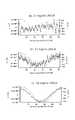
- FIG. 11is a graph showing% DHI and elapsed time obtained from this simple X-ray photograph.
- the horizontal axisrepresents elapsed time (week after operation), and the vertical axis represents% DHI.
- % DHIdecreased until 7 days after the operation. However, the level of the decrease stopped when LASCol was significantly higher than the control. In addition, there was no significant difference between the LASCol administration group and the AC administration group, or between the AC administration group and the control group. Thereafter,% DHI in the LASCol administration group, the AC administration group, and the control group all showed a decreasing tendency.
- LASColAs samples having different concentrations, LASCol of 7 mg / ml, 14 mg / ml, 21 mg / ml, and 42 mg / ml were prepared. For comparison, 7 mg / ml atelocollagen and a control solvent were also prepared.
- Rats using 7 mg / ml concentration of LASColwere “7 mg LASCol administration group”, rats using 14 mg / ml concentration of LASCol were “14 mg LASCol administration group”, and rats using LASCol of 21 mg / ml concentration were “21 mg LASCol administration group”
- the rats using LASCol at a concentration of 42 mg / mlare referred to as “42 mg LASCol administration group”.
- a rat given 7 mg / ml atelocollagenis used as a 7 mg AC administration group.
- the horizontal axisis the elapsed time after surgery (week after surgery), and the vertical axis is% DHI.
- the number of rats used in each groupis shown in FIG.
- the 42 mg LASCol-administered group, 21 mg LASCol-administered group, and 7 mg AC-administered groupshowed significantly higher% DHI values than the control. Therefore, it can be said that LASCol at least 21 mg / ml or more has intervertebral maintenance ability alone.
- OP-1Ostogenic Protein-1
- bFGFbFGF
- TGF- ⁇ 1, GDF-5, BMP2, VEGF, IGF-1, and the likemay be used as growth factors.
- the concentration of OP-1was 2 ⁇ g of OP-1 mixed with 15 ⁇ l of LASCol at a concentration of 21 mg / ml. This is called the OP-1 + LASCol administration group.
- FIG. 13 (a)shows the% DHI results for the OP-1 + LASCol administration group, the 21 mg LASCol administration group, the 7 mg AC administration group, and the control.
- the horizontal axisis the week after surgery, and the vertical axis is% DHI.
- the number of rats used in each groupis shown in FIG.
- LASColat least 21 mg / ml or more has intervertebral intervertebral maintenance ability equal to or higher than that of atelocollagen.
- LASColis preferably at a concentration of 21 mg / ml or more.
- the concentration of 42 mg / mlis close to the upper limit as the handling concentration.
- higher concentrations including the dry statecan be used by devising the administration method and form.
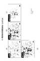
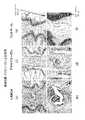
- FIG. 14is a photograph of a tissue sample of rat nucleus pulposus nucleus region 1 week after surgery stained with safranin-O. More specifically, the rat's tail intervertebral disc and the caudal vertebra on both sides of the intervertebral disc were removed, fixed in formalin, embedded in paraffin, a cut surface was prepared, and stained with safranin-O. In safranin-O, proteoglycan, which is a typical extracellular matrix, is stained red.
- Each photograph in FIG. 14is a black and white image processed from a stained photograph. Both photographs have caudal vertebrae on the left and right, and a sample between the caudal vertebrae.
- FIG. 14 (e)shows the caudal vertebra and nucleus pulposus region. The same applies to other photographs and the subsequent photographs shown in FIGS.
- FIGS. 14 (a) and 14 (b)show the case where the nucleus pulposus nucleus (referring to the part where the nucleus pulposus was present) was filled with LASCol at a concentration of 21 mg / ml after excision of the rat caudal nucleus pulposus. (This is the LASCol administration group.) 14 (c) and 14 (d) show the case where the nucleus pulposus region is filled with atelocollagen at a concentration of 7 mg / ml (this is an AC administration group). FIGS. 14 (e) and 14 (f) show a case where the nucleus pulposus region is filled with a solvent (in FIG. 14, “control” is indicated).
- FIGS. 14 (a) and 14 (b), FIG. 14 (c) and FIG. 14 (d), FIG. 14 (e) and FIG.It is a photograph of the same part with a different magnification.
- the scale baris shown in FIG. 14 (e) and FIG. 14 (f). The same applies to other photographs and the subsequent photographs shown in FIGS.
- FIG. 16, and FIG. 17are photographs of tissue samples of rat caudal nucleus pulposus stained with safranin-O at 2 weeks after surgery, 4 weeks after surgery, and 8 weeks after surgery, respectively.
- (a) and (b)show a case where the nucleus pulposus region is filled with LASCol after excision of rat nucleus pulposus nucleus.
- (C) and (d)show the case where the nucleus pulposus region is filled with atelocollagen.
- E) and (f)show the case where the nucleus pulposus region is filled with a solvent (in each figure, indicated as control).
- the nucleus pulposus regionwas darkly stained red in the case of LASCol administration. This indicates that proteoglycan is abundant. Cell infiltration into the nucleus pulposus region was also confirmed.
- FIG. 17 (c) and FIG. 17 (d)a red stained portion was observed in the nucleus pulposus region even when atelocollagen was administered.
- the color tonewas lighter than in the case of LASCol administration in FIGS. 17 (a) and 17 (b), and very few cells were infiltrated.
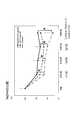
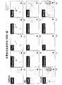
- FIG. 18shows the result of more quantitative examination of FIGS. 14 to 17.
- the horizontal axisrepresents the week after the operation, and the vertical axis represents the area ( ⁇ 10 3 ⁇ m 2 ) of the red portion showing positive proteoglycan.
- the LASCol administration group, the AC administration group, and the control groupare shown.
- the LASCol administration groupwas represented by “L”
- the atelocollagen administration groupwas represented by “AC”
- the control groupwas represented by “Cont”.
- the LASCol administration groupwas significantly larger in area than the other two groups, and the atelocollagen administration group was significantly larger than the control group. In addition, no significant change was observed in any of the administration groups from 1 week to 8 weeks after the operation.
- FIG. 19shows the results of counting the number of cells infiltrating the nucleus pulposus region in FIGS. 14 to 17.
- the horizontal axisis the week after the operation, and the vertical axis is the number of cells per intervertebral disc infiltrating the nucleus pulposus region (number / disc).
- the LASCol administration group, the AC administration group, and the control groupare shown.
- the LASCol administration groupwas represented by “L”
- the atelocollagen administration groupwas represented by “AC”
- the control groupwas represented by “Cont”.
- the average number of cells in the LASCol administration groupwas 66.3 ⁇ 9.4, the atelocollagen administration group was 20.4 ⁇ 7.1, and the control group was 2.3 ⁇ 2.2.
- the control groupwas 2.3 ⁇ 2.2.
- theywere 62.4 ⁇ 17.4, 19.2 ⁇ 5.6, and 1.0 ⁇ 0.7, respectively.
- theywere 77.8 ⁇ 23.2, 23.3 ⁇ 5.3, and 2.0 ⁇ 2.2, respectively.
- the LASCol administration grouphad significantly more cells than the other two groups, and the atelocollagen administration group was significantly more than the control group. In addition, no significant change was observed in any group from 1 week to 8 weeks after the operation.
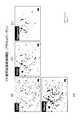
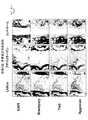
- FIG. 20is a photograph of a black and white processed image obtained by multiple fluorescent immunostaining of the nucleus pulposus region 1 week after the operation. Multiple fluorescence immunization was performed with DAPI, Brachyury, Tie2 and Aggrecan. More specifically, the rat intervertebral disc and the caudal vertebra on both sides of the disc were taken out, fixed with formalin, and embedded in paraffin to create a cut surface. Next, it was deparaffinized using xylene, activated by a warm bath method using a buffer, and blocked with hydrogen peroxide. Then, a primary antibody reaction was carried out with each marker, a labeled secondary antibody reaction was carried out for each animal species, and encapsulated.
- FIG. 21, FIG. 22, and FIG. 23show the results of black and white processing of images obtained by multiple fluorescent immunostaining of the nucleus pulposus area at 2 weeks after surgery, 4 weeks after surgery, and 8 weeks after surgery, respectively.
- the results of multiple fluorescent immunostainingshowed the same tendency as in FIG.
- FIG. 24shows photographs of the results of staining of the nucleus pulposus with safranin-O immediately after surgery, 3 days after surgery, and 1 week after surgery.
- 24 (a) to 24 (c)show the LASCol administration group (denoted “LASCol”), and FIGS. 24 (d) to 24 (f) represent the atelocollagen administration group (“AC”).
- 24 (g) to 24 (i)show a control group (denoted as “Control”).
- LASCola LASCol gel stained in green was observed in the nucleus pulposus region immediately after the operation. On the 3rd day and 1 week after the operation, numerous cell infiltrations were observed in the gel.
- a green collagen gelwas observed in the nucleus pulposus region one week after the operation, but only a few cells were observed on the gel surface 3 days and 1 week after the operation.
- the nucleus pulposus areawas void immediately after surgery and 3 days after surgery. One week after the operation, the nucleus pulposus region was in a collapsed state and no cell infiltration was observed. Thus, LASCol showed cell infiltration 3 days after administration.
- 25 to 30show photographs of multiple fluorescent immunostaining of the nucleus pulposus region.
- 25shows the case immediately after the operation
- FIG. 26shows the case 3 days after the operation
- FIGS. 27 to 29show the case 1 week after the operation.
- the LASCol administration groupwas described as “LASCol”, the atelocollagen administration group as “AC”, and the control group as “Control”.
- FIG. 25(immediately after the operation).
- 25 (a) to 25 (d)are LASCol administration groups
- FIGS. 25 (e) to 25 (h)are atelocollagen administration groups
- FIGS. 25 (i) to 25 (l)are control groups.
- the nucleus pulposus regionhad an area that was darkly stained with Col1, indicating the presence of each injected collagen gel. Moreover, Col2 was negative for both LASCol and atelocollagen. In addition, in all three groups, there was almost no portion stained by staining with DAPI, and it was found that nucleus pulposus cells were almost removed.
- FIG. 263 days after surgery.
- 26 (a) to 26 (d)are LASCol administration groups
- FIGS. 26 (e) to 26 (h)are atelocollagen administration groups
- FIGS. 26 (i) to 26 (l)are control groups.
- FIG. 27(1 week after operation).
- 27 (a) to 27 (d)are LASCol administration groups
- FIGS. 27 (e) to 27 (h)are atelocollagen administration groups
- FIGS. 27 (i) to 27 (l)are control groups.
- FIG. 28(1 week after operation).
- 28 (a) to 28 (e)are the LASCol administration group
- FIGS. 28 (f) to 28 (j)are the atelocollagen administration group
- FIGS. 28 (k) to 28 (o)are the control group.
- LASCol administration groupcell aggregation was observed with DAPI. A small amount of Brachyury and Tie2 positive cells were observed in the nucleus pulposus region. In the atelocollagen administration group, cells considered to be residual cells were observed, but both Brachyury and Tie2 were negative. In the control group, the nucleus pulposus region was crushed by compression of the annulus fibrosus region.
- the therapeutic agent used for the treatment of intervertebral disc degeneration according to the present inventioncan regenerate the nucleus pulposus by filling the nucleus pulposus region from which nucleus pulposus cells have flowed out.
- the single substancehas intervertebral maintenance ability at a concentration of 21 mg / ml or more. Accordingly, the nucleus pulposus can be regenerated while maintaining the intervertebral distance. If the concentration is at least 3.5 mg / ml or more, LASCol exhibits a gel form, and the same therapeutic effect can be expected by using it together with other auxiliary substances having intervertebral maintenance ability.
- This therapeutic agentis safe because it does not use living cells or donor nucleus pulposus cells.
- the intervertebral disc cell (nucleus nucleus cells and / or annulus cells) culture materialpromotes spheroids of nucleus pulposus cells and annulus cells, and also cultures spheroids having a large number of spheroids. can do.
- the therapeutic agent for intervertebral disc degeneration according to the present inventioncan be suitably used for the treatment of intervertebral disc degeneration.
- the disc material culture medium according to the present inventioncan culture nucleus pulposus cells and / or annulus fibrosus cells.
Landscapes
- Health & Medical Sciences (AREA)
- Life Sciences & Earth Sciences (AREA)
- Chemical & Material Sciences (AREA)
- Engineering & Computer Science (AREA)
- General Health & Medical Sciences (AREA)
- Medicinal Chemistry (AREA)
- Organic Chemistry (AREA)
- Bioinformatics & Cheminformatics (AREA)
- Zoology (AREA)
- Biomedical Technology (AREA)
- Veterinary Medicine (AREA)
- Public Health (AREA)
- Animal Behavior & Ethology (AREA)
- Genetics & Genomics (AREA)
- Epidemiology (AREA)
- Biotechnology (AREA)
- Wood Science & Technology (AREA)
- Transplantation (AREA)
- Dermatology (AREA)
- Oral & Maxillofacial Surgery (AREA)
- Biochemistry (AREA)
- Rheumatology (AREA)
- Biophysics (AREA)
- Proteomics, Peptides & Aminoacids (AREA)
- Molecular Biology (AREA)
- Gastroenterology & Hepatology (AREA)
- General Engineering & Computer Science (AREA)
- Microbiology (AREA)
- Pharmacology & Pharmacy (AREA)
- Toxicology (AREA)
- Cell Biology (AREA)
- Nuclear Medicine, Radiotherapy & Molecular Imaging (AREA)
- General Chemical & Material Sciences (AREA)
- Physical Education & Sports Medicine (AREA)
- Chemical Kinetics & Catalysis (AREA)
- Dispersion Chemistry (AREA)
- Sustainable Development (AREA)
- Immunology (AREA)
- Orthopedic Medicine & Surgery (AREA)
- Peptides Or Proteins (AREA)
Abstract
Description
Translated fromJapanese本発明は、脊椎椎間板変性を治療する際に用いる治療剤およびその方法に関するものである。また、本発明は、椎間板細胞培養材も含む。The present invention relates to a therapeutic agent and a method for use in treating spinal disc degeneration. The present invention also includes an intervertebral disc cell culture material.
ヒトの脊椎は、椎体、椎弓および棘突起等で構成される椎骨が縦に連なる。椎骨間には、クッション材として椎間板が配置され、脊椎全体の屈曲、伸展、回旋といった動作を可能にしている。椎間板は、線維輪と呼ばれる層状をなす環状の線維と、その中にあるコラーゲンとプロテオグリカンを産生する軟骨細胞を含むゼリー状の髄核で構成されている。In the human spine, vertebrae composed of vertebral bodies, vertebral arches, spinous processes and the like are vertically connected. An intervertebral disc is disposed as a cushioning material between the vertebrae, and the entire spine can be bent, extended, and rotated. The intervertebral disc is composed of a layered annular fiber called annulus fibrosus and a jelly-like nucleus pulposus that contains chondrocytes that produce collagen and proteoglycans.
この椎間板に於いて何らかの原因で髄核と線維輪の正常な位置関係が破綻した状態のことを椎間板ヘルニアと呼ぶ。正常なクッション性が失われることで背部痛などの局所痛を生じる。さらに脱出した髄核が近傍の神経組織を圧迫すると、神経支配領域への放散痛を生じ、坐骨神経痛がその代表的な症状として挙げられる。In this intervertebral disc, the normal positional relationship between the nucleus pulposus and the annulus fibrosus for some reason is called intervertebral disc herniation. Loss of normal cushioning causes local pain such as back pain. Further, when the prolapsed nucleus pulposus presses the nearby nerve tissue, it causes radiation pain to the innervation region, and sciatica is a typical symptom.
例えば、椎間板ヘルニアを治療する際には、外科的に漏れ出た髄核を取り除く手術療法がある。しかし、手術療法後は髄核の損失により椎骨間は狭まり、継時的には脊柱の加齢性変化、すなわち変性が進行するとされている。For example, when treating a herniated disc, there is a surgical treatment that removes the nucleus pulposus that has leaked surgically. However, after the surgical treatment, the vertebrae are narrowed due to loss of the nucleus pulposus, and it is said that the age-related changes of the spinal column, that is, degeneration progresses over time.
椎骨間距離が縮まると衝撃吸収性が減少して局所痛を誘発する。また、脊柱の安定性が減少することで椎骨のずれを生じて脊柱管狭窄が起こり、坐骨神経痛などの神経障害を来すようになる。したがって、椎間板を維持できる充填剤が長く求められた。しかし、単に充填するだけでは、その充填剤が飛び出し、ヘルニアと同様の障害が再発する。そこで、髄核があった部分に定着して組織を再生することで椎骨間距離を長期に渡って維持することが期待できるような充填剤が切望された。When the intervertebral distance is reduced, shock absorption is reduced and local pain is induced. In addition, a decrease in the stability of the spinal column causes a vertebral shift, resulting in stenosis of the spinal canal, resulting in neuropathy such as sciatica. Therefore, a long-standing filler that can maintain the intervertebral disc has been sought. However, simply filling will cause the filler to pop out and cause the same failure as hernia. Therefore, a filler that can be expected to maintain the intervertebral distance for a long period of time by regenerating the tissue by fixing it in the part where the nucleus pulposus was present was desired.
髄核の細胞外マトリックスは主にアグリカンとII型コラーゲンで、構成する細胞は脊索由来である。線維輪の細胞外マトリックスは主にアグリカンとI型コラーゲンで、構成する細胞は間葉由来である。そこで、傷んだ髄核の代用材としてコラーゲンを使うということが従来から行われていた(特許文献1)。The extracellular matrix of nucleus pulposus is mainly aggrecan and type II collagen, and the constituent cells are derived from notochord. The extracellular matrix of the annulus is mainly aggrecan and type I collagen, and the constituent cells are derived from mesenchyme. Therefore, it has been conventionally performed to use collagen as a substitute for damaged nucleus pulposus (Patent Document 1).
一方、特許文献2には、変性椎間板疾患を有する患者を治療するためのマトリックスであって:光酸化触媒反応および可視光線の照射によって架橋された消化耐性で改造可能なコラーゲンを含有する、注射可能な流体;および変性椎間板疾患を治療するための注射可能な細胞マトリックスを形成するように前記注射可能な流体内に分散された、生体内でプロテオグリカンを合成する固有の能力を有する複数の生細胞;を含んで成るマトリックスが開示されている。特許文献2の発明は、単に椎骨間の距離を維持するためでなく、髄核細胞の再生を促すものである。
コラーゲンは生体親和性があり、また入手も容易な材料である。コラーゲンには多くのタイプがあることが知られている。コラーゲンは、α鎖の3重らせん構造を有している。特許文献3には、所定の酵素によりこのα鎖の末端を切断することにより作製した低接着性コラーゲン(Low Adhesive Scaffold Collagen:以下「LASCol」と呼ぶ。)が記載されている。また、LASColは、細胞培養のための足場の材料として知られている(特許文献4)。Collagen is a material that is biocompatible and easily available. It is known that there are many types of collagen. Collagen has a triple helical structure of α chain.
従来のコラーゲンを用いている足場と比べて、LASColを用いている足場を利用することにより、培養したい細胞は凝集塊(スフェロイド)を形成し、培養したい細胞をより生体内に近い三次元的な状態で培養することができる。また、このLASColは、幹細胞の分化誘導の促進に効果がある(特許文献4)。Compared to a scaffold using conventional collagen, by using a scaffold using LASCol, cells to be cultured form aggregates (spheroids), and the cells to be cultured are three-dimensionally closer to the living body. It can be cultured in a state. This LASCol is effective in promoting differentiation induction of stem cells (Patent Document 4).
特許文献2では、髄核の代わりに注入するマトリックスはドナー脊椎動物から得られるとされ、例えばブタの脊柱の椎間板から無菌で切除された髄核組織である。また、このマトリックスにはプロテオグリカンを産生する生細胞も混入されている。ドナーからの髄核組織はドナーの有していたウイルスや他の何らかのコンタミネーションの危険性が否定できない。また、種の異なる生細胞の導入も移植を受けたヒトにどのような影響があるか、不明な点が多い。In
一方、特許文献1は、生体親和性があり、人体に使用された実績もある材料であるコラーゲンが使用されるが、髄核細胞の再生という効果はない。したがって、髄核の代わりに注入される充填材として、より安全で、髄核細胞の再生が可能なものが望まれていた。On the other hand,
本発明は上記の課題に鑑みて想到されたものであり、椎間板変性の治療のために、線維輪中に髄核の代わりに注入される組成物(椎間板変性の治療剤)を提供するものである。ここで、椎間板変性にはヘルニアを含む。より具体的には、LASColを含む椎間板変性の治療に用いる治療剤である。なお、本発明はLASColを用いた椎間板変性の治療方法といってもよい。The present invention has been conceived in view of the above problems, and provides a composition (a therapeutic agent for intervertebral disc degeneration) that is injected into the annulus instead of the nucleus pulposus for the treatment of intervertebral disc degeneration. is there. Here, disc degeneration includes hernia. More specifically, it is a therapeutic agent used for the treatment of intervertebral disc degeneration including LASCol. The present invention may be referred to as a method for treating disc degeneration using LASCol.
また、本発明はLASColを含む椎間板細胞培養材も提供する。ここで椎間板細胞培養材とは、髄核細胞及び/又は線維輪細胞を培養することのできる培養材である。また、本発明は、治療剤および培養材の製造方法、椎間板変性の治療剤を用いたヒト以外の動物への治療方法、および治療剤によって再生された髄核細胞および線維輪細胞をも提供する。The present invention also provides an intervertebral disc cell culture material containing LASCol. Here, the disc cell culture material is a culture material capable of culturing nucleus pulposus cells and / or annulus fibrosus cells. The present invention also provides a method for producing a therapeutic agent and a culture material, a method for treating non-human animals using a therapeutic agent for intervertebral disc degeneration, and nucleus pulposus cells and annulus fibrosus cells regenerated by the therapeutic agent. .
本発明に係る椎間板変性の治療剤は、LASColを含有し、髄核の抜けた椎骨間の距離を長期に渡って維持することができる。LASCol自体は椎間板に生来存在するコラーゲンを材料としており椎間板での親和性が高く、高い安全性に寄与する。さらに、LASColは、周囲の細胞から髄核の成分であるプロテオグリカンを産生する細胞を遊走・浸潤することができ、外部から髄核細胞や生細胞を注入しなくても、髄核が再生するという効果を得ることができる。The therapeutic agent for intervertebral disc degeneration according to the present invention contains LASCol, and can maintain the distance between vertebrae from which the nucleus pulposus has been removed over a long period of time. LASCol itself is made of collagen that is naturally present in the intervertebral disc, and has a high affinity with the intervertebral disc, contributing to high safety. Furthermore, LASCol can migrate and infiltrate cells that produce proteoglycan, a component of the nucleus pulposus, from surrounding cells, and the nucleus pulposus regenerates without injecting nucleus pulposus cells or living cells from the outside. An effect can be obtained.
以下に本発明に係る椎間板変性の治療剤および椎間板細胞培養材について図面および実施例を示し説明を行う。なお、以下の説明は、本発明の一実施形態および一実施例を例示するものであり、本発明が以下の説明に限定されるものではない。以下の説明は本発明の趣旨を逸脱しない範囲で改変することができる。Hereinafter, the therapeutic agent for disc degeneration and the disc cell culture material according to the present invention will be described with reference to the drawings and examples. The following description exemplifies an embodiment and an example of the present invention, and the present invention is not limited to the following description. The following description can be modified without departing from the spirit of the present invention.
本発明に係る椎間板変性の治療剤および椎間板細胞培養材として用いられるLASColは、コラーゲンまたはアテロコラーゲンの分解物を含む。この分解物は、コラーゲンが持つ細胞との接着性が弱くなり、低接着性に変わる性質を有する。また、本発明に係る椎間板変性の治療剤としては、ハイドロゲル、ゼラチンゲル、キトサンゲル、ヒアルロン酸・コラーゲンハイドロゲル、ヒアルロン酸ポリマー、ヒアルロン酸・PEGポリマー、コラーゲン・ヒアルロン酸・PEGハイドロゲル、高純度アルギン酸ゲル(UPAL)といった物質や、人体親和性のある溶媒(これらをまとめて「補助物質」と呼ぶ。)を含ませてもよい。もちろん、LASColのみであってもよい。また、緩衝液、pH調製液、塩、細胞成長因子を加えることもできる。LASCol used as a therapeutic agent for intervertebral disc degeneration and a disc cell culture material according to the present invention contains collagen or a degradation product of atelocollagen. This degradation product has the property that collagen has weak adhesion to cells and changes to low adhesion. The therapeutic agents for intervertebral disc degeneration according to the present invention include hydrogel, gelatin gel, chitosan gel, hyaluronic acid / collagen hydrogel, hyaluronic acid polymer, hyaluronic acid / PEG polymer, collagen / hyaluronic acid / PEG hydrogel, high A substance such as a pure alginic acid gel (UPAL) or a solvent compatible with the human body (collectively referred to as “auxiliary substance”) may be included. Of course, only LASCol may be used. Further, a buffer solution, a pH adjusting solution, a salt, and a cell growth factor can be added.
LASColは、コラーゲンまたはアテロコラーゲンを酵素で分解することによって得られる。そして、分解する際の条件によって、含まれるペプチド配列が異なる。すなわち、LASColは、分解の条件によって、異なる種類のLASColが得られる。LASCol is obtained by degrading collagen or atelocollagen with an enzyme. And the peptide sequence contained differs according to the conditions at the time of decomposition | disassembly. That is, different types of LASCol can be obtained depending on the decomposition conditions.
本発明で利用できるLASColの特徴は、コラーゲンまたはアテロコラーゲンのトリプルヘリカルドメインの下記(A:配列番号1)に示されるアミノ末端のアミノ酸配列において、Y1とY2の間の化学結合が切断されているα鎖の組み合わせからなる点にある。
(A)-Y1-Y2-Y3-G-Y4-Y5-G-Y6-Y7-G-Y8-Y9-G-(配列番号1):
(但し、Gは、グリシンであり、Y1~Y9は、任意のアミノ酸である)。The characteristic of LASCol that can be used in the present invention is that the chemical bond between Y1 and Y2 is cleaved in the amino terminal amino acid sequence shown in the following (A: SEQ ID NO: 1) of the triple helical domain of collagen or atelocollagen. The combination of α chains.
(A) -Y1 -Y2 -Y3 -GY4 -Y5 -GY6 -Y7 -GY8 -Y9 -G- (SEQ ID NO: 1):
(However, G is glycine, and Y1 to Y9 are arbitrary amino acids).
コラーゲンのトリプルヘリカルドメインは、-G-X-Y-(Gはグリシンで、XおよびYは任意のアミノ酸)という配列が連続していることが知られている。上記の配列では、「-Y3-G-Y4-Y5-」中の「G」がトリプルヘリカルドメインのN末端側のグリシンを表している。上記の配列をみてわかるように、Y1とY2との間の化学結合の切断とは、トリプルヘリカルドメインの外側で切断が行われていることがわかる。後述するように、分解条件が異なるとトリプルヘリカルドメインの内側で切断が生じる。本発明で用いられるLASColの1つは、トリプルヘリカルドメインの外側で切断が生じているLASColである。以下これをLASCol-Aと呼ぶ。The triple helical domain of collagen is known to have a sequence of -GXY- (G is glycine, and X and Y are arbitrary amino acids). In the above sequence, “G” in “—Y3 -GY4 -Y5 —” represents glycine on the N-terminal side of the triple helical domain. As can be seen from the above sequence, it is understood that the cleavage of the chemical bond between Y1 and Y2 is performed outside the triple helical domain. As will be described later, when the decomposition conditions are different, cleavage occurs inside the triple helical domain. One of the LASCols used in the present invention is LASCol in which cleavage occurs outside the triple helical domain. Hereinafter, this is referred to as LASCol-A.
なお、分解の条件によって、以下のLASColが得られることが知られている。コラーゲンまたはアテロコラーゲンのトリプルヘリカルドメインの下記(B:配列番号2)に示されるアミノ末端のアミノ酸配列においてX1とX2との間の化学結合、X2とGとの間の化学結合、GとX3との間の化学結合、X4とGとの間の化学結合若しくはX6とGとの間の化学結合が切断されているα鎖の組み合わせからなる。It is known that the following LASCol can be obtained depending on the decomposition conditions. A chemical bond between X1 and X2 in the amino terminal amino acid sequence shown below (B: SEQ ID NO: 2) of the triple helical domain of collagen or atelocollagen; a chemical bond between X2 and G; It consists of a combination of α chains in which the chemical bond between X3 , the chemical bond between X4 and G, or the chemical bond between X6 and G is cleaved.
(B)-G-X1-X2-G-X3-X4-G-X5-X6-G-(配列番号2):(但し、Gは、グリシンであり、X1~X6は、任意のアミノ酸である)。これをLASCol-Bと呼ぶ。LASCol-Bは、トリプルヘリカルドメインの内側で切断が生じている。配列番号2番では「-G-X1-X2-G-」のGがトリプルヘリカルドメインのN末端側のグリシンである。もちろん、他のペプチドが含まれるLASColもあり得る。椎間板変性の治療剤としてLASCol-Aは、現在知られているLASCol中で最も好適である。しかし、他のLASColを排除するものではない。(B) -GX1 -X2 -GX3 -X4 -GX5 -X6 -G- (SEQ ID NO: 2): (where G is glycine, and X1 to X6 is any amino acid). This is called LASCol-B. LASCol-B is cleaved inside the triple helical domain. In SEQ ID NO: 2, G in “-GX1 -X2 -G-” is glycine on the N-terminal side of the triple helical domain. Of course, there may be LASCol containing other peptides. LASCol-A is the most suitable among currently known LASCols as a therapeutic agent for disc degeneration. However, it does not exclude other LASCols.
本発明に係る椎間板変性の治療剤として利用されるLASColは、酸性状態で溶液として保存が可能である。そして、pHおよび濃度を調整し、体温まで温度を上げることで、ゲル状となる。ゲル状になることで、LASColは、線維輪内での拡散が抑制され、細胞の誘導及び/又は細胞外基質の産生をもたらし、組織再生を達成することで椎骨間距離を維持するという効果(「椎骨間維持能」と呼ぶ。)を発揮する。LASCol used as a therapeutic agent for intervertebral disc degeneration according to the present invention can be stored as a solution in an acidic state. And it adjusts pH and a density | concentration, It becomes gel form by raising temperature to body temperature. By becoming a gel, LASCol suppresses diffusion in the annulus fibrosus, leads to cell induction and / or production of extracellular matrix, and maintains the intervertebral distance by achieving tissue regeneration ( It is called “intervertebral maintenance ability”.)
ゲル状となったときの弾性率は、溶液中のLASColの濃度およびpH、温度に比例する。後述する実施例では、pHおよび濃度を調整し、液体の状態でシリンジに吸引し、線維輪内に注射で投与し、線維輪内でゲル状にさせる例を示す。しかし、本発明に係る椎間板変性の治療剤として利用されるLASColは、膜状や、スポンジ状にして、患部に埋め込んでもよい。なお、膜状、スポンジ状とは、LASColを所定の形状に乾燥したもの(形状体ともいう。)をいう。The elastic modulus when the gel is formed is proportional to the concentration, pH, and temperature of LASCol in the solution. In the examples described later, an example is shown in which the pH and concentration are adjusted, sucked into a syringe in a liquid state, administered by injection into the annulus fibrosus, and gelled in the annulus fibrosus. However, LASCol used as a therapeutic agent for intervertebral disc degeneration according to the present invention may be in the form of a film or sponge and embedded in the affected area. Note that the film form and the sponge form refer to those obtained by drying LASCol into a predetermined shape (also referred to as a shape body).
また、椎間板変性の治療剤として、LASColを補助物質と共に利用してもよい。椎骨間維持能という機械的な強度は補助物質が担い、LASColは、周囲の細胞から髄核の成分であるプロテオグリカンを産生する細胞を遊走・浸潤し、髄核細胞を再生する役目を担うことで、椎間板変性の治療剤となるからである。Also, LASCol may be used together with an auxiliary substance as a therapeutic agent for disc degeneration. The mechanical strength of intervertebral maintenance is borne by auxiliary substances, and LASCol plays a role of regenerating nucleus pulposus cells by migrating and infiltrating cells that produce proteoglycan, which is a component of nucleus pulposus, from surrounding cells. This is because it becomes a therapeutic agent for disc degeneration.
後述するように本発明で用いるLASColは、濃度3.5mg/ml(後述する「実用弾性率」で20Pa)以上であれば、ゲル状を呈するといえる。したがって、この濃度以上のLASColは、補助物質と混ぜることで、髄核細胞を再生する椎間板変性の治療剤および椎間板細胞培養材を得ることができる。As will be described later, it can be said that LASCol used in the present invention exhibits a gel form when the concentration is 3.5 mg / ml (“Practical Elastic Modulus” described later is 20 Pa) or more. Therefore, LASCol at this concentration or higher can be mixed with an auxiliary substance to obtain a therapeutic agent for intervertebral disc degeneration that regenerates nucleus pulposus cells and an intervertebral disc cell culture material.
また、濃度が7mg/ml以上であれば単体で椎骨間維持能を有し、21mg/ml以上になれば、単体でもアテロコラーゲン以上の椎骨間維持能を有する。In addition, when the concentration is 7 mg / ml or more, it has intervertebral maintenance ability alone, and when it is 21 mg / ml or more, it has intervertebral maintenance ability more than atelocollagen.
したがって、本発明で用いることのできるLASColは、3.5mg/ml以上で利用でき、好ましくは7mg/ml以上、より好ましくは21mg/ml以上の濃度で用いることができる。なお、ゲルとしての濃度の上限は少なくとも42mg/ml以上であるが、それ以上の濃度であっても椎間板変性の治療剤として利用することができる。Therefore, LASCol which can be used in the present invention can be used at 3.5 mg / ml or more, preferably 7 mg / ml or more, more preferably 21 mg / ml or more. The upper limit of the gel concentration is at least 42 mg / ml or more, but even higher concentrations can be used as a therapeutic agent for intervertebral disc degeneration.
LASColの作製方法に関する知見としては、LASCol-BもLASCol-Aもほぼ同じである。そこで、どちらにも共通する知見については、単にLASColとして説明する。また、以下の説明で「分解物」とはLASColを意味する。As for the knowledge about the production method of LASCol, LASCol-B and LASCol-A are almost the same. Therefore, the knowledge common to both is described simply as LASCol. In the following description, “decomposed product” means LASCol.
<LASColの材料>
LASColの材料になるコラーゲンまたはアテロコラーゲンは、特に限定されず、周知のコラーゲンおよびアテロコラーゲンであればよい。<Materials of LASCol>
Collagen or atelocollagen used as the material for LASCol is not particularly limited, and may be known collagen or atelocollagen.
コラーゲンとしては、哺乳類(例えば、ウシ、ブタ、ウサギ、ヒト、ラットまたはマウスなど)、鳥類(例えば、ニワトリなど)、または、魚類(例えば、サメ、コイ、ウナギ、マグロ(例えば、キハダマグロ)、ティラピア、タイ、サケなど)または爬虫類(例えば、スッポン)のコラーゲンを用いることができる。Collagens include mammals (eg, cows, pigs, rabbits, humans, rats or mice), birds (eg, chickens), or fish (eg, shark, carp, eel, tuna (eg, yellowfin tuna), tilapia , Thailand, salmon, etc.) or reptile (eg, suppon) collagen.
本発明で用いるコラーゲンは、例えば、上記哺乳類または鳥類の真皮、腱、骨または筋膜などに由来するコラーゲン、上記魚類の皮膚または鱗などに由来するコラーゲン、上記爬虫類の真皮、腱、骨などに由来するコラーゲンを用いることができる。Collagens used in the present invention include, for example, collagen derived from the dermis, tendon, bone or fascia of mammals or birds, collagen derived from the skin or scales of fish, dermis, tendon, bone of the reptile, etc. Derived collagen can be used.
また、LASColの作製に用いるアテロコラーゲンとしては、上記哺乳類、鳥類、魚類または爬虫類のコラーゲンをプロテアーゼ(例えば、ペプシンなど)によって処理して得られる、コラーゲン分子のアミノ末端および/またはカルボキシル末端からテロペプチドが部分的に除去されているアテロコラーゲンを用いることができる。Moreover, as atelocollagen used for the preparation of LASCol, telopeptide is obtained from the amino terminus and / or carboxyl terminus of the collagen molecule obtained by treating the above-mentioned mammalian, avian, fish or reptile collagen with a protease (for example, pepsin). Atelocollagen that has been partially removed can be used.
これらの中では、ニワトリ、ブタ、ウシ、ヒトまたはラットのコラーゲンまたはアテロコラーゲンが好ましく用いることができる。また、ブタ、ウシまたはヒトのコラーゲンまたはアテロコラーゲンをLASColの材料として更に好ましく用いることができる。Among these, chicken, pig, cow, human or rat collagen or atelocollagen can be preferably used. Further, porcine, bovine or human collagen or atelocollagen can be more preferably used as a material for LASCol.
また、LASColの材料として魚類のコラーゲンまたはアテロコラーゲンを用いることもできる。魚類を用いれば、材料を簡便に、安全に、かつ大量に入手可能であり、ヒトに対してよりウイルスフリーの安全なコラーゲンまたはアテロコラーゲンの分解物(LASCol)を提供することができる。It is also possible to use fish collagen or atelocollagen as the LASCol material. If fish is used, the material can be obtained simply, safely and in large quantities, and a virus-free safe collagen or atelocollagen degradation product (LASCol) can be provided to humans.
なお、LASColの材料として魚類のコラーゲンまたはアテロコラーゲンを用いる場合には、サメ、コイ、ウナギ、マグロ(例えば、キハダマグロ)、ティラピア、ブラックバス、ブルーギル、タイまたはサケのコラーゲンまたはアテロコラーゲンを用いることが好ましく、マグロ、ティラピア、タイまたはサケのコラーゲンまたはアテロコラーゲンを用いることが更に好ましい。When fish collagen or atelocollagen is used as the LASCol material, it is preferable to use shark, carp, eel, tuna (eg, yellowfin tuna), tilapia, black bass, bluegill, Thai or salmon collagen or atelocollagen. More preferably, tuna, tilapia, Thai or salmon collagen or atelocollagen is used.
LASColの材料としてアテロコラーゲンを用いる場合、熱による変性温度が、好ましくは15℃以上、より好ましくは20℃以上であるアテロコラーゲンを用いることが好ましい。例えば、分解物の材料として魚類のアテロコラーゲンを用いる場合、マグロ(例えば、キハダマグロ)またはコイ、ティラピアなどのアテロコラーゲンは熱変性温度が25℃以上であるので、これらのアテロコラーゲンを用いることが好ましい。When using atelocollagen as the LASCol material, it is preferable to use atelocollagen having a heat denaturation temperature of preferably 15 ° C. or higher, more preferably 20 ° C. or higher. For example, when fish atelocollagen is used as a material for degradation products, telo (eg, yellowfin tuna) or atelocollagen such as carp and tilapia has a heat denaturation temperature of 25 ° C. or higher, and it is preferable to use these atelocollagens.
上記構成であれば、本実施の形態の椎間板変性の治療剤のゲル状になる温度を、好ましくは15℃以上、より好ましくは20℃以上に調節することができる。その結果、上記構成であれば、貯蔵時の安定性、利用時の安定性に優れた椎間板変性の治療剤を実現することができる。With the above configuration, the temperature at which the therapeutic agent for intervertebral disc degeneration of the present embodiment becomes a gel can be adjusted to preferably 15 ° C. or higher, more preferably 20 ° C. or higher. As a result, if it is the said structure, the therapeutic agent of the disc degeneration excellent in the stability at the time of storage and the stability at the time of utilization is realizable.
これらのコラーゲンまたはアテロコラーゲンは、周知の方法によって入手することができる。例えば、哺乳類、鳥類または魚類のコラーゲンに富んだ組織をpH2~4程度の酸性溶液に投入することによって、コラーゲンを溶出することができる。更に、当該溶出液にペプシンなどのプロテアーゼを添加して、コラーゲン分子のアミノ末端および/またはカルボキシル末端のテロペプチドを、部分的に除去する。更に、当該溶出液に塩化ナトリウムなどの塩を加えることによって、アテロコラーゲンを沈殿させることができる。These collagens or atelocollagen can be obtained by a known method. For example, collagen can be eluted by putting a tissue rich in collagen of mammals, birds or fish into an acidic solution of about pH 2-4. Further, a protease such as pepsin is added to the eluate to partially remove the amino terminal and / or carboxyl terminal telopeptide of the collagen molecule. Furthermore, atelocollagen can be precipitated by adding a salt such as sodium chloride to the eluate.
LASColを得るには、コラーゲンまたはアテロコラーゲンに酵素を作用させて、これらの材料を分解する。しかし、既にトリプルヘリカルドメイン内の化学結合が切断されているコラーゲンまたはアテロコラーゲンの分解物を作製する(例えば、化学合成法、組み換えタンパク質の発現)ことでLASColを得ることもできる。To obtain LASCol, these materials are decomposed by causing an enzyme to act on collagen or atelocollagen. However, LASCol can also be obtained by preparing a degradation product of collagen or atelocollagen in which the chemical bond in the triple helical domain has already been cleaved (for example, chemical synthesis, expression of recombinant protein).
以下に、上述したコラーゲンまたはアテロコラーゲンを酵素(例えば、プロテアーゼ)によって分解しLASColを得る方法について説明する。Hereinafter, a method for obtaining LASCol by degrading the aforementioned collagen or atelocollagen with an enzyme (for example, protease) will be described.
上記酵素としては特に限定されないが、例えば、システインプロテアーゼを用いることが好ましい。The enzyme is not particularly limited, but for example, cysteine protease is preferably used.
システインプロテアーゼとしては、塩基性アミノ酸量よりも酸性アミノ酸量の方が多いシステインプロテアーゼ、酸性領域の水素イオン濃度において活性であるシステインプロテアーゼを用いることが好ましい。As the cysteine protease, it is preferable to use a cysteine protease having a larger amount of acidic amino acids than a basic amino acid amount, or a cysteine protease active at a hydrogen ion concentration in an acidic region.
このようなシステインプロテアーゼとしては、アクチニダイン[EC 3.4.22.14]、パパイン[EC 3.4.22.2]、フィシン[EC 3.4.22.3]、ブロメライン[EC 3.4.22.32]、カテプシンB[EC 3.4.22.1]、カテプシンL[EC 3.4.22.15]、カテプシンS[EC 3.4.22.27]、カテプシンK[EC 3.4.22.38]、カテプシンH[EC 3.4.22.16]、アロライン、カルシウム依存性プロテアーゼなどを挙げることが可能である。なお、鍵括弧内は酵素番号である。Such cysteine proteases include actinidin [EC 3.4.22.14], papain [EC 3.4.22.2], ficin [EC 3.4.22.3], bromelain [EC 3.4. .22.32], cathepsin B [EC 3.4.22.1], cathepsin L [EC 3.4.22.15], cathepsin S [EC 3.4.22.27], cathepsin K [
これらの中では、アクチニダイン、パパイン、フィシン、カテプシンK、アロラインまたはブロメラインを用いることが好ましく、アクチニダイン、パパイン、フィシン、カテプシンKを用いることが更に好ましい。Among these, it is preferable to use actinidine, papain, ficin, cathepsin K, alloline or bromelain, and it is more preferable to use actinidine, papain, ficin, cathepsin K.
上述した酵素は、公知の方法によって入手することができる。例えば、化学合成による酵素の作製;細菌、真菌、各種動植物の細胞または組織からの酵素の抽出;遺伝子工学的手段による酵素の作製;などによって入手することができる。勿論、市販の酵素を用いることも可能である。The enzyme described above can be obtained by a known method. For example, it can be obtained by preparation of an enzyme by chemical synthesis; extraction of an enzyme from cells or tissues of bacteria, fungi, various animals and plants; preparation of an enzyme by genetic engineering means; Of course, commercially available enzymes can also be used.
コラーゲンまたはアテロコラーゲンを酵素(例えば、プロテアーゼ)によって分解することによって切断を行う場合には、例えば、以下の(i)~(iii)の方法にしたがって切断工程を行うことができる。以下の(i)~(iii)の方法は、あくまでも切断工程の一例であって、LASColの製造方法は、これら(i)~(iii)の方法に限定されない。When cutting by degrading collagen or atelocollagen with an enzyme (for example, protease), for example, the cutting step can be performed according to the following methods (i) to (iii). The following methods (i) to (iii) are merely examples of the cutting step, and the production method of LASCol is not limited to these methods (i) to (iii).
なお、以下の(i)および(ii)の方法で、LASCol-Bを得ることができる。また、以下の(iii)の方法は、LASCol-AとLASCol-Bを得ることができる。
(i)高濃度の塩の存在下にて、コラーゲンまたはアテロコラーゲンと、酵素とを接触させる方法。
(ii)高濃度の塩と接触させた後の酵素と、コラーゲンまたはアテロコラーゲンとを接触させる方法。
(iii)低濃度の塩の存在下にて、コラーゲンまたはアテロコラーゲンと、酵素とを接触させる方法。Note that LASCol-B can be obtained by the following methods (i) and (ii). In addition, the following method (iii) can obtain LASCol-A and LASCol-B.
(I) A method of bringing collagen or atelocollagen into contact with an enzyme in the presence of a high concentration of salt.
(Ii) A method in which an enzyme after contact with a high concentration salt is contacted with collagen or atelocollagen.
(Iii) A method of bringing collagen or atelocollagen into contact with an enzyme in the presence of a low concentration of salt.
上述した(i)の方法の具体例としては、例えば、高濃度の塩を含む水溶液中で、コラーゲンまたはアテロコラーゲンと、酵素とを接触させる方法を挙げることができる。Specific examples of the method (i) described above include a method of bringing collagen or atelocollagen into contact with an enzyme in an aqueous solution containing a high concentration of salt.
上述した(ii)の方法の具体例としては、例えば、高濃度の塩を含む水溶液と酵素とを予め接触させ、その後、当該酵素と、コラーゲンまたはアテロコラーゲンとを接触させる方法を挙げることができる。Specific examples of the method (ii) described above include, for example, a method in which an aqueous solution containing a high concentration salt and an enzyme are contacted in advance, and then the enzyme is contacted with collagen or atelocollagen.
上述した(iii)の方法の具体例としては、例えば、低濃度の塩を含む水溶液中で、コラーゲンまたはアテロコラーゲンと、酵素とを接触させる方法を挙げることができる。上記水溶液の具体的な構成としては特に限定されないが、例えば、水を用いることが可能である。Specific examples of the method (iii) described above include a method of bringing collagen or atelocollagen into contact with an enzyme in an aqueous solution containing a low concentration salt. Although it does not specifically limit as a specific structure of the said aqueous solution, For example, it is possible to use water.
上記塩の具体的な構成としては特に限定されないが、塩化物を用いることが好ましい。塩化物としては、特に限定されないが、例えば、NaCl、KCl、LiClまたはMgCl2を用いることが可能である。The specific structure of the salt is not particularly limited, but chloride is preferably used. The chloride is not particularly limited, but for example, it is possible to use NaCl, KCl, and LiCl or MgCl2.
上記高濃度の塩を含む水溶液における塩の濃度は特に限定されないが、高いほど好ましいといえる。例えば、当該濃度は、200mM以上であることが好ましく、500mM以上であることがより好ましく、1000mM以上であることがより好ましく、1500mM以上であることがより好ましく、2000mM以上であることが最も好ましい。The concentration of the salt in the aqueous solution containing the high concentration salt is not particularly limited, but it can be said that a higher concentration is preferable. For example, the concentration is preferably 200 mM or higher, more preferably 500 mM or higher, more preferably 1000 mM or higher, more preferably 1500 mM or higher, and most preferably 2000 mM or higher.
上記低濃度の塩を含む水溶液における塩の濃度は特に限定されないが、低いほど好ましいといえる。例えば、当該濃度は、200mMよりも低いことが好ましく、150mM以下であることがより好ましく、100mM以下であることがより好ましく、50mM以下であることがより好ましく、略0mMであることが最も好ましい。The concentration of the salt in the aqueous solution containing the low-concentration salt is not particularly limited. For example, the concentration is preferably lower than 200 mM, more preferably 150 mM or less, more preferably 100 mM or less, more preferably 50 mM or less, and most preferably about 0 mM.
上記水溶液(例えば、水)に溶解させるコラーゲンまたはアテロコラーゲンの量は特に限定されないが、例えば、1000重量部~10000重量部の水溶液に対して、1重量部のコラーゲンまたはアテロコラーゲンを溶解させることが好ましい。The amount of collagen or atelocollagen dissolved in the aqueous solution (for example, water) is not particularly limited. For example, it is preferable to dissolve 1 part by weight of collagen or atelocollagen in an aqueous solution of 1000 parts by weight to 10,000 parts by weight.
上記構成であれば、水溶液に対して酵素が加えられた場合、当該酵素とコラーゲンまたはアテロコラーゲンとを効率よく接触させることができる。そして、その結果、コラーゲンまたはアテロコラーゲンを酵素によって効率よく分解することができる。If it is the said structure, when an enzyme is added with respect to aqueous solution, the said enzyme and collagen or atelocollagen can be made to contact efficiently. As a result, collagen or atelocollagen can be efficiently decomposed by an enzyme.
上記水溶液に加える酵素の量は特に限定されないが、例えば、100重量部のコラーゲンまたはアテロコラーゲンに対して、10重量部~20重量部の酵素を加えることが好ましい。The amount of the enzyme to be added to the aqueous solution is not particularly limited. For example, it is preferable to add 10 to 20 parts by weight of the enzyme with respect to 100 parts by weight of collagen or atelocollagen.
上記構成であれば、水溶液中の酵素の濃度が高いので、コラーゲンまたはアテロコラーゲンを酵素(例えば、プロテアーゼ)によって効率よく分解することができる。With the above configuration, since the concentration of the enzyme in the aqueous solution is high, collagen or atelocollagen can be efficiently decomposed by the enzyme (for example, protease).
水溶液中でコラーゲンまたはアテロコラーゲンと酵素とを接触させるときの他の条件(例えば、水溶液のpH、温度、接触時間など)も特に限定されず、適宜、設定することができるが以下の範囲であることが好ましい。なお、以下にこれらの条件の好ましい範囲について例示する。Other conditions for contacting collagen or atelocollagen with an enzyme in an aqueous solution (for example, pH, temperature, contact time, etc. of the aqueous solution) are not particularly limited, and can be set as appropriate, but within the following ranges. Is preferred. In addition, the preferable range of these conditions is illustrated below.
1)水溶液のpHは、pH2.0~7.0が好ましく、pH3.0~6.5が更に好ましい。水溶液のpHを上述した範囲に保つために、水溶液に対して周知のバッファーを加えることが可能である。上記pHであれば、水溶液中にコラーゲンまたはアテロコラーゲンを均一に溶解することができ、その結果、酵素反応を効率よく進めることができる。1) The pH of the aqueous solution is preferably pH 2.0 to 7.0, more preferably pH 3.0 to 6.5. In order to keep the pH of the aqueous solution in the above-described range, a known buffer can be added to the aqueous solution. If it is the said pH, collagen or atelocollagen can be melt | dissolved uniformly in aqueous solution, As a result, an enzyme reaction can be advanced efficiently.
2)温度は特に限定されず、用いる酵素に応じて温度を選択すればよい。例えば、当該温度は、15℃~40℃であることが好ましく、20℃~35℃であることがより好ましい。2) The temperature is not particularly limited, and the temperature may be selected according to the enzyme used. For example, the temperature is preferably 15 ° C. to 40 ° C., and more preferably 20 ° C. to 35 ° C.
3)接触時間は特に限定されず、酵素の量、および/または、コラーゲンまたはアテロコラーゲンの量に応じて接触時間を選択すればよい。例えば、当該時間は、1時間~60日間であることが好ましく、1日間~7日間であることがより好ましく、3日間~7日間であることがさらに好ましい。3) The contact time is not particularly limited, and the contact time may be selected according to the amount of enzyme and / or the amount of collagen or atelocollagen. For example, the time is preferably 1 hour to 60 days, more preferably 1 day to 7 days, and even more preferably 3 days to 7 days.
なお、水溶液中でコラーゲンまたはアテロコラーゲンと酵素とを接触させた後、必要に応じて、pHを再調整する工程、酵素を失活させる工程、および、不純物を除去する工程からなる群より選択される少なくとも1つの工程を経てもよい。In addition, after contacting collagen or atelocollagen with an enzyme in an aqueous solution, if necessary, it is selected from the group consisting of a step of readjusting the pH, a step of deactivating the enzyme, and a step of removing impurities. At least one step may be performed.
また、上記不純物を除去する工程は、物質を分離するための一般的な方法によって行うことができる。上記不純物を除去する工程は、例えば、透析、塩析、ゲル濾過クロマトグラフィー、等電点沈殿、イオン交換クロマトグラフィー、または、疎水性相互作用クロマトグラフィーなどによって行うことができる。Further, the step of removing the impurities can be performed by a general method for separating substances. The step of removing the impurities can be performed, for example, by dialysis, salting out, gel filtration chromatography, isoelectric precipitation, ion exchange chromatography, hydrophobic interaction chromatography, or the like.
本発明に係る椎間板変性の治療剤は、主として外科的手術によって、椎間板中に注射などにより投与される。なお、この際には、椎間板変性の治療剤に含まれるLASColは、所定以上の弾性率(後述する実用弾性率)を有するのが望ましい。弾性率が低い状態であると、椎間板からLASColが流れ出てしまうおそれがあるからである。The therapeutic agent for intervertebral disc degeneration according to the present invention is administered by injection or the like into the intervertebral disc mainly by surgical operation. In this case, the LASCol contained in the therapeutic agent for intervertebral disc degeneration desirably has a predetermined elastic modulus (a practical elastic modulus described later). This is because LASCol may flow out of the intervertebral disc when the elastic modulus is low.
本発明に係る椎間板変性の治療剤若しくは椎間板細胞培養材は、ゲル状態や乾燥状態(粉末および形状体を含む)などで供給される。また、本発明に係る椎間板変性の治療剤若しくは椎間板細胞培養材を所定の濃度で使用するとは、乾燥状態のLASColに対して一定の溶媒を加える指示が添付若しくは通知され、その結果、本発明に係る好適な濃度のLASColになる場合も含まれる。The therapeutic agent for intervertebral disc degeneration or intervertebral disc cell culture material according to the present invention is supplied in a gel state or a dry state (including powder and shape). In addition, when the therapeutic agent for intervertebral disc degeneration or the intervertebral disc cell culture material according to the present invention is used at a predetermined concentration, an instruction to add a certain solvent to the dry LASCol is attached or notified. The case where the concentration of LASCol is suitable is also included.
本明細書で「投与」とは、患部(椎間板)を介して患者に治療剤を与えること、をいう。また、本発明を用いて治療する椎間板障害は、代表例である椎間板ヘルニアに加えて腰痛症、脊柱管狭窄症、脊柱変形といった椎間板変性に関連した疾患等に適用することができる。言い換えると本発明は、本発明に係る椎間板変性の治療剤を用いた、椎間板変性の治療方法と言える。また、本発明に係る椎間板細胞の培養材を用いた椎間板細胞の培養方法とも言える。As used herein, “administration” means giving a therapeutic agent to a patient via an affected part (intervertebral disc). Further, the disc disorder treated using the present invention can be applied to diseases related to intervertebral disc degeneration such as low back pain, spinal canal stenosis and spinal deformity in addition to typical herniated disc herniation. In other words, the present invention can be said to be a method for treating intervertebral disc degeneration using the therapeutic agent for intervertebral disc degeneration according to the present invention. It can also be said to be a method for culturing intervertebral disc cells using the intervertebral disc cell culture material according to the present invention.
<LASColを含む溶液の作製>
塩化ナトリウムの濃度が0mMと1500mMである50mMクエン酸緩衝液(pH3.0)を準備した。なお、当該水溶液の溶媒として、水を用いた。<Preparation of a solution containing LASCol>
A 50 mM citrate buffer solution (pH 3.0) having a sodium chloride concentration of 0 mM and 1500 mM was prepared. Note that water was used as a solvent of the aqueous solution.
アクチニダインを活性化するため、10mMのジチオスレイトールおよび5mMのEDTA(Ethylenediaminetetraacetic acid)を含む50mMのリン酸緩衝液(pH6.5)に対し、アクチニダインを溶解し、90分間、25℃にて静置した。なお、アクチニダインとしては、周知の方法にて精製したものを利用した(例えば、非特許文献1参照)。In order to activate actinidine, actinidine was dissolved in 50 mM phosphate buffer (pH 6.5) containing 10 mM dithiothreitol and 5 mM EDTA (Ethylenediaminetetic acid), and left at 25 ° C. for 90 minutes. did. In addition, as actinidine, what was refine | purified by the well-known method was utilized (for example, refer nonpatent literature 1).
次いで、塩を含む50mMのクエン酸緩衝液(pH3.0)に対し、ブタ由来のI型コラーゲンを溶解した。アクチニダインを含む水溶液と、ブタ由来のI型コラーゲンを含む当該溶液を10日間以上、20℃にて接触させて、I型コラーゲンの分解物を作製した。なお、ブタ由来のI型コラーゲンは、周知の方法に基づいて精製した(例えば、非特許文献1参照)。Next, porcine type I collagen was dissolved in 50 mM citrate buffer (pH 3.0) containing salt. An aqueous solution containing actinidine and the solution containing porcine type I collagen were contacted at 20 ° C. for 10 days or longer to prepare a degradation product of type I collagen. In addition, porcine type I collagen was purified based on a well-known method (for example, refer nonpatent literature 1).
上述した分解物をラウリル硫酸ナトリウム-ポリアクリルアミドゲル電気泳動(SDS-PAGE)にかけ、I型コラーゲンの分解物を分離した。The degradation product described above was subjected to sodium lauryl sulfate-polyacrylamide gel electrophoresis (SDS-PAGE) to separate the degradation product of type I collagen.
次いで、I型コラーゲンの分解物を、常法によりPVDF(Polyvinylidene Difluoride)膜へ転写した。そして、PVDF膜へ転写されたα1鎖の分解物のアミノ末端のアミノ酸配列を、エドマン分解法によって決定した。Next, the degradation product of type I collagen was transferred to a PVDF (Polyvinylidene Fluoride) membrane by a conventional method. The amino terminal amino acid sequence of the α1 chain degradation product transferred to the PVDF membrane was determined by the Edman degradation method.
なお、実際のエドマン分析は、アプロサイエンス株式会社、または、近畿大学医学部分析機器共同研究室に依頼して、周知の方法にしたがって行った。In addition, the actual Edman analysis was performed according to a well-known method by requesting from Apro Science Co., Ltd. or Kinki University School of Medicine Analytical Instruments Joint Laboratory.
表1に、塩濃度が0mMと1500mMの場合のα1鎖の分解物のアミノ末端およびその近傍のアミノ酸配列を示す。Table 1 shows the amino terminus and the amino acid sequence in the vicinity of the α1 chain degradation product when the salt concentration is 0 mM and 1500 mM.
表1に示すように、塩濃度が低いと(0mM)、「GPMGPSGPRG・・・」で表されるトリプルヘリカルドメインの外側で切断が生じ、塩濃度が高いと(1500mM)、トリプルヘリカルドメインの内側で切断が生じる。配列番号3では、左から3番目のグリシン(G)からC末端に向かってトリプルヘリカルドメインが始まる。0mMの時に生成したものがLASCol-Aの溶液であり、1500mMで生成したものがLASCol-Bの溶液である。以下の実施例ではLASCol-Aの溶液をLASColの溶液として使用した。As shown in Table 1, when the salt concentration is low (0 mM), cleavage occurs outside the triple helical domain represented by “GPMGPSGPRG...”, And when the salt concentration is high (1500 mM), the inside of the triple helical domain. Disconnection occurs. In SEQ ID NO: 3, the triple helical domain starts from the third glycine (G) from the left toward the C-terminus. The solution produced at 0 mM is the LASCol-A solution, and the solution produced at 1500 mM is the LASCol-B solution. In the following examples, the LASCol-A solution was used as the LASCol solution.

なお、LASCol-Aは、α2鎖でも切断が生じる。表2において、配列番号5は、α2鎖のアミノ酸末端部分を示す。配列番号5では「・・GPMGLMG・・・」の左端のグリシン(G)からC末端に向かってトリプルヘリカルドメインが始まる。そしてLASCol-Aの作成条件である塩濃度が0mMの時のα2鎖の末端を配列番号6に示す。これは配列番号2を参照し、GとX3との間の化学結合が切断されていることに相当する。In addition, LASCol-A is cleaved even in the α2 chain. In Table 2, SEQ ID NO: 5 shows the amino acid terminal portion of the α2 chain. In SEQ ID NO: 5, the triple helical domain starts from the glycine (G) at the left end of “•• GPMGLMMG. The end of the α2 chain is shown in SEQ ID NO: 6 when the salt concentration, which is the production condition of LASCol-A, is 0 mM. This refers to the SEQ ID NO: 2, a chemical bond between G and X3 correspond to being cut.
つまり、LASCol-Aはα1鎖ではトリプルヘリカルドメインの外側で切断が生じているが、α2鎖ではトリプルヘリカルドメインの内側で切断が生じている。LASCol-Aは配列番号3若しくは配列番号6のいずれかの切断を有していればよい。That is, LASCol-A is cleaved outside the triple helical domain in the α1 chain, but cleaved inside the triple helical domain in the α2 chain. LASCol-A only needs to have a cleavage of either SEQ ID NO: 3 or SEQ ID NO: 6.

図1には、LASColを含む溶液の弾性特性(複素弾性率における貯蔵弾性率G’)を示す。横軸は時間(分)であり、縦軸は貯蔵弾性率G’(Pa)である。図1(a)と図1(b)は、横軸は同じであるが、縦軸が異なる。図1(b)の縦軸のスケールは図1(a)より大きい。図1(a)および図1(b)のそれぞれの曲線はLASColの濃度の違いを表す。濃度の異なるLASCol溶液は、最終濃度が2.1mg/mL、3.5mg/mL、4.9mg/mL(以上図1(a))、21mg/ml(図1(b)になるように5mMの塩酸溶液で調製した。FIG. 1 shows elastic properties (storage elastic modulus G ′ in complex elastic modulus) of a solution containing LASCol. The horizontal axis represents time (minutes), and the vertical axis represents storage elastic modulus G ′ (Pa). 1A and 1B have the same horizontal axis but different vertical axes. The scale of the vertical axis in FIG. 1B is larger than that in FIG. Each curve in FIG. 1 (a) and FIG. 1 (b) represents the difference in the concentration of LASCol. LASCol solutions with different concentrations are 5 mM so that the final concentrations are 2.1 mg / mL, 3.5 mg / mL, 4.9 mg / mL (above FIG. 1 (a)), 21 mg / ml (FIG. 1 (b)). In a hydrochloric acid solution.
これらLASColは、酸性溶液中に5℃から10℃で保存される。この状態ではLASColは液状で保存できる。図1は、LASColにpH調整剤と濃度調整液を加え、pHをほぼ7.4に調整した後に、動的粘弾性測定装置(レオメーター、HAAKE MARS III、サーモフィッシャーサイエンティフィック社)にセットし、37℃に昇温後測定した結果である。測定条件は、周波数1Hz、振り幅6°/秒、ひずみ量1%とした。なお、昇温は数秒で完了する。These LASCols are stored at 5 to 10 ° C. in an acidic solution. In this state, LASCol can be stored in liquid form. Fig. 1 shows that after adding a pH adjusting agent and a concentration adjusting solution to LASCol and adjusting the pH to about 7.4, it is set in a dynamic viscoelasticity measuring device (rheometer, HAAKE MARS III, Thermo Fisher Scientific) It is the result measured after raising the temperature to 37 ° C. The measurement conditions were a frequency of 1 Hz, a swing width of 6 ° / second, and a strain amount of 1%. The temperature increase is completed in a few seconds.
図1(a)を参照して、何れの濃度の場合も、測定開始した直後の貯蔵弾性率G’は低かった。その後、どの濃度においても、貯蔵弾性率G’は上昇し、約10分後で飽和値に近くなった。一方、図1(b)では、測定開始1分で飽和値まで貯蔵弾性率G’は上昇し、その後緩やかに下降し飽和した。図1および図2より明らかなように、濃度を高くすることで貯蔵弾性率G’が上昇するまでの時間も短くなった。Referring to FIG. 1 (a), the storage elastic modulus G 'immediately after the start of measurement was low at any concentration. Thereafter, at any concentration, the storage modulus G 'increased and approached the saturation value after about 10 minutes. On the other hand, in FIG. 1B, the storage elastic modulus G ′ increased to the saturation value in 1 minute from the start of measurement, and then gradually decreased and saturated. As is clear from FIG. 1 and FIG. 2, the time until the storage elastic modulus G ′ increases as the concentration increases is shortened.
このことから、pHおよび濃度を調整し、温度を上昇させるとLASColを含む溶液の貯蔵弾性率G’は、濃度に応じた所定の値まで上昇することがわかった。また、LASColを所定濃度に調製し、37℃にしてから30分経過するとほぼ貯蔵弾性率は安定した値となることがわかった。そこで、この時の貯蔵弾性率をLASColの「実用弾性率」と呼ぶ。From this, it was found that when the pH and concentration were adjusted and the temperature was raised, the storage elastic modulus G ′ of the solution containing LASCol increased to a predetermined value corresponding to the concentration. Further, it was found that when the LASCol was prepared at a predetermined concentration and the temperature was changed to 37 ° C. and 30 minutes passed, the storage elastic modulus almost became a stable value. Therefore, the storage elastic modulus at this time is referred to as “practical elastic modulus” of LASCol.
LASColは適切な条件に暴露することで、弾性率が測定できないゾルから弾性率を定量できるゲルに性状が変化し、特に生体に注入する上でインジェクタブルゲルとして利用できることが示された。It has been shown that LASCol changes from a sol in which the elastic modulus cannot be measured to a gel in which the elastic modulus can be quantified when exposed to appropriate conditions, and can be used as an injectable gel particularly when injected into a living body.
図2はレオメーターで37℃で30分経過した後の「ひずみ(レオメーターの駆動側の回転方向の変位)」と「応力(レオメータの受動側が受ける応力)」の関係を表すものである。左縦軸はひずみφ(rad)であり、右縦軸は応力M(μNm)であり、横軸はマシンステップ数であり無単位であるが、500ステップで1秒に相当する。すなわち、図2の各図は、5×10⁻⁴radから-5×10⁻⁴radまでの往復を1秒かけて測定したものである。FIG. 2 shows the relationship between “strain (displacement in the rotational direction on the rheometer drive side)” and “stress (stress on the passive side of the rheometer)” after 30 minutes at 37 ° C. with a rheometer. The left vertical axis is strain φ (rad), the right vertical axis is stress M (μNm), and the horizontal axis is the number of machine steps and is unitless, but 500 steps corresponds to 1 second. That is, each figure in FIG. 2 is a measurement of a round trip from 5 × 10⁻⁴ rad to −5 × 10⁻⁴ rad over 1 second.
図2(a)は、LASCol濃度が2.1mg/mlの場合であり、図2(b)はLASCol濃度が3.5mg/mlであり、図2(c)はLASCol濃度が5.6mg/mlである。それぞれの実用弾性率は、8Pa、20Pa、70Paであった。LASCol濃度が2.1mg/ml(図2(a))では、ひずみに対して応力の応答性はほとんどなかった。つまりLASColは、液体に近い状態といえる。LASCol濃度が3.5mg/ml(図2(b))に上昇すると、ひずみに相当する応力の応答性が見られた。Fig. 2 (a) shows a case where the LASCol concentration is 2.1 mg / ml, Fig. 2 (b) shows a LASCol concentration of 3.5 mg / ml, and Fig. 2 (c) shows a LASCol concentration of 5.6 mg / ml. ml. The practical elastic moduli were 8 Pa, 20 Pa, and 70 Pa, respectively. When the LASCol concentration was 2.1 mg / ml (FIG. 2A), there was almost no stress response to strain. That is, LASCol can be said to be in a state close to a liquid. When the LASCol concentration was increased to 3.5 mg / ml (FIG. 2 (b)), stress responsiveness corresponding to strain was observed.
LASColの濃度がさらに上がると(図2(c))、応力は加えられたひずみと同期するようになった。なお、ひずみと応力の位相がずれるのは、ゲルが損失弾性率を持つからである。したがって、図2(b)のLASCol濃度が3.5mg/mlの時点でゲル状を呈するようになると判断できた。これは実用弾性率では、20Paに相当した。When the concentration of LASCol was further increased (FIG. 2 (c)), the stress became synchronized with the applied strain. The strain and stress are out of phase because the gel has a loss modulus. Therefore, it could be determined that the gel became a gel when the LASCol concentration in FIG. 2B was 3.5 mg / ml. This was equivalent to 20 Pa in practical elastic modulus.
椎間板変性の治療剤として利用する場合は、ゲル状になった際の貯蔵弾性率は、20Paが下限であると考えられる。LASColは細胞の足場としての機能も有しているため、1か所にある程度貯留している必要がある。20Paより低い弾性では、LASColはゲルとしての挙動をしないため、椎間板中に貯留することが困難と考えられるからである。When used as a therapeutic agent for intervertebral disc degeneration, 20 Pa is considered to be the lower limit of the storage elastic modulus when gelled. Since LASCol also has a function as a cell scaffold, it needs to be stored to some extent in one place. This is because LASCol does not behave as a gel when the elasticity is lower than 20 Pa, and it is considered difficult to store it in the intervertebral disc.
以下に本発明に係る椎間板変性の治療剤の実施例について説明する。なお、以下の実験は、全て神戸大学医学倫理委員会ならびに同動物実験委員会の承認を得て行われたものである。Examples of the therapeutic agent for intervertebral disc degeneration according to the present invention will be described below. The following experiments were all conducted with the approval of the Kobe University Medical Ethics Committee and the Animal Experiment Committee.
ヒトの椎間板の髄核と線維輪のサンプルを腰椎椎間板切除または椎体間固定術で患者から取り出した。患者数(n数)は15であった。患者の年齢は46.1±24.1歳であり、男性8名、女性7名であった。また、椎間板変性の度合いを表すPfirrmann分類では中央値が2であった。Human nucleus pulposus and annulus fibrosus samples were removed from the patient by lumbar discectomy or interbody fusion. The number of patients (n number) was 15. The patient's age was 46.1 ± 24.1 years, 8 males and 7 females. In addition, the median value was 2 in the Pfiremann classification indicating the degree of intervertebral disc degeneration.
また、12週齢のスプラグ・ダウリーラット(Sprague-Dawley rat:以下「SDラット」と呼ぶ。)の椎間板髄核細胞と線維輪細胞も取り出した。Also, the disc nucleus pulposus cells and annulus fibrosus cells of 12-week-old sprag-Dawley rats (hereinafter referred to as “SD rats”) were also removed.
次に24ウエルプレートに、7.0mg/mlのLASColゲルを固定したもの、2.1mg/mlのアテロコラーゲンゲルを固定したものを用意した。培養したサンプル細胞は、ラットおよびヒトそれぞれn=6とした。各サンプルは、それぞれのプレートに入れられた、10%FBSを添加したDMEM(ダルベッコ改変イーグル培地)中で192時間培養した。Next, a 24-well plate with 7.0 mg / ml LASCol gel fixed and 2.1 mg / ml atelocollagen gel fixed was prepared. The cultured sample cells were n = 6 for rats and humans, respectively. Each sample was cultured for 192 hours in DMEM (Dulbecco's modified Eagle's medium) supplemented with 10% FBS in each plate.
なお、LASColゲル上で培養された細胞は、LASColゲル群と呼び、アテロコラーゲンゲル上で培養された細胞はACゲル群と呼ぶ。In addition, the cells cultured on the LASCol gel are referred to as the LASCol gel group, and the cells cultured on the atelocollagen gel are referred to as the AC gel group.
その結果、アテロコラーゲンゲル上では、ヒト椎間板髄核細胞及びヒト線維輪細胞は共に有意に増殖した。一方、LASColゲル上では、明らかにこれらの細胞は増殖しなかった。しかしながら、細胞の凝集であるスフェロイドはLASColの方が多く観察された。なお、図3は、培養された細胞がスフェロイドになった状態を示す写真である。As a result, both human intervertebral disc nucleus pulposus cells and human annulus cells proliferated significantly on the atelocollagen gel. On the other hand, clearly these cells did not grow on the LASCol gel. However, more spheroids, which are cell aggregates, were observed in LASCol. FIG. 3 is a photograph showing a state in which the cultured cells have become spheroids.
図3を参照する。図3(a)は、LASColゲル上でのスフェロイドを示し、図3(b)はアテロコラーゲンゲル上でのスフェロイドを示す。写真中のスケールバーは100μmを示す。図3(a)では、多くの細胞が密集しスフェロイド(三角矢印)を形成しているのに対して、図3(b)では、数個の細胞が集まっているだけである(三角矢印)。Refer to FIG. FIG. 3 (a) shows spheroids on LASCol gel, and FIG. 3 (b) shows spheroids on atelocollagen gel. The scale bar in the photograph indicates 100 μm. In FIG. 3A, many cells are densely formed to form a spheroid (triangular arrow), whereas in FIG. 3B, only a few cells are gathered (triangular arrow). .
図4および図5は、それぞれ、ラットの椎間板髄核細胞(図4(a))、線維輪細胞(図4(b))、ヒトの椎間板髄核細胞(図5(a))、線維輪細胞(図5(b))における培養時間とスフェロイド数の推移を示すグラフである。それぞれ横軸は培養時間(時間)であり、縦軸はスフェロイド数である。なお、スフェロイドは細胞が3個以上凝集したものとし、100倍の倍率の視野内での個数をカウントしたものである。4 and 5 show rat disc nucleus pulposus cells (FIG. 4 (a)), annulus fibrosus cells (FIG. 4 (b)), human intervertebral disc nucleus pulposus cells (FIG. 5 (a)), annulus fibrosus, respectively. It is a graph which shows transition of culture time and spheroid number in a cell (Drawing 5 (b)). The horizontal axis represents the culture time (hour), and the vertical axis represents the number of spheroids. The spheroid is obtained by aggregating 3 or more cells, and counting the number in a field of view of 100 times magnification.
例えば図3(b)の三角矢印で示した状態のものをスフェロイドとしてカウントした。もちろん、図3(a)に示したように、多数の細胞が集まっている場合はスフェロイドとした。図4及び図5のいずれのグラフでも実線がLASColゲル群であり、点線がACゲル群である。それぞれのグラフでは、平均±標準偏差で記載し、統計学的解析にはTwo-way ANOVAとTukey-Kramer post-hoc testを使用した。For example, the state indicated by the triangular arrow in FIG. 3B was counted as a spheroid. Of course, as shown to Fig.3 (a), when many cells gathered, it was set as the spheroid. In both graphs of FIG. 4 and FIG. 5, the solid line is the LASCol gel group, and the dotted line is the AC gel group. In each graph, the mean ± standard deviation was described, and two-way ANOVA and Tukey-Kramer post-hoc test were used for statistical analysis.
図4(a)、図4(b)のラット細胞の結果を参照する。どちらの細胞種でも、LASColゲル群ではスフェロイドは増加していた。しかし、ACゲル群では増加は見られなかった。スフェロイド数は、培養開始後12時間から192時間までLASColゲル群で有意に多かった。Referring to the results of the rat cells in FIGS. 4 (a) and 4 (b). In both cell types, spheroids increased in the LASCol gel group. However, there was no increase in the AC gel group. The number of spheroids was significantly higher in the LASCol gel group from 12 hours to 192 hours after the start of culture.
図5(a)、図5(b)のヒト細胞の結果を参照する。いずれの細胞種でもLASColゲル群はスフェロイド数が増加していた。しかし、ACゲル群ではスフェロイド数の増加は見られなかった。スフェロイド数は、髄核細胞では培養開始後48時間から192時間まで、線維輪細胞では24時間から192時間までLASColゲル群で有意に多かった。Referring to the results of human cells in FIGS. 5 (a) and 5 (b). In any cell type, the number of spheroids was increased in the LASCol gel group. However, no increase in the number of spheroids was observed in the AC gel group. The number of spheroids was significantly higher in the LASCol gel group from 48 hours to 192 hours after initiation of culture in nucleus pulposus cells and from 24 hours to 192 hours in annulus fibrosus cells.
以上のことから、LASColゲル環境で細胞を培養すると、アテロコラーゲンゲル環境で培養する場合と比べ、髄核細胞でも線維輪細胞でも、スフェロイドの数が多く、またスフェロイドを構成する細胞も多くなるといえる。すでに述べたようにLASColがゲル状になるのは、濃度が3.5mg/ml以上(実用弾性率で20Pa以上)であった。したがって、LASCol濃度が3.5mg/ml以上(少なくとも7.0mg/ml以上)であれば、生体内で細胞を培養しスフェロイド形成能を発揮するといえる。From the above, it can be said that when cells are cultured in the LASCol gel environment, the number of spheroids and the number of cells constituting the spheroids increase in both nucleus pulposus cells and annulus fibrosus cells as compared to the culture in the atelocollagen gel environment. As described above, LASCol was gelled at a concentration of 3.5 mg / ml or more (practical elastic modulus of 20 Pa or more). Therefore, if the LASCol concentration is 3.5 mg / ml or more (at least 7.0 mg / ml or more), it can be said that cells are cultured in vivo and exert spheroid-forming ability.
次に細胞の表現型毎に免疫染色を行う培養細胞多重蛍光免疫染色を行った。髄核細胞については、DNAと結合するDAPI(4’,6-diamidino-2-phenylindole)、髄核・脊索マーカーであるBrachyury、前駆細胞のマーカーであるTie2、および細胞外マトリックスであるアグリカン(Aggrecan)を用いた。線維輪細胞に対しては、DAPI、線維輪のマーカーであるPAX1、Aggrecanを用いた。Next, cultured cell multiple fluorescent immunostaining was performed for immunostaining for each cell phenotype. For nucleus pulposus cells, DAPI (4 ′, 6-diamidino-2-phenylindole) that binds to DNA, Brachyury, a nucleus pulposus / chordicular marker, Tie2, a marker for progenitor cells, and aggrecan, an extracellular matrix (Aggrecan) ) Was used. For annulus fibrosus cells, DAPI, annulus fibrosus markers PAX1, and Aggrecan were used.
図6はヒト椎間板髄核細胞のLASColゲル群の染色結果を表す写真において、染色された部分だけを黒色にし、他を白色に画像処理した写真である。右下のスケールバーはそれぞれ100μmである。図6(a)から図6(d)はそれぞれDAPI、Brachyury、Tie2、Aggrecanの結果である。図6(e)は全てを合わせた図である(「Merge」と記した。)。FIG. 6 is a photograph showing the result of staining the LASCol gel group of human intervertebral disc nucleus pulposus cells, with only the stained portion blackened and the others imaged white. Each lower right scale bar is 100 μm. FIGS. 6A to 6D show the results of DAPI, Brachyury, Tie2, and Aggrecan, respectively. FIG. 6E is a diagram in which all are combined (denoted as “Merge”).
DAPI(図6(a))では複数のスフェロイド形成が認められた。また、髄核・脊索マーカーであるBrachyury(図6(b))、前駆細胞マーカーのTie2(図6(c))、細胞外マトリックスを構成するAggrecan(図6(d))ではスフェロイドに一致した強い発現が観察された。In DAPI (FIG. 6 (a)), formation of a plurality of spheroids was observed. In addition, Brachyury (Fig. 6 (b)) which is a nucleus pulposus / chordant marker, Tie2 (Fig. 6 (c)) which is a progenitor cell marker, and Aggrecan (Fig. 6 (d)) which constitutes an extracellular matrix correspond to spheroids. Strong expression was observed.
図7はヒト椎間板髄核細胞のACゲル群の染色結果を示す写真である。図6と同様に染色された部分だけを黒色とし、他は白色に画像処理した写真である。右下のスケールバーはそれぞれ100μmである。DAPI(図7(a))では一様な細胞の分布が認められた。図6(a)のような細胞が凝集したスフェロイド形成は認められなかった。Brachyury(図7(b))およびTie2(図7(c))ではほとんど発現が認められなかった。つまり、髄核・脊索および前駆細胞の存在は認められなかった。Aggrecan(図7(d))では細胞の分布(図7a))に一致した弱い発現が認められた。FIG. 7 is a photograph showing the staining results of AC gel group of human intervertebral disc nucleus pulposus cells. In the same manner as in FIG. 6, only the stained portion is black, and the other is a white image processed. Each lower right scale bar is 100 μm. In DAPI (FIG. 7 (a)), uniform cell distribution was observed. Spheroid formation in which cells aggregated as shown in FIG. 6A was not observed. In Brachyury (FIG. 7 (b)) and Tie2 (FIG. 7 (c)), almost no expression was observed. In other words, the presence of nucleus pulposus, notochord and progenitor cells was not observed. In Aggrecan (FIG. 7 (d)), weak expression consistent with cell distribution (FIG. 7a)) was observed.
図8はヒト椎間板線維輪細胞のLASColゲル群の染色結果を示す写真である。図6と同様に染色された部分だけを黒色とし、他は白色に画像処理した写真である。右下のスケールバーはそれぞれ100μmである。DAPI(図8(a))では複数のスフェロイド形成が認められた。線維輪マーカーであるPAX1(図8(b))およびAggrecan(図8(c))ではスフェロイド(図8(a))に一致した強い発現が認められた。FIG. 8 is a photograph showing the result of staining the LASCol gel group of human intervertebral disc annulus cells. In the same manner as in FIG. 6, only the stained portion is black, and the other is a white image processed. Each lower right scale bar is 100 μm. In DAPI (FIG. 8 (a)), formation of a plurality of spheroids was observed. Strong expression consistent with spheroids (FIG. 8 (a)) was observed in PAX1 (FIG. 8 (b)) and Aggrecan (FIG. 8 (c)), which are annulus fibrosus markers.
図9はヒト椎間板線維輪細胞のACゲル群の染色結果を示す写真である。図6と同様に染色された部分だけを黒色とし、他は白色に画像処理した写真である。右下のスケールバーはそれぞれ100μmである。DAPI(図9(a))では一様な細胞の分布が認められたが、スフェロイド形成は見られなかった。線維輪マーカーであるPAX1(図9(b))とAggrecan(図9(c))では細胞分布(図9(a))に一致した弱い発現が認められた。FIG. 9 is a photograph showing the staining result of AC gel group of human intervertebral disc annulus cells. In the same manner as in FIG. 6, only the stained portion is black, and the other is a white image processed. Each lower right scale bar is 100 μm. DAPI (FIG. 9 (a)) showed a uniform cell distribution but no spheroid formation. Weak expression consistent with cell distribution (FIG. 9 (a)) was observed in PAX1 (FIG. 9 (b)) and Aggrecan (FIG. 9 (c)), which are annulus fibrosus markers.
以上のことより、LASColは髄核細胞および線維輪細胞のスフェロイドを促進させ、それによって、組織を再生させる髄核細胞、前駆細胞、線維輪細胞が遊走・浸潤・定着し、組織に特異的な細胞外マトリックス(Aggrecan)が発現される。この点アテロコラーゲンでは細胞数は増えるものの、髄核細胞、前駆細胞、線維輪細胞はほとんど検出できず、組織の細胞外マトリックスはほとんど発現されない。すなわち、LASColは、髄核細胞および線維輪細胞の機能を維持してさらに高める培養材として利用することができる。Based on the above, LASCol promotes spheroids of nucleus pulposus cells and annulus fibrosus cells, whereby nucleus pulposus cells, progenitor cells and annulus cells that regenerate tissues migrate, infiltrate, and colonize, and are specific to tissues. Extracellular matrix (Aggrecan) is expressed. In this regard, although atelocollagen increases the number of cells, nucleus pulposus cells, progenitor cells, and annulus fibrosus cells are hardly detectable, and the extracellular matrix of the tissue is hardly expressed. That is, LASCol can be used as a culture material that maintains and further enhances the functions of nucleus pulposus cells and annulus fibrosus cells.
これはLASColが単体での椎骨間維持能が低い場合でも、補助物質と混ぜて使用すれば、椎骨間を維持しながら、髄核細胞、前駆細胞、線維輪細胞の再生を期待できることを示している。なお、後述する実施例において実際にLASColが髄核細胞を取り去った椎骨間に周囲の細胞から髄核の成分であるプロテオグリカンを産生する細胞を遊走・浸潤させることができることを示す。This shows that even if LASCol alone has a low intervertebral maintenance ability, it can be expected to regenerate nucleus pulposus cells, progenitor cells, and annulus fibrosus cells while maintaining intervertebral space when used in combination with auxiliary substances. Yes. In the examples described later, it is shown that LASCol can migrate and infiltrate cells that produce proteoglycan, which is a component of nucleus pulposus, from surrounding cells between vertebrae from which nucleus pulposus cells have been removed.
次にラットを用いたin vivoの実験結果について説明する。実験には、12週齢のSDラットを用いた。全身麻酔下にSDラットの尾の皮膚に小切開を加え、8-9、9-10および10-11番の尾椎骨の間の椎間板から髄核摘出を行った後、21.0mg/mlのLASColを15μl、7.0mg/mlのアテロコラーゲン、コントロールとしての溶媒をそれぞれの椎間板の髄核部分に注入した。小切開はナイロン糸にて縫合した。Next, the results of in vivo experiments using rats will be described. In the experiment, 12-week-old SD rats were used. A small incision was made in the tail skin of an SD rat under general anesthesia, and a nucleotomy was performed from the intervertebral disc between the tail vertebras of 8-9, 9-10, and 10-11, followed by 21.0 mg / ml. LASCol 15 μl, 7.0 mg / ml atelocollagen, and a solvent as a control were injected into the nucleus pulposus of each intervertebral disc. The small incision was sutured with nylon thread.
そして術後0、7、14、28、そして56日後の単純X線写真を撮影した。椎骨間距離を術前値で補正した値(% Disc Height Index:以後「%DHI」と呼ぶ。)は、Masudaらの方法(非特許文献2)で算出した。LASCol、アテロコラーゲンおよび溶媒を注入した各群を、LASCol投与群、AC投与群およびコントロール群と呼ぶ。And, after the
図10に単純エックス線写真(図10(a))と術後4週のMRI(Magnetic Resonance Imaging)のT2強調像(図10(b))を例示する。図10(a)は、術後の経過日毎の単純エックス線写真である。矢印は、LASCol(第8、第9番尾椎骨間)、アテロコラーゲン(「AC」と記した。第9、第10番尾椎骨間)およびコントロール(「Control」と記した。第10、第11番尾椎骨間)を投与した箇所を表す。Fig. 10 illustrates a simple X-ray photograph (Fig. 10 (a)) and a T2-weighted image (Fig. 10 (b)) of MRI (Magnetic Resonance Imaging) 4 weeks after the operation. FIG. 10A is a simple X-ray photograph for each elapsed day after the operation. The arrows indicate LASCol (between eighth and ninth tail vertebrae), atelocollagen (labeled “AC”, between ninth and tenth vertebrae) and control (labeled “Control”. Tenth and eleventh. This represents the place where the intercostal vertebrae were administered.
図10(b)を参照する。MRIのT2強調像では、LASCol投与群、AC投与群、コントロール群ともに、正常椎間板(図10(b)では「Normal」と記した。)より輝度が低かった。しかし、LASCol投与群は、AC投与群やコントロール群と比較して輝度は高かった。したがって、髄核が除去された後、LASCol投与群は、AC投与群、コントロール群と比較して変性が低減しているといえる。Refer to FIG. 10 (b). In the T2-weighted image of MRI, the luminance was lower in the LASCol administration group, AC administration group, and control group than in the normal intervertebral disc (indicated as “Normal” in FIG. 10B). However, the luminance of the LASCol administration group was higher than that of the AC administration group and the control group. Therefore, after the nucleus pulposus is removed, it can be said that the LASCol administration group has reduced degeneration compared to the AC administration group and the control group.
図11は、この単純エックス線写真より求めた%DHIと経過時間を示すグラフである。図11を参照して、横軸は経過時間(術後週)を表し、縦軸は%DHIである。また、術後1週、術後2週そして術後4週はn=8のデータに基づき、術後8週はLASCol投与群とAC投与群はn=8のデータであるが、コントロールはn=6のデータである。FIG. 11 is a graph showing% DHI and elapsed time obtained from this simple X-ray photograph. Referring to FIG. 11, the horizontal axis represents elapsed time (week after operation), and the vertical axis represents% DHI. Further, 1 week after surgery, 2 weeks after surgery and 4 weeks after surgery are based on n = 8 data, and 8 weeks after surgery are data of n = 8 in LASCol administration group and AC administration group, but control is n = 6 data.
図11を参照して、全ての場合について、術後7日までは%DHIは低下した。しかし、その低下の程度は、LASColは有意にコントロールよりも高い値で低下が止まった。また、LASCol投与群とAC投与群およびAC投与群とコントロール群の間に優位な差は認められなかった。その後はLASCol投与群、AC投与群およびコントロール群の%DHIは全て低下傾向を示した。Referring to FIG. 11, in all cases,% DHI decreased until 7 days after the operation. However, the level of the decrease stopped when LASCol was significantly higher than the control. In addition, there was no significant difference between the LASCol administration group and the AC administration group, or between the AC administration group and the control group. Thereafter,% DHI in the LASCol administration group, the AC administration group, and the control group all showed a decreasing tendency.
次に%DHIに対するLASColの濃度の違いによる効果を調べた。濃度違いのサンプルとして、7mg/ml、14mg/ml、21mg/ml、42mg/mlのLASColを用意した。また、対比として、7mg/mlのアテロコラーゲンとコントロールの溶媒も用意した。Next, the effect of the difference in the concentration of LASCol with respect to% DHI was examined. As samples having different concentrations, LASCol of 7 mg / ml, 14 mg / ml, 21 mg / ml, and 42 mg / ml were prepared. For comparison, 7 mg / ml atelocollagen and a control solvent were also prepared.
7mg/ml濃度のLASColを用いたラットを「7mgLASCol投与群」、14mg/ml濃度のLASColを用いたラットを「14mgLASCol投与群」、21mg/ml濃度のLASColを用いたラットを「21mgLASCol投与群」、42mg/ml濃度のLASColを用いたラットを「42mgLASCol投与群」とする。また、7mg/mlのアテロコラーゲンを与えたラットを7mgAC投与群とする。Rats using 7 mg / ml concentration of LASCol were “7 mg LASCol administration group”, rats using 14 mg / ml concentration of LASCol were “14 mg LASCol administration group”, and rats using LASCol of 21 mg / ml concentration were “21 mg LASCol administration group” The rats using LASCol at a concentration of 42 mg / ml are referred to as “42 mg LASCol administration group”. Further, a rat given 7 mg / ml atelocollagen is used as a 7 mg AC administration group.
結果を図12(a)に示す。横軸は術後の経過時間(術後週)であり、縦軸は%DHIである。また、各群のラットの使用匹数を図12(b)に示す。術後8週の時点で、42mgLASCol投与群、21mgLASCol投与群および7mgAC投与群はコントロールに対して有意に高い%DHIの値を示した。したがって、少なくとも21mg/ml以上のLASColは単独で椎骨間維持能を有するといえる。The result is shown in FIG. The horizontal axis is the elapsed time after surgery (week after surgery), and the vertical axis is% DHI. Moreover, the number of rats used in each group is shown in FIG. At 8 weeks after surgery, the 42 mg LASCol-administered group, 21 mg LASCol-administered group, and 7 mg AC-administered group showed significantly higher% DHI values than the control. Therefore, it can be said that LASCol at least 21 mg / ml or more has intervertebral maintenance ability alone.
次にLASColに成長因子を加えた場合の効果について調べた。成長因子の混合で、髄核部分での細胞再生が期待できる。成長因子としては、OP-1(Osteogenic Protein-1)を入れたものを用意した。なお、成長因子としては、OP-1の他、bFGF、TGF-β1、GDF-5、BMP2、VEGF、IGF-1といったものを使用してもよい。OP-1の濃度は、21mg/mlの濃度のLASCol15μlに対して2μgのOP-1を混合した。これをOP-1+LASCol投与群と呼ぶ。Next, the effect of adding a growth factor to LASCol was examined. Cell regeneration at the nucleus pulposus can be expected by mixing growth factors. As a growth factor, one containing OP-1 (Osteogenic Protein-1) was prepared. As growth factors, in addition to OP-1, bFGF, TGF-β1, GDF-5, BMP2, VEGF, IGF-1, and the like may be used. The concentration of OP-1 was 2 μg of OP-1 mixed with 15 μl of LASCol at a concentration of 21 mg / ml. This is called the OP-1 + LASCol administration group.
図13(a)には、OP-1+LASCol投与群、21mgLASCol投与群、7mgAC投与群およびコントロールの%DHIの結果を示す。横軸は術後週であり、縦軸は%DHIである。また、各群のラットの使用匹数は図13(b)に示す。FIG. 13 (a) shows the% DHI results for the OP-1 + LASCol administration group, the 21 mg LASCol administration group, the 7 mg AC administration group, and the control. The horizontal axis is the week after surgery, and the vertical axis is% DHI. Moreover, the number of rats used in each group is shown in FIG.
図13(a)を参照して、OP-1+LASCol投与群、21mgLASCol投与群ともに術後4週までの間、コントロールよりも%DHIは有意に高かった。したがって、この結果からも、少なくとも21mg/ml以上のLASColは単独でアテロコラーゲンと同等以上の椎骨間維持能を有するといえる。Referring to FIG. 13 (a),% DHI was significantly higher than that of the control in the OP-1 + LASCol administration group and the 21 mg LASCol administration group until 4 weeks after the operation. Therefore, also from this result, it can be said that LASCol of at least 21 mg / ml or more has intervertebral intervertebral maintenance ability equal to or higher than that of atelocollagen.
したがって、椎間板変性の治療剤としてヒトに単体で適用する場合、LASColは21mg/ml以上の濃度であるのが好適であると考えられる。Therefore, when applied to humans alone as a therapeutic agent for intervertebral disc degeneration, it is considered that LASCol is preferably at a concentration of 21 mg / ml or more.
なお、ラットの尾骨の髄核領域に投与するLASColの量は微量(15μl)であるので、42mg/mlという濃度は、取扱濃度としては、上限に近い。しかし、ヒトに適用する場合は、投与方法や形態の工夫により、乾燥状態を含むより高い濃度まで使用できると考えられる。Since the amount of LASCol administered to the nucleus pulposus region of the rat tailbone is very small (15 μl), the concentration of 42 mg / ml is close to the upper limit as the handling concentration. However, when applied to humans, it is considered that higher concentrations including the dry state can be used by devising the administration method and form.
図14は術後1週のラット尾椎髄核領域の組織標本をサフラニン-Oで染色した写真である。より具体的には、ラットの尾の椎間板とその椎間板の両側にある尾椎骨を取り出し、ホルマリンで固定し、パラフィン包埋して、切断面を作成し、サフラニン-Oで染色した。サフラニン-Oでは代表的な細胞外基質であるプロテオグリカンが赤色に染色される。図14の各写真は、染色した写真を白黒に画像処理したものである。いずれの写真も左右に尾椎骨があり、尾椎骨の間を標本にしたものである。図14(e)に尾椎骨および髄核領域を示した。他の写真および以後の図15から図17の写真も同じである。FIG. 14 is a photograph of a tissue sample of rat nucleus pulposus
図14(a)及び図14(b)はラット尾椎髄核を摘出した後、髄核領域(髄核があった部分をいう。)に21mg/mlの濃度のLASColを充填した場合を示す(これはLASCol投与群である。)。図14(c)及び図14(d)は髄核領域に7mg/mlの濃度のアテロコラーゲンを充填した場合(これはAC投与群である。)を示す。図14(e)及び図14(f)は髄核領域に溶媒を充填した場合を示す(図14中では「コントロール」と記した。)。FIGS. 14 (a) and 14 (b) show the case where the nucleus pulposus nucleus (referring to the part where the nucleus pulposus was present) was filled with LASCol at a concentration of 21 mg / ml after excision of the rat caudal nucleus pulposus. (This is the LASCol administration group.) 14 (c) and 14 (d) show the case where the nucleus pulposus region is filled with atelocollagen at a concentration of 7 mg / ml (this is an AC administration group). FIGS. 14 (e) and 14 (f) show a case where the nucleus pulposus region is filled with a solvent (in FIG. 14, “control” is indicated).
各写真において右下のスケールバーは100μmを示し、図14(a)と図14(b)、図14(c)と図14(d)、図14(e)と図14(f)はそれぞれ同一部分の倍率違いの写真である。図14(e)および図14(f)にスケールバーを示した。他の写真および以後の図15から図17の写真も同様である。In each photograph, the scale bar at the lower right indicates 100 μm, and FIGS. 14 (a) and 14 (b), FIG. 14 (c) and FIG. 14 (d), FIG. 14 (e) and FIG. It is a photograph of the same part with a different magnification. The scale bar is shown in FIG. 14 (e) and FIG. 14 (f). The same applies to other photographs and the subsequent photographs shown in FIGS.
図14(a)及び図14(b)を参照して、LASCol投与の場合では、髄核領域において赤色に濃染された部分が観測され、プロテオグリカンが豊富な領域(矢頭)があった。また、プロテオグリカンが豊富な領域には細胞の浸潤が認められた。14 (a) and 14 (b), in the case of LASCol administration, a red-stained portion was observed in the nucleus pulposus region, and there was a region (arrowhead) rich in proteoglycan. In addition, cell infiltration was observed in a region rich in proteoglycan.
図14(c)及び図14(d)を参照して、アテロコラーゲン投与の場合も髄核領域において赤色に染色された部分(矢頭)が認められた。しかし、その色調は図14(a)及び図14(b)のLASCol投与した場合と比較して淡く、浸潤した細胞もごくわずかであった。14 (c) and 14 (d), a red-stained portion (arrowhead) was observed in the nucleus pulposus region even when atelocollagen was administered. However, the color tone was light compared with the case of LASCol administration of FIGS. 14 (a) and 14 (b), and the infiltrating cells were very few.
図14(e)及び図14(f)を参照して、溶媒投与の場合では髄核領域において赤色に染色された部分はなく、細胞も認められなかった。Referring to FIGS. 14 (e) and 14 (f), in the case of solvent administration, there was no red stained portion in the nucleus pulposus region, and no cells were observed.
図15、図16、図17はそれぞれ、術後2週、術後4週、術後8週の場合のラット尾椎髄核領域の組織標本をサフラニン-Oで染色した写真である。それぞれの写真において、(a)及び(b)はラット尾椎髄核を摘出した後、髄核領域にLASColを充填した場合を示す。(c)及び(d)は髄核領域にアテロコラーゲンを充填した場合を示す。(e)及び(f)は髄核領域に溶媒を充填した場合を示す(各図中ではコントロールと記した。)。15, FIG. 16, and FIG. 17 are photographs of tissue samples of rat caudal nucleus pulposus stained with safranin-O at 2 weeks after surgery, 4 weeks after surgery, and 8 weeks after surgery, respectively. In each photograph, (a) and (b) show a case where the nucleus pulposus region is filled with LASCol after excision of rat nucleus pulposus nucleus. (C) and (d) show the case where the nucleus pulposus region is filled with atelocollagen. (E) and (f) show the case where the nucleus pulposus region is filled with a solvent (in each figure, indicated as control).
図17(a)及び図17(b)を参照して、LASCol投与の場合では髄核領域が赤く濃染された。これはプロテオグリカンが豊富であることを示している。また、髄核領域への細胞浸潤も確認された。Referring to FIG. 17 (a) and FIG. 17 (b), the nucleus pulposus region was darkly stained red in the case of LASCol administration. This indicates that proteoglycan is abundant. Cell infiltration into the nucleus pulposus region was also confirmed.
図17(c)及び図17(d)を参照して、アテロコラーゲン投与の場合も髄核領域において赤色に染色された部分が認められた。しかし、その色調は図17(a)及び図17(b)のLASCol投与した場合と比較して淡く、浸潤した細胞もごくわずかであった。Referring to FIG. 17 (c) and FIG. 17 (d), a red stained portion was observed in the nucleus pulposus region even when atelocollagen was administered. However, the color tone was lighter than in the case of LASCol administration in FIGS. 17 (a) and 17 (b), and very few cells were infiltrated.
図17(e)及び図17(f)を参照して、溶媒投与の場合では髄核領域において赤色に染色された部分はなく、細胞も認められなかった。結果、術後1週の場合とほぼ同じ結果が得られた。Referring to FIG. 17 (e) and FIG. 17 (f), in the case of solvent administration, there was no red stained portion in the nucleus pulposus region, and no cells were observed. As a result, almost the same result as in one week after the operation was obtained.
図18は、図14から図17をより定量的に調べた結果を示す。横軸は術後週であり、縦軸はプロテオグリカン陽性を示す赤色部分の面積(×103μm2)である。各術後週において、LASCol投与群、AC投与群およびコントロール群が併記されている。術後1週の部分に、LASCol投与群を「L」で表し、アテロコラーゲン投与群を「AC」で表し、コントロール群を「Cont」で示した。FIG. 18 shows the result of more quantitative examination of FIGS. 14 to 17. The horizontal axis represents the week after the operation, and the vertical axis represents the area (× 103 μm2 ) of the red portion showing positive proteoglycan. In each postoperative week, the LASCol administration group, the AC administration group, and the control group are shown. One week after the operation, the LASCol administration group was represented by “L”, the atelocollagen administration group was represented by “AC”, and the control group was represented by “Cont”.
術後1週ではLASCol群では(3.33±0.89)×105μm2、アテロコラーゲン群は(1.27±0.29)×105μm2、コントロール群は(7.47±3.67)×103μm2であった。術後2週ではそれぞれ、(2.83±0.30)×105μm2、(1.36±0.37)×105μm2、(5.03±2.70)×103μm2であった。術後4週ではそれぞれ、(2.64±0.73)×105μm2、(6.55±1.53)×104μm2、(3.50±1.39)×103μm2であった。術後8週ではそれぞれ、(2.44±0.59)×105μm2、(7.80±5.03)×104μm2、(2.52±1.68)×103μm2であった。One week after the operation, (3.33 ± 0.89) × 105 μm2 in the LASCol group, (1.27 ± 0.29) × 105 μm2 in the atelocollagen group, and (7.47 ± 3) in the control group. .67) × 103 μm2 . Two weeks after the operation, (2.83 ± 0.30) × 105 μm2 , (1.36 ± 0.37) × 105 μm2 , (5.03 ± 2.70) × 103 μm, respectively.2 . 4 weeks after the operation, (2.64 ± 0.73) × 105 μm2 , (6.55 ± 1.53) × 104 μm2 , (3.50 ± 1.39) × 103 μm, respectively.2 . Eight weeks after the operation, (2.44 ± 0.59) × 105 μm2 , (7.80 ± 5.03) × 104 μm2 , (2.52 ± 1.68) × 103 μm, respectively.2 .
いずれの時点でもLASCol投与群は他の2群より有意に面積が大きく、アテロコラーゲン投与群はコントロール群より有意に大きかった。また、術後1週から8週にかけて、いずれの投与群でも有意な変化は認められなかった。At any time, the LASCol administration group was significantly larger in area than the other two groups, and the atelocollagen administration group was significantly larger than the control group. In addition, no significant change was observed in any of the administration groups from 1 week to 8 weeks after the operation.
図19は、図14から図17において、髄核領域に浸潤している細胞数をカウントした結果を示す。横軸は術後週であり、縦軸は髄核領域に浸潤している椎間板あたりの細胞の数(個/椎間板)である。各術後週において、LASCol投与群、AC投与群およびコントロール群が併記されている。術後1週の部分に、LASCol投与群を「L」で表し、アテロコラーゲン投与群を「AC」で表し、コントロール群を「Cont」で示した。FIG. 19 shows the results of counting the number of cells infiltrating the nucleus pulposus region in FIGS. 14 to 17. The horizontal axis is the week after the operation, and the vertical axis is the number of cells per intervertebral disc infiltrating the nucleus pulposus region (number / disc). In each postoperative week, the LASCol administration group, the AC administration group, and the control group are shown. One week after the operation, the LASCol administration group was represented by “L”, the atelocollagen administration group was represented by “AC”, and the control group was represented by “Cont”.
術後1週ではLASCol投与群では細胞数は平均66.3±9.4個、アテロコラーゲン投与群は20.4±7.1個、コントロール群は2.3±2.2個であった。術後2週ではそれぞれ62.4±17.4個、19.2±5.6個、1.0±0.7個であった。術後4週ではそれぞれ77.8±23.2個、23.3±5.3個、2.0±2.2個であった。術後8週ではそれぞれ65.8±18.0個、28.8±8.7個、2.6±2.4個であった。One week after the operation, the average number of cells in the LASCol administration group was 66.3 ± 9.4, the atelocollagen administration group was 20.4 ± 7.1, and the control group was 2.3 ± 2.2. At 2 weeks after the operation, they were 62.4 ± 17.4, 19.2 ± 5.6, and 1.0 ± 0.7, respectively. At 4 weeks after the operation, they were 77.8 ± 23.2, 23.3 ± 5.3, and 2.0 ± 2.2, respectively. At 8 weeks after the operation, they were 65.8 ± 18.0, 28.8 ± 8.7, 2.6 ± 2.4, respectively.
いずれの時点でもLASCol投与群は他の2群より細胞数が有意に多く、アテロコラーゲン投与群はコントロール群より有意に多かった。また、術後1週から8週にかけて、いずれの群でも有意な変化は認められなかった。At any time, the LASCol administration group had significantly more cells than the other two groups, and the atelocollagen administration group was significantly more than the control group. In addition, no significant change was observed in any group from 1 week to 8 weeks after the operation.
図20は、術後1週の髄核領域を多重蛍光免疫染色した画像を白黒に処理した写真である。多重蛍光免疫はDAPI、Brachyury、Tie2およびAggrecanで行った。より具体的には、ラットの尾の椎間板とその椎間板の両側にある尾椎骨を取り出し、ホルマリンで固定し、パラフィン包埋して、切断面を作成した。次にキシレンを用いて脱パラフィン処理をし、緩衝液を用いた温浴法で賦活化、過酸化水素水にてブロッキングした。そして、各マーカーで一次抗体反応させ、動物種ごとに標識二次抗体反応を行い、封入した。FIG. 20 is a photograph of a black and white processed image obtained by multiple fluorescent immunostaining of the
LASCol投与群では髄核領域にBrachyury,Tie2陽性細胞を認め、Aggrecanの発現も強く見られた。一方、アテロコラーゲン投与群とコントロール群ではBrachyury,Tie2陽性細胞はほとんどなく、Aggrecanの発現も弱かった。In the LASCol administration group, Brachyury, Tie2-positive cells were observed in the nucleus pulposus region, and Aggrecan expression was also strongly observed. On the other hand, in the atelocollagen administration group and the control group, there were almost no Brachyury and Tie2 positive cells, and the expression of Aggrecan was weak.
図21、図22、図23は、それぞれ術後2週、術後4週、術後8週の髄核領域を多重蛍光免疫染色した画像を白黒に処理した結果である。多重蛍光免疫染色の結果は、図20の場合と同じ傾向であった。FIG. 21, FIG. 22, and FIG. 23 show the results of black and white processing of images obtained by multiple fluorescent immunostaining of the nucleus pulposus area at 2 weeks after surgery, 4 weeks after surgery, and 8 weeks after surgery, respectively. The results of multiple fluorescent immunostaining showed the same tendency as in FIG.
図24には、術直後、術後3日、術後1週における髄核部分のサフラニン-Oによる染色の結果写真を示す。図24(a)~図24(c)はLASCol投与群(「LASCol」と記した。)であり、図24(d)~図24(f)はアテロコラーゲン投与群(「AC」と記した。)であり、図24(g)~図24(i)はコントロール群(「Control」と記した。)である。FIG. 24 shows photographs of the results of staining of the nucleus pulposus with safranin-O immediately after surgery, 3 days after surgery, and 1 week after surgery. 24 (a) to 24 (c) show the LASCol administration group (denoted “LASCol”), and FIGS. 24 (d) to 24 (f) represent the atelocollagen administration group (“AC”). 24 (g) to 24 (i) show a control group (denoted as “Control”).
LASCol投与群では術直後は髄核領域に緑色に染色されたLASColゲルが認められた。術後3日、1週ではゲル内に多数の細胞浸潤が認められた。アテロコラーゲン投与群でも術直後から1週で髄核領域に緑色のコラーゲンゲルが認められたが、術後3日、1週では細胞はゲル表面にわずかに認めるのみであった。コントロール群は術直後および術後3日では、髄核領域は空隙であった。術後1週では髄核領域は圧潰した状態であり、細胞浸潤も認められなかった。このように、LASColは、投与後3日で細胞浸潤が認められた。In the LASCol administration group, a LASCol gel stained in green was observed in the nucleus pulposus region immediately after the operation. On the 3rd day and 1 week after the operation, numerous cell infiltrations were observed in the gel. In the atelocollagen administration group, a green collagen gel was observed in the nucleus pulposus region one week after the operation, but only a few cells were observed on the
図25~図30には、髄核領域の多重蛍光免疫染色の写真を示す。図25は術直後、図26は術後3日、図27~図29は術後1週の場合である。それぞれLASCol投与群を「LASCol」、アテロコラーゲン投与群を「AC」、コントロール群を「Control」と記した。25 to 30 show photographs of multiple fluorescent immunostaining of the nucleus pulposus region. 25 shows the case immediately after the operation, FIG. 26 shows the
図25(術直後)を参照する。図25(a)~図25(d)はLASCol投与群、図25(e)~図25(h)はアテロコラーゲン投与群、図25(i)~図25(l)は、コントロール群である。各群に対して、DAPI、Col1(I型コラーゲンに反応する。)、Col2(II型コラーゲンに反応する。)による染色結果である。Mergeは、図25内での併合を表す。Refer to FIG. 25 (immediately after the operation). 25 (a) to 25 (d) are LASCol administration groups, FIGS. 25 (e) to 25 (h) are atelocollagen administration groups, and FIGS. 25 (i) to 25 (l) are control groups. The results of staining with DAPI, Col1 (reacts with type I collagen), and Col2 (reacts with type II collagen) for each group. Merge represents the merge in FIG.
LASCol投与群、アテロコラーゲン投与群では髄核領域にCol1で濃染する領域があり、注入した各コラーゲンゲルの存在を示していた。また、LASCol、アテロコラーゲンともCol2は陰性であった。また、3群ともDAPIによる染色で染色される部分がほとんどなく、髄核細胞はほとんど除去されていることが分かった。In the LASCol administration group and the atelocollagen administration group, the nucleus pulposus region had an area that was darkly stained with Col1, indicating the presence of each injected collagen gel. Moreover, Col2 was negative for both LASCol and atelocollagen. In addition, in all three groups, there was almost no portion stained by staining with DAPI, and it was found that nucleus pulposus cells were almost removed.
図26(術後3日)を参照する。図26(a)~図26(d)はLASCol投与群、図26(e)~図26(h)はアテロコラーゲン投与群、図26(i)~図26(l)は、コントロール群である。各群に対して、DAPI、Col1(I型コラーゲンに反応する。)、Col2(II型コラーゲンに反応する。)による染色結果である。Mergeは図26内での併合を表す。Refer to FIG. 26 (3 days after surgery). 26 (a) to 26 (d) are LASCol administration groups, FIGS. 26 (e) to 26 (h) are atelocollagen administration groups, and FIGS. 26 (i) to 26 (l) are control groups. The results of staining with DAPI, Col1 (reacts with type I collagen), and Col2 (reacts with type II collagen) for each group. Merge represents the merge in FIG.
LASCol投与群ではDAPIで細胞が集簇している部分にCol1陽性領域が認められ、LASColゲル内に細胞が浸潤していることが示された。Col2陽性部分は髄核領域内にほとんどなかった。アテロコラーゲン群ではCol1陽性のアテロコラーゲンを認めたが、ゲル表面に留まっており、ゲル内への細胞浸潤はなかった。
In the LASCol administration group, a Col1-positive region was observed in the portion where the cells were concentrated by DAPI, indicating that the cells were infiltrating into the LASCol gel. There was almost no Col2-positive part in the nucleus pulposus region. Col1 positive atelocollagen was observed in the atelocollagen group, but remained on the gel surface and there was no cell infiltration into the gel.
図27(術後1週)を参照する。図27(a)~図27(d)はLASCol投与群、図27(e)~図27(h)はアテロコラーゲン投与群、図27(i)~図27(l)は、コントロール群である。各群に対して、DAPI、Col1(I型コラーゲンに反応する。)、Col2(II型コラーゲンに反応する。)による染色結果である。Mergeは図27内での併合である。Refer to FIG. 27 (1 week after operation). 27 (a) to 27 (d) are LASCol administration groups, FIGS. 27 (e) to 27 (h) are atelocollagen administration groups, and FIGS. 27 (i) to 27 (l) are control groups. The results of staining with DAPI, Col1 (reacts with type I collagen), and Col2 (reacts with type II collagen) for each group. Merge is a merge in FIG.
LASCol投与群では細胞が集簇している部分がCol2陽性となっていた。一方Col1は弱い発現が見られるのみであった。アテロコラーゲン投与群ではCol1陽性のアテロコラーゲンゲルが残存していることが分かった。Col2陽性領域は認められなかった。コントロール群では線維輪細胞領域に部分的にCol1陽性部分を認めた。In the LASCol administration group, the part where the cells were concentrated was Col2-positive. On the other hand, Col1 was only weakly expressed. It was found that Col1-positive atelocollagen gel remained in the atelocollagen administration group. No Col2-positive area was observed. In the control group, a Col1-positive part was partially recognized in the annulus fibrosus cell region.
図28(術後1週)を参照する。図28(a)~図28(e)はLASCol投与群、図28(f)~図28(j)はアテロコラーゲン投与群、図28(k)~図28(o)は、コントロール群である。各群に対して、DAPI、Brachyury、Tie2、Aggrecanによる染色結果である。Mergeは図28内での併合である。Referring to FIG. 28 (1 week after operation). 28 (a) to 28 (e) are the LASCol administration group, FIGS. 28 (f) to 28 (j) are the atelocollagen administration group, and FIGS. 28 (k) to 28 (o) are the control group. The results of staining with DAPI, Brachyury, Tie2, and Aggrecan for each group. Merge is a merge in FIG.
LASCol投与群ではDAPIで細胞の集簇が見られた。また髄核領域内にBrachyury、Tie2陽性の細胞が少量ながら見られた。アテロコラーゲン投与群では残存細胞と考えられる細胞を認めたが、Brachyury、Tie2とも陰性であった。コントロール群では線維輪領域の圧迫により、髄核領域が圧潰していた。In the LASCol administration group, cell aggregation was observed with DAPI. A small amount of Brachyury and Tie2 positive cells were observed in the nucleus pulposus region. In the atelocollagen administration group, cells considered to be residual cells were observed, but both Brachyury and Tie2 were negative. In the control group, the nucleus pulposus region was crushed by compression of the annulus fibrosus region.
以上のように、本発明に係る椎間板変性の治療に用いる治療剤は髄核細胞が流出した髄核領域に充填することで髄核を再生することができる。また、単体では21mg/ml以上の濃度で、椎骨間維持能を有する。したがって、椎骨間距離を維持しつつ、髄核を再生することができる。また、少なくとも3.5mg/ml以上の濃度であれば、LASColはゲル状を呈するので、椎骨間維持能を有する他の補助物質と共に用いることで、同様の治療効果を期待できる。本治療剤は生細胞やドナーの髄核細胞を使用しないので安全性が高い。As described above, the therapeutic agent used for the treatment of intervertebral disc degeneration according to the present invention can regenerate the nucleus pulposus by filling the nucleus pulposus region from which nucleus pulposus cells have flowed out. Moreover, the single substance has intervertebral maintenance ability at a concentration of 21 mg / ml or more. Accordingly, the nucleus pulposus can be regenerated while maintaining the intervertebral distance. If the concentration is at least 3.5 mg / ml or more, LASCol exhibits a gel form, and the same therapeutic effect can be expected by using it together with other auxiliary substances having intervertebral maintenance ability. This therapeutic agent is safe because it does not use living cells or donor nucleus pulposus cells.
また、本発明に係る椎間板細胞(髄核細胞及び/又は線維輪細胞)の培養材は、髄核細胞や線維輪細胞のスフェロイドを促し、また1つ1つのスフェロイドの細胞数も多いスフェロイドを培養することができる。Further, the intervertebral disc cell (nucleus nucleus cells and / or annulus cells) culture material according to the present invention promotes spheroids of nucleus pulposus cells and annulus cells, and also cultures spheroids having a large number of spheroids. can do.
本発明に係る椎間板変性の治療剤は椎間板変性の治療に対して好適に利用することができる。本発明に係る椎間板細胞の培養材は、髄核細胞及び/又は線維輪細胞を培養することができる。The therapeutic agent for intervertebral disc degeneration according to the present invention can be suitably used for the treatment of intervertebral disc degeneration. The disc material culture medium according to the present invention can culture nucleus pulposus cells and / or annulus fibrosus cells.
Claims (10)
Translated fromJapanese(A)-Y1-Y2-Y3-G-Y4-Y5-G-Y6-Y7-G-Y8-Y9-G-(配列番号1);
(但し、Gはグリシンであり、Y1~Y9は、任意のアミノ酸である)
(B)-G-X1-X2-G-X3-X4-G-X5-X6-G-(配列番号2);
(但し、Gはグリシンであり、X1~X6は、任意のアミノ酸である)In the LASCol, the chemical bond between Y1 and Y2 of the α1 chain is cleaved in the amino terminal amino acid sequence shown in the following (A) of the triple helical domain of collagen or atelocollagen, or the following (B) therapeutic agent for disc degeneration described in the chemical bond include a degradation product of collagen or atelocollagen is disconnected, according to claim 1 between the G and X3 of the α2 chain in the amino acid sequence of the amino terminus shown in.
(A) -Y1 -Y2 -Y3 -GY4 -Y5 -GY6 -Y7 -GY8 -Y9 -G- (SEQ ID NO: 1);
(However, G is glycine, and Y1 to Y9 are arbitrary amino acids)
(B) -GX1 -X2 -GX3 -X4 -GX5 -X6 -G- (SEQ ID NO: 2);
(However, G is glycine, and X1 to X6 are arbitrary amino acids)
前記LASColにハイドロゲル、ゼラチンゲル、キトサンゲル、ヒアルロン酸・コラーゲンハイドロゲル、ヒアルロン酸ポリマー、ヒアルロン酸・PEGポリマー、コラーゲン・ヒアルロン酸・PEGハイドロゲル、高純度アルギン酸ゲル、人体親和性のある溶媒の少なくとも一つを添加する工程を有する椎間板変性の治療剤の製造方法。Cleaving the N-terminus of at least one of the amino acid sequences of collagen and atelocollagen; cleaving with an enzyme to obtain LASCol;
Hydrogel, gelatin gel, chitosan gel, hyaluronic acid / collagen hydrogel, hyaluronic acid polymer, hyaluronic acid / PEG polymer, collagen / hyaluronic acid / PEG hydrogel, high-purity alginic acid gel, human affinity solvent A method for producing a therapeutic agent for intervertebral disc degeneration, comprising a step of adding at least one.
(A)-Y1-Y2-Y3-G-Y4-Y5-G-Y6-Y7-G-Y8-Y9-G-(配列番号1);
(但し、Gはグリシンであり、Y1~Y9は、任意のアミノ酸である)
(B)-G-X1-X2-G-X3-X4-G-X5-X6-G-(配列番号2);
(但し、Gはグリシンであり、X1~X6は、任意のアミノ酸である)In the LASCol, the chemical bond between Y1 and Y2 of the α1 chain is cleaved in the amino terminal amino acid sequence shown in the following (A) of the triple helical domain of collagen or atelocollagen, or the following (B) chemical bonding comprises a degradation product of collagen or atelocollagen is disconnected, disc cells cultured material of claim 5 between the G and X3 in the amino acid sequence of α2 chain of amino terminal shown in.
(A) -Y1 -Y2 -Y3 -GY4 -Y5 -GY6 -Y7 -GY8 -Y9 -G- (SEQ ID NO: 1);
(However, G is glycine, and Y1 to Y9 are arbitrary amino acids)
(B) -GX1 -X2 -GX3 -X4 -GX5 -X6 -G- (SEQ ID NO: 2);
(However, G is glycine, and X1 to X6 are arbitrary amino acids)
前記LASColにハイドロゲル、ゼラチンゲル、キトサンゲル、ヒアルロン酸・コラーゲンハイドロゲル、ヒアルロン酸ポリマー、ヒアルロン酸・PEGポリマー、コラーゲン・ヒアルロン酸・PEGハイドロゲル、高純度アルギン酸ゲル、人体親和性のある溶媒の少なくとも一つを添加する工程を有する椎間板細胞培養材の製造方法。Cleaving the N-terminus of at least one of the amino acid sequences of collagen and atelocollagen; cleaving with an enzyme to obtain LASCol;
Hydrogel, gelatin gel, chitosan gel, hyaluronic acid / collagen hydrogel, hyaluronic acid polymer, hyaluronic acid / PEG polymer, collagen / hyaluronic acid / PEG hydrogel, high-purity alginic acid gel, human affinity solvent The manufacturing method of the disc cell culture material which has the process of adding at least one.
Annulus fibrosus cells regenerated by injecting the therapeutic agent for spinal disc degeneration according to any one of claims 1 to 3 into the nucleus pulposus region.
Priority Applications (3)
| Application Number | Priority Date | Filing Date | Title |
|---|---|---|---|
| JP2019569584AJP6984829B2 (en) | 2018-01-31 | 2019-01-31 | Therapeutic agent for intervertebral disc degeneration and intervertebral disc cell culture material |
| US16/966,226US11951231B2 (en) | 2018-01-31 | 2019-01-31 | Therapeutic agent for intervertebral disc degeneration and material for culturing inter vertebral disc cells |
| CN201980011055.3ACN111867645B (en) | 2018-01-31 | 2019-01-31 | Therapeutic agent for intervertebral disc degeneration and intervertebral disc cell culture material |
Applications Claiming Priority (2)
| Application Number | Priority Date | Filing Date | Title |
|---|---|---|---|
| JP2018015541 | 2018-01-31 | ||
| JP2018-015541 | 2018-01-31 |
Publications (1)
| Publication Number | Publication Date |
|---|---|
| WO2019151444A1true WO2019151444A1 (en) | 2019-08-08 |
Family
ID=67478324
Family Applications (1)
| Application Number | Title | Priority Date | Filing Date |
|---|---|---|---|
| PCT/JP2019/003494CeasedWO2019151444A1 (en) | 2018-01-31 | 2019-01-31 | Therapeutic agent for intervertebral disc degeneration and material for culturing intervertebral disc cells |
Country Status (4)
| Country | Link |
|---|---|
| US (1) | US11951231B2 (en) |
| JP (1) | JP6984829B2 (en) |
| CN (1) | CN111867645B (en) |
| WO (1) | WO2019151444A1 (en) |
Cited By (1)
| Publication number | Priority date | Publication date | Assignee | Title |
|---|---|---|---|---|
| WO2021118379A1 (en) | 2019-12-12 | 2021-06-17 | Ineb - Instituto De Engenharia Biomédica | Fetal decellularized nucleus pulposus material and methods for obtaining pharmaceutic compositions to be used in the treatment of intervertebral disc degeneration and back pain |
Families Citing this family (1)
| Publication number | Priority date | Publication date | Assignee | Title |
|---|---|---|---|---|
| CN115501341A (en)* | 2021-06-07 | 2022-12-23 | 苏州市立医院 | Application of Brachyury gene/protein promoter in preparation of medicines for preventing and/or treating intervertebral disc degeneration |
Citations (1)
| Publication number | Priority date | Publication date | Assignee | Title |
|---|---|---|---|---|
| WO2015167003A1 (en)* | 2014-04-30 | 2015-11-05 | 学校法人近畿大学 | Collagen or atelocollagen hydrolysate, production method for said hydrolysate, and use for said hydrolysate |
Family Cites Families (15)
| Publication number | Priority date | Publication date | Assignee | Title |
|---|---|---|---|---|
| US6340369B1 (en)* | 1999-08-13 | 2002-01-22 | Bret A. Ferree | Treating degenerative disc disease with harvested disc cells and analogues of the extracellular matrix |
| US6723335B1 (en)* | 2000-04-07 | 2004-04-20 | Jeffrey William Moehlenbruck | Methods and compositions for treating intervertebral disc degeneration |
| TW200526203A (en)* | 2001-08-31 | 2005-08-16 | Univ Southern California | Non-toxic crosslinking reagents to resist curve progression in scoliosis and increase disc permeability |
| US7744651B2 (en)* | 2002-09-18 | 2010-06-29 | Warsaw Orthopedic, Inc | Compositions and methods for treating intervertebral discs with collagen-based materials |
| EP1620045A2 (en)* | 2002-09-18 | 2006-02-01 | SDGI Holdings, Inc. | Collagen-based materials and methods for augmenting intervertebral discs |
| US20040054414A1 (en) | 2002-09-18 | 2004-03-18 | Trieu Hai H. | Collagen-based materials and methods for augmenting intervertebral discs |
| US20040186471A1 (en)* | 2002-12-07 | 2004-09-23 | Sdgi Holdings, Inc. | Method and apparatus for intervertebral disc expansion |
| US20060275273A1 (en)* | 2004-02-20 | 2006-12-07 | Seyedin Mitchell S | Intervertebral Disc Repair, Methods and Devices Therefor |
| CN100402088C (en)* | 2006-04-26 | 2008-07-16 | 周立军 | Preparation method and product of low molecular weight amino polysaccharide collagen complex |
| TR201815802T4 (en)* | 2008-06-25 | 2018-11-21 | Mesoblast Inc | Repair and / or reconstruction of invertabral discs. |
| US9044335B2 (en)* | 2009-05-05 | 2015-06-02 | Cornell University | Composite tissue-engineered intervertebral disc with self-assembled annular alignment |
| CN101954123A (en)* | 2010-10-21 | 2011-01-26 | 中国人民解放军第三军医大学第二附属医院 | Artificial intervertebral disc complex tissue and preparation method thereof |
| WO2015167004A1 (en)* | 2014-04-30 | 2015-11-05 | 学校法人近畿大学 | Composition for inducing differentiation |
| BR112018069060A2 (en)* | 2016-03-23 | 2019-01-29 | Mochida Pharm Co Ltd | composition to treat the intervertebral disc |
| CN107496060A (en)* | 2017-08-09 | 2017-12-22 | 国家纳米科学中心 | A kind of three-dimensional artificial total spinal disc and its preparation method and application |
- 2019
- 2019-01-31WOPCT/JP2019/003494patent/WO2019151444A1/ennot_activeCeased
- 2019-01-31JPJP2019569584Apatent/JP6984829B2/enactiveActive
- 2019-01-31USUS16/966,226patent/US11951231B2/enactiveActive
- 2019-01-31CNCN201980011055.3Apatent/CN111867645B/enactiveActive
Patent Citations (1)
| Publication number | Priority date | Publication date | Assignee | Title |
|---|---|---|---|---|
| WO2015167003A1 (en)* | 2014-04-30 | 2015-11-05 | 学校法人近畿大学 | Collagen or atelocollagen hydrolysate, production method for said hydrolysate, and use for said hydrolysate |
Non-Patent Citations (3)
| Title |
|---|
| LEE, KI ET AL.: "Tissue Engineering of the Intervertebral Disc With Cultured Nucleus Pulposus Cells Using Atelocollagen Scaffold and Growth Factors", SPINE, vol. 37, no. 6, 2012, pages 452 - 458* |
| TAKEOKA, YUKI ET AL.: "The possibility of the intervertebral disc regeneration by Low Adhesive Scaffold Collagen (LASCol", SPINE RESEARCH, vol. 9, no. 3, March 2018 (2018-03-01), pages 233, ISSN: 1884-7137* |
| TANG, X . ET AL.: "A new non-enzymatic method for isolating human intervertebral disc cells preserves the phenotype of nucleus pulposus cells", CYTOTECHNOLOGY, vol. 66, no. 6, December 2014 (2014-12-01), pages 979 - 986, XP055630636, ISSN: 0920-9069* |
Cited By (1)
| Publication number | Priority date | Publication date | Assignee | Title |
|---|---|---|---|---|
| WO2021118379A1 (en) | 2019-12-12 | 2021-06-17 | Ineb - Instituto De Engenharia Biomédica | Fetal decellularized nucleus pulposus material and methods for obtaining pharmaceutic compositions to be used in the treatment of intervertebral disc degeneration and back pain |
Also Published As
| Publication number | Publication date |
|---|---|
| JP6984829B2 (en) | 2021-12-22 |
| CN111867645B (en) | 2022-05-27 |
| CN111867645A (en) | 2020-10-30 |
| US20210046214A1 (en) | 2021-02-18 |
| JPWO2019151444A1 (en) | 2021-01-07 |
| US11951231B2 (en) | 2024-04-09 |
Similar Documents
| Publication | Publication Date | Title |
|---|---|---|
| Helm et al. | Utilization of type I collagen gel, demineralized bone matrix, and bone morphogenetic protein-2 to enhance autologous bone lumbar spinal fusion | |
| JP2012082180A (en) | Self-organizing peptide hydrogel for bone regeneration | |
| KR102751476B1 (en) | Autologous bone graft substitute | |
| KR102437057B1 (en) | Fgf-18 in graft transplantation and tissue engineering procedures | |
| US20200360561A1 (en) | Materials and methods for filling bone voids | |
| Kim et al. | Promising efficacy of Escherichia coli recombinant human bone morphogenetic protein-2 in collagen sponge for ectopic and orthotopic bone formation and comparison with mammalian cell recombinant human bone morphogenetic protein-2 | |
| US20200390526A1 (en) | Method for manufacturing complex for bone grafting and complex for bone grafting manufactured thereby | |
| EP2826494A1 (en) | Method for producing tissue repair material | |
| JP6984829B2 (en) | Therapeutic agent for intervertebral disc degeneration and intervertebral disc cell culture material | |
| Sheehan et al. | The safety and utility of recombinant human bone morphogenetic protein-2 for cranial procedures in a nonhuman primate model | |
| KR100996259B1 (en) | Temperature-sensitive hydrogels containing anionic and cationic polymers | |
| JP2009519008A5 (en) | ||
| CN115040636B (en) | Use of polypeptides in promoting cartilage regeneration or repair | |
| CN109863163B (en) | Polypeptides promoting bone regeneration or bone formation and uses thereof | |
| WO2008019434A1 (en) | The use of growth factors in a method of improving fat-graft survival | |
| US9981010B2 (en) | Methods and compositions for bone formation | |
| JP6042038B2 (en) | Bone formation promoter | |
| JP2014230685A (en) | Periodontal tissue forming material | |
| JP7012970B2 (en) | Nerve injury treatment | |
| TW201545779A (en) | Method for production of decellularized biological material and the decellularized biological material prepared | |
| US20230331771A1 (en) | Method for preparing atelocollagen prepared in high purity and high yield and use thereof | |
| EP4005597A1 (en) | Biograft material | |
| Mahanani et al. | Synthetic coral scaffold incorporated platelet rich plasma induce angiogenesis post tooth extraction | |
| Davis et al. | Methods and compositions for bone formation | |
| WO2024155651A2 (en) | Nanosheet-hydrogel composites and methods of use |
Legal Events
| Date | Code | Title | Description |
|---|---|---|---|
| 121 | Ep: the epo has been informed by wipo that ep was designated in this application | Ref document number:19748442 Country of ref document:EP Kind code of ref document:A1 | |
| ENP | Entry into the national phase | Ref document number:2019569584 Country of ref document:JP Kind code of ref document:A | |
| NENP | Non-entry into the national phase | Ref country code:DE | |
| 122 | Ep: pct application non-entry in european phase | Ref document number:19748442 Country of ref document:EP Kind code of ref document:A1 |