WO2019126068A1 - Engineered extracellular vesicles for enhanced tissue delivery - Google Patents
Engineered extracellular vesicles for enhanced tissue deliveryDownload PDFInfo
- Publication number
- WO2019126068A1 WO2019126068A1PCT/US2018/066072US2018066072WWO2019126068A1WO 2019126068 A1WO2019126068 A1WO 2019126068A1US 2018066072 WUS2018066072 WUS 2018066072WWO 2019126068 A1WO2019126068 A1WO 2019126068A1
- Authority
- WO
- WIPO (PCT)
- Prior art keywords
- extracellular vesicle
- engineered extracellular
- tissue
- engineered
- moiety
- Prior art date
- Legal status (The legal status is an assumption and is not a legal conclusion. Google has not performed a legal analysis and makes no representation as to the accuracy of the status listed.)
- Ceased
Links
- CGGIXPDVYAFQMN-HPCHECBXSA-NCC[C@@H](CC1)[N]1(C)[C@]1(C)[C@@H]2[C@H]1[C@@H]2C(C)CChemical compoundCC[C@@H](CC1)[N]1(C)[C@]1(C)[C@@H]2[C@H]1[C@@H]2C(C)CCGGIXPDVYAFQMN-HPCHECBXSA-N0.000description1
Classifications
- A—HUMAN NECESSITIES
- A61—MEDICAL OR VETERINARY SCIENCE; HYGIENE
- A61P—SPECIFIC THERAPEUTIC ACTIVITY OF CHEMICAL COMPOUNDS OR MEDICINAL PREPARATIONS
- A61P35/00—Antineoplastic agents
- A—HUMAN NECESSITIES
- A61—MEDICAL OR VETERINARY SCIENCE; HYGIENE
- A61K—PREPARATIONS FOR MEDICAL, DENTAL OR TOILETRY PURPOSES
- A61K47/00—Medicinal preparations characterised by the non-active ingredients used, e.g. carriers or inert additives; Targeting or modifying agents chemically bound to the active ingredient
- A61K47/50—Medicinal preparations characterised by the non-active ingredients used, e.g. carriers or inert additives; Targeting or modifying agents chemically bound to the active ingredient the non-active ingredient being chemically bound to the active ingredient, e.g. polymer-drug conjugates
- A61K47/51—Medicinal preparations characterised by the non-active ingredients used, e.g. carriers or inert additives; Targeting or modifying agents chemically bound to the active ingredient the non-active ingredient being chemically bound to the active ingredient, e.g. polymer-drug conjugates the non-active ingredient being a modifying agent
- A61K47/56—Medicinal preparations characterised by the non-active ingredients used, e.g. carriers or inert additives; Targeting or modifying agents chemically bound to the active ingredient the non-active ingredient being chemically bound to the active ingredient, e.g. polymer-drug conjugates the non-active ingredient being a modifying agent the modifying agent being an organic macromolecular compound, e.g. an oligomeric, polymeric or dendrimeric molecule
- A61K47/59—Medicinal preparations characterised by the non-active ingredients used, e.g. carriers or inert additives; Targeting or modifying agents chemically bound to the active ingredient the non-active ingredient being chemically bound to the active ingredient, e.g. polymer-drug conjugates the non-active ingredient being a modifying agent the modifying agent being an organic macromolecular compound, e.g. an oligomeric, polymeric or dendrimeric molecule obtained otherwise than by reactions only involving carbon-to-carbon unsaturated bonds, e.g. polyureas or polyurethanes
- A61K47/60—Medicinal preparations characterised by the non-active ingredients used, e.g. carriers or inert additives; Targeting or modifying agents chemically bound to the active ingredient the non-active ingredient being chemically bound to the active ingredient, e.g. polymer-drug conjugates the non-active ingredient being a modifying agent the modifying agent being an organic macromolecular compound, e.g. an oligomeric, polymeric or dendrimeric molecule obtained otherwise than by reactions only involving carbon-to-carbon unsaturated bonds, e.g. polyureas or polyurethanes the organic macromolecular compound being a polyoxyalkylene oligomer, polymer or dendrimer, e.g. PEG, PPG, PEO or polyglycerol
- A—HUMAN NECESSITIES
- A61—MEDICAL OR VETERINARY SCIENCE; HYGIENE
- A61K—PREPARATIONS FOR MEDICAL, DENTAL OR TOILETRY PURPOSES
- A61K47/00—Medicinal preparations characterised by the non-active ingredients used, e.g. carriers or inert additives; Targeting or modifying agents chemically bound to the active ingredient
- A61K47/50—Medicinal preparations characterised by the non-active ingredients used, e.g. carriers or inert additives; Targeting or modifying agents chemically bound to the active ingredient the non-active ingredient being chemically bound to the active ingredient, e.g. polymer-drug conjugates
- A61K47/69—Medicinal preparations characterised by the non-active ingredients used, e.g. carriers or inert additives; Targeting or modifying agents chemically bound to the active ingredient the non-active ingredient being chemically bound to the active ingredient, e.g. polymer-drug conjugates the conjugate being characterised by physical or galenical forms, e.g. emulsion, particle, inclusion complex, stent or kit
- A61K47/6905—Medicinal preparations characterised by the non-active ingredients used, e.g. carriers or inert additives; Targeting or modifying agents chemically bound to the active ingredient the non-active ingredient being chemically bound to the active ingredient, e.g. polymer-drug conjugates the conjugate being characterised by physical or galenical forms, e.g. emulsion, particle, inclusion complex, stent or kit the form being a colloid or an emulsion
- A61K47/6907—Medicinal preparations characterised by the non-active ingredients used, e.g. carriers or inert additives; Targeting or modifying agents chemically bound to the active ingredient the non-active ingredient being chemically bound to the active ingredient, e.g. polymer-drug conjugates the conjugate being characterised by physical or galenical forms, e.g. emulsion, particle, inclusion complex, stent or kit the form being a colloid or an emulsion the form being a microemulsion, nanoemulsion or micelle
- A61K47/6909—Micelles formed by phospholipids
- A—HUMAN NECESSITIES
- A61—MEDICAL OR VETERINARY SCIENCE; HYGIENE
- A61K—PREPARATIONS FOR MEDICAL, DENTAL OR TOILETRY PURPOSES
- A61K49/00—Preparations for testing in vivo
- A61K49/001—Preparation for luminescence or biological staining
- A61K49/0063—Preparation for luminescence or biological staining characterised by a special physical or galenical form, e.g. emulsions, microspheres
- A61K49/0069—Preparation for luminescence or biological staining characterised by a special physical or galenical form, e.g. emulsions, microspheres the agent being in a particular physical galenical form
- A61K49/0076—Preparation for luminescence or biological staining characterised by a special physical or galenical form, e.g. emulsions, microspheres the agent being in a particular physical galenical form dispersion, suspension, e.g. particles in a liquid, colloid, emulsion
- A61K49/0082—Preparation for luminescence or biological staining characterised by a special physical or galenical form, e.g. emulsions, microspheres the agent being in a particular physical galenical form dispersion, suspension, e.g. particles in a liquid, colloid, emulsion micelle, e.g. phospholipidic micelle and polymeric micelle
Definitions
- Exosomesand micro vesicles (MVs) are types of extracellular vesicles (EVs) comprising lipid bilayers secreted by a wide range of cell types. Exosomes from different sources contain a unique milieu of biological factors.
- engineered EVscan specifically target and/or deliver therapeutic biological factors (or therapeutic agents) to the diseased or damaged cells.
- methods for producing engineered EVs, and methods of using these EVs to target and deliver therapeutic agents (e.g., biological factors) to diseased or damaged cellsare also provided.
- the engineering of the EVs as described hereinimparts a variety of advantageous characteristics to the EV, such as, for example, enhanced targeting to target cells/tissues and/or enhanced uptake of therapeutic cargo by target cells/tissues, and/or increased residency time in the organism.
- this enhanced delivery and/or uptake to target cells and/or tissuesis advantageous because it allows administration to a subject by systemic delivery (e.g., intravenous injection) and does not require local delivery to target tissues (though local delivery is also envisioned).
- the engineered EVs disclosed hereinare advantageous because, unlike existing methods of varying vesicular production, there is no requirement for alteration of the vesicle- producing cells. Instead the EVs are tailored after their production by cells.
- engineered EVscomprising a functionalizing unit.
- the functionalizing unitcomprises an anchor moiety (e.g., a lipid anchor moiety; shown as “A” in Formula I) configured to insert substantially or at least partially into the lipid bilayer membrane of an EV.
- the functionalizing unitcomprises one or more of a targeting moiety and/or a reporting moiety (shown as“T” in Formula I), wherein the targeting moiety is configured to bind to a target molecule, and wherein the reporting moiety is configured for detection of the engineered EV, in vivo, in vitro, and ex vivo.
- the functionalizing unitcomprises a coupling moiety (shown as“C” in Formula I) configured to couple the targeting moiety or the reporting moiety to the anchor.
- the coupling moietycomprises a first member of a specific binding pair and a second member of a specific binding pair that bind one another with high affinity and/or specificity.
- the functionalizing unitcomprises at least one spacer (shown as“S” in Formula II) configured to improve tethering, proximity, flexibility, rigidity, and/or orientation.
- Anchor Moietyshown as“S” in Formula II
- the anchor moietycomprises a hydrophobic moiety configured to insert (i.e., be buried or embedded) at least partially in a EV membrane.
- the hydrophobic moietyinserts into the lipid bilayer membrane of an EV and is located at least in part in the lipid bilayer membrane, thereby serving as an anchor for conjugating targeting moieties and/or reporter moieties.
- the anchor moietycomprises an amphipathic lipid portion having a hydrophobic portion and a hydrophilic portion.
- the hydrophobic portioncomprises one or more long-chain saturated and/or unsaturated hydrocarbon groups (e.g., 1, 2, 3, 4, 5, or more).
- the hydrophobic portioncomprises one or more alkyl chains that insert into the lipid bilayer of an EV.
- the amphipathic lipid portioncomprises a phospholipid, an aminolipid or a sphingolipid (combinations of any of these types can also be used, depending on the embodiment).
- phospholipidsinclude dilauroyl-phosphatidylcholine (DLPC), dimyristoyl-phosphatidylcholine (DMPC), dipalmitoyl-phosphatidylcholine (DPPC), diarachidoyl-phosphatidylcholine (DAPC), distearoyl-phosphatidylcholine (DSPC), dioleoyl-phosphatidylcholine (DOPC), 1,2- distearoyl-sn-glycero-3-ethylphosphocholine (ethyl-DSPC), dipentadecanoyl- phosphatidylcholine (DPDPC), l-myristoyl-2-palmitoyl -phosphatidylcholine (MPPC), 1- palm
- the phospholipidscan be provided as a salt (e.g., an alkali metal salt) and/or as a mixture comprising the salt.
- the phospholipidcomprises acyl groups derived from fatty acids, which in some such embodiments have one or more C10-C24 carbon chains as disclosed elsewhere herein.
- the anchor moietycomprises a phosphatidylethanolamine, and in some such embodiments, the carbon chain length is about 10 to about 20 carbons (or any chain length therebetween, including endpoints).
- phosphatidylethanolaminesare provided herein comprising saturated and/or unsaturated fatty acids.
- Non-limiting examples of phosphatidylethanolaminesinclude dimyristoylphosphatidylethanolamine (DMPE), dipalmitoylphosphatidylethanolamine (DPPE), dioleoylphosphatidylethanolamine (DOPE) and distearoylphosphatidyl-ethanolamine (DSPE).
- DMPEdimyristoylphosphatidylethanolamine
- DPPEdipalmitoylphosphatidylethanolamine
- DOPEdioleoylphosphatidylethanolamine
- DSPEdistearoylphosphatidyl-ethanolamine
- the phosphatidylethanolamineis DMPE.
- the phosphatidylethanolamineis DSPE.
- the phospholipidis diacylphosphatidylcholine (e.g., DSPC, DOPC, DPPC, DLPC, POPC), diacylphosphatidylethanolamine (e.g., DOPE, POPE, DPPE, DMPE, DSPE), or a mixture thereof.
- the phospholipidcomprises acyl groups derived from fatty acids, which in some such embodiments have one or more C10- C 24 (e.g., lauroyl, myristoyl, palmitoyl, stearoyl, or oleoyl) tails.
- the anchor moietycomprises an amphipathic lipid lacking a phosphorus atom.
- the amphipathic lipidis selected from the group comprising sphingolipids, glycosphingolipid families, diacylglycerols, and b- acyloxyacids, tearylamine, dodecylamine, hexadecylamine, acetyl palmitate, glycerolricinoleate, hexadecyl stereate, isopropyl myristate, amphoteric acrylic polymers, triethanolamine-lauryl sulfate, alkyl-aryl sulfate polyethyloxylated fatty acid amides, dioctadecyldimethyl ammonium bromide and the like, ceramide, diacylphosphatidylcholine, and diacylphosphatidylethanolamine.
- the hydrophilic characteristics of the amphipathic lipidare conferred by the presence of polar or charged groups such as carbohydrates, phosphate, carboxylic, sulfato, amino, sulfhydryl, nitro, hydroxyl, and other like groups.
- the hydrophobicity of the amphipathic lipidis conferred by the inclusion of apolar groups that include, but are not limited to, long chain saturated and unsaturated aliphatic hydrocarbon groups and such groups substituted by one or more aromatic, cycloaliphatic, or heterocyclic group(s).
- the targeting moietyconfigured to bind to a target molecule, wherein the target molecule is a ligand on the surface of a target cell.
- the targeting moietyhelps the EV identify a particular target.
- the target moleculecan be a soluble and/or free floating (e.g., not cell bound) molecule.
- the interaction (e.g., binding) of the targeting moiety and the ligandcauses the engineered EV to be endocytosed by (or otherwise engulfed by) the target cell, thereby delivering the cargo of the EV to the target cell.
- the target cellsare present in (e.g., residents of) a target tissue that is damaged, dysfunctional, and/or infected.
- the ligandis differentially expressed on non-target cells (e.g., healthy cells) as compared to target cells, thereby imparting a degree of specific targeting of the engineered EV to cells that express the ligand.
- the ligandmay be absent on non-target cells and/or over expressed on target cells.
- targeting moietyis configured to target cells mediating cardiac inflammation following acute myocardial infarction, such as macrophages.
- targeting moieties that bind ligands on the surface of macrophagessuch as CD68, CDl lb, CDl lc, CD16, or combinations thereof.
- cardiac cellssuch as cardiomyocytes and/or cardiac fibroblasts, are the target cells bound by the targeting moiety.
- Non-limiting examples of cardiac cell ligands bound by targeting moieties provided hereininclude discoidin domain receptor tyrosine kinase 2 (DDR2), CD90, CD163, Tcf2l, Seal, vimentin, Pdgfra, FSP1, periostin, and MEFSK4. Additional embodiments herein provide for targeting moieties that bind a ligand that is overexpressed in infarcted heart, such as AT1 receptor.
- target cells bound by targeting moieties provided hereinare cells that have incurred damage due to ischemia (and/or reperfusion) following acute myocardial infarction.
- targeting moietiescan be used to bind a ligand on the surface of a target cell.
- the targeting moietyis an antibody or a homing peptide.
- monoclonal antibodies, recombinant antibodies, human antibodies, and/or humanized antibodiesare used as targeting moieties.
- fragments of an antibodyare used, yet retain (or even improve) binding to the target cell ligand.
- functional derivatives of an antibodyare used, yet retaining (or even improving) binding to the target cell ligand.
- a Fab, a Fab', a F(ab') 2 , an Fv, a single-chain Fv (scFv)are employed.
- minibodies, single-domain antibodies such as a heavy chain variable domain (VH), a light chain variable domain (VL) and a variable domain (VHH) of a camelid derived nanobody, diabodies, and/or single-domain antibodiesare employed as targeting moieties in some embodiments.
- combinations of targeting moietiesare used (e.g., hetero-dimers, hetero-trimers, etc.).
- the targeting moietyoptionally also includes a second peptide that binds a different target cell ligand than the first peptide.
- targeting moietiesare provided herein comprising one or more homing peptides.
- homing peptidescomprise the amino acid sequences of CSTSMLKAC (SEQ ID NO: 1), CKPGTSSYC (SEQ ID NO: 2), CPDRSVNNC (SEQ ID NO: 3), CSTSMLKACGGCSTSMLKACGGCSTSMLKAC (SEQ ID NO: 4), ASSLNIA (SEQ ID NO: 5), or ASSLNIAGGASSLNIAGGASSLNIA (SEQ ID NO: 6).
- Some embodimentspertain to a cardiac homing peptide (CHP) or a muscle targeting peptide (MTP). While in some embodiments, specific amino acid sequences are used, additional embodiments provided for herein employ peptides that are about 75%, about 80%, about 85%, about 90%, about 95%, about 98%, or about 99% homologous to such sequences. In some embodiments, the percent homology may vary (e.g., be lower), however the homing peptide retains at least a portion of the targeting function of a homing peptide encoded by or having a sequence specifically disclosed herein.
- the EVcomprises a variety of targeting moieties.
- the EVcomprises a plurality of homing peptides of the same amino acid sequence.
- the EVcomprises a plurality of homing peptides of differing amino acid sequence.
- the EVcomprises an ischemic cellular targeting peptide derived from phage display screening of a random peptide library for selective binding to ischemic heart tissue as compared to control cells and tissues. Coupling Moiety; Specific Binding Pair
- a coupling moietycomprising a specific binding pair, wherein the specific binding pair comprises a first member of the specific binding pair and a second member of the specific binding pair that bind one another with high affinity and/or specificity.
- the first membere.g., a host, a molecule with a binding pocket, an electrophile, etc.
- the second membere.g., a guest, a nucleophile, etc.
- the first member of the specific binding pairis conjugated to the targeting and/or reporter moiety and the second member of the specific binding pair is conjugated to the anchor moiety.
- binding between the first and second members of the specific binding pairoccurs via covalent bonding, while in additional embodiments the binding occurs via non-covalent interactions.
- the non-covalent interactionscomprise one or more of host/guest interactions, complexation, ionic bonding, hydrophobic interactions, van der Waals forces, and hydrogen bonding.
- the specific binding pairhas a dissociation constant K d of less than or equal to about: 10 -8 mol/L, 10 -9 mol/L, 10 -10 mol/L, 10 -11 mol/L, 10 -12 mol/L, 10 -13 mol/L, 10 -14 mol/L, 10 -15 mol/L, or ranges spanning and/or including the aforementioned values.
- K ddissociation constant
- Non limiting examples of the specific binding pairinclude an antibody or an antigen-binding portion thereof and an antigen (e.g., fluorescein, digoxin, digoxigenin); a biotin (bio) moiety and an avidin moiety; a dinitrophenol (DNP) and an anti-DNP antibody; a hapten and an anti hapten; folate and a folate binding protein; vitamin B12 and an intrinsic factor; a carbohydrate and a lectin or carbohydrate receptor; a polysaccharide and a polysaccharide binding moiety; a lectin and a receptor; a ligand and a receptor; a drug and a drug receptor; complementary chemical reactive groups (e.g., sulfhydryl/maleimide, sulfhydryl/haloacetyl derivative, amine/isotriocyanate, amine/succinimidyl ester, and amine/sulfonyl halides); an antibody (e.
- the first and/or second member of the specific binding pairis bound to the targeting moiety, the reporter moiety and/or the anchor moiety via a reactive group.
- the reactive groupis provide via reaction between one or more of the following structures selected from the group comprising primary amines (- NH 2 ), secondary amines, sulfhydryls (-SH), carboxyls (-COOH), and carbonyls (-CHO).
- the reactive groupsare reacted with coinciding reactive groups to couple the targeting moiety, the reporter moiety and/or the anchor moiety together via a bonding unit (e.g., a disulfide bond, an amide bond, an ester, a thioester, etc.).
- a bonding unite.g., a disulfide bond, an amide bond, an ester, a thioester, etc.
- the specific binding paircomprises an aptamer and its target molecule.
- Aptamerscan be short nucleic acids or short peptides (e.g., between about 5 and about 50 kDa).
- aptamershave a molecular weight of less than or equal to about: 5 kDa, 10 kDa, 15 kDa, 20 kDa, 30 kDa, 40 kDa, 50 kDa, or ranges including and/or spanning the aforementioned values.
- the aptamersstrongly bind a target molecule, typically with binding constants in the micromolar to nanomolar range (e.g., less than about 1000 mM to less than about 1000 nM).
- Aptamer targetscan include, but are not limited to, an organic dye (e.g., fluorescein, Cy3, Cy5), a disaccharide (e.g., cellobiose, lactose, maltose, gentiobiose), an aminoglycoside (e.g., tobramycin, lividomycin, kanamycin A, kanamycin B, neomycin B), an antibiotic (e.g., viomycin and tetracyclin), dopamine, porphyrins (e.g., hematoporphyrin), and biotin.
- an organic dyee.g., fluorescein, Cy3, Cy5
- a disaccharidee.g., cellobiose, lacto
- the targeting moiety and/or the anchor moietyis biotinylated.
- biotinylatedshall be given its ordinary meaning, and shall also refer to any covalent or non-covalent adduct of a biotin moiety with other moieties such as biomolecules, e.g., proteins, nucleic acids (including DNA, RNA, DNA/RNA chimeric molecules, nucleic acid analogs and peptide nucleic acids), proteins (including enzymes, peptides and antibodies), carbohydrates, and lipids.
- biotin moietycomprises biotin (cis-hexahydro-2-oxo-lH- thieno[3,4]imidazole-4-pentanoic acid)
- additional embodiments provided for hereinemploy derivatives or analogs thereof that can specifically bind to an avidin moiety, including, without limitation, biotin-e-N-lysine, biocytin hydrazide, amino or sulfhydryl derivatives of 2- iminobiotin and biotinyl-6-aminocaproic acid-N-hydroxysuccinimide ester, sulfosuccinimideiminobiotin, biotinbromoacetylhy dr azide, p-diazobenzoyl biocytin, 3-(N- maleimidopropionyl)biocytin.desthiobiotin, oxybiotin, 2'-iminobiotin, diaminobiotin, bio
- avidinand“avidin moiety” shall be given its ordinary meaning, and shall also refer to, at least, native egg-white glycoprotein avidin (or native avidin from other sources), as well as any derivatives, analogs and other non-native forms of avidin that can specifically bind to biotin moieties.
- the avidin moietycan comprise deglycosylated forms of avidin, bacterial streptavidins produced by selected strains of Streptomyces , e.g., Streptomyces avidinii, truncated streptavidins, recombinant avidin and streptavidin, derivatives of native, deglycosylated and recombinant avidin and of native, recombinant and truncated streptavidin, for example, N-acyl avidins (e.g., N-acetyl avidin), N- phthalyl avidin, and N-succinyl avidin, and the commercial products ExtrAvidin®, Captavidin®, Neutravidin® and Neutralite Avidin®.
- N-acyl avidinse.g., N-acetyl avidin
- N-phthalyl avidine.g., N-phthalyl avidin
- N-succinyl avidine.g.
- avidin-type moleculesincluding both native and recombinant avidin and streptavidin as well as derivatized molecules, e.g. nonglycosylated avidins, N-acyl avidins and truncated streptavidins, are encompassed within the terms“avidin” and“avidin moiety”.
- the avidinexists as a tetrameric protein, wherein each of the four tetramers is capable of binding at least one biotin moiety.
- the avidin moietyis streptavidin.
- the spacer provided hereindoes not significantly interfere with the function or activity of the anchor moiety, the targeting moiety, and/or the first/second members of the specific binding pair of the coupling moiety.
- the spaceris polymeric and is functionalized the at one or both ends.
- the spaceris functionalized to the anchor moiety and a member of the binding pair of the coupling moiety.
- the spacer(or a second spacer) is functionalized to the targeting moiety and the a member of the binding pair of the coupling moiety.
- the spaceris a hydrophilic polymer or an amphiphilic polymer.
- the spaceris functionalized at one or both ends (e.g., to the anchor moiety, the coupling moiety, and/or the targeting/reporting moiety).
- the polymerhas an average molecular weight (in Da) of less than or equal to about: 200, 500, 1000, 1500, 2000, 2500, 3000, 3500, 4000, 4500, 5000, 5500, 6000, 6500, 7000, 7500, 8000, 8500, 9000, 9500, 10,000, 10,500, 11,000, 11,500, 12,000, 12,500, 13,000, 13,500, 14,000, 14,500, 15,000, 15,500, 16,000, 16,500, 17,000, 17,500, 18,000, 18,500, 19,000, 19,500, 20,000, or ranges including and/or spanning the aforementioned values.
- Non limiting examples of spacer polymers provided hereinare polyethylene glycol (PEG), polypropylene glycol, methoxypolyethylene glycol (mPEG), polyvinylalcohol, polyvinylpyrrolidone, copolymers thereof, or combinations thereof.
- the polymeris PEG.
- a lipid anchorcomprising a phosphatidylethanolamine conjugated to a hydrophilic polymer (e.g., a spacer), such as, for example, DMPE-PEG, DPPE-PEG, DSPE-PEG, DAPE-PEG, or in any combination with any other anchor disclosed elsewhere herein.
- a hydrophilic polymere.g., a spacer
- the functionalizing unitlacks a spacer (as shown in Formula I) while in other embodiments the spacer is present (as shown in Formula II).
- the spacercomprises a series of atoms linked via covalent bonds. Additional embodiments provide spacers that are branched or unbranched.
- the spaceris flexible, while in other embodiments the spacer is rigid. In some embodiments, the spacer is linear, branched, bifunctional, trifunctional, homofunctional, or heterofunctional. In some embodiments, the spacer is chemical chain or a chemical compound. In some embodiments, the spacer is resistant to heat, salts, acids, bases, light and chemicals. In some embodiments, the spacer is of sufficient stereo-selectivity to allow coupling of the anchor moiety to the targeting moiety via the coupling moiety.
- the spacercomprises about 1-40 plural valent atoms or more selected from the group consisting of C, N, O, S and P.
- the number of plural valent atoms in a spacermay be, for example, 0, 1, 2, 3, 4, 5, 6, 7, 8, 9, 10, 20, 25, 30, or 40, or more.
- the spacerhas one or more pendant side chains or pendant functional groups (or both).
- Non-limiting examples of such pendant moietiesare hydrophilicity modifiers, e.g., sulfo (-SO3H- or -SO 3 -).
- the spaceris composed of any combination of single, double, triple or aromatic carbon-carbon bonds, carbon-nitrogen bonds, nitrogen-nitrogen bonds, carbon-oxygen bonds and carbon-sulfur bonds.
- Exemplary linking membersinclude a moiety which includes -C(0)NH-, -C(0)0-, -NH-, -S-, -O-, and the like.
- the spacerconsists of a combination of moieties selected from alkyl, alkylene, aryl, -C(0)NH-, -C(0)0-, -NH-, -S-, -0-, -C(0)-, -S(0) n - where n is 1-3.
- a spacermay be linear or non-linear; some spacer may have pendant side chains or pendant functional groups (or both).
- pendant moietiesare hydrophilicity modifiers, for example solubilizing groups like, e.g., sulfo (— SO 3 H— or— SO 3 — ).
- the PEG moietiesare functionalized at one or both ends.
- functionalization at both ends with the same reactive moietycan be employed to create a homobifunctional PEG derivative.
- homobifunctional PEG derivativesinclude, without limitation, COOH-PEG-COOH; NH 2 - PEG-NPh; and MAL-PEG-MAL (where MAL denotes a maleimide group).
- the spaceris a heterobifunctional spacer.
- heterobifunctional spacersare provided that contain one end having a first reactive functionality to specifically link to a first molecule, and an opposite end having a second reactive functionality to specifically link to a second molecule.
- the PEG derivativecan be a multi-arm PEG derivative.
- the multi-arm PEG derivativecan be a PEG derivative having a core structure comprising pentaerythritol (including, for example, 4arm PEG amine (4ARM-PEG-NH 2 ); 4arm PEG carboxyl (4ARM-PEG-COOH) ; 4arm PEG maleimide (4ARM-PEG-MAL); 4arm PEG succinimidyl succinate (4ARM-PEG-SS); 4arm PEG succinimidyl glutarate (4ARM-PEG-SG)); a PEG derivative having a core structure comprising hexaglycerin (including, for example, 8arm PEG amine (8ARM-PEG-NH 2 ); 8arm PEG carboxyl (8ARM-PEG-COOH); 8arm PEG succinimidyl succinate (8ARM-PEG-SS); 8arm PEG amine (8ARM-PEG-SG); PEG derivative having a core structure comprising pentaerythritol (
- end groups for heterobifunctional PEGscan include maleimide, vinyl sulfones, pyridyl disulfide, amine, carboxylic acids and NHS esters.
- the activated PEG derivativescan then be used to attach the PEG to the desired component of the cloaking platform.
- functionalization with different reactive moietiescan be used to create a heterobifunctional PEG derivative comprising different reactive groups at each end.
- Such heterobifunctional PEGscan be useful in linking two entities, where a hydrophilic, flexible and biocompatible spacer is needed.
- heterobifunctional PEG derivativesinclude, without limitation, hydroxyl PEG carboxyl (HO- PEG-COOH); thiol PEG carboxyl (HS-PEG-COOH); hydroxyl PEG amine (HO-PEG-NH2); t-Boc amine PEG amine (TBOC-PEG-NH2); amine PEG carboxyl (NH2-PEG-COOH); t-Boc amine PEG NHS ester (TBOC-PEG-NHS); FMOC amine PEG NHS ester (FMOC-PEG- NHS); acrylate PEG NHS ester (ACLT-PEG-NHS); maleimide PEG carboxyl (MAL-PEG- COOH); maleimide PEG amine (MAL-PEG-NH2), including the TFA salt thereof; maleimide PEG NHS ester (MAL-PEG-NHS); biotin PEG NHS ester (BIOTIN-PEG-NHS); biotin polyethylene glycol maleimide (B
- the PEG or other polymeris bound to the lipid anchor (e.g., DMPE) through a covalent bond, such as an amide, carbamate or amine linkage.
- the PEG or other polymermay be linked to a specific binding pair member or targeting moiety with a covalent bond including, for example, amide, ester, ether, thioester, thioamide or disulfide bonds.
- the PEG moleculeis linked to the lipid via a linker moiety.
- linker moiety suitable for coupling the PEG to a lipidcan be used including, e.g., non-ester containing linker moieties and ester-containing linker moieties.
- the linker moietyis a non-ester containing linker moiety.
- suitable non-ester containing linker moietiesinclude, but are not limited to, amido, amino, carbonyl, carbamate, urea, disulphide, ether, succinyl, succinamidyl, ether, disulphide, as well as combinations thereof (such as a linker containing both a carbamate linker moiety and an amido linker moiety).
- an ester containing linker moietyis used to couple the PEG to the lipid.
- Suitable ester containing linker moietiesinclude, e.g., carbonate, succinoyl, phosphate esters, sulfonate esters, and combinations thereof.
- phosphatidylethanolamines having a variety of acyl chain groups of varying chain lengths and degrees of saturationare conjugated to a PEG molecule of variable molecular weight (e.g. from about 300 to about 10000 daltons).
- DPPE-PEGrefers to DPPE having PEG attached thereto.
- there is a lipid anchorprovided comprising DMPE-PEG, DPPE-PEG, DSPE- PEG, or DAPE-PEG.
- the EV membrane anchoring platform technology termed“cloaking” as described hereincan be used to directly embed tissue-specific antibodies and/or homing peptides on EV membrane surface ex vivo for enhanced EV delivery to and update by cells and/or tissues of interest.
- a cloaking platformthat employs a phospholipid anchor comprising of l,2-bis(dimethylphosphino) ethane (DMPE) covalently attached to a polyethylene glycol chain consisting of 5k units (5k-PEG) which is conjugated to the protein streptavidin (S). Joined, this molecule is referred to as DPS herein.
- the cloaking of EVscomprises adding modified glycerol-phospholipid-PEG conjugates (e.g., DMPE-PEG) to isolated EVs (e.g., exosomes) in solution.
- modified glycerol-phospholipid-PEG conjugatese.g., DMPE-PEG
- DMPE-PEGembeds into EV lipid bilayer membrane and serves as an anchor for conjugating fluorescent molecules and/or ligand proteins.
- streptavidincan be conjugated with DMPE-PEG to create a modular EV membrane anchoring platform (DMPE-PEG-streptavidin; DPS).
- any biotinylated moleculee.g., an antibody, a homing peptide, or a reporter moiety
- can be coupled to the DPS to decorate (i.e., cloak) EV lipid bilayer membrane for targeted deliverye.g., biotinylated antibodies can be used to target macrophages [anti-CD68] and cardiac fibroblasts [anti-DDR2] in vitro).
- any biotinylated targeting moietycan be directly attached to the membrane surface of any EV with the DPS complex to engineer highly target- specified therapeutic EVs.
- the targeting approachinvolves adding modified glycerol-phospholipid-PEG conjugates (DMPE-PEG) to isolated EVs in solution.
- DMPE-PEGembeds into exosome membranes and thereby serves as an anchor for conjugating fluorescent molecules or ligand proteins.
- engineered EVsthat can be generated in order to target and deliver cargo biological factors to particular target cells in damaged, dysfunctional, and/or infected tissues.
- Non-limiting examples of such engineered EVsare discussed in more detail elsewhere herein.
- the reporter moietyis conjugated to the first or second member of the specific binding pair of the coupling moiety, and is thereby coupled to the anchor moiety via the specific binding pair.
- the reporter moietycomprises a fluorescent molecule, e.g., fluorescein, fluorescein dyes (e.g., fluorescein isothiocyanine, naphthofluorescein, 4',5'-dichloro-2',7'-dimethoxyfluorescein, and 6- carboxyfluorescein), carbocyanine, merocyanine, styryl dyes, oxonol dyes, phycoerythrin, erythrosin, eosin, rhodamine dyes (e.g., carboxytetramethyl-rhodamine or TAMRA, carboxyrhodamine 6G, carboxy-X-rhodamine, lissamine rhodamine B
- the reporter moietycomprises a fluorescent protein, e.g., green fluorescent protein (GFP), enhanced GFP, blue fluorescent proteins, cyan fluorescent protein, yellow fluorescent protein, red fluorescent protein, and/or derivatives thereof.
- the reporter moietycomprises a radioisotope that is detectable by single photon emission computed tomography or position emission tomography, e.g., iodine- 131, iodine- 125, bismuth-2l2, bismuth-2l3, astatine-22l, copper-67, copper-64, rhenium-l86, rhenium- 186, phosphorus-32, samarium-l53, lutetium-l77, technetium-99m, gallium-67, indium-l l l, and thallium-201.
- the reporter moietycomprises a quantum dot (QdotTM) fluorescent particle, e.g., Qdot525, Qdot565, Qdot585, Qdot605, Qdot625, Qdot655, Qdot705 and Qdot800.
- QdotTMquantum dot
- the fluorophoresdesirably exhibit absorption and emission wavelengths in the visible (e.g., between 400 and 750 nm) rather than in the ultraviolet range of the spectrum (e.g., lower than 400 nm).
- the reporter moietygenerates a signal that can be measured and whose intensity is related to (e.g., proportional to) the amount of uptake of the engineered EVs by a cell or tissue.
- the engineered EVcomprises the reporter moiety and the targeting moiety.
- the anchor moietyis coupled to the reporter moiety instead of the targeting moiety.
- the reporter moietydoes not substantially interfere with the desired biological or therapeutic activity of the engineered EV.
- the reporter moietygenerates, or causes to be generated, a detectable signal, including, but not limited to luminescent, photoluminescent, electroluminescent, bioluminescent, chemiluminescent, fluorescent, phosphorescent, chromophore, radioisotope, electrochemical, mass spectrometry, Raman, hapten, affinity tag, atom, or an enzyme.
- a detectable signalincluding, but not limited to luminescent, photoluminescent, electroluminescent, bioluminescent, chemiluminescent, fluorescent, phosphorescent, chromophore, radioisotope, electrochemical, mass spectrometry, Raman, hapten, affinity tag, atom, or an enzyme.
- the reporter moietygenerates a detectable signal resulting from a chemical or physical change (e.g., heat, light, electrical, pH, salt concentration, enzymatic activity, or proximity events).
- a proximity eventincludes two reporter moieties approaching each other, or associating with each other, or binding each other.
- the appropriate procedures for detecting a signal, or change in the signal, generated by the reporter moietyare well known in the art.
- the reporter moietygenerates a signal, or a change in a signal, upon excitation from an appropriated energy source (e.g., electromagnetic source).
- the EV(prior to being engineered as described herein) can be derived from a variety of cells.
- the EVis derived from one or more of stem cells, progenitors and/or precursors.
- stem cellsinclude pluripotent stem cells, embryonic stem cells, induced pluripotent stem cells, hematopoietic stem cells, mesenchymal stem cells, and endothelial precursor cells.
- the EVis derived from cardiospheres, cardiosphere-derived cells (CDCs), human neural stem cells, bone marrow stem cells, immune cells, neural tissue, mononuclear cells, or newt Al cell line.
- the EVis derived from a population of cells which has been genetically manipulated to express one or more proteins and/or microRNAs. Additionally, various types of EVs are employed depending on the embodiment. In some embodiments, exosomes, microvesicles, apoptotic bodies, and/or ectosomes are engineered as described herein. In one embodiment, the EV is an exosome. In one such embodiment, the EV is an exosome derived from CDCs (CDCexo). CDC-EV encompasses CDCexo, as well as microvesicles, apoptotic bodies, and other EVs derived from CDCs. Additional embodiments provide for EVs and/or exosomes comprising a biomarker.
- the biomarkeris a tetraspanin, such as, for example, CD63, CD81, CD82, CD53, and CD37.
- Additional embodimentsprovide for EVs comprising a biological protein and/or microRNA capable of facilitating regeneration and/or improved function of a tissue.
- the EVsare synthetic.
- the population of cellshas been genetically manipulated. This includes, for example, knockout or transgenic cell lines.
- the cellsare genetically modified to express endothelial nitric oxide synthase, vascular endothelial growth factor, stromal derived factor 1 , insulin-like growth factor 1, hepatocyte growth factor. Depending on the embodiment, this may further include transient knockdown of one or more genes and associated coding and non coding transcripts within the population of cells, via any number of methods known in the art, such as introduction of dsRNA, siRNA, miR, a vector, plasmid, artificial plasmid, and replicative or non-replicative virus.
- the population of cellshas been altered by exposure to environmental conditions (e.g., hypoxia), small molecule addition, presence/absence of exogenous factors (e.g., growth factors, cytokines) at the time, or substantially contemporaneous with, isolating the plurality of EVs.
- environmental conditionse.g., hypoxia
- small molecule additione.g., presence/absence of exogenous factors (e.g., growth factors, cytokines) at the time, or substantially contemporaneous with, isolating the plurality of EVs.
- exogenous factorse.g., growth factors, cytokines
- altering the regulatory state of the cellchanges composition of one or more EVs in the plurality of EVs.
- a targeted therapeutic compositioncomprising the engineered EVs.
- the targeted therapeutic compositioncomprises a pharmaceutically acceptable carrier and one or more excipients.
- the methodcomprises administering an effective amount of a targeted therapeutic composition comprising engineered EVs to a subject in need thereof.
- the engineered EVsare delivered to and/or are taken up by damaged or dysfunctional cells and/or tissues.
- administration of the targeted therapeutic compositioncomprises administration at a tissue or organ site that is the same as the target tissue.
- administration of targeted therapeutic compositioncomprises administration at a tissue or organ site that is different from the target tissue.
- administration of targeted therapeutic compositioncomprises administration systemically (e.g., in the blood).
- a single dose of the targeted therapeutic compositioncomprises between about 1 x 10 6 and about 1 x 10 8 of the engineered EVs. In some embodiments, a single dose of the targeted therapeutic composition is administered multiple times to the subject. In some embodiments, the administration of the targeted therapeutic composition is inhalation or oral administration. In some embodiments, the targeted therapeutic composition is administered by intra-arterial, intravenous, or retrograde coronary sinus infusion or injection. For example, in some embodiments administration comprises percutaneous injection.
- Tissues treated according the methods provided hereininclude, in some embodiments, cardiac tissue, brain or other neural tissue, skeletal muscle tissue, pulmonary tissue, arterial tissue, and capillary tissue.
- the tissue to be treatedis damaged or dysfunctional is due to an injury, age-related degeneration, cancer, or infection.
- the methods provided hereintreat tissue that is damaged or dysfunctional due to an acute event or a chronic disease.
- the acute event or chronic diseaseis as a result of myocardial infarction, traumatic head injury, and/or stroke.
- Non-limiting examples of additional chronic diseases that are treatedinclude congestive heart failure, heart disease, ischemic heart disease, valvular heart disease, connective tissue diseases, HIV infection, dilated cardiomyopathy, myopathy, and dystrophinopathy (e.g., Duchenne muscular dystrophy), liver disease, sickle cell disease, dilated cardiomyopathy, infection such as Schistosomiasis, diabetes, Alzheimer’s disease, Parkinson’s disease, Huntington’s disease, and Amyotrophic Lateral Sclerosis (ALS).
- congestive heart failuree.g., heart disease, ischemic heart disease, valvular heart disease, connective tissue diseases, HIV infection, dilated cardiomyopathy, myopathy, and dystrophinopathy (e.g., Duchenne muscular dystrophy), liver disease, sickle cell disease, dilated cardiomyopathy, infection such as Schistosomiasis, diabetes, Alzheimer’s disease, Parkinson’s disease, Huntington’s disease, and Amyotrophic Lateral Sclerosis (ALS).
- the damaged or dysfunctional tissueis in need of repair, regeneration, or improved function due to an acute event (including physical trauma caused by a force originating from outside the body).
- Acute eventsinclude, but are not limited to, trauma such as laceration, crush or impact injury, shock, loss of blood or oxygen flow, infection, chemical or heat exposure, poison or venom exposure, drug overuse or overexposure, and the like.
- the damaged tissueis pulmonary, arterial or capillary tissue, such as the endothelial lining of distal pulmonary arteries.
- the damaged tissueis cardiac tissue.
- damage or dysfunction of tissueis related to an immune response.
- auto-immune disorderscan lead to damage or dysfunction of tissues (or cells) in the short or long-term.
- the auto-immune disorderis one or more of rheumatoid arthritis, lupus, celiac disease, Sjogren's syndrome, polymyalgia rheumatic, multiple sclerosis, ankylosing spondylitis, Type I diabetes, vasculitis, and temporal arteritis.
- engineered EVsare delivered to an infected target tissue, such as a target tissue infected with one or more bacteria, viruses, fungi, and/or parasites.
- EVsare used to treat tissues with infections of bacterial origin (for example, infectious bacteria selected the group of genera consisting of Bordetella, Borrelia, Brucella, Campylobacter, Chlamydia and Chlamydophila, Clostridium, Corynebacterium, Enterococcus, Escherichia, Francisella, Haemophilus, Helicobacter, Legionella, Leptospira, Listeria, Mycobacterium, Mycoplasma, Neisseria, Pseudomonas, Rickettsia, Salmonella, Shigella, Staphylococcus, Streptococcus, Treponema, Vibrio, and Yersinia, and mutants or combinations thereof).
- infectious bacteriaselected the group of genera consisting of Bordetella, Borrelia
- the EVsinhibit or prevent one or more bacterial functions, thereby reducing the severity and/or duration of an infection.
- administration of EVssensitizes the bacteria (or other pathogen) to an adjunct therapy (such as an antibiotic).
- the infectionis a result of one or more viruses selected from the group consisting of adenovirus, Coxsackievirus, Epstein-Barr virus, hepatitis a virus, hepatitis b virus, hepatitis c virus, herpes simplex virus type 1, herpes simplex virus type 2, cytomegalovirus, ebola virus, human herpes virus type 8, HIV, influenza virus, measles virus, mumps virus, human papillomavirus, parainfluenza virus, poliovirus, rabies virus, respiratory syncytial virus, rubella virus, and varicella-zoster virus.
- virusesselected from the group consisting of adenovirus, Coxsackievirus, Epstein-Barr virus, hepatitis a virus, hepatitis b virus, hepatitis c virus, herpes simplex virus type 1, herpes simplex virus type 2, cytomegalovirus, ebola virus, human herpe
- the EVscan be used to treat a wide variety of cell types as well, including but not limited to vascular cells, epithelial cells, interstitial cells, musculature (skeletal, smooth, and/or cardiac), skeletal cells (such as bone, cartilage, and connective tissue), nervous cells (such as neurons, glial cells, astrocytes, Schwann cells), liver cells, kidney cells, gut cells, lung cells, skin cells or any other cell in the body.
- vascular cellsvascular cells
- epithelial cellssuch as bone, cartilage, and connective tissue
- skeletal cellssuch as bone, cartilage, and connective tissue
- nervous cellssuch as neurons, glial cells, astrocytes, Schwann cells
- liver cellssuch as neurons, glial cells, astrocytes, Schwann cells
- kidney cellssuch as glial cells, astrocytes, Schwann cells
- lung cellsskin cells or any other cell in the body.
- the diseaseis a dystrophinopathy.
- the disease stateis a dystrophic disorder.
- the dystrophinopathyincludes one or more of Duchenne muscular dystrophy (DMD) and/or Becker muscular dystrophy.
- the disease stateis a myopathy.
- the myopathyis a skeletal muscle myopathy. In some embodiments, functional improvements at dystrophic skeletal muscles are achieved.
- the plurality of cloaked exosomesmodulate smad pathway activity, including for example, smad4 and smad 2/3. In various embodiments, the plurality of cloaked exosomes increase cardiomyocyte proliferation. In various embodiments, the plurality of cloaked exosomes are capable of modulating SDF-l, VEGF and/or collagen expression. In various embodiments, the plurality of cloaked exosomes is capable of enhancing infiltration of monocytes, macrophages, and T-cells.
- administration of the targeted therapeutic composition or engineered EVsenhances the regeneration or production of new tissue in the subject.
- administrationcauses a functional improvement in a tissue.
- the functional improvementcomprises increased contractility, improvements in cardiac performance or muscular performance including one or more of reduced arrhythmia, increased cardiac output, increased ventricular function, decreased right ventricle systolic pressure, decreased arteriolar narrowing, improved pulmonary vascular resistance, improved baseline ejection volume, increased viable tissue, reduced scar mass, increased cardiac wall thickness in an area subjected to the myocardial infarction, improved regenerative remodeling of injury sites, enhanced angiogenesis, improved cardiomyogenic effects, reduced apoptosis, and decreased levels of pro-inflammatory cytokines.
- administration of targeted therapeutic compositionimproves the viability of the targeted tissue.
- the EVsare derived from the same tissue type as is in need of repair or regeneration. Additional embodiments provide for methods of treatment employing EVs derived from a tissue type other than the tissue in need of repair or regeneration.
- the delivery of engineered EVscauses enhanced cognition in response to treatment of neural damage, improved blood-oxygen transfer in response to treatment of lung damage, improved immune function in response to treatment of damaged immunological-related tissues.
- the targeted therapeutic compositionalters gene expression in the damaged or dysfunctional tissue, thereby improving viability of the damaged tissue, and/or enhancing regeneration or production of new tissue in the subject.
- the targeted therapeutic compositionenhances infiltration of monocytes, macrophages, and T-cells upon administration. Additional embodiments provide for co administration of the targeted therapeutic composition with one or more additional therapeutic agents.
- the damaged or dysfunctional tissueincludes skeletal muscle tissue.
- functional improvementmay include increased contractile strength, improved ability to walk (for example, and increase in the six-minute walk test results), improved ability to stand from a seated position, improved ability to sit from a recumbent or supine position, or improved manual dexterity such as pointing and/or clicking a mouse.
- the engineered EVswhen the damaged or dysfunctional tissue is in the brain or spine, are capable of delivering microRNAs and other biological cargo by crossing the blood-brain barrier.
- injectioncomprises injection of the targeted therapeutic composition into the heart or skeletal muscle.
- the delivery and/or uptake of the engineered EVis enhanced compared to unmodified EV.
- the enhanced delivery and/or uptake of the engineered EVsenables a delivery approach (e.g., systemic administration) that is minimally invasive as compared to unmodified EVs.
- an engineered EV as disclosed hereinhas enhanced uptake and/or delivery to target cells as compared to an untargeted EV.
- uptake and/or deliveryis enhanced by equal to or at least about: 10%, 20%, 30%, 40%, 50%, 75%, 100%, or ranges including and/or spanning the aforementioned values.
- uptake and/or deliveryis enhanced by about 2-fold, about 3-fold, about 5-fold, or aboutlO-fold.
- use of the “cloaked” or engineered EVs (including exosomes) disclosed hereinimproves the state of the damaged or dysfunctional tissue by at least about 10%, about 15% about 20%, about 25% or more.
- the improvementresults in at least a partial regain of function.
- the improvementcomprises a reduction of inflammation of the damaged or dysfunctional tissue.
- the regenerative cellsare from the same tissue type as is in need of repair or regeneration. In several other embodiments, the regenerative cells are from a tissue type other than the tissue in need of repair or regeneration.
- the methodincludes administering a therapeutically effective amount of EVs, the molecular cargo of EVs, and/or combinations of the forgoing to a subject (e.g., a patient) suffering from the disease, thereby treating the disease and/or its symptoms.
- the methodscomprising administering a detectable engineered EV comprising a reporter moiety to a subject, detecting and measuring a signal generated by the reporter moiety, and determining tissue retention based on said signal.
- the detectable engineered EVfurther comprises a lipid anchor configured to insert at least partially into a lipid bilayer of the EV, and also further comprises a coupling moiety that couples the reporter moiety and the lipid anchor.
- the effective dosage range, the effective dosing regimen, and/or the route of administrationis selected based on a target tissue retention.
- the target tissue retentionis at least about ten-fold greater than background levels while in other embodiments the target tissue retention is at least fifty-fold greater than background levels.
- the signal generated by the reporter moietyis detected and measured at any time within about 5 minutes, about 10 minutes, about 15 minutes, about 20 minutes, about 25 minutes, about 30 minutes, about 40 minutes, about 50 minutes, or about 60 minutes after administration (or any time therebetween, including endpoints).
- compositions and related methods set forth in further detail elsewhere hereindescribe certain actions taken by a practitioner; however, it should be understood that they can also include the instruction of those actions by another party.
- actions such as “administering a targeted therapeutic composition comprising engineered EVs”include “instructing the administration of a targeted therapeutic composition comprising engineered EVs.”
- the engineered EVs as described hereinimparts a variety of advantageous characteristics to the EV, such as enhanced targeting to target cells/tissues and/or enhanced uptake of therapeutic cargo by target cells/tissues, and/or increased residency time in the organism.
- this enhanced delivery and/or uptake to target cells and/or tissuesis particularly advantageous because it allows administration to a subject by systemic delivery (e.g., intravenous injection) rather than local delivery to target tissues.
- the engineered EVs disclosed hereinare particularly advantageous because, unlike existing methods of EV production, there is no requirement for alteration of the EV-producing cells.
- Figure 1depicts the removal of excess DMPE-PEG5k-Streptavidin-Biotin molecules during the last stage of the exosome cloaking protocol.
- Figures 2A-2Ddepict data related to the uptake of fluorescently-cloaked CDC exo incubated with neonatal rat ventricular myocytes (NRVMs).
- Figure 2Adepicts a schematic of the cloaking technology.
- Figures 2B-2Cshow representative FACS plots depicting NRVM uptake of CDCexo cloaked with biotinylated (bio)-FITC (B) or bio-PE (C).
- Figure 2Ddepicts pooled data from Figures 2B and 2C.
- Figures 3A-3Ddepict data related to the targeting of macrophages with anti- CD68-cloaked exosomes.
- Figure 3Adepicts the gating strategy for macrophage flow cytometry.
- Figures 3B and 3Cshow representative FACS plots depicting macrophage uptake of CDCexo cloaked with biotinylated (bio)-FITC or bio-FITC + bio-anti-CD68 in overlay histogram format (B) or staggered format (C).
- Figure 3Ddepicts pooled data from Figures 3B and 3C.
- Figures 4A-4Cdepict data related to the targeting of cardiac fibroblasts with DDR2-cloaked exosomes.
- Figures 4A and 4Cdepict representative FACS histogram data of cardiac fibroblast uptake of untreated CDCexo and biotinylated (bio)-FITC+bio-anti-DDR2 cloaked CDCexo.
- Figure 4Bdepicts pooled data from Figure 4A and immunoglobulin G (IgG) antibody cloak controls.
- IgGimmunoglobulin G
- Figures 5A and 5Bdepict schematics of CDC-EV membranes cloaked with biotinylated Quantum dots (Qdots) and homing peptides (separately or simultaneously).
- Figure 5Adepicts the engineering of control CDC-EVs comprising biotinylated Qdot655.
- Figure 5Bdepicts the engineering of ischemic myocardium-targeted CDC-EVs cloaked with both biotinylated Qdot655 and biotinylated ischemic homing peptide CSTSMFKAC (SEQ ID NO: 1).
- Figure 6shows an example NanoSight particle tracking profile data for IschCDC-EV + Qdot 655 in visible and fluorescent modes.
- Figure 6depicts data related to the characterization of engineered CDC-EV particle number, size and Qdot655 loading by dynamic light scattering methods (visible and fluorescence mode tracking) with a NanoSight NS300 instrument.
- Figures 7A-7Cdepict data related to the uptake of ischemic peptide/Qdot655-cloaked CDC-EVs and control Qdot655 -cloaked CDC-EVs by neonatal rat ventricular myocytes (NRVMs) in oxidative stress assays.
- Figure 7Adepicts data collected by a fluorescent plate reader.
- Figures 7B and 7Cdepict data collected by flow cytometry analysis of NRVM cells.
- Figure 8Adepicts average Qdot655 fluorescence of control and ischemia-targeted CDC-EVs in whole tissue (heart, liver, lung, spleen, and kidney) as measured by a plate reader.
- Figure 8Bdepicts Xenogen imaging of the whole heart of three rats administered control and ischemia-targeted CDC-EVs.
- Figure 8Bincludes Xenogen whole heart images for Qdot 655 localization of control (Ctrl) and ischemic peptide-targeted (Isch) CDC-EV.
- Figure 8Cis a schematic of a method according to some embodiments.
- Figure 9Adepicts Qdot655 fluorescence of control and ischemia-targeted CDC-EVs in whole tissue (heart, liver, lung, spleen, and kidney) of control rats and I/R rats.
- Figure 9Bdepicts the whole heart fluorescence plate reader data from Figure 9A.
- FIGs 10A and 10Bdepict the ischemic rat heart tissues of two rats stained with 2,3,5-Triphenyl-2H-tetrazolium chloride (TTC) to identify regions of cardiac damage (blanched white regions), stained with Masson’s Trichrome to reveal fibrotic scar areas (blue/gray color), and imaged for Qdot 655 fluorescence (Cy5 filter set) to locate ischemic tissue-targeted CDC-EV uptake. Regions of cardiac damage due myocardial infarction are indicated with a dashed outlined.
- TTC2,3,5-Triphenyl-2H-tetrazolium chloride
- Figures 11 A- 11 Ddepict data and information related to homing of ischemic peptide HEK-EV using surface display.
- Figure 11Ais a schematic of an expression cassette according to some embodiments.
- the lentivector expression cassettewas used to make a fusion Ischemic peptide (Isch) coding sequence (3 repeats) fused upstream to the C1C2 domain of the human lactadherin protein (for EV membrane surface display) along with a C-terminal DDK flag tag (to detect by Western blot).
- Figure 11Bdepicts western blot data confirming expression of the fusion surface display protein in cells and on secreted exosomes.
- Figure 11Cis a graphical depiction of Pooled FACS data of HEK-EV+GFP uptake assays with NRVM oxidative stress assays.
- LVLeft ventricle
- RVright ventricle
- IZischemic zone
- Figures 12A-12Cdepict information relating to Uptake assays of CDC-EV with Qdot 655 and muscle targeting peptide cloaks.
- Figure 12Ais a schematic of a design of MTP and Qdot 655 membrane cloaks according to some embodiments.
- Figure 12Bshows representative FACS histograms of mouse H2K mdx myoblast uptake assays of CDC-EV with muscle targeting peptide (MTP) and Qdot 655 labeling cloaks versus controls.
- Figure 13shows a nanoparticle tracking analysis of cloaked CDC-EV.
- Figures 14A-14Dshow a nanoparticle tracking analysis of CDC-EV with MTP and Qdot 655 cloaks.
- Figure 14Aincludes NanoSight particle tracking sample video images of CDC-EV + Qdot 655 cloaks during data collection in either visible or fluorescent mode.
- Figure 14Bis a graphical representation of a NanoSight NTA quantitative analyses of Qdot 655 cloak controls after purification using 100 kDa post-reaction spin column chromatography.
- Figures 14C and 14Dare depictions of NanoSight profiles of control CDC- EV with Qdot 655 cloak ( Figure 14C) or Qdot 655 + MTP homing peptide cloaks ( Figure 14D).
- n4 NTA measurements per experimental group.
- Figures 15A and 15Bdepict FACS bead Tim4 assays with GFP-loaded HEK-EVs.
- Figure 15Ais a schematic representation of how Tim4-coupled magnetic bead FACS assays work to detect internal, loaded GFP as well as surface CD81 markers, according to some embodiments.
- Figure 15Bincludes FACS histograms of GFP-loaded HEK-EVs on Tim4 beads for GFP detection (upper panels) and for CD81 as EV positive controls (lower panels) for CtrlHEK-EV or IschHEK-EV loaded with GFP.
- Various methods, platforms, and components for engineering targeted EVsare disclosed. Some embodiments disclosed herein pertain to methods of engineering EVs configured to interact with target tissues, such as damaged and/or dysfunctional tissue. In some embodiments, engineered EVs with enhanced delivery to target tissues are provided herein. In some embodiments, engineered EVs with enhanced uptake by target tissues are provided herein. In some embodiments, the engineered EVs comprise one or more therapeutic factors that are delivered to target tissues. Several embodiments relate to EVs engineered using the membrane cloaking platform technology described herein. In some embodiments, the targeting moiety binds to a target molecule on a target tissue.
- the binding of the targeting moiety and tissue ligandcauses the engineered EV to be endocytosed by the target tissue.
- the engineered EVsfurther comprise a reporter moiety configured for detection of the engineered EV in vivo and in vitro.
- the lipid anchor and/or targeting moietyfurther comprises one or more spacers.
- the engineered EVsare exosomes.
- the exosomes starting materialsare exosomes as described in U.S. Application Publication No. 2015/0203844, which is incorporated by reference herein in its entirety.
- the engineered exosomesare derived from cells and comprise one or more of the therapeutic biological factors of their parental cells.
- a variety of methods, components, and platforms for targeting EVs to tissues of interestare described herein to illustrate various examples that may be employed to achieve one or more desired improvements (e.g., enhanced delivery and/or uptake by target cells). These examples are only illustrative and are not intended in any way to restrict the general inventions presented and the various aspects and features of these inventions.
- Treat” or“treating” or“treatment”refers to any type of action that imparts a modulating effect, which, for example, can be a beneficial effect, to a subject afflicted with a disorder, disease or illness, including preventing the manifestation of disease states associated with the condition, improvement in the condition of the subject (e.g., in one or more symptoms or in the disease), delay or reduction in the progression of the condition, and/or change in clinical parameters, disease or illness, curing the illness, etc.
- the term“therapeutically effective amount,” as used herein,refers to an amount of the therapeutic (e.g., cloaked EVs, or the molecular cargo thereof, or combinations thereof) that imparts a modulating effect, which, for example, can be a beneficial effect, to a subject afflicted with a disorder, disease or illness, including improvement in the condition of the subject (e.g., modulating one or more symptoms), delay or reduction in the progression of the condition, prevention or delay of the onset of the disorder, and/or change in clinical parameters, disease or illness, etc.
- a modulating effectwhich, for example, can be a beneficial effect
- an effective amountcan refer to the amount of a composition, compound, or agent that improves a condition in a subject by at least 5%, e.g., at least 10%, at least 15%, at least 20%, at least 25%, at least 30%, at least 35%, at least 40%, at least 45%, at least 50%, at least 55%, at least 60%, at least 65%, at least 70%, at least 75%, at least 80%, at least 85%, at least 90%, at least 95%, or at least 100%.
- Actual dosage levels of active ingredients and agents in an active composition of the disclosed subject mattercan be varied so as to administer an amount of the active agent(s) that is effective to achieve the desired response for a particular subject and/or application.
- the selected dosage levelwill depend upon a variety of factors including, but not limited to, the activity of the composition, formulation, route of administration, combination with other drugs or treatments, severity of the condition being treated, and the physical condition and prior medical history of the subject being treated.
- the term“a therapeutically effective amount”can mean an amount of cloaked CDC-EVs sufficient to reverse dystrophinopathy through dystrophin re-expression and/or to durably (e.g., substantially irreversibly) restore skeletal muscle function at a targeted dystrophic skeletal muscle. Cardiospheres
- Cardiospheresare undifferentiated cardiac cells that grow as self-adherent clusters as described in WO/2005/012510 and Messina et ah,“Isolation and Expansion of Adult Cardiac Stem Cells From Human and Murine Heart,” Circulation Research, 95:911-921 (2004), the disclosures of which are herein incorporated by reference in their entirety, the disclosure of which is herein incorporated by reference in its entirety.
- heart tissuecan be collected from a patient during surgery or cardiac biopsy.
- the heart tissuecan be harvested from the left ventricle, right ventricle, septum, left atrium, right atrium, crista terminalis, right ventricular endocardium, septal or ventricle wall, atrial appendages, or combinations thereof.
- a biopsycan be obtained, e.g., by using a percutaneous bioptome as described in, e.g., US/2009/012422 and US/2012/0039857, the disclosures of which are herein incorporated by reference in their entirety.
- the tissuecan then be cultured directly, or alternatively, the heart tissue can be frozen, thawed, and then cultured.
- the tissuecan be digested with protease enzymes such as collagenase, trypsin and the like.
- the heart tissuecan be cultured as an explant such that cells including fibroblast-like cells and cardiosphere-forming cells grow out from the explant.
- an explantis cultured on a culture vessel coated with one or more components of the extracellular matrix (e.g., fibronectin, laminin, collagen, elastin, or other extracellular matrix proteins).
- the tissue explantcan be cultured for about 1, 2, 3, 4, or more weeks prior to collecting the cardiosphere- forming cells.
- a layer of fibroblast-like cellscan grow from the explant onto which cardiosphere-forming cells appear.
- Cardiosphere-forming cellscan appear as small, round, phase -bright cells under phase contrast microscopy.
- Cells surrounding the explant including cardiosphere-forming cellscan be collected by manual methods or by enzymatic digestion.
- the collected cardiosphere-forming cellscan be cultured under conditions to promote the formation of cardiospheres.
- the cellsare cultured in cardiosphere-growth medium comprising buffered media, amino acids, nutrients, serum or serum replacement, growth factors including but not limited to EGF and bFGF, cytokines including but not limited to cardiotrophin, and other cardiosphere promoting factors such as but not limited to thrombin.
- Cardiosphere-forming cellscan be plated at an appropriate density necessary for cardiosphere formation, such as about 20,000-100,000 cells/mL.
- the cellscan be cultured on sterile dishes coated with poly-D-lysine, or other natural or synthetic molecules that hinder the cells from attaching to the surface of the dish. Cardiospheres can appear spontaneously about 2-7 days or more after cardiosphere-forming cells are plated. In several embodiments, the engineered EVs are initially isolated from cardiospheres.
- Cardiosphere-derived cellsare a population of cells generated by manipulating cardiospheres in the manner as described in, e.g., US/2012/0315252, the disclosures of which are herein incorporated by reference in their entirety.
- CDCscan be generated by plating cardiospheres on a solid surface which is coated with a substance which encourages adherence of cells to a solid surface of a culture vessel, e.g., fibronectin, a hydrogel, a polymer, laminin, serum, collagen, gelatin, or poly-D-lysine, and expanding same as an adherent monolayer culture.
- CDCscan be repeatedly passaged, e.g., passaged two times or more, according to standard cell culturing methods.
- the engineered EVsare initially isolated from CDCs.
- EVsincludes exosomes and/or microvesicles. While, in several embodiments, functionalized EVs or EVs are described herein, those embodiments should be understood to include microvesicles and/or exosomes as well. Thus, where an embodiment is described for EVs, those same descriptions are to be understood as applying equally to exosomes or micro vesicles.
- the release of EVs into the extracellular environmentallows for interaction with recipient cells via, for example, adhesion to the cell surface mediated by lipid-ligand receptor interactions, internalization via endocytic uptake, or by direct fusion of the vesicles and cell membrane. These processes lead to the release of EV cargo content into the target cell.
- exosome-cell interactionsmodulation of genetic pathways in the target recipient cell, as induced through any of several different mechanisms including antigen presentation, the transfer of transcription factors, cytokines, growth factors, nucleic acid such as mRNA and microRNAs.
- EVs engineered using the membrane cloaking platform technology described hereinthe cloaking imparting to the EVs enhanced delivery and uptake to cells and tissues of interest, increasing therapeutic benefit.
- functionalized EVse.g., engineered EVs or simply EVs for short
- the functionalized EVs as disclosedhave a diameter that is less than or equal to about: 15 nm, 20 nm, 30 nm, 40 nm, 50 nm, 60 nm, 70 nm, 80 nm, 90 nm, 100 nm, 120 nm, 140 nm, 150 nm, 160 nm, 170 nm, 180 nm, 190 nm, 200 nm, 210 nm, 250 nm, 300 nm. 500 nm, 1000 nm, or ranges spanning and/or including the aforementioned values.
- the EVsare generated from a cellular body, and are 100, 200, 300, 400, 500, 600, 700, 800, 900, 1000, 2000, 5000, or 10,000 times smaller in at least one dimension (such as a diameter) than the original cellular body.
- The“cargo” contents of EVsmay reflect their parental cellular origin, as containing distinct subsets of biological factors in connection with their parent cellular origin, including the cell regulatory state when formed.
- EVcontain a biological milieu of different proteins, including cytokines and growth factors, coding and noncoding RNA molecules, all necessarily derived from their parental cells.
- EVfurther express the extracellular domain of membrane-bound receptors at the surface of the membrane.
- RNA moleculescan be selectively incorporated (and in some cases enriched), rather than randomly incorporated, into EVs.
- the presence of RNA molecules in EVs and their potential to effect changes in target recipient cellsis employed in therapeutic approaches comprising targeted EVs.
- the functionalized EVsinclude one or more RNAs (e.g., a plurality of 2, 3, 4, 5, 6, 7, 8, 9, 10 or more RNAs), including non-coding RNAs.
- the non-coding RNAsinclude tRNAs, yRNAs, rTNAs, mirRNAs, lncRNAs, piRNAs, snRNAs, snoRNAs, further including fragments thereof, among others.
- the EVsinclude and/or deliver one or more microRNAs selected the following: let-7a, let-7b, let-7c, let-7e, let-7f, miR-lb, miR-9, miR-l7, miR-l7a, miR-l8la, miR-l9a, miR-l9b, miR-2l, miR-22, miR-23a, miR-23b, miR-24, miR-26a, miR-26b, miR-27a, miR-27b, miR-29, miR-29a, miR-29b, miR- 29c, miR-29d, miR-30b, miR-30c, miR-34, mi-R34a, miR-92, miR92a, miR-96, miR-l22, miR-l25a-5p, miR-l25b, miR-l26, miR-l28, miR-l30a, miR-l32, miR-l33a
- a plurality (e.g., 1, 2, 3, 4, or more) of these miRNAs or fragments thereofare used to treat damaged or dysfunctional tissue.
- the EVsdo not contain or are depleted of one or more of the microRNAs listed above, including one or more of miR-92, miR-l7, miR-2l, miR-92, miR92a, miR-29, miR-29a, miR-29b, miR-29c, miR-34, mi-R34a, miR-l50, miR-45l, miR-l45, miR-l43, miR-l44, miR-l93a-3p, miR-l33a, miR-l55, miR- l8la, miR-2l4, miR-l99b, miR-l99a, miR-l26, miR-378, miR-363 and miR-30b, or miR-499 for example.
- the EVsfurther comprise at
- one or more of the plurality of functionalized EVsincludes one or more cloaked exosomes expressing a biomarker.
- the biomarkersare tetraspanins.
- the tetraspaninsare one or more selected from the group including CD9, CD63, CD81, CD82, CD53, and CD37.
- the cloaked exosomesexpress one or more lipid raft associated proteins (e.g., glycosylphosphatidylinositol-anchored proteins and flotillin), cholesterol, sphingomyelin, and/or hexosylceramides.
- lipid raft associated proteinse.g., glycosylphosphatidylinositol-anchored proteins and flotillin
- cholesterolsphingomyelin
- hexosylceramidese.g., hexosylceramides.
- the plurality of cloaked exosomesincludes one or more cloaked exosomes containing a biological protein.
- the biological proteinincludes transcription factors, cytokines, growth factors, and similar proteins capable of modulating signaling pathways in a target cell.
- the biological proteinis capable of facilitating regeneration and/or improved function of a tissue.
- the biological proteinis capable of modulating a pathway related to vasodilation, such as prostacyclin and nitric oxide, and/or vasoconstrictors such as thromboxane and endothelin-l (ET-l).
- the biological proteinis capable of modulating pathways related to Iraki, Traf6, toll-like receptor (TLR) signaling pathway, NOX-4, SMAD-4, and/or TGF-b. In other embodiments, the biological protein is capable of mediating Ml and/or M2 immune responses in macrophages. In other embodiments, the biological protein related to exosome formation and packaging of cytosolic proteins such as Hsp70, Hsp90, 14-3-3 epsilon, PKM2, GW182 and AG02. In certain embodiments, the cloaked exosomes express CD63, HSP70, CD 105 or combinations thereof. In other embodiments, the cloaked exosomes do not express CD9 or CD81, or express neither. For example, plurality of cloaked exosomes can include one or more cloaked exosomes that are CD63+, HSP+, CD105+, CD9-, and CD81-.
- the plurality of cloaked exosomesincludes one or more cloaked exosomes containing a signaling lipid. This includes ceramide and derivatives. In other embodiments, the plurality of cloaked exosomes includes one or more exosomes containing a coding and/or non- coding nucleic acid.
- the use of cloaked EVsmay provide advantages, not only over cell-based therapies, but also over EV-based therapies, such as enhanced efficiency of targeting, longer residence time at a target tissue, ability to engineer a“multiplicity of infection” into EV interactions with target cells, and the like.
- the focused application of engineered EVs to target tissues by the cloaking technology disclosed hereinprovides superior therapeutic results as compared to administration of therapeutic stem cells, for one or more of the following reasons. First, the retention of delivered stem cells has been shown to be short-lived. Second, the quantity of local release of EV s from a delivered stem cell is limited and occurs only as long as the cell is retained.
- engineered EVs deliveredcan be much higher (e.g., high dosing of its contents).
- engineered EVscan be readily taken up by the cells in the local tissue milieu.
- issues of immunogenicityare avoided by the administration of engineered EVs in lieu of stem cells.
- repeated doses of engineered EVsare feasible, while impractical/potentially dangerous for stem cells as they can potentially impact the microvasculature.
- application of biological factors enriched in other species and vital to their regenerative potentialmay be extendible to mammalian species.
- EVsmay have one or more of the following properties or others: improved safety profile (with decreased risks for immunogenic and/or tumorigenic responses with lower content of membrane-bound proteins, including MHC complex molecules), higher residency time (e.g., resistance to degradation), improved scalability (durability of EVs in culture allows for the acquisition of large quantities of exosomes through their collection from a culture medium in which the exosomes are secreted over periods of time), improved reproducibility, enhanced delivery (both through targeting and because EV encapsulation of bioactive components in lipid vesicles allows protection of contents from degradation in vivo, thereby potentially negating obstacles often associated with delivery of soluble molecules such as cytokines, growth factors, transcription factors and RNAs).
- improved safety profilewith decreased risks for immunogenic and/or tumorigenic responses with lower content of membrane-bound proteins, including MHC complex molecules
- higher residency timee.g., resistance to degradation
- improved scalabilitydurability of EVs in culture allows for the acquisition of large quantities of exosome
- Exosomescontain many biological factors that serve to initiate and promote many of the therapeutic effects of their parent cells, including cytokines, growth factors, transcription factors, nucleic acids including non-coding nucleic acids such as microRNAs.
- One approach for loading therapeutic agents into EVsinvolves transfecting exosome -producing cells and having them overexpress a specific gene product.
- the exosomes from these producer cellsare then fused with liposomes embedded with peptides or antibodies as targeting moieties in vitro to produce hybrid vesicles.
- Exosomes isolated from genetically engineered cells with particular surface proteinscan be fused with various liposomes for membrane engineering.
- exosomesare one type of EV. Exosomes are formed via a specific intracellular pathway involving multivesicular bodies or endosomal- related regions of the plasma membrane of a cell. Their initial formation begins with inward budding of the cell membrane to form endosomes, which is followed by invagination of the limiting membrane of late endosomes to form multivesicular bodies. Fusion of the MVB with the plasma membrane results in the release of the internal vesicles to the extracellular space, through the formation of vesicles thereafter known as exosomes. Exosomes are lipid bilayer vesicles that are enriched in a variety of biological factors, including cytokines, growth factors, transcription factors, and coding and non-coding nucleic acids.
- Exosomescan range in size from approximately 20-150 nm in diameter (or as described elsewhere herein for EVs). In some cases, they have a characteristic buoyant density of approximately 1.1 -1.2 g/mL, and a characteristic lipid composition. Their lipid membrane is typically rich in cholesterol and contains sphingomyelin, ceramide, lipid rafts and exposed phosphatidylserine. Exosomes express certain marker proteins, such as integrins and cell adhesion molecules, but generally lack markers of lysosomes, mitochondria, or caveolae.
- the exosomescontain cell-derived components, such as but not limited to, proteins, DNA and RNA (e.g ., microRNA and noncoding RNA).
- exosomescan be obtained from cells obtained from a source that is allogeneic, autologous, xenogeneic, or syngeneic with respect to the recipient of the exosomes.
- miRNAsfunction as post-transcriptional regulators, often through binding to complementary sequences on target messenger RNA transcripts (mRNAs), thereby resulting in translational repression, target mRNA degradation and/or gene silencing.
- miRl46aexhibits over a 250-fold increased expression in CDCs, and miR2lO is upregulated approximately 30-fold, as compared to the exosomes isolated from normal human dermal fibroblasts.
- Exosomes derived from cardiospheres and CDCsare described in, e.g., WO/2014/028493, the disclosure of which is herein incorporated by reference in its entirety.
- Methods for preparing exosomescan include the steps of: culturing cardiospheres or CDCs in conditioned media, isolating the cells from the conditioned media, purifying the exosome by, e.g., sequential centrifugation, and optionally, clarifying the exosomes on a density gradient, e.g., sucrose density gradient.
- the isolated and purified exosomesare essentially free of non-exosome components, such as components of cardiospheres or CDCs.
- Exosomescan be resuspended in a buffer such as a sterile PBS buffer containing 0.01-1% human serum albumin. The exosomes may be frozen and stored for future use.
- Exosomescan be prepared using a commercial kit such as, but not limited to the ExoSpinTM Exosome Purification Kit, Invitrogen® Total Exosome Purification Kit, PureExo® Exosome Isolation Kit, and ExoCapTM Exosome Isolation kit. Methods for isolating exosome from stem cells are found in, e.g., Tan et al, Journal of Extracellular Vesicles, 2:22614 (2013); Ono et al.
- the plurality of exosomesis isolated from the supernatants of the population of cells. This includes, for example, exosomes secreted into media as conditioned by a population of cells in culture, further including cell lines capable of serial passaging.
- the cellsare cultured in a serum-free media.
- the cells in cultureare grown to 10, 20, 30, 40, 50, 60, 70, 80, 90, or 90% or more confluency when exosomes are isolated.
- the population of cellshas been genetically manipulated.
- the cellsare genetically modified to express endothelial nitric oxide synthase (eNOS), vascular endothelial growth factor (VEGF), SDF-l (stromal derived factor), IGF-l (insulin-like growth factor 1), HGF (hepatocyte growth factor).
- eNOSendothelial nitric oxide synthase
- VEGFvascular endothelial growth factor
- SDF-lstromal derived factor
- IGF-linsulin-like growth factor 1
- HGFhepatocyte growth factor
- thismay further include transient knockdown of one or more genes and associated coding and non-coding transcripts within the population of cells, via any number of methods known in the art, such as introduction of dsRNA, small interfering ribonucleic acid (siRNA), microRNA (miR), etc.
- thismay further include transient expression of one or more genes and associated coding and non-coding transcripts within the population of cells, via any number of methods known in the art, such as introduction of a vector, plasmid, artificial plasmid, replicative and/or non- replicative virus, etc.
- the population of cellshas been altered by exposure to environmental conditions (e.g., hypoxia), small molecule addition, presence/absence of exogenous factors (e.g., growth factors, cytokines) at the time, or substantially contemporaneous with, isolating the plurality of exosomes in a manner altering the regulatory state of the cell.
- environmental conditionse.g., hypoxia
- exogenous factorse.g., growth factors, cytokines
- altering the regulatory state of the cellchanges composition of one or more exosomes in the plurality of exosomes.
- differential ultracentrifugationhas become a leading technique wherein secreted exosomes are isolated from the supernatants of cultured cells.
- This approachallows for separation of exosomes from nonmembranous particles, by exploiting their relatively low buoyant density.
- Size exclusionallows for their separation from biochemically similar, but biophysically different microvesicles, which possess larger diameters of up to 1 ,000 nm. Differences in flotation velocity further allows for separation of differentially sized exosomes.
- exosome sizeswill possess a diameter ranging from 30-200 nm, including sizes of 40-100 nm.
- exosomes with a diameter ranging from about 30 to about 300 nm, including sizes of about 40 to about 100 nm, or about 30 to about 50 nm, about 50 to about 70 nm, or about 70 to about 90 nmcan be isolated. Further purification may rely on specific properties of the particular exosomes of interest. This includes, e.g., use of immunoadsorption with a protein of interest to select specific vesicles with exoplasmic or outward orientations.
- isolating a plurality of exosomes from the population of cellsincludes centrifugation of the cells and/or media conditioned by the cells. In several embodiments, ultracentrifugation is used.
- isolating a plurality of exosomes from the population of cellsis via size- exclusion filtration. In other embodiments, isolating a plurality of exosomes from the population of cells includes use of discontinuous density gradients, immunoaffinity, ultrafiltration and/or high performance liquid chromatography (HPLC).
- HPLChigh performance liquid chromatography
- differential ultracentrifugationis the most commonly used for exosome isolation.
- This techniqueutilizes increasing centrifugal force from 2000xg to l0,000xg to separate the medium- and larger-sized particles and cell debris from the exosome pellet at l00,000xg.
- employment of differential ultracentrifugation for exosome isolationutilizes increasing centrifugal force from 2000xg to l0,000xg to separate the medium- and larger-sized particles and cell debris from the exosome pellet at l00,000xg.
- Centrifugation aloneallows for significant separation/collection of exosomes from a conditioned medium, although it is insufficient to remove various protein aggregates, genetic materials, particulates from media and cell debris that are common contaminants.
- Enhanced specificity of exosome purificationmay deploy sequential centrifugation in combination with ultrafiltration, or equilibrium density gradient centrifugation in a sucrose density gradient, to provide for the greater purity of the exosome preparation (flotation density 1.1 -1.2 g/ml) or application of a discrete sugar cushion in preparation.
- differential ultracentrifugationincludes using centrifugal force from l000-2000xg, 2000-3000xg, 3000- 4000xg, 4000-5000xg, 5000-6000xg, 6000-7000xg, 7000-8000xg, 8000-9000xg, 9000- l0,000xg, to l0,000xg or more to separate larger- sized particles from a plurality of exosomes derived from the cells. Centrifugation alone allows for significant separation/collection of exosomes from a conditioned medium, although it is insufficient to remove various protein aggregates, genetic materials, particulates from media and cell debris that are common contaminants.
- Enhanced specificity of exosome purificationmay deploy sequential centrifugation in combination with ultrafiltration, or equilibrium density gradient centrifugation in a sucrose density gradient, to provide for the greater purity of the exosome preparation (flotation density l.l-l.2g/mL) or application of a discrete sugar cushion in preparation.
- Ultrafiltrationcan be used to purify exosomes without compromising their biological activity.
- Membranes with different pore sizes -such as 100 kDa molecular weight cut-off (MWCO) and gel filtration to eliminate smaller particles - have been used to avoid the use of a nonneutral pH or non-physiological salt concentration.
- MWCOmolecular weight cut-off
- THFtangential flow filtration
- HPLCcan also be used to purify exosomes to homogeneouslysized particles and preserve their biological activity as the preparation is maintained at a physiological pH and salt concentration.
- a size exclusion membrane with different pore sizesis used.
- a size exclusion membranecan include use of a filter with a pore size of 0.1-0.5 mM, 0.5-1.0 pM, 1-2.5 pM, 2.5-5 pM, 5 or more pM. In certain embodiments, the pore size is about 0.2 mM.
- filtration or ultrafiltrationincludes size exclusion ranging from 100-500 daltons (Da), 500-1 kDa, 1-2 kDa, 2-5 kDa, 5-10 kDa, 10-25 kDa, 25-50 kDa, 50-100 kDa, 100-250 kDa, 250-500 kDa, 500 or more kDa.
- the size exclusionis for about 2-5 kDa. In certain embodiments, the size exclusion is for about 3 kDa.
- filtration or ultrafiltrationincludes size exclusion includes use of hollow fiber membranes capable of isolating particles ranging from 100-500 daltons (Da), 500-1 kDa, 1-2 kDa, 2-5 kDa, 5-10 kDa, 10-25 kDa, 25-50 kDa, 50-100 kDa, 100-250 kDa, 250-500 kDa, 500 or more kDa.
- the size exclusionis for about 2-5 kDa. In certain embodiments, the size exclusion is for about 3 kDa.
- a molecular weight cut-off (MWCO) gel filtrationcapable of isolating particles ranging from 100-500 daltons (Da), 500-1 kDa, 1-2 kDa, 2-5 kDa, 5-10 kDa, 10-25 kDa, 25-50 kDa, 50-100 kDa, 100-250 kDa, 250-500 kDa, 500 or more kDa.
- the size exclusionis for about 2-5 kDa. In certain embodiments, the size exclusion is for about 3 kDa. In various embodiments, such systems are used in combination with variable fluid flow systems.
- Flow field-flow fractionationis an elution-based technique that is used to separate and characterize macromolecules (e.g., proteins) and nano- to micro-sized particles (e.g., organelles and cells) and which has been successfully applied to fractionate exosomes from culture media.
- isolating a plurality of exosomes from the population of cellsincludes use of a precipitation reagent.
- a precipitation reagentExoQuick®
- ExoQuick®can be added to conditioned cell media to quickly and rapidly precipitate a population of exosomes.
- isolating a plurality of exosomes from the population of cellsincludes use of volume-excluding polymers (e.g., polyethylene glycols (PEGs)) are used.
- isolating a plurality of exosomes from the population of cellsincludes use of flow field-flow fractionation (F1FFF), an elution-based technique.
- F1FFFflow field-flow fractionation
- focused techniquesmay be applied to isolate specific exosomes of interest. This includes relying on antibody immunoaffinity to recognizing certain exosome-associated antigens. As described, exosomes further express the extracellular domain of membrane- bound receptors at the surface of the membrane.
- Conjugation to magnetic beads, chromatography matrices, plates or microfluidic devicesallows isolating of specific exosome populations of interest as may be related to their production from a parent cell of interest or associated cellular regulatory state.
- Other affinity- capture methodsuse lectins which bind to specific saccharide residues on the exosome surface.
- the techniques disclosed hereincan provide exosomes having a size of about 10 nm to about 250 nm in diameter, including those about 10 nm to about 15 nm, about 15 nm to about 20 nm, about 20 nm to about 25 nm, about 25 nm to about 30 nm, about 30 nm to about 35 nm, about 35 nm to about 40 nm, about 40 nm to about 50 nm, about 50 nm to about 60 nm, about 60 nm to about 70 nm, about 70 nm to about 80 nm, about 80 nm to about 90 nm, about 90 nm to about 95 nm, about 95 nm to about 100 nm, about
- exosomescan be cloaked using the modular platform components described herein and the protocol described in Example 1. Variations to the cloaking protocol described herein due to alternative platform components will be appreciated by one of skill in the art.
- exosomes engineered using a membrane cloaking platformenjoy several advantages.
- the‘cloaking’ platform disclosed hereinprovides the benefit of targeting exosomes without requiring any alteration of the exosome -producing cells, thus leveraging the advantages of the innate structure and components of the therapeutic exosomes derived from unmodified parental producer cells.
- the cloaking platforminvolves attaching a biotinylated targeting moiety directly to the surface of any exosome vesicle with an embedded lipid anchor molecule to engineer highly target-specific therapeutic exosomes.
- This lipid anchorcomprises, in some embodiments, a 1 ,2-bis(dimethylphosphino) ethane (DMPE) phospholipid anchor covalently attached to a 5kDa polyethylene glycol chain (that can vary in molecular weight, but is approximately 5kDa in several embodiments) that is conjugated to streptavidin.
- DMPE1 ,2-bis(dimethylphosphino) ethane
- exosomesthat can be engineered in order to target and deliver therapeutic payloads to particular target tissues, damaged or dysfunctional tissues. Further non-limiting examples of such cloaked EVs are discussed in more detail below.
- CDC-EVs10 KDa & 1000 KDa Method; MSC-EVs; Newt-EVs
- CDC-EV (10 KDa or 1000 KDa) drug substanceis obtained after filtering CDC conditioned medium (CM) containing EVs through a 10 KDa or 1000 KDa pore size filter, wherein the final product, composed of secreted EVs and concentrated CM, is formulated in PlasmaLyte A by diafiltration and stored frozen.
- CMCDC conditioned medium
- EVs originating from human bone marrow mesenchymal stem cellsare obtained after filtering MSC CM containing EVs through a 10 KDa pore size filter following a similar process as for CDC-EV production.
- MSC-EVsare a non-cellular, filter sterilized product obtained from human MSCs cultured under defined, serum-free conditions.
- the final product, composed of secreted EVs and concentrated CM,is formulated in PlasmaLyte A and stored frozen. The frozen final product is“ready to use” for direct subconjunctival injection after thawing.
- Newt-EVsEVs originating from newt Al cell line
- CMnewt Al cell line
- Newt-EVsare obtained after filtering Al cell line CM containing EVs through a 10 KDa pore size filter following a similar process as for CDC-EV production.
- Newt-EVsare a non-cellular, filter sterilized product obtained from newt Al cells cultured under defined, serum- free conditions.
- the final product, composed of secreted EVs and concentrated CM,is formulated in PlasmaLyte A and stored frozen. The frozen final product is ready to use for direct subconjunctival injection after thawing.
- a dose of EVs(which include exosomes, micro vesicles, or both) is administered to a patient or subject.
- the dose of EVs administeredis equal to or less than about: 1.0 x 10 3 , 1.0 x 10 4 , 1.0 x 10 5 , 1.0 x 10 6 , 1.0 x 10 7 , 5.0 x 10 7 , 1.0 x 10 8 , 2.0 x 10 8 , 3.5 x 10 8 , 5.0 x 10 8 , 7.5 x 10 8 , 1.0 x 10 9 , or ranges including and/or spanning the aforementioned values.
- the dose of EVs administeredis in a range from about: 1.0 x 10 3 to 1.0 x 10 4 , 1.0 x 10 5 to 1.0 x 10 9 , 1.0 x 10 5 to 1.0 x 10 6 , 1.0 x 10 6 to 1.0 x 10 7 , 1.0 x 10 7 to 5.0 x 10 7 , 5.0 x 10 7 to 1.0 x 10 8 , 1.0 x 10 8 to 2.0 x 10 8 , 2.0 x 10 8 to 3.5 x 10 8 , 3.5 x 10 8 to 5.0 x 10 8 , 5.0 x 10 8 to 7.5 x 10 8 , or 7.5 x 10 8 to 1.0 x 10 9 EVs, or an overlapping range thereof.
- the EV doseis administered on a per kilogram basis, for example, about 1.0 x 10 5 EVs/kg to about 1.0 x 10 9 EVs/kg.
- the EVsare delivered in an amount based on the mass of the target tissue, for example about 1.0 x 10 5 EVs/gram of target tissue to about 1.0 x 10 9 EVs/gram of target tissue.
- the EVsare administered based on a ratio of the number of EVs the number of cells in a particular target tissue, for example EVdarget cell ratio ranging from about 10 9 : 1 to about 1 : 1, including about 10 8 : 1 , about 10 7 : 1 , about 10 6 : 1 , about 10 5 : 1 , about 10 4 : 1 , about 10 3 : 1 , about 10 2 : 1 , about 10: 1, and ratios in between these ratios.
- EVdarget cell ratioranging from about 10 9 : 1 to about 1 : 1, including about 10 8 : 1 , about 10 7 : 1 , about 10 6 : 1 , about 10 5 : 1 , about 10 4 : 1 , about 10 3 : 1 , about 10 2 : 1 , about 10: 1, and ratios in between these ratios.
- the EVsare administered in an amount about 10-fold to an amount of about 1 ,000,000-fold greater than the number of cells in the target tissue, including about 50-fold, about lOO-fold, about 500-fold, about lOOO-fold, about 10, 000-fold, about 100, 000-fold, about 500,000-fold, about 750,000-fold, and amounts in between these amounts.
- the EVsare to be administered in conjunction with the concurrent therapy (for example, with cells that can still shed EVs, pharmaceutical therapy, nucleic acid therapy, and the like), and the dose of EVs administered is adjusted accordingly. For example, the dose of EVs can be increased or decreased as needed to achieve the desired therapeutic effect.
- the EVsare delivered in a single, bolus dose. In some embodiments, multiple doses of EVs are delivered. In some embodiments, EVs are infused (or otherwise delivered) at a specified rate over time. In some embodiments, EVs are administered within a relatively short time frame after an adverse event (such as an injury or damaging event, or adverse physiological event such as an MI), and the EVs’ administration prevents the generation or progression of damage to a target tissue.
- an adverse eventsuch as an injury or damaging event, or adverse physiological event such as an MI
- the administrationis as soon as possible after an adverse event. In some embodiments the administration is as soon as practicable after an adverse event (for example, once a subject has been stabilized in other respects).
- administrationis within about 1 to about 2 hours, within about 2 to about 3 hours, within about 3 to about 4 hours, within about 4 to about 5 hours, within about 5 to about 6 hours, within about 6 to about 8 hours, within about 8 to about 10 hours, or within about 10 to about 12 hours, or an overlapping range thereof. In some embodiments, administration is at a time point that occurs longer after an adverse event is effective at preventing damage to tissue.
- the quantities of cloaked exosomes that are administered to achieved these effectsrange from 1 x 10 6 to 1 x 10 7 , 1 x 10 7 to 1 x 10 8 , 1 x 10 8 to 1 x 10 9 , 1 x 10 9 to 1 x 10 10 , 1 x 10 10 to 1 x 10 11 , 1 x 10 11 to 1 x 10 12 , 1 x 10 12 or more.
- the numbers of cloaked exosomesis relative to the number of cells used in a clinically relevant dose for a cell-therapy method.
- CDCshuman cardiac-derived cells
- administrationcan be in repeated doses.
- defining an effective dose range, dosing regimen and route of administrationmay be guided by studies using fluorescently labeled cloaked exosomes, and measuring target tissue retention, which can be >10C, >50X, or >100C background, as measured 5, 10, 15, 30, or 30 or more min as a screening criterion.
- >100C background measured at 30 minsis a baseline measurement for a low and high dose that is then assessed for safety and bioactivity (e.g., using MRI endpoints: scar size, global and regional function).
- single dosesare compared to two, three, four, four or more sequentially-applied doses.
- the repeated or sequentially-applied dosesare provided for treatment of an acute disease and/or condition. In various embodiments, the repeated or sequentially-applied doses are provided for treatment of a chronic disease and/or condition. In various embodiments, the administration can be in repeated doses, such as two, three, four, four or more sequentially-applied doses. In various embodiments, the repeated or sequentially- applied doses are provided for treatment of an acute disease and/or condition. In various embodiments, the repeated or sequentially-applied doses are provided for treatment of a chronic disease and/or condition.
- EV-based therapycan delivered via a number of routes: intravenous, intracoronary, and intramyocardial. EVs also allow for new delivery routes that were previously not used for cell therapy, such as inhalation.
- Intravenous delivery techniquecan occur through a peripheral or central venous catheter.
- An additional approachis intracoronary cell infusion.
- administration of cloaked EVs to the subjectoccurs through any of known techniques in the art. In some embodiments, this includes percutaneous delivery and/or injection into heart or skeletal muscle. In some embodiments, myocardial infusion is used, for example, the use of intracoronary catheters.
- deliverycan be intra-arterial or intravenous.
- Additional delivery sitesinclude any one or more compartments of the heart, such as myocardium, associated arterial, venous, and/or ventricular locations.
- administrationcan include delivery to a tissue or organ site that is the same as the site of diseased and/or dysfunctional tissue.
- administrationcan include delivery to a tissue or organ site that is different from the site or diseased and/or dysfunctional tissue.
- the deliveryis via inhalation or oral administration.
- administration of cloaked exosomescan include combinations of multiple delivery techniques, such as intravenous, intracoronary, and intramyocardial delivery.
- An additional approachis intracoronary cell infusion. As delivered through the central lumen of a balloon catheter positioned in the coronary artery, exosomes can be administered with coronary flow. In some instances, balloon occlusion is used to introduce flow interruption as a means to minimize washout of the therapeutic. While intracoronary approach does offer the advantage of selective, local delivery of cells to the myocardial area of interest, thereby limiting risks of systemic administration, it also requires that the target myocardium be subtended by a patent coronary artery or identifiable collateral vessel and therefore performed following percutaneous coronary intervention (PCI). In another approach, direct intramyocardial delivery via injection into the myocardium via a transepicardial or transendocardial entry.
- PCIpercutaneous coronary intervention
- engineered EVsa reporter moiety configured for detection of the engineered EV in vivo and in vitro.
- retrograde coronary sinus deliveryThis approach relies on catheter placement into the coronary sinus, inflation of the balloon, and exosome administered by infusion at pressures higher than coronary sinus pressure (e.g., 20 mL), thereby allowing for retrograde perfusion of cells into the myocardium.
- engineered EVscomprise, in some embodiments, a targeting moiety configured to bind to a target molecule and an anchor (e.g., a lipid anchor) configured to insert at least partially into a lipid bilayer of the vesicle.
- an anchore.g., a lipid anchor
- a coupling moietythat couples the targeting moiety and the lipid anchor.
- the coupling moietycomprises a first member of a specific binding pair and a second member of a specific binding pair that bind one another with high affinity and/or specificity.
- the presence the targeting moietyenhances delivery to target cells and/or tissues of in need of treatment and causes the engineered extracellular vesicle to be endocytosed by the targeted cells and/or tissues, thereby delivering its therapeutic cargo.
- the engineered extracellular vesiclecomprises a reporter moiety configured for detection of the engineered extracellular vesicle in vivo and in vitro.
- the reporter moiety and/or lipid anchorfurther comprises a spacer.
- the cloaking platform described hereinis modular and be customized with little difficulty to engineer highly target-specified therapeutic EVs, as a targeting moiety can be selected that binds a target extracellular ligand (e.g., an antibody that recognizes a specific cell membrane factor on a desired target tissue) and then coupled to a population of EVs wherein the aforementioned lipid anchor is embedded.
- the engineered EVs described hereincan be further customized in other dimensions depending on the needs of the subject to be treated, as the‘cargo’ the EVs can be varied depending on their derivation.
- the extracellular vesiclecomprises a plurality of targeting moieties.
- the extracellular vesiclecomprises a plurality of reporter moieties.
- the extracellular vesiclecomprises one or more targeting moieties and one or more reporter moieties.
- a cloaking platformthat employs a phospholipid anchor comprising of l,2-bis(dimethylphosphino) ethane (DMPE) covalently attached to a polyethylene glycol chain consisting of 5k units (5k-PEG) which is conjugated to the protein streptavidin (S). Joined, this molecule is referred to as DPS herein.
- DMPEl,2-bis(dimethylphosphino) ethane
- Sprotein streptavidin
- the biotinylated moiety:DPS complexcan be added to any cellular or extracellular vesicular lipid bilayer membrane as the DPS will anchor the entire complex on the surfaces of the cells or extracellular vesicles and display the biotinylated molecule, thus generating configurable cells and EVs.
- the cloaking of exosomescomprises adding modified glycerol-phospholipid- PEG conjugates (for example, DMPE-PEG) to isolated EVs (for example, exosomes) in solution.
- DMPE-PEGembeds into exosome membranes and serves as an anchor for conjugating fluorescent molecules and/or ligand proteins.
- streptavidincan be conjugated with DMPE-PEG to create a modular exosome membrane anchoring platform (DMPE-PEG-streptavidin; DPS).
- DPSmodular exosome membrane anchoring platform
- any biotinylated moleculee.g., an antibody, a homing peptide, or a reporter moiety
- Example 2shows how cardiosphere-derived cell (CDC)-derived exosomes (CDC exo ) were isolated, cloaked with DPS, conjugated with a biotinylated fluorescent marker (for example, bio-FITC and bio-PE), and then taken up by neonatal rat ventricular myocytes (NRVMs) in culture.
- a biotinylated fluorescent markerfor example, bio-FITC and bio-PE
- NRVMsneonatal rat ventricular myocytes
- Example 2further demonstrates the use of this platform by using biotinylated antibodies to target macrophages (anti-CD68) and cardiac fibroblasts (anti-DDR2) in vitro.
- any biotinylated targeting moietycan be directly attached to the surface of any extracellular vesicle with the DPS complex to engineer highly target-specified therapeutic EVs.
- engineered EVsthat can be generated in order to target and deliver cargo biological factors to particular target cells, such as cells residing in damaged, dysfunctional, and/or infected tissues.
- target cellssuch as cells residing in damaged, dysfunctional, and/or infected tissues.
- Non-limiting examples of such engineered EVsare discussed in more detail elsewhere herein.
- EVsmay play signaling roles in cellular development, cancer metastasis, immune modulation and/or tissue regeneration.
- the EV membrane anchoring platform termed“cloaking” and described hereincan be used to directly embed tissue-specific antibodies or homing peptides on EV membrane surfaces ex vivo for enhanced vesicle uptake in cells of interest.
- the cloaking systemincludes three components: DMPE phospholipid membrane anchor, polyethylene glycol spacer and a conjugated streptavidin platform molecule, to which any biotinylated molecule can be coupled for EV decoration.
- Cloakingwas compared to a complementary approach, surface display, in which parental cells are engineered to secrete EVs with fusion surface targeting proteins.
- EV targetingcan be enhanced both by cloaking and by surface display; the former entails chemical modification of preformed EVs, while the latter requires genetic modification of the parent cells.
- the present reduction to practice of the cloaking approachusing several different EV surface modifications to target distinct cells and tissues, supports the notion of cloaking as a platform technology.
- one embodiment of the methods described hereinincludes a membrane engineering methodology to directly embed EV surfaces ex vivo with an anchor conjugated to streptavidin.
- Thisprovides a modular platform where any biotinylated molecule, such as a fluorescent molecule for tracking biodistribution, can be combined with tissue-targeted antibodies or homing peptides to facilitate engineered EV uptake in cells of interest.
- the targeting approachinvolves adding modified glycerol -phospholipid-PEG conjugates (DMPE-PEG) to isolated EVs in solution.
- DMPE-PEGmodified glycerol -phospholipid-PEG conjugates
- DMPE-PEGembeds into vesicle lipid bilayer membranes and serves as an anchor for coupling biotinylated fluorescent molecules or ligand proteins.
- Cell uptake assays and whole animal biodistribution studieswere performed using biotinylated fluorophores cloaked on EVs.
- DMPE-PEG5k-Streptavidin(2 mg dry; depicted below) was resuspended in 400 pL phosphate buffered saline (PBS) to a final concentration of 5 pg/pL.
- PBSphosphate buffered saline
- Bio-PEG-FITC(depicted below) was resuspended in 1000 pL PBS to a final concentration of 25 pg/pL.
- Step 1Coupling of a biotin-molecule to the DMPE-PEG5k-Streptavidin molecule
- a 20 uL reactionwas prepared comprising: (1) 2 pL of 5 pg/pL stock of DMPE- PEG5k-Streptavidin; (2) 2 pL of stock 25 m g/m L Bio-PEG-FITC or 14 m g/m L Bio-PEG-PE; and (3) 16 pL water.
- another reaction mixturewas prepared as above but leaving out the DMPE-PEG5k-Streptavidin.
- Step 2Cloaking of exosomes with DMPE-PEG5k-Streptavidin-Biotin molecules
- a 200 pL reaction mixturecomprising (1) 20 pL of 5pg/pL exosomes (100 pg, ⁇ 1c10 L 9 particles); (2) 10 pL of DMPE-PEG5k-Biotin-molecule mixture (from earlier step); and (3) 170 pL water, was prepared and mixed gently. Following a 10 minute incubation at 37°C for 10 minutes, the reaction mixture was placed on ice.
- Step 3Removal of excess DMPE-PEG5k-Streptavidin-Biotin molecules
- the removal of excess DMPE-PEG5k-Streptavidin- Biotin moleculescomprises four steps that are depicted in Figure 1 and described below.
- Step A200 pL of cloaking reaction mixture was transferred to a 0.5 ml, lOOkD mini spin column (Amicon/ Milipore Ultra 0.5 mL, cat # UFC510096).
- Step BThe column was spun at 13,000 rpm for 5 minutes in a microfuge (or until about 20 pL was left in the top of the column).
- Step C200 pL of PBS was added to the top of column to wash, and the Step #2 spin was repeated.
- Step DThe column was removed from the tube, inverted, and spun at 13,000 rpm in a microfuge for 5 minutes to collect cloaked exosomes.
- DMPE-PEGmodified glycerol-phospholipid-PEG conjugates
- DPSmodular exosome membrane anchoring platform
- Any biotinylated moleculee.g., an antibody can be coupled to the DPS anchor to decorate exosome membranes for targeted delivery.
- exosomes from CDCswere isolated, cloaked with DPS, coupled with a biotinylated fluorescent marker (e.g., bio-FITC [NANOCS] or bio-PE [Invitrogen]), and then added to neonatal rat ventricular myocytes (NRVMs) in culture.
- a biotinylated fluorescent markere.g., bio-FITC [NANOCS] or bio-PE [Invitrogen]
- NRVMsneonatal rat ventricular myocytes
- This platformwas further tested using biotinylated antibodies to target macrophages (anti-CD68) and cardiac fibroblasts (anti-DDR2) in vitro.
- biotinylated fluorophoresbio-FITC and bio-PE
- the cloaking reactioncomprises several steps. First, the DPS anchor was incubated with the biotinylated molecule in a 1:5 ratio (e.g., 10 pg DPS plus 50 pg bio-FITC or bio-PE) for 10 min at 25°C ( Figure 2A). Next, the complex was mixed with CDC exo (10 9 in 500 pF) and incubated for 10 min at 37°C. The resulting suspension was concentrated by ultra-centrifugal filter devices (UFC). The flow-through (bottom of column, containing unincorporated complexes and dyes) was discarded and the retentate (top of column, containing the cloaked CDC exo ) was washed 2x with PBS by UFC. As a negative control, CDC exo were incubated with bio-FITC or bio-PE without the DPS anchor.
- a 1:5 ratioe.g. 10 pg DPS plus 50 pg bio-FITC or bio-PE
- Figures 2A-2Dshow exosome fluorescent cloaking according to some embodiments.
- Figure 2Ais a schematic of cloaking technology according to some embodiments, depicting the three components: DMPE phospholipid membrane anchor, Polyethylene glycol (PEG) 5K spacer and streptavidin (STVDN) platform molecule, together abbreviated DPS.
- DPSPolyethylene glycol
- PEGPolyethylene glycol
- STVDNstreptavidin
- StreptavidinSTVDN was conjugated with DMPE-PEG to create a modular EV membrane anchoring platform (DMPE-PEG-STVDN; DPS).
- DMPE-PEG-STVDNDPS
- any biotinylated moleculee.g., antibodies
- DPS anchore.g., a modular EV membrane anchoring platform
- FIG. 2AA schematic of the membrane cloak anchoring strategy is depicted in Figure 2A.
- CDC-EVwere isolated, cloaked with DPS, then coupled with a biotinylated fluorescent marker bio-FITC or bio-PE. Ultrafiltration techniques were utilized to enrich and concentrate EVs from CDC conditioned media.
- Macrophageshave been shown to be key mediators of cardiac inflammation after acute myocardial infarction (AMI), leading to adverse tissue remodeling events that are detrimental to organ function.
- AMIacute myocardial infarction
- CDC exowere cloaked using a biotinylated anti-CD68 antibody.
- CD68is a well-characterized, classical macrophage surface marker.
- bio-CD68biotinylated CD68 monoclonal antibody
- clone FA-l 1ThermoFisher
- ThermoFisherwas used to cloak 10 9 CDC exo as described above, substituting bio-PE for the antibody.
- the CDC exowere also cloaked with bio-FITC simultaneously to enable tracking of cellular uptake.
- a mouse splenocyte assay containing a mixture of T, B cells, macrophages and monocyteswas employed as a test cell population.
- CDC exowere cloaked with bio-FITC alone, or in combination with bio-CD68 cloaks.
- Splenocyteswere incubated with the cloaked CDC exo for 5 to 15 minutes, fixed and then FACS performed on gated macrophage populations using a rat monoclonal Anti-F4/80, clone CLA3-1, antibody (Abeam) along with a goat anti-Rat IgG (H + L) secondary antibody, Qdot® 655 conjugate (ThermoFisher).
- FIG. 3AThe FACS gating strategy that was employed is shown in Figure 3A.
- the results of macrophage uptake assaysare shown in Figures 3B and 3C and graphed in Figure 3D. Macrophages routinely endocytose CDC exo , thus any enhancement is expected to be incremental.
- the results shown in Figure 3demonstrate that the basal rate of macrophage CDC exo uptake is 83% after 5 minutes of incubation, and that uptake does not increase when CDC exo incubation is extended to 15 minutes.
- the FITC:CD68-cloaked CDC exoshow enhanced uptake (91%) after 5 minutes of incubation, and this further increases to 93% with 15 minutes of incubation.
- the cloaking of the CDC exo with an anti-CD68 targeting moietyled to increased targeting and uptake by target cells.
- Cardiomyocytescomprise a small portion of healthy heart tissue, with varying percentages in mice (56%), rats (30%), and humans (28%). It has been reported that cardiac fibroblasts make up 27% (mouse), 64% (rat), and 70% (human) of total cardiac tissue. Further, studies have shown that cardiac fibroblasts take an active role in the development of hypertrophy and dysfunctional cardiomyocyte remodeling. The mechanism of cardiomyocyte-fibroblast communication is poorly understood. It has been shown that these cells are highly resistant to CDC exo treatments, with a typical level of 3-5% rate of uptake. Thus, developing methods to successfully target CDC-EV to this cell type may prove clinically relevant.
- DDR2Discoidin domain receptor tyrosine kinase 2
- bio-FITC cloakAs well as a rabbit polyclonal bio-anti-DDR2 antibody (bio-DDR2; LifeSpan Biosciences) cloak.
- FIG. 4Ashows representative FACS histograms of rat cardiac fibroblast uptake assays of CDC-EV with targeting antibody cloaks.
- a graphic inset in Figure 4Aincludes CDC-EV diagrams showing the cloaks added.
- CDC-EV with bio-DDR2 and bio-FITCbio-DDR2/FITC
- bio- IgG/FITCas nontargeting control
- Neonatal rat cardiac fibroblastswere isolated and exposed to CDC-EV (bio-DDR2/FITC or bio-IgG/FITC). Twelve hours later, cells were harvested and analyzed for CDC-EV uptake by flow cytometry. Cardiac fibroblasts treated with bio-DDR2/FITC, in contrast to bio-IgG/FITC, CDC-EV revealed significantly greater uptake (30% vs. ⁇ 5%, p ⁇ 0.0001) ( Figures 4B and 4C).
- Phage display in vitro and in vivo screenswere used to identify unique, short peptide sequences that confer tissue homing specificities. Homing peptides targeting tissues such as lung, brain, kidney, muscle, and ischemic myocardium may be identified.
- the muscle homing peptide sequence ASSLNIAwas selected to assess if cloaking CDC-EV can confer enhanced muscle cell uptake.
- the muscle targeting peptidewas used in combination with bio-Qdot 655 fluorescent molecules for tracking uptake with mouse H2K mdx myoblasts in vitro.
- a schematic of the cloaking molecules usedis shown in Figure 12A.
- the dual-cloaked CDC-EVwere again analyzed for vesicle size, concentration and fluorescent tagging using dynamic light scattering in visible or fluorescent modes by NanoSight methods to visualize and quantitate Qdot 655-labeled EVs ( Figures 14A-14D). Equal amounts of Qdot 655-labeled, control or MTPcloaked CDC-EV were incubated with undifferentiated H2K mdx myoblasts for 12 h.
- the cellswere then prepared for FACS to quantify levels of cloaked CDC-EV uptake.
- cloaking CDC-EV with a homing peptideenhances muscle cell uptake.
- CDC extracellular vesicle membraneswere decorated with biotinylated Qdot655 and biotinylated ischemic homing peptide CSTSMLKAC (SEQ ID NO: 1) separately or simultaneously as depicted in Figure 5 to generate control and ischemic myocardium-targeted CDC-EVs.
- the ischemia peptide cloakwas synthesized with three copies of the homing peptide CSTSMLKAC sequence, separated by two glycine spacer residues, and a C-terminal biotin group for coupling to the DPC membrane anchor.
- CDC-EVswere analyzed for ischemic peptide and Qdot 655 cloaking using NanoSight NTA methods ( Figures 5A-5B) to verify EV recovery after cloaking and assess Qdot 655 labeling efficiency.
- CDC-EVs cloaked with an ischemic homing peptide and Qdot655were characterized with regards to particle number, size and Qdot655 loading using dynamic light scattering methods in visible or fluorescence mode tracking with a NanoSight NS300 instrument ( Figure 6).
- Rat cardiomyocyteswere either cultured untreated or subjected to H2O2 pre-treatment to model ischemic conditions in vitro as described.
- Equal particle numbers of ischemic peptide, Qdot 655-cloaked Isch CDC-EV and Qdot 655 control Ctrl-CDC-EVwere added to NRVMs and allowed to incubate for 12 h. Rates of CDC-EV uptake were quantified for Qdot 655 fluorescence using FACS; data were normalized to non-ischemic NRVM uptake levels for each group and plotted in Figure 6.
- FIG. 7Adepicts data collected by fluorescent plate reader analysis.
- Figures 7B and 7Cdepict data collected by flow cytometry analysis of NRVM cells.
- Figure 7Cshows FACS graphs of uptake rates of NRVM cells with CDC-EV labeled with Qdot 655-/+ ischemic targeting peptide (Isch) cloaks.
- the rat ischemia/reperfusion (I/R) modelwas employed as a model of myocardial infarction coupled to tail vein injections of Qdot 655-tagged Ctrl CDC-EV or Qdot 655-tagged and cloaked with ischemia homing peptides ( Isch CDC-EV, 10 8 EVs per animal in 1 ml, PBS).
- Isch CDC-EV10 8 EVs per animal in 1 ml, PBS.
- the experimental outlineis shown in the schematic in Figure 8C. All test animals underwent transient coronary ligation to induce I/R.
- Major organs(heart, liver, lung and kidneys) were harvested 48 h after EV injections.
- the cloaking of exosomesallows for an increase in targeting ranging from about 10 to about 50%, including about 10% to about 15%, about 15% to about 20%, about 20% to about 25%, about 25% to about 30%, about 30% to about 40%, about 40% to about 50%, and any value therebetween, including endpoints.
- Figures 9A and 9Bshow Qdot 655-labeled CDC-EV tissue biodistribution and homing with ischemia.
- Figure 9Ashows a graphical representation of whole organ Qdot 655 fluorescent measurements to identify Qdot-tagged CDC-EV biodistribution in control versus ischemia/reperfusion (I/R) rat study animals.
- Figure 9Bshows bar graph data of untargeted control ( Ctrl CDC exo ) or ischemia-targeted Isch CDC-EV homing to heart tissue with or without I/R injury in rat models of myocardial infarction.
- the Y axes in Figures 9A and 9Brepresent raw Qdot 655 fluorescence expressed as relative fluorescence units (RFU).
- n4-5 rats per experimental group. Individual rats are indicated by circles.
- Figure 9Ashows biodistribution of heart-targeted exosomes among other tissues. While there was deposition of exosomes in other tissues, the only significant increase in exosomes in response to ischemia/reperfusion was in the heart tissue (Figure 9B). These data thus confirm the enhanced targeting to specific tissues using tissue-specific targeting moieties. Other targeting moieties specific for other tissues (and/or disease/injury states) would be expected to result in enhanced targeting of exosomes to those tissues (e.g., liver, lung etc.).
- Figure 10Bincludes example TTC stains of whole heart slices to identify I/R scar location in rat hearts (blanched regions, left panels) and detailed microscopic fluorescent imaging data (right panels) of slides with thin sections of the same heart tissue to image localization of control Ctrl CDC-EV + Qdot 655 or ischemia-targeted exosomes ( Isch CDC- EV+Qdot 655).
- Regions of heart scarring due to infarction in TTC stainsare outlined in white and corresponding areas in heart tissue sections outlined in yellow. Scar bar indicated at 2000 pm, LV left ventricle, RV right ventricle. Consistent with the other results described herein, CDC-EVs cloaked with an ischemic myocardium homing peptide showed robust targeting and uptake by regions of cardiac tissue damaged through infarction. In other words, to verify EV distribution to infarcted regions, the hearts were stained with TTC to identify the scar region (blanched white areas), shown in left panels of Figure 10B. These same hearts were then sectioned, mounted on slides and imaged for Qdot 655 fluorescence.
- tissue homing peptidesas cloaks to target CDC-EV
- the ischemic peptide surface displayalso directed the HEK-EV-mediated delivery of loaded GFP cargo to ischemic myocardial tissue similar to what was observed for the ischemic peptide cloaked EVs ( Figure 11D).
- cloaking or surface display-decorating CDC- EV with the ischemia homing peptidegreatly enhances localization to injured myocardium by simple IV administration, and not just to heart tissue in general.
- both cloaking and transgenic surface displayproduce EVs with enhanced homing to ischemic tissue.
- EVscan function as nanocarriers.
- EVshave intrinsic, favorable lipid and surface protein composition that offer cellular uptake advantages over existing delivery systems.
- EVsalso show low immunogenicity profiles and retain long half-lives in circulation.
- Some embodimentsinvolve manipulating CDC-EVs, ex vivo, to improve tissue targeting and therapeutic value.
- surface display approachesinvolve the use of transgenic modification of the producer cells.
- transgenesare introduced by viral transduction or transposon integration.
- surface display techniques using antibodiesuse a known, validated single-chain variable fragment sequence to fuse to the C1C2 display domain and expressed in the producer cells.
- cloakinglends itself to utilizing any biotinylated antibody.
- adding a biotinylated fluorophore, such as FITC, PE or Qdotsis a simple process when using the cloaking platform.
- Advantages of the cloaking technology, where, for example, EVs are produced ab initio from any parental cell line,are that the technology is easy to implement, economical, and effective.
- Molecular platform methodshave herein been described and implemented to, for example, place targeting moieties, such as antibodies, homing peptides and other biological ligands, directly onto EV surfaces to enhance tissue targeting.
- advantages of the platformare that it is simple to employ and quick. For example, cloaking EVs may require less than one hour of hands-on experimental time.
- the studies described hereindemonstrate utilizing at least three different types of cloaks (fluorescent molecules, targeting antibodies, and homing peptides) across diverse cell culture types for uptake studies, as well as animal models to verify tissue localization of engineered EVs.
- the systems and methods described hereinmay be used to screen for top targeting molecules to direct EVs to desired destinations in cell culture models that are otherwise resistant to EV uptake, and/or to program EV delivery to organs and discreet tissues with animal models via delivery by IV to the circulation, and enable the ability to craft EVs for specific purposes. Additional Description of Methods used in Examples 1 -3
- Isolation of EVsEVs and exosomes may be isolated by a variety of methods. Some methods of isolation include: (1) ultracentrifugation, (2) size-exclusion (e.g., ultrafiltration and/or chromatography), (3) immunological separation (e.g., antibody-bead capture), and (4) polymer-based precipitation. Some of these methods offers tradeoffs between purity (i.e., protein-to-particle ratio, with the goal of minimizing non-EV proteins that may be present in the conditioned media), yield (number of particles) and quality (preservation of particle integrity).
- purityi.e., protein-to-particle ratio, with the goal of minimizing non-EV proteins that may be present in the conditioned media
- yieldnumber of particles
- qualitypreservation of particle integrity
- Purity of the EV preparationhas been shown to influence potency, as large protein contaminants such as extracellular matrix proteins coat receptors necessary for endocytosis and signal transduction in target cells. Some embodiments favor the use of ultrafiltration due to its convenient application, scalability, satisfactory yield, and purity.
- EVs prepared by conditioning CDC cells for 15 days in glucose-containing serum-free basal media(which increases potency). Conditioned media was cleared of cellular debris using sterile vacuum filtration (0.45 pm filter). EVs were isolated using ultrafiltration by centrifugation (UFC) with a molecular weight cutoff of 10 kDa, which retains the bioactive fraction (Vivacell 10 kDa MWCO Filtration unit). Glucose was included in the basal media as it may enhance production of vesicles and increase their resilience as manifested by less cryodamage during repeated freeze/thaw cycles. Characterization of EV preparations occurred at three levels; identity, bioactivity, and potency.
- Primary EV characterization methodsincluded verifying particle size distributions proximate to previous descriptions in the literature (30-150 nm), presence of salient exosome markers including tetraspanins (CD63, CD9, and CD81), the absence of cell debris contaminants (e.g., endoplasmic reticulum proteins such as calnexin), and intactness of vesicles (e.g., RNA protection following ribonuclease [RNAse] treatment).
- RNAseRNA protection following ribonuclease
- EV batcheswere routinely characterized in terms of (1) particle size, number, and concentration (by nanoparticle tracking analysis, NanoSight NS300, Malvern); (2) RNA and protein content; (3) quantitative polymerase chain reaction (qPCR) quantification of selected miRs and Y RNA fragments which are associated with CDC-EV efficacy; (4) response to IV-injected EVs an in vivo potency assay of mouse AMI; and (5) confirmation of surface ligand remodeling and presence of tetraspanins/absence of calnexin. [0157] EV Engineering.
- StreptavidinSTVDN was conjugated with 1,2- bis(dimethylphosphino)ethane: polyethylene glycol 5k (DMPE-PEG) to create a modular EV membrane anchoring platform (DMPE-PEG-STVDN; abbreviated DPS) using a custom chemical synthesis service (NANOCS, Inc.) ⁇ DMPE-PEG“cloak” embeds into vesicle membranes and serves as an anchor for coupling fluorescent molecules or ligand proteins.
- any biotinylated moleculee.g., antibodies
- the cloaking reactionwas straightforward.
- the DPS anchorwas incubated with the biotinylated molecule in a 1 :5 ratio, e.g., 10 pg DPS plus 50 pg bio-FITC (NANOCS, cat# PG2-BNFC-5k) or bio-PE (Thermo Fisher Scientific, cat# P811), bio- Antibody, bio-Homing peptide, bio-Qdot 655 (Thermo Fisher Scientific, cat# Q10321MP) for 10 min at 25 °C.
- the complexwas mixed with CDC-EV (10 9 - 10 11 particles in 500 pL) and incubated for 10 min at 37 °C. The resulting suspension was concentrated by 100 kDa UFC.
- biotinylated peptideH 2 N- ASSLNIAGGASSLNIAGGASSLNIA(KLC Biot )-OH
- ischemia-targeting peptideH 2 N-
- CSTSMLKACGGCSTSMLKACGGCSTSMLKAC Biot -OHwas synthesized using ABclonal, Inc. custom peptide synthesis services. Ischemia-targeting peptide approach was further validated using transfected human embryonic kidney (HEK293) cells to produce engineered EVs with (1) a GFP lentivector that targets to secreted vesicles (XO-GFP; XPAKGFP, System Biosciences), and (2) the ischemic targeting peptide CSTSMLKAC coding sequence was cloned in triplicate and fused at the N-terminus to the C1C2 domain of the human lactadherin protein (surface display technology) along with a C-terminal DDK flag tag (to detect by Western blot).
- HEK293transfected human embryonic kidney
- HEK293 cellswere transfected with XO-GFP plasmid alone or cotransfected Ischemic peptide surface display lentivector plasmid using standard Lipofectamine procedures (Invitrogen). The next day, media was exchanged to serum-free medium. Twenty-four hours later, conditioned medium was harvested, cell debris removed (3200xg for 20 min), and EVs isolated by UFC. When examined by nanoparticle tracking analysis (NanoSight), EVs revealed typical size (mode diameter - 130 nm) and concentration (10 9 particles/mL) found with EVs and exosomes.
- NanoSight EV particle analysisemploys Nanoparticle Tracking Analysis (NTA), a type of light scattering technology that also utilizes particle tracking by Brownian motion, that be used for sizing nanoparticles as well as counting the number of particles present in a sample that is in a natural, aqueous environment.
- NTANanoparticle Tracking Analysis
- CDC-EVswere gently vortexed at 2.5 k for 10 s and then bath sonicated for 10 min at 33 °C to ensure adequate vesicle dispersion in the solution prior to NTA analysis.
- NanoSight measurementsare carried out in 0.02 pm filtered PBS to remove any background particles and then visualized on an NS300 NanoSight instrument in either visible mode or fluorescent mode 532 nm laser with a 565 nm long pass filter, to detect Qdot 655 labeling, at ambient temperature. All measurements were made in quadruplicate with flow applied with an automated syringe pump between detections.
- Neonatal rat ventricular myocyteswere isolated from P2 neonatal Sprague-Dawley rats. The cells were plated on fibronectin-coated 6-well plates at a density of 1.5 million cells/well in Dulbecco’s Modified Eagle Medium (DMEM) containing 10% Fetal Bovine Serum (Gibco) media and incubated at 37 °C, with 5% C0 2 for 24 h. Following washing with serum free DMEM, the cells were incubated with control or engineered EVs (l0 3 EV/NRVM cell) for 4 h.
- DMEMModified Eagle Medium
- GibcoFetal Bovine Serum
- NRVMswhere then processed (Tryple, Thermo Fisher Scientific) for flow cytometry on a BD FACS Canto II instrument. Flow data were analyzed using FlowJo ® software.
- In vivo experimental protocolswere performed on 7- to lO-week-old female Wistar-Kyoto rats (Charles River Labs). To induce ischemia/reperfusion (IR) injury, rats were provided general anesthesia, and then a thoracotomy was performed at the 4th intercostal space to expose the heart and left anterior descending coronary artery. A 7-0 silk suture was then used to ligate the left anterior descending coronary artery, which was subsequently removed after 45 min to allow for reperfusion for 20 min.
- IRischemia/reperfusion
- PBS sham, control or targeted EVs(10 9 particles in 1 mL PBS vehicle) were injected into test animals via slow tail vein injection. After 48 h, the animals were sacrificed and whole organ tissues collected for Qdot fluorescence biodistribution quantitation using either fluorescent plate reader (SpectraMax iD3; excitation/emission settings: 450 nm/655 nm) or tissue imaging using a Xenogen IVIS Lumina III Series instrument with Qdot 655 detection settings.
- fluorescent plate readerSpectraMax iD3; excitation/emission settings: 450 nm/655 nm
- tissue imagingusing a Xenogen IVIS Lumina III Series instrument with Qdot 655 detection settings.
- Heartswere then sectioned at a thickness of 5 pm and mounted on slides.
- Qdot 655 localizationwas performed using fluorescent image scanning with Cy5.5 filter set (Cytation 5 Cell Multi-Mode Reader).
- GFP biodistribution from XO-GFP-loaded Ctrl HEK-EV or Isch HEK-EV I/R studieswere detected using an anti- TurboGFP antibody and AF488 secondary antibody combination as described herein.
- Heart tissue sectionswere scanned using the Cytation instrument with the FITC filter settings to image GFP localization.
- actions such as“administering a population of exosomes”include“instructing the administration of a population of exosomes.”
- actions such as“administering a population of exosomes”include“instructing the administration of a population of exosomes.”
- the indefinite article“a” or“an”does not exclude a plurality.
- the term “about” as used herein to, for example, define the values and ranges of molecular weightsmeans that the indicated values and/or range limits can vary within ⁇ 20%, e.g., within ⁇ 10%.
- the use of “about” before a numberincludes the number itself. For example,“about 5” provides express support for“5”.
Landscapes
- Health & Medical Sciences (AREA)
- Chemical & Material Sciences (AREA)
- Life Sciences & Earth Sciences (AREA)
- General Health & Medical Sciences (AREA)
- Veterinary Medicine (AREA)
- Public Health (AREA)
- Animal Behavior & Ethology (AREA)
- Engineering & Computer Science (AREA)
- Epidemiology (AREA)
- Pharmacology & Pharmacy (AREA)
- Medicinal Chemistry (AREA)
- Dispersion Chemistry (AREA)
- Bioinformatics & Cheminformatics (AREA)
- Biophysics (AREA)
- Molecular Biology (AREA)
- Nanotechnology (AREA)
- Biomedical Technology (AREA)
- Chemical Kinetics & Catalysis (AREA)
- General Chemical & Material Sciences (AREA)
- Nuclear Medicine, Radiotherapy & Molecular Imaging (AREA)
- Organic Chemistry (AREA)
- Medicinal Preparation (AREA)
Abstract
Description
ENGINEERED EXTRACELLULAR VESICLES FOR ENHANCED TISSUE
DELIVERY
RELATED APPLICATIONS
[0001] This application claims the benefit of U.S. Provisional Application No. 62/608,532, filed December 20, 2017. The contents of the aforementioned application is expressly incorporated herein by reference in its entirety.
STATEMENT REGARDING FEDERALLY SPONSORED R&D
[0002] This invention was made with government support under R01 HL 124074 awarded by the National Institutes of Health. The government has certain rights in the invention.
REFERENCE TO SEQUENCE LISTING, TABLE, OR COMPUTER PROGRAM
LISTING
[0003] The present application is being filed along with a Sequence Listing in electronic format. The Sequence Listing is provided as a file entitled CSMC007WO_SEQUENCES.TXT, created and last saved on October 10, 2018, which is 1.05 kilobytes in size. The information in the electronic format of the Sequence Listing is incorporated herein by reference in its entirety.
BACKGROUND
[0004] Exosomes (EXOs) and micro vesicles (MVs) are types of extracellular vesicles (EVs) comprising lipid bilayers secreted by a wide range of cell types. Exosomes from different sources contain a unique milieu of biological factors.
SUMMARY
[0005] To address a need for specific targeting of therapeutic EVs to diseased or damaged tissue, there are provided herein engineered EVs. In some embodiments, these EVs can specifically target and/or deliver therapeutic biological factors (or therapeutic agents) to the diseased or damaged cells. In some embodiments, also provided are methods for producing engineered EVs, and methods of using these EVs to target and deliver therapeutic agents (e.g., biological factors) to diseased or damaged cells.
[0006] In some embodiments, the engineering of the EVs as described herein imparts a variety of advantageous characteristics to the EV, such as, for example, enhanced targeting to target cells/tissues and/or enhanced uptake of therapeutic cargo by target cells/tissues, and/or increased residency time in the organism. In some embodiments, this enhanced delivery and/or uptake to target cells and/or tissues is advantageous because it allows administration to a subject by systemic delivery (e.g., intravenous injection) and does not require local delivery to target tissues (though local delivery is also envisioned). In several embodiments, the engineered EVs disclosed herein are advantageous because, unlike existing methods of varying vesicular production, there is no requirement for alteration of the vesicle- producing cells. Instead the EVs are tailored after their production by cells.
Modular Membrane Cloaking Platform Technology
[0007] In several embodiments, provided herein are engineered EVs comprising a functionalizing unit. In some embodiments, the functionalizing unit comprises an anchor moiety (e.g., a lipid anchor moiety; shown as “A” in Formula I) configured to insert substantially or at least partially into the lipid bilayer membrane of an EV. In some embodiments, the functionalizing unit comprises one or more of a targeting moiety and/or a reporting moiety (shown as“T” in Formula I), wherein the targeting moiety is configured to bind to a target molecule, and wherein the reporting moiety is configured for detection of the engineered EV, in vivo, in vitro, and ex vivo. In some embodiments, the functionalizing unit comprises a coupling moiety (shown as“C” in Formula I) configured to couple the targeting moiety or the reporting moiety to the anchor. In some embodiments, the coupling moiety comprises a first member of a specific binding pair and a second member of a specific binding pair that bind one another with high affinity and/or specificity.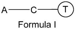

[0008] In several embodiments, the functionalizing unit comprises at least one spacer (shown as“S” in Formula II) configured to improve tethering, proximity, flexibility, rigidity, and/or orientation. Anchor Moiety

[0009] In several embodiments, the anchor moiety comprises a hydrophobic moiety configured to insert (i.e., be buried or embedded) at least partially in a EV membrane. In some embodiments, the hydrophobic moiety inserts into the lipid bilayer membrane of an EV and is located at least in part in the lipid bilayer membrane, thereby serving as an anchor for conjugating targeting moieties and/or reporter moieties. In several embodiments, the anchor moiety comprises an amphipathic lipid portion having a hydrophobic portion and a hydrophilic portion. In several embodiments, the hydrophobic portion comprises one or more long-chain saturated and/or unsaturated hydrocarbon groups (e.g., 1, 2, 3, 4, 5, or more). In several embodiments, the hydrophobic portion comprises one or more alkyl chains that insert into the lipid bilayer of an EV.
[0010] In some embodiments, the amphipathic lipid portion comprises a phospholipid, an aminolipid or a sphingolipid (combinations of any of these types can also be used, depending on the embodiment). Non-limiting examples of phospholipids include dilauroyl-phosphatidylcholine (DLPC), dimyristoyl-phosphatidylcholine (DMPC), dipalmitoyl-phosphatidylcholine (DPPC), diarachidoyl-phosphatidylcholine (DAPC), distearoyl-phosphatidylcholine (DSPC), dioleoyl-phosphatidylcholine (DOPC), 1,2- distearoyl-sn-glycero-3-ethylphosphocholine (ethyl-DSPC), dipentadecanoyl- phosphatidylcholine (DPDPC), l-myristoyl-2-palmitoyl -phosphatidylcholine (MPPC), 1- palmitoyl-2-myristoyl -phosphatidylcholine (PMPC), 1 -palmitoyl-2-stearoyl- phosphatidylcholine (PSPC), l-stearoyl-2-palmitoyl-phosphatidylcholine (SPPC), 1- palmitoyl-2-oleylphosphatidylcholine (POPC), 1 -oleyl-2-palmitoyl -phosphatidylcholine (OPPC), dilauroylphosphatidylglycerol (DLPG), diarachidoylphosphatidylglycerol (DAPG), dimyristoylphosphatidylglycerol (DMPG), dipalmitoylphosphatidylglycerol (DPPG), distearoylphosphatidylglycerol (DSPG), dioleoyl-phosphatidylglycerol (DOPG), dimyristoyl phosphatidic acid (DMPA), dipalmitoyl phosphatidic acid (DPPA), distearoyl phosphatidic acid (DSPA), diarachidoylphosphatidic acid (DAP A), dimyristoylphosphatidylethanolamine (DMPE, also referred to as l,2-bis(dimethylphosphino)ethane), dipalmitoylphosphatidylethanolamine (DPPE), distearoyl phosphatidyl-ethanolamine (DSPE), dioleylphosphatidylethanolamine (DOPE), diarachidoylphosphatidylethanolamine (DAPE), dilinoleylphosphatidylethanolamine (DLPE), dimyristoyl phosphatidylserine (DMPS), diarachidoyl phosphatidylserine (DAPS), dipalmitoyl phosphatidylserine (DPPS), distearoylphosphatidylserine (DSPS), dioleoylphosphatidylserine (DOPS), dipalmitoyl sphingomyelin (DPSP), and distearoylsphingomyelin (DSSP), dilauroyl-phosphatidylinositol (DLPI), diarachidoylphosphatidylinositol (DAPI), dimyristoylphosphatidylinositol (DMPI), dipalmitoylphosphatidylinositol (DPPI), distearoylphosphatidylinositol (DSPI), and dioleoyl- phosphatidylinositol (DOPI). In some embodiments, the phospholipids can be provided as a salt (e.g., an alkali metal salt) and/or as a mixture comprising the salt. In some such embodiments, the phospholipid comprises acyl groups derived from fatty acids, which in some such embodiments have one or more C10-C24 carbon chains as disclosed elsewhere herein. In several embodiments, the anchor moiety comprises a phosphatidylethanolamine, and in some such embodiments, the carbon chain length is about 10 to about 20 carbons (or any chain length therebetween, including endpoints). In several embodiments, phosphatidylethanolamines are provided herein comprising saturated and/or unsaturated fatty acids. Non-limiting examples of phosphatidylethanolamines include dimyristoylphosphatidylethanolamine (DMPE), dipalmitoylphosphatidylethanolamine (DPPE), dioleoylphosphatidylethanolamine (DOPE) and distearoylphosphatidyl-ethanolamine (DSPE). In several embodiments, the phosphatidylethanolamine is DMPE. In several embodiments, the phosphatidylethanolamine is DSPE. In some embodiments, the phospholipid is diacylphosphatidylcholine (e.g., DSPC, DOPC, DPPC, DLPC, POPC), diacylphosphatidylethanolamine (e.g., DOPE, POPE, DPPE, DMPE, DSPE), or a mixture thereof. In some such embodiments, the phospholipid comprises acyl groups derived from fatty acids, which in some such embodiments have one or more C10- C24 (e.g., lauroyl, myristoyl, palmitoyl, stearoyl, or oleoyl) tails.
[0011] In some embodiments, the anchor moiety comprises an amphipathic lipid lacking a phosphorus atom. In several embodiments, the amphipathic lipid is selected from the group comprising sphingolipids, glycosphingolipid families, diacylglycerols, and b- acyloxyacids, tearylamine, dodecylamine, hexadecylamine, acetyl palmitate, glycerolricinoleate, hexadecyl stereate, isopropyl myristate, amphoteric acrylic polymers, triethanolamine-lauryl sulfate, alkyl-aryl sulfate polyethyloxylated fatty acid amides, dioctadecyldimethyl ammonium bromide and the like, ceramide, diacylphosphatidylcholine, and diacylphosphatidylethanolamine. [0012] In some embodiments, the hydrophilic characteristics of the amphipathic lipid are conferred by the presence of polar or charged groups such as carbohydrates, phosphate, carboxylic, sulfato, amino, sulfhydryl, nitro, hydroxyl, and other like groups. In some embodiments, the hydrophobicity of the amphipathic lipid is conferred by the inclusion of apolar groups that include, but are not limited to, long chain saturated and unsaturated aliphatic hydrocarbon groups and such groups substituted by one or more aromatic, cycloaliphatic, or heterocyclic group(s).
Targeting Moiety
[0013] In several embodiments, the targeting moiety configured to bind to a target molecule, wherein the target molecule is a ligand on the surface of a target cell. In some embodiments, the targeting moiety helps the EV identify a particular target. In some embodiments, the target molecule can be a soluble and/or free floating (e.g., not cell bound) molecule. In some embodiments, the interaction (e.g., binding) of the targeting moiety and the ligand causes the engineered EV to be endocytosed by (or otherwise engulfed by) the target cell, thereby delivering the cargo of the EV to the target cell.
[0014] In some embodiments, the target cells are present in (e.g., residents of) a target tissue that is damaged, dysfunctional, and/or infected. In several embodiments, the ligand is differentially expressed on non-target cells (e.g., healthy cells) as compared to target cells, thereby imparting a degree of specific targeting of the engineered EV to cells that express the ligand. In some embodiments, the ligand may be absent on non-target cells and/or over expressed on target cells.
[0015] In several embodiments, targeting moiety is configured to target cells mediating cardiac inflammation following acute myocardial infarction, such as macrophages. In several embodiments there are provided herein targeting moieties that bind ligands on the surface of macrophages, such as CD68, CDl lb, CDl lc, CD16, or combinations thereof. In several embodiments, cardiac cells, such as cardiomyocytes and/or cardiac fibroblasts, are the target cells bound by the targeting moiety. Non-limiting examples of cardiac cell ligands bound by targeting moieties provided herein include discoidin domain receptor tyrosine kinase 2 (DDR2), CD90, CD163, Tcf2l, Seal, vimentin, Pdgfra, FSP1, periostin, and MEFSK4. Additional embodiments herein provide for targeting moieties that bind a ligand that is overexpressed in infarcted heart, such as AT1 receptor. In several embodiments, target cells bound by targeting moieties provided herein are cells that have incurred damage due to ischemia (and/or reperfusion) following acute myocardial infarction.
[0016] Depending on the embodiment, a variety of targeting moieties can be used to bind a ligand on the surface of a target cell. For example, in some embodiments, the targeting moiety is an antibody or a homing peptide. In some embodiments, monoclonal antibodies, recombinant antibodies, human antibodies, and/or humanized antibodies are used as targeting moieties. In some embodiments, fragments of an antibody are used, yet retain (or even improve) binding to the target cell ligand. In some embodiments, functional derivatives of an antibody are used, yet retaining (or even improving) binding to the target cell ligand. For example, in several embodiments, a Fab, a Fab', a F(ab')2, an Fv, a single-chain Fv (scFv) are employed. Minibodies, single-domain antibodies such as a heavy chain variable domain (VH), a light chain variable domain (VL) and a variable domain (VHH) of a camelid derived nanobody, diabodies, and/or single-domain antibodies are employed as targeting moieties in some embodiments. In several embodiments, combinations of targeting moieties are used (e.g., hetero-dimers, hetero-trimers, etc.). In several embodiments, the targeting moiety optionally also includes a second peptide that binds a different target cell ligand than the first peptide. In several embodiments, targeting moieties are provided herein comprising one or more homing peptides. In several such embodiments, homing peptides comprise the amino acid sequences of CSTSMLKAC (SEQ ID NO: 1), CKPGTSSYC (SEQ ID NO: 2), CPDRSVNNC (SEQ ID NO: 3), CSTSMLKACGGCSTSMLKACGGCSTSMLKAC (SEQ ID NO: 4), ASSLNIA (SEQ ID NO: 5), or ASSLNIAGGASSLNIAGGASSLNIA (SEQ ID NO: 6). Some embodiments pertain to a cardiac homing peptide (CHP) or a muscle targeting peptide (MTP). While in some embodiments, specific amino acid sequences are used, additional embodiments provided for herein employ peptides that are about 75%, about 80%, about 85%, about 90%, about 95%, about 98%, or about 99% homologous to such sequences. In some embodiments, the percent homology may vary (e.g., be lower), however the homing peptide retains at least a portion of the targeting function of a homing peptide encoded by or having a sequence specifically disclosed herein.
[0017] In some embodiments, the EV comprises a variety of targeting moieties. For example, in some embodiments, the EV comprises a plurality of homing peptides of the same amino acid sequence. In some embodiments, the EV comprises a plurality of homing peptides of differing amino acid sequence. In some embodiments, the EV comprises an ischemic cellular targeting peptide derived from phage display screening of a random peptide library for selective binding to ischemic heart tissue as compared to control cells and tissues. Coupling Moiety; Specific Binding Pair
[0018] In order to couple the targeting moiety to the anchor moiety, there is provided herein, in some embodiments, a coupling moiety comprising a specific binding pair, wherein the specific binding pair comprises a first member of the specific binding pair and a second member of the specific binding pair that bind one another with high affinity and/or specificity. In several such embodiments, the first member (e.g., a host, a molecule with a binding pocket, an electrophile, etc.) of the specific binding pair is conjugated to the anchor moiety, and the second member (e.g., a guest, a nucleophile, etc.) of the specific binding pair is conjugated to the targeting and/or reporter moiety. In other embodiments, the first member of the specific binding pair is conjugated to the targeting and/or reporter moiety and the second member of the specific binding pair is conjugated to the anchor moiety. In some embodiments, binding between the first and second members of the specific binding pair occurs via covalent bonding, while in additional embodiments the binding occurs via non-covalent interactions. In several such embodiments, the non-covalent interactions comprise one or more of host/guest interactions, complexation, ionic bonding, hydrophobic interactions, van der Waals forces, and hydrogen bonding. In such instances, the specific binding pair has a dissociation constant Kd of less than or equal to about: 10-8 mol/L, 10-9 mol/L, 10-10 mol/L, 10-11 mol/L, 10-12 mol/L, 10-13 mol/L, 10-14 mol/L, 10-15 mol/L, or ranges spanning and/or including the aforementioned values. Various specific binding pairs are employed depending on the embodiment. Non limiting examples of the specific binding pair include an antibody or an antigen-binding portion thereof and an antigen (e.g., fluorescein, digoxin, digoxigenin); a biotin (bio) moiety and an avidin moiety; a dinitrophenol (DNP) and an anti-DNP antibody; a hapten and an anti hapten; folate and a folate binding protein; vitamin B12 and an intrinsic factor; a carbohydrate and a lectin or carbohydrate receptor; a polysaccharide and a polysaccharide binding moiety; a lectin and a receptor; a ligand and a receptor; a drug and a drug receptor; complementary chemical reactive groups (e.g., sulfhydryl/maleimide, sulfhydryl/haloacetyl derivative, amine/isotriocyanate, amine/succinimidyl ester, and amine/sulfonyl halides); an antibody (e.g., IgG) and protein A or protein G; a toxin and a toxin receptor; a peptide/protein and a peptide/protein receptor; an enzyme substrate and an enzyme; and DNA/RNA and complementary DNA/RNA.
[0019] In some embodiments, the first and/or second member of the specific binding pair is bound to the targeting moiety, the reporter moiety and/or the anchor moiety via a reactive group. In several embodiments, the reactive group is provide via reaction between one or more of the following structures selected from the group comprising primary amines (- NH2), secondary amines, sulfhydryls (-SH), carboxyls (-COOH), and carbonyls (-CHO). In several embodiments, the reactive groups are reacted with coinciding reactive groups to couple the targeting moiety, the reporter moiety and/or the anchor moiety together via a bonding unit (e.g., a disulfide bond, an amide bond, an ester, a thioester, etc.).
[0020] In some embodiments, the specific binding pair comprises an aptamer and its target molecule. Aptamers can be short nucleic acids or short peptides (e.g., between about 5 and about 50 kDa). In some embodiments, aptamers have a molecular weight of less than or equal to about: 5 kDa, 10 kDa, 15 kDa, 20 kDa, 30 kDa, 40 kDa, 50 kDa, or ranges including and/or spanning the aforementioned values. In several embodiments, the aptamers strongly bind a target molecule, typically with binding constants in the micromolar to nanomolar range (e.g., less than about 1000 mM to less than about 1000 nM). Aptamer targets can include, but are not limited to, an organic dye (e.g., fluorescein, Cy3, Cy5), a disaccharide (e.g., cellobiose, lactose, maltose, gentiobiose), an aminoglycoside (e.g., tobramycin, lividomycin, kanamycin A, kanamycin B, neomycin B), an antibiotic (e.g., viomycin and tetracyclin), dopamine, porphyrins (e.g., hematoporphyrin), and biotin.
[0021] In some embodiments, as disclosed elsewhere herein, the targeting moiety and/or the anchor moiety is biotinylated. The term“biotinylated” shall be given its ordinary meaning, and shall also refer to any covalent or non-covalent adduct of a biotin moiety with other moieties such as biomolecules, e.g., proteins, nucleic acids (including DNA, RNA, DNA/RNA chimeric molecules, nucleic acid analogs and peptide nucleic acids), proteins (including enzymes, peptides and antibodies), carbohydrates, and lipids. While in some embodiments the biotin moiety comprises biotin (cis-hexahydro-2-oxo-lH- thieno[3,4]imidazole-4-pentanoic acid), additional embodiments provided for herein employ derivatives or analogs thereof that can specifically bind to an avidin moiety, including, without limitation, biotin-e-N-lysine, biocytin hydrazide, amino or sulfhydryl derivatives of 2- iminobiotin and biotinyl-6-aminocaproic acid-N-hydroxysuccinimide ester, sulfosuccinimideiminobiotin, biotinbromoacetylhy dr azide, p-diazobenzoyl biocytin, 3-(N- maleimidopropionyl)biocytin.desthiobiotin, oxybiotin, 2'-iminobiotin, diaminobiotin, biotin sulfoxide, and biocytin.
[0022] The term“avidin” and“avidin moiety” shall be given its ordinary meaning, and shall also refer to, at least, native egg-white glycoprotein avidin (or native avidin from other sources), as well as any derivatives, analogs and other non-native forms of avidin that can specifically bind to biotin moieties. In some embodiments, the avidin moiety can comprise deglycosylated forms of avidin, bacterial streptavidins produced by selected strains of Streptomyces , e.g., Streptomyces avidinii, truncated streptavidins, recombinant avidin and streptavidin, derivatives of native, deglycosylated and recombinant avidin and of native, recombinant and truncated streptavidin, for example, N-acyl avidins (e.g., N-acetyl avidin), N- phthalyl avidin, and N-succinyl avidin, and the commercial products ExtrAvidin®, Captavidin®, Neutravidin® and Neutralite Avidin®. All forms of avidin-type molecules, including both native and recombinant avidin and streptavidin as well as derivatized molecules, e.g. nonglycosylated avidins, N-acyl avidins and truncated streptavidins, are encompassed within the terms“avidin” and“avidin moiety”. In some embodiments, the avidin exists as a tetrameric protein, wherein each of the four tetramers is capable of binding at least one biotin moiety. In some embodiments, the avidin moiety is streptavidin.
Spacer
[0023] In several embodiments, the spacer provided herein does not significantly interfere with the function or activity of the anchor moiety, the targeting moiety, and/or the first/second members of the specific binding pair of the coupling moiety. In several embodiments, the spacer is polymeric and is functionalized the at one or both ends. In some embodiments, the spacer is functionalized to the anchor moiety and a member of the binding pair of the coupling moiety. In some embodiments, the spacer (or a second spacer) is functionalized to the targeting moiety and the a member of the binding pair of the coupling moiety. In several embodiments, the spacer is a hydrophilic polymer or an amphiphilic polymer. In some embodiments, the spacer is functionalized at one or both ends (e.g., to the anchor moiety, the coupling moiety, and/or the targeting/reporting moiety). In some embodiments, the polymer has an average molecular weight (in Da) of less than or equal to about: 200, 500, 1000, 1500, 2000, 2500, 3000, 3500, 4000, 4500, 5000, 5500, 6000, 6500, 7000, 7500, 8000, 8500, 9000, 9500, 10,000, 10,500, 11,000, 11,500, 12,000, 12,500, 13,000, 13,500, 14,000, 14,500, 15,000, 15,500, 16,000, 16,500, 17,000, 17,500, 18,000, 18,500, 19,000, 19,500, 20,000, or ranges including and/or spanning the aforementioned values. Non limiting examples of spacer polymers provided herein are polyethylene glycol (PEG), polypropylene glycol, methoxypolyethylene glycol (mPEG), polyvinylalcohol, polyvinylpyrrolidone, copolymers thereof, or combinations thereof. In some embodiments, the polymer is PEG.
[0024] In several embodiments, there is provided herein a lipid anchor comprising a phosphatidylethanolamine conjugated to a hydrophilic polymer (e.g., a spacer), such as, for example, DMPE-PEG, DPPE-PEG, DSPE-PEG, DAPE-PEG, or in any combination with any other anchor disclosed elsewhere herein. In some embodiments, the functionalizing unit lacks a spacer (as shown in Formula I) while in other embodiments the spacer is present (as shown in Formula II). In some embodiments, the spacer comprises a series of atoms linked via covalent bonds. Additional embodiments provide spacers that are branched or unbranched. In some embodiments, the spacer is flexible, while in other embodiments the spacer is rigid. In some embodiments, the spacer is linear, branched, bifunctional, trifunctional, homofunctional, or heterofunctional. In some embodiments, the spacer is chemical chain or a chemical compound. In some embodiments, the spacer is resistant to heat, salts, acids, bases, light and chemicals. In some embodiments, the spacer is of sufficient stereo-selectivity to allow coupling of the anchor moiety to the targeting moiety via the coupling moiety.
[0025] In some embodiments, the spacer comprises about 1-40 plural valent atoms or more selected from the group consisting of C, N, O, S and P. The number of plural valent atoms in a spacer may be, for example, 0, 1, 2, 3, 4, 5, 6, 7, 8, 9, 10, 20, 25, 30, or 40, or more. In some embodiments, the spacer has one or more pendant side chains or pendant functional groups (or both). Non-limiting examples of such pendant moieties are hydrophilicity modifiers, e.g., sulfo (-SO3H- or -SO3-). In some embodiments, the spacer is composed of any combination of single, double, triple or aromatic carbon-carbon bonds, carbon-nitrogen bonds, nitrogen-nitrogen bonds, carbon-oxygen bonds and carbon-sulfur bonds. Exemplary linking members include a moiety which includes -C(0)NH-, -C(0)0-, -NH-, -S-, -O-, and the like. In some embodiments, the spacer consists of a combination of moieties selected from alkyl, alkylene, aryl, -C(0)NH-, -C(0)0-, -NH-, -S-, -0-, -C(0)-, -S(0)n- where n is 1-3. A spacer may be linear or non-linear; some spacer may have pendant side chains or pendant functional groups (or both). Examples of such pendant moieties are hydrophilicity modifiers, for example solubilizing groups like, e.g., sulfo (— SO3H— or— SO3— ).
[0026] In some embodiments, the PEG moieties are functionalized at one or both ends. In some embodiments, functionalization at both ends with the same reactive moiety can be employed to create a homobifunctional PEG derivative. Some examples of homobifunctional PEG derivatives include, without limitation, COOH-PEG-COOH; NH2- PEG-NPh; and MAL-PEG-MAL (where MAL denotes a maleimide group).
[0027] In some embodiments, the spacer is a heterobifunctional spacer. In some embodiments, heterobifunctional spacers are provided that contain one end having a first reactive functionality to specifically link to a first molecule, and an opposite end having a second reactive functionality to specifically link to a second molecule.
[0028] In some embodiments, the PEG derivative can be a multi-arm PEG derivative. In some embodiments, the multi-arm PEG derivative can be a PEG derivative having a core structure comprising pentaerythritol (including, for example, 4arm PEG amine (4ARM-PEG-NH2); 4arm PEG carboxyl (4ARM-PEG-COOH) ; 4arm PEG maleimide (4ARM-PEG-MAL); 4arm PEG succinimidyl succinate (4ARM-PEG-SS); 4arm PEG succinimidyl glutarate (4ARM-PEG-SG)); a PEG derivative having a core structure comprising hexaglycerin (including, for example, 8arm PEG amine (8ARM-PEG-NH2); 8arm PEG carboxyl (8ARM-PEG-COOH); 8arm PEG succinimidyl succinate (8ARM-PEG-SS); 8arm PEG amine (8ARM-PEG-SG); PEG derivative having a core structure comprising tripentaerythritol (including, for example, 8arm PEG amine (8ARM(TP)-PEG-NH2); 8arm PEG carboxyl (8ARM(TP)-PEG-COOH); 8arm PEG succinimidyl succinate (8ARM(TP)- PEG-SS); 8arm PEG amine (8ARM(TP)-PEG-SG)). Optionally, end groups for heterobifunctional PEGs can include maleimide, vinyl sulfones, pyridyl disulfide, amine, carboxylic acids and NHS esters. The activated PEG derivatives can then be used to attach the PEG to the desired component of the cloaking platform.
[0029] In some embodiments, functionalization with different reactive moieties can be used to create a heterobifunctional PEG derivative comprising different reactive groups at each end. Such heterobifunctional PEGs can be useful in linking two entities, where a hydrophilic, flexible and biocompatible spacer is needed. Some examples of heterobifunctional PEG derivatives include, without limitation, hydroxyl PEG carboxyl (HO- PEG-COOH); thiol PEG carboxyl (HS-PEG-COOH); hydroxyl PEG amine (HO-PEG-NH2); t-Boc amine PEG amine (TBOC-PEG-NH2); amine PEG carboxyl (NH2-PEG-COOH); t-Boc amine PEG NHS ester (TBOC-PEG-NHS); FMOC amine PEG NHS ester (FMOC-PEG- NHS); acrylate PEG NHS ester (ACLT-PEG-NHS); maleimide PEG carboxyl (MAL-PEG- COOH); maleimide PEG amine (MAL-PEG-NH2), including the TFA salt thereof; maleimide PEG NHS ester (MAL-PEG-NHS); biotin PEG NHS ester (BIOTIN-PEG-NHS); biotin polyethylene glycol maleimide (BIOTIN-PEG-MAL); OPSS PEG NHS ester (OPSS-PEG- NHS).
[0030] In some embodiments, the PEG or other polymer is bound to the lipid anchor (e.g., DMPE) through a covalent bond, such as an amide, carbamate or amine linkage. In addition, in several embodiments, the PEG or other polymer may be linked to a specific binding pair member or targeting moiety with a covalent bond including, for example, amide, ester, ether, thioester, thioamide or disulfide bonds. In other embodiments, the PEG molecule is linked to the lipid via a linker moiety. Any linker moiety suitable for coupling the PEG to a lipid can be used including, e.g., non-ester containing linker moieties and ester-containing linker moieties. In some embodiments, the linker moiety is a non-ester containing linker moiety. Non-limiting examples of suitable non-ester containing linker moieties include, but are not limited to, amido, amino, carbonyl, carbamate, urea, disulphide, ether, succinyl, succinamidyl, ether, disulphide, as well as combinations thereof (such as a linker containing both a carbamate linker moiety and an amido linker moiety). In other embodiments, an ester containing linker moiety is used to couple the PEG to the lipid. Suitable ester containing linker moieties include, e.g., carbonate, succinoyl, phosphate esters, sulfonate esters, and combinations thereof.
[0031] In some embodiments, phosphatidylethanolamines having a variety of acyl chain groups of varying chain lengths and degrees of saturation are conjugated to a PEG molecule of variable molecular weight (e.g. from about 300 to about 10000 daltons). For example, DPPE-PEG refers to DPPE having PEG attached thereto. Thus, in several embodiments, there is a lipid anchor provided comprising DMPE-PEG, DPPE-PEG, DSPE- PEG, or DAPE-PEG. Cloaking Platform
[0032] The EV membrane anchoring platform technology termed“cloaking” as described herein can be used to directly embed tissue-specific antibodies and/or homing peptides on EV membrane surface ex vivo for enhanced EV delivery to and update by cells and/or tissues of interest. There is provided, in some embodiments, a cloaking platform that employs a phospholipid anchor comprising of l,2-bis(dimethylphosphino) ethane (DMPE) covalently attached to a polyethylene glycol chain consisting of 5k units (5k-PEG) which is conjugated to the protein streptavidin (S). Joined, this molecule is referred to as DPS herein. This unique configuration enables the attachment of any biotin conjugated molecule (e.g., antibody, protein, nucleic acid) to DPS. Once attached, the biotinylated moiety:DPS complex can be added to any cellular or EV lipid bilayer membrane, as the DPS will anchor the entire complex on the membrane surfaces of the cells or EVs and display the biotinylated molecule, thus generating configurable cells and EVs. Accordingly, in some embodiments, the cloaking of EVs comprises adding modified glycerol-phospholipid-PEG conjugates (e.g., DMPE-PEG) to isolated EVs (e.g., exosomes) in solution. DMPE-PEG embeds into EV lipid bilayer membrane and serves as an anchor for conjugating fluorescent molecules and/or ligand proteins. For example, streptavidin can be conjugated with DMPE-PEG to create a modular EV membrane anchoring platform (DMPE-PEG-streptavidin; DPS). Thus, any biotinylated molecule (e.g., an antibody, a homing peptide, or a reporter moiety) can be coupled to the DPS to decorate (i.e., cloak) EV lipid bilayer membrane for targeted delivery (e.g., biotinylated antibodies can be used to target macrophages [anti-CD68] and cardiac fibroblasts [anti-DDR2] in vitro). Further, it is contemplated that any biotinylated targeting moiety can be directly attached to the membrane surface of any EV with the DPS complex to engineer highly target- specified therapeutic EVs. In some embodiments, the targeting approach involves adding modified glycerol-phospholipid-PEG conjugates (DMPE-PEG) to isolated EVs in solution. DMPE-PEG embeds into exosome membranes and thereby serves as an anchor for conjugating fluorescent molecules or ligand proteins.
[0033] In view of the disclosure provided herein, there are a variety of other engineered EVs that can be generated in order to target and deliver cargo biological factors to particular target cells in damaged, dysfunctional, and/or infected tissues. Non-limiting examples of such engineered EVs are discussed in more detail elsewhere herein. Reporter Moiety
[0034] In some embodiments, the reporter moiety is conjugated to the first or second member of the specific binding pair of the coupling moiety, and is thereby coupled to the anchor moiety via the specific binding pair. In some embodiments, the reporter moiety comprises a fluorescent molecule, e.g., fluorescein, fluorescein dyes (e.g., fluorescein isothiocyanine, naphthofluorescein, 4',5'-dichloro-2',7'-dimethoxyfluorescein, and 6- carboxyfluorescein), carbocyanine, merocyanine, styryl dyes, oxonol dyes, phycoerythrin, erythrosin, eosin, rhodamine dyes (e.g., carboxytetramethyl-rhodamine or TAMRA, carboxyrhodamine 6G, carboxy-X-rhodamine, lissamine rhodamine B, rhodamine 6G, rhodamine Green, rhodamine Red, and tetramethylrhodamine), coumarin, coumarin dyes (e.g., methoxycoumarin, dialkylaminocoumarin, hydroxycoumarin, and aminomethylcoumarin [AMCA]), Oregon Green Dyes (e.g., Oregon Green 488, Oregon Green 500, and Oregon Green 514), Texas Red, Texas Red-X, Spectrum Red™, Spectrum Green™, cyanine dyes (e.g., Cy- 3™, cyanine 5 far-red fluorescent dye, Cy-3.5™, and Cy-5.5™), Fluor dyes (e.g., Alexa Fluor 350, Alexa Fluor 488, Alexa Fluor 532, Alexa Fluor 546, Alexa Fluor 568, Alexa Fluor 594, Alexa Fluor 633, Alexa Fluor 660, and Alexa Fluor 680), BODIPY dyes (e.g., BODIPY FL, BODIPY R6G, BODIPY TMR, BODIPY TR, BODIPY 530/550, BODIPY 558/568, BODIPY 564/570, BODIPY 576/589, BODIPY 581/591, BODIPY 630/650, and BODIPY 650/665), IRDyes (e.g., IRD40, IRD 700, and IRD 800), and/or derivatives thereof. In several embodiments, the reporter moiety comprises a fluorescent protein, e.g., green fluorescent protein (GFP), enhanced GFP, blue fluorescent proteins, cyan fluorescent protein, yellow fluorescent protein, red fluorescent protein, and/or derivatives thereof. In several embodiments, the reporter moiety comprises a radioisotope that is detectable by single photon emission computed tomography or position emission tomography, e.g., iodine- 131, iodine- 125, bismuth-2l2, bismuth-2l3, astatine-22l, copper-67, copper-64, rhenium-l86, rhenium- 186, phosphorus-32, samarium-l53, lutetium-l77, technetium-99m, gallium-67, indium-l l l, and thallium-201. In several embodiments, the reporter moiety comprises a quantum dot (Qdot™) fluorescent particle, e.g., Qdot525, Qdot565, Qdot585, Qdot605, Qdot625, Qdot655, Qdot705 and Qdot800. In several embodiments, the fluorophores desirably exhibit absorption and emission wavelengths in the visible (e.g., between 400 and 750 nm) rather than in the ultraviolet range of the spectrum (e.g., lower than 400 nm). [0035] In several embodiments, the reporter moiety generates a signal that can be measured and whose intensity is related to (e.g., proportional to) the amount of uptake of the engineered EVs by a cell or tissue. In some embodiments, the engineered EV comprises the reporter moiety and the targeting moiety. In some embodiments, the anchor moiety is coupled to the reporter moiety instead of the targeting moiety. In several embodiments, the reporter moiety does not substantially interfere with the desired biological or therapeutic activity of the engineered EV.
[0036] In some embodiments, the reporter moiety generates, or causes to be generated, a detectable signal, including, but not limited to luminescent, photoluminescent, electroluminescent, bioluminescent, chemiluminescent, fluorescent, phosphorescent, chromophore, radioisotope, electrochemical, mass spectrometry, Raman, hapten, affinity tag, atom, or an enzyme.
[0037] In some embodiments, the reporter moiety generates a detectable signal resulting from a chemical or physical change (e.g., heat, light, electrical, pH, salt concentration, enzymatic activity, or proximity events). A proximity event includes two reporter moieties approaching each other, or associating with each other, or binding each other. The appropriate procedures for detecting a signal, or change in the signal, generated by the reporter moiety are well known in the art. In some embodiments, the reporter moiety generates a signal, or a change in a signal, upon excitation from an appropriated energy source (e.g., electromagnetic source).
EV Source
[0038] Depending on the embodiment, the EV (prior to being engineered as described herein) can be derived from a variety of cells. In several embodiments, the EV is derived from one or more of stem cells, progenitors and/or precursors. Non-limiting examples of stem cells include pluripotent stem cells, embryonic stem cells, induced pluripotent stem cells, hematopoietic stem cells, mesenchymal stem cells, and endothelial precursor cells. In several embodiments, the EV is derived from cardiospheres, cardiosphere-derived cells (CDCs), human neural stem cells, bone marrow stem cells, immune cells, neural tissue, mononuclear cells, or newt Al cell line. In some embodiments, the EV is derived from a population of cells which has been genetically manipulated to express one or more proteins and/or microRNAs. Additionally, various types of EVs are employed depending on the embodiment. In some embodiments, exosomes, microvesicles, apoptotic bodies, and/or ectosomes are engineered as described herein. In one embodiment, the EV is an exosome. In one such embodiment, the EV is an exosome derived from CDCs (CDCexo). CDC-EV encompasses CDCexo, as well as microvesicles, apoptotic bodies, and other EVs derived from CDCs. Additional embodiments provide for EVs and/or exosomes comprising a biomarker. In some such embodiments the biomarker is a tetraspanin, such as, for example, CD63, CD81, CD82, CD53, and CD37. Additional embodiments provide for EVs comprising a biological protein and/or microRNA capable of facilitating regeneration and/or improved function of a tissue.
[0039] In certain embodiments, the EVs are synthetic. In certain embodiments, the population of cells has been genetically manipulated. This includes, for example, knockout or transgenic cell lines. In certain embodiments, the cells are genetically modified to express endothelial nitric oxide synthase, vascular endothelial growth factor, stromal derived factor 1 , insulin-like growth factor 1, hepatocyte growth factor. Depending on the embodiment, this may further include transient knockdown of one or more genes and associated coding and non coding transcripts within the population of cells, via any number of methods known in the art, such as introduction of dsRNA, siRNA, miR, a vector, plasmid, artificial plasmid, and replicative or non-replicative virus. In other embodiments, the population of cells has been altered by exposure to environmental conditions (e.g., hypoxia), small molecule addition, presence/absence of exogenous factors (e.g., growth factors, cytokines) at the time, or substantially contemporaneous with, isolating the plurality of EVs. For example, one may add a differentiation agent to a population of stem cells, progenitors and/or precursors in order to promote partial or full differentiation of the cell, and thereafter derive a plurality of EVs. In various embodiments, altering the regulatory state of the cell changes composition of one or more EVs in the plurality of EVs.
Methods of Treating Damaged or Dysfunctional Tissue with the Engineered EVs
[0040] In several embodiments, there is provided a targeted therapeutic composition comprising the engineered EVs. In some embodiments, the targeted therapeutic composition comprises a pharmaceutically acceptable carrier and one or more excipients.
[0041] Also provided herein are methods of treating damaged or dysfunctional cells and/or tissue in a subject (e.g, a human patient) in need thereof. In some embodiments, the method comprises administering an effective amount of a targeted therapeutic composition comprising engineered EVs to a subject in need thereof. In some embodiments, the engineered EVs are delivered to and/or are taken up by damaged or dysfunctional cells and/or tissues. In several embodiments, administration of the targeted therapeutic composition comprises administration at a tissue or organ site that is the same as the target tissue. In some embodiments, administration of targeted therapeutic composition comprises administration at a tissue or organ site that is different from the target tissue. In some embodiments, administration of targeted therapeutic composition comprises administration systemically (e.g., in the blood). In several embodiments, a single dose of the targeted therapeutic composition comprises between about 1 x 10⁶ and about 1 x 10⁸ of the engineered EVs. In some embodiments, a single dose of the targeted therapeutic composition is administered multiple times to the subject. In some embodiments, the administration of the targeted therapeutic composition is inhalation or oral administration. In some embodiments, the targeted therapeutic composition is administered by intra-arterial, intravenous, or retrograde coronary sinus infusion or injection. For example, in some embodiments administration comprises percutaneous injection.
[0042] Tissues treated according the methods provided herein include, in some embodiments, cardiac tissue, brain or other neural tissue, skeletal muscle tissue, pulmonary tissue, arterial tissue, and capillary tissue. In several embodiments, the tissue to be treated is damaged or dysfunctional is due to an injury, age-related degeneration, cancer, or infection. In some embodiments, the methods provided herein treat tissue that is damaged or dysfunctional due to an acute event or a chronic disease. In some embodiments, the acute event or chronic disease is as a result of myocardial infarction, traumatic head injury, and/or stroke. Non-limiting examples of additional chronic diseases that are treated include congestive heart failure, heart disease, ischemic heart disease, valvular heart disease, connective tissue diseases, HIV infection, dilated cardiomyopathy, myopathy, and dystrophinopathy (e.g., Duchenne muscular dystrophy), liver disease, sickle cell disease, dilated cardiomyopathy, infection such as Schistosomiasis, diabetes, Alzheimer’s disease, Parkinson’s disease, Huntington’s disease, and Amyotrophic Lateral Sclerosis (ALS).
[0043] In various embodiments, the damaged or dysfunctional tissue is in need of repair, regeneration, or improved function due to an acute event (including physical trauma caused by a force originating from outside the body). Acute events include, but are not limited to, trauma such as laceration, crush or impact injury, shock, loss of blood or oxygen flow, infection, chemical or heat exposure, poison or venom exposure, drug overuse or overexposure, and the like. In certain embodiments, the damaged tissue is pulmonary, arterial or capillary tissue, such as the endothelial lining of distal pulmonary arteries. In other embodiments, the damaged tissue is cardiac tissue.
[0044] In several embodiments, damage or dysfunction of tissue (or cells) is related to an immune response. For example, in several embodiments, auto-immune disorders can lead to damage or dysfunction of tissues (or cells) in the short or long-term. In several embodiments, the auto-immune disorder is one or more of rheumatoid arthritis, lupus, celiac disease, Sjogren's syndrome, polymyalgia rheumatic, multiple sclerosis, ankylosing spondylitis, Type I diabetes, vasculitis, and temporal arteritis.
[0045] In some embodiments, engineered EVs are delivered to an infected target tissue, such as a target tissue infected with one or more bacteria, viruses, fungi, and/or parasites. In some embodiments, EVs are used to treat tissues with infections of bacterial origin (for example, infectious bacteria selected the group of genera consisting of Bordetella, Borrelia, Brucella, Campylobacter, Chlamydia and Chlamydophila, Clostridium, Corynebacterium, Enterococcus, Escherichia, Francisella, Haemophilus, Helicobacter, Legionella, Leptospira, Listeria, Mycobacterium, Mycoplasma, Neisseria, Pseudomonas, Rickettsia, Salmonella, Shigella, Staphylococcus, Streptococcus, Treponema, Vibrio, and Yersinia, and mutants or combinations thereof). In some embodiments, the EVs inhibit or prevent one or more bacterial functions, thereby reducing the severity and/or duration of an infection. In some embodiments, administration of EVs sensitizes the bacteria (or other pathogen) to an adjunct therapy (such as an antibiotic). In some embodiments, where the infection is viral in origin, the infection is a result of one or more viruses selected from the group consisting of adenovirus, Coxsackievirus, Epstein-Barr virus, hepatitis a virus, hepatitis b virus, hepatitis c virus, herpes simplex virus type 1, herpes simplex virus type 2, cytomegalovirus, ebola virus, human herpes virus type 8, HIV, influenza virus, measles virus, mumps virus, human papillomavirus, parainfluenza virus, poliovirus, rabies virus, respiratory syncytial virus, rubella virus, and varicella-zoster virus. [0046] According to some embodiments, the EVs can be used to treat a wide variety of cell types as well, including but not limited to vascular cells, epithelial cells, interstitial cells, musculature (skeletal, smooth, and/or cardiac), skeletal cells (such as bone, cartilage, and connective tissue), nervous cells (such as neurons, glial cells, astrocytes, Schwann cells), liver cells, kidney cells, gut cells, lung cells, skin cells or any other cell in the body.
[0047] In some embodiments, the disease is a dystrophinopathy. In some embodiments, the disease state is a dystrophic disorder. In some embodiments, the dystrophinopathy includes one or more of Duchenne muscular dystrophy (DMD) and/or Becker muscular dystrophy. In some embodiments, the disease state is a myopathy. In some embodiments, the myopathy is a skeletal muscle myopathy. In some embodiments, functional improvements at dystrophic skeletal muscles are achieved.
[0048] In various embodiments, the plurality of cloaked exosomes modulate smad pathway activity, including for example, smad4 and smad 2/3. In various embodiments, the plurality of cloaked exosomes increase cardiomyocyte proliferation. In various embodiments, the plurality of cloaked exosomes are capable of modulating SDF-l, VEGF and/or collagen expression. In various embodiments, the plurality of cloaked exosomes is capable of enhancing infiltration of monocytes, macrophages, and T-cells.
[0049] In several embodiments, administration of the targeted therapeutic composition or engineered EVs enhances the regeneration or production of new tissue in the subject. In some embodiments, administration causes a functional improvement in a tissue. In some embodiments, for example, when pulmonary, arterial, capillary, cardiac, or skeletal muscle tissue is damaged or dysfunctional, the functional improvement comprises increased contractility, improvements in cardiac performance or muscular performance including one or more of reduced arrhythmia, increased cardiac output, increased ventricular function, decreased right ventricle systolic pressure, decreased arteriolar narrowing, improved pulmonary vascular resistance, improved baseline ejection volume, increased viable tissue, reduced scar mass, increased cardiac wall thickness in an area subjected to the myocardial infarction, improved regenerative remodeling of injury sites, enhanced angiogenesis, improved cardiomyogenic effects, reduced apoptosis, and decreased levels of pro-inflammatory cytokines. In several embodiments, administration of targeted therapeutic composition improves the viability of the targeted tissue. In some embodiments, the EVs are derived from the same tissue type as is in need of repair or regeneration. Additional embodiments provide for methods of treatment employing EVs derived from a tissue type other than the tissue in need of repair or regeneration. In some embodiments, the delivery of engineered EVs causes enhanced cognition in response to treatment of neural damage, improved blood-oxygen transfer in response to treatment of lung damage, improved immune function in response to treatment of damaged immunological-related tissues.
[0050] In several embodiments, the targeted therapeutic composition alters gene expression in the damaged or dysfunctional tissue, thereby improving viability of the damaged tissue, and/or enhancing regeneration or production of new tissue in the subject. In some embodiments, the targeted therapeutic composition enhances infiltration of monocytes, macrophages, and T-cells upon administration. Additional embodiments provide for co administration of the targeted therapeutic composition with one or more additional therapeutic agents.
[0051] In several embodiments, the damaged or dysfunctional tissue includes skeletal muscle tissue. For example, in certain embodiments in which skeletal muscle tissue is damaged or dysfunctional, functional improvement may include increased contractile strength, improved ability to walk (for example, and increase in the six-minute walk test results), improved ability to stand from a seated position, improved ability to sit from a recumbent or supine position, or improved manual dexterity such as pointing and/or clicking a mouse.
[0052] In some embodiments, when the damaged or dysfunctional tissue is in the brain or spine, the engineered EVs, including CDC-derived cloaked exosomes, are capable of delivering microRNAs and other biological cargo by crossing the blood-brain barrier. This includes, for example, cloaked exosomes administered at a site that is not the site of damaged or dysfunctional tissue, such as delivery of cargo contents to injured brain when administered via the intravenous or intra-arterial routes. In some embodiments, injection comprises injection of the targeted therapeutic composition into the heart or skeletal muscle.
[0053] In some embodiments, the delivery and/or uptake of the engineered EV is enhanced compared to unmodified EV. In some embodiments, the enhanced delivery and/or uptake of the engineered EVs enables a delivery approach (e.g., systemic administration) that is minimally invasive as compared to unmodified EVs. In several embodiments, an engineered EV as disclosed herein has enhanced uptake and/or delivery to target cells as compared to an untargeted EV. In several embodiments, uptake and/or delivery is enhanced by equal to or at least about: 10%, 20%, 30%, 40%, 50%, 75%, 100%, or ranges including and/or spanning the aforementioned values. In several embodiments, uptake and/or delivery is enhanced by about 2-fold, about 3-fold, about 5-fold, or aboutlO-fold. In several embodiments, use of the “cloaked” or engineered EVs (including exosomes) disclosed herein improves the state of the damaged or dysfunctional tissue by at least about 10%, about 15% about 20%, about 25% or more. In several embodiments, the improvement results in at least a partial regain of function. In several embodiments, the improvement comprises a reduction of inflammation of the damaged or dysfunctional tissue. In several embodiments, the regenerative cells are from the same tissue type as is in need of repair or regeneration. In several other embodiments, the regenerative cells are from a tissue type other than the tissue in need of repair or regeneration. In some embodiments, the method includes administering a therapeutically effective amount of EVs, the molecular cargo of EVs, and/or combinations of the forgoing to a subject (e.g., a patient) suffering from the disease, thereby treating the disease and/or its symptoms.
Other Methods of Using the Engineered EVs
[0054] Also provided herein are methods for defining an effective dosing range of, an effective dosing regimen of, or a route of administration for, a targeted therapeutic composition comprising the engineered EVs, the methods comprising administering the targeted therapeutic composition, detecting and measuring a signal generated by the reporter moiety, and determining tissue retention based on said signal. Further provided herein are methods for defining an effective dosing regimen for a therapeutic composition comprising EVs, the methods comprising administering a detectable engineered EV comprising a reporter moiety to a subject, detecting and measuring a signal generated by the reporter moiety, and determining tissue retention based on said signal. Additionally provided herein are methods for defining a route of administration for a therapeutic composition comprising EVs, the methods comprising administering a detectable engineered EV comprising a reporter moiety to a subject, detecting and measuring a signal generated by the reporter moiety, and determining tissue retention based on said signal. In some embodiments, the detectable engineered EV further comprises a lipid anchor configured to insert at least partially into a lipid bilayer of the EV, and also further comprises a coupling moiety that couples the reporter moiety and the lipid anchor. In some embodiments, the effective dosage range, the effective dosing regimen, and/or the route of administration is selected based on a target tissue retention. In some embodiments, the target tissue retention is at least about ten-fold greater than background levels while in other embodiments the target tissue retention is at least fifty-fold greater than background levels. Depending on the embodiment, the signal generated by the reporter moiety is detected and measured at any time within about 5 minutes, about 10 minutes, about 15 minutes, about 20 minutes, about 25 minutes, about 30 minutes, about 40 minutes, about 50 minutes, or about 60 minutes after administration (or any time therebetween, including endpoints).
[0055] The compositions and related methods set forth in further detail elsewhere herein describe certain actions taken by a practitioner; however, it should be understood that they can also include the instruction of those actions by another party. Thus, actions such as “administering a targeted therapeutic composition comprising engineered EVs” include “instructing the administration of a targeted therapeutic composition comprising engineered EVs.”
[0056] In some embodiments, the engineered EVs as described herein imparts a variety of advantageous characteristics to the EV, such as enhanced targeting to target cells/tissues and/or enhanced uptake of therapeutic cargo by target cells/tissues, and/or increased residency time in the organism. In some embodiments, this enhanced delivery and/or uptake to target cells and/or tissues is particularly advantageous because it allows administration to a subject by systemic delivery (e.g., intravenous injection) rather than local delivery to target tissues. In several embodiments, the engineered EVs disclosed herein are particularly advantageous because, unlike existing methods of EV production, there is no requirement for alteration of the EV-producing cells.
BRIEF DESCRIPTION OF THE FIGURES
[0057] Figure 1 depicts the removal of excess DMPE-PEG5k-Streptavidin-Biotin molecules during the last stage of the exosome cloaking protocol.
[0058] Figures 2A-2D depict data related to the uptake of fluorescently-cloaked CDCexo incubated with neonatal rat ventricular myocytes (NRVMs). Figure 2A depicts a schematic of the cloaking technology. Figures 2B-2C show representative FACS plots depicting NRVM uptake of CDCexo cloaked with biotinylated (bio)-FITC (B) or bio-PE (C). Figure 2D depicts pooled data from Figures 2B and 2C.
[0059] Figures 3A-3D depict data related to the targeting of macrophages with anti- CD68-cloaked exosomes. Figure 3A depicts the gating strategy for macrophage flow cytometry. Figures 3B and 3C show representative FACS plots depicting macrophage uptake of CDCexo cloaked with biotinylated (bio)-FITC or bio-FITC + bio-anti-CD68 in overlay histogram format (B) or staggered format (C). Figure 3D depicts pooled data from Figures 3B and 3C.
[0060] Figures 4A-4C depict data related to the targeting of cardiac fibroblasts with DDR2-cloaked exosomes. Figures 4A and 4C depict representative FACS histogram data of cardiac fibroblast uptake of untreated CDCexo and biotinylated (bio)-FITC+bio-anti-DDR2 cloaked CDCexo. Figure 4B depicts pooled data from Figure 4A and immunoglobulin G (IgG) antibody cloak controls.
[0061] Figures 5A and 5B depict schematics of CDC-EV membranes cloaked with biotinylated Quantum dots (Qdots) and homing peptides (separately or simultaneously). Figure 5A depicts the engineering of control CDC-EVs comprising biotinylated Qdot655. Figure 5B depicts the engineering of ischemic myocardium-targeted CDC-EVs cloaked with both biotinylated Qdot655 and biotinylated ischemic homing peptide CSTSMFKAC (SEQ ID NO: 1).
[0062] Figure 6 shows an example NanoSight particle tracking profile data for IschCDC-EV + Qdot 655 in visible and fluorescent modes. Figure 6 depicts data related to the characterization of engineered CDC-EV particle number, size and Qdot655 loading by dynamic light scattering methods (visible and fluorescence mode tracking) with a NanoSight NS300 instrument.
[0063] Figures 7A-7C depict data related to the uptake of ischemic peptide/Qdot655-cloaked CDC-EVs and control Qdot655 -cloaked CDC-EVs by neonatal rat ventricular myocytes (NRVMs) in oxidative stress assays. Figure 7A depicts data collected by a fluorescent plate reader. Figures 7B and 7C depict data collected by flow cytometry analysis of NRVM cells.
[0064] Figures 8A and 8B depict data related to the in vivo biodistribution of ischemic peptide/Qdot655-cloaked CDC-EVs and control Qdot655-cloaked CDC-EVs administered via tail vein to ischemia/reperfusion model rats (n=5 rats per group). Figure 8A is a graphical representation of whole organ Qdot 655 fluorescent measurements to identify CDC-EV biodistribution in ischemia/reperfusion (I/R) a rat study animals (n = 3 rats per sample group; data for each individual rat is indicated by a circle). Figure 8A depicts average Qdot655 fluorescence of control and ischemia-targeted CDC-EVs in whole tissue (heart, liver, lung, spleen, and kidney) as measured by a plate reader. Figure 8B depicts Xenogen imaging of the whole heart of three rats administered control and ischemia-targeted CDC-EVs. Figure 8B includes Xenogen whole heart images for Qdot 655 localization of control (Ctrl) and ischemic peptide-targeted (Isch) CDC-EV. Figure 8C is a schematic of a method according to some embodiments.
[0065] Figures 9A and 9B depict data related to the in vivo biodistribution of ischemic peptide/Qdot655-cloaked CDC-EVs and control Qdot655-cloaked CDC-EVs administered via tail vein to control rats or ischemia/reperfusion (I/R) rats (n=5 rats per group). Figure 9A depicts Qdot655 fluorescence of control and ischemia-targeted CDC-EVs in whole tissue (heart, liver, lung, spleen, and kidney) of control rats and I/R rats. Figure 9B depicts the whole heart fluorescence plate reader data from Figure 9A.
[0066] Figures 10A and 10B depict the ischemic rat heart tissues of two rats stained with 2,3,5-Triphenyl-2H-tetrazolium chloride (TTC) to identify regions of cardiac damage (blanched white regions), stained with Masson’s Trichrome to reveal fibrotic scar areas (blue/gray color), and imaged for Qdot 655 fluorescence (Cy5 filter set) to locate ischemic tissue-targeted CDC-EV uptake. Regions of cardiac damage due myocardial infarction are indicated with a dashed outlined.
[0067] Figures 11 A- 11 D depict data and information related to homing of ischemic peptide HEK-EV using surface display. Figure 11A is a schematic of an expression cassette according to some embodiments. The lentivector expression cassette was used to make a fusion Ischemic peptide (Isch) coding sequence (3 repeats) fused upstream to the C1C2 domain of the human lactadherin protein (for EV membrane surface display) along with a C-terminal DDK flag tag (to detect by Western blot). Figure 11B depicts western blot data confirming expression of the fusion surface display protein in cells and on secreted exosomes. Figure 11C is a graphical depiction of Pooled FACS data of HEK-EV+GFP uptake assays with NRVM oxidative stress assays. Figure 11D includes immunofluorescent rat heart tissue section images from I/R models of myocardial infarction. Left ventricle (LV), right ventricle (RV) are labeled and ischemic zone (IZ) areas are encircled and labeled n = 2 rats per experimental group, all rats received I/R injury.
[0068] Figures 12A-12C depict information relating to Uptake assays of CDC-EV with Qdot 655 and muscle targeting peptide cloaks. Figure 12A is a schematic of a design of MTP and Qdot 655 membrane cloaks according to some embodiments. Figure 12B shows representative FACS histograms of mouse H2K mdx myoblast uptake assays of CDC-EV with muscle targeting peptide (MTP) and Qdot 655 labeling cloaks versus controls. Figure 12C shows a graphical analysis of pooled data from the Figure 12B of CDC-EV uptake levels n = 3 wells per experimental group.
[0069] Figure 13 shows a nanoparticle tracking analysis of cloaked CDC-EV. NanoSight NTA particle tracking data profiles in visible mode for naive CDC-EV (black) or CDC-EV plus FITC cloaks (green). Schematic representation of the CDC-EV particles are shown as circle diagrams and the particle size means and modes are indicated n = 3 wells per NRVM experimental group; n = 4 NTA measurements per exosome experimental group.
[0070] Figures 14A-14D show a nanoparticle tracking analysis of CDC-EV with MTP and Qdot 655 cloaks. Figure 14A includes NanoSight particle tracking sample video images of CDC-EV + Qdot 655 cloaks during data collection in either visible or fluorescent mode. Figure 14B is a graphical representation of a NanoSight NTA quantitative analyses of Qdot 655 cloak controls after purification using 100 kDa post-reaction spin column chromatography. Figures 14C and 14D are depictions of NanoSight profiles of control CDC- EV with Qdot 655 cloak (Figure 14C) or Qdot 655 + MTP homing peptide cloaks (Figure 14D). n = 4 NTA measurements per experimental group.
[0071] Figures 15A and 15B depict FACS bead Tim4 assays with GFP-loaded HEK-EVs. Figure 15A is a schematic representation of how Tim4-coupled magnetic bead FACS assays work to detect internal, loaded GFP as well as surface CD81 markers, according to some embodiments. Figure 15B includes FACS histograms of GFP-loaded HEK-EVs on Tim4 beads for GFP detection (upper panels) and for CD81 as EV positive controls (lower panels) for CtrlHEK-EV or IschHEK-EV loaded with GFP. DET AILED DESCRIPTION
General
[0072] Various methods, platforms, and components for engineering targeted EVs are disclosed. Some embodiments disclosed herein pertain to methods of engineering EVs configured to interact with target tissues, such as damaged and/or dysfunctional tissue. In some embodiments, engineered EVs with enhanced delivery to target tissues are provided herein. In some embodiments, engineered EVs with enhanced uptake by target tissues are provided herein. In some embodiments, the engineered EVs comprise one or more therapeutic factors that are delivered to target tissues. Several embodiments relate to EVs engineered using the membrane cloaking platform technology described herein. In some embodiments, the targeting moiety binds to a target molecule on a target tissue. In some embodiments, the binding of the targeting moiety and tissue ligand causes the engineered EV to be endocytosed by the target tissue. In some embodiments, the engineered EVs further comprise a reporter moiety configured for detection of the engineered EV in vivo and in vitro. In some embodiments, the lipid anchor and/or targeting moiety further comprises one or more spacers. In several embodiments, the engineered EVs are exosomes.
[0073] In some embodiments, the exosomes starting materials are exosomes as described in U.S. Application Publication No. 2015/0203844, which is incorporated by reference herein in its entirety. In some embodiments, the engineered exosomes are derived from cells and comprise one or more of the therapeutic biological factors of their parental cells. A variety of methods, components, and platforms for targeting EVs to tissues of interest are described herein to illustrate various examples that may be employed to achieve one or more desired improvements (e.g., enhanced delivery and/or uptake by target cells). These examples are only illustrative and are not intended in any way to restrict the general inventions presented and the various aspects and features of these inventions. Furthermore, the phraseology and terminology used herein is for the purpose of description and should not be regarded as limiting. No features, structure, or step disclosed herein is essential or indispensable. Any of the platform components or methods disclosed herein can exclude one or more steps or features described herein. [0074] As used herein,“and/or” refers to and encompasses any and all possible combinations of one or more of the associated listed items, as well as the lack of combinations when interpreted in the alternative (“or”).
[0075] “Treat” or“treating” or“treatment” refers to any type of action that imparts a modulating effect, which, for example, can be a beneficial effect, to a subject afflicted with a disorder, disease or illness, including preventing the manifestation of disease states associated with the condition, improvement in the condition of the subject (e.g., in one or more symptoms or in the disease), delay or reduction in the progression of the condition, and/or change in clinical parameters, disease or illness, curing the illness, etc.
[0076] The term“therapeutically effective amount,” as used herein, refers to an amount of the therapeutic (e.g., cloaked EVs, or the molecular cargo thereof, or combinations thereof) that imparts a modulating effect, which, for example, can be a beneficial effect, to a subject afflicted with a disorder, disease or illness, including improvement in the condition of the subject (e.g., modulating one or more symptoms), delay or reduction in the progression of the condition, prevention or delay of the onset of the disorder, and/or change in clinical parameters, disease or illness, etc. For example, in some embodiments, an effective amount can refer to the amount of a composition, compound, or agent that improves a condition in a subject by at least 5%, e.g., at least 10%, at least 15%, at least 20%, at least 25%, at least 30%, at least 35%, at least 40%, at least 45%, at least 50%, at least 55%, at least 60%, at least 65%, at least 70%, at least 75%, at least 80%, at least 85%, at least 90%, at least 95%, or at least 100%. Actual dosage levels of active ingredients and agents in an active composition of the disclosed subject matter can be varied so as to administer an amount of the active agent(s) that is effective to achieve the desired response for a particular subject and/or application. The selected dosage level will depend upon a variety of factors including, but not limited to, the activity of the composition, formulation, route of administration, combination with other drugs or treatments, severity of the condition being treated, and the physical condition and prior medical history of the subject being treated. The term“a therapeutically effective amount” can mean an amount of cloaked CDC-EVs sufficient to reverse dystrophinopathy through dystrophin re-expression and/or to durably (e.g., substantially irreversibly) restore skeletal muscle function at a targeted dystrophic skeletal muscle. Cardiospheres
[0077] Cardiospheres are undifferentiated cardiac cells that grow as self-adherent clusters as described in WO/2005/012510 and Messina et ah,“Isolation and Expansion of Adult Cardiac Stem Cells From Human and Murine Heart,” Circulation Research, 95:911-921 (2004), the disclosures of which are herein incorporated by reference in their entirety, the disclosure of which is herein incorporated by reference in its entirety.
[0078] Briefly, heart tissue can be collected from a patient during surgery or cardiac biopsy. The heart tissue can be harvested from the left ventricle, right ventricle, septum, left atrium, right atrium, crista terminalis, right ventricular endocardium, septal or ventricle wall, atrial appendages, or combinations thereof. A biopsy can be obtained, e.g., by using a percutaneous bioptome as described in, e.g., US/2009/012422 and US/2012/0039857, the disclosures of which are herein incorporated by reference in their entirety. The tissue can then be cultured directly, or alternatively, the heart tissue can be frozen, thawed, and then cultured. The tissue can be digested with protease enzymes such as collagenase, trypsin and the like. The heart tissue can be cultured as an explant such that cells including fibroblast-like cells and cardiosphere-forming cells grow out from the explant. In some instances, an explant is cultured on a culture vessel coated with one or more components of the extracellular matrix (e.g., fibronectin, laminin, collagen, elastin, or other extracellular matrix proteins). The tissue explant can be cultured for about 1, 2, 3, 4, or more weeks prior to collecting the cardiosphere- forming cells. A layer of fibroblast-like cells can grow from the explant onto which cardiosphere-forming cells appear. Cardiosphere-forming cells can appear as small, round, phase -bright cells under phase contrast microscopy. Cells surrounding the explant including cardiosphere-forming cells can be collected by manual methods or by enzymatic digestion. The collected cardiosphere-forming cells can be cultured under conditions to promote the formation of cardiospheres. In some aspects, the cells are cultured in cardiosphere-growth medium comprising buffered media, amino acids, nutrients, serum or serum replacement, growth factors including but not limited to EGF and bFGF, cytokines including but not limited to cardiotrophin, and other cardiosphere promoting factors such as but not limited to thrombin. Cardiosphere-forming cells can be plated at an appropriate density necessary for cardiosphere formation, such as about 20,000-100,000 cells/mL. The cells can be cultured on sterile dishes coated with poly-D-lysine, or other natural or synthetic molecules that hinder the cells from attaching to the surface of the dish. Cardiospheres can appear spontaneously about 2-7 days or more after cardiosphere-forming cells are plated. In several embodiments, the engineered EVs are initially isolated from cardiospheres.
Cardiosphere-Derived Cells
[0079] Cardiosphere-derived cells (CDCs) are a population of cells generated by manipulating cardiospheres in the manner as described in, e.g., US/2012/0315252, the disclosures of which are herein incorporated by reference in their entirety. For example, CDCs can be generated by plating cardiospheres on a solid surface which is coated with a substance which encourages adherence of cells to a solid surface of a culture vessel, e.g., fibronectin, a hydrogel, a polymer, laminin, serum, collagen, gelatin, or poly-D-lysine, and expanding same as an adherent monolayer culture. CDCs can be repeatedly passaged, e.g., passaged two times or more, according to standard cell culturing methods. In several embodiments, the engineered EVs are initially isolated from CDCs.
EVs
[0080] In some embodiments, EVs includes exosomes and/or microvesicles. While, in several embodiments, functionalized EVs or EVs are described herein, those embodiments should be understood to include microvesicles and/or exosomes as well. Thus, where an embodiment is described for EVs, those same descriptions are to be understood as applying equally to exosomes or micro vesicles. The release of EVs into the extracellular environment allows for interaction with recipient cells via, for example, adhesion to the cell surface mediated by lipid-ligand receptor interactions, internalization via endocytic uptake, or by direct fusion of the vesicles and cell membrane. These processes lead to the release of EV cargo content into the target cell. The net result of exosome-cell interactions is modulation of genetic pathways in the target recipient cell, as induced through any of several different mechanisms including antigen presentation, the transfer of transcription factors, cytokines, growth factors, nucleic acid such as mRNA and microRNAs.
[0081] Provided herein are EVs engineered using the membrane cloaking platform technology described herein, the cloaking imparting to the EVs enhanced delivery and uptake to cells and tissues of interest, increasing therapeutic benefit. In some embodiments, functionalized EVs (e.g., engineered EVs or simply EVs for short) have a broad range of diameters and functions, including apoptotic bodies (1-5 pm), micro vesicles (100-1000 nm) in size, and vesicles of endosomal origin, known as exosomes (50-150 nm). In some embodiments, the functionalized EVs as disclosed have a diameter that is less than or equal to about: 15 nm, 20 nm, 30 nm, 40 nm, 50 nm, 60 nm, 70 nm, 80 nm, 90 nm, 100 nm, 120 nm, 140 nm, 150 nm, 160 nm, 170 nm, 180 nm, 190 nm, 200 nm, 210 nm, 250 nm, 300 nm. 500 nm, 1000 nm, or ranges spanning and/or including the aforementioned values. In some embodiments, the EVs are generated from a cellular body, and are 100, 200, 300, 400, 500, 600, 700, 800, 900, 1000, 2000, 5000, or 10,000 times smaller in at least one dimension (such as a diameter) than the original cellular body.
[0082] The“cargo” contents of EVs may reflect their parental cellular origin, as containing distinct subsets of biological factors in connection with their parent cellular origin, including the cell regulatory state when formed. In some embodiments, EV contain a biological milieu of different proteins, including cytokines and growth factors, coding and noncoding RNA molecules, all necessarily derived from their parental cells. In addition to containing a rich array of cytosolic derivatives, EV further express the extracellular domain of membrane-bound receptors at the surface of the membrane.
[0083] In addition to components reflecting their vesicular origin, another property in some embodiments of EVs (and exosomes) is a capability to contain both mRNA and microRNA associated with signaling processes, with both cargo mRNA being capable to translation in recipient cells, or microRNA functionally degrading target mRNA in recipient cells. Other noncoding RNAs, capable for influencing gene expression, may also be present in EVs (or in some embodiments could be introduced into the EVs). RNA molecules (such as mRNA or microRNA populations) can be selectively incorporated (and in some cases enriched), rather than randomly incorporated, into EVs. In some embodiments, the presence of RNA molecules in EVs and their potential to effect changes in target recipient cells is employed in therapeutic approaches comprising targeted EVs. In various embodiments, the functionalized EVs include one or more RNAs (e.g., a plurality of 2, 3, 4, 5, 6, 7, 8, 9, 10 or more RNAs), including non-coding RNAs. In other embodiments, the non-coding RNAs include tRNAs, yRNAs, rTNAs, mirRNAs, lncRNAs, piRNAs, snRNAs, snoRNAs, further including fragments thereof, among others. In some embodiments, the EVs include and/or deliver one or more microRNAs selected the following: let-7a, let-7b, let-7c, let-7e, let-7f, miR-lb, miR-9, miR-l7, miR-l7a, miR-l8la, miR-l9a, miR-l9b, miR-2l, miR-22, miR-23a, miR-23b, miR-24, miR-26a, miR-26b, miR-27a, miR-27b, miR-29, miR-29a, miR-29b, miR- 29c, miR-29d, miR-30b, miR-30c, miR-34, mi-R34a, miR-92, miR92a, miR-96, miR-l22, miR-l25a-5p, miR-l25b, miR-l26, miR-l28, miR-l30a, miR-l32, miR-l33a, miR-l40-3p, miR-l43, miR-l44, miR-l45, miR-l46a, miR-l48a, miR-l50, miR-l55, miR-l85, miR-l87, miR-l9l, miR-l93a-3p, miR-l99a, miR-l99b, miR-2lO, miR-2l4, miR-223, miR-320a, miR- 363, miR-376c, miR-378, miR-423-5p, miR-45l, miR-499, miR-574-3p, miR-574-5p, miR- 663, miR-638, miR-720, miR-762, miR-885-5p, miR-l224-3p, miR-l268, miR-l28l, miR- 1307, miR-l976, miR-3l4l, miR-3l96, miR-3l97, miR-4267, miR-428l, miR-let-7e 143, miR-lrt-7g, tomiR-l469, or fragments of any of the foregoing.. In several embodiments, a plurality (e.g., 1, 2, 3, 4, or more) of these miRNAs or fragments thereof are used to treat damaged or dysfunctional tissue. In several embodiments, the EVs do not contain or are depleted of one or more of the microRNAs listed above, including one or more of miR-92, miR-l7, miR-2l, miR-92, miR92a, miR-29, miR-29a, miR-29b, miR-29c, miR-34, mi-R34a, miR-l50, miR-45l, miR-l45, miR-l43, miR-l44, miR-l93a-3p, miR-l33a, miR-l55, miR- l8la, miR-2l4, miR-l99b, miR-l99a, miR-l26, miR-378, miR-363 and miR-30b, or miR-499 for example. In several embodiments, the EVs further comprise at least one protein that further facilitates regeneration and/or improved function of the tissue. In some embodiments, the EVs can be enriched and/or depleted of any one of these markers or RNA molecules.
[0084] In various embodiments, one or more of the plurality of functionalized EVs (e.g., cloaked exosomes) includes one or more cloaked exosomes expressing a biomarker. In certain embodiments, the biomarkers are tetraspanins. In other embodiments, the tetraspanins are one or more selected from the group including CD9, CD63, CD81, CD82, CD53, and CD37. In other embodiments, the cloaked exosomes express one or more lipid raft associated proteins (e.g., glycosylphosphatidylinositol-anchored proteins and flotillin), cholesterol, sphingomyelin, and/or hexosylceramides.
[0085] In several embodiments, the plurality of cloaked exosomes includes one or more cloaked exosomes containing a biological protein. In various embodiments, the biological protein includes transcription factors, cytokines, growth factors, and similar proteins capable of modulating signaling pathways in a target cell. In various embodiments, the biological protein is capable of facilitating regeneration and/or improved function of a tissue. In other embodiments, the biological protein is capable of modulating a pathway related to vasodilation, such as prostacyclin and nitric oxide, and/or vasoconstrictors such as thromboxane and endothelin-l (ET-l). In various embodiments, the biological protein is capable of modulating pathways related to Iraki, Traf6, toll-like receptor (TLR) signaling pathway, NOX-4, SMAD-4, and/or TGF-b. In other embodiments, the biological protein is capable of mediating Ml and/or M2 immune responses in macrophages. In other embodiments, the biological protein related to exosome formation and packaging of cytosolic proteins such as Hsp70, Hsp90, 14-3-3 epsilon, PKM2, GW182 and AG02. In certain embodiments, the cloaked exosomes express CD63, HSP70, CD 105 or combinations thereof. In other embodiments, the cloaked exosomes do not express CD9 or CD81, or express neither. For example, plurality of cloaked exosomes can include one or more cloaked exosomes that are CD63+, HSP+, CD105+, CD9-, and CD81-.
[0086] In some embodiments, the plurality of cloaked exosomes includes one or more cloaked exosomes containing a signaling lipid. This includes ceramide and derivatives. In other embodiments, the plurality of cloaked exosomes includes one or more exosomes containing a coding and/or non- coding nucleic acid.
[0087] The use of cloaked EVs, as disclosed herein, may provide advantages, not only over cell-based therapies, but also over EV-based therapies, such as enhanced efficiency of targeting, longer residence time at a target tissue, ability to engineer a“multiplicity of infection” into EV interactions with target cells, and the like. In some embodiments, the focused application of engineered EVs to target tissues by the cloaking technology disclosed herein provides superior therapeutic results as compared to administration of therapeutic stem cells, for one or more of the following reasons. First, the retention of delivered stem cells has been shown to be short-lived. Second, the quantity of local release of EV s from a delivered stem cell is limited and occurs only as long as the cell is retained. Third, the quantity of engineered EVs delivered can be much higher (e.g., high dosing of its contents). Fourth, engineered EVs can be readily taken up by the cells in the local tissue milieu. Fifth, issues of immunogenicity are avoided by the administration of engineered EVs in lieu of stem cells. Sixth, repeated doses of engineered EVs are feasible, while impractical/potentially dangerous for stem cells as they can potentially impact the microvasculature. Seventh, application of biological factors enriched in other species and vital to their regenerative potential, may be extendible to mammalian species. In other words, EVs may have one or more of the following properties or others: improved safety profile (with decreased risks for immunogenic and/or tumorigenic responses with lower content of membrane-bound proteins, including MHC complex molecules), higher residency time (e.g., resistance to degradation), improved scalability (durability of EVs in culture allows for the acquisition of large quantities of exosomes through their collection from a culture medium in which the exosomes are secreted over periods of time), improved reproducibility, enhanced delivery (both through targeting and because EV encapsulation of bioactive components in lipid vesicles allows protection of contents from degradation in vivo, thereby potentially negating obstacles often associated with delivery of soluble molecules such as cytokines, growth factors, transcription factors and RNAs).
[0088] Therapies based on the administration of EVs, which can be cell-free, are an approach to regenerative medicine that can provide advantages relative to cell therapy. Exosomes, for example, contain many biological factors that serve to initiate and promote many of the therapeutic effects of their parent cells, including cytokines, growth factors, transcription factors, nucleic acids including non-coding nucleic acids such as microRNAs.
[0089] One approach for loading therapeutic agents into EVs involves transfecting exosome -producing cells and having them overexpress a specific gene product. The exosomes from these producer cells are then fused with liposomes embedded with peptides or antibodies as targeting moieties in vitro to produce hybrid vesicles. Exosomes isolated from genetically engineered cells with particular surface proteins can be fused with various liposomes for membrane engineering. These“hybrid” micelle: exosome vesicles have an altered lipid composition due to the addition of the exogenous lipid micelles. While these hybrid vesicles have shown some activity for drug delivery to recipient cells, they possess the disadvantages of difficulty of manufacture, a non-native membrane composition, and a non-native cargo that does not reflect the biological factors found in native therapeutic stem cells.
Exosomes
[0090] As noted elsewhere herein, exosomes are one type of EV. Exosomes are formed via a specific intracellular pathway involving multivesicular bodies or endosomal- related regions of the plasma membrane of a cell. Their initial formation begins with inward budding of the cell membrane to form endosomes, which is followed by invagination of the limiting membrane of late endosomes to form multivesicular bodies. Fusion of the MVB with the plasma membrane results in the release of the internal vesicles to the extracellular space, through the formation of vesicles thereafter known as exosomes. Exosomes are lipid bilayer vesicles that are enriched in a variety of biological factors, including cytokines, growth factors, transcription factors, and coding and non-coding nucleic acids.
[0091] Exosomes can range in size from approximately 20-150 nm in diameter (or as described elsewhere herein for EVs). In some cases, they have a characteristic buoyant density of approximately 1.1 -1.2 g/mL, and a characteristic lipid composition. Their lipid membrane is typically rich in cholesterol and contains sphingomyelin, ceramide, lipid rafts and exposed phosphatidylserine. Exosomes express certain marker proteins, such as integrins and cell adhesion molecules, but generally lack markers of lysosomes, mitochondria, or caveolae. In some embodiments, the exosomes contain cell-derived components, such as but not limited to, proteins, DNA and RNA ( e.g ., microRNA and noncoding RNA). In some embodiments, exosomes can be obtained from cells obtained from a source that is allogeneic, autologous, xenogeneic, or syngeneic with respect to the recipient of the exosomes.
[0092] As disclosed elsewhere herein, certain types of RNA, e.g., microRNA (miRNA), are known to be carried by exosomes. miRNAs function as post-transcriptional regulators, often through binding to complementary sequences on target messenger RNA transcripts (mRNAs), thereby resulting in translational repression, target mRNA degradation and/or gene silencing. For example, as described in WO/2014/028493, miRl46a exhibits over a 250-fold increased expression in CDCs, and miR2lO is upregulated approximately 30-fold, as compared to the exosomes isolated from normal human dermal fibroblasts.
Isolation of EVs (and Exosomes)
[0093] Exosomes derived from cardiospheres and CDCs are described in, e.g., WO/2014/028493, the disclosure of which is herein incorporated by reference in its entirety. Methods for preparing exosomes can include the steps of: culturing cardiospheres or CDCs in conditioned media, isolating the cells from the conditioned media, purifying the exosome by, e.g., sequential centrifugation, and optionally, clarifying the exosomes on a density gradient, e.g., sucrose density gradient. In some instances, the isolated and purified exosomes are essentially free of non-exosome components, such as components of cardiospheres or CDCs. Exosomes can be resuspended in a buffer such as a sterile PBS buffer containing 0.01-1% human serum albumin. The exosomes may be frozen and stored for future use. [0094] Exosomes can be prepared using a commercial kit such as, but not limited to the ExoSpin™ Exosome Purification Kit, Invitrogen® Total Exosome Purification Kit, PureExo® Exosome Isolation Kit, and ExoCap™ Exosome Isolation kit. Methods for isolating exosome from stem cells are found in, e.g., Tan et al, Journal of Extracellular Vesicles, 2:22614 (2013); Ono et al. , Sci Signal, 7(332):ra63 (2014) and US/2012/0093885 and US/2014/0004601. Methods for isolating exosome from cardiosphere-derived cells are found in, e.g., Ibrahim et al., Stem Cell Reports, 2:606-619 (2014). Collected exosomes can be concentrated and/or purified using methods known in the art. Specific methodologies include ultracentrifugation, density gradient, HPLC, adherence to substrate based on affinity, or filtration based on size exclusion.
[0095] In various embodiments, the plurality of exosomes is isolated from the supernatants of the population of cells. This includes, for example, exosomes secreted into media as conditioned by a population of cells in culture, further including cell lines capable of serial passaging. In certain embodiments, the cells are cultured in a serum-free media. In certain embodiments, the cells in culture are grown to 10, 20, 30, 40, 50, 60, 70, 80, 90, or 90% or more confluency when exosomes are isolated. In certain embodiments, the population of cells has been genetically manipulated. This includes, for example, knockout (KO) or transgenic (TG) cell lines, wherein an endogenous gene has been removed (or disrupted) and/or an exogenous introduced in a stable, persistent manner. In certain embodiments, the cells are genetically modified to express endothelial nitric oxide synthase (eNOS), vascular endothelial growth factor (VEGF), SDF-l (stromal derived factor), IGF-l (insulin-like growth factor 1), HGF (hepatocyte growth factor). Depending on the embodiment, this may further include transient knockdown of one or more genes and associated coding and non-coding transcripts within the population of cells, via any number of methods known in the art, such as introduction of dsRNA, small interfering ribonucleic acid (siRNA), microRNA (miR), etc. Depending on the embodiment, this may further include transient expression of one or more genes and associated coding and non-coding transcripts within the population of cells, via any number of methods known in the art, such as introduction of a vector, plasmid, artificial plasmid, replicative and/or non- replicative virus, etc. In other embodiments, the population of cells has been altered by exposure to environmental conditions (e.g., hypoxia), small molecule addition, presence/absence of exogenous factors (e.g., growth factors, cytokines) at the time, or substantially contemporaneous with, isolating the plurality of exosomes in a manner altering the regulatory state of the cell. For example, one may add a differentiation agent to a population of stem cells, progenitors and/or precursors in order to promote partial or full differentiation of the cell, and thereafter derive a plurality of exosomes. In various embodiments, altering the regulatory state of the cell changes composition of one or more exosomes in the plurality of exosomes.
[0096] For example, differential ultracentrifugation has become a leading technique wherein secreted exosomes are isolated from the supernatants of cultured cells. This approach allows for separation of exosomes from nonmembranous particles, by exploiting their relatively low buoyant density. Size exclusion allows for their separation from biochemically similar, but biophysically different microvesicles, which possess larger diameters of up to 1 ,000 nm. Differences in flotation velocity further allows for separation of differentially sized exosomes. In general, exosome sizes will possess a diameter ranging from 30-200 nm, including sizes of 40-100 nm. In general, exosomes with a diameter ranging from about 30 to about 300 nm, including sizes of about 40 to about 100 nm, or about 30 to about 50 nm, about 50 to about 70 nm, or about 70 to about 90 nm can be isolated. Further purification may rely on specific properties of the particular exosomes of interest. This includes, e.g., use of immunoadsorption with a protein of interest to select specific vesicles with exoplasmic or outward orientations. In several embodiments, isolating a plurality of exosomes from the population of cells includes centrifugation of the cells and/or media conditioned by the cells. In several embodiments, ultracentrifugation is used. In several embodiments, isolating a plurality of exosomes from the population of cells is via size- exclusion filtration. In other embodiments, isolating a plurality of exosomes from the population of cells includes use of discontinuous density gradients, immunoaffinity, ultrafiltration and/or high performance liquid chromatography (HPLC).
[0097] Among current methods, e.g., differential centrifugation, discontinuous density gradients, immunoaffinity, ultrafiltration and high performance liquid chromatography (HPLC), differential ultracentrifugation is the most commonly used for exosome isolation. This technique utilizes increasing centrifugal force from 2000xg to l0,000xg to separate the medium- and larger-sized particles and cell debris from the exosome pellet at l00,000xg. In some embodiments, employment of differential ultracentrifugation for exosome isolation utilizes increasing centrifugal force from 2000xg to l0,000xg to separate the medium- and larger-sized particles and cell debris from the exosome pellet at l00,000xg. Centrifugation alone allows for significant separation/collection of exosomes from a conditioned medium, although it is insufficient to remove various protein aggregates, genetic materials, particulates from media and cell debris that are common contaminants. Enhanced specificity of exosome purification may deploy sequential centrifugation in combination with ultrafiltration, or equilibrium density gradient centrifugation in a sucrose density gradient, to provide for the greater purity of the exosome preparation (flotation density 1.1 -1.2 g/ml) or application of a discrete sugar cushion in preparation. In certain embodiments, differential ultracentrifugation includes using centrifugal force from l000-2000xg, 2000-3000xg, 3000- 4000xg, 4000-5000xg, 5000-6000xg, 6000-7000xg, 7000-8000xg, 8000-9000xg, 9000- l0,000xg, to l0,000xg or more to separate larger- sized particles from a plurality of exosomes derived from the cells. Centrifugation alone allows for significant separation/collection of exosomes from a conditioned medium, although it is insufficient to remove various protein aggregates, genetic materials, particulates from media and cell debris that are common contaminants. Enhanced specificity of exosome purification may deploy sequential centrifugation in combination with ultrafiltration, or equilibrium density gradient centrifugation in a sucrose density gradient, to provide for the greater purity of the exosome preparation (flotation density l.l-l.2g/mL) or application of a discrete sugar cushion in preparation.
[0098] Ultrafiltration can be used to purify exosomes without compromising their biological activity. Membranes with different pore sizes - such as 100 kDa molecular weight cut-off (MWCO) and gel filtration to eliminate smaller particles - have been used to avoid the use of a nonneutral pH or non-physiological salt concentration. Currently available tangential flow filtration (TFF) systems are scalable (to >l0,000L), allowing one to not only purify, but concentrate the exosome fractions, and such approaches are less time consuming than differential centrifugation. HPLC can also be used to purify exosomes to homogeneouslysized particles and preserve their biological activity as the preparation is maintained at a physiological pH and salt concentration. In certain embodiments, a size exclusion membrane with different pore sizes is used. For example, a size exclusion membrane can include use of a filter with a pore size of 0.1-0.5 mM, 0.5-1.0 pM, 1-2.5 pM, 2.5-5 pM, 5 or more pM. In certain embodiments, the pore size is about 0.2 mM. In certain embodiments, filtration or ultrafiltration includes size exclusion ranging from 100-500 daltons (Da), 500-1 kDa, 1-2 kDa, 2-5 kDa, 5-10 kDa, 10-25 kDa, 25-50 kDa, 50-100 kDa, 100-250 kDa, 250-500 kDa, 500 or more kDa. In certain embodiments, the size exclusion is for about 2-5 kDa. In certain embodiments, the size exclusion is for about 3 kDa. In other embodiments, filtration or ultrafiltration includes size exclusion includes use of hollow fiber membranes capable of isolating particles ranging from 100-500 daltons (Da), 500-1 kDa, 1-2 kDa, 2-5 kDa, 5-10 kDa, 10-25 kDa, 25-50 kDa, 50-100 kDa, 100-250 kDa, 250-500 kDa, 500 or more kDa. In certain embodiments, the size exclusion is for about 2-5 kDa. In certain embodiments, the size exclusion is for about 3 kDa. In other embodiments, a molecular weight cut-off (MWCO) gel filtration capable of isolating particles ranging from 100-500 daltons (Da), 500-1 kDa, 1-2 kDa, 2-5 kDa, 5-10 kDa, 10-25 kDa, 25-50 kDa, 50-100 kDa, 100-250 kDa, 250-500 kDa, 500 or more kDa. In certain embodiments, the size exclusion is for about 2-5 kDa. In certain embodiments, the size exclusion is for about 3 kDa. In various embodiments, such systems are used in combination with variable fluid flow systems.
[0099] Other chemical methods have exploit differential solubility of exosomes for precipitation techniques, addition to volume-excluding polymers ( e.g ., polyethylene glycols (PEGs)), possibly combined additional rounds of centrifugation or filtration. For example, a precipitation reagent, ExoQuick®, can be added to conditioned cell media to quickly and rapidly precipitate a population of exosomes, although re-suspension of pellets prepared via this technique may be difficult. Flow field-flow fractionation (F1FFF) is an elution-based technique that is used to separate and characterize macromolecules (e.g., proteins) and nano- to micro-sized particles (e.g., organelles and cells) and which has been successfully applied to fractionate exosomes from culture media. Thus, in some embodiments, isolating a plurality of exosomes from the population of cells includes use of a precipitation reagent. For example, a precipitation reagent, ExoQuick®, can be added to conditioned cell media to quickly and rapidly precipitate a population of exosomes. In other embodiments, isolating a plurality of exosomes from the population of cells includes use of volume-excluding polymers (e.g., polyethylene glycols (PEGs)) are used. In another embodiment, isolating a plurality of exosomes from the population of cells includes use of flow field-flow fractionation (F1FFF), an elution-based technique. [0100] Beyond these techniques relying on general biochemical and biophysical features, focused techniques may be applied to isolate specific exosomes of interest. This includes relying on antibody immunoaffinity to recognizing certain exosome-associated antigens. As described, exosomes further express the extracellular domain of membrane- bound receptors at the surface of the membrane. This presents a ripe opportunity for isolating and segregating exosomes in connections with their parental cellular origin, based on a shared antigenic profile. Conjugation to magnetic beads, chromatography matrices, plates or microfluidic devices allows isolating of specific exosome populations of interest as may be related to their production from a parent cell of interest or associated cellular regulatory state. Other affinity-capture methods use lectins which bind to specific saccharide residues on the exosome surface. Beyond these techniques relying on general biochemical and biophysical features, focused techniques may be applied to isolated specific exosomes of interest. This includes relying on antibody immunoaffinity to recognizing certain exosome-associated antigens. Conjugation to magnetic beads, chromatography matrices, plates or microfluidic devices allows isolating of specific exosome populations of interest as may be related to their production from a parent cell of interest or associated cellular regulatory state. Other affinity- capture methods use lectins which bind to specific saccharide residues on the exosome surface.
[0101] In various embodiments, the techniques disclosed herein can provide exosomes having a size of about 10 nm to about 250 nm in diameter, including those about 10 nm to about 15 nm, about 15 nm to about 20 nm, about 20 nm to about 25 nm, about 25 nm to about 30 nm, about 30 nm to about 35 nm, about 35 nm to about 40 nm, about 40 nm to about 50 nm, about 50 nm to about 60 nm, about 60 nm to about 70 nm, about 70 nm to about 80 nm, about 80 nm to about 90 nm, about 90 nm to about 95 nm, about 95 nm to about 100 nm, about
100 nm to about 105 nm, about 105 nm to about 110 nm, about 110 nm to about 115 nm, about
115 nm to about 120 nm, about 120 nm to about 125 nm, about 125 nm to about 130 nm, about
130 nm to about 135 nm, about 135 nm to about 140 nm, about 140 nm to about 145 nm, about
145 nm to about 150 nm, about 150 to about 200 nm, about 200 nm to about 250 nm, about 250 nm or more.
[0102] Following isolation, exosomes can be cloaked using the modular platform components described herein and the protocol described in Example 1. Variations to the cloaking protocol described herein due to alternative platform components will be appreciated by one of skill in the art.
[0103] In some embodiments, exosomes engineered using a membrane cloaking platform enjoy several advantages. In some embodiments, the‘cloaking’ platform disclosed herein provides the benefit of targeting exosomes without requiring any alteration of the exosome -producing cells, thus leveraging the advantages of the innate structure and components of the therapeutic exosomes derived from unmodified parental producer cells. In some embodiments, the cloaking platform involves attaching a biotinylated targeting moiety directly to the surface of any exosome vesicle with an embedded lipid anchor molecule to engineer highly target-specific therapeutic exosomes. This lipid anchor comprises, in some embodiments, a 1 ,2-bis(dimethylphosphino) ethane (DMPE) phospholipid anchor covalently attached to a 5kDa polyethylene glycol chain (that can vary in molecular weight, but is approximately 5kDa in several embodiments) that is conjugated to streptavidin. In view of the disclosure provided herein, there are a variety of exosomes that can be engineered in order to target and deliver therapeutic payloads to particular target tissues, damaged or dysfunctional tissues. Further non-limiting examples of such cloaked EVs are discussed in more detail below.
CDC-EVs: 10 KDa & 1000 KDa Method; MSC-EVs; Newt-EVs
A) 10 KDa & 1000 KDa Method
[0104] CDC-EV (10 KDa or 1000 KDa) drug substance is obtained after filtering CDC conditioned medium (CM) containing EVs through a 10 KDa or 1000 KDa pore size filter, wherein the final product, composed of secreted EVs and concentrated CM, is formulated in PlasmaLyte A by diafiltration and stored frozen.
B) MSC-EVs
[0105] EVs originating from human bone marrow mesenchymal stem cells (MSC- EVs) are obtained after filtering MSC CM containing EVs through a 10 KDa pore size filter following a similar process as for CDC-EV production. MSC-EVs are a non-cellular, filter sterilized product obtained from human MSCs cultured under defined, serum-free conditions. The final product, composed of secreted EVs and concentrated CM, is formulated in PlasmaLyte A and stored frozen. The frozen final product is“ready to use” for direct subconjunctival injection after thawing. C) Newt-EVs
[0106] EVs originating from newt Al cell line (Newt-EVs) are obtained after filtering Al cell line CM containing EVs through a 10 KDa pore size filter following a similar process as for CDC-EV production. Newt-EVs are a non-cellular, filter sterilized product obtained from newt Al cells cultured under defined, serum- free conditions. The final product, composed of secreted EVs and concentrated CM, is formulated in PlasmaLyte A and stored frozen. The frozen final product is ready to use for direct subconjunctival injection after thawing.
Doses of EVs in EV-Based Therapies
[0107] In some embodiments, a dose of EVs (which include exosomes, micro vesicles, or both) is administered to a patient or subject. In some embodiments, the dose of EVs administered is equal to or less than about: 1.0 x 10³, 1.0 x 10⁴, 1.0 x 10⁵, 1.0 x 10⁶, 1.0 x 10⁷, 5.0 x 10⁷, 1.0 x 10⁸, 2.0 x 10⁸, 3.5 x 10⁸ , 5.0 x 10⁸, 7.5 x 10⁸, 1.0 x 10⁹, or ranges including and/or spanning the aforementioned values. In some embodiments, the dose of EVs administered is in a range from about: 1.0 x 10³ to 1.0 x 10⁴, 1.0 x 10⁵ to 1.0 x 10⁹, 1.0 x 10⁵ to 1.0 x 10⁶, 1.0 x 10⁶ to 1.0 x 10⁷, 1.0 x 10⁷ to 5.0 x 10⁷, 5.0 x 10⁷ to 1.0 x 10⁸, 1.0 x 10⁸ to 2.0 x 10⁸, 2.0 x 10⁸ to 3.5 x 10⁸ , 3.5 x 10⁸ to 5.0 x 10⁸ , 5.0 x 10⁸ to 7.5 x 10⁸, or 7.5 x 10⁸ to 1.0 x 10⁹ EVs, or an overlapping range thereof.
[0108] In some embodiments, the EV dose is administered on a per kilogram basis, for example, about 1.0 x 10⁵ EVs/kg to about 1.0 x 10⁹ EVs/kg. In some embodiments, the EVs are delivered in an amount based on the mass of the target tissue, for example about 1.0 x 10⁵ EVs/gram of target tissue to about 1.0 x 10⁹ EVs/gram of target tissue. In some embodiments, the EVs are administered based on a ratio of the number of EVs the number of cells in a particular target tissue, for example EVdarget cell ratio ranging from about 109: 1 to about 1 : 1, including about 108: 1 , about 107: 1 , about 106 : 1 , about 105 : 1 , about 104: 1 , about 103 : 1 , about 102 : 1 , about 10: 1, and ratios in between these ratios. In some embodiments, the EVs are administered in an amount about 10-fold to an amount of about 1 ,000,000-fold greater than the number of cells in the target tissue, including about 50-fold, about lOO-fold, about 500-fold, about lOOO-fold, about 10, 000-fold, about 100, 000-fold, about 500,000-fold, about 750,000-fold, and amounts in between these amounts. In some embodiments, the EVs are to be administered in conjunction with the concurrent therapy (for example, with cells that can still shed EVs, pharmaceutical therapy, nucleic acid therapy, and the like), and the dose of EVs administered is adjusted accordingly. For example, the dose of EVs can be increased or decreased as needed to achieve the desired therapeutic effect.
[0109] In some embodiments, the EVs are delivered in a single, bolus dose. In some embodiments, multiple doses of EVs are delivered. In some embodiments, EVs are infused (or otherwise delivered) at a specified rate over time. In some embodiments, EVs are administered within a relatively short time frame after an adverse event (such as an injury or damaging event, or adverse physiological event such as an MI), and the EVs’ administration prevents the generation or progression of damage to a target tissue. For example, if EVs are administered within about 20 to about 30 minutes, within about 30 to about 40 minutes, within about 40 to about 50 minutes, or within about 50 to about 60 minutes post-adverse event, the damage or adverse impact on a tissue is reduced (as compared to tissues that were not treated at such early time points). In some embodiments, the administration is as soon as possible after an adverse event. In some embodiments the administration is as soon as practicable after an adverse event (for example, once a subject has been stabilized in other respects). In some embodiments, administration is within about 1 to about 2 hours, within about 2 to about 3 hours, within about 3 to about 4 hours, within about 4 to about 5 hours, within about 5 to about 6 hours, within about 6 to about 8 hours, within about 8 to about 10 hours, or within about 10 to about 12 hours, or an overlapping range thereof. In some embodiments, administration is at a time point that occurs longer after an adverse event is effective at preventing damage to tissue.
[0110] In various embodiments, the quantities of cloaked exosomes that are administered to achieved these effects range from 1 x 10⁶ to 1 x 10⁷, 1 x 10⁷ to 1 x 10⁸, 1 x 10⁸ to 1 x 10⁹, 1 x 10⁹ to 1 x 10¹⁰, 1 x 10¹⁰ to 1 x 10¹¹, 1 x 10¹¹ to 1 x 10¹², 1 x 10¹² or more. In some embodiments, the numbers of cloaked exosomes is relative to the number of cells used in a clinically relevant dose for a cell-therapy method. For example, it has been demonstrated that 3mL / 3 x 10⁵ human cardiac-derived cells (CDCs), is capable of providing therapeutic benefit in intracoronary administration, and therefore, a plurality of cloaked exosomes as derived from that number of cells is a clinically relevant dose for a cell-therapy method. In several embodiments, larger or smaller doses of exosomes may also be used. [0111] In various embodiments, administration can be in repeated doses. For example, defining an effective dose range, dosing regimen and route of administration, may be guided by studies using fluorescently labeled cloaked exosomes, and measuring target tissue retention, which can be >10C, >50X, or >100C background, as measured 5, 10, 15, 30, or 30 or more min as a screening criterion. In certain embodiments, >100C background measured at 30 mins is a baseline measurement for a low and high dose that is then assessed for safety and bioactivity (e.g., using MRI endpoints: scar size, global and regional function). In various embodiments, single doses are compared to two, three, four, four or more sequentially-applied doses. In various embodiments, the repeated or sequentially-applied doses are provided for treatment of an acute disease and/or condition. In various embodiments, the repeated or sequentially-applied doses are provided for treatment of a chronic disease and/or condition. In various embodiments, the administration can be in repeated doses, such as two, three, four, four or more sequentially-applied doses. In various embodiments, the repeated or sequentially- applied doses are provided for treatment of an acute disease and/or condition. In various embodiments, the repeated or sequentially-applied doses are provided for treatment of a chronic disease and/or condition.
Additional Routes of Administration for EV-Based Therapies
[0112] As disclosed elsewhere herein, EV-based therapy can delivered via a number of routes: intravenous, intracoronary, and intramyocardial. EVs also allow for new delivery routes that were previously not used for cell therapy, such as inhalation. Intravenous delivery technique can occur through a peripheral or central venous catheter. An additional approach is intracoronary cell infusion. In various embodiments, administration of cloaked EVs to the subject occurs through any of known techniques in the art. In some embodiments, this includes percutaneous delivery and/or injection into heart or skeletal muscle. In some embodiments, myocardial infusion is used, for example, the use of intracoronary catheters. In various embodiments, delivery can be intra-arterial or intravenous. Additional delivery sites include any one or more compartments of the heart, such as myocardium, associated arterial, venous, and/or ventricular locations. In certain embodiments, administration can include delivery to a tissue or organ site that is the same as the site of diseased and/or dysfunctional tissue. In certain embodiments, administration can include delivery to a tissue or organ site that is different from the site or diseased and/or dysfunctional tissue. In certain embodiments, the delivery is via inhalation or oral administration. In various embodiments, administration of cloaked exosomes can include combinations of multiple delivery techniques, such as intravenous, intracoronary, and intramyocardial delivery.
[0113] An additional approach is intracoronary cell infusion. As delivered through the central lumen of a balloon catheter positioned in the coronary artery, exosomes can be administered with coronary flow. In some instances, balloon occlusion is used to introduce flow interruption as a means to minimize washout of the therapeutic. While intracoronary approach does offer the advantage of selective, local delivery of cells to the myocardial area of interest, thereby limiting risks of systemic administration, it also requires that the target myocardium be subtended by a patent coronary artery or identifiable collateral vessel and therefore performed following percutaneous coronary intervention (PCI). In another approach, direct intramyocardial delivery via injection into the myocardium via a transepicardial or transendocardial entry. While this epicardial approach allows for direct visualization of the infarcted myocardium for accurate targeting of delivery. Targeted injections can also be obtained by an endocardial approach, which obviates the need for surgery and has been applied as a stand-alone procedure. Provided herein, in some embodiments, are engineered EVs a reporter moiety configured for detection of the engineered EV in vivo and in vitro. Another alternative intravenous route is retrograde coronary sinus delivery. This approach relies on catheter placement into the coronary sinus, inflation of the balloon, and exosome administered by infusion at pressures higher than coronary sinus pressure (e.g., 20 mL), thereby allowing for retrograde perfusion of cells into the myocardium.
Modular Membrane Cloaking
[0114] Provided, in several embodiments, are methods of decorating the surface of EVs to enhance their specific cellular targeting for uptake (herein termed“cloaking” to provide engineered EVs). Such engineered EVs comprise, in some embodiments, a targeting moiety configured to bind to a target molecule and an anchor (e.g., a lipid anchor) configured to insert at least partially into a lipid bilayer of the vesicle. There is further provided, in some embodiments, a coupling moiety that couples the targeting moiety and the lipid anchor. In some embodiments, the coupling moiety comprises a first member of a specific binding pair and a second member of a specific binding pair that bind one another with high affinity and/or specificity. In some embodiments, the presence the targeting moiety enhances delivery to target cells and/or tissues of in need of treatment and causes the engineered extracellular vesicle to be endocytosed by the targeted cells and/or tissues, thereby delivering its therapeutic cargo. In some embodiments, the engineered extracellular vesicle comprises a reporter moiety configured for detection of the engineered extracellular vesicle in vivo and in vitro. In some embodiments, the reporter moiety and/or lipid anchor further comprises a spacer. The cloaking platform described herein is modular and be customized with little difficulty to engineer highly target-specified therapeutic EVs, as a targeting moiety can be selected that binds a target extracellular ligand (e.g., an antibody that recognizes a specific cell membrane factor on a desired target tissue) and then coupled to a population of EVs wherein the aforementioned lipid anchor is embedded. The engineered EVs described herein can be further customized in other dimensions depending on the needs of the subject to be treated, as the‘cargo’ the EVs can be varied depending on their derivation. In some embodiments, the extracellular vesicle comprises a plurality of targeting moieties. In several embodiments, the extracellular vesicle comprises a plurality of reporter moieties. In some embodiments, the extracellular vesicle comprises one or more targeting moieties and one or more reporter moieties.
[0115] There is further provided, in some embodiments, a cloaking platform that employs a phospholipid anchor comprising of l,2-bis(dimethylphosphino) ethane (DMPE) covalently attached to a polyethylene glycol chain consisting of 5k units (5k-PEG) which is conjugated to the protein streptavidin (S). Joined, this molecule is referred to as DPS herein. This unique configuration enables the attachment of any biotin conjugated molecule (e.g., antibody, protein, nucleic acid) to DPS. Once attached, the biotinylated moiety:DPS complex can be added to any cellular or extracellular vesicular lipid bilayer membrane as the DPS will anchor the entire complex on the surfaces of the cells or extracellular vesicles and display the biotinylated molecule, thus generating configurable cells and EVs. Accordingly, in some embodiments, the cloaking of exosomes comprises adding modified glycerol-phospholipid- PEG conjugates (for example, DMPE-PEG) to isolated EVs (for example, exosomes) in solution. DMPE-PEG embeds into exosome membranes and serves as an anchor for conjugating fluorescent molecules and/or ligand proteins. As described in Example 1, streptavidin can be conjugated with DMPE-PEG to create a modular exosome membrane anchoring platform (DMPE-PEG-streptavidin; DPS). Thus, any biotinylated molecule (e.g., an antibody, a homing peptide, or a reporter moiety) can be coupled to the DPS to decorate (i.e., cloak) exosome membranes for targeted delivery. Demonstrating the feasibility of this technology, Example 2 shows how cardiosphere-derived cell (CDC)-derived exosomes (CDCexo) were isolated, cloaked with DPS, conjugated with a biotinylated fluorescent marker (for example, bio-FITC and bio-PE), and then taken up by neonatal rat ventricular myocytes (NRVMs) in culture. Example 2 further demonstrates the use of this platform by using biotinylated antibodies to target macrophages (anti-CD68) and cardiac fibroblasts (anti-DDR2) in vitro. Further, it is contemplated that any biotinylated targeting moiety can be directly attached to the surface of any extracellular vesicle with the DPS complex to engineer highly target-specified therapeutic EVs.
[0116] In view of the disclosure provided herein, there are a variety of other engineered EVs that can be generated in order to target and deliver cargo biological factors to particular target cells, such as cells residing in damaged, dysfunctional, and/or infected tissues. Non-limiting examples of such engineered EVs are discussed in more detail elsewhere herein.
EXAMPLES
[0117] The present invention is further described with reference to the following non-limiting examples. EVs may play signaling roles in cellular development, cancer metastasis, immune modulation and/or tissue regeneration. As shown in the following examples, the EV membrane anchoring platform termed“cloaking” and described herein can be used to directly embed tissue-specific antibodies or homing peptides on EV membrane surfaces ex vivo for enhanced vesicle uptake in cells of interest. In some embodiments, the cloaking system includes three components: DMPE phospholipid membrane anchor, polyethylene glycol spacer and a conjugated streptavidin platform molecule, to which any biotinylated molecule can be coupled for EV decoration. Cloaking was compared to a complementary approach, surface display, in which parental cells are engineered to secrete EVs with fusion surface targeting proteins. EV targeting can be enhanced both by cloaking and by surface display; the former entails chemical modification of preformed EVs, while the latter requires genetic modification of the parent cells. The present reduction to practice of the cloaking approach, using several different EV surface modifications to target distinct cells and tissues, supports the notion of cloaking as a platform technology.
[0118] Also as shown in the following examples, one embodiment of the methods described herein includes a membrane engineering methodology to directly embed EV surfaces ex vivo with an anchor conjugated to streptavidin. This provides a modular platform where any biotinylated molecule, such as a fluorescent molecule for tracking biodistribution, can be combined with tissue-targeted antibodies or homing peptides to facilitate engineered EV uptake in cells of interest. In some embodiments, the targeting approach involves adding modified glycerol -phospholipid-PEG conjugates (DMPE-PEG) to isolated EVs in solution. In some embodiments, DMPE-PEG embeds into vesicle lipid bilayer membranes and serves as an anchor for coupling biotinylated fluorescent molecules or ligand proteins. Cell uptake assays and whole animal biodistribution studies were performed using biotinylated fluorophores cloaked on EVs.
EXAMPLE 1: Cloaking of Exosomes
Stocks
[0119] DMPE-PEG5k-Streptavidin (2 mg dry; depicted below) was resuspended in 400 pL phosphate buffered saline (PBS) to a final concentration of 5 pg/pL.

[0120] Bio-PEG-FITC (depicted below) was resuspended in 1000 pL PBS to a final concentration of 25 pg/pL.

Step 1: Coupling of a biotin-molecule to the DMPE-PEG5k-Streptavidin molecule
[0121] Using 10 pg DMPE-PEG5k-Streptavidin per reaction plus 20-50 pg biotin molecule, a 20 uL reaction was prepared comprising: (1) 2 pL of 5 pg/pL stock of DMPE- PEG5k-Streptavidin; (2) 2 pL of stock 25 m g/m L Bio-PEG-FITC or 14 m g/m L Bio-PEG-PE; and (3) 16 pL water. As a control, another reaction mixture was prepared as above but leaving out the DMPE-PEG5k-Streptavidin.
[0122] Both reaction mixtures were mixed gently, incubated at room temperature for 10 minutes, and then placed on ice.
Step 2: Cloaking of exosomes with DMPE-PEG5k-Streptavidin-Biotin molecules
[0123] A 200 pL reaction mixture comprising (1) 20 pL of 5pg/pL exosomes (100 pg, ~1c10L9 particles); (2) 10 pL of DMPE-PEG5k-Biotin-molecule mixture (from earlier step); and (3) 170 pL water, was prepared and mixed gently. Following a 10 minute incubation at 37°C for 10 minutes, the reaction mixture was placed on ice.
Step 3: Removal of excess DMPE-PEG5k-Streptavidin-Biotin molecules
[0124] In some embodiments, the removal of excess DMPE-PEG5k-Streptavidin- Biotin molecules comprises four steps that are depicted in Figure 1 and described below.
[0125] Step A: 200 pL of cloaking reaction mixture was transferred to a 0.5 ml, lOOkD mini spin column (Amicon/ Milipore Ultra 0.5 mL, cat # UFC510096).
[0126] Step B: The column was spun at 13,000 rpm for 5 minutes in a microfuge (or until about 20 pL was left in the top of the column).
[0127] Step C: 200 pL of PBS was added to the top of column to wash, and the Step #2 spin was repeated.
[0128] Step D: The column was removed from the tube, inverted, and spun at 13,000 rpm in a microfuge for 5 minutes to collect cloaked exosomes.
EXAMPLE 2: Modification of exosome surfaces with molecular targeting cloaks
Experimental Design
[0129] The novel approach of decorating exosomes surfaces to enhance their specific cellular targeting for uptake, termed “cloaking” herein, involves, in some embodiments, adding modified glycerol-phospholipid-PEG conjugates (DMPE-PEG) to isolated exosomes in solution. DMPE-PEG embeds into exosome membranes and thereby serves as an anchor for conjugating fluorescent molecules or ligand proteins. As described in Example 1, streptavidin was conjugated with DMPE-PEG to create a modular exosome membrane anchoring platform (DMPE-PEG-streptavidin; DPS). Any biotinylated molecule (e.g., an antibody) can be coupled to the DPS anchor to decorate exosome membranes for targeted delivery. To demonstrate the feasibility of the cloaking platform technology, exosomes from CDCs (CDCexo) were isolated, cloaked with DPS, coupled with a biotinylated fluorescent marker (e.g., bio-FITC [NANOCS] or bio-PE [Invitrogen]), and then added to neonatal rat ventricular myocytes (NRVMs) in culture. This platform was further tested using biotinylated antibodies to target macrophages (anti-CD68) and cardiac fibroblasts (anti-DDR2) in vitro.
NRVM Uptake of Exosomes Cloaked with Fluorophores
[0130] The feasibility of conjugating two different biotinylated (bio) fluorophores (bio-FITC and bio-PE) to CDCexo was demonstrated in this example.
[0131] The cloaking reaction comprises several steps. First, the DPS anchor was incubated with the biotinylated molecule in a 1:5 ratio (e.g., 10 pg DPS plus 50 pg bio-FITC or bio-PE) for 10 min at 25°C (Figure 2A). Next, the complex was mixed with CDCexo (109 in 500 pF) and incubated for 10 min at 37°C. The resulting suspension was concentrated by ultra-centrifugal filter devices (UFC). The flow-through (bottom of column, containing unincorporated complexes and dyes) was discarded and the retentate (top of column, containing the cloaked CDCexo) was washed 2x with PBS by UFC. As a negative control, CDCexo were incubated with bio-FITC or bio-PE without the DPS anchor.
[0132] Figures 2A-2D show exosome fluorescent cloaking according to some embodiments. Figure 2A is a schematic of cloaking technology according to some embodiments, depicting the three components: DMPE phospholipid membrane anchor, Polyethylene glycol (PEG) 5K spacer and streptavidin (STVDN) platform molecule, together abbreviated DPS. To the right in Figure 2A, example types of biotinylated molecules that can be coupled to the DPS membrane platform are shown. Figures 2B and 2C show representative fluorescent-activated cell sorting (FACS) plots depicting NRVM uptake of CDC-EV cloaked with bio-FITC (Figure 2B) or bio-PE (Figure 2C), gates are indicated. Figure 2D shows pooled data from Figures 2B and 2C. n = 4 wells per experimental group.
[0133] Streptavidin (STVDN) was conjugated with DMPE-PEG to create a modular EV membrane anchoring platform (DMPE-PEG-STVDN; DPS). Thus, any biotinylated molecule (e.g., antibodies) can be coupled to the DPS anchor to decorate vesicle membranes for targeted delivery. A schematic of the membrane cloak anchoring strategy is depicted in Figure 2A. To demonstrate the feasibility of this technology, CDC-EV were isolated, cloaked with DPS, then coupled with a biotinylated fluorescent marker bio-FITC or bio-PE. Ultrafiltration techniques were utilized to enrich and concentrate EVs from CDC conditioned media.
[0134] To test uptake, fluorescently-cloaked CDCexo were incubated with NRVMs (103 CDCexo/NRVM). Four hours later, NRVMs treated with bio-FITC-cloaked CDCexo (Figures 2B & 2D) and bio-PE-cloaked CDCexo (Figures 2C-2D) revealed enhanced uptake by flow cytometry (Figures 2B-2D). Thus, it has been demonstrated that DPS binds biotinylated fluorophores, which serves as a platform for both CDCexo tracking and antibody-directed targeting strategies.
[0135] In other words, CDC-EV were incubated with cloaks for 10 min, then after an ultrafiltration step to remove unincorporated cloaks, bio-FITC and bio-PE cloaked CDC- EV were added to NRVMs in culture. The assays were allowed to proceed for 4 h, then the NRVMs were subjected to FACS analysis to quantitate fluorescent CDC-EV uptake. The results indicate rapid uptake of the cloaked bio-FITC CDC-EV (Figure 1B) and bio-PE CDC- EV (Figure 1C) when compared to unlabeled CDC-EV with approximate equal uptake rate of about 40% neonatal rat ventricular myocytes (NRVMs) with fluorescent intensities well over background (Figure 1C).
Targeting of Macrophages with anti-CD68-Cloaked Exosomes
[0136] Macrophages have been shown to be key mediators of cardiac inflammation after acute myocardial infarction (AMI), leading to adverse tissue remodeling events that are detrimental to organ function. To target macrophages, CDCexo were cloaked using a biotinylated anti-CD68 antibody. CD68 is a well-characterized, classical macrophage surface marker. Approximately 20 pg of biotinylated CD68 monoclonal antibody (bio-CD68), clone FA-l 1 (ThermoFisher), was used to cloak 109 CDCexo as described above, substituting bio-PE for the antibody. The CDCexo were also cloaked with bio-FITC simultaneously to enable tracking of cellular uptake. A mouse splenocyte assay containing a mixture of T, B cells, macrophages and monocytes was employed as a test cell population. CDCexo were cloaked with bio-FITC alone, or in combination with bio-CD68 cloaks. Splenocytes were incubated with the cloaked CDCexo for 5 to 15 minutes, fixed and then FACS performed on gated macrophage populations using a rat monoclonal Anti-F4/80, clone CLA3-1, antibody (Abeam) along with a goat anti-Rat IgG (H+L) secondary antibody, Qdot® 655 conjugate (ThermoFisher).
[0137] The FACS gating strategy that was employed is shown in Figure 3A. The results of macrophage uptake assays are shown in Figures 3B and 3C and graphed in Figure 3D. Macrophages routinely endocytose CDCexo, thus any enhancement is expected to be incremental. The results shown in Figure 3 demonstrate that the basal rate of macrophage CDCexo uptake is 83% after 5 minutes of incubation, and that uptake does not increase when CDCexo incubation is extended to 15 minutes. However, the FITC:CD68-cloaked CDCexo show enhanced uptake (91%) after 5 minutes of incubation, and this further increases to 93% with 15 minutes of incubation. Thus, the cloaking of the CDCexo with an anti-CD68 targeting moiety led to increased targeting and uptake by target cells.
Targeting of Cardiac Fibroblasts with anti-DDR2-Cloaked Exosomes
[0138] The process of embedding membrane anchors did not deform the CDC-EV as shown in Figure 13 A, and apparently did not abrogate cellular uptake in cardiomyocytes as shown in Figures 1B-1D. Cardiomyocytes comprise a small portion of healthy heart tissue, with varying percentages in mice (56%), rats (30%), and humans (28%). It has been reported that cardiac fibroblasts make up 27% (mouse), 64% (rat), and 70% (human) of total cardiac tissue. Further, studies have shown that cardiac fibroblasts take an active role in the development of hypertrophy and dysfunctional cardiomyocyte remodeling. The mechanism of cardiomyocyte-fibroblast communication is poorly understood. It has been shown that these cells are highly resistant to CDCexo treatments, with a typical level of 3-5% rate of uptake. Thus, developing methods to successfully target CDC-EV to this cell type may prove clinically relevant.
[0139] Discoidin domain receptor tyrosine kinase 2 (DDR2), an abundant cardiac fibroblast surface marker, was chosen as a target ligand. CDCexo were cloaked with a bio- FITC cloak as well as a rabbit polyclonal bio-anti-DDR2 antibody (bio-DDR2; LifeSpan Biosciences) cloak. Untreated CDCexo, CDCexo cloaked with bio-FITC, CDCexo cloaked with bio-FITC and bio-DDR2, and CDCexo cloaked with a biotinylated IgG isotype antibody (bio- IgG) control cloak were used in uptake assays with isolated, neonatal rat cardiac fibroblasts.
[0140] As shown in Figures 4A and 4B, CDC-EV DDR2 cloak exhibited differential uptake in cardiac fibroblasts. Figure 4A shows representative FACS histograms of rat cardiac fibroblast uptake assays of CDC-EV with targeting antibody cloaks. A graphic inset in Figure 4A includes CDC-EV diagrams showing the cloaks added. Figure 4B shows a graphical analysis of pooled data from (a) of CDC-EV uptake levels in rat cardiac fibroblasts in culture. For the analysis in Figures 4A and 4B, n = 3 wells per experimental group. The flow cytometric analysis showed a statistically significant (p<0.000l) increased rate of uptake of DDR2-cloaked CDCexo compared to non-cloaked or biotinylated IgG isotype antibody control cloaks, the update shifting from, -5% to over 40% (Figures 4A and 4B). Thus, the cloaking of the CDCexo with an anti-DDR2 targeting moiety led to increased targeting and uptake by target cells.
[0141] CDC-EV with bio-DDR2 and bio-FITC (bio-DDR2/FITC) or bio- IgG/FITC (as nontargeting control) were cloaked. Neonatal rat cardiac fibroblasts were isolated and exposed to CDC-EV (bio-DDR2/FITC or bio-IgG/FITC). Twelve hours later, cells were harvested and analyzed for CDC-EV uptake by flow cytometry. Cardiac fibroblasts treated with bio-DDR2/FITC, in contrast to bio-IgG/FITC, CDC-EV revealed significantly greater uptake (30% vs. ~ 5%, p < 0.0001) (Figures 4B and 4C).
EXAMPLE 3: Engineering and Characterization of targeted CDC-EVs
Engineering and In Vitro Characterization of CDC Extracellular Vesicles Cloaked with an Ischemic Homing Peptide or a Muscle Targeting Peptide
[0142] Phage display in vitro and in vivo screens were used to identify unique, short peptide sequences that confer tissue homing specificities. Homing peptides targeting tissues such as lung, brain, kidney, muscle, and ischemic myocardium may be identified. The muscle homing peptide sequence ASSLNIA was selected to assess if cloaking CDC-EV can confer enhanced muscle cell uptake. A homing peptide molecule bearing three copies of the peptide sequence ASSLNIA, separated by two glycine residues in between, was synthesized along with a biotin group conjugated to the C-terminus. The muscle targeting peptide (termed MTP) was used in combination with bio-Qdot 655 fluorescent molecules for tracking uptake with mouse H2K mdx myoblasts in vitro. A schematic of the cloaking molecules used is shown in Figure 12A. The dual-cloaked CDC-EV were again analyzed for vesicle size, concentration and fluorescent tagging using dynamic light scattering in visible or fluorescent modes by NanoSight methods to visualize and quantitate Qdot 655-labeled EVs (Figures 14A-14D). Equal amounts of Qdot 655-labeled, control or MTPcloaked CDC-EV were incubated with undifferentiated H2K mdx myoblasts for 12 h. The cells were then prepared for FACS to quantify levels of cloaked CDC-EV uptake. The FACS histograms shown in Figure 12B reveal significant enhancement (by nearly 100%) of myoblast uptake (p = 0.00014) of CDC-EV that display MTP cloaks on their surfaces when compared to control EVs (Figure 12C). Thus, according to some embodiments, cloaking CDC-EV with a homing peptide enhances muscle cell uptake.
[0143] Next, another homing peptide cloak was designed to test whether CDC-EV could be programmed to target injured cells and tissues. CDC extracellular vesicle membranes were decorated with biotinylated Qdot655 and biotinylated ischemic homing peptide CSTSMLKAC (SEQ ID NO: 1) separately or simultaneously as depicted in Figure 5 to generate control and ischemic myocardium-targeted CDC-EVs. The ischemia peptide cloak was synthesized with three copies of the homing peptide CSTSMLKAC sequence, separated by two glycine spacer residues, and a C-terminal biotin group for coupling to the DPC membrane anchor. The cloaked CDC-EV were analyzed for ischemic peptide and Qdot 655 cloaking using NanoSight NTA methods (Figures 5A-5B) to verify EV recovery after cloaking and assess Qdot 655 labeling efficiency. Specifically, CDC-EVs cloaked with an ischemic homing peptide and Qdot655 were characterized with regards to particle number, size and Qdot655 loading using dynamic light scattering methods in visible or fluorescence mode tracking with a NanoSight NS300 instrument (Figure 6). Rat cardiomyocytes were either cultured untreated or subjected to H2O2 pre-treatment to model ischemic conditions in vitro as described. Equal particle numbers of ischemic peptide, Qdot 655-cloakedIschCDC-EV and Qdot 655 control Ctrl-CDC-EV were added to NRVMs and allowed to incubate for 12 h. Rates of CDC-EV uptake were quantified for Qdot 655 fluorescence using FACS; data were normalized to non-ischemic NRVM uptake levels for each group and plotted in Figure 6. The ischemia targeting peptide cloaks directed greater uptake on NRVMs pre-treated with H2O2 versus untreated and nonischemic cloaked CDC-EV by about 13%. Enhancement of the ischemia targeting (p = 0.006) via cloaking in vitro was significant (p = 0.006). Thus, according to some embodiments, cloaking is successful for targeting ischemic cells in vitro.
[0144] Next, the uptake of Qdot655 -cloaked control CDC-EVs and ischemic homing peptide/Qdot655 -cloaked CDC-EVs by NRVMs was investigated in oxidative stress assays. Figure 7A depicts data collected by fluorescent plate reader analysis. Figures 7B and 7C depict data collected by flow cytometry analysis of NRVM cells. Figure 7C shows FACS graphs of uptake rates of NRVM cells with CDC-EV labeled with Qdot 655-/+ ischemic targeting peptide (Isch) cloaks. Data in Figure 7C are plotted as the average of raw Qdot 655 fluorescent readings for nonischemic control (Ctrl) versus ischemic (Isch, FhCE-treated) NRVMs. n = 4 wells per experimental group in Figure 7C. A statistically significant increase in NRVM uptake of the ischemic homing peptide-cloaked CDC-EVs relative to un targeted control EVs was observed using both methods. Thus, in accordance with several embodiments disclosed herein, the cloaking of the CDC-EVs with a homing peptide moiety enables increased targeting and uptake by target cells.
In Vivo Biodistribution of CDC-EVs Cloaked with an Ischemic Homing Peptide
[0145] In vivo studies with CDC-EV targeting were performed to (1) track CDC- EV major organ biodistribution after simple tail vein injection, (2) assess whether ischemia homing peptide cloaks direct CDC-EV uptake to damaged myocardium and (3) determine a role of ischemia in attracting and enriching ischemia-targeted CDC-EV to heart tissue. The rat ischemia/reperfusion (I/R) model was employed as a model of myocardial infarction coupled to tail vein injections of Qdot 655-taggedCtrlCDC-EV or Qdot 655-tagged and cloaked with ischemia homing peptides (IschCDC-EV, 108 EVs per animal in 1 ml, PBS). The experimental outline is shown in the schematic in Figure 8C. All test animals underwent transient coronary ligation to induce I/R. Major organs (heart, liver, lung and kidneys) were harvested 48 h after EV injections.
[0146] The in vivo biodistribution of Qdot655-cloaked control CDC-EVs and ischemic peptide/Qdot655-cloaked CDC-EVs was investigated in the I/R rat model. The engineered EVs were administered via tail vein to three I/R model rats. As depicted in Figure 8, CDC-EVs targeted with the ischemia homing peptide show enhanced localization to ischemic tissue regions. Whole organ Qdot 655 fluorescence values were quantitated and plotted in Figure 8A. A statistically significant increase in Qdot655 fluorescence (as measured by a plate reader) was observed in the heart tissue of rats treated with the ischemia targeted CDC-EVs relative to the control CDC-EVs (Figure 8 A). Thus, the major filtration organs such as liver and kidneys were major locations of EV biodistribution, with a trend towards higher levels ofIschCDC-EV in lungs, and EV distribution in whole hearts showed a significantly (p < 0.02) higher level of tracking with ischemia-homing cloakedIschCDC-EV compared to untargetedCtrlCDC-EV. This enhanced uptake was further evidenced by whole heart Xenogen fluorescent imaging, revealing much higher levels of fluorescence in rats that receivedIschCDC-EV injections with highest fluorescence detected in the region that had been subjected to I/R (Figure 8B). Thus, Xenogen whole tissue imaging of three rats administered control (CtrlCDC-EV) and ischemia-targeted (IschCDC-EV) CDC-EVs further confirmed the robust targeting of the CDC-EVs cloaked with an ischemic homing peptide to ischemic tissue regions (Figure 8B). In accordance with several embodiments disclosed herein, the cloaking of exosomes allows for an increase in targeting ranging from about 10 to about 50%, including about 10% to about 15%, about 15% to about 20%, about 20% to about 25%, about 25% to about 30%, about 30% to about 40%, about 40% to about 50%, and any value therebetween, including endpoints.
[0147] Additional characterization of the in vivo biodistribution of CDC-EVs cloaked with an ischemic myocardium homing peptide was undertaken. The identical rat I/R model outlined in Figure 8C was utilized, except that also included an uninfarcted experimental group as a control in the study. Thus, there were four experimental groups total using Qdot 655-tagged EVs, non-targetedCtrlCDC-EV ± infarction (I/R) andIschCDC-EV, ± I/R. Again, major organs were harvested 48 h after EV administration for whole tissue Qdot 655 fluorescence quantitation. Engineered CDC-EVs, administered via tail vein to control rats or ischemia/reperfusion (I/R) rats, were detected by fluorescent plate reader analysis of whole tissues. The primary biodistribution of CDC-EV were to the liver and kidneys, independent of whether I/R was modeled (Figure 9A). A significant uptake of the targetedIschCDC-EV was detected in the heart when I/R was applied (Figure 9B), as compared to the same EVs delivered without I/R injury, and when comparing uptake toCtrlCDC-EV (p = 0.021). Thus, CDC-EVs cloaked with an ischemic myocardium homing peptide demonstrated a statistically significant increase in homing to ischemic heart tissues only when ischemia/reperfusion was applied to the rats (Figures 9A and 9B).
[0148] Figures 9A and 9B show Qdot 655-labeled CDC-EV tissue biodistribution and homing with ischemia. Figure 9A shows a graphical representation of whole organ Qdot 655 fluorescent measurements to identify Qdot-tagged CDC-EV biodistribution in control versus ischemia/reperfusion (I/R) rat study animals. Figure 9B shows bar graph data of untargeted control (CtrlCDCexo) or ischemia-targetedIschCDC-EV homing to heart tissue with or without I/R injury in rat models of myocardial infarction. The Y axes in Figures 9A and 9B represent raw Qdot 655 fluorescence expressed as relative fluorescence units (RFU). For Figures 9 A and 9B, n = 4-5 rats per experimental group. Individual rats are indicated by circles.
[0149] Figure 9A shows biodistribution of heart-targeted exosomes among other tissues. While there was deposition of exosomes in other tissues, the only significant increase in exosomes in response to ischemia/reperfusion was in the heart tissue (Figure 9B). These data thus confirm the enhanced targeting to specific tissues using tissue-specific targeting moieties. Other targeting moieties specific for other tissues (and/or disease/injury states) would be expected to result in enhanced targeting of exosomes to those tissues (e.g., liver, lung etc.).
[0150] The targeting and uptake of Qdot655-cloaked control CDC-EVs and ischemic homing peptide/Qdot655 -cloaked CDC-EVs to regions of cardiac damage was investigated by staining and imaging of ischemic rat heart tissues. Ischemic rat heart tissues were incubated with 2,3,5-triphenyl-2H-tetrazolium chloride (TTC, 1% solution in PBS) for 20 minutes in the dark, washed with PBS, and then imaged to identify regions of cardiac damage (blanched white regions). Tissues were then sectioned and stained with Masson’s Trichrome to reveal fibrotic scar areas (blue/gray color). The same sections were then imaged for Qdot655 fluorescence (Cy5 filter set) to locate ischemic tissue-targeted CDC-EV uptake. Regions of cardiac damage due myocardial infarction are indicated with a dashed outline (see Figures 10A and 10B). Figure 10B includes example TTC stains of whole heart slices to identify I/R scar location in rat hearts (blanched regions, left panels) and detailed microscopic fluorescent imaging data (right panels) of slides with thin sections of the same heart tissue to image localization of controlCtrlCDC-EV + Qdot 655 or ischemia-targeted exosomes (IschCDC- EV+Qdot 655). Regions of heart scarring due to infarction in TTC stains are outlined in white and corresponding areas in heart tissue sections outlined in yellow. Scar bar indicated at 2000 pm, LV left ventricle, RV right ventricle. Consistent with the other results described herein, CDC-EVs cloaked with an ischemic myocardium homing peptide showed robust targeting and uptake by regions of cardiac tissue damaged through infarction. In other words, to verify EV distribution to infarcted regions, the hearts were stained with TTC to identify the scar region (blanched white areas), shown in left panels of Figure 10B. These same hearts were then sectioned, mounted on slides and imaged for Qdot 655 fluorescence. A striking correlation of Qdot 655 fluorescence with the targetedIschCDC-EV was observed that was not seen with the untargetedCtrlCDC-EV. Thus, it was shown that cloaking with an ischemia homing peptide directs CDC-EV to regions of infarcted heart tissue, and that cloaking with a targeting peptide targets CDC-EV to a target tissue in vivo.
Additional utilization of tissue homing peptides as cloaks to target CDC-EV
[0151] A complementary approach was implemented where transgenic parental cells were engineered to secrete EVs with targeting proteins using the lactadherin C1C2 domain for membrane surface display. The lactadherin surface display technique was tested. Three copies of the ischemia-targeting peptide sequence were fused to make a C1C2 display fusion expression construct. This construct was used to overexpress the fusion protein and produce engineered EVs from human embryonic kidney (HEK) cells (Figures 11A and 11B). To track uptake of theIschHEK-EVs, a reporter loading technology was combined along with the surface display construct such that these EVs also contained GFP cargo and verified HEK- EV GFP loading by bead FACS assays (Figures 15 A and 15B). Identical experiments to those using the ischemic peptide cloaks were conducted, but this time using GFP-loadedCtrlHEK- EV orIschHEK-EV.IschHEK-EVs conferred significantly enhanced uptake (p = 0.0022) in ischemic NRVMs when compared to control, untargetedCtrlHEK-EVs (Figure 11C). When GFP-loadedIschHEK-EVs were injected as in Figures 9 A and 9B, the ischemic peptide surface display also directed the HEK-EV-mediated delivery of loaded GFP cargo to ischemic myocardial tissue similar to what was observed for the ischemic peptide cloaked EVs (Figure 11D). Thus, according to some embodiments, cloaking or surface display-decorating CDC- EV with the ischemia homing peptide greatly enhances localization to injured myocardium by simple IV administration, and not just to heart tissue in general. Thus, according to some embodiments, both cloaking and transgenic surface display produce EVs with enhanced homing to ischemic tissue.
Additional disclosure relating to Examples 1 -3
[0152] These examples show that EVs can function as nanocarriers. In some embodiments EVs have intrinsic, favorable lipid and surface protein composition that offer cellular uptake advantages over existing delivery systems. In some embodiments, EVs also show low immunogenicity profiles and retain long half-lives in circulation. These examples further show that EVs may be loaded with valued cargo, and show programming of their delivery to specific target tissues that is a significant enhancement. Some embodiments involve manipulating CDC-EVs, ex vivo, to improve tissue targeting and therapeutic value. In the present disclosure, ischemia peptide surface display targeting and membrane cloaking both homed EVs specifically to areas of damaged myocardium that had been subjected to I/R.
[0153] The EV surface engineering methods described herein that target specific tissue such as injured myocardium using the ischemic homing peptide were successful. In some embodiments, surface display approaches involve the use of transgenic modification of the producer cells. In some embodiments such as those using surface display approaches, to establish a cell line stably expressing the surface display fusion protein, transgenes are introduced by viral transduction or transposon integration. In some embodiments, surface display techniques using antibodies use a known, validated single-chain variable fragment sequence to fuse to the C1C2 display domain and expressed in the producer cells. In contrast, in some embodiments, cloaking lends itself to utilizing any biotinylated antibody. In some embodiments, adding a biotinylated fluorophore, such as FITC, PE or Qdots is a simple process when using the cloaking platform. Advantages of the cloaking technology, where, for example, EVs are produced ab initio from any parental cell line, are that the technology is easy to implement, economical, and effective.
[0154] Molecular platform methods have herein been described and implemented to, for example, place targeting moieties, such as antibodies, homing peptides and other biological ligands, directly onto EV surfaces to enhance tissue targeting. In some embodiments, advantages of the platform are that it is simple to employ and quick. For example, cloaking EVs may require less than one hour of hands-on experimental time. The studies described herein demonstrate utilizing at least three different types of cloaks (fluorescent molecules, targeting antibodies, and homing peptides) across diverse cell culture types for uptake studies, as well as animal models to verify tissue localization of engineered EVs. The systems and methods described herein may be used to screen for top targeting molecules to direct EVs to desired destinations in cell culture models that are otherwise resistant to EV uptake, and/or to program EV delivery to organs and discreet tissues with animal models via delivery by IV to the circulation, and enable the ability to craft EVs for specific purposes. Additional Description of Methods used in Examples 1 -3
[0155] Isolation of EVs. EVs and exosomes may be isolated by a variety of methods. Some methods of isolation include: (1) ultracentrifugation, (2) size-exclusion (e.g., ultrafiltration and/or chromatography), (3) immunological separation (e.g., antibody-bead capture), and (4) polymer-based precipitation. Some of these methods offers tradeoffs between purity (i.e., protein-to-particle ratio, with the goal of minimizing non-EV proteins that may be present in the conditioned media), yield (number of particles) and quality (preservation of particle integrity). Purity of the EV preparation has been shown to influence potency, as large protein contaminants such as extracellular matrix proteins coat receptors necessary for endocytosis and signal transduction in target cells. Some embodiments favor the use of ultrafiltration due to its convenient application, scalability, satisfactory yield, and purity.
[0156] The data presented here used EVs prepared by conditioning CDC cells for 15 days in glucose-containing serum-free basal media (which increases potency). Conditioned media was cleared of cellular debris using sterile vacuum filtration (0.45 pm filter). EVs were isolated using ultrafiltration by centrifugation (UFC) with a molecular weight cutoff of 10 kDa, which retains the bioactive fraction (Vivacell 10 kDa MWCO Filtration unit). Glucose was included in the basal media as it may enhance production of vesicles and increase their resilience as manifested by less cryodamage during repeated freeze/thaw cycles. Characterization of EV preparations occurred at three levels; identity, bioactivity, and potency. Primary EV characterization methods included verifying particle size distributions proximate to previous descriptions in the literature (30-150 nm), presence of salient exosome markers including tetraspanins (CD63, CD9, and CD81), the absence of cell debris contaminants (e.g., endoplasmic reticulum proteins such as calnexin), and intactness of vesicles (e.g., RNA protection following ribonuclease [RNAse] treatment). EV batches were routinely characterized in terms of (1) particle size, number, and concentration (by nanoparticle tracking analysis, NanoSight NS300, Malvern); (2) RNA and protein content; (3) quantitative polymerase chain reaction (qPCR) quantification of selected miRs and Y RNA fragments which are associated with CDC-EV efficacy; (4) response to IV-injected EVs an in vivo potency assay of mouse AMI; and (5) confirmation of surface ligand remodeling and presence of tetraspanins/absence of calnexin. [0157] EV Engineering. Streptavidin (STVDN) was conjugated with 1,2- bis(dimethylphosphino)ethane: polyethylene glycol 5k (DMPE-PEG) to create a modular EV membrane anchoring platform (DMPE-PEG-STVDN; abbreviated DPS) using a custom chemical synthesis service (NANOCS, Inc.)· DMPE-PEG“cloak” embeds into vesicle membranes and serves as an anchor for coupling fluorescent molecules or ligand proteins. Thus, any biotinylated molecule (e.g., antibodies) can be coupled to the DPS anchor to decorate vesicle membranes for targeted delivery. The cloaking reaction was straightforward. First, the DPS anchor was incubated with the biotinylated molecule in a 1 :5 ratio, e.g., 10 pg DPS plus 50 pg bio-FITC (NANOCS, cat# PG2-BNFC-5k) or bio-PE (Thermo Fisher Scientific, cat# P811), bio- Antibody, bio-Homing peptide, bio-Qdot 655 (Thermo Fisher Scientific, cat# Q10321MP) for 10 min at 25 °C. Next, the complex was mixed with CDC-EV (109- 1011 particles in 500 pL) and incubated for 10 min at 37 °C. The resulting suspension was concentrated by 100 kDa UFC. The flow-through (bottom of column, containing unincorporated complexes and dyes) was discarded and the retentate (top of column, containing the cloaked CDC-EV) was washed 2x with PBS by UFC. As a negative control, CDC-EV were incubated with bio-FITC or bio-PE without the DPS anchor. The same reaction ratios were employed for cardiac fibroblast targeting with a-DDR2 biotinylated rabbit polyclonal antibody (LifeSpan Biosciences, cat# LS-C255960, rabbit IgG isotype control, Abeam cat# ab200208). Muscle targeting, biotinylated peptide (H2N- ASSLNIAGGASSLNIAGGASSLNIA(KLCBiot)-OH) was synthesized by New England Peptide, Inc. and the ischemia-targeting peptide (H2N-
CSTSMLKACGGCSTSMLKACGGCSTSMLKACBiot-OH) was synthesized using ABclonal, Inc. custom peptide synthesis services. Ischemia-targeting peptide approach was further validated using transfected human embryonic kidney (HEK293) cells to produce engineered EVs with (1) a GFP lentivector that targets to secreted vesicles (XO-GFP; XPAKGFP, System Biosciences), and (2) the ischemic targeting peptide CSTSMLKAC coding sequence was cloned in triplicate and fused at the N-terminus to the C1C2 domain of the human lactadherin protein (surface display technology) along with a C-terminal DDK flag tag (to detect by Western blot). HEK293 cells were transfected with XO-GFP plasmid alone or cotransfected Ischemic peptide surface display lentivector plasmid using standard Lipofectamine procedures (Invitrogen). The next day, media was exchanged to serum-free medium. Twenty-four hours later, conditioned medium was harvested, cell debris removed (3200xg for 20 min), and EVs isolated by UFC. When examined by nanoparticle tracking analysis (NanoSight), EVs revealed typical size (mode diameter - 130 nm) and concentration (109 particles/mL) found with EVs and exosomes. Successful loading of XO-GFP and ischemic peptide surface display into HEK EVs (IschHEK-EV) was confirmed by flow cytometry with magnetic bead capture (MagCapture™ Exosome T-cell immunoglobulin- and mucin-domain-containing molecule (Tim4), WAKO) assays (Figures l5a and 15B) as well as standard SDS-PAGE Western blot methods using the following antibodies: anti-DDK Flag tag Rabbit polyclonal antibody Abeam cat# abl l62; anti-TurboGFP rabbit polyclonal antibody, Evrogen cat# AB513; secondary Anti-rabbit IgG, horseradish peroxidase (HRP)-linked Antibody Cell Signaling technologies, cat# 7074S or Abeam secondary Goat Anti-Rabbit Alexa Fluor® 488 (IgG H&L) cat# ab 150077.
[0158] NanoSight EV particle analysis. In some embodiments, the NanoSight technique employs Nanoparticle Tracking Analysis (NTA), a type of light scattering technology that also utilizes particle tracking by Brownian motion, that be used for sizing nanoparticles as well as counting the number of particles present in a sample that is in a natural, aqueous environment. CDC-EVs were gently vortexed at 2.5 k for 10 s and then bath sonicated for 10 min at 33 °C to ensure adequate vesicle dispersion in the solution prior to NTA analysis. NanoSight measurements are carried out in 0.02 pm filtered PBS to remove any background particles and then visualized on an NS300 NanoSight instrument in either visible mode or fluorescent mode 532 nm laser with a 565 nm long pass filter, to detect Qdot 655 labeling, at ambient temperature. All measurements were made in quadruplicate with flow applied with an automated syringe pump between detections.
[0159] Cell culture and animal models. Neonatal rat ventricular myocytes (NRVMs) were isolated from P2 neonatal Sprague-Dawley rats. The cells were plated on fibronectin-coated 6-well plates at a density of 1.5 million cells/well in Dulbecco’s Modified Eagle Medium (DMEM) containing 10% Fetal Bovine Serum (Gibco) media and incubated at 37 °C, with 5% C02 for 24 h. Following washing with serum free DMEM, the cells were incubated with control or engineered EVs (l03EV/NRVM cell) for 4 h. The NRVMs where then processed (Tryple, Thermo Fisher Scientific) for flow cytometry on a BD FACS Canto II instrument. Flow data were analyzed using FlowJo® software. In vivo experimental protocols were performed on 7- to lO-week-old female Wistar-Kyoto rats (Charles River Labs). To induce ischemia/reperfusion (IR) injury, rats were provided general anesthesia, and then a thoracotomy was performed at the 4th intercostal space to expose the heart and left anterior descending coronary artery. A 7-0 silk suture was then used to ligate the left anterior descending coronary artery, which was subsequently removed after 45 min to allow for reperfusion for 20 min. PBS sham, control or targeted EVs (109 particles in 1 mL PBS vehicle) were injected into test animals via slow tail vein injection. After 48 h, the animals were sacrificed and whole organ tissues collected for Qdot fluorescence biodistribution quantitation using either fluorescent plate reader (SpectraMax iD3; excitation/emission settings: 450 nm/655 nm) or tissue imaging using a Xenogen IVIS Lumina III Series instrument with Qdot 655 detection settings.
[0160] Tissue analysis. Rat hearts were arrested in diastole after intraventricular injection of 10% KC1 and excised, washed in PBS, and cut into serial slices of ~ 1 mm in thickness (from apex to basal edge of infarction). Heart tissue slices were incubated with 2,3,5- triphenyl-2H-tetrazolium chloride (Sigma, TTC, 1% solution in PBS) for 20 min in the dark, washed with PBS, and then imaged to identify infarcted areas from viable tissue (white versus red, respectively). The same heart tissue slices were embedded in optimum cutting temperature solution in a base mold/embedding ring block (Tissue Tek). Tissue blocks were immediately frozen by submersion in cold 2-methylbutane. Hearts were then sectioned at a thickness of 5 pm and mounted on slides. Qdot 655 localization was performed using fluorescent image scanning with Cy5.5 filter set (Cytation 5 Cell Multi-Mode Reader). GFP biodistribution from XO-GFP-loadedCtrlHEK-EV orIschHEK-EV I/R studies were detected using an anti- TurboGFP antibody and AF488 secondary antibody combination as described herein. Heart tissue sections were scanned using the Cytation instrument with the FITC filter settings to image GFP localization.
[0161] Statistical analysis. Data are presented as mean ± the standard error of the mean (SEM). Student’s unpaired t-test or one-way analysis of variance (ANOVA) was used for comparisons between two groups unless otherwise noted. A value of p < 0.05 was considered significant.
[0162] Although the foregoing has been described in some detail by way of illustrations and examples for purposes of clarity and understanding, it will be understood by those of skill in the art that modifications can be made without departing from the spirit of the present disclosure. Therefore, it should be clearly understood that the forms disclosed herein are illustrative only and are not intended to limit the scope of the present disclosure, but rather to also cover all modification and alternatives coming with the true scope and spirit of the embodiments of the invention(s).
[0163] It is contemplated that various combinations or subcombinations of the specific features and aspects of the embodiments disclosed above may be made and still fall within one or more of the inventions. Further, the disclosure herein of any particular feature, aspect, method, property, characteristic, quality, attribute, element, or the like in connection with an embodiment can be used in all other embodiments set forth herein. Accordingly, it should be understood that various features and aspects of the disclosed embodiments can be combined with or substituted for one another in order to form varying modes of the disclosed inventions. Thus, it is intended that the scope of the present inventions herein disclosed should not be limited by the particular disclosed embodiments described above. Moreover, while the invention is susceptible to various modifications, and alternative forms, specific examples thereof have been shown in the drawings and are herein described in detail. It should be understood, however, that the invention is not to be limited to the particular forms or methods disclosed, but to the contrary, the invention is to cover all modifications, equivalents, and alternatives falling within the spirit and scope of the various embodiments described and the appended claims. Any methods disclosed herein need not be performed in the order recited. The methods disclosed herein include certain actions taken by a practitioner; however, they can also include any third-party instruction of those actions, either expressly or by implication. For example, actions such as“administering a population of exosomes” include“instructing the administration of a population of exosomes.” In addition, where features or aspects of the disclosure are described in terms of Markush groups, those skilled in the art will recognize that the disclosure is also thereby described in terms of any individual member or subgroup of members of the Markush group.
[0164] The ranges disclosed herein also encompass any and all overlap, sub-ranges, and combinations thereof. Language such as“up to,”“at least,”“greater than,”“less than,” “between,” and the like includes the number recited. Numbers preceded by a term such as “about” or“approximately” include the recited numbers. For example,“about 10 nanometers” includes“10 nanometers.”
[0165] Terms and phrases used in this application, and variations thereof, especially in the appended claims, unless otherwise expressly stated, should be construed as open ended as opposed to limiting. As examples of the foregoing, the term‘including’ should be read to mean‘including, without limitation,’‘including but not limited to,’ or the like.
[0166] The indefinite article“a” or“an” does not exclude a plurality. The term “about” as used herein to, for example, define the values and ranges of molecular weights means that the indicated values and/or range limits can vary within ±20%, e.g., within ±10%. The use of “about” before a number includes the number itself. For example,“about 5” provides express support for“5”.
Claims
1. An engineered extracellular vesicle, comprising:
a lipid anchor molecule configured to insert at least partially into a lipid bilayer of the extracellular vesicle,
a targeting moiety configured to bind to a target molecule, and
a coupling moiety configured to couple the targeting moiety and the lipid anchor.
2. The engineered extracellular vesicle of Claim 1, wherein the lipid anchor comprises a hydrophobic moiety wherein the hydrophobic moiety is located at least in part in the lipid bilayer.
3. The engineered extracellular vesicle of Claim 2, wherein the hydrophobic moiety comprises long chain saturated and unsaturated hydrocarbon groups.
4. The engineered extracellular vesicle of any one of the preceding Claims, wherein the lipid anchor comprises an amphipathic lipid having a hydrophobic moiety and a hydrophilic portion.
5. The engineered extracellular vesicle of Claim 4, wherein the amphipathic lipid is selected from the group comprising phospholipids, aminolipids and sphingolipids.
6. The engineered extracellular vesicle of Claim 5, wherein the phospholipid is selected from the group comprising dilauroyl-phosphatidylcholine (DLPC), dimyristoyl- phosphatidylcholine (DMPC), dipalmitoyl-phosphatidylcholine (DPPC), diarachidoyl- phosphatidylcholine (DAPC), distearoyl-phosphatidylcholine (DSPC), dioleoyl- phosphatidylcholine (DOPC), 1,2 Distearoyl-sn-glycero-3-Ethylphosphocholine (Ethyl- DSPC), dipentadecanoyl-phosphatidylcholine (DPDPC), 1 -myristoyl-2-palmitoyl- phosphatidylcholine (MPPC), l-palmitoyl-2-myristoyl -phosphatidylcholine (PMPC), 1- palmitoyl-2-stearoyl-phosphatidylcholine (PSPC), l-stearoyl-2-palmitoyl- phosphatidylcholine (SPPC), l-palmitoyl-2-oleylphosphatidylcholine (POPC), l-oleyl-2- palmitoyl-phosphatidylcholine (OPPC), dilauroylphosphatidylglycerol (DLPG), diarachidoylphosphatidylglycerol (DAPG), dimyristoylphosphatidylglycerol (DMPG) and its, dipalmitoylphosphatidylglycerol (DPPG), distearoylphosphatidylglycerol (DSPG), dioleoyl- phosphatidylglycerol (DOPG), dimyristoyl phosphatidic acid (DMPA), dipalmitoyl phosphatidic acid (DPPA), distearoyl phosphatidic acid (DSPA), diarachidoylphosphatidic acid (DAPA), dimyristoylphosphatidylethanolamine (DMPE), dipalmitoylphosphatidylethanolamine (DPPE), distearoyl phosphatidyl-ethanolamine (DSPE), dioleylphosphatidylethanolamine (DOPE), diarachidoylphosphatidylethanolamine (DAPE), dilinoleylphosphatidylethanolamine (DLPE), dimyristoyl phosphatidylserine (DMPS), diarachidoyl phosphatidylserine (DAPS), dipalmitoyl phosphatidylserine (DPPS), distearoylphosphatidylserine (DSPS), dioleoylphosphatidylserine (DOPS), dipalmitoyl sphingomyelin (DPSP), and distearoylsphingomyelin (DSSP), dilauroyl-phosphatidylinositol (DLPI), diarachidoylphosphatidylinositol (DAPI), dimyristoylphosphatidylinositol (DMPI), dipalmitoylphosphatidylinositol (DPPI), distearoylphosphatidylinositol (DSPI), and dioleoyl- phosphatidylinositol (DOPI).
7. The engineered extracellular vesicle of Claim 5 or 6, wherein the phospholipid comprises acyl groups derived from fatty acids having C10-C24 carbon chains.
8. The engineered extracellular vesicle of any one of the preceding Claims, wherein the lipid anchor comprises a phosphatidylethanolamine.
9. The engineered extracellular vesicle of Claim 8, wherein the phosphatidylethanolamine has a carbon chain length of 10 to 20.
10. The engineered extracellular vesicle of Claim 9, wherein the phosphatidylethanolamine comprises saturated fatty acids.
11. The engineered extracellular vesicle of Claim 9, wherein the phosphatidylethanolamine comprises unsaturated fatty acids.
12. The engineered extracellular vesicle of Claim 9, wherein the phosphatidylethanolamine comprises saturated fatty acids and unsaturated fatty acids.
13. The engineered extracellular vesicle of any one of Claims 8 to 12, wherein the phosphatidylethanolamine is selected from the group comprising dimyristoylphosphatidylethanolamine (DMPE) , dipalmitoylphosphatidylethanolamine (DPPE), dioleoylphosphatidylethanolamine (DOPE) and distearoylphosphatidyl-ethanolamine (DSPE).
14. The engineered extracellular vesicle of Claim 13, wherein the phosphatidylethanolamine is DMPE.
15. The engineered extracellular vesicle of any one of the preceding Claims, wherein the target molecule is a ligand on the surface of a target cell.
16. The engineered extracellular vesicle of Claim 15, wherein the binding of the targeting moiety and ligand causes the engineered extracellular vesicle to be endocytosed by the target cell.
17. The engineered extracellular vesicle of any one of Claims 15 to 16, wherein the target cells are residents of a target tissue, wherein said target tissue is damaged, dysfunctional, and/or infected.
18. The engineered extracellular vesicle of any one of Claims 15 to 17, wherein the ligand is differentially expressed between target cells and non-target cells.
19. The engineered extracellular vesicle of Claim 18, wherein the ligand is absent on non-target cells.
20. The engineered extracellular vesicle of Claim 18, wherein the ligand is over expressed on target cells.
21. The engineered extracellular vesicle of any one of the preceding Claims, wherein the targeting moiety is a peptide.
22. The engineered extracellular vesicle of Claim 21, wherein the peptide comprises a monoclonal antibody, a recombinant antibody, a human antibody, a humanized antibody, a Fab, a Fab', a F(ab')2, an Fv, a single-chain Fv (scFv), minibody, a diabody, and a single-domain antibody, a functional derivative thereof, a variant thereof or a fragment thereof.
23. The engineered extracellular vesicle of any one of Claims 15 to 22, wherein the target cells mediate cardiac inflammation following acute myocardial infarction.
24. The engineered extracellular vesicle of Claim 23, wherein the target cells that mediate cardiac inflammation following acute myocardial infarction are macrophages.
25. The engineered extracellular vesicle of Claim 24, wherein the ligands are selected from the group comprising CD68, CD1 lb, CD1 lc, and CD 16.
26. The engineered extracellular vesicle of any one of Claims 15 to 22, wherein the target cells are cardiac cells.
27. The engineered extracellular vesicle of Claim 26, wherein the cardiac cells are cardiomyocytes.
28. The engineered extracellular vesicle of Claim 26, wherein the cardiac cells are cardiac fibroblasts.
29. The engineered extracellular vesicle of any one of Claims 26-28, wherein the target cell ligands are selected from the group comprising discoidin domain receptor tyrosine kinase 2 (DDR2), CD90, Tcf2l, Seal and Vimentin, Pdgfra, FSP1, Periostin, and MEFSK4.
30. The engineered extracellular vesicle of Claim 18, wherein the ligand is overexpressed in infarcted heart.
31. The engineered extracellular vesicle of Claim 30, wherein the ligand is AT1 receptor.
32. The engineered extracellular vesicle of Claim 21, wherein the target cells are cells that have incurred damage due to ischemia following acute myocardial infarction.
33. The engineered extracellular vesicle of Claim 32, wherein the peptide is selected from the group consisting of CSTSMLKAC (SEQ ID NO: 1), CKPGTSSYC (SEQ ID NO: 2), and CPDRSVNNC (SEQ ID NO: 3).
34. The engineered extracellular vesicle of any one of the preceding Claims, wherein the coupling moiety comprises a specific binding pair, wherein the specific binding pair comprises a first member of a specific binding pair and a second member of a specific binding pair that bind one another with high affinity and specificity.
35. The engineered extracellular vesicle of Claim 34, wherein the first member of the specific binding pair is conjugated to the lipid anchor and the second member of the specific binding pair is conjugated to the targeting moiety.
36. The engineered extracellular vesicle of Claim 34, wherein the first member of the specific binding pair is conjugated to the targeting moiety and the second member of the specific binding pair is conjugated to the lipid anchor.
37. The engineered extracellular vesicle of any one of Claims 34-36, wherein the binding between the first and second member of the specific binding pair occurs via covalent bonding.
38. The engineered extracellular vesicle of any one of Claims 34-36, wherein the binding between the first and second member of the specific binding pair occurs via non- covalent interactions.
39. The engineered extracellular vesicle of Claim 38, wherein the non-covalent interactions comprise one or more of ionic bonding, hydrophobic interactions, van der Waals forces, and hydrogen bonding.
40. The engineered extracellular vesicle of any one of Claims 34-39, wherein the binding between the first and second member of the specific binding pair dissociation constant Kd ranged between about 10-10 to about 10-15 mol/L.
41. The engineered extracellular vesicle of Claim 34-40, wherein the first and second members of the specific binding pair is each selected from the group comprising an antibody or an antigen-binding portion thereof and an antigen, an biotin moiety and an avidin moiety, a dinitrophenol (DNP) and an anti-DNP antibody, a digoxin and an anti-digoxin antibody, a digoxigenin and an anti-digoxigenin antibody, a hapten and an anti-hapten, a polysaccharide and a polysaccharide binding moiety, a lectin and a receptor, a ligand and a receptor, a fluorescein and an anti-fluorescein antibody, and complementary nucleic acids.
42. The engineered extracellular vesicle of Claim 41, wherein the first member of the specific binding pair is a biotin moiety and the second member of the specific binding pair is an avidin moiety.
43. The engineered extracellular vesicle of Claim 42, wherein the biotin moiety is selected from the group comprising biotin (cis-hexahydro-2-oxo-lH-thieno[3,4]imidazole-4- pentanoic acid) and derivatives or analogs thereof that can specifically bind to an avidin moiety.
44. The engineered extracellular vesicle of Claim 43, wherein the biotin moiety is selected from the group comprising biotin-e-N-lysine, biocytin hydrazide, amino or sulfhydryl derivatives of 2-iminobiotin and biotinyl-6-aminocaproic acid-N-hydroxysuccinimide ester, sulfosuccinimideiminobiotin, biotinbromoacetylhy dr azide, p-diazobenzoyl biocytin, 3-(N- maleimidopropionyl)biocytin.desthiobiotin, oxybiotin, 2'-iminobiotin, diaminobiotin, biotin sulfoxide, and biocytin.
45. The engineered extracellular vesicle of Claim 42, wherein the avidin moiety comprises native egg-white glycoprotein avidin, as well as any derivatives, analogs, fragments and other non-native forms of thereof that can specifically bind to a biotin moiety.
46. The engineered extracellular vesicle of Claim 45, wherein the avidin moiety comprises streptavidin.
47. The engineered extracellular vesicle of Claim 45, wherein the avidin moiety comprises one or more of ExtrAvidin, Captavidin, Neutravidin, and Neutralite Avidin.
48. The engineered extracellular vesicle of Claim 45, wherein the avidin moiety comprises N-acyl avidins.
49. The engineered extracellular vesicle of Claim 48, wherein the N-acyl avidins comprise N-acetyl avidin, N-phthalyl avidin and N-succinyl avidin.
50. The engineered extracellular vesicle of any one of the preceding Claims, wherein the lipid anchor further comprises a spacer.
51. The engineered extracellular vesicle of any one of the preceding Claims, wherein the targeting moiety further comprises a spacer.
52. The engineered extracellular vesicle of any one of the preceding Claims, wherein the targeting moiety further comprises a spacer, and wherein the lipid anchor further comprises a spacer.
53. The engineered extracellular vesicle of any one of Claims 50-52, wherein the spacer comprises a single covalent bond or a series of covalent bonds
54. The engineered extracellular vesicle of any one of Claims 50-53, wherein the spacer is branched or unbranched.
55. The engineered extracellular vesicle of any one of Claims 50-54, wherein the spacer is linear or flexible.
56. The engineered extracellular vesicle of any one of Claims 50-55, wherein the spacer is a hydrophilic polymer.
57. The engineered extracellular vesicle of Claim 56, wherein the hydrophilic polymer is functionalized at one or both ends.
58. The engineered extracellular vesicle of any one of Claims 56-57, wherein the hydrophilic polymer has an average molecular weight of about 200, 500, 1000, 1500, 2000, 2500, 3000, 3500, 4000, 4500, 5000, 5500, 6000, 6500, 7000, 7500, 8000, 8500, 9000, 9500, 10,000, 10,500, 11,000, 11,500, 12,000, 12,500, 13,000, 13,500, 14,000, 14,500, 15,000, 15,500, 16,000, 16,500, 17,000, 17,500, 18,000, 18,500, 19,000, 19,500, or 20,000 Da.
59. The engineered extracellular vesicle of any one of Claims 56-58, wherein the hydrophilic polymer is polyethylene glycol (PEG), polypropylene glycol, methoxypolyethylene glycol (mPEG), polyvinylalcohol, polyvinylpyrrolidone, and copolymers thereof.
60. The engineered extracellular vesicle of Claim 59, wherein the hydrophilic polymer is PEG.
61. The engineered extracellular vesicle of any one of the preceding Claims, wherein the lipid anchor comprises a phosphatidylethanolamine conjugated to a hydrophilic polymer.
62. The engineered extracellular vesicle of Claim 61, wherein the lipid anchor comprises DMPE-PEG, DPPE-PEG, DSPE-PEG, or DAPE-PEG.
63. The engineered extracellular vesicle of any one of the preceding Claims, further comprising a reporter moiety configured for detection of the engineered extracellular vesicle in vivo and in vitro.
64. The engineered extracellular vesicle of Claim 63, wherein the reporter moiety is conjugated to a first or second member of the specific binding pair and is thereby coupled to the lipid anchor by the specific binding pair.
65. The engineered extracellular vesicle of any one of Claims 63-64, wherein the reporter moiety comprises a fluorescent molecule.
66. The engineered extracellular vesicle of Claim 65, wherein the fluorescent molecule is selected from the group consisting of fluorescein, fluorescein dyes, carbocyanine, merocyanine, styryl dyes, oxonol dyes, phycoerythrin, erythrosin, eosin, rhodamine dyes, coumarin, coumarin dyes, Oregon Green Dyes, Texas Red, Texas Red-X, Spectrum Red™, Spectrum Green™, cyanine dyes, Fluor dyes, and BODIPY dyes.
67. The engineered extracellular vesicle of any one of Claims 63-65, wherein the reporter moiety comprises a fluorescent protein.
68. The engineered extracellular vesicle of Claim 67, wherein the fluorescent protein is selected from the group consisting of green fluorescent protein (GFP), enhanced GFP (EGFP), blue fluorescent proteins (BFP), cyan fluorescent protein (CFP), yellow fluorescent protein (YFP), red fluorescent protein (RFP), and derivatives thereof.
69. The engineered extracellular vesicle of any one of Claims 63-65, wherein the reporter moiety comprises a radioisotope that is detectable by Single Photon Emission Computed Tomography (SPECT) or Position Emission Tomography (PET).
70. The engineered extracellular vesicle of Claim 69, wherein the reporter moiety is selected from the group comprising iodine-l3l (131I), iodine-l25 (125I), bismuth-2l2 (212Bi), bismuth-213 (213Bi), astatine-221 (211At), copper-67 (67Cu), copper-64 (64Cu), rhenium- 186 (186Re), rhenium-l86 (188Re), phosphorus-32 (32P), samarium-l53 (153Sm), lutetium-l77 (117LU), technetium-99m (99mTc), gallium-67 (67Ga), indium-l l l (mIn), and thallium-20l (201Tl).
71. The engineered extracellular vesicle of any one of Claims 63-65, wherein the reporter moiety comprises a quantum dot (Qdot) fluorescent particle.
72. The engineered extracellular vesicle of Claims 71 , wherein the Qdot fluorescent particle is selected from the group consisting of Qdot525, Qdot565, Qdot585, Qdot605, Qdot625, Qdot655, Qdot705 and Qdot800.
73. The engineered extracellular vesicle of any one of the preceding Claims, wherein the extracellular vesicle is derived from one or more of stem cells, progenitors and/or precursors.
74. The engineered extracellular vesicle of Claim 73, wherein the extracellular vesicle is derived from a stem cell.
75. The engineered extracellular vesicle of Claim 74, wherein the stem cell is one or more of pluripotent stem cells (pSCs), such as embryonic stem cells (ESCs) and induced pluripotent stem cells (iPSCs).
76. The engineered extracellular vesicle of Claim 74, wherein the stem cell is one or more of hematopoietic stem cells (hSCs), mesenchymal stem cells (MSCs), and endothelial precursor cells (EPCs).
77. The engineered extracellular vesicle of Claim 74, wherein the extracellular vesicle is derived from the group comprising cardiosphere-derived cells (CDCs), human neural stem cells (hNSCs), bone marrow stem cells, and mononuclear cells (MNCs).
78. The engineered extracellular vesicle of Claim 77, wherein the extracellular vesicle is derived from cardiosphere-derived cells (CDCs).
79. The engineered extracellular vesicle of any one of the preceding Claims, wherein the extracellular vesicle is selected from the group comprising exosomes, microvesicles, apoptotic bodies, and ectosomes.
80. The engineered extracellular vesicle of Claim 79, wherein the extracellular vesicle is an exosome.
81. The engineered extracellular vesicle of Claim 80, wherein the extracellular vesicle is an exosome derived from CDCs.
82. The engineered extracellular vesicle of Claim 81, wherein the exosome comprises a biomarker.
83. The engineered extracellular vesicle of Claim 82, wherein the biomarker is a tetraspanin.
84. The engineered extracellular vesicle of Claim 83, wherein the tetraspanin is one or more of CD63, CD81, CD82, CD53, and CD37.
85. The engineered extracellular vesicle of any one of Claims 79-84, wherein the exosome comprises a biological protein is capable of facilitating regeneration and/or improved function of a tissue.
86. The engineered extracellular vesicle of any one of Claims 79-85, wherein the exosome comprises a microRNA.
87. The engineered extracellular vesicle of any one of the preceding Claims, wherein the extracellular vesicle is derived from a population of cells has been genetically manipulated to express one or more proteins or microRNAs.
88. The engineered extracellular vesicle of any one of the preceding Claims, wherein, upon administration to a subject in need thereof, the engineered extracellular vesicle has enhanced delivery to target cells as compared to an untargeted extracellular vesicle.
89. The engineered extracellular vesicle of any one of the preceding Claims, wherein, upon administration to a subject in need thereof, the engineered extracellular vesicle has enhanced uptake by target cells as compared to an untargeted extracellular vesicle.
90. A targeted therapeutic composition, comprising the engineered extracellular vesicle of any one of claims 1-89.
91. The targeted therapeutic composition of Claim 90, further comprising a pharmaceutically acceptable carrier.
92. A method of treating tissue in a subject in need thereof, said method comprising administering the targeted therapeutic composition of any one of claims 90-91 to a subject in need thereof, wherein the tissue is in need of repair, regeneration, and/or improved function, and wherein the tissue is damaged or dysfunctional tissue.
93. The method of Claim 92, wherein administration of the targeted therapeutic composition enhances the regeneration or production of new tissue in the subject.
94. The method of any one of Claims 92-93, wherein administration of targeted therapeutic composition causes a functional improvement in the tissue.
95. The method of any one of Claims 92-94, wherein administration of targeted therapeutic composition improves the viability of the tissue.
96. The method of Claim 95, wherein the tissue comprises pulmonary, arterial or capillary tissue.
97. The method of Claim 92, wherein the tissue comprises cardiac tissue.
98. The method of any one of Claims 92-94, wherein the tissue is skeletal muscle tissue.
99. The method of any one of Claims 92-94, wherein the tissue brain or other neural tissue.
100. The method of any one of Claims 92-97, wherein administration of the targeted therapeutic composition causes in functional improvement in the tissue.
101. The method of Claim 100, wherein functional improvement comprises one or more of a reduction in arrhythmia, increased cardiac output, increased contractility, and increased ventricular function.
102. The method of Claim 92-100, wherein the damaged or dysfunctional tissue is due to an injury, age-related degeneration, cancer, or infection.
103. The method of any one of Claims 92-100, wherein the damaged or dysfunctional tissue is in need of repair, regeneration, or improved function due to an acute event.
104. The method of Claim 103, wherein the acute event is a myocardial infarction.
105. The method of Claim 104, wherein administration of the targeted therapeutic composition causes an increase in cardiac wall thickness in an area subjected to the myocardial infarction.
106. The method of Claim 103, wherein the acute event comprises traumatic head injury or stroke.
107. The method of any one of Claims 92-100, wherein the damaged or dysfunctional tissue is due to a chronic disease.
108. The method of Claim 107, wherein the chronic disease comprises one or more of congestive heart failure, emphysema, ischemic heart disease, hypertension, valvular heart disease, connective tissue diseases, HIV infection, liver disease, sickle cell disease, dilated cardiomyopathy, or diabetes.
109. The method of Claim 107, wherein the chronic disease is selected from the group comprising Alzheimer's disease, Parkinson's disease, Huntington's disease, or ALS.
110. The method of any one of Claims 92-108, wherein the administration of the targeted therapeutic composition improves cardiac performance in the subject.
111. The method of Claim 110, wherein the improved cardiac performance comprises a decrease in right ventricle systolic pressure.
112. The method of any one of Claims 110-111, wherein the improved cardiac performance comprises a reduction in arteriolar narrowing, or pulmonary vascular resistance.
113. The method of any one of Claims 110-112, wherein the improved cardiac performance comprises an improvement in baseline ejection volume.
114. The method of any one of Claims 110-113, wherein the improved cardiac performance comprises one or more of increases in viable tissue, reduction in scar mass, improvements in wall thickness, regenerative remodeling of injury sites, enhanced angiogenesis, improvements in cardiomyogenic effects, reduction in apoptosis, and/or decrease in levels of pro-inflammatory cytokines.
115. The method of any one of Claims 92-114, wherein the extracellular vesicles are derived from the same tissue type as is in need of repair or regeneration.
116. The method of any one of Claims 92-114, wherein the extracellular vesicles are derived from a tissue type other than the tissue in need of repair or regeneration.
117. The method of any one of Claims 92-116, wherein administration of the targeted therapeutic composition comprises delivery to a tissue or organ site that is the same as the tissue.
118. The method of any one of Claims 92-116, wherein administration of targeted therapeutic composition comprises delivery to a tissue or organ site that is different from the tissue.
119. The method of any one of Claims 92-118, wherein a single dose of the targeted therapeutic composition comprises 1 x 10⁸ or more engineered extracellular vesicles.
120. The method of any one of Claims 88-115, wherein a single dose of the targeted therapeutic composition is administered multiple times to the subject.
121. The method of any one of Claims 92-120, wherein the targeted therapeutic composition is administered by inhalation or oral administration.
122. The method of any one of Claims 92-120, wherein the targeted therapeutic composition is administered by one or more of intra-arterial infusion, intravenous infusion, and injection.
123. The method of Claim 122, wherein the injection comprises percutaneous injection.
124. The method of Claim 122, wherein the injection comprises injection into heart or skeletal muscle.
125. The method of any one of Claims 92-123, wherein the targeted therapeutic composition crosses the blood-brain barrier upon administration.
126. The method of any one of Claims 92-125, wherein administration of the targeted therapeutic composition alters gene expression in the damaged or dysfunctional tissue, improves viability of the damaged tissue, and/or enhances regeneration or production of new tissue in the subject.
127. The method of any one of Claims 92-126, wherein administration of the targeted therapeutic composition enhances infiltration of monocytes, macrophages, and T- cells.
128. The method of any one of Claims 92-127, further comprising co-administration of the targeted therapeutic composition with one or more additional therapeutic agents.
129. A method of defining an effective dose range for a therapeutic composition comprising extracellular vesicles, said method comprising:
a) administering a detectable engineered extracellular vesicle comprising a reporter moiety to a subject;
b) detecting and measuring a signal generated by the reporter moiety; and c) determining tissue retention based on said signal;
wherein the effective dosage range is selected based on a target tissue retention;
wherein the detectable engineered extracellular vesicle further comprises a lipid anchor configured to insert at least partially into a lipid bilayer of the extracellular vesicle;
and wherein the detectable engineered extracellular vesicle further comprises a coupling moiety that couples the reporter moiety and the lipid anchor.
130. A method of defining an effective dosing regimen for a therapeutic composition comprising extracellular vesicles, said method comprising:
a) administering a detectable engineered extracellular vesicle comprising a reporter moiety to a subject;
b) detecting and measuring a signal generated by the reporter moiety; and c) determining tissue retention based on said signal;
wherein the effective dosing regimen is selected based on a target tissue retention; wherein the detectable engineered extracellular vesicle further comprises a lipid anchor configured to insert at least partially into a lipid bilayer of the extracellular vesicle; and wherein the detectable engineered extracellular vesicle further comprises a coupling moiety that couples the reporter moiety and the lipid anchor.
131. A method of defining a route of administration for a therapeutic composition comprising extracellular vesicles, said method comprising:
a) administering a detectable engineered extracellular vesicle comprising a reporter moiety to a subject;
b) detecting and measuring a signal generated by the reporter moiety; and c) determining tissue retention based on said signal;
wherein the route of administration is selected based on a target tissue retention; wherein the detectable engineered extracellular vesicle further comprises a lipid anchor configured to insert at least partially into a lipid bilayer of the extracellular vesicle;
and wherein the detectable engineered extracellular vesicle further comprises a coupling moiety that couples the reporter moiety and the lipid anchor.
132. The method of any one of Claims 129-131, wherein the target tissue retention is at least ten-fold greater than background levels.
133. The method of any one of Claims 129-131, wherein the target tissue retention is at least fifty-fold greater than background levels.
134. The method of any one of Claims 125-131, wherein the target tissue retention is at least hundred-fold greater than background levels.
135. The method of any one of Claims 129-134, wherein the signal generated by the reporter moiety is detected and measured at any time within about 5 minutes after administration.
136. The method of any one of Claims 129-134, wherein the signal generated by the reporter moiety is detected and measured at any time within about 10 minutes after administration.
137. The method of any one of Claims 129-134, wherein the signal generated by the reporter moiety is detected and measured at any time within about 15 minutes after administration.
138. The method of any one of Claims 129-134, wherein the signal generated by the reporter moiety is detected and measured at any time within about 30 minutes after administration.
139. Use of an engineered extracellular vesicle in the treatment of damaged or dysfunctional tissue, the engineered extracellular vesicle comprising a lipid anchor inserted at least partially into a lipid bilayer of the extracellular vesicle, a targeting moiety capable of binding to a target molecule on a target cell, and a coupling moiety that couples the targeting moiety and the lipid anchor.
140. The use of Claim 139, for the treatment of damaged or dysfunctional cardiac tissue.
141. The use of Claim 139, for the treatment of tissue having been damaged or rendered dysfunctional due to an auto-immune disorder.
142. The use of Claim 139, for the treatment of tissue having been damaged or rendered dysfunctional due to kidney injury.
143. The engineered extracellular vesicle of any of Claims 1-89, wherein the engineered extracellular vesicle comprises a diameter in a range from 15 nm to 95 nm, 15 nm to 20 nm, 20 nm to 30 nm, 30 nm to 40 nm, 40 nm to 50 nm, 50 nm to 60 nm, 60 nm to 70 nm, 70 nm to 80 nm, 80 nm to 90 nm, or 90 nm to 95 nm, 95 nm to 140 nm, 140 to 210 nm, 140 nm to 150 nm, 150 nm to 160 nm, 160 nm to 170 run, 170 nm to 180 nm, 180 nm to 190 nm, 190 nm to 200 nm, or 200 nm to 210 nm, or an overlapping range thereof.
144. The targeted therapeutic composition of Claim 90 or 91 , wherein the engineered extracellular vesicle comprises a diameter in a range from 15 nm to 95 nm, 15 nm to 20 nm, 20 nm to 30 nm, 30 nm to 40 nm, 40 nm to 50 nm, 50 nm to 60 nm, 60 nm to 70 nm, 70 nm to 80 nm, 80 nm to 90 nm, or 90 nm to 95 nm, 95 nm to 140 nm, 140 to 210 nm, 140 nm to 150 nm, 150 nm to 160 nm, 160 nm to 170 run, 170 nm to 180 nm, 180 nm to 190 nm, 190 nm to 200 nm, or 200 nm to 210 nm, or an overlapping range thereof.
145. The method of any of Claims 92-128, wherein the engineered extracellular vesicle comprises a diameter in a range from 15 nm to 95 nm, 15 nm to 20 nm, 20 nm to 30 nm, 30 nm to 40 nm, 40 nm to 50 nm, 50 nm to 60 nm, 60 nm to 70 nm, 70 nm to 80 nm, 80 nm to 90 nm, or 90 nm to 95 nm, 95 nm to 140 nm, 140 to 210 nm, 140 nm to 150 nm, 150 nm to 160 nm, 160 nm to 170 run, 170 nm to 180 nm, 180 nm to 190 nm, 190 nm to 200 nm, or 200 nm to 210 nm, or an overlapping range thereof.
146. The method of any of Claims 129-138, wherein the extracellular vesicles comprise diameters ranging from 15 nm to 95 nm, 15 nm to 20 nm, 20 nm to 30 nm, 30 nm to 40 nm, 40 nm to 50 nm, 50 nm to 60 nm, 60 nm to 70 nm, 70 nm to 80 nm, 80 nm to 90 nm, or 90 nm to 95 nm, 95 nm to 140 nm, 140 to 210 nm, 140 nm to 150 nm, 150 nm to 160 nm, 160 nm to 170 run, 170 nm to 180 nm, 180 nm to 190 nm, 190 nm to 200 nm, or 200 nm to 210 nm, or an overlapping range thereof.
147. Use according to any of Claims 139-142, wherein the engineered extracellular vesicle comprises a diameter in a range from 15 nm to 95 nm, 15 nm to 20 nm, 20 nm to 30 nm, 30 nm to 40 nm, 40 nm to 50 nm, 50 nm to 60 nm, 60 nm to 70 nm, 70 nm to 80 nm, 80 nm to 90 nm, or 90 nm to 95 nm, 95 nm to 140 nm, 140 to 210 nm, 140 nm to 150 nm, 150 nm to 160 nm, 160 nm to 170 run, 170 nm to 180 nm, 180 nm to 190 nm, 190 nm to 200 nm, or 200 nm to 210 nm, or an overlapping range thereof.
148. The method of any of Claims 92-128, further comprising administering a plurality of said engineered extracellular vesicle to the subject in need at a dose in a range from 1.0 x 10⁵ to 1.0 x 10⁹, 1.0 x 10⁵ to 1.0 x 10⁶, 1.0 x 10⁶ to 1.0 x 10⁷, 1.0 x 10⁷ to 5.0 x 10⁷, 5.0 x 10⁷ to 1.0 x 10⁸, 1.0 x 10⁸ to 2.0 x 10⁸, 2.0 x 10⁸ to 3.5 x 10⁸ , 3.5 x 10⁸ to 5.0 x 10⁸ , 5.0 x 10⁸ to 7.5 x 10⁸, or 7.5 x 10⁸ to 1.0 x 10⁹ exosomes, or an overlapping range thereof.
149. The method of Claim 129, wherein the effective dose range comprises 1.0 x 10⁵ to 1.0 x 10⁹, 1.0 x 10⁵ to 1.0 x 10⁶, 1.0 x 10⁶ to 1.0 x 10⁷, 1.0 x 10⁷ to 5.0 x 10⁷, 5.0 x 10⁷ to 1.0 x 10⁸, 1.0 x 10⁸ to 2.0 x 10⁸, 2.0 x 10⁸ to 3.5 x 10⁸ , 3.5 x 10⁸ to 5.0 x 10⁸ , 5.0 x 10⁸ to 7.5 x 10⁸, or 7.5 x 10⁸ to 1.0 x 10⁹ exosomes, or an overlapping range thereof.
150. The method of any of Claims 92-128, further comprising administering a plurality of said engineered extracellular vesicle to the subject in need at a dose ranging from 1.0 x 10⁵ exosomes/gram of subject tissue to 1.0 x 10⁹ exosomes/gram of subject tissue.
151. The method of Claim 129, wherein the effective dose range comprises 1.0 x 10⁵ exosomes/gram of subject tissue to 1.0 x 10⁹ exosomes/gram of subject tissue.
Priority Applications (2)
| Application Number | Priority Date | Filing Date | Title |
|---|---|---|---|
| US16/771,587US11660355B2 (en) | 2017-12-20 | 2018-12-17 | Engineered extracellular vesicles for enhanced tissue delivery |
| EP18892257.9AEP3727351A4 (en) | 2017-12-20 | 2018-12-17 | MANIPULATED EXTRACELLULAR VESICLES FOR ENHANCED RELEASE INTO TISSUE |
Applications Claiming Priority (2)
| Application Number | Priority Date | Filing Date | Title |
|---|---|---|---|
| US201762608532P | 2017-12-20 | 2017-12-20 | |
| US62/608,532 | 2017-12-20 |
Publications (1)
| Publication Number | Publication Date |
|---|---|
| WO2019126068A1true WO2019126068A1 (en) | 2019-06-27 |
Family
ID=66994247
Family Applications (1)
| Application Number | Title | Priority Date | Filing Date |
|---|---|---|---|
| PCT/US2018/066072CeasedWO2019126068A1 (en) | 2017-12-20 | 2018-12-17 | Engineered extracellular vesicles for enhanced tissue delivery |
Country Status (3)
| Country | Link |
|---|---|
| US (1) | US11660355B2 (en) |
| EP (1) | EP3727351A4 (en) |
| WO (1) | WO2019126068A1 (en) |
Cited By (19)
| Publication number | Priority date | Publication date | Assignee | Title |
|---|---|---|---|---|
| WO2021084274A1 (en)* | 2019-10-31 | 2021-05-06 | Oxford University Innovation Limited | An extracellular vesicle |
| WO2021147924A1 (en)* | 2020-01-20 | 2021-07-29 | 医微细胞生物技术(广州)有限公司 | Application of vesicle in preparing drug for treating or preventing liver disease |
| EP3858332A1 (en)* | 2020-01-31 | 2021-08-04 | Max-Planck-Gesellschaft zur Förderung der Wissenschaften e.V. | Bottom-up assembly of synthetic extracellular vesicles |
| WO2021207126A1 (en)* | 2020-04-06 | 2021-10-14 | The Trustees Of Dartmouth College | Compositions and methods for treating bacterial infections |
| US11220687B2 (en) | 2012-08-13 | 2022-01-11 | Cedars-Sinai Medical Center | Exosomes and micro-ribonucleic acids for tissue regeneration |
| US11253551B2 (en) | 2016-01-11 | 2022-02-22 | Cedars-Sinai Medical Center | Cardiosphere-derived cells and exosomes secreted by such cells in the treatment of heart failure with preserved ejection fraction |
| WO2022076419A1 (en)* | 2020-10-05 | 2022-04-14 | Albert Einstein College Of Medicine | Methods of treating viral infections with pirnas or extracellular vessicles released from neural stem cells or neural progenitor cells |
| US11351200B2 (en) | 2016-06-03 | 2022-06-07 | Cedars-Sinai Medical Center | CDC-derived exosomes for treatment of ventricular tachyarrythmias |
| US11357799B2 (en) | 2014-10-03 | 2022-06-14 | Cedars-Sinai Medical Center | Cardiosphere-derived cells and exosomes secreted by such cells in the treatment of muscular dystrophy |
| US11541078B2 (en) | 2016-09-20 | 2023-01-03 | Cedars-Sinai Medical Center | Cardiosphere-derived cells and their extracellular vesicles to retard or reverse aging and age-related disorders |
| EP4124345A1 (en)* | 2021-07-30 | 2023-02-01 | 4basebio UK Ltd | Nanoparticles for muscle delivery |
| WO2023006985A1 (en)* | 2021-07-30 | 2023-02-02 | 4Basebio Uk Ltd | Nanoparticles and peptides for the delivery of cargos to muscle cells |
| US11660317B2 (en) | 2004-11-08 | 2023-05-30 | The Johns Hopkins University | Compositions comprising cardiosphere-derived cells for use in cell therapy |
| US11660355B2 (en) | 2017-12-20 | 2023-05-30 | Cedars-Sinai Medical Center | Engineered extracellular vesicles for enhanced tissue delivery |
| CN116262760A (en)* | 2023-01-18 | 2023-06-16 | 杭州纽龙生物科技有限公司 | Preparation method and application of biotinylated fluorescein isothiocyanate |
| US11759482B2 (en) | 2017-04-19 | 2023-09-19 | Cedars-Sinai Medical Center | Methods and compositions for treating skeletal muscular dystrophy |
| US12146137B2 (en) | 2018-02-05 | 2024-11-19 | Cedars-Sinai Medical Center | Methods for therapeutic use of exosomes and Y-RNAS |
| WO2025012776A1 (en)* | 2023-07-07 | 2025-01-16 | Aruna Bio, Inc. | Extracellular vesicles for treatment of amyotrophic lateral sclerosis |
| WO2025017186A1 (en)* | 2023-07-20 | 2025-01-23 | Institut National de la Santé et de la Recherche Médicale | Extracellular vesicles functionalized with a tethering system for cargo delivery |
Families Citing this family (14)
| Publication number | Priority date | Publication date | Assignee | Title |
|---|---|---|---|---|
| US12228481B2 (en)* | 2018-12-31 | 2025-02-18 | Expansion Technologies | Expansion microscopy compatible anchorable HandE staining for histopathology |
| CN114258398A (en) | 2019-06-13 | 2022-03-29 | 总医院公司 | Engineered human endogenous virus-like particles and methods of using the same for delivery to cells |
| US12213995B2 (en) | 2019-07-18 | 2025-02-04 | Direct Biologics, Llc | Preparations comprising mesenchymal stem cells and cannabinoids and methods of their use |
| GB2613486A (en) | 2020-07-24 | 2023-06-07 | Massachusetts Gen Hospital | Enhanced virus-like particles and methods of use thereof for delivery to cells |
| CN112553152B (en)* | 2020-10-27 | 2024-09-17 | 重庆市铂而斐细胞生物技术有限公司 | Method for rapidly improving yield of adipose-derived stem cell exosomes |
| CN112774645B (en)* | 2020-12-25 | 2022-05-17 | 浙江康佰裕生物科技有限公司 | Phospholipid microbeads as artificial antigen presenting cells, and preparation method and application thereof |
| US20250205163A1 (en)* | 2022-03-23 | 2025-06-26 | University Of Delaware | Engineered extracellular vesicles for targeted drug delivery to muscle |
| CN114848608B (en)* | 2022-05-17 | 2024-01-30 | 东南大学 | Protein or polypeptide delivery carrier and preparation method and application thereof |
| CN116036305B (en)* | 2022-05-17 | 2023-09-15 | 广州国家实验室 | Nanometer medicine and its prepn and application |
| CN115887491A (en)* | 2022-11-10 | 2023-04-04 | 中国药科大学 | An engineered bispecific exosome/vesicle preparation with bone marrow targeting function and its preparation method and application |
| CN116068167A (en)* | 2023-02-20 | 2023-05-05 | 厦门福流生物科技有限公司 | Separation and detection method of extracellular biomacromolecule |
| CN117070455B (en)* | 2023-04-04 | 2024-05-31 | 山东大学 | A macrophage therapeutic preparation and its preparation method and application in tumor treatment |
| WO2024254459A2 (en)* | 2023-06-09 | 2024-12-12 | Direct Biologics, Llc | Treatment of complex regional pain syndrome with extracellular vesicle composition |
| WO2024254540A2 (en)* | 2023-06-09 | 2024-12-12 | Direct Biologics, Llc | Treatment of amyotrophic lateral sclerosis with extracellular vesicle composition |
Citations (1)
| Publication number | Priority date | Publication date | Assignee | Title |
|---|---|---|---|---|
| US20030129221A1 (en)* | 1997-05-14 | 2003-07-10 | Semple Sean C. | High efficiency encapsulation of charged therapeutic agents in lipid vesicles |
Family Cites Families (361)
| Publication number | Priority date | Publication date | Assignee | Title |
|---|---|---|---|---|
| US3470876A (en) | 1966-09-28 | 1969-10-07 | John Barchilon | Dirigible catheter |
| US4106488A (en) | 1974-08-20 | 1978-08-15 | Robert Thomas Gordon | Cancer treatment method |
| US3964468A (en) | 1975-05-30 | 1976-06-22 | The Board Of Trustees Of Leland Stanford Junior University | Bioptome |
| US4659839A (en) | 1984-10-10 | 1987-04-21 | Mallinckrodt, Inc. | Coupling agents for radiolabeled antibody fragments |
| GB8711614D0 (en) | 1987-05-16 | 1987-06-24 | Medical Res Council | Proteins |
| US4960134A (en) | 1988-11-18 | 1990-10-02 | Webster Wilton W Jr | Steerable catheter |
| US5175004A (en) | 1988-12-27 | 1992-12-29 | Matsumura Kenneth N | Propagatable, new combinant cells for cellular replacement therapy |
| US4921482A (en) | 1989-01-09 | 1990-05-01 | Hammerslag Julius G | Steerable angioplasty device |
| US5052402A (en) | 1989-01-31 | 1991-10-01 | C.R. Bard, Inc. | Disposable biopsy forceps |
| US5104787A (en) | 1990-03-05 | 1992-04-14 | Lindstrom Richard L | Method for apparatus for a defined serumfree medical solution useful for corneal preservation |
| US6326198B1 (en) | 1990-06-14 | 2001-12-04 | Regents Of The University Of Michigan | Methods and compositions for the ex vivo replication of stem cells, for the optimization of hematopoietic progenitor cell cultures, and for increasing the metabolism, GM-CSF secretion and/or IL-6 secretion of human stromal cells |
| US5401629A (en) | 1990-08-07 | 1995-03-28 | The Salk Institute Biotechnology/Industrial Associates, Inc. | Assay methods and compositions useful for measuring the transduction of an intracellular signal |
| ATE123957T1 (en) | 1990-12-07 | 1995-07-15 | Ruesch Willy Ag | MEDICAL INSTRUMENT WITH DIRECTORABLE TIP. |
| US5315996A (en) | 1991-02-15 | 1994-05-31 | Lundquist Ingemar H | Torquable catheter and method |
| AU660444B2 (en) | 1991-02-15 | 1995-06-29 | Ingemar H. Lundquist | Torquable catheter and method |
| US5228441A (en) | 1991-02-15 | 1993-07-20 | Lundquist Ingemar H | Torquable catheter and method |
| US5329923A (en) | 1991-02-15 | 1994-07-19 | Lundquist Ingemar H | Torquable catheter |
| US5454787A (en) | 1991-02-15 | 1995-10-03 | Lundquist; Ingemar H. | Torquable tubular assembly and torquable catheter utilizing the same |
| US5981165A (en) | 1991-07-08 | 1999-11-09 | Neurospheres Holdings Ltd. | In vitro induction of dopaminergic cells |
| US5287857A (en) | 1992-06-22 | 1994-02-22 | David Mann | Apparatus and method for obtaining an arterial biopsy |
| US5334145A (en) | 1992-09-16 | 1994-08-02 | Lundquist Ingemar H | Torquable catheter |
| US5243167A (en) | 1992-09-16 | 1993-09-07 | Ingemar H. Lundquist | Apparatus and method for manufacturing a slotted torque tube |
| US5383852A (en) | 1992-12-04 | 1995-01-24 | C. R. Bard, Inc. | Catheter with independent proximal and distal control |
| US5368564A (en) | 1992-12-23 | 1994-11-29 | Angeion Corporation | Steerable catheter |
| US5492825A (en) | 1993-08-06 | 1996-02-20 | The Regents Of The University Of California | Mammalian inward rectifier potassium channel cDNA, IRK1, corresponding vectors, and transformed cells |
| US5616568A (en) | 1993-11-30 | 1997-04-01 | The Research Foundation Of State University Of New York | Functionalized derivatives of hyaluronic acid |
| US5454827A (en) | 1994-05-24 | 1995-10-03 | Aust; Gilbert M. | Surgical instrument |
| US5840502A (en) | 1994-08-31 | 1998-11-24 | Activated Cell Therapy, Inc. | Methods for enriching specific cell-types by density gradient centrifugation |
| US5702905A (en) | 1994-09-28 | 1997-12-30 | Spectral Diagnostics | Monoclonal antibody to human ventricular myosin light chains |
| US5872109A (en) | 1995-02-07 | 1999-02-16 | Shiseido Company, Ltd. | Anti-inflammatory agent |
| US5551427A (en) | 1995-02-13 | 1996-09-03 | Altman; Peter A. | Implantable device for the effective elimination of cardiac arrhythmogenic sites |
| US5715832A (en) | 1995-02-28 | 1998-02-10 | Boston Scientific Corporation | Deflectable biopsy catheter |
| US5925567A (en) | 1995-05-19 | 1999-07-20 | T. Breeders, Inc. | Selective expansion of target cell populations |
| US5702433A (en) | 1995-06-27 | 1997-12-30 | Arrow International Investment Corp. | Kink-resistant steerable catheter assembly for microwave ablation |
| DE69637147T2 (en) | 1995-07-28 | 2008-03-06 | Marie Curie Cancer Care | TRANSPORT PROTEINS AND ITS USES |
| FR2739621B1 (en) | 1995-10-05 | 1997-12-05 | Centre Nat Rech Scient | PEPTIDES FOR USE AS VECTORS FOR INTRACELLULAR ADDRESSING OF ACTIVE MOLECULES |
| US6337387B1 (en) | 1995-11-17 | 2002-01-08 | Asahi Kasei Kabushiki Kaisha | Differentiation-suppressive polypeptide |
| US5762069A (en) | 1995-12-29 | 1998-06-09 | Akos Biomedical, Inc. | Multiple sample biopsy forceps |
| US5856155A (en) | 1996-02-23 | 1999-01-05 | The Johns Hopkins University School Of Medicine | Compounds and related methods for modulating potassium ion channels and assays for such compounds |
| US5824031A (en) | 1996-02-28 | 1998-10-20 | Cardio Source | Apparatus and method for deflecting a tip of a lead or catheter |
| US6132390A (en) | 1996-02-28 | 2000-10-17 | Eupalamus Llc | Handle for manipulation of a stylet used for deflecting a tip of a lead or catheter |
| US5782748A (en) | 1996-07-10 | 1998-07-21 | Symbiosis Corporation | Endoscopic surgical instruments having detachable proximal and distal portions |
| GB9615726D0 (en) | 1996-07-26 | 1996-09-04 | Medical Res Council | Anti-viral agent 11 |
| US5955275A (en) | 1997-02-14 | 1999-09-21 | Arcaris, Inc. | Methods for identifying nucleic acid sequences encoding agents that affect cellular phenotypes |
| US6017735A (en) | 1997-01-23 | 2000-01-25 | Marie Curie Cancer Care | Materials and methods for intracellular transport and their uses |
| US6511477B2 (en) | 1997-03-13 | 2003-01-28 | Biocardia, Inc. | Method of drug delivery to interstitial regions of the myocardium |
| US6443949B2 (en) | 1997-03-13 | 2002-09-03 | Biocardia, Inc. | Method of drug delivery to interstitial regions of the myocardium |
| US6086582A (en) | 1997-03-13 | 2000-07-11 | Altman; Peter A. | Cardiac drug delivery system |
| US6416510B1 (en) | 1997-03-13 | 2002-07-09 | Biocardia, Inc. | Drug delivery catheters that attach to tissue and methods for their use |
| US6547787B1 (en) | 1997-03-13 | 2003-04-15 | Biocardia, Inc. | Drug delivery catheters that attach to tissue and methods for their use |
| US6099832A (en) | 1997-05-28 | 2000-08-08 | Genzyme Corporation | Transplants for myocardial scars |
| US5899914A (en) | 1997-06-11 | 1999-05-04 | Endius Incorporated | Surgical instrument |
| US5851212A (en) | 1997-06-11 | 1998-12-22 | Endius Incorporated | Surgical instrument |
| CA2241615A1 (en) | 1997-06-26 | 1998-12-26 | An-Go-Gen Inc. | Catheters |
| US7514074B2 (en) | 1997-07-14 | 2009-04-07 | Osiris Therapeutics, Inc. | Cardiac muscle regeneration using mesenchymal stem cells |
| CA2296704C (en) | 1997-07-14 | 2010-10-19 | Osiris Therapeutics, Inc. | Cardiac muscle regeneration using mesenchymal stem cells |
| GB9718609D0 (en) | 1997-09-02 | 1997-11-05 | Imp College Innovations Ltd | Fusion protein |
| US6123699A (en) | 1997-09-05 | 2000-09-26 | Cordis Webster, Inc. | Omni-directional steerable catheter |
| US7037648B1 (en) | 1997-11-07 | 2006-05-02 | John Hopkins University | Somatic transfer of modified genes to predict drug effects |
| US5938603A (en) | 1997-12-01 | 1999-08-17 | Cordis Webster, Inc. | Steerable catheter with electromagnetic sensor |
| US6203487B1 (en) | 1997-12-31 | 2001-03-20 | Thomas Jefferson University | Use of magnetic particles in the focal delivery of cells |
| WO1999039624A1 (en) | 1998-02-05 | 1999-08-12 | Biosense Inc. | Intracardiac drug delivery |
| CA2324350A1 (en) | 1998-03-23 | 1999-09-30 | Zymogenetics, Inc. | Cardiac-derived stem cells |
| US6296630B1 (en) | 1998-04-08 | 2001-10-02 | Biocardia, Inc. | Device and method to slow or stop the heart temporarily |
| US20010044619A1 (en) | 1998-04-08 | 2001-11-22 | Peter A. Altman | Cardiac drug delivery system and method for use |
| US6171610B1 (en) | 1998-04-24 | 2001-01-09 | University Of Massachusetts | Guided development and support of hydrogel-cell compositions |
| US6165164A (en) | 1999-03-29 | 2000-12-26 | Cordis Corporation | Catheter for injecting therapeutic and diagnostic agents |
| US6540725B1 (en) | 1998-06-04 | 2003-04-01 | Biosense Webster, Inc. | Injection catheter with controllably extendable injection needle |
| DE19833476B4 (en) | 1998-07-24 | 2005-08-25 | Huss, Ralf, Dr. | Genetically modified CD34 negatives, adherently growing hematopoietic stem cells and their use in gene therapy |
| US6102887A (en) | 1998-08-11 | 2000-08-15 | Biocardia, Inc. | Catheter drug delivery system and method for use |
| US6074408A (en) | 1998-10-13 | 2000-06-13 | Freeman; Kenneth V. | Modular medical instrument and method of using same |
| US6153582A (en) | 1998-11-05 | 2000-11-28 | Bausch & Lomb Surgical, Inc. | Defined serumfree medical solution for ophthalmology |
| US6572611B1 (en) | 1998-11-23 | 2003-06-03 | C. R. Bard, Inc. | Intracardiac grasp catheter |
| US6193763B1 (en) | 1998-12-17 | 2001-02-27 | Robert A. Mackin | Apparatus and method for contemporaneous treatment and fluoroscopic mapping of body tissue |
| AUPP785098A0 (en) | 1998-12-21 | 1999-01-21 | Victor Chang Cardiac Research Institute, The | Treatment of heart disease |
| US6783510B1 (en) | 1999-07-08 | 2004-08-31 | C.R. Bard, Inc. | Steerable catheter |
| US7015037B1 (en) | 1999-08-05 | 2006-03-21 | Regents Of The University Of Minnesota | Multiponent adult stem cells and methods for isolation |
| AU6519100A (en) | 1999-08-05 | 2001-03-05 | Biocardia, Inc. | A system and method for delivering thermally sensitive and reverse-thermal gelation matrials |
| SE9903185D0 (en) | 1999-09-08 | 1999-09-08 | Europ I Of Science Ab | Therapeutic method and device based on magnetism |
| EP1218489B1 (en) | 1999-09-24 | 2009-03-18 | Cybios LLC | Pluripotent embryonic-like stem cells, compositions, methods and uses thereof |
| US20030161817A1 (en) | 2001-03-28 | 2003-08-28 | Young Henry E. | Pluripotent embryonic-like stem cells, compositions, methods and uses thereof |
| CA2403279A1 (en) | 1999-10-13 | 2001-04-19 | Biocardia, Inc. | Pulmonary vein arrhythmia diagnostic device and method for use |
| US6716242B1 (en) | 1999-10-13 | 2004-04-06 | Peter A. Altman | Pulmonary vein stent and method for use |
| WO2001037721A2 (en) | 1999-11-22 | 2001-05-31 | The Research Foundation Of State University Of New York | Magnetic nanoparticles for selective therapy |
| US6224587B1 (en) | 1999-11-22 | 2001-05-01 | C.R. Bard, Inc. | Steerable catheter |
| CN100475953C (en) | 1999-12-06 | 2009-04-08 | 通用医疗公司 | pancreatic stem cells |
| CA2395950A1 (en) | 1999-12-28 | 2001-07-05 | Kyowa Hakko Kogyo Co., Ltd. | Cells capable of differentiating into heart muscle cells |
| US6818757B2 (en) | 2000-01-14 | 2004-11-16 | Beth Israel Deaconess Medical Center | Cardiac-cell specific enhancer elements and uses thereof |
| US6530944B2 (en) | 2000-02-08 | 2003-03-11 | Rice University | Optically-active nanoparticles for use in therapeutic and diagnostic methods |
| US6585716B2 (en) | 2000-04-05 | 2003-07-01 | Biocardia, Inc. | Method of treating the heart |
| US6478776B1 (en) | 2000-04-05 | 2002-11-12 | Biocardia, Inc. | Implant delivery catheter system and methods for its use |
| US20040087016A1 (en) | 2000-05-12 | 2004-05-06 | University Of Utah Research Foundation | Compositions and methods for cell dedifferentiation and tissue regeneration |
| EP1301228B1 (en) | 2000-07-13 | 2008-07-23 | Abbott Cardiovascular Systems Inc. | Deployment system for myocardial cellular material |
| US20110091428A1 (en) | 2000-07-31 | 2011-04-21 | New York Medical College | Compositions of adult organ stem cells and uses thereof |
| US7862810B2 (en) | 2000-07-31 | 2011-01-04 | New York Medical College | Methods and compositions for the repair and/or regeneration of damaged myocardium |
| US20020061587A1 (en) | 2000-07-31 | 2002-05-23 | Piero Anversa | Methods and compositions for the repair and/or regeneration of damaged myocardium |
| AU2001283088A1 (en) | 2000-07-31 | 2002-02-25 | New York Medical College | Methods and compositions for the repair and/or regeneration of damaged myocardium |
| US7547674B2 (en) | 2001-06-06 | 2009-06-16 | New York Medical College | Methods and compositions for the repair and/or regeneration of damaged myocardium |
| IL154769A0 (en) | 2000-09-06 | 2003-10-31 | Univ Johns Hopkins | Cardiac arrhythmia treatment methods |
| US6569105B1 (en) | 2000-09-14 | 2003-05-27 | Syntheon, Llc | Rotatable and deflectable biopsy forceps |
| FR2814752A1 (en) | 2000-10-02 | 2002-04-05 | Chru Lille | METHOD FOR OBTAINING IN VITRO INSULIN-SECRETORY MAMMAL CELLS AND USES THEREOF |
| US6511471B2 (en) | 2000-12-22 | 2003-01-28 | Biocardia, Inc. | Drug delivery catheters that attach to tissue and methods for their use |
| NZ541541A (en) | 2001-02-14 | 2007-04-27 | Anthrogenesis Corp | A composition suitable for transplantation which comprises a population of hematopoietic stem cells comprising placental stem cells that are CD34+ and CD38- |
| US20040168341A1 (en) | 2001-03-21 | 2004-09-02 | Petersen Peter Mosborg | Methods for performing operations, a housing for such methods, and furnishings for such housing |
| US20050074880A1 (en) | 2001-03-23 | 2005-04-07 | Sang Hoi U | Generation of multipotent central nervous system stem cells |
| JP4279558B2 (en) | 2001-04-27 | 2009-06-17 | ジョンズ・ホプキンス・ユニバーシティ | Biological pacemaker |
| US6905827B2 (en) | 2001-06-08 | 2005-06-14 | Expression Diagnostics, Inc. | Methods and compositions for diagnosing or monitoring auto immune and chronic inflammatory diseases |
| US7026121B1 (en) | 2001-06-08 | 2006-04-11 | Expression Diagnostics, Inc. | Methods and compositions for diagnosing and monitoring transplant rejection |
| CA2351156A1 (en) | 2001-07-04 | 2003-01-04 | Peter W. Zandstra | A bioprocess for the generation of pluripotent cell derived cells and tissues |
| US6796963B2 (en) | 2001-07-10 | 2004-09-28 | Myocardial Therapeutics, Inc. | Flexible tissue injection catheters with controlled depth penetration |
| WO2003006950A2 (en) | 2001-07-12 | 2003-01-23 | Geron Corporation | Cells of the cardiomyocyte lineage produced from human pluripotent stem cells |
| AU2002354893A1 (en) | 2001-07-20 | 2003-03-03 | Technion Research And Development Foundation Ltd. | Methods of generating human cardiac cells and tissues and uses thereof |
| US6997863B2 (en) | 2001-07-25 | 2006-02-14 | Triton Biosystems, Inc. | Thermotherapy via targeted delivery of nanoscale magnetic particles |
| US7731648B2 (en) | 2001-07-25 | 2010-06-08 | Aduro Biotech | Magnetic nanoscale particle compositions, and therapeutic methods related thereto |
| US7074175B2 (en) | 2001-07-25 | 2006-07-11 | Erik Schroeder Handy | Thermotherapy via targeted delivery of nanoscale magnetic particles |
| WO2003018780A1 (en) | 2001-08-27 | 2003-03-06 | Advanced Cell Technology, Inc. | De-differentiation and re-differentiation of somatic cells and production of cells for cell therapies |
| US7452532B2 (en) | 2001-09-30 | 2008-11-18 | Scicotec Gmbh | Transluminal application of adult stem cells for body organ tissue repair |
| US6805860B1 (en) | 2001-09-30 | 2004-10-19 | Eckhard Alt | Method of transluminal application of myogenic cells for repair or replacement of heart tissue |
| US20030082153A1 (en) | 2001-10-22 | 2003-05-01 | The Government Of The United States Of America | Stem cells that transform to beating cardiomyocytes |
| KR100969344B1 (en) | 2001-11-05 | 2010-07-09 | 메드제닉스 인코포레이티드 | Closed automated system for tissue-based treatment, methods of dosing and administering using the same |
| DE10162353A1 (en) | 2001-12-18 | 2003-07-03 | Roehm Gmbh | Extrusion nozzle for foils or plates with lateral melt feed |
| US20040014209A1 (en) | 2002-01-23 | 2004-01-22 | Lassar Andrew B. | Compositions and methods for modulating cell differentiation |
| GB0202149D0 (en) | 2002-01-30 | 2002-03-20 | Univ Edinburgh | Pluripotency determining factors and uses thereof |
| TWI288779B (en) | 2002-03-28 | 2007-10-21 | Blasticon Biotech Forschung | Dedifferentiated, programmable stem cells of monocytic origin, and their production and use |
| US6866117B2 (en) | 2002-04-05 | 2005-03-15 | Wing Enterprises, Inc. | Light weight ladder systems and methods |
| US7840261B2 (en) | 2002-06-05 | 2010-11-23 | Biocardia, Inc. | Catheter systems and methods for placing bi-ventricular pacing leads |
| CA2489712C (en) | 2002-06-21 | 2016-07-12 | University Of Utah Research Foundation | Crosslinked compounds and methods of making and using thereof |
| WO2004009767A2 (en) | 2002-07-23 | 2004-01-29 | Boston Scientific Limited | Cell therapy for regeneration |
| JP2005534345A (en) | 2002-07-29 | 2005-11-17 | エス セル インターナショナル ピーティーイー リミテッド | Multi-step method for differentiation of insulin positive, glucose responsive cells |
| US20040126879A1 (en) | 2002-08-29 | 2004-07-01 | Baylor College Of Medicine | Heart derived cells for cardiac repair |
| WO2004044142A2 (en) | 2002-11-05 | 2004-05-27 | The Brigham And Women's Hospital, Inc. | Mesenchymal stem cells and methods of use thereof |
| US7794702B2 (en) | 2003-01-15 | 2010-09-14 | The Trustees Of Columbia University In The City Of New York | Mesenchymal stem cells as a vehicle for ion channel transfer in syncytial structures |
| CN1768133A (en) | 2003-01-31 | 2006-05-03 | 加利福尼亚大学董事会 | Isolate or generate stem cells using ISLET1 as a marker |
| DE10331439B3 (en) | 2003-07-10 | 2005-02-03 | Micromod Partikeltechnologie Gmbh | Magnetic nanoparticles with improved magnetic properties |
| DE602004022923D1 (en) | 2003-03-04 | 2009-10-15 | Intercell Ag | STREPTOCOCCUS PYOGENES ANTIGENE |
| US7837631B2 (en) | 2003-03-14 | 2010-11-23 | Boston Scientific Scimed Inc. | Biopsy forceps with removable jaw segments |
| US20060041182A1 (en) | 2003-04-16 | 2006-02-23 | Forbes Zachary G | Magnetically-controllable delivery system for therapeutic agents |
| US20040214182A1 (en) | 2003-04-25 | 2004-10-28 | Vinod Sharma | Genetic modification of targeted regions of the cardiac conduction system |
| US7329638B2 (en) | 2003-04-30 | 2008-02-12 | The Regents Of The University Of Michigan | Drug delivery compositions |
| US20060025713A1 (en) | 2003-05-12 | 2006-02-02 | Alex Rosengart | Magnetic particle-based therapy |
| CA2431425A1 (en) | 2003-06-05 | 2004-12-05 | Angiogene, Inc. | Epas1 gene transfer to improve cell therapy |
| GB0313259D0 (en) | 2003-06-09 | 2003-07-16 | Consejo Superior Investigacion | Magnetic nanoparticles |
| ES2265199B1 (en) | 2003-06-12 | 2008-02-01 | Cellerix, S.L. | MOTHER CELLS MULTIPOTENT ADULTS FROM DEDIFFERENTIATED DRIVERS AND ITS APPLICATIONS. |
| US20070053839A1 (en) | 2003-06-12 | 2007-03-08 | Jianyi Zhang | Directing cells to target tissues organs |
| US11191789B2 (en) | 2003-06-27 | 2021-12-07 | DePuy Synthes Products, Inc. | Cartilage and bone repair and regeneration using postpartum-derived cells |
| EP2360240A1 (en) | 2003-06-30 | 2011-08-24 | Eisai R&D Management Co., Ltd. | A method for forming a chondroid tissue |
| US20060205071A1 (en) | 2003-07-17 | 2006-09-14 | Gamida-Cell Ltd. | Methods for ex-vivo expanding stem/progenitor cells |
| ITRM20030376A1 (en) | 2003-07-31 | 2005-02-01 | Univ Roma | PROCEDURE FOR THE ISOLATION AND EXPANSION OF CARDIOC STAMIN CELLS FROM BIOPSIA. |
| US20080213812A1 (en) | 2003-09-29 | 2008-09-04 | Andrews William H | Methods and compositions for modulating telomerase reverse transcriptase (TERT) expression |
| JP2005110565A (en) | 2003-10-07 | 2005-04-28 | Nobuya Yamanaka | Pluripotency maintenance agent |
| US7280863B2 (en) | 2003-10-20 | 2007-10-09 | Magnetecs, Inc. | System and method for radar-assisted catheter guidance and control |
| CN1537646A (en) | 2003-10-22 | 2004-10-20 | 高春平 | Tumour local comprehensive therepy method and device |
| US20050090732A1 (en) | 2003-10-28 | 2005-04-28 | Triton Biosystems, Inc. | Therapy via targeted delivery of nanoscale particles |
| EP1682150B1 (en) | 2003-11-10 | 2012-12-26 | The Scripps Research Institute | Compositions and methods for inducing cell dedifferentiation |
| GB0329310D0 (en) | 2003-12-18 | 2004-01-21 | Univ Keele | Method |
| US7625581B2 (en) | 2003-12-19 | 2009-12-01 | Ethicon, Inc. | Tissue scaffolds for use in muscoloskeletal repairs |
| US20070196281A1 (en) | 2003-12-31 | 2007-08-23 | Sungho Jin | Method and articles for remote magnetically induced treatment of cancer and other diseases, and method for operating such article |
| US20080274998A1 (en) | 2004-02-17 | 2008-11-06 | Yeda Research And Development Co. Ltd. | Disaccharide Molecules and Derivatives Thereof and Methods of Using Same |
| US7780873B2 (en) | 2004-02-23 | 2010-08-24 | Texas A&M University System | Bioactive complexes compositions and methods of use thereof |
| WO2005085422A1 (en) | 2004-02-27 | 2005-09-15 | Michigan State University | Adult stem cells and uses thereof |
| US20050214938A1 (en) | 2004-03-26 | 2005-09-29 | Gold Joseph D | Cardiac bodies: clusters of spontaneously contracting cells for regenerating cardiac function |
| US20090074669A1 (en) | 2004-03-31 | 2009-03-19 | Ebert Steven N | PNMT as a novel marker for progenitor cells |
| WO2005110395A1 (en) | 2004-05-19 | 2005-11-24 | University Of South Carolina | System and device for magnetic drug targeting with magnetic drug carrier particles |
| WO2005113754A1 (en) | 2004-05-20 | 2005-12-01 | New York Medical College | Pluripotent adult stem cells |
| JP2006006125A (en) | 2004-06-22 | 2006-01-12 | Nipro Corp | Method for preparing marrow mononuclear cell and apparatus for preparing cell |
| WO2006004910A2 (en) | 2004-06-28 | 2006-01-12 | Transtarget Inc. | Improved bispecific antibodies |
| US20060116321A1 (en) | 2004-07-01 | 2006-06-01 | Robbins Paul D | Immunosuppressive exosomes |
| WO2006014156A1 (en) | 2004-07-02 | 2006-02-09 | Macropore Biosurgery, Inc. | Systems and methods for isolating and using clinically safe adipose derived regenerative cells |
| US20070196918A1 (en) | 2004-07-15 | 2007-08-23 | Sayre Chauncey B | Reprogramming of adult human testicular stem cells to pluripotent germ-line stem cells |
| JP5053089B2 (en) | 2004-08-03 | 2012-10-17 | ベクトン・ディキンソン・アンド・カンパニー | Use of magnetic materials for direct isolation of compounds and fractionation of multicomponent samples |
| ES2313805B1 (en) | 2004-10-04 | 2009-12-23 | Cellerix, S.L. | IDENTIFICATION AND ISOLATION OF MULTIPOTENT CELLS OF NON-OSTEOCONDRAL MESENQUIMAL FABRIC. |
| US8431397B2 (en) | 2004-09-14 | 2013-04-30 | The Trustees Of Columbia University In The City Of New York | Differentiation of human mesenchymal stem cells to cardiac progenitor cells that promote cardiac repair |
| US20060234375A1 (en) | 2004-09-30 | 2006-10-19 | Doronin Sergey V | Use of human stem cells and/or factors they produce to promote adult mammalian cardiac repair through cardiomyocyte cell division |
| WO2006044204A2 (en) | 2004-10-05 | 2006-04-27 | University Of Georgia Research Foundation, Inc. | Neuronal progenitors from feeder-free human embryonic stem cell culture |
| FR2877571B1 (en) | 2004-11-05 | 2007-04-13 | Nanobiotix Sarl | NANOPARTICLES WITH INTRACELLULAR TARGETING ELEMENT, PREPARATION AND USES |
| EP2546333A3 (en) | 2004-11-08 | 2013-04-10 | Johns Hopkins University | Cardiac stem cells |
| US11660317B2 (en) | 2004-11-08 | 2023-05-30 | The Johns Hopkins University | Compositions comprising cardiosphere-derived cells for use in cell therapy |
| US7402151B2 (en) | 2004-12-17 | 2008-07-22 | Biocardia, Inc. | Steerable guide catheters and methods for their use |
| EP1674128A1 (en) | 2004-12-22 | 2006-06-28 | Steinbeis-Transferzentrum für Herz-Kreislaufforschung | Magnetic pole matrices useful for tissue engineering and treatment of disease |
| US7850960B2 (en) | 2004-12-30 | 2010-12-14 | University Of Washington | Methods for regulation of stem cells |
| DE602006017071D1 (en) | 2005-01-25 | 2010-11-04 | Five Prime Therapeutics Inc | COMPOSITIONS AND METHODS OF TREATING HEART DISEASES |
| CA2600653C (en) | 2005-03-04 | 2014-09-09 | Kyoto University | Pluripotent stem cell derived from cardiac tissue |
| DE102005016873A1 (en) | 2005-04-12 | 2006-10-19 | Magforce Nanotechnologies Ag | New nano-particle useful for production of composition to treatment and/or prophylaxis of proliferative illnesses, cancer and bacterial infections, where nano-particle is bonded therapeutic substance |
| CA2604493C (en) | 2005-04-12 | 2015-12-22 | Angioblast Systems, Inc. | Isolation of adult multipotential cells by tissue non-specific alkaline phosphatase |
| US7846393B2 (en) | 2005-04-21 | 2010-12-07 | California Institute Of Technology | Membrane filter for capturing circulating tumor cells |
| GB0508110D0 (en) | 2005-04-22 | 2005-06-01 | Univ Keele | Gene delivery |
| US20060281791A1 (en) | 2005-04-29 | 2006-12-14 | Children's Medical Center Corporation | Methods of increasing proliferation of adult mammalian cardiomyocytes through p38 map kinase inhibition |
| US20070003528A1 (en) | 2005-06-29 | 2007-01-04 | Paul Consigny | Intracoronary device and method of use thereof |
| US20070014869A1 (en) | 2005-07-15 | 2007-01-18 | Cormatrix Cardiovascular, Inc. | Compositions for reconstruction, replacement or repair of intracardiac tissue |
| EP1984487B1 (en) | 2005-08-03 | 2022-10-12 | Astellas Institute for Regenerative Medicine | Improved methods of reprogramming animal somatic cells |
| US20070048383A1 (en) | 2005-08-25 | 2007-03-01 | Helmus Michael N | Self-assembled endovascular structures |
| US20070054397A1 (en) | 2005-08-26 | 2007-03-08 | Harald Ott | Adult cardiac uncommitted progenitor cells |
| US20070099268A1 (en) | 2005-09-09 | 2007-05-03 | Cohen Ira S | Chimeric HCN channels |
| JP5203212B2 (en) | 2005-10-13 | 2013-06-05 | アントフロゲネシス コーポレーション | Production of oligodendrocytes from placenta-derived stem cells |
| US7736346B2 (en) | 2005-10-18 | 2010-06-15 | Biocardia, Inc. | Bio-interventional therapeutic treatments for cardiovascular diseases |
| BRPI0617664B8 (en) | 2005-10-21 | 2021-05-25 | Chugai Pharmaceutical Co Ltd | use of an antibody recognizing IL-6 for the production of a pharmaceutical composition to treat myocardial infarction or suppress left ventricular remodeling after myocardial infarction |
| CN100355454C (en) | 2005-10-25 | 2007-12-19 | 南京工业大学 | Nanometer magnetic powder-antihuman liver cancer monoclonal antibody HAb18 target medicine for magnetic thermal therapy |
| US8574571B2 (en) | 2005-10-25 | 2013-11-05 | Womens & Childrens Health Research Institute | Methods and compositions for modulating wound repair |
| CN100509059C (en) | 2005-10-25 | 2009-07-08 | 南京工业大学 | Nano magnetic powder for magnetic hyperthermia-anti-CEA antibody targeted drug |
| PT1970446E (en) | 2005-12-13 | 2011-09-01 | Univ Kyoto | Nuclear reprogramming factor |
| US20090011004A1 (en) | 2005-12-30 | 2009-01-08 | Philadelphia Health & Education Corp., D/B/A/ Drexel University Of College Of Medicine | Improved carriers for delivery of nucleic acid agents to cells and tissues |
| US7875451B2 (en) | 2006-01-19 | 2011-01-25 | The University Of Washington | Formulation to improve survival of transplanted cells |
| US8119123B2 (en) | 2006-02-16 | 2012-02-21 | New York Medical College | Compositions comprising vascular and myocyte progenitor cells and methods of their use |
| US7869854B2 (en) | 2006-02-23 | 2011-01-11 | Magnetecs, Inc. | Apparatus for magnetically deployable catheter with MOSFET sensor and method for mapping and ablation |
| EP2004237A1 (en) | 2006-04-03 | 2008-12-24 | Keele University | Targeted therapy |
| US20080069801A1 (en) | 2006-06-13 | 2008-03-20 | Lee Randall J | Methods and apparatus for using polymer-based beads and hydrogels for cardiac applications |
| US8568286B2 (en) | 2006-06-14 | 2013-10-29 | Cardiac Pacemakers, Inc. | Methods to position therapeutic agents using a magnetic field |
| US9149564B2 (en) | 2006-06-23 | 2015-10-06 | The Regents Of The University Of California | Articles comprising large-surface-area bio-compatible materials and methods for making and using them |
| US20080031854A1 (en) | 2006-07-11 | 2008-02-07 | Prestwich Glenn D | Thiolated macromolecules and methods of making and using thereof |
| EP2062271A2 (en) | 2006-09-05 | 2009-05-27 | Columbus Nanoworks, Inc. | Magnetic particles and methods of making and using the same |
| EP2145001A2 (en) | 2006-09-19 | 2010-01-20 | Asuragen, Inc. | Mir-15, mir-26, mir -31,mir -145, mir-147, mir-188, mir-215, mir-216 mir-331, mmu-mir-292-3p regulated genes and pathways as targets for therapeutic intervention |
| US20080089874A1 (en) | 2006-09-28 | 2008-04-17 | The Regents Of The University Of California | Directed differentiation and maturation of stem cell-derived cardiomyocytes |
| US8906870B2 (en) | 2006-10-09 | 2014-12-09 | Julius-Maximilians-Universitaet Wuerzberg | MicroRNA (miRNA) for the diagnosis and treatment of heart diseases |
| CN103255098A (en) | 2006-10-23 | 2013-08-21 | 人类起源公司 | Methods and compositions for treatment of bone defects with placental cell populations |
| US7787949B2 (en) | 2006-10-30 | 2010-08-31 | Medtronic, Inc. | Biological pacemaker compositions and systems incorporating interstitial cells of Cajal |
| US8017389B2 (en) | 2006-11-07 | 2011-09-13 | Keck Graduate Institute | Enriched stem cell and progenitor cell populations, and methods of producing and using such populations |
| KR101240487B1 (en) | 2006-11-09 | 2013-03-08 | 더 존스 홉킨스 유니버시티 | Dedifferentiation of adult mammalian cardiomyocytes into cardiac stem cells |
| US8076305B2 (en) | 2006-11-30 | 2011-12-13 | Medtronic, Inc. | Biological pacemakers including mutated hyperpolarization-activated cyclic nucleotide-gated (HCN) channels |
| KR100830889B1 (en) | 2006-12-18 | 2008-05-21 | 주식회사 케이티 | Cetuximab-coupled nanoparticles and preparation method thereof |
| DE102007008650B4 (en) | 2007-02-20 | 2012-06-06 | Charité - Universitätsmedizin Berlin | Cells for the therapy of the heart |
| EP3399025B1 (en) | 2007-03-23 | 2025-05-07 | Wisconsin Alumni Research Foundation | Somatic cell reprogramming |
| MX348010B (en) | 2007-04-07 | 2017-05-23 | Whitehead Inst Biomedical Res | Reprogramming of somatic cells. |
| US8496926B2 (en) | 2007-04-16 | 2013-07-30 | Biocardia, Inc. | Treatment for chronic myocardial infarction |
| US9205112B2 (en) | 2007-04-23 | 2015-12-08 | Creative Medical Health, Inc. | Combination treatment of cardiovascular disease |
| US20080297287A1 (en) | 2007-05-30 | 2008-12-04 | Magnetecs, Inc. | Magnetic linear actuator for deployable catheter tools |
| FI20075417A0 (en) | 2007-06-05 | 2007-06-05 | Marjo-Riitta Suuronen | Formulations and methods for culturing embryonic stem cells |
| US20120282229A1 (en) | 2007-08-01 | 2012-11-08 | Christian Kannemeier | Non-viral delivery of transcription factors that reprogram human somatic cells into a stem cell-like state |
| US20090136582A1 (en) | 2007-08-03 | 2009-05-28 | Albrecht Ralph M | Colloidal magnetic nanobioparticles for cytotoxicity and drug delivery |
| WO2009023510A1 (en) | 2007-08-09 | 2009-02-19 | Boston Scientific Scimed, Inc. | Catheter devices for myocardial injections or other uses |
| US20090081276A1 (en) | 2007-08-13 | 2009-03-26 | Eben Alsberg | Bioresorbable implant composition |
| US20090081170A1 (en) | 2007-09-13 | 2009-03-26 | Paul Riley | Cardiac progenitor cells |
| US8541185B2 (en) | 2007-09-24 | 2013-09-24 | Technion Research & Development Foundation Limited | Method of predicting responsiveness to autologous adoptive cell transfer therapy |
| WO2009048166A1 (en) | 2007-10-10 | 2009-04-16 | Kyoto University | Therapeutic agent for heart disease, which is intended to be used in cell transplantation therapy |
| CN101896188B (en) | 2007-10-15 | 2013-01-02 | 弗雷森纽斯医疗护理德国有限责任公司 | Use of microvesicles (MV) for the preparation of a medicament having adjuvant activity for endothelial cell transplantation, in particular for the treatment of diabetes by islet transplantation, and related methods |
| WO2009058818A2 (en) | 2007-10-29 | 2009-05-07 | The Board Of Regents Of The University Of Texas System | Compositions comprising a micro-rna and methods of their use in regulating cardiac remodeling |
| DE102007052114B4 (en) | 2007-10-30 | 2011-01-05 | T2Cure Gmbh | Method for modulating the function, growth or differentiation of a cell |
| WO2009061382A2 (en) | 2007-11-02 | 2009-05-14 | Marban Eduardo T | Cardiac stem cell and myocyte secreted paracrine factors and uses thereof |
| CN101951924B (en) | 2007-11-09 | 2015-06-24 | 得克萨斯系统大学董事会 | Micro-RNAs of the MIR-15 family modulate cardiomyocyte survival and cardiac repair |
| CA2743184A1 (en) | 2007-11-09 | 2009-05-14 | New York Medical College | Methods and compositions for the repair and/or regeneration of damaged myocardium using cytokines and variants thereof |
| WO2009064964A2 (en) | 2007-11-15 | 2009-05-22 | The University Of California | Switchable nano-vehicle delivery systems, and methods for making and using them |
| WO2009067644A1 (en) | 2007-11-21 | 2009-05-28 | University Of Miami | Compositions, systems and methods for obtaining and expanding insulin-producing cells |
| US9186317B2 (en) | 2007-11-26 | 2015-11-17 | Stc.Unm | Active nanoparticles and method of using |
| US8512696B2 (en) | 2007-11-30 | 2013-08-20 | Autologous, Llc | Methods of isolating non-senescent cardiac stem cells and uses thereof |
| AU2008333972A1 (en) | 2007-11-30 | 2009-06-11 | New York Medical College | Compositions comprising HDAC inhibitors and methods of their use in restoring stem cell function and preventing heart failure |
| US8124071B2 (en) | 2007-11-30 | 2012-02-28 | New York Medical College | Methods of reducing transplant rejection and cardiac allograft vasculopathy by implanting autologous stem cells |
| WO2009073518A1 (en) | 2007-11-30 | 2009-06-11 | New York Medical College | Methods of isolating non-senescent cardiac stem cells and uses thereof |
| US9090894B2 (en) | 2007-12-17 | 2015-07-28 | California Institute Of Technology | Modulating immune system development and function through microRNA MIR-146 |
| US20110110897A1 (en) | 2008-01-11 | 2011-05-12 | Schwarz Richard P | Adult Human Cardiac-Derived Progenitor Cells |
| US7750027B2 (en) | 2008-01-18 | 2010-07-06 | Oxagen Limited | Compounds having CRTH2 antagonist activity |
| US7999025B2 (en) | 2008-01-28 | 2011-08-16 | University Of Utah Research Foundation | Asymmetrically-functionalized nanoparticles organized on one-dimensional chains |
| US9078932B2 (en) | 2008-02-04 | 2015-07-14 | Emmetrope, Inc. | Magnetic cells for localizing delivery and tissue repair |
| WO2009103818A1 (en) | 2008-02-22 | 2009-08-27 | INSERM (Institut National de la Santé et de la Recherche Médicale) | Methods for obtaining progenitor cells and uses thereof in the treatment of tissue or organ damage |
| US20110003008A1 (en) | 2008-02-22 | 2011-01-06 | Agency For Science, Technology And Research (A*Star) | Mesenchymal stem cell particles |
| WO2009120702A2 (en) | 2008-03-25 | 2009-10-01 | Emory University | Elemental iron nanoparticles |
| EP2294185A2 (en) | 2008-05-08 | 2011-03-16 | Coretherapix Slu | Multipotent adult stem cell population |
| JP2011524164A (en) | 2008-06-06 | 2011-09-01 | サントル・ナシオナル・ドウ・ラ・ルシエルシユ・シアンテイフイク(セー・エヌ・エール・エス) | Use of endolysosomal systems and secretory vesicles (exosome-like) in small RNA based therapeutics and diagnostics and experimental studies of small RNAs |
| AU2009257663B2 (en) | 2008-06-09 | 2014-06-26 | New York Medical College | Compositions comprising cardiac stem cells overexpressing specific microRNA and methods of their use in repairing damaged myocardium |
| CN102056591B (en) | 2008-06-11 | 2013-12-11 | 刘彦仿 | Liposome medicament and preparation method and use thereof |
| WO2009149956A2 (en) | 2008-06-13 | 2009-12-17 | Life & Brain Gmbh | Fusion protein and use thereof |
| US20110225661A1 (en) | 2008-06-26 | 2011-09-15 | Spectrum Health Innovations, LLC | Method for treating and preventing radiation damage using genetically modified mesenchymal stem cells |
| GB0814302D0 (en) | 2008-08-05 | 2008-10-01 | Coretherapix Slu | Compounds and methods |
| US20130309304A1 (en) | 2008-08-05 | 2013-11-21 | Bernardo Nadal-Ginard | Compounds and methods |
| US20110092961A1 (en) | 2008-08-13 | 2011-04-21 | Searete Llc | Artificial cells |
| US8172831B2 (en) | 2008-09-02 | 2012-05-08 | Abbott Cardiovascular Systems Inc. | Catheter configured for incremental rotation |
| US20110300111A1 (en) | 2008-11-20 | 2011-12-08 | Cedars-Sinai Medical Center | Generation of induced pluripotent stem cells without the use of viral vectors |
| WO2010075286A1 (en) | 2008-12-24 | 2010-07-01 | University Of Washington | MOLECULAR ACTIVATORS OF THE Wnt/β-CATENIN PATHWAY |
| US20110280834A1 (en) | 2009-01-16 | 2011-11-17 | Cedars-Sinai Medical Center | Methods and compositions for cardiac tissue regeneration |
| EP2228444A1 (en) | 2009-03-09 | 2010-09-15 | Julius-Maximilians-Universität Würzburg | microRNA for diagnostic and therapeutic purposes in cardiovascular diseases |
| US20120039857A1 (en) | 2009-04-06 | 2012-02-16 | Capricor, Inc. | Systems and methods for cardiac tissue repair |
| US20120201795A1 (en) | 2009-04-29 | 2012-08-09 | Guardian Food Technologies, Llc | Inhibition Of Pathogenic Growth On Plant Materials Using Lactic Acid Producing Microorganisms |
| MX2011012418A (en) | 2009-05-20 | 2012-07-20 | Univ Texas | Identification of micro-rnas involved in post-myocardial infarction remodeling and heart failure. |
| JP5667180B2 (en) | 2009-07-01 | 2015-02-12 | イオン メディックス インコーポレイテッド | Microvesicles derived from mammalian nucleated cells and uses thereof |
| US20120258093A1 (en) | 2009-08-20 | 2012-10-11 | Institut National De La Sante Et De La Recherche Medicale (Inserm) | Vla-4 as a biomarker for prognosis and target for therapy in duchenne muscular dystrophy |
| WO2011029092A1 (en) | 2009-09-04 | 2011-03-10 | University Of Miami | Klf family members regulate intrinsic axon regeneration ability |
| AU2010294197C9 (en) | 2009-09-10 | 2018-09-13 | Velin-Pharma A/S | Method for the preparation of micro-RNA and its therapeutic application |
| US20120301463A1 (en) | 2009-09-30 | 2012-11-29 | President And Fellows Of Harvard College | Methods for Modulation of Autophagy Through the Modulation of Autophagy-Enhancing Gene Products |
| CN102686264A (en) | 2009-10-27 | 2012-09-19 | 雪松-西奈医疗中心 | External magnetic force for targeted cell delivery with enhanced cell retention |
| US20150010640A1 (en) | 2009-10-27 | 2015-01-08 | Cedars-Sinai Medical Center | Bi-functional compositions for targeting cells to diseased tissues and methods of using same |
| WO2011053901A2 (en) | 2009-10-30 | 2011-05-05 | Northwestern University | Magnetic nanostructures as theranostic agents |
| EP2498797A4 (en) | 2009-11-09 | 2013-12-25 | Brigham & Womens Hospital | TREATMENT OF HEART DISEASES |
| US20120321595A1 (en) | 2009-11-09 | 2012-12-20 | The Brigham And Women's Hospital, Inc. | Treatment of heart disease |
| WO2011062244A1 (en) | 2009-11-18 | 2011-05-26 | Kuroda Masahiko | Carrier, method for producing same, and applications of same |
| EP2327781A1 (en) | 2009-11-27 | 2011-06-01 | RWTH Aachen | Micro-RNA and tissue repair |
| US20110135577A1 (en) | 2009-12-03 | 2011-06-09 | National Taiwan University | Superparamagnetic nanoparticles IN MEDICAL THERAPEUTICS and manufacturing method THEREOF |
| RU2012129900A (en) | 2009-12-15 | 2014-01-27 | Борд Оф Риджентс, Дзе Юниверсити Оф Техас Систем | REGULATION OF MICRO-RNA IN ISCHEMIA AND ISCHEMIC-REPERFUSION DAMAGE |
| US20130189780A1 (en) | 2009-12-31 | 2013-07-25 | Fate Therapeutics, Inc. | Reprogramming compositions |
| EP2371370A1 (en) | 2010-04-01 | 2011-10-05 | Johann Wolfgang Goethe-Universität Frankfurt am Main | Antagonists of miRNA-29 expression and their use in the prevention and treatment of aortic aneurysms and atherosclerotic plaque destabilization |
| US9821009B2 (en) | 2010-04-13 | 2017-11-21 | Jiangsu Mingma Biotech Co., Ltd. | Method for modulating microRNA content in living beings and the use thereof |
| US9517250B2 (en) | 2010-04-28 | 2016-12-13 | The J. David Gladstone Institutes | Methods for generating cardiomyocytes |
| US9249392B2 (en) | 2010-04-30 | 2016-02-02 | Cedars-Sinai Medical Center | Methods and compositions for maintaining genomic stability in cultured stem cells |
| US9845457B2 (en) | 2010-04-30 | 2017-12-19 | Cedars-Sinai Medical Center | Maintenance of genomic stability in cultured stem cells |
| EP2385120A1 (en) | 2010-05-04 | 2011-11-09 | Justus-Liebig- Universitat Giessen | Use of anti-miRNA antisense oligonucleotides for the treatment of pulmonary hypertension |
| DK3190177T3 (en) | 2010-05-12 | 2022-01-31 | Prokidney | Bioactive kidney cells |
| JP5755732B2 (en) | 2010-06-10 | 2015-07-29 | ミダテック リミテッド | Peptide-supported nanoparticles |
| US8980279B2 (en) | 2010-07-26 | 2015-03-17 | Qu Biologics | Personalized site-specific immunomodulation |
| WO2012019103A2 (en) | 2010-08-06 | 2012-02-09 | The General Hospital Corporation D/B/A | System and apparatus for cell treatment |
| EP2603592A2 (en) | 2010-08-13 | 2013-06-19 | The University Court of the University of Glasgow | Therapeutic uses of microvesicles and related micrornas |
| US20120093885A1 (en) | 2010-10-18 | 2012-04-19 | Northwestern University | Therapeutic vesicles |
| EP2446929A1 (en) | 2010-10-27 | 2012-05-02 | Johann Wolfgang Goethe-Universität Frankfurt am Main | Microvesicles derived from atheroprotective endothelial cells for the treatment and prevention of atherosclerotic diseases |
| EP2638159B1 (en) | 2010-11-11 | 2019-04-24 | University of Miami | Compositions, kits and methods for treatment of cardiovascular, immunological, and inflammatory diseases |
| SG183579A1 (en) | 2011-02-11 | 2012-09-27 | Agency Science Tech & Res | Methods of detecting therapeutic exosomes |
| ES2629502T3 (en) | 2011-03-11 | 2017-08-10 | Children's Medical Center Corporation | Methods and compositions related to mesenchymal stem cell exosomes |
| US8871731B2 (en) | 2011-03-16 | 2014-10-28 | Migagen Therapeutics, Inc. | Micro-RNA for the regulation of cardiac apoptosis and contractile function |
| EP2691512B1 (en) | 2011-03-29 | 2019-05-01 | Asterias Biotherapeutics, Inc. | Enriched populations of cardiomyocyte lineage cells from pluripotent stem cells |
| WO2012149557A1 (en) | 2011-04-28 | 2012-11-01 | New York University | miR-33 INHIBITORS AND USES THEREOF TO DECREASE INFLAMMATION |
| WO2012162741A1 (en) | 2011-06-01 | 2012-12-06 | Monash University | Enrichment of cardiomyocytes |
| EP2535412A1 (en) | 2011-06-17 | 2012-12-19 | Universitat Pompeu-Fabra | New treatment for muscular dystrophies |
| EP2549399A1 (en) | 2011-07-19 | 2013-01-23 | Koninklijke Philips Electronics N.V. | Assessment of Wnt pathway activity using probabilistic modeling of target gene expression |
| US8802144B2 (en) | 2011-08-25 | 2014-08-12 | Wisconsin Alumni Research Foundation | 3-dimensional cardiac fibroblast derived extracellular matrix |
| WO2013048734A1 (en) | 2011-09-28 | 2013-04-04 | Tufts Medical Center, Inc. | Treatment and prevention of cardiovascular disease with cell derived lipid vesicles, microvesicles and exosomes |
| US20130280205A1 (en)* | 2012-01-13 | 2013-10-24 | Georgia Regents University | Activators of SGK-1 for Use as Cardioprotective Agents |
| KR101389850B1 (en) | 2012-05-04 | 2014-04-29 | 이화여자대학교 산학협력단 | Method for Culture of cardiac progenitor cells and Uses Therefor |
| WO2013170170A2 (en) | 2012-05-10 | 2013-11-14 | Board Of Regents Of The University Of Nebraska | Compositions and methods for gene therapy |
| EP2861238A4 (en) | 2012-06-05 | 2016-03-16 | Capricor Inc | Optimized methods for generation of cardiac stem cells from cardiac tissue and their use in cardiac therapy |
| EP2687219A1 (en) | 2012-07-18 | 2014-01-22 | Universität Duisburg-Essen | Use of preparations comprising exosomes derived from mesenchymal stem cells (MSCs) in the prevention and therapy of inflammatory conditions |
| WO2014013258A1 (en) | 2012-07-19 | 2014-01-23 | Reneuron Limited | Stem cell microparticles |
| EP2882445B1 (en) | 2012-08-13 | 2019-04-24 | Cedars-Sinai Medical Center | Exosomes and micro-ribonucleic acids for tissue regeneration |
| US9492484B2 (en) | 2012-09-27 | 2016-11-15 | The Regents Of The University Of California | Cardiosphere derived cell population and methods of use |
| EP2914273B1 (en) | 2012-10-26 | 2018-07-04 | Cedars-Sinai Medical Center | Therapeutic cells depleted of specific subpopulations of cells for use in tissue repair of regeneration |
| EP2948543A1 (en) | 2013-01-24 | 2015-12-02 | Bernardo Nadal-Ginard | Modulation of cardiac stem-progenitor cell differentiation, assays and uses thereof |
| AU2014244053B2 (en) | 2013-03-13 | 2018-08-30 | University Of Cincinnati | Treatment of a diastolic cardiac dysfunction with a TRPV2 receptor agonist |
| EP2971010B1 (en) | 2013-03-14 | 2020-06-10 | ModernaTX, Inc. | Formulation and delivery of modified nucleoside, nucleotide, and nucleic acid compositions |
| US20140275976A1 (en) | 2013-03-15 | 2014-09-18 | Adventist Health System/Sunbelt, Inc. | Global Ventricular Cardiac Diastolic Function Evaluation System and Associated Methods |
| CN105658789B (en) | 2013-08-14 | 2020-07-14 | 兰诺龙有限公司 | Stem Cell Microparticles and miRNAs |
| EP3058089B1 (en) | 2013-10-18 | 2018-12-05 | Institut National de la Santé et de la Recherche Médicale (INSERM) | Rny-derived small rnas as biomarkers for atherosclerosis-related disorders |
| US11377639B2 (en) | 2013-11-15 | 2022-07-05 | Wisconsin Alumni Research Foundation | Lineage reprogramming to induced cardiac progenitor cells (iCPC) by defined factors |
| SI3076949T1 (en) | 2013-12-04 | 2020-01-31 | Board Of Regents, The University Of Texas System | Method for isolating cancer cell-derived exoxomes |
| ES2837437T3 (en) | 2013-12-20 | 2021-06-30 | Univ Lausanne | Diagnostic, prognostic and therapeutic uses of long non-coding RNAs for heart disease and regenerative medicine |
| US20160346334A1 (en) | 2014-02-05 | 2016-12-01 | Stc.Unm | Exosomes as a therapeutic for cancer |
| US20150328263A1 (en) | 2014-05-14 | 2015-11-19 | University Of Maryland, Baltimore | Cardiac stem cells for cardiac repair |
| WO2015200851A1 (en)* | 2014-06-27 | 2015-12-30 | Inarian Neurodiagnostics, Llc | Method for enriching cns-derived exosomes |
| JP6878274B2 (en) | 2014-10-03 | 2021-05-26 | シーダーズ−サイナイ・メディカル・センターCedars−Sinai Medical Center | Myocardial cell-derived cells and exosomes secreted by such cells in the treatment of muscular dystrophy |
| US20170296591A1 (en) | 2014-10-03 | 2017-10-19 | Cedars-Sinai Medical Center | Cardiosphere-derived cells (cdcs) as therapeutic agents for pulmonary hypertension |
| WO2016057560A1 (en) | 2014-10-06 | 2016-04-14 | Cedars-Sinai Medical Center | Polarization of macrophages to a healing phenotype by cardiosphere-derived cells and by the exosomes secreted by such cells |
| EP3753552A1 (en)* | 2014-12-03 | 2020-12-23 | Capricor, Inc. | Processes for producing stable exosome formulations |
| SG10201906716QA (en) | 2015-01-20 | 2019-08-27 | Miragen Therapeutics Inc | Mir-92 inhibitors and uses thereof |
| EP3287135A4 (en) | 2015-03-20 | 2019-03-27 | Mie University | THERAPEUTIC AGENT FOR THE INHIBITION OF TUMOR PROLIFERATION AND METASTASIS, COMPRISING EXOSOMES RELEASED BY CYTOTOXIC T CELLS AND TARGETING STROMAL / MESENCHYMAL CANCER CELLS |
| US10131878B2 (en) | 2015-04-06 | 2018-11-20 | Wisconsin Alumni Research Foundation | Methods for epicardial differentiation of human pluripotent stem cells |
| KR101818560B1 (en) | 2015-05-18 | 2018-01-16 | 가톨릭관동대학교기술지주 주식회사 | Pharmaceutical composition for preventing or treating arrhythmias containing microvesicles from stem cell |
| US10624849B2 (en) | 2015-09-28 | 2020-04-21 | Northwestern University | Targeted extracellular vesicles comprising membrane proteins with engineered glycosylation sites |
| EP3402543B1 (en) | 2016-01-11 | 2021-09-08 | Cedars-Sinai Medical Center | Cardiosphere-derived cells and exosomes secreted by such cells in the treatment of heart failure with preserved ejection fraction |
| US20190365913A1 (en) | 2016-02-04 | 2019-12-05 | Tarveda Therapeutics, Inc. | Stapled peptide conjugates and particles |
| JP2019512014A (en) | 2016-02-26 | 2019-05-09 | イェール ユニバーシティーYale University | Compositions and methods of using piRNA in cancer diagnostics and therapeutics |
| EP3429557A4 (en) | 2016-03-14 | 2020-02-26 | Capricor, Inc. | METHOD FOR TREATING EYE INFLAMMATION AND CHEMICAL INJURIES OF THE EYE WITH EXTRA-CELLULAR VESICLES |
| US11458097B2 (en)* | 2016-03-30 | 2022-10-04 | The University Of North Carolina At Chapel Hill | Biological agent-exosome compositions and uses thereof |
| US11859234B2 (en) | 2016-05-06 | 2024-01-02 | The Trustees Of Princeton University | Method of monitoring RNase L activity |
| WO2017210652A1 (en) | 2016-06-03 | 2017-12-07 | Cedars-Sinai Medical Center | Cdc-derived exosomes for treatment of ventricular tachyarrythmias |
| EP3515459A4 (en) | 2016-09-20 | 2020-08-05 | Cedars-Sinai Medical Center | CARDIOSPHERIC CELLS AND THEIR EXTRACELLULAR VESICLES TO DELAY OR REVERSE THE AGING PROCESS AND AGE-RELATED DISEASES |
| AU2018255346B2 (en) | 2017-04-19 | 2024-05-02 | Capricor, Inc. | Methods and compositions for treating skeletal muscular dystrophy |
| HUE048369T2 (en) | 2017-07-17 | 2020-07-28 | Univ Masarykova | Method of diagnosis of colorectal cancer |
| EP4218775A1 (en) | 2017-08-04 | 2023-08-02 | Cedars-Sinai Medical Center | Cardiosphere-derived cells and their extracellular vesicles for treatment and prevention of cancer |
| WO2019050071A1 (en) | 2017-09-08 | 2019-03-14 | 고려대학교 산학협력단 | Composition for preventing or treating liver fibrosis, containing exosome or exosome-derived ribonucleic acid |
| WO2019126068A1 (en) | 2017-12-20 | 2019-06-27 | Cedars-Sinai Medical Center | Engineered extracellular vesicles for enhanced tissue delivery |
| AU2019216224A1 (en) | 2018-01-30 | 2020-08-20 | Capricor, Inc. | Activation-induced tissue-effector cells suitable for cell therapy and extracelluar vesicles derived therefrom |
| WO2019152549A1 (en) | 2018-02-05 | 2019-08-08 | Cedars-Sinai Medical Center | Methods for therapeutic use of exosomes and y-rnas |
| US12258594B2 (en) | 2018-12-05 | 2025-03-25 | The Broad Institute, Inc. | Cas proteins with reduced immunogenicity and methods of screening thereof |
| WO2020131986A1 (en) | 2018-12-21 | 2020-06-25 | Pioneer Hi-Bred International, Inc. | Multiplex genome targeting |
| EP3965800A4 (en) | 2019-05-08 | 2023-02-15 | Cedars-Sinai Medical Center | THERAPEUTIC ACTIVE CELLS AND EXOSOMES |
| WO2021178514A1 (en) | 2020-03-04 | 2021-09-10 | Cedars-Sinai Medical Center | Cardiosphere-derived cells, exosomes derived therefrom, and methods of using same to treat volumetric muscle loss |
| US20230141499A1 (en) | 2020-03-20 | 2023-05-11 | Cedars-Sinai Medical Center | Cardiosphere-derived cell (cdc) therapy for the treatment of viral infections |
| WO2021237238A1 (en) | 2020-05-19 | 2021-11-25 | Cedars-Sinai Medical Center | Exosome-derived piwi-interacting rna and methods of use thereof |
- 2018
- 2018-12-17WOPCT/US2018/066072patent/WO2019126068A1/ennot_activeCeased
- 2018-12-17EPEP18892257.9Apatent/EP3727351A4/enactivePending
- 2018-12-17USUS16/771,587patent/US11660355B2/enactiveActive
Patent Citations (1)
| Publication number | Priority date | Publication date | Assignee | Title |
|---|---|---|---|---|
| US20030129221A1 (en)* | 1997-05-14 | 2003-07-10 | Semple Sean C. | High efficiency encapsulation of charged therapeutic agents in lipid vesicles |
Non-Patent Citations (2)
| Title |
|---|
| KOOIJMANS ET AL.: "PEGylated and targeted extracellular vesicles display enhanced cell specificity and circulation time", JOURNAL OF CONTROLLED RELEASE, vol. 224, 7 January 2016 (2016-01-07), pages 77 - 85, XP029411482, DOI: doi:10.1016/j.jconrel.2016.01.009* |
| See also references ofEP3727351A4* |
Cited By (22)
| Publication number | Priority date | Publication date | Assignee | Title |
|---|---|---|---|---|
| US11660317B2 (en) | 2004-11-08 | 2023-05-30 | The Johns Hopkins University | Compositions comprising cardiosphere-derived cells for use in cell therapy |
| US11220687B2 (en) | 2012-08-13 | 2022-01-11 | Cedars-Sinai Medical Center | Exosomes and micro-ribonucleic acids for tissue regeneration |
| US11357799B2 (en) | 2014-10-03 | 2022-06-14 | Cedars-Sinai Medical Center | Cardiosphere-derived cells and exosomes secreted by such cells in the treatment of muscular dystrophy |
| US11872251B2 (en) | 2016-01-11 | 2024-01-16 | Cedars-Sinai Medical Center | Cardiosphere-derived cells and exosomes secreted by such cells in the treatment of heart failure with preserved ejection fraction |
| US11253551B2 (en) | 2016-01-11 | 2022-02-22 | Cedars-Sinai Medical Center | Cardiosphere-derived cells and exosomes secreted by such cells in the treatment of heart failure with preserved ejection fraction |
| US11351200B2 (en) | 2016-06-03 | 2022-06-07 | Cedars-Sinai Medical Center | CDC-derived exosomes for treatment of ventricular tachyarrythmias |
| US11541078B2 (en) | 2016-09-20 | 2023-01-03 | Cedars-Sinai Medical Center | Cardiosphere-derived cells and their extracellular vesicles to retard or reverse aging and age-related disorders |
| US11759482B2 (en) | 2017-04-19 | 2023-09-19 | Cedars-Sinai Medical Center | Methods and compositions for treating skeletal muscular dystrophy |
| US11660355B2 (en) | 2017-12-20 | 2023-05-30 | Cedars-Sinai Medical Center | Engineered extracellular vesicles for enhanced tissue delivery |
| US12146137B2 (en) | 2018-02-05 | 2024-11-19 | Cedars-Sinai Medical Center | Methods for therapeutic use of exosomes and Y-RNAS |
| WO2021084274A1 (en)* | 2019-10-31 | 2021-05-06 | Oxford University Innovation Limited | An extracellular vesicle |
| WO2021147924A1 (en)* | 2020-01-20 | 2021-07-29 | 医微细胞生物技术(广州)有限公司 | Application of vesicle in preparing drug for treating or preventing liver disease |
| WO2021152115A1 (en) | 2020-01-31 | 2021-08-05 | MAX-PLANCK-Gesellschaft zur Förderung der Wissenschaften e.V. | Bottom-up assembly of synthetic extracellular vesicles |
| EP3858332A1 (en)* | 2020-01-31 | 2021-08-04 | Max-Planck-Gesellschaft zur Förderung der Wissenschaften e.V. | Bottom-up assembly of synthetic extracellular vesicles |
| CN115379829A (en)* | 2020-01-31 | 2022-11-22 | 马克斯·普朗克科学促进学会 | Synthesizing extracellular vesicles by bottom-up assembly |
| WO2021207126A1 (en)* | 2020-04-06 | 2021-10-14 | The Trustees Of Dartmouth College | Compositions and methods for treating bacterial infections |
| WO2022076419A1 (en)* | 2020-10-05 | 2022-04-14 | Albert Einstein College Of Medicine | Methods of treating viral infections with pirnas or extracellular vessicles released from neural stem cells or neural progenitor cells |
| EP4124345A1 (en)* | 2021-07-30 | 2023-02-01 | 4basebio UK Ltd | Nanoparticles for muscle delivery |
| WO2023006985A1 (en)* | 2021-07-30 | 2023-02-02 | 4Basebio Uk Ltd | Nanoparticles and peptides for the delivery of cargos to muscle cells |
| CN116262760A (en)* | 2023-01-18 | 2023-06-16 | 杭州纽龙生物科技有限公司 | Preparation method and application of biotinylated fluorescein isothiocyanate |
| WO2025012776A1 (en)* | 2023-07-07 | 2025-01-16 | Aruna Bio, Inc. | Extracellular vesicles for treatment of amyotrophic lateral sclerosis |
| WO2025017186A1 (en)* | 2023-07-20 | 2025-01-23 | Institut National de la Santé et de la Recherche Médicale | Extracellular vesicles functionalized with a tethering system for cargo delivery |
Also Published As
| Publication number | Publication date |
|---|---|
| US20200316226A1 (en) | 2020-10-08 |
| EP3727351A4 (en) | 2021-10-06 |
| EP3727351A1 (en) | 2020-10-28 |
| US11660355B2 (en) | 2023-05-30 |
Similar Documents
| Publication | Publication Date | Title |
|---|---|---|
| US11660355B2 (en) | Engineered extracellular vesicles for enhanced tissue delivery | |
| JP6145595B2 (en) | Exosomes for biopharmaceutical delivery | |
| CN106692984B (en) | A tumor-targeted delivery carrier based on cell-derived microvesicles and its preparation method and application | |
| US11224662B2 (en) | Methods and therapeutics comprising ligand-targeted ELPs | |
| CN106619515A (en) | Liposomal compositions and uses of same | |
| EP0920339A2 (en) | Gene delivery system | |
| CN103002879A (en) | Microvesicles from cellular protoplasts and their applications | |
| US20210353769A1 (en) | Surface modified extracellular vesicles | |
| US20250222129A1 (en) | Lipid Based Nanoparticles for Targeted Gene Delivery to the Brain | |
| JP2019536455A (en) | Extracellular vesicles and methods and uses thereof | |
| CN108175759A (en) | A kind of antineoplastic target drug delivery system and preparation method and application | |
| WO2013059617A1 (en) | Liposome compositions and methods of use | |
| US6974698B1 (en) | Methods for delivering biologically active molecules into cells | |
| WO2014058179A2 (en) | Low-density lipoprotein analogue nanoparticles, and composition comprising same for targeted diagnosis and treatment of liver | |
| WO2025077709A1 (en) | Preparation method for micro-vehicle formed by enveloping sirna-loaded polypeptide dendrimer nanogel with a cell membrane of engineered mscs, and the use of same | |
| WO2023093596A1 (en) | Cyclic polypeptide carrier for efficient delivery of nucleic acid, and variants thereof | |
| CA2542684A1 (en) | Targeting compositions and preparation thereof | |
| EP4438719A1 (en) | Engineered migration body, and preparation method therefor and use thereof | |
| JPWO2020214846A5 (en) | ||
| CN116271068A (en) | Exosome drug-carrying system with anti-phagocytosis and tumor targeting capabilities and preparation and application thereof | |
| CN115043932A (en) | Nano protein degradation tool, application and preparation method thereof | |
| Chuang | Tailored Designs and Applications of Soft Nanomaterials for Advancing Chimeric Antigen Receptor Macrophage Engineering | |
| US20240033374A1 (en) | Nano-structural Protein Degradation Tool, Use, and Preparation Method thereof, and Lipid-based Protein Degradation Tool, Use, and Preparation Method thereof | |
| Jasiewicz | Utilizing Leucine Zippers to Mediate Improved Carrier Accumulation and Retention at the Site of Myocardial Infarction | |
| HK40048204A (en) | Surface modified extracellular vesicles |
Legal Events
| Date | Code | Title | Description |
|---|---|---|---|
| 121 | Ep: the epo has been informed by wipo that ep was designated in this application | Ref document number:18892257 Country of ref document:EP Kind code of ref document:A1 | |
| NENP | Non-entry into the national phase | Ref country code:DE | |
| ENP | Entry into the national phase | Ref document number:2018892257 Country of ref document:EP Effective date:20200720 |