WO2019119814A1 - Catheter tip positioning method and system - Google Patents
Catheter tip positioning method and systemDownload PDFInfo
- Publication number
- WO2019119814A1 WO2019119814A1PCT/CN2018/098142CN2018098142WWO2019119814A1WO 2019119814 A1WO2019119814 A1WO 2019119814A1CN 2018098142 WCN2018098142 WCN 2018098142WWO 2019119814 A1WO2019119814 A1WO 2019119814A1
- Authority
- WO
- WIPO (PCT)
- Prior art keywords
- wave
- position information
- qrs
- body surface
- preset
- Prior art date
- Legal status (The legal status is an assumption and is not a legal conclusion. Google has not performed a legal analysis and makes no representation as to the accuracy of the status listed.)
- Ceased
Links
Images


Classifications
- A—HUMAN NECESSITIES
- A61—MEDICAL OR VETERINARY SCIENCE; HYGIENE
- A61B—DIAGNOSIS; SURGERY; IDENTIFICATION
- A61B17/00—Surgical instruments, devices or methods
- A61B17/34—Trocars; Puncturing needles
- A61B17/3403—Needle locating or guiding means
- A—HUMAN NECESSITIES
- A61—MEDICAL OR VETERINARY SCIENCE; HYGIENE
- A61B—DIAGNOSIS; SURGERY; IDENTIFICATION
- A61B5/00—Measuring for diagnostic purposes; Identification of persons
- A61B5/24—Detecting, measuring or recording bioelectric or biomagnetic signals of the body or parts thereof
- A61B5/316—Modalities, i.e. specific diagnostic methods
- A61B5/318—Heart-related electrical modalities, e.g. electrocardiography [ECG]
- A61B5/346—Analysis of electrocardiograms
- A61B5/349—Detecting specific parameters of the electrocardiograph cycle
- A61B5/353—Detecting P-waves
Definitions
- the present inventionrelates to the field of intravascular device positioning technology, and more particularly to a catheter end positioning method and system.
- PICCCentral Venous Catheters
- PICCCentral Venous Catheters
- Puncture into the superior vena cava near the right atrium junctionwhich allows the drug to be quickly introduced into the blood circulation system closer to the heart.
- the key to puncture catheterizationis to enable medical staff to find the position of the end of the catheter in a timely and accurate manner. If the catheter is puncture too shallow or too deep, it may cause complications such as thrombosis, phlebitis, vein wall corrosion, venous perforation, and myocardial perforation.
- auxiliary positioning methodssuch as X-ray positioning, electrocardiogram (electrocardiogram) positioning.
- X-ray positioning systemis large in size and complicated in operation, and needs to be operated by a senior professional doctor, and needs nurse assistance, and the long-term acceptance of X-ray irradiation of the human body will kill white blood cells in the body and reduce human immunity.
- ECG positioningit is necessary to rely on the operator's experience to blindly wear, and the puncture success rate is greatly reduced.
- a catheter tip positioning method for positioning a catheter in a puncture catheter at a heart based on an electrocardiographic signalcomprising:
- a catheter tip positioning systemfor positioning a catheter of an intravascular device at a heart based on an electrocardiographic signal, the system comprising:
- An acquisition modulefor simultaneously acquiring a surface electrocardiogram signal and an in vivo ECG signal
- An acquiring moduleconfigured to acquire, according to the body surface electrocardiographic signal, position information of a body surface QRS wave group in a preset time interval;
- a processing moduleconfigured to acquire, according to location information of the body surface QRS complex, a current P wave amplitude of a complete in vivo P-QRS wave in the in vivo ECG signal;
- a positioning moduleconfigured to acquire position information of the end of the catheter according to a mapping relationship between a ratio of the current P wave amplitude to a preset P wave amplitude and preset position information
- a display moduleis configured to display the complete in-vivo P-QRS wave and position information of the catheter tip.
- non-transitory readable storage mediumsstoring computer readable instructions, which when executed by one or more processors, cause the one or more processors to perform the following steps :
- a computer apparatuscomprising a memory and a processor, wherein the memory stores a computer program, and when the computer program is executed by the processor, the processor performs the following steps:
- Figure 1is a flow chart showing a method of positioning a catheter tip in an embodiment
- FIG. 2is a flow chart showing the acquisition of the current P-wave amplitude of a complete in vivo P-QRS wave in the in vivo ECG signal according to the position information of the body surface QRS complex in one embodiment
- FIG. 3is a flowchart of acquiring P-wave position information in the body in the detection area according to an embodiment
- Figure 4is a schematic diagram of local distance transformation
- Figure 5is a flow diagram showing the complete in vivo P-QRS wave in one embodiment
- FIG. 6is an interface display diagram of a display window in an embodiment
- Figure 7is a flow chart showing positional information of the end of the catheter in one embodiment
- Figure 8is a flow chart showing a method of positioning a catheter tip in another embodiment
- Figure 9is a structural block diagram of a catheter end positioning system in one embodiment.
- a catheter tip positioning method for positioning a catheter in a puncture catheter at a heart based on an electrocardiographic signalis for the purpose of describing particular embodiments and is not intended to limit the invention.
- the systolic and diastolic phasesconstitute a cardiac cycle
- the cardiac signal of a cardiac cyclegenerally includes a P wave, a PR segment, a QRS complex, an ST segment, a T wave, and the like.
- P wavethe electrical activation of the normal heart begins from the sinus node. Since the sinoatrial node is located at the junction of the right atrium and the superior vena cava, the sinus node is first transmitted to the right atrium and passed through the room bundle to the left atrium to form a P wave on the electrocardiogram.
- the P waverepresents the atrial excitement and is the first wave in each wave group. The first half represents the excitement of the right atrium and the second half represents the activation of the left atrium.
- the QRS complexconsists of three closely connected waves.
- the first downward waveis called the Q wave.
- a high-point vertical wave after the Q waveis called the R wave.
- the R waveis followed by the downward wave. wave. Because they are closely connected and reflect the electrical activation process of the ventricle, they are collectively referred to as QRS complexes.
- This wave groupreflects the depolarization process of the left and right ventricles.
- a catheter in a puncture cathetercan be understood as a central venous catheter PICC placed through a peripheral vein, which is based on a four-electrode electrocardiographic system comprising at least a body surface electrode and an internal electrode (via a guide wire in the central venous catheter in which the peripheral vein is inserted, wherein the body surface electrode includes a body surface electrode LA, a body surface electrode LL, and a body surface electrode RL, wherein the body surface electrode LA is placed on the left arm of the human body, and the body surface is The electrode LL is placed on the left leg of the human body, and the body surface electrode RL is placed on the right leg of the human body.
- a catheter end positioning methodincludes:
- Step 102Simultaneously collect the surface ECG signal and the internal ECG signal.
- the patient who needs to perform the puncture catheteris connected to the body lead wire (the body surface electrode LA, the body surface electrode LL and the body surface electrode RL), and one of the body surface lead wires of the crocodile clip is connected. Puncture of the electrical guide wire.
- a Peripherally Inserted Central Venous Catheters (PICC) inserted into the peripheral veinwas inserted into the human body by venipuncture.
- the body surface electrocardiographic signalthat is, the surface electrocardiogram waveform data of the human body surface, can be simultaneously collected through the body surface electrode, and the electrocardiographic signal (in vivo electrode) in the PICC is used to collect the in vivo ECG signal in the human body.
- Step 104Acquire location information of the body surface QRS group according to the body surface electrocardiogram signal within a preset time interval.
- a preset time interval (2 to 3 seconds)2000 to 3000 data representing the surface ECG information and the in vivo ECG information can be obtained, and the position of the body surface QRS group is obtained according to the acquired body surface electrocardiographic signal. information.
- the QRS complexis distributed in the middle and high frequency regions of the ECG signal. The peak value falls between 10 and 20 Hz.
- the amplitude characteristics of the QRS complexare very obvious, which is significantly different from other waveforms. Real-time detection methods and non-real-time detection methods can be used to detect the position information of the QRS complex.
- the non-real-time detection methodcan adopt the wavelet analysis method, the neural network method and the pattern recognition method; the real-time detection method can filter the differential processing of the signal, and combine the amplitude to determine the position of the QRS group.
- the vertices and RR interval information of the R waves in each periodcan be detected by using the differential slope method, and then the turning points are searched forward and backward respectively to obtain the positions of the Q wave and the S wave, that is, the QRS can be acquired.
- the position information of the wave groupis used to calculate the width information of the QRS complex.
- the ECG signalscan be classified and classified according to the RR interval and the width of the QRS wave.
- Step 106According to the position information of the body surface QRS complex, the current P wave amplitude of the complete in vivo P-QRS wave in the in vivo ECG signal is obtained.
- the location information of the corresponding QRS complex in the bodycan be located.
- the P wave in the bodywill become high during the operation, which will cause great interference to the detection of the QRS complex, and it is impossible to accurately detect the position information of each QRS complex.
- the position information of the inner QRS complexcan be determined by the lead waveform of the body surface.
- the complete P-QRS wavecan be obtained on the basis of the QRS complex, and the current P-wave amplitude is obtained according to the complete P-QRS wave.
- Step 108Obtain position information of the end of the catheter according to the mapping relationship between the ratio of the current P wave amplitude and the preset P wave amplitude and the preset catheter end position information.
- the ratio of the twocan be calculated. According to the mapping relationship between the ratio and the preset catheter end position information, the end of the catheter corresponding to the current P wave amplitude can be obtained. location information.
- the preset P wave amplitudemay be a stable body surface P wave amplitude obtained before surgery, or may be an average value or a relative value of a plurality of continuous body surface P wave amplitudes obtained during the operation.
- the setting method of the P wave amplitudeis not further limited, and can be set according to user requirements.
- the superior vena cava and the right atrium of the upper wall junction junction CAJthat is, the lower third of the superior vena cava, near the right atrium junction, CAJ marks the end of the superior vena cava
- PICCPeripherally Inserted Central Venous
- the mapping relationship between the ratio of the current P wave amplitude and the preset P wave amplitude and the preset catheter end position informationcan be set, and the position information of the catheter end can be accurately located according to the mapping relationship.
- Step 110Display the complete in vivo P-QRS wave and position information at the end of the catheter.
- the catheter end positioning methodcan position the catheter in the puncture catheter at the heart based on the electrocardiogram signal, and simultaneously acquire the body surface electrocardiogram signal and the internal electrocardiogram signal, and obtain the body according to the position information of the body surface QRS wave group. Obtaining the current P-wave amplitude of the in-vivo P-QRS wave in the ECG signal, obtaining the position information of the end of the catheter according to the mapping relationship between the ratio of the current P-wave amplitude and the preset P-wave amplitude and the preset position information, and Displaying the complete in vivo P-QRS wave and the position information of the end of the catheter for medical personnel to view, the above method is convenient to operate, and the display is intuitive, greatly improving the practicability of the puncture operation, the success rate, and in the process of using, No other support personnel are required and there is no harm to the human body.
- the current P-wave amplitude of the in-vivo P-QRS wave in the in-vivo ECG signalis obtained according to the position information of the body surface QRS complex, including:
- Step 202Acquire location information of at least two consecutive body surface QRS groups and an RR interval width of adjacent two body surface QRS groups.
- the complete ECG waveformthat is, the complete P-QRS wave
- the complete ECG waveform acquired during the preset time intervalBased on the complete ECG waveform acquired during the preset time interval, at least two consecutive QRS complexes and the RR interval width between two consecutive QRS complexes can be screened.
- At least three consecutive QRS complexescan be selected, that is, the RR interval width of two consecutive QRS complexes can be obtained, which can be obtained according to two consecutive RR interval widths, if two consecutive If the RR interval width is equal within the preset range, indicating that there is no serious waveform interference (such as sudden arrhythmia or strenuous exercise), the position information of the acquired QRS complex can be considered accurate.
- Step 204Obtain a complete detection region of the P-QRS wave in the body according to the position information of the adjacent QRS group and the RR interval width.
- the position information of the QRS complex of the body surface and the RR interval widthare consistent with the position information of the QRS complex in the body and the RR interval width.
- the waveform of the ECG signal of the cardiac cycle, P wave, PR segment, QRS complex, ST segment, T wave, etc., wherein P wave represents the atrial activationis the first wave in each wave group , located in front of the QRS complex. According to the obtained position information of two consecutive body surface QRS complexes and the RR interval width, the detection area of the P-QRS wave in the body can be indirectly derived.
- Step 206Acquire P-wave position information in the body in the detection region of the complete in vivo P-QRS wave.
- the position information of the P wave in the bodycan be accurately located.
- Step 208Acquire a current P wave amplitude according to the P wave position information.
- the P wave position informationincludes a start and end point of the P wave, an amplitude reference line, a P wave amplitude, and a time width. According to the acquired P wave position information, the current P wave amplitude can be obtained.
- acquiring P-wave position information in the body in the detection region of the complete in vivo P-QRS waveincludes:
- Step 302Acquire a first P wave position information by using a differential slope method in the detection area.
- Diff(x)is the differential signal
- S(x)is the pre-differential signal of the P-wave detection area in the body
- tis the set interval (time difference of the difference)
- xis the current point coordinate.
- the appropriate differential intervalcan be determined according to the frequency of use, and the maximum value of the maximum value of the ECG signal of the slope maximum value can be found by using the difference result of the preset time interval, and the signal before the difference of the P wave detection area is calibrated by the amplitude to obtain the maximum amplitude position.
- the position information of the positionis the first P wave position information.
- the first P wave position informationincludes start and stop position information of the first P wave, an amplitude reference line, a first P wave amplitude, and a time width. Wherein, the first P wave is in addition to the QRS complex and the T wave, and has a significant undulating waveform for the baseline waveform.
- Step 304Acquire a second P wave position information by using a local distance variation method in the detection region of the complete in vivo P-QRS wave.
- the local P-QRS wave detection regionis used to obtain the second P-wave position information in the detection region of the complete in vivo P-QRS wave.
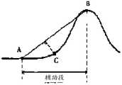
- the local distance transformationrefers to selecting an auxiliary segment containing the feature points to be extracted in the signal curve to be analyzed, and performing a straight line through the starting and ending points of the segment, and calculating each point on the signal curve in the auxiliary segment to The distance connecting the straight line from the beginning and the end of the segment, and the maximum distance point is used as the feature point in the signal curve of the segment.
- 4is a schematic diagram of a partial distance transformation, where AB represents the start and end points of the selected auxiliary segment, and C is the extracted feature point.
- an appropriate starting and ending pointcan be selected in the P wave detection area to be tested, and a straight line is calculated, and the distance from each point of the signal to be analyzed to the straight line is calculated, as shown in formula (2), the distance is the largest.
- the pointcorresponds to the peak of the second P wave, and then the second P wave position information can be acquired, and the second P wave amplitude and time width are calculated.
- xis the current point coordinate
- yis the corresponding signal amplitude value
- a, b, and care the straight line coefficients determined by the starting and ending points.
- step 302 and step 304can be reversed, and the order of the steps is not limited.
- Step 306Determine whether the first P wave position information and the second P wave position information are consistent.
- the first P wave position informationmay be obtained by the differential slope method, or the second P wave position information may be obtained by the local distance variation method, wherein the P wave position information in the body may be the first
- the P position informationmay also be the second P wave position information.
- the first P wave position and the second P wave position obtained by the differential slope method and the local distance transformation methodare inconsistent; Receiving the external interference, the first P wave position and the second P wave position obtained by the scoring slope method and the local transform method are substantially identical.
- the consistencymay be at least one of a start and end point of the P wave included in the P wave position information, an amplitude reference line, a P wave amplitude, and a time width.
- the P-wave position information in the bodycan be determined by comparing the acquired consistency of the first P-wave position information and the second P-wave position information.
- step 308the first P wave position information or the second P wave position information is used as the in vivo P wave position information. That is, the current ECG signal is collected without external interference, and the first P wave position information and the second P wave position information having consistency may be used as the P wave position information in the body.
- step 310is performed: determining P wave position information in the body according to the first P wave position information, the second P wave position information, and the preset physiological parameter range .
- the accurate P-wave position information in the bodycan be determined according to the acquired first P-wave position information and the second P-wave position information.
- the first P wave position information acquired by the differential slope methodis used as the P wave position information in the body;
- the second P-wave position information acquired by the local distance variation methodis used as the P-wave position information in the body.
- the differential slope method and the local distance variation methodcan be combined to obtain the corresponding detection result.
- the corresponding preset strategyis used to determine the P-wave position information in the body, and the P-wave position in the body can be improved. The accuracy of the information.
- acquiring the position information of the end of the catheter according to the mapping relationship between the ratio of the current P wave amplitude and the preset P wave amplitude and the preset position informationincluding: setting the preset P a amplitude value, the preset P wave amplitude is greater than 0; calculating a ratio of the current P wave amplitude to a preset P wave amplitude; when the ratio is greater than 1.5, the position information of the catheter end reaches Vena cava.
- P wave patternsinclude one-way (forward or negative), two-way (positive and negative bidirectional or negative positive bidirectional), and bimodal.
- the method provided by the present applicationcan be applied to a one-way (forward or negative) P-wave morphology. Among them, when the P wave shape is positive, the P wave amplitude is positive; when the P wave shape is negative, the P wave amplitude is negative.
- the preset P wave amplitudecan be the stable P surface amplitude obtained before surgery, or the average of the P wave amplitudes of a plurality of consecutive body surfaces obtained during the operation or a relatively stable one.
- P wave amplitudecan also be a magnitude that the doctor customizes according to the user's body surface ECG signal.
- the preset P waveis a forward P wave, and the preset P wave amplitude is greater than zero.
- acquiring the position information of the end of the catheter according to the mapping relationship between the ratio of the current P wave amplitude and the preset P wave amplitude and the preset position informationincluding: setting a preset P wave amplitude.
- the preset P wave amplitudeis greater than 0; the ratio of the current P wave amplitude to the preset P wave amplitude is calculated; when the ratio is greater than -0.6 and less than 0, the position information of the end of the catheter reaches the right atrium.
- the position information of the end of the catheterreaches the right atrium.
- the full in vivo P-QRS waveis displayed, including:
- Step 502Acquire a complete body surface P-QRS wave according to the body surface electrocardiographic signal, and display the body surface P-QRS wave as a shading contour.
- a complete body surface P-QRS wavecan be obtained, wherein the acquired body surface P-QRS wave can be displayed as a shading contour.
- the acquired body surface P-QRS wavecan also be displayed in a separate area.
- Step 504When acquiring the complete body surface P-QRS wave, the P-QRS wave in the body is displayed on the basis of the shading contour, so that the P wave component of the P-QRS wave of the body surface and the P wave component of the P-QRS wave in the body. Located in the same preset identifier.
- the acquired in vivo P-QRS waveis displayed on the basis of the shading profile, so that the P wave component of the body surface P-QRS wave and the P of the body P-QRS wave The wave components are in the same preset identification position.
- FIG. 6is an interface display diagram of a display window in an embodiment.
- the display windowincludes a plurality of display areas, and specifically includes an in-vivo electrocardiogram waveform for real-time display of P-QRS waves in the body.
- a display area 610a surface electrocardiogram waveform display area 620 for displaying a body surface P-QRS wave in real time; for displaying a body surface P-QRS wave (indicated by a broken line) and a P-QRS wave (solid line representation) in the body
- the contrast display area 630wherein the body surface P-QRS wave is displayed as a shading outline in the contrast display area 630.
- the preset flag 631is dynamically represented by a dotted line, so that the P wave amplitude can be directly obtained through the preset flag 631, and the P wave amplitude change is clear and intuitive.
- the location information of the catheter tipis displayed, including:
- Step 702Display a schematic diagram of the central vein, and identify the regions of the superior vena cava, the right atrium, and the inferior vena cava in the central vein schematic.
- the display windowfurther includes a catheter tip position display area 640 for displaying a central vein map, and a display area 640 at the catheter end position shows a schematic view of the heart, right atrium, superior vena cava, and inferior vena cava.
- Step 704In the central vein diagram, the corresponding area in the central vein schematic is illuminated according to the position information of the end of the catheter.
- the catheter end position display area 640displays the position information of the current catheter end in real time by means of text information for prompting the medical staff.
- the position of the end of the current catheteris also displayed in real time, and the manner of display can be displayed by illuminating the position of the end of the current catheter in the central vein map, which shows the end of the catheter by means of text and text.
- the location informationis more intuitively displayed to the medical staff.
- the catheter end position display area 640can also display the current patient's heart rate value, and provide the most effective physiological information to the operator intuitively. At the same time, the waveforms of the acquired body and body surface can also be saved.
- the catheter end positioning method of the present embodimentfurther includes:
- Step 802Receive a freeze instruction of the user, and obtain freeze information.
- the freeze informationincludes: current time, heart rate information, and a complete in-vivo P-QRS wave.
- the freeze informationincludes: current time, heart rate information, and complete in vivo P-QRS waves. That is, each time the P wave in the body is frozen, the recording time and heart rate value information can be frozen.
- freeze commandis used to freeze the P wave segment included in the current P-QRS wave in the body, and may be only a P wave component or a complete in vivo P-QRS wave.
- Step 804Display the freeze information in a time division manner according to a time sequence of receiving a freeze instruction of the user.
- the display windowfurther includes a plurality of freeze display areas 650 for displaying freeze information, and then step 804 displays the respective freeze information in the corresponding freeze display area 650 in sequence according to the reception time sequence of the freeze instruction.
- the display windowincludes three frozen display areas 650.
- the freeze commandis received in the first second
- the freeze informationis displayed in the first freeze display area 650
- a freeze commandis received, and the freeze command is correspondingly frozen.
- the informationis displayed in the second frozen display area 650, and upon receipt of a freeze command in the third second, the corresponding freeze information is displayed in the third freeze display area 650.
- the P wave shape corresponding to different parts of the catheter end during the operationcan be conveniently and accurately frozen, and the visual comparison is made at different times.
- the medical staffis more accurately judged the position of the catheter, and the frozen waveform data is provided to the medical staff as a study of the surgical report archive and the PICC puncture example.
- the name of the frozen P wave in the bodycan also be customized by the doctor according to the actual situation to meet the needs of different hospitals for different surgical reports. While displaying the frozen P wave in the body, the frozen recording time and heart rate value information can also be displayed, and the frozen information can be saved to the disease file as a report printout to achieve strong recordability of the patient's physiological information.
- the present applicationis capable of visually comparing P-wave variations using different display formats.
- the in-vivo electrocardiographic waveform display area 610 and the surface electrocardiographic waveform display area 620can realize real-time monitoring waveform comparison of the P-QRS wave and the body surface P-QRS wave in the body;
- the contrast display area 630is realized.
- the single complete P-QRS wave profile in the bodyis displayed in comparison with the preset P wave (doctor's custom);
- the medical staffcan freeze the body in different parts of the catheter end at different times in a plurality of frozen display areas 650 through subjective consciousness. Wave contrast.
- the medical treatmentcan quickly determine the trend of the P wave in the body and determine the position information of the end of the catheter in time.
- the change trend of the P wave in the body during the puncturecan be visually observed, and the various hazards caused by the blind wear of the PICC catheter can be avoided; and the waveform data of the body can be simply and efficiently passed through the subjective consciousness of the medical staff.
- the position information of the end of the catheter during the puncture processcan be automatically determined and displayed, and the most effective physiological information can be visually displayed to the medical staff, and the implementation efficiency, versatility and success of the electrocardiographic positioning operation can be improved. rate.
- the operation roomcan be operated without the operating room, and the operation flow is simple, and the complete catheterization operation process can be completed by the nurse alone.
- the present inventionfurther provides a catheter end positioning system for positioning a catheter of an intravascular device at a heart based on an electrocardiographic signal.
- the catheter end positioning system of the embodimentincludes:
- the collecting module 910is configured to simultaneously collect a body surface ECG signal and an internal ECG signal
- the obtaining module 920is configured to acquire, according to the body surface electrocardiographic signal, position information of the body surface QRS wave group in a preset time interval;
- the processing module 930is configured to acquire, according to the location information of the body surface QRS complex, the current P wave amplitude of the complete P-QRS wave in the in vivo ECG signal;
- the positioning module 940is configured to acquire the position information of the end of the catheter according to the mapping relationship between the ratio of the current P wave amplitude and the preset P wave amplitude and the preset position information;
- a display module 950is configured to display the complete in vivo P-QRS wave and position information of the catheter tip.
- the catheter end positioning systemcan position the catheter in the puncture catheter at the heart based on the electrocardiogram signal, and the body surface electrocardiogram signal and the in vivo ECG signal collected by the acquisition module 910 simultaneously, and the processing module 930 according to the body surface QRS complex
- the position informationis located to obtain the current P wave amplitude of the complete in vivo P-QRS wave in the ECG signal, and the positioning module 940 is based on the mapping relationship between the current P wave amplitude and the preset P wave amplitude and the preset position information.
- the above systemis convenient to operate and display intuitively, and greatly improves the practicability of the puncture operation. And success rate, and in the process of use, no other support personnel, and no harm to the human body.
- the processing moduleincludes:
- the location information and the RR interval width obtaining moduleare configured to obtain location information of at least two consecutive body surface QRS groups and an RR interval width of the adjacent two body surface QRS groups;
- the detection area acquisition module of the P-QRS waveis configured to obtain a complete detection region of the P-QRS wave in the body according to the position information of the QRS group and the RR interval width of the adjacent two body surface groups;
- a P wave position information acquiring moduleconfigured to acquire the P wave position information in the body in the detection area
- the current P-wave amplitude acquisition moduleis configured to acquire a current P-wave amplitude according to the P-wave position information.
- the P-wave position information acquisition moduleincludes:
- a first P wave position information acquiring moduleconfigured to acquire first P wave position information by using a differential slope method in the detection area
- a second P-wave position information acquiring moduleconfigured to acquire a second P-wave position information by using a local distance variation method in the detection area
- the determining moduleis configured to determine whether the first P wave position information and the second P wave position information are consistent; if they are consistent, the first P wave position information or the second P wave position information is used as the P wave position information in the body; if not, The P-wave position information in the body is determined according to the first P-wave position information, the second P-wave position information, and the preset physiological parameter range.
- the positioning modulecomprises:
- a first preset P wave amplitude setting moduleconfigured to set a preset P wave amplitude; the preset P wave amplitude is greater than 0;
- a first ratio calculation moduleconfigured to calculate a ratio of a current P wave amplitude to a preset P wave amplitude
- the positional information of the end of the catheterreaches the superior vena cava.
- the positioning modulecomprises:
- a second preset P wave amplitude setting moduleconfigured to set a preset P wave amplitude; the preset P wave amplitude is greater than 0;
- a second ratio calculation moduleconfigured to calculate a ratio of a current P wave amplitude to a preset P wave amplitude
- the position information of the end of the catheterreaches the right atrium.
- the display modulecomprises:
- the body surface P-QRS wave display moduleis configured to obtain a complete body surface P-QRS wave according to the body surface electrocardiographic signal, and display the body surface P-QRS wave as a shading contour;
- the in-vivo P-QRS wave display moduleis used to display the P-QRS wave in the body based on the shading profile when acquiring the complete body surface P-QRS wave, so that the P wave component of the body surface P-QRS wave and the body The P-wave component of the P-QRS wave is located at the same preset flag.
- the positioning modulecomprises:
- An identification modulefor displaying a central vein schematic and identifying regions of the superior vena cava, right atrium, and inferior vena cava in the central vein schematic;
- the lighting moduleis configured to illuminate a corresponding area in the central vein schematic according to the position information of the end of the catheter in the central vein diagram.
- the catheter end positioning system of the embodimentfurther includes:
- the freeze information acquisition moduleis configured to receive a freeze instruction of the user and obtain the freeze information; the freeze information includes: current time, heart rate information, and a complete in-vivo P-QRS wave;
- the freeze information display moduleis configured to display the freeze information in a time division manner according to the time sequence of receiving the freeze instruction.
- the display windowincludes an in vivo electrocardiographic waveform display area 610, a body surface electrocardiographic waveform display area 620, a contrast display area 630, and a catheter end position display. A region 640 and a plurality of frozen display regions 650.
- the catheter end positioning system in this embodimentincludes: a display window display module, configured to display the internal electrocardiogram waveform, the surface electrocardiogram waveform, the position information of the catheter end, and the freeze information in the display window. .
- the in-vivo electrocardiogram display area 610is configured to display a complete in-vivo P-QRS wave;
- the surface electrocardiographic waveform display area 620is configured to display a body surface P-QRS wave formed according to the body surface electrocardiographic signal;
- the contrast display area 630is used to simultaneously display the body surface P-QRS wave and the complete in vivo P-QRS wave;
- the catheter end position display area 640is used to display the position information of the catheter end in the central vein schematic in real time; and the plurality of frozen display areas 650 are used for The freeze information obtained in the order of receiving time according to the freeze instruction of the user is displayed correspondingly.
- the body surface P-QRS waveis displayed as a shading outline in the contrast display area 630.
- the preset flag 631is dynamically represented by a dotted line, so that the P wave amplitude can be directly obtained through the preset flag 631, and the P wave amplitude change is clear and intuitive.
- the catheter end position display area 640displays the position information of the current catheter end in real time by means of text information for prompting the medical staff.
- the position of the end of the current catheteris also displayed in real time, and the manner of display can be displayed by illuminating the position of the end of the current catheter in the central vein map, which shows the end of the catheter by means of text and text.
- the location informationis more intuitively displayed to the medical staff.
- the catheter end position display area 640can also display the current patient's heart rate value, and provide the most effective physiological information to the operator intuitively. At the same time, the waveforms of the acquired body and body surface can also be saved.
- the freeze display area 650includes three independent freeze units for displaying corresponding freeze information in time series.
- the P wave shape corresponding to different parts of the catheter end during the operationcan be conveniently and accurately frozen, and the difference is obtained.
- the intuitive contrast of the timeallows the medical staff to more accurately determine the location of the catheter, while the frozen waveform data is provided to the medical staff as a surgical report archive and PICC puncture examples.
- the name of the frozen P wave in the bodycan also be customized by the doctor according to the actual situation to meet the needs of different hospitals for different surgical reports. While displaying the frozen P wave in the body, the frozen recording time and heart rate value information can also be displayed, and the frozen information can be saved to the disease file as a report printout to achieve strong recordability of the patient's physiological information.
- the display windowfurther includes an information prompt area for displaying performance parameters of the device having the display window and the measured user information.
- the display windowalso includes space controls for implementing multi-function control, and the like.
- the performance parameters of the devicemay include battery parameters and network parameters
- the measured user informationmay include user identity information and user illness information.
- the present applicationis capable of visually comparing P-wave variations using different display formats.
- the in-vivo electrocardiographic waveform display area 610 and the surface electrocardiographic waveform display area 620can realize real-time monitoring waveform comparison of the P-QRS wave and the body surface P-QRS wave in the body;
- the contrast display area 630is realized.
- the single complete P-QRS wave profile in the bodyis displayed in comparison with the preset P wave (doctor's custom);
- the medical staffcan freeze the body in different parts of the catheter end at different times in a plurality of frozen display areas 650 through subjective consciousness. Wave contrast.
- the medical treatmentcan quickly determine the trend of the P wave in the body and determine the position information of the end of the catheter in time.
- the trend of the P wave in the body during the puncturecan be visually observed, and the various hazards caused by the blind insertion of the PICC catheter can be avoided; and the subjective consciousness of the medical staff is simply and efficiently
- the waveform data in the bodyis saved, the position information of the end of the catheter during the puncture process can be automatically determined and displayed, and the most effective physiological information can be visually displayed to the medical staff, and the implementation efficiency of the electrocardiographic positioning operation can be improved. Sex and success rate.
- catheter end positioning systemThe division of the various modules in the catheter end positioning system described above is for illustration only. In other embodiments, the catheter end positioning system can be divided into different modules as needed to perform all or part of the functions of the catheter end positioning system described above.
- the various modules in the catheter end positioning system described abovemay be implemented in whole or in part by software, hardware, and combinations thereof.
- Each of the above modulesmay be embedded in or independent of the processor in the computer device, or may be stored in a memory in the computer device in a software form, so that the processor invokes the operations corresponding to the above modules.
- One or more non-volatile readable storage media storing computer readable instructionsare also presented.
- one or more non-transitory readable storage mediumsstoring computer readable instructions, when executed by one or more processors, cause one or more processors to perform any of the above The steps of the catheter tip positioning method in the embodiment.
- a computer apparatuscomprising a memory and a processor, wherein the memory stores a computer program, the computer program being executed by the processor, causing the processor to perform catheter end positioning in any of the above embodiments The steps of the method.
- Non-volatile memorycan include read only memory (ROM), programmable ROM (PROM), electrically programmable ROM (EPROM), electrically erasable programmable ROM (EEPROM), or flash memory.
- Volatile memorycan include random access memory (RAM), which acts as an external cache.
- RAMis available in a variety of forms, such as static RAM (SRAM), dynamic RAM (DRAM), synchronous DRAM (SDRAM), dual data rate SDRAM (DDR SDRAM), enhanced SDRAM (ESDRAM), synchronization.
- SRAMstatic RAM
- DRAMdynamic RAM
- SDRAMsynchronous DRAM
- DDR SDRAMdual data rate SDRAM
- ESDRAMenhanced SDRAM
- synchronizationLink (Synchlink) DRAM (SLDRAM), Memory Bus (Rambus) Direct RAM (RDRAM), Direct Memory Bus Dynamic RAM (DRDRAM), and Memory Bus Dynamic RAM (RDRAM).
Landscapes
- Health & Medical Sciences (AREA)
- Life Sciences & Earth Sciences (AREA)
- Cardiology (AREA)
- Surgery (AREA)
- Medical Informatics (AREA)
- Pathology (AREA)
- Biomedical Technology (AREA)
- Heart & Thoracic Surgery (AREA)
- Engineering & Computer Science (AREA)
- Molecular Biology (AREA)
- Animal Behavior & Ethology (AREA)
- General Health & Medical Sciences (AREA)
- Public Health (AREA)
- Veterinary Medicine (AREA)
- Physics & Mathematics (AREA)
- Biophysics (AREA)
- Nuclear Medicine, Radiotherapy & Molecular Imaging (AREA)
- Measurement And Recording Of Electrical Phenomena And Electrical Characteristics Of The Living Body (AREA)
Abstract
Description
Translated fromChinese本发明涉及血管内设备定位技术领域,特别是涉及导管末端定位方法和系统。The present invention relates to the field of intravascular device positioning technology, and more particularly to a catheter end positioning method and system.
经外周静脉置入的中心静脉导管(Peripherally Inserted Central Venous Catheters,简称PICC)是一种将导管插入脉管并置留开口于体外的治疗技术,手术过程中需要将导管经由人体经脉,较为精准的穿刺到上腔静脉靠近右心房交界处,这样就能够迅速将药物输入血液循环系统距离心脏较近的位置。穿刺置管术的关键在于让医护人员能及时准确的找到导管末端的位置,如导管穿刺位置过浅或过深,会引起血栓、静脉炎、静脉壁腐蚀、静脉穿孔、心肌穿孔等并发症。Peripherally inserted Central Venous Catheters (PICC) is a kind of treatment technique in which a catheter is inserted into a vessel and left in the body. The catheter needs to be passed through the body meridian during the operation. Puncture into the superior vena cava near the right atrium junction, which allows the drug to be quickly introduced into the blood circulation system closer to the heart. The key to puncture catheterization is to enable medical staff to find the position of the end of the catheter in a timely and accurate manner. If the catheter is puncture too shallow or too deep, it may cause complications such as thrombosis, phlebitis, vein wall corrosion, venous perforation, and myocardial perforation.
为实现导管在体内穿刺过程中的精准定位,需要采用如X线定位、心电(electrocardiogram,心电)定位等辅助定位方式。其中,X线定位系统体积较大,操作复杂,需要由资深专业医生操作,并需要护士辅助,且人体长期接受X线照射会杀死体内白细胞,降低人体免疫力。采用心电定位时,需要依赖操作者的经验盲穿,穿刺成功率大大降低。In order to achieve accurate positioning of the catheter in the body puncture process, it is necessary to adopt auxiliary positioning methods such as X-ray positioning, electrocardiogram (electrocardiogram) positioning. Among them, the X-ray positioning system is large in size and complicated in operation, and needs to be operated by a senior professional doctor, and needs nurse assistance, and the long-term acceptance of X-ray irradiation of the human body will kill white blood cells in the body and reduce human immunity. When using ECG positioning, it is necessary to rely on the operator's experience to blindly wear, and the puncture success rate is greatly reduced.
发明内容Summary of the invention
基于此,有必要提供一种导管末端定位方法和系统。Based on this, it is necessary to provide a catheter end positioning method and system.
一种导管末端定位方法,基于心电图信号将穿刺置管术中的导管定位在心脏处,所述方法包括:A catheter tip positioning method for positioning a catheter in a puncture catheter at a heart based on an electrocardiographic signal, the method comprising:
同时采集体表心电信号和体内心电信号;Simultaneously collecting body surface ECG signals and in vivo ECG signals;
在预设时间间隔内,根据所述体表心电信号获取体表QRS波群的位置信息;Obtaining, according to the body surface electrocardiographic signal, position information of the body surface QRS wave group in a preset time interval;
根据所述体表QRS波群的位置信息定位获取所述体内心电信号中完整的体内P-QRS波的当前P波幅值;Obtaining a current P wave amplitude of a complete in vivo P-QRS wave in the in vivo ECG signal according to position information of the body surface QRS complex;
根据所述当前P波幅值与预设P波幅值的比值和预设位置信息的映射关系,获取所述导管末端的位置信息;Obtaining location information of the end of the catheter according to a mapping relationship between a ratio of the current P wave amplitude to a preset P wave amplitude and preset position information;
显示完整的所述体内P-QRS波和所述导管末端的位置信息。The complete in vivo P-QRS wave and position information of the catheter tip are displayed.
还提供一种导管末端定位系统,基于心电图信号将血管内设备的导管定位在心脏处,所述系统包括:There is also provided a catheter tip positioning system for positioning a catheter of an intravascular device at a heart based on an electrocardiographic signal, the system comprising:
采集模块,用于同时采集体表心电信号和体内心电信号;An acquisition module for simultaneously acquiring a surface electrocardiogram signal and an in vivo ECG signal;
获取模块,用于在预设时间间隔内,根据所述体表心电信号获取体表QRS波群的位置信息;An acquiring module, configured to acquire, according to the body surface electrocardiographic signal, position information of a body surface QRS wave group in a preset time interval;
处理模块,用于根据所述体表QRS波群的位置信息定位获取所述体内心电信号中完整的体内P-QRS波的当前P波幅值;a processing module, configured to acquire, according to location information of the body surface QRS complex, a current P wave amplitude of a complete in vivo P-QRS wave in the in vivo ECG signal;
定位模块,用于根据所述当前P波幅值与预设P波幅值的比值和预设位置信息的映射关系,获取所述导管末端的位置信息;及a positioning module, configured to acquire position information of the end of the catheter according to a mapping relationship between a ratio of the current P wave amplitude to a preset P wave amplitude and preset position information; and
显示模块,用于显示完整的所述体内P-QRS波和所述导管末端的位置信息。A display module is configured to display the complete in-vivo P-QRS wave and position information of the catheter tip.
还提出一个或多个存储有计算机可读指令的非易失性可读存储介质,所述计算机可读指令被一个或多个处理器执行时,使得所述一个或多个处理器执行以下步骤:Also presented is one or more non-transitory readable storage mediums storing computer readable instructions, which when executed by one or more processors, cause the one or more processors to perform the following steps :
同时采集体表心电信号和体内心电信号;Simultaneously collecting body surface ECG signals and in vivo ECG signals;
在预设时间间隔内,根据所述体表心电信号获取体表QRS波群的位置信息;Obtaining, according to the body surface electrocardiographic signal, position information of the body surface QRS wave group in a preset time interval;
根据所述体表QRS波群的位置信息定位获取所述体内心电信号中完整的体内P-QRS波的当前P波幅值;Obtaining a current P wave amplitude of a complete in vivo P-QRS wave in the in vivo ECG signal according to position information of the body surface QRS complex;
根据所述当前P波幅值与预设P波幅值的比值和预设位置信息的映射关 系,获取所述导管末端的位置信息;以及Obtaining position information of the end of the catheter according to a mapping relationship between a ratio of the current P wave amplitude to a preset P wave amplitude and preset position information;
显示完整的所述体内P-QRS波和所述导管末端的位置信息。The complete in vivo P-QRS wave and position information of the catheter tip are displayed.
还提出一种计算机设备,包括存储器及处理器,所述存储器中储存有计算机程序,所述计算机程序被所述处理器执行时,使得所述处理器执行以下步骤:A computer apparatus is also provided, comprising a memory and a processor, wherein the memory stores a computer program, and when the computer program is executed by the processor, the processor performs the following steps:
同时采集体表心电信号和体内心电信号;Simultaneously collecting body surface ECG signals and in vivo ECG signals;
在预设时间间隔内,根据所述体表心电信号获取体表QRS波群的位置信息;Obtaining, according to the body surface electrocardiographic signal, position information of the body surface QRS wave group in a preset time interval;
根据所述体表QRS波群的位置信息定位获取所述体内心电信号中完整的体内P-QRS波的当前P波幅值;Obtaining a current P wave amplitude of a complete in vivo P-QRS wave in the in vivo ECG signal according to position information of the body surface QRS complex;
根据所述当前P波幅值与预设P波幅值的比值和预设位置信息的映射关系,获取所述导管末端的位置信息;以及Acquiring the position information of the end of the catheter according to the mapping relationship between the ratio of the current P wave amplitude and the preset P wave amplitude and the preset position information;
显示完整的所述体内P-QRS波和所述导管末端的位置信息。The complete in vivo P-QRS wave and position information of the catheter tip are displayed.
本发明的一个或多个实施例的细节在下面的附图和描述中提出。本发明的其它特征、目的和优点将从说明书、附图以及权利要求书变得明显。Details of one or more embodiments of the invention are set forth in the accompanying drawings and description below. Other features, objects, and advantages of the invention will be apparent from the description and appended claims.
通过附图中所示的本发明的优选实施例的更具体说明,本发明的上述及其它目的、特征和优势将变得更加清晰。在全部附图中相同的附图标记指示相同的部分,且并未刻意按实际尺寸等比例缩放绘制附图,重点在于示出本发明的主旨。The above and other objects, features and advantages of the present invention will become more apparent from the <RTIgt; The same reference numerals are used throughout the drawings to refer to the same parts, and the drawings are not intended to be scaled to the actual size. The emphasis is on the subject matter of the invention.
图1为一个实施例中导管末端定位方法的流程图;Figure 1 is a flow chart showing a method of positioning a catheter tip in an embodiment;
图2为一个实施例中根据所述体表QRS波群的位置信息定位获取所述体内心电信号中完整的体内P-QRS波的当前P波幅值的流程图;2 is a flow chart showing the acquisition of the current P-wave amplitude of a complete in vivo P-QRS wave in the in vivo ECG signal according to the position information of the body surface QRS complex in one embodiment;
图3为一个实施例中在所述检测区定位获取体内P波位置信息的流程图;FIG. 3 is a flowchart of acquiring P-wave position information in the body in the detection area according to an embodiment; FIG.
图4为局部距离变换的示意图;Figure 4 is a schematic diagram of local distance transformation;
图5为一个实施例中显示完整的所述体内P-QRS波的流程图;Figure 5 is a flow diagram showing the complete in vivo P-QRS wave in one embodiment;
图6为一个实施例中显示窗口的界面显示图;6 is an interface display diagram of a display window in an embodiment;
图7为一个实施例中显示所述导管末端的位置信息的流程图;Figure 7 is a flow chart showing positional information of the end of the catheter in one embodiment;
图8为另一个实施例中导管末端定位方法的流程图;Figure 8 is a flow chart showing a method of positioning a catheter tip in another embodiment;
图9为一个实施例中导管末端定位系统的结构框架图。Figure 9 is a structural block diagram of a catheter end positioning system in one embodiment.
为了便于理解本发明,下面将参照相关附图对本发明进行更全面的描述。附图中给出了本发明的首选实施例。但是,本发明可以以许多不同的形式来实现,并不限于本文所描述的实施例。相反地,提供这些实施例的目的是使对本发明的公开内容更加透彻全面。In order to facilitate the understanding of the present invention, the present invention will be described more fully hereinafter with reference to the accompanying drawings. Preferred embodiments of the invention are given in the drawings. However, the invention may be embodied in many different forms and is not limited to the embodiments described herein. Rather, these embodiments are provided so that this disclosure will be thorough and comprehensive.
除非另有定义,本文所使用的所有的技术和科学术语与属于本发明的技术领域的技术人员通常理解的含义相同。本文中在本发明的说明书中所使用的术语只是为了描述具体的实施例的目的,不是旨在于限制本发明。一种导管末端定位方法,基于心电图信号将穿刺置管术中的导管定位在心脏处。All technical and scientific terms used herein have the same meaning as commonly understood by one of ordinary skill in the art to which this invention belongs, unless otherwise defined. The terminology used in the description of the present invention is for the purpose of describing particular embodiments and is not intended to limit the invention. A catheter tip positioning method for positioning a catheter in a puncture catheter at a heart based on an electrocardiographic signal.
需要说明的是,心脏收缩和舒张一次构成一个心动周期,一个心动周期的心电信号一般包括P波、PR段、QRS波群、ST段、T波等。其中,P波,正常心脏的电激动从窦房结开始。由于窦房结位于右心房与上腔静脉的交界处,所以窦房结的激动首先传导到右心房,通过房间束传到左心房,形成心电图上的P波。P波代表了心房的激动,是每一波组中的第一波,前半部代表右心房的激动,后半部代表左心房的激动。QRS波群,包括三个紧密相连的波,第一个向下的波称为Q波,继Q波后的一个高尖的直立波称为R波,R波后向下的波称为S波。因其紧密相连,且反映了心室的电激动过程,故统称为QRS波群。这个波群反映了左、右两心室的除极过程。It should be noted that the systolic and diastolic phases constitute a cardiac cycle, and the cardiac signal of a cardiac cycle generally includes a P wave, a PR segment, a QRS complex, an ST segment, a T wave, and the like. Among them, P wave, the electrical activation of the normal heart begins from the sinus node. Since the sinoatrial node is located at the junction of the right atrium and the superior vena cava, the sinus node is first transmitted to the right atrium and passed through the room bundle to the left atrium to form a P wave on the electrocardiogram. The P wave represents the atrial excitement and is the first wave in each wave group. The first half represents the excitement of the right atrium and the second half represents the activation of the left atrium. The QRS complex consists of three closely connected waves. The first downward wave is called the Q wave. A high-point vertical wave after the Q wave is called the R wave. The R wave is followed by the downward wave. wave. Because they are closely connected and reflect the electrical activation process of the ventricle, they are collectively referred to as QRS complexes. This wave group reflects the depolarization process of the left and right ventricles.
穿刺置管术中的导管可以理解为经外周静脉置入的中心静脉导管PICC,该穿刺置管术是基于四电极心电系统,该四电心电系统至少包括体表电极和 体内电极(经外周静脉置入的中心静脉导管内的电导丝),其中,体表电极包括体表电极LA、体表电极LL和体表电极RL,其中,体表电极LA置于人体的左胳膊,体表电极LL置于人体的左腿,体表电极RL置于人体的右腿。A catheter in a puncture catheter can be understood as a central venous catheter PICC placed through a peripheral vein, which is based on a four-electrode electrocardiographic system comprising at least a body surface electrode and an internal electrode (via a guide wire in the central venous catheter in which the peripheral vein is inserted, wherein the body surface electrode includes a body surface electrode LA, a body surface electrode LL, and a body surface electrode RL, wherein the body surface electrode LA is placed on the left arm of the human body, and the body surface is The electrode LL is placed on the left leg of the human body, and the body surface electrode RL is placed on the right leg of the human body.
在一个实施例中,导管末端定位方法,包括:In one embodiment, a catheter end positioning method includes:
步骤102:同时采集体表心电信号和体内心电信号。Step 102: Simultaneously collect the surface ECG signal and the internal ECG signal.
需要进行穿刺置管术的病人在穿刺前,首先接好体表导联线(体表电极LA、体表电极LL和体表电极RL),其中一个体表导联线的鳄鱼夹子接到进行穿刺的电导丝。穿刺过程中,通过静脉穿刺,将经外周静脉置入的中心静脉导管(Peripherally Inserted Central Venous Catheters,简称PICC)刺入人体内。此时,可以同时通过体表电极实时采集体表心电信号,即人体表面心电波形数据,利用PICC内的电导丝(体内电极)采集人体内的体内心电信号。Before the puncture, the patient who needs to perform the puncture catheter is connected to the body lead wire (the body surface electrode LA, the body surface electrode LL and the body surface electrode RL), and one of the body surface lead wires of the crocodile clip is connected. Puncture of the electrical guide wire. During the puncture, a Peripherally Inserted Central Venous Catheters (PICC) inserted into the peripheral vein was inserted into the human body by venipuncture. At this time, the body surface electrocardiographic signal, that is, the surface electrocardiogram waveform data of the human body surface, can be simultaneously collected through the body surface electrode, and the electrocardiographic signal (in vivo electrode) in the PICC is used to collect the in vivo ECG signal in the human body.
步骤104:在预设时间间隔内,根据体表心电信号获取体表QRS波群的位置信息。Step 104: Acquire location information of the body surface QRS group according to the body surface electrocardiogram signal within a preset time interval.
在预设时间间隔(2~3秒)内,可以获取2000~3000个用户表示体表心电信息和体内心电信息的数据,根据获取的体表心电信号获取体表QRS波群的位置信息。QRS波群分布于心电信号的中、高频区域,峰值落在10~20Hz之间,QRS波群的幅度特征非常明显,与其他波形区别显著。可以采用实时检测方法和非实时检测方法来检测QRS波群的位置信息。其中,非实时的检测方式可以采用小波分析法、神经网络法以及基于图形识别法;实时的检测方法可以对信号进行滤波差分处理,并结合幅值来确定QRS波群的位置。具体地,利用基于差分斜率法可以检测出各周期中R波的顶点、RR间期信息,进而分别向前、向后寻找转折点,以获取Q波和S波的位置,也即,可以获取QRS波群的位置信息,计算出QRS波群的宽度信息,同时,还可以根据RR间期和QRS波的宽度,对心电信号进行诊断分类。In a preset time interval (2 to 3 seconds), 2000 to 3000 data representing the surface ECG information and the in vivo ECG information can be obtained, and the position of the body surface QRS group is obtained according to the acquired body surface electrocardiographic signal. information. The QRS complex is distributed in the middle and high frequency regions of the ECG signal. The peak value falls between 10 and 20 Hz. The amplitude characteristics of the QRS complex are very obvious, which is significantly different from other waveforms. Real-time detection methods and non-real-time detection methods can be used to detect the position information of the QRS complex. Among them, the non-real-time detection method can adopt the wavelet analysis method, the neural network method and the pattern recognition method; the real-time detection method can filter the differential processing of the signal, and combine the amplitude to determine the position of the QRS group. Specifically, the vertices and RR interval information of the R waves in each period can be detected by using the differential slope method, and then the turning points are searched forward and backward respectively to obtain the positions of the Q wave and the S wave, that is, the QRS can be acquired. The position information of the wave group is used to calculate the width information of the QRS complex. At the same time, the ECG signals can be classified and classified according to the RR interval and the width of the QRS wave.
步骤106:根据体表QRS波群的位置信息定位获取体内心电信号中完整的体内P-QRS波的当前P波幅值。Step 106: According to the position information of the body surface QRS complex, the current P wave amplitude of the complete in vivo P-QRS wave in the in vivo ECG signal is obtained.
根据获取的QRS波群的位置信息就可以定位出体内相应的QRS波群的 位置信息。体内的P波在手术过程中,会变得高耸,这会给QRS波群的检测带来大的干扰,没法准确检测到每一个QRS波群的位置信息。鉴于体表、体内的QRS波群的位置信息一致,就可以通过体表的导联波形来确定的内心的QRS波群的位置信息。根据获取的体内QRS波群的位置信息,结合预设的算法,就可以在QRS波群的基础上,获取完整的P-QRS波,根据完整的P-QRS波,获取当前P波幅值。According to the obtained location information of the QRS complex, the location information of the corresponding QRS complex in the body can be located. The P wave in the body will become high during the operation, which will cause great interference to the detection of the QRS complex, and it is impossible to accurately detect the position information of each QRS complex. In view of the fact that the position information of the QRS complex in the body surface and the body is identical, the position information of the inner QRS complex can be determined by the lead waveform of the body surface. According to the obtained position information of the QRS complex in the body, combined with the preset algorithm, the complete P-QRS wave can be obtained on the basis of the QRS complex, and the current P-wave amplitude is obtained according to the complete P-QRS wave.
步骤108:根据当前P波幅值与预设P波幅值的比值和预设导管末端位置信息的映射关系,获取导管末端的位置信息。Step 108: Obtain position information of the end of the catheter according to the mapping relationship between the ratio of the current P wave amplitude and the preset P wave amplitude and the preset catheter end position information.
根据当前P波幅值和预设P波幅值就可以计算出两者的比值,根据该比值与预设的导管末端位置信息的映射关系,就可以获取当前P波幅值对应的导管末端的位置信息。According to the current P wave amplitude and the preset P wave amplitude, the ratio of the two can be calculated. According to the mapping relationship between the ratio and the preset catheter end position information, the end of the catheter corresponding to the current P wave amplitude can be obtained. location information.
其中,预设P波幅值,可以为手术前,获取的稳定的体表P波幅值,也可以为手术过程中,获取的多个连续体表P波幅值的平均值或某一个相对稳定的P波幅值;也可以是医生根据用户的体表心电信号自定义的一个幅值。在此,对P波幅值的设定方式,不做进一步的限定,可以根据用户需求来设定。Wherein, the preset P wave amplitude may be a stable body surface P wave amplitude obtained before surgery, or may be an average value or a relative value of a plurality of continuous body surface P wave amplitudes obtained during the operation. A stable P-wave amplitude; it can also be a magnitude that the doctor customizes based on the user's body surface ECG signal. Here, the setting method of the P wave amplitude is not further limited, and can be set according to user requirements.
基于体内P波在上腔静脉、右心房等不同部位会发生特异性变化就可以精准的定位体内导管末端的位置信息。该特异性变化可以理解为:导管末端进入上腔静脉,体内P波幅值会逐渐升高,导管末端到达上腔静脉与右心房的上壁交界连接点(Cavoatrial Junction,简称CAJ)时,体内P波幅值与QRS主波幅值平齐。其中,上腔静脉与右心房的上壁交界连接点CAJ,也即,上腔静脉下1/3段,靠近右心房交界处,CAJ标志着上腔静脉的终点,也是PICC(Peripherally Inserted Central Venous Catheters,经外周静脉置入中心静脉导管)尖端留置的最佳位置。Based on the P-wave in the body, specific changes occur in different parts of the superior vena cava and right atrium to accurately locate the position of the end of the catheter in the body. This specific change can be understood as: the end of the catheter enters the superior vena cava, the amplitude of the P wave in the body will gradually increase, and the end of the catheter reaches the upper boundary junction of the superior vena cava and the right atrium (Cavoatrial Junction, CAJ for short). The amplitude of the P wave is flush with the amplitude of the QRS main wave. Among them, the superior vena cava and the right atrium of the upper wall junction junction CAJ, that is, the lower third of the superior vena cava, near the right atrium junction, CAJ marks the end of the superior vena cava, is also PICC (Peripherally Inserted Central Venous) Catheters, the central venous catheter through the peripheral vein) the best position for tip retention.
根据该特异性变化,可以设定当前P波幅值与预设P波幅值的比值和预设导管末端位置信息的映射关系,根据该映射关系就可以准确的定位导管末端的位置信息。According to the specificity change, the mapping relationship between the ratio of the current P wave amplitude and the preset P wave amplitude and the preset catheter end position information can be set, and the position information of the catheter end can be accurately located according to the mapping relationship.
步骤110:显示完整的体内P-QRS波和导管末端的位置信息。Step 110: Display the complete in vivo P-QRS wave and position information at the end of the catheter.
当获取的完整的体内P-QRS波和导管末端的位置信息时,可以将获取的所有信息进行显示,以供医护人员查看。When the complete in vivo P-QRS wave and the position information of the end of the catheter are obtained, all the acquired information can be displayed for medical personnel to view.
上述导管末端定位方法,可以基于心电图信号将穿刺置管术中的导管定位在心脏处,通过同时采集体表心电信号和体内心电信号,并根据体表QRS波群的位置信息定位获取体内心电信号中完整的体内P-QRS波的当前P波幅值,根据当前P波幅值与预设P波幅值的比值和预设位置信息的映射关系,获取导管末端的位置信息,并显示完整的体内P-QRS波和所述导管末端的位置信息,以供医护人员查看,上述方法操作方便、显示直观,极大提高穿刺手术的实用性,成功率,且在使用的过程中,不需其他辅助人员,且对人体无任何伤害。The catheter end positioning method can position the catheter in the puncture catheter at the heart based on the electrocardiogram signal, and simultaneously acquire the body surface electrocardiogram signal and the internal electrocardiogram signal, and obtain the body according to the position information of the body surface QRS wave group. Obtaining the current P-wave amplitude of the in-vivo P-QRS wave in the ECG signal, obtaining the position information of the end of the catheter according to the mapping relationship between the ratio of the current P-wave amplitude and the preset P-wave amplitude and the preset position information, and Displaying the complete in vivo P-QRS wave and the position information of the end of the catheter for medical personnel to view, the above method is convenient to operate, and the display is intuitive, greatly improving the practicability of the puncture operation, the success rate, and in the process of using, No other support personnel are required and there is no harm to the human body.
如图2所示,在一个实施例中,根据体表QRS波群的位置信息定位获取体内心电信号中完整的体内P-QRS波的当前P波幅值,包括:As shown in FIG. 2, in one embodiment, the current P-wave amplitude of the in-vivo P-QRS wave in the in-vivo ECG signal is obtained according to the position information of the body surface QRS complex, including:
步骤202:获取至少两个连续的体表QRS波群的位置信息以及相邻两个体表QRS波群的RR间期宽度。Step 202: Acquire location information of at least two consecutive body surface QRS groups and an RR interval width of adjacent two body surface QRS groups.
在一个实施例中,可以根据获取的体表电信息,可以准确的获取在这预设时间间隔内(2~3秒)完整的心电图波形,也即,完整的P-QRS波。根据在预设时间间隔内获取的完整的心电波形,可以筛选出至少两个连续的QRS波群,以及两个连续的QRS波群之间RR间期宽度。In one embodiment, the complete ECG waveform, that is, the complete P-QRS wave, can be accurately acquired within the preset time interval (2 to 3 seconds) according to the acquired body surface electrical information. Based on the complete ECG waveform acquired during the preset time interval, at least two consecutive QRS complexes and the RR interval width between two consecutive QRS complexes can be screened.
具体的,可以筛选出至少三个连续的QRS波群,也即,可以获取两个连续QRS波群的RR间期宽度,可以根据获取的两个连续的RR间期宽度,若两个连续的RR间期宽度在预设范围内相等,则表明未发生严重的波形干扰的状况(比如病患突发心律失常或剧烈运动),则可以认为获取的QRS波群的位置信息是准确的。Specifically, at least three consecutive QRS complexes can be selected, that is, the RR interval width of two consecutive QRS complexes can be obtained, which can be obtained according to two consecutive RR interval widths, if two consecutive If the RR interval width is equal within the preset range, indicating that there is no serious waveform interference (such as sudden arrhythmia or strenuous exercise), the position information of the acquired QRS complex can be considered accurate.
步骤204:根据相邻两个体表QRS波群的位置信息和RR间期宽度,获取完整的体内P-QRS波的检测区。Step 204: Obtain a complete detection region of the P-QRS wave in the body according to the position information of the adjacent QRS group and the RR interval width.
由于,体表的QRS波群的位置信息和RR间期宽度与体内的QRS波群 的位置信息和RR间期宽度具有一致性。同时,心动周期的心电信号所包括的波形,P波、PR段、QRS波群、ST段、T波等,其中,P波代表了心房的激动,是每一波组中的第一波,位于QRS波群前。根据获取的两个连续的体表QRS波群的位置信息,以及RR间期宽度,就可以间接的推算出体内P-QRS波的检测区。Because the position information of the QRS complex of the body surface and the RR interval width are consistent with the position information of the QRS complex in the body and the RR interval width. At the same time, the waveform of the ECG signal of the cardiac cycle, P wave, PR segment, QRS complex, ST segment, T wave, etc., wherein P wave represents the atrial activation, is the first wave in each wave group , located in front of the QRS complex. According to the obtained position information of two consecutive body surface QRS complexes and the RR interval width, the detection area of the P-QRS wave in the body can be indirectly derived.
步骤206:在完整的体内P-QRS波的检测区定位获取体内P波位置信息。Step 206: Acquire P-wave position information in the body in the detection region of the complete in vivo P-QRS wave.
在获取的体内P-QRS波的检测区内,根据预设算法,例如差分斜率结合局部距离变换法,就可以准确的定位出体内P波的位置信息。In the acquired detection region of the P-QRS wave in the body, according to a preset algorithm, such as a differential slope combined with a local distance transformation method, the position information of the P wave in the body can be accurately located.
步骤208:根据P波位置信息获取当前P波幅值。Step 208: Acquire a current P wave amplitude according to the P wave position information.
其中,P波位置信息包括P波的起、止端点、幅值基准线、P波幅值以及时间宽度等。根据获取的P波位置信息就可以获取当前P波幅值。The P wave position information includes a start and end point of the P wave, an amplitude reference line, a P wave amplitude, and a time width. According to the acquired P wave position information, the current P wave amplitude can be obtained.
进一步的,如图3所示,在一个实施例中,在完整的体内P-QRS波的检测区定位获取体内P波位置信息,包括:Further, as shown in FIG. 3, in one embodiment, acquiring P-wave position information in the body in the detection region of the complete in vivo P-QRS wave includes:
步骤302:在检测区采用差分斜率法获取第一P波位置信息。Step 302: Acquire a first P wave position information by using a differential slope method in the detection area.
对于心电信号中,考虑当前选取的QRS波群与上一个QRS波群之间,排除T,P波会存在的片段,基于这个片段通过差分斜率法寻找P波位置。For the ECG signal, consider the segment between the currently selected QRS complex and the previous QRS complex, excluding the T and P waves, and find the P-wave position based on this segment by the differential slope method.
根据差分公式(1)确定体内心电信息的负斜率Determine the negative slope of the in vivo ECG information according to the difference formula (1)
Diff(x)=S(x)-S(x-t) (1)Diff(x)=S(x)-S(x-t) (1)
Diff(x)为差分后信号,S(x)为体内P波检测区差分前信号,t为设定的间隔(差分的时间差),x为当前点坐标。可以根据采用频率确定合适的差分间隔,利用预设时间间隔的差分结果找到斜率最大值的心电信号的幅值最大值,P波检测区差分前信号通过幅度校准,以获取幅值最大位置,该位置的位置信息就为第一P波位置信息。其中,第一P波位置信息包括第一P波的起、止点位置信息、幅值基准线、第一P波幅值以及时间宽度等。其中,第一P波在去除QRS波群和T波之外、且对于基线波形是有明显起伏的波形。Diff(x) is the differential signal, S(x) is the pre-differential signal of the P-wave detection area in the body, t is the set interval (time difference of the difference), and x is the current point coordinate. The appropriate differential interval can be determined according to the frequency of use, and the maximum value of the maximum value of the ECG signal of the slope maximum value can be found by using the difference result of the preset time interval, and the signal before the difference of the P wave detection area is calibrated by the amplitude to obtain the maximum amplitude position. The position information of the position is the first P wave position information. The first P wave position information includes start and stop position information of the first P wave, an amplitude reference line, a first P wave amplitude, and a time width. Wherein, the first P wave is in addition to the QRS complex and the T wave, and has a significant undulating waveform for the baseline waveform.
步骤304:在完整的体内P-QRS波的检测区采用局部距离变化法获取第二P波位置信息。Step 304: Acquire a second P wave position information by using a local distance variation method in the detection region of the complete in vivo P-QRS wave.
在完整的体内P-QRS波的检测区采用局部距离变换法来获取第二P波位置信息。The local P-QRS wave detection region is used to obtain the second P-wave position information in the detection region of the complete in vivo P-QRS wave.
需要说明的是,局部距离变换,是指在待分析信号曲线中选取包含所要提取特征点的一个辅助段,经该段起、止端点做一条直线,计算辅助段中信号曲线上的每一点到连接该段起、止端点的直线的距离,将最大距离点作为该段信号曲线中的特征点。图4为局部距离变换的示意图,其中AB表示所选辅助段的起止端点,C为提取出的特征点。根据局部距离变换法,可以在待测P波检测区域选择适当的起、止端点,做一条直线,计算待分析段信号的每一点到该直线的距离,如公式(2)所示,距离最大的点对应于第二P波的峰值,继而可以获取第二P波位置信息,计算出第二P波幅值和时间宽度。It should be noted that the local distance transformation refers to selecting an auxiliary segment containing the feature points to be extracted in the signal curve to be analyzed, and performing a straight line through the starting and ending points of the segment, and calculating each point on the signal curve in the auxiliary segment to The distance connecting the straight line from the beginning and the end of the segment, and the maximum distance point is used as the feature point in the signal curve of the segment. 4 is a schematic diagram of a partial distance transformation, where AB represents the start and end points of the selected auxiliary segment, and C is the extracted feature point. According to the local distance transformation method, an appropriate starting and ending point can be selected in the P wave detection area to be tested, and a straight line is calculated, and the distance from each point of the signal to be analyzed to the straight line is calculated, as shown in formula (2), the distance is the largest. The point corresponds to the peak of the second P wave, and then the second P wave position information can be acquired, and the second P wave amplitude and time width are calculated.
D=max|Ax+by+C| (2)D=max|Ax+by+C| (2)
其中,x为当前点坐标,y为对应的信号幅度值,a、b、c是起止点确定的直线系数。Where x is the current point coordinate, y is the corresponding signal amplitude value, and a, b, and c are the straight line coefficients determined by the starting and ending points.
需要说明的是,步骤302、步骤304的顺序可以调换,不限定其先后顺序。It should be noted that the order of
步骤306:判断第一P波位置信息与第二P波位置信息是否一致。Step 306: Determine whether the first P wave position information and the second P wave position information are consistent.
在完整的体内P-QRS波的检测区可以通过差分斜率法获取第一P波位置信息,也可以采用局部距离变化法获取第二P波位置信息,其中,体内P波位置信息可以为第一P位置信息也可以为第二P波位置信息。在手术的过程中,若受到外界环境的影响,就会对心电信息造成干扰,其采用差分斜率法和局部距离变换法分别获取的第一P波位置、第二P波位置不一致;若没有收到外界的干扰,其采用查分斜率法和局部变换法获取的第一P波位置和第二P波位置基本一致。其中,一致性可以为P波位置信息中所包含的P波的起、止端点、幅值基准线、P波幅值以及时间宽度中的至少一种。In the detection region of the complete in vivo P-QRS wave, the first P wave position information may be obtained by the differential slope method, or the second P wave position information may be obtained by the local distance variation method, wherein the P wave position information in the body may be the first The P position information may also be the second P wave position information. In the course of surgery, if it is affected by the external environment, it will cause interference to the ECG information. The first P wave position and the second P wave position obtained by the differential slope method and the local distance transformation method are inconsistent; Receiving the external interference, the first P wave position and the second P wave position obtained by the scoring slope method and the local transform method are substantially identical. The consistency may be at least one of a start and end point of the P wave included in the P wave position information, an amplitude reference line, a P wave amplitude, and a time width.
也即,可以通过比较获取的第一P波位置信息和第二P波位置信息的一致性,进而确定体内P波位置信息。That is, the P-wave position information in the body can be determined by comparing the acquired consistency of the first P-wave position information and the second P-wave position information.
当第一P波位置信息和第二P波位置信息一致时,则执行步骤308:将 所述第一P波位置信息或所述第二P波位置信息作为体内P波位置信息。也即,当前心电信号的采集没有受到外界干扰,可以将具有一致性的第一P波位置信息和第二P波位置信息作为体内P波位置信息。When the first P wave position information and the second P wave position information match, step 308 is performed: the first P wave position information or the second P wave position information is used as the in vivo P wave position information. That is, the current ECG signal is collected without external interference, and the first P wave position information and the second P wave position information having consistency may be used as the P wave position information in the body.
当第一P波位置信息和第二P波位置信息不一致,则执行步骤310:根据所述第一P波位置信息、第二P波位置信息及结合预设生理参数范围确定体内P波位置信息。手术过程中,若受到外界干扰时,可以根据获取的第一P波位置信息、第二P波位置信息来确定准确的体内P波位置信息。通过分析第一P波位置信息、第二P波位置信息,当遇上基线直线偏移剧烈,则将采用差分斜率法获取的第一P波位置信息作为体内P波位置信息;当存在尖细波形干扰时,将采用局部距离变化法获取的第二P波位置信息作为体内P波位置信息。When the first P wave position information and the second P wave position information do not match, step 310 is performed: determining P wave position information in the body according to the first P wave position information, the second P wave position information, and the preset physiological parameter range . During the operation, if the external interference occurs, the accurate P-wave position information in the body can be determined according to the acquired first P-wave position information and the second P-wave position information. By analyzing the first P wave position information and the second P wave position information, when the baseline linear offset is severe, the first P wave position information acquired by the differential slope method is used as the P wave position information in the body; In the case of waveform interference, the second P-wave position information acquired by the local distance variation method is used as the P-wave position information in the body.
在获取体内P波位置信息时,可以结合差分斜率法和局部距离变化法,获取相应的检测结果,根据检测结果采用相应的预设策略来确定体内P波位置信息,可以提高检测体内P波位置信息的准确性。When acquiring the P-wave position information in the body, the differential slope method and the local distance variation method can be combined to obtain the corresponding detection result. According to the detection result, the corresponding preset strategy is used to determine the P-wave position information in the body, and the P-wave position in the body can be improved. The accuracy of the information.
在一个实施例中,根据所述当前P波幅值与预设P波幅值的比值和预设位置信息的映射关系,获取所述导管末端的位置信息,包括:设定所述预设P波幅值,所述预设P波幅值大于0;计算所述当前P波幅值与预设P波幅值的比值;当所述比值大于1.5时,所述导管末端的位置信息到达上腔静脉。In an embodiment, acquiring the position information of the end of the catheter according to the mapping relationship between the ratio of the current P wave amplitude and the preset P wave amplitude and the preset position information, including: setting the preset P a amplitude value, the preset P wave amplitude is greater than 0; calculating a ratio of the current P wave amplitude to a preset P wave amplitude; when the ratio is greater than 1.5, the position information of the catheter end reaches Vena cava.
需要说明的是,常见P波形态包括单向(正向或负向)、双向(正负双向或负正双向)以及双峰。本申请所提供的方法可以应用于单向(正向或负向)P波形态。其中,当P波形态为正向时,P波幅值为正;当P波形态为负向时,P波幅值为负。It should be noted that common P wave patterns include one-way (forward or negative), two-way (positive and negative bidirectional or negative positive bidirectional), and bimodal. The method provided by the present application can be applied to a one-way (forward or negative) P-wave morphology. Among them, when the P wave shape is positive, the P wave amplitude is positive; when the P wave shape is negative, the P wave amplitude is negative.
预设P波幅值,可以为手术前,获取的稳定的体表P波幅值,也可以为手术过程中,获取的多个连续体表P波幅值的平均值或某一个相对稳定的P波幅值;也可以是医生根据用户的体表心电信号自定义的一个幅值。预设P波为正向P波,预设P波幅值大于0。The preset P wave amplitude can be the stable P surface amplitude obtained before surgery, or the average of the P wave amplitudes of a plurality of consecutive body surfaces obtained during the operation or a relatively stable one. P wave amplitude; can also be a magnitude that the doctor customizes according to the user's body surface ECG signal. The preset P wave is a forward P wave, and the preset P wave amplitude is greater than zero.
在另一个实施例中,根据当前P波幅值与预设P波幅值的比值和预设位 置信息的映射关系,获取所述导管末端的位置信息,包括:设定预设P波幅值,预设P波幅值大于0;计算当前P波幅值与预设P波幅值的比值;当该比值大于-0.6小于0时,导管末端的位置信息到达右心房。In another embodiment, acquiring the position information of the end of the catheter according to the mapping relationship between the ratio of the current P wave amplitude and the preset P wave amplitude and the preset position information, including: setting a preset P wave amplitude. The preset P wave amplitude is greater than 0; the ratio of the current P wave amplitude to the preset P wave amplitude is calculated; when the ratio is greater than -0.6 and less than 0, the position information of the end of the catheter reaches the right atrium.
进一步的,当前P波幅值与预设P波幅值的比值在-0.3到0之间时,导管末端的位置信息到达右心房。Further, when the ratio of the current P wave amplitude to the preset P wave amplitude is between -0.3 and 0, the position information of the end of the catheter reaches the right atrium.
在一个实施例中,显示完整的体内P-QRS波,包括:In one embodiment, the full in vivo P-QRS wave is displayed, including:
步骤502:根据体表心电信号获取完整的体表P-QRS波,将体表P-QRS波作为底纹轮廓进行显示。Step 502: Acquire a complete body surface P-QRS wave according to the body surface electrocardiographic signal, and display the body surface P-QRS wave as a shading contour.
根据采集的体表信号,以及结合差分斜率和局部距离变换法,可以获取完整的体表P-QRS波,其中,可以将获取的体表P-QRS波作为底纹轮廓进行显示。According to the collected body surface signal, and combined with the differential slope and the local distance transformation method, a complete body surface P-QRS wave can be obtained, wherein the acquired body surface P-QRS wave can be displayed as a shading contour.
进一步的,还可以将获取的体表P-QRS波在单独的区域进行显示。Further, the acquired body surface P-QRS wave can also be displayed in a separate area.
步骤504:当获取完整的体表P-QRS波时,在底纹轮廓的基础上显示体内P-QRS波,使体表P-QRS波的P波分量与体内P-QRS波的P波分量位于同一预设标识位。Step 504: When acquiring the complete body surface P-QRS wave, the P-QRS wave in the body is displayed on the basis of the shading contour, so that the P wave component of the P-QRS wave of the body surface and the P wave component of the P-QRS wave in the body. Located in the same preset identifier.
同时,当获取的完整的体表P-QRS波时,在底纹轮廓的基础上显示获取的体内P-QRS波,使体表P-QRS波的P波分量与体内P-QRS波的P波分量位于同一预设标识位。At the same time, when the complete body surface P-QRS wave is acquired, the acquired in vivo P-QRS wave is displayed on the basis of the shading profile, so that the P wave component of the body surface P-QRS wave and the P of the body P-QRS wave The wave components are in the same preset identification position.
图6为一个实施例中显示窗口的界面显示图,具体的,如图6所示,该显示窗口包括多个显示区域,其中,具体包括用于实时显示体内P-QRS波的体内心电波形显示区域610;用于实时显示体表P-QRS波的体表心电波形显示区域620;用于显示体表P-QRS波(虚线表示)与体内P-QRS波(实线表示)对比图的对比显示区域630,其中,体表P-QRS波作为底纹轮廓在对比显示区域630进行显示。其中,预设标识位631通过虚线动态标识呈现,这样就可以直接通过预设标识位631获取P波幅值,P波幅值变化便一目了然,直观。FIG. 6 is an interface display diagram of a display window in an embodiment. Specifically, as shown in FIG. 6 , the display window includes a plurality of display areas, and specifically includes an in-vivo electrocardiogram waveform for real-time display of P-QRS waves in the body. a
在一个实施例中,显示导管末端的位置信息,包括:In one embodiment, the location information of the catheter tip is displayed, including:
步骤702:显示中心静脉示意图,并在中心静脉示意图中标识出上腔静脉、右心房、下腔静脉的区域。Step 702: Display a schematic diagram of the central vein, and identify the regions of the superior vena cava, the right atrium, and the inferior vena cava in the central vein schematic.
参考图6,显示窗口还包括用于显示中心静脉示意图的导管末端位置显示区域640,在导管末端位置显示区域640显示了心脏、右心房、上腔静脉和下腔静脉的示意图。Referring to Figure 6, the display window further includes a catheter tip
步骤704:在中心静脉示意图中,根据导管末端的位置信息点亮中心静脉示意图中对应的区域。Step 704: In the central vein diagram, the corresponding area in the central vein schematic is illuminated according to the position information of the end of the catheter.
手术过程中,随着导管末端的移动,导管末端位置显示区域640会通过文字信息的方式实时显示当前导管末端的位置信息,用于提示医护人员。同时,在该中心静脉示意图中,也会实时显示当前导管末端的位置,其显示的方式,可以通过在中心静脉图中点亮当前导管末端所在的位置,其通过图文结合的方式显示导管末端的位置信息更为直观的显示给医护人员观看。During the operation, as the end of the catheter moves, the catheter end
进一步的,在导管末端位置显示区域640还可以显示当前病人的心率值,把最有效的生理信息直观的提供给操作者。同时,还可以将获取的体内、体表的波形进行保存。Further, the catheter end
在一个实施例中,本实施的导管末端定位方法还包括:In one embodiment, the catheter end positioning method of the present embodiment further includes:
步骤802:接收用户的冻结指令,获取冻结信息;其中冻结信息包括:当前的时间、心率信息、完整的体内P-QRS波。Step 802: Receive a freeze instruction of the user, and obtain freeze information. The freeze information includes: current time, heart rate information, and a complete in-vivo P-QRS wave.
当需要冻结某一体内P-QRS波时,当医护人员选择冻结,才截取冻结当前体内的一个包含P波的片段进行显示。也即,当接收到用户的冻结指令时,就可以获取冻结信息。冻结信息包括:当前的时间、心率信息、完整的体内P-QRS波。也即,每次冻结体内P波的同时,还可以冻结记录时间和心率值信息。When it is necessary to freeze a certain P-QRS wave in the body, when the medical staff chooses to freeze, the segment containing the P wave in the current body is frozen and displayed. That is, when the user's freeze instruction is received, the freeze information can be acquired. The freeze information includes: current time, heart rate information, and complete in vivo P-QRS waves. That is, each time the P wave in the body is frozen, the recording time and heart rate value information can be frozen.
需要说明的是,冻结指令用于冻结当前体内P-QRS波中包括的P波片段,可以仅为P波分量,也可以为完整的体内P-QRS波。It should be noted that the freeze command is used to freeze the P wave segment included in the current P-QRS wave in the body, and may be only a P wave component or a complete in vivo P-QRS wave.
步骤804:按照接收用户的冻结指令的时间顺序分区域显示所述冻结信息。Step 804: Display the freeze information in a time division manner according to a time sequence of receiving a freeze instruction of the user.
参考图6,显示窗口还包括用于显示冻结信息的多个冻结显示区域650,那么步骤804是按照冻结指令的接收时间顺序,先后将各个冻结信息显示在相应的冻结显示区域650。例如,显示窗口包括3个冻结显示区域650,在第1秒接收到冻结指令,则将冻结信息显示在第一个冻结显示区域650,在第2秒又接收到一个冻结指令,则将相应冻结信息显示在第二个冻结显示区域650,在第3秒又接收到一个冻结指令,则将相应冻结信息显示在第三个冻结显示区域650。通过设置多个冻结显示区域650,在检测到医护人员在冻结单元的冻结指令时,可以便捷、准确的冻结并显示手术过程中导管末端所在不同部位对应的P波形态,通过不同时刻的直观对比让医护人员更精准判断出导管所在位置信息,同时冻结的波形数据提供给医护人员作为手术报告存档和PICC穿刺实例的研究。Referring to FIG. 6, the display window further includes a plurality of
进一步的,冻结的体内P波的名称还可以由医生根据实际情况自定义设置,以满足不同医院不同情况手术报告的需求。在显示冻结的体内P波的同时,还可以显示冻结的记录时间和心率值信息,还可以将冻结信息保存到病档,作为报告打印输出,实现病人生理信息的强记录性。Further, the name of the frozen P wave in the body can also be customized by the doctor according to the actual situation to meet the needs of different hospitals for different surgical reports. While displaying the frozen P wave in the body, the frozen recording time and heart rate value information can also be displayed, and the frozen information can be saved to the disease file as a report printout to achieve strong recordability of the patient's physiological information.
本申请能够采用不同的显示形式来直观对比P波变化。其一,在体内心电波形显示区域610和体表心电波形显示区域620可以实现体内P-QRS波和体表P-QRS波的实时监护波形对比;其二,在对比显示区域630实现了体内单个完整P-QRS波轮廓与预设P波(医生自定义)的同时显示对比;其三,医护人员可以通过主观意识在多个冻结显示区域650冻结不同时刻导管末端所在不同部位的体内P波的对比。通过这三种直观的对比方法,能够让医护快速确定体内P波的变化趋势,及时确定导管末端所在的位置信息。The present application is capable of visually comparing P-wave variations using different display formats. First, the in-vivo electrocardiographic
通过上述方法,可以直观的观察到穿刺过程中体内P波的变化趋势,避免PICC穿刺置管时的盲穿所带来的各种危害;并通过医护人员的主观意识简单高效的把体内波形数据保存下来,可以自动确定穿刺过程中导管末端所在位置信息并进行显示,还能把最有效的生理信息直观的显示给医护人员,同时,还能提高心电定位手术的实施效率、通用性和成功率。使用上述方法, 无需手术室,在病床旁即可操作,操作流程简单,可以由护士单独简单操作即可完成全套置管手术流程。Through the above method, the change trend of the P wave in the body during the puncture can be visually observed, and the various hazards caused by the blind wear of the PICC catheter can be avoided; and the waveform data of the body can be simply and efficiently passed through the subjective consciousness of the medical staff. After being saved, the position information of the end of the catheter during the puncture process can be automatically determined and displayed, and the most effective physiological information can be visually displayed to the medical staff, and the implementation efficiency, versatility and success of the electrocardiographic positioning operation can be improved. rate. By using the above method, the operation room can be operated without the operating room, and the operation flow is simple, and the complete catheterization operation process can be completed by the nurse alone.
如图9所示,本发明还提供一种导管末端定位系统,基于心电图信号将血管内设备的导管定位在心脏处,本实施例的导管末端定位系统包括:As shown in FIG. 9, the present invention further provides a catheter end positioning system for positioning a catheter of an intravascular device at a heart based on an electrocardiographic signal. The catheter end positioning system of the embodiment includes:
采集模块910,用于同时采集体表心电信号和体内心电信号;The collecting
获取模块920,用于在预设时间间隔内,根据体表心电信号获取体表QRS波群的位置信息;The obtaining
处理模块930,用于根据体表QRS波群的位置信息定位获取体内心电信号中完整的P-QRS波的当前P波幅值;The
定位模块940,用于根据当前P波幅值与预设P波幅值的比值和预设位置信息的映射关系,获取导管末端的位置信息;及The
显示模块950,用于显示完整的体内P-QRS波和导管末端的位置信息。A
上述导管末端定位系统,可以基于心电图信号将穿刺置管术中的导管定位在心脏处,通过采集模块910同时采集的体表心电信号和体内心电信号,处理模块930根据体表QRS波群的位置信息定位获取体内心电信号中完整的体内P-QRS波的当前P波幅值,定位模块940根据当前P波幅值与预设P波幅值的比值和预设位置信息的映射关系,获取导管末端的位置信息,并由显示模块950显示完整的体内P-QRS波和导管末端的位置信息,以供医护人员查看,上述系统操作方便、显示直观,极大提高穿刺手术的实用性和成功率,且在使用的过程中,不需其他辅助人员,且对人体无任何伤害。The catheter end positioning system can position the catheter in the puncture catheter at the heart based on the electrocardiogram signal, and the body surface electrocardiogram signal and the in vivo ECG signal collected by the
在其中一个实施例中,处理模块包括:In one of the embodiments, the processing module includes:
位置信息以及RR间期宽度获取模块,用于获取至少两个连续的体表QRS波群的位置信息以及相邻两个体表QRS波群的RR间期宽度;The location information and the RR interval width obtaining module are configured to obtain location information of at least two consecutive body surface QRS groups and an RR interval width of the adjacent two body surface QRS groups;
P-QRS波的检测区获取模块,用于根据相邻两个体表QRS波群的位置信息和RR间期宽度,获取完整的体内P-QRS波的检测区;The detection area acquisition module of the P-QRS wave is configured to obtain a complete detection region of the P-QRS wave in the body according to the position information of the QRS group and the RR interval width of the adjacent two body surface groups;
P波位置信息获取模块,用于在检测区定位获取体内P波位置信息;以及a P wave position information acquiring module, configured to acquire the P wave position information in the body in the detection area; and
当前P波幅值获取模块,用于根据P波位置信息获取当前P波幅值。The current P-wave amplitude acquisition module is configured to acquire a current P-wave amplitude according to the P-wave position information.
在其中一个实施例中,P波位置信息获取模块,包括:In one embodiment, the P-wave position information acquisition module includes:
第一P波位置信息获取模块,用于在检测区采用差分斜率法获取第一P波位置信息;a first P wave position information acquiring module, configured to acquire first P wave position information by using a differential slope method in the detection area;
第二P波位置信息获取模块,用于在检测区采用局部距离变化法获取第二P波位置信息;a second P-wave position information acquiring module, configured to acquire a second P-wave position information by using a local distance variation method in the detection area;
判断模块,用于判断第一P波位置信息与第二P波位置信息是否一致;若一致,则将第一P波位置信息或第二P波位置信息作为体内P波位置信息;若不一致,则根据第一P波位置信息、第二P波位置信息及结合预设生理参数范围确定体内P波位置信息。The determining module is configured to determine whether the first P wave position information and the second P wave position information are consistent; if they are consistent, the first P wave position information or the second P wave position information is used as the P wave position information in the body; if not, The P-wave position information in the body is determined according to the first P-wave position information, the second P-wave position information, and the preset physiological parameter range.
在其中一个实施例中,定位模块包括:In one of the embodiments, the positioning module comprises:
第一预设P波幅值设定模块,用于设定预设P波幅值;预设P波幅值大于0;a first preset P wave amplitude setting module, configured to set a preset P wave amplitude; the preset P wave amplitude is greater than 0;
第一比值计算模块,用于计算当前P波幅值与预设P波幅值的比值;a first ratio calculation module, configured to calculate a ratio of a current P wave amplitude to a preset P wave amplitude;
其中,当比值大于1.5时,导管末端的位置信息到达上腔静脉。Wherein, when the ratio is greater than 1.5, the positional information of the end of the catheter reaches the superior vena cava.
在其中一个实施例中,定位模块包括:In one of the embodiments, the positioning module comprises:
第二预设P波幅值设定模块,用于设定预设P波幅值;预设P波幅值大于0;a second preset P wave amplitude setting module, configured to set a preset P wave amplitude; the preset P wave amplitude is greater than 0;
第二比值计算模块,用于计算当前P波幅值与预设P波幅值的比值;a second ratio calculation module, configured to calculate a ratio of a current P wave amplitude to a preset P wave amplitude;
其中,当比值大于-0.6小于0时,导管末端的位置信息到达右心房。Wherein, when the ratio is greater than -0.6 and less than 0, the position information of the end of the catheter reaches the right atrium.
在其中一个实施例中,显示模块包括:In one of the embodiments, the display module comprises:
体表P-QRS波显示模块,用于根据体表心电信号获取完整的体表P-QRS波,将体表P-QRS波作为底纹轮廓进行显示;以及The body surface P-QRS wave display module is configured to obtain a complete body surface P-QRS wave according to the body surface electrocardiographic signal, and display the body surface P-QRS wave as a shading contour;
体内P-QRS波显示模块,用于在获取完整的体表P-QRS波时,在底纹轮廓的基础上显示体内P-QRS波,以使体表P-QRS波的P波分量与体内P-QRS波的P波分量位于同一预设标识位。The in-vivo P-QRS wave display module is used to display the P-QRS wave in the body based on the shading profile when acquiring the complete body surface P-QRS wave, so that the P wave component of the body surface P-QRS wave and the body The P-wave component of the P-QRS wave is located at the same preset flag.
在其中一个实施例中,定位模块包括:In one of the embodiments, the positioning module comprises:
标识模块,用于显示中心静脉示意图,并在中心静脉示意图中标识出上腔静脉、右心房、下腔静脉的区域;以及An identification module for displaying a central vein schematic and identifying regions of the superior vena cava, right atrium, and inferior vena cava in the central vein schematic;
点亮模块,用于在中心静脉示意图中,根据导管末端的位置信息点亮中心静脉示意图中对应的区域。The lighting module is configured to illuminate a corresponding area in the central vein schematic according to the position information of the end of the catheter in the central vein diagram.
在其中一个实施例中,本实施例的导管末端定位系统还包括:In one embodiment, the catheter end positioning system of the embodiment further includes:
冻结信息获取模块,用于接收用户的冻结指令,获取冻结信息;冻结信息包括:当前的时间、心率信息、完整的体内P-QRS波;以及The freeze information acquisition module is configured to receive a freeze instruction of the user and obtain the freeze information; the freeze information includes: current time, heart rate information, and a complete in-vivo P-QRS wave;
冻结信息显示模块,用于按照接收冻结指令的时间顺序分区域显示冻结信息。The freeze information display module is configured to display the freeze information in a time division manner according to the time sequence of receiving the freeze instruction.
图6为一个实施例中显示窗口的界面显示图,如图6所示,该显示窗口包括体内心电波形显示区域610、体表心电波形显示区域620、对比显示区域630、导管末端位置显示区域640以及多个冻结显示区域650。在一个具体实施例中,本实施例中的导管末端定位系统包括:显示窗口显示模块,用于将体内心电波形、体表心电波形、导管末端的位置信息以及冻结信息显示在该显示窗口。6 is an interface display diagram of a display window in an embodiment. As shown in FIG. 6, the display window includes an in vivo electrocardiographic
具体地,体内心电波形显示区域610用于显示完整的体内P-QRS波;体表心电波形显示区域620用于显示根据体表心电信号形成的体表P-QRS波;对比显示区域630用于同时显示体表P-QRS波和完整的体内P-QRS波;导管末端位置显示区域640用于实时显示在中心静脉示意图中的导管末端的位置信息;多个冻结显示区域650用于对应显示根据用户的冻结指令的接收时间顺序获得的冻结信息。Specifically, the in-vivo
其中,体表P-QRS波作为底纹轮廓在对比显示区域630进行显示。其中,预设标识位631通过虚线动态标识呈现,这样就可以直接通过预设标识位631获取P波幅值,P波幅值变化便一目了然,直观。Among them, the body surface P-QRS wave is displayed as a shading outline in the
手术过程中,随着导管末端的移动,导管末端位置显示区域640会通过文字信息的方式实时显示当前导管末端的位置信息,用于提示医护人员。同时,在该中心静脉示意图中,也会实时显示当前导管末端的位置,其显示的 方式,可以通过在中心静脉图中点亮当前导管末端所在的位置,其通过图文结合的方式显示导管末端的位置信息更为直观的显示给医护人员观看。During the operation, as the end of the catheter moves, the catheter end
进一步的,在导管末端位置显示区域640还可以显示当前病人的心率值,把最有效的生理信息直观的提供给操作者。同时,还可以将获取的体内、体表的波形进行保存。Further, the catheter end
冻结显示区域650包括三个独立的冻结单元,用于按时间顺序分区域显示对应的冻结信息。The
通过设置多个用于显示冻结信息的冻结单元,在检测到医护人员在冻结单元的冻结指令时,可以便捷、准确的冻结并显示手术过程中导管末端所在不同部位对应的P波形态,通过不同时刻的直观对比让医护人员更精准判断出导管所在位置信息,同时冻结的波形数据提供给医护人员作为手术报告存档和PICC穿刺实例的研究。By setting a plurality of freezing units for displaying the freezing information, when detecting the freezing instruction of the medical staff in the freezing unit, the P wave shape corresponding to different parts of the catheter end during the operation can be conveniently and accurately frozen, and the difference is obtained. The intuitive contrast of the time allows the medical staff to more accurately determine the location of the catheter, while the frozen waveform data is provided to the medical staff as a surgical report archive and PICC puncture examples.
进一步的,冻结的体内P波的名称还可以由医生根据实际情况自定义设置,以满足不同医院不同情况手术报告的需求。在显示冻结的体内P波的同时,还可以显示冻结的记录时间和心率值信息,还可以将冻结信息保存到病档,作为报告打印输出,实现病人生理信息的强记录性。Further, the name of the frozen P wave in the body can also be customized by the doctor according to the actual situation to meet the needs of different hospitals for different surgical reports. While displaying the frozen P wave in the body, the frozen recording time and heart rate value information can also be displayed, and the frozen information can be saved to the disease file as a report printout to achieve strong recordability of the patient's physiological information.
进一步的,显示窗口还包括信息提示区域,用于显示具备该显示窗口的设备的性能参数以及被测用户信息。显示窗口还包括用于实现多功能控制的空间控件等。其中,设备的性能参数可以包括电池参数和网络参数等;被测用户信息可以包括用户身份信息和用户病症信息等。Further, the display window further includes an information prompt area for displaying performance parameters of the device having the display window and the measured user information. The display window also includes space controls for implementing multi-function control, and the like. The performance parameters of the device may include battery parameters and network parameters, and the measured user information may include user identity information and user illness information.
本申请能够采用不同的显示形式来直观对比P波变化。其一,在体内心电波形显示区域610和体表心电波形显示区域620可以实现体内P-QRS波和体表P-QRS波的实时监护波形对比;其二,在对比显示区域630实现了体内单个完整P-QRS波轮廓与预设P波(医生自定义)的同时显示对比;其三,医护人员可以通过主观意识在多个冻结显示区域650冻结不同时刻导管末端所在不同部位的体内P波的对比。通过这三种直观的对比方法,能够让医护快速确定体内P波的变化趋势,及时确定导管末端所在的位置信息。The present application is capable of visually comparing P-wave variations using different display formats. First, the in-vivo electrocardiographic
通过上述导管末端定位系统,可以直观的观察到穿刺过程中体内P波的变化趋势,避免PICC穿刺置管时的盲插所带来的各种危害;并通过医护人员的主观意识简单高效的把体内波形数据保存下来,可以自动确定穿刺过程中导管末端所在位置信息并进行显示,还能把最有效的生理信息直观的显示给医护人员,同时,还能提高心电定位手术的实施效率、通用性和成功率。使用上述方法,无需手术室,在病床旁即可操作,操作流程简单,可以由护士单独简单操作即可完成全套置管手术流程。Through the above-mentioned catheter end positioning system, the trend of the P wave in the body during the puncture can be visually observed, and the various hazards caused by the blind insertion of the PICC catheter can be avoided; and the subjective consciousness of the medical staff is simply and efficiently The waveform data in the body is saved, the position information of the end of the catheter during the puncture process can be automatically determined and displayed, and the most effective physiological information can be visually displayed to the medical staff, and the implementation efficiency of the electrocardiographic positioning operation can be improved. Sex and success rate. By using the above method, the operating room can be operated without the operating room, and the operation flow is simple, and the complete catheterization operation process can be completed by the nurse alone and simply.
上述导管末端定位系统中各个模块的划分仅用于举例说明,在其他实施例中,可将导管末端定位系统按照需要划分为不同的模块,以完成上述导管末端定位系统的全部或部分功能。The division of the various modules in the catheter end positioning system described above is for illustration only. In other embodiments, the catheter end positioning system can be divided into different modules as needed to perform all or part of the functions of the catheter end positioning system described above.
关于导管末端定位系统的具体限定可以参见上文中对于导管末端定位方法的限定,在此不再赘述。上述导管末端定位系统中的各个模块可全部或部分通过软件、硬件及其组合来实现。上述各模块可以硬件形式内嵌于或独立于计算机设备中的处理器中,也可以以软件形式存储于计算机设备中的存储器中,以便于处理器调用执行以上各个模块对应的操作。For specific definitions of the catheter end positioning system, reference may be made to the above definition of the catheter end positioning method, and details are not described herein again. The various modules in the catheter end positioning system described above may be implemented in whole or in part by software, hardware, and combinations thereof. Each of the above modules may be embedded in or independent of the processor in the computer device, or may be stored in a memory in the computer device in a software form, so that the processor invokes the operations corresponding to the above modules.
还提出一个或多个存储有计算机可读指令的非易失性可读存储介质。One or more non-volatile readable storage media storing computer readable instructions are also presented.
一个实施例中,一个或多个存储有计算机可读指令的非易失性可读存储介质,计算机可读指令被一个或多个处理器执行时,使得一个或多个处理器执行如上任一实施例中的导管末端定位方法的步骤。In one embodiment, one or more non-transitory readable storage mediums storing computer readable instructions, when executed by one or more processors, cause one or more processors to perform any of the above The steps of the catheter tip positioning method in the embodiment.
还提出一种计算机设备,包括存储器及处理器,所述存储器中储存有计算机程序,所述计算机程序被所述处理器执行时,使得所述处理器执行如上任一实施例中的导管末端定位方法的步骤。There is also provided a computer apparatus comprising a memory and a processor, wherein the memory stores a computer program, the computer program being executed by the processor, causing the processor to perform catheter end positioning in any of the above embodiments The steps of the method.
本申请所使用的对存储器、存储、数据库或其它介质的任何引用可包括非易失性和/或易失性存储器。非易失性存储器可包括只读存储器(ROM)、可编程ROM(PROM)、电可编程ROM(EPROM)、电可擦除可编 程ROM(EEPROM)或闪存。易失性存储器可包括随机存取存储器(RAM),它用作外部高速缓冲存储器。作为说明而非局限,RAM以多种形式可得,诸如静态RAM(SRAM)、动态RAM(DRAM)、同步DRAM(SDRAM)、双数据率SDRAM(DDR SDRAM)、增强型SDRAM(ESDRAM)、同步链路(Synchlink)DRAM(SLDRAM)、存储器总线(Rambus)直接RAM(RDRAM)、直接存储器总线动态RAM(DRDRAM)、以及存储器总线动态RAM(RDRAM)。Any reference to a memory, storage, database or other medium used herein may include non-volatile and/or volatile memory. Non-volatile memory can include read only memory (ROM), programmable ROM (PROM), electrically programmable ROM (EPROM), electrically erasable programmable ROM (EEPROM), or flash memory. Volatile memory can include random access memory (RAM), which acts as an external cache. By way of illustration and not limitation, RAM is available in a variety of forms, such as static RAM (SRAM), dynamic RAM (DRAM), synchronous DRAM (SDRAM), dual data rate SDRAM (DDR SDRAM), enhanced SDRAM (ESDRAM), synchronization. Link (Synchlink) DRAM (SLDRAM), Memory Bus (Rambus) Direct RAM (RDRAM), Direct Memory Bus Dynamic RAM (DRDRAM), and Memory Bus Dynamic RAM (RDRAM).
以上所述实施例的各技术特征可以进行任意的组合,为使描述简洁,未对上述实施例中的各个技术特征所有可能的组合都进行描述,然而,只要这些技术特征的组合不存在矛盾,都应当认为是本说明书记载的范围。The technical features of the above-described embodiments may be arbitrarily combined. For the sake of brevity of description, all possible combinations of the technical features in the above embodiments are not described. However, as long as there is no contradiction between the combinations of these technical features, All should be considered as the scope of this manual.
以上所述实施例仅表达了本发明的几种实施方式,其描述较为具体和详细,但并不能因此而理解为对发明专利范围的限制。应当指出的是,对于本领域的普通技术人员来说,在不脱离本发明构思的前提下,还可以做出若干变形和改进,这些都属于本发明的保护范围。因此,本发明专利的保护范围应以所附权利要求为准。The above-described embodiments are merely illustrative of several embodiments of the present invention, and the description thereof is more specific and detailed, but is not to be construed as limiting the scope of the invention. It should be noted that a number of variations and modifications may be made by those skilled in the art without departing from the spirit and scope of the invention. Therefore, the scope of the invention should be determined by the appended claims.
Claims (19)
Translated fromChineseApplications Claiming Priority (2)
| Application Number | Priority Date | Filing Date | Title |
|---|---|---|---|
| CN201711386629.4ACN108685605B (en) | 2017-12-20 | 2017-12-20 | Catheter tip positioning method and system |
| CN201711386629.4 | 2017-12-20 |
Publications (1)
| Publication Number | Publication Date |
|---|---|
| WO2019119814A1true WO2019119814A1 (en) | 2019-06-27 |
Family
ID=63844065
Family Applications (1)
| Application Number | Title | Priority Date | Filing Date |
|---|---|---|---|
| PCT/CN2018/098142CeasedWO2019119814A1 (en) | 2017-12-20 | 2018-08-01 | Catheter tip positioning method and system |
Country Status (2)
| Country | Link |
|---|---|
| CN (1) | CN108685605B (en) |
| WO (1) | WO2019119814A1 (en) |
Cited By (1)
| Publication number | Priority date | Publication date | Assignee | Title |
|---|---|---|---|---|
| CN119302643A (en)* | 2024-12-16 | 2025-01-14 | 四川省肿瘤医院 | Intracavity precise positioning method and related device based on electrocardiogram signal |
Families Citing this family (4)
| Publication number | Priority date | Publication date | Assignee | Title |
|---|---|---|---|---|
| CN109717865A (en)* | 2018-12-11 | 2019-05-07 | 深圳市科曼医疗设备有限公司 | catheter tip positioning method |
| CN110694163A (en)* | 2019-09-20 | 2020-01-17 | 山东百多安医疗器械有限公司 | Intracavitary electrocardiogram-based peripherally inserted central catheter positioning algorithm |
| CN116369929A (en)* | 2021-12-31 | 2023-07-04 | 深圳市理邦精密仪器股份有限公司 | PICC catheter information management method, ultrasonic equipment and storage medium |
| CN115177358B (en)* | 2022-07-12 | 2024-08-27 | 上海宏桐实业有限公司 | Positioning system of intracardiac mapping electrode catheter based on intracavity signal |
Citations (7)
| Publication number | Priority date | Publication date | Assignee | Title |
|---|---|---|---|---|
| US20080097232A1 (en)* | 2006-10-23 | 2008-04-24 | Rothenberg Peter M | Method of locating the tip of a central venous catheter |
| US20100317981A1 (en)* | 2009-06-12 | 2010-12-16 | Romedex International Srl | Catheter Tip Positioning Method |
| CN102821679A (en)* | 2010-02-02 | 2012-12-12 | C·R·巴德股份有限公司 | Apparatus and method for catheter navigation and tip location |
| CN103118591A (en)* | 2010-09-23 | 2013-05-22 | C·R·巴德股份有限公司 | Apparatus and method for catheter navigation using indovascular energy mapping |
| CN104771161A (en)* | 2015-04-15 | 2015-07-15 | 深圳开立生物医疗科技股份有限公司 | Catheter tail end positioning method, device and system |
| US20150216446A1 (en)* | 2014-02-06 | 2015-08-06 | C. R. Bard, Inc. | Systems and Methods for Guidance and Placement of an Intravascular Device |
| CN105054922A (en)* | 2015-07-17 | 2015-11-18 | 深圳开立生物医疗科技股份有限公司 | Intravascular catheter location prompting method, device and system |
Family Cites Families (3)
| Publication number | Priority date | Publication date | Assignee | Title |
|---|---|---|---|---|
| CN101983611A (en)* | 2010-11-25 | 2011-03-09 | 北京悦琦创通科技有限公司 | Pulse wave velocity computation method based on wavelet transform |
| CN105030228B (en)* | 2015-06-29 | 2019-07-02 | 深圳市理邦精密仪器股份有限公司 | The method and device of its P wave position is determined in electrocardiosignal |
| CN106691376B (en)* | 2016-11-30 | 2019-05-28 | 深圳市科曼医疗设备有限公司 | Electrocardiosignal adaptive filter method and device |
- 2017
- 2017-12-20CNCN201711386629.4Apatent/CN108685605B/enactiveActive
- 2018
- 2018-08-01WOPCT/CN2018/098142patent/WO2019119814A1/ennot_activeCeased
Patent Citations (7)
| Publication number | Priority date | Publication date | Assignee | Title |
|---|---|---|---|---|
| US20080097232A1 (en)* | 2006-10-23 | 2008-04-24 | Rothenberg Peter M | Method of locating the tip of a central venous catheter |
| US20100317981A1 (en)* | 2009-06-12 | 2010-12-16 | Romedex International Srl | Catheter Tip Positioning Method |
| CN102821679A (en)* | 2010-02-02 | 2012-12-12 | C·R·巴德股份有限公司 | Apparatus and method for catheter navigation and tip location |
| CN103118591A (en)* | 2010-09-23 | 2013-05-22 | C·R·巴德股份有限公司 | Apparatus and method for catheter navigation using indovascular energy mapping |
| US20150216446A1 (en)* | 2014-02-06 | 2015-08-06 | C. R. Bard, Inc. | Systems and Methods for Guidance and Placement of an Intravascular Device |
| CN104771161A (en)* | 2015-04-15 | 2015-07-15 | 深圳开立生物医疗科技股份有限公司 | Catheter tail end positioning method, device and system |
| CN105054922A (en)* | 2015-07-17 | 2015-11-18 | 深圳开立生物医疗科技股份有限公司 | Intravascular catheter location prompting method, device and system |
Cited By (1)
| Publication number | Priority date | Publication date | Assignee | Title |
|---|---|---|---|---|
| CN119302643A (en)* | 2024-12-16 | 2025-01-14 | 四川省肿瘤医院 | Intracavity precise positioning method and related device based on electrocardiogram signal |
Also Published As
| Publication number | Publication date |
|---|---|
| CN108685605B (en) | 2019-12-17 |
| CN108685605A (en) | 2018-10-23 |
Similar Documents
| Publication | Publication Date | Title |
|---|---|---|
| WO2019119814A1 (en) | Catheter tip positioning method and system | |
| CN109998529B (en) | A method for detecting P wave and T wave in ECG signal based on Gaussian function fitting | |
| CN112043274B (en) | Catheter tip positioning system | |
| AU2005295313B2 (en) | Monitoring physiological activity using partial state space reconstruction | |
| EP1993439B1 (en) | Automated analysis of a cardiac signal based on dynamical characteristics of the cardiac signal | |
| US9949659B2 (en) | System and method for determining dynamic elecardiographic ischemic changes | |
| US6119035A (en) | Method and system for synthesizing the 12-lead electrocardiogram | |
| JP2016019753A (en) | System and operation method thereof for providing electric anatomical cardiac image of patient | |
| US20230015562A1 (en) | Device and process for ecg measurements | |
| RU2758750C1 (en) | Re-annotation of electroanatomic map | |
| US20180303345A1 (en) | System and Method for Imaging Episodic Cardiac Conditions | |
| CN107184200A (en) | The complete intelligent pre-analysis methods of lead | |
| US10188831B2 (en) | Systems and methods for catheter tip placement using ECG | |
| Choudhary et al. | Orthogonal subspace projection based framework to extract heart cycles from SCG signal | |
| JP7080650B2 (en) | Analysis and mapping of ECG signals to eliminate Brugada syndrome and determination of ablation points | |
| US10646130B2 (en) | Method for recognizing point quantification standard elevation or depression near the equipotential line of each heartbeat | |
| WO2022180633A1 (en) | Apparatus and method for analysis and monitoring of high frequency electrograms and electrocardiograms in various physiological conditions | |
| Guillem et al. | Derivation of orthogonal leads from the 12-lead electrocardiogram. Performance of an atrial-based transform for the derivation of P loops | |
| EP3871594A1 (en) | Method and apparatus for analyzing electrocardio signal, and signal recorder and three-dimensional mapping system | |
| CN113288156B (en) | A method for generating ECG data from any lead perspective | |
| Sbrollini et al. | Athria: a new adaptive threshold identification algorithm for electrocardiographic p waves | |
| CN116342497B (en) | Three-dimensional mapping method and system for inner wall of human body cavity | |
| CN113749665B (en) | Method, device, equipment and medium for capturing abnormal index | |
| Shrivastav et al. | Discrimination of ischemia and normal sinus rhythm for cardiac signals using a modified k means clustering algorithm | |
| CN118452831A (en) | Sleep apnea syndrome monitoring device, medium and equipment |
Legal Events
| Date | Code | Title | Description |
|---|---|---|---|
| 121 | Ep: the epo has been informed by wipo that ep was designated in this application | Ref document number:18891207 Country of ref document:EP Kind code of ref document:A1 | |
| NENP | Non-entry into the national phase | Ref country code:DE | |
| 122 | Ep: pct application non-entry in european phase | Ref document number:18891207 Country of ref document:EP Kind code of ref document:A1 |