WO2018139608A1 - Preventive and/or therapeutic agent for infectious diseases or inflammatory diseases - Google Patents
Preventive and/or therapeutic agent for infectious diseases or inflammatory diseasesDownload PDFInfo
- Publication number
- WO2018139608A1 WO2018139608A1PCT/JP2018/002576JP2018002576WWO2018139608A1WO 2018139608 A1WO2018139608 A1WO 2018139608A1JP 2018002576 WJP2018002576 WJP 2018002576WWO 2018139608 A1WO2018139608 A1WO 2018139608A1
- Authority
- WO
- WIPO (PCT)
- Prior art keywords
- antibody
- disease
- apoa2
- amino acid
- seq
- Prior art date
- Legal status (The legal status is an assumption and is not a legal conclusion. Google has not performed a legal analysis and makes no representation as to the accuracy of the status listed.)
- Ceased
Links
Images


Classifications
- C—CHEMISTRY; METALLURGY
- C07—ORGANIC CHEMISTRY
- C07K—PEPTIDES
- C07K16/00—Immunoglobulins [IGs], e.g. monoclonal or polyclonal antibodies
- C07K16/18—Immunoglobulins [IGs], e.g. monoclonal or polyclonal antibodies against material from animals or humans
- A—HUMAN NECESSITIES
- A01—AGRICULTURE; FORESTRY; ANIMAL HUSBANDRY; HUNTING; TRAPPING; FISHING
- A01K—ANIMAL HUSBANDRY; AVICULTURE; APICULTURE; PISCICULTURE; FISHING; REARING OR BREEDING ANIMALS, NOT OTHERWISE PROVIDED FOR; NEW BREEDS OF ANIMALS
- A01K67/00—Rearing or breeding animals, not otherwise provided for; New or modified breeds of animals
- A01K67/027—New or modified breeds of vertebrates
- A—HUMAN NECESSITIES
- A61—MEDICAL OR VETERINARY SCIENCE; HYGIENE
- A61K—PREPARATIONS FOR MEDICAL, DENTAL OR TOILETRY PURPOSES
- A61K49/00—Preparations for testing in vivo
- A61K49/0004—Screening or testing of compounds for diagnosis of disorders, assessment of conditions, e.g. renal clearance, gastric emptying, testing for diabetes, allergy, rheuma, pancreas functions
- A61K49/0008—Screening agents using (non-human) animal models or transgenic animal models or chimeric hosts, e.g. Alzheimer disease animal model, transgenic model for heart failure
- A—HUMAN NECESSITIES
- A61—MEDICAL OR VETERINARY SCIENCE; HYGIENE
- A61P—SPECIFIC THERAPEUTIC ACTIVITY OF CHEMICAL COMPOUNDS OR MEDICINAL PREPARATIONS
- A61P29/00—Non-central analgesic, antipyretic or antiinflammatory agents, e.g. antirheumatic agents; Non-steroidal antiinflammatory drugs [NSAID]
- A—HUMAN NECESSITIES
- A61—MEDICAL OR VETERINARY SCIENCE; HYGIENE
- A61P—SPECIFIC THERAPEUTIC ACTIVITY OF CHEMICAL COMPOUNDS OR MEDICINAL PREPARATIONS
- A61P31/00—Antiinfectives, i.e. antibiotics, antiseptics, chemotherapeutics
- A—HUMAN NECESSITIES
- A61—MEDICAL OR VETERINARY SCIENCE; HYGIENE
- A61P—SPECIFIC THERAPEUTIC ACTIVITY OF CHEMICAL COMPOUNDS OR MEDICINAL PREPARATIONS
- A61P37/00—Drugs for immunological or allergic disorders
- A61P37/02—Immunomodulators
- C—CHEMISTRY; METALLURGY
- C12—BIOCHEMISTRY; BEER; SPIRITS; WINE; VINEGAR; MICROBIOLOGY; ENZYMOLOGY; MUTATION OR GENETIC ENGINEERING
- C12N—MICROORGANISMS OR ENZYMES; COMPOSITIONS THEREOF; PROPAGATING, PRESERVING, OR MAINTAINING MICROORGANISMS; MUTATION OR GENETIC ENGINEERING; CULTURE MEDIA
- C12N15/00—Mutation or genetic engineering; DNA or RNA concerning genetic engineering, vectors, e.g. plasmids, or their isolation, preparation or purification; Use of hosts therefor
- C12N15/09—Recombinant DNA-technology
- G—PHYSICS
- G01—MEASURING; TESTING
- G01N—INVESTIGATING OR ANALYSING MATERIALS BY DETERMINING THEIR CHEMICAL OR PHYSICAL PROPERTIES
- G01N33/00—Investigating or analysing materials by specific methods not covered by groups G01N1/00 - G01N31/00
- G01N33/48—Biological material, e.g. blood, urine; Haemocytometers
- G01N33/50—Chemical analysis of biological material, e.g. blood, urine; Testing involving biospecific ligand binding methods; Immunological testing
- G01N33/5005—Chemical analysis of biological material, e.g. blood, urine; Testing involving biospecific ligand binding methods; Immunological testing involving human or animal cells
- G01N33/5008—Chemical analysis of biological material, e.g. blood, urine; Testing involving biospecific ligand binding methods; Immunological testing involving human or animal cells for testing or evaluating the effect of chemical or biological compounds, e.g. drugs, cosmetics
- G01N33/5082—Supracellular entities, e.g. tissue, organisms
- G01N33/5088—Supracellular entities, e.g. tissue, organisms of vertebrates
- G—PHYSICS
- G01—MEASURING; TESTING
- G01N—INVESTIGATING OR ANALYSING MATERIALS BY DETERMINING THEIR CHEMICAL OR PHYSICAL PROPERTIES
- G01N33/00—Investigating or analysing materials by specific methods not covered by groups G01N1/00 - G01N31/00
- G01N33/48—Biological material, e.g. blood, urine; Haemocytometers
- G01N33/50—Chemical analysis of biological material, e.g. blood, urine; Testing involving biospecific ligand binding methods; Immunological testing
- G01N33/92—Chemical analysis of biological material, e.g. blood, urine; Testing involving biospecific ligand binding methods; Immunological testing involving lipids, e.g. cholesterol, lipoproteins, or their receptors
- A—HUMAN NECESSITIES
- A01—AGRICULTURE; FORESTRY; ANIMAL HUSBANDRY; HUNTING; TRAPPING; FISHING
- A01K—ANIMAL HUSBANDRY; AVICULTURE; APICULTURE; PISCICULTURE; FISHING; REARING OR BREEDING ANIMALS, NOT OTHERWISE PROVIDED FOR; NEW BREEDS OF ANIMALS
- A01K2267/00—Animals characterised by purpose
- A01K2267/03—Animal model, e.g. for test or diseases
- A01K2267/0337—Animal models for infectious diseases
- A—HUMAN NECESSITIES
- A01—AGRICULTURE; FORESTRY; ANIMAL HUSBANDRY; HUNTING; TRAPPING; FISHING
- A01K—ANIMAL HUSBANDRY; AVICULTURE; APICULTURE; PISCICULTURE; FISHING; REARING OR BREEDING ANIMALS, NOT OTHERWISE PROVIDED FOR; NEW BREEDS OF ANIMALS
- A01K2267/00—Animals characterised by purpose
- A01K2267/03—Animal model, e.g. for test or diseases
- A01K2267/035—Animal model for multifactorial diseases
- A01K2267/0368—Animal model for inflammation
- A—HUMAN NECESSITIES
- A61—MEDICAL OR VETERINARY SCIENCE; HYGIENE
- A61K—PREPARATIONS FOR MEDICAL, DENTAL OR TOILETRY PURPOSES
- A61K39/00—Medicinal preparations containing antigens or antibodies
- A61K2039/505—Medicinal preparations containing antigens or antibodies comprising antibodies
- C—CHEMISTRY; METALLURGY
- C07—ORGANIC CHEMISTRY
- C07K—PEPTIDES
- C07K2317/00—Immunoglobulins specific features
- C07K2317/60—Immunoglobulins specific features characterized by non-natural combinations of immunoglobulin fragments
- C07K2317/62—Immunoglobulins specific features characterized by non-natural combinations of immunoglobulin fragments comprising only variable region components
- C07K2317/622—Single chain antibody (scFv)
- C—CHEMISTRY; METALLURGY
- C07—ORGANIC CHEMISTRY
- C07K—PEPTIDES
- C07K2317/00—Immunoglobulins specific features
- C07K2317/70—Immunoglobulins specific features characterized by effect upon binding to a cell or to an antigen
- C07K2317/76—Antagonist effect on antigen, e.g. neutralization or inhibition of binding
- G—PHYSICS
- G01—MEASURING; TESTING
- G01N—INVESTIGATING OR ANALYSING MATERIALS BY DETERMINING THEIR CHEMICAL OR PHYSICAL PROPERTIES
- G01N2333/00—Assays involving biological materials from specific organisms or of a specific nature
- G01N2333/435—Assays involving biological materials from specific organisms or of a specific nature from animals; from humans
- G01N2333/775—Apolipopeptides
- G—PHYSICS
- G01—MEASURING; TESTING
- G01N—INVESTIGATING OR ANALYSING MATERIALS BY DETERMINING THEIR CHEMICAL OR PHYSICAL PROPERTIES
- G01N2500/00—Screening for compounds of potential therapeutic value
- Y—GENERAL TAGGING OF NEW TECHNOLOGICAL DEVELOPMENTS; GENERAL TAGGING OF CROSS-SECTIONAL TECHNOLOGIES SPANNING OVER SEVERAL SECTIONS OF THE IPC; TECHNICAL SUBJECTS COVERED BY FORMER USPC CROSS-REFERENCE ART COLLECTIONS [XRACs] AND DIGESTS
- Y02—TECHNOLOGIES OR APPLICATIONS FOR MITIGATION OR ADAPTATION AGAINST CLIMATE CHANGE
- Y02A—TECHNOLOGIES FOR ADAPTATION TO CLIMATE CHANGE
- Y02A50/00—TECHNOLOGIES FOR ADAPTATION TO CLIMATE CHANGE in human health protection, e.g. against extreme weather
- Y02A50/30—Against vector-borne diseases, e.g. mosquito-borne, fly-borne, tick-borne or waterborne diseases whose impact is exacerbated by climate change
Definitions
- Examples of the antibody of the present inventioninclude a polypeptide comprising the same or substantially the same amino acid sequence as the amino acid sequence represented by SEQ ID NO: 4 (preferably the same as the amino acid sequence represented by SEQ ID NO: 4 or And an antibody comprising a polypeptide having substantially the same amino acid sequence. That is, according to another embodiment of the present invention, a polypeptide comprising an amino acid sequence identical or substantially identical to the amino acid sequence represented by SEQ ID NO: 4 (preferably the amino acid sequence represented by SEQ ID NO: 4 A polypeptide comprising the same or substantially the same amino acid sequence as an active ingredient is provided as an anti-APOA2 recombinant immunoglobulin fragment composition. These anti-APOA2 recombinant immunoglobulin fragment compositions are preferably used for the prevention and / or treatment of infectious or inflammatory diseases.
- injectionsare dosage forms such as intravenous injections, subcutaneous injections, intradermal injections, intramuscular injections, infusions, and the like. May be included.
- Such an injectioncan be prepared according to a known method.
- a method for preparing an injectionit can be prepared, for example, by dissolving, suspending or emulsifying the antibody of the present invention or a salt thereof in a sterile aqueous liquid or oily liquid that is usually used for injection.
- the polynucleotidemay be double-stranded or single-stranded.
- a double strandit may be a double-stranded DNA, a double-stranded RNA or a DNA: RNA hybrid.
- a single strandit may be a sense strand (ie, a coding strand) or an antisense strand (ie, a non-coding strand).
- an appropriate secretion signalcan be incorporated into the polypeptide of interest.
- These signalsmay be endogenous to the polypeptide of interest or may be heterologous signals.
- the protein of the present invention(APOA2) is expressed in patients who are successfully treated with the antibody of the present invention. Further, inhibition of APOA2 activity can prevent or treat infectious diseases or inflammatory diseases. Become. Therefore, a compound that inhibits the activity of APOA2 or a salt thereof can be used as a preventive and / or therapeutic agent for infectious diseases or inflammatory diseases.
- the protein of the present invention used in the screening method (a-1)can be isolated and purified using the above-described method for producing the protein of the present invention or a partial peptide thereof.
- Cells having the ability to produce the protein of the present invention used in the screening method of (a-2) aboveinclude human or other warm-blooded animal cells that naturally express them or biological samples containing the same (for example, Blood, tissue, organ, etc.) are not particularly limited. In the case of blood, tissues, organs, etc. derived from non-human animals, they may be isolated and cultured from the living body, or the test substance may be administered to the living body and these biological samples may be isolated after a certain period of time. Good.
- test substanceexamples include proteins, peptides, non-peptidic compounds, synthetic compounds, fermentation products, cell extracts, plant extracts, animal tissue extracts, and the like. It may be a well-known one.
- Measurement of the activity of the protein of the present invention in the screening methods (a-1) and (a-2)can be performed by a conventionally known method.
- the screening kit of the present inventioncan further contain the above-described antibody of the present invention in order to measure the expression level of the protein in the cell producing the protein of the present invention.
- an infectious disease or inflammatory disease inducer containing apolipoprotein A2 (APOA2) as an active ingredientis provided.
- a method for producing a disease state model animal for an infectious disease or an inflammatory diseasecomprising a step of administering apolipoprotein A2 (APOA2) to a non-transgenic animal other than a human.
- APOA2apolipoprotein A2
- an animal model of an infectious disease or inflammatory diseaseproduced by the production method described above.
- the non-transgenic animal used in the method for producing a disease model animal according to the present embodimentrefers to a so-called wild-type animal that is not a transgenic animal.
- the transgenic animalis an animal in which exogenous DNA is introduced into the genome, and includes a knockout animal in which the function of a specific gene is not expressed by introducing an artificially manipulated gene.
- Transgenic animalsalso include animals with inheritable germline DNA changes and animals with non-inheritable somatic DNA changes. That is, the disease state model animal according to the present embodiment is produced based on a wild type animal. In the past, there have been few examples of induction of infectious diseases or inflammatory diseases based on wild-type animals. As a specific animal, any mammal except humans can be used.
- miceexamples include rodents such as guinea pigs such as mice, rats and hamsters, non-human primates such as monkeys, chimpanzees and orangutans, rabbits, cows, goats, sheep and pigs. It is not limited to these.
- rodentssuch as guinea pigs such as mice, rats and hamsters, non-human primates such as monkeys, chimpanzees and orangutans, rabbits, cows, goats, sheep and pigs. It is not limited to these.
- a rodentis preferable, and a mouse is particularly preferable.
- the administration route of apolipoprotein A2is not particularly limited. Regardless of oral or parenteral administration, therefore, any conventionally known administration such as oral, intraperitoneal, intravenous, intraarterial, intramuscular, subcutaneous, intradermal, inhalation, intragastric, intestinal, transdermal, etc.
- the routemay be exemplified, but is not limited thereto.
- Apolipoprotein A2may be administered alone, or may be administered in combination with an appropriate pharmaceutically acceptable carrier or diluent as long as the effect of apolipoprotein A2 (APOA2) is not impaired. . Therefore, it may be configured to contain various components used in pharmaceuticals and cosmetics, and can be prepared and used as a preparation having various dosage forms.
- a disease state model animal produced by the above-described method for producing a disease state animal model of an infectious disease or inflammatory diseaseis also provided.
- the infectious disease or inflammatory disease model animal provided by the present embodimentis produced by administering apolipoprotein A2 (APOA2) to a non-transgenic animal, that is, a wild-type animal. Symptoms specific to inflammatory diseases. Therefore, the disease state model animals provided by this embodiment are used to analyze the onset and pathogenesis mechanism of infectious diseases or inflammatory diseases, particularly refractory vasculitis, and the methods of treatment / prevention, and treatment / prevention agents. Available for evaluation and screening.
- APOA2apolipoprotein A2
- the present inventorsidentified the target antigen of VasSF by using each of the following three methods, and finally used MS / MS using tosyl group-activated Dynabeads (registered trademark). We succeeded in identifying the target antigen by narrowing down candidate substances by the method.
- the fraction bound to the coated beadswas eluted with an elution buffer (0.1 M glycine-HCl, pH 2.7), and neutralized with 1/10 volume of neutralization buffer (1 M Tris HCl, pH 9.0).
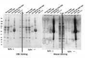
- SDS-PAGEwas performed using the fraction eluted in this way.
- staining to this SDS-PAGEis shown in FIG. As shown in the photograph of FIG. 2, since a band was observed in the vicinity of about 10 kDa, this band (spot) was cut out.
- Example 2Search for Target Antigen Candidate in Spot Using MS / MS Ion Search Method
- the spot obtained in FIG. 2was cut out with a cutter (left in FIG. 3).
- the MS / MS measurement of the peptide mixture obtained by protease decompositionwas performed, and the MS / MS spectrum was acquired by the method of selecting the ion derived from a specific peptide in a mass spectrometer.
- a scorewas calculated from the obtained MS / MS spectrum and the total protein data registered in the database (Table 1).
- 11 types of proteins listed in Table 1 belowwere hit as candidate molecules for the target antigen (the same table in the right side of FIG. 3 is abbreviated).
- the obtained polyclonal antibodywas administered to a spontaneous vasculitis model mouse (SCG / Kj mouse), and the therapeutic effect of each antibody was determined.
- CO 2 MPO-ANCAmyeloperoxidase-specific anti-neutrophil cytoplasmic antibody
- chlorinewas added to water purified by a reverse osmosis (RO) drinking water treatment apparatus so as to have a concentration of 0.3 to 2.0 ppm and allowed to be freely ingested.
- the animal facilitywas supplied with clean air through a HEPA filter and was restricted to breeding only SPF animals.
- micewere subjected to urinalysis once a week for about 3 weeks before, and were grouped according to the body weight and urinary occult blood / urine score values on the day of administration.
- 0.1 to 400 mg / kg of the samplewas administered ip for 5 days
- Solventstabilizer: D mannitol, glycine, PBS
- the dosewas calculated from the final measured body weight and adjusted to 0.33 mL per animal.
- body weight measurement and urinalysiswere performed twice a week.
- the spleen weight of the model mouse SCG / Kjwas higher in the untreated (Solvent administration) group than in the C56BL / 6 healthy mouse.
- administration of anti-APOA2 polyclonal antibody to this model mouse SCG / Kjdid not decrease the spleen weight value of healthy mice, but the spleen weight decreased.
- administration of a molecule (Differential Mol) different from VasSFdid not reduce the spleen weight.
- IL-1ainterleukin-1 ⁇ IL-1b: interleukin-1 ⁇ IL-2: interleukin-2 IL-3: interleukin-3 IL-4: interleukin-4 IL-5: interleukin-5 IL-6: interleukin-6 IL-9: interleukin-9 IL-10: interleukin-10 IL12p40: interleukin-12 subunit p40 (interleukin-12 subunit p40) IL-12p70: interleukin-12 subunit p70 (interleukin-12 subunit p70) IL-13: interleukin-13 (interleukin-13) IL-17: interleukin-17 Eotaxin: eotaxin G-CSF: granulocyte colony-stimulating factor GM-CSF: granulocyte / macrophage colony-stimulating factor IFN-g: interferon gamma KC: keratinocyte-derived chemokine (keratinocyte-derived chemokine) MCP
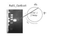
- E. coli protein expression optimization(introduction into pTAC-2 vector) Considering the codon usage of E. coli from the amino acid sequence of the effective clone (VasSF) (identical to SEQ ID NO: 4 and SEQ ID NO: 31 of Patent Document 5) in order to maximize the protein expression efficiency by E. coli, A plasmid pTAC2-URq01_OptEcoli containing an artificially synthesized gene (URq01_OptEcoli sequence; SEQ ID NO: 3) with 6 histidine tags added to the C-terminal was prepared (FIG. 12).
- the insert sequence(coding region: region of SEQ ID NO: 3) was PCR-amplified from the plasmid pTAC2-URq01_OptEcoli constructed as described above to prepare an integrated fragment.
- the base sequences of the primers used at this timeare as follows (the underlined portion is a sequence homologous to the end of the cloning site of the pET32 vector).
- a transformantwas prepared by introducing the vector into E. coli host DE32 using the heat shock method.
- the integration sequencewas confirmed from 10 colonies by PCR amplification of the inserted sequence using a primer set in the frame T7 promoter region of the pET32 vector and a primer in the T7 terminator region.
- the base sequences of the primers used at this timeare as follows.
- Recombinant Clone A recombinant protein(consisting of SEQ ID NO: 4, also referred to herein as “VasAP”) was purified from a culture of E. coli cell DE32 as a host by the following method.
- protein purificationcan be performed using a general purification method. Examples of purification methods include immobilized metal ion affinity chromatography, fractionation with an ion exchange column, chromatography with a positive ion exchange resin such as DEAE, and gel filtration.
- E. coli containing the target clonewas cultured at 10 ° C. for 18 to 48 hours.
- Immobilized metal (Ni) ion affinity chromatography purified protein of 8M Urea basewas centrifugally concentrated with an ultrafiltration membrane at 10 kDa.
- the protein substituted with 8 M urea PBS bufferwas dialyzed, and the urea concentration was lowered stepwise and replaced with PBS while adding arginine. Specifically, purification was performed in this order using 8M urea PBS, 4M urea / 0.4M arginine, 2M urea / 0.4M arginine, 2M urea 0.4M arginine, and PBS 3 times.
- FIG. 15A and 15Bare photographs showing the results of gel filtration HPLC profiles
- FIG. 15Cis a photograph showing the results of electrophoresis and Western blotting.
- the VasAP antibody administrationimproved the meniscus formation in the model mouse SCG / Kj (FIG. 16A), and the urinary findings showed a lower value compared to the Different Mol (FIG. 16).
- APOA2 protein(BTI: BT-928, LOT: 9280413) was prepared as a test protein. 1 mg of this test protein is dissolved in 1 mL of physiological saline (Nippon Zenyaku Kogyo Co., Ltd., # 412190), and then 1 mL of physiological saline is further added and mixed uniformly to obtain 0.5 mg / mL. An administration sample was prepared.
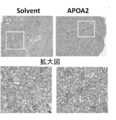
- the meniscus formation rate in the kidney glomeruluswas greatly increased as compared with the mice of the control group.
- the microscopic image shown in FIG. 23the microscopic image of the kidney glomeruli of the mice in the experimental group formed a meniscus and renal dysfunction occurred.
- the “enlarged view” in the lower part of FIG. 23is an enlarged view of a square box part of the upper microscopic image.
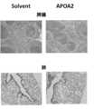
- the microscopic image of the spleen of the control group miceshowed that the white spleen and the red spleen were properly separated. The red pulp was not separated. Furthermore, vasculitis, hemorrhage and partial granulation appeared in the lung tissue of the experimental group mice, but these symptoms were not confirmed in the control group mice.
- inflammatory diseaseshere, vasculitis
- APOA2 proteinwild-type non-human animals
- [SEQ ID NO: 6]This shows the base sequence of the PCR primer (Rq01_OptEc_cds_6HisStpcmpR).
- [SEQ ID NO: 7]This shows the base sequence of the PCR primer (His6StpCmp_pET32_110-129F).
- [SEQ ID NO: 8]This shows the base sequence of the PCR primer (pET32vbRev693-712).
- [SEQ ID NO: 9]This shows the base sequence of the PCR primer (Rq01_OptEc_cds_F).
Landscapes
- Health & Medical Sciences (AREA)
- Life Sciences & Earth Sciences (AREA)
- Chemical & Material Sciences (AREA)
- Engineering & Computer Science (AREA)
- General Health & Medical Sciences (AREA)
- Organic Chemistry (AREA)
- Biomedical Technology (AREA)
- Immunology (AREA)
- Medicinal Chemistry (AREA)
- Molecular Biology (AREA)
- Animal Behavior & Ethology (AREA)
- Public Health (AREA)
- Veterinary Medicine (AREA)
- Urology & Nephrology (AREA)
- Biochemistry (AREA)
- Genetics & Genomics (AREA)
- Zoology (AREA)
- Biophysics (AREA)
- Pharmacology & Pharmacy (AREA)
- Nuclear Medicine, Radiotherapy & Molecular Imaging (AREA)
- General Chemical & Material Sciences (AREA)
- Chemical Kinetics & Catalysis (AREA)
- Pathology (AREA)
- Hematology (AREA)
- Rheumatology (AREA)
- Biotechnology (AREA)
- Bioinformatics & Cheminformatics (AREA)
- Toxicology (AREA)
- Cell Biology (AREA)
- Endocrinology (AREA)
- Microbiology (AREA)
- Physics & Mathematics (AREA)
- Proteomics, Peptides & Aminoacids (AREA)
- Diabetes (AREA)
- Food Science & Technology (AREA)
- Analytical Chemistry (AREA)
- General Physics & Mathematics (AREA)
- Epidemiology (AREA)
- Gastroenterology & Hepatology (AREA)
- Environmental Sciences (AREA)
Abstract
Description
Translated fromJapanese本発明は、感染性疾患または炎症性疾患の予防および/または治療剤に関する。The present invention relates to a preventive and / or therapeutic agent for infectious diseases or inflammatory diseases.
炎症性疾患である慢性関節リウマチなどには抗体医薬(インフリキシマブ、アクテムラなど)が使用されている。一方、同様に炎症性疾患である難治性血管炎には標準治療薬はなく、ステロイドや抗体医薬の使用が検討されている。最近では、免疫グロブリン(Ig)製剤を難治性血管炎にも使用することがあり、好酸球性多発血管炎性肉芽腫症(Eosinophilic Granulomatosis with Polyangitis:EGPA、チャーグ・ストラウス(Churg-Strauss)症候群)では認可されている。なお、免疫グロブリンは、抗体およびこれと構造上・機能上の関連性のあるタンパク質の総称である。つまり、結合する抗原が明らかになっている免疫グロブリンを、その特定抗原に対応させて抗体と呼んでいる。Antibody drugs (such as infliximab and actemra) are used for rheumatoid arthritis, which is an inflammatory disease. On the other hand, there is no standard treatment for refractory vasculitis, which is also an inflammatory disease, and the use of steroids and antibody drugs is being investigated. Recently, immunoglobulin (Ig) preparations may also be used for refractory vasculitis, eosinophilic granulomatosis with Polyangitis (EGPA), Churg-Strauss syndrome ) Is authorized. Immunoglobulin is a general term for antibodies and proteins structurally and functionally related thereto. That is, an immunoglobulin whose binding antigen has been clarified is called an antibody corresponding to the specific antigen.
難治性血管炎の治療法としては、ステロイドなどがガイドラインにあり、また、標準治療法として抗体医薬が検討されている。ここで、特定抗原に対する抗体医薬として難治性血管炎にも使用され始めているインフリキシマブ(レミケード(登録商標))は、クローン病では炎症性サイトカインのTNFαが対応抗原であり、過剰に作られたTNFαの働きを抑える抗体医薬として開発された。このインフリキシマブは、TNFαを産生している細胞をも壊す抗体医薬である(非特許文献1を参照)。また、例えば、リツキシマブ(リツキサン)は、抗体医薬品はCD20に対するモノクローナル抗体として海外でリンパ腫用に開発され、難治性血管炎にも利用され始めている(非特許文献2を参照)。抗原が特定されている種々のこれらの抗体医薬は難治性血管炎に特有のものではないものの、有効性はあり、直接抗原と反応して当該抗原を中和していると考えられている。現在、臨床的に使用されている抗体医薬は、難治性血管炎に特異的な抗原に対して開発された抗体ではないことから、リスクが常に存在する。Steroids are guidelines for the treatment of refractory vasculitis, and antibody drugs are being studied as standard treatments. Here, infliximab (Remicade (registered trademark)), which has begun to be used for refractory vasculitis as an antibody drug against a specific antigen, is the inflammatory cytokine TNFα as a corresponding antigen in Crohn's disease, and an excess of TNFα produced. It was developed as an antibody drug that suppresses the work. This infliximab is an antibody drug that also destroys cells that produce TNFα (see Non-Patent Document 1). In addition, for example, rituximab (Rituxan) has been developed overseas for lymphoma as a monoclonal antibody against CD20, and has begun to be used for intractable vasculitis (see Non-Patent Document 2). Although these various antibody drugs for which antigens have been specified are not unique to refractory vasculitis, they are effective and are believed to react directly with the antigen to neutralize the antigen. Currently, there is always a risk because antibody drugs currently in clinical use are not antibodies developed against antigens specific to refractory vasculitis.
大量の免疫グロブリンの投与が難治性血管炎の治療に有効である。詳しくは、免疫グロブリンの多様性は、108個のクローンからなり、基本的な分子構造は、軽鎖(L鎖)および重鎖(H鎖)の2種類のポリペプチド2本ずつがジスルフィド結合により連結したものである。重鎖は定常領域(C領域)および、VH領域からなる可変領域(V領域)が連結された構造である。一方、軽鎖は、CL領域からなる定常領域および、VL領域からなる可変領域が連結されている。可変領域のアミノ酸配列の多様性から多様な抗原に対する多様な抗体が生体内で作られている。Administration of large amounts of immunoglobulin is effective in treating refractory vasculitis. Specifically, the diversity of immunoglobulins consists of 108 clones, and the basic molecular structure is that each of two polypeptides, light chain (L chain) and heavy chain (H chain), is disulfide bonded. It is connected by. The heavy chain has a structure in which a constant region (C region) and a variable region (V region) composed of a VH region are linked. On the other hand, in the light chain, a constant region composed of a CL region and a variable region composed of a VL region are linked. Various antibodies against various antigens are produced in vivo due to the diversity of amino acid sequences in the variable region.
免疫グロブリン製剤の作用機序については、いくつかの仮説がある。第一の仮説は、免疫グロブリン製剤に含まれている、未知の抗原に対する抗体を含めた多種類の抗体が薬理効果を発揮しているという説である。また第二の仮説は、多種類の抗体のうち、難治性血管炎の自己抗体ミエロペルオキシダーゼ(MPO)に対する抗体(抗MPO抗体)(非特許文献3を参照)にその効果があり、特にMPOの広範なエピトープに対する多種類の抗MPO抗体が薬理効果を発揮しているという説である。そして、同様に難治性血管炎の重症化にも関与している自己抗体である抗モエシン抗体(特許文献1、非特許文献4~5を参照)に対する薬理作用ではないかとの説もある。両説において共通していることは、免疫グロブリン製剤が多様な免疫グロブリンの混合物であること、すなわちポリクローナルなものであることが治療効果に大きく寄与しているということである。別の仮説としては、特定の抗原に対する多様な抗体が有効性を示すとの説もある。There are several hypotheses about the mechanism of action of immunoglobulin preparations. The first hypothesis is that many types of antibodies, including antibodies against unknown antigens, contained in immunoglobulin preparations exert pharmacological effects. The second hypothesis is that an antibody (anti-MPO antibody) against the autoantibody myeloperoxidase (MPO) of refractory vasculitis (see Non-Patent Document 3) among many types of antibodies, particularly MPO The theory is that many types of anti-MPO antibodies against a wide range of epitopes exert pharmacological effects. There is also a theory that it may be a pharmacological action against an anti-moesin antibody (see
ヒト組み換え抗体の単クローンScFvが難治性血管炎モデルマウスに対して治療効果を示すことが知られている(特許文献2~5を参照)。このような状況において、患者の感染リスクを減少させ、組み換え免疫グロブリン製剤の治癒機転を明らかにする上でも、抗原が特定された抗体を有効成分とする難治性血管炎に対する抗体医薬の開発が望まれている。It is known that a human recombinant antibody monoclonal ScFv exhibits a therapeutic effect on intractable vasculitis model mice (see
抗原が特定された抗体であれば、これまでの組み換えScFvをモデルとして、組み換えDNA技術によって抗体医薬の製造が可能である。キメラ抗体やヒト化抗体は抗体医薬としてすでに実用化されている(特許文献6~7を参照)。また、免疫グロブリン遺伝子を宿主細胞内で可溶状態の正常型タンパク質として発現させる技術として、免疫グロブリンをシャペロニンとの融合タンパク質として発現させる例も知られている(特許文献8を参照)。なお、これらはすべて単一分子の免疫グロブリン、すなわちモノクローナルの免疫グロブリンまたはその断片である。If the antigen is an identified antibody, an antibody drug can be produced by recombinant DNA technology using the conventional recombinant ScFv as a model. Chimeric antibodies and humanized antibodies have already been put into practical use as antibody drugs (see
上述したように、難治性血管炎の標準治療法は存在していないのが現状である。また、抗体医薬を用いた治療法も検討されているが、難治性血管炎に特化した治療法として開発されたものではない。すなわち、炎症性疾患である難治性血管炎にはステロイドの使用がガイドラインにて示されているが標準治療薬が存在しないのが現状である。As mentioned above, there is currently no standard treatment for refractory vasculitis. In addition, a therapeutic method using an antibody drug has been studied, but it has not been developed as a therapeutic method specialized for intractable vasculitis. In other words, for refractory vasculitis, which is an inflammatory disease, the use of steroids is indicated in the guidelines, but there is no standard therapeutic drug at present.
そこで本発明は、上述したような従来の技術および医療の現状に鑑み、難治性血管炎等の炎症性疾患や感染性疾患の治療に有効な標的分子を特定することで、標的分子が特定されていない従来の治療法とは異なりこれらの疾患に特化した治療法を提供することを目的とする。Therefore, in view of the conventional technology and the current medical situation as described above, the present invention identifies a target molecule by identifying a target molecule that is effective in the treatment of inflammatory diseases such as intractable vasculitis and infectious diseases. The aim is to provide treatments specialized for these diseases, unlike conventional therapies that have not.
本発明者らは、上述した課題を解決することを目指して、鋭意研究を行なった。そして、その過程において多大なる困難を乗り越えた結果、特許文献5において血管炎モデルマウスに対して種々の治療効果を示した配列番号:31で表されるアミノ酸配列からなるヒト単鎖可変断片(hScFv;以下では当該クローンを「VasSF」とも称する)が標的とする抗原がアポリポタンパク質A2(APOA2)であるということを突き止めた。その結果、APOA2を特異的に認識する抗体を用いることで血管炎等の炎症性疾患や感染性疾患の予防および/または治療が可能となることを見出し、本発明を完成させるに至った。The present inventors have conducted intensive research with the aim of solving the above-mentioned problems. As a result of overcoming great difficulties in the process, a human single-chain variable fragment (hScFv consisting of the amino acid sequence represented by SEQ ID NO: 31 which exhibited various therapeutic effects on vasculitis model mice in
すなわち、本発明の一形態によれば、アポリポタンパク質A2(APOA2)を特異的に認識する抗体を有効成分として含有する、感染性疾患または炎症性疾患の予防および/または治療剤が提供される。That is, according to one aspect of the present invention, there is provided a prophylactic and / or therapeutic agent for infectious diseases or inflammatory diseases, which contains an antibody that specifically recognizes apolipoprotein A2 (APOA2) as an active ingredient.
本発明の他の形態によれば、配列番号:4で表されるアミノ酸配列と同一または実質的に同一のアミノ酸配列を含むポリペプチドを有効成分として含有する、抗APOA2組み換え免疫グロブリン断片組成物や、配列番号:4で表されるアミノ酸配列からなるポリペプチドを有効成分として含有する、抗APOA2組み換え免疫グロブリン断片組成物が提供される。According to another aspect of the present invention, there is provided an anti-APOA2 recombinant immunoglobulin fragment composition comprising, as an active ingredient, a polypeptide comprising the same or substantially the same amino acid sequence as the amino acid sequence represented by SEQ ID NO: 4 There is provided an anti-APOA2 recombinant immunoglobulin fragment composition containing a polypeptide consisting of the amino acid sequence represented by SEQ ID NO: 4 as an active ingredient.
本発明のさらに他の形態によれば、配列番号:4で表されるアミノ酸配列と同一または実質的に同一のアミノ酸配列を含むポリペプチドをコードするポリヌクレオチドおよび当該ポリヌクレオチドを含有するベクター、並びに当該ベクターを含有する宿主細胞が提供される。According to still another aspect of the present invention, a polynucleotide encoding a polypeptide comprising the amino acid sequence identical or substantially identical to the amino acid sequence represented by SEQ ID NO: 4, a vector containing the polynucleotide, and Host cells containing the vectors are provided.
本発明のさらに他の形態によれば、抗APOA2抗体の有効量を投与することを含む、感染性疾患または炎症性疾患の予防および/または治療方法や、アポリポタンパク質A2(APOA2)を用いる、感染性疾患または炎症性疾患の予防および/または治療剤のスクリーニング方法が提供される。According to yet another aspect of the present invention, a method of preventing and / or treating an infectious or inflammatory disease comprising administering an effective amount of an anti-APOA2 antibody, an infection using apolipoprotein A2 (APOA2) A method for screening a prophylactic and / or therapeutic agent for sexual diseases or inflammatory diseases is provided.
本発明のさらに他の形態によれば、アポリポタンパク質A2(APOA2)を有効成分として含有する、感染性疾患または炎症性疾患の誘導剤や、ヒトを除く非トランスジェニック動物にアポリポタンパク質A2(APOA2)を投与する工程を含む、感染性疾患または炎症性疾患の病態モデル動物の作製方法、並びに当該作製方法によって作製された、感染性疾患または炎症性疾患の病態モデル動物が提供される。According to still another aspect of the present invention, an infectious or inflammatory disease inducer containing apolipoprotein A2 (APOA2) as an active ingredient, or apolipoprotein A2 (APOA2) for non-transgenic animals other than humans. A method for producing a disease state animal model of an infectious disease or inflammatory disease, and a disease state animal model of an infectious disease or inflammatory disease produced by the production method are provided.
本発明を完成させる過程で、難治性血管炎等の炎症性疾患や感染性疾患の治療に有効な標的分子を特定することができた。そしてこの知見に基づき鋭意検討を重ねた結果、標的分子が特定されていない従来の治療法とは異なりこれらの疾患に特化した治療法を提供することが可能となった。In the process of completing the present invention, a target molecule effective for the treatment of inflammatory diseases such as intractable vasculitis and infectious diseases could be identified. And as a result of intensive studies based on this finding, it has become possible to provide treatments specialized for these diseases, unlike conventional treatments in which the target molecule is not specified.
本発明の第1の形態は、アポリポタンパク質A2(APOA2)を特異的に認識する抗体を有効成分として含有する、感染性疾患または炎症性疾患の予防および/または治療剤である。The first aspect of the present invention is a preventive and / or therapeutic agent for infectious diseases or inflammatory diseases, which contains an antibody that specifically recognizes apolipoprotein A2 (APOA2) as an active ingredient.
本発明者らが本発明を完成させるに至った経緯を概説すれば、まず、本発明者らは、特許文献5において血管炎モデルマウスに対して種々の治療効果を示した配列番号:31で表されるアミノ酸配列からなるヒト単鎖可変断片(hScFv;以下では当該クローンを「VasSF」とも称する)が標的とする抗原の探索を試みた。しかしながら、抗原の探索は困難を極め、後述する実施例の欄に記載のように種々の手法を試みるも容易には抗原の特定には至らず、極めて長期にわたる尽力と創意工夫の結果、最終的にVasSFの標的抗原を特定するに至り、それによって本発明を完成させるに至ったのである。If the present inventors outline the circumstances that led to the completion of the present invention, first, the present inventors will use SEQ ID NO: 31 which showed various therapeutic effects on vasculitis model mice in
すなわち、本発明者らは、驚くべきことに、VasSFの標的抗原として、従来は感染性疾患や炎症性疾患との関連性が全く知られていなかったタンパク質であるアポリポタンパク質A2(APOA2)がVasSFの標的抗原であることを突き止め、これにより、APOA2を特異的に認識する抗体を用いることで血管炎等の炎症性疾患や感染性疾患の予防および/または治療が可能となることを見出したのである。なお、ヒトアポリポタンパク質A2(hAPOA2)は、473bpからなるAPOA2遺伝子(RefSeq Accession No.NM_001643、CDSの配列が配列番号:1に対応する)によりコードされており、100アミノ酸(RefSeq Accession No.NP_001634、配列番号:2)からなっている。That is, the present inventors surprisingly, as a target antigen of VasSF, apolipoprotein A2 (APOA2), a protein that has never been known to be associated with infectious diseases or inflammatory diseases, is VasSF. As a result, it was found that the use of an antibody that specifically recognizes APOA2 makes it possible to prevent and / or treat inflammatory diseases such as vasculitis and infectious diseases. is there. The human apolipoprotein A2 (hAPOA2) is encoded by the 473 bp APOA2 gene (RefSeq Accession No. NM_001643, the CDS sequence corresponds to SEQ ID NO: 1), and 100 amino acids (RefSeq Accession No. NP_001634, It consists of SEQ ID NO: 2).
上述したいくつかの知見に基づき、本発明者らは、本発明を完成させるに至ったものである。以下、本発明を実施するための形態を詳細に説明するが、本発明の技術的範囲は下記の形態のみに限定されることはない。Based on the above findings, the present inventors have completed the present invention. Hereinafter, although the form for implementing this invention is demonstrated in detail, the technical scope of this invention is not limited only to the following form.
[本発明のタンパク質(抗原)]
本発明において用いられるアポリポタンパク質A2(APOA2;「本発明のタンパク質」とも称する)は、配列番号:2で表されるアミノ酸配列と同一または実質的に同一のアミノ酸配列を含むタンパク質である。本発明のタンパク質は、ヒトや他の温血動物(例えば、モルモット、ラット、マウス、ニワトリ、ウサギ、ブタ、ヒツジ、ウシ、サルなど)の細胞[例えば、肝細胞、脾細胞、神経細胞、グリア細胞、膵臓β細胞、骨髄細胞、メサンギウム細胞、ランゲルハンス細胞、表皮細胞、上皮細胞、杯細胞、内皮細胞、平滑筋細胞、線維芽細胞、線維細胞、筋細胞、脂肪細胞、免疫細胞(例、マクロファージ、T細胞、B細胞、ナチュラルキラー細胞、肥満細胞、好中球、好塩基球、好酸球、単球)、巨核球、滑膜細胞、軟骨細胞、骨細胞、骨芽細胞、破骨細胞、乳腺細胞もしくは間質細胞、またはこれら細胞の前駆細胞、幹細胞もしくは癌細胞など]もしくはそれらの細胞が存在するあらゆる組織[例えば、脳、脳の各部位(例、嗅球、扁桃核、大脳基底球、海馬、視床、視床下部、大脳皮質、延髄、小脳)、脊髄、下垂体、胃、膵臓、腎臓、肝臓、生殖腺、甲状腺、胆のう、骨髄、副腎、皮膚、筋肉(例、平滑筋、骨格筋)、肺、消化管(例、大腸、小腸)、血管、心臓、胸腺、脾臓、顎下腺、末梢血、前立腺、睾丸、卵巣、胎盤、子宮、骨、関節、脂肪組織(例、白色脂肪組織、褐色脂肪組織)など]等から単離・精製されるタンパク質であってもよい。また、化学合成もしくは無細胞翻訳系で生化学的に合成されたタンパク質であってもよいし、あるいは上記アミノ酸配列をコードする塩基配列を有する核酸を導入された形質転換体から産生される組換えタンパク質であってもよい。[Protein of the present invention (antigen)]
Apolipoprotein A2 (APOA2; also referred to as “protein of the present invention”) used in the present invention is a protein comprising the same or substantially the same amino acid sequence as the amino acid sequence represented by SEQ ID NO: 2. The protein of the present invention is used for cells of humans and other warm-blooded animals (eg, guinea pigs, rats, mice, chickens, rabbits, pigs, sheep, cows, monkeys, etc.) [eg, hepatocytes, spleen cells, neurons, glia. Cells, pancreatic β cells, bone marrow cells, mesangial cells, Langerhans cells, epidermal cells, epithelial cells, goblet cells, endothelial cells, smooth muscle cells, fibroblasts, fibrocytes, muscle cells, adipocytes, immune cells (eg, macrophages) , T cells, B cells, natural killer cells, mast cells, neutrophils, basophils, eosinophils, monocytes), megakaryocytes, synovial cells, chondrocytes, bone cells, osteoblasts, osteoclasts Breast cells or stromal cells, or precursor cells of these cells, stem cells or cancer cells, etc.] or any tissue in which these cells are present [eg, brain, brain regions (eg, olfactory bulb, amygdaloid nucleus, large cell) Basal sphere, hippocampus, thalamus, hypothalamus, cerebral cortex, medulla, cerebellum), spinal cord, pituitary, stomach, pancreas, kidney, liver, gonad, thyroid, gallbladder, bone marrow, adrenal gland, skin, muscle (eg, smooth muscle, Skeletal muscle), lungs, gastrointestinal tract (eg, large intestine, small intestine), blood vessels, heart, thymus, spleen, submandibular gland, peripheral blood, prostate, testis, ovary, placenta, uterus, bone, joint, adipose tissue (eg, White adipose tissue, brown adipose tissue, etc.)] and the like. Alternatively, it may be a protein synthesized chemically or biochemically in a cell-free translation system, or a recombinant produced from a transformant introduced with a nucleic acid having a base sequence encoding the amino acid sequence. It may be a protein.
配列番号:2で表されるアミノ酸配列と実質的に同一のアミノ酸配列としては、配列番号:2で表されるアミノ酸配列と約50%以上、好ましくは約60%以上、より好ましくは約70%以上、いっそう好ましくは約80%以上、特に好ましくは約90%以上、最も好ましくは約95%以上の相同性(ホモロジー;homology)を有するアミノ酸配列などが挙げられる。ここで「相同性」とは、当該技術分野において公知の数学的アルゴリズムを用いて2つのアミノ酸配列をアラインさせた場合の、最適なアラインメント(好ましくは、該アルゴリズムは最適なアラインメントのために配列の一方もしくは両方へのギャップの導入を考慮し得るものである)における、オーバーラップする全アミノ酸残基に対する同一アミノ酸および類似アミノ酸残基の割合(%)を意味する。「類似アミノ酸」とは物理化学的性質において類似したアミノ酸を意味し、例えば、芳香族アミノ酸(Phe、Trp、Tyr)、脂肪族アミノ酸(Ala、Leu、Ile、Val)、極性アミノ酸(Gln、Asn)、塩基性アミノ酸(Lys、Arg、His)、酸性アミノ酸(Glu、Asp)、水酸基を有するアミノ酸(Ser、Thr)、側鎖の小さいアミノ酸(Gly、Ala、Ser、Thr、Met)などの同じグループに分類されるアミノ酸が挙げられる。このような類似アミノ酸による置換はタンパク質の表現型に変化をもたらさない(すなわち、保存的アミノ酸置換である)ことが予測される。保存的アミノ酸置換の具体例は当該技術分野で周知であり、種々の文献に記載されている(例えば、Bowieら,Science,247:1306-1310(1990)を参照)。The amino acid sequence substantially identical to the amino acid sequence represented by SEQ ID NO: 2 is about 50% or more, preferably about 60% or more, more preferably about 70%, with the amino acid sequence represented by SEQ ID NO: 2. More preferably, amino acid sequences having a homology (homology) of about 80% or more, particularly preferably about 90% or more, and most preferably about 95% or more are mentioned. As used herein, “homology” refers to an optimal alignment when two amino acid sequences are aligned using a mathematical algorithm known in the art (preferably the algorithm uses a sequence of sequences for optimal alignment). The percentage of identical and similar amino acid residues relative to all overlapping amino acid residues in which one or both of the gaps can be considered). “Similar amino acids” mean amino acids that are similar in physicochemical properties, such as aromatic amino acids (Phe, Trp, Tyr), aliphatic amino acids (Ala, Leu, Ile, Val), polar amino acids (Gln, Asn). ), Basic amino acids (Lys, Arg, His), acidic amino acids (Glu, Asp), amino acids having hydroxyl groups (Ser, Thr), amino acids with small side chains (Gly, Ala, Ser, Thr, Met), etc. Examples include amino acids classified into groups. It is expected that substitution with such similar amino acids will not change the phenotype of the protein (ie, is a conservative amino acid substitution). Specific examples of conservative amino acid substitutions are well known in the art and are described in various references (see, for example, Bowie et al., Science, 247: 1306-1310 (1990)).
本明細書におけるアミノ酸配列の相同性は、相同性計算アルゴリズムNCBI BLAST(National Center for Biotechnology Information Basic Local Alignment Search Tool)を用い、以下の条件(期待値=10;ギャップを許す;マトリクス=BLOSUM62;フィルタリング=OFF)にて計算することができる。アミノ酸配列の相同性を決定するための他のアルゴリズムとしては、例えば、Karlinら,Proc.Natl.Acad.Sci.USA,90:5873-5877(1993)に記載のアルゴリズム[該アルゴリズムはNBLASTおよびXBLASTプログラム(version2.0)に組み込まれている(Altschulら,Nucleic Acids Res.,25:3389-3402(1997))]、Needlemanら,J.Mol.Biol.,48:444-453(1970)に記載のアルゴリズム[該アルゴリズムはGCGソフトウェアパッケージ中のGAPプログラムに組み込まれている]、MyersおよびMiller,CABIOS,4:11-17(1988)に記載のアルゴリズム[該アルゴリズムはCGC配列アラインメントソフトウェアパッケージの一部であるALIGNプログラム(version2.0)に組み込まれている]、Pearsonら,Proc.Natl.Acad.Sci.USA,85:2444-2448(1988)に記載のアルゴリズム[該アルゴリズムはGCGソフトウェアパッケージ中のFASTAプログラムに組み込まれている]等が挙げられ、それらも同様に好ましく用いられ得る。The homology of the amino acid sequences in this specification uses the homology calculation algorithm NCBI BLAST (National Center for Biotechnology Information Basic Alignment Search Tool) and the following conditions (expectation value = 10; allow gap; matrix = BLOSUM62; = OFF). Other algorithms for determining amino acid sequence homology include, for example, Karlin et al., Proc. Natl. Acad. Sci. USA, 90: 5873-5877 (1993) [The algorithm is incorporated into the NBLAST and XBLAST programs (version 2.0) (Altschul et al., Nucleic Acids Res., 25: 3389-3402 (1997)) ] Needleman et al., J .; Mol. Biol. 48: 444-453 (1970) [the algorithm is incorporated into the GAP program in the GCG software package], Myers and Miller, CABIOS, 4: 11-17 (1988) [ The algorithm is incorporated into the ALIGN program (version 2.0) which is part of the CGC sequence alignment software package], Pearson et al., Proc. Natl. Acad. Sci. USA, 85: 2444-2448 (1988) [the algorithm is incorporated in the FASTA program in the GCG software package] and the like, and they can be preferably used as well.
より好ましくは、配列番号:2で表されるアミノ酸配列と実質的に同一のアミノ酸配列とは、配列番号:2で表されるアミノ酸配列と約50%以上、好ましくは約60%以上、より好ましくは約70%以上、いっそう好ましくは約80%以上、特に好ましくは約90%以上、最も好ましくは約95%以上の同一性(アイデンティティ(identity))を有するアミノ酸配列である。More preferably, the amino acid sequence substantially the same as the amino acid sequence represented by SEQ ID NO: 2 is about 50% or more, preferably about 60% or more, more preferably the amino acid sequence represented by SEQ ID NO: 2. Is an amino acid sequence having about 70% or more, more preferably about 80% or more, particularly preferably about 90% or more, and most preferably about 95% or more identity.
本発明で用いられるタンパク質は、配列番号:2で表されるアミノ酸配列と実質的に同一のアミノ酸配列を含有し、かつ配列番号:2で表されるアミノ酸配列を含有するタンパク質と実質的に同質の活性を有するタンパク質である。The protein used in the present invention contains substantially the same amino acid sequence as the amino acid sequence represented by SEQ ID NO: 2 and is substantially the same as the protein containing the amino acid sequence represented by SEQ ID NO: 2. It is a protein having the activity of
実質的に同質の活性としては、例えば、リガンド結合活性やシグナル情報伝達作用などが挙げられる。ここで「実質的に同質」とは、それらの性質が定性的に(例、生理学的に、または薬理学的に)同質であることを示す。したがって、本発明のタンパク質の活性が同等であることが好ましいが、これらの活性の程度(例、約0.01~約100倍、好ましくは約0.1~約10倍、より好ましくは0.5~2倍)や、タンパク質の分子量などの量的要素は異なっていてもよい。Examples of substantially the same activity include ligand binding activity and signal information transmission action. Here, “substantially the same quality” indicates that these properties are qualitatively (eg, physiologically or pharmacologically) homogeneous. Accordingly, it is preferable that the activities of the protein of the present invention are equivalent, but the extent of these activities (eg, about 0.01 to about 100 times, preferably about 0.1 to about 10 times, more preferably about 0.1 times). 5 to 2 times) and quantitative factors such as protein molecular weight may be different.
リガンド結合活性やシグナル情報伝達作用などの活性の測定は、それ自体公知の方法に準じて行なうことができる。例えば、後述する本発明で用いられるタンパク質の活性を阻害する化合物またはその塩のスクリーニング方法において用いられる方法等に従って行なうことができる。Measurement of activities such as ligand binding activity and signal signal transduction can be performed according to methods known per se. For example, it can be performed according to a method used in a screening method for a compound or a salt thereof that inhibits the activity of the protein used in the present invention described later.
また、本発明で用いられるタンパク質としては、例えば、(i)配列番号:2で表されるアミノ酸配列中の1個または2個以上(例えば、1~50個程度、好ましくは1~30個程度、より好ましくは1~10個程度、さらに好ましくは数(1~5、4、3もしくは2)個)のアミノ酸が欠失したアミノ酸配列、(ii)配列番号:2で表されるアミノ酸配列に1個または2個以上(例えば、1~50個程度、好ましくは1~30個程度、より好ましくは1~10個程度、さらに好ましくは数(1~5、4、3もしくは2)個)のアミノ酸が付加したアミノ酸配列、(iii)配列番号:2で表されるアミノ酸配列に1個または2個以上(例えば、1~50個程度、好ましくは1~30個程度、より好ましくは1~10個程度、さらに好ましくは数(1~5、4、3もしくは2)個)のアミノ酸が挿入されたアミノ酸配列、(iv)配列番号:2で表されるアミノ酸配列中の1個または2個以上(例えば、1~50個程度、好ましくは1~30個程度、より好ましくは1~10個程度、さらに好ましくは数(1~5、4、3もしくは2)個)のアミノ酸が他のアミノ酸で置換されたアミノ酸配列、または(v)それらを組み合わせたアミノ酸配列を含有するタンパク質などのいわゆるムテインも含まれる。Examples of the protein used in the present invention include (i) one or two or more (for example, about 1 to 50, preferably about 1 to 30) in the amino acid sequence represented by SEQ ID NO: 2. More preferably, about 1 to 10, more preferably a number (1 to 5, 4, 3 or 2) of amino acid sequences, (ii) an amino acid sequence represented by SEQ ID NO: 2. 1 or 2 or more (for example, about 1 to 50, preferably about 1 to 30, more preferably about 1 to 10, more preferably a number (1 to 5, 4, 3 or 2)) (Iii) one or more amino acid sequences represented by SEQ ID NO: 2 (for example, about 1 to 50, preferably about 1 to 30, more preferably 1 to 10) About more and more Or an amino acid sequence in which several (1 to 5, 4, 3 or 2) amino acids are inserted, (iv) one or more (for example, 1) in the amino acid sequence represented by SEQ ID NO: 2 An amino acid in which about 50 to 50, preferably about 1 to 30, more preferably about 1 to 10, more preferably several (1 to 5, 4, 3 or 2) amino acids are substituted with other amino acids Also included are so-called muteins such as proteins containing sequences, or (v) amino acid sequences combining them.
上記のようにアミノ酸配列が挿入、欠失または置換されている場合、その挿入、欠失または置換の位置は、特に限定されない。When the amino acid sequence is inserted, deleted or substituted as described above, the position of the insertion, deletion or substitution is not particularly limited.
本発明で用いられるタンパク質の好ましい例としては、例えば、配列番号:2で表されるアミノ酸配列を含有するヒトアポリポタンパク質A2(Refseq Accession No.NP_001634)あるいは他の哺乳動物におけるそのホモログなどが挙げられる。Preferred examples of the protein used in the present invention include, for example, human apolipoprotein A2 (Refseq Accession No. NP_001634) containing the amino acid sequence represented by SEQ ID NO: 2, or a homolog thereof in other mammals. .
本明細書において、タンパク質およびペプチドは、ペプチド標記の慣例に従って左端がN末端(アミノ末端)、右端がC末端(カルボキシル末端)で記載される。配列番号:2で表されるアミノ酸配列を含有するタンパク質を初めとする、本発明で用いられるタンパク質は、C末端がカルボキシル基(-COOH)、カルボキシレート(-COO-)、アミド(-CONH2)またはエステル(-COOR)のいずれであってもよい。In the present specification, proteins and peptides are described with the N-terminus (amino terminus) at the left end and the C-terminus (carboxyl terminus) at the right end according to the convention of peptide designation. The protein used in the present invention including the protein containing the amino acid sequence represented by SEQ ID NO: 2 has a C terminus having a carboxyl group (—COOH), a carboxylate (—COO− ), an amide (—CONH2 ) Or ester (—COOR).
さらに、本発明で用いられるタンパク質には、N末端のアミノ酸残基(例、メチオニン残基)のアミノ基が保護基(例えば、ホルミル基、アセチル基などのC1-6アルカノイルなどのC1-6アシル基など)で保護されているもの、生体内で切断されて生成するN末端のグルタミン残基がピログルタミン酸化したもの、分子内のアミノ酸の側鎖上の置換基(例えば-OH、-SH、アミノ基、イミダゾール基、インドール基、グアニジノ基など)が適当な保護基(例えば、ホルミル基、アセチル基などのC1-6アルカノイル基などのC1-6アシル基など)で保護されているもの、あるいは糖鎖が結合したいわゆる糖タンパク質などの複合タンパク質なども含まれる。Furthermore, the protein used in the present invention, the amino acid residue (eg, methionine residue) of the N-terminal amino group protecting group (e.g., formyl group, such as C1-6 alkanoyl such as acetyl group C1- 6- acyl group), N-terminal glutamine residue produced by cleavage in vivo is pyroglutamine oxidized, substituents on the side chain of amino acids in the molecule (eg, —OH, — SH, amino group, imidazole group, indole group, guanidino group, etc.) is protected with a suitable protecting group (e.g., formyl group, C1-6 acyl group such as C1-6 alkanoyl group such as acetyl group) Or a complex protein such as a so-called glycoprotein having a sugar chain bound thereto.
本発明で用いられるタンパク質の部分ペプチドとしては、前記した本発明で用いられるタンパク質の部分アミノ酸配列を有するペプチドであり、かつ該タンパク質と実質的に同質の活性を有する限り、いずれのものであってもよい。ここで「実質的に同質の活性」とは上記と同義である。また、「実質的に同質の活性」の測定は、前記した本発明で用いられるタンパク質の場合と同様に行うことができる。The partial peptide of the protein used in the present invention is any peptide as long as it is a peptide having the partial amino acid sequence of the protein used in the present invention and has substantially the same activity as the protein. Also good. Here, “substantially the same quality of activity” has the same meaning as described above. In addition, the “substantially the same quality of activity” can be measured in the same manner as the protein used in the present invention.
例えば、本発明で用いられるタンパク質の構成アミノ酸配列のうち少なくとも20個以上、好ましくは50個以上、さらに好ましくは70個以上、より好ましくは100個以上、最も好ましくは150個以上のアミノ酸配列を有するペプチドなどが用いられる。For example, the amino acid sequence of the protein used in the present invention has at least 20 or more, preferably 50 or more, more preferably 70 or more, more preferably 100 or more, and most preferably 150 or more amino acid sequences. Peptides are used.
また、本発明で用いられる部分ペプチドは、そのアミノ酸配列中の1個または2個以上(好ましくは1~20個程度、より好ましくは1~10個程度、さらに好ましくは数(1~5、4、3もしくは2)個)のアミノ酸が欠失し、または、そのアミノ酸配列に1個または2個以上(好ましくは1~20個程度、より好ましくは1~10個程度、さらに好ましくは数(1~5、4、3もしくは2)個)のアミノ酸が付加し、または、そのアミノ酸配列に1個または2個以上(好ましくは1~20個程度、より好ましくは1~10個程度、さらに好ましくは数(1~5、4、3もしくは2)個)のアミノ酸が挿入され、または、そのアミノ酸配列中の1個または2個以上(好ましくは1~20個程度、より好ましくは1~10個程度、さらに好ましくは数(1~5、4、3もしくは2)個)のアミノ酸が他のアミノ酸で置換されていてもよい。In addition, the partial peptide used in the present invention is one or more (preferably about 1 to 20, more preferably about 1 to 10, more preferably a number (1 to 5, 4 or more) in the amino acid sequence. 3 or 2) amino acids, or one or more (preferably about 1 to 20, more preferably about 1 to 10, more preferably a number (1) in the amino acid sequence. ˜5, 4, 3, or 2) amino acids are added, or one or more (preferably about 1 to 20, more preferably about 1 to 10, more preferably about the amino acid sequence) A number (1-5, 4, 3 or 2) amino acids are inserted, or one or more (preferably about 1-20, more preferably about 1-10) in the amino acid sequence. Even better Properly amino acid number (1 to 5, 4, 3 or 2) pieces) may be replaced with other amino acids.
本発明で用いられる部分ペプチドは抗体作製のための抗原としても用いることができる。本発明で用いられるタンパク質またはその部分ペプチドは遊離体であってもよいし、塩であってもよい(以下、特に断らない限り同様である)。かような塩としては、生理学的に許容される酸(例、無機酸、有機酸)や塩基(例、アルカリ金属塩)などとの塩が用いられ、とりわけ生理学的に許容される酸付加塩が好ましい。このような塩としては、例えば、無機酸(例、塩酸、リン酸、臭化水素酸、硫酸)との塩、あるいは有機酸(例、酢酸、ギ酸、プロピオン酸、フマル酸、マレイン酸、コハク酸、酒石酸、クエン酸、リンゴ酸、シュウ酸、安息香酸、メタンスルホン酸、ベンゼンスルホン酸)との塩などが用いられる。The partial peptide used in the present invention can also be used as an antigen for antibody production. The protein or its partial peptide used in the present invention may be a free form or a salt (the same applies hereinafter unless otherwise specified). Such salts include salts with physiologically acceptable acids (eg, inorganic acids, organic acids) and bases (eg, alkali metal salts), and particularly physiologically acceptable acid addition salts. Is preferred. Examples of such salts include salts with inorganic acids (eg, hydrochloric acid, phosphoric acid, hydrobromic acid, sulfuric acid), or organic acids (eg, acetic acid, formic acid, propionic acid, fumaric acid, maleic acid, succinic acid). Acid, tartaric acid, citric acid, malic acid, oxalic acid, benzoic acid, methanesulfonic acid, benzenesulfonic acid) and the like.
本発明で用いられるタンパク質は、前述したヒトや他の温血動物の細胞または組織から、それ自体公知のタンパク質の精製方法を用いて精製することにより、製造されうる。具体的には、例えば、哺乳動物の組織または細胞を界面活性剤の存在下でホモジナイズし、得られる組織の粗抽出物画分を逆相クロマトグラフィー、イオン交換クロマトグラフィー、アフィニティークロマトグラフィーなどのクロマトグラフィー等に付すことにより、本発明で用いられるタンパク質が調製されうる。The protein used in the present invention can be produced by purifying the above-mentioned human or other warm-blooded animal cells or tissues using a known protein purification method. Specifically, for example, mammalian tissues or cells are homogenized in the presence of a surfactant, and the resulting crude extract fraction of tissues is subjected to chromatography such as reverse phase chromatography, ion exchange chromatography, affinity chromatography and the like. The protein used in the present invention can be prepared by subjecting to a graph or the like.
本発明で用いられるタンパク質またはその部分ペプチドは、公知のペプチド合成法に従って製造することもできる。The protein or its partial peptide used in the present invention can also be produced according to a known peptide synthesis method.
本発明で用いられるタンパク質の部分ペプチドは、本発明で用いられるタンパク質を適当なペプチダーゼで切断することによって製造することができる。The partial peptide of the protein used in the present invention can be produced by cleaving the protein used in the present invention with an appropriate peptidase.
さらに、本発明で用いられるタンパク質またはその部分ペプチドは、それをコードするポリヌクレオチドを含有する形質転換体を培養し、得られる培養物から該タンパク質または該部分ペプチドを分離精製することによって製造することもできる。Furthermore, the protein used in the present invention or a partial peptide thereof is produced by culturing a transformant containing a polynucleotide encoding the protein or separating and purifying the protein or the partial peptide from the obtained culture. You can also.
[本発明の抗体]
本発明で用いられるタンパク質またはその部分ペプチドに対する抗体(以下、「本発明の抗体」とも称する)は、本発明で用いられるタンパク質またはその部分ペプチドを特異的に認識する抗体であれば、ポリクローナル抗体、モノクローナル抗体のいずれであってもよい。抗体のアイソタイプは特に限定されないが、好ましくはIgG、IgMまたはIgAであり、特に好ましくはIgGである。[Antibody of the present invention]
An antibody to a protein used in the present invention or a partial peptide thereof (hereinafter also referred to as “the antibody of the present invention”) is a polyclonal antibody, as long as it is an antibody that specifically recognizes the protein used in the present invention or a partial peptide thereof. Any of monoclonal antibodies may be used. The isotype of the antibody is not particularly limited, but is preferably IgG, IgM or IgA, and particularly preferably IgG.
また、本発明の抗体は、標的抗原を特異的に認識し結合するための相補性決定領域(CDR)を少なくとも有するものであれば特に制限はなく、完全抗体分子の他、例えばFab、Fab’、F(ab’)2等のフラグメント、ScFv、ScFv-Fc、ミニボディー、ダイアボディー等の遺伝子工学的に作製されたコンジュゲート分子、あるいはポリエチレングリコール(PEG)等のタンパク質安定化作用を有する分子等で修飾されたそれらの誘導体などであってもよい。The antibody of the present invention is not particularly limited as long as it has at least a complementarity determining region (CDR) for specifically recognizing and binding a target antigen. For example, Fab, Fab ′ other than a complete antibody molecule , F (ab ′)2 and other fragments, ScFv, ScFv-Fc, minibodies, conjugated conjugated molecules such as diabodies, or molecules having a protein stabilizing effect such as polyethylene glycol (PEG) The derivative | guide_body modified by these etc. may be sufficient.
本発明の抗体としては、例えば、配列番号:4で表されるアミノ酸配列と同一または実質的に同一のアミノ酸配列を含むポリペプチド(好ましくは、配列番号:4で表されるアミノ酸配列と同一または実質的に同一のアミノ酸配列からなるポリペプチド)からなる抗体が挙げられる。すなわち、本発明の他の形態によれば、配列番号:4で表されるアミノ酸配列と同一または実質的に同一のアミノ酸配列を含むポリペプチド(好ましくは、配列番号:4で表されるアミノ酸配列と同一または実質的に同一のアミノ酸配列からなるポリペプチド)を有効成分として含有する、抗APOA2組み換え免疫グロブリン断片組成物が提供される。これらの抗APOA2組み換え免疫グロブリン断片組成物は、好ましくは感染性疾患または炎症性疾患の予防および/または治療に用いられるものである。Examples of the antibody of the present invention include a polypeptide comprising the same or substantially the same amino acid sequence as the amino acid sequence represented by SEQ ID NO: 4 (preferably the same as the amino acid sequence represented by SEQ ID NO: 4 or And an antibody comprising a polypeptide having substantially the same amino acid sequence. That is, according to another embodiment of the present invention, a polypeptide comprising an amino acid sequence identical or substantially identical to the amino acid sequence represented by SEQ ID NO: 4 (preferably the amino acid sequence represented by SEQ ID NO: 4 A polypeptide comprising the same or substantially the same amino acid sequence as an active ingredient is provided as an anti-APOA2 recombinant immunoglobulin fragment composition. These anti-APOA2 recombinant immunoglobulin fragment compositions are preferably used for the prevention and / or treatment of infectious or inflammatory diseases.
本発明で用いられるタンパク質またはその部分ペプチド(以下、抗体の説明においては、これらを単に「本発明のタンパク質」とも称する)に対する抗体は、それ自体公知の抗体または抗血清の製造法に従って製造することができる。An antibody against a protein used in the present invention or a partial peptide thereof (hereinafter, also referred to as “protein of the present invention” in the description of the antibody) is produced according to a known method for producing an antibody or antiserum. Can do.
以下に、本発明の抗体の免疫原調製法、および当該抗体の製造法について説明する。Hereinafter, a method for preparing an immunogen of the antibody of the present invention and a method for producing the antibody will be described.
本発明の抗体を調製するために使用される抗原としては、上記した本発明のタンパク質またはその部分ペプチド、あるいはそれと同一の抗原決定基を1種あるいは2種以上有する(合成)ペプチドなど、いずれのものも使用されうる(以下、これらを単に「本発明の抗原」とも称する)。Examples of the antigen used for preparing the antibody of the present invention include any of the above-described protein of the present invention or a partial peptide thereof, or a (synthetic) peptide having one or more antigen determinants identical thereto. Can also be used (hereinafter, these are also simply referred to as “antigens of the present invention”).
本発明のタンパク質またはその部分ペプチドは、例えば、(a)ヒト、サル、ラット、マウス、ニワトリなどの温血動物の組織または細胞から公知の方法あるいはそれに準ずる方法を用いて調製、(b)ペプチド・シンセサイザー等を使用する公知のペプチド合成方法で化学的に合成、(c)本発明のタンパク質またはその部分ペプチドをコードするDNAを含有する形質転換体を培養、あるいは(d)本発明のタンパク質またはその部分ペプチドをコードする核酸を鋳型として無細胞転写/翻訳系を用いて生化学的に合成することによって製造される。The protein of the present invention or a partial peptide thereof is prepared, for example, using (a) a known method from a tissue or cell of a warm-blooded animal such as a human, monkey, rat, mouse, or chicken, or a method analogous thereto, (b) a peptide Chemical synthesis by a known peptide synthesis method using a synthesizer or the like, (c) culturing a transformant containing DNA encoding the protein of the present invention or a partial peptide thereof, or (d) the protein of the present invention or It is produced by biochemical synthesis using a cell-free transcription / translation system using a nucleic acid encoding the partial peptide as a template.
(a)モノクローナル抗体産生細胞の作製
本発明の抗原は、温血動物に対して、例えば腹腔内注入、静脈注入、皮下注射、皮内注射などの投与方法によって、抗体産生が可能な部位にそれ自体単独で、あるいは担体、希釈剤とともに投与される。投与に際して抗体産生能を高めるため、完全フロイントアジュバントや不完全フロイントアジュバントを投与してもよい。投与は通常1~6週毎に1回ずつ、計2~10回程度行なわれる。用いられる温血動物としては、例えば、サル、ウサギ、イヌ、モルモット、マウス、ラット、ハムスター、ヒツジ、ヤギ、ロバ、ニワトリが挙げられる。抗Ig抗体産生の問題を回避するためには、投与対象と同一種の哺乳動物を用いることが好ましいが、モノクローナル抗体作製には一般にマウスおよびラットが好ましく用いられる。(A) Production of monoclonal antibody-producing cells The antigen of the present invention is administered to a warm-blooded animal at a site where antibody production is possible by administration methods such as intraperitoneal injection, intravenous injection, subcutaneous injection, and intradermal injection. It is administered by itself or with a carrier or diluent. Complete Freund's adjuvant or incomplete Freund's adjuvant may be administered in order to enhance antibody production ability upon administration. The administration is usually performed once every 1 to 6 weeks, about 2 to 10 times in total. Examples of warm-blooded animals used include monkeys, rabbits, dogs, guinea pigs, mice, rats, hamsters, sheep, goats, donkeys, and chickens. In order to avoid the problem of anti-Ig antibody production, it is preferable to use a mammal of the same species as that of the administration target, but mice and rats are generally preferably used for the production of monoclonal antibodies.
ヒトに対する人為的免疫感作は倫理的に困難であることから、本発明の抗体がヒトを投与対象とする場合には、(i)後述する方法に従って作製されるヒト抗体産生動物(例えば、マウス)を免疫してヒト抗体を得る、(ii)後述する方法に従ってキメラ抗体、ヒト化抗体もしくは完全ヒト抗体を作製する、あるいは(iii)体外免疫法とウイルスによる細胞不死化、ヒト-ヒト(もしくはマウス)ハイブリドーマ作製技術、ファージディスプレイ法等とを組み合わせてヒト抗体を得ることが好ましい。なお、体外免疫法は、通常の免疫では抗体産生が抑制される抗原に対する抗原を取得できる可能性があることや、ng~μgオーダーの抗原量で抗体を得ることが可能であること、免疫が数日間で終了することなどから、不安定で大量調製の困難な抗原に対する抗体を得る方法として、非ヒト動物由来の抗体を調製する場合にも好ましく用いられうる。Since human immunization to humans is ethically difficult, when the antibody of the present invention is to be administered to humans, (i) a human antibody-producing animal (for example, mouse) prepared according to the method described later ) To obtain a human antibody, (ii) preparing a chimeric antibody, humanized antibody or fully human antibody according to the method described below, or (iii) in vitro immunization and virus immortalization, human-human (or It is preferable to obtain a human antibody by combining a mouse) hybridoma production technique, a phage display method and the like. The in vitro immunization method can acquire an antigen against an antigen whose antibody production is suppressed by normal immunization, can obtain an antibody with an antigen amount of the order of ng to μg, Since it can be completed in several days, it can be preferably used also when preparing an antibody derived from a non-human animal as a method for obtaining an antibody against an unstable antigen that is difficult to prepare in large quantities.
体外免疫法に用いられる動物細胞としては、ヒトおよび上記した温血動物(好ましくはマウス、ラット)の末梢血、脾臓、リンパ節などから単離されるリンパ球、好ましくはBリンパ球等が挙げられる。例えば、マウス細胞やラット細胞の場合、4~12週齢程度の動物から脾臓を摘出・脾細胞を分離し、適当な培地(例えば、ダルベッコ改変イーグル培地(DMEM)、RPMI1640培地、ハムF12培地等)で洗浄した後、抗原を含む胎仔ウシ血清(FCS;5~20%程度)添加培地に浮遊させて4~10日間程度CO2インキュベーターなどを用いて培養する。抗原濃度としては、例えば0.05~5μgが挙げられるがこれに限定されない。同一系統の動物(1~2週齢程度が好ましい)の胸腺細胞培養上清を常法に従って調製し、培地に添加することが好ましい。Examples of animal cells used for in vitro immunization include lymphocytes isolated from the peripheral blood, spleen, lymph nodes, etc. of humans and warm-blooded animals (preferably mice and rats) described above, preferably B lymphocytes and the like. . For example, in the case of mouse cells or rat cells, the spleen is removed from an animal of about 4 to 12 weeks of age, and the spleen cells are isolated and an appropriate medium (for example, Dulbecco's modified Eagle medium (DMEM), RPMI 1640 medium, Ham F12 medium, etc.) And then suspended in a medium supplemented with fetal calf serum (FCS; about 5 to 20%) containing the antigen and cultured for about 4 to 10 days using a CO2 incubator or the like. Examples of the antigen concentration include, but are not limited to, 0.05 to 5 μg. It is preferable to prepare a thymocyte culture supernatant of an animal of the same strain (preferably about 1 to 2 weeks of age) according to a conventional method and add it to the medium.
ヒト細胞の体外免疫では、胸腺細胞培養上清を得ることは困難であるため、IL-2、IL-4、IL-5、IL-6等数種のサイトカインおよび必要に応じてアジュバント物質(例えば、ムラミルジペプチド等)を抗原とともに培地に添加して免疫感作を行なうことが好ましい。Since in vitro immunization of human cells, it is difficult to obtain a thymocyte culture supernatant, several cytokines such as IL-2, IL-4, IL-5, IL-6 and optionally an adjuvant substance (for example, , Muramyl dipeptide, etc.) is preferably added to the medium together with the antigen for immunization.
(b)モノクローナル抗体の精製
モノクローナル抗体の分離精製は、それ自体公知の方法、例えば、免疫グロブリンの分離精製法[例:塩析法、アルコール沈殿法、等電点沈殿法、電気泳動法、イオン交換体(例えば、DEAE、QEAE)による吸脱着法、超遠心法、ゲルろ過法、抗原結合固相あるいはプロテインAあるいはプロテインGなどの活性吸着剤により抗体のみを採取し、結合を解離させて抗体を得る特異的精製法など]に従って行なうことができる。(B) Purification of monoclonal antibody Separation and purification of the monoclonal antibody can be performed by a method known per se, for example, separation and purification of immunoglobulin [Example: salting out method, alcohol precipitation method, isoelectric precipitation method, electrophoresis method, ion Absorbing and desorbing using an exchanger (eg, DEAE, QEAE), ultracentrifugation, gel filtration, antigen-binding solid phase, or using an active adsorbent such as protein A or protein G, and removing the antibody to dissociate the antibody Can be carried out according to a specific purification method for obtaining
以上のようにして、ハイブリドーマを温血動物の生体内又は生体外で培養し、その体液または培養物から抗体を採取することによって、モノクローナル抗体を製造することができる。As described above, a monoclonal antibody can be produced by culturing a hybridoma in a warm-blooded animal in vivo or in vitro and collecting the antibody from the body fluid or culture.
本発明の抗体を感染性疾患や炎症性疾患の予防・治療に利用する場合、該抗体はこれらの疾患に対する治療活性を持つものでなければならないため、得られたモノクローナル抗体の治療活性の程度について調べる必要がある。治療活性は、抗体の存在下および非存在下における疾患モデル動物での治療効果の発現の程度などを比較することにより測定することができる。When the antibody of the present invention is used for the prophylaxis or treatment of infectious diseases or inflammatory diseases, the antibody must have therapeutic activity against these diseases, so the degree of therapeutic activity of the obtained monoclonal antibody It is necessary to investigate. The therapeutic activity can be measured by comparing the degree of expression of a therapeutic effect in a disease model animal in the presence and absence of an antibody.
好ましい実施形態において、本発明の抗体はヒトを投与対象とする医薬品として使用される。このため、本発明の抗体(好ましくはモノクローナル抗体)はヒトに投与した場合に抗原性を示す危険性が低減された抗体(具体的には、完全ヒト抗体、ヒト化抗体、マウス-ヒトキメラ抗体など)であり、特に好ましくは完全ヒト抗体である。ヒト化抗体およびキメラ抗体は、後述する方法に従って遺伝子工学的に作製することができる。また、完全ヒト抗体は、上記したヒト-ヒト(もしくはマウス)ハイブリドーマより製造することも可能ではあるが、大量の抗体を安定にかつ低コストで提供するためには、後述するヒト抗体産生動物(例えば、マウス)またはファージディスプレイ法を用いて製造することが望ましい。In a preferred embodiment, the antibody of the present invention is used as a pharmaceutical for human administration. For this reason, the antibody of the present invention (preferably a monoclonal antibody) is an antibody (specifically, a fully human antibody, a humanized antibody, a mouse-human chimeric antibody, etc.) with reduced risk of showing antigenicity when administered to humans. And particularly preferably a fully human antibody. Humanized antibodies and chimeric antibodies can be produced by genetic engineering according to the methods described below. In addition, a fully human antibody can be produced from the above-mentioned human-human (or mouse) hybridoma, but in order to provide a large amount of antibody stably and at low cost, a human antibody-producing animal (described later) ( For example, it is desirable to produce using mouse or phage display method.
(i)キメラ抗体の作製
本明細書において「キメラ抗体」とは、H鎖およびL鎖の可変領域(VHおよびVL)の配列がある哺乳動物種に由来し、定常領域(CHおよびCL)の配列が他の哺乳動物種に由来する抗体を意味する。可変領域の配列は、例えばマウス等の容易にハイブリドーマを作製することができる動物種由来であることが好ましく、定常領域の配列は投与対象となる哺乳動物種由来であることが好ましい。(I) Production of Chimeric Antibody As used herein, a “chimeric antibody” is derived from a mammalian species having sequences of heavy and light chain variable regions (VH and VL ), and comprises constant regions (CH and An antibody whose sequence of CL ) is derived from another mammalian species. The sequence of the variable region is preferably derived from an animal species that can easily produce a hybridoma such as a mouse, and the sequence of the constant region is preferably derived from the mammalian species to be administered.
キメラ抗体の作製法としては、例えば米国特許第6,331,415号明細書に記載の方法あるいはそれを一部改変した方法などが挙げられる。なお、得られたキメラモノクローナル抗体をパパインで分解すればFabが、ペプシンで分解すればF(ab’)2がそれぞれ得られる。Examples of the method for producing a chimeric antibody include the method described in US Pat. No. 6,331,415 or a method obtained by partially modifying it. If the obtained chimeric monoclonal antibody is digested with papain, Fab is obtained, and if digested with pepsin, F (ab ′)2 is obtained.
また、マウスVHおよびVLをコードするDNAを適当なリンカー、例えば1~40アミノ酸、好ましくは3~30アミノ酸、より好ましくは5~20アミノ酸からなるペプチド(例:[Ser-(Gly)m]nもしくは[(Gly)m-Ser]n(mは0~10の整数、nは1~5の整数)等)をコードするDNAを介して連結することによりscFvとすることができ、さらにCH3をコードするDNAを適当なリンカーを介して連結することによりminibodyモノマーとしたり、CH全長をコードするDNAを適当なリンカーを介して連結することによりScFv-Fcとすることもできる。このような遺伝子工学的に修飾(共役)された抗体分子をコードするDNAは、適当なプロモーターの制御下におくことにより大腸菌や酵母などの微生物で発現させることができ、大量に抗体分子を生産することができる。In addition, DNA encoding mouseVH andVL is converted into a suitable linker, for example, a peptide comprising 1 to 40 amino acids, preferably 3 to 30 amino acids, more preferably 5 to 20 amino acids (eg, [Ser- (Gly) m ] Or [(Gly) m-Ser] n (m is an integer from 0 to 10, n is an integer from 1 to 5) etc. or a minibody monomer by ligating the DNA encoding the CH3 via a suitable linker may also be a ScFv-Fc by ligating a DNA encoding aC H full length through a suitable linker. DNA encoding such genetically modified (conjugated) antibody molecules can be expressed in microorganisms such as Escherichia coli and yeast by placing them under the control of an appropriate promoter, producing a large amount of antibody molecules. can do.
マウスVHおよびVLをコードするDNAを1つのプロモーターの下流にタンデムに挿入して大腸菌に導入すると、モノシストロニックな遺伝子発現によりFvと呼ばれる二量体を形成する。また、分子モデリングを用いてVHおよびVLのFR中の適当なアミノ酸をCysに置換すると、両鎖の分子間ジスルフィド結合によりdsFvと呼ばれる二量体が形成される。When DNA encoding mouseVH andVL is inserted in tandem downstream of one promoter and introduced into Escherichia coli, a dimer called Fv is formed by monocistronic gene expression. Moreover, when an appropriate amino acid in FR ofVH andVL is substituted with Cys using molecular modeling, a dimer called dsFv is formed by an intermolecular disulfide bond between both chains.
(ii)ヒト化抗体
本明細書において「ヒト化抗体」とは、可変領域に存在する相補性決定領域(CDR)以外のすべての領域(すなわち、定常領域および可変領域中のフレームワーク領域(FR))の配列がヒト由来であり、CDRの配列のみが他の哺乳動物種由来である抗体を意味する。他の哺乳動物種としては、例えばマウス等の容易にハイブリドーマを作製することができる動物種が好ましい。(Ii) Humanized antibody As used herein, the term “humanized antibody” refers to all regions other than the complementarity determining region (CDR) existing in the variable region (that is, the framework region (FR) in the constant region and the variable region. )) Sequences are derived from humans, and only CDR sequences are derived from other mammalian species. As other mammalian species, for example, an animal species that can easily produce a hybridoma such as a mouse is preferable.
ヒト化抗体の作製法としては、例えば米国特許第5,225,539号、第5,585,089号、第5,693,761号および第5,693,762号に記載される方法あるいはそれらを一部改変した方法などが挙げられる。ヒト化抗体もキメラ抗体と同様に遺伝子工学的手法を用いてScFv、ScFv-Fc、minibody、dsFv、Fvなどに改変することができ、適当なプロモーターを用いることで大腸菌や酵母などの微生物でも生産させることができる。Examples of methods for producing humanized antibodies include the methods described in US Pat. Nos. 5,225,539, 5,585,089, 5,693,761 and 5,693,762, or those The method etc. which modified | changed partially are mentioned. Humanized antibodies can be modified to ScFv, ScFv-Fc, minibody, dsFv, Fv, etc. using genetic engineering techniques, as with chimeric antibodies, and can be produced in microorganisms such as Escherichia coli and yeast using appropriate promoters. Can be made.
ヒト化抗体作製技術は、例えばハイブリドーマの作製技術が確立していない他の動物種に好ましく投与しうるモノクローナル抗体を作製するのにも応用することができる。例えば、ウシ、ブタ、ヒツジ、ヤギ、ニワトリなどの家畜(家禽)として広く繁殖されている動物やイヌやネコなどのペット動物などが対象として挙げられる。The humanized antibody production technique can be applied to, for example, production of a monoclonal antibody that can be preferably administered to other animal species for which hybridoma production techniques have not been established. For example, animals that are widely bred as domestic animals (poultry) such as cattle, pigs, sheep, goats, chickens, and pet animals such as dogs and cats can be mentioned.
(iii)ヒト抗体産生動物を用いた完全ヒト抗体の作製
内因性免疫グロブリン(Ig)遺伝子をノックアウト(KO)した非ヒト温血動物に機能的なヒトIg遺伝子を導入し、これを抗原で免疫すれば、該動物由来の抗体の代わりにヒト抗体が産生される。従って、マウス等のようにハイブリドーマ作製技術が確立している動物を用いれば、従来のマウスモノクローナル抗体の作製と同様の方法によって完全ヒトモノクローナル抗体を取得することが可能となる。(Iii) Production of fully human antibody using human antibody-producing animal A functional human Ig gene is introduced into a non-human warm-blooded animal knocked out (KO) of an endogenous immunoglobulin (Ig) gene and immunized with an antigen. Thus, human antibodies are produced instead of the animal-derived antibodies. Therefore, using an animal with established hybridoma production technology, such as a mouse, it is possible to obtain a fully human monoclonal antibody by the same method as the production of a conventional mouse monoclonal antibody.
本発明の抗体を医薬品として利用する場合はモノクローナル抗体であることが好ましいが、ポリクローナル抗体であってもよい。本発明の抗体がポリクローナル抗体である場合には、ハイブリドーマの利用を要しないので、ハイブリドーマ作製技術は確立されていないがトランスジェニック技術は確立されている動物種、好ましくはウシ等の有蹄動物を用いて、上記と同様の方法によりヒト抗体産生動物を作製すれば、より大量のヒト抗体を安価に製造することも可能である(例えば、Nat.Biotechnol.,20,889-894(2002)を参照)。得られるヒトポリクローナル抗体は、ヒト抗体産生動物の血液、腹水、乳汁、卵など、好ましくは乳汁、卵を採取し、上記と同様の精製技術を組み合わせることによって精製することができる。When the antibody of the present invention is used as a pharmaceutical, it is preferably a monoclonal antibody, but may be a polyclonal antibody. When the antibody of the present invention is a polyclonal antibody, the use of a hybridoma is not required. Therefore, a hybridoma production technique is not established, but an animal species for which a transgenic technique is established, preferably an ungulate such as a cow is selected. If a human antibody-producing animal is produced by the same method as described above, a larger amount of human antibody can be produced at a low cost (for example, Nat. Biotechnol., 20, 889-894 (2002)). reference). The resulting human polyclonal antibody can be purified by collecting blood, ascites, milk, eggs, etc., preferably milk and eggs of human antibody-producing animals, and combining the same purification techniques as described above.
(iv)ファージディスプレイヒト抗体ライブラリーを用いた完全ヒト抗体の作製
完全ヒト抗体を作製するもう1つのアプローチは、ファージディスプレイを用いる方法である。この方法はPCRによる変異がCDR以外に導入される場合があり、そのため臨床段階で少数のHAHA産生の報告例があるが、その一方で宿主動物に由来する異種間ウイルス感染の危険性がない点や抗体の特異性が無限である(禁止クローンや糖鎖などに対する抗体も容易に作製可能)等の利点を有している。なお、ファージディスプレイヒト抗体ライブラリーの作製方法としては、特に限定されない。(Iv) Production of fully human antibodies using phage display human antibody library Another approach for producing fully human antibodies is to use phage display. In this method, mutations caused by PCR may be introduced in addition to CDRs. Therefore, there are a few reports of HAHA production in the clinical stage, but there is no risk of infection with heterologous viruses derived from host animals. And the specificity of the antibody is infinite (antibodies against prohibited clones, sugar chains, etc. can be easily produced). The method for preparing a phage display human antibody library is not particularly limited.
(3)ポリクローナル抗体の作製
本発明のポリクローナル抗体は、それ自体公知の方法あるいはそれに準じる方法に従って製造することができる。例えば、免疫抗原(タンパク質もしくはペプチド抗原)自体、あるいはそれとキャリアータンパク質との複合体をつくり、上記のモノクローナル抗体の製造法と同様に温血動物に免疫を行い、該免疫動物から本発明のタンパク質に対する抗体含有物を採取して、抗体の分離精製を行なうことにより製造することができる。(3) Production of polyclonal antibody The polyclonal antibody of the present invention can be produced according to a method known per se or a method analogous thereto. For example, an immune antigen (protein or peptide antigen) itself or a complex of it and a carrier protein is prepared, and a warm-blooded animal is immunized in the same manner as the above monoclonal antibody production method. It can be produced by collecting the antibody-containing material and separating and purifying the antibody.
ポリクローナル抗体は、上記の方法で免疫された温血動物の血液、腹水など、好ましくは血液から採取することができる。Polyclonal antibodies can be collected from blood, ascites, etc., preferably from blood of warm-blooded animals immunized by the above method.
抗血清中のポリクローナル抗体価の測定は、上記の抗血清中の抗体価の測定と同様にして測定できる。ポリクローナル抗体の分離精製は、上記のモノクローナル抗体の分離精製と同様の免疫グロブリンの分離精製法に従って行なうことができる。The measurement of the polyclonal antibody titer in the antiserum can be performed in the same manner as the measurement of the antibody titer in the antiserum. Separation and purification of the polyclonal antibody can be performed according to the same immunoglobulin separation and purification method as the above-described monoclonal antibody separation and purification.
以下に、本発明のタンパク質またはその部分ペプチド(以下、単に「本発明のタンパク質」とも称する)、本発明のタンパク質またはその部分ペプチドをコードするDNA、上述の本発明の抗体の用途を説明する。Hereinafter, the use of the protein of the present invention or a partial peptide thereof (hereinafter also simply referred to as “the protein of the present invention”), the DNA encoding the protein of the present invention or the partial peptide thereof, and the above-described antibody of the present invention will be described.
本発明のタンパク質は、本発明の抗体の標的分子であることから、感染性疾患または炎症性疾患に罹患しているかその疑いのある患者における本発明のタンパク質の発現の有無またはその程度を指標として、本発明の抗体を用いた予防・治療の有効性を推定することが可能である。Since the protein of the present invention is a target molecule of the antibody of the present invention, the presence or absence of the expression of the protein of the present invention in a patient suffering from or suspected of infectious disease or inflammatory disease is used as an index. It is possible to estimate the effectiveness of prevention / treatment using the antibody of the present invention.
また、本発明のタンパク質(APOA2)について、その活性を阻害することにより、感染性疾患または炎症性疾患の予防・治療が可能である。このため、本発明の抗体は、感染性疾患または炎症性疾患の予防・治療剤として用いられうる。また、他の観点から、本発明では、本発明の抗体の有効量を投与することを含む、感染性疾患または炎症性疾患の予防および/または治療方法も提供される。この際、「有効量」は、医師等の判断者が抗体の活性の程度や患者の体調および疾患の状況などに応じて適宜決定することが可能である。なお、感染症としては広義に解釈され、多種多様な外来物による感染に起因するもの(例えば、種々の細菌、ウイルス、または寄生虫による感染や、これに起因する敗血症など)が包含されうる。また、炎症性疾患としては、例えば、特発性血小板減少性紫斑病、無ガンマグロブリン血症、川崎病、ギラン・バレー症候群、顕微鏡的多発血管炎(MPA)、好酸球性多発血管炎性肉芽腫症(Eosinophilic Granulomatosis with Polyangitis:EGPA、チャーグ・ストラウス(Churg-Strauss)症候群)などの難治性血管炎のほか、腎炎および/または糸球体腎炎、特発性肺線維症などが例示される。Further, by inhibiting the activity of the protein of the present invention (APOA2), it is possible to prevent or treat infectious diseases or inflammatory diseases. Therefore, the antibody of the present invention can be used as a prophylactic / therapeutic agent for infectious diseases or inflammatory diseases. From another aspect, the present invention also provides a method for preventing and / or treating infectious diseases or inflammatory diseases, which comprises administering an effective amount of the antibody of the present invention. In this case, the “effective amount” can be appropriately determined by a judge such as a doctor according to the degree of antibody activity, the physical condition of the patient, the state of the disease, and the like. The infectious diseases are interpreted in a broad sense, and include those caused by infection by various foreign substances (for example, infection by various bacteria, viruses, or parasites, sepsis caused by the infection). Examples of inflammatory diseases include idiopathic thrombocytopenic purpura, agammaglobulinemia, Kawasaki disease, Guillain-Barre syndrome, microscopic polyangiitis (MPA), and eosinophilic polyangiogenic granulation. Examples include refractory vasculitis such as eosinophilic granulomatosis with polyangitis (EGPA, Churg-Strauss syndrome), nephritis and / or glomerulonephritis, idiopathic pulmonary fibrosis and the like.
本発明の抗体を含有する上記疾患の予防・治療剤は低毒性であり、そのまま液剤として、または適当な剤型の医薬組成物として、ヒトまたは哺乳動物(例えば、ラット、ウサギ、ヒツジ、ブタ、ウシ、ネコ、イヌ、サルなど)に対して経口的または非経口的(例えば、血管内投与、皮下投与、筋肉内投与など)に投与することができる。The prophylactic / therapeutic agent for the above-mentioned diseases containing the antibody of the present invention has low toxicity and can be used as a liquid or as a pharmaceutical composition of an appropriate dosage form as a human or mammal (eg, rat, rabbit, sheep, pig, It can be administered orally or parenterally (for example, intravascular administration, subcutaneous administration, intramuscular administration, etc.) to cows, cats, dogs, monkeys, etc.
本発明の抗体は、それ自体を投与してもよいし、または適当な医薬組成物として投与してもよい。投与に用いられる医薬組成物としては、本発明の抗体およびその塩と薬理学的に許容されうる担体、希釈剤もしくは賦形剤とを含むものであってもよい。このような医薬組成物は、経口または非経口投与に適する剤形として提供される。The antibody of the present invention may be administered per se or as an appropriate pharmaceutical composition. The pharmaceutical composition used for administration may contain the antibody of the present invention and a salt thereof and a pharmacologically acceptable carrier, diluent or excipient. Such pharmaceutical compositions are provided as dosage forms suitable for oral or parenteral administration.
非経口投与のための組成物としては、例えば、注射剤、坐剤等が用いられ、注射剤は静脈注射剤、皮下注射剤、皮内注射剤、筋肉注射剤、点滴注射剤等の剤形を包含してもよい。このような注射剤は、公知の方法に従って調製できる。注射剤の調製方法としては、例えば、上記本発明の抗体またはその塩を通常注射剤に用いられる無菌の水性液、または油性液に溶解、懸濁または乳化することによって調製できる。注射用の水性液としては、例えば、生理食塩水、ブドウ糖やその他の補助薬を含む等張液等が用いられ、適当な溶解補助剤、例えば、アルコール(例えば、エタノール)、ポリアルコール(例えば、プロピレングリコール、ポリエチレングリコール)、非イオン界面活性剤〔例えば、ポリソルベート80、HCO-50(polyoxyethylene(50mol)adduct of hydrogenated castor oil)〕等と併用してもよい。油性液としては、例えば、ゴマ油、大豆油等が用いられ、溶解補助剤として安息香酸ベンジル、ベンジルアルコール等を併用してもよい。調製された注射液は、適当なアンプルに充填されることが好ましい。直腸投与に用いられる坐剤は、上記抗体またはその塩を通常の坐薬用基剤に混合することによって調製されてもよい。As a composition for parenteral administration, for example, injections, suppositories and the like are used. Injections are dosage forms such as intravenous injections, subcutaneous injections, intradermal injections, intramuscular injections, infusions, and the like. May be included. Such an injection can be prepared according to a known method. As a method for preparing an injection, it can be prepared, for example, by dissolving, suspending or emulsifying the antibody of the present invention or a salt thereof in a sterile aqueous liquid or oily liquid that is usually used for injection. As an aqueous solution for injection, for example, isotonic solution containing physiological saline, glucose and other adjuvants, and the like are used, and suitable solubilizers such as alcohol (for example, ethanol), polyalcohol (for example, for example) Propylene glycol, polyethylene glycol), nonionic surfactants [for example,
経口投与のための組成物としては、固体または液体の剤形、具体的には錠剤(糖衣錠、フィルムコーティング錠を含む)、丸剤、顆粒剤、散剤、カプセル剤(ソフトカプセル剤を含む)、シロップ剤、乳剤、懸濁剤等が挙げられる。このような組成物は公知の方法によって製造され、製剤分野において通常用いられる担体、希釈剤もしくは賦形剤を含有していてもよい。錠剤用の担体、賦形剤としては、例えば、乳糖、でんぷん、ショ糖、ステアリン酸マグネシウムが用いられる。Compositions for oral administration include solid or liquid dosage forms, specifically tablets (including dragees and film-coated tablets), pills, granules, powders, capsules (including soft capsules), syrups Agents, emulsions, suspensions and the like. Such a composition is produced by a known method and may contain a carrier, a diluent or an excipient usually used in the pharmaceutical field. As the carrier and excipient for tablets, for example, lactose, starch, sucrose, and magnesium stearate are used.
上記の非経口用または経口用医薬組成物は、活性成分の投与量に適合するような投薬単位の剤形に調製されることが好都合である。このような投薬単位の剤形としては、例えば、錠剤、丸剤、カプセル剤、注射剤(アンプル)、坐剤が挙げられる。抗体の含有量としては、投薬単位剤形当たり通常5~500mg、とりわけ注射剤では5~100mg、その他の剤形では10~250mgの上記抗体が含有されていることが好ましい。The above parenteral or oral pharmaceutical composition is conveniently prepared in a dosage unit form suitable for the dose of the active ingredient. Examples of the dosage form of such a dosage unit include tablets, pills, capsules, injections (ampoules), and suppositories. The content of the antibody is preferably 5 to 500 mg per dosage unit dosage form, particularly 5 to 100 mg for injections, and 10 to 250 mg for other dosage forms.
本発明の抗体を含有する上記製剤の投与量は、投与対象、対象疾患、症状、投与経路などによっても異なるが、例えば、成人の治療・予防のために使用する場合には、本発明の抗体を1回量として、通常0.01~20mg/kg体重程度、好ましくは0.1~10mg/kg体重程度、さらに好ましくは0.1~5mg/kg体重程度を、1日1~5回程度、好ましくは1日1~3回程度、静脈注射により投与するのが好都合である。他の非経口投与および経口投与の場合もこれに準ずる量を投与することができる。症状が特に重い場合には、その症状に応じて増量してもよい。The dosage of the above-mentioned preparation containing the antibody of the present invention varies depending on the administration subject, target disease, symptom, administration route and the like, but for example, when used for treatment / prevention of adults, the antibody of the present invention Is usually about 0.01 to 20 mg / kg body weight, preferably about 0.1 to 10 mg / kg body weight, more preferably about 0.1 to 5 mg / kg body weight about 1 to 5 times a day. It is convenient to administer by intravenous injection, preferably about 1 to 3 times a day. In the case of other parenteral administration and oral administration, an equivalent amount can be administered. If symptoms are particularly severe, the dose may be increased according to the symptoms.
なお前記した各組成物は、上記抗体との配合により好ましくない相互作用を生じない限り他の活性成分を含有してもよい。Note that each of the above-described compositions may contain other active ingredients as long as an undesirable interaction is not caused by blending with the antibody.
さらに、本発明の抗体は、他の薬剤と併用してもよい。本発明の抗体および他の薬剤は、同時または異なった時間に、患者に投与すればよい。Furthermore, the antibody of the present invention may be used in combination with other drugs. The antibodies and other agents of the invention may be administered to the patient at the same time or at different times.
[本発明のポリヌクレオチド]
本発明によれば、上述した予防・治療剤や免疫グロブリン断片組成物の有効成分として用いられる本発明の抗体をコードするポリヌクレオチドもまた、提供する。一例として、当該ポリヌクレオチドは、配列番号:4で表されるアミノ酸配列と同一または実質的に同一のアミノ酸配列を含むポリペプチドをコードするポリヌクレオチドであり、好ましくは配列番号:4で表されるアミノ酸配列と同一または実質的に同一のアミノ酸配列からなるポリペプチドをコードするポリヌクレオチドであり、さらに好ましくは配列番号:4で表されるアミノ酸配列と同一のアミノ酸配列からなるポリペプチドをコードするポリヌクレオチドである。本発明の抗体をコードするポリヌクレオチドはDNAであってもRNAであってもよく、あるいはDNA/RNAキメラであってもよい。好ましくはDNAが挙げられる。また、該ポリヌクレオチドは二本鎖であっても、一本鎖であってもよい。二本鎖の場合は、二本鎖DNA、二本鎖RNAまたはDNA:RNAのハイブリッドでもよい。一本鎖の場合は、センス鎖(すなわち、コード鎖)であっても、アンチセンス鎖(すなわち、非コード鎖)であってもよい。[Polynucleotide of the present invention]
According to the present invention, there is also provided a polynucleotide encoding the antibody of the present invention used as an active ingredient of the above-mentioned preventive / therapeutic agent or immunoglobulin fragment composition. As an example, the polynucleotide is a polynucleotide encoding a polypeptide comprising an amino acid sequence identical or substantially identical to the amino acid sequence represented by SEQ ID NO: 4, preferably represented by SEQ ID NO: 4 A polynucleotide encoding a polypeptide consisting of the same or substantially the same amino acid sequence as the amino acid sequence, more preferably a polypeptide encoding a polypeptide consisting of the same amino acid sequence as the amino acid sequence represented by SEQ ID NO: 4 It is a nucleotide. The polynucleotide encoding the antibody of the present invention may be DNA or RNA, or may be a DNA / RNA chimera. Preferably, DNA is used. The polynucleotide may be double-stranded or single-stranded. In the case of a double strand, it may be a double-stranded DNA, a double-stranded RNA or a DNA: RNA hybrid. In the case of a single strand, it may be a sense strand (ie, a coding strand) or an antisense strand (ie, a non-coding strand).
本発明の抗体をコードするDNAとしては、例えば、配列番号:3で表される塩基配列を含有するDNAとハイストリンジェントな条件下でハイブリダイズする塩基配列を含有し、配列番号:4で表されるアミノ酸配列を含有するタンパク質と実質的に同質の活性を有するタンパク質をコードするDNAであればよい。The DNA encoding the antibody of the present invention includes, for example, a base sequence that hybridizes under high stringency conditions with a DNA containing the base sequence represented by SEQ ID NO: 3, and represented by SEQ ID NO: 4. Any DNA may be used as long as it encodes a protein having substantially the same quality of activity as the protein containing the amino acid sequence.
配列番号:3で表される塩基配列とハイストリンジェントな条件下でハイブリダイズできるDNAとしては、例えば、配列番号:3で表される塩基配列と約50%以上、好ましくは約60%以上、より好ましくは約70%以上、いっそう好ましくは約80%以上、特に好ましくは約90%以上、最も好ましくは約95%以上の相同性(ホモロジー(homology))を有する塩基配列を含有するDNAなどが用いられる。Examples of the DNA that can hybridize with the base sequence represented by SEQ ID NO: 3 under highly stringent conditions include, for example, about 50% or more, preferably about 60% or more, with the base sequence represented by SEQ ID NO: 3. More preferably about 70% or more, more preferably about 80% or more, particularly preferably about 90% or more, most preferably about 95% or more of a DNA containing a base sequence having homology (homology), etc. Used.
本明細書における塩基配列の相同性は、例えば、相同性計算アルゴリズムNCBI BLAST(National Center for Biotechnology Information Basic Local Alignment Search Tool)を用い、以下の条件(期待値=10;ギャップを許す;フィルタリング=ON;マッチスコア=1;ミスマッチスコア=-3)にて計算することができる。塩基配列の相同性を決定するための他のアルゴリズムとしては、上記したアミノ酸配列の相同性計算アルゴリズムが同様に好ましく例示される。The homology of the base sequence in this specification is, for example, using the homology calculation algorithm NCBI BLAST (National Center for Biotechnology Information Basic Alignment Search Tool), and the following conditions (expected value = 10; allow gap; filtering = ON ; Match score = 1; mismatch score = -3). As another algorithm for determining the homology of a base sequence, the above-mentioned algorithm for calculating homology of amino acid sequences is also preferably exemplified.
ハイブリダイゼーションは、それ自体公知の方法あるいはそれに準じる方法、例えば、Molecular Cloning 2nd ed.(J.Sambrook et al.,Cold Spring Harbor Lab.Press,1989)に記載の方法などに従って行なうことができる。また、市販のライブラリーを使用する場合、添付の使用説明書に記載の方法に従って行なうことができる。より好ましくは、ハイストリンジェントな条件に従って行なうことができる。Hybridization is a method known per se or a method analogous thereto, for example, Molecular Cloning 2nd ed. (J. Sambrook et al., Cold Spring Harbor Lab. Press, 1989). Moreover, when using a commercially available library, it can carry out according to the method as described in an attached instruction manual. More preferably, it can be carried out according to highly stringent conditions.
ハイストリンジェントな条件とは、例えば、ナトリウム濃度が約19~約40mM、好ましくは約19~約20mMで、温度が約50~約70℃、好ましくは約60~約65℃の条件を意味する。特に、ナトリウム塩濃度が約19mMで温度が約65℃の場合が好ましい。当業者は、ハイブリダイゼーション溶液の塩濃度、ハイブリダゼーション反応の温度、プローブ濃度、プローブの長さ、ミスマッチの数、ハイブリダイゼーション反応の時間、洗浄液の塩濃度、洗浄の温度等を適宜変更することにより、所望のストリンジェンシーに容易に調節することができる。High stringent conditions mean, for example, conditions in which the sodium concentration is about 19 to about 40 mM, preferably about 19 to about 20 mM, and the temperature is about 50 to about 70 ° C., preferably about 60 to about 65 ° C. . In particular, it is preferable that the sodium salt concentration is about 19 mM and the temperature is about 65 ° C. A person skilled in the art may appropriately change the salt concentration of the hybridization solution, the temperature of the hybridization reaction, the probe concentration, the length of the probe, the number of mismatches, the time of the hybridization reaction, the salt concentration of the washing solution, the washing temperature, etc. Thus, the desired stringency can be easily adjusted.
なお、配列番号:3は3’末端にHisタグを構成する6個のヒスチジン残基をコードする配列が付加されている(配列番号:3の第778~795番目の塩基)。したがって、本発明の抗体をコードするポリヌクレオチドとしては、少なくとも配列番号:3の第1~777番目の塩基を含む塩基配列(またはこれに対して上述した範囲の相同性を有する塩基配列)を含むものも好ましい。なお、同様の観点から、本発明の抗体としては、配列番号:4の第1~259番目のアミノ酸を含むアミノ酸配列と同一または実質的に同一(「実質的に同一」の好ましい形態は上記と同様)のアミノ酸配列を含むポリペプチドからなる抗体も好ましい。In SEQ ID NO: 3, a
また本発明は、上述した本発明のポリヌクレオチドを含むベクターをも提供する。当該ベクターは、それ自体を有効成分として含有する感染性疾患または炎症性疾患の予防および/または治療剤として用いられてもよいし、本発明の抗体を産生するのに用いられてもよい。The present invention also provides a vector comprising the above-described polynucleotide of the present invention. The vector may be used as a preventive and / or therapeutic agent for infectious diseases or inflammatory diseases containing itself as an active ingredient, or may be used to produce the antibody of the present invention.
上記ベクターとしては、通常、本発明の抗体をコードするDNAを担持するプラスミドまたはウイルスベクターが一般的である。当業者においては、所望のDNAを有するベクターを、一般的な遺伝子工学技術によって、適宜作製することが可能である。通常、市販の種々のベクターを利用することができる。The vector is generally a plasmid or virus vector carrying the DNA encoding the antibody of the present invention. Those skilled in the art can appropriately produce a vector having a desired DNA by a general genetic engineering technique. Usually, various commercially available vectors can be used.
本発明のベクターは、宿主細胞内において本発明のポリヌクレオチドを保持したり、本発明の抗体を発現させたりするのに有用である。The vector of the present invention is useful for retaining the polynucleotide of the present invention in a host cell or for expressing the antibody of the present invention.
本発明のポリヌクレオチドは、通常、適当なベクターへ担持(挿入)され、宿主細胞へ導入される。該ベクターとしては、挿入したDNAを安定に保持するものであれば特に制限されない。例えば、宿主細胞として大腸菌を用いるのであれば、クローニング用ベクターとしてpBluescriptベクター(Stratagene社製)などが好ましいが、市販の種々のベクターを利用することができる。また、本発明の抗体を生産する目的としてベクターを用いる場合には、特に発現ベクターが有用である。発現ベクターとしては、試験管内、大腸菌内、培養細胞内、生物個体内でポリペプチドを発現するベクターであれば特に制限されないが、例えば、試験管内発現であればpBESTベクター(プロメガ社製)、大腸菌であればpETベクター(Invitrogen社製)、培養細胞であればpME18S-FL3ベクター(GenBank Accession No. AB009864)、生物個体であればpME18Sベクター(Mol Cell Biol. 8:466-472(1988))などを例示することができる。ベクターへの本発明のポリヌクレオチドの挿入は、常法により、例えば、制限酵素サイトを用いたリガーゼ反応により、またはIn-fusion法(タカラバイオ)により、行うことができる。The polynucleotide of the present invention is usually carried (inserted) in an appropriate vector and introduced into a host cell. The vector is not particularly limited as long as it can stably hold the inserted DNA. For example, if Escherichia coli is used as a host cell, a pBluescript vector (Stratagene) is preferable as a cloning vector, but various commercially available vectors can be used. In addition, when a vector is used for the purpose of producing the antibody of the present invention, an expression vector is particularly useful. The expression vector is not particularly limited as long as it is a vector that expresses a polypeptide in vitro, in E. coli, in cultured cells, or in an individual organism. For example, in the case of in vitro expression, pBEST vector (manufactured by Promega), E. coli PET vector (manufactured by Invitrogen), pME18S-FL3 vector (GenBank Accession No. AB009864) for cultured cells, pME18S vector (Mol Cell Biol. 8: 466-472 (1988)) for organisms, etc. Can be illustrated. The polynucleotide of the present invention can be inserted into a vector by a conventional method, for example, by a ligase reaction using a restriction enzyme site, or by an in-fusion method (Takara Bio).
上記宿主細胞としては特に制限はなく、目的に応じて種々の宿主細胞が用いられる。本発明の抗体を発現させるための細胞としては、例えば、細菌細胞(例:ストレプトコッカス、スタフィロコッカス、大腸菌、ストレプトミセス、枯草菌)、昆虫細胞(例:ドロソフィラS2、スポドプテラSF9)、動物細胞(例:CHO、COS、HeLa、C127、3T3、BHK、HEK293、Bowes メラノーマ細胞)および植物細胞を例示することができる。宿主細胞へのベクター導入は、例えば、リン酸カルシウム沈殿法、電気パルス穿孔法(Current protocols in Molecular Biology edit. Ausubel et al. (1987) Publish. John Wiley & Sons.Section 9.1-9.9)、リポフェクション法(GIBCO-BRL社製)、マイクロインジェクション法などの公知の方法で行うことが可能である。The host cell is not particularly limited, and various host cells can be used depending on the purpose. Examples of cells for expressing the antibody of the present invention include bacterial cells (eg, Streptococcus, Staphylococcus, E. coli, Streptomyces, Bacillus subtilis), insect cells (eg, Drosophila S2, Spodoptera SF9), animal cells ( Examples: CHO, COS, HeLa, C127, 3T3, BHK, HEK293, Bowes melanoma cells) and plant cells. Vector introduction into host cells can be performed by, for example, calcium phosphate precipitation method, electric pulse perforation method (Current protocols in Molecular Biology edit. Ausubel et al. (1987) Publish. John Wiley & Sons.Section 9.1-9.9), lipofection method (GIBCO -BRL) and a known method such as a microinjection method.
宿主細胞において発現したポリペプチドを小胞体の内腔に、細胞周辺腔に、または細胞外の環境に分泌させるために、適当な分泌シグナルを目的のポリペプチドに組み込むことができる。これらのシグナルは目的のポリペプチドに対して内因性であっても、異種シグナルであってもよい。In order to secrete the polypeptide expressed in the host cell into the lumen of the endoplasmic reticulum, into the periplasmic space, or into the extracellular environment, an appropriate secretion signal can be incorporated into the polypeptide of interest. These signals may be endogenous to the polypeptide of interest or may be heterologous signals.
上記製造方法におけるポリペプチドの回収は、本発明のポリペプチドが培地に分泌される場合は、培地を回収する。本発明のポリペプチドが細胞内に産生される場合は、その細胞をまず溶解し、その後にポリペプチドを回収する。In the above production method, the polypeptide is collected when the polypeptide of the present invention is secreted into the medium. When the polypeptide of the present invention is produced intracellularly, the cell is first lysed, and then the polypeptide is recovered.
組み換え細胞培養物から本発明のポリペプチドを回収し精製するには、硫酸アンモニウムまたはエタノール沈殿、酸抽出、アニオンまたはカチオン交換クロマトグラフィー、ホスホセルロースクロマトグラフィー、疎水性相互作用クロマトグラフィー、アフィニティークロマトグラフィー、ヒドロキシルアパタイトクロマトグラフィーおよびレクチンクロマトグラフィーを含めた公知の方法を用いることができる。To recover and purify the polypeptides of the invention from recombinant cell culture, ammonium sulfate or ethanol precipitation, acid extraction, anion or cation exchange chromatography, phosphocellulose chromatography, hydrophobic interaction chromatography, affinity chromatography, hydroxyl chromatography Known methods including apatite chromatography and lectin chromatography can be used.
本発明のタンパク質(APOA2)は、本発明の抗体を用いた治療が奏功する患者において発現しており、さらに、APOA2の活性を阻害すると、感染性疾患または炎症性疾患の予防・治療が可能となる。従って、APOA2の活性を阻害する化合物またはその塩は、感染性疾患または炎症性疾患の予防および/または治療剤として用いられうる。The protein of the present invention (APOA2) is expressed in patients who are successfully treated with the antibody of the present invention. Further, inhibition of APOA2 activity can prevent or treat infectious diseases or inflammatory diseases. Become. Therefore, a compound that inhibits the activity of APOA2 or a salt thereof can be used as a preventive and / or therapeutic agent for infectious diseases or inflammatory diseases.
従って、本発明のタンパク質(APOA2)は、該タンパク質の活性を阻害する化合物またはその塩のスクリーニングのための試薬として有用である。Therefore, the protein of the present invention (APOA2) is useful as a reagent for screening for compounds or salts thereof that inhibit the activity of the protein.
すなわち、本発明の他の形態によれば、本発明のタンパク質を用いる、該タンパク質の活性を阻害する化合物またはその塩のスクリーニング方法が提供される。That is, according to another aspect of the present invention, there is provided a screening method for a compound or its salt that inhibits the activity of the protein, using the protein of the present invention.
本発明のタンパク質の活性を阻害する化合物またはその塩をスクリーニングする場合、該スクリーニング方法は、
(a-1)単離された本発明のタンパク質の活性を、被験物質の存在下と非存在下とで比較する方法と、
(a-2)本発明のタンパク質を産生する能力を有する細胞を被験物質の存在下および非存在下で培養し、両条件下における本発明のタンパク質の活性を比較する方法とに大別される。When screening a compound or a salt thereof that inhibits the activity of the protein of the present invention, the screening method comprises:
(A-1) a method for comparing the activity of the isolated protein of the present invention in the presence and absence of a test substance;
(A-2) A method of culturing cells having the ability to produce the protein of the present invention in the presence and absence of a test substance and comparing the activity of the protein of the present invention under both conditions .
上記(a-1)のスクリーニング方法において用いられる本発明のタンパク質は、上記した本発明のタンパク質またはその部分ペプチドの製造方法を用いて、単離・精製することができる。The protein of the present invention used in the screening method (a-1) can be isolated and purified using the above-described method for producing the protein of the present invention or a partial peptide thereof.
上記(a-2)のスクリーニング方法において用いられる本発明のタンパク質を産生する能力を有する細胞としては、それを生来発現しているヒトもしくは他の温血動物細胞またはそれを含む生体試料(例えば、血液、組織、臓器等)であれば特に制限はない。非ヒト動物由来の血液、組織、臓器等の場合は、それらを生体から単離して培養してもよいし、あるいは生体に被験物質を投与し、一定時間経過後にそれら生体試料を単離してもよい。Cells having the ability to produce the protein of the present invention used in the screening method of (a-2) above include human or other warm-blooded animal cells that naturally express them or biological samples containing the same (for example, Blood, tissue, organ, etc.) are not particularly limited. In the case of blood, tissues, organs, etc. derived from non-human animals, they may be isolated and cultured from the living body, or the test substance may be administered to the living body and these biological samples may be isolated after a certain period of time. Good.
また、本発明のタンパク質を産生する能力を有する細胞としては、上記の遺伝子工学的手法により作製された各種の形質転換体が例示される。In addition, examples of the cells having the ability to produce the protein of the present invention include various transformants prepared by the above-described genetic engineering techniques.
被験物質としては、例えばタンパク質、ペプチド、非ペプチド性化合物、合成化合物、発酵生産物、細胞抽出液、植物抽出液、動物組織抽出液などが挙げられ、これらの物質は新規なものであってもよいし、公知のものであってもよい。Examples of the test substance include proteins, peptides, non-peptidic compounds, synthetic compounds, fermentation products, cell extracts, plant extracts, animal tissue extracts, and the like. It may be a well-known one.
上記(a-1)および(a-2)のスクリーニング方法における本発明のタンパク質の活性の測定は、従来公知の手法により行うことができる。Measurement of the activity of the protein of the present invention in the screening methods (a-1) and (a-2) can be performed by a conventionally known method.
本発明のスクリーニング用キットは、本発明のタンパク質またはその部分ペプチド(以下、単に「本発明のタンパク質」とも称する)を含有する。本発明のタンパク質は上述のいずれかの方法を用いて、単離・精製されたものであってもよいし、あるいは上述のように、それを産生する細胞(温血動物細胞)の形態で提供されてもよい。The screening kit of the present invention contains the protein of the present invention or a partial peptide thereof (hereinafter also simply referred to as “protein of the present invention”). The protein of the present invention may be isolated and purified using any of the methods described above, or provided in the form of cells (warm-blooded animal cells) that produce the protein as described above. May be.
本発明のスクリーニング用キットは、本発明のタンパク質を産生する細胞における該タンパク質の発現量を測定するために、上述の本発明の抗体をさらに含有することもできる。The screening kit of the present invention can further contain the above-described antibody of the present invention in order to measure the expression level of the protein in the cell producing the protein of the present invention.
該スクリーニング用キットは、上記に加えて、任意で、反応緩衝液、ブロッキング液、洗浄用緩衝液、標識試薬、標識検出試薬などを含むことができる。In addition to the above, the screening kit can optionally contain a reaction buffer, a blocking solution, a washing buffer, a labeling reagent, a labeling detection reagent, and the like.
本発明のスクリーニング方法またはスクリーニング用キットを用いて得られる、本発明のタンパク質の活性を阻害する物質(遊離体であっても塩の形態であってもよい)は、感染性疾患または炎症性疾患の予防・治療剤として、低毒性で安全な医薬として有用である。Substances that inhibit the activity of the protein of the present invention obtained by using the screening method or screening kit of the present invention (which may be in a free form or a salt form) are used for infectious diseases or inflammatory diseases. As a prophylactic / therapeutic agent, it is useful as a safe drug with low toxicity.
本発明のスクリーニング方法またはスクリーニング用キットを用いて得られる化合物またはその塩を上述の予防・治療剤として使用する場合、常套手段に従って製剤化することができる。When the compound or salt thereof obtained using the screening method or screening kit of the present invention is used as the above-mentioned preventive / therapeutic agent, it can be formulated according to conventional means.
本明細書において、塩基やアミノ酸などを略号で表示する場合、IUPAC-IUB Commission on Biochemical Nomenclatureによる略号あるいは当該分野における慣用略号に基づくものである。また、アミノ酸に関し光学異性体がありうる場合は、特に明示しなければL体を示すものとする。In this specification, when a base, an amino acid, or the like is indicated by an abbreviation, it is based on an abbreviation by IUPAC-IUB Commission on Biochemical Nomenclature or a common abbreviation in the field. In addition, when there is an optical isomer with respect to an amino acid, L form is shown unless otherwise specified.
上述したように、本発明のタンパク質(APOA2)の活性を阻害すると、感染性疾患または炎症性疾患の予防・治療が可能となる。従って、本発明のタンパク質(APOA2)は、感染性疾患または炎症性疾患の病因である。このことから、本発明の他の一形態によれば、アポリポタンパク質A2(APOA2)を有効成分として含有する、感染性疾患または炎症性疾患の誘導剤が提供される。As described above, inhibition of the activity of the protein of the present invention (APOA2) makes it possible to prevent or treat infectious diseases or inflammatory diseases. Thus, the protein of the present invention (APOA2) is the etiology of infectious or inflammatory diseases. Therefore, according to another aspect of the present invention, an infectious disease or inflammatory disease inducer containing apolipoprotein A2 (APOA2) as an active ingredient is provided.
さらに、本発明のさらに他の一形態によれば、ヒトを除く非トランスジェニック動物にアポリポタンパク質A2(APOA2)を投与する工程を含む、感染性疾患または炎症性疾患の病態モデル動物の作製方法が提供される。また、本発明のさらに他の一形態によれば、上記の作製方法によって作製された、感染性疾患または炎症性疾患の病態モデル動物もまた提供される。Furthermore, according to still another aspect of the present invention, there is provided a method for producing a disease state model animal for an infectious disease or an inflammatory disease, comprising a step of administering apolipoprotein A2 (APOA2) to a non-transgenic animal other than a human. Provided. According to still another aspect of the present invention, there is also provided an animal model of an infectious disease or inflammatory disease produced by the production method described above.
本発明の一形態に係る感染性疾患または炎症性疾患の病態モデル動物の作製方法は、ヒトを除く非トランスジェニック動物にアポリポタンパク質A2(APOA2)を投与する工程を含む。このように構成することにより、実験動物に対して感染性疾患または炎症性疾患に特有の症状を誘導することができる。なお、具体的な疾患については、上述した通りであるため、ここでは詳細な説明を省略する。The method for producing a disease state model animal of an infectious disease or inflammatory disease according to one embodiment of the present invention includes a step of administering apolipoprotein A2 (APOA2) to a non-transgenic animal other than a human. By comprising in this way, the symptom peculiar to an infectious disease or an inflammatory disease can be induced | guided | derived with respect to an experimental animal. In addition, since it is as above-mentioned about a specific disease, detailed description is abbreviate | omitted here.
本形態に係る病態モデル動物の作製方法において用いられる非トランスジェニック動物は、トランスジェニック動物ではない、いわゆる野生型動物を指す。ここで、トランスジェニック動物とは、外来性DNAをゲノム内に導入した動物であり、人為的に操作した遺伝子を導入することで特定の遺伝子の機能が発現しないノックアウト動物等をも含む。また、トランスジェニック動物は、遺伝可能な生殖細胞系DNA変化を伴う動物、および遺伝可能でない体細胞系DNA変化を伴う動物のいずれもが含まれる。つまり、本形態に係る病態モデル動物は、野生型動物を基に作製される。従来において感染性疾患または炎症性疾患の病態モデル動物において野生型の動物を基に誘導された例はほとんどない。具体的な動物としては、ヒトを除く任意の哺乳動物を使うことができる。そのような動物としては、マウス、ラット、ハムスター等のモルモット等のげっ歯類の他、サル、チンパンジー、オランウータン等の非ヒト霊長類、ウサギ、ウシ、ヤギ、ヒツジ、ブタ等が挙げられるが、これらに限定するものではない。好ましくはげっ歯類であり、特に好ましくはマウスである。The non-transgenic animal used in the method for producing a disease model animal according to the present embodiment refers to a so-called wild-type animal that is not a transgenic animal. Here, the transgenic animal is an animal in which exogenous DNA is introduced into the genome, and includes a knockout animal in which the function of a specific gene is not expressed by introducing an artificially manipulated gene. Transgenic animals also include animals with inheritable germline DNA changes and animals with non-inheritable somatic DNA changes. That is, the disease state model animal according to the present embodiment is produced based on a wild type animal. In the past, there have been few examples of induction of infectious diseases or inflammatory diseases based on wild-type animals. As a specific animal, any mammal except humans can be used. Examples of such animals include rodents such as guinea pigs such as mice, rats and hamsters, non-human primates such as monkeys, chimpanzees and orangutans, rabbits, cows, goats, sheep and pigs. It is not limited to these. A rodent is preferable, and a mouse is particularly preferable.
本形態に係る病態モデル動物の作製方法において、アポリポタンパク質A2(APOA2)の投与経路は特に制限されない。経口、非経口投与の別を問わず、よって、経口、腹腔内、静脈内、動脈内、筋肉内、皮下、皮内、吸入、胃内、腸内、経皮など、従来公知の任意の投与経路が例示されうるが、これらに限定するものではない。In the pathological model animal production method according to this embodiment, the administration route of apolipoprotein A2 (APOA2) is not particularly limited. Regardless of oral or parenteral administration, therefore, any conventionally known administration such as oral, intraperitoneal, intravenous, intraarterial, intramuscular, subcutaneous, intradermal, inhalation, intragastric, intestinal, transdermal, etc. The route may be exemplified, but is not limited thereto.
アポリポタンパク質A2(APOA2)は単独で投与してもよいが、アポリポタンパク質A2(APOA2)の効果を損なわない範囲で適当な薬剤学的に許容可能な担体または希釈剤と組み合わせて投与してもよい。したがって、医薬品や化粧品に用いられる各種成分を含んで構成してもよく、各種剤形を有する製剤として調製して用いることができる。Apolipoprotein A2 (APOA2) may be administered alone, or may be administered in combination with an appropriate pharmaceutically acceptable carrier or diluent as long as the effect of apolipoprotein A2 (APOA2) is not impaired. . Therefore, it may be configured to contain various components used in pharmaceuticals and cosmetics, and can be prepared and used as a preparation having various dosage forms.
アポリポタンパク質A2(APOA2)の投与量、投与回数、および投与期間は、動物の種類、系統、週齢、性差等に応じて適宜設定することができる。マウスとしては実験動物として汎用されるFVB/N、Balb/c、C57BL/6が好ましいが、これに限定するものではない。このとき、所望の症状が1回で発症する場合には1回の投与でよく、所望の発症するまで投与を続けてもよい。The dose, number of administrations, and administration period of apolipoprotein A2 (APOA2) can be appropriately set according to the kind of animal, strain, age, sex, and the like. As the mouse, FVB / N, Balb / c, and C57BL / 6, which are widely used as experimental animals, are preferable, but not limited thereto. At this time, when the desired symptom develops once, the administration may be performed once, and the administration may be continued until the desired symptom develops.
作製した病態モデル動物が、感染性疾患または炎症性疾患を発症しているか否かの確認方法は公知の方法に基づいて行うことができる。例えば、血管炎の発症を確認するには、後述する実施例に示すように、モデル動物の腎臓、脾臓、肺等の組織を顕微鏡で観察することにより上記疾患に特有の病理学的変化が存在するか否かを判定することにより発症の確認が可能である。また、動物のUrinary Scoreを評価したり、動物から採取した血液中の各種成分を検出または測定したりすることにより発症を確認してもよい。A method for confirming whether or not the prepared disease state model animal has developed an infectious disease or an inflammatory disease can be performed based on a known method. For example, in order to confirm the onset of vasculitis, as shown in the examples described later, there are pathological changes peculiar to the above diseases by observing tissues such as kidney, spleen, and lung of model animals with a microscope. It is possible to confirm the onset by determining whether or not to do so. In addition, the onset may be confirmed by evaluating the urinary score of the animal or detecting or measuring various components in blood collected from the animal.
そして、本発明のさらに他の形態として、上述した感染性疾患または炎症性疾患の病態モデル動物の作製方法により作製された病態モデル動物も提供される。本形態によって提供される感染性疾患または炎症性疾患の病態モデル動物は、非トランスジェニック動物、つまり野生型動物にアポリポタンパク質A2(APOA2)投与することにより作製されたものであり、感染性疾患または炎症性疾患に特有の症状を示す。したがって、本形態によって提供される病態モデル動物は、感染性疾患または炎症性疾患、特に難治性血管炎の発症及び病態形成機序の解析や、これらの治療・予防方法、および治療・予防剤の評価およびスクリーニングに利用できる。As still another aspect of the present invention, a disease state model animal produced by the above-described method for producing a disease state animal model of an infectious disease or inflammatory disease is also provided. The infectious disease or inflammatory disease model animal provided by the present embodiment is produced by administering apolipoprotein A2 (APOA2) to a non-transgenic animal, that is, a wild-type animal. Symptoms specific to inflammatory diseases. Therefore, the disease state model animals provided by this embodiment are used to analyze the onset and pathogenesis mechanism of infectious diseases or inflammatory diseases, particularly refractory vasculitis, and the methods of treatment / prevention, and treatment / prevention agents. Available for evaluation and screening.
また、本発明のさらに他の形態によれば、感染性疾患または炎症性疾患の予防および/または治療剤のスクリーニング方法も提供される。例えば、上述した病態モデル動物に、感染性疾患または炎症性疾患の予防および/または治療剤の候補物質である試験物質を投与して症状の改善および消失を評価することができる。ここで、試験物質としては、感染性疾患または炎症性疾患の候補物質であればいかなるものであってもよく、生体内物質、遺伝子組換え物質、化学合成化合物等の別を問わず、また、これらに限定されるものでもない。また、単独で投与してもよいが、適当な薬学的に許容可能な担体または希釈剤と組み合わせて製剤化して投与してもよい。試験物質の投与は、公知のいずれの投与経路に従って行ってもよく、経口、非経口投与の別を問わない。したがって、経口、腹腔内、静脈内、動脈内、筋肉内、皮下、皮内、吸入、胃内、腸内、経皮等を例示できるが、これらに限定するものではない。試験物質の性質、特に薬物動態的性質や溶解性等を考慮して設定すればよい。また、試験物質の投与量、投与回数および投与期間は、動物の種類、系統、週齢、性差等、及び試験物質の種類等に応じて適宜設定することができる。Further, according to still another aspect of the present invention, there is also provided a screening method for a preventive and / or therapeutic agent for infectious diseases or inflammatory diseases. For example, a test substance that is a candidate substance for the prevention and / or treatment of infectious diseases or inflammatory diseases can be administered to the above-described disease state model animals to evaluate improvement and disappearance of symptoms. Here, the test substance may be any candidate substance for an infectious disease or inflammatory disease, regardless of whether it is an in vivo substance, a genetically modified substance, a chemically synthesized compound, etc. It is not limited to these. Moreover, although it may administer alone, it may be formulated and administered in combination with an appropriate pharmaceutically acceptable carrier or diluent. Administration of the test substance may be carried out according to any known administration route, whether oral or parenteral. Accordingly, examples include, but are not limited to, oral, intraperitoneal, intravenous, intraarterial, intramuscular, subcutaneous, intradermal, inhalation, intragastric, intestinal, and transdermal. What is necessary is just to set in consideration of the property of the test substance, especially pharmacokinetic properties and solubility. Further, the dose, the number of administrations, and the administration period of the test substance can be appropriately set according to the kind of animal, strain, age, sex, etc., the kind of test substance, and the like.
本形態に係る感染性疾患または炎症性疾患の予防および/または治療剤のスクリーニング方法における症状の改善および消失の評価は、上述した各種の指標および後述する実施例において採用されているのと同様の観点から行うことができる。そして、試験物質の投与により症状が改善または消失した場合には、その試験物質を感染性疾患または炎症性疾患の予防および/または治療剤であると評価することができる。また、ネガティブコントロールとして、何も投与しない、もしくは試験物質を含まないことを除いては同じ組成の製剤を投与した病態モデル動物を利用することができる。このネガティブコントロール動物と、試験物質を投与した動物とを症状の改善および消失について比較することができる。そして、例えば、試験物質が投与された動物における症状が、試験物質非投与のネガティブコントロール動物よりも改善した場合は、その試験物質を感染性疾患または炎症性疾患の予防および/または治療剤であると評価することができる。Evaluation of symptom improvement and elimination in the screening method for preventive and / or therapeutic agents for infectious diseases or inflammatory diseases according to this embodiment is the same as that employed in the various indicators described above and in the examples described later. This can be done from a viewpoint. When the symptoms are improved or disappeared by administration of the test substance, the test substance can be evaluated as a prophylactic and / or therapeutic agent for infectious diseases or inflammatory diseases. Further, as a negative control, a disease state model animal administered with a preparation having the same composition except that nothing is administered or no test substance is contained can be used. This negative control animal can be compared with the animal administered the test substance for improvement and disappearance of symptoms. And, for example, if the symptoms in the animals to which the test substance has been administered are improved compared to the negative control animals to which the test substance has not been administered, the test substance is a preventive and / or therapeutic agent for infectious diseases or inflammatory diseases. Can be evaluated.
以下、実施例を用いて本発明を詳述するが、本発明は以下の実施例に限定されるものではない。なお、下記実施例において、ヒトからの血液試料の採取はすべて、インフォームドコンセントを行って血液提供者の意思を確認した後に実施したものである。Hereinafter, the present invention will be described in detail using examples, but the present invention is not limited to the following examples. In the following examples, all blood samples were collected from humans after informed consent was performed and the blood donor's intention was confirmed.
[実施例1]
1.組み換えヒトScFv抗体(VasSF)の標的分子の同定
特許文献5において血管炎モデルマウスに対して種々の治療効果を示したことが記載されている、特許文献5の配列番号:31(本明細書の配列番号:4と同一である)で表されるアミノ酸配列からなる組み換えヒトScFv(VasSF)が標的とする抗原の探索を、以下の通り試みた。[Example 1]
1. Identification of Target Molecule of Recombinant Human ScFv Antibody (VasSF)
まず、特定の抗体が標的とする抗原の同定には、図1Aに示すように大きく2つの方法があるが、一般的に標的分子の同定は極めて困難である。First, as shown in FIG. 1A, there are two major methods for identifying an antigen targeted by a specific antibody. In general, identification of a target molecule is extremely difficult.
本実施例において本発明者らは、以下の3つの手法をそれぞれ用いることにより、VasSFの標的抗原の同定を行い、最終的には、トシル基活性化Dynabeads(登録商標)を使い、MS/MS法にて候補物質を絞り込むことで標的抗原を同定することに成功した。In this example, the present inventors identified the target antigen of VasSF by using each of the following three methods, and finally used MS / MS using tosyl group-activated Dynabeads (registered trademark). We succeeded in identifying the target antigen by narrowing down candidate substances by the method.
1-1.ProteoSeekTM法による同定
サンプルとして健常人由来の血漿を準備し、これをProteoSeekTMAlbumin/IgG Removal Kit (PIERCE)を用いて処理した後、以下の1次~4次抗体を用いたSDS-PAGEおよびウエスタンブロッティングにより標的抗原の同定を試みた。1-1. Identification by ProteoSeek™ method After preparing plasma from healthy individuals as a sample and treating it with ProteoSeek™ Albumin / IgG Removal Kit (PIERCE), SDS-PAGE using the following primary to quaternary antibodies and We attempted to identify the target antigen by Western blotting.
1次抗体:VasSF
2次抗体:抗ヒトIgG F(ab)2抗体(Rockland)
3次抗体:抗ウサギIgGヤギ抗体(アルカリホスファターゼ標識)(invitorogen)
4次抗体:抗ヤギIgGウサギ抗体(アルカリホスファターゼ標識)(jackson)
これらの結果を図1Bに示す。図1Bの左側の写真はクマシーブリリアントブルー(CBB)染色の結果を示し、右側の写真はウエスタンブロッティングの結果を示す。右側の写真の丸で囲んだ部位に反応が見られたが、非常に多くの標的抗原候補のスポットが重なり合っていたことから、本実験によって標的抗原の同定には至らなかった。Primary antibody: VasSF
Secondary antibody: anti-human IgG F (ab)2 antibody (Rockland)
Tertiary antibody: anti-rabbit IgG goat antibody (alkaline phosphatase label) (invitorogen)
Quaternary antibody: anti-goat IgG rabbit antibody (alkaline phosphatase labeled) (jackson)
These results are shown in FIG. 1B. The photograph on the left side of FIG. 1B shows the result of Coomassie Brilliant Blue (CBB) staining, and the photograph on the right side shows the result of Western blotting. Although a reaction was seen in the circled part on the right-hand photo, the target antigen was not identified by this experiment because a large number of target antigen candidate spots overlapped.
1-2.HiTrap(登録商標)NHS-activated HPカラム(GEヘルスケア)による同定
ベッド体積1mLのHiTrap NHS-activated HPカラム(GEヘルスケア)を準備した。0.4mg/mLのVasSF溶液を5mL(2mg)量り取り、amico 10kを用いて約40倍に濃縮した。その後、カップリングバッファーを用いて希釈して、上記で準備したカラムに結合させた。1-2. Identification by HiTrap (registered trademark) NHS-activated HP column (GE Healthcare) A HiTrap NHS-activated HP column (GE Healthcare) having a bed volume of 1 mL was prepared. A 0.4 mg / mL VasSF solution was weighed in 5 mL (2 mg) and concentrated about 40 times using amico 10k. Then, it diluted with the coupling buffer and was combined with the column prepared above.
次いで、サンプルとして準備した健常人由来の血漿を上記カラムで処理し、CBB染色および銀染色並びにウエスタンブロッティングにより標的抗原の同定を試みた。Next, plasma derived from a healthy person prepared as a sample was treated with the above column, and identification of the target antigen was attempted by CBB staining, silver staining, and Western blotting.
サンプルが結合したカラムの染色結果を図1Cに示す。図1Cに示すように、CBB染色(図1Cの左)ではほとんど検出できる状態ではなかったが、銀染色(図1Cの中央)によりわずかな量を検出することができた。また、ウエスタンブロッティング(図1Cの右)では10kDa付近に太いバンドが見られた。ただし、本実験においても非常に多くの標的抗原候補のスポットが重なり合っていたことから、本実験によってもやはり標的抗原の同定には至らなかった。The staining result of the column to which the sample is bound is shown in FIG. 1C. As shown in FIG. 1C, CBB staining (left of FIG. 1C) was hardly detectable, but a slight amount could be detected by silver staining (center of FIG. 1C). In addition, a thick band was observed in the vicinity of 10 kDa by western blotting (right of FIG. 1C). However, in this experiment, too many spots of target antigen candidates overlapped, and thus this experiment did not result in identification of the target antigen.
1-3.トシル基活性化Dynabeads(登録商標)による同定
付属のプロトコールに従い、VasSF 1mgをリガンドとして用いて、0.1Mホウ化ナトリウムバッファー中でDynabeads Tosylactivated(25mg)と混和して反応させることにより、リガンドコーティングビーズを作製した。そして、PBS(0.05% Tween20含有)を用いてこのコーティングビーズを5回洗浄した。1-3. Identification with tosyl group-activated Dynabeads (registered trademark) According to the attached protocol, 1 mg of VasSF is used as a ligand and mixed with Dynabeads Tosylactivated (25 mg) in 0.1 M sodium borate buffer to react with ligand-coated beads. Was made. Then, the coated beads were washed 5 times with PBS (containing 0.05% Tween 20).
一方、ヒト血漿(採血時、抗凝固剤としてEDTA-2Kを使用)3mLをPBSで10倍希釈した後、限外濾過膜50kDaで遠心濾過したものをサンプルとして準備した。Meanwhile, 3 mL of human plasma (EDTA-2K was used as an anticoagulant at the time of blood collection) was diluted 10-fold with PBS, and then centrifugally filtered with an ultrafiltration membrane of 50 kDa was prepared as a sample.
このサンプルを上記のコーティングビーズ25mgと混和し、1時間回転させながら室温にて反応させた。血漿を除去した後、PBS(0.05% Tween20含有)を用いて3回洗浄した。This sample was mixed with 25 mg of the above coated beads and reacted at room temperature while rotating for 1 hour. After removing the plasma, it was washed 3 times with PBS (containing 0.05% Tween 20).
その後、コーティングビーズに結合した画分を溶出バッファー(0.1Mグリシン-HCl、pH2.7)で溶出し、1/10体積の中和バッファー(1MトリスHCl、pH9.0)で中和した。このようにして溶出した画分を用いてSDS-PAGEを行った。ここで、このSDS-PAGEに対して銀染色を行った結果を示す写真を図2に示す。図2の写真に示すように、約10kDa付近にバンドが見られたことから、このバンド(スポット)を切り出した。Thereafter, the fraction bound to the coated beads was eluted with an elution buffer (0.1 M glycine-HCl, pH 2.7), and neutralized with 1/10 volume of neutralization buffer (1 M Tris HCl, pH 9.0). SDS-PAGE was performed using the fraction eluted in this way. Here, the photograph which shows the result of having performed silver dyeing | staining to this SDS-PAGE is shown in FIG. As shown in the photograph of FIG. 2, since a band was observed in the vicinity of about 10 kDa, this band (spot) was cut out.
[実施例2]
2.MS/MSイオンサーチ法を用いたスポットにおける標的抗原候補の検索
図2で得られたスポットをカッターで切り出した(図3の左)。次いで、プロテアーゼ分解により得られたペプチド混合物のMS/MS測定を行うことにより、質量分析計の中で特定のペプチド由来のイオンを選択する方法により、MS/MSスペクトルを得た。得られたMS/MSスペクトルと、データベースに登録されている全タンパク質データから、スコアを算出した(表1)。その結果、下記の表1に記載の11種のタンパク質が標的抗原の候補分子としてヒットした(図3の右の表は同じものを略号で記載している)。[Example 2]
2. Search for Target Antigen Candidate in Spot Using MS / MS Ion Search Method The spot obtained in FIG. 2 was cut out with a cutter (left in FIG. 3). Subsequently, the MS / MS measurement of the peptide mixture obtained by protease decomposition was performed, and the MS / MS spectrum was acquired by the method of selecting the ion derived from a specific peptide in a mass spectrometer. A score was calculated from the obtained MS / MS spectrum and the total protein data registered in the database (Table 1). As a result, 11 types of proteins listed in Table 1 below were hit as candidate molecules for the target antigen (the same table in the right side of FIG. 3 is abbreviated).
表1.MS/MSイオンサーチにより検出された候補分子Table 1. Candidate molecules detected by MS / MS ion search

[実施例3]
3.標的抗原の候補分子に対するポリクローナル抗体の治療効果
1)上記表に記載の11種の候補分子のうち、血液中に存在しうると想定される分子:Ig gamma-2 chain C region、Apolipoprotein A-II、Ig heavy chain V-III、Ankyrin repeat and KH domain-containing protein 1、Apolipoprotein C-Iの5種について、ポリクローナル抗体の調製を試みた。[Example 3]
3. Therapeutic Effect of Polyclonal Antibody against Target Antigen Candidate Molecules 1) Among the 11 candidate molecules listed in the above table, molecules assumed to be present in blood: Ig gamma-2 chain C region, Apolipoprotein A-II Polyclonal antibodies were tried for 5 types, Ig heavy chain V-III, Ankyrin repeating and KH domain-
2)Recombinant human ApoAII抗原を含むそれぞれの抗原をアジュバントと混合し、家兎に接種した。2) Each antigen including Recombinant human ApoAII antigen was mixed with an adjuvant and inoculated into a rabbit.
3)それぞれの抗原を1週おきに家兎に4回接種し、さらに1週後に採血して、ELISA法により抗体価が十分高いことを確認した。3) Rabbits were inoculated 4 times with each antigen every other week, blood was collected after one week, and it was confirmed that the antibody titer was sufficiently high by ELISA.
4)高度に免疫された家兎から全採血を行い、血漿成分を分離した。4) Whole blood was collected from highly immunized rabbits to separate plasma components.
5)血漿からproteinGカラムを用いてIgGを分離した。5) IgG was separated from plasma using a proteinG column.
6)それぞれの抗原を結合させたアフィニティカラムにより、IgG画分から特異抗体を分離精製した。6) The specific antibody was separated and purified from the IgG fraction by an affinity column to which each antigen was bound.
7)得られたポリクローナル抗体を、血管炎自然発症モデルマウス(SCG/Kjマウス)に投与して、各抗体の治療効果を判定した。7) The obtained polyclonal antibody was administered to a spontaneous vasculitis model mouse (SCG / Kj mouse), and the therapeutic effect of each antibody was determined.
上記の検討の結果、Apolipoprotein A-II(アポリポタンパク質A2;APOA2)を用いて作製したポリクローナル抗体のみが著明な治療効果を示した。このことから、VasSFの標的抗原はApolipoprotein A-II(アポリポタンパク質A2;APOA2)であることが漸く特定された。As a result of the above examination, only a polyclonal antibody prepared using Apolipoprotein A-II (apolipoprotein A2; APOA2) showed a marked therapeutic effect. From this, it was gradually identified that the target antigen of VasSF was Apolipoprotein A-II (apolipoprotein A2; APOA2).
[実施例4]
4.難治性血管炎の治療のための抗体医薬としての抗APOA2ポリクローナル抗体の治療効果
上述した実施例3において著明な治療効果を示した抗APOA2ポリクローナル抗体を、0.45%グリシン(Gly)および0.9%塩化ナトリウムを含む1.5%D-マンニトールに溶解して使用した。[Example 4]
4). Therapeutic Effect of Anti-APOA2 Polyclonal Antibody as an Antibody Drug for Treatment of Refractory Vasculitis The anti-APOA2 polyclonal antibody that showed a marked therapeutic effect in Example 3 described above was treated with 0.45% glycine (Gly) and 0 Used by dissolving in 1.5% D-mannitol containing 9% sodium chloride.
まず、10週齢のSCG/Kjマウスに、抗APOA2ポリクローナル抗体を10~40mg/kg/dayの処方で腹腔内投与(ip)により5日間連続投与し、13週齢になったところで、CO2ガスにて安楽殺し、血管炎の指標である血清中のMPO-ANCA(myeloperoxidase-specific anti-neutrophil cytoplasmic antibody)値、脾臓重量、末梢血中の白血球数、リンパ球数、単球数、顆粒球(好中球)数、および血小板数を測定し、その結果に基づいて治療効果を判定した。First, the 10-week-old SCG / Kj mice, anti APOA2 polyclonal antibody for 5 consecutive days administered by the intraperitoneal administration at a formulation of 10 ~ 40mg / kg / day ( ip), upon reaching the
4-1.方法
4-1-1)マウス飼育条件
SCG/Kjマウスは、14L10Dの明暗周期(明期 5:00-19:00、暗期 19:00-5:00)、温度23±1℃、湿度50±5%に設定された環境で飼育した。エサは、実験前までは小型齧歯類(特殊繁殖用)CRF1:放射線滅菌済み(オリエンタル酵母工業株式会社)を用い、投与前日から小型齧歯類(一般飼育用)FR-2:放射線滅菌済み(船橋ファーム)に切り替えた。飲水は、逆浸透(RO)式飲水処理装置により清浄化された水に、濃度0.3~2.0ppmになるよう塩素を添加し自由摂取させた。動物施設はHEPAフィルターを通した清浄な空気が供給され、SPF動物のみの飼育に限定した。4-1. Method 4-1-1) Mouse Breeding Conditions SCG / Kj mice have a light / dark cycle of 14L10D (light period 5: 00-19: 00, dark period 19: 00-5: 00),
4-1-2)治療法
対象マウスについて、約3週間前から週一回尿検査を行い、投与当日の体重および尿潜血・尿スコアの値によって群分けを行った。投与群は、サンプルを0.1~400mg/kg 5日間ip連投、対照群はSolvent(安定剤:Dマンニトール、グリシン、PBS)を5日間ip連投した。投与量は、最終測定体重より算出し、1匹当たり0.33mLになるよう調整した。投与後、週2回体重測定および尿検査を行った。4-1-2) Treatment Methods The subject mice were subjected to urinalysis once a week for about 3 weeks before, and were grouped according to the body weight and urinary occult blood / urine score values on the day of administration. In the administration group, 0.1 to 400 mg / kg of the sample was administered ip for 5 days, and in the control group, Solvent (stabilizer: D mannitol, glycine, PBS) was administered ip for 5 days. The dose was calculated from the final measured body weight and adjusted to 0.33 mL per animal. After administration, body weight measurement and urinalysis were performed twice a week.
4-1-3)採材
投与から3週間後に安楽殺し、解剖・採材を行った。4-1-3) Sampling Three weeks after administration, the animals were euthanized and dissected and sampled.
1.CO2(ドライアイス)麻酔下で心臓より全採血を行った(ヘパリン無)。1. Whole blood was collected from the heart under CO2 (dry ice) anesthesia (no heparin).
2.血液性状の検査のため、EDTA-2Kおよび生食:250μLが入ったサンプリングチューブに血液50μLを入れ、希釈した。約2時間後に、自動測定器(VetScanHMII ABAXIS社製、Union City, CA、USA)にて、WBC、RBC、Hgb、Hct、MCV、MCH、MCHC、Pltを測定した。2. For the blood property test, 50 μL of blood was placed in a sampling tube containing EDTA-2K and raw food: 250 μL and diluted. About 2 hours later, WBC, RBC, Hgb, Hct, MCV, MCH, MCHC, and Plt were measured with an automatic measuring device (VetScanHMII, manufactured by ABAXIS, Union City, CA, USA).
3.末梢血または採血したシリンジに残った血液で、塗沫抄本を2枚作製した。3. Two smears were made from peripheral blood or blood remaining in the collected syringe.
4.残余血は遠心分離後、血清を-80℃に保管した。そして、株式会社A-CLIP研究所に送付した(MPO-ANCAおよびサイトカイン(Bio-Plex)測定用)。4. Residual blood was centrifuged and serum was stored at -80 ° C. Then, it was sent to A-CLIP Laboratory, Inc. (for MPO-ANCA and cytokine (Bio-Plex) measurement).
5.開腹して写真を撮った後、脾臓、心臓、腎臓、肺、皮膚を摘出・処理し、残った全身を、10%中性緩衝ホルマリン液(和光純薬工業株式会社:060-01667)に浸漬した。5. After taking a photo after laparotomy, the spleen, heart, kidney, lungs and skin were removed and processed, and the remaining whole body was immersed in 10% neutral buffered formalin solution (Wako Pure Chemical Industries, Ltd .: 060-01667) did.
脾臓:重量測定後、RNA用に5mm角をRNA laterに取り、残りの組織を10%中性緩衝ホルマリン液に浸漬した。Spleen: After measuring the weight, a 5 mm square for RNA was taken in RNA later, and the remaining tissue was immersed in 10% neutral buffered formalin solution.
肺:RNA用に5mm角をRNA laterに取り、残りの組織を10%中性緩衝ホルマリン液中で脱気後、浸漬した。Lung: A 5 mm square for RNA was taken in RNA later, and the remaining tissue was degassed in 10% neutral buffered formalin solution and then immersed.
腎臓:写真撮影後、RNA用に5mm角をRNA laterに取り、残りの組織に割を入れ10%中性緩衝ホルマリン液に浸漬した。Kidney: After photography, a 5 mm square for RNA was taken in RNA later, and the remaining tissue was split and immersed in 10% neutral buffered formalin solution.
4-1-4)測定・解析
体重あたりの脾臓重量(%)
糸球体形成率:HE染色したスライドを顕微鏡観察することにより、80~100個の糸球体について半月体形成率(%Crescent)を求めた。4-1-4) Measurement / analysis Spleen weight per body weight (%)
Glomerular formation rate: The HE-stained slide was observed under a microscope to determine the meniscus formation rate (% Crescent) for 80 to 100 glomeruli.
4-1-5)血球の計数:白血球(WBC)、リンパ球(LYM)、単球(MON)、好中球(GRA)をVetScan(前出)により測定した。4-1-5) Counting of blood cells: White blood cells (WBC), lymphocytes (LYM), monocytes (MON), and neutrophils (GRA) were measured with VetScan (supra).
4-1-6)血清中のバイオマーカー解析
MPO-ANCAおよび抗モエシン抗体、BUNの定量はELISA法を用いて行った。4-1-6) Analysis of biomarkers in serum MPO-ANCA, anti-moesin antibody, and BUN were quantified by ELISA.
血清中の炎症性サイトカイン・ケモカイン(以下に示すもの)の定量は、Bio-Plex(バイオ・ラッド)を用いて行い、pg/mLで表記した:
Interleukin(IL)-1alpha、IL-1beta、IL-2、IL-3、IL-4、IL-5、IL-6、IL-9、IL-10、IL-12p40、IL-12p70、IL-13、IL-17、Eotaxin、G-CSF、GM-CSF、IFN-gamma、KC、MCP-1、MIP-1alpha、MIP-1beta、RANTES、TNF-alpha、IL-15、IL-18、FGF-basic、LIF、M-CSF、MIG、MIP-2、PDGF-bb、VEGF、IL6sR、IL-23。Quantification of serum inflammatory cytokine chemokine (shown below) was performed using Bio-Plex (Bio-Rad) and expressed in pg / mL:
Interleukin (IL) -1alpha, IL-1beta, IL-2, IL-3, IL-4, IL-5, IL-6, IL-9, IL-10, IL-12p40, IL-12p70, IL-13 IL-17, Eotaxin, G-CSF, GM-CSF, IFN-gamma, KC, MCP-1, MIP-1 alpha, MIP-1 beta, RANTES, TNF-alpha, IL-15, IL-18, FGF-basic , LIF, M-CSF, MIG, MIP-2, PDGF-bb, VEGF, IL6sR, IL-23.
4-2.結果
4-2-1)腎糸球体半月体形成の減少
自己免疫疾患である難治性血管炎の主たる標的組織は、腎臓、肺臓および脾臓である。まず、腎臓の機能に大きくかかわる糸球体の組織像に変化がある。C56BL/6健常マウスでは、腎臓糸球体のボーマン腔が形成された顕微鏡像を示した(図4のA)。一方、モデルマウスSCG/Kjの無治療(Solvent投与)群の腎臓糸球体の顕微鏡像は、半月体を形成し、腎機能不全が生じていた(図4のBの丸囲み)。このことは、ネガティブコントロールの投与でも同様であった(図4のC)。なお、ネガティブコントロールとしては、特許文献3に記載の方法でVasSF(ScFv)を精製する過程において精製された、抗Fab抗体により認識されない55kDaのサイズを有する分子(本明細書中、「Different Mol」とも称する)を用いた。これに対し、本モデルマウスSCG/Kjに抗APOA2ポリクローナル抗体を投与することで、腎臓糸球体半月体形成が低下し(図4のD)、その形成率が50%程度までに改善された(図5のA)。4-2. Results 4-2-1) Reduction of glomerular meniscus formation The main target tissues of refractory vasculitis, an autoimmune disease, are the kidney, lung and spleen. First, there is a change in the tissue image of the glomerulus that is greatly involved in the function of the kidney. The C56BL / 6 healthy mouse showed a microscopic image in which a Bowman cavity of the kidney glomerulus was formed (A in FIG. 4). On the other hand, the microscopic image of the kidney glomeruli in the non-treated (Solvent administration) group of model mice SCG / Kj formed a meniscus and had renal dysfunction (circled in B in FIG. 4). This was the same even when the negative control was administered (FIG. 4C). As a negative control, a molecule having a size of 55 kDa that is not recognized by an anti-Fab antibody purified in the process of purifying VasSF (ScFv) by the method described in Patent Document 3 (in the present specification, “Different Mol”). Also referred to). On the other hand, administration of anti-APOA2 polyclonal antibody to this model mouse SCG / Kj decreased kidney glomerular meniscus formation (D in FIG. 4), and the formation rate was improved to about 50% (FIG. 4D). FIG. 5A).
4-2-2)脾臓組織および重量への抗APOA2ポリクローナル抗体の治療効果
難治性血管炎は自己免疫疾患であることから、免疫機能不全があるためモデルマウスでは顕著な脾臓肥大が認められる(非特許文献11)。C56BL/6健常マウスの脾臓の顕微鏡像(図6のA)は、白脾髄と赤脾髄がきちんと分離していたが、モデルマウスSCG/Kjの無治療(Solvent投与)群の脾臓の顕微鏡像(図6のB)は、白脾髄と赤脾髄が分離していなかった。このことは、VasSFとは別分子(Different Mol)の投与でも同様であった(図6のC))。一方、本モデルマウスSCG/Kjに抗APOA2ポリクローナル抗体を投与することで、ほぼ健常マウスの脾臓の組織像に至った(図6のD)。4-2-2) Therapeutic effect of anti-APOA2 polyclonal antibody on spleen tissue and weight Since refractory vasculitis is an autoimmune disease, significant spleen enlargement is observed in model mice due to immune dysfunction (non-) Patent Document 11). The microscopic image of the spleen of C56BL / 6 healthy mice (A in FIG. 6) showed that the white spleen and red spleen were separated properly, but the spleen microscope of the model mouse SCG / Kj untreated (Solvent administration) group In the image (B in FIG. 6), the white pulp and red pulp were not separated. This was also the case with administration of a molecule (Different Mol) different from VasSF (FIG. 6C)). On the other hand, the anti-APOA2 polyclonal antibody was administered to this model mouse SCG / Kj, and the tissue image of the spleen of a healthy mouse was almost reached (D in FIG. 6).
また、図7を参照すると、モデルマウスSCG/Kjの脾臓重量は、無治療(Solvent投与)群でC56BL/6健常マウスよりも高い値を示した。一方、本モデルマウスSCG/Kjに抗APOA2ポリクローナル抗体を投与することで、健常マウスの脾臓重量の値までは下がらなかったものの、脾臓重量は低下した。なお、VasSFとは別分子(Different Mol)の投与では、脾臓重量を低下させなかった。Referring to FIG. 7, the spleen weight of the model mouse SCG / Kj was higher in the untreated (Solvent administration) group than in the C56BL / 6 healthy mouse. On the other hand, administration of anti-APOA2 polyclonal antibody to this model mouse SCG / Kj did not decrease the spleen weight value of healthy mice, but the spleen weight decreased. In addition, administration of a molecule (Differential Mol) different from VasSF did not reduce the spleen weight.
4-2-3)難治性血管炎の血清中のバイオマーカー:MPO-ANCA、および抗モエシン抗体に対する抗APOA2ポリクローナル抗体の治療効果
難治性血管炎は自己免疫疾患であり、臨床検査で用いられているバイオマーカー:MPO-ANCA、および鈴木和男らが発見した新バイオマーカー:抗モエシン抗体(非特許文献4)の血清レベルに対して抗APOA2ポリクローナル抗体が及ぼす治療効果を検討した。MPO-ANCA陽性を示すモデルマウスSCG/Kjマウスへの無治療(Solvent投与)群およびVasSFとは別分子(Different Mol)の投与群と比較して、抗APOA2ポリクローナル抗体を投与することで、ほぼ健常マウスの血清中のMPO-ANCAおよび抗モエシン抗体レベルに改善した(図8のAおよびB)。4-2-3) Biomarker in serum of refractory vasculitis: MPO-ANCA and therapeutic effect of anti-APOA2 polyclonal antibody against anti-moesin antibody Refractory vasculitis is an autoimmune disease and is used in clinical tests The therapeutic effect of the anti-APOA2 polyclonal antibody on the serum level of the existing biomarker: MPO-ANCA and the new biomarker discovered by Kazuo Suzuki et al. (Non-patent Document 4) was examined. By administering anti-APOA2 polyclonal antibody to the non-treated (Solvent administration) group and MPF-ANCA positive model mouse SCG / Kj mice compared to VasSF and another molecule administration group (Different Mol), MPO-ANCA and anti-moesin antibody levels in serum of healthy mice were improved (A and B in FIG. 8).
4-2-4)血清中のサイトカイン・ケモカインレベルに対する抗APOA2ポリクローナル抗体の治療効果
脾臓の組織の正常化および重量の減少、血清中の自己抗体:MPO-ANCA、抗モエシン抗体が抗APOA2ポリクローナル抗体によって正常に近いレベルに減少したことから、炎症のマーカーとなる血清中の炎症サイトカイン・ケモカインレベルをBio-Plex(バイオ・ラッド)を用いて(pg/mL)単位で測定した。IL-1alpha、IL-1beta、IL-2、IL-3、IL-4、IL-5、IL-6、IL-9、IL-10、IL-12p40、IL-12p70、IL-13、IL-17、Eotaxin、G-CSF、GM-CSF、IFN-gamma、KC、MCP-1、MIP-1alpha、MIP-1beta、RANTES、TNF-alpha、IL-15、IL-18、FGF-basic、LIF、M-CSF、MIG、MIP-2、PDGF-bb、VEGF、IL6sR、IL-23のうち、ほぼすべてのサイトカイン・ケモカインレベルがモデルマウスSCG/Kjの無治療(Solvent投与)群では上昇していた。一方、本モデルマウスSCG/Kjに抗APOA2ポリクローナル抗体を投与することにより、TNF-alpha、G-CSF、RANTES、IFN-gamma、IL-5、IL-10が改善した(図9のA~F)。
凡例
IL-1a:インターロイキン-1α(interleukin-1alpha)
IL-1b:インターロイキン-1β(interleukin-1beta)
IL-2:インターロイキン-2(interleukin-2)
IL-3:インターロイキン-3(interleukin-3)
IL-4:インターロイキン-4(interleukin-4)
IL-5:インターロイキン-5(interleukin-5)
IL-6:インターロイキン-6(interleukin-6)
IL-9:インターロイキン-9(interleukin-9)
IL-10:インターロイキン-10(interleukin-10)
IL12p40:インターロイキン-12 サブユニット p40(interleukin-12 subunit p40)
IL-12p70:インターロイキン-12 サブユニット p70(interleukin-12 subunit p70)
IL-13:インターロイキン-13(interleukin-13)
IL-17:インターロイキン-17(interleukin-17)
Eotaxin:エオタキシン(eotaxin)
G-CSF:顆粒球コロニー刺激因子(granulocyte colony-stimulating factor)
GM-CSF:顆粒球/マクロファージコロニー刺激因子(granulocyte macrophage-colony stimulating factor)
IFN-g:インターフェロンγ(interferon gamma)
KC:ケラチノサイト由来ケモカイン(keratinocyte-derived chemokine)
MCP-1:単球走化性因子タンパク質-1(monocyte chemoattractant protein-1)
MIP-1a:マクロファージ炎症性タンパク質-1α(macrophage inflammatory protein-1 alpha)
MIP-1b:マクロファージ炎症性タンパク質-1β(macrophage inflammatory protein-1 beta)
RANTES:活性化により制御される、正常T細胞で発現および分泌されるタンパク質(regulated on activation, normal T cell expressed and secreted)
TNF-a:腫瘍壊死因子-α(tumor necrosis factor-alpha)
IL-15:インターロイキン-15(interleukin-15)
IL-18:インターロイキン-18(interleukin-18)
FGF-basic:塩基性-線維芽細胞増殖因子(fibroblast growth factor-basic)
LIF:白血病阻止因子(leukemia-inhibitory factor)
M-CSF:マクロファージコロニー刺激因子(macrophage colony-stimulating factor)
MIG:インターフェロンγ誘導性モノカイン(monokine induced by interferon gamma)
MIP-2:マクロファージ炎症性タンパク質-2(macrophage inflammatory protein-2)
PDGF-bb:血小板由来増殖因子-BB(platelet-derived growth factor-BB)
VEGF:血管内皮増殖因子(vascular endothelial growth factor)
IL-6sR:インターロイキン-6可溶性受容体(interleukin-6 soluble receptor)
IL-23:インターロイキン-23(interleukin-23)
4-2-5)末梢血中の白血球数に対する抗APOA2ポリクローナル抗体の治療効果
難治性血管炎は自己免疫疾患であり、モデルマウスSCG/Kjでは脾臓肥大、血清中の自己抗体MPO-ANCA、抗モエシン抗体および炎症性サイトカイン・ケモカインの増加が認められ、VasSFと同様に抗APOA2ポリクローナル抗体がこれらを正常化の方向に向かわせている。この結果から、末梢血中の白血球への影響を調べたところ、全白血球数(WBC)、リンパ球数(LYM)、単球数(MON)は変化がなかった(図10のA、B)が、好中球数(GRA)は抗APOA2ポリクローナル抗体の投与により、ほぼ健常マウスの値にまで改善した(図10のB)。4-2-4) Therapeutic effect of anti-APOA2 polyclonal antibody on serum cytokine / chemokine levels Normalization of spleen tissue and weight reduction, Serum autoantibodies: MPO-ANCA, anti-moesin antibody is anti-APOA2 polyclonal antibody The level of inflammatory cytokines and chemokines in serum, which is a marker of inflammation, was measured in units of (pg / mL) using Bio-Plex (Bio-Rad). IL-1alpha, IL-1beta, IL-2, IL-3, IL-4, IL-5, IL-6, IL-9, IL-10, IL-12p40, IL-12p70, IL-13, IL- 17, Eotaxin, G-CSF, GM-CSF, IFN-gamma, KC, MCP-1, MIP-1 alpha, MIP-1 beta, RANTES, TNF-alpha, IL-15, IL-18, FGF-basic, LIF, Almost all cytokine / chemokine levels among M-CSF, MIG, MIP-2, PDGF-bb, VEGF, IL6sR, and IL-23 were elevated in the model mouse SCG / Kj untreated (Solvent administration) group . On the other hand, administration of anti-APOA2 polyclonal antibody to this model mouse SCG / Kj improved TNF-alpha, G-CSF, RANTES, IFN-gamma, IL-5, and IL-10 (A to F in FIG. 9). ).
Legend IL-1a: interleukin-1α
IL-1b: interleukin-1β
IL-2: interleukin-2
IL-3: interleukin-3
IL-4: interleukin-4
IL-5: interleukin-5
IL-6: interleukin-6
IL-9: interleukin-9
IL-10: interleukin-10
IL12p40: interleukin-12 subunit p40 (interleukin-12 subunit p40)
IL-12p70: interleukin-12 subunit p70 (interleukin-12 subunit p70)
IL-13: interleukin-13 (interleukin-13)
IL-17: interleukin-17
Eotaxin: eotaxin
G-CSF: granulocyte colony-stimulating factor
GM-CSF: granulocyte / macrophage colony-stimulating factor
IFN-g: interferon gamma
KC: keratinocyte-derived chemokine (keratinocyte-derived chemokine)
MCP-1: monocyte chemoattractant protein-1
MIP-1a: macrophage inflammatory protein-1 alpha
MIP-1b: macrophage inflammatory protein-1 beta
RANTES: Regulated on activation, normal T cell expressed and secreted, regulated by activation
TNF-a: tumor necrosis factor-alpha
IL-15: interleukin-15
IL-18: interleukin-18
FGF-basic: basic-fibroblast growth factor-basic
LIF: leukemia-inhibitory factor
M-CSF: macrophage colony-stimulating factor
MIG: interferon gamma-inducible monokine (monokine induced by interferon gamma)
MIP-2: Macrophage Inflammatory Protein-2
PDGF-bb: Platelet-derived growth factor-BB (BB)
VEGF: Vascular Endothelial Growth Factor
IL-6sR: Interleukin-6 soluble receptor (interleukin-6 soluble receptor)
IL-23: interleukin-23
4-2-5) Therapeutic effect of anti-APOA2 polyclonal antibody on the number of white blood cells in peripheral blood Refractory vasculitis is an autoimmune disease. In model mouse SCG / Kj, spleen hypertrophy, serum autoantibody MPO-ANCA, anti Increases in moesin antibodies and inflammatory cytokines / chemokines are observed, and anti-APOA2 polyclonal antibodies, like VasSF, are directed towards normalization. From this result, when the influence on the white blood cells in the peripheral blood was examined, the total white blood cell count (WBC), lymphocyte count (LYM), and monocyte count (MON) did not change (A and B in FIG. 10). However, the neutrophil count (GRA) was improved almost to the value of healthy mice by administration of the anti-APOA2 polyclonal antibody (B in FIG. 10).
4-2-6)肺組織の顕微鏡像
難治性血管炎は自己免疫疾患であることから、腎臓と同様に影響臓器である肺の顕微鏡像を調べた。モデルマウスSCG/Kjの無治療群およびDifferent Mol群では、肺組織において血管炎、出血、一部に肉芽が出現していた(図11のB、C)。一方、抗APOA2ポリクローナル抗体の投与により正常肺に近い像へと改善した(図11のA、D)。4-2-6) Microscopic image of lung tissue Since refractory vasculitis is an autoimmune disease, a microscopic image of the lung as an affected organ was examined in the same manner as the kidney. In the non-treated group and the Different Mol group of model mice SCG / Kj, vasculitis, hemorrhage, and partial granulation appeared in the lung tissue (B and C in FIG. 11). On the other hand, the anti-APOA2 polyclonal antibody was improved to an image close to normal lung (A and D in FIG. 11).
[実施例5]
5.難治性血管炎治療のためのhScFv組み換え体(VasAP)の構築
5-1.組み換え体の構築
5-1-1)大腸菌での発現最適化によるタンパク質発現
特許文献3(特開2013-147495号公報)による人工ガンマグロブリンのライブラリー作成では、大腸菌内でのガンマグロブリンの菌に対する毒性を極力抑えることが命題であったため、プラスミドによるタンパク質発現を厳しく制御するベクターとしてアラビノースプロモーター制御下に外来遺伝子を挿入するpBADベクターが採用された。その後、特許文献5(特開2016-160265号公報)において上記ライブラリーから有効性のあるクローン(VasSF)が選択されたことから、単一クローンによる発現効率の良いベクターの検索および配列の改良を行った。[Example 5]
5). 5. Construction of hScFv recombinant (VasAP) for treatment of intractable vasculitis 5-1. Construction of Recombinant 5-1-1) Protein Expression by Optimizing Expression in Escherichia coli In the preparation of an artificial gamma globulin library according to Patent Document 3 (Japanese Patent Laid-Open No. 2013-147495), gamma globulin in Escherichia coli Since it was a proposition to suppress toxicity as much as possible, a pBAD vector for inserting a foreign gene under the control of the arabinose promoter was adopted as a vector for strictly controlling protein expression by a plasmid. Thereafter, an effective clone (VasSF) was selected from the library in Patent Document 5 (Japanese Patent Laid-Open No. 2016-160265). went.
5-1-2)大腸菌タンパク発現最適化(pTAC-2ベクターへの導入)
大腸菌によるタンパク質の発現効率を最大化するために有効クローン(VasSF)のアミノ酸配列(配列番号:4、特許文献5の配列番号:31と同一である)から大腸菌のコドンユーセージを配慮して、C末端側に6個のヒスチジンタグが付加された人工合成遺伝子(URq01_OptEcoli配列;配列番号:3)を含むプラスミドpTAC2-URq01_OptEcoliを作製した(図12)。5-1-2) E. coli protein expression optimization (introduction into pTAC-2 vector)
Considering the codon usage of E. coli from the amino acid sequence of the effective clone (VasSF) (identical to SEQ ID NO: 4 and SEQ ID NO: 31 of Patent Document 5) in order to maximize the protein expression efficiency by E. coli, A plasmid pTAC2-URq01_OptEcoli containing an artificially synthesized gene (URq01_OptEcoli sequence; SEQ ID NO: 3) with 6 histidine tags added to the C-terminal was prepared (FIG. 12).
5-1-3)pET32ベクターへの導入
組み換えタンパク質の大腸菌での強発現の目的で一般的に使われるベクターであるpET系ベクターは、LacZオペロン制御下でT7プロモーターにより発現増強することができる、このため、本実験ではURq01_OptEcoli配列のpET系ベクターへの導入を試みた。5-1-3) Introduction into pET32 vector pET vectors, which are commonly used for the purpose of strong expression of recombinant proteins in E. coli, can be enhanced by the T7 promoter under the control of the LacZ operon. Therefore, in this experiment, we attempted to introduce the URq01_OptEcoli sequence into a pET vector.
具体的には、まず、上記で構築したプラスミドpTAC2-URq01_OptEcoliからインサート配列(コード領域:配列番号:3の領域)をPCR増幅し、組み込み断片を作製した。この際に用いたプライマーの塩基配列は以下の通りである(下線部はpET32ベクターのクローニング部位の末端と相同な配列である)。Specifically, first, the insert sequence (coding region: region of SEQ ID NO: 3) was PCR-amplified from the plasmid pTAC2-URq01_OptEcoli constructed as described above to prepare an integrated fragment. The base sequences of the primers used at this time are as follows (the underlined portion is a sequence homologous to the end of the cloning site of the pET32 vector).
フォワードプライマー:TTTAAGAAGGAGATATACATATGGATTTCGGCCTCAGCTG
(配列番号:5、図13に示すpET32_Rq01OptEc_infF(Tm))
リバースプライマー:TTAATGGTGATGGTGGTGATG
(配列番号:6、図13に示すRq01_OptEc_cds_6HisStpcmpR)
一方、pET32ベクターを線状化するとともに上記増幅断片との接合を可能とすることを目的として、インバースPCRを行うことにより当該ベクターを増幅した。この際に用いたプライマーの塩基配列は以下の通りである(下線部はインサート配列のHisタグ末端と相同な配列である)。Forward primer:TTTAAGAAGGAGATATACAT ATGGATTTCGGCCTCAGCTG
(SEQ ID NO: 5, pET32_Rq01OptEc_infF (Tm) shown in FIG. 13)
Reverse primer: TTAATGGTGATGGTGGTGATG
(SEQ ID NO: 6, Rq01_OptEc_cds_6HisStpcmpR shown in FIG. 13)
On the other hand, for the purpose of linearizing the pET32 vector and enabling the joining with the amplified fragment, the vector was amplified by performing inverse PCR. The base sequence of the primer used at this time is as follows (the underlined portion is a sequence homologous to the His tag end of the insert sequence).
フォワードプライマー:CATCACCACCATCACCATTAACTGCTAACAAAGCCCGAAAG
(配列番号:7、図13に示すHis6StpCmp_pET32_110-129F)
リバースプライマー:ATGTATATCTCCTTCTTAAA
(配列番号:8、図13に示すpET32vbRev693-712)
上記で増幅した組み込み断片および線状化pET32ベクターをIn-Fusion法(タカラバイオ)を用いて融合することにより、pET32ベクターのT7プロモーター制御領域の組み込み部位に組み込み断片を組み込んだ(図14)。Forward primer:CATCACCACCATCACCATTAA CTGCTAACAAAGCCCGAAAG
(SEQ ID NO: 7, His6StpCmp_pET32_110-129F shown in FIG. 13)
Reverse primer: ATGTATATCTCCTTCTTAAA
(SEQ ID NO: 8, pET32vbRev693-712 shown in FIG. 13)
The integrated fragment amplified above and the linearized pET32 vector were fused using the In-Fusion method (Takara Bio) to incorporate the integrated fragment into the integration site of the T7 promoter control region of the pET32 vector (FIG. 14).
その後、大腸菌宿主DE32にヒートショック法を用いて上記ベクターを導入することにより、形質転換体を作製した。なお、組み込み配列については、pET32ベクターのフレームT7プロモーター領域に設定されたプライマーおよびT7ターミネーター領域のプライマーを用いて挿入配列をPCR増幅することにより10コロニーから確認した。この際に用いたプライマーの塩基配列は以下の通りである。Thereafter, a transformant was prepared by introducing the vector into E. coli host DE32 using the heat shock method. The integration sequence was confirmed from 10 colonies by PCR amplification of the inserted sequence using a primer set in the frame T7 promoter region of the pET32 vector and a primer in the T7 terminator region. The base sequences of the primers used at this time are as follows.
フォワードプライマー:ATGGATTTCGGCCTCAGCTG
(配列番号:9、図13に示すRq01_OptEc_cds_F)
リバースプライマー:TTAATGGTGATGGTGGTGATG
(配列番号:6、図13に示すRq01_OptEc_cds_6HisStpcmpR)
そして、この10コロニーから得られたプラスミドDNAの塩基配列を確認し、いずれのクローンも正しく構成された組み込み配列であることを確認した。Forward primer: ATGGATTTCGGCCTCAGCTG
(SEQ ID NO: 9, Rq01_OptEc_cds_F shown in FIG. 13)
Reverse primer: TTAATGGTGATGGTGGTGATG
(SEQ ID NO: 6, Rq01_OptEc_cds_6HisStpcmpR shown in FIG. 13)
And the base sequence of the plasmid DNA obtained from these 10 colonies was confirmed, and it was confirmed that all clones were correctly constructed integration sequences.
5-2)組み換え体クローンの精製
以下の手法により、宿主である大腸菌細胞DE32の培養物から組み換えタンパク質(配列番号:4からなる、本明細書中「VasAP」とも称する)を精製した。ただし、本発明において、タンパク質精製としては一般的な精製方法を用いて精製することができる。精製方法の例としては、固定化金属イオンアフィニティークロマトグラフィー、イオン交換カラムによる分画化、DEAEなどの陽性イオン交換樹脂によるクロマトグラフィー、ゲル濾過法などがある。5-2) Purification of Recombinant Clone A recombinant protein (consisting of SEQ ID NO: 4, also referred to herein as “VasAP”) was purified from a culture of E. coli cell DE32 as a host by the following method. However, in the present invention, protein purification can be performed using a general purification method. Examples of purification methods include immobilized metal ion affinity chromatography, fractionation with an ion exchange column, chromatography with a positive ion exchange resin such as DEAE, and gel filtration.
1)目的クローンを含む大腸菌を10℃で18~48時間培養した。1) E. coli containing the target clone was cultured at 10 ° C. for 18 to 48 hours.
2)大腸菌の培養液から、3500×g、30分間の遠心分離により菌体を回収した。2) The cells were collected from the culture solution of E. coli by centrifugation at 3500 × g for 30 minutes.
3)菌体1L分につき、20mLの6Mグアニジンチオシアネートの50mMトリスバッファー(pH8.0)溶液を加え、超音波破砕機19~22kHzにて15秒間3回、処理を行った。3) 20 mL of a 6
4)超音波破砕した菌体液から15000×g、30分間の遠心分離により、上清を採取した。4) The supernatant was collected from the sonicated bacterial cell liquid by centrifugation at 15000 × g for 30 minutes.
5)上清から、8M Urea baseの固定化金属イオンアフィニティークロマトグラフィー精製により、目的のクローン由来のhScFv(VasAP)のピークを採取した。5) From the supernatant, the peak of hScFv (VasAP) derived from the target clone was collected by 8M Urea base immobilized metal ion affinity chromatography purification.
6)8M Urea baseの固定化金属(Ni)イオンアフィニティークロマトグラフィー精製したタンパク質を限外濾過膜 10kDaで遠心濃縮した。6) Immobilized metal (Ni) ion affinity chromatography purified protein of 8M Urea base was centrifugally concentrated with an ultrafiltration membrane at 10 kDa.
7)遠心濃縮したタンパク質のピークを、ゲル濾過HPLCを用いて分取した。7) The protein peak obtained by centrifugal concentration was collected using gel filtration HPLC.
8)ゲル濾過HPLCで得られたタンパク質のピークを再度、固定化金属(Co)イオンアフィニティークロマトグラフィー精製した。8) The protein peak obtained by gel filtration HPLC was purified again by immobilized metal (Co) ion affinity chromatography.
9)8M Urea baseの固定化金属(Co)イオンアフィニティークロマトグラフィー精製した溶液を限外濾過膜 10kDaで遠心濃縮した。9) A solution purified by immobilized metal (Co) ion affinity chromatography of 8M Urea base was centrifugally concentrated with an ultrafiltration membrane at 10 kDa.
10)遠心濃縮したタンパク質をゲル濾過HPLCを用いて8M尿素PBSバッファーに置換した。10) The centrifugally concentrated protein was replaced with 8M urea PBS buffer using gel filtration HPLC.
11)8M尿素PBSバッファーに置換したタンパク質を透析により、段階的に尿素濃度を下げてアルギニンを添加しながらPBSへと置換した。具体的には、8M尿素PBS、4M尿素/0.4Mアルギニン、2M尿素/0.4Mアルギニン、2M尿素0.4Mアルギニン、PBS3回でこの順に精製した。11) The protein substituted with 8 M urea PBS buffer was dialyzed, and the urea concentration was lowered stepwise and replaced with PBS while adding arginine. Specifically, purification was performed in this order using 8M urea PBS, 4M urea / 0.4M arginine, 2M urea / 0.4M arginine, 2M urea 0.4M arginine, and
以上の精製の結果を示すデータを図15に示す。図15のA、Bはゲル濾過HPLCのプロファイルの結果を示す写真であり、図15のCは電気泳動とウエスタンブロッティングの結果を示す写真である。Data showing the results of the above purification is shown in FIG. 15A and 15B are photographs showing the results of gel filtration HPLC profiles, and FIG. 15C is a photograph showing the results of electrophoresis and Western blotting.
[実施例6]
6.モノクローナル組み換えVasAP抗体の治療効果の確認
6-1.腎臓糸球体の半月体形成および尿所見における組み換えVasAP抗体の治療効果
上記で得られた組み換えVasAP抗体を用いて、上述した実施例4と同様の手法により、SCG/Kjモデルマウスを用いて当該抗体が腎臓糸球体の半月体形成および尿所見に及ぼす影響を確認した。結果を図16に示す。[Example 6]
6). 6. Confirmation of therapeutic effect of monoclonal recombinant VasAP antibody 6-1. Therapeutic effect of recombinant VasAP antibody on crescent formation and urinary findings of kidney glomeruli Using the recombinant VasAP antibody obtained above, the antibody was obtained using the SCG / Kj model mouse in the same manner as in Example 4 above. Was confirmed to have an effect on renal glomerular crescent formation and urinary findings. The results are shown in FIG.
図16に示すように、VasAP抗体の投与ではモデルマウスSCG/Kjでの半月体形成が改善し(図16のA)、尿所見でもDifferent Molと比較して低い値を示した(図16のB)。As shown in FIG. 16, the VasAP antibody administration improved the meniscus formation in the model mouse SCG / Kj (FIG. 16A), and the urinary findings showed a lower value compared to the Different Mol (FIG. 16). B).
6-2.脾臓重量に対する組み換えVasAP抗体の治療効果
上記で得られた組み換えVasAP抗体を用いて、上述した実施例4と同様の手法により、SCG/Kjモデルマウスを用いて当該抗体が脾臓重量に及ぼす影響を確認した。結果を図17に示す。6-2. Therapeutic effect of recombinant VasAP antibody on spleen weight Using the recombinant VasAP antibody obtained above, the effect of the antibody on spleen weight was confirmed using the SCG / Kj model mouse in the same manner as in Example 4 above. did. The results are shown in FIG.
図17に示すように、モデルマウスSCG/Kjにおいて肥大した脾臓の重量をDifferent Molでは悪化させる結果となったが、VasAP抗体の投与では脾臓重量を減少させる結果となった。As shown in FIG. 17, the weight of the enlarged spleen in the model mouse SCG / Kj was deteriorated by Different Mol, but the administration of VasAP antibody resulted in a decrease of the spleen weight.
6-3.血清中のバイオマーカー:MPO-ANCAおよび抗モエシン抗体に対する組み換えVasAP抗体の治療効果
上記で得られた組み換えVasAP抗体を用いて、上述した実施例4と同様の手法により、SCG/Kjモデルマウスを用いて当該抗体が血清中のMPO-ANCAおよび抗モエシン抗体レベルに及ぼす影響を確認した。結果を図18に示す。6-3. Biomarker in serum: therapeutic effect of recombinant VasAP antibody on MPO-ANCA and anti-moesin antibody Using the recombinant VasAP antibody obtained above, SCG / Kj model mice were used in the same manner as in Example 4 above. Thus, the effect of the antibody on MPO-ANCA and anti-moesin antibody levels in serum was confirmed. The results are shown in FIG.
図18に示すように、モデルマウスSCG/Kjにおいて上昇した血清中のMPO-ANCAおよび抗モエシン抗体レベルをDifferent Molでは悪化させる結果となったが、VasAP抗体の投与ではこれらを改善させる結果となった。As shown in FIG. 18, the serum MPO-ANCA and anti-moesin antibody levels that were elevated in model mice SCG / Kj were deteriorated by Different Mol, but administration of VasAP antibody resulted in improvement of these. It was.
6-4.血清中の炎症性サイトカイン・ケモカインレベルに対する組み換えVasAP抗体の治療効果
上記で得られた組み換えVasAP抗体を用いて、上述した実施例4と同様の手法により、SCG/Kjモデルマウスを用いて当該抗体が血清中の炎症性サイトカイン・ケモカインレベルに及ぼす影響を確認した。結果を図19に示す。6-4. Therapeutic Effect of Recombinant VasAP Antibody on Serum Inflammatory Cytokines / Chemokine Levels Using the above-obtained recombinant VasAP antibody, the antibody was produced using SCG / Kj model mice in the same manner as in Example 4 described above. The effect on serum levels of inflammatory cytokines and chemokines was confirmed. The results are shown in FIG.
図19に示すように、VasAP抗体の投与により、TNF-alpha、G-CSF、RANTES、IFN-gamma、IL-5、IL-10の血清中レベルが低下し、有効性を示した。As shown in FIG. 19, administration of VasAP antibody decreased the serum levels of TNF-alpha, G-CSF, RANTES, IFN-gamma, IL-5, and IL-10, indicating efficacy.
6-5.末梢血中の白血球数に対する組み換えVasAP抗体の治療効果
上記で得られた組み換えVasAP抗体を用いて、上述した実施例4と同様の手法により、SCG/Kjモデルマウスを用いて当該抗体が末梢血中の白血球数に及ぼす影響を確認した。結果を図20に示す。6-5. Therapeutic effect of recombinant VasAP antibody on the number of white blood cells in peripheral blood Using the recombinant VasAP antibody obtained above, the antibody was transferred to peripheral blood using the SCG / Kj model mouse in the same manner as in Example 4 described above. The effect on the white blood cell count was confirmed. The results are shown in FIG.
図20に示すように、VasAP抗体の投与による末梢血中の白血球数に対する影響については、全白血球数(WBC)、リンパ球数(LYM)では有意な改善は認められなかった(図20A)が、単球数(MON)および好中球数(GRA)では、VasAP抗体の投与によりほぼ健常マウスの値近くにまで減少した(図20B)。As shown in FIG. 20, no significant improvement was observed in the total white blood cell count (WBC) and lymphocyte count (LYM) with respect to the influence of the VasAP antibody on the white blood cell count in the peripheral blood (FIG. 20A). Monocyte count (MON) and neutrophil count (GRA) were reduced to nearly that of healthy mice by administration of VasAP antibody (FIG. 20B).
[実施例7]
7.野生型マウスにおける血管炎の誘導による誘導型血管炎モデルマウスの作製および評価
7-1.APOA2タンパク質の投与による血管炎の発症誘導
7-1-1)供試動物
雌のBALB/cCr Slcマウス(10週齢)4匹を準備し、うち3匹を実験群、1匹を対照群とした。[Example 7]
7). Preparation and evaluation of induced vasculitis model mice by induction of vasculitis in wild type mice 7-1. Induction of vasculitis by administration of APOA2 protein 7-1-1) Test animals Four female BALB / cCr Slc mice (10 weeks old) were prepared, of which 3 were experimental groups and 1 was a control group. did.
7-1-2)投与サンプルの調製
供試タンパク質としてAPOA2タンパク質(BTI:BT-928、LOT:9280413)を準備した。この供試タンパク質1mgを、生理食塩水(日本全薬工業株式会社製、♯412190)1mLに溶解させ、次いで1mLの生理食塩水をさらに添加して均一に混合して、0.5mg/mLの投与サンプルを調製した。7-1-2) Preparation of administration sample APOA2 protein (BTI: BT-928, LOT: 9280413) was prepared as a test protein. 1 mg of this test protein is dissolved in 1 mL of physiological saline (Nippon Zenyaku Kogyo Co., Ltd., # 412190), and then 1 mL of physiological saline is further added and mixed uniformly to obtain 0.5 mg / mL. An administration sample was prepared.
7-1-3)投与サンプルの投与
上記で調製した投与サンプルの0.2を、実験群の3匹のマウスにそれぞれ腹腔内投与した(供試タンパク質の投与量は0.1mg/匹)。一方、対照群のマウスには生理食塩水(Solvent)を0.2mL投与した。7-1-3) Administration of Administration Sample 0.2 of the administration sample prepared above was intraperitoneally administered to each of the three mice in the experimental group (the dose of the test protein was 0.1 mg / animal). On the other hand, 0.2 mL of physiological saline (Solvent) was administered to the control group of mice.
7-2.野生型マウスにおける血管炎の誘導の確認
7-2-1)Urinary Scoreの評価
上記で投与サンプルを投与した実験群の3匹のマウスについて、投与サンプル(供試タンパク質を含有する)の投与から44日後まで、Urinary Scoreを評価した。結果を図21に示す。なお、図21に示すグラフは、投与後日数(横軸)に対するUrinary Score(縦軸;3匹の算術平均値)の変動を、投与日(第0日)における値を100とした場合の相対値として示すものである。7-2. Confirmation of induction of vasculitis in wild-type mice 7-2-1) Evaluation of Urinary Score From the administration of the administration sample (containing the test protein) to the 3 mice in the experimental group to which the administration sample was administered as described above, Until the day after, the Uniform Score was evaluated. The results are shown in FIG. In addition, the graph shown in FIG. 21 shows the relative change when the value on the administration day (Day 0) is 100, the change in the Uniform Score (vertical axis; arithmetic mean value of 3 animals) relative to the number of days after administration (horizontal axis). It is shown as a value.
7-2-2)腎臓糸球体における半月体の形成の確認
対照群および実験群のそれぞれのマウスについて、上述した実施例4と同様の手法で半月体形成率を算出することにより、各マウスの腎臓糸球体における半月体の形成を確認した。この結果を図22に示す。また、各マウスの腎臓組織を上述した実施例4と同様の手法で観察して、半月体の形成を確認した。この結果を図23に示す。7-2-2) Confirmation of formation of meniscus in kidney glomeruli For each mouse in the control group and the experimental group, by calculating the meniscus formation rate in the same manner as in Example 4 described above, The formation of the meniscus in the kidney glomeruli was confirmed. The result is shown in FIG. In addition, the kidney tissue of each mouse was observed by the same method as in Example 4 to confirm the formation of the meniscus. The result is shown in FIG.
7-2-2)脾臓組織および肺組織の顕微鏡像の観察
各マウスの脾臓組織および肺組織を上述した実施例4と同様の手法で観察した。この結果を図23に示す。7-2-2) Observation of microscopic images of spleen tissue and lung tissue The spleen tissue and lung tissue of each mouse were observed in the same manner as in Example 4 described above. The result is shown in FIG.
7-3.結果
図21に示すように、実験群のマウスにおいては、供試タンパク質(APOA2)の投与後、Urinary Scoreの値が経時的に上昇し、その後も高値に維持されることがわかる。このことから、実験群のマウスにおいては、供試タンパク質(APOA2)の投与により、炎症性疾患が誘導されていることが確認できた。7-3. Results As shown in FIG. 21, in the experimental group of mice, after the test protein (APOA2) was administered, the value of the urinary score increased with time, and thereafter maintained at a high value. From this, it was confirmed that in the mice of the experimental group, inflammatory diseases were induced by administration of the test protein (APOA2).
また、図22に示すように、実験群のマウスにおいては、対照群のマウスと比較して、腎臓糸球体における半月体形成率が大きく上昇していた。また、図23に示す顕微鏡像においても、実験群のマウスの腎臓糸球体の顕微鏡像は、半月体を形成し、腎機能不全が生じていた。なお、図23の下段の「拡大図」は、上段の顕微鏡像の四角囲み部分の拡大図である。Further, as shown in FIG. 22, in the mice of the experimental group, the meniscus formation rate in the kidney glomerulus was greatly increased as compared with the mice of the control group. Also, in the microscopic image shown in FIG. 23, the microscopic image of the kidney glomeruli of the mice in the experimental group formed a meniscus and renal dysfunction occurred. The “enlarged view” in the lower part of FIG. 23 is an enlarged view of a square box part of the upper microscopic image.
さらに、図24に示すように、対照群のマウスの脾臓の顕微鏡像は、白脾髄と赤脾髄がきちんと分離していたが、実験群のマウスの脾臓の顕微鏡像は、白脾髄と赤脾髄が分離していなかった。また、実験群のマウスの肺組織では、血管炎、出血、一部に肉芽が出現していたが、対照群のマウスではこれらの症状は確認されなかった。Furthermore, as shown in FIG. 24, the microscopic image of the spleen of the control group mice showed that the white spleen and the red spleen were properly separated. The red pulp was not separated. Furthermore, vasculitis, hemorrhage and partial granulation appeared in the lung tissue of the experimental group mice, but these symptoms were not confirmed in the control group mice.
以上のことから、野生型の非ヒト動物(ここでは、マウス)に対してAPOA2タンパク質を投与することにより、炎症性疾患(ここでは、血管炎)を誘導することができることが示された。From the above, it was shown that inflammatory diseases (here, vasculitis) can be induced by administering APOA2 protein to wild-type non-human animals (here, mice).
[配列表フリーテキスト]
本願明細書の配列表の配列番号は、以下の配列を示す。
〔配列番号:1〕
ヒトAPOA2をコードするDNA(CDS;終止コドン含む)の塩基配列を示す。
〔配列番号:2〕
ヒトAPOA2のアミノ酸配列を示す。
〔配列番号:3〕
本発明の抗体の1種(VasAP)をコードするDNA(CDS;終止コドン含む)の塩基配列を示す。
〔配列番号:4〕
本発明の抗体の1種(VasAP)のアミノ酸配列を示す。
〔配列番号:5〕
PCR用プライマー(pET32_Rq01OptEc_infF(Tm))の塩基配列を示す。
〔配列番号:6〕
PCR用プライマー(Rq01_OptEc_cds_6HisStpcmpR)の塩基配列を示す。
〔配列番号:7〕
PCR用プライマー(His6StpCmp_pET32_110-129F)の塩基配列を示す。
〔配列番号:8〕
PCR用プライマー(pET32vbRev693-712)の塩基配列を示す。
〔配列番号:9〕
PCR用プライマー(Rq01_OptEc_cds_F)の塩基配列を示す。[Sequence Listing Free Text]
The sequence numbers in the sequence listing in the present specification indicate the following sequences.
[SEQ ID NO: 1]
This shows the base sequence of DNA (CDS; including stop codon) encoding human APOA2.
[SEQ ID NO: 2]
The amino acid sequence of human APOA2 is shown.
[SEQ ID NO: 3]
1 shows the base sequence of DNA (CDS; including stop codon) encoding one kind of antibody of the present invention (VasAP).
[SEQ ID NO: 4]
1 shows the amino acid sequence of one antibody (VasAP) of the present invention.
[SEQ ID NO: 5]
This shows the base sequence of the PCR primer (pET32_Rq01OptEc_infF (Tm)).
[SEQ ID NO: 6]
This shows the base sequence of the PCR primer (Rq01_OptEc_cds_6HisStpcmpR).
[SEQ ID NO: 7]
This shows the base sequence of the PCR primer (His6StpCmp_pET32_110-129F).
[SEQ ID NO: 8]
This shows the base sequence of the PCR primer (pET32vbRev693-712).
[SEQ ID NO: 9]
This shows the base sequence of the PCR primer (Rq01_OptEc_cds_F).
本出願は、2017年1月27日に出願された日本特許出願番号2017-13486号に基づいており、その開示内容は、参照により全体として組み入れられている。This application is based on Japanese Patent Application No. 2017-13486 filed on January 27, 2017, the disclosure of which is incorporated by reference in its entirety.
Claims (22)
Translated fromJapanese(2)配列番号:3で表される塩基配列と70%以上の相同性を有する塩基配列、
を含む、請求項10に記載のポリヌクレオチド。(1) a base sequence represented by SEQ ID NO: 3; or
(2) a base sequence having 70% or more homology with the base sequence represented by SEQ ID NO: 3;
The polynucleotide of claim 10 comprising:
(4)配列番号:3の第1~777番目の塩基を含む塩基配列と70%以上の相同性を有する塩基配列、
を含む、請求項10に記載のポリヌクレオチド。(3) a base sequence comprising the first to 777th bases of SEQ ID NO: 3; or
(4) a base sequence having 70% or more homology with the base sequence comprising the first to 777th bases of SEQ ID NO: 3;
The polynucleotide of claim 10 comprising:
前記病態モデル動物における感染性疾患または炎症性疾患の症状の改善および消失を評価する工程と、
を含む、感染性疾患または炎症性疾患の予防および/または治療剤のスクリーニング方法。Administering a test substance that is a candidate for a preventive and / or therapeutic agent for an infectious disease or inflammatory disease to the disease state model animal according to claim 21;
Evaluating the improvement and elimination of symptoms of infectious disease or inflammatory disease in the disease model animal,
A screening method for a prophylactic and / or therapeutic agent for an infectious disease or inflammatory disease.
Priority Applications (10)
| Application Number | Priority Date | Filing Date | Title |
|---|---|---|---|
| CA3051826ACA3051826A1 (en) | 2017-01-27 | 2018-01-26 | Prophylactic and/or therapeutic agent for infectious disease or inflammatory disease |
| JP2018564666AJP7126658B2 (en) | 2017-01-27 | 2018-01-26 | Preventive and/or therapeutic agents for infectious diseases or inflammatory diseases |
| US16/481,241US20190367600A1 (en) | 2017-01-27 | 2018-01-26 | Prophylactic and/or therapeutic agent of infectious diseases or inflammatory diseases |
| AU2018213716AAU2018213716B2 (en) | 2017-01-27 | 2018-01-26 | Prophylactic and/or therapeutic agent of infectious disease or inflammatory disease |
| SG11201906968YASG11201906968YA (en) | 2017-01-27 | 2018-01-26 | Prophylactic and/or therapeutic agent of infectious disease or inflammatory disease |
| EP18744092.0AEP3581204A4 (en) | 2017-01-27 | 2018-01-26 | PREVENTIVE AND / OR THERAPEUTIC AGENT FOR INFECTIOUS DISEASES OR INFLAMMATORY DISEASES |
| CN201880008834.3ACN110267680B (en) | 2017-01-27 | 2018-01-26 | Preventive and/or therapeutic agent for infectious disease or inflammatory disease |
| KR1020197024894AKR102581165B1 (en) | 2017-01-27 | 2018-01-26 | Prevention and/or treatment of infectious or inflammatory diseases |
| EP23201475.3AEP4317431A3 (en) | 2017-01-27 | 2018-01-26 | Preventive and/or therapeutic agent for infectious diseases or inflammatory diseases |
| US17/655,687US12281158B2 (en) | 2017-01-27 | 2022-03-21 | Prophylactic and/or therapeutic agent of infectious diseases or inflammatory diseases |
Applications Claiming Priority (2)
| Application Number | Priority Date | Filing Date | Title |
|---|---|---|---|
| JP2017013486 | 2017-01-27 | ||
| JP2017-013486 | 2017-01-27 |
Related Child Applications (2)
| Application Number | Title | Priority Date | Filing Date |
|---|---|---|---|
| US16/481,241A-371-Of-InternationalUS20190367600A1 (en) | 2017-01-27 | 2018-01-26 | Prophylactic and/or therapeutic agent of infectious diseases or inflammatory diseases |
| US17/655,687DivisionUS12281158B2 (en) | 2017-01-27 | 2022-03-21 | Prophylactic and/or therapeutic agent of infectious diseases or inflammatory diseases |
Publications (1)
| Publication Number | Publication Date |
|---|---|
| WO2018139608A1true WO2018139608A1 (en) | 2018-08-02 |
Family
ID=62978497
Family Applications (1)
| Application Number | Title | Priority Date | Filing Date |
|---|---|---|---|
| PCT/JP2018/002576CeasedWO2018139608A1 (en) | 2017-01-27 | 2018-01-26 | Preventive and/or therapeutic agent for infectious diseases or inflammatory diseases |
Country Status (9)
| Country | Link |
|---|---|
| US (2) | US20190367600A1 (en) |
| EP (2) | EP4317431A3 (en) |
| JP (1) | JP7126658B2 (en) |
| KR (1) | KR102581165B1 (en) |
| CN (1) | CN110267680B (en) |
| AU (1) | AU2018213716B2 (en) |
| CA (1) | CA3051826A1 (en) |
| SG (2) | SG10202108219QA (en) |
| WO (1) | WO2018139608A1 (en) |
Cited By (1)
| Publication number | Priority date | Publication date | Assignee | Title |
|---|---|---|---|---|
| WO2024004580A1 (en) | 2022-06-30 | 2024-01-04 | 国立大学法人千葉大学 | Method for testing disease progression toward vasculitis or testing risk of covid-19 becoming severe, testing kit, companion diagnostic agent, and testing marker |
Families Citing this family (1)
| Publication number | Priority date | Publication date | Assignee | Title |
|---|---|---|---|---|
| CA3155346A1 (en)* | 2019-10-30 | 2021-05-06 | National University Corporation Okayama University | Prophylactic and/or therapeutic agent for inflammatory pulmonary disease |
Citations (18)
| Publication number | Priority date | Publication date | Assignee | Title |
|---|---|---|---|---|
| US164290A (en) | 1875-06-08 | Improvement in bedsteads | ||
| US5225539A (en) | 1986-03-27 | 1993-07-06 | Medical Research Council | Recombinant altered antibodies and methods of making altered antibodies |
| JPH05304989A (en) | 1991-09-18 | 1993-11-19 | Kyowa Hakko Kogyo Co Ltd | Production of human type chimera antibody |
| US5585089A (en) | 1988-12-28 | 1996-12-17 | Protein Design Labs, Inc. | Humanized immunoglobulins |
| JP2000014383A (en) | 1998-07-03 | 2000-01-18 | Igaku Seibutsugaku Kenkyusho:Kk | Humanized antibodies |
| US6331415B1 (en) | 1983-04-08 | 2001-12-18 | Genentech, Inc. | Methods of producing immunoglobulins, vectors and transformed host cells for use therein |
| JP2004089199A (en) | 2003-11-04 | 2004-03-25 | Yanmar Co Ltd | Tine shaft fitting structure of rotary tiller |
| WO2009044723A1 (en)* | 2007-10-04 | 2009-04-09 | Biomarker Science Co., Ltd. | Method for prediction of effect of interferon therapy and kit for prediction |
| WO2010039161A2 (en) | 2008-09-30 | 2010-04-08 | Lsi Corporation | Methods and apparatus for selective data retention decoding in a hard disk drive |
| JP2010517007A (en)* | 2007-01-22 | 2010-05-20 | サイノヴァ ニューロテック リミテッド | Methods and biomarkers for diagnosing and monitoring psychotic disorders |
| JP2013147495A (en) | 2011-12-19 | 2013-08-01 | Chiba Univ | Artificial polyclonal immunoglobulin composition |
| WO2015050107A1 (en)* | 2013-10-01 | 2015-04-09 | 東レ株式会社 | Method for detecting pancreatic tumor, antibody, and pancreatic tumor detection kit |
| JP2016016628A (en) | 2014-07-10 | 2016-02-01 | 株式会社山岡製作所 | Resin molding method |
| WO2016121695A1 (en)* | 2015-01-26 | 2016-08-04 | 東レ株式会社 | Method for detecting biliary tract cancer and biliary tract cancer detection kit |
| JP2016160265A (en) | 2015-02-27 | 2016-09-05 | 国立大学法人 千葉大学 | Artificial immunoglobulin fragment composition |
| WO2016140210A1 (en)* | 2015-03-02 | 2016-09-09 | 東レ株式会社 | Pancreatic dysfunction detection method and detection kit |
| JP2017013486A (en) | 2015-04-22 | 2017-01-19 | ザ・ボーイング・カンパニーThe Boeing Company | System and tooling for manufacturing composite parts and related methods |
| US10166645B2 (en) | 2015-10-16 | 2019-01-01 | Koyo Machine Industries Company Limited | Centerless grinding machine |
Family Cites Families (11)
| Publication number | Priority date | Publication date | Assignee | Title |
|---|---|---|---|---|
| FR2801407B1 (en) | 1999-11-22 | 2002-04-12 | Roger Felix Sejalon | CONTINUOUSLY TRACABLE INVIOLABLE LABEL BRACELET |
| US20030022330A1 (en) | 2001-04-19 | 2003-01-30 | Myriad Genetics, Incorporated | APOA2-interacting proteins and use thereof |
| JP2004081199A (en) | 2002-06-25 | 2004-03-18 | Sekisui Chem Co Ltd | Method for producing recombinant antibody |
| US9028822B2 (en)* | 2002-06-28 | 2015-05-12 | Domantis Limited | Antagonists against TNFR1 and methods of use therefor |
| EP1697746B1 (en)* | 2003-12-05 | 2012-02-22 | The Cleveland Clinic Foundation | Risk markers for cardiovascular disease |
| EP2126109A2 (en)* | 2007-03-19 | 2009-12-02 | Sirtris Pharmaceuticals, Inc. | Biomarkers of sirtuin activity and methods of use thereof |
| WO2009032702A2 (en) | 2007-08-28 | 2009-03-12 | Uab Research Foundation | Synthetic apolipoprotein e mimicking polypeptides and methods of use |
| GB0720976D0 (en)* | 2007-10-26 | 2007-12-05 | Medical Res Council | Treatment of inflammatory disease |
| US9435813B2 (en)* | 2010-05-11 | 2016-09-06 | The General Hospital Corporation | Biomarkers of hemorrhagic shock |
| WO2012039161A1 (en) | 2010-09-22 | 2012-03-29 | 国立大学法人 千葉大学 | Novel testing method and testing reagent for angiitis |
| US10196457B2 (en)* | 2011-12-19 | 2019-02-05 | National University Corporation Chiba University | Artificial immunoglobulin fragment composition |
- 2018
- 2018-01-26USUS16/481,241patent/US20190367600A1/ennot_activeAbandoned
- 2018-01-26SGSG10202108219QApatent/SG10202108219QA/enunknown
- 2018-01-26EPEP23201475.3Apatent/EP4317431A3/enactivePending
- 2018-01-26JPJP2018564666Apatent/JP7126658B2/enactiveActive
- 2018-01-26CACA3051826Apatent/CA3051826A1/enactivePending
- 2018-01-26AUAU2018213716Apatent/AU2018213716B2/enactiveActive
- 2018-01-26SGSG11201906968YApatent/SG11201906968YA/enunknown
- 2018-01-26WOPCT/JP2018/002576patent/WO2018139608A1/ennot_activeCeased
- 2018-01-26CNCN201880008834.3Apatent/CN110267680B/enactiveActive
- 2018-01-26KRKR1020197024894Apatent/KR102581165B1/enactiveActive
- 2018-01-26EPEP18744092.0Apatent/EP3581204A4/ennot_activeWithdrawn
- 2022
- 2022-03-21USUS17/655,687patent/US12281158B2/enactiveActive
Patent Citations (20)
| Publication number | Priority date | Publication date | Assignee | Title |
|---|---|---|---|---|
| US164290A (en) | 1875-06-08 | Improvement in bedsteads | ||
| US6331415B1 (en) | 1983-04-08 | 2001-12-18 | Genentech, Inc. | Methods of producing immunoglobulins, vectors and transformed host cells for use therein |
| US5225539A (en) | 1986-03-27 | 1993-07-06 | Medical Research Council | Recombinant altered antibodies and methods of making altered antibodies |
| US5585089A (en) | 1988-12-28 | 1996-12-17 | Protein Design Labs, Inc. | Humanized immunoglobulins |
| US5693762A (en) | 1988-12-28 | 1997-12-02 | Protein Design Labs, Inc. | Humanized immunoglobulins |
| US5693761A (en) | 1988-12-28 | 1997-12-02 | Protein Design Labs, Inc. | Polynucleotides encoding improved humanized immunoglobulins |
| JPH05304989A (en) | 1991-09-18 | 1993-11-19 | Kyowa Hakko Kogyo Co Ltd | Production of human type chimera antibody |
| JP2000014383A (en) | 1998-07-03 | 2000-01-18 | Igaku Seibutsugaku Kenkyusho:Kk | Humanized antibodies |
| JP2004089199A (en) | 2003-11-04 | 2004-03-25 | Yanmar Co Ltd | Tine shaft fitting structure of rotary tiller |
| JP2010517007A (en)* | 2007-01-22 | 2010-05-20 | サイノヴァ ニューロテック リミテッド | Methods and biomarkers for diagnosing and monitoring psychotic disorders |
| WO2009044723A1 (en)* | 2007-10-04 | 2009-04-09 | Biomarker Science Co., Ltd. | Method for prediction of effect of interferon therapy and kit for prediction |
| WO2010039161A2 (en) | 2008-09-30 | 2010-04-08 | Lsi Corporation | Methods and apparatus for selective data retention decoding in a hard disk drive |
| JP2013147495A (en) | 2011-12-19 | 2013-08-01 | Chiba Univ | Artificial polyclonal immunoglobulin composition |
| WO2015050107A1 (en)* | 2013-10-01 | 2015-04-09 | 東レ株式会社 | Method for detecting pancreatic tumor, antibody, and pancreatic tumor detection kit |
| JP2016016628A (en) | 2014-07-10 | 2016-02-01 | 株式会社山岡製作所 | Resin molding method |
| WO2016121695A1 (en)* | 2015-01-26 | 2016-08-04 | 東レ株式会社 | Method for detecting biliary tract cancer and biliary tract cancer detection kit |
| JP2016160265A (en) | 2015-02-27 | 2016-09-05 | 国立大学法人 千葉大学 | Artificial immunoglobulin fragment composition |
| WO2016140210A1 (en)* | 2015-03-02 | 2016-09-09 | 東レ株式会社 | Pancreatic dysfunction detection method and detection kit |
| JP2017013486A (en) | 2015-04-22 | 2017-01-19 | ザ・ボーイング・カンパニーThe Boeing Company | System and tooling for manufacturing composite parts and related methods |
| US10166645B2 (en) | 2015-10-16 | 2019-01-01 | Koyo Machine Industries Company Limited | Centerless grinding machine |
Non-Patent Citations (22)
| Title |
|---|
| "Current protocols in Molecular Biology", 1987, JOHN WILEY & SONS |
| "GenBank", Database accession no. AB009864 |
| ALTSCHUL ET AL., NUCLEIC ACIDS RES, vol. 25, 1997, pages 3389 - 3402 |
| BOWIE ET AL., SCIENCE, vol. 247, 1990, pages 1306 - 1310 |
| DE JOODE AAROOZENDAAL CVAN DER LEIJ MJBUNGENER LBSANDERS JSSTEGEMAN CA: "Performance of two strategies for urgent ANCA and anti-GBM analysis in vasculitis", EUR J INTERN MED., vol. 25, no. 2, February 2014 (2014-02-01), pages 182 - 6 |
| ELLEN F. CARNEY, VASCULITIS: POTENTIAL ROLE OF AN ANTIMOESIN AUTOANTIBODY IN AAV NATURE REVIEWS NEPHROLOGY, vol. 10, 2014, pages 3 |
| GILBERTO POGGIOLISILVIO LAURETIMASSIMO CAMPIERIFILIPPO PIERANGELIPAOLO GIONCHETTIFEDERICA UGOLINILORENZO GENTILINIPIERO BAZZIFERNA: "Infliximab in the treatment of Crohn's disease", THER CLIN RISK MANAG., vol. 3, no. 2, June 2007 (2007-06-01), pages 301 - 308 |
| GOEKEN JA: "Antineutrophil cytoplasmic antibody-A useful serological marker for vasculitis", J CLIN IMMUNOL, vol. 11, 1991, pages 161 - 174 |
| ITO-IHARA TONO TNOGAKI FSUYAMA KTANAKA MYONEMOTO SFUKATSU AKITA TSUZUKI KE. MUSO: "Clinical efficacy of intravenous immunoglobulin for patients with MPO-ANCA-associated rapidly progressive glomerulonephritis", NEPHRON CLIN PRACT., vol. 102, 2005, pages c35 - c42 |
| J. SAMBROOK ET AL.: "Molecular Cloning", 1989, COLD SPRING HARBOR LAB. PRESS |
| KAIN REXNER MBRANDES RZIEBERMAYR RCUNNINGHAM DALDERSON CADAVIDOVITS ARAAB IJAHN RASHOUR 0: "Molecular mimicry in pauci-immune focal necrotizing glomerulonephritis", NAT. MED., vol. 14, no. 10, October 2008 (2008-10-01), pages 1088 - 96 |
| KALLENBERG CGSTEGEMAN CAHEERINGA P: "Autoantibodies vex the vasculature", NAT. MED., vol. 14, no. 10, October 2008 (2008-10-01), pages 1018 - 9 |
| KARLIN ET AL., PROC NATL ACAD SCI USA, vol. 90, 1993, pages 5873 - 577 |
| MOL CELL BIOL., vol. 8, 1988, pages 466 - 472 |
| MYERSMILLER, CABIOS, vol. 4, 1988, pages 11 - 17 |
| NAT. BIOTECHNOL., vol. 20, 2002, pages 889 - 994 |
| NEEDLEMAN ET AL., JMOL. BIOL, vol. 48, 1970, pages 444 - 453 |
| PEARSON ET AL., PROC NATL ACAD SCI USA, vol. 85, 1988, pages 2444 - 2448 |
| SUZUKI KNAGAO TITABASHI MHAMANO YSUGAMATA RYAMAZAKI YYUMURA WTSUKITA SWANG PCNAKAYAMA T: "A novel autoantibody against moesin in the serum of patients with MPO-ANCA-associated vasculitis", NEPHROL DIAL TRANSPLANT., vol. 29, no. 6, June 2014 (2014-06-01), pages 1168 - 77 |
| TOMIZAWA KNAGAO TKUSUNOKI RSAIGA KOSHIMA MKOBAYASHI KNAKAYAMA TTANOKURA MSUZUKI K: "Reduction of MPO-ANCA epitopes in SCG/Kj mice by 15-Deoxyspergualin treatment restricted by IgG2b associated with crescentic glomerulonephritis", RHEUMATOLOGY (OXFORD, vol. 49, 2010, pages 1245 - 56 |
| UNIZONY SVILLARREAL MMILOSLAVSKY EMLU NMERKEL PASPIERA RSEO PLANGFORD CAHOFFMAN GSKALLENBERG CM: "Clinical outcomes of treatment of anti-neutrophil cytoplasmic antibody (ANCA)-associated vasculitis based on ANCA type", ANN RHEUM DIS., 30 November 2015 (2015-11-30) |
| WATTS RASCOTT DG: "Vasculitis and inflammatory arthritis", BEST PRACT RES CLIN RHEUMATOL., vol. 30, no. 5, October 2016 (2016-10-01), pages 916 - 931, XP029842295, doi:10.1016/j.berh.2016.10.008 |
Cited By (1)
| Publication number | Priority date | Publication date | Assignee | Title |
|---|---|---|---|---|
| WO2024004580A1 (en) | 2022-06-30 | 2024-01-04 | 国立大学法人千葉大学 | Method for testing disease progression toward vasculitis or testing risk of covid-19 becoming severe, testing kit, companion diagnostic agent, and testing marker |
Also Published As
| Publication number | Publication date |
|---|---|
| JP7126658B2 (en) | 2022-08-29 |
| KR20190108152A (en) | 2019-09-23 |
| CN110267680B (en) | 2023-12-19 |
| EP3581204A1 (en) | 2019-12-18 |
| AU2018213716B2 (en) | 2025-01-30 |
| US20190367600A1 (en) | 2019-12-05 |
| CA3051826A1 (en) | 2018-08-02 |
| AU2018213716A1 (en) | 2019-08-15 |
| US12281158B2 (en) | 2025-04-22 |
| SG11201906968YA (en) | 2019-08-27 |
| US20220213180A1 (en) | 2022-07-07 |
| EP3581204A4 (en) | 2020-12-16 |
| CN110267680A (en) | 2019-09-20 |
| EP4317431A2 (en) | 2024-02-07 |
| KR102581165B1 (en) | 2023-09-21 |
| JPWO2018139608A1 (en) | 2020-01-09 |
| SG10202108219QA (en) | 2021-08-30 |
| EP4317431A3 (en) | 2024-05-22 |
Similar Documents
| Publication | Publication Date | Title |
|---|---|---|
| JP2008044966A (en) | Antibody against interleukin-22 and use therefor | |
| CA2711029A1 (en) | Anti mif antibodies | |
| CN103608030A (en) | Compositions and methods for treating and diagnosing cancer | |
| US10975144B2 (en) | CDNA encoding a monoclonal anti-IL-32 antibody and methods for the production of the antibody | |
| JP2010520760A (en) | Monoclonal human tumor-specific antibody | |
| US12281158B2 (en) | Prophylactic and/or therapeutic agent of infectious diseases or inflammatory diseases | |
| KR20150113135A (en) | Methods for increasing immunoglobulin a levels | |
| WO2016002820A1 (en) | Novel anti-human ox40 ligand antibody, and anti-influenza drug comprising same | |
| JP2011502170A (en) | Use of toll-like receptor 4 antagonists for the treatment or prevention of osteoarthritis diseases | |
| US12291573B2 (en) | Application of a nanobody targeting on IL-6RA | |
| US20230159652A1 (en) | Transferrin receptor 1 targeting for carcinogenesis prevention | |
| HK40009060A (en) | Preventive and/or therapeutic agent for infectious diseases or inflammatory diseases | |
| KR101836756B1 (en) | Vaccine targeting il-17a | |
| CN106749661B (en) | Anti-PSMP monoclonal antibody and its use | |
| JP2024067021A (en) | Prevention and treatment of non-alcoholic fatty liver disease, liver cirrhosis, and liver cancer by controlling oncostatin M receptor signaling | |
| CN119192371A (en) | Anti-S100A8/A9 antibodies and uses thereof | |
| HK1230964B (en) | Vaccine targeting il-17a | |
| HK1230964A1 (en) | Vaccine targeting il-17a | |
| EP2197915A1 (en) | Method for the treatment of glomerulonephritis |
Legal Events
| Date | Code | Title | Description |
|---|---|---|---|
| 121 | Ep: the epo has been informed by wipo that ep was designated in this application | Ref document number:18744092 Country of ref document:EP Kind code of ref document:A1 | |
| DPE1 | Request for preliminary examination filed after expiration of 19th month from priority date (pct application filed from 20040101) | ||
| ENP | Entry into the national phase | Ref document number:3051826 Country of ref document:CA | |
| ENP | Entry into the national phase | Ref document number:2018564666 Country of ref document:JP Kind code of ref document:A | |
| NENP | Non-entry into the national phase | Ref country code:DE | |
| ENP | Entry into the national phase | Ref document number:2018213716 Country of ref document:AU Date of ref document:20180126 Kind code of ref document:A | |
| ENP | Entry into the national phase | Ref document number:20197024894 Country of ref document:KR Kind code of ref document:A | |
| ENP | Entry into the national phase | Ref document number:2018744092 Country of ref document:EP Effective date:20190827 | |
| WWG | Wipo information: grant in national office | Ref document number:201917030619 Country of ref document:IN |