WO2018001099A1 - Method and system for extracting blood vessel - Google Patents
Method and system for extracting blood vesselDownload PDFInfo
- Publication number
- WO2018001099A1 WO2018001099A1PCT/CN2017/088276CN2017088276WWO2018001099A1WO 2018001099 A1WO2018001099 A1WO 2018001099A1CN 2017088276 WCN2017088276 WCN 2017088276WWO 2018001099 A1WO2018001099 A1WO 2018001099A1
- Authority
- WO
- WIPO (PCT)
- Prior art keywords
- blood vessel
- image
- determining
- region
- interest
- Prior art date
- Legal status (The legal status is an assumption and is not a legal conclusion. Google has not performed a legal analysis and makes no representation as to the accuracy of the status listed.)
- Ceased
Links
Images


Classifications
- A—HUMAN NECESSITIES
- A61—MEDICAL OR VETERINARY SCIENCE; HYGIENE
- A61B—DIAGNOSIS; SURGERY; IDENTIFICATION
- A61B5/00—Measuring for diagnostic purposes; Identification of persons
- A61B5/05—Detecting, measuring or recording for diagnosis by means of electric currents or magnetic fields; Measuring using microwaves or radio waves
- A61B5/055—Detecting, measuring or recording for diagnosis by means of electric currents or magnetic fields; Measuring using microwaves or radio waves involving electronic [EMR] or nuclear [NMR] magnetic resonance, e.g. magnetic resonance imaging
- A—HUMAN NECESSITIES
- A61—MEDICAL OR VETERINARY SCIENCE; HYGIENE
- A61B—DIAGNOSIS; SURGERY; IDENTIFICATION
- A61B5/00—Measuring for diagnostic purposes; Identification of persons
- A61B5/0033—Features or image-related aspects of imaging apparatus, e.g. for MRI, optical tomography or impedance tomography apparatus; Arrangements of imaging apparatus in a room
- A61B5/0037—Performing a preliminary scan, e.g. a prescan for identifying a region of interest
- A—HUMAN NECESSITIES
- A61—MEDICAL OR VETERINARY SCIENCE; HYGIENE
- A61B—DIAGNOSIS; SURGERY; IDENTIFICATION
- A61B5/00—Measuring for diagnostic purposes; Identification of persons
- A61B5/0033—Features or image-related aspects of imaging apparatus, e.g. for MRI, optical tomography or impedance tomography apparatus; Arrangements of imaging apparatus in a room
- A61B5/004—Features or image-related aspects of imaging apparatus, e.g. for MRI, optical tomography or impedance tomography apparatus; Arrangements of imaging apparatus in a room adapted for image acquisition of a particular organ or body part
- A—HUMAN NECESSITIES
- A61—MEDICAL OR VETERINARY SCIENCE; HYGIENE
- A61B—DIAGNOSIS; SURGERY; IDENTIFICATION
- A61B5/00—Measuring for diagnostic purposes; Identification of persons
- A61B5/48—Other medical applications
- A61B5/4887—Locating particular structures in or on the body
- A61B5/489—Blood vessels
- A—HUMAN NECESSITIES
- A61—MEDICAL OR VETERINARY SCIENCE; HYGIENE
- A61B—DIAGNOSIS; SURGERY; IDENTIFICATION
- A61B6/00—Apparatus or devices for radiation diagnosis; Apparatus or devices for radiation diagnosis combined with radiation therapy equipment
- A61B6/50—Apparatus or devices for radiation diagnosis; Apparatus or devices for radiation diagnosis combined with radiation therapy equipment specially adapted for specific body parts; specially adapted for specific clinical applications
- A61B6/504—Apparatus or devices for radiation diagnosis; Apparatus or devices for radiation diagnosis combined with radiation therapy equipment specially adapted for specific body parts; specially adapted for specific clinical applications for diagnosis of blood vessels, e.g. by angiography
- A—HUMAN NECESSITIES
- A61—MEDICAL OR VETERINARY SCIENCE; HYGIENE
- A61B—DIAGNOSIS; SURGERY; IDENTIFICATION
- A61B6/00—Apparatus or devices for radiation diagnosis; Apparatus or devices for radiation diagnosis combined with radiation therapy equipment
- A61B6/52—Devices using data or image processing specially adapted for radiation diagnosis
- A61B6/5294—Devices using data or image processing specially adapted for radiation diagnosis involving using additional data, e.g. patient information, image labeling, acquisition parameters
- G—PHYSICS
- G06—COMPUTING OR CALCULATING; COUNTING
- G06F—ELECTRIC DIGITAL DATA PROCESSING
- G06F18/00—Pattern recognition
- G06F18/20—Analysing
- G06F18/21—Design or setup of recognition systems or techniques; Extraction of features in feature space; Blind source separation
- G06F18/214—Generating training patterns; Bootstrap methods, e.g. bagging or boosting
- G06F18/2148—Generating training patterns; Bootstrap methods, e.g. bagging or boosting characterised by the process organisation or structure, e.g. boosting cascade
- G—PHYSICS
- G06—COMPUTING OR CALCULATING; COUNTING
- G06F—ELECTRIC DIGITAL DATA PROCESSING
- G06F18/00—Pattern recognition
- G06F18/20—Analysing
- G06F18/24—Classification techniques
- G—PHYSICS
- G06—COMPUTING OR CALCULATING; COUNTING
- G06T—IMAGE DATA PROCESSING OR GENERATION, IN GENERAL
- G06T5/00—Image enhancement or restoration
- G06T5/20—Image enhancement or restoration using local operators
- G06T5/30—Erosion or dilatation, e.g. thinning
- G—PHYSICS
- G06—COMPUTING OR CALCULATING; COUNTING
- G06T—IMAGE DATA PROCESSING OR GENERATION, IN GENERAL
- G06T7/00—Image analysis
- G06T7/0002—Inspection of images, e.g. flaw detection
- G06T7/0012—Biomedical image inspection
- G06T7/0014—Biomedical image inspection using an image reference approach
- G—PHYSICS
- G06—COMPUTING OR CALCULATING; COUNTING
- G06T—IMAGE DATA PROCESSING OR GENERATION, IN GENERAL
- G06T7/00—Image analysis
- G06T7/10—Segmentation; Edge detection
- G06T7/11—Region-based segmentation
- G—PHYSICS
- G06—COMPUTING OR CALCULATING; COUNTING
- G06T—IMAGE DATA PROCESSING OR GENERATION, IN GENERAL
- G06T7/00—Image analysis
- G06T7/10—Segmentation; Edge detection
- G06T7/136—Segmentation; Edge detection involving thresholding
- G—PHYSICS
- G06—COMPUTING OR CALCULATING; COUNTING
- G06V—IMAGE OR VIDEO RECOGNITION OR UNDERSTANDING
- G06V10/00—Arrangements for image or video recognition or understanding
- G06V10/20—Image preprocessing
- G06V10/34—Smoothing or thinning of the pattern; Morphological operations; Skeletonisation
- G—PHYSICS
- G06—COMPUTING OR CALCULATING; COUNTING
- G06V—IMAGE OR VIDEO RECOGNITION OR UNDERSTANDING
- G06V10/00—Arrangements for image or video recognition or understanding
- G06V10/70—Arrangements for image or video recognition or understanding using pattern recognition or machine learning
- G06V10/74—Image or video pattern matching; Proximity measures in feature spaces
- G06V10/75—Organisation of the matching processes, e.g. simultaneous or sequential comparisons of image or video features; Coarse-fine approaches, e.g. multi-scale approaches; using context analysis; Selection of dictionaries
- G06V10/755—Deformable models or variational models, e.g. snakes or active contours
- G—PHYSICS
- G06—COMPUTING OR CALCULATING; COUNTING
- G06V—IMAGE OR VIDEO RECOGNITION OR UNDERSTANDING
- G06V10/00—Arrangements for image or video recognition or understanding
- G06V10/70—Arrangements for image or video recognition or understanding using pattern recognition or machine learning
- G06V10/77—Processing image or video features in feature spaces; using data integration or data reduction, e.g. principal component analysis [PCA] or independent component analysis [ICA] or self-organising maps [SOM]; Blind source separation
- G06V10/774—Generating sets of training patterns; Bootstrap methods, e.g. bagging or boosting
- G06V10/7747—Organisation of the process, e.g. bagging or boosting
- A—HUMAN NECESSITIES
- A61—MEDICAL OR VETERINARY SCIENCE; HYGIENE
- A61B—DIAGNOSIS; SURGERY; IDENTIFICATION
- A61B6/00—Apparatus or devices for radiation diagnosis; Apparatus or devices for radiation diagnosis combined with radiation therapy equipment
- A61B6/02—Arrangements for diagnosis sequentially in different planes; Stereoscopic radiation diagnosis
- A61B6/03—Computed tomography [CT]
- A61B6/037—Emission tomography
- A—HUMAN NECESSITIES
- A61—MEDICAL OR VETERINARY SCIENCE; HYGIENE
- A61B—DIAGNOSIS; SURGERY; IDENTIFICATION
- A61B6/00—Apparatus or devices for radiation diagnosis; Apparatus or devices for radiation diagnosis combined with radiation therapy equipment
- A61B6/46—Arrangements for interfacing with the operator or the patient
- A61B6/467—Arrangements for interfacing with the operator or the patient characterised by special input means
- A61B6/469—Arrangements for interfacing with the operator or the patient characterised by special input means for selecting a region of interest [ROI]
- A—HUMAN NECESSITIES
- A61—MEDICAL OR VETERINARY SCIENCE; HYGIENE
- A61B—DIAGNOSIS; SURGERY; IDENTIFICATION
- A61B6/00—Apparatus or devices for radiation diagnosis; Apparatus or devices for radiation diagnosis combined with radiation therapy equipment
- A61B6/48—Diagnostic techniques
- A61B6/481—Diagnostic techniques involving the use of contrast agents
- G—PHYSICS
- G06—COMPUTING OR CALCULATING; COUNTING
- G06T—IMAGE DATA PROCESSING OR GENERATION, IN GENERAL
- G06T2200/00—Indexing scheme for image data processing or generation, in general
- G06T2200/04—Indexing scheme for image data processing or generation, in general involving 3D image data
- G—PHYSICS
- G06—COMPUTING OR CALCULATING; COUNTING
- G06T—IMAGE DATA PROCESSING OR GENERATION, IN GENERAL
- G06T2207/00—Indexing scheme for image analysis or image enhancement
- G06T2207/10—Image acquisition modality
- G06T2207/10072—Tomographic images
- G06T2207/10081—Computed x-ray tomography [CT]
- G—PHYSICS
- G06—COMPUTING OR CALCULATING; COUNTING
- G06T—IMAGE DATA PROCESSING OR GENERATION, IN GENERAL
- G06T2207/00—Indexing scheme for image analysis or image enhancement
- G06T2207/10—Image acquisition modality
- G06T2207/10072—Tomographic images
- G06T2207/10088—Magnetic resonance imaging [MRI]
- G—PHYSICS
- G06—COMPUTING OR CALCULATING; COUNTING
- G06T—IMAGE DATA PROCESSING OR GENERATION, IN GENERAL
- G06T2207/00—Indexing scheme for image analysis or image enhancement
- G06T2207/10—Image acquisition modality
- G06T2207/10072—Tomographic images
- G06T2207/10104—Positron emission tomography [PET]
- G—PHYSICS
- G06—COMPUTING OR CALCULATING; COUNTING
- G06T—IMAGE DATA PROCESSING OR GENERATION, IN GENERAL
- G06T2207/00—Indexing scheme for image analysis or image enhancement
- G06T2207/10—Image acquisition modality
- G06T2207/10072—Tomographic images
- G06T2207/10108—Single photon emission computed tomography [SPECT]
- G—PHYSICS
- G06—COMPUTING OR CALCULATING; COUNTING
- G06T—IMAGE DATA PROCESSING OR GENERATION, IN GENERAL
- G06T2207/00—Indexing scheme for image analysis or image enhancement
- G06T2207/10—Image acquisition modality
- G06T2207/10116—X-ray image
- G—PHYSICS
- G06—COMPUTING OR CALCULATING; COUNTING
- G06T—IMAGE DATA PROCESSING OR GENERATION, IN GENERAL
- G06T2207/00—Indexing scheme for image analysis or image enhancement
- G06T2207/20—Special algorithmic details
- G06T2207/20021—Dividing image into blocks, subimages or windows
- G—PHYSICS
- G06—COMPUTING OR CALCULATING; COUNTING
- G06T—IMAGE DATA PROCESSING OR GENERATION, IN GENERAL
- G06T2207/00—Indexing scheme for image analysis or image enhancement
- G06T2207/20—Special algorithmic details
- G06T2207/20081—Training; Learning
- G—PHYSICS
- G06—COMPUTING OR CALCULATING; COUNTING
- G06T—IMAGE DATA PROCESSING OR GENERATION, IN GENERAL
- G06T2207/00—Indexing scheme for image analysis or image enhancement
- G06T2207/30—Subject of image; Context of image processing
- G06T2207/30004—Biomedical image processing
- G06T2207/30101—Blood vessel; Artery; Vein; Vascular
- G—PHYSICS
- G06—COMPUTING OR CALCULATING; COUNTING
- G06T—IMAGE DATA PROCESSING OR GENERATION, IN GENERAL
- G06T2207/00—Indexing scheme for image analysis or image enhancement
- G06T2207/30—Subject of image; Context of image processing
- G06T2207/30172—Centreline of tubular or elongated structure
- G—PHYSICS
- G06—COMPUTING OR CALCULATING; COUNTING
- G06V—IMAGE OR VIDEO RECOGNITION OR UNDERSTANDING
- G06V2201/00—Indexing scheme relating to image or video recognition or understanding
- G06V2201/03—Recognition of patterns in medical or anatomical images
Definitions
- the present applicationrelates to medical imaging methods and systems, and more particularly to a method and system for extracting blood vessels in medical images.
- Vascular imaging techniquesincluding CT/MR angiography and MR non-contrast imaging, are a very important technology that can help doctors diagnose various diseases of blood vessels, such as calcification, stenosis, and aneurysms.
- the blood vessel images obtained during the vascular imaging processmainly three-dimensional images, do not give the doctor an intuitive feeling. Therefore, it is very important to diagnose blood vessel diseases by extracting blood vessels from images and displaying the morphology of blood vessels in three-dimensional display technology.
- One aspect of the present applicationprovides a blood vessel extraction method that is performed on at least one machine, each of the at least one machine having at least one processor and one memory.
- the methodincludes: acquiring an image including blood vessel information, the image including a plurality of layers; determining a region of interest in the image; establishing a blood vessel model; and, within the region of interest, extracting a region based on the blood vessel model The blood vessels in the image.
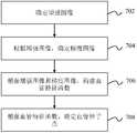
- the determining the region of interest in the imagecomprises: identifying slice information of the plurality of layers of the image; and determining, according to the slice information, a subgraph in the plurality of layers a screenshot range; within the scope of the screenshot, a subgraph is taken from the plurality of layers; according to the subgraph, a template is read; according to the template, Registering the sub-pictures to obtain a registration result; and determining the region of interest based on the registration result.
- the determining the region of interest in the imagecomprises performing machine learning.
- the methodfurther comprises determining a blood vessel seed point.
- the determining a blood vessel seed pointincludes: enhancing the image to obtain an enhanced image; determining the image grayscale gradient to obtain a gradient image; and according to the enhanced image and the gradient image, Constructing a blood vessel characteristic function; and determining the blood vessel seed point based on the characteristic function.
- the methodfurther comprises determining a blood vessel centerline.
- the determining a blood vessel centerlineincludes: collecting a training sample; reading the training sample, training to obtain a weak allocator; combining the weak allocator to obtain a strong allocator; a vessel enhancement map is determined; and, in the vessel enhancement map, a vessel centerline is determined based on Fast Marching.
- the training samplesinclude a positive training sample and a negative training sample.
- the determining a blood vessel centerlineincludes: registering a sample image; extracting a reference vessel centerline of the registered sample image; determining a vessel centerline model based on the reference vessel centerline; Mapping a vessel centerline model to the image to obtain a first vessel centerline of the image; setting a distance field in the image; and adjusting a first vessel centerline of the image based on the distance field To obtain the blood vessel centerline.
- the methodfurther comprises extracting a blood vessel based on the blood vessel centerline.
- the extracting the blood vesselincludes: determining a first segmentation result according to the blood vessel center line; determining a second segmentation result according to the first segmentation result; determining the second segmentation result according to the global condition, and determining the third segmentation result Setting a boundary condition of the blood vessel model; determining the third segmentation result according to the boundary condition, determining a fourth segmentation result; and extracting the blood vessel according to the fourth segmentation result.
- the methodfurther comprises detecting whether the blood vessel is successfully extracted within the region of interest; if not, extracting the blood vessel using the first method.
- the first methodincludes a segmentation method for an aneurysm blood vessel, a segmentation method for a low contrast agent blood vessel, or a segmentation method for a calcified malformation blood vessel.
- the extracting the blood vessel in the imagecomprises: counting a gray value of the region of interest; setting a growth parameter according to the gray value; growing a blood vessel trunk based on Fast Marching; and adjusting The growth parameters track the blood vessel branches.
- the methodfurther comprises extracting a particular portion of the blood vessel.
- the extracting the blood vessel specific portionincludes: determining an area including the specific portion; determining a first connected domain in the region; acquiring a feature of the first connected domain; determining the specific according to characteristics of the first connected domain a portion of the connected domain; and determining the particular portion based on the connected domain of the particular portion.
- the characteristic of the first connected domaincomprises a location, a size or a shape of the first connected domain.
- the determining the particular portioncomprises expanding or growing a connected domain of the determined particular portion.
- the systemincludes: at least one processor and executable instructions.
- the instructionswhen executed by at least one processor, cause the at least one processor to implement a method.
- the methodincludes: acquiring an image including blood vessel information, the image including a plurality of layers; determining a region of interest in the image; establishing a blood vessel model; and, within the region of interest, extracting a region based on the blood vessel model The blood vessels in the image.
- the determining the region of interest in the imagecomprises: identifying slice information of the plurality of layers of the image; and determining, according to the slice information, a subgraph in the plurality of layers a screenshot range; within the screenshot range, a subgraph is taken from the plurality of layers; a template is read according to the subgraph; and the subgraph is registered according to the template to obtain a registration result; The region of interest is determined based on the registration result.
- the methodfurther comprises determining a blood vessel seed point.
- Determining the blood vessel seed pointincludes: enhancing the image to obtain an enhanced image; determining the grayscale gradient of the image to obtain a gradient image; constructing a blood vessel feature function according to the enhanced image and the gradient image; and, according to The feature function determines the blood vessel seed point.
- the methodfurther comprises determining a blood vessel centerline.
- the computer readable mediumincludes executable instructions.
- the instructionswhen executed by at least one processor, cause the at least one processor to implement a method.
- the methodincludes: acquiring an image including blood vessel information, the image including a plurality of layers; determining a region of interest in the image; establishing a blood vessel model; and, within the region of interest, extracting a region based on the blood vessel model The blood vessels in the image.
- FIG. 1is a schematic illustration of an imaging system shown in accordance with some embodiments of the present application.
- FIG. 2is an exemplary flow diagram of generating an image, shown in accordance with some embodiments of the present application.
- FIG. 3is a schematic diagram of an image generator shown in accordance with some embodiments of the present application.
- FIG. 4is a schematic diagram of an image processing module shown in accordance with some embodiments of the present application.
- 5-Ais a schematic illustration of a blood vessel extraction unit shown in accordance with some embodiments of the present application.
- 5-Bis an exemplary flow diagram of extracting blood vessels shown in accordance with some embodiments of the present application.
- FIG. 6is an exemplary flow chart for determining a region of interest, in accordance with some embodiments of the present application.
- FIG. 7is an exemplary flow diagram of determining a blood vessel seed point, shown in accordance with some embodiments of the present application.
- 8-Ais an exemplary flow chart for obtaining a strong allocator based on training, according to some embodiments of the present application.
- 8-Bis an exemplary flow chart for determining a blood vessel centerline, in accordance with some embodiments of the present application.
- 8-Cis an exemplary flow chart for determining a vessel centerline, in accordance with some embodiments of the present application.
- FIG. 9is an exemplary flow diagram for extracting blood vessels from a vessel centerline, in accordance with some embodiments of the present application.
- FIG. 10is an exemplary flow diagram of extracting a blood vessel shown in accordance with some embodiments of the present application.
- FIG. 11is an exemplary flow diagram of extracting a particular portion of a blood vessel, shown in some embodiments of the present application.
- FIG. 12is a flow diagram of a particular embodiment of determining a region of interest of a head and neck, in accordance with some embodiments of the present application;
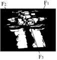
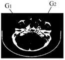
- 13-A through 13-Mare diagrams of exemplary experimental results for determining a region of interest for a head and neck target vessel, in accordance with some embodiments of the present application;
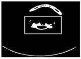
- 14-A and 14-Bare diagrams showing exemplary experimental results of determining a vessel centerline based on training, in accordance with some embodiments of the present application;
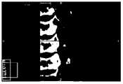
- 15-A through 15-Pare diagrams of exemplary experimental results of blood vessel extraction based on a vessel centerline, in accordance with some embodiments of the present application.
- 16-A through 16-Gare diagrams of exemplary experimental results of extracting a hepatic portal vein, as shown in some embodiments of the present application;
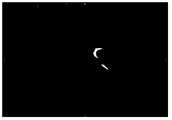
- FIG. 17is a schematic diagram of a U field, shown in accordance with some embodiments of the present application.
- imaging system 100can scan a given target, obtain scan data, and generate images associated therewith. In some embodiments, imaging system 100 can further process the generated image. In some embodiments, imaging system 100 can be a device or a group of devices. Specifically, the imaging system 100 can be a medical imaging system, for example, a PET (Positron Emission Tomography) design. A SPECT (Single-Photon-Emission Computed Tomography) device, a CT (Computed Tomography) device, an MRI (Magnetic resonance imaging) device, and the like. Further, the medical imaging system may be used alone or in combination. For example, a PET-CT device, a PET-MRI device, or a SPECT-MRI device.
- imaging system 100can include a scanner that can scan a given target and obtain information related thereto (eg, scan data). Further, imaging system 100 can be a radiological scanning device.
- the radiological scanning devicecan include a radioactive scanning source.
- a radioactive scanning sourcecan emit radioactive rays to a given target.
- the radioactive raysmay include one or a combination of particulate rays, photons, and the like.
- the particulate radiationmay include one or a combination of neutrons, protons, electrons, ⁇ medium, heavy ions, and the like.
- the photon raymay include one or a combination of X-rays, gamma rays, alpha rays, beta rays, ultraviolet rays, lasers, and the like.
- the photon raymay be an X-ray
- its corresponding imaging system 100may be one or more of a CT system, a digital radiography system (DR), a multimodal medical imaging system, and the like.
- the multimodal medical imaging systemcan include one or more of a CT-PET system, a SPECT-MRI system, and the like.
- imaging system 100can include a cavity 110, a bed frame 120, a high voltage generator 130, an operational control computer device 140, an image generator 150, and a control display device 160.
- the interior of the cavity 110can house components for generating and detecting radioactive rays.
- the cavity 110can house a radiation generator 180 and a detector 170.
- Radiation generator 180can emit radioactive rays. The radioactive rays can be emitted to an object placed in the cavity 110 and received by the detector 170 through the object.
- the radiation generator 180can be an X-ray tube. The X-ray tube can emit X-rays that pass through an object placed inside the cavity 110 and are received by the detector 170.
- the detector 170can be a circular detector, a square detector, or a curved detector or the like.
- the angle of rotation of the curved detectormay be between 0 and 360 degrees.
- the angle of rotation of the arcuate detectorcan be fixed.
- the angle of rotation of the arcuate detectorcan be adjusted as needed. For example, the adjustment may be made according to the resolution of the desired image, the size of the image, the sensitivity of the detector, the stability of the detector, or a combination of one or more of them.
- the detector 170can be a one-dimensional detector, a two-dimensional detector, or a three-dimensional detector.
- the bed frame 120can support an object to be detected (such as a patient to be detected).
- the bed frame 120can move inside the cavity 110 during the inspection process.
- the bed The frame 120is movable in the Z-axis direction.
- the patientcan be supine, prone, with the head in front or the foot in front.
- the bed frame 120can move inside the cavity 110 at a constant speed.
- the speed at which the bed frame 120 movescan be related to factors such as scan time, scanning area, and the like. In some embodiments, the speed at which the bed frame 120 moves may be a system default or may be set by a user.
- the high voltage generator 130can generate high voltage or high current.
- the generated high voltage or high currentcan be transmitted to the radiation generator 180.
- the high pressure generatedcan be 80kV-140kV, 75Kv-150kV or 120kV-140kV.
- the current generatedcan be 20 mA to 500 mA.
- the operational control computer device 140can be associated with the cavity 110, the radiation generator 180, the detector 170, the high voltage generator 130, the bed frame 120, the image generator 150, and/or the control display device 160.
- the above devicescan be connected directly or indirectly.
- the operational control computer device 140can control the cavity 110 to rotate to a certain position. This location can be a system default or it can be set by the user (eg doctor, nurse, etc.).
- the operational control computer device 140can control the high voltage generator 130.
- the operational control computer device 140can control the strength of the voltage or current generated by the high voltage generator 130.
- the operational control computer device 140can control the display device 160.
- the operational control computer device 140can control the parameters associated with the display.
- the parametersmay include display size, display scale, display order, number of displays, and the like. As an example, it is possible to control the entirety or part of the displayed image. As still another example, an image may be divided into several sub-pictures (eg, a head sub-picture, a neck sub-picture, a lower-limb sub-picture, etc.), and several sub-pictures may be displayed simultaneously or sequentially. As a further example, an image can be enlarged or reduced.
- the image generator 150can generate an image.
- image generator 150may perform operations such as image pre-processing, image reconstruction, and/or blood vessel extraction.
- Image generator 150may be associated with detector 170, operational control computer device 140, display device 160, and/or an external data source (not shown).
- image generator 150may receive data from detector 170 or an external data source and generate an image based on the received data.
- the external data sourcemay be a hard disk, a floppy disk, a random access memory (RAM), a dynamic random access memory (DRAM), a static random access memory (SRAM), or a magnetic bubble memory ( Bubble memory), thin film memory, magnetic plated wire memory, phase change memory, flash memory, a cloud disk, etc.
- RAMrandom access memory
- DRAMdynamic random access memory
- SRAMstatic random access memory
- Bubble memorymagnetic bubble memory
- thin film memorymagnetic plated wire memory
- phase change memoryphase change memory
- flash memorya cloud disk, etc.
- cloud disketc.
- image generator 150may generate The image is transferred
- Display device 160can display the received data or images.
- Display device 160can be coupled to operational control computer device 140 and image generator 150.
- display device 160can display the image generated by image generator 150.
- display device 160can send instructions to image generator 150 and/or operational control computer device 140.
- a usermay set imaging parameters through display device 160, which may be sent to operational control computer device 140.
- the imaging parametersmay include data acquisition parameters and image reconstruction parameters, and the like.
- the data collection parametermay include one or more of a scan time, scan target positioning information, a rotation speed of the rack, and a strength of the voltage/current.
- the image reconstruction parametersmay include one or more of reconstructing a field of view, a reconstruction matrix, a reconstruction algorithm, and the like.
- imaging system 100is merely for convenience of description, and the present application is not limited to the scope of the embodiments. It will be understood that, after understanding the principle of the system, it is possible for the various modules to be combined arbitrarily or the subsystems are connected to other modules without being deviated from the principle. Various modifications and changes in the form and details of the application of the method and system.
- FIG. 2is an exemplary flow diagram of an image generation process.
- scan parameterscan be set.
- the process of setting scan parameterscan be implemented by the operation control computer device 140.
- the scan parametersmay include one or more of scan time, scan target positioning information, position of the rack, rotational speed of the rack, intensity of voltage/current, and the like.
- the bed frame 120can be rotated to a particular location.
- the cavity 110can be moved to a particular location.
- the particular locationmay be a system default or may be set by a user (eg, a doctor, nurse).
- the positions setare different depending on the object to be tested.
- the object to be testedmay be the whole or a part of the object to be detected.
- the detection objectmay include a human body, an animal, a non-biological object, or the like.
- the test objectmay include an organ, a tissue, a lesion, a tumor site, or any combination of the above.
- the test objectmay be a head, a chest, an abdomen, a heart, a liver, an upper limb, a lower limb, a spine, a bone, a blood vessel, or the like, or any combination of the above.
- the object to be testedcan be scanned.
- scan data of the measured objectcan be acquired.
- the scanning process and the process of acquiring the scan datacan be performed by the radiation generator 180 and the detector 170 in common.
- the radioactive raysmay pass through the analyte and be absorbed by the analyte and received by the detector 170.
- the radioactive raysmay be reflected by the analyte to the detector 170 and received by the detector.
- the scan datamay be obtained in whole or in part from an external data source.
- an imagemay be generated based on the scan data.
- step 210can be implemented by image generator 150.
- the generated imagemay include an MRI image, a CT image, a PET image, or any combination of the above.
- a CT imagecan be obtained using a rendering algorithm.
- the generated imagemay comprise a two-dimensional image or a three-dimensional image.
- the generated imagecan also be processed. Processing of the image may include filtering and denoising the image, normalization of the grayscale, horizontal rotation of the image, correction of the size, and the like.
- an imagecan be output.
- the imagemay be displayed by display device 160.
- the imagesmay be transmitted to any external device associated with imaging system 100, such as a database, terminal, or the like.
- FIG. 3is a schematic diagram of an image generator 150.
- the image generator 150may include a data receiving module 310, a data processing module 320, an image processing module 330, a storage module 340, a control module 350, and an image output module 360.
- the data receiving module 310can receive data related to the measured object.
- the data related to the test objectmay include scan data, basic information (such as name, age, gender, height, weight, medical history, etc.), scan parameters, and the like.
- the scan datamay be collected by detector 170 and transmitted to data receiving module 310.
- after the scan data is collected by the detector 170it may be transferred to the storage module 340 and then transferred to the data receiving module 310 by the storage module 340.
- data receiving module 310can receive scan parameter data from operational control computer device 140.
- the data receiving module 310can receive data (eg, patient basic information) from an external data source (not shown).
- the data processing module 320can perform analysis processing on the received data.
- the data processing module 320can receive data from the data receiving module 310, the storage module 340, and/or an external data source and perform analytical processing.
- the data processing module 320can perform pre-processing operations on the received data.
- data processing module 320can process dark current and null scan data, remove dead pixels, remove noise, perform geometric corrections, and the like.
- data processing module 320can analyze the processed scan data to generate an initial image.
- the initial image described hereinis an unpreprocessed image generated based on scan data of the object to be tested.
- data processing module 320can statistically analyze the scan data and basic information to generate statistical results.
- data processing module 320can count the probability that a certain type of population is suffering from a certain disease. The statistical result can be transmitted to the storage module 340.
- Image processing module 330can generate images and/or process images.
- image processing module 330can receive the scan data processed by data processing module 320 and generate an image from the processed scan data.
- image processing module 330can process the initial image generated by data processing module 320.
- the processing operationsmay include filter denoising, grayscale normalization, image horizontal rotation, scale size correction, partial occlusion removal (eg, removal of glasses), and the like.
- image processing module 330can perform image reconstruction. For example, image processing module 330 can perform angiography.
- image processing module 330can further analyze the processed generated image. For example, image processing module 330 can extract blood vessels in the image.
- image processing module 330can control parameters of image processing module 330 for blood vessel extraction.
- the parametersmay include determination of a region of interest, determination of a seed point, determination of a centerline, and the like.
- the seed point referred to hereinrefers to an image voxel inside a blood vessel selected from an image.
- the seed pointcan be an image voxel near the center of the blood vessel.
- the centerline as used hereinrefers to a line that runs along the blood vessel inside the blood vessel.
- the vessel centerlinemay refer to a collection of pixel points located at or near the center of the vessel.
- the vessel centerlinemay refer to a line of pixels that are equidistant or substantially equal in distance to the vessel boundary.
- the storage module 340can store data, images, and/or related parameters, and the like.
- the stored datacan be in various forms of data. For example, one or more of a numerical value, a signal, an image, related information of a predetermined target, a command, an algorithm, a program, and the like.
- scan data, initial images, processed images, processing parameterseg, denoising parameters, normalized parameters, etc.
- storage module 340can include a fixed storage system (eg, a magnetic disk), a mobile storage system (eg, a USB interface, an interface to a Firewire port, etc., and/or a drive of a disk drive type), and the like.
- the storage module 340can store a blood vessel initial image, a processed blood vessel image, a blood vessel image setting parameter, and the like. Further, the storage module 340 may be temporary storage of data, that is, dump data for the next data processing; or may be long-term storage of data, that is, storing the final data processing result.
- Control module 350can control data receiving module 310, data processing module 320, image processing module 330, storage module 340, and/or output module 360. In some embodiments, control module 350 can control the time at which data receiving module 310 receives data and/or the path through which data is transmitted. In some embodiments, control module 350 can control data transmission speeds and data transmission modes (eg, real-time transmission or delayed transmission), and the like. In some embodiments, control module 350 can control image processing module 330 for image reconstruction. As an example, control module 350 may select an algorithm selected for image reconstruction. As yet another example, the control module 350 can control the parameters of the blood vessel extraction by the image processing module 330. The parameters may include determination of a region of interest, determination of a seed point, determination of a centerline, and the like. In some embodiments, control module 350 can receive instructions from a user (eg, a doctor, imaging engineer, etc.).
- a usereg, a doctor, imaging engineer, etc.
- the output module 360can output information.
- the informationmay include data, images, and/or related parameters, and the like.
- the informationmay be from data receiving module 310, data processing module 320, image processing module 330, storage module 340, and/or control module 350.
- the informationmay be presented in a variety of manners including, but not limited to, one or more of audio, video, images, text, and the like.
- informationcan be broadcast by a microphone, a loudspeaker, or the like.
- informationcan be presented on the display screen.
- the informationmay be in various forms of data including, but not limited to, one or more of values, signals, images, related information of a given target, commands, algorithms, programs, and the like.
- the informationmay include a blood vessel initial image, a blood vessel grayscale image, a blood vessel mask image, a blood vessel rough processing image, a blood vessel fine processing image, and the like.
- the informationcan include related parameters, such as a histogram, a level set, a function set, and the like.
- the informationcan be output to any external device (eg, database, terminal, etc.) associated with imaging system 100.
- the informationcan be displayed on any of the display devices (eg, display device 160, computer display, mobile display, etc.).
- various modules within image generator 150may include one or more general purpose processors.
- the processorincludes, but is not limited to, a programmable logic device (PLD), a special integrated circuit (ASIC), a microprocessor, and a system on chip (SoC). ), one or more of a digital signal processor (DSP), and the like.
- PLDprogrammable logic device
- ASICspecial integrated circuit
- SoCsystem on chip
- DSPdigital signal processor
- the two or more processorscan be combined on one hardware device.
- the processorcan implement data processing in a variety of ways, for example, by hardware, software, or a combination of hardware and software.
- image generator 150is merely a specific example and should not be considered as the only feasible implementation. Obviously, various modifications and changes in the form and details of the specific embodiments and steps may be made by those skilled in the art after the basic principles are understood. Corrections and changes are still within the scope of the above description.
- a storage unitmay be added to each module in the image generator 150 for storing intermediate data or processing results generated during the operation of each module.
- one or more modulescan be integrated into the same module to implement the functionality of one or more modules.
- the data receiving module 310 and the image output module 360can be integrated in one module while implementing input/output functions.
- the control module 350can be integrated in the image processing module 330 to control various parameters that may be involved in the operation of the image processing module 330.
- the image processing module 330may include an image information acquisition unit 410, an image pre-processing unit 420, an image reconstruction unit 430, and a blood vessel extraction unit 440.
- the image information acquisition unit 410can acquire initial image information.
- the image informationmay be scan data for generating an image, or may be an initial image that has been generated.
- image information acquisition unit 410may obtain scan data or an initial image from data processing module 320 or storage module 340.
- image information acquisition unit 410may transmit the received scan data or initial image to image pre-processing unit 420 and/or image reconstruction unit 430.
- the image pre-processing unit 420can perform a pre-processing operation on the image.
- the pre-processing operationsmay include filter denoising, grayscale normalization, image horizontal rotation, scale size correction, and partial occlusion removal (eg, removal of glasses), and the like.
- the image pre-processing unit 420may perform a filtering smoothing operation on the initial image to reduce image noise.
- the image reconstruction unit 430can reconstruct an image.
- image reconstruction unit 430can perform image reconstruction based on the scan data.
- image reconstruction unit 430can perform two-dimensional reconstruction or three-dimensional reconstruction.
- the algorithm for image reconstructionmay include one or more of Filtered Back Projection (FBP), Ordered Subsets Expectation Maximization (OSEM), FDK algorithm, and the like.
- image reconstruction unit 430can transmit the image to image pre-processing unit 420 and/or blood vessel extraction unit 440 for further processing.
- the blood vessel extraction unit 440can extract blood vessels from the image.
- blood vessels in the head, neck, abdomen, lower extremities, and the likecan be extracted.
- the blood vessel extraction algorithmmay include: a pattern recognition algorithm, a model algorithm, a tracking algorithm, an artificial intelligence algorithm, a neural network algorithm, a tubular detection algorithm, and the like.
- blood vessel extraction unit 440can determine a region of interest for blood vessel extraction, a seed point for a target vessel, a centerline, and a particular portion of a blood vessel (eg, a sinus), and the like.
- the blood vessel extraction unit 440can be based on Blood collection is performed by level set, region growth, MS model, CV model, and the like.
- the blood vessel extraction unit 440can separate the boundaries of the blood vessels. For example, blood vessel boundaries can be determined by data expansion and/or data erosion operations. As another example, multiple levels of model boundary parameters can be used to separate vessel boundaries.
- image processing module 330is merely a specific example and should not be considered as the only feasible implementation. Obviously, various modifications and changes in the form and details of the specific embodiments and steps may be made by those skilled in the art after the basic principles are understood. The changes are still within the scope of the above description.
- the image pre-processing unit 420can be integrated in the image reconstruction unit 430 while implementing the functions of image reconstruction and image pre-processing.
- the blood vessel extraction unit 440can be integrated in the image reconstruction unit 430, and the blood vessel can be directly extracted from the image after the image is reconstructed.
- FIG. 5Ais a schematic illustration of a blood vessel extraction unit 440, in accordance with some embodiments of the present application.
- the blood vessel extraction unit 440may include a separation subunit 510, an extraction subunit 520, and a feature extraction subunit 530.
- the ionizing unit 510can determine the region of interest.
- the region of interestmay be a region containing blood vessels.
- the shape of the region of interestmay be tubular, circular, circular, elliptical, triangular, rectangular, irregular, or the like.
- the shape of the region of interestis related to the physiological anatomy of the target vessel or the region in which it is located.
- the region of interest of the cerebral arterycan be tubular.
- the separation sub-unit 510can determine the region of interest in a template registration manner. For example, the image to be processed and the corresponding template can be registered to determine the region of interest.
- the region of interestmay be determined in a multi-template registration manner.
- the imagecan be divided into different regions according to the physiological structure, for example, the head, the neck, the abdomen, the lower limbs, etc., and then the image to be processed of the different regions and the template of the corresponding region are registered to determine the sense of each region. Area of interest.
- the detach subunit 510can employ a classifier based method to determine the region of interest. For example, image features (eg, one or more of image gray values, gradient values, enhancement values, shapes, etc.) may be extracted, and the extracted image features are analyzed using a classifier to determine regions of interest.
- the extraction subunit 520can extract the target blood vessel.
- the target blood vesselmay be a head and neck blood vessel, an abdominal blood vessel, a lower limb blood vessel, or the like.
- the head and neck blood vesselsmay include a vertebral artery, a basilar artery, an internal carotid artery, and the like.
- the abdominal blood vesselsmay include the abdominal aorta, the renal artery, the hepatic portal vein, and the like.
- the extraction sub-unit 520can further determine vessel seed points and/or vessel centerlines, and the like. As an example, the extraction sub-unit 520 can make a determination of a blood vessel seed point based on physiological structural characteristics (eg, symmetry) of the blood vessel type.
- the extraction sub-unit 520can determine a vessel centerline based on the vessel seed point. As a further example, the extraction subunit 520 can pass The center line of the blood vessel is determined by determining the starting point, the path point, and the ending point of the blood vessel and connecting the points. As a further example, the extraction sub-unit 520 may further select a line inside the blood vessel as a blood vessel center line after completing the blood vessel extraction. In some embodiments, the extraction sub-unit 520 can vascularize the blood vessel image to obtain a blood vessel enhancement image. In some embodiments, the extraction sub-unit 520 can perform blood vessel extraction in a blood vessel enhancement image.
- the extraction sub-unit 520can perform segmentation of blood vessels (also referred to as "extraction") in an iterative manner. For example, the extraction sub-unit 520 can divide the blood vessel into different regions, ensuring that there is only one intact main blood vessel in the region; the extraction sub-unit 520 can segment the main blood vessels in each region; and the extraction sub-unit 520 can detect whether the blood vessel segmentation is re-detected. If the extraction sub-unit 520 detects that the blood vessel segmentation fails, the extraction sub-unit 520 may adopt an iterative mode to select other alternative segmentation methods until the segmentation is successful.
- extractionalso referred to as "extraction”
- the feature extraction sub-unit 530can perform extraction of a specific portion from the blood vessels that have been extracted.
- the particular portioncan be a particular type of blood vessel or a portion of a blood vessel.
- a particular portion of the blood vesselcan include a sinus sinus or the like in the blood vessel; the feature extraction sub-unit 530 can perform venous sinus extraction from the already extracted blood vessel tree.
- the feature extraction sub-unit 530may partition the blood vessel that has been extracted, determine a specific region in which the connected domain of the sinus is determined, and expand and grow in the connected domain. , the extraction of sinus.
- an exemplary flow diagram of a blood vessel extraction processis shown in FIG. 5-B.
- the flow of blood vessel extractioncan be accomplished by blood vessel extraction unit 440.
- an imagecan be acquired.
- the imagemay be a two-dimensional image or a three-dimensional image.
- the imagemay be an image output by image pre-processing unit 420 and/or image reconstruction unit 430.
- a Region of Interest (ROI) for blood vessel extractioncan be determined.
- the region of interestmay be the region in which the blood vessel is located.
- the shape of the region of interestmay be tubular, circular, circular, elliptical, triangular, rectangular, irregular, or the like.
- the region of interestmay be determined in a template registration manner. For example, the image to be processed and the corresponding template can be registered to determine the region of interest. Further, the region of interest can be determined in a multi-template registration manner. For example, the image may be divided into different regions according to the physiological structure, and then the image to be processed of the different regions and the template of the corresponding region are registered to determine the region of interest of each region.
- a region of interestmay be determined using a classifier based approach. For example, image features (eg, one or more of image grayscale fingers, gradient values, enhancement values, shapes, etc.) may be extracted, and the extracted image features may be analyzed using a classifier to determine a region of interest.
- the region of interestmay be determined in a machine learning manner (eg, by a machine learning model).
- machine The learner modelmay include one or more parameters related to determining the region of interest, and the machine learning model may be trained through historical data to determine the parameters. The region of interest is further determined by the parameters.
- blood vesselscan be extracted.
- the vessel centerlinecan be determined first, and the vessel is further grown based on the vessel centerline.
- blood vesselscan be extracted by constructing a blood vessel model.
- blood vesselscan be grown in the region of interest based on characteristics of the blood vessel, such as grayscale values.
- blood vesselscan be extracted by machine learning.
- the vessel treecan also be determined based on the blood vessels that have been extracted.
- the alternative segmentation methodmay include a segmentation method for an aneurysm blood vessel, a segmentation method for a low contrast agent blood vessel, a segmentation method for a calcified malformation blood vessel, and the like.
- a level set algorithm without shape constraintcan be used to extract blood vessels above a gray scale and gradient threshold.
- a blood vessel model based methodcan be adopted, which adds shape constraints on the basis of image features such as gray scale and gradient.
- the segmentation method for calcified malformed blood vesselsis to comprehensively consider various image features such as gray scale, gradient, and shape constraint to perform blood vessel extraction.
- a particular portion of the blood vesselcan be extracted.
- a particular portion of a blood vesselrefers to a particular type of blood vessel, or a portion of a blood vessel.
- venous sinus extractioncan be performed from an already established vessel tree using the physiological anatomical characteristics of the sinus (eg, size, shape, location, etc.).
- step 504the extraction of blood vessels is merely a specific example and should not be considered as the only feasible implementation.
- step 506one or more optional steps may be added. For example, determining a blood vessel centerline, determining a blood vessel seed point, and the like.
- step 508may not be necessary.
- extraction or non-extractioncan be selected according to actual needs.
- FIG. 6is an exemplary flow diagram for determining a region of interest.
- determining the region of interest processmay be implemented by separate sub-unit 510.
- an imagecan be acquired. Different physiological regions may be included in the image, such as the head, neck, abdomen, lower limbs, and the like.
- the imagemay be a two dimensional or three dimensional image.
- the imagecan be an angiographic (CTA) image.
- the imagemay be an image output by image pre-processing unit 420 and/or image reconstruction unit 430.
- slice informationcan be identified.
- the slice layerherein refers to an N slice layer in which images are arranged in a certain order (for example, from head to foot), wherein N is any positive integer.
- the value of Ncan be the system default value or can be set by the user (for example, doctor, nurse).
- different analyteseg, different patients
- the systemcan determine the required scan range based on physiological information (eg, height, weight, etc.) of the test object to determine the N value.
- any one of the N slicescan be selected and the slice information can be identified.
- the slice informationmay include the size, shape, position, etc. of the slice.
- the area to which the slice belongsmay be determined based on the slice information. For example, whether the sheet belongs to the top, half, or skull base or the like can be judged according to the size and/or shape of the sheet.
- the screenshot range of the subgraphmay be obtained according to the slice information.
- the subgraphherein refers to a map corresponding to a certain part of the image, for example, a head subgraph, a neck subgraph, a belly subgraph, and the like.
- the interception range of the subgraphmay be obtained based on the identified slice position information (eg, head, half, skull base, etc.). For example, if the slice selected in step 604 is the second slice and the identified slice information is the header, then it can be determined that the screenshot range of the header submap is the 3-9 slice.
- the sub-picturemay be intercepted according to the acquired screenshot range. For example, the header subgraph can be intercepted based on the screenshot range.
- the subgraphs capturedmay be registered according to the template.
- the templatecan be a standard image template.
- N casescan be read in any of the storage devices (eg, databases) described herein, followed by comprehensive statistics of the N cases and by registration, fusion, and/or physician Manual marking and other operations are made into standard image templates.
- the head image and the neck image of a plurality of patientsmay be respectively taken out by the position of the foramen magnum and the atlas, and the head image template and the neck image template are prepared by registration, filtering, averaging, and the like.
- the templatemay be a template made based on an average population.
- the image to be processedis from a patient
- a certain region of interest of the reference casemay be intercepted, and the intercepted region of interest is labeled as a region of interest mask (also referred to as "film” or "mask”).
- a region of interest maskalso referred to as "film” or "mask”
- Methods of marking regions of interestmay include auto-tagging, manual marking, and the like.
- the process of marking as described hereinrefers to the process of partially occluding or covering an image to extract a particular portion of the image.
- the region of interest mask in each reference casecan then be mapped to a coordinate space based on a certain mapping relationship (eg, based on an affine registration relationship matrix and a non-rigid-registered deformation field), eg, a coordinate space of the reference image And determine the feeling The probability distribution of the interesting area.
- information of the plurality of reference cases corresponding to the region of interestmay be added to obtain a probability map (or information map) of the region of interest.
- the probability value of a certain pixel point in the probability mapmay represent a position probability of the target region (ie, the region of interest in which the blood vessel is located) (ie, the probability that the location is the target region).
- the probability value of the pixel point in the probability mapranges from [0, 1]. Specifically, in some embodiments, the larger the probability value of the pixel, the larger the gray value of the pixel, and the greater the position probability of the target region.
- the probability range of the probability mapcan be adjusted. For example, the probability range of the target area information map can be adjusted from greater than 0 to greater than 0.25, reducing the search domain.
- multiple templatesmay be employed for registration.
- the imagemay be dynamically divided into a head sub-picture and a neck sub-picture, and the head sub-picture and the neck sub-picture are respectively image-registered with the corresponding partition image template.
- the partitionrefers to a certain sub-area of the patient, such as the head, neck, abdomen, lower limbs, and the like.
- the anatomical features of the partition, clinical diagnostic information, and the likecan be combined during registration.
- the area of the internal carotid arterycan be initially determined based on the anatomical features of the neck.
- the adaptive registration method or spatial transformation modelcan be dynamically selected.
- the region of interestmay be determined based on the registration result.
- the merged region of interestmay be derived from the region of interest of the determined subgraph.
- the region of interest of the head and neckcan be combined to obtain the region of interest of the head and neck.
- Figure 7is an exemplary flow chart for determining a blood vessel seed point, in accordance with some embodiments of the present application.
- the process of determining a blood vessel seed pointcan be implemented by the extraction sub-unit 520.
- the vessel seed pointcan be determined after determining the region of interest.
- an enhanced imagemay be determined.
- the enhanced image described hereinrefers to an image obtained by enhancing an image in which a target blood vessel is located.
- the imagemay be enhanced based on a Hessian Matrix to determine an enhanced image.
- the imagemay be subjected to a pre-processing operation prior to determining the enhanced image. For example, gray value determination can be performed on the connected domain in the image of the middle cerebral artery, and the connected domain whose gray value is greater than a certain gray threshold can be removed.
- the grayscale thresholdmay be a system default or may be set by a user.
- a gradient imagemay be determined from the enhanced image.
- the gradient image described hereincontains gradient information of the image, for example, a gradient value.
- the gradient valuemay reflect the degree of change in the gray value of the pixels in the image. For example, the larger the gradient value, the more significant the grayscale value of the pixels in the image changes.
- a vascular feature functioncan be constructed from the enhanced image and the gradient image.
- the vascular feature functionis constructed based on image features.
- the image featuresmay include one or more of image gray values, gradient values, enhancement values, shapes, and the like.
- the region of interestcan be divided into two symmetric regions based on the symmetry of the blood vessel.
- the vascular signature functionincludes one or more variables related to vascular symmetry.
- the variablecan include image features of two symmetric regions.
- a vascular signature functioncan be separately constructed for two symmetric regions, respectively.
- a left vascular feature function and a right vascular feature functioncan be constructed separately.
- the vascular signature functioncan be expressed as shown in equation (1):
- il or r, where l represents the left side, r represents the right side, G is the gray level, T is the gradient, H is the enhancement value, and S is the shape index.
- the left vascular feature function and the right vascular feature functionmay be further determined based on the symmetry to obtain a total vascular feature function, as shown in equation (2):
- F lis the left vascular characteristic function
- F ris the right vascular characteristic function
- d zis the distance of the left and right symmetrical blood vessel points in the Z direction.
- a blood vessel seed pointcan be determined based on a blood vessel characteristic function.
- the blood vessel seed pointcan be determined based on a function value of the blood vessel characteristic function determined in step 706. For example, it may be determined that a pair of bilaterally symmetric points having the largest value of the blood vessel characteristic function are seed points of the target blood vessel.
- the process of determining a blood vessel seed pointis not necessary.
- the corresponding blood vesselcan be further extracted according to the blood vessel seed point.
- the blood vessel centerlinecan be determined.
- Figure 8-Ais an exemplary flow chart for training a strong classifier, in accordance with some embodiments of the present application.
- the process of training a strong allocatorcan be implemented by the extraction sub-unit 520.
- a strong allocatorcan be trained based on Adaboost.
- a training samplecan be collected. Training samples may include positive training samples (also referred to as "positive sample points") and negative training samples (also referred to as "negative sample points").
- points within the blood vesselmay be selected as positive sample points in the image template (see FIG. 6 for a detailed description of the image template), and several points may be randomly selected as the negative sample points in the surrounding area of the blood vessel.
- a mask operationis performed on a blood vessel to obtain a blood vessel mask Mask0.
- the vascular mask Mask0is expanded, and the edge of the vascular mask Mask0 is enlarged to obtain a vascular mask Mask1.
- the expansion operation described hereinis a process of incorporating all background points in contact with an object (e.g., a blood vessel) into the object to expand the boundary outward.
- the vascular masks Mask1 and Mask0are XORed, and the same points in Mask0 and Mask1 are excluded, and different points are extracted, and the areas where the different points are located are the areas around the blood vessels.
- positive and negative sample pointscan be acquired separately in the intravascular and perivascular regions.
- the number of positive and negative samplescan be arbitrary. The number of positive and negative samples does not have to be consistent. For example, the number of negative samples can be more than the positive samples.
- the training samplescan be read and trained to obtain a weak classifier.
- the positive sample point and the negative sample pointare read, and the weight distribution of the training sample is initialized.
- each sample pointcan be assigned the same weight.
- the first weak classifieris trained. It can then be judged whether the positive or negative sample points are accurately classified. If a sample point has been accurately classified, the weight of the sample point will be reduced in the next training; on the contrary, if a sample point is not accurately classified Then the weight of the sample point will be increased. Through the above judgment operation, the sample which is divided in the first training process can be highlighted, thereby obtaining a new sample distribution.
- the weak classifiermay be given a weight according to the classification error, indicating the importance of the weak classifier, and the less the classification error, the greater the weight.
- the sampleis trained again to get the next weak classifier and its weight.
- the sample distributionis further changed repeatedly, and a series of weak classifiers are trained until a predetermined classification error or a preset maximum iteration threshold is reached. For example, if the preset iteration threshold is T, then after T iterations, the weights corresponding to the T weak classifiers and the T weak classifiers can be obtained.
- the weak classifiercan be combined to obtain a strong classifier. Based on a series of weak classifiers trained in step 804, a plurality of weak classifiers are added in accordance with the weights to obtain a strong classifier.
- This strong classifiercan be expressed as equation (3):
- h t (x)represents a weak classifier
- ⁇ trepresents a weight corresponding to each weak classifier, indicating the performance of the weak classifier.
- the smaller the weightthe smaller the classifier plays a smaller role in the final strong classifier.
- the greater the weightthe larger the classifier plays a larger role in the final strong classifier.
- the resulting strong classifiercan be stored in any of the storage devices described in this application and can be further used for the extraction of blood vessel centerlines.
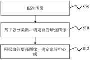
- FIG. 8-Bis an exemplary flow diagram for determining a vessel centerline based on a classifier.
- the process of determining the vessel centerlinecan be implemented by the extraction sub-unit 520.
- image registrationcan be performed (see Figure 6 for a detailed description of image registration) to determine the region of interest.
- a blood vessel enhancement imagecan be determined from the strong classifier.
- the strong classifier described hereincan be read from any of the storage devices described herein.
- a strong classifiercan be obtained by combining multiple weak classifiers (as shown in Figure 8-A).
- the enhanced image described hereinrefers to an image obtained by enhancing an area in which a target blood vessel is located.
- the process of determining a vascular enhanced imageis shown in equation (4):
- h t (x)represents a weak classifier
- ⁇ trepresents a weight corresponding to each weak classifier
- Vesselness (x)represents a blood vessel enhancement value
- a vessel centerlinecan be determined based on the vessel enhancement image.
- the blood vessel centerlinecan refer to a line that runs along the blood vessel inside the blood vessel.
- the vessel centerlinemay refer to a collection of pixel points located at or near the center of the vessel.
- the vessel centerlinemay refer to a line of pixels that are equidistant from the vessel boundary.
- the vessel centerlinecan be determined based on the Fast Marching Method.
- a transformed imagecan be determined, which can be determined according to the following formula (5):
- Grad(x)is the gradient value of the current point
- Std(x)is the variance of the current point and surrounding points
- Std(x 0 )is the variance corresponding to the seed point
- Vesselness(x )is the vascular enhancement value of the current point.
- a level set function fieldcan be established according to the transformed image.
- the curve of the movement changecan be embedded as a zero level set into a higher one-dimensional function, resulting in an evolution function.
- the evolution of a two-dimensional curvecan be transformed into the evolution of a three-dimensional surface, and the evolution equation of a three-dimensional surface can be obtained from a two-dimensional curve evolution equation.
- the position of the point set of the evolution function on the zero-level interfacecan be obtained. Then the evolution result of the curve of the movement change can be determined.
- initialization of the level set function fieldis first performed to establish an initialization curve.
- the blood vessel seed pointmay be used as an initial curve, and then, according to the evolution function, the level set function and the zero level set are continuously updated, and finally the evolution curve corresponding to the zero level set of the level set function is obtained.
- level setsreference is made to the literature, for example, Sethian, JA, Level set methods and fast marching methods: evolving interfaces in computational geometry, fluid mechanics, computer vision, and materials science, Cambridge University Press, 1999, content in the literature. It is included here by reference.
- an initial curve of the level set function field of the two seed pointscan be set based on the determined blood vessel seed point (eg, the blood vessel seed point determined according to the flow of FIG. 7).
- the determined blood vessel seed pointeg, the blood vessel seed point determined according to the flow of FIG. 7.
- These two level set function fieldswill evolve over time. For the pixel point where the P value is smaller in the formula (5), the shorter the time from the initial curve to the pixel point, that is, the level set function field changes along the pixel point where the P value is smaller (that is, the pixel point of the blood vessel). The sooner.
- the level set function field evolution equationcan be expressed as shown in equation (6):
- U i,j,k ,uare the level set function fields corresponding to a certain point
- U i,j,k ,0represent the zero level set function field.
- the two level set function fieldshave an intersection point, which is the point on the blood vessel. Further from the intersection, using the steepest descent method to trace back to the two seed points, the center line of the blood vessels between the two starting and ending seed points can be determined.
- a similarity value for each pixel point or voxel point in the blood vessel enhancement imageis determined.
- the blood vessel enhancement image acquired in the foregoing stepstarting from the seed point, according to the blood vessel feature, for example, one or more of image features (eg, image grayscale fingers, gradient values, enhancement values, shapes, etc.) may be extracted. And analyzing the similarity P value between the neighborhood points, the similarity P value can be obtained according to formula (7):
- the vascular enhancement imagecorresponds to a vascular enhancement value at the point. Since the inside of the blood vessel is a mean structure, the point gradient value belonging to the blood vessel content is small and the variance is small, and the absolute value of the variance of the variance corresponding to the seed point of the blood vessel
- the formula (7)is only an example in the present embodiment, in which the gradient value Grad (x) and the variance Std (x) can be replaced according to the characteristics of the blood vessel, and an equivalent effect can be obtained.
- a U fieldcan be obtained according to the similarity P value, and the U value in the U field can be obtained according to formula (8):
- uis the viscosity solution constant and the U value represents the minimum energy value required for the two seed points to pass the point (i, j, k) path.
- P value of the point (i, j, k) obtained by the formula (7)substituting into the formula (8), obtaining the U value of the point, and acquiring the U value of any point in the blood vessel region, as shown in FIG. U field renderings.
- FIG. 8-Cis an exemplary flow diagram for extracting a blood vessel centerline.
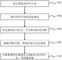
- the process of extracting the vessel centerlinecan be accomplished by the extraction sub-unit 520.
- multiple sample imagescan be registered.
- the blood vessels in the sample imageare first extracted, and then one of the sample images is used as a template image, and the remaining other sample images are registered to the template image.
- binary image registrationcan be performed on blood vessels.
- the sample imagemay be system default or may be determined by the user.
- the sample imagemay be the same type of image as the image to be processed. For example, assuming that the image to be processed is a head image, the sample image may also be a head image.
- the vessel centerline (also referred to as the "reference vessel centerline”) in the sample imagecan be extracted and the pixel points on the vessel centerline sampled to obtain a plurality of centerline sample points.
- the M blood vessel centerlines in the M sample imagesmay correspond to M training samples. Multiple centerline sample points can be included in each training sample.
- a blood vessel centerline average modelcan be determined.
- the centerline sample points of the acquisitionmay be extracted using a Dynamic Shape Model (ASM) algorithm to extract a centerline average model.
- ASMDynamic Shape Model
- the ASM algorithm described hereis based on the point distribution model, and the statistical information of the sample point distribution in the sample image is obtained by training the sample image. Further, it is also possible to obtain a possible change direction of the sample point, so as to find a feature point position corresponding to the sample point on the image to be processed.
- the blood vessel centerline in each sample imagemay correspond to a centerline shape vector.
- a plurality of centerline shape vectors corresponding to a plurality of blood vessel centerlines of the plurality of sample imagesmay constitute a training set.
- the centerline shape vector corresponding to the blood vessel centerline extracted from the sample imagemay be performed. Alignment operation. The main purpose of the alignment operation described herein is to reduce non-shape interference caused by external factors such as different image sizes and distances, so that the corresponding points in different samples are comparable.
- the specific operation of the alignment operationis as follows: 1. For M training samples, one sample is selected as a reference sample, and the center line shape vector of the reference sample is a reference shape vector. Align all centerline shape vectors in the training set to the reference sample shape vector; 2. Determine the centerline average shape vector. 3. Align all centerline shape vectors to the centerline average shape vector to further determine the new centerline average shape vector. 4. Align all centerline shape vectors to the new centerline average shape vector. This is repeated until convergence is achieved, resulting in a centerline averaging model.
- the principal direction and regularity of the shape changemay be obtained by a Principal Component Analysis (PCA) method.
- PCAPrincipal Component Analysis
- the covariance matrix of the datacan be determined. It is also possible to further determine the eigenvalues and eigenvectors of the covariance matrix and arrange the eigenvalues in descending order.
- the principal component analysis methoduses the principal component analysis method to extract the first k eigenvalues and corresponding eigenvectors in the covariance matrix.
- a statistical shape model of the training sampleis constructed according to the feature value and the feature vector, and a shape parameter is obtained to control the shape change of the sample point.
- the centerline average modelcan be mapped to the image to be processed to obtain the vessel centerline of the image to be processed.
- a preliminary threshold-based blood vessel segmentationcan be made to segment the general contour of the blood vessel and the blood vessel edge to obtain a preliminary segmentation result of the blood vessel.
- a preliminary threshold segmentationis also performed on the template image, and the binary image of the template image is registered to the corresponding binary image of the image to be processed, and the blood vessel centerline average model is mapped to the image to be processed.
- a boundary distance fieldcan be set.
- the distance fieldrefers to defining a distance value for one or more pixels in the image, and the distance value of one or more pixels in the image forms a distance field.
- the blood vessel edge obtained by the preliminary segmentation of the blood vesselmay be utilized to determine a distance value for each feature point on the blood vessel centerline of the mapped image to be processed. Specifically, since one blood vessel has two left and right edges, there may be two distance values for a certain feature point, which are the distance from the left edge of the blood vessel and the distance from the right edge, and the smaller distance value may be taken as the The distance value of the feature point.
- the magnitude of the distance value in the distance fieldreflects the extent to which the point is from the center of the blood vessel. In a set distance field, the point where the distance value is larger is closer to the center position of the blood vessel than the point where the distance value is smaller.
- the blood vessel centerline of the image to be processedcan be adjusted according to the distance field.
- the adjustment operationcan effect correction of the extracted blood vessel centerline.
- a priori informationeg, the radius of the blood vessel
- the weights described herecan be system defaults or can be set by the user.
- the centerline average modelis adjusted, and the center line of the mapped blood vessel is continuously moved in a direction far from the boundary, and finally converges to the center of the blood vessel.
- blood vessel extractioncan be further performed based on the corrected vessel centerline.
- the weightcannot be directly determined from the radius of the blood vessel.
- the blood vesselis close to the bone.
- the point where the distance field value satisfies 2Dmay be at the boundary of the blood vessel and the bone, and the weight should be specifically adjusted according to the specific situation.
- the process of extracting blood vesselscan be accomplished by extraction sub-unit 520.
- the region to be segmentedmay be determined based on the blood vessel centerline. In some embodiments, a certain area may be selected outwardly from the center line of the blood vessel as the area to be divided. In some embodiments, the region to be segmented may be set according to the size of the blood vessel. For example, an area centered on the center line and having a radius of 15 mm may be set as the area to be divided.
- a vascular model parametercan be determined.
- Vascular model parametersmay include radius, direction, boundary conditions, boundary gradients, boundary gray levels, enhancement conditions, and the like. In some embodiments, the vascular model parameters can be system defaults or can be set by the user.
- a seed pointcan be determined. In some embodiments, any point within the region to be segmented may be selected as a seed point.
- the neighborhood point of the seed pointcan be selected. The neighborhood point described herein refers to a certain range of pixels around the seed point. As an example, several points (eg, 6 points, 26 points, etc.) of the top, bottom, left, right, front, and back of the seed point may be selected as neighborhood points.
- step 910it may be determined whether the neighborhood point satisfies the vascular model parameter. In some embodiments, it may be determined whether the image features of the neighborhood points conform to current vascular model parameters. In some embodiments, the image features may correspond to a plurality of feature images.
- the feature images described hereininclude Laplacian images, gradient images, maximum rate images, and the like.
- the image featuresinclude gray values, gradient values, Laplacian values, maximum values, and the like.
- the process of determining whether the image feature conforms to the current vascular model parameteris: if the gradient of the neighborhood point is less than the boundary gradient, the maximum rate is greater than the boundary maximum rate, and the Laplacian value is at the boundary Laplacian Between the values, if the gray value is between the boundary gray conditions, the neighborhood point is considered to satisfy the blood vessel model parameter; otherwise, it is not satisfied.
- the neighborhood pointis determined as the candidate segmentation result. If not, then in step 912, it is further determined whether the neighborhood point is near the center of the blood vessel. If the neighborhood point is not near the center of the blood vessel, the neighborhood point is discarded and no operation is performed, and returning to step 908 to reselect other neighborhood points. If the neighborhood point is near the center of the blood vessel, then at step 914 the neighborhood point is determined as the candidate segmentation result. In some embodiments, it may be determined by central conditions whether the neighborhood point is near the center of the blood vessel. The central condition described herein refers to an image feature that determines whether an image feature at a certain point is close to a blood vessel center point.
- whether the neighborhood point is near the center of the blood vesselcan be determined based on the gradient value, the Laplacian value, the maximum rate, and the like. For example, if the maximum rate of the neighborhood point is greater than the maximum rate of the point away from the center of the blood vessel, or the gradient value of the neighborhood point is smaller than the gradient value of the point far from the center, it can be determined that the neighborhood point is close to the center of the blood vessel. In some embodiments, the distance of the neighborhood point from the center of the blood vessel and the distance of the seed point from the center point of the blood vessel can be compared. For example, if a neighborhood point is closer to the center of a blood vessel than a seed point, then the neighborhood point is considered to satisfy the central condition.
- a leakrefers to a point in the candidate segmentation result that contains a portion that does not conform to the current model parameters.
- a global conditioncan be used to determine if a leak has occurred.
- a global condition thresholdcan be set. The global condition threshold may be a system default value or may be set by a user. As an example, assuming that the blood vessel is a tubular structure, the number of points that can be expanded outward along the current candidate segmentation result satisfies a certain threshold condition. This threshold condition can be set to the global condition threshold.
- step 926it can be determined whether all neighborhood points have been determined. If all neighborhood points are not determined, then return to step 908 and another one or more neighborhood points are selected for decision. If all neighborhood points have been determined, then step 928 is performed to further determine if the vascular model parameters have reached the boundary threshold.
- the boundary thresholdmay be a system default or may be set by the user.
- the blood vesselcan be divided into three parts, which are arranged from the inside to the outside: an inner ring circular structure located at the center of the blood vessel, an intermediate annular structure immediately adjacent to the inner ring circle, and an outermost annular structure enclosing the blood vessel of the outermost layer.
- the boundary thresholdthen refers to the model boundary parameter at the outermost annular structure.
- the model boundary parametersmay be relaxed one or more times.
- the model boundary parameterscan be adjusted to the model parameters of the inner circle structure.
- the model boundary parameterscan be adjusted to the model parameters of the inner ring of the intermediate ring structure.
- the model boundary parameterscan be adjusted to the model parameters of the inner ring of the intermediate ring structure.
- the blood vesselcan be segmented based on the final segmentation result. In some embodiments, it may be further detected whether the blood vessel is successfully segmented. If it is not successful, you can choose another segmentation method to re-divide the blood vessels until the segmentation is successful. As an example, first, the blood vessel can be divided into different regions, ensuring that there is only one intact main blood vessel in the region. Further, it is possible to perform segmentation on the main blood vessels in each region to detect whether the blood vessel segmentation is successful. In some embodiments, different segmentation methods can be employed for major blood vessels in different regions.
- the cerebral arterycan be divided into blood vessels according to the gray value and the gradient value of the image, and the internal carotid artery can be divided into blood vessels by a shape constraint method. Further, if the blood vessel segmentation fails, the iterative mode may be used to select other alternative segmentation methods until the segmentation is successful.
- the alternative segmentation methodincludes a pattern recognition method, a model-based method, a tracking-based method, an artificial intelligence method, a neural network method, a tubular object detection method, and the like.
- Figure 10is an exemplary flow diagram showing the extraction of a blood vessel, in accordance with some embodiments of the present application.
- the process of extracting blood vesselscan be accomplished by extraction subunit 520. It should be noted that only one exemplary flow is given in FIG. 10, and it does not mean that the application must be performed according to the following procedure. In some embodiments, one or more steps may delete or adjust the order.
- the region of interestis determined prior to blood vessel extraction.
- the region of interestcan be a head, a neck, an abdomen, a lower limb, and the like.
- an abdominal blood vesselfor example, a hepatic portal vein
- the liver regioncan be determined first.
- the approximate location of the portal vein into the liver layercan be located based on anatomical information of the liver. For example, the approximate location of the portal vein into the liver layer can be estimated by the change in liver area per layer. In some embodiments, the approximate location of the portal vein into the liver layer can be obtained by local Hessian enhancement. In some embodiments, a blood vessel seed point can be located in the portal vein into the liver.
- the gray value of the region of interestcan be counted.
- the gray value of the liver and the gray value of the blood vesselcan be counted.
- a grayscale imagemay be determined, and grayscale statistics are performed in the grayscale image.
- the growth parameterscan be set based on the gray value statistics.
- the growth parametersare related to a growth algorithm.
- the growth algorithmcan be a fast marching algorithm (Fast Marching Method). Growth parameters can include grayscale thresholds, number of iterations, range of blood vessel growth, and the like.
- the growth algorithmcan be run based on the growth parameters to grow the vessel trunk.
- a vessel branchcan be further grown based on the growth parameters.
- step 1008it can be determined if the algorithm parameters need to be adjusted. In some embodiments, the determining step 1008 can be performed in real time or at certain time intervals. In some embodiments, it may be determined whether the algorithm parameters need to be adjusted based on the flow of the angiographic contrast agent and/or the condition of the current vascular region. If necessary, return to step 1004, adjust algorithm parameters (eg, grayscale thresholds, number of iterations, etc.), and in step 1006 and/or step 1007, further grow the vessel trunk and/or vessel section based on the adjusted algorithm parameters. If not required, at step 1010, the vessel branch is tracked based on growth parameters (eg, within the range of blood vessel growth).
- algorithm parameterseg, grayscale thresholds, number of iterations, etc.
- Figure 11is an exemplary flow for extracting a venous sinus, in accordance with some embodiments of the present application.
- the extraction of a particular portion of the blood vesselcan be performed based on the blood vessel that has been segmented (or extracted).
- the sinuscan be extracted from the blood vessels that have been extracted.
- the process of extracting the sinuscan be implemented by feature extraction sub-unit 530.
- the area in which the sinus is locatedcan be determined.
- the region in which the sinus is locatedmay be a banded region.
- the region in which the sinus is locatedcan be determined based on the extracted blood vessels. Methods for extracting blood vessels can be found in the description of other sections of this application.
- the region of the sinuscan be determined by a combination of nuclear expansion and nuclear erosion.
- a mask imagecan be obtained by nuclear expansion and nuclear corrosion, respectively, and the region where the sinus is located is determined based on the obtained mask image.
- the mask image as used hereinrefers to an image that partially occludes or covers an image to highlight a particular portion of the image.
- the brain regioncan be swelled with a 10 mm wide nucleus to obtain a mask 1, and the mask 2 can be obtained by a 20 mm wide nuclear etch.
- the mask 1is subtracted from the region marked by the mask 2, and the sinus sinus can be obtained. Area.
- a connected domain within the areacan be determined.
- the connected domainherein refers to an image region composed of pixel points having the same pixel value and adjacent in the region.
- the connected domains within the region of the sinuscan be obtained by etching. For example, the mask of blood vessels in the area where the sinus is located can be etched to obtain different connected domains.
- the connected domaincan be determined for position, size, and/or shape.
- each connected domainmay be determined by location, and according to the location of each connected domain, it is determined whether the connected domain is a connected domain in which the sinus is located. For example, if a connected domain is located in front of the brain, then According to the location of the connected domain, it is determined that the connected domain may be an arterial vascular transcranial region, not a connected domain where the sinus is located.
- the connected domainmay be sized and/or shaped to determine whether the connected domain is the connected domain in which the sinus is located, based on the size and/or shape of each connected domain.
- a thresholdmay be set to determine if the connected domain is the connected domain in which the sinus is located.
- the thresholdcan be the system default or it can be set by the user.
- different analytesmay correspond to different thresholds.
- one or more thresholdscan be set.
- a first volume threshold, a second volume threshold, a third volume threshold, and the likecan be set.
- the magnitudes of the first threshold, the second threshold, and/or the third thresholdmay be successively decremented. Specifically, for example, since the spatial size of the sinus portion is thick, if the volume of the connected domain is greater than the first threshold, it is determined that the connected domain is the connected domain where the sinus is located.
- the connected domainis determined to be a vein.
- the connected domain where the sinus is locatedsince the sinus end region has a small spatial volume, but is generally located at the top of the brain region, if the volume of the connected domain is greater than a third threshold and is located at the top of the brain region, it is determined that the connected domain is the communication where the sinus is located. area.
- the connected domainBy comparing the difference between the volume of the connected domain and the above threshold, it can be determined whether the connected domain is the connected domain where the sinus is located. As an example, if the volume of the connected domain is greater than the first volume threshold, it is determined that the connected domain is the connected domain where the sinus is located. As still another example, if the connected domain volume is greater than the second volume threshold and the shape is determined to be a non-slender strip shape, then the connected domain is determined to be the connected domain in which the sinus is located. As a further example, if the connected domain volume is greater than the third volume threshold and is located in the top region of the brain, then the connected domain is determined to be the connected domain in which the venous sinus is located.
- the connected domain of the sinuscan be obtained based on the result of the judgment.
- the connected domains of the sinuscan be labeled for further processing.
- the connected region of the sinuscan be expanded and/or region grown to obtain a venous sinus.
- the venous sinuscan be expanded by means of nuclear expansion to obtain a preliminary sinus. Further, regional growth is performed starting from the initial venous sinus, and a complete sinus can be obtained.
- the direction of region growthmay be from the center of gravity of the brain to the direction of the ribbon region in which the sinus is located.
- FIG. 12is an exemplary flow diagram for determining a region of interest (ROI) of a target vessel of the head and neck.
- ROIregion of interest
- An effect map of the region of interest of the head and neck target blood vessels obtained by the procedure shown in Fig. 12can be seen in Figs. 13-A to 13-M.
- an imagecan be acquired.
- the imageincludes head information and neck information.
- a slicecan be selected.
- slice informationcan be identified and output.
- the identification of the slice informationmay be based on the shape and size of the slice. For example, the maximum value of the horizontal direction and the vertical direction of the contour (for example, the head contour) in the image may be determined, and the determination result is compared with a preset threshold to identify the slice information.
- a preset thresholdcan be set. For example, the first threshold a1 and the second threshold a2 may be set. The threshold may be a system default value or may be set by a user.
- a plurality of slicesmay be taken from the selected slice, and a maximum density projection image of the plurality of slices may be acquired. Further, according to the number of connected domains in the maximum density projection image, it is determined that the selected slice is a skull base or a full head.
- the binarized image of the selected plurality of slicesmay be subjected to maximum density projection to obtain a maximum density projected image. In some embodiments, the selected plurality of slices may be binarized based on the bone threshold to obtain a binarized image.
- step 1208it may be determined whether the selected slice is a skull base based on the slice information. If the selected slice is the skull base, then go to step 1220 to determine the slice extent of the neck region and obtain a neck subgraph. If the selected slice is not the skull base, proceed to step 1210 to determine the slice extent of the head region and obtain the header submap. In some embodiments, the region subgraph may be dynamically divided according to the location information of the slice.
- the position information of the selected slicemay be combined, and the head may be determined according to an anatomical feature such as a skull height, a position of the occipital foramen, a position of the atlas, or a method of segmentation using a skull.
- the slice range of the region, and the head subgraphis taken from the image.
- the head sub-picturecan be registered with the head image template to locate the ROI of the head target blood vessel.
- the head subgraphcan be affinely registered with the head image template to locate the target vessel ROI of the head.
- the head image templatemay be a standardized image template.
- anatomical featuresmay be used to aid registration prior to performing affine registration. For example, if the target vessel is a vertebral artery cranial segment and/or a basilar artery, the position of the occipital foramen can be identified as a feature of affine registration by identifying the position of the occipital foramen in the head subgraph and the head image template, respectively. Auxiliary registration process.
- the target blood vesselis the internal carotid artery, it can be used as an affine registration feature by identifying the cavernous sinus, sphenoid sphenoid, sphenoid sinus, etc., to assist the registration process.
- step 1214it can be determined whether the neck target vessel ROI has been located. If the neck is positioned The ROI of the labeled blood vessel is transferred to step 1226, and the neck target blood vessel ROI and the head target blood vessel are combined to obtain the ROI of the desired head and neck target blood vessel. If the neck target blood vessel ROI is not located, proceed to step 1216 to determine whether the image contains neck information. If the neck information is not included in the image, step 1218 is performed to directly output the head target blood vessel ROI. If the image contains neck information, proceed to step 1220 to determine the slice range of the neck region and obtain a neck subgraph. In some embodiments, the extent of the neck region can be determined from anatomical features, such as cervical height, vertebral positioning, etc., and the neck subgraph is taken from the image.
- the neck subgraphcan be registered with the neck image template to locate the ROI of the target vessel of the neck.
- the neck image templatecan be a standardized image template.
- anatomical featuresmay be used to aid registration prior to performing affine registration. For example, if the target vessel is a vertebral artery through the segment, the position of the cervical vertebra can be identified in the neck subgraph and the neck image template, respectively, as a feature of affine registration to aid in the registration process.
- step 1224it can be determined whether the head target blood vessel ROI has been located. If the ROI of the head target vessel has been located, step 1226 is performed to combine the neck target vessel ROI and the head target vessel to obtain the ROI of the desired head and neck target vessel. If the ROI of the target vessel is not located, then return to step 1204 and continue to select other slices for further determination.
- FIGS 13-A through 13-Fillustrated in Figures 13-A through 13-F are a set of exemplary experimental results plots of regions of interest for determining head and neck target blood vessels.
- the head and neck target blood vesselsinclude the internal carotid artery cranial segment blood vessel, the vertebral artery transcranial segment blood vessel, the vertebral artery penetrating segment blood vessel, and the basilar artery.
- Figure 13-Ashows the localization effect of the region of interest of the internal carotid artery through the cranial segment.
- the A 1 , A 2 , and A 3 regionsare the regions of interest of the internal carotid artery.
- FIG. 13-Bis a view showing the localization effect of the region of interest of the vertebral artery cranial segment blood vessel, in which the B 1 , B 2 , and B 3 regions are masks of the region of interest of the vertebral artery cranial segment blood vessel.
- Figure 13-Cis a view showing the localization effect of the region of interest of the vertebral artery through the segmental blood vessel, in which the boxed region is the mask of the region of interest of the vertebral artery through the segmental vessel.
- Figure 13-Dis a view showing the positioning effect of the region of interest of the basilar artery, in which the boxed region is the mask of the region of interest of the basilar artery.
- FIG. 13-Eshow (right / left) (as shown in E 1, E 2, E 3 shown).
- Figure 13-Fis a three-dimensional stereoscopic view (left/right) of the region of interest of the target vessel (shown as F 1 , F 2 , F 3 in the figure).
- FIG. 13-Gshows the positioning effect of the region of interest of the internal carotid artery through the cranial segment, in which the G 1 and G 2 regions are the masks of the region of interest of the internal carotid artery cranial segment. .
- Fig. 13-Gshows the positioning effect of the region of interest of the internal carotid artery through the cranial segment, in which the G 1 and G 2 regions are the masks of the region of interest of the internal carotid artery cranial segment.
- FIG. 13-His a view showing the localization effect of the region of interest of the vertebral artery cranial segment blood vessel, in which the H 1 and H 2 regions are masks of the region of interest of the vertebral artery cranial segment blood vessel.
- Fig. 13-I and Fig. 13-Jshow the positioning effect of the region of interest of the vertebral artery through the segmental blood vessel, in which the boxed region is the mask of the region of interest of the vertebral artery through the segmental vessel.
- Figure 13-Kis a view showing the positioning effect of the region of interest of the basilar artery, in which the boxed region is the mask of the region of interest of the basilar artery.
- Figure 13-Lshows an image of the region of interest of the target vessel (shown as I, II, III, IV, V in the figure).
- Figure 13-Mis a three-dimensional stereoscopic view (left/right) of the region of interest of the target vessel (shown as I, II, III, IV, V in the figure).
- FIGS. 14-A and 14-Bare diagrams showing exemplary experimental results of determining a vessel centerline based on training.
- a vascular enhancement mapcan be obtained.
- the arrow in the figurepoints to the blood vessel area, wherein the left side view in Figure 14-A is the initial registration map and the right side view is the vertebral artery blood vessel enhancement map.
- Figure 14-Bshows the initial registration diagram on the left and the vertebral artery enhancement on the right.
- the contrast of the blood vessel and the surrounding tissueis significantly improved due to the increased accuracy of the classification.
- FIGS. 15-A through 15-Dare a set of exemplary experimental results plots for extracting blood vessels based on a vessel centerline.
- Fig. 15-Ais a transverse position image of the carotid artery segmentation result.
- Figure 15-Bis a sagittal image.
- Figure 15-Cshows the effect in three dimensions.
- Figure 15-Dis a coronal image
- FIGS. 15-I through 15-Lare third set of exemplary experimental results plots for extracting blood vessels based on vessel centerlines.
- the four imagesare transverse image of the carotid artery segmentation, sagittal image, three-dimensional display effect and coronal image.
- Figures 15-M through 15-Pare a fourth set of exemplary experimental results plots for extracting blood vessels based on a vessel centerline.
- the four imagesare transverse image of the carotid artery segmentation, sagittal image, three-dimensional display effect and coronal image.
- FIGS. 16-A through 16-Gare diagrams showing exemplary experimental results of extracting the hepatic portal vein.
- the liver regionis first segmented, and the sagittal view of the liver region is shown. The approximate portal vein entry to the liver is then estimated based on the change in area of each layer of liver.
- Figure 16-Ba cross-sectional view of the portal vein into the liver. After positioning the portal vein into the liver, Hessian enhancement was performed on the area near the portal vein to obtain an enhanced map, as shown in Figure 16-C. In the enhancement map, you can determine the liver The location of the portal vein, where the vessel seed point is located.
- the gray scale of the liver and the vascular gray of the portal veinwere then counted, and the threshold of the portal vein was estimated using the Gaussian curve fitting algorithm.
- the blood vessel growthwas then performed using the Fast Marching algorithm, as shown in Figures 16-D and 16-E, to extract the portal vein trunk. Reuse the relationship between the liver and the portal vein to locate the main portal of the hepatic portal vein. As shown in Fig. 16-F and Fig. 16-G, the portal vein twig is extracted on the basis of the portal vein main stem starting from the main portal vein portal.
- FIG. 17is a schematic diagram of a U field. The process of obtaining the U value can be seen in step 812 and its description.
- Tangible, permanent storage mediaincludes the memory or memory used by any computer, processor, or similar device or associated module. For example, various semiconductor memories, tape drives, disk drives, or the like that can provide storage functions for software at any time.
- All software or parts of itmay sometimes communicate over a network, such as the Internet or other communication networks.
- Such communicationcan load software from one computer device or processor to another.
- a systemthat loads from a management server or host computer of an on-demand service system to a computer environment, or other computer environment that implements the system, or a similar function associated with the information needed to provide on-demand services. Therefore, another medium capable of transmitting software elements can also be used as a physical connection between local devices, such as light waves, electric waves, electromagnetic waves, etc., through cable, fiber optic cable or air.
- Physical media used for carrier wavessuch as cables, wireless connections, or fiber optic cables can also be considered as media for carrying software.
- a computer readable mediumcan take many forms, including but not limited to, a tangible storage medium, carrier medium or physical transmission medium.
- Stable storage mediainclude: optical or magnetic disks, as well as storage systems used in other computers or similar devices that enable the implementation of the system components described in the figures.
- Unstable storage mediainclude dynamic memory, such as the main memory of a computer platform.
- Tangible transmission mediainclude coaxial cables, copper cables, and fiber optics, including the circuitry that forms the bus within the computer system.
- the carrier transmission mediumcan transmit electrical signals, electromagnetic signals, acoustic signals or optical signals, which can be generated by radio frequency or infrared data communication methods.
- Typical computer readable mediainclude hard disks, floppy disks, magnetic tape, and any other magnetic Media; CD-ROM, DVD, DVD-ROM, any other optical media; punched card, any other physical storage medium containing aperture mode; RAM, PROM, EPROM, FLASH-EPROM, any other memory slice or tape; Or a connected carrier, cable or transmission carrier connection device, any other program code and/or data that can be read by a computer. Many of these forms of computer readable media appear in the process of the processor executing instructions, passing one or more results.
- the computer program code required for the operation of various parts of the applicationcan be written in any one or more programming languages, including object oriented programming languages such as Java, Scala, Smalltalk, Eiffel, JADE, Emerald, C++, C#, VB.NET, Python. Etc., regular programming languages such as C, Visual Basic, Fortran 2003, Perl, COBOL 2002, PHP, ABAP, dynamic programming languages such as Python, Ruby and Groovy, or other programming languages.
- the program codecan run entirely on the user's computer, or run as a stand-alone software package on the user's computer, or partially on the user's computer, partly on a remote computer, or entirely on a remote computer or server.
- the remote computercan be connected to the user's computer via any network, such as a local area network (LAN) or wide area network (WAN), or connected to an external computer (eg, via the Internet), or in a cloud computing environment, or as Service usage such as Software as a Service (SaaS).
- LANlocal area network
- WANwide area network
- SaaSSoftware as a Service
- the present applicationuses specific words to describe embodiments of the present application.
- a "one embodiment,” “an embodiment,” and/or “some embodiments”means a feature, structure, or feature associated with at least one embodiment of the present application. Therefore, it should be emphasized and noted that “an embodiment” or “an embodiment” or “an alternative embodiment” that is referred to in this specification two or more times in different positions does not necessarily refer to the same embodiment. . Furthermore, some of the features, structures, or characteristics of one or more embodiments of the present application can be combined as appropriate.
Landscapes
- Engineering & Computer Science (AREA)
- Health & Medical Sciences (AREA)
- Life Sciences & Earth Sciences (AREA)
- Physics & Mathematics (AREA)
- Theoretical Computer Science (AREA)
- Medical Informatics (AREA)
- Nuclear Medicine, Radiotherapy & Molecular Imaging (AREA)
- General Health & Medical Sciences (AREA)
- General Physics & Mathematics (AREA)
- Computer Vision & Pattern Recognition (AREA)
- Radiology & Medical Imaging (AREA)
- Pathology (AREA)
- Biomedical Technology (AREA)
- Heart & Thoracic Surgery (AREA)
- Molecular Biology (AREA)
- Surgery (AREA)
- Animal Behavior & Ethology (AREA)
- Public Health (AREA)
- Veterinary Medicine (AREA)
- Biophysics (AREA)
- Evolutionary Computation (AREA)
- Artificial Intelligence (AREA)
- Multimedia (AREA)
- Software Systems (AREA)
- Data Mining & Analysis (AREA)
- High Energy & Nuclear Physics (AREA)
- Vascular Medicine (AREA)
- Databases & Information Systems (AREA)
- Computing Systems (AREA)
- Optics & Photonics (AREA)
- Evolutionary Biology (AREA)
- Bioinformatics & Computational Biology (AREA)
- General Engineering & Computer Science (AREA)
- Bioinformatics & Cheminformatics (AREA)
- Dentistry (AREA)
- Oral & Maxillofacial Surgery (AREA)
- Quality & Reliability (AREA)
- Apparatus For Radiation Diagnosis (AREA)
- Image Analysis (AREA)
Abstract
Description
Translated fromChinese交叉引用cross reference
本申请要求以下申请的优先权:2016年6月30日提交的编号为201610503562.7的中国申请,2016年7月29日提交的编号为201610608532.2的中国申请,2016年7月29日提交的编号为201610609053.2的中国申请,2016年8月18日提交的编号为201610686885.4的中国申请,2016年12月15日提交的201611163876.3的中国申请,2017年4月28日提交的编号为201710297072.0,以及2017年5月3日提交的编号为201710303879.0的中国申请,上述申请的内容以引用方式被包含于此。This application claims priority from the following application: China Application No. 201610503562.7 submitted on June 30, 2016, China Application No. 201610608532.2 submitted on July 29, 2016, and 201610609053.2 submitted on July 29, 2016. The Chinese application, the Chinese application numbered 201610686885.4 submitted on August 18, 2016, the 201611163876.3 Chinese application submitted on December 15, 2016, the number submitted on April 28, 2017 is 201710297072.0, and May 3, 2017 The Chinese application numbered 201710303879.0 is hereby incorporated by reference.
本申请涉及医学成像方法及系统,尤其涉及一种在医学图像中提取血管的方法及系统。The present application relates to medical imaging methods and systems, and more particularly to a method and system for extracting blood vessels in medical images.
血管成像技术包括CT/MR血管造影以及MR非造影成像等,是一类非常重要的技术,可以帮助医生诊断血管的各种疾病,如钙化、狭窄、动脉瘤等。在血管成像过程中得到的血管图像,主要是三维图像,并不能给医生直观的感受。因此,将血管从图像中提取出来,以三维显示技术展示血管的形态,对血管疾病的诊断是非常重要的。Vascular imaging techniques, including CT/MR angiography and MR non-contrast imaging, are a very important technology that can help doctors diagnose various diseases of blood vessels, such as calcification, stenosis, and aneurysms. The blood vessel images obtained during the vascular imaging process, mainly three-dimensional images, do not give the doctor an intuitive feeling. Therefore, it is very important to diagnose blood vessel diseases by extracting blood vessels from images and displaying the morphology of blood vessels in three-dimensional display technology.
简述Brief
本申请的一个方面提供了一种血管提取方法,所述方法在至少一个机器上执行,所述至少一个机器中的每一个机器具有至少一个处理器和一个存储器。所述方法包括:获取包含血管信息的图像,所述图像包括多层;确定所述图像中感兴趣区域;建立血管模型;以及,在所述感兴趣区域内,基于所述血管模型,提取所述图像中血管。One aspect of the present application provides a blood vessel extraction method that is performed on at least one machine, each of the at least one machine having at least one processor and one memory. The method includes: acquiring an image including blood vessel information, the image including a plurality of layers; determining a region of interest in the image; establishing a blood vessel model; and, within the region of interest, extracting a region based on the blood vessel model The blood vessels in the image.
根据本申请的一些实施例,所述确定所述图像中感兴趣区域包括:识别所述图像的所述多层的片层信息;根据所述片层信息,确定子图在所述多层中的截图范围;在所述截图范围内,从所述多层中截取子图;根据所述子图,读取模板;根据所述模板,配准所述子图,得到配准结果;以及,根据所述配准结果,确定所述感兴趣区域。According to some embodiments of the present application, the determining the region of interest in the image comprises: identifying slice information of the plurality of layers of the image; and determining, according to the slice information, a subgraph in the plurality of layers a screenshot range; within the scope of the screenshot, a subgraph is taken from the plurality of layers; according to the subgraph, a template is read; according to the template,Registering the sub-pictures to obtain a registration result; and determining the region of interest based on the registration result.
根据本申请的一些实施例,所述确定所述图像中感兴趣区域包括实施机器学习。According to some embodiments of the present application, the determining the region of interest in the image comprises performing machine learning.
根据本申请的一些实施例,所述方法进一步包括确定血管种子点。According to some embodiments of the present application, the method further comprises determining a blood vessel seed point.
根据本申请的一些实施例,所述确定血管种子点包括:对所述图像进行增强,得到增强图像;确定所述图像灰度梯度,得到梯度图像;根据所述增强图像和所述梯度图像,构建血管特征函数;以及,根据所述特征函数,确定所述血管种子点。According to some embodiments of the present application, the determining a blood vessel seed point includes: enhancing the image to obtain an enhanced image; determining the image grayscale gradient to obtain a gradient image; and according to the enhanced image and the gradient image, Constructing a blood vessel characteristic function; and determining the blood vessel seed point based on the characteristic function.
根据本申请的一些实施例,所述方法进一步包括确定血管中心线。According to some embodiments of the present application, the method further comprises determining a blood vessel centerline.
根据本申请的一些实施例,所述确定血管中心线包括:采集训练样本;读取所述训练样本,训练得到弱分配器;组合所述弱分配器,得到强分配器;基于所述强分配器,确定血管增强图;以及,在所述血管增强图中,基于Fast Marching确定血管中心线。According to some embodiments of the present application, the determining a blood vessel centerline includes: collecting a training sample; reading the training sample, training to obtain a weak allocator; combining the weak allocator to obtain a strong allocator; a vessel enhancement map is determined; and, in the vessel enhancement map, a vessel centerline is determined based on Fast Marching.
根据本申请的一些实施例,所述训练样本包括正训练样本和负训练样本。According to some embodiments of the present application, the training samples include a positive training sample and a negative training sample.
根据本申请的一些实施例,所述确定血管中心线包括:配准样本图像;提取所配准的样本图像的参考血管中心线;基于所述参考血管中心线,确定血管中心线模型;将所述血管中心线模型映射到所述图像,得到所述图像的第一血管中心线;设置所述图像中的一个距离场;以及,基于所述距离场,调整所述图像的第一血管中心线以得到所述血管中心线。According to some embodiments of the present application, the determining a blood vessel centerline includes: registering a sample image; extracting a reference vessel centerline of the registered sample image; determining a vessel centerline model based on the reference vessel centerline; Mapping a vessel centerline model to the image to obtain a first vessel centerline of the image; setting a distance field in the image; and adjusting a first vessel centerline of the image based on the distance field To obtain the blood vessel centerline.
根据本申请的一些实施例,所述方法进一步包括根据所述血管中心线,提取血管。所述提取血管包括:根据所述血管中心线,确定第一分割结果;根据所述第一分割结果,确定第二分割结果;根据全局条件,判断所述第二分割结果,确定第三分割结果;设定所述血管模型的边界条件;根据所述边界条件,判断所述第三分割结果,确定第四分割结果;以及,根据所述第四分割结果,提取所述血管。According to some embodiments of the present application, the method further comprises extracting a blood vessel based on the blood vessel centerline. The extracting the blood vessel includes: determining a first segmentation result according to the blood vessel center line; determining a second segmentation result according to the first segmentation result; determining the second segmentation result according to the global condition, and determining the third segmentation result Setting a boundary condition of the blood vessel model; determining the third segmentation result according to the boundary condition, determining a fourth segmentation result; and extracting the blood vessel according to the fourth segmentation result.
根据本申请的一些实施例,所述方法进一步包括:在感兴趣区域内,检测所述血管是否提取成功;如果未成功,利用第一方法提取血管。According to some embodiments of the present application, the method further comprises detecting whether the blood vessel is successfully extracted within the region of interest; if not, extracting the blood vessel using the first method.
根据本申请的一些实施例,所述第一方法包括针对动脉瘤血管的分割方法、针对低造影剂血管的分割方法、或针对钙化畸形血管的分割方法。According to some embodiments of the present application, the first method includes a segmentation method for an aneurysm blood vessel, a segmentation method for a low contrast agent blood vessel, or a segmentation method for a calcified malformation blood vessel.
根据本申请的一些实施例,所述提取所述图像中血管包括:统计所述感兴趣区域的灰度值;根据所述灰度值,设置生长参数;基于Fast Marching生长血管主干;以及,调整所述生长参数,追踪血管分支。According to some embodiments of the present application, the extracting the blood vessel in the image comprises: counting a gray value of the region of interest; setting a growth parameter according to the gray value; growing a blood vessel trunk based on Fast Marching; and adjusting The growth parameters track the blood vessel branches.
根据本申请的一些实施例,所述方法进一步包括提取血管特定部分。所述提取血管特定部分包括:确定包含特定部分的区域;确定所述区域内的第一连通域;获取所述第一连通域的特征;根据所述第一连通域的特征,确定所述特定部分的连通域;以及,基于所述特定部分的连通域,确定所述特定部分。According to some embodiments of the present application, the method further comprises extracting a particular portion of the blood vessel. The extracting the blood vessel specific portion includes: determining an area including the specific portion; determining a first connected domain in the region; acquiring a feature of the first connected domain; determining the specific according to characteristics of the first connected domain a portion of the connected domain; and determining the particular portion based on the connected domain of the particular portion.
根据本申请的一些实施例,所述第一连通域的特征包括所述第一连通域的位置、大小或形状。According to some embodiments of the present application, the characteristic of the first connected domain comprises a location, a size or a shape of the first connected domain.
根据本申请的一些实施例,所述确定所述特定部分包括对确定的所述特定部分的连通域进行膨胀或区域生长。According to some embodiments of the present application, the determining the particular portion comprises expanding or growing a connected domain of the determined particular portion.
本申请的另一个方面提供了一种提取血管的系统。该系统包括:至少一个处理器以及可执行指令。所述指令被至少一个处理器执行时,导致所述至少一个处理器实现一种方法。所述方法包括:获取包含血管信息的图像,所述图像包括多层;确定所述图像中感兴趣区域;建立血管模型;以及,在所述感兴趣区域内,基于所述血管模型,提取所述图像中血管。Another aspect of the present application provides a system for extracting blood vessels. The system includes: at least one processor and executable instructions. The instructions, when executed by at least one processor, cause the at least one processor to implement a method. The method includes: acquiring an image including blood vessel information, the image including a plurality of layers; determining a region of interest in the image; establishing a blood vessel model; and, within the region of interest, extracting a region based on the blood vessel model The blood vessels in the image.
根据本申请的一些实施例,所述确定所述图像中感兴趣区域包括:识别所述图像的所述多层的片层信息;根据所述片层信息,确定子图在所述多层中的截图范围;在所述截图范围内,从所述多层中截取子图;根据所述子图,读取模板;根据所述模板,配准所述子图,得到配准结果;以及,根据所述配准结果,确定所述感兴趣区域。According to some embodiments of the present application, the determining the region of interest in the image comprises: identifying slice information of the plurality of layers of the image; and determining, according to the slice information, a subgraph in the plurality of layers a screenshot range; within the screenshot range, a subgraph is taken from the plurality of layers; a template is read according to the subgraph; and the subgraph is registered according to the template to obtain a registration result; The region of interest is determined based on the registration result.
根据本申请的一些实施例,所述方法进一步包括确定血管种子点。所述确定血管种子点包括:对所述图像进行增强,得到增强图像;确定所述图像灰度梯度,得到梯度图像;根据所述增强图像和所述梯度图像,构建血管特征函数;以及,根据所述特征函数,确定所述血管种子点。According to some embodiments of the present application, the method further comprises determining a blood vessel seed point. Determining the blood vessel seed point includes: enhancing the image to obtain an enhanced image; determining the grayscale gradient of the image to obtain a gradient image; constructing a blood vessel feature function according to the enhanced image and the gradient image; and, according to The feature function determines the blood vessel seed point.
根据本申请的一些实施例,所述方法进一步包括确定血管中心线。According to some embodiments of the present application, the method further comprises determining a blood vessel centerline.
本申请的另一个方面提供了一种非暂时性的计算机可读介质。所述计算机可读介质包括可执行指令。所述指令被至少一个处理器执行时,导致所述至少一个处理器实现一种方法。所述方法包括:获取包含血管信息的图像,所述图像包括多层;确定所述图像中感兴趣区域;建立血管模型;以及,在所述感兴趣区域内,基于所述血管模型,提取所述图像中血管。Another aspect of the present application provides a non-transitory computer readable medium. The computer readable medium includes executable instructions. The instructions, when executed by at least one processor, cause the at least one processor to implement a method. The method includes: acquiring an image including blood vessel information, the image including a plurality of layers; determining a region of interest in the image; establishing a blood vessel model; and, within the region of interest, extracting a region based on the blood vessel model The blood vessels in the image.
本申请的一部分附加特性可以在下面的描述中进行说明。通过对以下描述和相应附图的检查或者对实施例的生产或操作的了解,本申请的一部分附加特性对于本领域技术人员是明显的。本披露的特性可以通过对以下描述的具体实施例的各种方面的方法、手段和组合的实践或使用得以实现和达到。Some additional features of this application can be described in the following description. A portion of the additional features of the present application are known in the art by inspection of the following description and corresponding drawings or an understanding of the production or operation of the embodiments.The technician is obvious. The features of the present disclosure can be realized and attained by the practice or use of the methods, the <RTIgt;
附图描述Description of the drawings
在此所述的附图用来提供对本申请的进一步理解,构成本申请的一部分,本申请的示意性实施例及其说明用于解释本申请,并不构成对本申请的限定。在各图中,相同标号表示相同部件。The drawings described herein are intended to provide a further understanding of the present application, and are intended to be a part of this application. In the respective drawings, the same reference numerals denote the same parts.
图1是根据本申请的一些实施例所示的一个成像系统的示意图;1 is a schematic illustration of an imaging system shown in accordance with some embodiments of the present application;
图2是根据本申请的一些实施例所示的生成图像的示例性流程图;2 is an exemplary flow diagram of generating an image, shown in accordance with some embodiments of the present application;
图3是根据本申请的一些实施例所示的图像生成器的示意图;3 is a schematic diagram of an image generator shown in accordance with some embodiments of the present application;
图4是根据本申请的一些实施例所示的图像处理模块的示意图;4 is a schematic diagram of an image processing module shown in accordance with some embodiments of the present application;
图5-A是根据本申请的一些实施例所示的血管提取单元的示意图;5-A is a schematic illustration of a blood vessel extraction unit shown in accordance with some embodiments of the present application;
图5-B是根据本申请的一些实施例所示的提取血管的示例性流程图;5-B is an exemplary flow diagram of extracting blood vessels shown in accordance with some embodiments of the present application;
图6是根据本申请的一些实施例所示的确定感兴趣区域的示例性流程图;6 is an exemplary flow chart for determining a region of interest, in accordance with some embodiments of the present application;
图7是根据本申请的一些实施例所示的确定血管种子点的示例性流程图;7 is an exemplary flow diagram of determining a blood vessel seed point, shown in accordance with some embodiments of the present application;
图8-A是根据本申请的一些实施例所示的基于训练得到强分配器的示例性流程图;8-A is an exemplary flow chart for obtaining a strong allocator based on training, according to some embodiments of the present application;
图8-B是根据本申请的一些实施例所示的确定血管中心线的示例性流程图;8-B is an exemplary flow chart for determining a blood vessel centerline, in accordance with some embodiments of the present application;
图8-C是根据本申请的一些实施例所示的确定血管中心线的示例性流程图;8-C is an exemplary flow chart for determining a vessel centerline, in accordance with some embodiments of the present application;
图9是根据本申请的一些实施例所示的根据血管中心线提取血管的示例性流程图;9 is an exemplary flow diagram for extracting blood vessels from a vessel centerline, in accordance with some embodiments of the present application;
图10是根据本申请的一些实施例所示的提取血管的示例性流程图;10 is an exemplary flow diagram of extracting a blood vessel shown in accordance with some embodiments of the present application;
图11是根据本申请的一些实施例所示的提取血管中特定部分的示例性流程图;11 is an exemplary flow diagram of extracting a particular portion of a blood vessel, shown in some embodiments of the present application;
图12是根据本申请的一些实施例所示的确定头颈部感兴趣区域的具体实施方式的流程图;12 is a flow diagram of a particular embodiment of determining a region of interest of a head and neck, in accordance with some embodiments of the present application;
图13-A至13-M是根据本申请的一些实施例所示的确定头颈部目标血管的确定感兴趣区域的示例性实验结果图;13-A through 13-M are diagrams of exemplary experimental results for determining a region of interest for a head and neck target vessel, in accordance with some embodiments of the present application;
图14-A和14-B是根据本申请的一些实施例所示的基于训练确定血管中心线的示例性实验结果图;14-A and 14-B are diagrams showing exemplary experimental results of determining a vessel centerline based on training, in accordance with some embodiments of the present application;
图15-A至15-P是根据本申请的一些实施例所示的基于血管中心线提取血管的示例性实验结果图;15-A through 15-P are diagrams of exemplary experimental results of blood vessel extraction based on a vessel centerline, in accordance with some embodiments of the present application;
图16-A至16-G是根据本申请的一些实施例所示的提取肝门静脉的示例性实验结果图;以及16-A through 16-G are diagrams of exemplary experimental results of extracting a hepatic portal vein, as shown in some embodiments of the present application;
图17是根据本申请的一些实施例所示的一个U场的示意图。17 is a schematic diagram of a U field, shown in accordance with some embodiments of the present application.
具体描述specific description
为了更清楚地说明本申请的实施例的技术方案,下面将对实施例描述中所需要使用的附图作简单的介绍。显而易见地,下面描述中的附图仅仅是本申请的一些示例或实施例,对于本领域的普通技术人员来讲,在不付出创造性劳动的前提下,还可以根据这些附图将本申请应用于其他类似情景。应当理解,给出这些示例性实施例仅仅是为了使相关领域的技术人员能够更好地理解进而实现本发明,而并非以任何方式限制本发明的范围。除非从语言环境中显而易见或另做说明,图中相同标号代表相同结构或操作。In order to more clearly illustrate the technical solutions of the embodiments of the present application, the drawings used in the description of the embodiments will be briefly described below. Obviously, the drawings in the following description are only some examples or embodiments of the present application, and those skilled in the art can apply the present application according to the drawings without any creative work. Other similar scenarios. It is to be understood that the present invention is not intended to limit the scope of the invention in any way. The same reference numerals in the drawings represent the same structures or operations, unless otherwise
如本申请和权利要求书中所示,除非上下文明确提示例外情形,“一”、“一个”、“一种”和/或“该”等词并非特指单数,也可包括复数。一般说来,术语“包括”与“包含”仅提示包括已明确标识的步骤和元素,而这些步骤和元素不构成一个排它性的罗列,方法或者设备也可能包含其他的步骤或元素。The words "a", "an", "the" and "the" In general, the terms "comprising" and "comprising" are intended to include only the steps and elements that are specifically identified, and the steps and elements do not constitute an exclusive list, and the method or device may also include other steps or elements.
虽然本申请对根据本申请的实施例的系统中的某些模块做出了各种引用,然而,任何数量的不同模块可以被使用并运行在客户端和/或服务器上。所述模块仅是说明性的,并且所述系统和方法的不同方面可以使用不同模块。Although the present application makes various references to certain modules in the system in accordance with embodiments of the present application, any number of different modules can be used and run on the client and/or server. The modules are merely illustrative, and different aspects of the systems and methods may use different modules.
本申请中使用了流程图用来说明根据本申请的实施例的系统所执行的操作。应当理解的是,前面或下面操作不一定按照顺序来精确地执行。相反,可以按照倒序或同时处理各种步骤。同时,也可以将其他操作添加到这些过程中,或从这些过程移除某一步或数步操作。Flowcharts are used in this application to illustrate the operations performed by systems in accordance with embodiments of the present application. It should be understood that the preceding or lower operations are not necessarily performed exactly in the order. Instead, the various steps can be processed in reverse or simultaneously. At the same time, you can add other operations to these processes, or remove a step or a few steps from these processes.
根据本申请的一些实施例,图1所示的是一个成像系统100的示意图。在一些实施例中,成像系统100可以对既定目标进行扫描,得到扫描数据和并生成与之相关的图像。在一些实施例中,成像系统100可以对生成的图像进行进一步处理。在一些实施例中,成像系统100可以是一个设备或一个设备组。具体地,成像系统100可以是一个医学成像系统,例如,一个PET(Positron Emission Tomography)设备、一个SPECT(Single-Photon-Emission Computed Tomography)设备、一个CT(Computed Tomography)设备、一个MRI(Magnetic resonance imaging)设备等。进一步地,所述医学成像系统可以是单独使用,也可以结合使用。例如,一个PET-CT设备、一个PET-MRI设备或一个SPECT-MRI设备等。1 is a schematic diagram of an imaging system 100, in accordance with some embodiments of the present application. In some embodiments, imaging system 100 can scan a given target, obtain scan data, and generate images associated therewith. In some embodiments, imaging system 100 can further process the generated image. In some embodiments, imaging system 100 can be a device or a group of devices. Specifically, the imaging system 100 can be a medical imaging system, for example, a PET (Positron Emission Tomography) design.A SPECT (Single-Photon-Emission Computed Tomography) device, a CT (Computed Tomography) device, an MRI (Magnetic resonance imaging) device, and the like. Further, the medical imaging system may be used alone or in combination. For example, a PET-CT device, a PET-MRI device, or a SPECT-MRI device.
在一些实施例中,成像系统100可以包括一个扫描仪,扫描仪可以对既定目标进行扫描,并获得与之相关的信息(例如扫描数据)。进一步地,成像系统100可以是一个放射性扫描设备。该放射性扫描设备可以包括一个放射性扫描源。放射性扫描源可以向既定目标发射放射性射线。所述放射性射线可以包括微粒射线、光子射线等中的一种或其组合。所述微粒射线可以包括中子、质子、电子、μ介质、重离子等中的一种或其组合。光子射线可以包括X射线、γ射线、α射线、β射线、紫外线、激光等中的一种或其组合。作为示例,光子射线可能是X射线,其相应的成像系统100则可以是一个CT系统、一个数字式射线成像系统(DR)、一个多模态医学成像系统等其中的一种或多种。进一步地,在一些实施例中,多模态医学成像系统可以包括CT-PET系统、SPECT-MRI系统等中的一种或多种。In some embodiments, imaging system 100 can include a scanner that can scan a given target and obtain information related thereto (eg, scan data). Further, imaging system 100 can be a radiological scanning device. The radiological scanning device can include a radioactive scanning source. A radioactive scanning source can emit radioactive rays to a given target. The radioactive rays may include one or a combination of particulate rays, photons, and the like. The particulate radiation may include one or a combination of neutrons, protons, electrons, μ medium, heavy ions, and the like. The photon ray may include one or a combination of X-rays, gamma rays, alpha rays, beta rays, ultraviolet rays, lasers, and the like. As an example, the photon ray may be an X-ray, and its corresponding imaging system 100 may be one or more of a CT system, a digital radiography system (DR), a multimodal medical imaging system, and the like. Further, in some embodiments, the multimodal medical imaging system can include one or more of a CT-PET system, a SPECT-MRI system, and the like.
在一些实施例中,成像系统100可以包括一个腔体110、一个床架120、一个高压发生器130、一个操作控制计算机设备140、一个图像生成器150、和一个控制显示设备160。腔体110内部可以收容用来产生和检测放射性射线的组件。在一些实施例中,腔体110可以收容一个辐射发生器180和一个探测器170。辐射发生器180可以发射放射性射线。放射性射线可以发射到置于腔体110中的物体处,并透过物体被探测器170接收。作为示例,所述辐射发生器180可以是一个X射线管。X射线管可以发射X射线,该射线透过置于腔体110内部的物体,并被探测器170接收。在一些实施例中,探测器170可以是圆形探测器、方形探测器、或弧形探测器等。所述弧形探测器的旋转角度可以是在0度到360度之间。在一些实施例中,弧形探测器的旋转角度可以是固定不变的。在一些实施例中,弧形探测器的旋转角度可以根据需要调整。例如,可以根据所需要的图像的分辨率、图像的大小、探测器的灵敏度、探测器的稳定性或其中的一种或者几种的组合,进行调整。在一些实施例中,探测器170可以是一维探测器、二维探测器、或三维探测器。In some embodiments, imaging system 100 can include a
床架120可以支撑待检测的物体(例如待检测的病人)。在一些实施例中,床架120可以在检测过程中在腔体110内部移动。如图1所示,在检测过程中,床架120可以沿Z轴方向移动。根据检测的需要,病人可以仰卧、俯卧、头部在前或脚部在前。在一些实施例中,床架120可以以恒定的速度在腔体110内部移动。床架120移动的速度可以和扫描时间、扫描区域等因素相关。在一些实施例中,床架120移动的速度可以是系统默认值,也可以由用户设定。The
高压发生器130可以产生高压或者强电流。在一些实施例中,所产生的高压或强电流可以传输至辐射发生器180。所产生的高压可以是80kV-140kV、75Kv-150kV或120kV-140kV。所产生的电流可以是20mA-500mA。The
操作控制计算机设备140可以与腔体110、辐射发生器180、探测器170、高压发生器130、床架120、图像生成器150和/或控制显示设备160相关联。上述设备之间可以通过直接或者间接的方式相连接。在一些实施例中,操作控制计算机设备140可以控制腔体110旋转至某一位置。该位置可以是系统默认值,也可以由用户(例如医生、护士等)设定。在一些实施例中,操作控制计算机设备140可以控制高压发生器130。例如,操作控制计算机设备140可以控制高压生成器130产生的电压或电流的强度。在一些实施例中,操作控制计算机设备140可以控制显示设备160。例如,操作控制计算机设备140可以控制与显示相关的参数。所述参数可以包括显示尺寸、显示比例、显示顺序、显示数量等。作为示例,可以控制显示图像的整体或部分。作为又一示例,可以将一个图像分为几个子图(例如头部子图、颈部子图、下肢子图等),同时或依次显示几个子图。作为进一步示例,可以放大或缩小某个图像。The operational
图像生成器150可以生成图像。在一些实施例中,图像生成器150可以进行图像预处理、图像重建、和/或血管提取等操作。图像生成器150可以和探测器170、操作控制计算机设备140、显示设备160和/或外部数据源(图中未体现)相关联。在一些实施例中,图像生成器150可以从探测器170或者外部数据源接收数据,并基于所接收的数据生成图像。所述的外部数据源可以是硬盘、软盘、随机存储器(random access memory,RAM)、动态随机存储器(dynamic random access memory,DRAM)、静态随机存储器(static random access memory,SRAM)、磁泡存储器(bubble memory)、薄膜存储器(thin film memory)、磁镀线存储器(magnetic plated wire memory)、相变存储器(phase change memory)、闪速存储器(flash memory)、云盘(a cloud disk)等中的一种或多种。在一些实施例中,图像生成器150可以将生成的图像传送至显示设备160进行显示。The
显示设备160可以显示所接收的数据或图像。显示设备160可以和操作控制计算机设备140和图像生成器150相连接。在一些实施例中,显示设备160可以显示由图像生成器150所生成的图像。在一些实施例中,显示设备160可以向图像生成器150和/或操作控制计算机设备140发送指令。例如,用户可以通过显示设备160设置成像参数,该成像参数可以发送至操作控制计算机设备140。所述成像参数可以包括数据采集参数和图像重建参数等。其中,数据采集参数可以包括扫描时间、扫描目标定位信息、机架的旋转速度、电压/电流的强度等中一个或多个。图像重建参数可以包括重建视场、重建矩阵、重建算法等中的一个或多个。
需要说明的是,以上对于成像系统100的描述,仅为描述方便,并不能把本申请限制在所举实施例范围之内。可以理解,对于本领域的技术人员来说,在了解该系统的原理后,可能在不背离这一原理的情况下,对各个模块进行任意组合,或者构成子系统与其他模块连接,对实施上述方法和系统的应用领域形式和细节上的各种修正和改变。It should be noted that the above description of the imaging system 100 is merely for convenience of description, and the present application is not limited to the scope of the embodiments. It will be understood that, after understanding the principle of the system, it is possible for the various modules to be combined arbitrarily or the subsystems are connected to other modules without being deviated from the principle. Various modifications and changes in the form and details of the application of the method and system.
根据本申请的一些实施例,图2所示的是生成图像过程的一种示例性流程图。在步骤204中,可以设置扫描参数。设置扫描参数的过程可以由操作控制计算机设备140实现。在一些实施例中,扫描参数可以包括扫描时间、扫描目标定位信息、机架的位置、机架的旋转速度、电压/电流的强度等中的一个或者多个。作为示例,可以旋转床架120至某一特定位置。作为又一示例,可以移动腔体110至某一特定位置。在一些实施例中,该特定位置可以是系统默认值,也可以由用户(例如医生、护士)设定。在一些实施例中,根据被测物的不同,设置的位置也不同。被测物可以是检测对象的整体或其中的一部分。检测对象可以包括人体、动物、非生物物体等。作为示例,被测物可以包括器官、组织、病变部位、肿瘤部位或者上述部位的任意组合。具体例如,被测物可以是头部、胸部、腹部、心脏、肝脏、上肢、下肢、脊椎、骨骼、血管等,或者上述部位的任意组合。In accordance with some embodiments of the present application, FIG. 2 is an exemplary flow diagram of an image generation process. In
在步骤206中,可以对被测物进行扫描。在步骤208中,可以获取被测物的扫描数据。扫描过程和获取扫描数据的过程可以由辐射发生器180和探测器170共同完成。在一些实施例中,放射性射线可以透过被测物,经过被测物吸收后,由探测器170接收。在一些实施例中,放射性射线可以由被测物反射至探测器170并由探测器接收。在一些实施例中,扫描数据可以全部或部分地从外部数据源获得。In
在步骤210,可以根据扫描数据,生成图像。在一些实施例中,步骤210可以由图像生成器150实现。生成的图像可以包括MRI图像、CT图像、PET图像或上述图像的任意组合。作为示例,可以利用再现算法得到CT图像。在一些实施例中,生成的图像可以包括二维图像或三维图像。在一些实施例中,还可以对生成的图像进行处理。对图像的处理可以包括对图像的滤波去噪、灰度的归一化、图像水平旋转、尺度大小的校正等。At
在步骤212中,可以输出图像。在一些实施例中,图像可以由显示设备160显示。在一些实施例中,图像可以传输至任何一个与成像系统100相关的外部设备,例如,数据库、终端等。In
需要注意的是,以上对于图像形成过程的描述,仅为描述方便,并不能把本申请限制在所举实施例范围之内。可以理解,对于本领域的技术人员来说,在了解该系统的原理后,可能在不背离这一原理的情况下,对各个步骤进行调换或者任意组合,对实施上述方法和系统的应用领域形式和细节上的各种修正和改变。例如,可以在获取扫描数据步骤208和生成图像步骤210之间加入其他的选择或处理条件。例如,可以将获取的扫描数据进行存储备份。类似地,该存储备份步骤可以添加至流程图中的任何两个步骤之间。It should be noted that the above description of the image forming process is merely for convenience of description, and the present application is not limited to the scope of the embodiments. It will be understood that, after understanding the principle of the system, it is possible for the person skilled in the art to change or combine any steps without departing from the principle, and to apply the above-mentioned methods and systems. And various corrections and changes in the details. For example, other selection or processing conditions can be added between the Acquire
根据本申请的一些是实施例,图3所示的是图像生成器150的示意图。图像生成器150可以包括一个数据接收模块310、一个数据处理模块320、一个图像处理模块330、一个存储模块340、一个控制模块350、和一个图像输出模块360。Some of the applications according to the present application are embodiments, and FIG. 3 is a schematic diagram of an
数据接收模块310可以接收与被测物相关的数据。与被测物相关的数据可以包括扫描数据、基本信息(例如姓名、年龄、性别、身高、体重,病史等)、扫描参数等。在一些实施例中,所述扫描数据可以由探测器170收集并传送至数据接收模块310。在一些实施例中,扫描数据由探测器170收集之后,可以先传送至存储模块340,再由存储模块340传送至数据接收模块310。在一些实施例中,数据接收模块310可以从操作控制计算机设备140接收扫描参数数据。在一些实施例中,数据接收模块310可以从外部数据源(图中未体现)接收数据(例如病人的基本信息)。The
数据处理模块320可以对接收到的数据进行分析处理。数据处理模块320可以从数据接收模块310、存储模块340和/或外部数据源接收数据并进行分析处理。在一些实施例中,数据处理模块320可以对所接收的数据进行预处理操作。作为示例,数据处理模块320可以处理暗电流和空扫数据、去除坏点、去除噪声、进行几何校正等。在一些实施例中,数据处理模块320可以分析处理接收到的扫描数据,生成初始图像。此处所述初始图像是基于被测物的扫描数据生成的未经预处理的图像。在一些实施例中,数据处理模块320可以统计分析扫描数据和基本信息,生成统计结果。作为示例,数据处理模块320可以统计某一类人群患某一种疾病的概率。所述统计结果可以传输至存储模块340。The
图像处理模块330可以生成图像和/或处理图像。在一些实施例中,图像处理模块330可以接收由数据处理模块320所处理的扫描数据,并根据经过处理的扫描数据生成图像。在一些实施例中,图像处理模块330可以处理由数据处理模块320生成的初始图像。所述处理操作可以包括滤波去噪、灰度的归一化、图像水平旋转、尺度大小的校正、部分遮挡物的去除(例如,眼镜的去除)等。在一些实施例中,图像处理模块330可以进行图像重建。例如,图像处理模块330可以进行血管造影。在一些实施例中,图像处理模块330可以进一步分析处理生成的图像。例如,图像处理模块330可以提取图像中的血管。作为示例,图像处理模块330可以控制图像处理模块330进行血管提取的参数。所述参数可以包括感兴趣区域的确定、种子点的确定、中心线的确定等。此处所述种子点是指从图像中选定的一个血管内部的图像体素。例如,种子点可以是靠近血管中心的一个图像体素。此处所述中心线是指位于血管内部的沿着血管走向的一条线。在一些实施例中,血管中心线可以指位于血管中心部位或靠近血管中心部位像素点的集合。在一些实施例中,血管中心线可以指到血管边界距离相等或基本相等的像素点的连线。
存储模块340可以存储数据、图像和/或相关参数等。存储的数据可以是各种形式数据。例如,数值、信号、图像、既定目标的相关信息、命令、算法、程序等中的一种或多种。作为示例,扫描数据、初始图像、经过处理的图像、处理参数(例如去噪参数、归一化参数等)可以存储在存储模块340中。在一些实施例中,存储模块340可以包括固定的存储系统(例如,磁盘)、移动式的存储系统(例如,USB接口、火线端口等的接口和/或磁盘驱动类的驱动)等。具体地,在一些实施例中,存储模块340可以存储血管初始图像、经过处理的血管图像、血管图像设置参数等。进一步地,存储模块340可以是数据的临时存储,即为下一次的数据处理转存数据;也可以是数据的长期存储,即存储最终的数据处理结果。The
控制模块350可以控制数据接收模块310、数据处理模块320、图像处理模块330、存储模块340、和/或输出模块360。在一些实施例中,控制模块350可以控制数据接收模块310接收数据的时间和/或传输数据的路径。在一些实施例中,控制模块350可以控制数据传输速度和数据传输模式(例如,实时传输或延时传输)等。在一些实施例中,控制模块350可以控制图像处理模块330进行图像重建。作为示例,控制模块350可以选择图像重建时所选用的算法。作为又一示例,控制模块350可以控制图像处理模块330进行血管提取的参数。所述参数可以包括感兴趣区域的确定、种子点的确定、中心线的确定等。在一些实施例中,控制模块350可以接收来自用户(例如,医生、成像工程师等)的指令。
输出模块360可以输出信息。所述信息可以包括数据、图像和/或相关参数等。所述信息可以来自数据接收模块310、数据处理模块320、图像处理模块330、存储模块340、和/或控制模块350。所述信息可以有多种呈现方式,包括但不限于音频、视频、图像、文字等中的一种或多种。作为示例,可以通过麦克风、扩音器等播报信息。作为又一示例,可以在显示屏上呈现信息。在一些实施例中,所述信息可以是各种形式的数据,包括但不限于数值、信号、图像、既定目标的相关信息、命令、算法、程序等中的一种或多种。作为示例,所述信息可以包括血管初始图像、血管灰度图像、血管掩膜图像、血管粗处理图像、血管精处理图像等。作为又一示例,所述信息可以包括相关参数,例如,直方图、水平集、函数集等。在一些实施例中,所述信息可以输出至任何一个与成像系统100相关的外部设备(例如数据库、终端等)。在一些实施例中,所述信息可以显示在任何一个显示设备上(例如显示设备160、电脑显示屏、手机显示屏等)。The
在一些实施例中,图像生成器150内部的各个模块可以包括一个或多个通用处理器。所述处理器包括但不限于,可编程逻辑设备(programed programmable logic device,PLD)、专用集成电路(special integrated circuit,ASIC)、微处理器(microprocessor)、嵌入式芯片系统(system on chip,SoC)、通讯信号处理器(digital signal processor,DSP)等中的一种或多种。所述两个及以上的处理器可结合在一个硬件设备上。所述处理器可通过多种方式实现数据处理,例如,通过硬件、软件或硬件软件结合等方式。In some embodiments, various modules within
以上对于图像生成器150的描述仅仅是具体的示例,不应被视为是唯一可行的实施方案。显然,对于本领域的专业人员来说,在了解基本原理后,可能在不背离这一原理的情况下,对具体实施方式与步骤进行形式和细节上的各种修正和改变,但是这些修正和改变仍在以上描述的范围之内。例如,图像生成器150中各个模块内可以添加一个存储单元,用于存储各模块运行过程中产生的中间数据或处理结果。又例如,一个或多个模块可以集成在同一个模块中,实现一个或多个模块的功能。再例如,数据接收模块310和图像输出模块360可以集成在一个模块中,同时实现输入/输出功能。进一步例如,控制模块350可以集成在图像处理模块330中,控制图像处理模块330运行中可能涉及的各种参数。The above description of
根据本申请的一些实施例,图4是图像处理模块330的示意图。如图4所示,图像处理模块330可以包括一个图像信息获取单元410、一个图像预处理单元420、一个图像重建单元430、和一个血管提取单元440。4 is a schematic diagram of
图像信息获取单元410可以获取初始图像信息。图像信息可以是用于生成图像的扫描数据,也可以是已经生成的初始图像。在一些实施例中,图像信息获取单元410可以从数据处理模块320或存储模块340获取扫描数据或初始图像。在一些实施例中,图像信息获取单元410可以将所接收的扫描数据或初始图像传输至图像预处理单元420和/或图像重建单元430。The image
图像预处理单元420可以对图像进行预处理操作。所述预处理操作可以包括滤波去噪、灰度的归一化、图像水平旋转、尺度大小的校正、和部分遮挡物的去除(例如,眼镜的去除)等。作为示例,图像预处理单元420可以对初始图像进行滤波平滑操作,降低图像噪声。The
图像重建单元430可以重建图像。在一些实施例中,图像重建单元430可以基于扫描数据进行图像重建。在一些实施例中,图像重建单元430可以进行二维重建或三维重建。在一些实施例中,用于图像重建的算法可以包括滤波反投影(Filtered Back Projection,FBP)、有序子集最大期望值(Ordered Subsets Expectation Maximization,OSEM)、FDK算法等中的一种或多种。在一些实施例中,图像重建单元430可以将图像传输至图像预处理单元420和/或血管提取单元440进行进一步处理。The
血管提取单元440可以从图像中提取血管。在一些实施例中,可以提取头部、颈部、腹部、下肢等部位的血管。血管提取算法可以包括:模式识别算法、模型算法、跟踪算法、人工智能的算法、神经网络算法、管状物检测算法等。在一些实施例中,血管提取单元440可以确定进行血管提取的感兴趣区域、目标血管的种子点、中心线、以及血管的特定部分(例如,静脉窦)等。在一些实施例中,血管提取单元440可以基于水平集、区域生长、MS模型、CV模型等进行血管的提取。在一些实施例中,血管提取单元440可以分离血管的边界。例如,可以通过数据膨胀和/或数据腐蚀操作确定血管边界。又例如,可以采用多级的模型边界参数分离血管边界。The blood
以上对于图像处理模块330的描述仅仅是具体的示例,不应被视为是唯一可行的实施方案。显然,对于本领域的专业人员来说,在了解基本原理后,可能在不背离这一原理的情况下,对具体实施方式与步骤进行形式和细节上的各种修正和改变,但是这些修正和改变仍在以上描述的范围之内。例如,图像预处理单元420可以集成在图像重建单元430中,同时实现图像重建和图像预处理的功能。又例如,血管提取单元440可以集成在图像重建单元430中,重建得到图像后可以直接从图像中提取血管。The above description of
根据本申请的一些实施例,图5-A是血管提取单元440的示意图。如图5所示,血管提取单元440可以包括一个分离子单元510、一个提取子单元520和一个特征提取子单元530。Figure 5-A is a schematic illustration of a blood
分离子单元510可以确定感兴趣区域。所述感兴趣区域可以是包含血管的区域。感兴趣区域的形状可以是管状、环形、圆形、椭圆形、三角形、矩形、不规则形状等。具体地,在一些实施例中,感兴趣区域的形状与目标血管或其所在区域的生理解剖结构有关。例如,脑动脉的感兴趣区域可以是管状。在一些实施例中,分离子单元510可以采用模板配准的方式确定感兴趣区域。例如,可以将待处理图像和对应的模板进行配准,确定感兴趣区域。在一些实施例中,可以采用多模板配准的方式确定感兴趣区域。例如,可以将图像按照生理结构划分成不同的区域,例如,头部、颈部、腹部、下肢等,然后将不同区域的待处理图像和对应区域的模板进行配准,以确定各个区域的感兴趣区域。在一些实施例中,分离子单元510可以采用基于分类器的方法确定感兴趣区域。例如,可以提取图像特征(例如,图像灰度值、梯度值、增强值、形状等中的一种或多种),利用分类器对所提取的图像特征进行分析,确定感兴趣区域。The
提取子单元520可以提取目标血管。所述目标血管可以是头颈部血管、腹部血管、下肢血管等。头颈部血管可以包括椎动脉、基底动脉、颈内动脉等。腹部血管可以包括腹主动脉、肾动脉、肝门静脉等。在一些实施例中,提取子单元520还可以进一步确定血管种子点和/或血管中心线等。作为示例,提取子单元520可以根据血管类型的生理结构特性(例如,对称性),进行血管种子点的确定。作为又一示例,提取子单元520可以基于血管种子点,确定血管中心线。作为进一步示例,提取子单元520可以通过确定血管的起始点、路径点和终止点,并连接这些点的方式确定血管中心线。作为再进一步示例,提取子单元520可以在完成血管提取后,进一步选择血管内部的一条连线作为血管中心线。在一些实施例中,提取子单元520可以对血管图像进行血管增强,得到血管增强图像。在一些实施例中,提取子单元520可以在血管增强图像中进行血管提取。在一些实施例中,提取子单元520可以采用迭代的方式进行血管的分割(也称为“提取”)。例如,提取子单元520可以将血管划分为不同的区域,保证区域内只有一条完整的主要血管;提取子单元520可以针对各区域内的主要血管进行分割;提取子单元520可以再检测血管分割是否成功;若提取子单元520检测到血管分割失败,提取子单元520则可采用迭代模式选择其他备选分割方法,直至分割成功。The
特征提取子单元530可以从已经提取的血管中进行特定部分的提取。所述特定部分可以是特定的某一类型的血管,或者是血管的某一部分。例如,血管的特定部分可以包括血管中的静脉窦等;特征提取子单元530可以从已经提取的血管树中进行静脉窦的提取。具体地,在一些实施例中,特征提取子单元530可以对已经提取的血管进行分区,确定一个特定区域,在该特定区域内确定静脉窦所在的连通域,并在所在连通域内进行膨胀和生长,进行静脉窦的提取。The
根据本申请的一些实施例,图5-B所示的是血管提取过程的一种示例性流程图。在一些实施例中,血管提取的流程可以由血管提取单元440实现。在步骤502中,可以获取图像。所述图像可以是二维图像或三维图像。在一些实施例中,所述图像可以是由图像预处理单元420和/或图像重建单元430输出的图像。In accordance with some embodiments of the present application, an exemplary flow diagram of a blood vessel extraction process is shown in FIG. 5-B. In some embodiments, the flow of blood vessel extraction can be accomplished by blood
在步骤504中,可以确定进行血管提取的感兴趣区域(Region of Interest,ROI)。所述感兴趣区域可以是血管所在的区域。感兴趣区域的形状可以是管状、环形、圆形、椭圆形、三角形、矩形、不规则形状等。在一些实施例中,可以采用模板配准的方式确定感兴趣区域。例如,可以将待处理图像和对应的模板进行配准,确定出感兴趣区域。进一步地,可以采用多模板配准的方式确定感兴趣区域。例如,可以将图像按照生理结构划分成不同的区域,然后将不同区域的待处理图像和对应区域的模板进行配准,以确定各个区域的感兴趣区域。在一些实施例中,可以采用基于分类器的方法确定感兴趣区域。例如,可以提取图像特征(例如,图像灰度指、梯度值、增强值、形状等中的一种或多种),利用分类器对所提取的图像特征进行分析,确定感兴趣区域。在一些实施例中,可以通过机器学习的方式(例如,通过机器学习模型)确定感兴趣区域。例如,机器学习模型可以包括一个或多个与确定感兴趣区域相关的参数,可以通过历史数据对机器学习模型进行训练,进而确定所述参数。并进一步通过所述参数确定感兴趣区域。In
在步骤506中,可以提取血管。在一些实施例中,可以先确定血管中心线,进一步基于血管中心线生长血管。在一些实施例中,可以通过构建血管模型提取血管。在一些实施例中,可以根据血管的特征(例如灰度值),在感兴趣区域内生长血管。在一些实施例中,可以通过机器学习的方式提取血管。在一些实施例中,还可以根据已经提取的血管,确定血管树。In
在一些实施例中,可以进一步检测血管是否分割成功。如果不成功,则可以选择备选分割方法重新进行血管分割,直至分割成功。所述备选分割方法可以包括:针对动脉瘤血管的分割方法、针对低造影剂血管的分割方法或针对钙化畸形血管的分割方法等。其中,针对动脉瘤的分割方法,可以采用无形状约束的水平集算法,在一个灰度和梯度阈值之上提取血管。针对低造影剂血管的分割方法,可以采用基于血管模型的方法,此方法在灰度和梯度等图像特征的基础上,另外加入了形状约束。针对钙化畸形血管的分割方法则是综合考虑灰度、梯度、形状约束等多种图像特征,进行血管提取。In some embodiments, it may be further detected whether the blood vessel is successfully segmented. If unsuccessful, the alternative segmentation method can be selected to re-segment the blood vessel until the segmentation is successful. The alternative segmentation method may include a segmentation method for an aneurysm blood vessel, a segmentation method for a low contrast agent blood vessel, a segmentation method for a calcified malformation blood vessel, and the like. Among them, for the segmentation method of aneurysm, a level set algorithm without shape constraint can be used to extract blood vessels above a gray scale and gradient threshold. For the segmentation method of low contrast agent blood vessels, a blood vessel model based method can be adopted, which adds shape constraints on the basis of image features such as gray scale and gradient. The segmentation method for calcified malformed blood vessels is to comprehensively consider various image features such as gray scale, gradient, and shape constraint to perform blood vessel extraction.
在步骤508中,可以提取血管中特定部分。血管中的特定部分是指特定的某一类型的血管,或者是血管的某一部分。例如,可以利用静脉窦的生理解剖特性(例如,大小、形状、位置等),从已经确定的血管树中进行静脉窦的提取。In
以上对于提取血管的描述仅仅是具体的示例,不应被视为是唯一可行的实施方案。显然,对于本领域的专业人员来说,在了解基本原理后,可能在不背离这一原理的情况下,对具体实施方式与步骤进行形式和细节上的各种修正和改变,但是这些修正和改变仍在以上描述的范围之内。作为示例,在步骤504和步骤506之间,可以添加一个或多个可选步骤。例如,确定血管中心线、确定血管种子点等。作为进一步示例,步骤508可以不是必须的。对于血管中的特定部分,可以根据实际需要选择提取或不提取。The above description of the extraction of blood vessels is merely a specific example and should not be considered as the only feasible implementation. Obviously, various modifications and changes in the form and details of the specific embodiments and steps may be made by those skilled in the art after the basic principles are understood. The changes are still within the scope of the above description. As an example, between
根据本申请的一些实施例,图6所示的是确定感兴趣区域的一种示例性流程图。在一些实施例中,确定感兴趣区域过程可以由分离子单元510实现。在步骤602中,可以获取图像。所述图像中可以包括不同的生理区域,例如头部、颈部、腹部、下肢等。所述图像可以是二维或者三维图像。在一些实施例中,所述图像可以是血管造影(CTA)图像。在一些实施例中,所述图像可以是由图像预处理单元420和/或图像重建单元430输出的图像。In accordance with some embodiments of the present application, FIG. 6 is an exemplary flow diagram for determining a region of interest. In some embodiments, determining the region of interest process may be implemented by
在步骤604中,可以识别片层信息。此处所述片层是指将图像按一定顺序(例如从头部到脚部)排列的N张片层,其中,N为任意正整数。N的取值可以是系统默认值,也可以由用户(例如医生、护士)设定。在一些实施例中,不同的被测物(例如不同的病人)可能对应不同的N值。例如,系统可以根据被测物的生理信息(例如身高、体重等),确定所需要的扫描范围,从而确定N值。在一些实施例中,可以选取N张片层中的任意一张,并识别片层信息。所述片层信息可以包括该片层的大小、形状、位置等。在一些实施例中,可以根据片层信息判断出该片层所属区域。例如,可以根据片层的大小和/或形状,判断该片层是否属于头顶、半头、或颅底等。In
在步骤606中,可以根据片层信息,获取子图的截图范围。此处所述子图是指对应图像某一部分的图,例如,头部子图、颈部子图、腹部子图等。作为示例,可以根据所识别的片层位置信息(例如,头顶、半头、颅底等),获取子图的截取范围。例如,如果在步骤604中所选取的片层为第2张,并且所识别的片层信息为头部,那么可以确定头部子图的截图范围为第3-9片层。在步骤607,可以根据所获取的截图范围,截取子图。例如,可以根据截图范围,截取头部子图。In
在步骤608中,可以根据模板配准所截取的子图。在一些实施例中,所述模板可以是标准图像模板。在一些实施例中,可以在本申请所述的任何一个存储设备(例如数据库)中读取N个病例,之后对N个病例的数据进行综合统计,并通过配准、融合、和/或医生手动标记等操作,制作成标准图像模板。例如,可以以枕骨大孔、寰椎位置为界,分别截取多个病人的头部图像和颈部图像,通过配准、滤波、平均等手段制作成头部图像模板和颈部图像模板。在一些实施例中,所述模板可以是基于平均人群所制作的模板。例如,假设待处理图像来自于某一个病人,那么可以在数据库中寻找与之在年龄、性别、体型、感兴趣区域解剖位置、疾病预判等方面相似或相关的多个参考病例的图像(也称为“参考图像”)和相关信息,并根据这些参考图像和相关信息制作成图像模板。具体例如,可以截取参考病例的某一个感兴趣区域,并将所截取的感兴趣区域标记为感兴趣区域掩膜(也称为“膜”或“mask”)。对于多个参考病例的图像,可以有多个不同的掩膜。标记感兴趣区域的方法可以包括自动标记、手动标记等。此处所述标记的过程是指对图像进行部分遮挡或覆盖,从而提取图像中某一特定部分的过程。之后可以基于一定映射关系(例如基于仿射配准关系矩阵和非刚体配准的形变场),将每个参考病例中该感兴趣区域掩膜映射到一个坐标空间,例如,参考图像的坐标空间,并确定该感兴趣区域的概率分布。进一步地,可以将多个参考病例中对应该感兴趣区域的信息相加,合成获得该感兴趣区域的概率图(或信息图谱)。在一些实施例中,所述概率图中某一个像素点的概率值可以表示目标区域(即血管所在的感兴趣区域)的位置概率(即该位置为目标区域的概率)。在一些实施例中,所述概率图中像素点的概率值取值范围为[0,1]。具体地,在一些实施例中,像素点的概率值越大,则该像素点的灰度值越大,目标区域的位置概率越大。另外地,在一些实施例中,可以调节概率图的概率范围。例如,可以将目标区域信息图谱的概率范围由大于0调节为大于0.25,减小搜索域。In
在一些实施例中,可以采用多模板进行配准。例如,可以将图像动态划分出头部子图和颈部子图,将头部子图和颈部子图分别与对应的分区图像模板进行图像配准。所述分区是指病人的某一子区域,例如头部、颈部、腹部、下肢等。在一些实施例中,配准过程中可以结合分区的解剖特征、临床诊断信息等。例如,可以根据颈部的解剖特征,初步确定颈内动脉所在区域。在一些实施例中,可以动态选择适应的配准方法或者空间变换模型。In some embodiments, multiple templates may be employed for registration. For example, the image may be dynamically divided into a head sub-picture and a neck sub-picture, and the head sub-picture and the neck sub-picture are respectively image-registered with the corresponding partition image template. The partition refers to a certain sub-area of the patient, such as the head, neck, abdomen, lower limbs, and the like. In some embodiments, the anatomical features of the partition, clinical diagnostic information, and the like can be combined during registration. For example, the area of the internal carotid artery can be initially determined based on the anatomical features of the neck. In some embodiments, the adaptive registration method or spatial transformation model can be dynamically selected.
在步骤610中,可以根据配准结果,确定感兴趣区域。在一些实施例中,可以根据所确定的子图的感兴趣区域得到合并的感兴趣区域。例如,可以合并头部和颈部的感兴趣区域,可以得到头颈部的感兴趣区域。In
根据本申请的一些实施例,图7是确定血管种子点的示例性流程图。在一些实施例中,确定血管种子点的过程可以由提取子单元520实现。在一些实施例中,在提取血管过程中,可能需要先确定血管种子点。在一些实施例中,可以在确定感兴趣区域后,确定血管种子点。Figure 7 is an exemplary flow chart for determining a blood vessel seed point, in accordance with some embodiments of the present application. In some embodiments, the process of determining a blood vessel seed point can be implemented by the
在步骤702中,可以确定增强图像。此处所述增强图像是指对目标血管所在的图像进行增强后所得到的图像。在一些实施例中,可以基于海森矩阵(Hessian Matrix)对图像进行增强,确定得到增强图像。在一些实施例中,确定增强图像之前,可以对图像进行预处理操作。例如,可以对大脑中动脉所在图像内的连通域进行灰度值确定,将灰度值大于一定灰度阈值的连通域去除。在一些实施例中,所述灰度阈值可以是系统默认值,也可以由用户设定。In
在步骤704中,可以根据增强图像,确定梯度图像。此处所述梯度图像上包含图像的梯度信息,例如,梯度值。所述梯度值可以反映图像中像素的灰度值的变化程度。例如,梯度值越大,图像中像素的灰度值变化越显著。In
在步骤706中,可以根据增强图像和梯度图像,构建血管特征函数。所述血管特征函数是根据图像特征而构建得到的。图像特征可以包括图像灰度值、梯度值、增强值、形状等中的一种或多种。在一些实施例中,可以根据血管的对称性,将感兴趣区域分为两个对称区域。在一些实施例中,所述血管特征函数包括一个或多个与血管对称性相关的变量。例如,所述变量可以包括两个对称区域的图像特征。在一些实施例中,可以分别针对两个对称区域,分别构建血管特征函数。例如,可以分别构建左血管特征函数和右血管特征函数。具体地,在一些实施例中,血管特征函数可以表达为如公式(1)所示:In
Fi=f(Gi,Ti,Hi,Si), (1)Fi =f(Gi ,Ti ,Hi ,Si ), (1)
其中,i=l或r,其中l表示左侧,r表示右侧,G为灰度,T为梯度,H为增强值,以及S为形状指数。Where i = l or r, where l represents the left side, r represents the right side, G is the gray level, T is the gradient, H is the enhancement value, and S is the shape index.
在一些实施例中,可以根据对称性,进一步确定左血管特征函数和右血管特征函数,得到总血管特征函数,如公式(2)所示:In some embodiments, the left vascular feature function and the right vascular feature function may be further determined based on the symmetry to obtain a total vascular feature function, as shown in equation (2):
F=(Fl*Fr)/dz, (2)F=(Fl *Fr )/dz , (2)
其中,Fl为左血管特征函数,Fr为右血管特征函数,dz为左右对称的血管点在Z方向上的距离。Among them, Fl is the left vascular characteristic function, Fr is the right vascular characteristic function, and dz is the distance of the left and right symmetrical blood vessel points in the Z direction.
在步骤708中,可以根据血管特征函数,确定血管种子点。在一些实施例中,可以根据步骤706中确定的血管特征函数的函数值,确定血管种子点。例如,可以确定血管特征函数值最大的一对左右对称的点为目标血管的种子点。In
以上对于提取血管种子点的描述仅仅是具体的示例,不应被视为是唯一可行的实施方案。显然,对于本领域的专业人员来说,在了解基本原理后,可能在不背离这一原理的情况下,对具体实施方式与步骤进行形式和细节上的各种修正和改变,但是这些修正和改变仍在以上描述的范围之内。例如,在一些实施例中,确定血管种子点的过程不是必须的。又例如,确定血管种子点后,可以进一步根据血管种子点,提取相应的血管。进一步地,确定血管种子点后,可以确定血管中心线。The above description of the extraction of blood vessel seed points is merely a specific example and should not be considered as the only feasible implementation. Obviously, various modifications and changes in the form and details of the specific embodiments and steps may be made by those skilled in the art after the basic principles are understood. The changes are still within the scope of the above description. For example, in some embodiments, the process of determining a blood vessel seed point is not necessary. For another example, after the blood vessel seed point is determined, the corresponding blood vessel can be further extracted according to the blood vessel seed point. Further, after determining the blood vessel seed point, the blood vessel centerline can be determined.
根据本申请的一些实施例,图8-A是训练强分类器的示例性流程图。在一些实施例中,训练强分配器的过程可以由提取子单元520实现。在一些实施例中,可以基于Adaboost训练强分配器。在步骤802中,可以采集训练样本。训练样本可以包括正训练样本(也可以称之为“正样本点”)和负训练样本(也可以称之为“负样本点”)。在一些实施例中,可以在图像模板(关于图像模板的详细描述见图6)中选取血管内的点作为正样本点,可以在血管周围区域随机选取若干点作为负样本点。作为示例,对血管进行掩膜操作,得到血管掩膜Mask0。之后对血管掩膜Mask0作膨胀操作,使血管掩膜Mask0的边缘扩大,得到血管掩膜Mask1。此处所述膨胀操作是将与物体(例如血管)接触的所有背景点合并到该物体中,使边界向外部扩张的过程。进一步对血管掩膜Mask1及Mask0进行异或操作,把Mask0和Mask1里相同的点排除,提取得到不同的点,这些不同的点所在的区域为血管周围的区域。在一些实施例中,可以在血管内和血管周围的区域分别采集正样本点和负样本点。正样本点和负样本的数量可以是任意的。正负样本的数量不一定要保持一致。例如,负样本的数量可以多于正样本。Figure 8-A is an exemplary flow chart for training a strong classifier, in accordance with some embodiments of the present application. In some embodiments, the process of training a strong allocator can be implemented by the
在步骤804中,可以读取训练样本,进行训练得到弱分类器。首先读取正样本点和负样本点,初始化训练样本的权值分布。在一些实施例中,可以赋予每一个样本点一个相同的权值。根据所述初始化的权值分布,训练得到第一个弱分类器。之后可以判断正负样本点是否被准确分类,如果某个样本点已经被准确分类,那么在下一次训练中,该样本点的权值就会被降低;相反,如果某个样本点没有被准确分类,那么该样本点的权值会被提高。通过上述判断操作,可以将在第一次训练过程中分错的样本突显出来,从而得到一个新的样本分布。在一些实施例中,可以根据分类误差的情况赋予弱分类器一个权重,表示该弱分类器的重要程度,分类误差越少权重越大。In
在新的样本分布下,再次对样本进行训练,得到下一个弱分类器及其权重。进一步重复改变样本分布,训练得到一系列弱分类器,直到达到某个预定的分类误差或预先设定的最大迭代阈值。例如,如果预先设定的迭代阈值为T,那么经过T次迭代,可以得到T个弱分类器及T个弱分类器对应的权重。Under the new sample distribution, the sample is trained again to get the next weak classifier and its weight. The sample distribution is further changed repeatedly, and a series of weak classifiers are trained until a predetermined classification error or a preset maximum iteration threshold is reached. For example, if the preset iteration threshold is T, then after T iterations, the weights corresponding to the T weak classifiers and the T weak classifiers can be obtained.
在步骤806中,可以组合弱分类器,得到强分类器。基于步骤804所训练得到的一系列弱分类器,将多个弱分类器按照权重叠加起来,得到强分类器。该强分类器可以表示为公式(3):In step 806, the weak classifier can be combined to obtain a strong classifier. Based on a series of weak classifiers trained in step 804, a plurality of weak classifiers are added in accordance with the weights to obtain a strong classifier. This strong classifier can be expressed as equation (3):

其中,ht(x)表示弱分类器,αt表示各个弱分类器对应的权重,表示该弱分类器的性能。权重越小代表该弱分类器在最终的强分类器中起着较小的作用,权重越大代表该弱分类器在最终的强分类器中起着较大的作用。虽然对于单个弱分类器来说,其分类的准确率不高,但是通过赋予各个弱分类器不同的权重,可以有效的提高分类器的准确率。最终得到的强分类器可以存储在本申请中所述的任意一个存储设备中,可以进一步用于血管中心线的提取。Where ht (x) represents a weak classifier, and αt represents a weight corresponding to each weak classifier, indicating the performance of the weak classifier. The smaller the weight, the smaller the classifier plays a smaller role in the final strong classifier. The greater the weight, the larger the classifier plays a larger role in the final strong classifier. Although the accuracy of classification is not high for a single weak classifier, the accuracy of the classifier can be effectively improved by giving each weak classifier different weights. The resulting strong classifier can be stored in any of the storage devices described in this application and can be further used for the extraction of blood vessel centerlines.
根据本申请的一些实施例,图8-B是根据分类器确定血管中心线的示例性流程图。在一些实施例中,确定血管中心线的过程可以由提取子单元520实现。在步骤808中,可以进行图像配准(关于图像配准的详细描述见图6),确定感兴趣区域。在步骤810,可以根据强分类器,确定血管增强图像。此处所述强分类器可以从本申请所述的任何一个存储设备中读取。在一些实施例中,强分类器可以由多个弱分类器组合而得到(如图8-A所示)。此处所述增强图像是指对目标血管所在的区域进行增强后所得到的图像。确定血管增强图像的过程如公式(4)所示:In accordance with some embodiments of the present application, FIG. 8-B is an exemplary flow diagram for determining a vessel centerline based on a classifier. In some embodiments, the process of determining the vessel centerline can be implemented by the extraction sub-unit 520. In step 808, image registration can be performed (see Figure 6 for a detailed description of image registration) to determine the region of interest. At step 810, a blood vessel enhancement image can be determined from the strong classifier. The strong classifier described herein can be read from any of the storage devices described herein. In some embodiments, a strong classifier can be obtained by combining multiple weak classifiers (as shown in Figure 8-A). The enhanced image described herein refers to an image obtained by enhancing an area in which a target blood vessel is located. The process of determining a vascular enhanced image is shown in equation (4):

如图8-A所示,ht(x)表示弱分类器,αt表示各个弱分类器对应的权重,以及Vesselness(x)表示血管增强值。As shown in Fig. 8-A, ht (x) represents a weak classifier, αt represents a weight corresponding to each weak classifier, and Vesselness (x) represents a blood vessel enhancement value.
上述公式(4)中,当某一点的血管增强值大于所设定的阈值
在步骤812中,可以基于血管增强图像,确定血管中心线。血管中心线可以指位于血管内部的沿着血管走向的一条线。在一些实施例中,血管中心线可以指位于血管中心部位或靠近血管中心部位像素点的集合。在一些实施例中,血管中心线可以指到血管边界距离相等的像素点的连线。在一些实施例中,可以基于快速行进算法(Fast Marching Method)确定血管中心线。In
在一些实施例中,基于步骤810所得到的血管增强图像,可以确定一个变换图像,该变换图像可以根据如下公式(5)确定:In some embodiments, based on the blood vessel enhancement image obtained in
Pi,j,k=(Grad(x)+fabs(Std(x)–Std(x0)))/Vesselness(x), (5)Pi ,j,k =(Grad(x)+fabs(Std(x)–Std(x0 )))/Vesselness(x), (5)
其中i,j,k表示像素点,Grad(x)为当前点的梯度值,Std(x)为当前点与周围点的方差,Std(x0)为种子点对应的方差,以及Vesselness(x)为当前点的血管增强值。Where i, j, k represent pixel points, Grad(x) is the gradient value of the current point, Std(x) is the variance of the current point and surrounding points, Std(x0 ) is the variance corresponding to the seed point, and Vesselness(x ) is the vascular enhancement value of the current point.
进一步地,可以根据所述变换图像,建立水平集函数场。在一些实施例中,基于水平集方法,可以将移动变化的曲线作为零水平集嵌入到更高一维的函数中,得到演化函数。例如,可以把二维曲线的演化转化为三维曲面的演化,并由二维曲线演化方程得到三维曲面的演化方程。进一步地,可以获得演化函数在零水平界面上点集的位置,则可以确定移动变化的曲线的演化结果。具体地,基于上述变化图像,首先进行水平集函数场的初始化,建立初始化曲线。在一些实施例中,可以以血管种子点为初始曲线,然后根据演化函数进行演化的过程中,不断的更新水平集函数和零水平集,最后得到水平集函数的零水平集对应的演化曲线。关于水平集的方法,可参考文献,例如,Sethian,JA,Level set methods and fast marching methods:evolving interfaces in computational geometry,fluid mechanics,computer vision,and materials science,Cambridge University Press,1999,文献中的内容以引用方式被包含于此。Further, a level set function field can be established according to the transformed image. In some embodiments, based on the level set method, the curve of the movement change can be embedded as a zero level set into a higher one-dimensional function, resulting in an evolution function. For example, the evolution of a two-dimensional curve can be transformed into the evolution of a three-dimensional surface, and the evolution equation of a three-dimensional surface can be obtained from a two-dimensional curve evolution equation. Further, the position of the point set of the evolution function on the zero-level interface can be obtained.Then the evolution result of the curve of the movement change can be determined. Specifically, based on the above-described change image, initialization of the level set function field is first performed to establish an initialization curve. In some embodiments, the blood vessel seed point may be used as an initial curve, and then, according to the evolution function, the level set function and the zero level set are continuously updated, and finally the evolution curve corresponding to the zero level set of the level set function is obtained. For methods of level sets, reference is made to the literature, for example, Sethian, JA, Level set methods and fast marching methods: evolving interfaces in computational geometry, fluid mechanics, computer vision, and materials science, Cambridge University Press, 1999, content in the literature. It is included here by reference.
具体例如,可以基于所确定的血管种子点(例如,根据图7的流程所确定的血管种子点),设置起止两个种子点的水平集函数场的初始曲线。这两个水平集函数场会随着时间不断演化。对于公式(5)中P值越小的像素点,从初始曲线到达该像素点的时间越短,即水平集函数场沿着P值越小的像素点(即越像血管的像素点)变化越快。在一些实施例中,水平集函数场演化方程可以表示为公式(6)所示:Specifically, for example, an initial curve of the level set function field of the two seed points can be set based on the determined blood vessel seed point (eg, the blood vessel seed point determined according to the flow of FIG. 7). These two level set function fields will evolve over time. For the pixel point where the P value is smaller in the formula (5), the shorter the time from the initial curve to the pixel point, that is, the level set function field changes along the pixel point where the P value is smaller (that is, the pixel point of the blood vessel). The sooner. In some embodiments, the level set function field evolution equation can be expressed as shown in equation (6):

其中Ui,j,k,u为某一点对应的水平集函数场,以及Ui,j,k,0代表零水平集函数场。随着两个水平集函数场的不断演化,在变化较快的路径上,两个水平集函数场有一个相交点,这个交点即为血管上的点。进一步从所述交点出发,利用最速下降法回溯到两个种子点,即可确定两个起止种子点间的血管中心线。Where Ui,j,k ,u are the level set function fields corresponding to a certain point, and Ui,j,k ,0 represent the zero level set function field. As the two level set function fields continue to evolve, on the faster changing path, the two level set function fields have an intersection point, which is the point on the blood vessel. Further from the intersection, using the steepest descent method to trace back to the two seed points, the center line of the blood vessels between the two starting and ending seed points can be determined.
基于血管特征,确定所述血管增强图像中各像素点或体素点的相似度值。在前述步骤获取的血管增强图像中,以所述种子点为起始点,根据血管特征,例如可以提取图像特征(例如,图像灰度指、梯度值、增强值、形状等中的一种或多种),分析邻域点之间的相似度P值,所述相似度P值可以根据公式(7)获取:Based on the vascular characteristics, a similarity value for each pixel point or voxel point in the blood vessel enhancement image is determined. In the blood vessel enhancement image acquired in the foregoing step, starting from the seed point, according to the blood vessel feature, for example, one or more of image features (eg, image grayscale fingers, gradient values, enhancement values, shapes, etc.) may be extracted. And analyzing the similarity P value between the neighborhood points, the similarity P value can be obtained according to formula (7):

其中,Grad(x)为当前点的梯度值,Std(x)为当前点与周围点的方差,Std(x0)为血管的种子点对应的方差,以及Vessel(x)为根据公式4获取的所述血管增强图像上对应该点的血管增强值。由于血管内部为均值结构,属于血管内容的点梯度值较小并且方差也较小,与血管的种子点对应的方差的差值绝对值|Std(x)-Std(x0)|也较小,所以对于公式(7)中P值越小的像素点,则越像血管,即从种子点为起点到达该像素点的时间越短(或者能量最短),从图像上显示为血管种子点与该点的距离越短。需要说明的是,公式(7)仅为本实施例中的一个示例,其中梯度值Grad(x)和方差Std(x)可以根据血管的特征进行替换,可以取得等同效果。Where Grad(x) is the gradient value of the current point, Std(x) is the variance of the current point and the surrounding points, Std(x0 ) is the variance corresponding to the seed point of the blood vessel, and Vessel(x) is obtained according to Equation 4. The vascular enhancement image corresponds to a vascular enhancement value at the point. Since the inside of the blood vessel is a mean structure, the point gradient value belonging to the blood vessel content is small and the variance is small, and the absolute value of the variance of the variance corresponding to the seed point of the blood vessel |Std(x)-Std(x0 )| is also small. Therefore, for a pixel point where the P value is smaller in the formula (7), the more the blood vessel is, that is, the shorter the time (or the shortest energy) from the seed point as the starting point to the pixel point, the blood vessel seed point is displayed from the image. The shorter the distance of this point. It should be noted that the formula (7) is only an example in the present embodiment, in which the gradient value Grad (x) and the variance Std (x) can be replaced according to the characteristics of the blood vessel, and an equivalent effect can be obtained.
根据所述相似度P值可以得到U场,所述U场中的U值可以根据公式(8)获取:A U field can be obtained according to the similarity P value, and the U value in the U field can be obtained according to formula (8):

其中,u为粘性解常数,U值代表所述两个种子点通过点(i,j,k)路径所需的最小能量值。根据公式(7)获取的点(i,j,k)的P值,代入公式(8),得到该点的U值,获取所述血管区域中任意点的U值,获取如图17所示的U场效果图。Where u is the viscosity solution constant and the U value represents the minimum energy value required for the two seed points to pass the point (i, j, k) path. According to the P value of the point (i, j, k) obtained by the formula (7), substituting into the formula (8), obtaining the U value of the point, and acquiring the U value of any point in the blood vessel region, as shown in FIG. U field renderings.
根据本申请的一些实施例,图8-C是提取血管中心线的一种示例性流程图。在一些实施例中,提取血管中心线的过程可以由提取子单元520实现。在步骤820中,可以配准多幅样本图像。作为示例,首先提取样本图像中的血管,然后以其中一幅样本图像作为模板图像,剩余其他样本图像向模板图像配准。例如,可以对血管进行二值图像配准。在一些实施例中,样本图像可以是系统默认的,也可以由用户确定。例如,样本图像可以是与待处理图像同一类型的图像。例如,假设待处理图像为头部图像,则样本图像也可以是头部图像。In accordance with some embodiments of the present application, FIG. 8-C is an exemplary flow diagram for extracting a blood vessel centerline. In some embodiments, the process of extracting the vessel centerline can be accomplished by the
在步骤822,可以提取样本图像中的血管中心线(也可以称之为“参考血管中心线”),并对血管中心线上的像素点进行采样,获得多个中心线样本点。在一些实施例中,假设有M幅样本图像,那么M幅样本图像中的M条血管中心线可以对应M个训练样本。每一个训练样本中可以包含多个中心线样本点。At
在步骤824中,可以确定血管中心线平均模型。在一些实施例中,可以将采集的中心线样本点,利用动态形状模型(Active Shape Model,ASM)算法,提取出中心线平均模型。在此处所述ASM算法是建立在点分布模型的基础上,通过训练样本图像,获取样本图像中样本点分布的统计信息。进一步地,还可以获得所述样本点可能的变化方向,从而实现在待处理图像上寻找与样本点对应的特征点位置。In
在一些实施例中,每一幅样本图像中的血管中心线可以对应一个中心线形状向量。多幅样本图像的多条血管中心线对应的多个中心线形状向量可以组成一个训练集。在一些实施例中,可以对从样本图像中提取到的血管中心线对应的中心线形状向量进行对齐操作。此处所述对齐操作的主要目的是减少由于图像大小不同、距离远近等外界因素造成的非形状干扰,使不同样本中相对应的点之间具有可比性。In some embodiments, the blood vessel centerline in each sample image may correspond to a centerline shape vector. A plurality of centerline shape vectors corresponding to a plurality of blood vessel centerlines of the plurality of sample images may constitute a training set. In some embodiments, the centerline shape vector corresponding to the blood vessel centerline extracted from the sample image may be performed.Alignment operation. The main purpose of the alignment operation described herein is to reduce non-shape interference caused by external factors such as different image sizes and distances, so that the corresponding points in different samples are comparable.
具体地,作为示例,对齐操作具体过程如下:1、对于M个训练样本,任选一个样本作为参考样本,该参考样本的中心线形状向量为参考形状向量。将训练集中所有中心线形状向量对齐到该参考样本形状向量;2、确定中心线平均形状向量。3、将所有中心线形状向量对齐到中心线平均形状向量,进一步确定新的中心线平均形状向量。4、将所有的中心线形状向量对齐到新的中心线平均形状向量。如此重复直到达到收敛,最终得到一个中心线平均模型。Specifically, as an example, the specific operation of the alignment operation is as follows: 1. For M training samples, one sample is selected as a reference sample, and the center line shape vector of the reference sample is a reference shape vector. Align all centerline shape vectors in the training set to the reference sample shape vector; 2. Determine the centerline average shape vector. 3. Align all centerline shape vectors to the centerline average shape vector to further determine the new centerline average shape vector. 4. Align all centerline shape vectors to the new centerline average shape vector. This is repeated until convergence is achieved, resulting in a centerline averaging model.
进一步地,在一些实施例中,对中心线形状向量作对齐操作处理之后,可以利用主成分分析(Principal Component Analysis,PCA)方法得到形状变化的主方向和规律。基于上述所得到的平均形状向量,可以确定数据的协方差矩阵。还可以进一步确定协方差矩阵的特征值和特征向量,并将特征值按降序排列。运用主成分分析的方法,提取协方差矩阵中的前k个特征值及对应的特征向量。进一步根据所述特征值及特征向量构造出训练样本的统计形状模型,得到一个形状参数,用来控制样本点的形状变化。Further, in some embodiments, after the alignment operation of the centerline shape vector, the principal direction and regularity of the shape change may be obtained by a Principal Component Analysis (PCA) method. Based on the average shape vector obtained above, the covariance matrix of the data can be determined. It is also possible to further determine the eigenvalues and eigenvectors of the covariance matrix and arrange the eigenvalues in descending order. Using the principal component analysis method, the first k eigenvalues and corresponding eigenvectors in the covariance matrix are extracted. Further, a statistical shape model of the training sample is constructed according to the feature value and the feature vector, and a shape parameter is obtained to control the shape change of the sample point.
在步骤826中,可以将中心线平均模型映射到待处理图像,得到待处理图像的血管中心线。在一些实施例中,对于待处理图像,可以做一个初步的基于阈值的血管分割,分割出血管的大致轮廓和血管边缘,得到血管初步分割结果。对模板图像也进行一个初步的阈值分割,将模板图像的二值图像向待处理图像的相应二值图像进行配准,同时将血管中心线平均模型映射到待处理图像。In
在步骤828中,可以设定边界距离场。所述距离场指对图像中的一个或多个像素点定义一个距离值,图像中一个或多个像素点的距离值就形成了一个距离场。在一些实施例中,可以利用血管初步分割结果得到的血管边缘,对映射得到的待处理图像的血管中心线上的每一个特征点确定一个距离值。具体地,由于一条血管具有左右两个边缘,对于某个特征点其距离值可以有两个,分别为距离血管左边缘的距离和距离右边缘的距离,可以取其中较小的距离值作为该特征点点的距离值。距离场中的距离值的大小反映了该点距离血管中心的程度。在一个设定好的距离场中,距离值大的点比距离值小的点更接近于血管的中心位置。In
步骤830中,可以根据距离场调整待处理图像的血管中心线。所述调整操作可以实现对已提取的血管中心线的修正。在一些实施例中,对步骤828中得到的距离场,根据一个先验信息(例如,血管的半径),对距离值越接近先验信息特征点,赋予的权值越高。例如,血管半径为5mm,则血管中心线的距离场值约为5-10mm(D~2D,D为血管半径),则对满足所述距离场值的点赋予高权值,其他点赋予低权值。此处所述权值可以是系统默认值,也可以由用户设定。利用该权值,结合ASM算法中形状参数的约束,调整中心线平均模型,将映射得到的血管中心线向距离边界较远的方向不断移动,最后使其收敛到血管中心。在一些实施例中,可以进一步根据修正后的血管中心线,进行血管提取。In
需要注意的是,在一些情况下,所述权值无法根据血管半径直接确定。例如,在头部区域,血管贴近骨头,在这种情况下,距离场值满足2D的点可能处于血管与骨头的边界处,则所述权值应根据具体情况具体调整。It should be noted that in some cases, the weight cannot be directly determined from the radius of the blood vessel. For example, in the head region, the blood vessel is close to the bone. In this case, the point where the distance field value satisfies 2D may be at the boundary of the blood vessel and the bone, and the weight should be specifically adjusted according to the specific situation.
根据本申请的一些实施例,图9是根据血管中心线提取血管的示例性流程图。在一些实施例中,提取血管的过程可以由提取子单元520实现。在步骤902中,可以根据血管中心线,确定待分割区域。在一些实施例中,可以以血管中心线为中心向外选取一定的区域作为待分割区域。在一些实施例中,待分割区域可以根据血管的尺寸设定。例如,可以设定以中心线为中心、半径为15mm的区域为待分割区域。9 is an exemplary flow diagram for extracting blood vessels from a vessel centerline, in accordance with some embodiments of the present application. In some embodiments, the process of extracting blood vessels can be accomplished by
在步骤904中,可以确定血管模型参数。血管模型参数可以包括半径、方向、边界条件、边界梯度、边界灰度、增强条件等。在一些实施例中,血管模型参数可以是系统默认值,也可以由用户设定。在步骤906中,可以确定种子点。在一些实施例中,可以选择待分割区域内的任意一点为种子点。在步骤908中,可以选取种子点的邻域点。此处所述邻域点指种子点周围一定范围内的像素点。作为示例,可以选定种子点的上、下、左、右、前、后若干个点(例如6个点、26个点等)为邻域点。In
在步骤910中,可以判断邻域点是否满足血管模型参数。在一些实施例中,可以判断邻域点的图像特征是否符合当前血管模型参数。在一些实施例中,图像特征可以对应多种特征图像。此处所述特征图像包括拉普拉斯图像、梯度图像、最大值率图像等。所述图像特征包括灰度值、梯度值、拉普拉斯值、最大值率等。In
在一些实施例中,判断图像特征是否符合当前血管模型参数的过程是:如果该邻域点的梯度小于边界梯度,最大值率大于边界最大值率,拉普拉斯值在边界拉普拉斯值之间,灰度值在边界灰度条件之间,则认为该邻域点满足血管模型参数;反之,则不满足。In some embodiments, the process of determining whether the image feature conforms to the current vascular model parameter is: if the gradient of the neighborhood point is less than the boundary gradient, the maximum rate is greater than the boundary maximum rate, and the Laplacian value is at the boundary Laplacian Between the values, if the gray value is between the boundary gray conditions, the neighborhood point is considered to satisfy the blood vessel model parameter; otherwise, it is not satisfied.
如果该邻域点满足血管模型参数,则在步骤914,将该邻域点确定为候选分割结果。如果不满足,则在步骤912,进一步判断该邻域点是否靠近血管中心。如果该邻域点不靠近血管中心,则将该邻域点舍弃不进行操作,并返回步骤908重新选取其他邻域点。如果该邻域点靠近血管中心,则在步骤914将该邻域点确定为候选分割结果。在一些实施例中,可以通过中心条件判断邻域点是否靠近血管中心。此处所述中心条件指判断某一点的图像特征是否接近血管中心点的图像特征。在一些实施例中,可以根据梯度值、拉普拉斯值、最大值率等,判断邻域点是否靠近血管中心。例如,若邻域点的最大值率大于远离血管中心的点的最大值率,或邻域点的梯度值小于远离中心的点的梯度值,则可以判断该邻域点靠近血管中心。在一些实施例中,可以比较邻域点距离血管中心的距离和种子点距离血管中心点的距离。例如,如果邻域点比种子点更靠近血管的中心,则认为该邻域点满足中心条件。If the neighborhood point satisfies the vascular model parameter, then at
在步骤914确定候选分割结果后,在步骤918中,可以判断是否发生泄漏。此处所述泄漏指候选分割结果中包含部分不符合当前模型参数的点。在一些实施例中,可以通过一个全局条件判断是否有泄漏情况发生。在一些实施例中,可以设定一个全局条件阈值。所述全局条件阈值可以是系统默认值,也可以由用户设定。作为示例,假设血管是一个管状结构,则沿当前候选分割结果可以向外扩张的点数满足一定阈值条件。可以将该阈值条件设置为全局条件阈值。在一些实施例中,可以通过比较当前候选分割结果的个数与全局条件阈值,判断是否有泄漏情况发生。例如,如果当前候选分割结果的个数远少于全局条件阈值,则可以判断为有泄漏情况发生,则在步骤920,将泄漏点删除。进一步,在步骤922,确定剩余的候选分割结果为最终候选分割结果。如果当前候选分割结果的个数大于全局条件阈值,则认为满足全局条件。在步骤924,将候选分割结果确定为最终候选分割结果。After the candidate segmentation result is determined in
确定最终候选分割结果后,在步骤926中,可以判断是否已经判断所有邻域点。如果没有判断所有邻域点,则返回至步骤908,选取另外一个或多个邻域点进行判断。如果已经判断所有邻域点,则执行步骤928,进一步判断血管模型参数是否已经达到边界阈值。在步骤928中,边界阈值可以是系统默认值,也可以由用户设定。作为示例,可以将血管分为三部分,从里往外排列分别是:位于血管中心的内圈圆结构、紧邻内圈圆的中间环形结构、和最外层的包裹血管的最外圈环形结构。则所述边界阈值指最外圈环形结构处的模型边界参数。After determining the final candidate segmentation result, in
如果血管模型参数已经达到边界阈值,则在步骤932,输出最终分割结果。如果没有达到边界阈值,则在步骤930,调整模型参数。在一些实施例中,可以一次或多次放宽模型边界参数。例如,可以将模型边界参数调整为内圈圆结构的模型参数。进一步地,可以将模型边界参数调整为中间环形结构的内环的模型参数。更进一步地,可以将模型边界参数调整为中间环形结构的内环的模型参数。If the vascular model parameter has reached the boundary threshold, then at
在一些实施例中,确定最终分割结果后,可以根据最终分割结果,分割血管。在一些实施例中,可以进一步检测血管是否分割成功。如果不成功,则可以选择其他分割方法重新进行血管分割,直至分割成功。作为示例,首先,可以将血管划分为不同的区域,保证区域内只有一条完整的主要血管。进一步地,可以针对各区域内的主要血管进行分割,检测血管分割是否成功。在一些实施例中,可以对不同区域内的主要血管采用不同的分割方法。例如,对脑动脉血管可以根据图像的灰度值和梯度值进行血管的分割,颈内动脉可以采用形状约束的方法进行血管的分割。进一步地,若血管分割失败,则可以采用迭代模式选择其他备选分割方法,直至分割成功。在一些实施例中,所述备选分割方法包括模式识别方法、基于模型的方法、基于跟踪的方法、人工智能方法、神经网络方法、管状物体检测法等。In some embodiments, after the final segmentation result is determined, the blood vessel can be segmented based on the final segmentation result. In some embodiments, it may be further detected whether the blood vessel is successfully segmented. If it is not successful, you can choose another segmentation method to re-divide the blood vessels until the segmentation is successful. As an example, first, the blood vessel can be divided into different regions, ensuring that there is only one intact main blood vessel in the region. Further, it is possible to perform segmentation on the main blood vessels in each region to detect whether the blood vessel segmentation is successful. In some embodiments, different segmentation methods can be employed for major blood vessels in different regions. For example, the cerebral artery can be divided into blood vessels according to the gray value and the gradient value of the image, and the internal carotid artery can be divided into blood vessels by a shape constraint method. Further, if the blood vessel segmentation fails, the iterative mode may be used to select other alternative segmentation methods until the segmentation is successful. In some embodiments, the alternative segmentation method includes a pattern recognition method, a model-based method, a tracking-based method, an artificial intelligence method, a neural network method, a tubular object detection method, and the like.
根据本申请的一些实施例,图10是所示的一种提取血管的示例性流程图。提取血管的过程可以由提取子单元520实现。需要注意的是,图10中仅给出了一个示例性流程,并不表示本申请必须按照下述流程进行。在一些实施例中,一个或多个步骤可以删除或调整顺序。如图5-B中所示,在进行血管提取之前,先确定感兴趣区域。在一些实施例中,所述感兴趣区域可以是头部、颈部、腹部、下肢等。作为示例,提取腹部血管(例如肝门静脉)时,可以先确定肝脏区域。在一些实施例中,可以根据肝脏的解剖学信息,定位门静脉入肝图层的大致位置。例如,可以通过每层肝脏面积的变化估算门静脉入肝图层的大致位置。在一些实施例中,可以通过局部Hessian增强得到门静脉入肝图层的大致位置。在一些实施例中,可以在门静脉入肝图层定位血管种子点。Figure 10 is an exemplary flow diagram showing the extraction of a blood vessel, in accordance with some embodiments of the present application. The process of extracting blood vessels can be accomplished by
在步骤1002,可以统计感兴趣区域的灰度值。在一些实施例中,可以统计肝脏的灰度值和血管的灰度值。在一些实施例中,可以确定灰度图像,在灰度图像中进行灰度值统计。在步骤1004,可以根据灰度值统计结果,设置生长参数。在一些实施例中,所述生长参数与生长算法相关。所述生长算法可以是快速行进算法(Fast Marching Method)。生长参数可以包括灰度阈值、迭代次数、血管生长范围等。在步骤1006,可以基于生长参数,运行所述生长算法,生长血管主干。在步骤1007,可以进一步基于生长参数,生长血管分支。At
在步骤1008,可以判断是否需要调整算法参数。在一些实施例中,判断步骤1008可以实时进行,也可以以一定时间间隔进行。在一些实施例中,可以根据血管造影剂的流动和/或当前血管区域的状况,判断是否需要调整算法参数。如果需要,则返回步骤1004,调整算法参数(例如灰度阈值、迭代次数等),并在步骤1006和/或步骤1007,根据调整后的算法参数,进一步生长血管主干和/或血管枝节。如果不需要,则在步骤1010,基于生长参数(例如在血管生长范围内),追踪血管分支。At
根据本申请的一些实施例,图11是提取静脉窦的一种示例性流程。在一些实施例中,可以根据已经分割(或提取)出的血管,进行血管特定部分的提取。例如,可以在已经提取的血管中,提取静脉窦。在一些实施例中,提取静脉窦的过程可以由特征提取子单元530实现。在步骤1102中,可以确定静脉窦所在区域。在一些实施例中,静脉窦所在区域可以是一个带状区域。在一些实施例中,可以基于已提取的血管,确定静脉窦所在区域。提取血管的方法可参见本申请中其他部分的描述。在一些实施例中,可以通过核膨胀和核腐蚀结合的方式确定静脉窦所在区域。具体地,可以通过核膨胀和核腐蚀分别得到掩膜(mask)图像,基于得到的掩膜图像,确定静脉窦所在区域。此处所述掩膜图像,是指对图像进行部分遮挡或覆盖,从而凸显图像中某一特定部分的图像。例如,可以将大脑区域采用10mm宽的核膨胀得到掩膜1,以20mm宽的核腐蚀得到掩膜2,掩膜1与掩膜2所标记的区域进行相减,可以得到静脉窦所在的带状区域。Figure 11 is an exemplary flow for extracting a venous sinus, in accordance with some embodiments of the present application. In some embodiments, the extraction of a particular portion of the blood vessel can be performed based on the blood vessel that has been segmented (or extracted). For example, the sinus can be extracted from the blood vessels that have been extracted. In some embodiments, the process of extracting the sinus can be implemented by
在步骤1104中,可以确定所在区域内的连通域。此处所述连通域指区域内具有相同像素值且位置相邻的像素点所组成的图像区域。在一些实施例中,可以采用腐蚀的方式得到静脉窦所在区域内的连通域。例如,可以对静脉窦所在区域内的血管的掩膜进行腐蚀,得到不同的连通域。In
在步骤1106中,可以对连通域进行位置、大小和/或形状判断。在一些实施例中,可以对每个连通域进行位置判断,根据每个连通域所在的位置,判断该连通域是否为静脉窦所在的连通域。例如,如果一个连通域位于大脑前下方,那么可以根据该连通域的位置,判断该连通域可能是动脉血管穿颅区域,并非静脉窦所在的连通域。在一些实施例中,可以对连通域进行大小和/或形状判断,根据每个连通域的大小和/或形状,判断该连通域是否为静脉窦所在的连通域。在一些实施例中,可以设定阈值以判断连通域是否是静脉窦所在的连通域。阈值可以是系统默认值,也可以由用户设定。在一些实施例中,不同被测物可能对应不同的阈值。在一些实施例中,可以设定一个或多个阈值。例如,可以设定第一体积阈值、第二体积阈值、第三体积阈值等。在一些实施例中,第一阈值、第二阈值和/或第三阈值的大小可以依次递减。具体例如,由于静脉窦部分区域的空间尺寸粗壮,因此,如果连通域的体积大于第一阈值,则判断该连通域为静脉窦所在的连通域。进一步地,由于静脉窦部分区域在空间尺寸上与脑动脉相近,但形态为细长型,因此,如果连通域的体积大于第二阈值,且形状为细长型,则判断该连通域为静脉窦所在的连通域。进一步地,由于静脉窦末端区域空间体积较小,但一般位于大脑区域顶部,因此,如果连通域的体积大于第三阈值,且位于大脑区域的顶部,则判断该连通域为静脉窦所在的连通域。In
可以通过比较连通域的体积与上述阈值的区别,判断连通域是否是静脉窦所在的连通域。作为示例,若连通域的体积大于第一体积阈值,则判断该连通域是静脉窦所在的连通域。作为又一示例,若连通域体积大于第二体积阈值,且形状判断为非细长条形状,则判断该连通域是静脉窦所在的连通域。作为进一步示例,若连通域体积大于第三体积阈值,且位于大脑顶部区域,则判断该连通域是静脉窦所在的连通域。By comparing the difference between the volume of the connected domain and the above threshold, it can be determined whether the connected domain is the connected domain where the sinus is located. As an example, if the volume of the connected domain is greater than the first volume threshold, it is determined that the connected domain is the connected domain where the sinus is located. As still another example, if the connected domain volume is greater than the second volume threshold and the shape is determined to be a non-slender strip shape, then the connected domain is determined to be the connected domain in which the sinus is located. As a further example, if the connected domain volume is greater than the third volume threshold and is located in the top region of the brain, then the connected domain is determined to be the connected domain in which the venous sinus is located.
在步骤1108中,可以根据判断结果,得到静脉窦的连通域。在一些实施例中,可以标记静脉窦的连通域以进行进一步的处理。在步骤1110中,可以对静脉窦的连通域进行膨胀和/或区域生长,得到静脉窦。例如,可以采用核膨胀的方式对静脉窦的连通域进行膨胀,得到初步静脉窦。进一步地,以初步静脉窦为起始区域进行区域生长,可以得到完整的静脉窦。在一些实施例中,区域生长的方向可以是从大脑重心指向静脉窦所在的带状区域的方向。In
根据本申请的一些实施例,图12是确定头颈部目标血管的感兴趣区域(ROI)的一个示例性流程图。通过图12所示流程得到的头颈部目标血管的感兴趣区域的效果图可参见图13-A至13-M。在步骤1202中,可以获取图像。在该实施例中,所述图像中包含头部信息和颈部信息。在步骤1204中,可以选定一个片层。In accordance with some embodiments of the present application, FIG. 12 is an exemplary flow diagram for determining a region of interest (ROI) of a target vessel of the head and neck. An effect map of the region of interest of the head and neck target blood vessels obtained by the procedure shown in Fig. 12 can be seen in Figs. 13-A to 13-M. In
在步骤1206中,可以识别并输出片层信息。在一些实施例中,可以基于片层的形状、大小进行片层信息的识别。例如,可以确定图像中轮廓(例如头部轮廓)的水平方向与垂直方向的最大值,并将确定结果与预设阈值进行比较,对片层信息进行识别。在一些实施例中,可以设定一个或多个预设阈值。例如,可以设定第一阈值a1和第二阈值a2。所述阈值可以是系统默认值,也可以由用户设定。若确定结果不小于阈值a1,则判定该片层为半头;若确定结果小于阈值a2,则判定该片层属于空白层或头顶。在一些实施例中,若确定结果不满足上述条件,可以从所选片层开始,截取若干片层,并获取所述若干片层的最大密度投影图像。进一步根据所述最大密度投影图像内的连通域的个数,判断所选片层为颅底或全头。在一些实施例中,可以对所选若干片层的二值化图像进行最大密度投影得到最大密度投影图像。在一些实施例中,可以基于骨头阈值,对所选若干片层进行二值化处理得到二值化图像。In
在步骤1208中,可以根据片层信息,判断所选片层是否为颅底。如果所选片层是颅底,则转至步骤1220,确定颈部区域的片层范围,获取颈部子图。若所选片层不是颅底,则继续步骤1210,确定头部区域的片层范围,获取头部子图。在一些实施例中,可以根据片层的位置信息动态划分区域子图。例如,若判断所选片层不是颅底,则可以结合所选片层的位置信息,根据解剖特征,如头颅高度、枕骨大孔位置、寰椎位置或使用颅骨分割等方法等确定出头部区域的片层范围,从图像中截取出头部子图。In
在步骤1212中,可以将头部子图与头部图像模板进行配准,定位头部目标血管的ROI。在一些实施例中,可以将头部子图与头部图像模板进行仿射配准,定位头部的目标血管ROI。所述头部图像模板可以是标准化图像模板。在一些实施例中,在进行仿射配准之前,可以使用解剖结构特征辅助配准。例如,若目标血管是椎动脉穿颅段和/或基底动脉,则可以通过在头部子图和头部图像模板中分别识别出枕骨大孔的位置,将其作为仿射配准的特征,辅助配准过程。再例如,若目标血管是颈内动脉,则可以通过识别海绵窦、蝶骨、蝶窦等,将其作为仿射配准的特征,辅助配准过程。In
在步骤1214中,可以判断是否已定位颈部目标血管ROI。若已定位颈部目标血管的ROI,则转至步骤1226,合并颈部目标血管ROI和头部目标血管,得到需要的头颈部目标血管的ROI。若未定位颈部目标血管ROI,则继续执行步骤1216,判断图像中是否含有颈部信息。若图像中不含有颈部信息,则执行步骤1218,直接输出头部目标血管ROI。若图像中含有颈部信息,则继续执行步骤1220,确定颈部区域的片层范围,获取颈部子图。在一些实施例中,可以根据解剖特征,例如颈椎高度、椎骨定位等确定出颈部区域的片层范围,从图像中截取出颈部子图。In
在步骤1222中,可以将颈部子图与颈部图像模板配准,定位颈部目标血管的ROI。所述颈部图像模板可以是标准化图像模板。在一些实施例中,在进行仿射配准之前,可以使用解剖结构特征辅助配准。例如,若目标血管是椎动脉穿椎段,则可通过在颈部子图和颈部图像模板中分别识别出颈椎骨的位置,将其作为仿射配准的特征,辅助配准过程。In
在步骤1224中,可以判断是否已定位头部目标血管ROI。若已定位头部目标血管的ROI,则执行步骤1226,合并颈部目标血管ROI和头部目标血管,得到需要的头颈部目标血管的ROI。若未定位目标血管的ROI,则返回步骤1204,继续选取其他片层进行进一步判定。In
根据本申请的一些实施例,图13-A至13-F所示的是确定头颈部目标血管的感兴趣区域的一组示例性实验结果图。所述头颈部目标血管包括颈内动脉穿颅段血管、椎动脉穿颅段血管、椎动脉穿椎段血管和基底动脉。其中,图13-A所示为颈内动脉穿颅段血管的感兴趣区域的定位效果图,图中A1、A2、A3区域为所述颈内动脉穿颅段血管的感兴趣区域的掩膜。图13-B为示为椎动脉穿颅段血管的感兴趣区域的定位效果图,图中B1、B2、B3区域为所述椎动脉穿颅段血管的感兴趣区域的掩膜。图13-C所示为椎动脉穿椎段血管的感兴趣区域的定位效果图,图中方框区域为所述椎动脉穿椎段血管的感兴趣区域的掩膜。图13-D为所示为基底动脉的感兴趣区域的定位效果图,图中方框区域为所述基底动脉的的感兴趣区域的掩膜。图13-E所示为所述目标血管的感兴趣区域的三维立体显示图(右/左)(如图中E1、E2、E3所示)。图13-F为所述目标血管的感兴趣区域的三维立体显示图(左/右)(如图中F1、F2、F3所示)。In accordance with some embodiments of the present application, illustrated in Figures 13-A through 13-F are a set of exemplary experimental results plots of regions of interest for determining head and neck target blood vessels. The head and neck target blood vessels include the internal carotid artery cranial segment blood vessel, the vertebral artery transcranial segment blood vessel, the vertebral artery penetrating segment blood vessel, and the basilar artery. Among them, Figure 13-A shows the localization effect of the region of interest of the internal carotid artery through the cranial segment. In the figure, the A1 , A2 , and A3 regions are the regions of interest of the internal carotid artery. Mask. Fig. 13-B is a view showing the localization effect of the region of interest of the vertebral artery cranial segment blood vessel, in which the B1 , B2 , and B3 regions are masks of the region of interest of the vertebral artery cranial segment blood vessel. Figure 13-C is a view showing the localization effect of the region of interest of the vertebral artery through the segmental blood vessel, in which the boxed region is the mask of the region of interest of the vertebral artery through the segmental vessel. Figure 13-D is a view showing the positioning effect of the region of interest of the basilar artery, in which the boxed region is the mask of the region of interest of the basilar artery. Three-dimensional region of interest of the target vessel shown in FIG. 13-E show (right / left) (as shown in E1, E2, E3 shown). Figure 13-F is a three-dimensional stereoscopic view (left/right) of the region of interest of the target vessel (shown as F1 , F2 , F3 in the figure).
根据本申请的一些实施例,图13-G至13-M所示的是确定头颈部目标血管的感兴趣区域的另一组示例性实验结果图。所述头颈部目标血管包括颈内动脉穿颅段血管、椎动脉穿颅段血管、椎动脉穿椎段血管和基底动脉。其中,图13-G所示为颈内动脉穿颅段血管的感兴趣区域的定位效果图,图中G1、G2区域为所述颈内动脉穿颅段血管的感兴趣区域的掩膜。图13-H为示为椎动脉穿颅段血管的感兴趣区域的定位效果图,图中H1、H2区域为所述椎动脉穿颅段血管的感兴趣区域的掩膜。图13-I和图13-J所示为椎动脉穿椎段血管的感兴趣区域的定位效果图,图中方框区域为所述椎动脉穿椎段血管的感兴趣区域的掩膜。图13-K为所示为基底动脉的感兴趣区域的定位效果图,图中方框区域为所述基底动脉的的感兴趣区域的掩膜。图13-L所示为确定了所述目标血管感兴趣区域的图像(如图中I、II、III、IV、V所示)。图13-M为所述目标血管的感兴趣区域的三维立体显示图(左/右)(如图中I、II、III、IV、V所示)。In accordance with some embodiments of the present application, illustrated in Figures 13-G through 13-M are another set of exemplary experimental results plots that determine regions of interest of the head and neck target blood vessels. The head and neck target blood vessels include the internal carotid artery cranial segment blood vessel, the vertebral artery transcranial segment blood vessel, the vertebral artery penetrating segment blood vessel, and the basilar artery. Among them, Fig. 13-G shows the positioning effect of the region of interest of the internal carotid artery through the cranial segment, in which the G1 and G2 regions are the masks of the region of interest of the internal carotid artery cranial segment. . Fig. 13-H is a view showing the localization effect of the region of interest of the vertebral artery cranial segment blood vessel, in which the H1 and H2 regions are masks of the region of interest of the vertebral artery cranial segment blood vessel. Fig. 13-I and Fig. 13-J show the positioning effect of the region of interest of the vertebral artery through the segmental blood vessel, in which the boxed region is the mask of the region of interest of the vertebral artery through the segmental vessel. Figure 13-K is a view showing the positioning effect of the region of interest of the basilar artery, in which the boxed region is the mask of the region of interest of the basilar artery. Figure 13-L shows an image of the region of interest of the target vessel (shown as I, II, III, IV, V in the figure). Figure 13-M is a three-dimensional stereoscopic view (left/right) of the region of interest of the target vessel (shown as I, II, III, IV, V in the figure).
根据本申请的一些实施例,图14-A和14-B所示的是基于训练确定血管中心线的示例性实验结果图。如图14-A和14-B所示,基于强分类器,可以得到血管增强图。图中箭头所指处为血管区域,其中图14-A中左侧图为初始的配准图,右侧图为椎动脉血管增强图。图14-B左侧图为初始的配准图,右侧图为椎动脉血管增强图。从图中可以看出,由于增加了分类的准确率,明显的提高了血管和周围组织的对比度。In accordance with some embodiments of the present application, FIGS. 14-A and 14-B are diagrams showing exemplary experimental results of determining a vessel centerline based on training. As shown in Figures 14-A and 14-B, based on the strong classifier, a vascular enhancement map can be obtained. The arrow in the figure points to the blood vessel area, wherein the left side view in Figure 14-A is the initial registration map and the right side view is the vertebral artery blood vessel enhancement map. Figure 14-B shows the initial registration diagram on the left and the vertebral artery enhancement on the right. As can be seen from the figure, the contrast of the blood vessel and the surrounding tissue is significantly improved due to the increased accuracy of the classification.
根据本申请的一些实施例,图15-A至15-D所示的是基于血管中心线提取血管的一组示例性实验结果图。其中,图15-A为颈动脉分割结果的横断位图像。图15-B为矢状位图像。图15-C三维显示效果。图15-D为冠状位图像In accordance with some embodiments of the present application, FIGS. 15-A through 15-D are a set of exemplary experimental results plots for extracting blood vessels based on a vessel centerline. Among them, Fig. 15-A is a transverse position image of the carotid artery segmentation result. Figure 15-B is a sagittal image. Figure 15-C shows the effect in three dimensions. Figure 15-D is a coronal image
根据本申请的一些实施例,图15-E至15-H所示的是基于血管中心线提取血管的第二组示例性实验结果图。类似地,四幅图分别为颈动脉分割结果的横断位图像、矢状位图像、三维显示效果及冠状位图像。根据本申请的一些实施例,图15-I至15-L所示的是基于血管中心线提取血管的第三组示例性实验结果图。类似地,四幅图分别为颈动脉分割结果的横断位图像、矢状位图像、三维显示效果及冠状位图像。根据本申请的一些实施例,图15-M至15-P所示的是基于血管中心线提取血管的第四组示例性实验结果图。类似地,四幅图分别为颈动脉分割结果的横断位图像、矢状位图像、三维显示效果及冠状位图像。In accordance with some embodiments of the present application, a second set of exemplary experimental results plots for extracting blood vessels based on a vessel centerline are shown in Figures 15-E through 15-H. Similarly, the four images are transverse image of the carotid artery segmentation, sagittal image, three-dimensional display effect and coronal image. In accordance with some embodiments of the present application, FIGS. 15-I through 15-L are third set of exemplary experimental results plots for extracting blood vessels based on vessel centerlines. Similarly, the four images are transverse image of the carotid artery segmentation, sagittal image, three-dimensional display effect and coronal image. In accordance with some embodiments of the present application, illustrated in Figures 15-M through 15-P are a fourth set of exemplary experimental results plots for extracting blood vessels based on a vessel centerline. Similarly, the four images are transverse image of the carotid artery segmentation, sagittal image, three-dimensional display effect and coronal image.
根据本申请的一些实施例,图16-A至16-G所示的是提取肝门静脉的示例性实验结果图。如图16-A所示,首先分割出肝脏区域,图中所示为肝脏区域的矢状位视图。之后根据每层肝脏的面积变化估算出大致的门静脉入肝位置。如图16-B所示,为门静脉入肝处的横截面视图。定位门静脉的入肝位置后,对门静脉附近的区域进行Hessian增强,得到增强图,如图16-C所示。在增强图中,可以确定入肝处的门静脉血管的位置,在该位置定位血管种子点。之后统计肝脏的灰度范围及门静脉的血管灰度,利用Gaussian曲线拟合算法估算门静脉血管的阈值。之后利用Fast Marching算法进行血管生长,如图16-D和图16-E所示,提取门静脉主干。再利用肝脏和门静脉主干的关系,定位到肝门静脉主干中心。如图16-F和图16-G所示,以肝门静脉主干中心为起点,在门静脉主干的基础上提取门静脉细枝。In accordance with some embodiments of the present application, FIGS. 16-A through 16-G are diagrams showing exemplary experimental results of extracting the hepatic portal vein. As shown in Figure 16-A, the liver region is first segmented, and the sagittal view of the liver region is shown. The approximate portal vein entry to the liver is then estimated based on the change in area of each layer of liver. As shown in Figure 16-B, a cross-sectional view of the portal vein into the liver. After positioning the portal vein into the liver, Hessian enhancement was performed on the area near the portal vein to obtain an enhanced map, as shown in Figure 16-C. In the enhancement map, you can determine the liverThe location of the portal vein, where the vessel seed point is located. The gray scale of the liver and the vascular gray of the portal vein were then counted, and the threshold of the portal vein was estimated using the Gaussian curve fitting algorithm. The blood vessel growth was then performed using the Fast Marching algorithm, as shown in Figures 16-D and 16-E, to extract the portal vein trunk. Reuse the relationship between the liver and the portal vein to locate the main portal of the hepatic portal vein. As shown in Fig. 16-F and Fig. 16-G, the portal vein twig is extracted on the basis of the portal vein main stem starting from the main portal vein portal.
根据本申请的一些实施例,图17所示的是一种U场示意图。U值的获取过程可见步骤812及其描述。According to some embodiments of the present application, FIG. 17 is a schematic diagram of a U field. The process of obtaining the U value can be seen in
以上概述了血管提取方法的不同方面和/或通过程序实现其他步骤的方法。技术中的程序部分可以被认为是以可执行的代码和/或相关数据的形式而存在的“产品”或“制品”,是通过计算机可读的介质所参与或实现的。有形的、永久的储存介质包括任何计算机、处理器、或类似设备或相关的模块所用到的内存或存储器。例如各种半导体存储器、磁带驱动器、磁盘驱动器或者类似任何时间能够为软件提供存储功能的设备。The above outlines different aspects of the blood vessel extraction method and/or methods for achieving other steps by the procedure. Program portions of the technology may be considered to be "products" or "articles of manufacture" in the form of executable code and/or related data, which are embodied or implemented by a computer readable medium. Tangible, permanent storage media includes the memory or memory used by any computer, processor, or similar device or associated module. For example, various semiconductor memories, tape drives, disk drives, or the like that can provide storage functions for software at any time.
所有软件或其中的一部分有时可能会通过网络进行通信,如互联网或其他通信网络。此类通信能够将软件从一个计算机设备或处理器加载到另一个。例如:从按需服务系统的一个管理服务器或主机计算机加载至一个计算机环境的硬件平台,或其他实现系统的计算机环境,或与提供按需服务所需要的信息相关的类似功能的系统。因此,另一种能够传递软件元素的介质也可以被用作局部设备之间的物理连接,例如光波、电波、电磁波等,通过电缆、光缆或者空气实现传播。用来载波的物理介质如电缆、无线连接或光缆等类似设备,也可以被认为是承载软件的介质。在这里的用法除非限制了有形的“储存”介质,其他表示计算机或机器“可读介质”的术语都表示在处理器执行任何指令的过程中参与的介质。All software or parts of it may sometimes communicate over a network, such as the Internet or other communication networks. Such communication can load software from one computer device or processor to another. For example, a system that loads from a management server or host computer of an on-demand service system to a computer environment, or other computer environment that implements the system, or a similar function associated with the information needed to provide on-demand services. Therefore, another medium capable of transmitting software elements can also be used as a physical connection between local devices, such as light waves, electric waves, electromagnetic waves, etc., through cable, fiber optic cable or air. Physical media used for carrier waves such as cables, wireless connections, or fiber optic cables can also be considered as media for carrying software. Usage herein Unless the tangible "storage" medium is limited, other terms referring to a computer or machine "readable medium" mean a medium that participates in the execution of any instruction by the processor.
因此,一个计算机可读的介质可能有多种形式,包括但不限于,有形的存储介质,载波介质或物理传输介质。稳定的储存介质包括:光盘或磁盘,以及其他计算机或类似设备中使用的,能够实现图中所描述的系统组件的存储系统。不稳定的存储介质包括动态内存,例如计算机平台的主内存。有形的传输介质包括同轴电缆、铜电缆以及光纤,包括计算机系统内部形成总线的线路。载波传输介质可以传递电信号、电磁信号,声波信号或光波信号,这些信号可以由无线电频率或红外数据通信的方法所产生的。通常的计算机可读介质包括硬盘、软盘、磁带、任何其他磁性介质;CD-ROM、DVD、DVD-ROM、任何其他光学介质;穿孔卡、任何其他包含小孔模式的物理存储介质;RAM、PROM、EPROM、FLASH-EPROM,任何其他存储器片或磁带;传输数据或指令的载波、电缆或传输载波的连接装置、任何其他可以利用计算机读取的程序代码和/或数据。这些计算机可读介质的形式中,会有很多种出现在处理器在执行指令、传递一个或更多结果的过程之中。Thus, a computer readable medium can take many forms, including but not limited to, a tangible storage medium, carrier medium or physical transmission medium. Stable storage media include: optical or magnetic disks, as well as storage systems used in other computers or similar devices that enable the implementation of the system components described in the figures. Unstable storage media include dynamic memory, such as the main memory of a computer platform. Tangible transmission media include coaxial cables, copper cables, and fiber optics, including the circuitry that forms the bus within the computer system. The carrier transmission medium can transmit electrical signals, electromagnetic signals, acoustic signals or optical signals, which can be generated by radio frequency or infrared data communication methods. Typical computer readable media include hard disks, floppy disks, magnetic tape, and any other magneticMedia; CD-ROM, DVD, DVD-ROM, any other optical media; punched card, any other physical storage medium containing aperture mode; RAM, PROM, EPROM, FLASH-EPROM, any other memory slice or tape; Or a connected carrier, cable or transmission carrier connection device, any other program code and/or data that can be read by a computer. Many of these forms of computer readable media appear in the process of the processor executing instructions, passing one or more results.
本申请各部分操作所需的计算机程序编码可以用任意一种或多种程序语言编写,包括面向对象编程语言如Java、Scala、Smalltalk、Eiffel、JADE、Emerald、C++、C#、VB.NET、Python等,常规程序化编程语言如C语言、Visual Basic、Fortran 2003、Perl、COBOL 2002、PHP、ABAP,动态编程语言如Python、Ruby和Groovy,或其他编程语言等。该程序编码可以完全在用户计算机上运行、或作为独立的软件包在用户计算机上运行、或部分在用户计算机上运行部分在远程计算机运行、或完全在远程计算机或服务器上运行。在后种情况下,远程计算机可以通过任何网络形式与用户计算机连接,例如,局域网(LAN)或广域网(WAN),或连接至外部计算机(例如通过因特网),或在云计算环境中,或作为服务使用如软件即服务(SaaS)。The computer program code required for the operation of various parts of the application can be written in any one or more programming languages, including object oriented programming languages such as Java, Scala, Smalltalk, Eiffel, JADE, Emerald, C++, C#, VB.NET, Python. Etc., regular programming languages such as C, Visual Basic, Fortran 2003, Perl, COBOL 2002, PHP, ABAP, dynamic programming languages such as Python, Ruby and Groovy, or other programming languages. The program code can run entirely on the user's computer, or run as a stand-alone software package on the user's computer, or partially on the user's computer, partly on a remote computer, or entirely on a remote computer or server. In the latter case, the remote computer can be connected to the user's computer via any network, such as a local area network (LAN) or wide area network (WAN), or connected to an external computer (eg, via the Internet), or in a cloud computing environment, or as Service usage such as Software as a Service (SaaS).
本领域技术人员能够理解,本申请所披露的内容可以出现多种变型和改进。例如,以上所描述的不同系统组件都是通过硬件设备所实现的,但是也可能只通过软件的解决方案得以实现。例如:在现有的服务器上安装系统。此外,这里所披露的位置信息的提供可能是通过一个固件、固件/软件的组合、固件/硬件的组合或硬件/固件/软件的组合得以实现。Those skilled in the art will appreciate that many variations and modifications can be made in the present disclosure. For example, the various system components described above are implemented by hardware devices, but may also be implemented only through software solutions. For example: install a system on an existing server. Moreover, the provision of location information disclosed herein may be accomplished by a combination of firmware, firmware/software, firmware/hardware, or hardware/firmware/software.
以上内容描述了本申请和/或一些其他的示例。根据上述内容,本申请还可以作出不同的变形。本申请披露的主题能够以不同的形式和例子所实现,并且本申请可以被应用于大量的应用程序中。后文权利要求中所要求保护的所有应用、修饰以及改变都属于本申请的范围。The above describes the present application and/or some other examples. According to the above, the present application can also make various modifications. The subject matter disclosed herein can be implemented in various forms and examples, and the application can be applied to a large number of applications. All applications, modifications and variations as claimed in the following claims are intended to be within the scope of the application.
同时,本申请使用了特定词语来描述本申请的实施例。如“一个实施例”、“一实施例”、和/或“一些实施例”意指与本申请至少一个实施例相关的某一特征、结构或特点。因此,应强调并注意的是,本说明书中在不同位置两次或多次提及的“一实施例”或“一个实施例”或“一替代性实施例”并不一定是指同一实施例。此外,本申请的一个或多个实施例中的某些特征、结构或特点可以进行适当的组合。Also, the present application uses specific words to describe embodiments of the present application. A "one embodiment," "an embodiment," and/or "some embodiments" means a feature, structure, or feature associated with at least one embodiment of the present application. Therefore, it should be emphasized and noted that “an embodiment” or “an embodiment” or “an alternative embodiment” that is referred to in this specification two or more times in different positions does not necessarily refer to the same embodiment. . Furthermore, some of the features, structures, or characteristics of one or more embodiments of the present application can be combined as appropriate.
此外,除非权利要求中明确说明,本申请所述处理元素和序列的顺序、数字字母的使用、或其他名称的使用,并非用于限定本申请流程和方法的顺序。尽管上述披露中通过各种示例讨论了一些目前认为有用的发明实施例,但应当理解的是,该类细节仅起到说明的目的,附加的权利要求并不仅限于披露的实施例,相反,权利要求旨在覆盖所有符合本申请实施例实质和范围的修正和等价组合。例如,虽然以上所描述的系统组件可以通过硬件设备实现,但是也可以只通过软件的解决方案得以实现,如在现有的服务器或移动设备上安装所描述的系统。In addition, unless otherwise stated in the claims, the order of the processing elements and sequences, and theThe use of the parent, or the use of other names, is not intended to limit the order of the processes and methods of the present application. Although the above disclosure discusses some embodiments of the invention that are presently considered useful by way of various examples, it should be understood that such details are for illustrative purposes only, and the appended claims are not limited to the disclosed embodiments. The requirements are intended to cover all modifications and equivalent combinations that come within the spirit and scope of the embodiments. For example, although the system components described above may be implemented by hardware devices, they may be implemented only by software solutions, such as installing the described systems on existing servers or mobile devices.
同理,应当注意的是,为了简化本申请披露的表述,从而帮助对一个或多个发明实施例的理解,前文对本申请实施例的描述中,有时会将多种特征归并至一个实施例、附图或对其的描述中。但是,这种披露方法并不意味着本申请对象所需要的特征比权利要求中提及的特征多。实际上,实施例的特征要少于上述披露的单个实施例的全部特征。In the same way, it should be noted that in order to simplify the description of the disclosure of the present application, in order to facilitate the understanding of one or more embodiments of the present invention, in the foregoing description of the embodiments of the present application, various features are sometimes combined into one embodiment. The drawings or the description thereof. However, such a method of disclosure does not mean that the subject matter of the present application requires more features than those mentioned in the claims. In fact, the features of the embodiments are less than all of the features of the single embodiments disclosed above.
一些实施例中使用了描述属性、数量的数字,应当理解的是,此类用于实施例描述的数字,在一些示例中使用了修饰词“大约”、“近似”或“大体上”来修饰。除非另外说明,“大约”、“近似”或“大体上”表明所述数字允许有±20%的变化。相应地,在一些实施例中,说明书和权利要求中使用的数值参数均为近似值,该近似值根据个别实施例所需特点可以发生改变。在一些实施例中,数值参数应考虑规定的有效数位并采用一般位数保留的方法。尽管本申请一些实施例中用于确认其范围广度的数值域和参数为近似值,在具体实施例中,此类数值的设定在可行范围内尽可能精确。Numbers describing attributes, quantities are used in some embodiments, it being understood that such numbers used in the examples are modified in some examples using the modifiers "about," "approximate," or "substantially". . Unless otherwise stated, "about", "approximately" or "substantially" indicates that the number is allowed to vary by ±20%. Accordingly, in some embodiments, numerical parameters used in the specification and claims are approximations that may vary depending upon the desired characteristics of the particular embodiments. In some embodiments, the numerical parameters should take into account the specified significant digits and employ a method of general digit retention. Although numerical fields and parameters used to confirm the breadth of its range in some embodiments of the present application are approximations, in certain embodiments, the setting of such values is as accurate as possible within the feasible range.
针对本申请引用的每个专利、专利申请、专利申请公开物和其他材料,如文章、书籍、说明书、出版物、文档、物件等,特将其全部内容并入本申请作为参考。与本申请内容不一致或产生冲突的申请历史文件除外,对本申请权利要求最广范围有限制的文件(当前或之后附加于本申请中的)也除外。需要说明的是,如果本申请附属材料中的描述、定义、和/或术语的使用与本申请所述内容有不一致或冲突的地方,以本申请的描述、定义和/或术语的使用为准。Each of the patents, patent applications, patent applications, and other materials, such as articles, books, specifications, publications, documents, articles, etc. Except for the application history documents that are inconsistent or conflicting with the content of the present application, and the documents that are limited to the widest scope of the claims of the present application (currently or later appended to the present application) are also excluded. It should be noted that where the use of descriptions, definitions, and/or terms in the accompanying materials of this application is inconsistent or conflicting with the content described in this application, the use of the description, definition and/or terminology of this application shall prevail. .
最后,应当理解的是,本申请中所述实施例仅用以说明本申请实施例的原则。其他的变形也可能属于本申请的范围。因此,作为示例而非限制,本申请实施例的替代配置可视为与本申请的教导一致。相应地,本申请的实施例不限于本申请明确介绍和描述的实施例。Finally, it should be understood that the embodiments described herein are merely illustrative of the principles of the embodiments of the present application. Other variations are also possible within the scope of the present application. Thus, by way of example, and not limitation,,, FIG. Accordingly, the embodiments of the present application are not limited to the embodiments that are specifically described and described herein.
Claims (20)
Translated fromChinesePriority Applications (3)
| Application Number | Priority Date | Filing Date | Title |
|---|---|---|---|
| US15/663,909US10357218B2 (en) | 2016-06-30 | 2017-07-31 | Methods and systems for extracting blood vessel |
| US16/517,961US11344273B2 (en) | 2016-06-30 | 2019-07-22 | Methods and systems for extracting blood vessel |
| US17/654,598US12274542B2 (en) | 2016-06-30 | 2022-03-14 | Methods and systems for extracting blood vessel |
Applications Claiming Priority (14)
| Application Number | Priority Date | Filing Date | Title |
|---|---|---|---|
| CN201610503562.7 | 2016-06-30 | ||
| CN201610503562.7ACN106204546A (en) | 2016-06-30 | 2016-06-30 | The dividing method of venous sinus |
| CN201610609053.2ACN106157320B (en) | 2016-07-29 | 2016-07-29 | A kind of image blood vessel segmentation method and device |
| CN201610608532.2 | 2016-07-29 | ||
| CN201610608532.2ACN106228561B (en) | 2016-07-29 | 2016-07-29 | Vessel extraction method |
| CN201610609053.2 | 2016-07-29 | ||
| CN201610686885.4ACN106340021B (en) | 2016-08-18 | 2016-08-18 | Blood vessel extraction method |
| CN201610686885.4 | 2016-08-18 | ||
| CN201611163876.3ACN106709920B (en) | 2016-12-15 | 2016-12-15 | Blood vessel extraction method and device |
| CN201611163876.3 | 2016-12-15 | ||
| CN201710297072.0 | 2017-04-28 | ||
| CN201710297072.0ACN107545584B (en) | 2017-04-28 | 2017-04-28 | Method, device and system for positioning region of interest in medical image |
| CN201710303879.0ACN107203741B (en) | 2017-05-03 | 2017-05-03 | Blood vessel extraction method, device and system |
| CN201710303879.0 | 2017-05-03 |
Related Child Applications (1)
| Application Number | Title | Priority Date | Filing Date |
|---|---|---|---|
| US15/663,909ContinuationUS10357218B2 (en) | 2016-06-30 | 2017-07-31 | Methods and systems for extracting blood vessel |
Publications (1)
| Publication Number | Publication Date |
|---|---|
| WO2018001099A1true WO2018001099A1 (en) | 2018-01-04 |
Family
ID=60785101
Family Applications (1)
| Application Number | Title | Priority Date | Filing Date |
|---|---|---|---|
| PCT/CN2017/088276CeasedWO2018001099A1 (en) | 2016-06-30 | 2017-06-14 | Method and system for extracting blood vessel |
Country Status (2)
| Country | Link |
|---|---|
| US (3) | US10357218B2 (en) |
| WO (1) | WO2018001099A1 (en) |
Cited By (5)
| Publication number | Priority date | Publication date | Assignee | Title |
|---|---|---|---|---|
| CN109191468A (en)* | 2018-08-16 | 2019-01-11 | 杭州晟视科技有限公司 | A kind of method, apparatus and storage medium of vessel extraction |
| CN112465904A (en)* | 2019-09-06 | 2021-03-09 | 上海晶赞融宣科技有限公司 | Image target positioning method and device, computer equipment and storage medium |
| CN113598804A (en)* | 2021-07-29 | 2021-11-05 | 江苏明天互联网大健康科技有限公司 | Double-lower-limb bone-removing integrated control system based on artificial intelligence |
| US20220409360A1 (en)* | 2021-06-04 | 2022-12-29 | Inteneural Networks Inc. | Machine learning based systems and methods for creating personalized endovascular stents and stent grafts |
| TWI790508B (en)* | 2020-11-30 | 2023-01-21 | 宏碁股份有限公司 | Blood vessel detecting apparatus and blood vessel detecting method based on image |
Families Citing this family (55)
| Publication number | Priority date | Publication date | Assignee | Title |
|---|---|---|---|---|
| WO2018001099A1 (en)* | 2016-06-30 | 2018-01-04 | 上海联影医疗科技有限公司 | Method and system for extracting blood vessel |
| EP3476294B1 (en)* | 2017-10-27 | 2023-06-28 | Siemens Healthcare GmbH | Determining of a puncture position of a vessel |
| US10909684B2 (en)* | 2017-11-20 | 2021-02-02 | University Of Iowa Research Foundation | Systems and methods for airway tree segmentation |
| EP3716853B1 (en)* | 2017-12-01 | 2024-01-10 | Koninklijke Philips N.V. | Positron emission tomography (pet) systems with transformable task-optimal geometry |
| US11566993B2 (en) | 2018-01-24 | 2023-01-31 | University Of Connecticut | Automated cell identification using shearing interferometry |
| US11269294B2 (en) | 2018-02-15 | 2022-03-08 | University Of Connecticut | Portable common path shearing interferometry-based holographic microscopy system with augmented reality visualization |
| US11461592B2 (en)* | 2018-08-10 | 2022-10-04 | University Of Connecticut | Methods and systems for object recognition in low illumination conditions |
| WO2020054019A1 (en)* | 2018-09-13 | 2020-03-19 | 株式会社Pfu | Image processing device, image processing method, and program |
| JP7187244B2 (en)* | 2018-10-10 | 2022-12-12 | キヤノンメディカルシステムズ株式会社 | Medical image processing device, medical image processing system and medical image processing program |
| US10885635B2 (en)* | 2018-12-18 | 2021-01-05 | Palo Alto Research Center Incorporated | Curvilinear object segmentation with noise priors |
| EP3671557B1 (en)* | 2018-12-20 | 2025-03-12 | RaySearch Laboratories AB | Data augmentation |
| CN114820654A (en)* | 2018-12-28 | 2022-07-29 | 上海联影智能医疗科技有限公司 | Blood vessel segmentation method, device, medical imaging equipment and storage medium |
| US11386555B2 (en)* | 2019-02-21 | 2022-07-12 | Case Western Reserve University | Assessment of arterial calcifications |
| CN109846465B (en)* | 2019-04-01 | 2021-09-28 | 数坤(北京)网络科技股份有限公司 | Vascular calcification false alarm detection method based on brightness analysis |
| CN110033051B (en)* | 2019-04-18 | 2021-08-20 | 杭州电子科技大学 | A method to discriminate the behavior of trawlers based on multi-step clustering |
| US11200691B2 (en) | 2019-05-31 | 2021-12-14 | University Of Connecticut | System and method for optical sensing, visualization, and detection in turbid water using multi-dimensional integral imaging |
| US11200976B2 (en) | 2019-08-23 | 2021-12-14 | Canon Medical Systems Corporation | Tracking method and apparatus |
| JP7418171B2 (en)* | 2019-08-30 | 2024-01-19 | キヤノン株式会社 | Image processing device, radiography system, image processing method and program |
| CN110599505A (en)* | 2019-09-17 | 2019-12-20 | 上海微创医疗器械(集团)有限公司 | Organ image segmentation method and device, electronic equipment and storage medium |
| DE102019007747A1 (en)* | 2019-11-07 | 2021-05-27 | Ziehm Imaging Gmbh | Method and device for determining the contour of anatomical structures in a digital X-ray-based fluoroscopic image |
| CN111105425A (en)* | 2019-12-24 | 2020-05-05 | 强联智创(北京)科技有限公司 | Symmetry axis/symmetry plane extraction method and system based on craniocerebral image data |
| US11282209B2 (en)* | 2020-01-10 | 2022-03-22 | Raytheon Company | System and method for generating contours |
| KR102375775B1 (en) | 2020-02-10 | 2022-03-21 | 주식회사 메디픽셀 | Apparatus and method for extracting major vessel region based on blood vessel image |
| US11947270B2 (en)* | 2020-02-26 | 2024-04-02 | Fei Company | Metrology of semiconductor devices in electron micrographs using fast marching level sets |
| CN111340239B (en)* | 2020-03-13 | 2021-05-04 | 清华大学 | Hesitant iterative calculation method and device for multimodal machine learning target recognition |
| CN111325759B (en)* | 2020-03-13 | 2024-04-16 | 上海联影智能医疗科技有限公司 | Vessel segmentation method, apparatus, computer device, and readable storage medium |
| CN111429421B (en)* | 2020-03-19 | 2021-08-27 | 推想医疗科技股份有限公司 | Model generation method, medical image segmentation method, device, equipment and medium |
| CN112164020B (en)* | 2020-03-31 | 2024-01-23 | 苏州润迈德医疗科技有限公司 | Method, device, analysis system and storage medium for accurately extracting blood vessel center line |
| CN111680447B (en)* | 2020-04-21 | 2023-11-17 | 深圳睿心智能医疗科技有限公司 | Blood flow characteristic prediction method, device, computer equipment and storage medium |
| CN111968070B (en)* | 2020-04-22 | 2023-12-05 | 深圳睿心智能医疗科技有限公司 | Blood vessel detection method and device based on three-dimensional modeling |
| WO2021251425A1 (en)* | 2020-06-09 | 2021-12-16 | 国立大学法人東北大学 | Standard blood vessel generation device, blood vessel evaluation device, standard blood vessel generation program, blood vessel evaluation program, standard blood vessel generation method, and blood vessel evaluation method |
| WO2021256096A1 (en)* | 2020-06-18 | 2021-12-23 | 富士フイルム株式会社 | Region correction device, method, and program |
| CN111729188B (en)* | 2020-06-24 | 2023-07-14 | 南京超维景生物科技有限公司 | Ultrasound guided drug-loaded microbubble delivery method and device |
| CN112116623B (en)* | 2020-09-21 | 2021-04-23 | 推想医疗科技股份有限公司 | Image segmentation method and device |
| CN112365474B (en)* | 2020-11-12 | 2024-07-30 | 中科麦迪人工智能研究院(苏州)有限公司 | Blood vessel extraction method, blood vessel extraction device, electronic equipment and storage medium |
| CN114648536A (en)* | 2020-12-17 | 2022-06-21 | 通用电气精准医疗有限责任公司 | Method and device for extracting vascular wall |
| CN114764807B (en)* | 2020-12-30 | 2025-04-25 | 上海联影医疗科技股份有限公司 | Coronary artery segmentation method, device, computer equipment and storage medium |
| CN113012114B (en)* | 2021-03-02 | 2021-12-03 | 推想医疗科技股份有限公司 | Blood vessel identification method and device, storage medium and electronic equipment |
| CN113724186B (en)* | 2021-03-10 | 2025-07-18 | 腾讯科技(深圳)有限公司 | Data processing method, device, equipment and medium |
| CN113012146B (en)* | 2021-04-12 | 2023-10-24 | 东北大学 | Vascular information acquisition method and device, electronic equipment and storage medium |
| CN113379702B (en)* | 2021-06-08 | 2024-05-28 | 广州医软智能科技有限公司 | Blood vessel path extraction method and device for microcirculation image |
| CN113749690B (en)* | 2021-09-24 | 2024-01-30 | 无锡祥生医疗科技股份有限公司 | Blood vessel blood flow measuring method, device and storage medium |
| CN114299357B (en)* | 2021-12-18 | 2024-05-03 | 深圳先进技术研究院 | Custom convolution path method for vessel wall image segmentation |
| CN114511512B (en)* | 2022-01-14 | 2024-05-10 | 江苏金马扬名信息技术股份有限公司 | Blood vessel image segmentation method based on interactive guidance |
| US20230267618A1 (en)* | 2022-02-18 | 2023-08-24 | GE Precision Healthcare LLC | Systems and methods for automated ultrasound examination |
| CN114549602B (en)* | 2022-02-21 | 2025-09-23 | 上海联影智能医疗科技有限公司 | Image fusion method, storage medium and computer program product |
| CN114677436B (en)* | 2022-03-14 | 2025-02-18 | 南京邮电大学 | Automatic localization method of cerebral blood vessels in brain CTA images based on model registration |
| CN114445445B (en)* | 2022-04-08 | 2022-07-01 | 广东欧谱曼迪科技有限公司 | Artery segmentation method and device for CT image, electronic device and storage medium |
| CN114723739B (en)* | 2022-05-09 | 2023-07-21 | 深圳市铱硙医疗科技有限公司 | Blood vessel segmentation model training data labeling method and device based on CTA image |
| CN115147360B (en)* | 2022-06-13 | 2023-04-04 | 北京医准智能科技有限公司 | Plaque segmentation method and device, electronic equipment and readable storage medium |
| CN115457010A (en)* | 2022-09-26 | 2022-12-09 | 杭州朗博康医疗科技有限公司 | Blood vessel segmentation method and device, electronic equipment and storage medium |
| CN115644903B (en)* | 2022-10-14 | 2025-08-29 | 西安市红会医院(西安市骨科研究所) | A vertebral positioning method and a vertebral sequence matching method |
| US20240404121A1 (en)* | 2023-05-31 | 2024-12-05 | Adeia Guides Inc. | Systems and methods for mesh geometry prediction based on a centroid-normal representation |
| CN116664569B (en)* | 2023-07-31 | 2023-10-10 | 山东正华建筑科技有限公司 | A method for detecting welding flash defects |
| CN120259408A (en)* | 2025-04-02 | 2025-07-04 | 佛山市禅一智能设备有限公司 | A method, device, electronic device and storage medium for quantitative grading of vascular hyperplasia |
Citations (5)
| Publication number | Priority date | Publication date | Assignee | Title |
|---|---|---|---|---|
| CN1430185A (en)* | 2001-12-29 | 2003-07-16 | 田捷 | Ultralarge scale medical image surface reconstruction method based on single-layer surface tracking |
| US20050148877A1 (en)* | 2003-12-17 | 2005-07-07 | Siemens Medical Solutions Usa, Inc. | Multidimensional transducer probe with different transmit and receive segments for medical ultrasound imaging |
| CN1779718A (en)* | 2004-11-18 | 2006-05-31 | 中国科学院自动化研究所 | Visibility block rendering device and method for virtual endoscope |
| CN101013503A (en)* | 2007-01-26 | 2007-08-08 | 清华大学 | Method for segmenting abdominal organ in medical image |
| CN104463860A (en)* | 2014-12-01 | 2015-03-25 | 冯炳和 | Method for segmenting liver |
Family Cites Families (91)
| Publication number | Priority date | Publication date | Assignee | Title |
|---|---|---|---|---|
| US7024027B1 (en) | 2001-11-13 | 2006-04-04 | Koninklijke Philips Electronics N.V. | Method and apparatus for three-dimensional filtering of angiographic volume data |
| US7177453B2 (en) | 2002-11-26 | 2007-02-13 | General Electric Company | Method and apparatus for partitioning a volume |
| US8090164B2 (en)* | 2003-08-25 | 2012-01-03 | The University Of North Carolina At Chapel Hill | Systems, methods, and computer program products for analysis of vessel attributes for diagnosis, disease staging, and surgical planning |
| JP4707559B2 (en) | 2003-12-16 | 2011-06-22 | 株式会社日立メディコ | Region extraction apparatus and region extraction method |
| US8229186B2 (en)* | 2004-01-15 | 2012-07-24 | Algotec Systems Ltd. | Vessel centerline determination |
| CN1700254A (en) | 2004-05-06 | 2005-11-23 | 西门子共同研究公司 | Artery-vein separation and vessel modeling |
| EP1751713A1 (en) | 2004-05-18 | 2007-02-14 | Koninklijke Philips Electronics N.V. | Image processing system for automatic segmentation of a 3-d tree-like tubular surface of an object, using 3-d deformable mesh models |
| US20060251325A1 (en)* | 2004-11-08 | 2006-11-09 | Charles Florin | Particle filter based vessel segmentation |
| EP1815436A1 (en)* | 2004-11-26 | 2007-08-08 | Bracco Imaging S.P.A. | Methods for mapping knowledge structures to organs: automated measurements and visualization using knowledge structure mapping |
| US7602970B2 (en) | 2005-03-21 | 2009-10-13 | Siemens Medical Solutions Usa, Inc. | System and method for Kalman filtering in vascular segmentation |
| US20080292194A1 (en)* | 2005-04-27 | 2008-11-27 | Mark Schmidt | Method and System for Automatic Detection and Segmentation of Tumors and Associated Edema (Swelling) in Magnetic Resonance (Mri) Images |
| US8285011B2 (en)* | 2005-06-02 | 2012-10-09 | M2S | Anatomical visualization and measurement system |
| US7773789B2 (en)* | 2005-08-30 | 2010-08-10 | Siemens Medical Solutions Usa, Inc. | Probabilistic minimal path for automated esophagus segmentation |
| US20070081706A1 (en)* | 2005-09-28 | 2007-04-12 | Xiang Zhou | Systems and methods for computer aided diagnosis and decision support in whole-body imaging |
| US8019139B2 (en)* | 2005-10-12 | 2011-09-13 | Carestream Health, Inc. | Method and system for processing an image of body tissues |
| US20070249912A1 (en)* | 2006-04-21 | 2007-10-25 | Siemens Corporate Research, Inc. | Method for artery-vein image separation in blood pool contrast agents |
| EP2046202B1 (en) | 2006-06-28 | 2014-06-11 | Koninklijke Philips N.V. | Optimal rotational trajectory determination for ra based on pre-determined optimal view map |
| EP1901232B1 (en) | 2006-09-18 | 2011-03-30 | Mediguide Ltd. | Method and system for navigating through an occluded tubular organ |
| US8160332B2 (en)* | 2006-10-03 | 2012-04-17 | Koninklijke Philips Electronics N.V. | Model-based coronary centerline localization |
| US7995818B2 (en) | 2006-11-22 | 2011-08-09 | General Electric Company | Systems and methods for synchronized image viewing with an image atlas |
| CN101541242B (en)* | 2006-11-30 | 2012-06-13 | 皇家飞利浦电子股份有限公司 | Visualizing a vascular structure |
| US7953266B2 (en) | 2007-02-06 | 2011-05-31 | Siemens Medical Solutions Usa, Inc. | Robust vessel tree modeling |
| US9047685B2 (en)* | 2007-05-30 | 2015-06-02 | The Cleveland Clinic Foundation | Automated centerline extraction method and generation of corresponding analytical expression and use thereof |
| EP2006803A1 (en)* | 2007-06-19 | 2008-12-24 | Agfa HealthCare NV | Method of segmenting anatomic entities in 3D digital medical images |
| JP5486197B2 (en)* | 2008-02-07 | 2014-05-07 | 富士フイルム株式会社 | Vertebral center detecting device, method and program |
| US8126232B2 (en) | 2008-03-05 | 2012-02-28 | Siemens Aktiengesellschaft | System and method for 3D vessel segmentation with minimal cuts |
| CN101393644B (en) | 2008-08-15 | 2010-08-04 | 华中科技大学 | A method and system for modeling hepatic portal vein vascular tree |
| CN101425186B (en) | 2008-11-17 | 2012-03-28 | 华中科技大学 | A Liver Segmentation Method and System Based on CT Image |
| US8538109B2 (en)* | 2009-03-18 | 2013-09-17 | Siemens Aktiengesellschaft | Method and system for dynamic pulmonary trunk modeling and intervention planning |
| US8073220B2 (en)* | 2009-04-20 | 2011-12-06 | Siemens Aktiengesellschaft | Methods and systems for fully automatic segmentation of medical images |
| CN101551906B (en) | 2009-05-13 | 2011-07-20 | 西安电子科技大学 | Extraction method of main blood vessels from abdominal CT images based on watershed of three-dimensional region |
| US10217238B2 (en) | 2009-05-20 | 2019-02-26 | Algotec Systems Ltd. | Soft tissue segmentation using a bone atlas |
| DE102009032257B4 (en)* | 2009-07-08 | 2017-05-04 | Siemens Healthcare Gmbh | Method and device for automated determination of the center line of at least a portion of a tubular tissue structure |
| US8750585B2 (en) | 2009-10-13 | 2014-06-10 | Agency For Science, Technology And Research | Method and system for segmenting a liver object in an image |
| CN101706843B (en) | 2009-11-16 | 2011-09-07 | 杭州电子科技大学 | Interactive film Interpretation method of mammary gland CR image |
| EP2357609B1 (en) | 2009-12-23 | 2012-06-27 | Intrasense | Adaptative hit-or-miss region growing for vessel segmentation in medical imaging |
| DE102010018262B4 (en)* | 2010-04-26 | 2016-08-11 | Siemens Healthcare Gmbh | A method for automatically detecting a contrast agent in a blood vessel of a patient with a CT system and CT system for performing this method |
| US8811693B2 (en)* | 2010-07-13 | 2014-08-19 | Siemens Aktiengesellschaft | Method and system for indicating a feeding vessel of a malformation in a medical image |
| US8315812B2 (en)* | 2010-08-12 | 2012-11-20 | Heartflow, Inc. | Method and system for patient-specific modeling of blood flow |
| CN101996329B (en) | 2010-11-17 | 2012-10-31 | 沈阳东软医疗系统有限公司 | Device and method for detecting blood vessel deformation area |
| DE102011076233B4 (en) | 2011-02-09 | 2013-04-18 | Siemens Aktiengesellschaft | Method and computer system for detecting a statistically relevant standard variant of the vascular structure of a patient with the aid of tomographic image data sets |
| CN102332054B (en) | 2011-08-25 | 2013-10-09 | 西北工业大学 | A Method for Recognition of Landmark Regions on the Surface of the Cerebral Cortex in 3D Brain Magnetic Resonance Images |
| US8755576B2 (en)* | 2011-09-09 | 2014-06-17 | Calgary Scientific Inc. | Determining contours of a vessel using an active contouring model |
| CN102402796B (en) | 2011-10-26 | 2013-09-18 | 重庆大学 | A three-dimensional structural description method of liver vasculature |
| US8837800B1 (en)* | 2011-10-28 | 2014-09-16 | The Board Of Trustees Of The Leland Stanford Junior University | Automated detection of arterial input function and/or venous output function voxels in medical imaging |
| US9361686B2 (en)* | 2011-12-15 | 2016-06-07 | Commonwealth Scientific And Industrial Research Organisation | Method and apparatus for the assessment of medical images |
| CN102567734B (en) | 2012-01-02 | 2013-06-12 | 西安电子科技大学 | Specific value based retina thin blood vessel segmentation method |
| CN102819823B (en) | 2012-01-12 | 2016-03-09 | 北京理工大学 | A kind of method of full-automatic tracking extraction blood vessel from contrastographic picture |
| US9129417B2 (en)* | 2012-02-21 | 2015-09-08 | Siemens Aktiengesellschaft | Method and system for coronary artery centerline extraction |
| ES2627108T3 (en)* | 2012-04-03 | 2017-07-26 | Intrasense | ROI remapping method preserving the topology between medical images |
| CN103810363B (en) | 2012-11-09 | 2015-07-01 | 上海联影医疗科技有限公司 | Blood vessel seed point selecting method and blood vessel extracting method in angiography |
| CN102930552B (en) | 2012-11-22 | 2015-03-18 | 北京理工大学 | Brain tumor automatic extraction method based on symmetrically structured subtraction |
| CN103871036B (en) | 2012-12-12 | 2017-11-28 | 上海联影医疗科技有限公司 | Rapid registering and joining method for three dimensional digital subtraction angiography image |
| CN103164859B (en) | 2013-04-11 | 2016-06-08 | 上海理工大学 | A kind of intravascular ultrasound image segmentation method |
| CN103247073B (en) | 2013-04-18 | 2016-08-10 | 北京师范大学 | Three-dimensional brain blood vessel model construction method based on tree structure |
| CN103284780A (en) | 2013-04-26 | 2013-09-11 | 广州宝胆医疗器械科技有限公司 | Puncture arthroscope |
| CN103279741B (en) | 2013-05-20 | 2016-07-06 | 大连理工大学 | A pedestrian warning system and its working method based on vehicle-mounted infrared images |
| CN103295200B (en) | 2013-05-29 | 2016-08-10 | 深圳大学 | Digital image registration method and device, digital subtraction image antidote and equipment |
| CN103284760B (en) | 2013-06-08 | 2015-04-08 | 哈尔滨工程大学 | Extension ultrasound vascular imaging method and device based on catheter path |
| CN104240220A (en) | 2013-06-17 | 2014-12-24 | 北京三星通信技术研究有限公司 | Equipment and method for segmenting blood vessels based on images |
| US9092849B2 (en)* | 2013-06-28 | 2015-07-28 | International Business Machines Corporation | Bidirectional blood vessel segmentation |
| US9805463B2 (en)* | 2013-08-27 | 2017-10-31 | Heartflow, Inc. | Systems and methods for predicting location, onset, and/or change of coronary lesions |
| CN104463830B (en) | 2013-09-18 | 2017-09-05 | 通用电气公司 | System and method for detecting intravascular plaque |
| CN103876764B (en)* | 2013-11-21 | 2016-03-30 | 沈阳东软医疗系统有限公司 | A kind of blood vessel developing method and device |
| CN103593829A (en) | 2013-11-26 | 2014-02-19 | 北京科技大学 | Hand vein pattern developing method based on mathematical morphology operation |
| CN103679801B (en) | 2013-12-01 | 2017-01-04 | 北京航空航天大学 | A kind of cardiovascular three-dimensional rebuilding method based on various visual angles X-ray |
| CN104166979B (en) | 2013-12-31 | 2017-11-28 | 上海联影医疗科技有限公司 | A kind of vessel extraction method |
| CN104766293A (en) | 2014-01-07 | 2015-07-08 | 北京三星通信技术研究有限公司 | Method and device for detecting blood vessel in image |
| CN103810709B (en) | 2014-02-25 | 2016-08-17 | 南京理工大学 | Eye fundus image based on blood vessel projects method for registering images with SD-OCT |
| WO2015175806A1 (en)* | 2014-05-16 | 2015-11-19 | The Trustees Of The University Of Pennsylvania | Applications of automatic anatomy recognition in medical tomographic imagery based on fuzzy anatomy models |
| CN103985123B (en) | 2014-05-17 | 2017-03-29 | 清华大学深圳研究生院 | Abdominal aortic aneurysm external boundary dividing method based on CTA images |
| CN104091346B (en) | 2014-07-24 | 2017-02-15 | 东南大学 | Full-automatic CT image coronary artery calcification score calculating method |
| JP6561348B2 (en) | 2014-10-08 | 2019-08-21 | イービーエム株式会社 | Blood flow analysis device for blood flow simulation, method and computer software program |
| CN104361554B (en) | 2014-10-23 | 2018-02-27 | 中国科学院苏州生物医学工程技术研究所 | A kind of externa automatic testing method based on ivus image |
| WO2016113690A1 (en)* | 2015-01-16 | 2016-07-21 | Koninklijke Philips N.V. | Vessel lumen sub-resolution segmentation |
| CN104809480B (en) | 2015-05-21 | 2018-06-19 | 中南大学 | A kind of eye fundus image Segmentation Method of Retinal Blood Vessels based on post-class processing and AdaBoost |
| CN105118056B (en) | 2015-08-13 | 2017-11-17 | 重庆大学 | Coronary artery blood vessel extraction method based on three dimensional morphology |
| CN105069803A (en) | 2015-08-19 | 2015-11-18 | 西安交通大学 | Classifier for micro-angioma of diabetes lesion based on colored image |
| CN105427277B (en) | 2015-10-30 | 2018-01-16 | 上海联影医疗科技有限公司 | Vessel extraction method |
| WO2017092615A1 (en)* | 2015-11-30 | 2017-06-08 | 上海联影医疗科技有限公司 | Computer aided diagnosis system and method |
| CN105741251B (en) | 2016-03-17 | 2018-09-21 | 中南大学 | A kind of blood vessel segmentation method of Hepatic CT A sequence images |
| CN105640583A (en) | 2016-03-31 | 2016-06-08 | 上海联影医疗科技有限公司 | Angiography method |
| CN105956587B (en) | 2016-04-20 | 2019-04-09 | 哈尔滨工业大学 | An automatic extraction method of meniscus from knee MRI image sequence based on shape constraint |
| CN106023165B (en) | 2016-05-12 | 2018-12-14 | 曲阜师范大学 | A kind of vessel extraction method of vivo tumor angiogenesis image under the photoacoustic microscope for optical resolution |
| WO2018001099A1 (en)* | 2016-06-30 | 2018-01-04 | 上海联影医疗科技有限公司 | Method and system for extracting blood vessel |
| CN106228561B (en) | 2016-07-29 | 2019-04-23 | 上海联影医疗科技有限公司 | Vessel extraction method |
| CN106157320B (en) | 2016-07-29 | 2019-02-01 | 上海联影医疗科技有限公司 | A kind of image blood vessel segmentation method and device |
| WO2018082085A1 (en)* | 2016-11-07 | 2018-05-11 | 中国科学院自动化研究所 | Microscope image acquisition method based on sequence slice |
| US10487757B2 (en) | 2017-10-23 | 2019-11-26 | Ford Global Technologies, Llc | Method and system for air flow through an engine having compressors |
| US10445891B2 (en)* | 2017-11-03 | 2019-10-15 | Canon Medical Systems Corporation | Medical image processing apparatus and method |
| US11132801B2 (en)* | 2018-02-02 | 2021-09-28 | Centerline Biomedical, Inc. | Segmentation of three-dimensional images containing anatomic structures |
- 2017
- 2017-06-14WOPCT/CN2017/088276patent/WO2018001099A1/ennot_activeCeased
- 2017-07-31USUS15/663,909patent/US10357218B2/enactiveActive
- 2019
- 2019-07-22USUS16/517,961patent/US11344273B2/enactiveActive
- 2022
- 2022-03-14USUS17/654,598patent/US12274542B2/enactiveActive
Patent Citations (5)
| Publication number | Priority date | Publication date | Assignee | Title |
|---|---|---|---|---|
| CN1430185A (en)* | 2001-12-29 | 2003-07-16 | 田捷 | Ultralarge scale medical image surface reconstruction method based on single-layer surface tracking |
| US20050148877A1 (en)* | 2003-12-17 | 2005-07-07 | Siemens Medical Solutions Usa, Inc. | Multidimensional transducer probe with different transmit and receive segments for medical ultrasound imaging |
| CN1779718A (en)* | 2004-11-18 | 2006-05-31 | 中国科学院自动化研究所 | Visibility block rendering device and method for virtual endoscope |
| CN101013503A (en)* | 2007-01-26 | 2007-08-08 | 清华大学 | Method for segmenting abdominal organ in medical image |
| CN104463860A (en)* | 2014-12-01 | 2015-03-25 | 冯炳和 | Method for segmenting liver |
Cited By (6)
| Publication number | Priority date | Publication date | Assignee | Title |
|---|---|---|---|---|
| CN109191468A (en)* | 2018-08-16 | 2019-01-11 | 杭州晟视科技有限公司 | A kind of method, apparatus and storage medium of vessel extraction |
| CN112465904A (en)* | 2019-09-06 | 2021-03-09 | 上海晶赞融宣科技有限公司 | Image target positioning method and device, computer equipment and storage medium |
| TWI790508B (en)* | 2020-11-30 | 2023-01-21 | 宏碁股份有限公司 | Blood vessel detecting apparatus and blood vessel detecting method based on image |
| US11690569B2 (en) | 2020-11-30 | 2023-07-04 | Acer Incorporated | Blood vessel detecting apparatus and image-based blood vessel detecting method |
| US20220409360A1 (en)* | 2021-06-04 | 2022-12-29 | Inteneural Networks Inc. | Machine learning based systems and methods for creating personalized endovascular stents and stent grafts |
| CN113598804A (en)* | 2021-07-29 | 2021-11-05 | 江苏明天互联网大健康科技有限公司 | Double-lower-limb bone-removing integrated control system based on artificial intelligence |
Also Published As
| Publication number | Publication date |
|---|---|
| US20220192617A1 (en) | 2022-06-23 |
| US11344273B2 (en) | 2022-05-31 |
| US20180000441A1 (en) | 2018-01-04 |
| US20190343477A1 (en) | 2019-11-14 |
| US12274542B2 (en) | 2025-04-15 |
| US10357218B2 (en) | 2019-07-23 |
Similar Documents
| Publication | Publication Date | Title |
|---|---|---|
| US12274542B2 (en) | Methods and systems for extracting blood vessel | |
| CN107545584B (en) | Method, device and system for positioning region of interest in medical image | |
| CN107203741B (en) | Blood vessel extraction method, device and system | |
| CN112508965B (en) | Automatic outline sketching system for normal organs in medical image | |
| US10482602B2 (en) | System and method for image segmentation | |
| US11062449B2 (en) | Method and system for extracting vasculature | |
| CN107798682B (en) | Image segmentation system, method, apparatus and computer-readable storage medium | |
| CN106340021B (en) | Blood vessel extraction method | |
| US9495725B2 (en) | Method and apparatus for medical image registration | |
| JP6220310B2 (en) | Medical image information system, medical image information processing method, and program | |
| US20180028137A1 (en) | Method and system for extracting lower limb vasculature | |
| WO2018000564A1 (en) | Method and system for extracting blood vessel centerline | |
| US20170178285A1 (en) | Method and system for cardiac image segmentation | |
| US9098912B2 (en) | Method, system and computer readable medium for automatic segmentation of a medical image | |
| KR20190084380A (en) | Method for extracting vascular structure in 2d x-ray angiogram, computer readable medium and apparatus for performing the method | |
| JP2013522712A5 (en) | ||
| JP2013522712A (en) | Region of interest definition in cardiac imaging | |
| US11995745B2 (en) | Systems and methods for correcting mismatch induced by respiratory motion in positron emission tomography image reconstruction | |
| CN108498110B (en) | System and method for detecting organ motion | |
| US8050470B2 (en) | Branch extension method for airway segmentation | |
| Savaashe et al. | A review on cardiac image segmentation | |
| JP2022505451A (en) | Reconstruction of active images using anatomical data | |
| WO2018201437A1 (en) | Image segmentation method and system | |
| WO2022253227A1 (en) | Systems and methods for image correction | |
| Cai et al. | [Retracted] Detection of 3D Arterial Centerline Extraction in Spiral CT Coronary Angiography |
Legal Events
| Date | Code | Title | Description |
|---|---|---|---|
| 121 | Ep: the epo has been informed by wipo that ep was designated in this application | Ref document number:17819096 Country of ref document:EP Kind code of ref document:A1 | |
| NENP | Non-entry into the national phase | Ref country code:DE | |
| 122 | Ep: pct application non-entry in european phase | Ref document number:17819096 Country of ref document:EP Kind code of ref document:A1 |