WO2017203886A1 - X-ray inspection apparatus and method for controlling x-ray inspection apparatus - Google Patents
X-ray inspection apparatus and method for controlling x-ray inspection apparatusDownload PDFInfo
- Publication number
- WO2017203886A1 WO2017203886A1PCT/JP2017/015291JP2017015291WWO2017203886A1WO 2017203886 A1WO2017203886 A1WO 2017203886A1JP 2017015291 WJP2017015291 WJP 2017015291WWO 2017203886 A1WO2017203886 A1WO 2017203886A1
- Authority
- WO
- WIPO (PCT)
- Prior art keywords
- ray
- image
- dimensional
- ray detector
- inspected
- Prior art date
- Legal status (The legal status is an assumption and is not a legal conclusion. Google has not performed a legal analysis and makes no representation as to the accuracy of the status listed.)
- Ceased
Links
Images


Classifications
- G—PHYSICS
- G01—MEASURING; TESTING
- G01N—INVESTIGATING OR ANALYSING MATERIALS BY DETERMINING THEIR CHEMICAL OR PHYSICAL PROPERTIES
- G01N23/00—Investigating or analysing materials by the use of wave or particle radiation, e.g. X-rays or neutrons, not covered by groups G01N3/00 – G01N17/00, G01N21/00 or G01N22/00
- G01N23/02—Investigating or analysing materials by the use of wave or particle radiation, e.g. X-rays or neutrons, not covered by groups G01N3/00 – G01N17/00, G01N21/00 or G01N22/00 by transmitting the radiation through the material
- G01N23/04—Investigating or analysing materials by the use of wave or particle radiation, e.g. X-rays or neutrons, not covered by groups G01N3/00 – G01N17/00, G01N21/00 or G01N22/00 by transmitting the radiation through the material and forming images of the material
- G01N23/046—Investigating or analysing materials by the use of wave or particle radiation, e.g. X-rays or neutrons, not covered by groups G01N3/00 – G01N17/00, G01N21/00 or G01N22/00 by transmitting the radiation through the material and forming images of the material using tomography, e.g. computed tomography [CT]
- G—PHYSICS
- G01—MEASURING; TESTING
- G01N—INVESTIGATING OR ANALYSING MATERIALS BY DETERMINING THEIR CHEMICAL OR PHYSICAL PROPERTIES
- G01N23/00—Investigating or analysing materials by the use of wave or particle radiation, e.g. X-rays or neutrons, not covered by groups G01N3/00 – G01N17/00, G01N21/00 or G01N22/00
- G01N23/02—Investigating or analysing materials by the use of wave or particle radiation, e.g. X-rays or neutrons, not covered by groups G01N3/00 – G01N17/00, G01N21/00 or G01N22/00 by transmitting the radiation through the material
- G01N23/04—Investigating or analysing materials by the use of wave or particle radiation, e.g. X-rays or neutrons, not covered by groups G01N3/00 – G01N17/00, G01N21/00 or G01N22/00 by transmitting the radiation through the material and forming images of the material
- G—PHYSICS
- G01—MEASURING; TESTING
- G01N—INVESTIGATING OR ANALYSING MATERIALS BY DETERMINING THEIR CHEMICAL OR PHYSICAL PROPERTIES
- G01N2223/00—Investigating materials by wave or particle radiation
- G01N2223/30—Accessories, mechanical or electrical features
- G01N2223/33—Accessories, mechanical or electrical features scanning, i.e. relative motion for measurement of successive object-parts
- G—PHYSICS
- G01—MEASURING; TESTING
- G01N—INVESTIGATING OR ANALYSING MATERIALS BY DETERMINING THEIR CHEMICAL OR PHYSICAL PROPERTIES
- G01N2223/00—Investigating materials by wave or particle radiation
- G01N2223/40—Imaging
- G01N2223/401—Imaging image processing
- G—PHYSICS
- G01—MEASURING; TESTING
- G01N—INVESTIGATING OR ANALYSING MATERIALS BY DETERMINING THEIR CHEMICAL OR PHYSICAL PROPERTIES
- G01N2223/00—Investigating materials by wave or particle radiation
- G01N2223/40—Imaging
- G01N2223/419—Imaging computed tomograph
Definitions
- the present inventionrelates to an X-ray inspection apparatus for non-destructively inspecting the inside of industrial products and the like and a control method for such an X-ray inspection apparatus.
- An X-ray inspection apparatusfor inspecting the inside of an inspection object such as an industrial product in a nondestructive manner is known (for example, see Patent Documents 1 and 2).
- An X-ray inspection apparatus described in Patent Document 1includes an X-ray source that irradiates an object to be inspected with X-rays, an X-ray line sensor (line sensor), an object to be inspected, and a vertical axis that rotates.
- a rotary tablethat rotates as a direction and a moving mechanism that moves the line sensor up and down are provided.
- the inspection object arranged between the X-ray source and the line sensoris rotated at a constant speed, and the line sensor is synchronized with the rotation of the inspection object from the upper end to the lower end of the inspection object.
- the line sensoris synchronized with the rotation of the inspection object from the upper end to the lower end of the inspection object.
- a plurality of X-ray imagesare acquired by the line sensor, and a CT image of the object to be inspected is generated from the acquired plurality of X-ray images.
- Patent Document 2when the object to be inspected is large and the required spatial resolution is high, the rotation mechanism of the object to be inspected is, for example, four or more rounds (360 ° ⁇ 4 rotations or more) and every 360 °.
- An X-ray inspection apparatusthat acquires a partial projection obtained by moving the two-dimensional detector to 4 parts (or 8 parts) of the object to be inspected and combining the obtained partial projections is disclosed as a prior art. Has been.
- the entire CT image of the object to be inspectedcan be generated. Since a necessary X-ray image can be acquired, it is possible to shorten the acquisition time of the X-ray image necessary for generating the entire CT image of the subject. However, even if an X-ray image necessary for generating an entire CT image of the object to be inspected can be acquired in a short time, a CT image generation process for generating a CT image by performing a predetermined calculation based on the acquired X-ray image If it takes a long time, the inspection time of the object to be inspected cannot be shortened.
- an object of the present inventionis to reduce the acquisition time of an X-ray image necessary for generating an entire CT image of an object to be inspected even if the object to be inspected is large, and to obtain a CT image based on the acquired X-ray image.
- An X-ray inspection apparatus and an X-ray inspection apparatus control methodcapable of shortening a CT image generation processing time for generating an object and reducing an inspection time of an object to be inspected.
- an X-ray inspection apparatusincludes an X-ray generator, a two-dimensional X-ray detector disposed so as to sandwich an object to be inspected between the X-ray generator, The X-ray generator and the two-dimensional X-ray detector are rotated so that the X-ray generator and the two-dimensional X-ray detector rotate relative to the inspection object on the outer periphery side of the inspection object, or the inspection object is A rotation mechanism that rotates, and a processing unit that captures and processes an X-ray image acquired by the two-dimensional X-ray detector, and a predetermined direction parallel to the detection surface of the two-dimensional X-ray detector is a first direction.
- the direction parallel to the detection surface and perpendicular to the first directionis the second direction, and the relative rotation direction of the X-ray generator and the two-dimensional X-ray detector with respect to the object to be inspected is the relative rotation direction. Then, the two-dimensional X-ray detector moves relative to the object to be examined at least in the first direction.
- the plane including the detection surfaceis a virtual projection plane
- the entire projection image of the inspected object projected onto the virtual projection plane by the X-rays emitted by the X-ray generatoris a virtual projection image
- the detection planeis at least It is smaller than the virtual projection image in the first direction
- the control unitrotates the X-ray generator and the two-dimensional X-ray detector relative to the object to be inspected with respect to the inspected object by the rotation mechanism and detects the two-dimensional X-ray.
- An image acquisition operation for causing the device to acquire an X-ray image at a predetermined angle and a movement operation for moving the two-dimensional X-ray detector relative to the object to be inspected in one direction in the first direction by a moving mechanismare alternately performed.
- a first position at a predetermined position in the second directionX-ray images of the object to be inspected divided in directions are acquired by a two-dimensional X-ray detector over 360 ° for every fixed angle in the relative rotation direction, and acquired over 360 ° for every fixed angle in the relative rotation direction. If the X-ray image of the object to be inspected divided in the first direction at the predetermined position in the second direction is an X-ray image for one row, the processing means can obtain the X-ray for one row taken from the two-dimensional X-ray detector.
- a composition process for combining a plurality of X-ray images acquired at the same angle in the relative rotation direction in the image in the first directionis performed for each fixed angle in the relative rotation direction, and is synthesized after the composition process.
- the X-ray imageis divided in the second direction to form a plurality of band-shaped X-ray images, and after the division process, based on the band-shaped X-ray image for 360 ° located at the same position in the second direction.
- CT image generation for generating a CT image by performing a predetermined calculationOr a division process that divides a row of X-ray images captured from the two-dimensional X-ray detector into a plurality of parts in the second direction, and is in the same position in the second direction after the division process.
- a combined X-ray image obtained by dividing the X-ray images obtained at the same angle in the relative rotation direction in the first direction and combining them into a belt-like X-ray imageis set for each fixed angle in the relative rotation direction.
- the X-ray inspection apparatus control method of the present inventionis a two-dimensional X-ray that is arranged so as to sandwich an object to be inspected between the X-ray generator and the X-ray generator.
- a rotation mechanismthat rotates the object to be inspected, and a predetermined direction parallel to the detection surface of the two-dimensional X-ray detector is defined as the first direction, the direction parallel to the detection surface and orthogonal to the first direction
- the second directionis the second direction
- the relative rotation direction of the X-ray generator and the two-dimensional X-ray detector with respect to the object to be inspectedis the relative rotation direction.
- An image acquisition operationfor causing the two-dimensional X-ray detector to acquire an X-ray image at a predetermined angle and a moving mechanism to move the two-dimensional X-ray detector in the first direction with respect to the object to be inspected.
- the X-ray image of the object to be inspected divided in the first direction at a predetermined position in the second directionis rotated over 360 ° for each constant angle in the relative rotation direction.
- a combining processis performed to divide the combined X-ray image in the second direction to form a plurality of strip-shaped X-ray images.
- 360 at the same position in the second directionPerform CT image generation processing for generating a CT image by performing a predetermined calculation based on a belt-shaped X-ray image for °°, or a row of X-ray images taken from a two-dimensional X-ray detector in the second direction If you execute a split process that splits into multiple
- a plurality of X-ray images that are divided at the same position in the second direction and acquired at the same angle in the relative rotation directionare joined in the first direction to form a belt-like X-ray.
- a synthesis process for synthesizing the imageis executed at every fixed angle in the relative rotation direction.
- a CT imageis obtained by performing a predetermined calculation based on a 360 ° band X-ray image at the same position in the second direction.
- a CT image generation process to be generatedis executed.
- the X-ray generator and the two-dimensional X-ray detectorare rotated 360 ° relative to the object to be inspected by the rotation mechanism, and the two-dimensional X-ray detector acquires an X-ray image at a certain angle.
- the operation and the moving operation of moving the two-dimensional X-ray detector relative to the object to be inspected in one direction in the first directionare alternately performed by the moving mechanism, and the predetermined position in the second direction is divided in the first direction.
- the X-ray image of the object to be inspectedis acquired by the two-dimensional X-ray detector over 360 ° for every fixed angle in the relative rotation direction.
- the X-ray generator and the two-dimensional Xcan be reduced. Therefore, in the present invention, even when the object to be inspected is large, it is possible to shorten the acquisition time of the X-ray image necessary for generating the entire CT image of the object to be inspected.
- a combining processfor combining a plurality of X-ray images acquired at the same angle in the relative rotation direction in a row of X-ray images captured from a two-dimensional X-ray detector in the first direction. Is executed for each fixed angle in the relative rotation direction, and after the combining process, the combined X-ray image is divided in the second direction to form a plurality of belt-shaped X-ray images, and after the dividing process, A CT image generation process for generating a CT image by performing a predetermined calculation based on a 360 ° belt-shaped X-ray image at the same position in the second direction is performed, or captured from a two-dimensional X-ray detector.
- a division processis performed to divide the X-ray image for one row into a plurality of parts in the second direction, and after the division process, the X-ray images are divided at the same position in the second direction and at the same angle in the relative rotation direction.
- a composition process for joining line images in the first direction to compose a belt-like X-ray imageis executed at fixed angles in the relative rotational direction, and after the composition process, a 360-degree belt-shaped belt at the same position in the second direction is executed.
- CT image generation processing for generating a CT image by performing a predetermined calculation based on an X-ray imageis executed. Therefore, in the present invention, by setting the width of the belt-shaped X-ray image (the width in the second direction) according to the processing capability of the processing means, the belt-shaped X-ray for 360 ° at the same position in the second direction. It is possible to reduce the time for CT image generation processing for generating a CT image by performing a predetermined calculation based on the image.
- the acquisition time of the X-ray image necessary for generating the entire CT image of the object to be inspectedcan be shortened, and the acquired X-ray image can be reduced. It is possible to reduce the CT image generation processing time for generating a CT image based on the above. Therefore, in the present invention, even when the object to be inspected is large, the inspection time of the object to be inspected can be shortened.
- the detection surface of the two-dimensional X-ray detectoris smaller than the virtual projection image in the first direction and the second direction, and the control unit detects two-dimensional X-ray images for one row of X-ray images.
- the two-dimensional X-ray detectoris moved relative to the object to be inspected at least in the second direction by the moving mechanism, and then the image acquisition operation and the moving operation are alternately performed to inspect the object to be inspected.
- the X-ray image for the next line in the second directionis acquired by the two-dimensional X-ray detector.
- the X-ray inspection apparatusincludes, as processing means, a first processing means connected to the two-dimensional X-ray detector and capturing an X-ray image acquired by the two-dimensional X-ray detector, and a first processing means. And a second processing unit connected to the first processing unit.
- the first processing unitcaptures the X-ray image for one column
- the first processing unittransmits the acquired X-ray image for one column to the second processing unit as it is. It is preferable to execute processing, division processing, and CT image generation processing.
- the X-ray inspection apparatusincludes, as processing means, first processing means connected to the two-dimensional X-ray detector and capturing an X-ray image acquired by the two-dimensional X-ray detector; A second processing unit connected to the first processing unit.
- the first processing unitexecutes a synthesis process and a division process when the X-ray image for one column is captured, and the second processing unit captures from the first processing unit.
- the CT image generation processingmay be executed based on a 360 ° band X-ray image at the same position in the second direction.
- the X-ray image capturing process with a high processing loadis executed by the first processing means, and the CT image generation process with a high processing load is executed in parallel by the second processing means. It becomes possible. Therefore, as compared with the case where the X-ray image capturing process and the CT image generation process are executed by one processing unit having a processing capability comparable to that of the first processing unit or the second processing unit.
- the CT image generation processing timecan be shortened, and as a result, the inspection time of the object to be inspected can be further shortened.
- an X-ray inspection apparatusincludes an X-ray generator and a two-dimensional X-ray detector disposed so as to sandwich an object to be inspected between the X-ray generator and the X-ray generator.
- the X-ray generator and the two-dimensional X-ray detectorare rotated or rotated so that the X-ray generator and the two-dimensional X-ray detector rotate relative to the inspection object on the outer peripheral side of the inspection object.
- a rotation mechanismthat rotates the body, and processing means that captures and processes an X-ray image acquired by the two-dimensional X-ray detector, and sets a predetermined direction parallel to the detection surface of the two-dimensional X-ray detector.
- the direction parallel to the detection surface and perpendicular to the first directionis the second direction
- the relative rotation direction of the X-ray generator and the two-dimensional X-ray detector with respect to the object to be inspectedis relatively rotated.
- the two-dimensional X-ray detectorin the first direction and the second direction with respect to the object to be inspected.
- the moving mechanism for translating the two-dimensional X-ray detector or moving the object to be inspected so as to move in pairsis connected to the X-ray generator, the two-dimensional X-ray detector, the rotating mechanism, and the moving mechanism.
- a plane including the detection surface as a virtual projection plane, and the entire projection image of the inspected object projected on the virtual projection plane by the X-rays emitted from the X-ray generatoris a virtual projection image.
- the surfaceis smaller than the virtual projection image in the first direction and the second direction, and the control unit rotates the X-ray generator and the two-dimensional X-ray detector relative to the object to be examined by 360 ° by the rotation mechanism.
- an image acquisition operationfor causing the two-dimensional X-ray detector to acquire an X-ray image at a certain angle, and a movement mechanism for moving the two-dimensional X-ray detector relative to the object to be examined in one direction in the first direction.
- the X-ray image of the inspected object divided in the first direction at a predetermined positionis acquired by the two-dimensional X-ray detector over 360 ° for every fixed angle in the relative rotation direction, and at every fixed angle in the relative rotation direction. If the X-ray image of the inspected object divided in the first direction at a predetermined position in the second direction obtained over 360 ° is an X-ray image for one row, the control unit When acquired by the two-dimensional X-ray detector, the two-dimensional X-ray detector is moved relative to the object to be inspected in at least the second direction by the moving mechanism, and then the image acquisition operation and the movement operation are alternately performed.
- the next-row X-ray image in the second direction of the object to be inspectedis acquired by the two-dimensional X-ray detector, and the processing means includes the one-row X-ray image captured from the two-dimensional X-ray detector.
- the X-ray inspection apparatus control method of the present inventionis a two-dimensional X-ray that is arranged so as to sandwich an object to be inspected between the X-ray generator and the X-ray generator.
- a rotation mechanismthat rotates the object to be inspected, and a predetermined direction parallel to the detection surface of the two-dimensional X-ray detector is defined as the first direction, the direction parallel to the detection surface and orthogonal to the first direction
- the second directionis the second direction
- the relative rotation direction of the X-ray generator and the two-dimensional X-ray detector with respect to the object to be inspectedis the first direction of the two-dimensional X-ray detector with respect to the object to be inspected.
- the two-dimensional X-ray detectoris translated or moved so as to move relative to the direction and the second direction. Equipped with a moving mechanism that translates the specimen, the plane including the detection surface is taken as the virtual projection plane, and the entire projection image of the inspected object projected onto the virtual projection plane by the X-rays emitted by the X-ray generator is virtually projected
- An imageis a control method for an X-ray inspection apparatus in which the detection surface is smaller than the virtual projection image in the first direction and the second direction, and the X-ray generator and the two-dimensional X-ray detector are An image acquisition operation that causes the two-dimensional X-ray detector to acquire an X-ray image at a certain angle while rotating relative to the object to be inspected 360 °, and a two-dimensional X-ray detector with respect to the object to be inspected by a moving mechanism
- the X-ray image of the object to be inspected divided in the first direction at a predetermined position in the second directionis alternately transferred 360 at a certain angle in the
- the next-row X-ray image in the second direction of the object to be inspectedis acquired by the two-dimensional X-ray detector, and the X-ray for one row captured from the two-dimensional X-ray detector
- a synthesis process for connecting and synthesizing a plurality of X-ray images acquired at the same angle in the relative rotation direction in the image in the first directionis performed at every fixed angle in the relative rotation direction, and 360 ° after the synthesis process.
- Based on synthesized X-ray images of minutesAnd executes the CT image generating process of generating a CT image by performing a constant arithmetic.
- the X-ray generator and the two-dimensional X-ray detectorare rotated 360 ° relative to the object to be inspected by the rotation mechanism, and the two-dimensional X-ray detector acquires an X-ray image at a certain angle.
- the operation and the moving operation of moving the two-dimensional X-ray detector relative to the object to be inspected in one direction in the first directionare alternately performed by the moving mechanism, and the predetermined position in the second direction is divided in the first direction.
- the X-ray image of the object to be inspectedis acquired by the two-dimensional X-ray detector over 360 ° for every fixed angle in the relative rotation direction.
- the moving mechanismmoves the two-dimensional X-ray detector relative to the object to be inspected in at least the second direction, and then The image acquisition operation and the movement operation are alternately performed to cause the two-dimensional X-ray detector to acquire the next row of X-ray images in the second direction of the object to be inspected. Therefore, in the present invention, even when the object to be inspected is large in both the first direction and the second direction, the X-ray image necessary for generating the entire CT image of the object to be inspected is acquired on the object to be inspected.
- the number of times of relative rotation of the X-ray generator and the two-dimensional X-ray detectorcan be reduced. Therefore, in the present invention, even when the object to be inspected is large in both the first direction and the second direction, it is possible to shorten the acquisition time of the X-ray image necessary for generating the entire CT image of the object to be inspected. Become.
- a combining process for combining a plurality of X-ray images acquired at the same angle in the relative rotation direction in a row of X-ray images captured from a two-dimensional X-ray detector in the first directionIs performed for every fixed angle in the relative rotation direction, and after the synthesis process, a CT image generation process is performed in which a predetermined calculation based on the synthesized X-ray image for 360 ° is performed to generate a CT image. That is, in the present invention, a CT image is generated for each row of X-ray images that are X-ray images of the object to be inspected at a predetermined position in the second direction.
- the X-ray image of the entire object to be inspected by the large-sized two-dimensional X-ray generatorfor each fixed angle in the relative rotation direction.
- CT image generationcompared to a case in which a two-dimensional X-ray detector is acquired over 360 ° and a predetermined calculation based on the acquired X-ray image is performed to generate a CT image of the entire inspection object at once. Processing time can be shortened.
- the acquisition time of the X-ray image necessary for generating the entire CT image of the object to be inspectedcan be shortened, and the acquired X-ray image can be reduced. It is possible to reduce the CT image generation processing time for generating a CT image based on the above. Therefore, in the present invention, even when the object to be inspected is large, the inspection time of the object to be inspected can be shortened.
- the X-ray inspection apparatusis connected to a two-dimensional X-ray detector as a processing means, a first processing means for capturing an X-ray image acquired by the two-dimensional X-ray detector, and a first processing means.
- Second processing meansand when the first processing means captures a row of X-ray images, the first processing means transmits the fetched one row of X-ray images as it is to the second processing means.
- CT image generation processingare preferably executed.
- the X-ray inspection apparatusincludes, as processing means, first processing means connected to a two-dimensional X-ray detector and capturing an X-ray image acquired by the two-dimensional X-ray detector; And a second processing unit connected to the first processing unit, the first processing unit executes a synthesizing process when the X-ray image for one column is captured, and the second processing unit captures the first processing unit after the synthesizing process.
- CT image generation processingmay be executed based on the X-ray image.
- the X-ray image capturing process with a high processing loadis executed by the first processing means, and the CT image generation process with a high processing load is executed in parallel by the second processing means. It becomes possible. Therefore, as compared with the case where the X-ray image capturing process and the CT image generation process are executed by one processing unit having a processing capability comparable to that of the first processing unit or the second processing unit.
- the CT image generation processing timecan be shortened, and as a result, the inspection time of the object to be inspected can be further shortened.
- the acquisition time of the X-ray image necessary for generating the entire CT image of the object to be inspectedis shortened, and the CT based on the acquired X-ray image is obtained. It is possible to reduce the CT image generation processing time for generating an image and reduce the inspection time of the object to be inspected.
- FIG. 1is a schematic diagram of a mechanical configuration of an X-ray inspection apparatus according to an embodiment of the present invention. It is a block diagram for demonstrating schematic structure of the X-ray inspection apparatus shown in FIG. It is a figure for demonstrating the synthetic
- FIG. 1is a schematic diagram of a mechanical configuration of an X-ray inspection apparatus 1 according to an embodiment of the present invention.
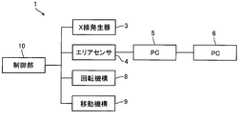
- FIG. 2is a block diagram for explaining a schematic configuration of the X-ray inspection apparatus 1 shown in FIG.
- the X-ray inspection apparatus 1 of this embodimentis an apparatus for inspecting the inside of an inspection object 2 such as an industrial product in a nondestructive manner.
- the X-ray inspection apparatus 1is an apparatus for inspecting a relatively large object 2 such as an engine block.
- This X-ray inspection apparatus 1includes an X-ray generator 3 that irradiates an object 2 to be inspected with X-rays, and a two-dimensional X-ray detection that is arranged so that the object 2 to be inspected is sandwiched between the X-ray generator 3. 4 (hereinafter referred to as “area sensor 4”).
- the X-ray inspection apparatus 1includes processing means 5 and 6 that capture and process an X-ray image acquired by the area sensor 4, a table 7 on which the inspection object 2 is mounted, and a rotation mechanism that rotates the table 7. 8 and a moving mechanism 9 for moving the area sensor 4 in parallel.
- the X-ray inspection apparatus 1includes two processing units 5 and 6 including a processing unit 5 as a first processing unit and a processing unit 6 as a second processing unit.
- the X-ray generator 3, the area sensor 4, the rotating mechanism 8, and the moving mechanism 9are connected to the control unit 10.
- the processing means 5 and 6include arithmetic means such as a CPU and storage means such as a semiconductor memory.
- the processing means 5 and 6 of this formare personal computers (PC). Therefore, hereinafter, the processing means 5 is referred to as “PC5” and the processing means 6 is referred to as “PC6”.
- the PC 5is connected to the area sensor 4 and takes in an X-ray image acquired by the area sensor 4.
- the PC 6is connected to the PC 5.
- the X-ray generator 3emits, for example, a conical X-ray (cone beam) toward the inspection object 2.
- the optical axis of the X-ray generator 3is parallel to the horizontal direction.
- the area sensor 4is a two-dimensional camera.
- the detection surface 4a of the area sensor 4is formed in a rectangular shape. Specifically, the detection surface 4a is formed in a square shape. The length of one side of the detection surface 4a is, for example, 200 (mm). Assuming that the direction parallel to the optical axis of the X-ray generator 3 is the front-rear direction, the detection surface 4a is arranged so as to be orthogonal to the front-rear direction.
- the area sensor 4has two of the four sides of the detection surface 4a formed in a square shape parallel to the up-down direction, and the remaining The two sides are arranged so as to be parallel to the left-right direction.
- the left-right direction of this embodimentis a first direction that is a predetermined direction parallel to the detection surface 4a
- the up-down directionis a direction parallel to the detection surface 4a and orthogonal to the first direction. It is the second direction.
- the table 7is disposed between the X-ray generator 3 and the area sensor 4 in the front-rear direction so that the inspection object 2 is disposed between the X-ray generator 3 and the area sensor 4.
- the rotation mechanism 8rotates the table 7 with the vertical direction as the axis direction of rotation. That is, the rotation mechanism 8 rotates the device under test 2 mounted on the table 7 so that the X-ray generator 3 and the area sensor 4 rotate relative to the device under test 2 on the outer peripheral side of the device under test 2.
- the moving mechanism 9translates the area sensor 4 in the horizontal direction and the vertical direction. That is, the moving mechanism 9 translates the area sensor 4 so that the area sensor 4 moves relative to the object to be inspected 2 in the horizontal direction and the vertical direction.
- the direction of relative rotation of the X-ray generator 3 and the area sensor 4 with respect to the inspection object 2may be referred to as “relative rotation direction”.
- a plane including the detection surface 4a of the area sensor 4is defined as a virtual projection plane VP, and an entire projection image of the inspection object 2 projected onto the virtual projection plane VP by X-rays emitted from the X-ray generator 3 is a virtual projection image VI. Then, the detection surface 4a is smaller than the virtual projection image VI in the vertical direction and the horizontal direction. In this embodiment, when the area sensor 4 is moved to nine locations, the area sensor 4 can acquire the entire X-ray image of the object 2 to be inspected.
- a first arrangement position 4Awhere the right end side of the lower end side portion of the inspection object 2 is projected
- a second arrangement position 4Bwhere the central portion in the left-right direction of the lower end side part of the inspection object 2 is projected.
- the third arrangement position 4Cwhere the left end side of the lower end side portion of the inspection object 2 is projected
- the fourth arrangement position 4Dwhere the right end side of the center part in the vertical direction of the inspection object 2 is projected
- the sixth arrangement position 4Fat which the left end side of the center portion in the vertical direction of the inspection object 2 is projected
- the right end side of the upper end side portion of the inspection object 2are projected.
- the area sensor 4 arranged at the first arrangement position 4Amoves to the left by the length of one side of the detection surface 4a
- the area sensor 4is arranged at the second arrangement position 4B and arranged at the second arrangement position 4B.
- the area sensor 4moves to the left by the length of one side of the detection surface 4a
- the area sensor 4is arranged at the third arrangement position 4C.
- the area sensor 4 arranged at the third arrangement position 4Cmoves to the right by twice the length of one side of the detection surface 4a and moves upward by the length of one side of the detection surface 4a
- the area sensor 4 arranged at the fourth arrangement position 4Dmoves to the left by the length of one side of the detection surface 4a
- the area sensor 4is arranged at the fifth arrangement position 4E, and at the fifth arrangement position 4E.
- the area sensor 4is arranged at the sixth arrangement position 4F.
- the area sensor 4 arranged at the sixth arrangement position 4Fmoves to the right by twice the length of one side of the detection surface 4a and moves to the upper side by the length of one side of the detection surface 4a
- the area The sensor 4is arranged at the seventh arrangement position 4G.
- the area sensor 4 arranged at the seventh arrangement position 4Gmoves to the left by the length of one side of the detection surface 4a
- the area sensor 4is moved to the eighth arrangement position 4H.
- the area sensor 4 arranged and moved to the eighth arrangement position 4Hmoves to the left by the length of one side of the detection surface 4a
- the area sensor 4is arranged at the ninth arrangement position 4I.
- a part of the inspection object 2is projected on the detection surface 4a of the area sensor 4 regardless of the position of the first arrangement position 4A to the ninth arrangement position 4I.
- the irradiation region of the X-ray generator 3is set so that the virtual projection image VI is projected onto the virtual projection plane VP without moving the X-ray generator 3.
- the X-ray inspection apparatus 1acquires an X-ray image of the inspection object 2 as follows. First, the control unit 10 adjusts the rotation mechanism 8 so that the rotation position of the rotation mechanism 8 becomes a predetermined origin position. Further, the control unit 10 causes the moving mechanism 9 to move the area sensor 4 to, for example, the first arrangement position 4A and stop it. In this state, the control unit 10 rotates the inspection object 2 mounted on the table 7 by 360 ° by the rotation mechanism 8 and acquires X-ray images A1 to A1000 (see FIG. 3) from the area sensor 4 at a certain angle. An image acquisition operation is performed. In the image acquisition operation of this embodiment, 1000 X-ray images A1 to A1000 are sequentially acquired every 0.36 °. Note that the number of X-ray images acquired by the image acquisition operation may be less than 1000 or more than 1000.
- the control unit 10performs a moving operation of moving the area sensor 4 in the left direction by the moving mechanism 9.
- the area sensor 4moves from the first arrangement position 4A to the second arrangement position 4B and stops.
- the control unit 10rotates the object 2 to be inspected 360 ° by the rotation mechanism 8 and sequentially sends 1000 X-ray images B1 to B1000 (see FIG. 3) to the area sensor 4 every 0.36 °.
- the image acquisition operation to be acquiredis performed.
- the control unit 10performs a moving operation of moving the area sensor 4 from the second arrangement position 4B to the third arrangement position 4C by the movement mechanism 9.
- the control unit 10rotates the object 2 to be rotated 360 ° by the rotation mechanism 8 and causes the area sensor 4 to sequentially acquire 1000 X-ray images C1 to C1000 (see FIG. 3) every 0.36 °. Perform image acquisition.
- the X-ray images A1, B1, and C1are X-ray images acquired at the same angle in the relative rotation direction of the area sensor 4 with respect to the inspection object 2, and the X-ray images A1, B1, and C1 are arranged in this order from the right side.
- an X-ray image of the origin position in the relative rotation direction of the lower end side portion of the inspection object 2is obtained. That is, each of the X-ray images A1, B1, and C1 is an X-ray image of the origin position in the relative rotation direction of the lower end side portion of the inspection object 2, and the left and right direction of the lower end side portion of the inspection object 2 It is the X-ray image divided
- the X-ray images A2, B2, and C2are X-ray images acquired at the same angle in the relative rotation direction, and the X-ray images A2, B2, and C2 are arranged in this order from the right side and connected and combined. Then, an X-ray image of a position shifted by 0.36 ° from the origin position in the relative rotation direction of the lower end side portion of the inspection object 2 is obtained. That is, each of the X-ray images A2, B2, and C2 is an X-ray image at a position shifted by 0.36 ° from the origin position in the relative rotation direction of the lower end side portion of the inspection object 2 and the inspection object 2 It is the X-ray image divided
- the X-ray images An, Bn, and Cnare X-ray images acquired at the same angle in the relative rotation direction, and the X-ray images An, Bn, and Cn are When arranged in this order from the right side and joined together to synthesize, the X-ray image of the lower end side portion of the inspection object 2 at a position shifted by (0.36 ⁇ (n ⁇ 1)) ° from the origin position in the relative rotation direction. It becomes.
- Each of the X-ray images An, Bn, and Cnis an X-ray image at a position shifted by (0.36 ⁇ (n ⁇ 1)) ° from the origin position in the relative rotation direction of the lower end side portion of the inspection object 2.
- the control unit 10causes the rotation mechanism 8 to rotate the X-ray generator 3 and the area sensor 4 relative to the object to be inspected 360 ° with the rotation mechanism 8 in a state in which the area sensor 4 is stopped.
- the image acquisition operation for acquiring the X-ray image and the movement operation for moving the area sensor 4 relative to the inspection object 2 in the left direction by the moving mechanism 9are alternately performed, and the lower end side portion of the inspection object 2
- the X-ray image divided in the left-right directionis acquired by the area sensor 4 over 360 ° at every fixed angle in the relative rotation direction.
- the control unit 10may acquire the X-ray images C1 to C1000 after acquiring the X-ray images A1 to A1000, and then acquire the X-ray images B1 to B1000. Further, the control unit 10 may acquire the X-ray images A1 to A1000 after acquiring the X-ray images B1 to B1000, and then acquire the X-ray images C1 to C1000, or may acquire the X-ray images B1 to B1000. X-ray images C1 to C1000 may be acquired after acquiring X-ray images, and then X-ray images A1 to A1000 may be acquired.
- control unit 10may acquire the X-ray images B1 to B1000 after acquiring the X-ray images C1 to C1000, and then acquire the X-ray images A1 to A1000, or may acquire the X-ray images C1 to C1000.
- X-ray images A1 to A1000may be acquired after acquiring X-ray images, and then X-ray images B1 to B1000 may be acquired.
- a control unit 10When a plurality of X-ray images divided in the left-right direction at the lower end side portion of the object 2 to be inspected, which are obtained over 360 ° at every fixed angle in the relative rotation direction, are set as a row of X-ray images P1, a control unit 10, when the X-ray image P ⁇ b> 1 for one row is acquired by the area sensor 4, the moving mechanism 9 moves the area sensor 4 from the third arrangement position 4 ⁇ / b> C to the fourth arrangement position 4 ⁇ / b> D. That is, the control unit 10 moves the area sensor 4 in the right direction and the upward direction by the moving mechanism 9.
- control unit 10performs an image acquisition operation similar to the above-described image acquisition operation, causes the area sensor 4 arranged at the fourth arrangement position 4D to acquire the X-ray images D1 to D1000, and then performs the above-described movement.
- the movement operation similar to the operationis performed to move the area sensor 4 from the fourth arrangement position 4D to the fifth arrangement position 4E.
- control unit 10performs the image acquisition operation to cause the area sensor 4 arranged at the fifth arrangement position 4E to acquire the X-ray images E1 to E1000, and then performs the movement operation to execute the fifth operation.
- the area sensor 4is moved from the arrangement position 4E to the sixth arrangement position 4F, and then an image acquisition operation is performed to cause the area sensor 4 arranged at the sixth arrangement position 4F to acquire the X-ray images F1 to F1000.
- the control unit 10moves the area sensor 4 in the right direction and the upward direction by the movement mechanism 9, and then performs the image acquisition operation and the movement operation.
- the area sensor 4moves the area sensor 4 in the right direction and the upward direction by the movement mechanism 9, and then performs the image acquisition operation and the movement operation.
- the area sensor 4moves the area sensor 4 in the right direction and the upward direction by the movement mechanism 9, and then performs the image acquisition operation and the movement operation.
- the control unit 10moves the area sensor 4 in the right direction and the upward direction by the moving mechanism 9, and then performs the image acquisition operation and the movement operation. Alternately, the area sensor 4 acquires the X-ray image P2 for the next line in the vertical direction.
- the control unit 10moves the area sensor 4 from the sixth arrangement position 4F to the seventh arrangement position 4G by the moving mechanism 9. Thereafter, the control unit 10 similarly performs an image acquisition operation to cause the area sensor 4 arranged at the seventh arrangement position 4G to acquire the X-ray images G1 to G1000, and then performs a movement operation to perform the seventh operation.
- the area sensor 4is moved from the arrangement position 4G to the eighth arrangement position 4H.
- the control unit 10performs the image acquisition operation to cause the area sensor 4 arranged at the eighth arrangement position 4H to acquire the X-ray images H1 to H1000, and then performs the movement operation to perform the eighth operation.
- an image acquisition operationis performed to cause the area sensor 4 arranged at the ninth arrangement position 4I to acquire X-ray images I1 to I1000.
- the acquisition of the X-ray image of the inspection object 2 by the area sensor 4is completed.
- the control unit 10moves the area sensor 4 in the right direction and the upward direction by the moving mechanism 9, and then performs the image acquisition operation and the movement operation.
- the area sensor 4moves the area sensor 4 in the right direction and the upward direction by the moving mechanism 9, and then performs the image acquisition operation and the movement operation.
- FIG. 3is a diagram for explaining the combining process and the dividing process in the PC 6 shown in FIG.
- the PC 5sequentially captures the X-ray images acquired by the area sensor 4. Further, when the PC 5 captures the X-ray image P1 for one row (ie, X-ray images A1 to A1000, B1 to B1000, C1 to C1000), the PC 5 transmits the captured X-ray image P1 for the single row to the PC 6 as it is. . First, the PC 6 combines and combines a plurality of X-ray images acquired at the same angle in the relative rotation direction of the area sensor 4 with respect to the inspection object 2 in the X-ray image P1 for one row in the left-right direction. Is executed at every fixed angle in the relative rotation direction.
- the PC 6arranges three X-ray images A1, B1, and C1 in this order from the right side and combines them to synthesize the combined X-ray image X1. Generate. Similarly, the PC 6 arranges and combines the three X-ray images A2, B2, and C2 in this order from the right side to generate a combined X-ray image X2. Further, the PC 6 performs the same combining process until a combined X-ray image X1000 in which three X-ray images A1000, B1000, and C1000 are connected is generated. That is, the PC 6 arranges and combines the three X-ray images An, Bn, and Cn in this order from the right side to generate 1000 synthesized X-ray images Xn.
- the PC 6divides the synthesized X-ray image X1 (the synthesized X-ray image) in the vertical direction into a plurality of strip X-ray images X1-1 to X1-4. Execute the process. In this embodiment, the PC 6 divides the combined X-ray image X1 into four strip X-ray images X1-1 to X1-4. Further, the PC 6 divides the composite X-ray image X1 into four strip X-ray images X1-1 to X1-4. Similarly, the PC 6 executes division processing to divide the composite X-ray image X2 in the vertical direction into four strip X-ray images X2-1 to X2-4 which are four strip X-ray images.
- the PC 6performs the same division process until the combined X-ray image X1000 is divided into the strip X-ray images X1000-1 to X1000-4. That is, the PC 6 executes a dividing process for dividing the combined X-ray image Xn into four vertical X-ray images Xn-1 to Xn-4.
- the belt-like X-ray images Xn-1 to Xn-4are arranged in this order from the lower side.
- the PC 6may sequentially divide the 1000 composite X-ray images Xn after generating 1000 composite X-ray images Xn, or every time one composite X-ray image Xn is generated, The generated composite X-ray image Xn may be divided.
- the PC 6executes a CT image generation process for generating a CT image by performing a predetermined calculation based on a 360 ° band X-ray image at the same position in the vertical direction. That is, the PC 6 performs a calculation based on the 1000 strip X-ray images X1-1, the strip X-ray images X2-1,..., The strip X-ray image X1000-1, and performs CT on the lowermost layer of the object 2 to be inspected. Generate an image. Further, the PC 6 performs a calculation based on the 1000 strip X-ray images X1-2, the strip X-ray images X2-2,...
- a CT image of the third layer from the bottomis generated, and an operation is performed based on 1000 strip X-ray images X1-4, strip X-ray images X2-4,..., Strip X-ray images X1000-4.
- a CT image of the fourth layer from the bottom of the inspection object 2is generated.
- the number of divisions of the composite X-ray image Xn in the division processmay be two or three, or may be five or more.
- the number of divisions of the composite X-ray image Xn in the division processis set according to the processing capability of the PC 6. Specifically, as the processing capability of the PC 6 increases, the number of divisions of the composite X-ray image Xn in the division processing decreases. On the other hand, when the processing capability of the PC 6 decreases, the number of divisions of the composite X-ray image Xn in the division processing increases.
- the PC 5sequentially captures each X-ray image constituting the X-ray image P2 for one column in parallel with the synthesis process, the division process, and the CT image generation process for the X-ray image P1 for the single column in the PC 6.
- the PC 5transmits the captured X-ray image P2 for the single line to the PC 6 as it is.
- the PC 6performs the synthesis process, the division process, and the CT image generation process for the X-ray image P2 for the single column in the same procedure as described above.
- the PC 5may transmit the captured X-ray image P2 for one column to the PC 6 before the composition processing, the division processing, and the CT image generation processing for the X-ray image P1 for the column in the PC 6 are completed. After completing the synthesizing process, the dividing process, and the CT image generating process for the one-line X-ray image P1, the captured one-line X-ray image P2 may be transmitted to the PC 6.
- the PC 5sequentially captures each X-ray image constituting the X-ray image P3 for one column in parallel with the composition processing, the division processing, and the CT image generation processing for the X-ray image P2 for the single column in the PC 6.
- the PC 5transmits the captured X-ray image P3 for the single line to the PC 6 as it is.
- the PC 6performs a synthesis process, a segmentation process, and a CT image generation process on the X-ray image P3 for one column in the same procedure as described above, and the CT image of the ninth layer from the bottom of the object to be inspected 2, A CT image of the tenth layer from the bottom of the inspection object 2, a CT image of the eleventh layer from the bottom of the inspection object 2, and a CT image of the uppermost layer of the inspection object 2 are generated.
- the rotation mechanism 8rotates the object 2 to be rotated 360 ° and the area sensor 4 acquires an X-ray image at a certain angle.
- the left and right directions of the X-ray image divided in the left-right direction of the lower end side portion of the object to be inspected and the center portion of the inspected object 2 in the up-down directionare alternately performed.
- the X-ray image divided in the left-right direction of the upper end side portion of the object to be inspected 2are acquired over 360 ° for each constant angle in the rotation direction of the object 2 to be inspected. ing.
- the PC 6performs a relative process of combining three X-ray images acquired at the same angle in the rotation direction of the inspected object 2 in the horizontal direction in the X-ray image P1 for one row. It is executed for each fixed angle in the rotation direction, and after the synthesis process, a synthesis X-ray image is divided into four in the vertical direction to form a belt-like X-ray image that is a belt-like X-ray image.
- a CT image generation processis performed in which a predetermined calculation based on a 360 ° band X-ray image at the same position in the vertical direction is performed to generate a CT image.
- the PC 6performs the synthesis process, the division process, and the CT image generation process for the X-ray images P2 and P3 for one column.
- the number of divisions of the composite X-ray image in the division processis set according to the processing capability of the PC 6. Therefore, in this embodiment, it is possible to reduce the time of CT image generation processing for generating a CT image by performing a predetermined calculation based on a strip X-ray image.
- the acquisition time of the X-ray image necessary for generating the entire CT image of the inspection object 2can be shortened, and the acquired X It is possible to reduce the CT image generation processing time for generating a CT image based on a line image. Therefore, in this embodiment, even when the inspection object 2 is large, the inspection time of the inspection object 2 can be shortened.
- the X-ray image capturing process with a high processing loadis executed on the PC 5 while the CT image generating process with a high processing load is executed in parallel with the PC 6.
- the CT image generation processing timecan be further shortened. As a result, it is possible to further reduce the inspection time of the object 2 to be inspected.
- FIG. 4is a diagram for explaining the dividing process and the synthesizing process according to another embodiment of the present invention.
- the PC 6divides a row of X-ray images captured from the area sensor 4 into a plurality of parts in the vertical direction (specifically, divides each of the plurality of X-ray images in the vertical direction).
- a plurality of X-ray images obtained at the same position in the vertical direction and obtained at the same angle in the rotation direction of the inspected object 2are joined in the left-right direction.
- a synthesis process for synthesizing the belt-shaped X-ray imageis performed at every fixed angle in the rotation direction of the inspection object 2, and after the synthesis process, a predetermined X-ray image based on a 360-degree belt X-ray image located at the same position in the vertical direction.
- the PC 6first performs division processing, for example, as shown in FIG. 4A, the X-ray image A1 is divided into four in the vertical direction to form divided X-ray images A11 to A14, or the X-ray image An area arranged at the first arrangement position 4A until the X-ray image A1000 is divided into four divided X-ray images A10001 to A10004, for example, by dividing A2 into four in the vertical direction into divided X-ray images A21 to A24.
- the X-ray image acquired by the sensor 4is divided.
- the PC 6divides the X-ray image B1 into four in the vertical direction into divided X-ray images B11 to B14, or divides the X-ray image B2 into four in the vertical direction into divided X-ray images B21 to B24.
- the X-ray image acquired by the area sensor 4 arranged at the second arrangement position 4Bis divided until the X-ray image B1000 is divided into four divided X-ray images B10001 to B10004.
- the PC 6divides the X-ray image C1 into four pieces in the vertical direction into divided X-ray images C11 to C14, or divides the X-ray image C2 into four pieces in the vertical direction into divided X-ray images C21 to C24.
- the X-ray image acquired by the area sensor 4 arranged at the third arrangement position 4Cis divided until the X-ray image C1000 is divided into four divided X-ray images C10001 to C10004.
- the PC 6performs a synthesis process after the division process, and obtains the divided X-ray images at the same position in the vertical direction and at the same angle in the rotation direction of the device under test 2 as shown in FIG.
- the three divided X-ray images A11 to C11are sequentially arranged from the right side and joined in the left-right direction to be combined into a belt-like X-ray image X1-1.
- the PC 6performs a composition process to connect the divided X-ray images A12 to C12 to combine them into a belt-like X-ray image X1-2, and to connect the divided X-ray images A13 to C13 to connect the belt-like X-ray image X1.
- the PC 6performs the same combining process until the strip X-ray images X1000-1 to X1000-4 are combined.
- the PC 6performs a CT image generation process for generating a CT image by performing a predetermined calculation based on a 360 ° band X-ray image at the same position in the vertical direction after the synthesis process. To do. Even in this case, by setting the number of X-ray image divisions in the division process according to the processing capability of the PC 6, it is possible to reduce the time of the CT image generation process as in the above-described form. Become.
- the PC 6when the processing capability of the PC 6 is relatively high, the PC 6 does not execute the division processing for the X-ray images P1 to P3 for one column, but executes the synthesis processing and the CT image generation processing. good. That is, for example, the PC 6 performs a predetermined calculation based on the 360 ° synthesized X-ray images X1 to X1000 (synthesized X-ray images) after the synthesizing process for generating the synthesized X-ray images X1 to X1000, and obtains the CT image CT image generation processing for generating the image may be executed.
- a CT imageis generated on the basis of a row of X-ray images P1 that are X-ray images of the lower end portion of the inspection object 2, and is an X-ray image of the central portion of the inspection object 2 in the vertical direction.
- a CT imageis generated based on the X-ray image P2 for one column, and a CT image is generated based on the X-ray image P3 for one column that is an X-ray image of the upper end side portion of the inspection object 2. That is, a CT image is generated for each row of X-ray images P1 to P3.
- the entire X-ray image of the object 2 to be inspected by the large area sensorover 360 ° at a certain angle in the relative rotation direction. It is possible to reduce the time of the CT image generation process as compared with the case where the entire CT image of the object to be inspected 2 is generated at once by performing a predetermined calculation based on the acquired X-ray image. become.
- the PC 6executes the combining process and the dividing process, but the PC 5 may execute the combining process and the dividing process.
- the X-ray inspection apparatus 1includes the two PCs 5 and 6. However, when the processing capability of the PC 5 is high, the X-ray inspection apparatus 1 does not include the PC 6. good. In this case, the PC 5 executes X-ray image capture processing, composition processing, division processing, and CT image generation processing. In the second modification of the X-ray image processing method described above, the PC 5 may execute the synthesis process, and the PC 6 may execute the CT image process. In the second modification of the X-ray image processing method described above, if the X-ray inspection apparatus 1 does not include the PC 6, the PC 5 performs X-ray image capture processing, composition processing, and CT image generation processing. Execute.
- the rotation mechanism 8rotates the object 2 to be inspected 2 mounted on the table 7, but the rotation mechanism 8 may rotate the X-ray generator 3 and the area sensor 4.
- the moving mechanism 9translates the area sensor 4 in the vertical direction and the horizontal direction.
- the moving mechanism 9may translate the subject 2 in the vertical direction and the horizontal direction. good.
- the moving mechanism 9may move the area sensor 4 in parallel in the left-right direction.
- the X-ray inspection apparatus 1includes a moving mechanism that translates the inspection object 2 in the vertical direction.
- the moving mechanism 9may move the device under test 2 in the left-right direction.
- the X-ray inspection apparatus 1includes a moving mechanism that translates the area sensor 4 in the vertical direction.
- the moving mechanism 9may move the X-ray generator 3 in parallel with the area sensor 4 in the vertical direction and the horizontal direction. In this case, for example, if the X-ray generator 3 is not moved, the virtual projection image VI (the entire projection image of the inspection object 2) cannot be projected onto the virtual projection plane VP. Three irradiation areas are set.
- the area sensor 4when the area sensor 4 is moved to nine positions from the first arrangement position 4A to the ninth arrangement position 4I, the area sensor 4 can acquire an entire X-ray image of the object 2 to be inspected.
- the area sensor 4when the area sensor 4 is moved to six locations from the first arrangement position 4A to the sixth arrangement position 4F, the area sensor 4 can acquire the entire X-ray image of the object 2 to be inspected.
- the area sensor 4may be moved to four locations of the first arrangement position 4A, the second arrangement position 4B, the fourth arrangement position 4D, and the fifth arrangement position 4E. It may be possible to acquire an X-ray image of the entire image.
- the entire X-ray image of the inspection object 2can be acquired by the area sensor 4. good.
- the detection surface 4ais larger than the virtual projection image VI in the vertical direction.
- the number of movements of the area sensor 4 from the first arrangement position 4A to the third arrangement position 4C at the same heightis the same height.
- the number of movements of the area sensor 4 from a certain fourth arrangement position 4D to the sixth arrangement position 4F(hereinafter referred to as “second stage movement number”) and the seventh arrangement position 4G to the ninth arrangement position at the same height.
- the number of movements of the area sensor 4 up to the position 4I(hereinafter referred to as “the number of movements of the third stage”) is equal, but the number of movements of the first stage and the second stage are determined according to the shape of the inspection object 2.
- the number of eye movements and the number of third movementsmay be different.
- the area sensor 4may move from the third arrangement position 4C to the sixth arrangement position 4F when acquiring an X-ray image of the object 2 to be inspected. In this case, the area sensor 4 subsequently moves sequentially to, for example, the fifth arrangement position 4E, the fourth arrangement position 4D, the seventh arrangement position 4G, the eighth arrangement position 4H, and the ninth arrangement position 4I.
- the optical axis of the X-ray generator 3is parallel to the horizontal direction, but the optical axis of the X-ray generator 3 may be tilted with respect to the horizontal direction.
Landscapes
- Health & Medical Sciences (AREA)
- General Physics & Mathematics (AREA)
- Chemical & Material Sciences (AREA)
- Pathology (AREA)
- Immunology (AREA)
- Analytical Chemistry (AREA)
- Physics & Mathematics (AREA)
- Biochemistry (AREA)
- General Health & Medical Sciences (AREA)
- Life Sciences & Earth Sciences (AREA)
- Nuclear Medicine, Radiotherapy & Molecular Imaging (AREA)
- Theoretical Computer Science (AREA)
- Engineering & Computer Science (AREA)
- Radiology & Medical Imaging (AREA)
- Pulmonology (AREA)
- Analysing Materials By The Use Of Radiation (AREA)
Abstract
Description
Translated fromJapanese本発明は、工業製品等の内部を非破壊で検査するためのX線検査装置およびかかるX線検査装置の制御方法に関する。The present invention relates to an X-ray inspection apparatus for non-destructively inspecting the inside of industrial products and the like and a control method for such an X-ray inspection apparatus.
従来、工業製品等の被検査体の内部を非破壊で検査するためのX線検査装置が知られている(たとえば、特許文献1、2参照)。特許文献1に記載のX線検査装置は、被検査体にX線を照射するX線源と、X線ラインセンサ(ラインセンサ)と、被検査体が搭載されるとともに上下方向を回転の軸方向として回転する回転テーブルと、ラインセンサを上下動させる移動機構とを備えている。このX線検査装置では、X線源とラインセンサとの間に配置される被検査体を定速回転させるとともに、被検査体の回転に同期させてラインセンサを被検査体の上端から下端まで移動させながら、ラインセンサによって複数枚のX線画像を取得し、取得した複数枚のX線画像から被検査体のCT画像を生成している。Conventionally, an X-ray inspection apparatus for inspecting the inside of an inspection object such as an industrial product in a nondestructive manner is known (for example, see
また、特許文献2には、被検査体が大きく、かつ、要求される空間分解能が高い場合に、被検査体の回転機構をたとえば4周以上(360°×4回転以上)させるとともに360°ごとに二次元検出器を移動させて、被検査体の4分割(あるいは8分割)された部分投影を取得し、所得した部分投影を合成して全投影を得るX線検査装置が従来技術として開示されている。Further, in
特許文献1に記載のX線検査装置では、被検査体の1回転に対するラインセンサの下降量を小さくしないと、適切なCT画像を取得することは困難である。たとえば、被検査体が1回転する間のラインセンサの下降量を0.1mm~0.2mm程度にしないと、このX線検査装置で適切なCT画像を生成することは困難である。したがって、このX線検査装置では、被検査体が大きいと、被検査体の検査を行う際に被検査体を回転させる回数が多くなり、その結果、被検査体の全体のCT画像を生成するために必要なX線画像の取得に時間がかかって、被検査体の検査時間が長くなる。In the X-ray inspection apparatus described in
これに対して、特許文献2に開示されたX線検査装置では、被検査体が大きくても、被検査体を4回転あるいは8回転させれば、被検査体の全体のCT画像の生成に必要なX線画像を取得できるため、被検査体の全体のCT画像の生成に必要なX線画像の取得時間を短縮することは可能である。しかしながら、被検査体の全体のCT画像の生成に必要なX線画像を短時間で取得できても、取得したX線画像に基づく所定の演算を行ってCT画像を生成するCT画像の生成処理に時間がかかると、被検査体の検査時間を短縮することはできない。On the other hand, in the X-ray inspection apparatus disclosed in
そこで、本発明の課題は、被検査体が大きくても、被検査体の全体のCT画像の生成に必要なX線画像の取得時間を短縮するとともに、取得したX線画像に基づいてCT画像を生成するCT画像の生成処理時間を短縮して、被検査体の検査時間を短縮することが可能なX線検査装置およびX線検査装置の制御方法を提供することにある。Accordingly, an object of the present invention is to reduce the acquisition time of an X-ray image necessary for generating an entire CT image of an object to be inspected even if the object to be inspected is large, and to obtain a CT image based on the acquired X-ray image. An X-ray inspection apparatus and an X-ray inspection apparatus control method capable of shortening a CT image generation processing time for generating an object and reducing an inspection time of an object to be inspected.
上記の課題を解決するため、本発明のX線検査装置は、X線発生器と、X線発生器との間で被検査体を挟むように配置される二次元X線検出器と、被検査体の外周側で被検査体に対してX線発生器および二次元X線検出器が相対回転するようにX線発生器と二次元X線検出器とを回転させるかまたは被検査体を回転させる回転機構と、二次元X線検出器で取得されたX線画像を取り込んで処理する処理手段とを備えるとともに、二次元X線検出器の検出面に平行な所定の方向を第1方向とし、検出面に平行な方向であってかつ第1方向に直交する方向を第2方向とし、被検査体に対するX線発生器および二次元X線検出器の相対回転の方向を相対回転方向とすると、被検査体に対して二次元X線検出器が少なくとも第1方向へ相対移動するように二次元X線検出器を平行移動させるかまたは被検査体を平行移動させる移動機構と、X線発生器と二次元X線検出器と回転機構と移動機構とが接続される制御部とを備え、検出面を含む平面を仮想投影面とし、X線発生器が射出するX線によって仮想投影面に投影される被検査体の全体の投影像を仮想投影像とすると、検出面は、少なくとも第1方向において仮想投影像よりも小さくなっており、制御部は、回転機構によってX線発生器および二次元X線検出器を被検査体に対して360°相対回転させるとともに二次元X線検出器に一定角度ごとにX線画像を取得させる画像取得動作と、移動機構によって二次元X線検出器を被検査体に対して第1方向の一方へ相対移動させる移動動作とを交互に行って、第2方向の所定位置の、第1方向で分割された被検査体のX線画像を相対回転方向の一定角度ごとに360°に亘って二次元X線検出器に取得させ、相対回転方向の一定角度ごとに360°に亘って取得された、第2方向の所定位置の第1方向で分割された被検査体のX線画像を一列分X線画像とすると、処理手段は、二次元X線検出器から取り込んだ一列分X線画像の中の相対回転方向の同じ角度で取得された複数のX線画像を第1方向で繋ぎ合わせて合成する合成処理を相対回転方向の一定角度ごとに実行するとともに、合成処理後に、合成されたX線画像を第2方向で分割して複数の帯状のX線画像にする分割処理を実行し、分割処理後に、第2方向において同じ位置にある360°分の帯状のX線画像に基づく所定の演算を行ってCT画像を生成するCT画像生成処理を実行するか、あるいは、二次元X線検出器から取り込んだ一列分X線画像を第2方向で複数に分割する分割処理を実行するとともに、分割処理後に、第2方向において同じ位置にある分割後のX線画像であって相対回転方向の同じ角度で取得された複数のX線画像を第1方向で繋ぎ合わせて帯状のX線画像に合成する合成処理を相対回転方向の一定角度ごとに実行し、合成処理後に、第2方向において同じ位置にある360°分の帯状のX線画像に基づく所定の演算を行ってCT画像を生成するCT画像生成処理を実行することを特徴とする。In order to solve the above problems, an X-ray inspection apparatus according to the present invention includes an X-ray generator, a two-dimensional X-ray detector disposed so as to sandwich an object to be inspected between the X-ray generator, The X-ray generator and the two-dimensional X-ray detector are rotated so that the X-ray generator and the two-dimensional X-ray detector rotate relative to the inspection object on the outer periphery side of the inspection object, or the inspection object is A rotation mechanism that rotates, and a processing unit that captures and processes an X-ray image acquired by the two-dimensional X-ray detector, and a predetermined direction parallel to the detection surface of the two-dimensional X-ray detector is a first direction. The direction parallel to the detection surface and perpendicular to the first direction is the second direction, and the relative rotation direction of the X-ray generator and the two-dimensional X-ray detector with respect to the object to be inspected is the relative rotation direction. Then, the two-dimensional X-ray detector moves relative to the object to be examined at least in the first direction. A moving mechanism that translates the two-dimensional X-ray detector or translates the object to be inspected, and a control unit to which the X-ray generator, the two-dimensional X-ray detector, the rotating mechanism, and the moving mechanism are connected. Provided that the plane including the detection surface is a virtual projection plane, and the entire projection image of the inspected object projected onto the virtual projection plane by the X-rays emitted by the X-ray generator is a virtual projection image, the detection plane is at least It is smaller than the virtual projection image in the first direction, and the control unit rotates the X-ray generator and the two-dimensional X-ray detector relative to the object to be inspected with respect to the inspected object by the rotation mechanism and detects the two-dimensional X-ray. An image acquisition operation for causing the device to acquire an X-ray image at a predetermined angle and a movement operation for moving the two-dimensional X-ray detector relative to the object to be inspected in one direction in the first direction by a moving mechanism are alternately performed. , A first position at a predetermined position in the second direction X-ray images of the object to be inspected divided in directions are acquired by a two-dimensional X-ray detector over 360 ° for every fixed angle in the relative rotation direction, and acquired over 360 ° for every fixed angle in the relative rotation direction. If the X-ray image of the object to be inspected divided in the first direction at the predetermined position in the second direction is an X-ray image for one row, the processing means can obtain the X-ray for one row taken from the two-dimensional X-ray detector. A composition process for combining a plurality of X-ray images acquired at the same angle in the relative rotation direction in the image in the first direction is performed for each fixed angle in the relative rotation direction, and is synthesized after the composition process. The X-ray image is divided in the second direction to form a plurality of band-shaped X-ray images, and after the division process, based on the band-shaped X-ray image for 360 ° located at the same position in the second direction. CT image generation for generating a CT image by performing a predetermined calculation Or a division process that divides a row of X-ray images captured from the two-dimensional X-ray detector into a plurality of parts in the second direction, and is in the same position in the second direction after the division process. A combined X-ray image obtained by dividing the X-ray images obtained at the same angle in the relative rotation direction in the first direction and combining them into a belt-like X-ray image is set for each fixed angle in the relative rotation direction. And a CT image generation process for generating a CT image by performing a predetermined calculation based on a 360 ° band X-ray image at the same position in the second direction after the synthesis process. .
また、上記の課題を解決するため、本発明のX線検査装置の制御方法は、X線発生器と、X線発生器との間で被検査体を挟むように配置される二次元X線検出器と、被検査体の外周側で被検査体に対してX線発生器および二次元X線検出器が相対回転するようにX線発生器と二次元X線検出器とを回転させるかまたは被検査体を回転させる回転機構とを備えるとともに、二次元X線検出器の検出面に平行な所定の方向を第1方向とし、検出面に平行な方向であってかつ第1方向に直交する方向を第2方向とし、被検査体に対するX線発生器および二次元X線検出器の相対回転の方向を相対回転方向とすると、被検査体に対して二次元X線検出器が少なくとも第1方向へ相対移動するように二次元X線検出器を平行移動させるかまたは被検査体を平行移動させる移動機構を備え、検出面を含む平面を仮想投影面とし、X線発生器が射出するX線によって仮想投影面に投影される被検査体の全体の投影像を仮想投影像とすると、検出面が少なくとも第1方向において仮想投影像よりも小さくなっているX線検査装置の制御方法であって、回転機構によってX線発生器および二次元X線検出器を被検査体に対して360°相対回転させるとともに二次元X線検出器に一定角度ごとにX線画像を取得させる画像取得動作と、移動機構によって二次元X線検出器を被検査体に対して第1方向の一方へ相対移動させる移動動作とを交互に行って、第2方向の所定位置の、第1方向で分割された被検査体のX線画像を相対回転方向の一定角度ごとに360°に亘って二次元X線検出器に取得させ、相対回転方向の一定角度ごとに360°に亘って取得された、第2方向の所定位置の第1方向で分割された被検査体のX線画像を一列分X線画像とすると、二次元X線検出器から取り込んだ一列分X線画像の中の相対回転方向の同じ角度で取得された複数のX線画像を第1方向で繋ぎ合わせて合成する合成処理を相対回転方向の一定角度ごとに実行するとともに、合成処理後に、合成されたX線画像を第2方向で分割して複数の帯状のX線画像にする分割処理を実行し、分割処理後に、第2方向において同じ位置にある360°分の帯状のX線画像に基づく所定の演算を行ってCT画像を生成するCT画像生成処理を実行するか、あるいは、二次元X線検出器から取り込んだ一列分X線画像を第2方向で複数に分割する分割処理を実行するとともに、分割処理後に、第2方向において同じ位置にある分割後のX線画像であって相対回転方向の同じ角度で取得された複数のX線画像を第1方向で繋ぎ合わせて帯状のX線画像に合成する合成処理を相対回転方向の一定角度ごとに実行し、合成処理後に、第2方向において同じ位置にある360°分の帯状のX線画像に基づく所定の演算を行ってCT画像を生成するCT画像生成処理を実行することを特徴とする。In order to solve the above-mentioned problem, the X-ray inspection apparatus control method of the present invention is a two-dimensional X-ray that is arranged so as to sandwich an object to be inspected between the X-ray generator and the X-ray generator. Whether the X-ray generator and the two-dimensional X-ray detector are rotated so that the X-ray generator and the two-dimensional X-ray detector rotate relative to the test object on the outer peripheral side of the test object Or a rotation mechanism that rotates the object to be inspected, and a predetermined direction parallel to the detection surface of the two-dimensional X-ray detector is defined as the first direction, the direction parallel to the detection surface and orthogonal to the first direction The second direction is the second direction, and the relative rotation direction of the X-ray generator and the two-dimensional X-ray detector with respect to the object to be inspected is the relative rotation direction. Translate the two-dimensional X-ray detector so that it moves relative to one direction, or inspect A plane including the detection surface as a virtual projection plane, and a projection image of the entire object to be inspected projected on the virtual projection plane by X-rays emitted from the X-ray generator is referred to as a virtual projection image. Then, there is provided a control method for an X-ray inspection apparatus in which the detection surface is smaller than the virtual projection image at least in the first direction, and the X-ray generator and the two-dimensional X-ray detector are moved relative to the object to be inspected by the rotation mechanism. An image acquisition operation for causing the two-dimensional X-ray detector to acquire an X-ray image at a predetermined angle and a moving mechanism to move the two-dimensional X-ray detector in the first direction with respect to the object to be inspected. The X-ray image of the object to be inspected divided in the first direction at a predetermined position in the second direction is rotated over 360 ° for each constant angle in the relative rotation direction. Let the dimensional X-ray detector acquire, When the X-ray image of the object to be inspected divided in the first direction at a predetermined position in the second direction, which is acquired over 360 ° for every fixed angle in the rotation direction, is a two-dimensional X-ray image. Combining a plurality of X-ray images acquired at the same angle in the relative rotation direction in a row of X-ray images captured from the line detector by combining them in the first direction for each fixed angle in the relative rotation direction In addition, after the combining process, a combining process is performed to divide the combined X-ray image in the second direction to form a plurality of strip-shaped X-ray images. After the dividing process, 360 at the same position in the second direction Perform CT image generation processing for generating a CT image by performing a predetermined calculation based on a belt-shaped X-ray image for °°, or a row of X-ray images taken from a two-dimensional X-ray detector in the second direction If you execute a split process that splits into multiple In addition, after the division process, a plurality of X-ray images that are divided at the same position in the second direction and acquired at the same angle in the relative rotation direction are joined in the first direction to form a belt-like X-ray. A synthesis process for synthesizing the image is executed at every fixed angle in the relative rotation direction. After the synthesis process, a CT image is obtained by performing a predetermined calculation based on a 360 ° band X-ray image at the same position in the second direction. A CT image generation process to be generated is executed.
本発明では、回転機構によってX線発生器および二次元X線検出器を被検査体に対して360°相対回転させるとともに二次元X線検出器に一定角度ごとにX線画像を取得させる画像取得動作と、移動機構によって二次元X線検出器を被検査体に対して第1方向の一方へ相対移動させる移動動作とを交互に行って、第2方向の所定位置の、第1方向で分割された被検査体のX線画像を相対回転方向の一定角度ごとに360°に亘って二次元X線検出器に取得させている。そのため、本発明では、被検査体が大きくても、被検査体の全体のCT画像の生成に必要なX線画像を取得する際に、被検査体に対してX線発生器および二次元X線検出器を相対回転させる回数を低減することが可能になる。したがって、本発明では、被検査体が大きくても、被検査体の全体のCT画像の生成に必要なX線画像の取得時間を短縮することが可能になる。In the present invention, the X-ray generator and the two-dimensional X-ray detector are rotated 360 ° relative to the object to be inspected by the rotation mechanism, and the two-dimensional X-ray detector acquires an X-ray image at a certain angle. The operation and the moving operation of moving the two-dimensional X-ray detector relative to the object to be inspected in one direction in the first direction are alternately performed by the moving mechanism, and the predetermined position in the second direction is divided in the first direction. The X-ray image of the object to be inspected is acquired by the two-dimensional X-ray detector over 360 ° for every fixed angle in the relative rotation direction. Therefore, in the present invention, even when the object to be inspected is large, when an X-ray image necessary for generating the entire CT image of the object to be inspected is acquired, the X-ray generator and the two-dimensional X The number of relative rotations of the line detector can be reduced. Therefore, in the present invention, even when the object to be inspected is large, it is possible to shorten the acquisition time of the X-ray image necessary for generating the entire CT image of the object to be inspected.
また、本発明では、二次元X線検出器から取り込んだ一列分X線画像の中の相対回転方向の同じ角度で取得された複数のX線画像を第1方向で繋ぎ合わせて合成する合成処理を相対回転方向の一定角度ごとに実行するとともに、合成処理後に、合成されたX線画像を第2方向で分割して複数の帯状のX線画像にする分割処理を実行し、分割処理後に、第2方向において同じ位置にある360°分の帯状のX線画像に基づく所定の演算を行ってCT画像を生成するCT画像生成処理を実行しているか、あるいは、二次元X線検出器から取り込んだ一列分X線画像を第2方向で複数に分割する分割処理を実行するとともに、分割処理後に、第2方向において同じ位置にある分割後のX線画像であって相対回転方向の同じ角度で取得された複数のX線画像を第1方向で繋ぎ合わせて帯状のX線画像に合成する合成処理を相対回転方向の一定角度ごとに実行し、合成処理後に、第2方向において同じ位置にある360°分の帯状のX線画像に基づく所定の演算を行ってCT画像を生成するCT画像生成処理を実行している。そのため、本発明では、処理手段の処理能力に応じて帯状のX線画像の幅(第2方向の幅)を設定することで、第2方向において同じ位置にある360°分の帯状のX線画像に基づく所定の演算を行ってCT画像を生成するCT画像生成処理の時間を短縮することが可能になる。Further, in the present invention, a combining process for combining a plurality of X-ray images acquired at the same angle in the relative rotation direction in a row of X-ray images captured from a two-dimensional X-ray detector in the first direction. Is executed for each fixed angle in the relative rotation direction, and after the combining process, the combined X-ray image is divided in the second direction to form a plurality of belt-shaped X-ray images, and after the dividing process, A CT image generation process for generating a CT image by performing a predetermined calculation based on a 360 ° belt-shaped X-ray image at the same position in the second direction is performed, or captured from a two-dimensional X-ray detector In addition, a division process is performed to divide the X-ray image for one row into a plurality of parts in the second direction, and after the division process, the X-ray images are divided at the same position in the second direction and at the same angle in the relative rotation direction. Acquired multiple A composition process for joining line images in the first direction to compose a belt-like X-ray image is executed at fixed angles in the relative rotational direction, and after the composition process, a 360-degree belt-shaped belt at the same position in the second direction is executed. CT image generation processing for generating a CT image by performing a predetermined calculation based on an X-ray image is executed. Therefore, in the present invention, by setting the width of the belt-shaped X-ray image (the width in the second direction) according to the processing capability of the processing means, the belt-shaped X-ray for 360 ° at the same position in the second direction. It is possible to reduce the time for CT image generation processing for generating a CT image by performing a predetermined calculation based on the image.
このように本発明では、被検査体が大きくても、被検査体の全体のCT画像の生成に必要なX線画像の取得時間を短縮することが可能になり、また、取得したX線画像に基づいてCT画像を生成するCT画像の生成処理時間を短縮することが可能になる。したがって、本発明では、被検査体が大きくても、被検査体の検査時間を短縮することが可能になる。As described above, according to the present invention, even when the object to be inspected is large, the acquisition time of the X-ray image necessary for generating the entire CT image of the object to be inspected can be shortened, and the acquired X-ray image can be reduced. It is possible to reduce the CT image generation processing time for generating a CT image based on the above. Therefore, in the present invention, even when the object to be inspected is large, the inspection time of the object to be inspected can be shortened.
本発明において、たとえば、二次元X線検出器の検出面は、第1方向および第2方向において仮想投影像よりも小さくなっており、制御部は、一列分X線画像が二次元X線検出器によって取得されると、移動機構によって二次元X線検出器を被検査体に対して少なくとも第2方向へ相対移動させ、その後、画像取得動作と移動動作とを交互に行って、被検査体の、第2方向における次の一列分X線画像を二次元X線検出器に取得させる。In the present invention, for example, the detection surface of the two-dimensional X-ray detector is smaller than the virtual projection image in the first direction and the second direction, and the control unit detects two-dimensional X-ray images for one row of X-ray images. When the object is acquired, the two-dimensional X-ray detector is moved relative to the object to be inspected at least in the second direction by the moving mechanism, and then the image acquisition operation and the moving operation are alternately performed to inspect the object to be inspected. The X-ray image for the next line in the second direction is acquired by the two-dimensional X-ray detector.
この場合には、X線検査装置は、処理手段として、二次元X線検出器に接続され二次元X線検出器で取得されたX線画像を取り込む第1処理手段と、第1処理手段に接続される第2処理手段とを備え、第1処理手段は、一列分X線画像を取り込むと、取り込んだ一列分X線画像をそのまま第2処理手段に送信し、第2処理手段は、合成処理と分割処理とCT画像生成処理とを実行することが好ましい。また、この場合には、X線検査装置は、処理手段として、二次元X線検出器に接続され二次元X線検出器で取得されたX線画像を取り込む第1処理手段と、第1処理手段に接続される第2処理手段とを備え、第1処理手段は、一列分X線画像を取り込むと、合成処理と分割処理とを実行し、第2処理手段は、第1処理手段から取り込んだ、第2方向において同じ位置にある360°分の帯状のX線画像に基づいてCT画像生成処理を実行しても良い。In this case, the X-ray inspection apparatus includes, as processing means, a first processing means connected to the two-dimensional X-ray detector and capturing an X-ray image acquired by the two-dimensional X-ray detector, and a first processing means. And a second processing unit connected to the first processing unit. When the first processing unit captures the X-ray image for one column, the first processing unit transmits the acquired X-ray image for one column to the second processing unit as it is. It is preferable to execute processing, division processing, and CT image generation processing. In this case, the X-ray inspection apparatus includes, as processing means, first processing means connected to the two-dimensional X-ray detector and capturing an X-ray image acquired by the two-dimensional X-ray detector; A second processing unit connected to the first processing unit. The first processing unit executes a synthesis process and a division process when the X-ray image for one column is captured, and the second processing unit captures from the first processing unit. However, the CT image generation processing may be executed based on a 360 ° band X-ray image at the same position in the second direction.
このように構成すると、処理の負荷が高いX線画像の取込処理を第1処理手段で実行しながら、処理の負荷が高いCT画像生成処理を第2処理手段で並行して実行することが可能になる。したがって、第1処理手段や第2処理手段の処理能力と同程度の処理能力を有する1個の処理手段によって、X線画像の取込処理とCT画像生成処理とを実行する場合と比較して、CT画像の生成処理時間を短縮することが可能になり、その結果、被検査体の検査時間をより短縮することが可能になる。With this configuration, the X-ray image capturing process with a high processing load is executed by the first processing means, and the CT image generation process with a high processing load is executed in parallel by the second processing means. It becomes possible. Therefore, as compared with the case where the X-ray image capturing process and the CT image generation process are executed by one processing unit having a processing capability comparable to that of the first processing unit or the second processing unit. The CT image generation processing time can be shortened, and as a result, the inspection time of the object to be inspected can be further shortened.
さらに、上記の課題を解決するため、本発明のX線検査装置は、X線発生器と、X線発生器との間で被検査体を挟むように配置される二次元X線検出器と、被検査体の外周側で被検査体に対してX線発生器および二次元X線検出器が相対回転するようにX線発生器と二次元X線検出器とを回転させるかまたは被検査体を回転させる回転機構と、二次元X線検出器で取得されたX線画像を取り込んで処理する処理手段とを備えるとともに、二次元X線検出器の検出面に平行な所定の方向を第1方向とし、検出面に平行な方向であってかつ第1方向に直交する方向を第2方向とし、被検査体に対するX線発生器および二次元X線検出器の相対回転の方向を相対回転方向とすると、被検査体に対して二次元X線検出器が第1方向および第2方向へ相対移動するように二次元X線検出器を平行移動させるかまたは被検査体を平行移動させる移動機構と、X線発生器と二次元X線検出器と回転機構と移動機構とが接続される制御部とを備え、検出面を含む平面を仮想投影面とし、X線発生器が射出するX線によって仮想投影面に投影される被検査体の全体の投影像を仮想投影像とすると、検出面は、第1方向および第2方向において仮想投影像よりも小さくなっており、制御部は、回転機構によってX線発生器および二次元X線検出器を被検査体に対して360°相対回転させるとともに二次元X線検出器に一定角度ごとにX線画像を取得させる画像取得動作と、移動機構によって二次元X線検出器を被検査体に対して第1方向の一方へ相対移動させる移動動作とを交互に行って、第2方向の所定位置の、第1方向で分割された被検査体のX線画像を相対回転方向の一定角度ごとに360°に亘って二次元X線検出器に取得させるとともに、相対回転方向の一定角度ごとに360°に亘って取得された、第2方向の所定位置の第1方向で分割された被検査体のX線画像を一列分X線画像とすると、制御部は、一列分X線画像が二次元X線検出器によって取得されると、移動機構によって二次元X線検出器を被検査体に対して少なくとも第2方向へ相対移動させ、その後、画像取得動作と移動動作とを交互に行って、被検査体の、第2方向における次の一列分X線画像を二次元X線検出器に取得させ、処理手段は、二次元X線検出器から取り込んだ一列分X線画像の中の相対回転方向の同じ角度で取得された複数のX線画像を第1方向で繋ぎ合わせて合成する合成処理を相対回転方向の一定角度ごとに実行するとともに、合成処理後に、360°分の合成されたX線画像に基づく所定の演算を行ってCT画像を生成するCT画像生成処理を実行することを特徴とする。Furthermore, in order to solve the above-described problems, an X-ray inspection apparatus according to the present invention includes an X-ray generator and a two-dimensional X-ray detector disposed so as to sandwich an object to be inspected between the X-ray generator and the X-ray generator. The X-ray generator and the two-dimensional X-ray detector are rotated or rotated so that the X-ray generator and the two-dimensional X-ray detector rotate relative to the inspection object on the outer peripheral side of the inspection object. A rotation mechanism that rotates the body, and processing means that captures and processes an X-ray image acquired by the two-dimensional X-ray detector, and sets a predetermined direction parallel to the detection surface of the two-dimensional X-ray detector. 1 direction, the direction parallel to the detection surface and perpendicular to the first direction is the second direction, and the relative rotation direction of the X-ray generator and the two-dimensional X-ray detector with respect to the object to be inspected is relatively rotated. The two-dimensional X-ray detector in the first direction and the second direction with respect to the object to be inspected. The moving mechanism for translating the two-dimensional X-ray detector or moving the object to be inspected so as to move in pairs is connected to the X-ray generator, the two-dimensional X-ray detector, the rotating mechanism, and the moving mechanism. A plane including the detection surface as a virtual projection plane, and the entire projection image of the inspected object projected on the virtual projection plane by the X-rays emitted from the X-ray generator is a virtual projection image. The surface is smaller than the virtual projection image in the first direction and the second direction, and the control unit rotates the X-ray generator and the two-dimensional X-ray detector relative to the object to be examined by 360 ° by the rotation mechanism. And an image acquisition operation for causing the two-dimensional X-ray detector to acquire an X-ray image at a certain angle, and a movement mechanism for moving the two-dimensional X-ray detector relative to the object to be examined in one direction in the first direction. Alternately with the movement of the second direction The X-ray image of the inspected object divided in the first direction at a predetermined position is acquired by the two-dimensional X-ray detector over 360 ° for every fixed angle in the relative rotation direction, and at every fixed angle in the relative rotation direction. If the X-ray image of the inspected object divided in the first direction at a predetermined position in the second direction obtained over 360 ° is an X-ray image for one row, the control unit When acquired by the two-dimensional X-ray detector, the two-dimensional X-ray detector is moved relative to the object to be inspected in at least the second direction by the moving mechanism, and then the image acquisition operation and the movement operation are alternately performed. The next-row X-ray image in the second direction of the object to be inspected is acquired by the two-dimensional X-ray detector, and the processing means includes the one-row X-ray image captured from the two-dimensional X-ray detector. A plurality of X-ray images acquired at the same angle in the relative rotation direction in the first direction CT image generation for generating a CT image by performing a synthesis process for joining and synthesizing for every fixed angle in the relative rotation direction and performing a predetermined calculation based on the synthesized X-ray image for 360 ° after the synthesis process A process is executed.
また、上記の課題を解決するため、本発明のX線検査装置の制御方法は、X線発生器と、X線発生器との間で被検査体を挟むように配置される二次元X線検出器と、被検査体の外周側で被検査体に対してX線発生器および二次元X線検出器が相対回転するようにX線発生器と二次元X線検出器とを回転させるかまたは被検査体を回転させる回転機構とを備えるとともに、二次元X線検出器の検出面に平行な所定の方向を第1方向とし、検出面に平行な方向であってかつ第1方向に直交する方向を第2方向とし、被検査体に対するX線発生器および二次元X線検出器の相対回転の方向を相対回転方向とすると、被検査体に対して二次元X線検出器が第1方向および第2方向へ相対移動するように二次元X線検出器を平行移動させるかまたは被検査体を平行移動させる移動機構を備え、検出面を含む平面を仮想投影面とし、X線発生器が射出するX線によって仮想投影面に投影される被検査体の全体の投影像を仮想投影像とすると、検出面が第1方向および第2方向において仮想投影像よりも小さくなっているX線検査装置の制御方法であって、回転機構によってX線発生器および二次元X線検出器を被検査体に対して360°相対回転させるとともに二次元X線検出器に一定角度ごとにX線画像を取得させる画像取得動作と、移動機構によって二次元X線検出器を被検査体に対して第1方向の一方へ相対移動させる移動動作とを交互に行って、第2方向の所定位置の、第1方向で分割された被検査体のX線画像を相対回転方向の一定角度ごとに360°に亘って二次元X線検出器に取得させるとともに、相対回転方向の一定角度ごとに360°に亘って取得された、第2方向の所定位置の第1方向で分割された被検査体のX線画像を一列分X線画像とすると、一列分X線画像が二次元X線検出器によって取得されると、移動機構によって二次元X線検出器を被検査体に対して少なくとも第2方向へ相対移動させ、その後、画像取得動作と移動動作とを交互に行って、被検査体の、第2方向における次の一列分X線画像を二次元X線検出器に取得させ、なおかつ、二次元X線検出器から取り込んだ一列分X線画像の中の相対回転方向の同じ角度で取得された複数のX線画像を第1方向で繋ぎ合わせて合成する合成処理を相対回転方向の一定角度ごとに実行するとともに、合成処理後に、360°分の合成されたX線画像に基づく所定の演算を行ってCT画像を生成するCT画像生成処理を実行することを特徴とする。In order to solve the above-mentioned problem, the X-ray inspection apparatus control method of the present invention is a two-dimensional X-ray that is arranged so as to sandwich an object to be inspected between the X-ray generator and the X-ray generator. Whether the X-ray generator and the two-dimensional X-ray detector are rotated so that the X-ray generator and the two-dimensional X-ray detector rotate relative to the test object on the outer peripheral side of the test object Or a rotation mechanism that rotates the object to be inspected, and a predetermined direction parallel to the detection surface of the two-dimensional X-ray detector is defined as the first direction, the direction parallel to the detection surface and orthogonal to the first direction The second direction is the second direction, and the relative rotation direction of the X-ray generator and the two-dimensional X-ray detector with respect to the object to be inspected is the first direction of the two-dimensional X-ray detector with respect to the object to be inspected. The two-dimensional X-ray detector is translated or moved so as to move relative to the direction and the second direction. Equipped with a moving mechanism that translates the specimen, the plane including the detection surface is taken as the virtual projection plane, and the entire projection image of the inspected object projected onto the virtual projection plane by the X-rays emitted by the X-ray generator is virtually projected An image is a control method for an X-ray inspection apparatus in which the detection surface is smaller than the virtual projection image in the first direction and the second direction, and the X-ray generator and the two-dimensional X-ray detector are An image acquisition operation that causes the two-dimensional X-ray detector to acquire an X-ray image at a certain angle while rotating relative to the object to be inspected 360 °, and a two-dimensional X-ray detector with respect to the object to be inspected by a moving mechanism The X-ray image of the object to be inspected divided in the first direction at a predetermined position in the second direction is alternately transferred 360 at a certain angle in the relative rotation direction by alternately performing a moving operation for relative movement in one direction in the first direction. Over a two-dimensional X-ray detector In addition, when the X-ray image of the object to be inspected divided in the first direction at a predetermined position in the second direction, acquired over 360 ° for each fixed angle in the relative rotation direction, is an X-ray image for one row. When the X-ray image for one row is acquired by the two-dimensional X-ray detector, the two-dimensional X-ray detector is moved relative to the object to be inspected in at least the second direction by the moving mechanism, and then the image acquisition operation and movement are performed. By alternately performing the operation, the next-row X-ray image in the second direction of the object to be inspected is acquired by the two-dimensional X-ray detector, and the X-ray for one row captured from the two-dimensional X-ray detector A synthesis process for connecting and synthesizing a plurality of X-ray images acquired at the same angle in the relative rotation direction in the image in the first direction is performed at every fixed angle in the relative rotation direction, and 360 ° after the synthesis process. Based on synthesized X-ray images of minutes And executes the CT image generating process of generating a CT image by performing a constant arithmetic.
本発明では、回転機構によってX線発生器および二次元X線検出器を被検査体に対して360°相対回転させるとともに二次元X線検出器に一定角度ごとにX線画像を取得させる画像取得動作と、移動機構によって二次元X線検出器を被検査体に対して第1方向の一方へ相対移動させる移動動作とを交互に行って、第2方向の所定位置の、第1方向で分割された被検査体のX線画像を相対回転方向の一定角度ごとに360°に亘って二次元X線検出器に取得させている。また、本発明では、一列分X線画像が二次元X線検出器によって取得されると、移動機構によって二次元X線検出器を被検査体に対して少なくとも第2方向へ相対移動させ、その後、画像取得動作と移動動作とを交互に行って、被検査体の、第2方向における次の一列分X線画像を二次元X線検出器に取得させている。そのため、本発明では、第1方向および第2方向の両方向において被検査体が大きくても、被検査体の全体のCT画像の生成に必要なX線画像を取得する際に、被検査体に対してX線発生器および二次元X線検出器を相対回転させる回数を低減することが可能になる。したがって、本発明では、第1方向および第2方向の両方向において被検査体が大きくても、被検査体の全体のCT画像の生成に必要なX線画像の取得時間を短縮することが可能になる。In the present invention, the X-ray generator and the two-dimensional X-ray detector are rotated 360 ° relative to the object to be inspected by the rotation mechanism, and the two-dimensional X-ray detector acquires an X-ray image at a certain angle. The operation and the moving operation of moving the two-dimensional X-ray detector relative to the object to be inspected in one direction in the first direction are alternately performed by the moving mechanism, and the predetermined position in the second direction is divided in the first direction. The X-ray image of the object to be inspected is acquired by the two-dimensional X-ray detector over 360 ° for every fixed angle in the relative rotation direction. In the present invention, when a row of X-ray images is acquired by the two-dimensional X-ray detector, the moving mechanism moves the two-dimensional X-ray detector relative to the object to be inspected in at least the second direction, and then The image acquisition operation and the movement operation are alternately performed to cause the two-dimensional X-ray detector to acquire the next row of X-ray images in the second direction of the object to be inspected. Therefore, in the present invention, even when the object to be inspected is large in both the first direction and the second direction, the X-ray image necessary for generating the entire CT image of the object to be inspected is acquired on the object to be inspected. On the other hand, the number of times of relative rotation of the X-ray generator and the two-dimensional X-ray detector can be reduced. Therefore, in the present invention, even when the object to be inspected is large in both the first direction and the second direction, it is possible to shorten the acquisition time of the X-ray image necessary for generating the entire CT image of the object to be inspected. Become.
また、本発明では、二次元X線検出器から取り込んだ一列分X線画像の中の相対回転方向の同じ角度で取得された複数のX線画像を第1方向で繋ぎ合わせて合成する合成処理を相対回転方向の一定角度ごとに実行するとともに、合成処理後に、360°分の合成されたX線画像に基づく所定の演算を行ってCT画像を生成するCT画像生成処理を実行している。すなわち、本発明では、第2方向の所定の位置における被検査体のX線画像である一列分X線画像ごとにCT画像を生成している。そのため、本発明では、第1方向および第2方向の両方向において被検査体が大きくても、大型の二次元X線発生器によって被検査体全体のX線画像を相対回転方向の一定角度ごとに360°に亘って二次元X線検出器に取得させるとともに取得されたX線画像に基づく所定の演算を行って被検査体全体のCT画像を一度に生成する場合と比較して、CT画像生成処理の時間を短縮することが可能になる。Further, in the present invention, a combining process for combining a plurality of X-ray images acquired at the same angle in the relative rotation direction in a row of X-ray images captured from a two-dimensional X-ray detector in the first direction. Is performed for every fixed angle in the relative rotation direction, and after the synthesis process, a CT image generation process is performed in which a predetermined calculation based on the synthesized X-ray image for 360 ° is performed to generate a CT image. That is, in the present invention, a CT image is generated for each row of X-ray images that are X-ray images of the object to be inspected at a predetermined position in the second direction. For this reason, in the present invention, even if the object to be inspected is large in both the first direction and the second direction, the X-ray image of the entire object to be inspected by the large-sized two-dimensional X-ray generator for each fixed angle in the relative rotation direction. CT image generation compared to a case in which a two-dimensional X-ray detector is acquired over 360 ° and a predetermined calculation based on the acquired X-ray image is performed to generate a CT image of the entire inspection object at once. Processing time can be shortened.
このように本発明では、被検査体が大きくても、被検査体の全体のCT画像の生成に必要なX線画像の取得時間を短縮することが可能になり、また、取得したX線画像に基づいてCT画像を生成するCT画像の生成処理時間を短縮することが可能になる。したがって、本発明では、被検査体が大きくても、被検査体の検査時間を短縮することが可能になる。As described above, according to the present invention, even when the object to be inspected is large, the acquisition time of the X-ray image necessary for generating the entire CT image of the object to be inspected can be shortened, and the acquired X-ray image can be reduced. It is possible to reduce the CT image generation processing time for generating a CT image based on the above. Therefore, in the present invention, even when the object to be inspected is large, the inspection time of the object to be inspected can be shortened.
本発明において、X線検査装置は、処理手段として、二次元X線検出器に接続され二次元X線検出器で取得されたX線画像を取り込む第1処理手段と、第1処理手段に接続される第2処理手段とを備え、第1処理手段は、一列分X線画像を取り込むと、取り込んだ一列分X線画像をそのまま第2処理手段に送信し、第2処理手段は、合成処理とCT画像生成処理とを実行することが好ましい。また、本発明において、X線検査装置は、処理手段として、二次元X線検出器に接続され二次元X線検出器で取得されたX線画像を取り込む第1処理手段と、第1処理手段に接続される第2処理手段とを備え、第1処理手段は、一列分X線画像を取り込むと、合成処理を実行し、第2処理手段は、第1処理手段から取り込んだ、合成処理後のX線画像に基づいてCT画像生成処理を実行しても良い。In the present invention, the X-ray inspection apparatus is connected to a two-dimensional X-ray detector as a processing means, a first processing means for capturing an X-ray image acquired by the two-dimensional X-ray detector, and a first processing means. Second processing means, and when the first processing means captures a row of X-ray images, the first processing means transmits the fetched one row of X-ray images as it is to the second processing means. And CT image generation processing are preferably executed. In the present invention, the X-ray inspection apparatus includes, as processing means, first processing means connected to a two-dimensional X-ray detector and capturing an X-ray image acquired by the two-dimensional X-ray detector; And a second processing unit connected to the first processing unit, the first processing unit executes a synthesizing process when the X-ray image for one column is captured, and the second processing unit captures the first processing unit after the synthesizing process. CT image generation processing may be executed based on the X-ray image.
このように構成すると、処理の負荷が高いX線画像の取込処理を第1処理手段で実行しながら、処理の負荷が高いCT画像生成処理を第2処理手段で並行して実行することが可能になる。したがって、第1処理手段や第2処理手段の処理能力と同程度の処理能力を有する1個の処理手段によって、X線画像の取込処理とCT画像生成処理とを実行する場合と比較して、CT画像の生成処理時間を短縮することが可能になり、その結果、被検査体の検査時間をより短縮することが可能になる。With this configuration, the X-ray image capturing process with a high processing load is executed by the first processing means, and the CT image generation process with a high processing load is executed in parallel by the second processing means. It becomes possible. Therefore, as compared with the case where the X-ray image capturing process and the CT image generation process are executed by one processing unit having a processing capability comparable to that of the first processing unit or the second processing unit. The CT image generation processing time can be shortened, and as a result, the inspection time of the object to be inspected can be further shortened.
以上のように、本発明では、被検査体が大きくても、被検査体の全体のCT画像の生成に必要なX線画像の取得時間を短縮するとともに、取得したX線画像に基づいてCT画像を生成するCT画像の生成処理時間を短縮して、被検査体の検査時間を短縮することが可能になる。As described above, in the present invention, even when the object to be inspected is large, the acquisition time of the X-ray image necessary for generating the entire CT image of the object to be inspected is shortened, and the CT based on the acquired X-ray image is obtained. It is possible to reduce the CT image generation processing time for generating an image and reduce the inspection time of the object to be inspected.
以下、図面を参照しながら、本発明の実施の形態を説明する。Hereinafter, embodiments of the present invention will be described with reference to the drawings.
(X線検査装置の構成)
図1は、本発明の実施の形態にかかるX線検査装置1の機械構成の概略図である。図2は、図1に示すX線検査装置1の概略構成を説明するためのブロック図である。(Configuration of X-ray inspection equipment)
FIG. 1 is a schematic diagram of a mechanical configuration of an
本形態のX線検査装置1は、工業製品等の被検査体2の内部を非破壊で検査するための装置である。具体的には、X線検査装置1は、エンジンブロック等の比較的大きな被検査体2を検査するための装置である。このX線検査装置1は、被検査体2にX線を照射するX線発生器3と、X線発生器3との間に被検査体2を挟むように配置される二次元X線検出器4(以下、「エリアセンサ4」とする)とを備えている。The
また、X線検査装置1は、エリアセンサ4で取得されたX線画像を取り込んで処理する処理手段5、6と、被検査体2が搭載されるテーブル7と、テーブル7を回転させる回転機構8と、エリアセンサ4を平行移動させる移動機構9とを備えている。本形態のX線検査装置1は、第1処理手段としての処理手段5と、第2処理手段としての処理手段6との2個の処理手段5、6を備えている。X線発生器3とエリアセンサ4と回転機構8と移動機構9とは、制御部10に接続されている。In addition, the
処理手段5、6は、CPU等の演算手段と、半導体メモリ等の記憶手段とを備えている。本形態の処理手段5、6は、パーソナルコンピュータ(PC)である。したがって、以下では、処理手段5を「PC5」とし、処理手段6を「PC6」とする。PC5は、エリアセンサ4に接続されており、エリアセンサ4で取得されたX線画像を取り込む。PC6は、PC5に接続されている。The processing means 5 and 6 include arithmetic means such as a CPU and storage means such as a semiconductor memory. The processing means 5 and 6 of this form are personal computers (PC). Therefore, hereinafter, the processing means 5 is referred to as “PC5” and the processing means 6 is referred to as “PC6”. The
X線発生器3は、たとえば、被検査体2に向かって円錐状のX線(コーンビーム)を射出する。X線発生器3の光軸は、水平方向と平行になっている。エリアセンサ4は、二次元カメラである。エリアセンサ4の検出面4aは、矩形状に形成されている。具体的には、検出面4aは、正方形状に形成されている。検出面4aの一辺の長さは、たとえば、200(mm)となっている。X線発生器3の光軸に平行な方向を前後方向とすると、検出面4aは、前後方向に直交するように配置されている。また、上下方向と前後方向とに直交する方向を左右方向とすると、エリアセンサ4は、正方形状に形成される検出面4aの4辺のうちの2辺が上下方向と平行になり、残りの2辺が左右方向と平行になるように配置されている。本形態の左右方向は、検出面4aに平行な所定の方向である第1方向となっており、上下方向は、検出面4aに平行な方向であってかつ第1方向に直交する方向である第2方向となっている。The
テーブル7は、X線発生器3とエリアセンサ4との間に被検査体2が配置されるように、前後方向においてX線発生器3とエリアセンサ4との間に配置されている。回転機構8は、上下方向を回転の軸方向としてテーブル7を回転させる。すなわち、回転機構8は、被検査体2の外周側で被検査体2に対してX線発生器3およびエリアセンサ4が相対回転するように、テーブル7に搭載される被検査体2を回転させる。移動機構9は、エリアセンサ4を左右方向および上下方向へ平行移動させる。すなわち、移動機構9は、被検査体2に対してエリアセンサ4が左右方向および上下方向へ相対移動するようにエリアセンサ4を平行移動させる。以下では、被検査体2に対するX線発生器3およびエリアセンサ4の相対回転の方向を「相対回転方向」と記載する場合がある。The table 7 is disposed between the
エリアセンサ4の検出面4aを含む平面を仮想投影面VPとし、X線発生器3が射出するX線によって仮想投影面VPに投影される被検査体2の全体の投影像を仮想投影像VIとすると、検出面4aは、上下方向および左右方向において仮想投影像VIよりも小さくなっている。本形態では、9箇所にエリアセンサ4を移動させると、エリアセンサ4によって被検査体2の全体のX線画像を取得することが可能になる。A plane including the
具体的には、被検査体2の下端側部分の右端側が投影される第1配置位置4Aと、被検査体2の下端側部分の左右方向の中央部が投影される第2配置位置4Bと、被検査体2の下端側部分の左端側が投影される第3配置位置4Cと、被検査体2の上下方向の中心部分の右端側が投影される第4配置位置4Dと、被検査体2の中心部分が投影される第5配置位置4Eと、被検査体2の上下方向の中心部分の左端側が投影される第6配置位置4Fと、被検査体2の上端側部分の右端側が投影される第7配置位置4Gと、被検査体2の上端側部分の左右方向の中央部が投影される第8配置位置4Hと、被検査体2の上端側部分の左端側が投影される第9配置位置4Iとの9箇所にエリアセンサ4を移動させると、エリアセンサ4によって被検査体2の全体のX線画像を取得することが可能になる。Specifically, a
第1配置位置4Aに配置されるエリアセンサ4が検出面4aの一辺の長さ分、左側へ移動すると、エリアセンサ4は第2配置位置4Bに配置され、第2配置位置4Bに配置されるエリアセンサ4が検出面4aの一辺の長さ分、左側へ移動すると、エリアセンサ4は第3配置位置4Cに配置される。第3配置位置4Cに配置されるエリアセンサ4が検出面4aの一辺の長さの2倍分、右側へ移動するとともに、検出面4aの一辺の長さ分、上側へ移動すると、エリアセンサ4は第4配置位置4Dに配置される。When the
同様に、第4配置位置4Dに配置されるエリアセンサ4が検出面4aの一辺の長さ分、左側へ移動すると、エリアセンサ4は第5配置位置4Eに配置され、第5配置位置4Eに配置されるエリアセンサ4が検出面4aの一辺の長さ分、左側へ移動すると、エリアセンサ4は第6配置位置4Fに配置される。また、第6配置位置4Fに配置されるエリアセンサ4が検出面4aの一辺の長さの2倍分、右側へ移動するとともに、検出面4aの一辺の長さ分、上側へ移動すると、エリアセンサ4は第7配置位置4Gに配置され、第7配置位置4Gに配置されるエリアセンサ4が検出面4aの一辺の長さ分、左側へ移動すると、エリアセンサ4は第8配置位置4Hに配置され、第8配置位置4Hに配置されるエリアセンサ4が検出面4aの一辺の長さ分、左側へ移動すると、エリアセンサ4は第9配置位置4Iに配置される。Similarly, when the
本形態では、第1配置位置4A~第9配置位置4Iのいずれの位置にエリアセンサ4が配置されても、エリアセンサ4の検出面4aに被検査体2の一部分が投影される。ただし、第1配置位置4A~第9配置位置4Iのいずれかの位置にエリアセンサ4が配置されたときに、検出面4aに被検査体2の一部分が投影されなくても良い。なお、本形態では、X線発生器3を移動させなくても仮想投影面VPに仮想投影像VIが投影されるように、X線発生器3の照射領域が設定されている。In this embodiment, a part of the
(X線画像の取得方法)
X線検査装置1で被検査体2の検査を行うときには、X線検査装置1は、以下のように、被検査体2のX線画像を取得する。まず、制御部10は、回転機構8の回転位置が所定の原点位置となるように回転機構8を調整する。また、制御部10は、移動機構9によって、エリアセンサ4をたとえば、第1配置位置4Aに移動させて停止させる。この状態で、制御部10は、テーブル7に搭載された被検査体2を回転機構8によって360°回転させるとともにエリアセンサ4に一定角度ごとにX線画像A1~A1000(図3参照)を取得させる画像取得動作を行う。本形態の画像取得動作では、0.36°ごとに1000枚のX線画像A1~A1000が順次、取得される。なお、画像取得動作で取得されるX線画像の枚数は、1000枚未満であっても良いし、1000枚を超えても良い。(X-ray image acquisition method)
When the
その後、制御部10は、移動機構9によってエリアセンサ4を左方向へ移動させる移動動作を行う。この移動動作では、第1配置位置4Aから第2配置位置4Bへエリアセンサ4が移動して停止する。この状態で、制御部10は、被検査体2を回転機構8によって360°回転させるとともにエリアセンサ4に0.36°ごとに1000枚のX線画像B1~B1000(図3参照)を順次、取得させる画像取得動作を行う。その後、制御部10は、移動機構9によってエリアセンサ4を第2配置位置4Bから第3配置位置4Cへ移動させる移動動作を行う。また、制御部10は、回転機構8によって被検査体2を360°回転させるとともにエリアセンサ4に0.36°ごとに1000枚のX線画像C1~C1000(図3参照)を順次、取得させる画像取得動作を行う。Thereafter, the
X線画像A1、B1、C1は、被検査体2に対するエリアセンサ4の相対回転方向において同じ角度で取得されたX線画像であり、X線画像A1、B1、C1を右側からこの順番で配置して繋ぎ合わせて合成すると、被検査体2の下端側部分の、相対回転方向における原点位置のX線画像となる。すなわち、X線画像A1、B1、C1のそれぞれは、被検査体2の下端側部分の、相対回転方向における原点位置のX線画像であるとともに、被検査体2の下端側部分の、左右方向で分割されたX線画像である。The X-ray images A1, B1, and C1 are X-ray images acquired at the same angle in the relative rotation direction of the
同様に、X線画像A2、B2、C2は、相対回転方向において同じ角度で取得されたX線画像であり、X線画像A2、B2、C2を右側からこの順番で配置して繋ぎ合わせて合成すると、被検査体2の下端側部分の、相対回転方向における原点位置から0.36°ずれた位置のX線画像となる。すなわち、X線画像A2、B2、C2のそれぞれは、被検査体2の下端側部分の、相対回転方向における原点位置から0.36°ずれた位置のX線画像であるとともに、被検査体2の下端側部分の、左右方向で分割されたX線画像である。Similarly, the X-ray images A2, B2, and C2 are X-ray images acquired at the same angle in the relative rotation direction, and the X-ray images A2, B2, and C2 are arranged in this order from the right side and connected and combined. Then, an X-ray image of a position shifted by 0.36 ° from the origin position in the relative rotation direction of the lower end side portion of the
すなわち、「n」を1から1000までの整数とすると、X線画像An、Bn、Cnは、相対回転方向において同じ角度で取得されたX線画像であり、X線画像An、Bn、Cnを右側からこの順番で配置して繋ぎ合わせて合成すると、被検査体2の下端側部分の、相対回転方向における原点位置から(0.36×(n-1))°ずれた位置のX線画像となる。また、X線画像An、Bn、Cnのそれぞれは、被検査体2の下端側部分の、相対回転方向における原点位置から(0.36×(n-1))°ずれた位置のX線画像であるとともに、被検査体2の下端側部分の、左右方向で分割されたX線画像である。That is, when “n” is an integer from 1 to 1000, the X-ray images An, Bn, and Cn are X-ray images acquired at the same angle in the relative rotation direction, and the X-ray images An, Bn, and Cn are When arranged in this order from the right side and joined together to synthesize, the X-ray image of the lower end side portion of the
このように、画像取得動作と移動動作とが交互に行われると、被検査体2の下端側部分の、左右方向で分割されたX線画像が相対回転方向の0.36°ごとに360°に亘って取得される。すなわち、制御部10は、エリアセンサ4を停止させた状態で回転機構8によってX線発生器3およびエリアセンサ4を被検査体2に対して360°相対回転させるとともにエリアセンサ4に一定角度ごとにX線画像を取得させる画像取得動作と、移動機構9によってエリアセンサ4を被検査体2に対して左方向へ相対移動させる移動動作とを交互に行って、被検査体2の下端側部分の、左右方向で分割されたX線画像を相対回転方向の一定角度ごとに360°に亘ってエリアセンサ4に取得させる。As described above, when the image acquisition operation and the movement operation are alternately performed, the X-ray image divided in the left-right direction at the lower end side portion of the
なお、制御部10は、X線画像A1~A1000を取得した後に、X線画像C1~C1000を取得し、その後、X線画像B1~B1000を取得しても良い。また、制御部10は、X線画像B1~B1000を取得した後に、X線画像A1~A1000を取得し、その後、X線画像C1~C1000を取得しても良いし、X線画像B1~B1000を取得した後に、X線画像C1~C1000を取得し、その後、X線画像A1~A1000を取得しても良い。さらに、制御部10は、X線画像C1~C1000を取得した後に、X線画像B1~B1000を取得し、その後、X線画像A1~A1000を取得しても良いし、X線画像C1~C1000を取得した後に、X線画像A1~A1000を取得し、その後、X線画像B1~B1000を取得しても良い。The
相対回転方向の一定角度ごとに360°に亘って取得された、被検査体2の下端側部分の、左右方向で分割された複数のX線画像を一列分X線画像P1とすると、制御部10は、一列分X線画像P1がエリアセンサ4によって取得されると、移動機構9によってエリアセンサ4を第3配置位置4Cから第4配置位置4Dへ移動させる。すなわち、制御部10は、移動機構9によってエリアセンサ4を右方向および上方向へ移動させる。When a plurality of X-ray images divided in the left-right direction at the lower end side portion of the
その後、制御部10は、上述の画像取得動作と同様の画像取得動作を行って、第4配置位置4Dに配置されたエリアセンサ4にX線画像D1~D1000を取得させてから、上述の移動動作と同様の移動動作を行って、第4配置位置4Dから第5配置位置4Eへエリアセンサ4を移動させる。また、制御部10は、同様に、画像取得動作を行って、第5配置位置4Eに配置されたエリアセンサ4にX線画像E1~E1000を取得させてから、移動動作を行って、第5配置位置4Eから第6配置位置4Fへエリアセンサ4を移動させ、その後、画像取得動作を行って、第6配置位置4Fに配置されたエリアセンサ4にX線画像F1~F1000を取得させる。Thereafter, the
このように、制御部10は、一列分X線画像P1がエリアセンサ4によって取得されると、移動機構9によってエリアセンサ4を右方向および上方向へ移動させてから、画像取得動作と移動動作とを交互に行って、上下方向における被検査体2の中心部分の、左右方向で分割されたX線画像を相対回転方向の0.36°ごとに360°に亘ってエリアセンサ4に取得させる。すなわち、相対回転方向の一定角度ごとに360°に亘って取得された、上下方向における被検査体2の中心部分の、左右方向で分割された複数のX線画像を一列分X線画像P2とすると、制御部10は、一列分X線画像P1がエリアセンサ4によって取得されると、移動機構9によってエリアセンサ4を右方向および上方向へ移動させてから、画像取得動作と移動動作とを交互に行って、上下方向における次の一列分X線画像P2をエリアセンサ4に取得させる。As described above, when the X-ray image P1 for one row is acquired by the
また、制御部10は、一列分X線画像P2がエリアセンサ4によって取得されると、移動機構9によってエリアセンサ4を第6配置位置4Fから第7配置位置4Gへ移動させる。その後、制御部10は、同様に、画像取得動作を行って、第7配置位置4Gに配置されたエリアセンサ4にX線画像G1~G1000を取得させてから、移動動作を行って、第7配置位置4Gから第8配置位置4Hへエリアセンサ4を移動させる。また、制御部10は、同様に、画像取得動作を行って、第8配置位置4Hに配置されたエリアセンサ4にX線画像H1~H1000を取得させてから、移動動作を行って、第8配置位置4Hから第9配置位置4Iへエリアセンサ4を移動させた後、画像取得動作を行って、第9配置位置4Iに配置されたエリアセンサ4にX線画像I1~I1000を取得させる。X線画像I1~I1000が取得されると、エリアセンサ4による被検査体2のX線画像の取得が終了する。Further, when the X-ray image P2 for one line is acquired by the
このように、制御部10は、一列分X線画像P2がエリアセンサ4によって取得されると、移動機構9によってエリアセンサ4を右方向および上方向へ移動させてから、画像取得動作と移動動作とを交互に行って、被検査体2の上端側部分の、左右方向で分割されたX線画像を相対回転方向の0.36°ごとに360°に亘ってエリアセンサ4に取得させる。すなわち、相対回転方向の一定角度ごとに360°に亘って取得された、被検査体2の上端側部分の、左右方向で分割された複数のX線画像を一列分X線画像P3とすると、制御部10は、一列分X線画像P2がエリアセンサ4によって取得されると、移動機構9によってエリアセンサ4を右方向および上方向へ移動させてから、画像取得動作と移動動作とを交互に行って、上下方向における次の一列分X線画像P3をエリアセンサ4に取得させる。As described above, when the X-ray image P2 for one row is acquired by the
(X線画像の処理方法)
図3は、図2に示すPC6での合成処理および分割処理を説明するための図である。(X-ray image processing method)
FIG. 3 is a diagram for explaining the combining process and the dividing process in the
PC5は、エリアセンサ4で取得されたX線画像を順次、取り込む。また、PC5は、一列分X線画像P1を取り込むと(すなわち、X線画像A1~A1000、B1~B1000、C1~C1000)を取り込むと、取り込んだ一列分X線画像P1をそのままPC6へ送信する。PC6は、まず、一列分X線画像P1の中の、被検査体2に対するエリアセンサ4の相対回転方向における同じ角度で取得された複数のX線画像を左右方向で繋ぎ合わせて合成する合成処理を相対回転方向の一定角度ごとに実行する。The
具体的には、PC6は、図3(B)に示すように、3枚のX線画像A1、B1、C1を右側からこの順番で配置して繋ぎ合わせて合成し、合成X線画像X1を生成する。同様に、PC6は、3枚のX線画像A2、B2、C2を右側からこの順番で配置して繋ぎ合わせて合成し、合成X線画像X2を生成する。また、PC6は、3枚のX線画像A1000、B1000、C1000が繋ぎ合わされた合成X線画像X1000が生成されるまで、同様の合成処理を行う。すなわち、PC6は、3枚のX線画像An、Bn、Cnを右側からこの順番で配置して繋ぎ合わせて合成し、1000個の合成X線画像Xnを生成する。Specifically, as shown in FIG. 3B, the
合成処理後に、PC6は、合成X線画像X1(合成されたX線画像)を上下方向で分割して複数の帯状のX線画像である帯状X線画像X1-1~X1-4にする分割処理を実行する。本形態では、PC6は、合成X線画像X1を帯状X線画像X1-1~X1-4に4分割する。また、PC6は、合成X線画像X1を帯状X線画像X1-1~X1-4に4等分する。同様に、PC6は、合成X線画像X2を上下方向で分割して4個の帯状のX線画像である帯状X線画像X2-1~X2-4にする分割処理を実行する。また、PC6は、合成X線画像X1000が帯状X線画像X1000-1~X1000-4に分割されるまで、同様の分割処理を行う。すなわち、PC6は、合成X線画像Xnを上下方向に4分割して帯状X線画像Xn-1~Xn-4にする分割処理を実行する。帯状X線画像Xn-1~Xn-4は、下側からこの順番で配置されている。After the synthesizing process, the
なお、PC6は、1000個の合成X線画像Xnを生成した後に、1000個の合成X線画像Xnを順次、分割しても良いし、1個の合成X線画像Xnを生成するたびに、生成された合成X線画像Xnを分割しても良い。Note that the
分割処理後に、PC6は、上下方向において同じ位置にある360°分の帯状X線画像に基づく所定の演算を行ってCT画像を生成するCT画像生成処理を実行する。すなわち、PC6は、1000個の帯状X線画像X1-1、帯状X線画像X2-1、・・・、帯状X線画像X1000-1に基づく演算を行って被検査体2の最下層のCT画像を生成する。また、PC6は、1000個の帯状X線画像X1-2、帯状X線画像X2-2、・・・、帯状X線画像X1000-2に基づく演算を行って被検査体2の下から2番目の層のCT画像を生成し、1000個の帯状X線画像X1-3、帯状X線画像X2-3、・・・、帯状X線画像X1000-3に基づく演算を行って被検査体2の下から3番目の層のCT画像を生成し、1000個の帯状X線画像X1-4、帯状X線画像X2-4、・・・、帯状X線画像X1000-4に基づく演算を行って被検査体2の下から4番目の層のCT画像を生成する。After the division process, the
なお、分割処理での合成X線画像Xnの分割数は、2個または3個であっても良いし、5個以上であっても良い。分割処理での合成X線画像Xnの分割数は、PC6の処理能力に応じて設定される。具体的には、PC6の処理能力が高くなると、分割処理での合成X線画像Xnの分割数は少なくなる。一方、PC6の処理能力が低くなると、分割処理での合成X線画像Xnの分割数は多くなる。Note that the number of divisions of the composite X-ray image Xn in the division process may be two or three, or may be five or more. The number of divisions of the composite X-ray image Xn in the division process is set according to the processing capability of the
また、PC5は、PC6での一列分X線画像P1に対する合成処理、分割処理およびCT画像生成処理に並行して、一列分X線画像P2を構成する各X線画像を順次、取り込む。PC5は、一列分X線画像P2の取込みが完了すると、取り込んだ一列分X線画像P2をそのままPC6へ送信する。PC6は、一列分X線画像P1に対する合成処理、分割処理およびCT画像生成処理が完了すると、上述した手順と同様の手順で、一列分X線画像P2に対する合成処理、分割処理およびCT画像生成処理を実行して、被検査体2の下から5番目の層のCT画像と、被検査体2の下から6番目の層のCT画像と、被検査体2の下から7番目の層のCT画像と、被検査体2の下から8番目の層のCT画像とを生成する。In addition, the
なお、PC5は、PC6での一列分X線画像P1に対する合成処理、分割処理およびCT画像生成処理が完了する前に、取り込んだ一列分X線画像P2をPC6へ送信しても良いし、PC6での一列分X線画像P1に対する合成処理、分割処理およびCT画像生成処理が完了した後に、取り込んだ一列分X線画像P2をPC6へ送信しても良い。Note that the
また、PC5は、PC6での一列分X線画像P2に対する合成処理、分割処理およびCT画像生成処理に並行して、一列分X線画像P3を構成する各X線画像を順次、取り込む。PC5は、一列分X線画像P3の取込みが完了すると、取り込んだ一列分X線画像P3をそのままPC6へ送信する。PC6は、上述した手順と同様の手順で、一列分X線画像P3に対する合成処理、分割処理およびCT画像生成処理を実行して、被検査体2の下から9番目の層のCT画像と、被検査体2の下から10番目の層のCT画像と、被検査体2の下から11番目の層のCT画像と、被検査体2の最上層のCT画像とを生成する。In addition, the
(本形態の主な効果)
以上説明したように、本形態では、回転機構8によって被検査体2を360°回転させるとともにエリアセンサ4に一定角度ごとにX線画像を取得させる画像取得動作と、移動機構9によってエリアセンサ4を左方向へ移動させる移動動作とを交互に行って、被検査体2の下端側部分の、左右方向で分割されたX線画像と、上下方向における被検査体2の中心部分の、左右方向で分割されたX線画像と、被検査体2の上端側部分の、左右方向で分割されたX線画像とを、被検査体2の回転方向の一定角度ごとに360°に亘って取得している。そのため、本形態では、被検査体2が大きくても、被検査体2の全体のCT画像の生成に必要なX線画像を取得する際に、被検査体2を回転させる回数を低減することが可能になる。したがって、本形態では、被検査体2が大きくても、被検査体2の全体のCT画像の生成に必要なX線画像の取得時間を短縮することが可能になる。(Main effects of this form)
As described above, in this embodiment, the
本形態では、PC6は、一列分X線画像P1の中の、被検査体2の回転方向における同じ角度で取得された3枚のX線画像を左右方向で繋ぎ合わせて合成する合成処理を相対回転方向の一定角度ごとに実行するとともに、合成処理後に、合成X線画像を上下方向で4分割して帯状のX線画像である帯状X線画像にする分割処理を実行し、分割処理後に、上下方向において同じ位置にある360°分の帯状X線画像に基づく所定の演算を行ってCT画像を生成するCT画像生成処理を実行している。また、PC6は、一列分X線画像P2、P3に対しても同様に、合成処理と分割処理とCT画像生成処理とを実行している。また、本形態では、分割処理での合成X線画像の分割数は、PC6の処理能力に応じて設定されている。そのため、本形態では、帯状X線画像に基づく所定の演算を行ってCT画像を生成するCT画像生成処理の時間を短縮することが可能になる。In this embodiment, the
このように本形態では、被検査体2が大きくても、被検査体2の全体のCT画像の生成に必要なX線画像の取得時間を短縮することが可能になり、また、取得したX線画像に基づいてCT画像を生成するCT画像の生成処理時間を短縮することが可能になる。したがって、本形態では、被検査体2が大きくても、被検査体2の検査時間を短縮することが可能になる。As described above, in this embodiment, even when the
また、本形態では、処理の負荷が高いX線画像の取込処理をPC5で実行しながら、処理の負荷が高いCT画像生成処理をPC6で並行して実行しているため、PC5、6の処理能力と同程度の処理能力を有する1台のPCによってX線画像の取込処理とCT画像生成処理とを実行する場合と比較して、CT画像の生成処理時間をより短縮することが可能になり、その結果、被検査体2の検査時間をより短縮することが可能になる。In the present embodiment, the X-ray image capturing process with a high processing load is executed on the
(X線画像の処理方法の変形例1)
図4は、本発明の他の実施の形態にかかる分割処理および合成処理を説明するための図である。(
FIG. 4 is a diagram for explaining the dividing process and the synthesizing process according to another embodiment of the present invention.
上述した形態において、PC6は、エリアセンサ4から取り込んだ一列分X線画像を上下方向で複数に分割する(具体的には、複数のX線画像のそれぞれを上下方向で分割する)分割処理を実行するとともに、分割処理後に、上下方向において同じ位置にある分割後のX線画像であって被検査体2の回転方向の同じ角度で取得された複数のX線画像を左右方向で繋ぎ合わせて帯状のX線画像に合成する合成処理を被検査体2の回転方向の一定角度ごとに実行し、合成処理後に、上下方向において同じ位置にある360°分の帯状のX線画像に基づく所定の演算を行ってCT画像を生成するCT画像生成処理を実行しても良い。In the above-described form, the
すなわち、PC6は、まず、分割処理を行って、たとえば、図4(A)に示すように、X線画像A1を上下方向に4分割して分割X線画像A11~A14にしたり、X線画像A2を上下方向に4分割して分割X線画像A21~A24にしたりして、X線画像A1000が分割X線画像A10001~A10004に4分割されるまで、第1配置位置4Aに配置されるエリアセンサ4で取得されたX線画像を分割する。That is, the
同様に、PC6は、X線画像B1を上下方向に4分割して分割X線画像B11~B14にしたり、X線画像B2を上下方向に4分割して分割X線画像B21~B24にしたりして、X線画像B1000が分割X線画像B10001~B10004に4分割されるまで、第2配置位置4Bに配置されるエリアセンサ4で取得されたX線画像を分割する。また、PC6は、X線画像C1を上下方向に4分割して分割X線画像C11~C14にしたり、X線画像C2を上下方向に4分割して分割X線画像C21~C24にしたりして、X線画像C1000が分割X線画像C10001~C10004に4分割されるまで、第3配置位置4Cに配置されるエリアセンサ4で取得されたX線画像を分割する。Similarly, the
また、PC6は、分割処理後に合成処理を行って、図4(B)に示すように、上下方向において同じ位置にある分割X線画像であって被検査体2の回転方向の同じ角度で取得された3枚の分割X線画像A11~C11を右側から順番に配置するとともに左右方向で繋ぎ合わせて帯状X線画像X1-1に合成する。同様に、PC6は、合成処理を行って、分割X線画像A12~C12を繋ぎ合わせて帯状X線画像X1-2に合成し、分割X線画像A13~C13を繋ぎ合わせて帯状X線画像X1-3に合成し、分割X線画像A14~C14を繋ぎ合わせて帯状X線画像X1-4に合成する。また、PC6は、帯状X線画像X1000-1~X1000-4が合成されるまで、同様の合成処理を行う。Further, the
また、PC6は、合成処理後に、上述した形態と同様に、上下方向において同じ位置にある360°分の帯状X線画像に基づく所定の演算を行ってCT画像を生成するCT画像生成処理を実行する。この場合であっても、分割処理でのX線画像の分割数をPC6の処理能力に応じて設定することで、上述した形態と同様に、CT画像生成処理の時間を短縮することが可能になる。Further, after the synthesis process, the
(X線画像の処理方法の変形例2)
上述した形態において、PC6の処理能力が比較的高い場合には、PC6は、一列分X線画像P1~P3に対する分割処理を実行せずに、合成処理とCT画像生成処理とを実行しても良い。すなわち、PC6は、たとえば、合成X線画像X1~X1000を生成する合成処理後に、360°分の合成X線画像X1~X1000(合成されたX線画像)に基づく所定の演算を行ってCT画像を生成するCT画像生成処理を実行しても良い。(
In the above-described form, when the processing capability of the
この場合には、被検査体2の下端側部分のX線画像である一列分X線画像P1に基づいてCT画像が生成され、上下方向における被検査体2の中心部分のX線画像である一列分X線画像P2に基づいてCT画像が生成され、被検査体2の上端側部分のX線画像である一列分X線画像P3に基づいてCT画像が生成される。すなわち、一列分X線画像P1~P3のごとにCT画像が生成される。そのため、左右方向および上下方向の両方向において被検査体2が大きくても、大型のエリアセンサによって被検査体2の全体のX線画像を相対回転方向の一定角度ごとに360°に亘ってエリアセンサに取得させるとともに取得されたX線画像に基づく所定の演算を行って被検査体2の全体のCT画像を一度に生成する場合と比較して、CT画像生成処理の時間を短縮することが可能になる。In this case, a CT image is generated on the basis of a row of X-ray images P1 that are X-ray images of the lower end portion of the
したがって、この場合であっても、被検査体2が大きくても、被検査体2の全体のCT画像の生成に必要なX線画像の取得時間を短縮することが可能になり、また、取得したX線画像に基づいてCT画像を生成するCT画像の生成処理時間を短縮することが可能になる。その結果、被検査体2が大きくても、被検査体2の検査時間を短縮することが可能になる。Therefore, even in this case, even when the
(他の実施の形態)
上述した形態では、PC6が合成処理と分割処理とを実行しているが、PC5が合成処理と分割処理とを実行しても良い。また、上述した形態では、X線検査装置1は、2個のPC5、6を備えているが、PC5の処理能力が高い場合には、X線検査装置1は、PC6を備えていなくても良い。この場合には、PC5が、X線画像の取込処理と合成処理と分割処理とCT画像生成処理とを実行する。また、上述したX線画像の処理方法の変形例2において、PC5が合成処理を実行し、PC6がCT画像処理を実行しても良い。また、上述したX線画像の処理方法の変形例2において、X線検査装置1がPC6を備えていない場合には、PC5が、X線画像の取込処理と合成処理とCT画像生成処理とを実行する。(Other embodiments)
In the above-described form, the
上述した形態では、回転機構8は、テーブル7に搭載される被検査体2を回転させているが、回転機構8は、X線発生器3およびエリアセンサ4を回転させても良い。また、上述した形態では、移動機構9は、上下方向および左右方向へエリアセンサ4を平行移動させているが、移動機構9は、上下方向および左右方向へ被検査体2を平行移動させても良い。また、移動機構9は、左右方向へエリアセンサ4を平行移動させても良い。この場合には、X線検査装置1は、上下方向へ被検査体2を平行移動させる移動機構を備えている。また、移動機構9は、左右方向へ被検査体2を平行移動させても良い。この場合には、X線検査装置1は、上下方向へエリアセンサ4を平行移動させる移動機構を備えている。また、上述した形態において、移動機構9は、エリアセンサ4と一緒にX線発生器3を上下方向および左右方向へ平行移動させても良い。この場合には、たとえば、X線発生器3を移動させないと、仮想投影面VPに仮想投影像VI(被検査体2の全体の投影像)を投影することができないように、X線発生器3の照射領域が設定されている。In the embodiment described above, the
上述した形態では、第1配置位置4A~第9配置位置4Iの9箇所にエリアセンサ4を移動させると、エリアセンサ4によって被検査体2の全体のX線画像を取得することが可能になっているが、たとえば、第1配置位置4A~第6配置位置4Fの6箇所にエリアセンサ4を移動させると、エリアセンサ4によって被検査体2の全体のX線画像を取得することが可能になっていても良いし、第1配置位置4A、第2配置位置4B、第4配置位置4Dおよび第5配置位置4Eの4箇所にエリアセンサ4を移動させると、エリアセンサ4によって被検査体2の全体のX線画像を取得することが可能になっていても良い。また、第1配置位置4A~第3配置位置4Cの3箇所にエリアセンサ4を移動させると、エリアセンサ4によって被検査体2の全体のX線画像を取得することが可能になっていても良い。この場合には、検出面4aは、上下方向において、仮想投影像VIよりも大きくなっている。In the above-described embodiment, when the
また、上述した形態では、同じ高さにある第1配置位置4Aから第3配置位置4Cまでのエリアセンサ4の移動回数(以下、「一段目の移動回数」とする)と、同じ高さにある第4配置位置4Dから第6配置位置4Fまでのエリアセンサ4の移動回数(以下、「二段目の移動回数」とする)と、同じ高さにある第7配置位置4Gから第9配置位置4Iまでのエリアセンサ4の移動回数(以下、「三段目の移動回数」とする)とが等しくなっているが、被検査体2の形状に応じて、一段目の移動回数と二段目の移動回数と三段目の移動回数とが異なっていても良い。In the above-described form, the number of movements of the
上述した形態において、エリアセンサ4は、被検査体2のX線画像を取得する際に、第3配置位置4Cから第6配置位置4Fに移動しても良い。この場合には、エリアセンサ4は、その後、たとえば、第5配置位置4E、第4配置位置4D、第7配置位置4G、第8配置位置4Hおよび第9配置位置4Iに順次移動する。また、上述した形態では、X線発生器3の光軸は、水平方向と平行になっているが、X線発生器3の光軸は、水平方向に対して傾いていても良い。In the embodiment described above, the
1 X線検査装置
2 被検査体
3 X線発生器
4 エリアセンサ(二次元X線検出器)
4a 検出面
5 PC(処理手段、第1処理手段)
6 PC(処理手段、第2処理手段)
8 回転機構
9 移動機構
10 制御部
VP 仮想投影面
VI 仮想投影像DESCRIPTION OF
6 PC (processing means, second processing means)
8 Rotating
Claims (9)
Translated fromJapanese前記二次元X線検出器の検出面に平行な所定の方向を第1方向とし、前記検出面に平行な方向であってかつ前記第1方向に直交する方向を第2方向とし、前記被検査体に対する前記X線発生器および前記二次元X線検出器の相対回転の方向を相対回転方向とすると、前記被検査体に対して前記二次元X線検出器が少なくとも前記第1方向へ相対移動するように前記二次元X線検出器を平行移動させるかまたは前記被検査体を平行移動させる移動機構と、前記X線発生器と前記二次元X線検出器と前記回転機構と前記移動機構とが接続される制御部とを備え、
前記検出面を含む平面を仮想投影面とし、前記X線発生器が射出するX線によって前記仮想投影面に投影される前記被検査体の全体の投影像を仮想投影像とすると、前記検出面は、少なくとも前記第1方向において前記仮想投影像よりも小さくなっており、
前記制御部は、前記回転機構によって前記X線発生器および前記二次元X線検出器を前記被検査体に対して360°相対回転させるとともに前記二次元X線検出器に一定角度ごとにX線画像を取得させる画像取得動作と、前記移動機構によって前記二次元X線検出器を前記被検査体に対して前記第1方向の一方へ相対移動させる移動動作とを交互に行って、前記第2方向の所定位置の、前記第1方向で分割された前記被検査体のX線画像を前記相対回転方向の一定角度ごとに360°に亘って前記二次元X線検出器に取得させ、
前記相対回転方向の一定角度ごとに360°に亘って取得された、前記第2方向の所定位置の前記第1方向で分割された前記被検査体のX線画像を一列分X線画像とすると、
前記処理手段は、前記二次元X線検出器から取り込んだ前記一列分X線画像の中の前記相対回転方向の同じ角度で取得された複数のX線画像を前記第1方向で繋ぎ合わせて合成する合成処理を前記相対回転方向の一定角度ごとに実行するとともに、前記合成処理後に、合成されたX線画像を前記第2方向で分割して複数の帯状のX線画像にする分割処理を実行し、前記分割処理後に、前記第2方向において同じ位置にある360°分の帯状のX線画像に基づく所定の演算を行ってCT画像を生成するCT画像生成処理を実行するか、あるいは、前記二次元X線検出器から取り込んだ前記一列分X線画像を前記第2方向で複数に分割する分割処理を実行するとともに、前記分割処理後に、前記第2方向において同じ位置にある分割後のX線画像であって前記相対回転方向の同じ角度で取得された複数のX線画像を前記第1方向で繋ぎ合わせて帯状のX線画像に合成する合成処理を前記相対回転方向の一定角度ごとに実行し、前記合成処理後に、前記第2方向において同じ位置にある360°分の帯状のX線画像に基づく所定の演算を行ってCT画像を生成するCT画像生成処理を実行することを特徴とするX線検査装置。An X-ray generator, a two-dimensional X-ray detector disposed so as to sandwich the test object between the X-ray generator, and the X with respect to the test object on the outer periphery side of the test object A rotation mechanism that rotates the X-ray generator and the two-dimensional X-ray detector or rotates the object to be inspected so that the line generator and the two-dimensional X-ray detector rotate relative to each other; Processing means for capturing and processing the X-ray image acquired by the X-ray detector,
A predetermined direction parallel to the detection surface of the two-dimensional X-ray detector is defined as a first direction, a direction parallel to the detection surface and orthogonal to the first direction is defined as a second direction, and the inspection target When the relative rotation direction of the X-ray generator and the two-dimensional X-ray detector with respect to the body is a relative rotation direction, the two-dimensional X-ray detector moves relative to the object to be inspected at least in the first direction. A moving mechanism that translates the two-dimensional X-ray detector or translates the object to be inspected, the X-ray generator, the two-dimensional X-ray detector, the rotating mechanism, and the moving mechanism; And a control unit to which
When the plane including the detection surface is a virtual projection surface, and the entire projection image of the inspection object projected onto the virtual projection surface by X-rays emitted from the X-ray generator is a virtual projection image, the detection surface Is smaller than the virtual projection image at least in the first direction,
The control unit causes the rotation mechanism to rotate the X-ray generator and the two-dimensional X-ray detector relative to the object to be inspected by 360 °, and causes the two-dimensional X-ray detector to perform X-rays at a certain angle. An image acquiring operation for acquiring an image and a moving operation for moving the two-dimensional X-ray detector relative to the object to be inspected in one of the first directions by the moving mechanism are alternately performed, and the second An X-ray image of the object to be inspected divided in the first direction at a predetermined position in the direction is acquired by the two-dimensional X-ray detector over 360 ° for each fixed angle in the relative rotation direction;
An X-ray image of the object to be inspected divided in the first direction at a predetermined position in the second direction, acquired over 360 ° at every fixed angle in the relative rotation direction, is an X-ray image for one column. ,
The processing means combines a plurality of X-ray images acquired at the same angle in the relative rotation direction in the one row of X-ray images captured from the two-dimensional X-ray detector in the first direction. And a dividing process for dividing the synthesized X-ray image in the second direction into a plurality of belt-shaped X-ray images after the synthesizing process. Then, after the dividing process, a CT image generation process for generating a CT image by performing a predetermined calculation based on a 360 ° band X-ray image at the same position in the second direction, or A division process for dividing the X-ray image for one column captured from the two-dimensional X-ray detector into a plurality of parts in the second direction is performed, and after the division process, the divided X-rays at the same position in the second direction Line image A plurality of X-ray images acquired at the same angle in the relative rotation direction are combined in the first direction to be combined into a belt-like X-ray image at a certain angle in the relative rotation direction; After the synthesis process, a CT image generation process for generating a CT image by performing a predetermined calculation based on a 360 ° band X-ray image at the same position in the second direction is performed. Inspection device.
前記制御部は、前記一列分X線画像が前記二次元X線検出器によって取得されると、前記移動機構によって前記二次元X線検出器を前記被検査体に対して少なくとも前記第2方向へ相対移動させ、その後、前記画像取得動作と前記移動動作とを交互に行って、前記被検査体の、前記第2方向における次の前記一列分X線画像を前記二次元X線検出器に取得させることを特徴とする請求項1記載のX線検査装置。The detection surface is smaller than the virtual projection image in the first direction and the second direction;
When the X-ray image for one row is acquired by the two-dimensional X-ray detector, the control unit moves the two-dimensional X-ray detector in the second direction at least with respect to the object to be inspected by the moving mechanism. Relative movement is performed, and then the image acquisition operation and the movement operation are alternately performed to acquire the next row of X-ray images of the object in the second direction in the two-dimensional X-ray detector. The X-ray inspection apparatus according to claim 1, wherein:
前記第1処理手段は、前記一列分X線画像を取り込むと、取り込んだ前記一列分X線画像をそのまま前記第2処理手段に送信し、
前記第2処理手段は、前記合成処理と前記分割処理と前記CT画像生成処理とを実行することを特徴とする請求項2記載のX線検査装置。As the processing means, a first processing means connected to the two-dimensional X-ray detector and capturing an X-ray image acquired by the two-dimensional X-ray detector, and a second processing means connected to the first processing means. And
When the first processing means captures the one-row X-ray image, the captured first-row X-ray image is transmitted to the second processing means as it is,
The X-ray inspection apparatus according to claim 2, wherein the second processing unit executes the synthesis process, the division process, and the CT image generation process.
前記第1処理手段は、前記一列分X線画像を取り込むと、前記合成処理と前記分割処理とを実行し、
前記第2処理手段は、前記第1処理手段から取り込んだ、前記第2方向において同じ位置にある360°分の帯状のX線画像に基づいて前記CT画像生成処理を実行することを特徴とする請求項2記載のX線検査装置。As the processing means, a first processing means connected to the two-dimensional X-ray detector and capturing an X-ray image acquired by the two-dimensional X-ray detector, and a second processing means connected to the first processing means. And
When the first processing means captures the one row of X-ray images, the first processing means executes the synthesis processing and the division processing,
The second processing means executes the CT image generation process based on a 360 ° band X-ray image taken from the first processing means and located at the same position in the second direction. The X-ray inspection apparatus according to claim 2.
前記二次元X線検出器の検出面に平行な所定の方向を第1方向とし、前記検出面に平行な方向であってかつ前記第1方向に直交する方向を第2方向とし、前記被検査体に対する前記X線発生器および前記二次元X線検出器の相対回転の方向を相対回転方向とすると、前記被検査体に対して前記二次元X線検出器が前記第1方向および前記第2方向へ相対移動するように前記二次元X線検出器を平行移動させるかまたは前記被検査体を平行移動させる移動機構と、前記X線発生器と前記二次元X線検出器と前記回転機構と前記移動機構とが接続される制御部とを備え、
前記検出面を含む平面を仮想投影面とし、前記X線発生器が射出するX線によって前記仮想投影面に投影される前記被検査体の全体の投影像を仮想投影像とすると、前記検出面は、前記第1方向および前記第2方向において前記仮想投影像よりも小さくなっており、
前記制御部は、前記回転機構によって前記X線発生器および前記二次元X線検出器を前記被検査体に対して360°相対回転させるとともに前記二次元X線検出器に一定角度ごとにX線画像を取得させる画像取得動作と、前記移動機構によって前記二次元X線検出器を前記被検査体に対して前記第1方向の一方へ相対移動させる移動動作とを交互に行って、前記第2方向の所定位置の、前記第1方向で分割された前記被検査体のX線画像を前記相対回転方向の一定角度ごとに360°に亘って前記二次元X線検出器に取得させるとともに、
前記相対回転方向の一定角度ごとに360°に亘って取得された、前記第2方向の所定位置の前記第1方向で分割された前記被検査体のX線画像を一列分X線画像とすると、
前記制御部は、前記一列分X線画像が前記二次元X線検出器によって取得されると、前記移動機構によって前記二次元X線検出器を前記被検査体に対して少なくとも前記第2方向へ相対移動させ、その後、前記画像取得動作と前記移動動作とを交互に行って、前記被検査体の、前記第2方向における次の前記一列分X線画像を前記二次元X線検出器に取得させ、
前記処理手段は、前記二次元X線検出器から取り込んだ前記一列分X線画像の中の前記相対回転方向の同じ角度で取得された複数のX線画像を前記第1方向で繋ぎ合わせて合成する合成処理を前記相対回転方向の一定角度ごとに実行するとともに、前記合成処理後に、360°分の合成されたX線画像に基づく所定の演算を行ってCT画像を生成するCT画像生成処理を実行することを特徴とするX線検査装置。An X-ray generator, a two-dimensional X-ray detector disposed so as to sandwich the test object between the X-ray generator, and the X with respect to the test object on the outer periphery side of the test object A rotation mechanism that rotates the X-ray generator and the two-dimensional X-ray detector or rotates the object to be inspected so that the line generator and the two-dimensional X-ray detector rotate relative to each other; Processing means for capturing and processing the X-ray image acquired by the X-ray detector,
A predetermined direction parallel to the detection surface of the two-dimensional X-ray detector is defined as a first direction, a direction parallel to the detection surface and orthogonal to the first direction is defined as a second direction, and the inspection target When the relative rotation direction of the X-ray generator and the two-dimensional X-ray detector with respect to the body is a relative rotation direction, the two-dimensional X-ray detector is in the first direction and the second direction with respect to the object to be inspected. A translation mechanism that translates the two-dimensional X-ray detector or translates the object to be inspected so as to relatively move in the direction, the X-ray generator, the two-dimensional X-ray detector, and the rotation mechanism; A control unit to which the moving mechanism is connected,
When the plane including the detection surface is a virtual projection surface, and the entire projection image of the inspection object projected onto the virtual projection surface by X-rays emitted from the X-ray generator is a virtual projection image, the detection surface Is smaller than the virtual projection image in the first direction and the second direction,
The control unit causes the rotation mechanism to rotate the X-ray generator and the two-dimensional X-ray detector relative to the object to be inspected by 360 °, and causes the two-dimensional X-ray detector to perform X-rays at a certain angle. An image acquiring operation for acquiring an image and a moving operation for moving the two-dimensional X-ray detector relative to the object to be inspected in one of the first directions by the moving mechanism are alternately performed, and the second The X-ray image of the object to be inspected divided in the first direction at a predetermined position in the direction is acquired by the two-dimensional X-ray detector over 360 ° for each constant angle in the relative rotation direction, and
An X-ray image of the object to be inspected divided in the first direction at a predetermined position in the second direction, acquired over 360 ° at every fixed angle in the relative rotation direction, is an X-ray image for one column. ,
When the X-ray image for one row is acquired by the two-dimensional X-ray detector, the control unit moves the two-dimensional X-ray detector in the second direction at least with respect to the object to be inspected by the moving mechanism. Relative movement is performed, and then the image acquisition operation and the movement operation are alternately performed to acquire the next row of X-ray images of the object in the second direction in the two-dimensional X-ray detector. Let
The processing means combines a plurality of X-ray images acquired at the same angle in the relative rotation direction in the one row of X-ray images captured from the two-dimensional X-ray detector in the first direction. And a CT image generation process for generating a CT image by performing a predetermined calculation based on the synthesized X-ray image for 360 ° after the synthesis process. An X-ray inspection apparatus characterized by being executed.
前記第1処理手段は、前記一列分X線画像を取り込むと、取り込んだ前記一列分X線画像をそのまま前記第2処理手段に送信し、
前記第2処理手段は、前記合成処理と前記CT画像生成処理とを実行することを特徴とする請求項5記載のX線検査装置。As the processing means, a first processing means connected to the two-dimensional X-ray detector and capturing an X-ray image acquired by the two-dimensional X-ray detector, and a second processing means connected to the first processing means. And
When the first processing means captures the one-row X-ray image, the captured first-row X-ray image is transmitted to the second processing means as it is,
The X-ray inspection apparatus according to claim 5, wherein the second processing unit executes the synthesis process and the CT image generation process.
前記第1処理手段は、前記一列分X線画像を取り込むと、前記合成処理を実行し、
前記第2処理手段は、前記第1処理手段から取り込んだ、合成処理後のX線画像に基づいて前記CT画像生成処理を実行することを特徴とする請求項5記載のX線検査装置。As the processing means, a first processing means connected to the two-dimensional X-ray detector and capturing an X-ray image acquired by the two-dimensional X-ray detector, and a second processing means connected to the first processing means. And
When the first processing means captures the one row of X-ray images, the first processing means executes the synthesis processing,
The X-ray examination apparatus according to claim 5, wherein the second processing unit executes the CT image generation process based on the X-ray image after the synthesis process fetched from the first processing unit.
前記二次元X線検出器の検出面に平行な所定の方向を第1方向とし、前記検出面に平行な方向であってかつ前記第1方向に直交する方向を第2方向とし、前記被検査体に対する前記X線発生器および前記二次元X線検出器の相対回転の方向を相対回転方向とすると、前記被検査体に対して前記二次元X線検出器が少なくとも前記第1方向へ相対移動するように前記二次元X線検出器を平行移動させるかまたは前記被検査体を平行移動させる移動機構を備え、
前記検出面を含む平面を仮想投影面とし、前記X線発生器が射出するX線によって前記仮想投影面に投影される前記被検査体の全体の投影像を仮想投影像とすると、前記検出面が少なくとも前記第1方向において前記仮想投影像よりも小さくなっているX線検査装置の制御方法であって、
前記回転機構によって前記X線発生器および前記二次元X線検出器を前記被検査体に対して360°相対回転させるとともに前記二次元X線検出器に一定角度ごとにX線画像を取得させる画像取得動作と、前記移動機構によって前記二次元X線検出器を前記被検査体に対して前記第1方向の一方へ相対移動させる移動動作とを交互に行って、前記第2方向の所定位置の、前記第1方向で分割された前記被検査体のX線画像を前記相対回転方向の一定角度ごとに360°に亘って前記二次元X線検出器に取得させ、
前記相対回転方向の一定角度ごとに360°に亘って取得された、前記第2方向の所定位置の前記第1方向で分割された前記被検査体のX線画像を一列分X線画像とすると、
前記二次元X線検出器から取り込んだ前記一列分X線画像の中の前記相対回転方向の同じ角度で取得された複数のX線画像を前記第1方向で繋ぎ合わせて合成する合成処理を前記相対回転方向の一定角度ごとに実行するとともに、前記合成処理後に、合成されたX線画像を前記第2方向で分割して複数の帯状のX線画像にする分割処理を実行し、前記分割処理後に、前記第2方向において同じ位置にある360°分の帯状のX線画像に基づく所定の演算を行ってCT画像を生成するCT画像生成処理を実行するか、あるいは、前記二次元X線検出器から取り込んだ前記一列分X線画像を前記第2方向で複数に分割する分割処理を実行するとともに、前記分割処理後に、前記第2方向において同じ位置にある分割後のX線画像であって前記相対回転方向の同じ角度で取得された複数のX線画像を前記第1方向で繋ぎ合わせて帯状のX線画像に合成する合成処理を前記相対回転方向の一定角度ごとに実行し、前記合成処理後に、前記第2方向において同じ位置にある360°分の帯状のX線画像に基づく所定の演算を行ってCT画像を生成するCT画像生成処理を実行することを特徴とするX線検査装置の制御方法。An X-ray generator, a two-dimensional X-ray detector disposed so as to sandwich the test object between the X-ray generator, and the X with respect to the test object on the outer periphery side of the test object A rotation mechanism for rotating the X-ray generator and the two-dimensional X-ray detector or rotating the object to be inspected so that the line generator and the two-dimensional X-ray detector rotate relative to each other;
A predetermined direction parallel to the detection surface of the two-dimensional X-ray detector is defined as a first direction, a direction parallel to the detection surface and orthogonal to the first direction is defined as a second direction, and the inspection target When the relative rotation direction of the X-ray generator and the two-dimensional X-ray detector with respect to the body is a relative rotation direction, the two-dimensional X-ray detector moves relative to the object to be inspected at least in the first direction. A moving mechanism for translating the two-dimensional X-ray detector or translating the object to be inspected,
When the plane including the detection surface is a virtual projection surface, and the entire projection image of the inspection object projected onto the virtual projection surface by X-rays emitted from the X-ray generator is a virtual projection image, the detection surface Is a control method of an X-ray inspection apparatus that is smaller than the virtual projection image in at least the first direction,
An image that causes the X-ray generator and the two-dimensional X-ray detector to rotate 360 ° relative to the object to be inspected by the rotation mechanism and causes the two-dimensional X-ray detector to acquire an X-ray image at a predetermined angle. An acquisition operation and a movement operation for moving the two-dimensional X-ray detector relative to the object to be inspected in one of the first directions by the movement mechanism are alternately performed, and a predetermined position in the second direction is obtained. The X-ray image of the object to be inspected divided in the first direction is acquired by the two-dimensional X-ray detector over 360 ° for each constant angle in the relative rotation direction,
An X-ray image of the object to be inspected divided in the first direction at a predetermined position in the second direction, acquired over 360 ° at every fixed angle in the relative rotation direction, is an X-ray image for one column. ,
A combining process for combining a plurality of X-ray images acquired at the same angle in the relative rotation direction in the X-ray image for one row captured from the two-dimensional X-ray detector in the first direction; The division processing is executed at every fixed angle in the relative rotation direction, and after the synthesis processing, the division processing is executed to divide the synthesized X-ray image in the second direction into a plurality of belt-like X-ray images, Later, a CT image generation process for generating a CT image by performing a predetermined calculation based on a 360 ° band X-ray image at the same position in the second direction is performed, or the two-dimensional X-ray detection is performed A divided X-ray image obtained by dividing the row of X-ray images captured from the container into a plurality of parts in the second direction, and after the dividing process, at the same position in the second direction; Relative A plurality of X-ray images acquired at the same angle in the rolling direction are combined in the first direction to be combined into a belt-like X-ray image, and a combining process is performed for each fixed angle in the relative rotation direction, and after the combining process Control of an X-ray inspection apparatus performing CT image generation processing for generating a CT image by performing a predetermined calculation based on a 360 ° band X-ray image at the same position in the second direction Method.
前記二次元X線検出器の検出面に平行な所定の方向を第1方向とし、前記検出面に平行な方向であってかつ前記第1方向に直交する方向を第2方向とし、前記被検査体に対する前記X線発生器および前記二次元X線検出器の相対回転の方向を相対回転方向とすると、前記被検査体に対して前記二次元X線検出器が前記第1方向および前記第2方向へ相対移動するように前記二次元X線検出器を平行移動させるかまたは前記被検査体を平行移動させる移動機構を備え、
前記検出面を含む平面を仮想投影面とし、前記X線発生器が射出するX線によって前記仮想投影面に投影される前記被検査体の全体の投影像を仮想投影像とすると、前記検出面が前記第1方向および前記第2方向において前記仮想投影像よりも小さくなっているX線検査装置の制御方法であって、
前記回転機構によって前記X線発生器および前記二次元X線検出器を前記被検査体に対して360°相対回転させるとともに前記二次元X線検出器に一定角度ごとにX線画像を取得させる画像取得動作と、前記移動機構によって前記二次元X線検出器を前記被検査体に対して前記第1方向の一方へ相対移動させる移動動作とを交互に行って、前記第2方向の所定位置の、前記第1方向で分割された前記被検査体のX線画像を前記相対回転方向の一定角度ごとに360°に亘って前記二次元X線検出器に取得させるとともに、
前記相対回転方向の一定角度ごとに360°に亘って取得された、前記第2方向の所定位置の前記第1方向で分割された前記被検査体のX線画像を一列分X線画像とすると、
前記一列分X線画像が前記二次元X線検出器によって取得されると、前記移動機構によって前記二次元X線検出器を前記被検査体に対して少なくとも前記第2方向へ相対移動させ、その後、前記画像取得動作と前記移動動作とを交互に行って、前記被検査体の、前記第2方向における次の前記一列分X線画像を前記二次元X線検出器に取得させ、なおかつ、
前記二次元X線検出器から取り込んだ前記一列分X線画像の中の前記相対回転方向の同じ角度で取得された複数のX線画像を前記第1方向で繋ぎ合わせて合成する合成処理を前記相対回転方向の一定角度ごとに実行するとともに、前記合成処理後に、360°分の合成されたX線画像に基づく所定の演算を行ってCT画像を生成するCT画像生成処理を実行することを特徴とするX線検査装置の制御方法。An X-ray generator, a two-dimensional X-ray detector disposed so as to sandwich the test object between the X-ray generator, and the X with respect to the test object on the outer periphery side of the test object A rotation mechanism for rotating the X-ray generator and the two-dimensional X-ray detector or rotating the object to be inspected so that the line generator and the two-dimensional X-ray detector rotate relative to each other;
A predetermined direction parallel to the detection surface of the two-dimensional X-ray detector is defined as a first direction, a direction parallel to the detection surface and orthogonal to the first direction is defined as a second direction, and the inspection target When the relative rotation direction of the X-ray generator and the two-dimensional X-ray detector with respect to the body is a relative rotation direction, the two-dimensional X-ray detector is in the first direction and the second direction with respect to the object to be inspected. A translation mechanism that translates the two-dimensional X-ray detector to translate in a direction or translates the object to be inspected,
When the plane including the detection surface is a virtual projection surface, and the entire projection image of the inspection object projected onto the virtual projection surface by X-rays emitted from the X-ray generator is a virtual projection image, the detection surface Is an X-ray inspection apparatus control method that is smaller than the virtual projection image in the first direction and the second direction,
An image that causes the X-ray generator and the two-dimensional X-ray detector to rotate 360 ° relative to the object to be inspected by the rotation mechanism and causes the two-dimensional X-ray detector to acquire an X-ray image at a predetermined angle. An acquisition operation and a movement operation for moving the two-dimensional X-ray detector relative to the object to be inspected in one of the first directions by the movement mechanism are alternately performed, and a predetermined position in the second direction is obtained. The X-ray image of the object to be inspected divided in the first direction is acquired by the two-dimensional X-ray detector over 360 ° for each constant angle in the relative rotation direction,
An X-ray image of the object to be inspected divided in the first direction at a predetermined position in the second direction, acquired over 360 ° at every fixed angle in the relative rotation direction, is an X-ray image for one column. ,
When the one-row X-ray image is acquired by the two-dimensional X-ray detector, the moving mechanism moves the two-dimensional X-ray detector relative to the inspected object at least in the second direction, and then , Alternately performing the image acquisition operation and the movement operation, and causing the two-dimensional X-ray detector to acquire the next row of X-ray images in the second direction of the object to be inspected, and
A combining process for combining a plurality of X-ray images acquired at the same angle in the relative rotation direction in the X-ray image for one row captured from the two-dimensional X-ray detector in the first direction; A CT image generation process is performed for generating a CT image by performing a predetermined calculation based on a 360-degree combined X-ray image after the combining process, and performing the calculation for each fixed angle in the relative rotation direction. A control method for the X-ray inspection apparatus.
Priority Applications (4)
| Application Number | Priority Date | Filing Date | Title |
|---|---|---|---|
| EP17802485.7AEP3467485B1 (en) | 2016-05-24 | 2017-04-14 | X-ray inspection apparatus and method for controlling x-ray inspection apparatus |
| JP2018519144AJP6781481B2 (en) | 2016-05-24 | 2017-04-14 | X-ray inspection device and control method of X-ray inspection device |
| US16/099,908US10627353B2 (en) | 2016-05-24 | 2017-04-14 | X-ray inspection apparatus and method for controlling X-ray inspection apparatus |
| EP20187303.1AEP3745120B1 (en) | 2016-05-24 | 2017-04-14 | X-ray inspection apparatus and method for controlling x-ray inspection apparatus |
Applications Claiming Priority (4)
| Application Number | Priority Date | Filing Date | Title |
|---|---|---|---|
| JP2016-102988 | 2016-05-24 | ||
| JP2016102988 | 2016-05-24 | ||
| JP2016-108236 | 2016-05-31 | ||
| JP2016108236 | 2016-05-31 |
Publications (1)
| Publication Number | Publication Date |
|---|---|
| WO2017203886A1true WO2017203886A1 (en) | 2017-11-30 |
Family
ID=60412207
Family Applications (1)
| Application Number | Title | Priority Date | Filing Date |
|---|---|---|---|
| PCT/JP2017/015291CeasedWO2017203886A1 (en) | 2016-05-24 | 2017-04-14 | X-ray inspection apparatus and method for controlling x-ray inspection apparatus |
Country Status (4)
| Country | Link |
|---|---|
| US (1) | US10627353B2 (en) |
| EP (2) | EP3467485B1 (en) |
| JP (1) | JP6781481B2 (en) |
| WO (1) | WO2017203886A1 (en) |
Cited By (2)
| Publication number | Priority date | Publication date | Assignee | Title |
|---|---|---|---|---|
| WO2022107476A1 (en) | 2020-11-18 | 2022-05-27 | 日本装置開発株式会社 | X-ray inspection device |
| WO2022224558A1 (en)* | 2021-04-22 | 2022-10-27 | 日本装置開発株式会社 | X-ray inspection device |
Families Citing this family (1)
| Publication number | Priority date | Publication date | Assignee | Title |
|---|---|---|---|---|
| JP7143567B2 (en)* | 2018-09-14 | 2022-09-29 | 株式会社島津テクノリサーチ | Material testing machine and radiation CT equipment |
Citations (5)
| Publication number | Priority date | Publication date | Assignee | Title |
|---|---|---|---|---|
| JPH08233752A (en)* | 1995-02-27 | 1996-09-13 | Toshiba Corp | Radiation cross-section imaging device |
| JP2003028812A (en)* | 2001-07-17 | 2003-01-29 | Shimadzu Corp | X-ray fluoroscope |
| JP2006322799A (en)* | 2005-05-18 | 2006-11-30 | Shimadzu Corp | X-ray equipment |
| JP2008032754A (en)* | 2007-10-25 | 2008-02-14 | Toshiba It & Control Systems Corp | X-ray fluoroscopic inspection apparatus |
| JP2014008281A (en)* | 2012-06-29 | 2014-01-20 | Rigaku Corp | X-ray imaging device and x-ray imaging method |
Family Cites Families (9)
| Publication number | Priority date | Publication date | Assignee | Title |
|---|---|---|---|---|
| US5023895A (en)* | 1989-03-02 | 1991-06-11 | Innovative Imaging Systems, Inc. | Three dimensional tomographic system |
| FR2668829B1 (en)* | 1990-11-05 | 1993-10-22 | Commissariat Energie Atomique | DEVICE AND METHOD FOR NON-DESTRUCTIVE CONTROL WITH SIMULTANEOUS ACQUISITION OF RADIOGRAPHIC DATA AND TOMOGRAPHIC DATA. |
| IL109143A (en)* | 1993-04-05 | 1999-03-12 | Cardiac Mariners Inc | X-ray detector for a low dosage scanning beam digital x-ray imaging system |
| US6504898B1 (en)* | 2000-04-17 | 2003-01-07 | Mds (Canada) Inc. | Product irradiator for optimizing dose uniformity in products |
| US7356115B2 (en)* | 2002-12-04 | 2008-04-08 | Varian Medical Systems Technology, Inc. | Radiation scanning units including a movable platform |
| JP2004354215A (en) | 2003-05-29 | 2004-12-16 | Ihi Aerospace Co Ltd | Radiographic nondestructive test equipment |
| JP2005292047A (en) | 2004-04-02 | 2005-10-20 | Sony Corp | X-ray tomographic imaging device, and x-ray tomographic imaging method |
| JP4375555B2 (en)* | 2004-05-14 | 2009-12-02 | 株式会社島津製作所 | X-ray CT system |
| JP4640589B2 (en)* | 2005-05-12 | 2011-03-02 | 株式会社島津製作所 | X-ray equipment |
- 2017
- 2017-04-14WOPCT/JP2017/015291patent/WO2017203886A1/ennot_activeCeased
- 2017-04-14USUS16/099,908patent/US10627353B2/enactiveActive
- 2017-04-14JPJP2018519144Apatent/JP6781481B2/enactiveActive
- 2017-04-14EPEP17802485.7Apatent/EP3467485B1/enactiveActive
- 2017-04-14EPEP20187303.1Apatent/EP3745120B1/enactiveActive
Patent Citations (5)
| Publication number | Priority date | Publication date | Assignee | Title |
|---|---|---|---|---|
| JPH08233752A (en)* | 1995-02-27 | 1996-09-13 | Toshiba Corp | Radiation cross-section imaging device |
| JP2003028812A (en)* | 2001-07-17 | 2003-01-29 | Shimadzu Corp | X-ray fluoroscope |
| JP2006322799A (en)* | 2005-05-18 | 2006-11-30 | Shimadzu Corp | X-ray equipment |
| JP2008032754A (en)* | 2007-10-25 | 2008-02-14 | Toshiba It & Control Systems Corp | X-ray fluoroscopic inspection apparatus |
| JP2014008281A (en)* | 2012-06-29 | 2014-01-20 | Rigaku Corp | X-ray imaging device and x-ray imaging method |
Non-Patent Citations (1)
| Title |
|---|
| See also references ofEP3467485A4* |
Cited By (5)
| Publication number | Priority date | Publication date | Assignee | Title |
|---|---|---|---|---|
| WO2022107476A1 (en) | 2020-11-18 | 2022-05-27 | 日本装置開発株式会社 | X-ray inspection device |
| US12235227B2 (en) | 2020-11-18 | 2025-02-25 | JED Co., Ltd. | X-ray inspection apparatus |
| WO2022224558A1 (en)* | 2021-04-22 | 2022-10-27 | 日本装置開発株式会社 | X-ray inspection device |
| JP2022167132A (en)* | 2021-04-22 | 2022-11-04 | 日本装置開発株式会社 | X-ray inspection device |
| DE112022002279T5 (en) | 2021-04-22 | 2024-02-15 | Jed Co., Ltd | X-ray examination device |
Also Published As
| Publication number | Publication date |
|---|---|
| US20190178819A1 (en) | 2019-06-13 |
| JPWO2017203886A1 (en) | 2019-03-22 |
| EP3467485A1 (en) | 2019-04-10 |
| EP3467485B1 (en) | 2021-04-07 |
| EP3467485A4 (en) | 2020-05-13 |
| EP3745120A1 (en) | 2020-12-02 |
| US10627353B2 (en) | 2020-04-21 |
| JP6781481B2 (en) | 2020-11-04 |
| EP3745120B1 (en) | 2022-11-02 |
Similar Documents
| Publication | Publication Date | Title |
|---|---|---|
| US11835475B2 (en) | Inspection position identification method, three-dimensional image generation method, and inspection device | |
| JP2020534904A5 (en) | ||
| WO2017203886A1 (en) | X-ray inspection apparatus and method for controlling x-ray inspection apparatus | |
| US8345817B2 (en) | Apparatus and method for non-rotational computer tomography | |
| WO2016170685A1 (en) | X-ray inspection device, x-ray inspection method, and method for manufacturing structure | |
| JP3694833B2 (en) | Eucentric tilted 3D X-ray CT and 3D image imaging method using the same | |
| US7231013B2 (en) | Precise x-ray inspection system utilizing multiple linear sensors | |
| JP6708857B2 (en) | X-ray inspection device | |
| JP5817990B2 (en) | Tire inspection device | |
| WO2022224558A1 (en) | X-ray inspection device | |
| JP4926645B2 (en) | Radiation inspection apparatus, radiation inspection method, and radiation inspection program | |
| JP7138858B2 (en) | CT reconstruction processing method by filtered back projection method | |
| JP5125297B2 (en) | X-ray inspection apparatus and X-ray inspection method | |
| JP4926734B2 (en) | Radiation inspection apparatus, radiation inspection method, and radiation inspection program | |
| JP6780591B2 (en) | X-ray imaging device and method of synthesizing X-ray imaging images | |
| CN106066335B (en) | Method and system based on external CT detection device on-line checking heavy caliber tube walls of double weeks | |
| WO1999038439A1 (en) | X-ray image pickup device | |
| US20220099602A1 (en) | In-Situ 3D printing and Non-Destructive Testing with Computer Tomography Using X-ray Flexible Detector | |
| JP2005134213A (en) | X-ray tomographic method and device | |
| JP4788272B2 (en) | X-ray tomographic imaging apparatus and X-ray tomographic imaging method | |
| Teramoto et al. | High speed oblique CT system for solder bump inspection | |
| JP2936391B2 (en) | CT device | |
| Chen et al. | Three-dimensional X-ray laminography and application in failure analysis for System in Package (SiP) | |
| JP2007121082A (en) | X-ray image output apparatus, X-ray image output method, and X-ray image output program | |
| Liu et al. | Cone-beam CT reconstruction along any orientation of interest |
Legal Events
| Date | Code | Title | Description |
|---|---|---|---|
| ENP | Entry into the national phase | Ref document number:2018519144 Country of ref document:JP Kind code of ref document:A | |
| NENP | Non-entry into the national phase | Ref country code:DE | |
| 121 | Ep: the epo has been informed by wipo that ep was designated in this application | Ref document number:17802485 Country of ref document:EP Kind code of ref document:A1 | |
| ENP | Entry into the national phase | Ref document number:2017802485 Country of ref document:EP Effective date:20190102 |