USRE36043E - Endoscope and method for vein removal - Google Patents
Endoscope and method for vein removalDownload PDFInfo
- Publication number
- USRE36043E USRE36043EUS08/585,410US58541096AUSRE36043EUS RE36043 EUSRE36043 EUS RE36043EUS 58541096 AUS58541096 AUS 58541096AUS RE36043 EUSRE36043 EUS RE36043E
- Authority
- US
- United States
- Prior art keywords
- vessel
- section
- incision
- patient
- harvesting
- Prior art date
- Legal status (The legal status is an assumption and is not a legal conclusion. Google has not performed a legal analysis and makes no representation as to the accuracy of the status listed.)
- Expired - Lifetime
Links
- 238000000034methodMethods0.000titleclaimsabstractdescription92
- 210000003462veinAnatomy0.000titledescription10
- 238000003306harvestingMethods0.000claimsabstractdescription75
- 238000002224dissectionMethods0.000claimsabstractdescription32
- 210000001519tissueAnatomy0.000claimsabstractdescription28
- 230000000740bleeding effectEffects0.000claimsabstractdescription19
- 210000002808connective tissueAnatomy0.000claimsabstractdescription11
- 230000006378damageEffects0.000claimsdescription12
- 210000003038endotheliumAnatomy0.000claimsdescription7
- 239000012530fluidSubstances0.000claims15
- 210000004204blood vesselAnatomy0.000abstractdescription166
- 210000001124body fluidAnatomy0.000abstractdescription4
- 239000010839body fluidSubstances0.000abstractdescription4
- 230000007246mechanismEffects0.000description25
- 238000005286illuminationMethods0.000description7
- 210000003752saphenous veinAnatomy0.000description7
- 208000027418Wounds and injuryDiseases0.000description6
- 230000017531blood circulationEffects0.000description6
- 208000014674injuryDiseases0.000description6
- 230000033001locomotionEffects0.000description5
- 206010052428WoundDiseases0.000description4
- 230000001684chronic effectEffects0.000description4
- 238000003973irrigationMethods0.000description4
- 230000002262irrigationEffects0.000description4
- 230000008733traumaEffects0.000description4
- 239000008280bloodSubstances0.000description3
- 210000004369bloodAnatomy0.000description3
- 230000004087circulationEffects0.000description3
- 239000000835fiberSubstances0.000description3
- 210000002414legAnatomy0.000description3
- 238000001356surgical procedureMethods0.000description3
- 230000005540biological transmissionEffects0.000description2
- 210000003414extremityAnatomy0.000description2
- 210000003191femoral veinAnatomy0.000description2
- 210000004013groinAnatomy0.000description2
- 238000003780insertionMethods0.000description2
- 230000037431insertionEffects0.000description2
- 230000000717retained effectEffects0.000description2
- 206010039580ScarDiseases0.000description1
- 230000015271coagulationEffects0.000description1
- 238000005345coagulationMethods0.000description1
- 210000002683footAnatomy0.000description1
- 208000015181infectious diseaseDiseases0.000description1
- 238000012986modificationMethods0.000description1
- 230000004048modificationEffects0.000description1
- 238000002360preparation methodMethods0.000description1
- 230000037390scarringEffects0.000description1
- 238000007789sealingMethods0.000description1
- 238000004659sterilization and disinfectionMethods0.000description1
- 210000000689upper legAnatomy0.000description1
Images


Classifications
- A—HUMAN NECESSITIES
- A61—MEDICAL OR VETERINARY SCIENCE; HYGIENE
- A61B—DIAGNOSIS; SURGERY; IDENTIFICATION
- A61B17/00—Surgical instruments, devices or methods
- A61B17/00008—Vein tendon strippers
- A—HUMAN NECESSITIES
- A61—MEDICAL OR VETERINARY SCIENCE; HYGIENE
- A61B—DIAGNOSIS; SURGERY; IDENTIFICATION
- A61B1/00—Instruments for performing medical examinations of the interior of cavities or tubes of the body by visual or photographical inspection, e.g. endoscopes; Illuminating arrangements therefor
- A61B1/012—Instruments for performing medical examinations of the interior of cavities or tubes of the body by visual or photographical inspection, e.g. endoscopes; Illuminating arrangements therefor characterised by internal passages or accessories therefor
- A61B1/018—Instruments for performing medical examinations of the interior of cavities or tubes of the body by visual or photographical inspection, e.g. endoscopes; Illuminating arrangements therefor characterised by internal passages or accessories therefor for receiving instruments
- A—HUMAN NECESSITIES
- A61—MEDICAL OR VETERINARY SCIENCE; HYGIENE
- A61B—DIAGNOSIS; SURGERY; IDENTIFICATION
- A61B17/00—Surgical instruments, devices or methods
- A61B2017/00969—Surgical instruments, devices or methods used for transplantation
Definitions
- the blood vessel harvesting devices of the prior arthave certain distinct disadvantages. While the prior art devices eliminate the need for a full length incision to remove the blood vessel segment, two incisions, one at each end of the segment to be harvested, are required in order to remove the blood vessel segment. For patients likely to develop chronic wounds, each additional incision increases the risk to the patient, and it is desirable to keep such incisions as close to the patient's trunk as possible and to minimize the number and size of such incisions. Additionally, the prior art devices do not allow for the viewing of the dissection of the blood vessel segment. The physician operating the removal device is unable to see the progression of the dissection and must rely on the guide rod inserted within the blood vessel to guide the cutting instrument in the proper direction. The inability to view the dissection directly increases the risk of damaging the blood vessel segment and the risk of causing injury to the patient.
- FIG. 4Ais a top plan view of the distal end of the ligation-cutting tool of FIG. 4;
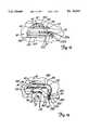
- the generally U-shaped ligation clips 216aid the physician in properly aligning the ligation-cutting tool 200 and the side branch 22 to be ligated by providing an abutment for the side branch 22 when the side branch 22 is positioned between the yokes 208 and 210.
- the yokes 208 and 210are biased towards each other in a conventional manner, the ligation clips 216 are deformed to clamp onto the side branch 22 therebetween and the blood flow through the side branch 22 is halted at two slightly spaced apart points (e.g., two clips are applied approximately 0.25 inches apart).
- the ligation clip applicator 202is activated and the yokes 208 and 210 clamp the ligation clips 216 onto the side branch 22, the side branch 22 is also held securely for cutting the side branch 22.
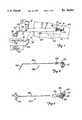
- the shaft 251 of the side biting ligation-cutting tool 250is a slender member that is longer than the lumen 38 of the endoscope 30.
- a housing 259covers those mechanisms on the shaft 251 that transmit the manipulations of the handle 270 and the plunger 274 at the proximal end of the side biting ligation-cutting tool 250 to the clipping and cutting motions, respectively, at the distal end of the side biting ligation-cutting tool 250.
- the dissecting tool 100 and endoscope 30are again advanced distally along the blood vessel 20 (as previously described) until another side branch is reached.
- the dissecting ring 110is large enough to pass over the clipped and severed stumps of any side branches 22 which extend from the blood vessel 20.
- the ligation-cutting tool 200is then used as previously described to sever additional side branches from the blood vessel 20.
- the procedureis repeated until the desired length of blood vessel 20 has been dissected free from the surrounding tissue and side branches.
- the suction-coagulator tool 300is used as required to control bleeding, again under the constant vigilance of the physician through the endoscope 30.
- the blood vessel 20has been held in tension by the physician via the gripping tool 150.
- the endoscope 30is advanced distally into the patient's body and the blood vessel 20 is moved into the lumen 38 of the scope body 34.
- the gripping forceps 150is inserted through the dissecting ring 110 of the dissecting tool 100 and the assembly of the dissecting tool 100 and the gripping forceps 150 is inserted into the proximal end of the lumen 38 and advanced distally through the lumen 38.
- the gripping forceps 150is again used to hold the free end 352 of the segment 400 of the blood vessel 20 during the continued procedure.
- the endoscope 30 and dissecting tool 100are distally advanced over the blood vessel 20 and into the patient's body through the second incision 450.
Landscapes
- Health & Medical Sciences (AREA)
- Life Sciences & Earth Sciences (AREA)
- Surgery (AREA)
- Animal Behavior & Ethology (AREA)
- Public Health (AREA)
- Engineering & Computer Science (AREA)
- Biomedical Technology (AREA)
- Heart & Thoracic Surgery (AREA)
- Medical Informatics (AREA)
- Molecular Biology (AREA)
- Veterinary Medicine (AREA)
- General Health & Medical Sciences (AREA)
- Nuclear Medicine, Radiotherapy & Molecular Imaging (AREA)
- Rheumatology (AREA)
- Physics & Mathematics (AREA)
- Biophysics (AREA)
- Optics & Photonics (AREA)
- Pathology (AREA)
- Radiology & Medical Imaging (AREA)
- Surgical Instruments (AREA)
Abstract
Description
Claims (23)
Priority Applications (1)
| Application Number | Priority Date | Filing Date | Title |
|---|---|---|---|
| US08/585,410USRE36043E (en) | 1992-10-02 | 1996-01-11 | Endoscope and method for vein removal |
Applications Claiming Priority (2)
| Application Number | Priority Date | Filing Date | Title |
|---|---|---|---|
| US07/956,904US5373840A (en) | 1992-10-02 | 1992-10-02 | Endoscope and method for vein removal |
| US08/585,410USRE36043E (en) | 1992-10-02 | 1996-01-11 | Endoscope and method for vein removal |
Related Parent Applications (1)
| Application Number | Title | Priority Date | Filing Date |
|---|---|---|---|
| US07/956,904ReissueUS5373840A (en) | 1992-10-02 | 1992-10-02 | Endoscope and method for vein removal |
Publications (1)
| Publication Number | Publication Date |
|---|---|
| USRE36043Etrue USRE36043E (en) | 1999-01-12 |
Family
ID=25498840
Family Applications (2)
| Application Number | Title | Priority Date | Filing Date |
|---|---|---|---|
| US07/956,904CeasedUS5373840A (en) | 1992-10-02 | 1992-10-02 | Endoscope and method for vein removal |
| US08/585,410Expired - LifetimeUSRE36043E (en) | 1992-10-02 | 1996-01-11 | Endoscope and method for vein removal |
Family Applications Before (1)
| Application Number | Title | Priority Date | Filing Date |
|---|---|---|---|
| US07/956,904CeasedUS5373840A (en) | 1992-10-02 | 1992-10-02 | Endoscope and method for vein removal |
Country Status (1)
| Country | Link |
|---|---|
| US (2) | US5373840A (en) |
Cited By (67)
| Publication number | Priority date | Publication date | Assignee | Title |
|---|---|---|---|---|
| US5968066A (en) | 1994-06-29 | 1999-10-19 | General Surgical Innovations, Inc. | Methods and devices for blood vessel harvesting |
| US6068639A (en) | 1995-05-19 | 2000-05-30 | General Surgical Innovations, Inc. | Methods and devices for blood vessel harvesting |
| US6077289A (en) | 1995-05-19 | 2000-06-20 | General Surgical Innovations | Methods and devices for harvesting blood vessels with balloons |
| US6206823B1 (en) | 1999-08-02 | 2001-03-27 | Ethicon Endo-Surgery, Inc. | Surgical instrument and method for endoscopic tissue dissection |
| EP1090593A2 (en) | 1999-10-05 | 2001-04-11 | Ethicon Endo-Surgery | Surgical device with integrally mounted image sensor |
| US6319265B1 (en) | 1998-02-27 | 2001-11-20 | Cardiothoracic Systems, Inc. | Dissecting retractor for harvesting vessels |
| US6358244B1 (en) | 1996-07-12 | 2002-03-19 | Endo Surgical Devices, Inc. | Endarterectomy surgical instrument and procedure |
| WO2002039882A2 (en) | 2000-11-17 | 2002-05-23 | Embro Vascular | Vein harvesting system and method |
| US6443970B1 (en) | 2001-01-24 | 2002-09-03 | Ethicon, Inc. | Surgical instrument with a dissecting tip |
| US6458128B1 (en) | 2001-01-24 | 2002-10-01 | Ethicon, Inc. | Electrosurgical instrument with a longitudinal element for conducting RF energy and moving a cutting element |
| US6464702B2 (en) | 2001-01-24 | 2002-10-15 | Ethicon, Inc. | Electrosurgical instrument with closing tube for conducting RF energy and moving jaws |
| US6511494B1 (en) | 2000-11-17 | 2003-01-28 | Embro Corporation | Vein harvesting system and method |
| US6520975B2 (en)* | 1999-02-04 | 2003-02-18 | Antonio Carlos Branco | Kit for endovascular venous surgery |
| US20030045812A1 (en)* | 1995-12-11 | 2003-03-06 | Knighton David R. | Apparatus and method for vein removal |
| US6551314B1 (en) | 2002-04-03 | 2003-04-22 | Thomas J. Fogarty | Methods and systems for vein harvesting |
| US6554829B2 (en) | 2001-01-24 | 2003-04-29 | Ethicon, Inc. | Electrosurgical instrument with minimally invasive jaws |
| US20030125733A1 (en)* | 2001-09-28 | 2003-07-03 | Ethicon, Inc. | Surgical device for applying radio frequency energy to a portion of a captured vessel |
| US6589168B2 (en) | 1996-02-07 | 2003-07-08 | Robert Lee Thompson | Video gynecological examination apparatus |
| US6592604B2 (en) | 2001-09-28 | 2003-07-15 | Ethicon, Inc. | Vessel harvesting retractor with dissection element |
| US6592582B2 (en) | 2001-09-28 | 2003-07-15 | Ethicon, Inc. | Vessel harvesting retractor with electrosurgical plunger |
| US6620161B2 (en) | 2001-01-24 | 2003-09-16 | Ethicon, Inc. | Electrosurgical instrument with an operational sequencing element |
| US6652521B2 (en) | 2001-01-24 | 2003-11-25 | Ethicon, Inc. | Surgical instrument with a bi-directional cutting element |
| US6656176B2 (en) | 2001-09-28 | 2003-12-02 | Ethicon, Inc. | Vessel harvesting retractor with integral electrosurgical clamping elements |
| US20030225426A1 (en)* | 2002-04-03 | 2003-12-04 | Thomas J. Fogarty, M.D. | Methods and systems for vein harvesting and fistula creation |
| US20040049208A1 (en)* | 2002-04-03 | 2004-03-11 | Thomas Fogarty, M.D. | Methods and systems for vein harvesting and fistula creation |
| WO2004028590A2 (en) | 2002-09-27 | 2004-04-08 | Ethicon, Inc. | Portable, reusable visualization system |
| US20040225317A1 (en)* | 2003-05-05 | 2004-11-11 | Rehnke Robert D. | Apparatus for use in fascial cleft surgery for opening an anatomic space |
| US20040236231A1 (en)* | 2003-05-23 | 2004-11-25 | Embro Corporation | Light catheter for illuminating tissue structures |
| US20050065398A1 (en)* | 1998-06-19 | 2005-03-24 | Ronald Adams | Non-circular resection device and endoscope |
| US20050096671A1 (en)* | 2003-10-31 | 2005-05-05 | Parris Wellman | Control mechanism for a surgical instrument |
| US20050096645A1 (en)* | 2003-10-31 | 2005-05-05 | Parris Wellman | Multitool surgical device |
| US20050096646A1 (en)* | 2003-10-31 | 2005-05-05 | Parris Wellman | Surgical system for retracting and severing tissue |
| US20050096670A1 (en)* | 2003-10-31 | 2005-05-05 | Parris Wellman | Surgical end effector |
| US20050096677A1 (en)* | 2003-10-31 | 2005-05-05 | Parris Wellman | Space-creating retractor with vessel manipulator |
| US20050148817A1 (en)* | 2003-10-31 | 2005-07-07 | Olympus Corporation | Living-body tissue removing apparatus |
| US20050149094A1 (en)* | 2003-10-31 | 2005-07-07 | Olympus Corporation | Trocar |
| US20050154257A1 (en)* | 2003-10-31 | 2005-07-14 | Olympus Corporation | Living-body tissue removing apparatus |
| US20050273125A1 (en)* | 2004-05-13 | 2005-12-08 | Opie John C | Percutaneous vein harvester with shielded blade |
| US20060030756A1 (en)* | 2004-06-25 | 2006-02-09 | Usher Raymond W | Vein harvesting system including dilator shaft and removable retractor housing |
| US20060036274A1 (en)* | 2004-06-25 | 2006-02-16 | Usher Raymond W | One-piece vessel harvester |
| US20060074444A1 (en)* | 2004-09-28 | 2006-04-06 | Lin Arthur M | Modular vessel harvesting system and method |
| US20060116746A1 (en)* | 2003-01-17 | 2006-06-01 | Chin Albert K | Cardiac electrode attachment procedure |
| US20060173474A1 (en)* | 2003-10-31 | 2006-08-03 | Parris Wellman | Surgical device having a track to guide an actuator |
| US20070005084A1 (en)* | 2004-06-16 | 2007-01-04 | Clague Cynthia T | Minimally invasive coring vein harvester |
| US7384423B1 (en) | 1995-07-13 | 2008-06-10 | Origin Medsystems, Inc. | Tissue dissection method |
| US20080161843A1 (en)* | 2006-10-16 | 2008-07-03 | Clague Cynthia T | Vessel support device and method of vessel harvesting |
| US20080306333A1 (en)* | 1999-08-10 | 2008-12-11 | Chin Albert K | Apparatus and Method for Endoscopic Surgical Procedures |
| US20090131747A1 (en)* | 1998-06-22 | 2009-05-21 | Maquet Cardiovascular Llc | Instrument And Method For Remotely Manipulating A Tissue Structure |
| US20090131907A1 (en)* | 1999-08-10 | 2009-05-21 | Maquet Cardiovascular Llc | Endoscopic Cardiac Surgery |
| US7645289B2 (en) | 2001-06-26 | 2010-01-12 | Tyco Healthcare Group Lp | Conduit harvesting instrument and method |
| US20110046624A1 (en)* | 2009-08-21 | 2011-02-24 | Maquet Cardiovascular Llc | Single handled endoscopic vessel harvesting system with rotation control |
| US7938842B1 (en) | 1998-08-12 | 2011-05-10 | Maquet Cardiovascular Llc | Tissue dissector apparatus |
| US20110124958A1 (en)* | 2009-11-23 | 2011-05-26 | Nelson Dvora Y | Device and method for extracting tubular structures |
| US7972265B1 (en) | 1998-06-22 | 2011-07-05 | Maquet Cardiovascular, Llc | Device and method for remote vessel ligation |
| US20110172688A1 (en)* | 2010-01-11 | 2011-07-14 | Tyco Healthcare Group Lp | Conduit Harvesting Instrument and Method |
| US8241210B2 (en) | 1998-06-22 | 2012-08-14 | Maquet Cardiovascular Llc | Vessel retractor |
| USD710497S1 (en)* | 2012-02-10 | 2014-08-05 | Merit Medical Systems, Inc. | Snare introducer |
| US10058345B2 (en) | 2013-09-09 | 2018-08-28 | Terumo Cardiovascular Systems Corporation | Single-pass endoscopic vessel harvesting |
| US10064611B2 (en) | 2015-07-22 | 2018-09-04 | Covidien Lp | Methods and devices for vein harvesting |
| US10070853B2 (en) | 2013-08-14 | 2018-09-11 | Covidien Lp | Expandable balloon desufflation assembly |
| US10166376B2 (en) | 2013-06-11 | 2019-01-01 | Covidien Lp | Restricted expansion dissector |
| US10299770B2 (en) | 2006-06-01 | 2019-05-28 | Maquet Cardiovascular Llc | Endoscopic vessel harvesting system components |
| US10575835B2 (en) | 2014-10-14 | 2020-03-03 | Covidien Lp | Methods and devices for vein harvesting |
| US10646210B2 (en) | 2014-10-14 | 2020-05-12 | Covidien Lp | Methods and devices for vein harvesting |
| US11369400B2 (en) | 2019-03-20 | 2022-06-28 | Covidien Lp | Balloon dissector |
| US11432839B2 (en) | 2018-01-12 | 2022-09-06 | Maquet Cardiovascular Llc | Vessel harvesting apparatus and method |
| US11547466B2 (en) | 2018-06-20 | 2023-01-10 | Covidien Lp | Visualization devices and methods for use in surgical procedures |
Families Citing this family (134)
| Publication number | Priority date | Publication date | Assignee | Title |
|---|---|---|---|---|
| US5373840A (en)* | 1992-10-02 | 1994-12-20 | Knighton; David R. | Endoscope and method for vein removal |
| US5667478A (en)* | 1992-11-06 | 1997-09-16 | Clarus Medical Systems, Inc. | Surgical instrument with stick-on fiber-optic viewing system and method of using |
| US5667472A (en)* | 1994-03-18 | 1997-09-16 | Clarus Medical Systems, Inc. | Surgical instrument and method for use with a viewing system |
| US5653722A (en)* | 1995-01-03 | 1997-08-05 | Kieturakis; Maciej J. | Anterograde/retrograde spiral dissector and method of use in vein grafting |
| US5591183A (en)* | 1995-04-12 | 1997-01-07 | Origin Medsystems, Inc. | Dissection apparatus |
| US5702417A (en)* | 1995-05-22 | 1997-12-30 | General Surgical Innovations, Inc. | Balloon loaded dissecting instruments |
| US5993472A (en)* | 1995-05-22 | 1999-11-30 | General Surgical Innovations, Inc. | Balloon dissecting instruments |
| US6596010B1 (en) | 1995-05-22 | 2003-07-22 | General Surgical Innovations, Inc. | Balloon dissecting instruments |
| US6004340A (en)* | 1995-05-22 | 1999-12-21 | General Surgical Innovations, Inc. | Balloon dissecting instruments |
| US5944734A (en)* | 1995-05-22 | 1999-08-31 | General Surgical Innovations, Inc. | Balloon dissecting instruments |
| US7037317B2 (en)* | 1995-05-22 | 2006-05-02 | United States Surgical Corporation | Balloon dissecting instruments |
| US5893866A (en)* | 1995-05-22 | 1999-04-13 | General Surgical Innovations, Inc. | Balloon dissecting instruments |
| US6179854B1 (en) | 1995-05-22 | 2001-01-30 | General Surgical Innovations, Inc. | Apparatus and method for dissecting and retracting elongate structures |
| US5979452A (en) | 1995-06-07 | 1999-11-09 | General Surgical Innovations, Inc. | Endoscopic linton procedure using balloon dissectors and retractors |
| US5857961A (en)* | 1995-06-07 | 1999-01-12 | Clarus Medical Systems, Inc. | Surgical instrument for use with a viewing system |
| US5707389A (en)* | 1995-06-07 | 1998-01-13 | Baxter International Inc. | Side branch occlusion catheter device having integrated endoscope for performing endoscopically visualized occlusion of the side branches of an anatomical passageway |
| US5759150A (en) | 1995-07-07 | 1998-06-02 | Olympus Optical Co., Ltd. | System for evulsing subcutaneous tissue |
| US5797946A (en)* | 1995-07-13 | 1998-08-25 | Origin Medsystems, Inc. | Method for arterial harvest and anastomosis for coronary bypass grafting |
| US7001404B1 (en) | 1995-07-13 | 2006-02-21 | Origin Medsystems, Inc. | Tissue separation cannula and method |
| US5968065A (en) | 1995-07-13 | 1999-10-19 | Origin Medsystems, Inc. | Tissue separation cannula |
| US5667480A (en)* | 1995-10-20 | 1997-09-16 | Ethicon Endo-Surgery, Inc. | Method and devices for endoscopic vessel harvesting |
| US5782753A (en)* | 1995-10-20 | 1998-07-21 | United States Surgical Corporation | Surgical retractor |
| CA2244164A1 (en) | 1996-01-24 | 1997-07-31 | Albert K. Chin | Tissue separation cannula with dissection probe and method |
| US5817013A (en)* | 1996-03-19 | 1998-10-06 | Enable Medical Corporation | Method and apparatus for the minimally invasive harvesting of a saphenous vein and the like |
| US5913870A (en)* | 1996-08-13 | 1999-06-22 | United States Surgical Corporation | Surgical dissector |
| US5928135A (en)* | 1996-08-15 | 1999-07-27 | Ethicon Endo-Surgery, Inc. | Method and devices for endoscopic vessel harvesting |
| US6152936A (en) | 1996-09-23 | 2000-11-28 | Esd Medical, Llc | Surgical loop delivery device |
| US6019771A (en)* | 1996-12-02 | 2000-02-01 | Cardiothoracic Systems, Inc. | Devices and methods for minimally invasive harvesting of a vessel especially the saphenous vein for coronary bypass grafting |
| US5970982A (en)* | 1997-02-20 | 1999-10-26 | Perkins; Rodney C. | Minimally invasive biological vessel harvesting method |
| WO1998036692A1 (en)* | 1997-02-20 | 1998-08-27 | Perkins Rodney C | Biological vessel harvesting device and method |
| DE19707374C2 (en)* | 1997-02-25 | 2000-10-26 | Storz Karl Gmbh & Co Kg | Medical dissection spatula with expandable spatula jaw parts |
| DE19717977A1 (en)* | 1997-04-23 | 1998-05-28 | Rainer Prof Dr Dr Schmelzeisen | Lining=up fixture device used to treat jaw or face fractures |
| US5913818A (en)* | 1997-06-02 | 1999-06-22 | General Surgical Innovations, Inc. | Vascular retractor |
| US5938680A (en)* | 1997-06-19 | 1999-08-17 | Cardiothoracic Systems, Inc. | Devices and methods for harvesting vascular conduits |
| US5902315A (en)* | 1997-08-28 | 1999-05-11 | Ethicon Endo-Surgery, Inc. | Optical tissue dissector/retractor |
| US5922004A (en)* | 1997-08-28 | 1999-07-13 | Ethicon Endo-Surgery, Inc. | Method for performing optical tissue dissection/retraction |
| US6436116B1 (en) | 1997-10-06 | 2002-08-20 | Smith & Nephew, Inc. | Methods and apparatus for removing veins |
| WO1999025238A2 (en)* | 1997-11-18 | 1999-05-27 | Emory University | Device for endoscopic vessel harvesting |
| US5899912A (en) | 1997-12-17 | 1999-05-04 | Eaves, Iii; Felmont F. | Apparatus and method for endoscopic harvesting of elongate tissue structure |
| US6632171B2 (en) | 1997-12-22 | 2003-10-14 | Given Imaging Ltd. | Method for in vivo delivery of autonomous capsule |
| IL122716A0 (en)* | 1997-12-22 | 1998-08-16 | Tally Eitan Zeev Pearl And Co | System and method for in vivo delivery of autonomous capsule |
| AU2589699A (en) | 1998-02-06 | 1999-08-23 | Ronald J. Brinkerhoff | Device for visualizing, dissecting and harvesting vessels |
| US5957927A (en)* | 1998-02-24 | 1999-09-28 | Synthes (Usa) | Bone fixation device introducer |
| US6129661A (en)* | 1998-04-09 | 2000-10-10 | Smith & Nephew, Inc. | Endoscopic instrumentation with working channel |
| DE19827360C2 (en)* | 1998-06-19 | 2000-05-31 | Storz Karl Gmbh & Co Kg | Medical instrument for endoscopic removal of the saphenous vein |
| US6648815B2 (en) | 1998-06-19 | 2003-11-18 | Karl Storz Gmbh & Co. Kg | Medical instrument and method for endoscopic removal of the saphenous vein |
| US6045561A (en)* | 1998-06-23 | 2000-04-04 | Orthopaedic Biosystems Ltd., Inc. | Surgical knot manipulator |
| US6042538A (en)* | 1998-11-18 | 2000-03-28 | Emory University | Device for endoscopic vessel harvesting |
| US6159146A (en)* | 1999-03-12 | 2000-12-12 | El Gazayerli; Mohamed Mounir | Method and apparatus for minimally-invasive fundoplication |
| US6488689B1 (en)* | 1999-05-20 | 2002-12-03 | Aaron V. Kaplan | Methods and apparatus for transpericardial left atrial appendage closure |
| WO2000071033A1 (en)* | 1999-05-21 | 2000-11-30 | Cardiothoracic Systems, Inc. | Refractor apparatus for use in harvesting mammary arteries during heart by-pass surgery |
| US7813789B2 (en)* | 1999-06-15 | 2010-10-12 | Given Imaging Ltd. | In-vivo imaging device, optical system and method |
| US7996067B2 (en)* | 1999-06-15 | 2011-08-09 | Given Imaging Ltd. | In-vivo imaging device, optical system and method |
| IL130486A (en)* | 1999-06-15 | 2005-08-31 | Given Imaging Ltd | Optical system |
| US7398781B1 (en) | 1999-08-10 | 2008-07-15 | Maquet Cardiovascular, Llc | Method for subxiphoid endoscopic access |
| US7597698B2 (en) | 1999-08-10 | 2009-10-06 | Maquet Cardiovascular Llc | Apparatus and method for endoscopic encirclement of pulmonary veins for epicardial ablation |
| US7264587B2 (en)* | 1999-08-10 | 2007-09-04 | Origin Medsystems, Inc. | Endoscopic subxiphoid surgical procedures |
| US7526342B2 (en) | 1999-08-10 | 2009-04-28 | Maquet Cardiovascular Llc | Apparatus for endoscopic cardiac mapping and lead placement |
| US6231561B1 (en)* | 1999-09-20 | 2001-05-15 | Appriva Medical, Inc. | Method and apparatus for closing a body lumen |
| US6352544B1 (en) | 2000-02-22 | 2002-03-05 | Gregory A. Spitz | Apparatus and methods for removing veins |
| US6736773B2 (en)* | 2001-01-25 | 2004-05-18 | Scimed Life Systems, Inc. | Endoscopic vision system |
| GB0111683D0 (en)* | 2001-05-12 | 2001-07-04 | Menon Johansson Anatole S | Device for assisting laparoscopic surgery |
| US6887251B1 (en)* | 2001-07-12 | 2005-05-03 | William D. Suval | Method and apparatus for vessel harvesting |
| WO2003013367A2 (en) | 2001-08-10 | 2003-02-20 | General Surgical Innovations Inc. | Vascular harvesting tool and methods |
| ATE526883T1 (en) | 2001-12-27 | 2011-10-15 | Olympus Corp | COVER WITH DEVICES FOR ENDOSCOPIC HARVESTING OF BLOOD VESSELS |
| US7001329B2 (en)* | 2002-07-23 | 2006-02-21 | Pentax Corporation | Capsule endoscope guidance system, capsule endoscope holder, and capsule endoscope |
| AU2003274635A1 (en) | 2002-10-15 | 2004-05-04 | Given Imaging Ltd. | Device, system and method for transfer of signals to a moving device |
| US7056329B2 (en)* | 2002-10-23 | 2006-06-06 | Intellimed Surgical Solutions, Llc | Laparoscopic direct vision dissecting port |
| US20040093000A1 (en)* | 2002-10-23 | 2004-05-13 | Stephen Kerr | Direct vision port site dissector |
| US6818003B2 (en) | 2003-01-16 | 2004-11-16 | Mark H. Genovesi | Blood vessel harvesting device |
| US7309342B2 (en)* | 2003-01-16 | 2007-12-18 | Mark Genovesi | Heatless blood vessel harvesting device |
| US20050137468A1 (en)* | 2003-12-18 | 2005-06-23 | Jerome Avron | Device, system, and method for in-vivo sensing of a substance |
| JP2005245937A (en)* | 2004-03-08 | 2005-09-15 | Pentax Corp | Clothing with communication function and endoscope system |
| US20050195785A1 (en)* | 2004-03-08 | 2005-09-08 | Pentax Corporation | Image signal processing device |
| JP2005245938A (en)* | 2004-03-08 | 2005-09-15 | Pentax Corp | Diagnostic clothing, diagnostic clothing system, and endoscope system |
| WO2005112786A2 (en)* | 2004-05-14 | 2005-12-01 | Ethicon Endo-Surgery, Inc. | T-type suture anchoring devices and methods of using same |
| US8078266B2 (en) | 2005-10-25 | 2011-12-13 | Voyage Medical, Inc. | Flow reduction hood systems |
| US7930016B1 (en) | 2005-02-02 | 2011-04-19 | Voyage Medical, Inc. | Tissue closure system |
| US10064540B2 (en) | 2005-02-02 | 2018-09-04 | Intuitive Surgical Operations, Inc. | Visualization apparatus for transseptal access |
| US7860555B2 (en) | 2005-02-02 | 2010-12-28 | Voyage Medical, Inc. | Tissue visualization and manipulation system |
| US11478152B2 (en) | 2005-02-02 | 2022-10-25 | Intuitive Surgical Operations, Inc. | Electrophysiology mapping and visualization system |
| US8137333B2 (en) | 2005-10-25 | 2012-03-20 | Voyage Medical, Inc. | Delivery of biological compounds to ischemic and/or infarcted tissue |
| US8050746B2 (en) | 2005-02-02 | 2011-11-01 | Voyage Medical, Inc. | Tissue visualization device and method variations |
| US7860556B2 (en) | 2005-02-02 | 2010-12-28 | Voyage Medical, Inc. | Tissue imaging and extraction systems |
| US20080015569A1 (en) | 2005-02-02 | 2008-01-17 | Voyage Medical, Inc. | Methods and apparatus for treatment of atrial fibrillation |
| US9510732B2 (en) | 2005-10-25 | 2016-12-06 | Intuitive Surgical Operations, Inc. | Methods and apparatus for efficient purging |
| US7918787B2 (en) | 2005-02-02 | 2011-04-05 | Voyage Medical, Inc. | Tissue visualization and manipulation systems |
| US20060224040A1 (en)* | 2005-03-31 | 2006-10-05 | Given Imaging Ltd. | In vivo imaging device and method of manufacture thereof |
| US8221310B2 (en) | 2005-10-25 | 2012-07-17 | Voyage Medical, Inc. | Tissue visualization device and method variations |
| WO2007058877A2 (en)* | 2005-11-10 | 2007-05-24 | Mayo Foundation For Medical Education And Research | Vein closure and injection kits and methods |
| US7918783B2 (en)* | 2006-03-22 | 2011-04-05 | Boston Scientific Scimed, Inc. | Endoscope working channel with multiple functionality |
| US8518024B2 (en) | 2006-04-24 | 2013-08-27 | Transenterix, Inc. | System and method for multi-instrument surgical access using a single access port |
| CA2650474A1 (en) | 2006-04-24 | 2007-11-08 | Synecor, Llc | Natural orifice surgical system |
| US9055906B2 (en) | 2006-06-14 | 2015-06-16 | Intuitive Surgical Operations, Inc. | In-vivo visualization systems |
| US10004388B2 (en) | 2006-09-01 | 2018-06-26 | Intuitive Surgical Operations, Inc. | Coronary sinus cannulation |
| WO2008028149A2 (en) | 2006-09-01 | 2008-03-06 | Voyage Medical, Inc. | Electrophysiology mapping and visualization system |
| US20080097476A1 (en) | 2006-09-01 | 2008-04-24 | Voyage Medical, Inc. | Precision control systems for tissue visualization and manipulation assemblies |
| US10335131B2 (en) | 2006-10-23 | 2019-07-02 | Intuitive Surgical Operations, Inc. | Methods for preventing tissue migration |
| US20080183036A1 (en) | 2006-12-18 | 2008-07-31 | Voyage Medical, Inc. | Systems and methods for unobstructed visualization and ablation |
| US9226648B2 (en) | 2006-12-21 | 2016-01-05 | Intuitive Surgical Operations, Inc. | Off-axis visualization systems |
| US8131350B2 (en) | 2006-12-21 | 2012-03-06 | Voyage Medical, Inc. | Stabilization of visualization catheters |
| JP4662374B2 (en)* | 2007-03-26 | 2011-03-30 | オリンパス株式会社 | Endoscopic blood vessel collection device |
| EP2148608A4 (en) | 2007-04-27 | 2010-04-28 | Voyage Medical Inc | Complex shape steerable tissue visualization and manipulation catheter |
| US8657805B2 (en) | 2007-05-08 | 2014-02-25 | Intuitive Surgical Operations, Inc. | Complex shape steerable tissue visualization and manipulation catheter |
| WO2008141238A1 (en) | 2007-05-11 | 2008-11-20 | Voyage Medical, Inc. | Visual electrode ablation systems |
| US8235985B2 (en) | 2007-08-31 | 2012-08-07 | Voyage Medical, Inc. | Visualization and ablation system variations |
| US8858609B2 (en) | 2008-02-07 | 2014-10-14 | Intuitive Surgical Operations, Inc. | Stent delivery under direct visualization |
| US8515507B2 (en) | 2008-06-16 | 2013-08-20 | Given Imaging Ltd. | Device and method for detecting in-vivo pathology |
| US9101735B2 (en) | 2008-07-07 | 2015-08-11 | Intuitive Surgical Operations, Inc. | Catheter control systems |
| US8894643B2 (en) | 2008-10-10 | 2014-11-25 | Intuitive Surgical Operations, Inc. | Integral electrode placement and connection systems |
| US8333012B2 (en) | 2008-10-10 | 2012-12-18 | Voyage Medical, Inc. | Method of forming electrode placement and connection systems |
| US9468364B2 (en) | 2008-11-14 | 2016-10-18 | Intuitive Surgical Operations, Inc. | Intravascular catheter with hood and image processing systems |
| US20120289947A1 (en)* | 2010-01-18 | 2012-11-15 | Wolfgang Neuberger | Device and method for removing veins |
| US8694071B2 (en) | 2010-02-12 | 2014-04-08 | Intuitive Surgical Operations, Inc. | Image stabilization techniques and methods |
| US9814522B2 (en) | 2010-04-06 | 2017-11-14 | Intuitive Surgical Operations, Inc. | Apparatus and methods for ablation efficacy |
| EP2593024A1 (en)* | 2010-07-17 | 2013-05-22 | The New York And Presbyterian Hospital | Methods and systems for minimally invasive endoscopic surgeries |
| WO2013096763A2 (en) | 2011-12-23 | 2013-06-27 | Pavilion Medical Innovations, Llc | Unitary endoscopic vessel harvesting devices |
| US20130282009A1 (en)* | 2012-04-18 | 2013-10-24 | Cardica, Inc. | Endoscopic vein harvesting system |
| US9413896B2 (en) | 2012-09-14 | 2016-08-09 | The Spectranetics Corporation | Tissue slitting methods and systems |
| US9198681B2 (en)* | 2012-10-12 | 2015-12-01 | Cook Medical Technologies Llc | Device and method for removing tissue inside a body vessel |
| WO2014158613A1 (en) | 2013-03-14 | 2014-10-02 | Saphena Medical, Inc. | Unitary endoscopic vessel harvesting devices |
| US9814481B2 (en) | 2013-03-14 | 2017-11-14 | Saphena Medical, Inc. | Unitary endoscopic vessel harvesting devices |
| US10835279B2 (en) | 2013-03-14 | 2020-11-17 | Spectranetics Llc | Distal end supported tissue slitting apparatus |
| US20150073207A1 (en)* | 2013-09-09 | 2015-03-12 | Terumo Cardiovascular Systems Corporation | Single-pass endoscopic vessel harvesting |
| WO2015147157A1 (en)* | 2014-03-27 | 2015-10-01 | 富士フイルム株式会社 | Surgical operation device for endoscope, endoscope, and endoscope operational tool |
| WO2016027489A1 (en)* | 2014-08-21 | 2016-02-25 | テルモ株式会社 | Method for stripping blood vessel and device for stripping blood vessel |
| EP3501435A1 (en) | 2014-09-30 | 2019-06-26 | Fujifilm Corporation | Surgical apparatus for endoscope |
| US9750495B2 (en) | 2014-09-30 | 2017-09-05 | Covidien Lp | Methods and devices for tissue suturing |
| US9943328B2 (en) | 2015-04-28 | 2018-04-17 | Saphena Medical, Inc. | Unitary endoscopic vessel harvesting devices with an elastic force |
| JP7166761B2 (en) | 2015-06-17 | 2022-11-08 | サフィナ・メディカル・インコーポレイテッド | single endoscope angiography device |
| PL3515327T3 (en) | 2016-09-23 | 2024-06-10 | Atricure, Inc. | DEVICES FOR CLOSING THE LEFT ATRIAL APPEARANCE |
| JP2021519143A (en) | 2018-03-27 | 2021-08-10 | センターハート・インコーポレイテッドSentreHEART, Inc. | Devices and methods for left atrial appendage closure |
| WO2020206266A1 (en) | 2019-04-05 | 2020-10-08 | Saphena Medical, Inc. | Unitary device for vessel harvesting and method of using same |
| US11471140B2 (en)* | 2020-04-24 | 2022-10-18 | Verivas Solutions Inc. | Verivas rapid vein harvester |
Citations (54)
| Publication number | Priority date | Publication date | Assignee | Title |
|---|---|---|---|---|
| US1867624A (en)* | 1930-04-01 | 1932-07-19 | Memorial Hospital For The Trea | Device for obtaining biopsy specimens |
| US2001169A (en)* | 1934-11-01 | 1935-05-14 | Oscar R Wallace | Building structure |
| US2011169A (en)* | 1932-04-13 | 1935-08-13 | Wappler Frederick Charles | Forcipated surgical electrode |
| US2028635A (en)* | 1933-09-11 | 1936-01-21 | Wappler Frederick Charles | Forcipated surgical instrument |
| US2316297A (en)* | 1943-01-15 | 1943-04-13 | Beverly A Southerland | Surgical instrument |
| US2868206A (en)* | 1956-07-25 | 1959-01-13 | Frederick G Stoesser | Intra luminal vein stripper |
| US2944552A (en)* | 1958-12-29 | 1960-07-12 | Richard B Wilk | Surgical instrument |
| US3185155A (en)* | 1963-03-13 | 1965-05-25 | Slaten | Vein stripper |
| US3336916A (en)* | 1963-10-30 | 1967-08-22 | Richard F Edlich | Electrocautery process |
| US3856016A (en)* | 1972-11-03 | 1974-12-24 | H Davis | Method for mechanically applying an occlusion clip to an anatomical tubular structure |
| US3882854A (en)* | 1973-08-23 | 1975-05-13 | Research Corp | Surgical clip and applicator |
| US3934115A (en)* | 1973-09-25 | 1976-01-20 | Peterson Gerald H | Method and apparatus for electric singe cutting |
| SU510235A1 (en)* | 1973-01-16 | 1976-04-15 | Ростовский государственный медицинский институт | Veno extractor |
| USRE29088E (en)* | 1972-10-10 | 1976-12-28 | Surgical cutting instrument having electrically heated cutting edge | |
| US4038987A (en)* | 1974-02-08 | 1977-08-02 | Olympus Optical Co., Ltd. | Forceps device for endoscope |
| GB2082459A (en)* | 1980-08-26 | 1982-03-10 | Atrium Medical Corp | Apparatus for vein removal |
| US4362160A (en)* | 1979-07-24 | 1982-12-07 | Richard Wolf Gmbh | Endoscopes |
| US4369768A (en)* | 1980-07-30 | 1983-01-25 | Marko Vukovic | Arthroscope |
| US4440170A (en)* | 1979-03-06 | 1984-04-03 | Ethicon, Inc. | Surgical clip applying instrument |
| SU1123674A1 (en)* | 1983-06-17 | 1984-11-15 | Тернопольский государственный медицинский институт | Venoextractor |
| US4493321A (en)* | 1982-05-25 | 1985-01-15 | Leather Robert P | Venous valve cutter for the incision of valve leaflets in situ |
| US4499899A (en)* | 1983-01-21 | 1985-02-19 | Brimfield Precision, Inc. | Fiber-optic illuminated microsurgical scissors |
| US4499898A (en)* | 1982-08-23 | 1985-02-19 | Koi Associates | Surgical knife with controllably extendable blade and gauge therefor |
| US4516575A (en)* | 1982-06-03 | 1985-05-14 | Coopervision, Inc. | Surgical scalpel |
| US4556058A (en)* | 1983-08-17 | 1985-12-03 | United States Surgical Corporation | Apparatus for ligation and division with fixed jaws |
| US4586919A (en)* | 1984-04-06 | 1986-05-06 | Taheri Syde A | External shunt and method for procuring and preserving the endothelium of a vein used in arterial bypass |
| US4597389A (en)* | 1982-09-30 | 1986-07-01 | Ibrahim Adel A | Device for removing objects from tubular body passages |
| US4638802A (en)* | 1984-09-21 | 1987-01-27 | Olympus Optical Co., Ltd. | High frequency instrument for incision and excision |
| US4653476A (en)* | 1984-07-05 | 1987-03-31 | Richard Wolf Gmbh | Instrument insert for a uretero-renoscope |
| GB2195540A (en)* | 1986-09-19 | 1988-04-13 | Anthony John Milne | Vein stripper |
| US4745908A (en)* | 1987-05-08 | 1988-05-24 | Circon Corporation | Inspection instrument fexible shaft having deflection compensation means |
| US4759348A (en)* | 1981-09-28 | 1988-07-26 | Cawood Charles David | Endoscope assembly and surgical instrument for use therewith |
| US4759364A (en)* | 1985-09-19 | 1988-07-26 | Richard Wolf Gmbh | Pincers attachment for a surgical handle to be used in endoscopy |
| US4762120A (en)* | 1983-11-08 | 1988-08-09 | Laserscope, Inc. | Endoscopic device having handle assembly and catheter assembly |
| US4768508A (en)* | 1986-06-06 | 1988-09-06 | Thomas J. Fogarty | Vein valve cutting method |
| US4793346A (en)* | 1986-09-04 | 1988-12-27 | Bruce Mindich | Process and apparatus for harvesting vein |
| US4858595A (en)* | 1987-12-18 | 1989-08-22 | Gerhard Buess | Mediastinoscope |
| US4862874A (en)* | 1987-06-10 | 1989-09-05 | Kellner Hans Joerg | Endoscope for removal of thrombi from pulmonary arterial vessels |
| US4869268A (en)* | 1987-05-14 | 1989-09-26 | Inbae Yoon | Multi-functional instruments and stretchable ligating and occluding devices |
| US4877016A (en)* | 1988-07-29 | 1989-10-31 | Kantor Edward A | Video endoscopic microscope |
| US4932952A (en)* | 1988-12-20 | 1990-06-12 | Alto Development Corporation | Antishock, anticlog suction coagulator |
| US4997436A (en)* | 1988-06-03 | 1991-03-05 | Oberlander Michael A | Arthroscopic clip insertion tool |
| US5013312A (en)* | 1990-03-19 | 1991-05-07 | Everest Medical Corporation | Bipolar scalpel for harvesting internal mammary artery |
| US5020514A (en)* | 1989-07-19 | 1991-06-04 | Richard Wolf Gmbh | Endoscope for nasal surgery |
| DE3942589A1 (en)* | 1989-12-22 | 1991-07-04 | Olympus Optical Europ | VENAL VALVE CUTTING KNIFE |
| US5037433A (en)* | 1990-05-17 | 1991-08-06 | Wilk Peter J | Endoscopic suturing device and related method and suture |
| US5047038A (en)* | 1985-07-01 | 1991-09-10 | Edward Weck Incorporated | Automatic hemostatic clip applier |
| US5049154A (en)* | 1989-08-07 | 1991-09-17 | Berkshire Research & Development, Inc. | Adjustable intra-luminal valvulotome |
| US5213093A (en)* | 1991-05-29 | 1993-05-25 | Applied Vascular Devices, Inc. | Endoscope with non-circular probe and method of making same |
| US5284478A (en)* | 1992-06-08 | 1994-02-08 | Nobles Anthony A | Detachable tip optical valvulotome |
| US5373840A (en)* | 1992-10-02 | 1994-12-20 | Knighton; David R. | Endoscope and method for vein removal |
| US5425355A (en)* | 1991-01-28 | 1995-06-20 | Laserscope | Energy discharging surgical probe and surgical process having distal energy application without concomitant proximal movement |
| US5447513A (en)* | 1992-05-06 | 1995-09-05 | Ethicon, Inc. | Endoscopic ligation and division instrument |
| US5549637A (en)* | 1994-11-10 | 1996-08-27 | Crainich; Lawrence | Articulated medical instrument |
Family Cites Families (1)
| Publication number | Priority date | Publication date | Assignee | Title |
|---|---|---|---|---|
| US4754754A (en)* | 1984-08-20 | 1988-07-05 | Garito Jon C | Electrosurgical handpiece for blades and needles |
- 1992
- 1992-10-02USUS07/956,904patent/US5373840A/ennot_activeCeased
- 1996
- 1996-01-11USUS08/585,410patent/USRE36043E/ennot_activeExpired - Lifetime
Patent Citations (54)
| Publication number | Priority date | Publication date | Assignee | Title |
|---|---|---|---|---|
| US1867624A (en)* | 1930-04-01 | 1932-07-19 | Memorial Hospital For The Trea | Device for obtaining biopsy specimens |
| US2011169A (en)* | 1932-04-13 | 1935-08-13 | Wappler Frederick Charles | Forcipated surgical electrode |
| US2028635A (en)* | 1933-09-11 | 1936-01-21 | Wappler Frederick Charles | Forcipated surgical instrument |
| US2001169A (en)* | 1934-11-01 | 1935-05-14 | Oscar R Wallace | Building structure |
| US2316297A (en)* | 1943-01-15 | 1943-04-13 | Beverly A Southerland | Surgical instrument |
| US2868206A (en)* | 1956-07-25 | 1959-01-13 | Frederick G Stoesser | Intra luminal vein stripper |
| US2944552A (en)* | 1958-12-29 | 1960-07-12 | Richard B Wilk | Surgical instrument |
| US3185155A (en)* | 1963-03-13 | 1965-05-25 | Slaten | Vein stripper |
| US3336916A (en)* | 1963-10-30 | 1967-08-22 | Richard F Edlich | Electrocautery process |
| USRE29088E (en)* | 1972-10-10 | 1976-12-28 | Surgical cutting instrument having electrically heated cutting edge | |
| US3856016A (en)* | 1972-11-03 | 1974-12-24 | H Davis | Method for mechanically applying an occlusion clip to an anatomical tubular structure |
| SU510235A1 (en)* | 1973-01-16 | 1976-04-15 | Ростовский государственный медицинский институт | Veno extractor |
| US3882854A (en)* | 1973-08-23 | 1975-05-13 | Research Corp | Surgical clip and applicator |
| US3934115A (en)* | 1973-09-25 | 1976-01-20 | Peterson Gerald H | Method and apparatus for electric singe cutting |
| US4038987A (en)* | 1974-02-08 | 1977-08-02 | Olympus Optical Co., Ltd. | Forceps device for endoscope |
| US4440170A (en)* | 1979-03-06 | 1984-04-03 | Ethicon, Inc. | Surgical clip applying instrument |
| US4362160A (en)* | 1979-07-24 | 1982-12-07 | Richard Wolf Gmbh | Endoscopes |
| US4369768A (en)* | 1980-07-30 | 1983-01-25 | Marko Vukovic | Arthroscope |
| GB2082459A (en)* | 1980-08-26 | 1982-03-10 | Atrium Medical Corp | Apparatus for vein removal |
| US4759348A (en)* | 1981-09-28 | 1988-07-26 | Cawood Charles David | Endoscope assembly and surgical instrument for use therewith |
| US4493321A (en)* | 1982-05-25 | 1985-01-15 | Leather Robert P | Venous valve cutter for the incision of valve leaflets in situ |
| US4516575A (en)* | 1982-06-03 | 1985-05-14 | Coopervision, Inc. | Surgical scalpel |
| US4499898A (en)* | 1982-08-23 | 1985-02-19 | Koi Associates | Surgical knife with controllably extendable blade and gauge therefor |
| US4597389A (en)* | 1982-09-30 | 1986-07-01 | Ibrahim Adel A | Device for removing objects from tubular body passages |
| US4499899A (en)* | 1983-01-21 | 1985-02-19 | Brimfield Precision, Inc. | Fiber-optic illuminated microsurgical scissors |
| SU1123674A1 (en)* | 1983-06-17 | 1984-11-15 | Тернопольский государственный медицинский институт | Venoextractor |
| US4556058A (en)* | 1983-08-17 | 1985-12-03 | United States Surgical Corporation | Apparatus for ligation and division with fixed jaws |
| US4762120A (en)* | 1983-11-08 | 1988-08-09 | Laserscope, Inc. | Endoscopic device having handle assembly and catheter assembly |
| US4586919A (en)* | 1984-04-06 | 1986-05-06 | Taheri Syde A | External shunt and method for procuring and preserving the endothelium of a vein used in arterial bypass |
| US4653476A (en)* | 1984-07-05 | 1987-03-31 | Richard Wolf Gmbh | Instrument insert for a uretero-renoscope |
| US4638802A (en)* | 1984-09-21 | 1987-01-27 | Olympus Optical Co., Ltd. | High frequency instrument for incision and excision |
| US5047038A (en)* | 1985-07-01 | 1991-09-10 | Edward Weck Incorporated | Automatic hemostatic clip applier |
| US4759364A (en)* | 1985-09-19 | 1988-07-26 | Richard Wolf Gmbh | Pincers attachment for a surgical handle to be used in endoscopy |
| US4768508A (en)* | 1986-06-06 | 1988-09-06 | Thomas J. Fogarty | Vein valve cutting method |
| US4793346A (en)* | 1986-09-04 | 1988-12-27 | Bruce Mindich | Process and apparatus for harvesting vein |
| GB2195540A (en)* | 1986-09-19 | 1988-04-13 | Anthony John Milne | Vein stripper |
| US4745908A (en)* | 1987-05-08 | 1988-05-24 | Circon Corporation | Inspection instrument fexible shaft having deflection compensation means |
| US4869268A (en)* | 1987-05-14 | 1989-09-26 | Inbae Yoon | Multi-functional instruments and stretchable ligating and occluding devices |
| US4862874A (en)* | 1987-06-10 | 1989-09-05 | Kellner Hans Joerg | Endoscope for removal of thrombi from pulmonary arterial vessels |
| US4858595A (en)* | 1987-12-18 | 1989-08-22 | Gerhard Buess | Mediastinoscope |
| US4997436A (en)* | 1988-06-03 | 1991-03-05 | Oberlander Michael A | Arthroscopic clip insertion tool |
| US4877016A (en)* | 1988-07-29 | 1989-10-31 | Kantor Edward A | Video endoscopic microscope |
| US4932952A (en)* | 1988-12-20 | 1990-06-12 | Alto Development Corporation | Antishock, anticlog suction coagulator |
| US5020514A (en)* | 1989-07-19 | 1991-06-04 | Richard Wolf Gmbh | Endoscope for nasal surgery |
| US5049154A (en)* | 1989-08-07 | 1991-09-17 | Berkshire Research & Development, Inc. | Adjustable intra-luminal valvulotome |
| DE3942589A1 (en)* | 1989-12-22 | 1991-07-04 | Olympus Optical Europ | VENAL VALVE CUTTING KNIFE |
| US5013312A (en)* | 1990-03-19 | 1991-05-07 | Everest Medical Corporation | Bipolar scalpel for harvesting internal mammary artery |
| US5037433A (en)* | 1990-05-17 | 1991-08-06 | Wilk Peter J | Endoscopic suturing device and related method and suture |
| US5425355A (en)* | 1991-01-28 | 1995-06-20 | Laserscope | Energy discharging surgical probe and surgical process having distal energy application without concomitant proximal movement |
| US5213093A (en)* | 1991-05-29 | 1993-05-25 | Applied Vascular Devices, Inc. | Endoscope with non-circular probe and method of making same |
| US5447513A (en)* | 1992-05-06 | 1995-09-05 | Ethicon, Inc. | Endoscopic ligation and division instrument |
| US5284478A (en)* | 1992-06-08 | 1994-02-08 | Nobles Anthony A | Detachable tip optical valvulotome |
| US5373840A (en)* | 1992-10-02 | 1994-12-20 | Knighton; David R. | Endoscope and method for vein removal |
| US5549637A (en)* | 1994-11-10 | 1996-08-27 | Crainich; Lawrence | Articulated medical instrument |
Non-Patent Citations (28)
| Title |
|---|
| "Angioscopic Preparation for Saphenous Vein in Situ Bypass Grafting," A. K. Chin et al.,Endovascular Surgery, 1989, pp. 74-81. |
| "Incision Decision", Atrium Medical Corporation advertisement, appearing in J. Thorac. Cardiovasc. Surg., 83(4), 1982. |
| "Reversed Saphenous Vein for Femoropopliteal Bypass Grafting," K. G. Burnand, Vascular Surgical Techniques An Atlas, K. G. Burnand et al., 1989, pp. 228-234. |
| "Saphenous vein grafts are number 1. Period.", Atrium Medical Corporation advertisement, appearing in J. Thorac. Cardiovasc. Surg., 81(6), 1982. |
| "The in Situ Saphenous Vein Arterial; Bypass by Valve Incision," R. P. Leather et al., Vascular Surgical Techniques An Atlas, 1989, pp. 255-262. |
| Angioscopic Preparation for Saphenous Vein in Situ Bypass Grafting, A. K. Chin et al., Endovascular Surgery, 1989, pp. 74 81.* |
| DeLaria, G.A., et al., "Leg Wound Complications Associated With Coronary Revascularization", J. Thorac. Cardiovasc. Surg., 81:403-407, 1981. |
| DeLaria, G.A., et al., Leg Wound Complications Associated With Coronary Revascularization , J. Thorac. Cardiovasc. Surg., 81:403 407, 1981.* |
| Dmitri et al., "A quick and atraumatic method of autologous vein harvesting using the subcutaneous extraluminal dissector", J. Cardiovasc. Surg. 28:103-111 (1987). |
| Dmitri et al., A quick and atraumatic method of autologous vein harvesting using the subcutaneous extraluminal dissector , J. Cardiovasc. Surg. 28:103 111 (1987).* |
| Hauer et al., "Endoscopic subfacial discission of perforating veins", Surgical Endosc. 2:5-12 (1988). |
| Hauer et al., Endoscopic subfacial discission of perforating veins , Surgical Endosc. 2:5 12 (1988).* |
| Incision Decision , Atrium Medical Corporation advertisement, appearing in J. Thorac. Cardiovasc. Surg., 83(4), 1982.* |
| J. Sklar Mfg. Co., Inc., "Surgical Instruments", Long Island City, NY; 1973. |
| J. Sklar Mfg. Co., Inc., Surgical Instruments , Long Island City, NY; 1973.* |
| Lor e , John Marion, Tender Grip Forceps , American Journal of Surgery; vol. 104; 7/1962.* |
| Lore, John Marion, "Tender Grip Forceps", American Journal of Surgery; vol. 104; 7/1962. |
| Meldrum Hanna et al., Long Saphaneous Vein Harvesting , J. Surg. 56:923 924 (1986).* |
| Meldrum-Hanna et al., "Long Saphaneous Vein Harvesting", J. Surg. 56:923-924 (1986). |
| Moazami, Nader, Ph.D., et al., "Minimally Invasive Greater Saphenous Vein Harvesting for Coronary Artery Bypass Surgery", Surgical Rounds, pp. 94-98, Mar. 1997. |
| Moazami, Nader, Ph.D., et al., Minimally Invasive Greater Saphenous Vein Harvesting for Coronary Artery Bypass Surgery , Surgical Rounds, pp. 94 98, Mar. 1997.* |
| Rashid, A., et al., "Subcutaneous Technique for Saphenous Vein Harvest", Ann. Thorac. Surg., 37(2):169-170, 1984. |
| Rashid, A., et al., Subcutaneous Technique for Saphenous Vein Harvest , Ann. Thorac. Surg., 37(2):169 170, 1984.* |
| Reversed Saphenous Vein for Femoropopliteal Bypass Grafting, K. G. Burnand, Vascular Surgical Techniques An Atlas, K. G. Burnand et al., 1989, pp. 228 234.* |
| Saphenous vein grafts are number 1. Period. , Atrium Medical Corporation advertisement, appearing in J. Thorac. Cardiovasc. Surg., 81(6), 1982.* |
| The in Situ Saphenous Vein Arterial; Bypass by Valve Incision, R. P. Leather et al., Vascular Surgical Techniques An Atlas, 1989, pp. 255 262.* |
| Wheatley, D.J., ed., Surgery of Coronary Artery Disease, C.V. Mosby Co., pp. 348 349, 374 375.* |
| Wheatley, D.J., ed., Surgery of Coronary Artery Disease, C.V. Mosby Co., pp. 348-349, 374-375. |
Cited By (141)
| Publication number | Priority date | Publication date | Assignee | Title |
|---|---|---|---|---|
| US8016850B2 (en) | 1994-06-29 | 2011-09-13 | General Surgical Innovations, Inc. | Extraluminal balloon dissection |
| US8323304B2 (en) | 1994-06-29 | 2012-12-04 | Covidien Lp | Extraluminal balloon dissection |
| US8062323B2 (en) | 1994-06-29 | 2011-11-22 | Tyco Healthcare Group Lp | Extraluminal balloon dissection |
| US6989018B2 (en) | 1994-06-29 | 2006-01-24 | General Surgical Innovations, Inc. | Extraluminal balloon dissection |
| US6764497B2 (en) | 1994-06-29 | 2004-07-20 | General Surgical Innovations, Inc. | Extraluminal balloon dissection |
| US20060106414A1 (en)* | 1994-06-29 | 2006-05-18 | General Surgical Innovations, Inc. | Extraluminal balloon dissection |
| US8500770B2 (en) | 1994-06-29 | 2013-08-06 | Covidien Lp | Extraluminal balloon dissection |
| US5968066A (en) | 1994-06-29 | 1999-10-19 | General Surgical Innovations, Inc. | Methods and devices for blood vessel harvesting |
| US7314475B2 (en) | 1994-06-29 | 2008-01-01 | General Surgical Innovations, Inc. | Extraluminal balloon dissection |
| US20080097507A1 (en)* | 1994-06-29 | 2008-04-24 | Fogarty Thomas J | Extraluminal balloon dissection |
| US6077289A (en) | 1995-05-19 | 2000-06-20 | General Surgical Innovations | Methods and devices for harvesting blood vessels with balloons |
| US6068639A (en) | 1995-05-19 | 2000-05-30 | General Surgical Innovations, Inc. | Methods and devices for blood vessel harvesting |
| US6840946B2 (en) | 1995-05-19 | 2005-01-11 | General Surgical Innovations, Inc. | Methods and devices for blood vessel harvesting |
| US20050154415A1 (en)* | 1995-05-19 | 2005-07-14 | Fogarty Thomas J. | Methods and devices for blood vessel harvesting |
| US6527787B1 (en) | 1995-05-19 | 2003-03-04 | General Surgical Innovations, Inc. | Methods and devices for blood vessel harvesting |
| US7473262B2 (en) | 1995-05-19 | 2009-01-06 | General Surgical Innovations, Inc. | Methods and devices for blood vessel harvesting |
| US8133248B2 (en) | 1995-05-19 | 2012-03-13 | Tyco Healthcare Group Lp | Methods and devices for blood vessel harvesting |
| US20100049235A1 (en)* | 1995-05-19 | 2010-02-25 | General Surgical Innovations Inc. | Methods and devices for blood vessel harvesting |
| US7077852B2 (en) | 1995-05-19 | 2006-07-18 | General Surgical Innovations | Methods and devices for blood vessel harvesting |
| US6451035B1 (en) | 1995-05-19 | 2002-09-17 | General Surgical Innovations, Inc. | Methods and devices for blood vessel harvesting |
| US7588584B2 (en) | 1995-05-19 | 2009-09-15 | General Surgical Innovations, Inc. | Methods and devices for blood vessel harvesting |
| US20090024156A1 (en)* | 1995-07-13 | 2009-01-22 | Chin Albert K | Tissue Dissection Method |
| US7981133B2 (en) | 1995-07-13 | 2011-07-19 | Maquet Cardiovascular, Llc | Tissue dissection method |
| US7384423B1 (en) | 1995-07-13 | 2008-06-10 | Origin Medsystems, Inc. | Tissue dissection method |
| US20030045812A1 (en)* | 1995-12-11 | 2003-03-06 | Knighton David R. | Apparatus and method for vein removal |
| US7066875B2 (en)* | 1995-12-11 | 2006-06-27 | Cardio Thoracic Systems, Inc. | Apparatus and method for vein removal |
| US6589168B2 (en) | 1996-02-07 | 2003-07-08 | Robert Lee Thompson | Video gynecological examination apparatus |
| US6358244B1 (en) | 1996-07-12 | 2002-03-19 | Endo Surgical Devices, Inc. | Endarterectomy surgical instrument and procedure |
| US6319265B1 (en) | 1998-02-27 | 2001-11-20 | Cardiothoracic Systems, Inc. | Dissecting retractor for harvesting vessels |
| US20050065398A1 (en)* | 1998-06-19 | 2005-03-24 | Ronald Adams | Non-circular resection device and endoscope |
| US8043207B2 (en)* | 1998-06-19 | 2011-10-25 | Boston Scientific Scimed, Inc. | Non-circular resection device and endoscope |
| US20090131747A1 (en)* | 1998-06-22 | 2009-05-21 | Maquet Cardiovascular Llc | Instrument And Method For Remotely Manipulating A Tissue Structure |
| US8241210B2 (en) | 1998-06-22 | 2012-08-14 | Maquet Cardiovascular Llc | Vessel retractor |
| US7867163B2 (en) | 1998-06-22 | 2011-01-11 | Maquet Cardiovascular Llc | Instrument and method for remotely manipulating a tissue structure |
| US7972265B1 (en) | 1998-06-22 | 2011-07-05 | Maquet Cardiovascular, Llc | Device and method for remote vessel ligation |
| US9730782B2 (en) | 1998-08-12 | 2017-08-15 | Maquet Cardiovascular Llc | Vessel harvester |
| US8986335B2 (en) | 1998-08-12 | 2015-03-24 | Maquet Cardiovascular Llc | Tissue dissector apparatus and method |
| US9700398B2 (en) | 1998-08-12 | 2017-07-11 | Maquet Cardiovascular Llc | Vessel harvester |
| US8460331B2 (en) | 1998-08-12 | 2013-06-11 | Maquet Cardiovascular, Llc | Tissue dissector apparatus and method |
| US7938842B1 (en) | 1998-08-12 | 2011-05-10 | Maquet Cardiovascular Llc | Tissue dissector apparatus |
| US6520975B2 (en)* | 1999-02-04 | 2003-02-18 | Antonio Carlos Branco | Kit for endovascular venous surgery |
| US6206823B1 (en) | 1999-08-02 | 2001-03-27 | Ethicon Endo-Surgery, Inc. | Surgical instrument and method for endoscopic tissue dissection |
| US20090131907A1 (en)* | 1999-08-10 | 2009-05-21 | Maquet Cardiovascular Llc | Endoscopic Cardiac Surgery |
| US20080306333A1 (en)* | 1999-08-10 | 2008-12-11 | Chin Albert K | Apparatus and Method for Endoscopic Surgical Procedures |
| EP1090593A2 (en) | 1999-10-05 | 2001-04-11 | Ethicon Endo-Surgery | Surgical device with integrally mounted image sensor |
| US20070208230A1 (en)* | 2000-11-17 | 2007-09-06 | Embro Corporation | Vein harvesting system and method |
| US10507012B2 (en) | 2000-11-17 | 2019-12-17 | Maquet Cardiovascular Llc | Vein harvesting system and method |
| US7211040B2 (en) | 2000-11-17 | 2007-05-01 | Embro Corporation | Vein harvesting system and method |
| US7959553B2 (en) | 2000-11-17 | 2011-06-14 | Embro Corporation | Vein harvesting system and method |
| US8777835B2 (en) | 2000-11-17 | 2014-07-15 | Embro Corporation | Vein harvesting system and method |
| US6558313B1 (en) | 2000-11-17 | 2003-05-06 | Embro Corporation | Vein harvesting system and method |
| US6511494B1 (en) | 2000-11-17 | 2003-01-28 | Embro Corporation | Vein harvesting system and method |
| US6705986B2 (en) | 2000-11-17 | 2004-03-16 | Embro Corporation | Vein harvesting system and method |
| US20090023981A1 (en)* | 2000-11-17 | 2009-01-22 | Embro Corporation | Vein harvesting system and method |
| WO2002039882A2 (en) | 2000-11-17 | 2002-05-23 | Embro Vascular | Vein harvesting system and method |
| US6554829B2 (en) | 2001-01-24 | 2003-04-29 | Ethicon, Inc. | Electrosurgical instrument with minimally invasive jaws |
| US6773435B2 (en) | 2001-01-24 | 2004-08-10 | Ethicon, Inc. | Electrosurgical instrument with closing tube for conducting RF energy and moving jaws |
| US6443970B1 (en) | 2001-01-24 | 2002-09-03 | Ethicon, Inc. | Surgical instrument with a dissecting tip |
| US6623482B2 (en) | 2001-01-24 | 2003-09-23 | Ethicon, Inc. | Electrosurgical instrument with minimally invasive jaws |
| US6652521B2 (en) | 2001-01-24 | 2003-11-25 | Ethicon, Inc. | Surgical instrument with a bi-directional cutting element |
| US6620161B2 (en) | 2001-01-24 | 2003-09-16 | Ethicon, Inc. | Electrosurgical instrument with an operational sequencing element |
| US7063699B2 (en) | 2001-01-24 | 2006-06-20 | Ethicon, Inc. | Electrosurgical instrument with minimally invasive jaws |
| US6790217B2 (en) | 2001-01-24 | 2004-09-14 | Ethicon, Inc. | Surgical instrument with a dissecting tip |
| US6464702B2 (en) | 2001-01-24 | 2002-10-15 | Ethicon, Inc. | Electrosurgical instrument with closing tube for conducting RF energy and moving jaws |
| US20040236326A1 (en)* | 2001-01-24 | 2004-11-25 | Schulze Dale R. | Electrosurgical instrument with closing tube for conducting RF energy and moving jaws |
| US6695840B2 (en) | 2001-01-24 | 2004-02-24 | Ethicon, Inc. | Electrosurgical instrument with a longitudinal element for conducting RF energy and moving a cutting element |
| US6458128B1 (en) | 2001-01-24 | 2002-10-01 | Ethicon, Inc. | Electrosurgical instrument with a longitudinal element for conducting RF energy and moving a cutting element |
| US7699861B2 (en) | 2001-06-26 | 2010-04-20 | Tyco Healthcare Group Lp | Conduit harvesting instrument and method |
| US7645289B2 (en) | 2001-06-26 | 2010-01-12 | Tyco Healthcare Group Lp | Conduit harvesting instrument and method |
| EP1435853A4 (en)* | 2001-09-28 | 2006-06-07 | Ethicon Inc | Vessel harvesting retractor with integral electrosurgical clamping elements |
| US20030195544A1 (en)* | 2001-09-28 | 2003-10-16 | Ethicon, Inc. | Vessel harvesting retractor with dissection element |
| US20030125733A1 (en)* | 2001-09-28 | 2003-07-03 | Ethicon, Inc. | Surgical device for applying radio frequency energy to a portion of a captured vessel |
| US6656176B2 (en) | 2001-09-28 | 2003-12-02 | Ethicon, Inc. | Vessel harvesting retractor with integral electrosurgical clamping elements |
| US6592582B2 (en) | 2001-09-28 | 2003-07-15 | Ethicon, Inc. | Vessel harvesting retractor with electrosurgical plunger |
| US20040106938A1 (en)* | 2001-09-28 | 2004-06-03 | Hess Christopher J. | Vessel harvesting retractor with integral electrosurgical clamping elements |
| US6835195B2 (en) | 2001-09-28 | 2004-12-28 | Ethicon, Inc. | Surgical device for applying radio frequency energy to a portion of a captured vessel |
| US6592604B2 (en) | 2001-09-28 | 2003-07-15 | Ethicon, Inc. | Vessel harvesting retractor with dissection element |
| US20040097921A1 (en)* | 2001-09-28 | 2004-05-20 | Ethicon, Inc. | Vessel harvesting retractor with electrosurgical plunger |
| US20030225426A1 (en)* | 2002-04-03 | 2003-12-04 | Thomas J. Fogarty, M.D. | Methods and systems for vein harvesting and fistula creation |
| US6551314B1 (en) | 2002-04-03 | 2003-04-22 | Thomas J. Fogarty | Methods and systems for vein harvesting |
| US7074220B2 (en) | 2002-04-03 | 2006-07-11 | Thomas J. Fogarty | Methods and systems for vein harvesting and fistula creation |
| US20040049208A1 (en)* | 2002-04-03 | 2004-03-11 | Thomas Fogarty, M.D. | Methods and systems for vein harvesting and fistula creation |
| WO2004028590A2 (en) | 2002-09-27 | 2004-04-08 | Ethicon, Inc. | Portable, reusable visualization system |
| US20060116746A1 (en)* | 2003-01-17 | 2006-06-01 | Chin Albert K | Cardiac electrode attachment procedure |
| US7967835B2 (en) | 2003-05-05 | 2011-06-28 | Tyco Healthcare Group Lp | Apparatus for use in fascial cleft surgery for opening an anatomic space |
| US8048087B2 (en) | 2003-05-05 | 2011-11-01 | Tyco Healthcare Group Lp | Apparatus for use in fascial cleft surgery for opening an anatomic space |
| US20080045994A1 (en)* | 2003-05-05 | 2008-02-21 | Rehnke Robert D | Apparatus for use in fascial cleft surgery for opening an anatomic space |
| US20040225317A1 (en)* | 2003-05-05 | 2004-11-11 | Rehnke Robert D. | Apparatus for use in fascial cleft surgery for opening an anatomic space |
| US20040236231A1 (en)* | 2003-05-23 | 2004-11-25 | Embro Corporation | Light catheter for illuminating tissue structures |
| US20090318816A1 (en)* | 2003-05-23 | 2009-12-24 | Embro Corporation | Light catheter for illuminating tissue structures |
| US20050148817A1 (en)* | 2003-10-31 | 2005-07-07 | Olympus Corporation | Living-body tissue removing apparatus |
| US7662164B2 (en) | 2003-10-31 | 2010-02-16 | Olympus Corporation | Living-body tissue removing apparatus |
| US20050149094A1 (en)* | 2003-10-31 | 2005-07-07 | Olympus Corporation | Trocar |
| US20060173474A1 (en)* | 2003-10-31 | 2006-08-03 | Parris Wellman | Surgical device having a track to guide an actuator |
| US7314479B2 (en) | 2003-10-31 | 2008-01-01 | Parris Wellman | Space-creating retractor with vessel manipulator |
| US8105231B2 (en) | 2003-10-31 | 2012-01-31 | Olympus Corporation | Living-body tissue removing apparatus |
| US20050096645A1 (en)* | 2003-10-31 | 2005-05-05 | Parris Wellman | Multitool surgical device |
| US20050154257A1 (en)* | 2003-10-31 | 2005-07-14 | Olympus Corporation | Living-body tissue removing apparatus |
| US20050096670A1 (en)* | 2003-10-31 | 2005-05-05 | Parris Wellman | Surgical end effector |
| US20050096671A1 (en)* | 2003-10-31 | 2005-05-05 | Parris Wellman | Control mechanism for a surgical instrument |
| US20050096677A1 (en)* | 2003-10-31 | 2005-05-05 | Parris Wellman | Space-creating retractor with vessel manipulator |
| US20050096646A1 (en)* | 2003-10-31 | 2005-05-05 | Parris Wellman | Surgical system for retracting and severing tissue |
| US20050273125A1 (en)* | 2004-05-13 | 2005-12-08 | Opie John C | Percutaneous vein harvester with shielded blade |
| US20100305594A1 (en)* | 2004-05-13 | 2010-12-02 | Scottsdale Medical Devices, Inc. | Percutaneous vein harvester with shielded blade |
| US8480696B2 (en) | 2004-06-16 | 2013-07-09 | Medtronic, Inc. | Minimally invasive coring vein harvester |
| US20070005084A1 (en)* | 2004-06-16 | 2007-01-04 | Clague Cynthia T | Minimally invasive coring vein harvester |
| US20070015970A1 (en)* | 2004-06-25 | 2007-01-18 | Usher Raymond W | Vein harvesting system including dilator shaft and removable retractor housing |
| US7909762B2 (en) | 2004-06-25 | 2011-03-22 | Medtronic, Inc. | Vein harvesting system including dilator shaft and removable retractor housing |
| US7762951B2 (en) | 2004-06-25 | 2010-07-27 | Medtronic, Inc. | Vein harvesting system including dilator shaft and removable retractor housing |
| US20060030756A1 (en)* | 2004-06-25 | 2006-02-09 | Usher Raymond W | Vein harvesting system including dilator shaft and removable retractor housing |
| US20060036274A1 (en)* | 2004-06-25 | 2006-02-16 | Usher Raymond W | One-piece vessel harvester |
| US7887558B2 (en) | 2004-09-28 | 2011-02-15 | Maquet Cardiovascular Llc | Modular vessel harvesting system and method |
| US20060074444A1 (en)* | 2004-09-28 | 2006-04-06 | Lin Arthur M | Modular vessel harvesting system and method |
| US8906048B2 (en) | 2004-09-28 | 2014-12-09 | Maquet Cardiovascular Llc | Modular vessel harvesting system and method |
| US11141055B2 (en) | 2006-06-01 | 2021-10-12 | Maquet Cardiovascular Llc | Endoscopic vessel harvesting system components |
| US11134835B2 (en) | 2006-06-01 | 2021-10-05 | Maquet Cardiovascular Llc | Endoscopic vessel harvesting system components |
| US10299770B2 (en) | 2006-06-01 | 2019-05-28 | Maquet Cardiovascular Llc | Endoscopic vessel harvesting system components |
| US20080161841A1 (en)* | 2006-10-16 | 2008-07-03 | Clague Cynthia T | Cutting device and method of vessel harvesting |
| US20100114136A1 (en)* | 2006-10-16 | 2010-05-06 | Scottsdale Medical Devices, Inc. | Cutting device and method of vessel harvesting |
| US20100121362A1 (en)* | 2006-10-16 | 2010-05-13 | Scottsdale Medical Devices, Inc. | Vessel support device and method of vessel harvesting |
| US20080167669A1 (en)* | 2006-10-16 | 2008-07-10 | Clague Cynthia T | Vessel tensioning handle and method of vessel harvesting |
| US20080161843A1 (en)* | 2006-10-16 | 2008-07-03 | Clague Cynthia T | Vessel support device and method of vessel harvesting |
| US8657818B2 (en) | 2009-08-21 | 2014-02-25 | Maquet Cardiovascular Llc | Single handled endoscopic vessel harvesting system with rotation control |
| US20110046624A1 (en)* | 2009-08-21 | 2011-02-24 | Maquet Cardiovascular Llc | Single handled endoscopic vessel harvesting system with rotation control |
| US8926642B2 (en) | 2009-11-23 | 2015-01-06 | Nelson Medical Enterprises, Llc | Method for extracting tubular structures |
| US20110124958A1 (en)* | 2009-11-23 | 2011-05-26 | Nelson Dvora Y | Device and method for extracting tubular structures |
| US20110172688A1 (en)* | 2010-01-11 | 2011-07-14 | Tyco Healthcare Group Lp | Conduit Harvesting Instrument and Method |
| USD710497S1 (en)* | 2012-02-10 | 2014-08-05 | Merit Medical Systems, Inc. | Snare introducer |
| US10166376B2 (en) | 2013-06-11 | 2019-01-01 | Covidien Lp | Restricted expansion dissector |
| US10070853B2 (en) | 2013-08-14 | 2018-09-11 | Covidien Lp | Expandable balloon desufflation assembly |
| US10835229B2 (en) | 2013-08-14 | 2020-11-17 | Covidien Lp | Expandable balloon desufflation assembly |
| US10058345B2 (en) | 2013-09-09 | 2018-08-28 | Terumo Cardiovascular Systems Corporation | Single-pass endoscopic vessel harvesting |
| US10575835B2 (en) | 2014-10-14 | 2020-03-03 | Covidien Lp | Methods and devices for vein harvesting |
| US10646210B2 (en) | 2014-10-14 | 2020-05-12 | Covidien Lp | Methods and devices for vein harvesting |
| US11571193B2 (en) | 2014-10-14 | 2023-02-07 | Covidien LLP | Methods and devices for vein harvesting |
| US10064611B2 (en) | 2015-07-22 | 2018-09-04 | Covidien Lp | Methods and devices for vein harvesting |
| US11432839B2 (en) | 2018-01-12 | 2022-09-06 | Maquet Cardiovascular Llc | Vessel harvesting apparatus and method |
| US20230134921A1 (en)* | 2018-01-12 | 2023-05-04 | Maquet Cardiovascular Llc | Vessel harvesting apparatus and method |
| US12408941B2 (en)* | 2018-01-12 | 2025-09-09 | Maquet Cardiovascular Llc | Vessel harvesting apparatus and method |
| US11547466B2 (en) | 2018-06-20 | 2023-01-10 | Covidien Lp | Visualization devices and methods for use in surgical procedures |
| US11369400B2 (en) | 2019-03-20 | 2022-06-28 | Covidien Lp | Balloon dissector |
Also Published As
| Publication number | Publication date |
|---|---|
| US5373840A (en) | 1994-12-20 |
Similar Documents
| Publication | Publication Date | Title |
|---|---|---|
| USRE36043E (en) | Endoscope and method for vein removal | |
| EP0840576B1 (en) | Apparatus for vein removal | |
| US5938680A (en) | Devices and methods for harvesting vascular conduits | |
| US5913866A (en) | Devices and methods for harvesting vascular conduits | |
| WO1997021398A9 (en) | Apparatus and method for vein removal | |
| US6019771A (en) | Devices and methods for minimally invasive harvesting of a vessel especially the saphenous vein for coronary bypass grafting | |
| US7211040B2 (en) | Vein harvesting system and method | |
| US5913870A (en) | Surgical dissector | |
| US5928138A (en) | Method and devices for endoscopic vessel harvesting | |
| AU2001297758B2 (en) | Vein harvesting system and method | |
| US20160199090A1 (en) | Vessel harvester | |
| EP0769269A1 (en) | Surgical retractor | |
| EP0867148A1 (en) | Devices for endoscopic vessel harvesting | |
| JP2002513610A (en) | Illuminated surgical retractor | |
| AU2002227086A1 (en) | Vein harvesting system and method | |
| WO2004049957A2 (en) | Apparatus and methods for minimally invasive harvesting of a vascular conduit |
Legal Events
| Date | Code | Title | Description |
|---|---|---|---|
| FEPP | Fee payment procedure | Free format text:PAT HOLDER NO LONGER CLAIMS SMALL ENTITY STATUS, ENTITY STATUS SET TO UNDISCOUNTED (ORIGINAL EVENT CODE: STOL); ENTITY STATUS OF PATENT OWNER: LARGE ENTITY | |
| FPAY | Fee payment | Year of fee payment:8 | |
| FPAY | Fee payment | Year of fee payment:12 | |
| AS | Assignment | Owner name:MAQUET CARDIOVASCULAR LLC, CALIFORNIA Free format text:ASSIGNMENT OF ASSIGNORS INTEREST;ASSIGNORS:BOSTON SCIENTIFIC LIMITED;BOSTON SCIENTIFIC SCIMED, INC.;CORVITA CORPORATION;AND OTHERS;REEL/FRAME:020442/0801 Effective date:20080102 | |
| AS | Assignment | Owner name:MAQUET CARDIOVASCULAR LLC, CALIFORNIA Free format text:ASSIGNMENT OF ASSIGNORS INTEREST;ASSIGNOR:CARDIOTHORACIC SYSTEMS, LLC;REEL/FRAME:020627/0942 Effective date:20080303 Owner name:CARDIOTHORACIC SYSTEMS, LLC, DELAWARE Free format text:CHANGE OF NAME;ASSIGNOR:CARDIOTHORACIC SYSTEMS, INC.;REEL/FRAME:020627/0928 Effective date:20080103 Owner name:EMBRO VASCULAR L.L.C., MINNESOTA Free format text:CHANGE OF NAME;ASSIGNOR:EMBRO CORPORATION;REEL/FRAME:020627/0925 Effective date:19950817 | |
| AS | Assignment | Owner name:MAQUET CARDIOVASCULAR LLC, CALIFORNIA Free format text:ASSIGNMENT OF ASSIGNORS INTEREST;ASSIGNOR:CARDIOTHORACIC SYSTEMS, LLC;REEL/FRAME:020753/0328 Effective date:20080401 |