US9980707B2 - Endoscopic ultrasound-guided biopsy needle - Google Patents
Endoscopic ultrasound-guided biopsy needleDownload PDFInfo
- Publication number
- US9980707B2 US9980707B2US13/079,226US201113079226AUS9980707B2US 9980707 B2US9980707 B2US 9980707B2US 201113079226 AUS201113079226 AUS 201113079226AUS 9980707 B2US9980707 B2US 9980707B2
- Authority
- US
- United States
- Prior art keywords
- cannula
- notch
- distal
- beveled
- needle
- Prior art date
- Legal status (The legal status is an assumption and is not a legal conclusion. Google has not performed a legal analysis and makes no representation as to the accuracy of the status listed.)
- Active, expires
Links
- 238000001574biopsyMethods0.000titleclaimsdescription9
- 238000005070samplingMethods0.000claimsdescription9
- 238000012800visualizationMethods0.000claimsdescription6
- 230000007704transitionEffects0.000claimsdescription4
- 210000001198duodenumAnatomy0.000claims2
- 230000001154acute effectEffects0.000claims1
- 239000000523sampleSubstances0.000description23
- 238000000034methodMethods0.000description14
- 238000002604ultrasonographyMethods0.000description6
- 230000008901benefitEffects0.000description4
- 238000009558endoscopic ultrasoundMethods0.000description3
- 238000005516engineering processMethods0.000description3
- 206010028980NeoplasmDiseases0.000description2
- 238000003780insertionMethods0.000description2
- 230000037431insertionEffects0.000description2
- 239000000463materialSubstances0.000description2
- 238000002310reflectometryMethods0.000description2
- 239000010963304 stainless steelSubstances0.000description1
- 206010006187Breast cancerDiseases0.000description1
- 208000026310Breast neoplasmDiseases0.000description1
- 229910000589SAE 304 stainless steelInorganic materials0.000description1
- 208000006990cholangiocarcinomaDiseases0.000description1
- 238000010276constructionMethods0.000description1
- 238000011109contaminationMethods0.000description1
- 238000003745diagnosisMethods0.000description1
- 238000007435diagnostic evaluationMethods0.000description1
- 201000010099diseaseDiseases0.000description1
- 208000037265diseases, disorders, signs and symptomsDiseases0.000description1
- 239000000835fiberSubstances0.000description1
- 230000001788irregularEffects0.000description1
- 230000003902lesionEffects0.000description1
- 238000002324minimally invasive surgeryMethods0.000description1
- 238000013188needle biopsyMethods0.000description1
- 229910001000nickel titaniumInorganic materials0.000description1
- 239000007787solidSubstances0.000description1
- 238000007920subcutaneous administrationMethods0.000description1
Images


Classifications
- A—HUMAN NECESSITIES
- A61—MEDICAL OR VETERINARY SCIENCE; HYGIENE
- A61B—DIAGNOSIS; SURGERY; IDENTIFICATION
- A61B10/00—Instruments for taking body samples for diagnostic purposes; Other methods or instruments for diagnosis, e.g. for vaccination diagnosis, sex determination or ovulation-period determination; Throat striking implements
- A61B10/02—Instruments for taking cell samples or for biopsy
- A61B10/0233—Pointed or sharp biopsy instruments
- A61B10/0266—Pointed or sharp biopsy instruments means for severing sample
- A61B10/0275—Pointed or sharp biopsy instruments means for severing sample with sample notch, e.g. on the side of inner stylet
- A—HUMAN NECESSITIES
- A61—MEDICAL OR VETERINARY SCIENCE; HYGIENE
- A61B—DIAGNOSIS; SURGERY; IDENTIFICATION
- A61B1/00—Instruments for performing medical examinations of the interior of cavities or tubes of the body by visual or photographical inspection, e.g. endoscopes; Illuminating arrangements therefor
- A61B1/012—Instruments for performing medical examinations of the interior of cavities or tubes of the body by visual or photographical inspection, e.g. endoscopes; Illuminating arrangements therefor characterised by internal passages or accessories therefor
- A61B1/018—Instruments for performing medical examinations of the interior of cavities or tubes of the body by visual or photographical inspection, e.g. endoscopes; Illuminating arrangements therefor characterised by internal passages or accessories therefor for receiving instruments
- A—HUMAN NECESSITIES
- A61—MEDICAL OR VETERINARY SCIENCE; HYGIENE
- A61B—DIAGNOSIS; SURGERY; IDENTIFICATION
- A61B1/00—Instruments for performing medical examinations of the interior of cavities or tubes of the body by visual or photographical inspection, e.g. endoscopes; Illuminating arrangements therefor
- A61B1/24—Instruments for performing medical examinations of the interior of cavities or tubes of the body by visual or photographical inspection, e.g. endoscopes; Illuminating arrangements therefor for the mouth, i.e. stomatoscopes, e.g. with tongue depressors; Instruments for opening or keeping open the mouth
- A—HUMAN NECESSITIES
- A61—MEDICAL OR VETERINARY SCIENCE; HYGIENE
- A61B—DIAGNOSIS; SURGERY; IDENTIFICATION
- A61B10/00—Instruments for taking body samples for diagnostic purposes; Other methods or instruments for diagnosis, e.g. for vaccination diagnosis, sex determination or ovulation-period determination; Throat striking implements
- A61B10/02—Instruments for taking cell samples or for biopsy
- A61B10/04—Endoscopic instruments, e.g. catheter-type instruments
- A—HUMAN NECESSITIES
- A61—MEDICAL OR VETERINARY SCIENCE; HYGIENE
- A61B—DIAGNOSIS; SURGERY; IDENTIFICATION
- A61B8/00—Diagnosis using ultrasonic, sonic or infrasonic waves
- A61B8/08—Clinical applications
- A61B8/0833—Clinical applications involving detecting or locating foreign bodies or organic structures
- A61B8/0841—Clinical applications involving detecting or locating foreign bodies or organic structures for locating instruments
- A—HUMAN NECESSITIES
- A61—MEDICAL OR VETERINARY SCIENCE; HYGIENE
- A61B—DIAGNOSIS; SURGERY; IDENTIFICATION
- A61B8/00—Diagnosis using ultrasonic, sonic or infrasonic waves
- A61B8/48—Diagnostic techniques
- A61B8/481—Diagnostic techniques involving the use of contrast agents, e.g. microbubbles introduced into the bloodstream
- A—HUMAN NECESSITIES
- A61—MEDICAL OR VETERINARY SCIENCE; HYGIENE
- A61B—DIAGNOSIS; SURGERY; IDENTIFICATION
- A61B10/00—Instruments for taking body samples for diagnostic purposes; Other methods or instruments for diagnosis, e.g. for vaccination diagnosis, sex determination or ovulation-period determination; Throat striking implements
- A61B2010/009—Various features of diagnostic instruments
- A61B2010/0093—Various features of diagnostic instruments slide rules
- A—HUMAN NECESSITIES
- A61—MEDICAL OR VETERINARY SCIENCE; HYGIENE
- A61B—DIAGNOSIS; SURGERY; IDENTIFICATION
- A61B10/00—Instruments for taking body samples for diagnostic purposes; Other methods or instruments for diagnosis, e.g. for vaccination diagnosis, sex determination or ovulation-period determination; Throat striking implements
- A61B10/02—Instruments for taking cell samples or for biopsy
- A61B10/04—Endoscopic instruments, e.g. catheter-type instruments
- A61B2010/045—Needles
- A—HUMAN NECESSITIES
- A61—MEDICAL OR VETERINARY SCIENCE; HYGIENE
- A61B—DIAGNOSIS; SURGERY; IDENTIFICATION
- A61B17/00—Surgical instruments, devices or methods
- A61B17/34—Trocars; Puncturing needles
- A61B17/3403—Needle locating or guiding means
- A61B2017/3413—Needle locating or guiding means guided by ultrasound
- A—HUMAN NECESSITIES
- A61—MEDICAL OR VETERINARY SCIENCE; HYGIENE
- A61B—DIAGNOSIS; SURGERY; IDENTIFICATION
- A61B90/00—Instruments, implements or accessories specially adapted for surgery or diagnosis and not covered by any of the groups A61B1/00 - A61B50/00, e.g. for luxation treatment or for protecting wound edges
- A61B90/39—Markers, e.g. radio-opaque or breast lesions markers
- A61B2090/3925—Markers, e.g. radio-opaque or breast lesions markers ultrasonic
Definitions
- the inventionrelates generally to endoscopic surgical devices. More particularly, the invention pertains to a biopsy needle configured for use during minimally-invasive procedures such as endoscopic procedures.
- Fine needle aspirationis a diagnostic biopsy procedure used to obtain a sample from a target site in a patient body.
- a fine needlee.g., 19-gauge to 25-gauge
- suctionis applied to the proximal end of a lumen of the needle to aspirate cells through its distal end.
- the proceduretypically is far less invasive than other biopsy techniques, whether performed percutaneously (e.g., to sample a suspected breast tumor or subcutaneous lesion) or endoscopically (e.g., to sample a suspected cholangiocarcinoma via a duodenoscope).
- EUSendoscopic ultrasound
- a needle useful for EUS and/or percutaneous FNBfine needle biopsy
- a larger sample sizee.g., a larger number of cells in the sample or a “core” comprising intact adjacent cells held together in similar form to their native location
- a larger-gauge needle or requiring multiple passes of the needleto reliably obtain a diagnostically efficacious sample with regard to the number and integrity of the cells in the sample.
- a tissue-sampling needle devicemay include an elongate tubular cannula with a cannula wall defining a cannula lumen, where the cannula lumen extends longitudinally through the cannula.
- the cannulamay include a distal beveled end with a long side and a short side and a notch through the cannula wall that is open to the cannula lumen.
- the notchis disposed proximally adjacent to the beveled distal cannula end and is generally centered in longitudinal alignment with the long beveled end side and opposite the short beveled end side.
- the notchmay include a distal lip defined by a portion of the cannula wall, the distal lip being configured to extend proximally from a distal-most end of the notch such that a central distal lip portion is disposed proximal of lip end portions that are continuous with generally longitudinal lateral sides of the notch, and to include a proximal-facing cutting edge.
- a notched aspiration biopsy needle disclosed hereinmay include a flexible elongate tubular cannula sized no larger than 19-gauge, with a cannula wall defining a cannula lumen configured to communicate with a proximal source of suction.
- the cannula lumenextends longitudinally through the cannula, a distal beveled end of the cannula including a long side and a short side, and the structure includes a notch through the cannula wall, open to the cannula lumen.
- the notchis disposed proximally adjacent to the beveled distal cannula end and is generally centered in longitudinal alignment with the long beveled end side and opposite the short beveled end side, and the notch includes a cutting edge defined by a proximal-facing portion of the cannula wall.
- a method of tissue collectionmay include providing an elongate needle, that includes a beveled distal end and a notch open into a needle lumen, wherein the notch is near the distal end and is disposed opposite an angled distal face of the beveled distal end, and wherein a distal lip defining a distal end portion of the notch comprises a proximally-facing cutting edge.
- the methodmay further include directing the distal end of the needle into a target site, applying suction to the needle lumen; and moving the needle proximally in a manner engaging the proximally-facing cutting edge with the target site such that a sample from the target site is collected into the needle lumen.
- FIGS. 1A-1Dshow different views of a tissue-sampling needle device embodiment
- FIGS. 2A-2Bshow two views of another tissue-sampling needle device embodiment
- FIG. 3shows another tissue-sampling needle device embodiment
- FIGS. 4A-4Cshow a method of using a tissue-sampling needle device embodiment.
- proximalrefers to the handle-end of a device held by a user
- distalrefers to the opposite end.
- surgical visualization devicerefers to endoscopes including CCD, ultrasound, fiber optic, and CMOS devices, as well as other devices used for visualizing an internal portion of a patient body such as, for example, a laparoscope or bronchoscope.
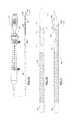
- FIGS. 1A-1Dshow a tissue-sampling needle device 100 .
- the deviceincludes a proximal handle or hub 102 from which an elongate tubular cannula 104 extends distally.
- the cannula 104includes a cannula wall 106 that defines a cannula lumen 108 .
- a distal end 110 of the cannula 104is beveled, including a long side 110 a substantially parallel with the central longitudinal axis of the cannula 104 and extending to its distal-most tip end.
- a short side 110 b of the beveled distal end 110is opposite the long end 110 a .
- a detailed illustration of the distal end 110is shown in the top plan view of FIG. 1B .
- Other embodimentsmay include a double bevel, where one beveled surface is opposite the notch, or single or double bevels that are at least partially transverse relative to the notch.
- a notch 120is disposed proximally adjacent to the beveled distal cannula end 110 and is generally centered in longitudinal alignment with the long beveled end side 110 a and opposite the short beveled end side 110 b .
- the notch 120is generally arcuate, defined on its proximal side by a parabolic edge 122 extending along generally longitudinal, but somewhat curved lateral notch sides 124 .
- the distal edge 124 of the notch 120preferably is formed as generally parabolic lip that joins the proximal edge 122 at a pair of lip end portions 126 that preferably provide a curved transition between the proximal lateral and distal edges 122 , 124 .
- the radiused lip end portions 126preferably are configured to provide stress relief within the cannula structure.
- a central distal lip portion 125 of the distal edge 124preferably forms a proximal-facing cutting edge.
- the notchwill occupy about one-half the circumference of the cannula 104 at the broadest point of the notch. Inclusion of the bevel illustrated in FIG. 1C may provide advantages for successful sample collection.
- contact of the bevel face against tissuemay create a slight bias/pressure toward the notch that will help tissue to be pulled/captured into the notch when the stylet is withdrawn, including that contact pressure on the notch side of the shaft surface may be slightly greater than on the exterior surface immediately opposite the notch.
- An elongate stylet 130may be disposed removably through the cannula lumen 108 .
- the stylet 130will occupy substantially an entire cross-sectional area of at least a lengthwise portion of the cannula lumen 108 .
- a distal end 132 of the stylet 130will be beveled to align with the distal beveled cannula end 110 in a substantially coplanar manner.
- This constructionwill provide enhanced support for the cannula (particularly during navigation to a target site) including providing a generally solid cutting and/or tissue-penetrating distal tip end formed by the matching bevels of the stylet 130 and cannula 104 .
- the distal beveled needle endmay be closed, such that the lumen 108 extending longitudinally through the cannula terminates within the cannula 104 .
- a styletmay be reinserted into the needle lumen after the sample has been excised and captured through the notch into the needle lumen.
- the styletmay be extended distally to cover the open notch (thereby preventing contamination of the sample by inadvertent collection of cells along the needle track during withdrawal of the needle), but leaving room in a closed needle lumen portion for the sample to remain intact between the notch and a closed distal end in an embodiment where the needle lumen is closed at the distal end.
- the cannula 104may be constructed as a 19-gauge needle made of 304 stainless steel, with an inner diameter of about 0.9 mm (about 0.037 inches).
- the notch 120may be circumferentially located opposite and proximal of a distal bevel that is at about a 30° angle relative to the short side such that a proximal-most end of the notch 120 (defined by the proximal edge 122 ) is about 9 mm (about 0.36 inches) longitudinally proximal of the distal-most tip end of the cannula 104 .
- the longitudinal distance between the proximal-most notch edge 122 and the proximal-most portion of the distal lip 125will be about 4 mm (about 0.16 inches).
- the proximal-most portion of the distal lip 125will be about 0.6 mm (about 0.025 inches) from the distal-most end of the notch 120 , which will be defined by a curved lip end portion 126 , including a radius of curvature of about 0.05 mm (0.002 inches), joining the distal edge 124 with the proximal edge 122 .
- the longitudinal linear distance between the distal-most lip end portions 126 and the proximal end of the bevel 110 opposite the notch 120may be about 1.47 mm (0.058 inches) in 19-gauge, 20-gauge, 21-gauge, or 22-gauge embodiments to provide optimal strength and notch position relative to the needle's distal end.
- the 19-gauge needlemay be used with a sheath measuring about 4.2 to 5.2 Fr.
- a beveled NiTi stylet 130may be disposed slidably/removably through the cannula lumen.
- FIGS. 2A, 2B and 3show additional embodiments of a tissue-sampling needle device 200 .
- the deviceincludes a proximal handle or hub 202 from which an elongate tubular cannula 204 extends distally.
- the cannula 204includes a cannula wall that defines a cannula lumen.
- a distal end 210 of the cannula 204is beveled, including a long side 210 a substantially parallel with the central longitudinal axis of the cannula 204 and extending to its distal-most tip end.
- a short side 210 b of the beveled distal end 210is opposite the long end 210 a .
- a detail view of the needle device 200is shown in a top plan view in FIG. 2B .
- the distal end 210may be open to the lumen or may be closed. In embodiments with an open end 210 , a sample may be ejected out the distal end after collection.
- a notch 220is disposed proximally adjacent to the beveled distal cannula end 210 and is generally centered in longitudinal alignment with the long beveled end side 210 a and opposite the short beveled end side 210 b .
- the notch 220is generally arcuate, defined on its proximal side by a parabolic edge 222 extending along generally longitudinal, but somewhat curved lateral notch sides 224 .
- the distal edge 224 of the notch 220preferably is formed as generally parabolic lip that joins the proximal edge 222 at a pair of lip end portions 226 that preferably provide a curved transition between the proximal and distal edges 222 , 224 .
- a central distal lip portion 225 of the distal edge 224preferably forms a proximal-facing cutting edge.
- the notchwill occupy about one-half the circumference of the cannula 204 at the broadest point of the notch.
- the cannula 204includes surface features 240 configured to enhance echogenicity, thereby providing an improved ability to navigate the device during an EUS procedure.
- the surface features 240are shown here as dimples on an exterior surface of the cannula 204 , but may alternatively be embodied as grooves or other regular or irregular features on an external or internal surface. Embedded echogenic features such as bubbles, voids, or pieces of echo-contrasting materials may also be used within the scope of the present invention.
- Embedded echogenic featuressuch as bubbles, voids, or pieces of echo-contrasting materials may also be used within the scope of the present invention.
- Those of skill in the artwill appreciate that many currently-known and/or future-developed echogenicity-enhancing means may be used within the scope of the present invention.
- the terms echogenic and echogenicity-enhancingare used to refer to structural features that increase the reflectivity of ultrasound waves used during ultrasound visualization of a device, with the increase being over the typical ultrasound reflectivity/visualizability of a device lacking
- FIG. 3is similar to FIG. 2A , but shows that the echogenic features 240 may extend distally across the space occupied by the notch 220 . It is preferable that echogenicity-enhancing features be disposed at a specified predetermined distance from the distal-most tip end of the cannula 204 . Although the echogenic features 240 are shown at a distance from the notch 220 , a cannula according to the present embodiments may be constructed with those echogenic features disposed flush up to the margins of the notch. A stylet 230 , which may include echogenicity-enhancing features may be disposed through the cannula lumen of the embodiments of FIGS. 2A-3 .
- FIGS. 4A-4CA method of tissue collection is described with reference to FIGS. 4A-4C , using the needle device 200 of FIGS. 2A-2B .
- the needle cannula 204with the stylet 230 disposed therein, is directed into a target site 450 to be sampled (e.g., a tumor mass).
- a target site 450e.g., a tumor mass
- the stylet 230is withdrawn and suction is applied to the proximal end of the needle cannula lumen 208 . This will pull tissue from the target site 450 through the notch 220 into the lumen 208 .
- FIG. 4Athe needle cannula 204 , with the stylet 230 disposed therein, is directed into a target site 450 to be sampled (e.g., a tumor mass).
- FIG. 4Bthe stylet 230 is withdrawn and suction is applied to the proximal end of the needle cannula lumen 208 . This
- the userwill then quickly retract the cannula 204 proximally such that the proximal-facing cutting edge of the distal notch's central lip 225 cuts a sample 452 of tissue from the target site that is drawn into the lumen 208 and that may be captured within, distal, or proximal of the notch 220 .
- the cannula 204may be advanced and retracted slightly (e.g., about half a centimeter, two or three times) and/or rotated or otherwise manipulated by the user if desired to try to capture sample material.
- the cannula 204may be bowed slightly during use to accentuate contact of the notch 220 with adjacent tissue to promote improved sample collection.
- the sample obtainedpreferably will include a desirable number of intact cells, preferably more intact cells than are ordinarily obtained using a non-notched FNA biopsy needle (“more” indicating both a greater number and a higher degree of cell integrity within the sample obtained). It has been found that histological-grade FNB samples may be obtained in this manner, which may be preferred for certain diagnostic purposes over the cytological-grade samples typically obtained through FNA. The needle may then be withdrawn from the patient's body.
- the cannula 204during introduction of the device into a patient body, the cannula 204 will be directed through the working channel of a peroral endoscope such as a duodenoscope into a patient's body. It is then navigated (under ultrasound visualization if echogenicity-enhancing features are provided, as in the embodiment shown in FIGS. 2A-3 ) into the target site 450 .
- the device 200may be introduced through other access means known in the art including percutaneous means such as direct insertion of the needle cannula through a patient's skin or insertion through a trocar, sheath, or other access device (with or without endoscopic or ultrasound visualization), all within the scope of the present invention.
- an outer sheathmay be disposed slidably along the exterior of the cannula 204 and advanced over the notch 220 after the sample is excised by the cutting edge. This configuration, which may be practiced within the scope of the present invention, may lessen the likelihood that the sample collected will become lost or contaminated during needle withdrawal.
- the needle device and methods disclosed hereprovide the advantages associated with FNA needles of small size and maneuverability, while offering a means of collecting more intact samples from target sites. They also are not hampered by the guillotine-style moving parts of other notched needle systems known in the biopsy art (which are generally larger in scale due to a need for having a cutting member that movably transects the notch).
Landscapes
- Health & Medical Sciences (AREA)
- Life Sciences & Earth Sciences (AREA)
- Surgery (AREA)
- General Health & Medical Sciences (AREA)
- Molecular Biology (AREA)
- Veterinary Medicine (AREA)
- Pathology (AREA)
- Public Health (AREA)
- Engineering & Computer Science (AREA)
- Biomedical Technology (AREA)
- Heart & Thoracic Surgery (AREA)
- Medical Informatics (AREA)
- Animal Behavior & Ethology (AREA)
- Radiology & Medical Imaging (AREA)
- Nuclear Medicine, Radiotherapy & Molecular Imaging (AREA)
- Physics & Mathematics (AREA)
- Biophysics (AREA)
- Optics & Photonics (AREA)
- Hematology (AREA)
- Dentistry (AREA)
- Oral & Maxillofacial Surgery (AREA)
- Ultra Sonic Daignosis Equipment (AREA)
Abstract
Description
Claims (14)
Priority Applications (4)
| Application Number | Priority Date | Filing Date | Title |
|---|---|---|---|
| US29/388,901USD657461S1 (en) | 2011-04-04 | 2011-04-04 | Biopsy needle tip |
| US13/079,226US9980707B2 (en) | 2011-04-04 | 2011-04-04 | Endoscopic ultrasound-guided biopsy needle |
| US29/442,054USD690009S1 (en) | 2011-04-04 | 2011-12-15 | Biopsy needle tip |
| US14/703,146US9986981B2 (en) | 2010-04-06 | 2015-05-04 | Endoscopic ultrasound-guided notched biopsy needle |
Applications Claiming Priority (1)
| Application Number | Priority Date | Filing Date | Title |
|---|---|---|---|
| US13/079,226US9980707B2 (en) | 2011-04-04 | 2011-04-04 | Endoscopic ultrasound-guided biopsy needle |
Related Child Applications (2)
| Application Number | Title | Priority Date | Filing Date |
|---|---|---|---|
| US29/388,901ContinuationUSD657461S1 (en) | 2011-04-04 | 2011-04-04 | Biopsy needle tip |
| US14/703,146ContinuationUS9986981B2 (en) | 2010-04-06 | 2015-05-04 | Endoscopic ultrasound-guided notched biopsy needle |
Publications (2)
| Publication Number | Publication Date |
|---|---|
| US20120253228A1 US20120253228A1 (en) | 2012-10-04 |
| US9980707B2true US9980707B2 (en) | 2018-05-29 |
Family
ID=45922562
Family Applications (4)
| Application Number | Title | Priority Date | Filing Date |
|---|---|---|---|
| US13/079,226Active2034-06-17US9980707B2 (en) | 2010-04-06 | 2011-04-04 | Endoscopic ultrasound-guided biopsy needle |
| US29/388,901ActiveUSD657461S1 (en) | 2011-04-04 | 2011-04-04 | Biopsy needle tip |
| US29/442,054ActiveUSD690009S1 (en) | 2011-04-04 | 2011-12-15 | Biopsy needle tip |
| US14/703,146ActiveUS9986981B2 (en) | 2010-04-06 | 2015-05-04 | Endoscopic ultrasound-guided notched biopsy needle |
Family Applications After (3)
| Application Number | Title | Priority Date | Filing Date |
|---|---|---|---|
| US29/388,901ActiveUSD657461S1 (en) | 2011-04-04 | 2011-04-04 | Biopsy needle tip |
| US29/442,054ActiveUSD690009S1 (en) | 2011-04-04 | 2011-12-15 | Biopsy needle tip |
| US14/703,146ActiveUS9986981B2 (en) | 2010-04-06 | 2015-05-04 | Endoscopic ultrasound-guided notched biopsy needle |
Country Status (1)
| Country | Link |
|---|---|
| US (4) | US9980707B2 (en) |
Families Citing this family (84)
| Publication number | Priority date | Publication date | Assignee | Title |
|---|---|---|---|---|
| US9375203B2 (en) | 2002-03-25 | 2016-06-28 | Kieran Murphy Llc | Biopsy needle |
| US9186128B2 (en) | 2008-10-01 | 2015-11-17 | Covidien Lp | Needle biopsy device |
| US8968210B2 (en) | 2008-10-01 | 2015-03-03 | Covidien LLP | Device for needle biopsy with integrated needle protection |
| US9782565B2 (en) | 2008-10-01 | 2017-10-10 | Covidien Lp | Endoscopic ultrasound-guided biliary access system |
| US11298113B2 (en) | 2008-10-01 | 2022-04-12 | Covidien Lp | Device for needle biopsy with integrated needle protection |
| US9332973B2 (en) | 2008-10-01 | 2016-05-10 | Covidien Lp | Needle biopsy device with exchangeable needle and integrated needle protection |
| US9980707B2 (en)* | 2011-04-04 | 2018-05-29 | Cook Medical Technologies Llc | Endoscopic ultrasound-guided biopsy needle |
| US20120245486A1 (en)* | 2011-03-25 | 2012-09-27 | Anthony Melchiorri | Ghost-core biopsy needle |
| US9044573B2 (en) | 2011-08-11 | 2015-06-02 | Phase One Medical, Llc | Method and apparatus for the dialysis of blood |
| USD725776S1 (en)* | 2011-10-07 | 2015-03-31 | Geon-Mok Lee | Acupotomy needle |
| USD736916S1 (en)* | 2012-01-09 | 2015-08-18 | Angiodynamics, Inc. | Dialysis catheter tip |
| US20130190609A1 (en)* | 2012-01-25 | 2013-07-25 | Cook Medical Technologies Llc | Echogenic medical device |
| US9445837B2 (en) | 2012-03-16 | 2016-09-20 | Nfinium Vascular Technologies Llc | Surgical needle with enhanced ultrasound reflectivity |
| EP2838435B1 (en)* | 2012-04-16 | 2020-03-25 | Hathaway, Jeff M. | Biopsy device |
| USD689605S1 (en)* | 2012-08-06 | 2013-09-10 | B. Braun Medical, Inc. | Piercing pin |
| IL221634A0 (en) | 2012-08-26 | 2012-12-31 | Medimop Medical Projects Ltd | Universal drug vial adapter |
| WO2014058667A1 (en) | 2012-10-10 | 2014-04-17 | Cook Medical Technologies Llc | Rotary sample-collection needle |
| AU2014226558B2 (en)* | 2013-03-05 | 2016-06-16 | Cook Medical Technologies Llc | Endoscopic biopsy needle with coil sheath |
| JP6263254B2 (en) | 2013-03-14 | 2018-01-17 | マフィン・インコーポレイテッドMuffin Incorporated | Echogenic surface using Lulaw's triangle |
| US9622727B2 (en)* | 2013-03-14 | 2017-04-18 | The Cleveland Clinic Foundation | Tissue sampling device |
| IL225734A0 (en) | 2013-04-14 | 2013-09-30 | Medimop Medical Projects Ltd | Ready-to-use drug vial assemblages including drug vial and drug vial closure having fluid transfer member, and drug vial closure therefor |
| CN105228676B (en) | 2013-05-10 | 2018-01-05 | 麦迪麦珀医疗工程有限公司 | Include the medical treatment device of the vial adapter with inline dry kit |
| DE212014000169U1 (en) | 2013-08-07 | 2016-03-14 | Medimop Medical Projects Ltd. | Fluid transfer devices for use with infusion fluid containers |
| US9795503B2 (en) | 2013-10-18 | 2017-10-24 | Rodolfo Alfredo PEREZ GROSSMANN | Method and apparatus for trabeculectomy and suprachoroidal shunt surgery |
| CN103583429A (en)* | 2013-11-07 | 2014-02-19 | 北京市水产科学研究所 | Fish gonadal tissue taking device fast to manufacture |
| US9149260B2 (en) | 2014-02-28 | 2015-10-06 | 3DBiopsy LLC | Biopsy needle assembly |
| USD794183S1 (en)* | 2014-03-19 | 2017-08-08 | Medimop Medical Projects Ltd. | Dual ended liquid transfer spike |
| KR20150116689A (en)* | 2014-04-08 | 2015-10-16 | 주식회사 파인메딕스 | Needle for EUS-FNA |
| US20150342580A1 (en) | 2014-05-30 | 2015-12-03 | Cook Medical Technologies Llc | Laser cut needle cannula with increased flexibility |
| JP5953441B2 (en)* | 2014-06-10 | 2016-07-20 | オリンパス株式会社 | Biopsy system |
| US10159470B2 (en) | 2014-07-30 | 2018-12-25 | Covidien Lp | Exchangeable core biopsy needle |
| US20160030014A1 (en)* | 2014-07-30 | 2016-02-04 | Covidien Lp | Exchangeable core biopsy needle |
| US10182798B2 (en) | 2014-07-30 | 2019-01-22 | Covidien Lp | Exchangeable core biopsy needle |
| JP5908198B1 (en)* | 2014-08-20 | 2016-04-26 | オリンパス株式会社 | Needle tube |
| US9980699B2 (en) | 2014-09-12 | 2018-05-29 | Cook Medical Technologies Llc | Shaped echogenic needle groove |
| CA2964551A1 (en)* | 2014-10-16 | 2016-04-21 | Kieran Murphy, Llc | Biopsy needle |
| JP6358724B2 (en) | 2015-01-05 | 2018-07-18 | ウエスト・ファーマ.サービシーズ・イスラエル,リミテッド | Dual vial adapter assembly with easy removable pill adapter to ensure accurate use |
| US9844362B2 (en) | 2015-01-13 | 2017-12-19 | Covidien Lp | Exchangeable core biopsy needle |
| WO2016148882A1 (en)* | 2015-03-17 | 2016-09-22 | SPIRATION, INC., d/b/a OLYMPUS RESPIRATORY AMERICA | Improved needle scribe at tip |
| US12186129B2 (en)* | 2015-03-31 | 2025-01-07 | Boston Scientific Scimed, Inc. | Devices and methods for ultrasound imaging |
| WO2017009822A1 (en) | 2015-07-16 | 2017-01-19 | Medimop Medical Projects Ltd | Liquid drug transfer devices for secure telescopic snap fit on injection vials |
| WO2017019954A1 (en) | 2015-07-30 | 2017-02-02 | Cook Medical Technologies Llc | Liquid stylet apparatus |
| US20170055968A1 (en) | 2015-08-31 | 2017-03-02 | Cook Medical Technologies Llc | Needle and stylet management device with rotary handle |
| USD801522S1 (en) | 2015-11-09 | 2017-10-31 | Medimop Medical Projects Ltd. | Fluid transfer assembly |
| CN115721558A (en) | 2015-11-25 | 2023-03-03 | 西部制药服务以色列有限公司 | Dual vial adapter assembly comprising a drug vial adapter having a self-sealing inlet valve |
| USD810932S1 (en)* | 2016-03-11 | 2018-02-20 | Douglas B. Coffman | Curved needle with two ports |
| WO2017172469A1 (en) | 2016-04-01 | 2017-10-05 | Cook Medical Technologies Llc | Needle surface for reduced coagulation and method for same |
| JP6811441B2 (en)* | 2016-04-27 | 2021-01-13 | 株式会社ナノ・グレインズ | Tissue collection tool |
| IL245800A0 (en) | 2016-05-24 | 2016-08-31 | West Pharma Services Il Ltd | Dual vial adapter assemblages including identical twin vial adapters |
| IL245803A0 (en) | 2016-05-24 | 2016-08-31 | West Pharma Services Il Ltd | Dual vial adapter assemblages including vented drug vial adapter and vented liquid vial adapter |
| IL246073A0 (en) | 2016-06-06 | 2016-08-31 | West Pharma Services Il Ltd | Fluid transfer devices for use with drug pump cartridge having slidable driving plunger |
| IL247376A0 (en) | 2016-08-21 | 2016-12-29 | Medimop Medical Projects Ltd | Syringe assembly |
| CN206434352U (en)* | 2016-09-14 | 2017-08-25 | 深圳市前海康启源科技有限公司 | The biopsy needle control system of auxiliary positioning |
| CN206586974U (en)* | 2016-09-14 | 2017-10-27 | 深圳市前海康启源科技有限公司 | Biopsy needle scanning control system |
| CN106137264A (en)* | 2016-09-14 | 2016-11-23 | 深圳市前海康启源科技有限公司 | There is the biopsy needle control system of double light path image scanning function |
| USD832430S1 (en) | 2016-11-15 | 2018-10-30 | West Pharma. Services IL, Ltd. | Dual vial adapter assemblage |
| IL249408A0 (en) | 2016-12-06 | 2017-03-30 | Medimop Medical Projects Ltd | Liquid transfer device for use with infusion liquid container and pincers-like hand tool for use therewith for releasing intact drug vial therefrom |
| IL251458A0 (en) | 2017-03-29 | 2017-06-29 | Medimop Medical Projects Ltd | User actuated liquid drug transfer devices for use in ready-to-use (rtu) liquid drug transfer assemblages |
| US10561407B2 (en) | 2017-05-05 | 2020-02-18 | Hoya Corporation | Apparatuses and methods for endoscopic tool joints |
| US11116483B2 (en) | 2017-05-19 | 2021-09-14 | Merit Medical Systems, Inc. | Rotating biopsy needle |
| US11844500B2 (en) | 2017-05-19 | 2023-12-19 | Merit Medical Systems, Inc. | Semi-automatic biopsy needle device and methods of use |
| US11793498B2 (en) | 2017-05-19 | 2023-10-24 | Merit Medical Systems, Inc. | Biopsy needle devices and methods of use |
| IL254802A0 (en) | 2017-09-29 | 2017-12-31 | Medimop Medical Projects Ltd | Dual vial adapter assemblages with twin vented female vial adapters |
| US20190247638A1 (en) | 2018-02-13 | 2019-08-15 | Kieran P. Murphy | Delivery system for delivering a drug depot to a target site under image guidance and methods and uses of same |
| USD905853S1 (en) | 2018-02-27 | 2020-12-22 | Medical Components, Inc. | Catheter tip |
| USD885572S1 (en)* | 2018-03-12 | 2020-05-26 | Olympus Corporation | Biopsy needle |
| USD1023297S1 (en)* | 2018-06-04 | 2024-04-16 | Airlift Concrete Experts, LLC | Subterranean injection rod tip |
| JP1630477S (en) | 2018-07-06 | 2019-05-07 | ||
| WO2020018765A1 (en)* | 2018-07-19 | 2020-01-23 | Boston Scientific Scimed, Inc. | Medical devices and related methods |
| USD923812S1 (en) | 2019-01-16 | 2021-06-29 | West Pharma. Services IL, Ltd. | Medication mixing apparatus |
| JP1648075S (en) | 2019-01-17 | 2019-12-16 | ||
| JP7209849B2 (en) | 2019-01-18 | 2023-01-20 | ウェスト・ファーマ・サービシーズ・アイエル・リミテッド | Liquid transfer device for use with IV bottles |
| US11918542B2 (en) | 2019-01-31 | 2024-03-05 | West Pharma. Services IL, Ltd. | Liquid transfer device |
| CN109758190A (en)* | 2019-03-11 | 2019-05-17 | 南京市第一医院 | An improved biopsy needle |
| CN109938779A (en)* | 2019-03-20 | 2019-06-28 | 南阳市中心医院 | A kind of Internal Medicine-Oncology puncture sampler |
| JP7284289B2 (en) | 2019-04-09 | 2023-05-30 | ウェスト ファーマ サービシーズ イスラエル リミテッド | Infusion device with integrated syringe |
| KR20240122586A (en) | 2019-04-30 | 2024-08-12 | 웨스트 파마. 서비시즈 일, 리미티드 | Liquid transfer device with dual lumen iv spike |
| US12295556B2 (en) | 2019-09-27 | 2025-05-13 | Merit Medical Systems, Inc. | Rotation biopsy system and handle |
| US12150627B2 (en) | 2019-12-11 | 2024-11-26 | Merit Medical Systems, Inc. | Bone biopsy device and related methods |
| WO2021117649A1 (en)* | 2019-12-13 | 2021-06-17 | 富士フイルム株式会社 | Biopsy needle and tissue collection device |
| USD933205S1 (en)* | 2020-02-28 | 2021-10-12 | Jeisys Medical Inc. | Needle array |
| USD956958S1 (en) | 2020-07-13 | 2022-07-05 | West Pharma. Services IL, Ltd. | Liquid transfer device |
| US12070243B2 (en) | 2020-09-25 | 2024-08-27 | Industrial Technology Research Institute | Medical material needle |
| USD984880S1 (en) | 2020-11-06 | 2023-05-02 | Medical Components, Inc. | Clamp with indicator |
Citations (31)
| Publication number | Priority date | Publication date | Assignee | Title |
|---|---|---|---|---|
| US4055167A (en) | 1976-04-23 | 1977-10-25 | Bernstein Dell L | Curettement device |
| US4627444A (en) | 1985-06-20 | 1986-12-09 | Regents Of The University Of Minnesota | Device for sampling tissues and fluids from bodily cavities |
| US4702260A (en) | 1985-04-16 | 1987-10-27 | Ko Pen Wang | Flexible bronchoscopic needle assembly |
| US4791937A (en) | 1986-08-19 | 1988-12-20 | Ko Pen Wang | Transendoscopic needle |
| US4900300A (en) | 1987-07-06 | 1990-02-13 | Lee David A | Surgical instrument |
| US4903709A (en) | 1988-09-21 | 1990-02-27 | Skinner Bruce A J | Biopsy method |
| US4989614A (en) | 1988-02-23 | 1991-02-05 | Vance Products Incorporated | Fine-needle aspiration cell sampling methods |
| US4991592A (en) | 1989-12-04 | 1991-02-12 | Christ Howard N | Device for obtaining tissue sample in performing a biopsy |
| US5090419A (en)* | 1990-08-23 | 1992-02-25 | Aubrey Palestrant | Apparatus for acquiring soft tissue biopsy specimens |
| US5106364A (en) | 1989-07-07 | 1992-04-21 | Kabushiki Kaisha Topcon | Surgical cutter |
| US5199441A (en) | 1991-08-20 | 1993-04-06 | Hogle Hugh H | Fine needle aspiration biopsy apparatus and method |
| US5320110A (en)* | 1991-10-29 | 1994-06-14 | Wang Ko P | Pleural biopsy syringe-needles |
| US5394887A (en)* | 1994-01-14 | 1995-03-07 | Haaga; John R. | Biopsy needle |
| US5449001A (en) | 1994-04-14 | 1995-09-12 | Terwilliger; Richard A. | Biopsy needle |
| US5458112A (en) | 1994-08-15 | 1995-10-17 | Arrow Precision Products, Inc. | Biliary biopsy device |
| US5470308A (en) | 1992-08-12 | 1995-11-28 | Vidamed, Inc. | Medical probe with biopsy stylet |
| US5807304A (en) | 1995-03-09 | 1998-09-15 | Cockburn; John F. | Medical needle for use in ultrasound imaging |
| US5817033A (en) | 1994-04-11 | 1998-10-06 | Desantis; Stephen A. | Needle core biopsy device |
| US5830153A (en)* | 1994-04-26 | 1998-11-03 | Kass; Erik S. | Controlled surgical core biopsy system |
| US5865765A (en) | 1995-10-16 | 1999-02-02 | Mohajer; Reza S. | Dilator/sampler for sampling materials and fluid from a body cavity |
| CN2449657Y (en) | 2000-11-30 | 2001-09-26 | 陈著声 | Pathological tissue aspiration biopsy needle |
| US20030083684A1 (en)* | 2001-10-26 | 2003-05-01 | Cesarini Peter M. | Reciprocating rotary arthroscopic surgical instrument |
| WO2003079907A1 (en) | 2002-03-20 | 2003-10-02 | Board Of Regents, The University Of Texas System | Biopsy needle |
| US20030236471A1 (en)* | 2001-08-09 | 2003-12-25 | Fisher John S. | Dual action aspiration biopsy needle |
| US20040097887A1 (en)* | 2001-05-18 | 2004-05-20 | Us Endoscopy Group, Inc. | Duodenoscope needle |
| US20050101879A1 (en)* | 2003-11-06 | 2005-05-12 | Shidham Vinod B. | Needle aspiration biopsy device and method |
| US20060189891A1 (en)* | 2004-12-15 | 2006-08-24 | Irving Waxman | Flexible elongate surgical needle device having a tissue engaging section being of greater flexibility than an intermediate section, and methods of using the device |
| US20070265647A1 (en)* | 2006-05-09 | 2007-11-15 | Possis Medical, Inc. | Atherectomy system having a variably exposed cutter |
| US20080091196A1 (en)* | 2006-10-17 | 2008-04-17 | Wilson-Cook Medical Inc. | Wire-guided aspiration needle |
| US20090118641A1 (en) | 2007-11-02 | 2009-05-07 | Jacques Van Dam | Devices, Methods, and Kits for a Biopsy Device |
| US20110098596A1 (en)* | 2008-04-18 | 2011-04-28 | Ali Ozkan Ozturk | Practical and Safe Needle Biopsy Device |
Family Cites Families (49)
| Publication number | Priority date | Publication date | Assignee | Title |
|---|---|---|---|---|
| US737293A (en)* | 1900-11-01 | 1903-08-25 | George H Summerfeldt | Veterinary surgical instrument. |
| US3277893A (en)* | 1964-01-31 | 1966-10-11 | Becton Dickinson Co | Hypodermic projectile with barb in the cannula bevel |
| US3308822A (en)* | 1964-04-02 | 1967-03-14 | Loretta Fontano | Hypodermic needle |
| USD247975S (en)* | 1976-07-30 | 1978-05-23 | Luther Ronald B | Catheterization needle |
| US4249541A (en)* | 1979-04-26 | 1981-02-10 | David S. Pratt | Biopsy device |
| US4651753A (en)* | 1984-10-12 | 1987-03-24 | Jayco Pharmaceuticals | Endoscopic multiple biopsy instrument |
| USD300060S (en)* | 1985-02-25 | 1989-02-28 | William Cook Europe A/S | Biopsy cannula |
| USD300246S (en)* | 1986-06-26 | 1989-03-14 | Brown Larry E | Fluid transfer needle |
| USD303290S (en)* | 1986-07-29 | 1989-09-05 | Becton, Dickinson And Company | Biopsy needle or the like |
| USD302589S (en)* | 1986-07-29 | 1989-08-01 | Becton, Dickinson And Company | Spinal needle or the like |
| US5036860A (en)* | 1989-11-24 | 1991-08-06 | Medical Device Technologies, Inc. | Disposable soft tissue biopsy apparatus |
| US5172702A (en)* | 1989-11-24 | 1992-12-22 | Medical Device Technologies, Inc. | Disposable spring-loaded soft tissue biopsy apparatus |
| US5309910A (en)* | 1992-09-25 | 1994-05-10 | Ep Technologies, Inc. | Cardiac mapping and ablation systems |
| US5487392A (en)* | 1993-11-15 | 1996-01-30 | Haaga; John R. | Biopxy system with hemostatic insert |
| US5649547A (en)* | 1994-03-24 | 1997-07-22 | Biopsys Medical, Inc. | Methods and devices for automated biopsy and collection of soft tissue |
| US5968022A (en)* | 1995-04-28 | 1999-10-19 | Saito; Yoshikuni | Medical hollow needle and method of production |
| CN1226180A (en)* | 1996-06-10 | 1999-08-18 | 伊兰股份有限公司 | Needle for subcutaneous delivery of fluids |
| US6296624B1 (en)* | 1997-02-04 | 2001-10-02 | Allen Gerber | Body access system |
| US7189206B2 (en)* | 2003-02-24 | 2007-03-13 | Senorx, Inc. | Biopsy device with inner cutter |
| JP2001149374A (en)* | 1999-11-29 | 2001-06-05 | Asahi Optical Co Ltd | Endoscope tissue collection tool |
| DK176336B1 (en)* | 1999-12-22 | 2007-08-20 | Asahi Optical Co Ltd | Endoscopic tissue collection instrument |
| WO2001049352A2 (en)* | 2000-01-03 | 2001-07-12 | Johns Hopkins University | Device and method for manual retinal vein catheterization |
| US6585664B2 (en)* | 2000-08-02 | 2003-07-01 | Ethicon Endo-Surgery, Inc. | Calibration method for an automated surgical biopsy device |
| US6478775B1 (en)* | 2000-10-02 | 2002-11-12 | Genyx Medical Inc. | Device for delivering non-biodegradable bulking composition to a urological site |
| USD448047S1 (en)* | 2001-01-11 | 2001-09-18 | Avery Dennison Corporation | Needle pen |
| US8109885B2 (en)* | 2002-03-19 | 2012-02-07 | C. R. Bard, Inc. | Biopsy device for removing tissue specimens using a vacuum |
| GB0208627D0 (en)* | 2002-04-16 | 2002-05-22 | Imprint Pharm Ltd | Needle |
| WO2003088824A2 (en)* | 2002-04-19 | 2003-10-30 | Pelikan Technologies, Inc. | Device and method for variable speed lancet |
| US20080161720A1 (en)* | 2002-10-07 | 2008-07-03 | Nicoson Zachary R | Registration system |
| US20050070818A1 (en)* | 2003-09-30 | 2005-03-31 | Mueller Richard L. | Biopsy device with viewing assembly |
| US7988642B2 (en)* | 2003-10-14 | 2011-08-02 | Suros Surgical Systems, Inc. | Vacuum assisted biopsy device |
| JP4500315B2 (en)* | 2003-10-14 | 2010-07-14 | シュロス・サージカル・システムズ・インコーポレーテッド | Vacuum assisted biopsy needle set |
| USD538933S1 (en)* | 2004-02-23 | 2007-03-20 | Agustin Andrade | Combined thyroid core biopsy needle and biopsy cannula |
| WO2006007410A2 (en)* | 2004-06-16 | 2006-01-19 | Medtronic, Inc. | Minimally invasive coring vein harvester |
| US20060064031A1 (en)* | 2004-09-17 | 2006-03-23 | Miller Stuart H | Biopsy needle |
| US7651482B2 (en)* | 2005-02-04 | 2010-01-26 | Boston Scientific Scimed, Inc. | Non-coring needles and methods of manufacturing same |
| US20070016101A1 (en)* | 2005-07-13 | 2007-01-18 | Feldman Dennis D | Core Biopsy Device |
| JP4991723B2 (en)* | 2005-08-10 | 2012-08-01 | シー・アール・バード・インコーポレーテッド | Single insertion multiple sampling biopsy device with integrated marker |
| US7942830B2 (en)* | 2006-05-09 | 2011-05-17 | Vertos Medical, Inc. | Ipsilateral approach to minimally invasive ligament decompression procedure |
| EP3417792B1 (en)* | 2006-08-21 | 2022-03-02 | C. R. Bard, Inc. | Self-contained handheld biopsy needle |
| US20080097347A1 (en)* | 2006-09-22 | 2008-04-24 | Babak Arvanaghi | Bendable needle assembly |
| US7914463B2 (en)* | 2006-10-23 | 2011-03-29 | Clipius Technologies, Inc. | Double core biopsy instrumentation kit |
| WO2008157376A1 (en)* | 2007-06-13 | 2008-12-24 | Epimed International Inc. | Safety neural injection system and related methods |
| EP2224856A4 (en)* | 2007-10-25 | 2017-05-24 | Epitome Pharmaceuticals Limited | Tissue splitting biopsy needle |
| US8052616B2 (en)* | 2007-11-20 | 2011-11-08 | Devicor Medical Products, Inc. | Biopsy device with fine pitch drive train |
| US20110190660A1 (en) | 2008-05-08 | 2011-08-04 | Mayo Foundation For Medical Education And Research | Biopsy Devices |
| US7846109B2 (en)* | 2008-12-18 | 2010-12-07 | Devicor Medical Products, Inc. | Biopsy device with sliding cutter cover |
| US9980707B2 (en)* | 2011-04-04 | 2018-05-29 | Cook Medical Technologies Llc | Endoscopic ultrasound-guided biopsy needle |
| USD678511S1 (en)* | 2010-09-22 | 2013-03-19 | Control Medical Technology, Llc | Rapid exchange aspiration catheter |
- 2011
- 2011-04-04USUS13/079,226patent/US9980707B2/enactiveActive
- 2011-04-04USUS29/388,901patent/USD657461S1/enactiveActive
- 2011-12-15USUS29/442,054patent/USD690009S1/enactiveActive
- 2015
- 2015-05-04USUS14/703,146patent/US9986981B2/enactiveActive
Patent Citations (34)
| Publication number | Priority date | Publication date | Assignee | Title |
|---|---|---|---|---|
| US4055167A (en) | 1976-04-23 | 1977-10-25 | Bernstein Dell L | Curettement device |
| US4702260A (en) | 1985-04-16 | 1987-10-27 | Ko Pen Wang | Flexible bronchoscopic needle assembly |
| US4627444A (en) | 1985-06-20 | 1986-12-09 | Regents Of The University Of Minnesota | Device for sampling tissues and fluids from bodily cavities |
| US4791937A (en) | 1986-08-19 | 1988-12-20 | Ko Pen Wang | Transendoscopic needle |
| US4900300A (en) | 1987-07-06 | 1990-02-13 | Lee David A | Surgical instrument |
| US4989614A (en) | 1988-02-23 | 1991-02-05 | Vance Products Incorporated | Fine-needle aspiration cell sampling methods |
| US4903709A (en) | 1988-09-21 | 1990-02-27 | Skinner Bruce A J | Biopsy method |
| US5106364A (en) | 1989-07-07 | 1992-04-21 | Kabushiki Kaisha Topcon | Surgical cutter |
| US4991592A (en) | 1989-12-04 | 1991-02-12 | Christ Howard N | Device for obtaining tissue sample in performing a biopsy |
| US5090419A (en)* | 1990-08-23 | 1992-02-25 | Aubrey Palestrant | Apparatus for acquiring soft tissue biopsy specimens |
| US5199441A (en) | 1991-08-20 | 1993-04-06 | Hogle Hugh H | Fine needle aspiration biopsy apparatus and method |
| US5320110A (en)* | 1991-10-29 | 1994-06-14 | Wang Ko P | Pleural biopsy syringe-needles |
| US5470308A (en) | 1992-08-12 | 1995-11-28 | Vidamed, Inc. | Medical probe with biopsy stylet |
| US5394887A (en)* | 1994-01-14 | 1995-03-07 | Haaga; John R. | Biopsy needle |
| US5971939A (en) | 1994-04-11 | 1999-10-26 | Laurus Medical Corporation | Needle core biopsy device |
| US5817033A (en) | 1994-04-11 | 1998-10-06 | Desantis; Stephen A. | Needle core biopsy device |
| US5449001A (en) | 1994-04-14 | 1995-09-12 | Terwilliger; Richard A. | Biopsy needle |
| US5830153A (en)* | 1994-04-26 | 1998-11-03 | Kass; Erik S. | Controlled surgical core biopsy system |
| US5458112A (en) | 1994-08-15 | 1995-10-17 | Arrow Precision Products, Inc. | Biliary biopsy device |
| US5807304A (en) | 1995-03-09 | 1998-09-15 | Cockburn; John F. | Medical needle for use in ultrasound imaging |
| US5865765A (en) | 1995-10-16 | 1999-02-02 | Mohajer; Reza S. | Dilator/sampler for sampling materials and fluid from a body cavity |
| CN2449657Y (en) | 2000-11-30 | 2001-09-26 | 陈著声 | Pathological tissue aspiration biopsy needle |
| US20040097887A1 (en)* | 2001-05-18 | 2004-05-20 | Us Endoscopy Group, Inc. | Duodenoscope needle |
| US20050090765A1 (en) | 2001-08-09 | 2005-04-28 | Biopsy Sciences, Llc | Dual Action Aspiration Biopsy Needle |
| US20030236471A1 (en)* | 2001-08-09 | 2003-12-25 | Fisher John S. | Dual action aspiration biopsy needle |
| US6709408B2 (en) | 2001-08-09 | 2004-03-23 | Biopsy Sciences, Llc | Dual action aspiration biopsy needle |
| US20030083684A1 (en)* | 2001-10-26 | 2003-05-01 | Cesarini Peter M. | Reciprocating rotary arthroscopic surgical instrument |
| WO2003079907A1 (en) | 2002-03-20 | 2003-10-02 | Board Of Regents, The University Of Texas System | Biopsy needle |
| US20050101879A1 (en)* | 2003-11-06 | 2005-05-12 | Shidham Vinod B. | Needle aspiration biopsy device and method |
| US20060189891A1 (en)* | 2004-12-15 | 2006-08-24 | Irving Waxman | Flexible elongate surgical needle device having a tissue engaging section being of greater flexibility than an intermediate section, and methods of using the device |
| US20070265647A1 (en)* | 2006-05-09 | 2007-11-15 | Possis Medical, Inc. | Atherectomy system having a variably exposed cutter |
| US20080091196A1 (en)* | 2006-10-17 | 2008-04-17 | Wilson-Cook Medical Inc. | Wire-guided aspiration needle |
| US20090118641A1 (en) | 2007-11-02 | 2009-05-07 | Jacques Van Dam | Devices, Methods, and Kits for a Biopsy Device |
| US20110098596A1 (en)* | 2008-04-18 | 2011-04-28 | Ali Ozkan Ozturk | Practical and Safe Needle Biopsy Device |
Non-Patent Citations (18)
| Title |
|---|
| Chhieng, David C. et al., "Fine-Needle Aspiration Cytology of Hodgkin Disease," Cancer Cytopathology, 2001, American Cancer Society, pp. 52-59. |
| Davenport, R.D., "Rapid on-site evaluation of transbronchial aspirates," Chest, 1990, vol. 98, pp. 59-61. |
| Diette, Gregory B., "Utility of On-Site Cytopathology Assessment for Bronchoscopic Evaluation of Lung Masses and Adenopathy," Chest, 2000, vol. 117, pp. 1186-1190. |
| Gittlen, S.D., "A new versatile transbronchial cytology needle for the staging and diagnosis of bronchogenic carcinoma," Chest, 1988, vol. 94, pp. 561-565. |
| Kaffes, Arthur J., "Fine Needle Aspiration At Endoscopic Ultrasound With a Novel Olympus Side-Port Needle: A Pilot Experience," Gastrointestinal Endoscopy, Abstract T1492, 2010, vol. 71, No. 5, p. 291. |
| Mayall, Frederick et al., "Improved FNA cytology results with a near patient diagnosis service for non-breast lesions," J. Clin. Pathol., 1998, vol. 51, pp. 541-544. |
| Mazzone MD, Peter et al., "Bronchoscopy and Needle Biopsy Techniques for Diagnosis and Staging of Lung Cancer," Clinics in Chest Medicine, vol. 23, No. 1, Mar. 2002, pp. 137-158. |
| McLoud MD, Theresa C., "Should Cutting Needles Replace Needle Aspiration of Lung Lesions?", Radiology, Jun. 1998, pp. 569-570. |
| Olympus EndoTherpay, SmoothShot, Expanded Line of Transbronchial Aspiration Needles Provides Comprehensive Scope Compatibility, Exceptional Puncture Performance, and Improved Operability, date unknown, 3 pages. |
| Olympus KeyMed, Diagnosis (Needle Aspiration), keymed.co.uk/index.cfm/page/.../615, 2010, 2 pages. |
| PCT International Preliminary Report on Patentability and Written Opinion of the International Searching Authority for International PCT No. PCT/US2011/031048, dated Oct. 18, 2012. |
| Shure, D., "Transbronchial biopsy and needle aspiration," Chest, 1989, vol. 95, pp. 1130-1138. |
| Trumm, C.G. et al., "Biopsy," Ch. 9, date unknown, pp. 94-95. |
| Wang, K.P. et al., "Needle brush in the diagnosis of lung mass or nodule through flexible bronchoscopy," Chest, 1991, vol. 100, pp. 1148-1150. |
| Wang, K.P., "Flexible transbronchial needle aspiration biopsy for histologic specimens," Chest, 1985, vol. 88, pp. 860-863. |
| Wang, Ko Pen, "Biopsy Sampling Technique," Chest, 1989, vol. 95, pp. 484-485. |
| Weisbrod, Gordon L. et al., "Preliminary Experience with a Dual Cutting Edge Needle in Thoracic Percutaneous Fine-Needle Aspiration Biopsy," Radiology, Apr. 1987, pp. 75-78. |
| Yang, Grace, C.H. et al., "Ultrasound-Guided Fine-Needle Aspiration of the Thyroid Assessed by Ultrafast Papanicolaou Stain: Data from 1135 Biopsies with a Two to Six Year Follow-Up," Thyroid, vol. 11, No. 6, 2001, pp. 581-589. |
Also Published As
| Publication number | Publication date |
|---|---|
| USD657461S1 (en) | 2012-04-10 |
| US20150230780A1 (en) | 2015-08-20 |
| US20120253228A1 (en) | 2012-10-04 |
| USD690009S1 (en) | 2013-09-17 |
| US9986981B2 (en) | 2018-06-05 |
Similar Documents
| Publication | Publication Date | Title |
|---|---|---|
| US9980707B2 (en) | Endoscopic ultrasound-guided biopsy needle | |
| CA2792548C (en) | Endoscopic ultrasound-guided biopsy needle | |
| EP3148445B1 (en) | Laser cut needle cannula with increased flexibility | |
| US9782153B2 (en) | Endoscopic biopsy needle with coil sheath | |
| US12185925B2 (en) | Endoscopic tri-point biopsy needle | |
| US10792022B2 (en) | Tissue sampling devices, systems and methods | |
| US20100317996A1 (en) | Ultrasound-navigable barbed biopsy device and method | |
| JP2019503745A (en) | System and method for improving tissue sampling | |
| IES85915Y1 (en) | Endoscopic ultrasound-guided biopsy needle | |
| IE20110141U1 (en) | Endoscopic ultrasound-guided biopsy needle | |
| WO2025074374A1 (en) | A needle for harvesting a tissue during biopsy |
Legal Events
| Date | Code | Title | Description |
|---|---|---|---|
| AS | Assignment | Owner name:COOK MEDICAL TECHNOLOGIES LLC, INDIANA Free format text:ASSIGNMENT OF ASSIGNORS INTEREST;ASSIGNOR:WILSON-COOK MEDICAL INC.;REEL/FRAME:029044/0072 Effective date:20120906 Owner name:COOK MEDICAL TECHNOLOGIES LLC, INDIANA Free format text:ASSIGNMENT OF ASSIGNORS INTEREST;ASSIGNOR:COOK IRELAND LIMITED;REEL/FRAME:029043/0783 Effective date:20120907 | |
| AS | Assignment | Owner name:WILSON-COOK MEDICAL INC., NORTH CAROLINA Free format text:ASSIGNMENT OF ASSIGNORS INTEREST;ASSIGNORS:SCHEMBRE, DREW B.;CHMURA, KEVIN;SIGNING DATES FROM 20110203 TO 20110204;REEL/FRAME:035882/0320 Owner name:COOK MEDICAL TECHNOLOGIES LLC, INDIANA Free format text:ASSIGNMENT OF ASSIGNORS INTEREST;ASSIGNOR:WILSON-COOK MEDICAL INC.;REEL/FRAME:035884/0313 Effective date:20110203 Owner name:COOK MEDICAL TECHNOLOGIES LLC, INDIANA Free format text:ASSIGNMENT OF ASSIGNORS INTEREST;ASSIGNOR:COOK IRELAND LIMITED;REEL/FRAME:035887/0222 Effective date:20110217 Owner name:COOK IRELAND LIMITED, IRELAND Free format text:ASSIGNMENT OF ASSIGNORS INTEREST;ASSIGNOR:CLANCY, MICHAEL S.;REEL/FRAME:035980/0692 Effective date:20110126 | |
| FEPP | Fee payment procedure | Free format text:PETITION RELATED TO MAINTENANCE FEES GRANTED (ORIGINAL EVENT CODE: PTGR) | |
| STCF | Information on status: patent grant | Free format text:PATENTED CASE | |
| MAFP | Maintenance fee payment | Free format text:PAYMENT OF MAINTENANCE FEE, 4TH YEAR, LARGE ENTITY (ORIGINAL EVENT CODE: M1551); ENTITY STATUS OF PATENT OWNER: LARGE ENTITY Year of fee payment:4 | |
| AS | Assignment | Owner name:WILMINGTON TRUST, NATIONAL ASSOCIATION, AS COLLATERAL AGENT, DELAWARE Free format text:SECURITY INTEREST;ASSIGNOR:COOK MEDICAL TECHNOLOGIES LLC;REEL/FRAME:066700/0277 Effective date:20240227 |