US9968446B2 - Tubular scaffold for fabrication of heart valves - Google Patents
Tubular scaffold for fabrication of heart valvesDownload PDFInfo
- Publication number
- US9968446B2 US9968446B2US14/205,820US201414205820AUS9968446B2US 9968446 B2US9968446 B2US 9968446B2US 201414205820 AUS201414205820 AUS 201414205820AUS 9968446 B2US9968446 B2US 9968446B2
- Authority
- US
- United States
- Prior art keywords
- tubular
- scaffold
- mesh
- leaflets
- tubular scaffold
- Prior art date
- Legal status (The legal status is an assumption and is not a legal conclusion. Google has not performed a legal analysis and makes no representation as to the accuracy of the status listed.)
- Active
Links
Images


Classifications
- A—HUMAN NECESSITIES
- A61—MEDICAL OR VETERINARY SCIENCE; HYGIENE
- A61F—FILTERS IMPLANTABLE INTO BLOOD VESSELS; PROSTHESES; DEVICES PROVIDING PATENCY TO, OR PREVENTING COLLAPSING OF, TUBULAR STRUCTURES OF THE BODY, e.g. STENTS; ORTHOPAEDIC, NURSING OR CONTRACEPTIVE DEVICES; FOMENTATION; TREATMENT OR PROTECTION OF EYES OR EARS; BANDAGES, DRESSINGS OR ABSORBENT PADS; FIRST-AID KITS
- A61F2/00—Filters implantable into blood vessels; Prostheses, i.e. artificial substitutes or replacements for parts of the body; Appliances for connecting them with the body; Devices providing patency to, or preventing collapsing of, tubular structures of the body, e.g. stents
- A61F2/02—Prostheses implantable into the body
- A61F2/24—Heart valves ; Vascular valves, e.g. venous valves; Heart implants, e.g. passive devices for improving the function of the native valve or the heart muscle; Transmyocardial revascularisation [TMR] devices; Valves implantable in the body
- A61F2/2412—Heart valves ; Vascular valves, e.g. venous valves; Heart implants, e.g. passive devices for improving the function of the native valve or the heart muscle; Transmyocardial revascularisation [TMR] devices; Valves implantable in the body with soft flexible valve members, e.g. tissue valves shaped like natural valves
- A61F2/2415—Manufacturing methods
- A—HUMAN NECESSITIES
- A61—MEDICAL OR VETERINARY SCIENCE; HYGIENE
- A61L—METHODS OR APPARATUS FOR STERILISING MATERIALS OR OBJECTS IN GENERAL; DISINFECTION, STERILISATION OR DEODORISATION OF AIR; CHEMICAL ASPECTS OF BANDAGES, DRESSINGS, ABSORBENT PADS OR SURGICAL ARTICLES; MATERIALS FOR BANDAGES, DRESSINGS, ABSORBENT PADS OR SURGICAL ARTICLES
- A61L27/00—Materials for grafts or prostheses or for coating grafts or prostheses
- A61L27/50—Materials characterised by their function or physical properties, e.g. injectable or lubricating compositions, shape-memory materials, surface modified materials
- A61L27/56—Porous materials, e.g. foams or sponges
- C—CHEMISTRY; METALLURGY
- C12—BIOCHEMISTRY; BEER; SPIRITS; WINE; VINEGAR; MICROBIOLOGY; ENZYMOLOGY; MUTATION OR GENETIC ENGINEERING
- C12M—APPARATUS FOR ENZYMOLOGY OR MICROBIOLOGY; APPARATUS FOR CULTURING MICROORGANISMS FOR PRODUCING BIOMASS, FOR GROWING CELLS OR FOR OBTAINING FERMENTATION OR METABOLIC PRODUCTS, i.e. BIOREACTORS OR FERMENTERS
- C12M23/00—Constructional details, e.g. recesses, hinges
- C12M23/02—Form or structure of the vessel
- C—CHEMISTRY; METALLURGY
- C12—BIOCHEMISTRY; BEER; SPIRITS; WINE; VINEGAR; MICROBIOLOGY; ENZYMOLOGY; MUTATION OR GENETIC ENGINEERING
- C12M—APPARATUS FOR ENZYMOLOGY OR MICROBIOLOGY; APPARATUS FOR CULTURING MICROORGANISMS FOR PRODUCING BIOMASS, FOR GROWING CELLS OR FOR OBTAINING FERMENTATION OR METABOLIC PRODUCTS, i.e. BIOREACTORS OR FERMENTERS
- C12M23/00—Constructional details, e.g. recesses, hinges
- C12M23/20—Material Coatings
- C—CHEMISTRY; METALLURGY
- C12—BIOCHEMISTRY; BEER; SPIRITS; WINE; VINEGAR; MICROBIOLOGY; ENZYMOLOGY; MUTATION OR GENETIC ENGINEERING
- C12M—APPARATUS FOR ENZYMOLOGY OR MICROBIOLOGY; APPARATUS FOR CULTURING MICROORGANISMS FOR PRODUCING BIOMASS, FOR GROWING CELLS OR FOR OBTAINING FERMENTATION OR METABOLIC PRODUCTS, i.e. BIOREACTORS OR FERMENTERS
- C12M25/00—Means for supporting, enclosing or fixing the microorganisms, e.g. immunocoatings
- C12M25/14—Scaffolds; Matrices
- A—HUMAN NECESSITIES
- A61—MEDICAL OR VETERINARY SCIENCE; HYGIENE
- A61L—METHODS OR APPARATUS FOR STERILISING MATERIALS OR OBJECTS IN GENERAL; DISINFECTION, STERILISATION OR DEODORISATION OF AIR; CHEMICAL ASPECTS OF BANDAGES, DRESSINGS, ABSORBENT PADS OR SURGICAL ARTICLES; MATERIALS FOR BANDAGES, DRESSINGS, ABSORBENT PADS OR SURGICAL ARTICLES
- A61L2430/00—Materials or treatment for tissue regeneration
- A61L2430/20—Materials or treatment for tissue regeneration for reconstruction of the heart, e.g. heart valves
Definitions
- the present inventionrelates to a heart valves and, more particularly to a tubular mesh or braided scaffold for use in fabrication of heart valves.
- Valvular heart diseaseis the next cardiac epidemic. While replacement of a dysfunctional valve markedly reduces the mortality associated with it, the current options are limited to either mechanical or bio-prosthetic heart valves. Mechanical heart valves tend to last longer due to their stronger composition, but the lifelong need for anticoagulant medication is their major drawbacks. In contrast, bio-prosthetic heart valves, including transcatheters, do not require anticoagulant medications but they do not last long, and are calcified rapidly due to the several production procedures such as cross-linking that they go through.
- the present inventionrelates to a tubular scaffold for use in fabrication of heart valves.
- the tubular scaffoldcomprises a tubular mesh material shaped into a valvular structure having at least two leaflets.
- the mesh materialis braided, such that the tubular mesh material is a tubular braided scaffold.
- tubular scaffoldis shaped into a tri-leaflet valvular structure.
- tubular scaffoldis shaped into a hi-leaflet valvular structure.
- tubular scaffoldis made of polymeric materials.
- the tubular scaffoldis made of metallic materials.
- the tubular scaffoldincludes a leaflet portion and a tubular portion, with the leaflets formed on the leaflet portion, and wherein a shape of each leaflet corresponds to a portion of a surface of a cone which portion is defined by the intersections on the conical surface of at least two flat planes having peripheries on the conical surface corresponding in length respectively to the circumference of the base with the leaflets each having a peripheral five portion while on the other side are attached to a tubular portion.
- tubular mesh material shaped into a valvular structureis fitted onto a frame of an artificial heart valve.
- the scaffoldis connected to a frame having a base and at least two upstanding posts.
- the present inventionalso comprises a method for forming and using the invention described herein.
- FIG. 1is an illustration of a tubular scaffold that has been formed to a shape of a tri-leaflet heart valve
- FIG. 2Ais a right, side-view illustration of the tubular scaffold according to the principles of the present invention.
- FIG. 2Bis a bottom-view illustration of the tubular scaffold according to the principles of the present invention.
- FIG. 2Cis a rear, side-view illustration of the tubular scaffold according to the principles of the present invention.
- FIG. 2Dis a top-view illustration of the tubular scaffold according to the principles of the present invention.
- FIG. 3is an illustration depicting an example of forming the tubular scaffold from a braided mesh tube
- FIG. 4is an illustration depicting a process of forming the tubular scaffold using a scaffolding mold having a scaffolding mold first part and a scaffolding mold second part according to the principles of the present invention
- FIG. 5is an illustration of a tubular scaffold according to the principles of the present invention.
- FIG. 6is an illustration depicting a tubular scaffold as being fitted onto a frame according to the principles of the present invention.
- the present inventionrelates to a heart valves and, more particularly, to a tubular braided scaffold for use in fabrication of heart valves.
- the following descriptionis presented to enable one of ordinary skill, in the art to make and use the invention and to incorporate it in the context of particular applications.
- Various modifications, as well as a variety of uses in afferent applicationswill be readily apparent to those skilled in the art, and the general principles defined herein may be applied to a wide range of embodiments.
- the present inventionis not intended to be limited to the embodiments presented, but is to be accorded the widest scope consistent with the principles and novel features disclosed herein.
- any element in a claim that does not explicitly state “means for” performing a specified function, or “step for” performing a specific function,is not to be interpreted as a “means” or “step” clause as specified in 35 U.S.C. Section 112, Paragraph 6.
- the use of “step of” or “act of” in the claims hereinis not intended to invoke the provisions of 35 U.S.C. 112, Paragraph 6.
- the present inventionrelates to a scaffold for formation of heart valves. More specifically, the present invention relates to a valvular scaffold made of as tube that has a braided (or mesh) pattern that is shaped into a (1) tri-leaflet or (2) bi-leaflet valvular structure, where the scaffold can be made from a variety materials such as but not limited to woven polymer or metallic fibers such as Nitinol.
- the formed valvecan be later connected to a structure such as but not limited to a tri or bi-leaflet artificial heart valve frame to become ready for implantation.
- the current inventioncan be used as a mechanical valve. Upon proper selection of the scaffold material, it can become elastic or super elastic to provide its leaflets with enough flexibility suitable for heart valve applications. This flexibility will help the valve to improve blood flow dynamics and therefore reduce the formation of blood clots due to damaging red blood cells and finally eliminate the use of anticoagulation therapy needed for patients having mechanical heart valves.
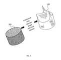
- FIG. 1shows a tubular scaffold 100 that has been formed to a shape of a tri-leaflet heart valve.
- FIGS. 2A through 2Dillustrate right, bottom, rear, and top views, respectively, of the tubular scaffold 100 .
- the scaffold 100is a continuous mesh without any parts sewn or attached to the valve. The whole structure has integrity so that the leaflets are not separate parts from the body of the tube.
- the tubular scaffold 100can be formed using any suitable mechanism or technique.
- a framesuch as a bioprosthetic heart valve frame
- a braided mesh tube 300can be positioned (by pinching the leaflets into shape around the base mold) and heat treated to firm a heart valve shape (i.e., the tubular scaffold 100 ).
- the braided mesh scaffold 100is formed of a shape memory material, such as Nitinol, then it has memory and will hold the shape as depicted in the figures.
- a scaffolding moldcan be included that includes a scaffolding mold first part 402 and a scaffolding mold second part 404 .
- the scaffolding mold second pan 404is formed in the shape of a desired heart valve, a non-limiting example of which includes a tri-leaflet heart valve.
- the scaffolding mold second part 404is formed to be a negative of the shape of the scaffolding, mold first part 402 .
- a braided mesh tube 300(e.g., made of Nitinol or any other mesh or braided material) can be positioned over the scaffolding mold first part 402 .
- the scaffolding mold first 402 and second 404 partsare designed in a such a way that if you put the parts 402 and 404 together, there will be a uniform gap between their surfaces so that there will be enough space for the mesh 300 to be placed in between. Depending on the thickness of the mesh, the gap dimension can be adjusted.
- the scaffolding mold second part 404can be positioned over the metal braided mesh tube 300 such that the metal mesh tube 300 is pressed between the scaffolding mold first pan 402 and scaffolding mold second part 404 (i.e., the two parts 404 and 406 are compressed together).
- the braided mesh tube 300upon removal from the scaffolding mold, is pressed into the shape of a desired scaffold 100 .
- the tubular scaffold 100can be formed in any desired shape having leaflets, non-limiting examples of which include having a bi-leaflet shape and a tri-leaflet shape.
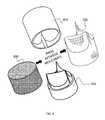
- the tubular scaffold 100includes a leaflet portion 500 and a tubular portion 502 , with the leaflets 504 formed on the leaflet portion 500 .
- a shape of each leaflet 504corresponds to a portion of a surface of a cone which portion is defined by the intersections on the conical surface of at least two flat planes having peripheries on the conical surface corresponding in length respectively to the circumference of the base with the leaflets 504 each having a peripheral free portion 506 while on the other side are attached to the tubular portion 502 .
- the tubular scaffold 100can be attached fitted onto a frame 600 of an artificial heart valve.
- a ring base 602 and two to three posts 604would be inserted into the tubular scaffold 100 so that the scaffold 100 sits on-top and covers the valve.
- some part of the tubewill be integrated into the frame 600 so that the leaflets 504 are continuously connected to each other and to the base 602 and upstanding posts 604 of the frame 600 .
- the tubular scaffoldAfter the tubular scaffold is formed into the desired valve shape, it can be used as is or further treated, as desired.
- the tubular scaffoldcan be further treated by growing tissue thereon using a mold as described in U.S. application Ser. No. 14/094,760, filed on Dec. 2, 2013, entitled, “Apparatus for Growing a Heart Valve in Three-Dimensions,” which is incorporated by reference as though fully set forth herein.
- the formed valvecan be implanted with or without surface modification.
- the valvecan become biologically active and recruit the cells inside the body to form a tissue around itself.
- a method of forming a biological active valve with a mesh scaffoldis described in U.S. patent application Ser. No. 13/427,807, filed on Mar. 22, 2012, and entitled. “Mesh Enclosed Tissue Constructs,” which is incorporated by reference as though fully set forth herein; however this is not the only method that can be used to form a hybrid biologically active mesh valve.
- the mesh patternwill help the tissue to have structural integrity from both sides of the mesh.
- the tissuecan be even made before implantation as discussed above.
- the tissuecan be grown in and around the structure in a way to enclose the mesh.
Landscapes
- Health & Medical Sciences (AREA)
- Engineering & Computer Science (AREA)
- Life Sciences & Earth Sciences (AREA)
- Chemical & Material Sciences (AREA)
- Zoology (AREA)
- Organic Chemistry (AREA)
- Bioinformatics & Cheminformatics (AREA)
- Wood Science & Technology (AREA)
- Biomedical Technology (AREA)
- General Health & Medical Sciences (AREA)
- Sustainable Development (AREA)
- Microbiology (AREA)
- Biochemistry (AREA)
- General Engineering & Computer Science (AREA)
- Biotechnology (AREA)
- Genetics & Genomics (AREA)
- Cardiology (AREA)
- Veterinary Medicine (AREA)
- Animal Behavior & Ethology (AREA)
- Public Health (AREA)
- Transplantation (AREA)
- Oral & Maxillofacial Surgery (AREA)
- Clinical Laboratory Science (AREA)
- Immunology (AREA)
- Heart & Thoracic Surgery (AREA)
- Vascular Medicine (AREA)
- Medicinal Chemistry (AREA)
- Epidemiology (AREA)
- Dispersion Chemistry (AREA)
- Dermatology (AREA)
- Manufacturing & Machinery (AREA)
- Prostheses (AREA)
Abstract
Description
This is a Continuation-in-Part application of U.S. application Ser. No. 14/094,760, filed on Dec. 2, 2013, entitled, “Apparatus for Growing a Heart Valve in Three-Dimensions,” which is a non-provisional application of U.S. Provisional Application No. 61/732,669, filed on Dec. 3, 2012, and entitled, “Apparatus for Growing a Heart Valve in Three-Dimensions.”
This is ALSO a Continuation-in-Part application of U.S. patent application Ser. No. 13/427,807, filed on Mar. 22, 2012, and entitled, “Mesh Enclosed Tissue Constructs,” which is a non-provisional application of U.S. Provisional Application No. 61/466,882, entitled “A SELF-REGENERATIVE HYBRID TISSUE STRUCTURE FOR 3D FABRICATION OF HEART VALVES, BLOOD VESSELS AND OTHER CONSTRUCTS,” filed on Mar. 23, 2011; AND U.S. Provisional Application No. 61/496,369, entitled, “HYBRID TISSUE ENGINEERED HEART VALVE,” filed on Jun. 13, 2011; AND U.S. Provisional Application No. 61/540,330, entitled, “Scaffold for Fabrication of Engineered Heart Valves and Other Applications,” filed on Sep. 28, 2011; AND U.S. Provisional Application No. 61/559,694, entitled, “METAL MESH SCAFFOLD FOR TISSUE ENGINEERING OF MEMBRANES,” filed on Jan. 19, 2012.
This is ALSO a non-provisional application of U.S. Provisional Application No. 61/778,294, filed on Mar. 12, 2013, and entitled, “Tubular Braided Scaffold for Fabrication of Heart Valves.”
(1) Field of Invention
The present invention relates to a heart valves and, more particularly to a tubular mesh or braided scaffold for use in fabrication of heart valves.
(2) Description of Related Art
Valvular heart disease is the next cardiac epidemic. While replacement of a dysfunctional valve markedly reduces the mortality associated with it, the current options are limited to either mechanical or bio-prosthetic heart valves. Mechanical heart valves tend to last longer due to their stronger composition, but the lifelong need for anticoagulant medication is their major drawbacks. In contrast, bio-prosthetic heart valves, including transcatheters, do not require anticoagulant medications but they do not last long, and are calcified rapidly due to the several production procedures such as cross-linking that they go through.
Thus, a continuing need exists for a long lasting and stable heart valve that is resistant to calcification and a method for making the same.
The present invention relates to a tubular scaffold for use in fabrication of heart valves. The tubular scaffold comprises a tubular mesh material shaped into a valvular structure having at least two leaflets.
In another aspect, the mesh material is braided, such that the tubular mesh material is a tubular braided scaffold.
In yet another aspect, the tubular scaffold is shaped into a tri-leaflet valvular structure.
In another aspect, the tubular scaffold is shaped into a hi-leaflet valvular structure.
In yet another aspect, the tubular scaffold is made of polymeric materials.
In another aspect, the tubular scaffold is made of metallic materials.
In yet another aspect, the tubular scaffold includes a leaflet portion and a tubular portion, with the leaflets formed on the leaflet portion, and wherein a shape of each leaflet corresponds to a portion of a surface of a cone which portion is defined by the intersections on the conical surface of at least two flat planes having peripheries on the conical surface corresponding in length respectively to the circumference of the base with the leaflets each having a peripheral five portion while on the other side are attached to a tubular portion.
In another aspect, the tubular mesh material shaped into a valvular structure is fitted onto a frame of an artificial heart valve. Further, the scaffold is connected to a frame having a base and at least two upstanding posts.
Finally, as can be appreciated by one in the art, the present invention also comprises a method for forming and using the invention described herein.
The objects, features and advantages of the present invention will be apparent from the following detailed descriptions of the various aspects of the invention in conjunction with reference to the following drawings, where:
The present invention relates to a heart valves and, more particularly, to a tubular braided scaffold for use in fabrication of heart valves. The following description is presented to enable one of ordinary skill, in the art to make and use the invention and to incorporate it in the context of particular applications. Various modifications, as well as a variety of uses in afferent applications will be readily apparent to those skilled in the art, and the general principles defined herein may be applied to a wide range of embodiments. Thus, the present invention is not intended to be limited to the embodiments presented, but is to be accorded the widest scope consistent with the principles and novel features disclosed herein.
In the following detailed description, numerous specific details are set forth in order to provide a more thorough understanding of the present invention. However, it will be apparent to one skilled in the art that the present invention may be practiced without necessarily being limited to these specific details. In other instances, well-known structures and devices are shown in block diagram form, rather than in detail, in order to avoid obscuring the present invention.
The reader's attention is directed to all papers and documents which are filed concurrently with this specification and which are open to public inspection with this specification, and the contents of all such papers and documents are incorporated herein by reference. All the features disclosed in this specification, (including any accompanying claims, abstract, and drawings) may be replaced by alternative features serving the same, equivalent or similar purpose, unless expressly stated otherwise. Thus, unless expressly stated otherwise, each feature disclosed is only one example of a generic series of equivalent or similar features.
Furthermore, any element in a claim that does not explicitly state “means for” performing a specified function, or “step for” performing a specific function, is not to be interpreted as a “means” or “step” clause as specified in 35 U.S.C. Section 112, Paragraph 6. In particular, the use of “step of” or “act of” in the claims herein is not intended to invoke the provisions of 35 U.S.C. 112, Paragraph 6.
Please note, if used, the labels left, right, front, back, top, bottom, forward, reverse, clockwise and counter clockwise have been used for convenience purposes only and are not intended to imply an particular fixed direction. Instead, they are used to reflect relative locations and/or directions between various portions of an object.
(1) Description
The present invention relates to a scaffold for formation of heart valves. More specifically, the present invention relates to a valvular scaffold made of as tube that has a braided (or mesh) pattern that is shaped into a (1) tri-leaflet or (2) bi-leaflet valvular structure, where the scaffold can be made from a variety materials such as but not limited to woven polymer or metallic fibers such as Nitinol. The formed valve can be later connected to a structure such as but not limited to a tri or bi-leaflet artificial heart valve frame to become ready for implantation. The current invention can be used as a mechanical valve. Upon proper selection of the scaffold material, it can become elastic or super elastic to provide its leaflets with enough flexibility suitable for heart valve applications. This flexibility will help the valve to improve blood flow dynamics and therefore reduce the formation of blood clots due to damaging red blood cells and finally eliminate the use of anticoagulation therapy needed for patients having mechanical heart valves.
Thetubular scaffold 100 can be formed using any suitable mechanism or technique. As a non-limiting example, a frame (such as a bioprosthetic heart valve frame) can be used as a base mold. For example and as shown inFIG. 3 , abraided mesh tube 300 can be positioned (by pinching the leaflets into shape around the base mold) and heat treated to firm a heart valve shape (i.e., the tubular scaffold100). If thebraided mesh scaffold 100 is formed of a shape memory material, such as Nitinol, then it has memory and will hold the shape as depicted in the figures.
As another non-limiting example and as shownFIG. 4 , a scaffolding mold can be included that includes a scaffolding moldfirst part 402 and a scaffolding moldsecond part 404. The scaffolding moldsecond pan 404 is formed in the shape of a desired heart valve, a non-limiting example of which includes a tri-leaflet heart valve. Alternatively, the scaffolding moldsecond part 404 is formed to be a negative of the shape of the scaffolding, moldfirst part 402. Thus, when a material is positioned between the scaffolding moldfirst pan 402 and the scaffolding moldsecond part 404 is pressed over and onto the scaffolding moldfirst part 402, the material is pressed or bent into the desired heart valve shape, thereby creating a scaffold for use with the heart mold. As a non-limiting example and as shown inFIG. 4 , a braided mesh tube300 (e.g., made of Nitinol or any other mesh or braided material) can be positioned over the scaffolding moldfirst part 402. The scaffolding mold first402 and second404 parts are designed in a such a way that if you put theparts mesh 300 to be placed in between. Depending on the thickness of the mesh, the gap dimension can be adjusted.
Thus, the scaffolding moldsecond part 404 can be positioned over the metalbraided mesh tube 300 such that themetal mesh tube 300 is pressed between the scaffolding moldfirst pan 402 and scaffolding mold second part404 (i.e., the twoparts 404 and406 are compressed together). In this aspect, upon removal from the scaffolding mold, thebraided mesh tube 300 is pressed into the shape of a desiredscaffold 100.
As noted above, thetubular scaffold 100 can be formed in any desired shape having leaflets, non-limiting examples of which include having a bi-leaflet shape and a tri-leaflet shape. In another aspect and as shown inFIG. 5 , thetubular scaffold 100 includes aleaflet portion 500 and atubular portion 502, with theleaflets 504 formed on theleaflet portion 500. A shape of eachleaflet 504 corresponds to a portion of a surface of a cone which portion is defined by the intersections on the conical surface of at least two flat planes having peripheries on the conical surface corresponding in length respectively to the circumference of the base with theleaflets 504 each having a peripheralfree portion 506 while on the other side are attached to thetubular portion 502.
In other aspect and as shown inFIG. 6 , thetubular scaffold 100 can be attached fitted onto aframe 600 of an artificial heart valve. For example, aring base 602 and two to threeposts 604 would be inserted into thetubular scaffold 100 so that thescaffold 100 sits on-top and covers the valve. In this way, some part of the tube will be integrated into theframe 600 so that theleaflets 504 are continuously connected to each other and to thebase 602 andupstanding posts 604 of theframe 600. This continuous connection will help theleaflets 504 and the valve to have more durability and prevents fatigue on the structure due to improper attachments of leaflets to thebase 602 and upstanding posts604 (as one of the reasons for failure and short term functionality of bioprosthetic heart valves is structural vulnerability and detachment of the leaflets from their base attachment).
After the tubular scaffold is formed into the desired valve shape, it can be used as is or further treated, as desired. As a non-limiting example, the tubular scaffold can be further treated by growing tissue thereon using a mold as described in U.S. application Ser. No. 14/094,760, filed on Dec. 2, 2013, entitled, “Apparatus for Growing a Heart Valve in Three-Dimensions,” which is incorporated by reference as though fully set forth herein.
Alternatively and/or in addition to, the formed valve can be implanted with or without surface modification. By using a proper coating method, the valve can become biologically active and recruit the cells inside the body to form a tissue around itself. A method of forming a biological active valve with a mesh scaffold is described in U.S. patent application Ser. No. 13/427,807, filed on Mar. 22, 2012, and entitled. “Mesh Enclosed Tissue Constructs,” which is incorporated by reference as though fully set forth herein; however this is not the only method that can be used to form a hybrid biologically active mesh valve. The mesh pattern will help the tissue to have structural integrity from both sides of the mesh. The tissue can be even made before implantation as discussed above. By using cell culture techniques, the tissue can be grown in and around the structure in a way to enclose the mesh.
Claims (10)
1. A tubular scaffold for fabrication of a heart valve, consisting essentially of:
a tubular mesh material shaped into a valvular structure having at least two leaflets, wherein the tubular mesh material is formed of a layer of mesh having a first side and a second side, the layer of mesh including a network of holes passing directly and unobstructed through the mesh from the first side to the second side; and
wherein the tubular mesh material is formed of a material such that when pressed into the valvular structure having at least two leaflets and released, the tubular scaffold retains the shape of the valvular structure with the at least two leaflets.
2. The tubular scaffold as set forth inclaim 1 , wherein the mesh material is braided, such that the tubular mesh material is a tubular braided scaffold.
3. The tubular scaffold as set forth inclaim 1 , wherein the tubular scaffold is made of polymeric materials.
4. The tubular scaffold as set forth inclaim 1 , wherein the tubular scaffold is made of metallic materials.
5. The tubular scaffold as set forth inclaim 1 , wherein the tubular scaffold includes a leaflet portion and a tubular portion, with the leaflets formed on the leaflet portion.
6. The tubular scaffold as set forth inclaim 1 , wherein a shape of each leaflet corresponds to a portion of a surface of a cone which portion is defined by the intersections on the conical surface of at least two flat planes having peripheries on the conical surface corresponding in length respectively to the circumference of the base with the leaflets each having a peripheral free portion while on the other side are attached to a tubular portion.
7. The tubular scaffold as set forth inclaim 1 , wherein the tubular scaffold is shaped into a tri-leaflet valvular structure.
8. The tubular scaffold as set forth inclaim 1 , wherein the tubular scaffold is shaped into a bi-leaflet valvular structure.
9. A tubular scaffold for fabrication of a heart valve, consisting essentially of:
a tubular mesh material shaded into a valvular structure having at least two leaflets, wherein the tubular mesh material is formed of a layer of mesh having a first side and a second side, the layer of mesh including a network of holes passing directly and unobstructed through the mesh from the first side to the second side; and
wherein the tubular mesh material is formed of a material such that when pressed into the valvular structure having at least two leaflets and released, the tubular scaffold retains the shape of the valvular structure with the at least two leaflets;
wherein the tubular mesh material shaped into a valvular structure is fitted onto a frame of an artificial heart valve.
10. A tubular scaffold for fabrication of a heart valve, consisting essentially of:
a tubular mesh material shaded into a valvular structure having at least two leaflets, wherein the tubular mesh material is formed of a layer of mesh having a first side and a second side, the layer of mesh including a network of holes passing directly and unobstructed through the mesh from the first side to the second side; and
wherein the tubular mesh material is formed of a material such that when pressed into the valvular structure having at least two leaflets and released, the tubular scaffold retains the shape of the valvular structure with the at least two leaflets;
wherein the tubular mesh material is connected to a frame having a base and at least two upstanding posts, with the at least two leaflets residing between the at least two posts to form the valvular structure with the at least two leaflets.
Priority Applications (1)
| Application Number | Priority Date | Filing Date | Title |
|---|---|---|---|
| US14/205,820US9968446B2 (en) | 2011-03-23 | 2014-03-12 | Tubular scaffold for fabrication of heart valves |
Applications Claiming Priority (9)
| Application Number | Priority Date | Filing Date | Title |
|---|---|---|---|
| US201161466882P | 2011-03-23 | 2011-03-23 | |
| US201161496369P | 2011-06-13 | 2011-06-13 | |
| US201161540330P | 2011-09-28 | 2011-09-28 | |
| US201261559694P | 2012-01-19 | 2012-01-19 | |
| US13/427,807US8936650B2 (en) | 2011-03-23 | 2012-03-22 | Mesh enclosed tissue constructs |
| US201261732669P | 2012-12-03 | 2012-12-03 | |
| US201361778094P | 2013-03-12 | 2013-03-12 | |
| US14/094,760US10016461B2 (en) | 2012-12-03 | 2013-12-02 | Apparatus and process for growing a heart valve in three-dimensions |
| US14/205,820US9968446B2 (en) | 2011-03-23 | 2014-03-12 | Tubular scaffold for fabrication of heart valves |
Related Parent Applications (1)
| Application Number | Title | Priority Date | Filing Date |
|---|---|---|---|
| US14/094,760Continuation-In-PartUS10016461B2 (en) | 2011-03-23 | 2013-12-02 | Apparatus and process for growing a heart valve in three-dimensions |
Publications (2)
| Publication Number | Publication Date |
|---|---|
| US20140336755A1 US20140336755A1 (en) | 2014-11-13 |
| US9968446B2true US9968446B2 (en) | 2018-05-15 |
Family
ID=51865352
Family Applications (1)
| Application Number | Title | Priority Date | Filing Date |
|---|---|---|---|
| US14/205,820ActiveUS9968446B2 (en) | 2011-03-23 | 2014-03-12 | Tubular scaffold for fabrication of heart valves |
Country Status (1)
| Country | Link |
|---|---|
| US (1) | US9968446B2 (en) |
Families Citing this family (3)
| Publication number | Priority date | Publication date | Assignee | Title |
|---|---|---|---|---|
| EP2842517A1 (en)* | 2011-12-29 | 2015-03-04 | Sorin Group Italia S.r.l. | A kit for implanting prosthetic vascular conduits |
| US11299705B2 (en)* | 2016-11-07 | 2022-04-12 | Deka Products Limited Partnership | System and method for creating tissue |
| CN112843334B (en)* | 2021-01-13 | 2022-07-08 | 东华大学 | A kind of three-dimensional printing composite aerogel to construct a simulated gas pipe and its preparation method |
Citations (51)
| Publication number | Priority date | Publication date | Assignee | Title |
|---|---|---|---|---|
| US4191218A (en) | 1975-05-07 | 1980-03-04 | Albany International Corp. | Fabrics for heart valve and vascular prostheses and methods of fabricating same |
| US5147391A (en)* | 1990-04-11 | 1992-09-15 | Carbomedics, Inc. | Bioprosthetic heart valve with semi-permeable commissure posts and deformable leaflets |
| WO1996008213A1 (en) | 1994-09-12 | 1996-03-21 | Advanced Tissue Sciences, Inc. | Three-dimensional human cell cultures on cardiac valve frameworks and their uses |
| WO1996039814A1 (en) | 1995-06-07 | 1996-12-19 | Advanced Tissue Sciences, Inc. | Seeding heart valves |
| WO2000042950A2 (en) | 1999-01-26 | 2000-07-27 | Edwards Lifesciences Corporation | Flexible heart valve |
| US6103255A (en) | 1999-04-16 | 2000-08-15 | Rutgers, The State University | Porous polymer scaffolds for tissue engineering |
| US6139575A (en) | 1999-04-02 | 2000-10-31 | Medtronic, Inc. | Hybrid mechanical heart valve prosthesis |
| US6388043B1 (en)* | 1998-02-23 | 2002-05-14 | Mnemoscience Gmbh | Shape memory polymers |
| US20020094569A1 (en) | 2000-10-12 | 2002-07-18 | Hanry Yu | Multi-layer cell encapsulation for tissue engineering |
| US20020111676A1 (en) | 1998-10-20 | 2002-08-15 | Eugene Bell | Cardiovascular components for transplantation and methods of making thereof |
| US20020143393A1 (en)* | 1993-11-01 | 2002-10-03 | Cox James L. | Replacement atrioventricular heart valve |
| US6461382B1 (en) | 2000-09-22 | 2002-10-08 | Edwards Lifesciences Corporation | Flexible heart valve having moveable commissures |
| WO2003007795A2 (en) | 2001-07-16 | 2003-01-30 | Edwards Lifesciences Corporation | Tissue engineered heart valve |
| WO2003076564A2 (en) | 2001-05-16 | 2003-09-18 | The General Hospital Corporation | Tissue-engineered organs |
| US6666886B1 (en) | 1999-02-16 | 2003-12-23 | Regents Of The University Of Minnesota | Tissue equivalent approach to a tissue-engineered cardiovascular valve |
| WO2004007699A2 (en) | 2002-07-16 | 2004-01-22 | Biogentis Inc. | Method for preparing engineered tissue |
| WO2004018008A1 (en) | 2002-08-01 | 2004-03-04 | Symetis Ag | In-vitro method for the production of a homologous stented tissue-engineered heart valve |
| US20040121459A1 (en) | 2002-12-23 | 2004-06-24 | Industrial Technology Research Institute | Method and carrier for culturing multi-layer tissue in vitro |
| US20050002982A1 (en) | 1997-11-17 | 2005-01-06 | Mooney David J. | Hybrid tissues for tissue engineering |
| US20050069572A1 (en) | 2002-10-09 | 2005-03-31 | Jennifer Elisseeff | Multi-layered polymerizing hydrogels for tissue regeneration |
| WO2005034726A2 (en) | 2003-09-23 | 2005-04-21 | Ortho Therapeutics, Llc | Absorbable implants and methods for their use in hemostasis and in the treatment of osseous defects |
| US20050095711A1 (en) | 2003-11-01 | 2005-05-05 | More Robert B. | Bioreactor for growing engineered tissue |
| US20050123520A1 (en) | 2003-05-05 | 2005-06-09 | Eavey Roland D. | Generation of living tissue in vivo using a mold |
| US20050143810A1 (en) | 2003-10-24 | 2005-06-30 | Martin Dauner | Cardiovascular implant, method and device for its production, and its provision for surgery |
| US20050181016A1 (en) | 2003-07-17 | 2005-08-18 | Toby Freyman | Decullularized extracellular matrix of conditioned body tissues and uses thereof |
| US20050209687A1 (en) | 2002-02-19 | 2005-09-22 | Bioartis, Inc. | Artificial vessel scaffold and artifical organs therefrom |
| EP1693025A1 (en) | 2005-02-17 | 2006-08-23 | Universität Zürich | Method of manufacturing a tissue-engineered prosthesis |
| WO2006099334A2 (en) | 2005-03-11 | 2006-09-21 | Wake Forest University Health Sciences | Production of tissue engineered heart valves |
| US20060271166A1 (en) | 2005-05-27 | 2006-11-30 | Heart Leaflet Technologies, Inc. | Stentless support structure |
| US7166464B2 (en) | 2001-12-11 | 2007-01-23 | Cytograft Tissue Engineering, Inc. | Method of culturing cells to produce a tissue sheet |
| US20070041952A1 (en) | 2005-04-18 | 2007-02-22 | Duke University | Three-dimensional fiber scaffolds for tissue engineering |
| US20080133002A1 (en) | 2006-12-05 | 2008-06-05 | Daniel Gelbart | Inflatable artificial valve |
| US7422603B2 (en) | 2001-12-27 | 2008-09-09 | Arbor Surgical Technologies, Inc. | Bioprosthetic heart valve |
| US20090163612A1 (en) | 1999-05-07 | 2009-06-25 | Cellology Limited | Tissue engineering scaffold |
| EP2085054A1 (en) | 2008-02-01 | 2009-08-05 | Technische Universiteit Eindhoven | Method for manufacturing a tissue-engineered construct |
| US7575759B2 (en) | 2002-01-02 | 2009-08-18 | The Regents Of The University Of Michigan | Tissue engineering scaffolds |
| US20090222085A1 (en) | 2008-02-22 | 2009-09-03 | University Of Iowa Research Foundation | Cellulose Based Heart Valve Prosthesis |
| US20090252795A1 (en) | 2008-04-07 | 2009-10-08 | Smyth Stuart K J | Bioceramic scaffolds for tissue engineering |
| US7635592B2 (en) | 2000-08-21 | 2009-12-22 | Rice University | Tissue engineering scaffolds promoting matrix protein production |
| US20100249922A1 (en) | 2007-09-19 | 2010-09-30 | St Jude Medical Inc. | Fiber-reinforced synthetic sheets for prosthetic heart valve leaflets |
| US7846728B2 (en) | 2006-10-13 | 2010-12-07 | BioStruxs, LLC | Tissue engineering in vivo with vascularized scaffolds |
| US7871435B2 (en) | 2004-01-23 | 2011-01-18 | Edwards Lifesciences Corporation | Anatomically approximate prosthetic mitral heart valve |
| US7914808B2 (en) | 2001-07-16 | 2011-03-29 | Depuy Products, Inc. | Hybrid biologic/synthetic porous extracellular matrix scaffolds |
| US7968026B1 (en) | 2000-09-20 | 2011-06-28 | Osteopore International Ltd. | Three-dimensional bioresorbable scaffolds for tissue engineering applications |
| US8039258B2 (en) | 2004-09-28 | 2011-10-18 | Ethicon, Inc. | Tissue-engineering scaffolds containing self-assembled-peptide hydrogels |
| US20120015331A1 (en) | 2009-03-23 | 2012-01-19 | Simon Wood | Scaffold |
| US8137686B2 (en) | 2004-04-20 | 2012-03-20 | Depuy Mitek, Inc. | Nonwoven tissue scaffold |
| US8163006B2 (en) | 2004-07-28 | 2012-04-24 | Cordis Corporation | Reduced profile AAA device |
| US20120245706A1 (en) | 2011-03-23 | 2012-09-27 | The Regents Of The University Of California | Mesh enclosed tissue constructs |
| WO2012161786A1 (en) | 2011-02-25 | 2012-11-29 | University Of Connecticut | Prosthetic heart valve |
| WO2013142879A1 (en) | 2012-03-23 | 2013-09-26 | Cytograft Tissue Engineering, Inc. | Tissue-engineered heart valve for transcatheter repair |
- 2014
- 2014-03-12USUS14/205,820patent/US9968446B2/enactiveActive
Patent Citations (85)
| Publication number | Priority date | Publication date | Assignee | Title |
|---|---|---|---|---|
| US4191218A (en) | 1975-05-07 | 1980-03-04 | Albany International Corp. | Fabrics for heart valve and vascular prostheses and methods of fabricating same |
| US5147391A (en)* | 1990-04-11 | 1992-09-15 | Carbomedics, Inc. | Bioprosthetic heart valve with semi-permeable commissure posts and deformable leaflets |
| US20020143393A1 (en)* | 1993-11-01 | 2002-10-03 | Cox James L. | Replacement atrioventricular heart valve |
| US6673109B2 (en)* | 1993-11-01 | 2004-01-06 | 3F Therapeutics, Inc. | Replacement atrioventricular heart valve |
| WO1996008213A1 (en) | 1994-09-12 | 1996-03-21 | Advanced Tissue Sciences, Inc. | Three-dimensional human cell cultures on cardiac valve frameworks and their uses |
| EP0781116A1 (en) | 1994-09-12 | 1997-07-02 | Advanced Tissue Sciences, Inc. | Three-dimensional human cell cultures on cardiac valve frameworks and their uses |
| EP0781116A4 (en) | 1994-09-12 | 1999-08-25 | Advanced Tissue Sciences Inc | Three-dimensional human cell cultures on cardiac valve frameworks and their uses |
| WO1996039814A1 (en) | 1995-06-07 | 1996-12-19 | Advanced Tissue Sciences, Inc. | Seeding heart valves |
| EP0847238A1 (en) | 1995-06-07 | 1998-06-17 | Advanced Tissue Sciences, Inc. | Seeding heart valves |
| EP0847238A4 (en) | 1995-06-07 | 2000-04-19 | Advanced Tissue Sciences Inc | Seeding heart valves |
| US20050002982A1 (en) | 1997-11-17 | 2005-01-06 | Mooney David J. | Hybrid tissues for tissue engineering |
| US6388043B1 (en)* | 1998-02-23 | 2002-05-14 | Mnemoscience Gmbh | Shape memory polymers |
| US20020111676A1 (en) | 1998-10-20 | 2002-08-15 | Eugene Bell | Cardiovascular components for transplantation and methods of making thereof |
| WO2000042950A2 (en) | 1999-01-26 | 2000-07-27 | Edwards Lifesciences Corporation | Flexible heart valve |
| US6666886B1 (en) | 1999-02-16 | 2003-12-23 | Regents Of The University Of Minnesota | Tissue equivalent approach to a tissue-engineered cardiovascular valve |
| US6139575A (en) | 1999-04-02 | 2000-10-31 | Medtronic, Inc. | Hybrid mechanical heart valve prosthesis |
| US6337198B1 (en) | 1999-04-16 | 2002-01-08 | Rutgers, The State University | Porous polymer scaffolds for tissue engineering |
| US6103255A (en) | 1999-04-16 | 2000-08-15 | Rutgers, The State University | Porous polymer scaffolds for tissue engineering |
| US20090163612A1 (en) | 1999-05-07 | 2009-06-25 | Cellology Limited | Tissue engineering scaffold |
| US7635592B2 (en) | 2000-08-21 | 2009-12-22 | Rice University | Tissue engineering scaffolds promoting matrix protein production |
| US8071007B1 (en) | 2000-09-20 | 2011-12-06 | Osteopore International Pte. Ltd. | Three-dimensional bioresorbable scaffolds for tissue engineering applications |
| US7968026B1 (en) | 2000-09-20 | 2011-06-28 | Osteopore International Ltd. | Three-dimensional bioresorbable scaffolds for tissue engineering applications |
| US6461382B1 (en) | 2000-09-22 | 2002-10-08 | Edwards Lifesciences Corporation | Flexible heart valve having moveable commissures |
| US20090286278A1 (en) | 2000-10-12 | 2009-11-19 | Agency For Science, Technology And Research | Multi-layer cell encapsulation for tissue engineering |
| US20020094569A1 (en) | 2000-10-12 | 2002-07-18 | Hanry Yu | Multi-layer cell encapsulation for tissue engineering |
| US20050220891A1 (en) | 2000-10-12 | 2005-10-06 | Agency For Science, Technology And Research | Multi-layer cell encapsulation for tissue engineering |
| US7943353B2 (en) | 2000-10-12 | 2011-05-17 | Agency For Science, Technology And Research | Multi-layer cell encapsulation for tissue engineering |
| WO2003076564A2 (en) | 2001-05-16 | 2003-09-18 | The General Hospital Corporation | Tissue-engineered organs |
| EP1406561A2 (en) | 2001-07-16 | 2004-04-14 | Edwards Lifesciences Corporation | Tissue engineered heart valve |
| US7914808B2 (en) | 2001-07-16 | 2011-03-29 | Depuy Products, Inc. | Hybrid biologic/synthetic porous extracellular matrix scaffolds |
| WO2003007795A2 (en) | 2001-07-16 | 2003-01-30 | Edwards Lifesciences Corporation | Tissue engineered heart valve |
| WO2003007795A3 (en) | 2001-07-16 | 2003-04-24 | Edwards Lifesciences Corp | Tissue engineered heart valve |
| US20030027332A1 (en) | 2001-07-16 | 2003-02-06 | Edwards Lifesciences Corporation | Tissue engineered heart valve |
| US7166464B2 (en) | 2001-12-11 | 2007-01-23 | Cytograft Tissue Engineering, Inc. | Method of culturing cells to produce a tissue sheet |
| US7972377B2 (en) | 2001-12-27 | 2011-07-05 | Medtronic, Inc. | Bioprosthetic heart valve |
| US7422603B2 (en) | 2001-12-27 | 2008-09-09 | Arbor Surgical Technologies, Inc. | Bioprosthetic heart valve |
| US7575759B2 (en) | 2002-01-02 | 2009-08-18 | The Regents Of The University Of Michigan | Tissue engineering scaffolds |
| US20050209687A1 (en) | 2002-02-19 | 2005-09-22 | Bioartis, Inc. | Artificial vessel scaffold and artifical organs therefrom |
| US7521231B2 (en) | 2002-07-16 | 2009-04-21 | Organogenesis, Inc. | Method for preparing engineered tissue |
| US20060128010A1 (en) | 2002-07-16 | 2006-06-15 | Lucie Germain | Method for preparing engineered tissue |
| WO2004007699A2 (en) | 2002-07-16 | 2004-01-22 | Biogentis Inc. | Method for preparing engineered tissue |
| WO2004018008A1 (en) | 2002-08-01 | 2004-03-04 | Symetis Ag | In-vitro method for the production of a homologous stented tissue-engineered heart valve |
| US20060246584A1 (en) | 2002-08-01 | 2006-11-02 | Bruno Covelli | In-vitro method for the production of a homologous stented tissue-engineered heart valve |
| US20050069572A1 (en) | 2002-10-09 | 2005-03-31 | Jennifer Elisseeff | Multi-layered polymerizing hydrogels for tissue regeneration |
| US6884621B2 (en) | 2002-12-23 | 2005-04-26 | Industrial Technology Research Institute | Method and carrier for culturing multi-layer tissue in vitro |
| US20040121459A1 (en) | 2002-12-23 | 2004-06-24 | Industrial Technology Research Institute | Method and carrier for culturing multi-layer tissue in vitro |
| US20050123520A1 (en) | 2003-05-05 | 2005-06-09 | Eavey Roland D. | Generation of living tissue in vivo using a mold |
| US20050181016A1 (en) | 2003-07-17 | 2005-08-18 | Toby Freyman | Decullularized extracellular matrix of conditioned body tissues and uses thereof |
| WO2005034726A3 (en) | 2003-09-23 | 2007-05-03 | Ortho Therapeutics Llc | Absorbable implants and methods for their use in hemostasis and in the treatment of osseous defects |
| WO2005034726A2 (en) | 2003-09-23 | 2005-04-21 | Ortho Therapeutics, Llc | Absorbable implants and methods for their use in hemostasis and in the treatment of osseous defects |
| WO2005035726A2 (en) | 2003-10-09 | 2005-04-21 | Elisseeff Jennifer H | Multi-layered polymerizing hydrogels for tissue regeneration |
| US20050143810A1 (en) | 2003-10-24 | 2005-06-30 | Martin Dauner | Cardiovascular implant, method and device for its production, and its provision for surgery |
| US7851200B2 (en) | 2003-11-01 | 2010-12-14 | More Robert B | Bioreactor for growing engineered tissue |
| US20060270028A1 (en) | 2003-11-01 | 2006-11-30 | More Robert B | Bioreactor for growing engineered tissue |
| US20050095711A1 (en) | 2003-11-01 | 2005-05-05 | More Robert B. | Bioreactor for growing engineered tissue |
| US7871435B2 (en) | 2004-01-23 | 2011-01-18 | Edwards Lifesciences Corporation | Anatomically approximate prosthetic mitral heart valve |
| US8137686B2 (en) | 2004-04-20 | 2012-03-20 | Depuy Mitek, Inc. | Nonwoven tissue scaffold |
| US8163006B2 (en) | 2004-07-28 | 2012-04-24 | Cordis Corporation | Reduced profile AAA device |
| US8039258B2 (en) | 2004-09-28 | 2011-10-18 | Ethicon, Inc. | Tissue-engineering scaffolds containing self-assembled-peptide hydrogels |
| US20080145920A1 (en) | 2005-02-17 | 2008-06-19 | Universitaet Zuerich | Method of Manufacturing a Tissue-Engineered Prosthesis |
| EP1693025A1 (en) | 2005-02-17 | 2006-08-23 | Universität Zürich | Method of manufacturing a tissue-engineered prosthesis |
| WO2006087101A1 (en) | 2005-02-17 | 2006-08-24 | Universitaet Zuerich | Method of manufacturing a tissue-engineered prosthesis |
| US20130217128A1 (en) | 2005-02-17 | 2013-08-22 | Technische Universiteit Eindhoven | Method of manufacturing a tissue-engineered prosthesis |
| EP2617389A1 (en) | 2005-02-17 | 2013-07-24 | Universität Zürich | Method of manufacturing a tissue-engineered prosthesis and bioreactor therefor |
| US8399243B2 (en) | 2005-02-17 | 2013-03-19 | Universitaet Zuerich | Method of manufacturing a tissue-engineered prosthesis |
| WO2006099334A2 (en) | 2005-03-11 | 2006-09-21 | Wake Forest University Health Sciences | Production of tissue engineered heart valves |
| US20060253192A1 (en) | 2005-03-11 | 2006-11-09 | Wake Forest University Health Sciences | Production of tissue engineered heart valves |
| WO2006099334A3 (en) | 2005-03-11 | 2007-04-26 | Univ Wake Forest Health Sciences | Production of tissue engineered heart valves |
| US20070041952A1 (en) | 2005-04-18 | 2007-02-22 | Duke University | Three-dimensional fiber scaffolds for tissue engineering |
| US20060271166A1 (en) | 2005-05-27 | 2006-11-30 | Heart Leaflet Technologies, Inc. | Stentless support structure |
| US7846728B2 (en) | 2006-10-13 | 2010-12-07 | BioStruxs, LLC | Tissue engineering in vivo with vascularized scaffolds |
| US20080133002A1 (en) | 2006-12-05 | 2008-06-05 | Daniel Gelbart | Inflatable artificial valve |
| US20100249922A1 (en) | 2007-09-19 | 2010-09-30 | St Jude Medical Inc. | Fiber-reinforced synthetic sheets for prosthetic heart valve leaflets |
| WO2009096780A1 (en) | 2008-02-01 | 2009-08-06 | Technische Universiteit Eindhoven | Method for manufacturing a tissue-engineered construct |
| US20110033885A1 (en) | 2008-02-01 | 2011-02-10 | Jeroen Kortsmit | Method for manufacturing a tissue-engineered construct |
| EP2085054A1 (en) | 2008-02-01 | 2009-08-05 | Technische Universiteit Eindhoven | Method for manufacturing a tissue-engineered construct |
| US8609415B2 (en) | 2008-02-01 | 2013-12-17 | Technische Universiteit Eindhoven | Method for manufacturing a tissue-engineered construct |
| US8017396B2 (en) | 2008-02-22 | 2011-09-13 | Vijay Kumar | Cellulose based heart valve prosthesis |
| US20090222085A1 (en) | 2008-02-22 | 2009-09-03 | University Of Iowa Research Foundation | Cellulose Based Heart Valve Prosthesis |
| US20090252795A1 (en) | 2008-04-07 | 2009-10-08 | Smyth Stuart K J | Bioceramic scaffolds for tissue engineering |
| US20120015331A1 (en) | 2009-03-23 | 2012-01-19 | Simon Wood | Scaffold |
| WO2012161786A1 (en) | 2011-02-25 | 2012-11-29 | University Of Connecticut | Prosthetic heart valve |
| US20120244617A1 (en) | 2011-03-23 | 2012-09-27 | The Regents Of The University Of California | Mesh enclosed tissue constructs |
| US20120245706A1 (en) | 2011-03-23 | 2012-09-27 | The Regents Of The University Of California | Mesh enclosed tissue constructs |
| WO2013142879A1 (en) | 2012-03-23 | 2013-09-26 | Cytograft Tissue Engineering, Inc. | Tissue-engineered heart valve for transcatheter repair |
Non-Patent Citations (110)
| Title |
|---|
| ‘Braid’. Merriam-Webster [online]. 2016, [retrieved on Jun. 20, 2016]. Retrieved from the Internet: <URL: http://www.merriam-webster.com/dictionary/braid>.* |
| Abu-Omar Y, Ratnatunga CP. Prosthetic heart valves. Surgery 26 (Oxford). 2008 Published by Elsevier Ltd., pp. 196-500. |
| Adamczyk MM, Vesely I. Biaxial strain distributions in explanted porcine bioprosthetic valves. J Heart Valve Dis. Sep. 2002;11(5): pp. 688-695. |
| Advisory Action for U.S. Appl. No. 14/094.760, dated Apr. 7, 2016. |
| Aikawa E, Whittaker, P., Farber, M., Mendelson, K., Padera, R.F., Aikawa, M.. and Schoen, F.J. Human semilunar cardiac valve remodeling by activated cells from fetus to adult: Implications for postnatal adaptation. pathology, and tissue engineering. Circulation. 2006;113:1344-1352. |
| Alavi SH, Kheradvar, A. A hybrid self-regenerative tissue approach as a proper alternative for prosthetic heart valves. ASAIO J. 2011;57: p. 88. |
| Alavi SH, Kheradvar, A. A hybrid self-renewal engineered tissue for heart valve leaflets. Society of Heart Valve Disease 6th Biennial Meeting. 2011. |
| Alavi SH, Kheradvar, A. Metal mesh scaffold for tissue engineering of membranes. Tissue Eng Part C. 2011: Tissue Engineering: Part C, vol. 18, No. 4, 2012, a Mary Ann Liebert, Inc., DOI. 10,1089/ten.tec.2011.0531. |
| Appleton AJE, Appleton CTG, Boughner DR, Rogers KA. Vascular smooth muscle cells as a valvular interstitial cell surrogate in heart valve tissue engineering, Tissue Engineering Part A. 2009;15:3889-3897. |
| Apte SS, Paul A, Prakash S, Shum-Tim D. "Current developments in the tissue engineering of autologous heart valves: Moving towards clinical use." Future Cardiology, 7, 2011; pp. 77-97. |
| Bobak D, Frank M, Tenner A. Characterization of c1q receptor expression on human phagocytic cells: Effects of pdbu and fmlp. The Journal of Immunology. 1986;136;4604-4610. |
| Boontheekul T, Mooney DJ. Protein-based signaling systems in tissue engineering. Current Opinion in Biotechnology 2003;14.559-565. |
| Boyan BD, Hummert T.W., Dean. D.D.. and Schwartz. Z. Role of material surfaces in regulating bone and cartilage cell responses. Biomaterials, 1996;17:137. |
| Breuer CK, Mettler BA, Anthony T, Sales VL, Schoen FJ, Mayer JE. Application of tissue-engineering principles toward the development of a semilunar heart valve substitute, Tissue Engineering. 2004;10:1725-1736. |
| Butcher JT, S. Tressel, T. Johnson, D. Turner, G. Sorescu, H. Jo, and R. M. Nerem. . Profiles of valvular and vascular endothelial cells reveal phenotypic differences: Influence of shear stress. Arterioscler. Thromb. Vasc. Biol. 2006;26:69-77. |
| Chen J, Lee A, Zhao J, Wang H, Lui H, McLean DI, Zeng H. Spectroscopic characterization and microscopic imaging of extracted and in situ cutaneous collagen and elastic tissue components under two-photon excitation. Skin Res Technol. 2009;15:418-426. |
| Cox G, Kable E, Jones A. Fraser I, Manconi F, Gorrell MD. 3-dimensional imaging of collagen using second harmonic generation. J Struct Biol. 2003;141:53-62. |
| d'Arcy JL, Prendergast BD, Chambers JB, Ray SG, Bridgewater B. Valvular heart disease: The next cardiac epidemic. Heart. 2011;97:91-93. |
| Deymier-Black AC, Almer JD, Stock SR, Haeffner DR, Dunand DC. Synchrotron x-ray dirffraction study of load partitioning during elastic deformation of bovine dentin. Acta Biomater. 2010;6:2172-2180. |
| Ferrans VJ, Spray TL, Billingham ME, Roberts WC. Structural changes in glutaraldehyde-treated porcine heterografts used as substitute cardiac valves: Transmission and scanning electron microscopic observations in 12 patients. Am J Cardiol. 1978;41:1159-4184. |
| Flanagan TC, Pandit, A. Living artificial heart valve alternatives: A review. Eur Cell Mater. 2003;6:28-45. |
| Fong, P , et al., "The use of polymer based scaffolds in tissue-engineered heart valves." Progress in Pediatric Cardiology, Elsevier, Amsterdam, NL, vol. 21, No. 2, Mar. 1, 2006, pp. 193-199. |
| Gan BK, Kondyurin A, Bilek MMM. Comparison of protein surface attachment on untreated and plasma immersion ion implantation treated polystyrene: Protein islands and carpet. Langmuir. 2007;23:2741-2746. |
| Georgiou E, Theodossiou T, Hovhannisyan V, Politopoulos K, Rapti GS, Yova D. Second and third optical harmonic generation in type i collagen, by nanosecond laser irradiation, over a broad spectral region. Optics Communications. 2000;176:253-260, References Cited p. 78, Principal Investigator/Program Director (Last, first, middle): Kheradvar, Arash. |
| Grande-Allen K, Liao J. The heterogeneous biomechanics and mechanobiology of the mitral valve: Implications for tissue engineering. Current Cardiology Reports. 2011;13:113-120. |
| Hera M, Dizon RF, Glick BS, Lee CS, Kaestner KH, Piston DW, Bindokas VP. Imaging pancreatic betacells in the intact pancreas. Am J Physiol Endocrinol Metab. 2006;290.E1041-1047. |
| Hildebrant J, Fukaya. H, Martin CJ. Stress-strain relations of tissue sheets undergoing uniform twodimensional stretch. J Appl Physiol,. 1969;27:758-762. |
| Hoerstrup SP, Sodian R, Daebritz S, Wang J, Bacha EA, Martin DP, Moran AM, Guleserian KJ, Spading JS, Kaushal S, Vacanti JP, Schoen FJ, Mayer JE, Jr. Functional living trileaflet heart valves grown in vitro. Circulation. 2000;102: III-44-49. |
| Hoffmann G, Lutter, G., Cremer, J. Durability of bioprosthetic cardiac valves. Dtsch Arztebl Int. 2008;105:143-148. |
| Hori J, and T. Ng. Neural progenitor cells lack immunagenicity and resist destruction as allografts. Stem Cells. 2003:21-405-416. |
| Iijima K, Liu HP, Chiang AS, Hearn SA, Konsolaki M, Zhong Y. Dissecting the pathological effects of human abeta40 and abeta42 in Drosophila: A potential model for alzheimer's disease. Proc Natl Acad Sci U S A. 2004;101 6623-6628. |
| Katzer et al., Polyetheretherketane cytotoxicity and mutagenicity in vitro. Biomaterials, vol. 23 (2002) pp. 1749-1759. |
| Khaled A, and S. Durum. Lyrnphocide: Cytokines and the control of lymphoid homeostasis. Nat Rev Immunol. 2002;2:817-830. |
| Kheradvar A, Falahatpisheh, A. The effects of dynamic saddle annulus and leaflet length on transmitral flow pattern and leaflet stress of a bi-leaflet bioprosthetic mitral valve. J Heart Valve Dis. 2011; The Edwards Lifesciences Center for Advanced Cardiovascular Technology, The Henry Samueli School of Engineering, University of California, Irvine, Irvine, CA, USA, J Heart Valve Dis, vol. 21. No. 2, Mar. 2012. |
| Kheradvar A, Gharib M. On mitral valve dynamics and its connection to early diastolic flow. Annals of Biomedical Engineering. 2009;37:1-13. |
| Kheradvar A, Gorman RC, Gorman JH, III, Zeeshan A, Gharib M. Evaluation of isovolumic relaxation phase in the process of ventricular remodeling following myocardial infarction. Engineering in Medicine and Biology Society, 2004. IEMBS '04. 26th Annual International Conference of the IEEE. 2004;2:3654-3657. |
| Kheradvar A, Kasalko J, Johnson D, Gharib M. An in vitro study of changing profile heights in mitral bioprostheses and their influence on flow. Asaio Journal. 2006;52:34-38. |
| Kheradvar A, Milano M, Gorman R, Gorman J, Gharib M. Assessment of left ventricular viscoelastic components based on ventricular harmonic behavior. Cardiovascular Engineering. 2006;6:30-39. |
| Kheradvar A, Milano M, Gorman RC, Gorman JH, III, Gharib M. Estimation of elastic and viscous properties of the left ventricle based on annulus plane harmonic behavior, Conference Proceedings, Annual International Conference of the IEEE Engineering in Medicine and Biology Society (IEEE Cat., No. 06CH37748). 2006:4 pp.-4 pp. 4 pp. |
| Kieswener K, Schwartz, Z., Hummed, T. W., and Cochran, D. L. Surface roughness modulates the local production of growth factors and Cytokines by osteoblast-like mg-63 cells, J Biomed Mater Res. 1996;32:55. |
| Kondyurin A, Pecheva E, Pramatarova L. Calcium phosphate formation on plasma immersion ion implanted low density polyethylene and polytetrafluorethylene surfaces. Journal of Materials Science: Materials in Medicine. 2008;19:1145-1153. |
| Ku CH, P. H. Johnson, P. Batten, P. Sarathchandra, R. C. Chambers, P. M. Taylor, M. H. Yacoub, and A. H. Chester. Collagen synthesis by mesenchymal stem cells and aortic valve interstitial cells in response to mechanical stretch. Cardiovasc. Res. 2006;71,548-556. |
| Kuznetsova N, Chi SL. Leikin S. Sugars and polyols inhibit fibrillogenesis of type i collagen by disrupting hydrogen-bonded water bridges between the helices. Biochemistry. 1998;37:11888-11895. |
| Leitao E, Barbosa MA, De Groot K. In vitro testing of surface-modified biomaterials. Journal of Materials Science: Materials in Medicine. 1998;9:543-548. |
| Liu AC, Joag VR, Gotlieb AI. The emerging role of valve interstitial cell phenotypes in regulating heart valve pathobiology. Am J Pathol. 2007;171:1407-1418. |
| Liu et al., Surface modification of titanium, titanium alloys, and related materials for biomedical applications. Materials Science and Engineering R., vol. 47, (2004), pp. 49-121. |
| Liu WF, Ma M, Brattie KM, Deng TT, Langer R, Anderson DG. Real-time in vivo detection of biomaterial-induced reactive oxygen species. Biomaterials. 2011;32:1796-1801. |
| Liu YC, Chiang AS. High-resolution confocal imaging and three-dimensional rendering. Methods, 2003;30:86-93. |
| Long J, and Tranquillo, RT. Elastic fiber production in cardiovascular tissue-equivalents Matrix Biology. 2003;22339-350, References Cited p. 77, Principal Investigator/Program Director (Last, first, middle): Kheradvar, Arash. |
| M.S. Hahn MKM, E. Wang, R. Schmedlen, J. West. Physiologic pulsatile flow bioreactor conditioning of poly(ethytene glycol)-based tissue engineered vascular grafts. Annals of Biomedical Engineering. 2007;35:190-200. |
| Ma M, Liu WF, Hill PS, Brattie KM, Siegwart DJ, Chin J, Park M, Guerreiro J, Anderson DG. Development of cationic polymer coatings to regulate foreign-body responses. Advanced Materials. 2011;23:H189-H194. |
| Maltz MF, Pham MT, Matz W, Reuther H, Steiner G, Richter E. Ion beam treatment of titanium surfaces for enhancing deposition of hydroxyapatite from solution. Biomolecular Engineering. 2002;19:269-272. |
| McGuigan AP, Sefton MV. The thrombogenicity of human umbilical vein endothelia cell seeded collagen modules. Biomaterials. 2008;29:2453-2463. |
| Mendelson K, Schoen F. Heart valve tissue engineering: Concepts, approaches, progress, and challenges, Annals of Biomedical Engineering. 2006:34:1799-1819. |
| Misfeld et al., Heart valve macro- and microstructure., Phil. Trans. R. Soc. B., vol. 362 (2007), pp. 1421-1436. |
| Mulholland DL, Gotlieb, A.I. Cell biology of valvular interstitial cells. Can J Cardiol. Mar. 1996;12(3): pp. 231-236. |
| Na GC, Butz LJ, Bailey DG, Carroll RJ. In vitro collagen fibril assembly in glycerol solution: Evidence for a helical cooperative mechanism involving microfibrils. Biochemistry. 1986;25:958-966. |
| Nikomo VT, Gardin JM, Skelton TN, Gottdiener JS. Scott CG, Enriquez-Sarano M. Burden of valvular heart diseases: A population-based study. Lancet. 2006;368=1005-1011. |
| Notification of Transmittal of the International Search Report and the Written Opinion of the International Searching Authority for PCT/US2012/000159, dated Apr. 17, 2013. |
| Office Action 1 for U.S. Appl. No. 13/427,807, dated Nov. 21, 2012. |
| Office Action 1 for U.S. Appl. No. 14/094,760, dated Apr. 21, 2015. |
| Office Action 2 for U.S. Appl. No. 13/427,807, dated Jun. 5, 2013. |
| Office Action 2 for U.S. Appl. No. 14/094,760, dated Nov. 19, 2015. |
| Office Action 3 for U.S Appl. No. 13/427,807, dated Oct. 4, 2013. |
| Office Action 3 for U.S. Appl. No. 14/094,760, dated Jul. 8, 2016. |
| Oshida Y, Sachdeva R, Miyazaki S, Daly J. Effects of shot-peening on surface contact angles of biomaterials. Journal of Materials Science: Materials in Medicine. 1993;4:443-447. |
| PCT International Search Report and the Written Opinion of the International Searching Authority for PCT/US2014/024125, dated Jul. 10, 2014. |
| PCT International Search Report PCT/US2013/072779, dated Feb. 21, 2014. |
| PCT Written Opinion for PCT/US2013/072779, dated Feb. 21, 2014. |
| Pignataro B, Conte E, Scandurra A, Marietta G. Improved cell adhesion to ion beam-irradiated polymer surfaces. Biomaterials. 1997;18:1461-1470. |
| Pypen CMJM, Plenk Jr H, Ebel MF, Svagera R, Wernisch J. Characterization of microblasted and reactive ion etched surfaces on the commercially pure metals niobium, tantalum and titanium. Journal of Materials Science: Materials in Medicine. 1997;8:781-784. |
| Rabkin E, Schoen FJ. Cardiovascular tissue engineering. Cardiovascuiar Pathoiogy. 2002;11:305-317. |
| Rabkin-Aikawa E, Farber, M., Aikawa, M., Schoen, F.J. Dynamic and reversible changes of interstitial cell phenotype during remodeling of cardiac valves. J Heart Valve Dis. 2004;13:841-847. |
| Rabkin-Aikawa E, Mayer, J.E. Jr., Schoen, F.J. Heart valve regeneration. Adv Biochem Eng Biotechnol. 2005:94:141-179. |
| Response to Advisory Action for U.S. Appl. No. 14/094,760, dated Apr. 7, 2016. |
| Response to Office Action 1 for U.S Appl. No. 13/427,807, dated Feb. 21, 2013. |
| Response to Office Action 1 for U.S. Appl. No. 14/094,760, dated Aug. 4, 2015. |
| Response to Office Action 2 for U.S Appl. No. 14/094,760, dated Mar. 21, 2016. |
| Response to Office Action 2 for U.S. Appl. No. 13/427,807, dated Sep. 5, 2013. |
| Response to Office Action 3 for U.S. Appl. No. 14/094,760, dated Dec. 8, 2016. |
| Sacks MS, Schoen FJ, Mayer JE. Bioengineering challenges for heart valve tissue engineering. Annual Review of Biomedical Engineering. 2009;11:289-313. |
| Saidi IS, Jacques SL, Tittel FK. Mie and rayleigh modeling of visible-light scattering in neonatal skin. Appl. Opt. 1995;34:7410-7418. |
| Salvador-Morales C, Zhang L, Langer R. Farokhzad DC. Immunocompatibility properties of lipid-polymer hybrid nanopartictes with heterogeneous surface functional groups. Biomaterials. 2009;30:2231-2240. |
| Schenke-Layland K, Madershahian N, Riemann I, Starcher B, Halbhuber KJ, Konig K, Stock UA. Impact of cryopreservation on extracellular matrix structures of heart valve leaflets. Ann Thorac Surg., 2006;81:918-926. |
| Schenke-Layland K. Non-invasive multiphoton imaging of extracellular matrix structures. J Biophotonics. 2008;1:451-462. |
| Shah SR VN. The effect of glycosaminoglycan stabilization on tissue buckling in bioprosthetic heart valves. Biomaterials. 2008;29:1645-1653. |
| Shenton MJ, Bradley JW, van den Berg JA, Armour DG, Stevens GC. Ultralow energy ion beam surface modification of low density polyethylene. The Journal of Physical Chemistry B. 2005;109:22085-22088. |
| Shinoka T, Breuer, C.K., Tanel R.E., Zund, G., Miura, T., Ma, P.X., Langer, R., Vacanti, J.P., Mayer, J.E. Jr. Tissue engineering heart valves: Valve leaflet replacement study in a lamb model. Ann Thorac Surg. 1995,60:S513-516. |
| Shinoka T, Ma, P.X., Shurn-Tim, D.; Breuer, C.K., Cusick, R.A., Zund, G., Langer, R.; Vacanti, J.P., Mayer, J.E. Jr. Tissue-engineered heart valves. Autologous valve leaflet replacement study in a lamb model. Circulation. 1996;94:164-168. |
| Smith DB, Sacks MS, Pattany PM, Schroeder R., Fatigue-induced changes in bioprosthetic heart valve three dimensional geometry and the relation to tissue damage. J Heart Valve Dis. Jan. 1999;8(1): pp. 25-33. |
| Steinhaff G, Stock U, Karim N, Mertsching H, Timke A, Meliss RR, Pethig K, Havench A, Bader A. Tissue engineering of pulmonary heart valves on allogenic acellular matrix conduits : In vivo restoration of valve tissue. Circulation. 2000;102:III-50-55, References Cited p. 76, Principal Investigator/Program Director (Last, first, middle): Kheradvar, Arash. |
| Stephens EH, de Jonge N, McNeill MP, Durst CA, Grande-Allen KJ. Age-related changes in material behavior of porcine mitral and aortic valves and correlation to matrix composition. Tissue Engineering Part A. 2010;16:867-878. |
| Sugita Y, Suzuki Y, Someya K, Ogawa A, Furuhata H, Miyoshi S, Motornura T, Miyamoto H, Igo S, Nosé Y. Experimental evaluation of a new antithrombogenic stent using ion beam surface modification. Artificial Organs. 2009;33:456-463. |
| Syedain ZH, Tranquillo RT. Controlled cyclic stretch bioreactor for tissue-engineered heart valves. Biomaterials. 2009;30:4078-4084. |
| Tedder ME SA, Chen J, Liao J, Simionescu DT. Assembly and testing of stem cell-seeded layered collagen constructs for heart valve tissue engineering. Tissue Eng Part A 2010;17:25-36. |
| Thubnkar M, Deck J, Aouad J. S. N., Role ofmechanical stress in calcification of aortic bioprosthetic valves. J Thorac Cardiovasc Surg. 1983;86:115-125. |
| Tranquillo ZHSaRT. Controlled cyclic stretch bioreactor for tissue-engineered heart valves. Biomaterials 30 (2009): pp. 4078-4084. |
| Types of Knitted Mesh. Datasheet [online]. KnitMesh Technologies, 2009 [retrieved on Jun. 20, 2016]. Retrieved from the Internet: <URL: http://www.knitmeshtechnologies.com/manufacture-and-processing/types-of-knitted-mesh.html>.* |
| van den Broek CN, Pullens RAA, Frøbert O, Rutten MCM, den Hartog WF, van de Vosse FN. Medium with blood-analog mechanical properties for cardiovascular tissue culturing. Biorheology. 2008;45(6): pp. 651-661. |
| van der Merwe et al., A computational study of knitted Nitinol meshes for their prospective use as external vein reinforcement. Journal of Biomechanics, vol. 41 (2008) pp. 1302-1309. |
| van Geemen D, Riem Vis P, Soekhradj—Soechit S, Sluijter J, de Liefde—van Beast M, Kluin J, Bouten C. Decreased mechanical properties of heart valve tissue constructs cultured in platelet lysate as compared to fetal bovine serum. Tissue Engineering Part C Methods. 2011:online ahead of editing. |
| Vesely I, Barber JE, Ratliff NB. Tissue damage and calcification may be independent mechanisms of bioprosthetic heart valve failure. J Heart Valve Dis. 2001;10: pp. 471-477. |
| Vesely I, Bougher D, Song T. Tissue buckling as a mechanism of bioprosthetic valve failure. Ann Thorac Surg. 1988;46:302-308. |
| Vesely I. Heart valve tissue engineering. Circ Res. 2005;97:743-755. |
| Walachovà K, Svorcik V, Bacàkovà L, Hnatowicz V. Colonization of ion-modified polyethylene with vascular smooth muscle cells in vitro. Biomaterials. 2002;23:2989-2996. |
| Wells PB, Yeh AT, Humphrey JD. Influence of glycerol on the mechanical reversibility and thermal damage susceptibility of collagenous tissues. IEEE Trans Biomed Eng, 2006;53:747-753. |
| Wirrig EE, Hinton RB, Yutzey KE. Differential expression of cartilage and bone-related proteins in pediatric and adult diseased aortic valves. Journal of Molecular and Cellular Cardiology. 2011;50:561-569. |
| Yeh AT, Choi B, Nelson JS, Tromberg BJ. Reversible dissociation of collagen in tissues. 2003;121;1332-1335. |
| Zioupos P, Barbenel JC, Fisher J. Mechanical and optical anisotropy of bovine pericardium. Med Biol Eng Comput. 1992;30: pp. 76-82. |
| Zund, G., et al., "Tissue engineering: a new approach in cardiovascular surgery: seeding of human fibroblasts followed by human endothelial cells on resorbable mesh," European Journal of Cardia-Thoracic Surgery. No. 13, Jan. 1, 1996, pp. 160-164. |
Also Published As
| Publication number | Publication date |
|---|---|
| US20140336755A1 (en) | 2014-11-13 |
Similar Documents
| Publication | Publication Date | Title |
|---|---|---|
| JP7507720B2 (en) | Artificial valve, frame and leaflet, and method of manufacturing same | |
| JP7608488B2 (en) | Multiframe prosthetic heart valve | |
| US12090071B2 (en) | Vascular prosthetic assemblies | |
| US20220313425A1 (en) | Flow optimized polymeric heart valve | |
| JP6392778B2 (en) | Geometric control of bending characteristics in artificial heart valve leaflets | |
| JP6463693B2 (en) | Geometric heart valve | |
| AU2017200748B2 (en) | Improved prosthetic heart valve with leaflet shelving | |
| US9456898B2 (en) | Heart valve prosthesis with open stent | |
| JP2020096913A (en) | Valved conduit | |
| US20050240262A1 (en) | Involuted endovascular valve and method of construction | |
| CN111449805A (en) | A valve repair jig | |
| US20140288642A1 (en) | Artificial valved conduits for cardiac reconstructive procedures and methods for their production | |
| EP2854709A1 (en) | Prosthetic heart valve having a polymeric stent | |
| CN212382790U (en) | A heart valve device with an anchoring ring | |
| US9968446B2 (en) | Tubular scaffold for fabrication of heart valves | |
| WO2014165010A1 (en) | Tubular scaffold for fabrication of heart valves | |
| EP3965694B1 (en) | Valved conduit with expandable frame | |
| US20210236260A1 (en) | Stent-graft |
Legal Events
| Date | Code | Title | Description |
|---|---|---|---|
| AS | Assignment | Owner name:THE REGENTS OF THE UNIVERSITY OF CALIFORNIA, CALIF Free format text:ASSIGNMENT OF ASSIGNORS INTEREST;ASSIGNORS:KHERADVAR, ARASH;ALAVI, SEYEDHAMED;REEL/FRAME:045912/0167 Effective date:20180212 | |
| STCF | Information on status: patent grant | Free format text:PATENTED CASE | |
| MAFP | Maintenance fee payment | Free format text:PAYMENT OF MAINTENANCE FEE, 4TH YR, SMALL ENTITY (ORIGINAL EVENT CODE: M2551); ENTITY STATUS OF PATENT OWNER: SMALL ENTITY Year of fee payment:4 |