US9968245B2 - Apparatus and method for obtaining and providing imaging information associated with at least one portion of a sample, and effecting such portion(s) - Google Patents
Apparatus and method for obtaining and providing imaging information associated with at least one portion of a sample, and effecting such portion(s)Download PDFInfo
- Publication number
- US9968245B2 US9968245B2US13/614,459US201213614459AUS9968245B2US 9968245 B2US9968245 B2US 9968245B2US 201213614459 AUS201213614459 AUS 201213614459AUS 9968245 B2US9968245 B2US 9968245B2
- Authority
- US
- United States
- Prior art keywords
- electro
- magnetic radiation
- exemplary
- fiber
- see
- Prior art date
- Legal status (The legal status is an assumption and is not a legal conclusion. Google has not performed a legal analysis and makes no representation as to the accuracy of the status listed.)
- Active, expires
Links
Images


Classifications
- A—HUMAN NECESSITIES
- A61—MEDICAL OR VETERINARY SCIENCE; HYGIENE
- A61B—DIAGNOSIS; SURGERY; IDENTIFICATION
- A61B18/00—Surgical instruments, devices or methods for transferring non-mechanical forms of energy to or from the body
- A61B18/18—Surgical instruments, devices or methods for transferring non-mechanical forms of energy to or from the body by applying electromagnetic radiation, e.g. microwaves
- A—HUMAN NECESSITIES
- A61—MEDICAL OR VETERINARY SCIENCE; HYGIENE
- A61B—DIAGNOSIS; SURGERY; IDENTIFICATION
- A61B1/00—Instruments for performing medical examinations of the interior of cavities or tubes of the body by visual or photographical inspection, e.g. endoscopes; Illuminating arrangements therefor
- A61B1/06—Instruments for performing medical examinations of the interior of cavities or tubes of the body by visual or photographical inspection, e.g. endoscopes; Illuminating arrangements therefor with illuminating arrangements
- A61B1/07—Instruments for performing medical examinations of the interior of cavities or tubes of the body by visual or photographical inspection, e.g. endoscopes; Illuminating arrangements therefor with illuminating arrangements using light-conductive means, e.g. optical fibres
- A—HUMAN NECESSITIES
- A61—MEDICAL OR VETERINARY SCIENCE; HYGIENE
- A61B—DIAGNOSIS; SURGERY; IDENTIFICATION
- A61B18/00—Surgical instruments, devices or methods for transferring non-mechanical forms of energy to or from the body
- A61B18/18—Surgical instruments, devices or methods for transferring non-mechanical forms of energy to or from the body by applying electromagnetic radiation, e.g. microwaves
- A61B18/20—Surgical instruments, devices or methods for transferring non-mechanical forms of energy to or from the body by applying electromagnetic radiation, e.g. microwaves using laser
- A61B18/22—Surgical instruments, devices or methods for transferring non-mechanical forms of energy to or from the body by applying electromagnetic radiation, e.g. microwaves using laser the beam being directed along or through a flexible conduit, e.g. an optical fibre; Couplings or hand-pieces therefor
- A—HUMAN NECESSITIES
- A61—MEDICAL OR VETERINARY SCIENCE; HYGIENE
- A61B—DIAGNOSIS; SURGERY; IDENTIFICATION
- A61B5/00—Measuring for diagnostic purposes; Identification of persons
- A61B5/0059—Measuring for diagnostic purposes; Identification of persons using light, e.g. diagnosis by transillumination, diascopy, fluorescence
- A61B5/0062—Arrangements for scanning
- A61B5/0066—Optical coherence imaging
- A—HUMAN NECESSITIES
- A61—MEDICAL OR VETERINARY SCIENCE; HYGIENE
- A61B—DIAGNOSIS; SURGERY; IDENTIFICATION
- A61B5/00—Measuring for diagnostic purposes; Identification of persons
- A61B5/0059—Measuring for diagnostic purposes; Identification of persons using light, e.g. diagnosis by transillumination, diascopy, fluorescence
- A61B5/0075—Measuring for diagnostic purposes; Identification of persons using light, e.g. diagnosis by transillumination, diascopy, fluorescence by spectroscopy, i.e. measuring spectra, e.g. Raman spectroscopy, infrared absorption spectroscopy
- A—HUMAN NECESSITIES
- A61—MEDICAL OR VETERINARY SCIENCE; HYGIENE
- A61B—DIAGNOSIS; SURGERY; IDENTIFICATION
- A61B5/00—Measuring for diagnostic purposes; Identification of persons
- A61B5/0059—Measuring for diagnostic purposes; Identification of persons using light, e.g. diagnosis by transillumination, diascopy, fluorescence
- A61B5/0082—Measuring for diagnostic purposes; Identification of persons using light, e.g. diagnosis by transillumination, diascopy, fluorescence adapted for particular medical purposes
- A61B5/0084—Measuring for diagnostic purposes; Identification of persons using light, e.g. diagnosis by transillumination, diascopy, fluorescence adapted for particular medical purposes for introduction into the body, e.g. by catheters
- A61B5/0086—Measuring for diagnostic purposes; Identification of persons using light, e.g. diagnosis by transillumination, diascopy, fluorescence adapted for particular medical purposes for introduction into the body, e.g. by catheters using infrared radiation
- A—HUMAN NECESSITIES
- A61—MEDICAL OR VETERINARY SCIENCE; HYGIENE
- A61B—DIAGNOSIS; SURGERY; IDENTIFICATION
- A61B17/00—Surgical instruments, devices or methods
- A61B2017/00017—Electrical control of surgical instruments
- A61B2017/00022—Sensing or detecting at the treatment site
- A61B2017/00057—Light
- A61B2017/00061—Light spectrum
Definitions
- the present inventionrelates generally apparatus and method for providing imaging information associated with at least one portion of a sample, obtaining diagnostic information for a sample and/or modifying at least one property of at least one portion of the sample.
- microendoscopyhas been described for minimally invasive therapy in medicine. Small sizes of these devices can reduce anesthesia requirements and minimize tissue damage, opening up the possibility of safer intervention. Ultraminiature endoscopes may also give rise to new procedures, permitting diagnosis and microsurgery in previously inaccessible areas of the body. Previously, however, a widespread adoption of microendoscopy may be hampered by the poor image quality of current devices and the overall size of the endoscope and its associated microsurgical instrumentation.
- One of the objects of the present inventionis to provide a new form of microendoscopically-guided therapy that overcomes these limitations.
- Endoscopically-guided fetal surgeryis one of the applications of microendoscopy.
- 4,5 Indications for interventioncan include congenital diaphragmatic hernia, lower urinary tract obstruction, sacrococcygeal teratoma, and thoracic space occupying lesions, among others.
- 5,6 Placental surgery, notably laser coagulation of vessels on the chorionic plate,has gained significant attention for the treatment of twin-twin transfusion syndrome (TTTS). 7-13
- TTTStwin-twin transfusion syndrome
- TTTSis considered a complication of monochorionic pregnancies where blood is preferentially shunted through placental arteriovenous (A-V) anastomoses towards one twin and away from the other.
- A-Varteriovenous
- the donor twinbecomes hypovolemic, resulting in oligohydramnios and oliguria.
- the recipient twinconversely becomes hemodynamically overloaded, with subsequent polyhydramnios and polyuria.
- Severe TTTScan occur in 15% of monochrorionic pregnancies, at a rate of approximately 3000/year in the United States. 2 When left untreated, organ and cardiac failure ensue, resulting in mortality rates ranging from 80-90%, with significant neurological defects in surviving twins. 2,14
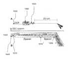
- Placental A-V anastomosesmay be identified using the fetoscope by their characteristic anatomy, which can comprises an artery from one twin and a vein from the other, diving through a common foramen in the chorionic plate (as shown in FIG. 1A ).
- 16 A 100-400 ⁇ m optical fiberis then inserted into the accessory port.
- the anastomotic vesselsare coagulated using 0.1 sec pulses of 40-100 W light provided by a diode or Nd:YAG laser (1064 nm) delivered through the optical fiber (see FIG. 1B ).
- 1 Multiple trialshave shown that fetal survival is significantly improved when the laser coagulation is conducted prior to 26 weeks gestation, with an overall survival rate ranging from 55-72%. 2,3,7
- Fetoscopescan be constructed from fiber-optic imaging bundles, which transmit two-dimensional images from the amnionic cavity to the physician. 1-3,5 Maintaining a good image quality with small diameter bundles is challenging; each optical fiber including its cladding has a finite diameter and only a limited number of optical fibers can be packed into a confined space. Small-diameter fiber bundles therefore provide images with a relatively low number of pixels. Single-mode fiber bundles, containing ultrathin fibers, have the highest fiber density. However these bundles are quite rigid and tend to have relatively low light throughput due to the cladding required on the optical fibers.
- SEEspectrally-encoded endoscopy
- 32 SEEcan likely overcome the limitations of prior fiber-bundle fetoscopes for safer and more effective TTTS laser coagulation therapy.
- SEEe.g., a broadband light emanating from a fiber 200 can be separated into different colors (e.g., wavelengths) 210 using a lens/grating pair 220 at the distal end of the probe (as shown in FIG. 2 ).
- This exemplary optical configurationcan focus each color onto a different location on the tissue, as illustrated in FIG. 2 .
- Reflected light, returned back through the optics and fiber,can then be decoded outside of the body, using a spectrometer, to form one line of the endoscopic image.
- Such “fast-axis” of image acquisitioncan be performed remotely from the probe at rates ranging from 10-30 kHz.
- a two-dimensional imagemay be formed by moving the fiber using well-established mechanical devices, such as a motor or galvanometer that also reside outside the body. 33
- Such second, “slow-axis” of scanningcan be performed at a 30 Hz video rate. Since a high-speed scanning mechanism is not needed inside the endoscope, the diameter of the SEE probe can be as small as that of the optical fiber, which can typically be sized in the range of 80-250 ⁇ m. Furthermore, the number of pixels in an SEE image can be larger than that of fiber bundles, dependent on the spectral width of the light source and the ability of the probe to separate out the different wavelength components.
- Spectral encodingis not only provided for a two-dimensional endoscopy.
- the grating and lensare placed in one arm of an optical interferometer, such procedure can also provide depth information.
- Three-dimensional imagingcan be obtained using spectral encoding with a variety of interferometric techniques, including. e.g., speckle pattern subtraction and time-domain heterodyne interferometry. 34,35
- One of the objects of the present inventionis to provide an ultraminiature (e.g., 350 ⁇ m diameter) endoscopic imaging apparatus/device/arrangement/system with integrated laser therapy capabilities for microsurgical applications inside the body, e.g., for safe and effective treatment of twin-twin transfusion syndrome (TTTS). It is possible to overcome the limitations and deficiencies of the current TTTS therapy devices by providing a much smaller microsurgical endoscope that provides more informative images with a better image quality.
- This exemplary enhancementincludes spectrally encoded endoscopy (SEE) concepts that use wavelength division multiplexing to obtain high-resolution images through a single optical fiber.
- SEEspectrally encoded endoscopy
- the exemplary SEE system and probecan be provided for color differentiation of arterial and venous placental vessels and can add Doppler imaging to quantify blood flow. Additionally, it is possible to incorporate therapeutic laser delivery through the same probe without increasing its size.
- exemplary total diameter of the exemplary apparatus/device/system/arrangementcan be, e.g., 350 ⁇ m, small enough to be introduced into the amniotic cavity through an amniocentesis needle (e.g., 22-gauge), which can provide, e.g., a 10-fold reduction in complication rate.
- Simultaneous imaging with more than one wavelength bandcan provide color information from the sample.
- One such exemplary embodiment of apparatus and method to achieve such exemplary resultmay include a coupling of visible (VIS) and near-infrared (NIR) light into the SEE fiber. If wavelengths are chosen correctly, for example 1064 and 532 nm, then the wavelength regions can overlap one another by diffracting at different orders. In another exemplary embodiment, the wavelengths may be selected such that the absorption properties of the sample can facilitate the differentiation and quantification of compounds within the sample, such as differentiation of arterial from venous blood by measuring differences in oxy- and deoxy-hemoglobin absorption.
- the SEE probemay be situated in an interferometer, and a spectral interferometric phase may be detected and analyzed to provide information on blood flow and other motions of the sample.
- the SEE probemay be configured by use of a specialty fiber or at least one fiber adjacent to the imaging fiber to deliver therapeutic light to the sample to effect therapy.
- the delivery of therapy lightmay be conducted in parallel with an imaging procedure.
- a small (e.g., 350 ⁇ m or other such diameter ⁇ 2.0 mm) diameter SEE probecan utilize a dual-clad fiber; and the imaging light may be transmitted via a single-mode core.
- the therapy procedurecan be performed via the innermost cladding.
- a 1500 lines/mm gratingcan be used to provide two-dimensional imaging with greater than 90,000 pixels.
- the SEE-guided therapy probecan be configured to effect coagulation of blood vessels such as those of the placenta.
- the SEE-guided therapy probecan be provided such that its diameter does not cause undue damage to the amniotic membranes or the uterus, so to facilitate a safe therapy and place the fetus and mother at minimal risk.
- the size of the SEE probecan be sufficiently small to fit within a narrow gauge needle, for example with a gauge of 18-25 Ga.
- the needlecan be an amniocentesis needle.
- a SEE-guided placental vascular coagulation procedurecan be important for a care treatment for TTTS.
- an ultraminiature endoscope with an integrated laser therapy systemcan be provided.
- One exemplary application of such devicecan include but not limited to, e.g., a treatment of twin-twin transfusion syndrome (TTTS), a serious complication of monochorionic twin pregnancies that occurs at a rate of 3000/year in the United States.
- TTTStwin-twin transfusion syndrome
- communicating placental vesselsshunt blood from one twin to the other, resulting in a very high fetal mortality rate and significant morbidity for survivors.
- Complete laser coagulation of the anastomotic vessels in uterocan be an effective therapy for this disease.
- Prior operative fetoscopesare generally too large (e.g., 2-3 mm diameter), which can cause undue membrane damage when introduced into the amniotic cavity. Consequently, a placental laser therapy can be associated with unacceptable rates (10%) of iatrogenic preterm premature rupture of membranes (iPPROM) and in utero fetal demise (IUFD).
- iPPROMiatrogenic preterm premature rupture of membranes
- IUFDin utero fetal demise
- the poor image quality of today's fetoscopesis also of concern, as incomplete coagulation due to missed anastomoses can lead to adverse perinatal outcomes following the laser therapy.
- the exemplary embodiments of the present inventioncan overcome the deficiencies of the prior TTTS laser coagulation devices by being significantly smaller and by facilitating further options for identifying communicating vessels. This can be done using, e.g., spectrally encoded endoscopy (SEE) procedures and systems. SEE procedures and systems can use wavelength-division multiplexing to obtain high-resolution two- and three-dimensional endoscopic images through a single optical fiber. High-resolution, 3D video-rate imaging in vivo using a 350 ⁇ m diameter, monochromatic version of the SEE probe can be achieved with such procedures and systems. According to still other exemplary embodiments of the present invention, it is possible to obtain color imaging, and the exemplary device may provide a measurement blood flow. In yet another embodiment of the present invention, a therapeutic laser light may be coupled through the same fiber so that imaging and intervention can be accomplished concurrently without increasing the endoscope diameter.
- SEEspectrally encoded endoscopy
- the size of the exemplary devicecan be, e.g., about 350 ⁇ m, so as to allow this device to be inserted into the amniotic cavity through a 22-gauge amniocentesis needle, which should lower the complication rates of placental laser therapy by an order of magnitude.
- Such exemplary embodiment of the devicemay facilitate a use thereof in other procedures where small size and highly capable image guided intervention can decrease complication rates and improve patient care.
- the SEE procedures and apparatuscan be utilized as follows.
- a multifunctional SEE system and probefor discriminating arteries from veins.
- this exemplary device/system/arrangement/apparatuscan be used to identify A-V anastomoses.
- Nd:YAG laser lightit is possible to transmit high power Nd:YAG laser light through the SEE probe to enable image-guided vessel photocoagulation without increasing the probe's size.
- these exemplary featurescan facilitate the TTTS management to be enhanced by providing an endoscopic therapy system and apparatus with enhanced capabilities that can be inserted through an amniocentesis needle.
- This exemplary enhancementcan make laser coagulation therapy for TTTS safer and more effective.
- this exemplary device/apparatuscan be used for an endoscopically-guided therapy in other areas of the body that have previously been difficult to access.
- the apparatuscan be utilized to effect therapy for other applications.
- the therapy laseris designed to provide maximum absorption for water (e.g., around water absorption peaks at approximately 1500 nm, 1800 nm, 3000 nm, etc.)
- other therapymay be affected, such as tissue coagulation, ablation, etc.
- an eximer laser or CO 2 laseris utilized in conjunction with the exemplary apparatus, such exemplary combination may be used for resurfacing or a superficial ablation.
- apparatus and processcan be provided for imaging information associated with at least one portion of a sample.
- at least two first different wavelengths of at least one first electro-magnetic radiationcan be provided within a first wavelength range provided on the portion of the sample so as to determine at least one first transverse location of the portion
- at least two second different wavelengths of at least one second electro-magnetic radiationare provided within a second wavelength range provided on the portion so as to determine at least one second transverse location of the portion.
- the first and second rangescan east partially overlap on the portion.
- a relative phase between at least one third electro-magnetic radiation electro-magnetic radiation being returned from the sample and at least one fourth electro-magnetic radiation returned from a referencecan be obtained to determine a relative depth location of the portion.
- First information of the portion based on the first transverse location and the relative depth location, and second information of the portion based on the second transverse location and the relative depth locationcan be obtained.
- the imaging informationmay include the first and second information.
- further informationcan be generated for the portion by combining the first and second information.
- At least two third different wavelengths of at least one fifth electro-magnetic radiationcan be provided within a third wavelength range on the portion of the sample so as to determine at least one third transverse location of the portion
- third information of the portioncan be provided based on the third transverse location and the relative depth location, wherein the imaging information includes the third information.
- the first informationcan be associated with a red wavelength range
- the second informationmay be associated with a green wavelength range
- the third informationcan be associated with a blue wavelength range.
- the imaging informationcan be three-dimensional information.
- apparatus and processcan provide imaging information associated with at least one portion of the sample. For example, at least one wavelength of at least one particular electro-magnetic radiation can be provided on the portion of the sample so as to determine at least one transverse location of the portion.
- obtain a relative phasecan be obtained between at least one first electro-magnetic radiation electro-magnetic radiation being returned from a sample and at least one second electro-magnetic radiation returned from a reference to determine a motion of the portion or of particles within or on the portion.
- the information of the portioncan be provided based on the transverse location and the motion.
- a relative depth location of the portioncan be determined, and the information may be provided as a further function of the relative depth location.
- an apparatus for obtaining diagnostic information for a structure and modifying at least one property of at least one portion of the structurecan be provided.
- the apparatuscan include a fiber configured to provide there through the electro-magnetic radiation.
- At least one first waveguiding portion of the fibercan be configured to provide a first electro-magnetic radiation to the portion so as to obtain the information, and at least one second waveguiding portion of the fiber may be configured to provide a second electro-magnetic radiation to the portion so as to modify at the property.
- the apparatuscan further include a dispersive arrangement configured to receive the first and second electromagnetic radiations.
- a wavelength of the first electro-magnetic radiation and/or the second electro-magnetic radiationcan be a multiple of a wavelength of another one of the first electro-magnetic radiation or the second electro-magnetic radiation.
- the first electro-magnetic radiation and the second electro-magnetic radiationcan at least partially overlap on the portion.
- the propertymay include blood.
- a wavelength of the second electro-magnetic radiationcan overlaps with one or more certain wavelengths where an absorption of the radiation is effective for changing the property. Such certain wavelengths can include a multiple of about 532 nm.
- an apparatuscan be provided for obtaining information for a structure.
- the apparatuscan include a dispersive arrangement configured to receive a plurality of electro-magnetic radiations and forward a dispersed radiation of each of the electro-magnetic radiations to at least one portion of the structure and at least partially overlap the portion.
- One of the electro-magnetic radiationscan have a wavelength in a first range
- another one of the electro-magnetic radiationsmay have a wavelength in a second range.
- Each of the first and second rangescan be at least one element that is different from another one of the second ranges.
- At least one first wavelength within one of the first and second rangesmay be a multiple of at least one second wavelength within another one of the first and second ranges.
- the first wavelengthcan overlap with one or more certain wavelengths where an absorption of the radiation is effective for changing at least one property of the structure.
- Such certain wavelengthscan include a multiple of about 532 nm.
- FIG. 1Ais an exemplary fetoscopic image of an A-V anastomosis before a laser coagulation
- FIG. 1Bis an exemplary fetoscopic image of the A-V anastomosis after the laser coagulation
- FIG. 2is an operational diagram of an exemplary embodiment of an apparatus used in operation according to the present invention which implements the SEE procedure;
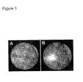
- FIG. 3Ais an exemplary image of a placenta obtained ex vivo with a first exemplary embodiment of the SEE apparatus according to the present invention
- FIG. 3Bis an exemplary color (via gray scale) image corresponding to the exemplary image of FIG. 3A ;
- FIG. 4Ais an exemplary illustration of a distal end of an exemplary embodiment of the SEE apparatus (e.g., probe) according to the present invention having, e.g., a 350 ⁇ m diameter;
- FIG. 4Bis an enlarged illustration of a diffraction grating provided at a tip of the exemplary apparatus shown in FIG. 4A ;
- FIG. 4Cis the exemplary SEE apparatus that is illustrated next to a human hair (provided for size comparison);
- FIG. 4Dis a photograph of the SEE apparatus provided within, e.g., a 23-gauge stainless steel hypodermic tube which can be used for applications where the probe is delivered through a needle;
- FIG. 5Ais an exemplary SEE image of a metastatic ovarian cancer in vivo, where tumor nodules are shown on a parietal peritoneal wall of a mouse, according to an exemplary embodiment of the present invention
- FIG. 5Bis a cross-sectional histological section from the corresponding area of FIG. 5A , showing tumor nodules (e.g., H&E stain; original magnification 40 ⁇ ), and scale bars representing 500 ⁇ m;
- tumor nodulese.g., H&E stain; original magnification 40 ⁇
- scale barsrepresenting 500 ⁇ m
- FIG. 6Ais an exemplary VIS-NIR image of a human placenta, obtained postpartum, reconstructed using 532 nm and 1064 nm wavelengths, according to an exemplary embodiment of the present invention
- FIG. 6Bis an exemplary RGB photograph associated with the image of FIG. 6A , with the bar being indicative of 5 mm;
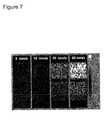
- FIG. 7is a set of exemplary two-dimensional Doppler SEE images of a dual-channel flowing Intralipid phantom obtained using an exemplary embodiment of the present invention
- FIG. 8 ais a schematic diagram of a measurement configuration, showing an exemplary embodiment of the SEE apparatus according to the present invention with respect to the tube and the flow direction;
- FIG. 8 bis a graph illustrating measured (e.g., solid curves) and calculated (e.g., dashed curves) cross-sectional flow velocities at the center of the tube shown in FIG. 8 a;
- FIG. 8 cis a series of exemplary images of a two-dimensional measurement of the intralipid flow across the tube at different average flow velocities
- FIG. 8 dis a set of combined flow and average reflectance images of, e.g., two 0.5 mm diameter tubes with similar and opposite flow velocities;
- FIG. 9is a schematic diagram of an exemplary embodiment of a SEE-guided laser therapy apparatus according to the present invention.
- FIG. 10Ais a schematic diagram of another exemplary embodiment of the SEE arrangement (e.g., probe) according to the present invention.
- FIG. 10Bis an exploded view of optics at a distal end of the exemplary arrangement of FIG. 10A ;
- An exemplary embodiment of the device/system/arrangement/apparatus according to the present inventioncan include, e.g., a 350- ⁇ m diameter SEE probe, constructed from a dual-clad fiber.
- a SEE imaging lightcan be transmitted through a central core, and an innermost cladding will guide high power 1064 nm light for vascular coagulation.
- the exemplary system (and probe)can obtain color images using two separate wavelength bands, centered near 532 (VIS) and 1064 nm (NIR).
- VIScentered near 532
- NIR1064 nm
- a spectral-domain heterodyne interferometry procedure and arrangementcan be implemented to obtain three-dimensional images of chorionic plate topology and two-dimensional Doppler maps of blood flow. Provided below, exemplary results in accordance with exemplary embodiments of the present invention are described.
- SEEwas developed in 2001 32 when it was realized that the spectral encoding technology that has been developed for endoscopic confocal microscopy can provide more resolvable points for any given probe diameter than conventional fiber bundles.
- An exemplary bench top systemwas constructed that utilized a 850 nm modelocked Ti:Al 2 O 3 laser, a 1000 lines/mm transmission grating, and a CCD camera to obtain macroscopic images of excised specimens at video rates (30 frames per second).
- 32Using a beam diameter of approximately 500 ⁇ m, the imaging of a placenta that was obtained postpartum was demonstrated (as shown in FIG. 3 ). The analysis indicated that the technology can be extended to increase the number of resolvable points by more than an order of magnitude. 32
- Initial imagescompared favorably with those obtained by conventional fetoscopes with larger diameters.
- Double-Clad Fiber SEE ApparatusDouble-Clad Fiber SEE Apparatus.
- SEE images obtained from a single mode fibervariably (generally) display speckle noise artifacts.
- an exemplary SEE imaging systemcan be provided that may illuminate the sample with single mode light through the core of a double-clad fiber and collected the remitted light through its innermost cladding.
- Double-clad fiberscan be obtained from commercially available sources. SEE images which cab be obtained with the double-clad fiber likely indicate a substantial reduction of speckle artifact, as well as an increased depth of field, both of which are highly desirable for endoscopy. 37 With an appropriate selection of the innermost cladding diameter, these advantages can be realized without significant loss in transverse resolution. 37
- a placement of the SEE apparatus/probe in the sample arm of an interferometercan provide three-dimensional information in addition to the standard two-dimensional view.
- the exemplary SEE probecan be configured so that every resolvable point on the sample may be illuminated by light with a bandwidth sufficiently large to conduct low coherence interferometry at each spot. For small diameter probes, this exemplary configuration can be accomplished without the loss of a transverse resolution.
- Certain exemplary benchhave been described for two different forms of a heterodyne interferometry, e.g., heterodyne speckle pattern subtraction 35 and time-domain low coherence interferometry.
- an exemplary diffraction gratingcan be incorporated directly onto the tip of an optical fiber.
- the exemplary gratingcan provide a high diffraction efficiency, may be robust to extremely high optical power densities, and likely be compatible with aqueous environments.
- Exemplary images of the exemplary embodiment of the SEE apparatus/probe 400 according to the present inventionis shown in FIGS. 4A-4D . 33
- light from a single-mode fiber 405expanded through a 1.8 mm long silica spacer 410 , can be focused by a 350 ⁇ m diameter GRIN lens 415 .
- Light from the lens 415can then be diffracted by a 1000 lines/mm transmission grating 425 fabricated on a 1.5 mm long spacer 420 that can be polished at Littrow's angle of, e.g., about 19°.
- An expanded view of the diffraction grating 425is shown in FIG. 4B .
- An exemplary maximum diameter of the exemplary apparatus/probe 400can be about 350 ⁇ m; e.g., a size comparison between the exemplary apparatus/probe 400 and a human hair 430 is shown in FIG. 4C . Since the exemplary apparatus/probe 400 can be constructed from a single optical fiber, it may be very flexible, with a bending radius of approximately 2 mm, except within ⁇ 3.5 mm of the distal end.
- the exemplary 350 ⁇ m diameter SEE apparatus/probe 400can be delivered into the abdominal cavity using a miniature laparoscopic through, e.g., a modified 23-gauge needle 440 (as shown in FIG. 4D ).
- a small openingcan be made in the needle wall, near its distal end, for the light to pass through.
- the openingcan be sealed with a clear 6- ⁇ m thick layer of transparent plastic.
- FIG. 5AA three-dimensional image of the parietal peritoneal wall, obtained with the SEE apparatus/probe in vivo, is shown in FIG. 5A .
- the two- and three-dimensional data setscan be combined to form a three-dimensional surface view of the tissue.
- a large, approximately 1 mm ⁇ 1 mm ⁇ 0.2 mm superficial tumor nodulecan be seen near the center of the frame adjacent to two smaller 300 ⁇ 300 ⁇ 100 ⁇ m tumor nodules.
- the exemplary apparatuse.g., a microendoscope
- the exemplary apparatuscan provide images with sufficient information to distinguish placental arteries from veins.
- arteriescan be differentiated from veins by: i) color, ii) pulsatility/flow characteristics, and iii) a three-dimensional anatomic configuration, as arteries cross over veins on the chorionic plate. 40
- the exemplary embodiment of the SEE arrangement/device/system/apparatus/probe according to the present inventioncan facilitate the identification of more communicating vessels than the conventional two-dimensional procedure.
- the exemplary SEE procedures and apparatusare capable of performing three-dimensional imaging.
- Provided belowis a description of exemplary information in supporting the implementation and use of the exemplary embodiments of the procedures and apparatus for obtaining color and blood flow image data according to the present invention.
- SEE imaginghas been monochromatic; broadband light centered at 850 nm has been used merely for encoding spatial information.
- the exemplary embodiments of SEE procedures and apparatus according to the present invention described hereincan obtain color information by using two distinct wavelength bands, e.g., one centered near 1064 nm (NIR) and the other near 532 nm (VIS).
- NIR1064 nm
- VISnear 532 nm
- This exemplary selection of wavelength regionscan be appropriate since, the Nd:YAG therapy beam may overlap with the first and second order of the NIR and VIS imaging beams, respectively.
- the differential absorption of oxygenated and deoxygenated adult and fetal blood at VIS and NIRcan be similar to that of the green and red wavelength bands used in standard RGB color imaging. 41-43
- a placentahas been imaged, obtained immediately postpartum, with 1064 ⁇ 12 nm, 532 ⁇ 5 nm, and RGB light.
- a linear filterwas first applied to the 1064 nm image data to remove noise.
- a VIS-NIR image(shown in FIG. 6A ) was then generated by placing the 1064 nm image in the red channel and the 532 nm image in both the green and blue channels of a new 24-bit color image. No other manipulations other than contrast enhancement were made to the VIS-NIR image of FIG. 6A .
- the corresponding exemplary RGB image(in gray scale) is shown in FIG. 6B .
- exemplary SEE heterodyne interferometry proceduresare capable of measuring the Doppler shift caused by blood flow.
- a simple phantomhas been produced, comprising a 1.0 mm diameter tube, looped back onto itself and affixed to a microscope slide. This phantom configuration can ensure that the exemplary SEE image may include two adjacent tubes with similar flow rates, with opposite flow directions. The tube was perfused with 10% Intralipid, which has a transport scattering coefficient similar to that of whole blood.
- Flow velocitieswere varied from about 0-60 mm/s, representing the approximate range of blood flow velocities that would be observed in a second trimester placenta and in TTTS.
- 44 Heterodyne spectral-domain SEE imaging of the phantomwas conducted using our 350 ⁇ m diameter probe.
- 33 The spectral interferogramswere processed using windowed short-time Fourier transforms to obtain local fringe magnitudes and phases.
- 33,34Doppler shifts at each point in the image were estimated by comparing the local phases of two adjacent spectrally-encoded lines.
- Two-dimensional flow maps (differential phase images) of the phantom(as shown in a set of exemplary images of FIG.
- FIG. 7shows that the exemplary heterodyne SEE procedures and apparatus may track the flow rate and direction throughout the entire velocity range.
- FIG. 7shows flow directions (e.g., arrows) being along the horizontal dimension and flow velocities are shown at the top of each image (e.g., the exemplary images demonstrating a relationship between interferometric phase and flow velocity/direction, and differential phase magnitudes (a.u.) being plotted using a color Doppler lookup table depicted in the color bar).
- the exemplary embodiments of the SEE procedures and apparatuscan also detect the flow at depth within scattering media and tissue.
- 1% intralipidmay be circulated through a 1 mm diameter transparent tube.
- the SEE beamcan illuminate the tube as illustrated in FIG. 8 a .
- the exemplary SEE procedures and apparatuscan image through scattering medium [11]
- the x-z reflection datawhen acquired as a function of time, can include information on the flow of scatterers throughout the entire cross section of the tube.
- the flow velocity and directionmay be controlled by a peristaltic pump and its average value can be estimated by measuring the total volume that was flowing through the tube as a function of time.
- v ⁇ ( x , z )1 k ⁇ ( x ) ⁇ cos ⁇ ⁇ ⁇ ⁇ ⁇ ⁇ ⁇ ( k ⁇ ( x ) , z ) ⁇ t , ( 1 )
- k(x)is the wave number
- ⁇denotes the angle between the SEE beam propagation and the direction of sample motion.
- the ratio between the average fitted velocitiescan be 1:2.6:4.4, which is in agreement with the measured average flow ratios of 1:3:4.5.
- An absolute value of the flow velocitiesmay be difficult to obtain due to the relatively high uncertainty level in measuring the angle ⁇ , which can be estimated to be in the range of about 80° ⁇ 90°.
- a set of exemplary images of the two dimensional (x-z) distributions of flow velocity across the 1 mm diameter tubeare shown in FIG. 8 c for different pump powers. Without any pumping, FIG.
- the phasemay fluctuate randomly and no flow was detected (e.g., left most image).
- the flow velocity distributionscan provide two-dimensional parabolic distributions with higher velocity in the center of the tube, approaching zero at the tube walls. Phase error at the tube's center may cause loss of data at higher flow velocities. Nevertheless, the flow profile may be estimated by fitting the parabolic curves to the areas closer to the tube walls, where flow is slower.
- the exemplary embodiment of the SEE procedures and apparatuscan additionally provide reflectance imaging of the tubes.
- the intensity of each pixelrepresents the total reflectance, integrated over depth, and the color represents velocity, averaged over the tube's depth. With no pumping, the flow was likely random, resulting in purple hue in both tubes.
- Placental laser coagulation therapy using 1064 nm lightcan be highly effective for interrupting twin-twin blood flow, but hampered by the large size of modern operative fetoscopes and their inability to image all communicating vessels.
- the exemplary embodiments of the procedures and apparatus according to the present inventioncan overcome these limitations by being significantly smaller and by providing more options for identifying A-V anastomoses.
- a first one of the exemplary objects indicated abovecan be focused on providing and testing an exemplary multifunctional SEE instrumentation of the exemplary SEE apparatus and procedure according to the present invention, combined with coincident high power laser irradiation.
- exemplary SEE microendoscopescan be provided, integrated with the apparatus which can be implemented for the first one of the exemplary objects, and tested in phantoms and human tissue ex vivo. It is possible to use such exemplary procedures and arrangements, e.g., in a 10 pregnant ewes to demonstrate the feasibility of SEE-guided laser coagulation through an amniocentesis needle in vivo (see exemplary object 3).
- Exemplary capabilities of the exemplary embodiment of the SEE-guided laser therapy apparatuscan include, e.g., (a) high quality two-dimensional imaging, (b) three-dimensional, color, and Doppler imaging to identify A-V anastomoses, and (c) simultaneous laser therapy.
- a bench top exemplary SEE apparatusthat can be used to design, implement, and validate different technical approaches for achieving these capabilities.
- the exemplary systemcan additionally be used to test and validate exemplary SEE probe configurations for the second exemplary object.
- FIG. 9A schematic diagram of one exemplary embodiment of the SEE apparatus according to the present invention is shown in FIG. 9 .
- the exemplary components of the exemplary system of FIG. 9are as follows: WDM—wavelength division multiplexer 900 , BS—beam splitter 905 ; MM—multimode, SM—single mode, SMF—single mode fiber, MM/SM—multimode/single mode splitter 910 , PC—polarization controller 915 , DCF—dual-clad fiber 920 ; M—mirror 925 , DM—dichroic mirror 930 , VIS—broadband light centered around 532 nm 935 , and NIR—broadband light centered around 1064 nm 940 .
- VISbroadband VIS
- NIRcentered near 1064 nm
- VISbroadband VIS
- NIRnear 1064 nm
- a small fraction of lightcan be directed towards a reference arm mirror 950 .
- the remainder of the lightcan be transmitted through a single/multimode splitter 910 , which can comprise a mirror with a central transparent opening that transmits single mode light and reflects multimode light.
- the imaging lightcan be coupled to the core of the dual-clad fiber (DCF) of the SEE apparatus/probe 955 .
- DCFdual-clad fiber
- the exemplary sample arm probecan include the DCF, terminated by a lens and grating.
- “slow axis” scanningcan be performed using a galvanometer attached to the grating.
- This exemplary configurationcan facilitate the testing of various approaches for color and Doppler imaging as well as probe components prior to the final microendoscope fabrication step.
- the exemplary SEE apparatus/probescan be incorporated into the exemplary system according to the present invention, e.g., for testing and other purposes.
- the beam splitter 910can be recombined by the beam splitter 910 and separated into wavelength bands of the VIS 960 and NIR 965 using, e.g., a wavelength division multiplexer 900 or dichroic beam splitter.
- Each wavelength bandcan be detected by separate custom-built spectrometers, designed to measure spectral interferograms for 3D and Doppler flow map reconstructions.
- the VIS spectrometer 960can utilize a high-speed Silicon linear CCD array while the NIR spectrometer 965 can employ an InGaAs linear array.
- the spectral resolutions of the spectrometerscan be approximately 5-10 times greater than the spectral resolution on the sample.
- linear array detectorscan over-sample the spectral data so that approximately 5-10 pixels may be digitized for each resolvable point.
- Two-dimensional, three-dimensional, Doppler, and color (VIS & NIR) SEE imagescan be obtained by computing windowed short-time Fourier transforms of the background-corrected spectral fringe data. 33,34
- the two-dimensional imagemay be determined and/or generated based on, e.g., at least in part, the integrated power of the local Fourier spectra for each point.
- Color imagescan be generated by transforming the VIS and NIR SEE images into RGB space. While the VIS and NIR images can be co-registered and may have the same physical dimensions, the pixel density of the NIR image can be one-quarter that of the VIS image. The NIR image may therefore be interpolated prior to generation of the composite color image. Determining quantitative estimates of blood oxygenation content can also be analyzed using, e.g., two- and three-dimensional data sets at both wavelengths. 47-49
- the spectral power density of the continuum sourcecan be, e.g., significantly higher than that of the source utilized in our preliminary studies. Thus, ample light can be provided to conduct shot-noise limited, depth resolved spectral-domain low coherence interferometry at each point in the image. 50 Reflectance as a function of depth within the tissue can be obtained from the magnitude of the local Fourier data. Phase dithering of the reference arm mirror may be used to eliminate or deduce depth degeneracy. 51,52 As a result, one line in the image can be generated from two linear array scans, where each scan may be acquired at a ⁇ /2 differential reference arm phase delay. 33 Doppler information can be obtained by determining the relative difference between Fourier domain phases at each location within tissue. 45,53,54
- Such exemplary embodiment of the system according to the present inventioncan also provides a speckle-reduction, which may be implemented by acquiring SEE images through the multi-mode inner cladding of the DCF. 37
- VIS light returned from the probe's innermost claddingmay be reflected off the single/multimode splitter 910 and directed to another Silicon array spectrometer 970 , which can be optimized for a multimode detection.
- the pixel countmay be lower, approximating 22,500.
- Approximately 10 discrete axial locationscan be obtained at each point, resulting in a total pixel count of 300 ⁇ 300 ⁇ 10 ⁇ 900,000 in the volumetric images.
- axial resolutions for both the VIS and NIR channelscan be ⁇ 280 ⁇ m.
- the total depth rangecan be approximately 3 mm for each wavelength region. If necessary for this application, greater depth ranges can be achieved by use of linear CCD's with higher pixel counts.
- the Atmel Silicon cameracan sample 60,000 lines per second, whereas the InGaAs camera may generally operate at a line rate of 10 kHz. As a result, volumetric image data can be acquired at video rates for both VIS and NIR wavelengths.
- the exemplary embodiment of the SEE-guided laser therapy system, apparatus and procedure according to the present inventioncan integrate a therapeutic laser such as a Nd:YAG laser 975 , thus meeting clinically-established specifications for the coagulation procedure.
- a therapeutic lasersuch as a Nd:YAG laser 975
- An operator-controlled pushbutton switchcan activate a shutter that may remain open for 0.1 seconds every time the switch is activated.
- Camerascan communicate to the host CPU via high-speed interfaces such as Camera Link. Assuming that exemplary highest 16-bit acquisition rates can be used for each camera, the total data rate may be approximately 310 MB/s, which is within the Camera Link DMA specification. For example, raw data can be stored in real time to a RAID array at rates such as 350 MB/s.
- a computing arrangement according to thecan process the interferograms and display the exemplary resulting images. Since real-time complex FFT processing can be used to process and display all SEE images at video rate (e.g., 30 fps).
- the exemplary embodiments of the apparatus, system and components according to the present inventioncan include various designs.
- fetal surgeonscurrently use three cues to differentiate placental arteries from veins: color, pulsatility, and three-dimensional anatomical structure. 40
- the exemplary embodiment of the SEE system and apparatus according to the present inventionis capable of measuring all three of these features, with higher resolution and in a quantitative manner that should be superior to qualitative interpretation of conventional two-dimensional fetoscopic images.
- Exemplary SEE Doppler flow imaging capabilitiescan be included in the exemplary SEE system to improve the surgeon's ability to see A-V anastomoses.
- Exemplary advantages of an exemplary multifaceted procedure and apparatus for identifying communicating vessels at this development phasecan include redundancy; if one approach becomes intractable.
- VISe.g., centered around 532 nm
- NIRcentered around 1064 nm
- An exemplary selection of VIS (e.g., centered around 532 nm) and NIR (centered around 1064 nm) wavelength regionsis based on a preference to minimize the diameter of the SEE probe and the use of standard, high power 1064 nm light for placental vascular coagulation. It is possible to use other exemplary wavelengths where the fundamental can also effect a substantial vascular coagulation. Alternatively, a probe with several fibers may be provided.
- the first fibercan deliver high-power 1064 nm light and the other fibers may be used for SEE imaging at wavelengths other than VIS and NIR, such as red (e.g., 600-700 nm), green (e.g., 500-600 nm), and blue (e.g., 400-500 nm).
- rede.g., 600-700 nm
- greene.g., 500-600 nm
- bluee.g. 400-500 nm.
- Heterodyne interferometrycan be conducted for both VIS and NIR wavelength regions, since VIS imaging may have the highest spatial resolution and NIR imaging can penetrate deeper into tissue owing to decreased scattering. It is also possible that only one of these exemplary wavelength bands can be used to provide three-dimensional structural and flow cues required for optimal identification of A-V anastomoses. In such case, it is possible to eliminate one of the spectrometers from the interferometer and use these components for another multimode detection channel.
- the VIS bandcan be used for a multimode detection, due to the higher number of resolvable points attained by SEE at these wavelengths. It is possible that further multimode NIR information may be provided for high-quality two-dimensional imaging. In this case, it is possible to partition the InGaAs linear array so that approximately 100 pixels can be utilized for multimode detection while the remaining pixels may be reserved for heterodyne detection.
- Free-space and DCF-coupled power from the fiber-pumped continuum sourcecan be measured for each bandwidth region.
- Different combinations of filters, gratings, and fiber opticsmay be used to shape the spectrum in each wavelength band to ensure that they are sufficiently uniform for efficient image reconstruction.
- Spectral stabilitycan also be measured. If significant spectral variation is present, it is possible to use either a separate photodiode or an additional spectrometer to collect background spectra for real time correction of the spectrometer data.
- fiber in the optical traincan be desirable. However, it may be unlikely that single-mode fibers and the core of the DCF may be single mode for both wavelength regions. Furthermore, reference and sample arm polarization balancing may be difficult to achieve in the VIS and NIR wavelengths simultaneously. Thus, it is possible to use various fiber optic and free space components to identify the most stable, compact system that produces high quality images.
- Spectrometerscan be designed using a variety of lenses, transmission gratings, and reflection gratings may be tested for maximum throughput and spectral resolution.
- Spectrometer throughputcan be determined by comparing power at the input and at the array detector.
- Spectral resolutionmay be obtained at the ends and middle of the two spectral ranges for each interferometer by measuring the full-width-half-maximum of coherent narrow band laser light.
- Heterodyne SNRcan be determined for each spectrometer by placing an attenuator and mirror in the sample arm, measuring the demodulated signal, and the noise with the sample arm blocked.
- Imaging performancecan be assessed using a bench top sample arm probe that simulates the optical characteristics of the small diameter exemplary SEE probe (see exemplary object 2).
- Images of resolution charts and phantomscan be quantitatively analyzed to measure transverse and axial resolutions. Measurements may be compared to computer models and analytic simulation results.
- Image quality metricsmay serve as feedback information for exemplary embodiments of the system according to the present invention.
- Vessel phantoms containing both scattering media and human blood with varying oxygen contentcan be constructed.
- Monte Carlo modelingcan be performed to determine the relationships between diffusely and singly scattered light from phantoms with different degrees of oxygenation.
- exemplary procedurescan be implemented for a vessel characterization that can combine two- and three-dimensional information in the VIS and NIR wavelength regions.
- Quantitative exemplary procedures for estimating blood oxygenation contentcan be utilized that may use the differential oxy- and deoxy-hemoglobin absorption spectra.
- Exemplary procedures for reconstituting images that approximate the conventional RGB color viewmay also be developed.
- Performance of the exemplary procedurescan be evaluated by testing them on blood phantoms and human placentas with arteries and veins that have been filled with heparinized deoxygenated and oxygenated blood, respectively.
- 57 Imagingcan be conducted through saline. Observers, blinded to blood oxygen content and vessel type can discriminate artery from vein in both SEE color images and corresponding RGB color fetoscopy images. Sensitivity and specificity for color SEE and standard fetoscopy may be determined using knowledge of vessel type as the gold standard. Sensitivities and specificities for the two modalities can be compared using McNemar's test. Quantitative SEE assessment of oxygen content can be compared with known oxygen content by linear regression. If these two wavelength regions are insufficient for meeting our milestones, alternative wavelength bands may be provided.
- Self-referencing exemplary procedures for extracting the phase from the exemplary heterodyne VIS and NIR SEE systems and apparatuscan be provided and tested in moving phantoms with known velocity.
- Flow ratesmay approximate that in the normal placenta and TTTS cases (e.g., 0-60 mm/s).
- 44Flow velocity, velocity variance, power Doppler, and Doppler spectral images can be determined from the phase information.
- the exemplary probe/apparatus beammay not typically be perpendicular to the chorionic plate, angular differences between the optical axis and flow direction would likely not greatly affect relative measurements of flow. It is possible to validate the expected cos( ⁇ ) relationship and determine its effect on flow imaging maps constructed from phantoms.
- Phantoms and human placentascan be perfused with heparinized blood through the umbilical vein in a pulsatile manner that simulates maternal-fetal circulation.
- 57Time-resolved and time-averaged SEE cross-sectional measurements of flow velocity and average power Doppler signal may be compared with Doppler ultrasound using paired t-tests.
- human placentasobtained postpartum can be perfused with pulsatile blood.
- arteries and veinscan be then imaged through saline by SEE and state-of-the-art fetoscope instrumentation.
- the trained fetal surgeonblinded to the vessel type, can identify arteries and veins using both technologies. Sensitivity and specificity of the two technologies for categorizing artery versus vein can be determined. Sensitivities and specificities of the two exemplary embodiments may be compared using McNemar's test. Histology can be used as a standard.
- the power of the Nd:YAG lasercan be measured before and after propagation through the prototype system optics.
- Optics for coupling into the DCFmay be designed using computer modeling. Fiber coupling efficiency can be measured. All optics including the DCF, multimode splitter, fiber coupler, and bench top probe lens/grating pairs can be exposed to continuous and 0.1 s pulsed high power 1064 nm light.
- the powers preferrede.g., 40-100 W
- the estimated maximum energy densitymay be four orders of magnitude lower than the lower bound of their exposure limit. 59
- These and other exemplary componentscan be evaluated for signs of laser-associated damage and other more preferable components can be provided if damage occurs.
- Vascular coagulation therapycan be conducted in free space and through saline using the aforementioned Nd:YAG laser, coupled through the system into the DCF.
- Human placentas, obtained postpartummay be perfused with human blood.
- Nd:YAG laser pulses(0.1 sec) can be transmitted through the bench top sample arm probe and aimed at human placental arteries and veins. The number of pulses may be varied for different vessel diameters and types to optimize exposure parameters.
- Vascular coagulation testscan also be conducted on adjacent locations with standard Nd:YAG instrumentation from the fetal surgery unit. Spatial extent of vascular coagulation can be evaluated using quantitative histomorphology and compared between the two surgical devices using paired t-tests.
- the exemplary system according to the present inventioncan be incorporated, e.g., into a portable cart for ease of use in the surgical suite.
- Softwarecan be provided to store the data in real-time to the RAID, and to generate and display the exemplary two-dimensional, three-dimensional, color, and Doppler SEE images.
- OPTObjective Performance Targets
- exemplary probe/apparatus diametercan be proportional to fetal complication rate
- the preferable instrumentationmay have a small or minimal size. It is possible to provide an exemplary small (i.e. 350 ⁇ m) diameter guided laser therapy probe/apparatus that may be compatible with the advanced capabilities described above with respect to exemplary object 1, and that can be also an order of magnitude smaller than operative fetoscopes in clinical use. Thus, it is possible to administer this exemplary device/probe/apparatus into the amniotic cavity through a small-diameter amniocentesis needle, resulting in a 10-fold reduction in iPPROM and IUFD, compared with current operative fetoscopy.
- FIGS. 10A and 10BThe exemplary embodiment of the SEE microendoscope arrangement and apparatus according to the present invention shown in FIGS. 10A and 10B has some similarities with the 350 ⁇ m diameter miniature endoscope previously described for use in vivo. 33,37
- the componentsare labeled as follows: G—galvanometer 1000 , DCF—dual-clad fiber 1005 , and GRIN—gradient index lens 1010 .
- Ggalvanometer 1000
- DCFdual-clad fiber 1005
- GRINgradient index lens
- broadband VIS and NIR lightcan be coupled into the core of the DCF 1015 for effectuating SEE imaging.
- the proximal end of the fibermay be affixed to a galvanometer shaft 1020 .
- the DCF 1005can be fusion spliced or affixed to a silica spacer 1025 , gradient index lens 1010 , and another spacer 1030 that may be polished at Littrow's angle of the diffraction grating.
- the replicated holographic transmission grating 1035may be stamped onto a hard epoxy coating on the exposed face of the second spacer.
- An outer housing of the probecan be a 22-gauge amniocentesis needle, e.g., similar to that used in the exemplary microlaparoscopic procedure provided on living mice (see FIG. 4D ).
- Both the core 1015 and innermost cladding 1045can collect light reflected from the subject.
- Core lightmay be directed to the VIS and NIR interferometric spectrometers.
- innermost cladding lightcan be detected by the multimode spectrometer (as shown in FIG. 9 ) to obtain an image with minimal speckle noise and an increased depth of field.
- High-power laser therapy lightmay be delivered through the innermost cladding of the DCF 1045 to the tissue for vessel coagulation.
- the exemplary configuration of the probe/apparatus optics according to the present inventioncan be based on, e.g., (a) field of view and working distance specifications of state-of-the-art fetoscopes for safe placental laser vessel coagulation procedures, 3,40 (b) viewing angle considerations to provide optimal surgical guidance, (c) appropriate wavelengths for distinguishing arteries from veins, and (d) the constraints placed on the microendoscope by a selection of an exemplary coagulation laser wavelength.
- Table 1summarizes the optical parameters and performance characteristics of one design that balances the desired requirements of an operative fetoscope, while still maintaining good imaging performance.
- an exemplary specifications of an exemplary SEE therapy probe/apparatus and fetoscopeare provided.
- This exemplary embodiment of the probe/apparatuscan provide for 1 cm FOV along the spectrally-encoded axis and 3 cm along the slow-axis of scanning.
- ⁇ Cross-sectional areamay be determined from diameters of 3.3 mm trocar required for the Storz operative fetoscope 3 and a 22-gauge needle for the SEE-microendoscope.
- This exemplary configuration and the entire parameter spacecan be analyzed to identify the optimal or preferable configuration for this exemplary application.
- this exemplary SEE microendoscope/apparatusmay have comparable design specifications to the conventional operative fetoscope.
- an angled viewcan be easier to use than the forward-looking view of prior technology. 40 Maximizing the field of view based on the exemplary embodiment of the present invention is one of the objects of the present invention. For this exemplary configuration, the spectral bandwidth may be maximized in order to increase the field of view.
- DCF innermost claddingcan be as follows: 100 ⁇ m; DCF NA: 0.12; GRIN polished to 0.20 mm thickness; grating groove density: 1500 lines/mm; Littrow's angle (saline) 34°.
- Optical designComputer modeling can be conducted to analyze the optical design parameter space. DCF's with a variety of core and innermost cladding diameters, and numerical apertures may be designed. High groove density transmission gratings can be designed for both air and saline immersion that can provide >75% diffraction efficiency for both wavelength regions. 60 A variety of different possibilities for increasing field of view along the spectrally-encoded axis may be reviewed, including using a miniature fish-eye lens, placing the grating prior to the lens, and increasing the spectral bandwidths/grating groove densities further.
- DCF'scan be drawn, characterized and tested for imaging.
- Silica spacersmay be cut to the appropriate length and angle polished to minimize backreflections (Spacer 1) and to provide Littrow's angle (Spacer 2).
- Distal silica spacersmay be assembled in a custom mount and provided for transmission grating deposition. Exemplary components may then be tested individually for their optical characteristics, including the existence of visible defects, grating diffraction efficiency, backreflections, and aberrations. Individual components can be assembled using optical adhesives that can withstand high power 1064 nm exposure.
- the 22-gauge amniocentesis needlecan serve two exemplary purposes: i) it may house the DCF and optics to enable “slow axis” scanning and ii) it can serve as the miniature trocar for inserting the imaging probe/apparatus into the amniotic cavity.
- the tip of the needlemay be open or closed by epoxy. When the tip is open, the optics can emerge from the tip for scanning externally to the needle. Exemplary advantages of this exemplary approach are that saline can be perfused through the needle to clear the field of view and lack of aberrations. However, in this case, the probe/apparatus optics may not be protected.
- the opticscan generally be protected when the tip is closed, but a transparent window is then used in the needle (see FIG. 4D ), which may cause aberrations. Both of these exemplary configurations can be tested on resolution standards and saline-filled phantoms to determine the appropriate choice for this exemplary application.
- Fiber rotationcan convey mechanical motion from the proximal to distal ends of the exemplary SEE probe/apparatus. 33
- the exemplary embodiment of the probe according to the present inventioncan be two times longer, and the motion transduction can be confirmed for a variety of fiber buffer materials.
- OPTObjective Performance Targets
- Imaging backreflections⁇ 30 dB.
- the exemplary upper bound on probe back-reflectionscan be preferable for high-quality imaging that may be measured by comparing returned signal powers with and without a mirror at the image plane of the exemplary probe/apparatus.
- This OPTcan be comparable to that of commercially available fetoscopes and may be measured by imaging resolution standards at the image plane.
- This OPTcan be comparable to that of commercially available fetoscopes and may be measured by imaging resolution standards below the image plane.
- Imaging rate⁇ 30 Hz.
- Video rate imagingcan be preferable for endoscopically-guided intervention, and the display rate may be determined for the exemplary two-dimensional SEE images.
- Laser vessel coagulationcan be effectively conducted with this OPT. 2
- Nd:YAG induced probe damageNone.
- This OPTcan be evaluated by conducting SEE imaging after continuous and repeated exposure of, e.g., high power 1064 nm light.
- Image-guided therapycan be conducted, e.g., in 10 pregnant ewes using the exemplary device through an amniocentesis needle and conventional operative fetoscopy equipment.
- Safety of the exemplary device/apparatuscan be evaluated by comparing damage that occurs at the maternal incision and uterine entry sites. Efficacy may be measured by comparing a) the accuracy of each exemplary technique for differentiating artery from vein, and b) diagnoses obtained at sites of laser photocoagulation. Histopathology can be the standard. Following completion of this exemplary procedure, data can be provided in support of this procedure for treatment of TTTS.
Landscapes
- Health & Medical Sciences (AREA)
- Life Sciences & Earth Sciences (AREA)
- Surgery (AREA)
- Physics & Mathematics (AREA)
- Engineering & Computer Science (AREA)
- Public Health (AREA)
- Biomedical Technology (AREA)
- Heart & Thoracic Surgery (AREA)
- Medical Informatics (AREA)
- Molecular Biology (AREA)
- Veterinary Medicine (AREA)
- Animal Behavior & Ethology (AREA)
- General Health & Medical Sciences (AREA)
- Pathology (AREA)
- Biophysics (AREA)
- Nuclear Medicine, Radiotherapy & Molecular Imaging (AREA)
- Radiology & Medical Imaging (AREA)
- Spectroscopy & Molecular Physics (AREA)
- Optics & Photonics (AREA)
- Electromagnetism (AREA)
- Otolaryngology (AREA)
- Investigating Or Analysing Materials By Optical Means (AREA)
- Analysing Materials By The Use Of Radiation (AREA)
Abstract
Description
This application is a divisional application of, and therefore claims priority from, U.S. patent application Ser. No. 11/875,676, filed on Oct. 19, 2007 and from U.S. Patent Application Ser. No. 60/862,205 filed on Oct. 19, 2006, the entire disclosures of which are incorporated herein by reference.
The present invention relates generally apparatus and method for providing imaging information associated with at least one portion of a sample, obtaining diagnostic information for a sample and/or modifying at least one property of at least one portion of the sample.
A concept of microendoscopy has been described for minimally invasive therapy in medicine. Small sizes of these devices can reduce anesthesia requirements and minimize tissue damage, opening up the possibility of safer intervention. Ultraminiature endoscopes may also give rise to new procedures, permitting diagnosis and microsurgery in previously inaccessible areas of the body. Previously, however, a widespread adoption of microendoscopy may be hampered by the poor image quality of current devices and the overall size of the endoscope and its associated microsurgical instrumentation. One of the objects of the present invention is to provide a new form of microendoscopically-guided therapy that overcomes these limitations.
Operative Fetoscopy.
Endoscopically-guided fetal surgery is one of the applications of microendoscopy.4,5Indications for intervention can include congenital diaphragmatic hernia, lower urinary tract obstruction, sacrococcygeal teratoma, and thoracic space occupying lesions, among others.5,6Placental surgery, notably laser coagulation of vessels on the chorionic plate, has gained significant attention for the treatment of twin-twin transfusion syndrome (TTTS).7-13The use of these techniques can result in live, healthy births in cases that would otherwise result in in utero fetal demise (IUFD).
Twin-Twin Transfusion Syndrome.
TTTS is considered a complication of monochorionic pregnancies where blood is preferentially shunted through placental arteriovenous (A-V) anastomoses towards one twin and away from the other. In severe TTTS, the donor twin becomes hypovolemic, resulting in oligohydramnios and oliguria.2The recipient twin conversely becomes hemodynamically overloaded, with subsequent polyhydramnios and polyuria.2Severe TTTS can occur in 15% of monochrorionic pregnancies, at a rate of approximately 3000/year in the United States.2When left untreated, organ and cardiac failure ensue, resulting in mortality rates ranging from 80-90%, with significant neurological defects in surviving twins.2,14
Laser Coagulation for Treatment of TTTS.
A variety of treatments for TTTS have been investigated, including serial amnioreduction, septostomy, and fetoscopically-guided laser coagulation of placental A-V anastomoses.2-4,15Studies have shown that laser coagulation of communicating vessels appears to be the most promising of these techniques.3,15Laser coagulation therapy of TTTS can use a microendoscope, containing an instrumentation port, which is inserted through a cannula into the amnionic cavity. Generally, the amniotic fluid is replaced with warmed, sterile normal saline or Hartmann's solution to facilitate visualization.1Placental A-V anastomoses may be identified using the fetoscope by their characteristic anatomy, which can comprises an artery from one twin and a vein from the other, diving through a common foramen in the chorionic plate (as shown inFIG. 1A ).16A 100-400 μm optical fiber is then inserted into the accessory port. The anastomotic vessels are coagulated using 0.1 sec pulses of 40-100 W light provided by a diode or Nd:YAG laser (1064 nm) delivered through the optical fiber (seeFIG. 1B ).1Multiple trials have shown that fetal survival is significantly improved when the laser coagulation is conducted prior to 26 weeks gestation, with an overall survival rate ranging from 55-72%.2,3,7
Potential Issues with Laser Coagulation.
While overall survival is significantly improved with laser coagulation, acute fetal loss due to an iatrogenic preterm premature rupture of membranes (iPPROM) can occur in greater than about 10% of cases.4,12,17This difficulty can be referred to as the “Achilles heel” of fetoscopic surgery.1,4,5One of the primary factors implicated in this high rate of fatal complications is the size of currently available fetoscopically-guided surgical instrumentation.1,4,5Conventional fetoscopes may have a diameter of 2.0 mm (see Karl Storz model 11630) and with the optical fiber for therapy, generally uses a 3.3 mm trocar for insertion into the amniotic sac.3In comparison, 22-gauge amniocentesis needles (˜0.71 mm outer diameter) are generally associated with an iPPROM and fetal loss rate of <1%.18-21The size of current instrumentation should be significantly reduced to avoid the unacceptably high complication rates associated with the use of current fetoscopy technology.1,4,5.
Other than iPPROM, improvements in identifying culprit vessels can decrease the number of adverse perinatal outcomes following therapy. Recent studies have shown that in many laser coagulation cases with neonatal hematologic complications, recurrent TTTS, IUFD, and adverse neurological outcomes, culprit vessels were not identified fetoscopically, and the coagulation of communicating vessels was incomplete.22-25The use of complementary procedures for visualizing blood flow, such as Doppler ultrasound,26-28has recently been proposed to increase knowledge of the pathophysiology of TTTS and potentially provide additional diagnostic parameters to guide the therapeutic procedure. Additionally, development of further fetoscope technology can be beneficial to provide higher quality images and new diagnostic information. Progress in these areas will undoubtedly increase the probability of identifying more communicating vessels, which in turn would likely increase the efficacy of laser coagulation therapy.
Microendoscope Technology.
Fetoscopes can be constructed from fiber-optic imaging bundles, which transmit two-dimensional images from the amnionic cavity to the physician.1-3,5Maintaining a good image quality with small diameter bundles is challenging; each optical fiber including its cladding has a finite diameter and only a limited number of optical fibers can be packed into a confined space. Small-diameter fiber bundles therefore provide images with a relatively low number of pixels. Single-mode fiber bundles, containing ultrathin fibers, have the highest fiber density. However these bundles are quite rigid and tend to have relatively low light throughput due to the cladding required on the optical fibers. Because the cladding does not transmit image data, pixelation artifact is also a problem, likely resulting in a honeycomb pattern superimposed on the image. The limitations of fiber bundles for miniature endoscopic imaging have motivated the search for other methods. Image formation with a single optical fiber is particularly attractive since single optical fibers are flexible and have excellent light transmission. There has been an attempt to rapidly scan light from a single fiber or the entire fiber itself to obtain an image.29-31While images devoid of pixelation artifacts, have been obtained using these techniques, the size of the scanning mechanisms can prohibit their use in the smallest endoscopes.
Spectrally-Encoded Endoscopy.
Another exemplary procedure has been developed for microendoscopy, which can be identified as spectrally-encoded endoscopy (SEE).32SEE can likely overcome the limitations of prior fiber-bundle fetoscopes for safer and more effective TTTS laser coagulation therapy. With SEE, e.g., a broadband light emanating from afiber 200 can be separated into different colors (e.g., wavelengths)210 using a lens/grating pair 220 at the distal end of the probe (as shown inFIG. 2 ). This exemplary optical configuration can focus each color onto a different location on the tissue, as illustrated inFIG. 2 . Reflected light, returned back through the optics and fiber, can then be decoded outside of the body, using a spectrometer, to form one line of the endoscopic image. Such “fast-axis” of image acquisition can be performed remotely from the probe at rates ranging from 10-30 kHz. A two-dimensional image may be formed by moving the fiber using well-established mechanical devices, such as a motor or galvanometer that also reside outside the body.33Such second, “slow-axis” of scanning can be performed at a 30 Hz video rate. Since a high-speed scanning mechanism is not needed inside the endoscope, the diameter of the SEE probe can be as small as that of the optical fiber, which can typically be sized in the range of 80-250 μm. Furthermore, the number of pixels in an SEE image can be larger than that of fiber bundles, dependent on the spectral width of the light source and the ability of the probe to separate out the different wavelength components.
Spectral encoding is not only provided for a two-dimensional endoscopy. For example, when the grating and lens are placed in one arm of an optical interferometer, such procedure can also provide depth information. Three-dimensional imaging can be obtained using spectral encoding with a variety of interferometric techniques, including. e.g., speckle pattern subtraction and time-domain heterodyne interferometry.34,35
One of the objects of the present invention is to provide an ultraminiature (e.g., 350 μm diameter) endoscopic imaging apparatus/device/arrangement/system with integrated laser therapy capabilities for microsurgical applications inside the body, e.g., for safe and effective treatment of twin-twin transfusion syndrome (TTTS). It is possible to overcome the limitations and deficiencies of the current TTTS therapy devices by providing a much smaller microsurgical endoscope that provides more informative images with a better image quality. This exemplary enhancement includes spectrally encoded endoscopy (SEE) concepts that use wavelength division multiplexing to obtain high-resolution images through a single optical fiber. The exemplary SEE system and probe can be provided for color differentiation of arterial and venous placental vessels and can add Doppler imaging to quantify blood flow. Additionally, it is possible to incorporate therapeutic laser delivery through the same probe without increasing its size. exemplary total diameter of the exemplary apparatus/device/system/arrangement can be, e.g., 350 μm, small enough to be introduced into the amniotic cavity through an amniocentesis needle (e.g., 22-gauge), which can provide, e.g., a 10-fold reduction in complication rate.
Simultaneous imaging with more than one wavelength band can provide color information from the sample. One such exemplary embodiment of apparatus and method to achieve such exemplary result may include a coupling of visible (VIS) and near-infrared (NIR) light into the SEE fiber. If wavelengths are chosen correctly, for example 1064 and 532 nm, then the wavelength regions can overlap one another by diffracting at different orders. In another exemplary embodiment, the wavelengths may be selected such that the absorption properties of the sample can facilitate the differentiation and quantification of compounds within the sample, such as differentiation of arterial from venous blood by measuring differences in oxy- and deoxy-hemoglobin absorption. In yet another exemplary embodiment, the SEE probe may be situated in an interferometer, and a spectral interferometric phase may be detected and analyzed to provide information on blood flow and other motions of the sample. In another exemplary embodiment, the SEE probe may be configured by use of a specialty fiber or at least one fiber adjacent to the imaging fiber to deliver therapeutic light to the sample to effect therapy. In yet another exemplary embodiment, the delivery of therapy light may be conducted in parallel with an imaging procedure.
In still another exemplary embodiment, a small (e.g., 350 μm or other such diameter <2.0 mm) diameter SEE probe can utilize a dual-clad fiber; and the imaging light may be transmitted via a single-mode core. The therapy procedure can be performed via the innermost cladding. For example, a 1500 lines/mm grating can be used to provide two-dimensional imaging with greater than 90,000 pixels.
According to still another exemplary embodiment, the SEE-guided therapy probe can be configured to effect coagulation of blood vessels such as those of the placenta. In yet another exemplary embodiment, the SEE-guided therapy probe can be provided such that its diameter does not cause undue damage to the amniotic membranes or the uterus, so to facilitate a safe therapy and place the fetus and mother at minimal risk. According to a further exemplary embodiment, the size of the SEE probe can be sufficiently small to fit within a narrow gauge needle, for example with a gauge of 18-25 Ga. In a still further exemplary embodiment, the needle can be an amniocentesis needle.
Accordingly, with at least the above-described exemplary embodiments, a SEE-guided placental vascular coagulation procedure can be important for a care treatment for TTTS.
Further, according to yet another exemplary embodiment of the present invention, an ultraminiature endoscope with an integrated laser therapy system can be provided. One exemplary application of such device can include but not limited to, e.g., a treatment of twin-twin transfusion syndrome (TTTS), a serious complication of monochorionic twin pregnancies that occurs at a rate of 3000/year in the United States. In TTTS, communicating placental vessels shunt blood from one twin to the other, resulting in a very high fetal mortality rate and significant morbidity for survivors. Complete laser coagulation of the anastomotic vessels in utero can be an effective therapy for this disease. Prior operative fetoscopes are generally too large (e.g., 2-3 mm diameter), which can cause undue membrane damage when introduced into the amniotic cavity. Consequently, a placental laser therapy can be associated with unacceptable rates (10%) of iatrogenic preterm premature rupture of membranes (iPPROM) and in utero fetal demise (IUFD). The poor image quality of today's fetoscopes is also of concern, as incomplete coagulation due to missed anastomoses can lead to adverse perinatal outcomes following the laser therapy.
The exemplary embodiments of the present invention can overcome the deficiencies of the prior TTTS laser coagulation devices by being significantly smaller and by facilitating further options for identifying communicating vessels. This can be done using, e.g., spectrally encoded endoscopy (SEE) procedures and systems. SEE procedures and systems can use wavelength-division multiplexing to obtain high-resolution two- and three-dimensional endoscopic images through a single optical fiber. High-resolution, 3D video-rate imaging in vivo using a 350 μm diameter, monochromatic version of the SEE probe can be achieved with such procedures and systems. According to still other exemplary embodiments of the present invention, it is possible to obtain color imaging, and the exemplary device may provide a measurement blood flow. In yet another embodiment of the present invention, a therapeutic laser light may be coupled through the same fiber so that imaging and intervention can be accomplished concurrently without increasing the endoscope diameter.
For example, the size of the exemplary device can be, e.g., about 350 μm, so as to allow this device to be inserted into the amniotic cavity through a 22-gauge amniocentesis needle, which should lower the complication rates of placental laser therapy by an order of magnitude. Such exemplary embodiment of the device may facilitate a use thereof in other procedures where small size and highly capable image guided intervention can decrease complication rates and improve patient care.
According to a further exemplary embodiment of the present invention, the SEE procedures and apparatus can be utilized as follows. In particular, it is possible to provide a multifunctional SEE system and probe for discriminating arteries from veins. By incorporating high quality two- and three-dimensional imaging, e.g., color information and Doppler blood flow mapping, this exemplary device/system/arrangement/apparatus can be used to identify A-V anastomoses. Further, it is possible to transmit high power Nd:YAG laser light through the SEE probe to enable image-guided vessel photocoagulation without increasing the probe's size. Taken together, these exemplary features can facilitate the TTTS management to be enhanced by providing an endoscopic therapy system and apparatus with enhanced capabilities that can be inserted through an amniocentesis needle. This exemplary enhancement can make laser coagulation therapy for TTTS safer and more effective. In addition to a treatment of TTTS, this exemplary device/apparatus can be used for an endoscopically-guided therapy in other areas of the body that have previously been difficult to access.
In a further exemplary embodiment according to the present invention, the apparatus can be utilized to effect therapy for other applications. For example, if the therapy laser is designed to provide maximum absorption for water (e.g., around water absorption peaks at approximately 1500 nm, 1800 nm, 3000 nm, etc.), other therapy may be affected, such as tissue coagulation, ablation, etc. In addition or alternatively, according to another exemplary embodiment of the present invention, if an eximer laser or CO2laser is utilized in conjunction with the exemplary apparatus, such exemplary combination may be used for resurfacing or a superficial ablation.
Further, to address some of the objects of the present invention and/or deficiency with the conventional procedures and devices described herein, another exemplary embodiment of the apparatus according to the present invention can be provided. In particular, such exemplary apparatus . . . .
According to further exemplary embodiments of the present invention, apparatus and process can be provided for imaging information associated with at least one portion of a sample. For example, (i) at least two first different wavelengths of at least one first electro-magnetic radiation can be provided within a first wavelength range provided on the portion of the sample so as to determine at least one first transverse location of the portion, and (ii) at least two second different wavelengths of at least one second electro-magnetic radiation are provided within a second wavelength range provided on the portion so as to determine at least one second transverse location of the portion. The first and second ranges can east partially overlap on the portion. Further, a relative phase between at least one third electro-magnetic radiation electro-magnetic radiation being returned from the sample and at least one fourth electro-magnetic radiation returned from a reference can be obtained to determine a relative depth location of the portion. First information of the portion based on the first transverse location and the relative depth location, and second information of the portion based on the second transverse location and the relative depth location can be obtained. The imaging information may include the first and second information.
In addition, further information can be generated for the portion by combining the first and second information. At least two third different wavelengths of at least one fifth electro-magnetic radiation can be provided within a third wavelength range on the portion of the sample so as to determine at least one third transverse location of the portion Further, third information of the portion can be provided based on the third transverse location and the relative depth location, wherein the imaging information includes the third information. The first information can be associated with a red wavelength range, the second information may be associated with a green wavelength range, and the third information can be associated with a blue wavelength range. The imaging information can be three-dimensional information.
According to a stiff further exemplary embodiment of the present invention, apparatus and process can provide imaging information associated with at least one portion of the sample. For example, at least one wavelength of at least one particular electro-magnetic radiation can be provided on the portion of the sample so as to determine at least one transverse location of the portion. In addition, obtain a relative phase can be obtained between at least one first electro-magnetic radiation electro-magnetic radiation being returned from a sample and at least one second electro-magnetic radiation returned from a reference to determine a motion of the portion or of particles within or on the portion. The information of the portion can be provided based on the transverse location and the motion. A relative depth location of the portion can be determined, and the information may be provided as a further function of the relative depth location.
In yet another exemplary embodiment of the present invention, an apparatus for obtaining diagnostic information for a structure and modifying at least one property of at least one portion of the structure can be provided. For example, the apparatus can include a fiber configured to provide there through the electro-magnetic radiation. At least one first waveguiding portion of the fiber can be configured to provide a first electro-magnetic radiation to the portion so as to obtain the information, and at least one second waveguiding portion of the fiber may be configured to provide a second electro-magnetic radiation to the portion so as to modify at the property.
The apparatus can further include a dispersive arrangement configured to receive the first and second electromagnetic radiations. A wavelength of the first electro-magnetic radiation and/or the second electro-magnetic radiation can be a multiple of a wavelength of another one of the first electro-magnetic radiation or the second electro-magnetic radiation. The first electro-magnetic radiation and the second electro-magnetic radiation can at least partially overlap on the portion. The property may include blood. A wavelength of the second electro-magnetic radiation can overlaps with one or more certain wavelengths where an absorption of the radiation is effective for changing the property. Such certain wavelengths can include a multiple of about 532 nm.
According to yet another exemplary embodiment of the present invention, an apparatus can be provided for obtaining information for a structure. The apparatus can include a dispersive arrangement configured to receive a plurality of electro-magnetic radiations and forward a dispersed radiation of each of the electro-magnetic radiations to at least one portion of the structure and at least partially overlap the portion. One of the electro-magnetic radiations can have a wavelength in a first range, and another one of the electro-magnetic radiations may have a wavelength in a second range. Each of the first and second ranges can be at least one element that is different from another one of the second ranges. At least one first wavelength within one of the first and second ranges may be a multiple of at least one second wavelength within another one of the first and second ranges. The first wavelength can overlap with one or more certain wavelengths where an absorption of the radiation is effective for changing at least one property of the structure. Such certain wavelengths can include a multiple of about 532 nm.
These and other objects, features and advantages of the present invention will become apparent upon reading the following detailed description of embodiments of the present invention.
Further objects, features and advantages of the invention will become apparent from the following detailed description taken in conjunction with the accompanying figures showing illustrative embodiments of the invention, in which:
Throughout the figures, the same reference numerals and characters, unless otherwise stated, are used to denote like features, elements, components or portions of the illustrated embodiments. Moreover, while the subject invention will now be described in detail with reference to the figures, it is done so in connection with the illustrative embodiments. It is intended that changes and modifications can be made to the described embodiments without departing from the true scope and spirit of the subject invention.
Overview
An exemplary embodiment of the device/system/arrangement/apparatus according to the present invention can include, e.g., a 350-μm diameter SEE probe, constructed from a dual-clad fiber. According to this exemplary embodiment, a SEE imaging light can be transmitted through a central core, and an innermost cladding will guidehigh power 1064 nm light for vascular coagulation. In addition to providing simultaneous therapy, the exemplary system (and probe) can obtain color images using two separate wavelength bands, centered near 532 (VIS) and 1064 nm (NIR). Further, a spectral-domain heterodyne interferometry procedure and arrangement can be implemented to obtain three-dimensional images of chorionic plate topology and two-dimensional Doppler maps of blood flow. Provided below, exemplary results in accordance with exemplary embodiments of the present invention are described.
Summary of SEE and Exemplary Apparatus.
SEE was developed in 200132when it was realized that the spectral encoding technology that has been developed for endoscopic confocal microscopy can provide more resolvable points for any given probe diameter than conventional fiber bundles.36An exemplary bench top system was constructed that utilized a 850 nm modelocked Ti:Al2O3laser, a 1000 lines/mm transmission grating, and a CCD camera to obtain macroscopic images of excised specimens at video rates (30 frames per second).32Using a beam diameter of approximately 500 μm, the imaging of a placenta that was obtained postpartum was demonstrated (as shown inFIG. 3 ). The analysis indicated that the technology can be extended to increase the number of resolvable points by more than an order of magnitude.32Initial images compared favorably with those obtained by conventional fetoscopes with larger diameters.
Double-Clad Fiber SEE Apparatus.
Due to a coherent nature of light, SEE images obtained from a single mode fiber variably (generally) display speckle noise artifacts. To overcome these artifacts, an exemplary SEE imaging system can be provided that may illuminate the sample with single mode light through the core of a double-clad fiber and collected the remitted light through its innermost cladding.37Double-clad fibers can be obtained from commercially available sources. SEE images which cab be obtained with the double-clad fiber likely indicate a substantial reduction of speckle artifact, as well as an increased depth of field, both of which are highly desirable for endoscopy.37With an appropriate selection of the innermost cladding diameter, these advantages can be realized without significant loss in transverse resolution.37
Heterodyne Low-Coherence Interferometry for Three-Dimensional SEE Procedures.
It can be beneficial to facilitate optical low-coherence interferometry procedures for three-dimensional biological imaging.38,39Thus, a placement of the SEE apparatus/probe in the sample arm of an interferometer can provide three-dimensional information in addition to the standard two-dimensional view. For example, the exemplary SEE probe can be configured so that every resolvable point on the sample may be illuminated by light with a bandwidth sufficiently large to conduct low coherence interferometry at each spot. For small diameter probes, this exemplary configuration can be accomplished without the loss of a transverse resolution. Certain exemplary bench have been described for two different forms of a heterodyne interferometry, e.g., heterodyne speckle pattern subtraction35and time-domain low coherence interferometry.34Results obtained from these exemplary configurations demonstrates an ability of exemplary SEE procedures and apparatus to provide three-dimensional imaging at near real-time rates (e.g., 5 frames per second).34By leveraging the signal-to-noise ratio (SNR) advantages of spectral-domain low-coherence interferometry, it is possible to illustrate that three-dimensional images can be obtained at video rates.33
Exemplary 350-μm Diameter SEE Apparatus/Probe.
For example, to reduce the size of the exemplary SEE apparatus/probe, an exemplary diffraction grating can be incorporated directly onto the tip of an optical fiber. The exemplary grating can provide a high diffraction efficiency, may be robust to extremely high optical power densities, and likely be compatible with aqueous environments. Exemplary images of the exemplary embodiment of the SEE apparatus/probe 400 according to the present invention is shown inFIGS. 4A-4D .33For example, as shown inFIG. 4A , light from a single-mode fiber 405, expanded through a 1.8 mmlong silica spacer 410, can be focused by a 350 μmdiameter GRIN lens 415. Light from thelens 415 can then be diffracted by a 1000 lines/mm transmission grating425 fabricated on a 1.5 mmlong spacer 420 that can be polished at Littrow's angle of, e.g., about 19°. An expanded view of thediffraction grating 425 is shown inFIG. 4B . An exemplary maximum diameter of the exemplary apparatus/probe 400 can be about 350 μm; e.g., a size comparison between the exemplary apparatus/probe 400 and ahuman hair 430 is shown inFIG. 4C . Since the exemplary apparatus/probe 400 can be constructed from a single optical fiber, it may be very flexible, with a bending radius of approximately 2 mm, except within ˜3.5 mm of the distal end.
SEE In Vivo Using the 350 μm Diameter Probe.
To demonstrate the exemplary SEE procedures and applications according to exemplary embodiments of the present invention in vivo, it is possible to image, e.g., metastatic ovarian tumors on the parietal peritoneum of a living mouse.33The exemplary 350 μm diameter SEE apparatus/probe 400 can be delivered into the abdominal cavity using a miniature laparoscopic through, e.g., a modified 23-gauge needle440 (as shown inFIG. 4D ). A small opening can be made in the needle wall, near its distal end, for the light to pass through. The opening can be sealed with a clear 6-μm thick layer of transparent plastic. A three-dimensional image of the parietal peritoneal wall, obtained with the SEE apparatus/probe in vivo, is shown inFIG. 5A . In this exemplary image, the two- and three-dimensional data sets can be combined to form a three-dimensional surface view of the tissue. A large, approximately 1 mm×1 mm×0.2 mm superficial tumor nodule can be seen near the center of the frame adjacent to two smaller 300×300×100 μm tumor nodules.
Differentiating Arteries from Veins.
In order for the user (e.g., surgeon) to identify A-V anastomoses, the exemplary apparatus (e.g., a microendoscope) can provide images with sufficient information to distinguish placental arteries from veins. In the placenta, arteries can be differentiated from veins by: i) color, ii) pulsatility/flow characteristics, and iii) a three-dimensional anatomic configuration, as arteries cross over veins on the chorionic plate.40By providing information on all three of these features, the exemplary embodiment of the SEE arrangement/device/system/apparatus/probe according to the present invention can facilitate the identification of more communicating vessels than the conventional two-dimensional procedure. As indicated herein, the exemplary SEE procedures and apparatus are capable of performing three-dimensional imaging. Provided below is a description of exemplary information in supporting the implementation and use of the exemplary embodiments of the procedures and apparatus for obtaining color and blood flow image data according to the present invention.
Exemplary Color Imaging Using 1064 and 532 nm Light.
Conventionally, SEE imaging has been monochromatic; broadband light centered at 850 nm has been used merely for encoding spatial information. The exemplary embodiments of SEE procedures and apparatus according to the present invention described herein can obtain color information by using two distinct wavelength bands, e.g., one centered near 1064 nm (NIR) and the other near 532 nm (VIS). This exemplary selection of wavelength regions can be appropriate since, the Nd:YAG therapy beam may overlap with the first and second order of the NIR and VIS imaging beams, respectively. In addition, the differential absorption of oxygenated and deoxygenated adult and fetal blood at VIS and NIR can be similar to that of the green and red wavelength bands used in standard RGB color imaging.41-43
To demonstrate the feasibility of artery and vein discrimination using the exemplary embodiments of the present invention, a placenta has been imaged, obtained immediately postpartum, with 1064±12 nm, 532±5 nm, and RGB light. A linear filter was first applied to the 1064 nm image data to remove noise. A VIS-NIR image (shown inFIG. 6A ) was then generated by placing the 1064 nm image in the red channel and the 532 nm image in both the green and blue channels of a new 24-bit color image. No other manipulations other than contrast enhancement were made to the VIS-NIR image ofFIG. 6A . The corresponding exemplary RGB image (in gray scale) is shown inFIG. 6B . By comparing the two images ofFIGS. 6A and 6B , it is apparent that both have sufficient contrast to differentiate larger vessels, likely containing deoxygenated blood (shown as arrowheads610) from smaller overlying vessels possibly containing oxygenated blood (shown as arrows620). The smaller vessels are dark in the VIS-NIR image and darker in the image (red in a color image), which may be because of the absorption ratio of deoxygenated to oxygenated blood flips at the isosbestic point (˜800 nm).43An understanding the color of blood vessels may be a complex task.41Nevertheless, these exemplary results indicate that images obtained at such two wavelength regions can provide sufficient information to distinguish arteries from veins.
Doppler SEE Demonstration.
In addition to obtaining three-dimensional information, exemplary SEE heterodyne interferometry procedures, conducted at each wavelength-encoded point on the sample, are capable of measuring the Doppler shift caused by blood flow. In order to demonstrate the feasibility of exemplary Doppler SEE procedures, a simple phantom has been produced, comprising a 1.0 mm diameter tube, looped back onto itself and affixed to a microscope slide. This phantom configuration can ensure that the exemplary SEE image may include two adjacent tubes with similar flow rates, with opposite flow directions. The tube was perfused with 10% Intralipid, which has a transport scattering coefficient similar to that of whole blood. Flow velocities were varied from about 0-60 mm/s, representing the approximate range of blood flow velocities that would be observed in a second trimester placenta and in TTTS.44Heterodyne spectral-domain SEE imaging of the phantom was conducted using our 350 μm diameter probe.33The spectral interferograms were processed using windowed short-time Fourier transforms to obtain local fringe magnitudes and phases.33,34Doppler shifts at each point in the image were estimated by comparing the local phases of two adjacent spectrally-encoded lines. Two-dimensional flow maps (differential phase images) of the phantom (as shown in a set of exemplary images ofFIG. 7 ) demonstrate that the exemplary heterodyne SEE procedures and apparatus may track the flow rate and direction throughout the entire velocity range. In particular,FIG. 7 shows flow directions (e.g., arrows) being along the horizontal dimension and flow velocities are shown at the top of each image (e.g., the exemplary images demonstrating a relationship between interferometric phase and flow velocity/direction, and differential phase magnitudes (a.u.) being plotted using a color Doppler lookup table depicted in the color bar).
The exemplary embodiments of the SEE procedures and apparatus can also detect the flow at depth within scattering media and tissue. In order to demonstrate the ability of the exemplary SEE procedures and apparatus to image and measure flow, 1% intralipid may be circulated through a 1 mm diameter transparent tube. For example, the SEE beam can illuminate the tube as illustrated inFIG. 8a . Since the exemplary SEE procedures and apparatus can image through scattering medium [11], the x-z reflection data, when acquired as a function of time, can include information on the flow of scatterers throughout the entire cross section of the tube. The flow velocity and direction may be controlled by a peristaltic pump and its average value can be estimated by measuring the total volume that was flowing through the tube as a function of time. At average velocity of 6.6 mm/s and 20 mm/s flow velocity may show an inverted parabolic distribution, approaching zero at the tube walls. With the increase in pump speeds (average velocities of 30-40 mm/s), phase changes at the tube center became faster than the camera line rate (30 kHz), resulting with high error rates. An exemplary graph of a flow velocity profile at the center of the tube is shown inFIG. 8b for three different powers of the peristaltic pump. The solid curves represent the measured velocities calculated from the raw data according to Eq. 1.
where k(x) is the wave number and θ denotes the angle between the SEE beam propagation and the direction of sample motion.
The experimental data can be fitted with parabolic formulas:
v=−a·x2+a·R2, (2)
where R=0.5 mm denotes the tube radius. The ratio between the average fitted velocities can be 1:2.6:4.4, which is in agreement with the measured average flow ratios of 1:3:4.5. An absolute value of the flow velocities may be difficult to obtain due to the relatively high uncertainty level in measuring the angle θ, which can be estimated to be in the range of about 80°≤θ<90°. A set of exemplary images of the two dimensional (x-z) distributions of flow velocity across the 1 mm diameter tube are shown inFIG. 8c for different pump powers. Without any pumping,FIG. 8c shows that the phase may fluctuate randomly and no flow was detected (e.g., left most image). At average velocity of 6.6 mm/s and 20 mm/s the flow velocity distributions can provide two-dimensional parabolic distributions with higher velocity in the center of the tube, approaching zero at the tube walls. Phase error at the tube's center may cause loss of data at higher flow velocities. Nevertheless, the flow profile may be estimated by fitting the parabolic curves to the areas closer to the tube walls, where flow is slower.
v=−a·x2+a·R2, (2)
where R=0.5 mm denotes the tube radius. The ratio between the average fitted velocities can be 1:2.6:4.4, which is in agreement with the measured average flow ratios of 1:3:4.5. An absolute value of the flow velocities may be difficult to obtain due to the relatively high uncertainty level in measuring the angle θ, which can be estimated to be in the range of about 80°≤θ<90°. A set of exemplary images of the two dimensional (x-z) distributions of flow velocity across the 1 mm diameter tube are shown in
In addition to imaging flow speeds and direction, the exemplary embodiment of the SEE procedures and apparatus can additionally provide reflectance imaging of the tubes. To demonstrate simultaneous reflectance and flow imaging, two adjacent, 0.5 mm diameter transparent tubes, with similar but opposite flow directions, were imaged at 30 frames per second (30 kHz line rate). In the three exemplary images of inFIG. 8d , the intensity of each pixel represents the total reflectance, integrated over depth, and the color represents velocity, averaged over the tube's depth. With no pumping, the flow was likely random, resulting in purple hue in both tubes. With the average flow velocity of 5 mm/s in each tube, estimated by measuring the intralipid volume accumulated per 20 seconds, the different colors representing flows can be seen, with red [lighter] and blue [darker] hues corresponding to the flow in the right (left) direction.
Exemplary Design and Procedures
Exemplary Design
Placental laser coagulation therapy using 1064 nm light can be highly effective for interrupting twin-twin blood flow, but hampered by the large size of modern operative fetoscopes and their inability to image all communicating vessels. The exemplary embodiments of the procedures and apparatus according to the present invention can overcome these limitations by being significantly smaller and by providing more options for identifying A-V anastomoses. A first one of the exemplary objects indicated above can be focused on providing and testing an exemplary multifunctional SEE instrumentation of the exemplary SEE apparatus and procedure according to the present invention, combined with coincident high power laser irradiation. For a second one of the exemplary objects indicated above, 350 μm diameter exemplary SEE microendoscopes can be provided, integrated with the apparatus which can be implemented for the first one of the exemplary objects, and tested in phantoms and human tissue ex vivo. It is possible to use such exemplary procedures and arrangements, e.g., in a 10 pregnant ewes to demonstrate the feasibility of SEE-guided laser coagulation through an amniocentesis needle in vivo (see exemplary object 3).
Exemplary Procedures and Arrangements
Exemplary capabilities of the exemplary embodiment of the SEE-guided laser therapy apparatus according to the present invention can include, e.g., (a) high quality two-dimensional imaging, (b) three-dimensional, color, and Doppler imaging to identify A-V anastomoses, and (c) simultaneous laser therapy. In this Aim, we will construct a bench top exemplary SEE apparatus that can be used to design, implement, and validate different technical approaches for achieving these capabilities. The exemplary system can additionally be used to test and validate exemplary SEE probe configurations for the second exemplary object.
SEE Illumination.
A schematic diagram of one exemplary embodiment of the SEE apparatus according to the present invention is shown inFIG. 9 . The exemplary components of the exemplary system ofFIG. 9 are as follows: WDM—wavelength division multiplexer 900, BS—beam splitter 905; MM—multimode, SM—single mode, SMF—single mode fiber, MM/SM—multimode/single mode splitter 910, PC—polarization controller 915, DCF—dual-cladfiber 920; M—mirror 925, DM—dichroic mirror 930, VIS—broadband light centered around 532nm 935, and NIR—broadband light centered around 1064nm 940. For example, light generated by acontinuum source 945 can be filtered by afilter apparatus 947 to transmit broadband VIS (centered near 532 nm) and NIR (centered near 1064 nm) radiation. For three-dimensional and Doppler imaging, a small fraction of light can be directed towards areference arm mirror 950. The remainder of the light can be transmitted through a single/multimode splitter 910, which can comprise a mirror with a central transparent opening that transmits single mode light and reflects multimode light. Following the transmission through the single/multimode splitter 910, the imaging light can be coupled to the core of the dual-clad fiber (DCF) of the SEE apparatus/probe 955.
Exemplary Bench Top Sample Arm Probe.
The exemplary sample arm probe can include the DCF, terminated by a lens and grating. In order to obtain a two-dimensional image, “slow axis” scanning can be performed using a galvanometer attached to the grating. This exemplary configuration can facilitate the testing of various approaches for color and Doppler imaging as well as probe components prior to the final microendoscope fabrication step. The exemplary SEE apparatus/probes can be incorporated into the exemplary system according to the present invention, e.g., for testing and other purposes.
Exemplary SEE Detection.
Following the reflection from the sample, light returned from the dual-clad fiber core and reference mirror can be recombined by thebeam splitter 910 and separated into wavelength bands of the VIS960 andNIR 965 using, e.g., awavelength division multiplexer 900 or dichroic beam splitter. Each wavelength band can be detected by separate custom-built spectrometers, designed to measure spectral interferograms for 3D and Doppler flow map reconstructions. For a spectral detection, the VIS spectrometer960 can utilize a high-speed Silicon linear CCD array while theNIR spectrometer 965 can employ an InGaAs linear array.45,46In order to obtain three-dimensional information, the spectral resolutions of the spectrometers can be approximately 5-10 times greater than the spectral resolution on the sample. Thus, linear array detectors can over-sample the spectral data so that approximately 5-10 pixels may be digitized for each resolvable point.33
Exemplary Image Reconstruction.
Two-dimensional, three-dimensional, Doppler, and color (VIS & NIR) SEE images can be obtained by computing windowed short-time Fourier transforms of the background-corrected spectral fringe data.33,34The two-dimensional image may be determined and/or generated based on, e.g., at least in part, the integrated power of the local Fourier spectra for each point. Color images can be generated by transforming the VIS and NIR SEE images into RGB space. While the VIS and NIR images can be co-registered and may have the same physical dimensions, the pixel density of the NIR image can be one-quarter that of the VIS image. The NIR image may therefore be interpolated prior to generation of the composite color image. Determining quantitative estimates of blood oxygenation content can also be analyzed using, e.g., two- and three-dimensional data sets at both wavelengths.47-49
The spectral power density of the continuum source can be, e.g., significantly higher than that of the source utilized in our preliminary studies. Thus, ample light can be provided to conduct shot-noise limited, depth resolved spectral-domain low coherence interferometry at each point in the image.50Reflectance as a function of depth within the tissue can be obtained from the magnitude of the local Fourier data. Phase dithering of the reference arm mirror may be used to eliminate or deduce depth degeneracy.51,52As a result, one line in the image can be generated from two linear array scans, where each scan may be acquired at a ˜π/2 differential reference arm phase delay.33Doppler information can be obtained by determining the relative difference between Fourier domain phases at each location within tissue.45,53,54
Multimode Imaging.
Such exemplary embodiment of the system according to the present invention can also provides a speckle-reduction, which may be implemented by acquiring SEE images through the multi-mode inner cladding of the DCF.37To conduct multimode imaging, VIS light returned from the probe's innermost cladding may be reflected off the single/multimode splitter 910 and directed to anotherSilicon array spectrometer 970, which can be optimized for a multimode detection.
Exemplary Imaging System Performance.
For at least one of the exemplary embodiments of the small diameter probe/apparatus according to the present invention, the VIS wavelength band can provide 300 resolvable points across the image, resulting in a total two-dimensional pixel count of 3002=90,000, which is greater than that of commercially available fetoscopes with an order of magnitude larger diameter. At NIR wavelengths, the pixel count may be lower, approximating 22,500. Approximately 10 discrete axial locations can be obtained at each point, resulting in a total pixel count of 300×300×10≈900,000 in the volumetric images. For this example, axial resolutions for both the VIS and NIR channels can be ˜280 μm. After resolving depth ambiguity, the total depth range can be approximately 3 mm for each wavelength region. If necessary for this application, greater depth ranges can be achieved by use of linear CCD's with higher pixel counts. The Atmel Silicon camera can sample 60,000 lines per second, whereas the InGaAs camera may generally operate at a line rate of 10 kHz. As a result, volumetric image data can be acquired at video rates for both VIS and NIR wavelengths.
Integrated Laser Therapy.
The exemplary embodiment of the SEE-guided laser therapy system, apparatus and procedure according to the present invention can integrate a therapeutic laser such as a Nd:YAG laser 975, thus meeting clinically-established specifications for the coagulation procedure. An operator-controlled pushbutton switch can activate a shutter that may remain open for 0.1 seconds every time the switch is activated.
Digitization, Storage, and Display.
Cameras can communicate to the host CPU via high-speed interfaces such as Camera Link. Assuming that exemplary highest 16-bit acquisition rates can be used for each camera, the total data rate may be approximately 310 MB/s, which is within the Camera Link DMA specification. For example, raw data can be stored in real time to a RAID array at rates such as 350 MB/s. A computing arrangement according to the can process the interferograms and display the exemplary resulting images. Since real-time complex FFT processing can be used to process and display all SEE images at video rate (e.g., 30 fps).
Exemplary Design Choices.
The exemplary embodiments of the apparatus, system and components according to the present invention can include various designs.
a) Exemplary Technology for Differentiating Arteries and Veins.
During fetoscopy, fetal surgeons currently use three cues to differentiate placental arteries from veins: color, pulsatility, and three-dimensional anatomical structure.40The exemplary embodiment of the SEE system and apparatus according to the present invention is capable of measuring all three of these features, with higher resolution and in a quantitative manner that should be superior to qualitative interpretation of conventional two-dimensional fetoscopic images. Exemplary SEE Doppler flow imaging capabilities can be included in the exemplary SEE system to improve the surgeon's ability to see A-V anastomoses. Exemplary advantages of an exemplary multifaceted procedure and apparatus for identifying communicating vessels at this development phase can include redundancy; if one approach becomes intractable.
b) Wavelength Selection.
An exemplary selection of VIS (e.g., centered around 532 nm) and NIR (centered around 1064 nm) wavelength regions is based on a preference to minimize the diameter of the SEE probe and the use of standard,high power 1064 nm light for placental vascular coagulation. It is possible to use other exemplary wavelengths where the fundamental can also effect a substantial vascular coagulation. Alternatively, a probe with several fibers may be provided. In such case, the first fiber can deliver high-power 1064 nm light and the other fibers may be used for SEE imaging at wavelengths other than VIS and NIR, such as red (e.g., 600-700 nm), green (e.g., 500-600 nm), and blue (e.g., 400-500 nm). Even with a four-fiber approach, the resultant exemplary SEE-guided laser therapy probe/apparatus can be significantly smaller than operative fetoscopes.
c) Heterodyne Interferometric Systems.
Heterodyne interferometry can be conducted for both VIS and NIR wavelength regions, since VIS imaging may have the highest spatial resolution and NIR imaging can penetrate deeper into tissue owing to decreased scattering. It is also possible that only one of these exemplary wavelength bands can be used to provide three-dimensional structural and flow cues required for optimal identification of A-V anastomoses. In such case, it is possible to eliminate one of the spectrometers from the interferometer and use these components for another multimode detection channel.
d) Multimode Imaging.
According to the exemplary embodiment of the present invention, the VIS band can be used for a multimode detection, due to the higher number of resolvable points attained by SEE at these wavelengths. It is possible that further multimode NIR information may be provided for high-quality two-dimensional imaging. In this case, it is possible to partition the InGaAs linear array so that approximately 100 pixels can be utilized for multimode detection while the remaining pixels may be reserved for heterodyne detection.
Exemplary Procedures
Herein below is a description of exemplary procedures and tests that can be utilized to construct and verify the performance of the exemplary embodiment of the SEE-laser coagulation therapy system and apparatus according to the present invention:
a. SEE Light Source.
Free-space and DCF-coupled power from the fiber-pumped continuum source can be measured for each bandwidth region. Different combinations of filters, gratings, and fiber optics may be used to shape the spectrum in each wavelength band to ensure that they are sufficiently uniform for efficient image reconstruction. Spectral stability can also be measured. If significant spectral variation is present, it is possible to use either a separate photodiode or an additional spectrometer to collect background spectra for real time correction of the spectrometer data.
b. Fiber Optics.
Due to stability and convenience, fiber in the optical train can be desirable. However, it may be unlikely that single-mode fibers and the core of the DCF may be single mode for both wavelength regions. Furthermore, reference and sample arm polarization balancing may be difficult to achieve in the VIS and NIR wavelengths simultaneously. Thus, it is possible to use various fiber optic and free space components to identify the most stable, compact system that produces high quality images.
c. Spectrometer Subsystems.
Spectrometers can be designed using a variety of lenses, transmission gratings, and reflection gratings may be tested for maximum throughput and spectral resolution. Spectrometer throughput can be determined by comparing power at the input and at the array detector. Spectral resolution may be obtained at the ends and middle of the two spectral ranges for each interferometer by measuring the full-width-half-maximum of coherent narrow band laser light. Heterodyne SNR can be determined for each spectrometer by placing an attenuator and mirror in the sample arm, measuring the demodulated signal, and the noise with the sample arm blocked.
d. Image Quality.
Imaging performance can be assessed using a bench top sample arm probe that simulates the optical characteristics of the small diameter exemplary SEE probe (see exemplary object 2). Images of resolution charts and phantoms can be quantitatively analyzed to measure transverse and axial resolutions. Measurements may be compared to computer models and analytic simulation results. Image quality metrics may serve as feedback information for exemplary embodiments of the system according to the present invention.
e. Color Imaging.
A variety of techniques can be reviewed for quantitative and qualitative analysis of color from the VIS and NIR images. Vessel phantoms containing both scattering media and human blood with varying oxygen content can be constructed. Monte Carlo modeling can be performed to determine the relationships between diffusely and singly scattered light from phantoms with different degrees of oxygenation. Based on the phantom measurement and modeling results, exemplary procedures can be implemented for a vessel characterization that can combine two- and three-dimensional information in the VIS and NIR wavelength regions. Quantitative exemplary procedures for estimating blood oxygenation content can be utilized that may use the differential oxy- and deoxy-hemoglobin absorption spectra. Exemplary procedures for reconstituting images that approximate the conventional RGB color view may also be developed. In short, digital photographs and SEE images of phantoms and human placentas (see below) can be acquired and their color space representations may be compared. Relationships between features extracted from the two color spaces may be determined and multivariate data analysis and error function minimization will find optimal mappings from VIS-NIR to RGB color spaces.55,56
Performance of the exemplary procedures can be evaluated by testing them on blood phantoms and human placentas with arteries and veins that have been filled with heparinized deoxygenated and oxygenated blood, respectively.57Imaging can be conducted through saline. Observers, blinded to blood oxygen content and vessel type can discriminate artery from vein in both SEE color images and corresponding RGB color fetoscopy images. Sensitivity and specificity for color SEE and standard fetoscopy may be determined using knowledge of vessel type as the gold standard. Sensitivities and specificities for the two modalities can be compared using McNemar's test. Quantitative SEE assessment of oxygen content can be compared with known oxygen content by linear regression. If these two wavelength regions are insufficient for meeting our milestones, alternative wavelength bands may be provided.
f. Flow Imaging.
Self-referencing exemplary procedures for extracting the phase from the exemplary heterodyne VIS and NIR SEE systems and apparatus can be provided and tested in moving phantoms with known velocity. Flow rates may approximate that in the normal placenta and TTTS cases (e.g., 0-60 mm/s).44Flow velocity, velocity variance, power Doppler, and Doppler spectral images can be determined from the phase information.58Since the exemplary probe/apparatus beam may not typically be perpendicular to the chorionic plate, angular differences between the optical axis and flow direction would likely not greatly affect relative measurements of flow. It is possible to validate the expected cos(θ) relationship and determine its effect on flow imaging maps constructed from phantoms. Phantoms and human placentas can be perfused with heparinized blood through the umbilical vein in a pulsatile manner that simulates maternal-fetal circulation.57Time-resolved and time-averaged SEE cross-sectional measurements of flow velocity and average power Doppler signal may be compared with Doppler ultrasound using paired t-tests.
g. Artery Vs. Vein Differentiation.
As described above, human placentas, obtained postpartum can be perfused with pulsatile blood. In a separate set of placentas, arteries and veins can be then imaged through saline by SEE and state-of-the-art fetoscope instrumentation. The trained fetal surgeon, blinded to the vessel type, can identify arteries and veins using both technologies. Sensitivity and specificity of the two technologies for categorizing artery versus vein can be determined. Sensitivities and specificities of the two exemplary embodiments may be compared using McNemar's test. Histology can be used as a standard.
h. Nd:YAG Laser Power and Coupling.
The power of the Nd:YAG laser can be measured before and after propagation through the prototype system optics. Optics for coupling into the DCF may be designed using computer modeling. Fiber coupling efficiency can be measured. All optics including the DCF, multimode splitter, fiber coupler, and bench top probe lens/grating pairs can be exposed to continuous and 0.1 s pulsedhigh power 1064 nm light. The powers preferred (e.g., 40-100 W) would likely not damage the gratings, since in the unlikely worst case scenario, the estimated maximum energy density may be four orders of magnitude lower than the lower bound of their exposure limit.59These and other exemplary components can be evaluated for signs of laser-associated damage and other more preferable components can be provided if damage occurs.
i. Nd:YAG Placental Vascular Photocoagulation Experiments.
Vascular coagulation therapy can be conducted in free space and through saline using the aforementioned Nd:YAG laser, coupled through the system into the DCF. Human placentas, obtained postpartum may be perfused with human blood. Nd:YAG laser pulses (0.1 sec) can be transmitted through the bench top sample arm probe and aimed at human placental arteries and veins. The number of pulses may be varied for different vessel diameters and types to optimize exposure parameters. Vascular coagulation tests can also be conducted on adjacent locations with standard Nd:YAG instrumentation from the fetal surgery unit. Spatial extent of vascular coagulation can be evaluated using quantitative histomorphology and compared between the two surgical devices using paired t-tests.
j. System Integration.
The exemplary system according to the present invention can be incorporated, e.g., into a portable cart for ease of use in the surgical suite. Software can be provided to store the data in real-time to the RAID, and to generate and display the exemplary two-dimensional, three-dimensional, color, and Doppler SEE images.
Exemplary Testing
In order to conform the operation of the exemplary embodiment of the SEE system and apparatus according to the present invention for a microendoscopic laser vessel coagulation, the following Objective Performance Targets (OPT) can be reviewed:
- a) Continuum source spectral variation: ≤10%. In order to ensure adequate image quality, and good spectral stability, spectral variation can be confirmed by measuring the temporal variance of the corrected spectrum in exemplary two wavelength bands described herein.
- b) Sustained display rate: ≥30 fps. Video rate imaging may be preferable for endoscopically-guided intervention. Display rate can be measured for the exemplary two-dimensional SEE images.
- c) Sustained data storage rate: ≥150 MB/s. The raw data from the spectrometers can be digitally stored in real-time. Such data storage rate would likely be sufficient to recover all images at video rate.
- d) Spectrometer spectral resolution ≤0.1 nm. This spectral resolution can provide up to 4 mm depth range for our heterodyne SEE low-coherence interferometry systems, which is adequate for determining placental vascular three-dimensional anatomy. This exemplary metric can be determined as described herein above.
- e) Heterodyne SNR: ≥105 dB. This OPT can facilitate allow imaging at up to 500 μm in tissue at the VIS wavelength region and approximately 3 mm at NIR. These penetration depths can be sufficient to image through the artery and vein walls to measure Doppler signal from flowing blood. This exemplary metric can be determined as described herein above.
- f) Nd:YAG coupling efficiency: ≥50%. Based on data from current Nd:YAG systems, 25 W can be sufficient to effect satisfactory placental vessel coagulation.2Assuming 50% Nd:YAG throughput for the innermost cladding of the SEE probe, a coupling efficiency of >50% may exceed this exemplary threshold value on the placenta. Laser power before and after the DCF can be used to evaluate this metric.
- g) Transverse resolution: factor of 2 deviation from theory. Exemplary transverse resolutions of the exemplary embodiment of the SEE systems according to the present invention can depend on the grating and lenses used in our bench top probe. The exemplary transverse resolution can match theoretical resolutions within a factor of two. Transverse resolutions may be measured by SEE imaging of resolution standards.
- h) Number of spectrally resolvable points: 200 at VIS, 100 at NIR. Such OPT can be the number of resolvable points in the best commercially available fetoscope. Number of resolvable points may be determined by dividing the field of view by the measured transverse spatial resolutions.
- i) Speckle reduction for multimode system: ≥10 dB increase in SNR. Review of dual-clad SEE demonstrates that a factor 10 increase in SNR can provide natural appearing images with a large depth of field. SNR can be measured on single- and multimode VIS images of human placentas, obtained postpartum.
- j) Nd:YAG vascular photocoagulation: Equivalent to or better than standard of care. Exemplary metric can be determined as described herein above.
- k) Color imaging: Equivalent to or better than fetoscopy. Exemplary metric can be determined as described herein above.
- l) Flow imaging: R≥0.6. Exemplary flow velocities measured by phase-SEE and a flowmeter can be obtained over the velocity range of about 0-60 mm/s44and compared using linear regression. A Pearson's correlation coefficient of R>0.6 can be considered a good or substantial agreement between the two measurements.
- m) Vessel identification: Equivalent to or better than fetoscopy. Exemplary metric can be determined as described herein above.
Further, since exemplary probe/apparatus diameter can be proportional to fetal complication rate, the preferable instrumentation may have a small or minimal size. It is possible to provide an exemplary small (i.e. 350 μm) diameter guided laser therapy probe/apparatus that may be compatible with the advanced capabilities described above with respect toexemplary object 1, and that can be also an order of magnitude smaller than operative fetoscopes in clinical use. Thus, it is possible to administer this exemplary device/probe/apparatus into the amniotic cavity through a small-diameter amniocentesis needle, resulting in a 10-fold reduction in iPPROM and IUFD, compared with current operative fetoscopy.
SEE Microendoscope Description
The exemplary embodiment of the SEE microendoscope arrangement and apparatus according to the present invention shown inFIGS. 10A and 10B has some similarities with the 350 μm diameter miniature endoscope previously described for use in vivo.33,37InFIGS. 10A and 10B , the components are labeled as follows: G—galvanometer 1000, DCF—dual-cladfiber 1005, and GRIN—gradient index lens 1010. To summarize, broadband VIS and NIR light can be coupled into the core of theDCF 1015 for effectuating SEE imaging. The proximal end of the fiber may be affixed to agalvanometer shaft 1020. When the shaft rotates, thefiber 1005 can rotate with it throughout its length, enabling “slow axis” scanning of the second dimension of the image.33TheDCF 1005 can be fusion spliced or affixed to asilica spacer 1025,gradient index lens 1010, and anotherspacer 1030 that may be polished at Littrow's angle of the diffraction grating. The replicatedholographic transmission grating 1035 may be stamped onto a hard epoxy coating on the exposed face of the second spacer. An outer housing of the probe can be a 22-gauge amniocentesis needle, e.g., similar to that used in the exemplary microlaparoscopic procedure provided on living mice (seeFIG. 4D ).33Both thecore 1015 andinnermost cladding 1045 can collect light reflected from the subject. Core light may be directed to the VIS and NIR interferometric spectrometers. For example, innermost cladding light can be detected by the multimode spectrometer (as shown inFIG. 9 ) to obtain an image with minimal speckle noise and an increased depth of field. High-power laser therapy light may be delivered through the innermost cladding of theDCF 1045 to the tissue for vessel coagulation.
Exemplary Probe Optics.
The exemplary configuration of the probe/apparatus optics according to the present invention can be based on, e.g., (a) field of view and working distance specifications of state-of-the-art fetoscopes for safe placental laser vessel coagulation procedures,3,40(b) viewing angle considerations to provide optimal surgical guidance, (c) appropriate wavelengths for distinguishing arteries from veins, and (d) the constraints placed on the microendoscope by a selection of an exemplary coagulation laser wavelength. There may be many degrees of freedom in the optical design parameters, including center wavelength and bandwidth of the imaging wavelength regions, grating groove density, lens focal length, and DCF core and innermost cladding numerical apertures.
| TABLE 1 | ||||
| State-of-the-art | SEE- | |||
| Specification | fetoscope | microendoscope | ||
| Field of | 3 cm | 1-3 | cm* | |
| Depth of focus | 5 cm | 5 |
| View angle |
| 0° | 70° | ||
| Diameter (area)† | 2.0 mm (8.5 mm2) | 350 μm (0.4 mm2) | |
| Max. res. points | 50,000 | 90,000 |
| 1 | 1 | mm | |||
Table 1 summarizes the optical parameters and performance characteristics of one design that balances the desired requirements of an operative fetoscope, while still maintaining good imaging performance. In this table, an exemplary specifications of an exemplary SEE therapy probe/apparatus and fetoscope are provided.1-3This exemplary embodiment of the probe/apparatus can provide for 1 cm FOV along the spectrally-encoded axis and 3 cm along the slow-axis of scanning. †Cross-sectional area may be determined from diameters of 3.3 mm trocar required for the Storz operative fetoscope3and a 22-gauge needle for the SEE-microendoscope.
This exemplary configuration and the entire parameter space can be analyzed to identify the optimal or preferable configuration for this exemplary application. For example, with the exception of viewing angle, the field of view, and diameter, this exemplary SEE microendoscope/apparatus may have comparable design specifications to the conventional operative fetoscope. However, an angled view can be easier to use than the forward-looking view of prior technology.40Maximizing the field of view based on the exemplary embodiment of the present invention is one of the objects of the present invention. For this exemplary configuration, the spectral bandwidth may be maximized in order to increase the field of view. Since, it may be less preferable for the long-wavelength portion of the NIR beam to overlap with wavelengths where water absorption is strong, it is possible to blue-shift the center wavelength by a minor amount (˜15%). As a result, the centroid of the therapy beam can be slightly shifted from the center of the image. A computer simulation may be conducted to validate such exemplary configuration, possibly demonstrating near diffraction-limited performance at both imaging wavelengths and an appropriate beam profile for laser therapy (see graph ofFIG. 11 ). For example, an inset of the graph ofFIG. 11 shows thebeam profile 1100 for the Nd:YAG laser spot. Simulation parameters: DCF innermost cladding can be as follows: 100 μm; DCF NA: 0.12; GRIN polished to 0.20 mm thickness; grating groove density: 1500 lines/mm; Littrow's angle (saline) 34°.
Further Exemplary Procedures
Described below are further exemplary embodiments of procedures and tests according to the present invention that can be utilized to construct and verify the performance of the SEE-laser therapy microendoscope/apparatus:
a) Optical design. Computer modeling can be conducted to analyze the optical design parameter space. DCF's with a variety of core and innermost cladding diameters, and numerical apertures may be designed. High groove density transmission gratings can be designed for both air and saline immersion that can provide >75% diffraction efficiency for both wavelength regions.60A variety of different possibilities for increasing field of view along the spectrally-encoded axis may be reviewed, including using a miniature fish-eye lens, placing the grating prior to the lens, and increasing the spectral bandwidths/grating groove densities further.
b) Optical fabrication. DCF's can be drawn, characterized and tested for imaging. Silica spacers may be cut to the appropriate length and angle polished to minimize backreflections (Spacer 1) and to provide Littrow's angle (Spacer 2). Distal silica spacers may be assembled in a custom mount and provided for transmission grating deposition. Exemplary components may then be tested individually for their optical characteristics, including the existence of visible defects, grating diffraction efficiency, backreflections, and aberrations. Individual components can be assembled using optical adhesives that can withstandhigh power 1064 nm exposure.
c) Amniocentesis needle. The 22-gauge amniocentesis needle can serve two exemplary purposes: i) it may house the DCF and optics to enable “slow axis” scanning and ii) it can serve as the miniature trocar for inserting the imaging probe/apparatus into the amniotic cavity. The tip of the needle may be open or closed by epoxy. When the tip is open, the optics can emerge from the tip for scanning externally to the needle. Exemplary advantages of this exemplary approach are that saline can be perfused through the needle to clear the field of view and lack of aberrations. However, in this case, the probe/apparatus optics may not be protected. The optics can generally be protected when the tip is closed, but a transparent window is then used in the needle (seeFIG. 4D ), which may cause aberrations. Both of these exemplary configurations can be tested on resolution standards and saline-filled phantoms to determine the appropriate choice for this exemplary application.
d) Mechanics. Fiber rotation can convey mechanical motion from the proximal to distal ends of the exemplary SEE probe/apparatus.33Thus, the exemplary embodiment of the probe according to the present invention can be two times longer, and the motion transduction can be confirmed for a variety of fiber buffer materials.
e) Testing. Once the exemplary probes/apparatus are fabricated, it is possible to test each probe for insertion loss and backreflections within each wavelength region. Transverse resolution, depth of focus, field of view, and beam quality at the focus can be determined by use of a slit-scanning beam profiler and exemplary SEE imaging of resolution charts.
f) Validation. Exemplary testing procedures described above with the integrated exemplary SEE system, apparatus and probe can be used herein.
In order to demonstrate suitability of the bench top SEE probe/apparatus for microendoscopic laser vessel coagulation the following Objective Performance Targets (OPT) can be, e.g.:
a) Single-pass core insertion loss: ≤5 dB. Given the power spectral density of our continuum source, high-quality imaging can be conducted with a single pass loss less than or equal to this OPT.
b) Imaging backreflections: ≤30 dB. The exemplary upper bound on probe back-reflections can be preferable for high-quality imaging that may be measured by comparing returned signal powers with and without a mirror at the image plane of the exemplary probe/apparatus.
c) Field of view at image plane: ≥1 cm. This OPT can be comparable to that of commercially available fetoscopes and may be measured by imaging resolution standards at the image plane.
d) Depth of focus: ≥4.0 cm. This OPT can be comparable to that of commercially available fetoscopes and may be measured by imaging resolution standards below the image plane.
e) Imaging rate: ≥30 Hz. Video rate imaging can be preferable for endoscopically-guided intervention, and the display rate may be determined for the exemplary two-dimensional SEE images.
Nd:YAG inner cladding transmitted power: ≥25 W. Laser vessel coagulation can be effectively conducted with this OPT.2
g) Nd:YAG induced probe damage: None. This OPT can be evaluated by conducting SEE imaging after continuous and repeated exposure of, e.g.,high power 1064 nm light.
In another exemplary object of the present invention is to test the SEE visualization and therapy system developed as described herein above. Image-guided therapy can be conducted, e.g., in 10 pregnant ewes using the exemplary device through an amniocentesis needle and conventional operative fetoscopy equipment. Safety of the exemplary device/apparatus can be evaluated by comparing damage that occurs at the maternal incision and uterine entry sites. Efficacy may be measured by comparing a) the accuracy of each exemplary technique for differentiating artery from vein, and b) diagnoses obtained at sites of laser photocoagulation. Histopathology can be the standard. Following completion of this exemplary procedure, data can be provided in support of this procedure for treatment of TTTS.
The foregoing merely illustrates the principles of the invention. Various modifications and alterations to the described embodiments will be apparent to those skilled in the art in view of the teachings herein. Indeed, the arrangements, systems and methods according to the exemplary embodiments of the present invention can be used with any OCT system, OFDI system, spectral domain OCT (SD-OCT) system or other imaging systems, and for example with those described in International Patent Application PCT/US2004/029148, filed Sep. 8, 2004, U.S. patent application Ser. No. 11/266,779, filed Nov. 2, 2005, and U.S. patent application Ser. No. 10/501,276, filed Jul. 9, 2004, the disclosures of which are incorporated by reference herein in their entireties. It will thus be appreciated that those skilled in the art will be able to devise numerous systems, arrangements and methods which, although not explicitly shown or described herein, embody the principles of the invention and are thus within the spirit and scope of the present invention. In addition, to the extent that the prior art knowledge has not been explicitly incorporated by reference herein above, it is explicitly being incorporated herein in its entirety. All publications referenced herein above are incorporated herein by reference in their entireties.
- 1. Bussey J G, Luks F, Carr S R, Plevyak M and Tracy T F, Jr. Minimal-access fetal surgery for twin-to-twin transfusion syndrome. Surg Endosc 2004; 18:83-6.
- 2. Lewi L, Van Schoubroeck D, Gratacos E, Witters I, Timmerman D and Deprest J. Monochorionic diamniotic twins: complications and management options. Curr Opin Obstet Gynecol 2003; 15:177-94.
- 3. Senat M V, Deprest J, Boulvain M, Paupe A, Winer N and Ville Y. Endoscopic laser surgery versus serial amnioreduction for severe twin-to-twin transfusion syndrome. N Engl J Med 2004; 351:136-44.
- 4. Deprest J A and Gratacos E. Obstetrical endoscopy. Curr Opin Obstet Gynecol 1999; 11:195-203.
- 5. Deprest J A, Lerut T E and Vandenberghe K. Operative fetoscopy: new perspective in fetal therapy? Prenat Diagn 1997; 17:1247-60.
- 6. Danzer E, Sydorak R M, Harrison M R and Albanese C T. Minimal access fetal surgery. Eur J Obstet Gynecol Reprod Biol 2003; 108:3-13.
- 7. Huber A, Diehl W, Bregenzer T, Hackeloer B J and Hecher K. Stage-related outcome in twin-twin transfusion syndrome treated by fetoscopic laser coagulation. Obstet Gynecol 2006; 108:333-7.
- 8. Lau T K, Leung T Y, Fung T Y and Leung T N. Treatment of twin-twin transfusion syndrome by fetoscopic laser photocoagulation. Chin Med J (Engl) 2004; 117:1431-4.
- 9. Gardiner H M, Taylor K T, Karatza A, Vanderheyden T, Huber A, Greenwald S E, Fisk N M and Hecher K. Twin-twin transfusion syndrome: the influence of intrauterine laser photocoagulation on arterial distensibility in childhood. Circulation 2003; 107:1906-11.
- 10. Quintero R A, Bornick P W, Allen M H and Johson P K. Selective laser photocoagulation of communicating vessels in severe twin-twin transfusion syndrome in women with an anterior placenta. Obstet Gynecol 2001; 97:477-81.
- 11. Hecher K, Diehl W, Zikulnig L, Vetter M and Hackeloer B J. Endoscopic laser coagulation of placental anastomoses in 200 pregnancies with severe mid-trimester twin-to-twin transfusion syndrome. Eur J Obstet
Gynecol Reprod Biol 2000; 92:135-9. - 12. Ville Y, Hecher K, Gagnon A, Sebire N, Hyett J and Nicolaides K. Endoscopic laser coagulation in the management of severe twin-to-twin transfusion syndrome. Br J Obstet Gynaecol 1998; 105:446-53.
- 13. Deprest J A, Van Schoubroeck D, Evrard V A, Flageole H, Van Ballaer P and Vandenberghe K. Fetoscopic Nd:YAG Laser Coagulation for Twin-Twin Transfusion Syndrome in Cases of Anterior Placenta. J Am Assoc Gynecol Laparosc 1996; 3:S9.
- 14. Cincotta R B, Gray P H, Phythian G, Rogers Y M and Chan F Y. Long term outcome of twin-twin transfusion syndrome. Arch Dis Child Fetal
Neonatal Ed 2000; 83:F171-6. - 15. Fieni S, Gramellini D, Piantelli G, Verrotti C and Cavallotti D. Twin-twin transfusion syndrome: a review of treatment option. Acta Biomed Ateneo Parmense 2004; 75 Suppl 1:34-9.
- 16. Machin G A, Feldstein V A, van Gernert M J, Keith L G and Hecher K. Doppler sonographic demonstration of arterio-venous anastomosis in monochorionic twin gestation.
Ultrasound Obstet Gynecol 2000; 16:214-7. - 17. Hecher K, Plath H, Bregenzer T, Hansmann M and Hackeloer B J. Endoscopic laser surgery versus serial amniocenteses in the treatment of severe twin-twin transfusion syndrome. Am J Obstet Gynecol 1999; 180:717-24.
- 18. Reece E A. Early and midtrimester genetic amniocenteses. Safety and outcomes. Obstet Gynecol Clin North Am 1997; 24:71-81.
- 19. Tabor A, Philip J, Bang J, Madsen M, Obel E B and Norgaard-Pedersen B. Needle size and risk of miscarriage after amniocentesis. Lancet 1988; 1:183-4.
- 20. NIHCD Consensus Conference on Antenatal Diagnosis. 1979; NIH Publication No. 80-1973.
- 21. Working Party of Amniocentesis: An assessment of the hazards of amniocentesis. Br J Obstet Gynecol 1978; 85:Suppl 2.
- 22. Yamamoto M and Ville Y. Recent findings on laser treatment of twin-to-twin transfusion syndrome. Curr Opin Obstet Gynecol 2006; 18:87-92.
- 23. Cavicchioni O, Yamamoto M, Robyr R, Takahashi Y and Ville Y. Intrauterine fetal demise following laser treatment in twin-to-twin transfusion syndrome. Bjog 2006; 113:590-4.
- 24. Robyr R, Lewi L, Salomon L J, Yamamoto M, Bernard J P, Deprest J and Ville Y. Prevalence and management of late fetal complications following successful selective laser coagulation of chorionic plate anastomoses in twin-to-twin transfusion syndrome. Am J Obstet Gynecol 2006; 194:796-803.
- 25. Lewi L, Jani J, Cannie M, Robyr R, Ville Y, Hecher K, Gratacos E, Vandecruys H, Vandecaveye V, Dymarkowski S and Deprest J. Intertwin anastomoses in monochorionic placentas after fetoscopic laser coagulation for twin-to-twin transfusion syndrome: is there more than meets the eye? Am J Obstet Gynecol 2006; 194:790-5.
- 26. Feldstein V A. Understanding twin-twin transfusion syndrome: role of Doppler ultrasound. Ultrasound Q 2002; 18:247-54.
- 27. Machin G A. Twin-twin transfusion syndrome—possible roles for Doppler ultrasound and amniocentesis. Prenat Diagn 1995; 15:681-2.
- 28. Ohno Y, Ando H, Tanamura A, Kurauchi O, Mizutani S and Tomoda Y. The value of Doppler ultrasound in the diagnosis and management of twin-to-twin transfusion syndrome. Arch Gynecol Obstet 1994; 255:37-42.
- 29. Brown C, Reinhall P G, Karasawa S and Seibel E J. Optomechanical design and fabrication of resonant microscanners for a scanning fiber endoscope. Optical Engineering 2006; 45:043001.
- 30. Dickensheets D L and Kino G S. Micromachined scanning confocal optical microscope. Optics Letters 1996; 21:764-6.
- 31. Polglase A L, McLaren W J, Skinner S A, Kiesslich R, Neurath M F and Delaney P M. A fluorescence confocal endomicroscope for in vivo microscopy of the upper- and the lower-GI tract. Gastrointest Endosc 2005; 62:686-95.
- 32. Tearney G J, Shishkov M and Bouma B E. Spectrally encoded miniature endoscopy. Optics Letters 2002; 27:
- 33. Yelin D, Rizvi I, White W, Motz J, Hasan T, Bouma B E and G. J. T. Three-dimensional miniature endoscopy. Nature 2006; (in press).
- 34. Yelin D, Yun S H, Bouma B E and Tearney G J. Three-dimensional imaging using spectral encoding heterodyne interferometry. Opt Lett 2005; 30:1794-6.
- 35. Yelin D, Bouma B E, Iftimia N and Tearney G J. Three-dimensional spectrally encoded imaging. Opt Lett 2003; 28:2321-3.
- 36. Tearney G J, Webb R H and Bouma B E. Spectrally encoded confocal microscopy. Optics Letters 1998; 23:1152-4.
- 37. Yelin D, Bouma B E, Yun S H and Tearney G J. Double-clad fiber for endoscopy. Opt Lett 2004; 29:2408-10.
- 38. Bouma B E and Tearney G J. Power-efficient nonreciprocal interferometer and linear-scanning fiber-optic catheter for optical coherence tomography. Optics Letters 1999; 24:
- 39. Yun S H, Tearney G J, Vakoc B, Shishkov M, Oh W, Desjardins A, Suter M, Chan R C, Evans J A, Jang I K, Nishioka N S, de Boer J F and Bouma B E. Comprehensive volumetric microscopy in vivo. Nature Medicine 2006; (in press).
- 40. Jennings R. Laser surgery for vascular coagulation in twin-twin transfusion syndrome. 2006;
- 41. Kienle A, Lilge L I, Vitkin A, Patterson M S, Wilson B C, Hibst R and Steiner R. Why do veins appear blue? A new look at an old question. Applied Optics 1996; 35:1151-60.
- 42. Mendelson Y and Kent J C. Variations in optical absorption spectra of adult and fetal hemoglobins and its effect on pulse oximetry. IEEE Transactions on Biomedical Engineering 1989; 36:844-8.
- 43. Roggan A, Friebel M, Dorschel K, Hahn A and Muller G. Optical properties of circulating human blood in the wavelength range 400-2500 nm. Journal of Biomedical Optics 1999; 4:36-46.
- 44. Ishii K, Chmait R H, Martinez J M, Nakata M and Quintero R A. Ultrasound assessment of venous blood flow before and after laser therapy: approach to understanding the pathophysiology of twin-twin transfusion syndrome. Ultrasound Obstet Gynecol 2004; 24:164-8.
- 45. Nassif N, Cense B, Park B H, Yun S H, Chen T C, Bouma B E, Tearney G J and de Boer J F. In vivo human retinal imaging by ultrahigh-speed spectral domain optical coherence tomography. Opt Lett 2004; 29:480-2.
- 46. Yun S H, Tearney G J, de Boer J F and Bouma B E. High-speed spectral-domain optical coherence tomography at 1.3 μm wavelength. Optics Express 2003; 11:3598-604.
- 47. Schmitt J M, Xiang S H and Yung K M. Differential absorption imaging with optical coherence tomography. Journal of Optical Society A 1998; 15:2288-96.
- 48. Faber D J, Mik E G, Aalders M C G and van Leeuwen T G. Light absorption of (oxy-)hemoglobin assessed by spectroscopic optical coherence tomography. Optics Letters 2003; 28:1436-8.
- 49. Faber D J, Mik E G, Aalders M C G and van Leeuwen T G. Toward assessment of blood oxygen saturation by spectroscopic optical coherence tomography. Optics Letters 2005; 30:1015-7.
- 50. de Boer J F, Cense B, Park B H, Pierce M C, Tearney G J and Bouma B E Improved signal-to-noise ratio in spectral-domain compared with time-domain optical coherence tomography. Opt Left 2003; 28:2067-9.
- 51. Wojtkowski M, Bajraszewski T, Gorczynska I, Targowski P, Kowalczyk A, Wasilewski W and Radzewicz C. Ophthalmic imaging by spectral optical coherence tomography. Am J Ophthalmol 2004; 138:412-9.
- 52. Leitgeb R A, Hitzenberger C K, Fercher A F and Bajraszewski T. Phase-shifting algorithm to achieve high-speed long-depth-range probing by frequency-domain optical coherence tomography. Opt Lett 2003; 28:2201-3.
- 53. Leitgeb R A, Schmetterer L, Hitzenberger C K, Fercher A F, Berisha F, Wojtkowski M and Bajraszewski T. Real-time measurement of in vitro flow by Fourier-domain color Doppler optical coherence tomography. Opt Lett 2004; 29:171-3.
- 54. Vakoc B J, Yun S H, Tearney G J and Bouma B E. Elimination of depth degeneracy in optical frequency-domain imaging through polarization-based optical demodulation. Opt Lett 2006; 31:362-4.
- 55. Theoharatos C, Laskaris N A, Economou G and Fotopoulos S. A generic scheme for color image retrieval based on the multivariate Wald-Wolfowitz test. Ieee Transactions on Knowledge and Data Engineering 2005; 17:808-19.
- 56. Zhai H C, Liang Y M and Mu G G. Color-image retrieval based on fuzzy correlation. Science in China Series F-Information Sciences 2004; 47:295-300.
- 57. Bajoria R. Vascular anatomy of monochorionic placenta in relation to discordant growth and amniotic fluid volume. Hum Reprod 1998; 13:2933-40.
- 58. Yang V X D, Gordon M L, Qi B, Pekar J, Lo S, Seng-Yue E, Mok A, Wilson B C and Vitkin A I. High speed, wide velocity dynamic range Doppler optical coherence tomography (Part I): System design, signal processing, and performance. Optics Express 2003; 11:794-809.
- 59. Shah S. Damage Threshold of Replicated Gratings 1998.
- 60. Rowe D. High groove density grating performance at 1064 and 532 nm. 2006;
Claims (13)
1. An apparatus for obtaining diagnostic information for a structure and modifying at least one property of at least one portion of the structure, comprising:
a source providing an electro-magnetic radiation which modifies the at least one property of the at least one portion of the structure by ablating or coagulating the at least one portion;
a fiber configured to provide therethrough the electro-magnetic radiation which includes:
a first electro-magnetic radiation having a first wavelength in a visible range,
a second electro-magnetic radiation having a second wavelength in an infrared range,
the first electro-magnetic radiation and the second electro-magnetic radiation being provided to the at least one portion of the structure,
the first electro-magnetic radiation and the second electro-magnetic radiation overlapping on the at least one portion of the structure, and
a third electro-magnetic radiation,
wherein at least one first portion of the fiber provides therethrough the first and second electro-magnetic radiations, and at least one second portion of the fiber provides the third electro-magnetic radiation to the at least one portion of the structure, and
wherein the third electro-magnetic radiation modifies the at least one property of the at least one portion of the structure;
a diffraction grating to receive the first and second electromagnetic radiations or reflections thereof from the at least one portion of the structure; and
a radiation detector electronically configured to detect a further radiation from the ablated or coagulated at least one portion of the structure provided from at least two distinct transverse locations of the at least one portion of the structure.
2. The apparatus according toclaim 1 , wherein the diffraction grating receives the first and second electromagnetic radiations.
3. The apparatus according toclaim 1 , wherein the second wavelength is a multiple of the first wavelength.
4. The apparatus according toclaim 3 , wherein the first wavelength is about 532 nm.
5. The apparatus ofclaim 1 , wherein the third electro-magnetic radiation comprises a third wavelength equal to the second wavelength.
6. A method for obtaining diagnostic information for a structure and modifying at least one property of at least one portion of the structure, comprising:
providing, by a source coupled to a fiber, a first electro-magnetic radiation having a first wavelength in a visible range and a second electro-magnetic radiation having a second wavelength in an infrared range;
transmitting, by at least one first portion of the fiber, the first electro-magnetic radiation and the second electro-magnetic radiation towards the at least one portion of the structure,
the first electro-magnetic radiation and the second electro-magnetic radiation overlapping on the at least one portion of the structure;
receiving, by the at least one first portion of the fiber, reflections of the first electro-magnetic radiation and the second electro-magnetic radiation from the at least one portion of the structure;
transmitting, by at least one second portion of the fiber, a third electro-magnetic radiation to the at least one portion of the structure, the third electro-magnetic radiation modifying the at least one property of the at least one portion of the structure by ablating or coagulating the at least one portion of the structure; and
detecting, by a radiation detector optically coupled to the fiber, a further radiation from the ablated or coagulated at least one portion of the structure provided from at least two distinct transverse locations of the at least one portion to obtain the diagnostic information.
7. The method ofclaim 6 , wherein the third electromagnetic radiation is provided by another source.
8. The method ofclaim 6 , where the at least one first portion of the fiber is a core of the fiber and wherein the at least one second portion of the fiber is a cladding of the fiber.
9. The method ofclaim 6 , wherein transmitting the first electro-magnetic radiation and the second electro-magnetic radiation towards the at least one portion of the structure further comprises:
transmitting the first electro-magnetic radiation and the second electro-magnetic radiation towards the at least one portion of the structure via a diffraction grating coupled to the fiber.
10. The method ofclaim 9 , wherein transmitting the first electro-magnetic radiation and the second electro-magnetic radiation towards the at least one portion of the structure via the diffraction grating coupled to the fiber further comprises:
transmitting the first electro-magnetic radiation and the second electro-magnetic radiation towards the at least one portion of the structure through the diffraction grating coupled to the fiber.
11. The method ofclaim 6 , wherein transmitting the first electro-magnetic radiation and the second electro-magnetic radiation towards the at least one portion of the structure further comprises:
transmitting the first electro-magnetic radiation and the second electro-magnetic radiation such that the first electro-magnetic radiation and the second electro-magnetic radiation at least partially overlap on the at least one portion of the structure.
12. The method ofclaim 6 , wherein the third electro-magnetic radiation comprises a third wavelength equal to the second wavelength.
13. The method ofclaim 6 , further comprising:
obtaining color information for the at least one portion of the structure based on receiving the reflections of the first electro-magnetic radiation and the second electro-magnetic radiation from the at least one portion of the structure.
Priority Applications (1)
| Application Number | Priority Date | Filing Date | Title |
|---|---|---|---|
| US13/614,459US9968245B2 (en) | 2006-10-19 | 2012-09-13 | Apparatus and method for obtaining and providing imaging information associated with at least one portion of a sample, and effecting such portion(s) |
Applications Claiming Priority (3)
| Application Number | Priority Date | Filing Date | Title |
|---|---|---|---|
| US86220506P | 2006-10-19 | 2006-10-19 | |
| US11/875,676US8838213B2 (en) | 2006-10-19 | 2007-10-19 | Apparatus and method for obtaining and providing imaging information associated with at least one portion of a sample, and effecting such portion(s) |
| US13/614,459US9968245B2 (en) | 2006-10-19 | 2012-09-13 | Apparatus and method for obtaining and providing imaging information associated with at least one portion of a sample, and effecting such portion(s) |
Related Parent Applications (1)
| Application Number | Title | Priority Date | Filing Date |
|---|---|---|---|
| US11/875,676DivisionUS8838213B2 (en) | 2006-10-19 | 2007-10-19 | Apparatus and method for obtaining and providing imaging information associated with at least one portion of a sample, and effecting such portion(s) |
Publications (2)
| Publication Number | Publication Date |
|---|---|
| US20130066215A1 US20130066215A1 (en) | 2013-03-14 |
| US9968245B2true US9968245B2 (en) | 2018-05-15 |
Family
ID=39314867
Family Applications (2)
| Application Number | Title | Priority Date | Filing Date |
|---|---|---|---|
| US11/875,676Active2033-04-03US8838213B2 (en) | 2006-10-19 | 2007-10-19 | Apparatus and method for obtaining and providing imaging information associated with at least one portion of a sample, and effecting such portion(s) |
| US13/614,459Active2028-02-20US9968245B2 (en) | 2006-10-19 | 2012-09-13 | Apparatus and method for obtaining and providing imaging information associated with at least one portion of a sample, and effecting such portion(s) |
Family Applications Before (1)
| Application Number | Title | Priority Date | Filing Date |
|---|---|---|---|
| US11/875,676Active2033-04-03US8838213B2 (en) | 2006-10-19 | 2007-10-19 | Apparatus and method for obtaining and providing imaging information associated with at least one portion of a sample, and effecting such portion(s) |
Country Status (2)
| Country | Link |
|---|---|
| US (2) | US8838213B2 (en) |
| WO (1) | WO2008049118A2 (en) |
Families Citing this family (151)
| Publication number | Priority date | Publication date | Assignee | Title |
|---|---|---|---|---|
| US7231243B2 (en) | 2000-10-30 | 2007-06-12 | The General Hospital Corporation | Optical methods for tissue analysis |
| US9295391B1 (en) | 2000-11-10 | 2016-03-29 | The General Hospital Corporation | Spectrally encoded miniature endoscopic imaging probe |
| AT503309B1 (en) | 2001-05-01 | 2011-08-15 | Gen Hospital Corp | DEVICE FOR DETERMINING ATHEROSCLEROTIC BEARING BY MEASURING OPTICAL TISSUE PROPERTIES |
| US7355716B2 (en) | 2002-01-24 | 2008-04-08 | The General Hospital Corporation | Apparatus and method for ranging and noise reduction of low coherence interferometry LCI and optical coherence tomography OCT signals by parallel detection of spectral bands |
| US8054468B2 (en)* | 2003-01-24 | 2011-11-08 | The General Hospital Corporation | Apparatus and method for ranging and noise reduction of low coherence interferometry LCI and optical coherence tomography OCT signals by parallel detection of spectral bands |
| EP2436307B1 (en) | 2003-03-31 | 2015-10-21 | The General Hospital Corporation | Speckle reduction in optical coherence tomography by path length encoded angular compounding |
| KR101386971B1 (en) | 2003-06-06 | 2014-04-18 | 더 제너럴 하스피탈 코포레이션 | Process and apparatus for a wavelength tunning source |
| EP2280256B1 (en) | 2003-10-27 | 2016-11-16 | The General Hospital Corporation | Method and apparatus for performing optical imaging using frequency-domain interferometry |
| KR101239250B1 (en)* | 2004-05-29 | 2013-03-05 | 더 제너럴 하스피탈 코포레이션 | Process, system and software arrangement for a chromatic dispersion compensation using reflective layers in optical coherence tomography (oct) imaging |
| AU2005270037B2 (en) | 2004-07-02 | 2012-02-09 | The General Hospital Corporation | Endoscopic imaging probe comprising dual clad fibre |
| EP1782020B1 (en) | 2004-08-06 | 2012-10-03 | The General Hospital Corporation | Process, system and software arrangement for determining at least one location in a sample using an optical coherence tomography |
| WO2006024014A2 (en) | 2004-08-24 | 2006-03-02 | The General Hospital Corporation | Process, system and software arrangement for measuring a mechanical strain and elastic properties of a sample |
| EP2272421A1 (en) | 2004-08-24 | 2011-01-12 | The General Hospital Corporation | Method and apparatus for imaging of vessel segments |
| US7365859B2 (en) | 2004-09-10 | 2008-04-29 | The General Hospital Corporation | System and method for optical coherence imaging |
| KR101257100B1 (en) | 2004-09-29 | 2013-04-22 | 더 제너럴 하스피탈 코포레이션 | System and Method for Optical Coherence Imaging |
| WO2006058049A1 (en) | 2004-11-24 | 2006-06-01 | The General Hospital Corporation | Common-path interferometer for endoscopic oct |
| WO2006058346A1 (en) | 2004-11-29 | 2006-06-01 | The General Hospital Corporation | Arrangements, devices, endoscopes, catheters and methods for performing optical imaging by simultaneously illuminating and detecting multiple points on a sample |
| ES2337497T3 (en) | 2005-04-28 | 2010-04-26 | The General Hospital Corporation | EVALUATION OF CHARACTERISTICS OF THE IMAGE OF AN ANATOMICAL STRUCTURE IN IMAGES OF TOMOGRAPHY OF OPTICAL COHERENCE. |
| US9060689B2 (en) | 2005-06-01 | 2015-06-23 | The General Hospital Corporation | Apparatus, method and system for performing phase-resolved optical frequency domain imaging |
| EP2267404B1 (en) | 2005-08-09 | 2016-10-05 | The General Hospital Corporation | Apparatus and method for performing polarization-based quadrature demodulation in optical coherence tomography |
| US7843572B2 (en) | 2005-09-29 | 2010-11-30 | The General Hospital Corporation | Method and apparatus for optical imaging via spectral encoding |
| US7889348B2 (en)* | 2005-10-14 | 2011-02-15 | The General Hospital Corporation | Arrangements and methods for facilitating photoluminescence imaging |
| EP1971848B1 (en)* | 2006-01-10 | 2019-12-04 | The General Hospital Corporation | Systems and methods for generating data based on one or more spectrally-encoded endoscopy techniques |
| US20070238955A1 (en)* | 2006-01-18 | 2007-10-11 | The General Hospital Corporation | Systems and methods for generating data using one or more endoscopic microscopy techniques |
| US8145018B2 (en) | 2006-01-19 | 2012-03-27 | The General Hospital Corporation | Apparatus for obtaining information for a structure using spectrally-encoded endoscopy techniques and methods for producing one or more optical arrangements |
| DK1973466T3 (en) | 2006-01-19 | 2021-02-01 | Massachusetts Gen Hospital | BALLOON IMAGING CATHETER |
| JP5680829B2 (en) | 2006-02-01 | 2015-03-04 | ザ ジェネラル ホスピタル コーポレイション | A device that irradiates a sample with multiple electromagnetic radiations |
| WO2007149602A2 (en) | 2006-02-01 | 2007-12-27 | The General Hospital Corporation | Methods and systems for providing electromagnetic radiation to at least one portion of a sample using conformal laser therapy procedures |
| US9777053B2 (en) | 2006-02-08 | 2017-10-03 | The General Hospital Corporation | Methods, arrangements and systems for obtaining information associated with an anatomical sample using optical microscopy |
| EP2982929A1 (en) | 2006-02-24 | 2016-02-10 | The General Hospital Corporation | Methods and systems for performing angle-resolved fourier-domain optical coherence tomography |
| WO2007133961A2 (en) | 2006-05-10 | 2007-11-22 | The General Hospital Corporation | Processes, arrangements and systems for providing frequency domain imaging of a sample |
| US7920271B2 (en) | 2006-08-25 | 2011-04-05 | The General Hospital Corporation | Apparatus and methods for enhancing optical coherence tomography imaging using volumetric filtering techniques |
| US8838213B2 (en) | 2006-10-19 | 2014-09-16 | The General Hospital Corporation | Apparatus and method for obtaining and providing imaging information associated with at least one portion of a sample, and effecting such portion(s) |
| DE102006050885B4 (en)* | 2006-10-27 | 2016-11-03 | Siemens Healthcare Gmbh | Device for generating tissue section images |
| EP2104968A1 (en) | 2007-01-19 | 2009-09-30 | The General Hospital Corporation | Rotating disk reflection for fast wavelength scanning of dispersed broadband light |
| WO2008089406A2 (en)* | 2007-01-19 | 2008-07-24 | The General Hospital Corporation | Apparatus and method for simultaneous inspection at different depths based on the principle of frequency domain optical coherence tomography |
| US8700358B1 (en)* | 2007-03-07 | 2014-04-15 | The United States Of America As Represented By The Administrator Of The National Aeronautics And Space Administration | Method for reducing the refresh rate of Fiber Bragg Grating sensors |
| US9176319B2 (en) | 2007-03-23 | 2015-11-03 | The General Hospital Corporation | Methods, arrangements and apparatus for utilizing a wavelength-swept laser using angular scanning and dispersion procedures |
| US10534129B2 (en) | 2007-03-30 | 2020-01-14 | The General Hospital Corporation | System and method providing intracoronary laser speckle imaging for the detection of vulnerable plaque |
| US8045177B2 (en) | 2007-04-17 | 2011-10-25 | The General Hospital Corporation | Apparatus and methods for measuring vibrations using spectrally-encoded endoscopy |
| US7566173B2 (en)* | 2007-07-09 | 2009-07-28 | Alcon, Inc. | Multi-spot ophthalmic laser probe |
| US9375158B2 (en) | 2007-07-31 | 2016-06-28 | The General Hospital Corporation | Systems and methods for providing beam scan patterns for high speed doppler optical frequency domain imaging |
| WO2009039303A1 (en)* | 2007-09-19 | 2009-03-26 | State University Of New York At Stony Brook | Optical coherence tomography systems and methods |
| US7933021B2 (en)* | 2007-10-30 | 2011-04-26 | The General Hospital Corporation | System and method for cladding mode detection |
| US7898656B2 (en) | 2008-04-30 | 2011-03-01 | The General Hospital Corporation | Apparatus and method for cross axis parallel spectroscopy |
| EP2274572A4 (en) | 2008-05-07 | 2013-08-28 | Gen Hospital Corp | SYSTEM, METHOD AND COMPUTER MEDIUM FOR MONITORING THE MOVEMENT OF VESSELS DURING A THREE-DIMENSIONAL MICROSCOPY EXAMINATION OF CORONARY ARTERIES |
| CA3194784A1 (en) | 2008-05-20 | 2009-11-26 | University Health Network | Device and method for fluorescence-based imaging and monitoring |
| US8201997B1 (en)* | 2008-05-29 | 2012-06-19 | Ipitek, Inc. | Imaging temperature sensing system |
| CN102057397A (en)* | 2008-06-05 | 2011-05-11 | 波士顿大学理事会 | System and method for producing an optically sectioned image using both structured and uniform illumination |
| US8861910B2 (en) | 2008-06-20 | 2014-10-14 | The General Hospital Corporation | Fused fiber optic coupler arrangement and method for use thereof |
| WO2010009136A2 (en) | 2008-07-14 | 2010-01-21 | The General Hospital Corporation | Apparatus and methods for color endoscopy |
| US8780176B2 (en)* | 2008-08-15 | 2014-07-15 | Technion Research & Development Foundation Limited | Vessel imaging system and method |
| JP5289004B2 (en)* | 2008-11-18 | 2013-09-11 | 株式会社東芝 | Ultrasonic diagnostic equipment |
| JP5731394B2 (en)* | 2008-12-10 | 2015-06-10 | ザ ジェネラル ホスピタル コーポレイション | System, apparatus and method for extending imaging depth range of optical coherence tomography through optical subsampling |
| ES2341079B1 (en)* | 2008-12-11 | 2011-07-13 | Fundacio Clinic Per A La Recerca Biomedica | EQUIPMENT FOR IMPROVED VISION BY INFRARED VASCULAR STRUCTURES, APPLICABLE TO ASSIST PHYTOSCOPIC, LAPAROSCOPIC AND ENDOSCOPIC INTERVENTIONS AND SIGNAL TREATMENT PROCESS TO IMPROVE SUCH VISION. |
| JP2012515576A (en) | 2009-01-20 | 2012-07-12 | ザ ジェネラル ホスピタル コーポレイション | Endoscopic biopsy device, system, and method |
| US8097864B2 (en) | 2009-01-26 | 2012-01-17 | The General Hospital Corporation | System, method and computer-accessible medium for providing wide-field superresolution microscopy |
| CA2749670A1 (en)* | 2009-02-04 | 2010-08-12 | The General Hospital Corporation | Apparatus and method for utilization of a high-speed optical wavelength tuning source |
| EP2399523A4 (en)* | 2009-02-23 | 2015-04-01 | Olympus Medical Systems Corp | ORGANISM OBSERVATION DEVICE AND METHOD FOR CREATING A TOMOGRAM OF THE ORGANISM |
| US8812087B2 (en)* | 2009-06-16 | 2014-08-19 | Technion Research & Development Foundation Limited | Method and system of spectrally encoded imaging |
| US9999694B2 (en) | 2009-07-02 | 2018-06-19 | Sloan-Kettering Institute For Cancer Research | Multimodal silica-based nanoparticles |
| US9625456B2 (en) | 2009-07-02 | 2017-04-18 | Sloan-Kettering Institute For Cancer Research | Fluorescent silica-based nanoparticles |
| US12161734B2 (en) | 2009-07-02 | 2024-12-10 | Sloan-Kettering Institute For Cancer Research | Multimodal silica-based nanoparticles |
| JP5819823B2 (en) | 2009-07-14 | 2015-11-24 | ザ ジェネラル ホスピタル コーポレイション | Device for measuring the flow and pressure inside a blood vessel and method of operating the device |
| US20180365825A1 (en)* | 2009-10-05 | 2018-12-20 | Carolyn M. Salafia | Automated placental measurement |
| US20110081056A1 (en)* | 2009-10-05 | 2011-04-07 | Salafia Carolyn M | Automated placental measurement |
| US20120289976A1 (en)* | 2009-11-13 | 2012-11-15 | Rimscience Co., Ltd. | Surgical device and medical needle module having indication function |
| BR112012012240A2 (en)* | 2009-11-24 | 2016-04-12 | Alcon Res Ltd | ophthalmic illuminator and method |
| WO2011069144A1 (en)* | 2009-12-04 | 2011-06-09 | Afl Telecommunications Llc | Quad optical time domain reflectometer (otdr) |
| CN102655906B (en)* | 2009-12-15 | 2015-03-25 | 爱尔康研究有限公司 | Multi-spot laser probe |
| KR20130004326A (en) | 2010-03-05 | 2013-01-09 | 더 제너럴 하스피탈 코포레이션 | Systems, methods and computer-accessible medium which provide microscopic images of at least one anatomical structure at a particular resolution |
| US9044142B2 (en)* | 2010-03-12 | 2015-06-02 | Carl Zeiss Meditec Ag | Surgical optical systems for detecting brain tumors |
| US9069130B2 (en) | 2010-05-03 | 2015-06-30 | The General Hospital Corporation | Apparatus, method and system for generating optical radiation from biological gain media |
| EP2575598A2 (en) | 2010-05-25 | 2013-04-10 | The General Hospital Corporation | Apparatus, systems, methods and computer-accessible medium for spectral analysis of optical coherence tomography images |
| EP2575597B1 (en) | 2010-05-25 | 2022-05-04 | The General Hospital Corporation | Apparatus for providing optical imaging of structures and compositions |
| JP6066901B2 (en) | 2010-06-03 | 2017-01-25 | ザ ジェネラル ホスピタル コーポレイション | Method for apparatus and device for imaging structures in or in one or more luminal organs |
| GB2482867A (en)* | 2010-08-16 | 2012-02-22 | Gsi Group Ltd | Optimising the focus of a fibre laser |
| WO2012058381A2 (en) | 2010-10-27 | 2012-05-03 | The General Hospital Corporation | Apparatus, systems and methods for measuring blood pressure within at least one vessel |
| CN103261878B (en)* | 2010-12-13 | 2016-03-30 | 皇家飞利浦电子股份有限公司 | Method and apparatus for analyzing a region of interest in an object using X-rays |
| US9052497B2 (en)* | 2011-03-10 | 2015-06-09 | King Abdulaziz City For Science And Technology | Computing imaging data using intensity correlation interferometry |
| US9330092B2 (en) | 2011-07-19 | 2016-05-03 | The General Hospital Corporation | Systems, methods, apparatus and computer-accessible-medium for providing polarization-mode dispersion compensation in optical coherence tomography |
| EP3835718B1 (en) | 2011-08-25 | 2023-07-26 | The General Hospital Corporation | Apparatus for providing micro-optical coherence tomography inside a respiratory system |
| JP2015502562A (en) | 2011-10-18 | 2015-01-22 | ザ ジェネラル ホスピタル コーポレイション | Apparatus and method for generating and / or providing recirculating optical delay |
| WO2013108209A1 (en) | 2012-01-19 | 2013-07-25 | Technion Research & Development Foundation Ltd. | Vessel imaging system and method |
| WO2013148306A1 (en) | 2012-03-30 | 2013-10-03 | The General Hospital Corporation | Imaging system, method and distal attachment for multidirectional field of view endoscopy |
| US9590122B2 (en) | 2012-05-18 | 2017-03-07 | Siemens Healthcare Diagnostics Inc. | Fish eye lens analyzer |
| JP2015517387A (en) | 2012-05-21 | 2015-06-22 | ザ ジェネラル ホスピタル コーポレイション | Apparatus, device and method for capsule microscopy |
| JP6227652B2 (en)* | 2012-08-22 | 2017-11-08 | ザ ジェネラル ホスピタル コーポレイション | System, method, and computer-accessible medium for fabricating a miniature endoscope using soft lithography |
| US10245181B2 (en) | 2012-12-21 | 2019-04-02 | Alcon Research, Ltd. | Grin fiber multi-spot laser probe |
| US9968261B2 (en) | 2013-01-28 | 2018-05-15 | The General Hospital Corporation | Apparatus and method for providing diffuse spectroscopy co-registered with optical frequency domain imaging |
| WO2014120773A1 (en)* | 2013-01-29 | 2014-08-07 | The General Hospital Corporation | Apparatus and method for mesoskopic tomography |
| WO2014120791A1 (en) | 2013-01-29 | 2014-08-07 | The General Hospital Corporation | Apparatus, systems and methods for providing information regarding the aortic valve |
| US11179028B2 (en) | 2013-02-01 | 2021-11-23 | The General Hospital Corporation | Objective lens arrangement for confocal endomicroscopy |
| SG10201707238QA (en)* | 2013-03-07 | 2017-10-30 | Univ Nanyang Tech | Optical imaging device and method for imaging a sample |
| US10478072B2 (en) | 2013-03-15 | 2019-11-19 | The General Hospital Corporation | Methods and system for characterizing an object |
| EP2997354A4 (en) | 2013-05-13 | 2017-01-18 | The General Hospital Corporation | Detecting self-interefering fluorescence phase and amplitude |
| WO2015009932A1 (en) | 2013-07-19 | 2015-01-22 | The General Hospital Corporation | Imaging apparatus and method which utilizes multidirectional field of view endoscopy |
| EP3021735A4 (en) | 2013-07-19 | 2017-04-19 | The General Hospital Corporation | Determining eye motion by imaging retina. with feedback |
| WO2015013651A2 (en) | 2013-07-26 | 2015-01-29 | The General Hospital Corporation | System, apparatus and method utilizing optical dispersion for fourier-domain optical coherence tomography |
| US9155474B2 (en)* | 2013-08-14 | 2015-10-13 | Omega Optical, Inc. | System for multispectral imaging of fluorescence |
| JP6238690B2 (en)* | 2013-11-13 | 2017-11-29 | オリンパス株式会社 | Endoscope system |
| KR20160117440A (en) | 2013-12-31 | 2016-10-10 | 메모리얼 슬로안-케터링 캔서 센터 | Systems, methods, and apparatus for multichannel imaging of fluorescent sources in real-time |
| WO2015105870A1 (en) | 2014-01-08 | 2015-07-16 | The General Hospital Corporation | Method and apparatus for microscopic imaging |
| WO2015116951A2 (en) | 2014-01-31 | 2015-08-06 | Canon U.S.A., Inc. | Optical probe, light intensity detection, imaging method and system |
| US10736494B2 (en) | 2014-01-31 | 2020-08-11 | The General Hospital Corporation | System and method for facilitating manual and/or automatic volumetric imaging with real-time tension or force feedback using a tethered imaging device |
| US10261223B2 (en) | 2014-01-31 | 2019-04-16 | Canon Usa, Inc. | System and method for fabrication of miniature endoscope using nanoimprint lithography |
| US10095020B2 (en)* | 2014-01-31 | 2018-10-09 | Canon U.S.A., Inc. | Apparatus and methods for color endoscopy |
| CA2939345C (en)* | 2014-02-17 | 2022-05-31 | Children's National Medical Center | Method and system for providing recommendation for optimal execution of surgical procedures |
| WO2015153982A1 (en) | 2014-04-04 | 2015-10-08 | The General Hospital Corporation | Apparatus and method for controlling propagation and/or transmission of electromagnetic radiation in flexible waveguide(s) |
| CN106659797B (en) | 2014-05-29 | 2021-04-27 | 纪念斯隆-凯特琳癌症中心 | Nanoparticle Drug Conjugates |
| JP6769949B2 (en) | 2014-07-24 | 2020-10-14 | ユニバーシティー ヘルス ネットワーク | Data collection and analysis for diagnostic purposes |
| US10912462B2 (en) | 2014-07-25 | 2021-02-09 | The General Hospital Corporation | Apparatus, devices and methods for in vivo imaging and diagnosis |
| US10429627B2 (en)* | 2014-11-24 | 2019-10-01 | University Of Utah Research Foundation | Computational microscopy through a cannula |
| WO2016092053A1 (en)* | 2014-12-12 | 2016-06-16 | Werth Messtechnik Gmbh | Method and device for measuring features of workpieces |
| KR102329767B1 (en) | 2015-01-22 | 2021-11-23 | 엘지전자 주식회사 | Polarization sensitive optical coherence tomography apparatus and method for controlling the same |
| EP3202315A4 (en)* | 2015-04-21 | 2018-06-20 | Olympus Corporation | Medical device and operating method for medical device |
| WO2016196201A1 (en) | 2015-05-29 | 2016-12-08 | Memorial Sloan Kettering Cancer Center | Methods of treatment using ultrasmall nanoparticles to induce cell death of nutrient-deprived cancer cells via ferroptosis |
| WO2017024234A1 (en) | 2015-08-05 | 2017-02-09 | Canon U.S.A., Inc. | Endoscope probes and systems, and methods for use therewith |
| WO2017024145A1 (en) | 2015-08-05 | 2017-02-09 | Canon U.S.A., Inc. | Forward and angle view endoscope |
| US9869854B2 (en) | 2015-12-16 | 2018-01-16 | Canon U.S.A, Inc. | Endoscopic system |
| WO2017117203A1 (en) | 2015-12-28 | 2017-07-06 | Canon U.S.A., Inc. | Optical probe, light intensity detection, imaging method and system |
| US10551245B2 (en) | 2016-02-12 | 2020-02-04 | Canon U.S.A., Inc. | Simple monolithic optical element for forward-viewing spectrally encoded endoscopy |
| DE102016108952B4 (en)* | 2016-05-13 | 2022-01-27 | Carl Zeiss Meditec Ag | Surgical instrument for ophthalmology |
| US10969571B2 (en)* | 2016-05-30 | 2021-04-06 | Eric Swanson | Few-mode fiber endoscope |
| US10321810B2 (en) | 2016-06-13 | 2019-06-18 | Canon U.S.A., Inc. | Spectrally encoded endoscopic probe having a fixed fiber |
| US10234694B2 (en) | 2016-07-15 | 2019-03-19 | Canon U.S.A., Inc. | Spectrally encoded probes |
| US10401610B2 (en) | 2016-07-15 | 2019-09-03 | Canon Usa, Inc. | Spectrally encoded probe with multiple diffraction orders |
| JP2019534069A (en) | 2016-09-23 | 2019-11-28 | キヤノン ユーエスエイ, インコーポレイテッドCanon U.S.A., Inc | Spectral-coded endoscopy apparatus and method |
| WO2018072807A1 (en)* | 2016-09-27 | 2018-04-26 | Siemens Aktiengesellschaft | Apparatus and method for ascertaining pathological implications |
| US10898068B2 (en) | 2016-11-01 | 2021-01-26 | Canon U.S.A., Inc. | Multi-bandwidth spectrally encoded endoscope |
| JP2018094395A (en) | 2016-11-03 | 2018-06-21 | キヤノン ユーエスエイ, インコーポレイテッドCanon U.S.A., Inc | Diagnostic spectrally encoded endoscopy apparatuses and systems, and methods for use with the same |
| US10682044B2 (en) | 2017-01-12 | 2020-06-16 | Canon U.S.A., Inc. | Spectrally encoded forward view and spectrally encoded multi-view endoscope using back-reflected light between reflective surfaces |
| US12064183B2 (en) | 2017-03-21 | 2024-08-20 | Canon U.S.A., Inc. | Methods, apparatuses and storage mediums for ablation planning and performance |
| CA3064253A1 (en) | 2017-05-25 | 2018-11-29 | Memorial Sloan Kettering Cancer Center | Ultrasmall nanoparticles labeled with zirconium-89 and methods thereof |
| US10895692B2 (en) | 2017-06-01 | 2021-01-19 | Canon U.S.A., Inc. | Fiber optic rotary joints and methods of using and manufacturing same |
| US10925525B2 (en) | 2017-08-18 | 2021-02-23 | Canon U.S.A., Inc. | Combined pulse oximetry and diffusing wave spectroscopy system and control method therefor |
| US10825152B2 (en) | 2017-09-14 | 2020-11-03 | Canon U.S.A., Inc. | Distortion measurement and correction for spectrally encoded endoscopy |
| US10357160B2 (en) | 2017-10-05 | 2019-07-23 | Canon U.S.A., Inc. | Image acquiring apparatus, systems, and methods |
| KR101891036B1 (en)* | 2017-10-19 | 2018-08-23 | 한국기초과학지원연구원 | Fast parallel optical coherence tomography image making apparatus and method |
| US11224336B2 (en) | 2017-11-17 | 2022-01-18 | Canon U.S.A., Inc. | Rotational extender and/or repeater for rotating fiber based optical imaging systems, and methods and storage mediums for use therewith |
| US10809538B2 (en) | 2017-11-27 | 2020-10-20 | Canon U.S.A., Inc. | Image acquisition apparatus, spectral apparatus, methods, and storage medium for use with same |
| US10891730B1 (en)* | 2017-11-30 | 2021-01-12 | University Of Southern California | Fiber pattern removal and image reconstruction endoscopic devices and related methods |
| US10506922B2 (en) | 2018-04-06 | 2019-12-17 | Canon U.S.A., Inc. | Spectrometer for color spectrally-encoded endoscopy |
| WO2020159984A1 (en) | 2019-01-30 | 2020-08-06 | Canon U.S.A., Inc. | Apparatuses, systems, methods and storage mediums for performance of co-registration |
| US12171410B2 (en) | 2019-02-05 | 2024-12-24 | Canon U.S.A., Inc. | Endoscope observation window cleaning |
| US11707186B2 (en) | 2019-06-14 | 2023-07-25 | Canon U.S.A., Inc. | Fluorescence or auto-fluorescence trigger or triggers |
| EP4024117B1 (en)* | 2019-08-27 | 2025-07-09 | FUJIFILM Corporation | Lighting optical system for endoscope |
| EP4078508A4 (en)* | 2019-12-18 | 2023-11-22 | ChemImage Corporation | Systems and methods of combining imaging modalities for improved tissue detection |
| US12366441B2 (en) | 2020-08-06 | 2025-07-22 | Canon U.S.A., Inc. | Detector or photomultiplier tube (PMT) gain control over time |
| US20240023791A1 (en)* | 2020-08-26 | 2024-01-25 | Baylor College Of Medicine | End expandable optical fiber bundle |
| US20230005133A1 (en)* | 2021-06-24 | 2023-01-05 | Carolyn M Salafia | Automated placental measurement |
Citations (603)
| Publication number | Priority date | Publication date | Assignee | Title |
|---|---|---|---|---|
| US2339754A (en) | 1941-03-04 | 1944-01-25 | Westinghouse Electric & Mfg Co | Supervisory apparatus |
| US3090753A (en) | 1960-08-02 | 1963-05-21 | Exxon Research Engineering Co | Ester oil compositions containing acid anhydride |
| US3601480A (en) | 1968-07-10 | 1971-08-24 | Physics Int Co | Optical tunnel high-speed camera system |
| US3856000A (en) | 1972-06-19 | 1974-12-24 | Machido Seisakusho Kk | Endoscope |
| US3872407A (en) | 1972-09-01 | 1975-03-18 | Us Navy | Rapidly tunable laser |
| US3941121A (en) | 1974-12-20 | 1976-03-02 | The University Of Cincinnati | Focusing fiber-optic needle endoscope |
| US3973219A (en) | 1975-04-24 | 1976-08-03 | Cornell Research Foundation, Inc. | Very rapidly tuned cw dye laser |
| US3983507A (en) | 1975-01-06 | 1976-09-28 | Research Corporation | Tunable laser systems and method |
| US4030831A (en) | 1976-03-22 | 1977-06-21 | The United States Of America As Represented By The Secretary Of The Navy | Phase detector for optical figure sensing |
| US4030827A (en) | 1973-12-03 | 1977-06-21 | Institut National De La Sante Et De La Recherche Medicale (Inserm) | Apparatus for the non-destructive examination of heterogeneous samples |
| US4140364A (en) | 1973-06-23 | 1979-02-20 | Olympus Optical Co., Ltd. | Variable field optical system for endoscopes |
| US4141362A (en) | 1977-05-23 | 1979-02-27 | Richard Wolf Gmbh | Laser endoscope |
| US4224929A (en) | 1977-11-08 | 1980-09-30 | Olympus Optical Co., Ltd. | Endoscope with expansible cuff member and operation section |
| US4295738A (en) | 1979-08-30 | 1981-10-20 | United Technologies Corporation | Fiber optic strain sensor |
| US4300816A (en) | 1979-08-30 | 1981-11-17 | United Technologies Corporation | Wide band multicore optical fiber |
| US4303300A (en) | 1979-02-07 | 1981-12-01 | Thomson-Csf | Rotary-joint device providing for an optical waveguide transmission |
| US4428643A (en) | 1981-04-08 | 1984-01-31 | Xerox Corporation | Optical scanning system with wavelength shift correction |
| US4479499A (en) | 1982-01-29 | 1984-10-30 | Alfano Robert R | Method and apparatus for detecting the presence of caries in teeth using visible light |
| US4533247A (en) | 1981-09-03 | 1985-08-06 | International Standard Electric Corporation | Optical transmission system |
| JPS6140633U (en) | 1984-08-20 | 1986-03-14 | 富士通株式会社 | Spectrometer capable of high-speed wavelength sweeping |
| US4585349A (en) | 1983-09-12 | 1986-04-29 | Battelle Memorial Institute | Method of and apparatus for determining the position of a device relative to a reference |
| US4601036A (en) | 1982-09-30 | 1986-07-15 | Honeywell Inc. | Rapidly tunable laser |
| US4607622A (en) | 1985-04-11 | 1986-08-26 | Charles D. Fritch | Fiber optic ocular endoscope |
| US4631498A (en) | 1985-04-26 | 1986-12-23 | Hewlett-Packard Company | CW Laser wavemeter/frequency locking technique |
| US4639999A (en) | 1984-11-02 | 1987-02-03 | Xerox Corporation | High resolution, high efficiency I.R. LED printing array fabrication method |
| US4650327A (en) | 1985-10-28 | 1987-03-17 | Oximetrix, Inc. | Optical catheter calibrating assembly |
| JPS62188001A (en) | 1986-02-13 | 1987-08-17 | Matsushita Electric Ind Co Ltd | Rotating magnetic head recording device |
| US4734578A (en) | 1985-03-27 | 1988-03-29 | Olympus Optical Co., Ltd. | Two-dimensional scanning photo-electric microscope |
| US4744656A (en) | 1986-12-08 | 1988-05-17 | Spectramed, Inc. | Disposable calibration boot for optical-type cardiovascular catheter |
| US4751706A (en) | 1986-12-31 | 1988-06-14 | The United States Of America As Represented By The Secretary Of The Army | Laser for providing rapid sequence of different wavelengths |
| US4763977A (en) | 1985-01-09 | 1988-08-16 | Canadian Patents And Development Limited-Societe | Optical fiber coupler with tunable coupling ratio and method of making |
| US4770492A (en) | 1986-10-28 | 1988-09-13 | Spectran Corporation | Pressure or strain sensitive optical fiber |
| US4827907A (en) | 1986-11-28 | 1989-05-09 | Teac Optical Co., Ltd. | Intra-observation apparatus |
| US4834111A (en) | 1987-01-12 | 1989-05-30 | The Trustees Of Columbia University In The City Of New York | Heterodyne interferometer |
| US4868834A (en) | 1988-09-14 | 1989-09-19 | The United States Of America As Represented By The Secretary Of The Army | System for rapidly tuning a low pressure pulsed laser |
| US4890901A (en) | 1987-12-22 | 1990-01-02 | Hughes Aircraft Company | Color corrector for embedded prisms |
| US4892406A (en) | 1988-01-11 | 1990-01-09 | United Technologies Corporation | Method of and arrangement for measuring vibrations |
| US4905169A (en) | 1988-06-02 | 1990-02-27 | The United States Of America As Represented By The United States Department Of Energy | Method and apparatus for simultaneously measuring a plurality of spectral wavelengths present in electromagnetic radiation |
| US4909631A (en) | 1987-12-18 | 1990-03-20 | Tan Raul Y | Method for film thickness and refractive index determination |
| US4925302A (en) | 1988-04-13 | 1990-05-15 | Hewlett-Packard Company | Frequency locking device |
| US4928005A (en) | 1988-01-25 | 1990-05-22 | Thomson-Csf | Multiple-point temperature sensor using optic fibers |
| US4940328A (en) | 1988-11-04 | 1990-07-10 | Georgia Tech Research Corporation | Optical sensing apparatus and method |
| JPH02259617A (en) | 1989-03-30 | 1990-10-22 | Sony Corp | Laser beam deflector |
| US4965599A (en) | 1989-11-13 | 1990-10-23 | Eastman Kodak Company | Scanning apparatus for halftone image screen writing |
| US4965441A (en) | 1988-01-27 | 1990-10-23 | Commissariat A L'energie Atomique | Method for the scanning confocal light-optical microscopic and indepth examination of an extended field and devices for implementing said method |
| US4966589A (en) | 1988-11-14 | 1990-10-30 | Hemedix International, Inc. | Intravenous catheter placement device |
| US4984888A (en) | 1989-12-13 | 1991-01-15 | Imo Industries, Inc. | Two-dimensional spectrometer |
| US4993834A (en) | 1988-10-03 | 1991-02-19 | Fried. Krupp Gmbh | Spectrometer for the simultaneous measurement of intensity in various spectral regions |
| US4998972A (en) | 1988-04-28 | 1991-03-12 | Thomas J. Fogarty | Real time angioscopy imaging system |
| US5039193A (en) | 1990-04-03 | 1991-08-13 | Focal Technologies Incorporated | Fibre optic single mode rotary joint |
| US5040889A (en) | 1986-05-30 | 1991-08-20 | Pacific Scientific Company | Spectrometer with combined visible and ultraviolet sample illumination |
| US5045936A (en) | 1988-07-25 | 1991-09-03 | Keymed (Medical And Industrial Equipment) Limited | Laser scanning imaging apparatus and method of ranging |
| US5046501A (en) | 1989-01-18 | 1991-09-10 | Wayne State University | Atherosclerotic identification |
| US5065331A (en) | 1981-05-18 | 1991-11-12 | Vachon Reginald I | Apparatus and method for determining the stress and strain in pipes, pressure vessels, structural members and other deformable bodies |
| US5085496A (en) | 1989-03-31 | 1992-02-04 | Sharp Kabushiki Kaisha | Optical element and optical pickup device comprising it |
| JPH0456907A (en) | 1990-06-26 | 1992-02-24 | Fujikura Ltd | Optical fiber coupler |
| US5120953A (en) | 1988-07-13 | 1992-06-09 | Harris Martin R | Scanning confocal microscope including a single fibre for transmitting light to and receiving light from an object |
| US5121983A (en) | 1989-12-14 | 1992-06-16 | Goldstar Co., Ltd. | Stereoscopic projector |
| US5127730A (en) | 1990-08-10 | 1992-07-07 | Regents Of The University Of Minnesota | Multi-color laser scanning confocal imaging system |
| US5177488A (en) | 1991-10-08 | 1993-01-05 | Hughes Aircraft Company | Programmable fiber optic delay line, and radar target simulation system incorporating the same |
| US5197470A (en) | 1990-07-16 | 1993-03-30 | Eastman Kodak Company | Near infrared diagnostic method and instrument |
| US5202931A (en) | 1987-10-06 | 1993-04-13 | Cell Analysis Systems, Inc. | Methods and apparatus for the quantitation of nuclear protein |
| US5202745A (en) | 1990-11-07 | 1993-04-13 | Hewlett-Packard Company | Polarization independent optical coherence-domain reflectometry |
| US5208651A (en) | 1991-07-16 | 1993-05-04 | The Regents Of The University Of California | Apparatus and method for measuring fluorescence intensities at a plurality of wavelengths and lifetimes |
| US5212667A (en) | 1992-02-03 | 1993-05-18 | General Electric Company | Light imaging in a scattering medium, using ultrasonic probing and speckle image differencing |
| US5214538A (en) | 1988-07-25 | 1993-05-25 | Keymed (Medical And Industrial Equipment) Limited | Optical apparatus |
| US5217456A (en) | 1992-02-24 | 1993-06-08 | Pdt Cardiovascular, Inc. | Device and method for intra-vascular optical radial imaging |
| US5228001A (en) | 1991-01-23 | 1993-07-13 | Syracuse University | Optical random access memory |
| US5241364A (en) | 1990-10-19 | 1993-08-31 | Fuji Photo Film Co., Ltd. | Confocal scanning type of phase contrast microscope and scanning microscope |
| US5248876A (en) | 1992-04-21 | 1993-09-28 | International Business Machines Corporation | Tandem linear scanning confocal imaging system with focal volumes at different heights |
| US5250186A (en) | 1990-10-23 | 1993-10-05 | Cetus Corporation | HPLC light scattering detector for biopolymers |
| US5251009A (en) | 1990-01-22 | 1993-10-05 | Ciba-Geigy Corporation | Interferometric measuring arrangement for refractive index measurements in capillary tubes |
| US5262644A (en) | 1990-06-29 | 1993-11-16 | Southwest Research Institute | Remote spectroscopy for raman and brillouin scattering |
| US5275594A (en) | 1990-11-09 | 1994-01-04 | C. R. Bard, Inc. | Angioplasty system having means for identification of atherosclerotic plaque |
| US5281811A (en) | 1991-06-17 | 1994-01-25 | Litton Systems, Inc. | Digital wavelength division multiplex optical transducer having an improved decoder |
| US5283795A (en) | 1992-04-21 | 1994-02-01 | Hughes Aircraft Company | Diffraction grating driven linear frequency chirped laser |
| US5291885A (en) | 1990-11-27 | 1994-03-08 | Kowa Company Ltd. | Apparatus for measuring blood flow |
| US5293872A (en) | 1991-04-03 | 1994-03-15 | Alfano Robert R | Method for distinguishing between calcified atherosclerotic tissue and fibrous atherosclerotic tissue or normal cardiovascular tissue using Raman spectroscopy |
| US5293873A (en) | 1991-08-29 | 1994-03-15 | Siemens Aktiengesellschaft | Measuring arrangement for tissue-optical examination of a subject with visible, NIR or IR light |
| US5302025A (en) | 1982-08-06 | 1994-04-12 | Kleinerman Marcos Y | Optical systems for sensing temperature and other physical parameters |
| US5304810A (en) | 1990-07-18 | 1994-04-19 | Medical Research Council | Confocal scanning optical microscope |
| US5304173A (en) | 1985-03-22 | 1994-04-19 | Massachusetts Institute Of Technology | Spectral diagonostic and treatment system |
| US5305759A (en) | 1990-09-26 | 1994-04-26 | Olympus Optical Co., Ltd. | Examined body interior information observing apparatus by using photo-pulses controlling gains for depths |
| US5317389A (en) | 1989-06-12 | 1994-05-31 | California Institute Of Technology | Method and apparatus for white-light dispersed-fringe interferometric measurement of corneal topography |
| US5321501A (en) | 1991-04-29 | 1994-06-14 | Massachusetts Institute Of Technology | Method and apparatus for optical imaging with means for controlling the longitudinal range of the sample |
| US5333144A (en) | 1991-12-30 | 1994-07-26 | U.S. Philips Corporation | Diode laser device having a reflecting feedback element, and apparatus using the device |
| US5348003A (en) | 1992-09-03 | 1994-09-20 | Sirraya, Inc. | Method and apparatus for chemical analysis |
| US5353790A (en) | 1992-01-17 | 1994-10-11 | Board Of Regents, The University Of Texas System | Method and apparatus for optical measurement of bilirubin in tissue |
| US5383467A (en) | 1992-11-18 | 1995-01-24 | Spectrascience, Inc. | Guidewire catheter and apparatus for diagnostic imaging |
| US5394235A (en) | 1993-03-17 | 1995-02-28 | Ando Electric Co., Ltd. | Apparatus for measuring distortion position of optical fiber |
| US5400771A (en) | 1993-01-21 | 1995-03-28 | Pirak; Leon | Endotracheal intubation assembly and related method |
| US5404415A (en) | 1993-01-27 | 1995-04-04 | Shin-Etsu Chemical Co., Ltd. | Optical fiber coupler and method for preparing same |
| US5411016A (en) | 1994-02-22 | 1995-05-02 | Scimed Life Systems, Inc. | Intravascular balloon catheter for use in combination with an angioscope |
| US5414509A (en) | 1993-03-08 | 1995-05-09 | Associated Universities, Inc. | Optical pressure/density measuring means |
| US5419323A (en) | 1988-12-21 | 1995-05-30 | Massachusetts Institute Of Technology | Method for laser induced fluorescence of tissue |
| US5424827A (en) | 1993-04-30 | 1995-06-13 | Litton Systems, Inc. | Optical system and method for eliminating overlap of diffraction spectra |
| US5439000A (en) | 1992-11-18 | 1995-08-08 | Spectrascience, Inc. | Method of diagnosing tissue with guidewire |
| US5441053A (en) | 1991-05-03 | 1995-08-15 | University Of Kentucky Research Foundation | Apparatus and method for multiple wavelength of tissue |
| US5450203A (en) | 1993-12-22 | 1995-09-12 | Electroglas, Inc. | Method and apparatus for determining an objects position, topography and for imaging |
| US5454807A (en) | 1993-05-14 | 1995-10-03 | Boston Scientific Corporation | Medical treatment of deeply seated tissue using optical radiation |
| US5459325A (en) | 1994-07-19 | 1995-10-17 | Molecular Dynamics, Inc. | High-speed fluorescence scanner |
| US5465147A (en) | 1991-04-29 | 1995-11-07 | Massachusetts Institute Of Technology | Method and apparatus for acquiring images using a ccd detector array and no transverse scanner |
| US5479928A (en) | 1992-09-21 | 1996-01-02 | Institut National De La Sante Et De La Recherche Medicale | Ultrasonic method and apparatus for flow measurement |
| US5486701A (en) | 1992-06-16 | 1996-01-23 | Prometrix Corporation | Method and apparatus for measuring reflectance in two wavelength bands to enable determination of thin film thickness |
| WO1996002184A1 (en) | 1994-07-14 | 1996-02-01 | Washington Research Foundation | Method and apparatus for detecting barrett's metaplasia of the esophagus |
| US5491552A (en) | 1993-03-29 | 1996-02-13 | Bruker Medizintechnik | Optical interferometer employing mutually coherent light source and an array detector for imaging in strongly scattered media |
| US5491524A (en) | 1994-10-05 | 1996-02-13 | Carl Zeiss, Inc. | Optical coherence tomography corneal mapping apparatus |
| EP0697611A2 (en) | 1994-08-18 | 1996-02-21 | Carl Zeiss | Optical coherence tomography assisted surgical apparatus |
| WO1996004839A1 (en) | 1994-08-08 | 1996-02-22 | Computed Anatomy, Incorporated | Processing of keratoscopic images using local spatial phase |
| US5522004A (en) | 1993-04-30 | 1996-05-28 | Telefonaktiebolaget Lm Ericsson | Device and method for dispersion compensation in a fiber optic transmission system |
| JPH08136345A (en) | 1994-11-10 | 1996-05-31 | Anritsu Corp | Double monochromator |
| US5526338A (en) | 1995-03-10 | 1996-06-11 | Yeda Research & Development Co. Ltd. | Method and apparatus for storage and retrieval with multilayer optical disks |
| JPH08160129A (en) | 1994-12-05 | 1996-06-21 | Uniden Corp | Speed detector |
| US5555087A (en) | 1993-06-15 | 1996-09-10 | Fuji Photo Film Co., Ltd. | Method and apparatus for employing a light source and heterodyne interferometer for obtaining information representing the microstructure of a medium at various depths therein |
| US5565983A (en) | 1995-05-26 | 1996-10-15 | The Perkin-Elmer Corporation | Optical spectrometer for detecting spectra in separate ranges |
| US5566267A (en) | 1994-12-15 | 1996-10-15 | Ceram Optec Industries Inc. | Flat surfaced optical fibers and diode laser medical delivery devices |
| US5565986A (en) | 1994-03-30 | 1996-10-15 | Kn+E,Uml U+Ee Ttel; Alexander | Stationary optical spectroscopic imaging in turbid objects by special light focusing and signal detection of light with various optical wavelengths |
| US5583342A (en) | 1993-06-03 | 1996-12-10 | Hamamatsu Photonics K.K. | Laser scanning optical system and laser scanning optical apparatus |
| US5590660A (en) | 1994-03-28 | 1997-01-07 | Xillix Technologies Corp. | Apparatus and method for imaging diseased tissue using integrated autofluorescence |
| JPH0910213A (en) | 1995-06-28 | 1997-01-14 | Hitachi Medical Corp | Needle-like ultrasonic probe |
| US5600486A (en) | 1995-01-30 | 1997-02-04 | Lockheed Missiles And Space Company, Inc. | Color separation microlens |
| US5621830A (en) | 1995-06-07 | 1997-04-15 | Smith & Nephew Dyonics Inc. | Rotatable fiber optic joint |
| US5623336A (en) | 1993-04-30 | 1997-04-22 | Raab; Michael | Method and apparatus for analyzing optical fibers by inducing Brillouin spectroscopy |
| US5628313A (en) | 1992-06-30 | 1997-05-13 | Cordis Webster, Inc. | Cardiovascular catheter with laterally stable basket-shaped electrode array |
| US5635830A (en) | 1993-03-29 | 1997-06-03 | Matsushita Electric Industrial Co., Ltd. | Optical magnetic field sensor employing differently sized transmission lines |
| US5649924A (en) | 1988-06-10 | 1997-07-22 | Trimedyne, Inc. | Medical device for irradiation of tissue |
| JPH09230248A (en) | 1995-11-07 | 1997-09-05 | Univ Leland Stanford Jr | Small scanning confocal microscope |
| US5698397A (en) | 1995-06-07 | 1997-12-16 | Sri International | Up-converting reporters for biological and other assays using laser excitation techniques |
| US5697373A (en) | 1995-03-14 | 1997-12-16 | Board Of Regents, The University Of Texas System | Optical method and apparatus for the diagnosis of cervical precancers using raman and fluorescence spectroscopies |
| US5701155A (en) | 1992-09-11 | 1997-12-23 | Welch Allyn, Inc. | Processor module for video inspection probe |
| US5710630A (en) | 1994-05-05 | 1998-01-20 | Boehringer Mannheim Gmbh | Method and apparatus for determining glucose concentration in a biological sample |
| US5716324A (en) | 1992-08-25 | 1998-02-10 | Fuji Photo Film Co., Ltd. | Endoscope with surface and deep portion imaging systems |
| US5719399A (en) | 1995-12-18 | 1998-02-17 | The Research Foundation Of City College Of New York | Imaging and characterization of tissue based upon the preservation of polarized light transmitted therethrough |
| US5730731A (en) | 1988-04-28 | 1998-03-24 | Thomas J. Fogarty | Pressure-based irrigation accumulator |
| US5735276A (en) | 1995-03-21 | 1998-04-07 | Lemelson; Jerome | Method and apparatus for scanning and evaluating matter |
| RU2108122C1 (en) | 1996-09-24 | 1998-04-10 | Владимир Павлович Жаров | Method and device for physiotherapeutic irradiation with light |
| US5740808A (en) | 1996-10-28 | 1998-04-21 | Ep Technologies, Inc | Systems and methods for guilding diagnostic or therapeutic devices in interior tissue regions |
| US5748318A (en) | 1996-01-23 | 1998-05-05 | Brown University Research Foundation | Optical stress generator and detector |
| US5748598A (en) | 1995-12-22 | 1998-05-05 | Massachusetts Institute Of Technology | Apparatus and methods for reading multilayer storage media using short coherence length sources |
| US5752518A (en) | 1996-10-28 | 1998-05-19 | Ep Technologies, Inc. | Systems and methods for visualizing interior regions of the body |
| US5784352A (en) | 1995-07-21 | 1998-07-21 | Massachusetts Institute Of Technology | Apparatus and method for accessing data on multilayered optical media |
| US5785651A (en) | 1995-06-07 | 1998-07-28 | Keravision, Inc. | Distance measuring confocal microscope |
| JPH10213485A (en) | 1997-01-29 | 1998-08-11 | Seitai Hikarijoho Kenkyusho:Kk | Light measuring apparatus |
| WO1998035203A2 (en) | 1997-02-07 | 1998-08-13 | Massachusetts Institute Of Technology | Method and apparatus for performing optical measurements using a rapidly frequency-tuned laser |
| US5795295A (en) | 1996-06-25 | 1998-08-18 | Carl Zeiss, Inc. | OCT-assisted surgical microscope with multi-coordinate manipulator |
| US5801826A (en) | 1997-02-18 | 1998-09-01 | Williams Family Trust B | Spectrometric device and method for recognizing atomic and molecular signatures |
| US5801831A (en) | 1996-09-20 | 1998-09-01 | Institute For Space And Terrestrial Science | Fabry-Perot spectrometer for detecting a spatially varying spectral signature of an extended source |
| US5803082A (en) | 1993-11-09 | 1998-09-08 | Staplevision Inc. | Omnispectramammography |
| US5807261A (en) | 1992-09-14 | 1998-09-15 | Sextant Medical Corporation | Noninvasive system for characterizing tissue in vivo |
| US5817144A (en) | 1994-10-25 | 1998-10-06 | Latis, Inc. | Method for contemporaneous application OF laser energy and localized pharmacologic therapy |
| JPH10267631A (en) | 1997-03-26 | 1998-10-09 | Kowa Co | Optical measuring device |
| JPH10267830A (en) | 1997-03-26 | 1998-10-09 | Kowa Co | Optical measuring device |
| US5829439A (en) | 1995-06-28 | 1998-11-03 | Hitachi Medical Corporation | Needle-like ultrasonic probe for ultrasonic diagnosis apparatus, method of producing same, and ultrasonic diagnosis apparatus using same |
| US5836877A (en) | 1997-02-24 | 1998-11-17 | Lucid Inc | System for facilitating pathological examination of a lesion in tissue |
| US5840075A (en) | 1996-08-23 | 1998-11-24 | Eclipse Surgical Technologies, Inc. | Dual laser device for transmyocardial revascularization procedures |
| US5840023A (en) | 1996-01-31 | 1998-11-24 | Oraevsky; Alexander A. | Optoacoustic imaging for medical diagnosis |
| US5840031A (en) | 1993-07-01 | 1998-11-24 | Boston Scientific Corporation | Catheters for imaging, sensing electrical potentials and ablating tissue |
| US5843000A (en) | 1996-05-07 | 1998-12-01 | The General Hospital Corporation | Optical biopsy forceps and method of diagnosing tissue |
| US5842995A (en) | 1996-06-28 | 1998-12-01 | Board Of Regents, The Univerisity Of Texas System | Spectroscopic probe for in vivo measurement of raman signals |
| US5843052A (en) | 1996-10-04 | 1998-12-01 | Benja-Athon; Anuthep | Irrigation kit for application of fluids and chemicals for cleansing and sterilizing wounds |
| US5847827A (en) | 1995-06-23 | 1998-12-08 | Carl Zeiss Jena Gmbh | Coherence biometry and coherence tomography with dynamic coherent |
| US5862273A (en) | 1996-02-23 | 1999-01-19 | Kaiser Optical Systems, Inc. | Fiber optic probe with integral optical filtering |
| US5867268A (en) | 1995-03-01 | 1999-02-02 | Optical Coherence Technologies, Inc. | Optical fiber interferometer with PZT scanning of interferometer arm optical length |
| US5865754A (en) | 1995-08-24 | 1999-02-02 | Purdue Research Foundation Office Of Technology Transfer | Fluorescence imaging system and method |
| US5872879A (en) | 1996-11-25 | 1999-02-16 | Boston Scientific Corporation | Rotatable connecting optical fibers |
| US5871449A (en) | 1996-12-27 | 1999-02-16 | Brown; David Lloyd | Device and method for locating inflamed plaque in an artery |
| US5877856A (en) | 1996-05-14 | 1999-03-02 | Carl Zeiss Jena Gmbh | Methods and arrangement for increasing contrast in optical coherence tomography by means of scanning an object with a dual beam |
| US5887009A (en) | 1997-05-22 | 1999-03-23 | Optical Biopsy Technologies, Inc. | Confocal optical scanning system employing a fiber laser |
| US5892583A (en) | 1997-08-21 | 1999-04-06 | Li; Ming-Chiang | High speed inspection of a sample using superbroad radiation coherent interferometer |
| US5910839A (en) | 1996-02-05 | 1999-06-08 | The Regents Of The University Of California | White light velocity interferometer |
| WO1999028856A1 (en) | 1997-12-04 | 1999-06-10 | Applied Spectral Imaging Ltd. | Method of cancer cell detection |
| US5912764A (en) | 1996-09-18 | 1999-06-15 | Olympus Optical Co., Ltd. | Endoscope optical system and image pickup apparatus |
| US5920390A (en) | 1997-06-26 | 1999-07-06 | University Of North Carolina | Fiberoptic interferometer and associated method for analyzing tissue |
| US5920373A (en) | 1997-09-24 | 1999-07-06 | Heidelberg Engineering Optische Messysteme Gmbh | Method and apparatus for determining optical characteristics of a cornea |
| US5921926A (en) | 1997-07-28 | 1999-07-13 | University Of Central Florida | Three dimensional optical imaging colposcopy |
| US5926592A (en) | 1995-03-24 | 1999-07-20 | Optiscan Pty Ltd | Optical fibre confocal imager with variable near-confocal control |
| WO1999045338A1 (en) | 1998-03-06 | 1999-09-10 | Optical Coherence Technologies, Inc. | Optical coherent tomography apparatus, fiberoptic lateral scanner and method for studying biological tissues in vivo |
| US5951482A (en) | 1997-10-03 | 1999-09-14 | Intraluminal Therapeutics, Inc. | Assemblies and methods for advancing a guide wire through body tissue |
| WO1999045838A1 (en) | 1998-03-09 | 1999-09-16 | Spectrascience, Inc. | Optical biopsy system and methods for tissue diagnosis |
| US5955737A (en) | 1997-10-27 | 1999-09-21 | Systems & Processes Engineering Corporation | Chemometric analysis for extraction of individual fluorescence spectrum and lifetimes from a target mixture |
| US5968064A (en) | 1997-02-28 | 1999-10-19 | Lumend, Inc. | Catheter system for treating a vascular occlusion |
| US5975697A (en) | 1998-11-25 | 1999-11-02 | Oti Ophthalmic Technologies, Inc. | Optical mapping apparatus with adjustable depth resolution |
| US5983125A (en) | 1993-12-13 | 1999-11-09 | The Research Foundation Of City College Of New York | Method and apparatus for in vivo examination of subcutaneous tissues inside an organ of a body using optical spectroscopy |
| US5987346A (en) | 1993-02-26 | 1999-11-16 | Benaron; David A. | Device and method for classification of tissue |
| US5991697A (en) | 1996-12-31 | 1999-11-23 | The Regents Of The University Of California | Method and apparatus for optical Doppler tomographic imaging of fluid flow velocity in highly scattering media |
| US5994690A (en) | 1997-03-17 | 1999-11-30 | Kulkarni; Manish D. | Image enhancement in optical coherence tomography using deconvolution |
| US5995223A (en) | 1998-06-01 | 1999-11-30 | Power; Joan Fleurette | Apparatus for rapid phase imaging interferometry and method therefor |
| US6002480A (en) | 1997-06-02 | 1999-12-14 | Izatt; Joseph A. | Depth-resolved spectroscopic optical coherence tomography |
| US6006128A (en) | 1997-06-02 | 1999-12-21 | Izatt; Joseph A. | Doppler flow imaging using optical coherence tomography |
| US6007996A (en) | 1995-12-12 | 1999-12-28 | Applied Spectral Imaging Ltd. | In situ method of analyzing cells |
| US6010449A (en) | 1997-02-28 | 2000-01-04 | Lumend, Inc. | Intravascular catheter system for treating a vascular occlusion |
| US6014214A (en) | 1997-08-21 | 2000-01-11 | Li; Ming-Chiang | High speed inspection of a sample using coherence processing of scattered superbroad radiation |
| US6016197A (en) | 1995-08-25 | 2000-01-18 | Ceramoptec Industries Inc. | Compact, all-optical spectrum analyzer for chemical and biological fiber optic sensors |
| JP2000023978A (en) | 1998-05-19 | 2000-01-25 | Koninkl Philips Electron Nv | Elastic deformation detecting method and ultrasonograph |
| US6020963A (en) | 1996-06-04 | 2000-02-01 | Northeastern University | Optical quadrature Interferometer |
| US6025956A (en) | 1995-12-26 | 2000-02-15 | Olympus Optical Co., Ltd. | Incident-light fluorescence microscope |
| JP2000046729A (en) | 1998-07-31 | 2000-02-18 | Takahisa Mitsui | Apparatus and method for high-speed measurement of optical topographic image by using wavelength dispersion |
| US6033721A (en) | 1994-10-26 | 2000-03-07 | Revise, Inc. | Image-based three-axis positioner for laser direct write microchemical reaction |
| US6037579A (en) | 1997-11-13 | 2000-03-14 | Biophotonics Information Laboratories, Ltd. | Optical interferometer employing multiple detectors to detect spatially distorted wavefront in imaging of scattering media |
| US6044288A (en) | 1996-11-08 | 2000-03-28 | Imaging Diagnostics Systems, Inc. | Apparatus and method for determining the perimeter of the surface of an object being scanned |
| US6045511A (en) | 1995-02-24 | 2000-04-04 | Dipl-Ing. Lutz Ott | Device and evaluation procedure for the depth-selective, noninvasive detection of the blood flow and/or intra and/or extra-corporeally flowing liquids in biological tissue |
| JP2000504234A (en) | 1996-03-04 | 2000-04-11 | イノテック ユーエスエイ インコーポレイテッド | Laser surgical device and method of using the same |
| US6048742A (en) | 1998-02-26 | 2000-04-11 | The United States Of America As Represented By The Secretary Of The Air Force | Process for measuring the thickness and composition of thin semiconductor films deposited on semiconductor wafers |
| US6052186A (en) | 1997-11-05 | 2000-04-18 | Excel Precision, Inc. | Dual laser system for extended heterodyne interferometry |
| US6053613A (en) | 1998-05-15 | 2000-04-25 | Carl Zeiss, Inc. | Optical coherence tomography with new interferometer |
| JP2000121961A (en) | 1998-10-13 | 2000-04-28 | Olympus Optical Co Ltd | Confocal optical scanning probe system |
| JP2000126116A (en) | 1998-10-28 | 2000-05-09 | Olympus Optical Co Ltd | Photo-diagnosis system |
| JP2000131222A (en) | 1998-10-22 | 2000-05-12 | Olympus Optical Co Ltd | Optical tomographic imaging system |
| RU2149464C1 (en) | 1999-01-19 | 2000-05-20 | Таганрогский государственный радиотехнический университет | Dynamic memory unit for storage of radio signals |
| US6069698A (en) | 1997-08-28 | 2000-05-30 | Olympus Optical Co., Ltd. | Optical imaging apparatus which radiates a low coherence light beam onto a test object, receives optical information from light scattered by the object, and constructs therefrom a cross-sectional image of the object |
| US6078047A (en) | 1997-03-14 | 2000-06-20 | Lucent Technologies Inc. | Method and apparatus for terahertz tomographic imaging |
| US6091496A (en) | 1997-01-28 | 2000-07-18 | Zetetic Institute | Multiple layer, multiple track optical disk access by confocal interference microscopy using wavenumber domain reflectometry and background amplitude reduction and compensation |
| US6091984A (en) | 1997-10-10 | 2000-07-18 | Massachusetts Institute Of Technology | Measuring tissue morphology |
| US6094274A (en) | 1998-06-05 | 2000-07-25 | Olympus Optical Co., Ltd. | Fluorescence detecting device |
| WO2000042906A2 (en) | 1999-01-22 | 2000-07-27 | Massachusetts Institute Of Technology | Fiber optic needle probes for optical coherence tomography imaging |
| WO2000043730A1 (en) | 1999-01-20 | 2000-07-27 | Lightlab Imaging | Methods and apparatus for high-speed longitudinal scanning in imaging systems |
| US6107048A (en) | 1997-11-20 | 2000-08-22 | Medical College Of Georgia Research Institute, Inc. | Method of detecting and grading dysplasia in epithelial tissue |
| US6111645A (en) | 1991-04-29 | 2000-08-29 | Massachusetts Institute Of Technology | Grating based phase control optical delay line |
| US6117128A (en) | 1997-04-30 | 2000-09-12 | Kenton W. Gregory | Energy delivery catheter and method for the use thereof |
| US6120516A (en) | 1997-02-28 | 2000-09-19 | Lumend, Inc. | Method for treating vascular occlusion |
| US6134033A (en) | 1998-02-26 | 2000-10-17 | Tyco Submarine Systems Ltd. | Method and apparatus for improving spectral efficiency in wavelength division multiplexed transmission systems |
| US6134010A (en) | 1997-11-07 | 2000-10-17 | Lucid, Inc. | Imaging system using polarization effects to enhance image quality |
| US6134003A (en) | 1991-04-29 | 2000-10-17 | Massachusetts Institute Of Technology | Method and apparatus for performing optical measurements using a fiber optic imaging guidewire, catheter or endoscope |
| US6151522A (en) | 1998-03-16 | 2000-11-21 | The Research Foundation Of Cuny | Method and system for examining biological materials using low power CW excitation raman spectroscopy |
| US6161031A (en) | 1990-08-10 | 2000-12-12 | Board Of Regents Of The University Of Washington | Optical imaging methods |
| US6159445A (en) | 1994-07-20 | 2000-12-12 | Nycomed Imaging As | Light imaging contrast agents |
| US6166373A (en) | 1998-07-21 | 2000-12-26 | The Institute For Technology Development | Focal plane scanner with reciprocating spatial window |
| JP2001500026A (en) | 1996-02-27 | 2001-01-09 | アータン ラボラトリーズ | Prostate imaging method and device |
| JP2001004447A (en) | 1999-06-23 | 2001-01-12 | Yokogawa Electric Corp | Spectroscope |
| US6175669B1 (en) | 1998-03-30 | 2001-01-16 | The Regents Of The Universtiy Of California | Optical coherence domain reflectometry guidewire |
| WO2001004828A1 (en) | 1999-07-13 | 2001-01-18 | Chromavision Medical Systems, Inc. | Automated detection of objects in a biological sample |
| US6185271B1 (en) | 1999-02-16 | 2001-02-06 | Richard Estyn Kinsinger | Helical computed tomography with feedback scan control |
| US6193676B1 (en) | 1997-10-03 | 2001-02-27 | Intraluminal Therapeutics, Inc. | Guide wire assembly |
| US6198956B1 (en) | 1999-09-30 | 2001-03-06 | Oti Ophthalmic Technologies Inc. | High speed sector scanning apparatus having digital electronic control |
| US6201989B1 (en) | 1997-03-13 | 2001-03-13 | Biomax Technologies Inc. | Methods and apparatus for detecting the rejection of transplanted tissue |
| US6208415B1 (en) | 1997-06-12 | 2001-03-27 | The Regents Of The University Of California | Birefringence imaging in biological tissue using polarization sensitive optical coherent tomography |
| US6208887B1 (en) | 1999-06-24 | 2001-03-27 | Richard H. Clarke | Catheter-delivered low resolution Raman scattering analyzing system for detecting lesions |
| JP2001104315A (en) | 1999-10-08 | 2001-04-17 | Olympus Optical Co Ltd | Ultrasonic-guided paracentesis system device |
| WO2001033215A1 (en) | 1999-11-01 | 2001-05-10 | Keren Mechkarim Ichilov, Pnimit D' | System and method for generating a profile of particulate components of a body fluid sample |
| WO2001038820A1 (en) | 1999-11-24 | 2001-05-31 | Haag-Streit Ag | Method and device for measuring the optical properties of at least two regions located at a distance from one another in a transparent and/or diffuse object |
| US6245026B1 (en) | 1996-07-29 | 2001-06-12 | Farallon Medsystems, Inc. | Thermography catheter |
| WO2001042735A1 (en) | 1999-12-09 | 2001-06-14 | Oti Ophthalmic Technologies Inc. | Optical mapping apparatus with adjustable depth resolution |
| US6249349B1 (en) | 1996-09-27 | 2001-06-19 | Vincent Lauer | Microscope generating a three-dimensional representation of an object |
| US6249381B1 (en) | 1998-05-13 | 2001-06-19 | Sony Corporation | Illuminating method and illuminating device |
| US6249630B1 (en) | 1996-12-13 | 2001-06-19 | Imra America, Inc. | Apparatus and method for delivery of dispersion-compensated ultrashort optical pulses with high peak power |
| JP2001508340A (en) | 1997-01-13 | 2001-06-26 | メディスペクトラ インコーポレーテッド | Spatially resolved optical measurements |
| JP2001174744A (en) | 1999-10-06 | 2001-06-29 | Olympus Optical Co Ltd | Optical scanning probe device |
| JP2001174404A (en) | 1999-12-15 | 2001-06-29 | Takahisa Mitsui | Apparatus and method for measuring optical tomographic image |
| US6263234B1 (en) | 1996-10-01 | 2001-07-17 | Leica Microsystems Heidelberg Gmbh | Confocal surface-measuring device |
| US6264610B1 (en) | 1999-05-05 | 2001-07-24 | The University Of Connecticut | Combined ultrasound and near infrared diffused light imaging system |
| US6272268B1 (en) | 1996-12-31 | 2001-08-07 | Corning Incorporated | Optical couplers with multilayer fibers |
| US6272376B1 (en) | 1999-01-22 | 2001-08-07 | Cedars-Sinai Medical Center | Time-resolved, laser-induced fluorescence for the characterization of organic material |
| JP2001212086A (en) | 2000-02-07 | 2001-08-07 | Japan Science & Technology Corp | Tomographic image forming method and apparatus therefor |
| US6274871B1 (en) | 1998-10-22 | 2001-08-14 | Vysis, Inc. | Method and system for performing infrared study on a biological sample |
| US20010020126A1 (en) | 1996-10-28 | 2001-09-06 | David K. Swanson | Systems and methods for visualizing tissue during diagnostic or therapeutic procedures |
| JP2001515382A (en) | 1997-03-06 | 2001-09-18 | マサチューセッツ インスティチュート オブ テクノロジー | Equipment for optical scanning of living tissue |
| JP2001264246A (en) | 2000-03-21 | 2001-09-26 | Olympus Optical Co Ltd | Optical imaging device |
| US6297018B1 (en) | 1998-04-17 | 2001-10-02 | Ljl Biosystems, Inc. | Methods and apparatus for detecting nucleic acid polymorphisms |
| US6301048B1 (en) | 2000-05-19 | 2001-10-09 | Avanex Corporation | Tunable chromatic dispersion and dispersion slope compensator utilizing a virtually imaged phased array |
| US6308092B1 (en) | 1999-10-13 | 2001-10-23 | C. R. Bard Inc. | Optical fiber tissue localization device |
| WO2001082786A2 (en) | 2000-05-03 | 2001-11-08 | Flock Stephen T | Optical imaging of subsurface anatomical structures and biomolecules |
| US6324419B1 (en) | 1998-10-27 | 2001-11-27 | Nejat Guzelsu | Apparatus and method for non-invasive measurement of stretch |
| US20010047137A1 (en) | 1998-10-08 | 2001-11-29 | University Of Kentucky Research Foundation, Kentucky Corporation | Methods and apparatus for in vivo identification and characterization of vulnerable atherosclerotic plaques |
| US20010055462A1 (en) | 2000-06-19 | 2001-12-27 | Seibel Eric J. | Medical imaging, diagnosis, and therapy using a scanning single optical fiber system |
| US6341036B1 (en) | 1998-02-26 | 2002-01-22 | The General Hospital Corporation | Confocal microscopy with multi-spectral encoding |
| JP2002035005A (en) | 2000-07-21 | 2002-02-05 | Olympus Optical Co Ltd | Therapeutic device |
| US20020024015A1 (en) | 2000-08-30 | 2002-02-28 | Juergen Hoffmann | Device and method for the excitation of fluorescent labels and scanning microscope |
| US6353693B1 (en) | 1999-05-31 | 2002-03-05 | Sanyo Electric Co., Ltd. | Optical communication device and slip ring unit for an electronic component-mounting apparatus |
| US6359692B1 (en) | 1999-07-09 | 2002-03-19 | Zygo Corporation | Method and system for profiling objects having multiple reflective surfaces using wavelength-tuning phase-shifting interferometry |
| US20020037252A1 (en) | 2000-09-26 | 2002-03-28 | Fuji Photo Film Co., Ltd. | Method and apparatus for obtaining an optical tomographic image of a sentinel lymph node |
| JP2002113017A (en) | 2000-10-05 | 2002-04-16 | Fuji Photo Film Co Ltd | Laser treatment device |
| US6374128B1 (en) | 1998-11-20 | 2002-04-16 | Fuji Photo Film Co., Ltd. | Blood vessel imaging system |
| US6377349B1 (en) | 1998-03-30 | 2002-04-23 | Carl Zeiss Jena Gmbh | Arrangement for spectral interferometric optical tomography and surface profile measurement |
| US20020048026A1 (en) | 2000-03-28 | 2002-04-25 | Fumio Isshiki | Laser interferometer displacement measuring system, exposure apparatus, and elecron beam lithography apparatus |
| US20020048025A1 (en) | 2000-08-12 | 2002-04-25 | Hideyuki Takaoka | Optical system and optical apparatus |
| US20020052547A1 (en) | 2000-10-31 | 2002-05-02 | Fuji Photo Film Co., Ltd. | Endoscope apparatus |
| US6384915B1 (en) | 1998-03-30 | 2002-05-07 | The Regents Of The University Of California | Catheter guided by optical coherence domain reflectometry |
| WO2002037075A2 (en) | 2000-10-31 | 2002-05-10 | Forskningscenter Risø | Optical amplification in coherent optical frequency modulated continuous wave reflectometry |
| US20020057431A1 (en) | 1999-04-09 | 2002-05-16 | Fateley William G. | System and method for encoded spatio-spectral information processing |
| US6393312B1 (en) | 1999-10-13 | 2002-05-21 | C. R. Bard, Inc. | Connector for coupling an optical fiber tissue localization device to a light source |
| JP2002148185A (en) | 2000-11-08 | 2002-05-22 | Fuji Photo Film Co Ltd | Oct apparatus |
| US6396941B1 (en) | 1996-08-23 | 2002-05-28 | Bacus Research Laboratories, Inc. | Method and apparatus for internet, intranet, and local viewing of virtual microscope slides |
| US6394964B1 (en) | 1998-03-09 | 2002-05-28 | Spectrascience, Inc. | Optical forceps system and method of diagnosing and treating tissue |
| US20020064341A1 (en) | 2000-11-27 | 2002-05-30 | Fauver Mark E. | Micro-fabricated optical waveguide for use in scanning fiber displays and scanned fiber image acquisition |
| JP2002516586A (en) | 1997-06-23 | 2002-06-04 | ティーエイチエス インターナショナル,インコーポレイテッド | Method and apparatus for providing acoustic hemostasis |
| WO2002045572A2 (en) | 2000-12-06 | 2002-06-13 | Spentech, Inc. | Doppler ultrasound method for detecting emboli |
| US20020076152A1 (en) | 2000-12-14 | 2002-06-20 | Hughes Richard P. | Optical fiber termination |
| US20020086347A1 (en) | 1999-06-23 | 2002-07-04 | Johnson Peter C. | Method for quantitative analysis of blood vessel structure |
| US20020085209A1 (en) | 2000-11-14 | 2002-07-04 | Mittleman Daniel M. | Interferometric imaging system and method |
| US20020091322A1 (en) | 1998-12-03 | 2002-07-11 | Joseph Chaiken | Method and apparatus for noninvasive assessment of skin condition and diagnosis of skin abnormalities |
| WO2002054027A1 (en) | 2000-12-28 | 2002-07-11 | Coretek, Inc. | Wavelength monitoring system |
| US20020093662A1 (en) | 2000-09-05 | 2002-07-18 | Chen Gang Paul | System and method for fabricating components of precise optical path length |
| JP2002205434A (en) | 2001-01-10 | 2002-07-23 | Seiko Epson Corp | Image output device and printing system |
| JP2002214128A (en) | 2001-01-16 | 2002-07-31 | Japan Science & Technology Corp | Heterodyne beat image synchronous measurement method and apparatus |
| US20020109851A1 (en) | 2000-08-08 | 2002-08-15 | Deck Leslie L. | Frequency transform phase shifting interferometry |
| US6437867B2 (en) | 1996-12-04 | 2002-08-20 | The Research Foundation Of The City University Of New York | Performing selected optical measurements with optical coherence domain reflectometry |
| US20020113965A1 (en) | 2000-02-18 | 2002-08-22 | Roche John W. | High numerical aperture flow cytometer and method of using same |
| US6441892B2 (en) | 1999-11-19 | 2002-08-27 | Jobin Yvon, Inc. | Compact spectrofluorometer |
| US6441959B1 (en) | 2000-05-19 | 2002-08-27 | Avanex Corporation | Method and system for testing a tunable chromatic dispersion, dispersion slope, and polarization mode dispersion compensator utilizing a virtually imaged phased array |
| US6445939B1 (en) | 1999-08-09 | 2002-09-03 | Lightlab Imaging, Llc | Ultra-small optical probes, imaging optics, and methods for using same |
| US6445485B1 (en) | 2000-01-21 | 2002-09-03 | At&T Corp. | Micro-machine polarization-state controller |
| US6445944B1 (en) | 1999-02-01 | 2002-09-03 | Scimed Life Systems | Medical scanning system and related method of scanning |
| US20020122246A1 (en) | 1998-02-26 | 2002-09-05 | Tearney Guillermo J. | Confocal microscopy with multi-spectral encoding and system and apparatus for spectroscopically encoded confocal microscopy |
| US20020122182A1 (en) | 2001-03-01 | 2002-09-05 | Carl Zeiss, Inc. | Optical delay line |
| WO2002068853A1 (en) | 2001-02-22 | 2002-09-06 | Expro North Sea Limited | Improved tubing coupling |
| US20020140942A1 (en) | 2001-02-17 | 2002-10-03 | Fee Michale Sean | Acousto-optic monitoring and imaging in a depth sensitive manner |
| US6463313B1 (en) | 1997-07-09 | 2002-10-08 | Thomas R. Winston | Systems for guiding a medical instrument through a body |
| US6469846B2 (en) | 2000-06-29 | 2002-10-22 | Riken | Grism |
| WO2002083003A1 (en) | 2001-04-11 | 2002-10-24 | Clarke Dana S | Tissue structure identification in advance of instrument |
| US20020158211A1 (en) | 2001-04-16 | 2002-10-31 | Dakota Technologies, Inc. | Multi-dimensional fluorescence apparatus and method for rapid and highly sensitive quantitative analysis of mixtures |
| US20020161357A1 (en) | 2000-12-28 | 2002-10-31 | Anderson R. Rox | Method and apparatus for EMR treatment |
| US6475210B1 (en) | 2000-02-11 | 2002-11-05 | Medventure Technology Corp | Light treatment of vulnerable atherosclerosis plaque |
| US6475159B1 (en) | 1995-09-20 | 2002-11-05 | S. Ward Casscells | Method of detecting vulnerable atherosclerotic plaque |
| US6477403B1 (en) | 1999-08-09 | 2002-11-05 | Asahi Kogaku Kogyo Kabushiki Kaisha | Endoscope system |
| US20020163622A1 (en) | 2001-04-06 | 2002-11-07 | Paul Magnin | Apparatus and method for selective data collection and signal to noise ratio enhancement using optical coherence tomography |
| US20020168158A1 (en) | 1999-09-02 | 2002-11-14 | Asahi Kogaku Kogyo Kabushiki Kaisha | Fiber bundle and endoscope apparatus |
| US20020166946A1 (en) | 2001-03-12 | 2002-11-14 | Olympus Optical Co., Ltd. | Optical scanning probe device using low coherence light |
| US20020172485A1 (en) | 2001-04-02 | 2002-11-21 | Keaton Gregory L. | Optical wavelength filtering apparatus with depressed-index claddings |
| US6485413B1 (en) | 1991-04-29 | 2002-11-26 | The General Hospital Corporation | Methods and apparatus for forward-directed optical scanning instruments |
| US6485482B1 (en) | 1999-07-30 | 2002-11-26 | Scimed Life Systems, Inc. | Rotational and translational drive coupling for catheter assembly |
| US20020183623A1 (en) | 2001-05-31 | 2002-12-05 | Jing Tang | Multi-path optical catheter |
| US20020188204A1 (en) | 2001-06-07 | 2002-12-12 | Mcnamara Edward I. | Fiber optic endoscopic gastrointestinal probe |
| US20020198457A1 (en) | 2001-04-30 | 2002-12-26 | Tearney Guillermo J. | Method and apparatus for improving image clarity and sensitivity in optical coherence tomography using dynamic feedback to control focal properties and coherence gating |
| US20020196446A1 (en) | 2001-01-22 | 2002-12-26 | Roth Jonathan E. | Method and apparatus for polarization-sensitive optical coherence tomography |
| US6501551B1 (en) | 1991-04-29 | 2002-12-31 | Massachusetts Institute Of Technology | Fiber optic imaging endoscope interferometer with at least one faraday rotator |
| US20030001071A1 (en) | 2000-07-28 | 2003-01-02 | Mandella Michael J. | Fiber-coupled, high-speed, angled-dual-axis optical coherence scanning microscopes |
| JP2003014585A (en) | 2001-06-07 | 2003-01-15 | Agilent Technol Inc | Determination of optical device characteristics |
| WO2003003903A2 (en) | 2001-07-02 | 2003-01-16 | Palomar Medical Technologies, Inc. | Laser device for medical/cosmetic procedures |
| US20030013973A1 (en) | 2001-01-19 | 2003-01-16 | Massachusetts Institute Of Technology | System and methods of fluorescence, reflectance and light scattering spectroscopy for measuring tissue characteristics |
| US6516014B1 (en) | 1998-11-13 | 2003-02-04 | The Research And Development Institute, Inc. | Programmable frequency reference for laser frequency stabilization, and arbitrary optical clock generator, using persistent spectral hole burning |
| US20030025917A1 (en) | 2001-07-18 | 2003-02-06 | Avraham Suhami | Method and apparatus for dispersion compensated reflected time-of-flight tomography |
| US20030028114A1 (en) | 1995-09-20 | 2003-02-06 | Texas Heart Institute | Method and apparatus for detecting vulnerable atherosclerotic plaque |
| US20030026735A1 (en) | 2001-06-22 | 2003-02-06 | Nolte David D. | Bio-optical compact disk system |
| JP2003035659A (en) | 2001-06-15 | 2003-02-07 | Carl Zeiss Jena Gmbh | Lateral numerical compensation by the stereo variable correlation kernel of dispersion in PCI measurement signal and OCT A scan signal |
| US6517532B1 (en) | 1997-05-15 | 2003-02-11 | Palomar Medical Technologies, Inc. | Light energy delivery head |
| US20030030816A1 (en) | 2001-08-11 | 2003-02-13 | Eom Tae Bong | Nonlinearity error correcting method and phase angle measuring method for displacement measurement in two-freqency laser interferometer and displacement measurement system using the same |
| WO2003012405A2 (en) | 2001-08-03 | 2003-02-13 | Rollins Andrew M | Aspects of basic oct engine technologies for high speed optical coherence tomography and light source and other improvements in oct |
| WO2003013624A2 (en) | 2001-08-10 | 2003-02-20 | C2Cure Inc. | Intra vascular imaging method and apparatus |
| US20030053673A1 (en) | 2001-09-18 | 2003-03-20 | Piet Dewaele | Radiographic scoring method |
| US6538817B1 (en) | 1999-10-25 | 2003-03-25 | Aculight Corporation | Method and apparatus for optical coherence tomography with a multispectral laser source |
| US6540391B2 (en) | 2000-04-27 | 2003-04-01 | Iridex Corporation | Method and apparatus for real-time detection, control and recording of sub-clinical therapeutic laser lesions during ocular laser photocoagulation |
| JP2003512085A (en) | 1998-09-11 | 2003-04-02 | スペクトルックス・インコーポレイテッド | Multi-mode optical tissue diagnosis system |
| JP2003102672A (en) | 2001-10-01 | 2003-04-08 | Japan Science & Technology Corp | Method and apparatus for automatically detecting and treating or collecting a target site such as a lesion |
| US20030067607A1 (en) | 2001-10-09 | 2003-04-10 | Ralf Wolleschensky | Method and arrangement for the depth-resolved detection of specimens |
| US6549801B1 (en) | 1998-06-11 | 2003-04-15 | The Regents Of The University Of California | Phase-resolved optical coherence tomography and optical doppler tomography for imaging fluid flow in tissue with fast scanning speed and high velocity sensitivity |
| US6556305B1 (en) | 2000-02-17 | 2003-04-29 | Veeco Instruments, Inc. | Pulsed source scanning interferometer |
| US6556853B1 (en) | 1995-12-12 | 2003-04-29 | Applied Spectral Imaging Ltd. | Spectral bio-imaging of the eye |
| US20030082105A1 (en) | 2001-06-04 | 2003-05-01 | Alan Fischman | Methods and devices for detection and therapy of atheromatous plaque |
| US6558324B1 (en) | 2000-11-22 | 2003-05-06 | Siemens Medical Solutions, Inc., Usa | System and method for strain image display |
| US6560259B1 (en) | 2000-05-31 | 2003-05-06 | Applied Optoelectronics, Inc. | Spatially coherent surface-emitting, grating coupled quantum cascade laser with unstable resonance cavity |
| US6564089B2 (en) | 1999-02-04 | 2003-05-13 | University Hospital Of Cleveland | Optical imaging device |
| US6567585B2 (en) | 2000-04-04 | 2003-05-20 | Optiscan Pty Ltd | Z sharpening for fibre confocal microscopes |
| US20030097048A1 (en) | 2001-05-31 | 2003-05-22 | Ryan S. Eric | Referencing optical catheters |
| US20030103212A1 (en) | 2001-08-03 | 2003-06-05 | Volker Westphal | Real-time imaging system and method |
| US20030108911A1 (en) | 2001-08-01 | 2003-06-12 | Chromeon Gmbh | Arrangement and method for multiple-fluorescence measurement |
| US20030120137A1 (en) | 2001-12-21 | 2003-06-26 | Romuald Pawluczyk | Raman spectroscopic system with integrating cavity |
| WO2003053226A2 (en) | 2001-12-11 | 2003-07-03 | C2Cure Inc. | Apparatus, method and system for intravascular photographic imaging |
| US6593101B2 (en) | 2000-03-28 | 2003-07-15 | Board Of Regents, The University Of Texas System | Enhancing contrast in biological imaging |
| US20030135101A1 (en) | 2001-12-21 | 2003-07-17 | Advanced Cardiovascular Systems, Inc. | System and methods for imaging within a body lumen |
| RU2209094C2 (en) | 1996-12-26 | 2003-07-27 | Мединол Лтд | Method and equipment for manufacturing stents |
| US6611833B1 (en) | 1999-06-23 | 2003-08-26 | Tissueinformatics, Inc. | Methods for profiling and classifying tissue using a database that includes indices representative of a tissue population |
| US6615071B1 (en) | 1995-09-20 | 2003-09-02 | Board Of Regents, The University Of Texas System | Method and apparatus for detecting vulnerable atherosclerotic plaque |
| US20030164952A1 (en) | 2000-08-25 | 2003-09-04 | Nikolaj Deichmann | Method and apparatus for three-dimensional optical scanning of interior surfaces |
| US20030165263A1 (en) | 2002-02-19 | 2003-09-04 | Hamer Michael J. | Histological assessment |
| US20030171691A1 (en) | 1999-06-25 | 2003-09-11 | Casscells S. Ward | Method and apparatus for detecting vulnerable atherosclerotic plaque |
| US20030174339A1 (en) | 2002-02-14 | 2003-09-18 | Feldchtein Felix I. | Method for studying a sample and optical interferometer for doing the same |
| US6622732B2 (en) | 1998-07-15 | 2003-09-23 | Corazon Technologies, Inc. | Methods and devices for reducing the mineral content of vascular calcified lesions |
| RU2213421C1 (en) | 2002-06-21 | 2003-09-27 | Южно-Российский государственный университет экономики и сервиса | Dynamic radio-signal memory device |
| US20030191392A1 (en) | 2002-04-05 | 2003-10-09 | Haldeman Paul Craig | Doppler guiding catheter using sensed blood turbulence levels |
| US20030199769A1 (en) | 2002-04-08 | 2003-10-23 | Adrian Podoleanu | Apparatus for high resolution imaging of moving organs |
| WO2003088826A1 (en) | 2001-10-24 | 2003-10-30 | Scimed Life Systems, Inc. | Optical catheter connector |
| US20030216719A1 (en) | 2001-12-12 | 2003-11-20 | Len Debenedictis | Method and apparatus for treating skin using patterns of optical energy |
| US20030220749A1 (en) | 2002-04-09 | 2003-11-27 | Zhongping Chen | Phase-resolved functional optical coherence tomography: simultaneous imaging of the stokes vectors, structure, blood flow velocity, standard deviation and birefringence in biological samples |
| US20030218756A1 (en) | 2002-01-16 | 2003-11-27 | Zhongping Chen | High resolution optical coherence tomography with an improved depth range using an axicon lens |
| US6658278B2 (en) | 2001-10-17 | 2003-12-02 | Terumo Cardiovascular Systems Corporation | Steerable infrared imaging catheter having steering fins |
| US6657730B2 (en) | 2001-01-04 | 2003-12-02 | Tilman Pfau | Interferometer with angled beam entry |
| US20030236443A1 (en) | 2002-04-19 | 2003-12-25 | Cespedes Eduardo Ignacio | Methods and apparatus for the identification and stabilization of vulnerable plaque |
| US20040002650A1 (en) | 2001-06-20 | 2004-01-01 | Evgenia Mandrusov | Agents that stimulate therapeutic angiogenesis and techniques and devices that enable their delivery |
| US6680780B1 (en) | 1999-12-23 | 2004-01-20 | Agere Systems, Inc. | Interferometric probe stabilization relative to subject movement |
| JP2004028970A (en) | 2002-06-21 | 2004-01-29 | Institute Of Tsukuba Liaison Co Ltd | Polarization-sensitive optical spectrum interference coherence tomography apparatus and method for measuring polarization information inside sample using the apparatus |
| US6687010B1 (en) | 1999-09-09 | 2004-02-03 | Olympus Corporation | Rapid depth scanning optical imaging device |
| US6687036B2 (en) | 2000-11-03 | 2004-02-03 | Nuonics, Inc. | Multiplexed optical scanner technology |
| US6687007B1 (en) | 2000-12-14 | 2004-02-03 | Kestrel Corporation | Common path interferometer for spectral image generation |
| JP2004037165A (en) | 2002-07-01 | 2004-02-05 | Fuji Photo Optical Co Ltd | Interferometer device |
| JP2004056907A (en) | 2002-07-19 | 2004-02-19 | Nippon Steel Corp | Synchronous machine |
| US20040039252A1 (en) | 2002-06-27 | 2004-02-26 | Koch Kenneth Elmon | Self-navigating endotracheal tube |
| US20040039298A1 (en) | 1996-09-04 | 2004-02-26 | Abreu Marcio Marc | Noninvasive measurement of chemical substances |
| JP2004057652A (en) | 2002-07-31 | 2004-02-26 | Takeshi Shiina | Ultrasonographic system, distortion distribution display method, and elastic modulus distribution display method |
| US20040054268A1 (en) | 2000-03-01 | 2004-03-18 | Rinat Esenaliev | Continuous optoacoustic monitoring of hemoglobin concentration and hematocrit |
| JP2004089552A (en) | 2002-09-03 | 2004-03-25 | Pentax Corp | Diagnostic light irradiation device |
| US6721094B1 (en) | 2001-03-05 | 2004-04-13 | Sandia Corporation | Long working distance interference microscope |
| US20040072200A1 (en) | 2000-11-13 | 2004-04-15 | Rudolf Rigler | Detection of nucleic acid polymorphisms |
| JP2004113780A (en) | 2002-09-06 | 2004-04-15 | Pentax Corp | Endoscope and optical tomography endoscope device |
| US6725073B1 (en) | 1999-08-17 | 2004-04-20 | Board Of Regents, The University Of Texas System | Methods for noninvasive analyte sensing |
| US20040075841A1 (en) | 2002-10-16 | 2004-04-22 | Fiso Technologies, Inc. | System and method for measuring an optical path difference in a sensing interferometer |
| US20040077949A1 (en) | 2001-01-11 | 2004-04-22 | Blofgett David W. | Assessment of tooth structure using laser based ultrasonics |
| US20040076940A1 (en) | 1998-01-28 | 2004-04-22 | Immersion Medical, Inc. | Interface device and method for interfacing instruments to medical procedure simulation systems |
| US20040085540A1 (en) | 2000-12-28 | 2004-05-06 | Lapotko Dmitri Olegovich | Method and device for photothermal examination of microinhomogeneities |
| US20040086245A1 (en) | 2002-03-19 | 2004-05-06 | Farroni Julia A. | Optical fiber |
| US6738144B1 (en) | 1999-12-17 | 2004-05-18 | University Of Central Florida | Non-invasive method and low-coherence apparatus system analysis and process control |
| US20040095464A1 (en) | 2002-11-18 | 2004-05-20 | Kunihiko Miyagi | Vibrating object observing system and vocal cord observing processing apparatus |
| JP2004514920A (en) | 2000-05-03 | 2004-05-20 | ダーク・ソーンクセン | Fully automatic slide scanner for microscope |
| US6741355B2 (en) | 2000-11-20 | 2004-05-25 | Robert Bosch Gmbh | Short coherence fiber probe interferometric measuring device |
| US6741884B1 (en) | 1998-09-03 | 2004-05-25 | Hypermed, Inc. | Infrared endoscopic balloon probes |
| WO2004043251A1 (en) | 2002-11-07 | 2004-05-27 | Infraredx, Inc. | Spectroscope with modified field-of-view |
| US20040100681A1 (en) | 2000-08-11 | 2004-05-27 | Anders Bjarklev | Optical wavelength converter |
| US20040100631A1 (en) | 2002-11-27 | 2004-05-27 | Mark Bashkansky | Method and apparatus for reducing speckle in optical coherence tomography images |
| US20040110206A1 (en) | 2002-09-26 | 2004-06-10 | Bio Techplex Corporation | Waveform modulated light emitting diode (LED) light source for use in a method of and apparatus for screening to identify drug candidates |
| US6757467B1 (en) | 2000-07-25 | 2004-06-29 | Optical Air Data Systems, Lp | Optical fiber system |
| US20040126120A1 (en) | 2001-03-08 | 2004-07-01 | Xtellus, Inc. | Dynamic gain equalizer |
| US20040126048A1 (en) | 2001-01-12 | 2004-07-01 | Dave Digant P. | Fiber-based optical low coherence tomography |
| US20040133191A1 (en) | 2002-12-27 | 2004-07-08 | Masayuki Momiuchi | Laser device for medical treatment system |
| US20040150830A1 (en) | 2003-01-29 | 2004-08-05 | Chan Winston Kong | Interferometer having a scanning mirror |
| US20040152989A1 (en) | 2003-01-03 | 2004-08-05 | Jayanth Puttappa | Speckle pattern analysis method and system |
| US20040150829A1 (en) | 2001-04-17 | 2004-08-05 | Peter Koch | Interferometric arrangement for determining the transit time of light in a sample |
| US20040165184A1 (en) | 2003-02-24 | 2004-08-26 | Pentax Corporation | Confocal probe |
| US20040166593A1 (en) | 2001-06-22 | 2004-08-26 | Nolte David D. | Adaptive interferometric multi-analyte high-speed biosensor |
| WO2004073501A2 (en) | 2003-02-20 | 2004-09-02 | Gutin Mikhail | Optical coherence tomography with 3d coherence scanning |
| US6790175B1 (en) | 1999-10-28 | 2004-09-14 | Pentax Corporation | Endoscope system |
| US20040189999A1 (en) | 2003-03-06 | 2004-09-30 | De Groot Peter J. | Profiling complex surface structures using scanning interferometry |
| US20040188148A1 (en) | 1999-08-31 | 2004-09-30 | Halliburton Energy Service, Inc. | Roller-cone bits, systems, drilling methods, and design methods with optimization of tooth orientation |
| US20040204651A1 (en) | 1998-09-03 | 2004-10-14 | Freeman Jenny E. | Infrared endoscopic balloon probes |
| US20040212808A1 (en) | 2002-09-25 | 2004-10-28 | Olympus Optical Co., Ltd. | Optical probe system |
| JP2004317437A (en) | 2003-04-18 | 2004-11-11 | Olympus Corp | Optical imaging apparatus |
| WO2004100789A1 (en) | 2003-05-14 | 2004-11-25 | Spectracure Ab | System and method for therapy and diagnosis comprising optical components for distribution of radiation |
| CN1550203A (en) | 2003-05-12 | 2004-12-01 | ��ʿд������ʽ���� | Air bag type endoscope |
| US20040239938A1 (en) | 2003-05-28 | 2004-12-02 | Duke University | System for fourier domain optical coherence tomography |
| US20040246490A1 (en) | 2003-06-04 | 2004-12-09 | Feiling Wang | Measurements of substances using two different propagation modes of light through a common optical path |
| US20040246583A1 (en) | 2001-12-14 | 2004-12-09 | Emmerich Mueller | Retro-reflecting device in particular for tunable lasers |
| WO2004105598A1 (en) | 2003-05-22 | 2004-12-09 | Boston Scientific Limited | Systems and methods for dynamic optical imaging |
| US20040254474A1 (en) | 2001-05-07 | 2004-12-16 | Eric Seibel | Optical fiber scanner for performing multimodal optical imaging |
| RU2242710C2 (en) | 2002-06-07 | 2004-12-20 | Геликонов Григорий Валентинович | Method and device for building object image and device for delivering low coherence optical radiation |
| US20040258106A1 (en) | 2003-06-19 | 2004-12-23 | Araujo Francisco M. | Laser source with configurable output beam characteristics |
| US20040263843A1 (en) | 2003-04-18 | 2004-12-30 | Knopp Kevin J. | Raman spectroscopy system and method and specimen holder therefor |
| US6839496B1 (en) | 1999-06-28 | 2005-01-04 | University College Of London | Optical fibre probe for photoacoustic material analysis |
| US20050004453A1 (en) | 2003-01-24 | 2005-01-06 | Tearney Guillermo J. | System and method for identifying tissue using low-coherence interferometry |
| US20050018133A1 (en) | 2003-05-01 | 2005-01-27 | The Cleveland Clinic Foundation | Method and apparatus for measuring a retinal sublayer characteristic |
| US20050018201A1 (en) | 2002-01-24 | 2005-01-27 | De Boer Johannes F | Apparatus and method for ranging and noise reduction of low coherence interferometry lci and optical coherence tomography oct signals by parallel detection of spectral bands |
| US20050018200A1 (en) | 2002-01-11 | 2005-01-27 | Guillermo Tearney J. | Apparatus for low coherence ranging |
| US20050036150A1 (en) | 2003-01-24 | 2005-02-17 | Duke University | Method for optical coherence tomography imaging with molecular contrast |
| US20050035295A1 (en) | 2003-06-06 | 2005-02-17 | Brett Bouma | Process and apparatus for a wavelength tuning source |
| US20050049488A1 (en) | 2003-08-29 | 2005-03-03 | Olympus Corporation | Medical system |
| US20050046837A1 (en) | 2003-09-03 | 2005-03-03 | Fujitsu Limited | Spectroscopic apparatus |
| JP2005062850A (en) | 2003-07-29 | 2005-03-10 | Olympus Corp | Confocal microscope |
| US20050057756A1 (en) | 2001-12-18 | 2005-03-17 | Massachusetts Institute Of Technology | Systems and methods for phase measurements |
| US20050059894A1 (en) | 2003-09-16 | 2005-03-17 | Haishan Zeng | Automated endoscopy device, diagnostic method, and uses |
| US20050057680A1 (en) | 2003-09-16 | 2005-03-17 | Agan Martin J. | Method and apparatus for controlling integration time in imagers |
| US20050065421A1 (en) | 2003-09-19 | 2005-03-24 | Siemens Medical Solutions Usa, Inc. | System and method of measuring disease severity of a patient before, during and after treatment |
| US20050075547A1 (en) | 2003-06-04 | 2005-04-07 | Feiling Wang | Coherence-gated optical glucose monitor |
| US20050083534A1 (en) | 2003-08-28 | 2005-04-21 | Riza Nabeel A. | Agile high sensitivity optical sensor |
| JP2005110208A (en) | 2003-09-30 | 2005-04-21 | Lucent Technol Inc | High-speed modulation for optical subcarrier |
| WO2005045362A1 (en) | 2003-10-23 | 2005-05-19 | Carl Zeiss Meditec Ag | Apparatus for interferometric eye length measurement with increased sensitivity |
| WO2005047813A1 (en) | 2003-10-27 | 2005-05-26 | The General Hospital Corporation | Method and apparatus for performing optical imaging using frequency-domain interferometry |
| US6900899B2 (en) | 2001-08-20 | 2005-05-31 | Agilent Technologies, Inc. | Interferometers with coated polarizing beam splitters that are rotated to optimize extinction ratios |
| JP2005516187A (en) | 2002-01-24 | 2005-06-02 | ザ ジェネラル ホスピタル コーポレーション | Apparatus and method for ranging with parallel detection of spectral bands and noise reduction of low coherence interferometry (LCI) and optical coherence tomography (OCT) signals |
| US20050119567A1 (en) | 2002-04-25 | 2005-06-02 | Cardiac Pacemakers, Inc. | Methods using a dual balloon telescoping guiding catheter |
| JP2005156540A (en) | 2003-09-26 | 2005-06-16 | Nippon Telegr & Teleph Corp <Ntt> | Variable wavelength light generator and optical interference tomography device for optical interference tomography |
| US20050128488A1 (en) | 2003-11-28 | 2005-06-16 | Dvir Yelin | Method and apparatus for three-dimensional spectrally encoded imaging |
| US6909105B1 (en) | 1999-03-02 | 2005-06-21 | Max-Planck-Gesellschaft Zur Forderung Der Wissenschaften E.V. | Method and device for representing an object |
| RU2255426C1 (en) | 2004-02-19 | 2005-06-27 | Южно-Российский государственный университет экономики и сервиса | Radio-signal dynamic memory device having series binary fiber- optic system |
| JP2005195485A (en) | 2004-01-08 | 2005-07-21 | Olympus Corp | Confocal microscopic spectroscope |
| US20050165303A1 (en) | 2003-12-15 | 2005-07-28 | Martin Kleen | Catheter device |
| US20050171438A1 (en) | 2003-12-09 | 2005-08-04 | Zhongping Chen | High speed spectral domain functional optical coherence tomography and optical doppler tomography for in vivo blood flow dynamics and tissue structure |
| US20050190372A1 (en) | 2003-08-14 | 2005-09-01 | Aristide Dogariu | Interferometric sensor for characterizing materials |
| JP2005241872A (en) | 2004-02-25 | 2005-09-08 | Fujitsu Ltd | Microscope image photographing system and method |
| US20050197530A1 (en) | 2003-09-25 | 2005-09-08 | Wallace Daniel T. | Balloon visualization for traversing a tissue wall |
| WO2005082225A1 (en) | 2004-02-27 | 2005-09-09 | Optiscan Pty Ltd | Optical element |
| US6949072B2 (en) | 2003-09-22 | 2005-09-27 | Infraredx, Inc. | Devices for vulnerable plaque detection |
| US20050221270A1 (en) | 2001-03-07 | 2005-10-06 | Connelly Patrick R | Process for identifying and treating cells types within a living organism |
| US6961123B1 (en) | 2001-09-28 | 2005-11-01 | The Texas A&M University System | Method and apparatus for obtaining information from polarization-sensitive optical coherence tomography |
| US20050254061A1 (en) | 2004-05-14 | 2005-11-17 | Alphonse Gerard A | Low coherence interferometry for detecting and characterizing plaques |
| US20050254059A1 (en) | 2004-05-14 | 2005-11-17 | Alphonse Gerard A | Low coherence interferometric system for optical metrology |
| US6980299B1 (en) | 2001-10-16 | 2005-12-27 | General Hospital Corporation | Systems and methods for imaging a sample |
| US20060020172A1 (en) | 2004-07-21 | 2006-01-26 | Rowiak Gmbh. | OCT laryngoscope |
| US6996549B2 (en) | 1998-05-01 | 2006-02-07 | Health Discovery Corporation | Computer-aided image analysis |
| US20060033923A1 (en) | 2002-05-17 | 2006-02-16 | Japan Science And Technology Agency | Autonomous ultra-short optical pulse compression, phase compensating and waveform shaping device |
| WO2006020605A2 (en) | 2004-08-10 | 2006-02-23 | The Regents Of The University Of California | Device and method for the delivery and/or elimination of compounds in tissue |
| US20060039004A1 (en) | 2004-08-06 | 2006-02-23 | The General Hospital Corporation | Process, system and software arrangement for determining at least one location in a sample using an optical coherence tomography |
| US7006232B2 (en) | 2002-04-05 | 2006-02-28 | Case Western Reserve University | Phase-referenced doppler optical coherence tomography |
| US7006231B2 (en) | 2001-10-18 | 2006-02-28 | Scimed Life Systems, Inc. | Diffraction grating based interferometric systems and methods |
| US7019838B2 (en) | 2003-05-30 | 2006-03-28 | Duke University | System and method for low coherence broadband quadrature interferometry |
| US7027633B2 (en) | 2000-11-30 | 2006-04-11 | Foran David J | Collaborative diagnostic systems |
| WO2006038876A1 (en) | 2004-10-08 | 2006-04-13 | Trajan Badju | A method and a system for generating three- or two-dimensional images |
| US20060093276A1 (en) | 2004-11-02 | 2006-05-04 | The General Hospital Corporation | Fiber-optic rotational device, optical system and method for imaging a sample |
| WO2006050320A2 (en) | 2004-10-29 | 2006-05-11 | The General Hospital Corporation | Polarization-sensitive optical coherence tomography |
| US20060100528A1 (en)* | 2004-11-08 | 2006-05-11 | Kabushiki Kaisha Topcon | Optical image measuring apparatus and optical image measuring method |
| US20060103850A1 (en) | 2004-11-12 | 2006-05-18 | Alphonse Gerard A | Single trace multi-channel low coherence interferometric sensor |
| US20060106375A1 (en) | 2004-11-15 | 2006-05-18 | Werneth Randell L | Ablation system with feedback |
| WO2006058187A2 (en) | 2004-11-23 | 2006-06-01 | Robert Eric Betzig | Optical lattice microscopy |
| US7072047B2 (en) | 2002-07-12 | 2006-07-04 | Case Western Reserve University | Method and system for quantitative image correction for optical coherence tomography |
| US20060146339A1 (en) | 2004-12-06 | 2006-07-06 | Fujinon Corporation | Optical tomographic apparatus |
| US20060155193A1 (en) | 2000-01-27 | 2006-07-13 | National Research Center Of Canada | Visible-near infrared spectroscopy in burn injury assessment |
| US20060167363A1 (en) | 2001-10-11 | 2006-07-27 | Osypka Medical Gmbh | System and apparatus for determining the left-ventricular ejection time TLVE of a heart of a subject |
| US20060164639A1 (en) | 2005-01-21 | 2006-07-27 | Horn Jochen M M | Cross-dispersed spectrometer in a spectral domain optical coherence tomography system |
| US20060171503A1 (en) | 2005-01-21 | 2006-08-03 | O'hara Keith E | Method to suppress artifacts in frequency-domain optical coherence tomography |
| US20060184048A1 (en) | 2005-02-02 | 2006-08-17 | Vahid Saadat | Tissue visualization and manipulation system |
| US20060189928A1 (en) | 2005-02-18 | 2006-08-24 | Siemens Aktiengesellschaft | Catheter device |
| US7099358B1 (en) | 2005-08-05 | 2006-08-29 | Santec Corporation | Tunable laser light source |
| US20060193352A1 (en) | 2005-02-25 | 2006-08-31 | Changho Chong | Tunable fiber laser light source |
| US7113625B2 (en) | 2004-10-01 | 2006-09-26 | U.S. Pathology Labs, Inc. | System and method for image analysis of slides |
| US20060224053A1 (en) | 2005-03-30 | 2006-10-05 | Skyline Biomedical, Inc. | Apparatus and method for non-invasive and minimally-invasive sensing of venous oxygen saturation and pH levels |
| US7130320B2 (en) | 2003-11-13 | 2006-10-31 | Mitutoyo Corporation | External cavity laser with rotary tuning element |
| US7139598B2 (en) | 2002-04-04 | 2006-11-21 | Veralight, Inc. | Determination of a measure of a glycation end-product or disease state using tissue fluorescence |
| US7142835B2 (en) | 2003-09-29 | 2006-11-28 | Silicon Laboratories, Inc. | Apparatus and method for digital image correction in a receiver |
| US20060279742A1 (en) | 2005-06-01 | 2006-12-14 | The General Hospital Corporation | Apparatus, method and system for performing phase-resolved optical frequency domain imaging |
| WO2006131859A2 (en) | 2005-06-07 | 2006-12-14 | Philips Intellectual Property & Standards Gmbh | Laser optical feedback tomography sensor and method |
| US20070002435A1 (en) | 2003-05-29 | 2007-01-04 | The Regents Of The University Of Michigan | Double-clad fiber scanning microscope |
| US20070019208A1 (en) | 2004-12-10 | 2007-01-25 | Fuji Photo Film Co., Ltd. | Optical tomography apparatus |
| US20070024860A1 (en) | 2005-08-01 | 2007-02-01 | Mitutoyo Corporation | Dual laser high precision interferometer |
| US20070038040A1 (en) | 2005-04-22 | 2007-02-15 | The General Hospital Corporation | Arrangements, systems and methods capable of providing spectral-domain polarization-sensitive optical coherence tomography |
| US20070035743A1 (en) | 2005-08-09 | 2007-02-15 | The General Hospital Corporation | Apparatus, methods and storage medium for performing polarization-based quadrature demodulation in optical coherence tomography |
| DE102005034443A1 (en) | 2005-07-22 | 2007-02-22 | Carl Zeiss Jena Gmbh | Sample e.g. cell particle, luminescence microscopy method, involves prevailing one of sample regions for image of sample, so that image has local resolution which is enhanced in relation to excitation radiation distribution |
| US20070048818A1 (en) | 1999-03-12 | 2007-03-01 | Human Genome Sciences, Inc. | Human secreted proteins |
| WO2007030835A2 (en) | 2005-09-10 | 2007-03-15 | Baer Stephen C | High resolution microscopy using an optically switchable fluorophore |
| US20070070496A1 (en) | 2005-09-23 | 2007-03-29 | Gweon Dae G | Confocal self-interference microscopy from which side lobe has been removed |
| JP2007075403A (en) | 2005-09-15 | 2007-03-29 | Pentax Corp | OCT observation instrument, fixing instrument, and OCT system |
| US20070076217A1 (en) | 2005-10-05 | 2007-04-05 | Chris Baker | Optical coherence tomography for eye-length measurement |
| JP2007083053A (en) | 2005-09-21 | 2007-04-05 | Siemens Ag | CATHETER DEVICE AND IMAGE MONITORING METHOD DURING TREATMENT OF VESSEL |
| US20070086017A1 (en) | 2005-10-07 | 2007-04-19 | Bioptigen, Inc. | Imaging Systems Using Unpolarized Light And Related Methods And Controllers |
| US20070086013A1 (en) | 2005-10-11 | 2007-04-19 | Zygo Corporation | Interferometry method and system including spectral decomposition |
| US20070091317A1 (en) | 2005-10-26 | 2007-04-26 | Klaus Freischlad | Method and apparatus for optically analyzing a surface |
| US7231243B2 (en) | 2000-10-30 | 2007-06-12 | The General Hospital Corporation | Optical methods for tissue analysis |
| US20070133002A1 (en) | 2005-10-11 | 2007-06-14 | Duke University | Systems and methods for endoscopic angle-resolved low coherence interferometry |
| US7236637B2 (en) | 1999-11-24 | 2007-06-26 | Ge Medical Systems Information Technologies, Inc. | Method and apparatus for transmission and display of a compressed digitized image |
| US7242480B2 (en) | 2004-05-14 | 2007-07-10 | Medeikon Corporation | Low coherence interferometry for detecting and characterizing plaques |
| US20070188855A1 (en) | 2006-01-19 | 2007-08-16 | The General Hospital Corporation | Apparatus for obtaining information for a structure using spectrally-encoded endoscopy teachniques and methods for producing one or more optical arrangements |
| JP2007524455A (en) | 2003-06-23 | 2007-08-30 | インフレアデックス, インク. | Intraluminal spectrometer with wall contact probe |
| US20070203404A1 (en) | 2006-01-31 | 2007-08-30 | Zysk Adam M | Method and apparatus for measurement of optical properties in tissue |
| US20070208225A1 (en) | 2006-02-23 | 2007-09-06 | Atmos Medizintechnik Gmbh & Co. Kg | Process and arrangement for the production of a signal that corresponds to the opening status of the vocal cords of the larynx |
| US7267494B2 (en) | 2005-02-01 | 2007-09-11 | Finisar Corporation | Fiber stub for cladding mode coupling reduction |
| US7272252B2 (en) | 2002-06-12 | 2007-09-18 | Clarient, Inc. | Automated system for combining bright field and fluorescent microscopy |
| US20070223006A1 (en) | 2006-01-19 | 2007-09-27 | The General Hospital Corporation | Systems and methods for performing rapid fluorescence lifetime, excitation and emission spectral measurements |
| US20070233396A1 (en) | 2005-09-29 | 2007-10-04 | The General Hospital Corporation | Method and apparatus for optical imaging via spectral encoding |
| US20070233056A1 (en) | 2006-02-08 | 2007-10-04 | The General Hospital Corporation | Methods, arrangements and systems for obtaining information associated with an anatomical sample using optical microscopy |
| US20070236700A1 (en) | 2006-04-05 | 2007-10-11 | The General Hospital Corporation | Methods, arrangements and systems for polarization-sensitive optical frequency domain imaging of a sample |
| US20070253901A1 (en) | 2006-04-27 | 2007-11-01 | David Deng | Atherosclerosis genes and related reagents and methods of use thereof |
| US20070258094A1 (en) | 2006-04-28 | 2007-11-08 | Bioptigen, Inc. | Methods, systems and computer program products for optical coherence tomography (OCT) using automatic dispersion compensation |
| US20070263226A1 (en) | 2006-05-15 | 2007-11-15 | Eastman Kodak Company | Tissue imaging system |
| US20070291277A1 (en) | 2006-06-20 | 2007-12-20 | Everett Matthew J | Spectral domain optical coherence tomography system |
| US20080002197A1 (en) | 2006-06-19 | 2008-01-03 | Ke-Xun Sun | Grating angle magnification enhanced angular sensor and scanner |
| US20080013960A1 (en)* | 2000-11-10 | 2008-01-17 | The General Hospital Corporation | Apparatus and method for providing information for at least one structure |
| US20080021275A1 (en) | 2006-01-19 | 2008-01-24 | The General Hospital Corporation | Methods and systems for optical imaging or epithelial luminal organs by beam scanning thereof |
| US7336366B2 (en) | 2005-01-20 | 2008-02-26 | Duke University | Methods and systems for reducing complex conjugate ambiguity in interferometric data |
| US20080049220A1 (en) | 2006-08-28 | 2008-02-28 | Federico Izzia | Spectroscopic microscopy with image-driven analysis |
| US20080070323A1 (en) | 2005-05-23 | 2008-03-20 | Robert Betzig | Optical microscopy with phototransformable optical labels |
| US7355721B2 (en) | 2003-05-05 | 2008-04-08 | D4D Technologies, Llc | Optical coherence tomography imaging |
| US20080094613A1 (en) | 2003-01-24 | 2008-04-24 | The General Hospital Corporation | Apparatus and method for ranging and noise reduction of low coherence interferometry lci and optical coherence tomography oct signals by parallel detection of spectral bands |
| US20080097225A1 (en) | 2006-10-19 | 2008-04-24 | The General Hospital Corporation | Apparatus and method for obtaining and providing imaging information associated with at least one portion of a sample, and effecting such portion(s) |
| US7366376B2 (en) | 2004-09-29 | 2008-04-29 | The General Hospital Corporation | System and method for optical coherence imaging |
| US20080139906A1 (en) | 2006-11-20 | 2008-06-12 | Karlheinz Bussek | Device and method for non-invasive optical detection of chemical and physical blood values and body content substances |
| US7391520B2 (en) | 2005-07-01 | 2008-06-24 | Carl Zeiss Meditec, Inc. | Fourier domain optical coherence tomography employing a swept multi-wavelength laser and a multi-channel receiver |
| US20080154090A1 (en) | 2005-01-04 | 2008-06-26 | Dune Medical Devices Ltd. | Endoscopic System for In-Vivo Procedures |
| US20080192236A1 (en) | 2004-11-18 | 2008-08-14 | Michelson Diagnostics Limited | Interference Apparatus And Method And Probe |
| JP2008533712A (en) | 2005-03-09 | 2008-08-21 | ビーエーエスエフ ソシエタス・ヨーロピア | Photocell containing a photoactive semiconductor material |
| US20080201081A1 (en) | 2004-10-22 | 2008-08-21 | Fermiscan Australia Pty Ltd | Analytical Method and Apparatus |
| US20080204762A1 (en) | 2007-01-17 | 2008-08-28 | Duke University | Methods, systems, and computer program products for removing undesired artifacts in fourier domain optical coherence tomography (FDOCT) systems using integrating buckets |
| US20080218696A1 (en) | 2005-07-01 | 2008-09-11 | Jose Mir | Non-Invasive Monitoring System |
| US20080226029A1 (en) | 2007-03-12 | 2008-09-18 | Weir Michael P | Medical device including scanned beam unit for imaging and therapy |
| US20080228086A1 (en) | 2006-02-15 | 2008-09-18 | Olusegun Johnson Ilegbusi | Systems and methods for evaluating vessels |
| US20080234560A1 (en) | 2007-03-23 | 2008-09-25 | Hitachi, Ltd. | Optical measurement instrument for living body semiconductor laser installation for living body light measuring device |
| US20080252901A1 (en) | 2003-09-26 | 2008-10-16 | School Jiridical Person Kitasato Gakuen | Wavelength-Tunable Light Source And Optical Coherence Tomography |
| US20080265130A1 (en) | 2005-02-23 | 2008-10-30 | Tristan Colomb | Wave Front Sensing Method and Apparatus |
| US7458683B2 (en) | 2003-06-16 | 2008-12-02 | Amo Manufacturing Usa, Llc | Methods and devices for registering optical measurement datasets of an optical system |
| US20080297806A1 (en) | 2007-01-19 | 2008-12-04 | The General Hospital Corporation | Apparatus and method for controlling ranging depth in optical frequency domain imaging |
| US20080308730A1 (en) | 2005-01-27 | 2008-12-18 | Vizi Szilveszter E | Real-Time, 3D, Non-Linear Microscope Measuring System and Method for Application of the Same |
| US20090004453A1 (en) | 2006-02-24 | 2009-01-01 | Shoji Murai | Fiber-Reinforced Thermoplastic Resin Molded Article, Molding Material, and Method for Production of the Molded Article |
| US20090005691A1 (en) | 2007-04-10 | 2009-01-01 | University Of Southern California | Methods and systems for blood flow measurement using doppler optical coherence tomography |
| US20090011948A1 (en) | 2005-04-25 | 2009-01-08 | Unlu M Selim | Structured Substrates for Optical Surface Profiling |
| US20090012368A1 (en) | 2003-08-11 | 2009-01-08 | Scimed Life Systems, Inc. | Imaging endoscope |
| US20090044799A1 (en) | 2007-08-15 | 2009-02-19 | Chunyuan Qiu | Systems and methods for intubation |
| US20090051923A1 (en) | 2005-09-30 | 2009-02-26 | Infraredx, Inc. | Arterial probe for oct |
| WO2009033064A2 (en) | 2007-09-05 | 2009-03-12 | The General Hospital Corporation | Systems, methods and computer-accessible medium for providing spectral-domain optical coherence phase microscopy for cell and deep tissue imaging |
| US7530948B2 (en) | 2005-02-28 | 2009-05-12 | University Of Washington | Tethered capsule endoscope for Barrett's Esophagus screening |
| US20090131801A1 (en) | 2007-10-12 | 2009-05-21 | The General Hospital Corporation | Systems and processes for optical imaging of luminal anatomic structures |
| US7539530B2 (en) | 2003-08-22 | 2009-05-26 | Infraredx, Inc. | Method and system for spectral examination of vascular walls through blood during cardiac motion |
| US20090192358A1 (en) | 2008-01-28 | 2009-07-30 | The General Hospital Corporation | Systems, processes and computer-accessible medium for providing hybrid flourescence and optical coherence tomography imaging |
| US20090196477A1 (en) | 2004-05-29 | 2009-08-06 | The General Hospital Corporation | Process, System And Software Arrangement For A Chromatic Dispersion Compensation Using Reflective Layers In Optical Coherence Tomography (OCT) Imaging |
| US20090209834A1 (en) | 2006-10-30 | 2009-08-20 | Elfi-Tech Ltd. | System and Method for In Vivo Measurement of Biological Parameters |
| US20090273777A1 (en) | 2008-04-30 | 2009-11-05 | The General Hospital Corporation | Apparatus and method for cross axis parallel spectroscopy |
| US20090281390A1 (en) | 2006-05-26 | 2009-11-12 | Stichting Voor De Technische Wetenschappen | Optical Triggering System For Stroboscopy, And A Stroboscopic System |
| US20090290156A1 (en) | 2008-05-21 | 2009-11-26 | The Board Of Trustee Of The University Of Illinois | Spatial light interference microscopy and fourier transform light scattering for cell and tissue characterization |
| US20090305309A1 (en) | 2005-09-15 | 2009-12-10 | The Regents Of The University Of California | Methods and Compositions for Detecting Neoplastic Cells |
| US20090306520A1 (en) | 2008-06-02 | 2009-12-10 | Lightlab Imaging, Inc. | Quantitative methods for obtaining tissue characteristics from optical coherence tomography images |
| WO2009153929A1 (en) | 2008-06-19 | 2009-12-23 | 株式会社トプコン | Optical image measuring device |
| US20090323056A1 (en) | 2007-05-04 | 2009-12-31 | The General Hospital Corporation | Methods, arrangements and systems for obtaining information associated with a sample using optical microscopy |
| US20100002241A1 (en) | 2008-07-07 | 2010-01-07 | Canon Kabushiki Kaisha | Optical coherence tomographic imaging apparatus and optical coherence tomographic imaging method |
| US7646905B2 (en) | 2002-12-23 | 2010-01-12 | Qinetiq Limited | Scoring estrogen and progesterone receptors expression based on image analysis |
| EP2149776A1 (en) | 2008-07-30 | 2010-02-03 | Canon Kabushiki Kaisha | Optical coherence tomographic imaging method and optical coherence tomographic imaging apparatus |
| US7664300B2 (en) | 2005-02-03 | 2010-02-16 | Sti Medical Systems, Llc | Uterine cervical cancer computer-aided-diagnosis (CAD) |
| US20100086251A1 (en) | 2006-10-26 | 2010-04-08 | Chris Xu | Production of optical pulses at a desired wavelength using solition self-frequency shift in higher-order-mode fiber |
| US20100145145A1 (en) | 2008-12-05 | 2010-06-10 | Johnson Electric S.A. | Capsule endoscope |
| US20100150467A1 (en) | 2008-07-21 | 2010-06-17 | Mingtao Zhao | Methods, systems, and computer readable media for synthetic wavelength-based phase unwrapping in optical coherence tomography and spectral domain phase microscopy |
| US7782464B2 (en) | 2006-05-12 | 2010-08-24 | The General Hospital Corporation | Processes, arrangements and systems for providing a fiber layer thickness map based on optical coherence tomography images |
| US7799558B1 (en) | 2007-05-22 | 2010-09-21 | Dultz Shane C | Ligand binding assays on microarrays in closed multiwell plates |
| US7805034B2 (en) | 2008-01-29 | 2010-09-28 | Fujifilm Corporation | OCT probe for eliminating ghost images |
| US20100261995A1 (en) | 2009-04-08 | 2010-10-14 | Nellcor Puritan Bennett Llc | Medical device and technique for using the same |
| US20110028967A1 (en) | 2009-07-31 | 2011-02-03 | Case Western Reserve University | Characterizing ablation lesions using optical coherence tomography (oct) |
| WO2011055376A1 (en) | 2009-11-09 | 2011-05-12 | Tata Institute Of Fundamental Research | Biological laser plasma x-ray point source |
| US20110160681A1 (en) | 2008-12-04 | 2011-06-30 | Searete Llc, A Limited Liability Corporation Of The State Of Delaware | Systems, devices, and methods including catheters having light removable coatings based on a sensed condition |
| US7973936B2 (en) | 2001-01-30 | 2011-07-05 | Board Of Trustees Of Michigan State University | Control system and apparatus for use with ultra-fast laser |
| WO2011080713A1 (en) | 2009-12-29 | 2011-07-07 | Perseus-Biomed Inc. | Method and system for tissue imaging and analysis |
| US20110218403A1 (en) | 2010-03-05 | 2011-09-08 | The General Hospital Corporation | Systems, methods and computer-accessible medium which provide microscopic images of at least one anatomical structure at a particular resolution |
| US8315282B2 (en) | 2005-01-20 | 2012-11-20 | Massachusetts Institute Of Technology | Fourier domain mode locking: method and apparatus for control and improved performance |
Family Cites Families (67)
| Publication number | Priority date | Publication date | Assignee | Title |
|---|---|---|---|---|
| US196446A (en)* | 1877-10-23 | Improvement in horseshoes | ||
| US2057431A (en)* | 1933-03-29 | 1936-10-13 | Raymond H Hobrock | Method of making resistance elements |
| US2052547A (en)* | 1934-10-05 | 1936-09-01 | Automotive Prod Co Ltd | Hydraulic brake and jack |
| US3082105A (en)* | 1960-09-29 | 1963-03-19 | Bethlehem Steel Corp | Chrome silica brick |
| US3120137A (en)* | 1961-01-03 | 1964-02-04 | Ingersoll Rand Canada | Apparatus for forming varying shaped bores in hollow members |
| GB1257778A (en) | 1967-12-07 | 1971-12-22 | ||
| US4002650A (en)* | 1973-12-10 | 1977-01-11 | The Standard Oil Company (Ohio) | Preparation of maleic anhydride from n-butane |
| US4077949A (en)* | 1973-12-28 | 1978-03-07 | Sloan-Kettering Institute For Cancer Research | Polypeptide hormones of the thymus |
| US4072200A (en)* | 1976-05-12 | 1978-02-07 | Morris Fred J | Surveying of subterranean magnetic bodies from an adjacent off-vertical borehole |
| US4054268A (en)* | 1976-05-12 | 1977-10-18 | Sher Emil Z | Glass panel railing |
| GB2047894B (en) | 1978-03-09 | 1982-11-03 | Nat Res Dev | Speckle interferometric measurement of small oscillatory movements |
| GB2030313A (en) | 1978-06-29 | 1980-04-02 | Wolf Gmbh Richard | Endoscopes |
| HU187188B (en) | 1982-11-25 | 1985-11-28 | Koezponti Elelmiszeripari | Device for generating radiation of controllable spectral structure |
| EP0590268B1 (en) | 1985-03-22 | 1998-07-01 | Massachusetts Institute Of Technology | Fiber Optic Probe System for Spectrally Diagnosing Tissue |
| JPS62188001U (en) | 1986-05-20 | 1987-11-30 | ||
| CA1290019C (en) | 1986-06-20 | 1991-10-01 | Hideo Kuwahara | Dual balanced optical signal receiver |
| JPS63158363A (en) | 1986-12-22 | 1988-07-01 | Daikin Mfg Co Ltd | Seal device for air rotary joint |
| CA1339426C (en) | 1987-09-01 | 1997-09-02 | Michael R. Layton | Hydrophone demodulator circuit and method |
| JP2808119B2 (en)* | 1988-07-28 | 1998-10-08 | ファナック株式会社 | Acceleration / deceleration control method |
| US5133035A (en) | 1989-11-14 | 1992-07-21 | Hicks John W | Multifiber endoscope with multiple scanning modes to produce an image free of fixed pattern noise |
| DD293205B5 (en) | 1990-03-05 | 1995-06-29 | Zeiss Carl Jena Gmbh | Optical fiber guide for a medical observation device |
| US4989631A (en)* | 1990-05-14 | 1991-02-05 | Harbin Roy W | Valve device with control sleeve and check valve |
| WO1993003672A1 (en) | 1991-08-20 | 1993-03-04 | Redd Douglas C B | Optical histochemical analysis, in vivo detection and real-time guidance for ablation of abnormal tissues using a raman spectroscopic detection system |
| JP2750992B2 (en)* | 1992-08-12 | 1998-05-18 | 三菱電機株式会社 | Semiconductor device and manufacturing method thereof |
| FI93781C (en) | 1993-03-18 | 1995-05-26 | Wallac Oy | Biospecific multiparametric method of determination |
| DE4309056B4 (en) | 1993-03-20 | 2006-05-24 | Häusler, Gerd, Prof. Dr. | Method and device for determining the distance and scattering intensity of scattering points |
| US5648848A (en) | 1995-02-01 | 1997-07-15 | Nikon Precision, Inc. | Beam delivery apparatus and method for interferometry using rotatable polarization chucks |
| WO1996028212A1 (en) | 1995-03-09 | 1996-09-19 | Innotech Usa, Inc. | Laser surgical device and method of its use |
| FR2738343B1 (en) | 1995-08-30 | 1997-10-24 | Cohen Sabban Joseph | OPTICAL MICROSTRATIGRAPHY DEVICE |
| US5747537A (en)* | 1995-09-05 | 1998-05-05 | Washington University | Method of inhibiting parasitic activity |
| DE19542955C2 (en) | 1995-11-17 | 1999-02-18 | Schwind Gmbh & Co Kg Herbert | endoscope |
| US6296608B1 (en) | 1996-07-08 | 2001-10-02 | Boston Scientific Corporation | Diagnosing and performing interventional procedures on tissue in vivo |
| GB9707414D0 (en) | 1997-04-11 | 1997-05-28 | Imperial College | Anatomical probe |
| DE69820867T2 (en) | 1997-04-29 | 2004-11-18 | Amersham Health As | LIGHT IMAGING CONTRAST AGENTS |
| JP2001526650A (en) | 1997-04-29 | 2001-12-18 | ニユコメド・イメージング・アクシエセルカペト | Optical contrast agent |
| US6058352A (en) | 1997-07-25 | 2000-05-02 | Physical Optics Corporation | Accurate tissue injury assessment using hybrid neural network analysis |
| US6165170A (en) | 1998-01-29 | 2000-12-26 | International Business Machines Corporation | Laser dermablator and dermablation |
| WO1999057507A1 (en) | 1998-05-01 | 1999-11-11 | Board Of Regents, The University Of Texas System | Method and apparatus for subsurface imaging |
| US6193352B1 (en)* | 1998-12-03 | 2001-02-27 | Eastman Kodak Company | Method for cleaning an ink jet print head |
| US6653651B1 (en)* | 1998-12-09 | 2003-11-25 | Carl D. Meinhart | Micron resolution particle image velocimeter |
| CA2367804A1 (en) | 1999-03-29 | 2000-10-05 | Mark A. Hamm | Single mode optical fiber coupling systems |
| CA2381223C (en) | 1999-07-30 | 2009-11-24 | Ceramoptec Industries, Inc. | Dual wavelength medical diode laser system |
| WO2001027679A1 (en) | 1999-10-15 | 2001-04-19 | Cellavision Ab | Microscope and method for manufacturing a composite image with a high resolution |
| US7294333B1 (en) | 2000-10-20 | 2007-11-13 | Genegrafts Ltd. | Nucleic acid constructs and cells, and methods utilizing same for modifying the electrophysiological function of excitable tissues |
| US6993549B2 (en)* | 2001-05-25 | 2006-01-31 | Sun Microsystems, Inc. | System and method for performing gloating point operations involving extended exponents |
| US20030045798A1 (en) | 2001-09-04 | 2003-03-06 | Richard Hular | Multisensor probe for tissue identification |
| US6661513B1 (en) | 2001-11-21 | 2003-12-09 | Roygbiv, Llc | Refractive-diffractive spectrometer |
| US7736301B1 (en) | 2001-12-18 | 2010-06-15 | Advanced Cardiovascular Systems, Inc. | Rotatable ferrules and interfaces for use with an optical guidewire |
| EP1324051A1 (en) | 2001-12-26 | 2003-07-02 | Kevin R. Forrester | Motion measuring device |
| JP4135551B2 (en) | 2002-05-07 | 2008-08-20 | 松下電工株式会社 | Position sensor |
| WO2003105678A2 (en) | 2002-06-12 | 2003-12-24 | Advanced Research And Technology Institute, Inc. | Method and apparatus for improving both lateral and axial resolution in ophthalmoscopy |
| EP1551273A4 (en) | 2002-10-18 | 2011-04-06 | Arieh Sher | Atherectomy system with imaging guidewire |
| EP1426799A3 (en) | 2002-11-29 | 2005-05-18 | Matsushita Electric Industrial Co., Ltd. | Optical demultiplexer, optical multi-/demultiplexer, and optical device |
| DE10260256B9 (en) | 2002-12-20 | 2007-03-01 | Carl Zeiss | Interferometer system and measuring / machining tool |
| EP2436307B1 (en) | 2003-03-31 | 2015-10-21 | The General Hospital Corporation | Speckle reduction in optical coherence tomography by path length encoded angular compounding |
| JP4135550B2 (en) | 2003-04-18 | 2008-08-20 | 日立電線株式会社 | Semiconductor light emitting device |
| DE10351319B4 (en) | 2003-10-31 | 2005-10-20 | Med Laserzentrum Luebeck Gmbh | Interferometer for optical coherence tomography |
| AU2005270037B2 (en) | 2004-07-02 | 2012-02-09 | The General Hospital Corporation | Endoscopic imaging probe comprising dual clad fibre |
| US7365859B2 (en) | 2004-09-10 | 2008-04-29 | The General Hospital Corporation | System and method for optical coherence imaging |
| GB0426609D0 (en) | 2004-12-03 | 2005-01-05 | Ic Innovations Ltd | Analysis |
| JP2008541096A (en) | 2005-05-13 | 2008-11-20 | ザ ジェネラル ホスピタル コーポレイション | Apparatus, system, and method capable of performing spectral domain optical coherence reflectometry for sensitive detection of chemical and biological samples |
| US7859679B2 (en) | 2005-05-31 | 2010-12-28 | The General Hospital Corporation | System, method and arrangement which can use spectral encoding heterodyne interferometry techniques for imaging |
| US7668342B2 (en) | 2005-09-09 | 2010-02-23 | Carl Zeiss Meditec, Inc. | Method of bioimage data processing for revealing more meaningful anatomic features of diseased tissues |
| GB0601183D0 (en) | 2006-01-20 | 2006-03-01 | Perkinelmer Ltd | Improvements in and relating to imaging |
| JP2007271761A (en) | 2006-03-30 | 2007-10-18 | Fujitsu Ltd | Spectroscopic device and wavelength dispersion control device |
| EP2133423B1 (en) | 2007-03-26 | 2013-01-09 | National University Corporation Tokyo University of Marine Science And Technology | Germ cell marker using fish vasa gene |
| US8045177B2 (en)* | 2007-04-17 | 2011-10-25 | The General Hospital Corporation | Apparatus and methods for measuring vibrations using spectrally-encoded endoscopy |
- 2007
- 2007-10-19USUS11/875,676patent/US8838213B2/enactiveActive
- 2007-10-19WOPCT/US2007/081982patent/WO2008049118A2/enactiveApplication Filing
- 2012
- 2012-09-13USUS13/614,459patent/US9968245B2/enactiveActive
Patent Citations (680)
| Publication number | Priority date | Publication date | Assignee | Title |
|---|---|---|---|---|
| US2339754A (en) | 1941-03-04 | 1944-01-25 | Westinghouse Electric & Mfg Co | Supervisory apparatus |
| US3090753A (en) | 1960-08-02 | 1963-05-21 | Exxon Research Engineering Co | Ester oil compositions containing acid anhydride |
| US3601480A (en) | 1968-07-10 | 1971-08-24 | Physics Int Co | Optical tunnel high-speed camera system |
| US3856000A (en) | 1972-06-19 | 1974-12-24 | Machido Seisakusho Kk | Endoscope |
| US3872407A (en) | 1972-09-01 | 1975-03-18 | Us Navy | Rapidly tunable laser |
| US4140364A (en) | 1973-06-23 | 1979-02-20 | Olympus Optical Co., Ltd. | Variable field optical system for endoscopes |
| US4030827A (en) | 1973-12-03 | 1977-06-21 | Institut National De La Sante Et De La Recherche Medicale (Inserm) | Apparatus for the non-destructive examination of heterogeneous samples |
| US3941121A (en) | 1974-12-20 | 1976-03-02 | The University Of Cincinnati | Focusing fiber-optic needle endoscope |
| US3983507A (en) | 1975-01-06 | 1976-09-28 | Research Corporation | Tunable laser systems and method |
| US3973219A (en) | 1975-04-24 | 1976-08-03 | Cornell Research Foundation, Inc. | Very rapidly tuned cw dye laser |
| US4030831A (en) | 1976-03-22 | 1977-06-21 | The United States Of America As Represented By The Secretary Of The Navy | Phase detector for optical figure sensing |
| US4141362A (en) | 1977-05-23 | 1979-02-27 | Richard Wolf Gmbh | Laser endoscope |
| US4224929A (en) | 1977-11-08 | 1980-09-30 | Olympus Optical Co., Ltd. | Endoscope with expansible cuff member and operation section |
| US4303300A (en) | 1979-02-07 | 1981-12-01 | Thomson-Csf | Rotary-joint device providing for an optical waveguide transmission |
| US4295738A (en) | 1979-08-30 | 1981-10-20 | United Technologies Corporation | Fiber optic strain sensor |
| US4300816A (en) | 1979-08-30 | 1981-11-17 | United Technologies Corporation | Wide band multicore optical fiber |
| US4428643A (en) | 1981-04-08 | 1984-01-31 | Xerox Corporation | Optical scanning system with wavelength shift correction |
| US5065331A (en) | 1981-05-18 | 1991-11-12 | Vachon Reginald I | Apparatus and method for determining the stress and strain in pipes, pressure vessels, structural members and other deformable bodies |
| US4533247A (en) | 1981-09-03 | 1985-08-06 | International Standard Electric Corporation | Optical transmission system |
| US4479499A (en) | 1982-01-29 | 1984-10-30 | Alfano Robert R | Method and apparatus for detecting the presence of caries in teeth using visible light |
| US5302025A (en) | 1982-08-06 | 1994-04-12 | Kleinerman Marcos Y | Optical systems for sensing temperature and other physical parameters |
| US4601036A (en) | 1982-09-30 | 1986-07-15 | Honeywell Inc. | Rapidly tunable laser |
| US4585349A (en) | 1983-09-12 | 1986-04-29 | Battelle Memorial Institute | Method of and apparatus for determining the position of a device relative to a reference |
| JPS6140633U (en) | 1984-08-20 | 1986-03-14 | 富士通株式会社 | Spectrometer capable of high-speed wavelength sweeping |
| US4639999A (en) | 1984-11-02 | 1987-02-03 | Xerox Corporation | High resolution, high efficiency I.R. LED printing array fabrication method |
| US4763977A (en) | 1985-01-09 | 1988-08-16 | Canadian Patents And Development Limited-Societe | Optical fiber coupler with tunable coupling ratio and method of making |
| US5304173A (en) | 1985-03-22 | 1994-04-19 | Massachusetts Institute Of Technology | Spectral diagonostic and treatment system |
| US5318024A (en) | 1985-03-22 | 1994-06-07 | Massachusetts Institute Of Technology | Laser endoscope for spectroscopic imaging |
| US4734578A (en) | 1985-03-27 | 1988-03-29 | Olympus Optical Co., Ltd. | Two-dimensional scanning photo-electric microscope |
| US4607622A (en) | 1985-04-11 | 1986-08-26 | Charles D. Fritch | Fiber optic ocular endoscope |
| US4631498A (en) | 1985-04-26 | 1986-12-23 | Hewlett-Packard Company | CW Laser wavemeter/frequency locking technique |
| US4650327A (en) | 1985-10-28 | 1987-03-17 | Oximetrix, Inc. | Optical catheter calibrating assembly |
| JPS62188001A (en) | 1986-02-13 | 1987-08-17 | Matsushita Electric Ind Co Ltd | Rotating magnetic head recording device |
| US5040889A (en) | 1986-05-30 | 1991-08-20 | Pacific Scientific Company | Spectrometer with combined visible and ultraviolet sample illumination |
| US4770492A (en) | 1986-10-28 | 1988-09-13 | Spectran Corporation | Pressure or strain sensitive optical fiber |
| US4827907A (en) | 1986-11-28 | 1989-05-09 | Teac Optical Co., Ltd. | Intra-observation apparatus |
| US4744656A (en) | 1986-12-08 | 1988-05-17 | Spectramed, Inc. | Disposable calibration boot for optical-type cardiovascular catheter |
| US4751706A (en) | 1986-12-31 | 1988-06-14 | The United States Of America As Represented By The Secretary Of The Army | Laser for providing rapid sequence of different wavelengths |
| US4834111A (en) | 1987-01-12 | 1989-05-30 | The Trustees Of Columbia University In The City Of New York | Heterodyne interferometer |
| US5202931A (en) | 1987-10-06 | 1993-04-13 | Cell Analysis Systems, Inc. | Methods and apparatus for the quantitation of nuclear protein |
| US4909631A (en) | 1987-12-18 | 1990-03-20 | Tan Raul Y | Method for film thickness and refractive index determination |
| US4890901A (en) | 1987-12-22 | 1990-01-02 | Hughes Aircraft Company | Color corrector for embedded prisms |
| US4892406A (en) | 1988-01-11 | 1990-01-09 | United Technologies Corporation | Method of and arrangement for measuring vibrations |
| US4928005A (en) | 1988-01-25 | 1990-05-22 | Thomson-Csf | Multiple-point temperature sensor using optic fibers |
| US4965441A (en) | 1988-01-27 | 1990-10-23 | Commissariat A L'energie Atomique | Method for the scanning confocal light-optical microscopic and indepth examination of an extended field and devices for implementing said method |
| US4925302A (en) | 1988-04-13 | 1990-05-15 | Hewlett-Packard Company | Frequency locking device |
| US5730731A (en) | 1988-04-28 | 1998-03-24 | Thomas J. Fogarty | Pressure-based irrigation accumulator |
| US4998972A (en) | 1988-04-28 | 1991-03-12 | Thomas J. Fogarty | Real time angioscopy imaging system |
| US4905169A (en) | 1988-06-02 | 1990-02-27 | The United States Of America As Represented By The United States Department Of Energy | Method and apparatus for simultaneously measuring a plurality of spectral wavelengths present in electromagnetic radiation |
| US5649924A (en) | 1988-06-10 | 1997-07-22 | Trimedyne, Inc. | Medical device for irradiation of tissue |
| US5120953A (en) | 1988-07-13 | 1992-06-09 | Harris Martin R | Scanning confocal microscope including a single fibre for transmitting light to and receiving light from an object |
| US5214538A (en) | 1988-07-25 | 1993-05-25 | Keymed (Medical And Industrial Equipment) Limited | Optical apparatus |
| US5045936A (en) | 1988-07-25 | 1991-09-03 | Keymed (Medical And Industrial Equipment) Limited | Laser scanning imaging apparatus and method of ranging |
| US4868834A (en) | 1988-09-14 | 1989-09-19 | The United States Of America As Represented By The Secretary Of The Army | System for rapidly tuning a low pressure pulsed laser |
| US4993834A (en) | 1988-10-03 | 1991-02-19 | Fried. Krupp Gmbh | Spectrometer for the simultaneous measurement of intensity in various spectral regions |
| US4940328A (en) | 1988-11-04 | 1990-07-10 | Georgia Tech Research Corporation | Optical sensing apparatus and method |
| US4966589A (en) | 1988-11-14 | 1990-10-30 | Hemedix International, Inc. | Intravenous catheter placement device |
| US5562100A (en) | 1988-12-21 | 1996-10-08 | Massachusetts Institute Of Technology | Method for laser induced fluorescence of tissue |
| US5419323A (en) | 1988-12-21 | 1995-05-30 | Massachusetts Institute Of Technology | Method for laser induced fluorescence of tissue |
| US5046501A (en) | 1989-01-18 | 1991-09-10 | Wayne State University | Atherosclerotic identification |
| JPH02259617A (en) | 1989-03-30 | 1990-10-22 | Sony Corp | Laser beam deflector |
| US5085496A (en) | 1989-03-31 | 1992-02-04 | Sharp Kabushiki Kaisha | Optical element and optical pickup device comprising it |
| US5317389A (en) | 1989-06-12 | 1994-05-31 | California Institute Of Technology | Method and apparatus for white-light dispersed-fringe interferometric measurement of corneal topography |
| US4965599A (en) | 1989-11-13 | 1990-10-23 | Eastman Kodak Company | Scanning apparatus for halftone image screen writing |
| US4984888A (en) | 1989-12-13 | 1991-01-15 | Imo Industries, Inc. | Two-dimensional spectrometer |
| US5121983A (en) | 1989-12-14 | 1992-06-16 | Goldstar Co., Ltd. | Stereoscopic projector |
| US5251009A (en) | 1990-01-22 | 1993-10-05 | Ciba-Geigy Corporation | Interferometric measuring arrangement for refractive index measurements in capillary tubes |
| US5039193A (en) | 1990-04-03 | 1991-08-13 | Focal Technologies Incorporated | Fibre optic single mode rotary joint |
| JPH0456907A (en) | 1990-06-26 | 1992-02-24 | Fujikura Ltd | Optical fiber coupler |
| US5262644A (en) | 1990-06-29 | 1993-11-16 | Southwest Research Institute | Remote spectroscopy for raman and brillouin scattering |
| US5197470A (en) | 1990-07-16 | 1993-03-30 | Eastman Kodak Company | Near infrared diagnostic method and instrument |
| US5304810A (en) | 1990-07-18 | 1994-04-19 | Medical Research Council | Confocal scanning optical microscope |
| US6161031A (en) | 1990-08-10 | 2000-12-12 | Board Of Regents Of The University Of Washington | Optical imaging methods |
| US5127730A (en) | 1990-08-10 | 1992-07-07 | Regents Of The University Of Minnesota | Multi-color laser scanning confocal imaging system |
| US5305759A (en) | 1990-09-26 | 1994-04-26 | Olympus Optical Co., Ltd. | Examined body interior information observing apparatus by using photo-pulses controlling gains for depths |
| US5241364A (en) | 1990-10-19 | 1993-08-31 | Fuji Photo Film Co., Ltd. | Confocal scanning type of phase contrast microscope and scanning microscope |
| US5250186A (en) | 1990-10-23 | 1993-10-05 | Cetus Corporation | HPLC light scattering detector for biopolymers |
| US5202745A (en) | 1990-11-07 | 1993-04-13 | Hewlett-Packard Company | Polarization independent optical coherence-domain reflectometry |
| US5275594A (en) | 1990-11-09 | 1994-01-04 | C. R. Bard, Inc. | Angioplasty system having means for identification of atherosclerotic plaque |
| US5291885A (en) | 1990-11-27 | 1994-03-08 | Kowa Company Ltd. | Apparatus for measuring blood flow |
| US5228001A (en) | 1991-01-23 | 1993-07-13 | Syracuse University | Optical random access memory |
| US5293872A (en) | 1991-04-03 | 1994-03-15 | Alfano Robert R | Method for distinguishing between calcified atherosclerotic tissue and fibrous atherosclerotic tissue or normal cardiovascular tissue using Raman spectroscopy |
| US6160826A (en) | 1991-04-29 | 2000-12-12 | Massachusetts Institute Of Technology | Method and apparatus for performing optical frequency domain reflectometry |
| US6421164B2 (en) | 1991-04-29 | 2002-07-16 | Massachusetts Institute Of Technology | Interferometeric imaging with a grating based phase control optical delay line |
| US5956355A (en) | 1991-04-29 | 1999-09-21 | Massachusetts Institute Of Technology | Method and apparatus for performing optical measurements using a rapidly frequency-tuned laser |
| US5321501A (en) | 1991-04-29 | 1994-06-14 | Massachusetts Institute Of Technology | Method and apparatus for optical imaging with means for controlling the longitudinal range of the sample |
| US6111645A (en) | 1991-04-29 | 2000-08-29 | Massachusetts Institute Of Technology | Grating based phase control optical delay line |
| US5465147A (en) | 1991-04-29 | 1995-11-07 | Massachusetts Institute Of Technology | Method and apparatus for acquiring images using a ccd detector array and no transverse scanner |
| US6134003A (en) | 1991-04-29 | 2000-10-17 | Massachusetts Institute Of Technology | Method and apparatus for performing optical measurements using a fiber optic imaging guidewire, catheter or endoscope |
| US6564087B1 (en) | 1991-04-29 | 2003-05-13 | Massachusetts Institute Of Technology | Fiber optic needle probes for optical coherence tomography imaging |
| US20010036002A1 (en) | 1991-04-29 | 2001-11-01 | Guillermo Tearney | Grating based phase control optical delay line |
| US6282011B1 (en) | 1991-04-29 | 2001-08-28 | Massachusetts Institute Of Technology | Grating based phase control optical delay line |
| US5459570A (en) | 1991-04-29 | 1995-10-17 | Massachusetts Institute Of Technology | Method and apparatus for performing optical measurements |
| US6501551B1 (en) | 1991-04-29 | 2002-12-31 | Massachusetts Institute Of Technology | Fiber optic imaging endoscope interferometer with at least one faraday rotator |
| US6485413B1 (en) | 1991-04-29 | 2002-11-26 | The General Hospital Corporation | Methods and apparatus for forward-directed optical scanning instruments |
| US5441053A (en) | 1991-05-03 | 1995-08-15 | University Of Kentucky Research Foundation | Apparatus and method for multiple wavelength of tissue |
| US5281811A (en) | 1991-06-17 | 1994-01-25 | Litton Systems, Inc. | Digital wavelength division multiplex optical transducer having an improved decoder |
| US5208651A (en) | 1991-07-16 | 1993-05-04 | The Regents Of The University Of California | Apparatus and method for measuring fluorescence intensities at a plurality of wavelengths and lifetimes |
| US5293873A (en) | 1991-08-29 | 1994-03-15 | Siemens Aktiengesellschaft | Measuring arrangement for tissue-optical examination of a subject with visible, NIR or IR light |
| US5177488A (en) | 1991-10-08 | 1993-01-05 | Hughes Aircraft Company | Programmable fiber optic delay line, and radar target simulation system incorporating the same |
| US5333144A (en) | 1991-12-30 | 1994-07-26 | U.S. Philips Corporation | Diode laser device having a reflecting feedback element, and apparatus using the device |
| US5353790A (en) | 1992-01-17 | 1994-10-11 | Board Of Regents, The University Of Texas System | Method and apparatus for optical measurement of bilirubin in tissue |
| US5212667A (en) | 1992-02-03 | 1993-05-18 | General Electric Company | Light imaging in a scattering medium, using ultrasonic probing and speckle image differencing |
| US5217456A (en) | 1992-02-24 | 1993-06-08 | Pdt Cardiovascular, Inc. | Device and method for intra-vascular optical radial imaging |
| US5283795A (en) | 1992-04-21 | 1994-02-01 | Hughes Aircraft Company | Diffraction grating driven linear frequency chirped laser |
| US5248876A (en) | 1992-04-21 | 1993-09-28 | International Business Machines Corporation | Tandem linear scanning confocal imaging system with focal volumes at different heights |
| US5486701A (en) | 1992-06-16 | 1996-01-23 | Prometrix Corporation | Method and apparatus for measuring reflectance in two wavelength bands to enable determination of thin film thickness |
| US5628313A (en) | 1992-06-30 | 1997-05-13 | Cordis Webster, Inc. | Cardiovascular catheter with laterally stable basket-shaped electrode array |
| US5810719A (en) | 1992-08-25 | 1998-09-22 | Fuji Photo Film Co., Ltd. | Endoscope |
| US5716324A (en) | 1992-08-25 | 1998-02-10 | Fuji Photo Film Co., Ltd. | Endoscope with surface and deep portion imaging systems |
| US5348003A (en) | 1992-09-03 | 1994-09-20 | Sirraya, Inc. | Method and apparatus for chemical analysis |
| US5701155A (en) | 1992-09-11 | 1997-12-23 | Welch Allyn, Inc. | Processor module for video inspection probe |
| US5807261A (en) | 1992-09-14 | 1998-09-15 | Sextant Medical Corporation | Noninvasive system for characterizing tissue in vivo |
| US5479928A (en) | 1992-09-21 | 1996-01-02 | Institut National De La Sante Et De La Recherche Medicale | Ultrasonic method and apparatus for flow measurement |
| US5439000A (en) | 1992-11-18 | 1995-08-08 | Spectrascience, Inc. | Method of diagnosing tissue with guidewire |
| US5383467A (en) | 1992-11-18 | 1995-01-24 | Spectrascience, Inc. | Guidewire catheter and apparatus for diagnostic imaging |
| US5601087A (en) | 1992-11-18 | 1997-02-11 | Spectrascience, Inc. | System for diagnosing tissue with guidewire |
| US5400771A (en) | 1993-01-21 | 1995-03-28 | Pirak; Leon | Endotracheal intubation assembly and related method |
| US5404415A (en) | 1993-01-27 | 1995-04-04 | Shin-Etsu Chemical Co., Ltd. | Optical fiber coupler and method for preparing same |
| US5987346A (en) | 1993-02-26 | 1999-11-16 | Benaron; David A. | Device and method for classification of tissue |
| US5414509A (en) | 1993-03-08 | 1995-05-09 | Associated Universities, Inc. | Optical pressure/density measuring means |
| US5394235A (en) | 1993-03-17 | 1995-02-28 | Ando Electric Co., Ltd. | Apparatus for measuring distortion position of optical fiber |
| US5635830A (en) | 1993-03-29 | 1997-06-03 | Matsushita Electric Industrial Co., Ltd. | Optical magnetic field sensor employing differently sized transmission lines |
| US5491552A (en) | 1993-03-29 | 1996-02-13 | Bruker Medizintechnik | Optical interferometer employing mutually coherent light source and an array detector for imaging in strongly scattered media |
| US5424827A (en) | 1993-04-30 | 1995-06-13 | Litton Systems, Inc. | Optical system and method for eliminating overlap of diffraction spectra |
| US5522004A (en) | 1993-04-30 | 1996-05-28 | Telefonaktiebolaget Lm Ericsson | Device and method for dispersion compensation in a fiber optic transmission system |
| US5623336A (en) | 1993-04-30 | 1997-04-22 | Raab; Michael | Method and apparatus for analyzing optical fibers by inducing Brillouin spectroscopy |
| US5454807A (en) | 1993-05-14 | 1995-10-03 | Boston Scientific Corporation | Medical treatment of deeply seated tissue using optical radiation |
| US5583342A (en) | 1993-06-03 | 1996-12-10 | Hamamatsu Photonics K.K. | Laser scanning optical system and laser scanning optical apparatus |
| US5555087A (en) | 1993-06-15 | 1996-09-10 | Fuji Photo Film Co., Ltd. | Method and apparatus for employing a light source and heterodyne interferometer for obtaining information representing the microstructure of a medium at various depths therein |
| US5840031A (en) | 1993-07-01 | 1998-11-24 | Boston Scientific Corporation | Catheters for imaging, sensing electrical potentials and ablating tissue |
| US5803082A (en) | 1993-11-09 | 1998-09-08 | Staplevision Inc. | Omnispectramammography |
| US5983125A (en) | 1993-12-13 | 1999-11-09 | The Research Foundation Of City College Of New York | Method and apparatus for in vivo examination of subcutaneous tissues inside an organ of a body using optical spectroscopy |
| US5450203A (en) | 1993-12-22 | 1995-09-12 | Electroglas, Inc. | Method and apparatus for determining an objects position, topography and for imaging |
| US5411016A (en) | 1994-02-22 | 1995-05-02 | Scimed Life Systems, Inc. | Intravascular balloon catheter for use in combination with an angioscope |
| US5590660A (en) | 1994-03-28 | 1997-01-07 | Xillix Technologies Corp. | Apparatus and method for imaging diseased tissue using integrated autofluorescence |
| US5565986A (en) | 1994-03-30 | 1996-10-15 | Kn+E,Uml U+Ee Ttel; Alexander | Stationary optical spectroscopic imaging in turbid objects by special light focusing and signal detection of light with various optical wavelengths |
| US5710630A (en) | 1994-05-05 | 1998-01-20 | Boehringer Mannheim Gmbh | Method and apparatus for determining glucose concentration in a biological sample |
| WO1996002184A1 (en) | 1994-07-14 | 1996-02-01 | Washington Research Foundation | Method and apparatus for detecting barrett's metaplasia of the esophagus |
| US5459325A (en) | 1994-07-19 | 1995-10-17 | Molecular Dynamics, Inc. | High-speed fluorescence scanner |
| US6159445A (en) | 1994-07-20 | 2000-12-12 | Nycomed Imaging As | Light imaging contrast agents |
| WO1996004839A1 (en) | 1994-08-08 | 1996-02-22 | Computed Anatomy, Incorporated | Processing of keratoscopic images using local spatial phase |
| EP0697611A2 (en) | 1994-08-18 | 1996-02-21 | Carl Zeiss | Optical coherence tomography assisted surgical apparatus |
| US6004314A (en) | 1994-08-18 | 1999-12-21 | Carl Zeiss, Inc. | Optical coherence tomography assisted surgical apparatus |
| US5491524A (en) | 1994-10-05 | 1996-02-13 | Carl Zeiss, Inc. | Optical coherence tomography corneal mapping apparatus |
| US5817144A (en) | 1994-10-25 | 1998-10-06 | Latis, Inc. | Method for contemporaneous application OF laser energy and localized pharmacologic therapy |
| US6033721A (en) | 1994-10-26 | 2000-03-07 | Revise, Inc. | Image-based three-axis positioner for laser direct write microchemical reaction |
| JPH08136345A (en) | 1994-11-10 | 1996-05-31 | Anritsu Corp | Double monochromator |
| JPH08160129A (en) | 1994-12-05 | 1996-06-21 | Uniden Corp | Speed detector |
| US5566267A (en) | 1994-12-15 | 1996-10-15 | Ceram Optec Industries Inc. | Flat surfaced optical fibers and diode laser medical delivery devices |
| US5600486A (en) | 1995-01-30 | 1997-02-04 | Lockheed Missiles And Space Company, Inc. | Color separation microlens |
| US6045511A (en) | 1995-02-24 | 2000-04-04 | Dipl-Ing. Lutz Ott | Device and evaluation procedure for the depth-selective, noninvasive detection of the blood flow and/or intra and/or extra-corporeally flowing liquids in biological tissue |
| US5867268A (en) | 1995-03-01 | 1999-02-02 | Optical Coherence Technologies, Inc. | Optical fiber interferometer with PZT scanning of interferometer arm optical length |
| US5526338A (en) | 1995-03-10 | 1996-06-11 | Yeda Research & Development Co. Ltd. | Method and apparatus for storage and retrieval with multilayer optical disks |
| US5697373A (en) | 1995-03-14 | 1997-12-16 | Board Of Regents, The University Of Texas System | Optical method and apparatus for the diagnosis of cervical precancers using raman and fluorescence spectroscopies |
| US5735276A (en) | 1995-03-21 | 1998-04-07 | Lemelson; Jerome | Method and apparatus for scanning and evaluating matter |
| US5926592A (en) | 1995-03-24 | 1999-07-20 | Optiscan Pty Ltd | Optical fibre confocal imager with variable near-confocal control |
| US5565983A (en) | 1995-05-26 | 1996-10-15 | The Perkin-Elmer Corporation | Optical spectrometer for detecting spectra in separate ranges |
| US5698397A (en) | 1995-06-07 | 1997-12-16 | Sri International | Up-converting reporters for biological and other assays using laser excitation techniques |
| US5621830A (en) | 1995-06-07 | 1997-04-15 | Smith & Nephew Dyonics Inc. | Rotatable fiber optic joint |
| US5785651A (en) | 1995-06-07 | 1998-07-28 | Keravision, Inc. | Distance measuring confocal microscope |
| US5847827A (en) | 1995-06-23 | 1998-12-08 | Carl Zeiss Jena Gmbh | Coherence biometry and coherence tomography with dynamic coherent |
| US5829439A (en) | 1995-06-28 | 1998-11-03 | Hitachi Medical Corporation | Needle-like ultrasonic probe for ultrasonic diagnosis apparatus, method of producing same, and ultrasonic diagnosis apparatus using same |
| JPH0910213A (en) | 1995-06-28 | 1997-01-14 | Hitachi Medical Corp | Needle-like ultrasonic probe |
| US5784352A (en) | 1995-07-21 | 1998-07-21 | Massachusetts Institute Of Technology | Apparatus and method for accessing data on multilayered optical media |
| US5865754A (en) | 1995-08-24 | 1999-02-02 | Purdue Research Foundation Office Of Technology Transfer | Fluorescence imaging system and method |
| US6016197A (en) | 1995-08-25 | 2000-01-18 | Ceramoptec Industries Inc. | Compact, all-optical spectrum analyzer for chemical and biological fiber optic sensors |
| US20030028114A1 (en) | 1995-09-20 | 2003-02-06 | Texas Heart Institute | Method and apparatus for detecting vulnerable atherosclerotic plaque |
| US6475159B1 (en) | 1995-09-20 | 2002-11-05 | S. Ward Casscells | Method of detecting vulnerable atherosclerotic plaque |
| US6615071B1 (en) | 1995-09-20 | 2003-09-02 | Board Of Regents, The University Of Texas System | Method and apparatus for detecting vulnerable atherosclerotic plaque |
| JPH09230248A (en) | 1995-11-07 | 1997-09-05 | Univ Leland Stanford Jr | Small scanning confocal microscope |
| US6556853B1 (en) | 1995-12-12 | 2003-04-29 | Applied Spectral Imaging Ltd. | Spectral bio-imaging of the eye |
| US6007996A (en) | 1995-12-12 | 1999-12-28 | Applied Spectral Imaging Ltd. | In situ method of analyzing cells |
| US5719399A (en) | 1995-12-18 | 1998-02-17 | The Research Foundation Of City College Of New York | Imaging and characterization of tissue based upon the preservation of polarized light transmitted therethrough |
| US5748598A (en) | 1995-12-22 | 1998-05-05 | Massachusetts Institute Of Technology | Apparatus and methods for reading multilayer storage media using short coherence length sources |
| US6025956A (en) | 1995-12-26 | 2000-02-15 | Olympus Optical Co., Ltd. | Incident-light fluorescence microscope |
| US5748318A (en) | 1996-01-23 | 1998-05-05 | Brown University Research Foundation | Optical stress generator and detector |
| US5840023A (en) | 1996-01-31 | 1998-11-24 | Oraevsky; Alexander A. | Optoacoustic imaging for medical diagnosis |
| US5910839A (en) | 1996-02-05 | 1999-06-08 | The Regents Of The University Of California | White light velocity interferometer |
| US5862273A (en) | 1996-02-23 | 1999-01-19 | Kaiser Optical Systems, Inc. | Fiber optic probe with integral optical filtering |
| JP2002214127A (en) | 1996-02-27 | 2002-07-31 | Massachusetts Inst Of Technol <Mit> | Method and apparatus for making optical measurements using fiber optic imaging guidewires, catheters or endoscopes |
| JP2001500026A (en) | 1996-02-27 | 2001-01-09 | アータン ラボラトリーズ | Prostate imaging method and device |
| JP2000504234A (en) | 1996-03-04 | 2000-04-11 | イノテック ユーエスエイ インコーポレイテッド | Laser surgical device and method of using the same |
| US5843000A (en) | 1996-05-07 | 1998-12-01 | The General Hospital Corporation | Optical biopsy forceps and method of diagnosing tissue |
| US5877856A (en) | 1996-05-14 | 1999-03-02 | Carl Zeiss Jena Gmbh | Methods and arrangement for increasing contrast in optical coherence tomography by means of scanning an object with a dual beam |
| US6020963A (en) | 1996-06-04 | 2000-02-01 | Northeastern University | Optical quadrature Interferometer |
| US5795295A (en) | 1996-06-25 | 1998-08-18 | Carl Zeiss, Inc. | OCT-assisted surgical microscope with multi-coordinate manipulator |
| US5842995A (en) | 1996-06-28 | 1998-12-01 | Board Of Regents, The Univerisity Of Texas System | Spectroscopic probe for in vivo measurement of raman signals |
| US6245026B1 (en) | 1996-07-29 | 2001-06-12 | Farallon Medsystems, Inc. | Thermography catheter |
| US6396941B1 (en) | 1996-08-23 | 2002-05-28 | Bacus Research Laboratories, Inc. | Method and apparatus for internet, intranet, and local viewing of virtual microscope slides |
| US5840075A (en) | 1996-08-23 | 1998-11-24 | Eclipse Surgical Technologies, Inc. | Dual laser device for transmyocardial revascularization procedures |
| US20040039298A1 (en) | 1996-09-04 | 2004-02-26 | Abreu Marcio Marc | Noninvasive measurement of chemical substances |
| US5912764A (en) | 1996-09-18 | 1999-06-15 | Olympus Optical Co., Ltd. | Endoscope optical system and image pickup apparatus |
| US5801831A (en) | 1996-09-20 | 1998-09-01 | Institute For Space And Terrestrial Science | Fabry-Perot spectrometer for detecting a spatially varying spectral signature of an extended source |
| RU2108122C1 (en) | 1996-09-24 | 1998-04-10 | Владимир Павлович Жаров | Method and device for physiotherapeutic irradiation with light |
| US6249349B1 (en) | 1996-09-27 | 2001-06-19 | Vincent Lauer | Microscope generating a three-dimensional representation of an object |
| US6263234B1 (en) | 1996-10-01 | 2001-07-17 | Leica Microsystems Heidelberg Gmbh | Confocal surface-measuring device |
| US5843052A (en) | 1996-10-04 | 1998-12-01 | Benja-Athon; Anuthep | Irrigation kit for application of fluids and chemicals for cleansing and sterilizing wounds |
| US5752518A (en) | 1996-10-28 | 1998-05-19 | Ep Technologies, Inc. | Systems and methods for visualizing interior regions of the body |
| US20010020126A1 (en) | 1996-10-28 | 2001-09-06 | David K. Swanson | Systems and methods for visualizing tissue during diagnostic or therapeutic procedures |
| US5740808A (en) | 1996-10-28 | 1998-04-21 | Ep Technologies, Inc | Systems and methods for guilding diagnostic or therapeutic devices in interior tissue regions |
| US6044288A (en) | 1996-11-08 | 2000-03-28 | Imaging Diagnostics Systems, Inc. | Apparatus and method for determining the perimeter of the surface of an object being scanned |
| US5949929A (en) | 1996-11-25 | 1999-09-07 | Boston Scientific Corporation | Rotatably connecting optical fibers |
| JP2001507251A (en) | 1996-11-25 | 2001-06-05 | ボストン サイエンティフィック コーポレイション | Device for rotatably connecting optical fiber |
| US5872879A (en) | 1996-11-25 | 1999-02-16 | Boston Scientific Corporation | Rotatable connecting optical fibers |
| US6437867B2 (en) | 1996-12-04 | 2002-08-20 | The Research Foundation Of The City University Of New York | Performing selected optical measurements with optical coherence domain reflectometry |
| US6249630B1 (en) | 1996-12-13 | 2001-06-19 | Imra America, Inc. | Apparatus and method for delivery of dispersion-compensated ultrashort optical pulses with high peak power |
| RU2209094C2 (en) | 1996-12-26 | 2003-07-27 | Мединол Лтд | Method and equipment for manufacturing stents |
| US5871449A (en) | 1996-12-27 | 1999-02-16 | Brown; David Lloyd | Device and method for locating inflamed plaque in an artery |
| US6272268B1 (en) | 1996-12-31 | 2001-08-07 | Corning Incorporated | Optical couplers with multilayer fibers |
| US5991697A (en) | 1996-12-31 | 1999-11-23 | The Regents Of The University Of California | Method and apparatus for optical Doppler tomographic imaging of fluid flow velocity in highly scattering media |
| JP2001508340A (en) | 1997-01-13 | 2001-06-26 | メディスペクトラ インコーポレーテッド | Spatially resolved optical measurements |
| US6091496A (en) | 1997-01-28 | 2000-07-18 | Zetetic Institute | Multiple layer, multiple track optical disk access by confocal interference microscopy using wavenumber domain reflectometry and background amplitude reduction and compensation |
| JPH10213485A (en) | 1997-01-29 | 1998-08-11 | Seitai Hikarijoho Kenkyusho:Kk | Light measuring apparatus |
| WO1998035203A2 (en) | 1997-02-07 | 1998-08-13 | Massachusetts Institute Of Technology | Method and apparatus for performing optical measurements using a rapidly frequency-tuned laser |
| US5801826A (en) | 1997-02-18 | 1998-09-01 | Williams Family Trust B | Spectrometric device and method for recognizing atomic and molecular signatures |
| US5836877A (en) | 1997-02-24 | 1998-11-17 | Lucid Inc | System for facilitating pathological examination of a lesion in tissue |
| US6010449A (en) | 1997-02-28 | 2000-01-04 | Lumend, Inc. | Intravascular catheter system for treating a vascular occlusion |
| US5968064A (en) | 1997-02-28 | 1999-10-19 | Lumend, Inc. | Catheter system for treating a vascular occlusion |
| US6120516A (en) | 1997-02-28 | 2000-09-19 | Lumend, Inc. | Method for treating vascular occlusion |
| JP2001515382A (en) | 1997-03-06 | 2001-09-18 | マサチューセッツ インスティチュート オブ テクノロジー | Equipment for optical scanning of living tissue |
| US6201989B1 (en) | 1997-03-13 | 2001-03-13 | Biomax Technologies Inc. | Methods and apparatus for detecting the rejection of transplanted tissue |
| US6078047A (en) | 1997-03-14 | 2000-06-20 | Lucent Technologies Inc. | Method and apparatus for terahertz tomographic imaging |
| US5994690A (en) | 1997-03-17 | 1999-11-30 | Kulkarni; Manish D. | Image enhancement in optical coherence tomography using deconvolution |
| JPH10267830A (en) | 1997-03-26 | 1998-10-09 | Kowa Co | Optical measuring device |
| JPH10267631A (en) | 1997-03-26 | 1998-10-09 | Kowa Co | Optical measuring device |
| US6117128A (en) | 1997-04-30 | 2000-09-12 | Kenton W. Gregory | Energy delivery catheter and method for the use thereof |
| US6517532B1 (en) | 1997-05-15 | 2003-02-11 | Palomar Medical Technologies, Inc. | Light energy delivery head |
| US5887009A (en) | 1997-05-22 | 1999-03-23 | Optical Biopsy Technologies, Inc. | Confocal optical scanning system employing a fiber laser |
| JP2002503134A (en) | 1997-06-02 | 2002-01-29 | イザット,ジョーゼフ,エイ. | Imaging of Doppler flow using optical coherence tomography |
| US6002480A (en) | 1997-06-02 | 1999-12-14 | Izatt; Joseph A. | Depth-resolved spectroscopic optical coherence tomography |
| US20030023153A1 (en) | 1997-06-02 | 2003-01-30 | Joseph A. Izatt | Doppler flow imaging using optical coherence tomography |
| US6006128A (en) | 1997-06-02 | 1999-12-21 | Izatt; Joseph A. | Doppler flow imaging using optical coherence tomography |
| US6208415B1 (en) | 1997-06-12 | 2001-03-27 | The Regents Of The University Of California | Birefringence imaging in biological tissue using polarization sensitive optical coherent tomography |
| JP2002516586A (en) | 1997-06-23 | 2002-06-04 | ティーエイチエス インターナショナル,インコーポレイテッド | Method and apparatus for providing acoustic hemostasis |
| US5920390A (en) | 1997-06-26 | 1999-07-06 | University Of North Carolina | Fiberoptic interferometer and associated method for analyzing tissue |
| US6463313B1 (en) | 1997-07-09 | 2002-10-08 | Thomas R. Winston | Systems for guiding a medical instrument through a body |
| US6141577A (en) | 1997-07-28 | 2000-10-31 | University Of Central Florida | Three dimensional optical imaging colposcopy |
| US5921926A (en) | 1997-07-28 | 1999-07-13 | University Of Central Florida | Three dimensional optical imaging colposcopy |
| US5892583A (en) | 1997-08-21 | 1999-04-06 | Li; Ming-Chiang | High speed inspection of a sample using superbroad radiation coherent interferometer |
| US6014214A (en) | 1997-08-21 | 2000-01-11 | Li; Ming-Chiang | High speed inspection of a sample using coherence processing of scattered superbroad radiation |
| US6069698A (en) | 1997-08-28 | 2000-05-30 | Olympus Optical Co., Ltd. | Optical imaging apparatus which radiates a low coherence light beam onto a test object, receives optical information from light scattered by the object, and constructs therefrom a cross-sectional image of the object |
| US5920373A (en) | 1997-09-24 | 1999-07-06 | Heidelberg Engineering Optische Messysteme Gmbh | Method and apparatus for determining optical characteristics of a cornea |
| US6193676B1 (en) | 1997-10-03 | 2001-02-27 | Intraluminal Therapeutics, Inc. | Guide wire assembly |
| US5951482A (en) | 1997-10-03 | 1999-09-14 | Intraluminal Therapeutics, Inc. | Assemblies and methods for advancing a guide wire through body tissue |
| US6091984A (en) | 1997-10-10 | 2000-07-18 | Massachusetts Institute Of Technology | Measuring tissue morphology |
| US5955737A (en) | 1997-10-27 | 1999-09-21 | Systems & Processes Engineering Corporation | Chemometric analysis for extraction of individual fluorescence spectrum and lifetimes from a target mixture |
| US6052186A (en) | 1997-11-05 | 2000-04-18 | Excel Precision, Inc. | Dual laser system for extended heterodyne interferometry |
| US6134010A (en) | 1997-11-07 | 2000-10-17 | Lucid, Inc. | Imaging system using polarization effects to enhance image quality |
| US6037579A (en) | 1997-11-13 | 2000-03-14 | Biophotonics Information Laboratories, Ltd. | Optical interferometer employing multiple detectors to detect spatially distorted wavefront in imaging of scattering media |
| US6107048A (en) | 1997-11-20 | 2000-08-22 | Medical College Of Georgia Research Institute, Inc. | Method of detecting and grading dysplasia in epithelial tissue |
| JP2001525580A (en) | 1997-12-04 | 2001-12-11 | アプライド スペクトラル イメージング リミテッド | Method for detecting cancer cells |
| WO1999028856A1 (en) | 1997-12-04 | 1999-06-10 | Applied Spectral Imaging Ltd. | Method of cancer cell detection |
| US20040076940A1 (en) | 1998-01-28 | 2004-04-22 | Immersion Medical, Inc. | Interface device and method for interfacing instruments to medical procedure simulation systems |
| US20020122246A1 (en) | 1998-02-26 | 2002-09-05 | Tearney Guillermo J. | Confocal microscopy with multi-spectral encoding and system and apparatus for spectroscopically encoded confocal microscopy |
| US6831781B2 (en) | 1998-02-26 | 2004-12-14 | The General Hospital Corporation | Confocal microscopy with multi-spectral encoding and system and apparatus for spectroscopically encoded confocal microscopy |
| US6048742A (en) | 1998-02-26 | 2000-04-11 | The United States Of America As Represented By The Secretary Of The Air Force | Process for measuring the thickness and composition of thin semiconductor films deposited on semiconductor wafers |
| US6134033A (en) | 1998-02-26 | 2000-10-17 | Tyco Submarine Systems Ltd. | Method and apparatus for improving spectral efficiency in wavelength division multiplexed transmission systems |
| US6341036B1 (en) | 1998-02-26 | 2002-01-22 | The General Hospital Corporation | Confocal microscopy with multi-spectral encoding |
| WO1999045338A1 (en) | 1998-03-06 | 1999-09-10 | Optical Coherence Technologies, Inc. | Optical coherent tomography apparatus, fiberoptic lateral scanner and method for studying biological tissues in vivo |
| US6174291B1 (en) | 1998-03-09 | 2001-01-16 | Spectrascience, Inc. | Optical biopsy system and methods for tissue diagnosis |
| WO1999045838A1 (en) | 1998-03-09 | 1999-09-16 | Spectrascience, Inc. | Optical biopsy system and methods for tissue diagnosis |
| US6394964B1 (en) | 1998-03-09 | 2002-05-28 | Spectrascience, Inc. | Optical forceps system and method of diagnosing and treating tissue |
| US6151522A (en) | 1998-03-16 | 2000-11-21 | The Research Foundation Of Cuny | Method and system for examining biological materials using low power CW excitation raman spectroscopy |
| US6377349B1 (en) | 1998-03-30 | 2002-04-23 | Carl Zeiss Jena Gmbh | Arrangement for spectral interferometric optical tomography and surface profile measurement |
| US6384915B1 (en) | 1998-03-30 | 2002-05-07 | The Regents Of The University Of California | Catheter guided by optical coherence domain reflectometry |
| US6175669B1 (en) | 1998-03-30 | 2001-01-16 | The Regents Of The Universtiy Of California | Optical coherence domain reflectometry guidewire |
| US6297018B1 (en) | 1998-04-17 | 2001-10-02 | Ljl Biosystems, Inc. | Methods and apparatus for detecting nucleic acid polymorphisms |
| US6996549B2 (en) | 1998-05-01 | 2006-02-07 | Health Discovery Corporation | Computer-aided image analysis |
| US6249381B1 (en) | 1998-05-13 | 2001-06-19 | Sony Corporation | Illuminating method and illuminating device |
| US6053613A (en) | 1998-05-15 | 2000-04-25 | Carl Zeiss, Inc. | Optical coherence tomography with new interferometer |
| JP2000023978A (en) | 1998-05-19 | 2000-01-25 | Koninkl Philips Electron Nv | Elastic deformation detecting method and ultrasonograph |
| US5995223A (en) | 1998-06-01 | 1999-11-30 | Power; Joan Fleurette | Apparatus for rapid phase imaging interferometry and method therefor |
| US6094274A (en) | 1998-06-05 | 2000-07-25 | Olympus Optical Co., Ltd. | Fluorescence detecting device |
| US6549801B1 (en) | 1998-06-11 | 2003-04-15 | The Regents Of The University Of California | Phase-resolved optical coherence tomography and optical doppler tomography for imaging fluid flow in tissue with fast scanning speed and high velocity sensitivity |
| US6622732B2 (en) | 1998-07-15 | 2003-09-23 | Corazon Technologies, Inc. | Methods and devices for reducing the mineral content of vascular calcified lesions |
| US6166373A (en) | 1998-07-21 | 2000-12-26 | The Institute For Technology Development | Focal plane scanner with reciprocating spatial window |
| JP2000046729A (en) | 1998-07-31 | 2000-02-18 | Takahisa Mitsui | Apparatus and method for high-speed measurement of optical topographic image by using wavelength dispersion |
| US6741884B1 (en) | 1998-09-03 | 2004-05-25 | Hypermed, Inc. | Infrared endoscopic balloon probes |
| US20040204651A1 (en) | 1998-09-03 | 2004-10-14 | Freeman Jenny E. | Infrared endoscopic balloon probes |
| JP2003512085A (en) | 1998-09-11 | 2003-04-02 | スペクトルックス・インコーポレイテッド | Multi-mode optical tissue diagnosis system |
| US20010047137A1 (en) | 1998-10-08 | 2001-11-29 | University Of Kentucky Research Foundation, Kentucky Corporation | Methods and apparatus for in vivo identification and characterization of vulnerable atherosclerotic plaques |
| US6816743B2 (en) | 1998-10-08 | 2004-11-09 | University Of Kentucky Research Foundation | Methods and apparatus for in vivo identification and characterization of vulnerable atherosclerotic plaques |
| JP2000121961A (en) | 1998-10-13 | 2000-04-28 | Olympus Optical Co Ltd | Confocal optical scanning probe system |
| US6274871B1 (en) | 1998-10-22 | 2001-08-14 | Vysis, Inc. | Method and system for performing infrared study on a biological sample |
| JP2000131222A (en) | 1998-10-22 | 2000-05-12 | Olympus Optical Co Ltd | Optical tomographic imaging system |
| US6324419B1 (en) | 1998-10-27 | 2001-11-27 | Nejat Guzelsu | Apparatus and method for non-invasive measurement of stretch |
| JP2000126116A (en) | 1998-10-28 | 2000-05-09 | Olympus Optical Co Ltd | Photo-diagnosis system |
| US6516014B1 (en) | 1998-11-13 | 2003-02-04 | The Research And Development Institute, Inc. | Programmable frequency reference for laser frequency stabilization, and arbitrary optical clock generator, using persistent spectral hole burning |
| US6374128B1 (en) | 1998-11-20 | 2002-04-16 | Fuji Photo Film Co., Ltd. | Blood vessel imaging system |
| US5975697A (en) | 1998-11-25 | 1999-11-02 | Oti Ophthalmic Technologies, Inc. | Optical mapping apparatus with adjustable depth resolution |
| US20020091322A1 (en) | 1998-12-03 | 2002-07-11 | Joseph Chaiken | Method and apparatus for noninvasive assessment of skin condition and diagnosis of skin abnormalities |
| RU2149464C1 (en) | 1999-01-19 | 2000-05-20 | Таганрогский государственный радиотехнический университет | Dynamic memory unit for storage of radio signals |
| US6191862B1 (en) | 1999-01-20 | 2001-02-20 | Lightlab Imaging, Llc | Methods and apparatus for high speed longitudinal scanning in imaging systems |
| WO2000043730A1 (en) | 1999-01-20 | 2000-07-27 | Lightlab Imaging | Methods and apparatus for high-speed longitudinal scanning in imaging systems |
| WO2000042906A2 (en) | 1999-01-22 | 2000-07-27 | Massachusetts Institute Of Technology | Fiber optic needle probes for optical coherence tomography imaging |
| US6272376B1 (en) | 1999-01-22 | 2001-08-07 | Cedars-Sinai Medical Center | Time-resolved, laser-induced fluorescence for the characterization of organic material |
| US6445944B1 (en) | 1999-02-01 | 2002-09-03 | Scimed Life Systems | Medical scanning system and related method of scanning |
| US6564089B2 (en) | 1999-02-04 | 2003-05-13 | University Hospital Of Cleveland | Optical imaging device |
| US6185271B1 (en) | 1999-02-16 | 2001-02-06 | Richard Estyn Kinsinger | Helical computed tomography with feedback scan control |
| US6909105B1 (en) | 1999-03-02 | 2005-06-21 | Max-Planck-Gesellschaft Zur Forderung Der Wissenschaften E.V. | Method and device for representing an object |
| US20070048818A1 (en) | 1999-03-12 | 2007-03-01 | Human Genome Sciences, Inc. | Human secreted proteins |
| US20020057431A1 (en) | 1999-04-09 | 2002-05-16 | Fateley William G. | System and method for encoded spatio-spectral information processing |
| US6264610B1 (en) | 1999-05-05 | 2001-07-24 | The University Of Connecticut | Combined ultrasound and near infrared diffused light imaging system |
| US6353693B1 (en) | 1999-05-31 | 2002-03-05 | Sanyo Electric Co., Ltd. | Optical communication device and slip ring unit for an electronic component-mounting apparatus |
| US20020086347A1 (en) | 1999-06-23 | 2002-07-04 | Johnson Peter C. | Method for quantitative analysis of blood vessel structure |
| US6611833B1 (en) | 1999-06-23 | 2003-08-26 | Tissueinformatics, Inc. | Methods for profiling and classifying tissue using a database that includes indices representative of a tissue population |
| JP2001004447A (en) | 1999-06-23 | 2001-01-12 | Yokogawa Electric Corp | Spectroscope |
| US6208887B1 (en) | 1999-06-24 | 2001-03-27 | Richard H. Clarke | Catheter-delivered low resolution Raman scattering analyzing system for detecting lesions |
| US20030171691A1 (en) | 1999-06-25 | 2003-09-11 | Casscells S. Ward | Method and apparatus for detecting vulnerable atherosclerotic plaque |
| US6839496B1 (en) | 1999-06-28 | 2005-01-04 | University College Of London | Optical fibre probe for photoacoustic material analysis |
| US6359692B1 (en) | 1999-07-09 | 2002-03-19 | Zygo Corporation | Method and system for profiling objects having multiple reflective surfaces using wavelength-tuning phase-shifting interferometry |
| WO2001004828A1 (en) | 1999-07-13 | 2001-01-18 | Chromavision Medical Systems, Inc. | Automated detection of objects in a biological sample |
| JP2003504627A (en) | 1999-07-13 | 2003-02-04 | クロマビジョン メディカル システムズ インコーポレイテッド | Automatic detection of objects in biological samples |
| US6485482B1 (en) | 1999-07-30 | 2002-11-26 | Scimed Life Systems, Inc. | Rotational and translational drive coupling for catheter assembly |
| US6445939B1 (en) | 1999-08-09 | 2002-09-03 | Lightlab Imaging, Llc | Ultra-small optical probes, imaging optics, and methods for using same |
| US6477403B1 (en) | 1999-08-09 | 2002-11-05 | Asahi Kogaku Kogyo Kabushiki Kaisha | Endoscope system |
| US6725073B1 (en) | 1999-08-17 | 2004-04-20 | Board Of Regents, The University Of Texas System | Methods for noninvasive analyte sensing |
| US20040188148A1 (en) | 1999-08-31 | 2004-09-30 | Halliburton Energy Service, Inc. | Roller-cone bits, systems, drilling methods, and design methods with optimization of tooth orientation |
| US20020168158A1 (en) | 1999-09-02 | 2002-11-14 | Asahi Kogaku Kogyo Kabushiki Kaisha | Fiber bundle and endoscope apparatus |
| US7133138B2 (en)* | 1999-09-09 | 2006-11-07 | Olympus Corporation | Rapid depth scanning optical imaging device |
| US6687010B1 (en) | 1999-09-09 | 2004-02-03 | Olympus Corporation | Rapid depth scanning optical imaging device |
| US6198956B1 (en) | 1999-09-30 | 2001-03-06 | Oti Ophthalmic Technologies Inc. | High speed sector scanning apparatus having digital electronic control |
| JP2001174744A (en) | 1999-10-06 | 2001-06-29 | Olympus Optical Co Ltd | Optical scanning probe device |
| JP2001104315A (en) | 1999-10-08 | 2001-04-17 | Olympus Optical Co Ltd | Ultrasonic-guided paracentesis system device |
| US6308092B1 (en) | 1999-10-13 | 2001-10-23 | C. R. Bard Inc. | Optical fiber tissue localization device |
| US6393312B1 (en) | 1999-10-13 | 2002-05-21 | C. R. Bard, Inc. | Connector for coupling an optical fiber tissue localization device to a light source |
| US6538817B1 (en) | 1999-10-25 | 2003-03-25 | Aculight Corporation | Method and apparatus for optical coherence tomography with a multispectral laser source |
| US6790175B1 (en) | 1999-10-28 | 2004-09-14 | Pentax Corporation | Endoscope system |
| JP2003513278A (en) | 1999-11-01 | 2003-04-08 | ケレン メッカリム イチロブ, プニミット デー | System and method for generating a particulate component profile of a body fluid sample |
| WO2001033215A1 (en) | 1999-11-01 | 2001-05-10 | Keren Mechkarim Ichilov, Pnimit D' | System and method for generating a profile of particulate components of a body fluid sample |
| US6441892B2 (en) | 1999-11-19 | 2002-08-27 | Jobin Yvon, Inc. | Compact spectrofluorometer |
| US6806963B1 (en) | 1999-11-24 | 2004-10-19 | Haag-Streit Ag | Method and device for measuring the optical properties of at least two regions located at a distance from one another in a transparent and/or diffuse object |
| WO2001038820A1 (en) | 1999-11-24 | 2001-05-31 | Haag-Streit Ag | Method and device for measuring the optical properties of at least two regions located at a distance from one another in a transparent and/or diffuse object |
| US7236637B2 (en) | 1999-11-24 | 2007-06-26 | Ge Medical Systems Information Technologies, Inc. | Method and apparatus for transmission and display of a compressed digitized image |
| JP2003516531A (en) | 1999-12-09 | 2003-05-13 | オーティーアイ オフサルミック テクノロジーズ インク | Optical mapping device with variable depth resolution |
| WO2001042735A1 (en) | 1999-12-09 | 2001-06-14 | Oti Ophthalmic Technologies Inc. | Optical mapping apparatus with adjustable depth resolution |
| JP2001174404A (en) | 1999-12-15 | 2001-06-29 | Takahisa Mitsui | Apparatus and method for measuring optical tomographic image |
| US6738144B1 (en) | 1999-12-17 | 2004-05-18 | University Of Central Florida | Non-invasive method and low-coherence apparatus system analysis and process control |
| US6680780B1 (en) | 1999-12-23 | 2004-01-20 | Agere Systems, Inc. | Interferometric probe stabilization relative to subject movement |
| US6445485B1 (en) | 2000-01-21 | 2002-09-03 | At&T Corp. | Micro-machine polarization-state controller |
| US20060155193A1 (en) | 2000-01-27 | 2006-07-13 | National Research Center Of Canada | Visible-near infrared spectroscopy in burn injury assessment |
| JP2001212086A (en) | 2000-02-07 | 2001-08-07 | Japan Science & Technology Corp | Tomographic image forming method and apparatus therefor |
| US6475210B1 (en) | 2000-02-11 | 2002-11-05 | Medventure Technology Corp | Light treatment of vulnerable atherosclerosis plaque |
| US6556305B1 (en) | 2000-02-17 | 2003-04-29 | Veeco Instruments, Inc. | Pulsed source scanning interferometer |
| US20020113965A1 (en) | 2000-02-18 | 2002-08-22 | Roche John W. | High numerical aperture flow cytometer and method of using same |
| US20040054268A1 (en) | 2000-03-01 | 2004-03-18 | Rinat Esenaliev | Continuous optoacoustic monitoring of hemoglobin concentration and hematocrit |
| JP2001264246A (en) | 2000-03-21 | 2001-09-26 | Olympus Optical Co Ltd | Optical imaging device |
| US6593101B2 (en) | 2000-03-28 | 2003-07-15 | Board Of Regents, The University Of Texas System | Enhancing contrast in biological imaging |
| US20020048026A1 (en) | 2000-03-28 | 2002-04-25 | Fumio Isshiki | Laser interferometer displacement measuring system, exposure apparatus, and elecron beam lithography apparatus |
| US6567585B2 (en) | 2000-04-04 | 2003-05-20 | Optiscan Pty Ltd | Z sharpening for fibre confocal microscopes |
| US6692430B2 (en) | 2000-04-10 | 2004-02-17 | C2Cure Inc. | Intra vascular imaging apparatus |
| US6540391B2 (en) | 2000-04-27 | 2003-04-01 | Iridex Corporation | Method and apparatus for real-time detection, control and recording of sub-clinical therapeutic laser lesions during ocular laser photocoagulation |
| US20020016533A1 (en) | 2000-05-03 | 2002-02-07 | Marchitto Kevin S. | Optical imaging of subsurface anatomical structures and biomolecules |
| WO2001082786A2 (en) | 2000-05-03 | 2001-11-08 | Flock Stephen T | Optical imaging of subsurface anatomical structures and biomolecules |
| JP2004514920A (en) | 2000-05-03 | 2004-05-20 | ダーク・ソーンクセン | Fully automatic slide scanner for microscope |
| US6301048B1 (en) | 2000-05-19 | 2001-10-09 | Avanex Corporation | Tunable chromatic dispersion and dispersion slope compensator utilizing a virtually imaged phased array |
| US6441959B1 (en) | 2000-05-19 | 2002-08-27 | Avanex Corporation | Method and system for testing a tunable chromatic dispersion, dispersion slope, and polarization mode dispersion compensator utilizing a virtually imaged phased array |
| US6560259B1 (en) | 2000-05-31 | 2003-05-06 | Applied Optoelectronics, Inc. | Spatially coherent surface-emitting, grating coupled quantum cascade laser with unstable resonance cavity |
| US20010055462A1 (en) | 2000-06-19 | 2001-12-27 | Seibel Eric J. | Medical imaging, diagnosis, and therapy using a scanning single optical fiber system |
| US6469846B2 (en) | 2000-06-29 | 2002-10-22 | Riken | Grism |
| JP2002035005A (en) | 2000-07-21 | 2002-02-05 | Olympus Optical Co Ltd | Therapeutic device |
| US6757467B1 (en) | 2000-07-25 | 2004-06-29 | Optical Air Data Systems, Lp | Optical fiber system |
| US20030001071A1 (en) | 2000-07-28 | 2003-01-02 | Mandella Michael J. | Fiber-coupled, high-speed, angled-dual-axis optical coherence scanning microscopes |
| US20020109851A1 (en) | 2000-08-08 | 2002-08-15 | Deck Leslie L. | Frequency transform phase shifting interferometry |
| US6882432B2 (en) | 2000-08-08 | 2005-04-19 | Zygo Corporation | Frequency transform phase shifting interferometry |
| US20040100681A1 (en) | 2000-08-11 | 2004-05-27 | Anders Bjarklev | Optical wavelength converter |
| US20020048025A1 (en) | 2000-08-12 | 2002-04-25 | Hideyuki Takaoka | Optical system and optical apparatus |
| US20030164952A1 (en) | 2000-08-25 | 2003-09-04 | Nikolaj Deichmann | Method and apparatus for three-dimensional optical scanning of interior surfaces |
| US20020024015A1 (en) | 2000-08-30 | 2002-02-28 | Juergen Hoffmann | Device and method for the excitation of fluorescent labels and scanning microscope |
| US6459487B1 (en) | 2000-09-05 | 2002-10-01 | Gang Paul Chen | System and method for fabricating components of precise optical path length |
| US20020093662A1 (en) | 2000-09-05 | 2002-07-18 | Chen Gang Paul | System and method for fabricating components of precise optical path length |
| US20020037252A1 (en) | 2000-09-26 | 2002-03-28 | Fuji Photo Film Co., Ltd. | Method and apparatus for obtaining an optical tomographic image of a sentinel lymph node |
| JP2002095663A (en) | 2000-09-26 | 2002-04-02 | Fuji Photo Film Co Ltd | Method of acquiring optical tomographic image of sentinel lymph node and its device |
| JP2002113017A (en) | 2000-10-05 | 2002-04-16 | Fuji Photo Film Co Ltd | Laser treatment device |
| US7231243B2 (en) | 2000-10-30 | 2007-06-12 | The General Hospital Corporation | Optical methods for tissue analysis |
| US20020052547A1 (en) | 2000-10-31 | 2002-05-02 | Fuji Photo Film Co., Ltd. | Endoscope apparatus |
| WO2002037075A2 (en) | 2000-10-31 | 2002-05-10 | Forskningscenter Risø | Optical amplification in coherent optical frequency modulated continuous wave reflectometry |
| US6687036B2 (en) | 2000-11-03 | 2004-02-03 | Nuonics, Inc. | Multiplexed optical scanner technology |
| JP2002148185A (en) | 2000-11-08 | 2002-05-22 | Fuji Photo Film Co Ltd | Oct apparatus |
| US20080013960A1 (en)* | 2000-11-10 | 2008-01-17 | The General Hospital Corporation | Apparatus and method for providing information for at least one structure |
| US20040072200A1 (en) | 2000-11-13 | 2004-04-15 | Rudolf Rigler | Detection of nucleic acid polymorphisms |
| US20020085209A1 (en) | 2000-11-14 | 2002-07-04 | Mittleman Daniel M. | Interferometric imaging system and method |
| US6741355B2 (en) | 2000-11-20 | 2004-05-25 | Robert Bosch Gmbh | Short coherence fiber probe interferometric measuring device |
| US6558324B1 (en) | 2000-11-22 | 2003-05-06 | Siemens Medical Solutions, Inc., Usa | System and method for strain image display |
| US20020064341A1 (en) | 2000-11-27 | 2002-05-30 | Fauver Mark E. | Micro-fabricated optical waveguide for use in scanning fiber displays and scanned fiber image acquisition |
| US7027633B2 (en) | 2000-11-30 | 2006-04-11 | Foran David J | Collaborative diagnostic systems |
| WO2002045572A2 (en) | 2000-12-06 | 2002-06-13 | Spentech, Inc. | Doppler ultrasound method for detecting emboli |
| US20020076152A1 (en) | 2000-12-14 | 2002-06-20 | Hughes Richard P. | Optical fiber termination |
| US6501878B2 (en) | 2000-12-14 | 2002-12-31 | Nortel Networks Limited | Optical fiber termination |
| US6687007B1 (en) | 2000-12-14 | 2004-02-03 | Kestrel Corporation | Common path interferometer for spectral image generation |
| US20020161357A1 (en) | 2000-12-28 | 2002-10-31 | Anderson R. Rox | Method and apparatus for EMR treatment |
| US20040085540A1 (en) | 2000-12-28 | 2004-05-06 | Lapotko Dmitri Olegovich | Method and device for photothermal examination of microinhomogeneities |
| US7230708B2 (en) | 2000-12-28 | 2007-06-12 | Dmitri Olegovich Lapotko | Method and device for photothermal examination of microinhomogeneities |
| WO2002054027A1 (en) | 2000-12-28 | 2002-07-11 | Coretek, Inc. | Wavelength monitoring system |
| US6657730B2 (en) | 2001-01-04 | 2003-12-02 | Tilman Pfau | Interferometer with angled beam entry |
| JP2002205434A (en) | 2001-01-10 | 2002-07-23 | Seiko Epson Corp | Image output device and printing system |
| US20040077949A1 (en) | 2001-01-11 | 2004-04-22 | Blofgett David W. | Assessment of tooth structure using laser based ultrasonics |
| US20040126048A1 (en) | 2001-01-12 | 2004-07-01 | Dave Digant P. | Fiber-based optical low coherence tomography |
| JP2002214128A (en) | 2001-01-16 | 2002-07-31 | Japan Science & Technology Corp | Heterodyne beat image synchronous measurement method and apparatus |
| US20030013973A1 (en) | 2001-01-19 | 2003-01-16 | Massachusetts Institute Of Technology | System and methods of fluorescence, reflectance and light scattering spectroscopy for measuring tissue characteristics |
| US20020196446A1 (en) | 2001-01-22 | 2002-12-26 | Roth Jonathan E. | Method and apparatus for polarization-sensitive optical coherence tomography |
| US7973936B2 (en) | 2001-01-30 | 2011-07-05 | Board Of Trustees Of Michigan State University | Control system and apparatus for use with ultra-fast laser |
| US20020140942A1 (en) | 2001-02-17 | 2002-10-03 | Fee Michale Sean | Acousto-optic monitoring and imaging in a depth sensitive manner |
| WO2002068853A1 (en) | 2001-02-22 | 2002-09-06 | Expro North Sea Limited | Improved tubing coupling |
| US6654127B2 (en) | 2001-03-01 | 2003-11-25 | Carl Zeiss Ophthalmic Systems, Inc. | Optical delay line |
| US20020122182A1 (en) | 2001-03-01 | 2002-09-05 | Carl Zeiss, Inc. | Optical delay line |
| US6721094B1 (en) | 2001-03-05 | 2004-04-13 | Sandia Corporation | Long working distance interference microscope |
| US20050221270A1 (en) | 2001-03-07 | 2005-10-06 | Connelly Patrick R | Process for identifying and treating cells types within a living organism |
| US20040126120A1 (en) | 2001-03-08 | 2004-07-01 | Xtellus, Inc. | Dynamic gain equalizer |
| US20020166946A1 (en) | 2001-03-12 | 2002-11-14 | Olympus Optical Co., Ltd. | Optical scanning probe device using low coherence light |
| US20020172485A1 (en) | 2001-04-02 | 2002-11-21 | Keaton Gregory L. | Optical wavelength filtering apparatus with depressed-index claddings |
| US6552796B2 (en) | 2001-04-06 | 2003-04-22 | Lightlab Imaging, Llc | Apparatus and method for selective data collection and signal to noise ratio enhancement using optical coherence tomography |
| US20020163622A1 (en) | 2001-04-06 | 2002-11-07 | Paul Magnin | Apparatus and method for selective data collection and signal to noise ratio enhancement using optical coherence tomography |
| WO2002083003A1 (en) | 2001-04-11 | 2002-10-24 | Clarke Dana S | Tissue structure identification in advance of instrument |
| US20020158211A1 (en) | 2001-04-16 | 2002-10-31 | Dakota Technologies, Inc. | Multi-dimensional fluorescence apparatus and method for rapid and highly sensitive quantitative analysis of mixtures |
| US20040150829A1 (en) | 2001-04-17 | 2004-08-05 | Peter Koch | Interferometric arrangement for determining the transit time of light in a sample |
| US20020198457A1 (en) | 2001-04-30 | 2002-12-26 | Tearney Guillermo J. | Method and apparatus for improving image clarity and sensitivity in optical coherence tomography using dynamic feedback to control focal properties and coherence gating |
| US20040254474A1 (en) | 2001-05-07 | 2004-12-16 | Eric Seibel | Optical fiber scanner for performing multimodal optical imaging |
| US20020183623A1 (en) | 2001-05-31 | 2002-12-05 | Jing Tang | Multi-path optical catheter |
| US20030097048A1 (en) | 2001-05-31 | 2003-05-22 | Ryan S. Eric | Referencing optical catheters |
| US6701181B2 (en) | 2001-05-31 | 2004-03-02 | Infraredx, Inc. | Multi-path optical catheter |
| US20030082105A1 (en) | 2001-06-04 | 2003-05-01 | Alan Fischman | Methods and devices for detection and therapy of atheromatous plaque |
| JP2003014585A (en) | 2001-06-07 | 2003-01-15 | Agilent Technol Inc | Determination of optical device characteristics |
| US20020188204A1 (en) | 2001-06-07 | 2002-12-12 | Mcnamara Edward I. | Fiber optic endoscopic gastrointestinal probe |
| US7113288B2 (en) | 2001-06-15 | 2006-09-26 | Carl Zeiss Jena Gmbh | Numerical a posteriori dispersion compensation in PCI measurement signals and OCT A-scan signals with spatially variant correlation core |
| JP2003035659A (en) | 2001-06-15 | 2003-02-07 | Carl Zeiss Jena Gmbh | Lateral numerical compensation by the stereo variable correlation kernel of dispersion in PCI measurement signal and OCT A scan signal |
| US20030043381A1 (en) | 2001-06-15 | 2003-03-06 | Carl Zeiss Jena Gmbh | Numerical a posteriori dispersion compensation in PCI measurement signals and OCT A-scan signals with spatially variant correlation core |
| US20040002650A1 (en) | 2001-06-20 | 2004-01-01 | Evgenia Mandrusov | Agents that stimulate therapeutic angiogenesis and techniques and devices that enable their delivery |
| US6685885B2 (en) | 2001-06-22 | 2004-02-03 | Purdue Research Foundation | Bio-optical compact dist system |
| US20030026735A1 (en) | 2001-06-22 | 2003-02-06 | Nolte David D. | Bio-optical compact disk system |
| US20040166593A1 (en) | 2001-06-22 | 2004-08-26 | Nolte David D. | Adaptive interferometric multi-analyte high-speed biosensor |
| WO2003003903A2 (en) | 2001-07-02 | 2003-01-16 | Palomar Medical Technologies, Inc. | Laser device for medical/cosmetic procedures |
| US20030025917A1 (en) | 2001-07-18 | 2003-02-06 | Avraham Suhami | Method and apparatus for dispersion compensated reflected time-of-flight tomography |
| US20030108911A1 (en) | 2001-08-01 | 2003-06-12 | Chromeon Gmbh | Arrangement and method for multiple-fluorescence measurement |
| US20030137669A1 (en) | 2001-08-03 | 2003-07-24 | Rollins Andrew M. | Aspects of basic OCT engine technologies for high speed optical coherence tomography and light source and other improvements in optical coherence tomography |
| US7061622B2 (en) | 2001-08-03 | 2006-06-13 | Case Western Reserve University | Aspects of basic OCT engine technologies for high speed optical coherence tomography and light source and other improvements in optical coherence tomography |
| US20030103212A1 (en) | 2001-08-03 | 2003-06-05 | Volker Westphal | Real-time imaging system and method |
| WO2003012405A2 (en) | 2001-08-03 | 2003-02-13 | Rollins Andrew M | Aspects of basic oct engine technologies for high speed optical coherence tomography and light source and other improvements in oct |
| WO2003013624A2 (en) | 2001-08-10 | 2003-02-20 | C2Cure Inc. | Intra vascular imaging method and apparatus |
| US20030030816A1 (en) | 2001-08-11 | 2003-02-13 | Eom Tae Bong | Nonlinearity error correcting method and phase angle measuring method for displacement measurement in two-freqency laser interferometer and displacement measurement system using the same |
| US6900899B2 (en) | 2001-08-20 | 2005-05-31 | Agilent Technologies, Inc. | Interferometers with coated polarizing beam splitters that are rotated to optimize extinction ratios |
| US20030053673A1 (en) | 2001-09-18 | 2003-03-20 | Piet Dewaele | Radiographic scoring method |
| US6961123B1 (en) | 2001-09-28 | 2005-11-01 | The Texas A&M University System | Method and apparatus for obtaining information from polarization-sensitive optical coherence tomography |
| JP2003102672A (en) | 2001-10-01 | 2003-04-08 | Japan Science & Technology Corp | Method and apparatus for automatically detecting and treating or collecting a target site such as a lesion |
| US20030067607A1 (en) | 2001-10-09 | 2003-04-10 | Ralf Wolleschensky | Method and arrangement for the depth-resolved detection of specimens |
| US20060167363A1 (en) | 2001-10-11 | 2006-07-27 | Osypka Medical Gmbh | System and apparatus for determining the left-ventricular ejection time TLVE of a heart of a subject |
| US6980299B1 (en) | 2001-10-16 | 2005-12-27 | General Hospital Corporation | Systems and methods for imaging a sample |
| US7148970B2 (en) | 2001-10-16 | 2006-12-12 | The General Hospital Corporation | Systems and methods for imaging a sample |
| US6658278B2 (en) | 2001-10-17 | 2003-12-02 | Terumo Cardiovascular Systems Corporation | Steerable infrared imaging catheter having steering fins |
| US7006231B2 (en) | 2001-10-18 | 2006-02-28 | Scimed Life Systems, Inc. | Diffraction grating based interferometric systems and methods |
| WO2003088826A1 (en) | 2001-10-24 | 2003-10-30 | Scimed Life Systems, Inc. | Optical catheter connector |
| JP2005510323A (en) | 2001-11-29 | 2005-04-21 | ザ・ジェネラル・ホスピタル・コーポレイション | Confocal microscopy using multispectral encoding and systems and apparatus for spectroscopy-encoded confocal microscopy |
| WO2003053226A2 (en) | 2001-12-11 | 2003-07-03 | C2Cure Inc. | Apparatus, method and system for intravascular photographic imaging |
| US20030216719A1 (en) | 2001-12-12 | 2003-11-20 | Len Debenedictis | Method and apparatus for treating skin using patterns of optical energy |
| US20040246583A1 (en) | 2001-12-14 | 2004-12-09 | Emmerich Mueller | Retro-reflecting device in particular for tunable lasers |
| US20050057756A1 (en) | 2001-12-18 | 2005-03-17 | Massachusetts Institute Of Technology | Systems and methods for phase measurements |
| US7365858B2 (en) | 2001-12-18 | 2008-04-29 | Massachusetts Institute Of Technology | Systems and methods for phase measurements |
| US20030135101A1 (en) | 2001-12-21 | 2003-07-17 | Advanced Cardiovascular Systems, Inc. | System and methods for imaging within a body lumen |
| US20030120137A1 (en) | 2001-12-21 | 2003-06-26 | Romuald Pawluczyk | Raman spectroscopic system with integrating cavity |
| US7310150B2 (en) | 2002-01-11 | 2007-12-18 | The General Hospital Corporation | Apparatus and method for low coherence ranging |
| US20050018200A1 (en) | 2002-01-11 | 2005-01-27 | Guillermo Tearney J. | Apparatus for low coherence ranging |
| US20030218756A1 (en) | 2002-01-16 | 2003-11-27 | Zhongping Chen | High resolution optical coherence tomography with an improved depth range using an axicon lens |
| JP2005516187A (en) | 2002-01-24 | 2005-06-02 | ザ ジェネラル ホスピタル コーポレーション | Apparatus and method for ranging with parallel detection of spectral bands and noise reduction of low coherence interferometry (LCI) and optical coherence tomography (OCT) signals |
| US20050018201A1 (en) | 2002-01-24 | 2005-01-27 | De Boer Johannes F | Apparatus and method for ranging and noise reduction of low coherence interferometry lci and optical coherence tomography oct signals by parallel detection of spectral bands |
| US20080152353A1 (en) | 2002-01-24 | 2008-06-26 | The General Hospital Corporation | Apparatus and method for ranging and noise reduction of low coherence interferometry lci and optical coherence tomography oct signals by parallel detection of spectral bands |
| US20080100837A1 (en) | 2002-01-24 | 2008-05-01 | The General Hospital Corporation | Apparatus and method for ranging and noise reduction of low coherence interferometry lci and optical coherence tomography oct signals by parallel detection of spectral bands |
| US20100094576A1 (en) | 2002-01-24 | 2010-04-15 | The General Hospital Corporation | Apparatus and method for ranging and noise reduction of low coherence interferometry lci and optical coherence tomography oct signals by parallel detection of spectral bands |
| US20080097709A1 (en) | 2002-01-24 | 2008-04-24 | The General Hospital Corporation | Apparatus and method for rangings and noise reduction of low coherence interferometry lci and optical coherence tomography oct signals by parallel detection of spectral bands |
| US7355716B2 (en) | 2002-01-24 | 2008-04-08 | The General Hospital Corporation | Apparatus and method for ranging and noise reduction of low coherence interferometry LCI and optical coherence tomography OCT signals by parallel detection of spectral bands |
| US7643152B2 (en) | 2002-01-24 | 2010-01-05 | The General Hospital Corporation | Apparatus and method for ranging and noise reduction of low coherence interferometry LCI and optical coherence tomography OCT signals by parallel detection of spectral bands |
| US7630083B2 (en) | 2002-01-24 | 2009-12-08 | The General Hospital Corporation | Apparatus and method for ranging and noise reduction of low coherence interferometry LCI and optical coherence tomography OCT signals by parallel detection of spectral bands |
| US20030174339A1 (en) | 2002-02-14 | 2003-09-18 | Feldchtein Felix I. | Method for studying a sample and optical interferometer for doing the same |
| US20030165263A1 (en) | 2002-02-19 | 2003-09-04 | Hamer Michael J. | Histological assessment |
| US20040086245A1 (en) | 2002-03-19 | 2004-05-06 | Farroni Julia A. | Optical fiber |
| US7139598B2 (en) | 2002-04-04 | 2006-11-21 | Veralight, Inc. | Determination of a measure of a glycation end-product or disease state using tissue fluorescence |
| US20030191392A1 (en) | 2002-04-05 | 2003-10-09 | Haldeman Paul Craig | Doppler guiding catheter using sensed blood turbulence levels |
| US7006232B2 (en) | 2002-04-05 | 2006-02-28 | Case Western Reserve University | Phase-referenced doppler optical coherence tomography |
| US20030199769A1 (en) | 2002-04-08 | 2003-10-23 | Adrian Podoleanu | Apparatus for high resolution imaging of moving organs |
| US20030220749A1 (en) | 2002-04-09 | 2003-11-27 | Zhongping Chen | Phase-resolved functional optical coherence tomography: simultaneous imaging of the stokes vectors, structure, blood flow velocity, standard deviation and birefringence in biological samples |
| US20030236443A1 (en) | 2002-04-19 | 2003-12-25 | Cespedes Eduardo Ignacio | Methods and apparatus for the identification and stabilization of vulnerable plaque |
| US20050119567A1 (en) | 2002-04-25 | 2005-06-02 | Cardiac Pacemakers, Inc. | Methods using a dual balloon telescoping guiding catheter |
| US7177027B2 (en) | 2002-05-17 | 2007-02-13 | Japan Science And Technology Agency | Autonomous ultra-short optical pulse compression, phase compensating and waveform shaping device |
| US20060033923A1 (en) | 2002-05-17 | 2006-02-16 | Japan Science And Technology Agency | Autonomous ultra-short optical pulse compression, phase compensating and waveform shaping device |
| RU2242710C2 (en) | 2002-06-07 | 2004-12-20 | Геликонов Григорий Валентинович | Method and device for building object image and device for delivering low coherence optical radiation |
| US7272252B2 (en) | 2002-06-12 | 2007-09-18 | Clarient, Inc. | Automated system for combining bright field and fluorescent microscopy |
| RU2213421C1 (en) | 2002-06-21 | 2003-09-27 | Южно-Российский государственный университет экономики и сервиса | Dynamic radio-signal memory device |
| JP2004028970A (en) | 2002-06-21 | 2004-01-29 | Institute Of Tsukuba Liaison Co Ltd | Polarization-sensitive optical spectrum interference coherence tomography apparatus and method for measuring polarization information inside sample using the apparatus |
| US20040039252A1 (en) | 2002-06-27 | 2004-02-26 | Koch Kenneth Elmon | Self-navigating endotracheal tube |
| JP2004037165A (en) | 2002-07-01 | 2004-02-05 | Fuji Photo Optical Co Ltd | Interferometer device |
| US7072047B2 (en) | 2002-07-12 | 2006-07-04 | Case Western Reserve University | Method and system for quantitative image correction for optical coherence tomography |
| JP2004056907A (en) | 2002-07-19 | 2004-02-19 | Nippon Steel Corp | Synchronous machine |
| JP2004057652A (en) | 2002-07-31 | 2004-02-26 | Takeshi Shiina | Ultrasonographic system, distortion distribution display method, and elastic modulus distribution display method |
| JP2004089552A (en) | 2002-09-03 | 2004-03-25 | Pentax Corp | Diagnostic light irradiation device |
| JP2004113780A (en) | 2002-09-06 | 2004-04-15 | Pentax Corp | Endoscope and optical tomography endoscope device |
| US20040212808A1 (en) | 2002-09-25 | 2004-10-28 | Olympus Optical Co., Ltd. | Optical probe system |
| US20040110206A1 (en) | 2002-09-26 | 2004-06-10 | Bio Techplex Corporation | Waveform modulated light emitting diode (LED) light source for use in a method of and apparatus for screening to identify drug candidates |
| US20040075841A1 (en) | 2002-10-16 | 2004-04-22 | Fiso Technologies, Inc. | System and method for measuring an optical path difference in a sensing interferometer |
| WO2004037068A2 (en) | 2002-10-22 | 2004-05-06 | Reliant Technologies, Inc. | Method and apparatus for treating skin using patterns of optical energy |
| WO2004043251A1 (en) | 2002-11-07 | 2004-05-27 | Infraredx, Inc. | Spectroscope with modified field-of-view |
| US20040095464A1 (en) | 2002-11-18 | 2004-05-20 | Kunihiko Miyagi | Vibrating object observing system and vocal cord observing processing apparatus |
| US20040100631A1 (en) | 2002-11-27 | 2004-05-27 | Mark Bashkansky | Method and apparatus for reducing speckle in optical coherence tomography images |
| US7646905B2 (en) | 2002-12-23 | 2010-01-12 | Qinetiq Limited | Scoring estrogen and progesterone receptors expression based on image analysis |
| US20040133191A1 (en) | 2002-12-27 | 2004-07-08 | Masayuki Momiuchi | Laser device for medical treatment system |
| US20040152989A1 (en) | 2003-01-03 | 2004-08-05 | Jayanth Puttappa | Speckle pattern analysis method and system |
| US20080094613A1 (en) | 2003-01-24 | 2008-04-24 | The General Hospital Corporation | Apparatus and method for ranging and noise reduction of low coherence interferometry lci and optical coherence tomography oct signals by parallel detection of spectral bands |
| US7075658B2 (en) | 2003-01-24 | 2006-07-11 | Duke University | Method for optical coherence tomography imaging with molecular contrast |
| US20080094637A1 (en) | 2003-01-24 | 2008-04-24 | The General Hospital Corporation | Apparatus and method for ranging and noise reduction of low coherence interferometry lci and optical coherence tomography oct signals by parallel detection of spectral bands |
| US20050036150A1 (en) | 2003-01-24 | 2005-02-17 | Duke University | Method for optical coherence tomography imaging with molecular contrast |
| US7643153B2 (en) | 2003-01-24 | 2010-01-05 | The General Hospital Corporation | Apparatus and method for ranging and noise reduction of low coherence interferometry LCI and optical coherence tomography OCT signals by parallel detection of spectral bands |
| US20050004453A1 (en) | 2003-01-24 | 2005-01-06 | Tearney Guillermo J. | System and method for identifying tissue using low-coherence interferometry |
| US20040150830A1 (en) | 2003-01-29 | 2004-08-05 | Chan Winston Kong | Interferometer having a scanning mirror |
| WO2004073501A2 (en) | 2003-02-20 | 2004-09-02 | Gutin Mikhail | Optical coherence tomography with 3d coherence scanning |
| JP2004258144A (en) | 2003-02-24 | 2004-09-16 | Pentax Corp | Confocal probe and confocal microscope |
| US20040165184A1 (en) | 2003-02-24 | 2004-08-26 | Pentax Corporation | Confocal probe |
| US20040189999A1 (en) | 2003-03-06 | 2004-09-30 | De Groot Peter J. | Profiling complex surface structures using scanning interferometry |
| US20040263843A1 (en) | 2003-04-18 | 2004-12-30 | Knopp Kevin J. | Raman spectroscopy system and method and specimen holder therefor |
| JP2004317437A (en) | 2003-04-18 | 2004-11-11 | Olympus Corp | Optical imaging apparatus |
| US20040247268A1 (en) | 2003-04-18 | 2004-12-09 | Olympus Corporation | Optical imaging system |
| US20050018133A1 (en) | 2003-05-01 | 2005-01-27 | The Cleveland Clinic Foundation | Method and apparatus for measuring a retinal sublayer characteristic |
| US7355721B2 (en) | 2003-05-05 | 2008-04-08 | D4D Technologies, Llc | Optical coherence tomography imaging |
| CN1550203A (en) | 2003-05-12 | 2004-12-01 | ��ʿд������ʽ���� | Air bag type endoscope |
| WO2004100789A1 (en) | 2003-05-14 | 2004-11-25 | Spectracure Ab | System and method for therapy and diagnosis comprising optical components for distribution of radiation |
| WO2004105598A1 (en) | 2003-05-22 | 2004-12-09 | Boston Scientific Limited | Systems and methods for dynamic optical imaging |
| JP2007500059A (en) | 2003-05-22 | 2007-01-11 | ボストン サイエンティフィック リミテッド | Dynamic optical image forming apparatus and method |
| US20040239938A1 (en) | 2003-05-28 | 2004-12-02 | Duke University | System for fourier domain optical coherence tomography |
| US20070002435A1 (en) | 2003-05-29 | 2007-01-04 | The Regents Of The University Of Michigan | Double-clad fiber scanning microscope |
| US7019838B2 (en) | 2003-05-30 | 2006-03-28 | Duke University | System and method for low coherence broadband quadrature interferometry |
| US20040246490A1 (en) | 2003-06-04 | 2004-12-09 | Feiling Wang | Measurements of substances using two different propagation modes of light through a common optical path |
| US6903820B2 (en) | 2003-06-04 | 2005-06-07 | Tomophase Corporation | Measurements of substances using two different propagation modes of light through a common optical path |
| US20050075547A1 (en) | 2003-06-04 | 2005-04-07 | Feiling Wang | Coherence-gated optical glucose monitor |
| US20050035295A1 (en) | 2003-06-06 | 2005-02-17 | Brett Bouma | Process and apparatus for a wavelength tuning source |
| US7458683B2 (en) | 2003-06-16 | 2008-12-02 | Amo Manufacturing Usa, Llc | Methods and devices for registering optical measurement datasets of an optical system |
| US20040258106A1 (en) | 2003-06-19 | 2004-12-23 | Araujo Francisco M. | Laser source with configurable output beam characteristics |
| JP2007524455A (en) | 2003-06-23 | 2007-08-30 | インフレアデックス, インク. | Intraluminal spectrometer with wall contact probe |
| JP2005062850A (en) | 2003-07-29 | 2005-03-10 | Olympus Corp | Confocal microscope |
| US20090012368A1 (en) | 2003-08-11 | 2009-01-08 | Scimed Life Systems, Inc. | Imaging endoscope |
| US20050190372A1 (en) | 2003-08-14 | 2005-09-01 | Aristide Dogariu | Interferometric sensor for characterizing materials |
| US7539530B2 (en) | 2003-08-22 | 2009-05-26 | Infraredx, Inc. | Method and system for spectral examination of vascular walls through blood during cardiac motion |
| US20050083534A1 (en) | 2003-08-28 | 2005-04-21 | Riza Nabeel A. | Agile high sensitivity optical sensor |
| US20050049488A1 (en) | 2003-08-29 | 2005-03-03 | Olympus Corporation | Medical system |
| US20050046837A1 (en) | 2003-09-03 | 2005-03-03 | Fujitsu Limited | Spectroscopic apparatus |
| US7304798B2 (en) | 2003-09-03 | 2007-12-04 | Fujitsu Limited | Spectroscopic apparatus |
| US20050057680A1 (en) | 2003-09-16 | 2005-03-17 | Agan Martin J. | Method and apparatus for controlling integration time in imagers |
| US20050059894A1 (en) | 2003-09-16 | 2005-03-17 | Haishan Zeng | Automated endoscopy device, diagnostic method, and uses |
| US20050065421A1 (en) | 2003-09-19 | 2005-03-24 | Siemens Medical Solutions Usa, Inc. | System and method of measuring disease severity of a patient before, during and after treatment |
| US6949072B2 (en) | 2003-09-22 | 2005-09-27 | Infraredx, Inc. | Devices for vulnerable plaque detection |
| US20050197530A1 (en) | 2003-09-25 | 2005-09-08 | Wallace Daniel T. | Balloon visualization for traversing a tissue wall |
| JP2005156540A (en) | 2003-09-26 | 2005-06-16 | Nippon Telegr & Teleph Corp <Ntt> | Variable wavelength light generator and optical interference tomography device for optical interference tomography |
| US20080252901A1 (en) | 2003-09-26 | 2008-10-16 | School Jiridical Person Kitasato Gakuen | Wavelength-Tunable Light Source And Optical Coherence Tomography |
| US7142835B2 (en) | 2003-09-29 | 2006-11-28 | Silicon Laboratories, Inc. | Apparatus and method for digital image correction in a receiver |
| JP2005110208A (en) | 2003-09-30 | 2005-04-21 | Lucent Technol Inc | High-speed modulation for optical subcarrier |
| WO2005045362A1 (en) | 2003-10-23 | 2005-05-19 | Carl Zeiss Meditec Ag | Apparatus for interferometric eye length measurement with increased sensitivity |
| US20060244973A1 (en) | 2003-10-27 | 2006-11-02 | Seok-Hyun Yun | Method and apparatus for performing optical imaging using frequency-domain interferometry |
| US7733497B2 (en) | 2003-10-27 | 2010-06-08 | The General Hospital Corporation | Method and apparatus for performing optical imaging using frequency-domain interferometry |
| US7969578B2 (en) | 2003-10-27 | 2011-06-28 | The General Hospital Corporation | Method and apparatus for performing optical imaging using frequency-domain interferometry |
| WO2005047813A1 (en) | 2003-10-27 | 2005-05-26 | The General Hospital Corporation | Method and apparatus for performing optical imaging using frequency-domain interferometry |
| US7130320B2 (en) | 2003-11-13 | 2006-10-31 | Mitutoyo Corporation | External cavity laser with rotary tuning element |
| US20050128488A1 (en) | 2003-11-28 | 2005-06-16 | Dvir Yelin | Method and apparatus for three-dimensional spectrally encoded imaging |
| US20050171438A1 (en) | 2003-12-09 | 2005-08-04 | Zhongping Chen | High speed spectral domain functional optical coherence tomography and optical doppler tomography for in vivo blood flow dynamics and tissue structure |
| US7359062B2 (en) | 2003-12-09 | 2008-04-15 | The Regents Of The University Of California | High speed spectral domain functional optical coherence tomography and optical doppler tomography for in vivo blood flow dynamics and tissue structure |
| US20050165303A1 (en) | 2003-12-15 | 2005-07-28 | Martin Kleen | Catheter device |
| JP2005195485A (en) | 2004-01-08 | 2005-07-21 | Olympus Corp | Confocal microscopic spectroscope |
| RU2255426C1 (en) | 2004-02-19 | 2005-06-27 | Южно-Российский государственный университет экономики и сервиса | Radio-signal dynamic memory device having series binary fiber- optic system |
| JP2005241872A (en) | 2004-02-25 | 2005-09-08 | Fujitsu Ltd | Microscope image photographing system and method |
| WO2005082225A1 (en) | 2004-02-27 | 2005-09-09 | Optiscan Pty Ltd | Optical element |
| US7190464B2 (en) | 2004-05-14 | 2007-03-13 | Medeikon Corporation | Low coherence interferometry for detecting and characterizing plaques |
| US20050254059A1 (en) | 2004-05-14 | 2005-11-17 | Alphonse Gerard A | Low coherence interferometric system for optical metrology |
| US7242480B2 (en) | 2004-05-14 | 2007-07-10 | Medeikon Corporation | Low coherence interferometry for detecting and characterizing plaques |
| US20050254061A1 (en) | 2004-05-14 | 2005-11-17 | Alphonse Gerard A | Low coherence interferometry for detecting and characterizing plaques |
| US20090196477A1 (en) | 2004-05-29 | 2009-08-06 | The General Hospital Corporation | Process, System And Software Arrangement For A Chromatic Dispersion Compensation Using Reflective Layers In Optical Coherence Tomography (OCT) Imaging |
| US20060020172A1 (en) | 2004-07-21 | 2006-01-26 | Rowiak Gmbh. | OCT laryngoscope |
| US20060039004A1 (en) | 2004-08-06 | 2006-02-23 | The General Hospital Corporation | Process, system and software arrangement for determining at least one location in a sample using an optical coherence tomography |
| WO2006020605A2 (en) | 2004-08-10 | 2006-02-23 | The Regents Of The University Of California | Device and method for the delivery and/or elimination of compounds in tissue |
| US7366376B2 (en) | 2004-09-29 | 2008-04-29 | The General Hospital Corporation | System and method for optical coherence imaging |
| US7113625B2 (en) | 2004-10-01 | 2006-09-26 | U.S. Pathology Labs, Inc. | System and method for image analysis of slides |
| WO2006038876A1 (en) | 2004-10-08 | 2006-04-13 | Trajan Badju | A method and a system for generating three- or two-dimensional images |
| US20080201081A1 (en) | 2004-10-22 | 2008-08-21 | Fermiscan Australia Pty Ltd | Analytical Method and Apparatus |
| US20080007734A1 (en) | 2004-10-29 | 2008-01-10 | The General Hospital Corporation | System and method for providing Jones matrix-based analysis to determine non-depolarizing polarization parameters using polarization-sensitive optical coherence tomography |
| WO2006050320A2 (en) | 2004-10-29 | 2006-05-11 | The General Hospital Corporation | Polarization-sensitive optical coherence tomography |
| US20060093276A1 (en) | 2004-11-02 | 2006-05-04 | The General Hospital Corporation | Fiber-optic rotational device, optical system and method for imaging a sample |
| US20060100528A1 (en)* | 2004-11-08 | 2006-05-11 | Kabushiki Kaisha Topcon | Optical image measuring apparatus and optical image measuring method |
| US20060103850A1 (en) | 2004-11-12 | 2006-05-18 | Alphonse Gerard A | Single trace multi-channel low coherence interferometric sensor |
| US20060106375A1 (en) | 2004-11-15 | 2006-05-18 | Werneth Randell L | Ablation system with feedback |
| US20080192236A1 (en) | 2004-11-18 | 2008-08-14 | Michelson Diagnostics Limited | Interference Apparatus And Method And Probe |
| US7609391B2 (en) | 2004-11-23 | 2009-10-27 | Robert Eric Betzig | Optical lattice microscopy |
| WO2006058187A2 (en) | 2004-11-23 | 2006-06-01 | Robert Eric Betzig | Optical lattice microscopy |
| US20060146339A1 (en) | 2004-12-06 | 2006-07-06 | Fujinon Corporation | Optical tomographic apparatus |
| US20070019208A1 (en) | 2004-12-10 | 2007-01-25 | Fuji Photo Film Co., Ltd. | Optical tomography apparatus |
| US20080154090A1 (en) | 2005-01-04 | 2008-06-26 | Dune Medical Devices Ltd. | Endoscopic System for In-Vivo Procedures |
| US7336366B2 (en) | 2005-01-20 | 2008-02-26 | Duke University | Methods and systems for reducing complex conjugate ambiguity in interferometric data |
| US8315282B2 (en) | 2005-01-20 | 2012-11-20 | Massachusetts Institute Of Technology | Fourier domain mode locking: method and apparatus for control and improved performance |
| US7342659B2 (en) | 2005-01-21 | 2008-03-11 | Carl Zeiss Meditec, Inc. | Cross-dispersed spectrometer in a spectral domain optical coherence tomography system |
| US7330270B2 (en) | 2005-01-21 | 2008-02-12 | Carl Zeiss Meditec, Inc. | Method to suppress artifacts in frequency-domain optical coherence tomography |
| US20060164639A1 (en) | 2005-01-21 | 2006-07-27 | Horn Jochen M M | Cross-dispersed spectrometer in a spectral domain optical coherence tomography system |
| US20060171503A1 (en) | 2005-01-21 | 2006-08-03 | O'hara Keith E | Method to suppress artifacts in frequency-domain optical coherence tomography |
| US20080308730A1 (en) | 2005-01-27 | 2008-12-18 | Vizi Szilveszter E | Real-Time, 3D, Non-Linear Microscope Measuring System and Method for Application of the Same |
| US7267494B2 (en) | 2005-02-01 | 2007-09-11 | Finisar Corporation | Fiber stub for cladding mode coupling reduction |
| US20060184048A1 (en) | 2005-02-02 | 2006-08-17 | Vahid Saadat | Tissue visualization and manipulation system |
| US7664300B2 (en) | 2005-02-03 | 2010-02-16 | Sti Medical Systems, Llc | Uterine cervical cancer computer-aided-diagnosis (CAD) |
| US20060189928A1 (en) | 2005-02-18 | 2006-08-24 | Siemens Aktiengesellschaft | Catheter device |
| US7649160B2 (en) | 2005-02-23 | 2010-01-19 | Lyncee Tec S.A. | Wave front sensing method and apparatus |
| US20080265130A1 (en) | 2005-02-23 | 2008-10-30 | Tristan Colomb | Wave Front Sensing Method and Apparatus |
| US20060193352A1 (en) | 2005-02-25 | 2006-08-31 | Changho Chong | Tunable fiber laser light source |
| JP2006237359A (en) | 2005-02-25 | 2006-09-07 | Sun Tec Kk | Wavelength scanning fiber laser light source |
| US7382809B2 (en) | 2005-02-25 | 2008-06-03 | Santec Corporation | Tunable fiber laser light source |
| US7530948B2 (en) | 2005-02-28 | 2009-05-12 | University Of Washington | Tethered capsule endoscope for Barrett's Esophagus screening |
| JP2008533712A (en) | 2005-03-09 | 2008-08-21 | ビーエーエスエフ ソシエタス・ヨーロピア | Photocell containing a photoactive semiconductor material |
| US20060224053A1 (en) | 2005-03-30 | 2006-10-05 | Skyline Biomedical, Inc. | Apparatus and method for non-invasive and minimally-invasive sensing of venous oxygen saturation and pH levels |
| US20070038040A1 (en) | 2005-04-22 | 2007-02-15 | The General Hospital Corporation | Arrangements, systems and methods capable of providing spectral-domain polarization-sensitive optical coherence tomography |
| US20090011948A1 (en) | 2005-04-25 | 2009-01-08 | Unlu M Selim | Structured Substrates for Optical Surface Profiling |
| US20080070323A1 (en) | 2005-05-23 | 2008-03-20 | Robert Betzig | Optical microscopy with phototransformable optical labels |
| US20060279742A1 (en) | 2005-06-01 | 2006-12-14 | The General Hospital Corporation | Apparatus, method and system for performing phase-resolved optical frequency domain imaging |
| WO2006131859A2 (en) | 2005-06-07 | 2006-12-14 | Philips Intellectual Property & Standards Gmbh | Laser optical feedback tomography sensor and method |
| US7391520B2 (en) | 2005-07-01 | 2008-06-24 | Carl Zeiss Meditec, Inc. | Fourier domain optical coherence tomography employing a swept multi-wavelength laser and a multi-channel receiver |
| US20080218696A1 (en) | 2005-07-01 | 2008-09-11 | Jose Mir | Non-Invasive Monitoring System |
| DE102005034443A1 (en) | 2005-07-22 | 2007-02-22 | Carl Zeiss Jena Gmbh | Sample e.g. cell particle, luminescence microscopy method, involves prevailing one of sample regions for image of sample, so that image has local resolution which is enhanced in relation to excitation radiation distribution |
| US20070024860A1 (en) | 2005-08-01 | 2007-02-01 | Mitutoyo Corporation | Dual laser high precision interferometer |
| US7099358B1 (en) | 2005-08-05 | 2006-08-29 | Santec Corporation | Tunable laser light source |
| US20070035743A1 (en) | 2005-08-09 | 2007-02-15 | The General Hospital Corporation | Apparatus, methods and storage medium for performing polarization-based quadrature demodulation in optical coherence tomography |
| WO2007030835A2 (en) | 2005-09-10 | 2007-03-15 | Baer Stephen C | High resolution microscopy using an optically switchable fluorophore |
| JP2007075403A (en) | 2005-09-15 | 2007-03-29 | Pentax Corp | OCT observation instrument, fixing instrument, and OCT system |
| US20090305309A1 (en) | 2005-09-15 | 2009-12-10 | The Regents Of The University Of California | Methods and Compositions for Detecting Neoplastic Cells |
| JP2007083053A (en) | 2005-09-21 | 2007-04-05 | Siemens Ag | CATHETER DEVICE AND IMAGE MONITORING METHOD DURING TREATMENT OF VESSEL |
| US20070070496A1 (en) | 2005-09-23 | 2007-03-29 | Gweon Dae G | Confocal self-interference microscopy from which side lobe has been removed |
| US20070233396A1 (en) | 2005-09-29 | 2007-10-04 | The General Hospital Corporation | Method and apparatus for optical imaging via spectral encoding |
| US20090051923A1 (en) | 2005-09-30 | 2009-02-26 | Infraredx, Inc. | Arterial probe for oct |
| US20070076217A1 (en) | 2005-10-05 | 2007-04-05 | Chris Baker | Optical coherence tomography for eye-length measurement |
| US20070086017A1 (en) | 2005-10-07 | 2007-04-19 | Bioptigen, Inc. | Imaging Systems Using Unpolarized Light And Related Methods And Controllers |
| US20070133002A1 (en) | 2005-10-11 | 2007-06-14 | Duke University | Systems and methods for endoscopic angle-resolved low coherence interferometry |
| US20070086013A1 (en) | 2005-10-11 | 2007-04-19 | Zygo Corporation | Interferometry method and system including spectral decomposition |
| US20070091317A1 (en) | 2005-10-26 | 2007-04-26 | Klaus Freischlad | Method and apparatus for optically analyzing a surface |
| US20080021275A1 (en) | 2006-01-19 | 2008-01-24 | The General Hospital Corporation | Methods and systems for optical imaging or epithelial luminal organs by beam scanning thereof |
| US20070188855A1 (en) | 2006-01-19 | 2007-08-16 | The General Hospital Corporation | Apparatus for obtaining information for a structure using spectrally-encoded endoscopy teachniques and methods for producing one or more optical arrangements |
| US20070223006A1 (en) | 2006-01-19 | 2007-09-27 | The General Hospital Corporation | Systems and methods for performing rapid fluorescence lifetime, excitation and emission spectral measurements |
| US20070203404A1 (en) | 2006-01-31 | 2007-08-30 | Zysk Adam M | Method and apparatus for measurement of optical properties in tissue |
| US20070233056A1 (en) | 2006-02-08 | 2007-10-04 | The General Hospital Corporation | Methods, arrangements and systems for obtaining information associated with an anatomical sample using optical microscopy |
| US20080228086A1 (en) | 2006-02-15 | 2008-09-18 | Olusegun Johnson Ilegbusi | Systems and methods for evaluating vessels |
| US20070208225A1 (en) | 2006-02-23 | 2007-09-06 | Atmos Medizintechnik Gmbh & Co. Kg | Process and arrangement for the production of a signal that corresponds to the opening status of the vocal cords of the larynx |
| US20090004453A1 (en) | 2006-02-24 | 2009-01-01 | Shoji Murai | Fiber-Reinforced Thermoplastic Resin Molded Article, Molding Material, and Method for Production of the Molded Article |
| US20070236700A1 (en) | 2006-04-05 | 2007-10-11 | The General Hospital Corporation | Methods, arrangements and systems for polarization-sensitive optical frequency domain imaging of a sample |
| US20070253901A1 (en) | 2006-04-27 | 2007-11-01 | David Deng | Atherosclerosis genes and related reagents and methods of use thereof |
| US20070258094A1 (en) | 2006-04-28 | 2007-11-08 | Bioptigen, Inc. | Methods, systems and computer program products for optical coherence tomography (OCT) using automatic dispersion compensation |
| US7782464B2 (en) | 2006-05-12 | 2010-08-24 | The General Hospital Corporation | Processes, arrangements and systems for providing a fiber layer thickness map based on optical coherence tomography images |
| US20070263226A1 (en) | 2006-05-15 | 2007-11-15 | Eastman Kodak Company | Tissue imaging system |
| US20090281390A1 (en) | 2006-05-26 | 2009-11-12 | Stichting Voor De Technische Wetenschappen | Optical Triggering System For Stroboscopy, And A Stroboscopic System |
| US20080002197A1 (en) | 2006-06-19 | 2008-01-03 | Ke-Xun Sun | Grating angle magnification enhanced angular sensor and scanner |
| US20070291277A1 (en) | 2006-06-20 | 2007-12-20 | Everett Matthew J | Spectral domain optical coherence tomography system |
| US20080049220A1 (en) | 2006-08-28 | 2008-02-28 | Federico Izzia | Spectroscopic microscopy with image-driven analysis |
| US20080097225A1 (en) | 2006-10-19 | 2008-04-24 | The General Hospital Corporation | Apparatus and method for obtaining and providing imaging information associated with at least one portion of a sample, and effecting such portion(s) |
| US20100086251A1 (en) | 2006-10-26 | 2010-04-08 | Chris Xu | Production of optical pulses at a desired wavelength using solition self-frequency shift in higher-order-mode fiber |
| US20090209834A1 (en) | 2006-10-30 | 2009-08-20 | Elfi-Tech Ltd. | System and Method for In Vivo Measurement of Biological Parameters |
| US20080139906A1 (en) | 2006-11-20 | 2008-06-12 | Karlheinz Bussek | Device and method for non-invasive optical detection of chemical and physical blood values and body content substances |
| US20080204762A1 (en) | 2007-01-17 | 2008-08-28 | Duke University | Methods, systems, and computer program products for removing undesired artifacts in fourier domain optical coherence tomography (FDOCT) systems using integrating buckets |
| US20080297806A1 (en) | 2007-01-19 | 2008-12-04 | The General Hospital Corporation | Apparatus and method for controlling ranging depth in optical frequency domain imaging |
| US7911621B2 (en) | 2007-01-19 | 2011-03-22 | The General Hospital Corporation | Apparatus and method for controlling ranging depth in optical frequency domain imaging |
| US20080226029A1 (en) | 2007-03-12 | 2008-09-18 | Weir Michael P | Medical device including scanned beam unit for imaging and therapy |
| US20080234560A1 (en) | 2007-03-23 | 2008-09-25 | Hitachi, Ltd. | Optical measurement instrument for living body semiconductor laser installation for living body light measuring device |
| US20090005691A1 (en) | 2007-04-10 | 2009-01-01 | University Of Southern California | Methods and systems for blood flow measurement using doppler optical coherence tomography |
| US20090323056A1 (en) | 2007-05-04 | 2009-12-31 | The General Hospital Corporation | Methods, arrangements and systems for obtaining information associated with a sample using optical microscopy |
| US7799558B1 (en) | 2007-05-22 | 2010-09-21 | Dultz Shane C | Ligand binding assays on microarrays in closed multiwell plates |
| US20090044799A1 (en) | 2007-08-15 | 2009-02-19 | Chunyuan Qiu | Systems and methods for intubation |
| WO2009033064A2 (en) | 2007-09-05 | 2009-03-12 | The General Hospital Corporation | Systems, methods and computer-accessible medium for providing spectral-domain optical coherence phase microscopy for cell and deep tissue imaging |
| US20090131801A1 (en) | 2007-10-12 | 2009-05-21 | The General Hospital Corporation | Systems and processes for optical imaging of luminal anatomic structures |
| US20090192358A1 (en) | 2008-01-28 | 2009-07-30 | The General Hospital Corporation | Systems, processes and computer-accessible medium for providing hybrid flourescence and optical coherence tomography imaging |
| US7805034B2 (en) | 2008-01-29 | 2010-09-28 | Fujifilm Corporation | OCT probe for eliminating ghost images |
| US20090273777A1 (en) | 2008-04-30 | 2009-11-05 | The General Hospital Corporation | Apparatus and method for cross axis parallel spectroscopy |
| US20090290156A1 (en) | 2008-05-21 | 2009-11-26 | The Board Of Trustee Of The University Of Illinois | Spatial light interference microscopy and fourier transform light scattering for cell and tissue characterization |
| US20090306520A1 (en) | 2008-06-02 | 2009-12-10 | Lightlab Imaging, Inc. | Quantitative methods for obtaining tissue characteristics from optical coherence tomography images |
| WO2009153929A1 (en) | 2008-06-19 | 2009-12-23 | 株式会社トプコン | Optical image measuring device |
| US20100002241A1 (en) | 2008-07-07 | 2010-01-07 | Canon Kabushiki Kaisha | Optical coherence tomographic imaging apparatus and optical coherence tomographic imaging method |
| US20100150467A1 (en) | 2008-07-21 | 2010-06-17 | Mingtao Zhao | Methods, systems, and computer readable media for synthetic wavelength-based phase unwrapping in optical coherence tomography and spectral domain phase microscopy |
| EP2149776A1 (en) | 2008-07-30 | 2010-02-03 | Canon Kabushiki Kaisha | Optical coherence tomographic imaging method and optical coherence tomographic imaging apparatus |
| US20110160681A1 (en) | 2008-12-04 | 2011-06-30 | Searete Llc, A Limited Liability Corporation Of The State Of Delaware | Systems, devices, and methods including catheters having light removable coatings based on a sensed condition |
| US20100145145A1 (en) | 2008-12-05 | 2010-06-10 | Johnson Electric S.A. | Capsule endoscope |
| US20100261995A1 (en) | 2009-04-08 | 2010-10-14 | Nellcor Puritan Bennett Llc | Medical device and technique for using the same |
| US20110028967A1 (en) | 2009-07-31 | 2011-02-03 | Case Western Reserve University | Characterizing ablation lesions using optical coherence tomography (oct) |
| WO2011055376A1 (en) | 2009-11-09 | 2011-05-12 | Tata Institute Of Fundamental Research | Biological laser plasma x-ray point source |
| WO2011080713A1 (en) | 2009-12-29 | 2011-07-07 | Perseus-Biomed Inc. | Method and system for tissue imaging and analysis |
| US20110218403A1 (en) | 2010-03-05 | 2011-09-08 | The General Hospital Corporation | Systems, methods and computer-accessible medium which provide microscopic images of at least one anatomical structure at a particular resolution |
Non-Patent Citations (246)
| Title |
|---|
| A.A. Bednov et al., "Investigation of Statistical Properties of Lymph Flow Dynamics Using Speckle-Microscopy," SPIE, 2981: 181-90 (1997). |
| Athey, B.D. et al., "Development and Demonstration of a Networked Telepathology 3-D Imaging, Databasing, and Communication System", 1998 ("C2"), pp. 5-17. |
| B. Bailey et al. "Enhancement of Axial Resolution in Fluorescence Microscopy by Standing-Wave Excitation," Nature 366:44 (1993). |
| Bachmann A.H. et al: "Heterodyne Fourier Domain Optical Coherence Tomography for Full Range Probing with High Axial Resolution", Optics Express, OSA, vol. 14, No. 4, Feb. 20, 2006. |
| Bajoria, Rekha "Vascular Anatomy of Monochorionic Placenta in Relation to Discordant . . . ," Human Reproduction, vol. 13, No. 10, pp. 2933-2940, 1998. |
| Baxter: "Image Zooming", Jan. 25, 2005, Retrieved from the Internet. |
| Bouma B E et al: Diagnosis of Specialized Intestinal Metaplasia of the Esophagus with Optical Coherence Tomography, Conference on Lasers and Electro-Optics. Technical Digest. OSA, US, vol. 56, May 6, 2001. |
| Bussey, J.G. et al., "Minimal-Access Fetal Surgery for Twin-to-Twin Transfusion Syndrome," Surgical Endoscopy, vol. 18, pp. 83-86, 2004. |
| C. Joo, et al. "Spectral Domain optical coherence phase and multiphoton microscopy," Optics Letters 32:623 (2007). |
| Canadian Office Action dated Oct. 10, 2012 for 2,514,189. |
| Cavicchioni, O. et al., "Intrauterine Fetal Demise Following Laser Treatment in . . . ," An International Journal of Obstetrics and Gynaecology, pp. 590-594, Mar. 27, 2006. |
| Chinese Office Action dated Aug. 4, 2010 for CN 200780005949.9. |
| Chinese Office Action dated Aug. 4, 2010 for CN 200780016266.3. |
| Chinese Office Action dated Jun. 8, 2010 for Chinese application No. 200780031332.4. |
| Cincotta, R.B. et al., "Long Term Outcome of Twin-Twin Transfusion Syndrome," Arch. Dis. Child Fetal Neonatal Ed., vol. 83, pp. F171-F176, 2000. |
| Communication pursuant to Article 94(3) for EP 10186189.6 dated Dec. 22, 2011. |
| Constance R. Chu et al: Arthroscopic Microscopy of Articular Cartilage Using Optical Coherence Tomography, American Journal of Sports Medicine, American Orthopedic Society for Sports Medicine, Waltham, MA, Vo. 32, No. 9, Apr. 1, 2004. |
| D.J. Bowery et al., (1999) "Patterns of Gastritis in Patients with Gastro-Oesophageal Reflux Disease,", Gut, vol. 45, pp. 798-803. |
| D'Amico, A. V., et al., "Optical Coherence Tomography as a Method for Identifying Benign and Maliganat Microscopic Structures in the Prostate Gland", Urology, vol. 55, Isue 5, May 2000 ("C3"), pp. 783-787. |
| E. Betzig et al. "Imaging intracellular fluorescent proteins at nanometer resolution," Science 313:1642 (2006), stochastic optical reconstruction microscopy (STORM). |
| E. Montgomery et al., "Reproducibility of the Diagnosis of Dysplasia in Barrett Esophagus: A Reaffirmation," Apr. 2001, Human Pathology, vol. 32, No. 4, pp. 368-378. |
| Elena Zagaynova et al: "Optical Colierence Tomography: Potentialities in Clinical Practice", Proceedings of SPIE, Aug. 20, 2004. |
| European Communication Pursuant to EPC Article 94(3) for EP 07845206.7 dated Aug. 30, 2012. |
| European Extended Search Repon dated Feb. 1, 2013 for EP 12171521.3. |
| European Extended Search Report dated Feb. 1, 2013 for EP 12171521.3. |
| European Extended Search Report dated Mar. 26, 2013 for EP 09825421.1. |
| European Office Action dated Jul. 14, 2010 for EP 06751266.5. |
| European Office Action dated Jun. 11, 2010 for EP 07761877.5. |
| European Official Action dated Aug. 1, 2012 for EP 10193526.0. |
| European Official Communication dated Aug. 1, 2012 for EP 10193526.0. |
| European Official Communication dated Feb. 11, 2013 for EP 08837490.5. |
| European Official Communication dated Feb. 15, 2012 for EP 04822169.1. |
| European Official Communication dated Feb. 6, 2013 for 04822169.1. |
| European Official Communication dated Jun. 28, 2013 for EP 09158713.9. |
| European Search Report dated Dec. 3, 2010 for EP 10182442.3. |
| European Search Report dated Jul. 15, 2013 for EP 10800455.7. |
| European Search Report dated Jul. 26, 2013 for EP 09743687.7. |
| European Search Report dated Jun. 25, 2012 for EP 10733985.5. |
| European Search Report dated Mar. 11, 2013 doe EP 10739129.4. |
| European Search Report dated Mar. 11, 2013 for EP 10739129.4. |
| European Search Report dated Mar. 2, 2012 for EP 11188120.7. |
| European Search Report dated Sep. 10, 2013 for EP 10183412.5. |
| European Search Report for 12194876.4 dated Feb. 1, 2013. |
| Extended European Search Repon dated Jul. 11, 2013 for EP 09832543.4. |
| Extended European Search Report dated Dec. 14, 2010 for EP 10132301.1. |
| Extended European Search Report dated Dec. 14, 2010 for EP 10182301.1. |
| Extended European Search Report dated Jul. 2, 2013 for EP 10738929.8. |
| Extended European Search Report dated Nov. 28, 2011 for EP 09767845.2. |
| Faber, D.J. et al., "Toward Assessment of blood Oxygen Saturation by Spectroscopic . . . ," Optics Letters, vol. 30, No. 9, pp. 1015-1017, May 1, 2005. |
| Feng et al., "Mesocopic Conductors and Correlations in Laser Speckle Patters" Science, New Series, vol. 251, No. 4994, pp. 633-639 (Feb. 8, 1991). |
| Fernandez-Suarez, M. et al., "Fluorescent Probes for Super-Resolution Imaging in Living Cells" Nature Reviews Molecular Cell Biology vol. 9, Dec. 2008. |
| Fu L e tal: Double-Clad Photonic Crystal Fiber Coupler for compact Nonlinear Optical Microscopy Imaging, Optics Letters, OSA, Optical Society of America, vol. 31, No. 10, May 15, 2006, pp. 1471-1473. |
| Gang Yao et al. "Monte Carlo Simulation of an Optical Coherence Tomography Signal in Homogenous Turbid Media," Physics in Medicine and Biology, 1999. |
| Gardiner, Helena M. et al., "Twin-Twin Transfusion Syndrome . . . ," Circulation, vol. 107, pp. 1906-1911, 2003. |
| Ge Z et al: "Identification of Colonic Dysplasia and Neoplasia by Diffuse Reflectance Spectroscopy and Pattern Recognition Techniques", Applied Spectroscopy, The Society for Applied Spectroscopy, vol. 52, No. 6, Jun. 1, 1998. |
| Giuliano Scarcelli et al., "Three-Dimensional Brillouin Confocal Microscopy". Optical Society of American, 2007, CtuV5. |
| Giuliano, Scarcelli et al., "Confocal Brillouin Microscopy for Three-Dimensional Mechanical Imaging." Nat Photonis, Dec. 9, 2007. |
| Gonzalez, R.C. and Wintz, P., "Digital Image Processing" Addison-Wesley Publishing Company, Reading MA, 1987. |
| Groot De P et al: "Three Dimensional Imaging by Sub-Nyquist Sampling of White-Light Interferograms", Optics Letters, vol. 18, No. 17, Sep. 1, 1993. |
| H. Geddert et al., "Expression of Cyclin B1 in the Metaplasia-Dysphasia-Carcinoma Sequence of Barrett Esophagus," Jan. 2002, Cancer, vol. 94, No. 1, pp. 212-218. |
| Haggitt et al: "Barrett's Esophaagus, Dysplasia, and Adenocarcinoma", Human Pathology, Saunders, Philadelphia, PA, US, vol. 25, No. 10, Oct. 1, 1994. |
| Hendrik Verschueren, "Interference Reflection Microscopy in Cell Biology", J. Cell Sci. 75, 1985, pp. 289-301. |
| Hess, S.T. et al. "Ultra-high Resolution Imaging by Fluorescence Photoactivation Localization Microscopy" Biophysical Journal vol. 91, Dec. 2006, 4258-4272. |
| Hess, S.T. et al. "Ultra-high Resolution Imaging by Fluorescence Photoactivation Localization Microscopy" Biophysical Journal vol. 91, Dec. 2006, pp. 4258-4272. |
| Hongchen, Zhai et al., "Color-Image Retrieval Based on Fuzzy Correlation," Information Sciences, vol. 47, No. 3, pp. 295-300, 2004. |
| Huber, Agnes et al., "Stage-Related Outcome in Twin-Twin Transfusion Syndrome . . . ," Obstetrics & Gynecology, vol. 108, No. 2, pp. 333-337, Aug. 2006. |
| Huber, R et al: "Fourier Domain Mode Locked Lasers for OCT Imaging at up to 290 kHz Sweep Rates", Proceedings of SPIE, SPIE-International Society for Optical Engineering, US, vol. 5861, No. 1, Jan. 1, 2005. |
| Huber, R et al: "Fourier Domain Mode Locked Lasers for OCT Imaging at up to 290 kHz Sweep Rates", Proceedings of SPIE, SPIE—International Society for Optical Engineering, US, vol. 5861, No. 1, Jan. 1, 2005. |
| Igor Gurov et al: "High-Speed Signal Evaluation in Optical Coherence Tomography Based on Sub-Nyquist Sampling and Kalman Filtering Method" AIP Coherence Proceedings, vol. 860, Jan. 1, 2006. |
| Igor Gurov et al: (2007) "Full-field High-Speed Optical Coherence Tomography System for Evaluting Multilayer and Random Tissues", Proc. of SPIE, vol. 6618. |
| International Application Search Report and Written Opinion dated Apr. 11, 2013 for PCT/US2013/021299. |
| International Search Report and Written Opinion dated Aug. 16, 2012 for PCT/US2012/035887. |
| International Search Report and Written Opinion dated Aug. 16, 2012 for PCT/US2012/051132. |
| International Search Report and Written Opinion dated Aug. 30, 2012 for PCT/US2012/035234. |
| International Search Report and Written Opinion dated Aug. 31, 2010 for PCT/US2010/022034. |
| International Search Report and Written Opinion dated Dec. 6, 2012 for PCT/US2012/052553. |
| International Search Report and Written Opinion dated Feb. 23, 2011 for PCT/US2010/041923. |
| International Search Report and Written Opinion dated Feb. 9, 2012 based on PCT/US2011/034810. |
| International Search Report and Written Opinion dated Jan. 31, 2013 for PCT/US2012/060843. |
| International Search Report and Written Opinion dated May 29, 2012 for PCT/US2011/05007. |
| International Search Report and Written Opinion dated May 29, 2012 for PCT/US2011/058110. |
| International Search Report and Written Opinion dated Oct. 25, 2012 for PCT/US2012/047415. |
| International Search Report and Written Opinion dated Sep. 19, 2013 for PCT/US2013/042008. |
| International Search Report and Written Opinion for PCT/US2011/037916 dated Dec. 27, 2011. |
| International Search Report and Written Opinion for PCT/US2011/038421 dated Jan. 12, 2012. |
| International Search Report and Written Opinion for PCT/US2011/039066 dated Dec. 28, 2011. |
| International Search Report and Written Opinion for PCT/US2013/022136. |
| International Search report dated Apr. 29, 2011 for PCT/US2010/051715. |
| International Search Report dated Feb. 23, 2010 for U.S. Appl. No. 11/445,131. |
| International Search Report dated Jan. 27, 2010 for PCT/US2009/047988. |
| International Search Report dated Jan. 27, 2010 for PCT/US2009/050553. |
| International Search Report dated Jan. 31, 2013 for PCT/US2012/061135. |
| International Search report dated Jul. 28, 2011 for PCT/US2010/059534. |
| International Search Report dated May 27, 2010 for PCT/US2009/063420. |
| International Search report dated Nov. 13, 2011 for PCT/US2011/027450. |
| International Search report dated Nov. 18, 2011 for PCT/US2011/027437. |
| International Search report dated Nov. 18, 2011 for PCT/US2011/027450. |
| International Search report dated Nov. 22, 2011 for PCT/US2011/027421. |
| International Search report dated Sep. 13, 2010 for PCT/US2010/023215. |
| Internatioual Search Report and Written Opinion dated Jan. 31, 2013 for PCT/US2012/060843. |
| J.R. Turner et al., MN Antigen Expression in Normal Preneoplastic, and Neoplastic Esophagus: A Clinicopathological Study of a New Cancer-Associated Biomarker,: Jun. 1997, Human Pathology, vol. 28, No. 6, pp. 740-744. |
| Japanese Language Appeal Decision dated Jan. 10, 2012 for JP 2006-503161. |
| Japanese Notice of Grounds for Rejection dated Dec. 28, 2011 for JP2008-535793. |
| Japanese Notice of Grounds for Rejection dated Oct. 28, 2011 for JP2009-294737. |
| Japanese Notice of Reasons for Reiection dated Jun. 26, 2012 for JP 2002-585939. |
| Japanese Notice of Reasons for Rejection dated Dec. 12, 2011 for JP 2008-333712. |
| Japanese Notice of Reasons for Rejection dated Dec. 12, 2011 for JP 2008-533712. |
| Japanese Notice of Reasons for Rejection dated Dec. 12, 2011 for JP2003516531. |
| Japanese Notice of Reasons for Rejection dated Dec. 18, 2012 for 2011-136398. |
| Japanese Notice of Reasons for Rejection dated Feb. 15, 2012 for JP 20025-553509. |
| Japanese Notice of Reasons for Rejection dated Feb. 15, 2012 for JP 2008-553509. |
| Japanese Notice of Reasons for Rejection dated Feb. 17, 2012 for JP 2007-539336. |
| Japanese Notice of Reasons for Rejection dated Jul. 16, 2013 for JP 2009-063553. |
| Japanese Notice of Reasons for Rejection dated Jun. 20, 2012 for JP 2009-546534. |
| Japanese Notice of Reasons for Rejection dated Jun. 20, 2012 for JP 2099-546534. |
| Japanese Notice of Reasons for Rejection dated Mar. 13, 2012 for JP2009-063553. |
| Japanese Notice of Reasons for Rejection dated Mar. 27, 2012 for JP 2003-102672. |
| Japanese Notice of Reasons for Rejection dated Mar. 27, 2012 for JP 2008-554495. |
| Japanese Notice of Reasons for Rejection dated May 21, 2012 for JP 2008-551523. |
| Japanese Notice of Reasons for Rejection dated May 8, 2012 for JP 2008-533727. |
| Japanese Notice of Reasons for Rejection dated Nov. 27, 2012 for 2009-554772. |
| Japanese Notice of Reasons for Rejection dated Oct. 11, 2012 for 2008-533712. |
| Japanese Notice of Reasons for Rejection for 2007-539336 dated May 21, 2013. |
| Japanese Notice of Reasons for Rejection for 2010-529142 dated Jan. 29, 2013. |
| Japanese Notice of Reasons for Rejection for 2013-026880 dated Jun. 6, 2013. |
| Japanese Notice of Reasons for Rejection for 2013-026897 dated Jun. 6, 2013. |
| Japanese Notice of Reasons for Rejections dated Nov. 27, 2012 for JP 2009-554772. |
| Japanese Notice of Reasons for Rejections dated Nov. 9, 2012 for 2007-530134. |
| Japanese Notice of Reasons for Rejections dated Nov. 9, 2012 for JP 2007-530134. |
| Japanese Notice of Reasons for Rejections dated Oct. 10, 2012 for 2008-553511 PO. |
| Japanese Notice of Reasons for Rejections dated Oct. 10, 2012 for 2008-553511. |
| Japanese Notice of Reasons for Rejections dated Oct. 11, 2012 for JP 2008-533712. |
| Japanese Notice of Reasons for Rejections dated Oct. 2, 2012 for 2007-543626. |
| Japanese Office Action dated Apr. 13, 2010 for Japanese Patent application No. 2007-515029. |
| Japanese Office Action dated Aug. 20, 2013 for JP 2011-546443. |
| Japanese Office Action dated Oct. 8, 2013 for JP 2011-168738. |
| Japanese Official Action dated Oct. 15, 2013 for JP 2012-181098. |
| Joshua, Fox et al: "Measuring Primate RNFL Thickness with OCT", IEEE Journal of Selected Topics in Quantum Electronics, IEEE Service Center, Piscataway, NJ, US, vol. 7,No. 6, Nov. 1, 2001. |
| K. Drabe et al. "Localization of Spontaneous Emission in front of a mirror," Optics Communications 73:91 (1989). |
| Kienle, Alwin et al., "Why do Veins Appear Blue? A New Look at an Old Question," Applied Optics, vol. 35, No. 7, pp. 1151-1160, Mar. 1, 1996. |
| Kinoshita, Masaya et al., "Optical Frequency-Domain Imaging Microprofilmetry with a Frequency-Tunable Liquid-Crystal Fbry-Perot Etalon Device" Applied Optics, vol. 38, No. 34, Dec. 1, 1999. |
| Korean Notification of Ground for Rejection dated Sep. 30, 2013 for 10-2007-7027721. |
| Korean Office Action dated May 25, 2012 for KR 10-2007-7008116. |
| Lee et al., "The Unstable Atheroma," Arteriosclerosis, Thrombosis & Vascular Biology, 17:1859-67 (1997). |
| Lee, Seok-Jeong et al., "Ultrahigh Scanning Speed Optical Coherence Tomography Using Optical Frequency Comb Generators", The Japan Soceity of Applied Physics, vol. 40 (2001). |
| M. Gustafsson "Nonlinear structured illumination microscopy: Wide-field fluorescence imaging with theoretically unlimited resolution," PNAS 102:13081 (2005)). |
| M. Gustafsson "Surpassing the lateral resolution limit by a factor of two using structured illumination microscopy," Journal of Microscopy 198:82 (2000). |
| M. Kourogi et al: "Programmable High Speed (1MHz) Vernier-mode-locked Frequency-Swept Laser for OCT Imaging", Proceedings of SPIE, vol. 6847, Feb. 7, 2008. |
| M. Rust et al. "Sub-diffraction-limited imaging by stochastic optical reconstruction microscopy (STORM)," Nature Methods 3:783 (2006), and structured illumination microscopy (SIM). |
| M.I. Canto et al., (1999) "Vital Staining and Barrett's Esophagus," Gastrointestinal Endoscopy, vol. 49, No. 3, Part 2, pp. S12-S16. |
| Machin, G.A. et al., "Doppler Sonographic Demonstration of Arterio-Venous Anastomosis . . . ," Ultasound Obstet. Gynecol., vol. 16, pp. 214-217, 2000. |
| Masahiro, Yamanari et al: "polarization-Sensitive Swept-Source Optical Coherence Tomography with Continuous Source Polarization Modulation", Optics Express, vol. 16, No. 8, Apr. 14, 2008. |
| Moiseev et al., "Spectral Self-Interfence Fluorescence Microscopy", J. Appl. Phys. 96 (9) 2004, pp. 5311-5315. |
| Moiseev et al., "Spectral Self-Interference Fluorescence Microscopy", J. Appl. Phys. 96 (9) 2004, pp. 5311-5315. |
| Montag Ethan D., "Parts of the Eye" online textbook for JIMG 774: Vision & Psychophysics, download on Jun. 23, 2010 from http://www.cis.rit.edu/people/faculty/montag/vandplite/pages/chap_8/ch8p3.html. |
| Montag Ethan D., "Parts of the Eye" online textbook for JIMG 774: Vision & Psycophysics, download on Jun. 23, 2010 from http://www.cis.rit.edu/people/faculty/montag/vandplite/pages/chap_8/ch8p3.html. |
| Murakami, K. "A Miniature Confocal Optical Scanning Microscopy for Endscopes", Proceedings of SPIE, vol. 5721, Feb. 28, 2005, pp. 119-131. |
| Nakamura, Koichiro et al., "A New Technique of Optical Ranging by a Frequency-Shifted Feedback Laser", IEEE Phontonics Technology Letters, vol. 10, No. 12, pp. 1041-1135, Dec. 1998. |
| Notice of Allowance and Fees dated Dec. 1, 2010 for U.S. Appl. No. 12/261,967. |
| Notice of Allowance and Fees dated Nov. 23, 2010 for U.S. Appl. No. 12/621,918. |
| Notice of Allowance and Fees dated Nov. 23, 2010 for U.S. Appl. No. 12/627,918. |
| Notice of Reasons for Rejection dated Apr. 16, 2013 for JP 2008-533727. |
| Notice of Reasons for Rejection dated Apr. 16, 2013 for JP 2009-510092. |
| Notice of Reasons for Rejection dated Feb. 19, 2013 for JP 2008-507983. |
| Notice of Reasons for Rejection dated Feb. 5, 2013 for JP 2008-509233. |
| Notice of Reasons for Rejection dated May 5, 2010 for JP 2006-515266. |
| Notice of Reasons for Rejection dated May 7, 2013 for JP 2011-508674. |
| Notice of Reasons for Rejection dated Nov. 2, 2011 for JP 2008-509233. |
| Notice of Reasons for Rejection dated Nov. 21, 2011 for JP 2007-525075. |
| Offce Action dated Jan. 25, 2013 for U.S. Appl. No. 11/537,043. |
| Office Action dated Apr. 8, 2010 of U.S. Appl. No. 11/414,564. |
| Office Action dated Aug. 27, 2010 for U.S. Appl. No. 11/569,790. |
| Office Action dated Aug. 31, 2010 for U.S. Appl. No. 11/671,278. |
| Office Action dated Aug. 31, 2010 for U.S. Appl. No. 11/677,278. |
| Office Action dated Aug. 5, 2010 for U.S. Appl. No. 11/623,852. |
| Office Action dated Dec. 3, 2010 for U.S. Appl. No. 12/210,979. |
| Office Action dated Jan. 25, 2010 for U.S. Appl. No. 11/537,048. |
| Office Action dated Jul. 16, 2010 for U.S. Appl. No. 11/445,990. |
| Office Action dated Jul. 20, 2010 for U.S. Appl. No. 11/625,135. |
| Office Action dated Jul. 7, 2010 for U.S. Appl. No. 11/624,277. |
| Office Action dated Jun. 10, 2010 for U.S. Appl. No. 11/505,700. |
| Office Action dated Jun. 2, 2010 for U.S. Appl. No. 12/112,205. |
| Office Action dated Jun. 4, 2010 for U.S. Appl. No. 11/285,301. |
| Office Action dated Jun. 8, 2010 for U.S. Appl. No. 12/201,816. |
| Office Action dated Mar. 18, 2010 for U.S. Appl. No. 11/844,454. |
| Office Action dated Mar. 18, 2010 of U.S. Appl. No. 11/844,454. |
| Office Action dated May 28, 2010 for U.S. Appl. No. 12/015,642. |
| Office Action dated Nov. 15, 2010 for U.S. Appl. No. 12/795,529. |
| Office Action dated Nov. 24, 2010 for U.S. Appl. No. 11/624,334. |
| Office Action dated Oct. 20, 2010 for U.S. Appl. No. 12/015,642. |
| Office Action dated Oct. 25, 2010 for U.S. Appl. No. 11/622,854. |
| Office Action dated Oct. 26, 2010 for U.S. Appl. No. 11/211,482. |
| Office Action dated Oct. 27, 2010 for U.S. Appl. No. 11/744,287. |
| Office Action dated Sep. 29, 2010 for U.S. Appl. No. 11/672,571. |
| Office Action dated Sep. 3, 2010 for U.S. Appl. No. 12/139,314. |
| Official European Search report dated Apr. 24, 2013 for EP 10182341.7. |
| Ohno, Y. et al., "The Value of Doppler Ultrasound in the Diagnosis . . . ," Arch. Gynecol Obstet, vol. 255, pp. 37-42, 1994. |
| O'Reich et al., (2000) "Expression of Oestrogen and Progesterone Receptors in Low-Grade Endometrial Stromal Sarcomas,", British Journal of Cancer, vol. 82, No. 5, pp. 1030-1034. |
| P. Pfau et al., (2003) "Criteria for the Diagnosis of Dysphasia by Endoscopic Optical Coherence Tomography," Gastrointestinal Endoscopy, vol. 58, No. 2, pp. 196-2002. |
| Park et al., "Diffraction Phase and Fluorescence Microscopy", Opt. Expr. 14 (18) 2006, pp. 8263-8268. |
| Poneros er al: "Optical Coherence Tomography of the Biliary Tree During ERCP", Gastrointestinal Endoscopy, Elsevier, NL, vol. 55, No. 1, Jan. 1, 2002, pp. 84-88. |
| Qiang Zhou et al: "A Novel Machine Vision Application for Analysis and Visualization of Confocal Microscopic Images" Machine Vision and Applications, vol. 16, No. 2, Feb. 1, 2005. |
| R. Haggitt et al., "Barrett's Esophagus Correlation Between Mucin Histochemistry, Flow Cytometry, and Histological Diagnosis for Predicting Increased Cancer Risk," Apr. 1988, American Journal of Pathology, vol. 131, No. 1, pp. 53-61. |
| R. Kiesslich et al., (2004) "Confocal Laser Endoscopy for Diagnosing Intraepithelial Neoplasias and Colorectal Cancer in Vivo," Gastroenterology, vol. 127, No. 3, pp. 706-713. |
| R. Thompson et al. "Precise nanometer localization analysis for individual fluorescent probes," Biophysical Journal 82:2775 (2002). |
| R.H. Hardwick et al., (1995) "c-erbB-2 Overexpression in the Dysplasia/Carcinoma Sequence of Barrett's Desophagus," Journal of Clinical Pathology, vol. 48, No. 2, pp. 129-132. |
| R.H. Hardwick et al., (1995) "c-erbB-2 Overexpression in the Dysplasia/Carcinoma Sequence of Barrett's Oesophagus," Journal of Clinical Pathology, vol. 48, No. 2, pp. 129-132. |
| Roggan, Andre et al., "Optical Properties of Circulating Human Blood in the Wavelength . . . ," Journal of Biomedical Optics, vol. 4, No. 1, pp. 36-46, Jan. 1999. |
| S. Hell et al. "Ground-State-Depletion fluorescence microscopy-a concept for breaking the diffraction resolution limit," Applied Physics B. 60:780 (1994)) fluorescence microscopy, photo-activated localization microscopy (PALM). |
| S. Hell et al. "Ground-State-Depletion fluorescence microscopy—a concept for breaking the diffraction resolution limit," Applied Physics B. 60:780 (1994)) fluorescence microscopy, photo-activated localization microscopy (PALM). |
| S. Hell et al., "Breaking the diffraction resolution limit by stimulated-emission-stimulated-emission-depletion fluorescence microscopy," Optics Letters. 19:495 (1995) and Ground State Depletion (GSD). |
| S. Hell et al., "Breaking the diffraction resolution limit by stimulated-emission—stimulated-emission-depletion fluorescence microscopy," Optics Letters. 19:495 (1995) and Ground State Depletion (GSD). |
| S. Jackle et al., (2000) "In Vivo Endoscopic Optical Coherence Tomography of the Human Gastrointestinal Tract-Toward Optical Biopsy," Encoscopy, vol. 32, No. 10, pp. 743-749. |
| Sanchez et al., "Near-Field Fluorscence Microscopy Based on Two-Photon Excvitation with Metal Tips", Phys. Rev. Lett. 82 (20) 1999, pp. 4014-4017. |
| Seltzer et al., (1991) "160 nm Continuous Tuning of a MQW Laser in an External Cavity Across the Entire 1.3 μm Communications Window," Electronics Letters, vol. 27, pp. 95-96. |
| Senat, Marie-Victoire et al., "Endoscopic Laser Surgery Versus Serial . . . ," The New England Journal of Medicine, vol. 351, pp. 136-144, Jul. 8, 2004. |
| Seok, H. Yun et al: "Comprehensive Volumetric Optical Microscopy in Vivo", Nature Medicine, vol. 12, No. 12, Jan. 1, 2007. |
| Shen et al: "Ex Vivo Histology-Correlated Optical Coherence Tomography in the Detection of Transmural Inflammation in Crohn's Disease", Clinical Gastroenterology and Heptalogy, vol. 2, No. 9, Sep. 1, 2004. |
| Shen et al: "In Vivo Colonscopic Optical Coherence Tomography for Transmural Inflammation in Inflammatory Bowel Disease", Clinical Gastroenterology and Hepatology, American Gastroenterological Association, US, vol. 2, No. 12, Dec. 1, 2004. |
| Silva et al: "Extended Range, Rapid Scanning Optical Delay Line for Biomedical Interferometric Imaging", Electronics Letters, IEE Stevenage, GB vol. 35, No. 17, Aug. 19, 1999. |
| Swan et al. "Toward nanometer-scale resolution in fluorescence microscopy using spectral self-interference," IEEE Quantum Electronics 9:294 (2003). |
| Swan et al., "High Resolution Spectral Self-Interference Fluorescence Microscopy", Proc. SPIE 4621, 2002, pp. 77-85. |
| Swan et al., "Toward Nanometer-Scale Resolution in Fluorescence Microscopy using Spectral Self-Inteference" IEEE Journal. Selected Topics in Quantum Electronics 9 (2) 2003, pp. 294-300. |
| Swan et al., "Toward Nanometer-Scale Resolution in Fluorescence Microscopy using Spectral Self-Interference" IEEE Journal. Selected Topics in Quantum Electronics 9 (2) 2003, pp. 294-300. |
| Tearney, G.J. et al., "In Vivo Endoscopic Optical Biopsy with Optical Coherence Tomography", Science, vol. 276, No. 5321, Jun. 27, 1997 ("C6"), pp. 2037-2039. |
| Thomas J. Flotte: "Pathology Correlations with Optical Biopsy Techniques", Annals of the New York Academy of Sciences, Wiley-Blackwell Publishing, Inc. SU, vol. 838, No. 1, Feb. 1, 1998, pp. 143-149. |
| V. Tuchin et al., "Speckle interferometry in the measurements ofbiotissues vibrations," SPIE, 1647: 125 (1992). |
| V. Tuchin et al., "Speckle interferometry in the measurements ofbiotissues vibrations," SPIE, 1647:125 (1992). |
| Vakoc, B. J. et al., "Elimination of Depth Degeneracy in Optical Frequency-Domain Imaging . . . ," Optic Letter, vol. 31, No. 3, pp. 362-364, Feb. 1, 2006. |
| Vaughan, J.M. et al., "Brillouin Scattering, Density and Elastic Properties of the Lens and Cornea of the Eye", Nature, vol. 284, Apr. 3, 1980, pp. 489-491. |
| Viliyam K. Pratt. Lazernye SistemySvyazi. Moskva, Izdatelstvo "Svyaz", 1972. p. 68-70. |
| Ville, Yves et al., "Endoscopic Laser Coagulation in the Management of Severe . . . ," British Journal of Obstetrics and Gynaecology, vol. 105, pp. 446-453, Apr. 1998. |
| Virmani et al., "Lesions from sudden coronary death: A comprehensive morphological classification scheme for atherosclerotic lesions," Arterioscler. Thromb. Vase. Bio., 20:1262-75 (2000). |
| W. Polkowski et al, (1998) Clinical Decision making in Barret's Oesophagus can be supported by Computerized Immunoquantitation and Morphometry of Features Associated with Proliferation and Differentiation, Journal of pathology, vol. 184, pp. 161-168. |
| W. Polkowski et al, (1998) Clinical Decision making in Barrett's Oesophagus can be supported by Computerized Immunoquantitation and Morphometry of Features Associated with Proliferation and Differentiation, Journal of pathology, vol. 184, pp. 161-168. |
| W.Y. Oh et al: "High-Speed Polarization, Sensitive Optical Frequency Domain Imaging with Frequency Multiplexing", Optics Express, vol. 16, No. 2, Jan. 2008. |
| Westphal et al: "Correlation of Endoscopic Optical Coherence Tomography with Histology in the Lower-GI Tract", Gastrointestinal Endoscopy, Elsevier, NL, vol. 61, No. 4, Apr. 1, 2005. |
| Wieser, Wolfgang et al., "Multi-Megahertz OCT: High Quality 3D Imaging at 20 million A-Scans and 4.5 Gvoxels Per Second" Jul. 5, 2010, vol. 18, No. 14, Optics Express. |
| Wojtkowski, Maciej, Ph.D. "Three-Dimensional Retinal Imaging with High-Speed Ultrahigh-Resolution Optical Coherence Tomography" Ophthalmology, Oct. 2005, 112(10): 1734-1746. |
| Wojtkowski, Maciej. Ph.D. "Three-Dimensional Retinal Imaging with High-Speed Ultrahigh-Resolution Optical Coherence Tomography" Ophthalmology, Oct. 2005, 112(10): 1734-1746. |
| X. Qi et al., (2004) "Computer Aided Diagnosis of Dysphasia in Barrett's Esophagus Using Endoscopic Optical Coherence Tomography," SPIE, Coherence Domain Optical Methods and Optical Coherence Tomography in Biomedicine VIII. Proc. of Conference on., vol. 5316, pp. 33-40. |
| X. Qi et al., (2004) "Computer Aided Diagnosis of Dysphasia in Barrett's Esophagus Using Endoscopic Optical Coherence Tomovaphy," SPIE, Coherence Domain Optical Methods and Optical Coherence Tomography in Biomedicine VIII. Proc. of Conference on., vol. 5316, pp. 33-40. |
| Yoden, K. et al. "An Approach to Optical Reflection Tomography Along the Geometrial Thickness," Optical Review, vol. 7, No. 5, Oct. 1, 2000. |
| Yong Zhao et al: "Virtual Data Grid Middleware Services for Data-Intensive Science", Concurrency and Computation: Practice and Experience, Wiley, London, GB, Jan. 1, 2000, pp. 1-7, pp. 1532-0626. |
| Yun, Seok H. et al., "Comprehensive Volumetric Optical Micropscopy in Vivo," Nat. Med., vol. 12, No. 12, pp. 1429-1433, Dec. 2006. |
| Zhang et al., "Full Range Polarization-Sensitive Fourier Domain Optical Coherence Tomography" Optics Express, Nov. 29, 2004, vol. 12, No. 24. |
Also Published As
| Publication number | Publication date |
|---|---|
| US20080097225A1 (en) | 2008-04-24 |
| US8838213B2 (en) | 2014-09-16 |
| WO2008049118A2 (en) | 2008-04-24 |
| WO2008049118A3 (en) | 2008-11-27 |
| US20130066215A1 (en) | 2013-03-14 |
Similar Documents
| Publication | Publication Date | Title |
|---|---|---|
| US9968245B2 (en) | Apparatus and method for obtaining and providing imaging information associated with at least one portion of a sample, and effecting such portion(s) | |
| US9186067B2 (en) | Apparatus for applying a plurality of electro-magnetic radiations to a sample | |
| EP1986562B1 (en) | Apparatus for controlling at least one of at least two sections of at least one fiber | |
| US10426548B2 (en) | Methods and systems for providing electromagnetic radiation to at least one portion of a sample using conformal laser therapy procedures | |
| EP2789291B1 (en) | Spectrally encoded miniature endoscopic imaging probe | |
| US10130259B2 (en) | Systems for optical imaging of biological tissues | |
| US8115934B2 (en) | Device and method for imaging the ear using optical coherence tomography | |
| CN101400313B (en) | Means for controlling at least one of at least two portions of at least one optical fiber | |
| CN105748040B (en) | Stereochemical structure function imaging system | |
| EP1983920B1 (en) | Methods and systems for monitoring and obtaining information of at least one portion of a sample using conformal laser therapy procedures, and providing electromagnetic radiation thereto | |
| Boppart | Optical coherence tomography | |
| Reiss | Optomechanical medical devices (instruments) |
Legal Events
| Date | Code | Title | Description |
|---|---|---|---|
| AS | Assignment | Owner name:THE GENERAL HOSPITAL CORPORATION, MASSACHUSETTS Free format text:ASSIGNMENT OF ASSIGNORS INTEREST;ASSIGNORS:TEARNEY, GUILLERMO J.;BOUMA, BRETT EUGENE;YELIN, DVIR;REEL/FRAME:028962/0310 Effective date:20080109 | |
| STCF | Information on status: patent grant | Free format text:PATENTED CASE | |
| MAFP | Maintenance fee payment | Free format text:PAYMENT OF MAINTENANCE FEE, 4TH YR, SMALL ENTITY (ORIGINAL EVENT CODE: M2551); ENTITY STATUS OF PATENT OWNER: SMALL ENTITY Year of fee payment:4 |