US9949700B2 - Medical device approaches - Google Patents
Medical device approachesDownload PDFInfo
- Publication number
- US9949700B2 US9949700B2US15/199,711US201615199711AUS9949700B2US 9949700 B2US9949700 B2US 9949700B2US 201615199711 AUS201615199711 AUS 201615199711AUS 9949700 B2US9949700 B2US 9949700B2
- Authority
- US
- United States
- Prior art keywords
- medical
- medical image
- image
- volumetric
- emplacement
- Prior art date
- Legal status (The legal status is an assumption and is not a legal conclusion. Google has not performed a legal analysis and makes no representation as to the accuracy of the status listed.)
- Active, expires
Links
Images


Classifications
- A—HUMAN NECESSITIES
- A61—MEDICAL OR VETERINARY SCIENCE; HYGIENE
- A61B—DIAGNOSIS; SURGERY; IDENTIFICATION
- A61B6/00—Apparatus or devices for radiation diagnosis; Apparatus or devices for radiation diagnosis combined with radiation therapy equipment
- A61B6/02—Arrangements for diagnosis sequentially in different planes; Stereoscopic radiation diagnosis
- A61B6/03—Computed tomography [CT]
- A61B6/037—Emission tomography
- A—HUMAN NECESSITIES
- A61—MEDICAL OR VETERINARY SCIENCE; HYGIENE
- A61B—DIAGNOSIS; SURGERY; IDENTIFICATION
- A61B5/00—Measuring for diagnostic purposes; Identification of persons
- A61B5/06—Devices, other than using radiation, for detecting or locating foreign bodies ; Determining position of diagnostic devices within or on the body of the patient
- A—HUMAN NECESSITIES
- A61—MEDICAL OR VETERINARY SCIENCE; HYGIENE
- A61B—DIAGNOSIS; SURGERY; IDENTIFICATION
- A61B6/00—Apparatus or devices for radiation diagnosis; Apparatus or devices for radiation diagnosis combined with radiation therapy equipment
- A61B6/12—Arrangements for detecting or locating foreign bodies
- A—HUMAN NECESSITIES
- A61—MEDICAL OR VETERINARY SCIENCE; HYGIENE
- A61B—DIAGNOSIS; SURGERY; IDENTIFICATION
- A61B6/00—Apparatus or devices for radiation diagnosis; Apparatus or devices for radiation diagnosis combined with radiation therapy equipment
- A61B6/46—Arrangements for interfacing with the operator or the patient
- A61B6/461—Displaying means of special interest
- A61B6/463—Displaying means of special interest characterised by displaying multiple images or images and diagnostic data on one display
- A—HUMAN NECESSITIES
- A61—MEDICAL OR VETERINARY SCIENCE; HYGIENE
- A61B—DIAGNOSIS; SURGERY; IDENTIFICATION
- A61B6/00—Apparatus or devices for radiation diagnosis; Apparatus or devices for radiation diagnosis combined with radiation therapy equipment
- A61B6/52—Devices using data or image processing specially adapted for radiation diagnosis
- A61B6/5211—Devices using data or image processing specially adapted for radiation diagnosis involving processing of medical diagnostic data
- A61B6/5229—Devices using data or image processing specially adapted for radiation diagnosis involving processing of medical diagnostic data combining image data of a patient, e.g. combining a functional image with an anatomical image
- A61B6/5247—Devices using data or image processing specially adapted for radiation diagnosis involving processing of medical diagnostic data combining image data of a patient, e.g. combining a functional image with an anatomical image combining images from an ionising-radiation diagnostic technique and a non-ionising radiation diagnostic technique, e.g. X-ray and ultrasound
- G—PHYSICS
- G06—COMPUTING OR CALCULATING; COUNTING
- G06T—IMAGE DATA PROCESSING OR GENERATION, IN GENERAL
- G06T19/00—Manipulating 3D models or images for computer graphics
- G06T19/003—Navigation within 3D models or images
- G—PHYSICS
- G06—COMPUTING OR CALCULATING; COUNTING
- G06T—IMAGE DATA PROCESSING OR GENERATION, IN GENERAL
- G06T7/00—Image analysis
- G06T7/10—Segmentation; Edge detection
- G06T7/11—Region-based segmentation
- G—PHYSICS
- G06—COMPUTING OR CALCULATING; COUNTING
- G06T—IMAGE DATA PROCESSING OR GENERATION, IN GENERAL
- G06T7/00—Image analysis
- G06T7/10—Segmentation; Edge detection
- G06T7/136—Segmentation; Edge detection involving thresholding
- A—HUMAN NECESSITIES
- A61—MEDICAL OR VETERINARY SCIENCE; HYGIENE
- A61B—DIAGNOSIS; SURGERY; IDENTIFICATION
- A61B34/00—Computer-aided surgery; Manipulators or robots specially adapted for use in surgery
- A61B34/10—Computer-aided planning, simulation or modelling of surgical operations
- A61B2034/107—Visualisation of planned trajectories or target regions
- A—HUMAN NECESSITIES
- A61—MEDICAL OR VETERINARY SCIENCE; HYGIENE
- A61B—DIAGNOSIS; SURGERY; IDENTIFICATION
- A61B34/00—Computer-aided surgery; Manipulators or robots specially adapted for use in surgery
- A61B34/20—Surgical navigation systems; Devices for tracking or guiding surgical instruments, e.g. for frameless stereotaxis
- A61B2034/2046—Tracking techniques
- A61B2034/2051—Electromagnetic tracking systems
- A—HUMAN NECESSITIES
- A61—MEDICAL OR VETERINARY SCIENCE; HYGIENE
- A61B—DIAGNOSIS; SURGERY; IDENTIFICATION
- A61B34/00—Computer-aided surgery; Manipulators or robots specially adapted for use in surgery
- A61B34/20—Surgical navigation systems; Devices for tracking or guiding surgical instruments, e.g. for frameless stereotaxis
- A61B2034/2046—Tracking techniques
- A61B2034/2055—Optical tracking systems
- A—HUMAN NECESSITIES
- A61—MEDICAL OR VETERINARY SCIENCE; HYGIENE
- A61B—DIAGNOSIS; SURGERY; IDENTIFICATION
- A61B34/00—Computer-aided surgery; Manipulators or robots specially adapted for use in surgery
- A61B34/20—Surgical navigation systems; Devices for tracking or guiding surgical instruments, e.g. for frameless stereotaxis
- A61B2034/2046—Tracking techniques
- A61B2034/2065—Tracking using image or pattern recognition
- A—HUMAN NECESSITIES
- A61—MEDICAL OR VETERINARY SCIENCE; HYGIENE
- A61B—DIAGNOSIS; SURGERY; IDENTIFICATION
- A61B90/00—Instruments, implements or accessories specially adapted for surgery or diagnosis and not covered by any of the groups A61B1/00 - A61B50/00, e.g. for luxation treatment or for protecting wound edges
- A61B90/36—Image-producing devices or illumination devices not otherwise provided for
- A61B90/37—Surgical systems with images on a monitor during operation
- A61B2090/378—Surgical systems with images on a monitor during operation using ultrasound
- A—HUMAN NECESSITIES
- A61—MEDICAL OR VETERINARY SCIENCE; HYGIENE
- A61B—DIAGNOSIS; SURGERY; IDENTIFICATION
- A61B5/00—Measuring for diagnostic purposes; Identification of persons
- A61B5/05—Detecting, measuring or recording for diagnosis by means of electric currents or magnetic fields; Measuring using microwaves or radio waves
- A61B5/055—Detecting, measuring or recording for diagnosis by means of electric currents or magnetic fields; Measuring using microwaves or radio waves involving electronic [EMR] or nuclear [NMR] magnetic resonance, e.g. magnetic resonance imaging
- A—HUMAN NECESSITIES
- A61—MEDICAL OR VETERINARY SCIENCE; HYGIENE
- A61B—DIAGNOSIS; SURGERY; IDENTIFICATION
- A61B5/00—Measuring for diagnostic purposes; Identification of persons
- A61B5/06—Devices, other than using radiation, for detecting or locating foreign bodies ; Determining position of diagnostic devices within or on the body of the patient
- A61B5/061—Determining position of a probe within the body employing means separate from the probe, e.g. sensing internal probe position employing impedance electrodes on the surface of the body
- A61B5/062—Determining position of a probe within the body employing means separate from the probe, e.g. sensing internal probe position employing impedance electrodes on the surface of the body using magnetic field
- A—HUMAN NECESSITIES
- A61—MEDICAL OR VETERINARY SCIENCE; HYGIENE
- A61B—DIAGNOSIS; SURGERY; IDENTIFICATION
- A61B5/00—Measuring for diagnostic purposes; Identification of persons
- A61B5/06—Devices, other than using radiation, for detecting or locating foreign bodies ; Determining position of diagnostic devices within or on the body of the patient
- A61B5/061—Determining position of a probe within the body employing means separate from the probe, e.g. sensing internal probe position employing impedance electrodes on the surface of the body
- A61B5/064—Determining position of a probe within the body employing means separate from the probe, e.g. sensing internal probe position employing impedance electrodes on the surface of the body using markers
- A—HUMAN NECESSITIES
- A61—MEDICAL OR VETERINARY SCIENCE; HYGIENE
- A61B—DIAGNOSIS; SURGERY; IDENTIFICATION
- A61B6/00—Apparatus or devices for radiation diagnosis; Apparatus or devices for radiation diagnosis combined with radiation therapy equipment
- A61B6/02—Arrangements for diagnosis sequentially in different planes; Stereoscopic radiation diagnosis
- A61B6/03—Computed tomography [CT]
- A61B6/032—Transmission computed tomography [CT]
- G—PHYSICS
- G06—COMPUTING OR CALCULATING; COUNTING
- G06T—IMAGE DATA PROCESSING OR GENERATION, IN GENERAL
- G06T2207/00—Indexing scheme for image analysis or image enhancement
- G06T2207/10—Image acquisition modality
- G06T2207/10072—Tomographic images
Definitions
- Image-guided surgerymakes use of imaging to aid the surgeon to perform more effective or more accurate surgery.
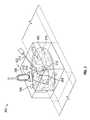
- FIGS. 1A and 1Billustrate exemplary volumetric medical images for image-guided medical procedures.
- FIG. 2illustrates a two-dimensional slice of a volumetric medical image with exemplary image scanning and needle approach paths.
- FIG. 3illustrates a three-dimensional volumetric medical image with exemplary image scanning and needle approach paths.
- FIG. 4is a flow diagram illustrative of an embodiment of a routine implemented by the system to display medical device emplacements.
- FIG. 5Ais a diagram illustrating an embodiment of an environment for medical device approaches.
- FIG. 5Bis a diagram illustrating an embodiment of a rendering of medical device approaches on a head mounted display.
- Implementations disclosed hereinprovide systems, methods, and apparatus for guidance tasks related to pose and/or emplacement of medical devices. Certain embodiments pertain to systems for facilitating visualization of medical imaging device scanning approaches and/or needle approaches for surgical applications. Visualization of medical device poses and/or emplacements can allow a user to select a desirable needle insertion point to minimize insertion or repositioning attempts.
- emplacementand the term “pose” as used herein are broad terms encompassing their plain and ordinary meanings and can refer to, without limitation, position, orientation, the combination of position and orientation, or any other appropriate location information.
- volumetric or 3D datathere are numerous types of volumetric or 3D data that various embodiments of the guidance system herein can display.
- the term “volumetric medical image”is a broad term that encompasses its ordinary and customary meaning, and includes, without limitation any data in a volume or 3D space that can be displayed.
- the volumetric medical imagecan include, without limitation, one or more of a CT scan, an MRI, other 3D intraoperative or preoperative imaging data, other volume data, segmented internal organs, segmented blood vessels, annotations, tumors, etc.
- Such datacan include CT scans, MRI, PET, 3D ultrasound, and any of numerous other types of 3D data.
- a subset of the datais chosen to display.
- This subset of datacan include axis-aligned slices, the entire volume, or a sub-volume of the data.
- An inherent difficulty with image guidanceis the display of three dimensions of data on a two-dimensional screen or “dual eye” three-dimensional display.
- a systemmight only display a single plane, or show three orthogonal planes separately on the screen.
- the datacan also be shown as a volumetric ‘block’ of data, as shown in FIG. 3 .
- FIGS. 1A and 1Billustrate exemplary volumetric medical images 100 for image-guided medical procedures.
- the image 100can be obtained based on any medical imaging technology as described herein, such as ultrasound, CT scan, MRI, open-magnet MRI, optical coherence tomography, positron emission tomography (“PET”) scans, fluoroscopy, ultrasound, or other preoperative, or intraoperative 2D or 3D anatomical imaging data.
- the image 100can include multiple planes or slices 110 of image data.
- the volumetric medical image 100can be obtained as a plurality of slices 110 from an imaging device configured to perform multiple 2D scans, or can be obtained as 3D volume data from an imaging device configured to generate 3D data.
- the planes or slices 110can be the slices as obtained from the 2D scans, or they can be modified and/or generated to be, for example, parallel to the plane of a region of interest.
- a plane or slice 110 of the volumetric medical image 100can include a cross-sectional view of a portion of a subject 120 .
- the location and orientation of the plane or slice 110can be selected automatically based on detected structures within the volumetric medical image 100 , or can be selected manually by a user.
- the location and orientationcan be selected so as to provide a cross-sectional view of a target structure 130 , such as a lesion, tumor, or other structure.
- a region of interest 140can be selected within a plane or slice 110 .
- the region of interest 140can be selected automatically or by a user, for example, based on a location of an emplacement sensor associated with a medical device (e.g., an ultrasound probe) manipulated by the user.
- the systemcan utilize and/or display imagery associated with the medical device (e.g., ultrasound image data obtained from an ultrasound probe) in addition to (e.g., superimposed upon) the volumetric image data.
- the data shown in the region of interestcan be any appropriate visualizable medical data, not limited to ultrasound or CT data.
- the data displayed outside of the region of interestcan be any visualizable medical data, and can even be from the same data set as the data shown in the region of interest.
- MRI datacan be shown in fading planes outside of the region of interest and in focus (and visualizable through a tunnel) inside the region of interest.
- annotationcan be displayed along with the rendering of the visualizable medical data inside and/or outside of the region of interest. In this manner, a user can see the annotations in the context of the visualizable medical data.
- more than one set of visualizable medical datacan be rendered.
- Each onecan be rendered in a different manner. For example, they can be rendered with different transparencies, brightnesses, contrast, colors, etc. Further, one or more can be rendered with a different transparency, brightness, contrast or color as distance from the region of interest increases. For example, brightness can decrease and/or transparency can increase further from the region of interest 140 .
- FIG. 2illustrates a 2D slice 200 of a volumetric medical image with exemplary image scanning and needle approach paths as determined by a guidance system.
- the exemplary slice 200 depicted in FIG. 2includes a cross-sectional image of a torso of a patient, including sections of skin 202 , bone 204 , gas 206 , and other internal tissues 208 , as well as a target structure 210 (non-limiting examples: a lesion, tumor, or other target).
- a suggested scanning path 212 for an ultrasound probe and suggested approach paths 216 for a needle 218 or other invasive medical deviceare also depicted.
- the target structures 210can be identified in a variety of ways.
- the guidance systemcan allow a user to mark a target by interacting with the display of the volumetric medical image. For example, consider a procedure where the doctor is using the guidance system with an ablation needle 218 and an ultrasound probe 214 . The doctor can mark the target structure 210 by pressing a button on a keyboard or mouse, touching a screen, pointing with a medical device, gesturing or issuing a verbal command, or with any other appropriate method, as described in greater detail in U.S. application Ser. No.
- the systemcan detect the target structure 210 within an image stream obtained from an imaging device manipulated by the user (e.g., an ultrasound probe 214 ).
- the location of the target structure 210can be marked at the point where a needle 218 intersects with the ultrasound image plane, where the needle's projection intersects with the ultrasound image plane, or any other appropriate relationship (such as at the location of the tip of the needle).
- a physicianidentifies a target structure 210 within the ultrasound image, she can point to it using the needle 218 even if the needle 218 is outside the body of the patient.
- the physicianor assistant
- the target structure 210can be identified based at least in part on the amount of time the physician spends observing an object or the amount of time that a medical device, or corresponding emplacement sensor, is located at a particular emplacement. For example, if a lesion or other abnormal structure is detected within the ultrasound image, and the physician keeps the probe 214 in the same location for a threshold time period (e.g., 3 seconds, 5 seconds, 10 seconds, 15 seconds, or longer), the guidance system can determine that the object is a target structure 210 and store its location.
- a threshold time periode.g., 3 seconds, 5 seconds, 10 seconds, 15 seconds, or longer
- the systemcan identify multiple target structures 210 .
- physiciansduring some liver ablation procedures or other procedures, can manage fifteen target structures 210 , or even more.
- the guidance systemcan store and display any number of target structures 210 concurrently. If there is more than one target structure 210 in view, the image guidance system can display a number or other indicator next to each one (not pictured).
- those target structures 210 which are closer to the ultrasound image planecan be drawn more saliently or vividly (with more bold color and thicker lines) while the points that are far away are drawn less saliently (more transparent, blurred, muted colors, etc.).
- the systemcan analyze the various types of tissue within the medical image to determine an approach path for the medical device.
- ultrasound waves emitted by an ultrasound probe 214may be able to travel through soft tissues 208 of a patient, but can be blocked or scattered by denser tissues such as bone 204 , or by regions of lower density, such as gas 208 .
- An ablation needle 218may be able to travel through soft tissues 208 and/or regions of gas 208 , but may be unable to travel through bone 204 .
- needle approach pathscan be chosen so as to avoid critical or delicate regions such as blood vessels or vital organs (not shown), even though such regions do not present an obstacle to ultrasound scanning.
- the guidance systemcan analyze the image to determine the tissue type or density of various regions of the image.
- the guidance systemcan analyze the slice 200 based on an intensity of some or all pixels or voxels within the slice 200 .
- the systemcan determine a radiodensity or intensity of voxels based on the Hounsfield unit (HU) scale or other suitable scale. For example, an intensity of greater than 700 HU can indicate bone, while an intensity of between ⁇ 1000 HU and ⁇ 350 HU can indicate intestinal gas.
- HUHounsfield unit
- an intensity of greater than 700 HUcan indicate bone
- an intensity of between ⁇ 1000 HU and ⁇ 350 HUcan indicate intestinal gas.
- different imaging devicescan output different HU for different types of tissue or can use different scales for intensity of voxels.
- the systemcan analyze the intensity level for each voxel of the medical image to determine the tissue type and/or object density.
- the systemcan use edge detection to identify objects, for example, to identify the location of skin 202 within the image slice 200 .
- the systemcan use known HU values of critical or delicate regions (e.g., large blood vessels, heart, etc.) to identify the critical or delicate regions.
- the systemcan analyze one or more image slices 200 taken at different parts of the breathing cycle. In this way, the system can determine the emplacement of the objects within the image slices at different parts of the breathing cycle and to identify deformations of the objects caused by breathing.
- the systemcan determine one or more medical device approach paths 216 to the target structure 210 from an approach region.
- the approach regioncan correspond to a surface or perimeter of the medical image and/or a desired start point, surface, or perimeter within the medical image.
- the systemcan determine and display paths to the target structure from the surface of the medical image and/or from a particular structure within the medical image, such as the skin, an intestine, etc.
- the systemcan analyze a plurality of paths between the target structure 210 and the approach region of the medical image.
- the systemcan use ray tracing or ray casting to identify paths to the target structure from the approach region of the image. For example, the system can analyze voxels along a particular path to identify paths between the target structure 210 and the approach region.
- the systemcan use one or more rendered images to identify paths to the target structure. For example, the system can render one more images from the perspective of the target to the approach region. In some embodiments, the system can treat the target as a point-of-view location and capture the voxels located between the target and the approach region, similar to a camera taking a picture. In certain embodiments, the volume of the captured voxels can generally correspond to a trapezoid, cone, frustum, or other shape.
- the one or more imagescan be from different angles with respect to the target.
- the systemcan render six images (each one from the perspective of a different side of a cube corresponding to the target). It will be understood that fewer or more images can be rendered from different perspectives, as desired.
- the systemcan render the captured images (or captured voxels). As part of the rendering process, the system can map the captured volume of voxels or imaged volume to an image area or region. In addition, the system can identify how to treat the different voxels during the rendering process. For example, the system can average the voxels that are to be located on a point of the image area, use the minimum or maximum voxel for the point of the image area, etc. In certain embodiments, the system can identify voxels that do not satisfy a density threshold (or otherwise not to be used for a medical device approach), as described in greater detail below, to be rendered differently than voxels that satisfy the density threshold. For example, if any one of the voxels that are to be mapped to a location in the image area do not satisfy the density threshold, the system can color code the location, such as by whiting or blacking it out, etc.
- a density thresholdor otherwise not to be used for a medical device approach
- the one or more rendered imagescan be applied or mapped to the approach region of the 3D volume for viewing.
- the systemcan map the one or more images to the surface of the 3D volume. Accordingly, a user can view the images on the approach region.
- the systemcan also analyze the rendered images (non-limiting examples: using voxel intensities, edge detection, etc.) to identify locations in the rendered image in which the approach region is visible. The identification of the approach region within the image can indicate a direct path between the approach region and the target structure 210 .
- the systemcan analyze the image slice 200 along a series of circular paths of increasing or decreasing radius and centered on the target structure 210 (e.g., from the target structure 210 outward, or from the approach region). For example, the system can analyze the voxels within a sphere and then expand the sphere and analyze voxels within the expanded sphere. Analyzing a series of circular paths can be efficient, as angular segments of a circle that are deemed unacceptable for an approach path (e.g., due to the presence of bone) can be eliminated and the same angular regions can be omitted from the analysis of later circular paths, reducing processing time.
- the systemcan apply one or more criteria to identify one or more medical device approaches 216 .
- the systemcan use a density threshold, characteristics of a medical device, such as length, width, thickness, diameter, scan length, scan width, number transducer crystals, etc., to identify one or more medical device approaches.
- the systemcan use characteristics of the medical device to determine the medical device approach.
- the medical devicecan be an invasive medical device, such as, but not limited to, biopsy needles, ablation needles, surgical needles, nerve-block needles, or other needles, electrocautery device, catheters, stents, laparoscopes or laparoscopic cameras, ultrasound transducers, or other instruments that enter a part of the body, or a non-invasive medical device that does not enter the body, such as, but not limited to, ultrasound transducers, probes, other external imaging devices, or other external devices, etc.
- the medical devicescan also include medical imaging devices that provide or aid in the selection or generation of medical images for display.
- the medical imaging devicecan be any device that is used to select a particular medical image for display or generate medical images.
- the medical imaging devicescan include invasive medical devices, such as laparoscopic cameras or invasive ultrasound transducers, and non-invasive medical devices, such as external ultrasound transducers.
- each medical devicecan have unique dimensions, such as width, length, and/or thickness.
- medical imaging devicescan have different imaging depths, widths and/or thicknesses.
- medical imaging devicescan use different scan frequencies, each of which can affect which tissue can be imaged. The system can use the various characteristics to determine the medical device approach.
- the systemcan use the medical device's physical characteristics, such as its hardness or ability to pass through an object, deformability, curvature, length, width, thickness, and/or diameter to determine the medical device approach.
- Each invasive medical devicecan have its own physical characteristics that the system can use to determine the medical device approach. For example, the system can determine more medical device approaches for invasive medical devices that are longer, thinner, are more deformable, or have a smaller diameter.
- the diameter, deformability, or thickness of the medical devicecan be different along its longitudinal axis. In such cases, the system can use the different thicknesses or deformability and location along the medical device axis to identify the medical device approaches.
- the systemcan eliminate paths 216 that pass through a region of bone 204 , or other region through which the needle 218 is unable to pass. Similarly, the system can eliminate paths for the needle 218 that pass through a vital organ or a large blood vessel (not shown).
- the systemcan have information regarding the length of the needle 218 , and can determine a maximum approach path length based at least in part on the length of the needle 218 and/or maximum reach of the needle 218 .
- the systemcan exclude approach paths in which the distance from the approach region to the target structure 210 is longer than maximum approach length.
- the systemcan include approach paths 216 that are wide and/or thick enough for the needle 218 to pass through, based on a known width and/or thickness of the needle 218 .
- the systemcan use the imaging characteristics of the medical imaging device, such as, but not limited to, number of imaging sensors, number of transducer crystals, imaging frequency, imaging depth, resolution, fade, imaging width and/or imaging thickness to determine the medical device approach.
- the systemcan also use the physical characteristics of the medical imaging device.
- the systemcan use the physical characteristics of a non-invasive medical imaging as well.
- each medical imaging devicecan have unique imaging characteristics that the system can use to determine the medical device approach.
- the systemcan determine more medical device approaches for medical imaging devices that have a larger imaging depth, larger imaging width, more imaging sensors, more transducer crystals, better resolution, less fade, or lower imaging frequency.
- medical imaging devices with a larger imaging depth or width, more imaging sensors or more transducerscan capture a larger image cross-section, which can enable the system to identify more potential paths.
- the systemcan identify more medical device paths for medical imaging devices that have better resolution or fade less based on the distance from the imager, etc.
- imaging devicesmay unable to penetrate certain tissue depending on the frequency used. For example, many ultrasound transducers are unable to penetrate bone or gas and optical image sensors cannot penetrate tissue. Accordingly, the system can use the scan frequency or imaging type to identify medical device approaches. In addition, as described in greater detail above with reference to invasive medical devices, the physical characteristics of a medical imaging device can affect the number of medical device approaches identified by the system.
- the systemcan eliminate paths that pass through regions of gas 206 or bone 204 because the ultrasound probe 214 cannot penetrate the gas 206 or bone 204 .
- the system 204can use the imaging dimensions of the ultrasound probe to determine medical device paths 212 as well.
- the ultrasound probe 214can include a linear array of ultrasound crystals, each crystal configured for imaging along a path.
- the systemcan determine acceptable ultrasound scanning paths 212 where the unobstructed path has a 2D or 3D width of a threshold number of crystals.
- the threshold number of crystalscan be based on a number of crystals that have an unobstructed path to the target structure 210 and/or a number of crystals of the ultrasound probe 214 .
- the threshold number of crystalscan be 25% or 50% of the total number of crystals of the ultrasound probe 214 , and/or can be 1, 3, 5, 10, 15, or more crystals.
- the systemcan use the image depth of the ultrasound probe 214 determine the medical device approach paths 216 . For example, the system can eliminate paths to the target structure 210 that are longer than the image depth of the ultrasound probe 210 .
- the systemcan use a density threshold to identify the medical device approaches.
- the density thresholdcan be based on the Hounsfield unit scale or similar scale, and can correspond to a minimum density, maximum density, or range of densities.
- the systemcan identify objects and/or voxels that do not satisfy the density threshold (or are otherwise not to be used for a medical device approach) as obstructing objects.
- the systemcan exclude paths that intersect with obstructing objects.
- the systemcan identify voxels or objects that have been annotated as not satisfying the density threshold or as obstructing objects.
- the density thresholdcan be static.
- the systemcan identify objects having a density equal to or greater than bone as not satisfying the density threshold, irrespective of the medical device.
- the systemcan identify objects having the same density as the critical tissue as not satisfying the density threshold.
- the density thresholdcan be based at least in part on a characteristic of the medical device. For example, gas within the image can satisfy the density threshold for a needle, but may not satisfy the threshold for an ultrasound device.
- the systemcan identify one or more semi-direct paths from the target structure 210 to the approach region.
- a semi-direct pathmay become available if a portion of the approach region or patient is deformed. For example, if a physician wants to access a portion of a patient under the rib cage, no direct path may be available. However, by compressing the abdomen or at a different time of the breathing cycling, a path may become available.
- the systemcan identify semi-direct medical device approaches based at least in part on a deformation of the approach region of an image.
- the systemcan apply a flexibility factor to the approach region of the image.
- the flexibility factorcan indicate a margin by which the approach region can be flexed. For example, the flexibility factor can indicate that the approach region can be flexed by 1-5 cm or by a percentage of the width or length of the image. Using the flexibility factor and other information as describe above, the system can identify one or more semi-direct medical device approaches.
- the systemcan identify and display locations at the approach region for emplacing the medical device.
- the systemcan identify and display a particular emplacement for the medical device. For example, the system can highlight a region corresponding to the dimensions of the medical device.
- the systemcan display emplacements such as by displaying images of needles 218 along the acceptable paths 216 , and/or by displaying highlighted or otherwise distinguishing portions of the approach region.
- the systemcan display one or more ultrasound probe emplacements such as by displaying an image of an ultrasound probe 214 , by displaying a scanning region or path 212 , and/or by displaying a highlighted or otherwise distinguishable portion of the approach region.
- the systemcan highlight the portions of the medical image area or volume that satisfy the density threshold and/or that can be used as part of a medical device approach. For example, the objects and/or voxels throughout the medical image area or volume that satisfy the density threshold can be highlighted, displayed in a particular color, or brought into focus. Similarly, the objects and/or voxels that do not satisfy the density threshold can be faded, taken out of focus, displayed in a different color etc.
- the systemcan track the location of a medical device and identify and display one or more medical device approaches based at least in part on the determined location of the medical device.
- the medical device approachescan be for the tracked medical device and/or for another medical device.
- the systemcan display medical device approaches for the medical imaging device and/or for another medical device, such as an invasive medical device.
- the systemcan use the determined location of the medical device to display and/or highlight the medical device approaches that are nearest to the medical device. For example, if the system identifies six medical device approaches, it can use the determined location of the medical device to display or highlight the three medical device approaches that are closest to the determined location of the medical device.
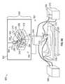
- FIG. 3illustrates a 3D volumetric medical image 300 with exemplary image scanning and needle approach paths.
- the exemplary image 300 depicted in FIG. 3includes a volume 302 containing a target structure 304 , skin 306 , bone 308 , and other internal tissue 310 .
- the systemcan determine approach paths, poses, and/or emplacements for medical devices such as an ultrasound probe 312 and a needle 314 , or other medical device.
- acceptable, desirable, and/or suggested approach pathscan be selected in a 3D volume based on the same or similar criteria as those discussed above with reference to FIG. 2 .
- ultrasound scanning pathscan be selected so as to avoid regions of gas or bone 308 and/or based on imaging characteristics of the ultrasound probe 312 .
- Needle approach pathscan be selected so as to avoid regions of bone 308 or delicate structures, and/or based on the length of the needle 314 .
- the systemcan use a variety of techniques to analyze and determine acceptable medical device approaches.
- the systemcan utilize ray tracing, projective texture mapping, edge detection, one or more rendered images, etc., as described above by analyzing a plurality of direct or semi-direct paths between the approach region and the target structure 304 .
- Some implementationscan analyze the volume 302 along a plurality of spherical surfaces of increasing or decreasing radius, similar to the circular paths analyzed in 2D systems described above. Suggested emplacements 316 can then be displayed within the 3D volumetric medical image 300 for the reference of the physician.
- voxels in frontWhen displaying 3D volumetric data, voxels in front (closer to the point-of-view location) can obscure the voxels behind them. In some instances, this can occlude information that can be important from preoperative 3D data.
- One way to address thisis to allow the doctor to view the data as 2D slices 318 , in cross section, etc., as described in greater detail in U.S. application Ser. No. 14/166,179, entitled, SYSTEMS, METHODS, APPARATUSES, AND COMPUTER-READABLE MEDIA FOR IMAGE MANAGEMENT IN IMAGE-GUIDED MEDICAL PROCEDURES, incorporated by reference herein in its entirety.
- a 2D slice 318can be displayed based on a location of a medical device such as an ultrasound probe 312 , with the location of the probe 312 determined based on data received from one or more emplacement sensors.
- the determined location of the probe 312can be used to select suggested emplacements for a second medical device.
- the guidance systemcan select one or more needle emplacements 316 near the location of the probe 312 (e.g., to facilitate the physician's simultaneous manipulation of the probe 312 and needle 314 ).
- a previously obtained, real-time, or near real-time medical imagee.g., an ultrasound image obtained from the probe 312
- the 2D slice 318can be displayed with increased brightness relative to the surrounding 3D volumetric image 300 , or the surrounding 3D volumetric image 300 can be displayed with partial transparency or out of focus.
- FIG. 4is a flow diagram illustrative of an embodiment of a routine 400 implemented to display medical device emplacements.
- routine 400can be implemented by one or more computing devices/components that are associated with the systems described herein.
- various blocks described herein with reference to FIG. 4can be implemented in a variety of orders. For example, some blocks can be implemented concurrently or in a different order as desired. For example, the system can perform blocks 420 and 430 concurrently and/or implement them in a different order.
- a volumetric medical image received as a single set of 3D volumetric datacan be converted into a plurality of 2D image slices, and/or a volumetric medical image received as a plurality of 2D slices can be converted to a 3D volume as desired.
- the density of content within the volumetric medical imagecan be determined.
- the density of contentcan be determined based at least in part on an intensity value of voxels associated with the content within the volumetric medical image.
- the density of contentcan be determined using a Hounsfield unit scale or other suitable scale for determining a mass density based on an image intensity.
- an indicationcan be received of a target location within the volumetric medical image.
- the indicationcan be received from a human user, such as a physician, can be received based on an input by the user, such as a keystroke or button press, or the like, and/or can be determined by the system, for example, based on a computer analysis of a region within the medical image, a detected reaction of a user, an amount of time a medical device is located at a particular emplacement, etc.
- the target locationcan be determined based on a real-time medical image. For example, as depicted in FIG.
- the systemcan identify one or more obstructing objects within the volumetric medical image.
- the systemcan identify the obstructing objects based at least in part on the determined density of content within the volumetric medical image, or otherwise identified as not to be used for a medical device approach.
- obstructing objectscan be determined based on regions within the volumetric medical image having a density that does not satisfy a density threshold.
- the density thresholdcan be a minimum density, maximum density or range of densities.
- the systemcan identify a region having a density approximately equal to the density of bone as an obstructing object.
- the density thresholdcan be based at least in part on one or more imaging characteristics of a medical imaging device.
- the density thresholdcan be based at least in part on a scan frequency of the medical imaging device.
- the maximum density of objects through which waves and/or radiation emitted by a medical imaging device can passcan vary based on the frequency of the waves and/or radiation (e.g., a scan frequency).
- the density thresholdcan be determined based on the known scan frequency of the medical imaging device.
- the density thresholdcan be determined based on the type of medical device (e.g., whether the device is an intrusive device, medical imaging device, or other type of medical device).
- the density thresholdcan include a single threshold, or can include a range of densities.
- a density threshold for an ultrasound imaging devicecan include the range between ⁇ 350 HU and 700 HU, so as to exclude regions of intestinal gas, air, and bone.
- the one or more pathwayscan be determined based at least in part on characteristics of the medical device, such as threshold quantity of transducing elements at a particular emplacement of a medical imaging device each having a direct path to the target location that does not pass through the identified obstructing objects, other imaging characteristics of the medical device, and/or physical characteristics of the medical device.
- the systemcan identify the one or more pathways based on a tracked location of a medical device.
- the pathways identifiedcan be for the tracked medical device and/or for another medical device.
- the systemcan cause a display to concurrently display the volumetric medical image and a plurality of emplacements on the approach region of the volumetric medical image for emplacements of one or more medical devices.
- Emplacementscan be determined based at least in part on the determined plurality of direct pathways.
- an emplacement for a medical imaging devicecan be determined as a location along the approach region of the volumetric medical image where the image scanning path of the medical imaging device includes one of the determined plurality of direct pathways within the volumetric medical image.
- the plurality of emplacementscan be displayed based at least in part on a determined emplacement of an emplacement sensor based on emplacement data received from the emplacement sensor.
- the emplacement sensorcan be associated with a medical device such as an ultrasound probe.
- FIG. 5Ais a diagram illustrating an embodiment of an environment 500 for image-guided medical device approaches.
- the environment 500includes a display 502 displaying an image 550 , an image guidance unit 504 , a position sensing unit 506 , a surgical system 508 , imager 510 , medical devices 512 , 514 , a patient 516 , a stand 518 , and a table 520 .
- an image guidance system 501can include any one or any combination of the display 502 , the image guidance unit 504 , the position sensing unit 506 , the surgical system 508 , the imager 510 , the medical devices 512 , 514 , the stand 518 , and/or the table 520 .
- the image guidance unit 504can process or combine the data and show image guidance data on display 502 .
- This image guidance datacan be used by a healthcare provider to guide a procedure and improve care.
- the image guidance datacan also be used by the system to identify the medical device approaches described above.
- system 501there are numerous other possible embodiments of system 501 .
- many of the depicted componentscan be joined together to form a single component and can be implemented in a single computer or machine.
- additional position sensing unitscan be used in conjunction with position sensing unit 506 to track relevant medical devices 512 , 514 , as discussed in more detail below.
- Additional imagers 510can be included, and combined imaging data from the multiple imagers 510 can be processed by image guidance unit 504 and shown on display 502 .
- two or more surgical systems 508can be used.
- Image guidance unit 504Information about and from multiple surgical systems 508 and attached medical devices 512 (and additional medical devices not shown) can be processed by image guidance unit 504 and shown on display 502 . These and other possible embodiments are discussed in more detail below. It will be understood that any combination of the display objects, image guidance cues, etc., described herein can be displayed concurrently, or simultaneously. Further, reference to displaying objects “concurrently” and/or “simultaneously” is to be interpreted broadly and may refer to displaying objects in such a way that to a human observer the objects are visible at the same time.
- Imager 510can be communicatively coupled to image guidance unit 504 .
- imager 510can be coupled to a second display unit (not shown).
- the second display unitcan display imaging data from imager 510 .
- the imaging data displayed on display 502 and displayed on second display unitcan be the same or different.
- the imager 510can be an ultrasound machine 510
- the medical device 514can be a movable imaging unit, such as an ultrasound transducer 514 or ultrasound probe 514
- the second display unitcan be a display associated with the ultrasound machine 510 that displays the ultrasound images from the ultrasound machine 510 .
- a movable imaging unit 514can be communicatively coupled to image guidance unit 504 .
- the movable imaging unit 514can be useful for allowing a user to indicate what portions of a first set of imaging data are to be displayed.
- the movable imaging unit 514can be an ultrasound transducer 514 , a needle or other medical device, for example, and can be used by a user to indicate what portions of imaging data, such as an intraoperative or preoperative CT scan, to show on a display 502 as image 550 .
- the movable imaging unit 514can also be used to identify medical device approaches for the moveable imaging unit 514 and/or for the medical device 512 . Further, in some embodiments, there can be a third set of intraoperative or preoperative imaging data that can be displayed with the first set of imaging data.
- a navigation system 501comprises a display 502 and a position sensing unit 506 communicatively coupled to image guidance unit 504 .
- position sensing unit 506 , display 502 , and image guidance unit 504are coupled to the stand 518 .
- Image guidance unit 504can be used to produce images 550 that are displayed on display 502 .
- the images 550 produced on display 502 by the image guidance unit 504can be determined based on ultrasound or other visual images from the first medical device 512 and second medical device 514 .
- the image 550can include the volumetric medical image 300 described above in greater detail.
- some or all of the display objectscan be displayed as 3D objects.
- the display objects in the image 550can be displayed in a perspective based at least in part on a point-of-view location.
- More medical devicescan be added to the system 501 as desired.
- the system 501can include an ultrasound probe, ablation needle, laparoscopic camera, stapler, cauterizer, scalpel and/or any other medical device or medical device, and the system 501 can determine a medical device approach for any one or more of the medical devices.
- the system 501can also process and/or display collected data, such as preoperative CT scans, X-Rays, MRIs, laser scanned 3D surfaces etc.
- the imaging data obtained from one or both of medical devices 512 and 514can include other modalities such as a CT scan, MRI, open-magnet MRI, optical coherence tomography (“OCT”), positron emission tomography (“PET”) scans, fluoroscopy, ultrasound, or other preoperative, or intraoperative 6D or 3D anatomical imaging data.
- medical devices 512 and 514can also be scalpels, implantable hardware, or any other device used in surgery. Any appropriate surgical system 508 or imager 510 can be communicatively coupled to the corresponding medical instruments 512 and 514 .
- the medical devices 512 , 514can be communicatively coupled to the position sensing unit 506 (non-limiting example: sensors embedded or coupled to the medical devices 512 , 514 can be communicatively coupled with the position sensing unit 506 ).
- the position sensing unit 506can be part of imager 510 or it can be separate.
- the position sensing unit 506can be used to determine the emplacement of first medical device 512 and/or the second medical device 514 .
- the position sensing unit 506can include a magnetic tracker and/or one or more magnetic coils can be coupled to medical devices 512 and/or 514 .
- the position sensing unit 506can be an optical 3D tracking system using fiducials for tracking sensors.
- optical 3D tracking systemscan include the NIDI Polaris Spectra, Vicra, Certus, PhaseSpace IMPULSE, Vicon MX, InterSense IS-900, NaturalPoint OptiTrack, Polhemus FastTrak, IsoTrak, or Claron MicronTracker2.
- the position sensing unit 506can each be an inertial 3D tracking system comprising a compass, accelerometer, tilt sensor, and/or gyro, such as the InterSense InertiaCube or the Nintendo Wii controller, mechanical tracking system, camera-based tracking system, radar-based tracking system, etc.
- the position sensing unit 506can be attached to or affixed on the corresponding medical device 512 and 514 .
- a signal emitting devicemight include a radio-frequency identifier (RFID).
- RFIDradio-frequency identifier
- the position sensing unit 506can use the GPS coordinates of the tracking sensors or can, for example, triangulate the radio frequency signal being emitted by the RFID associated with tracking sensors.
- the tracking systemscan also include one or more 3D mice.
- the system 501can register the location of the volumetric medical image 300 to the position sensing region and/or the patient 516 . Accordingly, the system 501 can use the emplacement data and registered volumetric medical image 300 to identify the location of the medical devices 512 , 514 , relative to the volumetric medical image 300 , as well as identify the emplacement of the medical devices 512 , 514 relative to the identified medical device approaches.
- the display 502displays 3D images to a user, such as a healthcare provider.
- Stereoscopic 3D displaysseparate the imagery shown to each of the user's eyes. This can be accomplished by a stereoscopic display, a lenticular auto-stereoscopic display, or any other appropriate type of display.
- the display 502can be an alternating row or alternating column display.
- Example alternating row displaysinclude the Miracube G240S, as well as Zalman Trimon Monitors.
- Alternating column displaysinclude devices manufactured by Sharp, as well as many “auto-stereoscopic” displays (non-limiting example: Philips).
- Sony Panasonic 3D passive displays and LG, Samsung, and/or Vizio 3D TVscan be used as well.
- Display 502can also be a cathode ray tube.
- Cathode Ray Tube (CRT) based devicescan use temporal sequencing, showing imagery for the left and right eye in temporal sequential alternation. This method can also be used by projection-based devices, as well as by liquid crystal display (LCD) devices, light emitting diode (LED) devices, organic LED (OLED) devices, liquid crystal on silicon (LCOS) devices, DLP devices, virtual retinal display (MicroVision) devices, or the like.
- LCDliquid crystal display
- LEDlight emitting diode
- OLEDorganic LED
- LCOSliquid crystal on silicon
- DLP devicesliquid crystal on silicon
- MicroVisionvirtual retinal display
- the display 502shows a perspective view of the volumetric medical image 300 , a virtual ultrasound probe 316 , and a virtual needle 314 .
- the virtual medical devices 314 , 316can be displayed in a virtual 3D scene with the display 502 acting as a window into the virtual 3D scene.
- the virtual medical device 558can also move to the right.
- the virtual medical devices 314 , 316can correspond to determined medical device emplacements for the medical devices 512 , 514 .
- the virtual medical devices 314 , 316can indicate where the medical devices 512 , 514 are to be emplaced.
- the display 502can include additional virtual medical devices indicating the determined location of the medical devices 512 , 514 relative to the recommended emplacements. Additional cues can be displayed to aid the user in emplacing the medical devices in the recommended emplacements.
- the virtual medical device 558will likewise show the change in orientation, and show the tip of the virtual medical device 558 in the background and the other end of the virtual medical device 558 in the foreground.
- the point-of-view locationcan be a fixed location, such as a predetermined distance/angle from the display 502 or stand 518 and or a location configured by the user; or the point-of-view location can by dynamic.
- the system 501can track a user in real-time and determine the point-of-view location based at least in part on the tracked location of the user.
- a healthcare providermay want to track one or more of a scalpel, a biopsy, a cauterizer (including an electrocauterizer and Bovies), forceps, cutting loops on hysteroscopes, harmonic shears, lasers (including CO 2 lasers), etc.
- a scalpelincluding an electrocauterizer and Bovies
- a cauterizerincluding an electrocauterizer and Bovies
- forcepsincluding an electrocauterizer and Bovies
- cutting loops on hysteroscopesincluding harmonic shears
- lasersincluding CO 2 lasers
- a healthcare provideris able to see image guidance data on display 502 that will allow her to know the relative pose, location, or emplacement of the tracked instrument(s) with respect to one another or with respect to imaging data and will be able to see, on display 620 , the features of the instrument rendered in the scene.
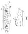
- virtual image contentmay be displayed on a head mounted display (HMD) 650 instead of or in addition to a display 502 as depicted in FIG. 5A .
- the HMD 650may include one or more stereoscopic displays 652 , 654 configured to display 3D content to a wearer of the HMD 650 .
- the HMD 650can include a left stereoscopic display 652 configured to project visual content 653 to a left eye of a wearer, and a right stereoscopic display 654 configured to display visual content 655 to a right eye of a wearer.
- displays 652 , 654can be opaque, and can be sized and located so as to occupy a subset of the field of view of a wearer.
- the displays 652 , 654can be transparent or translucent, or can be implemented as retinal scan displays that project an image onto the eye rather than display an image on the display screen.
- Displays 652 , 654can allow the displayed content 653 , 655 to be seen clearly without glare or interference due to light from the world in the region beyond the displays 652 , 654 .
- displays 652 , 654 occupying less than the entire field of view of the wearercan allow the wearer to view the virtual content 653 , 655 while also viewing the real environment in the portion of the wearer's field of view unoccupied by displays 652 , 654 , such as through transparent portions 656 , 658 of the HMD 650 .
- the imaging systems described hereincan display virtual image content on the displays 652 , 654 using a location offset with respect to the location of corresponding objects.
- the displays 652 , 654can display and/or augment objects that are located elsewhere, such as below, above, or to the side of the displays 652 , 654 and/or the HMD 650 .
- the virtual medical devices 314 , 316can be displayed with a vertical offset relative to the medical devices 512 , 514 so as to allow a wearer of the HMD 650 , such as a surgeon, to simultaneously view the virtual medical devices 558 , 560 on the displays 652 , 654 and the patient 516 with real medical devices 512 , 514 located below the HMD 650 .
- the offsetcan be predetermined based on a dimension or preference of an individual wearer prior to use, or based on a default such as an average height or dimension of an expected wearer, or dynamic based on data received during use.
- An example offsetcan be a distance equal or approximately equal to the vertical distance between a wearer's elbow and the wearer's eye (non-limiting example: in the range of 0.5 m to 5 m), a measured distance between the HMD 650 and the medical devices 512 , 514 , etc. Additional details regarding the offset are described in U.S. application Ser. No. 15/199,630, filed concurrently herewith, entitled, LOUPE DISPLAY.
- the HMD 650can be implemented with any of the imaging systems described with reference to FIG. 5A .
- displays 652 , 654can display virtual medical devices 558 , 560 corresponding to real medical devices 512 , 514 .
- Displays 652 , 654can further display any other virtual image features described elsewhere herein, such as a medical device approach.
- position sensing units, a display unit, image guidance units, and/or any other module or unit of embodiments hereincan each be separate computing devices, applications, or processes or can run as part of the same computing devices, applications, or processes—or one of more can be combined to run as part of one application or process—and/or each or one or more can be part of or run on a computing device.
- Computing devices or computer systemscan include a bus or other communication mechanism for communicating information, and a processor coupled with the bus for processing information.
- a computer system or devicecan have a main memory, such as a random access memory or other dynamic storage device, coupled to the bus.
- the main memorycan be used to store instructions and temporary variables.
- the computer system or devicecan also include a read-only memory or other static storage device coupled to the bus for storing static information and instructions.
- the computer systems or devicescan also be coupled to a display, such as a CRT, LCD monitor, LED array, e-paper, projector, or stereoscopic display.
- Input devicescan also be coupled to the computer system or device. These input devices can include a mouse, a trackball, touchscreen, tablet, foot pedal, or cursor direction keys.
- Each computer system or computing devicecan be implemented using one or more physical computers, processors, embedded devices, field programmable gate arrays (FPGAs), or computer systems or portions thereof.
- the instructions executed by the computer system or computing devicecan also be read from a computer-readable medium.
- the computer-readable mediumcan be non-transitory, such as a CD, DVD, optical or magnetic disk, laserdisc, flash memory, or any other medium that is readable by the computer system or device.
- hardwired circuitrycan be used in place of or in combination with software instructions executed by the processor. Communication among modules, systems, devices, and elements can be over a direct or switched connections, and wired or wireless networks or connections, via directly connected wires, or any other appropriate communication mechanism.
- Transmission of informationcan be performed on the hardware layer using any appropriate system, device, or protocol, including those related to or utilizing Firewire, PCI, PCI express, CardBus, USB, CAN, SCSI, IDA, RS232, RS422, RS485, 802.11, etc.
- the communication among modules, systems, devices, and elementscan include handshaking, notifications, coordination, encapsulation, encryption, headers, such as routing or error detecting headers, or any other appropriate communication protocol or attribute.
- Communicationcan also messages related to HTTP, HTTPS, FTP, TCP, IP, ebMS OASIS/ebXML, DICOM, DICOS, secure sockets, VPN, encrypted or unencrypted pipes, MIME, SMTP, MIME Multipart/Related Content-type, SQL, etc.
- Any appropriate 3D graphics processingcan be used for displaying or rendering, including processing based on OpenGL, Direct3D, Java 3D, etc.
- Whole, partial, or modified 3D graphics packagescan also be used, such packages including 3DS Max, SolidWorks, Maya, Form Z, Cybermotion 3D, VTK, Slicer, or any others.
- various parts of the needed renderingcan occur on traditional or specialized graphics hardware.
- the renderingcan also occur on the general CPU, on programmable hardware, on a separate processor, be distributed over multiple processors, over multiple dedicated graphics cards, or using any other appropriate combination of hardware or technique.
Landscapes
- Engineering & Computer Science (AREA)
- Health & Medical Sciences (AREA)
- Life Sciences & Earth Sciences (AREA)
- Medical Informatics (AREA)
- Physics & Mathematics (AREA)
- General Health & Medical Sciences (AREA)
- Biophysics (AREA)
- Pathology (AREA)
- Biomedical Technology (AREA)
- Heart & Thoracic Surgery (AREA)
- Molecular Biology (AREA)
- Surgery (AREA)
- Animal Behavior & Ethology (AREA)
- Veterinary Medicine (AREA)
- Public Health (AREA)
- Nuclear Medicine, Radiotherapy & Molecular Imaging (AREA)
- Radiology & Medical Imaging (AREA)
- High Energy & Nuclear Physics (AREA)
- Optics & Photonics (AREA)
- Theoretical Computer Science (AREA)
- General Physics & Mathematics (AREA)
- Computer Vision & Pattern Recognition (AREA)
- Human Computer Interaction (AREA)
- Software Systems (AREA)
- Computer Graphics (AREA)
- Remote Sensing (AREA)
- Radar, Positioning & Navigation (AREA)
- Computer Hardware Design (AREA)
- General Engineering & Computer Science (AREA)
- Ultra Sonic Daignosis Equipment (AREA)
- Pulmonology (AREA)
- Quality & Reliability (AREA)
- Apparatus For Radiation Diagnosis (AREA)
Abstract
Description
This application claims the benefit of U.S. Provisional Application No. 62/195,676, filed Jul. 22, 2015, which is incorporated by reference herein in its entirety for all purposes. Any and all applications for which a foreign or domestic priority claim is identified in the Application Data Sheet as filed with the present application are incorporated by reference under 37 CFR 1.57 and made a part of this specification.
Image-guided surgery makes use of imaging to aid the surgeon to perform more effective or more accurate surgery.
Implementations disclosed herein provide systems, methods, and apparatus for guidance tasks related to pose and/or emplacement of medical devices. Certain embodiments pertain to systems for facilitating visualization of medical imaging device scanning approaches and/or needle approaches for surgical applications. Visualization of medical device poses and/or emplacements can allow a user to select a desirable needle insertion point to minimize insertion or repositioning attempts.
Systems and methods described herein can be used to determine one or more poses and/or emplacements of various medical devices. The term “emplacement” and the term “pose” as used herein are broad terms encompassing their plain and ordinary meanings and can refer to, without limitation, position, orientation, the combination of position and orientation, or any other appropriate location information.
As discussed elsewhere herein, there are numerous types of volumetric or 3D data that various embodiments of the guidance system herein can display. The term “volumetric medical image” is a broad term that encompasses its ordinary and customary meaning, and includes, without limitation any data in a volume or 3D space that can be displayed. The volumetric medical image can include, without limitation, one or more of a CT scan, an MRI, other 3D intraoperative or preoperative imaging data, other volume data, segmented internal organs, segmented blood vessels, annotations, tumors, etc. Such data can include CT scans, MRI, PET, 3D ultrasound, and any of numerous other types of 3D data. In some embodiments, in order to display 3D data on a 2D interface, such as a computer screen, or even a 3D interface, such as a head-mounted display or other 3D display, a subset of the data is chosen to display. This subset of data can include axis-aligned slices, the entire volume, or a sub-volume of the data. An inherent difficulty with image guidance is the display of three dimensions of data on a two-dimensional screen or “dual eye” three-dimensional display. When displaying 3D data, such as CT scans, a system might only display a single plane, or show three orthogonal planes separately on the screen. The data can also be shown as a volumetric ‘block’ of data, as shown inFIG. 3 .
As depicted inFIG. 1B , a plane orslice 110 of the volumetricmedical image 100 can include a cross-sectional view of a portion of asubject 120. In various embodiments, the location and orientation of the plane orslice 110 can be selected automatically based on detected structures within the volumetricmedical image 100, or can be selected manually by a user. For example, the location and orientation can be selected so as to provide a cross-sectional view of atarget structure 130, such as a lesion, tumor, or other structure.
In some embodiments, a region ofinterest 140 can be selected within a plane orslice 110. The region ofinterest 140 can be selected automatically or by a user, for example, based on a location of an emplacement sensor associated with a medical device (e.g., an ultrasound probe) manipulated by the user. In some implementations, the system can utilize and/or display imagery associated with the medical device (e.g., ultrasound image data obtained from an ultrasound probe) in addition to (e.g., superimposed upon) the volumetric image data. The data shown in the region of interest can be any appropriate visualizable medical data, not limited to ultrasound or CT data. Further, the data displayed outside of the region of interest can be any visualizable medical data, and can even be from the same data set as the data shown in the region of interest. For example, MRI data can be shown in fading planes outside of the region of interest and in focus (and visualizable through a tunnel) inside the region of interest. Further, annotation can be displayed along with the rendering of the visualizable medical data inside and/or outside of the region of interest. In this manner, a user can see the annotations in the context of the visualizable medical data.
In various embodiments, more than one set of visualizable medical data can be rendered. Each one can be rendered in a different manner. For example, they can be rendered with different transparencies, brightnesses, contrast, colors, etc. Further, one or more can be rendered with a different transparency, brightness, contrast or color as distance from the region of interest increases. For example, brightness can decrease and/or transparency can increase further from the region ofinterest 140.
Thetarget structures 210 can be identified in a variety of ways. In some embodiments, the guidance system can allow a user to mark a target by interacting with the display of the volumetric medical image. For example, consider a procedure where the doctor is using the guidance system with anablation needle 218 and anultrasound probe 214. The doctor can mark thetarget structure 210 by pressing a button on a keyboard or mouse, touching a screen, pointing with a medical device, gesturing or issuing a verbal command, or with any other appropriate method, as described in greater detail in U.S. application Ser. No. 14/166,179, entitled, SYSTEMS, METHODS, APPARATUSES, AND COMPUTER-READABLE MEDIA FOR IMAGE MANAGEMENT IN IMAGE-GUIDED MEDICAL PROCEDURES, and Ser. No. 13/014,596, entitled, IMAGE ANNOTATION IN IMAGE-GUIDED MEDICAL PROCEDURES, each of which is incorporated herein by reference in its entirety.
In some embodiments, the system can detect thetarget structure 210 within an image stream obtained from an imaging device manipulated by the user (e.g., an ultrasound probe214). In certain embodiments, the location of thetarget structure 210 can be marked at the point where aneedle 218 intersects with the ultrasound image plane, where the needle's projection intersects with the ultrasound image plane, or any other appropriate relationship (such as at the location of the tip of the needle). For example, when a physician identifies atarget structure 210 within the ultrasound image, she can point to it using theneedle 218 even if theneedle 218 is outside the body of the patient. The physician (or assistant) can press, for example, a button or foot pedal, which informs the image guidance system to store the 3D position of thistarget structure 210. In some embodiments, thetarget structure 210 can be identified based at least in part on the amount of time the physician spends observing an object or the amount of time that a medical device, or corresponding emplacement sensor, is located at a particular emplacement. For example, if a lesion or other abnormal structure is detected within the ultrasound image, and the physician keeps theprobe 214 in the same location for a threshold time period (e.g., 3 seconds, 5 seconds, 10 seconds, 15 seconds, or longer), the guidance system can determine that the object is atarget structure 210 and store its location.
In certain embodiments, the system can identifymultiple target structures 210. For example, physicians, during some liver ablation procedures or other procedures, can manage fifteentarget structures 210, or even more. The guidance system can store and display any number oftarget structures 210 concurrently. If there is more than onetarget structure 210 in view, the image guidance system can display a number or other indicator next to each one (not pictured). In some embodiments, in order to reduce visual clutter if there aremany target structures 210, thosetarget structures 210 which are closer to the ultrasound image plane can be drawn more saliently or vividly (with more bold color and thicker lines) while the points that are far away are drawn less saliently (more transparent, blurred, muted colors, etc.).
In addition, to identifying a target structure, the system can analyze the various types of tissue within the medical image to determine an approach path for the medical device. For example, ultrasound waves emitted by anultrasound probe 214 may be able to travel throughsoft tissues 208 of a patient, but can be blocked or scattered by denser tissues such asbone 204, or by regions of lower density, such asgas 208. Anablation needle 218 may be able to travel throughsoft tissues 208 and/or regions ofgas 208, but may be unable to travel throughbone 204. In addition, needle approach paths can be chosen so as to avoid critical or delicate regions such as blood vessels or vital organs (not shown), even though such regions do not present an obstacle to ultrasound scanning.
Thus, when a2D slice 200 is obtained, the guidance system can analyze the image to determine the tissue type or density of various regions of the image. In some embodiments, the guidance system can analyze theslice 200 based on an intensity of some or all pixels or voxels within theslice 200. The system can determine a radiodensity or intensity of voxels based on the Hounsfield unit (HU) scale or other suitable scale. For example, an intensity of greater than 700 HU can indicate bone, while an intensity of between −1000 HU and −350 HU can indicate intestinal gas. However, it will be understood that different imaging devices can output different HU for different types of tissue or can use different scales for intensity of voxels.
In some embodiments, the system can analyze the intensity level for each voxel of the medical image to determine the tissue type and/or object density. In certain embodiments, the system can use edge detection to identify objects, for example, to identify the location ofskin 202 within theimage slice 200. In some aspects, the system can use known HU values of critical or delicate regions (e.g., large blood vessels, heart, etc.) to identify the critical or delicate regions. Furthermore, in certain embodiments, such as with medical images involving the chest or abdomen of a patient, the system can analyze one or more image slices200 taken at different parts of the breathing cycle. In this way, the system can determine the emplacement of the objects within the image slices at different parts of the breathing cycle and to identify deformations of the objects caused by breathing. It will be understood that the system can use other scales or measurements to identify the objects in the medical image, such as, but not limited to, manual or automatic segmentation, annotation, as described in greater detail in U.S. application Ser. No. 13/014,596, entitled, IMAGE ANNOTATION IN IMAGE-GUIDED MEDICAL PROCEDURES, previously incorporated herein by reference, etc.
In addition to analyzing the content of the medical image, the system can determine one or more medicaldevice approach paths 216 to thetarget structure 210 from an approach region. The approach region can correspond to a surface or perimeter of the medical image and/or a desired start point, surface, or perimeter within the medical image. For example, the system can determine and display paths to the target structure from the surface of the medical image and/or from a particular structure within the medical image, such as the skin, an intestine, etc. In certain embodiments, to identify medicaldevice approach paths 216 to thetarget structure 210, the system can analyze a plurality of paths between thetarget structure 210 and the approach region of the medical image.
In some cases, the system can use ray tracing or ray casting to identify paths to the target structure from the approach region of the image. For example, the system can analyze voxels along a particular path to identify paths between thetarget structure 210 and the approach region.
In some embodiments, the system can use one or more rendered images to identify paths to the target structure. For example, the system can render one more images from the perspective of the target to the approach region. In some embodiments, the system can treat the target as a point-of-view location and capture the voxels located between the target and the approach region, similar to a camera taking a picture. In certain embodiments, the volume of the captured voxels can generally correspond to a trapezoid, cone, frustum, or other shape.
In certain embodiments, the one or more images can be from different angles with respect to the target. As a non-limiting example, if the approach region is the surface of the 3D volume and the target is located within the 3D volume, the system can render six images (each one from the perspective of a different side of a cube corresponding to the target). It will be understood that fewer or more images can be rendered from different perspectives, as desired.
The system can render the captured images (or captured voxels). As part of the rendering process, the system can map the captured volume of voxels or imaged volume to an image area or region. In addition, the system can identify how to treat the different voxels during the rendering process. For example, the system can average the voxels that are to be located on a point of the image area, use the minimum or maximum voxel for the point of the image area, etc. In certain embodiments, the system can identify voxels that do not satisfy a density threshold (or otherwise not to be used for a medical device approach), as described in greater detail below, to be rendered differently than voxels that satisfy the density threshold. For example, if any one of the voxels that are to be mapped to a location in the image area do not satisfy the density threshold, the system can color code the location, such as by whiting or blacking it out, etc.
The one or more rendered images can be applied or mapped to the approach region of the 3D volume for viewing. With continued reference to the example, the system can map the one or more images to the surface of the 3D volume. Accordingly, a user can view the images on the approach region. In some embodiments, the system can also analyze the rendered images (non-limiting examples: using voxel intensities, edge detection, etc.) to identify locations in the rendered image in which the approach region is visible. The identification of the approach region within the image can indicate a direct path between the approach region and thetarget structure 210.
In certain embodiments, the system can analyze theimage slice 200 along a series of circular paths of increasing or decreasing radius and centered on the target structure210 (e.g., from thetarget structure 210 outward, or from the approach region). For example, the system can analyze the voxels within a sphere and then expand the sphere and analyze voxels within the expanded sphere. Analyzing a series of circular paths can be efficient, as angular segments of a circle that are deemed unacceptable for an approach path (e.g., due to the presence of bone) can be eliminated and the same angular regions can be omitted from the analysis of later circular paths, reducing processing time.
In analyzing the various paths between thetarget structure 210 to the approach region, the system can apply one or more criteria to identify one or more medical device approaches216. For example, the system can use a density threshold, characteristics of a medical device, such as length, width, thickness, diameter, scan length, scan width, number transducer crystals, etc., to identify one or more medical device approaches.
In certain cases, the system can use characteristics of the medical device to determine the medical device approach. The medical device can be an invasive medical device, such as, but not limited to, biopsy needles, ablation needles, surgical needles, nerve-block needles, or other needles, electrocautery device, catheters, stents, laparoscopes or laparoscopic cameras, ultrasound transducers, or other instruments that enter a part of the body, or a non-invasive medical device that does not enter the body, such as, but not limited to, ultrasound transducers, probes, other external imaging devices, or other external devices, etc. The medical devices can also include medical imaging devices that provide or aid in the selection or generation of medical images for display. In some embodiments, the medical imaging device can be any device that is used to select a particular medical image for display or generate medical images. The medical imaging devices can include invasive medical devices, such as laparoscopic cameras or invasive ultrasound transducers, and non-invasive medical devices, such as external ultrasound transducers.
Depending on the medical device used, the medical device approach can be different. For example, each medical device can have unique dimensions, such as width, length, and/or thickness. Similarly, medical imaging devices can have different imaging depths, widths and/or thicknesses. In addition, medical imaging devices can use different scan frequencies, each of which can affect which tissue can be imaged. The system can use the various characteristics to determine the medical device approach.
In some embodiments, such as for invasive medical devices, the system can use the medical device's physical characteristics, such as its hardness or ability to pass through an object, deformability, curvature, length, width, thickness, and/or diameter to determine the medical device approach. Each invasive medical device can have its own physical characteristics that the system can use to determine the medical device approach. For example, the system can determine more medical device approaches for invasive medical devices that are longer, thinner, are more deformable, or have a smaller diameter. In some cases, the diameter, deformability, or thickness of the medical device can be different along its longitudinal axis. In such cases, the system can use the different thicknesses or deformability and location along the medical device axis to identify the medical device approaches.
With reference toFIG. 2 , in some implementations, the system can eliminatepaths 216 that pass through a region ofbone 204, or other region through which theneedle 218 is unable to pass. Similarly, the system can eliminate paths for theneedle 218 that pass through a vital organ or a large blood vessel (not shown). In certain embodiments, the system can have information regarding the length of theneedle 218, and can determine a maximum approach path length based at least in part on the length of theneedle 218 and/or maximum reach of theneedle 218. The system can exclude approach paths in which the distance from the approach region to thetarget structure 210 is longer than maximum approach length. In some embodiments, the system can includeapproach paths 216 that are wide and/or thick enough for theneedle 218 to pass through, based on a known width and/or thickness of theneedle 218.
In certain embodiments, such as for medical imaging devices, the system can use the imaging characteristics of the medical imaging device, such as, but not limited to, number of imaging sensors, number of transducer crystals, imaging frequency, imaging depth, resolution, fade, imaging width and/or imaging thickness to determine the medical device approach. In some cases, such as for invasive medical imaging devices, the system can also use the physical characteristics of the medical imaging device. In certain circumstances, the system can use the physical characteristics of a non-invasive medical imaging as well.
Similar to physical characteristics, each medical imaging device can have unique imaging characteristics that the system can use to determine the medical device approach. In certain embodiments, the system can determine more medical device approaches for medical imaging devices that have a larger imaging depth, larger imaging width, more imaging sensors, more transducer crystals, better resolution, less fade, or lower imaging frequency. For example, medical imaging devices with a larger imaging depth or width, more imaging sensors or more transducers can capture a larger image cross-section, which can enable the system to identify more potential paths. Similarly, the system can identify more medical device paths for medical imaging devices that have better resolution or fade less based on the distance from the imager, etc.
In addition, in some cases, imaging devices may unable to penetrate certain tissue depending on the frequency used. For example, many ultrasound transducers are unable to penetrate bone or gas and optical image sensors cannot penetrate tissue. Accordingly, the system can use the scan frequency or imaging type to identify medical device approaches. In addition, as described in greater detail above with reference to invasive medical devices, the physical characteristics of a medical imaging device can affect the number of medical device approaches identified by the system.
With continued reference to the embodiments illustrated inFIG. 2 , the system can eliminate paths that pass through regions ofgas 206 orbone 204 because theultrasound probe 214 cannot penetrate thegas 206 orbone 204. Thesystem 204 can use the imaging dimensions of the ultrasound probe to determinemedical device paths 212 as well. For example, theultrasound probe 214 can include a linear array of ultrasound crystals, each crystal configured for imaging along a path. The system can determine acceptableultrasound scanning paths 212 where the unobstructed path has a 2D or 3D width of a threshold number of crystals.
In certain embodiments, the threshold number of crystals can be based on a number of crystals that have an unobstructed path to thetarget structure 210 and/or a number of crystals of theultrasound probe 214. For example, the threshold number of crystals can be 25% or 50% of the total number of crystals of theultrasound probe 214, and/or can be 1, 3, 5, 10, 15, or more crystals. In addition, the system can use the image depth of theultrasound probe 214 determine the medicaldevice approach paths 216. For example, the system can eliminate paths to thetarget structure 210 that are longer than the image depth of theultrasound probe 210.
In some cases, the system can use a density threshold to identify the medical device approaches. As mentioned above, the density threshold can be based on the Hounsfield unit scale or similar scale, and can correspond to a minimum density, maximum density, or range of densities. In some embodiments, the system can identify objects and/or voxels that do not satisfy the density threshold (or are otherwise not to be used for a medical device approach) as obstructing objects. In certain embodiments, the system can exclude paths that intersect with obstructing objects. In addition, in some cases, the system can identify voxels or objects that have been annotated as not satisfying the density threshold or as obstructing objects.
In some embodiments, the density threshold can be static. For example, the system can identify objects having a density equal to or greater than bone as not satisfying the density threshold, irrespective of the medical device. In certain embodiments, such as when the density of critical tissue, such as heart tissue is known, the system can identify objects having the same density as the critical tissue as not satisfying the density threshold.
In certain embodiments, the density threshold can be based at least in part on a characteristic of the medical device. For example, gas within the image can satisfy the density threshold for a needle, but may not satisfy the threshold for an ultrasound device.
In some cases, the system can identify one or more semi-direct paths from thetarget structure 210 to the approach region. In certain embodiments, a semi-direct path may become available if a portion of the approach region or patient is deformed. For example, if a physician wants to access a portion of a patient under the rib cage, no direct path may be available. However, by compressing the abdomen or at a different time of the breathing cycling, a path may become available. Accordingly, in certain embodiments, the system can identify semi-direct medical device approaches based at least in part on a deformation of the approach region of an image. In some cases, the system can apply a flexibility factor to the approach region of the image. The flexibility factor can indicate a margin by which the approach region can be flexed. For example, the flexibility factor can indicate that the approach region can be flexed by 1-5 cm or by a percentage of the width or length of the image. Using the flexibility factor and other information as describe above, the system can identify one or more semi-direct medical device approaches.
Based on the identified medical device approaches the system can identify and display locations at the approach region for emplacing the medical device. In certain embodiments, the system can identify and display a particular emplacement for the medical device. For example, the system can highlight a region corresponding to the dimensions of the medical device. As another non-limiting example, based on twoneedle approach paths 216 determined to be acceptable, the system can display emplacements such as by displaying images ofneedles 218 along theacceptable paths 216, and/or by displaying highlighted or otherwise distinguishing portions of the approach region. Similarly, the system can display one or more ultrasound probe emplacements such as by displaying an image of anultrasound probe 214, by displaying a scanning region orpath 212, and/or by displaying a highlighted or otherwise distinguishable portion of the approach region.
In addition, in certain embodiments, the system can highlight the portions of the medical image area or volume that satisfy the density threshold and/or that can be used as part of a medical device approach. For example, the objects and/or voxels throughout the medical image area or volume that satisfy the density threshold can be highlighted, displayed in a particular color, or brought into focus. Similarly, the objects and/or voxels that do not satisfy the density threshold can be faded, taken out of focus, displayed in a different color etc.
In some embodiments, the system can track the location of a medical device and identify and display one or more medical device approaches based at least in part on the determined location of the medical device. The medical device approaches can be for the tracked medical device and/or for another medical device. For example, if the system is tracking a medical imaging device, it can display medical device approaches for the medical imaging device and/or for another medical device, such as an invasive medical device. In certain embodiments, the system can use the determined location of the medical device to display and/or highlight the medical device approaches that are nearest to the medical device. For example, if the system identifies six medical device approaches, it can use the determined location of the medical device to display or highlight the three medical device approaches that are closest to the determined location of the medical device.
Similar systems and methods can be applied with 3D volumetric images.FIG. 3 illustrates a 3D volumetricmedical image 300 with exemplary image scanning and needle approach paths. Theexemplary image 300 depicted inFIG. 3 includes avolume 302 containing atarget structure 304,skin 306,bone 308, and otherinternal tissue 310. As described in greater detail with reference toFIG. 2 , the system can determine approach paths, poses, and/or emplacements for medical devices such as anultrasound probe 312 and aneedle 314, or other medical device. In some aspects, acceptable, desirable, and/or suggested approach paths can be selected in a 3D volume based on the same or similar criteria as those discussed above with reference toFIG. 2 . For example, ultrasound scanning paths can be selected so as to avoid regions of gas orbone 308 and/or based on imaging characteristics of theultrasound probe 312. Needle approach paths can be selected so as to avoid regions ofbone 308 or delicate structures, and/or based on the length of theneedle 314.
Similar to the 2D system described with reference toFIG. 2 , the system can use a variety of techniques to analyze and determine acceptable medical device approaches. For example, the system can utilize ray tracing, projective texture mapping, edge detection, one or more rendered images, etc., as described above by analyzing a plurality of direct or semi-direct paths between the approach region and thetarget structure 304. Some implementations can analyze thevolume 302 along a plurality of spherical surfaces of increasing or decreasing radius, similar to the circular paths analyzed in 2D systems described above.Suggested emplacements 316 can then be displayed within the 3D volumetricmedical image 300 for the reference of the physician.
When displaying 3D volumetric data, voxels in front (closer to the point-of-view location) can obscure the voxels behind them. In some instances, this can occlude information that can be important from preoperative 3D data. One way to address this is to allow the doctor to view the data as 2D slices318, in cross section, etc., as described in greater detail in U.S. application Ser. No. 14/166,179, entitled, SYSTEMS, METHODS, APPARATUSES, AND COMPUTER-READABLE MEDIA FOR IMAGE MANAGEMENT IN IMAGE-GUIDED MEDICAL PROCEDURES, incorporated by reference herein in its entirety. For example, a2D slice 318 can be displayed based on a location of a medical device such as anultrasound probe 312, with the location of theprobe 312 determined based on data received from one or more emplacement sensors.
Moreover, the determined location of theprobe 312 can be used to select suggested emplacements for a second medical device. For example, where the emplacement data indicates the location of anultrasound probe 312 imaging thetarget structure 304, the guidance system can select one ormore needle emplacements 316 near the location of the probe312 (e.g., to facilitate the physician's simultaneous manipulation of theprobe 312 and needle314). In some embodiments, a previously obtained, real-time, or near real-time medical image (e.g., an ultrasound image obtained from the probe312) can be superimposed on the2D slice 318. In some embodiments, to enhance the visibility of the2D slice 318, the2D slice 318 can be displayed with increased brightness relative to the surrounding 3Dvolumetric image 300, or the surrounding 3Dvolumetric image 300 can be displayed with partial transparency or out of focus.
Atblock 410, a volumetric medical image is received. As described elsewhere herein, the volumetric medical image can be obtained from any of various medical imaging technologies, including without limitation ultrasound, CT scan, MRI, open-magnet MRI, optical coherence tomography, PET scans, fluoroscopy, ultrasound, or other preoperative or intraoperative 2D or 3D anatomical imaging data. The volumetric medical image can be received as a single set of 3D volumetric data, or can be received as a plurality of 2D slices as described with reference toFIGS. 1A-B . In some embodiments, a volumetric medical image received as a single set of 3D volumetric data can be converted into a plurality of 2D image slices, and/or a volumetric medical image received as a plurality of 2D slices can be converted to a 3D volume as desired.
Atblock 420, the density of content within the volumetric medical image can be determined. As described in greater detail above, in some embodiments, the density of content can be determined based at least in part on an intensity value of voxels associated with the content within the volumetric medical image. For example, the density of content can be determined using a Hounsfield unit scale or other suitable scale for determining a mass density based on an image intensity.
Atblock 430, an indication can be received of a target location within the volumetric medical image. As described in greater detail above, in some embodiments, the indication can be received from a human user, such as a physician, can be received based on an input by the user, such as a keystroke or button press, or the like, and/or can be determined by the system, for example, based on a computer analysis of a region within the medical image, a detected reaction of a user, an amount of time a medical device is located at a particular emplacement, etc. In another example, the target location can be determined based on a real-time medical image. For example, as depicted inFIG. 3 , the target location can be determined based on a structure being scanned by a user with an ultrasound probe. The target location can be determined based on the combination of the location of the target structure within the real-time ultrasound image and the determined emplacement of the emplacement sensor associated with the ultrasound probe. In various embodiments, one or more target locations can be selected and/or stored.
Atblock 440, the system can identify one or more obstructing objects within the volumetric medical image. In some embodiments, the system can identify the obstructing objects based at least in part on the determined density of content within the volumetric medical image, or otherwise identified as not to be used for a medical device approach. In certain embodiments, obstructing objects can be determined based on regions within the volumetric medical image having a density that does not satisfy a density threshold. As described previously, the density threshold can be a minimum density, maximum density or range of densities. For example, the system can identify a region having a density approximately equal to the density of bone as an obstructing object.
In some embodiments, the density threshold can be based at least in part on one or more imaging characteristics of a medical imaging device. For example, the density threshold can be based at least in part on a scan frequency of the medical imaging device. The maximum density of objects through which waves and/or radiation emitted by a medical imaging device can pass can vary based on the frequency of the waves and/or radiation (e.g., a scan frequency). Thus, the density threshold can be determined based on the known scan frequency of the medical imaging device. In another example, the density threshold can be determined based on the type of medical device (e.g., whether the device is an intrusive device, medical imaging device, or other type of medical device). In some aspects, the density threshold can include a single threshold, or can include a range of densities. For example, a density threshold for an ultrasound imaging device can include the range between −350 HU and 700 HU, so as to exclude regions of intestinal gas, air, and bone.
Atblock 450, one or more pathways can be determined from an approach region of the volumetric medical image to the target location. In some embodiments, the pathways can be identified based on pathways from the approach region of the medical image to the target that do not intersect or pass through an obstructing object. In certain embodiments, the system can identify the pathways based on pathways from the approach region of the medical image to the target that intersect or pass through at least two image slices. The pathways can be determined based on the methods described elsewhere herein, such as ray tracing, projective texture mapping, one or more rendered images, analysis of circular paths, analysis of spherical surfaces, or other methods.
As described in greater detail above, in some implementations, the one or more pathways can be determined based at least in part on characteristics of the medical device, such as threshold quantity of transducing elements at a particular emplacement of a medical imaging device each having a direct path to the target location that does not pass through the identified obstructing objects, other imaging characteristics of the medical device, and/or physical characteristics of the medical device. For example, for an ultrasound probe having a plurality of detecting crystals disposed in a linear array with known spacing, the plurality of pathways can be selected to be wide enough that at least 1, 3, 5, 10, 15, or more crystals of the ultrasound probe will have a direct path to the target location clear of obstructing objects, or a certain percentage of the ultrasound probe's crystals will have a direct path to the target location.
As described previously, in some cases, the system can identify the one or more pathways based on a tracked location of a medical device. In certain embodiments, the pathways identified can be for the tracked medical device and/or for another medical device.
Atblock 460, the system can cause a display to concurrently display the volumetric medical image and a plurality of emplacements on the approach region of the volumetric medical image for emplacements of one or more medical devices. Emplacements can be determined based at least in part on the determined plurality of direct pathways. For example, an emplacement for a medical imaging device can be determined as a location along the approach region of the volumetric medical image where the image scanning path of the medical imaging device includes one of the determined plurality of direct pathways within the volumetric medical image. In some embodiments, the plurality of emplacements can be displayed based at least in part on a determined emplacement of an emplacement sensor based on emplacement data received from the emplacement sensor. For example, the emplacement sensor can be associated with a medical device such as an ultrasound probe.
In some embodiments, the system described herein can be implemented in conjunction with and/or as part of a guidance system, as described below with reference toFIGS. 5A, 5B , and as described in greater detail in U.S. application Ser. No. 15/199,630, filed concurrently herewith, entitled, LOUPE DISPLAY.
In some embodiments, theposition sensing unit 506 can trackmedical devices image guidance unit 504.
Although only twomedical devices FIG. 5A , it will be understood that additional medical devices can be tracked and associated data can be provided to theimage guidance unit 504. Theimage guidance unit 504 can process or combine the data and show image guidance data ondisplay 502. This image guidance data can be used by a healthcare provider to guide a procedure and improve care. The image guidance data can also be used by the system to identify the medical device approaches described above.
There are numerous other possible embodiments ofsystem 501. For example, many of the depicted components can be joined together to form a single component and can be implemented in a single computer or machine. Further, additional position sensing units can be used in conjunction withposition sensing unit 506 to track relevantmedical devices Additional imagers 510 can be included, and combined imaging data from themultiple imagers 510 can be processed byimage guidance unit 504 and shown ondisplay 502. Additionally, two or moresurgical systems 508 can be used.
Information about and from multiplesurgical systems 508 and attached medical devices512 (and additional medical devices not shown) can be processed byimage guidance unit 504 and shown ondisplay 502. These and other possible embodiments are discussed in more detail below. It will be understood that any combination of the display objects, image guidance cues, etc., described herein can be displayed concurrently, or simultaneously. Further, reference to displaying objects “concurrently” and/or “simultaneously” is to be interpreted broadly and may refer to displaying objects in such a way that to a human observer the objects are visible at the same time.
In some embodiments, anavigation system 501 comprises adisplay 502 and aposition sensing unit 506 communicatively coupled to imageguidance unit 504. In some embodiments,position sensing unit 506,display 502, andimage guidance unit 504 are coupled to thestand 518.Image guidance unit 504 can be used to produceimages 550 that are displayed ondisplay 502. Theimages 550 produced ondisplay 502 by theimage guidance unit 504 can be determined based on ultrasound or other visual images from the firstmedical device 512 and secondmedical device 514.
In the illustrated embodiment, theimage 550 can include the volumetricmedical image 300 described above in greater detail. In theimage 550, some or all of the display objects can be displayed as 3D objects. Furthermore, the display objects in theimage 550 can be displayed in a perspective based at least in part on a point-of-view location. More medical devices can be added to thesystem 501 as desired. For example, thesystem 501 can include an ultrasound probe, ablation needle, laparoscopic camera, stapler, cauterizer, scalpel and/or any other medical device or medical device, and thesystem 501 can determine a medical device approach for any one or more of the medical devices. Thesystem 501 can also process and/or display collected data, such as preoperative CT scans, X-Rays, MRIs, laser scanned 3D surfaces etc.
In some embodiments, the imaging data obtained from one or both ofmedical devices medical devices surgical system 508 orimager 510 can be communicatively coupled to the correspondingmedical instruments
Themedical devices medical devices position sensing unit 506 can be part ofimager 510 or it can be separate. Theposition sensing unit 506 can be used to determine the emplacement of firstmedical device 512 and/or the secondmedical device 514. In some embodiments, theposition sensing unit 506 can include a magnetic tracker and/or one or more magnetic coils can be coupled tomedical devices 512 and/or514. In some embodiments, theposition sensing unit 506 can include an optical tracker and/or one or more visually-detectable fiducials can be coupled tomedical devices 512 and/or514. In some embodiments, theposition sensing unit 506 can be located below the patient. In such embodiments, theposition sensing unit 506 can be located on or below the table520. For example, in embodiments where theposition sensing unit 506 is a magnetic tracker, it can be mounted below the surgical table520. Such an arrangement can be useful when the tracking volume of theposition sensing unit 506 is dependent on the location of theposition sensing unit 506, as with many magnetic trackers. In some embodiments, magnetic tracking coils can be mounted in or on themedical devices
In some embodiments, the position sensing unit can determine one or more x, y, z coordinates and/or the quaternions (non-limiting examples: yaw, pitch, and/or roll) of tracking sensors associated with one or more of themedical devices position sensing unit 506 can be an electromagnetic measurement system (non-limiting example: NDI Aurora system) using sensor coils for tracking sensors attached to the first and/or secondmedical devices position sensing unit 506 can be an optical 3D tracking system using fiducials for tracking sensors. Such optical 3D tracking systems can include the NIDI Polaris Spectra, Vicra, Certus, PhaseSpace IMPULSE, Vicon MX, InterSense IS-900, NaturalPoint OptiTrack, Polhemus FastTrak, IsoTrak, or Claron MicronTracker2. In some embodiments, theposition sensing unit 506 can each be an inertial 3D tracking system comprising a compass, accelerometer, tilt sensor, and/or gyro, such as the InterSense InertiaCube or the Nintendo Wii controller, mechanical tracking system, camera-based tracking system, radar-based tracking system, etc. In some embodiments, theposition sensing unit 506 can be attached to or affixed on the correspondingmedical device
In some embodiments, theposition sensing units 506, can include sensing devices such as the HiBall tracking system, a GPS device, or signal emitting device that would allow for tracking of the position and/or orientation (non-limiting example: emplacement) of the tracking sensor (also referred to as an emplacement sensor). In some embodiments, aposition sensing unit 506 can be affixed to either or both of themedical devices medical devices position sensing unit 506. A room coordinate system reference, such as thedisplay 502 can also be tracked by theposition sensing unit 506 in order to determine the emplacements of themedical devices Devices position sensing unit 506 can be an Ascension Flock of Birds, Nest of Birds, driveBAY, medSAFE, trakSTAR, miniBIRD, MotionSTAR, pciBIRD, or Calypso 6D Localization System and tracking sensors attached to the first and/or secondmedical devices
The term “tracking sensor” (also referred to as an emplacement sensor), as used herein, is a broad term encompassing its plain and ordinary meaning and includes without limitation all types of magnetic coils or other magnetic field sensing devices for use with magnetic trackers, fiducials or other optically detectable markers for use with optical trackers, such as those discussed above and below, or other sensors, such as accelerometers, gyroscopes, etc. In some embodiments, the tracking sensors can be implemented using optical position sensing devices, such as the HiBall tracking system and theposition sensing unit 506 can form part of the HiBall tracking system. Tracking sensors can also include a GPS device or signal emitting device that allows for tracking of the position and/or orientation of the tracking sensor. In some embodiments, a signal emitting device might include a radio-frequency identifier (RFID). In such embodiments, theposition sensing unit 506 can use the GPS coordinates of the tracking sensors or can, for example, triangulate the radio frequency signal being emitted by the RFID associated with tracking sensors. The tracking systems can also include one or more 3D mice.
In certain embodiments, thesystem 501 can register the location of the volumetricmedical image 300 to the position sensing region and/or thepatient 516. Accordingly, thesystem 501 can use the emplacement data and registered volumetricmedical image 300 to identify the location of themedical devices medical image 300, as well as identify the emplacement of themedical devices
Furthermore, thesystem 501 can use the emplacement data associated with the tracking sensors (non-limiting example: received from the tracking sensors or from the position sensing unit506) to determine other emplacement information, including, but not limited to the emplacement of the medical device approaches as described above. In some cases, the tracking sensors can output the emplacement data, which can be used by the tracker to determine the emplacement of the tracking sensor. In certain cases, the tracker tracking the tracker sensors determines the emplacement of the tracking sensors without data being output from the tracking sensors.
In some embodiments, thedisplay 502 displays 3D images to a user, such as a healthcare provider. Stereoscopic 3D displays separate the imagery shown to each of the user's eyes. This can be accomplished by a stereoscopic display, a lenticular auto-stereoscopic display, or any other appropriate type of display. Thedisplay 502 can be an alternating row or alternating column display. Example alternating row displays include the Miracube G240S, as well as Zalman Trimon Monitors. Alternating column displays include devices manufactured by Sharp, as well as many “auto-stereoscopic” displays (non-limiting example: Philips). In some embodiments, Sony Panasonic 3D passive displays and LG, Samsung, and/or Vizio 3D TVs can be used as well.Display 502 can also be a cathode ray tube. Cathode Ray Tube (CRT) based devices, can use temporal sequencing, showing imagery for the left and right eye in temporal sequential alternation. This method can also be used by projection-based devices, as well as by liquid crystal display (LCD) devices, light emitting diode (LED) devices, organic LED (OLED) devices, liquid crystal on silicon (LCOS) devices, DLP devices, virtual retinal display (MicroVision) devices, or the like.
In certain embodiments, thedisplay 502 can be a head mounted display (HMD) worn by the user in order to receive 3D images from theimage guidance unit 504. In such embodiments, a separate display, such as thepictured display 502, can be omitted. The 3D graphics can be produced using underlying data models, stored in theimage guidance unit 504 and projected onto one or more 6D planes in order to create left and right eye images for a head mount, lenticular, or other 3D display.
The underlying 3D model can be updated based on the relative emplacements of thevarious devices devices medical device 514 is an ultrasound probe, then the underlying data model can be updated to reflect the most recent ultrasound image. If the firstmedical device 512 is an ablation needle, then the underlying model can be updated to reflect any changes related to the needle, such as power or duration information.
Any appropriate 3D graphics processing can be used for rendering including processing based on OpenGL, Direct3D, Java 3D, etc. Whole, partial, or modified 3D graphics packages can also be used, such packages including 3DS Max, SolidWorks, Maya, Form Z, Cybermotion 3D, VTK, Slicer, or any others. In some embodiments, various parts of the needed rendering can occur on traditional or specialized graphics hardware. The rendering can also occur on the general CPU, on programmable hardware, on a separate processor, be distributed over multiple processors, over multiple dedicated graphics cards, or using any other appropriate combination of hardware or technique.
One or more components, units, devices, or elements of various embodiments can be packaged and/or distributed as part of a kit. For example, in one embodiment, an ablation needle, one or more tracking sensors, 3D viewing glasses, and/or a portion of an ultrasound wand can form a kit. Other embodiments can have different elements or combinations of elements grouped and/or packaged together. Kits can be sold or distributed separately from or with the other portions of thesystem 501.
One will readily recognize that there are numerous other examples ofimage guidance systems 501 which can use, incorporate, support, or provide for the techniques, methods, processes, and systems described herein.
With continued reference toFIG. 5A , thedisplay 502 shows a perspective view of the volumetricmedical image 300, avirtual ultrasound probe 316, and avirtual needle 314. The virtualmedical devices display 502 acting as a window into the virtual 3D scene. Thus, as amedical device 512 is moved to the right with respect to a point-of-view location (non-limiting example: the location of the point-of-view for viewing the 3D space), the virtual medical device558 can also move to the right. In some embodiments, the virtualmedical devices medical devices medical devices medical devices display 502 can include additional virtual medical devices indicating the determined location of themedical devices
In some embodiments, if themedical device 512 is rotated 90° so that the tip of the medical device is pointing away from the point-of-view location (non-limiting example: at the display502), the virtual medical device558 will likewise show the change in orientation, and show the tip of the virtual medical device558 in the background and the other end of the virtual medical device558 in the foreground. In some embodiments, as described in greater detail in U.S. application Ser. No. 14/212,933, incorporated herein by reference in its entirety, the point-of-view location can be a fixed location, such as a predetermined distance/angle from thedisplay 502 or stand518 and or a location configured by the user; or the point-of-view location can by dynamic. For example, thesystem 501 can track a user in real-time and determine the point-of-view location based at least in part on the tracked location of the user.
Other embodiments can track and display other types of instruments and their features. For example, a healthcare provider may want to track one or more of a scalpel, a biopsy, a cauterizer (including an electrocauterizer and Bovies), forceps, cutting loops on hysteroscopes, harmonic shears, lasers (including CO2lasers), etc. For example, in various embodiments, the following devices can be tracked and various aspects of their design displayed on display502: Olympus™ OES Pro Hystero-Resectoscope, SonoSurg Ultrasonic Surgical System Olympus™ GF-UC 560 Endoscope Wallus™ Embryo Transfer Catheter AngioDynamics® NanoKnife™, VenaCure™ laser, StarBurst, Uniblade, Habib® Resector Bovie™ Electrodes, Covidien Evident™, Cool-Tip™ Ablation Antennas, Opti4™ Electrodes Microsulis MEA (microwave endometrial ablation), Acculis Halt™ Medical System Optimed BigLumen Aspiration Catheter Optimed Optipure Stent Central venous catheterization introducer medical device (such as those made by Bard and Arrow).
Once tracked, a healthcare provider is able to see image guidance data ondisplay 502 that will allow her to know the relative pose, location, or emplacement of the tracked instrument(s) with respect to one another or with respect to imaging data and will be able to see, on display620, the features of the instrument rendered in the scene.
Referring now toFIG. 5B , virtual image content may be displayed on a head mounted display (HMD)650 instead of or in addition to adisplay 502 as depicted inFIG. 5A . TheHMD 650 may include one or morestereoscopic displays HMD 650. For example, theHMD 650 can include a leftstereoscopic display 652 configured to projectvisual content 653 to a left eye of a wearer, and a rightstereoscopic display 654 configured to displayvisual content 655 to a right eye of a wearer. In some embodiments, displays652,654 can be opaque, and can be sized and located so as to occupy a subset of the field of view of a wearer. In certain embodiments, thedisplays
In some embodiments, the imaging systems described herein can display virtual image content on thedisplays displays HMD 650, thedisplays displays HMD 650. In the illustrated embodiment, the virtualmedical devices medical devices HMD 650, such as a surgeon, to simultaneously view the virtual medical devices558,560 on thedisplays patient 516 with realmedical devices HMD 650.
The offset can be predetermined based on a dimension or preference of an individual wearer prior to use, or based on a default such as an average height or dimension of an expected wearer, or dynamic based on data received during use. An example offset can be a distance equal or approximately equal to the vertical distance between a wearer's elbow and the wearer's eye (non-limiting example: in the range of 0.5 m to 5 m), a measured distance between theHMD 650 and themedical devices
TheHMD 650 can be implemented with any of the imaging systems described with reference toFIG. 5A . For example, displays652,654 can display virtual medical devices558,560 corresponding to realmedical devices Displays
The processes, computer readable medium, and systems described herein can be performed on various types of hardware, such as computer systems or computing devices. In some embodiments, position sensing units, a display unit, image guidance units, and/or any other module or unit of embodiments herein can each be separate computing devices, applications, or processes or can run as part of the same computing devices, applications, or processes—or one of more can be combined to run as part of one application or process—and/or each or one or more can be part of or run on a computing device. Computing devices or computer systems can include a bus or other communication mechanism for communicating information, and a processor coupled with the bus for processing information. A computer system or device can have a main memory, such as a random access memory or other dynamic storage device, coupled to the bus. The main memory can be used to store instructions and temporary variables. The computer system or device can also include a read-only memory or other static storage device coupled to the bus for storing static information and instructions. The computer systems or devices can also be coupled to a display, such as a CRT, LCD monitor, LED array, e-paper, projector, or stereoscopic display. Input devices can also be coupled to the computer system or device. These input devices can include a mouse, a trackball, touchscreen, tablet, foot pedal, or cursor direction keys.
Each computer system or computing device can be implemented using one or more physical computers, processors, embedded devices, field programmable gate arrays (FPGAs), or computer systems or portions thereof. The instructions executed by the computer system or computing device can also be read from a computer-readable medium. The computer-readable medium can be non-transitory, such as a CD, DVD, optical or magnetic disk, laserdisc, flash memory, or any other medium that is readable by the computer system or device. In some embodiments, hardwired circuitry can be used in place of or in combination with software instructions executed by the processor. Communication among modules, systems, devices, and elements can be over a direct or switched connections, and wired or wireless networks or connections, via directly connected wires, or any other appropriate communication mechanism. Transmission of information can be performed on the hardware layer using any appropriate system, device, or protocol, including those related to or utilizing Firewire, PCI, PCI express, CardBus, USB, CAN, SCSI, IDA, RS232, RS422, RS485, 802.11, etc. The communication among modules, systems, devices, and elements can include handshaking, notifications, coordination, encapsulation, encryption, headers, such as routing or error detecting headers, or any other appropriate communication protocol or attribute. Communication can also messages related to HTTP, HTTPS, FTP, TCP, IP, ebMS OASIS/ebXML, DICOM, DICOS, secure sockets, VPN, encrypted or unencrypted pipes, MIME, SMTP, MIME Multipart/Related Content-type, SQL, etc.
Any appropriate 3D graphics processing can be used for displaying or rendering, including processing based on OpenGL, Direct3D, Java 3D, etc. Whole, partial, or modified 3D graphics packages can also be used, such packages including 3DS Max, SolidWorks, Maya, Form Z, Cybermotion 3D, VTK, Slicer, or any others. In some embodiments, various parts of the needed rendering can occur on traditional or specialized graphics hardware. The rendering can also occur on the general CPU, on programmable hardware, on a separate processor, be distributed over multiple processors, over multiple dedicated graphics cards, or using any other appropriate combination of hardware or technique.
As will be apparent, the features and attributes of the specific embodiments disclosed above can be combined in different ways to form additional embodiments, all of which fall within the scope of the present disclosure.
Conditional language used herein, such as, among others, “can,” “could,” “might,” “can,” “e.g.,” and the like, unless specifically stated otherwise, or otherwise understood within the context as used, is generally intended to convey that certain embodiments include, while other embodiments do not include, certain features, elements and/or states. Thus, such conditional language is not generally intended to imply that features, elements and/or states are in any way required for one or more embodiments or that one or more embodiments necessarily include logic for deciding, with or without author input or prompting, whether these features, elements and/or states are included or are to be performed in any particular embodiment.
Any process descriptions, elements, or blocks in the processes, methods, and flow diagrams described herein and/or depicted in the attached figures should be understood as potentially representing modules, segments, or portions of code which include one or more executable instructions for implementing specific logical functions or steps in the process. Alternate implementations are included within the scope of the embodiments described herein in which elements or functions can be deleted, executed out of order from that shown or discussed, including substantially concurrently or in reverse order, depending on the functionality involved, as would be understood by those skilled in the art.
All of the methods and processes described above can be embodied in, and fully automated via, software code modules executed by one or more general purpose computers or processors, such as those computer systems described above. The code modules can be stored in any type of computer-readable medium or other computer storage device. Some or all of the methods can alternatively be embodied in specialized computer hardware.
It should be emphasized that many variations and modifications can be made to the above-described embodiments, the elements of which are to be understood as being among other acceptable examples. All such modifications and variations are intended to be included herein within the scope of this disclosure and protected by the following claims.
Claims (18)
1. A method of medical imaging device guidance, the method comprising:
receiving a volumetric medical image, the volumetric medical image including a plurality of image slices;
determining a density of content within the volumetric medical image based at least in part on an intensity value of voxels associated with the content within the volumetric medical image;
receiving an indication of a target location within the volumetric medical image;
identifying based at least in part on the determined density of the content obstructing objects within the volumetric medical image having a density that does not satisfy a density threshold, wherein the density threshold is based at least in part on one or more imaging characteristics of a medical imaging device;
determining a plurality of direct pathways from an approach region of the volumetric medical image to the target location that pass through at least two of the plurality of image slices and that do not pass through the obstructing objects; and
causing a display to concurrently display:
the volumetric medical image, and
a plurality of emplacements on the approach region of the volumetric medical image for emplacement of the medical imaging device based at least in part on the determined plurality of direct pathways.
2. The method ofclaim 1 , further comprising:
receiving emplacement data associated with an emplacement sensor; and
further causing the display to concurrently display the plurality of emplacements on the approach region of the volumetric medical image for emplacement of the medical imaging device based at least in part on the emplacement data.
3. The method ofclaim 1 , wherein the imaging characteristics of the medical imaging device comprise a scan frequency of the medical imaging device and wherein the density threshold is based at least in part on scan frequency.
4. The method ofclaim 3 , wherein said determining a plurality of direct pathways from an approach region of the volumetric medical image to the target location is further based at least in part on a threshold quantity of transducing elements at a particular emplacement of the medical imaging device each having a direct path to the target location that does not pass through the obstructing objects.
5. A method of medical device guidance, the method comprising:
determining a density of content within a volumetric medical image based at least in part on an intensity value of voxels associated with the content within the volumetric medical image;
receiving an indication of a target location within the volumetric medical image;
identifying, based at least in part on the determined density of the content, obstructing objects within the volumetric medical image having a density that does not satisfy a density threshold, wherein the density threshold is based at least in part on one or more characteristics of a medical device;
determining a plurality of pathways from an approach region of the volumetric medical image to the target location that do not pass through the obstructing objects; and
causing a display to concurrently display:
the volumetric medical image, and
a plurality of emplacements on the approach region of the volumetric medical image for emplacement of the medical device based at least in part on the determined plurality of pathways.
6. The method ofclaim 5 , wherein the medical device is a first medical device, the method further comprising:
receiving emplacement data associated with an emplacement sensor, the emplacement sensor associated with a second medical device; and
further causing the display to concurrently display the plurality of emplacements on the approach region of the volumetric medical image for emplacement of the first medical device based at least in part on the emplacement data.
7. The method ofclaim 6 , wherein said receiving an indication of the target location within the volumetric medical image is based at least in part on the determined emplacement of the emplacement sensor.
8. The method ofclaim 6 , wherein said determining the plurality of pathways from the approach region of the volumetric medical image to the target location is based at least in part on the determined emplacement of the emplacement sensor and one or more dimensions of the first medical device.
9. The method ofclaim 5 , wherein the characteristics of the medical device comprise a type of the medical device.
10. The method ofclaim 9 , wherein the type of medical device is a medical imaging device and the density threshold is further based at least in part on a scan frequency of the medical imaging device.
11. The method ofclaim 10 , wherein said determining a plurality of pathways from an approach region of the volumetric medical image to the target location is further based at least in part on a threshold quantity of transducing elements of the medical imaging device at a particular emplacement of the medical imaging device each having a direct path to the target location that does not pass through the obstructing objects.
12. The method ofclaim 10 , wherein said determining a plurality of pathways from an approach region of the volumetric medical image to the target location is further based at least in part on a scan depth of the medical imaging device.
13. The method ofclaim 9 , wherein the type of medical device is an invasive medical device, wherein said determining the plurality of pathways from the approach region of the volumetric medical image to the target location is further based at least in part on at least one of a length of the invasive medical device or a width of the invasive medical device.
14. The method ofclaim 5 , wherein the volumetric medical image is a first volumetric medical image corresponding to a first time during a breathing cycle of a patient, the obstructing objects are first obstructing objects and the plurality of pathways is a first plurality of pathways, the method further comprising:
identifying second obstructing objects within a second volumetric medical image having a density that does not satisfy the density threshold, the second volumetric medical image corresponding to a second time during the breathing cycle; and
determining a second plurality of pathways from an approach region of the second volumetric medical image to the target location that do not pass through the second obstructing objects.
15. The method ofclaim 5 , further comprising determining a plurality of semi-direct pathways from the approach region of the volumetric medical image to the target location that do not pass through the obstructing objects based at least in part on a deformation of the approach region of the volumetric medical image.
16. The method ofclaim 5 , wherein said receiving the indication of the target location within the volumetric medical image is based at least in part on an amount of time that a medical device is located at a particular location.
17. The method ofclaim 5 , wherein the density threshold comprises a range of densities.
18. The method ofclaim 5 , wherein said determining the plurality of pathways from an approach region of the volumetric medical image to the target location that do not pass through the obstructing objects comprises:
capturing a volume of voxels between the target to the approach region;
mapping the volume of voxels to an image area; and
identifying at least a portion of the image in which the approach region is visible.
Priority Applications (3)
| Application Number | Priority Date | Filing Date | Title |
|---|---|---|---|
| US15/199,711US9949700B2 (en) | 2015-07-22 | 2016-06-30 | Medical device approaches |
| US15/955,059US11103200B2 (en) | 2015-07-22 | 2018-04-17 | Medical device approaches |
| US17/446,417US20220192611A1 (en) | 2015-07-22 | 2021-08-30 | Medical device approaches |
Applications Claiming Priority (2)
| Application Number | Priority Date | Filing Date | Title |
|---|---|---|---|
| US201562195676P | 2015-07-22 | 2015-07-22 | |
| US15/199,711US9949700B2 (en) | 2015-07-22 | 2016-06-30 | Medical device approaches |
Related Child Applications (1)
| Application Number | Title | Priority Date | Filing Date |
|---|---|---|---|
| US15/955,059ContinuationUS11103200B2 (en) | 2015-07-22 | 2018-04-17 | Medical device approaches |
Publications (2)
| Publication Number | Publication Date |
|---|---|
| US20170024903A1 US20170024903A1 (en) | 2017-01-26 |
| US9949700B2true US9949700B2 (en) | 2018-04-24 |
Family
ID=57836164
Family Applications (3)
| Application Number | Title | Priority Date | Filing Date |
|---|---|---|---|
| US15/199,711Active2036-10-11US9949700B2 (en) | 2015-07-22 | 2016-06-30 | Medical device approaches |
| US15/955,059ActiveUS11103200B2 (en) | 2015-07-22 | 2018-04-17 | Medical device approaches |
| US17/446,417AbandonedUS20220192611A1 (en) | 2015-07-22 | 2021-08-30 | Medical device approaches |
Family Applications After (2)
| Application Number | Title | Priority Date | Filing Date |
|---|---|---|---|
| US15/955,059ActiveUS11103200B2 (en) | 2015-07-22 | 2018-04-17 | Medical device approaches |
| US17/446,417AbandonedUS20220192611A1 (en) | 2015-07-22 | 2021-08-30 | Medical device approaches |
Country Status (1)
| Country | Link |
|---|---|
| US (3) | US9949700B2 (en) |
Cited By (15)
| Publication number | Priority date | Publication date | Assignee | Title |
|---|---|---|---|---|
| US20140316272A1 (en)* | 2012-10-26 | 2014-10-23 | Kabushiki Kaisha Toshiba | Ultrasound diagnosis apparatus |
| US10127629B2 (en) | 2006-08-02 | 2018-11-13 | Inneroptic Technology, Inc. | System and method of providing real-time dynamic imagery of a medical procedure site using multiple modalities |
| US10136951B2 (en) | 2009-02-17 | 2018-11-27 | Inneroptic Technology, Inc. | Systems, methods, apparatuses, and computer-readable media for image guided surgery |
| US10188467B2 (en) | 2014-12-12 | 2019-01-29 | Inneroptic Technology, Inc. | Surgical guidance intersection display |
| US10314559B2 (en) | 2013-03-14 | 2019-06-11 | Inneroptic Technology, Inc. | Medical device guidance |
| US10398513B2 (en) | 2009-02-17 | 2019-09-03 | Inneroptic Technology, Inc. | Systems, methods, apparatuses, and computer-readable media for image management in image-guided medical procedures |
| US10433814B2 (en) | 2016-02-17 | 2019-10-08 | Inneroptic Technology, Inc. | Loupe display |
| US10772686B2 (en) | 2016-10-27 | 2020-09-15 | Inneroptic Technology, Inc. | Medical device navigation using a virtual 3D space |
| US10820944B2 (en) | 2014-10-02 | 2020-11-03 | Inneroptic Technology, Inc. | Affected region display based on a variance parameter associated with a medical device |
| US11090019B2 (en) | 2017-10-10 | 2021-08-17 | Holo Surgical Inc. | Automated segmentation of three dimensional bony structure images |
| US11103200B2 (en) | 2015-07-22 | 2021-08-31 | Inneroptic Technology, Inc. | Medical device approaches |
| US11263772B2 (en) | 2018-08-10 | 2022-03-01 | Holo Surgical Inc. | Computer assisted identification of appropriate anatomical structure for medical device placement during a surgical procedure |
| US11259879B2 (en) | 2017-08-01 | 2022-03-01 | Inneroptic Technology, Inc. | Selective transparency to assist medical device navigation |
| US11278359B2 (en) | 2017-08-15 | 2022-03-22 | Holo Surgical, Inc. | Graphical user interface for use in a surgical navigation system with a robot arm |
| US11464578B2 (en) | 2009-02-17 | 2022-10-11 | Inneroptic Technology, Inc. | Systems, methods, apparatuses, and computer-readable media for image management in image-guided medical procedures |
Families Citing this family (19)
| Publication number | Priority date | Publication date | Assignee | Title |
|---|---|---|---|---|
| CN108024832A (en)* | 2015-09-28 | 2018-05-11 | 富士胶片株式会社 | Projection mapping device |
| EP3795108A1 (en)* | 2016-01-06 | 2021-03-24 | Boston Scientific Scimed, Inc. | Systems and methods for planning medical procedures |
| CN109964249A (en)* | 2016-09-21 | 2019-07-02 | 皇家飞利浦有限公司 | The device delineated for the adaptive profile to body part |
| EP3420914A1 (en) | 2017-06-30 | 2019-01-02 | Koninklijke Philips N.V. | Ultrasound system and method |
| EP3443888A1 (en)* | 2017-08-15 | 2019-02-20 | Holo Surgical Inc. | A graphical user interface for displaying automatically segmented individual parts of anatomy in a surgical navigation system |
| KR101862359B1 (en)* | 2017-12-28 | 2018-06-29 | (주)휴톰 | Program and method for generating surgical simulation information |
| JP7181298B2 (en)* | 2018-01-03 | 2022-11-30 | コーニンクレッカ フィリップス エヌ ヴェ | Systems and methods for detecting abnormal tissue using vascular features |
| US20190246946A1 (en)* | 2018-02-15 | 2019-08-15 | Covidien Lp | 3d reconstruction and guidance based on combined endobronchial ultrasound and magnetic tracking |
| US20190254753A1 (en)* | 2018-02-19 | 2019-08-22 | Globus Medical, Inc. | Augmented reality navigation systems for use with robotic surgical systems and methods of their use |
| US10521916B2 (en) | 2018-02-21 | 2019-12-31 | Covidien Lp | Locating tumors using structured light scanning |
| WO2019208804A1 (en)* | 2018-04-27 | 2019-10-31 | キヤノンメディカルシステムズ株式会社 | Medical information processing system and medical information processing program |
| JP7258640B2 (en) | 2018-04-27 | 2023-04-17 | キヤノンメディカルシステムズ株式会社 | Medical information processing system and medical information processing program |
| ES1213885Y (en)* | 2018-05-18 | 2018-09-12 | Vilas Joaquin Sabaris | REAL-TIME LOCALIZATION SYSTEM FOR MINIMUM LIGHT, PROBE OR LASER FIBER SURGICAL INTERVENTIONS FOR INTERNAL SURGERY |
| JP7259052B2 (en)* | 2019-08-16 | 2023-04-17 | 富士フイルム株式会社 | ULTRASOUND DIAGNOSTIC SYSTEM AND CONTROL METHOD OF ULTRASOUND DIAGNOSTIC SYSTEM |
| US20230183377A1 (en) | 2020-02-26 | 2023-06-15 | Sorrento Therapeutics, Inc. | Activatable antigen binding proteins with universal masking moieties |
| US12127872B2 (en) | 2020-02-28 | 2024-10-29 | Clarix Imaging Corporation | Volumetric image guidance for improved pathology sectioning and analysis of tissue samples |
| CN112472139B (en)* | 2020-12-18 | 2023-01-03 | 深圳市德力凯医疗设备股份有限公司 | Imaging parameter configuration method, storage medium and ultrasonic equipment |
| US11937799B2 (en) | 2021-09-29 | 2024-03-26 | Cilag Gmbh International | Surgical sealing systems for instrument stabilization |
| WO2024246740A1 (en)* | 2023-05-30 | 2024-12-05 | Medtronic Navigation, Inc. | Systems and methods for bone graft mixture guidance based on navigation information |
Citations (368)
| Publication number | Priority date | Publication date | Assignee | Title |
|---|---|---|---|---|
| US3556079A (en) | 1967-05-16 | 1971-01-19 | Haruo Omizo | Method of puncturing a medical instrument under guidance of ultrasound |
| US4058114A (en) | 1974-09-11 | 1977-11-15 | Siemens Aktiengesellschaft | Ultrasonic arrangement for puncturing internal body organs, vessels and the like |
| USRE30397E (en) | 1976-04-27 | 1980-09-09 | Three-dimensional ultrasonic imaging of animal soft tissue | |
| US4249539A (en) | 1979-02-09 | 1981-02-10 | Technicare Corporation | Ultrasound needle tip localization system |
| US4294544A (en) | 1979-08-03 | 1981-10-13 | Altschuler Bruce R | Topographic comparator |
| US4390025A (en) | 1980-04-24 | 1983-06-28 | Tokyo Shibaura Denki Kabushiki Kaisha | Ultrasonic display apparatus having means for detecting the position of an ultrasonic probe |
| US4407294A (en) | 1982-01-07 | 1983-10-04 | Technicare Corporation | Ultrasound tissue probe localization system |
| US4431006A (en) | 1982-01-07 | 1984-02-14 | Technicare Corporation | Passive ultrasound needle probe locator |
| US4567896A (en) | 1984-01-20 | 1986-02-04 | Elscint, Inc. | Method and apparatus for calibrating a biopsy attachment for ultrasonic imaging apparatus |
| US4583538A (en) | 1984-05-04 | 1986-04-22 | Onik Gary M | Method and apparatus for stereotaxic placement of probes in the body utilizing CT scanner localization |
| US4620546A (en) | 1984-06-30 | 1986-11-04 | Kabushiki Kaisha Toshiba | Ultrasound hyperthermia apparatus |
| US4671292A (en) | 1985-04-30 | 1987-06-09 | Dymax Corporation | Concentric biopsy probe |
| JPS63290550A (en) | 1987-05-22 | 1988-11-28 | Hitachi Medical Corp | Ultrasonic tomographic apparatus for guiding puncture needle |
| US4839836A (en) | 1986-03-11 | 1989-06-13 | U.S. Philips Corporation | Signal transient improvement arrangement |
| US4862873A (en) | 1987-05-27 | 1989-09-05 | Olympus Optical Co., Ltd. | Stereo endoscope |
| US4884219A (en) | 1987-01-21 | 1989-11-28 | W. Industries Limited | Method and apparatus for the perception of computer-generated imagery |
| US4899756A (en) | 1988-07-18 | 1990-02-13 | Sonek Jiri D | Articulated needle guide for ultrasound imaging and method of using same |
| US4911173A (en) | 1987-11-13 | 1990-03-27 | Diasonics, Inc. | Biopsy attachment for ultrasound probe |
| US4945305A (en) | 1986-10-09 | 1990-07-31 | Ascension Technology Corporation | Device for quantitatively measuring the relative position and orientation of two bodies in the presence of metals utilizing direct current magnetic fields |
| EP0427358A1 (en) | 1989-11-08 | 1991-05-15 | George S. Allen | Mechanical arm for and interactive image-guided surgical system |
| US5076279A (en) | 1990-07-17 | 1991-12-31 | Acuson Corporation | Needle guide for assembly upon an ultrasound imaging transducer |
| US5078142A (en) | 1989-11-21 | 1992-01-07 | Fischer Imaging Corporation | Precision mammographic needle biopsy system |
| US5078140A (en) | 1986-05-08 | 1992-01-07 | Kwoh Yik S | Imaging device - aided robotic stereotaxis system |
| US5095910A (en) | 1990-04-18 | 1992-03-17 | Advanced Technology Laboratories, Inc. | Ultrasonic imaging of biopsy needle |
| US5109276A (en) | 1988-05-27 | 1992-04-28 | The University Of Connecticut | Multi-dimensional multi-spectral imaging system |
| US5158088A (en) | 1990-11-14 | 1992-10-27 | Advanced Technology Laboratories, Inc. | Ultrasonic diagnostic systems for imaging medical instruments within the body |
| US5161536A (en) | 1991-03-22 | 1992-11-10 | Catheter Technology | Ultrasonic position indicating apparatus and methods |
| US5193120A (en) | 1991-02-27 | 1993-03-09 | Mechanical Technology Incorporated | Machine vision three dimensional profiling system |
| US5209235A (en) | 1991-09-13 | 1993-05-11 | Cardiovascular Imaging Systems, Inc. | Ultrasonic imaging catheter assembly and method for identification of the same |
| US5249581A (en) | 1991-07-15 | 1993-10-05 | Horbal Mark T | Precision bone alignment |
| US5251127A (en) | 1988-02-01 | 1993-10-05 | Faro Medical Technologies Inc. | Computer-aided surgery apparatus |
| US5261404A (en) | 1991-07-08 | 1993-11-16 | Mick Peter R | Three-dimensional mammal anatomy imaging system and method |
| US5265610A (en) | 1991-09-03 | 1993-11-30 | General Electric Company | Multi-planar X-ray fluoroscopy system using radiofrequency fields |
| US5271400A (en) | 1992-04-01 | 1993-12-21 | General Electric Company | Tracking system to monitor the position and orientation of a device using magnetic resonance detection of a sample contained within the device |
| US5307153A (en) | 1990-06-19 | 1994-04-26 | Fujitsu Limited | Three-dimensional measuring apparatus |
| US5309913A (en) | 1992-11-30 | 1994-05-10 | The Cleveland Clinic Foundation | Frameless stereotaxy system |
| US5323002A (en) | 1992-03-25 | 1994-06-21 | Texas Instruments Incorporated | Spatial light modulator based optical calibration system |
| US5371543A (en) | 1993-03-03 | 1994-12-06 | Texas Instruments Incorporated | Monolithic color wheel |
| US5383454A (en) | 1990-10-19 | 1995-01-24 | St. Louis University | System for indicating the position of a surgical probe within a head on an image of the head |
| US5394875A (en) | 1993-10-21 | 1995-03-07 | Lewis; Judith T. | Automatic ultrasonic localization of targets implanted in a portion of the anatomy |
| US5411026A (en) | 1993-10-08 | 1995-05-02 | Nomos Corporation | Method and apparatus for lesion position verification |
| JPH07116164A (en) | 1993-10-25 | 1995-05-09 | Hitachi Medical Corp | Ultrasonic tomographic apparatus |
| US5433198A (en) | 1993-03-11 | 1995-07-18 | Desai; Jawahar M. | Apparatus and method for cardiac ablation |
| US5433739A (en) | 1993-11-02 | 1995-07-18 | Sluijter; Menno E. | Method and apparatus for heating an intervertebral disc for relief of back pain |
| US5443489A (en) | 1993-07-20 | 1995-08-22 | Biosense, Inc. | Apparatus and method for ablation |
| US5446798A (en) | 1989-06-20 | 1995-08-29 | Fujitsu Limited | Method and apparatus for measuring position and orientation of an object based on a sequence of projected points |
| US5447154A (en) | 1992-07-31 | 1995-09-05 | Universite Joseph Fourier | Method for determining the position of an organ |
| US5452024A (en) | 1993-11-01 | 1995-09-19 | Texas Instruments Incorporated | DMD display system |
| US5457493A (en) | 1993-09-15 | 1995-10-10 | Texas Instruments Incorporated | Digital micro-mirror based image simulation system |
| US5474073A (en) | 1994-11-22 | 1995-12-12 | Advanced Technology Laboratories, Inc. | Ultrasonic diagnostic scanning for three dimensional display |
| US5476096A (en) | 1994-02-09 | 1995-12-19 | Vingmed Sound A/S | Analysis and measurement of temporal tissue variations |
| US5483961A (en) | 1993-03-19 | 1996-01-16 | Kelly; Patrick J. | Magnetic field digitizer for stereotactic surgery |
| US5488431A (en) | 1993-11-04 | 1996-01-30 | Texas Instruments Incorporated | Video data formatter for a multi-channel digital television system without overlap |
| US5489952A (en) | 1993-07-14 | 1996-02-06 | Texas Instruments Incorporated | Method and device for multi-format television |
| US5491510A (en) | 1993-12-03 | 1996-02-13 | Texas Instruments Incorporated | System and method for simultaneously viewing a scene and an obscured object |
| US5494039A (en) | 1993-07-16 | 1996-02-27 | Cryomedical Sciences, Inc. | Biopsy needle insertion guide and method of use in prostate cryosurgery |
| WO1996005768A1 (en) | 1994-08-19 | 1996-02-29 | Biosense, Inc. | Medical diagnosis, treatment and imaging systems |
| US5503152A (en) | 1994-09-28 | 1996-04-02 | Tetrad Corporation | Ultrasonic transducer assembly and method for three-dimensional imaging |
| US5505204A (en) | 1993-05-13 | 1996-04-09 | University Hospital (London) Development Corporation | Ultrasonic blood volume flow rate meter |
| US5515856A (en) | 1994-08-30 | 1996-05-14 | Vingmed Sound A/S | Method for generating anatomical M-mode displays |
| US5517990A (en) | 1992-11-30 | 1996-05-21 | The Cleveland Clinic Foundation | Stereotaxy wand and tool guide |
| US5526051A (en) | 1993-10-27 | 1996-06-11 | Texas Instruments Incorporated | Digital television system |
| US5526812A (en) | 1993-06-21 | 1996-06-18 | General Electric Company | Display system for enhancing visualization of body structures during medical procedures |
| US5529070A (en) | 1990-11-22 | 1996-06-25 | Advanced Technology Laboratories, Inc. | Acquisition and display of ultrasonic images from sequentially oriented image planes |
| US5531227A (en) | 1994-01-28 | 1996-07-02 | Schneider Medical Technologies, Inc. | Imaging device and method |
| US5532997A (en) | 1990-06-06 | 1996-07-02 | Texas Instruments Incorporated | Optical tracking system |
| US5541723A (en) | 1993-06-21 | 1996-07-30 | Minolta Camera Kabushiki Kaisha | Distance measuring device |
| US5558091A (en) | 1993-10-06 | 1996-09-24 | Biosense, Inc. | Magnetic determination of position and orientation |
| US5568811A (en) | 1994-10-04 | 1996-10-29 | Vingmed Sound A/S | Method for motion encoding of tissue structures in ultrasonic imaging |
| US5579026A (en) | 1993-05-14 | 1996-11-26 | Olympus Optical Co., Ltd. | Image display apparatus of head mounted type |
| US5581271A (en) | 1994-12-05 | 1996-12-03 | Hughes Aircraft Company | Head mounted visual display |
| US5588948A (en) | 1993-02-17 | 1996-12-31 | Olympus Optical Co. Ltd. | Stereoscopic endoscope |
| US5608849A (en) | 1991-08-27 | 1997-03-04 | King, Jr.; Donald | Method of visual guidance for positioning images or data in three-dimensional space |
| US5611345A (en) | 1995-04-24 | 1997-03-18 | Hibbeln; John F. | Medical instrument with improved ultrasonic visibility |
| US5611353A (en) | 1993-06-21 | 1997-03-18 | Osteonics Corp. | Method and apparatus for locating functional structures of the lower leg during knee surgery |
| US5612753A (en) | 1995-01-27 | 1997-03-18 | Texas Instruments Incorporated | Full-color projection display system using two light modulators |
| US5625408A (en) | 1993-06-24 | 1997-04-29 | Canon Kabushiki Kaisha | Three-dimensional image recording/reconstructing method and apparatus therefor |
| WO1997015249A1 (en) | 1995-10-23 | 1997-05-01 | Cypromed A.S | Method for control of prostheses and other aid means |
| US5629794A (en) | 1995-05-31 | 1997-05-13 | Texas Instruments Incorporated | Spatial light modulator having an analog beam for steering light |
| US5630027A (en) | 1994-12-28 | 1997-05-13 | Texas Instruments Incorporated | Method and apparatus for compensating horizontal and vertical alignment errors in display systems |
| US5628327A (en) | 1994-12-15 | 1997-05-13 | Imarx Pharmaceutical Corp. | Apparatus for performing biopsies and the like |
| WO1997017014A1 (en) | 1995-11-10 | 1997-05-15 | Nyfotek As | Surgical instrument |
| US5647361A (en) | 1992-09-28 | 1997-07-15 | Fonar Corporation | Magnetic resonance imaging method and apparatus for guiding invasive therapy |
| US5647373A (en) | 1993-11-07 | 1997-07-15 | Ultra-Guide Ltd. | Articulated needle guide for ultrasound imaging and method of using same |
| US5660185A (en) | 1995-04-13 | 1997-08-26 | Neovision Corporation | Image-guided biopsy apparatus with enhanced imaging and methods |
| US5662111A (en) | 1991-01-28 | 1997-09-02 | Cosman; Eric R. | Process of stereotactic optical navigation |
| US5699444A (en) | 1995-03-31 | 1997-12-16 | Synthonics Incorporated | Methods and apparatus for using image data to determine camera location and orientation |
| US5701898A (en) | 1994-09-02 | 1997-12-30 | The United States Of America As Represented By The Department Of Health And Human Services | Method and system for Doppler ultrasound measurement of blood flow |
| US5701900A (en) | 1995-05-01 | 1997-12-30 | Cedars-Sinai Medical Center | Ultrasonic transducer orientation sensing and display apparatus and method |
| US5726670A (en) | 1992-07-20 | 1998-03-10 | Olympus Optical Co., Ltd. | Display apparatus to be mounted on the head or face of an individual |
| US5728044A (en) | 1995-03-10 | 1998-03-17 | Shan; Yansong | Sensor device for spacial imaging of endoscopes |
| US5758650A (en) | 1996-09-30 | 1998-06-02 | Siemens Medical Systems, Inc. | Universal needle guide for ultrasonic transducers |
| US5766135A (en) | 1995-03-08 | 1998-06-16 | Terwilliger; Richard A. | Echogenic needle tip |
| US5784098A (en) | 1995-08-28 | 1998-07-21 | Olympus Optical Co., Ltd. | Apparatus for measuring three-dimensional configurations |
| US5793701A (en) | 1995-04-07 | 1998-08-11 | Acuson Corporation | Method and apparatus for coherent image formation |
| US5792147A (en) | 1994-03-17 | 1998-08-11 | Roke Manor Research Ltd. | Video-based systems for computer assisted surgery and localisation |
| US5797849A (en) | 1995-03-28 | 1998-08-25 | Sonometrics Corporation | Method for carrying out a medical procedure using a three-dimensional tracking and imaging system |
| US5807395A (en) | 1993-08-27 | 1998-09-15 | Medtronic, Inc. | Method and apparatus for RF ablation and hyperthermia |
| US5810008A (en) | 1996-12-03 | 1998-09-22 | Isg Technologies Inc. | Apparatus and method for visualizing ultrasonic images |
| US5817022A (en) | 1995-03-28 | 1998-10-06 | Sonometrics Corporation | System for displaying a 2-D ultrasound image within a 3-D viewing environment |
| US5820561A (en) | 1996-07-30 | 1998-10-13 | Vingmed Sound A/S | Analysis and measurement of temporal tissue velocity information |
| US5820554A (en) | 1993-08-31 | 1998-10-13 | Medtronic, Inc. | Ultrasound biopsy needle |
| US5829444A (en) | 1994-09-15 | 1998-11-03 | Visualization Technology, Inc. | Position tracking and imaging system for use in medical applications |
| US5829439A (en) | 1995-06-28 | 1998-11-03 | Hitachi Medical Corporation | Needle-like ultrasonic probe for ultrasonic diagnosis apparatus, method of producing same, and ultrasonic diagnosis apparatus using same |
| US5870136A (en) | 1997-12-05 | 1999-02-09 | The University Of North Carolina At Chapel Hill | Dynamic generation of imperceptible structured light for tracking and acquisition of three dimensional scene geometry and surface characteristics in interactive three dimensional computer graphics applications |
| US5891034A (en) | 1990-10-19 | 1999-04-06 | St. Louis University | System for indicating the position of a surgical probe within a head on an image of the head |
| WO1999026534A1 (en) | 1995-07-26 | 1999-06-03 | Burdette Medical Systems, Inc. | Virtual reality 3d visualization for surgical procedures |
| US5920395A (en) | 1993-04-22 | 1999-07-06 | Image Guided Technologies, Inc. | System for locating relative positions of objects in three dimensional space |
| US5961527A (en) | 1997-01-22 | 1999-10-05 | Barzell Whitmore Maroon Bells, Inc. | Omni-directional precision instrument platform |
| US5991085A (en) | 1995-04-21 | 1999-11-23 | I-O Display Systems Llc | Head-mounted personal visual display apparatus with image generator and holder |
| US6016439A (en) | 1996-10-15 | 2000-01-18 | Biosense, Inc. | Method and apparatus for synthetic viewpoint imaging |
| US6019724A (en) | 1995-02-22 | 2000-02-01 | Gronningsaeter; Aage | Method for ultrasound guidance during clinical procedures |
| US6048312A (en) | 1998-04-23 | 2000-04-11 | Ishrak; Syed Omar | Method and apparatus for three-dimensional ultrasound imaging of biopsy needle |
| US6064749A (en) | 1996-08-02 | 2000-05-16 | Hirota; Gentaro | Hybrid tracking for augmented reality using both camera motion detection and landmark tracking |
| US6091546A (en) | 1997-10-30 | 2000-07-18 | The Microoptical Corporation | Eyeglass interface system |
| US6095982A (en) | 1995-03-14 | 2000-08-01 | Board Of Regents, The University Of Texas System | Spectroscopic method and apparatus for optically detecting abnormal mammalian epithelial tissue |
| US6099471A (en) | 1997-10-07 | 2000-08-08 | General Electric Company | Method and apparatus for real-time calculation and display of strain in ultrasound imaging |
| US6108130A (en) | 1999-09-10 | 2000-08-22 | Intel Corporation | Stereoscopic image sensor |
| US6122541A (en) | 1995-05-04 | 2000-09-19 | Radionics, Inc. | Head band for frameless stereotactic registration |
| US6122538A (en) | 1997-01-16 | 2000-09-19 | Acuson Corporation | Motion--Monitoring method and system for medical devices |
| US6160666A (en) | 1994-02-07 | 2000-12-12 | I-O Display Systems Llc | Personal visual display system |
| US6167296A (en) | 1996-06-28 | 2000-12-26 | The Board Of Trustees Of The Leland Stanford Junior University | Method for volumetric image navigation |
| US6181371B1 (en) | 1995-05-30 | 2001-01-30 | Francis J Maguire, Jr. | Apparatus for inducing attitudinal head movements for passive virtual reality |
| US6216029B1 (en) | 1995-07-16 | 2001-04-10 | Ultraguide Ltd. | Free-hand aiming of a needle guide |
| US6241725B1 (en) | 1993-12-15 | 2001-06-05 | Sherwood Services Ag | High frequency thermal ablation of cancerous tumors and functional targets with image data assistance |
| WO2001039683A1 (en) | 1999-12-03 | 2001-06-07 | Sinvent As | Tool navigator |
| US6245017B1 (en) | 1998-10-30 | 2001-06-12 | Kabushiki Kaisha Toshiba | 3D ultrasonic diagnostic apparatus |
| US6246898B1 (en) | 1995-03-28 | 2001-06-12 | Sonometrics Corporation | Method for carrying out a medical procedure using a three-dimensional tracking and imaging system |
| US6248101B1 (en) | 1997-01-22 | 2001-06-19 | Barzell Whitmore Maroon Bells, Inc. | Omni-directional precision instrument platform |
| US6261234B1 (en) | 1998-05-07 | 2001-07-17 | Diasonics Ultrasound, Inc. | Method and apparatus for ultrasound imaging with biplane instrument guidance |
| US20010016804A1 (en) | 1996-09-04 | 2001-08-23 | Cunningham Richard L. | Surgical simulation interface device and method |
| US20010045979A1 (en) | 1995-03-29 | 2001-11-29 | Sanyo Electric Co., Ltd. | Methods for creating an image for a three-dimensional display, for calculating depth information, and for image processing using the depth information |
| US6341016B1 (en) | 1999-08-06 | 2002-01-22 | Michael Malione | Method and apparatus for measuring three-dimensional shape of object |
| US20020010384A1 (en) | 2000-03-30 | 2002-01-24 | Ramin Shahidi | Apparatus and method for calibrating an endoscope |
| US6348058B1 (en) | 1997-12-12 | 2002-02-19 | Surgical Navigation Technologies, Inc. | Image guided spinal surgery guide, system, and method for use thereof |
| US6350238B1 (en) | 1999-11-02 | 2002-02-26 | Ge Medical Systems Global Technology Company, Llc | Real-time display of ultrasound in slow motion |
| US6352507B1 (en) | 1999-08-23 | 2002-03-05 | G.E. Vingmed Ultrasound As | Method and apparatus for providing real-time calculation and display of tissue deformation in ultrasound imaging |
| US20020032772A1 (en) | 2000-09-14 | 2002-03-14 | Bjorn Olstad | Method for searching and analysing information in data networks |
| US20020049375A1 (en) | 1999-05-18 | 2002-04-25 | Mediguide Ltd. | Method and apparatus for real time quantitative three-dimensional image reconstruction of a moving organ and intra-body navigation |
| US6379302B1 (en) | 1999-10-28 | 2002-04-30 | Surgical Navigation Technologies Inc. | Navigation information overlay onto ultrasound imagery |
| US6385475B1 (en) | 1997-03-11 | 2002-05-07 | Philippe Cinquin | Process and device for the preoperative determination of the positioning data of endoprosthetic parts |
| US20020077543A1 (en) | 2000-06-27 | 2002-06-20 | Robert Grzeszczuk | Method and apparatus for tracking a medical instrument based on image registration |
| US20020077540A1 (en) | 2000-11-17 | 2002-06-20 | Kienzle Thomas C. | Enhanced graphic features for computer assisted surgery system |
| US20020105484A1 (en) | 2000-09-25 | 2002-08-08 | Nassir Navab | System and method for calibrating a monocular optical see-through head-mounted display system for augmented reality |
| US6442417B1 (en) | 1999-11-29 | 2002-08-27 | The Board Of Trustees Of The Leland Stanford Junior University | Method and apparatus for transforming view orientations in image-guided surgery |
| US6447450B1 (en) | 1999-11-02 | 2002-09-10 | Ge Medical Systems Global Technology Company, Llc | ECG gated ultrasonic image compounding |
| US6456868B2 (en) | 1999-03-30 | 2002-09-24 | Olympus Optical Co., Ltd. | Navigation apparatus and surgical operation image acquisition/display apparatus using the same |
| US20020138008A1 (en) | 2000-01-13 | 2002-09-26 | Kazuhiro Tsujita | Method and apparatus for displaying fluorescence images and method and apparatus for acquiring endoscope images |
| US20020135673A1 (en) | 2000-11-03 | 2002-09-26 | Favalora Gregg E. | Three-dimensional display systems |
| US20020140814A1 (en) | 2001-03-28 | 2002-10-03 | Koninkiijke Philips Electronics N.V. | Method for assisting an automated video tracking system in reaquiring a target |
| US6470207B1 (en) | 1999-03-23 | 2002-10-22 | Surgical Navigation Technologies, Inc. | Navigational guidance via computer-assisted fluoroscopic imaging |
| US6471366B1 (en) | 2001-07-30 | 2002-10-29 | The United States Of America As Represented By The Secretary Of The Navy | Depth-compensated underwater light |
| US6477400B1 (en) | 1998-08-20 | 2002-11-05 | Sofamor Danek Holdings, Inc. | Fluoroscopic image guided orthopaedic surgery system with intraoperative registration |
| US6478793B1 (en) | 1999-06-11 | 2002-11-12 | Sherwood Services Ag | Ablation treatment of bone metastases |
| US20020198451A1 (en) | 2001-02-27 | 2002-12-26 | Carson Christopher P. | Surgical navigation systems and processes for high tibial osteotomy |
| US6503195B1 (en) | 1999-05-24 | 2003-01-07 | University Of North Carolina At Chapel Hill | Methods and systems for real-time structured light depth extraction and endoscope using real-time structured light depth extraction |
| US6518939B1 (en) | 1996-11-08 | 2003-02-11 | Olympus Optical Co., Ltd. | Image observation apparatus |
| US6527443B1 (en) | 1999-04-20 | 2003-03-04 | Brainlab Ag | Process and apparatus for image guided treatment with an integration of X-ray detection and navigation system |
| US6537217B1 (en) | 2001-08-24 | 2003-03-25 | Ge Medical Systems Global Technology Company, Llc | Method and apparatus for improved spatial and temporal resolution in ultrasound imaging |
| US6545706B1 (en) | 1999-07-30 | 2003-04-08 | Electric Planet, Inc. | System, method and article of manufacture for tracking a head of a camera-generated image of a person |
| US6546279B1 (en) | 2001-10-12 | 2003-04-08 | University Of Florida | Computer controlled guidance of a biopsy needle |
| US6551325B2 (en) | 2000-09-26 | 2003-04-22 | Brainlab Ag | Device, system and method for determining the position of an incision block |
| WO2003034705A2 (en) | 2001-10-19 | 2003-04-24 | University Of North Carolina At Chapel Hill | Methods and systems for dynamic virtual convergence and head mountable display |
| US6570566B1 (en) | 1999-06-10 | 2003-05-27 | Sony Corporation | Image processing apparatus, image processing method, and program providing medium |
| US6575969B1 (en) | 1995-05-04 | 2003-06-10 | Sherwood Services Ag | Cool-tip radiofrequency thermosurgery electrode system for tumor ablation |
| US6579240B2 (en) | 2001-06-12 | 2003-06-17 | Ge Medical Systems Global Technology Company, Llc | Ultrasound display of selected movement parameter values |
| US6587711B1 (en) | 1999-07-22 | 2003-07-01 | The Research Foundation Of Cuny | Spectral polarizing tomographic dermatoscope |
| US6592522B2 (en) | 2001-06-12 | 2003-07-15 | Ge Medical Systems Global Technology Company, Llc | Ultrasound display of displacement |
| US6594517B1 (en) | 1998-05-15 | 2003-07-15 | Robin Medical, Inc. | Method and apparatus for generating controlled torques on objects particularly objects inside a living body |
| US20030135119A1 (en) | 2001-12-31 | 2003-07-17 | Medison Co., Ltd. | Method and apparatus for enabling a biopsy needle to be observed |
| US6597818B2 (en) | 1997-05-09 | 2003-07-22 | Sarnoff Corporation | Method and apparatus for performing geo-spatial registration of imagery |
| US6604404B2 (en) | 1997-12-31 | 2003-08-12 | Ultraguide Ltd. | Calibration method and apparatus for calibrating position sensors on scanning transducers |
| US20030163142A1 (en) | 1997-11-27 | 2003-08-28 | Yoav Paltieli | System and method for guiding the movements of a device to a target particularly for medical applications |
| US20030164172A1 (en) | 2000-06-09 | 2003-09-04 | Chumas Nicole Jane | Method and apparatus for guiding a surgical instrument |
| US6616610B2 (en) | 2000-11-16 | 2003-09-09 | Ge Medical Systems Kretztechnik Gmbh & Co. Ohg | Method for determination of the direction of introduction and for controlling the introduction path of biopsy needles |
| US6626832B1 (en) | 1999-04-15 | 2003-09-30 | Ultraguide Ltd. | Apparatus and method for detecting the bending of medical invasive tools in medical interventions |
| US6652462B2 (en) | 2001-06-12 | 2003-11-25 | Ge Medical Systems Global Technology Company, Llc. | Ultrasound display of movement parameter gradients |
| WO2003105289A2 (en) | 2002-06-07 | 2003-12-18 | University Of North Carolina At Chapel Hill | Methods and systems for laser based real-time structured light depth extraction |
| US20030231789A1 (en) | 2002-06-18 | 2003-12-18 | Scimed Life Systems, Inc. | Computer generated representation of the imaging pattern of an imaging device |
| US6689067B2 (en) | 2001-11-28 | 2004-02-10 | Siemens Corporate Research, Inc. | Method and apparatus for ultrasound guidance of needle biopsies |
| US20040034313A1 (en) | 2000-12-15 | 2004-02-19 | Aesculap Ag & Co. Kg | Method and device for determining the mechanical axis of a femur |
| US6695786B2 (en) | 2001-03-16 | 2004-02-24 | U-Systems, Inc. | Guide and position monitor for invasive medical instrument |
| US6711429B1 (en) | 1998-09-24 | 2004-03-23 | Super Dimension Ltd. | System and method for determining the location of a catheter during an intra-body medical procedure |
| US6725082B2 (en) | 1999-03-17 | 2004-04-20 | Synthes U.S.A. | System and method for ligament graft placement |
| US20040078036A1 (en) | 2002-10-21 | 2004-04-22 | Yaron Keidar | Real-time monitoring and mapping of ablation lesion formation in the heart |
| US6733458B1 (en) | 2001-09-25 | 2004-05-11 | Acuson Corporation | Diagnostic medical ultrasound systems and methods using image based freehand needle guidance |
| US20040095507A1 (en) | 2002-11-18 | 2004-05-20 | Medicapture, Inc. | Apparatus and method for capturing, processing and storing still images captured inline from an analog video stream and storing in a digital format on removable non-volatile memory |
| US20040116810A1 (en) | 2002-12-17 | 2004-06-17 | Bjorn Olstad | Ultrasound location of anatomical landmarks |
| US6766184B2 (en) | 2000-03-28 | 2004-07-20 | Board Of Regents, The University Of Texas System | Methods and apparatus for diagnostic multispectral digital imaging |
| US6768496B2 (en) | 2000-03-30 | 2004-07-27 | Siemens Aktiengesellschaft | System and method for generating an image from an image dataset and a video image |
| US20040147920A1 (en) | 2002-10-21 | 2004-07-29 | Yaron Keidar | Prediction and assessment of ablation of cardiac tissue |
| US20040152970A1 (en) | 2003-01-30 | 2004-08-05 | Mark Hunter | Six degree of freedom alignment display for medical procedures |
| US6775404B1 (en) | 1999-03-18 | 2004-08-10 | University Of Washington | Apparatus and method for interactive 3D registration of ultrasound and magnetic resonance images based on a magnetic position sensor |
| US6783524B2 (en) | 2001-04-19 | 2004-08-31 | Intuitive Surgical, Inc. | Robotic surgical tool with ultrasound cauterizing and cutting instrument |
| US20040215071A1 (en) | 2003-04-25 | 2004-10-28 | Frank Kevin J. | Method and apparatus for performing 2D to 3D registration |
| US20040243148A1 (en) | 2003-04-08 | 2004-12-02 | Wasielewski Ray C. | Use of micro- and miniature position sensing devices for use in TKA and THA |
| US20040243146A1 (en) | 2002-11-18 | 2004-12-02 | Chesbrough Richard M | Method and apparatus for supporting a medical device |
| US20040249282A1 (en) | 2003-06-09 | 2004-12-09 | Bjorn Olstad | System and method for extracting information based on ultrasound-located landmarks |
| US20040249281A1 (en) | 2003-06-09 | 2004-12-09 | Bjorn Olstad | Method and apparatus for extracting wall function information relative to ultrasound-located landmarks |
| US20040254454A1 (en) | 2001-06-13 | 2004-12-16 | Kockro Ralf Alfons | Guide system and a probe therefor |
| US20050010098A1 (en) | 2003-04-11 | 2005-01-13 | Sigmund Frigstad | Method and apparatus for knowledge based diagnostic imaging |
| WO2005010711A2 (en) | 2003-07-21 | 2005-02-03 | Johns Hopkins University | Robotic 5-dimensional ultrasound |
| US6863655B2 (en) | 2001-06-12 | 2005-03-08 | Ge Medical Systems Global Technology Company, Llc | Ultrasound display of tissue, tracking and tagging |
| JP2005058584A (en) | 2003-08-19 | 2005-03-10 | Toshiba Corp | Ultrasonic diagnostic equipment |
| US6873867B2 (en) | 2000-04-05 | 2005-03-29 | Brainlab Ag | Referencing or registering a patient or a patient body part in a medical navigation system by means of irradiation of light points |
| US6875179B2 (en) | 2002-06-17 | 2005-04-05 | Board Of Trustees Of The University Of Arkansas | Ultrasonic guided catheter deployment system |
| US20050085718A1 (en) | 2003-10-21 | 2005-04-21 | Ramin Shahidi | Systems and methods for intraoperative targetting |
| US20050085717A1 (en) | 2003-10-21 | 2005-04-21 | Ramin Shahidi | Systems and methods for intraoperative targetting |
| US20050090742A1 (en) | 2003-08-19 | 2005-04-28 | Yoshitaka Mine | Ultrasonic diagnostic apparatus |
| US6895268B1 (en) | 1999-06-28 | 2005-05-17 | Siemens Aktiengesellschaft | Medical workstation, imaging system, and method for mixing two images |
| US20050107679A1 (en)* | 2003-07-11 | 2005-05-19 | Bernhard Geiger | System and method for endoscopic path planning |
| US20050111733A1 (en) | 2003-11-26 | 2005-05-26 | Fors Steven L. | Automated digitized film slicing and registration tool |
| US20050159641A1 (en) | 2004-01-15 | 2005-07-21 | Pentax Corporation | Optical system for stereoscopic rigid endoscope |
| US20050182316A1 (en) | 2002-08-29 | 2005-08-18 | Burdette Everette C. | Method and system for localizing a medical tool |
| US6936048B2 (en) | 2003-01-16 | 2005-08-30 | Charlotte-Mecklenburg Hospital Authority | Echogenic needle for transvaginal ultrasound directed reduction of uterine fibroids and an associated method |
| US6947783B2 (en) | 2000-09-26 | 2005-09-20 | Brainlab Ag | System for the navigation-assisted positioning of elements |
| US20050222574A1 (en) | 2002-09-27 | 2005-10-06 | Aesculap Ag & Co. Kg | Method and apparatus for determining the position of the tibial exit point of the anterior cruciate ligament |
| US20050251148A1 (en) | 2002-11-05 | 2005-11-10 | Aesculap Ag & Co. Kg | Method and device for determining the position of a knee-joint endoprosthesis |
| JP2005323669A (en) | 2004-05-12 | 2005-11-24 | Toshiba Corp | Ultrasonic diagnostic equipment for puncture therapy |
| US6978167B2 (en) | 2002-07-01 | 2005-12-20 | Claron Technology Inc. | Video pose tracking system and method |
| US20060004275A1 (en) | 2004-06-30 | 2006-01-05 | Vija A H | Systems and methods for localized image registration and fusion |
| US20060020204A1 (en) | 2004-07-01 | 2006-01-26 | Bracco Imaging, S.P.A. | System and method for three-dimensional space management and visualization of ultrasound data ("SonoDEX") |
| US20060036162A1 (en) | 2004-02-02 | 2006-02-16 | Ramin Shahidi | Method and apparatus for guiding a medical instrument to a subsurface target site in a patient |
| US7008373B2 (en) | 2001-11-08 | 2006-03-07 | The Johns Hopkins University | System and method for robot targeting under fluoroscopy based on image servoing |
| US20060052792A1 (en) | 2003-02-26 | 2006-03-09 | Aesculap Ag & Co. Kg | Patella reference device |
| US20060058610A1 (en) | 2004-08-31 | 2006-03-16 | General Electric Company | Increasing the efficiency of quantitation in stress echo |
| US20060058675A1 (en) | 2004-08-31 | 2006-03-16 | General Electric Company | Three dimensional atrium-ventricle plane detection |
| US20060058674A1 (en) | 2004-08-31 | 2006-03-16 | General Electric Company | Optimizing ultrasound acquisition based on ultrasound-located landmarks |
| US20060058609A1 (en) | 2004-08-31 | 2006-03-16 | General Electric Company | Extracting ultrasound summary information useful for inexperienced users of ultrasound |
| US20060100505A1 (en) | 2004-10-26 | 2006-05-11 | Viswanathan Raju R | Surgical navigation using a three-dimensional user interface |
| US20060122495A1 (en) | 2002-11-14 | 2006-06-08 | Kienzle Thomas C Iii | Interchangeable localizing devices for use with tracking systems |
| US7072707B2 (en) | 2001-06-27 | 2006-07-04 | Vanderbilt University | Method and apparatus for collecting and processing physical space data for use while performing image-guided surgery |
| US20060184040A1 (en) | 2004-12-09 | 2006-08-17 | Keller Kurtis P | Apparatus, system and method for optically analyzing a substrate |
| US20060193504A1 (en) | 2003-03-27 | 2006-08-31 | Koninklijke Philips Electronics N.V. | Guidance of invasive medical devices by three dimensional ultrasonic imaging |
| US7110013B2 (en) | 2000-03-15 | 2006-09-19 | Information Decision Technology | Augmented reality display integrated with self-contained breathing apparatus |
| US20060229594A1 (en) | 2000-01-19 | 2006-10-12 | Medtronic, Inc. | Method for guiding a medical device |
| US20060235290A1 (en) | 2005-04-04 | 2006-10-19 | Aesculap Ag & Co. Kg | Method and apparatus for positioning a cutting tool for orthopedic surgery using a localization system |
| US20060235538A1 (en) | 2005-04-13 | 2006-10-19 | Tornier | Surgical apparatus for implantation of a partial of total knee prosthesis |
| US20060241450A1 (en) | 2003-03-17 | 2006-10-26 | Biotelligent Inc. | Ultrasound guided tissue measurement system |
| US20060253032A1 (en) | 2005-04-26 | 2006-11-09 | Altmann Andres C | Display of catheter tip with beam direction for ultrasound system |
| US20060271056A1 (en) | 2005-05-10 | 2006-11-30 | Smith & Nephew, Inc. | System and method for modular navigated osteotome |
| US20060282023A1 (en) | 2002-12-03 | 2006-12-14 | Aesculap Ag & Co. Kg | Method of determining the position of the articular point of a joint |
| US20060293643A1 (en) | 2004-03-05 | 2006-12-28 | Wallace Daniel T | Robotic catheter system |
| US20070002582A1 (en) | 2003-03-14 | 2007-01-04 | Light Sciences Corporation | Medical apparatus employing flexible light structures and methods for manufacturing same |
| US20070016035A1 (en) | 2005-05-17 | 2007-01-18 | Ge Medical Systems Global Technology Company, Llc | Ultrasonic diagnostic apparatus and ultrasonic image generating method |
| US20070032906A1 (en) | 2002-08-13 | 2007-02-08 | Sutherland Garnette R | Microsurgical robot system |
| WO2007019216A1 (en) | 2005-08-04 | 2007-02-15 | Teratech Corporation | Integrated ultrasound and magnetic resonance imaging system |
| US20070073155A1 (en) | 2005-09-02 | 2007-03-29 | Ultrasound Ventures, Llc | Ultrasound guidance system |
| US20070073455A1 (en) | 2005-09-29 | 2007-03-29 | Toyota Jidosha Kabushiki Kaisha | Vehicle preferential treatment system, electrically powered vehicle, server used for vehicle preferential treatment system, and vehicle preferential treatment method |
| WO2007067323A2 (en) | 2005-12-02 | 2007-06-14 | Abbott Cardiovascular Systems Inc. | Image-guidance in medical image processing |
| US7245746B2 (en) | 2001-06-12 | 2007-07-17 | Ge Medical Systems Global Technology Company, Llc | Ultrasound color characteristic mapping |
| US20070167701A1 (en) | 2005-12-26 | 2007-07-19 | Depuy Products, Inc. | Computer assisted orthopaedic surgery system with light source and associated method |
| US20070167699A1 (en) | 2005-12-20 | 2007-07-19 | Fabienne Lathuiliere | Methods and systems for segmentation and surface matching |
| US7248232B1 (en) | 1998-02-25 | 2007-07-24 | Semiconductor Energy Laboratory Co., Ltd. | Information processing device |
| US20070225553A1 (en) | 2003-10-21 | 2007-09-27 | The Board Of Trustees Of The Leland Stanford Junio | Systems and Methods for Intraoperative Targeting |
| US20070239281A1 (en) | 2006-01-10 | 2007-10-11 | Brainlab Ab | Femur head center localization |
| US20070244488A1 (en) | 2006-03-03 | 2007-10-18 | Robert Metzger | Tensor for use in surgical navigation |
| US20070255136A1 (en) | 2006-04-27 | 2007-11-01 | General Electric Company | Method and system for measuring flow through a heart valve |
| US20070270718A1 (en) | 2005-04-13 | 2007-11-22 | Tornier | Surgical apparatus for implantation of a partial or total knee prosthesis |
| US7313430B2 (en) | 2003-08-28 | 2007-12-25 | Medtronic Navigation, Inc. | Method and apparatus for performing stereotactic surgery |
| US20080004516A1 (en) | 2006-06-30 | 2008-01-03 | Disilvestro Mark R | Registration pointer and method for registering a bone of a patient to a computer assisted orthopaedic surgery system |
| US20080004481A1 (en) | 2006-06-28 | 2008-01-03 | Jeffrey Bax | Apparatus and method for guiding insertion of a medical tool |
| US20080030578A1 (en) | 2006-08-02 | 2008-02-07 | Inneroptic Technology Inc. | System and method of providing real-time dynamic imagery of a medical procedure site using multiple modalities |
| US20080039723A1 (en) | 2006-05-18 | 2008-02-14 | Suri Jasjit S | System and method for 3-d biopsy |
| US20080051910A1 (en) | 2006-08-08 | 2008-02-28 | Aesculap Ag & Co. Kg | Method and apparatus for positioning a bone prosthesis using a localization system |
| US7351205B2 (en) | 2003-01-03 | 2008-04-01 | Civco Medical Instruments Co., Inc. | Shallow angle needle guide apparatus and method |
| US20080091106A1 (en) | 2006-10-17 | 2008-04-17 | Medison Co., Ltd. | Ultrasound system for fusing an ultrasound image and an external medical image |
| US20080114235A1 (en) | 2006-11-15 | 2008-05-15 | Wisconsin Alumni Research Foundation | System and method for simultaneous 3DPR device tracking and imaging under MR-guidance for therapeutic endovascular interventions |
| US7379769B2 (en) | 2003-09-30 | 2008-05-27 | Sunnybrook Health Sciences Center | Hybrid imaging method to monitor medical device delivery and patient support for use in the method |
| US7392076B2 (en) | 2003-11-04 | 2008-06-24 | Stryker Leibinger Gmbh & Co. Kg | System and method of registering image data to intra-operatively digitized landmarks |
| US20080161824A1 (en) | 2006-12-27 | 2008-07-03 | Howmedica Osteonics Corp. | System and method for performing femoral sizing through navigation |
| US7398116B2 (en) | 2003-08-11 | 2008-07-08 | Veran Medical Technologies, Inc. | Methods, apparatuses, and systems useful in conducting image guided interventions |
| US20080200794A1 (en) | 2007-02-19 | 2008-08-21 | Robert Teichman | Multi-configuration tracknig array and related method |
| US20080208081A1 (en) | 2005-05-02 | 2008-08-28 | Smith & Nephew, Inc. | System and Method For Determining Tibial Rotation |
| US20080208031A1 (en) | 2007-02-28 | 2008-08-28 | Wisconsin Alumni Research Foundation | Voltage standing wave suppression for MR-guided therapeutic interventions |
| US20080214932A1 (en) | 2005-06-15 | 2008-09-04 | Aesculap Ag & Co. Kg | Method and surgical navigation system for creating a recess to receive an acetabulum |
| US20080232679A1 (en) | 2005-08-17 | 2008-09-25 | Hahn Daniel V | Apparatus and Method for 3-Dimensional Scanning of an Object |
| US20080287794A1 (en) | 2007-05-16 | 2008-11-20 | General Electric Company | Method for implementing an imaging nd navigation system |
| US20080287837A1 (en) | 2004-05-21 | 2008-11-20 | Ethicon Endo-Surgery, Inc. | Ultrasound medical system and method |
| US20080287805A1 (en) | 2007-05-16 | 2008-11-20 | General Electric Company | System and method to guide an instrument through an imaged subject |
| US7466303B2 (en) | 2004-02-10 | 2008-12-16 | Sunnybrook Health Sciences Center | Device and process for manipulating real and virtual objects in three-dimensional space |
| US20090024030A1 (en) | 2007-07-20 | 2009-01-22 | Martin Lachaine | Methods and systems for guiding the acquisition of ultrasound images |
| US7505809B2 (en) | 2003-01-13 | 2009-03-17 | Mediguide Ltd. | Method and system for registering a first image with a second image relative to the body of a patient |
| US20090118724A1 (en) | 2005-10-05 | 2009-05-07 | Roni Zvuloni | Method and Apparatus for Positioning a Medical Instrument |
| US20090131783A1 (en) | 2007-11-21 | 2009-05-21 | Surgi-Vision, Inc. | Methods, systems and computer program products for positioning a guidance apparatus relative to a patient |
| WO2009063423A1 (en) | 2007-11-16 | 2009-05-22 | Koninklijke Philips Electronics, N.V. | Interventional navigation using 3d contrast-enhanced ultrasound |
| US20090137907A1 (en) | 2007-11-22 | 2009-05-28 | Kabushiki Kaisha Toshiba | Imaging diagnosis apparatus having needling navigation control system and a needling navigation controlling method |
| WO2009094646A2 (en) | 2008-01-24 | 2009-07-30 | The University Of North Carolina At Chapel Hill | Methods, systems, and computer readable media for image guided ablation |
| US7588541B2 (en) | 2003-12-10 | 2009-09-15 | Sonosite, Inc. | Method and system for positioning a medical device at one or more angles relative to an imaging probe |
| US20090234369A1 (en) | 2006-06-19 | 2009-09-17 | Robarts Research Institute | Apparatus for guiding a medical tool |
| US7596267B2 (en)* | 2003-02-28 | 2009-09-29 | Cedara Software Corp. | Image region segmentation system and method |
| US20090312629A1 (en) | 2008-06-13 | 2009-12-17 | Inneroptic Technology Inc. | Correction of relative tracking errors based on a fiducial |
| US7652259B2 (en) | 2000-08-21 | 2010-01-26 | Spectrum Dynamics Llc | Apparatus and methods for imaging and attenuation correction |
| US7662128B2 (en) | 2002-12-23 | 2010-02-16 | Salcudean Septimiu E | Steerable needle |
| US7678052B2 (en) | 2004-04-13 | 2010-03-16 | General Electric Company | Method and apparatus for detecting anatomic structures |
| WO2010057315A1 (en) | 2008-11-24 | 2010-05-27 | The University Of British Columbia | Apparatus and method for imaging a medical instrument |
| US7747305B2 (en)* | 2003-06-11 | 2010-06-29 | Case Western Reserve University | Computer-aided-design of skeletal implants |
| US20100208963A1 (en) | 2006-11-27 | 2010-08-19 | Koninklijke Philips Electronics N. V. | System and method for fusing real-time ultrasound images with pre-acquired medical images |
| WO2010096419A2 (en) | 2009-02-17 | 2010-08-26 | Inneroptic Technology, Inc. | Systems, methods, apparatuses, and computer-readable media for image guided surgery |
| US7797032B2 (en) | 1999-10-28 | 2010-09-14 | Medtronic Navigation, Inc. | Method and system for navigating a catheter probe in the presence of field-influencing objects |
| US20100268072A1 (en) | 2007-11-15 | 2010-10-21 | Koninklijke Philips Electronics N.V. | Method and apparatus for positional tracking of therapeutic ultrasound transducer |
| US7833221B2 (en) | 2004-10-22 | 2010-11-16 | Ethicon Endo-Surgery, Inc. | System and method for treatment of tissue using the tissue as a fiducial |
| US7833168B2 (en) | 2003-08-13 | 2010-11-16 | Envisioneering Medical Technologies, Llc | Targeted biopsy delivery system |
| US20100298705A1 (en) | 2009-05-20 | 2010-11-25 | Laurent Pelissier | Freehand ultrasound imaging systems and methods for guiding fine elongate instruments |
| US20100296718A1 (en)* | 2009-05-20 | 2010-11-25 | Algotec Systems Ltd. | Soft tissue segmentation using a bone atlas |
| US20100305448A1 (en) | 2009-05-26 | 2010-12-02 | Anne Cecile Dagonneau | Apparatus and method for indicating ultrasound probe orientation and activation status |
| US7846103B2 (en) | 2004-09-17 | 2010-12-07 | Medical Equipment Diversified Services, Inc. | Probe guide for use with medical imaging systems |
| US20100312121A1 (en) | 2009-06-09 | 2010-12-09 | Zhonghui Guan | Apparatus for a needle director for an ultrasound transducer probe |
| US20100331252A1 (en) | 2009-06-25 | 2010-12-30 | Hamrick Mark W | Myostatin inhibitor enhancement of musculoskeletal repair |
| US7876942B2 (en) | 2006-03-30 | 2011-01-25 | Activiews Ltd. | System and method for optical position measurement and guidance of a rigid or semi-flexible tool to a target |
| WO2011014687A2 (en) | 2009-07-31 | 2011-02-03 | Inneroptic Technology, Inc. | Dual-tube stereoscope |
| US7889905B2 (en) | 2005-05-23 | 2011-02-15 | The Penn State Research Foundation | Fast 3D-2D image registration method with application to continuously guided endoscopy |
| US20110046486A1 (en) | 2009-08-18 | 2011-02-24 | Medison Co., Ltd. | Ultrasound image enhancement in an ultrasound system |
| US20110057930A1 (en) | 2006-07-26 | 2011-03-10 | Inneroptic Technology Inc. | System and method of using high-speed, high-resolution depth extraction to provide three-dimensional imagery for endoscopy |
| US7912849B2 (en) | 2005-05-06 | 2011-03-22 | Microsoft Corporation | Method for determining contextual summary information across documents |
| US7920909B2 (en) | 2005-09-13 | 2011-04-05 | Veran Medical Technologies, Inc. | Apparatus and method for automatic image guided accuracy verification |
| US20110082351A1 (en) | 2009-10-07 | 2011-04-07 | Inneroptic Technology, Inc. | Representing measurement information during a medical procedure |
| US7976469B2 (en) | 2007-06-04 | 2011-07-12 | Medtronic, Inc. | Percutaneous needle guide |
| US20110201976A1 (en) | 2005-06-01 | 2011-08-18 | Focus Surgery, Inc. | Laparoscopic hifu probe |
| US20110201915A1 (en) | 2008-10-23 | 2011-08-18 | Koninklijke Philips Electronics N.V. | Cardiac and or respiratory gated image acquisition system and method for virtual anatomy enriched real time 2d imaging in interventional radiofrequency ablation or pace maker replacement procecure |
| US8023712B2 (en) | 2007-12-27 | 2011-09-20 | Olympus Medical Systems Corp. | Medical system and method for generating medical guide image |
| US20110230351A1 (en) | 2010-02-10 | 2011-09-22 | Bayer Cropscience Ag | Spiroheterocyclically Substituted Tetramic Acid Derivatives |
| US20110238043A1 (en) | 2010-03-25 | 2011-09-29 | Sonowand As | Attachment device for attaching a rigid body to an arbitrary medical instrument |
| US20110251483A1 (en) | 2010-04-12 | 2011-10-13 | Inneroptic Technology, Inc. | Image annotation in image-guided medical procedures |
| US8041413B2 (en) | 2006-10-02 | 2011-10-18 | Hansen Medical, Inc. | Systems and methods for three-dimensional ultrasound mapping |
| US8052636B2 (en) | 2004-03-05 | 2011-11-08 | Hansen Medical, Inc. | Robotic catheter system and methods |
| US20110274324A1 (en) | 2010-05-04 | 2011-11-10 | Logan Clements | System and method for abdominal surface matching using pseudo-features |
| US20110282188A1 (en) | 2007-11-26 | 2011-11-17 | C.R. Bard, Inc. | Insertion guidance system for needles and medical components |
| US20110288412A1 (en) | 2006-04-20 | 2011-11-24 | Gynesonics, Inc. | Devices and methods for treatment of tissue |
| US8066644B2 (en) | 2007-05-17 | 2011-11-29 | Vanderbilt University | System, method and device for positioning a target located within soft tissue in a path of an instrument |
| US20110295108A1 (en) | 2007-11-26 | 2011-12-01 | C.R. Bard, Inc. | Apparatus for use with needle insertion guidance system |
| US8073528B2 (en) | 2007-09-30 | 2011-12-06 | Intuitive Surgical Operations, Inc. | Tool tracking systems, methods and computer products for image guided surgery |
| US8086298B2 (en) | 2008-09-29 | 2011-12-27 | Civco Medical Instruments Co., Inc. | EM tracking systems for use with ultrasound and other imaging modalities |
| US20120035473A1 (en) | 2003-03-10 | 2012-02-09 | Focus Surgery, Inc. | Laparoscopic hifu probe |
| US20120059260A1 (en) | 2009-04-28 | 2012-03-08 | Koninklijke Philips Electronics N.V. | Biopsy guide system with an ultrasound transducer and method of using same |
| US8135669B2 (en) | 2005-10-13 | 2012-03-13 | Microsoft Corporation | Information access with usage-driven metadata feedback |
| US8137281B2 (en) | 2005-12-20 | 2012-03-20 | Shenzhen Mindray Bio-Medical Electronics Co., Ltd. | Structure for attaching needle guide to ultrasound probe |
| US20120071759A1 (en) | 2010-09-20 | 2012-03-22 | Soma Access Systems, Llc | Virtual image formation method for an ultrasound device |
| US20120078094A1 (en) | 2008-09-08 | 2012-03-29 | Olympus Medical Systems Corp. | Ultrasound-Guided Ablation Method and Ultrasound-Guided Ablation System |
| US8147408B2 (en) | 2005-08-31 | 2012-04-03 | Sonosite, Inc. | Medical device guide locator |
| US8152724B2 (en) | 2003-11-11 | 2012-04-10 | Soma Access Systems, Llc | Ultrasound guided probe device and method of using same |
| US8167805B2 (en) | 2005-10-20 | 2012-05-01 | Kona Medical, Inc. | Systems and methods for ultrasound applicator station keeping |
| US20120143029A1 (en) | 2007-11-26 | 2012-06-07 | Bard Access Systems, Inc. | Systems and methods for guiding a medical instrument |
| US20120143055A1 (en) | 2010-12-01 | 2012-06-07 | General Electric Company | Method and system for ultrasound imaging |
| US20120165679A1 (en) | 2010-12-22 | 2012-06-28 | C. R. Bard. Inc. | Selectable Angle Needle Guide |
| US8216149B2 (en) | 2005-08-11 | 2012-07-10 | Kabushiki Kaisha Toshiba | Puncture adaptor, ultrasonic probe for puncture, ultrasonic diagnostic apparatus for puncture, method for detecting angle of puncture needle |
| US8221322B2 (en) | 2002-06-07 | 2012-07-17 | Verathon Inc. | Systems and methods to improve clarity in ultrasound images |
| US8228028B2 (en) | 2007-06-05 | 2012-07-24 | Ascension Technology Corporation | Systems and methods for compensating for large moving objects in magnetic-tracking environments |
| US20120259210A1 (en) | 2011-04-11 | 2012-10-11 | Harhen Edward P | Ultrasound guided positioning of cardiac replacement valves with 3d visualization |
| US8296797B2 (en) | 2005-10-19 | 2012-10-23 | Microsoft International Holdings B.V. | Intelligent video summaries in information access |
| US8340379B2 (en) | 2008-03-07 | 2012-12-25 | Inneroptic Technology, Inc. | Systems and methods for displaying guidance data based on updated deformable imaging data |
| US20130030286A1 (en) | 2011-07-28 | 2013-01-31 | Alouani Ali T | Image guided surgery trackers using multiple asynchronous sensors |
| US20130044930A1 (en) | 2011-04-18 | 2013-02-21 | Senhu Li | Image segmentation of organs and anatomical structures |
| US20130079770A1 (en) | 2011-09-22 | 2013-03-28 | Pathfinder Therapeutics, Inc. | Ire guidance collar |
| US20130151533A1 (en) | 2011-12-07 | 2013-06-13 | Microsoft Corporation | Provision of query suggestions independent of query logs |
| US20130178745A1 (en) | 2011-09-22 | 2013-07-11 | Pathfinder Therapeutics, Inc. | Ultrasound tracking adapter |
| WO2013116240A1 (en) | 2012-01-30 | 2013-08-08 | Inneroptic Technology, Inc. | Multiple medical device guidance |
| US20130249787A1 (en) | 2012-03-23 | 2013-09-26 | Sony Corporation | Head-mounted display |
| US8641621B2 (en) | 2009-02-17 | 2014-02-04 | Inneroptic Technology, Inc. | Systems, methods, apparatuses, and computer-readable media for image management in image-guided medical procedures |
| US20140078138A1 (en) | 2012-03-06 | 2014-03-20 | Pathfinder Therapeutics, Inc. | Apparatus and methods for localization and relative positioning of a surgical instrument |
| US20140201669A1 (en) | 2011-07-28 | 2014-07-17 | Koninklijke Philips N.V. | Ablation planning system |
| US20140275810A1 (en) | 2013-03-15 | 2014-09-18 | Inneroptic Technology, Inc. | Sensor mount |
| US20140343404A1 (en) | 2013-03-14 | 2014-11-20 | Inneroptic Technology, Inc. | Medical device guidance |
| US20140350390A1 (en)* | 2012-01-18 | 2014-11-27 | Koninklijke Philips N.V. | Ultrasonic guidance of a needle path during biopsy |
| US9282947B2 (en) | 2009-12-01 | 2016-03-15 | Inneroptic Technology, Inc. | Imager focusing based on intraoperative data |
| US20160117857A1 (en) | 2014-10-02 | 2016-04-28 | Inneroptic Technology, Inc. | Affected region display |
| US20160166336A1 (en) | 2014-12-12 | 2016-06-16 | Inneroptic Technology, Inc. | Surgical guidance intersection display |
| US20160196694A1 (en) | 2015-01-05 | 2016-07-07 | Worcester Polytechnic Institute | System and method for controlling immersiveness of head-worn displays |
Family Cites Families (51)
| Publication number | Priority date | Publication date | Assignee | Title |
|---|---|---|---|---|
| IL119262A0 (en) | 1996-02-15 | 1996-12-05 | Biosense Israel Ltd | Locatable biopsy needle |
| US5806521A (en)* | 1996-03-26 | 1998-09-15 | Sandia Corporation | Composite ultrasound imaging apparatus and method |
| US7194117B2 (en)* | 1999-06-29 | 2007-03-20 | The Research Foundation Of State University Of New York | System and method for performing a three-dimensional virtual examination of objects, such as internal organs |
| US5967991A (en) | 1996-12-03 | 1999-10-19 | Echocath, Inc. | Drive apparatus for an interventional medical device used in an ultrasonic imaging system |
| US6246784B1 (en)* | 1997-08-19 | 2001-06-12 | The United States Of America As Represented By The Department Of Health And Human Services | Method for segmenting medical images and detecting surface anomalies in anatomical structures |
| US6678546B2 (en)* | 2001-01-30 | 2004-01-13 | Fischer Imaging Corporation | Medical instrument guidance using stereo radiolocation |
| JP4319031B2 (en)* | 2001-09-06 | 2009-08-26 | コーニンクレッカ フィリップス エレクトロニクス エヌ ヴィ | Object segmentation method and apparatus |
| US6676605B2 (en) | 2002-06-07 | 2004-01-13 | Diagnostic Ultrasound | Bladder wall thickness measurement system and methods |
| ATE312648T1 (en) | 2002-06-17 | 2005-12-15 | Nucletron Bv | SYSTEM FOR REAL-TIME PLANNING OF RADIATION THERAPY |
| EP1524011B1 (en) | 2003-10-14 | 2011-05-25 | Nucletron B.V. | Method and apparatus for determining the position of a surgical tool relative to a target volume inside an animal body |
| JP4522129B2 (en) | 2004-03-31 | 2010-08-11 | キヤノン株式会社 | Image processing method and image processing apparatus |
| US7447344B2 (en)* | 2004-04-16 | 2008-11-04 | Siemens Medical Solutions Usa, Inc. | System and method for visualization of pulmonary emboli from high-resolution computed tomography images |
| CN101065771B (en)* | 2004-11-23 | 2011-05-18 | 皇家飞利浦电子股份有限公司 | Plane Angle Imaging of the Bronchial Tree |
| US8398541B2 (en) | 2006-06-06 | 2013-03-19 | Intuitive Surgical Operations, Inc. | Interactive user interfaces for robotic minimally invasive surgical systems |
| US7379062B2 (en)* | 2005-08-01 | 2008-05-27 | Barco Nv | Method for determining a path along a biological object with a lumen |
| DE602007012886D1 (en) | 2006-04-12 | 2011-04-14 | Nassir Navab | VIRTUAL PENETRATING MIRROR FOR VISUALIZING VIRTUAL OBJECTS IN ANGIOGRAPHIC APPLICATIONS |
| US8675022B2 (en) | 2006-05-16 | 2014-03-18 | Edda Technology, Inc. | Joy-stick like graphical user interface to adjust 3D cross sectional plane in 3D volume |
| US8155416B2 (en) | 2008-02-04 | 2012-04-10 | INTIO, Inc. | Methods and apparatuses for planning, performing, monitoring and assessing thermal ablation |
| US8556888B2 (en) | 2006-08-04 | 2013-10-15 | INTIO, Inc. | Methods and apparatuses for performing and monitoring thermal ablation |
| US8147413B2 (en) | 2006-10-12 | 2012-04-03 | Innoscion, Llc | Image guided catheter having deployable balloons and pericardial access procedure |
| US8147414B2 (en) | 2006-10-12 | 2012-04-03 | Innoscion, Llc | Image guided catheter having remotely controlled surfaces-mounted and internal ultrasound transducers |
| US9037215B2 (en)* | 2007-01-31 | 2015-05-19 | The Penn State Research Foundation | Methods and apparatus for 3D route planning through hollow organs |
| US8821376B2 (en)* | 2007-03-12 | 2014-09-02 | David Tolkowsky | Devices and methods for performing medical procedures in tree-like luminal structures |
| US8560118B2 (en)* | 2007-04-16 | 2013-10-15 | Neuroarm Surgical Ltd. | Methods, devices, and systems for non-mechanically restricting and/or programming movement of a tool of a manipulator along a single axis |
| US9622813B2 (en) | 2007-11-01 | 2017-04-18 | Covidien Lp | Method for volume determination and geometric reconstruction |
| WO2010052596A1 (en) | 2008-11-04 | 2010-05-14 | Koninklijke Philips Electronics, N.V. | Method and system for ultrasound therapy |
| US11464578B2 (en) | 2009-02-17 | 2022-10-11 | Inneroptic Technology, Inc. | Systems, methods, apparatuses, and computer-readable media for image management in image-guided medical procedures |
| CN102598088A (en)* | 2009-11-11 | 2012-07-18 | 艾克提维尤斯有限公司 | Systems & methods for planning and performing percutaneous needle procedures |
| JP6035148B2 (en) | 2009-12-08 | 2016-11-30 | コーニンクレッカ フィリップス エヌ ヴェKoninklijke Philips N.V. | Ablation treatment plan and device |
| WO2011156624A2 (en)* | 2010-06-09 | 2011-12-15 | Regents Of The University Of Minnesota | Dual mode ultrasound transducer (dmut) system and method for controlling delivery of ultrasound therapy |
| US10423757B2 (en) | 2010-11-18 | 2019-09-24 | Koninklijke Philips N.V. | System and method for probabilistic ablation planning |
| US8523043B2 (en) | 2010-12-07 | 2013-09-03 | Immersion Corporation | Surgical stapler having haptic feedback |
| JP2012187161A (en)* | 2011-03-09 | 2012-10-04 | Fujifilm Corp | Image processing apparatus, image processing method, and image processing program |
| US9078665B2 (en) | 2011-09-28 | 2015-07-14 | Angiodynamics, Inc. | Multiple treatment zone ablation probe |
| WO2013055707A1 (en) | 2011-10-09 | 2013-04-18 | Clear Guide Medical, Llc | Interventional in-situ image-guidance by fusing ultrasound video |
| US10238837B2 (en) | 2011-10-14 | 2019-03-26 | Intuitive Surgical Operations, Inc. | Catheters with control modes for interchangeable probes |
| CN104470575B (en) | 2012-08-14 | 2017-06-06 | 直观外科手术操作公司 | Systems and methods for configuring components in a minimally invasive device |
| KR20140112207A (en) | 2013-03-13 | 2014-09-23 | 삼성전자주식회사 | Augmented reality imaging display system and surgical robot system comprising the same |
| CN105050525B (en) | 2013-03-15 | 2018-07-31 | 直观外科手术操作公司 | Shape sensor system for tracking interventional instruments and method of use |
| EP3074947B1 (en)* | 2013-11-27 | 2020-07-01 | BK Medical Holding Company, Inc. | Multi-imaging modality navigation system |
| US8977339B1 (en)* | 2013-12-05 | 2015-03-10 | Intrinsic Medical Imaging Llc | Method for assessing stenosis severity through stenosis mapping |
| US20150238259A1 (en) | 2014-02-27 | 2015-08-27 | Dan David Albeck | Nerve sparing treatment systems and methods |
| EP4140432B1 (en)* | 2014-11-13 | 2025-08-06 | Intuitive Surgical Operations, Inc. | Systems for filtering localization data |
| US20160228068A1 (en)* | 2015-02-10 | 2016-08-11 | General Electric Company | Quality assurance for mri-guided breast biopsy |
| WO2016133848A2 (en)* | 2015-02-16 | 2016-08-25 | Cardiovascular Biotherapeutics, Inc. | Therapeutic angiogenesis for treating erectile conditions |
| US9949700B2 (en)* | 2015-07-22 | 2018-04-24 | Inneroptic Technology, Inc. | Medical device approaches |
| US9675319B1 (en) | 2016-02-17 | 2017-06-13 | Inneroptic Technology, Inc. | Loupe display |
| US10470839B2 (en) | 2016-06-02 | 2019-11-12 | Covidien Lp | Assessment of suture or staple line integrity and localization of potential tissue defects along the suture or staple line |
| US10278778B2 (en) | 2016-10-27 | 2019-05-07 | Inneroptic Technology, Inc. | Medical device navigation using a virtual 3D space |
| US11259879B2 (en) | 2017-08-01 | 2022-03-01 | Inneroptic Technology, Inc. | Selective transparency to assist medical device navigation |
| US11484365B2 (en) | 2018-01-23 | 2022-11-01 | Inneroptic Technology, Inc. | Medical image guidance |
- 2016
- 2016-06-30USUS15/199,711patent/US9949700B2/enactiveActive
- 2018
- 2018-04-17USUS15/955,059patent/US11103200B2/enactiveActive
- 2021
- 2021-08-30USUS17/446,417patent/US20220192611A1/ennot_activeAbandoned
Patent Citations (446)
| Publication number | Priority date | Publication date | Assignee | Title |
|---|---|---|---|---|
| US3556079A (en) | 1967-05-16 | 1971-01-19 | Haruo Omizo | Method of puncturing a medical instrument under guidance of ultrasound |
| US4058114A (en) | 1974-09-11 | 1977-11-15 | Siemens Aktiengesellschaft | Ultrasonic arrangement for puncturing internal body organs, vessels and the like |
| USRE30397E (en) | 1976-04-27 | 1980-09-09 | Three-dimensional ultrasonic imaging of animal soft tissue | |
| US4249539A (en) | 1979-02-09 | 1981-02-10 | Technicare Corporation | Ultrasound needle tip localization system |
| US4294544A (en) | 1979-08-03 | 1981-10-13 | Altschuler Bruce R | Topographic comparator |
| US4390025A (en) | 1980-04-24 | 1983-06-28 | Tokyo Shibaura Denki Kabushiki Kaisha | Ultrasonic display apparatus having means for detecting the position of an ultrasonic probe |
| US4407294A (en) | 1982-01-07 | 1983-10-04 | Technicare Corporation | Ultrasound tissue probe localization system |
| US4431006A (en) | 1982-01-07 | 1984-02-14 | Technicare Corporation | Passive ultrasound needle probe locator |
| US4567896A (en) | 1984-01-20 | 1986-02-04 | Elscint, Inc. | Method and apparatus for calibrating a biopsy attachment for ultrasonic imaging apparatus |
| US4583538A (en) | 1984-05-04 | 1986-04-22 | Onik Gary M | Method and apparatus for stereotaxic placement of probes in the body utilizing CT scanner localization |
| US4620546A (en) | 1984-06-30 | 1986-11-04 | Kabushiki Kaisha Toshiba | Ultrasound hyperthermia apparatus |
| US4671292A (en) | 1985-04-30 | 1987-06-09 | Dymax Corporation | Concentric biopsy probe |
| US4839836A (en) | 1986-03-11 | 1989-06-13 | U.S. Philips Corporation | Signal transient improvement arrangement |
| US5078140A (en) | 1986-05-08 | 1992-01-07 | Kwoh Yik S | Imaging device - aided robotic stereotaxis system |
| US4945305A (en) | 1986-10-09 | 1990-07-31 | Ascension Technology Corporation | Device for quantitatively measuring the relative position and orientation of two bodies in the presence of metals utilizing direct current magnetic fields |
| US4884219A (en) | 1987-01-21 | 1989-11-28 | W. Industries Limited | Method and apparatus for the perception of computer-generated imagery |
| JPS63290550A (en) | 1987-05-22 | 1988-11-28 | Hitachi Medical Corp | Ultrasonic tomographic apparatus for guiding puncture needle |
| US4862873A (en) | 1987-05-27 | 1989-09-05 | Olympus Optical Co., Ltd. | Stereo endoscope |
| US4911173A (en) | 1987-11-13 | 1990-03-27 | Diasonics, Inc. | Biopsy attachment for ultrasound probe |
| US5251127A (en) | 1988-02-01 | 1993-10-05 | Faro Medical Technologies Inc. | Computer-aided surgery apparatus |
| US5109276A (en) | 1988-05-27 | 1992-04-28 | The University Of Connecticut | Multi-dimensional multi-spectral imaging system |
| US4899756A (en) | 1988-07-18 | 1990-02-13 | Sonek Jiri D | Articulated needle guide for ultrasound imaging and method of using same |
| US5446798A (en) | 1989-06-20 | 1995-08-29 | Fujitsu Limited | Method and apparatus for measuring position and orientation of an object based on a sequence of projected points |
| EP0427358A1 (en) | 1989-11-08 | 1991-05-15 | George S. Allen | Mechanical arm for and interactive image-guided surgical system |
| US5078142A (en) | 1989-11-21 | 1992-01-07 | Fischer Imaging Corporation | Precision mammographic needle biopsy system |
| US5095910A (en) | 1990-04-18 | 1992-03-17 | Advanced Technology Laboratories, Inc. | Ultrasonic imaging of biopsy needle |
| US5532997A (en) | 1990-06-06 | 1996-07-02 | Texas Instruments Incorporated | Optical tracking system |
| US5307153A (en) | 1990-06-19 | 1994-04-26 | Fujitsu Limited | Three-dimensional measuring apparatus |
| US5076279A (en) | 1990-07-17 | 1991-12-31 | Acuson Corporation | Needle guide for assembly upon an ultrasound imaging transducer |
| US5891034A (en) | 1990-10-19 | 1999-04-06 | St. Louis University | System for indicating the position of a surgical probe within a head on an image of the head |
| US5383454A (en) | 1990-10-19 | 1995-01-24 | St. Louis University | System for indicating the position of a surgical probe within a head on an image of the head |
| US5851183A (en) | 1990-10-19 | 1998-12-22 | St. Louis University | System for indicating the position of a surgical probe within a head on an image of the head |
| US5383454B1 (en) | 1990-10-19 | 1996-12-31 | Univ St Louis | System for indicating the position of a surgical probe within a head on an image of the head |
| US5158088A (en) | 1990-11-14 | 1992-10-27 | Advanced Technology Laboratories, Inc. | Ultrasonic diagnostic systems for imaging medical instruments within the body |
| US5529070A (en) | 1990-11-22 | 1996-06-25 | Advanced Technology Laboratories, Inc. | Acquisition and display of ultrasonic images from sequentially oriented image planes |
| US5662111A (en) | 1991-01-28 | 1997-09-02 | Cosman; Eric R. | Process of stereotactic optical navigation |
| US5193120A (en) | 1991-02-27 | 1993-03-09 | Mechanical Technology Incorporated | Machine vision three dimensional profiling system |
| US5161536A (en) | 1991-03-22 | 1992-11-10 | Catheter Technology | Ultrasonic position indicating apparatus and methods |
| US5261404A (en) | 1991-07-08 | 1993-11-16 | Mick Peter R | Three-dimensional mammal anatomy imaging system and method |
| US5249581A (en) | 1991-07-15 | 1993-10-05 | Horbal Mark T | Precision bone alignment |
| US5608849A (en) | 1991-08-27 | 1997-03-04 | King, Jr.; Donald | Method of visual guidance for positioning images or data in three-dimensional space |
| US5265610A (en) | 1991-09-03 | 1993-11-30 | General Electric Company | Multi-planar X-ray fluoroscopy system using radiofrequency fields |
| US5209235A (en) | 1991-09-13 | 1993-05-11 | Cardiovascular Imaging Systems, Inc. | Ultrasonic imaging catheter assembly and method for identification of the same |
| US5323002A (en) | 1992-03-25 | 1994-06-21 | Texas Instruments Incorporated | Spatial light modulator based optical calibration system |
| US5271400A (en) | 1992-04-01 | 1993-12-21 | General Electric Company | Tracking system to monitor the position and orientation of a device using magnetic resonance detection of a sample contained within the device |
| US5726670A (en) | 1992-07-20 | 1998-03-10 | Olympus Optical Co., Ltd. | Display apparatus to be mounted on the head or face of an individual |
| US5447154A (en) | 1992-07-31 | 1995-09-05 | Universite Joseph Fourier | Method for determining the position of an organ |
| US5647361A (en) | 1992-09-28 | 1997-07-15 | Fonar Corporation | Magnetic resonance imaging method and apparatus for guiding invasive therapy |
| US5309913A (en) | 1992-11-30 | 1994-05-10 | The Cleveland Clinic Foundation | Frameless stereotaxy system |
| US5517990A (en) | 1992-11-30 | 1996-05-21 | The Cleveland Clinic Foundation | Stereotaxy wand and tool guide |
| US5588948A (en) | 1993-02-17 | 1996-12-31 | Olympus Optical Co. Ltd. | Stereoscopic endoscope |
| US5371543A (en) | 1993-03-03 | 1994-12-06 | Texas Instruments Incorporated | Monolithic color wheel |
| US5433198A (en) | 1993-03-11 | 1995-07-18 | Desai; Jawahar M. | Apparatus and method for cardiac ablation |
| US5483961A (en) | 1993-03-19 | 1996-01-16 | Kelly; Patrick J. | Magnetic field digitizer for stereotactic surgery |
| US5920395A (en) | 1993-04-22 | 1999-07-06 | Image Guided Technologies, Inc. | System for locating relative positions of objects in three dimensional space |
| US5505204A (en) | 1993-05-13 | 1996-04-09 | University Hospital (London) Development Corporation | Ultrasonic blood volume flow rate meter |
| US5579026A (en) | 1993-05-14 | 1996-11-26 | Olympus Optical Co., Ltd. | Image display apparatus of head mounted type |
| US5611353A (en) | 1993-06-21 | 1997-03-18 | Osteonics Corp. | Method and apparatus for locating functional structures of the lower leg during knee surgery |
| US5541723A (en) | 1993-06-21 | 1996-07-30 | Minolta Camera Kabushiki Kaisha | Distance measuring device |
| US5526812A (en) | 1993-06-21 | 1996-06-18 | General Electric Company | Display system for enhancing visualization of body structures during medical procedures |
| US5625408A (en) | 1993-06-24 | 1997-04-29 | Canon Kabushiki Kaisha | Three-dimensional image recording/reconstructing method and apparatus therefor |
| US5608468A (en) | 1993-07-14 | 1997-03-04 | Texas Instruments Incorporated | Method and device for multi-format television |
| US5570135A (en) | 1993-07-14 | 1996-10-29 | Texas Instruments Incorporated | Method and device for multi-format television |
| US5489952A (en) | 1993-07-14 | 1996-02-06 | Texas Instruments Incorporated | Method and device for multi-format television |
| US5494039A (en) | 1993-07-16 | 1996-02-27 | Cryomedical Sciences, Inc. | Biopsy needle insertion guide and method of use in prostate cryosurgery |
| US5443489A (en) | 1993-07-20 | 1995-08-22 | Biosense, Inc. | Apparatus and method for ablation |
| US5807395A (en) | 1993-08-27 | 1998-09-15 | Medtronic, Inc. | Method and apparatus for RF ablation and hyperthermia |
| US5820554A (en) | 1993-08-31 | 1998-10-13 | Medtronic, Inc. | Ultrasound biopsy needle |
| US5457493A (en) | 1993-09-15 | 1995-10-10 | Texas Instruments Incorporated | Digital micro-mirror based image simulation system |
| US5558091A (en) | 1993-10-06 | 1996-09-24 | Biosense, Inc. | Magnetic determination of position and orientation |
| US5411026A (en) | 1993-10-08 | 1995-05-02 | Nomos Corporation | Method and apparatus for lesion position verification |
| US5394875A (en) | 1993-10-21 | 1995-03-07 | Lewis; Judith T. | Automatic ultrasonic localization of targets implanted in a portion of the anatomy |
| JPH07116164A (en) | 1993-10-25 | 1995-05-09 | Hitachi Medical Corp | Ultrasonic tomographic apparatus |
| US5526051A (en) | 1993-10-27 | 1996-06-11 | Texas Instruments Incorporated | Digital television system |
| US5452024A (en) | 1993-11-01 | 1995-09-19 | Texas Instruments Incorporated | DMD display system |
| US5433739A (en) | 1993-11-02 | 1995-07-18 | Sluijter; Menno E. | Method and apparatus for heating an intervertebral disc for relief of back pain |
| US5488431A (en) | 1993-11-04 | 1996-01-30 | Texas Instruments Incorporated | Video data formatter for a multi-channel digital television system without overlap |
| US5647373A (en) | 1993-11-07 | 1997-07-15 | Ultra-Guide Ltd. | Articulated needle guide for ultrasound imaging and method of using same |
| US5491510A (en) | 1993-12-03 | 1996-02-13 | Texas Instruments Incorporated | System and method for simultaneously viewing a scene and an obscured object |
| US6241725B1 (en) | 1993-12-15 | 2001-06-05 | Sherwood Services Ag | High frequency thermal ablation of cancerous tumors and functional targets with image data assistance |
| US5531227A (en) | 1994-01-28 | 1996-07-02 | Schneider Medical Technologies, Inc. | Imaging device and method |
| US6160666A (en) | 1994-02-07 | 2000-12-12 | I-O Display Systems Llc | Personal visual display system |
| US5476096A (en) | 1994-02-09 | 1995-12-19 | Vingmed Sound A/S | Analysis and measurement of temporal tissue variations |
| US5792147A (en) | 1994-03-17 | 1998-08-11 | Roke Manor Research Ltd. | Video-based systems for computer assisted surgery and localisation |
| WO1996005768A1 (en) | 1994-08-19 | 1996-02-29 | Biosense, Inc. | Medical diagnosis, treatment and imaging systems |
| USRE37088E1 (en) | 1994-08-30 | 2001-03-06 | Vingmed Sound A/S | Method for generating anatomical M-mode displays |
| US5515856A (en) | 1994-08-30 | 1996-05-14 | Vingmed Sound A/S | Method for generating anatomical M-mode displays |
| US5701898A (en) | 1994-09-02 | 1997-12-30 | The United States Of America As Represented By The Department Of Health And Human Services | Method and system for Doppler ultrasound measurement of blood flow |
| US5967980A (en) | 1994-09-15 | 1999-10-19 | Visualization Technology, Inc. | Position tracking and imaging system for use in medical applications |
| US5829444A (en) | 1994-09-15 | 1998-11-03 | Visualization Technology, Inc. | Position tracking and imaging system for use in medical applications |
| US5503152A (en) | 1994-09-28 | 1996-04-02 | Tetrad Corporation | Ultrasonic transducer assembly and method for three-dimensional imaging |
| US5568811A (en) | 1994-10-04 | 1996-10-29 | Vingmed Sound A/S | Method for motion encoding of tissue structures in ultrasonic imaging |
| US5474073A (en) | 1994-11-22 | 1995-12-12 | Advanced Technology Laboratories, Inc. | Ultrasonic diagnostic scanning for three dimensional display |
| US5581271A (en) | 1994-12-05 | 1996-12-03 | Hughes Aircraft Company | Head mounted visual display |
| US5628327A (en) | 1994-12-15 | 1997-05-13 | Imarx Pharmaceutical Corp. | Apparatus for performing biopsies and the like |
| US5630027A (en) | 1994-12-28 | 1997-05-13 | Texas Instruments Incorporated | Method and apparatus for compensating horizontal and vertical alignment errors in display systems |
| US5612753A (en) | 1995-01-27 | 1997-03-18 | Texas Instruments Incorporated | Full-color projection display system using two light modulators |
| US6019724A (en) | 1995-02-22 | 2000-02-01 | Gronningsaeter; Aage | Method for ultrasound guidance during clinical procedures |
| US5766135A (en) | 1995-03-08 | 1998-06-16 | Terwilliger; Richard A. | Echogenic needle tip |
| US5728044A (en) | 1995-03-10 | 1998-03-17 | Shan; Yansong | Sensor device for spacial imaging of endoscopes |
| US6095982A (en) | 1995-03-14 | 2000-08-01 | Board Of Regents, The University Of Texas System | Spectroscopic method and apparatus for optically detecting abnormal mammalian epithelial tissue |
| US5817022A (en) | 1995-03-28 | 1998-10-06 | Sonometrics Corporation | System for displaying a 2-D ultrasound image within a 3-D viewing environment |
| US6246898B1 (en) | 1995-03-28 | 2001-06-12 | Sonometrics Corporation | Method for carrying out a medical procedure using a three-dimensional tracking and imaging system |
| US5797849A (en) | 1995-03-28 | 1998-08-25 | Sonometrics Corporation | Method for carrying out a medical procedure using a three-dimensional tracking and imaging system |
| US20010045979A1 (en) | 1995-03-29 | 2001-11-29 | Sanyo Electric Co., Ltd. | Methods for creating an image for a three-dimensional display, for calculating depth information, and for image processing using the depth information |
| US5699444A (en) | 1995-03-31 | 1997-12-16 | Synthonics Incorporated | Methods and apparatus for using image data to determine camera location and orientation |
| US5793701A (en) | 1995-04-07 | 1998-08-11 | Acuson Corporation | Method and apparatus for coherent image formation |
| US5660185A (en) | 1995-04-13 | 1997-08-26 | Neovision Corporation | Image-guided biopsy apparatus with enhanced imaging and methods |
| US5991085A (en) | 1995-04-21 | 1999-11-23 | I-O Display Systems Llc | Head-mounted personal visual display apparatus with image generator and holder |
| US5611345A (en) | 1995-04-24 | 1997-03-18 | Hibbeln; John F. | Medical instrument with improved ultrasonic visibility |
| US5701900A (en) | 1995-05-01 | 1997-12-30 | Cedars-Sinai Medical Center | Ultrasonic transducer orientation sensing and display apparatus and method |
| US6122541A (en) | 1995-05-04 | 2000-09-19 | Radionics, Inc. | Head band for frameless stereotactic registration |
| US6575969B1 (en) | 1995-05-04 | 2003-06-10 | Sherwood Services Ag | Cool-tip radiofrequency thermosurgery electrode system for tumor ablation |
| US6181371B1 (en) | 1995-05-30 | 2001-01-30 | Francis J Maguire, Jr. | Apparatus for inducing attitudinal head movements for passive virtual reality |
| US5629794A (en) | 1995-05-31 | 1997-05-13 | Texas Instruments Incorporated | Spatial light modulator having an analog beam for steering light |
| US5829439A (en) | 1995-06-28 | 1998-11-03 | Hitachi Medical Corporation | Needle-like ultrasonic probe for ultrasonic diagnosis apparatus, method of producing same, and ultrasonic diagnosis apparatus using same |
| US6216029B1 (en) | 1995-07-16 | 2001-04-10 | Ultraguide Ltd. | Free-hand aiming of a needle guide |
| US7171255B2 (en) | 1995-07-26 | 2007-01-30 | Computerized Medical Systems, Inc. | Virtual reality 3D visualization for surgical procedures |
| US20010041838A1 (en) | 1995-07-26 | 2001-11-15 | Holupka Edward J. | Virtual reality 3D visualization for surgical procedures |
| WO1999026534A1 (en) | 1995-07-26 | 1999-06-03 | Burdette Medical Systems, Inc. | Virtual reality 3d visualization for surgical procedures |
| US5784098A (en) | 1995-08-28 | 1998-07-21 | Olympus Optical Co., Ltd. | Apparatus for measuring three-dimensional configurations |
| WO1997015249A1 (en) | 1995-10-23 | 1997-05-01 | Cypromed A.S | Method for control of prostheses and other aid means |
| WO1997017014A1 (en) | 1995-11-10 | 1997-05-15 | Nyfotek As | Surgical instrument |
| US20010007919A1 (en) | 1996-06-28 | 2001-07-12 | Ramin Shahidi | Method and apparatus for volumetric image navigation |
| US6591130B2 (en) | 1996-06-28 | 2003-07-08 | The Board Of Trustees Of The Leland Stanford Junior University | Method of image-enhanced endoscopy at a patient site |
| US6167296A (en) | 1996-06-28 | 2000-12-26 | The Board Of Trustees Of The Leland Stanford Junior University | Method for volumetric image navigation |
| US6529758B2 (en) | 1996-06-28 | 2003-03-04 | The Board Of Trustees Of The Leland Stanford Junior University | Method and apparatus for volumetric image navigation |
| US5820561A (en) | 1996-07-30 | 1998-10-13 | Vingmed Sound A/S | Analysis and measurement of temporal tissue velocity information |
| US6064749A (en) | 1996-08-02 | 2000-05-16 | Hirota; Gentaro | Hybrid tracking for augmented reality using both camera motion detection and landmark tracking |
| US20010016804A1 (en) | 1996-09-04 | 2001-08-23 | Cunningham Richard L. | Surgical simulation interface device and method |
| US5758650A (en) | 1996-09-30 | 1998-06-02 | Siemens Medical Systems, Inc. | Universal needle guide for ultrasonic transducers |
| US6016439A (en) | 1996-10-15 | 2000-01-18 | Biosense, Inc. | Method and apparatus for synthetic viewpoint imaging |
| US6518939B1 (en) | 1996-11-08 | 2003-02-11 | Olympus Optical Co., Ltd. | Image observation apparatus |
| US5810008A (en) | 1996-12-03 | 1998-09-22 | Isg Technologies Inc. | Apparatus and method for visualizing ultrasonic images |
| US6122538A (en) | 1997-01-16 | 2000-09-19 | Acuson Corporation | Motion--Monitoring method and system for medical devices |
| US5961527A (en) | 1997-01-22 | 1999-10-05 | Barzell Whitmore Maroon Bells, Inc. | Omni-directional precision instrument platform |
| US6248101B1 (en) | 1997-01-22 | 2001-06-19 | Barzell Whitmore Maroon Bells, Inc. | Omni-directional precision instrument platform |
| US20040181144A1 (en) | 1997-03-11 | 2004-09-16 | Aesculap Ag & Co. Kg | Process and device for the preoperative determination of the positioning data of endoprosthetic parts |
| US6915150B2 (en) | 1997-03-11 | 2005-07-05 | Aesculap Ag & Co. Kg | Process and device for the preoperative determination of the positioning data of endoprosthetic parts |
| US7033360B2 (en) | 1997-03-11 | 2006-04-25 | Aesculap Ag & Co. Kg | Process and device for the preoperative determination of the positioning data endoprosthetic parts |
| US6385475B1 (en) | 1997-03-11 | 2002-05-07 | Philippe Cinquin | Process and device for the preoperative determination of the positioning data of endoprosthetic parts |
| US6597818B2 (en) | 1997-05-09 | 2003-07-22 | Sarnoff Corporation | Method and apparatus for performing geo-spatial registration of imagery |
| US6099471A (en) | 1997-10-07 | 2000-08-08 | General Electric Company | Method and apparatus for real-time calculation and display of strain in ultrasound imaging |
| US6091546A (en) | 1997-10-30 | 2000-07-18 | The Microoptical Corporation | Eyeglass interface system |
| US20030163142A1 (en) | 1997-11-27 | 2003-08-28 | Yoav Paltieli | System and method for guiding the movements of a device to a target particularly for medical applications |
| US5870136A (en) | 1997-12-05 | 1999-02-09 | The University Of North Carolina At Chapel Hill | Dynamic generation of imperceptible structured light for tracking and acquisition of three dimensional scene geometry and surface characteristics in interactive three dimensional computer graphics applications |
| US6348058B1 (en) | 1997-12-12 | 2002-02-19 | Surgical Navigation Technologies, Inc. | Image guided spinal surgery guide, system, and method for use thereof |
| US6604404B2 (en) | 1997-12-31 | 2003-08-12 | Ultraguide Ltd. | Calibration method and apparatus for calibrating position sensors on scanning transducers |
| US7248232B1 (en) | 1998-02-25 | 2007-07-24 | Semiconductor Energy Laboratory Co., Ltd. | Information processing device |
| US6048312A (en) | 1998-04-23 | 2000-04-11 | Ishrak; Syed Omar | Method and apparatus for three-dimensional ultrasound imaging of biopsy needle |
| US6261234B1 (en) | 1998-05-07 | 2001-07-17 | Diasonics Ultrasound, Inc. | Method and apparatus for ultrasound imaging with biplane instrument guidance |
| US6594517B1 (en) | 1998-05-15 | 2003-07-15 | Robin Medical, Inc. | Method and apparatus for generating controlled torques on objects particularly objects inside a living body |
| US6477400B1 (en) | 1998-08-20 | 2002-11-05 | Sofamor Danek Holdings, Inc. | Fluoroscopic image guided orthopaedic surgery system with intraoperative registration |
| US6711429B1 (en) | 1998-09-24 | 2004-03-23 | Super Dimension Ltd. | System and method for determining the location of a catheter during an intra-body medical procedure |
| US6245017B1 (en) | 1998-10-30 | 2001-06-12 | Kabushiki Kaisha Toshiba | 3D ultrasonic diagnostic apparatus |
| US6725082B2 (en) | 1999-03-17 | 2004-04-20 | Synthes U.S.A. | System and method for ligament graft placement |
| US6775404B1 (en) | 1999-03-18 | 2004-08-10 | University Of Washington | Apparatus and method for interactive 3D registration of ultrasound and magnetic resonance images based on a magnetic position sensor |
| US6470207B1 (en) | 1999-03-23 | 2002-10-22 | Surgical Navigation Technologies, Inc. | Navigational guidance via computer-assisted fluoroscopic imaging |
| US20030073901A1 (en) | 1999-03-23 | 2003-04-17 | Simon David A. | Navigational guidance via computer-assisted fluoroscopic imaging |
| US6456868B2 (en) | 1999-03-30 | 2002-09-24 | Olympus Optical Co., Ltd. | Navigation apparatus and surgical operation image acquisition/display apparatus using the same |
| US6626832B1 (en) | 1999-04-15 | 2003-09-30 | Ultraguide Ltd. | Apparatus and method for detecting the bending of medical invasive tools in medical interventions |
| US6527443B1 (en) | 1999-04-20 | 2003-03-04 | Brainlab Ag | Process and apparatus for image guided treatment with an integration of X-ray detection and navigation system |
| US20020049375A1 (en) | 1999-05-18 | 2002-04-25 | Mediguide Ltd. | Method and apparatus for real time quantitative three-dimensional image reconstruction of a moving organ and intra-body navigation |
| US6503195B1 (en) | 1999-05-24 | 2003-01-07 | University Of North Carolina At Chapel Hill | Methods and systems for real-time structured light depth extraction and endoscope using real-time structured light depth extraction |
| US6570566B1 (en) | 1999-06-10 | 2003-05-27 | Sony Corporation | Image processing apparatus, image processing method, and program providing medium |
| US20030040743A1 (en) | 1999-06-11 | 2003-02-27 | Cosman Eric R. | Ablation treatment of bone metastases |
| US7480533B2 (en) | 1999-06-11 | 2009-01-20 | Covidien Ag | Ablation treatment of bone metastases |
| US6881214B2 (en) | 1999-06-11 | 2005-04-19 | Sherwood Services Ag | Ablation treatment of bone metastases |
| US20050192564A1 (en) | 1999-06-11 | 2005-09-01 | Cosman Eric R. | Ablation treatment of bone metastases |
| US6478793B1 (en) | 1999-06-11 | 2002-11-12 | Sherwood Services Ag | Ablation treatment of bone metastases |
| US6895268B1 (en) | 1999-06-28 | 2005-05-17 | Siemens Aktiengesellschaft | Medical workstation, imaging system, and method for mixing two images |
| US6587711B1 (en) | 1999-07-22 | 2003-07-01 | The Research Foundation Of Cuny | Spectral polarizing tomographic dermatoscope |
| US6545706B1 (en) | 1999-07-30 | 2003-04-08 | Electric Planet, Inc. | System, method and article of manufacture for tracking a head of a camera-generated image of a person |
| US6341016B1 (en) | 1999-08-06 | 2002-01-22 | Michael Malione | Method and apparatus for measuring three-dimensional shape of object |
| US6676599B2 (en) | 1999-08-23 | 2004-01-13 | G.E. Vingmed Ultrasound As | Method and apparatus for providing real-time calculation and display of tissue deformation in ultrasound imaging |
| US7077807B2 (en) | 1999-08-23 | 2006-07-18 | G.E. Vingmed Ultrasound As | Method and apparatus for providing real-time calculation and display of tissue deformation in ultrasound imaging |
| US7261694B2 (en) | 1999-08-23 | 2007-08-28 | G.E. Vingmed Ultrasound As | Method and apparatus for providing real-time calculation and display of tissue deformation in ultrasound imaging |
| US6352507B1 (en) | 1999-08-23 | 2002-03-05 | G.E. Vingmed Ultrasound As | Method and apparatus for providing real-time calculation and display of tissue deformation in ultrasound imaging |
| US7798965B2 (en) | 1999-08-23 | 2010-09-21 | G.E. Vingmed Ultrasound As | Method and apparatus for providing real-time calculation and display of tissue deformation in ultrasound imaging |
| US6517485B2 (en) | 1999-08-23 | 2003-02-11 | G.E. Vingmed Ultrasound As | Method and apparatus for providing real-time calculation and display of tissue deformation in ultrasound imaging |
| US6108130A (en) | 1999-09-10 | 2000-08-22 | Intel Corporation | Stereoscopic image sensor |
| US6669635B2 (en) | 1999-10-28 | 2003-12-30 | Surgical Navigation Technologies, Inc. | Navigation information overlay onto ultrasound imagery |
| US7797032B2 (en) | 1999-10-28 | 2010-09-14 | Medtronic Navigation, Inc. | Method and system for navigating a catheter probe in the presence of field-influencing objects |
| US6379302B1 (en) | 1999-10-28 | 2002-04-30 | Surgical Navigation Technologies Inc. | Navigation information overlay onto ultrasound imagery |
| US6968224B2 (en) | 1999-10-28 | 2005-11-22 | Surgical Navigation Technologies, Inc. | Method of detecting organ matter shift in a patient |
| US20020156375A1 (en) | 1999-10-28 | 2002-10-24 | Paul Kessman | Navigation information overlay onto ultrasound imagery |
| US6350238B1 (en) | 1999-11-02 | 2002-02-26 | Ge Medical Systems Global Technology Company, Llc | Real-time display of ultrasound in slow motion |
| US6447450B1 (en) | 1999-11-02 | 2002-09-10 | Ge Medical Systems Global Technology Company, Llc | ECG gated ultrasonic image compounding |
| US6442417B1 (en) | 1999-11-29 | 2002-08-27 | The Board Of Trustees Of The Leland Stanford Junior University | Method and apparatus for transforming view orientations in image-guided surgery |
| WO2001039683A1 (en) | 1999-12-03 | 2001-06-07 | Sinvent As | Tool navigator |
| US20020138008A1 (en) | 2000-01-13 | 2002-09-26 | Kazuhiro Tsujita | Method and apparatus for displaying fluorescence images and method and apparatus for acquiring endoscope images |
| US20060229594A1 (en) | 2000-01-19 | 2006-10-12 | Medtronic, Inc. | Method for guiding a medical device |
| US7110013B2 (en) | 2000-03-15 | 2006-09-19 | Information Decision Technology | Augmented reality display integrated with self-contained breathing apparatus |
| US6766184B2 (en) | 2000-03-28 | 2004-07-20 | Board Of Regents, The University Of Texas System | Methods and apparatus for diagnostic multispectral digital imaging |
| US6768496B2 (en) | 2000-03-30 | 2004-07-27 | Siemens Aktiengesellschaft | System and method for generating an image from an image dataset and a video image |
| US6511418B2 (en) | 2000-03-30 | 2003-01-28 | The Board Of Trustees Of The Leland Stanford Junior University | Apparatus and method for calibrating and endoscope |
| US20020010384A1 (en) | 2000-03-30 | 2002-01-24 | Ramin Shahidi | Apparatus and method for calibrating an endoscope |
| US6873867B2 (en) | 2000-04-05 | 2005-03-29 | Brainlab Ag | Referencing or registering a patient or a patient body part in a medical navigation system by means of irradiation of light points |
| US20030164172A1 (en) | 2000-06-09 | 2003-09-04 | Chumas Nicole Jane | Method and apparatus for guiding a surgical instrument |
| US6782287B2 (en) | 2000-06-27 | 2004-08-24 | The Board Of Trustees Of The Leland Stanford Junior University | Method and apparatus for tracking a medical instrument based on image registration |
| US20020077543A1 (en) | 2000-06-27 | 2002-06-20 | Robert Grzeszczuk | Method and apparatus for tracking a medical instrument based on image registration |
| US7652259B2 (en) | 2000-08-21 | 2010-01-26 | Spectrum Dynamics Llc | Apparatus and methods for imaging and attenuation correction |
| US20020032772A1 (en) | 2000-09-14 | 2002-03-14 | Bjorn Olstad | Method for searching and analysing information in data networks |
| US7093012B2 (en) | 2000-09-14 | 2006-08-15 | Overture Services, Inc. | System and method for enhancing crawling by extracting requests for webpages in an information flow |
| US20020105484A1 (en) | 2000-09-25 | 2002-08-08 | Nassir Navab | System and method for calibrating a monocular optical see-through head-mounted display system for augmented reality |
| US6551325B2 (en) | 2000-09-26 | 2003-04-22 | Brainlab Ag | Device, system and method for determining the position of an incision block |
| US6947783B2 (en) | 2000-09-26 | 2005-09-20 | Brainlab Ag | System for the navigation-assisted positioning of elements |
| US20020135673A1 (en) | 2000-11-03 | 2002-09-26 | Favalora Gregg E. | Three-dimensional display systems |
| US6616610B2 (en) | 2000-11-16 | 2003-09-09 | Ge Medical Systems Kretztechnik Gmbh & Co. Ohg | Method for determination of the direction of introduction and for controlling the introduction path of biopsy needles |
| US20020077540A1 (en) | 2000-11-17 | 2002-06-20 | Kienzle Thomas C. | Enhanced graphic features for computer assisted surgery system |
| US6917827B2 (en) | 2000-11-17 | 2005-07-12 | Ge Medical Systems Global Technology Company, Llc | Enhanced graphic features for computer assisted surgery system |
| US20040034313A1 (en) | 2000-12-15 | 2004-02-19 | Aesculap Ag & Co. Kg | Method and device for determining the mechanical axis of a femur |
| US7331932B2 (en) | 2000-12-15 | 2008-02-19 | Aesculap Ag & Co. Kg | Method and device for determining the mechanical axis of a femur |
| US20020198451A1 (en) | 2001-02-27 | 2002-12-26 | Carson Christopher P. | Surgical navigation systems and processes for high tibial osteotomy |
| US6923817B2 (en) | 2001-02-27 | 2005-08-02 | Smith & Nephew, Inc. | Total knee arthroplasty systems and processes |
| US6827723B2 (en) | 2001-02-27 | 2004-12-07 | Smith & Nephew, Inc. | Surgical navigation systems and processes for unicompartmental knee arthroplasty |
| US6695786B2 (en) | 2001-03-16 | 2004-02-24 | U-Systems, Inc. | Guide and position monitor for invasive medical instrument |
| US20020140814A1 (en) | 2001-03-28 | 2002-10-03 | Koninkiijke Philips Electronics N.V. | Method for assisting an automated video tracking system in reaquiring a target |
| US6783524B2 (en) | 2001-04-19 | 2004-08-31 | Intuitive Surgical, Inc. | Robotic surgical tool with ultrasound cauterizing and cutting instrument |
| US7245746B2 (en) | 2001-06-12 | 2007-07-17 | Ge Medical Systems Global Technology Company, Llc | Ultrasound color characteristic mapping |
| US6592522B2 (en) | 2001-06-12 | 2003-07-15 | Ge Medical Systems Global Technology Company, Llc | Ultrasound display of displacement |
| US6579240B2 (en) | 2001-06-12 | 2003-06-17 | Ge Medical Systems Global Technology Company, Llc | Ultrasound display of selected movement parameter values |
| US6652462B2 (en) | 2001-06-12 | 2003-11-25 | Ge Medical Systems Global Technology Company, Llc. | Ultrasound display of movement parameter gradients |
| US6863655B2 (en) | 2001-06-12 | 2005-03-08 | Ge Medical Systems Global Technology Company, Llc | Ultrasound display of tissue, tracking and tagging |
| US20040254454A1 (en) | 2001-06-13 | 2004-12-16 | Kockro Ralf Alfons | Guide system and a probe therefor |
| US7072707B2 (en) | 2001-06-27 | 2006-07-04 | Vanderbilt University | Method and apparatus for collecting and processing physical space data for use while performing image-guided surgery |
| US6471366B1 (en) | 2001-07-30 | 2002-10-29 | The United States Of America As Represented By The Secretary Of The Navy | Depth-compensated underwater light |
| US6537217B1 (en) | 2001-08-24 | 2003-03-25 | Ge Medical Systems Global Technology Company, Llc | Method and apparatus for improved spatial and temporal resolution in ultrasound imaging |
| US6733458B1 (en) | 2001-09-25 | 2004-05-11 | Acuson Corporation | Diagnostic medical ultrasound systems and methods using image based freehand needle guidance |
| WO2003032837A1 (en) | 2001-10-12 | 2003-04-24 | University Of Florida | Computer controlled guidance of a biopsy needle |
| US6546279B1 (en) | 2001-10-12 | 2003-04-08 | University Of Florida | Computer controlled guidance of a biopsy needle |
| WO2003034705A2 (en) | 2001-10-19 | 2003-04-24 | University Of North Carolina At Chapel Hill | Methods and systems for dynamic virtual convergence and head mountable display |
| US20040238732A1 (en) | 2001-10-19 | 2004-12-02 | Andrei State | Methods and systems for dynamic virtual convergence and head mountable display |
| US20100045783A1 (en) | 2001-10-19 | 2010-02-25 | Andrei State | Methods and systems for dynamic virtual convergence and head mountable display using same |
| US7008373B2 (en) | 2001-11-08 | 2006-03-07 | The Johns Hopkins University | System and method for robot targeting under fluoroscopy based on image servoing |
| US6689067B2 (en) | 2001-11-28 | 2004-02-10 | Siemens Corporate Research, Inc. | Method and apparatus for ultrasound guidance of needle biopsies |
| US20030135119A1 (en) | 2001-12-31 | 2003-07-17 | Medison Co., Ltd. | Method and apparatus for enabling a biopsy needle to be observed |
| US6764449B2 (en) | 2001-12-31 | 2004-07-20 | Medison Co., Ltd. | Method and apparatus for enabling a biopsy needle to be observed |
| US20050219552A1 (en) | 2002-06-07 | 2005-10-06 | Ackerman Jermy D | Methods and systems for laser based real-time structured light depth extraction |
| US7385708B2 (en) | 2002-06-07 | 2008-06-10 | The University Of North Carolina At Chapel Hill | Methods and systems for laser based real-time structured light depth extraction |
| US8221322B2 (en) | 2002-06-07 | 2012-07-17 | Verathon Inc. | Systems and methods to improve clarity in ultrasound images |
| WO2003105289A2 (en) | 2002-06-07 | 2003-12-18 | University Of North Carolina At Chapel Hill | Methods and systems for laser based real-time structured light depth extraction |
| US6875179B2 (en) | 2002-06-17 | 2005-04-05 | Board Of Trustees Of The University Of Arkansas | Ultrasonic guided catheter deployment system |
| US20030231789A1 (en) | 2002-06-18 | 2003-12-18 | Scimed Life Systems, Inc. | Computer generated representation of the imaging pattern of an imaging device |
| US6978167B2 (en) | 2002-07-01 | 2005-12-20 | Claron Technology Inc. | Video pose tracking system and method |
| US20070032906A1 (en) | 2002-08-13 | 2007-02-08 | Sutherland Garnette R | Microsurgical robot system |
| US20050182316A1 (en) | 2002-08-29 | 2005-08-18 | Burdette Everette C. | Method and system for localizing a medical tool |
| US20050222574A1 (en) | 2002-09-27 | 2005-10-06 | Aesculap Ag & Co. Kg | Method and apparatus for determining the position of the tibial exit point of the anterior cruciate ligament |
| US20040078036A1 (en) | 2002-10-21 | 2004-04-22 | Yaron Keidar | Real-time monitoring and mapping of ablation lesion formation in the heart |
| US20040147920A1 (en) | 2002-10-21 | 2004-07-29 | Yaron Keidar | Prediction and assessment of ablation of cardiac tissue |
| US20050251148A1 (en) | 2002-11-05 | 2005-11-10 | Aesculap Ag & Co. Kg | Method and device for determining the position of a knee-joint endoprosthesis |
| US20060122495A1 (en) | 2002-11-14 | 2006-06-08 | Kienzle Thomas C Iii | Interchangeable localizing devices for use with tracking systems |
| US20040243146A1 (en) | 2002-11-18 | 2004-12-02 | Chesbrough Richard M | Method and apparatus for supporting a medical device |
| US20040095507A1 (en) | 2002-11-18 | 2004-05-20 | Medicapture, Inc. | Apparatus and method for capturing, processing and storing still images captured inline from an analog video stream and storing in a digital format on removable non-volatile memory |
| US7209776B2 (en) | 2002-12-03 | 2007-04-24 | Aesculap Ag & Co. Kg | Method of determining the position of the articular point of a joint |
| US20060282023A1 (en) | 2002-12-03 | 2006-12-14 | Aesculap Ag & Co. Kg | Method of determining the position of the articular point of a joint |
| US20070167771A1 (en) | 2002-12-17 | 2007-07-19 | G.E. Medical Systems Global Technology Co., Llc | Ultrasound location of anatomical landmarks |
| US20040116810A1 (en) | 2002-12-17 | 2004-06-17 | Bjorn Olstad | Ultrasound location of anatomical landmarks |
| US7662128B2 (en) | 2002-12-23 | 2010-02-16 | Salcudean Septimiu E | Steerable needle |
| US7351205B2 (en) | 2003-01-03 | 2008-04-01 | Civco Medical Instruments Co., Inc. | Shallow angle needle guide apparatus and method |
| US7505809B2 (en) | 2003-01-13 | 2009-03-17 | Mediguide Ltd. | Method and system for registering a first image with a second image relative to the body of a patient |
| US6936048B2 (en) | 2003-01-16 | 2005-08-30 | Charlotte-Mecklenburg Hospital Authority | Echogenic needle for transvaginal ultrasound directed reduction of uterine fibroids and an associated method |
| US20040152970A1 (en) | 2003-01-30 | 2004-08-05 | Mark Hunter | Six degree of freedom alignment display for medical procedures |
| US20060052792A1 (en) | 2003-02-26 | 2006-03-09 | Aesculap Ag & Co. Kg | Patella reference device |
| US7596267B2 (en)* | 2003-02-28 | 2009-09-29 | Cedara Software Corp. | Image region segmentation system and method |
| US20120035473A1 (en) | 2003-03-10 | 2012-02-09 | Focus Surgery, Inc. | Laparoscopic hifu probe |
| US20070002582A1 (en) | 2003-03-14 | 2007-01-04 | Light Sciences Corporation | Medical apparatus employing flexible light structures and methods for manufacturing same |
| US20060241450A1 (en) | 2003-03-17 | 2006-10-26 | Biotelligent Inc. | Ultrasound guided tissue measurement system |
| US20060193504A1 (en) | 2003-03-27 | 2006-08-31 | Koninklijke Philips Electronics N.V. | Guidance of invasive medical devices by three dimensional ultrasonic imaging |
| US20040243148A1 (en) | 2003-04-08 | 2004-12-02 | Wasielewski Ray C. | Use of micro- and miniature position sensing devices for use in TKA and THA |
| US20050010098A1 (en) | 2003-04-11 | 2005-01-13 | Sigmund Frigstad | Method and apparatus for knowledge based diagnostic imaging |
| US20040215071A1 (en) | 2003-04-25 | 2004-10-28 | Frank Kevin J. | Method and apparatus for performing 2D to 3D registration |
| US20040249282A1 (en) | 2003-06-09 | 2004-12-09 | Bjorn Olstad | System and method for extracting information based on ultrasound-located landmarks |
| US20040249281A1 (en) | 2003-06-09 | 2004-12-09 | Bjorn Olstad | Method and apparatus for extracting wall function information relative to ultrasound-located landmarks |
| US7747305B2 (en)* | 2003-06-11 | 2010-06-29 | Case Western Reserve University | Computer-aided-design of skeletal implants |
| US20050107679A1 (en)* | 2003-07-11 | 2005-05-19 | Bernhard Geiger | System and method for endoscopic path planning |
| WO2005010711A2 (en) | 2003-07-21 | 2005-02-03 | Johns Hopkins University | Robotic 5-dimensional ultrasound |
| US20110237947A1 (en) | 2003-07-21 | 2011-09-29 | The Johns Hopkins University | Robotic 5-dimensional ultrasound |
| US7398116B2 (en) | 2003-08-11 | 2008-07-08 | Veran Medical Technologies, Inc. | Methods, apparatuses, and systems useful in conducting image guided interventions |
| US7833168B2 (en) | 2003-08-13 | 2010-11-16 | Envisioneering Medical Technologies, Llc | Targeted biopsy delivery system |
| US20050090742A1 (en) | 2003-08-19 | 2005-04-28 | Yoshitaka Mine | Ultrasonic diagnostic apparatus |
| JP2005058584A (en) | 2003-08-19 | 2005-03-10 | Toshiba Corp | Ultrasonic diagnostic equipment |
| US7313430B2 (en) | 2003-08-28 | 2007-12-25 | Medtronic Navigation, Inc. | Method and apparatus for performing stereotactic surgery |
| US8050736B2 (en) | 2003-09-30 | 2011-11-01 | Hologic, Inc. | Hybrid imaging method to monitor medical device delivery and patient support for use in the method |
| US7379769B2 (en) | 2003-09-30 | 2008-05-27 | Sunnybrook Health Sciences Center | Hybrid imaging method to monitor medical device delivery and patient support for use in the method |
| US20050085718A1 (en) | 2003-10-21 | 2005-04-21 | Ramin Shahidi | Systems and methods for intraoperative targetting |
| US20070225553A1 (en) | 2003-10-21 | 2007-09-27 | The Board Of Trustees Of The Leland Stanford Junio | Systems and Methods for Intraoperative Targeting |
| US20050085717A1 (en) | 2003-10-21 | 2005-04-21 | Ramin Shahidi | Systems and methods for intraoperative targetting |
| US20070276234A1 (en) | 2003-10-21 | 2007-11-29 | The Board Of Trustees Of The Leland Stanford Junior University | Systems and Methods for Intraoperative Targeting |
| US7392076B2 (en) | 2003-11-04 | 2008-06-24 | Stryker Leibinger Gmbh & Co. Kg | System and method of registering image data to intra-operatively digitized landmarks |
| US8152724B2 (en) | 2003-11-11 | 2012-04-10 | Soma Access Systems, Llc | Ultrasound guided probe device and method of using same |
| US20050111733A1 (en) | 2003-11-26 | 2005-05-26 | Fors Steven L. | Automated digitized film slicing and registration tool |
| US7588541B2 (en) | 2003-12-10 | 2009-09-15 | Sonosite, Inc. | Method and system for positioning a medical device at one or more angles relative to an imaging probe |
| US20050159641A1 (en) | 2004-01-15 | 2005-07-21 | Pentax Corporation | Optical system for stereoscopic rigid endoscope |
| US20060036162A1 (en) | 2004-02-02 | 2006-02-16 | Ramin Shahidi | Method and apparatus for guiding a medical instrument to a subsurface target site in a patient |
| US7466303B2 (en) | 2004-02-10 | 2008-12-16 | Sunnybrook Health Sciences Center | Device and process for manipulating real and virtual objects in three-dimensional space |
| US20060293643A1 (en) | 2004-03-05 | 2006-12-28 | Wallace Daniel T | Robotic catheter system |
| US8052636B2 (en) | 2004-03-05 | 2011-11-08 | Hansen Medical, Inc. | Robotic catheter system and methods |
| US7678052B2 (en) | 2004-04-13 | 2010-03-16 | General Electric Company | Method and apparatus for detecting anatomic structures |
| JP2005323669A (en) | 2004-05-12 | 2005-11-24 | Toshiba Corp | Ultrasonic diagnostic equipment for puncture therapy |
| US20080287837A1 (en) | 2004-05-21 | 2008-11-20 | Ethicon Endo-Surgery, Inc. | Ultrasound medical system and method |
| US20060004275A1 (en) | 2004-06-30 | 2006-01-05 | Vija A H | Systems and methods for localized image registration and fusion |
| US20060020204A1 (en) | 2004-07-01 | 2006-01-26 | Bracco Imaging, S.P.A. | System and method for three-dimensional space management and visualization of ultrasound data ("SonoDEX") |
| US20060058674A1 (en) | 2004-08-31 | 2006-03-16 | General Electric Company | Optimizing ultrasound acquisition based on ultrasound-located landmarks |
| US20060058609A1 (en) | 2004-08-31 | 2006-03-16 | General Electric Company | Extracting ultrasound summary information useful for inexperienced users of ultrasound |
| US20060058610A1 (en) | 2004-08-31 | 2006-03-16 | General Electric Company | Increasing the efficiency of quantitation in stress echo |
| US20060058675A1 (en) | 2004-08-31 | 2006-03-16 | General Electric Company | Three dimensional atrium-ventricle plane detection |
| US7846103B2 (en) | 2004-09-17 | 2010-12-07 | Medical Equipment Diversified Services, Inc. | Probe guide for use with medical imaging systems |
| US7833221B2 (en) | 2004-10-22 | 2010-11-16 | Ethicon Endo-Surgery, Inc. | System and method for treatment of tissue using the tissue as a fiducial |
| US20060100505A1 (en) | 2004-10-26 | 2006-05-11 | Viswanathan Raju R | Surgical navigation using a three-dimensional user interface |
| US20060184040A1 (en) | 2004-12-09 | 2006-08-17 | Keller Kurtis P | Apparatus, system and method for optically analyzing a substrate |
| US20060235290A1 (en) | 2005-04-04 | 2006-10-19 | Aesculap Ag & Co. Kg | Method and apparatus for positioning a cutting tool for orthopedic surgery using a localization system |
| US20070270718A1 (en) | 2005-04-13 | 2007-11-22 | Tornier | Surgical apparatus for implantation of a partial or total knee prosthesis |
| US20060235538A1 (en) | 2005-04-13 | 2006-10-19 | Tornier | Surgical apparatus for implantation of a partial of total knee prosthesis |
| US20060253032A1 (en) | 2005-04-26 | 2006-11-09 | Altmann Andres C | Display of catheter tip with beam direction for ultrasound system |
| US20060253030A1 (en) | 2005-04-26 | 2006-11-09 | Altmann Andres C | Registration of electro-anatomical map with pre-acquired image using ultrasound |
| US20080208081A1 (en) | 2005-05-02 | 2008-08-28 | Smith & Nephew, Inc. | System and Method For Determining Tibial Rotation |
| US7912849B2 (en) | 2005-05-06 | 2011-03-22 | Microsoft Corporation | Method for determining contextual summary information across documents |
| US20060271056A1 (en) | 2005-05-10 | 2006-11-30 | Smith & Nephew, Inc. | System and method for modular navigated osteotome |
| US20070016035A1 (en) | 2005-05-17 | 2007-01-18 | Ge Medical Systems Global Technology Company, Llc | Ultrasonic diagnostic apparatus and ultrasonic image generating method |
| US7889905B2 (en) | 2005-05-23 | 2011-02-15 | The Penn State Research Foundation | Fast 3D-2D image registration method with application to continuously guided endoscopy |
| US8038631B1 (en) | 2005-06-01 | 2011-10-18 | Sanghvi Narendra T | Laparoscopic HIFU probe |
| US20110201976A1 (en) | 2005-06-01 | 2011-08-18 | Focus Surgery, Inc. | Laparoscopic hifu probe |
| US20080214932A1 (en) | 2005-06-15 | 2008-09-04 | Aesculap Ag & Co. Kg | Method and surgical navigation system for creating a recess to receive an acetabulum |
| WO2007019216A1 (en) | 2005-08-04 | 2007-02-15 | Teratech Corporation | Integrated ultrasound and magnetic resonance imaging system |
| US20070167705A1 (en) | 2005-08-04 | 2007-07-19 | Chiang Alice M | Integrated ultrasound imaging system |
| US8216149B2 (en) | 2005-08-11 | 2012-07-10 | Kabushiki Kaisha Toshiba | Puncture adaptor, ultrasonic probe for puncture, ultrasonic diagnostic apparatus for puncture, method for detecting angle of puncture needle |
| US20080232679A1 (en) | 2005-08-17 | 2008-09-25 | Hahn Daniel V | Apparatus and Method for 3-Dimensional Scanning of an Object |
| US8147408B2 (en) | 2005-08-31 | 2012-04-03 | Sonosite, Inc. | Medical device guide locator |
| US8257264B2 (en) | 2005-09-02 | 2012-09-04 | Ultrasound Ventures, Llc | Ultrasonic probe with a needle clip and method of using same |
| US20070073155A1 (en) | 2005-09-02 | 2007-03-29 | Ultrasound Ventures, Llc | Ultrasound guidance system |
| US20070078346A1 (en) | 2005-09-02 | 2007-04-05 | Ultrasound Ventures, Llc | Sterile shell for an ultrasonic probe and method of using same |
| US7920909B2 (en) | 2005-09-13 | 2011-04-05 | Veran Medical Technologies, Inc. | Apparatus and method for automatic image guided accuracy verification |
| US7962193B2 (en) | 2005-09-13 | 2011-06-14 | Veran Medical Technologies, Inc. | Apparatus and method for image guided accuracy verification |
| US20070073455A1 (en) | 2005-09-29 | 2007-03-29 | Toyota Jidosha Kabushiki Kaisha | Vehicle preferential treatment system, electrically powered vehicle, server used for vehicle preferential treatment system, and vehicle preferential treatment method |
| US20090118724A1 (en) | 2005-10-05 | 2009-05-07 | Roni Zvuloni | Method and Apparatus for Positioning a Medical Instrument |
| US8135669B2 (en) | 2005-10-13 | 2012-03-13 | Microsoft Corporation | Information access with usage-driven metadata feedback |
| US8296797B2 (en) | 2005-10-19 | 2012-10-23 | Microsoft International Holdings B.V. | Intelligent video summaries in information access |
| US20130132374A1 (en) | 2005-10-19 | 2013-05-23 | Microsoft Holdings International B.V. | Intelligent video summaries in information access |
| US8167805B2 (en) | 2005-10-20 | 2012-05-01 | Kona Medical, Inc. | Systems and methods for ultrasound applicator station keeping |
| WO2007067323A2 (en) | 2005-12-02 | 2007-06-14 | Abbott Cardiovascular Systems Inc. | Image-guidance in medical image processing |
| JP2009517177A (en) | 2005-12-02 | 2009-04-30 | アボット・カーディオヴァスキュラー・システムズ・インコーポレーテッド | Method and apparatus for image guided medical procedures |
| WO2007067323A3 (en) | 2005-12-02 | 2007-09-20 | Advanced Cardiovascular System | Image-guidance in medical image processing |
| US20070167801A1 (en) | 2005-12-02 | 2007-07-19 | Webler William E | Methods and apparatuses for image guided medical procedures |
| US20070167699A1 (en) | 2005-12-20 | 2007-07-19 | Fabienne Lathuiliere | Methods and systems for segmentation and surface matching |
| US8137281B2 (en) | 2005-12-20 | 2012-03-20 | Shenzhen Mindray Bio-Medical Electronics Co., Ltd. | Structure for attaching needle guide to ultrasound probe |
| US20070167701A1 (en) | 2005-12-26 | 2007-07-19 | Depuy Products, Inc. | Computer assisted orthopaedic surgery system with light source and associated method |
| US20070239281A1 (en) | 2006-01-10 | 2007-10-11 | Brainlab Ab | Femur head center localization |
| US20070244488A1 (en) | 2006-03-03 | 2007-10-18 | Robert Metzger | Tensor for use in surgical navigation |
| US7876942B2 (en) | 2006-03-30 | 2011-01-25 | Activiews Ltd. | System and method for optical position measurement and guidance of a rigid or semi-flexible tool to a target |
| US20110288412A1 (en) | 2006-04-20 | 2011-11-24 | Gynesonics, Inc. | Devices and methods for treatment of tissue |
| US20070255136A1 (en) | 2006-04-27 | 2007-11-01 | General Electric Company | Method and system for measuring flow through a heart valve |
| US20080039723A1 (en) | 2006-05-18 | 2008-02-14 | Suri Jasjit S | System and method for 3-d biopsy |
| US20090234369A1 (en) | 2006-06-19 | 2009-09-17 | Robarts Research Institute | Apparatus for guiding a medical tool |
| US20080004481A1 (en) | 2006-06-28 | 2008-01-03 | Jeffrey Bax | Apparatus and method for guiding insertion of a medical tool |
| US20080004516A1 (en) | 2006-06-30 | 2008-01-03 | Disilvestro Mark R | Registration pointer and method for registering a bone of a patient to a computer assisted orthopaedic surgery system |
| US20110057930A1 (en) | 2006-07-26 | 2011-03-10 | Inneroptic Technology Inc. | System and method of using high-speed, high-resolution depth extraction to provide three-dimensional imagery for endoscopy |
| US8350902B2 (en) | 2006-08-02 | 2013-01-08 | Inneroptic Technology, Inc. | System and method of providing real-time dynamic imagery of a medical procedure site using multiple modalities |
| US7728868B2 (en) | 2006-08-02 | 2010-06-01 | Inneroptic Technology, Inc. | System and method of providing real-time dynamic imagery of a medical procedure site using multiple modalities |
| US8482606B2 (en) | 2006-08-02 | 2013-07-09 | Inneroptic Technology, Inc. | System and method of providing real-time dynamic imagery of a medical procedure site using multiple modalities |
| US20100198045A1 (en) | 2006-08-02 | 2010-08-05 | Inneroptic Technology Inc. | System and method of providing real-time dynamic imagery of a medical procedure site using multiple modalities |
| US20080030578A1 (en) | 2006-08-02 | 2008-02-07 | Inneroptic Technology Inc. | System and method of providing real-time dynamic imagery of a medical procedure site using multiple modalities |
| US20120108955A1 (en) | 2006-08-02 | 2012-05-03 | Inneroptic Technology Inc. | System and method of providing real-time dynamic imagery of a medical procedure site using multiple modalities |
| US20140016848A1 (en) | 2006-08-02 | 2014-01-16 | Inneroptic Technology, Inc. | System and method of providing real-time dynamic imagery of a medical procedure site using multiple modalities |
| WO2008017051A2 (en) | 2006-08-02 | 2008-02-07 | Inneroptic Technology Inc. | System and method of providing real-time dynamic imagery of a medical procedure site using multiple modalities |
| US20080051910A1 (en) | 2006-08-08 | 2008-02-28 | Aesculap Ag & Co. Kg | Method and apparatus for positioning a bone prosthesis using a localization system |
| US8041413B2 (en) | 2006-10-02 | 2011-10-18 | Hansen Medical, Inc. | Systems and methods for three-dimensional ultrasound mapping |
| US20080091106A1 (en) | 2006-10-17 | 2008-04-17 | Medison Co., Ltd. | Ultrasound system for fusing an ultrasound image and an external medical image |
| US20080114235A1 (en) | 2006-11-15 | 2008-05-15 | Wisconsin Alumni Research Foundation | System and method for simultaneous 3DPR device tracking and imaging under MR-guidance for therapeutic endovascular interventions |
| US20100208963A1 (en) | 2006-11-27 | 2010-08-19 | Koninklijke Philips Electronics N. V. | System and method for fusing real-time ultrasound images with pre-acquired medical images |
| US20080161824A1 (en) | 2006-12-27 | 2008-07-03 | Howmedica Osteonics Corp. | System and method for performing femoral sizing through navigation |
| US20080200794A1 (en) | 2007-02-19 | 2008-08-21 | Robert Teichman | Multi-configuration tracknig array and related method |
| US20080208031A1 (en) | 2007-02-28 | 2008-08-28 | Wisconsin Alumni Research Foundation | Voltage standing wave suppression for MR-guided therapeutic interventions |
| US20080287794A1 (en) | 2007-05-16 | 2008-11-20 | General Electric Company | Method for implementing an imaging nd navigation system |
| US20080287805A1 (en) | 2007-05-16 | 2008-11-20 | General Electric Company | System and method to guide an instrument through an imaged subject |
| US8066644B2 (en) | 2007-05-17 | 2011-11-29 | Vanderbilt University | System, method and device for positioning a target located within soft tissue in a path of an instrument |
| US7976469B2 (en) | 2007-06-04 | 2011-07-12 | Medtronic, Inc. | Percutaneous needle guide |
| US8228028B2 (en) | 2007-06-05 | 2012-07-24 | Ascension Technology Corporation | Systems and methods for compensating for large moving objects in magnetic-tracking environments |
| US20090024030A1 (en) | 2007-07-20 | 2009-01-22 | Martin Lachaine | Methods and systems for guiding the acquisition of ultrasound images |
| US8073528B2 (en) | 2007-09-30 | 2011-12-06 | Intuitive Surgical Operations, Inc. | Tool tracking systems, methods and computer products for image guided surgery |
| US20100268072A1 (en) | 2007-11-15 | 2010-10-21 | Koninklijke Philips Electronics N.V. | Method and apparatus for positional tracking of therapeutic ultrasound transducer |
| WO2009063423A1 (en) | 2007-11-16 | 2009-05-22 | Koninklijke Philips Electronics, N.V. | Interventional navigation using 3d contrast-enhanced ultrasound |
| US20100268085A1 (en) | 2007-11-16 | 2010-10-21 | Koninklijke Philips Electronics N.V. | Interventional navigation using 3d contrast-enhanced ultrasound |
| US20090131783A1 (en) | 2007-11-21 | 2009-05-21 | Surgi-Vision, Inc. | Methods, systems and computer program products for positioning a guidance apparatus relative to a patient |
| US20090137907A1 (en) | 2007-11-22 | 2009-05-28 | Kabushiki Kaisha Toshiba | Imaging diagnosis apparatus having needling navigation control system and a needling navigation controlling method |
| US20120143029A1 (en) | 2007-11-26 | 2012-06-07 | Bard Access Systems, Inc. | Systems and methods for guiding a medical instrument |
| US20110282188A1 (en) | 2007-11-26 | 2011-11-17 | C.R. Bard, Inc. | Insertion guidance system for needles and medical components |
| US20110295108A1 (en) | 2007-11-26 | 2011-12-01 | C.R. Bard, Inc. | Apparatus for use with needle insertion guidance system |
| US8023712B2 (en) | 2007-12-27 | 2011-09-20 | Olympus Medical Systems Corp. | Medical system and method for generating medical guide image |
| US20110046483A1 (en) | 2008-01-24 | 2011-02-24 | Henry Fuchs | Methods, systems, and computer readable media for image guided ablation |
| WO2009094646A2 (en) | 2008-01-24 | 2009-07-30 | The University Of North Carolina At Chapel Hill | Methods, systems, and computer readable media for image guided ablation |
| US8340379B2 (en) | 2008-03-07 | 2012-12-25 | Inneroptic Technology, Inc. | Systems and methods for displaying guidance data based on updated deformable imaging data |
| US8831310B2 (en) | 2008-03-07 | 2014-09-09 | Inneroptic Technology, Inc. | Systems and methods for displaying guidance data based on updated deformable imaging data |
| US20090312629A1 (en) | 2008-06-13 | 2009-12-17 | Inneroptic Technology Inc. | Correction of relative tracking errors based on a fiducial |
| US20120078094A1 (en) | 2008-09-08 | 2012-03-29 | Olympus Medical Systems Corp. | Ultrasound-Guided Ablation Method and Ultrasound-Guided Ablation System |
| US8086298B2 (en) | 2008-09-29 | 2011-12-27 | Civco Medical Instruments Co., Inc. | EM tracking systems for use with ultrasound and other imaging modalities |
| US20110201915A1 (en) | 2008-10-23 | 2011-08-18 | Koninklijke Philips Electronics N.V. | Cardiac and or respiratory gated image acquisition system and method for virtual anatomy enriched real time 2d imaging in interventional radiofrequency ablation or pace maker replacement procecure |
| WO2010057315A1 (en) | 2008-11-24 | 2010-05-27 | The University Of British Columbia | Apparatus and method for imaging a medical instrument |
| US20110301451A1 (en) | 2008-11-24 | 2011-12-08 | The University Of British Columbia | Apparatus And Method For Imaging A Medical Instrument |
| WO2010096419A2 (en) | 2009-02-17 | 2010-08-26 | Inneroptic Technology, Inc. | Systems, methods, apparatuses, and computer-readable media for image guided surgery |
| US20170065352A1 (en) | 2009-02-17 | 2017-03-09 | Inneroptic Technology, Inc. | Systems, methods, apparatuses, and computer-readable media for image management in image-guided medical procedures |
| US9398936B2 (en) | 2009-02-17 | 2016-07-26 | Inneroptic Technology, Inc. | Systems, methods, apparatuses, and computer-readable media for image guided surgery |
| US9364294B2 (en) | 2009-02-17 | 2016-06-14 | Inneroptic Technology, Inc. | Systems, methods, apparatuses, and computer-readable media for image management in image-guided medical procedures |
| US8690776B2 (en) | 2009-02-17 | 2014-04-08 | Inneroptic Technology, Inc. | Systems, methods, apparatuses, and computer-readable media for image guided surgery |
| US8641621B2 (en) | 2009-02-17 | 2014-02-04 | Inneroptic Technology, Inc. | Systems, methods, apparatuses, and computer-readable media for image management in image-guided medical procedures |
| US8585598B2 (en) | 2009-02-17 | 2013-11-19 | Inneroptic Technology, Inc. | Systems, methods, apparatuses, and computer-readable media for image guided surgery |
| US20120059260A1 (en) | 2009-04-28 | 2012-03-08 | Koninklijke Philips Electronics N.V. | Biopsy guide system with an ultrasound transducer and method of using same |
| US20100296718A1 (en)* | 2009-05-20 | 2010-11-25 | Algotec Systems Ltd. | Soft tissue segmentation using a bone atlas |
| US20100298705A1 (en) | 2009-05-20 | 2010-11-25 | Laurent Pelissier | Freehand ultrasound imaging systems and methods for guiding fine elongate instruments |
| US20100305448A1 (en) | 2009-05-26 | 2010-12-02 | Anne Cecile Dagonneau | Apparatus and method for indicating ultrasound probe orientation and activation status |
| US20100312121A1 (en) | 2009-06-09 | 2010-12-09 | Zhonghui Guan | Apparatus for a needle director for an ultrasound transducer probe |
| US20100331252A1 (en) | 2009-06-25 | 2010-12-30 | Hamrick Mark W | Myostatin inhibitor enhancement of musculoskeletal repair |
| WO2011014687A2 (en) | 2009-07-31 | 2011-02-03 | Inneroptic Technology, Inc. | Dual-tube stereoscope |
| US20110043612A1 (en) | 2009-07-31 | 2011-02-24 | Inneroptic Technology Inc. | Dual-tube stereoscope |
| US20110046486A1 (en) | 2009-08-18 | 2011-02-24 | Medison Co., Ltd. | Ultrasound image enhancement in an ultrasound system |
| US20110082351A1 (en) | 2009-10-07 | 2011-04-07 | Inneroptic Technology, Inc. | Representing measurement information during a medical procedure |
| US9282947B2 (en) | 2009-12-01 | 2016-03-15 | Inneroptic Technology, Inc. | Imager focusing based on intraoperative data |
| US20110230351A1 (en) | 2010-02-10 | 2011-09-22 | Bayer Cropscience Ag | Spiroheterocyclically Substituted Tetramic Acid Derivatives |
| US20110238043A1 (en) | 2010-03-25 | 2011-09-29 | Sonowand As | Attachment device for attaching a rigid body to an arbitrary medical instrument |
| US8554307B2 (en) | 2010-04-12 | 2013-10-08 | Inneroptic Technology, Inc. | Image annotation in image-guided medical procedures |
| US9107698B2 (en) | 2010-04-12 | 2015-08-18 | Inneroptic Technology, Inc. | Image annotation in image-guided medical procedures |
| US20110251483A1 (en) | 2010-04-12 | 2011-10-13 | Inneroptic Technology, Inc. | Image annotation in image-guided medical procedures |
| US20160166334A1 (en) | 2010-04-12 | 2016-06-16 | Inneroptic Technology, Inc. | Image annotation in image-guided medical procedures |
| WO2012169990A2 (en) | 2010-05-04 | 2012-12-13 | Pathfinder Therapeutics, Inc. | System and method for abdominal surface matching using pseudo-features |
| US20110274324A1 (en) | 2010-05-04 | 2011-11-10 | Logan Clements | System and method for abdominal surface matching using pseudo-features |
| US20120071759A1 (en) | 2010-09-20 | 2012-03-22 | Soma Access Systems, Llc | Virtual image formation method for an ultrasound device |
| US20120143055A1 (en) | 2010-12-01 | 2012-06-07 | General Electric Company | Method and system for ultrasound imaging |
| US20120165679A1 (en) | 2010-12-22 | 2012-06-28 | C. R. Bard. Inc. | Selectable Angle Needle Guide |
| US20120259210A1 (en) | 2011-04-11 | 2012-10-11 | Harhen Edward P | Ultrasound guided positioning of cardiac replacement valves with 3d visualization |
| US20130044930A1 (en) | 2011-04-18 | 2013-02-21 | Senhu Li | Image segmentation of organs and anatomical structures |
| US20130030286A1 (en) | 2011-07-28 | 2013-01-31 | Alouani Ali T | Image guided surgery trackers using multiple asynchronous sensors |
| US20140201669A1 (en) | 2011-07-28 | 2014-07-17 | Koninklijke Philips N.V. | Ablation planning system |
| US20130079770A1 (en) | 2011-09-22 | 2013-03-28 | Pathfinder Therapeutics, Inc. | Ire guidance collar |
| US20130178745A1 (en) | 2011-09-22 | 2013-07-11 | Pathfinder Therapeutics, Inc. | Ultrasound tracking adapter |
| US20130151533A1 (en) | 2011-12-07 | 2013-06-13 | Microsoft Corporation | Provision of query suggestions independent of query logs |
| US20140350390A1 (en)* | 2012-01-18 | 2014-11-27 | Koninklijke Philips N.V. | Ultrasonic guidance of a needle path during biopsy |
| US8670816B2 (en) | 2012-01-30 | 2014-03-11 | Inneroptic Technology, Inc. | Multiple medical device guidance |
| US20140180074A1 (en) | 2012-01-30 | 2014-06-26 | Inneroptic Technology, Inc. | Multiple medical device guidance |
| WO2013116240A1 (en) | 2012-01-30 | 2013-08-08 | Inneroptic Technology, Inc. | Multiple medical device guidance |
| US20140078138A1 (en) | 2012-03-06 | 2014-03-20 | Pathfinder Therapeutics, Inc. | Apparatus and methods for localization and relative positioning of a surgical instrument |
| US20130249787A1 (en) | 2012-03-23 | 2013-09-26 | Sony Corporation | Head-mounted display |
| US20140343404A1 (en) | 2013-03-14 | 2014-11-20 | Inneroptic Technology, Inc. | Medical device guidance |
| US20140275810A1 (en) | 2013-03-15 | 2014-09-18 | Inneroptic Technology, Inc. | Sensor mount |
| US20160117857A1 (en) | 2014-10-02 | 2016-04-28 | Inneroptic Technology, Inc. | Affected region display |
| US20160166336A1 (en) | 2014-12-12 | 2016-06-16 | Inneroptic Technology, Inc. | Surgical guidance intersection display |
| US20160196694A1 (en) | 2015-01-05 | 2016-07-07 | Worcester Polytechnic Institute | System and method for controlling immersiveness of head-worn displays |
Non-Patent Citations (111)
| Title |
|---|
| "3D Laparoscope Technology," http://www.inneroptic.com/tech_3DL.htm, copyright 2007 InnerOptic Technology, Inc. printed Sep. 19, 2007, 2 pages. |
| "Cancer Facts & Figures 2004," www.cancer.org/downloads/STT/CAFF_finalPWSecured.pdf, copyright 2004 American Cancer Society, Inc., printed Sep. 19, 2007, 60 pages. |
| "David Laserscanner <-Latest News <- Institute for Robotics and Process Control <- Te . . . ," http://www/rob.cs.tu-bs.de/en/news/david, printed Sep. 19, 2007, 1 page. |
| "Laser scanned 3d model Final" video, still image of video attached, http://www.youtube.com/watch?v+DaLglgmoUf8, copyright 2007 YouTube, LLC, printed Sep. 19, 2007, 2 pages. |
| "Olympus Endoscopic Ultrasound System," www.olympusamerica.com/msg_section/download_brochures/135_b_gfum130.pdf, printed Sep. 20, 2007, 20 pages. |
| "Point Grey Research Inc.—Imaging Products—Triclops SDK Samples," http://www.ptgrey.com/products/triclopsSDK/samples.asp, copyright 2007 Point Grey Research Inc., printed Sep. 19, 2007, 1 page. |
| "Robbins, Mike—Computer Vision Research—Stereo Depth Perception," http://www.compumike.com/vision/stereodepth. php, copyright 2007 Michael F. Robbins, printed Sep. 19, 2007, 3 pages. |
| "RUE, Registered Ultrasound-Endoscope," copyright 2007 InnerOptic Technology, Inc., 2 pages. |
| "Sony Introduces ‘head-mount image processing unit’ for endoscopic image display" (Jul. 23, 2013), retrieved Sep. 27, 2016, 8 pages, available at http://www.sony.net/SonyInfo/News/Press/201307/13-085E/index.html. |
| "Sony Introduces Head-Mounted Display for Endoscopic Surgery" (Jul. 23, 2013), retrieved Sep. 27, 2016, 5 pages, available at http://www.medgaget.com/2013/07/sony-introduces-head-mounted-display-for-endoscopic-surgery.html. |
| Advertisement, "Inspeck 3DC 3D Capturor," Inspeck 3DC 3D Capturor (www.inspeck.com), 1998. |
| Advertisement, "Virtual 3D High Speed Non-Contact Surface Perception," Virtual 3-D Technologies Corporation (www.virtual3dtech.com)., Dec. 21, 1998. |
| Advertisements, "Virtuoso," Visual Interface, Inc. (www.visint.com), Dec. 21, 1998. |
| Akka, "Automatic Software Control of Display Parameters for Stereoscopic Graphics Images," SPIE Volume 1669: Stereoscopic Displays and Applications III, pp. 31-38 (1992). |
| Ali et al., "Near Infrared Spectroscopy and Imaging to Probe Differences in Water Content in Normal and Cancer Human Prostate Tissues," Technology in Cancer Research & Treatment; Oct. 2004; 3(5):491-497; Adenine Press. |
| Aylward et al., Analysis of the Parameter Space of a Metric for Registering 3D Vascular Images, in W. Niessen and M. Viergever (Eds.): MICCAI 2001, LNCS 2208, pp. 932-939, 2001. |
| Aylward et al., Registration and Analysis of Vascular Images, International Journal of Computer Vision 55(2/3), 123-138, 2003. |
| Aylward, et al., Intra-Operative 3D Ultrasound Augmentation, Proceedings of the IEEE International Symposium on Biomedical Imaging, Washington, Jul. 2002. |
| Azuma et al., "Improving Static and Dynamic Registration in an Optical See-Through HMD," Paper Presented at SIGGRAPH '94 Annual Conference in Orlando, FL, 17 pages (1994). |
| Azuma, "A Survey of Augmented Reality," Presence: Teleoperators and Virtual Environments 6, 4:1-48 (Aug. 1997). |
| Badler et al., "Simulating Humans: Computer Graphics, Animation, and Control," Oxford University Press (1993). |
| Bajura, Michael et al., "Merging Virtual Objects with the Real World: Seeing Ultrasound Imagery within the Patient," Computer Graphics, Proceedings of SIGGRAPH 1992, vol. 26(2), pp. 203-210, available from www.cs.unc.edu/˜fuchs/publications/MergVirtObjs92.pdf, printed Sep. 20, 2007, 8 pages. |
| Benavides et al., "Multispectral digital colposcopy for in vivo detection of cervical cancer," Optics Express; May 19, 2003; 11(1 0) Optical Society of America; USA. |
| Beraldin, J.A. et al., "Optimized Position Sensors for Flying-Spot Active Triangulation Systems," Proceedings of the Fourth International Conference on a 3-D Digital Imaging and Modeling (3DIM), Banff, Alberta, Canada, Oct. 6-10, 2003, pp. 334-341, NRC 47083, copyright 2003 National Research Council of Canada, http:/iit-iti.nrc-cnrc.gc.ca/iit-publications-iti/docs/NRC-47083.pdf, printed Sep. 19, 2007, 9 pages. |
| Billinghurst, M. et al., Research Directions in Handheld AR; Int. J. of Virtual Reality 5(2),51-58 (2006). |
| Blais, F., "Review of 20 Years of Range Sensor Development," Journal of Electronic Imaging, 13(1): 231-240, Jan. 2004, NRC 46531, copyright 2004 National Research Council of Canada, http://iit-iti.nrc-cnrc.gc.ca/iit-publications-iti/docs/NRC-46531.pdf, printed Sep. 19, 2007, 14 pages. |
| Bouguet, Jean-Yves, "Camera Calibration Toolbox for Matlab," www.vision.caltech.edu/bouguetj/calib_doc, printed Sep. 20, 2007, 5 pages. |
| Buxton et al.; "Colposcopically directed punch biopsy: a potentially misleading investigation," British Journal of Obstetrics and Gynecology; Dec. 1991; 98:1273-1276. |
| Caines, Judy S. et al. Stereotaxic Needle Core Biopsy of Breast Lesions Using a Regular Mammographic Table with an Adaptable Stereotaxic Device, American Journal of Roentgenology, vol. 163, No. 2, Aug. 1994, pp. 317-321. Downloaded from www.ajrorline.org on Jul. 10, 2013. |
| Cancer Prevention & Early Detection Facts & Figures 2004; National Center for Tobacco-Free Kids; 2004; American Cancer Society; USA. |
| Cantor et al., "Cost-Effectiveness Analysis of Diagnosis and Management of Cervical Squamous Intraepithelial Lesions," Diagnostic Strategies for SILs; Feb. 1998; 91(2):270-277. |
| Catalano et al. "Multiphase helical CT findings after percutaneous ablation procedures for hepatocellular carcinoma." Abdom. Imaging, 25(6),2000, pp. 607-614. |
| Chiriboga et al., "Infrared Spectroscopy of Human Tissue. IV. Detection of Dysplastic and Neoplastic Changes of Human Cervical Tissue Via Infrared Microscopy," Cellular and Molecular Biology; 1998; 44(1): 219-229. |
| Crawford, David E. et al., "Computer Modeling of Prostate Biopsy: Tumor Size and Location—Not Clinical Significance—Determine Cancer Detection," Journal of Urology, Apr. 1998, vol. 159(4), pp. 1260-1264, 5 pages. |
| Deering, Michael "High Resolution Virtual Reality." Proceedings of SIGGRAPH '92, Computer Graphics, 26(2), 1992, pp. 195-202. |
| Depiero et al., "3-D Computer Vision Using Structured Light: Design, Calibration and Implementation Issues," The University of Tennessee, pp. 1-46, (1996). |
| Dodd, G.D. et al. "Minimally invasive treatment of malignant hepatic tumors: at the threshold of a major breakthrough." Radiographies 20(1),2000, pp. 9-27. |
| Drascic et al., "Perceptual Issues in Augmented Reality," SPIE vol. 2653: Stereoscopic Displays and Virtual Reality Systems III, pp. 123-134 (Feb. 1996). |
| Dumoulin, C.L. et al, Real-Time Position Monitoring of Invasive Devices Using Magnetic Resonance, Magnetic Resonance in Medicine, vol. 29, Issue 3, Mar. 1993, pp. 411-415. |
| Fahey et al., "Meta-analysis of Pap Test Accuracy; American Journal of Epidemiology," 1995 141(7):680-689; The John Hopkins University School of Hvqiene and Public Health; USA. |
| Foxlin et al., "An Inertial Head-Orientation Tracker with Automatic Drift Compensation for Use with HMD's," Proceedings of the 1994 Virtual Reality Software and Technology Conference, Aug. 23-26, 1994, Singapore, pp. 159-173 (1994). |
| Fronheiser et al., Real-Time 3D Color Doppler for Guidance of Vibrating Interventional Devices, IEEE Ultrasonics Symposium, pp. 149-152 (2004). |
| Fuchs, et al.: "Optimizing a Head-Tracked Stereo Display System to Guide Hepatic Tumor Ablation," Departments of Computer Sciences and Radiology, and School of Medicine, University of North Carolina at Chapel Hill; InnerOptic Technology, Inc. 2008. |
| Fuchs, et al.: "Virtual Environments Technology to Aid Needle Biopsies of the Breast," Health Care in the Information Age, Ch. 6, pp. 60-61, Presented in San Diego, Jan. 17-20, 1996, published by IOS Press and Ohmsha Feb. 1996. |
| Fuchs, Henry et al. "Augmented Reality Visualization for Laparoscopic Surgery," Proceedings of Medical Image Computing and Computer-Assisted Intervention (MICCAI) 1998, pp. 934-943, available from www.cs.unc.edu/˜fuchs/publications/AugRealVis_LaparoSurg98.pdf, printed Sep. 20, 2007, 10 pages. |
| Fuhrmann A. et al., Comprehensive calibration and registration procedures for augmented reality; Proc. Eurographics Workshop on Virtual Environments 2001,219-228 (2001). |
| Garrett, William F. et al., "Real-Time Incremental Visualization of Dynamic Ultrasound Volumes Using Parallel BSP Trees,"Proceedings of IEEE Visualization 1996, pp. 235-240, available from www.cs.unc.edu/˜andrei/pubs/1996_VIS_dualBSP_Mac.pdf, printed Sep. 20, 2007, 7 pages. |
| Georgakoudi et al., "Trimodal spectroscopy for the detection and characterization of cervical precancers in vivo," American Journal of Obstetrics and Gynecology; Mar. 2002; 186(3):374-382; USA. |
| Herline et al., Surface Registration for Use in Interactive, Image-Guided Liver Surgery, Computer Aided Surgery 5:11-17 (2000). |
| Holloway, R.; Registration Error Analysis for Augmented Reality; Presence: Teleoperators and Virtual Environments 6(4), 413-432 (1997). |
| Hornung et al., "Quantitative near-infrared spectroscopy of cervical dysplasia in vivo," Human Reproduction; 1999; 14(11):2908-2916; European Society of Human Reproduction and Embryology. |
| Howard, M.D., et al.: "An Electronic Device for Needle Placement during Sonographically Guided Percutaneous Intervention", Radiology 2001; 218:905-911. |
| InnerAim Brochure; 3D Visualization Software for Simpler, Safer, more Precise Aiming, Published no earlier than Apr. 1, 2010. |
| InVision System Brochure; A "GPS" for Real-Time 3D Needle Visualization & Guidance, Published no earlier than Mar. 1, 2008. |
| InVision User Manual; Professional Instructions for Use, Published no earlier than Dec. 1, 2008. |
| Jacobs, Marco C. et al., "Managing Latency in Complex Augmented Reality Systems," ACM SIGGRAPH Proceedings of the Symposium of Interactive 3D Graphics 1997, pp. 49-54, available from www.cs.unc.edu/˜us/Latency//ManagingRelativeLatency.html, printed Sep. 20, 2007, 12 pages. |
| Jolesz, Ferenc A, M.D., et al. MRI-Guided Laser-Induced Interstitial Thermotherapy: Basic Principles, SPIE Institute on Laser-Induced Interstitial Thermotherapy (L1TT), Jun. 22-23, 1995, Berlin, Germany. |
| Kadi, A Majeed, et al., Design and Simulation of an Articulated Surgical Arm for Guiding Sterotactic Neurosurgery, SPIE vol. 1708 Applications of Artificial Intelligence X: Machine Vision and Robotics (1992). Downloaded from: http://proceedings.spiedigitallibrary.org/ on Jul. 11, 2013. |
| Kanbara et al., "A Stereoscopic Video See-through Augmented Reality System Based on Real-time Vision-Based Registration," Nara Institute of Science and Technology, pp. 1-8 (2000). |
| Kato, Amami, et al., A frameless, armless navigational system for computer-assisted neurosurgery, Journal of Neurosurgery, vol. 74, No. 5, May 1991, pp. 845-849. |
| Keller et al., "What is it in Head Mounted Displays (MDs) that really make them all so terrible?," pp. 1-8 (1998). |
| Lass, Amir, "Assessment of Ovarian Reserve," Human Reproduction, 2004, vol. 19(3), pp. 467-469, available from http://humrep.oxfordjournals.orgcgi/reprint/19/3/467, printed Sep. 20, 2007, 3 pages. |
| Lee et al., "Modeling Real Objects Using Video See-Through Augmented Reality," Presence, 11(2):144-157 (Apr. 2002). |
| Lee, et al., "Modeling Real Objects Using Video See-Through Augmented Reality," Proceedings of the Second International Symposium on Mixed Reality, ISMR 2001, pp. 19-26 (Mar. 14-15, 2001). |
| Leven et al., DaVinci Canvas: A Telerobotic Surgical System with Integrated, Robot-Assisted, Laparoscopic Ultrasound Capability, in J. Duncan and G. Gerig (Eds.): MICCAI 2005, LNCS 3749, pp. 811-818, 2005. |
| Levy, et al., An Internet-Connected, Patient Specific, Deformable Brain Atlas Integrated into a Surgical Navigation System, Journal of Digital Imaging, vol. 10, No. 3. Suppl. 1 Aug. 1997: pp. 231-237. |
| Livingston, Mark A. et al., "Magnetic Tracker Calibration for Improved Augmented Reality Registration," Presence: Teleoperators and Virtual Environments, 1997, vol. 6(5), pp. 532-546, available from www.cs.unc.edu/˜andrei/pubs/1997_Presence_calibr.pdf, printed Sep. 20, 2007, 14 pages. |
| Matsunaga et al., "The Effect of the Ratio Difference of Overlapped Areas of Stereoscopic Images on each Eye in a Teleoperalion," Stereoscopic Displays and Virtual Reality Systems VII, Proceedings of SPIE, 3957:236-243 (2000). |
| Meehan, Michael et al., "Effect of Latency on Presence in Stressful Virtual Environment," Proceedings of IEEE Virtual Reality 2003, pp. 141-148, available from http://www.cs.unc.edu/˜eve/pubs.html, printed Sep. 20, 2007, 8 pages. |
| Milgram et al., "Adaptation Effects in Stereo due to Online Changes in Camera Configuration," SPIE vol. 1669-13, Stereoscopic Displays and Applications III, 17 pages (1992). |
| Mitchell et al., "Colposcopy for the Diagnosis of Squamous Intraepithelial lesions: A metaanalysis," Obstetrics and Gynecology; Apr. 1998; 91(4):626-631. |
| Nakamoto et al., 3D Ultrasound System Using a Magneto-optic Hybrid Tracker for Augmented Reality Visualization in Laparoscopic Liver Surgery, in T. Dohi and R. Kikinis (Eds.): MICCAI 2002, LNCS 2489, pp. 148-155, 2002. |
| Nordstrom et al., "Identification of Cervical Intraepithelial Neoplasia (CIN) Using UV-Excited Fluorescence and Diffuse-Reflectance Tissue Spectroscopy," Lasers in Surgery and Medicine; 2001; 29; pp. 118-127; Wiley-Liss, Inc. |
| Ohbuchi et al. "An Incremental Volume Rendering Algorithm for Interactive 3D Ultrasound Imaging", UNC-CH Computer Science Technical Report TR91-003, (1991). |
| Ohbuchi et al., "Incremental Volume Reconstruction and Rendering for 3D Ultrasound Imaging," Visualization in Biomedical Computing, SPIE Proceedings, pp. 312-323, (Oct. 13, 1992). |
| Ohbuchi, "Incremental Acquisition and Visualization of 3D Ultrasound Images," Ph.D. Dissertation, UNC-CH Computer Science Technical Report TR95--023, (1993). |
| Pogue, Brian W. et al., "Analysis of acetic acid-induced whitening of high-grade squamous intraepitheliallesions," Journal of Biomedical Optics; Oct. 2001; 6(4):397-403. |
| Raij, A.B., et al., Comparing Interpersonal Interactions with a Virtual Human to Those with a Real Human; IEEE Transactions on Visualization and Computer Graphics 13(3), 443-457 (2007). |
| Raz et al, Real-Time Magnetic Resonance Imaging-Guided Focal Laser Therapy in Patients with Low-Risk Prostate Cancer, European Urology 58, pp. 173-177. Mar. 12, 2010. |
| Robinett et al., "A Computational Model for the Stereoscopic Optics of a Head-Mounted Display," SPIE vol. 1457, Stereoscopic Displays and Applications II, pp. 140-160 (1991). |
| Rolland et al., Towards Quantifying Depth and Size Perception in Virtual Environments, Presence: Teleoperators and Virtual Environments, Winter 1995, vol. 4, Issue 1, pp. 1-21 and 24-49. |
| Rosenthal, Michael et al., "Augmented Reality Guidance for Needle Biopsies: A Randomized, Controlled Trial in Phantoms," Proceedings of MICCAI 2001, eds. W. Niessen and M. Viergever, Lecture Notes in Computer Science, 2001, vol. 2208, pp. 240-248, available from www.cs.unc.edu/˜us/AugmentedRealityAssistance.pdf, printed Sep. 20, 2007, 9 pages. |
| Rosenthal, Michael et al., "Augmented Reality Guidance for Needle Biopsies: An Initial Randomized, Controlled Trial in Phantoms," Proceedings of Medical Image Analysis, Sep. 2002, vol. 6(3), pp. 313-320, available from www.cs.unc.edu/˜fuchs/publications/AugRealGuida_NeedleBiop02.pdf, printed Sep. 20, 2007, 8 pages. |
| Screenshots from video produced by the University of North Carolina, produced circa 1992. |
| State et al., "Case Study: Observing a Volume Rendered Fetus within a Pregnant Patient," Proceedings of IEEE Visualization 1994, pp. 364-368, available from www.cs.unc.edu/˜fuchs/publications/cs-ObservVolRendFetus94.pdf, printed Sep. 20, 2007, 5 pages. |
| State et al., "Interactive Volume Visualization on a Heterogeneous Message-Passing Multicomputer," Proceedings of 1995 Symposium on Interactive 3D Graphics, 1995, pp. 69-74, 208, available from www.cs.unc.edu/˜andrei/pubs/1995_I3D_vol2_Mac.pdf, printed Sep. 20, 2007. |
| State et al., "Simulation-Based Design and Rapid Prototyping of a Parallax-Free, Orthoscopic Video See-Through Head-Mounted Display," Proceedings of International Symposium on Mixed and Augmented Reality (ISMAR) 2005, available from www.cs.unc.edu/˜andrei/pubs/2005_ISMAR_VSTHMD_design.pdf, printed Sep. 20, 2007, 4 pages. |
| State et al., "Stereo Imagery from the UNC Augmented Reality System for Breast Biopsy Guidance" Proc. Medicine Meets Virtual Reality (MMVR) 2003 (Newport Beach, CA, Jan. 22-25, 2003). |
| State et al., "Superior Augmented Reality Registration by Integrating Landmark Tracking and Magnetic Tracking," ACM SIGGRAPH Computer Graphics, Proceedings of SIGGRAPH 1996, pp. 429-438, available from www.cs.princeton.edu/courses/archive/fall01/cs597d/papers/state96.pdf, printed Sep. 20, 2007, 10 pages. |
| State et al., "Technologies for Augmented Reality Systems: Realizing Ultrasound-Guided Needle Biopsies," Proc. SIGGRAPH 96 (New Orleans, LA, Aug. 4-9,1996). In Computer Graphics Proceedings, Annual Conference Series, 1996, ACM SIGGRAPH, pp. 439-446. |
| State, Andrei "Exact Eye Contact with Virtual Humans." Proc. IEEE International Workshop on Human Computer Interaction 2007 (Rio de Janeiro, Brazil, Oct. 20, 2007), pp. 138-145. |
| State, et al.: Contextually Enhanced 3D Visualization for Multi-Born Tumor Ablation Guidance, Departments of Computer Science and Radiology, and School of Medicine, University of North Carolina at Chapel Hill; InnerOptic Technology, Inc. 2008, Chapel Hill, NC, pp. 70-77. |
| StereoMirror Technology Webpage, http://www.planar.com/products/flatpanel_monitors/stereoscopic/ (Printed Dec. 29, 2011). |
| Symons et al., "What are You Looking at? Acuity for Triadic Eye Gaze," J. Gen. Psychology 131(4), pp. 451-469 (2004). |
| Takacs et al., "The Virtual Human Interface: A Photorealistic Digital Human," IEEE Computer Graphics and Applications 23(5), pp. 38-45 (2003). |
| Takagi et al., "Development of a Stereo Video See-through HMD for AR Systems," IEEE, pp. 68-77 (2000). |
| Takayama et al., "Virtual Human with Regard to Physical Contact and Eye Contact," Entertaining Computing 2005, LNCS, vol. 3711, pp. 268-278 (2005). |
| U.S. Appl. No. 11/828,826 including its ongoing prosecution history, including without limitation Office Actions, Amendments, Remarks, and any other potentially relevant documents, Jul. 26, 2007, Keller et al. |
| U.S. Appl. No. 15/041,868 including its ongoing prosecution history, including without limitation Office Actions, Amendments, Remarks, and any other potentially relevant documents, Feb. 11, 2016, Fuchs et al. |
| U.S. Appl. No. 15/068,323 including its ongoing prosecution history, including without limitation Office Actions, Amendments, Remarks, and any other potentially relevant documents, Mar. 11, 2016, Razzaque et al. |
| U.S. Appl. No. 15/182,346 including its ongoing prosecution history, including without limitation Office Actions, Amendments, Remarks, and any other potentially relevant documents, Jun. 14, 2016, Razzaque et al. |
| U.S. Appl. No. 15/199,630 including its ongoing prosecution history, including without limitation Office Actions, Amendments, Remarks, and any other potentially relevant documents, Jun. 30, 2016, Razzaque et al. |
| Ultraguide 1000 System, Ultraguide, www.ultraguideinc.com, 1998. |
| Van Staveren et al., "Light Scattering in Intralipid-10% in the wavelength range of 400-1100 nm," Applied Optics; Nov. 1991; 30(31):4507-4514. |
| Viola et al., "Alignment by Maximization of Mutual Information," International Journal of Computer Vision, vol. 24, No. 2, pp. 137-154 (1997). |
| Viola, Paul A., Alignment by Maximization of Mutual Information, Ph.D. Dissertation, MIT-Artificial Intelligence Laboratory Technical Report No. 1548 (Jun. 1995), 156 pages. |
| Ware et al., "Dynamic Adjustment of Stereo Display Parameters," IEEE Transactions on Systems, Many and Cybernetics, 28(1):1-19 (1998). |
| Watson et al., "Using Texture Maps to Correct for Optical Distortion in Head-Mounted Displays," Proceedings of the Virtual Reality Annual Symposium '95, IEEE, pp. 1-7 (1995). |
| Welch, Hybrid Self-Tracker: An Inertial/Optical Hybrid Three-Dimensional Tracking System, University of North Carolina Chapel Hill Department of Computer Science, TR 95-048 (1995). |
| Yinghui et al., Real-Time Deformation Using Modal Analysis on Graphics Hardware, Graphite 2006, Kuala Lumpur, Malaysia, Nov. 29-Dec. 2, 2006. |
| Zitnick et al., "Multi-Base Stereo Using Surface Extraction," Visual Interface Inc., (Nov. 24, 1996). |
Cited By (27)
| Publication number | Priority date | Publication date | Assignee | Title |
|---|---|---|---|---|
| US10127629B2 (en) | 2006-08-02 | 2018-11-13 | Inneroptic Technology, Inc. | System and method of providing real-time dynamic imagery of a medical procedure site using multiple modalities |
| US10733700B2 (en) | 2006-08-02 | 2020-08-04 | Inneroptic Technology, Inc. | System and method of providing real-time dynamic imagery of a medical procedure site using multiple modalities |
| US11481868B2 (en) | 2006-08-02 | 2022-10-25 | Inneroptic Technology, Inc. | System and method of providing real-time dynamic imagery of a medical procedure she using multiple modalities |
| US10136951B2 (en) | 2009-02-17 | 2018-11-27 | Inneroptic Technology, Inc. | Systems, methods, apparatuses, and computer-readable media for image guided surgery |
| US11464578B2 (en) | 2009-02-17 | 2022-10-11 | Inneroptic Technology, Inc. | Systems, methods, apparatuses, and computer-readable media for image management in image-guided medical procedures |
| US11464575B2 (en) | 2009-02-17 | 2022-10-11 | Inneroptic Technology, Inc. | Systems, methods, apparatuses, and computer-readable media for image guided surgery |
| US10398513B2 (en) | 2009-02-17 | 2019-09-03 | Inneroptic Technology, Inc. | Systems, methods, apparatuses, and computer-readable media for image management in image-guided medical procedures |
| US12419695B2 (en) | 2009-02-17 | 2025-09-23 | Inneroptic Technology, Inc. | Systems, methods, apparatuses, and computer-readable media for image management in image-guided medical procedures |
| US20140316272A1 (en)* | 2012-10-26 | 2014-10-23 | Kabushiki Kaisha Toshiba | Ultrasound diagnosis apparatus |
| US10314559B2 (en) | 2013-03-14 | 2019-06-11 | Inneroptic Technology, Inc. | Medical device guidance |
| US10820944B2 (en) | 2014-10-02 | 2020-11-03 | Inneroptic Technology, Inc. | Affected region display based on a variance parameter associated with a medical device |
| US11684429B2 (en) | 2014-10-02 | 2023-06-27 | Inneroptic Technology, Inc. | Affected region display associated with a medical device |
| US12262960B2 (en) | 2014-10-02 | 2025-04-01 | Inneroptic Technology, Inc. | Affected region display associated with a medical device |
| US10820946B2 (en) | 2014-12-12 | 2020-11-03 | Inneroptic Technology, Inc. | Surgical guidance intersection display |
| US11931117B2 (en) | 2014-12-12 | 2024-03-19 | Inneroptic Technology, Inc. | Surgical guidance intersection display |
| US11534245B2 (en) | 2014-12-12 | 2022-12-27 | Inneroptic Technology, Inc. | Surgical guidance intersection display |
| US10188467B2 (en) | 2014-12-12 | 2019-01-29 | Inneroptic Technology, Inc. | Surgical guidance intersection display |
| US11103200B2 (en) | 2015-07-22 | 2021-08-31 | Inneroptic Technology, Inc. | Medical device approaches |
| US11179136B2 (en) | 2016-02-17 | 2021-11-23 | Inneroptic Technology, Inc. | Loupe display |
| US10433814B2 (en) | 2016-02-17 | 2019-10-08 | Inneroptic Technology, Inc. | Loupe display |
| US11369439B2 (en) | 2016-10-27 | 2022-06-28 | Inneroptic Technology, Inc. | Medical device navigation using a virtual 3D space |
| US10772686B2 (en) | 2016-10-27 | 2020-09-15 | Inneroptic Technology, Inc. | Medical device navigation using a virtual 3D space |
| US11259879B2 (en) | 2017-08-01 | 2022-03-01 | Inneroptic Technology, Inc. | Selective transparency to assist medical device navigation |
| US11278359B2 (en) | 2017-08-15 | 2022-03-22 | Holo Surgical, Inc. | Graphical user interface for use in a surgical navigation system with a robot arm |
| US11622818B2 (en) | 2017-08-15 | 2023-04-11 | Holo Surgical Inc. | Graphical user interface for displaying automatically segmented individual parts of anatomy in a surgical navigation system |
| US11090019B2 (en) | 2017-10-10 | 2021-08-17 | Holo Surgical Inc. | Automated segmentation of three dimensional bony structure images |
| US11263772B2 (en) | 2018-08-10 | 2022-03-01 | Holo Surgical Inc. | Computer assisted identification of appropriate anatomical structure for medical device placement during a surgical procedure |
Also Published As
| Publication number | Publication date |
|---|---|
| US20190021681A1 (en) | 2019-01-24 |
| US20170024903A1 (en) | 2017-01-26 |
| US20220192611A1 (en) | 2022-06-23 |
| US11103200B2 (en) | 2021-08-31 |
Similar Documents
| Publication | Publication Date | Title |
|---|---|---|
| US20220192611A1 (en) | Medical device approaches | |
| US11931117B2 (en) | Surgical guidance intersection display | |
| US12419695B2 (en) | Systems, methods, apparatuses, and computer-readable media for image management in image-guided medical procedures | |
| US11754971B2 (en) | Method and system for displaying holographic images within a real object | |
| US11484365B2 (en) | Medical image guidance | |
| US10398513B2 (en) | Systems, methods, apparatuses, and computer-readable media for image management in image-guided medical procedures | |
| US9107698B2 (en) | Image annotation in image-guided medical procedures | |
| JP6395995B2 (en) | Medical video processing method and apparatus | |
| US8690776B2 (en) | Systems, methods, apparatuses, and computer-readable media for image guided surgery | |
| US20110105895A1 (en) | Guided surgery | |
| KR101993384B1 (en) | Method, Apparatus and system for correcting medical image by patient's pose variation | |
| US11941765B2 (en) | Representation apparatus for displaying a graphical representation of an augmented reality | |
| US12171498B2 (en) | Presentation device for displaying a graphical presentation of an augmented reality |
Legal Events
| Date | Code | Title | Description |
|---|---|---|---|
| AS | Assignment | Owner name:INNEROPTIC TECHNOLOGY, INC., NORTH CAROLINA Free format text:ASSIGNMENT OF ASSIGNORS INTEREST;ASSIGNORS:RAZZAQUE, SHARIF;KOHLI, LUV;STATE, ANDREI;SIGNING DATES FROM 20160628 TO 20160629;REEL/FRAME:039083/0937 | |
| STCF | Information on status: patent grant | Free format text:PATENTED CASE | |
| MAFP | Maintenance fee payment | Free format text:PAYMENT OF MAINTENANCE FEE, 4TH YR, SMALL ENTITY (ORIGINAL EVENT CODE: M2551); ENTITY STATUS OF PATENT OWNER: SMALL ENTITY Year of fee payment:4 |