US9943302B2 - Medical device for wound closure and method of use - Google Patents
Medical device for wound closure and method of useDownload PDFInfo
- Publication number
- US9943302B2 US9943302B2US12/511,462US51146209AUS9943302B2US 9943302 B2US9943302 B2US 9943302B2US 51146209 AUS51146209 AUS 51146209AUS 9943302 B2US9943302 B2US 9943302B2
- Authority
- US
- United States
- Prior art keywords
- medical device
- tissue
- outer member
- elongate body
- foam structure
- Prior art date
- Legal status (The legal status is an assumption and is not a legal conclusion. Google has not performed a legal analysis and makes no representation as to the accuracy of the status listed.)
- Active, expires
Links
Images


Classifications
- A—HUMAN NECESSITIES
- A61—MEDICAL OR VETERINARY SCIENCE; HYGIENE
- A61B—DIAGNOSIS; SURGERY; IDENTIFICATION
- A61B17/00—Surgical instruments, devices or methods
- A61B17/04—Surgical instruments, devices or methods for suturing wounds; Holders or packages for needles or suture materials
- A61B17/0401—Suture anchors, buttons or pledgets, i.e. means for attaching sutures to bone, cartilage or soft tissue; Instruments for applying or removing suture anchors
- A61B2017/0404—Buttons
- A—HUMAN NECESSITIES
- A61—MEDICAL OR VETERINARY SCIENCE; HYGIENE
- A61B—DIAGNOSIS; SURGERY; IDENTIFICATION
- A61B17/00—Surgical instruments, devices or methods
- A61B17/04—Surgical instruments, devices or methods for suturing wounds; Holders or packages for needles or suture materials
- A61B17/0401—Suture anchors, buttons or pledgets, i.e. means for attaching sutures to bone, cartilage or soft tissue; Instruments for applying or removing suture anchors
- A61B2017/0417—T-fasteners
- A—HUMAN NECESSITIES
- A61—MEDICAL OR VETERINARY SCIENCE; HYGIENE
- A61B—DIAGNOSIS; SURGERY; IDENTIFICATION
- A61B17/00—Surgical instruments, devices or methods
- A61B17/04—Surgical instruments, devices or methods for suturing wounds; Holders or packages for needles or suture materials
- A61B17/0401—Suture anchors, buttons or pledgets, i.e. means for attaching sutures to bone, cartilage or soft tissue; Instruments for applying or removing suture anchors
- A61B2017/0446—Means for attaching and blocking the suture in the suture anchor
- A61B2017/0461—Means for attaching and blocking the suture in the suture anchor with features cooperating with special features on the suture, e.g. protrusions on the suture
- A61B2017/0462—One way system, i.e. also tensioning the suture
- A—HUMAN NECESSITIES
- A61—MEDICAL OR VETERINARY SCIENCE; HYGIENE
- A61B—DIAGNOSIS; SURGERY; IDENTIFICATION
- A61B17/00—Surgical instruments, devices or methods
- A61B17/04—Surgical instruments, devices or methods for suturing wounds; Holders or packages for needles or suture materials
- A61B17/0401—Suture anchors, buttons or pledgets, i.e. means for attaching sutures to bone, cartilage or soft tissue; Instruments for applying or removing suture anchors
- A61B2017/0464—Suture anchors, buttons or pledgets, i.e. means for attaching sutures to bone, cartilage or soft tissue; Instruments for applying or removing suture anchors for soft tissue
- A—HUMAN NECESSITIES
- A61—MEDICAL OR VETERINARY SCIENCE; HYGIENE
- A61B—DIAGNOSIS; SURGERY; IDENTIFICATION
- A61B17/00—Surgical instruments, devices or methods
- A61B17/04—Surgical instruments, devices or methods for suturing wounds; Holders or packages for needles or suture materials
- A61B17/06—Needles ; Sutures; Needle-suture combinations; Holders or packages for needles or suture materials
- A61B17/06166—Sutures
- A61B2017/06176—Sutures with protrusions, e.g. barbs
Definitions
- the present disclosurerelates to a medical device and more particularly a wound closure device for repairing perforations in tissue and sealing a tissue wall.
- a variety of surgical proceduresfor example, laparoscopic procedures, are performed through an access port, during which the access port punctures the tissue to provide access to the surgical site. Punctures, if left untreated for a period of time, may allow for fluid passage from one tissue/organ to another. Fluid passage between certain tissues may contribute to undesirable complications such as infection.
- wound closure devicessuch as sutures are used to close various layers of tissue post-surgery. Suturing a patient after removal of an access device may be cumbersome, while accumulating additional costs to the patient such as increased time spent in the operating room.
- the medical deviceincludes an elongate body having a plurality of barbs extending from a surface thereof, and an inner member and outer member, each moveably positioned along the elongate body.
- the inner member and outer memberare each spaced from a distal portion of the elongate body.
- the inner memberis preferably moveably positionable between a tissue wall and the outer member.
- the inner memberis a tissue scaffold.
- the outer memberis preferably relatively rigid as compared to the inner member. The outer member may also serve as a tissue scaffold.
- the medical devicemay also include a foam structure attached to the distal portion of the elongate body.
- the foam structureis configured to change dimension from a first compressed shape for delivery to a second expanded shape for placement.
- the foam structuremay also be shaped so as to limit movement proximally through a tissue wall.
- the foam structureis a closed cell foam.
- the elongate bodypreferably also includes a plurality of barbs which are oriented so as to enable the outer member to move distally along the elongate body while limiting proximal movement of the outer member.
- a medical device for wound closureincludes an elongate body having a distal portion and a proximal portion, and a plurality of barbs extending from a surface thereof.
- the medical devicefurther includes a foam structure attached to the distal portion of the elongate body and an outer member moveably positioned on the elongate body.
- the medical devicemay also include a tissue scaffold moveably positioned on the elongate body. In situ, the tissue scaffold may be at least partially positioned in a tissue perforation.
- Tissue scaffolds of the present disclosuremay be selected from the group consisting of proteins, polysaccharides, polynucleotides, poly ( ⁇ -hydroxy esters), poly (hydroxy alkanoates), poly (ortho esters), polyurethanes, polylactones, poly (amino acids), and combinations thereof.
- a method of closing tissuecomprising the steps of positioning a medical device through a tissue wall such that a foam structure at a distal end of the medical device is adjacent an inner surface of the tissue; and, advancing an outer member distally over barbs on a surface of the elongate body so as to secure the medical device in situ.
- An alternate method of closing tissuecomprising the steps of inserting a medical device through a tissue wall, at least a portion of the medical device being contained within a sleeve; removing the sleeve such that a foam structure at a distal end of the medical device is positioned adjacent an inner surface of the tissue; and, advancing an outer member distally over barbs on a surface of the elongate body so as to secure the medical device in situ.
- FIG. 1is a perspective view of one embodiment of a wound closure device in accordance with the present disclosure
- FIGS. 2A-2Dare side views of the device of FIG. 1 , with portions of tissue removed, showing the steps of placement of the device;
- FIG. 3is an alternate embodiment of a wound closure device in accordance with the present disclosure
- FIG. 4is a side view of the device of FIG. 3 , with portions of tissue removed, showing the device in tissue;
- FIG. 5Ais a side view of the device of FIG. 3 in a first, compressed shape
- FIG. 5Bis a side view of the device of FIG. 3 in tissue, with portions of tissue removed, in a second, expanded shape.
- the present disclosureis directed to a medical, e.g., wound closure, device.
- the wound closure deviceincludes an elongate body having a plurality of barbs extending from the surface.
- the devicealso includes an inner member and an outer member, each moveably positioned on a proximal portion of the elongate body.
- the wound closure devicefurther includes a foam structure attached to a distal portion of the elongate body.
- the inner member or the outer membermay function as a tissue scaffold moveably positioned on the elongate body.
- proximalmeans the portion of the device which is nearer to the user, while the term “distal” refers to the portion of the device which is further away from the user.
- tissueas defined herein means various skin layers, muscles, tendons, ligaments, nerves, fat, fascia, bone and different organs.
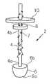
- the wound closure device 2comprises an elongate body 4 which includes a plurality of barbs 7 disposed along a surface of the elongate body 4 .
- the elongate body 4is generally cylindrical in shape and may have a generally circular cross-sectional area, although other shapes and cross-sectional areas are envisioned.
- the elongate body 4is in the form of a suture and is comprised of a biodegradable polymeric material.
- a distal portion 4 a of the elongate body 4is connected to a foam structure 6 .
- the elongate body 4is centrally connected to a relatively flat surface 6 b of the foam structure 6 .
- the elongate body 4could also be attached off-center.
- the elongate body 4may be connected to the foam structure 6 using various methods within the purview of those skilled in the art including, but not limited to, molding, over molding, adhesives and glues (e.g., cyanoacrylates), mechanical connections, male and female locking mechanisms, and the like.
- the foam structure 6is generally conical or hemispherical in shape.
- a distal portion 6 a of the foam structure 6is generally arcuate in shape and once inserted through a tissue wall, the foam structure 6 is relatively convex with respect to a tissue wall.
- a proximal portion 6 b of the foam structure 6is generally flat and, in use, is positioned adjacent a tissue wall so as to seal a tissue perforation and prevent fluids from passing through a tissue wall or tissue plane. It should be noted that although the foam structure is illustrated as generally conical in shape, this disclosure is not limited to conical-shaped foam structures and other shapes are contemplated.
- the foam structureis large enough, for example, to extend over a tissue perforation created by an access device (e.g., endoluminal device), sealing the tissue wall and limiting fluid passage from a first side of a tissue wall to a second side of a tissue wall. It should also be noted that certain embodiments may not include a foam structure.
- an access devicee.g., endoluminal device
- Foams of the present disclosuremay be compressible and are capable of undergoing a change in shape.
- the foam structuremay be of a configured to change shape from a first compressed shape when inserted in tissue for delivery to a second, expanded shape for placement. Upon penetration of a tissue wall, the foam structure may expand to seal a tissue defect.
- Foam structures of the present disclosurealso are shaped so as to limit movement proximally through a tissue wall, once inserted.
- the foam structuremay be constructed of a material which expands from heat or fluid (polymer hydrogels) contact; alternately, the foam structure may be mechanically compressed through use of a member such as a sleeve e.g., introducer, wherein upon removal of the sleeve, the foam expands.
- Other membersincluding the outer member and the inner member may also be compressible foams which change shape from a first, smaller shape, to a second, larger shape.
- Foamsmay have an open cell structure where the pores are connected to each other, forming an interconnected network. Conversely, foams of the present disclosure may be closed cell foams where the pores are not interconnected. Closed cell foams are generally denser and have a higher compressive strength. In certain preferred embodiments, the foam structure of the present disclosure is a closed cell foam.
- Tissue damage or tissue voidsmay have the potential to form adhesions during certain healing periods.
- Foam structures of the present disclosuremay be chemically tailored to have anti-adhesive properties, which may assist in preventing adjacent tissue walls from adhering together, preventing adhesions at a wound site.
- the foam structuresmay be made of anti-adhesive materials.
- the foam structuresmay be coated with anti-adhesive materials.
- the elongate body 4has an inner member 8 and an outer member 10 mounted thereon.
- the inner member 8 and the outer member 10are spaced from the distal portion 4 a of the elongate body 4 .
- the inner member 8 and the outer member 10are located on the proximal portion 4 b of the elongate body.
- the inner member and outer membermay be moveably mounted on a central portion of the elongate body.
- Each of the inner member 8 and outer member 10has an opening extending therethrough and is moveably positioned on the elongate body 4 .
- the inner member 8is positioned between a tissue wall (not shown) and the outer member 10 .
- Both the inner member 8 and the outer member 10are generally shaped like a disc, although other shapes are envisioned. In some embodiments, the inner member 8 may be configured to fill a surface void or tissue defect.
- the outer member 10is generally rigid as compared to the inner member 8 so as to affect movement of the inner member 8 .
- the inner member 8is comprised of an absorbable polymer such as collagen.
- the inner member 8may be in the form of a sheet or a porous material such as foam.
- the outer member 10may of any solid or dense porous material which is rigid, so as to impart movement on the inner member 8 as it is advanced distally along an elongate body 4 .
- At least the inner member 8may provide a tissue scaffold for cellular infiltration and tissue ingrowth. It is also envisioned that in alternate embodiments, the outer member 10 may provide a scaffold for tissue ingrowth.
- the tissue scaffoldis porous and provides a temporary scaffold/substrate for cell adherence. Tissue scaffolds may be tailored to closely match the mechanical properties of the surrounding tissue intended for regeneration. For example, when the wound closure device is used to close dermal tissue, the scaffold may be mechanically tuned to complement dermal tissue.
- the tissue scaffoldcomprises degradable materials including those listed below, and in certain preferred embodiments the tissue scaffold is collagen.
- the scaffold degradation profilecan be tailored to allow cells to proliferate while the tissue scaffold degrades over time.
- One skilled in the artcan alter the degradation profile of a tissue scaffold by changing various parameters including but not limited to polymer composition and chemistry, density, morphology, molecular weight, size, porosity and pore size, wettability and processing parameters.
- the elongate body 4further includes a plurality of barbs 7 which may be aligned to enable the wound closure device 2 to move through tissue in one direction while resisting movement through tissue in a generally opposite direction. That is, the barbs 7 , extending from the surface of the elongate body 4 , permit movement of inner member 8 and outer member 10 in a distal direction while resisting movement of the inner member 8 and outer member 10 in a proximal direction. Additionally, the barbs 7 prevent movement of the foam structure 6 towards a proximal portion of the device.
- the outer member 10is advanced in a distal direction along the elongate body 4 , thereby moving the inner member 8 in a distal direction (with the barbs preventing proximal movement).
- the barbs 7prevent proximal movement of the inner and outer members ( 8 and 10 ), thereby fixating the device 2 against the tissue wall (not shown).
- barbsencompasses various projections from the surface of an elongate body.
- the barbsare formed integrally with the elongate body 4 .
- Barbs extending from the outer surface of the elongate body 4include but are not limited to projections such as threads, anchors, and teeth.
- the barbsare yieldable toward the elongate body 4 of the wound closure device.
- the barbscan be arranged in any suitable pattern, for example helical, linear, or randomly spaced. The number, configuration, spacing and surface area of the barbs can vary depending upon the tissue type in which the device is used, as well as the composition and geometry of the material utilized to form the device.
- the barbsmay be longer and spaced further apart to enable it to grip the soft tissue.
- the barbscan be arranged in various directions at various angles.
- the wound closure devicemay include a staggered arrangement of large or small barbs.
- the shape and/or surface area of the barbscan also vary.
- fuller-tipped barbscan be made of varying sizes designed for specific surgical applications. In certain applications, such as when closing an access port site and the surgeon is working with fat and relatively soft tissues, larger barbs may be desired, whereas smaller barbs may be more suitable for different procedures with collagen-dense tissues.
- a combination of large and small barbs within the same structuremay be beneficial, for example when a wound closure device is used in tissue repair with differing layer structures. Use of the combination of large and small barbs on the same device, wherein barb sizes are customized for each tissue layer will ensure maximum holding strength of the device in situ.
- FIGS. 2A and 2Billustrate according to one embodiment, a method by which the wound closure device of FIG. 1 may be positioned in tissue. Note, portions of the tissue are removed for clarity.
- a device 2is shown in a first position where a foam structure 6 has partially penetrated a tissue wall 30 .
- the devicemay be inserted into tissue and a distal portion of the device 2 may be inserted through a tissue wall 30 with or without the use of an inserter (not shown).
- the proximal portion 6 b of the foam structure 6may be rotated (by the user or an insertion device) for positioning against the tissue wall 30 ( FIG. 2B ).
- the proximal portion 6 b of the foam structure 6which may be generally flat, is positioned adjacent a tissue wall 30 so as to seal a puncture wound and prevent fluids from passing through the tissue wall or tissue plane. It should be noted that the foam structure 6 may be inserted in a position so as to minimize tissue contact during delivery. As shown in FIG. 1 , the foam structure 6 is inserted at an angle and rotated or turned into position against a tissue wall 30 ; in other embodiments, an inserter may to used which enables the foam structure 6 to be inserted in a compressed position so as to minimize tissue contact during delivery.
- an outer member 10is advanced in a distal direction as indicated by an arrow in FIG. 2C .
- the outer member 10may be rigid as compared to an inner member 8 , and its movement imparts movement of the inner member 8 . That is, the outer member 10 is advanced towards the inner member 8 and once in contact, the outer member 10 and the inner member 8 move together over the barbed surface 4 in a distal direction so as to secure the device 2 in tissue ( FIG. 2D ).
- the inner member 8is adjacent a tissue surface 31 , applying pressure to the tissue surface 31 so as to limit movement of the device 2 .
- FIGS. 2A-2Dillustrate the embodiment described in FIG. 1 , other embodiments described herein will be placed in tissue in a similar manner.
- FIG. 3Another embodiment of a wound closure device 40 is shown in FIG. 3 .
- An elongate body 44is connected at a distal portion 44 a to a foam structure 42 the elongate body 44 has an outer member 48 mounted thereon, spaced from a distal portion 44 a of the elongate body.
- the foam structure 42may be generally conical or hemispherical in shape, having a generally flat proximal portion 42 b and a curved or somewhat pointed distal portion 42 a .
- the foam structure 42is circular in cross-sectional area, taken along line A-A, although other shapes are envisioned.
- the foam structure 42may be centrally attached to a barbed elongate body 44 , or could alternatively be attached off-center.
- the barbs 46function in a similar manner as described above and may have various lengths and may be disposed at different angles relative to a surface of the elongate body 44 .
- the outer member 48has an opening extending therethrough and is rigid enough to move in a distal direction over the barbed elongate body 44 .
- the outer member 48 as shownis funnel-shaped such that a proximal portion 48 b has a larger cross-sectional area, which narrows toward a distal portion 48 a . In situ, the distal portion 48 a may partially penetrate a tissue plane, as illustrated in FIG. 4 .
- the funnel shape of the outer member 48may assist in further sealing of a tissue defect which may be created by an access device such as an endoscopic device used for Natural Orifice Transluminal Endoscopic Surgery (N.O.T.E.S.).
- an access devicesuch as an endoscopic device used for Natural Orifice Transluminal Endoscopic Surgery (N.O.T.E.S.).
- the outer membermay further include an active agent such as a hemostat.
- FIG. 5Aillustrates the embodiment of FIGS. 3 and 4 by way of example, it being understood that other embodiments of a wound closure device as described herein may also be compressed by a sleeve for insertion into tissue.
- the sleeve 52may be employed to retain the foam structure 42 in a first, compressed shape for insertion/delivery.
- the sleeve 52also extends over the elongate body 44 and outer member 48 .
- the sleeve 52may be removed (retracted in the direction of the arrow), allowing the foam structure 42 to expand to a second larger shape, extending over a tissue defect 60 a , for placement.
- the outer member 48may be advanced in a distal direction, securing the device in place.
- the sleevemay also keep other members of the device, e.g., inner members and tissue scaffolds, in a compressed position for insertion into a body cavity.
- Biodegradable materialswhich may be synthetic and natural materials.
- biodegradablerefers to materials which decompose, or lose structural integrity under body conditions (e.g., enzymatic degradation or hydrolysis).
- Suitable synthetic biodegradable materialsmay include, but are not limited to, polymers such as those made from lactide, glycolide, caprolactone, valerolactone, carbonates (e.g., trimethylene carbonate, tetramethylene carbonate, and the like), dioxanones (e.g., 1,4-dioxanone), ⁇ -valerolactone, 1,dioxepanones (e.g., 1,4-dioxepan-2-one and 1,5-dioxepan-2-one), ethylene glycol, ethylene oxide, esteramides, ⁇ -hydroxyvalerate, ⁇ -hydroxypropionate, alpha-hydroxy acid, hydroxybuterates, poly (ortho esters), hydroxy alkanoates, tyrosine carbonates, polyimide carbonates, polyimino carbonates such as poly (bisphenol A-iminocarbonate) and poly (hydroquinone-iminocarbonate), polyurethanes, polyanhydr
- the foam structurecomprises a material which contains an aliphatic diacid linking two dihydroxy compounds.
- the dihydroxy compounds which may be utilizedinclude, but are not limited to, polyols including polyalkylene oxides, polyvinyl alcohols, and the like.
- the dihydroxy compoundscan be a polyalkylene oxide such as polyethylene oxide (“PEO”), polypropylene oxide (“PPO”), block or random copolymers of polyethylene oxide (PEO) and polypropylene oxide (PPO).
- Suitable aliphatic diacidswhich may be utilized in forming the foams include, for example, aliphatic diacids having from about 2 to about 8 carbon atoms suitable diacids include, but are not limited to sebacic acid, azelaic acid, suberic acid, pimelic acid, adipic acid, glutaric acid, succinic acid, malonic acid, oxalic acid and combinations thereof.
- a polyethylene glycolmay be utilized as the dihydroxy compound as disclosed in U.S. Patent Application Publication No. 20060253094, the entire disclosure of which is incorporated by reference herein. It may be desirable to utilize a PEG with a molecular weight ranging from about 200 to about 1000, typically from about 400 to about 900. Suitable PEGs are commercially available from a variety of sources under the designations PEG 200, PEG 400, PEG 600 and PEG 900.
- Suitable natural biodegradable polymersmay include, but are not limited to, collagen, poly (amino acids), polysaccharides such as cellulose, dextran, chitin, and glycosaminoglycans, hyaluronic acid, gut, copolymers and combinations thereof.
- collagenis used to construct the inner member of the medical device.
- Collagen as used hereinincludes natural collagen such as animal derived collagen, or synthetic collagen such as human or bacterial recombinant collagen.
- the collagencan be modified by using any method known to those skilled in the art to provide pendant portions of the collagen with moieties which are capable of covalently bonding with the reactive chemical groups of a glycosaminoglycan.
- pendant moietiesinclude aldehydes, sulfones, vinylsulfones, isocyanates, and acid anhydrides.
- electrophilic groupssuch as —CO 2 N(COCH 2 ) 2 , —CO 2 N(COCH 2 ) 2 , —CO 2 H, —CHO, —CHOCH 2 , —N ⁇ C ⁇ O, —SO 2 CH ⁇ CH 2 , —N(COCH) 2 , —S—S—(C 5 H 4 N) may also be added to pendant chains of the collagen to allow covalent bonding to occur with the glycosaminoglycans.
- the collagenmay be modified through the addition of an oxidizing agent.
- an oxidizing agentcreates oxidative cleavage along portions of the collagen thereby creating pendant aldehyde groups capable of reacting with the glycosaminoglycans.
- the oxidizing agentmay be, for example, iodine, peroxide, periodic acid, hydrogen peroxide, a periodate, a compound containing periodate, sodium periodate, a diisocyanate compound, a halogen, a compound containing halogen, n-bromosuccinimide, a permanganate, a compound containing permanganate, ozone, a compound containing ozone, chromic acid, sulfuryl chloride, a sulfoxide, a selenoxide, an oxidizing enzyme (oxidase) and combinations thereof.
- the oxidizing agentis periodic acid.
- certain members of the wound closure devicecomprise non-degradable materials.
- the outer membermay be non-degradable.
- a non-degradable materialmay be better suited for an external environment, or may provide better resistance against skin flora, compared to certain biodegradable materials.
- Suitable non-biodegradable materialsmay be used to construct the wound closure device including, but not limited to, fluorinated polymers (e.g., fluoroethylenes, propylenes, fluoroPEGs), polyolefins such as polyethylene, polyesters such as poly ethylene terepththalate (PET), nylons, polyamides, polyurethanes, silicones, ultra high molecular weight polyethylene (UHMWPE), polybutesters, polyethylene glycol, polyaryletherketone, copolymers and combinations thereof.
- fluorinated polymerse.g., fluoroethylenes, propylenes, fluoroPEGs
- polyolefinssuch as polyethylene
- polyesterssuch as poly ethylene terepththalate (PET)
- nylonssuch as poly ethylene terepththalate (PET)
- PETpoly ethylene terepththalate
- nylonssuch as poly ethylene terepththalate (PET)
- PETpoly
- medical devices according to the present disclosuremay be constructed at least in part using shape memory polymers.
- Suitable polymers used to prepare hard and soft segments of shape memory polymersmay include, but are not limited to, polycaprolactone, dioxanone, lactide, glycolide, polyacrylates, polyamides, polysiloxanes, polyurethanes, polyether amides, polyurethane/ureas, polyether esters, and urethane/butadiene copolymers and combinations thereof.
- the foam structuremay comprise shape memory materials which expand the foam upon reaching body temperature, sealing an inner tissue wall.
- the medical devicemay comprise metals (e.g., steel or titanium), metal alloys and the like.
- the elongate body or outer membermay comprise degradable metals such as degradable magnesium.
- Suitable materials of the present disclosurecan be processed by methods within the purview of those skilled in the art including, but not limited to extrusion, injection molding, compression molding, blow molding, film blowing, thermoforming, calendaring, spinning, and film casting.
- foams of the present disclosurecan be manufactured using various processes within the purview of those skilled in the art.
- foamscan be manufactured though standard lyophilization (freeze drying) techniques, solvent casting and particulate leaching, compression molding, phase separation, gas foaming (e.g., internal blowing agents such as CO 2 ), or through the use of a porogen (e.g., salt particles).
- foams which are used as tissue scaffoldscan also be created through computer aided design techniques including solid freeform fabrication (SFF).
- any part of the devicemay include biologically acceptable additives such as plasticizers, antioxidants, dyes, image-enhancing agents, dilutants, bioactive agents such as pharmaceutical and medicinal agents, and combinations thereof which can be coated on the wound closure device or impregnated within the resin or polymer.
- biologically acceptable additivessuch as plasticizers, antioxidants, dyes, image-enhancing agents, dilutants, bioactive agents such as pharmaceutical and medicinal agents, and combinations thereof which can be coated on the wound closure device or impregnated within the resin or polymer.
- Medicinal agents which may be incorporated into the wound closure devicemay include, but are not limited to, antimicrobial agents, anti-virals, anti-fungals, and the like.
- Antimicrobial agents as used hereinis defined by an agent which by itself or through assisting the body (immune system) helps the body destroy or resist microorganisms which may be pathogenic (disease causing).
- the term “antimicrobial agent”includes, e.g., antibiotics, quorum sensing blockers, surfactants, metal ions, antimicrobial proteins and peptides, antimicrobial polysaccharides, antiseptics, disinfectants, anti-virals, anti-fungals, quorum sensing blockers, and combinations thereof.
- Suitable antiseptics and disinfectantsinclude hexachlorophene; cationic biguanides like chlorohexadine and cyclohexidine; iodine and iodophores like povidone-iodine; halo-substituted phenolic compounds like PCMX (e.g., p-chloro-m-xylenon) and triclosan (e.g., 2,4,4′-trichloro-2′hydroxy-diphenylether); furan medical preparations like nitrofurantoin and nitrofurazone; methanamine; aldehydes like gluteraldehyde and formaldehyde; alcohols; combinations thereof, and the like.
- at least one of the antimicrobial agentsmay be an antiseptic, such as triclosan.
- Classes of antibioticsthat can be combined with the present disclosure include tetracyclines like minocycline; rifamycins like rifampin; macrolides like erythromycin; penicillins like nafcillin; cephalosporins like cefazolon; beta-lactam antibiotics like imipenen and aztreonam; aminoglycosides like gentamicin and TOBRAMYCIN®; chloramphenicol; sulfonamides like sulfamethoxazole; glycopeptides like vancomycin; quilones like ciproflaxin; fusidic acid; trimethoprim; metronidazole; clindamycin; mupirocin; polyenes like amphotericin B; azoles like fluconazole; and beta-lactam inhibitors like sublactam.
- antimicrobialswhich may be added include, for example antimicrobial peptides and/or proteins; antimicrobial polysaccharides; quorum sensing blockers (e.g., brominated furanones); anti-virals; metal ions such as ionic silver and ionic silver glass; surfactants; chemotherapeutic drug; telomerase inhibitors; other cyclic monomers including 5-cyclic monomers; mitoxantrone; and the like.
- quorum sensing blockerse.g., brominated furanones
- anti-viralse.g., metal ions such as ionic silver and ionic silver glass
- surfactantschemotherapeutic drug
- telomerase inhibitorsother cyclic monomers including 5-cyclic monomers; mitoxantrone; and the like.
- suitable bioactive agentswhich may be used include colorants, dyes, preservatives, protein and peptide preparations, protein therapeutics, polysaccharides such as hyaluronic acid, lectins, lipids, probiotics, angiogenic agents, anti-thrombotics, anti-clotting agents, clotting agents, analgesics, anesthetics, wound repair agents, chemotherapeutics, biologics, anti-inflammatory agents, anti-proliferatives, diagnostic agents, antipyretic, antiphlogistic and analgesic agents, vasodilators, antihypertensive and antiarrhythmic agents, hypotensive agents, antitussive agents, antineoplastics, local anesthetics, hormone preparations, antiasthmatic and antiallergic agents, antihistaminics, anticoagulants, antispasmodics, cerebral circulation and metabolism improvers, antidepressant and antianxiety agents, vitamin D preparations, hypoglycemic agents,
- polymer drugse.g., polymeric forms of such compounds for example, polymeric antibiotics, polymeric antiseptics, polymeric chemotherapeutics, polymeric anti-proliferatives, polymeric antiseptics, polymeric non-steroidal anti-inflammatory drugs (NSAIDS), and the like may be utilized and combinations thereof.
- polymeric antibioticse.g., polymeric antiseptics, polymeric chemotherapeutics, polymeric anti-proliferatives, polymeric antiseptics, polymeric non-steroidal anti-inflammatory drugs (NSAIDS), and the like
- NSAIDSpolymeric non-steroidal anti-inflammatory drugs
- medical devices of the present disclosuremay contain suitable medicinal agents such as viruses and cells; peptides; polypeptides and proteins, analogs, muteins, and active fragments thereof, such as immunoglobulins; antibodies (monoclonal and polyclonal); cytokines (e.g., lymphokines, monokines, chemokines); blood clotting factors; hemopoietic factors; interleukins (IL-2, IL-3, IL-4, IL-6); interferons ( ⁇ -IFN, ⁇ -IFN and ⁇ -IFN); erythropoietin; nucleases; tumor necrosis factor; colony stimulating factors (e.g., GCSF, GM-CSF, MCSF); insulin; anti-tumor agents and tumor suppressors; blood proteins; gonadotropins (e.g., FSH, LH, CG, etc.); hormones and hormone analogs (e.g., growth hormone); vaccines (e.g.,
- additivessuch as image-enhancing agents (e.g., contrast agents) and more specifically, radiopaque markers, may be incorporated into the medical device.
- image-enhancing agentsenable visualization of the wound closure device (against surrounding tissue), when imaged or scanned through different filters such as MRI, X-ray, fluoroscopy, CT, various light sources, and the like.
- filterssuch as MRI, X-ray, fluoroscopy, CT, various light sources, and the like.
- the wound closure devicemay be made from a material possessing radiographic density higher than the surrounding host tissue and have sufficient thickness to affect the transmission of x-rays to produce contrast in the image.
- Useful image-enhancing agentsinclude but are not limited to radiopaque markers such as tantalum, barium sulfate, bismuth trioxide, bromine, iodide, titanium oxide, zirconium, barium, titanium, bismuth, iodine, nickel, iron, silver, and combinations thereof.
- radiopaque markerssuch as tantalum, barium sulfate, bismuth trioxide, bromine, iodide, titanium oxide, zirconium, barium, titanium, bismuth, iodine, nickel, iron, silver, and combinations thereof.
- compoundssuch as tantalum, platinum, barium and bismuth may be incorporated into the wound closure device.
- image-enhancing agentsare not bioabsorbable or degradable but are excreted from the body or stored in the body.
- Image-enhancing agentsmay be compounded into the materials (e.g. resin) as filler prior to processing including extrusion or molding. These agents may be added in various concentrations to maximize polymer processing while maximizing the material characteristics of the wound closure device.
- the biocompatible agentscan be added in quantities sufficient to enhance radiopacity while maintaining the polymer's properties.
- image-enhancing agentsmay be incorporated into a biodegradable material, enabling surgeons to know when the biodegradable material has degraded.
- bioactive agentsinclude, but are not limited to mixing, blending, compounding, spraying, wicking, solvent evaporating, dipping, brushing, vapor deposition, coextrusion, capillary wicking, film casting, molding and the like. Additionally, solvents may be used to incorporate various agents (e.g., bioactive agents) into the composite device.
- Suitable solventsinclude alcohols, e.g., methanol, ethanol, propanol, chlorinated hydrocarbons (such as methylene chloride, chloroform, 1,2-dichloro-ethane), and aliphatic hydrocarbons such as hexane, heptene, ethyl acetate.
- alcoholse.g., methanol, ethanol, propanol
- chlorinated hydrocarbonssuch as methylene chloride, chloroform, 1,2-dichloro-ethane
- aliphatic hydrocarbonssuch as hexane, heptene, ethyl acetate.
Landscapes
- Health & Medical Sciences (AREA)
- Surgery (AREA)
- Life Sciences & Earth Sciences (AREA)
- Medical Informatics (AREA)
- Nuclear Medicine, Radiotherapy & Molecular Imaging (AREA)
- Engineering & Computer Science (AREA)
- Biomedical Technology (AREA)
- Heart & Thoracic Surgery (AREA)
- Rheumatology (AREA)
- Molecular Biology (AREA)
- Animal Behavior & Ethology (AREA)
- General Health & Medical Sciences (AREA)
- Public Health (AREA)
- Veterinary Medicine (AREA)
- Materials For Medical Uses (AREA)
- Surgical Instruments (AREA)
Abstract
Description
Claims (23)
Priority Applications (8)
| Application Number | Priority Date | Filing Date | Title |
|---|---|---|---|
| US12/511,462US9943302B2 (en) | 2008-08-12 | 2009-07-29 | Medical device for wound closure and method of use |
| AU2009208061AAU2009208061A1 (en) | 2008-08-12 | 2009-08-07 | Medical device for wound closure and method of use |
| CA2675068ACA2675068A1 (en) | 2008-08-12 | 2009-08-10 | Medical device for wound closure and method of use |
| JP2009186918AJP2010042258A (en) | 2008-08-12 | 2009-08-11 | Medical device for wound closure and method of use |
| EP09251981.8AEP2153779B1 (en) | 2008-08-12 | 2009-08-12 | Medical device for wound closure |
| US12/581,995US9271706B2 (en) | 2008-08-12 | 2009-10-20 | Medical device for wound closure and method of use |
| US15/008,591US10722224B2 (en) | 2008-08-12 | 2016-01-28 | Medical device for wound closure and method of use |
| US15/918,259US11013504B2 (en) | 2008-08-12 | 2018-03-12 | Medical device for wound closure and method of use |
Applications Claiming Priority (2)
| Application Number | Priority Date | Filing Date | Title |
|---|---|---|---|
| US8814508P | 2008-08-12 | 2008-08-12 | |
| US12/511,462US9943302B2 (en) | 2008-08-12 | 2009-07-29 | Medical device for wound closure and method of use |
Related Child Applications (2)
| Application Number | Title | Priority Date | Filing Date |
|---|---|---|---|
| US12/581,995Continuation-In-PartUS9271706B2 (en) | 2008-08-12 | 2009-10-20 | Medical device for wound closure and method of use |
| US15/918,259DivisionUS11013504B2 (en) | 2008-08-12 | 2018-03-12 | Medical device for wound closure and method of use |
Publications (2)
| Publication Number | Publication Date |
|---|---|
| US20100042144A1 US20100042144A1 (en) | 2010-02-18 |
| US9943302B2true US9943302B2 (en) | 2018-04-17 |
Family
ID=41327608
Family Applications (2)
| Application Number | Title | Priority Date | Filing Date |
|---|---|---|---|
| US12/511,462Active2033-01-22US9943302B2 (en) | 2008-08-12 | 2009-07-29 | Medical device for wound closure and method of use |
| US15/918,259Expired - Fee RelatedUS11013504B2 (en) | 2008-08-12 | 2018-03-12 | Medical device for wound closure and method of use |
Family Applications After (1)
| Application Number | Title | Priority Date | Filing Date |
|---|---|---|---|
| US15/918,259Expired - Fee RelatedUS11013504B2 (en) | 2008-08-12 | 2018-03-12 | Medical device for wound closure and method of use |
Country Status (5)
| Country | Link |
|---|---|
| US (2) | US9943302B2 (en) |
| EP (1) | EP2153779B1 (en) |
| JP (1) | JP2010042258A (en) |
| AU (1) | AU2009208061A1 (en) |
| CA (1) | CA2675068A1 (en) |
Cited By (3)
| Publication number | Priority date | Publication date | Assignee | Title |
|---|---|---|---|---|
| US11013504B2 (en) | 2008-08-12 | 2021-05-25 | Covidien Lp | Medical device for wound closure and method of use |
| US20210204937A1 (en)* | 2018-05-18 | 2021-07-08 | Jworld Co., Ltd. | Medical suture and medical suture kit |
| US12383246B2 (en) | 2020-10-12 | 2025-08-12 | Abbott Cardiovascular Systems, Inc. | Vessel closure device with improved safety and tract hemostasis |
Families Citing this family (86)
| Publication number | Priority date | Publication date | Assignee | Title |
|---|---|---|---|---|
| US9579091B2 (en) | 2000-01-05 | 2017-02-28 | Integrated Vascular Systems, Inc. | Closure system and methods of use |
| US6461364B1 (en) | 2000-01-05 | 2002-10-08 | Integrated Vascular Systems, Inc. | Vascular sheath with bioabsorbable puncture site closure apparatus and methods of use |
| US8758400B2 (en) | 2000-01-05 | 2014-06-24 | Integrated Vascular Systems, Inc. | Closure system and methods of use |
| US6391048B1 (en) | 2000-01-05 | 2002-05-21 | Integrated Vascular Systems, Inc. | Integrated vascular device with puncture site closure component and sealant and methods of use |
| DE60144328D1 (en)* | 2000-09-08 | 2011-05-12 | Abbott Vascular Inc | Surgical clamp |
| US6626918B1 (en)* | 2000-10-06 | 2003-09-30 | Medical Technology Group | Apparatus and methods for positioning a vascular sheath |
| US7905900B2 (en) | 2003-01-30 | 2011-03-15 | Integrated Vascular Systems, Inc. | Clip applier and methods of use |
| US6695867B2 (en) | 2002-02-21 | 2004-02-24 | Integrated Vascular Systems, Inc. | Plunger apparatus and methods for delivering a closure device |
| US6623510B2 (en) | 2000-12-07 | 2003-09-23 | Integrated Vascular Systems, Inc. | Closure device and methods for making and using them |
| US8690910B2 (en) | 2000-12-07 | 2014-04-08 | Integrated Vascular Systems, Inc. | Closure device and methods for making and using them |
| IES20010547A2 (en) | 2001-06-07 | 2002-12-11 | Christy Cummins | Surgical Staple |
| IES20030424A2 (en) | 2002-06-04 | 2003-12-10 | Robert Stevenson | Blood vessel closure clip and delivery device |
| US8398656B2 (en) | 2003-01-30 | 2013-03-19 | Integrated Vascular Systems, Inc. | Clip applier and methods of use |
| US8905937B2 (en) | 2009-02-26 | 2014-12-09 | Integrated Vascular Systems, Inc. | Methods and apparatus for locating a surface of a body lumen |
| US8202293B2 (en) | 2003-01-30 | 2012-06-19 | Integrated Vascular Systems, Inc. | Clip applier and methods of use |
| US8758398B2 (en) | 2006-09-08 | 2014-06-24 | Integrated Vascular Systems, Inc. | Apparatus and method for delivering a closure element |
| US8821534B2 (en) | 2010-12-06 | 2014-09-02 | Integrated Vascular Systems, Inc. | Clip applier having improved hemostasis and methods of use |
| EP1667586A1 (en) | 2003-09-15 | 2006-06-14 | Abbott Laboratories | Suture locking device and methods |
| US8926633B2 (en) | 2005-06-24 | 2015-01-06 | Abbott Laboratories | Apparatus and method for delivering a closure element |
| US8313497B2 (en) | 2005-07-01 | 2012-11-20 | Abbott Laboratories | Clip applier and methods of use |
| US8808310B2 (en) | 2006-04-20 | 2014-08-19 | Integrated Vascular Systems, Inc. | Resettable clip applier and reset tools |
| US8556930B2 (en) | 2006-06-28 | 2013-10-15 | Abbott Laboratories | Vessel closure device |
| ATE514382T1 (en) | 2006-11-30 | 2011-07-15 | Wilson Cook Medical Inc | FABRIC ANCHOR FOR SEAM CLOSURE OF PERFORATIONS |
| JP5226792B2 (en)* | 2007-09-25 | 2013-07-03 | クック メディカル テクノロジーズ エルエルシー | Medical instruments, devices and methods for using tissue anchors |
| US8893947B2 (en) | 2007-12-17 | 2014-11-25 | Abbott Laboratories | Clip applier and methods of use |
| WO2009082596A1 (en)* | 2007-12-18 | 2009-07-02 | Wilson-Cook Medical, Inc. | Device and method for placement of tissue anchors |
| US7841502B2 (en) | 2007-12-18 | 2010-11-30 | Abbott Laboratories | Modular clip applier |
| WO2009132111A1 (en)* | 2008-04-23 | 2009-10-29 | Wilson-Cook Medical Inc. | Tacking device |
| US20090270911A1 (en)* | 2008-04-24 | 2009-10-29 | Shipp John I | Vessel Sealing Device and Method of Using Same |
| US9282965B2 (en) | 2008-05-16 | 2016-03-15 | Abbott Laboratories | Apparatus and methods for engaging tissue |
| US9271706B2 (en)* | 2008-08-12 | 2016-03-01 | Covidien Lp | Medical device for wound closure and method of use |
| WO2010022060A1 (en)* | 2008-08-19 | 2010-02-25 | Wilson-Cook Medical Inc. | Apparatus for removing lymph nodes or anchoring into tissue during a translumenal procedure |
| US8192461B2 (en)* | 2008-09-11 | 2012-06-05 | Cook Medical Technologies Llc | Methods for facilitating closure of a bodily opening using one or more tacking devices |
| US8398676B2 (en) | 2008-10-30 | 2013-03-19 | Abbott Vascular Inc. | Closure device |
| US8377095B2 (en)* | 2008-12-05 | 2013-02-19 | Cook Medical Technologies, LLC | Tissue anchors for purse-string closure of perforations |
| AU2009324819B2 (en) | 2008-12-09 | 2014-04-17 | Cook Medical Technologies Llc | Retractable tacking device |
| CA2746213A1 (en)* | 2008-12-09 | 2010-07-08 | Wilson-Cook Medical Inc. | Apparatus and methods for controlled release of tacking devices |
| EP2389122B1 (en)* | 2008-12-19 | 2015-03-04 | Cook Medical Technologies LLC | Clip devices |
| JP2012512715A (en)* | 2008-12-19 | 2012-06-07 | クック メディカル テクノロジーズ エルエルシー | A tacking device of varying thickness and method of delivery and deployment thereof |
| US8858594B2 (en) | 2008-12-22 | 2014-10-14 | Abbott Laboratories | Curved closure device |
| US9414820B2 (en) | 2009-01-09 | 2016-08-16 | Abbott Vascular Inc. | Closure devices, systems, and methods |
| US9486191B2 (en) | 2009-01-09 | 2016-11-08 | Abbott Vascular, Inc. | Closure devices |
| US9089311B2 (en) | 2009-01-09 | 2015-07-28 | Abbott Vascular Inc. | Vessel closure devices and methods |
| US9173644B2 (en) | 2009-01-09 | 2015-11-03 | Abbott Vascular Inc. | Closure devices, systems, and methods |
| US20100179589A1 (en) | 2009-01-09 | 2010-07-15 | Abbott Vascular Inc. | Rapidly eroding anchor |
| US20110218568A1 (en)* | 2009-01-09 | 2011-09-08 | Voss Laveille K | Vessel closure devices, systems, and methods |
| US20100179567A1 (en)* | 2009-01-09 | 2010-07-15 | Abbott Vascular Inc. | Closure devices, systems, and methods |
| US20100185234A1 (en)* | 2009-01-16 | 2010-07-22 | Abbott Vascular Inc. | Closure devices, systems, and methods |
| EP2413809B1 (en)* | 2009-04-03 | 2014-10-08 | Cook Medical Technologies LLC | Medical devices for rapid deployment and fixation of tissue anchors |
| CA2757554A1 (en) | 2009-04-03 | 2010-10-07 | Cook Medical Technologies Llc | Tissue anchors and medical devices for rapid deployment of tissue anchors |
| AU2010245115B2 (en) | 2009-05-06 | 2014-09-18 | Hansa Medical Products, Inc. | Self-adjusting medical device |
| JP2012527970A (en)* | 2009-05-28 | 2012-11-12 | クック メディカル テクノロジーズ エルエルシー | Hail-fastening device and hail-fastening device deployment method |
| US20110054492A1 (en) | 2009-08-26 | 2011-03-03 | Abbott Laboratories | Medical device for repairing a fistula |
| KR101137991B1 (en)* | 2009-09-30 | 2012-04-20 | 전남대학교산학협력단 | Fabrication and manufacturing method of image based patient specific spinal implant |
| US8617206B2 (en)* | 2009-10-08 | 2013-12-31 | Covidien Lp | Wound closure device |
| EP2498689A4 (en)* | 2009-11-09 | 2015-04-22 | Cardiovascular Technologies Inc | Tissue closure devices, device and systems for delivery, kits and methods therefor |
| US8500776B2 (en)* | 2010-02-08 | 2013-08-06 | Covidien Lp | Vacuum patch for rapid wound closure |
| US8506593B2 (en) | 2010-04-11 | 2013-08-13 | Lap IP, Inc | Implantable biodegradable wound closure device and method |
| US8932325B2 (en)* | 2010-05-19 | 2015-01-13 | Cook Medical Technologies Llc | Devices and methods useful for sealing bodily openings |
| WO2011146729A2 (en)* | 2010-05-19 | 2011-11-24 | Cook Incorporated | Devices and methods useful for sealing bodily openings |
| US9044267B2 (en) | 2010-06-11 | 2015-06-02 | Entourage Medical Technologies, Inc. | System and method for transapical access and closure |
| US8758399B2 (en) | 2010-08-02 | 2014-06-24 | Abbott Cardiovascular Systems, Inc. | Expandable bioabsorbable plug apparatus and method |
| US9730690B2 (en) | 2010-09-20 | 2017-08-15 | Entourage Medical Technologies, Inc. | Method for providing surgical access |
| US9149265B2 (en) | 2011-02-26 | 2015-10-06 | Abbott Cardiovascular Systems, Inc. | Hinged tissue support device |
| US9149276B2 (en) | 2011-03-21 | 2015-10-06 | Abbott Cardiovascular Systems, Inc. | Clip and deployment apparatus for tissue closure |
| AU2012284618B2 (en)* | 2011-07-19 | 2017-05-18 | Shieldheart Medtech Ab | Stabilizer, barrier disc and wound dressing comprising stabilizer, method for controlling the position of a wound dressing or barrier disc, and method for facilitating drainage from a wound dressing or barrier disc in negative pressure wound treatment |
| US9055932B2 (en) | 2011-08-26 | 2015-06-16 | Abbott Cardiovascular Systems, Inc. | Suture fastener combination device |
| US9332976B2 (en) | 2011-11-30 | 2016-05-10 | Abbott Cardiovascular Systems, Inc. | Tissue closure device |
| DE112012005689T5 (en)* | 2012-01-17 | 2014-10-02 | Spiration, Inc. | Systems and methods for treating fistulas in the lung and trachea |
| US9138214B2 (en)* | 2012-03-02 | 2015-09-22 | Abbott Cardiovascular Systems, Inc. | Suture securing systems, devices and methods |
| US9943298B2 (en) | 2012-10-19 | 2018-04-17 | Cook Medical Technologies Llc | Vascular closure with shape memory characteristic |
| US10070850B2 (en) | 2012-10-19 | 2018-09-11 | Cook Medical Technologies Llc | Vascular closure with multiple connections |
| US20140172012A1 (en) | 2012-12-13 | 2014-06-19 | Cook Medical Technologies Llc | Vascular closure device suture tension mechanism |
| US9364209B2 (en) | 2012-12-21 | 2016-06-14 | Abbott Cardiovascular Systems, Inc. | Articulating suturing device |
| US9486132B2 (en) | 2013-01-17 | 2016-11-08 | Abbott Cardiovascular Systems, Inc. | Access device for accessing tissue |
| US10758216B2 (en) | 2013-03-14 | 2020-09-01 | Cook Medical Technologies Llc | Internal closure systems and devices |
| US9724082B2 (en) | 2013-03-15 | 2017-08-08 | Cook Medical Technologies Llc | Delivery system for tissue opening closures |
| US10154835B2 (en) | 2013-05-09 | 2018-12-18 | Essential Medical, Inc. | Vascular closure device with conforming plug member |
| US10265433B2 (en)* | 2015-03-18 | 2019-04-23 | Lawrence Livermore National Security, Llc | Hemorrhage management system |
| CN107921101B (en)* | 2015-04-03 | 2022-02-08 | 百奥诚智有限公司 | Powder composition for producing cross-linked protein foam and method of use thereof |
| US11026676B2 (en) | 2017-02-06 | 2021-06-08 | Covidien Lp | Surgical wound closure apparatus |
| EP3691699A4 (en) | 2017-10-04 | 2021-06-30 | Bio-Change Ltd. | CROSS-LINKED PROTEIN FOAM AND METHOD OF USE THEREOF IN A POLYVALENT CELL FRAMEWORK |
| JP2021506556A (en)* | 2017-12-22 | 2021-02-22 | ポリノボ バイオマテリアルズ ピーティーワイ リミテッド | Tissue repair laminate |
| US11439383B2 (en) | 2019-08-20 | 2022-09-13 | Abbott Cardiovascular Systems, Inc. | Self locking suture and self locking suture mediated closure device |
| US20240050082A1 (en)* | 2020-12-18 | 2024-02-15 | The Johns Hopkins University | Perianal fistula closure system |
| KR102625332B1 (en)* | 2021-10-14 | 2024-01-15 | 재단법인 아산사회복지재단 | Hemostasis device |
Citations (95)
| Publication number | Priority date | Publication date | Assignee | Title |
|---|---|---|---|---|
| US3123077A (en)* | 1964-03-03 | Surgical suture | ||
| US4235238A (en)* | 1978-05-11 | 1980-11-25 | Olympus Optical Co., Ltd. | Apparatus for suturing coeliac tissues |
| US4705040A (en)* | 1985-11-18 | 1987-11-10 | Medi-Tech, Incorporated | Percutaneous fixation of hollow organs |
| US4744364A (en) | 1987-02-17 | 1988-05-17 | Intravascular Surgical Instruments, Inc. | Device for sealing percutaneous puncture in a vessel |
| EP0513736A1 (en) | 1991-05-13 | 1992-11-19 | United States Surgical Corporation | Device for repairing torn tissue |
| US5269809A (en)* | 1990-07-02 | 1993-12-14 | American Cyanamid Company | Locking mechanism for use with a slotted suture anchor |
| US5342393A (en) | 1992-08-27 | 1994-08-30 | Duke University | Method and device for vascular repair |
| US5350399A (en)* | 1991-09-23 | 1994-09-27 | Jay Erlebacher | Percutaneous arterial puncture seal device and insertion tool therefore |
| US5370661A (en)* | 1990-11-06 | 1994-12-06 | Branch; Thomas P. | Method and apparatus for re-approximating tissue |
| WO1994028800A1 (en) | 1993-06-04 | 1994-12-22 | Kensey Nash Corporation | Hemostatic vessel puncture closure with filament lock |
| US5531759A (en)* | 1994-04-29 | 1996-07-02 | Kensey Nash Corporation | System for closing a percutaneous puncture formed by a trocar to prevent tissue at the puncture from herniating |
| US5545178A (en) | 1994-04-29 | 1996-08-13 | Kensey Nash Corporation | System for closing a percutaneous puncture formed by a trocar to prevent tissue at the puncture from herniating |
| US5549633A (en)* | 1994-08-24 | 1996-08-27 | Kensey Nash Corporation | Apparatus and methods of use for preventing blood seepage at a percutaneous puncture site |
| US5593422A (en) | 1989-05-29 | 1997-01-14 | Muijs Van De Moer; Wouter M. | Occlusion assembly for sealing openings in blood vessels and a method for sealing openings in blood vessels |
| US5620461A (en) | 1989-05-29 | 1997-04-15 | Muijs Van De Moer; Wouter M. | Sealing device |
| US5669935A (en)* | 1995-07-28 | 1997-09-23 | Ethicon, Inc. | One-way suture retaining device for braided sutures |
| US6007563A (en) | 1991-11-08 | 1999-12-28 | Kensey Nash Corporation | Method of deploying percutaneous puncture closure |
| US6174322B1 (en)* | 1997-08-08 | 2001-01-16 | Cardia, Inc. | Occlusion device for the closure of a physical anomaly such as a vascular aperture or an aperture in a septum |
| US6280474B1 (en)* | 1997-01-09 | 2001-08-28 | Neucoll, Inc. | Devices for tissue repair and methods for preparation and use thereof |
| US6306159B1 (en)* | 1998-12-23 | 2001-10-23 | Depuy Orthopaedics, Inc. | Meniscal repair device |
| US6322580B1 (en) | 2000-09-01 | 2001-11-27 | Angiolink Corporation | Wound site management and wound closure device |
| US20010046476A1 (en) | 2000-05-10 | 2001-11-29 | Isp Investments Inc. | Polymeric delivery and release systems for oral care actives |
| US6462169B1 (en) | 1999-11-30 | 2002-10-08 | Poly-Med, Inc. | Amorphous polymeric polyaxial initiators and compliant crystalline copolymers therefrom |
| US20020188319A1 (en) | 2001-06-08 | 2002-12-12 | Morris Edward J. | Method and apparatus for sealing access |
| US20020198562A1 (en)* | 2000-04-19 | 2002-12-26 | Radi Medical Systems Ab | Reinforced absorbable medical sealing device |
| US6506190B1 (en) | 1998-05-21 | 2003-01-14 | Christopher J. Walshe | Tissue anchor system |
| US20030012734A1 (en) | 1996-09-23 | 2003-01-16 | Incept Llc. | Biocompatible crosslinked polymers |
| US6508828B1 (en) | 2000-11-03 | 2003-01-21 | Radi Medical Systems Ab | Sealing device and wound closure device |
| US6514534B1 (en) | 1998-08-14 | 2003-02-04 | Incept Llc | Methods for forming regional tissue adherent barriers and drug delivery systems |
| US6533762B2 (en) | 2000-09-01 | 2003-03-18 | Angiolink Corporation | Advanced wound site management systems and methods |
| US6566406B1 (en) | 1998-12-04 | 2003-05-20 | Incept, Llc | Biocompatible crosslinked polymers |
| US6605294B2 (en) | 1998-08-14 | 2003-08-12 | Incept Llc | Methods of using in situ hydration of hydrogel articles for sealing or augmentation of tissue or vessels |
| FR2839451A1 (en) | 2002-05-07 | 2003-11-14 | Oreal | New cosmetic or dermatological compound, useful e.g. for treating dry skin, dandruff or acne, comprising active agent bonded to polymer via spacer and released in presence of microbial enzyme |
| US6669707B1 (en)* | 1998-07-21 | 2003-12-30 | Lee L. Swanstrom | Method and apparatus for attaching or locking an implant to an anatomic vessel or hollow organ wall |
| US6703047B2 (en) | 2001-02-02 | 2004-03-09 | Incept Llc | Dehydrated hydrogel precursor-based, tissue adherent compositions and methods of use |
| US6773450B2 (en) | 2002-08-09 | 2004-08-10 | Quill Medical, Inc. | Suture anchor and method |
| US20040176800A1 (en) | 2003-03-07 | 2004-09-09 | Paraschac Joseph Francis | Barbed closure device |
| US6794485B2 (en) | 2000-10-27 | 2004-09-21 | Poly-Med, Inc. | Amorphous polymeric polyaxial initiators and compliant crystalline copolymers therefrom |
| US20040185250A1 (en) | 2003-02-07 | 2004-09-23 | John Amy T. | Triclosan containing absorbable sutures with extended antimicrobial properties |
| US6818018B1 (en) | 1998-08-14 | 2004-11-16 | Incept Llc | In situ polymerizable hydrogels |
| WO2005016176A2 (en) | 2003-08-07 | 2005-02-24 | Leiboff Arnold R | Device and method for tacking a prosthetic screen |
| US20050085852A1 (en)* | 2003-10-15 | 2005-04-21 | Theresa Ditter | Vascular sealing device with locking hub |
| US20050169974A1 (en) | 2002-05-08 | 2005-08-04 | Radi Medical Systems Ab | Dissolvable medical sealing device |
| US20050234509A1 (en)* | 2004-03-30 | 2005-10-20 | Mmt Medical, Inc. | Center joints for PFO occluders |
| US20050261709A1 (en)* | 2004-05-20 | 2005-11-24 | Olympus Corporation | Treatment system for living tissues |
| WO2005110280A2 (en) | 2004-05-07 | 2005-11-24 | Valentx, Inc. | Devices and methods for attaching an endolumenal gastrointestinal implant |
| US20050267533A1 (en)* | 2004-03-23 | 2005-12-01 | Michael Gertner | Methods and devices for the surgical creation of satiety and biofeedback pathways |
| US20050283187A1 (en)* | 2004-06-22 | 2005-12-22 | Longson Matthew S | Vascular occlusion device |
| WO2006009925A2 (en) | 2004-06-18 | 2006-01-26 | The Catheter Exchange, Inc. | Method and device for cavity obliteration |
| US7026437B2 (en) | 2000-10-27 | 2006-04-11 | Poly-Med, Inc | Amorphous polymeric polyaxial initiators and compliant crystalline copolymers therefrom |
| US20060106418A1 (en) | 2002-07-31 | 2006-05-18 | Abbott Laboratories Vascular Enterprises, Limited | Apparatus for sealing surgical punctures |
| US20060122608A1 (en) | 2004-12-08 | 2006-06-08 | Fallin T W | System and method for anchoring suture to bone |
| US20060142797A1 (en)* | 2004-12-16 | 2006-06-29 | Radi Medical Systems Ab | Medical sealing device |
| US20060173492A1 (en)* | 2003-07-03 | 2006-08-03 | Radi Medical Systems Ab | Wound closure and sealing device |
| US20060206146A1 (en) | 2005-03-11 | 2006-09-14 | Radi Medical Systems Ab | Medical sealing device |
| EP1704878A2 (en) | 1995-12-18 | 2006-09-27 | AngioDevice International GmbH | Crosslinked polymer compositions and methods for their use |
| US20060229670A1 (en) | 2005-04-01 | 2006-10-12 | Bates Brian L | Method and a medical closure system for sealing a puncture |
| US20060229672A1 (en) | 2005-04-11 | 2006-10-12 | St. Jude Medical Puerto Rico B.V. | Tissue puncture closure device with automatic torque sensing tamping system |
| US20060229673A1 (en) | 2005-04-11 | 2006-10-12 | Forsberg Andrew T | Tissue puncture closure device with magazine fed tamping system |
| US20060229674A1 (en) | 2005-04-12 | 2006-10-12 | St. Jude Medical Puerto Rico B.V. | Tissue puncture closure device with scroll gear transmission tamping system |
| US20060265007A1 (en) | 2005-05-17 | 2006-11-23 | St. Jude Medical Puerto Rico B.V. | Tissue puncture closure system with retractable sheath |
| US20060265006A1 (en) | 2005-05-17 | 2006-11-23 | White John O | Tissue puncture closure device with disengagable automatic tamping system |
| US7169168B2 (en) | 1989-05-29 | 2007-01-30 | Kensey Nash Corporation | Sealing device |
| US20070032824A1 (en) | 2005-08-04 | 2007-02-08 | St. Jude Medical Puerto Rico B.V. | Tissue puncture closure device with track plug |
| US20070032823A1 (en) | 2005-08-04 | 2007-02-08 | St. Jude Medical Puerto Rico B.V. | Tissue puncture closure device with coiled automatic tamping system |
| US7182763B2 (en) | 2004-11-23 | 2007-02-27 | Instrasurgical, Llc | Wound closure device |
| US20070049971A1 (en)* | 2005-06-09 | 2007-03-01 | Sing-Fatt Chin | Method and apparatus for closing off a portion of a heart ventricle |
| US20070135842A1 (en)* | 1991-10-22 | 2007-06-14 | Kensey Nash Corporation | Sealing device |
| US20070150002A1 (en)* | 2005-12-23 | 2007-06-28 | Ethicon, Inc. | Systems and methods for closing a vessel wound |
| US20070156175A1 (en)* | 2005-12-29 | 2007-07-05 | Weadock Kevin S | Device for attaching, relocating and reinforcing tissue and methods of using same |
| US20070167982A1 (en)* | 2004-03-23 | 2007-07-19 | Michael Gertner | Methods and devices for percutaneously modifying organs to treat patients |
| US20070185529A1 (en) | 2006-02-03 | 2007-08-09 | James Coleman | Wound closure devices and methods |
| US20070198059A1 (en)* | 2006-01-31 | 2007-08-23 | Patel Umesh H | Fistula grafts and related methods and systems for treating fistulae |
| US7288105B2 (en) | 2001-08-01 | 2007-10-30 | Ev3 Endovascular, Inc. | Tissue opening occluder |
| US20070255314A1 (en) | 2005-04-11 | 2007-11-01 | St. Jude Medical Puerto Rico B.V. | Tissue puncture closure device with automatic torque sensing tamping system |
| US20070276433A1 (en) | 2006-05-23 | 2007-11-29 | St. Jude Medical Puerto Rico B.V. | Puncture closure apparatuses, sealing plugs, and related methods |
| US20080004657A1 (en)* | 2005-04-29 | 2008-01-03 | Obermiller F J | Volumetric grafts for treatment of fistulae and related methods and systems |
| US20080071311A1 (en) | 2006-09-18 | 2008-03-20 | St. Jude Medical Puerto Rico B.V. | Flexible tamping device |
| US7347850B2 (en) | 1998-08-14 | 2008-03-25 | Incept Llc | Adhesion barriers applicable by minimally invasive surgery and methods of use thereof |
| US20080114395A1 (en) | 2005-01-14 | 2008-05-15 | Radi Medical Systems Ab | Closure Device |
| US20080114092A1 (en) | 1998-12-04 | 2008-05-15 | Incept Llc | Adhesion barriers applicable by minimally invasive surgery and methods of use thereof |
| US7416554B2 (en)* | 2002-12-11 | 2008-08-26 | Usgi Medical Inc | Apparatus and methods for forming and securing gastrointestinal tissue folds |
| US20080243182A1 (en)* | 2007-03-29 | 2008-10-02 | Datascope Investment Corp. | Vascular hemostasis device and deployment apparatus |
| EP2143737A1 (en) | 2008-07-11 | 2010-01-13 | Tyco Healthcare Group LP | Functionalized inclusion complexes as crosslinkers |
| EP2153779A2 (en) | 2008-08-12 | 2010-02-17 | Tyco Healthcare Group LP | Medical device for wound closure and method of use |
| EP2196193A1 (en) | 2008-07-08 | 2010-06-16 | Tyco Healthcare Group LP | Hydrogels suitable for use in polyp removal |
| US7758594B2 (en)* | 2005-05-20 | 2010-07-20 | Neotract, Inc. | Devices, systems and methods for treating benign prostatic hyperplasia and other conditions |
| EP2233161A2 (en) | 2009-03-27 | 2010-09-29 | Confluent Surgical Inc. | Low-swelling biocompatible hydrogels |
| EP2233160A2 (en) | 2009-03-27 | 2010-09-29 | Confluent Surgical Inc. | Low-swelling biocompatible hydrogels |
| US8029532B2 (en)* | 2006-10-11 | 2011-10-04 | Cook Medical Technologies Llc | Closure device with biomaterial patches |
| US8083768B2 (en)* | 2000-12-14 | 2011-12-27 | Ensure Medical, Inc. | Vascular plug having composite construction |
| US8114124B2 (en)* | 2003-02-04 | 2012-02-14 | Damage Control Surgical Technologies, Inc. | Method and apparatus for solid organ tissue approximation |
| US8162974B2 (en)* | 2006-02-02 | 2012-04-24 | Boston Scientific Scimed, Inc. | Occlusion apparatus, system, and method |
| US8211122B2 (en)* | 2003-09-26 | 2012-07-03 | Abbott Laboratories | Device for suturing intracardiac defects |
| US8277481B2 (en)* | 2003-12-26 | 2012-10-02 | Terumo Kabushiki Kaisha | Tissue closure and tissue closing device |
Family Cites Families (18)
| Publication number | Priority date | Publication date | Assignee | Title |
|---|---|---|---|---|
| US4007743A (en)* | 1975-10-20 | 1977-02-15 | American Hospital Supply Corporation | Opening mechanism for umbrella-like intravascular shunt defect closure device |
| US5634936A (en)* | 1995-02-06 | 1997-06-03 | Scimed Life Systems, Inc. | Device for closing a septal defect |
| US5725556A (en)* | 1995-12-15 | 1998-03-10 | M & R Medical, Inc. | Suture locking apparatus |
| US5853422A (en)* | 1996-03-22 | 1998-12-29 | Scimed Life Systems, Inc. | Apparatus and method for closing a septal defect |
| US6491714B1 (en)* | 1996-05-03 | 2002-12-10 | William F. Bennett | Surgical tissue repair and attachment apparatus and method |
| DK173680B1 (en)* | 1999-02-10 | 2001-06-11 | Coloplast As | ostomy Prop |
| US6022351A (en)* | 1999-02-23 | 2000-02-08 | Bremer; Paul W. | Skull closure device and procedure |
| USRE44297E1 (en)* | 1999-06-18 | 2013-06-11 | Radi Medical Systems Ab | Tool, a sealing device, a system and a method for closing a wound |
| EP1253857B1 (en)* | 2000-02-03 | 2009-01-21 | Tissuemed Limited | Device for the closure of a surgical puncture |
| JP3855223B2 (en)* | 2000-09-25 | 2006-12-06 | ニチバン株式会社 | Hemostatic device |
| US6596013B2 (en)* | 2001-09-20 | 2003-07-22 | Scimed Life Systems, Inc. | Method and apparatus for treating septal defects |
| US7658748B2 (en)* | 2003-09-23 | 2010-02-09 | Cardia, Inc. | Right retrieval mechanism |
| EP1748732A1 (en)* | 2004-05-07 | 2007-02-07 | NMT Medical, Inc. | Catching mechanisms for tubular septal occluder |
| US20050253094A1 (en) | 2004-05-12 | 2005-11-17 | Yeoh Theng H | Optical encoder and alignment jig |
| SE0403070D0 (en)* | 2004-12-16 | 2004-12-16 | Radi Medical Systems | Closure Device |
| US20060259074A1 (en)* | 2005-02-22 | 2006-11-16 | Brian Kelleher | Methods and devices for anchoring to soft tissue |
| CA2662901A1 (en)* | 2006-06-21 | 2007-12-27 | Cook Incorporated | Fistula grafts and related methods and systems useful for treating gastrointestinal fistulae |
| US8870914B2 (en)* | 2006-09-12 | 2014-10-28 | Cook Medical Technologies Llc | Medical device and a method for sealing a puncture or an opening |
- 2009
- 2009-07-29USUS12/511,462patent/US9943302B2/enactiveActive
- 2009-08-07AUAU2009208061Apatent/AU2009208061A1/ennot_activeAbandoned
- 2009-08-10CACA2675068Apatent/CA2675068A1/ennot_activeAbandoned
- 2009-08-11JPJP2009186918Apatent/JP2010042258A/enactivePending
- 2009-08-12EPEP09251981.8Apatent/EP2153779B1/ennot_activeNot-in-force
- 2018
- 2018-03-12USUS15/918,259patent/US11013504B2/ennot_activeExpired - Fee Related
Patent Citations (109)
| Publication number | Priority date | Publication date | Assignee | Title |
|---|---|---|---|---|
| US3123077A (en)* | 1964-03-03 | Surgical suture | ||
| US4235238A (en)* | 1978-05-11 | 1980-11-25 | Olympus Optical Co., Ltd. | Apparatus for suturing coeliac tissues |
| US4705040A (en)* | 1985-11-18 | 1987-11-10 | Medi-Tech, Incorporated | Percutaneous fixation of hollow organs |
| US4744364A (en) | 1987-02-17 | 1988-05-17 | Intravascular Surgical Instruments, Inc. | Device for sealing percutaneous puncture in a vessel |
| US5593422A (en) | 1989-05-29 | 1997-01-14 | Muijs Van De Moer; Wouter M. | Occlusion assembly for sealing openings in blood vessels and a method for sealing openings in blood vessels |
| US7169168B2 (en) | 1989-05-29 | 2007-01-30 | Kensey Nash Corporation | Sealing device |
| US5916236A (en) | 1989-05-29 | 1999-06-29 | Kensey Nash Corporation | Occlusion assembly for sealing openings in blood vessels and a method for sealing openings in blood vessels |
| US5620461A (en) | 1989-05-29 | 1997-04-15 | Muijs Van De Moer; Wouter M. | Sealing device |
| US5269809A (en)* | 1990-07-02 | 1993-12-14 | American Cyanamid Company | Locking mechanism for use with a slotted suture anchor |
| US5370661A (en)* | 1990-11-06 | 1994-12-06 | Branch; Thomas P. | Method and apparatus for re-approximating tissue |
| EP0513736A1 (en) | 1991-05-13 | 1992-11-19 | United States Surgical Corporation | Device for repairing torn tissue |
| US5350399A (en)* | 1991-09-23 | 1994-09-27 | Jay Erlebacher | Percutaneous arterial puncture seal device and insertion tool therefore |
| US20070135842A1 (en)* | 1991-10-22 | 2007-06-14 | Kensey Nash Corporation | Sealing device |
| US6007563A (en) | 1991-11-08 | 1999-12-28 | Kensey Nash Corporation | Method of deploying percutaneous puncture closure |
| US20010003158A1 (en) | 1991-11-08 | 2001-06-07 | Kensey Nash Corporation | Hemostatic puncture closure system and method of use |
| US5342393A (en) | 1992-08-27 | 1994-08-30 | Duke University | Method and device for vascular repair |
| US5700277A (en)* | 1993-06-04 | 1997-12-23 | Kensey Nash Corporation | Hemostatic vessel puncture closure with filament lock |
| WO1994028800A1 (en) | 1993-06-04 | 1994-12-22 | Kensey Nash Corporation | Hemostatic vessel puncture closure with filament lock |
| US5545178A (en) | 1994-04-29 | 1996-08-13 | Kensey Nash Corporation | System for closing a percutaneous puncture formed by a trocar to prevent tissue at the puncture from herniating |
| US5531759A (en)* | 1994-04-29 | 1996-07-02 | Kensey Nash Corporation | System for closing a percutaneous puncture formed by a trocar to prevent tissue at the puncture from herniating |
| US5549633A (en)* | 1994-08-24 | 1996-08-27 | Kensey Nash Corporation | Apparatus and methods of use for preventing blood seepage at a percutaneous puncture site |
| US5681334A (en) | 1994-08-24 | 1997-10-28 | Kensey Nash Corporation | Apparatus and methods of use for preventing blood seepage at a percutaneous puncture site |
| US5669935A (en)* | 1995-07-28 | 1997-09-23 | Ethicon, Inc. | One-way suture retaining device for braided sutures |
| EP1704878A2 (en) | 1995-12-18 | 2006-09-27 | AngioDevice International GmbH | Crosslinked polymer compositions and methods for their use |
| US20030012734A1 (en) | 1996-09-23 | 2003-01-16 | Incept Llc. | Biocompatible crosslinked polymers |
| US7009034B2 (en) | 1996-09-23 | 2006-03-07 | Incept, Llc | Biocompatible crosslinked polymers |
| US6280474B1 (en)* | 1997-01-09 | 2001-08-28 | Neucoll, Inc. | Devices for tissue repair and methods for preparation and use thereof |
| US6174322B1 (en)* | 1997-08-08 | 2001-01-16 | Cardia, Inc. | Occlusion device for the closure of a physical anomaly such as a vascular aperture or an aperture in a septum |
| US6506190B1 (en) | 1998-05-21 | 2003-01-14 | Christopher J. Walshe | Tissue anchor system |
| US7056333B2 (en) | 1998-05-21 | 2006-06-06 | Walshe Christopher J | Tissue anchor system |
| US6669707B1 (en)* | 1998-07-21 | 2003-12-30 | Lee L. Swanstrom | Method and apparatus for attaching or locking an implant to an anatomic vessel or hollow organ wall |
| US7347850B2 (en) | 1998-08-14 | 2008-03-25 | Incept Llc | Adhesion barriers applicable by minimally invasive surgery and methods of use thereof |
| US6514534B1 (en) | 1998-08-14 | 2003-02-04 | Incept Llc | Methods for forming regional tissue adherent barriers and drug delivery systems |
| US6605294B2 (en) | 1998-08-14 | 2003-08-12 | Incept Llc | Methods of using in situ hydration of hydrogel articles for sealing or augmentation of tissue or vessels |
| US6818018B1 (en) | 1998-08-14 | 2004-11-16 | Incept Llc | In situ polymerizable hydrogels |
| US20080114092A1 (en) | 1998-12-04 | 2008-05-15 | Incept Llc | Adhesion barriers applicable by minimally invasive surgery and methods of use thereof |
| US6566406B1 (en) | 1998-12-04 | 2003-05-20 | Incept, Llc | Biocompatible crosslinked polymers |
| US6306159B1 (en)* | 1998-12-23 | 2001-10-23 | Depuy Orthopaedics, Inc. | Meniscal repair device |
| US6462169B1 (en) | 1999-11-30 | 2002-10-08 | Poly-Med, Inc. | Amorphous polymeric polyaxial initiators and compliant crystalline copolymers therefrom |
| US20020198562A1 (en)* | 2000-04-19 | 2002-12-26 | Radi Medical Systems Ab | Reinforced absorbable medical sealing device |
| US20010046476A1 (en) | 2000-05-10 | 2001-11-29 | Isp Investments Inc. | Polymeric delivery and release systems for oral care actives |
| US6322580B1 (en) | 2000-09-01 | 2001-11-27 | Angiolink Corporation | Wound site management and wound closure device |
| US6348064B1 (en) | 2000-09-01 | 2002-02-19 | Angiolink Corporation | Wound site management and wound closure device |
| US6533762B2 (en) | 2000-09-01 | 2003-03-18 | Angiolink Corporation | Advanced wound site management systems and methods |
| US6794485B2 (en) | 2000-10-27 | 2004-09-21 | Poly-Med, Inc. | Amorphous polymeric polyaxial initiators and compliant crystalline copolymers therefrom |
| US7129319B2 (en) | 2000-10-27 | 2006-10-31 | Poly Med Inc. | Amorphous polymeric polyaxial initiators and compliant crystalline copolymers therefrom |
| US7026437B2 (en) | 2000-10-27 | 2006-04-11 | Poly-Med, Inc | Amorphous polymeric polyaxial initiators and compliant crystalline copolymers therefrom |
| US7070858B2 (en) | 2000-10-27 | 2006-07-04 | Poly Med Inc | Amorphous polymeric polyaxial initiators and compliant crystalline copolymers therefrom |
| US6508828B1 (en) | 2000-11-03 | 2003-01-21 | Radi Medical Systems Ab | Sealing device and wound closure device |
| US8083768B2 (en)* | 2000-12-14 | 2011-12-27 | Ensure Medical, Inc. | Vascular plug having composite construction |
| US6703047B2 (en) | 2001-02-02 | 2004-03-09 | Incept Llc | Dehydrated hydrogel precursor-based, tissue adherent compositions and methods of use |
| US20020188319A1 (en) | 2001-06-08 | 2002-12-12 | Morris Edward J. | Method and apparatus for sealing access |
| US7288105B2 (en) | 2001-08-01 | 2007-10-30 | Ev3 Endovascular, Inc. | Tissue opening occluder |
| FR2839451A1 (en) | 2002-05-07 | 2003-11-14 | Oreal | New cosmetic or dermatological compound, useful e.g. for treating dry skin, dandruff or acne, comprising active agent bonded to polymer via spacer and released in presence of microbial enzyme |
| US20050169974A1 (en) | 2002-05-08 | 2005-08-04 | Radi Medical Systems Ab | Dissolvable medical sealing device |
| US20060106418A1 (en) | 2002-07-31 | 2006-05-18 | Abbott Laboratories Vascular Enterprises, Limited | Apparatus for sealing surgical punctures |
| US6773450B2 (en) | 2002-08-09 | 2004-08-10 | Quill Medical, Inc. | Suture anchor and method |
| US7416554B2 (en)* | 2002-12-11 | 2008-08-26 | Usgi Medical Inc | Apparatus and methods for forming and securing gastrointestinal tissue folds |
| US8114124B2 (en)* | 2003-02-04 | 2012-02-14 | Damage Control Surgical Technologies, Inc. | Method and apparatus for solid organ tissue approximation |
| US20040185250A1 (en) | 2003-02-07 | 2004-09-23 | John Amy T. | Triclosan containing absorbable sutures with extended antimicrobial properties |
| US20040176800A1 (en) | 2003-03-07 | 2004-09-09 | Paraschac Joseph Francis | Barbed closure device |
| US20060173492A1 (en)* | 2003-07-03 | 2006-08-03 | Radi Medical Systems Ab | Wound closure and sealing device |
| US7021316B2 (en)* | 2003-08-07 | 2006-04-04 | Tools For Surgery, Llc | Device and method for tacking a prosthetic screen |
| WO2005016176A2 (en) | 2003-08-07 | 2005-02-24 | Leiboff Arnold R | Device and method for tacking a prosthetic screen |
| US8211122B2 (en)* | 2003-09-26 | 2012-07-03 | Abbott Laboratories | Device for suturing intracardiac defects |
| US20050085852A1 (en)* | 2003-10-15 | 2005-04-21 | Theresa Ditter | Vascular sealing device with locking hub |
| US8277481B2 (en)* | 2003-12-26 | 2012-10-02 | Terumo Kabushiki Kaisha | Tissue closure and tissue closing device |
| US20050267533A1 (en)* | 2004-03-23 | 2005-12-01 | Michael Gertner | Methods and devices for the surgical creation of satiety and biofeedback pathways |
| US20070167982A1 (en)* | 2004-03-23 | 2007-07-19 | Michael Gertner | Methods and devices for percutaneously modifying organs to treat patients |
| US20050234509A1 (en)* | 2004-03-30 | 2005-10-20 | Mmt Medical, Inc. | Center joints for PFO occluders |
| WO2005110280A2 (en) | 2004-05-07 | 2005-11-24 | Valentx, Inc. | Devices and methods for attaching an endolumenal gastrointestinal implant |
| US20050261709A1 (en)* | 2004-05-20 | 2005-11-24 | Olympus Corporation | Treatment system for living tissues |
| WO2006009925A2 (en) | 2004-06-18 | 2006-01-26 | The Catheter Exchange, Inc. | Method and device for cavity obliteration |
| US20050283187A1 (en)* | 2004-06-22 | 2005-12-22 | Longson Matthew S | Vascular occlusion device |
| US7182763B2 (en) | 2004-11-23 | 2007-02-27 | Instrasurgical, Llc | Wound closure device |
| US20060122608A1 (en) | 2004-12-08 | 2006-06-08 | Fallin T W | System and method for anchoring suture to bone |
| US8105352B2 (en)* | 2004-12-16 | 2012-01-31 | Radi Medical Systems Ab | Medical sealing device |
| US20060142797A1 (en)* | 2004-12-16 | 2006-06-29 | Radi Medical Systems Ab | Medical sealing device |
| US20080114395A1 (en) | 2005-01-14 | 2008-05-15 | Radi Medical Systems Ab | Closure Device |
| US20060206146A1 (en) | 2005-03-11 | 2006-09-14 | Radi Medical Systems Ab | Medical sealing device |
| US20060229670A1 (en) | 2005-04-01 | 2006-10-12 | Bates Brian L | Method and a medical closure system for sealing a puncture |
| US20060229673A1 (en) | 2005-04-11 | 2006-10-12 | Forsberg Andrew T | Tissue puncture closure device with magazine fed tamping system |
| US7250057B2 (en) | 2005-04-11 | 2007-07-31 | St. Jude Medical Puerto Rico B.V. | Tissue puncture closure device with automatic torque sensing tamping system |
| US20060229672A1 (en) | 2005-04-11 | 2006-10-12 | St. Jude Medical Puerto Rico B.V. | Tissue puncture closure device with automatic torque sensing tamping system |
| US20070255314A1 (en) | 2005-04-11 | 2007-11-01 | St. Jude Medical Puerto Rico B.V. | Tissue puncture closure device with automatic torque sensing tamping system |
| US20060229674A1 (en) | 2005-04-12 | 2006-10-12 | St. Jude Medical Puerto Rico B.V. | Tissue puncture closure device with scroll gear transmission tamping system |
| US20080004657A1 (en)* | 2005-04-29 | 2008-01-03 | Obermiller F J | Volumetric grafts for treatment of fistulae and related methods and systems |
| US20060265007A1 (en) | 2005-05-17 | 2006-11-23 | St. Jude Medical Puerto Rico B.V. | Tissue puncture closure system with retractable sheath |
| US20060265006A1 (en) | 2005-05-17 | 2006-11-23 | White John O | Tissue puncture closure device with disengagable automatic tamping system |
| US7758594B2 (en)* | 2005-05-20 | 2010-07-20 | Neotract, Inc. | Devices, systems and methods for treating benign prostatic hyperplasia and other conditions |
| US20070049971A1 (en)* | 2005-06-09 | 2007-03-01 | Sing-Fatt Chin | Method and apparatus for closing off a portion of a heart ventricle |
| US20070032823A1 (en) | 2005-08-04 | 2007-02-08 | St. Jude Medical Puerto Rico B.V. | Tissue puncture closure device with coiled automatic tamping system |
| US20070032824A1 (en) | 2005-08-04 | 2007-02-08 | St. Jude Medical Puerto Rico B.V. | Tissue puncture closure device with track plug |
| US8267942B2 (en)* | 2005-12-23 | 2012-09-18 | Ethicon, Inc. | Systems and methods for closing a vessel wound |
| US20070150002A1 (en)* | 2005-12-23 | 2007-06-28 | Ethicon, Inc. | Systems and methods for closing a vessel wound |
| US20070156175A1 (en)* | 2005-12-29 | 2007-07-05 | Weadock Kevin S | Device for attaching, relocating and reinforcing tissue and methods of using same |
| US20070198059A1 (en)* | 2006-01-31 | 2007-08-23 | Patel Umesh H | Fistula grafts and related methods and systems for treating fistulae |
| US8162974B2 (en)* | 2006-02-02 | 2012-04-24 | Boston Scientific Scimed, Inc. | Occlusion apparatus, system, and method |
| US20070185529A1 (en) | 2006-02-03 | 2007-08-09 | James Coleman | Wound closure devices and methods |
| US20070276433A1 (en) | 2006-05-23 | 2007-11-29 | St. Jude Medical Puerto Rico B.V. | Puncture closure apparatuses, sealing plugs, and related methods |
| US20080071311A1 (en) | 2006-09-18 | 2008-03-20 | St. Jude Medical Puerto Rico B.V. | Flexible tamping device |
| US8029532B2 (en)* | 2006-10-11 | 2011-10-04 | Cook Medical Technologies Llc | Closure device with biomaterial patches |
| US8080034B2 (en)* | 2007-03-29 | 2011-12-20 | St. Jude Medical, Inc. | Vascular hemostasis device and deployment apparatus |
| US20080243182A1 (en)* | 2007-03-29 | 2008-10-02 | Datascope Investment Corp. | Vascular hemostasis device and deployment apparatus |
| EP2196193A1 (en) | 2008-07-08 | 2010-06-16 | Tyco Healthcare Group LP | Hydrogels suitable for use in polyp removal |
| EP2143737A1 (en) | 2008-07-11 | 2010-01-13 | Tyco Healthcare Group LP | Functionalized inclusion complexes as crosslinkers |
| EP2153779A2 (en) | 2008-08-12 | 2010-02-17 | Tyco Healthcare Group LP | Medical device for wound closure and method of use |
| EP2233160A2 (en) | 2009-03-27 | 2010-09-29 | Confluent Surgical Inc. | Low-swelling biocompatible hydrogels |
| EP2233161A2 (en) | 2009-03-27 | 2010-09-29 | Confluent Surgical Inc. | Low-swelling biocompatible hydrogels |
Non-Patent Citations (11)
| Title |
|---|
| European Search Report for EP 07751966 date of completion is Nov. 5, 2012. |
| European Search Report for EP 10251821.4-1269 date of completion is Jan. 25, 2011 (3 pages). |
| European Search Report for EP 12169360.0-1269 date of completion is Jun. 8, 2012 (6 pages). |
| Extended European Search Report corresponding to EP 14 16 4895.6, dated Dec. 16, 2014; (12 pp). |
| International Search Report from Application No. EP 08 25 0526 dated Jan. 7, 2009. |
| International Search Report issued in Application EP 11250562.3 dated Dec. 8, 2011. |
| International Search Report issued in Application EP 11250563.1 dated Dec. 27, 2011. |
| International Search Report issued in Application EP 11250564.9 dated Dec. 8, 2011. |
| International Search Report issued in Application EP 11250565.6 dated Dec. 23, 2011. |
| International Search Report issued in Application EP 11250566.4 dated Dec. 22, 2011. |
| Raul Zurita et al.: "Triclosan Release from Coated Polyglycolide Threads", Macromolecular Bioscience, vol. 6, No. 1, Jan. 5, 2006, pp. 58-69. |
Cited By (3)
| Publication number | Priority date | Publication date | Assignee | Title |
|---|---|---|---|---|
| US11013504B2 (en) | 2008-08-12 | 2021-05-25 | Covidien Lp | Medical device for wound closure and method of use |
| US20210204937A1 (en)* | 2018-05-18 | 2021-07-08 | Jworld Co., Ltd. | Medical suture and medical suture kit |
| US12383246B2 (en) | 2020-10-12 | 2025-08-12 | Abbott Cardiovascular Systems, Inc. | Vessel closure device with improved safety and tract hemostasis |
Also Published As
| Publication number | Publication date |
|---|---|
| EP2153779A2 (en) | 2010-02-17 |
| CA2675068A1 (en) | 2010-02-12 |
| US11013504B2 (en) | 2021-05-25 |
| EP2153779A3 (en) | 2010-10-27 |
| US20100042144A1 (en) | 2010-02-18 |
| AU2009208061A1 (en) | 2010-03-04 |
| US20180199930A1 (en) | 2018-07-19 |
| JP2010042258A (en) | 2010-02-25 |
| EP2153779B1 (en) | 2016-01-06 |
Similar Documents
| Publication | Publication Date | Title |
|---|---|---|
| US11013504B2 (en) | Medical device for wound closure and method of use | |
| US10722224B2 (en) | Medical device for wound closure and method of use | |
| EP2143387B1 (en) | Anastomosis sheath | |
| US9463260B2 (en) | Self-sealing compositions | |
| US8491612B2 (en) | Anastomosis sheath and method of use | |
| EP2286739B1 (en) | Anastomosis sheath | |
| EP2236091B1 (en) | Multizone implants | |
| US9592043B2 (en) | Multizone implants | |
| EP2236089A1 (en) | Multizone implants | |
| EP2236092A1 (en) | Multizone implants |
Legal Events
| Date | Code | Title | Description |
|---|---|---|---|
| AS | Assignment | Owner name:TYCO HEALTHCARE GROUP LP, CONNECTICUT Free format text:ASSIGNMENT OF ASSIGNORS INTEREST;ASSIGNOR:BENNETT, STEVEN;REEL/FRAME:023188/0936 Effective date:20090831 Owner name:TYCO HEALTHCARE GROUP LP,CONNECTICUT Free format text:ASSIGNMENT OF ASSIGNORS INTEREST;ASSIGNOR:BENNETT, STEVEN;REEL/FRAME:023188/0936 Effective date:20090831 | |
| AS | Assignment | Owner name:TYCO HEALTHCARE GROUP LP, CONNECTICUT Free format text:ASSIGNMENT OF ASSIGNORS INTEREST;ASSIGNOR:BENNETT, STEVEN L.;REEL/FRAME:028284/0806 Effective date:20110630 | |
| AS | Assignment | Owner name:COVIDIEN LP, MASSACHUSETTS Free format text:CHANGE OF NAME;ASSIGNOR:TYCO HEALTHCARE GROUP LP;REEL/FRAME:029065/0448 Effective date:20120928 | |
| AS | Assignment | Owner name:COVIDIEN LP, MASSACHUSETTS Free format text:ASSIGNMENT OF ASSIGNORS INTEREST;ASSIGNOR:CONFLUENT SURGICAL, INC.;REEL/FRAME:031957/0193 Effective date:20130924 | |
| STCF | Information on status: patent grant | Free format text:PATENTED CASE | |
| CC | Certificate of correction | ||
| MAFP | Maintenance fee payment | Free format text:PAYMENT OF MAINTENANCE FEE, 4TH YEAR, LARGE ENTITY (ORIGINAL EVENT CODE: M1551); ENTITY STATUS OF PATENT OWNER: LARGE ENTITY Year of fee payment:4 |