US9814865B2 - Coated medical device - Google Patents
Coated medical deviceDownload PDFInfo
- Publication number
- US9814865B2 US9814865B2US14/193,312US201414193312AUS9814865B2US 9814865 B2US9814865 B2US 9814865B2US 201414193312 AUS201414193312 AUS 201414193312AUS 9814865 B2US9814865 B2US 9814865B2
- Authority
- US
- United States
- Prior art keywords
- balloon
- lipophilic drug
- stent
- bioactive material
- paclitaxel
- Prior art date
- Legal status (The legal status is an assumption and is not a legal conclusion. Google has not performed a legal analysis and makes no representation as to the accuracy of the status listed.)
- Expired - Lifetime, expires
Links
Images


Classifications
- A—HUMAN NECESSITIES
- A61—MEDICAL OR VETERINARY SCIENCE; HYGIENE
- A61M—DEVICES FOR INTRODUCING MEDIA INTO, OR ONTO, THE BODY; DEVICES FOR TRANSDUCING BODY MEDIA OR FOR TAKING MEDIA FROM THE BODY; DEVICES FOR PRODUCING OR ENDING SLEEP OR STUPOR
- A61M25/00—Catheters; Hollow probes
- A61M25/10—Balloon catheters
- A—HUMAN NECESSITIES
- A61—MEDICAL OR VETERINARY SCIENCE; HYGIENE
- A61F—FILTERS IMPLANTABLE INTO BLOOD VESSELS; PROSTHESES; DEVICES PROVIDING PATENCY TO, OR PREVENTING COLLAPSING OF, TUBULAR STRUCTURES OF THE BODY, e.g. STENTS; ORTHOPAEDIC, NURSING OR CONTRACEPTIVE DEVICES; FOMENTATION; TREATMENT OR PROTECTION OF EYES OR EARS; BANDAGES, DRESSINGS OR ABSORBENT PADS; FIRST-AID KITS
- A61F2/00—Filters implantable into blood vessels; Prostheses, i.e. artificial substitutes or replacements for parts of the body; Appliances for connecting them with the body; Devices providing patency to, or preventing collapsing of, tubular structures of the body, e.g. stents
- A61F2/82—Devices providing patency to, or preventing collapsing of, tubular structures of the body, e.g. stents
- A61F2/844—Devices providing patency to, or preventing collapsing of, tubular structures of the body, e.g. stents folded prior to deployment
- A—HUMAN NECESSITIES
- A61—MEDICAL OR VETERINARY SCIENCE; HYGIENE
- A61F—FILTERS IMPLANTABLE INTO BLOOD VESSELS; PROSTHESES; DEVICES PROVIDING PATENCY TO, OR PREVENTING COLLAPSING OF, TUBULAR STRUCTURES OF THE BODY, e.g. STENTS; ORTHOPAEDIC, NURSING OR CONTRACEPTIVE DEVICES; FOMENTATION; TREATMENT OR PROTECTION OF EYES OR EARS; BANDAGES, DRESSINGS OR ABSORBENT PADS; FIRST-AID KITS
- A61F2/00—Filters implantable into blood vessels; Prostheses, i.e. artificial substitutes or replacements for parts of the body; Appliances for connecting them with the body; Devices providing patency to, or preventing collapsing of, tubular structures of the body, e.g. stents
- A61F2/95—Instruments specially adapted for placement or removal of stents or stent-grafts
- A61F2/958—Inflatable balloons for placing stents or stent-grafts
- A—HUMAN NECESSITIES
- A61—MEDICAL OR VETERINARY SCIENCE; HYGIENE
- A61L—METHODS OR APPARATUS FOR STERILISING MATERIALS OR OBJECTS IN GENERAL; DISINFECTION, STERILISATION OR DEODORISATION OF AIR; CHEMICAL ASPECTS OF BANDAGES, DRESSINGS, ABSORBENT PADS OR SURGICAL ARTICLES; MATERIALS FOR BANDAGES, DRESSINGS, ABSORBENT PADS OR SURGICAL ARTICLES
- A61L27/00—Materials for grafts or prostheses or for coating grafts or prostheses
- A61L27/50—Materials characterised by their function or physical properties, e.g. injectable or lubricating compositions, shape-memory materials, surface modified materials
- A61L27/54—Biologically active materials, e.g. therapeutic substances
- A—HUMAN NECESSITIES
- A61—MEDICAL OR VETERINARY SCIENCE; HYGIENE
- A61L—METHODS OR APPARATUS FOR STERILISING MATERIALS OR OBJECTS IN GENERAL; DISINFECTION, STERILISATION OR DEODORISATION OF AIR; CHEMICAL ASPECTS OF BANDAGES, DRESSINGS, ABSORBENT PADS OR SURGICAL ARTICLES; MATERIALS FOR BANDAGES, DRESSINGS, ABSORBENT PADS OR SURGICAL ARTICLES
- A61L29/00—Materials for catheters, medical tubing, cannulae, or endoscopes or for coating catheters
- A61L29/08—Materials for coatings
- A61L29/085—Macromolecular materials
- A—HUMAN NECESSITIES
- A61—MEDICAL OR VETERINARY SCIENCE; HYGIENE
- A61L—METHODS OR APPARATUS FOR STERILISING MATERIALS OR OBJECTS IN GENERAL; DISINFECTION, STERILISATION OR DEODORISATION OF AIR; CHEMICAL ASPECTS OF BANDAGES, DRESSINGS, ABSORBENT PADS OR SURGICAL ARTICLES; MATERIALS FOR BANDAGES, DRESSINGS, ABSORBENT PADS OR SURGICAL ARTICLES
- A61L29/00—Materials for catheters, medical tubing, cannulae, or endoscopes or for coating catheters
- A61L29/14—Materials characterised by their function or physical properties, e.g. lubricating compositions
- A61L29/16—Biologically active materials, e.g. therapeutic substances
- A—HUMAN NECESSITIES
- A61—MEDICAL OR VETERINARY SCIENCE; HYGIENE
- A61L—METHODS OR APPARATUS FOR STERILISING MATERIALS OR OBJECTS IN GENERAL; DISINFECTION, STERILISATION OR DEODORISATION OF AIR; CHEMICAL ASPECTS OF BANDAGES, DRESSINGS, ABSORBENT PADS OR SURGICAL ARTICLES; MATERIALS FOR BANDAGES, DRESSINGS, ABSORBENT PADS OR SURGICAL ARTICLES
- A61L29/00—Materials for catheters, medical tubing, cannulae, or endoscopes or for coating catheters
- A61L29/14—Materials characterised by their function or physical properties, e.g. lubricating compositions
- A61L29/18—Materials at least partially X-ray or laser opaque
- A—HUMAN NECESSITIES
- A61—MEDICAL OR VETERINARY SCIENCE; HYGIENE
- A61L—METHODS OR APPARATUS FOR STERILISING MATERIALS OR OBJECTS IN GENERAL; DISINFECTION, STERILISATION OR DEODORISATION OF AIR; CHEMICAL ASPECTS OF BANDAGES, DRESSINGS, ABSORBENT PADS OR SURGICAL ARTICLES; MATERIALS FOR BANDAGES, DRESSINGS, ABSORBENT PADS OR SURGICAL ARTICLES
- A61L31/00—Materials for other surgical articles, e.g. stents, stent-grafts, shunts, surgical drapes, guide wires, materials for adhesion prevention, occluding devices, surgical gloves, tissue fixation devices
- A61L31/14—Materials characterised by their function or physical properties, e.g. injectable or lubricating compositions, shape-memory materials, surface modified materials
- A61L31/16—Biologically active materials, e.g. therapeutic substances
- A—HUMAN NECESSITIES
- A61—MEDICAL OR VETERINARY SCIENCE; HYGIENE
- A61M—DEVICES FOR INTRODUCING MEDIA INTO, OR ONTO, THE BODY; DEVICES FOR TRANSDUCING BODY MEDIA OR FOR TAKING MEDIA FROM THE BODY; DEVICES FOR PRODUCING OR ENDING SLEEP OR STUPOR
- A61M25/00—Catheters; Hollow probes
- A61M25/10—Balloon catheters
- A61M25/1027—Making of balloon catheters
- A61M25/1029—Production methods of the balloon members, e.g. blow-moulding, extruding, deposition or by wrapping a plurality of layers of balloon material around a mandril
- A—HUMAN NECESSITIES
- A61—MEDICAL OR VETERINARY SCIENCE; HYGIENE
- A61M—DEVICES FOR INTRODUCING MEDIA INTO, OR ONTO, THE BODY; DEVICES FOR TRANSDUCING BODY MEDIA OR FOR TAKING MEDIA FROM THE BODY; DEVICES FOR PRODUCING OR ENDING SLEEP OR STUPOR
- A61M25/00—Catheters; Hollow probes
- A61M25/10—Balloon catheters
- A61M25/104—Balloon catheters used for angioplasty
- A—HUMAN NECESSITIES
- A61—MEDICAL OR VETERINARY SCIENCE; HYGIENE
- A61P—SPECIFIC THERAPEUTIC ACTIVITY OF CHEMICAL COMPOUNDS OR MEDICINAL PREPARATIONS
- A61P9/00—Drugs for disorders of the cardiovascular system
- A61P9/10—Drugs for disorders of the cardiovascular system for treating ischaemic or atherosclerotic diseases, e.g. antianginal drugs, coronary vasodilators, drugs for myocardial infarction, retinopathy, cerebrovascula insufficiency, renal arteriosclerosis
- A—HUMAN NECESSITIES
- A61—MEDICAL OR VETERINARY SCIENCE; HYGIENE
- A61F—FILTERS IMPLANTABLE INTO BLOOD VESSELS; PROSTHESES; DEVICES PROVIDING PATENCY TO, OR PREVENTING COLLAPSING OF, TUBULAR STRUCTURES OF THE BODY, e.g. STENTS; ORTHOPAEDIC, NURSING OR CONTRACEPTIVE DEVICES; FOMENTATION; TREATMENT OR PROTECTION OF EYES OR EARS; BANDAGES, DRESSINGS OR ABSORBENT PADS; FIRST-AID KITS
- A61F2/00—Filters implantable into blood vessels; Prostheses, i.e. artificial substitutes or replacements for parts of the body; Appliances for connecting them with the body; Devices providing patency to, or preventing collapsing of, tubular structures of the body, e.g. stents
- A61F2/82—Devices providing patency to, or preventing collapsing of, tubular structures of the body, e.g. stents
- A61F2/86—Stents in a form characterised by the wire-like elements; Stents in the form characterised by a net-like or mesh-like structure
- A—HUMAN NECESSITIES
- A61—MEDICAL OR VETERINARY SCIENCE; HYGIENE
- A61F—FILTERS IMPLANTABLE INTO BLOOD VESSELS; PROSTHESES; DEVICES PROVIDING PATENCY TO, OR PREVENTING COLLAPSING OF, TUBULAR STRUCTURES OF THE BODY, e.g. STENTS; ORTHOPAEDIC, NURSING OR CONTRACEPTIVE DEVICES; FOMENTATION; TREATMENT OR PROTECTION OF EYES OR EARS; BANDAGES, DRESSINGS OR ABSORBENT PADS; FIRST-AID KITS
- A61F2210/00—Particular material properties of prostheses classified in groups A61F2/00 - A61F2/26 or A61F2/82 or A61F9/00 or A61F11/00 or subgroups thereof
- A61F2210/0014—Particular material properties of prostheses classified in groups A61F2/00 - A61F2/26 or A61F2/82 or A61F9/00 or A61F11/00 or subgroups thereof using shape memory or superelastic materials, e.g. nitinol
- A—HUMAN NECESSITIES
- A61—MEDICAL OR VETERINARY SCIENCE; HYGIENE
- A61F—FILTERS IMPLANTABLE INTO BLOOD VESSELS; PROSTHESES; DEVICES PROVIDING PATENCY TO, OR PREVENTING COLLAPSING OF, TUBULAR STRUCTURES OF THE BODY, e.g. STENTS; ORTHOPAEDIC, NURSING OR CONTRACEPTIVE DEVICES; FOMENTATION; TREATMENT OR PROTECTION OF EYES OR EARS; BANDAGES, DRESSINGS OR ABSORBENT PADS; FIRST-AID KITS
- A61F2210/00—Particular material properties of prostheses classified in groups A61F2/00 - A61F2/26 or A61F2/82 or A61F9/00 or A61F11/00 or subgroups thereof
- A61F2210/0076—Particular material properties of prostheses classified in groups A61F2/00 - A61F2/26 or A61F2/82 or A61F9/00 or A61F11/00 or subgroups thereof multilayered, e.g. laminated structures
- A—HUMAN NECESSITIES
- A61—MEDICAL OR VETERINARY SCIENCE; HYGIENE
- A61F—FILTERS IMPLANTABLE INTO BLOOD VESSELS; PROSTHESES; DEVICES PROVIDING PATENCY TO, OR PREVENTING COLLAPSING OF, TUBULAR STRUCTURES OF THE BODY, e.g. STENTS; ORTHOPAEDIC, NURSING OR CONTRACEPTIVE DEVICES; FOMENTATION; TREATMENT OR PROTECTION OF EYES OR EARS; BANDAGES, DRESSINGS OR ABSORBENT PADS; FIRST-AID KITS
- A61F2240/00—Manufacturing or designing of prostheses classified in groups A61F2/00 - A61F2/26 or A61F2/82 or A61F9/00 or A61F11/00 or subgroups thereof
- A61F2240/001—Designing or manufacturing processes
- A—HUMAN NECESSITIES
- A61—MEDICAL OR VETERINARY SCIENCE; HYGIENE
- A61F—FILTERS IMPLANTABLE INTO BLOOD VESSELS; PROSTHESES; DEVICES PROVIDING PATENCY TO, OR PREVENTING COLLAPSING OF, TUBULAR STRUCTURES OF THE BODY, e.g. STENTS; ORTHOPAEDIC, NURSING OR CONTRACEPTIVE DEVICES; FOMENTATION; TREATMENT OR PROTECTION OF EYES OR EARS; BANDAGES, DRESSINGS OR ABSORBENT PADS; FIRST-AID KITS
- A61F2250/00—Special features of prostheses classified in groups A61F2/00 - A61F2/26 or A61F2/82 or A61F9/00 or A61F11/00 or subgroups thereof
- A61F2250/0014—Special features of prostheses classified in groups A61F2/00 - A61F2/26 or A61F2/82 or A61F9/00 or A61F11/00 or subgroups thereof having different values of a given property or geometrical feature, e.g. mechanical property or material property, at different locations within the same prosthesis
- A61F2250/0025—Special features of prostheses classified in groups A61F2/00 - A61F2/26 or A61F2/82 or A61F9/00 or A61F11/00 or subgroups thereof having different values of a given property or geometrical feature, e.g. mechanical property or material property, at different locations within the same prosthesis differing in roughness
- A—HUMAN NECESSITIES
- A61—MEDICAL OR VETERINARY SCIENCE; HYGIENE
- A61F—FILTERS IMPLANTABLE INTO BLOOD VESSELS; PROSTHESES; DEVICES PROVIDING PATENCY TO, OR PREVENTING COLLAPSING OF, TUBULAR STRUCTURES OF THE BODY, e.g. STENTS; ORTHOPAEDIC, NURSING OR CONTRACEPTIVE DEVICES; FOMENTATION; TREATMENT OR PROTECTION OF EYES OR EARS; BANDAGES, DRESSINGS OR ABSORBENT PADS; FIRST-AID KITS
- A61F2250/00—Special features of prostheses classified in groups A61F2/00 - A61F2/26 or A61F2/82 or A61F9/00 or A61F11/00 or subgroups thereof
- A61F2250/0014—Special features of prostheses classified in groups A61F2/00 - A61F2/26 or A61F2/82 or A61F9/00 or A61F11/00 or subgroups thereof having different values of a given property or geometrical feature, e.g. mechanical property or material property, at different locations within the same prosthesis
- A61F2250/0026—Special features of prostheses classified in groups A61F2/00 - A61F2/26 or A61F2/82 or A61F9/00 or A61F11/00 or subgroups thereof having different values of a given property or geometrical feature, e.g. mechanical property or material property, at different locations within the same prosthesis differing in surface structures
- A—HUMAN NECESSITIES
- A61—MEDICAL OR VETERINARY SCIENCE; HYGIENE
- A61F—FILTERS IMPLANTABLE INTO BLOOD VESSELS; PROSTHESES; DEVICES PROVIDING PATENCY TO, OR PREVENTING COLLAPSING OF, TUBULAR STRUCTURES OF THE BODY, e.g. STENTS; ORTHOPAEDIC, NURSING OR CONTRACEPTIVE DEVICES; FOMENTATION; TREATMENT OR PROTECTION OF EYES OR EARS; BANDAGES, DRESSINGS OR ABSORBENT PADS; FIRST-AID KITS
- A61F2250/00—Special features of prostheses classified in groups A61F2/00 - A61F2/26 or A61F2/82 or A61F9/00 or A61F11/00 or subgroups thereof
- A61F2250/0058—Additional features; Implant or prostheses properties not otherwise provided for
- A61F2250/0067—Means for introducing or releasing pharmaceutical products into the body
- A—HUMAN NECESSITIES
- A61—MEDICAL OR VETERINARY SCIENCE; HYGIENE
- A61F—FILTERS IMPLANTABLE INTO BLOOD VESSELS; PROSTHESES; DEVICES PROVIDING PATENCY TO, OR PREVENTING COLLAPSING OF, TUBULAR STRUCTURES OF THE BODY, e.g. STENTS; ORTHOPAEDIC, NURSING OR CONTRACEPTIVE DEVICES; FOMENTATION; TREATMENT OR PROTECTION OF EYES OR EARS; BANDAGES, DRESSINGS OR ABSORBENT PADS; FIRST-AID KITS
- A61F2250/00—Special features of prostheses classified in groups A61F2/00 - A61F2/26 or A61F2/82 or A61F9/00 or A61F11/00 or subgroups thereof
- A61F2250/0058—Additional features; Implant or prostheses properties not otherwise provided for
- A61F2250/0081—Prosthesis for animals
- A—HUMAN NECESSITIES
- A61—MEDICAL OR VETERINARY SCIENCE; HYGIENE
- A61F—FILTERS IMPLANTABLE INTO BLOOD VESSELS; PROSTHESES; DEVICES PROVIDING PATENCY TO, OR PREVENTING COLLAPSING OF, TUBULAR STRUCTURES OF THE BODY, e.g. STENTS; ORTHOPAEDIC, NURSING OR CONTRACEPTIVE DEVICES; FOMENTATION; TREATMENT OR PROTECTION OF EYES OR EARS; BANDAGES, DRESSINGS OR ABSORBENT PADS; FIRST-AID KITS
- A61F2250/00—Special features of prostheses classified in groups A61F2/00 - A61F2/26 or A61F2/82 or A61F9/00 or A61F11/00 or subgroups thereof
- A61F2250/0058—Additional features; Implant or prostheses properties not otherwise provided for
- A61F2250/0096—Markers and sensors for detecting a position or changes of a position of an implant, e.g. RF sensors, ultrasound markers
- A61F2250/0098—Markers and sensors for detecting a position or changes of a position of an implant, e.g. RF sensors, ultrasound markers radio-opaque, e.g. radio-opaque markers
- A—HUMAN NECESSITIES
- A61—MEDICAL OR VETERINARY SCIENCE; HYGIENE
- A61F—FILTERS IMPLANTABLE INTO BLOOD VESSELS; PROSTHESES; DEVICES PROVIDING PATENCY TO, OR PREVENTING COLLAPSING OF, TUBULAR STRUCTURES OF THE BODY, e.g. STENTS; ORTHOPAEDIC, NURSING OR CONTRACEPTIVE DEVICES; FOMENTATION; TREATMENT OR PROTECTION OF EYES OR EARS; BANDAGES, DRESSINGS OR ABSORBENT PADS; FIRST-AID KITS
- A61F2310/00—Prostheses classified in A61F2/28 or A61F2/30 - A61F2/44 being constructed from or coated with a particular material
- A61F2310/00005—The prosthesis being constructed from a particular material
- A61F2310/00011—Metals or alloys
- A—HUMAN NECESSITIES
- A61—MEDICAL OR VETERINARY SCIENCE; HYGIENE
- A61L—METHODS OR APPARATUS FOR STERILISING MATERIALS OR OBJECTS IN GENERAL; DISINFECTION, STERILISATION OR DEODORISATION OF AIR; CHEMICAL ASPECTS OF BANDAGES, DRESSINGS, ABSORBENT PADS OR SURGICAL ARTICLES; MATERIALS FOR BANDAGES, DRESSINGS, ABSORBENT PADS OR SURGICAL ARTICLES
- A61L2300/00—Biologically active materials used in bandages, wound dressings, absorbent pads or medical devices
- A61L2300/40—Biologically active materials used in bandages, wound dressings, absorbent pads or medical devices characterised by a specific therapeutic activity or mode of action
- A61L2300/416—Anti-neoplastic or anti-proliferative or anti-restenosis or anti-angiogenic agents, e.g. paclitaxel, sirolimus
- A—HUMAN NECESSITIES
- A61—MEDICAL OR VETERINARY SCIENCE; HYGIENE
- A61L—METHODS OR APPARATUS FOR STERILISING MATERIALS OR OBJECTS IN GENERAL; DISINFECTION, STERILISATION OR DEODORISATION OF AIR; CHEMICAL ASPECTS OF BANDAGES, DRESSINGS, ABSORBENT PADS OR SURGICAL ARTICLES; MATERIALS FOR BANDAGES, DRESSINGS, ABSORBENT PADS OR SURGICAL ARTICLES
- A61L2300/00—Biologically active materials used in bandages, wound dressings, absorbent pads or medical devices
- A61L2300/60—Biologically active materials used in bandages, wound dressings, absorbent pads or medical devices characterised by a special physical form
- A61L2300/606—Coatings
- A—HUMAN NECESSITIES
- A61—MEDICAL OR VETERINARY SCIENCE; HYGIENE
- A61L—METHODS OR APPARATUS FOR STERILISING MATERIALS OR OBJECTS IN GENERAL; DISINFECTION, STERILISATION OR DEODORISATION OF AIR; CHEMICAL ASPECTS OF BANDAGES, DRESSINGS, ABSORBENT PADS OR SURGICAL ARTICLES; MATERIALS FOR BANDAGES, DRESSINGS, ABSORBENT PADS OR SURGICAL ARTICLES
- A61L2400/00—Materials characterised by their function or physical properties
- A61L2400/18—Modification of implant surfaces in order to improve biocompatibility, cell growth, fixation of biomolecules, e.g. plasma treatment
- A—HUMAN NECESSITIES
- A61—MEDICAL OR VETERINARY SCIENCE; HYGIENE
- A61L—METHODS OR APPARATUS FOR STERILISING MATERIALS OR OBJECTS IN GENERAL; DISINFECTION, STERILISATION OR DEODORISATION OF AIR; CHEMICAL ASPECTS OF BANDAGES, DRESSINGS, ABSORBENT PADS OR SURGICAL ARTICLES; MATERIALS FOR BANDAGES, DRESSINGS, ABSORBENT PADS OR SURGICAL ARTICLES
- A61L2420/00—Materials or methods for coatings medical devices
- A61L2420/02—Methods for coating medical devices
- A—HUMAN NECESSITIES
- A61—MEDICAL OR VETERINARY SCIENCE; HYGIENE
- A61L—METHODS OR APPARATUS FOR STERILISING MATERIALS OR OBJECTS IN GENERAL; DISINFECTION, STERILISATION OR DEODORISATION OF AIR; CHEMICAL ASPECTS OF BANDAGES, DRESSINGS, ABSORBENT PADS OR SURGICAL ARTICLES; MATERIALS FOR BANDAGES, DRESSINGS, ABSORBENT PADS OR SURGICAL ARTICLES
- A61L2420/00—Materials or methods for coatings medical devices
- A61L2420/06—Coatings containing a mixture of two or more compounds
- A—HUMAN NECESSITIES
- A61—MEDICAL OR VETERINARY SCIENCE; HYGIENE
- A61M—DEVICES FOR INTRODUCING MEDIA INTO, OR ONTO, THE BODY; DEVICES FOR TRANSDUCING BODY MEDIA OR FOR TAKING MEDIA FROM THE BODY; DEVICES FOR PRODUCING OR ENDING SLEEP OR STUPOR
- A61M25/00—Catheters; Hollow probes
- A61M25/10—Balloon catheters
- A61M25/1027—Making of balloon catheters
- A61M25/1029—Production methods of the balloon members, e.g. blow-moulding, extruding, deposition or by wrapping a plurality of layers of balloon material around a mandril
- A61M2025/1031—Surface processing of balloon members, e.g. coating or deposition; Mounting additional parts onto the balloon member's surface
- A—HUMAN NECESSITIES
- A61—MEDICAL OR VETERINARY SCIENCE; HYGIENE
- A61M—DEVICES FOR INTRODUCING MEDIA INTO, OR ONTO, THE BODY; DEVICES FOR TRANSDUCING BODY MEDIA OR FOR TAKING MEDIA FROM THE BODY; DEVICES FOR PRODUCING OR ENDING SLEEP OR STUPOR
- A61M25/00—Catheters; Hollow probes
- A61M25/10—Balloon catheters
- A61M2025/1043—Balloon catheters with special features or adapted for special applications
- A61M2025/105—Balloon catheters with special features or adapted for special applications having a balloon suitable for drug delivery, e.g. by using holes for delivery, drug coating or membranes
- A—HUMAN NECESSITIES
- A61—MEDICAL OR VETERINARY SCIENCE; HYGIENE
- A61M—DEVICES FOR INTRODUCING MEDIA INTO, OR ONTO, THE BODY; DEVICES FOR TRANSDUCING BODY MEDIA OR FOR TAKING MEDIA FROM THE BODY; DEVICES FOR PRODUCING OR ENDING SLEEP OR STUPOR
- A61M2250/00—Specially adapted for animals
Definitions
- This inventionrelates generally to human and veterinary medical devices and, more particularly, to coated medical devices incorporating drugs, bioactive agents, therapeutic agents or diagnostic agents.
- a coated medical deviceit has become common to treat a variety of medical conditions by temporarily or permanently introducing a coated medical device, and, in particular, a coated medical implanted device partly or completely into the esophagus, trachea, colon, biliary tract, urinary tract, vascular system or other location within a human or veterinary patient.
- Many treatments of the vascular or other systemsentail the introduction of a device such as a stent, a catheter, a balloon, a wire guide, a cannula or the like.
- a stentmay most simply be considered as a cylinder of relatively short length which opens a body passage or lumen or which maintains a body passage or lumen in an open condition.
- balloonssuch as angioplasty or dilation balloons are expanded to open a body passage or vessel lumen, thereby causing potential trauma or injury to the expanded passage or vessel.
- Such medical devicesare generally capable of serving their intended purposes quite well. Some drawbacks can be encountered during their use, however. For example, when a device is introduced into and manipulated through the vascular system of a patient, the blood vessel walls can be disturbed or injured. Clot formation or thrombosis often results at the injured site, causing stenosis (closure) of the blood vessel. Moreover, if the medical device is left within the patient for an extended period of time, thrombus often forms on the device itself, again causing stenosis. As a result, the patient is placed at risk of a variety of complications, including heart attack, pulmonary embolism, and stroke. Thus, the use of such a medical device can entail the risk of precisely the problems that its use was intended to ameliorate.
- edge effect traumaoccurs to the tissue at and beyond the ends of the implanted stent.
- This trauma or injurycan be the result of the implanted stent causing injury to the vessel wall.
- delivery of such an implanted stentnormally includes the use of an inflatable balloon of which the stent is mounted thereon with the ends of the balloon extending axially beyond the ends of the stent.
- the balloonis inflated to deliver the stent, the ends of the balloon extending beyond that of the stent inflate so as to dilate and injure the tissue extending beyond the ends of the stent.
- the therapeutic or treatment agentcan possibly cause injury to the tissue extending beyond the ends of the stent.
- This treatmentcould include a chemical, radiation, or biochemical agent or treatment.
- delivery agentssuch as polymers and the like used to deliver the treatment agent can also cause this edge effect reaction to the tissue extending beyond the ends of the implanted stent.
- the tissuewill react such as with smooth muscle cell proliferation and the like thereby creating an adverse reaction and subsequent closure or stenosis of the vessel.
- PTApercutaneous transluminal angioplasty
- a balloon-tipped catheteris inserted in a patient's artery, the balloon being deflated.
- the tip of the catheteris advanced to the site of the atherosclerotic plaque to be dilated.
- the balloonis placed within or across the stenotic segment of the artery, and then inflated. Inflation of the balloon “cracks” the atherosclerotic plaque and expands the vessel, thereby relieving the stenosis, at least in part.
- the blood vesselmay suffer acute occlusion immediately after or within the initial hours after the dilation procedure. Such occlusion is referred to as “abrupt closure.” Abrupt closure occurs in perhaps five percent or so of the cases in which PTA is employed, and can result in myocardial infarction and death if blood flow is not restored promptly.
- the primary mechanisms of abrupt closuresare believed to be elastic recoil, arterial dissection and/or thrombosis. It has been postulated that the delivery of an appropriate agent (such as an antithrombic) directly into the arterial wall at the time of angioplasty could reduce the incidence of thrombotic acute closure, but the results of attempts to do so have been mixed.
- a second major problem encountered in PTAis the re-narrowing of an artery after an initially successful angioplasty.
- This re-narrowingis referred to as “restenosis” and typically occurs within the first six months after angioplasty.
- Restenosisis believed to arise through the proliferation and migration of cellular components from the arterial wall, as well as through geometric changes in the arterial wall referred to as “remodeling.” It has similarly been postulated that the delivery of appropriate agents directly into the arterial wall could interrupt the cellular and/or remodeling events leading to restenosis.
- the results of attempts to prevent restenosis in this mannerhave been mixed.
- Non-atherosclerotic vascular stenosismay also be treated by PTA.
- Takayasu arteritis or neurofibromatosismay cause stenosis by fibrotic thickening of the arterial wall. Restenosis of these lesions occurs at a high rate following angioplasty, however, due to the fibrotic nature of the diseases. Medical therapies to treat or obviate them have been similarly disappointing.
- a device such as an intravascular stentcan be a useful adjunct to PTA, particularly in the case of either acute or threatened closure after angioplasty.
- the stentis placed in the dilated segment of the artery to mechanically prevent abrupt closure and restenosis.
- antiplatelet and anticoagulation therapytypically by systemic administration
- the incidence of thrombotic vessel closure or other thrombotic complicationremains significant, and the prevention of restenosis is not as successful as desired.
- an undesirable side effect of the systemic antiplatelet and anticoagulation therapyis an increased incidence of bleeding complications, most often at the percutaneous entry site.
- containment materialscan significantly increase the time and cost of manufacturing suitable implantable devices. Moreover, some bioactive materials may not be able to withstand incorporation in known containment materials. Additionally, certain containment materials may not be biocompatible and may cause problems of the type desired to be reduced.
- the coated medical devicecomprises an expandable balloon of which a bioactive material is applied thereto and coats the outer surface of the expandable balloon.
- the bioactive materialis a lipophilic material such as paclitaxel or dexamethazone which is an anti-inflammatory steroid for attachment to the cell wall.
- This lipophilic bioactive materialis attracted by the cell membrane of the endothelial cells of the inner wall and initially adheres to these cells when put in contact therewith. The lipophilic material is then drawn or transferred into the cell membrane.
- smooth muscle cellsare then exposed which also include lipids and attract the lipophilic bioactive material.
- the delivery and/or attachment of this lipophilic bioactive materialis preferably accomplished by bringing the lipophilic bioactive material in physical or direct contact with the endothelial or smooth muscle cells.
- the medical device of the present inventionnot only includes preferably an expandable balloon but an expandable balloon with a coating of the preferred lipophilic bioactive material.
- the coating material and, in particular, the lipophilic bioactive materialis brought into direct or physical contact with the inner wall cells of the vessel and thus transferred from the balloon to the desired passage or vessel wall cells.
- preferred inflation times of up to and about one minuteare used in delivering the lipophilic bioactive material.
- a hydrophilic materialis applied to the base material of the device of which the preferred lipophilic bioactive material is applied or coated thereon.
- This hydrophilic materialalso known as a slip coating lessens the adhesion of the base material to the lipophilic bioactive material and helps facilitate a delivery of the lipophilic material to the vessel cells at the delivery site.
- bloodfor example, helps wet the slip coating and further enhance the delivery process and deliver as much of the lipophilic bioactive material to the vessel wall.
- the ratio of lipophilic to hydrophilic coating materialsthe delivery of the lipophilic material to the vessel walls can be better controlled. This ratio can be altered depending on the particular type of base material of the delivery device and the particular bioactive material being delivered to the vessel wall.
- the medical device of the present inventionsuch as a balloon is coated with the preferred lipophilic bioactive material with the balloon in an expanded or inflated condition.
- a larger and more complete dose of the lipophilic materialcan be applied to the outer surface of the balloon.
- the balloonis then deflated or evacuated so that the balloon wall material can be folded and assume its smallest outer diameter for insertion into the vessel or for placement of another device such as a coated stent over the folded outer surface of the balloon.
- the medical devicesuch as the stent may also be coated with a bioactive substance for minimizing any undesirable response to the traumatized or stenotic vessel wall.
- another layer of bioactive materialsuch as a lipophilic bioactive material is applied and/or coated over the folded balloon and medical device such as a stent mounted on the balloon.
- thisincreases the amount of bioactive material that can be delivered to the vessel wall to treat and minimize adverse reactions due to treatment of the vessel tissue.
- the balloon and stentare delivered to the treatment site, the balloon is inflated to expand and deliver the stent to the treatment site.
- the bioactive materialis brought in direct contact with the vessel wall not only with the stent but with the balloon coming in contact with the vessel wall extending beyond the ends of the stent.
- the preferred lipophilic bioactive materialis applied to the vessel wall extending beyond the ends of the implanted stent and thus minimizing, if not eliminating, the undesirable edge effect or restenosis that is often observed with the implantation of stents not utilizing material on the balloon for the delivery process.
- the application or coating of the balloon material in an inflated conditionallows for a full application of the bioactive material between the folds of the balloon and thus full circumferential delivery of the lipophilic bioactive material to the inner surface of the vessel.
- bioactive materialor “bioactive materials.”.
- bioactive materialsany or all of these will be collectively referred to as “a bioactive material” or “bioactive materials.”.
- the specific improvement of the present inventionentails attaining a desired surface roughness, or texturing, on the surface of the device by whatever treatment of the surface and applying the bioactive material directly to that roughened or textured surface without the need of any further overlying or containment layer or coating.
- this straightforward expedientyields a coated implantable medical device which is sufficiently durable to withstand the desired implantation without suffering an unacceptable amount of loss (if any) of bioactive material from the device.
- At least a part of the surface of the devicefor example the outer surface of a stent, is treated to produce a roughened, uneven, or unsmooth surface, and the bioactive material is formed or posited on at least the part of the surface.
- the degree of surface treatmentis controlled to provide sufficient adhesion of the bioactive material to the device surface.
- the devicefirst comprises a structure adapted for temporary or permanent introduction into the esophagus, trachea, colon, biliary tract, urinary tract, vascular system or other location in a human or veterinary patient.
- the structurecomprises a base material (preferably non-porous) having a roughened or textured surface.
- the surface of the base materialcan be roughened or textured by etching but is preferably roughened or textured by abrasion with an abrasive grit, most preferably sodium bicarbonate (USP).
- the medical device of the present inventionalso comprises a layer of bioactive material posited directly upon the roughened or textured surface of the base material of the structure. Furthermore, the device advantageously does not require or is free of any additional coating or layer atop the layer of bioactive material.
- the base material of the structure and the bioactive material posited on that base materialcan comprise any of a wide range of suitable materials.
- the selection of a specific combination of base material, bioactive material and surface roughness or texturedepends upon the intended use of the medical device. Although texture may have a meaning of a repeatable pattern, this is clearly not the intent.
- the surface of the stentis that of any topography, whether repeatable or not, that helps improve adhesion of the bioactive material on the base material or modification thereof.
- a textured surfacewill include a roughened, uneven, or unsmooth surface.
- the suitability of a chosen combinationcan readily be determined by an adhesion test which simulates the actual delivery of bioactive material during introduction and deployment of the device in a patient. Such a test is straightforward and is believed not to entail an undue amount of experimentation, particularly in comparison to the amount and technical level of testing required before a product of this type can be marketed in the United States.
- the medical device of the present invention and its method of manufacturehave several advantages over prior stents and other medical devices and methods for manufacturing them.
- the time and cost of manufacture of the medical deviceare minimized by the absence of any steps to incorporate the bioactive material in a containment layer, or to apply a containment or time-release layer over the bioactive material.
- the particularly preferred use of sodium bicarbonate as the abrasive to treat, roughen, or texture the surface of the base material of the structureenjoys several indirect cost savings resulting from the low toxicity of the sodium bicarbonate to production workers, the ease of product and waste cleanup, and the biocompatibility of any residual sodium bicarbonate. Worker safety and ease of product and waste cleanup are, of course, important advantages in their own right.
- the present inventionis directed to a medical device comprising: a structure adapted for introduction into a patient, the structure comprising a base material (preferably non-porous) having a at least one of a roughened, uneven, unsmooth, or textured surface; and a layer of a bioactive material posited directly upon the surface of the base material of the structure. Furthermore, the medical device does not require or is free of any additional coating or layer atop the layer of bioactive material for delivering the bioactive material.
- the structureis preferably configured as a stent, such as a vascular or other stent.
- the medical deviceis a delivery device such as an expandable balloon of which a stent is mounted thereon.
- the balloon materialis treated so as to be capable of delivering the bioactive material to the treatment site.
- the balloon materialpreferably includes a polyamide such as nylon or nylon 12 for applying the bioactive material directly thereto.
- a hydrophilic slip coatingcan be applied to the surface of the medical delivery device to further facilitate the delivery and attachment of the lipophilic bioactive material to the vessel wall.
- Other balloon materialsinclude PEBAX, polyethylene or irradiated polyethylene which has a smooth or slippery surface.
- the base material of the structurepreferably comprises at least one of: stainless steel, tantalum, titanium, nitinol, gold, platinum, inconel, iridium, silver, tungsten, or another biocompatible metal, or alloys of any of these; carbon or carbon fiber; cellulose acetate, cellulose nitrate, silicone, polyethylene terephthalate, polyurethane, polyamide, polyester, polyorthoester, polyanhydride, polyether sulfone, polycarbonate, polypropylene, high molecular weight polyethylene, polytetrafluoroethylene, or another biocompatible polymeric material, or mixtures or copolymers of these; polylactic acid, polyglycolic acid or copolymers thereof, a polyanhydride, polycaprolactone, polyhydroxybutyrate valerate or another biodegradable polymer, or mixtures or copolymers of these; a protein, an extracellular matrix component, collagen, fibrin or another biologic agent; or a suitable mixture of any of these.
- the bioactive material of the layer on the roughened, uneven, unsmooth, or textured surface of the base materialpreferably comprises at least one of: paclitaxel; estrogen or estrogen derivatives; heparin or another thrombin inhibitor, hirudin, hirulog, argatroban, D-phenylalanyl-L-poly-L-arginyl chloromethyl ketone or another antithrombogenic agent, or mixtures thereof; urokinase, streptokinase, a tissue plasminogen activator, or another thrombolytic agent, or mixtures thereof; a fibrinolytic agent; a vasospasm inhibitor; a calcium channel blocker, a nitrate, nitric oxide, a nitric oxide promoter or another vasodilator; an antimicrobial agent or antibiotic; aspirin, ticlopidine or another antiplatelet agent; colchicine or another antimitotic, or another microtubule inhibitor; cytochalasin or
- the roughened, uneven, unsmooth, or textured surface of the base material of the structurehas a mean surface roughness of about 10 ⁇ in. (about 250 nm) and a surface roughness range between about 1 ⁇ in. and about 100 ⁇ in. (about 25 nm and about 2.5 ⁇ m).
- the present inventionis directed to a medical device comprising: a structure adapted for introduction into a patient, the structure comprising a base material having a roughened, uneven, unsmooth, or textured surface, the structure being configured as a vascular stent and the base material comprising at least one of stainless steel, nitinol, or an allow of nickel and titanium; and a layer of a bioactive material posited directly upon the roughened or textured surface of the base material of the structure, the bioactive material comprising paclitaxel; wherein the medical device does not require or is free of any additional coating or layer atop the layer of bioactive material; and wherein the roughened or textured surface of the base material of the structure has a mean surface roughness of about 10 ⁇ in. (about 250 nm) and a surface roughness range between about 1 ⁇ in. and about 100 ⁇ in. (about 25 nm and about 2.5 ⁇ m).
- the present inventionis directed to a method of manufacturing a medical device comprising the steps of: providing a structure adapted for introduction into a patient, the structure comprising a base material (preferably non-porous) having a surface; roughening or texturing the surface of the base material of the structure; and positing a layer of a bioactive material directly upon the roughened or textured surface of the base material of the structure; the method being characterized in that the resulting medical device does not require or is free of any additional coating or layer atop the layer of bioactive material.
- the methodis carried out with a structure configured as a stent, such as a vascular stent.
- the methodis preferably carried out with a base material and a bioactive material as described in the first aspect of the invention above.
- the positing step of the methodis preferably carried out by spraying a solution of the bioactive material on the roughened or textured surface of the base material of the structure. Dipping the base material in a solution of the bioactive material is also contemplated in the practice of the present invention.
- the roughening or texturing step of the methodis preferably carried out by abrading the surface of the base material of the structure. Etching of the surface is also contemplated in the practice of the present invention.
- Abrading of the surface of the base materialis preferably carried out with an abrasive grit comprising at least one of sodium bicarbonate (USP), calcium carbonate, aluminum oxide, colmanite (calcium borate), crushed glass or crushed walnut shells. More preferably, the abrading is carried out with an abrasive grit having a particle size of about 5 microns (5 ⁇ m) to about 500 microns (500 ⁇ m). Even more preferably, the abrading is carried out with sodium bicarbonate (USP) having a nominal particle size of about 50 microns (50 ⁇ m).
- USPsodium bicarbonate
- Abrading of the surface of the base materialis preferably carried out with an abrasive grit delivered at a pressure under flow of about 5 to about 200 PSI (about 34 to about 1380 KPa) and at a grit feed rate of about 1 to about 1000 g/min.
- Abrading of the surfaceis preferably carried out so as to yield a textured surface on the base material having a mean surface roughness of about 10 ⁇ in. (about 250 nm) and a surface roughness range between about 1 ⁇ in. and about 100 ⁇ in. (about 25 nm and about 2.5 ⁇ m).
- the present inventionis directed to the product of the method described in the third aspect of the invention, above.
- the present inventionis directed to a method of medical treatment or diagnosis which comprises introducing the medical device of the present invention, or the product of the method of the present invention, into a human or veterinary patient.
- the medical device of the present invention and its method of manufacturehave several advantages over prior stents and other medical devices and methods for manufacturing them.
- the time and cost of manufacture of the medical device of the present inventionare minimized by the absence of any steps to incorporate the bioactive material in a containment layer, or to apply a containment or time-release layer over the bioactive material.
- the particularly preferred use of sodium bicarbonate as the abrasive to roughen or texture the surface of the base material of the structureenjoys cost savings resulting from the low toxicity of the sodium bicarbonate to production workers, the ease of product and waste cleanup, and the biocompatibility of any residual sodium bicarbonate. It should go without saying that the good worker safety and ease of product and waste cleanup enjoyed by the method of the present invention are highly desirable advantages, without regard to any costs saved.
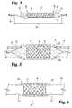
- FIG. 1is a side view showing one of the steps of the method of the preferred embodiment of the present invention.
- FIG. 2is an enlarged cross-sectional view of a portion of the medical device and product of the preferred embodiment of the present invention
- FIG. 3depicts another preferred embodiment of the present invention in which a coated medical device such as a coated stent is mounted or positioned on another medical device such as an inflatable balloon with a bioactive material disposed on at least the outer surface of the balloon;
- a coated medical devicesuch as a coated stent is mounted or positioned on another medical device such as an inflatable balloon with a bioactive material disposed on at least the outer surface of the balloon;
- FIG. 4depicts an enlarged and longitudinally cross-sectioned view of the medical device stent mounted on the medical device balloon of FIG. 3 ;
- FIG. 5depicts a coated medical device stent of the present invention mounted on medical device balloon which has been positioned in a vessel and expanded therein;
- FIG. 6depicts the coated expanded medical device stent implanted in the vessel of FIG. 5 with the medical device balloon having been deflated and removed from the treatment site;
- FIG. 7depicts an enlarged cross-section end view of the balloon of FIG. 5 in which the folds of the balloon unfurl during expansion and make contact with the inner surface of a vessel;
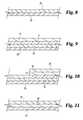
- FIGS. 8-11depict various embodiments of the bioactive material layers of the present invention posited on the base material of a medical device stent and a medical device balloon.
- the medical device 10 of the present inventionfirst comprises a structure 12 adapted for temporary or permanent introduction into a human or veterinary patient.
- “Adapted”means that the structure 12 is particularly configured, shaped and sized for such introduction.
- the structure 12is most preferably configured as a vascular stent adapted for insertion into the vascular system of the patient.
- the structure 12can of course be particularly configured for use in other systems and sites such as the esophagus, trachea, colon, biliary ducts, urethra and ureters, among others.
- the structure 12can alternatively be configured as any conventional vascular or other comparable medical device, and can include any of a variety of conventional stent or other adjuncts, such as helically wound strands, perforated cylinders or the like.
- the inserted structure 12need not be an entire device, but can merely be that portion of a vascular or other device which is intended to be introduced into the patient.
- the structure 12can be configured as at least one of, or any portion of, a catheter, a wire guide, a cannula, a stent, a vascular or other graft, a cardiac pacemaker lead or lead tip, a cardiac defibrillator lead or lead tip, a heart valve, a suture, a needle, an angioplasty device or a pacemaker.
- the structure 12can also be configured as a combination of portions of any of these.
- FIGS. 1 and 2show only a structure 12 configured as a stent, and more particularly, a vascular stent. More preferably, the structure 12 is configured as a vascular stent such as the “LOGIC” stent, the “V-FLEX PLUS” stent, or the “ACHIEVE” stent, all commercially available from Cook Incorporated, Bloomington, Ind. Such stents are cut from a cannula of suitable material and possess a plurality of interconnected struts allowing the stents to expand upon inflation of a balloon on which they are carried.
- a vascular stentsuch as the “LOGIC” stent, the “V-FLEX PLUS” stent, or the “ACHIEVE” stent, all commercially available from Cook Incorporated, Bloomington, Ind.
- Such stentsare cut from a cannula of suitable material and possess a plurality of interconnected struts allowing the stents to expand upon inflation
- the particular shape and dimensions of the structure 12should of course be selected as required for its specific purpose and for the particular site in the patient at which it will be employed, such as in the coronary arteries, aorta, esophagus, trachea, colon, biliary tract or urinary tract.
- a structure 12 intended for each locationwill have different dimensions particularly suited to such use.
- aortic, esophageal, tracheal and colonic stentsmay have diameters up to about 25 mm and lengths about 100 mm or longer.
- Vascular stentsare generally shorter, typically about 10 to 60 mm in length, and often preferably about 12 to 25 mm in length.
- Such vascular stentsare typically designed to expand to a diameter of about 2 to 6 mm when inserted into the vascular system of a patient, often preferably about 2 to 4 mm.
- the structure 12is composed of a base material 14 suitable for the intended use of the structure 12 .
- the base material 14is preferably biocompatible. A variety of conventional materials can be employed as the base material 14 . Some materials may be more useful for structures other than the coronary stent exemplifying the structure 12 .
- the base material 14may be either elastic or inelastic as required for its intended use in the patient.
- the base materialmay be either biodegradable or non-biodegradable, and a variety of biodegradable polymers are known.
- the base material 14can also be porous or preferably non-porous, again based on its intended use or application.
- the base material 14can include at least one of stainless steel, tantalum, titanium, nitinol, gold, platinum, inconel, iridium, silver, tungsten, or another biocompatible metal, or alloys of any of these; carbon or carbon fiber; cellulose acetate, cellulose nitrate, silicone, polyethylene terephthalate, polyurethane, polyamide, polyester, polyorthoester, polyanhydride, polyether sulfone, polycarbonate, polypropylene, high molecular weight polyethylene, polytetrafluoroethylene, or another biocompatible polymeric material, or mixtures or copolymers of these; polylactic acid, polyglycolic acid or copolymers thereof, a polyanhydride, polycaprolactone, polyhydroxybutyrate valerate or another biodegradable polymer, or mixtures or copolymers of these; a protein, an extracellular matrix component, collagen, fibrin or another biologic agent; or a suitable mixture of any of these.
- Stainless steelis particularly useful as the base material 14 when the structure 12 is configured as a vascular stent.
- particularly preferred base materials 14include stainless steel, nitinol, tantalum, polylactic acid, polyglycolic acid and biodegradable materials. Molybdenum-rhenium alloy and magnesium may also possibly be useful base materials 14 as well.
- a conventional radiopaque marker or coatingmay and preferably should be applied to it at some limited location.
- the radiopaque marker or coatingprovides a means for identifying the location of the structure 12 by X-ray or fluoroscopy during or after its introduction into the patient's vascular system.
- the base material 14 of the structure 12 of the medical device 10 of the present inventionincludes a roughened or textured surface 16 extending at least partly over the base material 14 .
- the surface 16is roughened or textured in a manner described in more detail below. While the surface 16 can be the entire surface of the base material 14 , in the preferred embodiment of the present invention (where the structure 12 is configured as a vascular stent) the surface 16 is the outer surface of the base material 14 .
- the medical device 10 of the present inventionfurther comprises at least one layer 18 of a bioactive material posited directly upon the roughened or textured surface 16 of the base material 14 of the structure 12 .
- the medical device 10 of the present inventionis characterized in that it does not require or is free of any additional coating or layer atop the layer 18 of bioactive material. Although, it is to be understood that for any reason an additional coating or layer atop or below the layer 18 of bioactive is desired, such coating or layer can be applied and still be within the contemplation of the present invention.
- the layer 18may be smoother or rougher than the roughened or textured surface 16 .
- the base material 14 of the structure 12is preferably non-porous, although the structure 12 itself can be perforate.
- the difference between a porous material and a non-porous but perforate materialis a practical one; the relatively smaller open cells of a porous material are of a character and number sufficient to retain an appreciable amount of an applied bioactive material therein, while the relatively larger perforations of a non-porous material are of a character and number which are not sufficient to retain an appreciable amount of an applied bioactive material therein.
- the open cells of a porous materialcan be considered generally microscopic, while perforations through a non-porous material can be considered generally macroscopic.
- a vast range of drugs, medicants and materialscan be employed as the bioactive material in the layer 18 .
- Particularly useful in the practice of the present inventionare materials which prevent or ameliorate abrupt closure and restenosis of blood vessels previously opened by stenting surgery or other procedures.
- Thrombolyticswhich dissolve, break up or disperse thrombi
- antithrombogenicswhich interfere with or prevent the formation of thrombi
- thrombolyticsare urokinase, streptokinase and the tissue plasminogen activators.
- Particularly preferred antithrombogenicsare heparin, hirudin and the antiplatelets.
- Urokinaseis a plasminogen activating enzyme typically obtained from human kidney cell cultures. Urokinase catalyzes the conversion of plasminogen into the fibrinolytic plasmin, which breaks down fibrin thrombi.
- Heparinis a mucopolysaccharide anticoagulant typically obtained from porcine intestinal mucosa or bovine lung. Heparin acts as a thrombin inhibitor by greatly enhancing the effects of the blood's endogenous antithrombin III. Thrombin, a potent enzyme in the coagulation cascade, is key in catalyzing the formation of fibrin. Therefore, by inhibiting thrombin, heparin inhibits the formation of fibrin thrombi.
- bioactive materials having other functionscan also be successfully delivered by the device 10 of the present invention.
- an antiproliferative agentsuch as methotrexate will inhibit over-proliferation of smooth muscle cells and thus inhibit restenosis of the dilated segment of the blood vessel.
- localized delivery of an antiproliferative agentis also useful for the treatment of a variety of malignant conditions characterized by highly vascular growth.
- the device 10 of the present inventioncould be placed in the arterial supply of the tumor to provide a means of delivering a relatively high dose of the antiproliferative agent directly to the tumor.
- a vasodilatorsuch as a calcium channel blocker or a nitrate will suppress vasospasm, which is common following angioplasty procedures.
- Vasospasmoccurs as a response to injury of a blood vessel, and the tendency toward vasospasm decreases as the vessel heals. Accordingly, the vasodilator is desirably supplied over a period of about two to three weeks.
- trauma from angioplastyis not the only vessel injury which can cause vasospasm, and the device 10 may be introduced into vessels other than the coronary arteries, such as the aorta, carotid arteries, renal arteries, iliac arteries or peripheral arteries for the prevention of vasospasm in them.
- an anti-cancer chemotherapeutic agentcan be delivered by the device 10 to a localized tumor. More particularly, the device 10 can be placed in an artery supplying blood to the tumor or elsewhere to deliver a relatively high and prolonged dose of the agent directly to the tumor, while limiting systemic exposure and toxicity.
- the agentmay be a curative, a pre-operative debulker reducing the size of the tumor, or a palliative which eases the symptoms of the disease.
- bioactive material in the present inventionis delivered across the device 10 , and not by passage from an outside source through any lumen defined in the device 10 , such as through a catheter employed for conventional chemotherapy.
- the bioactive material of the present inventionmay, of course, be released from the device 10 into any lumen defined in it, and that lumen may carry some other agent to be delivered through it.
- Paclitaxelis a particularly preferred anti-cancer agent and/or anti-angiogenic agent as the bioactive material of the layer 18 .
- Paclitaxelis also a lipophilic bioactive material that is attracted by the lipids in the endothelial and smooth muscle wall cells of the vessel.
- the implantable medical devicesuch as a stent of the present invention
- the stentmaintains the bioactive material layer 18 in direct contact with the vessel wall.
- Paclitaxelis applied to a medical device such as a balloon which is used for delivering another medical device such as a stent to a treatment site.
- the Paclitaxel coating on the balloon materialis then brought in direct contact of the vessel wall for only that period of the inflation of the balloon which is typically in the neighborhood of approximately one minute.
- the angiogenesis-dependent diseasesare those diseases which require or induce vascular growth, for example, certain types of cancer.
- Estrogen and estrogen derivativesare also particularly preferred as the bioactive material of the layer 18 .
- Dopamine or a dopamine agonist such as bromocriptine mesylate or pergolide mesylateis useful for the treatment of neurological disorders such as Parkinson's disease.
- the device 10could be placed in the vascular supply of the thalamic substantia nigra for this purpose, or elsewhere, localizing treatment in the thalamus.
- the present inventionalso contemplates the use of bioactive materials which covalently bond to the roughened or textured surface 16 of the base material 14 of the structure 12 .
- the bioactive material of the layer 18comprises at least one of: paclitaxel; estrogen or estrogen derivatives; heparin or another thrombin inhibitor, hirudin, hirulog, argatroban, D-phenylalanyl-L-poly-L-arginyl chloromethyl ketone, or another antithrombogenic agent, or mixtures thereof; urokinase, streptokinase, a tissue plasminogen activator, or another thrombolytic agent, or mixtures thereof; a fibrinolytic agent; a vasospasm inhibitor; a calcium channel blocker, a nitrate, nitric oxide, a nitric oxide promoter or another vasodilator; an antimicrobial agent or antibiotic; aspirin, ticlopidine or another antiplatelet agent; colchicine or another antimitotic, or another microtubul
- bioactive material of the layer 18are heparin, anti-inflammatory steroids including but not limited to dexamethasone and its derivatives, and mixtures of heparin and such steroids.
- bioactive materialin the practice of the present invention, including: smooth muscle cell inhibitors, collagen inhibitors, anti-coagulants and cholesterol reducing agents; forskolin, vapiprost, prostaglandin and analogues thereof, prostacyclin and prostacyclin analogues, dextran and dipyridamole; angiotensin converting enzyme inhibitors such as Captopril® (available from Squibb), Cilazapril® (available from Hoffman-LaRoche), or Lisinopril® (available from Merck); fibroblast growth factor (FGF) antagonists, fish oil (omega 3-fatty acid), histamine antagonists, Lovastatin® (an inhibitor of HMG-CoA reductase, a cholesterol-lowering drug from Merck), methotrexate, monoclonal antibodies (such as to PDGF receptors), nitroprusside, phosphodiesterase inhibitors, prostaglandin inhibitor (available from Glaxo), vascular endothelial
- the present inventionis also directed to a method of manufacturing the medical device 10 disclosed above. More particularly, the method of the present invention first comprises providing a structure 12 adapted for the temporary or permanent introduction into a patient.
- the structure 12comprises a preferably non-porous base material 14 having a surface 16 and is configured, for example, as a stent (such as a vascular stent).
- the structure 12 and the base material 14have been described in detail above, and for brevity, such details will not be repeated here.
- Stainless steel, nitinol, tantalum, polylactic acid, polyglycolic acid and biodegradable materialsare particularly preferred as the base material 14 of the structure 12 .
- the method of the present inventionfurther comprises the steps of attaining a desired roughness or texture on the surface 16 of the base material 14 of the structure 12 , and positing a layer 18 of a bioactive material directly upon the roughened or textured surface 16 of the base material 14 .
- a bioactive materialuseful in the layer 18 has been disclosed in detail above; again, for brevity, such detail will not be repeated.
- Paclitaxel, a taxane or another paclitaxel analogue, estrogen and estrogen derivativesare particularly preferred as bioactive materials in the layer 18 .
- the method of manufacturing a medical device 10 according to the present inventionis characterized in that the resulting medical device 10 does not require or is free of any additional coating or layer atop the layer 18 of bioactive material.
- the method of the present inventiontherefore does not include any steps in which the bioactive material is covered by or contained within a time-release or containment layer. While the method of the present invention contemplates the use of a base material 14 which itself comprises a plurality of layers or constituents, such an arrangement may not be preferred in the practice of the present invention. In any event, it would be the outermost one of such plural layers or constituents which possesses the roughened or textured surface 16 on which the layer 18 of bioactive material is posited directly.
- the step of directly positing the layer 18 of bioactive material on the roughened or textured surface 16 of the base material 14can be carried out in any convenient manner.
- the structure 12(or suitable portion thereof) can be dipped or soaked in an appropriate solution of the desired bioactive material, and the solvent of the solution evaporated to leave a layer 18 of the bioactive material on the roughened or textured surface 16 of the base material 14 .
- the positing stepis carried out by spraying a solution of the bioactive material on the roughened or textured surface 16 of the base material 14 of the structure 12 and allowing the structure 12 to dry. While spraying may have a relatively low efficiency in transferring the bioactive material to the roughened or textured surface 16 , it is adequate for the purposes of the present invention.
- paclitaxel(the particularly preferred bioactive material in the present invention) can be posited by spraying an ethanolic solution of it on the roughened or textured surface 16 of the base material 14 .
- the solutionconveniently contains about 2 to about 4 mg of paclitaxel per ml of ethanol.
- ethanolshould be 100% USP grade or equivalent, not denatured alcohol or 95% ethanol.
- sprayingcan be readily carried out to posit about 5 to about 500 ⁇ g, preferably 50 to 150 ⁇ g, of paclitaxel on the roughened or textured surface 16 of the base material 14 . Perhaps less than about 1% of the paclitaxel is ultimately posited from solution onto the textured surface 16 .
- the surface 16 of the base material 14 of the structure 12can be roughened or textured in any convenient manner, such as by etching.
- the surface 16is roughened or textured by abrading, for example, by abrading with an abrasive grit 24 comprising at least one of sodium bicarbonate (USP), calcium carbonate, aluminum oxide, colmanite (calcium borate), crushed glass, crushed walnut shells, or mixtures of these or other abrasive particulates.
- USPsodium bicarbonate
- CaCOcalcium carbonate
- aluminum oxidecolmanite (calcium borate)
- crushed glasscrushed walnut shells, or mixtures of these or other abrasive particulates.
- Such roughening or texturingis most easily carried out by placing the medical device 10 on a mandrel 20 in a position such that abrasive grit 24 delivered from a nozzle 22 impinges on the surface 16 .
- the initial surface of the base material prior to roughening or texturingmay be smooth
- the grit size and feed rate of the abrasive grit 24 , the structure of the nozzle 22 , the pressure at which the abrasive grit 24 is delivered from the nozzle 22 , the distance of the surface 16 from the nozzle 22 and the rate of relative movement of the medical device 10 and the nozzle 22are all factors to be considered in achieving an appropriate desired roughness or texture of the surface 16 of the base material 14 of the structure 12 .
- the abrading stepcan be carried out with an abrasive grit 24 having a particle size of about 5 microns (5 ⁇ m) to about 500 microns (500 ⁇ m).
- the abrading stepis carried out with sodium bicarbonate (USP) having a nominal particle size of about 50 microns (50 ⁇ m), with approximately 50% greater than 40 microns (40 ⁇ m) and approximately 1% greater than 150 microns (150 ⁇ m).
- USPsodium bicarbonate
- Such abradingis preferably carried out with the sodium bicarbonate or other abrasive grit 24 delivered at a pressure under flow of about 5 to about 200 PSI (about 34 to about 1380 KPa), most preferably about 100 PSI (about 690 KPa).
- Such abradingis also preferably carried out with the sodium bicarbonate or other abrasive grit 24 delivered at a grit feed rate of about 1 to about 1000 g/min, most preferably about 10 to about 15 g/min.
- the carrier gas or propellant for delivery of the abrasive gritis preferably nitrogen, air or argon, and most preferably nitrogen, although other gases may be suitable as well.
- the distance from the outlet of the nozzle 22 to the center of the mandrel 20can be about 1 to about 100 mm.
- a preferred nozzle 22is the Comco Microblaster; when employed, the preferred distance from the outlet of the nozzle 22 to the center of the mandrel 20 is about 5 to about 10 mm.
- the Paasche LAC #3is also useful as the nozzle 22 .
- the base material 14 of the structure 12is stainless steel, and the abrasive grit 24 is 50 micron sodium bicarbonate (USP), a particularly preferred combination of abrading conditions is:
- Nozzle 22Comco Microblaster
- Spray plan8 equally-spaced circumferential positions
- Sweep rateAbout 16 mm/sec
- Grit feed rateAbout 0.15 to 0.30 g/sec
- Nozzle outlet to mandrel centerabout 5 to 10 mm
- a roughened or textured surface 16is obtained which is thought to have a mean surface roughness (that is, a mean height of surface features) of about 10 ⁇ in. (about 250 nm) and a surface roughness range between about 1 ⁇ in. and about 100 ⁇ in. (about 25 nm and about 2.5 ⁇ m).
- a surface 16is capable of retaining on it a highly substantial portion of bioactive material posited directly on it without requiring any additional covering or containment layer.
- the adhesion of paclitaxel to two types of stainless steel, grit abraded stentswas compared to its adhesion to stents of those types whose surfaces had instead been plasma treated prior to the direct deposition of paclitaxel thereon (control stents).
- the coated stents of both typesthat is, medical devices 10 of the present invention and control stents, were then subjected to a physical adhesion test which simulated the rate at which paclitaxel would be delivered during introduction and deployment of the stents in clinical use.
- the adhesion testinvolved passing each stent through a water-filled guiding catheter of appropriate diameter and inflating the balloon catheter to expand each stent to its intended diameter.
- the stentsare already mounted before coating.
- the amount of paclitaxel remaining on each stentwas then measured by spectrometry and compared to the amount of paclitaxel initially posited on each stent.
- Stents having surfaces 16 roughened or textured by abrasion with different abrasive grits 24retained 84.1 ⁇ 10.2% of the paclitaxel originally applied, while stents having plasma treated surfaces retained only 44.3 ⁇ 8.7% of the paclitaxel originally applied (p ⁇ 0.0001). This appears to demonstrate the successful retention of the layer 18 of bioactive material on the roughened or textured surface 16 of the base material 14 of the structure 12 of the medical device 10 of the present invention.
- any trial-and-error testingto obtain the optimal processing conditions for any desired combination of particular base materials 14 and bioactive materials.
- Such testingsimply requires roughening or texturing the surface 16 of a particular base material 14 in a selected manner, applying a layer 18 of a particular bioactive material to the roughened or textured surface 16 and measuring the retention of bioactive material on the roughened or textured surface 16 after clinical introduction and deployment has been mimicked.
- the present inventionprovides a medical device 10 and method for manufacturing the same which is particularly advantageous over prior devices and methods for making such devices.
- the time and cost of manufacture of the medical device of the present inventionare minimized by the absence of any steps to incorporate the bioactive material in a containment layer, or to apply a containment or time-release layer over the bioactive material.
- the particularly preferred use of sodium bicarbonate as the abrasive to provide roughness or texture to the surface of the base material of the structureis advantageous in the low toxicity of the sodium bicarbonate to production workers, the ease of product and waste cleanup, and the biocompatibility of any residual sodium bicarbonate.
- FIG. 3depicts another preferred embodiment of the present invention in which a coated medical device 10 such as a coated stent is mounted or positioned on another medical device 26 such as an inflatable balloon with a bioactive material 28 disposed on the outer surface of the balloon.
- This bioactive material 28is preferably a lipophilic bioactive material such as paclitaxel and other lipophilic materials such as dexamethozone and the like previously described herein.
- coated implantable medical device 10includes a bioactive material layer posited thereon as previously described. This bioactive material layer can also be a lipophilic bioactive material such as bioactive material 28 applied to balloon medical device 26 .
- balloon ends 30 and 32extend longitudinally beyond respective stent ends 34 and 36 .
- the folded balloon ends 30 and 32extend beyond the end of the stent ends and are coated with lipophilic bioactive material for deposition on the vessel wall extending beyond the ends of the delivered stent.
- the balloon 26is preferably of a polyamid material such as nylon 12 which is available from COOK, Inc., Bloomington, Ind.
- the balloon 26is attached to a catheter shaft 38 , which includes a guide wire lumen as well as an inflation lumen for inflating the balloon.
- FIG. 4depicts an enlarged and longitudinally cross-sectioned view of a medical device stent 10 mounted on medical device balloon 26 of FIG. 3 .
- the distal and proximal ends 30 and 32 of the balloonare folded and extend radially outward to the outer diameter of the compressed stent.
- the folded ends of the ballooncan extend beyond the outer diameter of the stent.
- the bioactive material layers 18 and 28are of the same lipophilic bioactive material such as paclitaxel, which is applied to the stent and balloon after the stent is mounted on the balloon.
- the paclitaxel lipophilic bioactive material coatingis applied to the outer surface of the balloon and stent. This coating process is as previously described.
- the paclitaxel bioactive material layeris applied to the vessel wall when the balloon and stent are expanded in the vessel at the treatment site.
- FIG. 5depicts medical device stent 10 mounted on medical device balloon 26 which has been positioned in vessel 40 and expanded therein. As shown, that portion of vessel 42 is expanded radially outward due to the expansion of the stent and balloon to alleviate a stenotic or traumatized condition of the vessel. Portions of proximal and distal balloon ends 30 and 32 come in direct or physical contact with the radially enlarged portions of vessel wall 40 . Thus, the lipophilic bioactive material 28 is applied to the enlarged portions of the vessel coming in contact therewith as well as bioactive material 18 which is on the outer surface of the medical device stent 10 . This advantageously not only treats the enlarged vessel wall supported by stent 10 , but also the lipophilic bioactive material is applied to the vessel extending beyond the ends of the stent and thus eliminating the undesirable edge effect associated with stent implantation.
- FIG. 6depicts expanded medical device stent 10 implanted in vessel 40 of FIG. 5 with medical device balloon 26 having been deflated and removed from the treatment site.
- lipophilic bioactive material 18has been applied to the vessel wall along the length of the stent
- bioactive material 28is likewise applied to the vessel wall extending beyond the ends of the stent where the previously inflated balloon ends came in contact with the vessel wall.
- This drug-delivered zone 44extending along the length of the vessel in which the balloon and stent have come in contact with the wall and to which the lipophilic bioactive material has been advantageously applied thereto for treating the traumatized or stenosed vessel and minimizing, if not eliminating, any adverse reaction due to the implantation of the stent and delivery balloon.
- FIG. 7depicts an enlarged cross-section end view of balloon 26 of FIG. 5 in which folds 46 , 48 and 50 unfold or unfurl during expansion and making contact with the inner surface of vessel 40 .
- folds 46 , 48 and 50unfurl, rotate and come in a wiping contact with the inner surface of vessel 40 in a rotational movement indicated by arrows 52 , 54 and 56 .
- the lipophilic bioactive material disposed on the surface thereofcomes in contact with the inner surface of the vessel wall and is posited on the vessel surface and attached thereto with the lipophilic attraction of the cells. This rotational or wiping action further ensures a complete circumferential coating of the inner vessel surface.
- FIGS. 8-11Depicted in FIGS. 8-11 are various embodiments of the bioactive material layers posited on the base material of medical device stent 10 and medical device 26 .
- a single layer of lipophilic bioactive material 28is posited or applied to balloon base material 26 .
- a single lipophilic coating materialis applied to the surface of the balloon for a direct application to a vessel wall, for example, after the previous introduction of another stent. This balloon could be used for an angioplasty procedure without the use of a stent.
- FIG. 11depicts the same balloon base material layer 26 of which a layer of hydrophilic material is posited or coated thereon.
- the lipophilic bioactive material layeris sprayed, posited, or disposed on the hydrophilic or slip coating layer 58 .
- the hydrophilic layerpermits easier detachment or delivery of the lipophilic layer 28 when in contact with the cells of a vessel wall.
- FIG. 9depicts another preferred embodiment of the present invention of which balloon base material 26 has lipophilic bioactive material 28 coated thereon in one operation and then medical device 10 such as a stent with base material 14 and then lipophilic bioactive material 18 is crimped or positioned around the balloon.
- FIG. 10depicts the base and lipophilic material layers of the structure of FIG. 9 with an additional layer of bioactive material 28 sprayed, posited, or disposed therein.
- This configurationpresents an alternative embodiment for delivering greater doses of the lipophilic bioactive material to the cells located on the surface of a vessel wall.
- the present inventionis useful in the performance of various surgical procedures and in the manufacture of devices for the performance of various surgical procedures, and therefore finds applicability in human and veterinary medicine.
- the medical device of the present inventioncan also include channels, grooves, recesses, indentations, projections, buildups, and the like to increase the surface area of the device to which the bioactive material can be posited therein.
- paclitaxelis a lipophilic material and is rapidly taken up by cells, particularly containing lipids. Once in the cells, it binds to proteins, which helps keep the paclitaxel for subsequent use. If paclitaxel is kept around for short times, for example, 20 minutes, it can still have prolonged effects for up to, for example, 14 days.
- the discussion of paclitaxelcan be found in various articles such as “Paclitaxel Inhibits Arterial Smooth Muscle Cell Proliferation and Migration in Vitro and in Vivo Using Local Drug Delivery,” Circulation, Vol. 96, No. 2, Jul. 15, 1997, pp. 636. Another article entitled “Drug Therapy,” New England Journal of Medicine, Apr. 13, 1995, pp. 1004.
Landscapes
- Health & Medical Sciences (AREA)
- Life Sciences & Earth Sciences (AREA)
- Engineering & Computer Science (AREA)
- Animal Behavior & Ethology (AREA)
- General Health & Medical Sciences (AREA)
- Public Health (AREA)
- Veterinary Medicine (AREA)
- Biomedical Technology (AREA)
- Heart & Thoracic Surgery (AREA)
- Epidemiology (AREA)
- Vascular Medicine (AREA)
- Chemical & Material Sciences (AREA)
- Medicinal Chemistry (AREA)
- Oral & Maxillofacial Surgery (AREA)
- Transplantation (AREA)
- Molecular Biology (AREA)
- Cardiology (AREA)
- Hematology (AREA)
- Anesthesiology (AREA)
- Child & Adolescent Psychology (AREA)
- Biophysics (AREA)
- Pulmonology (AREA)
- Surgery (AREA)
- Dermatology (AREA)
- Bioinformatics & Cheminformatics (AREA)
- Manufacturing & Machinery (AREA)
- Physics & Mathematics (AREA)
- Optics & Photonics (AREA)
- Chemical Kinetics & Catalysis (AREA)
- Urology & Nephrology (AREA)
- Nuclear Medicine, Radiotherapy & Molecular Imaging (AREA)
- Organic Chemistry (AREA)
- Pharmacology & Pharmacy (AREA)
- General Chemical & Material Sciences (AREA)
- Materials For Medical Uses (AREA)
- Media Introduction/Drainage Providing Device (AREA)
Abstract
Description
Claims (22)
Priority Applications (3)
| Application Number | Priority Date | Filing Date | Title |
|---|---|---|---|
| US14/193,312US9814865B2 (en) | 2000-10-31 | 2014-02-28 | Coated medical device |
| US15/381,291US20170105858A1 (en) | 2000-10-31 | 2016-12-16 | Coated medical device |
| US15/810,721US20190351197A9 (en) | 2000-10-31 | 2017-11-13 | Coated medical device |
Applications Claiming Priority (8)
| Application Number | Priority Date | Filing Date | Title |
|---|---|---|---|
| US24444600P | 2000-10-31 | 2000-10-31 | |
| US10/000,659US6918927B2 (en) | 2000-10-31 | 2001-10-31 | Coated implantable medical device |
| US39543402P | 2002-07-12 | 2002-07-12 | |
| US10/618,977US7803149B2 (en) | 2002-07-12 | 2003-07-14 | Coated medical device |
| US11/141,574US8172793B2 (en) | 2000-10-31 | 2005-05-31 | Coated medical device |
| US12/609,521US9694162B2 (en) | 2000-10-31 | 2009-10-30 | Coated medical device |
| US13/072,332US8974522B2 (en) | 2000-10-31 | 2011-03-25 | Coated medical device |
| US14/193,312US9814865B2 (en) | 2000-10-31 | 2014-02-28 | Coated medical device |
Related Parent Applications (1)
| Application Number | Title | Priority Date | Filing Date |
|---|---|---|---|
| US13/072,332ContinuationUS8974522B2 (en) | 2000-10-31 | 2011-03-25 | Coated medical device |
Related Child Applications (2)
| Application Number | Title | Priority Date | Filing Date |
|---|---|---|---|
| US15/381,291ContinuationUS20170105858A1 (en) | 2000-10-31 | 2016-12-16 | Coated medical device |
| US15/810,721ContinuationUS20190351197A9 (en) | 2000-10-31 | 2017-11-13 | Coated medical device |
Publications (2)
| Publication Number | Publication Date |
|---|---|
| US20140178563A1 US20140178563A1 (en) | 2014-06-26 |
| US9814865B2true US9814865B2 (en) | 2017-11-14 |
Family
ID=30115871
Family Applications (13)
| Application Number | Title | Priority Date | Filing Date |
|---|---|---|---|
| US10/618,977Active2026-03-06US7803149B2 (en) | 2000-10-31 | 2003-07-14 | Coated medical device |
| US11/141,574Expired - LifetimeUS8172793B2 (en) | 2000-10-31 | 2005-05-31 | Coated medical device |
| US11/234,702Expired - LifetimeUS7731685B2 (en) | 2002-07-12 | 2005-09-23 | Coated medical device |
| US12/359,884Expired - LifetimeUS8673387B2 (en) | 2000-10-31 | 2009-01-26 | Coated medical device |
| US12/609,521Expired - LifetimeUS9694162B2 (en) | 2000-10-31 | 2009-10-30 | Coated medical device |
| US12/892,177Expired - Fee RelatedUS10532190B2 (en) | 2002-07-12 | 2010-09-28 | Coated medical device |
| US13/072,332Expired - Fee RelatedUS8974522B2 (en) | 2000-10-31 | 2011-03-25 | Coated medical device |
| US14/013,591PendingUS20140005541A1 (en) | 2000-10-31 | 2013-08-29 | Coated medical device |
| US14/193,312Expired - LifetimeUS9814865B2 (en) | 2000-10-31 | 2014-02-28 | Coated medical device |
| US15/166,486AbandonedUS20160271373A1 (en) | 2000-10-31 | 2016-05-27 | Coated medical device |
| US15/381,291AbandonedUS20170105858A1 (en) | 2000-10-31 | 2016-12-16 | Coated medical device |
| US15/810,721AbandonedUS20190351197A9 (en) | 2000-10-31 | 2017-11-13 | Coated medical device |
| US16/742,372AbandonedUS20200230372A1 (en) | 2002-07-12 | 2020-01-14 | Coated medical device |
Family Applications Before (8)
| Application Number | Title | Priority Date | Filing Date |
|---|---|---|---|
| US10/618,977Active2026-03-06US7803149B2 (en) | 2000-10-31 | 2003-07-14 | Coated medical device |
| US11/141,574Expired - LifetimeUS8172793B2 (en) | 2000-10-31 | 2005-05-31 | Coated medical device |
| US11/234,702Expired - LifetimeUS7731685B2 (en) | 2002-07-12 | 2005-09-23 | Coated medical device |
| US12/359,884Expired - LifetimeUS8673387B2 (en) | 2000-10-31 | 2009-01-26 | Coated medical device |
| US12/609,521Expired - LifetimeUS9694162B2 (en) | 2000-10-31 | 2009-10-30 | Coated medical device |
| US12/892,177Expired - Fee RelatedUS10532190B2 (en) | 2002-07-12 | 2010-09-28 | Coated medical device |
| US13/072,332Expired - Fee RelatedUS8974522B2 (en) | 2000-10-31 | 2011-03-25 | Coated medical device |
| US14/013,591PendingUS20140005541A1 (en) | 2000-10-31 | 2013-08-29 | Coated medical device |
Family Applications After (4)
| Application Number | Title | Priority Date | Filing Date |
|---|---|---|---|
| US15/166,486AbandonedUS20160271373A1 (en) | 2000-10-31 | 2016-05-27 | Coated medical device |
| US15/381,291AbandonedUS20170105858A1 (en) | 2000-10-31 | 2016-12-16 | Coated medical device |
| US15/810,721AbandonedUS20190351197A9 (en) | 2000-10-31 | 2017-11-13 | Coated medical device |
| US16/742,372AbandonedUS20200230372A1 (en) | 2002-07-12 | 2020-01-14 | Coated medical device |
Country Status (9)
| Country | Link |
|---|---|
| US (13) | US7803149B2 (en) |
| EP (4) | EP1521603B1 (en) |
| JP (2) | JP4727987B2 (en) |
| AT (1) | ATE495769T1 (en) |
| AU (2) | AU2003256540B2 (en) |
| CA (1) | CA2484383C (en) |
| DE (1) | DE60335801D1 (en) |
| DK (1) | DK1521603T3 (en) |
| WO (1) | WO2004006976A1 (en) |
Families Citing this family (324)
| Publication number | Priority date | Publication date | Assignee | Title |
|---|---|---|---|---|
| US7896914B2 (en)* | 1995-06-07 | 2011-03-01 | Cook Incorporated | Coated implantable medical device |
| US7550005B2 (en)* | 1995-06-07 | 2009-06-23 | Cook Incorporated | Coated implantable medical device |
| US7846202B2 (en)* | 1995-06-07 | 2010-12-07 | Cook Incorporated | Coated implantable medical device |
| US7611533B2 (en)* | 1995-06-07 | 2009-11-03 | Cook Incorporated | Coated implantable medical device |
| US6774278B1 (en)* | 1995-06-07 | 2004-08-10 | Cook Incorporated | Coated implantable medical device |
| US20070203520A1 (en)* | 1995-06-07 | 2007-08-30 | Dennis Griffin | Endovascular filter |
| US7867275B2 (en)* | 1995-06-07 | 2011-01-11 | Cook Incorporated | Coated implantable medical device method |
| US20060025726A1 (en)* | 1996-06-04 | 2006-02-02 | Vance Products Incorporated, D/B/A Cook Urological Incorporated | Implantable medical device with pharmacologically active layer |
| US20060052757A1 (en)* | 1996-06-04 | 2006-03-09 | Vance Products Incorporated, D/B/A Cook Urological Incorporated | Implantable medical device with analgesic or anesthetic |
| US20060030826A1 (en)* | 1996-06-04 | 2006-02-09 | Vance Products Incorporated,d/b/a Cook Urological Incorporated | Implantable medical device with anti-neoplastic drug |
| US6306166B1 (en)* | 1997-08-13 | 2001-10-23 | Scimed Life Systems, Inc. | Loading and release of water-insoluble drugs |
| US8668737B2 (en) | 1997-10-10 | 2014-03-11 | Senorx, Inc. | Tissue marking implant |
| US7637948B2 (en) | 1997-10-10 | 2009-12-29 | Senorx, Inc. | Tissue marking implant |
| US7713297B2 (en) | 1998-04-11 | 2010-05-11 | Boston Scientific Scimed, Inc. | Drug-releasing stent with ceramic-containing layer |
| US7947015B2 (en) | 1999-01-25 | 2011-05-24 | Atrium Medical Corporation | Application of a therapeutic substance to a tissue location using an expandable medical device |
| US6955661B1 (en) | 1999-01-25 | 2005-10-18 | Atrium Medical Corporation | Expandable fluoropolymer device for delivery of therapeutic agents and method of making |
| US6862470B2 (en) | 1999-02-02 | 2005-03-01 | Senorx, Inc. | Cavity-filling biopsy site markers |
| US9820824B2 (en) | 1999-02-02 | 2017-11-21 | Senorx, Inc. | Deployment of polysaccharide markers for treating a site within a patent |
| US7651505B2 (en) | 2002-06-17 | 2010-01-26 | Senorx, Inc. | Plugged tip delivery for marker placement |
| US20090030309A1 (en) | 2007-07-26 | 2009-01-29 | Senorx, Inc. | Deployment of polysaccharide markers |
| US6725083B1 (en) | 1999-02-02 | 2004-04-20 | Senorx, Inc. | Tissue site markers for in VIVO imaging |
| US8361082B2 (en) | 1999-02-02 | 2013-01-29 | Senorx, Inc. | Marker delivery device with releasable plug |
| US7983734B2 (en) | 2003-05-23 | 2011-07-19 | Senorx, Inc. | Fibrous marker and intracorporeal delivery thereof |
| US8498693B2 (en) | 1999-02-02 | 2013-07-30 | Senorx, Inc. | Intracorporeal marker and marker delivery device |
| US6575991B1 (en) | 1999-06-17 | 2003-06-10 | Inrad, Inc. | Apparatus for the percutaneous marking of a lesion |
| US7462162B2 (en) | 2001-09-04 | 2008-12-09 | Broncus Technologies, Inc. | Antiproliferative devices for maintaining patency of surgically created channels in a body organ |
| US20030070676A1 (en)* | 1999-08-05 | 2003-04-17 | Cooper Joel D. | Conduits having distal cage structure for maintaining collateral channels in tissue and related methods |
| CA2659518A1 (en) | 2000-11-20 | 2002-05-30 | Senorx, Inc. | Tissue site markers for in vivo imaging |
| DE10115740A1 (en) | 2001-03-26 | 2002-10-02 | Ulrich Speck | Preparation for restenosis prophylaxis |
| WO2003002243A2 (en) | 2001-06-27 | 2003-01-09 | Remon Medical Technologies Ltd. | Method and device for electrochemical formation of therapeutic species in vivo |
| WO2006014731A2 (en)* | 2001-09-04 | 2006-02-09 | Broncus Technologies, Inc. | Methods and devices for maintaining surgically created channels in a body organ |
| JP2005507754A (en) | 2001-11-08 | 2005-03-24 | アトリウム メディカル コーポレイション | Intraluminal device having a coating containing a therapeutic agent |
| US20110306997A9 (en)* | 2002-02-21 | 2011-12-15 | Roschak Edmund J | Devices for creating passages and sensing for blood vessels |
| DK1521603T3 (en) | 2002-07-12 | 2011-04-18 | Cook Inc | Coated medical device |
| DE10244847A1 (en)* | 2002-09-20 | 2004-04-01 | Ulrich Prof. Dr. Speck | Medical device for drug delivery |
| US20060036158A1 (en) | 2003-11-17 | 2006-02-16 | Inrad, Inc. | Self-contained, self-piercing, side-expelling marking apparatus |
| BRPI0408891A (en)* | 2003-03-28 | 2006-04-11 | Kosan Biosciences Inc | devices, methods, and compositions for preventing restenosis |
| US20040230176A1 (en)* | 2003-04-23 | 2004-11-18 | Medtronic Vascular, Inc. | System for treating a vascular condition that inhibits restenosis at stent ends |
| US7877133B2 (en) | 2003-05-23 | 2011-01-25 | Senorx, Inc. | Marker or filler forming fluid |
| US8308682B2 (en) | 2003-07-18 | 2012-11-13 | Broncus Medical Inc. | Devices for maintaining patency of surgically created channels in tissue |
| US7763063B2 (en) | 2003-09-03 | 2010-07-27 | Bolton Medical, Inc. | Self-aligning stent graft delivery system, kit, and method |
| US9198786B2 (en) | 2003-09-03 | 2015-12-01 | Bolton Medical, Inc. | Lumen repair device with capture structure |
| US20080264102A1 (en) | 2004-02-23 | 2008-10-30 | Bolton Medical, Inc. | Sheath Capture Device for Stent Graft Delivery System and Method for Operating Same |
| US11259945B2 (en) | 2003-09-03 | 2022-03-01 | Bolton Medical, Inc. | Dual capture device for stent graft delivery system and method for capturing a stent graft |
| US8292943B2 (en) | 2003-09-03 | 2012-10-23 | Bolton Medical, Inc. | Stent graft with longitudinal support member |
| US8500792B2 (en) | 2003-09-03 | 2013-08-06 | Bolton Medical, Inc. | Dual capture device for stent graft delivery system and method for capturing a stent graft |
| US11596537B2 (en) | 2003-09-03 | 2023-03-07 | Bolton Medical, Inc. | Delivery system and method for self-centering a proximal end of a stent graft |
| US20070198078A1 (en) | 2003-09-03 | 2007-08-23 | Bolton Medical, Inc. | Delivery system and method for self-centering a Proximal end of a stent graft |
| JP2007505658A (en) | 2003-09-15 | 2007-03-15 | アトリウム メディカル コーポレーション | Application of therapeutic substances to tissue sites using expandable medical devices |
| US8021331B2 (en) | 2003-09-15 | 2011-09-20 | Atrium Medical Corporation | Method of coating a folded medical device |
| WO2005039443A2 (en)* | 2003-10-21 | 2005-05-06 | Medlogics Device Corporation | Gamma-tocopherol therapy for restenosis prevention |
| US20050273002A1 (en) | 2004-06-04 | 2005-12-08 | Goosen Ryan L | Multi-mode imaging marker |
| US7349971B2 (en)* | 2004-02-05 | 2008-03-25 | Scenera Technologies, Llc | System for transmitting data utilizing multiple communication applications simultaneously in response to user request without specifying recipient's communication information |
| US20070027523A1 (en)* | 2004-03-19 | 2007-02-01 | Toner John L | Method of treating vascular disease at a bifurcated vessel using coated balloon |
| CA2559747C (en)* | 2004-03-19 | 2013-12-31 | Abbott Laboratories | Multiple drug delivery from a balloon and a prosthesis |
| US20100030183A1 (en)* | 2004-03-19 | 2010-02-04 | Toner John L | Method of treating vascular disease at a bifurcated vessel using a coated balloon |
| JP4447356B2 (en)* | 2004-03-19 | 2010-04-07 | パイオニア株式会社 | Speaker device |
| US8431145B2 (en) | 2004-03-19 | 2013-04-30 | Abbott Laboratories | Multiple drug delivery from a balloon and a prosthesis |
| EP1750782A1 (en)* | 2004-04-29 | 2007-02-14 | Cube Medical A/S | A balloon for use in angioplasty with an outer layer of nanofibers |
| US8696564B2 (en)* | 2004-07-09 | 2014-04-15 | Cardiac Pacemakers, Inc. | Implantable sensor with biocompatible coating for controlling or inhibiting tissue growth |
| US8409167B2 (en) | 2004-07-19 | 2013-04-02 | Broncus Medical Inc | Devices for delivering substances through an extra-anatomic opening created in an airway |
| CA2577792A1 (en)* | 2004-07-19 | 2006-02-09 | Broncus Technologies, Inc. | Methods and devices for maintaining surgically created channels in a body organ |
| US7901451B2 (en)* | 2004-09-24 | 2011-03-08 | Biosensors International Group, Ltd. | Drug-delivery endovascular stent and method for treating restenosis |
| WO2006036970A2 (en)* | 2004-09-28 | 2006-04-06 | Atrium Medical Corporation | Method of thickening a coating using a drug |
| US9801982B2 (en) | 2004-09-28 | 2017-10-31 | Atrium Medical Corporation | Implantable barrier device |
| WO2006036982A2 (en) | 2004-09-28 | 2006-04-06 | Atrium Medical Corporation | Drug delivery coating for use with a stent |
| US9801913B2 (en) | 2004-09-28 | 2017-10-31 | Atrium Medical Corporation | Barrier layer |
| US8367099B2 (en)* | 2004-09-28 | 2013-02-05 | Atrium Medical Corporation | Perforated fatty acid films |
| US9000040B2 (en) | 2004-09-28 | 2015-04-07 | Atrium Medical Corporation | Cross-linked fatty acid-based biomaterials |
| US8312836B2 (en)* | 2004-09-28 | 2012-11-20 | Atrium Medical Corporation | Method and apparatus for application of a fresh coating on a medical device |
| US20090011116A1 (en)* | 2004-09-28 | 2009-01-08 | Atrium Medical Corporation | Reducing template with coating receptacle containing a medical device to be coated |
| US8124127B2 (en) | 2005-10-15 | 2012-02-28 | Atrium Medical Corporation | Hydrophobic cross-linked gels for bioabsorbable drug carrier coatings |
| US9012506B2 (en)* | 2004-09-28 | 2015-04-21 | Atrium Medical Corporation | Cross-linked fatty acid-based biomaterials |
| US20060085058A1 (en)* | 2004-10-20 | 2006-04-20 | Rosenthal Arthur L | System and method for delivering a biologically active material to a body lumen |
| US8419656B2 (en) | 2004-11-22 | 2013-04-16 | Bard Peripheral Vascular, Inc. | Post decompression marker introducer system |
| ATE538833T1 (en)* | 2005-03-18 | 2012-01-15 | Cook Medical Technologies Llc | WIRE GUIDES WITH NEW OUTER LAYER AREAS AND CHAMBERS TO IMPROVE HYDROPHILIC PROPERTIES AND RELEASE OF THERAPEUTIC ACTIVE INGREDIENTS |
| US9125968B2 (en)* | 2005-03-30 | 2015-09-08 | Boston Scientific Scimed, Inc. | Polymeric/ceramic composite materials for use in medical devices |
| US10357328B2 (en) | 2005-04-20 | 2019-07-23 | Bard Peripheral Vascular, Inc. and Bard Shannon Limited | Marking device with retractable cannula |
| US10076641B2 (en) | 2005-05-11 | 2018-09-18 | The Spectranetics Corporation | Methods and systems for delivering substances into luminal walls |
| US20090220564A1 (en)* | 2005-08-19 | 2009-09-03 | Baumbach William R | Methods of treating and preventing acute myocardial infarction |
| US8008395B2 (en)* | 2005-09-27 | 2011-08-30 | Boston Scientific Scimed, Inc. | Organic-inorganic hybrid particle material and polymer compositions containing same |
| US9427423B2 (en) | 2009-03-10 | 2016-08-30 | Atrium Medical Corporation | Fatty-acid based particles |
| US9278161B2 (en) | 2005-09-28 | 2016-03-08 | Atrium Medical Corporation | Tissue-separating fatty acid adhesion barrier |
| EP1931265B1 (en)* | 2005-09-30 | 2011-12-07 | Cook Medical Technologies LLC | Coated vaso-occlusion device |
| US8052658B2 (en)* | 2005-10-07 | 2011-11-08 | Bard Peripheral Vascular, Inc. | Drug-eluting tissue marker |
| US20070129690A1 (en)* | 2005-12-02 | 2007-06-07 | Joel Rosenblatt | Catheter with polymeric coating |
| JP2009519770A (en) | 2005-12-16 | 2009-05-21 | インターフェイス・アソシエイツ・インコーポレーテッド | Medical multilayer balloon and method for producing the same |
| US8840660B2 (en) | 2006-01-05 | 2014-09-23 | Boston Scientific Scimed, Inc. | Bioerodible endoprostheses and methods of making the same |
| US7919108B2 (en)* | 2006-03-10 | 2011-04-05 | Cook Incorporated | Taxane coatings for implantable medical devices |
| US20080286325A1 (en)* | 2006-01-05 | 2008-11-20 | Med Institute, Inc. | Cyclodextrin elution media for medical device coatings comprising a taxane therapeutic agent |
| US8089029B2 (en) | 2006-02-01 | 2012-01-03 | Boston Scientific Scimed, Inc. | Bioabsorbable metal medical device and method of manufacture |
| US20070203564A1 (en)* | 2006-02-28 | 2007-08-30 | Boston Scientific Scimed, Inc. | Biodegradable implants having accelerated biodegradation properties in vivo |
| EP1832301A3 (en)* | 2006-03-08 | 2007-12-05 | Sahajanand Medical Technologies PVT. ltd | Coatings for implantable medical devices |
| US7875284B2 (en)* | 2006-03-10 | 2011-01-25 | Cook Incorporated | Methods of manufacturing and modifying taxane coatings for implantable medical devices |
| US20070224235A1 (en) | 2006-03-24 | 2007-09-27 | Barron Tenney | Medical devices having nanoporous coatings for controlled therapeutic agent delivery |
| US20070225795A1 (en)* | 2006-03-24 | 2007-09-27 | Juan Granada | Composite vascular prosthesis |
| WO2007112025A2 (en)* | 2006-03-24 | 2007-10-04 | Prescient Medical Inc | Composite vascular prosthesis |
| US8187620B2 (en) | 2006-03-27 | 2012-05-29 | Boston Scientific Scimed, Inc. | Medical devices comprising a porous metal oxide or metal material and a polymer coating for delivering therapeutic agents |
| US8048150B2 (en) | 2006-04-12 | 2011-11-01 | Boston Scientific Scimed, Inc. | Endoprosthesis having a fiber meshwork disposed thereon |
| US8303648B2 (en)* | 2006-04-25 | 2012-11-06 | Cook Medical Technologies Llc | Artificial venous valve containing therapeutic agent |
| CA2654376A1 (en)* | 2006-06-06 | 2007-12-13 | Rutgers, The State University Of New Jersey | Iodinated polymers |
| JP2009540926A (en)* | 2006-06-22 | 2009-11-26 | ウィルソン−クック・メディカル・インコーポレーテッド | Self-cleaning stent |
| US8815275B2 (en) | 2006-06-28 | 2014-08-26 | Boston Scientific Scimed, Inc. | Coatings for medical devices comprising a therapeutic agent and a metallic material |
| WO2008002778A2 (en) | 2006-06-29 | 2008-01-03 | Boston Scientific Limited | Medical devices with selective coating |
| EP2380606B1 (en)* | 2006-06-30 | 2018-03-21 | Cook Medical Technologies LLC | Methods of manufacturing and modifying taxane coatings for implantable medical devices |
| EP2054537A2 (en) | 2006-08-02 | 2009-05-06 | Boston Scientific Scimed, Inc. | Endoprosthesis with three-dimensional disintegration control |
| US20080033522A1 (en)* | 2006-08-03 | 2008-02-07 | Med Institute, Inc. | Implantable Medical Device with Particulate Coating |
| DE102006038235A1 (en)* | 2006-08-07 | 2008-02-14 | Biotronik Vi Patent Ag | Improving the stability of biodegradable metallic stents, methods and use |
| EP2068757B1 (en) | 2006-09-14 | 2011-05-11 | Boston Scientific Limited | Medical devices with drug-eluting coating |
| JP2010503489A (en) | 2006-09-15 | 2010-02-04 | ボストン サイエンティフィック リミテッド | Biodegradable endoprosthesis and method for producing the same |
| WO2008034066A1 (en) | 2006-09-15 | 2008-03-20 | Boston Scientific Limited | Bioerodible endoprostheses and methods of making the same |
| ES2357661T3 (en) | 2006-09-15 | 2011-04-28 | Boston Scientific Scimed, Inc. | BIOEROSIONABLE ENDOPROOTHESIS WITH BIOESTABLE INORGANIC LAYERS. |
| EP2959925B1 (en) | 2006-09-15 | 2018-08-29 | Boston Scientific Limited | Medical devices and methods of making the same |
| WO2008036548A2 (en) | 2006-09-18 | 2008-03-27 | Boston Scientific Limited | Endoprostheses |
| CN101663054B (en)* | 2006-10-20 | 2013-08-07 | 生物传感器国际集团有限公司 | Drug delivery intravascular stent and method of use thereof |
| US20080097591A1 (en)* | 2006-10-20 | 2008-04-24 | Biosensors International Group | Drug-delivery endovascular stent and method of use |
| US8067055B2 (en)* | 2006-10-20 | 2011-11-29 | Biosensors International Group, Ltd. | Drug-delivery endovascular stent and method of use |
| WO2008051749A2 (en) | 2006-10-23 | 2008-05-02 | C. R. Bard, Inc. | Breast marker |
| US20080103584A1 (en)* | 2006-10-25 | 2008-05-01 | Biosensors International Group | Temporal Intraluminal Stent, Methods of Making and Using |
| US9492596B2 (en) | 2006-11-06 | 2016-11-15 | Atrium Medical Corporation | Barrier layer with underlying medical device and one or more reinforcing support structures |
| EP2083875B1 (en) | 2006-11-06 | 2013-03-27 | Atrium Medical Corporation | Coated surgical mesh |
| US7981150B2 (en) | 2006-11-09 | 2011-07-19 | Boston Scientific Scimed, Inc. | Endoprosthesis with coatings |
| WO2008060973A1 (en)* | 2006-11-10 | 2008-05-22 | Vance Products Incorporated D/B/A Cook Urological Incorporated | Graft for hysterotomy closure |
| US8414525B2 (en) | 2006-11-20 | 2013-04-09 | Lutonix, Inc. | Drug releasing coatings for medical devices |
| US8414526B2 (en)* | 2006-11-20 | 2013-04-09 | Lutonix, Inc. | Medical device rapid drug releasing coatings comprising oils, fatty acids, and/or lipids |
| US8425459B2 (en) | 2006-11-20 | 2013-04-23 | Lutonix, Inc. | Medical device rapid drug releasing coatings comprising a therapeutic agent and a contrast agent |
| US20080175887A1 (en) | 2006-11-20 | 2008-07-24 | Lixiao Wang | Treatment of Asthma and Chronic Obstructive Pulmonary Disease With Anti-proliferate and Anti-inflammatory Drugs |
| US20080276935A1 (en) | 2006-11-20 | 2008-11-13 | Lixiao Wang | Treatment of asthma and chronic obstructive pulmonary disease with anti-proliferate and anti-inflammatory drugs |
| US9737640B2 (en) | 2006-11-20 | 2017-08-22 | Lutonix, Inc. | Drug releasing coatings for medical devices |
| US8998846B2 (en) | 2006-11-20 | 2015-04-07 | Lutonix, Inc. | Drug releasing coatings for balloon catheters |
| US8414910B2 (en) | 2006-11-20 | 2013-04-09 | Lutonix, Inc. | Drug releasing coatings for medical devices |
| US9700704B2 (en) | 2006-11-20 | 2017-07-11 | Lutonix, Inc. | Drug releasing coatings for balloon catheters |
| US8668703B2 (en)* | 2006-12-01 | 2014-03-11 | Wake Forest University Health Sciences | Medical devices incorporating collagen inhibitors |
| US9579077B2 (en) | 2006-12-12 | 2017-02-28 | C.R. Bard, Inc. | Multiple imaging mode tissue marker |
| WO2008076973A2 (en) | 2006-12-18 | 2008-06-26 | C.R.Bard Inc. | Biopsy marker with in situ-generated imaging properties |
| ES2506144T3 (en) | 2006-12-28 | 2014-10-13 | Boston Scientific Limited | Bioerodible endoprosthesis and their manufacturing procedure |
| KR101198980B1 (en) | 2007-01-21 | 2012-11-07 | 헤모텍 아게 | Medical product for treating stenosis of body passages and for preventing threatening restenosis |
| DE102007003184A1 (en)* | 2007-01-22 | 2008-07-24 | Orlowski, Michael, Dr. | Method for loading structured surfaces |
| US20080188830A1 (en)* | 2007-02-06 | 2008-08-07 | Arrow International, Inc. | Selectively reinforced medical devices |
| US8932345B2 (en)* | 2007-02-07 | 2015-01-13 | Cook Medical Technologies Llc | Medical device coatings for releasing a therapeutic agent at multiple rates |
| US7938286B2 (en)* | 2007-02-13 | 2011-05-10 | Gateway Plastics, Inc. | Container system |
| DE102007008479A1 (en)* | 2007-02-21 | 2008-09-04 | Orlowski, Michael, Dr. | Coated Expandable System |
| US8431149B2 (en) | 2007-03-01 | 2013-04-30 | Boston Scientific Scimed, Inc. | Coated medical devices for abluminal drug delivery |
| US8070797B2 (en) | 2007-03-01 | 2011-12-06 | Boston Scientific Scimed, Inc. | Medical device with a porous surface for delivery of a therapeutic agent |
| US20080228193A1 (en)* | 2007-03-09 | 2008-09-18 | Anthem Orthopaedics Llc | Implantable medicament delivery device and delivery tool and method for use therewith |
| JP5461202B2 (en)* | 2007-03-09 | 2014-04-02 | アイコン メディカル コーポレーション | Bioabsorbable coating for medical devices |
| US8067054B2 (en) | 2007-04-05 | 2011-11-29 | Boston Scientific Scimed, Inc. | Stents with ceramic drug reservoir layer and methods of making and using the same |
| US7976915B2 (en) | 2007-05-23 | 2011-07-12 | Boston Scientific Scimed, Inc. | Endoprosthesis with select ceramic morphology |
| JP2008305262A (en)* | 2007-06-08 | 2008-12-18 | Konica Minolta Business Technologies Inc | Printer introduction method in server and thin client environment |
| US9192697B2 (en) | 2007-07-03 | 2015-11-24 | Hemoteq Ag | Balloon catheter for treating stenosis of body passages and for preventing threatening restenosis |
| US8002823B2 (en) | 2007-07-11 | 2011-08-23 | Boston Scientific Scimed, Inc. | Endoprosthesis coating |
| US7942926B2 (en) | 2007-07-11 | 2011-05-17 | Boston Scientific Scimed, Inc. | Endoprosthesis coating |
| EP2187988B1 (en) | 2007-07-19 | 2013-08-21 | Boston Scientific Limited | Endoprosthesis having a non-fouling surface |
| US7931683B2 (en) | 2007-07-27 | 2011-04-26 | Boston Scientific Scimed, Inc. | Articles having ceramic coated surfaces |
| US8815273B2 (en) | 2007-07-27 | 2014-08-26 | Boston Scientific Scimed, Inc. | Drug eluting medical devices having porous layers |
| WO2009018340A2 (en) | 2007-07-31 | 2009-02-05 | Boston Scientific Scimed, Inc. | Medical device coating by laser cladding |
| JP2010535541A (en) | 2007-08-03 | 2010-11-25 | ボストン サイエンティフィック リミテッド | Coating for medical devices with large surface area |
| DE102007036685A1 (en) | 2007-08-03 | 2009-02-05 | Innora Gmbh | Improved drug-coated medical devices their manufacture and use |
| DE102007040868A1 (en) | 2007-08-29 | 2009-04-16 | Innora Gmbh | Balloon catheter with protection against unfolding |
| US8052745B2 (en) | 2007-09-13 | 2011-11-08 | Boston Scientific Scimed, Inc. | Endoprosthesis |
| US8100855B2 (en)* | 2007-09-17 | 2012-01-24 | Abbott Cardiovascular Systems, Inc. | Methods and devices for eluting agents to a vessel |
| US8216632B2 (en) | 2007-11-02 | 2012-07-10 | Boston Scientific Scimed, Inc. | Endoprosthesis coating |
| US7938855B2 (en) | 2007-11-02 | 2011-05-10 | Boston Scientific Scimed, Inc. | Deformable underlayer for stent |
| US8029554B2 (en) | 2007-11-02 | 2011-10-04 | Boston Scientific Scimed, Inc. | Stent with embedded material |
| US7828840B2 (en)* | 2007-11-15 | 2010-11-09 | Med Institute, Inc. | Medical devices and methods for local delivery of angiotensin II type 2 receptor antagonists |
| EP3791826B1 (en)* | 2007-12-18 | 2025-01-29 | Intersect ENT, Inc. | Self-expanding devices |
| US8162880B2 (en)* | 2008-01-18 | 2012-04-24 | Swaminathan Jayaraman | Delivery of therapeutic and marking substance through intra lumen expansion of a delivery device |
| WO2009099767A2 (en) | 2008-01-31 | 2009-08-13 | C.R. Bard, Inc. | Biopsy tissue marker |
| WO2009126550A2 (en)* | 2008-04-08 | 2009-10-15 | Med Institute, Inc. | Surface structure of a component of a medical device and a method of forming the surface structure |
| US8920491B2 (en) | 2008-04-22 | 2014-12-30 | Boston Scientific Scimed, Inc. | Medical devices having a coating of inorganic material |
| US8932346B2 (en) | 2008-04-24 | 2015-01-13 | Boston Scientific Scimed, Inc. | Medical devices having inorganic particle layers |
| US9126025B2 (en)* | 2008-05-01 | 2015-09-08 | Bayer Intellectual Property Gmbh | Method of coating a folded catheter balloon |
| EP2285901B1 (en) | 2008-05-06 | 2020-07-22 | CJ CheilJedang Corporation | Biodegradable polyester blends |
| US7998192B2 (en) | 2008-05-09 | 2011-08-16 | Boston Scientific Scimed, Inc. | Endoprostheses |
| US8236046B2 (en) | 2008-06-10 | 2012-08-07 | Boston Scientific Scimed, Inc. | Bioerodible endoprosthesis |
| EP2303350A2 (en) | 2008-06-18 | 2011-04-06 | Boston Scientific Scimed, Inc. | Endoprosthesis coating |
| CN102076281B (en) | 2008-06-30 | 2014-11-05 | 波顿医疗公司 | Systems and methods for abdominal aortic aneurysm |
| US9067045B2 (en) | 2008-07-25 | 2015-06-30 | Cook Medical Technologies Llc | Balloon catheter and method for making same |
| US7985252B2 (en) | 2008-07-30 | 2011-07-26 | Boston Scientific Scimed, Inc. | Bioerodible endoprosthesis |
| US9005274B2 (en)* | 2008-08-04 | 2015-04-14 | Stentys Sas | Method for treating a body lumen |
| US8642063B2 (en) | 2008-08-22 | 2014-02-04 | Cook Medical Technologies Llc | Implantable medical device coatings with biodegradable elastomer and releasable taxane agent |
| WO2010024898A2 (en)* | 2008-08-29 | 2010-03-04 | Lutonix, Inc. | Methods and apparatuses for coating balloon catheters |
| US8655458B2 (en)* | 2008-09-05 | 2014-02-18 | Cardiac Pacemakers, Inc. | Conformal electrodes for shaped medical device lead body segments |
| US8114429B2 (en) | 2008-09-15 | 2012-02-14 | Cv Ingenuity Corp. | Local delivery of water-soluble or water-insoluble therapeutic agents to the surface of body lumens |
| US9198968B2 (en) | 2008-09-15 | 2015-12-01 | The Spectranetics Corporation | Local delivery of water-soluble or water-insoluble therapeutic agents to the surface of body lumens |
| US8257722B2 (en) | 2008-09-15 | 2012-09-04 | Cv Ingenuity Corp. | Local delivery of water-soluble or water-insoluble therapeutic agents to the surface of body lumens |
| US8128951B2 (en) | 2008-09-15 | 2012-03-06 | Cv Ingenuity Corp. | Local delivery of water-soluble or water-insoluble therapeutic agents to the surface of body lumens |
| US9327061B2 (en) | 2008-09-23 | 2016-05-03 | Senorx, Inc. | Porous bioabsorbable implant |
| US8049061B2 (en) | 2008-09-25 | 2011-11-01 | Abbott Cardiovascular Systems, Inc. | Expandable member formed of a fibrous matrix having hydrogel polymer for intraluminal drug delivery |
| US8076529B2 (en) | 2008-09-26 | 2011-12-13 | Abbott Cardiovascular Systems, Inc. | Expandable member formed of a fibrous matrix for intraluminal drug delivery |
| US8226603B2 (en) | 2008-09-25 | 2012-07-24 | Abbott Cardiovascular Systems Inc. | Expandable member having a covering formed of a fibrous matrix for intraluminal drug delivery |
| US20100082097A1 (en)* | 2008-10-01 | 2010-04-01 | Joel Rosenblatt | Article Containing Segregated Biguanide and Lewis Acid |
| US8382824B2 (en) | 2008-10-03 | 2013-02-26 | Boston Scientific Scimed, Inc. | Medical implant having NANO-crystal grains with barrier layers of metal nitrides or fluorides |
| US8231980B2 (en) | 2008-12-03 | 2012-07-31 | Boston Scientific Scimed, Inc. | Medical implants including iridium oxide |
| EP4215147A3 (en) | 2008-12-30 | 2023-10-18 | C. R. Bard, Inc. | Marker delivery device for tissue marker placement |
| EP2403546A2 (en) | 2009-03-02 | 2012-01-11 | Boston Scientific Scimed, Inc. | Self-buffering medical implants |
| US8071156B2 (en) | 2009-03-04 | 2011-12-06 | Boston Scientific Scimed, Inc. | Endoprostheses |
| EP3284447B1 (en) | 2009-03-13 | 2020-05-20 | Bolton Medical Inc. | System for deploying an endoluminal prosthesis at a surgical site |
| EP2413839B1 (en)* | 2009-04-02 | 2014-05-07 | Cook Medical Technologies LLC | System and method for maintaining patency of a stent using a magnet |
| US8740843B2 (en) | 2009-04-13 | 2014-06-03 | Cook Medical Technologies Llc | Coated balloon catheter |
| US8287937B2 (en) | 2009-04-24 | 2012-10-16 | Boston Scientific Scimed, Inc. | Endoprosthese |
| EP2243501A1 (en) | 2009-04-24 | 2010-10-27 | Eurocor Gmbh | Shellac and paclitaxel coated catheter balloons |
| CN105214143A (en) | 2009-04-28 | 2016-01-06 | 苏尔莫迪克斯公司 | For sending the apparatus and method of bioactivator |
| US20100285085A1 (en)* | 2009-05-07 | 2010-11-11 | Abbott Cardiovascular Systems Inc. | Balloon coating with drug transfer control via coating thickness |
| DE202009006632U1 (en) | 2009-05-07 | 2009-07-16 | Sellin, Lothar | Coating of medical surfaces |
| DE102009003114A1 (en)* | 2009-05-14 | 2010-11-18 | Biotronik Vi Patent Ag | Catheter with single-layered inner shaft |
| EP2754463B1 (en) | 2009-05-15 | 2016-06-22 | Intersect ENT, Inc. | Expandable devices |
| US9265633B2 (en) | 2009-05-20 | 2016-02-23 | 480 Biomedical, Inc. | Drug-eluting medical implants |
| CA2765258A1 (en) | 2009-06-17 | 2010-12-23 | Hans-Georg Neumann | Method and device for coating catheters or balloon catheters |
| US10369256B2 (en) | 2009-07-10 | 2019-08-06 | Boston Scientific Scimed, Inc. | Use of nanocrystals for drug delivery from a balloon |
| EP2453938B1 (en) | 2009-07-17 | 2015-08-19 | Boston Scientific Scimed, Inc. | Nucleation of drug delivery balloons to provide improved crystal size and density |
| US20110038910A1 (en) | 2009-08-11 | 2011-02-17 | Atrium Medical Corporation | Anti-infective antimicrobial-containing biomaterials |
| US20110178592A1 (en)* | 2009-09-29 | 2011-07-21 | M.I.Tech Co., Inc. | Implantable Tube And Coating Method Thereof |
| DE202009014776U1 (en) | 2009-11-02 | 2010-03-11 | Sellin, Lothar | coating |
| EP2501431B1 (en)* | 2009-11-19 | 2020-01-08 | Wellinq Medical B.V. | Narrow profile composition-releasing expandable medical balloon catheter |
| US8480620B2 (en)* | 2009-12-11 | 2013-07-09 | Abbott Cardiovascular Systems Inc. | Coatings with tunable solubility profile for drug-coated balloon |
| US8951595B2 (en)* | 2009-12-11 | 2015-02-10 | Abbott Cardiovascular Systems Inc. | Coatings with tunable molecular architecture for drug-coated balloon |
| US20110144577A1 (en)* | 2009-12-11 | 2011-06-16 | John Stankus | Hydrophilic coatings with tunable composition for drug coated balloon |
| WO2011072398A1 (en) | 2009-12-18 | 2011-06-23 | Interface Biologics, Inc. | Local delivery of drugs from self assembled coatings |
| DE202009017490U1 (en) | 2009-12-22 | 2010-04-08 | Sellin, Lothar | Frankincense and / or boswellic acid coating |
| WO2011081712A1 (en)* | 2009-12-31 | 2011-07-07 | Boston Scientific Scimed, Inc. | Cryo activated drug delivery and cutting balloons |
| US9199066B2 (en) | 2010-03-12 | 2015-12-01 | Quattro Vascular Pte Ltd. | Device and method for compartmental vessel treatment |
| WO2011119536A1 (en) | 2010-03-22 | 2011-09-29 | Abbott Cardiovascular Systems Inc. | Stent delivery system having a fibrous matrix covering with improved stent retention |
| US8668732B2 (en) | 2010-03-23 | 2014-03-11 | Boston Scientific Scimed, Inc. | Surface treated bioerodible metal endoprostheses |
| WO2011119430A1 (en)* | 2010-03-26 | 2011-09-29 | Boston Scientific Scimed, Inc. | Endoprosthesis |
| US8808357B2 (en)* | 2010-04-06 | 2014-08-19 | Poly-Med, Inc. | Radiopaque iodinated and iodide-containing crystalline absorbable aliphatic polymeric materials and applications thereof |
| EP2383000A1 (en) | 2010-04-19 | 2011-11-02 | InnoRa Gmbh | Limus-coated medical devices |
| EP2380604A1 (en) | 2010-04-19 | 2011-10-26 | InnoRa Gmbh | Improved coating formulations for scoring or cutting balloon catheters |
| EP2380605A1 (en) | 2010-04-19 | 2011-10-26 | InnoRa Gmbh | Improved formulations for drug-coated medical devices |
| US8940356B2 (en) | 2010-05-17 | 2015-01-27 | Abbott Cardiovascular Systems Inc. | Maintaining a fixed distance during coating of drug coated balloon |
| US8702650B2 (en) | 2010-09-15 | 2014-04-22 | Abbott Laboratories | Process for folding of drug coated balloon |
| US9101741B2 (en) | 2010-05-17 | 2015-08-11 | Abbott Laboratories | Tensioning process for coating balloon |
| US20120064223A1 (en)* | 2010-09-15 | 2012-03-15 | Abbott Laboratories | Drug Coated Balloon Surface Relaxation Process To Minimize Drug Loss |
| WO2012009707A2 (en) | 2010-07-16 | 2012-01-19 | Atrium Medical Corporation | Composition and methods for altering the rate of hydrolysis of cured oil-based materials |
| WO2012028310A2 (en) | 2010-08-31 | 2012-03-08 | Avidal Vascular Gmbh | Pharmaceutical compositions comprising a taxane |
| EP2632450A1 (en) | 2010-08-31 | 2013-09-04 | Avidal Vascular GmbH | Compositions comprising a taxane for coating medical devices |
| US8889211B2 (en) | 2010-09-02 | 2014-11-18 | Boston Scientific Scimed, Inc. | Coating process for drug delivery balloons using heat-induced rewrap memory |
| WO2012039884A1 (en) | 2010-09-23 | 2012-03-29 | Boston Scientific Scimed, Inc. | Drug coated balloon with transferable coating |
| WO2012054129A1 (en)* | 2010-10-18 | 2012-04-26 | Boston Scientific Scimed, Inc. | Drug eluting medical device utilizing bioadhesives |
| DE202011002713U1 (en) | 2011-02-14 | 2011-04-14 | Sellin, Lothar | Biodegradable medical coating and its use |
| US9415193B2 (en) | 2011-03-04 | 2016-08-16 | W. L. Gore & Associates, Inc. | Eluting medical devices |
| DE102011100122A1 (en) | 2011-04-26 | 2012-10-31 | Eurocor Gmbh | Catheter balloon coated with rapamycin and shellac |
| US20120283624A1 (en)* | 2011-05-05 | 2012-11-08 | Gary Bradford Shirley | Drug Eluting Device and Method of Use Thereof |
| US8709034B2 (en) | 2011-05-13 | 2014-04-29 | Broncus Medical Inc. | Methods and devices for diagnosing, monitoring, or treating medical conditions through an opening through an airway wall |
| US9345532B2 (en) | 2011-05-13 | 2016-05-24 | Broncus Medical Inc. | Methods and devices for ablation of tissue |
| US10213529B2 (en) | 2011-05-20 | 2019-02-26 | Surmodics, Inc. | Delivery of coated hydrophobic active agent particles |
| US9757497B2 (en) | 2011-05-20 | 2017-09-12 | Surmodics, Inc. | Delivery of coated hydrophobic active agent particles |
| US9861727B2 (en) | 2011-05-20 | 2018-01-09 | Surmodics, Inc. | Delivery of hydrophobic active agent particles |
| US9186129B2 (en)* | 2011-05-26 | 2015-11-17 | Adn International, Llc | Expandable device for tissue collection from an aerodigestive body lumen |
| US9084874B2 (en) | 2011-06-10 | 2015-07-21 | Abbott Laboratories | Method and system to maintain a fixed distance during coating of a medical device |
| US8940358B2 (en) | 2011-06-10 | 2015-01-27 | Abbott Cardiovascular Systems Inc. | Maintaining a fixed distance by laser or sonar assisted positioning during coating of a medical device |
| US8647702B2 (en) | 2011-06-10 | 2014-02-11 | Abbott Laboratories | Maintaining a fixed distance by providing an air cushion during coating of a medical device |
| WO2013007273A1 (en) | 2011-07-08 | 2013-01-17 | Cardionovum Sp.Z.O.O. | Balloon surface coating |
| EP2731661B1 (en)* | 2011-07-12 | 2019-09-18 | Boston Scientific Scimed, Inc. | Drug elution medical device |
| US8669360B2 (en) | 2011-08-05 | 2014-03-11 | Boston Scientific Scimed, Inc. | Methods of converting amorphous drug substance into crystalline form |
| US9056152B2 (en) | 2011-08-25 | 2015-06-16 | Boston Scientific Scimed, Inc. | Medical device with crystalline drug coating |
| US9981058B2 (en) | 2011-09-29 | 2018-05-29 | Cardionovum Sp.Z.O.O. | Balloon surface coating |
| US9415140B2 (en) | 2011-10-14 | 2016-08-16 | Innora Gmbh | Formulations for drug-coated medical devices |
| DE202011106744U1 (en) | 2011-10-15 | 2011-11-11 | Lothar Sellin | Better bioavailability of shellac / paclitaxel coatings |
| WO2013078235A1 (en) | 2011-11-23 | 2013-05-30 | Broncus Medical Inc | Methods and devices for diagnosing, monitoring, or treating medical conditions through an opening through an airway wall |
| SG11201403991RA (en) | 2012-02-01 | 2014-09-26 | Quattro Vascular Pte Ltd | Device for compartmental dilatation of blood vessels |
| US9216033B2 (en) | 2012-02-08 | 2015-12-22 | Quattro Vascular Pte Ltd. | System and method for treating biological vessels |
| CN104168859B (en) | 2012-02-08 | 2016-06-29 | 夸超脉管私人有限公司 | There is the restraining structure of non-linear axial pillar |
| CN103301562B (en) | 2012-03-08 | 2015-08-26 | 财团法人工业技术研究院 | Enzyme treatment method, enzyme treatment apparatus used for the method, and kit comprising the apparatus |
| JP6104232B2 (en) | 2012-03-27 | 2017-03-29 | テルモ株式会社 | Coating composition and medical device |
| CA2867429C (en) | 2012-03-27 | 2018-11-27 | Terumo Kabushiki Kaisha | Coating composition for a drug-eluting stent or catheter |
| EP2846743B1 (en) | 2012-04-12 | 2016-12-14 | Bolton Medical Inc. | Vascular prosthetic delivery device |
| US9867880B2 (en) | 2012-06-13 | 2018-01-16 | Atrium Medical Corporation | Cured oil-hydrogel biomaterial compositions for controlled drug delivery |
| US9956385B2 (en) | 2012-06-28 | 2018-05-01 | The Spectranetics Corporation | Post-processing of a medical device to control morphology and mechanical properties |
| WO2014008875A1 (en) | 2012-07-12 | 2014-01-16 | Cardionovum Gmbh | Catheter balloon, method for producing a coated catheter balloon and use of the pharmacological active ingredient |
| US9475930B2 (en) | 2012-08-17 | 2016-10-25 | Metabolix, Inc. | Biobased rubber modifiers for polymer blends |
| WO2014029442A1 (en) | 2012-08-23 | 2014-02-27 | Cardionovum Gmbh | Balloon surface coating for valvuloplasty |
| US9901715B2 (en) | 2012-09-05 | 2018-02-27 | W. L. Gore Associates, Inc. | Retractable sheath devices, systems, and methods |
| US10806830B2 (en) | 2012-10-26 | 2020-10-20 | Urotronic, Inc. | Drug-coated balloon catheters for body lumens |
| US11504450B2 (en) | 2012-10-26 | 2022-11-22 | Urotronic, Inc. | Drug-coated balloon catheters for body lumens |
| US10881839B2 (en) | 2012-10-26 | 2021-01-05 | Urotronic, Inc. | Drug-coated balloon catheters for body lumens |
| WO2014066085A1 (en) | 2012-10-26 | 2014-05-01 | Lixiao Wang | Drug coated balloon catheters for nonvascular strictures |
| US11938287B2 (en) | 2012-10-26 | 2024-03-26 | Urotronic, Inc. | Drug-coated balloon catheters for body lumens |
| US10898700B2 (en) | 2012-10-26 | 2021-01-26 | Urotronic, Inc. | Balloon catheters for body lumens |
| US10850076B2 (en) | 2012-10-26 | 2020-12-01 | Urotronic, Inc. | Balloon catheters for body lumens |
| US11246963B2 (en) | 2012-11-05 | 2022-02-15 | Surmodics, Inc. | Compositions and methods for delivery of hydrophobic active agents |
| WO2014071387A1 (en) | 2012-11-05 | 2014-05-08 | Surmodics, Inc. | Composition and method for delivery of hydrophobic active agents |
| WO2014105754A1 (en) | 2012-12-28 | 2014-07-03 | Flowcardia, Inc. | Drug delivery via mechanical vibration balloon |
| IE20130079A1 (en) | 2013-02-13 | 2014-09-10 | Patrick T Prendergast | Method for loading dilatable catheter balloons and dilatation catheters obtained therefrom |
| US9439751B2 (en) | 2013-03-15 | 2016-09-13 | Bolton Medical, Inc. | Hemostasis valve and delivery systems |
| DE202013002567U1 (en) | 2013-03-18 | 2013-05-07 | Lothar Sellin | NABP coating |
| US9872940B2 (en)* | 2013-04-01 | 2018-01-23 | Terumo Kabushiki Kaisha | Drug coating layer |
| WO2014177221A1 (en) | 2013-05-02 | 2014-11-06 | Cardionovum Gmbh | Balloon surface coating |
| WO2014194220A1 (en) | 2013-05-30 | 2014-12-04 | Metabolix, Inc. | Recyclate blends |
| USD716451S1 (en) | 2013-09-24 | 2014-10-28 | C. R. Bard, Inc. | Tissue marker for intracorporeal site identification |
| USD715442S1 (en) | 2013-09-24 | 2014-10-14 | C. R. Bard, Inc. | Tissue marker for intracorporeal site identification |
| USD716450S1 (en) | 2013-09-24 | 2014-10-28 | C. R. Bard, Inc. | Tissue marker for intracorporeal site identification |
| USD715942S1 (en) | 2013-09-24 | 2014-10-21 | C. R. Bard, Inc. | Tissue marker for intracorporeal site identification |
| US10525171B2 (en) | 2014-01-24 | 2020-01-07 | The Spectranetics Corporation | Coatings for medical devices |
| US10611903B2 (en) | 2014-03-27 | 2020-04-07 | Cj Cheiljedang Corporation | Highly filled polymer systems |
| DE102014106422A1 (en) | 2014-05-08 | 2015-11-12 | Dot Gmbh | Method, apparatus and composition for the production of coated balloon catheters |
| US9492594B2 (en) | 2014-07-18 | 2016-11-15 | M.A. Med Alliance SA | Coating for intraluminal expandable catheter providing contact transfer of drug micro-reservoirs |
| US11406742B2 (en) | 2014-07-18 | 2022-08-09 | M.A. Med Alliance SA | Coating for intraluminal expandable catheter providing contact transfer of drug micro-reservoirs |
| US10195061B2 (en) | 2014-10-22 | 2019-02-05 | Boston Scientific Scimed, Inc. | Controlled tissue ingrowth stent covering |
| WO2016079649A1 (en) | 2014-11-17 | 2016-05-26 | Quattro Vascular Pte Ltd. | Balloon catheter system |
| US9238090B1 (en) | 2014-12-24 | 2016-01-19 | Fettech, Llc | Tissue-based compositions |
| CN107635593A (en) | 2015-04-24 | 2018-01-26 | 优敦力公司 | Drug-Coated Balloon Catheters for Non-Stenosis |
| US11904072B2 (en) | 2015-04-24 | 2024-02-20 | Urotronic, Inc. | Drug coated balloon catheters for nonvascular strictures |
| CN104873312B (en)* | 2015-05-27 | 2017-02-22 | 吉林大学 | Magnesium alloy cardiovascular stent |
| GB2541154B (en)* | 2015-06-09 | 2019-06-12 | Cook Medical Technologies Llc | Bioactive material coated medical device |
| US10709726B2 (en) | 2015-08-14 | 2020-07-14 | The University Of Sydney | Connexin 45 inhibition for therapy |
| EP3391930B1 (en)* | 2016-03-23 | 2024-06-05 | Terumo Kabushiki Kaisha | Balloon catheter and method for producing same |
| DE102016110815B4 (en) | 2016-06-13 | 2018-05-17 | Dot Gmbh | Coated balloon catheter and composition for coating the balloon catheter |
| DE102016118558A1 (en) | 2016-09-29 | 2018-03-29 | Claas Selbstfahrende Erntemaschinen Gmbh | Self-propelled harvester |
| US10898446B2 (en) | 2016-12-20 | 2021-01-26 | Surmodics, Inc. | Delivery of hydrophobic active agents from hydrophilic polyether block amide copolymer surfaces |
| JP7005742B2 (en)* | 2017-07-26 | 2022-01-24 | 鼎科医療技術(蘇州)有限公司 | Surface liquefied drug coated balloon |
| US20200368050A1 (en)* | 2017-08-05 | 2020-11-26 | Envision Scientific Private Limited | Implantable device with enhanced drug delivery area |
| CN108553741B (en)* | 2018-05-15 | 2021-03-16 | 西南医科大学附属医院 | Cardiovascular intervention catheter device |
| WO2019222843A1 (en) | 2018-05-22 | 2019-11-28 | Interface Biologics, Inc. | Compositions and methods for delivering drugs to a vessel wall |
| CN111249601A (en)* | 2018-11-30 | 2020-06-09 | 上海微创医疗器械(集团)有限公司 | Porous balloon and preparation method thereof |
| CN113727750B (en) | 2019-02-22 | 2024-09-17 | 优敦力公司 | Drug-coated balloon catheter for body cavities |
| TWI819221B (en)* | 2019-06-20 | 2023-10-21 | 楊國煌 | Dressing for containing fluid |
| CN110384854B (en)* | 2019-08-02 | 2020-12-01 | 上海心玮医疗科技有限公司 | A kind of drug balloon with controllable drug metabolism and preparation method thereof |
| US12226552B2 (en) | 2019-09-30 | 2025-02-18 | Surmodics, Inc. | Active agent depots formed in situ |
| US12251312B1 (en)* | 2019-10-07 | 2025-03-18 | Smith & Nephew, Inc. | Medical devices and methods for forming medical devices having a porous structure |
| EP4585192A3 (en) | 2020-06-24 | 2025-10-15 | Bolton Medical, Inc. | Anti-backspin component for vascular prosthesis delivery device |
| CN113546223A (en)* | 2021-06-22 | 2021-10-26 | 华南理工大学 | Method for compositely constructing anticoagulant surface coating by utilizing dopamine and recombinant hirudin |
| EP4398968A4 (en)* | 2021-09-10 | 2025-07-30 | Cardivascular Systems | Drug-coated balloon for angioplasty systems with programmed inflation sequences and adaptive monitoring of inflation sequences |
| CN118557814B (en)* | 2024-08-01 | 2024-10-18 | 四川大学 | A cell membrane coating with a pit-shaped topological structure and its preparation method and application |
Citations (211)
| Publication number | Priority date | Publication date | Assignee | Title |
|---|---|---|---|---|
| US3699956A (en) | 1970-10-01 | 1972-10-24 | Tecna Corp | Percutaneous lead device |
| DD104782A1 (en) | 1972-05-29 | 1974-03-20 | ||
| US3952334A (en) | 1974-11-29 | 1976-04-27 | General Atomic Company | Biocompatible carbon prosthetic devices |
| US4101984A (en) | 1975-05-09 | 1978-07-25 | Macgregor David C | Cardiovascular prosthetic devices and implants with porous systems |
| US4247352A (en) | 1976-11-29 | 1981-01-27 | North American Philips Corporation | Method of bonding crystal layers to insulating substrates |
| US4300244A (en) | 1979-09-19 | 1981-11-17 | Carbomedics, Inc. | Cardiovascular grafts |
| US4305926A (en) | 1979-09-13 | 1981-12-15 | Johannes Everse | Immobilization of Streptokinase |
| US4343788A (en) | 1979-06-29 | 1982-08-10 | The Procter & Gamble Company | Antimicrobial polymer compositions |
| US4365359A (en) | 1979-02-15 | 1982-12-28 | Raab S | PMMA Coated bone connective prostheses and method of forming same |
| US4473909A (en) | 1980-09-26 | 1984-10-02 | Ernst Leitz Wetzlar Gmbh | Auditory ossicle phosthesis |
| US4476590A (en) | 1980-03-27 | 1984-10-16 | National Research Development Corporation | Antimicrobial surgical implants |
| US4479795A (en) | 1979-06-29 | 1984-10-30 | The Procter & Gamble Company | Antimicrobial polymer compositions |
| US4502159A (en) | 1982-08-12 | 1985-03-05 | Shiley Incorporated | Tubular prostheses prepared from pericardial tissue |
| EP0166070A1 (en) | 1984-01-10 | 1986-01-02 | W. Döllken & Co GmbH | Process and device for manufacturing preshaped plastic elements for wall coverings |
| EP0166071A2 (en) | 1984-06-19 | 1986-01-02 | N.V. Optimed | Method for manufacturing a spectacle glass for myopes |
| US4666704A (en) | 1985-05-24 | 1987-05-19 | International Minerals & Chemical Corp. | Controlled release delivery system for macromolecules |
| US4677143A (en) | 1984-10-01 | 1987-06-30 | Baxter Travenol Laboratories, Inc. | Antimicrobial compositions |
| US4689046A (en) | 1985-03-11 | 1987-08-25 | Carbomedics, Inc. | Heart valve prosthesis |
| US4739762A (en) | 1985-11-07 | 1988-04-26 | Expandable Grafts Partnership | Expandable intraluminal graft, and method and apparatus for implanting an expandable intraluminal graft |
| US4768507A (en) | 1986-02-24 | 1988-09-06 | Medinnovations, Inc. | Intravascular stent and percutaneous insertion catheter system for the dilation of an arterial stenosis and the prevention of arterial restenosis |
| US4769013A (en) | 1982-09-13 | 1988-09-06 | Hydromer, Inc. | Bio-effecting medical material and device |
| US4793825A (en) | 1984-09-11 | 1988-12-27 | The Secretary Of State For Defence In Her Britannic Majesty's Government Of The United Kingdom And Northern Ireland | Active silicon implant devices |
| US4872867A (en) | 1985-06-19 | 1989-10-10 | Ube Industries, Ltd. | Compositions having antithrombogenic properties and blood contact medical devices using the same |
| US4879135A (en) | 1984-07-23 | 1989-11-07 | University Of Medicine And Dentistry Of New Jersey | Drug bonded prosthesis and process for producing same |
| US4886062A (en) | 1987-10-19 | 1989-12-12 | Medtronic, Inc. | Intravascular radially expandable stent and method of implant |
| US4909799A (en) | 1987-09-18 | 1990-03-20 | Olav Thulesius | Methods for preventing thrombosis; and surgical implant having reduced platelet deposition characteristics |
| US4917686A (en) | 1985-12-16 | 1990-04-17 | Colorado Biomedical, Inc. | Antimicrobial device and method |
| US4925668A (en) | 1989-01-18 | 1990-05-15 | Becton, Dickinson And Company | Anti-infective and lubricious medical articles and method for their preparation |
| US4950258A (en) | 1988-01-28 | 1990-08-21 | Japan Medical Supply Co., Ltd. | Plastic molded articles with shape memory property |
| US4950256A (en) | 1988-04-07 | 1990-08-21 | Luther Medical Products, Inc. | Non-thrombogenic intravascular time release catheter |
| US4994047A (en) | 1988-05-06 | 1991-02-19 | Menlo Care, Inc. | Multi-layer cannula structure |
| US5004461A (en) | 1989-03-23 | 1991-04-02 | Wilson Joseph E | Methods for rendering plastics thromboresistant and product |
| US5019393A (en) | 1988-08-03 | 1991-05-28 | New England Deaconess Hospital Corporation | Biocompatible substance with thromboresistance |
| US5019601A (en) | 1987-12-29 | 1991-05-28 | Cuno, Incorporated | Elastomeric composition containing therapeutic agents and articles manufactured therefrom |
| US5019096A (en) | 1988-02-11 | 1991-05-28 | Trustees Of Columbia University In The City Of New York | Infection-resistant compositions, medical devices and surfaces and methods for preparing and using same |
| WO1991012779A1 (en) | 1990-02-28 | 1991-09-05 | Medtronic, Inc. | Intralumenal drug eluting prosthesis |
| US5053048A (en) | 1988-09-22 | 1991-10-01 | Cordis Corporation | Thromboresistant coating |
| US5059166A (en) | 1989-12-11 | 1991-10-22 | Medical Innovative Technologies R & D Limited Partnership | Intra-arterial stent with the capability to inhibit intimal hyperplasia |
| US5067491A (en) | 1989-12-08 | 1991-11-26 | Becton, Dickinson And Company | Barrier coating on blood contacting devices |
| US5091205A (en) | 1989-01-17 | 1992-02-25 | Union Carbide Chemicals & Plastics Technology Corporation | Hydrophilic lubricious coatings |
| US5092841A (en) | 1990-05-17 | 1992-03-03 | Wayne State University | Method for treating an arterial wall injured during angioplasty |
| US5098977A (en) | 1987-09-23 | 1992-03-24 | Board Of Regents, The University Of Texas System | Methods and compositions for providing articles having improved biocompatability characteristics |
| US5102402A (en)* | 1991-01-04 | 1992-04-07 | Medtronic, Inc. | Releasable coatings on balloon catheters |
| US5102417A (en) | 1985-11-07 | 1992-04-07 | Expandable Grafts Partnership | Expandable intraluminal graft, and method and apparatus for implanting an expandable intraluminal graft |
| US5108424A (en) | 1984-01-30 | 1992-04-28 | Meadox Medicals, Inc. | Collagen-impregnated dacron graft |
| US5112457A (en) | 1990-07-23 | 1992-05-12 | Case Western Reserve University | Process for producing hydroxylated plasma-polymerized films and the use of the films for enhancing the compatiblity of biomedical implants |
| US5135516A (en) | 1989-12-15 | 1992-08-04 | Boston Scientific Corporation | Lubricious antithrombogenic catheters, guidewires and coatings |
| US5135518A (en) | 1990-08-28 | 1992-08-04 | Barbara Vera | Heat-retentive wet compress |
| US5163952A (en) | 1990-09-14 | 1992-11-17 | Michael Froix | Expandable polymeric stent with memory and delivery apparatus and method |
| US5165952A (en) | 1989-01-18 | 1992-11-24 | Becton, Dickinson And Company | Anti-infective and antithrombogenic medical articles and method for their preparation |
| US5171217A (en) | 1991-02-28 | 1992-12-15 | Indiana University Foundation | Method for delivery of smooth muscle cell inhibitors |
| US5180366A (en) | 1990-10-10 | 1993-01-19 | Woods W T | Apparatus and method for angioplasty and for preventing re-stenosis |
| US5182317A (en) | 1988-06-08 | 1993-01-26 | Cardiopulmonics, Inc. | Multifunctional thrombo-resistant coatings and methods of manufacture |
| US5197977A (en) | 1984-01-30 | 1993-03-30 | Meadox Medicals, Inc. | Drug delivery collagen-impregnated synthetic vascular graft |
| US5217493A (en) | 1992-03-11 | 1993-06-08 | Board Of Regents, The University Of Texas System | Antibacterial coated medical implants |
| US5222971A (en) | 1990-10-09 | 1993-06-29 | Scimed Life Systems, Inc. | Temporary stent and methods for use and manufacture |
| US5229172A (en) | 1993-01-19 | 1993-07-20 | Medtronic, Inc. | Modification of polymeric surface by graft polymerization |
| US5234456A (en) | 1990-02-08 | 1993-08-10 | Pfizer Hospital Products Group, Inc. | Hydrophilic stent |
| US5236447A (en) | 1990-06-29 | 1993-08-17 | Nissho Corporation | Artificial tubular organ |
| US5244654A (en) | 1990-11-08 | 1993-09-14 | Cordis Corporation | Radiofrequency plasma biocompatibility treatment of inside surfaces of medical tubing and the like |
| US5282850A (en) | 1989-07-25 | 1994-02-01 | Smith & Nephew Richards, Inc. | Artificial heart components with wear resistant coatings of reduced thrombogenicity |
| US5282823A (en) | 1992-03-19 | 1994-02-01 | Medtronic, Inc. | Intravascular radially expandable stent |
| US5288711A (en) | 1992-04-28 | 1994-02-22 | American Home Products Corporation | Method of treating hyperproliferative vascular disease |
| US5290271A (en) | 1990-05-14 | 1994-03-01 | Jernberg Gary R | Surgical implant and method for controlled release of chemotherapeutic agents |
| US5298255A (en) | 1988-10-28 | 1994-03-29 | Terumo Kabushiki Kaisha | Antithrombic medical material, artificial internal organ, and method for production of antithrombic medical material |
| US5304121A (en) | 1990-12-28 | 1994-04-19 | Boston Scientific Corporation | Drug delivery system making use of a hydrogel polymer coating |
| US5314688A (en) | 1991-11-21 | 1994-05-24 | Eli Lilly And Company | Local delivery of dipyridamole for the treatment of proliferative diseases |
| US5324261A (en) | 1991-01-04 | 1994-06-28 | Medtronic, Inc. | Drug delivery balloon catheter with line of weakness |
| US5342621A (en) | 1992-09-15 | 1994-08-30 | Advanced Cardiovascular Systems, Inc. | Antithrombogenic surface |
| US5342348A (en) | 1992-12-04 | 1994-08-30 | Kaplan Aaron V | Method and device for treating and enlarging body lumens |
| US5344444A (en) | 1991-07-03 | 1994-09-06 | Industrial Research B.V. | Expandable ring, cylinder or sleeve which can be made non-deformable |
| US5344411A (en) | 1991-02-27 | 1994-09-06 | Leonard Bloom | Method and device for inhibiting HIV, hepatitis B and other viruses and germs when using a catheter in a medical environment |
| US5345933A (en) | 1991-09-25 | 1994-09-13 | Siemens Aktiengesellschaft | Implantable medical device with a protected medication layer |
| US5348873A (en) | 1991-11-01 | 1994-09-20 | K.K. Vayu | Immobilization of an anti-thrombogenic substance with a photo-reactive azide and a photo-crosslinking material |
| US5356433A (en) | 1991-08-13 | 1994-10-18 | Cordis Corporation | Biocompatible metal surfaces |
| WO1994023787A1 (en) | 1993-04-22 | 1994-10-27 | Rammler David H | Sampling balloon catheter |
| US5364356A (en) | 1993-07-19 | 1994-11-15 | Bavaria Medizin Technologie Gmbh | Sleeve catheter |
| US5380299A (en) | 1993-08-30 | 1995-01-10 | Med Institute, Inc. | Thrombolytic treated intravascular medical device |
| US5383928A (en) | 1992-06-10 | 1995-01-24 | Emory University | Stent sheath for local drug delivery |
| US5383927A (en) | 1992-05-07 | 1995-01-24 | Intervascular Inc. | Non-thromogenic vascular prosthesis |
| US5395155A (en) | 1993-09-16 | 1995-03-07 | Yach; Thomas L. | Seat position changing apparatus for a bicycle or cycle-type machine |
| US5419760A (en) | 1993-01-08 | 1995-05-30 | Pdt Systems, Inc. | Medicament dispensing stent for prevention of restenosis of a blood vessel |
| US5443458A (en) | 1992-12-22 | 1995-08-22 | Advanced Cardiovascular Systems, Inc. | Multilayered biodegradable stent and method of manufacture |
| US5447724A (en) | 1990-05-17 | 1995-09-05 | Harbor Medical Devices, Inc. | Medical device polymer |
| US5449382A (en) | 1992-11-04 | 1995-09-12 | Dayton; Michael P. | Minimally invasive bioactivated endoprosthesis for vessel repair |
| US5455040A (en) | 1990-07-26 | 1995-10-03 | Case Western Reserve University | Anticoagulant plasma polymer-modified substrate |
| US5454886A (en) | 1993-11-18 | 1995-10-03 | Westaim Technologies Inc. | Process of activating anti-microbial materials |
| US5457113A (en) | 1993-10-15 | 1995-10-10 | Eli Lilly And Company | Methods for inhibiting vascular smooth muscle cell proliferation and restinosis |
| US5456663A (en) | 1984-05-25 | 1995-10-10 | Lemelson; Jerome H. | Drugs and methods for treating diseases |
| EP0679373A2 (en) | 1994-04-28 | 1995-11-02 | Advanced Cardiovascular Systems, Inc. | Intravascular prosthesis with anti-thrombogenic coating |
| US5464450A (en) | 1991-10-04 | 1995-11-07 | Scimed Lifesystems Inc. | Biodegradable drug delivery vascular stent |
| US5464650A (en) | 1993-04-26 | 1995-11-07 | Medtronic, Inc. | Intravascular stent and method |
| US5474563A (en) | 1993-03-25 | 1995-12-12 | Myler; Richard | Cardiovascular stent and retrieval apparatus |
| US5498238A (en) | 1990-06-15 | 1996-03-12 | Cortrak Medical, Inc. | Simultaneous angioplasty and phoretic drug delivery |
| US5500013A (en) | 1991-10-04 | 1996-03-19 | Scimed Life Systems, Inc. | Biodegradable drug delivery vascular stent |
| US5510330A (en) | 1994-03-25 | 1996-04-23 | Boehringer Mannheim Gmbh | Combinations of thrombolytically active proteins and non-heparin anticoagulants, and uses thereof. |
| US5512055A (en) | 1991-02-27 | 1996-04-30 | Leonard Bloom | Anti-infective and anti-inflammatory releasing systems for medical devices |
| US5522895A (en) | 1993-07-23 | 1996-06-04 | Rice University | Biodegradable bone templates |
| US5531716A (en) | 1993-09-29 | 1996-07-02 | Hercules Incorporated | Medical devices subject to triggered disintegration |
| US5534288A (en) | 1993-03-23 | 1996-07-09 | United States Surgical Corporation | Infection-resistant surgical devices and methods of making them |
| US5545208A (en) | 1990-02-28 | 1996-08-13 | Medtronic, Inc. | Intralumenal drug eluting prosthesis |
| WO1996025176A1 (en) | 1995-02-15 | 1996-08-22 | Neorx Corporation | Therapeutic inhibitor of vascular smooth muscle cells |
| US5554181A (en) | 1994-05-04 | 1996-09-10 | Regents Of The University Of Minnesota | Stent |
| US5562922A (en) | 1993-03-18 | 1996-10-08 | Cedars-Sinai Medical Center | Drug incorporating and release polymeric coating for bioprosthesis |
| US5567495A (en) | 1993-08-06 | 1996-10-22 | The Trustees Of Columbia University In The City Of New York | Infection resistant medical devices |
| US5578075A (en) | 1992-11-04 | 1996-11-26 | Michael Peck Dayton | Minimally invasive bioactivated endoprosthesis for vessel repair |
| EP0747069A2 (en) | 1995-06-07 | 1996-12-11 | Cook Incorporated | Implantable medical device |
| WO1996039949A1 (en) | 1995-06-07 | 1996-12-19 | Boston Scientific Corporation | Triggered release drug delivery system |
| WO1996039970A1 (en) | 1994-01-26 | 1996-12-19 | Reiley Mark A | Improved inflatable device for use in surgical protocols relating to treatment of fractured or diseased bone |
| US5591227A (en) | 1992-03-19 | 1997-01-07 | Medtronic, Inc. | Drug eluting stent |
| US5605696A (en) | 1995-03-30 | 1997-02-25 | Advanced Cardiovascular Systems, Inc. | Drug loaded polymeric material and method of manufacture |
| US5607475A (en) | 1995-08-22 | 1997-03-04 | Medtronic, Inc. | Biocompatible medical article and method |
| US5607463A (en) | 1993-03-30 | 1997-03-04 | Medtronic, Inc. | Intravascular medical device |
| US5628730A (en) | 1990-06-15 | 1997-05-13 | Cortrak Medical, Inc. | Phoretic balloon catheter with hydrogel coating |
| US5629008A (en) | 1992-06-02 | 1997-05-13 | C.R. Bard, Inc. | Method and device for long-term delivery of drugs |
| US5634901A (en) | 1992-11-02 | 1997-06-03 | Localmed, Inc. | Method of using a catheter sleeve |
| US5637113A (en) | 1994-12-13 | 1997-06-10 | Advanced Cardiovascular Systems, Inc. | Polymer film for wrapping a stent structure |
| US5649977A (en) | 1994-09-22 | 1997-07-22 | Advanced Cardiovascular Systems, Inc. | Metal reinforced polymer stent |
| US5667764A (en) | 1988-05-02 | 1997-09-16 | Zynaxis, Inc. | Compounds, compositions and methods for binding bio-affecting substances to surface membranes of bio-particles |
| US5674242A (en) | 1995-06-06 | 1997-10-07 | Quanam Medical Corporation | Endoprosthetic device with therapeutic compound |
| US5674192A (en) | 1990-12-28 | 1997-10-07 | Boston Scientific Corporation | Drug delivery |
| US5681846A (en) | 1995-03-17 | 1997-10-28 | Board Of Regents, The University Of Texas System | Extended stability formulations for paclitaxel |
| US5707385A (en) | 1994-11-16 | 1998-01-13 | Advanced Cardiovascular Systems, Inc. | Drug loaded elastic membrane and method for delivery |
| US5709874A (en) | 1993-04-14 | 1998-01-20 | Emory University | Device for local drug delivery and methods for using the same |
| US5716981A (en) | 1993-07-19 | 1998-02-10 | Angiogenesis Technologies, Inc. | Anti-angiogenic compositions and methods of use |
| US5733925A (en) | 1993-01-28 | 1998-03-31 | Neorx Corporation | Therapeutic inhibitor of vascular smooth muscle cells |
| US5733327A (en) | 1994-10-17 | 1998-03-31 | Igaki; Keiji | Stent for liberating drug |
| WO1998014174A1 (en) | 1996-10-01 | 1998-04-09 | Vivorx Pharmaceuticals, Inc. | Protein stabilized pharmacologically active agents, methods for the preparation thereof and methods for the use thereof |
| US5738901A (en) | 1993-09-20 | 1998-04-14 | Scimed Life Systems, Inc. | Catheter balloon with retraction coating |
| US5749915A (en) | 1988-08-24 | 1998-05-12 | Focal, Inc. | Polymeric endoluminal paving process |
| US5762638A (en) | 1991-02-27 | 1998-06-09 | Shikani; Alain H. | Anti-infective and anti-inflammatory releasing systems for medical devices |
| US5766158A (en) | 1995-02-06 | 1998-06-16 | Surface Solutions Laboratories, Inc. | Medical apparatus with scratch-resistant coating and method of making same |
| US5770198A (en) | 1988-05-18 | 1998-06-23 | The Research Foundation Of The State Of New York | Platelet-specific chimeric 7E3 immunoglobulin |
| US5772640A (en) | 1996-01-05 | 1998-06-30 | The Trustees Of Columbia University Of The City Of New York | Triclosan-containing medical devices |
| WO1998030249A2 (en) | 1997-01-14 | 1998-07-16 | Steris Corporation | Method and apparatus for cleaning, decontaminating, and sterilizing catheters |
| US5788979A (en) | 1994-07-22 | 1998-08-04 | Inflow Dynamics Inc. | Biodegradable coating with inhibitory properties for application to biocompatible materials |
| WO1998036784A1 (en) | 1997-02-20 | 1998-08-27 | Cook Incorporated | Coated implantable medical device |
| US5800507A (en) | 1992-03-19 | 1998-09-01 | Medtronic, Inc. | Intraluminal stent |
| WO1998043618A2 (en) | 1997-03-31 | 1998-10-08 | Neorx Corporation | Use of cytoskeletal inhibitors for the prevention of restenosis |
| US5820607A (en) | 1995-06-05 | 1998-10-13 | Board Of Regents, University Of Texas Systems | Multipurpose anti-microbial silastic sheath system for the prevention of device-related infections |
| US5824049A (en) | 1995-06-07 | 1998-10-20 | Med Institute, Inc. | Coated implantable medical device |
| US5843089A (en) | 1990-12-28 | 1998-12-01 | Boston Scientific Corporation | Stent lining |
| US5853745A (en) | 1995-11-08 | 1998-12-29 | Baylor College Of Medicine | Medical implant having a durable, resilient and effective antimicrobial coating |
| US5865814A (en) | 1995-06-07 | 1999-02-02 | Medtronic, Inc. | Blood contacting medical device and method |
| US5868719A (en) | 1997-01-15 | 1999-02-09 | Boston Scientific Corporation | Drug delivery balloon catheter device |
| WO1999008729A1 (en) | 1997-08-13 | 1999-02-25 | Boston Scientific Limited | Loading and release of water-insoluble drugs |
| US5893867A (en) | 1996-11-06 | 1999-04-13 | Percusurge, Inc. | Stent positioning apparatus and method |
| US5902283A (en) | 1995-04-24 | 1999-05-11 | Baylor College Of Medicine Board Of Regents | Antimicrobial impregnated catheters and other medical implants |
| WO1999025336A1 (en) | 1997-11-17 | 1999-05-27 | Lipogenics, Inc. | Methods for preventing restenosis using tocotrienols |
| US5941871A (en) | 1996-11-14 | 1999-08-24 | Scimed Life Systems, Inc. | Catheter systems with interchangeable parts |
| US5964730A (en) | 1996-08-15 | 1999-10-12 | Advanced Cardiovascular Systems, Inc. | Protective sheath for catheter balloons |
| US5972027A (en) | 1997-09-30 | 1999-10-26 | Scimed Life Systems, Inc | Porous stent drug delivery system |
| US5977163A (en) | 1996-03-12 | 1999-11-02 | Pg-Txl Company, L. P. | Water soluble paclitaxel prodrugs |
| US5980972A (en) | 1996-12-20 | 1999-11-09 | Schneider (Usa) Inc | Method of applying drug-release coatings |
| US5980566A (en) | 1998-04-11 | 1999-11-09 | Alt; Eckhard | Vascular and endoluminal stents with iridium oxide coating |
| US5981568A (en) | 1993-01-28 | 1999-11-09 | Neorx Corporation | Therapeutic inhibitor of vascular smooth muscle cells |
| WO1999059649A1 (en) | 1998-05-18 | 1999-11-25 | Boston Scientific Ltd. | Localized delivery of drug agents |
| US6013092A (en) | 1998-08-18 | 2000-01-11 | Baxter International Inc. | Folding of catheter-mounted balloons to facilitate non-rotational radial expansion of intraluminal devices |
| WO2000010622A1 (en) | 1998-08-20 | 2000-03-02 | Cook Incorporated | Coated implantable medical device |
| US6039721A (en) | 1996-07-24 | 2000-03-21 | Cordis Corporation | Method and catheter system for delivering medication with an everting balloon catheter |
| US6048620A (en) | 1995-02-22 | 2000-04-11 | Meadox Medicals, Inc. | Hydrophilic coating and substrates, particularly medical devices, provided with such a coating |
| WO2000021584A1 (en) | 1998-10-14 | 2000-04-20 | Boston Scientific Limited | Loading and release of water-insoluble drugs |
| US6071285A (en) | 1996-03-25 | 2000-06-06 | Lashinski; Robert D. | Rapid exchange folded balloon catheter and stent delivery system |
| WO2000032267A2 (en) | 1998-12-03 | 2000-06-08 | Scimed Life Systems, Inc. | Device for locally delivering a drug in a body cavity |
| WO2000032238A1 (en) | 1998-12-03 | 2000-06-08 | Scimed Life Systems, Inc. | Stent having drug crystals thereon |
| WO2000044414A1 (en) | 1999-01-28 | 2000-08-03 | Union Carbide Chemicals & Plastics Technology Corporation | Lubricious medical devices |
| WO2000045744A1 (en) | 1999-02-03 | 2000-08-10 | Boston Scientific Limited | Surface protection method for stents and balloon catheters for drug delivery |
| US6120536A (en) | 1995-04-19 | 2000-09-19 | Schneider (Usa) Inc. | Medical devices with long term non-thrombogenic coatings |
| US6120533A (en) | 1998-11-13 | 2000-09-19 | Isostent, Inc. | Stent delivery system for a radioisotope stent |
| US6146358A (en) | 1989-03-14 | 2000-11-14 | Cordis Corporation | Method and apparatus for delivery of therapeutic agent |
| US6171232B1 (en) | 1997-06-26 | 2001-01-09 | Cordis Corporation | Method for targeting in vivo nitric oxide release |
| WO2001004782A2 (en) | 1999-07-13 | 2001-01-18 | Searchbutton.Com | Method and apparatus for providing remote hosted search agents |
| US6203551B1 (en) | 1999-10-04 | 2001-03-20 | Advanced Cardiovascular Systems, Inc. | Chamber for applying therapeutic substances to an implant device |
| WO2001024866A1 (en) | 1999-10-06 | 2001-04-12 | The Penn State Research Foundation | System and device for preventing restenosis in body vessels |
| US6240616B1 (en) | 1997-04-15 | 2001-06-05 | Advanced Cardiovascular Systems, Inc. | Method of manufacturing a medicated porous metal prosthesis |
| US6273908B1 (en) | 1997-10-24 | 2001-08-14 | Robert Ndondo-Lay | Stents |
| US6273913B1 (en) | 1997-04-18 | 2001-08-14 | Cordis Corporation | Modified stent useful for delivery of drugs along stent strut |
| US6287285B1 (en) | 1998-01-30 | 2001-09-11 | Advanced Cardiovascular Systems, Inc. | Therapeutic, diagnostic, or hydrophilic coating for an intracorporeal medical device |
| US20010037140A1 (en) | 1998-04-27 | 2001-11-01 | Advanced Cardiovascular Systems, Inc. | Catheter balloon with biased multiple wings |
| US20010044651A1 (en)* | 1998-02-17 | 2001-11-22 | Steinke Thomas A. | Expandable stent with sliding and locking radial elements |
| US6328710B1 (en) | 1993-09-20 | 2001-12-11 | Scimed Life Systems, Inc. | Process improvements for preparing catheter balloons |
| US6335029B1 (en) | 1998-08-28 | 2002-01-01 | Scimed Life Systems, Inc. | Polymeric coatings for controlled delivery of active agents |
| US20020028243A1 (en) | 1998-09-25 | 2002-03-07 | Masters David B. | Protein matrix materials, devices and methods of making and using thereof |
| US6364856B1 (en) | 1998-04-14 | 2002-04-02 | Boston Scientific Corporation | Medical device with sponge coating for controlled drug release |
| US6379381B1 (en) | 1999-09-03 | 2002-04-30 | Advanced Cardiovascular Systems, Inc. | Porous prosthesis and a method of depositing substances into the pores |
| US6387379B1 (en) | 1987-04-10 | 2002-05-14 | University Of Florida | Biofunctional surface modified ocular implants, surgical instruments, medical devices, prostheses, contact lenses and the like |
| US6395326B1 (en) | 2000-05-31 | 2002-05-28 | Advanced Cardiovascular Systems, Inc. | Apparatus and method for depositing a coating onto a surface of a prosthesis |
| US20020111590A1 (en) | 2000-09-29 | 2002-08-15 | Davila Luis A. | Medical devices, drug coatings and methods for maintaining the drug coatings thereon |
| DE10115740A1 (en) | 2001-03-26 | 2002-10-02 | Ulrich Speck | Preparation for restenosis prophylaxis |
| US6495579B1 (en) | 1996-12-02 | 2002-12-17 | Angiotech Pharmaceuticals, Inc. | Method for treating multiple sclerosis |
| US20030028244A1 (en) | 1995-06-07 | 2003-02-06 | Cook Incorporated | Coated implantable medical device |
| US20030028243A1 (en) | 1995-06-07 | 2003-02-06 | Cook Incorporated | Coated implantable medical device |
| US20030036794A1 (en) | 1995-06-07 | 2003-02-20 | Cook Incorporated | Coated implantable medical device |
| US6575888B2 (en)* | 2000-01-25 | 2003-06-10 | Biosurface Engineering Technologies, Inc. | Bioabsorbable brachytherapy device |
| US20030130716A1 (en) | 2002-01-04 | 2003-07-10 | Scimed Life Systems,Inc. | Non-compliant balloon with compliant top-layer to protect coated stents during expansion |
| US6599275B1 (en) | 1996-06-04 | 2003-07-29 | Cook Incorporated | Implantable medical device |
| WO2004006976A1 (en) | 2002-07-12 | 2004-01-22 | Cook Incorporated | Coated medical device |
| US20040039438A1 (en) | 1998-04-11 | 2004-02-26 | Inflow Dynamics, Inc., A Delaware Corporation | Vascular and endoluminal stents with multi-layer coating including porous radiopaque layer |
| US20040068241A1 (en) | 1996-06-04 | 2004-04-08 | Fischer Frank J. | Implantable medical device |
| WO2004028582A1 (en) | 2002-09-20 | 2004-04-08 | Ulrich Speck | Medical device for dispensing medicaments |
| US6719774B1 (en) | 1999-12-23 | 2004-04-13 | Advanced Cardiovascular Systems, Inc. | Method for forming low profile balloon and low profile balloon for use with a catheter |
| US6764505B1 (en) | 2001-04-12 | 2004-07-20 | Advanced Cardiovascular Systems, Inc. | Variable surface area stent |
| US6805898B1 (en) | 2000-09-28 | 2004-10-19 | Advanced Cardiovascular Systems, Inc. | Surface features of an implantable medical device |
| US20050042295A1 (en) | 1993-07-19 | 2005-02-24 | Hunter William L. | Anti-angiogenic compositions and methods of use |
| US20050055078A1 (en) | 2003-09-04 | 2005-03-10 | Medtronic Vascular, Inc. | Stent with outer slough coating |
| US20050123605A1 (en) | 1993-07-19 | 2005-06-09 | Angiotech Pharmaceuticals, Inc. | Anti-angiogenic compositions and methods of use |
| US6918927B2 (en) | 2000-10-31 | 2005-07-19 | Cook Incorporated | Coated implantable medical device |
| US6939320B2 (en) | 1998-05-18 | 2005-09-06 | Boston Scientific Scimed., Inc. | Localized delivery of drug agents |
| US6979346B1 (en) | 2001-08-08 | 2005-12-27 | Advanced Cardiovascular Systems, Inc. | System and method for improved stent retention |
| US20070020382A1 (en) | 2002-03-27 | 2007-01-25 | Advanced Cardiovascular Systems, Inc. | 40-O-(2-hydroxy)ethyl-rapamycin coated stent |
| US7326245B2 (en) | 2002-01-31 | 2008-02-05 | Boston Scientific Scimed, Inc. | Medical device for delivering biologically active material |
Family Cites Families (65)
| Publication number | Priority date | Publication date | Assignee | Title |
|---|---|---|---|---|
| US534288A (en) | 1895-02-19 | Magneto-electric machine | ||
| GB688985A (en) | 1949-12-15 | 1953-03-18 | Personal Products Corp | Fabric-like webs of bonded textile fibres and the use thereof in sanitary napkins |
| US4479785A (en)* | 1982-07-20 | 1984-10-30 | Tugwood Ian D | Leg rope for a surfboard |
| CA1340766C (en)* | 1984-12-24 | 1999-09-28 | Clive Waldron | Selectable marker for development of vectors and transformation systems in plants |
| US5051257A (en) | 1989-05-09 | 1991-09-24 | Pietronigro Dennis D | Antineoplastic solution and method for treating neoplasms |
| US5049131A (en)* | 1989-05-31 | 1991-09-17 | Ashridge Ag | Balloon catheter |
| US5087491A (en)* | 1990-02-09 | 1992-02-11 | The United States Of America As Represented By The Secretary Of The Navy | Vibration-damping structural member |
| US5320634A (en) | 1990-07-03 | 1994-06-14 | Interventional Technologies, Inc. | Balloon catheter with seated cutting edges |
| US5893840A (en)* | 1991-01-04 | 1999-04-13 | Medtronic, Inc. | Releasable microcapsules on balloon catheters |
| US5605896A (en)* | 1992-02-25 | 1997-02-25 | Recordati S.A., Chemical And Pharmaceutical Company | Bicyclic heterocyclic derivatives having α1 adrenergic and 5HT1A activities |
| US5604463A (en)* | 1992-03-16 | 1997-02-18 | Zero Impedance Systems | Coupling circuit |
| US5571166A (en) | 1992-03-19 | 1996-11-05 | Medtronic, Inc. | Method of making an intraluminal stent |
| DE4225553C1 (en) | 1992-08-03 | 1994-05-11 | Michael Dr Rudolf | Balloon catheter with inner and outer balloons for cardiology - allowing application of both dilatation and active agents |
| US5336178A (en) | 1992-11-02 | 1994-08-09 | Localmed, Inc. | Intravascular catheter with infusion array |
| US6491938B2 (en)* | 1993-05-13 | 2002-12-10 | Neorx Corporation | Therapeutic inhibitor of vascular smooth muscle cells |
| US5976534A (en) | 1993-02-25 | 1999-11-02 | Zymogenetics, Inc. | Inhibition of intimal hyperplasia using antibodies to PDGF receptors and heparin |
| US5445208A (en)* | 1993-05-27 | 1995-08-29 | Associated Materials, Inc. | Vinyl door panel section |
| ES2210258T5 (en) | 1993-07-29 | 2009-01-16 | The Government Of The Usa, As Represented By The Secretary, Department Of Health And Human Services | PROCEDURE OF TREATMENT OF ATEROSCLEROSIS OR RESTENOSIS USING A STABILIZING AGENT OF MICROTUBLES. |
| US5335928A (en)* | 1993-09-16 | 1994-08-09 | Cannondale Corporation | Bicycle bottom bracket/crank assembly |
| WO1996014895A1 (en)* | 1994-11-14 | 1996-05-23 | Scimed Life Systems, Inc. | Catheter balloon with retraction coating |
| US5626562A (en) | 1994-11-28 | 1997-05-06 | Devices For Vascular Intervention | Drug delivery catheter |
| DE4446694A1 (en) | 1994-12-09 | 1996-06-13 | Schering Ag | Using additives to contrast agents to improve imaging |
| US5766201A (en) | 1995-06-07 | 1998-06-16 | Boston Scientific Corporation | Expandable catheter |
| US5738907A (en)* | 1995-08-04 | 1998-04-14 | Eltech Systems Corporation | Conductive metal porous sheet production |
| CN1156788A (en)* | 1995-10-02 | 1997-08-13 | 科勒公司 | Biogas driven generator set |
| US5792158A (en) | 1995-11-15 | 1998-08-11 | Lary; Banning Gray | University dilator with expandable incisor |
| WO1997041916A1 (en)* | 1996-05-03 | 1997-11-13 | Emed Corporation | Combined coronary stent deployment and local delivery of an agent |
| EP0806212B1 (en)* | 1996-05-10 | 2003-04-02 | IsoTis N.V. | Device for incorporation and release of biologically active agents |
| CA2209366C (en) | 1996-09-13 | 2004-11-02 | Interventional Technologies, Inc. | Incisor-dilator with tapered balloon |
| US6099561A (en)* | 1996-10-21 | 2000-08-08 | Inflow Dynamics, Inc. | Vascular and endoluminal stents with improved coatings |
| US6036702A (en)* | 1997-04-23 | 2000-03-14 | Vascular Science Inc. | Medical grafting connectors and fasteners |
| US6515016B2 (en)* | 1996-12-02 | 2003-02-04 | Angiotech Pharmaceuticals, Inc. | Composition and methods of paclitaxel for treating psoriasis |
| IT1289815B1 (en)* | 1996-12-30 | 1998-10-16 | Sorin Biomedica Cardio Spa | ANGIOPLASTIC STENT AND RELATED PRODUCTION PROCESS |
| US6491619B1 (en)* | 1997-01-31 | 2002-12-10 | Endologix, Inc | Radiation delivery catheters and dosimetry methods |
| US5762636A (en)* | 1997-03-24 | 1998-06-09 | Board Of Regents - Univ Of Nebraska | Intravascular catheter |
| US5843172A (en)* | 1997-04-15 | 1998-12-01 | Advanced Cardiovascular Systems, Inc. | Porous medicated stent |
| US6214333B1 (en)* | 1997-07-08 | 2001-04-10 | Texas Heart Institute | Vasoprotective recombinant adenovirus vector containing a human TFPI gene |
| US6890546B2 (en) | 1998-09-24 | 2005-05-10 | Abbott Laboratories | Medical devices containing rapamycin analogs |
| US6203487B1 (en)* | 1997-12-31 | 2001-03-20 | Thomas Jefferson University | Use of magnetic particles in the focal delivery of cells |
| US6306151B1 (en) | 1998-03-31 | 2001-10-23 | Interventional Technologies Inc. | Balloon with reciprocating stent incisor |
| US6416494B1 (en)* | 1998-06-11 | 2002-07-09 | Infinity Extrusion & Engineering, Inc. | Semi-compliant catheter balloons and methods of manufacture thereof |
| US6168619B1 (en)* | 1998-10-16 | 2001-01-02 | Quanam Medical Corporation | Intravascular stent having a coaxial polymer member and end sleeves |
| ATE328618T1 (en)* | 1998-10-28 | 2006-06-15 | Cellon S A | STRUCTURED AND POROUS SILICONE RUBBER |
| DE19900802C1 (en)* | 1999-01-12 | 2000-03-23 | Siemens Ag | Integrated ferroelectric memory |
| TW446852B (en)* | 1999-06-11 | 2001-07-21 | Hoya Corp | Phase shift mask and phase shift mask blank and methods of manufacturing the same |
| US6706892B1 (en)* | 1999-09-07 | 2004-03-16 | Conjuchem, Inc. | Pulmonary delivery for bioconjugation |
| US6719744B2 (en)* | 2000-03-15 | 2004-04-13 | 3M Innovative Properties Company | Elastic closure tape tab for disposable absorbent articles such as diapers |
| JP4754714B2 (en)* | 2000-06-01 | 2011-08-24 | テルモ株式会社 | Intraluminal indwelling |
| TWI257307B (en)* | 2000-07-12 | 2006-07-01 | Orthologic Corp | Pharmaceutical composition for cardiac tissue repair |
| US6451373B1 (en)* | 2000-08-04 | 2002-09-17 | Advanced Cardiovascular Systems, Inc. | Method of forming a therapeutic coating onto a surface of an implantable prosthesis |
| US6254632B1 (en)* | 2000-09-28 | 2001-07-03 | Advanced Cardiovascular Systems, Inc. | Implantable medical device having protruding surface structures for drug delivery and cover attachment |
| US6746773B2 (en)* | 2000-09-29 | 2004-06-08 | Ethicon, Inc. | Coatings for medical devices |
| US6564436B2 (en) | 2000-12-06 | 2003-05-20 | Polymer Group, Inc. | Method of forming an imaged compound textile fabric |
| US20020087123A1 (en)* | 2001-01-02 | 2002-07-04 | Hossainy Syed F.A. | Adhesion of heparin-containing coatings to blood-contacting surfaces of medical devices |
| EP1254673B1 (en)* | 2001-05-02 | 2005-11-09 | InFlow Dynamics, Inc. | Immuno-tolerant stent with surface microstructure |
| US6613083B2 (en)* | 2001-05-02 | 2003-09-02 | Eckhard Alt | Stent device and method |
| JP4294470B2 (en)* | 2001-06-14 | 2009-07-15 | クック インコーポレイテッド | Intravascular filter |
| AU2002336764B2 (en)* | 2001-09-24 | 2007-03-29 | Boston Scientific Limited | Optimized dosing for drug coated stents |
| JP2003135588A (en)* | 2001-11-08 | 2003-05-13 | Univ Nihon | Percutaneous transluminal drug delivery device |
| US7160317B2 (en)* | 2002-01-04 | 2007-01-09 | Boston Scientific Scimed, Inc. | Multiple-wing balloon catheter to reduce damage to coated expandable medical implants |
| US6790224B2 (en)* | 2002-02-04 | 2004-09-14 | Scimed Life Systems, Inc. | Medical devices |
| JP2005537223A (en)* | 2002-04-05 | 2005-12-08 | ニトロメッド,インク. | Nitric oxide donors, compositions and methods of use |
| US20030236513A1 (en)* | 2002-06-19 | 2003-12-25 | Scimed Life Systems, Inc. | Implantable or insertable medical devices for controlled delivery of a therapeutic agent |
| US8021331B2 (en)* | 2003-09-15 | 2011-09-20 | Atrium Medical Corporation | Method of coating a folded medical device |
| US20150079160A1 (en)* | 2009-06-02 | 2015-03-19 | Concept Medical Research Private Limited | Non-implantable medical device coated with nano-carriers for delivering one or more drugs to a body site |
- 2003
- 2003-07-14DKDK03764655.1Tpatent/DK1521603T3/enactive
- 2003-07-14USUS10/618,977patent/US7803149B2/enactiveActive
- 2003-07-14EPEP03764655Apatent/EP1521603B1/ennot_activeExpired - Lifetime
- 2003-07-14CACA2484383Apatent/CA2484383C/ennot_activeExpired - Lifetime
- 2003-07-14WOPCT/US2003/022040patent/WO2004006976A1/enactiveApplication Filing
- 2003-07-14ATAT03764655Tpatent/ATE495769T1/ennot_activeIP Right Cessation
- 2003-07-14DEDE60335801Tpatent/DE60335801D1/ennot_activeExpired - Lifetime
- 2003-07-14EPEP14172561.4Apatent/EP2851097A3/ennot_activeCeased
- 2003-07-14AUAU2003256540Apatent/AU2003256540B2/ennot_activeExpired
- 2003-07-14JPJP2004521824Apatent/JP4727987B2/ennot_activeExpired - Lifetime
- 2003-07-14EPEP10181983.7Apatent/EP2324867B1/ennot_activeRevoked
- 2003-07-14EPEP10182721.0Apatent/EP2324866B1/ennot_activeRevoked
- 2005
- 2005-05-31USUS11/141,574patent/US8172793B2/ennot_activeExpired - Lifetime
- 2005-09-23USUS11/234,702patent/US7731685B2/ennot_activeExpired - Lifetime
- 2009
- 2009-01-26USUS12/359,884patent/US8673387B2/ennot_activeExpired - Lifetime
- 2009-02-12AUAU2009200537Apatent/AU2009200537B2/ennot_activeExpired
- 2009-10-30USUS12/609,521patent/US9694162B2/ennot_activeExpired - Lifetime
- 2010
- 2010-07-16JPJP2010161487Apatent/JP2010264275A/enactivePending
- 2010-09-28USUS12/892,177patent/US10532190B2/ennot_activeExpired - Fee Related
- 2011
- 2011-03-25USUS13/072,332patent/US8974522B2/ennot_activeExpired - Fee Related
- 2013
- 2013-08-29USUS14/013,591patent/US20140005541A1/enactivePending
- 2014
- 2014-02-28USUS14/193,312patent/US9814865B2/ennot_activeExpired - Lifetime
- 2016
- 2016-05-27USUS15/166,486patent/US20160271373A1/ennot_activeAbandoned
- 2016-12-16USUS15/381,291patent/US20170105858A1/ennot_activeAbandoned
- 2017
- 2017-11-13USUS15/810,721patent/US20190351197A9/ennot_activeAbandoned
- 2020
- 2020-01-14USUS16/742,372patent/US20200230372A1/ennot_activeAbandoned
Patent Citations (253)
| Publication number | Priority date | Publication date | Assignee | Title |
|---|---|---|---|---|
| US3699956A (en) | 1970-10-01 | 1972-10-24 | Tecna Corp | Percutaneous lead device |
| DD104782A1 (en) | 1972-05-29 | 1974-03-20 | ||
| US3952334A (en) | 1974-11-29 | 1976-04-27 | General Atomic Company | Biocompatible carbon prosthetic devices |
| US4101984A (en) | 1975-05-09 | 1978-07-25 | Macgregor David C | Cardiovascular prosthetic devices and implants with porous systems |
| US4247352A (en) | 1976-11-29 | 1981-01-27 | North American Philips Corporation | Method of bonding crystal layers to insulating substrates |
| US4365359A (en) | 1979-02-15 | 1982-12-28 | Raab S | PMMA Coated bone connective prostheses and method of forming same |
| US4479795A (en) | 1979-06-29 | 1984-10-30 | The Procter & Gamble Company | Antimicrobial polymer compositions |
| US4343788A (en) | 1979-06-29 | 1982-08-10 | The Procter & Gamble Company | Antimicrobial polymer compositions |
| US4305926A (en) | 1979-09-13 | 1981-12-15 | Johannes Everse | Immobilization of Streptokinase |
| US4300244A (en) | 1979-09-19 | 1981-11-17 | Carbomedics, Inc. | Cardiovascular grafts |
| US4476590A (en) | 1980-03-27 | 1984-10-16 | National Research Development Corporation | Antimicrobial surgical implants |
| US4473909A (en) | 1980-09-26 | 1984-10-02 | Ernst Leitz Wetzlar Gmbh | Auditory ossicle phosthesis |
| US4502159A (en) | 1982-08-12 | 1985-03-05 | Shiley Incorporated | Tubular prostheses prepared from pericardial tissue |
| US4769013A (en) | 1982-09-13 | 1988-09-06 | Hydromer, Inc. | Bio-effecting medical material and device |
| EP0166070A1 (en) | 1984-01-10 | 1986-01-02 | W. Döllken & Co GmbH | Process and device for manufacturing preshaped plastic elements for wall coverings |
| US5108424A (en) | 1984-01-30 | 1992-04-28 | Meadox Medicals, Inc. | Collagen-impregnated dacron graft |
| US5197977A (en) | 1984-01-30 | 1993-03-30 | Meadox Medicals, Inc. | Drug delivery collagen-impregnated synthetic vascular graft |
| US5456663A (en) | 1984-05-25 | 1995-10-10 | Lemelson; Jerome H. | Drugs and methods for treating diseases |
| EP0166071A2 (en) | 1984-06-19 | 1986-01-02 | N.V. Optimed | Method for manufacturing a spectacle glass for myopes |
| US4879135A (en) | 1984-07-23 | 1989-11-07 | University Of Medicine And Dentistry Of New Jersey | Drug bonded prosthesis and process for producing same |
| US4793825A (en) | 1984-09-11 | 1988-12-27 | The Secretary Of State For Defence In Her Britannic Majesty's Government Of The United Kingdom And Northern Ireland | Active silicon implant devices |
| US4677143A (en) | 1984-10-01 | 1987-06-30 | Baxter Travenol Laboratories, Inc. | Antimicrobial compositions |
| US4689046A (en) | 1985-03-11 | 1987-08-25 | Carbomedics, Inc. | Heart valve prosthesis |
| US4666704A (en) | 1985-05-24 | 1987-05-19 | International Minerals & Chemical Corp. | Controlled release delivery system for macromolecules |
| US4872867A (en) | 1985-06-19 | 1989-10-10 | Ube Industries, Ltd. | Compositions having antithrombogenic properties and blood contact medical devices using the same |
| US4739762B1 (en) | 1985-11-07 | 1998-10-27 | Expandable Grafts Partnership | Expandable intraluminal graft and method and apparatus for implanting an expandable intraluminal graft |
| US5102417A (en) | 1985-11-07 | 1992-04-07 | Expandable Grafts Partnership | Expandable intraluminal graft, and method and apparatus for implanting an expandable intraluminal graft |
| US4776337A (en) | 1985-11-07 | 1988-10-11 | Expandable Grafts Partnership | Expandable intraluminal graft, and method and apparatus for implanting an expandable intraluminal graft |
| US4776337B1 (en) | 1985-11-07 | 2000-12-05 | Cordis Corp | Expandable intraluminal graft and method and apparatus for implanting an expandable intraluminal graft |
| US4739762A (en) | 1985-11-07 | 1988-04-26 | Expandable Grafts Partnership | Expandable intraluminal graft, and method and apparatus for implanting an expandable intraluminal graft |
| US4917686A (en) | 1985-12-16 | 1990-04-17 | Colorado Biomedical, Inc. | Antimicrobial device and method |
| US4768507A (en) | 1986-02-24 | 1988-09-06 | Medinnovations, Inc. | Intravascular stent and percutaneous insertion catheter system for the dilation of an arterial stenosis and the prevention of arterial restenosis |
| US6387379B1 (en) | 1987-04-10 | 2002-05-14 | University Of Florida | Biofunctional surface modified ocular implants, surgical instruments, medical devices, prostheses, contact lenses and the like |
| US4909799A (en) | 1987-09-18 | 1990-03-20 | Olav Thulesius | Methods for preventing thrombosis; and surgical implant having reduced platelet deposition characteristics |
| US5098977A (en) | 1987-09-23 | 1992-03-24 | Board Of Regents, The University Of Texas System | Methods and compositions for providing articles having improved biocompatability characteristics |
| US4886062A (en) | 1987-10-19 | 1989-12-12 | Medtronic, Inc. | Intravascular radially expandable stent and method of implant |
| US5019601A (en) | 1987-12-29 | 1991-05-28 | Cuno, Incorporated | Elastomeric composition containing therapeutic agents and articles manufactured therefrom |
| US4950258A (en) | 1988-01-28 | 1990-08-21 | Japan Medical Supply Co., Ltd. | Plastic molded articles with shape memory property |
| US5019096A (en) | 1988-02-11 | 1991-05-28 | Trustees Of Columbia University In The City Of New York | Infection-resistant compositions, medical devices and surfaces and methods for preparing and using same |
| US4950256A (en) | 1988-04-07 | 1990-08-21 | Luther Medical Products, Inc. | Non-thrombogenic intravascular time release catheter |
| US5667764A (en) | 1988-05-02 | 1997-09-16 | Zynaxis, Inc. | Compounds, compositions and methods for binding bio-affecting substances to surface membranes of bio-particles |
| US4994047A (en) | 1988-05-06 | 1991-02-19 | Menlo Care, Inc. | Multi-layer cannula structure |
| US5770198A (en) | 1988-05-18 | 1998-06-23 | The Research Foundation Of The State Of New York | Platelet-specific chimeric 7E3 immunoglobulin |
| US5182317A (en) | 1988-06-08 | 1993-01-26 | Cardiopulmonics, Inc. | Multifunctional thrombo-resistant coatings and methods of manufacture |
| US5019393A (en) | 1988-08-03 | 1991-05-28 | New England Deaconess Hospital Corporation | Biocompatible substance with thromboresistance |
| US5749915A (en) | 1988-08-24 | 1998-05-12 | Focal, Inc. | Polymeric endoluminal paving process |
| US5053048A (en) | 1988-09-22 | 1991-10-01 | Cordis Corporation | Thromboresistant coating |
| US5298255A (en) | 1988-10-28 | 1994-03-29 | Terumo Kabushiki Kaisha | Antithrombic medical material, artificial internal organ, and method for production of antithrombic medical material |
| US5091205A (en) | 1989-01-17 | 1992-02-25 | Union Carbide Chemicals & Plastics Technology Corporation | Hydrophilic lubricious coatings |
| US4925668A (en) | 1989-01-18 | 1990-05-15 | Becton, Dickinson And Company | Anti-infective and lubricious medical articles and method for their preparation |
| US5165952A (en) | 1989-01-18 | 1992-11-24 | Becton, Dickinson And Company | Anti-infective and antithrombogenic medical articles and method for their preparation |
| US6146358A (en) | 1989-03-14 | 2000-11-14 | Cordis Corporation | Method and apparatus for delivery of therapeutic agent |
| US5004461A (en) | 1989-03-23 | 1991-04-02 | Wilson Joseph E | Methods for rendering plastics thromboresistant and product |
| US5282850A (en) | 1989-07-25 | 1994-02-01 | Smith & Nephew Richards, Inc. | Artificial heart components with wear resistant coatings of reduced thrombogenicity |
| US5067491A (en) | 1989-12-08 | 1991-11-26 | Becton, Dickinson And Company | Barrier coating on blood contacting devices |
| US5059166A (en) | 1989-12-11 | 1991-10-22 | Medical Innovative Technologies R & D Limited Partnership | Intra-arterial stent with the capability to inhibit intimal hyperplasia |
| US5135516A (en) | 1989-12-15 | 1992-08-04 | Boston Scientific Corporation | Lubricious antithrombogenic catheters, guidewires and coatings |
| US5234456A (en) | 1990-02-08 | 1993-08-10 | Pfizer Hospital Products Group, Inc. | Hydrophilic stent |
| US5545208A (en) | 1990-02-28 | 1996-08-13 | Medtronic, Inc. | Intralumenal drug eluting prosthesis |
| WO1991012779A1 (en) | 1990-02-28 | 1991-09-05 | Medtronic, Inc. | Intralumenal drug eluting prosthesis |
| US5290271A (en) | 1990-05-14 | 1994-03-01 | Jernberg Gary R | Surgical implant and method for controlled release of chemotherapeutic agents |
| US5569463A (en) | 1990-05-17 | 1996-10-29 | Harbor Medical Devices, Inc. | Medical device polymer |
| US5092841A (en) | 1990-05-17 | 1992-03-03 | Wayne State University | Method for treating an arterial wall injured during angioplasty |
| US5447724A (en) | 1990-05-17 | 1995-09-05 | Harbor Medical Devices, Inc. | Medical device polymer |
| US5628730A (en) | 1990-06-15 | 1997-05-13 | Cortrak Medical, Inc. | Phoretic balloon catheter with hydrogel coating |
| US5498238A (en) | 1990-06-15 | 1996-03-12 | Cortrak Medical, Inc. | Simultaneous angioplasty and phoretic drug delivery |
| US5236447A (en) | 1990-06-29 | 1993-08-17 | Nissho Corporation | Artificial tubular organ |
| US5112457A (en) | 1990-07-23 | 1992-05-12 | Case Western Reserve University | Process for producing hydroxylated plasma-polymerized films and the use of the films for enhancing the compatiblity of biomedical implants |
| US5455040A (en) | 1990-07-26 | 1995-10-03 | Case Western Reserve University | Anticoagulant plasma polymer-modified substrate |
| US5135518A (en) | 1990-08-28 | 1992-08-04 | Barbara Vera | Heat-retentive wet compress |
| US5163952A (en) | 1990-09-14 | 1992-11-17 | Michael Froix | Expandable polymeric stent with memory and delivery apparatus and method |
| US5222971A (en) | 1990-10-09 | 1993-06-29 | Scimed Life Systems, Inc. | Temporary stent and methods for use and manufacture |
| US5180366A (en) | 1990-10-10 | 1993-01-19 | Woods W T | Apparatus and method for angioplasty and for preventing re-stenosis |
| US5244654A (en) | 1990-11-08 | 1993-09-14 | Cordis Corporation | Radiofrequency plasma biocompatibility treatment of inside surfaces of medical tubing and the like |
| US5304121A (en) | 1990-12-28 | 1994-04-19 | Boston Scientific Corporation | Drug delivery system making use of a hydrogel polymer coating |
| US5954706A (en) | 1990-12-28 | 1999-09-21 | Boston Scientific Corporation | Drug delivery |
| US5843089A (en) | 1990-12-28 | 1998-12-01 | Boston Scientific Corporation | Stent lining |
| US5674192A (en) | 1990-12-28 | 1997-10-07 | Boston Scientific Corporation | Drug delivery |
| US5102402A (en)* | 1991-01-04 | 1992-04-07 | Medtronic, Inc. | Releasable coatings on balloon catheters |
| US5370614A (en) | 1991-01-04 | 1994-12-06 | Medtronic, Inc. | Method for making a drug delivery balloon catheter |
| EP0519063B1 (en) | 1991-01-04 | 1996-05-22 | Medtronic, Inc. | Releasable coatings on balloon catheters |
| US5324261A (en) | 1991-01-04 | 1994-06-28 | Medtronic, Inc. | Drug delivery balloon catheter with line of weakness |
| WO1992011890A1 (en) | 1991-01-04 | 1992-07-23 | Medtronic, Inc. | Releasable coatings on balloon catheters |
| US5512055A (en) | 1991-02-27 | 1996-04-30 | Leonard Bloom | Anti-infective and anti-inflammatory releasing systems for medical devices |
| US5762638A (en) | 1991-02-27 | 1998-06-09 | Shikani; Alain H. | Anti-infective and anti-inflammatory releasing systems for medical devices |
| US5344411A (en) | 1991-02-27 | 1994-09-06 | Leonard Bloom | Method and device for inhibiting HIV, hepatitis B and other viruses and germs when using a catheter in a medical environment |
| US5171217A (en) | 1991-02-28 | 1992-12-15 | Indiana University Foundation | Method for delivery of smooth muscle cell inhibitors |
| US5344444A (en) | 1991-07-03 | 1994-09-06 | Industrial Research B.V. | Expandable ring, cylinder or sleeve which can be made non-deformable |
| US5356433A (en) | 1991-08-13 | 1994-10-18 | Cordis Corporation | Biocompatible metal surfaces |
| US5345933A (en) | 1991-09-25 | 1994-09-13 | Siemens Aktiengesellschaft | Implantable medical device with a protected medication layer |
| US5500013A (en) | 1991-10-04 | 1996-03-19 | Scimed Life Systems, Inc. | Biodegradable drug delivery vascular stent |
| US5464450A (en) | 1991-10-04 | 1995-11-07 | Scimed Lifesystems Inc. | Biodegradable drug delivery vascular stent |
| US5348873A (en) | 1991-11-01 | 1994-09-20 | K.K. Vayu | Immobilization of an anti-thrombogenic substance with a photo-reactive azide and a photo-crosslinking material |
| US5314688A (en) | 1991-11-21 | 1994-05-24 | Eli Lilly And Company | Local delivery of dipyridamole for the treatment of proliferative diseases |
| US5217493A (en) | 1992-03-11 | 1993-06-08 | Board Of Regents, The University Of Texas System | Antibacterial coated medical implants |
| US5282823A (en) | 1992-03-19 | 1994-02-01 | Medtronic, Inc. | Intravascular radially expandable stent |
| US5800507A (en) | 1992-03-19 | 1998-09-01 | Medtronic, Inc. | Intraluminal stent |
| US5697967A (en) | 1992-03-19 | 1997-12-16 | Medtronic, Inc. | Drug eluting stent |
| US5591227A (en) | 1992-03-19 | 1997-01-07 | Medtronic, Inc. | Drug eluting stent |
| US5288711A (en) | 1992-04-28 | 1994-02-22 | American Home Products Corporation | Method of treating hyperproliferative vascular disease |
| US5383927A (en) | 1992-05-07 | 1995-01-24 | Intervascular Inc. | Non-thromogenic vascular prosthesis |
| US5629008A (en) | 1992-06-02 | 1997-05-13 | C.R. Bard, Inc. | Method and device for long-term delivery of drugs |
| US5383928A (en) | 1992-06-10 | 1995-01-24 | Emory University | Stent sheath for local drug delivery |
| US5342621A (en) | 1992-09-15 | 1994-08-30 | Advanced Cardiovascular Systems, Inc. | Antithrombogenic surface |
| US5634901A (en) | 1992-11-02 | 1997-06-03 | Localmed, Inc. | Method of using a catheter sleeve |
| US5449382A (en) | 1992-11-04 | 1995-09-12 | Dayton; Michael P. | Minimally invasive bioactivated endoprosthesis for vessel repair |
| US5578075B1 (en) | 1992-11-04 | 2000-02-08 | Daynke Res Inc | Minimally invasive bioactivated endoprosthesis for vessel repair |
| US5578075A (en) | 1992-11-04 | 1996-11-26 | Michael Peck Dayton | Minimally invasive bioactivated endoprosthesis for vessel repair |
| US5342348A (en) | 1992-12-04 | 1994-08-30 | Kaplan Aaron V | Method and device for treating and enlarging body lumens |
| US5443458A (en) | 1992-12-22 | 1995-08-22 | Advanced Cardiovascular Systems, Inc. | Multilayered biodegradable stent and method of manufacture |
| US5419760A (en) | 1993-01-08 | 1995-05-30 | Pdt Systems, Inc. | Medicament dispensing stent for prevention of restenosis of a blood vessel |
| US5229172A (en) | 1993-01-19 | 1993-07-20 | Medtronic, Inc. | Modification of polymeric surface by graft polymerization |
| US5981568A (en) | 1993-01-28 | 1999-11-09 | Neorx Corporation | Therapeutic inhibitor of vascular smooth muscle cells |
| US5733925A (en) | 1993-01-28 | 1998-03-31 | Neorx Corporation | Therapeutic inhibitor of vascular smooth muscle cells |
| US5916596A (en) | 1993-02-22 | 1999-06-29 | Vivorx Pharmaceuticals, Inc. | Protein stabilized pharmacologically active agents, methods for the preparation thereof and methods for the use thereof |
| US5562922A (en) | 1993-03-18 | 1996-10-08 | Cedars-Sinai Medical Center | Drug incorporating and release polymeric coating for bioprosthesis |
| US5534288A (en) | 1993-03-23 | 1996-07-09 | United States Surgical Corporation | Infection-resistant surgical devices and methods of making them |
| US5474563A (en) | 1993-03-25 | 1995-12-12 | Myler; Richard | Cardiovascular stent and retrieval apparatus |
| US5607463A (en) | 1993-03-30 | 1997-03-04 | Medtronic, Inc. | Intravascular medical device |
| US5709874A (en) | 1993-04-14 | 1998-01-20 | Emory University | Device for local drug delivery and methods for using the same |
| WO1994023787A1 (en) | 1993-04-22 | 1994-10-27 | Rammler David H | Sampling balloon catheter |
| US5624411A (en) | 1993-04-26 | 1997-04-29 | Medtronic, Inc. | Intravascular stent and method |
| US5837008A (en) | 1993-04-26 | 1998-11-17 | Medtronic, Inc. | Intravascular stent and method |
| US5679400A (en) | 1993-04-26 | 1997-10-21 | Medtronic, Inc. | Intravascular stent and method |
| US5464650A (en) | 1993-04-26 | 1995-11-07 | Medtronic, Inc. | Intravascular stent and method |
| US5364356A (en) | 1993-07-19 | 1994-11-15 | Bavaria Medizin Technologie Gmbh | Sleeve catheter |
| US5886026A (en) | 1993-07-19 | 1999-03-23 | Angiotech Pharmaceuticals Inc. | Anti-angiogenic compositions and methods of use |
| US20050123605A1 (en) | 1993-07-19 | 2005-06-09 | Angiotech Pharmaceuticals, Inc. | Anti-angiogenic compositions and methods of use |
| US20050042295A1 (en) | 1993-07-19 | 2005-02-24 | Hunter William L. | Anti-angiogenic compositions and methods of use |
| US5716981A (en) | 1993-07-19 | 1998-02-10 | Angiogenesis Technologies, Inc. | Anti-angiogenic compositions and methods of use |
| US5522895A (en) | 1993-07-23 | 1996-06-04 | Rice University | Biodegradable bone templates |
| US5567495A (en) | 1993-08-06 | 1996-10-22 | The Trustees Of Columbia University In The City Of New York | Infection resistant medical devices |
| US5380299A (en) | 1993-08-30 | 1995-01-10 | Med Institute, Inc. | Thrombolytic treated intravascular medical device |
| WO1995006487A2 (en) | 1993-08-30 | 1995-03-09 | Med Institute, Inc. | Intravascular medical device |
| US5395155A (en) | 1993-09-16 | 1995-03-07 | Yach; Thomas L. | Seat position changing apparatus for a bicycle or cycle-type machine |
| US6328710B1 (en) | 1993-09-20 | 2001-12-11 | Scimed Life Systems, Inc. | Process improvements for preparing catheter balloons |
| US5738901A (en) | 1993-09-20 | 1998-04-14 | Scimed Life Systems, Inc. | Catheter balloon with retraction coating |
| US5531716A (en) | 1993-09-29 | 1996-07-02 | Hercules Incorporated | Medical devices subject to triggered disintegration |
| US5457113A (en) | 1993-10-15 | 1995-10-10 | Eli Lilly And Company | Methods for inhibiting vascular smooth muscle cell proliferation and restinosis |
| US5454886A (en) | 1993-11-18 | 1995-10-03 | Westaim Technologies Inc. | Process of activating anti-microbial materials |
| WO1996039970A1 (en) | 1994-01-26 | 1996-12-19 | Reiley Mark A | Improved inflatable device for use in surgical protocols relating to treatment of fractured or diseased bone |
| US5827289A (en) | 1994-01-26 | 1998-10-27 | Reiley; Mark A. | Inflatable device for use in surgical protocols relating to treatment of fractured or diseased bones |
| US5510330A (en) | 1994-03-25 | 1996-04-23 | Boehringer Mannheim Gmbh | Combinations of thrombolytically active proteins and non-heparin anticoagulants, and uses thereof. |
| EP0679373A2 (en) | 1994-04-28 | 1995-11-02 | Advanced Cardiovascular Systems, Inc. | Intravascular prosthesis with anti-thrombogenic coating |
| US5554181A (en) | 1994-05-04 | 1996-09-10 | Regents Of The University Of Minnesota | Stent |
| US5788979A (en) | 1994-07-22 | 1998-08-04 | Inflow Dynamics Inc. | Biodegradable coating with inhibitory properties for application to biocompatible materials |
| US5649977A (en) | 1994-09-22 | 1997-07-22 | Advanced Cardiovascular Systems, Inc. | Metal reinforced polymer stent |
| US5733327A (en) | 1994-10-17 | 1998-03-31 | Igaki; Keiji | Stent for liberating drug |
| US5707385A (en) | 1994-11-16 | 1998-01-13 | Advanced Cardiovascular Systems, Inc. | Drug loaded elastic membrane and method for delivery |
| US5637113A (en) | 1994-12-13 | 1997-06-10 | Advanced Cardiovascular Systems, Inc. | Polymer film for wrapping a stent structure |
| US5766158A (en) | 1995-02-06 | 1998-06-16 | Surface Solutions Laboratories, Inc. | Medical apparatus with scratch-resistant coating and method of making same |
| WO1996025176A1 (en) | 1995-02-15 | 1996-08-22 | Neorx Corporation | Therapeutic inhibitor of vascular smooth muscle cells |
| US6048620A (en) | 1995-02-22 | 2000-04-11 | Meadox Medicals, Inc. | Hydrophilic coating and substrates, particularly medical devices, provided with such a coating |
| US5681846A (en) | 1995-03-17 | 1997-10-28 | Board Of Regents, The University Of Texas System | Extended stability formulations for paclitaxel |
| US5605696A (en) | 1995-03-30 | 1997-02-25 | Advanced Cardiovascular Systems, Inc. | Drug loaded polymeric material and method of manufacture |
| US6120536A (en) | 1995-04-19 | 2000-09-19 | Schneider (Usa) Inc. | Medical devices with long term non-thrombogenic coatings |
| US5902283A (en) | 1995-04-24 | 1999-05-11 | Baylor College Of Medicine Board Of Regents | Antimicrobial impregnated catheters and other medical implants |
| US5820607A (en) | 1995-06-05 | 1998-10-13 | Board Of Regents, University Of Texas Systems | Multipurpose anti-microbial silastic sheath system for the prevention of device-related infections |
| US5674242A (en) | 1995-06-06 | 1997-10-07 | Quanam Medical Corporation | Endoprosthetic device with therapeutic compound |
| US5824049A (en) | 1995-06-07 | 1998-10-20 | Med Institute, Inc. | Coated implantable medical device |
| US20030028244A1 (en) | 1995-06-07 | 2003-02-06 | Cook Incorporated | Coated implantable medical device |
| US6774278B1 (en) | 1995-06-07 | 2004-08-10 | Cook Incorporated | Coated implantable medical device |
| US5865814A (en) | 1995-06-07 | 1999-02-02 | Medtronic, Inc. | Blood contacting medical device and method |
| US6096070A (en) | 1995-06-07 | 2000-08-01 | Med Institute Inc. | Coated implantable medical device |
| US7811622B2 (en) | 1995-06-07 | 2010-10-12 | Cook Incorporated | Coated implantable medical device |
| WO1996039949A1 (en) | 1995-06-07 | 1996-12-19 | Boston Scientific Corporation | Triggered release drug delivery system |
| US20040243225A1 (en) | 1995-06-07 | 2004-12-02 | Ragheb Anthony O. | Coated implantable medical device |
| EP0747069A2 (en) | 1995-06-07 | 1996-12-11 | Cook Incorporated | Implantable medical device |
| US20050222677A1 (en) | 1995-06-07 | 2005-10-06 | Bates Brian L | Coated implantable medical device |
| US5609629A (en) | 1995-06-07 | 1997-03-11 | Med Institute, Inc. | Coated implantable medical device |
| US5873904A (en) | 1995-06-07 | 1999-02-23 | Cook Incorporated | Silver implantable medical device |
| US20030028243A1 (en) | 1995-06-07 | 2003-02-06 | Cook Incorporated | Coated implantable medical device |
| US20030036794A1 (en) | 1995-06-07 | 2003-02-20 | Cook Incorporated | Coated implantable medical device |
| US5607475A (en) | 1995-08-22 | 1997-03-04 | Medtronic, Inc. | Biocompatible medical article and method |
| US5853745A (en) | 1995-11-08 | 1998-12-29 | Baylor College Of Medicine | Medical implant having a durable, resilient and effective antimicrobial coating |
| US5772640A (en) | 1996-01-05 | 1998-06-30 | The Trustees Of Columbia University Of The City Of New York | Triclosan-containing medical devices |
| US5977163A (en) | 1996-03-12 | 1999-11-02 | Pg-Txl Company, L. P. | Water soluble paclitaxel prodrugs |
| US6071285A (en) | 1996-03-25 | 2000-06-06 | Lashinski; Robert D. | Rapid exchange folded balloon catheter and stent delivery system |
| US6599275B1 (en) | 1996-06-04 | 2003-07-29 | Cook Incorporated | Implantable medical device |
| US20040068241A1 (en) | 1996-06-04 | 2004-04-08 | Fischer Frank J. | Implantable medical device |
| US6039721A (en) | 1996-07-24 | 2000-03-21 | Cordis Corporation | Method and catheter system for delivering medication with an everting balloon catheter |
| US5964730A (en) | 1996-08-15 | 1999-10-12 | Advanced Cardiovascular Systems, Inc. | Protective sheath for catheter balloons |
| WO1998014174A1 (en) | 1996-10-01 | 1998-04-09 | Vivorx Pharmaceuticals, Inc. | Protein stabilized pharmacologically active agents, methods for the preparation thereof and methods for the use thereof |
| US5893867A (en) | 1996-11-06 | 1999-04-13 | Percusurge, Inc. | Stent positioning apparatus and method |
| US5941871A (en) | 1996-11-14 | 1999-08-24 | Scimed Life Systems, Inc. | Catheter systems with interchangeable parts |
| US6495579B1 (en) | 1996-12-02 | 2002-12-17 | Angiotech Pharmaceuticals, Inc. | Method for treating multiple sclerosis |
| US5980972A (en) | 1996-12-20 | 1999-11-09 | Schneider (Usa) Inc | Method of applying drug-release coatings |
| WO1998030249A2 (en) | 1997-01-14 | 1998-07-16 | Steris Corporation | Method and apparatus for cleaning, decontaminating, and sterilizing catheters |
| US5868719A (en) | 1997-01-15 | 1999-02-09 | Boston Scientific Corporation | Drug delivery balloon catheter device |
| WO1998036784A1 (en) | 1997-02-20 | 1998-08-27 | Cook Incorporated | Coated implantable medical device |
| WO1998043618A2 (en) | 1997-03-31 | 1998-10-08 | Neorx Corporation | Use of cytoskeletal inhibitors for the prevention of restenosis |
| US6240616B1 (en) | 1997-04-15 | 2001-06-05 | Advanced Cardiovascular Systems, Inc. | Method of manufacturing a medicated porous metal prosthesis |
| US6273913B1 (en) | 1997-04-18 | 2001-08-14 | Cordis Corporation | Modified stent useful for delivery of drugs along stent strut |
| US6171232B1 (en) | 1997-06-26 | 2001-01-09 | Cordis Corporation | Method for targeting in vivo nitric oxide release |
| US20020037358A1 (en)* | 1997-08-13 | 2002-03-28 | Barry James J. | Loading and release of water-insoluble drugs |
| WO1999008729A1 (en) | 1997-08-13 | 1999-02-25 | Boston Scientific Limited | Loading and release of water-insoluble drugs |
| US6306166B1 (en) | 1997-08-13 | 2001-10-23 | Scimed Life Systems, Inc. | Loading and release of water-insoluble drugs |
| US5972027A (en) | 1997-09-30 | 1999-10-26 | Scimed Life Systems, Inc | Porous stent drug delivery system |
| US6273908B1 (en) | 1997-10-24 | 2001-08-14 | Robert Ndondo-Lay | Stents |
| WO1999025336A1 (en) | 1997-11-17 | 1999-05-27 | Lipogenics, Inc. | Methods for preventing restenosis using tocotrienols |
| US6287285B1 (en) | 1998-01-30 | 2001-09-11 | Advanced Cardiovascular Systems, Inc. | Therapeutic, diagnostic, or hydrophilic coating for an intracorporeal medical device |
| US20010044651A1 (en)* | 1998-02-17 | 2001-11-22 | Steinke Thomas A. | Expandable stent with sliding and locking radial elements |
| US20040039438A1 (en) | 1998-04-11 | 2004-02-26 | Inflow Dynamics, Inc., A Delaware Corporation | Vascular and endoluminal stents with multi-layer coating including porous radiopaque layer |
| US5980566A (en) | 1998-04-11 | 1999-11-09 | Alt; Eckhard | Vascular and endoluminal stents with iridium oxide coating |
| US6364856B1 (en) | 1998-04-14 | 2002-04-02 | Boston Scientific Corporation | Medical device with sponge coating for controlled drug release |
| US20010037140A1 (en) | 1998-04-27 | 2001-11-01 | Advanced Cardiovascular Systems, Inc. | Catheter balloon with biased multiple wings |
| WO1999059649A1 (en) | 1998-05-18 | 1999-11-25 | Boston Scientific Ltd. | Localized delivery of drug agents |
| US6280411B1 (en) | 1998-05-18 | 2001-08-28 | Scimed Life Systems, Inc. | Localized delivery of drug agents |
| US6939320B2 (en) | 1998-05-18 | 2005-09-06 | Boston Scientific Scimed., Inc. | Localized delivery of drug agents |
| US6369039B1 (en) | 1998-06-30 | 2002-04-09 | Scimed Life Sytems, Inc. | High efficiency local drug delivery |
| US6013092A (en) | 1998-08-18 | 2000-01-11 | Baxter International Inc. | Folding of catheter-mounted balloons to facilitate non-rotational radial expansion of intraluminal devices |
| US20020032414A1 (en) | 1998-08-20 | 2002-03-14 | Ragheb Anthony O. | Coated implantable medical device |
| US6730064B2 (en) | 1998-08-20 | 2004-05-04 | Cook Incorporated | Coated implantable medical device |
| WO2000010622A1 (en) | 1998-08-20 | 2000-03-02 | Cook Incorporated | Coated implantable medical device |
| US6299604B1 (en) | 1998-08-20 | 2001-10-09 | Cook Incorporated | Coated implantable medical device |
| US6335029B1 (en) | 1998-08-28 | 2002-01-01 | Scimed Life Systems, Inc. | Polymeric coatings for controlled delivery of active agents |
| US20020028243A1 (en) | 1998-09-25 | 2002-03-07 | Masters David B. | Protein matrix materials, devices and methods of making and using thereof |
| WO2000021584A1 (en) | 1998-10-14 | 2000-04-20 | Boston Scientific Limited | Loading and release of water-insoluble drugs |
| US6120533A (en) | 1998-11-13 | 2000-09-19 | Isostent, Inc. | Stent delivery system for a radioisotope stent |
| WO2000032267A2 (en) | 1998-12-03 | 2000-06-08 | Scimed Life Systems, Inc. | Device for locally delivering a drug in a body cavity |
| WO2000032238A1 (en) | 1998-12-03 | 2000-06-08 | Scimed Life Systems, Inc. | Stent having drug crystals thereon |
| EP1140273A2 (en) | 1998-12-03 | 2001-10-10 | Boston Scientific Limited | Device for locally delivering a drug in a body cavity |
| WO2000044414A1 (en) | 1999-01-28 | 2000-08-03 | Union Carbide Chemicals & Plastics Technology Corporation | Lubricious medical devices |
| WO2000045744A1 (en) | 1999-02-03 | 2000-08-10 | Boston Scientific Limited | Surface protection method for stents and balloon catheters for drug delivery |
| US6656156B2 (en) | 1999-02-03 | 2003-12-02 | Scimed Life Systems, Inc. | Dual surface protection coating for drug delivery balloon catheters and stents |
| US6419692B1 (en) | 1999-02-03 | 2002-07-16 | Scimed Life Systems, Inc. | Surface protection method for stents and balloon catheters for drug delivery |
| WO2001004782A2 (en) | 1999-07-13 | 2001-01-18 | Searchbutton.Com | Method and apparatus for providing remote hosted search agents |
| US6379381B1 (en) | 1999-09-03 | 2002-04-30 | Advanced Cardiovascular Systems, Inc. | Porous prosthesis and a method of depositing substances into the pores |
| US6203551B1 (en) | 1999-10-04 | 2001-03-20 | Advanced Cardiovascular Systems, Inc. | Chamber for applying therapeutic substances to an implant device |
| WO2001024866A1 (en) | 1999-10-06 | 2001-04-12 | The Penn State Research Foundation | System and device for preventing restenosis in body vessels |
| US6719774B1 (en) | 1999-12-23 | 2004-04-13 | Advanced Cardiovascular Systems, Inc. | Method for forming low profile balloon and low profile balloon for use with a catheter |
| US6575888B2 (en)* | 2000-01-25 | 2003-06-10 | Biosurface Engineering Technologies, Inc. | Bioabsorbable brachytherapy device |
| US6395326B1 (en) | 2000-05-31 | 2002-05-28 | Advanced Cardiovascular Systems, Inc. | Apparatus and method for depositing a coating onto a surface of a prosthesis |
| US6805898B1 (en) | 2000-09-28 | 2004-10-19 | Advanced Cardiovascular Systems, Inc. | Surface features of an implantable medical device |
| US20020111590A1 (en) | 2000-09-29 | 2002-08-15 | Davila Luis A. | Medical devices, drug coatings and methods for maintaining the drug coatings thereon |
| US6918927B2 (en) | 2000-10-31 | 2005-07-19 | Cook Incorporated | Coated implantable medical device |
| US20050278021A1 (en) | 2000-10-31 | 2005-12-15 | Med Institute, Inc. | Coated medical device |
| DE10115740A1 (en) | 2001-03-26 | 2002-10-02 | Ulrich Speck | Preparation for restenosis prophylaxis |
| EP1372737B1 (en) | 2001-03-26 | 2006-08-02 | Ulrich Speck | Preparation for the prophylaxis of restenosis |
| EP1666071A1 (en) | 2001-03-26 | 2006-06-07 | Ulrich Speck | Preparation for the prophylaxis of restenose |
| EP1666070A1 (en) | 2001-03-26 | 2006-06-07 | Ulrich Speck | Preparation for the prophylaxis of restenose |
| WO2002076509A2 (en) | 2001-03-26 | 2002-10-03 | Ulrich Speck | Preparation for the prophylaxis of restenosis |
| US6764505B1 (en) | 2001-04-12 | 2004-07-20 | Advanced Cardiovascular Systems, Inc. | Variable surface area stent |
| US6979346B1 (en) | 2001-08-08 | 2005-12-27 | Advanced Cardiovascular Systems, Inc. | System and method for improved stent retention |
| US20030130716A1 (en) | 2002-01-04 | 2003-07-10 | Scimed Life Systems,Inc. | Non-compliant balloon with compliant top-layer to protect coated stents during expansion |
| US7326245B2 (en) | 2002-01-31 | 2008-02-05 | Boston Scientific Scimed, Inc. | Medical device for delivering biologically active material |
| US20070020382A1 (en) | 2002-03-27 | 2007-01-25 | Advanced Cardiovascular Systems, Inc. | 40-O-(2-hydroxy)ethyl-rapamycin coated stent |
| US20060020331A1 (en) | 2002-07-12 | 2006-01-26 | Cook Incorporated | Coated medical device |
| WO2004006976A1 (en) | 2002-07-12 | 2004-01-22 | Cook Incorporated | Coated medical device |
| US20040073284A1 (en) | 2002-07-12 | 2004-04-15 | Cook Incorporated | Coated medical device |
| WO2004028582A1 (en) | 2002-09-20 | 2004-04-08 | Ulrich Speck | Medical device for dispensing medicaments |
| EP1539266B1 (en) | 2002-09-20 | 2008-04-09 | Bayer Schering Pharma Aktiengesellschaft | Medical device for dispensing medicaments |
| US20050055078A1 (en) | 2003-09-04 | 2005-03-10 | Medtronic Vascular, Inc. | Stent with outer slough coating |
Non-Patent Citations (79)
| Title |
|---|
| Abstract, "Animal Models of Vascular Disease: Restenosis and Remodeling," Circulation Supplement I 1997. |
| Alkan-Onyuksel et al., "A Mixed Micellar Formulation Suitable for the Parenteral Administration of Taxol," Pharmaceutial Research, vol. 11, No. 2, 1994. |
| Atkins, "Properties of Solutions," Physical Chemistry, Oxford University Press ISBN 0-7167-2871-9. |
| Axel, D.K. et al., "Paclitaxel Inhibits Arterial Smooth Muscle Cell Proliferation and Migration in Vitro and in Vivo Using Local Drug Delivery," Circulation 1997; 96:636-645. |
| Azrin, Michael et al., "Decreased Platelet Deposition and smooth Muscle Cell Proliferation Afater Intramural Heparin Delivery With Hydrogel-Coated Balloons," Circulation, vol. 90, No. 1, Jul. 1994 pp. 333-441. |
| Baron, Julia H. et al., "In vitro evaluation of c7E3-Fab (ReoPro) eluting polymer-coated cornoary stents," Cardiovascular Research 46 (2000) 585-594. |
| Bates' List of Motions, Bates v. Speck Interference No. 105,787. |
| Baumbach et al., "Local Drug Delivery: Impact of Pressure, Substance Characteristics, and Stenting on Drug Transfer Into the Arterial Wall," Catheterizaton and Cardiovascular Interventions 47:102-106 (1999). |
| Brieger et al., Local drug deliery systems and prevention of restenosis, Cardiovascular Research 35 (1997) 405-413. |
| Charles, Roger et al., "Ceramide-Coated Balloon Catheters Limit Neointimal Hyperplasia After Stretch Injury in Carotid Arteries," Circ. Res. 2000; 87; 282-288, American Heart Association, Dallas, TX. |
| Consigny et al., "Local Delivery of an Antiproliferative Drug with Use of Hydrogel-coated Angioplasty Balloons," J Vascular and Interventional Radiology, Jul.-Aug. 1994. |
| Creel et al., "Arterial Paclitaxel Distribution and Deposition," Circulation Research 2000;86;879-884. |
| Cremers et al., "Paclitaxel-beschichte PTCA-Katheter: Gibt es Unterscheide?," Clin res Cardiol 97—Suppl 1. |
| Data Supplement for Circulation Resarch: vol. 87, Issue 4, p. 282 Angioplasty Procedure. |
| Declaration, Bates v. Speck U.S. Interference No. 105,787. |
| Drachman et al., Neointimal Thickening after Stent Deliery of Paclitaxel: Change in Composition and Arrest of Growth Over Six Months, J Am Coll Cardiol 2000;36:2325-32. |
| Engelmann et al., "Determination of n-octanol/water partition and membrane binding of cationic porphyrins," International Journal of Pharmaceutics 329 (2007) 12-18. |
| Ettenson et al., "Local drug delivery: an emerging approach in the treatment of restenosis," Vascular Medicine 2000; 5: 97-102. |
| Final Office Action dated Apr. 19, 2006, U.S. Appl. No. 10/223,415. |
| Final Office Action dated Apr. 25, 2007, U.S. Appl. No. 10/414,444. |
| Final Office Action dated Aug. 15, 2007, U.S. Appl. No. 10/218,305. |
| Final Office Action dated Dec. 7, 2005, U.S. Appl. No. 10/414,444. |
| Final Office Action dated Jun. 6, 2007, U.S. Appl. No. 10/414,444. |
| Final Office Action dated Mar. 22, 2006, U.S. Appl. No. 10/218,305. |
| Final Office Action dated May 11, 2006, U.S. Appl. No. 10/870,079. |
| Final Office Action dated May 16, 2005, U.S. Appl. No. 10/218,305. |
| Final Office Action dated May 4, 2005, U.S. Appl. No. 10/223,415. |
| Hahn, Wallen W. et al., Glow Discharge Polymers as Coatings for Implanted Devices, Biomedical Sciences Instrumentation—vol. 17, pp. 109-113, 1981. |
| Heldman, "Paclitaxel Stent Coating Inhibits Neointimal Hyperplasia at 4 Weeks in a Porcine Model of Coronary Restenosis," Circulation May 8, 2001. |
| Herdeg et al, "Paclitaxel: Ein Chemotherapeutikum zur Restenoseprophylaxe? Experimentelle Untersuchungen in vitro and in vivo," Inteventionelle Kardiologie 89:390-397 (2000). |
| Hong et al., "Paclitaxel-coated Gianturco-Roubin II stents reduce neointimal hyperplasia in a porcine coronary in-stent restenosis model," Coron Artery Dis 12:513-515 2001. |
| Hou et al., "Intrapericardial Paclitaxel Delivery Inhibits Neointimal Proliferation and Promotes Arterial Enlargement After Porcine Coronary Overstretch," Circulation Sep. 26, 2000. |
| Indolfi et al, "Smooth Muscle Cell Proliferationis Proportional to the Degree of Balloon Injury in a Rat Model of Angioplasty," Circulation 1995;92:1230-1235. |
| Judgment, United States Patent and Trademark Office, Interference No. 105,787, Sep. 16, 2011. |
| Kalbitz et al., "Modulation der Wirkstoffpenetration in die Haut," Pharmazie 51 (1996) 9. |
| Kandarpa, Krishna et al., "Mural Delivery of Iloprost with Use of Hydrogel-coated Balloon Catheters Suppresses Local Platelet Aggregation," JVIR 1997; 8:997-1004. |
| Kandarpa, Krishna et al., "Site-specific Delivery of Iloprost during Experimental Angioplasty Suppresses Smooth Muscle Cell Proliferation," JVIR 1998; 9:487-493. |
| Kolodgie et al., "Local Deliery of Ceramide for Restenosis—Is There a Future for Lipid Therapy?," Circulation Resarch 2000;87:264-267. |
| Leo et al., "Partition Coefficients and Their Uses," Chemical Reviews, 1971, vol. 71, No. 6. |
| Li et al., "Synthesis, Biodistribution and Imaging Properties of Indium-111-DPTA-Paclitaxel in Mice Bearing Mammary Tumors," J. Nuclear Medicine, vol. 38, No. 7 Jul. 1997. |
| Liggins et al, "Solid-State Characterizaton of Paclitaxel," J Pharmaceutical Sciences, vol. 86, No. 12, Dec. 1997. |
| Long et al., "Perfluorocarbon Compounds as X-Ray Contrast Media," Bulletin de la Societe Internationale de Chirugie, Tome 34 1975 No. 2. |
| Manderson et al., "Balloon catheter injury to rabbit carotid artery. I. Changes in smooth muscle phenotype," Arterioscler Thromb Vasc Biol 1989;9;289-298. |
| Mitchel, Joseph F. et al., "Inhibition of Platelet Deposition and Lysis of Intracoronary Thrombus During Balloon Angioplasty Using Urokinase-Coated Hydrogel Balloons," Circulation, vol. 20, No. 4 Oct. 1994 pp. 1979-1988. |
| Mortimer, "Botenstofe and Vitamine," Chemie, Georg Thieme Verlag Stuttgart—New York 1987. |
| Nichols, M.F., et al., Electrical Insulation of Implantable Devices by Composite Polymer Coatings, Biomedical Sciences Instrumentation, vol. 23, pp. 57-62, 1987. |
| Non-final Office Action dated Dec. 1, 2004, U.S. Appl. No. 10/218,305. |
| Non-final Office Action dated Dec. 1, 2005, U.S. Appl. No. 10/223,415. |
| Non-final Office Action dated Dec. 2, 2005, U.S. Appl. No. 10/870,079. |
| Non-final Office Action dated Feb. 12, 2007, U.S. Appl. No. 10/218,305. |
| Non-final Office Action dated Feb. 8, 2007, U.S. Appl. No. 10/870,079. |
| Non-final Office Action dated Jan. 10, 2008, U.S. Appl. No. 10/218,305. |
| Non-final Office Action dated Jan. 31, 2007, U.S. Appl. No. 10/223,415. |
| Non-final Office Action dated Jul. 19, 2004, U.S. Appl. No. 10/223,415. |
| Non-final Office Action dated Jun. 6, 2006, U.S. Appl. No. 10/414,444. |
| Non-final Office Action dated Mar. 27, 2008, U.S. Appl. No. 10/223,415. |
| Non-final Office Action dated May 19, 2005, U.S. Appl. No. 10/414,444. |
| Non-final Office Action dated Oct. 11, 2007, U.S. Appl. No. 10/223,415. |
| Non-final Office Action dated Oct. 6, 2005, U.S. Appl. No. 10/218,305. |
| Non-final Office Action dated Oct. 9, 2007, U.S. Appl. No. 10/870,079. |
| Notice of Allowance dated Mar. 21, 2008, U.S. Appl. No. 10/414,444. |
| Oberhoff et al., "Local Delivery of Paclitaxel Using the Double-Balloon Perfusion Catheter Before Stenting in the Porcine Coronary Artery," Catheterizaton and Cardiovascular Interventions 53:562-568 (2001). |
| Restriction Requirement Office Action dated Jan. 12, 2005, U.S. Appl. No. 10/414,444. |
| Restriction Requirement Office Action dated Mar. 16, 2004, U.S. Appl. No. 10/218,305. |
| Restriction Requirement Office Action dated Mar. 19, 2004, U.S. Appl. No. 10/223,415. |
| Rowinsky E.K., et al., "Drug Therapy: Paclitaxel (Taxol)," The New England Journal of Medicine, Apr. 13, 1995; VI. 332. |
| Sangster, Octanol-Water Partition Coefficients: Fundamentals and Physical Chemistry, John Wiley & sones. |
| Scientific Sessions Newsletter, American Heart Asociation Nov. 2002, Issue 4. |
| Sollott et al., "Taxol Inhibits Neointimal Smooth Muscle Cell Accumulation after Angioplasty in the Rat," J Clin Invest vol. 95, Apr. 1995, pp. 1869-1876. |
| Speck List of Motions, Bates v. Speck Interference No. 105,787. |
| Tabata, Hirotsugu et al., "Arterial gene transfer of acidic fibroblast growth factor for therapeutic angiogenesis in vivo: critical role of secretion signal in use of naked DNA," Cardiovascular Research 35 (1997) 470-479. |
| U.S. Appl. No. 60/395,434, filed Jul. 12, 2002, Cook Inc. |
| Ulicky et al., Comprehensive Dictionary of Physical Chemistry, pp. 267-268. |
| US FDA Orange Book Record, Iohexol. |
| US FDA Orange Book Record, Iopamidol. |
| US FDA Orange Book Record, Iopromide. |
| US FDA Ornage Book Record, Iodixanol. |
| Van Belle et al., "Passivation of Metallic Stents After Arterial Gene Transfer of phVEGF165 Inihbits Thrombus Formation and Intimal Thickening," J. Am Coll Cardiol vol. 29, No. 6, pp. 1371-1379. |
| Yushmanov et al., "Dipyridamole Interacts with the Polar Part of Cationic Reversed Micelles in Chloroform: 1H NMR and ESR Evidence," Journal of Colloid and Interface Science 191, 384-390 (1997). |
Also Published As
Similar Documents
| Publication | Publication Date | Title |
|---|---|---|
| US20200230372A1 (en) | Coated medical device | |
| US6918927B2 (en) | Coated implantable medical device | |
| AU2002239436A1 (en) | Coated implantable medical device | |
| HK1053270B (en) | Coated implantable medical device |
Legal Events
| Date | Code | Title | Description |
|---|---|---|---|
| AS | Assignment | Owner name:COOK MEDICAL TECHNOLOGIES LLC, INDIANA Free format text:ASSIGNMENT OF ASSIGNORS INTEREST;ASSIGNOR:COOK INCORPORATED;REEL/FRAME:032818/0313 Effective date:20120321 Owner name:MED INSTITUTE, INC., INDIANA Free format text:ASSIGNMENT OF ASSIGNORS INTEREST;ASSIGNORS:BATES, BRIAN L.;RAGHEB, ANTHONY O.;STEWART, JOSEPH M., IV;AND OTHERS;SIGNING DATES FROM 20050629 TO 20050818;REEL/FRAME:032818/0193 Owner name:COOK INCORPORATED, INDIANA Free format text:ASSIGNMENT OF ASSIGNORS INTEREST;ASSIGNORS:BATES, BRIAN L.;RAGHEB, ANTHONY O.;STEWART, JOSEPH M., IV;AND OTHERS;SIGNING DATES FROM 20050629 TO 20050818;REEL/FRAME:032818/0193 Owner name:COOK MEDICAL TECHNOLOGIES LLC, INDIANA Free format text:ASSIGNMENT OF ASSIGNORS INTEREST;ASSIGNOR:MED INSTITUTE, INC.;REEL/FRAME:032818/0351 Effective date:20120510 | |
| STCF | Information on status: patent grant | Free format text:PATENTED CASE | |
| MAFP | Maintenance fee payment | Free format text:PAYMENT OF MAINTENANCE FEE, 4TH YEAR, LARGE ENTITY (ORIGINAL EVENT CODE: M1551); ENTITY STATUS OF PATENT OWNER: LARGE ENTITY Year of fee payment:4 | |
| AS | Assignment | Owner name:WILMINGTON TRUST, NATIONAL ASSOCIATION, AS COLLATERAL AGENT, DELAWARE Free format text:SECURITY INTEREST;ASSIGNOR:COOK MEDICAL TECHNOLOGIES LLC;REEL/FRAME:066700/0277 Effective date:20240227 | |
| MAFP | Maintenance fee payment | Free format text:PAYMENT OF MAINTENANCE FEE, 8TH YEAR, LARGE ENTITY (ORIGINAL EVENT CODE: M1552); ENTITY STATUS OF PATENT OWNER: LARGE ENTITY Year of fee payment:8 |