US9707126B2 - Systems and methods for corneal cross-linking with pulsed light - Google Patents
Systems and methods for corneal cross-linking with pulsed lightDownload PDFInfo
- Publication number
- US9707126B2 US9707126B2US13/665,495US201213665495AUS9707126B2US 9707126 B2US9707126 B2US 9707126B2US 201213665495 AUS201213665495 AUS 201213665495AUS 9707126 B2US9707126 B2US 9707126B2
- Authority
- US
- United States
- Prior art keywords
- cross
- cornea
- riboflavin
- linking
- oxygen
- Prior art date
- Legal status (The legal status is an assumption and is not a legal conclusion. Google has not performed a legal analysis and makes no representation as to the accuracy of the status listed.)
- Active, expires
Links
Images


Classifications
- A—HUMAN NECESSITIES
- A61—MEDICAL OR VETERINARY SCIENCE; HYGIENE
- A61F—FILTERS IMPLANTABLE INTO BLOOD VESSELS; PROSTHESES; DEVICES PROVIDING PATENCY TO, OR PREVENTING COLLAPSING OF, TUBULAR STRUCTURES OF THE BODY, e.g. STENTS; ORTHOPAEDIC, NURSING OR CONTRACEPTIVE DEVICES; FOMENTATION; TREATMENT OR PROTECTION OF EYES OR EARS; BANDAGES, DRESSINGS OR ABSORBENT PADS; FIRST-AID KITS
- A61F9/00—Methods or devices for treatment of the eyes; Devices for putting in contact-lenses; Devices to correct squinting; Apparatus to guide the blind; Protective devices for the eyes, carried on the body or in the hand
- A61F9/0008—Introducing ophthalmic products into the ocular cavity or retaining products therein
- A—HUMAN NECESSITIES
- A61—MEDICAL OR VETERINARY SCIENCE; HYGIENE
- A61F—FILTERS IMPLANTABLE INTO BLOOD VESSELS; PROSTHESES; DEVICES PROVIDING PATENCY TO, OR PREVENTING COLLAPSING OF, TUBULAR STRUCTURES OF THE BODY, e.g. STENTS; ORTHOPAEDIC, NURSING OR CONTRACEPTIVE DEVICES; FOMENTATION; TREATMENT OR PROTECTION OF EYES OR EARS; BANDAGES, DRESSINGS OR ABSORBENT PADS; FIRST-AID KITS
- A61F9/00—Methods or devices for treatment of the eyes; Devices for putting in contact-lenses; Devices to correct squinting; Apparatus to guide the blind; Protective devices for the eyes, carried on the body or in the hand
- A61F9/007—Methods or devices for eye surgery
- A61F9/0079—Methods or devices for eye surgery using non-laser electromagnetic radiation, e.g. non-coherent light or microwaves
- A—HUMAN NECESSITIES
- A61—MEDICAL OR VETERINARY SCIENCE; HYGIENE
- A61K—PREPARATIONS FOR MEDICAL, DENTAL OR TOILETRY PURPOSES
- A61K31/00—Medicinal preparations containing organic active ingredients
- A61K31/33—Heterocyclic compounds
- A61K31/395—Heterocyclic compounds having nitrogen as a ring hetero atom, e.g. guanethidine or rifamycins
- A61K31/495—Heterocyclic compounds having nitrogen as a ring hetero atom, e.g. guanethidine or rifamycins having six-membered rings with two or more nitrogen atoms as the only ring heteroatoms, e.g. piperazine or tetrazines
- A61K31/505—Pyrimidines; Hydrogenated pyrimidines, e.g. trimethoprim
- A61K31/519—Pyrimidines; Hydrogenated pyrimidines, e.g. trimethoprim ortho- or peri-condensed with heterocyclic rings
- A61K31/525—Isoalloxazines, e.g. riboflavins, vitamin B2
- A—HUMAN NECESSITIES
- A61—MEDICAL OR VETERINARY SCIENCE; HYGIENE
- A61K—PREPARATIONS FOR MEDICAL, DENTAL OR TOILETRY PURPOSES
- A61K41/00—Medicinal preparations obtained by treating materials with wave energy or particle radiation ; Therapies using these preparations
- A—HUMAN NECESSITIES
- A61—MEDICAL OR VETERINARY SCIENCE; HYGIENE
- A61N—ELECTROTHERAPY; MAGNETOTHERAPY; RADIATION THERAPY; ULTRASOUND THERAPY
- A61N5/00—Radiation therapy
- A61N5/06—Radiation therapy using light
- A61N5/0613—Apparatus adapted for a specific treatment
- A61N5/062—Photodynamic therapy, i.e. excitation of an agent
- A—HUMAN NECESSITIES
- A61—MEDICAL OR VETERINARY SCIENCE; HYGIENE
- A61P—SPECIFIC THERAPEUTIC ACTIVITY OF CHEMICAL COMPOUNDS OR MEDICINAL PREPARATIONS
- A61P27/00—Drugs for disorders of the senses
- A61P27/02—Ophthalmic agents
- A61P27/10—Ophthalmic agents for accommodation disorders, e.g. myopia
- A—HUMAN NECESSITIES
- A61—MEDICAL OR VETERINARY SCIENCE; HYGIENE
- A61F—FILTERS IMPLANTABLE INTO BLOOD VESSELS; PROSTHESES; DEVICES PROVIDING PATENCY TO, OR PREVENTING COLLAPSING OF, TUBULAR STRUCTURES OF THE BODY, e.g. STENTS; ORTHOPAEDIC, NURSING OR CONTRACEPTIVE DEVICES; FOMENTATION; TREATMENT OR PROTECTION OF EYES OR EARS; BANDAGES, DRESSINGS OR ABSORBENT PADS; FIRST-AID KITS
- A61F9/00—Methods or devices for treatment of the eyes; Devices for putting in contact-lenses; Devices to correct squinting; Apparatus to guide the blind; Protective devices for the eyes, carried on the body or in the hand
- A61F9/007—Methods or devices for eye surgery
- A61F9/008—Methods or devices for eye surgery using laser
- A61F2009/00861—Methods or devices for eye surgery using laser adapted for treatment at a particular location
- A61F2009/00872—Cornea
- A—HUMAN NECESSITIES
- A61—MEDICAL OR VETERINARY SCIENCE; HYGIENE
- A61F—FILTERS IMPLANTABLE INTO BLOOD VESSELS; PROSTHESES; DEVICES PROVIDING PATENCY TO, OR PREVENTING COLLAPSING OF, TUBULAR STRUCTURES OF THE BODY, e.g. STENTS; ORTHOPAEDIC, NURSING OR CONTRACEPTIVE DEVICES; FOMENTATION; TREATMENT OR PROTECTION OF EYES OR EARS; BANDAGES, DRESSINGS OR ABSORBENT PADS; FIRST-AID KITS
- A61F9/00—Methods or devices for treatment of the eyes; Devices for putting in contact-lenses; Devices to correct squinting; Apparatus to guide the blind; Protective devices for the eyes, carried on the body or in the hand
- A61F9/007—Methods or devices for eye surgery
- A61F9/013—Instruments for compensation of ocular refraction ; Instruments for use in cornea removal, for reshaping or performing incisions in the cornea
Definitions
- the inventionpertains to systems and methods for corneal cross-linking with pulsed light, especially when a cross-linking agent is applied to stabilize desired shape changes generated in corneal tissue.
- LASIKLaser-assisted in-situ keratomileusis
- LASIK eye surgeryan instrument called a microkeratome is used to cut a thin flap in the cornea. The cornea is then peeled back and the underlying cornea tissue ablated to the desired shape with an excimer laser. After the desired reshaping of the cornea is achieved, the cornea flap is put back in place and the surgery is complete.
- thermokeratoplastyprovides a noninvasive procedure that applies electrical energy in the microwave or radio frequency (RF) band to the cornea.
- the electrical energyraises the corneal temperature until the collagen fibers in the cornea shrink at about 60° C.
- the onset of shrinkageis rapid, and stresses resulting from this shrinkage reshape the corneal surface.
- application of energy according to particular patternsincluding, but not limited to, circular or annular patterns, may cause aspects of the cornea to flatten and improve vision in the eye.
- thermokeratoplastyin addressing eye disorders, such as myopia, keratoconus, and hyperopia, depends on determining whether the desired reshaping of the cornea has been achieved and stabilized.
- One aspect of the present disclosurerelates to a method for controlling activation of Riboflavin applied to an eye.
- the methodincludes applying the Riboflavin to a selected region of a cornea of the eye and initiating cross-linking activity in the selected region by activating the Riboflavin with pulsed light illumination.
- the pulsed light illuminationhas an irradiance, a dose, and an on/off duty cycle.
- the irradiance, the dose, and the on/off duty cycleare adjusted in response to a determination of photochemical kinetic pathways for Riboflavin cross-linking activity to control photochemical efficiency.
- a further aspect of the present disclosurerelates to a system for controlling cross-linking in corneal tissue.
- the systemincludes an applicator for applying a cross-linking agent to a selected region of a cornea.
- the systemalso includes a light source configured to initiate cross-linking activity in the selected region by activating the cross-linking agent.
- the light sourceis configured to deliver at least one of pulsed light illumination and continuous wave illumination.
- the pulsed light sourcehas an irradiance, a dose, and an on/off duty cycle. The irradiance, the dose, and the on/off duty cycle are adjusted in response to a determination of photochemical kinetic pathways for the cross-linking activity to control photochemical efficiency and achieve a desired amount of cross-linking in the cornea.
- the continuous wave illuminationcorresponds with absorption peaks of the cross-linking agent.
- the systemfurther includes a monitoring system configured to monitor the amount of cross-linking in the cornea.
- the irradiance, the dose and the on/off duty cycleare optionally further adjusted in response to the monitored amount of cross-linking in the cornea.
- Yet another aspect of the present disclosurerelates to a method of controllably applying cross-linking therapy to an eye.
- the methodincludes analyzing a cornea of the eye to determine a desired change in a biomechanical characteristic for the eye.
- the methodfurther includes achieving the desired change in the biomechanical characteristic for the eye by adjusting, in response to a determination of photochemical kinetic pathways of cross-linking activity in the eye, at least one parameter selected from: an irradiance of pulsed light illumination to be applied to the cornea, a dose, a duration of an exposure cycle of the pulsed light illumination, and a duration of a dark cycle of the pulsed light illumination.
- the methodadditionally includes activating the cross-linking agent by delivering the pulsed light illumination to the cornea according to the at least one adjusted parameter.
- a further aspect of the present disclosurerelates to a system for dynamically controlling an amount of cross-linking in a cornea.
- the systemincludes an applicator for applying a cross-linking agent to a cornea.
- the systemfurther includes a pulsed light source configured to activate the cross-linking agent.
- the pulsed light sourcehas an irradiance, a dose, and an on/off duty cycle. The irradiance, the dose, and the on/off duty cycle are adjusted based on a desired amount of cross-linking in the cornea.
- the systemadditionally includes a delivery device configured to provide oxygen to a surface of the cornea.
- An additional aspect of the present disclosurerelates to a method for controlling cross-linking in corneal tissue.
- the methodincludes applying a cross-linking agent to a cornea and activating the cross-linking agent with pulsed light illumination.
- the pulsed light illuminationhas an irradiance, a dose, and an on/off duty cycle.
- the methodalso includes monitoring an amount of cross-linking in the cornea and adjusting the irradiance, the dose, and the on/off duty cycle in response to the monitored amount of cross-linking in the cornea.
- the methodfurther includes delivering oxygen to a surface of the cornea.
- FIG. 1provides a block diagram of an example delivery system for delivering a cross-linking agent and an activator to a cornea of an eye in order to initiate molecular cross-linking of corneal collagen within the cornea.
- FIG. 2Aillustrates a graph of depletion and gradual replenishment of oxygen below a 100 ⁇ m corneal flap saturated with 0.1% Riboflavin during 3 mW/cm 2 continuous wave (CW) irradiation.
- CWcontinuous wave
- FIG. 2Billustrates a graph of oxygen recovery under a 100 ⁇ m corneal flap saturated with 0.1% Riboflavin during 30 mW/cm 2 pulsed irradiation.
- FIG. 3illustrates a graph of absorbance of reduced Riboflavin before and after 30 mW/cm 2 CW irradiation for 3 minutes.
- FIG. 4Aillustrates a graph of fluorescence or Riboflavin samples at 450 nm.
- FIG. 4Billustrates a graph of relative fluorescence of cross-linked Riboflavin flaps at different Riboflavin concentrations and depths.
- FIGS. 5A-5Cillustrate graphs of force versus displacement curves for porcine cornea for various soak times and UVA illumination scenarios.
- FIG. 6illustrates a graph of fluorescence versus wavelength for porcine cornea 200 ⁇ m flaps for various UVA illumination scenarios.
- FIG. 7Aillustrates a graph of cross-linking measured by fluorescence of the digested corneal flap at 450 nm for various UVA illumination scenarios.
- FIG. 7Billustrates a graph of force versus displacement curve for porcine cornea for various UVA illumination scenarios.
- FIG. 8Aillustrates an example approach for stabilizing or strengthening corneal tissue by applying a cross-linking agent according to aspects of the present invention.
- FIG. 8Billustrates an example approach for stabilizing or strengthening corneal tissue by applying Riboflavin as a cross-linking agent according to aspects of the present invention.
- FIG. 9Aillustrates an example device that may be employed to supersaturate a cross-linking agent with O 2 according to aspects of the present invention.
- FIG. 9Billustrates an example approach for stabilizing or strengthening corneal tissue by applying supersaturated Riboflavin as a cross-linking agent according to aspects of the present invention.
- FIG. 10Aillustrates an example device that may be employed to supersaturate a carrier gel with O 2 according to aspects of the present invention.
- FIG. 10Billustrates an example approach for stabilizing or strengthening corneal tissue by mixing Riboflavin with a gel supersaturated with O 2 according to aspects of the present invention.
- FIG. 10Cillustrates an example approach for stabilizing or strengthening corneal tissue by applying a gel supersaturated with O 2 according to aspects of the present invention.
- FIG. 11Aillustrates an example device that may be employed maintain a steady state of O 2 above the eye to expose the cornea to higher concentrations of O 2 according to aspects of the present invention.
- FIG. 11Billustrates another example device that may be employed to maintain a steady state of O 2 above the eye to expose the cornea to higher concentrations of O 2 according to aspects of the present invention.
- FIG. 11Cillustrates an example approach for stabilizing or strengthening corneal tissue by applying a state of O 2 above the eye to expose the cornea to higher concentrations of O 2 .
- FIG. 12illustrates an example approach for stabilizing or strengthening corneal tissue by monitoring cross-linking activity in real time and controlling the amount of O 2 exposure to achieve desired rates of cross-linking according to aspects of the present invention.
- the delivery system 100includes an applicator 132 for applying the cross-linking agent 130 to the cornea 2 .
- the delivery system 100includes a light source 110 and optical elements 112 for directing light to the cornea 2 .
- the delivery system 100also includes a controller 120 that is coupled to the applicator 132 and the optical elements 112 .
- the applicator 132may be an apparatus adapted to apply the cross-linking agent 130 according to particular patterns on the cornea 2 .
- the applicator 132may apply the cross-linking agent 130 to a corneal surface 2 A (e.g., an epithelium), or to other locations on the eye 1 . Particularly, the applicator 132 may apply the cross-linking agent 130 to an abrasion or cut of the corneal surface 2 A to facilitate the transport or penetration of the cross-linking agent through the cornea 2 to a mid-depth region 2 B.
- a corneal surface 2 Ae.g., an epithelium
- the applicator 132may apply the cross-linking agent 130 to an abrasion or cut of the corneal surface 2 A to facilitate the transport or penetration of the cross-linking agent through the cornea 2 to a mid-depth region 2 B.
- the initiating elementis ultraviolet-A (“UVA”) light
- UVAultraviolet-A
- the UVA lightmay be applied continuously (continuous wave or CW) or as pulsed light, and this selection has an effect on the amount, the rate, and the extent of cross-linking. If the UVA light is applied as pulsed light, the duration of the exposure cycle, the dark cycle, and the ratio of the exposure cycle to the dark cycle duration all have an effect on both the rate of cross-linking and the amount of resulting corneal stiffening.
- Other factors that play a significant role in cross-linkinginclude cross-linking agent concentration, temperature, specific conditions of the cornea (e.g., if any previous treatments have taken place), as well as other factors and parameters.
- aspects of the present disclosurerelate to determining the effect of each of these parameters on the rate and the amount of cross-linking, as well as the interrelations of these parameters among each other to optimize the conditions to achieve the desired amount, rate, and location (on the cornea 2 ) of corneal stiffening.
- aspects of the present disclosurerelate to monitoring the corneal response to a change in one or a plurality of parameters and adjusting the one or the plurality of parameters based on the received feedback.
- the devices and approaches disclosed hereinmay be used to preserve desired shape or structural changes following an eye therapy treatment by stabilizing the corneal tissue of the cornea 2 .
- the devices and approaches disclosed hereinmay also be used to enhance the strength or biomechanical structural integrity of the corneal tissue apart from any eye therapy treatment.
- the optical elements 112may include one or more mirrors or lenses for directing and focusing the light emitted by the light source 110 to a particular pattern on the cornea 2 suitable for activating the cross-linking agent 130 .
- the light source 110may be a UVA light source that may also alternatively or additionally emit photons with greater or lesser energy levels than ultraviolet light photons.
- the delivery system 100also includes a controller 120 for controlling the operation of the optical elements 112 or the applicator 132 , or both. By controlling aspects of the operation of the optical elements 112 and the applicator 132 , the controller 120 can control the regions of the cornea 2 that receive the cross-linking agent 130 and that are exposed to the light source 110 .
- the controller 120can control the particular regions of the cornea 2 that are strengthened and stabilized through cross-linking of the corneal collagen fibrils.
- the cross-linking agent 130can be applied generally to the eye 1 , without regard to a particular region of the cornea 2 requiring strengthening, but the light source 110 can be directed to a particular region of the cornea 2 requiring strengthening, to thereby control the region of the cornea 2 wherein cross-linking is initiated by controlling the regions of the cornea 2 that are exposed to the light source 110 .
- aspects of the present inventionrelate to modulating the specific regimes of the applied light to achieve a desired degree of corneal stiffening in selected regions of the cornea 2 .
- Another controllermay be used to control the operation of the optical elements 112 , and thereby control with precision the delivery of the light source 110 (i.e., the initiating element) to the cornea 2 by controlling any combination of: wavelength, bandwidth, intensity, power, location, depth of penetration, and duration (the duration of the exposure cycle, the dark cycle, and the ratio of the exposure cycle to the dark cycle duration) of treatment.
- the function of the controller 120can be partially or wholly replaced by a manual operation.
- the embodiments described hereinmay initiate cross-linking in the cornea according to an annular pattern defined, for example, by a thermokeratoplasty applicator
- the initiation pattern in other embodimentsis not limited to a particular shape. Indeed, energy may be applied to the cornea in non-annular patterns, so cross-linking may be initiated in areas of the cornea that correspond to the resulting non-annular changes in corneal structure. Examples of the non-annular shapes by which energy may be applied to the cornea are described in U.S. application Ser. No. 12/113,672, filed on May 1, 2008, the contents of which are entirely incorporated herein by reference.
- Embodimentsmay also employ aspects of multiphoton excitation microscopy.
- the delivery systeme.g., 100 in FIG. 1
- longer wavelengthsare scattered within the cornea 2 to a lesser degree than shorter wavelengths, which allows longer wavelengths of light to penetrate the cornea 2 more efficiently than shorter wavelength light. Shielding effects of incident irradiation at deeper depths within the cornea are also reduced over conventional short wavelength illumination since the absorption of the light by the photosensitizer is much less at the longer wavelengths. This allows for enhanced control over depth specific cross-linking.
- each photonmay be employed, where each photon carries approximately half the energy necessary to excite the molecules in the cross-linking agent 130 that release radicals (Riboflavin or photosensitizer and oxygen).
- radicalsRadiboflavin or photosensitizer and oxygen.
- a cross-linking agent moleculesimultaneously absorbs both photons, it absorbs enough energy to release reactive radicals in the corneal tissue.
- Embodimentsmay also utilize lower energy photons such that a cross-linking agent molecule must simultaneously absorb, for example, three, four, or five, photons to release a reactive radical.
- the probability of the near-simultaneous absorption of multiple photonsis low, so a high flux of excitation photons may be required, and the high flux may be delivered through a femtosecond laser.
- aspects of the present disclosurecan be employed to reduce the amount of time required to achieve the desired cross-linking.
- the timecan be reduced from minutes to seconds.
- the initiating elementi.e., the light source 110
- aspects of the present disclosureallow larger irradiance of the initiating element, e.g., multiples of 5 mW/cm 2 , to be applied to reduce the time required to achieve the desired cross-linking
- Highly accelerated cross-linkingis particularly possible when using laser scanning technologies in combination with a feedback system.
- the total dose of energy absorbed in the cornea 2can be described as an effective dose, which is an amount of energy absorbed through an area of the corneal surface 2 A.
- the effective dose for a region of the corneal surface 2 Acan be, for example, 5 J/cm 2 , or as high as 20 J/cm 2 or 30 J/cm 2 .
- the effective dose describedcan be delivered from a single application of energy, or from repeated applications of energy.
- aspects of the present disclosureprovide systems and methods for delivering pulsed light of specific duty cycle and frequency, especially when a cross-linking agent is applied to stabilize desired shape changes generated in corneal tissue.
- Corneal cross-linking with Riboflavinis a technique that uses UVA light to photoactivate Riboflavin to stabilize and/or reduce corneal ectasia, in diseases such as keratoconus and post-LASIK ectasia. Corneal cross-linking improves corneal strength by creating additional chemical bonds within the corneal tissue.
- systems and methodsgenerate pulsed light by employing a digital micro-mirror device (DMD), electronically turning a light source on and off, and/or using a mechanical or opto-electronic (e.g., Pockels cells) shutter or mechanical chopper or rotating aperture.
- DMD technologymay be used to modulate the application of initiating light spatially as well as a temporally.
- a controlled light sourceprojects the initiating light in a precise spatial pattern that is created by microscopically small mirrors laid out in a matrix on a semiconductor chip, known as a DMD. Each mirror represents one or more pixels in the pattern of projected light. The power and duration at which the light is projected is determined as described elsewhere.
- pulsed lightmay be generated in any suitable manner.
- Riboflavinis deactivated (reversibly or irreversibly) and/or photo-degraded to a greater extent as irradiance increases.
- Riboflavinabsorbs radiant energy, especially light, it undergoes photosensitization.
- Rfrepresents Riboflavin in the ground state.
- Rf* 1represents Riboflavin in the excited singlet state.
- Rf* 3represents Riboflavin in a triplet excited state.
- Rf. ⁇is the reduced radical anion form of Riboflavin.
- RfH.is the radical form of Riboflavin.
- RfH 2is the reduced form of Riboflavin.
- SHis the substrate.
- SH. +is the intermediate radical cation.
- S.is the radical.
- S oxis the oxidized form of the substrate.
- Rf oxis deuteroflavin (7,8-dimethyl-10-(formylmethyl)isoalloxazine) having UVA absorption and sensitizer properties similar to those of Riboflavin (and unlike those of RfH 2 ).
- Riboflavinis excited into its triplet excited state Rf* 3 as shown in reactions (1) to (3). From the triplet excited state Rf* 3 , the Riboflavin reacts further, generally according to Type I or Type II photomechanical mechanisms.
- Type I mechanism aboveis favored at low oxygen concentrations, and Type II mechanism is favored at high oxygen concentrations.
- the substratereacts with the sensitizer excited state to generate radicals or radical ions, respectively, by hydrogen atoms or electron transfer.
- the excited sensitizerreacts with oxygen to form singlet molecular oxygen. The singlet molecular oxygen then acts on tissue to produce additional cross-linked bonds.
- Oxygen concentration in the corneais modulated by UVA irradiance and temperature and quickly decreases at the beginning of UVA exposure.
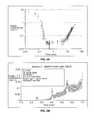
- the oxygen concentrationtends to deplete within about 10-15 seconds for irradiance of 3 mW/cm 2 (as shown, for example, in FIG. 2A ) and within about 3-5 seconds for irradiance of 30 mW/cm 2 .
- Utilizing pulsed light of a specific duty cycle, frequency, and irradiance, input from both Type I and Type II photochemical kinetic mechanismsmay be optimized to achieve the greatest amount of photochemical efficiency.
- utilizing pulsed lightallows regulating the rate of reactions involving Riboflavin.
- the rate of reactionsmay either be increased or decreased, as needed, by regulating, one of the parameters such as the irradiance, the dose, the on/off duty cycle, Riboflavin concentration, soak time, and others.
- additional ingredients that affect the reaction and cross-linking ratesmay be added to the cornea.
- One aspect of the present disclosurerelates to achieving photon optimization by allowing deactivated (reduced) Riboflavin to return to ground state Riboflavin in Type I reactions and allowing for reduced rate of oxygen uptake in Type II reactions where better photon conversion efficiency occurs.
- the rate of return of deactivated (reduced) Riboflavin to ground state in Type I reactions and the rate of oxygen uptake in Type II reactionsis determined by a number of factors. These factors include, but are not limited to, on/off duty cycle of pulsed light treatment, pulse rate frequency, irradiance, and dose. Moreover, the Riboflavin concentration, soak time, and addition of other agents, including oxidizers, affect the rate of oxygen uptake. These and other parameters, including duty cycle, pulse rate frequency, irradiance, and dose are optimized to achieve optimal photon efficiency and make efficient use of both Type I and Type II photochemical kinetic mechanisms for Riboflavin photosensitization. Moreover, these parameters are optimized in such a way as to achieve an optimum chemical amplification effect.
- the on/off duty cycleis between approximately 100/1 to approximately 1/100; the irradiance is between approximately 1 mW/cm 2 to approximately 500 mW/cm 2 average irradiance, and the pulse rate is between approximately 0.1 Hz to approximately 1000 Hz.
- the on/off duty cycleis between approximately 1000/1 to approximately 1/1000; the irradiance is between approximately 1 mW/cm 2 to approximately 1000 mW/cm 2 average irradiance, and the pulse rate is between approximately 1000 Hz to approximately 100,000 Hz.
- the laser sourcemay be an adjustable pulsed source, an LED system, arc sources or incandescents at very long on-time duty cycles, or any other suitable sources.
- Pulse rates of 0.1 Hz to approximately 1000 Hz or 1000 Hz to approximately 100,000 Hzmay be chosen based on the photochemical kinetics as detailed by Kamaev et al., Investigative Ophthalmology & Visual Science, April 2012, Vol. 53, No. 4, pp. 2360-2367 (April 2012), which is incorporated herein by reference in its entirety.
- the pulse lengthmay be long—on the order of one or several seconds—or short—on the order of fractions of a second.
- pulsed light illuminationcan be used to create greater or lesser stiffening of corneal tissue than may be achieved with continuous wave illumination for the same amount or dose of energy delivered.
- Light pulses of suitable length and frequencymay be used to achieve optimum chemical amplification.
- FIG. 2Aillustrates a graph of depletion and gradual replenishment curve of dissolved oxygen below a 100 ⁇ m thick porcine corneal flap, saturated with 0.1% Riboflavin during 3 mW/cm 2 UVA irradiation at 25° C.
- the oxygen concentration (mg/L)fell to zero at about 15 seconds and gradually started to increase after approximately 10 minutes, getting back to approximately one-tenth its starting value after 30 minutes.
- FIG. 2Billustrates a graph of oxygen recovery under a 100 ⁇ m thick corneal flap.
- the corneal flapwas saturated with 0.1% Riboflavin during 30 mW/cm 2 UVA irradiation.
- the irradiationwas pulsed at a 3 second on/3 seconds off cycle. Riboflavin drops were added to the cornea every 90 seconds. In this example, it took about 3 minutes for the oxygen concentration to gradually start increasing and about 6 minutes for the oxygen concentration to increase to 0.1 mg/L.
- sensitized photo-oxidation of the substrateoccurs mainly by its reaction with photochemically generated reactive oxygen species, such as singlet molecular oxygen. This is consistent with a Type II photochemical mechanism.
- oxygenbecomes totally depleted and the reaction between the substrate and Riboflavin becomes consistent with a predominantly Type I photochemical mechanism.
- the oxygen concentration in the corneaslowly increases to a concentration at which a Type II mechanism may begin to play an additional role. During this phase, a growing contribution would be expected from the singlet oxygen-mediated cross-linking, together with the enhancement of secondary radical reactions that are modulated by oxygen.
- corneal stiffeningis primarily in the anterior 200 ⁇ m of the corneal stroma.
- Increase of collagen fluorescence in UVA-exposed corneaswhich is related to their mechanical stiffening, can be detected at a depth 200 to 300 ⁇ m from the corneal surface as will be described further below.
- oxygen concentrationsstart to increase (replenish) as shown in FIGS. 2A and 2B .
- Excess oxygenmay be detrimental in corneal cross-linking process because oxygen is able to inhibit free radical photopolymerization reactions by interacting with radical species to form chain-terminating peroxide molecules.
- the pulse rate, irradiance, dose, and other parametersmay be adjusted to achieve an optimized oxygen regeneration rate. Calculating and adjusting the oxygen regeneration rate is another example of adjusting the reaction parameters to achieve a desired amount of corneal stiffening.
- Oxygen contentmay be depleted throughout the cornea, by various chemical reactions, except for the very thin corneal layer where oxygen diffusion is able to keep up with the kinetics of the reactions. This diffusion-controlled zone will gradually move deeper into the cornea as the reaction ability of the substrate to uptake oxygen decreases.
- the mechanism for corneal cross-linkingbegins with the additional pathway kinetics expressed in equation (6) above. After a short period of time (a few seconds), oxygen becomes depleted, and there is little oxygen available as shown in FIG. 2A . Under these anaerobic conditions, Leuco-Deuteroflavin+H 2 O 2 are formed as described in Heldman et al., Handbook of Food Engineering (2 nd Edition), CRC Press (2006). Leuco-Deuteroflavin has low absorption at 360 nm and lacks the photosensitizing ability and therefore cannot create radicals. Leuco-Deuteroflavin is referred to herein as reduced flavin, reduced Riboflavin, RfH 2 , and Fl red H 2 .
- Reduced Riboflavinundergoes an oxidation reaction as shown in equation (6) above.
- the oxidation of reduced Riboflavin by molecular oxygenis irreversible, autocatalytic, and involves generation of free radicals that can initiate radical polymerization (as in case of vinyl monomers, acrylamide with bis(acrylamide), etc.).
- the autocatalytic oxidation of reduced Riboflavins by oxygenis accounted for by the reactions described in Massey, V., Activation of molecular oxygen by flavins and flavoproteins, J. Biol. Chem. (1994), 269, 22459-22462.
- Oxygenis the naturally occurring oxidizer and is used as the oxidizer according to aspects of the present disclosure. According to further aspects of the present disclosure, oxygen and/or other oxidizers are utilized; such oxidizers may be added to the formulation or administered to the cornea in a suitable way.
- reduced Riboflavinmay be soaked in a suitable agent that contains oxygen and is able to oxidize the reduced Riboflavin.
- Vitamin B12may be added in any suitable manner to the reduced Riboflavin and/or to the cornea.
- Vitamin B12contains a Cobalt molecule that is capable of holding oxygen, thereby creating an oxygen storage reservoir.
- the reduced Riboflavinmay be super-saturated with Vitamin B12 or another suitable oxygen carrying agent.
- the suitable agent, such as Vitamin B12may be provided in conjunction with application of pulsed light. The proper level of oxygen can be maintained with various reversible oxygen carriers. See Yang N., Oster G. Dye-sensitized photopolymerization in the presence of reversible oxygen carriers. J. Phys. Chem. 74, 856-860 (1970), the contents of which are incorporated entirely herein by reference.
- Corneal stiffeningmay be applied to the cornea according to particular patterns, including, but not limited to, circular or annular patterns, which may cause aspects of the cornea to flatten and improve vision in the eye. For example, more or less corneal stiffening may be desired on the outer edges of the cornea as opposed to the center of the cornea. Aspects of the present disclosure relate to achieving more corneal stiffening on the outer diameter of the cornea and gradually decreasing the amount of corneal stiffening from the outer diameter toward the center of the cornea. Other aspects of the present disclosure relate to selecting regions of the cornea that require more corneal stiffening based on a predetermined set of characteristics and applying more corneal stiffening to those selected regions by varying the regime of the pulsed light.
- pulsed lightmay be applied with different irradiance, dose and/or different duty cycle to different areas of the cornea, leading to areas of differing levels of corneal stiffening or corneal stiffening gradients.
- Varying the regime of the pulsed light to achieve a desired level of corneal stiffeningis another example of adjusting the parameters of the cross-linking reaction to achieve specific, targeted results.
- varying the regime of the pulsed light to achieve a desired level of corneal stiffening at selected regions of the corneaallows for more precise and accurate control of the shape changes in the eye.
- Riboflavin RFH 2(with two hydrogen atoms supplied to the aromatic nucleus by the side chain) can be produced by anaerobic photolysis of Riboflavin (Holmstrom 1961) and observed by the reduction in absorption at 445 nm.
- FIG. 3a graph illustrating absorbance for 0.2 mm light path of an initial sample of Riboflavin versus absorbance for 0.2 mm light path of reduced Riboflavin RfH 2 after being irradiated for 3 minutes at an irradiance of 30 mW/cm 2 .
- the absorbance of the initial sampleis about 0.543, while the absorbance of the irradiated sample is about 0.340.
- RFH 2is autoxidizable and in the presence of oxygen yields the highly light-sensitive fluorescent and absorbing (445 nm)
- Reduced Riboflavin solutionscan be prepared under nitrogen by irradiation of Riboflavin with visible light in the presence of EDTA and stored in absence of oxygen. Reaction with oxygen completes during hundreds of msec (depending on the initial conditions), proceeds via free radicals (as described in Massey), and is able to initiate polymerization of vinyl monomers. The rate of the reaction with oxygen may be increased by dissolving oxygen in a flavin solution instead of in water.
- RFH 2was prepared by irradiation of Riboflavin solutions (with and without EDTA, 1% EDTA, 0.1% Riboflavin) and saturated with argon (to displace oxygen) in a shallow sealed quartz cuvette. Then, in the absence of additional UV light, porcine corneal flaps were immediately placed in those solutions. After 1-2 min this procedure was repeated with a fresh solution containing RFH 2 several times. Corneal flaps then were washed with distilled water, digested with papain buffer, and their fluorescence was measured.
- fluorescence of the corneal samples treated with preliminary UVA-exposed Riboflavin solutionswas higher than fluorescence of the corneal samples washed with pure, unexposed Riboflavin.
- the fluorescencewas the highest for the sample prepared by irradiation of Riboflavin solutions with a reducing agent such as EDTA. Accordingly, one way of increasing the cross-linking efficiency of RfH 2 is to use Riboflavin in a solution with a reducing agent for Riboflavin.
- the reducing agentsmay include, but are not limited to, EDTA, ascorbic acid, sugars, amines, amino acids, and any combination thereof. This is another example of modifying the parameters of the cross-linking reaction to achieve a desired level of cross-linking with the corneal fibrils.
- Riboflavin concentrationsbetween about 0.001% Riboflavin to about 1.0% Riboflavin may be utilized.
- Example 2Measurement of the Collagen Linked Fluorescence in Cross-Linked Corneal Flaps at Depths of 100 ⁇ m and 200 ⁇ m
- Porcine whole globes(SiouxPreme Packing Co., Sioux City, Iowa; shipped in saline solution packed in ice) were warmed to room temperature (25° C.). The corneas were then de-epithelialized with a dulled scalpel blade and 0.1, 0.25, or 0.5% riboflavin solution in 0.9% saline was applied to the top of each cornea during 20 minutes before cross-linking Corneas were pan-corneally irradiated with a top hat beam (3% root mean square) for a determined amount of time with 365-nm light source (UV LED NCSU033B[T]; Nichia Co., Tokushima, Japan) at the chosen irradiance (3 or 30 mW/cm 2 ) which was measured with a power sensor (model PD-300-UV; Ophir, Inc., Jerusalem, Israel) at the corneal surface.
- 365-nm light sourceUV LED NCSU033B[T]; Nichia
- Corneal flaps(each 100 ⁇ m thick, one after another) were excised from the eyes with aid of Intralase femtosecond laser (Abbott Medical Optics, Santa Ana, Calif.). The average thickness of the corneal flaps was calculated as a difference between the measurements before and after the excision from the eyes with an ultrasonic Pachymeter (DGH Technology, Exton, Pa.). The flaps were washed with distilled water until Riboflavin in the washing waters was not detectable by absorbance measurement at 455 nm (Thermo Scientific Evolution 300/600 UV-Vis Spectrophotometer, Thermo Fisher Scientific, Waltham, Mass.). The flaps then were dried in vacuum until the weight change became less than 10% (Rotary vane vacuum pump RV3 A652-01-903, BOC Edwards, West Wales, UK).
- Each flap (1 mg)was digested for 2 h at 65° C. with 2.5 units/ml of papain (from Papaya latex, Sigma) in 0.5 ml of papain buffer [1 ⁇ PBS (pH 7.4), 2 mM L-cysteine and 2 mM EDTA].
- the fluorescence of the papain bufferwas taken into account by measuring fluorescence in the absence of tissue and subtracting this value from the fluorescence of the samples.
- FIG. 4BFluorescence of the excised samples, which was a relative value to the non-cross-linked samples with the same thickness, is illustrated in FIG. 4B . It is shown that the corneal fluorescence after cross-linking (all samples were exposed to the same UVA dose, 5.4 J/cm 2 ) is greater for samples exposed to UVA for a longer duration with lower irradiance and lower concentration of Riboflavin. Corneal fluorescence is also greater at first 100 ⁇ m than at the next 100 ⁇ m in the cornea. The highest corneal fluorescence was observed at the first 100 ⁇ m for the sample that was soaked in 0.1% Riboflavin solution and irradiated at 3 mW/cm 2 .
- porcine eyesUpon arrival, porcine eyes have excess muscle tissue that was removed and placed in saline in the incubator (set to 37° C.) for 30 minutes. Eyes were then de-epithelialized and placed in a 0.1% Riboflavin solution for 20 minutes at 37° C. Eyes were removed from solution and physiological IOP was applied. Eyes were then placed under a UVA source and shutter system and irradiated according to the indicated protocol as shown in FIGS. 5A-5C . Riboflavin drops were applied every 1.5 minutes during UVA application. After being irradiated, the corneal thickness was measured with a pachymeter. The sample was then placed under the femto second laser and a ⁇ 200 ⁇ m flap was cut.
- the flapwas positioned in a biaxial materials tester (CS-BIO TESTER 5000, CellScale, Waterloo, ON Canada) and stretched until failure. The sample was then rinsed with distilled water and frozen for future papain digestion and fluorescence analysis.
- CS-BIO TESTER 5000CellScale, Waterloo, ON Canada
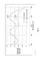
- FIG. 5Aillustrates force versus displacement curves for porcine cornea for various soak times and UVA illumination scenarios.
- FIG. 5Aillustrates results of experiments that show dissimilar biomechanical stiffness of the 0.25% Riboflavin sample 2 irradiated with 3 mW/cm 2 continuous wave illumination vs. the sample 3 irradiated with 30 mW/cm 2 continuous wave illumination for a total 5.4 J/cm 2 dose delivered.
- the biomechanical stiffness of the 0.25% Riboflavin sample 3 irradiated with 30 mW/cm 2 continuous for a total 5.4 J/cm 2 dose deliveredwas similar to the biomechanical stiffness of the 0.1% Riboflavin sample 4 irradiated under the same conditions.
- the biomechanical stiffness of the 0.25% Riboflavin sample 6 irradiated with 30 mW/cm 2 pulsed light with a 3 seconds on/3 seconds off duty cyclewas similar to the biomechanical stiffness of 0.25% Riboflavin sample 7 irradiated with 60 mW/cm 2 pulsed light with a 2 seconds on/4 seconds off duty cycle for a total 5.4 J/cm 2 dose delivered to each sample.
- the biomechanical stiffness of the 0.1% Riboflavin sample 8 irradiated with 30 mW/cm 2 pulsed light with a 3 seconds on/3 seconds off duty cyclewas higher than both the samples 6 and 7.
- the biomechanical stiffness of the Riboflavin sample 5 irradiated with 30 mW/cm 2 continuous wave illumination for a total 35.1 J/cm 2 dose deliveredwas higher than that for samples 6, 7, 3, 4, and 1 (control sample with 0.25% Riboflavin concentration) and lower than that for sample 2.
- Sample 4was soaked with Riboflavin for 30 minutes, while all the other samples were soaked with Riboflavin for 1 hour.
- FIG. 5Aillustrates the effect of varying different parameters on corneal cross-linking
- Different parametersirradiance, continuous wave vs. pulsed illumination, on/off duty cycle of pulsed light illumination, Riboflavin concentration, and other parameters—all have an effect on biomechanical stiffness.
- a DMDmay be used for illumination. With the DMD one can perform topography guided cross-linking as described, for example, in U.S. patent application Ser. No. 13/438,705, filed Apr. 3, 2012, and U.S. patent application Ser. No. 13/051,699, filed Mar. 18, 2011, the contents of which are incorporated entirely herein by reference.
- the algorithms associated with the topographymay be created using several different spatial and temporal irradiance and dose profiles.
- These spatial and temporal dose profilesmay be created using continuous wave illumination but may also be modulated via pulsed illumination by pulsing the illumination source under varying frequency and duty cycle regimes as described above.
- the DMDmay be able to modulate different frequencies and duty cycles on a pixel by pixel basis to give ultimate flexibility using continuous wave illumination.
- both pulsed illumination and modulated DMD frequency and duty cycle combinationsmay be combined. This allows for specific amounts of spatially determined corneal cross-linking.
- This spatially determined cross-linkingmay be combined with dosimetry, interferometry, optical coherence tomography (OCT), corneal topography, etc., for real-time modulated corneal cross-linking
- OCToptical coherence tomography
- corneal topographyetc.
- pre-clinical patient informationmay be combined with finite element biomechanical computer modeling to create patient specific pre-treatment plans.
- complex biomechanical stiffness patternsmay be imparted to the cornea to allow for various amounts of refractive correction.
- These refractive correctionsmay include combinations of myopia, hyperopia, astigmatism, irregular astigmatism, presbyopia and complex corneal refractive surface corrections because of ophthalmic conditions such as keratoconus, pellucid marginal disease, post-lasik ectasia, and other conditions of corneal biomechanical alteration/degeneration, etc.
- a specific advantage of the DMD system and methodis that it allows for randomized asynchronous pulsed topographic patterning, creating a non-periodic and uniformly appearing illumination which eliminates the possibility for triggering photosensitive epileptic seizures or flicker vertigo for pulsed frequencies between 2 Hz and 84 Hz as described above.
- Flicker vertigosometimes called the Bucha effect, is an imbalance in brain-cell activity caused by exposure to low-frequency flickering (or flashing) of a relatively bright light. It is a disorientation-, vertigo-, and nausea-inducing effect of a strobe light flashing at 1 Hz to 20 Hz, which corresponds approximately to the frequency of human brainwaves.
- the effectsare similar to seizures caused by epilepsy (particularly, photosensitive epilepsy), but are not restricted to people with histories of epilepsy.
- websites provided by federal agenciesare governed by section 508 of the Rehabilitation Act. The Act says that pages shall be designed to avoid causing the screen to flicker with a frequency between 2 Hz and 55 Hz.
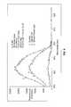
- FIG. 5Billustrates force versus displacement curves for samples of porcine cornea irradiated with pulsed light having various exposure times, as well as a curve for a sample irradiated with continuous wave illumination, and a curve for a control sample.
- All the samples that were irradiated with pulsed lighthad a 0.1% Riboflavin concentration and were irradiated with 30 mW/cm 2 pulsed light with a 3 seconds off cycle and a varied exposure (“on”) cycle.
- the sample 2 having a 4.5 second exposure cycleshad slightly lower biomechanical stiffness than samples 3 or 1.
- the duration of the exposure cycleaffects the amount of corneal stiffening at the same irradiance and dark phase duration. Therefore, aspects of the present disclosure affect the displacement per unit force ratio. This is yet one more parameter that may be altered in optimizing cross-linking.
- FIG. 5Cillustrates force versus displacement curves for samples of porcine cornea illuminated with pulsed light having varied dark phase durations, as well as curves for samples irradiated with continuous illumination, and a curve for a control sample.
- the sample 2 having 0.25% Riboflavin concentration irradiated with 3 mW/cm 2 continuous wave illuminationhad the lowest biomechanical stiffness out of samples 1 and 3-7.
- the duration of the dark phaseaffects the amount of corneal stiffening even at the same irradiance and exposure duration. This is yet one more parameter that may be altered in optimizing cross-linking.
- the graphs in FIGS. 5A-5Cshow that varying different parameters—applying pulsed instead of continuous wave illumination, varying on/off duty cycles, irradiance, dose, Riboflavin concentration, and soak times—all have an effect on biomechanical stiffness. These parameters may be modified in such a way as to achieve an optimum or desired amount of corneal stiffness anywhere on or within the cornea.
- porcine eyeswere de-epithelialized and placed in the 0.1% Riboflavin solution for 20 minutes. Eyes were removed from solution and physiological IOP was applied. A flap was cut using a femto second laser and an O 2 sensor was placed under the flap. UVA illumination was administered as indicated and Riboflavin drops were applied every 90 seconds for the duration of the dose delivered. After cross-linking the flap using the pulsed light dosing, the flap was removed and tested mechanically as described above.
- Corneaswere pan-corneally irradiated with a top hat beam (3% root mean square) for a determined amount of time with 365-nm light source (UV LED NCSU033B[T]; Nichia Co., Tokushima, Japan) at the chosen irradiance of 30 mW/cm 2 with either continuous wave illumination or pulsed illumination 3 seconds on/3 Seconds off.
- Corneal flaps(200 ⁇ m thick) were excised from the eyes with aid of Intralase femtosecond laser (Abbott Medical Optics, Santa Ana, Calif.). The average thickness of the corneal flaps was calculated as a difference between the measurements before and after the excision from the eyes with an ultrasonic Pachymeter (DGH Technology, Exton, Pa.).
- the flapswere washed with distilled water until Riboflavin in the washing waters was not detectable by absorbance measurement at 455 nm (Thermo Scientific Evolution 300/600 UV-Vis Spectrophotometer, Thermo Fisher Scientific, Waltham, Mass.). The flaps then were dried in vacuum until the weight change became less than 10% (Rotary vane vacuum pump RV3 A652-01-903, BOC Edwards, Westshire, UK). Each flap (1 mg) was digested for 2 h at 65° C. with 2.5 units/ml of papain (from Papaya latex, Sigma) in 0.5 ml of papain buffer [1 ⁇ PBS (pH 7.4), 2 mM L-cysteine and 2 mM EDTA].
- papainfrom Papaya latex, Sigma
- the fluorescence of the papain bufferwas taken into account by measuring fluorescence in the absence of tissue and subtracting this value from the fluorescence of the samples.
- the results seen belowcorroborate that the amount of fluorescence seen which is directly related to the amount of cross-linking is greater for the pulsed vs. continuous wave illumination in nearly the same proportion as the biomechanical measurements taken of the same flaps.
- fluorescencewas the highest—about 20,000 counts/s—for the sample irradiated with 30 mW/cm 2 pulsed light illumination with a 3 seconds on/3 seconds off cycle.
- the fluorescence for the sample irradiated with 30 mW/cm 2 continuous illuminationwas about 40% less, or about 12000 counts/s. This is yet another example demonstrating that applying pulsed light illumination as opposed to continuous wave illumination affects cross-linking in the cornea.
- Pig eyes from an abattoir(SiouxPreme, Sioux City, Iowa) were rinsed in saline. Eyes were cleaned and the epithelium was removed. Eyes were placed on a stand in the middle of a large beaker filled part way with water with a tube bubbling compressed oxygen into the water. The oxygen was turned on at certain times during the experiment to create a humid oxygenated environment for the eye. Eyes were soaked for 20 minutes with 0.1% Riboflavin, dH 2 O solution in an incubator set at 37° C. by using a rubber ring to hold the solution on top.
- Corneaswere pan-corneally irradiated with a top hat beam (3% root mean square) for a determined amount of time and irradiance (3 minutes CW at 30 mW/cm 2 with one drop of solution added every 30 seconds, or 30 minutes CW at 3 mW/cm 2 with one drop of solution added every minute, or 9 minutes for pulsed light—1.5 seconds on/3 seconds off—using a shutter system (Lambda SC Smart Shutter, Sutter Instrument, Novato, Calif.) at 30 mW/cm 2 with one drop of solution added every minute) with a 365-nm light source (UV LED NCSU033B[T]; Nichia Co., Tokushima, Japan). The irradiance was measured with a power sensor (model PD-300-UV; Ophir, Inc., Jerusalem, Israel) at the corneal surface.
- a power sensormodel PD-300-UV; Ophir, Inc., Jerusalem, Israel
- Corneal flaps(approximately 380 ⁇ m thick) were excised from the eyes with aid of Intralase femtosecond laser (Abbott Medical Optics, Santa Ana, Calif.). The average thickness of the corneal flaps was calculated as a difference between the measurements before and after the excision from the eyes with an ultrasonic Pachymeter (DGH Technology, Exton, Pa.). The flaps were washed with distilled water 15 times and then dried in a vacuum until the weight change became less than 10% (Rotary vane vacuum pump RV3 A652-01-903, BOC Edwards, West Wales, UK). Each flap (2 mg) was digested for 2.5 h at 65° C.
- FIG. 7Aillustrates a graph of the amount of cross-linking measured by fluorescence of the digested corneal flaps at 450 nm.
- Eyeswere de-epithelized, soaked for 20 minutes with 0.1% Riboflavin, put in a regular air environment or a humid oxygenated environment, and illuminated with 30 mW/cm 2 or 3 mW/cm 2 of UV light for 3 minutes CW with one drop of solution every 30 seconds, or 30 minutes CW with one drop of solution every minute, or 9 minutes pulsed (1.5 seconds on/3 seconds off) with one drop of solution every minute.
- Table 2The following treatments shown in Table 2 below were applied to each of the samples shown in FIG. 7A :
- UV Control Eyeswere illuminated with 30 mW/cm 2 CW of UV light for 3 minutes. Air, Pulsed Eyes were illuminated with 30 mW/cm 2 pulsed UV light Light (1.5 seconds on/3 seconds off) for 9 minutes. O 2 , CW Eyes were illuminated with 30 mW CW/cm 2 of UV light for 3 minutes with oxygen always on during UV exposure and soak. 3 mW CW Eyes were illuminated with 3 mW/cm 2 CW of UV light for 30 minutes. O 2 , Pulsed Eyes were illuminated with 30 mW/cm 2 of pulsed UV light Light (1.5 seconds on/3 seconds off) for 9 minutes with oxygen on during soak and UV exposure.
- a humid oxygenated environment with pulsed UV lightgreatly increases the amount of cross-linking taking place in the cornea.
- Applying a combination of Riboflavin and ultraviolet (UV) lightsterilizes a surface of the cornea.
- the Riboflavinacts as a photosensitizer that increases the absorption of UV light.
- the resulting absorption of UV lightcan induce DNA and RNA lesions, and as a result, is effective in killing viruses, bacteria, and other pathogens in the field.
- a humid oxygen environment and pulsing UV lightincrease the amount of cross-linking to a certain degree when done separately, while they increase the amount of cross-linking to a significantly greater extent when done in conjunction.
- Increased cross-linkinginvolves creation of an increased number of radicals. Radicals help to eliminate harmful bacteria present in the eye. Accordingly, a humid oxygenated environment and pulsing UV light result in more efficient elimination of viruses, bacteria, and other pathogens in the cornea, creating a sterile environment while minimizing any damage or other unwanted effects in the tissue. This is yet another example demonstrating that applying pulsed light illumination as opposed to continuous wave illumination affects the amount of cross-linking in the cornea.
- FIG. 7Billustrates force versus displacement curves for samples of porcine cornea illuminated with pulsed light of irradiance with oxygen, as well as curves for samples irradiated with continuous illumination, and a curve for a control sample.
- the sample 3 irradiated with 30 mW/cm 2 pulsed light illumination with oxygen and a 3 seconds on/3 seconds off duty cyclehad the highest biomechanical stiffness, followed by sample 4 irradiated with 45 mW/cm 2 pulsed light illumination with oxygen and an identical on/off duty cycle.
- the samples irradiated with 3 mW/cm 2 and 30 mW/cm 2 continuous wave illuminationhad lower biomechanical stiffness, followed by the control sample.
- the addition of oxygenaffects the amount of corneal stiffening even at the same on/off duty cycle. This is yet one more parameter that may be altered in optimizing cross-linking.
- oxygenmay be applied during the cross-linking treatments in a number of different ways.
- a treatmentsuch as thermokeratoplasty or LASIK surgery, is applied in step 210 to generate structural changes in the cornea and produce a desired shape change.
- the corneal tissueis treated with a cross-linking agent 222 .
- the cross-linking agentmay be applied directly on the treated tissue and/or in areas around the treated tissue.
- the cross-linking agentmay be an ophthalmic solution that is broadly delivered by a dropper, syringe, or the like.
- the cross-linking agentmay be selectively applied as an ophthalmic ointment with an appropriate ointment applicator.
- the cross-linking agent 222is then activated in step 230 with an initiating element 232 .
- Activation of the cross-linking agent 222may be triggered thermally by the application of microwaves or light from a corresponding energy or light source.
- the resulting cross-linking between collagen fibrilsprovides resistance to changes in corneal structure.
- Riboflavinis applied as a cross-linking agent 222 ′ to the corneal tissue in step 220 .
- light from an UV light sourcemay be applied as an initiating element 232 ′ in step 230 to initiate cross-linking in the corneal areas treated with Riboflavin.
- the UV lightinitiates cross-linking activity by causing the applied Riboflavin to release reactive oxygen radicals in the corneal tissue.
- the Riboflavinacts as a sensitizer to convert O 2 into singlet oxygen which causes cross-linking within the corneal tissue.
- the rate of cross-linking in the corneais related to the concentration of O 2 when it is irradiated with photoactivating light. Therefore, it may be advantageous to increase or decrease the concentration of O 2 actively during irradiation to control the rate of cross-linking until a desired amount of cross-linking is achieved.
- An approach according to aspects of the present inventioninvolves supersaturating the Riboflavin with O 2 .
- a higher concentration of O 2is delivered directly into the cornea with the Riboflavin and affects the conversion of O 2 into singlet oxygen when the Riboflavin is exposed to the photoactivating light.
- the Riboflavin 222 ′may be stored in a closed vessel, e.g., a vial, 300 under increased O 2 pressure 305 .
- the increased O 2 pressure 305results in a higher equilibrium concentration of O 2 in the Riboflavin 222 ′.
- the walls 310 of the vessel 300are preferably opaque or otherwise prevent visible, UV, or other light from entering the vessel interior 301 to minimize the degradation of the Riboflavin 222 ′. Accordingly, referring to FIG. 9B , the step 215 supersaturates the Riboflavin 222 ′ with O 2 so that a supersaturated Riboflavin 222 ′ is applied in step 220 .
- a gele.g., a methylcellulose gel
- a gel 421may be stored in an interior 401 of a closed vessel, e.g., a vial, 400 under increased O 2 pressure 405 .
- the increased O 2 pressure 405results in a higher equilibrium concentration of O 2 in the gel 421 .
- the gelcan then act as a carrier for O 2 .
- step 216saturates a gel 421 with O 2
- step 217mixes the supersaturated gel 421 with the Riboflavin 222 ′, so that a mixture 422 containing the Riboflavin 222 ′ and the supersaturated gel 421 is applied in step 220 .
- step 216saturates a gel 421 with O 2
- step 225applies the gel 421 to the cornea after the Riboflavin 222 ′ has been applied to the cornea.
- the gel 421increases the presence of O 2 when the Riboflavin 222 ′ is activated with the UV light.
- a steady state of O 2may be maintained at the surface of the cornea to expose the cornea to a selected amount of O 2 and cause O 2 to enter the cornea.
- the photoactivating lightcan then be applied to a cornea with the desired O 2 content.
- a ring 500is placed on the eye 1 to supply O 2 to the cornea 2 during irradiation.
- the ring 500includes one or more ports 502 that direct a steady flow of O 2 to the cornea 2 , which has been treated by Riboflavin.
- the flowapplies O 2 at high pressure against the cornea 2 , so that more O 2 is available during the irradiation of the Riboflavin in the corneal tissue.
- the ring 500may optionally be held in place by suction.
- an enclosure 510 receiving a supply of O 2 through a port 512is placed on the eye to establish a steady state of O 2 .
- the enclosure 510may be held in place by a suction ring 512 .
- the enclosure 510may be a cup-like structure.
- the enclosure 510maintains the O 2 at a higher pressure, e.g., higher than ambient, against the surface of the cornea 2 .
- the concentration of O 2 within the enclosure 510 and above the surface of the cornea 2can approach 100%.
- the O 2 within the enclosure 510makes more O 2 to be available for the irradiation of the Riboflavin in the corneal tissue.
- At least a portion of the walls 514 of the enclosure 510may be translucent to allow photoactivating light to pass through the enclosure 510 to irradiate the cornea 2 and activate the Riboflavin applied to the cornea 2 .
- the light sourcemay be disposed within the enclosure.
- the enclosure 510may also include a valve that allows the gas to be released.
- step 227establishes a steady state of O 2 above the corneal surface before the photoactivating light 232 ′ is applied in step 230 to initiate cross-linking with the Riboflavin 222 ′.
- the rate of cross-linkingmay be monitored in real time and the concentration of O 2 may be dynamically increased or decreased to achieve a desired amount of cross-linking.
- corneal tissueis treated with Riboflavin 222 ′ in step 220 .
- a first amount of O 2is provided above the corneal surface to introduce O 2 to the corneal tissue and establish a first concentration of O 2 in the cornea during irradiation.
- the devices described with reference to FIGS. 11A and 11Bmay be employed to change the amount of O 2 is provided above the corneal surface.
- the Riboflavin 222 ′is then activated in step 230 with UV light 232 ′.
- step 240the amount of cross-linking resulting from the activation of the Riboflavin 222 ′ is monitored.
- One technique for monitoring the cross-linkingemploys polarimetry to measure corneal birefringence and to determine the structure of the corneal tissue.
- the techniquemeasures the effects of cross-linking on corneal structure by applying polarized light to the corneal tissue.
- the corneal stromais anisotropic and its index of refractions depends on direction.
- the corneabehaves like a curved biaxial crystal with the fast axis orthogonal to the corneal surface and the slow axis (or corneal polarization axis) tangential to the corneal surface.
- a light beam emerging from the living eye after a double pass through the ocular opticscontains information on the polarization properties of the ocular structures (except optically inactive humours).
- the technique of using birefringence to monitor the structural changes resulting from cross-linkingis described further in U.S. Provisional Patent Application No. 61/388,963, filed Oct. 1, 2010, the contents of which are entirely incorporated herein by reference.
- a controlleremploying conventional computer hardware or similar processing hardware, can be used to monitor the amount of cross-linking.
- Such hardwaremay operate by reading and executing programmed instructions that are stored or fixed on computer-readable media, such as conventional computer disk.
- the controllermay be coupled to, and automatically control, the device(s) that provide the O 2 above the corneal surface.
- step 250Based on the information from the real time monitoring in step 240 , step 250 provides a second amount of O 2 above the eye to introduce another amount of O 2 to the corneal tissue and expose the cornea to a second concentration of O 2 during irradiation with UV light 232 ′ in step 260 .
- Steps 240 , 250 , and 260may be repeated any number of times to change the concentration of O 2 during irradiation to control the rate of cross-linking dynamically.
- the first amount of O 2 in step 228may be greater than the second amount of O 2 in step 250 , or vice versa. Changing the cornea's exposure from the first concentration to the second concentration changes the rate of cross-linking in the corneal tissue as desired. If the information from step 240 indicates that the first amount of O 2 is too low, step 250 provides a second amount of O 2 that is greater than the first amount of O 2 . On the other hand, if the information from step 240 indicates that the first amount of O 2 is too high, step 250 provides a second amount of O 2 that is greater than the first amount of O 2 . It may be necessary to remove the first amount of O 2 , e.g., from the enclosure 510 , before providing the second amount of O 2 in step 250 .
- step 250may introduce a non-O 2 element or substance above the corneal surface.
- nitrogen gas (N 2 )may replace the O 2 supplied by the devices 500 and 510 shown in FIGS. 11A and 11B .
- the photoactivating light 332 ′may include, for example, UV light or green light.
- cross-linking treatmentsare combined with astigmatic keratotomy eye surgery (AK).
- AKis a surgical procedure for correcting astigmatism, where incisions are made in the steepest part of the abnormally shaped cornea to relax the cornea into a rounded shape.
- AKis often employed in combination with cataract surgery.
- the resulting shape change from AKcan be stabilized with cross-linking treatment.
- cross-linking treatmentsmay be combined with radial keratotomy (RK), which is a surgical procedure for correcting myopia, where radial incisions are made to the cornea to make corrective shape changes.
- RKradial keratotomy
- the effect of applying the cross-linking agentmay also allow smaller incisions to be used during AK or RK.
- aspects of the present disclosurerelate to monitoring and optimizing the parameters of applying the cross-linking agent to the eye and of activating the cross-linking agent.
- a large variety of factorsaffect the rate of the cross-linking reaction and the amount of biomechanical stiffness achieved due to cross-linking. These factors include Riboflavin concentration, conditions on the cornea, temperature, presence of oxidizing agents, the type of illumination applied to activate the Riboflavin, the irradiance, the dose, the on/off duty cycle of the applied illumination, as well as other factors.
- Riboflavin concentrationinclude Riboflavin concentration, conditions on the cornea, temperature, presence of oxidizing agents, the type of illumination applied to activate the Riboflavin, the irradiance, the dose, the on/off duty cycle of the applied illumination, as well as other factors.
- a number of these factorsare interrelated, in other words, changing one factor may have an unexpected effect on another factor.
- aspects of the present disclosurerelate to determining the effect of each of these parameters on the rate and the amount of cross-linking, as well as the interrelations of these parameters among each to optimize the conditions to achieve the desired amount, rate, and location of corneal stiffening.
- aspects of the present disclosurerelate to monitoring the corneal response to a change in one or a plurality of parameters and adjusting the one or the plurality of parameters based on the received feedback.
- the embodiments described abovemay employ stepwise on/off pulsed light functions, it is understood that other functions for applying light to the cornea may be employed to achieve similar effects.
- lightmay be applied to the cornea according to a sinusoidal function, sawtooth function, or other complex functions or curves, or any combination of functions or curves.
- the functionmay be “substantially” stepwise where there may be more gradual transitions between on/off values.
- irradiancedoes not have to decrease down to a value of zero during the off cycle, and may be above zero during the off cycle. Effects of the present disclosure may be achieved by applying light to the cornea according to a curve varying irradiance between two or more values.
Landscapes
- Health & Medical Sciences (AREA)
- Life Sciences & Earth Sciences (AREA)
- Veterinary Medicine (AREA)
- Animal Behavior & Ethology (AREA)
- General Health & Medical Sciences (AREA)
- Public Health (AREA)
- Engineering & Computer Science (AREA)
- Biomedical Technology (AREA)
- Ophthalmology & Optometry (AREA)
- Nuclear Medicine, Radiotherapy & Molecular Imaging (AREA)
- Medicinal Chemistry (AREA)
- Chemical & Material Sciences (AREA)
- Pharmacology & Pharmacy (AREA)
- Vascular Medicine (AREA)
- Epidemiology (AREA)
- Heart & Thoracic Surgery (AREA)
- Surgery (AREA)
- Electromagnetism (AREA)
- Optics & Photonics (AREA)
- Physics & Mathematics (AREA)
- Pathology (AREA)
- Radiology & Medical Imaging (AREA)
- Biophysics (AREA)
- Bioinformatics & Cheminformatics (AREA)
- Chemical Kinetics & Catalysis (AREA)
- General Chemical & Material Sciences (AREA)
- Organic Chemistry (AREA)
- Pharmaceuticals Containing Other Organic And Inorganic Compounds (AREA)
Abstract
Description
Rf→Rf*1,Iabs (1)
Rf*1→Rf,k1 (2)
Rf*1→Rf*3,k2 (3)
Rf3*+SH→(RF.−+SH.+)→RfH.+S.,k3 (4)
2RfH.→Rf+RfH2,k4 (5)
RfH2+O2→Rfox+H2O2,k5 (6)
Rf3*+O2→Rf+1O2,k6 (7)
SH+1O2→Sox,k6 (8)
| TABLE 2 |
| Treatment Conditions for the Samples Shown in FIG. 7A |
| Sample | Treatment |
| Control | After being soaked, corneal flaps were cut at approximately |
| 380 μm. | |
| UV Control | Eyes were illuminated with 30 mW/cm2CW of UV light |
| for 3 minutes. | |
| Air, Pulsed | Eyes were illuminated with 30 mW/cm2pulsed UV light |
| Light | (1.5 seconds on/3 seconds off) for 9 minutes. |
| O2, CW | Eyes were illuminated with 30 mW CW/cm2of UV light |
| for 3 minutes with oxygen always on during UV exposure | |
| and soak. | |
| 3 mW CW | Eyes were illuminated with 3 mW/cm2CW of UV light for |
| 30 minutes. | |
| O2, Pulsed | Eyes were illuminated with 30 mW/cm2of pulsed UV light |
| Light | (1.5 seconds on/3 seconds off) for 9 minutes with oxygen |
| on during soak and UV exposure. | |
Claims (21)
Rf3*+SH→RfH.+S., and
2RfH.→Rf+RfH2,
Priority Applications (5)
| Application Number | Priority Date | Filing Date | Title |
|---|---|---|---|
| US13/665,495US9707126B2 (en) | 2009-10-21 | 2012-10-31 | Systems and methods for corneal cross-linking with pulsed light |
| EP13819425.3AEP2872082B1 (en) | 2012-07-16 | 2013-03-15 | Systems for corneal cross-linking with pulsed light |
| US13/841,617US20130245536A1 (en) | 2009-10-21 | 2013-03-15 | Systems and methods for corneal cross-linking with pulsed light |
| PCT/US2013/032567WO2014014521A1 (en) | 2012-07-16 | 2013-03-15 | Systems and methods for corneal cross-linking with pulsed light |
| US14/072,640US20140066835A1 (en) | 2011-05-24 | 2013-11-05 | Systems and methods for corneal cross-linking with pulsed light |
Applications Claiming Priority (5)
| Application Number | Priority Date | Filing Date | Title |
|---|---|---|---|
| US25373609P | 2009-10-21 | 2009-10-21 | |
| US12/909,228US8574277B2 (en) | 2009-10-21 | 2010-10-21 | Eye therapy |
| US201261671798P | 2012-07-16 | 2012-07-16 | |
| US201261699226P | 2012-09-10 | 2012-09-10 | |
| US13/665,495US9707126B2 (en) | 2009-10-21 | 2012-10-31 | Systems and methods for corneal cross-linking with pulsed light |
Related Parent Applications (1)
| Application Number | Title | Priority Date | Filing Date |
|---|---|---|---|
| US12/909,228Continuation-In-PartUS8574277B2 (en) | 2009-10-21 | 2010-10-21 | Eye therapy |
Related Child Applications (1)
| Application Number | Title | Priority Date | Filing Date |
|---|---|---|---|
| US13/841,617Continuation-In-PartUS20130245536A1 (en) | 2009-10-21 | 2013-03-15 | Systems and methods for corneal cross-linking with pulsed light |
Publications (2)
| Publication Number | Publication Date |
|---|---|
| US20130060187A1 US20130060187A1 (en) | 2013-03-07 |
| US9707126B2true US9707126B2 (en) | 2017-07-18 |
Family
ID=48141636
Family Applications (1)
| Application Number | Title | Priority Date | Filing Date |
|---|---|---|---|
| US13/665,495Active2031-02-28US9707126B2 (en) | 2009-10-21 | 2012-10-31 | Systems and methods for corneal cross-linking with pulsed light |
Country Status (4)
| Country | Link |
|---|---|
| US (1) | US9707126B2 (en) |
| EP (3) | EP4074294A1 (en) |
| JP (1) | JP6271541B2 (en) |
| WO (2) | WO2013059837A2 (en) |
Cited By (12)
| Publication number | Priority date | Publication date | Assignee | Title |
|---|---|---|---|---|
| US20160310319A1 (en)* | 2014-10-27 | 2016-10-27 | Avedro, Inc. | Systems and methods for cross-linking treatments of an eye |
| RU2676451C1 (en)* | 2018-03-19 | 2018-12-28 | Федеральное государственное автономное учреждение "Межотраслевой научно-технический комплекс "Микрохирургия глаза" имени академика С.Н. Федорова" Министерства здравоохранения Российской Федерации | Method of cross-linking corneal collagen by means of femtosecond laser in experiment |
| US10342697B2 (en) | 2016-04-13 | 2019-07-09 | Avedro, Inc. | Systems and methods for delivering drugs to an eye |
| US10729716B2 (en) | 2012-03-29 | 2020-08-04 | Cxl Ophthalmics, Llc | Compositions and methods for treating or preventing diseases associated with oxidative stress |
| WO2020176598A1 (en) | 2019-02-26 | 2020-09-03 | Avedro, Inc. | Systems and methods for cross-linking treatments of an eye |
| US20200345847A1 (en)* | 2017-02-21 | 2020-11-05 | Avedro, Inc. | Formulations for eye treatments |
| US10932864B2 (en) | 2018-11-28 | 2021-03-02 | Rxsight, Inc. | Tracking-based illumination control system |
| US11013593B2 (en) | 2018-12-02 | 2021-05-25 | Rxsight, Inc. | Light adjustable lens tracking system and method |
| US11033429B2 (en) | 2010-09-30 | 2021-06-15 | Cxl Ophthalmics, Llc | Ophthalmic treatment device, system, and method of use |
| WO2021211589A1 (en)* | 2020-04-15 | 2021-10-21 | Alpha Phase Technologies LLC | Methods of cross-linking collagen |
| US11931291B2 (en) | 2012-03-29 | 2024-03-19 | Epion Therapeutics, Inc. | Ophthalmic treatment solution delivery devices and delivery augmentation methods |
| US12226348B2 (en) | 2019-12-13 | 2025-02-18 | Alcon Inc. | System and method of corneal cross-linking |
Families Citing this family (40)
| Publication number | Priority date | Publication date | Assignee | Title |
|---|---|---|---|---|
| WO2011050164A1 (en) | 2009-10-21 | 2011-04-28 | Avedro, Inc. | Eye therapy |
| EP4480461A3 (en)* | 2010-03-19 | 2025-02-12 | Avedro, Inc. | Systems for applying and monitoring eye therapy |
| US9044308B2 (en) | 2011-05-24 | 2015-06-02 | Avedro, Inc. | Systems and methods for reshaping an eye feature |
| JP6122845B2 (en) | 2011-06-02 | 2017-04-26 | アヴェドロ・インコーポレーテッドAvedro,Inc. | System and method for monitoring the delivery of time-based photoactive agents or the presence of photoactive markers |
| EP2830554A1 (en) | 2012-03-29 | 2015-02-04 | CXL Ophthalmics, LLC | Ocular cross-linking system and method for sealing corneal wounds |
| TWI588560B (en) | 2012-04-05 | 2017-06-21 | 布萊恩荷登視覺協會 | Lens, device, method and system for refractive error |
| US9201250B2 (en) | 2012-10-17 | 2015-12-01 | Brien Holden Vision Institute | Lenses, devices, methods and systems for refractive error |
| WO2014060206A1 (en) | 2012-10-17 | 2014-04-24 | Albert Daxer | Device and method for irradiating the eye |
| CA2887655C (en) | 2012-10-17 | 2021-11-02 | Brien Holden Vision Institute | Lenses, devices, methods and systems for refractive error |
| US9498114B2 (en) | 2013-06-18 | 2016-11-22 | Avedro, Inc. | Systems and methods for determining biomechanical properties of the eye for applying treatment |
| US9498122B2 (en) | 2013-06-18 | 2016-11-22 | Avedro, Inc. | Systems and methods for determining biomechanical properties of the eye for applying treatment |
| AU2013404375B2 (en)* | 2013-10-30 | 2017-08-03 | Alcon Inc. | Crosslinking control |
| WO2015130944A1 (en)* | 2014-02-28 | 2015-09-03 | Massachusetts Eye & Ear Infirmary | Methods for cross-linking corneal collagen with verteporfin for the treatment of disorders of the eye |
| KR20250022884A (en) | 2014-09-09 | 2025-02-17 | 루미테라 인코포레이티드 | Multi-wavelength phototherapy devices, systems, and methods for the non-invasive treatment of damaged or diseased tissue |
| WO2016077747A1 (en) | 2014-11-13 | 2016-05-19 | Avedro, Inc. | Multipass virtually imaged phased array etalon |
| DE102014016990B3 (en)* | 2014-11-18 | 2016-02-11 | Wavelight Gmbh | Nozzle unit for cross-linking of eye tissue |
| EP4082547A1 (en) | 2014-12-02 | 2022-11-02 | Avedro, INC. | Systems, methods, and compositions for cross-linking treatments of an eye |
| EP3827792A1 (en) | 2015-04-24 | 2021-06-02 | Avedro, Inc. | Systems and methods for photoactivating a photosensitizer applied to an eye |
| EP4483884A3 (en) | 2015-05-22 | 2025-03-05 | Avedro Inc. | Systems for monitoring cross-linking activity for corneal treatments |
| WO2017015471A1 (en)* | 2015-07-21 | 2017-01-26 | Avedro, Inc. | Systems and methods for treaments of an eye with a photosensitizer |
| AU2016342057A1 (en) | 2015-10-23 | 2018-03-01 | The Trustees Of Columbia University In The City Of New York | Laser induced collagen crosslinking in tissue |
| KR102149590B1 (en) | 2015-12-03 | 2020-08-28 | 아베드로 인코퍼레이티드 | Systems and methods for treating eyes with mask devices |
| ITUB20160237A1 (en)* | 2016-01-26 | 2017-07-26 | Vision Engineering Italy Soc A Responsabilita Limitata | PROCEDURE FOR CONTROL OF THE DOSAGE OF A CHROMOPHOROR AGENT IN A CORNEAL FABRIC AND PROCEDURE FOR DOSING A CHROMOPHOROR AGENT IN A CORNEAL FABRIC |
| WO2018031529A1 (en)* | 2016-08-08 | 2018-02-15 | Avedro, Inc. | Systems and methods for cross-linking treatments of an eye |
| JP7035081B2 (en) | 2017-01-11 | 2022-03-14 | アヴェドロ・インコーポレーテッド | Systems and methods for determining the cross-linking distribution and / or the structural features of the cornea in the cornea |
| US11666481B1 (en) | 2017-12-01 | 2023-06-06 | The Trustees Of Columbia University In The City Of New York | Diagnosis and treatment of collagen-containing tissues |
| RU2682494C1 (en)* | 2017-12-12 | 2019-03-19 | Государственное бюджетное учреждение "Уфимский научно-исследовательский институт глазных болезней Академии наук Республики Башкортостан" | Method of treatment of keratectasia by the method of pulse accelerated ultraviolet crosslinking of cornea |
| WO2019147901A2 (en)* | 2018-01-26 | 2019-08-01 | The Trustees Of Columbia University In The City Of New York | Methods, computer-readable media, and systems for treating a cornea |
| EP4397287A3 (en) | 2018-03-05 | 2024-09-11 | Avedro, Inc. | System for eye tracking during eye treatment |
| WO2019173762A1 (en) | 2018-03-08 | 2019-09-12 | Avedro, Inc. | Micro-devices for treatment of an eye |
| AU2019345058B2 (en) | 2018-09-19 | 2024-12-05 | Avedro, Inc. | Systems and methods for eye tracking during eye treatment |
| CN118217090A (en) | 2018-10-09 | 2024-06-21 | 艾维德洛公司 | Photoactivation system and method for corneal cross-linking treatment |
| JP7515498B2 (en) | 2018-12-04 | 2024-07-12 | アヴェドロ インコーポレイテッド | Compounds and compositions for treating the eye |
| KR20210134636A (en) | 2019-01-25 | 2021-11-10 | 아베드로, 인코포레이티드 | Compounds and compositions for the treatment of eyes |
| IT201900011985A1 (en)* | 2019-07-17 | 2021-01-17 | Vision Eng Italy Srl | Liquid formulation, in particular to treat a corneal tissue |
| JP7624433B2 (en) | 2019-08-06 | 2025-01-30 | アヴェドロ・インコーポレーテッド | Photoactivation system and method for corneal crosslinking treatment |
| EP4072400B1 (en) | 2019-12-13 | 2025-09-03 | Alcon Inc. | System of determining incision depths in eyes |
| AU2021401427B2 (en)* | 2020-12-17 | 2025-08-14 | Avedro, Inc. | Treatments for eye infection |
| JP2024511317A (en) | 2021-03-08 | 2024-03-13 | アヴェドロ・インコーポレーテッド | Systems and methods for generating patient-specific corneal crosslinking treatment patterns |
| US20220338729A1 (en)* | 2021-04-27 | 2022-10-27 | Johnson & Johnson Vision Care, Inc. | System and method for identifying presbyopia |
Citations (207)
| Publication number | Priority date | Publication date | Assignee | Title |
|---|---|---|---|---|
| US4034750A (en) | 1975-04-22 | 1977-07-12 | Maurice Seiderman | Electrochemically linking collagen fibrils to animal tissue |
| US4161013A (en) | 1977-05-23 | 1979-07-10 | Massachusetts Institute Of Technology | Electromechanochemical device |
| US4326529A (en) | 1978-05-26 | 1982-04-27 | The United States Of America As Represented By The United States Department Of Energy | Corneal-shaping electrode |
| US4381007A (en) | 1981-04-30 | 1983-04-26 | The United States Of America As Represented By The United States Department Of Energy | Multipolar corneal-shaping electrode with flexible removable skirt |
| US4665913A (en) | 1983-11-17 | 1987-05-19 | Lri L.P. | Method for ophthalmological surgery |
| US4712543A (en) | 1982-01-20 | 1987-12-15 | Baron Neville A | Process for recurving the cornea of an eye |
| US4764007A (en) | 1986-02-24 | 1988-08-16 | The United States Of America As Represented By The Secretary Of The Air Force | Glare susceptibility tester |
| US4805616A (en) | 1980-12-08 | 1989-02-21 | Pao David S C | Bipolar probes for ophthalmic surgery and methods of performing anterior capsulotomy |
| US4881543A (en) | 1988-06-28 | 1989-11-21 | Massachusetts Institute Of Technology | Combined microwave heating and surface cooling of the cornea |
| US4891043A (en) | 1987-05-28 | 1990-01-02 | Board Of Trustees Of The University Of Illinois | System for selective release of liposome encapsulated material via laser radiation |
| US4969912A (en) | 1988-02-18 | 1990-11-13 | Kelman Charles D | Human collagen processing and autoimplant use |
| US4994058A (en) | 1986-03-19 | 1991-02-19 | Summit Technology, Inc. | Surface shaping using lasers |
| US5016615A (en) | 1990-02-20 | 1991-05-21 | Riverside Research Institute | Local application of medication with ultrasound |
| US5019074A (en) | 1987-03-09 | 1991-05-28 | Summit Technology, Inc. | Laser reprofiling system employing an erodable mask |
| US5103005A (en) | 1989-07-21 | 1992-04-07 | Coors Biotech, Inc. | Method for recovery of riboflavin |
| US5171318A (en) | 1987-11-09 | 1992-12-15 | Chiron Ophthalmics, Inc. | Treated corneal prosthetic device |
| US5171254A (en) | 1991-11-19 | 1992-12-15 | Sher Neal A | Eye fixation device |
| US5281211A (en) | 1989-06-07 | 1994-01-25 | University Of Miami, School Of Medicine, Dept. Of Ophthalmology | Noncontact laser microsurgical apparatus |
| US5461212A (en) | 1993-06-04 | 1995-10-24 | Summit Technology, Inc. | Astigmatic laser ablation of surfaces |
| US5490849A (en) | 1990-07-13 | 1996-02-13 | Smith; Robert F. | Uniform-radiation caustic surface for photoablation |
| US5512966A (en) | 1993-06-24 | 1996-04-30 | Orbtek, Inc. | Ophthalmic pachymeter and method of making ophthalmic determinations |
| US5562656A (en) | 1992-07-31 | 1996-10-08 | Nidek Co., Ltd. | Ophthalmic apparatus |
| US5618284A (en) | 1985-09-27 | 1997-04-08 | Sunrise Technologies | Collagen treatment apparatus |
| US5634921A (en) | 1993-08-23 | 1997-06-03 | Hood; Larry | Method and apparatus for modifications of visual acuity by thermal means |
| RU2086215C1 (en) | 1992-03-17 | 1997-08-10 | Александр Иосифович Симановский | Method for determining actual intraocular pressure, outflow ability coefficient and minute volume of chamber humor production |
| RU2098057C1 (en) | 1994-10-20 | 1997-12-10 | Ольга Александровна Киселева | Method for treating amotio retinae aggravated with subretinal adhesion formation |
| US5766171A (en) | 1994-02-09 | 1998-06-16 | Keravision, Inc. | Electrosurgical procedure for the treatment of the cornea |
| US5779696A (en) | 1990-07-23 | 1998-07-14 | Sunrise Technologies International, Inc. | Method and apparatus for performing corneal reshaping to correct ocular refractive errors |
| US5786893A (en) | 1993-04-15 | 1998-07-28 | Board Of Regents, The University Of Texas System | Raman spectrometer |
| US5814040A (en) | 1994-04-05 | 1998-09-29 | The Regents Of The University Of California | Apparatus and method for dynamic cooling of biological tissues for thermal mediated surgery |
| RU2121825C1 (en) | 1997-06-24 | 1998-11-20 | Межотраслевой научно-технический комплекс "Микрохирургия глаза" | Eye drops |
| RU2127099C1 (en) | 1997-04-07 | 1999-03-10 | Борзенок Сергей Анатольевич | Ocular drops "neokeratonik" |
| RU2127100C1 (en) | 1997-04-17 | 1999-03-10 | Борзенок Сергей Анатольевич | Ocular drops "pyrotonik" |
| US5885275A (en) | 1998-01-15 | 1999-03-23 | Diomed, Inc. | Medical spacing guide |
| US5891131A (en) | 1993-02-01 | 1999-04-06 | Arizona Board Of Regents | Method and apparatus for automated simulation and design of corneal refractive procedures |
| US5910110A (en) | 1995-06-07 | 1999-06-08 | Mentor Ophthalmics, Inc. | Controlling pressure in the eye during surgery |
| US6033396A (en) | 1995-11-06 | 2000-03-07 | Huang; David | Apparatus and method for performing laser thermal keratoplasty with minimized regression |
| US6099521A (en) | 1998-05-26 | 2000-08-08 | Shadduck; John H. | Semiconductor contact lens cooling system and technique for light-mediated eye therapies |
| US6101411A (en) | 1998-09-24 | 2000-08-08 | Newsome; David A. | Dilation enhancer |
| US6104959A (en) | 1997-07-31 | 2000-08-15 | Microwave Medical Corp. | Method and apparatus for treating subcutaneous histological features |
| US6139876A (en) | 1995-04-26 | 2000-10-31 | Jozsef Ladanyi | Gel with increased oxygen content |
| WO2000074648A2 (en) | 1999-06-04 | 2000-12-14 | Sunrise Technologies International, Inc. | Prevention of regression in refractive keratoplasty |
| US6161544A (en) | 1998-01-28 | 2000-12-19 | Keratoform, Inc. | Methods for accelerated orthokeratology |
| US6162210A (en) | 1998-08-06 | 2000-12-19 | Shadduck; John H. | Laser mediated treatments for presbyopia and hyperopia |
| US6188500B1 (en) | 1998-04-03 | 2001-02-13 | Psc Scanning, Inc. | Method for generating multiple scan lines in a thin scanner |
| US6218360B1 (en) | 1998-11-19 | 2001-04-17 | The Schepens Eye Research Institute | Collagen based biomaterials and methods of preparation and use |
| US6223075B1 (en) | 1994-08-22 | 2001-04-24 | Iomed, Inc. | Iontophoretic delivery device with integral hydrating means |
| US6270221B1 (en) | 1998-08-19 | 2001-08-07 | Alcon Universal Ltd. | Apparatus and method for measuring vision defects of a human eye |
| WO2001058495A2 (en) | 2000-02-11 | 2001-08-16 | The General Hospital Corporation | Photochemical tissue bonding |
| US6293938B1 (en) | 1994-04-08 | 2001-09-25 | Summit Technology, Inc. | Photo-refractive keratectomy |
| US20010041856A1 (en) | 1998-04-03 | 2001-11-15 | Alex Chartove | Ultrasound enhancement of percutaneous drug absorption |
| US6319273B1 (en) | 1999-12-16 | 2001-11-20 | Light Sciences Corporation | Illuminating device for treating eye disease |
| US6322557B1 (en) | 1997-12-10 | 2001-11-27 | Fedorov Svyatoslav Nikolaevich | Device for removing cataracts |
| US6325792B1 (en) | 1991-11-06 | 2001-12-04 | Casimir A. Swinger | Ophthalmic surgical laser and method |
| US20010055095A1 (en) | 1992-06-02 | 2001-12-27 | D'souza Hery M. | Method of corneal anlysis using a checkered placido apparatus |
| US20020002369A1 (en) | 1993-08-23 | 2002-01-03 | Hood Larry L. | Method and apparatus for modifying visual acuity by moving a focal point of energy within a cornea |
| US6342053B1 (en) | 1990-07-23 | 2002-01-29 | Laser Biotech, Inc. | Apparatus for cornea reshaping |
| US20020013577A1 (en) | 1998-03-06 | 2002-01-31 | Frey Rudolph W. | Laser beam delivery and eye tracking system |
| US6402739B1 (en) | 1998-12-08 | 2002-06-11 | Y-Beam Technologies, Inc. | Energy application with cooling |
| US6413255B1 (en) | 1999-03-09 | 2002-07-02 | Thermage, Inc. | Apparatus and method for treatment of tissue |
| US20020099363A1 (en) | 2001-01-23 | 2002-07-25 | Woodward Benjamin W. | Radiation treatment system and method of using same |
| US20020159618A1 (en) | 2001-04-26 | 2002-10-31 | Freeman James F. | System to automatically detect eye corneal striae |
| US20020164379A1 (en) | 2000-06-29 | 2002-11-07 | Toru Nishihara | Oxygen-containing ophthalmic composition |
| US6478792B1 (en) | 1999-09-03 | 2002-11-12 | Carl Zeiss Jena Gmbh | Method and apparatus for illumination of the eye |
| US20030018255A1 (en) | 1997-10-31 | 2003-01-23 | Martin Roy W. | Method and apparatus for medical procedures using high-intensity focused ultrasound |
| US6520956B1 (en) | 1995-11-06 | 2003-02-18 | David Huang | Apparatus and method for performing laser thermal keratoplasty with minimized regression |
| US6520958B1 (en) | 1997-05-27 | 2003-02-18 | Visx, Incorporated | System and methods for imaging corneal profiles |
| US6537545B1 (en) | 1998-03-09 | 2003-03-25 | Ista Pharmaceuticals, Inc. | Use of corneal hardening agents in enzymeorthokeratology |
| US6571118B1 (en) | 1998-05-04 | 2003-05-27 | Board Of Regents, The University Of Texas System | Combined fluorescence and reflectance spectroscopy |
| US6572849B2 (en) | 2000-09-20 | 2003-06-03 | Lee Shahinian, Jr. | Self-preserved antibacterial nasal, inhalable, and topical ophthalmic preparations and medications |
| US6617963B1 (en) | 1999-02-26 | 2003-09-09 | Sri International | Event-recording devices with identification codes |
| US20030189689A1 (en) | 2002-04-05 | 2003-10-09 | Sis Ag Surgical Instrument Systems | Device and method for determining geometric measurement values of an eye |
| US20030216728A1 (en) | 1996-01-05 | 2003-11-20 | Stern Roger A. | RF electrode assembly for handpiece |
| US20030231285A1 (en) | 2002-06-14 | 2003-12-18 | Ferguson R. Daniel | Line-scan laser ophthalmoscope |
| US20040001821A1 (en) | 2000-10-13 | 2004-01-01 | Silver David M. | Plasminogen activator to prevent corneal and subepithelial haze after laser vision correction surgery |
| US20040002694A1 (en) | 2000-05-12 | 2004-01-01 | Ceramoptec Industries, Inc. | System and method for accurate optical treatment of an eye's fundus |
| US6673067B1 (en) | 2000-01-31 | 2004-01-06 | Gholam A. Peyman | System and method for thermally and chemically treating cells at sites of interest in the body to impede cell proliferation |
| US20040071778A1 (en) | 1996-04-15 | 2004-04-15 | Bausch & Lomb Incorporated | Ophthalmic compound with extended dwell time on the eye |
| US20040093046A1 (en) | 2001-03-30 | 2004-05-13 | Sand Bruce J | Prevention of regression in thermal ciliary muscle tendinoplasty |
| US20040111086A1 (en) | 2002-12-09 | 2004-06-10 | Trembly B. Stuart | Feedback control of thermokeratoplasty treatments |
| WO2004052223A2 (en) | 2002-12-09 | 2004-06-24 | The Trustees Of Dartmouth College | Electrically-induced thermokeratoplasty systems and method |
| US20040199079A1 (en) | 2002-04-03 | 2004-10-07 | The Regents Of The University Of California | System and method for quantitative or qualitative measurement of exogenous substances in tissue and other materials using laser-induced fluorescence spectroscopy |
| US20040199158A1 (en) | 1993-08-23 | 2004-10-07 | Hood Larry L. | Method and apparatus for modifications of visual acuity by thermal means |
| US20040243160A1 (en) | 2003-05-27 | 2004-12-02 | Yichieh Shiuey, M.D. | System for cutting the cornea of an eye |
| US20050038471A1 (en) | 2000-02-11 | 2005-02-17 | Barbara Chan | Photochemical tissue bonding |
| US20050096515A1 (en) | 2003-10-23 | 2005-05-05 | Geng Z. J. | Three-dimensional surface image guided adaptive therapy system |
| US20050149006A1 (en) | 2001-11-07 | 2005-07-07 | Peyman Gholam A. | Device and method for reshaping the cornea |
| US6918904B1 (en) | 2001-11-07 | 2005-07-19 | Minu, L.L.C. | Method of reshaping the cornea by controlled thermal delivery |
| EP1561440A1 (en) | 2004-02-03 | 2005-08-10 | Iroc AG | Ophtalmological device |
| US6946440B1 (en) | 1999-09-15 | 2005-09-20 | Dewoolfson Bruce H | Composition for stabilizing corneal tissue during or after orthokeratology lens wear |
| WO2005110397A1 (en) | 2004-05-07 | 2005-11-24 | The Regents Of The University Of California | Treatment of myopia |
| WO2006012947A2 (en) | 2004-08-06 | 2006-02-09 | Roberto Pinelli | Apparatus for correcting presbyopia |
| US7001374B2 (en) | 2000-03-21 | 2006-02-21 | Minu, L.L.C. | Adjustable inlay with multizone polymerization |
| US7004902B2 (en) | 2003-03-21 | 2006-02-28 | Reichert, Inc. | Method and apparatus for measuring biomechanical characteristics of corneal tissue |
| US20060135957A1 (en) | 2004-12-21 | 2006-06-22 | Dorin Panescu | Method and apparatus to align a probe with a cornea |
| US20060149343A1 (en) | 1996-12-02 | 2006-07-06 | Palomar Medical Technologies, Inc. | Cooling system for a photocosmetic device |
| US7073510B2 (en) | 2000-02-11 | 2006-07-11 | The General Hospital Corporation | Photochemical tissue bonding |
| US20060177430A1 (en) | 2002-12-20 | 2006-08-10 | Chakshu Research Inc | Treatment of ocular disorders with ophthalmic formulations containing methylsulfonylmethane as a transport enhancer |
| US20060189964A1 (en) | 2004-05-07 | 2006-08-24 | Anderson Robert S | Apparatus and method to apply substances to tissue |
| US20060195076A1 (en) | 2005-01-10 | 2006-08-31 | Blumenkranz Mark S | Method and apparatus for patterned plasma-mediated laser trephination of the lens capsule and three dimensional phaco-segmentation |
| US7130835B2 (en) | 2002-03-28 | 2006-10-31 | Bausch & Lomb Incorporated | System and method for predictive ophthalmic correction |
| US7141049B2 (en) | 1999-03-09 | 2006-11-28 | Thermage, Inc. | Handpiece for treatment of tissue |
| WO2006128038A2 (en) | 2005-05-26 | 2006-11-30 | Ntk Enterprises, Inc. | Device, system, and method for epithelium protection during cornea reshaping |
| US20060276777A1 (en) | 2005-04-08 | 2006-12-07 | Coroneo Minas T | Corneal shaping |
| WO2007001926A2 (en) | 2005-06-24 | 2007-01-04 | Hyperbranch Medical Technology, Inc. | Low-swelling hydrogel sealants for wound repair |
| US20070024860A1 (en) | 2005-08-01 | 2007-02-01 | Mitutoyo Corporation | Dual laser high precision interferometer |
| US20070027509A1 (en) | 2005-07-29 | 2007-02-01 | Eisenberg Elliot S | Automated panretinal laser photocoagulation |
| US20070048340A1 (en) | 2005-08-31 | 2007-03-01 | Searete Llc, A Limited Liability Corporation Of The State Of Delaware | Multi step patterning of a skin surface |
| US20070055227A1 (en) | 2005-09-08 | 2007-03-08 | Refractec, Inc. | Probe used for an ocular procedure |
| US20070074722A1 (en) | 2005-09-21 | 2007-04-05 | Kurve Technology, Inc. | Medicament delivery control, monitoring, and reporting system and method |
| US20070099966A1 (en) | 2005-10-05 | 2007-05-03 | Fabricant Jill D | Device and Method for Inhibiting AGE Complex Formation |
| WO2007053826A2 (en) | 2005-10-31 | 2007-05-10 | Crs & Associates | Method and apparatus for measuring the deformation characteristics of an object |
| EP1790383A1 (en) | 2005-11-29 | 2007-05-30 | Rowiak GmbH | Method and device for machining of a workpiece |
| US20070135805A1 (en) | 2001-11-07 | 2007-06-14 | Minu Telesystems Llc | Device and method for reshaping the cornea |
| US20070142828A1 (en) | 2001-11-07 | 2007-06-21 | Minu, Llc | Method and system for altering the refractive properties of the eye |
| WO2007081750A2 (en) | 2006-01-06 | 2007-07-19 | The Curators Of The University Of Missouri | Ultrasound-mediated transcleral drug delivery |
| US20070203547A1 (en) | 2005-12-15 | 2007-08-30 | Costello Benedict J | Medical device identification |
| US7270658B2 (en) | 2000-05-12 | 2007-09-18 | Arthrocare Corporation | Systems and methods for electrosurgery |
| US20070244496A1 (en) | 1996-02-07 | 2007-10-18 | Hellenkamp Johann F | Automatic surgical device and control assembly for cutting a cornea |
| US20070244470A1 (en) | 2006-04-17 | 2007-10-18 | Sdgi Holdings, Inc. | Method and apparatus for embedding a transmitter into a tool, and a system for monitoring the tool |
| WO2007120457A2 (en) | 2006-04-13 | 2007-10-25 | Euclid Systems Corporation | Composition and method for stabilizing corneal tissue after refractive surgery |
| RU2309713C1 (en) | 2006-03-17 | 2007-11-10 | ЗАО "Екатеринбургский центр МНТК "Микрохирургия глаза" | Method for treating initial keratocone stage cases using excimer laser surgery approach |
| WO2007139927A1 (en) | 2006-05-26 | 2007-12-06 | The Cleveland Clinic Foundation | Method for measuring biomechanical properties in an eye |
| WO2007143111A2 (en) | 2006-06-01 | 2007-12-13 | University Of Southern California | Method and apparatus to guide laser corneal surgery with optical measurement |
| WO2008000478A1 (en) | 2006-06-30 | 2008-01-03 | Iroc Ag | Radiation system for ophthalmological applications |
| US20080015660A1 (en)* | 2006-07-13 | 2008-01-17 | Priavision, Inc. | Method And Apparatus For Photo-Chemical Oculoplasty/Keratoplasty |
| US20080027328A1 (en) | 1997-12-29 | 2008-01-31 | Julia Therapeutics, Llc | Multi-focal treatment of skin with acoustic energy |
| US20080063627A1 (en) | 2006-09-12 | 2008-03-13 | Surmodics, Inc. | Tissue graft materials containing biocompatible agent and methods of making and using same |
| WO2008052081A2 (en) | 2006-10-24 | 2008-05-02 | California Institute Of Technology | Photochemical therapy to affect mechanical and/or chemical properties of body tissue |
| US20080139671A1 (en) | 2006-12-07 | 2008-06-12 | Priavision, Inc. | Method and material for in situ corneal structural augmentation |
| WO2008095075A1 (en) | 2007-02-01 | 2008-08-07 | Ls Biopath, Inc. | Optical system for identification and characterization of abnormal tissue and cells |
| US20090024117A1 (en) | 2007-07-19 | 2009-01-22 | Avedro, Inc. | Eye therapy system |
| US20090054879A1 (en) | 2007-08-23 | 2009-02-26 | Ntk Enterprises, Inc. | System and method for defining and controlling ltk and other surgical eye procedures to produce little or no stromal collagen shrinkage |
| US20090069798A1 (en) | 2007-07-19 | 2009-03-12 | David Muller | Eye therapy system |
| US20090116096A1 (en) | 2006-04-20 | 2009-05-07 | Xceed Imaging Ltd. | All Optical System and Method for Providing Extended Depth of Focus of Imaging |
| US20090130176A1 (en) | 2006-03-30 | 2009-05-21 | Leila Bossy-Nobs | Intraocular lens with drug delivery system attached thereto |
| WO2009073213A1 (en) | 2007-12-05 | 2009-06-11 | Avedro, Inc. | Eye therapy system |
| US20090149923A1 (en)* | 2007-12-07 | 2009-06-11 | 21X Corporation Dba Priavision, Inc. | Method for equi-dosed time fractionated pulsed uva irradiation of collagen/riboflavin mixtures for ocular structural augmentation |
| RU2359716C2 (en) | 2006-07-03 | 2009-06-27 | Асахи Интек Ко., Лтд. | Medical referring wire, complete set of mentioned medical referring wire and microcatheter, complete set of mentioned medical referring wire, catheter-cylinder and referring catheter |
| US20090171305A1 (en) | 2006-01-05 | 2009-07-02 | El Hage Sami G | Combination therapy for long-lasting ckr |
| US20090192437A1 (en) | 2007-09-24 | 2009-07-30 | The Johns Hopkins University | Wearable photoactivator for ocular therapeutic applications and uses thereof |
| US20090209954A1 (en) | 2008-01-23 | 2009-08-20 | David Muller | System and method for reshaping an eye feature |
| WO2009114513A2 (en) | 2008-03-14 | 2009-09-17 | Euclid Systems Corporation | Ultraviolet irradiation to treat corneal weakness disorders |
| US20090234335A1 (en) | 2006-03-17 | 2009-09-17 | Amo Manufacturing Usa, Llc | Intrastromal refractive correction systems and methods |
| US20090271155A1 (en) | 2008-04-23 | 2009-10-29 | The Cleveland Clinic Foundation | Method for modeling biomechanical properties of an eye |
| US20090276042A1 (en) | 2006-05-03 | 2009-11-05 | Vision Crc Limited | Biological Polysiloxanes |
| US20090275929A1 (en) | 2008-04-30 | 2009-11-05 | Amo Development, Llc | System and method for controlling measurement in an eye during ophthalmic procedure |
| WO2009146151A2 (en) | 2008-04-04 | 2009-12-03 | Forsight Labs, Llc | Corneal onlay devices and methods |
| WO2010011119A1 (en) | 2008-07-22 | 2010-01-28 | Choza Remero Andres Abelino | Ophthalmic solution for corneal enlargement |
| US20100028407A1 (en) | 2006-04-27 | 2010-02-04 | University Of Louisville Research Foundation, Inc. | Layered bio-adhesive compositions and uses thereof |
| US20100036488A1 (en) | 2008-04-04 | 2010-02-11 | Forsight Labs, Llc | Therapeutic device for pain management and vision |
| WO2010015255A1 (en) | 2008-08-08 | 2010-02-11 | Glostrup Hospital | System and method for treatment of lens related disorders |
| WO2010023705A1 (en) | 2008-08-28 | 2010-03-04 | Sooft Italia Spa | Use of enhancers, possibly associated to riboflavin, as well as corresponding ophthalmic compositions for corneal cross-linking in the treatment of the keratoconus or of other corneal ectasic disorders |
| US20100057060A1 (en) | 2007-12-07 | 2010-03-04 | Seros Medical, Llc | In Situ UV/Riboflavin Ocular Treatment System |
| DE102008046834A1 (en) | 2008-09-11 | 2010-03-18 | Iroc Ag | Control program for controlling electromagnetic radiation for cross-linking of eye tissue |
| US20100082018A1 (en) | 2008-09-26 | 2010-04-01 | Daryus Panthakey | Method and system for reshaping the cornea |
| US20100094197A1 (en) | 2008-09-30 | 2010-04-15 | John Marshall | Eye therapy system |
| US20100114109A1 (en) | 2001-11-07 | 2010-05-06 | Peyman Gholam A | Method for prevention of rejection and sever encapsulation of a supportive or functioning implant |
| US20100149842A1 (en) | 2008-12-12 | 2010-06-17 | Square D Company | Low EMI Producing Switch-Mode Power Supply Within An Intelligent Electronic Device |
| US20100149487A1 (en) | 2008-12-17 | 2010-06-17 | Erez Ribak | System and method for fast retinal imaging |
| US20100173019A1 (en) | 2006-12-06 | 2010-07-08 | David Choohyun Paik | Method of stabilizing human eye tissue by reaction with nitrite and related agents such as nitro compounds |
| US7753943B2 (en) | 2001-02-06 | 2010-07-13 | Qlt Inc. | Reduced fluence rate PDT |
| US20100189817A1 (en) | 2007-04-02 | 2010-07-29 | Krueger Ronald R | Treating Glaucoma |
| US20100191228A1 (en) | 2009-01-27 | 2010-07-29 | Luis Antonio Ruiz | System and Method for Refractive Surgery with Augmentation by Intrastromal Corrective Procedures |
| US20100204584A1 (en) | 2009-02-12 | 2010-08-12 | Alcon Research, Ltd. | Method and apparatus for ocular surface imaging |
| US20100203103A1 (en) | 2007-08-16 | 2010-08-12 | Schepens Eye Research Institute | Therapeutic compositions for treatment of inflammation of ocular and adnexal tissues |
| US20100210996A1 (en) | 2001-11-07 | 2010-08-19 | Peyman Gholam A | Method for laser correction of refractive errors of an eye with a thin cornea |
| WO2010093908A2 (en) | 2009-02-12 | 2010-08-19 | University Of Rochester | Aberration control by corneal collagen crosslinking combined with beam-shaping technique |
| US20100286156A1 (en) | 2009-05-06 | 2010-11-11 | Roberto Pinelli | Collyrium For The Treatment Of Conical Cornea With Cross-Linking Trans-Epithelial Technique |
| EP2253321A1 (en) | 2009-05-06 | 2010-11-24 | Roberto Pinelli | Collyrium for the treatment of conial cornea with "cross-linking" trans-epithelial technique |
| US20100318017A1 (en) | 2009-06-11 | 2010-12-16 | Lewis Scott E | Method of improving vision using contact lenses and corneal collagen crosslinking |
| WO2011019940A2 (en) | 2009-08-12 | 2011-02-17 | Seros Medical, Llc | Deuterated water and riboflavin solution for extending singlet oxygen lifetimes in treatment of ocular tissue and method for use |
| US7898656B2 (en) | 2008-04-30 | 2011-03-01 | The General Hospital Corporation | Apparatus and method for cross axis parallel spectroscopy |
| US20110077624A1 (en) | 2009-09-30 | 2011-03-31 | Abbott Medical Optics Inc. | Methods for enhancing accommodation of a natural lens of an eye |
| US20110098790A1 (en) | 2009-10-26 | 2011-04-28 | Albert Daxer | Methods for treating corneal disease |
| US20110118654A1 (en) | 2009-10-21 | 2011-05-19 | Avedro, Inc. | Eye Therapy |
| RU2420330C2 (en) | 2005-10-28 | 2011-06-10 | Абдула Куркайев | Photosensitiser activation method |
| US20110190742A1 (en) | 2008-08-14 | 2011-08-04 | Sergey Igorevich Anisimov | Method for treating keratoconus by uv radiation and a device for carrying out said method (variants) |
| US20110208300A1 (en) | 2008-04-04 | 2011-08-25 | ForSight Labs. LLC | Corneal Onlay Devices and Methods |
| KG1376C1 (en) | 2010-05-31 | 2011-08-30 | M A Medvedev | Method for keratoconus treatment |
| RU2428152C1 (en) | 2010-04-19 | 2011-09-10 | Сергей Васильевич Безик | Method of intraocular lens mobilisation for ablation of secondary cataract with application of bimanual automated aspiration-irrigation system in presence of anterior capsulorhexis phimosis |
| WO2011116306A2 (en) | 2010-03-19 | 2011-09-22 | Avedro, Inc. | Systems and methods for applying and monitoring eye therapy |
| US20110264082A1 (en) | 2010-04-21 | 2011-10-27 | Michael Mrochen | Apparatus for the cross-linking of ocular tissue with electromagnetic radiation |
| US20110288466A1 (en) | 2010-04-13 | 2011-11-24 | Avedro, Inc. | Systems and methods for activating cross-linking in an eye |
| ITMI20101236A1 (en) | 2010-07-05 | 2012-01-06 | Roberto Pinelli | TRANS-EPITHELIAL OSMOTIC COLLIRIUM FOR THE CARE OF THE KERATOCONO. |
| US8111394B1 (en) | 2011-04-01 | 2012-02-07 | IsoSpec Technologies, LP | Raman spectrometer for monitoring traces of dissolved organic and inorganic substances |
| US8115919B2 (en) | 2007-05-04 | 2012-02-14 | The General Hospital Corporation | Methods, arrangements and systems for obtaining information associated with a sample using optical microscopy |
| US20120083772A1 (en) | 2010-09-30 | 2012-04-05 | Curveright Llc | Corneal treatment system and method |
| RU2456971C1 (en) | 2011-04-19 | 2012-07-27 | Федеральное государственное учреждение "Межотраслевой научно-технический комплекс "Микрохирургия глаза" имени академика С.Н. Федорова Федерального агентства по высокотехнологичной медицинской помощи" | Method of treating progressive keratoconus |
| US20120203051A1 (en) | 2009-09-10 | 2012-08-09 | Maine Medical Center | Treatment of cancer using the sodium salt of a benzoic acid derivative |
| US20120215155A1 (en) | 2010-03-19 | 2012-08-23 | Avedro Inc. | Controlled cross-linking initiation and corneal topography feedback systems for directing cross-linking |
| EP2490621A1 (en) | 2009-10-23 | 2012-08-29 | Ivantis, Inc. | Ocular implant system and method |
| WO2012149570A1 (en) | 2011-04-29 | 2012-11-01 | The General Hospital Corporation | Methods and arrangements for obtaining information and providing analysis for biological tissues |
| US20120283621A1 (en) | 2010-03-19 | 2012-11-08 | Avedro, Inc. | Sterilizing application of cross-linking agent |
| US20120289886A1 (en) | 2010-04-13 | 2012-11-15 | Avedro, Inc. | Controlled application of cross-linking agent |
| US20120303008A1 (en) | 2011-05-24 | 2012-11-29 | Avedro, Inc. | Systems and methods for reshaping an eye feature |
| US20120310223A1 (en) | 2011-06-02 | 2012-12-06 | University Of Rochester | Method for modifying the refractive index of ocular tissues and applications thereof |
| US20120310083A1 (en) | 2011-06-02 | 2012-12-06 | Avedro, Inc. | Systems and methods for monitoring time based photo active agent delivery or photo active marker presence |
| WO2012174453A2 (en) | 2011-06-15 | 2012-12-20 | Sybotics, Llc | Systems and methods for binocular iris imaging |
| US20130085370A1 (en) | 2011-10-02 | 2013-04-04 | Avedro, Inc. | Systems and methods for applying and monitoring eye therapy |
| US20130116757A1 (en) | 2010-05-07 | 2013-05-09 | Christoph Russmann | Method and device for stabilizing the cornea |
| WO2013148896A1 (en) | 2012-03-29 | 2013-10-03 | Cxl Ophthalmics, Llc | Ocular treatment solutions, delivery devices and delivery augmentation methods |
| WO2013149075A1 (en) | 2012-03-29 | 2013-10-03 | Cxl Ophthalmics, Llc | Compositions and methods for treating or preventing diseases associated with oxidative stress |
| WO2013148713A1 (en) | 2012-03-28 | 2013-10-03 | Cxl Ophthalmics, Llc | Ocular treatment system and method using red and gold phototherapy |
| WO2013148895A1 (en) | 2012-03-29 | 2013-10-03 | Cxl Ophthalmics, Llc | Ocular cross-linking system and method for sealing corneal wounds |
| US20140194957A1 (en) | 2010-09-30 | 2014-07-10 | Cxl Ophthalmics, Llc | Ophthalmic treatment device, system, and method of use |
| WO2014202736A1 (en) | 2013-06-21 | 2014-12-24 | Universität Rostock | Method and device for determining a spectral change of scattered light |
Family Cites Families (1)
| Publication number | Priority date | Publication date | Assignee | Title |
|---|---|---|---|---|
| US11367208B2 (en) | 2019-06-25 | 2022-06-21 | Nvidia Corporation | Image-based keypoint generation |
- 2012
- 2012-10-31JPJP2015523065Apatent/JP6271541B2/enactiveActive
- 2012-10-31USUS13/665,495patent/US9707126B2/enactiveActive
- 2012-10-31WOPCT/US2012/062843patent/WO2013059837A2/enactiveApplication Filing
- 2012-10-31EPEP22174910.4Apatent/EP4074294A1/enactivePending
- 2012-10-31EPEP12842319.1Apatent/EP2872081B2/enactiveActive
- 2013
- 2013-03-15EPEP13819425.3Apatent/EP2872082B1/enactiveActive
- 2013-03-15WOPCT/US2013/032567patent/WO2014014521A1/enactiveApplication Filing
Patent Citations (240)
| Publication number | Priority date | Publication date | Assignee | Title |
|---|---|---|---|---|
| US4034750A (en) | 1975-04-22 | 1977-07-12 | Maurice Seiderman | Electrochemically linking collagen fibrils to animal tissue |
| US4161013A (en) | 1977-05-23 | 1979-07-10 | Massachusetts Institute Of Technology | Electromechanochemical device |
| US4326529A (en) | 1978-05-26 | 1982-04-27 | The United States Of America As Represented By The United States Department Of Energy | Corneal-shaping electrode |
| US4805616A (en) | 1980-12-08 | 1989-02-21 | Pao David S C | Bipolar probes for ophthalmic surgery and methods of performing anterior capsulotomy |
| US4381007A (en) | 1981-04-30 | 1983-04-26 | The United States Of America As Represented By The United States Department Of Energy | Multipolar corneal-shaping electrode with flexible removable skirt |
| US4712543A (en) | 1982-01-20 | 1987-12-15 | Baron Neville A | Process for recurving the cornea of an eye |
| US4665913A (en) | 1983-11-17 | 1987-05-19 | Lri L.P. | Method for ophthalmological surgery |
| US5618284A (en) | 1985-09-27 | 1997-04-08 | Sunrise Technologies | Collagen treatment apparatus |
| US4764007A (en) | 1986-02-24 | 1988-08-16 | The United States Of America As Represented By The Secretary Of The Air Force | Glare susceptibility tester |
| US4994058A (en) | 1986-03-19 | 1991-02-19 | Summit Technology, Inc. | Surface shaping using lasers |
| US5019074A (en) | 1987-03-09 | 1991-05-28 | Summit Technology, Inc. | Laser reprofiling system employing an erodable mask |
| US4891043A (en) | 1987-05-28 | 1990-01-02 | Board Of Trustees Of The University Of Illinois | System for selective release of liposome encapsulated material via laser radiation |
| US5171318A (en) | 1987-11-09 | 1992-12-15 | Chiron Ophthalmics, Inc. | Treated corneal prosthetic device |
| US5332802A (en) | 1988-02-18 | 1994-07-26 | Autogenesis Technologies, Inc. | Human collagen processing and autoimplant use |
| US4969912A (en) | 1988-02-18 | 1990-11-13 | Kelman Charles D | Human collagen processing and autoimplant use |
| US4881543A (en) | 1988-06-28 | 1989-11-21 | Massachusetts Institute Of Technology | Combined microwave heating and surface cooling of the cornea |
| US5281211A (en) | 1989-06-07 | 1994-01-25 | University Of Miami, School Of Medicine, Dept. Of Ophthalmology | Noncontact laser microsurgical apparatus |
| US5103005A (en) | 1989-07-21 | 1992-04-07 | Coors Biotech, Inc. | Method for recovery of riboflavin |
| US5016615A (en) | 1990-02-20 | 1991-05-21 | Riverside Research Institute | Local application of medication with ultrasound |
| US5490849A (en) | 1990-07-13 | 1996-02-13 | Smith; Robert F. | Uniform-radiation caustic surface for photoablation |
| US6342053B1 (en) | 1990-07-23 | 2002-01-29 | Laser Biotech, Inc. | Apparatus for cornea reshaping |
| US5779696A (en) | 1990-07-23 | 1998-07-14 | Sunrise Technologies International, Inc. | Method and apparatus for performing corneal reshaping to correct ocular refractive errors |
| US6325792B1 (en) | 1991-11-06 | 2001-12-04 | Casimir A. Swinger | Ophthalmic surgical laser and method |
| US5171254A (en) | 1991-11-19 | 1992-12-15 | Sher Neal A | Eye fixation device |
| RU2086215C1 (en) | 1992-03-17 | 1997-08-10 | Александр Иосифович Симановский | Method for determining actual intraocular pressure, outflow ability coefficient and minute volume of chamber humor production |
| US20010055095A1 (en) | 1992-06-02 | 2001-12-27 | D'souza Hery M. | Method of corneal anlysis using a checkered placido apparatus |
| US5562656A (en) | 1992-07-31 | 1996-10-08 | Nidek Co., Ltd. | Ophthalmic apparatus |
| US5891131A (en) | 1993-02-01 | 1999-04-06 | Arizona Board Of Regents | Method and apparatus for automated simulation and design of corneal refractive procedures |
| US5786893A (en) | 1993-04-15 | 1998-07-28 | Board Of Regents, The University Of Texas System | Raman spectrometer |
| US5461212A (en) | 1993-06-04 | 1995-10-24 | Summit Technology, Inc. | Astigmatic laser ablation of surfaces |
| US5512966A (en) | 1993-06-24 | 1996-04-30 | Orbtek, Inc. | Ophthalmic pachymeter and method of making ophthalmic determinations |
| US20020002369A1 (en) | 1993-08-23 | 2002-01-03 | Hood Larry L. | Method and apparatus for modifying visual acuity by moving a focal point of energy within a cornea |
| US5634921A (en) | 1993-08-23 | 1997-06-03 | Hood; Larry | Method and apparatus for modifications of visual acuity by thermal means |
| US20040199158A1 (en) | 1993-08-23 | 2004-10-07 | Hood Larry L. | Method and apparatus for modifications of visual acuity by thermal means |
| US20040204707A1 (en) | 1993-08-23 | 2004-10-14 | Hood Larry L. | Method and apparatus for modifications of visual acuity by thermal means |
| US5766171A (en) | 1994-02-09 | 1998-06-16 | Keravision, Inc. | Electrosurgical procedure for the treatment of the cornea |
| US20020049437A1 (en) | 1994-02-09 | 2002-04-25 | Thomas A. Silvestrini | Electrosurgical procedure for the treatment of the cornea |
| US5814040A (en) | 1994-04-05 | 1998-09-29 | The Regents Of The University Of California | Apparatus and method for dynamic cooling of biological tissues for thermal mediated surgery |
| US6293938B1 (en) | 1994-04-08 | 2001-09-25 | Summit Technology, Inc. | Photo-refractive keratectomy |
| US6223075B1 (en) | 1994-08-22 | 2001-04-24 | Iomed, Inc. | Iontophoretic delivery device with integral hydrating means |
| RU2098057C1 (en) | 1994-10-20 | 1997-12-10 | Ольга Александровна Киселева | Method for treating amotio retinae aggravated with subretinal adhesion formation |
| US6139876A (en) | 1995-04-26 | 2000-10-31 | Jozsef Ladanyi | Gel with increased oxygen content |
| US5910110A (en) | 1995-06-07 | 1999-06-08 | Mentor Ophthalmics, Inc. | Controlling pressure in the eye during surgery |
| US6033396A (en) | 1995-11-06 | 2000-03-07 | Huang; David | Apparatus and method for performing laser thermal keratoplasty with minimized regression |
| US6520956B1 (en) | 1995-11-06 | 2003-02-18 | David Huang | Apparatus and method for performing laser thermal keratoplasty with minimized regression |
| US20030216728A1 (en) | 1996-01-05 | 2003-11-20 | Stern Roger A. | RF electrode assembly for handpiece |
| US20070244496A1 (en) | 1996-02-07 | 2007-10-18 | Hellenkamp Johann F | Automatic surgical device and control assembly for cutting a cornea |
| US20040071778A1 (en) | 1996-04-15 | 2004-04-15 | Bausch & Lomb Incorporated | Ophthalmic compound with extended dwell time on the eye |
| US20060149343A1 (en) | 1996-12-02 | 2006-07-06 | Palomar Medical Technologies, Inc. | Cooling system for a photocosmetic device |
| RU2127099C1 (en) | 1997-04-07 | 1999-03-10 | Борзенок Сергей Анатольевич | Ocular drops "neokeratonik" |
| RU2127100C1 (en) | 1997-04-17 | 1999-03-10 | Борзенок Сергей Анатольевич | Ocular drops "pyrotonik" |
| US6520958B1 (en) | 1997-05-27 | 2003-02-18 | Visx, Incorporated | System and methods for imaging corneal profiles |
| RU2121825C1 (en) | 1997-06-24 | 1998-11-20 | Межотраслевой научно-технический комплекс "Микрохирургия глаза" | Eye drops |
| US6104959A (en) | 1997-07-31 | 2000-08-15 | Microwave Medical Corp. | Method and apparatus for treating subcutaneous histological features |
| US6334074B1 (en) | 1997-07-31 | 2001-12-25 | Microwave Medical Corp. | Microwave applicator for therapeutic uses |
| US20030018255A1 (en) | 1997-10-31 | 2003-01-23 | Martin Roy W. | Method and apparatus for medical procedures using high-intensity focused ultrasound |
| US6322557B1 (en) | 1997-12-10 | 2001-11-27 | Fedorov Svyatoslav Nikolaevich | Device for removing cataracts |
| US20080027328A1 (en) | 1997-12-29 | 2008-01-31 | Julia Therapeutics, Llc | Multi-focal treatment of skin with acoustic energy |
| US5885275A (en) | 1998-01-15 | 1999-03-23 | Diomed, Inc. | Medical spacing guide |
| US6161544A (en) | 1998-01-28 | 2000-12-19 | Keratoform, Inc. | Methods for accelerated orthokeratology |
| US20020013577A1 (en) | 1998-03-06 | 2002-01-31 | Frey Rudolph W. | Laser beam delivery and eye tracking system |
| US20030175259A1 (en) | 1998-03-09 | 2003-09-18 | Hamper Karageozian | Use of corneal hardening agents in enzymeorthokeratology |
| US6537545B1 (en) | 1998-03-09 | 2003-03-25 | Ista Pharmaceuticals, Inc. | Use of corneal hardening agents in enzymeorthokeratology |
| US20010041856A1 (en) | 1998-04-03 | 2001-11-15 | Alex Chartove | Ultrasound enhancement of percutaneous drug absorption |
| US6188500B1 (en) | 1998-04-03 | 2001-02-13 | Psc Scanning, Inc. | Method for generating multiple scan lines in a thin scanner |
| US6571118B1 (en) | 1998-05-04 | 2003-05-27 | Board Of Regents, The University Of Texas System | Combined fluorescence and reflectance spectroscopy |
| US6099521A (en) | 1998-05-26 | 2000-08-08 | Shadduck; John H. | Semiconductor contact lens cooling system and technique for light-mediated eye therapies |
| US6162210A (en) | 1998-08-06 | 2000-12-19 | Shadduck; John H. | Laser mediated treatments for presbyopia and hyperopia |
| US6270221B1 (en) | 1998-08-19 | 2001-08-07 | Alcon Universal Ltd. | Apparatus and method for measuring vision defects of a human eye |
| US6101411A (en) | 1998-09-24 | 2000-08-08 | Newsome; David A. | Dilation enhancer |
| US6218360B1 (en) | 1998-11-19 | 2001-04-17 | The Schepens Eye Research Institute | Collagen based biomaterials and methods of preparation and use |
| US6402739B1 (en) | 1998-12-08 | 2002-06-11 | Y-Beam Technologies, Inc. | Energy application with cooling |
| US6617963B1 (en) | 1999-02-26 | 2003-09-09 | Sri International | Event-recording devices with identification codes |
| US7141049B2 (en) | 1999-03-09 | 2006-11-28 | Thermage, Inc. | Handpiece for treatment of tissue |
| US6413255B1 (en) | 1999-03-09 | 2002-07-02 | Thermage, Inc. | Apparatus and method for treatment of tissue |
| WO2000074648A2 (en) | 1999-06-04 | 2000-12-14 | Sunrise Technologies International, Inc. | Prevention of regression in refractive keratoplasty |
| US6478792B1 (en) | 1999-09-03 | 2002-11-12 | Carl Zeiss Jena Gmbh | Method and apparatus for illumination of the eye |
| US6946440B1 (en) | 1999-09-15 | 2005-09-20 | Dewoolfson Bruce H | Composition for stabilizing corneal tissue during or after orthokeratology lens wear |
| US7402562B2 (en) | 1999-09-15 | 2008-07-22 | Euclid Systems Corporation | Composition for stabilizing corneal tissue during or after orthokeratology lens wear |
| US6319273B1 (en) | 1999-12-16 | 2001-11-20 | Light Sciences Corporation | Illuminating device for treating eye disease |
| US6673067B1 (en) | 2000-01-31 | 2004-01-06 | Gholam A. Peyman | System and method for thermally and chemically treating cells at sites of interest in the body to impede cell proliferation |
| WO2001058495A2 (en) | 2000-02-11 | 2001-08-16 | The General Hospital Corporation | Photochemical tissue bonding |
| US20050038471A1 (en) | 2000-02-11 | 2005-02-17 | Barbara Chan | Photochemical tissue bonding |
| US20080009901A1 (en) | 2000-02-11 | 2008-01-10 | Redmond Robert W | Photochemical tissue bonding |
| US7331350B2 (en) | 2000-02-11 | 2008-02-19 | The General Hospital Corporation | Photochemical tissue bonding |
| US7073510B2 (en) | 2000-02-11 | 2006-07-11 | The General Hospital Corporation | Photochemical tissue bonding |
| US7001374B2 (en) | 2000-03-21 | 2006-02-21 | Minu, L.L.C. | Adjustable inlay with multizone polymerization |
| US20040002694A1 (en) | 2000-05-12 | 2004-01-01 | Ceramoptec Industries, Inc. | System and method for accurate optical treatment of an eye's fundus |
| US7270658B2 (en) | 2000-05-12 | 2007-09-18 | Arthrocare Corporation | Systems and methods for electrosurgery |
| US20020164379A1 (en) | 2000-06-29 | 2002-11-07 | Toru Nishihara | Oxygen-containing ophthalmic composition |
| US6572849B2 (en) | 2000-09-20 | 2003-06-03 | Lee Shahinian, Jr. | Self-preserved antibacterial nasal, inhalable, and topical ophthalmic preparations and medications |
| US20040001821A1 (en) | 2000-10-13 | 2004-01-01 | Silver David M. | Plasminogen activator to prevent corneal and subepithelial haze after laser vision correction surgery |
| US20020099363A1 (en) | 2001-01-23 | 2002-07-25 | Woodward Benjamin W. | Radiation treatment system and method of using same |
| US7753943B2 (en) | 2001-02-06 | 2010-07-13 | Qlt Inc. | Reduced fluence rate PDT |
| US7044945B2 (en) | 2001-03-30 | 2006-05-16 | Sand Bruce J | Prevention of regression in thermal ciliary muscle tendinoplasty |
| US20040093046A1 (en) | 2001-03-30 | 2004-05-13 | Sand Bruce J | Prevention of regression in thermal ciliary muscle tendinoplasty |
| US20020159618A1 (en) | 2001-04-26 | 2002-10-31 | Freeman James F. | System to automatically detect eye corneal striae |
| US20100210996A1 (en) | 2001-11-07 | 2010-08-19 | Peyman Gholam A | Method for laser correction of refractive errors of an eye with a thin cornea |
| US20100114109A1 (en) | 2001-11-07 | 2010-05-06 | Peyman Gholam A | Method for prevention of rejection and sever encapsulation of a supportive or functioning implant |
| US20070135805A1 (en) | 2001-11-07 | 2007-06-14 | Minu Telesystems Llc | Device and method for reshaping the cornea |
| US20070142828A1 (en) | 2001-11-07 | 2007-06-21 | Minu, Llc | Method and system for altering the refractive properties of the eye |
| US6918904B1 (en) | 2001-11-07 | 2005-07-19 | Minu, L.L.C. | Method of reshaping the cornea by controlled thermal delivery |
| US20050149006A1 (en) | 2001-11-07 | 2005-07-07 | Peyman Gholam A. | Device and method for reshaping the cornea |
| US7130835B2 (en) | 2002-03-28 | 2006-10-31 | Bausch & Lomb Incorporated | System and method for predictive ophthalmic correction |
| US20040199079A1 (en) | 2002-04-03 | 2004-10-07 | The Regents Of The University Of California | System and method for quantitative or qualitative measurement of exogenous substances in tissue and other materials using laser-induced fluorescence spectroscopy |
| US20030189689A1 (en) | 2002-04-05 | 2003-10-09 | Sis Ag Surgical Instrument Systems | Device and method for determining geometric measurement values of an eye |
| US20030231285A1 (en) | 2002-06-14 | 2003-12-18 | Ferguson R. Daniel | Line-scan laser ophthalmoscope |
| US7192429B2 (en) | 2002-12-09 | 2007-03-20 | The Trustees Of Dartmouth College | Thermokeratoplasty systems |
| US20070161976A1 (en) | 2002-12-09 | 2007-07-12 | Trembly B S | Thermokeratoplasty systems |
| US20040143250A1 (en) | 2002-12-09 | 2004-07-22 | Trembly B. Stuart | Thermokeratoplasty systems |
| US20040111086A1 (en) | 2002-12-09 | 2004-06-10 | Trembly B. Stuart | Feedback control of thermokeratoplasty treatments |
| WO2004052223A2 (en) | 2002-12-09 | 2004-06-24 | The Trustees Of Dartmouth College | Electrically-induced thermokeratoplasty systems and method |
| US20060177430A1 (en) | 2002-12-20 | 2006-08-10 | Chakshu Research Inc | Treatment of ocular disorders with ophthalmic formulations containing methylsulfonylmethane as a transport enhancer |
| US7004902B2 (en) | 2003-03-21 | 2006-02-28 | Reichert, Inc. | Method and apparatus for measuring biomechanical characteristics of corneal tissue |
| US20040243160A1 (en) | 2003-05-27 | 2004-12-02 | Yichieh Shiuey, M.D. | System for cutting the cornea of an eye |
| US20050096515A1 (en) | 2003-10-23 | 2005-05-05 | Geng Z. J. | Three-dimensional surface image guided adaptive therapy system |
| US20080208177A1 (en) | 2004-02-03 | 2008-08-28 | Michael Mrochen | Opthalmological Device |
| EP1561440A1 (en) | 2004-02-03 | 2005-08-10 | Iroc AG | Ophtalmological device |
| US20060189964A1 (en) | 2004-05-07 | 2006-08-24 | Anderson Robert S | Apparatus and method to apply substances to tissue |
| US20050271590A1 (en) | 2004-05-07 | 2005-12-08 | California Institute Of Technology | Treatment of myopia |
| WO2005110397A1 (en) | 2004-05-07 | 2005-11-24 | The Regents Of The University Of California | Treatment of myopia |
| US20070265603A1 (en) | 2004-08-06 | 2007-11-15 | Roberto Pinelli | Apparatus for correcting presbyopia |
| WO2006012947A2 (en) | 2004-08-06 | 2006-02-09 | Roberto Pinelli | Apparatus for correcting presbyopia |
| US20060135957A1 (en) | 2004-12-21 | 2006-06-22 | Dorin Panescu | Method and apparatus to align a probe with a cornea |
| US20060195076A1 (en) | 2005-01-10 | 2006-08-31 | Blumenkranz Mark S | Method and apparatus for patterned plasma-mediated laser trephination of the lens capsule and three dimensional phaco-segmentation |
| US20060276777A1 (en) | 2005-04-08 | 2006-12-07 | Coroneo Minas T | Corneal shaping |
| US20060287662A1 (en) | 2005-05-26 | 2006-12-21 | Ntk Enterprises, Inc. | Device, system, and method for epithelium protection during cornea reshaping |
| WO2006128038A2 (en) | 2005-05-26 | 2006-11-30 | Ntk Enterprises, Inc. | Device, system, and method for epithelium protection during cornea reshaping |
| WO2007001926A2 (en) | 2005-06-24 | 2007-01-04 | Hyperbranch Medical Technology, Inc. | Low-swelling hydrogel sealants for wound repair |
| US20070027509A1 (en) | 2005-07-29 | 2007-02-01 | Eisenberg Elliot S | Automated panretinal laser photocoagulation |
| US20070024860A1 (en) | 2005-08-01 | 2007-02-01 | Mitutoyo Corporation | Dual laser high precision interferometer |
| US20070048340A1 (en) | 2005-08-31 | 2007-03-01 | Searete Llc, A Limited Liability Corporation Of The State Of Delaware | Multi step patterning of a skin surface |
| US20070055227A1 (en) | 2005-09-08 | 2007-03-08 | Refractec, Inc. | Probe used for an ocular procedure |
| US20070074722A1 (en) | 2005-09-21 | 2007-04-05 | Kurve Technology, Inc. | Medicament delivery control, monitoring, and reporting system and method |
| US20070099966A1 (en) | 2005-10-05 | 2007-05-03 | Fabricant Jill D | Device and Method for Inhibiting AGE Complex Formation |
| RU2420330C2 (en) | 2005-10-28 | 2011-06-10 | Абдула Куркайев | Photosensitiser activation method |
| WO2007053826A2 (en) | 2005-10-31 | 2007-05-10 | Crs & Associates | Method and apparatus for measuring the deformation characteristics of an object |
| EP1790383A1 (en) | 2005-11-29 | 2007-05-30 | Rowiak GmbH | Method and device for machining of a workpiece |
| US20070123845A1 (en) | 2005-11-29 | 2007-05-31 | Holger Lubatschowski | Method and device for processing a workpiece |
| US20070203547A1 (en) | 2005-12-15 | 2007-08-30 | Costello Benedict J | Medical device identification |
| US20090171305A1 (en) | 2006-01-05 | 2009-07-02 | El Hage Sami G | Combination therapy for long-lasting ckr |
| WO2007081750A2 (en) | 2006-01-06 | 2007-07-19 | The Curators Of The University Of Missouri | Ultrasound-mediated transcleral drug delivery |
| RU2309713C1 (en) | 2006-03-17 | 2007-11-10 | ЗАО "Екатеринбургский центр МНТК "Микрохирургия глаза" | Method for treating initial keratocone stage cases using excimer laser surgery approach |
| US20090234335A1 (en) | 2006-03-17 | 2009-09-17 | Amo Manufacturing Usa, Llc | Intrastromal refractive correction systems and methods |
| US20090130176A1 (en) | 2006-03-30 | 2009-05-21 | Leila Bossy-Nobs | Intraocular lens with drug delivery system attached thereto |
| WO2007120457A2 (en) | 2006-04-13 | 2007-10-25 | Euclid Systems Corporation | Composition and method for stabilizing corneal tissue after refractive surgery |
| US20070244470A1 (en) | 2006-04-17 | 2007-10-18 | Sdgi Holdings, Inc. | Method and apparatus for embedding a transmitter into a tool, and a system for monitoring the tool |
| US20090116096A1 (en) | 2006-04-20 | 2009-05-07 | Xceed Imaging Ltd. | All Optical System and Method for Providing Extended Depth of Focus of Imaging |
| US20100028407A1 (en) | 2006-04-27 | 2010-02-04 | University Of Louisville Research Foundation, Inc. | Layered bio-adhesive compositions and uses thereof |
| US20090276042A1 (en) | 2006-05-03 | 2009-11-05 | Vision Crc Limited | Biological Polysiloxanes |
| US7935058B2 (en) | 2006-05-26 | 2011-05-03 | The Cleveland Clinic Foundation | Method for measuring biomechanical properties in an eye |
| WO2007139927A1 (en) | 2006-05-26 | 2007-12-06 | The Cleveland Clinic Foundation | Method for measuring biomechanical properties in an eye |
| WO2007143111A2 (en) | 2006-06-01 | 2007-12-13 | University Of Southern California | Method and apparatus to guide laser corneal surgery with optical measurement |
| US20100069894A1 (en) | 2006-06-30 | 2010-03-18 | Michael Mrochen | Radiation system for opthalmological applications |
| US8475437B2 (en) | 2006-06-30 | 2013-07-02 | Iroc Innocross Ag | Radiation system for opthalmological applications |
| WO2008000478A1 (en) | 2006-06-30 | 2008-01-03 | Iroc Ag | Radiation system for ophthalmological applications |
| RU2359716C2 (en) | 2006-07-03 | 2009-06-27 | Асахи Интек Ко., Лтд. | Medical referring wire, complete set of mentioned medical referring wire and microcatheter, complete set of mentioned medical referring wire, catheter-cylinder and referring catheter |
| US20080015660A1 (en)* | 2006-07-13 | 2008-01-17 | Priavision, Inc. | Method And Apparatus For Photo-Chemical Oculoplasty/Keratoplasty |
| US20080063627A1 (en) | 2006-09-12 | 2008-03-13 | Surmodics, Inc. | Tissue graft materials containing biocompatible agent and methods of making and using same |
| WO2008052081A2 (en) | 2006-10-24 | 2008-05-02 | California Institute Of Technology | Photochemical therapy to affect mechanical and/or chemical properties of body tissue |
| US8414911B2 (en) | 2006-10-24 | 2013-04-09 | The Regents Of The University Of California | Photochemical therapy to affect mechanical and/or chemical properties of body tissue |
| US20080114283A1 (en) | 2006-10-24 | 2008-05-15 | Mattson Matthew S | Photochemical therapy to affect mechanical and/or chemical properties of body tissue |
| US20100173019A1 (en) | 2006-12-06 | 2010-07-08 | David Choohyun Paik | Method of stabilizing human eye tissue by reaction with nitrite and related agents such as nitro compounds |
| US20080139671A1 (en) | 2006-12-07 | 2008-06-12 | Priavision, Inc. | Method and material for in situ corneal structural augmentation |
| WO2008095075A1 (en) | 2007-02-01 | 2008-08-07 | Ls Biopath, Inc. | Optical system for identification and characterization of abnormal tissue and cells |
| US20100189817A1 (en) | 2007-04-02 | 2010-07-29 | Krueger Ronald R | Treating Glaucoma |
| US8115919B2 (en) | 2007-05-04 | 2012-02-14 | The General Hospital Corporation | Methods, arrangements and systems for obtaining information associated with a sample using optical microscopy |
| US20090069798A1 (en) | 2007-07-19 | 2009-03-12 | David Muller | Eye therapy system |
| US20090024117A1 (en) | 2007-07-19 | 2009-01-22 | Avedro, Inc. | Eye therapy system |
| US20100203103A1 (en) | 2007-08-16 | 2010-08-12 | Schepens Eye Research Institute | Therapeutic compositions for treatment of inflammation of ocular and adnexal tissues |
| US20090054879A1 (en) | 2007-08-23 | 2009-02-26 | Ntk Enterprises, Inc. | System and method for defining and controlling ltk and other surgical eye procedures to produce little or no stromal collagen shrinkage |
| US20090192437A1 (en) | 2007-09-24 | 2009-07-30 | The Johns Hopkins University | Wearable photoactivator for ocular therapeutic applications and uses thereof |
| WO2009073213A1 (en) | 2007-12-05 | 2009-06-11 | Avedro, Inc. | Eye therapy system |
| US20090149842A1 (en) | 2007-12-05 | 2009-06-11 | David Muller | Eye therapy system |
| US20090149923A1 (en)* | 2007-12-07 | 2009-06-11 | 21X Corporation Dba Priavision, Inc. | Method for equi-dosed time fractionated pulsed uva irradiation of collagen/riboflavin mixtures for ocular structural augmentation |
| US20100057060A1 (en) | 2007-12-07 | 2010-03-04 | Seros Medical, Llc | In Situ UV/Riboflavin Ocular Treatment System |
| US20090209954A1 (en) | 2008-01-23 | 2009-08-20 | David Muller | System and method for reshaping an eye feature |
| WO2009114513A2 (en) | 2008-03-14 | 2009-09-17 | Euclid Systems Corporation | Ultraviolet irradiation to treat corneal weakness disorders |
| WO2009146151A2 (en) | 2008-04-04 | 2009-12-03 | Forsight Labs, Llc | Corneal onlay devices and methods |
| US20110208300A1 (en) | 2008-04-04 | 2011-08-25 | ForSight Labs. LLC | Corneal Onlay Devices and Methods |
| US20100036488A1 (en) | 2008-04-04 | 2010-02-11 | Forsight Labs, Llc | Therapeutic device for pain management and vision |
| US20090271155A1 (en) | 2008-04-23 | 2009-10-29 | The Cleveland Clinic Foundation | Method for modeling biomechanical properties of an eye |
| US7898656B2 (en) | 2008-04-30 | 2011-03-01 | The General Hospital Corporation | Apparatus and method for cross axis parallel spectroscopy |
| US20090275929A1 (en) | 2008-04-30 | 2009-11-05 | Amo Development, Llc | System and method for controlling measurement in an eye during ophthalmic procedure |
| WO2010011119A1 (en) | 2008-07-22 | 2010-01-28 | Choza Remero Andres Abelino | Ophthalmic solution for corneal enlargement |
| US20110202114A1 (en) | 2008-08-08 | 2011-08-18 | Line Kessel | System and method for treatment of lens related disorders |
| WO2010015255A1 (en) | 2008-08-08 | 2010-02-11 | Glostrup Hospital | System and method for treatment of lens related disorders |
| US20110190742A1 (en) | 2008-08-14 | 2011-08-04 | Sergey Igorevich Anisimov | Method for treating keratoconus by uv radiation and a device for carrying out said method (variants) |
| WO2010023705A1 (en) | 2008-08-28 | 2010-03-04 | Sooft Italia Spa | Use of enhancers, possibly associated to riboflavin, as well as corresponding ophthalmic compositions for corneal cross-linking in the treatment of the keratoconus or of other corneal ectasic disorders |
| US20110152219A1 (en) | 2008-08-28 | 2011-06-23 | Sooft Italia Spa | Use of enhancers, possibly associated to riboflavin, as well as corresponding ophthalmic compositions for corneal cross-linking in the treatment of the keratoconus or of other corneal ectasic disorders |
| DE102008046834A1 (en) | 2008-09-11 | 2010-03-18 | Iroc Ag | Control program for controlling electromagnetic radiation for cross-linking of eye tissue |
| US20110301524A1 (en) | 2008-09-11 | 2011-12-08 | Iroc Ag | Control program for controlling electromagnetic radiation for cross-linking eye tissue |
| US20100082018A1 (en) | 2008-09-26 | 2010-04-01 | Daryus Panthakey | Method and system for reshaping the cornea |
| US8366689B2 (en) | 2008-09-30 | 2013-02-05 | Avedro, Inc. | Method for making structural changes in corneal fibrils |
| US20100094197A1 (en) | 2008-09-30 | 2010-04-15 | John Marshall | Eye therapy system |
| US20100149842A1 (en) | 2008-12-12 | 2010-06-17 | Square D Company | Low EMI Producing Switch-Mode Power Supply Within An Intelligent Electronic Device |
| US20100149487A1 (en) | 2008-12-17 | 2010-06-17 | Erez Ribak | System and method for fast retinal imaging |
| US20100191228A1 (en) | 2009-01-27 | 2010-07-29 | Luis Antonio Ruiz | System and Method for Refractive Surgery with Augmentation by Intrastromal Corrective Procedures |
| WO2010093908A2 (en) | 2009-02-12 | 2010-08-19 | University Of Rochester | Aberration control by corneal collagen crosslinking combined with beam-shaping technique |
| US20100204584A1 (en) | 2009-02-12 | 2010-08-12 | Alcon Research, Ltd. | Method and apparatus for ocular surface imaging |
| EP2253321A1 (en) | 2009-05-06 | 2010-11-24 | Roberto Pinelli | Collyrium for the treatment of conial cornea with "cross-linking" trans-epithelial technique |
| US20100286156A1 (en) | 2009-05-06 | 2010-11-11 | Roberto Pinelli | Collyrium For The Treatment Of Conical Cornea With Cross-Linking Trans-Epithelial Technique |
| US20100318017A1 (en) | 2009-06-11 | 2010-12-16 | Lewis Scott E | Method of improving vision using contact lenses and corneal collagen crosslinking |
| WO2011019940A2 (en) | 2009-08-12 | 2011-02-17 | Seros Medical, Llc | Deuterated water and riboflavin solution for extending singlet oxygen lifetimes in treatment of ocular tissue and method for use |
| US20120203161A1 (en) | 2009-08-12 | 2012-08-09 | Seros Medical, Llc | Deuterated water and riboflavin solution for extending singlet oxygen lifetimes in treatment of ocular tissue and method of use |
| US20120203051A1 (en) | 2009-09-10 | 2012-08-09 | Maine Medical Center | Treatment of cancer using the sodium salt of a benzoic acid derivative |
| US20110077624A1 (en) | 2009-09-30 | 2011-03-31 | Abbott Medical Optics Inc. | Methods for enhancing accommodation of a natural lens of an eye |
| US20110118654A1 (en) | 2009-10-21 | 2011-05-19 | Avedro, Inc. | Eye Therapy |
| EP2490621A1 (en) | 2009-10-23 | 2012-08-29 | Ivantis, Inc. | Ocular implant system and method |
| US20110098790A1 (en) | 2009-10-26 | 2011-04-28 | Albert Daxer | Methods for treating corneal disease |
| US20120283621A1 (en) | 2010-03-19 | 2012-11-08 | Avedro, Inc. | Sterilizing application of cross-linking agent |
| US20110237999A1 (en) | 2010-03-19 | 2011-09-29 | Avedro Inc. | Systems and methods for applying and monitoring eye therapy |
| WO2011116306A2 (en) | 2010-03-19 | 2011-09-22 | Avedro, Inc. | Systems and methods for applying and monitoring eye therapy |
| US20120215155A1 (en) | 2010-03-19 | 2012-08-23 | Avedro Inc. | Controlled cross-linking initiation and corneal topography feedback systems for directing cross-linking |
| US20110288466A1 (en) | 2010-04-13 | 2011-11-24 | Avedro, Inc. | Systems and methods for activating cross-linking in an eye |
| US20120289886A1 (en) | 2010-04-13 | 2012-11-15 | Avedro, Inc. | Controlled application of cross-linking agent |
| RU2428152C1 (en) | 2010-04-19 | 2011-09-10 | Сергей Васильевич Безик | Method of intraocular lens mobilisation for ablation of secondary cataract with application of bimanual automated aspiration-irrigation system in presence of anterior capsulorhexis phimosis |
| US20110264082A1 (en) | 2010-04-21 | 2011-10-27 | Michael Mrochen | Apparatus for the cross-linking of ocular tissue with electromagnetic radiation |
| US20130116757A1 (en) | 2010-05-07 | 2013-05-09 | Christoph Russmann | Method and device for stabilizing the cornea |
| KG1376C1 (en) | 2010-05-31 | 2011-08-30 | M A Medvedev | Method for keratoconus treatment |
| ITMI20101236A1 (en) | 2010-07-05 | 2012-01-06 | Roberto Pinelli | TRANS-EPITHELIAL OSMOTIC COLLIRIUM FOR THE CARE OF THE KERATOCONO. |
| WO2012004726A1 (en) | 2010-07-05 | 2012-01-12 | Roberto Pinelli | Trans-epithelial osmotic collyrium for the treatment of keratoconus |
| US20140194957A1 (en) | 2010-09-30 | 2014-07-10 | Cxl Ophthalmics, Llc | Ophthalmic treatment device, system, and method of use |
| WO2012047307A1 (en) | 2010-09-30 | 2012-04-12 | Curveright Llc | Corneal treatment system and method |
| US20120083772A1 (en) | 2010-09-30 | 2012-04-05 | Curveright Llc | Corneal treatment system and method |
| US8111394B1 (en) | 2011-04-01 | 2012-02-07 | IsoSpec Technologies, LP | Raman spectrometer for monitoring traces of dissolved organic and inorganic substances |
| RU2456971C1 (en) | 2011-04-19 | 2012-07-27 | Федеральное государственное учреждение "Межотраслевой научно-технический комплекс "Микрохирургия глаза" имени академика С.Н. Федорова Федерального агентства по высокотехнологичной медицинской помощи" | Method of treating progressive keratoconus |
| US20120302862A1 (en) | 2011-04-29 | 2012-11-29 | The General Hospital Corporation | Methods and arrangements for obtaining information and providing analysis for biological tissues |
| WO2012149570A1 (en) | 2011-04-29 | 2012-11-01 | The General Hospital Corporation | Methods and arrangements for obtaining information and providing analysis for biological tissues |
| US20120303008A1 (en) | 2011-05-24 | 2012-11-29 | Avedro, Inc. | Systems and methods for reshaping an eye feature |
| US20120310223A1 (en) | 2011-06-02 | 2012-12-06 | University Of Rochester | Method for modifying the refractive index of ocular tissues and applications thereof |
| US20120310083A1 (en) | 2011-06-02 | 2012-12-06 | Avedro, Inc. | Systems and methods for monitoring time based photo active agent delivery or photo active marker presence |
| WO2012174453A2 (en) | 2011-06-15 | 2012-12-20 | Sybotics, Llc | Systems and methods for binocular iris imaging |
| US20130085370A1 (en) | 2011-10-02 | 2013-04-04 | Avedro, Inc. | Systems and methods for applying and monitoring eye therapy |
| WO2013148713A1 (en) | 2012-03-28 | 2013-10-03 | Cxl Ophthalmics, Llc | Ocular treatment system and method using red and gold phototherapy |
| WO2013149075A1 (en) | 2012-03-29 | 2013-10-03 | Cxl Ophthalmics, Llc | Compositions and methods for treating or preventing diseases associated with oxidative stress |
| WO2013148895A1 (en) | 2012-03-29 | 2013-10-03 | Cxl Ophthalmics, Llc | Ocular cross-linking system and method for sealing corneal wounds |
| WO2013148896A1 (en) | 2012-03-29 | 2013-10-03 | Cxl Ophthalmics, Llc | Ocular treatment solutions, delivery devices and delivery augmentation methods |
| US20140249509A1 (en) | 2012-03-29 | 2014-09-04 | Cxl Ophthalmics, Llc | Ophthalmic treatment solution delivery devices and delivery augmentation methods |
| WO2014202736A1 (en) | 2013-06-21 | 2014-12-24 | Universität Rostock | Method and device for determining a spectral change of scattered light |
Non-Patent Citations (114)
| Title |
|---|
| "Definity (perflutren) injection, suspension [Bristol-Myers Squibb Medical Imaging]," http://dailymed.nlm.nih.gov/dailymed/drugInfo.cfm?id=8338, revised Sep. 2008, retrieved via the internet archive from http://web.archive.org/web/20100321105500/http://dailymed.nlm.nih.gov/dailymed/drugInfo.cfm?id=8338, on Dec. 14, 2011 (15 pages). |
| "UV-X: Radiation System for Treatment of Keratokonus," PESCHKE Meditrade GmbH; retrieved from http://www.peschkemed.ch/ on Sep. 27, 2011 (date unknown, prior to Sep. 16, 2008) (1 page). |
| Abahussin, M. "3D Collagen Orientation Study of the Human Cornea Using X-ray Diffraction and Femtosecond Laser Technology" Investigative Ophthalmology & Visual Science, Nov. 2009, vol. 50, No. 11, pp. 5159-5164 (6 pages). |
| Acosta A. et al., "Corneal Stroma Regeneration in Felines After Supradescemetic Keratoprothesis Implantation," Cornea, vol. 25, No. 7, pp. 830-838; Aug. 2006 (9 pages). |
| Averianova, O. S., "Nastoyaschee I buduschee kross-linkage." Mir Ofalmologii, 2010, [online] [retrieved on Feb. 13, 2014] Retrieved from the internet: http://miroft.org.ua/publications/.html (3 pages). |
| Baier J. et al., "Singlet Oxygen Generation by UVA Light Exposure of Endogenous Photosensitizers," Biophysical Journal, vol. 91(4), pp. 1452-1459; Aug. 15, 2006 (8 pages). |
| Ballou, D. et al., "Direct Demonstration of Superoxide Anion Production During The Oxidation of Reduced Flavin and of Its Catalytic Decomposition by Erythrocuprein," Biochemical and Biophysical Research Communications vol. 36, No. 6, pp. 898-904, Jul. 11, 1969 (7 pages). |
| Barbarino, S. et al., "Post-LASIK ectasia: Stabilization and Effective Management with Riboflavin / ultraviolet A-induced collagen cross-linking," Association for Research in Vision and Ophthalmology, 2006 (1 page). |
| Berjano E., et al., "Radio-Frequency Heating of the Cornea: Theoretical Model and in Vitro Experiments," IEEE Transactions on Biomedical Engineering, vol. 49, No. 3, pp. 196-205; Mar. 2002 (10 pages). |
| Berjano E., et al., "Ring Electrode for Radio-frequency Heating of the Cornea: Modelling and in vitro Experiments," Medical & Biological Engineering & Computing, vol. 41, pp. 630-639; Jun. 2003 (10 pages). |
| Brüel, A., "Changes in Biomechanical Properties, Composition of Collagen and Elastin, and Advanced Glycation Endproducts of The Rat Aorta in Relation to Age," Atherosclerosis 127, Mar. 14, 1996 (11 pages). |
| Burke, JM et al., Abstract for "Retinal proliferation in response to vitreous hemoglobin or iron", Investigative Ophthalmology & Visual Science, May 1981, 20(5), pp. 582-592 (1 page). |
| Chace, KV. et al., Abstract for "The role of nonenzymatic glycosylation, transition metals, and free radicals in the formation of collagen aggregates", Arch Biochem Biophys., Aug. 1, 1991, 288(2), pp. 473-480 (1 page). |
| Chai, D. et al., "Quantitative Assessment of UVA-Riboflavin Corneal Cross-Linking Using Nonlinear Optical Microscopy," Investigative Ophthalmology & Visual Science, Jun. 2011, vol. 52, No. 7, 4231-4238 (8 pages). |
| Chan B.P., et al., "Effects of photochemical crosslinking on the microstructure of collagen and a feasibility study on controlled protein release;" Acta Biomaterialia, vol. 4, Issue 6, pp. 1627-1636; Jul. 1, 2008 (10 pages). |
| Chandonnet, "CO2 Laser Annular Thermokeratoplasty: A Preliminary Study," Lasers in Surgery and Medicine, vol. 12, pp. 264-273; 1992 (10 pages). |
| Clinical Trials.gov, "Riboflavin Mediated Corneal Crosslinking for Stabilizing Progression of Keratoconus (CCL)," University Hospital Freiburg, Feb. 20, 2008; retrieved from http://www.clinicaltrials.gov/ct2/show/NCT00626717, on Apr. 26, 2011 (3 pages). |
| Corbett M., et al., "Effect of Collagenase Inhibitors on Corneal Haze after PRK," Exp. Eye Res., vol. 72, Issue 3, pp. 253-259; Jan. 2001 (7 pages). |
| Coskenseven E. et al., "Comparative Study of Corneal Collagen Cross-linking With Riboflaving and UVA Irradiation in Patients With Keratoconus," Journal of Refractive Surgery, vol. 25, issue 4, pp. 371-376; Apr. 2009 (6 pages). |
| Ehlers W., et al., "Factors Affecting Therapeutic Concentration of Topical Aminocaproic Acid in Traumatic Hyphema," Investigative Ophthalmology & Visual Science, vol. 31, No. 11, pp. 2389-2394; Nov. 1990 (6 pages). |
| Erskine H., "Avedro Becomes Sponsor of US FDA Clinical Trials of Corneal Collagen Crosslinking," Press Release, Mar. 16, 2010 (1 page). |
| Fite et al. Noninvasive Multimodal Evaluation of Bioengineered Cartilage Constructs Combining Time-Resolved Fluorescence and Ultrasound Imaging. Tissue Eng: Part C vol. 17, No. 4, 2011 (10 pages). |
| Friedman, M. et al. "Advanced Corneal Cross-Linking System with Fluorescence Dosimetry", Journal of Ophthalmology, vol. 2012, Article ID 303459, dated May 7, 2012 (6 pages). |
| Frucht-Pery, et al. "Iontophoresis-gentamicin delivery into the rabbit cornea, using a hydrogel delivery probe," Jun. 20, 2003 (5 pages). |
| Frucht-Pery, et al. "Iontophoresis—gentamicin delivery into the rabbit cornea, using a hydrogel delivery probe," Jun. 20, 2003 (5 pages). |
| Gibson, Q. et al., "The Oxidation of Reduced Flavin Mononucleotide by Molecular Oxygen," Biochem. J. (1962) 83, 368-377 (10 pages). |
| Givens et al. "A Photoactivated Diazpryruvoyl Cross-Linking Agent for Bonding Tissue Containing Type-I Collagen." Photochemistry and Photobiology. vol. 78, No. 1, 2003 (pp. 23-29). |
| Glenn J.V., et al., "Advanced Glycation End Product (AGE) Accumulation on Bruch's Membrane: Links to Age-Related RPE Dysfunction;" Investigative Ophthalmology & Visual Science, vol. 50, No. 1, pp. 441-451; Jan. 2009 (11 pages). |
| Gravitz L., "Laser Show in the Surgical Suite: Lasers and a century-old dye could supplant needles and thread;" technology review, MIT, Mar./Apr. 2009; retrieved from http://www.technologyreview.com/biomedicine/22088/?nlid=1767, on Sep. 26, 2011 (2 pages). |
| Hafezi F., et al., "Collagen Crosslinking with Ultraviolet-A and Hypoosmolar Riboflavin Solution in Thin Corneas," J. Catract Refract. Surg., vol. 35, No. 1, pp. 621-624; Apr. 2009 (4 pages). |
| Hitzenberger et al., "Birefringence Properties of the Human Cornea Measured With Polarization Sensitive Optical Coherence Tomography," Bull. Soc. Beige Ophtalmol., 302, 153-168, 2006 (16 pages). |
| Holmström, B. et al., "Riboflavin as an Electron Donor in Photochemical Reactions," 1867-1871, Nov. 29, 1960 (5 pages). |
| How to Use Definity: "Frequently Asked Questions;" retrieved from http://www.definityimaging.com/how-faq.html, on Sep. 26, 2011 (date unknown, prior to Apr. 26, 2010) (3 pages). |
| IMEX, "KXL System: Crosslinking Para Cirugia Corneal Bibliografia Cientifica," Product Literature, Nov. 23, 2010 (24 pages). |
| International Search Report and Written Opinion mailed Dec. 21, 2010 which issued in corresponding International Patent Application No. PCT/US2010/053551 (14 pages). |
| International Search Report and Written Opinion mailed Feb. 6, 2014 which issued in International Patent Application No. PCT/US2013/068588 (6 pages). |
| International Search Report and Written Opinion mailed Jul. 18, 2013 which issued in International Patent Application No. PCT/US2013/032567 (6 pages). |
| Kamaev et al. ("Photochemical Kinetics of Corneal Cross-Linking with Riboflavin", Investigative Ophthalmology & Visual Science, Apr. 2012, vol. 53, No. 4).* |
| Kamaev et al., "Photochemical Kinetics of Corneal Cross-Linking With Riboflavin," Investigative Ophthalmology & Visual Science, Apr. 2012, vol. 53, No. 4, pp. 2360-2367 (8 pages). |
| Kampik D. et al., "Influence of Corneal Collagen Crosslinking With Riboflavin and Ultraviolet-A Irradiation on Excimer Laser Surgery," Investigative Opthalmology & Visual Science, vol. 51, No. 8, pp. 3929-3934; Aug. 2010 (6 pages). |
| Kanellopoulos, A. J., "Collagen Cross-linking in Early Keratoconus With Riboflavin in a Femtosecond Laser-created Pocket: Initial Clinical Results", Journal of Refractive Surgery, Aug. 18, 2009. |
| Kanellopoulos, A. J., "Keratoconus management: UVA-induced collagen cross-linking followed by a limited topo-guided surface excimer ablation," American Academy of Ophthalmology, 2006 (25 pages). |
| Kanellopoulos, A. J., "Ultraviolet a cornea collagen cross-linking, as a pre-treatment for surface excimer ablation in the management of keratoconus and post-LASIK ectasia," American Academy of Ophthalmology, 2005 (28 pages). |
| Kissner Anja, et al., "Pharmacological Modification of the Epithelial Permeability by Benzalkonium Chloride in UVA/Riboflavin Corneal Collagen Cross-Linking," Current Eye Research 35(8), pp. 715-721; Mar. 2010 (7 pages). |
| Koller T., et al., "Therapeutische Quervernetzung der Hornhaut mittels UVA and Riboflavin: Therapeutic Cross-Linking of the Cornea Using Riboflavin/UVA," Klinische Monatsblätter für Augenheilkunde, vol. 224, No. 9, pp. 700-706; Sep. 2007 (7 pages). |
| Koller, T. et. Al., "Complication and failure rates after corneal crosslinking," Journal Cataract and refractive surgery, vol. 35, No. 8, Aug. 2009, pp. 1358-1362. |
| Kornilovsky, I. M. "Novye neinvazivnye tekhnologii lazernoy modifikatsii optiko-refraksionnykk struktur glaza. Refraktsionnaya khirurgiya I oftalmologiya." vol. 9, No. 3, 2006 (pp. 17-26). |
| Krueger, R. et al., "Rapid vs. Standard Collagen CXL With Equivalent Energy Dosing," posted Nov. 9, 2009 (26 pages). |
| Krueger, Ronald R., "Rapid VS Standard Collagen CXL with Equivalent Energy Dosing," presentation slides, (26 pages); available at http://www.slideshare.net/logen/krueger-herekar-rapid-cross-linking (date unknown, prior to Nov. 9, 2009). |
| Li, C. et al. "Elastic Properties of Soft Tissue-Mimicking Phantoms Assessed by Combined Use of Laser Ultrasonics and Low Coherence Interferometry." Optics Express. vol. 19, No. 11, May 9, 2011 (pp. 10153-10163). |
| Li, C. et al. "Noncontact All-Optical Measurement of Corneal Elasticity." Optics Letters. vol. 37, No. 10, May 15, 2012 (pp. 1625-1627). |
| Li, P. et al. "In Vivo Microstructural and Microvascular Imaging of the Human Corneo-Scleral Limbus Using Optical Coherence Tomography." Biomedical Optics Express. vol. 2, No. 11, Oct. 18, 2011 (pp. 3109-3118). |
| Marzouky, et. al., Tensioactive-mediated Transepithelial Corneal Cross-linking-First Laboratory Report, European Ophthalmic Review, 2009, 3(2), pp. 67-70. |
| Marzouky, et. al., Tensioactive-mediated Transepithelial Corneal Cross-linking—First Laboratory Report, European Ophthalmic Review, 2009, 3(2), pp. 67-70. |
| Massey, V., "Activation of Molecular Oxygen by Flavins and Flavoproteins," The Journal of Biological Chemistry vol. 269, No. 36, Issue of Sep. 9, pp. 22459-22462, 1994 (4 pages). |
| Meek, K.M. et al. "The Cornea and Sclera", Collagen: Structure and Mechanics, Chapter 13, pp. 359-396, 2008 (38 pages). |
| Mi S., et al., "The adhesion of LASIK-like flaps in the cornea: effects of cross-linking, stromal fibroblasts and cytokine treatment," presented at British Society for Matrix Biology annual Meeting, Cardiff, UK, Sep. 8-9, 2008 (17 pages). |
| Muller L., et al., "The Specific Architecture of the Anterior Stroma Accounts for Maintenance of Corneal Curvature," Br. J. Opthalmol., vol. 85, pp. 437-443; Apr. 2001 (8 pages). |
| Mulroy L., et al., "Photochemical Keratodesmos for repair of Lamellar corneal Incisions;" Investigative Ophthalmology & Visual Science, vol. 41, No. 11, pp. 3335-3340; Oct. 2000 (6 pages). |
| Naoumidi T., et al., "Two-Year Follow-up of Conductive Keratoplasty for the Treatment of Hyperopic Astigmatism," J. Cataract Refract. Surg., vol. 32(5), pp. 732-741; May 2006 (10 pages). |
| Nesterov, A. P. "Transpalpebralny Tonometr Dlya Izmereniya Vnutriglaznogo Davleniya." Feb. 2, 2006. [online] [Retrieved Dec. 17, 2012] Retrieved from the Internet: <URL: http://grpz.ru/images/publication-pdf/27.pdf>. |
| Nesterov, A. P. "Transpalpebralny Tonometr Dlya Izmereniya Vnutriglaznogo Davleniya." Feb. 2, 2006. [online] [Retrieved Dec. 17, 2012] Retrieved from the Internet: <URL: http://grpz.ru/images/publication—pdf/27.pdf>. |
| O.V. Shilenskaya et al., "Vtorichnaya katarakta posle implantatsii myagkikh IOL," [online] Aug. 21, 2008 [retrieved Mar. 4, 2013] Retrieved from the Internet: <URL:http://www.reper.ru/rus/index.php?catid=210> (4 pages). |
| O'Neil A.C., et al., "Microvascular Anastomosis Using a Photochemical Tissue Bonding Technique;" Lasers in Surgery and Medicine, vol. 39, Issue 9, pp. 716-722; Oct. 2007 (7 pages). |
| Paddock C., Medical News Today: "Metastatic Melanoma PV-10 Trial Results Encouraging Says Drug Company;" Jun. 9, 2009; retrieved from http://www.medicalnewstoday.com/articles/153024.php, on Sep. 26, 2011 (2 pages). |
| Pallikaris I., et al., "Long-term Results of Conductive Keratoplasty for low to Moderate Hyperopia," J. Cataract Refract. Surg., vol. 31(8), pp. 1520-1529; Aug. 2005 (10 pages). |
| Pinelli R., "Resultados de la Sociedad de Cirugia Refractiva Italiana (SICR) utilizando el C3-R" presented at the Istitutor Laser Microchirurgia Oculare in 2007 in Italy (23 pages). |
| Pinelli R., "The Italian Refractive Surgery Society (SICR) results using C3-R" presented Jun. 22-23, 2007 in Italy (13 pages). |
| Pinelli R., et al., "C3-Riboflaving Treatments: Where Did We Come From? Where Are We Now?" Cataract & Refractive Surgery Today Europe, Summer 2007, pp. 36-46; Jun. 2007 (10 pages). |
| Pinelli, R. "Corneal Cross-Linking with Riboflavin: Entering a New Era in Ophthalmology." Ophthalmology Times Europe. vol. 2, No. 7, Sep. 1, 2006 (3 pages). |
| Pinelli, R., "Panel Discussion: Epithelium On/Off, Corneal abrasion for CCL contra", presented at the 3° International Congress of Corneal Cross Linking on Dec. 7-8, 2007 in Zurich (36 pages). |
| Ponce C., et al., "Central and Peripheral Corneal Thickness Measured with Optical Coherence Tomography, Scheimpflug Imaging, and Ultrasound Pachymetry in Normal, Keratoconus-suspect and Post-laser in situ Keratomileusis Eyes," J. Cataract Refract. Surgery, vol. 35, No. 6, pp. 1055-1062; Jun. 2009 (8 pages). |
| Proano C.E., et al., "Photochemical Keratodesmos for Bonding Corneal Incisions;" Investigative Ophthalmology & Visual Science, vol. 45, No. 7, pp. 2177-2181; Jul. 2004 (5 pages). |
| Randall, J. et al., "The Measurementand Intrepretation of Brillouin Scattering in the Lens of the Eye," The Royal Society, Abstract only, published 2013 [available online at http://rspb.royalsocietypublishing.org/content/214/1197/449.short] (1 page). |
| Reinstein, D. Z. et al. "Epithelial Thickness Profile as a Method to Evaluate the Effectiveness of Collagen Cross-Linking Treatment After Corneal Ectasis." Journal of Refractive Surgery. vol. 27, No. 5, May 2011 (pp. 356-363). [Abstract only]. |
| Reiss, S. et al., "Non-Invasive, ortsaufgeloeste Bestimmung von Gewebeeigenschaften derAugenlinse, Dichte undProteinkonzentration unter Anwendung der Brillouin-spektroskopie", Klin Monatsbl Augenheilkd, vol. 228, No. 12, pp. 1079-1085, Dec. 13, 2011 (7 pages). |
| Reiss, S. et al., "Spatially resolved Brillouin Spectroscopy to determine the rheological properties of the eye lens", Biomedical Optics Express, vol. 2, No. 8, p. 2144, Aug. 1, 2011 (1 page). |
| Rocha K., et al., "Comparative Study of Riboflavin-UVA Cross-linking and "Flash-linking" Using Surface Wave Elastometry," Journal of Refractive Surgery, vol. 24 Issue 7, pp. S748-S751; Sep. 2008 (4 pages). |
| Rolandi et al. Correlation of Collagen-Linked Fluorescence and Tendon Fiber Breaking Time. Gerontology 1991;27:240-243 (4 pages). |
| RxList: "Definity Drug Description;" The Internet Drug Index, revised Jun. 16, 2008, retrieved from http://www.rxlist.com/definity-drug.htm, on Sep. 26, 2011 (4 pages). |
| Scarcelli, G. et al., "Brillouin Optical Microscopy for Corneal Biomechanics", Investigative Ophthalmology & Visual Science, Jan. 2012, vol. 53, No. 1, pp. 185-190 (6 pages). |
| Search report and written opinion for Int'l Appl. No. PCT/US2012/062843, dated Apr. 11, 2013 (8 pages). |
| Sheehan M., et al., "Illumination System for Corneal Collagen Crosslinking," Optometry and Vision Science, vol. 88, No. 4, pp. 512-524; Apr. 2011 (13 pages). |
| Shell, J., "Pharmacokinetics of Topically Applied Ophthalmic Drugs," Survey of Ophthalmology, vol. 26, No. 4, pp. 207-218; Jan.-Feb. 1982 (12 pages). |
| Song P., Metzler D. Photochemical Degradation of Flavins-IV. Studies of the Anaerobic Photolysis of Riboflavin. Photochemistry and Photobiology, vol. 6, pp. 691-709, 1967 (21 pages). |
| Song P., Metzler D. Photochemical Degradation of Flavins—IV. Studies of the Anaerobic Photolysis of Riboflavin. Photochemistry and Photobiology, vol. 6, pp. 691-709, 1967 (21 pages). |
| Sonoda S., "Gene Transfer to Corneal Epithelium and Keratocytes Mediated by Ultrasound with Microbubbles," Investigative Ophthalmology & Visual Science, vol. 47, No. 2, pp. 558-564; Feb. 2006 (7 pages). |
| Spoerl E. et al., "Safety of UVA-Riboflavin Cross-Linking of the Cornea," Cornea, vol. 26, No. 4, pp. 385-389; May 2007 (5 pages). |
| Spoerl E., et al., "Artificial Stiffening of the Cornea by Induction of Intrastromal Cross-links," Der Ophthalmologe, vol. 94, No. 12, pp. 902-906; Dec. 1997 (5 pages). |
| Spoerl E., et al., "Induction of Cross-links in Corneal Tissue," Experimental Eye Research, vol. 66, Issue 1, pp. 97-103; Jan. 1998 (7 pages). |
| Spoerl E., et al., "Techniques for Stiffening the Cornea," Journal of Refractive Surgery, vol. 15, Issue 6, pp. 711-713; Nov.-Dec. 1999 (4 pages). |
| Sun, G.J. et al., Abstract for "Properties of 2,3-butanedione and 1-phenyl-1,2-propanedione as new photosensitizers for visible light cured dental resin composites", Polymer 41, pp. 6205-6212, published in 2000 (1 page). |
| Tessier FJ, et al., "Rigidification of Corneas Treated in vitro with Glyceraldehyde: Characterization of Two Novel Crosslinks and Two Chromophores," Investigative Opthalmology & Visual Science, vol. 43, E-Abstract; 2002 (2 pages). |
| Thorton, I. et. al., "Biomechancial Effects of Intraocular Pressure Elevation on Optic Berve/Lamina Cribrosa before and after Peripapillary Scleral Collagen Cross-Linking." Invest. Ophthalm,ol. Vis. Sci., Mar. 2009, 50(3): pp. 1227-1233. |
| Tomlinson, A. "Tear Film Osmolarity: Determination of a Referent for Dry Eye Diagnosis", Investigative Ophthalmology & Visual Science, Oct. 2006, vol. 47, No. 10, pp. 4309-4315 (7 pages). |
| Trembly et al., "Microwave Thermal Keratoplasty for Myopia: Keratoscopic Evaluation in Porcine Eyes," Journal of Refractive Surgery, vol. 17, No. 6, pp. 682-688; Nov./Dec. 2001 (8 pages). |
| Turgunbaev N. A. et al. Fotomodifikatsiya sklery u bolnykh s progressiruyuschei blizorukostyu (predvaritelnoe soobschenie). 2010 [online] Retrieved from the Internet:<URL: http://www.eyepress.ru/article.aspx?7484>(2 pages). |
| Vasan S., et al., "An agent cleaving glucose-derived protein crosslinks in vitro and in vivo;" Letters to Nature, vol. 382, pp. 275-278; Jul. 18, 1996 (4 pages). |
| Verzijl et al. Crosslinking by Advanced Glycation End Products Increases the Stiffness of the Collagen Network in Human Articular Cartilage. Arthritis & Rheumatism vol. 46, No. 1, Jan. 2002, pp. 114-123 (10 pages). |
| Wollensak G., "Crosslinking Treatment of Progressive Keratoconus: New Hope," Current Opinion in Ophthalmology, vol. 17(4), pp. 356-360; Aug. 2006 (5 pages). |
| Wollensak G., et al., "Biomechanical and Histological Changes After Corneal Crosslinking With and Without Epithelial Debridement," J. Cataract Refract. Surg., vol. 35, Issue 3, pp. 540-546; Mar. 2009 (7 pages). |
| Wollensak G., et al., "Collagen Crosslinking of Human and Porcine Sclera," J. Cataract Refract. Surg., vol. 30, Issue 3, pp. 689-695; Mar. 2004 (7 pages). |
| Wollensak G., et al., "Cross-linking of Scleral Collagen in the Rabbit Using Riboflavin and UVA," Acta Ophtalmologica Scandinavica, vol. 83(4), pp. 477-482; Aug. 2005 (6 pages). |
| Wollensak G., et al., "Hydration Behavior of Porcine Cornea Crosslinked with Riboflavin and Ultraviolet," A.J. Cataract Refract. Surg., vol. 33, Issue 3, pp. 516-521; Mar. 2007 (6 pages). |
| Wollensak G., et al., "Riboflavin/Ultraviolet-A-induced Collagen Crosslinking for the Treatment of Keratoconus," American Journal of Ophthalmology, vol. 135, No. 5, pp. 620-627; May 2003 (8 pages). |
| Wollensak, G. et al. "Laboratory Science: Stress-Strain Measurements of Human and Porcine Corneas after Riboflavin-Ultraviolet-A-Induced Cross-Linking." Journal of Cataract and Refractive Surgery. vol. 29, No. 9, Sep. 2003 (pp. 1780-1785). |
| Wong, J. et al., "Post-Lasik ectasia: PRK following previous stablization and effective management with Riboflavin / ultraviolet A-induced collagen cross-linking," Association for Research in Vision and Ophthalmology, 2006 (1 page). |
| Yang H., et al., "3-D Histomorphometry of the Normal and Early Glaucomatous Monkey Optic Nerve Head: Lamina Cribrosa and Peripapillary Scleral Position and Thickness," Investigative Ophthalmology & Visual Science, vol. 48, No. 10, pp. 4597-4607; Oct. 2007 (11 pages). |
| Yang N., Oster G. Dye-sensitized photopolymerization in the presence of reversible oxygen carriers. J. Phys. Chem. 74, 856-860 (1970) (5 pages). |
| Zang, Y. et al., "Effects of Ultraviolet-A and Riboflavin on the Interaction of Collagen and Proteoglycans during Corneal Cross-linking", Journal of Biological Chemistry, vol. 286, No. 15, dated Apr. 15, 2011 (pp. 13011-13022). |
| Zang, Y. et al., "Effects of Ultraviolet—A and Riboflavin on the Interaction of Collagen and Proteoglycans during Corneal Cross-linking", Journal of Biological Chemistry, vol. 286, No. 15, dated Apr. 15, 2011 (pp. 13011-13022). |
| Zderic V., et al., "Drug Delivery Into the Eye With the Use of Ultrasound," J. Ultrasound Med, vol. 23(10), pp. 1349-1359; Oct. 2004 (11 pages). |
| Zderic V., et al., "Ultrasound-enhanced Transcorneal Drug Delivery," Cornea vol. 23, No. 8, pp. 804-811; Nov. 2004 (8 pages). |
| Zhang, Y. et al., "Effect of the Synthetic NC-1059 Peptide on Diffusion of Riboflavin Across an Intact Corneal Epithelium", May 6, 2012 ARBO 2012 Annual Meeting Abstract, 140 Stroma and Keratocytes, program No. 1073, poster board number: A109 (1 page). |
Cited By (20)
| Publication number | Priority date | Publication date | Assignee | Title |
|---|---|---|---|---|
| US11033429B2 (en) | 2010-09-30 | 2021-06-15 | Cxl Ophthalmics, Llc | Ophthalmic treatment device, system, and method of use |
| US12156834B2 (en) | 2010-09-30 | 2024-12-03 | Cxl Ophthalmics, Llc | Ophthalmic treatment device, system, and method of use |
| US11135090B2 (en) | 2010-09-30 | 2021-10-05 | Cxl Ophthalmics, Llc | Ophthalmic treatment device, system, and method of use |
| US11931291B2 (en) | 2012-03-29 | 2024-03-19 | Epion Therapeutics, Inc. | Ophthalmic treatment solution delivery devices and delivery augmentation methods |
| US11497766B2 (en) | 2012-03-29 | 2022-11-15 | Cxl Ophthalmics, Llc | Compositions and methods for treating or preventing diseases associated with oxidative stress |
| US10729716B2 (en) | 2012-03-29 | 2020-08-04 | Cxl Ophthalmics, Llc | Compositions and methods for treating or preventing diseases associated with oxidative stress |
| US12427062B2 (en)* | 2014-10-27 | 2025-09-30 | Avedro, Inc. | Systems and methods for cross-linking treatments of an eye |
| US10350111B2 (en)* | 2014-10-27 | 2019-07-16 | Avedro, Inc. | Systems and methods for cross-linking treatments of an eye |
| US20160310319A1 (en)* | 2014-10-27 | 2016-10-27 | Avedro, Inc. | Systems and methods for cross-linking treatments of an eye |
| US11219553B2 (en)* | 2014-10-27 | 2022-01-11 | Avedro, Inc. | Systems and methods for cross-linking treatments of an eye |
| US20220151830A1 (en)* | 2014-10-27 | 2022-05-19 | Avedro, Inc. | Systems and methods for cross-linking treatments of an eye |
| US10342697B2 (en) | 2016-04-13 | 2019-07-09 | Avedro, Inc. | Systems and methods for delivering drugs to an eye |
| US20200345847A1 (en)* | 2017-02-21 | 2020-11-05 | Avedro, Inc. | Formulations for eye treatments |
| US12311025B2 (en)* | 2017-02-21 | 2025-05-27 | Avedro, Inc. | Formulations for eye treatments |
| RU2676451C1 (en)* | 2018-03-19 | 2018-12-28 | Федеральное государственное автономное учреждение "Межотраслевой научно-технический комплекс "Микрохирургия глаза" имени академика С.Н. Федорова" Министерства здравоохранения Российской Федерации | Method of cross-linking corneal collagen by means of femtosecond laser in experiment |
| US10932864B2 (en) | 2018-11-28 | 2021-03-02 | Rxsight, Inc. | Tracking-based illumination control system |
| US11013593B2 (en) | 2018-12-02 | 2021-05-25 | Rxsight, Inc. | Light adjustable lens tracking system and method |
| WO2020176598A1 (en) | 2019-02-26 | 2020-09-03 | Avedro, Inc. | Systems and methods for cross-linking treatments of an eye |
| US12226348B2 (en) | 2019-12-13 | 2025-02-18 | Alcon Inc. | System and method of corneal cross-linking |
| WO2021211589A1 (en)* | 2020-04-15 | 2021-10-21 | Alpha Phase Technologies LLC | Methods of cross-linking collagen |
Also Published As
| Publication number | Publication date |
|---|---|
| EP2872081B1 (en) | 2022-06-08 |
| EP2872081B2 (en) | 2025-07-16 |
| JP6271541B2 (en) | 2018-01-31 |
| WO2014014521A1 (en) | 2014-01-23 |
| EP2872081A4 (en) | 2016-03-30 |
| EP2872082B1 (en) | 2020-09-09 |
| EP2872081A2 (en) | 2015-05-20 |
| EP2872082A1 (en) | 2015-05-20 |
| WO2013059837A3 (en) | 2013-06-27 |
| US20130060187A1 (en) | 2013-03-07 |
| WO2013059837A2 (en) | 2013-04-25 |
| EP4074294A1 (en) | 2022-10-19 |
| EP2872082A4 (en) | 2016-03-23 |
| JP2015526156A (en) | 2015-09-10 |
Similar Documents
| Publication | Publication Date | Title |
|---|---|---|
| US9707126B2 (en) | Systems and methods for corneal cross-linking with pulsed light | |
| US20130245536A1 (en) | Systems and methods for corneal cross-linking with pulsed light | |
| US20140066835A1 (en) | Systems and methods for corneal cross-linking with pulsed light | |
| JP7615386B2 (en) | Systems and methods for treating the eye with a mask device - Patents.com | |
| US20220152202A1 (en) | Systems and methods for treatments of an eye with a photosensitizer | |
| EP3367983B1 (en) | Systems for cross-linking treatments of an eye | |
| EP3442481B1 (en) | Systems for delivering drugs to an eye | |
| US20080015660A1 (en) | Method And Apparatus For Photo-Chemical Oculoplasty/Keratoplasty | |
| EP2464387A2 (en) | Deuterated water and riboflavin solution for extending singlet oxygen lifetimes in treatment of ocular tissue and method for use | |
| US20140113009A1 (en) | Controlled application of cross-linking agent | |
| WO2014071408A1 (en) | Systems and methods for reshaping an eye feature |
Legal Events
| Date | Code | Title | Description |
|---|---|---|---|
| AS | Assignment | Owner name:AVEDRO, INC., MASSACHUSETTS Free format text:ASSIGNMENT OF ASSIGNORS INTEREST;ASSIGNORS:FRIEDMAN, MARC D.;KAMAEV, PAVEL;SHERR, EVAN;AND OTHERS;SIGNING DATES FROM 20130131 TO 20130201;REEL/FRAME:029737/0957 | |
| AS | Assignment | Owner name:VENTURE LENDING & LEASING VI, INC., CALIFORNIA Free format text:SECURITY AGREEMENT;ASSIGNOR:AVEDRO, INC.;REEL/FRAME:029835/0526 Effective date:20130204 | |
| AS | Assignment | Owner name:ORBIMED ROYALTY OPPORTUNITIES II, LP, NEW YORK Free format text:SECURITY INTEREST;ASSIGNOR:AVEDRO, INC.;REEL/FRAME:042167/0671 Effective date:20170320 | |
| AS | Assignment | Owner name:ORBIMED ROYALTY OPPORTUNITIES II, LP, NEW YORK Free format text:CORRECTIVE ASSIGNMENT TO CORRECT THE INCORRECT SERIAL NO. 14/140,184 PREVIOUSLY RECORDED AT REEL: 042167 FRAME: 0671. ASSIGNOR(S) HEREBY CONFIRMS THE ASSIGNMENT;ASSIGNOR:AVEDRO, INC.;REEL/FRAME:042390/0659 Effective date:20170320 | |
| STCF | Information on status: patent grant | Free format text:PATENTED CASE | |
| AS | Assignment | Owner name:AVEDRO, INC., MASSACHUSETTS Free format text:RELEASE BY SECURED PARTY;ASSIGNOR:ORBIMED ROYALTY OPPORTUNITIES II, LP;REEL/FRAME:051080/0756 Effective date:20191121 | |
| MAFP | Maintenance fee payment | Free format text:PAYMENT OF MAINTENANCE FEE, 4TH YR, SMALL ENTITY (ORIGINAL EVENT CODE: M2551); ENTITY STATUS OF PATENT OWNER: SMALL ENTITY Year of fee payment:4 | |
| FEPP | Fee payment procedure | Free format text:ENTITY STATUS SET TO UNDISCOUNTED (ORIGINAL EVENT CODE: BIG.); ENTITY STATUS OF PATENT OWNER: LARGE ENTITY | |
| MAFP | Maintenance fee payment | Free format text:PAYMENT OF MAINTENANCE FEE, 8TH YEAR, LARGE ENTITY (ORIGINAL EVENT CODE: M1552); ENTITY STATUS OF PATENT OWNER: LARGE ENTITY Year of fee payment:8 |