US9684957B2 - Systems methods, and media for detecting an anatomical object in a medical device image using a multi-stage classifier - Google Patents
Systems methods, and media for detecting an anatomical object in a medical device image using a multi-stage classifierDownload PDFInfo
- Publication number
- US9684957B2 US9684957B2US13/984,808US201213984808AUS9684957B2US 9684957 B2US9684957 B2US 9684957B2US 201213984808 AUS201213984808 AUS 201213984808AUS 9684957 B2US9684957 B2US 9684957B2
- Authority
- US
- United States
- Prior art keywords
- medical device
- device image
- stage
- classifier
- training
- Prior art date
- Legal status (The legal status is an assumption and is not a legal conclusion. Google has not performed a legal analysis and makes no representation as to the accuracy of the status listed.)
- Expired - Fee Related, expires
Links
Images


Classifications
- G—PHYSICS
- G06—COMPUTING OR CALCULATING; COUNTING
- G06V—IMAGE OR VIDEO RECOGNITION OR UNDERSTANDING
- G06V10/00—Arrangements for image or video recognition or understanding
- G06V10/70—Arrangements for image or video recognition or understanding using pattern recognition or machine learning
- G06V10/77—Processing image or video features in feature spaces; using data integration or data reduction, e.g. principal component analysis [PCA] or independent component analysis [ICA] or self-organising maps [SOM]; Blind source separation
- G06V10/771—Feature selection, e.g. selecting representative features from a multi-dimensional feature space
- G—PHYSICS
- G06—COMPUTING OR CALCULATING; COUNTING
- G06F—ELECTRIC DIGITAL DATA PROCESSING
- G06F18/00—Pattern recognition
- G06F18/20—Analysing
- G06F18/21—Design or setup of recognition systems or techniques; Extraction of features in feature space; Blind source separation
- G06F18/211—Selection of the most significant subset of features
- G06F18/2115—Selection of the most significant subset of features by evaluating different subsets according to an optimisation criterion, e.g. class separability, forward selection or backward elimination
- G—PHYSICS
- G06—COMPUTING OR CALCULATING; COUNTING
- G06F—ELECTRIC DIGITAL DATA PROCESSING
- G06F18/00—Pattern recognition
- G06F18/20—Analysing
- G06F18/21—Design or setup of recognition systems or techniques; Extraction of features in feature space; Blind source separation
- G06F18/214—Generating training patterns; Bootstrap methods, e.g. bagging or boosting
- G06F18/2148—Generating training patterns; Bootstrap methods, e.g. bagging or boosting characterised by the process organisation or structure, e.g. boosting cascade
- G06K9/6231—
- G06K9/6257—
- G—PHYSICS
- G06—COMPUTING OR CALCULATING; COUNTING
- G06T—IMAGE DATA PROCESSING OR GENERATION, IN GENERAL
- G06T7/00—Image analysis
- G06T7/0002—Inspection of images, e.g. flaw detection
- G06T7/0012—Biomedical image inspection
- G—PHYSICS
- G06—COMPUTING OR CALCULATING; COUNTING
- G06V—IMAGE OR VIDEO RECOGNITION OR UNDERSTANDING
- G06V2201/00—Indexing scheme relating to image or video recognition or understanding
- G06V2201/03—Recognition of patterns in medical or anatomical images
Definitions
- the disclosed subject matterrelates to systems, methods, and media for detecting an anatomical object in a medical device image.
- Pulmonary embolismis a relatively common cardiovascular emergency with about 600,000 cases occurring annually and causing approximately 200,000 deaths in the United States per year.
- a pulmonary embolususually starts from the lower extremity, travels in the bloodstream through the heart and into the lungs, gets lodged in the pulmonary arteries, and subsequently blocks blood flow into, and oxygen exchange in, the lungs, leading to sudden death.
- an embolusmay be classified into four groups (central, lobar, segmental and sub-segmental).
- Computed tomography pulmonary angiographyhas become the test of choice for PE diagnosis.
- CTPAComputed tomography pulmonary angiography
- the interpretation of CTPA image datasetsis made complex and time consuming by the intricate branching structure of the pulmonary vessels, a myriad of artifacts that may obscure or mimic PEs, and suboptimal bolus of contrast and inhomogeneity with the pulmonary arterial blood pool.
- system for detecting an anatomical object in a medical device imagecomprising: at least one hardware processor that: applies the medical device image to a classifier having a plurality of stages, wherein a first stage of the plurality of stages and a second stage of the plurality of stages each includes a strong learner formed from a plurality of weak learners, and the weak learners in the second stage include a plurality of the weak learners included in the first stage; and identifies the medical device image as being positive or negative of showing the anatomical object based on the application the medical device image to the classifier.
- methods for detecting art anatomical object in a medical device imagecomprising: applying the medical device image to a classifier having a plurality of stages, wherein a first stage of the plurality of stages and a second stage of the plurality of stages each includes a strong learner formed from a plurality of weak learners, and the weak learners in the second stage include a plurality of the weak learners included in the first stage; and identifying the medical device image as being positive or negative of showing the anatomical object based on the application the medical device image to the classifier.
- non-transitory computer-readable mediacontaining computer-executable instructions that, when executed by a processor, cause the processor to perform a method for detecting an anatomical object in a medical device image
- the methodcomprising: applying the medical device image to a classifier having a plurality of stages, wherein a first stage of the plurality of stages and a second stage of the plurality of stages each includes a strong learner formed from a plurality of weak learners, and the weak learners in the second stage include a plurality of the weak learners included in the first stage; and identifying the medical device image as being positive or negative of showing the anatomical object based on the application the medical device image to the classifier.
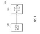
- FIG. 1is a block diagram of hardware that can be used in accordance with some embodiments.
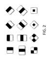
- FIG. 2shows examples of Haar features that can be used in accordance with some embodiments.
- FIG. 3is a block diagram of a multi-stage classifier in accordance with some embodiments.
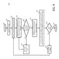
- FIG. 4is a flow diagram of a process for training a multi-stage classifier in accordance with some embodiments.
- FIG. 5is a block diagram of another multi-stage classifier in accordance with some embodiments.
- FIG. 6is a flow diagram of another process for training a multi-stage classifier in accordance with some embodiments.
- FIG. 7is a flow diagram of a process for training a single-stage classifier in accordance with some embodiments.
- FIG. 8is a flow diagram of a process for detecting objects in images using a classifier in accordance with some embodiments.
- systems, methods, and media for detecting an anatomical object in a medical device imageare provided. More particularly, in some embodiments, systems, methods, and media for detecting an anatomical object, such as a pulmonary trunk, in a medical device image, such as a computed tomography pulmonary angiography (CTPA) image, are provided.
- CTPAcomputed tomography pulmonary angiography
- the pulmonary trunkis the main pulmonary artery that rises from the right ventricle of the heart, extends upward, and divides into the right and left pulmonary arteries carrying blood to the lungs. Because PEs are only found in the pulmonary artery, identifying the pulmonary trunk in medical device images, such as CTPA images, can be used in PE diagnosis.
- Imaging device 102can be any suitable device for generating imaging data that can be provided to image processing device 104 .
- imaging device 102can be a computed tomography (CT) scanner.
- CTcomputed tomography
- Image processing device 104can be any suitable device for receiving and processing imaging data.
- image processing device 104can be a computer.
- Imaging device 102can communicate with image processing device 104 in any suitable manner such as via a direct connection between the devices, via a communication network, etc.
- image processing device 104can be any of a general purpose device such as a computer or a special purpose device such as a client, a server, etc. Any of these general or special purpose devices can include any suitable components such as a hardware processor (which can be a microprocessor, digital signal processor, a controller, etc), memory, communication interfaces, display controllers, input devices, etc.
- a hardware processorwhich can be a microprocessor, digital signal processor, a controller, etc
- memorycommunication interfaces
- display controllersinput devices, etc.
- imaging device 102 and image processing device 104can be integrated into a single device.
- a machine-learning-based approachcan be used by image processing device 104 for automatically detecting an anatomical object, such as a pulmonary trunk, in a medical device image.
- a cascaded AdaBoost classifiercan be trained with a large number of Haar features (example of which are shown in FIG. 2 ) extracted from computed tomography pulmonary angiography (CTPA) image samples, so that an anatomical object, such as a pulmonary trunk, can subsequently be automatically identified by sequentially scanning CTPA images and classifying each encountered sub-image with the trained classifier.
- CTPA imagescan be automatically scanned at multiple scales to handle size variations of the anatomical objects (e.g., pulmonary trunks).
- AdaBoost classifieris a type of machine learning algorithm drat combines weak learners to create a single strong learner.
- a weak learneris a classifier that may perform only slightly better than random guessing.
- a commonly used weak classifier called the decision stumpcan be used to make a prediction based on the value of a single input feature.
- h 1 , h 2 , . . . , h Nmake up a set of weak learners
- Boostingis a process to select weak learners h j and determine their coefficients ⁇ j , so as to combine the selected weak learners to form a strong learner F(x).
- AdaBoostcan be used to select the most relevant, features from any suitable number (e.g., thousands) of Haar features, each corresponding to a weak learner.
- a Haar featurecan be defined in terms of two adjacent rectangle regions, which can be illustrated in white and black as shown in FIG. 2 , for example.
- the value of a Haar featurecan be the sum of any suitable pixels values (such as intensity) in one or more first rectangle(s) (e.g., the white rectangles) of the feature minus the sum of the suitable pixel values in one or more second rectangle(s) (e.g., the black rectangle(s)) of the feature.
- any suitable criteriasuch as desired true positive rate, false positive rate, and number of weak learners, can be used to determine the number of strong boosted classifiers, the number of weak learners in each boosted classifier, and the relative operating characteristic (ROC) operating points (which can be selected from a ROC curve produced during training) for classifying images.
- a True Positive Rate (TPR) aa False Positive Rate (FPR) ⁇ i , and a maximum number of weak learners ⁇ i can be used as criteria for training a cascaded classifier stage.
- an AdaBoost classifier 300can include any suitable number of strong classifier stages 302 , 304 , and 306 .
- D i + , D i ⁇can be used to refer to positive sub-images and negative sub-images that can be used for training an AdaBoost classifier stage i.
- weak learnerscan be added to tire stage until a given target performance ( ⁇ i , ⁇ i ) or a given number of weak learners ⁇ i in the stage is reached.
- the output of the training at stage iis a boosted classifier containing weak learners from f ⁇ i ⁇ 1 +1 to f 96 i .
- new negative samplescan be classified by the stage to identify false positives (i.e., negative samples which are classified as positive) and then these negative samples (which are falsely classified as positives) can be combined with the negative samples used for training the current stage and the combination used for training the subsequent stage.
- process 400for training this classifier in accordance with some embodiments is shown. As illustrated, after process 400 begins at 402 , the process selects a first stage of the classifier to train. This stage can be selected in any suitable manner. Next, at 406 , the process can select an initial set of weak learners for the stage. Any suitable number of weak learners, including one, can be selected, and the weak learners can be selected in any suitable manner, such as randomly. Then, at 408 , process 400 can apply positive and negative sub-image samples to the set of weak learners. Any suitable number of positive and negative sub-image samples (e.g., 100 each) can be applied, and these samples can be selected for application in any suitable manner, such as randomly.
- positive and negative sub-image samplese.g., 100 each
- the processcan then determine at 410 whether the performance of the stage is sufficient or whether the maximum number of weak learners for the stage has been reached. Any suitable criteria or criterion can be used for determining whether the performance of the stage is sufficient in some embodiments. For example, in some embodiments, the performance of the stage can be deemed to be sufficient when the TPR ⁇ i is over 0.99 and FPR ⁇ i is below 0.05. Any suitable threshold ⁇ i for a maximum number of weak learners can be used in some embodiments. For example, ⁇ i can be 30 in some embodiments. If it is determined at 410 that the performance is not sufficient and the maximum number of weak learners has not been reached, then process 400 can add one or more weak learners to the set at 412 and loop back to 408 .
- the weak learners to be addedcan be selected in any suitable manner (e.g., randomly) and any suitable number of weak learners (including one) can be added, in some embodiments. Otherwise, at 414 process 400 can then assign the set of weak, learners to the boosted strong classifier for the current stage. Next, at 416 , process 400 can use the set of weak, learners to detect new negative samples that appear positive (i.e., false positives) and add these new negative samples to the set of negative samples and use this new set for the next stage. Any suitable number of new negative samples, such as 100, can be used in some embodiments. At 418 , process 400 can then determine whether the current stage is the last stage, and, if not, select the next stage at 420 . Otherwise, process can end at 422 .
- classifier 500can include any suitable number of strong classifier stages 502 , 504 , and 506 .
- D i + , D i ⁇can be used to refer to positive sub-images and negative sub-images that can be used for the teaming a classifier stage i.
- weak learnerscan be added to the stage until a given target performance ( ⁇ i , ⁇ i ) or a given number of weak learners ⁇ i in the stage is reached.
- the output of the training at stage iis a boosted classifier 504 containing weak learners from f 1 to f 96 i .
- a stagecan include all of the weak learners of all previous stages in some embodiments.
- new negative samplescan be classified by the stage to identify false positives (i.e., negative samples which are classified as positive) and then these negative samples (which are falsely classified as positives) can be added to the negative samples from the current stage and used for training the subsequent stage.
- process 600includes steps 402 , 404 , 406 , 408 , 410 , 412 , 414 , 416 , 418 , 420 , and 422 which can be performed as described above in connection with process 400 of FIG. 4 .
- process 600can branch to step 412 rather than step 406 .
- a single stage classifiercan be used.
- Such a classifiermay include a single classifier stage 302 as shown in FIG. 3 .
- process 700includes steps 402 , 406 , 408 , 410 , 412 , 414 , and 422 which can be performed as described above in connection with process 400 of FIG. 4 .
- negative samplescan be false positive samples from other training techniques as described above.
- an imagecan be provided to the one or more stages of the classifier and a positive indication or a negative indication can be provided. If at any stage in the classifier, an image is classified as negative, the image can be removed from subsequent testing by subsequent stages of the classifier and the classification of the image can be maintained as negative.
- process 800for detecting images in accordance with some embodiments is shown.
- the processcan select a detection scheme at 804 . Any suitable detection scheme can be used, such as the multi-stage or single-stage schemes described above.
- the first imagecan be selected at 806 .
- the first imagecan be selected in any suitable manner (e.g., such as randomly, in-order, etc.), and the image can be any suitable portion of another image (e.g., such as a random portion of a first image).
- the first strong classifier in the selected schemecan be selected.
- the imagecan then be applied to the selected strong classifier, which can assign a classification and a score to the image.
- process 800can then determine if the classification from the stage is negative. If so, the next image can be selected at 814 and process 800 can loop back to 808 . Otherwise, at 816 , it can be determined if the current stage is the last strong classifier. If not, then process 800 can select the next strong classifier at 818 and loop back to 810 . Otherwise, process 800 can classify the image as positive at 820 and merge the image with any previous overlapping, positive-classified images at 822 . Any suitable images can be identified as being overlapping in some embodiments.
- imagescan be identified as being overlapping if the images share over 25% of their data (e.g., based on location and size of the image) and/or if their z-axis distance is less than five pixels.
- their individual scorescan be added together.
- process 800can determine if the current image is the last image. If not, the process can select the next image at 814 and loop back to 808 . Otherwise, the process can select the highest-score merged image as the detected object at 826 and terminate at 828 .
- any suitable computer readable mediacan be used for storing instructions for performing the processes described herein, such as performing training of classifiers and classifying of images.
- computer readable mediacan be transitory or non-transitory.
- non-transitory computer readable mediacan include media such as magnetic media (such as hard disks, floppy disks, etc.), optical media (such as compact discs, digital video discs, Blu-ray discs, etc.), semiconductor media (such as flash memory, electrically programmable read only memory (EPROM), electrically erasable programmable read only memory (EEPROM), etc.), any suitable media that is not fleeting or devoid of any semblance of permanence during transmission, and/or any suitable tangible media.
- transitory computer readable mediacan include signals on networks, in wires, conductors, optical fibers, circuits, any suitable media that is fleeting and devoid of any semblance of permanence during transmission, and/or any suitable intangible media.
Landscapes
- Engineering & Computer Science (AREA)
- Theoretical Computer Science (AREA)
- Computer Vision & Pattern Recognition (AREA)
- Data Mining & Analysis (AREA)
- General Physics & Mathematics (AREA)
- Physics & Mathematics (AREA)
- Evolutionary Computation (AREA)
- Artificial Intelligence (AREA)
- General Engineering & Computer Science (AREA)
- Evolutionary Biology (AREA)
- Bioinformatics & Computational Biology (AREA)
- Bioinformatics & Cheminformatics (AREA)
- Life Sciences & Earth Sciences (AREA)
- Health & Medical Sciences (AREA)
- Medical Informatics (AREA)
- General Health & Medical Sciences (AREA)
- Computing Systems (AREA)
- Multimedia (AREA)
- Databases & Information Systems (AREA)
- Software Systems (AREA)
- Quality & Reliability (AREA)
- Radiology & Medical Imaging (AREA)
- Nuclear Medicine, Radiotherapy & Molecular Imaging (AREA)
- Image Analysis (AREA)
- Apparatus For Radiation Diagnosis (AREA)
Abstract
Description
F(x)=Σj−1Nfj(x)=Σj=1Nωjhj(x),
where ωjis the corresponding coefficient for weak learner hj. Boosting is a process to select weak learners hjand determine their coefficients ωj, so as to combine the selected weak learners to form a strong learner F(x).
Claims (24)
Priority Applications (1)
| Application Number | Priority Date | Filing Date | Title |
|---|---|---|---|
| US13/984,808US9684957B2 (en) | 2011-02-11 | 2012-02-13 | Systems methods, and media for detecting an anatomical object in a medical device image using a multi-stage classifier |
Applications Claiming Priority (3)
| Application Number | Priority Date | Filing Date | Title |
|---|---|---|---|
| US201161442112P | 2011-02-11 | 2011-02-11 | |
| PCT/US2012/024907WO2012109670A1 (en) | 2011-02-11 | 2012-02-13 | Systems, methods, and media for detecting an anatomical object in a medical device image |
| US13/984,808US9684957B2 (en) | 2011-02-11 | 2012-02-13 | Systems methods, and media for detecting an anatomical object in a medical device image using a multi-stage classifier |
Publications (2)
| Publication Number | Publication Date |
|---|---|
| US20140185887A1 US20140185887A1 (en) | 2014-07-03 |
| US9684957B2true US9684957B2 (en) | 2017-06-20 |
Family
ID=46638995
Family Applications (1)
| Application Number | Title | Priority Date | Filing Date |
|---|---|---|---|
| US13/984,808Expired - Fee RelatedUS9684957B2 (en) | 2011-02-11 | 2012-02-13 | Systems methods, and media for detecting an anatomical object in a medical device image using a multi-stage classifier |
Country Status (2)
| Country | Link |
|---|---|
| US (1) | US9684957B2 (en) |
| WO (1) | WO2012109670A1 (en) |
Cited By (5)
| Publication number | Priority date | Publication date | Assignee | Title |
|---|---|---|---|---|
| US10956785B2 (en) | 2018-04-27 | 2021-03-23 | Arizona Board Of Regents On Behalf Of Arizona State University | Methods, systems, and media for selecting candidates for annotation for use in training classifiers |
| US11100685B2 (en) | 2018-08-29 | 2021-08-24 | Arizona Board Of Regents On Behalf Of Arizona State University | Method and apparatus for detection and visualization of pulmonary embolism |
| US11164067B2 (en) | 2018-08-29 | 2021-11-02 | Arizona Board Of Regents On Behalf Of Arizona State University | Systems, methods, and apparatuses for implementing a multi-resolution neural network for use with imaging intensive applications including medical imaging |
| US12118455B2 (en) | 2017-04-27 | 2024-10-15 | Arizona Board Of Regents On Behalf Of Arizona State University | Systems, methods, and/or media, for selecting candidates for annotation for use in training a classifier |
| US12322098B2 (en) | 2021-09-07 | 2025-06-03 | Arizona Board Of Regents On Behalf Of Arizona State University | Systems, methods, and apparatuses for generating pre-trained models for nnU-net through the use of improved transfer learning techniques |
Families Citing this family (5)
| Publication number | Priority date | Publication date | Assignee | Title |
|---|---|---|---|---|
| WO2012109670A1 (en)* | 2011-02-11 | 2012-08-16 | Arizona Board Of Regents, For And On Behalf Of, Arizona State University | Systems, methods, and media for detecting an anatomical object in a medical device image |
| AU2012214149A1 (en) | 2011-02-11 | 2013-09-05 | Arizona Board Of Regents For And On Behalf Of Arizona State University | Methods, systems, and media for determining carotid intima-media thickness |
| US9330336B2 (en)* | 2011-09-16 | 2016-05-03 | Arizona Board of Regents, a body corporate of the State of Arizona, acting for and on behalf of, Arizona State University | Systems, methods, and media for on-line boosting of a classifier |
| WO2013116867A1 (en) | 2012-02-03 | 2013-08-08 | Arizona Board Of Regents, For And On Behalf Of, Arizona State University | Systems, methods, and media for monitoring the condition of a patient's heart |
| US10268950B2 (en)* | 2014-11-15 | 2019-04-23 | Beijing Kuangshi Technology Co., Ltd. | Face detection using machine learning |
Citations (38)
| Publication number | Priority date | Publication date | Assignee | Title |
|---|---|---|---|---|
| US20030199762A1 (en) | 2002-04-19 | 2003-10-23 | Sonometric Health, Llc | Method, apparatus, and product for accurately determining the intima-media thickness of a blood vessel |
| US20040208341A1 (en) | 2003-03-07 | 2004-10-21 | Zhou Xiang Sean | System and method for tracking a global shape of an object in motion |
| US20050220336A1 (en) | 2004-03-26 | 2005-10-06 | Kohtaro Sabe | Information processing apparatus and method, recording medium, and program |
| US20050228276A1 (en) | 2004-04-02 | 2005-10-13 | Teratech Corporation | Wall motion analyzer |
| US20060074834A1 (en)* | 2004-09-07 | 2006-04-06 | Anlei Dong | Methods and systems for 3D object detection using learning |
| US20060204121A1 (en) | 2005-03-03 | 2006-09-14 | Bryll Robert K | System and method for single image focus assessment |
| US20070280530A1 (en) | 2006-05-03 | 2007-12-06 | Siemens Medical Solutions Usa, Inc. | Using Candidates Correlation Information During Computer Aided Diagnosis |
| US20080009733A1 (en) | 2006-06-27 | 2008-01-10 | Ep Medsystems, Inc. | Method for Evaluating Regional Ventricular Function and Incoordinate Ventricular Contraction |
| US20080027887A1 (en) | 2006-07-27 | 2008-01-31 | The Government Of The Us, As Represented By The Secretary Of The Navy | System and method for fusing data from different information sources |
| US20080089571A1 (en) | 2006-10-17 | 2008-04-17 | Kabushiki Kaisha Toshiba | Ultrasonic imaging apparatus and a method of obtaining ultrasonic images |
| US20080154565A1 (en)* | 2006-05-23 | 2008-06-26 | Siemens Corporate Research, Inc. | Automatic organ detection using machine learning and classification algorithms |
| US20080171939A1 (en) | 2007-01-15 | 2008-07-17 | Fujifilm Corporation | Ultrasonic diagnostic apparatus, imt measurement method, and imt measurement program |
| US20080194957A1 (en) | 2007-02-14 | 2008-08-14 | Ralph Thomas Hoctor | Method and Apparatus for Generating an Ultrasound Image of Moving Objects Using Deformable Models |
| US20080192887A1 (en) | 2005-02-04 | 2008-08-14 | Koninklijke Philips Electronics, N.V. | System For The Determination Of Vessel Geometry And Flow Characteristics |
| US20080205750A1 (en) | 2007-02-28 | 2008-08-28 | Porikli Fatih M | Method for Adaptively Boosting Classifiers for Object Tracking |
| US20080240532A1 (en) | 2007-03-30 | 2008-10-02 | Siemens Corporation | System and Method for Detection of Fetal Anatomies From Ultrasound Images Using a Constrained Probabilistic Boosting Tree |
| US20080260230A1 (en) | 2005-09-16 | 2008-10-23 | The Ohio State University | Method and Apparatus for Detecting Intraventricular Dyssynchrony |
| US20090034816A1 (en) | 2007-08-03 | 2009-02-05 | Siemens Medical Solutions Usa, Inc. | Reduction of Lymph Tissue False Positives in Pulmonary Embolism Detection |
| US20090060307A1 (en) | 2007-08-27 | 2009-03-05 | Siemens Medical Solutions Usa, Inc. | Tensor Voting System and Method |
| US7526101B2 (en) | 2005-01-24 | 2009-04-28 | Mitsubishi Electric Research Laboratories, Inc. | Tracking objects in videos with adaptive classifiers |
| US20090175515A1 (en) | 2006-06-08 | 2009-07-09 | Tomtec Imaging Systems Gmbh | Method, device, and computer programme for evaluating images of a cavity |
| US20090252394A1 (en) | 2007-02-05 | 2009-10-08 | Siemens Medical Solutions Usa, Inc. | Computer Aided Detection of Pulmonary Embolism with Local Characteristic Features in CT Angiography |
| US20100046815A1 (en) | 2006-10-03 | 2010-02-25 | Jens Von Berg | Model-based coronary centerline localization |
| US20100061601A1 (en) | 2008-04-25 | 2010-03-11 | Michael Abramoff | Optimal registration of multiple deformed images using a physical model of the imaging distortion |
| US20100076517A1 (en) | 2008-09-23 | 2010-03-25 | Mir Imran | Energy harvesting mechanism for medical devices |
| US20100098308A1 (en) | 2008-10-16 | 2010-04-22 | Siemens Corporation | Pulmonary Emboli Detection with Dynamic Configuration Based on Blood Contrast Level |
| US20100113930A1 (en) | 2008-11-04 | 2010-05-06 | Fujifilm Corporation | Ultrasonic diagnostic device |
| US20100177944A1 (en) | 2007-04-02 | 2010-07-15 | The Trustees Of The University Of Pennsylvania | Combined feature ensemble mutual information image registration |
| US20100202681A1 (en) | 2007-06-01 | 2010-08-12 | Haizhou Ai | Detecting device of special shot object and learning device and method thereof |
| US20100266176A1 (en) | 2009-04-16 | 2010-10-21 | Fujifilm Corporation | Diagnosis assisting apparatus, diagnosis assisting method, and storage medium having a diagnosis assisting program recorded therein |
| US20110191283A1 (en)* | 2010-02-03 | 2011-08-04 | Siemens Corporation | Method and System for Medical Decision Support Using Organ Models and Learning Based Discriminative Distance Functions |
| US20110270089A1 (en) | 2008-08-05 | 2011-11-03 | Guardsman Scientific, Inc. | System and method for managing a patient |
| US20110293157A1 (en) | 2008-07-03 | 2011-12-01 | Medicsight Plc | Medical Image Segmentation |
| US20120089545A1 (en) | 2009-04-01 | 2012-04-12 | Sony Corporation | Device and method for multiclass object detection |
| US20120106815A1 (en) | 2010-10-28 | 2012-05-03 | Toshiba Medical Systems Corporation | Denoising method and system for preserving clinically significant structures in reconstructed images using adaptively weighted anisotropic diffusion filter |
| US20120274755A1 (en) | 2011-04-29 | 2012-11-01 | Tata Consultancy Services Limited | System and method for human detection and counting using background modeling, hog and haar features |
| US20130070997A1 (en) | 2011-09-16 | 2013-03-21 | Arizona Board of Regents, a body Corporate of the State of Arizona, Acting for and on Behalf of Ariz | Systems, methods, and media for on-line boosting of a classifier |
| US20140185887A1 (en)* | 2011-02-11 | 2014-07-03 | Hong Wu | Systems, Methods, and Media for Detecting an Anatomical Object in a Medical Device Image |
- 2012
- 2012-02-13WOPCT/US2012/024907patent/WO2012109670A1/enactiveApplication Filing
- 2012-02-13USUS13/984,808patent/US9684957B2/ennot_activeExpired - Fee Related
Patent Citations (40)
| Publication number | Priority date | Publication date | Assignee | Title |
|---|---|---|---|---|
| US20030199762A1 (en) | 2002-04-19 | 2003-10-23 | Sonometric Health, Llc | Method, apparatus, and product for accurately determining the intima-media thickness of a blood vessel |
| US20040208341A1 (en) | 2003-03-07 | 2004-10-21 | Zhou Xiang Sean | System and method for tracking a global shape of an object in motion |
| US20050220336A1 (en) | 2004-03-26 | 2005-10-06 | Kohtaro Sabe | Information processing apparatus and method, recording medium, and program |
| US20050228276A1 (en) | 2004-04-02 | 2005-10-13 | Teratech Corporation | Wall motion analyzer |
| US20060074834A1 (en)* | 2004-09-07 | 2006-04-06 | Anlei Dong | Methods and systems for 3D object detection using learning |
| US7526101B2 (en) | 2005-01-24 | 2009-04-28 | Mitsubishi Electric Research Laboratories, Inc. | Tracking objects in videos with adaptive classifiers |
| US20080192887A1 (en) | 2005-02-04 | 2008-08-14 | Koninklijke Philips Electronics, N.V. | System For The Determination Of Vessel Geometry And Flow Characteristics |
| US20060204121A1 (en) | 2005-03-03 | 2006-09-14 | Bryll Robert K | System and method for single image focus assessment |
| US20080260230A1 (en) | 2005-09-16 | 2008-10-23 | The Ohio State University | Method and Apparatus for Detecting Intraventricular Dyssynchrony |
| US20070280530A1 (en) | 2006-05-03 | 2007-12-06 | Siemens Medical Solutions Usa, Inc. | Using Candidates Correlation Information During Computer Aided Diagnosis |
| US20080154565A1 (en)* | 2006-05-23 | 2008-06-26 | Siemens Corporate Research, Inc. | Automatic organ detection using machine learning and classification algorithms |
| US20090175515A1 (en) | 2006-06-08 | 2009-07-09 | Tomtec Imaging Systems Gmbh | Method, device, and computer programme for evaluating images of a cavity |
| US20080009733A1 (en) | 2006-06-27 | 2008-01-10 | Ep Medsystems, Inc. | Method for Evaluating Regional Ventricular Function and Incoordinate Ventricular Contraction |
| US20080027887A1 (en) | 2006-07-27 | 2008-01-31 | The Government Of The Us, As Represented By The Secretary Of The Navy | System and method for fusing data from different information sources |
| US20100046815A1 (en) | 2006-10-03 | 2010-02-25 | Jens Von Berg | Model-based coronary centerline localization |
| US20080089571A1 (en) | 2006-10-17 | 2008-04-17 | Kabushiki Kaisha Toshiba | Ultrasonic imaging apparatus and a method of obtaining ultrasonic images |
| US20080171939A1 (en) | 2007-01-15 | 2008-07-17 | Fujifilm Corporation | Ultrasonic diagnostic apparatus, imt measurement method, and imt measurement program |
| US20090252394A1 (en) | 2007-02-05 | 2009-10-08 | Siemens Medical Solutions Usa, Inc. | Computer Aided Detection of Pulmonary Embolism with Local Characteristic Features in CT Angiography |
| US20080194957A1 (en) | 2007-02-14 | 2008-08-14 | Ralph Thomas Hoctor | Method and Apparatus for Generating an Ultrasound Image of Moving Objects Using Deformable Models |
| US20080205750A1 (en) | 2007-02-28 | 2008-08-28 | Porikli Fatih M | Method for Adaptively Boosting Classifiers for Object Tracking |
| US7840061B2 (en) | 2007-02-28 | 2010-11-23 | Mitsubishi Electric Research Laboratories, Inc. | Method for adaptively boosting classifiers for object tracking |
| US20080240532A1 (en) | 2007-03-30 | 2008-10-02 | Siemens Corporation | System and Method for Detection of Fetal Anatomies From Ultrasound Images Using a Constrained Probabilistic Boosting Tree |
| US20100177944A1 (en) | 2007-04-02 | 2010-07-15 | The Trustees Of The University Of Pennsylvania | Combined feature ensemble mutual information image registration |
| US20100202681A1 (en) | 2007-06-01 | 2010-08-12 | Haizhou Ai | Detecting device of special shot object and learning device and method thereof |
| US20090034816A1 (en) | 2007-08-03 | 2009-02-05 | Siemens Medical Solutions Usa, Inc. | Reduction of Lymph Tissue False Positives in Pulmonary Embolism Detection |
| US20090060307A1 (en) | 2007-08-27 | 2009-03-05 | Siemens Medical Solutions Usa, Inc. | Tensor Voting System and Method |
| US20100061601A1 (en) | 2008-04-25 | 2010-03-11 | Michael Abramoff | Optimal registration of multiple deformed images using a physical model of the imaging distortion |
| US20110293157A1 (en) | 2008-07-03 | 2011-12-01 | Medicsight Plc | Medical Image Segmentation |
| US20110270089A1 (en) | 2008-08-05 | 2011-11-03 | Guardsman Scientific, Inc. | System and method for managing a patient |
| US20100076517A1 (en) | 2008-09-23 | 2010-03-25 | Mir Imran | Energy harvesting mechanism for medical devices |
| US20100098308A1 (en) | 2008-10-16 | 2010-04-22 | Siemens Corporation | Pulmonary Emboli Detection with Dynamic Configuration Based on Blood Contrast Level |
| US20100113930A1 (en) | 2008-11-04 | 2010-05-06 | Fujifilm Corporation | Ultrasonic diagnostic device |
| US20120089545A1 (en) | 2009-04-01 | 2012-04-12 | Sony Corporation | Device and method for multiclass object detection |
| US20100266176A1 (en) | 2009-04-16 | 2010-10-21 | Fujifilm Corporation | Diagnosis assisting apparatus, diagnosis assisting method, and storage medium having a diagnosis assisting program recorded therein |
| US20110191283A1 (en)* | 2010-02-03 | 2011-08-04 | Siemens Corporation | Method and System for Medical Decision Support Using Organ Models and Learning Based Discriminative Distance Functions |
| US8812431B2 (en)* | 2010-02-03 | 2014-08-19 | Siemens Aktiengesellschaft | Method and system for medical decision support using organ models and learning based discriminative distance functions |
| US20120106815A1 (en) | 2010-10-28 | 2012-05-03 | Toshiba Medical Systems Corporation | Denoising method and system for preserving clinically significant structures in reconstructed images using adaptively weighted anisotropic diffusion filter |
| US20140185887A1 (en)* | 2011-02-11 | 2014-07-03 | Hong Wu | Systems, Methods, and Media for Detecting an Anatomical Object in a Medical Device Image |
| US20120274755A1 (en) | 2011-04-29 | 2012-11-01 | Tata Consultancy Services Limited | System and method for human detection and counting using background modeling, hog and haar features |
| US20130070997A1 (en) | 2011-09-16 | 2013-03-21 | Arizona Board of Regents, a body Corporate of the State of Arizona, Acting for and on Behalf of Ariz | Systems, methods, and media for on-line boosting of a classifier |
Non-Patent Citations (119)
| Title |
|---|
| "Deep Vein Thrombosis Overview", Technical Report, Society of Interventional Radiology, last accessed Sep. 17, 2014, pp. 1-3, available at: http://www.sirweb.org/patients/deep-vein-thrombosis/. |
| Alonso-Martinez, J.L., et al., "Delay and Misdiagnosis in Sub-Massive and Non-Massive Acute Pulmonary Embolism", In European Journal of Internal Medicine, vol. 21, No. 4, Aug. 2010, pp. 278-282. |
| Araoz, P.A., et al., "Helical CT Pulmonary Angiography Predictors of In-Hospital Morbidity and Mortality in Patients with Acute Pulmonary Embolism", In Journal of Thoracic Imaging, vol. 18, Oct. 2003, pp. 207-216. |
| Bi, J. and Liang, J., "Multiple instance learning of pulmonary embolism detection with geodesic distance along vascular structure", In Proceedings of IEEE Computer Society Conference on Computer Vision and Pattern Recognition (CVPR) Jun. 17-22, 2007, Minneapolis, MN, USA, pp. 1-8. |
| Bottiger, B.W., et al., "Inhaled Nitric Oxide Selectively Decreases Pulmonary Artery Pressure and Pulmonary Vascular Resistance Following Acute Massive Pulmonary Microembolism in Piglets", In Chest, vol. 110, No. 4, Oct. 1996, pp. 1041-1047. |
| Bouma, H., "Vessel-Diameter Quantification and Embolus Detection in CTA Images." Ph.D. Thesis, Eindhoven University of Technology, PrintPartners, Ipskamp, The Netherlands, Apr. 2008, pp. 9-133. |
| Bouma, H., et al, "Automatic Detection of Pulmonary Embolism in CTA Images", In IEEE Transactions on Medical Imaging, vol. 28, No. 8, Aug. 2009, pp. 1223-1230. |
| Bourdev, L. and Brandt, J., et al., "Robust Object Detection Via Soft Cascade", In Proceedings of the 2005 IEEE Conference on Computer Vision and Pattern Recognition (CVPR '05), Washington, DC, USA, Jun. 2005, pp. 236-243. |
| Chartrand-Lefebvre, C., "Computed tomography angiography in the diagnosis of pulmonary embolism: Interobserver agreement", In American Journal of Emergency Medicine, Jan. 27, 2011, pp. 118-119. |
| Cho, E.J., et al., "Right ventricular free wall circumferential strain reflects graded elevation in acute right ventricular afterload", In Am J Physiol Heart Circ Physiol., Feb. 2009, vol. 296, No. 2, pp. 818-824. |
| Collomb, J., et al., "Severity Assessment of Acute Pulmonary Embolism: Evaluation using Helical CT", In European Radiology, vol. 13, No. 7, Feb. 2003, pp. 1508-1514. |
| Costantino, G., et al., "Interobserver agreement in computer tomography readings for pulmonary embolism", In American Journal of Emergency Medicine, Jan. 27, 2011, pp. 119. |
| Costantino, G., et al., "Interobserver agreement in computer tomography readings for pulmonary embolism", In American Journal of Emergency Medicine, vol. 27, No. 9, Nov. 2009, pp. 1109-1111. |
| Craig, J.J., "Introduction to Robotics: Mechanics and Control", 3rd edition, Prentice Hall, Aug. 6, 2004, pp. 1-385. |
| Criminisi, A., et al., "Regression Forests for Efficient Anatomy Detection and Localization in CT Studies", In Proceedings of the International Workshop on Medical Computer Vision, Beijing, CN, Sep. 2010, pp. 106-117. |
| Crow, F.C., "Summed-Area Tables for Texture Mapping", In Computer Graphics, vol. 18, No. 3, Jul. 1984, pp. 207-212. |
| Dias-Junior, C.A., "The Effect of Sildenafil on Pulmonary Embolism-Induced Oxidative Stress and Pulmonary Hypertension", In Anesthesia & Analgesia, vol. 101, No. 1, Jul. 2005, pp. 115-120. |
| Dinesh, M.S., et al, "Adaptive Contrast-Based Computer Aided Detection for Pulmonary Embolism", In Proceedings of the SPIE International Society Conference for Optimal Engineering, Mar. 2009, vol. 7260, No. 726010, pp. 1-8. |
| Dollar, P., et al., "Multiple Component Learning for Object Detection", In Proceedings of the 10th European Conference on Computer Vision: Part II (ECCV '08), Marseille, FR, Oct. 12-18, 2008, pp. 211-224. |
| Dousset, M., et al., "Principles and performance of virtual CT and MIRA intraluminal endoscopy", In Virtual Endoscopy, Springer, Nov. 2002, pp. 1-19. |
| Frangi, A.F., et al., "Model-Based Quantitation of 3-D Magnetic Resonance Angiographic Images", In IEEE Transactions on Medical Imaging, vol. 18, No. 10, Oct. 1999, pp. 946-956. |
| Frangi, A.F., et al., "Multiscale vessel enhancement filtering", In Medical Image Computing and Computer-Assisted Intervention, Oct. 11-13, 1998, pp. 130-137. |
| Freund, Y. and Schapire, R.E., "A Decision-Theoretic Generalization of On-Line Learning and an Application to Boosting", In Journal of Computer and System Sciences, vol. 55, No. 1, Aug. 1997, pp. 119-139. |
| Freund, Y. and Schapire, R.E., "A Short Introduction to Boosting", In Journal of Japanese Society for Artificial Intelligence, vol. 14, No. 5, Sep. 1999, pp. 771-780. |
| Galson, S.K., "The surgeon general's call to action to prevent deep vein thrombosis and pulmonary embolism", Technical Report, U.S. Public Health Services, Sep. 15, 2008, pp. 1-35. |
| Ghaye, B., et al., "Can CT Pulmonary Angiography Allow Assessment of Severity and Prognosis in Patients Presenting with Pulmonary Embolism? What the Radiologist Needs to Know", In RadioGraphics, vol. 26, Jan. 2006, pp. 23-29. |
| Ghaye, B., et al., "Severe Pulmonary Embolism: Pulmonary Artery Clot Load Scores and Cardiovascular Parameters as Predictors of Mortality", In Radiology, vol. 239, Apr. 2006, pp. 884-891. |
| Godec, M., et al., "On-line Random Naive Bayes for Tracking", In Proceedings of the 20th International Conference (ICPR '10), Istanbul, TR, Aug. 23-26, 2010, pp. 3545-3548. |
| Goldstein, H., "Classical Mechanics", 2nd Edition, Jul. 1980, pp. 1-2. |
| Grabner, H. and Bischof, H., "On-line Boosting and Vision", In Proceedings of the IEEE Computer Society Conference on Computer Vision and Pattern Recognition (CVPR '06), New York, NY, USA, Jun. 17-22, 2006, pp. 260-267. |
| Grbovic, M. and Vucetic, S., "Tracking Concept Change with Incremental Boosting by Minimization of the Evolving Exponential Loss", In Proceedings of the European Conference on Machine Learning and Knowledge Discovery in Databases, Athens, GR, Sep. 5-9, 2011, pp. 516-532. |
| Grifoni, S.,"Short-Term Clinical Outcome of Patients with Acute Pulmonary Embolism, Normal Blood Pressure, and Echocardiographic Right Ventricular Dysfunction", In Circulation, vol. 101, No. 24, Jun. 2000, pp. 2817-2822. |
| Groth, M., et al., "Correlation of right ventricular dysfunction parameters and pulmonary vascular obstruction score in acute pulmonary embolism in a porcine model", In Emergency Radiology, Sep. 2010, pp. 367-374. |
| He, H., et al., "Incremental Learning from Stream Data," In IEEE Transactions on Neural Networks, vol. 22, No. 12, Dec. 2011, pp. 1901-1914. |
| Howard, G., et al., "For the ARIC Investigators: Carotid Artery Intimal-Medial Thickness Distribution in General Populations as Evaluated by B-Mode Ultrasound", In Stroke, vol. 24, No. 9, Sep. 1993, pp. 1297-1304. |
| Hurst, R., et al., "Clinical Use of Carotid Intima-Media Thickness: Review of the Literature", In Journal of the American Society of Echocardiography, vol. 20, No. 7, Jul. 2007, pp. 907-914. |
| International Patent Application No. PCT/US2013/024675, filed Feb. 4, 2013. |
| International Patent Application No. PCT/US2013/024677, filed Feb. 4, 2013. |
| International Preliminary Report on Patentability dated Aug. 22, 2013 in International Patent Application No. PCT/US2012/024925. |
| International Preliminary Report on Patentability in International Application No. PCT/US2012/024907, filed Feb. 13, 2012, mailed Aug. 22, 2013. |
| International Search Report in International Patent Application No. PCT/US2012/024925, filed Feb. 13, 2012, mailed Jun. 19, 2012. |
| International Search Report in International Patent Application No. PCT/US2013/024675, filed Feb. 4, 2013, mailed Apr. 16, 2013. |
| International Search Report in International Patent Application No. PCT/US2013/024677, filed Feb. 4, 2013, mailed Apr. 15, 2013. |
| Jardin, F., et al., "Echocardiographic Pattern of Acute Cor Pulmonale", In Chest, vol. 111, No. 1, Jan. 1997, pp. 209-217. |
| Kanitsar, A., et al., "CPR-Curved Planar Reformation", In Proceedings of IEEE Visualization, Nov. 1, 2002, pp. 37-44. |
| Kanitsar, A., et al., "CPR—Curved Planar Reformation", In Proceedings of IEEE Visualization, Nov. 1, 2002, pp. 37-44. |
| Kass, M., et al., "Snakes: Active Contour Models", In International Journal of Computer Vision, vol. 1, No. 4, Jan. 1988, pp. 321-331. |
| Kim, T.K., et al., "Online Multiple Classier Boosting for Object Tracking", In Proceedings of the 2010 IEEE Computer Society Conference on Computer vision and Pattern Recognition Workshops (CVPRW '10), San Francisco, CA, USA, Jun. 13-18, 2010, pp. 1-6. |
| Kiraly, A.P., et al., "Cartwheel projections of segmented pulmonary vasculature for the detection of pulmonary embolism", In Medical Imaging: Visualization, Image-Guided Procedures, and Display, Proc. SPIE 5744, Apr. 12, 2005, pp. 69-78. |
| Knutsson, H., "Representing Local Structure using Tensors", In Proceedings of the 6th Scandinavian Conference on Image Analysis, Oulu, Finland, Jun. 1989, pp. 244-251. |
| Kothe, U., "Edge and Junction Detection with an Improved Structure Tensor", In Proceedings of the 25th DAGM Symposium on Pattern Recognition, Magdeburg, DE, Sep. 10-12, 2003, pp. 25-32. |
| Kurkure, U., et al., "Automated Segmentation of Thoracic Aorta in Non-Contrast CT Images", In Proceedings of the 5th International Symposium on Biomedical Imaging: From Nano to Macro (ISBI '08), Paris, FR, May 14-17, 2008, pp. 29-32. |
| Leistner, C., et al., "On Robustness of On-Line Boosting-A Competitive Study", In Proceedings of the 2009 IEEE 12th International Conference on Computer Vision Workshops (ICCVW '09), Kyoto, JP, Sep. 27,-Oct. 4, 2009, pp. 1362-1369. |
| Leistner, C., et al., "On Robustness of On-Line Boosting—A Competitive Study", In Proceedings of the 2009 IEEE 12th International Conference on Computer Vision Workshops (ICCVW '09), Kyoto, JP, Sep. 27,-Oct. 4, 2009, pp. 1362-1369. |
| Levenberg, K., "A Method for the Solution of Certain Non-Linear Problems in Least Squares", In Quarterly Journal of Applied Mathmatics, vol. 2, Jul. 1944, pp. 164-168. |
| Li, S., et al., "Childhood Cardiovascular Risk Factors and Carotid Vascular Changes in Adulthood: the Bogalusa Heart Study", In the Journal of the American Medical Association (JAMA), vol. 290, No. 17, Nov. 2003, pp. 2271-2276. |
| Liang, J. and Bi, J., "Computer Aided Detection of Pulmonary Embolism with Tobogganing and Multiple Instance Classification in CT Pulmonary Angiography", In Proceedings of the 20th Intl Conference of Information Processing in Medical Imaging Kerkrade, NL, Jul. 2-6, 2007, pp. 630-641. |
| Liang, J. and Bi, J., "Local Characteristic Features for Computer-Aided Detection of Pulmonary Embolism in CT Angiography", In Proceedings of the First Workshop on Pulmonary Image Analysis, New York, NY, US, Sep. 6, 2008, pp. 263-272. |
| Liang, J., et al., "United Snakes", In Medical Image Analysis, vol. 10 No. 2, Apr. 2006, vol. 215-233. |
| Liu, D., et al., "Search strategies for multiple landmark detection by submodular maximization", IEEE Conference on Computer Vision and Pattern Recognition, Jun. 3-8, 2010, San Francisco, CA, USA, pp. 2831-2838. |
| Liu, X. and Yu, T., "Gradient Feature Selection for Online Boosting", In Proceedings of the IEEE 11th International Conference on Computer Vision (ICCV '07), Rio de Janeiro, BR, Oct. 14-21, 2007, pp. 1-8. |
| Lorenz, C., et al., "Multi-scale line segmentation with automatic estimation of width, contrast and tangential direction in 2-D and 3-D medical images", In Proc. of the Joint Conference on Computer Vision, Virtual Reality and Robotics in Medicine, London, UK, Mar. 19-22, 1997, pp. 233-242. |
| Mansencal, N., "Comparison of Different Echocardiographic Indexes Secondary to Right Ventricular Obstruction in Acute Pulmonary Embolism", In the American Journal of Cardiology, vol. 92, No. 1, Jul. 2003, pp. 116-119. |
| Marquardt, D.W., "An Algorithm for Least-Squares Estimation of Nonlinear Parameters", In SIAM Journal on Applied Mathematics, vol. 11 No. 2, Jun. 1963, pp. 431-441. |
| Mastora, I., "Severity of Acute Pulmonary Embolism: Evaluation of a New Spiral CT Angiographic Score in Correlation with Echocardiographic Data", In European Radiology, vol. 13, Jan. 2003, pp. 29-35. |
| Masutani, Y., et al., "Computerized Detection of Pulmonary Embolism in Spiral CT Angiography Based on Volumetric Image Analysis", In IEEE Transactions on Medical Imaging, vol. 21, No. 12, Dec. 2002, pp. 1517-1523. |
| McConnell, M.V., et al., "Regional Right Ventricular Dysfunction Detected by Echocardiography in Acute Pulmonary Embolism", In The American Journal of Cardiology, vol. 78 No. 4, Aug. 1996, pp. 469-473. |
| Notice of Allowance dated Sep. 14, 2015 in U.S. Appl. No. 13/621,837. |
| Office Action dated Apr. 24, 2015 in U.S. Appl. No. 14/023,380. |
| Office Action dated Aug. 23, 2013 in U.S. Appl. No. 13/984,808. |
| Office Action dated Jan. 22, 2015 in U.S. Appl. No. 14/376,181. |
| Office Action dated Jan. 29, 2015 in U.S. Appl. No. 13/621,837. |
| Office Action dated Jan. 4, 2016 in U.S. Appl. No. 14/023,380. |
| Office Action dated Jul. 17, 2014 in U.S. Appl. No. 13/621,837. |
| Office Action dated Jun. 15, 2016 in U.S. Appl. No. 14/376,568. |
| Office Action dated Nov. 27, 2015 in U.S. Appl. No. 14/376,568. |
| Office Action dated Oct. 7, 2013 in U.S. Appl. No. 14/023,380. |
| Office Action dated Sep. 18, 2013 in European Patent Application No. 12744949.4. |
| Ouellette, D.R., et al., "Pulmonary Embolism", Medscape.com, last updated Sep. 4, 2014, available at: http://emedicine.medscape.com/article/300901-overview#showall, pp. 1-24. |
| Oza, N. C. and Russell, S., "Online Bagging and Boosting", In Artificial Intelligence and Statistics, 2001, pp. 105-112. |
| Parag, T., et al., "Boosting Adaptive Linear Weak Classifiers for Online Learning and Tracking", In Proceedings of the IEEE Conference on Computer Vision and Recognition (CVPR '08), Anchorage, AK, USA, Jun. 23-28, 2008, pp. 1-8. |
| Parikh, D. and Polikar, R., "An Ensemble-Based Incremental Learning Approach to Data Fusion", In IEEE Transactions on Systems, Man, Cybernetics, Part B: Cybernetics, vol. 37, No. 2, Apr. 2007, pp. 437-450. |
| Patent Examination Report dated Aug. 26, 2015 in Australian Patent Application No. 2012214149. |
| Pelossof, R., et al., "Online Coordinate Boosting", In Proceedings of the 2009 IEEE 12th International Conference on Computer Vision Workshops, (ICCVW '09), Kyoto, JP, Sep. 27,-Oct. 4, 2009, pp. 1354-1361. |
| Pham, M. and Cham, T., "Detection with Multi-exit Asymmetric Boosting", In Proceedings of the IEEE Conference on Computer Vision and Pattern Recognition (CVPR '08), Anchorage, AK, USA, Jun. 23-28, 2008, pp. 1-8. |
| Pham, M. and Cham, T., "Fast Training and Selection of Haar Features Using Statistics in Boosting-Based Face Detection", In Proceedings of the IEEE 11th International Conference on Computer Vision (ICCV '07), Rio de Janeiro, BR, Oct. 14-21, 2007, pp. 1-7. |
| Pham, M. and Cham, T., "Online Learning Asymmetric Boosted Classifiers for Object Detection", In Proceedings of the IEEE Conference on Computer Vision and Recogition (CVPR '07), Minneapolis, MN, USA, Jun. 17-22, 2007, pp. 1-8. |
| Ribeiro, A., et al., "Echocardiography Doppler in Pulmonary Embolism: Right Ventricular Dysfunction as a Predictor of Mortality Rate", In American Heart Journal, vol. 134, No. 3, Mar. 1997, pp. 479-487. |
| Sato, Y. et al., "3-D multi-scale line filter for segmentation and visualization of curvilinear structures in medical images", In Proc. of the Joint Conference on Computer Vision, Virtual Reality and Robotics in Medicine, London, UK, Mar. 19-22, 1997, pp. 213-222. |
| Schapire, R. E. and Singer, Y., "BoosTexter: A Boosting-Based System for Text Categorization", In Machine Learning, vol. 39, No. 2, May 1, 2000, pp. 135-168. |
| Schapire, R. E., "Theoretical Views of Boosting and Applications", In Algorithmic Learning Theory, Lecture Notes in Computer Science, vol. 1720, Dec. 1999, pp. 13-25. |
| Sebbe, R., "Computer-aided Diagnosis of Pulmonary Embolism in Opacified CT Images", Ph.D. Dissertation, Faculte Polytechnique de Mons, Universitaires de Louvain, Belgium, Feb. 20, 2007, pp. 1-124. |
| Simon, M., et al., "Paddle-wheel CT display of pulmonary arteries and other lung structures: a new imaging approach", In American Journal of Roentgenology, Jul. 2001, pp. 195-198. |
| Simon, M., et al., "Paddle-wheel multislice helical CT display of pulmonary vessels and other lung structures", In Radiologic Clinics of North America, May 2003, pp. 617-626. |
| Stein, J., et al., "A Semiautomated Ultrasound Border Detection Program that Facilitates Clinical Measurement of Ultrasound Carotid Intima-Media Thickness", In the Journal of the American Society of Echocardiology, vol. 18, No. 3, Mar. 2005, pp. 244-251. |
| Stein, J., et al., "Use of Carotid Ultrasound to Identify Subclinical Vascular Disease & Evaluate Cardiovascular Disease Risk: A Consensus Statement from the American Society of Echocardiography Carotid Intima-Media Thickness Task Force", In the Journal of Am. Soc. of Echocardiography, vol. 21, No. 2, Feb. 2008, pp. 93-111. |
| Stein, J., et al., "Vascular Age: Integrating Carotid Intima-Media Thickness Measurements with Global Coronary Risk Assessment", In Clinical Cardiology, vol. 27, No. 7, Jul. 2004, pp. 388-392. |
| Stein, P.D. and Hull, R.D., "Multidetector computed tomography for the diagnosis of acute pulmonary embolism", In Current Opinion Pulmonary Medicine, Sep. 2007, pp. 384-388. |
| Stein, P.D. and Matta, F., "Acute Pulmonary Embolism", In Current Problems in Cardiology, vol. 35, No. 7, Jul. 2010, pp. 314-376. |
| Sternig, S., et al., "Transient Boost: On-line Boosting with Transient data", In Proceedings of the 2010 IEEE Computer Society Conference on Computer Vision and Pattern Recognition Workshops (CVPRW '10), San Francisco, CA, USA, Jun. 13-18, 2010, pp. 22-27. |
| Tajbakhsh, N., et al., "Motion Analysis of Right Ventricular Dysfunction under Mild and Moderate Pressure Overload Caused by Acute Pulmonary Embolism", In Ultrasound in Medicine and Biology, vol. 39, No. 11, Nov. 2013, pp. 2066-2074. |
| Tajbakhsh, N., et al., "Shape-Based Analysis of Right Ventricular Dysfunction associated with Acute Pulmonary Embolism", In SPIE Medical Imaging, vol. 8317, Mar. 2012, pp. 1-5. |
| Takamura, T., et al., "Reversible Left Ventricular Regional Non-Uniformity Quantified by Speckle-Tracking Displacement and Strain Imaging in Patients with Acute Pulmonary Embolism", In Journal of the American Society of Echocardiography, vol. 24, No. 7, Apr. 2011, pp. 792-802. |
| Torbicki, A., et al., "Guidelines on the diagnosis and management of acute pulmonary embolism of the European Society of Cardiology", In Eur Heart J., vol. 29, No. 18, Sep. 2008, pp. 2276-2315. |
| Vaidehi, V., et al., "Multiclass Object Detection System in Imaging Sensor Network Using Haar-like Features and Joint-Boosting Algorithm", In Proceedings of the 2011 International Conference on Recent Trends in Information Technology (ICRTIT '11), Chennai, Tamil Nadu, IN, Jun. 3-5, 2011, pp. 1011-1015. |
| Viola, P. and Jones M., "Fast and Robust Classification Using Asymmetric AdaBoost and a Detector Cascade", In Proceedings of the Annual Conference on Neural Information Processing Systems, Vancouver, BC, CA, Dec. 3-8, 2001, pp. 1311-1318. |
| Viola, P. and Jones, M., "Rapid Object Detection using a Boosted Cascade of Simple Features", In Proceedings of the IEEE Computer Society Conference on Computer Vision and Pattern Recognition, Kauai, HI, USA. Dec. 8-14, 2001, pp. 511-518. |
| Written Opinion in International Patent Application No. PCT/US2012/024925, filed Feb. 13, 2012, mailed Jun. 19, 2012. |
| Written Opinion in International Patent Application No. PCT/US2013/024675, filed Feb. 4, 2013, mailed Apr. 16, 2013. |
| Written Opinion in International Patent Application No. PCT/US2013/024677, filed Feb. 4, 2013, mailed Apr. 15, 2013. |
| Wu, B. and Nevatia, R., "Improving Part Based Object Detection by Unsupervised, Online Boosting", In Proceedings of the IEEE Conference on Computer Vision and Pattern Recognition (CVPR '07), Minneapolis, MN, USA, Jun. 17-22, 2007, pp. 1-8. |
| Wu, H., "Offline and Online Adaboost for Detecting Anatomical Structures", Thesis Paper, Arizona State University, Aug. 2011, pp. 1-66. |
| Wu, H., et al. "Self-Adaptive Asymmetric On-line Boosting for Detecting Anatomical Structures", In SPIE Medical Imaging, vol. 8315, Feb. 2012, pp. 1-7. |
| Wu, H., et al., "Machine Learning based Automatic Detection of Pulmonary Trunk", In Proceedings of the SPIE Conference on Medical Imaging 2011: Computer-Aided Diagnosis, Lake Buena Vista, FL, USA, Feb. 12, 2011, vol. 7963, pp. 1-6. |
| Zheng, Y., et al., "Automatic Aorta Segmentation and Valve Landmark Detection in C-Arm CT: Application to Aortic Valve Implantation", In IEEE Transactions on Medical Imaging, vol. 31, No. 12, Dec. 2012, pp. 2307-2321. |
| Zheng, Y., et al., "Fast Automatic Heart Chamber Segmentation from 3D CT Data Using Marginal Space Learning and Steerable Features", In Proceedings of the IEEE 11th International Conference on Computer Vision (ICCV '07), Rio de Janeiro, BR, Oct. 14-21, 2007, pp. 1-8. |
| Zhou, C., et al., "Automatic Pulmonary Vessel Segmentation in 3D Computed Tomographic Pulmonary Angiographic (CTPA) Images", In Proceedings of the SPIE 6144, Medical Imaging: Image Processing, Mar. 15, 2006, pp. Q1-Q7. |
| Zhou, S. K., et al., "A Boosting Regression Approach to Medical Anatomy Detection", In Proceedings of the IEEE Conference on Computer Vision and Pattern Recognition (CVPR '07), Minneapolis, MN, USA, Jun. 17-22, 2007, pp. 1-8. |
| Zou, X., et al., "Anatomy-Based Automatic Detection and Segmentation of Major Vessels in Thoracic CTA Images", In Computerized Medical Imaging and Graphics, vol. 30, No. 5, Jul. 2006, pp. 299-313. |
Cited By (5)
| Publication number | Priority date | Publication date | Assignee | Title |
|---|---|---|---|---|
| US12118455B2 (en) | 2017-04-27 | 2024-10-15 | Arizona Board Of Regents On Behalf Of Arizona State University | Systems, methods, and/or media, for selecting candidates for annotation for use in training a classifier |
| US10956785B2 (en) | 2018-04-27 | 2021-03-23 | Arizona Board Of Regents On Behalf Of Arizona State University | Methods, systems, and media for selecting candidates for annotation for use in training classifiers |
| US11100685B2 (en) | 2018-08-29 | 2021-08-24 | Arizona Board Of Regents On Behalf Of Arizona State University | Method and apparatus for detection and visualization of pulmonary embolism |
| US11164067B2 (en) | 2018-08-29 | 2021-11-02 | Arizona Board Of Regents On Behalf Of Arizona State University | Systems, methods, and apparatuses for implementing a multi-resolution neural network for use with imaging intensive applications including medical imaging |
| US12322098B2 (en) | 2021-09-07 | 2025-06-03 | Arizona Board Of Regents On Behalf Of Arizona State University | Systems, methods, and apparatuses for generating pre-trained models for nnU-net through the use of improved transfer learning techniques |
Also Published As
| Publication number | Publication date |
|---|---|
| WO2012109670A1 (en) | 2012-08-16 |
| US20140185887A1 (en) | 2014-07-03 |
Similar Documents
| Publication | Publication Date | Title |
|---|---|---|
| US9684957B2 (en) | Systems methods, and media for detecting an anatomical object in a medical device image using a multi-stage classifier | |
| US10966602B2 (en) | Automatically detecting eye type in retinal fundus images | |
| US10452899B2 (en) | Unsupervised deep representation learning for fine-grained body part recognition | |
| US9761004B2 (en) | Method and system for automatic detection of coronary stenosis in cardiac computed tomography data | |
| JP6371544B2 (en) | Image processing apparatus, image processing method, and image processing program | |
| CN111932535B (en) | Method, apparatus, device and storage medium for processing image | |
| KR102128910B1 (en) | Apparatus for skin lesion diagnosis based on neural network and method for aiding skin lesion diagnosis | |
| CN111242933B (en) | Device, equipment and storage medium for classifying retinal image arteries and veins | |
| CN109598223A (en) | Method and apparatus based on video acquisition target person | |
| Führ et al. | Combining patch matching and detection for robust pedestrian tracking in monocular calibrated cameras | |
| US11532148B2 (en) | Image processing system | |
| CN109919915A (en) | Retina fundus image abnormal region detection method and device based on deep learning | |
| CN113889238B (en) | Image identification method and device, electronic equipment and storage medium | |
| KR20100080712A (en) | Method for setting lip region for lip reading and apparatus for the same | |
| US20250039476A1 (en) | Video enhancement method and apparatus | |
| CN116630237A (en) | Image quality detection method and related device, electronic equipment and storage medium | |
| Devadethan et al. | Face detection and facial feature extraction based on a fusion of knowledge based method and morphological image processing | |
| US7643674B2 (en) | Classification methods, classifier determination methods, classifiers, classifier determination devices, and articles of manufacture | |
| CN107027067B (en) | Method and system for acquiring subtitle information in MV video resources | |
| Wu et al. | Machine learning-based automatic detection of pulmonary trunk | |
| KR102536481B1 (en) | Electronic device for object recognition and controlling method of electronic device for object recognition | |
| CN111127395B (en) | Blood vessel identification method based on SWI image and recurrent neural network | |
| CN109948456B (en) | Face recognition method and device applied to digital court | |
| Liang et al. | Local characteristic features for computer aided detection of pulmonary embolism in ct angiography | |
| CN111275045A (en) | Image subject recognition method, device, electronic device and medium |
Legal Events
| Date | Code | Title | Description |
|---|---|---|---|
| AS | Assignment | Owner name:ARIZONA BOARD OF REGENTS, A BODY CORPORATE OF THE Free format text:ASSIGNMENT OF ASSIGNORS INTEREST;ASSIGNORS:WU, HONG;DENG, KUN;LIANG, JIANMING;SIGNING DATES FROM 20130917 TO 20140127;REEL/FRAME:042380/0248 | |
| STCF | Information on status: patent grant | Free format text:PATENTED CASE | |
| CC | Certificate of correction | ||
| MAFP | Maintenance fee payment | Free format text:PAYMENT OF MAINTENANCE FEE, 4TH YEAR, MICRO ENTITY (ORIGINAL EVENT CODE: M3551); ENTITY STATUS OF PATENT OWNER: MICROENTITY Year of fee payment:4 | |
| FEPP | Fee payment procedure | Free format text:MAINTENANCE FEE REMINDER MAILED (ORIGINAL EVENT CODE: REM.); ENTITY STATUS OF PATENT OWNER: MICROENTITY | |
| LAPS | Lapse for failure to pay maintenance fees | Free format text:PATENT EXPIRED FOR FAILURE TO PAY MAINTENANCE FEES (ORIGINAL EVENT CODE: EXP.); ENTITY STATUS OF PATENT OWNER: MICROENTITY | |
| STCH | Information on status: patent discontinuation | Free format text:PATENT EXPIRED DUE TO NONPAYMENT OF MAINTENANCE FEES UNDER 37 CFR 1.362 | |
| FP | Lapsed due to failure to pay maintenance fee | Effective date:20250620 |