US9667935B2 - White balance enclosure for use with a multi-viewing elements endoscope - Google Patents
White balance enclosure for use with a multi-viewing elements endoscopeDownload PDFInfo
- Publication number
- US9667935B2 US9667935B2US14/271,234US201414271234AUS9667935B2US 9667935 B2US9667935 B2US 9667935B2US 201414271234 AUS201414271234 AUS 201414271234AUS 9667935 B2US9667935 B2US 9667935B2
- Authority
- US
- United States
- Prior art keywords
- viewing element
- white balance
- tip
- viewing
- white
- Prior art date
- Legal status (The legal status is an assumption and is not a legal conclusion. Google has not performed a legal analysis and makes no representation as to the accuracy of the status listed.)
- Expired - Fee Related, expires
Links
Images


Classifications
- H04N9/735—
- H—ELECTRICITY
- H04—ELECTRIC COMMUNICATION TECHNIQUE
- H04N—PICTORIAL COMMUNICATION, e.g. TELEVISION
- H04N23/00—Cameras or camera modules comprising electronic image sensors; Control thereof
- H04N23/80—Camera processing pipelines; Components thereof
- H04N23/84—Camera processing pipelines; Components thereof for processing colour signals
- H04N23/88—Camera processing pipelines; Components thereof for processing colour signals for colour balance, e.g. white-balance circuits or colour temperature control
- A—HUMAN NECESSITIES
- A61—MEDICAL OR VETERINARY SCIENCE; HYGIENE
- A61B—DIAGNOSIS; SURGERY; IDENTIFICATION
- A61B1/00—Instruments for performing medical examinations of the interior of cavities or tubes of the body by visual or photographical inspection, e.g. endoscopes; Illuminating arrangements therefor
- A61B1/00002—Operational features of endoscopes
- A61B1/00057—Operational features of endoscopes provided with means for testing or calibration
- A—HUMAN NECESSITIES
- A61—MEDICAL OR VETERINARY SCIENCE; HYGIENE
- A61B—DIAGNOSIS; SURGERY; IDENTIFICATION
- A61B1/00—Instruments for performing medical examinations of the interior of cavities or tubes of the body by visual or photographical inspection, e.g. endoscopes; Illuminating arrangements therefor
- A61B1/00163—Optical arrangements
- A61B1/00174—Optical arrangements characterised by the viewing angles
- A61B1/00177—Optical arrangements characterised by the viewing angles for 90 degrees side-viewing
- A—HUMAN NECESSITIES
- A61—MEDICAL OR VETERINARY SCIENCE; HYGIENE
- A61B—DIAGNOSIS; SURGERY; IDENTIFICATION
- A61B1/00—Instruments for performing medical examinations of the interior of cavities or tubes of the body by visual or photographical inspection, e.g. endoscopes; Illuminating arrangements therefor
- A61B1/00163—Optical arrangements
- A61B1/00174—Optical arrangements characterised by the viewing angles
- A61B1/00181—Optical arrangements characterised by the viewing angles for multiple fixed viewing angles
- A—HUMAN NECESSITIES
- A61—MEDICAL OR VETERINARY SCIENCE; HYGIENE
- A61B—DIAGNOSIS; SURGERY; IDENTIFICATION
- A61B1/00—Instruments for performing medical examinations of the interior of cavities or tubes of the body by visual or photographical inspection, e.g. endoscopes; Illuminating arrangements therefor
- A61B1/04—Instruments for performing medical examinations of the interior of cavities or tubes of the body by visual or photographical inspection, e.g. endoscopes; Illuminating arrangements therefor combined with photographic or television appliances
- A61B1/045—Control thereof
Definitions
- the present specificationgenerally relates to a multi-viewing elements endoscope, and more particularly to a white balancing enclosure, designed as a cap in one embodiment, for consistently and uniformly applying a white balance adjustment to a picture image or video generated by multiple viewing elements.
- An endoscopeconventionally comprises an elongated tubular shaft, rigid or flexible, having a video camera and/or fiber optic lens assembly at its distal end.
- the shaftis connected to a handle and viewing is made possible via an external screen.
- Various surgical toolsmay be inserted through a working channel in the endoscope for performing different surgical procedures.
- Endoscopessuch as colonoscopes
- Endoscopestypically have a front camera for viewing the internal organ, such as the colon, an illuminator, a fluid injector for cleaning the camera lens, and a working channel for insertion of surgical tools, for example, for removing polyps found in the colon.
- endoscopesalso have fluid injectors (“jet”) for cleaning a body cavity, such as the colon, into which they are inserted.
- the illuminators commonly usedare fiber optics, which transmit light generated remotely, to the endoscope tip section.
- the inside of internal organssuch as the stomach, colon or cecum is generally reddish.
- the captured color images and videoscarry a substantially reddish hue.
- a white balance adjustmentis carried out; that is, values, factors or coefficients for making the intensity of the picture image or video signals for three primary colors such as red (R), green (G) and blue (B) equal, are applied to the video signal generated from a camera.
- white balance adjustmentis also performed to make the intensity of the picture image or video signals for four additional colors, such as yellow (Ye), cyan (Cy), magenta (Mg), and green (G), equal for charge coupled device (CCD) sensor based processes.
- Yeyellow
- Cycyan
- Mgmagenta
- Ggreen
- CCDcharge coupled device
- a tip section of a multi-viewing element endoscopecomprises at least one front-pointing viewing element and at least one front illuminator associated therewith; at least one side-pointing viewing element and at least one side illuminator associated therewith; a front working channel configured for insertion of a medical tool; and at least one side service channel configured for insertion of medical tools.
- the multi-viewing element endoscopeis connected to a main control unit that governs a plurality of operational functionalities of the endoscope.
- At least one displaymay be connected to the main control unit and configured to display images and/or video streams received from the viewing elements of the multi-viewing element endoscope.
- each of the front-pointing viewing element and the at least one side-pointing viewing elementcomprises an image sensor such as, but not limited to, a charge coupled device (CCD) or a complementary metal oxide semiconductor (CMOS).
- CCDcharge coupled device
- CMOScomplementary metal oxide semiconductor
- the camera board of the main control unit circuit boardoutputs video feeds, received from the multiple viewing elements of the endoscope, to a white balancing circuit.
- the endoscope tipcomprises three viewing elements (one front-looking and two side-looking viewing elements). Therefore, in one embodiment, the output video feeds comprise three video feeds corresponding to the three viewing elements of the endoscope.
- a white balance circuitis implemented as part of the field-programmable gate array (FPGA) on the main control unit circuit board.
- FPGAfield-programmable gate array
- a white balance circuitis implemented as part of a digital signal processor (DSP) for video signals that is placed into an integrated circuit (DSP IC) or into the FPGA.
- DSPdigital signal processor
- a white balance circuitis implemented as part of a digital signal processor (DSP) for video signals that is built into a complementary metal oxide semiconductor (CMOS) video sensor.
- DSPdigital signal processor
- CMOScomplementary metal oxide semiconductor
- the present specificationis directed toward a device for enabling uniform white balancing of a first viewing element and a second viewing element in an endoscopic tip, comprising: a housing defining an enclosed volume and having an opening for receiving said endoscopic tip, wherein said opening has a first diameter configured to snugly receive said endoscopic tip such that external light is prevented from entering through said opening when said endoscopic tip is inserted therein and wherein the enclosed volume has a surface area that is at a predefined distance from the second viewing element when said endoscopic tip is inserted therein; and a member extending out from the surface area and within the enclosed volume, wherein said member is configured to position said first viewing element at the predefined distance from the surface area.
- a portion of the surface area of the enclosed volume within said second field of viewmay be at least 10 millimeters from the second viewing element.
- the enclosurecomprises at least one indicator on said surface area, wherein said indicator is positioned on said surface area such that it is visible via said at least one side viewing element, indicating to a user that said tip is properly positioned within said enclosure.
- the membermay be a stopper component that extends inwardly from said surface area and is configured to contact a distal face of said endoscopic tip.
- the housingmay comprise at least a first portion and a second portion which join together to form said housing.
- the deviceincludes a coupling mechanism for securing said housing to a control unit of an endoscope system.
- the coupling mechanismmay be at least one of a hanger or magnetic coupler.
- the enclosed volumehas a cylindrical or spherical shape.
- the housing defining said enclosed volumemay be of a second diameter which is equal to said first diameter plus twice said distance.
- the present specificationis directed toward a white balancing system for enabling uniform white balancing of a first viewing element, a second viewing element, and a third viewing element in a tip of an endoscope, wherein the first viewing element is positioned on a distal face of said tip and the second and third viewing elements are positioned on sides of said tip, said white balancing system comprising: a housing defining an enclosed volume and having an opening for receiving said endoscopic tip, wherein said opening has a first diameter configured to snugly receive said endoscopic tip such that external light is prevented from entering through said opening when said endoscopic tip is inserted therein, wherein the enclosed volume has a surface area that is at a first predefined distance from the second viewing element and at a first predefined distance from the third viewing element when said endoscopic tip is inserted therein; and a member extending out from the surface area and within the enclosed volume, wherein said member is configured to position said first viewing element a second predefined distance from the surface area.
- the first predefined distance and second predefined distancemay be the same or different.
- the white balance systemmay comprise a control unit connected to said endoscope and comprising a white balance circuit for white balance processing of images obtained by said first, second, and third viewing elements; and at least one display connected to said control unit for displaying said processed images.
- the white balance enclosureincludes, in some embodiments, a timer associated with said white balance circuit for controlling a time period of said white balance processing.
- the time periodmay be in the range of 3 to 5 seconds.
- the white balance enclosurefurther comprises a splitter associated with said white balance circuit for splitting a white balance command to a digital signal processor associated with each viewing element.
- the first viewing element, second viewing element, and third viewing elementeach have a field of view and wherein the surface areas of the enclosed volume within said fields of view comprise a white color.
- a portion of said surface area of the enclosed volume within a second field of viewis at least 10 millimeters from the second viewing element and wherein a portion of said surface area of the enclosed volume within a third field of view is at least 10 millimeters from the third viewing element.
- the housing defining said enclosed volumemay be of a second diameter which is equal to said first diameter plus twice said distance.
- the membermay be a stopper component that extends inwardly from said surface area and is configured to contact a distal face of said endoscopic tip.
- the present specificationis directed toward a method for performing a white balance for images obtained from at least one front viewing element and at least one side viewing element of a tip of an endoscope, said method comprising: inserting a distal tip of said endoscope comprising said front viewing element and side viewing element into an enclosure, said enclosure comprising a three-dimensional body defining an inner area and having a proximal end, a distal end, an inner surface, an outer surface, a distal wall, and an opening at said proximal end; positioning said tip within said inner area of said enclosure such that said front viewing element and side viewing element are within said enclosure and each of said front and side viewing elements is positioned an equal distance from said inner surface of said enclosure; instructing a control unit to white balance said front and side viewing elements, wherein said control unit calculates white balance values using digital signal processors on said control unit and stores white balance values in memory to be used for later processing of images; and, removing said endoscope tip from said enclosure.
- a timercounts 3 to 5 seconds.
- a controllerapplies previously calibrated and stored white balance values/factors to selectively amplify or attenuate the respective red, green and blue or yellow, cyan, magenta, and green signals of each video feed.
- DSPdigital signal processor
- a digital signal processorcompares actual values of red, green, and blue or yellow, cyan, magenta, and green from the CCD or CMOS sensor, which are received from a white picture, with theoretical values of red, green, and blue or yellow, cyan, magenta, and green from a mathematical model of a white picture. Corrective parameters obtained from the comparison are used for red, green, and blue or yellow, cyan, magenta, and green adjustment amplifiers and are stored in a DSP memory. The white balanced signals are then displayed on one, two, or three monitors.
- a capis designed to be conveniently slipped/slid onto and enclose the multiple viewing elements endoscopic tip.
- the white balance enclosureis designed in the form of a clasp that securely encloses and attaches to the endoscopic tip, or in the form of a snap which snug-fits onto the endoscopic tip.
- the shapes of the first and second portions of the white balance enclosureare square or any other suitable shape that facilitates the endoscopic tip to be equidistant from the inner walls of the enclosure.
- the first and second portionscan be of different shapes—for example, the first inner portion can be cylindrical while the second inner portion is rectangular, square, or vice versa.
- the interior of the white balance enclosureis isolated from the influx of exterior light, to avoid creating uneven shadows and illumination in the interior of the enclosure and to prevent parasitic external illumination from non-endoscopic light sources/spectrums.
- FIG. 1shows an exploded view of a tip section of a multi-viewing elements endoscope, according to some embodiments
- FIG. 2Bshows a rear perspective view of a tip section of a multi-viewing elements endoscope, according to some embodiments
- FIG. 3shows a cross-sectional view of a tip section of a multi-viewing elements endoscope, according to some embodiments
- FIG. 4shows a multi-viewing elements endoscopy system, according to some embodiments
- FIG. 5Ais a block diagram illustrating one embodiment of an overall video processing architecture
- FIG. 5Bis a block diagram illustrating an embodiment of a white balancing circuit
- FIG. 6Ais a perspective view of a white balance enclosure in accordance with an embodiment of the present specification.
- FIG. 6Bis another perspective view of a white balance enclosure in accordance with an embodiment of the present specification.
- FIG. 6Cis a cross-sectional view of one embodiment of a white balance enclosure showing the tip of a multi-viewing element endoscope positioned therein;
- FIG. 6Dis yet another perspective view of a white balance enclosure in accordance with an embodiment of the present specification.
- FIG. 6Eis still another perspective view of a white balance enclosure in accordance with an embodiment of the present specification.
- FIG. 7is a flow diagram showing exemplary steps of one embodiment of using the white balance enclosure to calibrate/white balance multiple viewing elements of an endoscope.
- endoscopemay refer particularly to a colonoscope and a gastroscope, according to some embodiments, but is not limited only to colonoscopes and/or gastroscopes.
- endoscopemay refer to any instrument used to examine the interior of a hollow organ or cavity of the body.
- FIG. 1shows an exploded view of a tip section 200 of a multi-viewing elements endoscope assembly 100 according to an embodiment.
- An aspect of some embodimentsrelates to multi-viewing elements endoscope assembly 100 having tip section 200 equipped with one or more side service channels.
- Tip section 200may be turned by way of flexible shaft (not shown), which may also be referred to as a bending section, such as, but not limited to a vertebra mechanism.
- tip section 200 of an endoscopeincludes a tip cover 300 , an electronic circuit board assembly 400 and a fluid channeling component 600 .
- Electronic circuit board assembly 400is, in one embodiment, configured to carry a front-looking viewing element 116 , a first side-looking viewing element 116 b and a second side-looking viewing element on the opposite side of the first side looking viewing element.
- the two side-looking viewing elementsmay be similar to front-looking viewing element 116 and may include a charge coupled device (CCD) or a complementary metal oxide semiconductor (CMOS) image sensor with optics.
- CCDcharge coupled device
- CMOScomplementary metal oxide semiconductor
- electronic circuit board assembly 400is, in one embodiment, configured to carry front illuminators 240 a , 240 b , 240 c , which are associated with and in communication with front looking viewing element 116 , and are positioned to essentially illuminate the fields of view of front-looking viewing element 116 .
- electronic circuit board assembly 400is, in one embodiment, configured to carry a first set of side illuminators 250 a and 250 b , which are associated with and in communication with side looking viewing element 116 b , and are positioned to essentially illuminate the fields of view of side looking viewing element 116 b .
- Electronic circuit board assembly 400is, in one embodiment, also configured to carry a second set of side illuminators, which are associated with and in communication with a second side looking viewing element, which are similar to side illuminators 250 a and 250 b.
- Front illuminators 240 a , 240 b , 240 c , first set of side illuminators 250 a and 250 b , and the second set of side illuminatorsmay optionally be discrete illuminators and may include a light-emitting diode (LED), which, in some embodiments, may be a white light LED, an infrared light LED, a near infrared light LED, an ultraviolet light LED or any other LED. In various embodiments, white balance is only possible for endoscopes using white light LEDs.
- LEDlight-emitting diode
- discretemay refer to an illumination source which generates light internally, in contrast to a non-discrete illuminator, which may be, for example, a fiber optic merely transmitting light generated remotely.
- FIGS. 2A and 2Bshow a perspective view of a tip section 200 of a multi-viewing elements endoscope assembly 100 according to an embodiment.
- Tip cover 300is configured to fit over the inner parts of the tip section 200 (including electronic circuit board assembly 400 and fluid channeling component 600 seen in FIG. 1 ), thus providing protection to the internal components housed within the inner parts.
- tip cover 300includes a front panel 320 having a front optical assembly 256 , corresponding to front looking viewing element 116 seen in FIG. 1 .
- Front optical assembly 256includes a plurality of lenses (or, in one embodiment, the plurality of lenses is assembled on the CCD or CMOS), static or movable, which can provide a field of view of up to essentially 180 degrees. Front optical assembly 256 , in one embodiment, can provide a focal length of up to approximately 110 millimeters.
- front looking viewing element 116is substantially directed along the long dimension of the endoscope.
- front looking viewing element 116is typically a wide angle viewing element, its field of view may include viewing directions at large angles with respect to its optical axis.
- front panel 320may include optical windows 242 a , 242 b and 242 c of illuminators 240 a , 240 b and 240 c , respectively. It should be noted that the number of illumination sources used for illumination of the field of view may vary in other embodiments.
- front panel 320may include a working channel opening 340 of a working channel 640 , which is discussed in further detail below.
- Jet channel opening 344 of jet channel 644is, in one embodiment, located on front panel 320 of tip cover 300 .
- Jet channel 644may be configured for providing a high-pressure jet of fluid, such as water or saline, for cleaning the walls of the body cavity.
- injector channel 646is configured, in one embodiment, to inject fluid (liquid and/or gas) to wash contaminants such as blood, feces and other debris from front optical assembly 256 of front looking viewing element 116 .
- injector channel 646is configured for cleaning front optical assembly 256 and one, two, or all of optical windows 242 a , 242 b , and 242 c .
- Injector channel 646may be fed by fluid such as water and/or gas, which can be used for cleaning and/or inflating a body cavity.
- Side optical assembly 256 bcorresponding to first side looking viewing element 116 b , is, in one embodiment, located on sidewall 362 of tip cover 300 and is similar to front optical assembly 256 . Further, sidewall 362 also houses optical windows 252 a and 252 b of illuminators 250 a and 250 b , corresponding to first side looking viewing element 116 b .
- an optical assembly and optical windows for a second side looking viewing elementare also on the sidewall 362 of tip cover 300 , on the opposing side to side optical assembly 256 b , which, in some embodiments, are similar to side optical assembly 256 b and optical windows 252 a and 252 b of illuminators 250 a and 250 b corresponding to first side looking viewing element 116 b .
- the white balance system of the present specificationcan be used with endoscopes having a front viewing element and one or more side viewing elements.
- first side looking viewing element 116 bis essentially oriented perpendicular to the long dimension of the endoscope. However, since side looking viewing element 116 b is typically a wide angle viewing element, its field of view may include viewing directions at large angles relative to its optical axis.
- side injector opening 266 of side injector channel 666is located at the proximal end of sidewall 362 in one embodiment.
- a nozzle cover 267is configured to fit side injector opening 266 .
- nozzle cover 267may include a nozzle 268 which is aimed at side optical assembly 256 b and configured for injecting fluid to wash contaminants such as blood, feces and other debris from side optical assembly 256 b of side looking viewing element 116 b .
- the fluidmay include gas, which is used for inflating a body cavity.
- nozzle 268can be configured for cleaning both side optical assembly 256 b and optical windows 252 a and/or 252 b.
- side injector channel 666is configured to supply fluids for cleaning any of the tip elements (such as any optical assembly, windows, illuminators, and other elements).
- injector channel 646 and side injector channel 666are fed from the same fluid channel.
- tip section 200is presented herein showing one side thereof, the opposing side may include elements similar to the side elements described herein (for example, side looking viewing element, side optical assembly, injector(s), nozzle(s), illuminator(s), window(s), opening(s) and other elements).
- sidewall 362forms of an essentially flat surface, which assists in directing the cleaning fluid injected from injector channel 666 toward side optical assembly 256 b and optical windows 252 a and/or 252 b . Lack of such a flat surface may result in dripping of the cleaning fluid along the curved surface of tip section 200 of the endoscope, without performing the desired cleaning action.
- tip section 200may include more than one side looking viewing element.
- the side looking viewing elementsmay be installed such that their field of views are substantially opposing.
- different configurations and a varied number of side-looking viewing elementsare possible within the general scope of the current specification.
- an endoscopesuch as but not limited to a colonoscope and/or gastroscope
- an endoscopethat includes (in a tip section thereof), in addition to a front viewing element and one or more side viewing elements, and in addition to a front working channel that is configured for insertion of a medical (such as surgical) tool, optionally, at least one side service channel that is configured for insertion of a medical tool.
- the fluid channeling componentincludes a side service channel 650 having a side service channel opening 350 .
- Tip section 370includes a front-pointing image sensor 372 , such as a charge coupled device (CCD) or a complementary metal oxide semiconductor (CMOS) image sensor.
- Front-looking image sensor 372is, in one embodiment, mounted on a printed circuit board 376 , which may be rigid or flexible.
- Printed circuit board 376is configured to supply front-looking image sensor 372 with necessary electrical power and signals such as clock, synchronization, etc., and to derive still images and/or video feeds captured by the image sensor.
- Printed circuit board 376is connected to a set of electrical cables which, in one embodiment, is threaded through an electrical channel running through the elongated shaft of the endoscope.
- Front-looking image sensor 372 and a lens assembly 374which in one embodiment, is mounted on top of image sensor 372 , provide the necessary optics for receiving images.
- Lens assembly 374may include a plurality of lenses, static or movable, for providing a field of view of at least 90 degrees and up to essentially 180 degrees.
- Front-looking image sensor 372 and lens assembly 374 , with or without printed circuit board 376may be jointly referred to as a “front-looking viewing element”.
- One or more discrete front illuminators 378are, in some embodiments, placed next to lens assembly 374 for illuminating its field of view.
- discrete front illuminatorsmay be attached to the same printed circuit board on which the front-pointing image sensor is mounted.
- tip section 370further includes a side-looking image sensor 382 , such as a charge coupled device (CCD) or a complementary metal oxide semiconductor (CMOS) image sensor.
- Side-looking image sensor 382is, in one embodiment, mounted on a printed circuit board 386 , which may be rigid or flexible.
- Printed circuit board 386is configured to supply side-looking image sensor 382 with necessary electrical power and signals such as clock, synchronization, etc., and to derive still images and/or video feeds captured by the image sensor.
- Side-looking image sensor 382 and a lens assembly 384which in one embodiment, is mounted on top of image sensor 382 , provide the necessary optics for receiving images.
- Side-looking image sensor 382 and lens assembly 384with or without printed circuit board 386 , may be jointly referred to as a “side looking viewing element”.
- One or more discrete side illuminators 388are, in some embodiments, placed next to lens assembly 384 for illuminating its field of view.
- discrete front illuminatorsmay be attached to the same printed circuit board on which the side-looking image sensor is mounted.
- the printed circuit boards employed in the present specificationmay optionally be a single printed circuit board on which both front and side-looking image sensors are mounted.
- the printed circuit boardis essentially L-shaped.
- Front and side-looking image sensors 372 and 382may be similar or identical in terms of, for example, field of view, resolution, light sensitivity, pixel size, focal length, focal distance and/or the like. Further, there may be two side-pointing image sensors in other embodiments.
- side-looking image sensors and their respective lens assembliesare advantageously positioned relatively close to the distal end surface of tip section 370 .
- a center of the side-looking viewing element(which is the center axis of side-looking image sensor 382 and lens assembly 384 ) is positioned approximately 7 to 11 millimeters from the distal end surface of the tip section.
- the multi-viewing elements endoscopecomprises one, two, or more than two side-looking viewing elements along with a front-looking viewing element.
- system 401includes a multi-viewing elements endoscope 402 .
- Multi-viewing elements endoscope 402may include a handle 404 , from which an elongated shaft 406 emerges. Elongated shaft 406 terminates with a tip section 408 , such as that described with respect to FIGS. 1, 2A, and 2B , which can be maneuvered by way of a bending section 410 .
- Handle 404is used for maneuvering elongated shaft 406 within a body cavity; the handle may include one or more knobs and/or switches 405 which control bending section 410 as well as functions such as fluid injection and suction.
- Handle 404may further include a working channel opening 412 through which surgical tools may be inserted as well as one or more side service channel openings.
- the main control unit 416governs a plurality of operational functionalities of the endoscope.
- the main control unit 416may govern power transmission to the tip section 408 of endoscope 402 , such as for the tip section's viewing elements and illuminators.
- the main control unit 416may further control one or more fluid, liquid and/or suction pumps, which supply corresponding functionalities to endoscope 402 .
- One or more input devicessuch as a keyboard 418 , can be connected to main control unit 416 for the purpose of human interaction with the main control unit 416 .
- an input devicesuch as a keyboard, may optionally be integrated with the main control unit in a same casing.
- a display 420can be connected to main control unit 416 and configured to display images and/or video streams received from the viewing elements of multi-viewing elements endoscope 402 .
- Display 420is optionally configured to display a user interface for allowing a human operator to set various features of system 401 .
- the video streams received from the different viewing elements of multi-viewing elements endoscope 402can be displayed separately on display 420 , either side-by-side or interchangeably (particularly, the operator may switch between views from the different viewing elements manually).
- these video streamsmay be processed by main control unit 416 to combine them into a single, panoramic video frame, based on an overlap between fields of view of the viewing elements.
- two or more displaysmay be connected to main control unit 416 , each for displaying a video stream from a different viewing element of the multi-viewing elements endoscope 402 .
- FIG. 5Ais a flow diagram detailing how a controller unit 520 of the main control unit operatively connects with the endoscope 510 and the display units 550 .
- Display units 550are described above with respect to FIG. 4 as display 420 .
- controller unit 520comprises a camera board 521 that transmits appropriate commands to control the power supply to the LEDs 511 and to control the operation of image sensor 512 (comprising one or more viewing elements), such as a charge coupled device (CCD) as shown in FIG. 5A or, in other embodiments, a complementary metal oxide semiconductor (CMOS) imager, located within the endoscope of the present specification.
- the camera board 521receives at least one video signal 513 generated by the image sensor 512 and optionally other remote commands 514 from the endoscope.
- Controller unit 520further comprises components for processing the video obtained from the image sensor 512 , including MPEG digital signal processor 522 and field-programmable gate array (FPGA) local processor 523 that performs video interpolation and on-screen display overlay.
- the video signalis sent for display through video output interface 524 .
- a video input interface 525is also provided for receiving video input from an external analog or digital video source.
- System on module (SOM) 526provides an interface for input devices such as a keyboard and mouse, while touch I/F 527 provides touch-screen interface functionality.
- Controller unit 520may further control one or more fluid, liquid and/or suction pump(s) which supply corresponding functionalities to endoscope 510 through pneumatic I/F 528 , pump 529 , and check valve 530 .
- Controller unit 520further comprises a power supply on board 545 and a front panel 535 , which provides operational buttons 540 and switch 541 for the user.
- Camera board 521receives video signal 513 which, in one embodiment, comprises three video feeds, corresponding to video pickups by three endoscopic tip viewing elements (one front and two side-looking viewing elements), as generated by image sensor 512 .
- FIG. 5Bshows a block diagram of an embodiment of a white balance circuit 500 that is implemented as part of the controller unit 520 of FIG. 5A .
- a plurality of video digital signal processors (DSPs) 570either placed on camera board 521 or built into a CMOS sensor, receive a “white balance command” through element OR 502 .
- a “white balance command”is either produced by a timer 501 which is controlled by an operator (physician) through a momentary electrical switch 541 or produced by a controller 503 with a built-in timer configured to receive commands from system-on-module (SOM) 526 .
- SOMsystem-on-module
- the commandsare provided through a multi-master serial single ended computer bus 504 , which, in various embodiments, comprises an Inter-Integrated Circuit (I 2 C) or other standard bus communication, including parallel.
- I 2 CInter-Integrated Circuit
- the “white balance command”is only operator initiated.
- the white balance time periodis typically a few seconds, such as 3-5 seconds, and can be other time periods dependent upon the DSP.
- each of the three video feeds 505includes color image information comprising the three primary color image signals—red (R), green (G), and blue (B), or four additional color image signals—yellow (Ye), cyan (Cy), magenta (Mg), and green (G), for reproducing a color image.
- the three endoscopic tip viewing elementsare directed to image a reference white object to obtain/calculate baseline or reference white balance values/factors W R , W G , W B for the corresponding three primary colors or W Ye , W Cy , W Mg , W G for the corresponding four additional colors.
- a novel white balance enclosure(described below with reference to FIGS. 6A, 6B, 6C, 6D, and 6E ) is used as a reference white object to consistently and uniformly white balance each of the three viewing elements of the endoscope.
- the endoscopic tipis inserted into the white balance enclosure and the three viewing elements of the endoscope, along with the corresponding illuminators, are placed in operation, described in detail in the following paragraph, to expose the three endoscopic tip viewing elements to a uniform white surrounding, thus generating three corresponding test feeds. While described for an endoscope comprising three viewing elements, the white balancing process described herein can be used for an endoscope having any number of viewing elements.
- white balance switch 541located on the front panel 535 of the main control unit
- the DSPs 570to calculate white balance values/factors W R , W G , W B or W Ye , W Cy , W Mg , W G corresponding to the three primary colors or four additional color respectively, for each of the three test feeds.
- the white balance values/factorsare then stored in an electronic memory element 555 , such as electrically erasable programmable read-only memory (EEPROM).
- EEPROMelectrically erasable programmable read-only memory
- the white balance processis performed by the DSPs 570 .
- a white balance signalis a command for the DSPs 570 to perform white balance processing and is sent from the white balance circuit 500 to the plurality of DSPs 570 through a splitter element 506 .
- the previously calculated and stored white balance values/factors W R , W G , W B or W Ye , W Cy , W Mg , W Gare sent to the DSPs 570 to independently amplify or attenuate the respective red, green, and blue signals or yellow, cyan, magenta, and green signals of each of the three video feeds 505 received by the white balance circuit.
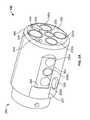
- FIGS. 6A, 6B, 6D and 6Eare perspective views of a white balance enclosure 600 of the present specification, while FIG. 6C is a cross-sectional view of a white balance enclosure 600 showing a multiple viewing elements endoscopic tip 620 positioned therein.
- enclosure 600is internally designed as a cap (to conveniently slip/slide onto, be positioned over, and enclose the multiple viewing elements endoscopic tip 620 ) comprising first body portion or front portion 605 and second body portion or housing 610 that, in one embodiment, are substantially cylindrical.
- Portions 605 , 610are of similar, identical, or different shapes, such as and not limited to rectangular, square, or any other shape.
- the white balance enclosuremay form a single, integrated body unit.
- the second body portion 610in one embodiment, is a housing defining an enclosed volume that has an opening 606 , defined by at least partially coaxial first body portion 605 , for receiving the endoscopic tip.
- the opening 606has a circumference or diameter that is configured to snugly receive the endoscopic tip such that external light is prevented from entering through the opening 606 .
- the enclosed volume of the housinghas a surface area that is located at a pre-defined distance from at least one second viewing element. Further, the enclosed volume of the housing includes a member extending from the surface area (and positioned thereupon) to position the first viewing element at a predefined distance from the surface area.
- first portion 605is positioned at, and housed at least partially coaxially within, a front area of second portion 610 .
- First portion 605defines an opening 606 , having a first diameter ‘d’, which leads into an inner area of second portion 610 , having a second diameter ‘D’.
- the inner areais substantially cylindrical or spherical for receiving the endoscope tip.
- the distal end of second portion 610is closed with a distal wall 611 .
- the first diameter ‘d’is configured to enable a multi-viewing elements endoscope tip 620 , such as a two viewing elements (one front and one side-looking viewing element) or a three viewing elements (one front and two side-looking viewing elements) endoscopic tip, to be conveniently inserted through and fit snugly into opening 606 and into a front area of the second portion 610 .
- the diameter ‘D’is configured to ensure that once the endoscopic tip 620 lies within second portion 610 , the distance of endoscopic tip 620 (and therefore the multiple viewing elements) from the inner surfaces of second portion 610 , including the distal wall 611 , is equal to ‘t’.
- the distance ‘t’ranges from 10 to 12 millimeters. In one embodiment, the distance ‘t’ is larger than 12 millimeters.
- At least a portion of the surface area of the enclosed volume that is within the field of view of the second viewing elementis at least 10 millimeters.
- first and second portions 605 and 610ensures that portions of the outer surface of tip 620 lies at distance ‘t’ from the inner walls of second portion 610 .
- the side-looking viewing elements located on the outer cylindrical side surface of endoscopic tip 620 and the front-looking viewing element located at the leading surface or distal face 615 of endoscopic tip 620are maintained at a substantially uniform distance ‘t’ from the inner walls of second portion 610 .
- An optional indicator marking 612further facilitates/aids a leading surface or distal face 615 of endoscopic tip 620 , and therefore the front-looking viewing elements situated thereon, to be maintained at a substantially uniform distance ‘t’ from distal wall 611 .
- the indicator marking 612is a thin line engraved or embossed into the inner walls of second portion 610 .
- the indicator 612is marked at a position such that when the side-looking viewing elements of tip 620 captures a view of the indicator, it should be understood by the user of the endoscope that leading surface 615 is positioned at an appropriate distance ‘t’ from distal wall 611 .
- the inner walls of second portion 610including distal wall 611 , has posts, protrusions, or stopper component(s) 613 that are positioned to meet endoscopic tip 620 at its edges.
- positioning of tip 620 at a proper distance ‘t’ from the inner wallsis facilitated by the use of physical structures, however, at the same time the viewing elements and illuminators and corresponding fields of view remain unhindered (the fields of view are not blocked).
- the inner surface are of the enclosure of the present specificationis of a uniform color. In other embodiments, the inner surface area of the enclosure facing the fields of view of the first and second viewing elements is the same color. In other embodiments, the inner surface area of the enclosure facing the field of view of the first viewing element is of a first color and the inner surface area of the enclosure facing the field of view of the second viewing element is of a second color.
- the inner walls of second portion 610 and distal wall 611together provide a uniform reference white surrounding/background to the multiple viewing elements of endoscopic tip 620 . Also, since each viewing element of tip 620 is situated at a substantially uniform distance ‘t’ from the white background, this facilitates simultaneous, consistent, and uniform white balancing of all of the multiple viewing elements of tip 620 . Additionally, once tip 620 is positioned within enclosure 600 , the interior of enclosure 600 can be isolated from the influx of exterior light, so as to avoid creating uneven shadows and illumination in the interior of enclosure 600 .
- enclosure 600is made from a thermoplastic elastomer (TPE) and/or a thermoset elastomer to ensure an optimally light yet robust structure.
- enclosure 600is an enclosure that can be slipped/slid onto endoscopic tip 620 .
- enclosure 600is designed in the form of: a clasp that securely encloses and attaches to endoscopic tip 620 ; a snap which snug-fits onto endoscopic tip 620 ; or any other such suitable insertion structure that would be advantageously evident to those of ordinary skill in the art.
- enclosure 600comprises cylindrical first and second portions 605 , 610
- the shape of the first and second portions 605 , 610is rectangular, square or any other suitable shape that facilitates positioning of endoscopic tip 620 (and therefore the front and side-looking viewing elements thereon) to be equidistant from the inner walls of enclosure 600 .
- first and second portions 605 and 610can be of different shapes—for example, first portion 605 can be cylindrical while second portion 610 is rectangular, square, or vice versa.
- An outer surface 630 of second portion 610comprises a connector that, in one embodiment, is a hanger 625 as shown in FIGS. 6A and 6B .
- the connectoris a coupler 626 as shown in FIGS. 6D and 6E .
- enclosure 600is removably attached to a side of the main control unit (such as main control unit 416 shown in FIG. 4 ) by mating hanger 625 with a corresponding plug located on the side of the main control unit, in accordance with an embodiment.
- hanger 625connects with the corresponding plug by structurally engaging with the plug or magnetically coupling with the plug.
- enclosure 600is integrated to a side of the main control unit (such as the main control unit 416 shown in FIG. 4 ) by coupler 626 .
- the white balance enclosurewhen the white balance enclosure is connected to the main control unit via coupler 626 , it is fixedly attached. The endoscope is inserted into the white balance enclosure when attached and the calibration ensues.
- outer surface 630can have any shape, size and dimensions as would be ergonomically advantageous.
- the white balance enclosuremay reside within the main control unit such that it is an integral part of the unit.
- FIG. 7is a flow diagram showing exemplary steps of one embodiment of using the white balance enclosure, internally designed as a cap in accordance with an embodiment (and hereinafter referred to as the ‘cap’), to simultaneously, uniformly, and consistently calibrate/white balance multiple viewing elements of an endoscopic tip.
- a physician or other operator of the endoscopic deviceinserts the multi-viewing elements endoscopic tip (such as an endoscopic tip comprising three viewing elements—one front-looking and two side-looking) through an opening of the cap.
- the physicianensures that the endoscopic tip is positioned within the cap such that the endoscopic tip is substantially equidistant from inner walls as well as a distal wall of the cap.
- the multiple viewing elementsare activated using appropriate input commands to expose the multiple viewing elements simultaneously, uniformly, and consistently to a reference white surrounding within the cap.
- the image/video processing systemgenerates white fields (test feeds) corresponding to each of the multiple viewing elements.
- the white balance switchlocated on a front panel of a main control unit, is pressed/activated by the operator (input received from the system-on-module SOM) or activated by a predetermined command signal to perform a white balance.
- DSPsdigital signal processors
- W R , W G , W B or W Ye , W Cy , W Mg , W Gcorresponding to the three primary colors (Red, Green and Blue) or four additional colors (Yellow, Cyan, Magenta, and Green) for the test feeds of each multiple viewing element.
- the operatorremoves the endoscopic tip from the white balance enclosure.
- the white balance enclosureis placed along the main control unit.
- the cap/enclosureis coupled to a side of the main control unit using a coupling mechanism, such as a hanger.
- the hangermay be attached to the cap or to the main control unit.
- each of the words “comprise” “include” and “have”, and forms thereof,are not necessarily limited to members in a list with which the words may be associated.
Landscapes
- Health & Medical Sciences (AREA)
- Life Sciences & Earth Sciences (AREA)
- Surgery (AREA)
- Engineering & Computer Science (AREA)
- Biomedical Technology (AREA)
- Molecular Biology (AREA)
- Pathology (AREA)
- Radiology & Medical Imaging (AREA)
- Nuclear Medicine, Radiotherapy & Molecular Imaging (AREA)
- Biophysics (AREA)
- Physics & Mathematics (AREA)
- Heart & Thoracic Surgery (AREA)
- Medical Informatics (AREA)
- Optics & Photonics (AREA)
- Animal Behavior & Ethology (AREA)
- General Health & Medical Sciences (AREA)
- Public Health (AREA)
- Veterinary Medicine (AREA)
- Multimedia (AREA)
- Signal Processing (AREA)
- Endoscopes (AREA)
- Instruments For Viewing The Inside Of Hollow Bodies (AREA)
Abstract
Description
Claims (9)
Priority Applications (2)
| Application Number | Priority Date | Filing Date | Title |
|---|---|---|---|
| US14/271,234US9667935B2 (en) | 2013-05-07 | 2014-05-06 | White balance enclosure for use with a multi-viewing elements endoscope |
| US15/493,007US10205925B2 (en) | 2013-05-07 | 2017-04-20 | White balance enclosure for use with a multi-viewing elements endoscope |
Applications Claiming Priority (2)
| Application Number | Priority Date | Filing Date | Title |
|---|---|---|---|
| US201361820650P | 2013-05-07 | 2013-05-07 | |
| US14/271,234US9667935B2 (en) | 2013-05-07 | 2014-05-06 | White balance enclosure for use with a multi-viewing elements endoscope |
Related Child Applications (1)
| Application Number | Title | Priority Date | Filing Date |
|---|---|---|---|
| US15/493,007ContinuationUS10205925B2 (en) | 2013-05-07 | 2017-04-20 | White balance enclosure for use with a multi-viewing elements endoscope |
Publications (2)
| Publication Number | Publication Date |
|---|---|
| US20140333742A1 US20140333742A1 (en) | 2014-11-13 |
| US9667935B2true US9667935B2 (en) | 2017-05-30 |
Family
ID=51864500
Family Applications (2)
| Application Number | Title | Priority Date | Filing Date |
|---|---|---|---|
| US14/271,234Expired - Fee RelatedUS9667935B2 (en) | 2013-05-07 | 2014-05-06 | White balance enclosure for use with a multi-viewing elements endoscope |
| US15/493,007ActiveUS10205925B2 (en) | 2013-05-07 | 2017-04-20 | White balance enclosure for use with a multi-viewing elements endoscope |
Family Applications After (1)
| Application Number | Title | Priority Date | Filing Date |
|---|---|---|---|
| US15/493,007ActiveUS10205925B2 (en) | 2013-05-07 | 2017-04-20 | White balance enclosure for use with a multi-viewing elements endoscope |
Country Status (5)
| Country | Link |
|---|---|
| US (2) | US9667935B2 (en) |
| EP (1) | EP2994034B1 (en) |
| JP (1) | JP6669647B2 (en) |
| CN (1) | CN105358043B (en) |
| WO (1) | WO2014182723A1 (en) |
Cited By (2)
| Publication number | Priority date | Publication date | Assignee | Title |
|---|---|---|---|---|
| US20210298580A1 (en)* | 2020-03-26 | 2021-09-30 | Fujikura Ltd. | Endoscope |
| US11672414B2 (en) | 2019-12-10 | 2023-06-13 | Arthrex, Inc. | Method and device for color correction of two or more self-illuminated camera systems |
Families Citing this family (34)
| Publication number | Priority date | Publication date | Assignee | Title |
|---|---|---|---|---|
| US9474440B2 (en) | 2009-06-18 | 2016-10-25 | Endochoice, Inc. | Endoscope tip position visual indicator and heat management system |
| US10524645B2 (en) | 2009-06-18 | 2020-01-07 | Endochoice, Inc. | Method and system for eliminating image motion blur in a multiple viewing elements endoscope |
| US10130246B2 (en) | 2009-06-18 | 2018-11-20 | Endochoice, Inc. | Systems and methods for regulating temperature and illumination intensity at the distal tip of an endoscope |
| US10663714B2 (en) | 2010-10-28 | 2020-05-26 | Endochoice, Inc. | Optical system for an endoscope |
| US9706908B2 (en) | 2010-10-28 | 2017-07-18 | Endochoice, Inc. | Image capture and video processing systems and methods for multiple viewing element endoscopes |
| US10517464B2 (en) | 2011-02-07 | 2019-12-31 | Endochoice, Inc. | Multi-element cover for a multi-camera endoscope |
| US9636003B2 (en) | 2013-06-28 | 2017-05-02 | Endochoice, Inc. | Multi-jet distributor for an endoscope |
| US12207796B2 (en) | 2013-03-28 | 2025-01-28 | Endochoice Inc. | Multi-jet controller for an endoscope |
| US10595714B2 (en) | 2013-03-28 | 2020-03-24 | Endochoice, Inc. | Multi-jet controller for an endoscope |
| WO2014182723A1 (en) | 2013-05-07 | 2014-11-13 | Endochoice, Inc. | White balance enclosed for use with a multi-viewing elements endoscope |
| US9949623B2 (en) | 2013-05-17 | 2018-04-24 | Endochoice, Inc. | Endoscope control unit with braking system |
| US10064541B2 (en) | 2013-08-12 | 2018-09-04 | Endochoice, Inc. | Endoscope connector cover detection and warning system |
| USD758577S1 (en)* | 2013-09-03 | 2016-06-07 | Endochoice, Inc. | White balance enclosure cap for a medical device |
| US9943218B2 (en) | 2013-10-01 | 2018-04-17 | Endochoice, Inc. | Endoscope having a supply cable attached thereto |
| US9968242B2 (en) | 2013-12-18 | 2018-05-15 | Endochoice, Inc. | Suction control unit for an endoscope having two working channels |
| WO2015112747A2 (en) | 2014-01-22 | 2015-07-30 | Endochoice, Inc. | Image capture and video processing systems and methods for multiple viewing element endoscopes |
| US11234581B2 (en) | 2014-05-02 | 2022-02-01 | Endochoice, Inc. | Elevator for directing medical tool |
| EP3689219B1 (en) | 2014-07-21 | 2023-08-30 | EndoChoice, Inc. | Multi-focal, multi-camera endoscope systems |
| US10542877B2 (en) | 2014-08-29 | 2020-01-28 | Endochoice, Inc. | Systems and methods for varying stiffness of an endoscopic insertion tube |
| EP3235241B1 (en) | 2014-12-18 | 2023-09-06 | EndoChoice, Inc. | System for processing video images generated by a multiple viewing elements endoscope |
| WO2016112034A2 (en) | 2015-01-05 | 2016-07-14 | Endochoice, Inc. | Tubed manifold of a multiple viewing elements endoscope |
| US10376181B2 (en) | 2015-02-17 | 2019-08-13 | Endochoice, Inc. | System for detecting the location of an endoscopic device during a medical procedure |
| US10078207B2 (en) | 2015-03-18 | 2018-09-18 | Endochoice, Inc. | Systems and methods for image magnification using relative movement between an image sensor and a lens assembly |
| US10401611B2 (en) | 2015-04-27 | 2019-09-03 | Endochoice, Inc. | Endoscope with integrated measurement of distance to objects of interest |
| US10516865B2 (en) | 2015-05-17 | 2019-12-24 | Endochoice, Inc. | Endoscopic image enhancement using contrast limited adaptive histogram equalization (CLAHE) implemented in a processor |
| US20170119474A1 (en) | 2015-10-28 | 2017-05-04 | Endochoice, Inc. | Device and Method for Tracking the Position of an Endoscope within a Patient's Body |
| EP4579310A3 (en) | 2015-11-24 | 2025-09-10 | Endochoice, Inc. | Disposable air/water and suction valves for an endoscope |
| JP2019507628A (en) | 2016-02-24 | 2019-03-22 | エンドチョイス インコーポレイテッドEndochoice, Inc. | Circuit board assembly for multiple view element endoscopes using CMOS sensors |
| US10292570B2 (en) | 2016-03-14 | 2019-05-21 | Endochoice, Inc. | System and method for guiding and tracking a region of interest using an endoscope |
| EP3429478B1 (en) | 2016-06-21 | 2021-04-21 | Endochoice, Inc. | Endoscope system with multiple connection interfaces to interface with different video data signal sources |
| KR102624929B1 (en)* | 2017-05-11 | 2024-01-12 | 타카시 마토 | catheter device |
| WO2018221404A1 (en)* | 2017-06-01 | 2018-12-06 | アルプス電気株式会社 | Catheter device |
| CN118177700A (en)* | 2018-01-05 | 2024-06-14 | 波士顿科学国际有限公司 | Fluorophore imaging device, system and method for endoscopic surgery |
| WO2022013853A1 (en)* | 2020-07-12 | 2022-01-20 | 270 Surgical Ltd. | White balance apparatus |
Citations (348)
| Publication number | Priority date | Publication date | Assignee | Title |
|---|---|---|---|---|
| US4027697A (en) | 1975-11-19 | 1977-06-07 | Bonney Roland W | Rotary valve |
| US4084401A (en) | 1975-07-09 | 1978-04-18 | Hughes Aircraft Company | Digital watch with two buttons and improved setting and display control |
| US4402313A (en) | 1979-11-22 | 1983-09-06 | Olympus Optical Co., Ltd. | Endoscope light source device |
| US4461282A (en) | 1978-05-02 | 1984-07-24 | Kabushiki Kaisha Medos Kenkyusho | Mechanism for direction changing of endoscope top end |
| US4494549A (en) | 1981-05-21 | 1985-01-22 | Olympus Optical Co., Ltd. | Device for diagnosing body cavity interiors with supersonic waves |
| US4532918A (en) | 1983-10-07 | 1985-08-06 | Welch Allyn Inc. | Endoscope signal level control |
| US4588294A (en) | 1984-06-27 | 1986-05-13 | Warner-Lambert Technologies, Inc. | Searching and measuring endoscope |
| US4641635A (en) | 1984-08-15 | 1987-02-10 | Olympus Optical Co., Ltd. | Endoscope apparatus |
| US4727859A (en) | 1986-12-29 | 1988-03-01 | Welch Allyn, Inc. | Right angle detachable prism assembly for borescope |
| US4764001A (en) | 1984-06-13 | 1988-08-16 | Olympus Optical Co., Ltd. | Retrofocus-type objective for an endoscope |
| US4801792A (en) | 1986-03-22 | 1989-01-31 | Olympus Optical Co., Ltd. | Endoscope |
| US4825850A (en) | 1988-05-13 | 1989-05-02 | Opielab, Inc. | Contamination protection system for endoscope control handles |
| US4877314A (en) | 1987-05-25 | 1989-10-31 | Olympus Optical Co., Ltd. | Objective lens system for endoscopes |
| US4902115A (en) | 1986-09-22 | 1990-02-20 | Olympus Optical Co., Ltd. | Optical system for endoscopes |
| US4976522A (en) | 1988-05-02 | 1990-12-11 | Olympus Optical Co., Ltd. | Objective lens system for endoscopes |
| US4984878A (en) | 1988-09-29 | 1991-01-15 | Fuji Photo Optical Co., Ltd. | Ojective lens for endoscope |
| US5007406A (en) | 1988-11-04 | 1991-04-16 | Asahi Kogaku Kogyo Kabushiki Kaisha | Bending control device of endoscope |
| US5014685A (en) | 1988-07-13 | 1991-05-14 | Asahi Kogaku Kogyo Kabushiki Kaisha | Brake for bending control device of endoscope |
| US5193525A (en) | 1990-11-30 | 1993-03-16 | Vision Sciences | Antiglare tip in a sheath for an endoscope |
| US5224929A (en) | 1990-12-21 | 1993-07-06 | C. R. Bard, Inc. | Irrigation/aspiration cannula and valve assembly |
| US5296971A (en) | 1991-03-04 | 1994-03-22 | Olympus Optical Co., Ltd. | Objective lens system for endoscopes |
| US5359456A (en) | 1991-10-15 | 1994-10-25 | Olympus Optical Co., Ltd. | Objective lens system for endoscopes |
| US5395329A (en) | 1994-01-19 | 1995-03-07 | Daig Corporation | Control handle for steerable catheter |
| US5447148A (en) | 1993-07-08 | 1995-09-05 | Vision Sciences, Inc. | Endoscopic contamination protection system to facilitate cleaning of endoscopes |
| US5460167A (en) | 1993-03-04 | 1995-10-24 | Olympus Optical Co., Ltd. | Endoscope cover with channel |
| US5464007A (en) | 1994-02-23 | 1995-11-07 | Welch Allyn, Inc. | Fluid insensitive braking for an endoscope |
| US5489256A (en) | 1992-09-01 | 1996-02-06 | Adair; Edwin L. | Sterilizable endoscope with separable disposable tube assembly |
| US5518502A (en) | 1994-06-08 | 1996-05-21 | The United States Surgical Corporation | Compositions, methods and apparatus for inhibiting fogging of endoscope lenses |
| US5547457A (en) | 1993-01-22 | 1996-08-20 | Olympus Optical Co., Ltd. | Objective optical system for endoscopes |
| US5547455A (en) | 1994-03-30 | 1996-08-20 | Medical Media Systems | Electronically steerable endoscope |
| US5575755A (en) | 1994-02-23 | 1996-11-19 | Welch Allyn, Inc. | Fluid insensitive braking for an endoscope |
| US5587839A (en) | 1994-10-18 | 1996-12-24 | Fuji Photo Optical Co., Ltd. | Objective lens system for endoscope |
| US5630782A (en) | 1992-09-01 | 1997-05-20 | Adair; Edwin L. | Sterilizable endoscope with separable auxiliary assembly |
| US5662588A (en) | 1994-05-31 | 1997-09-02 | Olympus Optical Co., Ltd. | Endoscope apparatus |
| US5674182A (en) | 1993-02-26 | 1997-10-07 | Olympus Optical Co., Ltd. | Endoscope system including endoscope and protection cover |
| US5685823A (en) | 1994-03-30 | 1997-11-11 | Asahi Kogaku Kogyo Kabushiki Kaisha | End structure of endoscope |
| US5702347A (en) | 1993-01-27 | 1997-12-30 | Olympus Optical Co., Ltd. | Endoscope system including endoscope and disposable protection cover |
| US5707344A (en) | 1993-05-07 | 1998-01-13 | Olympus Optical Co., Ltd. | Endoscope |
| JPH1043129A (en) | 1996-08-02 | 1998-02-17 | Olympus Optical Co Ltd | Endoscope |
| US5725478A (en) | 1996-03-14 | 1998-03-10 | Saad; Saad A. | Methods and apparatus for providing suction and/or irrigation in a rigid endoscope while maintaining visual contact with a target area through the endoscope |
| US5725474A (en) | 1993-11-26 | 1998-03-10 | Asahi Kogaku Kogyo Kabushiki Kaisha | Front end structure of endoscope |
| US5725477A (en) | 1993-11-18 | 1998-03-10 | Asahi Kogaku Kogyo Kabushiki Kaisha | Front end structure of endoscope |
| US5725476A (en) | 1993-11-18 | 1998-03-10 | Asahi Kogaku Kogyo Kabushiki Kaisha | Front end structure of endoscope |
| US5777797A (en) | 1995-09-11 | 1998-07-07 | Fuji Photo Optical Co., Ltd. | Objective lens system for endoscopes having an image transfer optical fiber bundle |
| US5782751A (en) | 1994-05-26 | 1998-07-21 | Asahi Kogaku Kogyo Kabushiki Kaisha | Side view endoscope |
| JPH10239740A (en) | 1997-02-28 | 1998-09-11 | Toshiba Corp | Endoscope device |
| US5810715A (en) | 1995-09-29 | 1998-09-22 | Olympus Optical Co., Ltd. | Endoscope provided with function of being locked to flexibility of insertion part which is set by flexibility modifying operation member |
| US5836894A (en) | 1992-12-21 | 1998-11-17 | Artann Laboratories | Apparatus for measuring mechanical parameters of the prostate and for imaging the prostate using such parameters |
| US5860913A (en) | 1996-05-16 | 1999-01-19 | Olympus Optical Co., Ltd. | Endoscope whose distal cover can be freely detachably attached to main distal part thereof with high positioning precision |
| US5870234A (en) | 1996-09-06 | 1999-02-09 | Jos. Schneider Optische Werke Kreuznach Gmbh & Co. Kg | Compact wide-angle lens |
| CA2297986A1 (en) | 1997-08-28 | 1999-03-04 | John E. Rosenstengel | Method and system for automated detection of clustered microcalcifications from digital mammograms |
| JPH11137512A (en) | 1997-11-07 | 1999-05-25 | Toshiba Corp | Endoscope device |
| US5916148A (en) | 1995-06-29 | 1999-06-29 | Olympus Optical Co., Ltd. | Objective optical system for endoscopes |
| US5940126A (en) | 1994-10-25 | 1999-08-17 | Kabushiki Kaisha Toshiba | Multiple image video camera apparatus |
| US6095970A (en) | 1997-02-19 | 2000-08-01 | Asahi Kogaku Kogyo Kabushiki Kaisha | Endoscope |
| US6117068A (en) | 1995-10-19 | 2000-09-12 | Elite Genetics, Inc | Artificial insemination system |
| US6181481B1 (en) | 1998-11-30 | 2001-01-30 | Fuji Photo Optical Co., Ltd. | Objective lens for endoscope |
| US6196967B1 (en) | 1998-03-18 | 2001-03-06 | Linvatec Corporation | Arthroscopic component joining system |
| US6277064B1 (en)* | 1997-12-30 | 2001-08-21 | Inbae Yoon | Surgical instrument with rotatably mounted offset endoscope |
| US20010036322A1 (en) | 2000-03-10 | 2001-11-01 | Bloomfield John F. | Image processing system using an array processor |
| US20020017515A1 (en) | 2000-08-11 | 2002-02-14 | Asahi Kogaku Kogyo Kabushiki Kaisha | Method of manufacturing treatment instrument of endoscope |
| US6359674B1 (en) | 1999-06-08 | 2002-03-19 | Olympus Optical Co., Ltd. | Liquid crystal lens, liquid crystal lens unit, and liquid crystal lens assembly |
| US6375610B2 (en) | 1997-01-23 | 2002-04-23 | Fairmont Medical Products Pty. Ltd. | Endoscopic drape |
| US20020047897A1 (en) | 1999-12-17 | 2002-04-25 | Asahi Kogaku Kogyo Kabushiki Kaisha | Electronic endoscope selector |
| US6402738B1 (en) | 1999-02-04 | 2002-06-11 | Asahi Kogaku Kogyo Kabushiki Kaisha | Wire connection structure for endoscope |
| US20020087047A1 (en) | 1999-09-13 | 2002-07-04 | Visionscope, Inc. | Miniature endoscope system |
| US6419626B1 (en) | 1998-08-12 | 2002-07-16 | Inbae Yoon | Surgical instrument endoscope with CMOS image sensor and physical parameter sensor |
| US20020109771A1 (en) | 2001-02-14 | 2002-08-15 | Michael Ledbetter | Method and system for videoconferencing |
| US20020109774A1 (en) | 2001-01-16 | 2002-08-15 | Gavriel Meron | System and method for wide field imaging of body lumens |
| US20020161281A1 (en) | 2000-04-03 | 2002-10-31 | Ross Jaffe | Endoscope having a guide tube |
| US6476851B1 (en) | 1996-12-27 | 2002-11-05 | Olympus Optical Co., Ltd. | Electronic endoscope |
| US20020172498A1 (en) | 2001-05-18 | 2002-11-21 | Pentax Precision Instrument Corp. | Computer-based video recording and management system for medical diagnostic equipment |
| US20020183591A1 (en) | 2001-02-06 | 2002-12-05 | Nobuyuki Matsuura | Endoscopic system and method for positioning an indwelling tube |
| US20030030918A1 (en) | 2001-05-14 | 2003-02-13 | Asahi Kogaku Kogyo Kabushiki Kaisha | Endoscope objective optical system |
| US20030063398A1 (en) | 2001-09-28 | 2003-04-03 | Fuji Photo Optical Co., Ltd. | Electronic endoscope eliminating influence of light distribution in optical zooming |
| US20030076411A1 (en) | 2001-10-01 | 2003-04-24 | Pentax Corporation | Electronic endoscope with light-amount adjustment apparatus |
| US20030083552A1 (en) | 2001-10-19 | 2003-05-01 | Visionscope, Inc. | Miniature endoscope with imaging fiber system |
| US20030128893A1 (en) | 2001-11-19 | 2003-07-10 | Alfio Castorina | Method for merging digital images to obtain a high dynamic range digital image |
| US20030153897A1 (en) | 2002-02-12 | 2003-08-14 | Russo Ronald D. | Closed system drainage and infusion connector valve |
| US20030158503A1 (en) | 2002-01-18 | 2003-08-21 | Shinya Matsumoto | Capsule endoscope and observation system that uses it |
| US6636254B1 (en) | 1993-11-29 | 2003-10-21 | Olympus Optical Co., Ltd, | Image processing apparatus for performing turn or mirror inversion on an input video signal and outputting different images simultaneously |
| US6638214B2 (en) | 2000-08-02 | 2003-10-28 | Fuji Photo Optical Co., Ltd. | Observation window washing device of endoscope |
| US6673012B2 (en) | 2000-04-19 | 2004-01-06 | Pentax Corporation | Control device for an endoscope |
| US20040015054A1 (en) | 2002-07-05 | 2004-01-22 | Kazuhiko Hino | Bending control mechanism for endoscope |
| US6690337B1 (en) | 1999-06-09 | 2004-02-10 | Panoram Technologies, Inc. | Multi-panel video display |
| US20040046865A1 (en)* | 2001-05-16 | 2004-03-11 | Olympus Optical Co., Ltd. | Endoscope device, endoscope and image processing device for endoscope |
| US6712760B2 (en) | 2000-04-10 | 2004-03-30 | Pentax Corporation | Television device of portable endoscope |
| US20040061780A1 (en) | 2002-09-13 | 2004-04-01 | Huffman David A. | Solid-state video surveillance system |
| US20040106850A1 (en) | 2002-11-27 | 2004-06-03 | Olympus Corporation | Endoscope apparatus |
| US20040133072A1 (en) | 2002-09-13 | 2004-07-08 | Kennedy Bruce L. | Video recording and image capture device |
| US20040138532A1 (en) | 2001-05-20 | 2004-07-15 | Arkady Glukhovsky | Method for in vivo imaging of an unmodified gastrointestinal tract |
| US20040158129A1 (en) | 2003-02-10 | 2004-08-12 | Pentax Corporation | Endoscope |
| US20040160682A1 (en) | 2003-02-14 | 2004-08-19 | Hitoshi Miyano | Endoscope objective lens |
| US20040190159A1 (en) | 2003-03-28 | 2004-09-30 | Naoki Hasegawa | Endoscope image pickup unit for picking up magnified images of an object, a focus adjustment apparatus and method, and a focus range check apparatus and method for the same |
| US20040249247A1 (en) | 2003-05-01 | 2004-12-09 | Iddan Gavriel J. | Endoscope with panoramic view |
| US6832984B2 (en) | 1997-08-21 | 2004-12-21 | Paul Stelzer | Minimally invasive surgery device |
| US20050018042A1 (en) | 2003-01-07 | 2005-01-27 | Jean Rovegno | Video processor for endoscopy |
| US20050020876A1 (en) | 2000-04-20 | 2005-01-27 | Olympus Corporation | Operation microscope |
| US20050038317A1 (en) | 2004-10-11 | 2005-02-17 | Nitesh Ratnakar | Dual View Endoscope |
| US20050047134A1 (en) | 1997-08-26 | 2005-03-03 | Color Kinetics | Controlled lighting methods and apparatus |
| US20050090709A1 (en) | 2003-09-23 | 2005-04-28 | Olympus Corporation | Endoscope suitable to body cavity |
| US6888119B2 (en) | 2001-03-12 | 2005-05-03 | Olympus Corporation | Light scanning probe apparatus using light of low coherence |
| US20050119527A1 (en) | 2003-04-01 | 2005-06-02 | Scimed Life Systems, Inc. | Force feedback control system for video endoscope |
| US20050124858A1 (en) | 2003-09-01 | 2005-06-09 | Hirohiko Matsuzawa | Capsule type endoscope |
| JP2005253543A (en) | 2004-03-10 | 2005-09-22 | Pentax Corp | Endoscope waterway |
| US20050222499A1 (en) | 2003-04-01 | 2005-10-06 | Banik Michael S | Interface for video endoscope system |
| US20050234347A1 (en) | 2004-03-29 | 2005-10-20 | Fujinon Corporation | Puncture-type endoscopic probe |
| US20050234296A1 (en) | 2004-04-14 | 2005-10-20 | Usgi Medical Inc. | Method and apparatus for obtaining endoluminal access |
| US20050251127A1 (en) | 2003-10-15 | 2005-11-10 | Jared Brosch | Miniature ultrasonic transducer with focusing lens for intracardiac and intracavity applications |
| US20050272975A1 (en) | 2004-03-23 | 2005-12-08 | Mcweeney John O | In-vivo visualization system |
| US20050277808A1 (en) | 2004-05-14 | 2005-12-15 | Elazar Sonnenschein | Methods and devices related to camera connectors |
| US20050283048A1 (en) | 2001-10-19 | 2005-12-22 | Visionscope, Llc | Portable imaging system employing a miniature endoscope |
| JP2006025888A (en) | 2004-07-12 | 2006-02-02 | Forte Grow Medical Kk | Endoscopy tube |
| US20060047184A1 (en) | 2004-08-26 | 2006-03-02 | Scimed Life Systems, Inc. | Endoscope having auto-insufflation and exsufflation |
| JP2006068109A (en) | 2004-08-31 | 2006-03-16 | Nagoya Institute Of Technology | Spherical capsule type omnidirectional endoscope |
| US20060063976A1 (en) | 2004-09-03 | 2006-03-23 | Sightline Technologies Ltd. | Optical head for endoscope |
| US20060069314A1 (en) | 2004-09-24 | 2006-03-30 | Mina Farr | Solid state illumination for endoscopy |
| US20060114986A1 (en) | 2004-09-30 | 2006-06-01 | Knapp Keith N Ii | Adapter for use with digital imaging medical device |
| US20060149129A1 (en) | 2005-01-05 | 2006-07-06 | Watts H D | Catheter with multiple visual elements |
| WO2006073676A1 (en) | 2004-12-30 | 2006-07-13 | Avantis Medical Systems, Inc. | Disposable multi-lumen catheter/sheath with reusable stylet/endoscope |
| US20060171693A1 (en) | 2005-01-28 | 2006-08-03 | Stryker Corporation | Endoscope with integrated light source |
| US20060173245A1 (en) | 2005-01-28 | 2006-08-03 | Stryker Corporation | Disposable attachable light source unit for an endoscope |
| EP1690497A1 (en) | 2003-12-02 | 2006-08-16 | Olympus Corporation | Ultrasonographic device |
| US20060184037A1 (en) | 2004-11-30 | 2006-08-17 | Can Ince | Pulsed lighting imaging systems and methods |
| US20060183975A1 (en) | 2004-04-14 | 2006-08-17 | Usgi Medical, Inc. | Methods and apparatus for performing endoluminal procedures |
| US20060189845A1 (en) | 2004-04-14 | 2006-08-24 | Usgi Medical Inc. | Methods and apparaus for off-axis visualization |
| US20060215406A1 (en) | 2005-02-18 | 2006-09-28 | William Thrailkill | Medical diagnostic instrument with highly efficient, tunable light emitting diode light source |
| US20060235306A1 (en) | 2005-04-15 | 2006-10-19 | Integra Lifesciences (Ireland) | Ultrasonic horn for removal of hard tissue |
| US20060252994A1 (en) | 2005-05-06 | 2006-11-09 | Nitesh Ratnakar | Next Generation Colonoscope |
| US20060264704A1 (en) | 2004-01-19 | 2006-11-23 | Olympus Corporation | Capsule-type medical apparatus |
| US20060293556A1 (en) | 2005-05-16 | 2006-12-28 | Garner David M | Endoscope with remote control module or camera |
| US20070015989A1 (en) | 2005-07-01 | 2007-01-18 | Avantis Medical Systems, Inc. | Endoscope Image Recognition System and Method |
| US20070049803A1 (en) | 2004-04-27 | 2007-03-01 | Hiroki Moriyama | Endoscope and endoscope system |
| US20070055100A1 (en) | 2004-05-14 | 2007-03-08 | Takayuki Kato | Endoscope and endoscope apparatus |
| US20070079029A1 (en) | 2005-09-30 | 2007-04-05 | Symbol Technologies, Inc. | Processing image data from multiple sources |
| US20070088193A1 (en) | 2004-05-24 | 2007-04-19 | Koji Omori | Light source device for endoscope |
| US20070106119A1 (en) | 2005-06-29 | 2007-05-10 | Yasuo Hirata | Endoscope |
| US20070142711A1 (en) | 2005-12-13 | 2007-06-21 | Lex Bayer | Detachable Imaging Device, Endoscope Having A Detachable Imaging Device, And Method of Configuring Such An Endoscope |
| US20070162095A1 (en) | 2006-01-06 | 2007-07-12 | Ezc Medical Llc | Modular visualization stylet apparatus and methods of use |
| US20070167681A1 (en) | 2001-10-19 | 2007-07-19 | Gill Thomas J | Portable imaging system employing a miniature endoscope |
| US20070177008A1 (en) | 2005-01-05 | 2007-08-02 | Avantis Medical, Inc. | Endoscope with an imaging catheter assembly and method of configuring an endoscope |
| WO2007087421A2 (en) | 2006-01-23 | 2007-08-02 | Avantis Medical Systems, Inc. | Endoscope |
| US20070177009A1 (en) | 2005-01-05 | 2007-08-02 | Avantis Medical Systems, Inc. | Endoscope assembly with a polarizing filter |
| WO2007092636A2 (en) | 2006-02-09 | 2007-08-16 | Avantis Medical Systems, Inc. | Endoscope assembly with a polarizing filter |
| WO2007092533A2 (en) | 2006-02-06 | 2007-08-16 | Avantis Medical Systems, Inc. | Endoscope with an imaging catheter assembly and method of configuring an endoscope |
| US20070188427A1 (en) | 1997-12-17 | 2007-08-16 | Color Kinetics Incorporated | Organic light emitting diode methods and apparatus |
| US20070197875A1 (en) | 2003-11-14 | 2007-08-23 | Osaka Shoji | Endoscope device and imaging method using the same |
| US20070203396A1 (en) | 2006-02-28 | 2007-08-30 | Mccutcheon John G | Endoscopic Tool |
| US20070206945A1 (en) | 2006-03-01 | 2007-09-06 | Delorme David M | Method and apparatus for panoramic imaging |
| US20070213591A1 (en) | 2005-09-06 | 2007-09-13 | Stryker Gi Ltd. | Disposable Cap for Endoscope |
| US20070229656A1 (en) | 2006-03-27 | 2007-10-04 | Semion Khait | Battery contacts for an in-vivo imaging device |
| US20070241895A1 (en) | 2006-04-13 | 2007-10-18 | Morgan Kelvin L | Noise reduction for flexible sensor material in occupant detection |
| US20070244354A1 (en) | 2006-04-18 | 2007-10-18 | Avantis Medical Systems, Inc. | Vibratory Device, Endoscope Having Such A Device, Method For Configuring An Endoscope, And Method Of Reducing Looping Of An Endoscope. |
| US20070244353A1 (en) | 2006-04-13 | 2007-10-18 | Larsen Dane M | Resectoscopic Device And Method |
| US20070247867A1 (en) | 2006-04-21 | 2007-10-25 | Sunoptic Technologies Llc | Portable LED Light Source for an Endoscope or Boroscope |
| US20070265492A1 (en) | 2004-06-07 | 2007-11-15 | Elazar Sonnenschein | Multipurpose Endoscopy Suite |
| US20070270642A1 (en) | 2006-05-19 | 2007-11-22 | Avantis Medical Systems, Inc. | System and method for producing and improving images |
| US20070293720A1 (en) | 2005-01-05 | 2007-12-20 | Avantis Medical Systems, Inc. | Endoscope assembly and method of viewing an area inside a cavity |
| US20080009673A1 (en) | 2006-05-15 | 2008-01-10 | Khachi Gerald J | Balloon endoscope device |
| US20080021274A1 (en) | 2005-01-05 | 2008-01-24 | Avantis Medical Systems, Inc. | Endoscopic medical device with locking mechanism and method |
| US20080025413A1 (en) | 2006-07-28 | 2008-01-31 | John Apostolopoulos | Selecting bit rates for encoding multiple data streams |
| WO2008015164A1 (en) | 2006-08-01 | 2008-02-07 | Varioptic | Liquid lens with four liquids |
| US20080036864A1 (en) | 2006-08-09 | 2008-02-14 | Mccubbrey David | System and method for capturing and transmitting image data streams |
| US20080045797A1 (en) | 2004-07-02 | 2008-02-21 | Osaka University | Endoscope Attachment And Endoscope |
| US20080058601A1 (en) | 2004-01-19 | 2008-03-06 | Olympus Corporation | Endoscopic imaging apparatus and capsule-type endoscope |
| US20080071290A1 (en) | 2006-06-13 | 2008-03-20 | Intuitive Surgical, Inc. | Minimally invasive surgery instrument assembly with reduced cross section |
| US20080091065A1 (en) | 2006-10-04 | 2008-04-17 | Olympus Medical Systems Corporation | Medical image processing apparatus, endoscope system and medical image processing system |
| US20080130108A1 (en) | 2005-01-05 | 2008-06-05 | Avantis Medical Systems, Inc. | Endoscope assembly with a polarizing filter |
| US20080151070A1 (en) | 2006-12-26 | 2008-06-26 | Canon Kabushiki Kaisha | Image capture and display control apparatus, image capture and display control method, and image capture and display system |
| US20080161646A1 (en) | 2006-01-30 | 2008-07-03 | New Wave Surgical Corp. | Device For White Balancing And Appying An Anti-Fog Agent To Medical Videoscopes Prior To Medical Procedures |
| US20080167529A1 (en) | 2005-03-15 | 2008-07-10 | Takashi Otawara | Endoscope Insertion Portion |
| US20080163652A1 (en) | 2007-01-04 | 2008-07-10 | Leonid Shatskin | Safing lock mechanism |
| US20080177139A1 (en) | 2007-01-19 | 2008-07-24 | Brian Courtney | Medical imaging probe with rotary encoder |
| US20080183043A1 (en) | 2007-01-30 | 2008-07-31 | Fraunhofer-Gesellschaft Zur Forderung Der Angewandten Forschung E.V. | Illumination apparatus for an image sensing means at the distal end of an endoscope |
| US20080183034A1 (en)* | 2007-01-31 | 2008-07-31 | Stryker Gi Ltd. | Endoscope Stand |
| US20080221388A1 (en) | 2007-03-09 | 2008-09-11 | University Of Washington | Side viewing optical fiber endoscope |
| US20080246771A1 (en) | 2007-04-03 | 2008-10-09 | Dell Products L.P. | Graphics processing system and method |
| US7435218B2 (en) | 2001-04-27 | 2008-10-14 | Storz Endoskop Gmbh | Optical instrument, in particular an endoscope, having an interchangeable head |
| US20080253686A1 (en) | 2007-04-10 | 2008-10-16 | Avantis Medical Systems, Inc. | Method and Device for Examining or Imaging an Interior Surface of a Cavity |
| US20080262312A1 (en) | 2007-04-17 | 2008-10-23 | University Of Washington | Shadowing pipe mosaicing algorithms with application to esophageal endoscopy |
| EP1986541A2 (en) | 2006-01-23 | 2008-11-05 | Avantis Medical Systems, Inc. | Endoscope |
| US20080275298A1 (en) | 2004-10-11 | 2008-11-06 | Novation Science, Llc | Dual View Endoscope |
| US20080303898A1 (en) | 2007-06-06 | 2008-12-11 | Olympus Medical Systems Corp. | Endoscopic image processing apparatus |
| US20090005643A1 (en) | 2007-06-27 | 2009-01-01 | Syntheon Llc | Torque-transmitting, variably-flexible, locking insertion device and method for operating the insertion device |
| US20090023998A1 (en) | 2007-07-17 | 2009-01-22 | Nitesh Ratnakar | Rear view endoscope sheath |
| US20090030275A1 (en) | 2005-09-28 | 2009-01-29 | Imperial Innovations Limited | Imaging System |
| WO2009015396A2 (en) | 2007-07-26 | 2009-01-29 | Avantis Medical Systems, Inc. | Endoscope system |
| US20090054790A1 (en) | 2006-02-23 | 2009-02-26 | Jurgen Czaniera | Method and Arrangement for Generating a Signal which Corresponds to the Degree of Opening of the Vocal Folds of the Larynx |
| US20090062615A1 (en) | 2007-08-31 | 2009-03-05 | Koji Yamaya | Endoscope with focal length varying function |
| US20090086017A1 (en) | 2007-09-27 | 2009-04-02 | Hitoshi Miyano | Imaging optical system and endoscope imaging apparatus |
| WO2009049322A2 (en) | 2007-10-11 | 2009-04-16 | Avantis Medical Systems, Inc. | Endoscope assembly comprising retrograde viewing imaging device and instrument channel |
| WO2009049324A1 (en) | 2007-10-11 | 2009-04-16 | Avantis Medical Systems, Inc. | Method and device for reducing the fixed pattern noise of a digital image |
| US20090135245A1 (en) | 2007-11-27 | 2009-05-28 | Jiafu Luo | Camera system with multiple pixel arrays on a chip |
| US20090137875A1 (en) | 2005-09-22 | 2009-05-28 | Hideya Kitagawa | Endoscope insertion portion |
| US20090143647A1 (en) | 2007-12-03 | 2009-06-04 | Olympus Medical Systems Corp. | Medical appliance, endoscope overtube, and endoscope apparatus |
| US20090147076A1 (en) | 2007-12-10 | 2009-06-11 | Hasan Ertas | Wide angle HDTV endoscope |
| US20090182917A1 (en) | 2008-01-15 | 2009-07-16 | Kim Jechan | System having capability for daisy-chained serial distribution of video display data |
| US20090216084A1 (en) | 2008-02-26 | 2009-08-27 | Kenji Yamane | Endoscopic aspiration device |
| US20090234183A1 (en) | 2008-03-11 | 2009-09-17 | Kazunori Abe | Endoscope system and method for inspecting electronic endoscope |
| US20090231419A1 (en) | 2007-02-06 | 2009-09-17 | Avantis Medical Systems, Inc. | Endoscope Assembly and Method of Performing a Medical Procedure |
| US20090253966A1 (en) | 2005-05-11 | 2009-10-08 | Hironobu Ichimura | Distal end portion of endoscope |
| US20090287188A1 (en) | 2008-05-13 | 2009-11-19 | Boston Scientific Scimed, Inc. | Steering system with locking mechanism |
| US20090287192A1 (en) | 2005-04-01 | 2009-11-19 | Vivenzio Robert L | Medical diagnostic instrument having portable illuminator |
| US20090299144A1 (en) | 2006-11-24 | 2009-12-03 | Olympus Medical Systems Corp. | Capsule endoscope |
| US7630148B1 (en) | 2008-06-11 | 2009-12-08 | Ge Inspection Technologies, Lp | System for providing zoom, focus and aperture control in a video inspection device |
| US20100010309A1 (en) | 2008-07-14 | 2010-01-14 | Olympus Corporation | Endoscope bend tube and endoscope including the bend tube |
| US20100016673A1 (en) | 2008-02-12 | 2010-01-21 | Innurvation, Inc. | Ingestible Endoscopic Optical Scanning Device |
| US20100053312A1 (en) | 2006-11-28 | 2010-03-04 | Olympus Corporation | Endoscope apparatus |
| US20100069713A1 (en) | 2008-09-18 | 2010-03-18 | Fujifilm Corporation | Electronic endoscope system |
| US20100076268A1 (en) | 2008-09-19 | 2010-03-25 | Olympus Medical Systems Corp. | Endoscope for oblique viewing |
| US20100073948A1 (en) | 2008-09-24 | 2010-03-25 | Code 3, Inc. | Light bar |
| US20100073470A1 (en) | 2008-09-22 | 2010-03-25 | Fujifilm Corporation | Imaging apparatus and endoscope |
| US7701650B2 (en) | 2007-08-23 | 2010-04-20 | Hon Hai Precision Industry Co., Ltd. | Wide-angle lens module and endoscope |
| US7713246B2 (en) | 2002-10-28 | 2010-05-11 | Covidien Ag | Automatic valve |
| US20100123950A1 (en) | 2008-11-19 | 2010-05-20 | Olympus Corporation | Objective optical system |
| US20100130822A1 (en) | 2007-06-22 | 2010-05-27 | Olympus Medical Systems Corp. | Capsule-type medical device and method of monitoring esophagus inside and stomach inside, method of monitoring stomach inside and method of washing stomach using the same |
| US20100141763A1 (en) | 2008-12-04 | 2010-06-10 | Masaya Itoh | Video monitoring system |
| US20100160729A1 (en) | 2008-12-19 | 2010-06-24 | Smith Paul J | Systems and methods for directing instruments to varying positions at the distal end of a guide tube |
| US7746572B2 (en) | 2007-08-21 | 2010-06-29 | Fujinon Corporation | Imaging lens and image pickup apparatus |
| US20100174144A1 (en) | 2007-06-08 | 2010-07-08 | Thomas Hsu | Devices and methods for removal of debris from the objective lens of an endoscope |
| JP2010178766A (en) | 2009-02-03 | 2010-08-19 | Hoya Corp | Endoscope for treatment |
| US20100231702A1 (en) | 2007-09-28 | 2010-09-16 | Fujifilm Corporation | Image pickup device and endoscope provided with image pickup device |
| US20100249513A1 (en) | 2009-03-31 | 2010-09-30 | Jay Tydlaska | Laryngoscope and system |
| US20100245653A1 (en) | 2008-01-14 | 2010-09-30 | Integrated Medical Systems International, Inc. | Endoscope Objective Lens and Method of Assembly |
| US7813047B2 (en) | 2006-12-15 | 2010-10-12 | Hand Held Products, Inc. | Apparatus and method comprising deformable lens element |
| US20100280322A1 (en) | 2008-06-04 | 2010-11-04 | Fujifilm Corporation | Illumination device for use in endoscope |
| US7828725B2 (en) | 2006-04-26 | 2010-11-09 | Hoya Corporation | Bent state holding mechanism of an endoscope |
| US20100296178A1 (en) | 2006-05-05 | 2010-11-25 | Magalie Genet | Miniaturized optical head with high spatial resolution and high sensitivity, especially for fibred confocal fluorescence imaging |
| CA2765559A1 (en) | 2009-06-18 | 2010-12-23 | Peer Medical Ltd. | Multi-camera endoscope |
| US20100326703A1 (en) | 2009-06-24 | 2010-12-30 | Zvika Gilad | In vivo sensing device with a flexible circuit board and method of assembly thereof |
| US20110004059A1 (en) | 2008-07-09 | 2011-01-06 | Innurvation, Inc. | Displaying Image Data From A Scanner Capsule |
| US20110034769A1 (en) | 1997-10-06 | 2011-02-10 | Micro-Imaging Solutions Llc | Reduced area imaging device incorporated within wireless endoscopic devices |
| US20110063427A1 (en) | 2008-03-18 | 2011-03-17 | Novadaq Technologies Inc. | Imaging system for combined full-color reflectance and near-infrared imaging |
| US20110084835A1 (en) | 2009-10-13 | 2011-04-14 | Augusta E.N.T., P.C. | Medical instrument cleaning system and method |
| US7927272B2 (en) | 2006-08-04 | 2011-04-19 | Avantis Medical Systems, Inc. | Surgical port with embedded imaging device |
| DE202010016900U1 (en) | 2010-12-22 | 2011-05-19 | Peer Medical Ltd. | Multi-camera endoscope |
| US20110140003A1 (en) | 2009-12-16 | 2011-06-16 | Gerd Beck | Test apparatus for an optical investigation system |
| US7967745B2 (en) | 2006-09-28 | 2011-06-28 | Given Imaging, Ltd. | In vivo imaging device and method of manufacture thereof |
| US20110160530A1 (en) | 2004-10-11 | 2011-06-30 | Nitesh Ratnakar | Next Generation Endoscope |
| US7976462B2 (en) | 2004-04-06 | 2011-07-12 | Integrated Endoscopy, Inc. | Endoscope designs and methods of manufacture |
| US20110169931A1 (en) | 2010-01-12 | 2011-07-14 | Amit Pascal | In-vivo imaging device with double field of view and method for use |
| US20110184243A1 (en) | 2009-12-22 | 2011-07-28 | Integrated Endoscopy, Inc. | Endoscope with different color light sources |
| US20110211267A1 (en) | 2009-04-16 | 2011-09-01 | Hideyasu Takato | Objective optical system |
| US20110282144A1 (en) | 2008-11-17 | 2011-11-17 | Mayo Foundation For Medical Education And Research | Diagnostic capsules, delivery/retrieval systems, kits and methods |
| US20110292258A1 (en) | 2010-05-28 | 2011-12-01 | C2Cure, Inc. | Two sensor imaging systems |
| US20120040305A1 (en) | 2008-09-24 | 2012-02-16 | Denstply International Inc. | Imaging device for dental instruments and methods for intra-oral viewing |
| US20120053407A1 (en)* | 2009-06-18 | 2012-03-01 | Peer Medical Ltd. | Multi-camera endoscope |
| US20120050606A1 (en) | 2010-08-31 | 2012-03-01 | University Of Southern California | Illumination sphere with intelligent led lighting units in scalable daisy chain with interchangeable filters |
| US20120057251A1 (en) | 2009-12-11 | 2012-03-08 | Hideyasu Takato | Objective optical system |
| US20120065468A1 (en) | 2009-06-18 | 2012-03-15 | Peer Medical Ltd. | Multi-viewing element endoscope |
| US20120076425A1 (en) | 2004-09-15 | 2012-03-29 | Adobe Systems Incorporated | Locating a Feature in a Digital Image |
| CA2812097A1 (en) | 2010-09-20 | 2012-03-29 | Peermedical Ltd. | Multi-camera endoscope having fluid channels |
| WO2012056453A2 (en) | 2010-10-28 | 2012-05-03 | Peermedical Ltd. | Optical systems for multi-sensor endoscopes |
| WO2012075153A2 (en) | 2010-12-03 | 2012-06-07 | Research Triangle Institute | Ultrasound device, and associated cable assembly |
| WO2012077116A1 (en) | 2010-12-09 | 2012-06-14 | Peermedical Ltd. | Flexible electronic circuit board for a multi-camera endoscope |
| WO2012077117A1 (en)* | 2010-12-09 | 2012-06-14 | Peermedical Ltd. | Flexible electronic circuit board multi-camera endoscope |
| WO2012096102A1 (en) | 2011-01-14 | 2012-07-19 | コニカミノルタオプト株式会社 | Probe, optical measurement system, provision method, and packaged article for diagnosis purposes |
| JP2012135432A (en) | 2010-12-27 | 2012-07-19 | Fujifilm Corp | Endoscope |
| US20120209289A1 (en) | 2011-02-15 | 2012-08-16 | Intuitive Surgical Operations, Inc. | Seals and sealing methods for a surgical instrument having an articulated end effector actuated by a drive shaft |
| US20120212630A1 (en) | 1999-05-11 | 2012-08-23 | Pryor Timothy R | Camera based interaction and instruction |
| US20120220832A1 (en) | 2010-01-13 | 2012-08-30 | Olympus Medical Systems Corp. | Endoscope bending device |
| US8262558B2 (en) | 2007-01-19 | 2012-09-11 | Olympus Medical Systems Corp | Treatment tool |
| US20120232343A1 (en) | 2011-03-07 | 2012-09-13 | Peer Medical Ltd. | Multi camera endoscope assembly having multiple working channels |
| US20120232340A1 (en) | 2011-03-07 | 2012-09-13 | Peer Medical Ltd. | Multi camera endoscope having a side service channel |
| WO2012120507A1 (en) | 2011-02-07 | 2012-09-13 | Peermedical Ltd. | Multi-element cover for a multi-camera endoscope |
| US20120229615A1 (en) | 2011-03-07 | 2012-09-13 | Peer Medical Ltd. | Endoscope circuit board assembly |
| US20120253121A1 (en) | 2011-03-31 | 2012-10-04 | Ryou Kitano | Electronic endoscope |
| US8300325B2 (en) | 2010-04-07 | 2012-10-30 | Olympus Medical Systems Corp. | Objective lens and endoscope using the same |
| US20120277535A1 (en) | 2010-11-25 | 2012-11-01 | Olympus Medical Systems Corp. | Bending operation device for endoscope and the endoscope |
| US20120289858A1 (en) | 2010-10-25 | 2012-11-15 | Ouyang Xiaolong | Method and apparatus for hysteroscopy and endometrial biopsy |
| US20130053646A1 (en) | 2011-08-29 | 2013-02-28 | Hiroaki Yamamoto | Endoscope diagnostic apparatus |
| US20130060086A1 (en) | 2011-02-24 | 2013-03-07 | Olive Medical Corporation | Imaging sensor providing improved visualization for surgical scopes |
| US20130057724A1 (en) | 2011-08-26 | 2013-03-07 | Hiroyuki Miyahara | Imaging element and imaging apparatus |
| US20130066297A1 (en) | 2010-01-11 | 2013-03-14 | Motus Gi Medical Technologies Ltd. | Systems and methods for cleaning body cavities and for endoscopic steering |
| US20130085329A1 (en) | 2009-03-31 | 2013-04-04 | Intuitive Surgical Operations, Inc. | Three-dimensional target devices, assemblies and methods for calibrating an endoscopic camera |
| US20130109916A1 (en) | 2009-06-18 | 2013-05-02 | Peer Medical Ltd. | Multi-camera endoscope |
| US8447132B1 (en) | 2009-12-09 | 2013-05-21 | CSR Technology, Inc. | Dynamic range correction based on image content |
| US20130131447A1 (en) | 2011-11-21 | 2013-05-23 | Christopher A. Benning | Endoscopic system for optimized visualization |
| US20130137930A1 (en) | 2011-11-28 | 2013-05-30 | Samsung Electronics Co., Ltd | Objective lens for endoscopic device, actuator for focusing, and endoscopic system |
| CA2798716A1 (en) | 2011-12-13 | 2013-06-13 | Peermedical Ltd. | Removable tip endoscope |
| JP2013116277A (en) | 2011-12-05 | 2013-06-13 | Hoya Corp | Electronic endoscope, white balance adjustment method, electronic endoscope system, and white balance adjustment tool |
| CA2798729A1 (en) | 2011-12-13 | 2013-06-13 | Peermedical Ltd. | Rotatable connector for an endoscope |
| US20130158344A1 (en) | 2011-04-01 | 2013-06-20 | Olympus Medical Systems Corp. | Receiving apparatus and capsule endoscope system |
| US20130169843A1 (en)* | 2010-09-30 | 2013-07-04 | Olympus Medical Systems Corp. | Image pickup apparatus |
| US20130197309A1 (en) | 2012-01-31 | 2013-08-01 | Olympus Medical Systems Corp. | Endoscope |
| US20130197556A1 (en) | 2006-03-23 | 2013-08-01 | Ethicon Endo-Surgery, Inc. | Articulatable surgical device with rotary driven cutting member |
| US20130222640A1 (en) | 2012-02-27 | 2013-08-29 | Samsung Electronics Co., Ltd. | Moving image shooting apparatus and method of using a camera device |
| US20130267778A1 (en) | 2012-04-04 | 2013-10-10 | Henke-Sass, Wolf Gmbh | Protective sleeve for an endoscope having an endoscope tube |
| US20130264465A1 (en) | 2011-11-14 | 2013-10-10 | Omnivision Technologies, Inc. | Shared terminal of an image sensor system for transferring image data and control signals |
| US20130281925A1 (en) | 2012-04-19 | 2013-10-24 | Medtronic Ablation Frontiers Llc | Catheter deflection anchor |
| US20130303979A1 (en) | 2010-11-26 | 2013-11-14 | Universitat Bern Verwaltungsdirektion | Suction and irrigation device |
| US20130317295A1 (en) | 2006-12-29 | 2013-11-28 | GE Inspection Technologies | Light assembly for remote visual inspection apparatus |
| US20140031627A1 (en) | 2012-07-24 | 2014-01-30 | Charles Jacobs | Connector for Use with Endoscope |
| US8672836B2 (en) | 2007-01-31 | 2014-03-18 | The Penn State Research Foundation | Method and apparatus for continuous guidance of endoscopy |
| WO2014061023A1 (en) | 2012-10-18 | 2014-04-24 | Endochoice Innovation Center Ltd. | Multi-camera endoscope |
| US20140184766A1 (en) | 2012-12-31 | 2014-07-03 | Marc R. Amling | Modular medical imaging system |
| US20140213850A1 (en) | 2009-06-18 | 2014-07-31 | Endochoice Inc. | Compact Multi-Viewing Element Endoscope System |
| US20140225998A1 (en) | 2011-11-14 | 2014-08-14 | Omnivision Technologies, Inc. | Synchronization of image acquisition in multiple image sensors with a synchronization clock signal |
| US20140296643A1 (en) | 2009-06-18 | 2014-10-02 | Endochoice, Inc. | Image Capture Assembly for Use in a Multi-Viewing Elements Endoscope |
| US20140296866A1 (en) | 2009-06-18 | 2014-10-02 | Endochoice, Inc. | Multiple Viewing Elements Endoscope Having Two Front Service Channels |
| WO2014160983A2 (en) | 2013-03-28 | 2014-10-02 | Endochoice, Inc. | Compact multi-viewing element endoscope system |
| US20140296628A1 (en) | 2009-06-18 | 2014-10-02 | Endochoice, Inc. | Endoscope Tip Position Visual Indicator and Heat Management System |
| US20140309495A1 (en) | 2009-06-18 | 2014-10-16 | Endochoice, Inc. | Multiple Viewing Elements Endoscope System With Modular Imaging Units |
| US20140316198A1 (en) | 2009-06-18 | 2014-10-23 | Endochoice, Inc. | Circuit Board Assembly of a Multiple Viewing Elements Endoscope |
| US20140316204A1 (en) | 2013-03-28 | 2014-10-23 | Endochoice Inc. | Removably Attachable Endoscopic Water Bottle Assembly and A Flushing Adaptor Thereof |
| US20140320617A1 (en) | 2013-04-29 | 2014-10-30 | Endochoice Inc. | Video Processing In A Compact Multi-Viewing Element Endoscope System |
| WO2014182728A2 (en) | 2013-05-06 | 2014-11-13 | Endochoice, Inc. | Image capture assembly for use in a multi-viewing elements endoscope |
| WO2014182723A1 (en) | 2013-05-07 | 2014-11-13 | Endochoice, Inc. | White balance enclosed for use with a multi-viewing elements endoscope |
| US20140333743A1 (en) | 2010-09-20 | 2014-11-13 | Endochoice, Inc. | Operational Interface in a Multi-Viewing Element Endoscope |
| WO2014183012A1 (en) | 2013-05-09 | 2014-11-13 | Endochoice, Inc. | Operational interface in a multi-viewing elements endoscope |
| US20140343358A1 (en) | 2013-05-17 | 2014-11-20 | Avantis Medical Systems, Inc. | Secondary imaging endoscopic device |
| WO2014186230A1 (en) | 2013-05-13 | 2014-11-20 | Endochoice, Inc. | An endoscope tip position visual indicator and heat management system |
| WO2014186525A1 (en) | 2013-05-17 | 2014-11-20 | Endochoice, Inc. | Interface unit in a multiple viewing elements endoscope system |
| US20140343489A1 (en) | 2013-05-17 | 2014-11-20 | Endochoice, Inc. | Endoscope Control Unit with Braking System |
| US20140343361A1 (en) | 2013-05-16 | 2014-11-20 | Endochoice, Inc. | Tip Protector for a Multi-Viewing Elements Endoscope |
| WO2014186521A1 (en) | 2013-05-17 | 2014-11-20 | Endochoice, Inc | Multiple viewing elements endoscope having two front service channels |
| US20140364692A1 (en) | 2013-03-28 | 2014-12-11 | Endochoice, Inc. | Fluid Distribution Device For A Multiple Viewing Elements Endoscope |
| US20140364694A1 (en) | 2013-03-28 | 2014-12-11 | Endochoice, Inc. | Manifold For A Multiple Viewing Elements Endoscope |
| US20140364691A1 (en) | 2013-03-28 | 2014-12-11 | Endochoice, Inc. | Circuit Board Assembly of A Multiple Viewing Elements Endoscope |
| WO2014210516A2 (en) | 2013-06-28 | 2014-12-31 | Endochoice, Inc. | Multiple viewing elements endoscope system with modular imaging units |
| US20150005581A1 (en) | 2013-06-28 | 2015-01-01 | Endochoice, Inc. | Multi-Jet Distributor For An Endoscope |
| WO2015002847A2 (en) | 2013-07-01 | 2015-01-08 | Endochoice, Inc. | Circuit board assembly of a multiple viewing elements endoscope |
| US20150045614A1 (en) | 2013-08-12 | 2015-02-12 | Endochoice Inc. | Endoscope Connector Cover Detection and Warning System |
| US20150057500A1 (en) | 2013-08-26 | 2015-02-26 | Endochoice Inc. | System for Connecting and Disconnecting A Main Connector and A Main Control Unit of An Endoscope |
| WO2015047631A1 (en) | 2013-09-24 | 2015-04-02 | Endochoice, Inc. | Circuit board assembly of a multiple viewing elements endoscope |
| US20150094536A1 (en) | 2013-10-01 | 2015-04-02 | Endochoice, Inc. | Endoscope Having a Supply Cable Attached Thereto |
| US20150099925A1 (en) | 2013-10-03 | 2015-04-09 | Endochoice, Inc. | Endoscope with Integrated Sensors |
| WO2015050829A1 (en) | 2013-10-01 | 2015-04-09 | Endochoice, Inc. | Endoscope having a supply cable attached thereto |
| US20150105618A1 (en) | 2011-03-07 | 2015-04-16 | Peer Medical Ltd. | Camera Assembly for Medical Probes |
| WO2015084442A1 (en) | 2013-12-02 | 2015-06-11 | Endochoice, Inc. | Fluid distribution device for a viewing endoscope |
| WO2015095481A1 (en) | 2013-12-18 | 2015-06-25 | Endochoice, Inc. | Suction control for endoscopes having two working channels |
| US20150201827A1 (en) | 2010-10-28 | 2015-07-23 | Endochoice, Inc. | Image Capture and Video Processing Systems and Methods for Multiple Viewing Element Endoscopes |
| US20150208900A1 (en) | 2010-09-20 | 2015-07-30 | Endochoice, Inc. | Interface Unit In A Multiple Viewing Elements Endoscope System |
| US20150208909A1 (en) | 2009-06-18 | 2015-07-30 | Endochoice, Inc. | Method and System for Eliminating Image Motion Blur in A Multiple Viewing Elements Endoscope |
| WO2015112747A2 (en) | 2014-01-22 | 2015-07-30 | Endochoice, Inc. | Image capture and video processing systems and methods for multiple viewing element endoscopes |
| WO2015112899A1 (en) | 2014-01-25 | 2015-07-30 | Endochoice, Inc. | Method and system for eliminating image motion blur in a multiple viewing elements endoscope |
| US20150230698A1 (en) | 2000-07-14 | 2015-08-20 | Novadaq Technologies Inc. | Compact Fluorescence Endoscopy Video System |
| WO2015134060A1 (en) | 2014-03-04 | 2015-09-11 | Endochoice, Inc. | Manifold for a multiple viewing elements endoscope |
| US20150313450A1 (en) | 2013-05-17 | 2015-11-05 | Endochoice, Inc. | Stopper for the Cables of a Bending Section of An Endoscope |
| US20150313445A1 (en) | 2014-05-01 | 2015-11-05 | Endochoice, Inc. | System and Method of Scanning a Body Cavity Using a Multiple Viewing Elements Endoscope |
| WO2015168664A1 (en) | 2014-05-02 | 2015-11-05 | Endochoice, Inc. | Stopper for the cables of a bending section of an endoscope |
| US20150313451A1 (en) | 2014-05-02 | 2015-11-05 | Endochoice, Inc. | Elevator for Directing Medical Tool |
| US20150320300A1 (en) | 2010-10-28 | 2015-11-12 | Endochoice, Inc. | Systems and Methods of Distributing Illumination for Multiple Viewing Element and Multiple Illuminator Endoscopes |
| WO2015171732A1 (en) | 2014-05-07 | 2015-11-12 | Endochoice, Inc. | Endoscope illumination with multiple viewing elements and illuminators |
| US20150359415A1 (en) | 2013-05-17 | 2015-12-17 | Endochoice, Inc. | Endoscope Control Unit with Braking System |
| US20150374206A1 (en) | 2014-06-26 | 2015-12-31 | Endochoice, Inc. | Methods and Systems for Managing Information Generated From and Transmitted To An Endoscopic System |
| US20160015258A1 (en) | 2014-07-21 | 2016-01-21 | Endochoice, Inc. | Multi-Focal, Multi-Camera Endoscope Systems |
| WO2016033403A1 (en) | 2014-08-29 | 2016-03-03 | Endochoice, Inc. | Systems and methods for varying stiffness of an endoscopic insertion tube |
Family Cites Families (52)
| Publication number | Priority date | Publication date | Assignee | Title |
|---|---|---|---|---|
| US3582464A (en) | 1967-04-10 | 1971-06-01 | Hercules Inc | Aqueous dispersions of rosin anhydride and their use as sizing agents for paper |
| US3639714A (en) | 1970-05-15 | 1972-02-01 | Fujisoku Electric | Pushbutton alternate action switch with pushbutton shaft unconnected to alternate action actuator block |
| US3955064A (en) | 1974-05-23 | 1976-05-04 | Le Salon Bruno Demetrio Ltd. | Hair styling iron having interchangeable heating tips |
| US4037588A (en) | 1975-09-11 | 1977-07-26 | Richard Wolf Gmbh | Laryngoscopes |
| JPH0434495Y2 (en)* | 1985-01-28 | 1992-08-17 | ||
| JPS61126221A (en) | 1985-08-14 | 1986-06-13 | Matsumoto Sekkei Jimusho:Kk | Method of stabilizing banking on soft ground |
| JPH0617942B2 (en)* | 1989-02-15 | 1994-03-09 | 株式会社東芝 | Electronic endoscopic device |
| JP3263060B2 (en)* | 1991-03-11 | 2002-03-04 | オリンパス光学工業株式会社 | Endoscope system |
| ATE190850T1 (en) | 1991-08-21 | 2000-04-15 | Smith & Nephew Inc | FLUID TREATMENT SYSTEM |
| US5685821A (en) | 1992-10-19 | 1997-11-11 | Arthrotek | Method and apparatus for performing endoscopic surgical procedures |
| US5475420A (en) | 1993-06-09 | 1995-12-12 | Origin Medsystems, Inc. | Video imaging system with image processing optimized for small-diameter endoscopes |
| US5830121A (en) | 1993-10-27 | 1998-11-03 | Asahi Kogaku Kogyo Kabushiki Kaisha | Endoscopic apparatus having an endoscope and a peripheral device wherein total usage of the endoscope is quantified and recorded |
| US5810717A (en) | 1995-09-22 | 1998-09-22 | Mitsubishi Cable Industries, Ltd. | Bending mechanism and stereoscope using same |
| US7018331B2 (en) | 1996-08-26 | 2006-03-28 | Stryker Corporation | Endoscope assembly useful with a scope-sensing light cable |
| US5810770A (en) | 1996-12-13 | 1998-09-22 | Stryker Corporation | Fluid management pump system for surgical procedures |
| US6058109A (en) | 1997-02-04 | 2000-05-02 | The Kohl Group, Inc. | Combined uniform rate and burst rate transmission system |
| US6095971A (en) | 1997-10-22 | 2000-08-01 | Fuji Photo Optical Co., Ltd. | Endoscope fluid controller |
| JP2001095747A (en) | 1999-09-30 | 2001-04-10 | Olympus Optical Co Ltd | Electronic endoscope |
| US6699179B2 (en) | 2000-01-27 | 2004-03-02 | Scimed Life Systems, Inc. | Catheter introducer system for exploration of body cavities |
| IL138632A (en) | 2000-09-21 | 2008-06-05 | Minelu Zonnenschein | Multiple view endoscopes |
| WO2002062262A2 (en) | 2001-02-02 | 2002-08-15 | Insight Instruments, Inc. | Endoscope system and method of use |
| JP4011888B2 (en)* | 2001-11-07 | 2007-11-21 | オリンパス株式会社 | Measuring endoscope device |
| US7428019B2 (en) | 2001-12-26 | 2008-09-23 | Yeda Research And Development Co. Ltd. | System and method for increasing space or time resolution in video |
| JP4147033B2 (en) | 2002-01-18 | 2008-09-10 | オリンパス株式会社 | Endoscope device |
| JP4131012B2 (en) | 2002-10-10 | 2008-08-13 | Hoya株式会社 | Endoscope with sheath |
| JP4144444B2 (en) | 2003-06-20 | 2008-09-03 | フジノン株式会社 | Endoscope fluid delivery device |
| US7154378B1 (en) | 2003-09-11 | 2006-12-26 | Stryker Corporation | Apparatus and method for using RFID to track use of a component within a device |
| US8500630B2 (en) | 2004-06-30 | 2013-08-06 | Given Imaging Ltd. | In vivo device with flexible circuit board and method for assembly thereof |
| US7241263B2 (en) | 2004-09-30 | 2007-07-10 | Scimed Life Systems, Inc. | Selectively rotatable shaft coupler |
| EP1799096A2 (en) | 2004-09-30 | 2007-06-27 | Boston Scientific Scimed, Inc. | System and method of obstruction removal |
| US8137265B2 (en) | 2005-03-18 | 2012-03-20 | Olympus Medical Systems Corp. | Endoscope, endoscope system, and switching circuit member for endoscope |
| JP2006325690A (en)* | 2005-05-24 | 2006-12-07 | Pentax Corp | Endoscope protective cap |
| JP2006334112A (en)* | 2005-06-01 | 2006-12-14 | Pentax Corp | White balance adjuster for electronic endoscope |
| US7918788B2 (en) | 2005-10-31 | 2011-04-05 | Ethicon, Inc. | Apparatus and method for providing flow to endoscope channels |
| WO2007086073A2 (en) | 2006-01-30 | 2007-08-02 | Vision - Sciences Inc. | Controllable endoscope |
| JP4922622B2 (en)* | 2006-02-16 | 2012-04-25 | オリンパスメディカルシステムズ株式会社 | Endoscope color balance adjustment tool and endoscope system |
| US7955255B2 (en) | 2006-04-20 | 2011-06-07 | Boston Scientific Scimed, Inc. | Imaging assembly with transparent distal cap |
| JP2007325724A (en) | 2006-06-07 | 2007-12-20 | Olympus Medical Systems Corp | Endoscope cleaning / disinfection management system |
| JP4904470B2 (en) | 2007-09-19 | 2012-03-28 | 富士フイルム株式会社 | Observation image forming apparatus |
| GB2470327B (en) | 2008-03-07 | 2012-03-21 | Milwaukee Electric Tool Corp | Visual inspection device |
| US20120281536A1 (en) | 2009-06-12 | 2012-11-08 | Cygnus Broadband, Inc. | Systems and methods for detection for prioritizing and scheduling packets in a communication network |
| WO2011041720A2 (en) | 2009-10-01 | 2011-04-07 | Jacobsen Stephen C | Method and apparatus for manipulating movement of a micro-catheter |
| JP5597021B2 (en) | 2010-04-15 | 2014-10-01 | オリンパス株式会社 | Image processing apparatus and program |
| WO2012005063A1 (en) | 2010-07-08 | 2012-01-12 | オリンパスメディカルシステムズ株式会社 | Endoscope system and endoscope actuator control method |
| WO2012005108A1 (en) | 2010-07-09 | 2012-01-12 | オリンパスメディカルシステムズ株式会社 | Image recording/regenerating system |
| WO2012078071A1 (en)* | 2010-12-08 | 2012-06-14 | Svechkopalov Anatoly Petrovich | Ballast material for underwater main pipelines and ballast material of increased density for underwater main pipelines |
| WO2012132096A1 (en) | 2011-03-29 | 2012-10-04 | オリンパスメディカルシステムズ株式会社 | Endoscope adapter, endoscope processor, and endoscope system |
| TW201315361A (en) | 2011-09-22 | 2013-04-01 | Altek Corp | Electronic device and image sensor heat dissipation structure |
| EP2679139B1 (en) | 2011-12-16 | 2016-01-13 | Olympus Corporation | Resin tube |
| US9289110B2 (en) | 2012-04-05 | 2016-03-22 | Stryker Corporation | Control for surgical fluid management pump system |
| JPWO2014084135A1 (en) | 2012-11-27 | 2017-01-05 | オリンパス株式会社 | Endoscope device |
| US9319636B2 (en)* | 2012-12-31 | 2016-04-19 | Karl Storz Imaging, Inc. | Video imaging system with multiple camera white balance capability |
- 2014
- 2014-05-06WOPCT/US2014/037004patent/WO2014182723A1/enactiveApplication Filing
- 2014-05-06JPJP2016513026Apatent/JP6669647B2/enactiveActive
- 2014-05-06USUS14/271,234patent/US9667935B2/ennot_activeExpired - Fee Related
- 2014-05-06CNCN201480038565.7Apatent/CN105358043B/ennot_activeExpired - Fee Related
- 2014-05-06EPEP14794424.3Apatent/EP2994034B1/enactiveActive
- 2017
- 2017-04-20USUS15/493,007patent/US10205925B2/enactiveActive
Patent Citations (475)
| Publication number | Priority date | Publication date | Assignee | Title |
|---|---|---|---|---|
| US4084401A (en) | 1975-07-09 | 1978-04-18 | Hughes Aircraft Company | Digital watch with two buttons and improved setting and display control |
| US4027697A (en) | 1975-11-19 | 1977-06-07 | Bonney Roland W | Rotary valve |
| US4461282A (en) | 1978-05-02 | 1984-07-24 | Kabushiki Kaisha Medos Kenkyusho | Mechanism for direction changing of endoscope top end |
| US4402313A (en) | 1979-11-22 | 1983-09-06 | Olympus Optical Co., Ltd. | Endoscope light source device |
| US4494549A (en) | 1981-05-21 | 1985-01-22 | Olympus Optical Co., Ltd. | Device for diagnosing body cavity interiors with supersonic waves |
| US4532918A (en) | 1983-10-07 | 1985-08-06 | Welch Allyn Inc. | Endoscope signal level control |
| US4764001A (en) | 1984-06-13 | 1988-08-16 | Olympus Optical Co., Ltd. | Retrofocus-type objective for an endoscope |
| US4588294A (en) | 1984-06-27 | 1986-05-13 | Warner-Lambert Technologies, Inc. | Searching and measuring endoscope |
| US4641635A (en) | 1984-08-15 | 1987-02-10 | Olympus Optical Co., Ltd. | Endoscope apparatus |
| US4801792A (en) | 1986-03-22 | 1989-01-31 | Olympus Optical Co., Ltd. | Endoscope |
| US4902115A (en) | 1986-09-22 | 1990-02-20 | Olympus Optical Co., Ltd. | Optical system for endoscopes |
| US4727859A (en) | 1986-12-29 | 1988-03-01 | Welch Allyn, Inc. | Right angle detachable prism assembly for borescope |
| US4877314A (en) | 1987-05-25 | 1989-10-31 | Olympus Optical Co., Ltd. | Objective lens system for endoscopes |
| US4976522A (en) | 1988-05-02 | 1990-12-11 | Olympus Optical Co., Ltd. | Objective lens system for endoscopes |
| US4825850A (en) | 1988-05-13 | 1989-05-02 | Opielab, Inc. | Contamination protection system for endoscope control handles |
| US5014685A (en) | 1988-07-13 | 1991-05-14 | Asahi Kogaku Kogyo Kabushiki Kaisha | Brake for bending control device of endoscope |
| US4984878A (en) | 1988-09-29 | 1991-01-15 | Fuji Photo Optical Co., Ltd. | Ojective lens for endoscope |
| US5007406A (en) | 1988-11-04 | 1991-04-16 | Asahi Kogaku Kogyo Kabushiki Kaisha | Bending control device of endoscope |
| US5193525A (en) | 1990-11-30 | 1993-03-16 | Vision Sciences | Antiglare tip in a sheath for an endoscope |
| US5224929A (en) | 1990-12-21 | 1993-07-06 | C. R. Bard, Inc. | Irrigation/aspiration cannula and valve assembly |
| US5296971A (en) | 1991-03-04 | 1994-03-22 | Olympus Optical Co., Ltd. | Objective lens system for endoscopes |
| US5359456A (en) | 1991-10-15 | 1994-10-25 | Olympus Optical Co., Ltd. | Objective lens system for endoscopes |
| US5489256A (en) | 1992-09-01 | 1996-02-06 | Adair; Edwin L. | Sterilizable endoscope with separable disposable tube assembly |
| US5630782A (en) | 1992-09-01 | 1997-05-20 | Adair; Edwin L. | Sterilizable endoscope with separable auxiliary assembly |
| US5836894A (en) | 1992-12-21 | 1998-11-17 | Artann Laboratories | Apparatus for measuring mechanical parameters of the prostate and for imaging the prostate using such parameters |
| US5547457A (en) | 1993-01-22 | 1996-08-20 | Olympus Optical Co., Ltd. | Objective optical system for endoscopes |
| US5702347A (en) | 1993-01-27 | 1997-12-30 | Olympus Optical Co., Ltd. | Endoscope system including endoscope and disposable protection cover |
| US5674182A (en) | 1993-02-26 | 1997-10-07 | Olympus Optical Co., Ltd. | Endoscope system including endoscope and protection cover |
| US5460167A (en) | 1993-03-04 | 1995-10-24 | Olympus Optical Co., Ltd. | Endoscope cover with channel |
| US5707344A (en) | 1993-05-07 | 1998-01-13 | Olympus Optical Co., Ltd. | Endoscope |
| US5518501A (en) | 1993-07-08 | 1996-05-21 | Vision-Sciences, Inc. | Endoscopic contamination protection system to facilitate cleaning of endoscopes |
| US5447148A (en) | 1993-07-08 | 1995-09-05 | Vision Sciences, Inc. | Endoscopic contamination protection system to facilitate cleaning of endoscopes |
| US5725476A (en) | 1993-11-18 | 1998-03-10 | Asahi Kogaku Kogyo Kabushiki Kaisha | Front end structure of endoscope |
| US5725477A (en) | 1993-11-18 | 1998-03-10 | Asahi Kogaku Kogyo Kabushiki Kaisha | Front end structure of endoscope |
| US5725474A (en) | 1993-11-26 | 1998-03-10 | Asahi Kogaku Kogyo Kabushiki Kaisha | Front end structure of endoscope |
| US6636254B1 (en) | 1993-11-29 | 2003-10-21 | Olympus Optical Co., Ltd, | Image processing apparatus for performing turn or mirror inversion on an input video signal and outputting different images simultaneously |
| US5395329A (en) | 1994-01-19 | 1995-03-07 | Daig Corporation | Control handle for steerable catheter |
| US5575755A (en) | 1994-02-23 | 1996-11-19 | Welch Allyn, Inc. | Fluid insensitive braking for an endoscope |
| US5464007A (en) | 1994-02-23 | 1995-11-07 | Welch Allyn, Inc. | Fluid insensitive braking for an endoscope |
| US5685823A (en) | 1994-03-30 | 1997-11-11 | Asahi Kogaku Kogyo Kabushiki Kaisha | End structure of endoscope |
| US6261226B1 (en) | 1994-03-30 | 2001-07-17 | Medical Media Systems | Electronically Steerable Endoscope |
| US5547455A (en) | 1994-03-30 | 1996-08-20 | Medical Media Systems | Electronically steerable endoscope |
| US5782751A (en) | 1994-05-26 | 1998-07-21 | Asahi Kogaku Kogyo Kabushiki Kaisha | Side view endoscope |
| US5662588A (en) | 1994-05-31 | 1997-09-02 | Olympus Optical Co., Ltd. | Endoscope apparatus |
| US5518502A (en) | 1994-06-08 | 1996-05-21 | The United States Surgical Corporation | Compositions, methods and apparatus for inhibiting fogging of endoscope lenses |
| US5587839A (en) | 1994-10-18 | 1996-12-24 | Fuji Photo Optical Co., Ltd. | Objective lens system for endoscope |
| US5940126A (en) | 1994-10-25 | 1999-08-17 | Kabushiki Kaisha Toshiba | Multiple image video camera apparatus |
| US5916148A (en) | 1995-06-29 | 1999-06-29 | Olympus Optical Co., Ltd. | Objective optical system for endoscopes |
| US5777797A (en) | 1995-09-11 | 1998-07-07 | Fuji Photo Optical Co., Ltd. | Objective lens system for endoscopes having an image transfer optical fiber bundle |
| US5810715A (en) | 1995-09-29 | 1998-09-22 | Olympus Optical Co., Ltd. | Endoscope provided with function of being locked to flexibility of insertion part which is set by flexibility modifying operation member |
| US6117068A (en) | 1995-10-19 | 2000-09-12 | Elite Genetics, Inc | Artificial insemination system |
| US5725478A (en) | 1996-03-14 | 1998-03-10 | Saad; Saad A. | Methods and apparatus for providing suction and/or irrigation in a rigid endoscope while maintaining visual contact with a target area through the endoscope |
| US5860913A (en) | 1996-05-16 | 1999-01-19 | Olympus Optical Co., Ltd. | Endoscope whose distal cover can be freely detachably attached to main distal part thereof with high positioning precision |
| JPH1043129A (en) | 1996-08-02 | 1998-02-17 | Olympus Optical Co Ltd | Endoscope |
| US5870234A (en) | 1996-09-06 | 1999-02-09 | Jos. Schneider Optische Werke Kreuznach Gmbh & Co. Kg | Compact wide-angle lens |
| US6476851B1 (en) | 1996-12-27 | 2002-11-05 | Olympus Optical Co., Ltd. | Electronic endoscope |
| US6375610B2 (en) | 1997-01-23 | 2002-04-23 | Fairmont Medical Products Pty. Ltd. | Endoscopic drape |
| US6095970A (en) | 1997-02-19 | 2000-08-01 | Asahi Kogaku Kogyo Kabushiki Kaisha | Endoscope |
| JPH10239740A (en) | 1997-02-28 | 1998-09-11 | Toshiba Corp | Endoscope device |
| US20050096501A1 (en) | 1997-08-21 | 2005-05-05 | Paul Stelzer | Minimally invasive surgery device |
| US6832984B2 (en) | 1997-08-21 | 2004-12-21 | Paul Stelzer | Minimally invasive surgery device |
| US20050047134A1 (en) | 1997-08-26 | 2005-03-03 | Color Kinetics | Controlled lighting methods and apparatus |
| CA2297986A1 (en) | 1997-08-28 | 1999-03-04 | John E. Rosenstengel | Method and system for automated detection of clustered microcalcifications from digital mammograms |
| US20110034769A1 (en) | 1997-10-06 | 2011-02-10 | Micro-Imaging Solutions Llc | Reduced area imaging device incorporated within wireless endoscopic devices |
| JPH11137512A (en) | 1997-11-07 | 1999-05-25 | Toshiba Corp | Endoscope device |
| US20070188427A1 (en) | 1997-12-17 | 2007-08-16 | Color Kinetics Incorporated | Organic light emitting diode methods and apparatus |
| US6277064B1 (en)* | 1997-12-30 | 2001-08-21 | Inbae Yoon | Surgical instrument with rotatably mounted offset endoscope |
| US6196967B1 (en) | 1998-03-18 | 2001-03-06 | Linvatec Corporation | Arthroscopic component joining system |
| US6419626B1 (en) | 1998-08-12 | 2002-07-16 | Inbae Yoon | Surgical instrument endoscope with CMOS image sensor and physical parameter sensor |
| US6181481B1 (en) | 1998-11-30 | 2001-01-30 | Fuji Photo Optical Co., Ltd. | Objective lens for endoscope |
| US6402738B1 (en) | 1999-02-04 | 2002-06-11 | Asahi Kogaku Kogyo Kabushiki Kaisha | Wire connection structure for endoscope |
| US20120212630A1 (en) | 1999-05-11 | 2012-08-23 | Pryor Timothy R | Camera based interaction and instruction |
| US6359674B1 (en) | 1999-06-08 | 2002-03-19 | Olympus Optical Co., Ltd. | Liquid crystal lens, liquid crystal lens unit, and liquid crystal lens assembly |
| US6690337B1 (en) | 1999-06-09 | 2004-02-10 | Panoram Technologies, Inc. | Multi-panel video display |
| US20020087047A1 (en) | 1999-09-13 | 2002-07-04 | Visionscope, Inc. | Miniature endoscope system |
| US20020047897A1 (en) | 1999-12-17 | 2002-04-25 | Asahi Kogaku Kogyo Kabushiki Kaisha | Electronic endoscope selector |
| US20010036322A1 (en) | 2000-03-10 | 2001-11-01 | Bloomfield John F. | Image processing system using an array processor |
| US20020161281A1 (en) | 2000-04-03 | 2002-10-31 | Ross Jaffe | Endoscope having a guide tube |
| US6712760B2 (en) | 2000-04-10 | 2004-03-30 | Pentax Corporation | Television device of portable endoscope |
| US6673012B2 (en) | 2000-04-19 | 2004-01-06 | Pentax Corporation | Control device for an endoscope |
| US20050020876A1 (en) | 2000-04-20 | 2005-01-27 | Olympus Corporation | Operation microscope |
| US20150230698A1 (en) | 2000-07-14 | 2015-08-20 | Novadaq Technologies Inc. | Compact Fluorescence Endoscopy Video System |
| US6638214B2 (en) | 2000-08-02 | 2003-10-28 | Fuji Photo Optical Co., Ltd. | Observation window washing device of endoscope |
| US20020017515A1 (en) | 2000-08-11 | 2002-02-14 | Asahi Kogaku Kogyo Kabushiki Kaisha | Method of manufacturing treatment instrument of endoscope |
| US20020109774A1 (en) | 2001-01-16 | 2002-08-15 | Gavriel Meron | System and method for wide field imaging of body lumens |
| US20020183591A1 (en) | 2001-02-06 | 2002-12-05 | Nobuyuki Matsuura | Endoscopic system and method for positioning an indwelling tube |
| US20020109771A1 (en) | 2001-02-14 | 2002-08-15 | Michael Ledbetter | Method and system for videoconferencing |
| US6888119B2 (en) | 2001-03-12 | 2005-05-03 | Olympus Corporation | Light scanning probe apparatus using light of low coherence |
| US7435218B2 (en) | 2001-04-27 | 2008-10-14 | Storz Endoskop Gmbh | Optical instrument, in particular an endoscope, having an interchangeable head |
| US20030030918A1 (en) | 2001-05-14 | 2003-02-13 | Asahi Kogaku Kogyo Kabushiki Kaisha | Endoscope objective optical system |
| US20040046865A1 (en)* | 2001-05-16 | 2004-03-11 | Olympus Optical Co., Ltd. | Endoscope device, endoscope and image processing device for endoscope |
| US20020172498A1 (en) | 2001-05-18 | 2002-11-21 | Pentax Precision Instrument Corp. | Computer-based video recording and management system for medical diagnostic equipment |
| US20040138532A1 (en) | 2001-05-20 | 2004-07-15 | Arkady Glukhovsky | Method for in vivo imaging of an unmodified gastrointestinal tract |
| US20030063398A1 (en) | 2001-09-28 | 2003-04-03 | Fuji Photo Optical Co., Ltd. | Electronic endoscope eliminating influence of light distribution in optical zooming |
| US20030076411A1 (en) | 2001-10-01 | 2003-04-24 | Pentax Corporation | Electronic endoscope with light-amount adjustment apparatus |
| US20050283048A1 (en) | 2001-10-19 | 2005-12-22 | Visionscope, Llc | Portable imaging system employing a miniature endoscope |
| US20030083552A1 (en) | 2001-10-19 | 2003-05-01 | Visionscope, Inc. | Miniature endoscope with imaging fiber system |
| US20070167681A1 (en) | 2001-10-19 | 2007-07-19 | Gill Thomas J | Portable imaging system employing a miniature endoscope |
| US20030128893A1 (en) | 2001-11-19 | 2003-07-10 | Alfio Castorina | Method for merging digital images to obtain a high dynamic range digital image |
| US20030158503A1 (en) | 2002-01-18 | 2003-08-21 | Shinya Matsumoto | Capsule endoscope and observation system that uses it |
| US20030153897A1 (en) | 2002-02-12 | 2003-08-14 | Russo Ronald D. | Closed system drainage and infusion connector valve |
| US20040015054A1 (en) | 2002-07-05 | 2004-01-22 | Kazuhiko Hino | Bending control mechanism for endoscope |
| US20040061780A1 (en) | 2002-09-13 | 2004-04-01 | Huffman David A. | Solid-state video surveillance system |
| US20040133072A1 (en) | 2002-09-13 | 2004-07-08 | Kennedy Bruce L. | Video recording and image capture device |
| US7713246B2 (en) | 2002-10-28 | 2010-05-11 | Covidien Ag | Automatic valve |
| US20040106850A1 (en) | 2002-11-27 | 2004-06-03 | Olympus Corporation | Endoscope apparatus |
| US20050018042A1 (en) | 2003-01-07 | 2005-01-27 | Jean Rovegno | Video processor for endoscopy |
| US20040158129A1 (en) | 2003-02-10 | 2004-08-12 | Pentax Corporation | Endoscope |
| US20040160682A1 (en) | 2003-02-14 | 2004-08-19 | Hitoshi Miyano | Endoscope objective lens |
| US20040190159A1 (en) | 2003-03-28 | 2004-09-30 | Naoki Hasegawa | Endoscope image pickup unit for picking up magnified images of an object, a focus adjustment apparatus and method, and a focus range check apparatus and method for the same |
| US20050222499A1 (en) | 2003-04-01 | 2005-10-06 | Banik Michael S | Interface for video endoscope system |
| US20050119527A1 (en) | 2003-04-01 | 2005-06-02 | Scimed Life Systems, Inc. | Force feedback control system for video endoscope |
| US20040249247A1 (en) | 2003-05-01 | 2004-12-09 | Iddan Gavriel J. | Endoscope with panoramic view |
| US20050124858A1 (en) | 2003-09-01 | 2005-06-09 | Hirohiko Matsuzawa | Capsule type endoscope |
| US20050090709A1 (en) | 2003-09-23 | 2005-04-28 | Olympus Corporation | Endoscope suitable to body cavity |
| US20050251127A1 (en) | 2003-10-15 | 2005-11-10 | Jared Brosch | Miniature ultrasonic transducer with focusing lens for intracardiac and intracavity applications |
| US20070197875A1 (en) | 2003-11-14 | 2007-08-23 | Osaka Shoji | Endoscope device and imaging method using the same |
| EP1690497A1 (en) | 2003-12-02 | 2006-08-16 | Olympus Corporation | Ultrasonographic device |
| US20080058601A1 (en) | 2004-01-19 | 2008-03-06 | Olympus Corporation | Endoscopic imaging apparatus and capsule-type endoscope |
| US20060264704A1 (en) | 2004-01-19 | 2006-11-23 | Olympus Corporation | Capsule-type medical apparatus |
| JP2005253543A (en) | 2004-03-10 | 2005-09-22 | Pentax Corp | Endoscope waterway |
| US20050272975A1 (en) | 2004-03-23 | 2005-12-08 | Mcweeney John O | In-vivo visualization system |
| US20050234347A1 (en) | 2004-03-29 | 2005-10-20 | Fujinon Corporation | Puncture-type endoscopic probe |
| US7976462B2 (en) | 2004-04-06 | 2011-07-12 | Integrated Endoscopy, Inc. | Endoscope designs and methods of manufacture |
| US20060183975A1 (en) | 2004-04-14 | 2006-08-17 | Usgi Medical, Inc. | Methods and apparatus for performing endoluminal procedures |
| US20050234296A1 (en) | 2004-04-14 | 2005-10-20 | Usgi Medical Inc. | Method and apparatus for obtaining endoluminal access |
| US20060189845A1 (en) | 2004-04-14 | 2006-08-24 | Usgi Medical Inc. | Methods and apparaus for off-axis visualization |
| US20070049803A1 (en) | 2004-04-27 | 2007-03-01 | Hiroki Moriyama | Endoscope and endoscope system |
| US20050277808A1 (en) | 2004-05-14 | 2005-12-15 | Elazar Sonnenschein | Methods and devices related to camera connectors |
| US20070055100A1 (en) | 2004-05-14 | 2007-03-08 | Takayuki Kato | Endoscope and endoscope apparatus |
| US20070088193A1 (en) | 2004-05-24 | 2007-04-19 | Koji Omori | Light source device for endoscope |
| US20070265492A1 (en) | 2004-06-07 | 2007-11-15 | Elazar Sonnenschein | Multipurpose Endoscopy Suite |
| US20080045797A1 (en) | 2004-07-02 | 2008-02-21 | Osaka University | Endoscope Attachment And Endoscope |
| JP2006025888A (en) | 2004-07-12 | 2006-02-02 | Forte Grow Medical Kk | Endoscopy tube |
| US20060047184A1 (en) | 2004-08-26 | 2006-03-02 | Scimed Life Systems, Inc. | Endoscope having auto-insufflation and exsufflation |
| JP2006068109A (en) | 2004-08-31 | 2006-03-16 | Nagoya Institute Of Technology | Spherical capsule type omnidirectional endoscope |
| US8449457B2 (en) | 2004-09-03 | 2013-05-28 | Stryker Gi Services C.V. | Optical head for endoscope |
| US20060063976A1 (en) | 2004-09-03 | 2006-03-23 | Sightline Technologies Ltd. | Optical head for endoscope |
| US20120076425A1 (en) | 2004-09-15 | 2012-03-29 | Adobe Systems Incorporated | Locating a Feature in a Digital Image |
| US20060069314A1 (en) | 2004-09-24 | 2006-03-30 | Mina Farr | Solid state illumination for endoscopy |
| US20060114986A1 (en) | 2004-09-30 | 2006-06-01 | Knapp Keith N Ii | Adapter for use with digital imaging medical device |
| US20080275298A1 (en) | 2004-10-11 | 2008-11-06 | Novation Science, Llc | Dual View Endoscope |
| US20150164308A1 (en) | 2004-10-11 | 2015-06-18 | Nitesh Ratnakar | Dual view endoscope |
| US8715168B2 (en) | 2004-10-11 | 2014-05-06 | Nitesh Ratnakar | Dual view endoscope |
| US20140107418A1 (en) | 2004-10-11 | 2014-04-17 | Nitesh Ratnakar | Dual view endoscope |
| US20110160530A1 (en) | 2004-10-11 | 2011-06-30 | Nitesh Ratnakar | Next Generation Endoscope |
| US8585584B2 (en) | 2004-10-11 | 2013-11-19 | Nitesh Ratnakar | Dual view endoscope |
| US20050038317A1 (en) | 2004-10-11 | 2005-02-17 | Nitesh Ratnakar | Dual View Endoscope |
| US20060184037A1 (en) | 2004-11-30 | 2006-08-17 | Can Ince | Pulsed lighting imaging systems and methods |
| WO2006073676A1 (en) | 2004-12-30 | 2006-07-13 | Avantis Medical Systems, Inc. | Disposable multi-lumen catheter/sheath with reusable stylet/endoscope |
| US20070177009A1 (en) | 2005-01-05 | 2007-08-02 | Avantis Medical Systems, Inc. | Endoscope assembly with a polarizing filter |
| US8797392B2 (en) | 2005-01-05 | 2014-08-05 | Avantis Medical Sytems, Inc. | Endoscope assembly with a polarizing filter |
| US8289381B2 (en) | 2005-01-05 | 2012-10-16 | Avantis Medical Systems, Inc. | Endoscope with an imaging catheter assembly and method of configuring an endoscope |
| US20060149129A1 (en) | 2005-01-05 | 2006-07-06 | Watts H D | Catheter with multiple visual elements |
| WO2006073725A1 (en) | 2005-01-05 | 2006-07-13 | Avantis Medical Systems, Inc. | Catheter with multiple visual elements |
| US20080130108A1 (en) | 2005-01-05 | 2008-06-05 | Avantis Medical Systems, Inc. | Endoscope assembly with a polarizing filter |
| EP1835844A1 (en) | 2005-01-05 | 2007-09-26 | Avantis Medical System, Inc. | Catheter with multiple visual elements |
| US20080021274A1 (en) | 2005-01-05 | 2008-01-24 | Avantis Medical Systems, Inc. | Endoscopic medical device with locking mechanism and method |
| US20140336459A1 (en) | 2005-01-05 | 2014-11-13 | Avantis Medical Systems, Inc. | Endoscope assembly with a polarizing filter |
| US20070293720A1 (en) | 2005-01-05 | 2007-12-20 | Avantis Medical Systems, Inc. | Endoscope assembly and method of viewing an area inside a cavity |
| US20070177008A1 (en) | 2005-01-05 | 2007-08-02 | Avantis Medical, Inc. | Endoscope with an imaging catheter assembly and method of configuring an endoscope |
| US8872906B2 (en) | 2005-01-05 | 2014-10-28 | Avantis Medical Systems, Inc. | Endoscope assembly with a polarizing filter |
| US20060173245A1 (en) | 2005-01-28 | 2006-08-03 | Stryker Corporation | Disposable attachable light source unit for an endoscope |
| US20060171693A1 (en) | 2005-01-28 | 2006-08-03 | Stryker Corporation | Endoscope with integrated light source |
| US20060215406A1 (en) | 2005-02-18 | 2006-09-28 | William Thrailkill | Medical diagnostic instrument with highly efficient, tunable light emitting diode light source |
| US20080167529A1 (en) | 2005-03-15 | 2008-07-10 | Takashi Otawara | Endoscope Insertion Portion |
| US20090287192A1 (en) | 2005-04-01 | 2009-11-19 | Vivenzio Robert L | Medical diagnostic instrument having portable illuminator |
| US20060235306A1 (en) | 2005-04-15 | 2006-10-19 | Integra Lifesciences (Ireland) | Ultrasonic horn for removal of hard tissue |
| US20060252994A1 (en) | 2005-05-06 | 2006-11-09 | Nitesh Ratnakar | Next Generation Colonoscope |
| US7621869B2 (en) | 2005-05-06 | 2009-11-24 | Nitesh Ratnakar | Next generation colonoscope |
| US20090253966A1 (en) | 2005-05-11 | 2009-10-08 | Hironobu Ichimura | Distal end portion of endoscope |
| US20060293556A1 (en) | 2005-05-16 | 2006-12-28 | Garner David M | Endoscope with remote control module or camera |
| US20070106119A1 (en) | 2005-06-29 | 2007-05-10 | Yasuo Hirata | Endoscope |
| US20070015989A1 (en) | 2005-07-01 | 2007-01-18 | Avantis Medical Systems, Inc. | Endoscope Image Recognition System and Method |
| US20070213591A1 (en) | 2005-09-06 | 2007-09-13 | Stryker Gi Ltd. | Disposable Cap for Endoscope |
| US20090137875A1 (en) | 2005-09-22 | 2009-05-28 | Hideya Kitagawa | Endoscope insertion portion |
| US20090030275A1 (en) | 2005-09-28 | 2009-01-29 | Imperial Innovations Limited | Imaging System |
| US20070079029A1 (en) | 2005-09-30 | 2007-04-05 | Symbol Technologies, Inc. | Processing image data from multiple sources |
| EP2457492A1 (en) | 2005-12-13 | 2012-05-30 | Avantis Medical Systems, Inc. | Detachable imaging device, endoscope having a detachable imaging device, and method of configuring such an endoscope |
| US20140018624A1 (en) | 2005-12-13 | 2014-01-16 | Avantis Medical Systems, Inc. | Detachable imaging device, endoscope having a detachable imaging device, and method of configuring such an endoscope |
| US20150223676A1 (en) | 2005-12-13 | 2015-08-13 | Avantis Medical Systems, Inc. | Detachable imaging device, endoscope having a detachable imaging device, and method of configuring such an endoscope |
| US8182422B2 (en) | 2005-12-13 | 2012-05-22 | Avantis Medical Systems, Inc. | Endoscope having detachable imaging device and method of using |
| US20070142711A1 (en) | 2005-12-13 | 2007-06-21 | Lex Bayer | Detachable Imaging Device, Endoscope Having A Detachable Imaging Device, And Method of Configuring Such An Endoscope |
| EP2457493A1 (en) | 2005-12-13 | 2012-05-30 | Avantis Medical Systems, Inc. | Detachable imaging device, endoscope having a detachable imaging device, and method of configuring such an endoscope |
| WO2007070644A2 (en) | 2005-12-13 | 2007-06-21 | Avantis Medical Systems, Inc. | Detachable imaging device, endoscope having a detachable imaging device, and method of configuring such an endoscope |
| US20120209071A1 (en) | 2005-12-13 | 2012-08-16 | Avantis Medical Sytems, Inc. | Detachable imaging device, endoscope having a detachable imaging device, and method of configuring such an endoscope |
| EP1968425A2 (en) | 2005-12-13 | 2008-09-17 | Avantis Medical Systems, Inc. | Detachable imaging device, endoscope having a detachable imaging device, and method of configuring such an endoscope |
| JP2013230383A (en) | 2005-12-13 | 2013-11-14 | Avantis Medical Systems Inc | Detachable imaging device |
| US20070162095A1 (en) | 2006-01-06 | 2007-07-12 | Ezc Medical Llc | Modular visualization stylet apparatus and methods of use |
| EP1986541A2 (en) | 2006-01-23 | 2008-11-05 | Avantis Medical Systems, Inc. | Endoscope |
| WO2007087421A2 (en) | 2006-01-23 | 2007-08-02 | Avantis Medical Systems, Inc. | Endoscope |
| US20130116506A1 (en) | 2006-01-23 | 2013-05-09 | Avantis Medical Systems, Inc. | Endoscope |
| US8235887B2 (en) | 2006-01-23 | 2012-08-07 | Avantis Medical Systems, Inc. | Endoscope assembly with retroscope |
| JP2013208459A (en) | 2006-01-23 | 2013-10-10 | Avantis Medical Systems Inc | Endoscope |
| US20070185384A1 (en) | 2006-01-23 | 2007-08-09 | Avantis Medical Systems, Inc. | Endoscope |
| US20080161646A1 (en) | 2006-01-30 | 2008-07-03 | New Wave Surgical Corp. | Device For White Balancing And Appying An Anti-Fog Agent To Medical Videoscopes Prior To Medical Procedures |
| EP1988812A2 (en) | 2006-02-06 | 2008-11-12 | Avantis Medical Systems, Inc. | Endoscope with an imaging catheter assembly and method of configuring an endoscope |
| WO2007092533A2 (en) | 2006-02-06 | 2007-08-16 | Avantis Medical Systems, Inc. | Endoscope with an imaging catheter assembly and method of configuring an endoscope |
| JP2013215582A (en) | 2006-02-09 | 2013-10-24 | Avantis Medical Systems Inc | Endoscope assembly with polarizing filter |
| EP2520218A1 (en) | 2006-02-09 | 2012-11-07 | Avantis Medical Systems, Inc. | Endoscope assembly with a polarizing filter |
| EP1988813A2 (en) | 2006-02-09 | 2008-11-12 | Avantis Medical Systems, Inc. | Endoscope assembly with a polarizing filter |
| WO2007092636A2 (en) | 2006-02-09 | 2007-08-16 | Avantis Medical Systems, Inc. | Endoscope assembly with a polarizing filter |
| US20090054790A1 (en) | 2006-02-23 | 2009-02-26 | Jurgen Czaniera | Method and Arrangement for Generating a Signal which Corresponds to the Degree of Opening of the Vocal Folds of the Larynx |
| US20070203396A1 (en) | 2006-02-28 | 2007-08-30 | Mccutcheon John G | Endoscopic Tool |
| US20070206945A1 (en) | 2006-03-01 | 2007-09-06 | Delorme David M | Method and apparatus for panoramic imaging |
| US20130197556A1 (en) | 2006-03-23 | 2013-08-01 | Ethicon Endo-Surgery, Inc. | Articulatable surgical device with rotary driven cutting member |
| US20070229656A1 (en) | 2006-03-27 | 2007-10-04 | Semion Khait | Battery contacts for an in-vivo imaging device |
| US20070241895A1 (en) | 2006-04-13 | 2007-10-18 | Morgan Kelvin L | Noise reduction for flexible sensor material in occupant detection |
| US20070244353A1 (en) | 2006-04-13 | 2007-10-18 | Larsen Dane M | Resectoscopic Device And Method |
| US8287446B2 (en) | 2006-04-18 | 2012-10-16 | Avantis Medical Systems, Inc. | Vibratory device, endoscope having such a device, method for configuring an endoscope, and method of reducing looping of an endoscope |
| US20070244354A1 (en) | 2006-04-18 | 2007-10-18 | Avantis Medical Systems, Inc. | Vibratory Device, Endoscope Having Such A Device, Method For Configuring An Endoscope, And Method Of Reducing Looping Of An Endoscope. |
| US20070247867A1 (en) | 2006-04-21 | 2007-10-25 | Sunoptic Technologies Llc | Portable LED Light Source for an Endoscope or Boroscope |
| US7828725B2 (en) | 2006-04-26 | 2010-11-09 | Hoya Corporation | Bent state holding mechanism of an endoscope |
| US20100296178A1 (en) | 2006-05-05 | 2010-11-25 | Magalie Genet | Miniaturized optical head with high spatial resolution and high sensitivity, especially for fibred confocal fluorescence imaging |
| US20080009673A1 (en) | 2006-05-15 | 2008-01-10 | Khachi Gerald J | Balloon endoscope device |
| WO2007136859A2 (en) | 2006-05-19 | 2007-11-29 | Avantis Medical Systems, Inc. | Device and method for reducing effects of video artifacts |
| US8587645B2 (en) | 2006-05-19 | 2013-11-19 | Avantis Medical Systems, Inc. | Device and method for reducing effects of video artifacts |
| WO2007136879A2 (en) | 2006-05-19 | 2007-11-29 | Avantis Medical Systems, Inc. | System and method for producing and improving images |
| US20140046136A1 (en) | 2006-05-19 | 2014-02-13 | Avantis Medical Systems, Inc. | Device and method for reducing effects of video artifacts |
| US20070270642A1 (en) | 2006-05-19 | 2007-11-22 | Avantis Medical Systems, Inc. | System and method for producing and improving images |
| US8310530B2 (en) | 2006-05-19 | 2012-11-13 | Avantis Medical Systems, Inc. | Device and method for reducing effects of video artifacts |
| US20070279486A1 (en) | 2006-05-19 | 2007-12-06 | Avantis Medical Systems, Inc. | Device and method for reducing effects of video artifacts |
| US8197399B2 (en) | 2006-05-19 | 2012-06-12 | Avantis Medical Systems, Inc. | System and method for producing and improving images |
| EP2023794A2 (en) | 2006-05-19 | 2009-02-18 | Avantis Medical Systems, Inc. | System and method for producing and improving images |
| EP2023795A2 (en) | 2006-05-19 | 2009-02-18 | Avantis Medical Systems, Inc. | Device and method for reducing effects of video artifacts |
| US20120224026A1 (en) | 2006-05-19 | 2012-09-06 | Avantis Medical Systems, Inc. | System and method for producing and improving images |
| US20080071290A1 (en) | 2006-06-13 | 2008-03-20 | Intuitive Surgical, Inc. | Minimally invasive surgery instrument assembly with reduced cross section |
| US20080025413A1 (en) | 2006-07-28 | 2008-01-31 | John Apostolopoulos | Selecting bit rates for encoding multiple data streams |
| WO2008015164A1 (en) | 2006-08-01 | 2008-02-07 | Varioptic | Liquid lens with four liquids |
| US20110160535A1 (en) | 2006-08-04 | 2011-06-30 | Avantis Medical Systems, Inc. | Surgical access port with embedded imaging device |
| US7927272B2 (en) | 2006-08-04 | 2011-04-19 | Avantis Medical Systems, Inc. | Surgical port with embedded imaging device |
| US20080036864A1 (en) | 2006-08-09 | 2008-02-14 | Mccubbrey David | System and method for capturing and transmitting image data streams |
| US7967745B2 (en) | 2006-09-28 | 2011-06-28 | Given Imaging, Ltd. | In vivo imaging device and method of manufacture thereof |
| US20080091065A1 (en) | 2006-10-04 | 2008-04-17 | Olympus Medical Systems Corporation | Medical image processing apparatus, endoscope system and medical image processing system |
| US20090299144A1 (en) | 2006-11-24 | 2009-12-03 | Olympus Medical Systems Corp. | Capsule endoscope |
| US20100053312A1 (en) | 2006-11-28 | 2010-03-04 | Olympus Corporation | Endoscope apparatus |
| US7813047B2 (en) | 2006-12-15 | 2010-10-12 | Hand Held Products, Inc. | Apparatus and method comprising deformable lens element |
| US20080151070A1 (en) | 2006-12-26 | 2008-06-26 | Canon Kabushiki Kaisha | Image capture and display control apparatus, image capture and display control method, and image capture and display system |
| US20130317295A1 (en) | 2006-12-29 | 2013-11-28 | GE Inspection Technologies | Light assembly for remote visual inspection apparatus |
| US20080163652A1 (en) | 2007-01-04 | 2008-07-10 | Leonid Shatskin | Safing lock mechanism |
| US8262558B2 (en) | 2007-01-19 | 2012-09-11 | Olympus Medical Systems Corp | Treatment tool |
| US20080177139A1 (en) | 2007-01-19 | 2008-07-24 | Brian Courtney | Medical imaging probe with rotary encoder |
| US20080183043A1 (en) | 2007-01-30 | 2008-07-31 | Fraunhofer-Gesellschaft Zur Forderung Der Angewandten Forschung E.V. | Illumination apparatus for an image sensing means at the distal end of an endoscope |
| US20080183034A1 (en)* | 2007-01-31 | 2008-07-31 | Stryker Gi Ltd. | Endoscope Stand |
| US8672836B2 (en) | 2007-01-31 | 2014-03-18 | The Penn State Research Foundation | Method and apparatus for continuous guidance of endoscopy |
| US20090231419A1 (en) | 2007-02-06 | 2009-09-17 | Avantis Medical Systems, Inc. | Endoscope Assembly and Method of Performing a Medical Procedure |
| US20080221388A1 (en) | 2007-03-09 | 2008-09-11 | University Of Washington | Side viewing optical fiber endoscope |
| US20080246771A1 (en) | 2007-04-03 | 2008-10-09 | Dell Products L.P. | Graphics processing system and method |
| US8064666B2 (en) | 2007-04-10 | 2011-11-22 | Avantis Medical Systems, Inc. | Method and device for examining or imaging an interior surface of a cavity |
| US20120300999A1 (en) | 2007-04-10 | 2012-11-29 | Avantis Medical Systems, Inc. | Method and device for examining or imaging an interior surface of a cavity |
| US9044185B2 (en) | 2007-04-10 | 2015-06-02 | Avantis Medical Systems, Inc. | Method and device for examining or imaging an interior surface of a cavity |
| US20080253686A1 (en) | 2007-04-10 | 2008-10-16 | Avantis Medical Systems, Inc. | Method and Device for Examining or Imaging an Interior Surface of a Cavity |
| US20080262312A1 (en) | 2007-04-17 | 2008-10-23 | University Of Washington | Shadowing pipe mosaicing algorithms with application to esophageal endoscopy |
| US20080303898A1 (en) | 2007-06-06 | 2008-12-11 | Olympus Medical Systems Corp. | Endoscopic image processing apparatus |
| US20100174144A1 (en) | 2007-06-08 | 2010-07-08 | Thomas Hsu | Devices and methods for removal of debris from the objective lens of an endoscope |
| US20100130822A1 (en) | 2007-06-22 | 2010-05-27 | Olympus Medical Systems Corp. | Capsule-type medical device and method of monitoring esophagus inside and stomach inside, method of monitoring stomach inside and method of washing stomach using the same |
| US20090005643A1 (en) | 2007-06-27 | 2009-01-01 | Syntheon Llc | Torque-transmitting, variably-flexible, locking insertion device and method for operating the insertion device |
| US20090023998A1 (en) | 2007-07-17 | 2009-01-22 | Nitesh Ratnakar | Rear view endoscope sheath |
| WO2009014895A1 (en) | 2007-07-26 | 2009-01-29 | Avantis Medical Systems, Inc. | Endoscope assembly and method of viewing an area inside a cavity |
| WO2009015396A2 (en) | 2007-07-26 | 2009-01-29 | Avantis Medical Systems, Inc. | Endoscope system |
| EP2190341A2 (en) | 2007-07-26 | 2010-06-02 | Avantis Medical Systems, Inc. | Endoscope system |
| US7746572B2 (en) | 2007-08-21 | 2010-06-29 | Fujinon Corporation | Imaging lens and image pickup apparatus |
| US7701650B2 (en) | 2007-08-23 | 2010-04-20 | Hon Hai Precision Industry Co., Ltd. | Wide-angle lens module and endoscope |
| US20090062615A1 (en) | 2007-08-31 | 2009-03-05 | Koji Yamaya | Endoscope with focal length varying function |
| US20090086017A1 (en) | 2007-09-27 | 2009-04-02 | Hitoshi Miyano | Imaging optical system and endoscope imaging apparatus |
| US20100231702A1 (en) | 2007-09-28 | 2010-09-16 | Fujifilm Corporation | Image pickup device and endoscope provided with image pickup device |
| EP2211683A2 (en) | 2007-10-11 | 2010-08-04 | Avantis Medical Systems, Inc. | Endoscope assembly comprising retrograde viewing imaging device and instrument channel |
| US20090213211A1 (en) | 2007-10-11 | 2009-08-27 | Avantis Medical Systems, Inc. | Method and Device for Reducing the Fixed Pattern Noise of a Digital Image |
| WO2009049322A2 (en) | 2007-10-11 | 2009-04-16 | Avantis Medical Systems, Inc. | Endoscope assembly comprising retrograde viewing imaging device and instrument channel |
| WO2009049324A1 (en) | 2007-10-11 | 2009-04-16 | Avantis Medical Systems, Inc. | Method and device for reducing the fixed pattern noise of a digital image |
| WO2009062179A1 (en) | 2007-11-10 | 2009-05-14 | Avantis Medical Systems, Inc. | Endoscope assembly with a polarizing filter |
| US20090135245A1 (en) | 2007-11-27 | 2009-05-28 | Jiafu Luo | Camera system with multiple pixel arrays on a chip |
| US20090143647A1 (en) | 2007-12-03 | 2009-06-04 | Olympus Medical Systems Corp. | Medical appliance, endoscope overtube, and endoscope apparatus |
| US20090147076A1 (en) | 2007-12-10 | 2009-06-11 | Hasan Ertas | Wide angle HDTV endoscope |
| US20100245653A1 (en) | 2008-01-14 | 2010-09-30 | Integrated Medical Systems International, Inc. | Endoscope Objective Lens and Method of Assembly |
| US20090182917A1 (en) | 2008-01-15 | 2009-07-16 | Kim Jechan | System having capability for daisy-chained serial distribution of video display data |
| US20100016673A1 (en) | 2008-02-12 | 2010-01-21 | Innurvation, Inc. | Ingestible Endoscopic Optical Scanning Device |
| US20090216084A1 (en) | 2008-02-26 | 2009-08-27 | Kenji Yamane | Endoscopic aspiration device |
| US20090234183A1 (en) | 2008-03-11 | 2009-09-17 | Kazunori Abe | Endoscope system and method for inspecting electronic endoscope |
| US20110063427A1 (en) | 2008-03-18 | 2011-03-17 | Novadaq Technologies Inc. | Imaging system for combined full-color reflectance and near-infrared imaging |
| US20090287188A1 (en) | 2008-05-13 | 2009-11-19 | Boston Scientific Scimed, Inc. | Steering system with locking mechanism |
| US20100280322A1 (en) | 2008-06-04 | 2010-11-04 | Fujifilm Corporation | Illumination device for use in endoscope |
| US7630148B1 (en) | 2008-06-11 | 2009-12-08 | Ge Inspection Technologies, Lp | System for providing zoom, focus and aperture control in a video inspection device |
| US20110004059A1 (en) | 2008-07-09 | 2011-01-06 | Innurvation, Inc. | Displaying Image Data From A Scanner Capsule |
| US20100010309A1 (en) | 2008-07-14 | 2010-01-14 | Olympus Corporation | Endoscope bend tube and endoscope including the bend tube |
| US20100069713A1 (en) | 2008-09-18 | 2010-03-18 | Fujifilm Corporation | Electronic endoscope system |
| US20100076268A1 (en) | 2008-09-19 | 2010-03-25 | Olympus Medical Systems Corp. | Endoscope for oblique viewing |
| US20100073470A1 (en) | 2008-09-22 | 2010-03-25 | Fujifilm Corporation | Imaging apparatus and endoscope |
| US20120040305A1 (en) | 2008-09-24 | 2012-02-16 | Denstply International Inc. | Imaging device for dental instruments and methods for intra-oral viewing |
| US20100073948A1 (en) | 2008-09-24 | 2010-03-25 | Code 3, Inc. | Light bar |
| US20110282144A1 (en) | 2008-11-17 | 2011-11-17 | Mayo Foundation For Medical Education And Research | Diagnostic capsules, delivery/retrieval systems, kits and methods |
| US20100123950A1 (en) | 2008-11-19 | 2010-05-20 | Olympus Corporation | Objective optical system |
| US20100141763A1 (en) | 2008-12-04 | 2010-06-10 | Masaya Itoh | Video monitoring system |
| US20100160729A1 (en) | 2008-12-19 | 2010-06-24 | Smith Paul J | Systems and methods for directing instruments to varying positions at the distal end of a guide tube |
| JP2010178766A (en) | 2009-02-03 | 2010-08-19 | Hoya Corp | Endoscope for treatment |
| US20100249513A1 (en) | 2009-03-31 | 2010-09-30 | Jay Tydlaska | Laryngoscope and system |
| US20130085329A1 (en) | 2009-03-31 | 2013-04-04 | Intuitive Surgical Operations, Inc. | Three-dimensional target devices, assemblies and methods for calibrating an endoscopic camera |
| US20110211267A1 (en) | 2009-04-16 | 2011-09-01 | Hideyasu Takato | Objective optical system |
| US20140296866A1 (en) | 2009-06-18 | 2014-10-02 | Endochoice, Inc. | Multiple Viewing Elements Endoscope Having Two Front Service Channels |
| US20120065468A1 (en) | 2009-06-18 | 2012-03-15 | Peer Medical Ltd. | Multi-viewing element endoscope |
| US20150208909A1 (en) | 2009-06-18 | 2015-07-30 | Endochoice, Inc. | Method and System for Eliminating Image Motion Blur in A Multiple Viewing Elements Endoscope |
| CA2765559A1 (en) | 2009-06-18 | 2010-12-23 | Peer Medical Ltd. | Multi-camera endoscope |
| EP2865322A1 (en) | 2009-06-18 | 2015-04-29 | EndoChoice Innovation Center Ltd. | Multi-camera endoscope |
| EP2442706A1 (en) | 2009-06-18 | 2012-04-25 | Peer Medical Ltd. | Multi-camera endoscope |
| WO2010146587A1 (en) | 2009-06-18 | 2010-12-23 | Peer Medical Ltd. | Multi-camera endoscope |
| US20130109916A1 (en) | 2009-06-18 | 2013-05-02 | Peer Medical Ltd. | Multi-camera endoscope |
| US20140316198A1 (en) | 2009-06-18 | 2014-10-23 | Endochoice, Inc. | Circuit Board Assembly of a Multiple Viewing Elements Endoscope |
| US20140309495A1 (en) | 2009-06-18 | 2014-10-16 | Endochoice, Inc. | Multiple Viewing Elements Endoscope System With Modular Imaging Units |
| US20140296628A1 (en) | 2009-06-18 | 2014-10-02 | Endochoice, Inc. | Endoscope Tip Position Visual Indicator and Heat Management System |
| US20160015257A1 (en) | 2009-06-18 | 2016-01-21 | Endochoice Innovation Center Ltd. | Multi-Camera Endoscope |
| US20140296643A1 (en) | 2009-06-18 | 2014-10-02 | Endochoice, Inc. | Image Capture Assembly for Use in a Multi-Viewing Elements Endoscope |
| US9101268B2 (en) | 2009-06-18 | 2015-08-11 | Endochoice Innovation Center Ltd. | Multi-camera endoscope |
| US20140213850A1 (en) | 2009-06-18 | 2014-07-31 | Endochoice Inc. | Compact Multi-Viewing Element Endoscope System |
| US20120053407A1 (en)* | 2009-06-18 | 2012-03-01 | Peer Medical Ltd. | Multi-camera endoscope |
| US20110263938A1 (en) | 2009-06-18 | 2011-10-27 | Avi Levy | Multi-camera endoscope |
| US20100326703A1 (en) | 2009-06-24 | 2010-12-30 | Zvika Gilad | In vivo sensing device with a flexible circuit board and method of assembly thereof |
| US20110084835A1 (en) | 2009-10-13 | 2011-04-14 | Augusta E.N.T., P.C. | Medical instrument cleaning system and method |
| US8447132B1 (en) | 2009-12-09 | 2013-05-21 | CSR Technology, Inc. | Dynamic range correction based on image content |
| US20120057251A1 (en) | 2009-12-11 | 2012-03-08 | Hideyasu Takato | Objective optical system |
| US20110140003A1 (en) | 2009-12-16 | 2011-06-16 | Gerd Beck | Test apparatus for an optical investigation system |
| US20110184243A1 (en) | 2009-12-22 | 2011-07-28 | Integrated Endoscopy, Inc. | Endoscope with different color light sources |
| US20130066297A1 (en) | 2010-01-11 | 2013-03-14 | Motus Gi Medical Technologies Ltd. | Systems and methods for cleaning body cavities and for endoscopic steering |
| US20110169931A1 (en) | 2010-01-12 | 2011-07-14 | Amit Pascal | In-vivo imaging device with double field of view and method for use |
| US20120220832A1 (en) | 2010-01-13 | 2012-08-30 | Olympus Medical Systems Corp. | Endoscope bending device |
| US8300325B2 (en) | 2010-04-07 | 2012-10-30 | Olympus Medical Systems Corp. | Objective lens and endoscope using the same |
| US20110292258A1 (en) | 2010-05-28 | 2011-12-01 | C2Cure, Inc. | Two sensor imaging systems |
| US20120050606A1 (en) | 2010-08-31 | 2012-03-01 | University Of Southern California | Illumination sphere with intelligent led lighting units in scalable daisy chain with interchangeable filters |
| US20150208900A1 (en) | 2010-09-20 | 2015-07-30 | Endochoice, Inc. | Interface Unit In A Multiple Viewing Elements Endoscope System |
| US20140333743A1 (en) | 2010-09-20 | 2014-11-13 | Endochoice, Inc. | Operational Interface in a Multi-Viewing Element Endoscope |
| WO2012038958A2 (en)* | 2010-09-20 | 2012-03-29 | Peermedical Ltd. | Multi-camera endoscope having fluid channels |
| US20130172676A1 (en) | 2010-09-20 | 2013-07-04 | Peer Medical Ltd. | Multi-camera endoscope having fluid channels |
| EP2618718A2 (en) | 2010-09-20 | 2013-07-31 | Peer Medical Ltd. | Multi-camera endoscope having fluid channels |
| CA2812097A1 (en) | 2010-09-20 | 2012-03-29 | Peermedical Ltd. | Multi-camera endoscope having fluid channels |
| US20130169843A1 (en)* | 2010-09-30 | 2013-07-04 | Olympus Medical Systems Corp. | Image pickup apparatus |
| US8460182B2 (en) | 2010-10-25 | 2013-06-11 | EndoSee Corporaton | Method and apparatus for hysteroscopy and endometrial biopsy |
| US20120289858A1 (en) | 2010-10-25 | 2012-11-15 | Ouyang Xiaolong | Method and apparatus for hysteroscopy and endometrial biopsy |
| US20140276207A1 (en) | 2010-10-25 | 2014-09-18 | Endosee Corporation | Method and appartus for hysteroscopy and endometrial biopsy |
| US20150320300A1 (en) | 2010-10-28 | 2015-11-12 | Endochoice, Inc. | Systems and Methods of Distributing Illumination for Multiple Viewing Element and Multiple Illuminator Endoscopes |
| US20130296649A1 (en) | 2010-10-28 | 2013-11-07 | Peer Medical Ltd. | Optical Systems for Multi-Sensor Endoscopes |
| JP2013542467A (en) | 2010-10-28 | 2013-11-21 | エンドチョイス イノベーション センター リミテッド | Optical system for multi-sensor endoscope |
| US20150201827A1 (en) | 2010-10-28 | 2015-07-23 | Endochoice, Inc. | Image Capture and Video Processing Systems and Methods for Multiple Viewing Element Endoscopes |
| CN103403605A (en) | 2010-10-28 | 2013-11-20 | 恩多巧爱思创新中心有限公司 | Optical systems for multi-sensor endoscopes |
| EP2635932A2 (en) | 2010-10-28 | 2013-09-11 | Peer Medical Ltd. | Optical systems for multi-sensor endoscopes |
| WO2012056453A2 (en) | 2010-10-28 | 2012-05-03 | Peermedical Ltd. | Optical systems for multi-sensor endoscopes |
| US20120277535A1 (en) | 2010-11-25 | 2012-11-01 | Olympus Medical Systems Corp. | Bending operation device for endoscope and the endoscope |
| US20130303979A1 (en) | 2010-11-26 | 2013-11-14 | Universitat Bern Verwaltungsdirektion | Suction and irrigation device |
| WO2012075153A2 (en) | 2010-12-03 | 2012-06-07 | Research Triangle Institute | Ultrasound device, and associated cable assembly |
| US9320419B2 (en) | 2010-12-09 | 2016-04-26 | Endochoice Innovation Center Ltd. | Fluid channeling component of a multi-camera endoscope |
| WO2012077117A1 (en)* | 2010-12-09 | 2012-06-14 | Peermedical Ltd. | Flexible electronic circuit board multi-camera endoscope |
| US20130271588A1 (en) | 2010-12-09 | 2013-10-17 | Yaniv Kirma | Flexible Electronic Circuit Board for a Multi-Camera Endoscope |
| US20130274551A1 (en) | 2010-12-09 | 2013-10-17 | Endochoice, Inc. | Fluid Channeling Component of a Multi-Camera Endoscope |
| EP2649648A1 (en) | 2010-12-09 | 2013-10-16 | Peer Medical Ltd. | Flexible electronic circuit board for a multi-camera endoscope |
| EP2648602A1 (en) | 2010-12-09 | 2013-10-16 | Peer Medical Ltd. | Flexible electronic circuit board multi-camera endoscope |
| JP2013544617A (en) | 2010-12-09 | 2013-12-19 | エンドチョイス イノベーション センター リミテッド | Flexible electronic circuit board for multi-camera endoscope |
| WO2012077116A1 (en) | 2010-12-09 | 2012-06-14 | Peermedical Ltd. | Flexible electronic circuit board for a multi-camera endoscope |
| CN103348470A (en) | 2010-12-09 | 2013-10-09 | 恩多巧爱思创新中心有限公司 | Flexible electronic circuit board for multi-camera endoscopy |
| DE202010016900U1 (en) | 2010-12-22 | 2011-05-19 | Peer Medical Ltd. | Multi-camera endoscope |
| JP2012135432A (en) | 2010-12-27 | 2012-07-19 | Fujifilm Corp | Endoscope |
| WO2012096102A1 (en) | 2011-01-14 | 2012-07-19 | コニカミノルタオプト株式会社 | Probe, optical measurement system, provision method, and packaged article for diagnosis purposes |
| CN103491854A (en) | 2011-02-07 | 2014-01-01 | 恩多卓斯创新中心有限公司 | Multi-element cover for a multi-camera endoscope |
| US9101266B2 (en) | 2011-02-07 | 2015-08-11 | Endochoice Innovation Center Ltd. | Multi-element cover for a multi-camera endoscope |
| EP2672878A1 (en) | 2011-02-07 | 2013-12-18 | Endochoice Innovation Center Ltd. | Multi-element cover for a multi-camera endoscope |
| WO2012120507A1 (en) | 2011-02-07 | 2012-09-13 | Peermedical Ltd. | Multi-element cover for a multi-camera endoscope |
| US20140148644A1 (en) | 2011-02-07 | 2014-05-29 | Endo Choice Innovation Center Ltd. | Multi-Element Cover for a Multi-Camera Endoscope |
| US20150305601A1 (en) | 2011-02-07 | 2015-10-29 | Endochoice Innovation Center Ltd. | Multi-Element Cover for a Multi-Camera Endoscope |
| US20120209289A1 (en) | 2011-02-15 | 2012-08-16 | Intuitive Surgical Operations, Inc. | Seals and sealing methods for a surgical instrument having an articulated end effector actuated by a drive shaft |
| US20130060086A1 (en) | 2011-02-24 | 2013-03-07 | Olive Medical Corporation | Imaging sensor providing improved visualization for surgical scopes |
| US20150196190A1 (en) | 2011-03-07 | 2015-07-16 | Endochoice, Inc. | Multi Camera Endoscope Having a Side Service Channel |
| US20120232343A1 (en) | 2011-03-07 | 2012-09-13 | Peer Medical Ltd. | Multi camera endoscope assembly having multiple working channels |
| US8926502B2 (en) | 2011-03-07 | 2015-01-06 | Endochoice, Inc. | Multi camera endoscope having a side service channel |
| US20150105618A1 (en) | 2011-03-07 | 2015-04-16 | Peer Medical Ltd. | Camera Assembly for Medical Probes |
| US20120232340A1 (en) | 2011-03-07 | 2012-09-13 | Peer Medical Ltd. | Multi camera endoscope having a side service channel |
| US20120229615A1 (en) | 2011-03-07 | 2012-09-13 | Peer Medical Ltd. | Endoscope circuit board assembly |
| US9101287B2 (en) | 2011-03-07 | 2015-08-11 | Endochoice Innovation Center Ltd. | Multi camera endoscope assembly having multiple working channels |
| US20150342446A1 (en) | 2011-03-07 | 2015-12-03 | Endochoice Innovation Center Ltd. | Multi Camera Endoscope Assembly Having Multiple Working Channels |
| US20120253121A1 (en) | 2011-03-31 | 2012-10-04 | Ryou Kitano | Electronic endoscope |
| US20130158344A1 (en) | 2011-04-01 | 2013-06-20 | Olympus Medical Systems Corp. | Receiving apparatus and capsule endoscope system |
| CN103702604A (en) | 2011-07-26 | 2014-04-02 | 恩多卓斯创新中心有限公司 | Multi-camera endoscope |
| JP2014524819A (en) | 2011-07-26 | 2014-09-25 | エンドチョイス イノベーション センター リミテッド | Multi-camera endoscope |
| EP2736400A1 (en) | 2011-07-26 | 2014-06-04 | Endochoice Innovation Center Ltd. | Multi-camera endoscope |
| WO2013014673A1 (en) | 2011-07-26 | 2013-01-31 | Peermedical Ltd. | Multi-camera endoscope |
| CN103732120A (en) | 2011-08-18 | 2014-04-16 | 恩多卓斯创新中心有限公司 | Multi-viewing element endoscope |
| EP2744390A1 (en) | 2011-08-18 | 2014-06-25 | Endochoice Innovation Center Ltd. | Multi-viewing element endoscope |
| JP2014524303A (en) | 2011-08-18 | 2014-09-22 | エンドチョイス イノベーション センター リミテッド | Multiple observation element endoscope |
| WO2013024476A1 (en) | 2011-08-18 | 2013-02-21 | Peermedical Ltd. | Multi-viewing element endoscope |
| US20130057724A1 (en) | 2011-08-26 | 2013-03-07 | Hiroyuki Miyahara | Imaging element and imaging apparatus |
| US20130053646A1 (en) | 2011-08-29 | 2013-02-28 | Hiroaki Yamamoto | Endoscope diagnostic apparatus |
| US20130264465A1 (en) | 2011-11-14 | 2013-10-10 | Omnivision Technologies, Inc. | Shared terminal of an image sensor system for transferring image data and control signals |
| US20140225998A1 (en) | 2011-11-14 | 2014-08-14 | Omnivision Technologies, Inc. | Synchronization of image acquisition in multiple image sensors with a synchronization clock signal |
| US20130131447A1 (en) | 2011-11-21 | 2013-05-23 | Christopher A. Benning | Endoscopic system for optimized visualization |
| US20130137930A1 (en) | 2011-11-28 | 2013-05-30 | Samsung Electronics Co., Ltd | Objective lens for endoscopic device, actuator for focusing, and endoscopic system |
| JP2013116277A (en) | 2011-12-05 | 2013-06-13 | Hoya Corp | Electronic endoscope, white balance adjustment method, electronic endoscope system, and white balance adjustment tool |
| US20130150671A1 (en) | 2011-12-13 | 2013-06-13 | Peer Medical Ltd. | Rotatable connector for an endoscope |
| CA2798716A1 (en) | 2011-12-13 | 2013-06-13 | Peermedical Ltd. | Removable tip endoscope |
| CA2798729A1 (en) | 2011-12-13 | 2013-06-13 | Peermedical Ltd. | Rotatable connector for an endoscope |
| US9314147B2 (en) | 2011-12-13 | 2016-04-19 | Endochoice Innovation Center Ltd. | Rotatable connector for an endoscope |
| EP2604175A1 (en) | 2011-12-13 | 2013-06-19 | Peer Medical Ltd. | Removable tip endoscope |
| US20130172670A1 (en) | 2011-12-13 | 2013-07-04 | Peer Medical Ltd. | Removable tip endoscope |
| JP2013123647A (en) | 2011-12-13 | 2013-06-24 | Peer Medical Ltd | Endoscope with removable distal end |
| JP2013123648A (en) | 2011-12-13 | 2013-06-24 | Peer Medical Ltd | Rotatable connector for endoscope |
| US20130197309A1 (en) | 2012-01-31 | 2013-08-01 | Olympus Medical Systems Corp. | Endoscope |
| US20130222640A1 (en) | 2012-02-27 | 2013-08-29 | Samsung Electronics Co., Ltd. | Moving image shooting apparatus and method of using a camera device |
| US20130267778A1 (en) | 2012-04-04 | 2013-10-10 | Henke-Sass, Wolf Gmbh | Protective sleeve for an endoscope having an endoscope tube |
| US20130281925A1 (en) | 2012-04-19 | 2013-10-24 | Medtronic Ablation Frontiers Llc | Catheter deflection anchor |
| US20140031627A1 (en) | 2012-07-24 | 2014-01-30 | Charles Jacobs | Connector for Use with Endoscope |
| EP2908714A1 (en) | 2012-10-18 | 2015-08-26 | EndoChoice Innovation Center Ltd. | Multi-camera endoscope |
| JP2015533300A (en) | 2012-10-18 | 2015-11-24 | エンドチョイス イノベーション センター リミテッド | Multi-camera endoscope |
| WO2014061023A1 (en) | 2012-10-18 | 2014-04-24 | Endochoice Innovation Center Ltd. | Multi-camera endoscope |
| CN104717916A (en) | 2012-10-18 | 2015-06-17 | 恩多卓斯创新中心有限公司 | Multi-camera endoscope |
| US20140184766A1 (en) | 2012-12-31 | 2014-07-03 | Marc R. Amling | Modular medical imaging system |
| US20140364692A1 (en) | 2013-03-28 | 2014-12-11 | Endochoice, Inc. | Fluid Distribution Device For A Multiple Viewing Elements Endoscope |
| US20140364694A1 (en) | 2013-03-28 | 2014-12-11 | Endochoice, Inc. | Manifold For A Multiple Viewing Elements Endoscope |
| EP2979123A2 (en) | 2013-03-28 | 2016-02-03 | EndoChoice, Inc. | Compact multi-viewing element endoscope system |
| WO2014160983A2 (en) | 2013-03-28 | 2014-10-02 | Endochoice, Inc. | Compact multi-viewing element endoscope system |
| CN105246393A (en) | 2013-03-28 | 2016-01-13 | 恩多巧爱思股份有限公司 | Compact multi-viewing element endoscopy system |
| US20140364691A1 (en) | 2013-03-28 | 2014-12-11 | Endochoice, Inc. | Circuit Board Assembly of A Multiple Viewing Elements Endoscope |
| US20140316204A1 (en) | 2013-03-28 | 2014-10-23 | Endochoice Inc. | Removably Attachable Endoscopic Water Bottle Assembly and A Flushing Adaptor Thereof |
| WO2014179236A1 (en) | 2013-04-29 | 2014-11-06 | Endochoice, Inc. | Video processing in a compact multi-viewing element endoscope system |
| US20140320617A1 (en) | 2013-04-29 | 2014-10-30 | Endochoice Inc. | Video Processing In A Compact Multi-Viewing Element Endoscope System |
| CN105324066A (en) | 2013-04-29 | 2016-02-10 | 恩多巧爱思股份有限公司 | Video processing in a compact multi-viewing element endoscope system |
| EP2991537A1 (en) | 2013-04-29 | 2016-03-09 | EndoChoice, Inc. | Video processing in a compact multi-viewing element endoscope system |
| WO2014182728A2 (en) | 2013-05-06 | 2014-11-13 | Endochoice, Inc. | Image capture assembly for use in a multi-viewing elements endoscope |
| EP2994032A2 (en) | 2013-05-06 | 2016-03-16 | EndoChoice, Inc. | Image capture assembly for multi-viewing elements endoscope |
| CN105338875A (en) | 2013-05-06 | 2016-02-17 | 恩多巧爱思股份有限公司 | Image acquisition assembly for use in multi-viewing element endoscopes |
| EP2994034A1 (en) | 2013-05-07 | 2016-03-16 | EndoChoice, Inc. | White balance enclosed for use with a multi-viewing elements endoscope |
| CN105358043A (en) | 2013-05-07 | 2016-02-24 | 恩多巧爱思股份有限公司 | White balance enclosure for use with a multi-viewing elements endoscope |
| WO2014182723A1 (en) | 2013-05-07 | 2014-11-13 | Endochoice, Inc. | White balance enclosed for use with a multi-viewing elements endoscope |
| US20140333742A1 (en) | 2013-05-07 | 2014-11-13 | Endochoice, Inc. | White Balance Enclosure for Use with a Multi-Viewing Elements Endoscope |
| CN105324065A (en) | 2013-05-09 | 2016-02-10 | 恩多巧爱思股份有限公司 | Operational interface in a multi-viewing elements endoscope |
| EP2994033A1 (en) | 2013-05-09 | 2016-03-16 | EndoChoice, Inc. | Operational interface in a multi-viewing elements endoscope |
| WO2014183012A1 (en) | 2013-05-09 | 2014-11-13 | Endochoice, Inc. | Operational interface in a multi-viewing elements endoscope |
| WO2014186230A1 (en) | 2013-05-13 | 2014-11-20 | Endochoice, Inc. | An endoscope tip position visual indicator and heat management system |
| EP2996621A1 (en) | 2013-05-13 | 2016-03-23 | EndoChoice, Inc. | An endoscope tip position visual indicator and heat management system |
| CN105358042A (en) | 2013-05-13 | 2016-02-24 | 恩多巧爱思股份有限公司 | An endoscope tip position visual indicator and heat management system |
| US20140343361A1 (en) | 2013-05-16 | 2014-11-20 | Endochoice, Inc. | Tip Protector for a Multi-Viewing Elements Endoscope |
| EP2996541A1 (en) | 2013-05-17 | 2016-03-23 | EndoChoice, Inc. | Interface unit in a multiple viewing elements endoscope system |
| US20140343489A1 (en) | 2013-05-17 | 2014-11-20 | Endochoice, Inc. | Endoscope Control Unit with Braking System |
| EP2996536A2 (en) | 2013-05-17 | 2016-03-23 | EndoChoice, Inc. | Endoscope control unit with braking system |
| WO2014186775A1 (en) | 2013-05-17 | 2014-11-20 | Avantis Medical Systems, Inc. | Secondary imaging endoscopic device |
| US20150313450A1 (en) | 2013-05-17 | 2015-11-05 | Endochoice, Inc. | Stopper for the Cables of a Bending Section of An Endoscope |
| US20150359415A1 (en) | 2013-05-17 | 2015-12-17 | Endochoice, Inc. | Endoscope Control Unit with Braking System |
| WO2014186525A1 (en) | 2013-05-17 | 2014-11-20 | Endochoice, Inc. | Interface unit in a multiple viewing elements endoscope system |
| EP2996542A1 (en) | 2013-05-17 | 2016-03-23 | EndoChoice, Inc. | Multiple viewing elements endoscope having two front service channels |
| CN105407788A (en) | 2013-05-17 | 2016-03-16 | 恩多巧爱思股份有限公司 | Interface unit in a multiple viewing elements endoscope system |
| US20140343358A1 (en) | 2013-05-17 | 2014-11-20 | Avantis Medical Systems, Inc. | Secondary imaging endoscopic device |
| WO2014186519A2 (en) | 2013-05-17 | 2014-11-20 | Endochoice, Inc. | Endoscope control unit with braking system |
| CN105377106A (en) | 2013-05-17 | 2016-03-02 | 恩多巧爱思股份有限公司 | Endoscope control unit with braking system |
| WO2014186521A1 (en) | 2013-05-17 | 2014-11-20 | Endochoice, Inc | Multiple viewing elements endoscope having two front service channels |
| WO2014210516A2 (en) | 2013-06-28 | 2014-12-31 | Endochoice, Inc. | Multiple viewing elements endoscope system with modular imaging units |
| US20150005581A1 (en) | 2013-06-28 | 2015-01-01 | Endochoice, Inc. | Multi-Jet Distributor For An Endoscope |
| WO2015002847A2 (en) | 2013-07-01 | 2015-01-08 | Endochoice, Inc. | Circuit board assembly of a multiple viewing elements endoscope |
| US20150045614A1 (en) | 2013-08-12 | 2015-02-12 | Endochoice Inc. | Endoscope Connector Cover Detection and Warning System |
| US20150057500A1 (en) | 2013-08-26 | 2015-02-26 | Endochoice Inc. | System for Connecting and Disconnecting A Main Connector and A Main Control Unit of An Endoscope |
| WO2015047631A1 (en) | 2013-09-24 | 2015-04-02 | Endochoice, Inc. | Circuit board assembly of a multiple viewing elements endoscope |
| WO2015050829A1 (en) | 2013-10-01 | 2015-04-09 | Endochoice, Inc. | Endoscope having a supply cable attached thereto |
| US20150094536A1 (en) | 2013-10-01 | 2015-04-02 | Endochoice, Inc. | Endoscope Having a Supply Cable Attached Thereto |
| US20150099926A1 (en) | 2013-10-03 | 2015-04-09 | Endochoice, Inc. | Endoscope with Integrated Sensors |
| US20150099925A1 (en) | 2013-10-03 | 2015-04-09 | Endochoice, Inc. | Endoscope with Integrated Sensors |
| WO2015084442A1 (en) | 2013-12-02 | 2015-06-11 | Endochoice, Inc. | Fluid distribution device for a viewing endoscope |
| WO2015095481A1 (en) | 2013-12-18 | 2015-06-25 | Endochoice, Inc. | Suction control for endoscopes having two working channels |
| US20150182105A1 (en) | 2013-12-18 | 2015-07-02 | Endochoice, Inc. | Suction Control Unit For An Endoscope Having Two Working Channels |
| WO2015112747A2 (en) | 2014-01-22 | 2015-07-30 | Endochoice, Inc. | Image capture and video processing systems and methods for multiple viewing element endoscopes |
| WO2015112899A1 (en) | 2014-01-25 | 2015-07-30 | Endochoice, Inc. | Method and system for eliminating image motion blur in a multiple viewing elements endoscope |
| WO2015134060A1 (en) | 2014-03-04 | 2015-09-11 | Endochoice, Inc. | Manifold for a multiple viewing elements endoscope |
| WO2015168066A1 (en) | 2014-05-01 | 2015-11-05 | Endochoice, Inc. | System and method of scanning a body cavity using a multiple viewing elements endoscope |
| US20150313445A1 (en) | 2014-05-01 | 2015-11-05 | Endochoice, Inc. | System and Method of Scanning a Body Cavity Using a Multiple Viewing Elements Endoscope |
| WO2015175246A1 (en) | 2014-05-02 | 2015-11-19 | Endochoice, Inc. | Stopper for the cables of a bending section of an endoscope |
| US20150313451A1 (en) | 2014-05-02 | 2015-11-05 | Endochoice, Inc. | Elevator for Directing Medical Tool |
| WO2015168664A1 (en) | 2014-05-02 | 2015-11-05 | Endochoice, Inc. | Stopper for the cables of a bending section of an endoscope |
| WO2015171732A1 (en) | 2014-05-07 | 2015-11-12 | Endochoice, Inc. | Endoscope illumination with multiple viewing elements and illuminators |
| US20150374206A1 (en) | 2014-06-26 | 2015-12-31 | Endochoice, Inc. | Methods and Systems for Managing Information Generated From and Transmitted To An Endoscopic System |
| WO2016014581A1 (en) | 2014-07-21 | 2016-01-28 | Endochoice, Inc. | Multi-focal, multi-camera endoscope systems |
| US20160015258A1 (en) | 2014-07-21 | 2016-01-21 | Endochoice, Inc. | Multi-Focal, Multi-Camera Endoscope Systems |
| US20160058268A1 (en) | 2014-08-29 | 2016-03-03 | Endochoice, Inc. | Systems and Methods for Varying Stiffness of An Endoscopic Insertion Tube |
| WO2016033403A1 (en) | 2014-08-29 | 2016-03-03 | Endochoice, Inc. | Systems and methods for varying stiffness of an endoscopic insertion tube |
Non-Patent Citations (31)
| Title |
|---|
| Corrected Notice of Allowance dated Apr. 13, 2016f or U.S. Appl. No. 13/680,646. |
| International Search Report for PCT/US14/37004, Sep. 25, 2014. |
| International Search Report for PCT/US14/38094, Nov. 6, 2014. |
| International Search Report for PCT/US2014/037526, Oct. 16, 2014. |
| International Search Report for PCT/US2014/071085, mailed on Mar. 27, 2015. |
| International Search Report for PCT/US2014/58143, mailed on Jan. 21, 2015. |
| International Search Report for PCT/US2015/012506, mailed on Dec. 11, 2015. |
| International Search Report for PCT/US2015/012751, mailed on Jun. 26, 2015. |
| International Search Report for PCT/US2015/027902, mailed on Jul. 23, 2015. |
| International Search Report for PCT/US2015/28962, mailed on Jul. 28, 2015. |
| International Search Report for PCT/US2015/29421, mailed on Aug. 7, 2015. |
| International Search Report for PCT/US2015/41396, mailed on Sep. 29, 2015. |
| International Search Report for PCT/US2015/47334, mailed on Dec. 28, 2015. |
| International Search Report for PCT/US2015/6548, mailed on Feb. 26, 2016. |
| International Search Report for PCT/US2015/66486, mailed on Dec. 17, 2015. |
| Notice of Allowance dated Jul. 15, 2016 for U.S. Appl. No. 14/274,323. |
| Notice of Allowance dated Mar. 28, 2016 for U.S. Appl. No. 13/413,059. |
| Notice of Allowance dated Mar. 29, 2016 for U.S. Appl. No. 13/680,646. |
| Office Action date Feb. 26, 2016 for U.S. Appl. No. 14/274,323. |
| Office Action dated Aug. 11, 2016 for U.S. Appl. No. 14/318,249. |
| Office Action dated Jul. 1, 2016 for U.S. Appl. No. 14/229,699. |
| Office Action dated Jul. 15, 2016 for U.S. Appl. No. 14/273,923. |
| Office Action dated Jun. 28, 2016 for U.S. Appl. No. 14/278,293. |
| Office Action dated Jun. 30, 2016 for U.S. Appl. No. 13/655,120. |
| Office Action dated Mar. 23, 2016 for U.S. Appl. No. 13/713,449. |
| Office Action dated Mar. 24, 2016 for U.S. Appl. No. 13/212,627. |
| Office Action dated Mar. 28, 2016 for U.S. Appl. No. 13/119,032. |
| Office Action dated May 5, 2016 for U.S. Appl. No. 14/278,338. |
| Office Action dated May 6, 2016 for U.S. Appl. No. 14/263,896. |
| OfficeActiondatedJul. 22, 2016forU.S. Appl. No. 14/549,265. |
| Sherman L.M., Plastics That Conduct Hear, Plastics Technology, Jun. 2001—article obtained online from http://www.ptonline.com/articles/plastics-that-conduct-heat. |
Cited By (4)
| Publication number | Priority date | Publication date | Assignee | Title |
|---|---|---|---|---|
| US11672414B2 (en) | 2019-12-10 | 2023-06-13 | Arthrex, Inc. | Method and device for color correction of two or more self-illuminated camera systems |
| US12035896B2 (en) | 2019-12-10 | 2024-07-16 | Arthrex, Inc. | Method and device for color correction of two or more self-illuminated camera systems |
| US20210298580A1 (en)* | 2020-03-26 | 2021-09-30 | Fujikura Ltd. | Endoscope |
| US12053157B2 (en)* | 2020-03-26 | 2024-08-06 | Fujikura Ltd. | Endoscope |
Also Published As
| Publication number | Publication date |
|---|---|
| EP2994034B1 (en) | 2020-09-16 |
| EP2994034A4 (en) | 2016-12-21 |
| US20170280979A1 (en) | 2017-10-05 |
| JP6669647B2 (en) | 2020-03-18 |
| WO2014182723A1 (en) | 2014-11-13 |
| JP2016522717A (en) | 2016-08-04 |
| US20140333742A1 (en) | 2014-11-13 |
| CN105358043A (en) | 2016-02-24 |
| US10205925B2 (en) | 2019-02-12 |
| CN105358043B (en) | 2018-12-21 |
| EP2994034A1 (en) | 2016-03-16 |
Similar Documents
| Publication | Publication Date | Title |
|---|---|---|
| US10205925B2 (en) | White balance enclosure for use with a multi-viewing elements endoscope | |
| US11534056B2 (en) | Multi-camera endoscope | |
| US10905320B2 (en) | Multi-camera endoscope | |
| US9872609B2 (en) | Multi-camera endoscope | |
| US11782259B2 (en) | Circuit board assembly for a multiple viewing elements endoscope using CMOS sensors | |
| US20180344140A1 (en) | Illuminator window for a multiple viewing element endoscope | |
| EP2908714A1 (en) | Multi-camera endoscope | |
| WO2013024476A1 (en) | Multi-viewing element endoscope | |
| US12303106B2 (en) | Multi-camera endoscope |
Legal Events
| Date | Code | Title | Description |
|---|---|---|---|
| AS | Assignment | Owner name:ENDOCHOICE, INC., GEORGIA Free format text:ASSIGNMENT OF ASSIGNORS INTEREST;ASSIGNORS:SALMAN, GOLAN;LEVIN, VICTOR;KIRMA, YANIV;AND OTHERS;SIGNING DATES FROM 20141118 TO 20141203;REEL/FRAME:034364/0514 | |
| STCF | Information on status: patent grant | Free format text:PATENTED CASE | |
| MAFP | Maintenance fee payment | Free format text:PAYMENT OF MAINTENANCE FEE, 4TH YEAR, LARGE ENTITY (ORIGINAL EVENT CODE: M1551); ENTITY STATUS OF PATENT OWNER: LARGE ENTITY Year of fee payment:4 | |
| FEPP | Fee payment procedure | Free format text:MAINTENANCE FEE REMINDER MAILED (ORIGINAL EVENT CODE: REM.); ENTITY STATUS OF PATENT OWNER: LARGE ENTITY | |
| LAPS | Lapse for failure to pay maintenance fees | Free format text:PATENT EXPIRED FOR FAILURE TO PAY MAINTENANCE FEES (ORIGINAL EVENT CODE: EXP.); ENTITY STATUS OF PATENT OWNER: LARGE ENTITY | |
| STCH | Information on status: patent discontinuation | Free format text:PATENT EXPIRED DUE TO NONPAYMENT OF MAINTENANCE FEES UNDER 37 CFR 1.362 | |
| FP | Lapsed due to failure to pay maintenance fee | Effective date:20250530 |