US9658231B2 - Using phage epitopes to profile the immune response - Google Patents
Using phage epitopes to profile the immune responseDownload PDFInfo
- Publication number
- US9658231B2 US9658231B2US14/822,045US201514822045AUS9658231B2US 9658231 B2US9658231 B2US 9658231B2US 201514822045 AUS201514822045 AUS 201514822045AUS 9658231 B2US9658231 B2US 9658231B2
- Authority
- US
- United States
- Prior art keywords
- acid sequence
- seq
- polypeptide
- amino acid
- sequence seq
- Prior art date
- Legal status (The legal status is an assumption and is not a legal conclusion. Google has not performed a legal analysis and makes no representation as to the accuracy of the status listed.)
- Active
Links
Images


Classifications
- G—PHYSICS
- G01—MEASURING; TESTING
- G01N—INVESTIGATING OR ANALYSING MATERIALS BY DETERMINING THEIR CHEMICAL OR PHYSICAL PROPERTIES
- G01N33/00—Investigating or analysing materials by specific methods not covered by groups G01N1/00 - G01N31/00
- G01N33/48—Biological material, e.g. blood, urine; Haemocytometers
- G01N33/50—Chemical analysis of biological material, e.g. blood, urine; Testing involving biospecific ligand binding methods; Immunological testing
- G01N33/53—Immunoassay; Biospecific binding assay; Materials therefor
- G01N33/574—Immunoassay; Biospecific binding assay; Materials therefor for cancer
- G01N33/57407—Specifically defined cancers
- G01N33/57434—Specifically defined cancers of prostate
- C—CHEMISTRY; METALLURGY
- C07—ORGANIC CHEMISTRY
- C07K—PEPTIDES
- C07K16/00—Immunoglobulins [IGs], e.g. monoclonal or polyclonal antibodies
- C—CHEMISTRY; METALLURGY
- C07—ORGANIC CHEMISTRY
- C07K—PEPTIDES
- C07K16/00—Immunoglobulins [IGs], e.g. monoclonal or polyclonal antibodies
- C07K16/005—Immunoglobulins [IGs], e.g. monoclonal or polyclonal antibodies constructed by phage libraries
- G—PHYSICS
- G01—MEASURING; TESTING
- G01N—INVESTIGATING OR ANALYSING MATERIALS BY DETERMINING THEIR CHEMICAL OR PHYSICAL PROPERTIES
- G01N33/00—Investigating or analysing materials by specific methods not covered by groups G01N1/00 - G01N31/00
- G01N33/48—Biological material, e.g. blood, urine; Haemocytometers
- G01N33/50—Chemical analysis of biological material, e.g. blood, urine; Testing involving biospecific ligand binding methods; Immunological testing
- G01N33/53—Immunoassay; Biospecific binding assay; Materials therefor
- G01N33/574—Immunoassay; Biospecific binding assay; Materials therefor for cancer
- G01N33/57407—Specifically defined cancers
- G01N33/57415—Specifically defined cancers of breast
- G—PHYSICS
- G01—MEASURING; TESTING
- G01N—INVESTIGATING OR ANALYSING MATERIALS BY DETERMINING THEIR CHEMICAL OR PHYSICAL PROPERTIES
- G01N33/00—Investigating or analysing materials by specific methods not covered by groups G01N1/00 - G01N31/00
- G01N33/48—Biological material, e.g. blood, urine; Haemocytometers
- G01N33/50—Chemical analysis of biological material, e.g. blood, urine; Testing involving biospecific ligand binding methods; Immunological testing
- G01N33/53—Immunoassay; Biospecific binding assay; Materials therefor
- G01N33/574—Immunoassay; Biospecific binding assay; Materials therefor for cancer
- G01N33/57407—Specifically defined cancers
- G01N33/57419—Specifically defined cancers of colon
- G—PHYSICS
- G01—MEASURING; TESTING
- G01N—INVESTIGATING OR ANALYSING MATERIALS BY DETERMINING THEIR CHEMICAL OR PHYSICAL PROPERTIES
- G01N33/00—Investigating or analysing materials by specific methods not covered by groups G01N1/00 - G01N31/00
- G01N33/48—Biological material, e.g. blood, urine; Haemocytometers
- G01N33/50—Chemical analysis of biological material, e.g. blood, urine; Testing involving biospecific ligand binding methods; Immunological testing
- G01N33/53—Immunoassay; Biospecific binding assay; Materials therefor
- G01N33/574—Immunoassay; Biospecific binding assay; Materials therefor for cancer
- G01N33/57407—Specifically defined cancers
- G01N33/57423—Specifically defined cancers of lung
- G—PHYSICS
- G01—MEASURING; TESTING
- G01N—INVESTIGATING OR ANALYSING MATERIALS BY DETERMINING THEIR CHEMICAL OR PHYSICAL PROPERTIES
- G01N33/00—Investigating or analysing materials by specific methods not covered by groups G01N1/00 - G01N31/00
- G01N33/48—Biological material, e.g. blood, urine; Haemocytometers
- G01N33/50—Chemical analysis of biological material, e.g. blood, urine; Testing involving biospecific ligand binding methods; Immunological testing
- G01N33/53—Immunoassay; Biospecific binding assay; Materials therefor
- G01N33/574—Immunoassay; Biospecific binding assay; Materials therefor for cancer
- G01N33/57484—Immunoassay; Biospecific binding assay; Materials therefor for cancer involving compounds serving as markers for tumor, cancer, neoplasia, e.g. cellular determinants, receptors, heat shock/stress proteins, A-protein, oligosaccharides, metabolites
- G01N33/57488—Immunoassay; Biospecific binding assay; Materials therefor for cancer involving compounds serving as markers for tumor, cancer, neoplasia, e.g. cellular determinants, receptors, heat shock/stress proteins, A-protein, oligosaccharides, metabolites involving compounds identifable in body fluids
- G—PHYSICS
- G01—MEASURING; TESTING
- G01N—INVESTIGATING OR ANALYSING MATERIALS BY DETERMINING THEIR CHEMICAL OR PHYSICAL PROPERTIES
- G01N2333/00—Assays involving biological materials from specific organisms or of a specific nature
- G01N2333/435—Assays involving biological materials from specific organisms or of a specific nature from animals; from humans
- G01N2333/46—Assays involving biological materials from specific organisms or of a specific nature from animals; from humans from vertebrates
- G01N2333/47—Assays involving proteins of known structure or function as defined in the subgroups
- G01N2333/4701—Details
- G01N2333/4703—Regulators; Modulating activity
- G01N2333/4704—Inhibitors; Supressors
- G—PHYSICS
- G01—MEASURING; TESTING
- G01N—INVESTIGATING OR ANALYSING MATERIALS BY DETERMINING THEIR CHEMICAL OR PHYSICAL PROPERTIES
- G01N2333/00—Assays involving biological materials from specific organisms or of a specific nature
- G01N2333/435—Assays involving biological materials from specific organisms or of a specific nature from animals; from humans
- G01N2333/705—Assays involving receptors, cell surface antigens or cell surface determinants
- G—PHYSICS
- G01—MEASURING; TESTING
- G01N—INVESTIGATING OR ANALYSING MATERIALS BY DETERMINING THEIR CHEMICAL OR PHYSICAL PROPERTIES
- G01N2333/00—Assays involving biological materials from specific organisms or of a specific nature
- G01N2333/90—Enzymes; Proenzymes
- G01N2333/91—Transferases (2.)
- G01N2333/912—Transferases (2.) transferring phosphorus containing groups, e.g. kinases (2.7)
Definitions
- PCAprostate cancer
- Prostate canceris typically diagnosed with a digital rectal exam and/or prostate specific antigen (PSA) screening.
- PSAprostate specific antigen
- An elevated serum PSA levelcan indicate the presence of PCA.
- PSAis used as a marker for prostate cancer because it is secreted only by prostate cells.
- a healthy prostatewill produce a stable amount—typically below 4 nanograms per milliliter (ng/ml), or a PSA reading of “4” or less—whereas cancer cells produce escalating amounts that correspond with the severity of the cancer.
- a level between 4 and 10 ng/mlmay raise a doctor's suspicion that a patient has prostate cancer, while amounts above 50 ng/ml may show that the tumor has spread elsewhere in the body.
- PSAprostate specific antigen
- an antibody profiling panelcomprising: a plurality of polypeptide probes, wherein at least one of the polypeptide probes comprises a full-length or fragment of a protein encoded by a gene listed in Tables 1, 2, 3, or 4; and each of the probes in the plurality of polypeptide probes is capable of being specifically bound by an antibody, is disclosed herein.
- an antibody profiling panelcomprising: a plurality of polypeptide probes, wherein at least one of the polypeptide probes comprises a sequence listed in Tables 1, 2, 3, or 4 or a sequence encoded by a sequence listed in Tables 1, 2, 3, or 4; and each of the probes in the plurality of polypeptide probes is capable of being specifically bound by an antibody, is disclosed herein.
- the subjectis a human.
- the antibodyis an autoantibody.
- the antibodyis a human autoantibody.
- the presence of a human autoantibody that binds to a polypeptide probeis indicative of cancer (e.g. an expression level for one or more autoantibodies is indicative of the presence, absence, or stage of the cancer).
- the quantity or level of a human autoantibody that binds to a polypeptide probeis indicative of cancer.
- the canceris a prostate, lung, breast or colon cancer.
- the polypeptide probecomprises a polypeptide sequence selected from SEQ ID NO: 1, 2, 3, 4, 5, 6, 7, 8, 9, 10, 11, 12, 13, 14, 56, 57, 58, 59, 60, 61, 62, 63, 64, 65, 66, 67, 68, 69, 112, 113, 114, 115, 116, 117, 118, 119, 120, 121, 122, 123, 124, 125, 126, 127, 128, 129, 130, 131, 132, 133, 134, 135, 136, 137, 138, 139, 140, 141, or a fragment thereof.
- the polypeptide probecomprises a polypeptide sequence encoded by SEQ ID NO: 15, 16, 17, 18, 19, 20, 21, 22, 23, 24, 25, 26, 27, 28, 70, 71, 72, 73, 74, 75, 76, 77, 78, 79, 80, 81, 82, 83, 84, 142, 143, 144, 145, 146, 147, 148, 149, 150, 151, 152, 153, 154, 155, 156, 157, 158, 159, 160, 161, 162, 163, 164, 165, 166, 167, 168, 169, 170, 171, or a fragment thereof.
- the polypeptide probecomprises the full-length or a fragment of a protein that is encoded by DCHS1 (SEQ ID NO: 29), Centrosomal Protein (CEP 164) (SEQ ID NO: 30), KBTBD6 (SEQ ID NO: 31), RPS19 (SEQ ID NO: 32), RPL34 (SEQ ID NO: 33), Hemk1 (SEQ ID NO: 34), eIF4G1 (SEQ ID NO: 35), BMI1 (SEQ ID NO: 36), BRD2 (SEQ ID NO: 37), RP3-323M22 (Nucleolin) (SEQ ID NO: 38), SFRS14 (SEQ ID NO: 39), LOC388789 (SEQ ID NO: 40), RNA binding motif protein 6 (genomic DNA sequence) (SEQ ID NO: 41), BRMSL1 (SEQ ID NO: 42), NKX3-1 (SEQ ID NO: 43), RPSA (SEQ ID NO: 44), Cytochrome C Oxidase 5 subunit (SEQ ID NO:
- UBE2I(SEQ ID NO: 52), TIMP2 (SEQ ID NO: 53), WDR77 (SEQ ID NO: 54), a fragment of Deaminase Domain Cont 1 (Human DNA sequence from clone RP1-20N2 on chromosome 6q24 Contains the gene for a novel protein similar to yeast and bacterial cytosine deaminase, NTs 48121-50100) (SEQ ID NO: 55), Lamin A/C (SEQ ID NO: 85), Lsm3 (SEQ ID NO: 86), a fragment of cDNA clone Chromosome 19, which encompasses the nucleic acid sequence for DAZ associated protein ( Homo sapiens chromosome 19 clone CTB-25B13, NTs 20521-22500) (SEQ ID NO: 87), ADAM metallopetidase domain 9 (SEQ ID NO: 88), AZGP1 (SEQ ID NO: 89), Desmocolin 3 (SEQ ID NO: 90
- the antibody profiling panelcomprises a plurality of polypeptide probes, wherein at least one of the polypeptide probes comprises a full-length or fragment of a protein listed in Table 1, or a polypeptide sequence selected from SEQ ID NO: 56, 57, 58, 59, 60, 61, 62, 63, 64, 65, 66, 67, 68, 69, 112, 113, 114, 115, 116, 117, 118, 119, 120, 121, 122, 123, 124, 125, 126, 127, 128, 129, 130, 131, 132, 133, 134, 135, 136, 137, 138, 139, 140, or 141, and each of said probes in said plurality of polypeptide probes is capable of being specifically bound by an antibody.
- one or more of the polypeptide probescan comprise SEQ ID NO: 1, 2, 3, 4, 5, 6, or 7. In another embodiment, one or more of the polypeptide probes can comprise a polypeptide encoded by SEQ ID NO: 15, 16, 17, 18, 19, 20, or 21. In one embodiment, the antibody profiling panel can further comprise a full-length or fragment of a protein listed in Tables 2, 3, or 4. In another embodiment, the antibody profiling panel, one of the polypeptide probes can comprise SEQ ID NO: 8, 9, 10, 11, 12, 13, or 14. In another embodiment, one or more of the polypeptide probes can comprise a polypeptide encoded by SEQ ID NO: 22, 23, 24, 25, 26, 27, or 28. In one embodiment the antibody is an autoantibody.
- the antibodyis a human autoantibody.
- the presence of a human autoantibody that binds to a polypeptide probeis indicative of cancer (e.g. an expression level for one or more autoantibodies is indicative of the presence, absence, or stage of the cancer).
- the quantity or level of a human autoantibody that binds to a polypeptide probeis indicative of cancer.
- the canceris a prostate, lung, breast or colon cancer.
- the plurality of probescomprise a polypeptide probe comprising a full-length or fragment of a protein encoded by CEP164, RPL34, BRMSL1, NKX3-1, RPSA, Cytochrome C oxidase 5 Subunit, UTR-region of chromosome 11, MAPKKK9, cDNA clone XR_113641.1, PSA, H2aa4, UBE2I, TIMP2, WDR77, Deaminase Domain, FAM53B, 5′UTR BMI1, RP3-323M22, or LOC388789.
- the polypeptide probecomprises SEQ ID NO: 2, 5, 9, 11, 14, 56, 57, 58, 59, 60, 61, 62, 63, 64, 65, 66, 67, 68, 69, or a fragment thereof.
- the polypeptide probecomprises a polypeptide sequence encoded by SEQ ID NO: 16, 19, 70, 72, 73, 74, 76, 77, 78, 79, 80, 81, 82, 83, 84, or a fragment thereof.
- the plurality of probescomprise a polypeptide probe comprising a full-length or fragment of a protein encoded by CEP164, RPL34, BRMSL1, NKX31, RPSA, Cytochrome C oxidase 5 Subunit, UTR-region of chromosome 11, MAPKKK9, cDNA clone XR_113641.1, PSA, H2aa4, UBE2I, TIMP2, WDR77, or Deaminase Domain.
- the plurality of probescomprise a polypeptide probe comprising a polypeptide sequence selected from SEQ ID NOs.
- the plurality of probescomprises a polypeptide probe comprising a polypeptide sequence encoded by SEQ ID NO: 16, 19, 70, 72, 73, 74, 76, 77, 78, 79, 80, 81, 82, 83, or 84.
- the plurality of probescomprise a polypeptide probe comprising a full-length or fragment of a protein encoded by FAM53B, 5′UTR BMI1, RP3-323M22, or LOC388789. In one embodiment, the plurality of probes comprise a polypeptide probe comprising a polypeptide sequence selected from SEQ ID NO: 9, 11, 14, or 60. In one embodiment, the plurality of probes comprises a polypeptide probe comprising a polypeptide sequence encoded by SEQ ID NO: 23, 25, 28, 71, or 75.
- an antibody profiling panelcomprising: a plurality of polypeptide probes, wherein at least one of the polypeptide probes comprises a full-length or fragment of a protein that is DCHS1, CEP164, KBTBD6, RPS19, RPL34, RNA binding protein 6, or Hemk1; and each of the probes in the plurality of polypeptide probes is capable of being specifically bound by an antibody, is disclosed herein.
- the plurality of probesfurther comprise a polypeptide probe comprising a full-length or fragment of a protein encoded by eIF4G1, 5′UTR BMI1, BRD2, RP3-323M22, SFRS14, or LOC388789.
- the polypeptide probecomprises a sequence listed in Table 1 or 2, such as SEQ ID NO: 1, 2, 3, 4, 5, 6, 7, 8, 9, 10, 11, 12, 13, 14, or a fragment thereof.
- the antibodyis an autoantibody.
- the antibodyis a human autoantibody.
- the presence of a human autoantibody that binds to a polypeptide probeis indicative of cancer (e.g. an expression level for one or more autoantibodies is indicative of the presence, absence, or stage of the cancer).
- the quantity or level of a human autoantibody that binds to a polypeptide probeis indicative of cancer.
- the canceris a prostate, lung, breast or colon cancer.
- one or more of the probesis displayed by a phage.
- the one or more probesis attached to a substrate, such as attached via a phage.
- the substrateis an array.
- the panelcomprises at least 2, 3, 4, 5, 6, 7, 8, 9, or 10 different probes.
- the panelcharacterizes a cancer, such as prostate cancer, with at least 80% sensitivity and specificity.
- the panelscreens for a cancer, such as prostate cancer, with at least 80% sensitivity and specificity.
- the methodcomprises detecting in a sample obtained from a subject a presence or level of one or more antibodies to one or more polypeptide probes comprising a full-length or a fragment of a protein encoded by DCHS1, CEP164, KBTBD6, RPS19, RPL34, SFRS14, RNA binding protein 6, or Hemk1; and characterizing or identifying, the prostate cancer based on a presence or level of the one or more antibodies.
- the methodfurther comprises detecting a presence, absence or level of one or more antibodies to one or more polypeptide probe comprising a full-length or a fragment of a protein encoded by eIF4G1, 5′UTR BMI1, BRD2, RP3-323M22, SFRS14, or LOC388789.
- the antibodyis an autoantibody. In another embodiment the antibody is a human autoantibody.
- the methodcomprises detecting in a sample obtained from a subject a presence or level of one or more antibodies to one or more polypeptide probes comprising a full-length or a fragment of a protein encoded by CEP164, RPL34, BRMSL1, NKX3-1, RPSA, Cytochrome C oxidase 5 Subunit, UTR-region of chromosome 11, MAPKKK9, cDNA clone XR_113641.1, PSA, H2aa4, UBE2I, TIMP2, WDR77, or Deaminase Domain; and characterizing the prostate cancer based on a presence or level of the one or more antibodies.
- the methodfurther comprises detecting a presence, absence or level of one or more antibodies to one or more polypeptide probe comprising a full-length or a fragment of a protein encoded by FAM53B, 5′UTR BMI1, RP3-323M22, or LOC388789.
- the subjectis a human.
- the antibodyis an autoantibody.
- the antibodyis a human autoantibody.
- the presence of a human autoantibody that binds to a polypeptide probeis indicative of cancer (e.g. an expression level for one or more autoantibodies is indicative of the presence, absence, or stage of the cancer).
- the quantity or level of a human autoantibody that binds to a polypeptide probeis indicative of cancer.
- the canceris a prostate, lung, breast or colon cancer.
- a method of obtaining a biopsywherein a determination of whether a biopsy should be obtained is based on detecting an expression level for an antibody.
- a subject suspected of having cancer based on an expression level of an antibodyis recommended to have a biopsy obtained.
- a biological sampleis obtained from a subject with a PSA level of greater than about 2.5 ng/ml, and the sample is contacted with one or more probes for an antibody, and based on the expression level of an antibody, a biopsy is obtained or recommended for the subject.
- the subjecthas a PSA level between about 2.5 ng/mL and about 10 ng/mL.
- the subjectis a human.
- the antibodyis an autoantibody.
- the antibodyis a human autoantibody.
- the methodfurther comprises contacting a biological sample obtained from the subject with one or more probes for a second antibody when the biopsy provides a positive result for a cancer, such as prostate cancer, and based on the expression level of the second antibody, a prognosis or theranosis is provided.
- a cancersuch as prostate cancer
- the subjectis a human.
- the second antibodyis an autoantibody.
- the second antibodyis a human autoantibody.
- the methodcomprises detecting an expression level for one or more antibodies, wherein the expression level of the one or more antibodies is indicative of the presence, absence, or stage of the cancer.
- the indicationis whether the cancer is aggressive or indolent.
- the method of identifying a cancer as aggressive or indolentcomprises: obtaining a positive biopsy result for cancer from the subject; contacting a biological sample obtained from the subject with one or more probes for an antibody; detecting an expression level for the antibody; and characterizing or identifying the cancer as aggressive or indolent based on the expression level of the antibody.
- the subjectis a human.
- the antibodyis an autoantibody. In another embodiment the antibody is a human autoantibody. In one embodiment the presence of a human autoantibody that binds to a polypeptide probe is indicative of cancer (e.g. an expression level for one or more autoantibodies is indicative of the presence, absence, or stage of the cancer). In another embodiment the quantity or level of a human autoantibody that binds to a polypeptide probe is indicative of cancer. In one embodiment the cancer is a prostate, lung, breast or colon cancer.
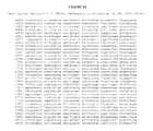
- FIG. 1is a schematic depicting detecting in a sample from a subject with PSA levels greater than 2.5 ng/mL the expression of one or more autoantibodies (“Autoantibody Test I”). If the result of the Autoantibody Test I is negative, a biopsy is not recommended to be obtained from the subject for further analysis. If result of the Autoantibody Test II is positive, then a biopsy is obtained. If the biopsy is positive for prostate cancer, expression of one or more autoantibodies is detected from a sample from the subject to characterize the cancer as aggressive or indolent, and a prognosis or theranosis provided.
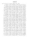
- FIG. 2lists the nucleic acid sequence for DCHS1 (SEQ ID NO: 29).
- FIG. 3lists the nucleic acid sequence for Centrosomal Protein (CEP 164) (SEQ ID NO: 30).
- FIG. 4lists the nucleic acid sequence for KBTBD6 (SEQ ID NO: 31).
- FIG. 5lists the nucleic acid sequence for RPS19 (SEQ ID NO: 32).
- FIG. 6lists the nucleic acid sequence for RPL34 (SEQ ID NO: 33).
- FIG. 7lists the nucleic acid sequence for Hemk1 (SEQ ID NO: 34).
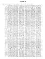
- FIG. 8lists the nucleic acid sequence for eIF4G1 (SEQ ID NO: 35).
- FIG. 9lists the nucleic acid sequence for BMI1 (SEQ ID NO: 36).
- FIG. 10lists the nucleic acid sequence for BRD2 (SEQ ID NO: 37).
- FIG. 11lists the nucleic acid sequence for RP3-323M22 (Nucleolin) (SEQ ID NO: 38).
- FIG. 12lists the nucleic acid sequence for SFRS14 (SEQ ID NO: 39).
- FIG. 13lists the nucleic acid sequence for LOC388789 (SEQ ID NO: 40).
- FIG. 14lists the nucleic acid sequence for RNA binding motif protein 6 (genomic DNA sequence) (SEQ ID NO: 41).
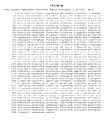
- FIG. 15lists the nucleic acid sequence for BRMSL1 (SEQ ID NO: 42).
- FIG. 16lists the nucleic acid sequence for NKX3-1 (SEQ ID NO: 43).
- FIG. 17lists the nucleic acid sequence for RPSA (SEQ ID NO: 44).
- FIG. 18lists the nucleic acid sequence for Cytochrome C Oxidase 5 subunit (SEQ ID NO: 45).
- FIG. 19lists the nucleic acid sequence for FAM53B (SEQ ID NO: 46).
- FIG. 20lists the nucleic acid sequence for a fragment of the UTR region of chromosome 11 ( Homo sapiens genomic DNA, chromosome 11 clone: CTD-2579L12, NTs 149521-151500) (SEQ ID NO: 47).
- FIG. 21lists the nucleic acid sequence for MAPKKK9 (SEQ ID NO: 48).
- FIG. 22lists the nucleic acid sequence for cDNA clone XR_113641.1 ( Homo sapiens hypothetical LOC643783, transcript variant 2 (LOC643783), partial miscRNA) (SEQ ID NO: 49).
- FIG. 23lists the nucleic acid sequence for PSA (SEQ ID NO: 50).
- FIG. 24lists the nucleic acid sequence for H2aa4 (SEQ ID NO: 51).
- FIG. 25lists the nucleic acid sequence for UBE2I (SEQ ID NO: 52).
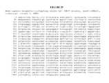
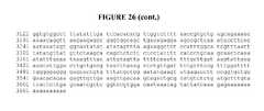
- FIG. 26lists the nucleic acid sequence for TIMP2 (SEQ ID NO: 53).
- FIG. 27lists the nucleic acid sequence for WDR77 (SEQ ID NO: 54).
- FIG. 28lists the nucleic acid sequence for a fragment of Deaminase Domain Cont 1 (Human DNA sequence from clone RP1-20N2 on chromosome 6q24 Contains the gene for a novel protein similar to yeast and bacterial cytosine deaminase, NTs 48121-50100) (SEQ ID NO: 55).
- FIG. 29lists the nucleic acid sequence for Lamin A/C (SEQ ID NO: 85).
- FIG. 30lists the nucleic acid sequence Lsm3 (SEQ ID NO: 86).
- FIG. 31lists the nucleic acid sequence for a fragment of cDNA clone Chromosome 19, which encompasses the nucleic acid sequence for DAZ associated protein ( Homo sapiens chromosome 19 clone CTB-25B13, NTs 20521-22500) (SEQ ID NO: 87).
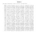
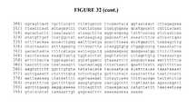
- FIG. 32lists the nucleic acid sequence for ADAM metallopetidase domain 9 (SEQ ID NO: 88).
- FIG. 33lists the nucleic acid sequence for AZGP1 (SEQ ID NO: 89).
- FIG. 34lists the nucleic acid sequence for Desmocolin 3 (SEQ ID NO: 90).
- FIG. 35lists the nucleic acid sequence for PERP (SEQ ID NO: 91).
- FIG. 36lists the nucleic acid sequence for Chromosome 3 UTR region ropporin/RhoEGF ( Homo sapiens 3 BAC RP11-783D3 (Roswell Park Cancer Institute Human BAC Library) NTs 178621-180600) (SEQ ID NO: 92).
- FIG. 37lists the nucleic acid sequence for Cox5a (SEQ ID NO: 93).
- FIG. 38lists the nucleic acid sequence for a Mitochondrion sequence ( Homo sapiens isolate PD047 mitochondrion, NTs 4801-6780) (SEQ ID NO: 94).
- FIG. 39lists the nucleic acid sequence for MYH9 (SEQ ID NO: 95).
- FIG. 40lists the nucleic acid sequence for ASND1 (SEQ ID NO: 96).
- FIG. 41lists the nucleic acid sequence for Cathepsin F (SEQ ID NO: 97).
- FIG. 42lists the nucleic acid sequence for Mastermind-like 2 ( Homo sapiens genomic DNA, chromosome 11q clone:RP11-82212, NTs 157801-159780) (SEQ ID NO: 98).
- FIG. 43lists the nucleic acid sequence for CSNK2A2 (SEQ ID NO: 99).
- FIG. 44lists the nucleic acid sequence for AURKAIP1 (SEQ ID NO: 100).
- FIG. 45lists the nucleic acid sequence for a fragment of Chromosome 4 ( Homo sapiens BAC clone RP11-327O17 from 4, NTs 107401-109380) (SEQ ID NO: 101).
- FIG. 46lists the nucleic acid sequence for ARF6 (SEQ ID NO: 102).
- FIG. 47lists the nucleic acid sequence for JAG1 (Human DNA sequence from clone RP1-278O22 on chromosome 20 Contains two novel genes, NTs 26161-26140) (SEQ ID NO: 103).
- FIG. 48lists the nucleic acid sequence for a Mitochondrion sequence ( Homo sapiens isolate PD047 mitochondrion, NTs 2041-4020) (SEQ ID NO: 104).
- FIG. 49lists the nucleic acid sequence for a fragment of Chromosome 20 (Human DNA sequence from clone RP1-278O22 on chromosome 20 Contains two novel genes, NTs 25321-27300) (SEQ ID NO:105).
- FIG. 50lists the nucleic acid sequence for a fragment of Chromosome 6 UTR region (Human DNA sequence from clone RP3-523G1 on chromosome 6p22.3-24.1, NTs 34621-36600) (SEQ ID NO: 106).
- FIG. 51lists the nucleic acid sequence for a fragment of MAPKKK5 (SEQ ID NO: 107).
- FIG. 52lists the nucleic acid sequence for RASA1 (SEQ ID NO: 108).
- FIG. 53lists the nucleic acid sequence for Hsp90b (SEQ ID NO: 109).
- FIG. 54lists the nucleic acid sequence for ribosomal protein S6 (RPS6) (SEQ ID NO: 110).
- FIG. 55lists the nucleic acid sequence for a fragment of Homo sapiens chromosome 3 ( Homo sapiens 3 BAC RP13-616I3 (Roswell Park Cancer Institute Human BAC Library) NTs 22921-24900) (SEQ ID NO: 111).
- compositions and methods of the present disclosurerelate to compositions and methods for characterizing a cancer or screening for a cancer.
- testswhich can be used to analyze a presence or absence of an antibody from a subject, such as a subject being tested or screened for a cancer.
- an antibodyis an autoantibody.
- the testcomprises a single antigen, thus detecting only an antibody that binds to that antigen.
- a panel of antigensis constructed such that the panel tests for a presence of one or more antibodies which specifically bind to two or more antigens derived from proteins associated with a specific cancer, such as lung cancer, prostate cancer, or ovarian cancer.
- a canceris characterized for a subject using a composition or method disclosed herein.
- a subjectis an individual or patient.
- a subjectis a human.
- a subjectis a cancer patient.
- a subjectexhibits no symptom of cancer, such as no symptoms of prostate cancer.
- a subjecthas no detectable symptom of cancer, such as no detectable symptoms for prostate cancer.
- a subjectexhibits a symptom of cancer, such as a symptom for prostate cancer.
- a subjectis a human.
- a subjectis an individual.
- a subjectis a patient, such as a cancer patient.
- Characterizing a cancer, or screening for a cancercan include detecting the cancer (including pre-symptomatic early stage detecting), determining the prognosis, diagnosis, or theranosis of the cancer, or determining the stage or progression of the cancer.
- a prognosisis predicting or giving a likelihood of outcome of a disease or condition, such as an extent of malignancy of a cancer, a likelihood of survival, or expected life expectancy, such as in an individual with prostate cancer.
- a prognosisis a prediction or likelihood analysis of cancer progression, cancer recurrence, or metastatic spread or relapse.
- the diagnosisis prediction or likelihood an individual or subject has a disease or condition, such as prostate cancer.
- the individualis an asymptomatic individual. In another embodiment, the individual is a symptomatic individual.
- a theranosisis a therapy selected based on an outcome of determining a binding of one or more antibodies from a sample from a subject to an antigen or polypeptide probe as described herein. In one embodiment, a theranosis is identifying an appropriate treatment or treatment efficacy for a cancer. In one embodiment, a theranosis is modifying a treatment. In another embodiment, a theranosis is selecting a treatment regimen. In yet another embodiment, a theranosis is discontinuing or not selecting a particular treatment regimen. In one embodiment a treatment regimen or therapeutic agent is selected based on the presence or absence of an autoantibody that binds to polypeptide probes described herein.
- the autoantibodyis a human aautoantibody. In one embodiment a treatment regimen or therapeutic agent is excluded based on the presence or absence of an autoantibody that binds to polypeptide probes described herein. In one embodiment the autoantibody is a human aautoantibody.
- characterizing or screening for a canceris detecting the cancer, such as pre-symptomatic early stage detecting.
- characterizing a canceris determining the stage or progression of the cancer, such as early-stage, late-stage or advanced stage of cancer.
- Characterizing or screening for a cancercan also be determining the likelihood or possibility an individual has a cancer.
- Characterizing or screening for a cancercan also be identification of a cancer, such as determining whether expression of one or more antibodies is indicative of the cancer.
- an antigen panelis used to detect a presence of one or more antibodies to one or more proteins, antigens, mimotopes, or epitopes.
- one or more polypeptide probes described hereinis a protein or fragment thereof.
- one or more polypeptide probes described hereincomprises an antigen, mimotope, or epitope.
- a “mimotope”can mimic the epitope of a protein or peptide.
- the mimotopeis structurally similar to an antigen or epitope of an expressed protein, but is unrelated or weakly related at the protein sequence level.
- the antigen panelcomprises one or more polypeptide probes comprising a polypeptide sequence selected from SEQ ID NO: 1, 2, 3, 4, 5, 6, 7, 8, 9, 10, 11, 12, 13, 14, 56, 57, 58, 59, 60, 61, 62, 63, 64, 65, 66, 67, 68, 69, 112, 113, 114, 115, 116, 117, 118, 119, 120, 121, 122, 123, 124, 125, 126, 127, 128, 129, 130, 131, 132, 133, 134, 135, 136, 137, 138, 139, 140, 141, or a fragment thereof.
- the antigen panelcomprises one or more polypeptide probes comprising a sequence encoded by SEQ ID NO: 15, 16, 17, 18, 19, 20, 21, 22, 23, 24, 25, 26, 27, 28, 70, 71, 72, 73, 74, 75, 76, 77, 78, 79, 80, 81, 82, 83, 84, 142, 143, 144, 145, 146, 147, 148, 149, 150, 151, 152, 153, 154, 155, 156, 157, 158, 159, 160, 161, 162, 163, 164, 165, 166, 167, 168, 169, 170, 171, or a fragment thereof.
- the polypeptide probecomprises the full-length or a fragment of a protein that is encoded by SEQ ID NO: 29, 30, 31, 32, 33, 34, 35, 36, 37, 38, 39, 40, 41, 42, 43, 44, 45, 46, 47, 48, 49, 51, 52, 53, 54, 55, 85, 86, 87, 88, 89, 90, 91, 92, 93, 94, 95, 96, 97, 98, 99, 100, 101, 102, 103, 104, 105, 106, 107, 108, 109, 110, 111, or a fragment thereof.
- the antigen panelcomprises one or more polypeptide probes derived from one or more proteins encoded by one or more genes selected from: CEP164, RPL34, BRMSL1, NKX3-1, RPSA, Cytochrome C oxidase 5 Subunit, UTR-region of chromosome 11, MAPKKK9, cDNA clone XR_113641.1, PSA, H2aa4, UBE2I, TIMP2, WDR77, Deaminase Domain, FAM53B, 5′UTR BMI1, RP3-323M22, or LOC388789.
- detection of one or more antibodiesis used to detect a presence of prostate cancer in a subject.
- the antigen panelcomprises one or more polypeptide probes derived from one or more proteins encoded by one or more genes selected from: DCHS1, CEP164, KBTBD6, RPS19, RPL34, RNA binding protein 6, Hemk1, eIF4G1, 5′UTR BMI1, BRD2, RP3-323M22, SFRS14, and LOC388789.
- detection of one or more antibodiesis used to detect a presence of prostate cancer in a subject.
- a cancercan also be characterized by determining a presence or absence, or level, of one or more antibodies in a sample.
- a sampleis obtained from a subject.
- the subjectcan be a mammal, including, but not limited to, humans, non-human primates, rodents, and the like.
- a sampleis a biological fluid.
- the biological fluidcan be, but not limited to, peripheral blood, sera, or plasma.
- the samplecan be ascites, urine, cerebrospinal fluid (CSF), sputum, saliva, bone marrow, synovial fluid, aqueous humor, amniotic fluid, cerumen, breast milk, broncheoalveolar lavage fluid, semen, prostatic fluid, cowper's fluid or pre-ejaculatory fluid, female ejaculate, sweat, fecal matter, hair, tears, cyst fluid, pleural and peritoneal fluid, pericardial fluid, lymph, chyme, chyle, bile, interstitial fluid, menses, pus, sebum, vomit, vaginal secretions, mucosal secretion, stool water, pancreatic juice, lavage fluids from sinus cavities, or bronchopulmonary aspirates.
- CSFcerebrospinal fluid
- an antibodyis an autoantibody.
- An autoantibodyrefers to an antibody produced by a host (with or without immunization) and directed to a host antigen (such as a tumor antigen). Tumor-associated antigens recognized by humoral effectors of the immune system are an attractive target for diagnostic and therapeutic approaches to human cancer.
- the binding of an antibody with a polypeptide probecan be specific, such that the interaction of the autoantibody with the polypeptide probe is dependent upon a presence of a particular structure (i.e., the antigenic determinant or epitope) of the polypeptide probe.
- Antigenic determinates or epitopescan comprise amino acids in linear or non-linear sequence in a polypeptide probe and can also comprise one or more amino acids which are in proximity to each other via protein folding (e.g., conformational epitopes).
- a single polypeptide or proteincan potentially be bound by multiple antibodies which recognize different epitopes.
- known epitopes of a particular polypeptidecan be used as a probe to detect for a presence, absence or level of autoantibodies which bind a particular epitope
- the polypeptide probecan be an antigen identified through serologic identification of antigens, for example by recombinant expression cloning (SEREX), such as described by Kim et al., Biotech. Lett . (2004); 26: 585-588.
- SEREXrecombinant expression cloning
- an antigencan be identified by screening expression cDNA libraries from human solid tumors with sera of autologous patients. This type of screening of a cDNA expression library by conventional methods typically requires the preparation of a large number of membrane filters blotted with bacteriophage plaques that are then searched with a specific probe. In the case of the SEREX experiments, the screening is performed using sera from cancer patients, which can be in very limited quantities.
- a polypeptide probe for detecting an antibodycan also be identified by phage-display technology, which can be based on the insertion of foreign nucleotide sequences into genes encoding for various capsid proteins of T7 phage, resulting in a heterogeneous mixture of phages, each displaying the different peptide sequence encoded by a corresponding insert.
- a physical link between a displayed fusion protein and DNA encoded for itmake this phage target selectable.
- the phage targetcan express or display a polypeptide probe, which can be used to detect antibodies that are produced by a subject, or autoantibodies, which can then be used to detect or characterize a cancer.
- the polypeptide probecan be displayed by a phage and used to detect an antibody from a sample obtained from a subject.
- an antibodyis an autoantibody.
- Polypeptideis used in its broadest sense and can include a sequence of subunit amino acids, amino acid analogs, or peptidomimetics. The subunits can be linked by peptide bonds.
- the polypeptidescan be naturally occurring, processed forms of naturally occurring polypeptides (such as by enzymatic digestion), chemically synthesized or recombinantly expressed.
- the polypeptides for use in the methods of the present inventioncan be chemically synthesized using standard techniques.
- the polypeptidescan comprise D-amino acids (which are resistant to L-amino acid-specific proteases), a combination of D- and L-amino acids, ⁇ amino acids, or various other designer or non-naturally occurring amino acids (e.g., ⁇ -methyl amino acids, C ⁇ -methyl amino acids, and N ⁇ -methyl amino acids, etc.) to convey special properties.
- Synthetic amino acidscan include ornithine for lysine, and norleucine for leucine or isoleucine.
- the polypeptidescan have peptidomimetic bonds, such as ester bonds, to prepare polypeptides with novel properties.
- a polypeptidecan be generated that incorporates a reduced peptide bond, i.e., R 1 —CH 2 —NH—R 2 , where R 1 and R 2 are amino acid residues or sequences.
- a reduced peptide bondcan be introduced as a dipeptide subunit.
- Such a polypeptidecan be resistant to protease activity, and can possess an extended half-life in vivo.
- a polypeptidecan also include a peptoid (N-substituted glycines), in which the one or more side chains are appended to nitrogen atoms along the molecule's backbone, rather than to the ⁇ -carbons, as in amino acids.
- Polypeptide and peptideare intended to be used interchangeably throughout this application, i.e. where the term peptide is used, it can also include polypeptide and where the term polypeptides is used, it can also include peptide.
- a polypeptide probecan be a fragment or portion of a larger protein.
- a fragmentcan range in size from two amino acid residues to the entire amino acid sequence minus one amino acid.
- a polypeptide probeis a fragment of an untranslated region (UTR) of a protein, such as a fragment that is encoded by a nucleic sequence that is a UTR region of a gene, such as the 5′ or 3′ UTR of a gene.
- UTRuntranslated region
- the fragmentcan be 2, 3, 4, 5, 6, 7, 8, 9, 10, 11, 12, 13, 14, 15, 16, 17, 18, 19, 20, 21, 22, 23, 24, 25, 26, 27, 28, 29, 30, 31, 32, 33, 34, 35, 36, 37, 38, 39, 40, 41, 42, 43, 44, 45, 46, 47, 48, 49, 50, 55, 60, 65, 70, 75, 80, 85, 90, 95, or 100 amino acids in size.
- the fragmentis less than about 5, 6, 7, 8, 9, 10, 11, 12, 13, 14, 15, 16, 17, 18, 19, 20, 21, 22, 23, 24, 25, 26, 27, 28, 29, 30, 31, 32, 33, 34, 35, 36, 37, 38, 39, 40, 41, 42, 43, 44, 45, 46, 47, 48, 49, 50, 55, 60, 65, 70, 75, 80, 85, 90, 95, or 100 amino acids in size.
- a polypeptide probe useful in the compositions and methods herein, regardless of size,is capable of specific interaction with an antibody, such as an autoantibody.
- a polypeptide probecan be a fragment of a protein encoded by a gene, or a region upstream or downstream of a coding sequence, such as a UTR region, of a gene listed in Table 1, Table 2, Table 3 or Table 4.
- the polypeptide probecomprises a polypeptide sequence selected from SEQ ID NO: 1, 2, 3, 4, 5, 6, 7, 8, 9, 10, 11, 12, 13, 14, 56, 57, 58, 59, 60, 61, 62, 63, 64, 65, 66, 67, 68, 69, 112, 113, 114, 115, 116, 117, 118, 119, 120, 121, 122, 123, 124, 125, 126, 127, 128, 129, 130, 131, 132, 133, 134, 135, 136, 137, 138, 139, 140, 141, or a fragment thereof.
- the polypeptide probecomprises a polypeptide sequence encoded by SEQ ID NO: 15, 16, 17, 18, 19, 20, 21, 22, 23, 24, 25, 26, 27, 28, 70, 71, 72, 73, 74, 75, 76, 77, 78, 79, 80, 81, 82, 83, 84, 142, 143, 144, 145, 146, 147, 148, 149, 150, 151, 152, 153, 154, 155, 156, 157, 158, 159, 160, 161, 162, 163, 164, 165, 166, 167, 168, 169, 170, 171, or a fragment thereof.
- the polypeptide probecomprises the full-length or a fragment of a protein that is encoded by SEQ ID NO: 29, 30, 31, 32, 33, 34, 35, 36, 37, 38, 39, 40, 41, 42, 43, 44, 45, 46, 47, 48, 49, 51, 52, 53, 54, 55, 85, 86, 87, 88, 89, 90, 91, 92, 93, 94, 95, 96, 97, 98, 99, 100, 101, 102, 103, 104, 105, 106, 107, 108, 109, 110, 111, or a fragment thereof.
- a polypeptide probeis a fragment of a protein encoded by a gene, or a fragment encoded by a sequence of a UTR region of a gene.
- the genecan be CEP164, RPL34, BRMSL1, NKX3-1, RPSA, Cytochrome C oxidase 5 Subunit, UTR-region of chromosome 11, MAPKKK9, cDNA clone XR_113641.1, PSA, H2aa4, UBE2I, TIMP2, WDR77, or Deaminase Domain.
- the geneis FAM53B, 5′UTR BMI1, RP3-323M22, or LOC388789.
- a polypeptide probecomprises SEQ ID NO: 2, 5, 9, 11, 14, 56, 57, 58, 59, 60, 61, 62, 63, 64, 65, 66, 67, 68, 69, or a fragment thereof.
- a polypeptide probecomprises a polypeptide encoded by SEQ ID NO: 16, 19, 23, 25, 28, 70, 71, 72, 73, 74, 75, 76, 77, 78, 79, 80, 81, 82, 83, 84, or a fragment thereof.
- the genecan be DCHS1, CEP164, KBTBD6, RPS19, RPL34, RNA binding protein 6, or Hemk1.
- the geneis eIF4G1, 5′UTR BMI1, BRD2, RP3-323M22, SFRS14, or LOC388789.
- a polypeptide probecan comprise a peptide sequence, or fragment thereof, such as those listed in Tables 1, 2, 3 or 4.
- a polypeptide probecomprises SEQ ID NO: 1, 2, 3, 4, 5, 6, 7, 8, 9, 10, 11, 12, 13, 14, or a fragment thereof.
- a polypeptide probecomprises a polypeptide encoded by SEQ ID NO: 15, 16, 17, 18, 19, 20, 21, 22, 23, 24, 25, 26, 27, 28, 29, 30, 31, 32, 33, 34, 35, 36, 37, 38, 39, 40, 41, or a fragment thereof.
- FIG. 2PQTTAPRRAR AGCTTTCGCTAGAGACGCCTCCATA (protoc- (SEQ ID PRRS (SEQ ID AGTCACTTGCCCGTTGGCCCCCACG adherin- NO: 29) NO: 1) ATCGGGGTCGGTTGCTCGCAGGGC 16 TGAGCAGAGATGTGCCAGGAGGGT pre- TGTTCTCACGCAAGAGGACGCTGT cursor) ACTCCTGCTGCTGGAAAGTAGGCG CCTCGTCGTTGACGTCAGCGACACT GACGGTCAGGACCTGCGTGGCCGA GCGCGGCGGGGAGCCGTGGTCTGA GG (SEQ ID NO: 15) 1B4A Centro- NM_014956.4 FIG.
- FIG. 8IRDPNQGG TTCTTCTACAGACATTTGTATAGT (SEQ ID KDITEEIMS TGTCATAGTGTCCCCAGGAATAG NO: 35) GARTASTP AGAGGACTGCGAGATTAGGCTCA TPPQTGGG GACCCCGGTTCCAAGACTGGGGA LEPQANGE TGGTGATGGGGTCGGAGAAGGCG TPQVAVIV ACGAAGGCTGGGATTCTGAAGGG RPDDRSQG CTATGCTCTGGGCCAGGCAGCCC AIIADRPGL TGGCCGGTCAGCAATGATTGCTC PGPEHSPSE CCTGTGACCGGTCATCTGGCCGG SQPSSPSPT ACAATGACAGCAACCTGGGGCGT PSPVLEP CTCCCCATTAGCTTGAGGCTCCAG GSEPNLAV ACC
- FIG. 15APRTRTLR TCGTCGAGGCTCCTGCTCCTGTGA (SEQ ID ARRSPRME CTCTCGAGCAGCCAGAGGCTCCT NO: 42) IAQKWMM ACCTCTATCGAGTCTTTACCTACT KTVKEEEW ACTTCTGACACTTTCTTCTTCTTA NVWMKCPI CCTTACAAACCTACTTTACAGGTT LKNSLPIS AGAACTTTTTGTCAAATGGCTAG KINFIKND AGTTTCTAGTTGAAATATTTCTTG (SEQ ID CTAATTCAGTCCACCTACGTTTTG NO: 56) ATGTTCTTCAGTATCGACCTTTTC GTGGTCTTATGAACCTTGGCGACC GTTGAAATGTCCTTTTATACGTTT AAGCATGTTTCCATCGTCCTTAGA TATCTCTCGACGAATCTTAGAC
- GGRGGGGGGAGGTCGAGGCGGAGGCGGAG(SEQ ID GGGGRGA GAGGAGGAGGCCGAGGCGCCGG No: 36) GGGRGAG AGGAGGCCGAGGCGCCGGAGCA AGGGRPEAA GGAGGAGGCCGGCCGGAGGCGG (SEQ ID CATGAGACGAGCGTGGCGGCCGC NO: 172) GGCTGCTCGGGGCCGCGCTGGTT GNCCATTGACAGCGGCGTCTGCA GCTCGCTTCAAGATGGCCGCTTG GCTCGCATTCATTTTCTGCTGAAC GACTTTTAACTTTCATTGTCTTTTC CGCCCGCTTCGATCGCCTCGCGCC GGCTGCTCTTTCCGGGATTTTTTA TCAAGCAGAAATGCATCGAACAA CGAGAATCAAGATCACTGAGCTA AATCCCCNCCTGATGTGTGTGCTT TGTGGAGGGTACTTCATTGATGCC ACAACCATAATAGAATGTCTACA TTCCTTCTGTAAAACGTGTATTGT TCGTTACCTGGAG
- An antibodysuch as an autoantibody, to one or more of a protein, or a fragment of a protein, encoded by a gene such as listed in Tables 1, 2, 3 or 4, or a polypeptide encoded by a UTR sequence of a gene such as one listed in Tables 1, 2, 3 or 4, can be detected according to one or more methods described herein and used to characterize a cancer, such as prostate cancer.
- a cancersuch as prostate cancer.
- Many of the proteinsmay have a role in various cancers, including prostate cancer.
- the human DCHS1 proteinprotocadherin-16 precursor
- DCHS1is a cadherin, a class of type-1 transmembrane proteins. Cadherins typically play important roles in cellular adhesion, for example, by binding cells expressing similar cadherins to each other. Structurally, DCHS1 is thought to contain 27 cadherin repeats (extracellular calcium ion-binding domains). DCHS1 expression has been associated with certain cancers, potentially playing a role in tumor adherence (see, e.g., Sjöblom, et. al. Science , (2006) 314:268-274).
- CEP164is believed to be a centrosomal protein which binds chromatin and plays a role in the DNA damage-activated signaling cascade. It is known to interact with ataxia telangiectasia mutated (ATM) and ATM/Rad3-related (ATR) kinases which phosphorylate CEP164 upon replication stress, ultraviolet radiation (UV), and ionizing radiation (IR). CEP164 also plays a role in cell cycle regulation, specifically at the G2/M checkpoint and in nuclear division (see, e.g., Sivasubramaniam et al., Genes & Dev . (2008); 22(5):687-600). As CEP 164 plays a role in genome stabilization, misregulation or mutation of this gene and/or protein can play a role in certain cancers.
- ATMataxia telangiectasia mutated
- ATRATM/Rad3-related kinases which phosphorylate CEP164 upon replication stress, ultraviolet radiation (UV), and
- the human KBTBD6(kelch repeat and BTB (POZ) domain containing 6) is a protein expressed in a wide variety of normal tissues. Its expression and/or misregulation has also been noted in multiple cancer types, including prostate, ovarian, kidney and lung tumors. The function of the protein is not currently known, however, the presence of the kelch repeat and BTB domain suggest that the protein is involved in protein-protein interactions and actin filament organization.
- RPS19ribosomal protein S19
- RPS19encodes a ribosomal protein that is a component of the 40S subunit. Located in the cytoplasm as part of the ribosomal complex, mutations in this gene are associated with Diamond-Blackfan anemia, suggesting a non-ribosomal function for the protein in erythropoietic differentiation.
- RPS19 proteinis also known to interact with fibroblast growth factor-2 (see, e.g., Soulet et al., Biochem. Biophys. Res. Commun . (2001); 289:591-596).
- RPL3460S Ribosomal protein L34
- c-MYCc-MYC
- c-MYCc-MYC
- RBM6RNA binding protein 6
- ATRATM or ATR
- HEMK1HEMK methyltransferase family protein 1
- HEMK1S-adenosylmethionine-dependent methyltransferase and is also thought to bind nucleic acids.
- HEMK1is considered a tumor-suppressor, misregulation of which is associated with various cancers, including prostate cancer, pancreatic cancer and liver cancer (see, e.g., U.S. Pat. App. Pub. No. 2008/0213791).
- polypeptide probessuch as a fragment of a protein encoded by a gene, or a polypeptide encoded by a sequence of a UTR region of a gene, such as a gene listed in Tables 1, 2, 3 or 4, can be used to detect one or more antibodies, such as autoantibodies, from a sample from a subject.
- the polypeptide probecomprises a polypeptide sequence selected from SEQ ID NO: 1, 2, 3, 4, 5, 6, 7, 8, 9, 10, 11, 12, 13, 14, 56, 57, 58, 59, 60, 61, 62, 63, 64, 65, 66, 67, 68, 69, 112, 113, 114, 115, 116, 117, 118, 119, 120, 121, 122, 123, 124, 125, 126, 127, 128, 129, 130, 131, 132, 133, 134, 135, 136, 137, 138, 139, 140, 141, or a fragment thereof.
- the polypeptide probecomprises a polypeptide sequence encoded by SEQ ID NO: 15, 16, 17, 18, 19, 20, 21, 22, 23, 24, 25, 26, 27, 28, 70, 71, 72, 73, 74, 75, 76, 77, 78, 79, 80, 81, 82, 83, 84, 142, 143, 144, 145, 146, 147, 148, 149, 150, 151, 152, 153, 154, 155, 156, 157, 158, 159, 160, 161, 162, 163, 164, 165, 166, 167, 168, 169, 170, 171, or a fragment thereof.
- the polypeptide probecomprises the full-length or a fragment of a protein that is encoded by SEQ ID NO: 29, 30, 31, 32, 33, 34, 35, 36, 37, 38, 39, 40, 41, 42, 43, 44, 45, 46, 47, 48, 49, 51, 52, 53, 54, 55, 85, 86, 87, 88, 89, 90, 91, 92, 93, 94, 95, 96, 97, 98, 99, 100, 101, 102, 103, 104, 105, 106, 107, 108, 109, 110, 111, or a fragment thereof.
- a polypeptide probeis a fragment of a protein encoded by CEP164, RPL34, BRMSL1, NKX3-1, RPSA, Cytochrome C oxidase 5 Subunit, UTR-region of chromosome 11, MAPKKK9, cDNA clone XR_113641.1, PSA, H2aa4, UBE2I, TIMP2, WDR77, or Deaminase Domain, or may be a polypeptide encoded by a UTR sequence of the gene, such as the 5′ or 3′ UTR sequence of CEP164, RPL34, BRMSL1, NKX3-1, RPSA, Cytochrome C oxidase 5 Subunit, UTR-region of chromosome 11, MAPKKK9, cDNA clone XR_113641.1, PSA, H2aa4, UBE2I, TIMP2, WDR77, or Deaminase Domain.
- a polypeptide probecan be a fragment of a protein encoded by FAM53B, 5′UTR BMI1, RP3-323M22, or LOC388789.
- a polypeptide probecomprises a peptide sequence, or fragment thereof, such as those listed in Tables 1, 2, 3, and 4.
- the polypeptide probecan comprise SEQ ID NO: 2, 5, 9, 11, 14, 56, 57, 58, 59, 60, 61, 62, 63, 64, 65, 66, 67, 68, 69, or a fragment thereof, or a fragment thereof.
- the polypeptide probecomprises a polypeptide encoded by SEQ ID NO: 16, 19, 23, 25, 28, 70, 71, 72, 73, 74, 75, 76, 77, 78, 79, 80, 81, 82, 83, 84, or a fragment thereof.
- a polypeptide probeis a fragment of a protein encoded by DCHS1, CEP164, KBTBD6, RPS19, RPL34, RNA binding protein 6, or Hemk1, or may be a polypeptide encoded by a UTR sequence of the gene, such as the 5′ or 3′ UTR sequence of DCHS1, CEP164, KBTBD6, RPS19, RPL34, RNA binding protein 6, or Hemk1.
- a polypeptide probecan be a fragment of a protein encoded by eIF4G1, 5′UTR BMI1, BRD2, RP3-323M22, SFRS14, or LOC388789.
- a polypeptide probecomprises a peptide sequence, or fragment thereof, such as those listed in Tables 1 and 2.
- the polypeptide probecan comprise SEQ ID NO: 1, 2, 3, 4, 5, 6, 7, 8, 9, 10, 11, 12, 13 or 14, or a fragment thereof.
- the polypeptide probecomprises a polypeptide encoded by SEQ ID NO: 15, 16, 17, 18, 19, 20, 21, 22, 23, 24, 25, 26, 27, 28, 29, 30, 31, 32, 33, 34, 35, 36, 37, 38, 39, 40, 41, or a fragment thereof.
- a panel as provided hereincan be used to analyze one or more antibodies to a plurality of polypeptide probes, such as one or more autoantibodies.
- a panelallows for the simultaneous analysis of multiple antibodies, such as autoantibodies, to a plurality of polypeptide probes correlating with carcinogenesis and/or metastasis.
- a panelcan include markers identified as correlating with cancerous tissue, metastatic cancer, localized cancer that is likely to metastasize, pre-cancerous tissue that is likely to become cancerous, and pre-cancerous tissue that is not likely to become cancerous.
- panelsmay be analyzed alone or in combination in order to provide the best possible diagnosis and/or prognosis.
- an antibody profiling panelcan comprise a plurality of polypeptide probes, wherein one or more of the probes is capable of binding an antibody.
- an antibody profiling panelcan comprise a plurality of probes, wherein one or more of the probes is capable of binding an antibody that targets a foreign antigen.
- an antibody profiling panelcan comprise a plurality of probes, wherein each of the probes is capable of binding an autoantibody.
- an antibody profiling panelcomprises 2-100 probes, 50-200 probes, 100-500 probes 200-750 probes, 200-1000 probes, 2-5,000 probes or 2-10,000 probes.
- an antibody profiling panelcomprises at least about 2, 3, 4, 5, 6, 7, 8, 9, 10, 11, 12, 13, 14, 15, 16, 17, 18, 19, 20, 21, 22, 23, 24, 25, 26, 27, 28, 29, 30, 31, 32, 33, 34, 35, 36, 37, 38, 39, 40, 41, 42, 43, 44, 45, 46, 47, 48, 49, 50, 55, 60, 65, 70, 75, 80, 85, 90, 95, or 100 polypeptide probes.
- an antibody profiling panelcomprises at least about 50, 100, 150, 200, 250, 500, 750, 1000, 5000, 10,000, 15,000, 20,000, 25,000, 30,000, 40,000, 50,000, 60,000, 70,000, 75,000, or 100,000 polypeptide probes.
- the probesare polypeptide probes.
- the probesare molecules that mimic an epitope bound by a particular antibody.
- An antibody profiling panelcan comprise at least 2, 3, 4, 5, 6, 7, 8, 9, 10, 11, 12, 13, or 14 polypeptide probes, wherein the polypeptide probes are a fragment of a protein encoded by a gene, or a fragment encoded by a sequence of a UTR region of a gene, such as genes listed in Tables 1, 2, 3, or 4.
- the polypeptide probecomprises a polypeptide sequence selected from SEQ ID NO: 1, 2, 3, 4, 5, 6, 7, 8, 9, 10, 11, 12, 13, 14, 56, 57, 58, 59, 60, 61, 62, 63, 64, 65, 66, 67, 68, 69, 112, 113, 114, 115, 116, 117, 118, 119, 120, 121, 122, 123, 124, 125, 126, 127, 128, 129, 130, 131, 132, 133, 134, 135, 136, 137, 138, 139, 140, 141, or a fragment thereof.
- the polypeptide probecomprises a polypeptide sequence encoded by SEQ ID NO: 15, 16, 17, 18, 19, 20, 21, 22, 23, 24, 25, 26, 27, 28, 70, 71, 72, 73, 74, 75, 76, 77, 78, 79, 80, 81, 82, 83, 84, 142, 143, 144, 145, 146, 147, 148, 149, 150, 151, 152, 153, 154, 155, 156, 157, 158, 159, 160, 161, 162, 163, 164, 165, 166, 167, 168, 169, 170, 171, or a fragment thereof.
- the polypeptide probecomprises the full-length or a fragment of a protein that is encoded by SEQ ID NO: 29, 30, 31, 32, 33, 34, 35, 36, 37, 38, 39, 40, 41, 42, 43, 44, 45, 46, 47, 48, 49, 51, 52, 53, 54, 55, 85, 86, 87, 88, 89, 90, 91, 92, 93, 94, 95, 96, 97, 98, 99, 100, 101, 102, 103, 104, 105, 106, 107, 108, 109, 110, 111, or a fragment thereof.
- an antibody profiling panelcomprises a plurality of polypeptide probes, wherein at least a subset of the polypeptide probes is a fragment of a protein encoded by a gene, or a fragment encoded by a sequence of a UTR region of a gene, wherein the gene is CEP164, RPL34, BRMSL1, NKX3-1, RPSA, Cytochrome C oxidase 5 Subunit, UTR-region of chromosome 11, MAPKKK9, cDNA clone XR_113641.1, PSA, H2aa4, UBE2I, TIMP2, WDR77, or Deaminase Domain.
- the polypeptide probecan comprise a fragment of a protein encoded by a gene, or a fragment encoded by a sequence of a UTR region of a gene, wherein the gene is FAM53B, 5′UTR BMI1, RP3-323M22, or LOC388789.
- an antibody profiling panelcomprises a plurality of polypeptide probes, wherein at least a subset of the polypeptide probes is a fragment of a protein encoded by a gene, or a fragment encoded by a sequence of a UTR region of a gene, wherein the gene is DCHS1, CEP164, KBTBD6, RPS19, RPL34, RNA binding protein 6, or Hemk1.
- the polypeptide probecan comprise a fragment of a protein encoded by a gene, or a fragment encoded by a sequence of a UTR region of a gene, wherein the gene is eIF4G1, 5′UTR BMI1, BRD2, RP3-323M22, SFRS14, or LOC388789.
- an antibody profiling panelcomprises a plurality of polypeptide probes, wherein at least a subset of the polypeptide probes is a peptide sequence, or fragment thereof, as listed in Tables 1, 2, 3, or 4.
- an antibody profiling panelcomprises a plurality of polypeptide probes, wherein at least a subset of the polypeptide probes comprises a polypeptide sequence selected from SEQ ID NO: 1, 2, 3, 4, 5, 6, 7, 8, 9, 10, 11, 12, 13, 14, 56, 57, 58, 59, 60, 61, 62, 63, 64, 65, 66, 67, 68, 69, 112, 113, 114, 115, 116, 117, 118, 119, 120, 121, 122, 123, 124, 125, 126, 127, 128, 129, 130, 131, 132, 133, 134, 135, 136, 137, 138, 139, 140, 141, or a fragment thereof.
- an antibody profiling panelcomprises a plurality of polypeptide probes, wherein at least a subset of the polypeptide probes comprises a polypeptide sequence encoded by SEQ ID NO: 15, 16, 17, 18, 19, 20, 21, 22, 23, 24, 25, 26, 27, 28, 70, 71, 72, 73, 74, 75, 76, 77, 78, 79, 80, 81, 82, 83, 84, 142, 143, 144, 145, 146, 147, 148, 149, 150, 151, 152, 153, 154, 155, 156, 157, 158, 159, 160, 161, 162, 163, 164, 165, 166, 167, 168, 169, 170, 171, or a fragment thereof.
- the polypeptide probecomprises the full-length or a fragment of a protein that is encoded by SEQ ID NO: 29, 30, 31, 32, 33, 34, 35, 36, 37, 38, 39, 40, 41, 42, 43, 44, 45, 46, 47, 48, 49, 51, 52, 53, 54, 55, 85, 86, 87, 88, 89, 90, 91, 92, 93, 94, 95, 96, 97, 98, 99, 100, 101, 102, 103, 104, 105, 106, 107, 108, 109, 110, 111, or a fragment thereof.
- an antibody profiling panelcomprises a plurality of polypeptide probes, wherein at least a subset of the polypeptide probes comprises SEQ ID NO: 2, 5, 9, 11, 14, 56, 57, 58, 59, 60, 61, 62, 63, 64, 65, 66, 67, 68, 69, or a fragment thereof, or a fragment thereof.
- an antibody profiling panelcomprises a plurality of polypeptide probes, wherein at least a subset of the polypeptide probes is encoded by SEQ ID NO: 16, 19, 23, 25, 28, 70, 71, 72, 73, 74, 75, 76, 77, 78, 79, 80, 81, 82, 83, 84, or a fragment thereof.
- an antibody profiling panelcomprises a plurality of polypeptide probes, wherein at least a subset of the polypeptide probes comprises SEQ ID NO: 1, 2, 3, 4, 5, 6, 7, 8, 9, 10, 11, 12, 13 or 14, or a fragment thereof.
- an antibody profiling panelcomprises a plurality of polypeptide probes, wherein at least a subset of the polypeptide probes is encoded by SEQ ID NO: 15, 16, 17, 18, 19, 20, 21, 22, 23, 24, 25, 26, 27, 28, 29, 30, 31, 32, 33, 34, 35, 36, 37, 38, 39, 40, 41, or a fragment thereof.
- an antibody profiling panelcan also comprise one or more polypeptide probes of the protein PSA, or fragment of PSA, in combination with one or more of the polypeptide probes discussed herein.
- an antibody profiling panelcan comprise polypeptide probes including a full-length protein or fragment of PSA and one or more polypeptide probes comprising SEQ ID NO: 1, 2, 3, 4, 5, 6, 7, 8, 9, 10, 11, 12, 13, 14, 56, 57, 58, 59, 60, 61, 62, 63, 64, 65, 66, 67, 68, 69, 112, 113, 114, 115, 116, 117, 118, 119, 120, 121, 122, 123, 124, 125, 126, 127, 128, 129, 130, 131, 132, 133, 134, 135, 136, 137, 138, 139, 140, 141, or a fragment thereof.
- an antibody profiling panelcan comprise polypeptide probes including a full-length protein or fragment of PSA and one or more polypeptide probes comprising a polypeptide sequence encoded by SEQ ID NO: 15, 16, 17, 18, 19, 20, 21, 22, 23, 24, 25, 26, 27, 28, 70, 71, 72, 73, 74, 75, 76, 77, 78, 79, 80, 81, 82, 83, 84, 142, 143, 144, 145, 146, 147, 148, 149, 150, 151, 152, 153, 154, 155, 156, 157, 158, 159, 160, 161, 162, 163, 164, 165, 166, 167, 168, 169, 170, 171, or a fragment thereof.
- the polypeptide probecomprises the full-length or a fragment of a protein that is encoded by SEQ ID NO: 29, 30, 31, 32, 33, 34, 35, 36, 37, 38, 39, 40, 41, 42, 43, 44, 45, 46, 47, 48, 49, 51, 52, 53, 54, 55, 85, 86, 87, 88, 89, 90, 91, 92, 93, 94, 95, 96, 97, 98, 99, 100, 101, 102, 103, 104, 105, 106, 107, 108, 109, 110, 111, or a fragment thereof.
- an antibody profiling panelcan comprise polypeptide probes including a full-length protein or fragment of PSA and a full-length protein encoded by a gene, fragment of a protein encoded by a gene, or a fragment encoded by a sequence of a UTR region of a gene, wherein the gene is CEP164, RPL34, BRMSL1, NKX3-1, RPSA, Cytochrome C oxidase 5 Subunit, UTR-region of chromosome 11, MAPKKK9, cDNA clone XR_113641.1, PSA, H2aa4, UBE2I, TIMP2, WDR77, or Deaminase Domain.
- an antibody profiling panelcan comprise a plurality of polypeptide probes, wherein the probes include a full-length protein or fragment of PSA and a full-length protein encoded by a gene, fragment of a protein encoded by a gene, or a fragment encoded by a sequence of a UTR region of a gene, wherein the gene is FAM53B, 5′UTR BMI1, RP3-323M22, or LOC388789.
- an antibody profiling panelcan comprise polypeptide probes including a full-length protein or fragment of PSA and a full-length protein encoded by a gene, fragment of a protein encoded by a gene, or a fragment encoded by a sequence of a UTR region of a gene, wherein the gene is DCHS1, CEP164, KBTBD6, RPS19, RPL34, RNA binding protein 6, or Hemk1.
- an antibody profiling panelcan comprise a plurality of polypeptide probes, wherein the probes include a full-length protein or fragment of PSA and a full-length protein encoded by a gene, fragment of a protein encoded by a gene, or a fragment encoded by a sequence of a UTR region of a gene, wherein the gene is eIF4G1, 5′UTR BMI1, BRD2, RP3-323M22, SFRS14, or LOC388789.
- an autoantibody profiling panelcan comprise a plurality of polypeptide probes, wherein the probes includes a full-length protein or fragment of PSA and one or probes comprising a peptide sequence, or fragment thereof, as listed in Tables 1, 2, 3 and 4.
- an autoantibody profiling panelcan comprise a plurality of polypeptide probes, wherein the probes includes a full-length protein or fragment of PSA and one or more probes comprising SEQ ID NO: 1, 2, 3, 4, 5, 6, 7, 8, 9, 10, 11, 12, 13, 14, 56, 57, 58, 59, 60, 61, 62, 63, 64, 65, 66, 67, 68, 69, 112, 113, 114, 115, 116, 117, 118, 119, 120, 121, 122, 123, 124, 125, 126, 127, 128, 129, 130, 131, 132, 133, 134, 135, 136, 137, 138, 139, 140, 141, or a fragment thereof.
- an autoantibody profiling panelcan comprise a plurality of polypeptide probes, wherein the probes includes a full-length protein or fragment of PSA and one or more probes comprising a polypeptide sequence encoded by SEQ ID NO: 15, 16, 17, 18, 19, 20, 21, 22, 23, 24, 25, 26, 27, 28, 70, 71, 72, 73, 74, 75, 76, 77, 78, 79, 80, 81, 82, 83, 84, 142, 143, 144, 145, 146, 147, 148, 149, 150, 151, 152, 153, 154, 155, 156, 157, 158, 159, 160, 161, 162, 163, 164, 165, 166, 167, 168, 169, 170, 171, or a fragment thereof.
- an autoantibody profiling panelcan comprise a plurality of polypeptide probes, wherein the probes includes a full-length protein or fragment of PSA and one or more probes comprising the full-length or a fragment of a protein that is encoded by SEQ ID NO: 29, 30, 31, 32, 33, 34, 35, 36, 37, 38, 39, 40, 41, 42, 43, 44, 45, 46, 47, 48, 49, 51, 52, 53, 54, 55, 85, 86, 87, 88, 89, 90, 91, 92, 93, 94, 95, 96, 97, 98, 99, 100, 101, 102, 103, 104, 105, 106, 107, 108, 109, 110, 111, or a fragment thereof.
- an autoantibody profiling panelcan comprise a plurality of polypeptide probes, wherein the probes includes a full-length protein or fragment of PSA and one or more probes comprising SEQ ID NO: 1, 2, 3, 4, 5, 6, 7, 8, 9, 10, 11, 12, 13, 14, 15, 16, 17, 18, 19, 20, 21, 22, 23, 24, 25, 26, 27, 28, 29, 30, 31, 32, 33, 34, 35, 36, 37, 38, 39, 40, 41, or a fragment thereof.
- an autoantibody profiling panelcan comprise a plurality of polypeptide probes, wherein the probes includes a full-length protein or fragment of PSA and one or more probes comprising SEQ ID NO: 2, 5, 9, 11, 14, 56, 57, 58, 59, 60, 61, 62, 63, 64, 65, 66, 67, 68, 69, or a fragment thereof, or a fragment thereof; or a polypeptide sequence encoded by a sequence selected from SEQ ID NOs. 16, 19, 23, 25, 28, 70, 71, 72, 73, 74, 75, 76, 77, 78, 79, 80, 81, 82, 83, 84, or a fragment thereof.
- a PSA polypeptide probecan be combined with any two or more of the polypeptide probes described herein, such as a polypeptide probe derived from a protein encoded by a gene, fragment of a protein encoded by a gene, or a fragment encoded by a sequence of a UTR region of a gene, wherein the gene is CEP164, RPL34, BRMSL1, NKX3-1, RPSA, Cytochrome C oxidase 5 Subunit, UTR-region of chromosome 11, MAPKKK9, cDNA clone XR_113641.1, PSA, H2aa4, UBE2I, TIMP2, WDR77, Deaminase Domain, FAM53B, 5′UTR BMI1, RP3-323M22, or LOC388789.
- a polypeptide probe derived from a protein encoded by a genefragment of a protein encoded by a gene, or a fragment encoded by a sequence of a UTR
- a PSA polypeptide probecan be combined with any two or more of the polypeptide probes described herein, such as a polypeptide probe derived from a protein encoded by a gene, fragment of a protein encoded by a gene, or a fragment encoded by a sequence of a UTR region of a gene, wherein the gene is DCHS1, CEP164, KBTBD6, RPS19, RPL34, RNA binding protein 6, Hemk1, eIF4G1, 5′UTR BMI1, BRD2, RP3-323M22, SFRS14, or LOC388789.
- a PSA polypeptide probecan be combined with at least 3, 4, 5, 6, 7, 8, 9, 10, 11, 12, 13 or 14 of polypeptide probes disclosed herein, such as listed in Tables 1, 2, 3, and 4.
- a polypeptide probecomprises SEQ ID NO: 2, 5, 9, 11, 14, 56, 57, 58, 59, 60, 61, 62, 63, 64, 65, 66, 67, 68, 69, or a fragment thereof, or a fragment thereof.
- a polypeptide probecomprises SEQ ID NO: 1, 2, 3, 4, 5, 6, 7, 8, 9, 10, 11, 12, 13 or 14, or a fragment thereof.
- a polypeptide probecomprises SEQ ID NO: 1, 2, 3, 4, 5, 6, 7, 8, 9, 10, 11, 12, 13, 14, 56, 57, 58, 59, 60, 61, 62, 63, 64, 65, 66, 67, 68, 69, 112, 113, 114, 115, 116, 117, 118, 119, 120, 121, 122, 123, 124, 125, 126, 127, 128, 129, 130, 131, 132, 133, 134, 135, 136, 137, 138, 139, 140, 141, or a fragment thereof.
- a polypeptide probecomprises a polypeptide encoded by SEQ ID NO: 15, 16, 17, 18, 19, 20, 21, 22, 23, 24, 25, 26, 27, 28, 29, 30, 31, 32, 33, 34, 35, 36, 37, 38, 39, 40, 41, or a fragment thereof.
- a polypeptide probecomprises a polypeptide encoded by SEQ ID NO: 16, 19, 23, 25, 28, 70, 71, 72, 73, 74, 75, 76, 77, 78, 79, 80, 81, 82, 83, 84, or a fragment thereof.
- a polypeptide probecomprises a polypeptide encoded by SEQ ID NO: 15, 16, 17, 18, 19, 20, 21, 22, 23, 24, 25, 26, 27, 28, 70, 71, 72, 73, 74, 75, 76, 77, 78, 79, 80, 81, 82, 83, 84, 142, 143, 144, 145, 146, 147, 148, 149, 150, 151, 152, 153, 154, 155, 156, 157, 158, 159, 160, 161, 162, 163, 164, 165, 166, 167, 168, 169, 170, 171, or a fragment thereof.
- a polypeptide probe disclosed hereinis attached to a substrate (e.g., glass slide chip or nanowell chip).
- a polypeptide probecan be directly or indirectly attached to the substrate.
- a polypeptide probeis attached to a substrate via a phage.
- the substratecan be any physically separable solid to which a polypeptide probe can be directly or indirectly attached including, but not limited to, surfaces provided by microarrays and wells, particles such as beads, columns, optical fibers, wipes, glass and modified or functionalized glass, quartz, mica, diazotized membranes (paper or nylon), polyformaldehyde, cellulose, cellulose acetate, paper, ceramics, metals, metalloids, semiconductive materials, quantum dots, coated beads or particles, other chromatographic materials, magnetic particles; plastics (including acrylics, polystyrene, copolymers of styrene or other materials, polypropylene, polyethylene, polybutylene, polyurethanes, TEFLONTM, etc.), polysaccharides, nylon or nitrocellulose, resins, silica or silica-based materials including silicon and modified silicon, carbon, metals, inorganic glasses, plastics, ceramics, conducting polymers (including polymers such as polypyrole and polyindole); micro
- the polypeptide probecan bound to a planar surface or to a particle, such as a bead or microsphere.
- the polypeptide probeis attached to a bead.
- the beadcan be a polystyrene, brominated polystyrene, polyacrylic acid, polyacrylonitrile, polyacrylamide, polyacrolein, polydimethylsiloxane, polybutadiene, polyisoprene, polyurethane, polyvinyl acetate, polyvinylchloride, polyvinylpyridine, polyvinylbenzylchloride, polyvinyltoluene, polyvinylidene chloride, polydivinylbenzene, polyglycidylmethacrylate, polymethylmethacrylate, or copolymers, blends, composites, or combination thereof.
- the beadcan have a diameter of between about 1 nm-1000 ⁇ m, 1 nm-500 ⁇ m, 5 nm-500 ⁇ m, or 10 nm-100 ⁇ m. In one embodiment, the bead has a diameter of between about 10 nm and 100 ⁇ m. In yet another embodiment, the bead has a diameter of less than about 1000 ⁇ m, 500 ⁇ m, 400 ⁇ m, 300 ⁇ m, 200 ⁇ m, or 100 ⁇ m.
- the beadis labeled or stained with more than one dye, such as at least 2, 3, 4, 5, 6, 7, 8, 9, or 10 different dyes.
- the beadis labeled or stained with two dyes.
- the two dyesare hydrophobic.
- the two dyesare fluorescent dyes, such as squaric acid-based dyes.
- the squaric acid-based dyesare selected from cyclobutenedione derivatives, symmetrical and unsymmetrical squaraines, substituted cephalosporin compounds, fluorinated squaraine compositions, alkylalkoxy squaraines, or squarylium compounds.
- the squaric acid-based dyesare selected from a red fluorescent dye and an orange fluorescent dye, such as the red fluorescent dye comprising 1,3-bis(1,3-dihydro-1,3,3-trimethyl-2H-indol-2-ylidene)methyl]-2,4-dihydro xycyclobutenediylium, bis(inner salt) and the orange fluorescent dye comprising 2-(3,5-dimethylpyrrol-2-yl)-4-(3,5-dimethyl-2H-pyrrol-2-ylidene)-3-hydroxy-2-cyclobuten-1-one.
- the red fluorescent dyecomprising 1,3-bis(1,3-dihydro-1,3,3-trimethyl-2H-indol-2-ylidene)methyl]-2,4-dihydro xycyclobutenediylium, bis(inner salt)
- the orange fluorescent dyecomprising 2-(3,5-dimethylpyrrol-2-yl)-4-(3,5-dimethyl-2H-pyr
- the substrateis coated using passive or chemically-derivatized coatings with any number of materials, including polymers, such as dextrans, acrylamides, gelatins or agarose. Such coatings can facilitate the use of the array with a biological sample.
- a presence of an immune response to a specific protein expressed in cancerous cellscan be indicative of a presence of cancer.
- the present inventionprovides a method (e.g., diagnostic or screening method) for detecting a presence of an antibody, such as an autoantibody, to a tumor or tumor-associated antigen.
- an antibodysuch as an autoantibody
- the presence of an antibody in cancerous but not cancerous cellsis indicative of the presence of cancer.
- the antibodyis an antibody to a tumor antigen.
- a method or composition disclosed hereincan find utility in the diagnosis, screening, or characterization of a cancer.
- a presence of an antibody, such as an autoantibody, to a specific proteincan be indicative of a cancer.
- detection of an antibody in a sample, such as an autoantibodycan be indicative of a specific stage or sub-type of the same cancer.
- the information obtained by detecting an antibody as described hereincan be used to determine a prognosis or theranosis, wherein an appropriate course of treatment can be determined.
- a subject with a specific antibody or stage of cancercan respond differently to a given treatment than individuals lacking the antibody. The information obtained from a method disclosed herein can thus provide for the personalization of diagnosis and treatment.
- a canceris characterized by detecting the level or presence or absence of an antibody, such as an autoantibody, in a sample.
- the cancercan be, but is not limited to, breast cancer, ovarian cancer, lung cancer, colon cancer, hyperplastic polyp, adenoma, colorectal cancer, high grade dysplasia, low grade dysplasia, prostatic hyperplasia, prostate cancer, melanoma, pancreatic cancer, brain cancer (such as a glioblastoma), hematological malignancy, hepatocellular carcinoma, cervical cancer, endometrial cancer, head and neck cancer, esophageal cancer, gastrointestinal stromal tumor (GIST), renal cell carcinoma (RCC) or gastric cancer.
- GISTgastrointestinal stromal tumor
- RRCCrenal cell carcinoma
- the colorectal cancercan be CRC Dukes B or Dukes C-D.
- the hematological malignancycan be B-Cell Chronic Lymphocytic Leukemia, B-Cell Lymphoma-DLBCL, B-Cell Lymphoma-DLBCL-germinal center-like, B-Cell Lymphoma-DLBCL-activated B-cell-like, and Burkitt's lymphoma.
- the cancercan also be a premalignant condition, such as Barrett's Esophagus.
- a method for screening or characterizing a prostate cancercan comprise detecting in a sample obtained from a subject a presence and/or level of one or more autoantibodies to one or more polypeptide probes comprising a polypeptide probe is a fragment of a protein encoded by a gene, or a fragment encoded by a sequence of a UTR region of a gene, wherein the gene is CEP164, RPL34, BRMSL1, NKX3-1, RPSA, Cytochrome C oxidase 5 Subunit, UTR-region of chromosome 11, MAPKKK9, cDNA clone XR_113641.1, PSA, H2aa4, UBE2I, TIMP2, WDR77, Deaminase Domain, FAM53B, 5′UTR BMI1, RP3-323M22, or LOC388789.
- a polypeptide probecan also comprise a polypeptide sequence, or a fragment thereof, selected from Table 1, 2, 3 and 4, such as a polypeptide probe comprising polypeptide probe comprises SEQ ID NO: 1, 2, 3, 4, 5, 6, 7, 8, 9, 10, 11, 12, 13 or 14, or a fragment thereof, or a polypeptide probe comprising a polypeptide encoded by SEQ ID NO: 15, 16, 17, 18, 19, 20, 21, 22, 23, 24, 25, 26, 27, 28, 29, 30, 31, 32, 33, 34, 35, 36, 37, 38, 39, 40, 41, or a fragment thereof.
- a polypeptide probecan also comprise SEQ ID NO: 12, 5, 9, 11, 14, 56, 57, 58, 59, 60, 61, 62, 63, 64, 65, 66, 67, 68, 69, or a fragment thereof, or a fragment thereof, or a polypeptide encoded by SEQ ID NO: 16, 19, 23, 25, 28, 70, 71, 72, 73, 74, 75, 76, 77, 78, 79, 80, 81, 82, 83, 84, or a fragment thereof.
- the methodcan comprise detecting in a sample obtained from a subject a presence and/or level of one or more autoantibodies to one or more polypeptide probes comprising a polypeptide probe is a fragment of a protein encoded by a gene, or a fragment encoded by a sequence of a UTR region of a gene, wherein the gene is DCHS1, CEP164, KBTBD6, RPS19, RPL34, SFRS14, RNA binding protein 6, Hemk1, eIF4G1, 5′UTR BMI1, BRD2, RP3-323M22, SFRS14, or LOC388789.
- a polypeptide probecan also comprise a polypeptide sequence, or a fragment thereof, selected from Table 1 or Table 2, such as a polypeptide probe comprising SEQ ID NO: 1, 2, 3, 4, 5, 6, 7, 8, 9, 10, 11, 12, 13 or 14, or a fragment thereof, or a polypeptide probe encoded by SEQ ID NO: 15, 16, 17, 18, 19, 20, 21, 22, 23, 24, 25, 26, 27, 28, 29, 30, 31, 32, 33, 34, 35, 36, 37, 38, 39, 40, 41, or a fragment thereof.
- the methodcan comprise detecting in a sample obtained from a subject a presence and/or level of one or more autoantibodies to one or more polypeptide probes comprising SEQ ID NO: 1, 2, 3, 4, 5, 6, 7, 8, 9, 10, 11, 12, 13, 14, 56, 57, 58, 59, 60, 61, 62, 63, 64, 65, 66, 67, 68, 69, 112, 113, 114, 115, 116, 117, 118, 119, 120, 121, 122, 123, 124, 125, 126, 127, 128, 129, 130, 131, 132, 133, 134, 135, 136, 137, 138, 139, 140, 141, or a fragment thereof, or a fragment thereof; or polypeptide probe encoded by SEQ ID NO: 15, 16, 17, 18, 19, 20, 21, 22, 23, 24, 25, 26, 27, 28, 70, 71, 72, 73, 74, 75, 76, 77, 78, 79, 80,
- a cancer(or absence of cancer) can be characterized.
- a presence or level of DCHS1, CEP164 and/or RPS19 autoantibodiesis detected, indicating a presence of prostate cancer in the subject.
- a methodfurther comprises detecting a presence or level of one or more autoantibodies to one or more polypeptide probe comprising a fragment of eIF4G1, 5′UTR BMI1, BRD2, RP3-323M22, SFRS14, or LOC388789.
- the fragment of a protein encoded by eIF4G1, 5′UTR BMI1, BRD2, RP3-323M22, SFRS14, or LOC388789can comprise a polypeptide sequence selected from Table 2.
- a method disclosed hereincan comprise detecting a plurality of antibodies, such as through the detection of binding of one or more antibodies that bind to a plurality of polypeptide probes.
- the antibodiesare autoantibodies.
- the antibodiesare antibodies to foreign antigens.
- the methodcomprises detecting in a sample one or more antibodies that binds to a panel of polypeptide probes, wherein the panel comprises 2-100 probes, 50-200 probes, 100-500 probes 200-750 probes, 200-1000 probes, 2-5,000 probes or 2-10,000 probes.
- the panel of polypeptide probescomprises at least about 2, 3, 4, 5, 6, 7, 8, 9, 10, 11, 12, 13, 14, 15, 16, 17, 18, 19, 20, 21, 22, 23, 24, 25, 26, 27, 28, 29, 30, 31, 32, 33, 34, 35, 36, 37, 38, 39, 40, 41, 42, 43, 44, 45, 46, 47, 48, 49, 50, 55, 60, 65, 70, 75, 80, 85, 90, 95, or 100 polypeptide probes.
- the panelcomprises at least about 50, 100, 150, 200, 250, 500, 750, 1000, 5000, 10,000, 15,000, 20,000, 25,000, 30,000, 40,000, 50,000, 60,000, 70,000, 75,000, or 100,000 polypeptide probes.
- the panelscomprises a plurality of polypeptide probes, wherein a subset of the probes comprise fragments of the same full-length protein, such that autoantibodies to different epitopes bind to the different probes and indicate a presence of an immune response, or antibody, to the full-length protein.
- a panel comprising multiple polypeptide probesallow for the simultaneous analysis of multiple markers correlating with carcinogenesis and/or metastasis.
- a panelincludes markers identified as correlating with cancerous tissue, metastatic cancer, localized cancer that is likely to metastasize, pre-cancerous tissue that is likely to become cancerous, pre-cancerous tissue that is not likely to become cancerous, or any combination thereof.
- a panelcan be analyzed alone or in combination in order to provide a diagnosis, prognosis, or theranosis.
- One or more markers for inclusion on a panelcan be selected by screening for their diagnostic, prognostic, or theranostic value.
- any of the proteins listed in Tables 1, 2, 3 or 4, or proteins encoded by the genes listed in Tables 1, 2, 3 or 4, in any combination,can be utilized to detect a presence of an antibody, such as an autoantibody, in a subject.
- the proteinis encoded SEQ ID NO: 29, 30, 31, 32, 33, 34, 35, 36, 37, 38, 39, 40, 41, 42, 43, 44, 45, 46, 47, 48, 49, 51, 52, 53, 54, 55, 85, 86, 87, 88, 89, 90, 91, 92, 93, 94, 95, 96, 97, 98, 99, 100, 101, 102, 103, 104, 105, 106, 107, 108, 109, 110, 111, or a fragment thereof.
- any combination of two or more proteins (e.g., cancer markers) or fragments thereofis used to detect one or more autoantibodies (e.g., a panel consisting of one or more full-length or fragments of the polypeptides listed in Tables 1, 2, 3, and/or 4).
- one or more autoantibodiese.g., a panel consisting of one or more full-length or fragments of the polypeptides listed in Tables 1, 2, 3, and/or 4).
- any combination of two or more proteins (e.g., cancer markers) or fragments thereofis used to detect one or more autoantibodies (e.g., a panel consisting of one or more full-length or fragments of the polypeptides listed in Tables 1 and 2).
- the methodcomprises detecting one or more antibodies that bind to at least 1, 2, 3, 4, 5, 6, 7, 8, 9, 10, 11, 12, 13, or 14 polypeptide probes, wherein the polypeptide probes are full-length or fragments of proteins encoded by the genes listed in Tables 1, 2, 3, and/or 4, or polypeptides encoded by the UTR sequence of the gene.
- the antibody profiling panelcomprises a plurality of polypeptide probes, wherein one or more polypeptide probes is a protein or fragment of a protein encoded by CEP164, RPL34, BRMSL1, NKX3-1, RPSA, Cytochrome C oxidase 5 Subunit, UTR-region of chromosome 11, MAPKKK9, cDNA clone XR_113641.1, PSA, H2aa4, UBE2I, TIMP2, WDR77, Deaminase Domain, FAM53B, 5′UTR BMI1, RP3-323M22, or LOC388789, or any combination thereof.
- the antibody profiling panelcomprises a plurality of polypeptide probes, wherein one or more polypeptide probes is a protein or fragment of a protein encoded by DCHS1, CEP164, KBTBD6, RPS19, RPL34, RNA binding protein 6, Hemk1, eIF4G1, 5′UTR BMI1, BRD2, RP3-323M22, SFRS14, LOC388789, or any combination thereof.
- the cancercan be characterized with increased accuracy, such as with increased specificity, sensitivity, or both.
- the sensitivitycan be determined by: (number of true positives)/(number of true positives+number of false negatives), whereas the specificity can be determined by: (number of true negatives)/(number of true negatives+number of false positives).
- the cancercan be characterized (e.g., detected, prognosed, etc.) with at least 20, 21, 22, 23, 24, 25, 26, 27, 28, 29, 30, 31, 32, 33, 34, 35, 36, 37, 38, 39, 40, 41, 42, 43, 44, 45, 46, 47, 48, 49, 50, 55, 60, 61, 62, 63, 64, 65, 66, 67, 68, 69, 70, 71, 72, 73, 74, 75, 76, 77, 78, 79, 80, 81, 82, 83, 84, 85, 86, 87, 88, 89, 90, 91, 92, 93, 94, 95, 96, 97, 98, 99 or 100% sensitivity.
- the cancercan be characterized (e.g., detected, prognosed, etc.) with at least 20, 21, 22, 23, 24, 25, 26, 27, 28, 29, 30, 31, 32, 33, 34, 35, 36, 37, 38, 39, 40, 41, 42, 43, 44, 45, 46, 47, 48, 49, 50, 55, 60, 61, 62, 63, 64, 65, 66, 67, 68, 69, 70, 71, 72, 73, 74, 75, 76, 77, 78, 79, 80, 81, 82, 83, 84, 85, 86, 87, 88, 89, 90, 91, 92, 93, 94, 95, 96, 97, 98, 99 or 100% specificity.
- Specificity or sensitivity of detectioncan be altered by altering the polypeptide probe make-up of a panel.
- sensitivity of a diagnostic, prognostic, or theranosstic assaye.g., an antibody detection assay, such as an autoantibody detection assay
- an antibody detection assaysuch as an autoantibody detection assay
- tailoring the probes to a particular subject or cancer to be diagnosed/prognosedcan be increased by increasing the number of probes, increasing the diversity of probes (e.g, utilizing probes comprising distinct epitopes from the same and/or different markers), or tailoring the probes to a particular subject or cancer to be diagnosed/prognosed.
- the confidence level for determining the specificity, sensitivity, or bothmay be with at least 20, 21, 22, 23, 24, 25, 26, 27, 28, 29, 30, 31, 32, 33, 34, 35, 36, 37, 38, 39, 40, 41, 42, 43, 44, 45, 46, 47, 48, 49, 50, 55, 60, 61, 62, 63, 64, 65, 66, 67, 68, 69, 70, 71, 72, 73, 74, 75, 76, 77, 78, 79, 80, 81, 82, 83, 84, 85, 86, 87, 88, 89, 90, 91, 92, 93, 94, 95, 96, 97, 98, or 99% confidence.
- a method and system disclosed hereincan also comprise detecting a plurality of antibodies, such as through the detection of antibodies binding to a plurality of polypeptide probes, and characterizing or screening for a cancer with increased or greater specificity as compared to a characterization based on detection of antibodies that bind to less than the plurality of polypeptide probes.
- the antibodiesare autoantibodies.
- the antibodiesare to foreign antigens.
- Two or more polypeptide probescan be used to diagnose a particular cancer.
- a cancercan be diagnosed by measuring the binding of autoantibodies to two polypeptide probe.
- the number of polypeptide useful for diagnosing a cancerincludes, but is not limited to, 2, 3, 4, 5, 6, 7, 8, 9, 10, 11, 12, 13, 14, 15, 16, 17, 18, 19, and 20 polypeptide probes.
- prostate canceris diagnosed with 5 or more polypeptide probes.
- prostate canceris diagnosed with 5 polypeptide probes, which provides a diagnosis that has a higher sensitivity as compared to using less than the 5 polypeptide probes.
- prostate canceris diagnosed with 10 or more polypeptide probes.
- a prostate canceris diagnosed with 10 polypeptide probes, which provides a diagnosis that has a higher specificity as compared to using less than the 10 polypeptide probes.
- the level, presence or absence of an antibodycan be determined by detecting the binding of one or more autoantibodies to a polypeptide probe. Detection of an antibody can be either quantitative or qualitative. For quantitative assays, the amount of antibody detected can be compared to a control or reference to determine whether an antibody is overexpressed or underexpressed in a sample.
- the control or referencecan be a normal sample or a sample from a known disease state, such as a cancer sample.
- Antibody binding to a polypeptide probecan be detected by techniques known in the art, such as, but not limited to, radioimmunoassay, ELISA (enzyme-linked immunosorbant assay), “sandwich” immunoassays, immunoradiometric assays, gel diffusion precipitation reactions, immunodiffusion assays, in situ immunoassays (e.g., using colloidal gold, enzyme or radioisotope labels, for example), Western blots, precipitation reactions, agglutination assays (e.g., gel agglutination assays, hemagglutination assays, etc.), complement fixation assays, immunofluorescence assays, protein A assays, and immunoelectrophoresis assays. Any of the assays used can be quantitative or qualitative, as desired.
- Detection of an antibody bound to a polypeptide probecan be detected using labeling technology.
- one or more antibodies in a sample collected from a subject to be testedcan be directly labeled (e.g., with a fluorescent or radioactive label) and exposed to a polypeptide probe or probe panel.
- Detection of a signal from the interactioncan be achieved using methodology appropriate to the type of label used (e.g., fluorescent microscopy can be used to detect binding of a fluorescently labeled autoantibody to a polypeptide probe).
- an autoantibodyis detected by detecting binding of a labeled secondary antibody or other antibody-binding reagent which specifically binds to the antibody bound to the polypeptide probe (e.g., a “sandwich immunoassay”).
- a labeled secondary antibody or other antibody-binding reagentwhich specifically binds to the antibody bound to the polypeptide probe.
- Many methodsare known in the art for detecting binding in an immunoassay and are within the scope of the present invention.
- the immunoassay described in U.S. Pat. Nos. 5,599,677, 5,672,480, or both, each of which is herein incorporated by reference,is used.
- automationis utilized to detect binding of one or more autoantibodies to a polypeptide probe or probe panels.
- Methods for the automation of immunoassaysinclude those described in U.S. Pat. Nos. 5,885,530, 4,981,785, 6,159,750, and 5,358,691, each of which is herein incorporated by reference. Analysis and/or presentation of results can also be automated.
- a computer with softwarethat analyzes raw data and generates a prognosis, diagnosis, or theranosis based on the level, presence or absence of antibody binding to one or more polypeptide probes is used.
- a computer-based analysis programcan be used to translate the raw data generated by the detection assay (e.g., a presence, absence, or amount of antibody binding to one or more polypeptide probes) into data of predictive value for a clinician.
- the cliniciancan access the predictive data using any suitable means.
- the datais transmitted over a network.
- the datais accessible by a clinician.
- a samplee.g., a biopsy or a serum or urine sample
- a profiling servicee.g., clinical lab at a medical facility, genomic profiling business, etc.
- the samplecomprises a tissue or other biological sample and the subject visits a medical center to have the sample obtained and sent to the profiling center.
- a subjectcollects the sample themself (e.g., a buccal swab) and directly sends it to a profiling center.
- the samplecomprises previously determined biological information.
- the informationcan be directly sent to the profiling service by the subject (e.g., an information card containing the information may be scanned by a computer and the data transmitted to a computer of the profiling center using an electronic communication system).
- a sampleUpon being received by the profiling service, a sample can be processed and a profile produced (i.e., antibody level, presence or absence of antibody).
- a profile generatedcan be specific for the diagnostic, prognostic, or theranostic information desired for a subject.
- a sample from a subjectis analyzed for a presence or expression level of one or more antibodies to one or more proteins encoded by a gene, fragment of one or more proteins encoded by a gene, or fragment encoded by a UTR region of a gene, wherein the gene is CEP164, RPL34, BRMSL1, NKX3-1, RPSA, Cytochrome C oxidase 5 Subunit, UTR-region of chromosome 11, MAPKKK9, cDNA clone XR_113641.1, PSA, H2aa4, UBE2I, TIMP2, WDR77, Deaminase Domain, FAM53B, 5′UTR BMI1, RP3-323M22, or LOC388789.
- the antibodiesare autoantibodies.
- a sample from a subjectis analyzed for a presence or expression level of one or more antibodies to one or more proteins encoded by a gene, fragment of one or more proteins encoded by a gene, or fragment encoded by a UTR region of a gene, wherein the gene is DCHS1, CEP164, KBTBD6, RPS19, RPL34, RNA binding protein 6, Hemk1, eIF4G1, BMI1, BRD2, RP3-323M22, SFRS14, or LOC388789.
- the antibodiesare autoantibodies.
- Profile datacan be prepared in a format suitable for interpretation by a treating clinician.
- the prepared formatrepresents a diagnosis, screening or risk assessment (e.g., likelihood of metastasis or PSA failure or the development of high prostate specific antigen levels in a patient following prostate cancer therapy (e.g., surgery)) for the subject, along with recommendations for particular treatment options.
- the datacan be displayed to the clinician by any suitable method.
- the profiling servicegenerates a report that is printed for the clinician (e.g., at the point of care). In another embodiment, the report is displayed to the clinician on a computer monitor.
- the informationis first analyzed at the point of care or at a regional facility.
- the raw datais then sent to a central processing facility for further analysis.
- further analysiscomprises converting the raw data to information useful for a clinician or subject, such as a patient.
- the central processing facilitycan provide the advantage of privacy (all data is stored in a central facility with uniform security protocols), speed, and uniformity of data analysis.
- the central processing facilitycan also control the fate of the data following treatment of a subject.
- using an electronic communication systemthe central facility provides data to the clinician, the subject, researchers, or any other individual.
- a subjectis able to directly access the data using the electronic communication system.
- a subjectchooses further intervention or counseling based on the result.
- the datais used for research use.
- the datacan be used to further optimize the inclusion or elimination of markers as useful indicators of a particular condition or stage of disease.
- the detection of one or more antibodies from a samplecan be used in conjunction with one or more other tests used for detecting or screening for cancer.
- the antibody detectioncan be used prior to, concurrent with, or subsequent to one or more other tests.
- a genetic test for a mutation or expression level of one or more genescan be used in conjunction with determining the antibody profile of a subject.
- Antibody detectioncan provide a non-invasive, inexpensive means for detecting or screening for a cancer.
- the detection of a level, presence or absence of one or more antibodiescan be used to determine whether a second sample or additional analysis of a sample from a subject is to be performed.
- a biopsyafter detecting an expression level of one or more antibodies of sample obtained from subject to one or more polypeptide probes comprising a fragment of a protein encoded by, or a polypeptide encoded by a UTR sequence of, CEP164, RPL34, BRMSL1, NKX3-1, RPSA, Cytochrome C oxidase 5 Subunit, UTR-region of chromosome 11, MAPKKK9, cDNA clone XR_113641.1, PSA, H2aa4, UBE2I, TIMP2, WDR77, Deaminase Domain, FAM53B, 5′UTR BMI1, RP3-323M22, or LOC388789, a biopsy can be recommended for the subject.
- a biopsyafter detecting an expression level of one or more antibodies of sample obtained from subject to one or more polypeptide probes comprising a fragment of a protein encoded by, or a polypeptide encoded by a UTR sequence of, DCHS1, CEP164, KBTBD6, RPS19, RPL34, SFRS14, RNA binding protein 6, Hemk1, eIF4G1, 5′UTR BMI1, BRD2, RP3-323M22, SFRS14, or LOC388789, a biopsy can be recommended for the subject.
- an expression level for one or more antibodies from a subjectcan be detected, and based on the expression level of the one or more antibodies, the subject can be identified as suspected of having cancer. In one embodiment, the subject is characterized as having a high probability or likelihood of having cancer. Based on the detection or expression level of the one or more antibodies, a recommendation that a biopsy be obtained can be made for the subject. In another embodiment, if there is a lack of detection or expression of the one or more antibodies, further analysis is not recommended and a biopsy not be obtained. (see for example, FIG. 1 , “Autoantibody Test I”)
- the subjectprior to detecting one or more antibodies from a subject, the subject is suspected of having cancer.
- the subjectcan have had a genetic test for a mutation or gene expression analysis, image analysis (such as magnetic resonance imaging (MRI), positron emission tomography (PET) scan, computerized tomography (CT) scan, nuclear magnetic resonance (NMR)), or biopsy, and have inconclusive or uncertain results.
- image analysissuch as magnetic resonance imaging (MRI), positron emission tomography (PET) scan, computerized tomography (CT) scan, nuclear magnetic resonance (NMR)
- biopsya genetic test for a mutation or gene expression analysis
- MRImagnetic resonance imaging
- PETpositron emission tomography
- CTcomputerized tomography
- NMRnuclear magnetic resonance
- an antibody profiling panel described hereincan be used in conjunction with a separate test which determines a presence or level of PSA (e.g., a serum PSA test).
- the panelsis utilized to diagnose or prognose a presence of a cancer (e.g., prostate cancer) in a subject.
- a subjectis suspected of having prostate cancer based on their PSA level, age, or both.
- a subjectcan be male and over 30, 35, 40, 45, 50, 55, 60, 65, 70 or 75 years of age.
- the subjectis between 30-80, 40-75, 45-75, or 50-75 years of age.
- the subjecthad a PSA blood test, digital rectal exam, or both.
- the subjectmay have a PSA level of at least about 1.0, 1.5, 2.0, 2.5, or 4.0 ng/ml.
- the subjectcan have a PSA level of between about 1.0-15 ng/ml, 2.0-15 ng/ml, or 2.5-10 ng/ml.
- a biological sample from a subjectsuch as a subject with a PSA level greater than about 2.5 ng/ml
- one or more probes for an antibodysuch as one or more probes for an autoantibody.
- a biopsy for the subjectcan be recommended (see for example FIG. 1 , “Autoantibody Test I”).
- the antibody testcan comprise detecting one or more antibodies in a sample that bind to a polypeptide probe as described herein.
- the antibody testis an autoantibody test.
- the antibodybinds a polypeptide probe comprising SEQ ID NO: 1, 2, 3, 4, 5, 6, 7, 8, 9, 10, 11, 12, 13, 14, 56, 57, 58, 59, 60, 61, 62, 63, 64, 65, 66, 67, 68, 69, 112, 113, 114, 115, 116, 117, 118, 119, 120, 121, 122, 123, 124, 125, 126, 127, 128, 129, 130, 131, 132, 133, 134, 135, 136, 137, 138, 139, 140, 141, or a fragment thereof.
- the antibodybinds a polypeptide probe comprising a polypeptide sequence encoded by SEQ ID NO: 15, 16, 17, 18, 19, 20, 21, 22, 23, 24, 25, 26, 27, 28, 70, 71, 72, 73, 74, 75, 76, 77, 78, 79, 80, 81, 82, 83, 84, 142, 143, 144, 145, 146, 147, 148, 149, 150, 151, 152, 153, 154, 155, 156, 157, 158, 159, 160, 161, 162, 163, 164, 165, 166, 167, 168, 169, 170, 171, or a fragment thereof.
- the antibodybinds a polypeptide probe comprising full-length or a fragment of a protein that is encoded by SEQ ID NO: 29, 30, 31, 32, 33, 34, 35, 36, 37, 38, 39, 40, 41, 42, 43, 44, 45, 46, 47, 48, 49, 51, 52, 53, 54, 55, 85, 86, 87, 88, 89, 90, 91, 92, 93, 94, 95, 96, 97, 98, 99, 100, 101, 102, 103, 104, 105, 106, 107, 108, 109, 110, 111, or a fragment thereof.
- the antibodybinds a polypeptide probe comprising a full-length or fragment of a protein encoded by, or a polypeptide encoded by a CEP164, RPL34, BRMSL1, NKX3-1, RPSA, Cytochrome C oxidase 5 Subunit, UTR-region of chromosome 11, MAPKKK9, cDNA clone XR_113641.1, PSA, H2aa4, UBE2I, TIMP2, WDR77, Deaminase Domain, FAM53B, 5′UTR BMI1, RP3-323M22, or LOC388789.
- a polypeptide probecomprises SEQ ID NO: 2, 5, 9, 11, 14, 56, 57, 58, 59, 60, 61, 62, 63, 64, 65, 66, 67, 68, 69, or a fragment thereof, or a fragment thereof.
- a polypeptide probecomprises a polypeptide encoded by SEQ ID NO: 16, 19, 23, 25, 28, 70, 71, 72, 73, 74, 75, 76, 77, 78, 79, 80, 81, 82, 83, 84, or a fragment thereof.
- the antibodybinds a polypeptide probe comprising a full-length or fragment of a protein encoded by, or a polypeptide encoded by a UTR of, DCHS1, CEP164, KBTBD6, RPS19, RPL34, RNA binding protein 6, Hemk1, eIF4G1, 5′UTR BMI1, BRD2, RP3-323M22, SFRS14, or LOC388789.
- a polypeptide probecomprises SEQ ID NO: 1, 2, 3, 4, 5, 6, 7, 8, 9, 10, 11, 12, 13, 14, or a fragment thereof.
- a polypeptide probecomprises a polypeptide encoded by SEQ ID NO: 15, 16, 17, 18, 19, 20, 21, 22, 23, 24, 25, 26, 27, 28, 29, 30, 31, 32, 33, 34, 35, 36, 37, 38, 39, 40, 41, or a fragment thereof.
- a biological sample obtained from the subjectcan be contacted with one or more probes for an antibody, which can be the same or different, as those used in deciding whether to obtain a biopsy. Based on the expression level of antibodies in the sample, a prognosis for the cancer can be provided. (see for example, FIG. 1 , “Autoantibody Test II”)
- a method of characterizing or screening for a cancer from a subject with a positive biopsy resultis provided.
- the subjecthas not yet provided a sample for detecting one or more antibodies.
- the subjecthas provided an initial sample for detecting one or more antibodies and detection of the one or more antibodies is used in deciding whether a biopsy is obtained.
- detection of one or more antibodiesis used for a diagnosis, prognosis or theranosis of a cancer, such as prostate cancer.
- the methodcomprises detecting an expression level for one or more antibodies, wherein the expression level of the one or more antibodies is indicative of the presence, absence, or stage of the cancer.
- the indicationis whether the cancer is aggressive or indolent.
- a canceris classified based on the detection of one or more antibodies to one or more polypeptide probes disclosed herein. In one embodiment, the cancer is classified as aggressive or malignant. In another embodiment, the cancer is classified as indolent or benign. Furthermore, after classification, detection of one or more antibodies from a sample from the subject can be used to select a treatment or therapeutic for the cancer.
- RNAwas isolated from total RNA following Novogen's Straight A's mRNA isolation protocol. OrientExpression cDNA synthesis and cloning system were used for the construction of T7 phage prostate cancer cDNA libraries.
- Protein A/G agarose beads(Pierce Biotechnology, Rockford, Ill.) were used to purify IgGs from the serum of prostate cancer patients. To enhance the selection of epitopes binding to IgGs specifically associated with prostate cancer, a dual procedure was performed.
- a pre-clearing stepwas used to remove nonspecific clones by pre-absorbing the phage epitope libraries onto purified IgGs from normal serum pool from 10 control men.
- the pre-cleared phage librarieswere selected onto the pool of IgGs purified from the serum of 6 localized prostate cancer patients.
- protein-A/G agarose beadsprovide a purification of the serum of IgGs. Fifty ⁇ l protein-A/G agarose beads were placed into 1.5 ml eppendorf tube and washed two times with 1 ⁇ PBS. Washed beads were blocked with 4% nonfat milk at 4° C. for 1 hr. The beads were then incubated at 4° C.
- Random phage colonieswere picked up and amplified in 96-well plates. Fresh phage lysates were spotted onto on FASTTM nitrocellulose coated glass slides (Schleicher & Schuell, Keene, N.H.). Extra T7 empty phage spots were spotted in quadruplicate as negative reference for normalizing the signal value from different slides.
- the arrayswere dried overnight at room temperature. Before processing with serum, the arrays were rinsed briefly in a 4% nonfat milk/PBS with 0.1% tween-20 to remove unbound phage, then transferred immediately to 4% nonfat milk/PBS as a blocking solution for 1 hr at room temperature. Without allowing the array to dry, 2 ml of PBS containing human serum and T7-tag antibody (Novagen) at a dilution of 1:500 and 1:5000 respectively was applied to the surface in a screw-top slide hybridization tube.
- the arrayswere incubated at room temperature for 1 hour, and then washed gently three times in PBS/0.1% Tween-20 solution 10 min each. All washes were performed at room temperature. After washing, the arrays were incubated with 2 ml of PBS containing Cy3-labeled goat anti-mouse antibody and Cy5-labeled goat anti-human antibody (Jackson ImmunoResearch) at a dilution of 1:5,000 for both for 1 hr in the dark. Three washes were performed using PBS/0.1% Tween-20 solution with 10 min each. The arrays were then dried using a stream of compressed air and scanned using 532 nm and 635 nm lasers (Axon Laboratories).
- the arrayswere quantified using GenePix software (Axon Laboratories). Raw ratios of each array were subtracted by median of ratios of the negative control spots with the observation that the signal for negative T7 empty phage on each chip correlates very well with the signal intensity for whole array. Then Z-transformation was applied to clones so that the mean of each clone is zero across arrays and the standard deviation is 1. Due to the fact a presence of antibodies specific to cancer was tested, epitopes with high reactivity in controls and low reactivity in patients were not expected. A GA/KNN algorithm, a machine learning language, was employed to calibrate the system. Briefly, the data set was randomly separated into a training set and a test set.
Landscapes
- Health & Medical Sciences (AREA)
- Life Sciences & Earth Sciences (AREA)
- Chemical & Material Sciences (AREA)
- Immunology (AREA)
- Engineering & Computer Science (AREA)
- Molecular Biology (AREA)
- Hematology (AREA)
- Biomedical Technology (AREA)
- Urology & Nephrology (AREA)
- Biochemistry (AREA)
- General Health & Medical Sciences (AREA)
- Medicinal Chemistry (AREA)
- Organic Chemistry (AREA)
- Cell Biology (AREA)
- Food Science & Technology (AREA)
- Pathology (AREA)
- Physics & Mathematics (AREA)
- Analytical Chemistry (AREA)
- Biotechnology (AREA)
- Oncology (AREA)
- General Physics & Mathematics (AREA)
- Microbiology (AREA)
- Hospice & Palliative Care (AREA)
- Biophysics (AREA)
- Genetics & Genomics (AREA)
- Proteomics, Peptides & Aminoacids (AREA)
- Virology (AREA)
- Peptides Or Proteins (AREA)
Abstract
Description
| TABLE 1 | |||||
| Clone | NCBI | Gene | Peptide | Clone DNA Sequence | |
| ID | Gene | Designation | Sequence | Sequence | (Encoding Peptide Sequence) |
| 2E11 | DCHS1 | AB384634.1 | FIG. 2 | PQTTAPRRAR | AGCTTTCGCTAGAGACGCCTCCATA |
| (protoc- | (SEQ ID | PRRS (SEQ ID | AGTCACTTGCCCGTTGGCCCCCACG | ||
| adherin- | NO: 29) | NO: 1) | ATCGGGGTCGGTTGCTCGCAGGGC | ||
| 16 | TGAGCAGAGATGTGCCAGGAGGGT | ||||
| pre- | TGTTCTCACGCAAGAGGACGCTGT | ||||
| cursor) | ACTCCTGCTGCTGGAAAGTAGGCG | ||||
| CCTCGTCGTTGACGTCAGCGACACT | |||||
| GACGGTCAGGACCTGCGTGGCCGA | |||||
| GCGCGGCGGGGAGCCGTGGTCTGA | |||||
| GG (SEQ ID NO: 15) | |||||
| 1B4A | Centro- | NM_014956.4 | FIG. 3 | PVSSSGSYSTP | TGGAGGAGAGGCTGGGCTGCCCCA |
| somal | (SEQ ID | IRKSLRRAAPP | AGCCCCTGCTCAGGGCCTCAGAAG | ||
| Protein | NO: 30) | FRA (SEQ ID | CCATACACCTTCACTCTGATTGTGC | ||
| (CEP | NO: 2) | TCATCAAGGCCCAGCATGCAGGAG | |||
| 164) | GCTCAAAGTAGCTTTTGGCTTGGGT | ||||
| (Minus | GTTGACGAGAAGAGAGGTAACCTG | ||||
| strand) | GGGTCATTCTTGACACGTTCCAGCC | ||||
| ACCTCCGGTTGGCCTCAATTATGCC | |||||
| CTGAAAGGTGGTGCTGCCCGCCTC | |||||
| AGGGACTTGCGAATGGGAGTGCTG | |||||
| TAGGAGCCGGAGCTGCTCACTGG | |||||
| (SEQ ID NO: 16) | |||||
| 37A8 | KBTBD6 | NM_152903.4 | FIG. 4 | SSFSPLN (SEQ | GAATTCGTCATTCTCACCTTTGAAT |
| (SEQ ID | ID NO: 3) | TAAAGCTTAGACTAAATAGTAATA | |||
| NO: 31) | TATCGTGGGAAGGATTTTGGTTTTG | ||||
| TGATATTTCTGTGAATTAAGGAATA | |||||
| GATGTTAACCATTATTTTGTAGAAA | |||||
| AGTGATTTGTATGTGGTTAATTATA | |||||
| AATAAAACTGGTACCAGAA | |||||
| (SEQ ID NO: 17) | |||||
| 4H10 | RPS19 | NM_001022.3 | FIG. 5 | AARRPHDAW | TTTATTAACCCAGCATGGTTTGTTC |
| (SEQ ID | SYCKRREPAG | TAATGCTTCTTGTTGGCAGCTGCCA | |||
| NO: 32) | VXQSSGSLPQ | CCTGTCCGGCGATTCTGTCCAGATC | |||
| KVREAESPRM | TCTTTGTCCCTGAGGTGTCAGTTTG | ||||
| GGYRQAGQA | CGGCCGCCATCTTGGTCCTTTTCCA | ||||
| QRACSLR | CCATTTTCAGCCCCTCCAGGGCTTG | ||||
| (SEQ ID NO: 4) | GAGGACCCGGCGGGCCACACTCTT | ||||
| GGAGCCTCGGCTGAAGTGGCTGGG | |||||
| CATGACGCCGTTTCTCTGACGTCCC | |||||
| CCATAGATCTTGGTCATGGAGCCA | |||||
| ACCCCAGCGCCACCCCGGAGGTAC | |||||
| AGGTGCCGCGCTGTGNAAGCAGCT | |||||
| CGCGTGTAGAACCAGTTCTCATCGT | |||||
| AGGGAGCAAGCTCTTTGTGCTTGGC | |||||
| CAGCTTGACGGTATCCACCCATTCG | |||||
| GGGACTTTCAGCTTCCCGGACTTTT | |||||
| TGAGGAAGGCTGCCAGAGCTCTGA | |||||
| CNAACTCCTGCTGGTTCACGTCTTT | |||||
| TACAGTAACTCCAGGCATCGTGCG | |||||
| GCCTCCGCGCTGC | |||||
| (SEQ ID NO: 18) | |||||
| 3D10 | RPL34 | NM_033625.2 | FIG. 6 | QARLFIFITQK | TTCTCGAGTGCGGCCGCAGCTTGGG |
| (SEQ ID | SFIFLFSFLTLC | TATGGAGACATATCATATAAGTAA | |||
| NO: 33) | LCLQHFHNDF | TGCTAGGGTCNGTGGTAGGAAGTT | |||
| LLLDKESTLD | TTTTCATAGGAGGTGTATGAGTTGG | ||||
| PVTNTFSTHG | TCGTAGCGGAATCGGGGGTATGCT | ||||
| TKTLLLTSLFL | GTTCGAATTCATAAGAACAGGGAG | ||||
| (SEQ ID NO: 5) | GTTAGAAGTAGGGTCTTGGTTCCAT | ||||
| GTGTGCTAAATGTGTTCGTGACAGG | |||||
| ATCAAGCGTGCTTTCCTTATCGAGG | |||||
| AGCAGAAAATCGTTGTGAAAGTGT | |||||
| TGAAGGCACAAGCACAGAGTCAGA | |||||
| AAGCTAAATAAAAAAATGAAACTT | |||||
| TTTTGAGTAATAAAAATGAAAAGA | |||||
| CGCGCTTGA (SEQ ID NO: 19) | |||||
| 40A3 | RNA | NT_022517.18 | FIG. 14 | LRGITKNDRN | CTCTGAGGGGCATCACCAAAAATG |
| binding | (SEQ ID | FNRKIHLNWISK | ACAGGAATTTCAACAGGAAGATAC | ||
| protein | NO: 41) | (SEQ ID NO: 6) | ATCTGAATTGGATCTCGAAATAAG | ||
| 6 | GAGTTTGTGTAAGAGAAAAGGAGG | ||||
| (Minus | ACACAAGCAAGGAGACACAAAAG | ||||
| strand) | ACAATTTGTCCAAGAGAGTAGTAG | ||||
| TAGAAACTGACAAAGGTAAGGCTG | |||||
| CTTGGTGGCCGGGTGCAGTGACTC | |||||
| ACGCCTGTAATCCCAGCACTTTGGG | |||||
| AGGCCAAGGCGGGTGGATCACCTG | |||||
| AGGTCAGGAGTTCGAGACCACCCT | |||||
| GACCAACAGGTGAAACCCCTCTCT | |||||
| ACTAAAAATACAAACATTAGCCCA | |||||
| TAGTCCCAGCTACTGGGGAGGCTG | |||||
| AGGCAGGAGAATCGCTTGAACCTG | |||||
| GGAGGCGGAGGTTGCAGTGAGCCA | |||||
| AGATCGTGCCATTGCACTCCAGCCT | |||||
| GGGCGACAGAATGAGACTGTCTCA | |||||
| AAACAAAAGGAAAAAAAAAA | |||||
| (SEQ ID NO: 20) | |||||
| 25C4 | Hemk1 | NM_016173.3 | FIG. 7 | RGCCAGIRCT | CACTTCTTCAAGCTCCAACACAAAT |
| (minus | (SEQ ID | (SEQ ID NO: 7) | GCTGCCTCCTTTAGGATGCCTGCTC | ||
| strand) | NO: 34) | TGTGCTCTCCCTGCCTCCCCTAGCC | |||
| CATACCTCTGCTGGCACCTTCTGTA | |||||
| CCATGCCTTCAGAAACCTTCTTATC | |||||
| CCCCTCATCTCTGGGGCCCCCTGTG | |||||
| GATCTGGCATACCCAAGTTCAGTA | |||||
| AATGTCTATCAGTAAGCTGATGGTA | |||||
| CATGCATTTTCTAGAATAGAGCTGG | |||||
| GACTTCCCATGTGGCCCACATCTGA | |||||
| CCTGGCAGCCCATGTATTCCGGTCA | |||||
| TTAGGGATGGGAAGCCATGAGGAC | |||||
| CTGGCCTTCTGCCCGACCCAGGCAG | |||||
| CCATTCAAGTTGAGCAATGGCCACT | |||||
| TCGAAGACTCAAGTGCACCTGATC | |||||
| CCTGCGCAACAGCCAC | |||||
| (SEQ ID NO: 21) | |||||
| TABLE 2 | |||||
| NCBI | Gene | Peptide | Clone DNA Sequence | ||
| Clone | Gene | Designation | Sequence | Sequence | (Encoding Peptide Sequence) |
| 24E1 | eIF4G1 | NM_182917.3 | FIG. 8 | IRDPNQGG | TTCTTCTACAGACATTTGTATAGT |
| (SEQ ID | KDITEEIMS | TGTCATAGTGTCCCCAGGAATAG | |||
| NO: 35) | GARTASTP | AGAGGACTGCGAGATTAGGCTCA | |||
| TPPQTGGG | GACCCCGGTTCCAAGACTGGGGA | ||||
| LEPQANGE | TGGTGATGGGGTCGGAGAAGGCG | ||||
| TPQVAVIV | ACGAAGGCTGGGATTCTGAAGGG | ||||
| RPDDRSQG | CTATGCTCTGGGCCAGGCAGCCC | ||||
| AIIADRPGL | TGGCCGGTCAGCAATGATTGCTC | ||||
| PGPEHSPSE | CCTGTGACCGGTCATCTGGCCGG | ||||
| SQPSSPSPT | ACAATGACAGCAACCTGGGGCGT | ||||
| PSPSPVLEP | CTCCCCATTAGCTTGAGGCTCCAG | ||||
| GSEPNLAV | ACCGCCTCCCGTCTGGGGAGGGG | ||||
| LSIPGDTM | TGGGTGTGGAGGCAGTGCGGGCC | ||||
| TTIQMSVEE | CCAGACATGATCTCCTCTGTGATA | ||||
| (SEQ ID NO: 8) | TCCTTTCCTCCTTGGTTTGGATCT | ||||
| CGAATTCGGATC | |||||
| (SEQ ID NO: 22) | |||||
| 3C4 | 5′-UTR | BC011652.2 | FIG. 9 | GGGRGAG | ATCACAAATAGGACAATACTTGC |
| BMI1 | (SEQ ID | GGRGAGA | TGGTCTCCAGGTAACGAACAATA | ||
| NO: 36) | GGGRPEAA | CACGTTTTACAGAAGGAATGTAG | |||
| (SEQ ID NO: 9) | ACATTCTATTATGGTTGTGGCATC | ||||
| AATGAAGTACCCTCCACAAAGCA | |||||
| CACACATCAGGTGGGGATTTAGC | |||||
| TCAGTGATCTTGATTCTCGTTGTT | |||||
| CGATGCATTTCTGCTTGATAAAAA | |||||
| ATCCCGGAAAGAGCAGCCGGCGC | |||||
| GAGGCGATCGAAGCGGGCGGAA | |||||
| AAGACAATGAAAGTTAAAAGTCG | |||||
| TTCAGCAGAAAATGAATGCGAGC | |||||
| CAAGCGGCCATCTTGAAGCGAGC | |||||
| TGCAGACGCCGCTGTCAATGGGC | |||||
| AACCAGCGCGGCCCCGAGCAGCC | |||||
| GCGGCCGCCACGCTCGTCTCATG | |||||
| CCGCCTCCGGCCGGCCTCCTCCTG | |||||
| CTCCGGCGCCTCGGCCTCCTCCGG | |||||
| CGCCTCGGCCTCCTCCTCCTCCGC | |||||
| CTCCGCCTCGACCTCCAACGCCTC | |||||
| CTCCTCCGGGGCCTCCTCCTCCTC | |||||
| CTCCTCGGC (SEQ ID NO: 23) | |||||
| 8A6 | BRD2 | BX908719.9 | FIG. 10, | ESRPMSYD | TGTAGGGCTTCCGGGGTTTCTTAC |
| (SEQ ID | EKRQLSLDI | GTAGGCAGGAAAGGACATAGCGC | |||
| NO: 37) | NKLPGEKL | TCAAGCTCTCTAAGTGTGGATGG | |||
| GRVVHIIQ | CTTGAGTGTTTCAAAATCAATCTC | ||||
| AREPSLRD | AATCTCTTCTGGGTTTGAATCACG | ||||
| SNPEEIEID | TAAAGAGGGCTCCCTGGCTTGGA | ||||
| FETLKPSTL | TTATATGCACAACTCGGCCCAGCT | ||||
| RELERYVL | TCTCCCCAGGTAATTTGTTGATGT | ||||
| SCLRKKPR | CCAGGCTCAGCTGCCGCTTCTCAT | ||||
| KPYSTYEM | CGTAACTCATGGGCCTGCTCTC | ||||
| RFISWF | (SEQ ID NO: 24) | ||||
| (SEQ ID NO: 10) | |||||
| 15F1 | RP3- | NM_005381.2 | FIG. 11 | LVSILLTKTIY | TTACTGTTACCTGATCAATGACAG |
| 323M22 | (SEQ ID | (SEQ ID NO: 11) | AGCCTTCTGAGGACATTCCAAGA | ||
| (Nucleolin) | NO: 38) | CAGTATACAGTCCTGTGGTCTCCT | |||
| TGGAAATCCGTCTAGTTAACATTT | |||||
| CAAGGGCAATACCGTGTTGGTTTT | |||||
| GACTGGATATTCATATAAACTTTT | |||||
| TAAAGAGTTGAGTGATAGAGCTA | |||||
| ACCCTTATCTGTAAGTTTTGAATT | |||||
| TATATTGTTTCATCCCATGTACAA | |||||
| AACCATTTTTTCCTACAAATAGTT | |||||
| TGGGTTTTGTTGTTGTTTCTTTTTT | |||||
| TTGTTTTGTTTTTGTTTTTTTTTTTT | |||||
| TTGCGTTCGTGGGGTTGTAAAAG | |||||
| AAAAGAAAGCAGAATGTTTTATC | |||||
| ATGGTTTTTGCTTCAGCGGCTTTA | |||||
| GGACAAATTAAAAG | |||||
| (SEQ ID NO: 25) | |||||
| 6E2 | SFRS14 | NM_001017392.3 | FIG. 12 | KAECFKNL | AAGCAGAGTGCTTTAAAAATTTG |
| (SEQ ID | IVKKQKSL | ATAGTAAAAAAGCAAAAATCTCT | |||
| NO: 39) | CSGFKEHL | GTGCTCTGGTTTTAAGGAACATTT | |||
| NEASILAQ | GAATGAGGCAAGCATTTTAGCAC | ||||
| VSVSSSKR | AGGTTTCTGTTTCAAGTTCAAAGA | ||||
| VWKSWEN | GAGTCTGGAAAAGTTGGGAAAAT | ||||
| LISSFMVW | TTAATATCATCTTTTATGGTGTGG | ||||
| NPAHLIISIP | AATCCTGCCCATTTGATTATTTCT | ||||
| NLEKTSDL | ATCCCAAATCTTGAAAAAACATC | ||||
| SMMSKLA | AGACTTATCTATGATGTCAAAGCT | ||||
| AALE | (SEQ ID NO: 26) | ||||
| (SEQ ID NO: 12) | |||||
| 12B2 | 5′-UTR | BC011652.2 | FIG. 9 | QRSGRDNG | AAGCTTATTATCTCATCATCAGTT |
| BMI1 | (SEQ ID | DVGAGAPF | ATAATTCTCTTATCTTCATCTGCA | ||
| NO: 36) | RLSSTSQPR | ACCTCTCCTCTATCTTCATTAGAG | |||
| RIKPIAPPP | CCATTGGCAGCATCAGCAGAAGG | ||||
| RAPSPEXG | ATGAGCTGCATAAAAATCCCTTCT | ||||
| AGGGGGG | TCTCTTCATTTCATTTTTGAAAAG | ||||
| RGGGGGGP | CCCTGGAACTAATTTGTATACAAT | ||||
| GGGGVGG | ATCTTGGAGAGTTTTATCTGACCT | ||||
| RGGGGGG | TATATTCAGTAGTGGTCTGGTCTT | ||||
| GGRGAGG | GTGAACTTGGACATCACAAATAG | ||||
| GRGAGAG | GACAATACTTGCTGGTCTCCAGGT | ||||
| GGRPEAA | AACGAACAATACACGTTTTACAG | ||||
| (SEQ ID NO: 13) | AAGGAATGTAGACATTCTATTAT | ||||
| GGTTGTGGCATCAATGAAGTACC | |||||
| CTCCACAAAGCACACACATCAGG | |||||
| NGGGGATTTAGCTCAGTGATCTT | |||||
| GATTCTCGTTGTTCGATGCATTTC | |||||
| TGCTTGATAAAAAATCCCGGAAA | |||||
| GAGCAGCCGGCGCGAGGCGATCG | |||||
| AAGCGGGCGGAAAAGACAATGA | |||||
| AAGTTAAAAGTCGTTCAGCAGAA | |||||
| AATGAATGCGAGCCAAGCGGCCA | |||||
| TCTTGAAGCGAGCTGCAGACGCC | |||||
| GCTGTCAATGGNCAACCAGCGCG | |||||
| GCCCCGAGCAGCCGCGGCCGCCA | |||||
| CGCTCGTCTCATGCCGCCTCCGGC | |||||
| CGGCCTCCTCCTGCTCCGGCGCCT | |||||
| CGGCCTCCTCCGGCGCCTCGGCCT | |||||
| CCTCCTCCTCCGCCTCCGCCTCGA | |||||
| CCTCCAACGCCTCCTCCTCCGCTT | |||||
| GAATTCGGATCCCCGAGCATCAC | |||||
| ACCTGACTGGAATACGAACAGCT | |||||
| CCACATNCNGT | |||||
| (SEQ ID NO: 27) | |||||
| 21D10 | Homo | BC150559.1 | FIG. 13 | PASASILAG | TTGGGCGTTCAGAGAGTTCACTG |
| sapiens | (SEQ ID | VPMYRNEF | GGTACTTCACTTGCTGAGCCATCC | ||
| hypo- | NO: 40) | TAWYRRM | TTTTGGTCTACTGACGACTTCGCC | ||
| thetical | SVVYGIGT | ATTGTCCGGCTATAGTAAAGCAG | |||
| LOC388789 | WSVLGSLL | TGAGCCCAACACAGACCAGGTGC | |||
| (LOC388789) | YYSRTMA | CGATCCCGTAGACCACCGACATC | |||
| KSSVDQKD | CGCCGGTACCAGGCCGTGAACTC | ||||
| GSASEVPS | ATTTCGATACATGGGTACGCCAG | ||||
| ELSERPSLR | CGAG (SEQ ID NO: 28) | ||||
| PHSSN | |||||
| (SEQ ID NO: 14) | |||||
| TABLE 3 | |||||
| NCBI | Gene | Peptide | Clone DNA Sequence | ||
| Gene | Clone | Designation | Sequence | Sequence | (Encoding Peptide Seqence) |
| 8E10 | BRMSL1 | NM_032352.3 | FIG. 15 | APRTRTLR | TCGTCGAGGCTCCTGCTCCTGTGA |
| (SEQ ID | ARRSPRME | CTCTCGAGCAGCCAGAGGCTCCT | |||
| NO: 42) | IAQKWMM | ACCTCTATCGAGTCTTTACCTACT | |||
| KTVKEEEW | ACTTCTGACACTTTCTTCTTCTTA | ||||
| NVWMKCPI | CCTTACAAACCTACTTTACAGGTT | ||||
| LKNSLPIS | AGAACTTTTTGTCAAATGGCTAG | ||||
| KINFIKND | AGTTTCTAGTTGAAATATTTCTTG | ||||
| (SEQ ID | CTAATTCAGTCCACCTACGTTTTG | ||||
| NO: 56) | ATGTTCTTCAGTATCGACCTTTTC | ||||
| GTGGTCTTATGAACCTTGGCGACC | |||||
| GTTGAAATGTCCTTTTATACGTTT | |||||
| AAGCATGTTTCCATCGTCCTTAGA | |||||
| TATCTCTCGAGACGAATCTTAGAC | |||||
| ATTTCTTGTTTATACTTACACTTT | |||||
| AAGTTCGAA (SEQ ID NO: 70) | |||||
| 1D10 | 5′-UTR-BMI1 | NM_005180.5 | FIG. 9 | GGRGGGG | GGAGGTCGAGGCGGAGGCGGAG |
| (SEQ ID | GGGGRGA | GAGGAGGAGGCCGAGGCGCCGG | |||
| No: 36) | GGGRGAG | AGGAGGCCGAGGCGCCGGAGCA | |||
| AGGGRPEAA | GGAGGAGGCCGGCCGGAGGCGG | ||||
| (SEQ ID | CATGAGACGAGCGTGGCGGCCGC | ||||
| NO: 172) | GGCTGCTCGGGGCCGCGCTGGTT | ||||
| GNCCATTGACAGCGGCGTCTGCA | |||||
| GCTCGCTTCAAGATGGCCGCTTG | |||||
| GCTCGCATTCATTTTCTGCTGAAC | |||||
| GACTTTTAACTTTCATTGTCTTTTC | |||||
| CGCCCGCTTCGATCGCCTCGCGCC | |||||
| GGCTGCTCTTTCCGGGATTTTTTA | |||||
| TCAAGCAGAAATGCATCGAACAA | |||||
| CGAGAATCAAGATCACTGAGCTA | |||||
| AATCCCCNCCTGATGTGTGTGCTT | |||||
| TGTGGAGGGTACTTCATTGATGCC | |||||
| ACAACCATAATAGAATGTCTACA | |||||
| TTCCTTCTGTAAAACGTGTATTGT | |||||
| TCGTTACCTGGAGACCAGCAAGT | |||||
| ATTGTCCTATTTGTGATGTCCAAG | |||||
| TTCACAAGACCAGACCACTACTG | |||||
| AATATAAGGTCAGATAAAACTCT | |||||
| CCAAGATATTGTATACAAATTAG | |||||
| TTCCAGGGCTTTTCAAAAATGAA | |||||
| ATGAAGAGAAGAAGGGATTTTTA | |||||
| TGCAGCTCATCCTTCTGCTGATGC | |||||
| TGCCAATGGCTCTAATGAAGATA | |||||
| GAGGAGGACGGTTGCAGATGAAG | |||||
| ATAAGAGAATTATAANCTGATGA | |||||
| TGAGATAATAAGGCTTGCGGCCG | |||||
| CACTCGAGAAACAGT | |||||
| (SEQ ID NO: 71) | |||||
| 1H2 | NKX3-1 | NM_0067167.3 | FIG. 16 | GTNQRREG | GGAGAGAGGGAAAATCAAGTGGT |
| (SEQ ID | KSSGIFQHFV | ATTTTCCAGCACTTTGTATGATTT | |||
| NO: 43 | (SEQ ID | TGGATGAGTTGTACACCCAAGGA | |||
| NO: 57) | TTCTGTTCTGCAACTCCATCCTCC | ||||
| TGTGTCACTGAATATCAACTCTGA | |||||
| AAGAGCAA (SEQ ID NO: 72) | |||||
| 4H9 | RPSA | NM_002295.4 | FIG. 17 | GKWCHAC | CGGGAAATGGTGCCACGCATGCG |
| (SEQ ID | AELPEPAST | CAGAACTTCCCGAGCCAGCATCC | |||
| NO: 44) | TSNPLSELP | ACCACATCAAACCCACTGAGTGA | |||
| CCCMGWQ | GCTCCCTTGTTGTTGCATGGGATG | ||||
| CPHSAEEN | GCAATGTCCACATAGCGCAGAGG | ||||
| LCYTAQW | AGAATCTGTGTTACACAGCGCAA | ||||
| (SEQ ID | TGGTAGGTAGGTTAACATAAGAT | ||||
| NO: 58) | GCCTCCGTGAGAGGCTGGTGGTC | ||||
| AGCCCTGGGGTCAGTAACCACAA | |||||
| GAAGCCGTGGCTCCCGGAAGGCT | |||||
| GCCTGGATCTGGTTAGTGAAGGT | |||||
| TCCAGGAGTGAAGCGGCCAGCAA | |||||
| TTGGAGTGGCTCCAGTGGCAGCA | |||||
| GCAAACTTCAGCACAGCCCTCTG | |||||
| GCCAGTATTCCTGGAGGATATAA | |||||
| CACTGACATCAGCAGGGTTTTCA | |||||
| ATGGCAACAATTGCACGAGCTGC | |||||
| CAGCAGAAGCTT | |||||
| (SEQ ID NO: 73) | |||||
| 5B1 | Cytochrome | NM_004255.3 | FIG. 18 | INTLVTYD | GATAAACACACTTGTTACCTATG |
| C Oxidase 5 | (SEQ ID | MVPEPKIID | ATATGGTTCCAGAGCCCAAAATC | ||
| Subunit | NO: 45) | AALRACRR | ATTGATGCTGCTTTGCGGGCATGC | ||
| LNDFASTV | AGACGGTTAAATGATTTTGCTAGT | ||||
| RILEVVKD | ACAGTTCGTATCCTAGAGGTTGTT | ||||
| KAGPHKEI | AAGGACAAAGCAGGACCTCATAA | ||||
| YPYVIQEL | GGAAATCTACCCCTATGTCATCCA | ||||
| RPTLNELGI | GGAACTTAGACCAACTTTAAATG | ||||
| STPEELGL | AACTGGGAATCTCCACTCCGGAG | ||||
| DKV (SEQ ID | GAACTGGGCCTTGA | ||||
| NO: 59) | CAAAGTGTAAACCGCATGGATGG | ||||
| GCTTCCCCAAGGATTTATTGACAT | |||||
| TGCTACTTGAGTGTGAACAGTTAC | |||||
| CTGGAAATACTGATGATAACATA | |||||
| TTACCTTATTTGAACAAGTTTTCC | |||||
| TTTATTGAGTACCAAGCCATGTAA | |||||
| TGGTAACTTGGACTTTAATAAAA | |||||
| GGGAAATGAGTTTGAACTGAAA | |||||
| (SEQ ID NO: 74) | |||||
| 17B8 | FAM53B | NM_014661.3 | FIG. 19 | EVHIKKKT | GGGAAGTCCACATTAAAAAGAAA |
| (SEQ ID | KQTLTNFQ | ACAAAACAAACCCTAACTAACTT | |||
| NO: 46) | MGLLVRG | CCAAATGGGTCTCCTGGTGCGGG | |||
| REWPCPGC | GGCGTGAGTGGCCGTGCCCTGGG | ||||
| AACLSKLP | TGTGCTGCCTGTCTGAGCAAGCTT | ||||
| (SEQ ID | CCCTAGCTGTGGAACCCCGGGCC | ||||
| NO: 60) | CCCTGCTGCGGGCTCTGCCTTGGT | ||||
| GTCATGCCTGCTGCACCCCCGTTT | |||||
| CCACTGACGTGCCGTCTGTGGCTA | |||||
| TGGGGGTGGTCACTGGAATGACG | |||||
| GTCACTCCAGACGTCAGCCGGCA | |||||
| GGGATGCAGCAGGCTGGCCGCGC | |||||
| A (SEQ ID NO: 75) | |||||
| 3C11 | UTR- | AP003173.4 | FIG. 20 | DHSMVEFP | ATTCTATGGTGGAATTTCCAAGA |
| Region | (SEQ ID | RIIVYPQFG | ATAATTGTTTATCCTCAGTTTGGA | ||
| Chromosome | NO: 47) | VGNEG | GTAGGAAATGAAGGATAATTTTT | ||
| 11 | (SEQ ID | TCCATTTCACCTCTATTGCAAATT | |||
| NO: 61) | TATTTTTTCAAGCCACACAAAAA | ||||
| ATTGTCTAAGATAAAATGAGAAT | |||||
| TATTCAGATCAATTCTGCAATGAT | |||||
| ACAGGGAAGATGTGAAAGGAGG | |||||
| GCTCAATGCAGAGTTGTGAAGTT | |||||
| GAAAACCACTATTTCTGTTCTAAA | |||||
| GACACAGTAAGCAGAGATCCATC | |||||
| TCTCTTCAGGCATCCTGCTTCTCT | |||||
| GCAGGTTACTTCTGCTTTAAGGAA | |||||
| AGTACATTTTTAGAACAAAGCTT | |||||
| (SEQ ID NO: 76) | |||||
| 3F6 | MAPKKK9 | NM_033141.2 | FIG. 21 | SSGSGESRL | TCAAGCGGGAGTGGAGAGAGTCG |
| (SEQ ID | QHSPSQSY | CCTACAGCATTCACCCAGCCAGT | |||
| NO: 48) | LCIPFPRGE | CCTACCTCTGTATCCCAT | |||
| DGDGPSSD | TCCCTCGTGGAGAGGATGGCGAT | ||||
| GIHEEPTPV | GGCCCCTCCAGTGATGGAATCCA | ||||
| NSATSTPQ | TGAGGAGCCCACCCCAGTCAACT | ||||
| LTPTNSLK | CGGCCACGAGT | ||||
| RGGAHHR | ACCCCTCAGCTGACGCCAACCAA | ||||
| RCEVALLG | CAGCCTCAAGCGGGGCGGTGCCC | ||||
| CGAVLAAT | ACCACCGCCGCTGCGAGGTGGCT | ||||
| GLGFDLLE | CTGCTCGGCTG | ||||
| AGKCQLLP | TGGGGCTGTTCTGGCAGCCACAG | ||||
| LEEPEPPAR | GCCTAGGGTTTGACTTGCTGGAA | ||||
| EEKKRREG | GCTGGCAAGTGCCAGCTGCTTCC | ||||
| LFQRSSRPR | CCTGGAGGAGC | ||||
| RSTSPPSRK | CTGAGCCACCAGCCCGGGAGGAG | ||||
| LFKKEEHQ | AAGAAAAGACGGGAGGGTCTTTT | ||||
| ACGRTRVT | TCAGAGGTCCAGCCGTCCTCGTC | ||||
| S (SEQ ID | GGAGCACCAGC | ||||
| NO: 62) | CCCCCATCCCGAAAGCTTTTCAAG | ||||
| AAGGAGGAGCACCAAGCTTGCGG | |||||
| CCGCACTCGAGTAACTAGTTAAC | |||||
| CCCTTGGGGC | |||||
| CTCTAAACGGGTCTTGAGGGGGT | |||||
| TANCTNGTTACTCGNGTGCGGCC | |||||
| GCNNGCTTGGTGCTCNNCNTTN | |||||
| (SEQ ID NO: 77) | |||||
| 21H4 | cDNA | XR_113641.1 | FIG. 22 | QKLCQAKE | ATCCCAGCACGGAGGCCCAGAAA |
| clone | (SEQ ID | KGMCMKK | ACTTTAAGATTTGAGTATTAATGT | ||
| NO: 49) | LRMLWEC | CTCAAGGTCAGGAGCAACCTCAA | |||
| QKLYSLGF* | GGCTAAAACTCAGATCTCAGGAC | ||||
| (SEQ ID | TCAATTTCACAGAAGTTCCACTAT | ||||
| NO: 63) | AAAGGCAATAATCTAAAGCTTTA | ||||
| AATGATATGAAAATTTTGTAATA | |||||
| AGAGTTCAGTATTTCTGCCAACAT | |||||
| TGGCGCATGGATTGCAAAGTTCA | |||||
| CAGGATTGAAAACACCATCGACA | |||||
| TAATGGAAATTGAACAGCATCTG | |||||
| ATTACTGAGTGCTATATCAGCAA | |||||
| GTTAAAAGGATCTTTTGCATACCT | |||||
| TTTAATGGTATATATCCTAAAACT | |||||
| GAAGTGTTCAATATAGACATCCA | |||||
| GATTGAAA (SEQ ID NO: 78) | |||||
| 4C4 | PSA | M27274.1 | FIG. 23 | S E G R T V | TGTGTGGGTATGAGGGTATGAGA |
| (SEQ ID | T N K V S R | GGGCCCCTCTCACTCCATTCCTTC | |||
| NO: 50) | K Y T G | TCCAGGACATCCCTCCACTCTTGG | |||
| (SEQ ID | GAGACACAGAGAAGGGCTGGTTC | ||||
| NO: 64) | CAGCTGGAGCTGGGAGGGGCAAT | ||||
| TGAGGGAGGAGGAAGGAGAAGG | |||||
| GGGAAGGAAAACAGGGTATGGG | |||||
| GGAAAGGACCCTGGGGAGCGAA | |||||
| GTGGAGGATACAACCTTGGGCCT | |||||
| GCAGGCCAGGCTACCTACCCACT | |||||
| TGGAAACCCACGCCAAAGCCGCA | |||||
| TCTACAGCTGAGCCACTCTGAGG | |||||
| CCTCCCCTCCCCGGCGGTCCCCAC | |||||
| TCAGCTCCAAAGTCTCTCTCCCTT | |||||
| TTCTCTCCCACACTCTATCATCCC | |||||
| CCGGATTCCTCTCTACTTGGTTCT | |||||
| CATTCTTCCTTTGACTTCCTGATC | |||||
| CTGTGTATTTTCGGCTCACCTTGA | |||||
| TTTGTCACTGTTCTCCCCTC | |||||
| (SEQ ID NO: 79) | |||||
| 5A1 | H2aa4 | NM_001040874.1 | FIG. 24 | QRGSGQQE | ACGCGGCTCGGGGACAACAAGAA |
| (SEQ ID | DAHHPSSP | GACGCGCATCATCCCTCGTCACCT | |||
| NO: 51) | PAGHPQRR | CCAGCTGGCCATCCGCAACGACG | |||
| GTEQAAGQ | AGGAACTGAACAAGCTGCTGGGC | ||||
| SHHRPGRR | AAAGTCACCATCGCCCAGGGCGG | ||||
| LA (SEQ ID | CGTCTTGCCTAACATCCAGGCCGT | ||||
| NO: 65) | ACTGCTCCCTAAGAAGACGGAGA | ||||
| GTCACCACAAGGCAAAGGGCAAG | |||||
| TGAGGCTGACGTCCGGCCCAAGT | |||||
| GGGCCCAGCCCGGCCCGCGTCTC | |||||
| GAAG (SEQ ID NO: 80) | |||||
| 1B4 | UBE2I | NM_194259.1 | FIG. 25 | ILYPETLLK | TGTGGCATCGTCAAAAGGAAGGG |
| (SEQ ID | LLISLRRFW | ATTGGTTTGGCAAGAACTTGTTTA | |||
| NO: 52) | AEMMEFSR | CAACATTTTTGCAAATCTAAAGTT | |||
| YTIMSSEN | GCTCCATACAATGACTAGTCACCT | ||||
| RDNLTSSFP | GGGGGGGTTGGGCGGGCGCCATC | ||||
| N* (SEQ ID | TTCCATTGCCGCCGCGGGTGTGCG | ||||
| NO: 66) | GTCTCGATTCGCTGAATTGCCCGT | ||||
| TTCCATACAGGGTCTCTTCCTTCG | |||||
| GTCTTTTGTATTTTTGATTGTTATG | |||||
| TAAAACTCGCTTTTATTTTAATAT | |||||
| TGATGTCAGTATTTCAACTGCTGT | |||||
| AAAATTATAAACTTTTATACTTGG | |||||
| GTAAGTCCCCCAGGGGCGAGTTC | |||||
| CTCGCTCTGGGATGCAGGCATGC | |||||
| TTCTCACCGTGCAGAGCTGCACTT | |||||
| GGCCTCAGCTGGCTGTATGGAAA | |||||
| (SEQ ID NO: 81) | |||||
| 18D3 | TIMP2 | NM_003255.4 | FIG. 26 | CSKHSSLL | ATGTTCTAAGCACAGCTCTCTTCT |
| (SEQ ID | LFSSCKQL | CCTATTTTCATCCTGCAAGCAACT | |||
| NO: 53) | KIFKIKFTL | CAAAATATTTAAAATAAAGTTTA | |||
| (SEQ ID | CATTGTAGTTATTTTCAAATCTTT | ||||
| NO: 67) | GCTTGATAAGTATTAAGAAATAT | ||||
| TGGACTTGCTGCCGTAATTTAAAG | |||||
| CTCTGTTGATTTTGTTTCCGTTTG | |||||
| GATTTTTGGGGGAGGGGAGCACT | |||||
| GTGTTTATGCTGGAATATGAAGTC | |||||
| TGAGACCTTCGGTGCTGGGAACA | |||||
| CACAAGAGTTGTTGAAAGTTGAC | |||||
| AAGCAGACTGCGCATGTCTCTGA | |||||
| TGCTTTGTATCATTCTTGAGCAAT | |||||
| CGCTCGGTCCGTGGACAATAAAC | |||||
| AGTATTATCAAAGAGAAAAAAAA | |||||
| (SEQ ID NO: 82) | |||||
| 2B10 | WDR77 | NM_024102.2 | FIG. 27 | NSLPLFPPQ | GCCACTTTTCCCACCCCAAAACA |
| (SEQ ID | NSMGPDIF | GCATGGGGCCTGACATCTTCTGCC | |||
| NO: 54) | CPGPLSL | CTGGTCCCCTTTCTCTTGATGTGG | |||
| DVESLNAV | AAAGTCTGAATGCAGTATTTATA | ||||
| FIDF* (SEQ | GACTTCTAAGGTTTTAAAATCCAG | ||||
| ID NO: 68) | TATCAAGAAGAAAATCAGAAATA | ||||
| CTGGTTGGTGAAATAAAGAGTTT | |||||
| AGGCATTGTTGGCCTGTCTTTTTT | |||||
| GAAGCATGTGTGTTATGTGTAGTT | |||||
| AGATATATTTCACTTATGTGAGTC | |||||
| ATCATGGTGTTGGTCTTGTAGCCC | |||||
| ATTATTTTTCCTGTGCTTCCCCAG | |||||
| CTTCCCAAAGTAGCTAGTTAGAA | |||||
| CTTAAGGTAAATATTTATTCTTGG | |||||
| GTTGGTGGAGTGGATATTGCCAG | |||||
| TTAGGAGTCATGGATCAATTACT | |||||
| GATTATATTGAAAGTAAATATAA | |||||
| TCAATTATGTACTTTTGAGCTTTG | |||||
| CAGGTTCAATTTAGGTAAAAATC | |||||
| ACATTATGAAACTGGGAAAGTCT | |||||
| GAAGGAATATGGGCAAAATATTT | |||||
| CTCAGTAAAGCTT | |||||
| (SEQ ID NO: 83) | |||||
| 5F4 | Deaminase | AL031320.1 | FIG. 28 | VSGSQRVK | GAGATGTAAGCGGCTCACAAAGG |
| Domain | (SEQ ID | YLLVNPLQ | GTGAAATATTTACTAGTTAACCCC | ||
| Cont 1 | NO: 55) | KKFINPCY | CTTGCAGAAAAAGTTATCAACCC | ||
| RGF (SEQ | TTGCTACAGAGGATTTTAAAAAA | ||||
| ID NO: 69) | TAAAATACAGCTTGTTCTATCTTT | ||||
| AGCATCTAACTGGGGAAAAGAAT | |||||
| CATAACATGTGAAAGAATAAATA | |||||
| AGAAATTGTGCTAACAGTAAGGA | |||||
| GTGTTATATGAAATATTACCTGAA | |||||
| GAACATGAAACTTGAACTTGCTA | |||||
| GAGATAGAGAATATTTAAAGAGG | |||||
| CTAAGCAGAGCATTTCAGGGAAA | |||||
| GGGCAAGAAGAAGCCTGGGTTGT | |||||
| GTGTGAGGAAATCAGCTGACAGA | |||||
| GGAGGAGACTATTAAGGAAGCAT | |||||
| AAGGAAAGAAAGACAAAAAATT | |||||
| GGGGTAAAAATATGTACGGCTTT | |||||
| GAAAGCTT (SEQ ID NO: 84) | |||||
| TABLE 4 | |||||
| NCBI | Gene | Peptide | Clone DNA Sequence | ||
| Clone | Gene | Designation | Sequence | Sequence | (Encoding Peptide Sequence) |
| 1G7 | Lamin | BC014507.1 | FIG. 29 | SCGPSMRTRWS | AAGCTTCGCCTCCTTGGCTGCCAG |
| A/C | (SEQ ID | SIRRSWRRLILPS | CTGCTTCTGGAGCTGGCTGAGCTG | ||
| NO: 85) | WTMPGSLLRGT | GGCAGAGAGGCTGTCGATGCGGA | |||
| ATWWGLPTRSC | TGCGCGACTGCTGCAGCTCCTCGT | ||||
| SSRASASTASLP | GGGCAGCCCCCACCAGGTTGCTG | ||||
| SSASSRSSWQPR | TTCCTCTCAGCAGACTGCCTGGCA | ||||
| RRSLRPHSS | TTGTCCAGCTTGGCAGAATAAGTC | ||||
| (SEQ ID | TTCTCCAGCTCCTTCTTATACTGC | ||||
| NO: 112) | TCCACCTGGTCCTCATGCTGGGCC | ||||
| CGCAG (SEQ ID NO: 142) | |||||
| 1B10 | Lsm3 | AJ238095.1 | FIG. 30 | MRNDRAASRQIT | AATGAGAAATGACCGAGCAGCTT |
| (SEQ ID | (SEQ ID | CGAGGCAGATTACATGACTTATG | |||
| NO: 86) | NO: 113) | ATCTACATTTAAATATGATCTTGG | |||
| GAGATGTGGAAGAAACTGTGACT | |||||
| ACTATAGAAATTGATGAAGAAAC | |||||
| ATATGAAGAGATATATAAATCAA | |||||
| CGAAACGGAATATTCCAATGCTC | |||||
| TTTGTCCGGGGAGATGGCGTTGTC | |||||
| CTGGTTGCCCCTCCACTGAGAGTT | |||||
| GGCTGAAACAAAGAATTTGTCCT | |||||
| GTATGGAAAACGGGAGACTTTGT | |||||
| ACAGTGGCCTCTCTAAAAGTACA | |||||
| AAACATTCATAAGAGAAACCTGC | |||||
| ATACATTTTGATATTAAGAAATAA | |||||
| TTCCGGGGATTCTCCACTCCTGAA | |||||
| ATGAGTTGATTTGCAGATAACTCT | |||||
| ACAACTTCTTAAGCTAAATGGTAT | |||||
| TTTCATTTTTCTCAAGCTCTCCAA | |||||
| TAAATATGACCACCAA | |||||
| (SEQ ID NO: 143) | |||||
| 2D7 | cDNA | AC027307.5 | FIG. 31 | LAHRPPCAEPDP | GGAGTTTCACTTTTGTTGCCCAGG |
| clone | (SEQ ID | GQRMELPAPVP | ATTGAGTGCAGTGCCCCGATCTTG | ||
| Chromo 19 | NO: 87) | RPRGASKPRDG | GCTCACTACAACCTCTGCCTCCTG | ||
| TSSHCDMPNCQ | GGTTCAAGCGACTCTCCTGCCTCA | ||||
| HPQGPGPAGEIR | GTGTCCTGAGTAGCTGGGATTAC | ||||
| SRCRSCWLRAV | AGGCGTCTGCCACCACGCCCGGC | ||||
| RCNPWLGR | TAATTTTGTATTTTTAGTAGAGAA | ||||
| (SEQ ID | CAGGTTTCACTATGTTGGTCAGGC | ||||
| NO: 114) | TGGTCTTGAACTCCTGACCTCAGC | ||||
| GCATCCAGAATTTTAGACGGGGC | |||||
| CCCCAGGGTGAGGTCTTGGCACC | |||||
| CTCCAGTAGAGAAGAAGGGACAT | |||||
| GGGCCATACGTGGGGTGTCCTTTC | |||||
| TGGGAGCCTTGCGTCCCTTACCTG | |||||
| CCTAGCCAGGGATTGCACCTCAC | |||||
| AGCACGCAGCCAGCAGGAACGGC | |||||
| ACCGTGATCTGATTTCACCTGCGG | |||||
| GCCCTGGGCCCTGGGGGTGTTGA | |||||
| CAATTGGGCATATCACAGTGTGA | |||||
| GCTAGTCCCGTCTCGGGGTTTGGA | |||||
| GGCTCCACGTGGCCGTGGTACAG | |||||
| GAGCAGGCAGTTCCATCCTCTGG | |||||
| CCTGGATCAGGCTCTGCACACGG | |||||
| AGGCCTGTGGGCCAG | |||||
| (SEQ ID NO: 144) | |||||
| 1H3 | ADAM | NR_027878.1 | FIG. 32 | NSGASGSRNFSS | TCGGCATAAAGTACCTCCTGGAA |
| metallo- | (SEQ ID | CSAEDFEK | GGAACCGACAGTCTTTACAACAG | ||
| peptidase | NO: 88) | (SEQ ID | TCACCATATGCACACTCAGCAAA | ||
| domain 9 | NO: 115) | TGATTTAAGCTTACAGGTACTTCC | |||
| TTCGCAGCAAGGGTCCAATTCAC | |||||
| ATTCCTTTGGAGTACCACAGTCAC | |||||
| ACTCTTCCCCAGCGTCCACCAACT | |||||
| TATTACCACAGGAGGGAGCACTA | |||||
| TAGGCTTCATCAGGCTTTGGAATA | |||||
| TTAAGAAGGCAGTTTCCTCCTTTA | |||||
| TTTAAAGTTACTTCTCAAAGTCCT | |||||
| CTGCACTGCAACTGCTAAAGTTTC | |||||
| TGGAACCCGATGCTCCTGAATTC | |||||
| (SEQ ID NO: 145) | |||||
| 3F5 | alpha-2 | NM_001185.3 | FIG. 33 | SSVPPQDTAPYS | TCAAGCGTGCCCCCGCAGGACAC |
| glyco- | (SEQ ID | CHVQHSSLAQPL | AGCCCCCTACTCCTGCCACGTGCA | ||
| protein1 | NO: 89) | VVPWEAS | GCACAGCAGCCTGGC | ||
| (AZGP1) | (SEQ ID | CCAGCCCCTCGTGGTGCCCTGGG | |||
| NO: 116) | AGGCCAGCTAGGAAGCAAGGGTT | ||||
| GGAGGCAATGTGGGATCTCAGAC | |||||
| CCAGTAGCTGCCCTTCCTGCCTGA | |||||
| TGTGGGAGCTGAACCACAGAAAT | |||||
| CACAGTCAATGGATCCACAAGGC | |||||
| CTGAGGAGCAGTGTGGGGGGACA | |||||
| GACAGGAGGTGGATTTGGAGACC | |||||
| GAAGACTGGGATGCCTGTCTTGA | |||||
| GTAGACTTGGACCCAAAAAATCA | |||||
| TCTCACCTTGAGCCCACCCCCACC | |||||
| CCATTGTCTAATCTGTAGAAGCCG | |||||
| GAAGCTTGCGGCCGCACTCGAGT | |||||
| AACTAGTTAACCCCTTGGGGCCTC | |||||
| TAAACGGGTCTTGAGGGGTTANC | |||||
| TNGTTNCTCGNGTGCGGCCGCNN | |||||
| GCTTCCGGCTTCTNCNGNTTNGNC | |||||
| NNTG N | |||||
| (SEQ ID NO: 146) | |||||
| 5F3 | Hemk1 | NM_016173.3 | FIG. 7 | VAVAQGSGALE | GTGGCTGTTGCGCAGGGATCAGG |
| (minus | (SEQ ID | SSKWPLLNLNG | TGCACTTGAGTCTTCGAAGTGGCC | ||
| strand) | NO: 34) | CLGRAEGQVLM | ATTGCTCAACTTGAATGGCTGCCT | ||
| ASHP | GGGTCGGGCAGAAGGCCAGGTCC | ||||
| (SEQ ID | TCATGGCTTCCCATCCCTAATGAC | ||||
| NO: 117) | CGGAATACATGGGCTGCCAGGTC | ||||
| AGATGTGGGCCACATGGGAAGTC | |||||
| CCAGCTCTATTCTAGAAAATGCAT | |||||
| GTACCATCAGCTTACTGATAGAC | |||||
| ATTTACTGAACTTGGGTATGCCAG | |||||
| ATCCACAGGGGGCCCCAGAGATG | |||||
| AGGGGGATAAGAAGGTTTCTGAA | |||||
| GGCATGGTACAGAAGGTGCCAGC | |||||
| AGAGGTATGGGCTAGGGGAGGCA | |||||
| GGGAGAGCACAGAGCAGGCATCC | |||||
| TAAAGGAGGCAGCATTTGTGTTG | |||||
| GAGCTTGAAGAAGTG | |||||
| (SEQ ID NO: 147) | |||||
| 5F8 | Desmo- | NG_016782.1 | FIG. 34 | SAFRGYLANNK | TAAGCTTTCATCTTCCCCAACCCT |
| collin 3 | (SEQ ID | (SEQ ID | GATGTCTTCCTATTCTCACTGATC | ||
| NO: 90) | NO: 118) | CCCCTACTGACTCAGCTTCACGCT | |||
| TCTTGATTATACCTCTCTCCTGTA | |||||
| GAAAAGCCTTGGCTGGCTCTCCTT | |||||
| TAGGATGAGAATAAATCCGAAAT | |||||
| CCTTAGTGTAGCATTTAGAAGTCC | |||||
| TATCTCCCACTTGTTTCTTAATATT | |||||
| CTCTTCTCTAACACCGAACTTGTT | |||||
| TCAAGCCTCTTTTCCAACACATGA | |||||
| TTTCTTCTATTCTAAATCAATTTAT | |||||
| TTATTATTTGCTAAATAGCCCCTA | |||||
| AAC | |||||
| (SEQ ID NO: 148) | |||||
| 1G12 | DAZ | AC027307.5 | FIG. 31 | SLAHRPPCAEPD | GGCTAATTTTGTATTTTTAGTAGA |
| Associated | (this is for | (SEQ ID | PGQRMELPAPV | GAACAGGTTTCACTATGTTGGTCA | |
| protein | a chromosome | NO: 87) | PRPRGASKPPRRD | GGCTGGTCTTGAACTCCTGACCTC | |
| 19 clone, not | (SEQ ID | AGCGCATCCAGAATTTTAGACGG | |||
| the specified | NO: 119) | GGCCCCCAGGGTGAGGTCTTGGC | |||
| gene) | ACCCTCCAGTAGAGAAGAAGGGA | ||||
| CATGGGCCATACGTGGGGTGTCC | |||||
| TTTCTGGGAGCCTTGCGTCCCTTA | |||||
| CCTGCCTAGCCAGGGATTGCACCT | |||||
| CACAGCACGCAGCCAGCAGGAAC | |||||
| GGCACCGTGATCTGATTTCACCTG | |||||
| CGGGCCCTGGGCCCTGGGGGTGT | |||||
| TTGACAATTGGGGCATATCACAG | |||||
| TGTGAGCTAGTCCCGTCTCGGGG | |||||
| GTTTGGAGGCTCCACGTGGCCGT | |||||
| GGTACAGGAGCAGGCAGTTCCAT | |||||
| CCTCTGGCCTGGATCAGGCTCTGC | |||||
| ACACGGAGGCCTGTGGGCCAG | |||||
| (SEQ ID NO: 149) | |||||
| 1G5 | RPL34 | NM_033625.2 | FIG. 6 | LFIFITQKSFIFLF | GTCTTTTCATTTTTATTACTCAAA |
| (Minus | (SEQ ID | SFLTLCLCLQHF | AAAGTTTCATTTTTTTATTTAGCTT | ||
| strand) | NO: 33) | HNDFLLLDKEST | TCTGACTCTGTGCTTGTGCCTTCA | ||
| LDPVTNTFSTHGT | ACACTTTCACAACGATTTTCTGCT | ||||
| (SEQ ID | CCTCGATAAGGAAAGCACGCTTG | ||||
| NO: 120) | ATCCTGTCACGAACACATTTAGCA | ||||
| CACATGGAACCAA | |||||
| (SEQ ID NO: 150) | |||||
| 3C9 | PERP | NM_022121.4 | FIG. 35 | PYQIYQVMIN | CTTACCAGATCTATCAGGTCATGA |
| (Minus | (SEQ ID | (SEQ ID | TAAATTAGACCCAGTCCATCTTTC | ||
| strand) | NO: 91) | NO: 121) | AATCCAGTCTACTCTGGTTCTGAA | ||
| CATATAAACACAAAACACTACAG | |||||
| ATTTATTAATATAGCATTTTCCCA | |||||
| CACCCTAACCCTATAAAGAACTTT | |||||
| AAAAGAGAAAATTTCATCTAAAT | |||||
| ATTTCACACTTAAAGGAAAGCCTT | |||||
| ACCAACTATGGCAACAGGTTTGG | |||||
| ACCATGAAATAGTACTTTCCTAGA | |||||
| TGACATATCGAGTCAACATGAAG | |||||
| CCTTAGCTGAAATGAATGATTCA | |||||
| GGATATTAATGAGAAATTCTCAC | |||||
| AAATGATATGCATTTAGGAAATG | |||||
| ATTTTGCTTTCCTTAAATAGTTCG | |||||
| AAGGCTTGAAAATAAACTTTTTTT | |||||
| TTGCATTTCTTTTAAAAGTT | |||||
| (SEQ ID NO: 151) | |||||
| 3D11 | Chromo- | AC117381.5 | FIG. 36 | VSTFLSRVGRVS | GTTTCCACATTCTTGTCAAGGGTT |
| some 3 | (Homo | (SEQ ID | LLNFLPF | GGTAGGGTCAGTCTTTTAAATTTC | |
| UTR | sapiens 3 | NO: 92) | (SEQ ID | TTGCCATTTTAGTGACTGTGCATT | |
| region | BAC RP11- | NO: 122) | GGTATTTCATTGTGGTTTATTTGC | ||
| ropporin/ | 783D3) | ATGATGACTAATGCTCAACACCA | |||
| RhoEGF | ACTAATCATGTTGAGTATTTTTAA | ||||
| TGTGCTTATTTGCCACTCATATAT | |||||
| CTTCTTTGATGAAGTGTCTCTTCA | |||||
| AATATTTTGCCCATTTAAAAACTG | |||||
| TATTGATTCTTATTATTGAATTGC | |||||
| AATAATTCTTTCTATCCGGATATA | |||||
| TATCCTTTGCCAGATATGTGTATT | |||||
| ACAAATGTTTTCTCCTAGCCTTCC | |||||
| ACCTCAGCCTCCCAAGTAGCTGG | |||||
| GAATGCAGGTGTGCACCACCACT | |||||
| CCAGGGTTTTTTGTTGTTGTTGTT | |||||
| GTTGTTTTTCTGTAGAGACAGGGT | |||||
| CTTGCCATGCTGCCGAGGCTGCTC | |||||
| TCAAACTCCTGGGATCAAGAAAT | |||||
| CCTCCTGCCTCGGCCTCCCAAAGT | |||||
| GCTGACATTACAAGCATGAGCCA | |||||
| CTGTGCCTGGCTAACTTTTCATCT | |||||
| TTTAAAGTAGTGTCTTGCAAAGA | |||||
| ACAACATTTTAATGAAGTCCATTT | |||||
| ATCAACTTTTTGATTCATTGTCCA | |||||
| TGCTTTTTGCATAATAAGAAATCT | |||||
| TTGCCTGCCTCAAAATTGCAAAGC | |||||
| TT | |||||
| (SEQ ID NO: 152) | |||||
| 3E4 | Cox5a | NM_004255.3 | FIG. 37 | NTLVTYDMVPE | AACACACTTGTTACCTATGATATG |
| (SEQ ID | PKIIDAALRACR | GTTCCAGAGCCCAAAATCATTGA | |||
| NO: 93) | RLNDFASTVRIL | TGCTGCTTTGCGGGCATGCAGAC | |||
| EVVKDKAGPHK | GGTTAAATGATTTTGCTAGTACAG | ||||
| EIYPYVIQELRPT | TTCGTATCCTAGAGGTTGTTAAGG | ||||
| LNELGISTPEELG | ACAAAGCAGGACCTCATAAGGAA | ||||
| LDKV | ATCTACCCCTATGTCATCCAGGAA | ||||
| (SEQ ID | CTTAGACCAACTTTAAATGAACTG | ||||
| NO: 123) | GGAATCTCCACTCCGGAGGAACT | ||||
| GGGCCTTGACAAAGTGTAACCGC | |||||
| ATAATAAAAGGGAAATGAGTTTG | |||||
| AACTG | |||||
| (SEQ ID NO: 153) | |||||
| 4B11 | Mito- | HQ113226.2 | FIG. 38 | PPSHHIPNLSLTK | GCCCCCATCTCATCATATACCAAA |
| chondrion | (SEQ ID | RKPSPHSLNLIH | TCTCTCCCTCACTAAACGTAAGCC | ||
| sequence | NO: 94) | HSRQLRWIKPNP | TTCTCCTCACTCTCTCAATCTTATC | ||
| ATQNLSILLNYP | CATCATAGCAGGCAGTTGAGGTG | ||||
| HRMNNSSSTVQP | GATTAAACCAAACCCAGCTACGC | ||||
| (SEQ ID | AAAATCTTAGCATACTCCTCAATT | ||||
| NO: 124) | ACCCACATAGGATGAATAATAGC | ||||
| AGTTCTACCGTACAACCCTAACAT | |||||
| AACCATTCTTAATTTAACTATTTA | |||||
| TATTATCCTAACTACTACCGCA | |||||
| (SEQ ID NO: 154) | |||||
| 4B3 | MYH9 | NM_002473.4 | FIG. 39 | SAGSCSSA | GGGTTCGTGTTCCTCAGCGTAGCC |
| (Minus | (SEQ ID | (SEQ ID | ATCAGGCTTGGCCAGCTGCTCCTT | ||
| strand) | NO: 95) | NO: 125) | GTAAAGCTGCCCCACAGTGCGGA | ||
| ACATGCCCTTCCGCGTCTTGAAGG | |||||
| CCCCGGGCAGTGCGGTCTCCGAC | |||||
| ATGCCGGCCACCTGGTCCAGGCC | |||||
| GATGATGCGGTCCACATCCTTCCA | |||||
| CAGCTCCGAGACAAACTTGTCAG | |||||
| AGGACTGGTGGAGCAGTGTGGCG | |||||
| ATGTTGTCATTCAGGGGATCCATG | |||||
| TTCTTCATCAGCCACTCGTCAGCT | |||||
| TTGTAATCCACCTTGCCGGCATAG | |||||
| TGGATAATGCAGAAATCAGCTTT | |||||
| GTCCTTCAGCTGCTTGGGCTTCTG | |||||
| GA | |||||
| (SEQ ID NO: 155) | |||||
| 4D10 | ASND1 | NM_019048.2 | FIG. 40 | KLLFALQLWNL | AAATTACTTTTCGCCTTGCAGCTG |
| (SEQ ID | VLQPLLFCPNGP | TGGAACTTGGTCTTACAGCCTCTG | |||
| NO: 96) | CSLDQELQKWK | CTCTTCTGCCCAAACGGGCCATGC | |||
| KLMKRHLINVD | AGTTTGGATCAAGAATTGCAAAA | ||||
| GSKSCP | ATGGAAAAAATTAATGAAAAGGC | ||||
| (SEQ ID | ATCTGATAAATGTGGACGGCTCC | ||||
| NO: 126) | AAATCATGTCCTTAGAAAATCTTT | ||||
| CTATTGAAAAGGAGACTAAATTG | |||||
| TAATGTGATTCACAATGTAACAAT | |||||
| ATAAAAATAAGTTTTTATATAATT | |||||
| ATATAAAAGTAAGATACTCTGCT | |||||
| GCTTTACTATTGTATAATAT | |||||
| (SEQ ID NO: 156) | |||||
| 4D9 | Cathepsin F | NM_003793.3 | FIG. 41 | EDDYSYQGHMQ | CAGAGGATGACTACAGCTACCAG |
| (SEQ ID | SCNFSAEKAKV | GGTCACATGCAGTCCTGCAACTTC | |||
| NO: 97) | YINDSVELSQNE | TCAGCAGAGAAGGCCAAGGTCTA | |||
| QKLAAWLAKRG | CATCAATGACTCCGTGGAGCTGA | ||||
| PISVAINAFGMQ | GCCAGAACGAGCAGAAGCTGGCA | ||||
| FYRHGISRPLRP | GCCTGGCTGGCCAAGAGAGGCCC | ||||
| LCSPWLIDHAVL | AATCTCCGTGGCCATCAATGCCTT | ||||
| LVGYGNRSDVP | TGGCATGCAGTTTTACCGCCACGG | ||||
| FWAIKNSWGTD | GATCTCCCGCCCTCTCCGGCCCCT | ||||
| WGEKGYYYLHR | CTGCAGCCCTTGGCTCATTGACCA | ||||
| GSGACGVNTMA | TGCGGTGTTGCTTGTGGGCTACGG | ||||
| SSAVVD | CAACCGCTCTGACGTTCCCTTTTG | ||||
| (SEQ ID | GGCCATCAAGAACAGCTGGGGCA | ||||
| NO: 127) | CTGACTGGGGTGAGAAGGGTTAC | ||||
| TACTACTTGCATCGCGGGTCCGGG | |||||
| GCCTGTGGCGTGAACACCATGGC | |||||
| CAGCTCGGCGGTGGTGGACTGAA | |||||
| GAGGGGCCCCCAGCTCGGGACCT | |||||
| GGTGCTGATCAGAGTGGCTGCTG | |||||
| CCCCAGCCTGACATGTGTCCAGG | |||||
| CCCCTCCCCGGGAGGTACAGCTG | |||||
| GCAGAGGGAAAGGCACTGGTACC | |||||
| TCAGGGTGAGCAGAGGGCACTGG | |||||
| GCTGGGGCACAGCCCCTGCTTCCC | |||||
| TGCACCCCATTCCCACCCTGAAGT | |||||
| TCTGCACCTGCACCTTTGTTGAAT | |||||
| TGTGGTAGCTTAGGAGGATGTCA | |||||
| GGGTGAAGGGTGGTATCTTGGCA | |||||
| GTTGAAGCTGGGGCAAGAACTCT | |||||
| GGGCTTGGGTAATGAGCAGGAAG | |||||
| AAAATTTTCTGATCTTAAGCCCAG | |||||
| CTGTGTTCTGCCCCCGCTTTCCTC | |||||
| TGTTTGATACTATAAATTTTCTGG | |||||
| TTCCCTTGGATTTAGGGATAGTGT | |||||
| CCCCCTCCATGTCCAGGAAACTTG | |||||
| TAACCACCCTTTTCTAACAGCAAT | |||||
| AAAGAGGGTCCTTGTCCCGAAAA | |||||
| AAAAAAAAA | |||||
| (SEQ ID NO: 157) | |||||
| 4F1 | Mastermind- | AP000779.4 | FIG. 42 | GTNQRQTMENH | GGCAGACAATGGAAAACCATTGA |
| like 2 | (Homo | (SEQ ID | (SEQ ID | AAAGGATTAAACTGGGAAGTGAT | |
| sapiens | NO: 98) | NO: 128) | ATGTTCTCTTTTGCATTTAAAAAG | ||
| genomic | ATCACCAATGGGGATATGGAGAA | ||||
| DNA, | TGGTCTGGATAGGTCTTAAGACTA | ||||
| Chromosome | GAGCCAGGAAGACATGTTAGAAG | ||||
| 11q) | GCTATCAATTGACCCTAAAGACA | ||||
| CTGCTTCAATCCCTTTGATGACAG | |||||
| TGAGTTTGCTTTCCCCAGAGATAG | |||||
| CTTATTGGACCTCAGGACTGCTGT | |||||
| GAGAAACAGAAAATGCTCCTTTA | |||||
| CGTGTTGCCTGAAGTTAGGCTCAC | |||||
| CGATTTGGGGCATGTTCTAATTCT | |||||
| ACCAGCTAGGAACACACAGAATC | |||||
| GCTTGTCAAACATTCTGAGTCAGA | |||||
| TATGTCCTCCCTATGTCTTTTCTG | |||||
| AGAAAGGCATACAGAAATTCCCA | |||||
| GCTAAACATCACCAGTTCCCTCAT | |||||
| TTGTTCCTCAGATGATATGGTCCA | |||||
| TTCAAGTTTTGTAATCATCATGGG | |||||
| GGTAGATGGAGGGTCCCAGTCCT | |||||
| CACAACCATTCTGGTAATTTACTC | |||||
| TTGAATTTACTGGTTCACATGTAT | |||||
| CTATTTTGTAGTGTGGCTCCAGAAA | |||||
| (SEQ ID NO: 158) | |||||
| 5D11 | CSNK2A2 | NM_001896.2 | FIG. 43 | SSCSEYNVRVAS | TCATCCTGCTCGGAGTACAATGTT |
| (SEQ ID | RYFKGPELLVD | CGTGTAGCCTCAAGGTACTTCAA | |||
| NO: 99) | YQMYDYSLDM | GGGACCAGAGCTCCTCGTGGACT | |||
| WSLGCMLASMI | ATCAGATGTATGATTATAGCTTGG | ||||
| FRREPFFHGQDN | ACATGTGGAGTTTGGGCTGTATGT | ||||
| YDQLVRIAKVL | TAGCAAGCATGATCTTTCGAAGG | ||||
| GTEELYGYLKK | GAACCATTCTTCCATGGACAGGA | ||||
| YHIDLDPHFNDI | CAACTATGACCAGCTTGTTCGCAT | ||||
| LGQHSRKRWEN | TGCCAAGGTTCTGGGTACAGAAG | ||||
| LSIVRTDTLSAL | AACTGTATGGGTATCTGAAGAAG | ||||
| RP | TATCACATAGACCTAGATCCACA | ||||
| (SEQ ID | CTTCAACGATATCCTGGGACAAC | ||||
| NO: 129) | ATTCACGGAAACGCTGGGAAAAC | ||||
| TTATCCATAGTGAGAACAGACAC | |||||
| CTTGTCAGCCCTGAGGCCCTAGAT | |||||
| CTTCTGGACAAACTTCTGCGATAC | |||||
| GACCATCAACAGAGACTGACTGC | |||||
| CAAAGAGGCCATGGAGCACCCAT | |||||
| ACTTCTACCCTGTGGTGAAGGAG | |||||
| CAGTCCCAGCCTTGTGCAGACAA | |||||
| TGCTGTGCTTTCCAGTGGTCTCAC | |||||
| GGCAGCACGATGAAGACTGGAAA | |||||
| GCGACGGGT | |||||
| (SEQ ID NO: 159) | |||||
| 7A9 | AURKAIP1 | NM_001127230.1; | FIG. 44 | AARLGPSLECW | CGGCCGCCCGCCTTGGCCCGTCTC |
| NM_001127229.1; | (SEQ ID | AAGSAGPFTAH | TGGAGTGCTGGGCAGCCGGGTCT | ||
| NM_0017900.2 | NO: 100) | RRPAQVGRPLSL | GCGGGCCCCTTTACAGCACATCG | ||
| (transcript | ARGPSWSWRRC | CCGGCCGGCCCAGGTAGGGCGGC | |||
| variants) | WSPGRCPSAPW | CTCTCTCCCTCGCAAGGGGGCCCA | |||
| RAGSRPAASCPD | GCTGGAGCTGGAGGAGATGCTGG | ||||
| WIPGPQGLWLH | TCCCCAGGAAGATGTCCGTCAGC | ||||
| RNPTSVRPAR | CCCCTGGAGAGCTGGCTCACGGC | ||||
| (SEQ ID | CCGCTGCTTCCTGCCCAGACTGGA | ||||
| NO: 130) | TACCGGGACCGCAGGGACTGTGG | ||||
| CTCCACCGCAATCCTACCAGTGTC | |||||
| CGCCCAGCCAGATAGGGGAAGGG | |||||
| GCCGAGCAGGGGGATGAAGGCGT | |||||
| CGCGGATGCGCCTCAAATTCAGT | |||||
| GCAAAAACGTGCTGAAGATCCGC | |||||
| CGGCGGAAGATGAA | |||||
| (SEQ ID NO: 160) | |||||
| 3C1 | Chromo- | AC096741.3 | FIG. 45 | GKERENIRTNT | GGCAGGGAAGGGAGAACATTAGG |
| some 4 | (Homo | (SEQ ID | (SEQ ID | ACAAATACCTAATGCACGCCAGG | |
| sapiens BAC | NO: 101) | NO: 131) | CCCTANTAATCGTAGATGATGGG | ||
| clone RP11- | TTGATGGGTGTAGCAAACCACCA | ||||
| 327017) | TGGCACATGTATATCTATGTAACA | ||||
| AACCTGCACATTCTGTACATGTAT | |||||
| CCCAGAACTTCAAGTAAAATTTTA | |||||
| AAAAATTCAAAAAAAGTAATAGG | |||||
| AAAAGGGGAAACATCCACGTGAG | |||||
| CAGTCCAGTTTCCCAATCTGGAAC | |||||
| TTGGAGCTGTTCACCTGGTGGGTG | |||||
| TTTGTGACTATTCAGACACAGACA | |||||
| ACAAAGGCTACTCCAGATTGAAG | |||||
| TGCACTGCTTACTTTCAGTGACCT | |||||
| CATAGAACTACTCAACATTGTTTT | |||||
| TGGTGATTCCTGTGCTATGGTTTG | |||||
| AATGGCTCCGCTCCAAAACTCAG | |||||
| GTGTTGCCAATGNGATGGTATTA | |||||
| AGAAGTAGGGCATTTAAAAAACA | |||||
| ACAACAGGCCTGGCGCGGTGGCC | |||||
| CACGCCTGTAATCCCAGCACTTTG | |||||
| GGAGGCTAAGGCGGGCGGATCAC | |||||
| CGGAGGTCAGGAATTCAAAACCA | |||||
| GCCTGGCCAACATGGCGAAACCC | |||||
| TGTCTCTACTAAAAATACAAAAA | |||||
| TTAGCCAGGCATGGTTGCGGGCG | |||||
| CCTGTAATCCCGGCTACTCGGGA | |||||
| GGCTGAGGCAGGGGAATCCTTGA | |||||
| ACCCGGGA | |||||
| (SEQ ID NO: 161) | |||||
| 3C3 | ARF6 | NM_001663.3 | FIG. 46 | PKCRLQRQYTG | GAAATGTAGACTGCAAAGGCAGT |
| (SEQ ID | KGGVGFVYEGV | ATACAGGAAAAGGTGGAGTGGGT | |||
| NO: 102) | (SEQ ID | TTTGTTTATGAGGGTGTCTGAAAA | |||
| NO: 132) | CTAAAATTGAGCGGGATATCATG | ||||
| GTATAGTTGGACAGTATTGGTCCT | |||||
| TCACACTTTGGCCATATTGTATAA | |||||
| TGGAGCTTTTACCAAAGATGTATG | |||||
| AGAAGTGTAAGACTATAAAAAAA | |||||
| TGAACTATTCAAAGTAAAACTCTT | |||||
| AACAAACATTTTACTTAAAGCAG | |||||
| ATGCAAAAGGGTATTCTCATGTA | |||||
| GGCTCCTGTTGGTGCAGAGGGAT | |||||
| TTTTTTGATTTCAGGATACAACTA | |||||
| AAGTACGAAGTTCTCAGTTTCACT | |||||
| TTAGTAGAAAGAGCTCTAGAAAT | |||||
| GAGGCTGATAAACACATCTAAGA | |||||
| ACACTGGTTGCTTTCTAAAATTTC | |||||
| CAAAGCTCCACCATAAATGTAAT | |||||
| TTTTAGTGTTTCAAATGATTGCAT | |||||
| TTTAAAGTATATAAATATGGGTTA | |||||
| TCCAATATCAATGCTATAGTAACA | |||||
| TCCTGAAACAAAACAAGCACAAA | |||||
| GGTATAAATGCCTAAACTGGAGG | |||||
| AAGCTTG | |||||
| (SEQ ID NO: 162) | |||||
| 3D1 | 3′ UTR | AL135937.22 | FIG. 47 | QTQTHTSAPLKC | CTCAGACTCAAACACACACCTCC |
| region | (Human DNA | (SEQ ID | QPWSFVEARICH | GCTCCCTTGAAGTGCCAGCCCTGG | |
| JAG1 | sequence | NO: 103) | GSQLVRCPVQH | AGCTTTGTTGAGGCTCGCATCTGC | |
| from clone | PSRIS | CACGGGAGTCAGCTAGTACGTTG | |||
| RP1-278022 on | (SEQ ID | CCCAGTTCAACATCCATCCAGGAT | |||
| chromosome 20) | NO: 133) | TTCATAGGAACTTGAGAATCATTG | |||
| TTTTTGGCTTGAATCCTGGGTTTG | |||||
| AGGTTTCTTCGTGTAGGAATCTGA | |||||
| AAAAAGGATTTGGAAACGTTGTT | |||||
| GTCTCTAATCCCAAAGTATGTATC | |||||
| TGGGAGGCTGCCTTCGCCATCACC | |||||
| CACCTAATAACTCAGG | |||||
| (SEQ ID NO: 163) | |||||
| 5A5 | Mito- | HQ113226.2 | FIG. 48 | PRLHQXKANYI | AGACTTCACCAGTCAAAGCGAAC |
| chondrion | (SEQ ID | YSIDPIT | TACATATACTCAATTGATCCAATA | ||
| sequence | NO: 104) | (SEQ ID | ACTTGACCAACGGAACAAGTTAC | ||
| NO: 134) | CCTAGGGATAACAGCGCAATCCT | ||||
| ATTCTAGAGTCCATATCAACAATA | |||||
| GGGTTTACGACCTCGATGTTGGAT | |||||
| CAGGACATCCCGATGGTGCAGCC | |||||
| GCTATTAAAGGTTCGTTTGTTCAA | |||||
| CGATTAAAGTCCTACGTGATCTGA | |||||
| GTTCAGACCGGAGTAATCCAGGT | |||||
| CGGTTTCTATCTACTTCAAATTCC | |||||
| TCCCTGTACGAAAGGACAAGAGA | |||||
| AATAAGGCCTACTTCACAAAGCG | |||||
| CCTTCCCCCGTAAATGATATCATC | |||||
| TCAAGCTT | |||||
| (SEQ ID NO: 164) | |||||
| 3E1 | Chromo- | AL135937.22 | FIG. 49 | P Q T T A P R | CTCGCTCAAACACACACCTCCGCT |
| some 20 | (SEQ ID | R A R P R R S | CCCTTGAAGTGCCAGCCCTGGAG | ||
| NO: 105) | (SEQ ID | CTTTGTTGAGGCTCGCATCTGCCA | |||
| NO: 135) | CGGGAGTCAGCTAGTACGTTGCC | ||||
| CAGTTCAACATCCATCCAGGATTT | |||||
| CATAGGAACTTGAGAATCATTGTT | |||||
| TTTGGCTTGAATCCTGGGTTTGAG | |||||
| GTTTCTTCGTGTAGGAATCTGAAA | |||||
| AAAGGATTTGGAAACGTTGTTGT | |||||
| CTCTAATCCCAAAGTATGTATCTG | |||||
| GGAGGCTGCCTTCGCCATCACCC | |||||
| ACCTAATAACTCAGGC | |||||
| (SEQ ID NO: 165) | |||||
| 5A9 | Chromo- | AL034375.23 | FIG. 50 | G T I S I V C | ATTGTTTGTTGTTGGGGGTGTCTT |
| some 6 | (SEQ ID | C W G C L C Q | TGTCAGCATCTAGTACAGTGCCTG | ||
| clone | NO: 106) | H L V Q C L A | GCAGATGGATGCTCAATAAATAT | ||
| UTR | D G C S I N I | TGATTTAATGGGTTATGAGGGTGT | |||
| region | D L M G Y E G | TAATATAAAATTAGCATTTATTCA | |||
| (Minus | V N I K L A F | GCAACTACTATGAGTCAGCCACT | |||
| strand) | I Q Q L L | GGGCTAAGTGGCTTACATGTTAA | |||
| (SEQ ID | GAACCTCACAGAAGCCAGGTGTG | ||||
| NO: 136) | GTGGCTCACGCCTGTAATCCCAGC | ||||
| ACTTTGGGAGGCTGAAGCGGGCA | |||||
| GATCACCTGAGGTCAGGAGTTTG | |||||
| AGTCCAGGCTGGCCAACGTGGTG | |||||
| AAACCCCATCTCTACTAAAAATA | |||||
| CAAAAATTAGCCAGTTGTGGTGG | |||||
| CAGGCGCCTGTAGTCCCAGCCAC | |||||
| TCAGGAGGCTAAGGCAGGAGAAT | |||||
| AGCTGGAACCCGGGAGGTGGAGA | |||||
| TTGCAGTGAGCCAAGATTGCACC | |||||
| ACTGCACTCCAGCCTGGGTGACA | |||||
| GAGTGAGACTCTGTCTCCAAAAA | |||||
| AAAAAGAAAAAGAAAAAGAACC | |||||
| TCCAGCAACCTAGTAGGTGAGCC | |||||
| CGGTTACTCTTGTTTTACAGGTGA | |||||
| GAAAATTGAGCCCTAGAGAAATA | |||||
| AAGTAACTTGCTTCAGGTCTCATG | |||||
| GTTAAGGGGAACCTGGGCCCTAA | |||||
| CAGTCCACTTCCTGTACCTTCAAC | |||||
| CACGGTTCTACCGCCTCCGCTAGG | |||||
| AAATGGCCCGAGGACATTCCTTA | |||||
| GCTGGCTTCAGCTTGCTCTTTTTC | |||||
| CCCTGCGGTCCACCCCTG | |||||
| (SEQ ID NO: 166) | |||||
| 5H2 | MAPKKK5 | NG_011965.1 | FIG. 51 | G M S H H A W | AGAGGGAGTATAGGGCTGTGCAC |
| (SEQ ID | P R P S F F N | AGAGACTATGATGGCCGTGCTAA | |||
| NO: 107) | T E Y F | GGTAAGAGTATTGATAATGTAAG | |||
| (SEQ ID NO: | CATACTTCCTCTATCAACAATAAT | ||||
| 137) | TGTTAACAGCTGCTTCAAGCACTT | ||||
| GATATTACCACTAGTTGTTAACTG | |||||
| AATCAAGCATGTGCTCCAAGTTC | |||||
| ACATTAATGTGAATTGAACAGCA | |||||
| TTGTGTACGTACGAGGAGCTTCAT | |||||
| GCAAGTGTTATACACTGCACTCAC | |||||
| AAGTATTATGATCTTACTAAGCAT | |||||
| TAGAAATACTCTGTGTTAAAGAA | |||||
| GCTTGGTCTAGGCCAAGCGTGGT | |||||
| GGCTCATGCCT (SEQ ID NO: 167) | |||||
| 1H5 | RAS p21 | BC020761.1 | FIG. 52 | D R R P G S F | GATCGGAGGCCAGGGTCCTTTGT |
| Protein | (SEQ ID | V L S F L S Q | ACTTTCATTTCTTAGCCAGATGAA | ||
| activator | NO: 108) | Met N V V T | TGTTGTCACCCATTTTAGGATTAT | ||
| (RASA1) | H F R I I A | TGCTATGTGTGGAGATTACTACAT | |||
| Met C G D Y | TGGTGGAAGACGTTTTTCTTCACT | ||||
| Y I G G R R F | GTCAGACCTAATAGGTTATTACA | ||||
| S S L S D L I | GTCATGTTTCTTGTTTGCTTAAAG | ||||
| G Y Y S H V S | GAGAAAAATTACTTTACCCAGTT | ||||
| C L L K G E K | GCACCACCAGAGCCAGTAGAAGA | ||||
| L L Y P V A P | TAGAAGGCGTGTACGAGCTATTC | ||||
| P E P V E D R | TACCTTACACAAAAGTACCAGAC | ||||
| R R V R A I L | ACTGATGAAATAAGTTTCTTAAA | ||||
| P Y T K V P D | AGGAGATATGTTCATTGTTCATAA | ||||
| T D E I S F L | TGAATTAGAAGATGGATGGATGT | ||||
| K G D Met F | GGGTTACAAATTTAAGAACAGAT | ||||
| I V H N E L E | GAACAAGGCCTTATTGTTGAAGA | ||||
| D G W Met W | CCTAGTAGAAGAGGTGGGCCGGG | ||||
| V T N L R T D | AAGAAGATCCACATGAAGGAAAA | ||||
| E Q G L I V E | ATATGGTTCCATGGGAAGATTTCC | ||||
| D L V E E V G | AAACAGGAAGCTT | ||||
| R E E D P H E | (SEQ ID NO: 168) | ||||
| G K I W F H G | |||||
| K I S K Q E A | |||||
| (SEQ ID | |||||
| NO: 138) | |||||
| 18H9 | Hsp90b | Ay359878.1 | FIG. 53 | YFAYLISEQNEE | TGAAGTGG CAGCAGAGGAACC CA |
| (SEQ ID | NKINHNTQHPIL | ATGCTGCAGTTCCTGATGAGATCC | |||
| NO: 109) | LSRVREGMGLD | CCCCTCTCGAGGGCGATGAGGAT | |||
| TLSLLPSTQGQE | GCGTCTCGCATGGAAGAAGTCGA | ||||
| REKNTRHQQGE | TTAGGTTAGGAGTTCATAGTTGGA | ||||
| PGGTGALEAAV | AAACTTGTGCCCTTGTATAGTGTC | ||||
| GAHGDTIQGHK | CCCATGGGCTCCCACTGCAGCCTC | ||||
| FSNYELLT (SEQ | GAGTGCCCCTGTCCCACCTGGCTC | ||||
| ID NO: 139) | CCCCTGCTGGTGTCTAGTGTTTTT | ||||
| TTCCCTCTCCTGTCCTTGTGTTGA | |||||
| AGGCAGTAAACTAAGGGTGTCAA | |||||
| GCCCCATTCCCTCTCTCACTCTTG | |||||
| ACAGCAGGATTGGATGTTGTGTA | |||||
| TTGTGGTTTATTTTATTTTCTTCAT | |||||
| TTTGTTCTGAAATTAAGTATGCAA | |||||
| AATAA (SEQ ID NO: 169) | |||||
| 4D7 | ribosomal | NM_001010.2 | FIG. 54 | C I V D A N L | GTTGCATTGTGGATGCAAATCTGA |
| protein | (SEQ ID | S V L N L V I | GCGTTCTCAACTTGGTTATTGTAA | ||
| S6 | NO: 110) | V K K G E K D | AAAAAGGAGAGAAGGATATTCCT | ||
| (RPS6) | I P G L T D T | GGACTGACTGATACTACAGTGCC | |||
| T V P R R L G | TCGCCGCCTGGGCCCCAAAAGAG | ||||
| P K R A S R I | CTAGCAGAATCCGCAAACTTTTCA | ||||
| R K L F N L S | ATCTCTCTAAAGAAGATGATGTCC | ||||
| K E D D V R Q | GCCAGTATGTTGTAAGAAAGCCC | ||||
| Y V V R K P L | TTAAATAAAGAAGGTAAGAAACC | ||||
| N K E G K K P | TAGGACCAAAGCACCCAAGATTC | ||||
| R T K A P K I | AGCGTCTTGTTACTCCACGTGTCC | ||||
| Q R L V T P R | TGCAGCACAAACGGCGGCGTATT | ||||
| V L Q H K R R | GCTCTGAAGAAGCAGCGTACCAA | ||||
| R I A L K K Q | GAAAAATAAAGAAGAGGCTGCAG | ||||
| R T K K N K E | AATATGCTAAACTTTTGGCCAAG | ||||
| E A A E Y A K | AGAATGAAGGAGGCTAAGGAGA | ||||
| L L A K R Met | AGCGCCAGGAACAAATTGCGAAG | ||||
| K E A K E K R | AGACGCAGACTTTCCTCTCTGCGA | ||||
| Q E Q I A K R | GCTTCTACTTCTAAGTCTGAATCC | ||||
| R R L S S L R | AGTCAGAAATAAGATTTTTTGAGT | ||||
| A S T S K S E | AACAAATAAATAAGATCAGA | ||||
| S S Q K | (SEQ ID NO: 170) | ||||
| (SEQ ID | |||||
| NO: 140) | |||||
| 36C4 | Homo | AC128709 | FIG. 55 | L I C I S L M | CCTGGGCAGTGATTAGGTCATAA |
| sapiens | (Homo | (SEQ ID | A N D V E H L | AGGTGGAGTCCTCATGGATGGGA | |
| chromo- | sapiens 3 | NO: 111) | F M F I C H L | TTAGTGTCTTTATAAAAGAGACCT | |
| some 3 | BAC RP13- | S | TTGCCATGTGAGGTTACAGTGAG | ||
| genomic | 616I3) | (SEQ ID | AAGACATCTGTCTATGAAGAAAG | ||
| contig | NO: 141) | TGGGCCCTCACCAAACACAGTCT | |||
| GCTGGCACTTTGCACTTCAACTCC | |||||
| CCAGCTTCCAGAACTGTAAGGAA | |||||
| TATAAGTCTGTTGTTGGTAAGCCA | |||||
| CCCGGTCTATGATATTTTGTTATA | |||||
| GCAGCCCAAACAGACTAAGACAG | |||||
| GTGACAAATAAACATGAAAAGAT | |||||
| GTTCAACATCATTAGCCATTAGGG | |||||
| AAATGCAGATTAAAA | |||||
| (SEQ ID NO: 171) | |||||
Claims (29)
Priority Applications (1)
| Application Number | Priority Date | Filing Date | Title |
|---|---|---|---|
| US14/822,045US9658231B2 (en) | 2010-03-17 | 2015-08-10 | Using phage epitopes to profile the immune response |
Applications Claiming Priority (3)
| Application Number | Priority Date | Filing Date | Title |
|---|---|---|---|
| US31475010P | 2010-03-17 | 2010-03-17 | |
| US13/050,544US20110237461A1 (en) | 2010-03-17 | 2011-03-17 | Using phage epitopes to profile the immune response |
| US14/822,045US9658231B2 (en) | 2010-03-17 | 2015-08-10 | Using phage epitopes to profile the immune response |
Related Parent Applications (1)
| Application Number | Title | Priority Date | Filing Date |
|---|---|---|---|
| US13/050,544ContinuationUS20110237461A1 (en) | 2010-03-17 | 2011-03-17 | Using phage epitopes to profile the immune response |
Publications (2)
| Publication Number | Publication Date |
|---|---|
| US20160041174A1 US20160041174A1 (en) | 2016-02-11 |
| US9658231B2true US9658231B2 (en) | 2017-05-23 |
Family
ID=44649827
Family Applications (3)
| Application Number | Title | Priority Date | Filing Date |
|---|---|---|---|
| US13/050,544AbandonedUS20110237461A1 (en) | 2010-03-17 | 2011-03-17 | Using phage epitopes to profile the immune response |
| US14/822,045ActiveUS9658231B2 (en) | 2010-03-17 | 2015-08-10 | Using phage epitopes to profile the immune response |
| US15/933,574Active2033-11-17US11307203B2 (en) | 2010-03-17 | 2018-03-23 | Using phage epitopes to profile the immune response |
Family Applications Before (1)
| Application Number | Title | Priority Date | Filing Date |
|---|---|---|---|
| US13/050,544AbandonedUS20110237461A1 (en) | 2010-03-17 | 2011-03-17 | Using phage epitopes to profile the immune response |
Family Applications After (1)
| Application Number | Title | Priority Date | Filing Date |
|---|---|---|---|
| US15/933,574Active2033-11-17US11307203B2 (en) | 2010-03-17 | 2018-03-23 | Using phage epitopes to profile the immune response |
Country Status (3)
| Country | Link |
|---|---|
| US (3) | US20110237461A1 (en) |
| EP (2) | EP2548025A4 (en) |
| WO (1) | WO2011116209A2 (en) |
Families Citing this family (6)
| Publication number | Priority date | Publication date | Assignee | Title |
|---|---|---|---|---|
| US7858323B2 (en) | 2004-06-09 | 2010-12-28 | The Regents Of The University Of Michigan | Phage microarray profiling of the humoral response to disease |
| CA2924669C (en)* | 2013-09-20 | 2023-03-21 | The Regents Of The University Of Michigan | Compositions and methods for the analysis of radiosensitivity |
| CN104368012B (en)* | 2014-08-20 | 2019-03-22 | 中国人民解放军第三〇七医院 | The purposes and its related drugs of people's RPL34 gene |
| US10542961B2 (en) | 2015-06-15 | 2020-01-28 | The Research Foundation For The State University Of New York | System and method for infrasonic cardiac monitoring |
| WO2017223533A1 (en)* | 2016-06-24 | 2017-12-28 | University Of Southern California | Mentsh analogs as therapeutics for diabetes, obesity, and their associated diseases and complications |
| SG10201908922UA (en)* | 2019-09-25 | 2021-04-29 | Sengenics Corp Pte Ltd | Identification of health status in the elderly using immunological biomarkers |
Citations (90)
| Publication number | Priority date | Publication date | Assignee | Title |
|---|---|---|---|---|
| US4109496A (en) | 1977-12-20 | 1978-08-29 | Norris Industries | Trapped key mechanism |
| US4323546A (en) | 1978-05-22 | 1982-04-06 | Nuc Med Inc. | Method and composition for cancer detection in humans |
| US4657760A (en) | 1979-03-20 | 1987-04-14 | Ortho Pharmaceutical Corporation | Methods and compositions using monoclonal antibody to human T cells |
| US4873191A (en) | 1981-06-12 | 1989-10-10 | Ohio University | Genetic transformation of zygotes |
| WO1990008832A1 (en) | 1989-01-27 | 1990-08-09 | National Research Development Corporation | Retroviral vector and its use in the production of transgenic animals |
| US4968103A (en) | 1988-07-22 | 1990-11-06 | Photofinish Cosmetics Inc. | Method of making a brush |
| US4981785A (en) | 1988-06-06 | 1991-01-01 | Ventrex Laboratories, Inc. | Apparatus and method for performing immunoassays |
| US5034506A (en) | 1985-03-15 | 1991-07-23 | Anti-Gene Development Group | Uncharged morpholino-based polymers having achiral intersubunit linkages |
| WO1993003367A1 (en) | 1991-07-29 | 1993-02-18 | Serex, Inc. | Differential binding affinities and dissociation assays based thereon |
| US5206344A (en) | 1985-06-26 | 1993-04-27 | Cetus Oncology Corporation | Interleukin-2 muteins and polymer conjugation thereof |
| US5223409A (en) | 1988-09-02 | 1993-06-29 | Protein Engineering Corp. | Directed evolution of novel binding proteins |
| US5225212A (en) | 1989-10-20 | 1993-07-06 | Liposome Technology, Inc. | Microreservoir liposome composition and method |
| US5283317A (en) | 1987-08-03 | 1994-02-01 | Ddi Pharmaceuticals, Inc. | Intermediates for conjugation of polypeptides with high molecular weight polyalkylene glycols |
| WO1994010300A1 (en) | 1992-10-30 | 1994-05-11 | The General Hospital Corporation | Interaction trap system for isolating novel proteins |
| US5358691A (en) | 1992-03-27 | 1994-10-25 | Abbott Laboratories | Automated continuous and random access analytical system |
| US5489677A (en) | 1990-07-27 | 1996-02-06 | Isis Pharmaceuticals, Inc. | Oligonucleoside linkages containing adjacent oxygen and nitrogen atoms |
| US5539082A (en) | 1993-04-26 | 1996-07-23 | Nielsen; Peter E. | Peptide nucleic acids |
| US5599677A (en) | 1993-12-29 | 1997-02-04 | Abbott Laboratories | Immunoassays for prostate specific antigen |
| US5602240A (en) | 1990-07-27 | 1997-02-11 | Ciba Geigy Ag. | Backbone modified oligonucleotide analogs |
| US5614396A (en) | 1990-06-14 | 1997-03-25 | Baylor College Of Medicine | Methods for the genetic modification of endogenous genes in animal cells by homologous recombination |
| US5631169A (en) | 1992-01-17 | 1997-05-20 | Joseph R. Lakowicz | Fluorescent energy transfer immunoassay |
| US5674486A (en) | 1991-06-25 | 1997-10-07 | San Diego Regional Cancer Center | Cancer immunotherapy with carrier cells |
| US5714331A (en) | 1991-05-24 | 1998-02-03 | Buchardt, Deceased; Ole | Peptide nucleic acids having enhanced binding affinity, sequence specificity and solubility |
| US5719262A (en) | 1993-11-22 | 1998-02-17 | Buchardt, Deceased; Ole | Peptide nucleic acids having amino acid side chains |
| US5824544A (en) | 1995-03-24 | 1998-10-20 | Genzyme Corporation | Adenovirus vectors for gene therapy |
| US5830730A (en) | 1997-05-08 | 1998-11-03 | The Regents Of The University Of California | Enhanced adenovirus-assisted transfection composition and method |
| WO1999002685A1 (en) | 1997-07-11 | 1999-01-21 | Introgene B.V. | Interleukin-3 gene therapy for cancer |
| US5872154A (en) | 1995-02-24 | 1999-02-16 | The Trustees Of The University Of Pennsylvania | Method of reducing an immune response to a recombinant adenovirus |
| US5885808A (en) | 1992-11-04 | 1999-03-23 | Imperial Cancer Research Technology Limited | Adenovirus with modified binding moiety specific for the target cells |
| US5885530A (en) | 1996-06-28 | 1999-03-23 | Dpc Cirrus, Inc. | Automated immunoassay analyzer |
| US5904920A (en) | 1991-10-04 | 1999-05-18 | Whitehead Institute For Biomedical Research | Regulation of systemic immune responses utilizing cytokines and antigens |
| US5972334A (en) | 1996-05-01 | 1999-10-26 | Genitope Corporation | Vaccines for treatment of lymphoma and leukemia |
| US5981225A (en) | 1998-04-16 | 1999-11-09 | Baylor College Of Medicine | Gene transfer vector, recombinant adenovirus particles containing the same, method for producing the same and method of use of the same |
| US5994106A (en) | 1994-06-10 | 1999-11-30 | Genvec, Inc. | Stocks of recombinant, replication-deficient adenovirus free of replication-competent adenovirus |
| US5994132A (en) | 1996-10-23 | 1999-11-30 | University Of Michigan | Adenovirus vectors |
| US5994523A (en) | 1994-04-22 | 1999-11-30 | The United States Of America As Represented By The Department Of Health And Human Services | Melanoma antigens and their use in diagnostic and therapeutic methods |
| US5994069A (en) | 1996-01-24 | 1999-11-30 | Third Wave Technologies, Inc. | Detection of nucleic acids by multiple sequential invasive cleavages |
| US5994128A (en) | 1995-06-15 | 1999-11-30 | Introgene B.V. | Packaging systems for human recombinant adenovirus to be used in gene therapy |
| US6001557A (en) | 1994-10-28 | 1999-12-14 | The Trustees Of The University Of Pennsylvania | Adenovirus and methods of use thereof |
| US6019978A (en) | 1995-06-05 | 2000-02-01 | The Wistar Institute Of Anatomy And Biology | Replication-defective adenovirus human type 5 recombinant as a vaccine carrier |
| WO2000009675A1 (en) | 1998-08-14 | 2000-02-24 | Aventis Pharmaceuticals Products Inc. | Adenovirus formulations for gene therapy |
| WO2000012738A1 (en) | 1998-08-27 | 2000-03-09 | Aventis Pharma S.A. | Targeted adenovirus vectors for delivery of heterologous genes |
| US6051230A (en) | 1992-03-05 | 2000-04-18 | Board Of Regents, The University Of Texas System | Compositions for targeting the vasculature of solid tumors |
| US6080912A (en) | 1997-03-20 | 2000-06-27 | Wisconsin Alumni Research Foundation | Methods for creating transgenic animals |
| US6159750A (en) | 1995-12-22 | 2000-12-12 | Abbott Laboratories | Fluorescence polarization immunoassay diagnostic method |
| US6180357B1 (en) | 1999-10-08 | 2001-01-30 | Arius Research, Inc. | Individualized patient-specific anti-cancer antibodies |
| EP1074617A2 (en) | 1999-07-29 | 2001-02-07 | Helix Research Institute | Primers for synthesising full-length cDNA and their use |
| US6207147B1 (en) | 1996-10-11 | 2001-03-27 | The Regents Of The University Of California | Cancer immunotherapy using tumor cells combined with mixed lymphocytes |
| WO2001098537A2 (en) | 2000-06-17 | 2001-12-27 | Third Wave Technologies, Inc. | Nucleic acid accessible hybridization sites |
| WO2002018424A2 (en) | 2000-09-01 | 2002-03-07 | Hyseq, Inc. | Nucleic acids and polypeptides |
| EP1270724A2 (en) | 2001-06-18 | 2003-01-02 | National Institute of Advanced Industrial Science and Technology | Guanosine triphosphate-binding protein coupled receptors |
| US6506559B1 (en) | 1997-12-23 | 2003-01-14 | Carnegie Institute Of Washington | Genetic inhibition by double-stranded RNA |
| WO2003010199A2 (en) | 2001-07-26 | 2003-02-06 | Kenton S.R.L. | Identification of specific tumour antigens by means of the selection of cdna libraries with sera and the use of said antigens in diagnostic imaging techniques |
| US20030028981A1 (en) | 1997-10-14 | 2003-02-13 | Chandler Don J. | Precision fluorescently dyed particles and methods of making and using same |
| US20030092009A1 (en) | 2000-11-16 | 2003-05-15 | Kaia Palm | Profiling tumor specific markers for the diagnosis and treatment of neoplastic disease |
| US6573361B1 (en) | 1999-12-06 | 2003-06-03 | Monsanto Technology Llc | Antifungal proteins and methods for their use |
| US20030138860A1 (en) | 2000-06-14 | 2003-07-24 | Robertson John Forsyth Russell | Cancer detection methods and reagents |
| WO2003064593A2 (en) | 2001-11-09 | 2003-08-07 | Virginia Mason Research Center | Antigen panels and methods of using the same |
| US6610508B1 (en) | 1999-03-08 | 2003-08-26 | Anadys Pharmaceuticals, Inc. | Translation driver system and methods for use thereof |
| US20030175736A1 (en) | 2001-08-02 | 2003-09-18 | The Regents Of The University Of Michigan | Expression profile of prostate cancer |
| US6686147B1 (en) | 1998-07-15 | 2004-02-03 | Ludwig Institute For Cancer Research | Cancer associated antigens and uses therefor |
| US20040044181A1 (en) | 2001-08-31 | 2004-03-04 | Tang Y. Tom | Novel nucleic acids and polypeptides |
| US6783961B1 (en) | 1999-02-26 | 2004-08-31 | Genset S.A. | Expressed sequence tags and encoded human proteins |
| EP1464709A1 (en) | 2003-03-31 | 2004-10-06 | Stichting Researchfonds Pathologie | Detection of HPV-induced invasive cancers and their precursor lesions with invasive potential |
| US20050032065A1 (en) | 2002-06-24 | 2005-02-10 | Afar Daniel E. H. | Methods of prognosis of prostate cancer |
| US6913241B2 (en) | 2002-09-20 | 2005-07-05 | Siemens Aktiengesellschaft | Actuating device |
| US20050147961A1 (en) | 2002-04-30 | 2005-07-07 | Esser Mark T. | Human papillomavirus multiplexed assay |
| WO2005123993A2 (en) | 2004-06-09 | 2005-12-29 | The Regents Of The University Of Michigan | Phage microarray profiling of the humoral response to disease |
| US20060024692A1 (en) | 2002-09-30 | 2006-02-02 | Oncotherapy Science, Inc. | Method for diagnosing non-small cell lung cancers |
| WO2006100156A2 (en) | 2005-03-24 | 2006-09-28 | Nolabs Ab | Intravascular, interstitial or intraorgan medical access device, and manufacturing method thereof, involving nitric oxide |
| US20070037143A1 (en) | 2005-06-02 | 2007-02-15 | Digilab Biovision Gmbh | Method for screening for protease modulators |
| US20070054353A1 (en) | 2002-09-26 | 2007-03-08 | John White | Nuclear receptor transcriptional corepressor and uses thereof |
| US20070082330A1 (en) | 2001-03-21 | 2007-04-12 | Xenoport, Inc. | Compounds displayed on icosahedral phage and methods of using same |
| US7205117B1 (en) | 1998-12-10 | 2007-04-17 | University Of Nottingham | Cancer detection method and reagents |
| US7214498B2 (en) | 2001-03-23 | 2007-05-08 | Benaroya Research Institute At Virginia Mason | Tumor associated antigens and methods of using the same |
| US20070269798A1 (en) | 2001-04-17 | 2007-11-22 | Xenoport, Inc. | Epitope-captured antibody display |
| US7368527B2 (en) | 1999-03-12 | 2008-05-06 | Human Genome Sciences, Inc. | HADDE71 polypeptides |
| US20080153113A1 (en) | 1998-05-11 | 2008-06-26 | Robertson John F R | Tumour Markers |
| US20080213921A1 (en)* | 2006-09-13 | 2008-09-04 | Robertson John F R | Immunoassay Methods |
| US20080213791A1 (en) | 2005-11-08 | 2008-09-04 | Euclid Diagnostics Llc | Materials and methods for assaying for methylation of CpG islands associated with genes in the evaluation of cancer |
| US20080280844A1 (en) | 2007-05-09 | 2008-11-13 | Lessnick Stephen L | Methods and compositions for the diagnosis and treatment of ewing's sarcoma |
| US7541150B2 (en) | 2002-04-08 | 2009-06-02 | University Of Louisville Research Foundation, Inc | Method for the diagnosis and prognosis of malignant diseases |
| US20090246781A1 (en)* | 2008-02-21 | 2009-10-01 | Robert Klem | Method for early determination of recurrence after therapy for prostate cancer |
| WO2009149166A2 (en) | 2008-06-03 | 2009-12-10 | Children's Hospital Medical Center | Methods and compositions for the diagnosis and treatment of proliferative disorders |
| US20110086061A1 (en) | 2000-06-14 | 2011-04-14 | Onclmmune Limited | Cancer Detection Methods and Reagents |
| WO2011120015A2 (en) | 2010-03-26 | 2011-09-29 | Armune Biosciences, Inc. | Method and system of particle-coupled phage epitope |
| US20110236903A1 (en)* | 2008-12-04 | 2011-09-29 | Mcclelland Michael | Materials and methods for determining diagnosis and prognosis of prostate cancer |
| US20130130355A1 (en) | 2011-09-28 | 2013-05-23 | Armune Biosciences, Inc. | Method and system of particle-phage epitope complex |
| US8592169B2 (en) | 2002-11-14 | 2013-11-26 | Oncimmune Limited | Tumour marker proteins and uses thereof |
| US8722339B2 (en) | 2005-05-27 | 2014-05-13 | Oncimmune Ltd. | Immunoassay methods |
Family Cites Families (5)
| Publication number | Priority date | Publication date | Assignee | Title |
|---|---|---|---|---|
| US7622260B2 (en)* | 2001-09-05 | 2009-11-24 | The Brigham And Women's Hospital, Inc. | Diagnostic and prognostic tests |
| JP2003135075A (en)* | 2001-11-05 | 2003-05-13 | Research Association For Biotechnology | NEW FULL-LENGTH cDNA |
| JP4557497B2 (en) | 2002-03-03 | 2010-10-06 | ローム・アンド・ハース・エレクトロニック・マテリアルズ,エル.エル.シー. | Method for producing silane monomer and polymer and photoresist composition comprising the same |
| GB0725239D0 (en) | 2007-12-24 | 2008-02-06 | Oncimmune Ltd | Calibrator for autoantibody assay |
| US8392127B2 (en)* | 2008-03-22 | 2013-03-05 | Merck Sharp & Dohme Corp. | Methods and gene expression signature for assessing growth factor signaling pathway regulation status |
- 2011
- 2011-03-17WOPCT/US2011/028845patent/WO2011116209A2/enactiveApplication Filing
- 2011-03-17EPEP11757004.4Apatent/EP2548025A4/ennot_activeCeased
- 2011-03-17USUS13/050,544patent/US20110237461A1/ennot_activeAbandoned
- 2011-03-17EPEP18187917.2Apatent/EP3450979A3/ennot_activeWithdrawn
- 2015
- 2015-08-10USUS14/822,045patent/US9658231B2/enactiveActive
- 2018
- 2018-03-23USUS15/933,574patent/US11307203B2/enactiveActive
Patent Citations (107)
| Publication number | Priority date | Publication date | Assignee | Title |
|---|---|---|---|---|
| US4109496A (en) | 1977-12-20 | 1978-08-29 | Norris Industries | Trapped key mechanism |
| US4323546A (en) | 1978-05-22 | 1982-04-06 | Nuc Med Inc. | Method and composition for cancer detection in humans |
| US4657760A (en) | 1979-03-20 | 1987-04-14 | Ortho Pharmaceutical Corporation | Methods and compositions using monoclonal antibody to human T cells |
| US4873191A (en) | 1981-06-12 | 1989-10-10 | Ohio University | Genetic transformation of zygotes |
| US5034506A (en) | 1985-03-15 | 1991-07-23 | Anti-Gene Development Group | Uncharged morpholino-based polymers having achiral intersubunit linkages |
| US5206344A (en) | 1985-06-26 | 1993-04-27 | Cetus Oncology Corporation | Interleukin-2 muteins and polymer conjugation thereof |
| US5283317A (en) | 1987-08-03 | 1994-02-01 | Ddi Pharmaceuticals, Inc. | Intermediates for conjugation of polypeptides with high molecular weight polyalkylene glycols |
| US4981785A (en) | 1988-06-06 | 1991-01-01 | Ventrex Laboratories, Inc. | Apparatus and method for performing immunoassays |
| US4968103A (en) | 1988-07-22 | 1990-11-06 | Photofinish Cosmetics Inc. | Method of making a brush |
| US5223409A (en) | 1988-09-02 | 1993-06-29 | Protein Engineering Corp. | Directed evolution of novel binding proteins |
| WO1990008832A1 (en) | 1989-01-27 | 1990-08-09 | National Research Development Corporation | Retroviral vector and its use in the production of transgenic animals |
| US5225212A (en) | 1989-10-20 | 1993-07-06 | Liposome Technology, Inc. | Microreservoir liposome composition and method |
| US5614396A (en) | 1990-06-14 | 1997-03-25 | Baylor College Of Medicine | Methods for the genetic modification of endogenous genes in animal cells by homologous recombination |
| US5602240A (en) | 1990-07-27 | 1997-02-11 | Ciba Geigy Ag. | Backbone modified oligonucleotide analogs |
| US5489677A (en) | 1990-07-27 | 1996-02-06 | Isis Pharmaceuticals, Inc. | Oligonucleoside linkages containing adjacent oxygen and nitrogen atoms |
| US5714331A (en) | 1991-05-24 | 1998-02-03 | Buchardt, Deceased; Ole | Peptide nucleic acids having enhanced binding affinity, sequence specificity and solubility |
| US5674486A (en) | 1991-06-25 | 1997-10-07 | San Diego Regional Cancer Center | Cancer immunotherapy with carrier cells |
| WO1993003367A1 (en) | 1991-07-29 | 1993-02-18 | Serex, Inc. | Differential binding affinities and dissociation assays based thereon |
| US5904920A (en) | 1991-10-04 | 1999-05-18 | Whitehead Institute For Biomedical Research | Regulation of systemic immune responses utilizing cytokines and antigens |
| US5631169A (en) | 1992-01-17 | 1997-05-20 | Joseph R. Lakowicz | Fluorescent energy transfer immunoassay |
| US6051230A (en) | 1992-03-05 | 2000-04-18 | Board Of Regents, The University Of Texas System | Compositions for targeting the vasculature of solid tumors |
| US5358691A (en) | 1992-03-27 | 1994-10-25 | Abbott Laboratories | Automated continuous and random access analytical system |
| WO1994010300A1 (en) | 1992-10-30 | 1994-05-11 | The General Hospital Corporation | Interaction trap system for isolating novel proteins |
| US5885808A (en) | 1992-11-04 | 1999-03-23 | Imperial Cancer Research Technology Limited | Adenovirus with modified binding moiety specific for the target cells |
| US5539082A (en) | 1993-04-26 | 1996-07-23 | Nielsen; Peter E. | Peptide nucleic acids |
| US5719262A (en) | 1993-11-22 | 1998-02-17 | Buchardt, Deceased; Ole | Peptide nucleic acids having amino acid side chains |
| US5672480A (en) | 1993-12-29 | 1997-09-30 | Abbott Laboratories | Immunoassays for prostate specific antigen |
| US5599677A (en) | 1993-12-29 | 1997-02-04 | Abbott Laboratories | Immunoassays for prostate specific antigen |
| US5994523A (en) | 1994-04-22 | 1999-11-30 | The United States Of America As Represented By The Department Of Health And Human Services | Melanoma antigens and their use in diagnostic and therapeutic methods |
| US5994106A (en) | 1994-06-10 | 1999-11-30 | Genvec, Inc. | Stocks of recombinant, replication-deficient adenovirus free of replication-competent adenovirus |
| US6001557A (en) | 1994-10-28 | 1999-12-14 | The Trustees Of The University Of Pennsylvania | Adenovirus and methods of use thereof |
| US5872154A (en) | 1995-02-24 | 1999-02-16 | The Trustees Of The University Of Pennsylvania | Method of reducing an immune response to a recombinant adenovirus |
| US5824544A (en) | 1995-03-24 | 1998-10-20 | Genzyme Corporation | Adenovirus vectors for gene therapy |
| US6019978A (en) | 1995-06-05 | 2000-02-01 | The Wistar Institute Of Anatomy And Biology | Replication-defective adenovirus human type 5 recombinant as a vaccine carrier |
| US6033908A (en) | 1995-06-15 | 2000-03-07 | Introgene, B.V. | Packaging systems for human recombinant adenovirus to be used in gene therapy |
| US5994128A (en) | 1995-06-15 | 1999-11-30 | Introgene B.V. | Packaging systems for human recombinant adenovirus to be used in gene therapy |
| US6159750A (en) | 1995-12-22 | 2000-12-12 | Abbott Laboratories | Fluorescence polarization immunoassay diagnostic method |
| US5994069A (en) | 1996-01-24 | 1999-11-30 | Third Wave Technologies, Inc. | Detection of nucleic acids by multiple sequential invasive cleavages |
| US5972334A (en) | 1996-05-01 | 1999-10-26 | Genitope Corporation | Vaccines for treatment of lymphoma and leukemia |
| US5885530A (en) | 1996-06-28 | 1999-03-23 | Dpc Cirrus, Inc. | Automated immunoassay analyzer |
| US6207147B1 (en) | 1996-10-11 | 2001-03-27 | The Regents Of The University Of California | Cancer immunotherapy using tumor cells combined with mixed lymphocytes |
| US5994132A (en) | 1996-10-23 | 1999-11-30 | University Of Michigan | Adenovirus vectors |
| US6080912A (en) | 1997-03-20 | 2000-06-27 | Wisconsin Alumni Research Foundation | Methods for creating transgenic animals |
| US5830730A (en) | 1997-05-08 | 1998-11-03 | The Regents Of The University Of California | Enhanced adenovirus-assisted transfection composition and method |
| WO1999002685A1 (en) | 1997-07-11 | 1999-01-21 | Introgene B.V. | Interleukin-3 gene therapy for cancer |
| US20030028981A1 (en) | 1997-10-14 | 2003-02-13 | Chandler Don J. | Precision fluorescently dyed particles and methods of making and using same |
| US6506559B1 (en) | 1997-12-23 | 2003-01-14 | Carnegie Institute Of Washington | Genetic inhibition by double-stranded RNA |
| US5981225A (en) | 1998-04-16 | 1999-11-09 | Baylor College Of Medicine | Gene transfer vector, recombinant adenovirus particles containing the same, method for producing the same and method of use of the same |
| US20080153113A1 (en) | 1998-05-11 | 2008-06-26 | Robertson John F R | Tumour Markers |
| US7402403B1 (en) | 1998-05-11 | 2008-07-22 | Oncimmune Limited | Tumour markers |
| US6686147B1 (en) | 1998-07-15 | 2004-02-03 | Ludwig Institute For Cancer Research | Cancer associated antigens and uses therefor |
| WO2000009675A1 (en) | 1998-08-14 | 2000-02-24 | Aventis Pharmaceuticals Products Inc. | Adenovirus formulations for gene therapy |
| WO2000012738A1 (en) | 1998-08-27 | 2000-03-09 | Aventis Pharma S.A. | Targeted adenovirus vectors for delivery of heterologous genes |
| US7205117B1 (en) | 1998-12-10 | 2007-04-17 | University Of Nottingham | Cancer detection method and reagents |
| US6783961B1 (en) | 1999-02-26 | 2004-08-31 | Genset S.A. | Expressed sequence tags and encoded human proteins |
| US7115416B1 (en) | 1999-02-26 | 2006-10-03 | Serono Genetics Institute S.A. | Expressed sequence tags and encoded human proteins |
| US6610508B1 (en) | 1999-03-08 | 2003-08-26 | Anadys Pharmaceuticals, Inc. | Translation driver system and methods for use thereof |
| US7368527B2 (en) | 1999-03-12 | 2008-05-06 | Human Genome Sciences, Inc. | HADDE71 polypeptides |
| EP1074617A2 (en) | 1999-07-29 | 2001-02-07 | Helix Research Institute | Primers for synthesising full-length cDNA and their use |
| EP1074617A3 (en) | 1999-07-29 | 2004-04-21 | Research Association for Biotechnology | Primers for synthesising full-length cDNA and their use |
| US6180357B1 (en) | 1999-10-08 | 2001-01-30 | Arius Research, Inc. | Individualized patient-specific anti-cancer antibodies |
| US6573361B1 (en) | 1999-12-06 | 2003-06-03 | Monsanto Technology Llc | Antifungal proteins and methods for their use |
| US20030138860A1 (en) | 2000-06-14 | 2003-07-24 | Robertson John Forsyth Russell | Cancer detection methods and reagents |
| US20110086061A1 (en) | 2000-06-14 | 2011-04-14 | Onclmmune Limited | Cancer Detection Methods and Reagents |
| WO2001098537A2 (en) | 2000-06-17 | 2001-12-27 | Third Wave Technologies, Inc. | Nucleic acid accessible hybridization sites |
| WO2002018424A2 (en) | 2000-09-01 | 2002-03-07 | Hyseq, Inc. | Nucleic acids and polypeptides |
| US20030092009A1 (en) | 2000-11-16 | 2003-05-15 | Kaia Palm | Profiling tumor specific markers for the diagnosis and treatment of neoplastic disease |
| US20070082330A1 (en) | 2001-03-21 | 2007-04-12 | Xenoport, Inc. | Compounds displayed on icosahedral phage and methods of using same |
| US7214498B2 (en) | 2001-03-23 | 2007-05-08 | Benaroya Research Institute At Virginia Mason | Tumor associated antigens and methods of using the same |
| US20070269798A1 (en) | 2001-04-17 | 2007-11-22 | Xenoport, Inc. | Epitope-captured antibody display |
| EP1270724A3 (en) | 2001-06-18 | 2003-05-21 | National Institute of Advanced Industrial Science and Technology | Guanosine triphosphate-binding protein coupled receptors |
| EP1270724A2 (en) | 2001-06-18 | 2003-01-02 | National Institute of Advanced Industrial Science and Technology | Guanosine triphosphate-binding protein coupled receptors |
| WO2003010199A2 (en) | 2001-07-26 | 2003-02-06 | Kenton S.R.L. | Identification of specific tumour antigens by means of the selection of cdna libraries with sera and the use of said antigens in diagnostic imaging techniques |
| US20030175736A1 (en) | 2001-08-02 | 2003-09-18 | The Regents Of The University Of Michigan | Expression profile of prostate cancer |
| US20040044181A1 (en) | 2001-08-31 | 2004-03-04 | Tang Y. Tom | Novel nucleic acids and polypeptides |
| WO2003064593A2 (en) | 2001-11-09 | 2003-08-07 | Virginia Mason Research Center | Antigen panels and methods of using the same |
| US7541150B2 (en) | 2002-04-08 | 2009-06-02 | University Of Louisville Research Foundation, Inc | Method for the diagnosis and prognosis of malignant diseases |
| US7067258B2 (en) | 2002-04-30 | 2006-06-27 | Merck & Co., Inc. | Human papillomavirus multiplexed assay |
| US20050147961A1 (en) | 2002-04-30 | 2005-07-07 | Esser Mark T. | Human papillomavirus multiplexed assay |
| US20050032065A1 (en) | 2002-06-24 | 2005-02-10 | Afar Daniel E. H. | Methods of prognosis of prostate cancer |
| US6913241B2 (en) | 2002-09-20 | 2005-07-05 | Siemens Aktiengesellschaft | Actuating device |
| US20070054353A1 (en) | 2002-09-26 | 2007-03-08 | John White | Nuclear receptor transcriptional corepressor and uses thereof |
| US20060024692A1 (en) | 2002-09-30 | 2006-02-02 | Oncotherapy Science, Inc. | Method for diagnosing non-small cell lung cancers |
| US8592169B2 (en) | 2002-11-14 | 2013-11-26 | Oncimmune Limited | Tumour marker proteins and uses thereof |
| EP1464709A1 (en) | 2003-03-31 | 2004-10-06 | Stichting Researchfonds Pathologie | Detection of HPV-induced invasive cancers and their precursor lesions with invasive potential |
| US20110070652A1 (en) | 2004-06-09 | 2011-03-24 | The Regents Of The University Of Michigan | Phage microarray profiling of the humoral response to disease |
| US20100009382A1 (en) | 2004-06-09 | 2010-01-14 | The Regents Of The University Of Michigan | Methods and compositions for diagnosing lung cancer |
| US9267133B2 (en) | 2004-06-09 | 2016-02-23 | The Regents Of The University Of Michigan | Phage microarray profiling of the humoral response to disease |
| US8617547B2 (en) | 2004-06-09 | 2013-12-31 | The Regents Of The University Of Michigan | Methods and compositions for diagnosing prostate cancer |
| WO2005123993A2 (en) | 2004-06-09 | 2005-12-29 | The Regents Of The University Of Michigan | Phage microarray profiling of the humoral response to disease |
| US20060014138A1 (en) | 2004-06-09 | 2006-01-19 | The Regents Of The University Of Michigan | Phage microarray profiling of the humoral response to disease |
| US7597890B2 (en) | 2004-06-09 | 2009-10-06 | The Regents Of The University Of Michigan | Methods and compositions for diagnosing lung cancer |
| EP2130926A2 (en) | 2004-06-09 | 2009-12-09 | The Regents of the University of Michigan | Phage microarray profiling of the humoral response to disease |
| US7858323B2 (en) | 2004-06-09 | 2010-12-28 | The Regents Of The University Of Michigan | Phage microarray profiling of the humoral response to disease |
| WO2006100156A2 (en) | 2005-03-24 | 2006-09-28 | Nolabs Ab | Intravascular, interstitial or intraorgan medical access device, and manufacturing method thereof, involving nitric oxide |
| US8722339B2 (en) | 2005-05-27 | 2014-05-13 | Oncimmune Ltd. | Immunoassay methods |
| US20070037143A1 (en) | 2005-06-02 | 2007-02-15 | Digilab Biovision Gmbh | Method for screening for protease modulators |
| US20080213791A1 (en) | 2005-11-08 | 2008-09-04 | Euclid Diagnostics Llc | Materials and methods for assaying for methylation of CpG islands associated with genes in the evaluation of cancer |
| US20080213921A1 (en)* | 2006-09-13 | 2008-09-04 | Robertson John F R | Immunoassay Methods |
| US8574848B2 (en) | 2006-09-13 | 2013-11-05 | Oncimmune Ltd. | Immunoassay methods |
| US20080280844A1 (en) | 2007-05-09 | 2008-11-13 | Lessnick Stephen L | Methods and compositions for the diagnosis and treatment of ewing's sarcoma |
| US20090246781A1 (en)* | 2008-02-21 | 2009-10-01 | Robert Klem | Method for early determination of recurrence after therapy for prostate cancer |
| WO2009149166A2 (en) | 2008-06-03 | 2009-12-10 | Children's Hospital Medical Center | Methods and compositions for the diagnosis and treatment of proliferative disorders |
| US20110236903A1 (en)* | 2008-12-04 | 2011-09-29 | Mcclelland Michael | Materials and methods for determining diagnosis and prognosis of prostate cancer |
| WO2011120015A2 (en) | 2010-03-26 | 2011-09-29 | Armune Biosciences, Inc. | Method and system of particle-coupled phage epitope |
| US20110237457A1 (en) | 2010-03-26 | 2011-09-29 | Armune Biosciences, Inc. | Method and system of particle-coupled phage epitope |
| US20130130355A1 (en) | 2011-09-28 | 2013-05-23 | Armune Biosciences, Inc. | Method and system of particle-phage epitope complex |
Non-Patent Citations (205)
| Title |
|---|
| Abate-Shen et al. Molecular genetics of prostate cancer. Genes Dev. Oct. 1, 2000; 14(19): 2410-34. |
| Abe et al., "Plasma Levels of Heat Shock Protein 70 inPatients with Prostate Cancer: A Potential Biomarker for Prostate Cancer" Clin Prostate Cancer, Jun. 2004; 3(1): 49-53. |
| Acession NM—015021 data, http://www.ncbi.nlm.nih.gov/nuccore/NM—015021, retrieved Dec. 18, 2011. |
| Albertus Daniel L. "AZGP1 Antibody Predicts Survival and Histone Deacetylase Inhibitoes Increase Expression in Lung Adenocarcinoma," journal of Thoracic Oncology vol. 3 No. 11 p. 1236-1244, No. 2008. |
| Autoantibodies in Prostate Cancer (Letters to the Editor 2005 New England Journal of Medicine 353: 2815-2817). |
| Bartel et al. Elimination of false positives that arise in using the two-hybrid system. Biotechniques. Jun. 1993; 14(6): 920-4. |
| Bauer et al. "Translation Initiation Factor eIF-4G Is Immunogenic, Overexposed, and Amplified in Patients with Squamous Cell Lung Carcinoma." Cancer. vol. 92, pp. 822-829 (2001). |
| Bauer, C. et al. "Overexpression of the Eukaryotic Translation Initiation Factor 4G (EIF4G-1) in Squamous Cell Lung Carcinoma." International Journal of Cancer. vol. 98, pp. 181-185 (2002). |
| Beer D. G. et al, "Gene-Expression Profiles Predict Survival of Patients With Lung Adenocarcinoma," Nature Medicine (Aug. 2002) vol. 8, pp. 816-824. |
| Beghetto, Elisa, et al.; "Identification of a human immunodominant B-cell epitope within the GRA1 antigen of 4 Toxoplasma gondii by phage display of cDNA libraries"; International Journal of Parasitology, Dec. 1, 2001; vol. 31, No. 14 p. 1659-1668; Pergamon Press, GB. |
| Berx & Van Roy, "Involvement of the members of the cadherin superfamily in cancer." Cold Spring Harbor Perspectives in Biology 2009. |
| Bo et al. "New Feature subset selection procedures for classification of expression profiles." Genome Biology. vol. 3, No. 4, research0017.1-0017.11 (2002). |
| Bolstad et al. "A comparison of normalization methods for high density oligonucleotide array data based on variance and bias." Bioinformatics. vol. 19, pp. 185-193 (2003). |
| Bradley et al. Formation of germ-line chimaeras from embryo-derived teratocarcinoma cell lines. Nature. May 17-23, 1984; 309(5965): 255-6. |
| Brass et al. "Translation initiation factor eIF-4gamma is encoded by an amplified gene and induces an immune response in squamous cell lung carcinoma." Human Molecular Genetics. vol. 6, pp. 33-39 (1997). |
| Brass et al., Blood, "Role of Amplified Genes in the Production of Autoantibodies" vol. 93(7) (Apr. 1), 1999:2158-2166. |
| Brinster et al. Factors affecting the efficiency of introducing foreign DNA into mice by microinjecting eggs. Proc Natl Acad Sci U S A. Jul. 1985; 82(13): 4438-42. |
| Brummelkamp et al. A system for stable expression of short interfering RNAs in mammalian cells. Science. Apr. 19, 2002; 296(5567): 550-3. Epub Mar. 21, 2002. |
| Burger et al., "Expression analysis of δ-catenin and prostate-specific membrane antigen: their potential as diagnostic markers for prostate cancer." Int. J. Cancer 2002, 100:228-237. |
| Canevari et al, "1975-1995 Revised Anti-Cancer Serological Respons: Biological Significance and Clinical Implications," Annals of Oncology (1996) vol. 7, pp. 227-232. |
| Caplen et al. Specific inhibition of gene expression by small double-stranded RNAs in invertebrate and vertebrate systems. Proc Natl Acad Sci U S A. Aug. 14, 2001; 98(17): 9742-7. Epub Jul. 31, 2001. |
| Carney et al, "Potential Clinical Utility of Serum Her-2/NEU Oncoprotein Concentrations in Patients With Breast Cancer," Clin Chem 2003, 49(10) 1579-98. |
| Carrell et al. A novel procedure for the synthesis of libraries containing small orgainic molecules. Angew. Chem. Int. Ed. Engl. 1994; 33: 2059-2061. |
| Carrell et al. A solution phase screening procedure for the isolation of active compounds from a library of molecules. Angew. Chem. Int. Ed. Engl. 1994; 33: 2061-2064. |
| CAS Entery 142: 2133341 (Database Entry) 2005. |
| Chamberlin et al. New RNA polymerase from Escherichia coli infected with bacteriophage T7. Nature. Oct. 17, 1970; 228(5268): 227-31. |
| Chen et al. "Protein profiles associated with survival in lung adenocarcinoma." Proceedings of the National Academy of Sciences USA. vol. 100, No. 23, pp. 13537-13542 (2003). |
| Chen Guoan et al, "Autoantibody profiles reveal uniguilin 1 as a humoral immune response target in lung adenocarcinoma," Research Article, Cancer Res. 2007; 67 (7) Apr. 1, 2007 p. 3461-3467 www.aacrjournals.org. |
| Cho et al. An unnatural biopolymer. Science. Sep. 3, 1993; 261(5126): 1303-5. |
| Crescenzi "The main biological determinants of tumor line taxonomy elucidated by a principal component analysis of microarray data," 2001, vol. 507 pp. 114-118. |
| Cromer et al. "Identification of genes associate with tumorigenesis and metastatic potential of hypopharyngeal cancer by microarray analysis." Oncogene. vol. 23, pp. 2484-2498 (2004). |
| Cull et al. Screening for receptor ligands using large libraries of peptides linked to the C terminus of the lac repressor. Proc Natl Acad Sci U S A. Mar. 1, 1992; 89(5): 1865-9. |
| Cwirla et al. Peptides on phage: a vast library of peptides for identifying ligands. Proc Natl Acad Sci U S A. Aug. 1990; 87(16): 6378-82. |
| Denis and Green. "A novel, mitogen-activated nuclear kinase is related to a Drosophila developmental regulator." Genes & Development. vol. 10, pp. 261-271 (1996). |
| Denis et al. "Ring3 Kinase Transactivates Promoters of Cell Cycle Regulatory Genes through E2F." Cell Growth & Differentiation. vol. 11, pp. 471-424 (2000). |
| Devlin et al. Random peptide libraries: a source of specific protein binding molecules. Science. Jul. 27, 1990; 249 (4967): 404-6. |
| Dewitt et al. "Diversomers": an approach to nonpeptide nonoligomeric chemical diversity. Proc Natl Acad Sci U S A. Aug. 1, 1993; 90(15): 6909-13. |
| Dhanasekaran et al. Delineation of prognostic biomarkers in prostate cancer. Nature. Aug. 23, 2001; 412(6849): 822-6. |
| Eisen "Cluster analysis and display of genome-wide expression patterns," 1998 vol. 95, pp. 14863-14868. |
| Elbashir et al. Duplexes of 21-nucleotide RNAs mediate RNA interference in cultured mammalian cells. Nature. May 24, 2001; 411(6836): 494-8. |
| Elbashir et al. Functional anatomy of siRNAs for mediating efficient RNAi in Drosophila melanogaster embryo lysate. EMBO J. Dec. 3, 2001; 20(23): 6877-88. |
| Elbashir et al. RNA interference is mediated by 21- and 22-nucleotide RNAs. Genes Dev. Jan. 15, 2001; 15(2): 188-200. |
| Elek D "Microarray-Based Expression Profiling in Prostate Tumors" Center for Molecular Biology and Biotechnology and Department of Biology, Boca Raton, FL invivo 14: p. 173-182, 2000. |
| Epstein et al. The pathological interpretation and significance of prostate needle biopsy findings: implications and current controversies. J Urol. Aug. 2001; 166(2): 402-10. |
| Erb et al. Recursive deconvolution of combinatorial chemical libraries. Proc Natl Acad Sci U S A. Nov. 22, 1994; 91(24): 11422-6. |
| Erkanli, Al, et al.; "Application of Bayesian Modeling of Autologous Antibody Responses against Ovarian Tumor-3 Associates Antigens to Cancer Detection"; Research Article, Cancer Res 2006; 66: (3). Feb. 1, 2006; p. 1792-1798; www.aacrjournals.org. |
| Etzioni et al. Cancer surveillance series: interpreting trends in prostate cancer—part III: Quantifying the link between population prostate-specific antigen testing and recent declines in prostate cancer mortality. J Natl Cancer Inst. Jun. 16, 1999; 91(12): 1033-9. |
| European office action dated Jun. 27, 2012 for EP Application No. 09006617.6. |
| European Search Report Dec. 18, 2009, Application No. 09006617.6, Filed Jun. 8, 2005, 6 pages. |
| Evans et al. Establishment in culture of pluripotential cells from mouse embryos. Nature. Jul. 9, 1981; 292(5819): 154-6. |
| Extended European Search Report Dated May 30, 2016, EP Application 16156268.1, 7 pages. |
| Felici et al. Selection of antibody ligands from a large library of oligopeptides expressed on a multivalent exposition vector. J Mol Biol. Nov. 20, 1991; 222(2): 301-10. |
| Fodor et al. Multiplexed biochemical assays with biological chips. Nature. Aug. 5, 1993; 364(6437): 555-6. |
| Fossa Alexander et al, "Serological Cloning of Cancer/Test is Antigens Expressed in Prostate Cancer Using CDNA Phage Surface Display," Cancer Immunology, Immunotherapy: CII, May 2004, vol. 53, pp. 431-438. |
| Fukuchi-Shimogori et al. "Malignant Transformation by Overproduction of Translation Initiation Factor eIF4G." Cancer Research. vol. 57, pp. 5041-5044 (1997). |
| Gallop et al. Applications of combinatorial technologies to drug discovery. 1. Background and peptide combinatorial libraries. J Med Chem. Apr. 29, 1994; 37(9): 1233-51. |
| Garber et al. "Diversity of gene expression in adenocarcinoma of the lung." Proceedings of the National Academy of Sciences USA. vol. 98, No. 24, pp. 13784-13789 (2001). |
| Ghose et al. Preparation of antibody-linked cytotoxic agents. Methods Enzymol. 1983; 93: 280-333. |
| Gingras et al. "Regulation of translation initiation by FRAP/mTOR." Genes & Development. vol. 15, pp. 807-826 (2001). |
| Goloub "Molecular Classification of Cancer: Class Discovery and Class Prediction by Gene Expression Monitoring," Science, 1999, vol. 286 pp. 531-537. |
| Golub et al. Molecular classification of cancer: class discovery and class prediction by gene expression monitoring. Science. Oct. 15, 1999; 286(5439): 531-7. |
| Graham et al. A new technique for the assay of infectivity of human adenovirus 5 DNA. Virology. Apr. 1973; 52(2): 456-67. |
| Gravdal, et al. A switch from E-cadherin to N-cadherin expression indicates epithelial to mesenchymal transition and is of strong and independent importance for the progress of prostate cancer. Clin Cancer Res. Dec. 1, 2007;13(23):7003-11. |
| Griffin et al. Initial clinical study of indium-111-labeled clone 110 anticarcinoembryonic antigen antibody in patients with colorectal cancer. J Clin Oncol. Apr. 1991; 9(4): 631-40. |
| Grossler et al. Transgenesis by means of blastocyst-derived embryonic stem cell lines. Proc Natl Acad Sci U S A. Dec. 1986; 83(23): 9065-9. |
| Gu et al., "A novel fusion of RBM6 to CSF1R in acute megakaryoblastic leukemia." Blood (2007), 110(1):323-333. |
| Gure, Ali 0., et al.; "Human Lung Cancer Antigens Recognized by Autologous Antibodies: Definition of a Novel 6 cDNA Derived from the Tumor Suppressor Gene Locus on Chromosome 3p21.3"; Ludwig Institute for Cancer Research; Cancer Research 58, p. 1034-1041; Mar. 1, 1998. |
| Hage et al. Recent advances in chromatographic and electrophoretic methods for the study of drug-protein interactions. J Chromatogr B Biomed Sci Appl. Oct. 10, 1997;699(1-2):499-525. |
| Hale et al, "Zin a-2-Glycoprotein is Expressed by Malignant Prostatic Epithelium and May Serve as a Potential Serum Marker for Prostate Cancer" Clinical Cancer Research, Apr. 7, 2001,(4) 846-53. |
| Hansen, Mona H., et al.; "Antigen-Specific IgG Antibodies in State IV Long-Time Survival Breast Cancer Patients"; Molecular Medicine, Blackwell Science, Jan. 1, 2001; vol. 7, No. 4 p. 230-239; Cambridge, MA. |
| Haskell et al. Efficient production of transgenic cattle by retroviral infection of early embryos. Mol Reprod Dev. Mar. 1995; 40(3): 386-90. |
| Heegaard NH. Capillary electrophoresis for the study of affinity interactions. J Mol Recognit. 1998 Winter; 11(1-6): 141-8. |
| Hiltunen et al. "Ubiquilin 1 Modulates Amyloid Precursor Protein Trafficking and Aβ Secretion." Journal of Biological Chemistry. vol. 281, No. 43, pp. 32240-32253 (2006). |
| Hnatowich et al. The preparation and labeling of DTPA-coupled albumin. Int J Appl Radiat Isot. May 1982; 33(5): 327-32. |
| Hogan et al. Manipulating the mouse embryo: a laboratory manual. vol. 34. Cold Spring Harbor NY: Cold spring harbor laboratory 1986. |
| Holen et al. Positional effects of short interfering RNAs targeting the human coagulation trigger Tissue Factor. Nucleic Acids Res. Apr. 15, 2002; 30(8): 1757-66. |
| Houghten et al. The use of synthetic peptide combinatorial libraries for the identification of bioactive peptides. Biotechniques. Sep. 1992; 13(3): 412-21. |
| Huebener et al. "AACR Special Conference in cancer research: ubiquitination in normal and cancer cells." Expert Opin. Biol. Ther. vol. 3, No. 1, pp. 187-192 (2003). |
| Hufton ‘Serological antigen selection of phage displayed colorectal tumour cDNA libraries’ Biochemical Society Transactions vol. 26, 1998, p. S5. |
| International search report and written opinion dated Mar. 22, 2013 for PCT Application No. PCT/US2012/058100. |
| International Search Report dated Feb. 8, 2012 PCT/US2011/028845. |
| International search report dated Jan. 2, 2012 for PCT/US2011/030091. |
| Iwabuchi et al. Use of the two-hybrid system to identify the domain of p53 involved in oligomerization. Oncogene. Jun. 1993; 8(6): 1693-6. |
| Jacobsen et al. Incidence of prostate cancer diagnosis in the eras before and after serum prostate-specific antigen testing. JAMA. Nov. 8, 1995; 274(18): 1445-9. |
| Jaenich R. Germ line integration and Mendelian transmission of the exogenous Moloney leukemia virus. Proc Natl Acad Sci U S A. Apr. 1976; 73(4): 1260-4. |
| Jaenich R. Transgenic animals. Science. Jun. 10, 1988; 240(4858): 1468-74. |
| Jahner et al. De novo methylation and expression of retroviral genomes during mouse embryogenesis. Nature. Aug. 12, 1982; 298(5875): 623-8. |
| Jahner et al. Insertion of the bacterial gpt gene into the germ line of mice by retroviral infection. Proc Natl Acad Sci U S A. Oct. 1985; 82(20): 6927-31. |
| Kacian et al. A replicating RNA molecule suitable for a detailed analysis of extracellular evolution and replication. Proc Natl Acad Sci U S A. Oct. 1972; 69(10): 3038-42. |
| Kanno et al. "Selective Recognition of Acetylated Histones by Bromodomain Proteins Visualized in Living Cells." Molecular Cell. vol. 13, pp. 33-43 (2004). |
| Karanikas et al, "Antibody and T Cell Responses of Patients With Adenocarcinoma Immunized With Mannan-MUC1 Fusion Protein," J. Clin Invest (1997) vol. 100, pp. 2783-2792. |
| Kawahara et al, "Use of Four Monoclonal Antibodies to Detect Tumor Markers," Cancer (1986) vol. 58, pp. 2008-2012. |
| Khaw et al. Myocardial infarct imaging of antibodies to canine cardiac myosin with indium-111-diethylenetriamine pentaacetic acid. Science. Jul. 11, 1980; 209(4453): 295-7. |
| Kleijnen et al. "The hPLIC Proteins May Provide a Link between the Ubiquitination Machinery and the Proteasome." Molecular Cell. vol. 6. No. 2, pp. 409-419 (2000). |
| Kohler et al. Continuous cultures of fused cells secreting antibody of predefined specificity. Nature. Aug. 7, 1975; 256(5517): 495-7. |
| Koziol et al. "Recursive Partitioning as an Approach to Selection of Immune Markers for Tumor Diagnosis." Clinical Cancer Research. vol. 9, No. 14, pp. 5120-5126 (2003). |
| Kuriyama, M., et al.; "Multipile Marker Evaluation in Human Prostate Cancer With the Use of Tissue-Specific Antigens"; JNCI, vol. 68, No. 1 Jan. 1982 p. 99-105. |
| Lam et al. A new type of synthetic peptide library for identifying ligand-binding activity. Nature. Nov. 7, 1991; 354(6348): 82-4. |
| Latulippe et al. "Comprehensive Gene Expression Analysis of Prostate Cancer Reveals Distinct Transcriptional Programs Associated with Metastatic Disease." Cancer Research. vol. 62, pp. 4499-4506 (2002). |
| Lauffer RB. Targeted relaxation enhancement agents for MRI. Magn Reson Med. Dec. 1991; 22(2): 339-42; discussion 343-6. |
| Li "Gene Assessment and Sample Classification for Gene Expression Data Using a Genetic Algorithm/k-nearest Neighbor Method," 2001, vol. 4, pp. 727-739. |
| Liao and Witte. "Autoimmune anti-androgen-receptor antibodies in human serum." Proceedings of the National Academy of Sciences USA. vol. 82, pp. 8345-8348 (1985). |
| Luderer et al, "Measurement of the Proportion of Free to Total Prostate-Specific Antigen Improves Diagnostic Perfomrance of Prostate-Specific Antigen in the Diagnostic Gray Zone of Total Prostate-Specific Antigen," Urology 1995, 46(2) 187-94. |
| Luo et al. "Gene Expression Analysis of Prostate Cancers." Molecular Carcinogenesis. vol. 33, pp. 25-35 (2002). |
| Luo et al. "Human Prostate Cancer and Benign Prostatic Hyperplasia: Molecular Dissection by Gene Expression Profiling." Cancer Research. vol. 61, pp. 4683-4688 (2001). |
| Maattanen, et al. European randomized study of prostate cancer screening: first-year results of the Finnish trial. Br J Cancer. Mar. 1999; 79(7-8): 1210-4. |
| Madura, et al. N-recognin/Ubc2 interactions in the N-end rule pathway. J Biol Chem. Jun. 5, 1993; 268(16): 12046-54. |
| Mah et al. "Identification of Ubiquilin, a Novel Presenilin Interactor That Increases Presenilin Protein Accumulation." Journal of Cell Biology. vol. 151, No. 4, pp. 847-862 (2000). |
| Marley et al, Free and Complexed Prostate-Specific Antigen Serum Ratios to Predict Probability of Primary Prostate Cancer and Benign Prostatic Hyperplasia, Urology 1996, 48(6A Suppl) 16-22. |
| Martin, et al. New access to 2′-O-alkylated ribonucleosides and properties of 2′-O-alkylated oligoribonucleotides. Helv. Chim. Acta. 1995; 78: 486. |
| Mazumder et al. "Regulated Release of L13a from the 60S Ribosomal Subunit as a Mechanism of Transcript-Specific Translational Control." Cell. vol. 115, pp. 187-198 (2003). |
| McConnell, et al. The cytosensor microphysiometer: biological applications of silicon technology. Science. Sep. 25, 1992; 257(5078): 1906-12. |
| Mercer Donald, "Use of Multiple Markers to Enhance Clinical Utility," Immunodiagnosis of Cancer, Immunology Series, 53, pp. 39-54, 1990. |
| Miller et al.: ‘Antibody microarray profiling of human prostate cancer sera:Antibody screening and identification of potential biomarkers’ Proteomics vol. 3, 2003, pp. 56-63. |
| Minenkova, Olga, et al.; "Identification of Tumor-Associated Antigens by Screening Phage-Displayed Human cDNA 1 Libraries With Sera From Tumor Patients"; Publication of the International Union Against Cancer; 106, p. 534-544 (2003); 2003 Wiley-Liss, Inc. |
| Mintz, Paul J., et al.; "Fingerprinting the Circulating Repertoire of Antibodies from Cancer Patients"; Research Article,4 Published online Dec. 23, 2002; doi:1 0.1038/nbt 774; www.nature.com/naturebiotechnology; Jan. 2003 vol. 21 p. 57-63. |
| Miura et al. "Laser Capture Microdissection and Microarray Expression Analysis of Lung Adenocarcinoma Reveals Tobacco Smoking- and Prognosis-related Molecular Profiles." Cancer Research. vol. 62, pp. 3244-3250 (2002). |
| Moingeon, "Strategies for Designing Vaccines Eliciting TH1 Responses in Humans," Journal of Biotechnology (2002) vol. 98, pp. 189-198. |
| Molofsky et al. "Bmi-1 dependence distinguishes neural stem cell self-renewal from progenitor proliferation." Nature. vol. 425, pp. 962-967 (2003). |
| Morino et al. "Eukaryotic Translation Initiation Factor 4E (eIF4E) Binding Site and the Middle One-Third of eIF4GI Constitute the Core Domain for Cap-Dependent Translation, and the C-Terminal One-Third Functions as a Modulatory Region." Molecular and Cellular Biology. vol. 20, pp. 468-477 (2000). |
| Mudenda B, "The Relationship Between Serum p53 Autoantibodies and Characteristics of Human Breast Cancer" Br. J. Cancer (1994) 69 p. 1115-1119; MacMillan Press Ltd. 1994. |
| NCBI reference sequence for hypothetical protein XP—373908 (current status) http://www.ncbi.nlm.nih.gov/protein/XP—373908.5?report=girevhist. Retrieved Dec. 21, 2011. |
| NCBI reference sequence for hypothetical protein XP—373908 http://www.ncbi.nlm.nih.gov/protein/XP—373908.5?report=genpept. Retrieved Dec. 21, 2011. |
| Nicolini et al, "Biomolecular Markers of Breast Cancer," Front Biosci 2006, 1; 11, 1818-43. |
| Nielsen, et al. Sequence-selective recognition of DNA by strand displacement with a thymine-substituted polyamide. Science. Dec. 6, 1991; 254(5037): 1497-500. |
| Notice of allowance dated Aug. 23, 2013 for U.S. Appl. No. 12/556,831. |
| Notice of allowance dated May 29, 2009 for U.S. Appl. No. 11/715,642. |
| Notice of allowance dated Sep. 23, 2010 for U.S. Appl. No. 11/145,861. |
| Obata et al., "Identification of cancer antigens in breast cancer by the SEREX expression cloning method" Breast Cancer, Oct. 1999, vol. 6, Issue 4, pp. 305-311. |
| Office action dated Aug. 19, 2009 for U.S. Appl. No. 11/145,861. |
| Office action dated Feb. 3, 2010 for U.S. Appl. No. 11/145,861. |
| Office action dated Jan. 16, 2009 for U.S. Appl. No. 11/715,642. |
| Office action dated Jan. 18, 2012 for U.S. Appl. No. 12/556,831. |
| Office action dated Jan. 2, 2014 for U.S. Appl. No. 13/072,542. |
| Office action dated Jan. 5, 2009 for U.S. Appl. No. 11/145,861. |
| Office action dated Jul. 11, 2013 for U.S. Appl. No. 12/914,465. |
| Office action dated Jul. 16, 2012 for U.S. Appl. No. 12/556,831. |
| Office action dated Jul. 8, 2008 for U.S. Appl. No. 11/715,642. |
| Office action dated Jun. 2, 2015 for U.S. Appl. No. 13/050,544. |
| Office action dated Mar. 21, 2013 for U.S. Appl. No. 13/072,542. |
| Office action dated May 16, 2014 for U.S. Appl. No. 12/914,465. |
| Office action dated Nov. 7, 2012 for U.S. Appl. No. 12/556,831. |
| Old, Lloyd J., et al.; "New Paths in Human Cancer Serology"; Ludwig Institute for Cancer Research; J. Exp. Med. The Rockefeller University Press 0022-1007/98/04/1163/05; vol. 187, No. 8, Apr. 20, 1998 p. 1163-1167; http://www.jem.org. |
| Ole et al., "A switch from E-cadherein to N-cadherin expression indicates ephithelial to mesenchymal transition and is of strong and independent importance for the progress of prostate cancer." Clin. Cancer Res. 2007, 7003-7011. |
| Park et al. "Bmi-1 is required for maintenance of adult self-renewing haematopoietic stem cells." Nature. vol. 15, No. 423 (6937), pp. 302-305 (2003). |
| PCR Technology: Applications and Principles of DNA Amplification, H Erlich (ed). New York, Stockton Press, 1989. |
| Qiu et al., "Development of Natural Protein Microarrays for Diagnosing Cancer Based on an Antibody Response to Tumor Antigens" Journal of Proteome Research, 2004, 3 (2), pp. 261-267. |
| Racz et al. "Expression Analysis of Genes at 3q26-q27 Involved in Frequent Amplification in Squamous Cell Lung Carcinoma." European Journal of Cancer. vol. 35, pp. 641-646 (1999). |
| Radmacher et al. "A Paradigm for Class Prediction Using Gene Expression Profiles." Journal of Computational Biology. vol. 9, No. 3, pp. 505-511 (2002). |
| Rhodes et al. "Large-scale meta-analysis of cancer microarray data identifies common transcriptional profiles of neoplastic transformation and progression." Proceedings of the National Academy of Sciences USA. vol. 101, No. 25, pp. 9309-9314 (2004). |
| Rhodes et al. "Oncomine: A Cancer Microarray Database and Integrated Data-Mining Platform." Neoplasia. vol. 6, No. 1, pp. 1-6 (2004). |
| Rhodes et al., "Multiplex biomarker approach for determining risk of prostate-specific antigen-defined recurrenc of prostate cancer." Journal of the National Cancer Institute 2003, 95(9):661-668. |
| Rivas, et al. New developments in the study of biomolecular associations via sedimentation equilibrium. Trends Biochem Sci. Aug. 1993; 18(8): 284-7. |
| Robertson, et al. Germ-line transmission of genes introduced into cultured pluripotential cells by retroviral vector. Nature. Oct. 2-8, 1986; 323(6087): 445-8. |
| Rossi et al. "Review: The role of the ubiquitination-proteasome pathway in breast cancer: Use of mouse models for analyzing ubiquitination processes." Breast Cancer Research. vol. 5, No. 1, pp. 16-22 (2003). |
| Ruijter, et al. Molecular genetics and epidemiology of prostate carcinoma. Endocr Rev. Feb. 1999; 20(1): 22-45. |
| Sambrook, et al. Molecular cloning: A laboratory manual+ Cold Spring Harbor. 1989: 16.9-16.15. |
| Sambrook, et al. Molecular cloning: A laboratory manual+ Cold Spring Harbor. 1989: 7.39-7.52. |
| Sambrook, et al. Molecular cloning: A laboratory manual+ Cold Spring Harbor. 1989: 9.31-9.58. |
| Scanlan et al, "Characterization of Human Colon Cancer Antigens Recognized by Autologous Antibodies," Int. J. Cancer (1998) vol. 76, pp. 652-658. |
| Scheinberg, et al. Tumor imaging with radioactive metal chelates conjugated to monoclonal antibodies. Science. Mar. 19, 1982; 215(4539): 1511-3. |
| Schroder, et al. Evaluation of the digital rectal examination as a screening test for prostate cancer. Rotterdam section of the European Randomized Study of Screening for Prostate Cancer. J Natl Cancer Inst. Dec. 2, 1998; 90(23): 1817-23. |
| Scott, et al. Searching for peptide ligands with an epitope library. Science. Jul. 27, 1990; 249(4967): 386-90. |
| Singh and Figg. "Upregulation of the Androgen Receptor During Prostate Cancer Progression." Cancer Biology and Therapy. vol. 3 pp. 284-285 (2004). |
| Singh et al. "Gene expression correlates of clinical prostate cancer behavior." Cancer Cell. vol. 1, pp. 203-209 (2002). |
| Sioud, M., et al.; "Profiling the immune responses in patient sera with peptide and cDNA display libraries (Review)"; International Journal of Molecular Medicine, Jan. 1, 2000; vol. 2, No. 6 p. 123-128; Spandidos, Athens, GR. |
| Sioud, Mouldy, et al.; "Profiling the immune response in patients with breast cancer by phage-displayed Cdna 6 libraries"; European Journal of Immunology, Mar. 1, 2001; Wiley-V C H Verlag GMBH & Co.; vol. 31, No. 3 p. 716-725; KGAA, DE. |
| Sivasubramaniam et al., Genes & Dev. (2008); 22(5):687-600. |
| Sjöblom et al., "The consensus coding sequences of human breast and colorectal cancers." Science, (2006) 314 (5797):268-274. |
| Sjolander, et al. Integrated fluid handling system for biomolecular interaction analysis. Anal Chem. Oct. 15, 1991; 63(20): 2338-45. |
| Slifer et al. "The Ubiquilin 1 Gene and Alzheimer's Disease." New England Journal of Medicine. vol. 352, No. 26, pp. 2752-2753 (2005). |
| Somers, Veerle A., et al.; "A Panel of Candidate Tumor Antigens in Colorectal Cancer Revealed by the Serological 2 Selection of a Phage Displayed cDNA Expression Library"; The Journal of Immunology, Sep. 1, 2002; vol. 169 0 p. 2772-2780; Baltimore, MD. |
| Soulet et al., "Fibroblast growth factor-2 interacts with free ribosomal protein S19." Biochem. Biophys. Res. Commun. 2001, 289(2):591-596. |
| Soussi Thierry, "P53 Antibodies in the Sera of Patients With Various Types of Cancer: A Review," Cancer Research, (Apr. 2000) vol. 60, pp. 1777-1788. |
| Sreekumar A. et al, "Humoral Immune Response to Alpha-Methylacyl-Coa Racemase and Prostate Cancer," JNCI Cancer Spectrum (Jun. 2004) vol. 96, pp. 834-843. |
| Stewart, et al. Expression of retroviral vectors in transgenic mice obtained by embryo infection. EMBO J. Feb. 1987; 6(2): 383-8. |
| Stockert, Elisabeth, et al.; "A Survey of the Humoral Immune Response of Cancer Patients to a Panel of Human Tumor Antigens"; J. Ep. Med. The Rockefeller University Press; 0022-1007/98/04/1349/06; vol. 187, No. 8 Apr. 20, 1998 p. 1349-1354; http://www.jem.org. |
| Stone, et al. Serologic analysis of ovarian tumor antigens reveals a bias toward antigens encoded on 17q. Int J Cancer Mar. 10, 2003;104(1):73-84. |
| Sumerdon, et al. An optimized antibody-chelator conjugate for imaging of carcinoembryonic antigen with indium-111. Int J Rad Appl Instrum B. 1990; 17(2): 247-54. |
| Szabo, et al. Surface plasmon resonance and its use in biomolecular interaction analysis (BIA). Curr Opin Struct Biol. Oct. 1995; 5(5): 699-705. |
| Taplin et al. "Androgen Receptor: A Key Molecule in the Progression of Prostate Cancer to Hormone Independence." Journal of Cellular Biochemistry. vol. 91, pp. 483-490 (2004). |
| Thomas et al. "Interaction between Presenilin 1 and Ubiquilin 1 as Detected by Fluorescence Lifetime Imaging Microscopy and a High-throughput Fluorescent Plate Reader." Journal of Biological Chemistry. vol. 281, No. 36, pp. 26400-26407 (2006). |
| Thorpe, et al. Improved antitumor effects of immunotoxins prepared with deglycosylated ricin A-chain and hindered disulfide linkages. Cancer Res. Nov. 15, 1988; 48(22): 6396-403. |
| Tukey et al. "Tightening the Clinical Trial." Controlled Clinical Trials. vol. 14, No. 4, pp. 266-285 (1993). |
| Tureci, Ozlem; "Serological Analysis of Human Tumor Antigens: Molecular Definition and Implications"; Molecular Medicine Today, Aug. 1997 p. 342-349; Elsevier Science Ltd. |
| Tuschl, et al. Small interfering RNAs: a revolutionary tool for the analysis of gene function and gene therapy. Mol Interv. Jun. 2002; 2(3): 158-67. |
| Vaarala, Markku H., et al.; "Several Genes Encoding Ribosomal Proteins are Over-Expressed in Prostate-Cancer 5 Cell Lines: Confirmation of L7a and L37 Over-Expression in Prostate-Cancer Tissue Samples"; Publication of the International Union Against Cancer; 78, p. 27-32 (1998); 1998 Wiley-Liss, Inc. |
| Van Cangh et al, Free to Total Prostate-Specific Antigen (PSA) Ratio Improves the Discrimination Between Prostate Cancer and Benign Prostatic Hyperplasia (BPH) in the Diagnostic Gray Zone of 1.8 Top 10NG/ML Total PSA, Urology, 1996 48(6A Suppl) 67-70. |
| Walker, Michael G., et al.; Prediction of Gene Function by Genome-Scale Expression Analysis: Prostate Cancer-1 Associated Genes; Genome Res. 19979: p. 1198-1203; Access the most recent version at doi: 10.11 01/gr.9.12.11 98; 1999 Cold Spring Harbor Laboratory Press ISSN1054-9803/99. |
| Wang 2005 "Autoantibody signatures in prostate cancer" The New England Journal of Medicine, Sep. 22, 2005, vol. 353, pp. 1224-1235. |
| Wang X., et al "Prostate Cancer Detection by Epitomic Profiling of the Humoral Immune Response," Prostate Cancer Symposium (2005) XP002558315. |
| WANG X., SREEKUMAR A., YU J., VARAMBALLY S., GHOSH D., PIENTA K.J., SANDA M.G., RUBIN M.A., WEI J.T., CHINNAIYAN A.: "Prostate Cancer Detection by Epitomic Profiling of the Humoral Immune Response", 2005 ASCO PROSTATE CANCER SYMPOSIUM, XP002558315, Retrieved from the Internet <URL:http://www.asco.org/ASCOv2/Meetings/Abstracts?&vmview=abst_detail_view&confID=37&abstractID=20042> [retrieved on 20091130] |
| Welsh et al. "Analysis of Gene Expression Indentifies Candidate Markers and Pharmacological Targets in Prostate Cancer." Cancer Research. vol. 61, pp. 5974-5978 (2001). |
| Wergeland, et al. Monoclonal antibodies evoked by the free oligopeptide (Gly)5 reacting specifically with peptidoglycan from staphylococci. J Immunol Methods. Nov. 23, 1987;104(1-2):57-63. |
| Wong, et al. A rapid chemical method of labeling human plasma proteins with 99mTc-pertechnetate at pH 7.4. Int J Appl Radiat Isot. May 1978; 29(4-5): 251-3. |
| Wong, et al. Imaging endocarditis with Tc-99m-labeled antibody—an experimental study: concise communication. J Nucl Med. Mar. 1982; 23(3): 229-34. |
| Wu, et al. The ligation amplification reaction (LAR)—amplification of specific DNA sequences using sequential rounds of template-dependent ligation. Genomics. May 1989; 4(4): 560-9. |
| Zervos, et al. Mxi1, a protein that specifically interacts with Max to bind Myc-Max recognition sites. Cell. Jan. 29, 1993; 72(2): 223-32. |
| Zhang Jian-Ying et al., "Enhancement of Antibody Detection in Cancer Using Panel of Recombinant Tumor-associated Antigens" Cancer Epidemiol Biomarkers Prev Feb. 12, 2003; 136-143. |
| Zhong 2003 "Antibodies to HSP70 and HSP90 in serum in non-small cell lung cancer patients" Cancer Detection and Prevention, vol. 27, pp. 285-290. |
| Zhong 2004 "Identification of circulating antibodies to tumor-associated proteins for combined use as markers of non-small cell lung cancer" Proteomics, vol. 4, Apr. 4, 2004 pp. 1216-1225. |
| Zhong, et al. "Efficient Identification and User of Tumor-Associated Antibodies as Markers of Non-small Cell Lung Cancer" Chest 2004, vol. 125, pp. 105-106. |
| Zisman et al, "Autoantibodies to Prostate Specific Antigen in Patients With Benign Prostatic Hyperlasia," Journal of Urology (19995) vol. 154, pp. 1052-1055. |
| Zucchi et al., "Gene expression profiles of epithelial cells microscopically isolated from a breast-invasive ductal carcinoma and a nodal metastasis." Proc. Nat'l Acad. Sci. (2004); 101(52):18147-52. |
| Zuckermann et al. Discovery of nanomolar ligands for 7-transmembrane G-protein-coupled receptors from a diverse N-(substituted)glycine peptoid library. J Med Chem. Aug. 19, 1994; 37(17): 2678-85. |
Also Published As
| Publication number | Publication date |
|---|---|
| US20110237461A1 (en) | 2011-09-29 |
| US20160041174A1 (en) | 2016-02-11 |
| EP2548025A4 (en) | 2013-09-25 |
| EP3450979A2 (en) | 2019-03-06 |
| WO2011116209A3 (en) | 2012-04-05 |
| WO2011116209A2 (en) | 2011-09-22 |
| EP2548025A2 (en) | 2013-01-23 |
| US20180224455A1 (en) | 2018-08-09 |
| US11307203B2 (en) | 2022-04-19 |
| EP3450979A3 (en) | 2019-04-24 |
Similar Documents
| Publication | Publication Date | Title |
|---|---|---|
| US11307203B2 (en) | Using phage epitopes to profile the immune response | |
| EP2140269B1 (en) | Protein signature/markers for the detection of adenocarcinoma | |
| JP5731488B2 (en) | Seselnin-1 as a marker for cancer | |
| EP2074422A2 (en) | Methods and kits for detecting prostate cancer biomarkers | |
| US20080081339A1 (en) | Tumor associated markers in the diagnosis of prostate cancer | |
| Qin et al. | Development of a “reverse capture” autoantibody microarray for studies of antigen‐autoantibody profiling | |
| US20130130355A1 (en) | Method and system of particle-phage epitope complex | |
| CA2706786A1 (en) | Diagnostic methods and arrays for use in the same | |
| Murphy et al. | Epitope presentation is an important determinant of the utility of antigens identified from protein arrays in the development of autoantibody diagnostic assays | |
| EP2620772A1 (en) | Gastric cancer biomarkers and methods of use thereof | |
| KR20070088979A (en) | Marker protein for cancer diagnosis, cancer diagnosis method and cancer diagnostic kit using the same | |
| JP2016519767A (en) | Methods and arrays for use in biomarker detection for prostate cancer | |
| CA2662012C (en) | Boris isoforms and methods of detecting and treating disease | |
| CN109957027A (en) | A set of centromeric protein F (CENPF) fragment antigens and their applications | |
| US20110237457A1 (en) | Method and system of particle-coupled phage epitope | |
| US20190137498A1 (en) | Methods and compositions for detecting cancer | |
| KR102131860B1 (en) | Biomarker Composition for Diagnosing Colorectal Cancer Specifically Binding to Arginine-methylated Gamma-glutamyl Transferase 1 | |
| US20150004621A1 (en) | Biological marker for early cancer detection and methods for cancer detection (bf819) | |
| CN102775488B (en) | Colorectal cancer specific antigen peptide and colorectal cancer detection kit | |
| CN102796191B (en) | Colorectal cancer specific antigenic peptide and test kit for detecting colorectal cancer | |
| CN102766193B (en) | Colorectal cancer specific antigen peptide and colorectal cancer detection kit | |
| JP6312141B2 (en) | Urinary biomarker for prostate cancer test |
Legal Events
| Date | Code | Title | Description |
|---|---|---|---|
| AS | Assignment | Owner name:HOWARD HUGHES MEDICAL INSTITUTE ("HHMI"), MARYLAND Free format text:ASSIGNMENT OF ASSIGNORS INTEREST;ASSIGNOR:CHINNAIYAN, ARUL M;REEL/FRAME:036311/0376 Effective date:20110715 Owner name:THE REGENTS OF THE UNIVERSITY OF MICHIGAN, MICHIGA Free format text:ASSIGNMENT OF ASSIGNORS INTEREST;ASSIGNORS:WANG, XIAOJU;TSODIKOV, ALEX;SIGNING DATES FROM 20110323 TO 20110516;REEL/FRAME:036311/0353 Owner name:ARMUNE BIOSCIENCES, INC., MICHIGAN Free format text:ASSIGNMENT OF ASSIGNORS INTEREST;ASSIGNOR:OHRNBERGER, JEANNE;REEL/FRAME:036311/0325 Effective date:20110531 | |
| AS | Assignment | Owner name:THE REGENTS OF THE UNIVERSITY OF MICHIGAN, MICHIGA Free format text:ASSIGNMENT OF ASSIGNORS INTEREST;ASSIGNOR:HOWARD HUGHES MEDICAL INSTITUTE ("HHMI");REEL/FRAME:036325/0162 Effective date:20110715 | |
| STCF | Information on status: patent grant | Free format text:PATENTED CASE | |
| AS | Assignment | Owner name:EXACT SCIENCES DEVELOPMENT COMPANY, LLC, WISCONSIN Free format text:ASSIGNMENT OF ASSIGNORS INTEREST;ASSIGNOR:ARMUNE BIOSCIENCE, INC.;REEL/FRAME:044568/0634 Effective date:20171215 | |
| MAFP | Maintenance fee payment | Free format text:PAYMENT OF MAINTENANCE FEE, 4TH YR, SMALL ENTITY (ORIGINAL EVENT CODE: M2551); ENTITY STATUS OF PATENT OWNER: SMALL ENTITY Year of fee payment:4 | |
| AS | Assignment | Owner name:EXACT SCIENCES CORPORATION, WISCONSIN Free format text:MERGER;ASSIGNOR:EXACT SCIENCES DEVELOPMENT COMPANY, LLC;REEL/FRAME:065528/0793 Effective date:20211228 | |
| MAFP | Maintenance fee payment | Free format text:PAYMENT OF MAINTENANCE FEE, 8TH YR, SMALL ENTITY (ORIGINAL EVENT CODE: M2552); ENTITY STATUS OF PATENT OWNER: SMALL ENTITY Year of fee payment:8 |