US9652655B2 - System and method for estimating extracorporeal blood volume in a physical sample - Google Patents
System and method for estimating extracorporeal blood volume in a physical sampleDownload PDFInfo
- Publication number
- US9652655B2 US9652655B2US13/544,664US201213544664AUS9652655B2US 9652655 B2US9652655 B2US 9652655B2US 201213544664 AUS201213544664 AUS 201213544664AUS 9652655 B2US9652655 B2US 9652655B2
- Authority
- US
- United States
- Prior art keywords
- sample
- image
- blood volume
- physical
- physical sample
- Prior art date
- Legal status (The legal status is an assumption and is not a legal conclusion. Google has not performed a legal analysis and makes no representation as to the accuracy of the status listed.)
- Active, expires
Links
Images


Classifications
- G—PHYSICS
- G06—COMPUTING OR CALCULATING; COUNTING
- G06T—IMAGE DATA PROCESSING OR GENERATION, IN GENERAL
- G06T7/00—Image analysis
- G06T7/60—Analysis of geometric attributes
- G06T7/62—Analysis of geometric attributes of area, perimeter, diameter or volume
- G06K9/00—
- G—PHYSICS
- G06—COMPUTING OR CALCULATING; COUNTING
- G06T—IMAGE DATA PROCESSING OR GENERATION, IN GENERAL
- G06T5/00—Image enhancement or restoration
- G06T7/602—
- A—HUMAN NECESSITIES
- A61—MEDICAL OR VETERINARY SCIENCE; HYGIENE
- A61B—DIAGNOSIS; SURGERY; IDENTIFICATION
- A61B5/00—Measuring for diagnostic purposes; Identification of persons
- A61B5/02—Detecting, measuring or recording for evaluating the cardiovascular system, e.g. pulse, heart rate, blood pressure or blood flow
- A61B5/02042—Determining blood loss or bleeding, e.g. during a surgical procedure
- G—PHYSICS
- G06—COMPUTING OR CALCULATING; COUNTING
- G06T—IMAGE DATA PROCESSING OR GENERATION, IN GENERAL
- G06T2207/00—Indexing scheme for image analysis or image enhancement
- G06T2207/10—Image acquisition modality
- G06T2207/10024—Color image
- G—PHYSICS
- G06—COMPUTING OR CALCULATING; COUNTING
- G06T—IMAGE DATA PROCESSING OR GENERATION, IN GENERAL
- G06T2207/00—Indexing scheme for image analysis or image enhancement
- G06T2207/30—Subject of image; Context of image processing
- G06T2207/30004—Biomedical image processing
Definitions
- This inventionrelates generally to the surgical field, and more specifically to a new and useful system and method for estimating the extracorporeal blood volume in a physical sample for use in surgical practice.
- Overestimation and underestimation of patient blood lossis a significant contributor to high operating and surgical costs for hospitals, clinics and other medical facilities. Specifically, overestimation of patient blood loss results in wasted transfusion-grade blood and higher operating costs for medical institutions and can lead to blood shortages. Underestimation of patient blood loss is a key contributor of delayed resuscitation and transfusion in the event of hemorrhage and has been associated with billions of dollars in avoidable patient infections, re-hospitalizations, and lawsuits annually. Thus, there is a need in the surgical field for a new and useful system and method for estimating extracorporeal blood volume in a physical sample. This invention provides such a new and useful system and method.
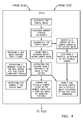
- FIG. 1Ais a flowchart representation of a method of a first preferred embodiment
- FIG. 1Bis a flowchart representation of one variation of the first preferred method
- FIG. 2Ais a flowchart representation of a method of a second preferred embodiment
- FIG. 2Bis a flowchart representation of one variation of the second preferred method
- FIG. 3Ais a flowchart representation of a method of a third preferred embodiment
- FIG. 3Bis a flowchart representation of one variation of the third preferred method
- FIG. 4is a flowchart representation of one variation of the first preferred method
- FIG. 5is a flowchart representation of one variation of the first preferred method
- FIG. 6is a flowchart representation of one variation of the first preferred method
- FIG. 7is a flowchart representation of another variation of the preferred method.
- FIG. 8is a schematic of a system of a preferred embodiment
- FIG. 9is a schematic of a variation of the preferred system.
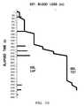
- FIG. 10is a graphical representation of an output in accordance with a system or a method of a preferred embodiment.
- a method S 100 of a preferred embodiment for estimating the extracorporeal blood volume in a portion of a physical sampleincludes: extracting a feature from a portion of an image of the sample in Block S 110 ; tagging the portion of the image of the sample with a blood volume indicator according to the extracted feature in Block S 120 ; and estimating the extracorporeal blood volume in at least the portion of the physical sample, associated with the portion of the image of the sample, according to the blood volume indicator in Block S 130 .
- the first preferred method S 100preferably functions to estimate the volume of blood in the physical sample by analyzing an image of the sample.
- the image of the sampleis preferably a color frame of a live video feed, wherein at least a portion of the physical sample is visible in the frame.
- the imagecan alternatively be a static or still image, an infrared image, a field of view of an optical sensor, a black and white image, a fingerprint of a field of view of an optical sensor, a point cloud, or any other suitable type of image.
- the imagecan be a scan of the physical sample.
- the imagecan be captured and then stored on a local or remote data storage device for subsequent processing, though the image can alternatively or in parallel be processed in real time, or in parts or segments to avoid storage of the full image.
- the first preferred method S 100preferably estimates an extracorporeal blood volume that includes blood external the body of a patient or subject. Additionally or alternatively, the first preferred method S 100 can estimate an extravascular blood volume that includes blood within the body of a patient or subject but external the vascular system of the patient.
- the physical sampleis preferably an absorbent surgical gauze sponge, a surgical dressing, or a surgical towel, though the sample can be any other textile. Additionally or alternatively, the physical sample can be a piece of clothing, a ground, table, wall, or floor surface, an external skin surface, a surgical glove, a surgical implement, or any other surface, material, substrate, or object.
- a surgeon, nurse, anesthesiologist, gynecologist, soldier, paramedic, or other usercan use a machine or device incorporating the first preferred method S 100 to estimate blood volume in one or more physical samples to generate a total estimated blood loss (EBL) of a patient, such as during a surgery, childbirth or any other medical or health-related event.
- EBLestimated blood loss
- a law enforcement officer, forensic investigator, or other usercan use a machine or device implementing the first preferred method S 100 to estimate extracorporeal blood volume at a crime scene or to assess victim risk during a medical emergency.
- the first preferred method S 100can additionally or alternatively function to estimate the volume, mass, or quantity of another blood-related parameter or extracorporeal blood volume indicator in the physical sample, such as hemoglobin or red blood cell mass or volume in the physical sample.
- another blood-related parameter or extracorporeal blood volume indicator in the physical samplesuch as hemoglobin or red blood cell mass or volume in the physical sample.
- Such blood-related parameterscan then be evaluated against additional variables or features to calculate the volume of blood, hemoglobin, red blood cells, white blood cells, plasma, etc. in the physical sample.
- HCThematocrit
- the first preferred method S 100can additionally or alternatively function to detect presence of blood in the sample, compute blood spread rate, compute blood loss rate, calculate blood surface area, estimate patient risk level (e.g., hypovolemic shock), and/or determine hemorrhage classification of the patient.
- the first preferred method S 100can provide any other functionality, analyze any other image type or format, estimate any other blood-related parameter, and/or calculate blood volume in the physical sample in any other way.
- the first preferred method S 100is preferably implemented in a handheld (mobile) electronic device, such as an application (or ‘app’) executing on a digital music player, a smartphone, or a tablet computer, as shown in FIG. 9 , wherein a camera integral with the electronic device captures the image of the sample, wherein a processor integral with the electronic device performs Blocks S 110 , S 120 , and S 130 , and wherein a display integral with the electronic device performs Block S 160 , which recites displaying the estimated blood volume of the portion of the physical sample, the whole of the physical sample, and/or a summed total blood volume across multiple physical samples.
- a handheld (mobile) electronic devicesuch as an application (or ‘app’) executing on a digital music player, a smartphone, or a tablet computer, as shown in FIG. 9 , wherein a camera integral with the electronic device captures the image of the sample, wherein a processor integral with the electronic device performs Blocks S 110 , S 120 , and S 130 , and where
- the electronic devicecan alternatively communicate with a remote server, such as via a wireless communication module 150 implementing cellular, Wi-Fi, or Bluetooth protocol, wherein the server performs at least some of Blocks S 110 , S 120 , and S 130 , and wherein at least some of the outputs of Blocks S 110 , S 120 , and S 130 are transmitted back to the electronic device and subsequently displayed.
- a remote serversuch as via a wireless communication module 150 implementing cellular, Wi-Fi, or Bluetooth protocol
- the first preferred method S 100can also be a standalone blood volume estimation system, such as a system including a staging tray configured to support a sample, a camera configured to image the sample, and a processor configured to perform at least a portion of the first preferred method S 100 and/or a communication module that communicates with a remote server configured to perform at least a portion of the first preferred method S 100 .
- the first preferred method S 100can be implemented in any other system, device, or combination thereof.
- the first preferred method S 100can therefore be useful in a hospital setting, such as in a surgical operating room, in a clinical setting, such as in a delivery room, in a military setting, such as on a battlefield, in a law enforcement setting, such as at a crime scene, or in a residential setting, such as to monitor blood loss due to menorrhagia (heavy menstrual bleeding) or epistaxis (nosebleeds).
- a hospital settingsuch as in a surgical operating room
- a clinical settingsuch as in a delivery room
- a military settingsuch as on a battlefield
- a law enforcement settingsuch as at a crime scene
- a residential settingsuch as to monitor blood loss due to menorrhagia (heavy menstrual bleeding) or epistaxis (nosebleeds).
- the first preferred method S 100can be useful in any other setting.
- Block S 110 of the first preferred method S 100includes extracting a feature from a portion of an image of the sample.
- the extracted feature of the portion of the imagepreferably enables correlation (or pairing) of the portion of the image with a blood loss indicator of the portion of the sample in Block S 120 , which can further enable estimation of the blood volume in the portion of the sample in Block S 130 .
- the extracted featureis preferably an intensity, luminosity, hue, saturation, brightness, gloss, or other color-related value of the portion of the image in at least one component space, such as the red, blue, green, cyan, magenta, yellow, key, and/or Lab component spaces.
- the extracted featurecan be a histogram of various color values across a set of pixels in the portion of the image. Additionally or alternatively, the extracted feature can be an estimated surface area of the sample shown in the image, an estimated surface area of a bloodied portion of the sample, a pixel count of the portion of the sample, a pixel count of the entire sample, or a pixel count of only the bloodied region of the sample, a color intensity value of an unsoiled portion of the sample, or any other relevant feature inherent in or available for extraction from the portion of the image of the sample. Furthermore, Block S 110 can include extracting any number of features from all or a portion of the image of the sample.
- Block S 110can similarly include accessing non-image features, such as a current patient intravascular hematocrit, an estimated patient intravascular hematocrit, an historic patient intravascular hematocrit, a weight of the sample, a clinician-estimated sample blood volume, computer-vision-based or gravimetric or human-generated estimates of blood volumes of previous samples, an ambient lighting condition, a type or other identifier of the physical sample, properties of the physical sample, a patient vital sign, patient medical history, an identity of a surgeon, or a type of surgery.
- non-image featuressuch as a current patient intravascular hematocrit, an estimated patient intravascular hematocrit, an historic patient intravascular hematocrit, a weight of the sample, a clinician-estimated sample blood volume, computer-vision-based or gravimetric or human-generated estimates of blood volumes of previous samples, an ambient lighting condition, a type or other identifier of the physical sample, properties of the physical sample, a patient vital sign, patient medical history, an identity of
- any of these non-image featurescan inform selection of template images for comparison with the portion of the sample image, selection of a particular parametric model or function, definition of alarm triggers for misplaced surgical gauze sponges, definition of alarm triggers for excess fluid or blood loss, transformation of extracted features into the blood volume indicator, and/or estimation of blood volume from the blood volume indicator.
- any of these non-image featurescan modify enable, or inform any other function of the first preferred method S 100 .
- Block S 110preferably includes segmenting the image, including isolating a first segment of the sample image representative of the physical sample that is a bloodied object (e.g., a surgical gauze sponge). Block S 110 preferably subsequently further segments the first region to define the portion of the sample image that corresponds to a particular portion of the physical sample captured in the sample image. Segmenting the sample image into multiple image segments preferably increases the resolution and/or accuracy of the estimated blood volume of each portion of the physical sample.
- the size and shape of each image segmentcan be static, wherein each segment comprises a predefined number of pixels in the image and/or a predefined dimension in physical space, as shown in FIG. 9 .
- the image segmentcan define a ten-pixel by ten-pixel rectilinear area of the image or a five-millimeter equilateral triangular area of the physical sample.
- the image segmentcan be isolated according to properties of individual pixels or groups of pixels in the image, such as hue, saturation, shade, brightness, chroma, wavelength, or any other metric of color or light, as shown in FIG. 7 .
- the sample imagecan be dynamically segmented, wherein portions of the sample image are separated by color property or other features rather than by (or in addition to) pixel location or by location on the physical sample.
- the portion of the sample imagecan include the whole of the physical sample associated with the sample image, or the sample image can be segmented or apportioned according to any other schema.
- the portion of the sample imageis preferably a single segment or region including multiple pixels of the image, but the portion of the sample image can alternatively be a plurality of image segments or regions of the sample image, can be of any other size, and/or can be of any other form.
- Block S 110extracts a feature from the sample image that is a dimension of the physical sample.
- Block S 110implements object recognition to isolate an object of known type within the field of view of the optical sensor and/or within the sample image.
- the objectcan be a surgical tool, a surgical tray, an operating table, a surgical gauze sponge, a suction canister, or any other object of known dimension. From this known dimension, a dimension of the physical sample can be extrapolated, such as by estimating the distance from and/or angle between the optical sensor and the known object and comparing the position of the sample and the known object in the image.
- Block S 110analyzes shadows in the sample image, coupled with known locations of light sources, to estimate an angle and distance between the physical sample and the capture origin (i.e. the location of the camera or optical sensor when the sample image was captured).
- the optical sensoris arranged at a known distance from and angle to a staging tray on which the physical sample is arranged for imaging, and Block S 110 includes extrapolating the dimension of the physical sample or a portion therefore based upon known placement of the optical sensor relative the staging tray.
- Block S 110manipulates an IR, sonic, laser, or other type of distance sensor arranged adjacent the optical sensor to transmit a signal toward the physical sample to determine the distance and/or angle between the physical sample and the capture origin of the image.
- a dimension of the physical sample or a portion thereofcan be estimated or determined in any other way.
- the distance and/or angle between the sample and the optical sensorcan be automatically extracted from the image to inform a transform from pixel count of the portion of the sample image into a physical dimension (e.g., inch, centimeter) of the corresponding portion of the physical sample in Block S 110 .
- the estimated angle and/or distancecan therefore define an extracted feature of the sample image that informs the generation of the blood indicator tag and/or the transformation of the blood indicator tag into the estimated blood volume in the portion of the physical sample.
- the distance and/or angle value(s)can be input by a user (e.g., a surgeon, a nurse), extrapolated from data generated by a non-optical sensor, or calculated or gathered in any other way to define a non-image feature related to the sample.
- a usere.g., a surgeon, a nurse
- extrapolated from data generated by a non-optical sensoror calculated or gathered in any other way to define a non-image feature related to the sample.
- Block S 110can additionally or alternatively implement any object localization, segmentation (e.g. using edge detection, background subtraction, graph-cut-based algorithms, etc.), gauging, clustering, pattern recognition, template matching (using any one of various metrics), feature extraction, descriptor extraction (e.g. extraction of texton maps, color histograms, HOG, SIFT, etc.), feature dimensionality reduction (e.g. PCA, K-Means, linear discriminant analysis, etc.), feature selection, thresholding, positioning, color analysis, parameteric regression, non-parametric regression, unsupervised or semisupervised parametric or non-parametric regression, or any other type of machine learning or machine vision to estimate a physical dimension of the sample.
- object localizatione.g. using edge detection, background subtraction, graph-cut-based algorithms, etc.
- gaugingclustering
- pattern recognitionusing any one of various metrics
- feature extractione.g. extraction of texton maps, color histograms, HOG, SIFT, etc.
- feature dimensionality reduction
- Such methodspreferably compensate for varying lighting conditions of the physical sample, warping of the physical sample (e.g., a wrinkle or warped gauze sponge), warping of the image of the physical sample (e.g., due to optical distortion caused by a lens of the optical sensor), variations in composition of the fluid present in or on the sample, or any other inconsistency or variable prevalent in any use scenarios.
- warping of the physical samplee.g., a wrinkle or warped gauze sponge
- warping of the image of the physical samplee.g., due to optical distortion caused by a lens of the optical sensor
- variations in composition of the fluid present in or on the samplee.g., due to optical distortion caused by a lens of the optical sensor
- variations in composition of the fluid present in or on the samplee.g., due to optical distortion caused by a lens of the optical sensor
- variations in composition of the fluid present in or on the samplee.g., variations in composition of the fluid present in or on the sample, or any other inconsistency or
- Block S 110includes assessing ambient lighting conditions, wherein the ambient lighting conditions define an extracted feature.
- the ‘redness,’ ‘greenness,’ and ‘blueness’ valuesi.e. color values in the red, green, and blue color component spaces
- the weighted composite color valuecan then be fed into a parametric function or compared against color values of template images to generate the blood volume indicator.
- Block S 110includes extracting a feature that identifies the physical sample in the sample image as an absorbent surgical gauze sponge.
- Block S 110preferably implements machine vision, such as object segmentation, edge detection, pattern recognition, or template matching, to determine if the sample image includes an absorbent surgical gauze sponge or if an absorbent surgical gauze sponge is within a field of view of a camera or other optical sensor.
- Block S 110can preferably determine the type of absorbent surgical gauze sponge, such as laparotomy or RAY-TEC gauze, which can inform selection of a template image of a similar template sample type for comparison with the portion of the sample.
- Block S 110can additionally or alternatively identify thread count, external dimension, color, physical tag, or any other identifying feature or property of the physical sample to identify the type of sample or a fluid absorptivity, saturation volume, dry weight or mass, dry color, or any other property of the physical sample. Block S 110 can therefore reduce processing time necessary to return a template image match for the portion of the sample by isolating key identifying features of the physical sample. Similarly, Block S 110 can improve accuracy of the blood volume estimation by isolating key properties that affect fluid absorbance and a correlation between fluid volume and optical properties of the physical sample.
- Block S 110can additionally or alternatively extract features from the sample image that identify other relevant objects, materials, or fluids in the sample image and/or the field of view of the optical sensor. For example, Block S 110 can recognize drops, pools, or smears of blood on a surgical tool, tray, table, wall, floor, or other surface as containing blood. Block S 110 can initiate an estimation of blood volume in or on a sample that is other than an absorbent surgical gauze sponge, surgical dressing, or surgical towel.
- template matchingcan be used to estimate blood volume in or on the physical sample, as described below, although color value, translucency, saturation, dimension, or any other metric of the sample can be used to parametrically or non-parametrically generate the blood volume indicator tag and/or estimate the extracorporeal blood volume in at least the portion of the physical sample.
- Block S 120 of the first preferred method S 100includes tagging the portion of the image of the sample with the blood volume indicator according to the extracted feature.
- the extracorporeal blood volume indicator tagis preferably an intermediate parameter for a region of interest in the sample image that translates pixel-level data in the sample image into a blood volume-related variable.
- the blood volume indicator tagis therefore preferably an estimate of hemoglobin content (e.g., mass, volume, density, percentage by weight, etc.) in the portion of the sample, though the extracorporeal blood volume indicator tag can alternatively be an estimate of red blood cell count or content, white blood count or content, platelet count or content, plasma content, or any other suitable extracorporeal blood volume indicator.
- the tagcan also include any other relevant information, such as estimated hematocrit of the blood in the portion of the physical sample, a time stamp of when the sample image was taken, a time stamp of when the sample image was analyzed, or volume or concentration of other fluids present on or in the portion of the sample, such as bile, saliva, gastric fluid, mucus, pleural fluid, saline, or fecal matter.
- the blood volume tagis preferably of a form that can be transformed or manipulated into an estimated extracorporeal blood volume in all or a portion of the sample.
- the extracorporeal blood volume indicator tag for the portion of the sample imageis preferably stored with the portion of the sample image or as a pointer to the portion of the sample image.
- Block S 120includes comparing the extracted feature of the portion of the image of the sample against similar features extracted from template samples (e.g., a training set, samples analyzed previously) of known blood volume indicators and/or known extracorporeal blood volumes.
- template samplese.g., a training set, samples analyzed previously
- the portion of the imageis tagged with the blood volume indicator based upon a non-parametric correlation with one or more template samples.
- Block S 120can include implementing a K-nearest neighbor method to compare the extracted feature of the image that is a redness intensity in the red component space with redness intensity values of template samples.
- Block S 120can further include implementing a K-nearest neighbor method to compare extracted features that include a greenness intensity and a blueness intensity (in conjunction with a redness intensity) of pixels from bloodied regions in the sample image with greenness and blueness intensity values of template samples.
- Block S 120includes pairing the portion of the image of the sample to a template image of known extracorporeal blood volume indicator.
- Each template imageis preferably contained within a library of template images, and each template image is preferably an image of a template sample of known blood, hemoglobin, red blood cell mass or volume (e.g., per unit physical area), and/or any other suitable blood-related parameter, blood volume indicator, or feature.
- Each template image in the libraryis preferably tagged with an extracorporeal blood volume indicator such that the portion of the sample image can be matched to a template image in Block S 110 , and such that a tag, indicative of the blood volume in the portion of the physical sample, can be associated with the portion of the sample image in Block S 120 .
- the library of template imagescan be assembled in a variety of ways.
- an imageis taken of a template sample that is a used surgical gauze, blood is washed from the used gauze and assayed to determine the hemoglobin mass absorbed into the used gauze, the image of the template sample is tagged with the hemoglobin mass (the extracorporeal blood volume indicator), and the image is catalogued in the library.
- a template sampleis prepared by adding a known volume of blood (of known hematocrit) to a surgical gauze of a known size, an image of the template sample is taken, the image of the template sample is tagged with the known blood volume (the extracorporeal blood volume indicator), and the image is catalogued in the library.
- the blood volume tag of each image templateis preferably a volume or mass of a blood-related parameter, such as hemoglobin or red blood cell content per physical area (e.g., 1 cm 2 ) such that, in Block S 130 , a blood volume indicator tag of a portion of the image can be multiple by an estimate physical area (or volume) of the corresponding portion of the physical sample to estimate the extracorporeal blood volume in the portion of the sample, as shown in FIG. 7 .
- a blood volume indicator tag of a portion of the imagecan be multiple by an estimate physical area (or volume) of the corresponding portion of the physical sample to estimate the extracorporeal blood volume in the portion of the sample, as shown in FIG. 7 .
- the template sample for each template imagecan be prepared in any other way or combination of ways, and the extracorporeal blood volume indicator can be any other suitable parameter or metric.
- the librarypreferably contains a large number of template images to account for variance in lighting, image quality, type of physical sample (e.g., type of surgical gauze sponge), volumes, concentrations, or hematocrits of blood or other indicator in each sample, “age” of the physical sample, surgical conditions, or any other suitable variable.
- the template images in the librarycan also be grouped, such as according to: the type of template sample, such as a gauze sponge, floor, operating table, clothing; lighting or backlighting of the template sample; hematocrit of blood in a template sample; thread count of the template sample that is a textile; quality of the image of the template sample, such as depth of field, focus, distance between the template sample and an optical sensor; or any other suitable parameter.
- the librarycan be stored locally on a machine or system configured to perform at least a portion of the first preferred method S 100 , or remotely, such as on a remote server or hard drive accessible by the machine or system when performing at least a portion of the first preferred method S 100 .
- each image segmentcan be decomposed into features that are separate color components (e.g., red, green, and blue), and the absolute difference in pixel intensity for the pixels in the portion of the sample image and the pixels in the template image can be calculated for at least one color component.
- the sample imagecan alternatively be decomposed prior to segmentation.
- the absolute difference in pixel intensityis preferably calculated at a wavelength of light that correlates with the extracorporeal blood volume indicator.
- the absolute difference in pixel intensity for the portion of the sample image and the template imagecan be calculated at 400 nm, a wavelength that can correlate well with hemoglobin concentration for certain absorbent surgical gauze sponges.
- the template imageis preferably paired with the portion of the image when a substantially minimal sum of absolute difference in pixel intensity between the portion of the sample image and the template image is calculated.
- Block S 120can implement a texton map to pair the sample image with one or more template images.
- patches from template (training) imagescan be clustered into centroid patches, such as by k-means clustering.
- the index of the centroid patch nearest the patch surrounding the pixelcan be calculated such that a histogram, of the nearest-centroid indices within a window around each pixel, can be constructed.
- a background histogram centroidcan also be constructed. Clean and bloodied histogram centroids for physical samples (e.g., surgical gauze sponges) can be similarly constructed.
- a classification algorithmsuch as SVM, Na ⁇ ve Bayes, LDA, K-Nearest-Neighbors, or logistic regression, can be trained using histograms centered around or mostly containing background, bloodied, and unsoiled pixels.
- histogram of the nearest-patch-centroid indices around each pixel in the portion of the sample imageis generated and classified based upon a comparison of the histogram and histogram centroid of the pixel, or based upon the output of one of the learned classifiers described above.
- the histograms and/or histogram centroids of the pixels in the portion of the sample imagecan then be compared with a subset of histograms and/or histogram centroids of pixels of the template images, based upon the determined class of physical sample, to pair one or more template images with the sample image.
- Block S 120therefore preferably recites stepping through subsequent template images in the template image library until a suitable match is found for the portion of the sample image.
- the hue, saturation, shade, brightness, chroma, intensity of wavelength, wavelength range, histogram, histogram centroid, class, or any other color property (e.g., feature) of the portion of the sample image and the template imagecan be compared in Block S 120 .
- the portion of the sample image and the template imageare preferably compared substantially directly.
- the template image and the portion of the sample imagecan be compared via template matching incorporating any other vision algorithm or image processing method.
- each template imageis a different color or hue in a library of color palettes, wherein each color correlates with a different blood volume or blood volume indicator.
- the librarypreferably includes color palettes for different types of surgical sponge gauzes, surgical towels, surgical tool surfaces, floor surfaces, operating or delivery table surfaces, and/or any other common surface, material, object, or feature, wherein each color that is a template image in a color palette is associated with a particular red blood cell content or indicator for a particular type of physical sample.
- the template image that is a colorcan be an image of the color or a numerical color identifier, such as a HEX code value (e.g., #FF0000, #A00000, #880000, etc.) or an RGB code value (e.g., (255, 0, 0), (160, 0, 0), (190, 0, 0), etc.).
- a HEX code valuee.g., #FF0000, #A00000, #880000, etc.
- RGB code valuee.g., (255, 0, 0), (160, 0, 0), (190, 0, 0), etc.
- the feature extracted from the portion of the sample image in Block S 110is a redness value, wherein the redness value is an intensity of a wavelength or composite intensity of a range of wavelengths of light, redness hue, redness saturation, or any other suitable light- or color-related value.
- Block S 110can similarly extract greenness, blueness, or other color component values of one or more bloodied pixels in the sample image.
- Block S 110preferably decomposes the sample image into distinct color spaces, such as red, green, and blue component spaces, wherein a color value or intensity is calculated for the portion of the sample image in each color space.
- the portion of the sample image that is decomposed in Block S 110preferably includes red pixels indicative of blood content in the portion of the physical sample that is associated with the portion of the sample image.
- the color value(s) of the portion of the imageare then compared substantially directly with color values of template images until a suitable match is found.
- template images with properties substantially dissimilar from those of the portion of the physical sample or the sample imagecan be withdrawn from comparison with the portion of the sample image in Block S 120 in order to reduce processing time required to find a template image match.
- template images of template samples of surfaces, products, materials, or dimensions substantially dissimilar from that of the portion of the physical sampleare excluded from comparison.
- Block S 110can extract a thread count feature from the sample image, wherein the thread count feature identifies the physical sample as laparotomy gauze, and wherein all template images of template samples that are not of laparotomy gauzes are removed from comparison with the portion of the sample image.
- thresholdingis used to remove substantially irrelevant template images from the test pool.
- template images with redness valuese.g., intensity, hue, saturation, shade, brightness, chroma, wavelength range
- Tree searchcan additionally or alternatively be used to reduce processing time.
- template imagescan be grouped in the template library and selected or deselected for comparison with the portion of the sample image according to any other schema.
- Block S 120includes transforming the extracted feature of the portion of the image of the sample into the blood volume indicator.
- Block S 120preferably implements an algorithm or other mathematical transformation to convert the extracted feature into the blood volume indicator for the portion of the image of the sample. Therefore, in this variation, Block S 120 preferably implements parameterized generation of the blood volume indicator.
- color values of the template imagesare used to generate a mathematical function, curve, or algorithm that correlates the extracted feature to the blood volume indicator.
- the extracted feature of the portion of the sample imagee.g., redness intensity in the red component space, blueness intensity in the blue component space, greenness intensity in the green component space, or a composite of two or three color intensities
- a parametric functione.g., intensity-blood volume function
- reflectance of oxygenated hemoglobin (Hb O2 )can be correlated with certain wavelengths of light to substantially directly estimate the content of hemoglobin in the portion of the physical sample associated with the portion of the image.
- red blood cell countcan be extrapolated from hemoglobin content.
- Blocks S 120 and S 130can implement both parametric and non-parametric techniques or methods to correlate one of more extracted features to one or more blood volume indicators. For example, extracted features that are color values in the red, green, and blue color spaces can be compared with template images via non-parametric techniques (e.g., template matching) to tag the portion of the sample with the blood volume indicator, and an extracted feature that is an estimated surface area of a bloodied region of the physical sample can be transformed according to a parametric function to generate a coefficient for conversion of the blood volume indicator into an estimated blood volume in the portion of the sample.
- non-parametric techniquese.g., template matching
- Block S 120 and S 130can manipulate any relevant image-based feature extracted in Block S 110 or any non-image-based feature (e.g., sourced from a clinician, sourced from a medical record, etc.) to generate the blood volume indicator of the portion of the image and the estimated blood volume for at least the portion of the sample, respectively.
- any relevant image-based feature extracted in Block S 110 or any non-image-based featuree.g., sourced from a clinician, sourced from a medical record, etc.
- Block S 130 of the first preferred method S 100includes estimating the extracorporeal blood volume in at least a portion of the physical sample, associated with the portion of the sample image, according to the blood volume indicator tag.
- the blood volume for each portion of the physical sample correlating with a portion of the imagecan be independently calculated and then summed to estimate a total blood volume in the physical sample.
- Block S 130can additionally or alternatively include estimating total hemoglobin mass or volume, total red blood cell mass or volume, or any other blood-related metric in the physical sample or across multiple samples.
- a variation of the first preferred method S 100further includes Block S 140 , which recites identifying the physical sample in the image as a type of absorbent gauze sponge.
- Block S 140preferably implements machine vision techniques to determine the type of physical sample, as described above. From identification of the type of physical sample in Block S 140 , the first preferred method S 100 can access sample-specific data such as dry weight, absorptivity, fluid saturation volume, or any other data or property of the physical sample, which can enable extraction of additional blood-related data from the image of the physical sample.
- Block S 150recites indexing a sample count for the physical sample.
- the sample countis preferably a count of absorbent surgical gauze sponges, dressings, or towels, though the sample count can additionally or alternatively be a count of blood droplets, blood drops, pools of blood, bloodied articles of clothing, bloodied surgical tools, or any other relevant or suitable blood formation or bloodied object.
- the sample countis preferably displayed with the estimated blood volume of the portion of the physical sample, and the sample count is preferably indexed substantially in real time when the image of the physical sample is taken.
- Block S 150can function in any other way to index the sample count and to provide this information to a user.
- Block S 160which recites displaying the estimated blood volume in the portion of the physical sample, the estimated blood volume in the whole physical sample, and/or the estimated total blood volume across multiple physical samples.
- At least some of this datais preferably presented to a user, such as to a surgeon, a nurse, an anesthesiologist, a gynecologist, a doctor, or a soldier.
- This datais preferably rendered on a digital display of a machine or system configured to perform at least a portion of the first preferred method S 100 .
- this datacan be presented in the form of an augmented reality overlay on top of the static sample image depicted on the display.
- this datacan be presented in the form of a dynamic augmented reality overlay on top of a live video stream captured by the optical sensor and depicted on the display.
- datacan be presented in an augmented reality overlay on subsequent scanned images of one physical sample, wherein the camera captures digital images at a rate such as 30 frames per second and the augmented reality overlay updates with each new frame or number of frames.
- This datacan alternatively be presented in table, chart, or diagram including the estimated blood volume in one or more physical samples over a period of time.
- Other blood-related metricscan also be estimated or maintained in the first preferred method S 100 and presented in Block S 160 , such as blood spread rate, blood surface area, patient risk level, or patient hemorrhage classification. However, this data or any other blood-related metric or patient information can be presented in any other way or form in Block S 160 .
- Block S 170recites estimating patient blood loss by summing the blood volume estimate of the physical sample with previous blood volume estimates of other physical samples. Additionally or alternatively, the blood volume estimate of the physical sample can be stored for future summation with blood volume estimates of additional physical samples. By summing blood volume estimates across multiple physical samples, blood loss of a patient can be tracked over time. For example, during a surgery, used surgical gauze sponges can be analyzed via Blocks S 110 and S 120 , wherein a running summation of blood volumes in each used gauze sponge provides time-elapse estimates of total blood loss of the patient, as shown in FIG. 10 .
- Other blood-related metricscan also be estimated or maintained in Block S 130 and summed over time in Block S 170 .
- Block S 180recites comparing the identified physical sample against a set of past identified physical samples.
- Block S 150preferably indexes the sample counter only when the identified physical sample is determined to be unique amongst the set of past identified physical samples.
- Block S 180therefore functions to determine if a previous sample image including the same physical sample was already analyzed according to any of Blocks S 110 , S 120 , and/or S 130 .
- Block S 180preferably substantially guards against double counting of the estimated blood volume in the physical sample in Block S 170 .
- Each sample image, a fingerprint of each sample image, or a fingerprint of each physical sampleis therefore preferably stored, such as on a local or remote sample image database, such that subsequent sample images or physical samples can be compared against past sample images or physical samples in Block S 180 .
- comparison of the sample image with previous sample imagescan require scale, rotation, mirror, stretch or other transformations or fingerprinting of the sample image and/or or previous sample images.
- Edge detection, segmentation, pattern recognition, feature extraction, and/or other machine vision techniquescan be used to determine the uniqueness of bloodied regions of the physical sample shown in the sample image, relative to bloodied regions of other, previously analyzed physical samples.
- Block S 180can function in any other way to identify the sample image as including the physical sample that was included in a previous sample image.
- Block S 190recites updating a digital medical record of the patient with the estimated blood volume in the physical sample.
- Block S 190can additionally or alternatively update the medical record of the patient with the estimated blood volume across multiple physical samples, the estimated blood loss of the patient, patient blood loss trends, or any other relevant metric or data generated related to the circulatory system of a patient.
- the digital medical recordcan be maintained locally on a machine or system implementing the first preferred method S 100 or on a local network or remote server accessed by the machine or system to retrieve, update, and/or upload the digital medical record.
- the physical sampleis a fluid canister that collects bodily fluids of a patient, such as blood, bile, saliva, gastric fluid, mucus, pleural fluid, urine, or fecal matter, wherein the image is an image of the fluid canister.
- Block S 110can include extracting features that include a volume of fluid within the canister, as well as redness, greenness, and blueness intensities of the portion of the image of that canister that includes bloodied pixels and preferably includes little to no glare.
- Block S 120can include estimating a percentage of blood within the canister relative to other bodily fluids based upon the extracted color values
- Block S 130can include estimating the volume of blood within the canister.
- the optical sensor that captures the image of the fluid canisteris preferably mounted to the fluid canister.
- the optical sensoris mounted to the side of and facing the fluid canister that is cylindrical such that the fluid level in the fluid canister can be estimated directly from the sample image.
- the optical sensoris mounted overhead the fluid canister that also includes a fluid level sensor, wherein an output of the fluid sensor defines a non-image feature that informs at least one of the blood volume indicator and the estimated blood volume in the fluid canister.
- the optical sensorcan be incorporated into a handheld device, wherein a user scans the fluid canister with the optical sensor to capture the sample image.
- an auxiliary light sourcesuch as a lamp or laser next to the canister
- ambient lightcould be assessed and used as a feature.
- fluidis added to the fluid canister over time
- subsequent sample images of the fluid canistercan be captured and analyzed over time, via the first preferred method S 100 , to generate a time-dependent, historical chronicle of fluid content of the fluid canister.
- Estimated blood volume in the fluid canistercan therefore be monitored over time, such as to generate a trend in blood loss for a patient.
- Such datacan be useful to trigger alarms if patient blood loss is occurring too rapidly or if patient blood loss has reached a critical total volume or critical red blood cell loss.
- loss of other fluidscan also be monitored.
- urine content (or total water content) of the fluid canistercan enable tracking of patient hydration level such that the patient can be administered saline when hydration level or hydration loss surpasses a threshold.
- Differences between fluid color properties of one sample image at a first time and a subsequent sample image at a second timecan indicate concentration changes of fluids in the fluid canister between the first and second times.
- a change in fluid level in the canister between the first and second times, coupled with fluid concentration changescan indicate the floor rate of fluids into (or out of) the fluid canister.
- Estimated blood and/or other fluid loss through analysis of the sample image of the fluid canistercan be further fed into analyses of sample images of surgical sponge gauzes, implements, surfaces, etc. to map total blood and/or other fluid loss of the patient over time.
- the first preferred method S 100can function in any other way to estimate the volume of blood within the physical sample that is a fluid canister.
- One variation of the first preferred method S 100further comprises estimating the volume of extracorporeal non-blood fluids in the physical sample, such as ascites, saline irrigant, bile, plasma, urine, or saliva.
- the redness of the physical samplee.g., color intensity of image pixels associated with the physical sample in the red component space
- the total red blood cell count or volumeis subtracted from the estimated total extracorporeal blood volume in the sample, according to an estimated or measured hematocrit of the blood in the physical sample, to estimate the total volume of plasma in the physical sample.
- the estimated total extracorporeal blood volumeis converted to as estimated total extracorporeal blood weight (or mass), wherein the estimated total extracorporeal blood weight (or mass) and dry weight (or mass) of the physical sample are subtracted from a wet weight (or mass) of the physical sample to estimate the total weight (or mass of volume) of substantially clear fluids (e.g., saline, intestinal ascites) in the physical sample.
- the first preferred method S 100preferably accessed a mass or weight measurement of the physical sample through a scale electrically coupled to the machine or device implementing the first preferred method S 100 .
- the first preferred method S 100preferably implements machine vision techniques to determine the type of physical sample, such as a surgical dressing, a surgical gauze sponge, or a surgical towel from a particular manufacturer.
- the first preferred method S 100can then access sample-specific data such as dry weight, absorptivity, fluid and/or saturation volume to enable extraction of further data related to blood or non-blood fluids in the physical sample.
- sample-specific datasuch as dry weight, absorptivity, fluid and/or saturation volume
- the first preferred method S 100can implement any other technique or method to estimate the volume, weight, or mass of an extracorporeal non-blood fluid in the physical sample.
- the first preferred methodcan additionally or alternatively analyze one or more extracted and/or non-image features to estimate any one or more of hemoglobin mass, hematocrit, hemoglobin concentration, fresh frozen plasma, packed red blood cells, colloids, platelets, crystalloid, or any other blood-related parameter of the patient. Any one or more of these blood-related parameters can additionally or alternatively be rendered on a display of the machine, system, or device implementing the first preferred method S 100 .
- One variation of the first preferred methodincludes recognizing gestures of a user to control operation of the machine, system, or device implementing the first preferred method S 100 .
- the preferred methodpreferably accesses a live video feed captured by the optical sensor that records the image of the physical sample or by any other optical sensor or camera coupled to the machine, system, or device implementing the first preferred method S 100 .
- the first preferred methodis preferably implemented during a surgery or other medical event or emergency during which a user is likely wearing a glove
- the first preferred method S 100is preferably controlled via non-contact means.
- this variation of the first preferred method S 100preferably recognizes non-contact hand gestures.
- a ‘thumbs up’can indicate that the user accepts the detection of the physical sample and the extracorporeal blood volume estimation of the physical sample. The extracorporeal blood volume can then be added to an aggregate extracorporeal blood volume estimated for a set of physical samples. Similarly, a ‘thumbs down’ can reject the detection and extracorporeal blood volume estimation for the physical sample.
- a usercan scroll through available physical sample types by sweeping a hand to the left or right. Similarly, the user can scroll through images of previous samples by sweeping a hand vertically.
- any other gesturecan be recognized in any other way to control any other function of the first preferred method S 100 .
- the preferred method S 100further functions to generate alarms or warnings related to the circulatory system of a patient.
- the preferred method S 100generates a warning that a physical sample that is a surgical sponge gauze was lost or left inside the patient if not identified within a threshold time (e.g., one hour) after being checked into a surgery.
- the first preferred method S 100sounds an alarm when the total estimated blood or red blood cell loss of the patient surpasses a threshold level.
- the threshold blood or red blood cell volumecan be unique to the patient and based upon any one or more of the age, gender, weight, medical history, etc. of the patient.
- the first preferred method S 100issues a warning of trends in patient blood loss, such as based upon blood distribution across multiple physical samples (e.g., sponges) over time.
- the first preferred methodcan additionally or alternatively provide data and/or warnings relating to a rate of blood loss, a rate of blood loss relative to sponge count, a rate of sponge usage, a histogram of sponge usage, or any other suitable data or warning related to the circulatory system of the patient.
- a second preferred method S 200 for estimating the extracorporeal blood volume in a portion of a physical sampleincludes: comparing a portion of an image of the sample with a template image of known extracorporeal blood volume indicator in Block S 210 ; tagging the portion of the image of the sample with a blood volume indicator according to the template image that is matched to the portion of the image of the sample in Block S 220 ; and estimating the extracorporeal blood volume in at least a portion of the physical sample, associated with the portion of the image of the sample, according to the blood volume indicator in Block S 230 .
- the second preferred method S 200preferably implements non-parametric estimation (e.g., template matching) of extracorporeal blood volume in the physical sample, as described above.
- Block S 220preferably incorporates a variation of Block S 220 of the first preferred method S 100
- Block S 230preferably incorporates a variation of Block S 130 of the first preferred method S 100 .
- the second preferred method S 200can implement any other technique, method, implementation, and/or variation of the first preferred method S 100 described above.
- One variation of the second preferred method S 200includes accessing the template image that is a color model paired with a blood volume indicator.
- the color modelcan be a template image, a representation of or feature extracted from a template image, a mathematical function or algorithm, or any other suitable color model correlating an extracted feature of the sample image with a blood volume indicator.
- Block S 210can include comparing the portion of the image of the sample with the template image to generate the blood volume indicator tag that is a composite of the known blood volume indicators of the multiple color models, such as a first and a second template image that each include a color model paired with a blood volume indicator.
- Block S 220 of the second preferred method S 200can include tagging the portion of the image of the sample with the blood volume indicator that is an estimated hemoglobin mass.
- Block S 230 of the second preferred method S 200can include estimating the extracorporeal blood volume in at least the portion of the physical sample according to the hemoglobin mass and an estimated hematocrit of blood in the physical sample.
- Blocks S 220 and S 230 of the second preferred method S 200can function in any other way, and the second preferred method can implement any other Block, variation, example, or implementation of the first preferred method S 100 .
- a third preferred method S 300 for counting physical surgical samplesincludes: identifying a physical sample in a field of view of an optical sensor in Block S 310 ; indexing a sample counter for the identified physical sample in Block S 320 ; extracting a feature from a portion of the field of the view of the optical sensor in Block S 320 ; and estimating the extracorporeal blood volume in a portion of the physical sample based upon the extracted feature in Block S 340 .
- the third preferred method S 300preferably functions to identify a physical sample, update a sample count, and estimate the volume of blood in the physical sample by analyzing the field of view of the optical sensor that includes the physical sample.
- the field of view of the optical sensoris preferably captured in the form of a static or still image of the sample.
- the physical sampleis preferably identified in the field of view of an optical sensor in Block S 310 , which preferably triggers Block S 302 to capture the image of the physical sample, wherein the image of the physical sample is only taken once the physical sample is identified.
- the image of the spongecan be captured in Block S 302 and subsequently analyzed in Block S 310 to identify the physical sample visible therein.
- the physical samplecan be any of a surgical dressing, a surgical gauze sponge, a surgical towel, or any other absorbent textile used to collect blood or other bodily fluids.
- a surgeon, nurse, anesthesiologist, gynecologist, soldier, paramedic, or other usercan preferably use a machine, system, or device implementing the third preferred method S 300 to maintain a count of and to estimate extracorporeal blood volume in surgical towels, gauze sponges, or other absorbent textiles.
- an estimated blood loss (EBL) for a patientcan be estimated.
- the third preferred method S 300can therefore be useful in a hospital setting, such as in a surgical operating room, or in a clinical setting, such as in a delivery room, or in any other suitable setting.
- the third preferred method S 300is preferably implemented in a handheld or mobile electronic device, such as a native application or ‘app’ executing on a digital music player, a PDA, a smartphone, or a tablet computer.
- a camera or other optical sensor integral with the electronic devicecan capture the image of the sample in Block S 302
- a processor integral with the electronic devicecan perform Blocks S 310 , S 320 , and S 330 , and S 340
- a display integral with the electronic devicecan display the sample count and the estimated blood volume in the physical sample and/or across multiple physical samples in Block S 360 .
- the electronic devicecan also communicate with a remote server that performs at least some of Blocks S 310 , S 320 , S 330 , and S 340 .
- the third preferred method S 300can be implemented in any other system, device, or combination thereof.
- Block S 310 of the third preferred method S 300recites identifying the physical sample in the field of view of the optical sensor.
- the field of view of the optical sensorcan be a static image or a video that was taken previously, wherein Block S 310 identifies the physical sample in the static image or video substantially after the image or video was taken.
- the field of view of the optical sensoris can alternatively be a live feed from the optical sensor, wherein Block S 310 identifies the physical sample in the field of view substantially in real time.
- the imageis preferably a color image captured by any of a digital color camera, an RGB camera, or any number of charge-coupled device (CCD) sensors, complimentary metal-oxide-semiconductor (CMOS) active pixel sensors, or other optical sensors of any other type.
- CCDcharge-coupled device
- CMOScomplimentary metal-oxide-semiconductor
- the optical sensorcan capture the image of the sample in any other form or across any other wavelength or range of wavelengths in the visible spectrum, infrared spectrum, or any other spectrum.
- Block S 310preferably implements machine vision to identify content in the field of view as including or not including a suitable sample that is surgical sponge gauze, towel, or dressing.
- Block S 310uses edge detection to estimate the perimeter of the physical sample visible in the field of view and then determines a physical dimension of the physical sample, such as length and width in inches, through gauging.
- the dimension of the physical samplecan be estimated by transforming the field of view according to a known or anticipated distance or angle between the optical sensor and the physical sample, by estimating distance and angle according to shadows or objects of known dimension in the field of view, by accessing data from an infrared, laser, sonic, or other range finder arranged proximal the optical sensor, or by any other suitable technique or device.
- Block S 310can determine both the presence, size, and/or and type of a physical sample in the field of view of the optical sensor.
- Block S 310also implements edge detection to determine a boundary of the physical sample visible in the field of view and subsequently removes substantially all of the field of view that is outside the estimated boundary of the physical sample. Block S 310 then performs image matching to compare generally the boundary of the physical sample visible in the field of view with boundaries of template samples in a library of proper physical samples. In this variation, deviation in boundary path, color property, contrast with a background, or other property of the estimated physical sample relative the template sample beyond a specified threshold can indicate that the sample in the field of view is not a suitable sample.
- Block S 310implements pattern recognition and machine learning to determine the presence and/or type of physical sample in the field of view of the optical sensor.
- This variationpreferably incorporates supervised machine learning, wherein Block S 310 accesses a set of training data that includes template images properly labeled as including or not including a suitable sample. A learning procedure then preferably transforms the training data into generalized patterns to create a model that can subsequently be used to analyze the field of view of the optical sensor an detect a proper physical sample shown therein.
- Block S 310can alternatively implement unsupervised learning or semi-supervised learning (e.g. clustering, mixture of Gaussians, GrabCut) in which at least some of the training data has not been labeled.
- Block S 310can further implement feature extraction, feature dimensionality reduction (e.g., principle component analysis (PCA)), feature selection, or any other suitable technique to prune redundant or irrelevant features from the field of view of the optical sensor (or the image).
- PCAprinciple component analysis
- the third preferred method S 300preferably accepts an input indicative of an improper identification of a physical sample in the field of view.
- the inputpreferably provided by a surgeon, nurse, anesthesiologist, gynecologist, or other user, can indicate that the field of view does include a suitable sample when Block S 310 incorrectly determines that the field of view does not include a suitable sample. Also or alternatively, the input can indicate that the field of view does not include a suitable sample when Block S 310 incorrectly determines that the field of view does include a suitable sample.
- This inputis then preferably fed back into the set of training data, wherein the input is assumed correct, the field of view is labeled with the input, and the field of view (or image) and input tag are added to the training set, such as in Block 332 shown in FIG. 3B .
- the field of viewcan also be fed back into the set of training data, wherein the determination of Block S 310 is assumed correct absent corrective input, the field of view is labeled with the determination of Block S 310 , and the field of view (or image) and determination tag are added to the training set.
- the training setcan grow perpetually and continue to teach Block S 310 , which may substantially improve the machine-learning algorithm and improve the accuracy of Block S 310 .
- Block S 310can therefore implement any of segmentation, localization, edge detection, gauging, clustering, pattern recognition, template matching, feature extraction, principle component analysis (PCA), feature dimensionality reduction, feature selection, thresholding, positioning, color analysis, closed feedback, or any other type of machine learning or machine vision.
- Such methodspreferably compensate for varying lighting conditions of the physical sponge, warping of the physical sample (e.g., a wrinkle or warped sponge), warping of the image of the physical sample (e.g., due to optical distortion caused by the optical sensor), or any other inconsistency or variable common in use scenarios
- Block S 310can additionally or alternatively function to identify other relevant objects, materials, or fluids in the field of view of the optical sensor.
- the aforementioned machine vision techniquescan again be similarly implemented in Block S 310 to identify blood droplets, drops, pools, or smears on a surgical tool, tray, table, wall, floor, or other surface.
- Such bloodies articlescan also or alternatively be added to the sample count in Block S 320 and/or analyzed in Blocks S 330 and/or S 340 .
- Block S 310can further include identifying additional physical samples in fields of view of the optical sensor and indexing the sample counter for the identified additional physical samples, either in series before or after identifying the physical sample or substantially simultaneously while identifying the physical sample.
- Block S 310can implement and one or more of the same or different aforementioned methods or techniques to identify the additional physical samples in the field of view of the image.
- Block S 302which recites capturing the image of the physical sample.
- Block S 302can trigger Block 110 , wherein Block S 302 captures the image and Block S 310 subsequently identifies the presence of a suitable sample in the field of view that is the image.
- an input from a surgeon, a nurse, an anesthesiologist, a gynecologist, or any other usercan trigger Block S 302 .
- Block S 310preferably triggers Block S 302 when a suitable sample is identified in the field of view of the optical sensor.
- a userpreferably places the physical sample within the field of view of the optical sensor, Block S 310 identifies the physical sample, and Block S 302 captures the image of the sample automatically.
- the physical sampleis preferably held at a substantially known angle between and/or distance from the optical sensor such that a dimension of the physical sample in the field of view can be estimated, such as through gauging described above.
- the image of the physical sample captured in Block S 302is preferably a color image of the physical sample against a background, wherein the image is subsequently presented to a user on a digital display in Block S 360 with the sample count and the estimated blood volume in the sample defining an augmented reality overlay.
- the imagecan be: a color image of the physical sample with the background removed; an infrared image or black and white image; a fingerprint of the field of view, such as with pointers or indicators of unique identifying features of the physical sample; or any other suitable type of image.
- the imageis preferably stored for later access, such as in the variation of the third preferred method S 300 that includes Block S 380 in which the identified physical sample is checked for a duplicate physical sample identified in a previous field of view or image.
- the imagecan be stored locally, such as on a data storage module arranged within a handheld electronic device performing at least some Blocks of the third preferred method S 300 , or remotely, such as in digital memory accessed through a remote server or a local network.
- Block S 320 of the third preferred method S 300includes indexing the sample counter for the identified physical sample identified.
- the sample counteris preferably a cumulative counter of successive physical samples identified as a surgical dressing, a surgical gauze sponge, or a surgical towel.
- physical samples of various typescan be tallied together in one group, though physical samples of various types can alternatively be tallied in separate groups.
- the groupscan be defined according to genus, such as according to sample type including a surgical dressing group, a surgical gauze sponge group, or a surgical towel group.
- the groupscan also be defined according to species, such as according to manufacture or purpose including a Ray-Tec surgical gauze group and a laparotomy surgical gauze group.
- the sample countcan include a tally of other blood-related samples, such as blood drops, pools, or smears of certain sizes or estimated blood volumes, though the sample count can track the number of any other relevant physical sample of any other type.
- Block S 324which recites receiving confirmation of identification of the physical sample.
- Block S 320preferably indexes the sample counter only for the physical sample that is confirmed.
- Sample confirmationis preferably provided by a user, such as through a touch-free gesture recognition or via a foot pedal, as described below.
- the sample countis preferably displayed to a user, such as through a display in Block S 360 .
- the sample countis also preferably updated and stored on a local or remote hard drive or data storage device accessible by the machine or system performing at least portions of the third preferred method S 300 .
- Block S 330 of the third preferred method S 300recites extracting a feature from a portion of the field of the view of the optical sensor.
- Block S 340 of the third preferred method S 300recites estimating the extracorporeal blood volume in a portion of the physical sample based upon the extracted feature. Therefore, Blocks S 330 and S 340 of the third preferred method S 300 preferably cooperate to estimate extracorporeal blood volume in the physical sample according to any one or more methods of the first preferred method described above.
- the field of view of the optical segment or the imageis statically segmented according to predefined segment size and/or shape, such as a square ten-pixel by ten-pixel area.
- image segmentcan be dynamically segmented, such as according to redness, hue, saturation, shade, brightness, chroma, wavelength range, or any other metric of color or light in the field of view or in the image.
- Each segment of the image segmentis preferably decomposed into separate color components (e.g., red, green, and blue), and for each color component, the absolute difference in pixel intensity for the pixels in the image segment and a template image is calculated.
- each template image in the library of template imagesis preferably an image of a master sample of known extracorporeal blood volume, hematocrit, red blood cell or hemoglobin volume, density, and/or any other suitable blood-related parameter or blood volume indicator.
- each template imagepreferably includes information to inform the blood volume or blood volume indicator of the image segment.
- the blood volume indicatorcan be converted into a blood volume, a hemoglobin or red blood cell mass or volume, or other blood-related metric, such as correlated with an estimated physical dimension of a portion of the physical sample identified in the image segment.
- the library of template imagescan additionally or alternatively be a color palette, wherein each template image is a different color indicative of a different blood volume or blood volume indicator, such as rather than each template image being of a physical master sample of known blood volume or indicator.
- the libraryis preferably a color palette for different types of absorbent surgical sponge gauzes, dressings, and towels, wherein each color (i.e. template image) in a color palette for a particular type of physical sample is associated with a particular blood volume or blood volume indicator.
- the template image that is a colorcan be an image of the color or a numerical color identifier, such as a HEX code value (e.g., #FF0000, #A00000, #880000, etc.) or an RGB code value (e.g., (255, 0, 0), (160, 0, 0), (190, 0, 0), etc.).
- a HEX code valuee.g., #FF0000, #A00000, #880000, etc.
- RGB code valuee.g., (255, 0, 0), (160, 0, 0), (190, 0, 0), etc.
- processing time required to find a template image match for each image segmentcan be reduced by avoiding comparison of each image segment with certain template images substantially dissimilar from the image segment.
- Template images of master samples of surfaces, products, materials, or dimensions substantially dissimilar from that of the physical samplecan be excluded from comparison. Thresholding can also be used to remove substantially irrelevant template images from the test pool. For example, template images with redness values (e.g., intensity, hue, saturation, shade, brightness, chroma, wavelength range) or physical dimensions substantially dissimilar from that of the image segment can be excluded from comparison. Tree searching can also be used to reduce processing time.
- template imagescan be grouped in the template library and selected or deselected from comparison with the image segment in any other way.
- the image libraryis substantially large enough that the entire portion of the image or field of associated with a proper physical sample is compared against template images in the library, and the blood volume or blood volume indicator is directly estimated for the entire physical sample without segmentation.
- Redness valueis calculated for each image segment.
- Redness valuecan be intensity of a wavelength or composite intensity of a range of wavelengths of light, redness hue, redness saturation, RGB code value (e.g., (0, 0, 0) through (255, 0, 0)) or any other suitable metric over the image segment.
- the image of the sampleis decomposed into distinct color spaces (e.g., red, green, and blue), wherein a redness value is calculated for the image segment in at least the red color space.
- the redness value of the image segmentcan then be converted into a blood volume or blood volume indicator, such as through a lookup table, a regression model, a non-negative least-squares algorithm, or any other suitable algorithm, model, or method.
- a blood volume or blood volume indicatorsuch as through a lookup table, a regression model, a non-negative least-squares algorithm, or any other suitable algorithm, model, or method.
- reflectance of oxygenated hemoglobin (HbO2)can be correlated with certain wavelengths of light to substantially directly estimate the volume or mass of hemoglobin in the portion of the physical sample identified in the image segment.
- the image or field of viewis not segmented, and a redness value is instead calculated for the entire portion of the image or field of view correlated with the physical sample.
- the redness valuecan be an average or weighted average of redness, hue, saturation, shade, brightness, chroma, wavelength range, or any other metric of color or light of the identified image sample.
- the blood volume or blood volume indicator for the entire portion of the physical sample identified in the field of view or in the imagecan be estimated according to the redness value.
- one variation of the third preferred method S 300further includes Block S 360 , which recites displaying the estimated extracorporeal blood volume in the physical sample and the sample count.
- This datais preferably presented on a digital display of a machine or system configured to perform the Blocks of the third preferred method S 300 .
- This datacan be presented in the form of an augmented reality overlay on top of the static sample image depicted on the display, in the form of a dynamic augmented reality overlay on top of a live video stream captured by the optical sensor and depicted on the display, in the form of a table, chart, or diagram including the sample count and the singular and/or cumulative estimated blood volumes in one or more physical samples over a period of time or during a medical event or emergency, or in any other suitable form.
- Thismay be useful in estimating patient risk, in determining when to administer saline or to provide a blood transfusion, in maintaining record of surgical events, and/or in estimating future blood-related events or patient needs.
- Blood-related metricscan also be estimated in the third preferred method S 300 and presented in Block S 360 , such as blood spread rate, blood surface area, patient risk level, extracorporeal non-blood fluid volume, or patient hemorrhage classification.
- sample count, estimated blood volume(s), and/or any other blood-related metric or patient informationcan be presented in any other way or form in Block S 360 .
- Block S 380recites comparing the identified physical sample against a set of past identified physical samples.
- the sample counteris preferably indexed in Block S 120 only when the identified physical sample is determined to be unique amongst the set of past identified physical samples.
- the image of the sample, a fingerprint of the sample image, the extracted feature of the image, or other identifying feature of the physical sample in the imagecan be compared with images, fingerprints, extracted features, and/or other identifying features of images of previous physical samples.
- Block S 380preferably substantially therefore preferably prevents double counting of the physical sample in the sample count of Block S 320 and/or prevents double counting of the estimated volume of blood in a total extracorporeal blood volume estimate across multiple physical samples or total estimated blood loss of a patient.
- comparison of the sample image with a previous sample imagecan require image fingerprinting or a scale, rotation, mirror, stretch or other transformation of either the sample image or the previous sample image.
- Edge detection, pattern recognition, and/or other machine vision techniquescan additionally or alternatively be used to determine the uniqueness of bloodied regions of the physical sample visible in the sample image, relative to bloodied regions of a previous physical sample.
- Block S 380can function in any other way to identify the physical sample as shown in a previous sample image.
- Block S 390recites updating a digital medical record of the patient with the estimated blood volume in the physical sample.
- Block S 190can additionally or alternatively update the medical record of the patient with the estimated blood volume across multiple physical samples, the estimated blood loss of the patient, patient blood loss trends, or any other relevant metric or data generated related to the circulatory system of a patient.
- the digital medical recordcan be maintained locally on a machine or system implementing the third preferred method S 300 or on a local network or remote server accessed by the machine or system to retrieve, update, and/or upload the digital medical record.
- the third preferred method S 300can further implement any one or more methods, Blocks, or variations of the first preferred method S 100 .
- a preferred system 100 for estimating the extracorporeal blood volume in a portion of a physical sampleincludes: an optical sensor 110 , a processor 120 , and a display 130 .
- the optical sensor 110captures an image of the physical sample (the ‘sample image’).
- the processor 120extracts a feature from a portion of an image of the sample, tags the portion of the image of the sample with a blood volume indicator according to the extracted feature, and estimates the extracorporeal blood volume in at least a portion of the physical sample, identified in the portion of the image of the sample, according to the blood volume indicator.
- the display 130depicts the estimated blood volume in at least the portion of the physical sample.
- the system 100preferably functions to estimate the volume of blood in the sample by analyzing the sample image.
- the preferred system 100is configured and/or adapted to perform one or more Blocks of the first preferred method S 100 .
- the sampleis preferably an absorbent surgical gauze sponge, though the sample can also be a table or floor surface, a piece of clothing, an external skin surface or surgical glove, a surgical implement, a fluid canister, or any other surface or material.
- a surgeon, nurse, anesthesiologist, gynecologist, doctor, soldier, or other usercan preferably use the system 100 to estimate blood volume in one sample and then sum the estimated blood volume in the sample with estimated blood volumes in other samples to generate a total estimated blood loss (EBL) of a patient during a surgery, child birth, or other medical event or situation.
- EBLestimated blood loss
- the preferred system 100can alternatively function to estimate the content (e.g., volume, mass) of another blood-related parameter or extracorporeal blood volume indicator in the sample, such as hemoglobin, (HGB) or red blood cell (RBC) content of the sample.
- another blood-related parameter or extracorporeal blood volume indicator in the samplesuch as hemoglobin, (HGB) or red blood cell (RBC) content of the sample.
- the preferred system 100can additionally or alternatively function to detect presence of blood in the sample, compute blood spread rate, calculate blood surface area, estimate patient risk level (e.g., hypovolemic shock), and/or determine hemorrhage classification of the patient.
- the preferred system 100can provide any other functionality, analyze any other image type or format, estimate any other blood-related parameter, and calculate blood volume in the physical sample in any other way.
- the preferred system 100can be configured as a handheld (e.g., mobile) electronic device, such as a smartphone or tablet running an image-based blood estimation application (or app) and including the camera 110 , the processor 120 , and the display 130 .
- the components of the preferred system 100can be substantially discreet and distinct (i.e., not contained within a single housing). For example and as shown in FIG.
- the optical sensor nocan be a camera substantially permanently arranged within an operating room, wherein the camera communicates with a local network or a remote server (including the processor 120 ) on which the sample image is analyzed (e.g., according to the method S 100 ), and wherein a display 130 that is a computer monitor, a television, or a handheld (mobile) electronic device accesses and displays the output of the processor 120 .
- the preferred system 100can be of any other form or include any other component.
- the preferred system 100can preferably be used in a variety of settings, including in a hospital setting, such as in a surgical operating room, in a clinical setting, such as in a delivery room, in a military setting, such as on a battlefield, or in a residential setting, such as aiding a consumer in monitoring blood loss due to menorrhagia (heavy menstrual bleeding) or epistaxis (nosebleeds).
- a hospital settingsuch as in a surgical operating room
- a clinical settingsuch as in a delivery room
- a military settingsuch as on a battlefield
- a residential settingsuch as aiding a consumer in monitoring blood loss due to menorrhagia (heavy menstrual bleeding) or epistaxis (nosebleeds).
- the preferred system 100can be used in any other setting.
- the optical sensor no of the preferred system 100functions to capture the image of the physical sample.
- the optical sensor nopreferably implements Block S 102 of the preferred embodiment.
- the optical sensor 110is preferably a digital camera that captures a color sample image or an RGB camera that captures independent image components in the red, green, and blue fields.
- the optical sensor nocan comprise any number of cameras, charge-coupled device (CCD) sensors, complimentary metal-oxide-semiconductor (CMOS) active pixel sensors, or optical sensors of any other type.
- the optical sensor 110can capture the sample image in any other form or across any other wavelength or range of wavelengths in the visible spectrum, infrared spectrum, or any other spectrum.
- the optical sensor 110is preferably a camera arranged within a handheld electronic device, as shown in FIG. 8 .
- the optical sensor nocan also or alternatively be a camera or other sensor configured to be mounted on a pedestal for placement in an operating room, configured to be mounted to a ceiling over an operating table, configured for attachment to a battlefield helmet of a field nurse, configured to mount to a standalone blood volume estimation system including the processor 120 , the display 130 , and a staging tray that supports the physical sample for imaging, or configured for placement in or attachment to any other object or structure.
- the processor 120 of the preferred systemextracts a feature from a portion of an image of the sample, tags the portion of the image of the sample with a blood volume indicator according to the extracted feature, and estimates the extracorporeal blood volume in at least a portion of the physical sample, identified in the portion of the image of the sample, according to the blood volume indicator.
- the processor 120can preferably perform the Blocks of the first preferred method S 100 described above.
- the processor 120can be coupled to the optical sensor 110 , such as via a wired connection (e.g., a trace on a shared PCB) or a wireless connection (e.g., a Wi-Fi or Bluetooth connection), such that the processor 120 can access the sample image captured by or visible in the field of view of the optical sensor no.
- the processor 120is arranged within a handheld electronic device that also contains the optical sensor no and the display 130 .
- the processor 120is a portion of or is tied to a remote server, wherein image data from the optical sensor 110 is transmitted (e.g., via an Internet or local network connection) to the remote processor 120 , wherein the processor 120 estimates the extracorporeal blood volume in at least the portion of the physical sample by analyzing the sample image, and wherein the blood volume estimate is transmitted to the display 130 .
- the processor 120can pair the portion of the sample image to the template image via template matching, and the template image is preferably one template image in a library of template images.
- the processor 120parametrically generate the blood volume indicator based upon at least one extract feature from the image of the sample.
- the processor 120can therefore be in communication with a local or remote data storage module, such as a hard drive in the handheld electronic device or a memory module of a remote server.
- the processor 120can further upload the sample image for checking subsequent sample images against duplicate analysis of the same physical sample, for example as described with reference to Block 180 of the first preferred method S 100 .
- the processor 120can analyze different types of images (e.g., static, streaming, .MPEG, .JPG, .TIFF) and/or images from one or more distinct cameras or optical sensors.
- the display 130 of the preferred system 100preferably depicts the estimated blood volume in at least the portion of the physical sample.
- the display 130is preferably arranged within the handheld electronic device (e.g., smartphone, tablet, personal data assistant) that also contains the optical sensor 110 and the processor 120 , as shown in FIG. 8 .
- the displaycan be a computer monitor, a television screen, or any other suitable display physically coextensive with any other device, as shown in FIG. 7 .
- the display 130can therefore be any of an LED, OLED, plasma, dot matrix, segment, e-ink, or retina display, a series of idiot lights corresponding to estimated blood volume, or any other suitable type of display.
- the display 130can be in communication with the processor 120 via any of a wired and a wireless connection.
- the display 130can preferably perform at least Block S 160 by depicting the estimated blood volume in the portion of the physical sample, in the whole of the physical sample, and/or across multiple physical samples.
- the blood volume estimateis preferably depicted in a common form, such as “cc's” (cubic centimeters).
- this datacan be presented in the form of a dynamic augmented reality overlay on top of a live video stream of the physical sample that is also depicted on the display 130 , wherein images from the optical sensor 110 are relayed substantially in real time, through the processor 120 , to the display 130 .
- FIG. 8this data can be presented in the form of a dynamic augmented reality overlay on top of a live video stream of the physical sample that is also depicted on the display 130 , wherein images from the optical sensor 110 are relayed substantially in real time, through the processor 120 , to the display 130 .
- the datacan alternatively be presented in a table, chart, or graph depicting at least one of a time-elapse cumulative estimated blood volume across multiple samples analyzed over time and individual blood volume estimates for each physical sample.
- the display 130can also depict: previous sample images; warnings, such as patient risk level (e.g., hypovolemic shock), or a hemorrhage classification of the patient; or suggestions, such as begin blood transfusion. Any of these data, warnings, and/or suggestions can also be depicted across multiple screens or made available for access on any one of more displays.
- one variation of the preferred system 100can further include a handheld housing 140 configured to contain the optical sensor 110 , the processor 120 , and the display 130 .
- the handheld housing 140 with optical sensor no, processor 120 , and display 130can define a handheld (mobile) electronic device capable of estimating blood volume in one or more physical samples in any number of suitable environments, such as in an operating room, a delivery room, a battlefield, a crime scene, and a home.
- the housing 140further contains a wireless communication module 150 that communicates the estimated blood volume in the portion of the physical sample to a remote server configured to store an electronic medical record of a patient.
- the medical recordis also preferably updated with estimated blood loss over time, patient risk level, hemorrhage classification, and/or other blood-related metrics or blood volume indicators.
- the patient medical recordcan therefore be updated substantially automatically during a medical event, such as a surgery or childbirth.
- the housing 140is preferably of a medical-grade material such that the system 100 that is a handheld electronic device is suitable for use in an operating room or other medical or clinical setting.
- the housingcan therefore be medical-grade stainless steel, such as 316L stainless steel, a medical-grade polymer, such as high-density polyethylene (HDPE), or a medical-grade silicone rubber.
- the housingcan be of any other material or combination of materials.
- a variation of the preferred system 100further includes a foot pedal 160 accessible by a user to confirm or to refute the identity of the physical sample.
- the processor 120preferably indexes the sample count when the user confirms the identity of the physical sample through the foot pedal 160 .
- the usercan engage the foot pedal 160 to select an appropriate sample type (e.g., a surgical sponge gauze, a surgical towel, a surgical dressing), to scroll through previous sample images, or to control the preferred system 100 in any other way.
- the foot pedal 160therefore preferably includes at least two input regions that can be engaged by a foot of a user to manipulate the preferred system 100 .
- the processoradditionally or alternatively compares a portion of the image of the sample with a template image of known blood volume indicator, tags the portion of the image of the sample with a blood volume indicator according to the template image that is matched to the portion of the image of the sample, and estimates the extracorporeal blood volume in at least a portion of the physical sample, associated with the portion of the image of the sample, according to the blood volume indicator.
- the processoradditionally or alternatively identifies the physical sample in the image, indexes a sample counter for the identified physical sample, extracts a feature from a portion of the image, estimates the extracorporeal blood volume in a portion of the physical sample based upon the extracted feature.
- the preferred system 100can therefore implement the first preferred method, the third preferred method, and/or any combination or variation thereof.
- the systems and methods of the preferred embodimentscan be embodied and/or implemented at least in part as a machine configured to receive a computer-readable medium storing computer-readable instructions.
- the instructionsare preferably executed by computer-executable components preferably integrated with the system, the optical sensor, the processor, the display, hardware/firmware/software elements of a system or handheld electronic device, or any suitable combination thereof.
- Other systems and methods of the preferred embodimentscan be embodied and/or implemented at least in part as a machine configured to receive a computer-readable medium storing computer-readable instructions.
- the instructionsare preferably executed by computer-executable components preferably integrated by computer-executable components preferably integrated with apparatuses and networks of the type described above.
- the computer-readable mediumcan be stored on any suitable computer readable media such as RAMs, ROMs, flash memory, EEPROMs, optical devices (CD or DVD), hard drives, floppy drives, or any suitable device.
- the computer-executable componentis preferably a processor but any suitable dedicated hardware device can (alternatively or additionally) execute the instructions.
Landscapes
- Engineering & Computer Science (AREA)
- Physics & Mathematics (AREA)
- Theoretical Computer Science (AREA)
- General Physics & Mathematics (AREA)
- Computer Vision & Pattern Recognition (AREA)
- Geometry (AREA)
- Investigating Or Analysing Materials By Optical Means (AREA)
- Investigating Or Analysing Biological Materials (AREA)
- Measurement Of The Respiration, Hearing Ability, Form, And Blood Characteristics Of Living Organisms (AREA)
- Image Analysis (AREA)
- Life Sciences & Earth Sciences (AREA)
- Artificial Intelligence (AREA)
- Bioinformatics & Cheminformatics (AREA)
- Bioinformatics & Computational Biology (AREA)
- Data Mining & Analysis (AREA)
- Evolutionary Biology (AREA)
- Evolutionary Computation (AREA)
- General Engineering & Computer Science (AREA)
Abstract
Description
wherein RBC (red blood cell content) is substantially correlated with hemoglobin volume, PV is plasma volume, and EBL is estimated blood loss (or volume of blood in the physical sample) and is a composite of RBC and PV. The first preferred method S100 can additionally or alternatively function to detect presence of blood in the sample, compute blood spread rate, compute blood loss rate, calculate blood surface area, estimate patient risk level (e.g., hypovolemic shock), and/or determine hemorrhage classification of the patient. However, the first preferred method S100 can provide any other functionality, analyze any other image type or format, estimate any other blood-related parameter, and/or calculate blood volume in the physical sample in any other way.
RBC=HGB/0.35 or
HCT=3×HGB,
which in turn can be used to predict blood volume. The blood volume for each portion of the physical sample correlating with a portion of the image can be independently calculated and then summed to estimate a total blood volume in the physical sample. Alternatively, the blood volume indicator tags for substantially all portions of the image can be summed and/or averaged and the total blood volume in the physical sample calculated at once. The estimated blood volumes across multiple samples can then be summed to generate a total blood volume in the samples, which preferably correlates with a total estimated blood loss of a patient. However, Block S130 can additionally or alternatively include estimating total hemoglobin mass or volume, total red blood cell mass or volume, or any other blood-related metric in the physical sample or across multiple samples.
Claims (42)
Priority Applications (7)
| Application Number | Priority Date | Filing Date | Title |
|---|---|---|---|
| US13/544,664US9652655B2 (en) | 2011-07-09 | 2012-07-09 | System and method for estimating extracorporeal blood volume in a physical sample |
| US15/389,365US10641644B2 (en) | 2012-07-09 | 2016-12-22 | System and method for estimating an amount of a blood component in a volume of fluid |
| US15/390,017US10424060B2 (en) | 2012-07-09 | 2016-12-23 | Method for estimating blood component quantities in surgical textiles |
| US15/594,017US10528782B2 (en) | 2011-07-09 | 2017-05-12 | System and method for estimating extracorporeal blood volume in a physical sample |
| US16/703,328US11222189B2 (en) | 2011-07-09 | 2019-12-04 | System and method for estimating extracorporeal blood volume in a physical sample |
| US17/555,624US11783503B2 (en) | 2011-07-09 | 2021-12-20 | Systems and method for estimating extracorporeal blood volume in a physical sample |
| US18/238,620US12373976B2 (en) | 2011-07-09 | 2023-08-28 | System and method for estimating extracorporeal blood volume in a physical sample |
Applications Claiming Priority (4)
| Application Number | Priority Date | Filing Date | Title |
|---|---|---|---|
| US201161506082P | 2011-07-09 | 2011-07-09 | |
| US201261646814P | 2012-05-14 | 2012-05-14 | |
| US201261646818P | 2012-05-14 | 2012-05-14 | |
| US13/544,664US9652655B2 (en) | 2011-07-09 | 2012-07-09 | System and method for estimating extracorporeal blood volume in a physical sample |
Related Child Applications (1)
| Application Number | Title | Priority Date | Filing Date |
|---|---|---|---|
| US15/594,017ContinuationUS10528782B2 (en) | 2011-07-09 | 2017-05-12 | System and method for estimating extracorporeal blood volume in a physical sample |
Publications (2)
| Publication Number | Publication Date |
|---|---|
| US20130010094A1 US20130010094A1 (en) | 2013-01-10 |
| US9652655B2true US9652655B2 (en) | 2017-05-16 |
Family
ID=47438431
Family Applications (7)
| Application Number | Title | Priority Date | Filing Date |
|---|---|---|---|
| US13/544,664Active2035-10-16US9652655B2 (en) | 2011-07-09 | 2012-07-09 | System and method for estimating extracorporeal blood volume in a physical sample |
| US13/544,646Active2033-03-11US8792693B2 (en) | 2011-07-09 | 2012-07-09 | System and method for estimating extracorporeal blood volume in a physical sample |
| US13/544,679Active2033-03-08US8897523B2 (en) | 2011-07-09 | 2012-07-09 | System and method for counting surgical samples |
| US15/594,017Active2032-12-21US10528782B2 (en) | 2011-07-09 | 2017-05-12 | System and method for estimating extracorporeal blood volume in a physical sample |
| US16/703,328ActiveUS11222189B2 (en) | 2011-07-09 | 2019-12-04 | System and method for estimating extracorporeal blood volume in a physical sample |
| US17/555,624ActiveUS11783503B2 (en) | 2011-07-09 | 2021-12-20 | Systems and method for estimating extracorporeal blood volume in a physical sample |
| US18/238,620ActiveUS12373976B2 (en) | 2011-07-09 | 2023-08-28 | System and method for estimating extracorporeal blood volume in a physical sample |
Family Applications After (6)
| Application Number | Title | Priority Date | Filing Date |
|---|---|---|---|
| US13/544,646Active2033-03-11US8792693B2 (en) | 2011-07-09 | 2012-07-09 | System and method for estimating extracorporeal blood volume in a physical sample |
| US13/544,679Active2033-03-08US8897523B2 (en) | 2011-07-09 | 2012-07-09 | System and method for counting surgical samples |
| US15/594,017Active2032-12-21US10528782B2 (en) | 2011-07-09 | 2017-05-12 | System and method for estimating extracorporeal blood volume in a physical sample |
| US16/703,328ActiveUS11222189B2 (en) | 2011-07-09 | 2019-12-04 | System and method for estimating extracorporeal blood volume in a physical sample |
| US17/555,624ActiveUS11783503B2 (en) | 2011-07-09 | 2021-12-20 | Systems and method for estimating extracorporeal blood volume in a physical sample |
| US18/238,620ActiveUS12373976B2 (en) | 2011-07-09 | 2023-08-28 | System and method for estimating extracorporeal blood volume in a physical sample |
Country Status (1)
| Country | Link |
|---|---|
| US (7) | US9652655B2 (en) |
Cited By (147)
| Publication number | Priority date | Publication date | Assignee | Title |
|---|---|---|---|---|
| US9773320B2 (en) | 2014-04-15 | 2017-09-26 | Gauss Surgical, Inc. | Method for estimating a quantity of a blood component in a fluid canister |
| US9824441B2 (en) | 2014-04-15 | 2017-11-21 | Gauss Surgical, Inc. | Method for estimating a quantity of a blood component in a fluid canister |
| US10282839B2 (en) | 2012-05-14 | 2019-05-07 | Gauss Surgical, Inc. | System and method for estimating a quantity of a blood component in a fluid canister |
| US10401347B2 (en) | 2017-01-11 | 2019-09-03 | Cypher Medical, Llc | Method of estimating blood volume |
| US10424060B2 (en)* | 2012-07-09 | 2019-09-24 | Gauss Surgical, Inc. | Method for estimating blood component quantities in surgical textiles |
| US10426356B2 (en) | 2011-07-09 | 2019-10-01 | Gauss Surgical, Inc. | Method for estimating a quantity of a blood component in a fluid receiver and corresponding error |
| US10528782B2 (en) | 2011-07-09 | 2020-01-07 | Gauss Surgical, Inc. | System and method for estimating extracorporeal blood volume in a physical sample |
| US10555675B2 (en) | 2015-05-15 | 2020-02-11 | Gauss Surgical, Inc. | Method for projecting blood loss of a patient during a surgery |
| US10641644B2 (en) | 2012-07-09 | 2020-05-05 | Gauss Surgical, Inc. | System and method for estimating an amount of a blood component in a volume of fluid |
| US10695081B2 (en) | 2017-12-28 | 2020-06-30 | Ethicon Llc | Controlling a surgical instrument according to sensed closure parameters |
| US10755813B2 (en) | 2017-12-28 | 2020-08-25 | Ethicon Llc | Communication of smoke evacuation system parameters to hub or cloud in smoke evacuation module for interactive surgical platform |
| US10758310B2 (en) | 2017-12-28 | 2020-09-01 | Ethicon Llc | Wireless pairing of a surgical device with another device within a sterile surgical field based on the usage and situational awareness of devices |
| US10789710B2 (en) | 2015-05-15 | 2020-09-29 | Gauss Surgical, Inc. | Methods and systems for characterizing fluids from a patient |
| US10849697B2 (en) | 2017-12-28 | 2020-12-01 | Ethicon Llc | Cloud interface for coupled surgical devices |
| US10863933B2 (en) | 2012-05-14 | 2020-12-15 | Gauss Surgical, Inc. | System and methods for managing blood loss of a patient |
| US10892995B2 (en) | 2017-12-28 | 2021-01-12 | Ethicon Llc | Surgical network determination of prioritization of communication, interaction, or processing based on system or device needs |
| US10892899B2 (en) | 2017-12-28 | 2021-01-12 | Ethicon Llc | Self describing data packets generated at an issuing instrument |
| US10898622B2 (en) | 2017-12-28 | 2021-01-26 | Ethicon Llc | Surgical evacuation system with a communication circuit for communication between a filter and a smoke evacuation device |
| US10932872B2 (en) | 2017-12-28 | 2021-03-02 | Ethicon Llc | Cloud-based medical analytics for linking of local usage trends with the resource acquisition behaviors of larger data set |
| US10932806B2 (en) | 2017-10-30 | 2021-03-02 | Ethicon Llc | Reactive algorithm for surgical system |
| US10944728B2 (en) | 2017-12-28 | 2021-03-09 | Ethicon Llc | Interactive surgical systems with encrypted communication capabilities |
| US10943454B2 (en) | 2017-12-28 | 2021-03-09 | Ethicon Llc | Detection and escalation of security responses of surgical instruments to increasing severity threats |
| US10966791B2 (en) | 2017-12-28 | 2021-04-06 | Ethicon Llc | Cloud-based medical analytics for medical facility segmented individualization of instrument function |
| US10973520B2 (en) | 2018-03-28 | 2021-04-13 | Ethicon Llc | Surgical staple cartridge with firing member driven camming assembly that has an onboard tissue cutting feature |
| US10987178B2 (en) | 2017-12-28 | 2021-04-27 | Ethicon Llc | Surgical hub control arrangements |
| US11013563B2 (en) | 2017-12-28 | 2021-05-25 | Ethicon Llc | Drive arrangements for robot-assisted surgical platforms |
| US11026687B2 (en) | 2017-10-30 | 2021-06-08 | Cilag Gmbh International | Clip applier comprising clip advancing systems |
| US11026751B2 (en) | 2017-12-28 | 2021-06-08 | Cilag Gmbh International | Display of alignment of staple cartridge to prior linear staple line |
| US11051876B2 (en) | 2017-12-28 | 2021-07-06 | Cilag Gmbh International | Surgical evacuation flow paths |
| US11056244B2 (en) | 2017-12-28 | 2021-07-06 | Cilag Gmbh International | Automated data scaling, alignment, and organizing based on predefined parameters within surgical networks |
| US11058498B2 (en) | 2017-12-28 | 2021-07-13 | Cilag Gmbh International | Cooperative surgical actions for robot-assisted surgical platforms |
| US11069012B2 (en) | 2017-12-28 | 2021-07-20 | Cilag Gmbh International | Interactive surgical systems with condition handling of devices and data capabilities |
| US11076921B2 (en) | 2017-12-28 | 2021-08-03 | Cilag Gmbh International | Adaptive control program updates for surgical hubs |
| US11090047B2 (en) | 2018-03-28 | 2021-08-17 | Cilag Gmbh International | Surgical instrument comprising an adaptive control system |
| US11096688B2 (en) | 2018-03-28 | 2021-08-24 | Cilag Gmbh International | Rotary driven firing members with different anvil and channel engagement features |
| US11096693B2 (en) | 2017-12-28 | 2021-08-24 | Cilag Gmbh International | Adjustment of staple height of at least one row of staples based on the sensed tissue thickness or force in closing |
| US11100631B2 (en) | 2017-12-28 | 2021-08-24 | Cilag Gmbh International | Use of laser light and red-green-blue coloration to determine properties of back scattered light |
| US11109941B2 (en) | 2017-01-02 | 2021-09-07 | Gauss Surgical, Inc. | Tracking surgical items with prediction of duplicate imaging of items |
| US11109866B2 (en) | 2017-12-28 | 2021-09-07 | Cilag Gmbh International | Method for circular stapler control algorithm adjustment based on situational awareness |
| US11114195B2 (en) | 2017-12-28 | 2021-09-07 | Cilag Gmbh International | Surgical instrument with a tissue marking assembly |
| US11129611B2 (en) | 2018-03-28 | 2021-09-28 | Cilag Gmbh International | Surgical staplers with arrangements for maintaining a firing member thereof in a locked configuration unless a compatible cartridge has been installed therein |
| US11132462B2 (en) | 2017-12-28 | 2021-09-28 | Cilag Gmbh International | Data stripping method to interrogate patient records and create anonymized record |
| US11147607B2 (en) | 2017-12-28 | 2021-10-19 | Cilag Gmbh International | Bipolar combination device that automatically adjusts pressure based on energy modality |
| US11160605B2 (en) | 2017-12-28 | 2021-11-02 | Cilag Gmbh International | Surgical evacuation sensing and motor control |
| US11166772B2 (en) | 2017-12-28 | 2021-11-09 | Cilag Gmbh International | Surgical hub coordination of control and communication of operating room devices |
| US11179175B2 (en) | 2017-12-28 | 2021-11-23 | Cilag Gmbh International | Controlling an ultrasonic surgical instrument according to tissue location |
| US11179208B2 (en) | 2017-12-28 | 2021-11-23 | Cilag Gmbh International | Cloud-based medical analytics for security and authentication trends and reactive measures |
| US11202570B2 (en) | 2017-12-28 | 2021-12-21 | Cilag Gmbh International | Communication hub and storage device for storing parameters and status of a surgical device to be shared with cloud based analytics systems |
| US11207067B2 (en) | 2018-03-28 | 2021-12-28 | Cilag Gmbh International | Surgical stapling device with separate rotary driven closure and firing systems and firing member that engages both jaws while firing |
| US11219453B2 (en) | 2018-03-28 | 2022-01-11 | Cilag Gmbh International | Surgical stapling devices with cartridge compatible closure and firing lockout arrangements |
| US11229368B2 (en) | 2017-01-13 | 2022-01-25 | Gauss Surgical, Inc. | Fluid loss estimation based on weight of medical items |
| US11229436B2 (en) | 2017-10-30 | 2022-01-25 | Cilag Gmbh International | Surgical system comprising a surgical tool and a surgical hub |
| US11234756B2 (en) | 2017-12-28 | 2022-02-01 | Cilag Gmbh International | Powered surgical tool with predefined adjustable control algorithm for controlling end effector parameter |
| US11253315B2 (en) | 2017-12-28 | 2022-02-22 | Cilag Gmbh International | Increasing radio frequency to create pad-less monopolar loop |
| US11257589B2 (en) | 2017-12-28 | 2022-02-22 | Cilag Gmbh International | Real-time analysis of comprehensive cost of all instrumentation used in surgery utilizing data fluidity to track instruments through stocking and in-house processes |
| US11259830B2 (en) | 2018-03-08 | 2022-03-01 | Cilag Gmbh International | Methods for controlling temperature in ultrasonic device |
| US11259807B2 (en) | 2019-02-19 | 2022-03-01 | Cilag Gmbh International | Staple cartridges with cam surfaces configured to engage primary and secondary portions of a lockout of a surgical stapling device |
| US11259806B2 (en) | 2018-03-28 | 2022-03-01 | Cilag Gmbh International | Surgical stapling devices with features for blocking advancement of a camming assembly of an incompatible cartridge installed therein |
| US11273001B2 (en) | 2017-12-28 | 2022-03-15 | Cilag Gmbh International | Surgical hub and modular device response adjustment based on situational awareness |
| US11278280B2 (en) | 2018-03-28 | 2022-03-22 | Cilag Gmbh International | Surgical instrument comprising a jaw closure lockout |
| US11278281B2 (en) | 2017-12-28 | 2022-03-22 | Cilag Gmbh International | Interactive surgical system |
| US11284936B2 (en) | 2017-12-28 | 2022-03-29 | Cilag Gmbh International | Surgical instrument having a flexible electrode |
| US11291495B2 (en) | 2017-12-28 | 2022-04-05 | Cilag Gmbh International | Interruption of energy due to inadvertent capacitive coupling |
| US11291510B2 (en) | 2017-10-30 | 2022-04-05 | Cilag Gmbh International | Method of hub communication with surgical instrument systems |
| US11298148B2 (en) | 2018-03-08 | 2022-04-12 | Cilag Gmbh International | Live time tissue classification using electrical parameters |
| US11304720B2 (en) | 2017-12-28 | 2022-04-19 | Cilag Gmbh International | Activation of energy devices |
| US11308075B2 (en) | 2017-12-28 | 2022-04-19 | Cilag Gmbh International | Surgical network, instrument, and cloud responses based on validation of received dataset and authentication of its source and integrity |
| US11304699B2 (en) | 2017-12-28 | 2022-04-19 | Cilag Gmbh International | Method for adaptive control schemes for surgical network control and interaction |
| US11304745B2 (en) | 2017-12-28 | 2022-04-19 | Cilag Gmbh International | Surgical evacuation sensing and display |
| US11304763B2 (en) | 2017-12-28 | 2022-04-19 | Cilag Gmbh International | Image capturing of the areas outside the abdomen to improve placement and control of a surgical device in use |
| US11311306B2 (en) | 2017-12-28 | 2022-04-26 | Cilag Gmbh International | Surgical systems for detecting end effector tissue distribution irregularities |
| US11311342B2 (en) | 2017-10-30 | 2022-04-26 | Cilag Gmbh International | Method for communicating with surgical instrument systems |
| US11317919B2 (en) | 2017-10-30 | 2022-05-03 | Cilag Gmbh International | Clip applier comprising a clip crimping system |
| US11317937B2 (en) | 2018-03-08 | 2022-05-03 | Cilag Gmbh International | Determining the state of an ultrasonic end effector |
| USD950728S1 (en) | 2019-06-25 | 2022-05-03 | Cilag Gmbh International | Surgical staple cartridge |
| US11317915B2 (en) | 2019-02-19 | 2022-05-03 | Cilag Gmbh International | Universal cartridge based key feature that unlocks multiple lockout arrangements in different surgical staplers |
| US11324557B2 (en) | 2017-12-28 | 2022-05-10 | Cilag Gmbh International | Surgical instrument with a sensing array |
| USD952144S1 (en) | 2019-06-25 | 2022-05-17 | Cilag Gmbh International | Surgical staple cartridge retainer with firing system authentication key |
| US11337746B2 (en) | 2018-03-08 | 2022-05-24 | Cilag Gmbh International | Smart blade and power pulsing |
| US11357503B2 (en) | 2019-02-19 | 2022-06-14 | Cilag Gmbh International | Staple cartridge retainers with frangible retention features and methods of using same |
| US11364075B2 (en) | 2017-12-28 | 2022-06-21 | Cilag Gmbh International | Radio frequency energy device for delivering combined electrical signals |
| US11369377B2 (en) | 2019-02-19 | 2022-06-28 | Cilag Gmbh International | Surgical stapling assembly with cartridge based retainer configured to unlock a firing lockout |
| US11376002B2 (en) | 2017-12-28 | 2022-07-05 | Cilag Gmbh International | Surgical instrument cartridge sensor assemblies |
| US11389164B2 (en) | 2017-12-28 | 2022-07-19 | Cilag Gmbh International | Method of using reinforced flexible circuits with multiple sensors to optimize performance of radio frequency devices |
| US11410259B2 (en) | 2017-12-28 | 2022-08-09 | Cilag Gmbh International | Adaptive control program updates for surgical devices |
| US11423007B2 (en) | 2017-12-28 | 2022-08-23 | Cilag Gmbh International | Adjustment of device control programs based on stratified contextual data in addition to the data |
| US11419667B2 (en) | 2017-12-28 | 2022-08-23 | Cilag Gmbh International | Ultrasonic energy device which varies pressure applied by clamp arm to provide threshold control pressure at a cut progression location |
| US11419630B2 (en) | 2017-12-28 | 2022-08-23 | Cilag Gmbh International | Surgical system distributed processing |
| US11424027B2 (en) | 2017-12-28 | 2022-08-23 | Cilag Gmbh International | Method for operating surgical instrument systems |
| US11432885B2 (en) | 2017-12-28 | 2022-09-06 | Cilag Gmbh International | Sensing arrangements for robot-assisted surgical platforms |
| US11439310B2 (en) | 2017-01-11 | 2022-09-13 | Cypher Medical, Llc | Method of estimating blood volume |
| USD964564S1 (en) | 2019-06-25 | 2022-09-20 | Cilag Gmbh International | Surgical staple cartridge retainer with a closure system authentication key |
| US11446052B2 (en) | 2017-12-28 | 2022-09-20 | Cilag Gmbh International | Variation of radio frequency and ultrasonic power level in cooperation with varying clamp arm pressure to achieve predefined heat flux or power applied to tissue |
| US11464535B2 (en) | 2017-12-28 | 2022-10-11 | Cilag Gmbh International | Detection of end effector emersion in liquid |
| US11464511B2 (en) | 2019-02-19 | 2022-10-11 | Cilag Gmbh International | Surgical staple cartridges with movable authentication key arrangements |
| US11464559B2 (en) | 2017-12-28 | 2022-10-11 | Cilag Gmbh International | Estimating state of ultrasonic end effector and control system therefor |
| US11471156B2 (en) | 2018-03-28 | 2022-10-18 | Cilag Gmbh International | Surgical stapling devices with improved rotary driven closure systems |
| US11504037B2 (en) | 2015-05-15 | 2022-11-22 | Gauss Surgical, Inc. | Systems and methods for assessing fluids from a patient |
| US11504192B2 (en) | 2014-10-30 | 2022-11-22 | Cilag Gmbh International | Method of hub communication with surgical instrument systems |
| US11510741B2 (en) | 2017-10-30 | 2022-11-29 | Cilag Gmbh International | Method for producing a surgical instrument comprising a smart electrical system |
| US11529187B2 (en) | 2017-12-28 | 2022-12-20 | Cilag Gmbh International | Surgical evacuation sensor arrangements |
| US11540855B2 (en) | 2017-12-28 | 2023-01-03 | Cilag Gmbh International | Controlling activation of an ultrasonic surgical instrument according to the presence of tissue |
| US11559307B2 (en) | 2017-12-28 | 2023-01-24 | Cilag Gmbh International | Method of robotic hub communication, detection, and control |
| US11559308B2 (en) | 2017-12-28 | 2023-01-24 | Cilag Gmbh International | Method for smart energy device infrastructure |
| US11564756B2 (en) | 2017-10-30 | 2023-01-31 | Cilag Gmbh International | Method of hub communication with surgical instrument systems |
| US11571234B2 (en) | 2017-12-28 | 2023-02-07 | Cilag Gmbh International | Temperature control of ultrasonic end effector and control system therefor |
| US11576677B2 (en) | 2017-12-28 | 2023-02-14 | Cilag Gmbh International | Method of hub communication, processing, display, and cloud analytics |
| US11589932B2 (en) | 2017-12-28 | 2023-02-28 | Cilag Gmbh International | Usage and technique analysis of surgeon / staff performance against a baseline to optimize device utilization and performance for both current and future procedures |
| US11589888B2 (en) | 2017-12-28 | 2023-02-28 | Cilag Gmbh International | Method for controlling smart energy devices |
| US11596291B2 (en) | 2017-12-28 | 2023-03-07 | Cilag Gmbh International | Method of compressing tissue within a stapling device and simultaneously displaying of the location of the tissue within the jaws |
| US11602393B2 (en) | 2017-12-28 | 2023-03-14 | Cilag Gmbh International | Surgical evacuation sensing and generator control |
| US11612444B2 (en) | 2017-12-28 | 2023-03-28 | Cilag Gmbh International | Adjustment of a surgical device function based on situational awareness |
| US11617625B2 (en) | 2019-03-12 | 2023-04-04 | Medline Industries, Lp | Systems, apparatus and methods for properly locating items |
| US11659023B2 (en) | 2017-12-28 | 2023-05-23 | Cilag Gmbh International | Method of hub communication |
| US11666331B2 (en) | 2017-12-28 | 2023-06-06 | Cilag Gmbh International | Systems for detecting proximity of surgical end effector to cancerous tissue |
| US11696760B2 (en) | 2017-12-28 | 2023-07-11 | Cilag Gmbh International | Safety systems for smart powered surgical stapling |
| US11744604B2 (en) | 2017-12-28 | 2023-09-05 | Cilag Gmbh International | Surgical instrument with a hardware-only control circuit |
| US11771487B2 (en) | 2017-12-28 | 2023-10-03 | Cilag Gmbh International | Mechanisms for controlling different electromechanical systems of an electrosurgical instrument |
| US11786245B2 (en) | 2017-12-28 | 2023-10-17 | Cilag Gmbh International | Surgical systems with prioritized data transmission capabilities |
| US11786251B2 (en) | 2017-12-28 | 2023-10-17 | Cilag Gmbh International | Method for adaptive control schemes for surgical network control and interaction |
| US11801098B2 (en) | 2017-10-30 | 2023-10-31 | Cilag Gmbh International | Method of hub communication with surgical instrument systems |
| US11818052B2 (en) | 2017-12-28 | 2023-11-14 | Cilag Gmbh International | Surgical network determination of prioritization of communication, interaction, or processing based on system or device needs |
| US11832840B2 (en) | 2017-12-28 | 2023-12-05 | Cilag Gmbh International | Surgical instrument having a flexible circuit |
| US11832899B2 (en) | 2017-12-28 | 2023-12-05 | Cilag Gmbh International | Surgical systems with autonomously adjustable control programs |
| US11857152B2 (en) | 2017-12-28 | 2024-01-02 | Cilag Gmbh International | Surgical hub spatial awareness to determine devices in operating theater |
| US11864728B2 (en) | 2017-12-28 | 2024-01-09 | Cilag Gmbh International | Characterization of tissue irregularities through the use of mono-chromatic light refractivity |
| US11871901B2 (en) | 2012-05-20 | 2024-01-16 | Cilag Gmbh International | Method for situational awareness for surgical network or surgical network connected device capable of adjusting function based on a sensed situation or usage |
| US11896322B2 (en) | 2017-12-28 | 2024-02-13 | Cilag Gmbh International | Sensing the patient position and contact utilizing the mono-polar return pad electrode to provide situational awareness to the hub |
| US11896443B2 (en) | 2017-12-28 | 2024-02-13 | Cilag Gmbh International | Control of a surgical system through a surgical barrier |
| US11903601B2 (en) | 2017-12-28 | 2024-02-20 | Cilag Gmbh International | Surgical instrument comprising a plurality of drive systems |
| US11911045B2 (en) | 2017-10-30 | 2024-02-27 | Cllag GmbH International | Method for operating a powered articulating multi-clip applier |
| US11925422B2 (en) | 2017-05-26 | 2024-03-12 | Medline Industries, Lp | Systems, apparatus and methods for continuously tracking medical items throughout a procedure |
| US11937769B2 (en) | 2017-12-28 | 2024-03-26 | Cilag Gmbh International | Method of hub communication, processing, storage and display |
| US11969216B2 (en) | 2017-12-28 | 2024-04-30 | Cilag Gmbh International | Surgical network recommendations from real time analysis of procedure variables against a baseline highlighting differences from the optimal solution |
| US11998193B2 (en) | 2017-12-28 | 2024-06-04 | Cilag Gmbh International | Method for usage of the shroud as an aspect of sensing or controlling a powered surgical device, and a control algorithm to adjust its default operation |
| US12029506B2 (en) | 2017-12-28 | 2024-07-09 | Cilag Gmbh International | Method of cloud based data analytics for use with the hub |
| US12035890B2 (en) | 2017-12-28 | 2024-07-16 | Cilag Gmbh International | Method of sensing particulate from smoke evacuated from a patient, adjusting the pump speed based on the sensed information, and communicating the functional parameters of the system to the hub |
| US12059276B2 (en) | 2019-08-21 | 2024-08-13 | Medline Industries, Lp | Systems, apparatus and methods for automatically counting medical objects, estimating blood loss and/or communicating between medical equipment |
| US12062442B2 (en) | 2017-12-28 | 2024-08-13 | Cilag Gmbh International | Method for operating surgical instrument systems |
| US12127729B2 (en) | 2017-12-28 | 2024-10-29 | Cilag Gmbh International | Method for smoke evacuation for surgical hub |
| US12133773B2 (en) | 2017-12-28 | 2024-11-05 | Cilag Gmbh International | Surgical hub and modular device response adjustment based on situational awareness |
| US12226151B2 (en) | 2017-12-28 | 2025-02-18 | Cilag Gmbh International | Capacitive coupled return path pad with separable array elements |
| US12303159B2 (en) | 2018-03-08 | 2025-05-20 | Cilag Gmbh International | Methods for estimating and controlling state of ultrasonic end effector |
| US12318152B2 (en) | 2017-12-28 | 2025-06-03 | Cilag Gmbh International | Computer implemented interactive surgical systems |
| US12376855B2 (en) | 2017-12-28 | 2025-08-05 | Cilag Gmbh International | Safety systems for smart powered surgical stapling |
| US12396806B2 (en) | 2017-12-28 | 2025-08-26 | Cilag Gmbh International | Adjustment of a surgical device function based on situational awareness |
| US12433508B2 (en) | 2017-12-28 | 2025-10-07 | Cilag Gmbh International | Surgical system having a surgical instrument controlled based on comparison of sensor and database data |
Families Citing this family (62)
| Publication number | Priority date | Publication date | Assignee | Title |
|---|---|---|---|---|
| US20110255761A1 (en)* | 2007-06-26 | 2011-10-20 | University Of Rochester | Method and system for detecting lung tumors and nodules |
| US9522396B2 (en) | 2010-12-29 | 2016-12-20 | S.D. Sight Diagnostics Ltd. | Apparatus and method for automatic detection of pathogens |
| US9646375B2 (en) | 2011-07-09 | 2017-05-09 | Gauss Surgical, Inc. | Method for setting a blood transfusion parameter |
| US9870625B2 (en)* | 2011-07-09 | 2018-01-16 | Gauss Surgical, Inc. | Method for estimating a quantity of a blood component in a fluid receiver and corresponding error |
| BR112014016072B1 (en) | 2011-12-29 | 2021-01-12 | Sight Diagnostics Ltd. | method and system for detecting a plasmodium infection in a blood sample |
| WO2014071399A1 (en)* | 2012-11-05 | 2014-05-08 | Gauss Surgical | Method for triggering blood salvage |
| US11020032B2 (en)* | 2013-03-15 | 2021-06-01 | Lars Otto LIEPOLD | Fluid sampling apparatus and method |
| WO2014183003A1 (en)* | 2013-05-10 | 2014-11-13 | University Of Utah Research Foundation | Devices, systems, and methods for measuring blood loss |
| EP2999988A4 (en) | 2013-05-23 | 2017-01-11 | S.D. Sight Diagnostics Ltd. | Method and system for imaging a cell sample |
| WO2014209806A1 (en)* | 2013-06-24 | 2014-12-31 | Theranova, Llc | Devices and methods for determining menstrual blood loss |
| IL227276A0 (en) | 2013-07-01 | 2014-03-06 | Parasight Ltd | A method and system for preparing a monolayer of cells, particularly suitable for diagnosis |
| US10831013B2 (en) | 2013-08-26 | 2020-11-10 | S.D. Sight Diagnostics Ltd. | Digital microscopy systems, methods and computer program products |
| FI20136061A7 (en)* | 2013-10-30 | 2015-05-01 | Droppi Veripalvelu Oy | A method, system and a service for analyzing samples of a dried liquid |
| WO2015161002A1 (en)* | 2014-04-15 | 2015-10-22 | Gauss Surgical, Inc. | Method for estimating a quantity of a blood component in a fluid receiver and corresponding error |
| CN105096293B (en)* | 2014-04-30 | 2018-12-07 | 西门子医疗保健诊断公司 | Method and apparatus for handling the block to be processed of sediment urinalysis image |
| US11100327B2 (en) | 2014-05-15 | 2021-08-24 | Fenwal, Inc. | Recording a state of a medical device |
| US10235567B2 (en) | 2014-05-15 | 2019-03-19 | Fenwal, Inc. | Head mounted display device for use in a medical facility |
| EP3186778B1 (en) | 2014-08-27 | 2023-01-11 | S.D. Sight Diagnostics Ltd. | System and method for calculating focus variation for a digital microscope |
| US10243832B2 (en)* | 2014-12-23 | 2019-03-26 | Industrial Technology Research Institute | Routing message delivery method applicable to network node and network node using the same and communication network using the same |
| JP6927465B2 (en)* | 2015-02-17 | 2021-09-01 | シーメンス・ヘルスケア・ダイアグノスティックス・インコーポレーテッドSiemens Healthcare Diagnostics Inc. | Model-based methods and equipment for classifying interfering factors in specimens |
| US9767554B2 (en)* | 2015-02-19 | 2017-09-19 | Sony Corporation | Method and system for detection of surgical gauze during anatomical surgery |
| JP2018514756A (en) | 2015-04-29 | 2018-06-07 | イクセラ,インコーポレイテッド | Method and apparatus for quantifying blood samples |
| EP3350644B1 (en) | 2015-09-17 | 2021-04-28 | S.D. Sight Diagnostics Ltd. | Methods and apparatus for detecting an entity in a bodily sample |
| TWI557691B (en)* | 2015-09-23 | 2016-11-11 | 睿緻科技股份有限公司 | Surveillance photography device and regional motion detection method thereof |
| US11107032B2 (en)* | 2015-10-02 | 2021-08-31 | Global Specimen Solutions, Inc. | Methods and systems for cloud-and mobile device-based biological inventory tracking |
| US10677807B2 (en)* | 2015-10-09 | 2020-06-09 | Edward L Mamenta | System and method for sample collection, transport and analysis |
| US11185236B2 (en)* | 2015-12-28 | 2021-11-30 | Cerner Innovation, Inc. | Methods and system for hemorrhage-specific determinations |
| US10339410B1 (en)* | 2016-01-13 | 2019-07-02 | Snap Inc. | Color extraction of a video stream |
| CA3018536A1 (en) | 2016-03-30 | 2017-10-05 | S.D. Sight Diagnostics Ltd | Distinguishing between blood sample components |
| WO2017177192A1 (en) | 2016-04-07 | 2017-10-12 | The General Hospital Corporation | White blood cell population dynamics |
| US10477036B2 (en)* | 2016-04-14 | 2019-11-12 | Canon Kabushiki Kaisha | Image processing apparatus, image processing method, and storage medium |
| EP4177593A1 (en) | 2016-05-11 | 2023-05-10 | S.D. Sight Diagnostics Ltd. | Sample carrier for optical measurements |
| JP6942148B2 (en) | 2016-05-11 | 2021-09-29 | エス.ディー.サイト ダイアグノスティクス リミテッド | Performing optical measurements on the sample |
| FI20165802A (en)* | 2016-10-24 | 2018-04-25 | Crf Box Oy | Analysis of absorbent products for the evaluation of people's health |
| US11657593B2 (en) | 2017-07-28 | 2023-05-23 | Siemens Healthcare Diagnostics Inc. | Deep learning volume quantifying methods and apparatus |
| JP6858672B2 (en)* | 2017-08-29 | 2021-04-14 | 富士フイルム株式会社 | Medical image processing system and endoscopic system |
| US20190139280A1 (en)* | 2017-11-06 | 2019-05-09 | Microsoft Technology Licensing, Llc | Augmented reality environment for tabular data in an image feed |
| AU2018369859B2 (en) | 2017-11-14 | 2024-01-25 | S.D. Sight Diagnostics Ltd | Sample carrier for optical measurements |
| WO2019102277A1 (en)* | 2017-11-23 | 2019-05-31 | Sigtuple Technologies Private Limited | Method and system for determining hematological parameters in a peripheral blood smear |
| US11266468B2 (en)* | 2017-12-28 | 2022-03-08 | Cilag Gmbh International | Cooperative utilization of data derived from secondary sources by intelligent surgical hubs |
| TWI667620B (en)* | 2018-01-25 | 2019-08-01 | 國立臺灣大學 | Pest surveillance system |
| US11488304B2 (en)* | 2018-05-01 | 2022-11-01 | Eizo Corporation | Gauze detection system and gauze detection method |
| CN119366875A (en) | 2018-09-27 | 2025-01-28 | 史赛克公司 | System and method for pipeline fluid characterization |
| CN109498857B (en)* | 2018-12-29 | 2020-10-16 | 刘铁楠 | Drainage monitoring system and method based on image recognition |
| KR20220058490A (en) | 2019-06-03 | 2022-05-09 | 가우스 서지컬, 인크. | Surgical article tracking system and method |
| CA3144778A1 (en)* | 2019-07-03 | 2021-01-07 | Gauss Surgical, Inc. | Providing guidance during a medical procedure |
| WO2021035038A1 (en)* | 2019-08-20 | 2021-02-25 | The General Hospital Corporation | Single-cell modeling of clinical data to determine red blood cell regulation |
| EP4073566B1 (en) | 2019-12-12 | 2025-04-23 | S.D. Sight Diagnostics Ltd. | Artificial generation of color blood smear image |
| WO2021116959A1 (en) | 2019-12-12 | 2021-06-17 | S.D. Sight Diagnostics Ltd | Analyzing an analyte disposed within a medium |
| EP4158648A1 (en) | 2020-05-29 | 2023-04-05 | Medtronic, Inc. | Intelligent assistance (ia) ecosystem |
| EP4158594A1 (en)* | 2020-06-02 | 2023-04-05 | Visionsense LTD | Systems and methods for mesh and tack diagnosis |
| EP4211547A4 (en) | 2020-09-08 | 2024-09-18 | Medtronic, Inc. | IMAGE DISCOVERY SERVICE TO ENHANCE CLINICAL IMAGE MANAGEMENT |
| WO2022162663A1 (en) | 2021-01-28 | 2022-08-04 | Scoutcam Ltd. | Systems and methods for monitoring potential failure in a machine or a component thereof |
| KR102777316B1 (en)* | 2021-03-10 | 2025-03-10 | 고려대학교 산학협력단 | Smart medical waste management apparatus |
| AU2023236146A1 (en)* | 2022-03-18 | 2024-09-26 | Stryker Corporation | Systems and methods for quantifying blood loss with a surgical sponge management system |
| AU2023236138A1 (en) | 2022-03-18 | 2024-09-19 | Stryker Corporation | Devices and methods for estimating a blood component in fluid within a canister of a medical waste collection system |
| WO2023244824A2 (en) | 2022-06-17 | 2023-12-21 | Stryker Corporation | Estimation of blood loss within a waste container of a medical waste collection system |
| WO2024028852A1 (en)* | 2022-08-01 | 2024-02-08 | Odysight.Ai Ltd | Monitoring liquid volume in a container |
| EP4608468A1 (en) | 2022-10-25 | 2025-09-03 | Stryker Corporation | Medical waste collection and disposal systems |
| AU2024251948A1 (en) | 2023-04-11 | 2025-10-09 | Stryker Corporation | Devices and methods for providing an anticlotting element for use with a medical waste collection system |
| WO2025137440A1 (en) | 2023-12-20 | 2025-06-26 | Stryker Corporation | Estimation of blood loss within a waste container of a medical waste collection system |
| CN118177747B (en)* | 2024-05-15 | 2024-07-19 | 四川省肿瘤医院 | System for measuring bleeding amount in operation |
Citations (131)
| Publication number | Priority date | Publication date | Assignee | Title |
|---|---|---|---|---|
| US2707955A (en) | 1953-07-17 | 1955-05-10 | Frederic W Borden | Blood-loss meter |
| US3182252A (en) | 1960-12-16 | 1965-05-04 | Berg Janwillem Van Den | Blood loss meter |
| US3199507A (en) | 1963-04-24 | 1965-08-10 | Michael L Kamm | Blood loss measure |
| US3367431A (en) | 1965-12-16 | 1968-02-06 | Dorothy H. Prindle Baker | Surgical sponge collector with means for counting and weighing sponges |
| US3646938A (en) | 1970-07-27 | 1972-03-07 | John N Haswell | Postpartum blood loss receptacle |
| US3832135A (en) | 1972-04-05 | 1974-08-27 | Becton Dickinson Co | Automatic clinical analyzer |
| US3864571A (en) | 1971-02-10 | 1975-02-04 | Wheeler International Inc | Method and Apparatus for Automatically, Identifying and Counting Various Cells in Body Fluids |
| US3948390A (en) | 1974-10-18 | 1976-04-06 | Ferreri John G | Laparotomy sponge package and counter |
| US4105019A (en) | 1976-02-11 | 1978-08-08 | Haswell John N | Method for collecting postpartum fluid loss |
| US4244369A (en) | 1979-02-26 | 1981-01-13 | The Kendall Company | Surgical sponge with visually detectable element |
| US4402373A (en) | 1982-01-26 | 1983-09-06 | Comeau Perry J | Blood loss analyzer |
| US4422548A (en) | 1982-01-18 | 1983-12-27 | Ritmed Limited | Surgical sponge counter and blood loss determination system |
| US4429789A (en) | 1982-11-22 | 1984-02-07 | Meridian Industries, Inc. | Surgical sponge counter |
| JPS59161801U (en) | 1983-04-14 | 1984-10-30 | 株式会社 大協器械製作所 | Bleeding measurement device |
| US4562842A (en) | 1984-02-21 | 1986-01-07 | Diane E. Morfeld | Blood-loss measurement apparatus |
| US4583546A (en) | 1983-11-18 | 1986-04-22 | Garde Patria P | Blood loss monitor |
| JPS62144652U (en) | 1986-03-06 | 1987-09-11 | ||
| US4773423A (en) | 1986-04-18 | 1988-09-27 | Northstar Research Institute, Ltd. | Blood loss measurement |
| US4784267A (en) | 1987-07-20 | 1988-11-15 | Gessler Annette L | Surgical sponge counter and disposal container |
| US4832198A (en) | 1987-06-15 | 1989-05-23 | Raza Alikhan | Container for packaging and counting surgical sponges |
| US4917694A (en) | 1982-05-19 | 1990-04-17 | The Kendall Company | Surgical sponge |
| US4922922A (en) | 1988-04-12 | 1990-05-08 | Pollock Richard A | Fluid monitoring apparatus |
| US5029584A (en) | 1989-09-21 | 1991-07-09 | Cornelius Smith | Method and apparatus for measuring patient blood loss |
| US5031642A (en)* | 1989-04-06 | 1991-07-16 | Nosek Bettie L | Integrator - collector for surgical/medical procedures |
| US5048683A (en) | 1990-05-10 | 1991-09-17 | The Kendall Company | Sponge counter bag |
| US5119814A (en) | 1990-07-25 | 1992-06-09 | Minnich Thomas E | Method and apparatus for monitoring blood loss via retinal venous oxygen saturation |
| WO1992017787A1 (en) | 1991-04-08 | 1992-10-15 | University Of South Alabama | Method and apparatus for monitoring blood loss |
| US5190059A (en) | 1989-11-16 | 1993-03-02 | Fabian Carl E | Surgical implement detector utilizing a powered marker |
| US5231032A (en) | 1991-04-08 | 1993-07-27 | University Of South Alabama | Method of monitoring blood loss |
| US5285682A (en) | 1991-04-09 | 1994-02-15 | Micklish Clara J | Sponge absorbed fluid measuring unit |
| US5492537A (en) | 1994-05-03 | 1996-02-20 | Aquintel, Inc. | Surgical fluid monitor |
| US5522805A (en) | 1994-05-03 | 1996-06-04 | Aquintel, Inc. | Surgical fluid monitor |
| WO1996039927A1 (en) | 1995-06-07 | 1996-12-19 | Blackbox, Inc. | Method for noninvasive intermittent and/or continuous hemoglobin, arterial oxygen content, and hematocrit determination |
| US5629498A (en)* | 1995-01-18 | 1997-05-13 | Richard A. Pollock | Intraoperative tracking devices and processes |
| US5633166A (en) | 1995-01-19 | 1997-05-27 | Mds Health Group Limited | Method of analyzing medical specimens with improved length of analytical run determination |
| US5650596A (en) | 1994-08-05 | 1997-07-22 | Surgical Resources, L.L.C. | Automatic surgical sponge counter and blood loss determination system |
| US5709670A (en) | 1994-05-03 | 1998-01-20 | Aquintel, Inc. | Surgical fluid and tissue loss monitor |
| US5807358A (en) | 1995-03-13 | 1998-09-15 | Atrium Medical Corporation | Dry suction regulator blood collection device |
| US5851835A (en) | 1995-12-18 | 1998-12-22 | Center For Laboratory Technology, Inc. | Multiparameter hematology apparatus and method |
| JPH1137845A (en) | 1997-07-22 | 1999-02-12 | Matsushita Electric Ind Co Ltd | Serum volume measurement device |
| US5923001A (en) | 1994-08-05 | 1999-07-13 | Surgical Resources, L.L.C. | Automatic surgical sponge counter and blood loss determination system |
| US5931824A (en) | 1996-09-04 | 1999-08-03 | Stewart; William W. | Identification and accountability system for surgical sponges |
| US5984893A (en) | 1997-03-27 | 1999-11-16 | Ward; Roger T. | Blood infusion control system |
| US5996889A (en) | 1996-04-15 | 1999-12-07 | Aesculap Ag & Co. Kg | Process and device for the monitoring and control of the flow of material in a hospital |
| US6006119A (en) | 1998-02-04 | 1999-12-21 | Polestar Technologies, Inc. | Non-invasive optical measurement of blood hematocrit |
| US6061583A (en) | 1995-12-27 | 2000-05-09 | Sysmex Corporation And Ken Ishihara | Noninvasive blood analyzer |
| US6359683B1 (en) | 2000-04-27 | 2002-03-19 | Becton, Dickinson And Company | Method for determining the volume of particles suspended in liquids |
| JP2002331031A (en) | 2001-05-09 | 2002-11-19 | Univ Nihon | Appropriate blood transfusion support device, method and program |
| US6510330B1 (en) | 1998-11-30 | 2003-01-21 | Gambro Ab | Method and device for measuring blood parameters |
| JP2003075436A (en) | 2001-09-04 | 2003-03-12 | Daiken Iki Kk | Bleeding volume measurement device |
| US20030095197A1 (en) | 2001-09-20 | 2003-05-22 | Eastman Kodak Company | System and method for deciding when to correct image-specific defects based on camera, scene, display and demographic data |
| US20030130596A1 (en) | 2000-01-25 | 2003-07-10 | Von Der Goltz Volker Freiherr | Device and method for detecting the coagulation functions of global, especially primary hemostasis |
| US6641039B2 (en) | 2002-03-21 | 2003-11-04 | Alcon, Inc. | Surgical procedure identification system |
| US20040031626A1 (en) | 1994-08-05 | 2004-02-19 | Morris Sharon L. | Automatic surgical sponge counter and blood loss determination system |
| US6728561B2 (en) | 2001-08-14 | 2004-04-27 | University Of Alabama In Huntsville | Multispectral image processing method, apparatus and computer program product for determining the blood oxygen saturation in a vessel |
| US6730054B2 (en) | 1999-10-16 | 2004-05-04 | Baxter International Inc. | Blood collection systems and methods that derive estimated effects upon the donor's blood volume and hematocrit |
| US20040129678A1 (en) | 2002-09-07 | 2004-07-08 | Timothy Crowley | Integrated apparatus and methods for treating liquids |
| US6777623B2 (en) | 2002-04-17 | 2004-08-17 | M. Daniel Ballard | System and method of tracking surgical sponges |
| JP2005052288A (en) | 2003-08-01 | 2005-03-03 | Sanyo Electric Industries Co Ltd | Gauze counter for operation |
| US20050051466A1 (en) | 2003-07-02 | 2005-03-10 | Carter Lee F. | Monitoring and control system for blood processing |
| US20050163354A1 (en) | 2002-01-19 | 2005-07-28 | Michael Ziegler | Method and device for the analysis of body fluids |
| US20060058593A1 (en) | 2004-09-02 | 2006-03-16 | Drinan Darrel D | Monitoring platform for detection of hypovolemia, hemorrhage and blood loss |
| US20060178578A1 (en) | 2005-02-10 | 2006-08-10 | Dennis Tribble | Vision system to calculate a fluid volume in a container |
| US7112273B2 (en) | 2002-09-27 | 2006-09-26 | Nxstage Medical, Inc. | Volumetric fluid balance control for extracorporeal blood treatment |
| US20060224086A1 (en) | 2005-03-13 | 2006-10-05 | Harty Robert D | Device for collecting and measuring fluid loss |
| JP2006280445A (en) | 2005-03-31 | 2006-10-19 | Toshiba Corp | Indwelling prevention system and gauze |
| US7147626B2 (en) | 2004-09-23 | 2006-12-12 | Celgene Corporation | Cord blood and placenta collection kit |
| US7158030B2 (en) | 2001-09-19 | 2007-01-02 | Avante International Technology | Medical assistance and tracking system and method employing smart tags |
| US20070004959A1 (en) | 2003-05-11 | 2007-01-04 | Michel Carrier | Blood pump with frusto-conical bearing structure |
| US7180014B2 (en) | 2003-03-20 | 2007-02-20 | Boris Farber | Method and equipment for automated tracking and identification of nonuniform items |
| US7274947B2 (en) | 2002-11-26 | 2007-09-25 | Interdigital Technology Corporation | Bias error compensated initial transmission power control for data services |
| US7297834B1 (en) | 2004-01-26 | 2007-11-20 | Michael Evan Shapiro | Surgical sponge identification system and method |
| US7299981B2 (en) | 2001-05-21 | 2007-11-27 | Scott Laboratories, Inc. | Smart supplies, components and capital equipment |
| US20070287182A1 (en) | 2006-08-08 | 2007-12-13 | Morris David A N | Method and apparatus for analyzing solutions |
| US20080030303A1 (en) | 2006-08-04 | 2008-02-07 | Myles Kobren | Surgical pad accounting system and method |
| US20080029416A1 (en) | 2006-08-04 | 2008-02-07 | Denise Paxton | Surgical sponge counting device and method |
| US20080045845A1 (en) | 2006-06-21 | 2008-02-21 | Iprm Intellectual Property Rights Management | Apparatus and computer program for determining a patient's volemic status represented by cardiopulmonary blood volume |
| US7364545B2 (en) | 2005-04-20 | 2008-04-29 | Klein Jeffrey A | Method of measuring bleeding volume |
| US7384399B2 (en) | 2004-02-11 | 2008-06-10 | Jamshid Ghajar | Cognition and motor timing diagnosis and training system and method |
| US20080194906A1 (en) | 2004-10-27 | 2008-08-14 | General Electric Company | Measurement and treatment system and method |
| US7430047B2 (en) | 2004-03-09 | 2008-09-30 | Gerald Walter Budd | Small container fluid dynamics to produce optimized inspection conditions |
| US7430478B2 (en) | 2000-03-01 | 2008-09-30 | Caridian Bct, Inc. | Blood processing information system with blood loss equivalency tracking |
| US7469727B2 (en) | 2002-08-21 | 2008-12-30 | Biodrain Medical, Inc. | Method and apparatus for disposing of liquid surgical waste for protection of healthcare workers |
| US7557710B2 (en) | 2006-03-17 | 2009-07-07 | Med Wave, Llc | System for tracking surgical items in an operating room environment |
| CN101505813A (en) | 2006-08-23 | 2009-08-12 | B布劳恩阿维图姆公司 | Medical device for extracorporeal blood treatment |
| WO2009117652A1 (en) | 2008-03-21 | 2009-09-24 | Abbott Point Of Care, Inc. | Method and apparatus for determining the hematocrit of a blood sample utilizing the intrinsic pigmentation of hemoglobin contained within the red blood cells |
| US20090310123A1 (en) | 2008-06-16 | 2009-12-17 | Robert John Thomson | Blood concentrate detector |
| US20090317002A1 (en) | 2008-06-23 | 2009-12-24 | John Richard Dein | Intra-operative system for identifying and tracking surgical sharp objects, instruments, and sponges |
| US7641612B1 (en) | 2006-01-17 | 2010-01-05 | Mccall Kenneth Shawn | Blood loss detection for hemodialysis system |
| US20100003714A1 (en) | 2004-11-10 | 2010-01-07 | Becton, Dickinson And Company | System and Method for Determining Fill Volume in a Container |
| US20100007727A1 (en) | 2003-04-10 | 2010-01-14 | Torre-Bueno Jose De La | Automated measurement of concentration and/or amount in a biological sample |
| US20100027868A1 (en) | 2008-08-01 | 2010-02-04 | Sysmex Corporation | Blood sample analyzing apparatus, blood sample coagulation determining apparatus, blood sample coagulation determining method and computer program product |
| USD611731S1 (en) | 2007-03-12 | 2010-03-16 | Ian Levine | Sponge counter unit |
| US20100066996A1 (en) | 2008-08-01 | 2010-03-18 | Sysmex Corporation | Sample analyzer |
| US7703674B2 (en) | 2005-10-11 | 2010-04-27 | Patient Safety Technologies, Inc. | Systems and methods for counting surgical implements |
| US7711403B2 (en) | 2001-04-05 | 2010-05-04 | Rhode Island Hospital | Non-invasive determination of blood components |
| US7749217B2 (en)* | 2002-05-06 | 2010-07-06 | Covidien Ag | Method and system for optically detecting blood and controlling a generator during electrosurgery |
| US7819818B2 (en) | 2004-02-11 | 2010-10-26 | Jamshid Ghajar | Cognition and motor timing diagnosis using smooth eye pursuit analysis |
| US20100280117A1 (en) | 2009-04-30 | 2010-11-04 | Xanodyne Pharmaceuticals, Inc. | Menorrhagia Instrument and Method for the Treatment of Menstrual Bleeding Disorders |
| WO2011019576A1 (en) | 2009-08-13 | 2011-02-17 | Siemens Healthcare Diagnostics Inc. | Methods and apparatus for ascertaining interferents and physical dimensions in liquid samples and containers to be analyzed by a clinical analyzer |
| US7909806B2 (en) | 2004-09-23 | 2011-03-22 | Anthrogenesis Corporation | Cord blood and placenta collection kit |
| US7966269B2 (en) | 2005-10-20 | 2011-06-21 | Bauer James D | Intelligent human-machine interface |
| US7995816B2 (en)* | 2007-09-24 | 2011-08-09 | Baxter International Inc. | Detecting access disconnect by pattern recognition |
| US20110196321A1 (en) | 2009-06-10 | 2011-08-11 | Tyco Healthcare Group Lp | Fluid Collection Canister Including Canister Top with Filter Membrane and Negative Pressure Wound Therapy Systems Including Same |
| US20110192745A1 (en) | 2010-02-05 | 2011-08-11 | Kyungyoon Min | Medical containers for use in blood collection and processing and medical systems, methods and apparatus for use in blood collection and processing |
| US20110200239A1 (en) | 2010-02-17 | 2011-08-18 | Levine Joshua D | Method and apparatus for remotely performing hematologic analysis utilizing a transmitted image of a centrifuged analysis tube |
| US8025173B2 (en) | 2006-09-07 | 2011-09-27 | Allegiance Corporation | Collapsible canister liner for medical fluid collection |
| US20110275957A1 (en) | 2010-05-06 | 2011-11-10 | Sachin Bhandari | Inertial Sensor Based Surgical Navigation System for Knee Replacement Surgery |
| US20110305376A1 (en) | 2010-06-14 | 2011-12-15 | Siemens Medical Solutions Usa, Inc. | Automatic Patient and Device Recognition and Association System |
| US20110316973A1 (en) | 2009-03-10 | 2011-12-29 | Miller J Scott | Extended dynamic range and extended dimensionality image signal conversion and/or delivery via legacy video interfaces |
| US20120000297A1 (en)* | 2009-03-19 | 2012-01-05 | Nobuya Hashizume | Liquid collecting system and a method therefor |
| US20120065482A1 (en) | 2005-04-08 | 2012-03-15 | Mark Ries Robinson | Determination of blood pump system performance and sample dilution using a property of fluid being transported |
| US8181860B2 (en)* | 2006-09-13 | 2012-05-22 | Clearcount Medical Solutions, Inc. | Apparatus and methods for monitoring objects in a surgical field |
| US20120210778A1 (en) | 2010-11-19 | 2012-08-23 | Becton, Dickinson And Company | Specimen Container Label for Automated Clinical Laboratory Processing Systems |
| US20120257188A1 (en) | 2011-04-08 | 2012-10-11 | William Marsh Rice University | Paper based spectrophotometric detection of blood hemoglobin concentration |
| US20120262704A1 (en) | 2011-04-15 | 2012-10-18 | Constitution Medical, Inc. | Measuring Volume and Constituents of Cells |
| US20120309636A1 (en) | 2011-01-21 | 2012-12-06 | Ian Gibbons | Systems and methods for sample use maximization |
| US20130010094A1 (en) | 2011-07-09 | 2013-01-10 | Siddarth Satish | System and method for estimating extracorporeal blood volume in a physical sample |
| WO2013009709A2 (en) | 2011-07-09 | 2013-01-17 | Gauss Surgical | Systems and methods for estimating extracorporeal blood volume and for counting surgical samples |
| US8398546B2 (en) | 2000-06-16 | 2013-03-19 | Bodymedia, Inc. | System for monitoring and managing body weight and other physiological conditions including iterative and personalized planning, intervention and reporting capability |
| US8472693B2 (en) | 2010-03-18 | 2013-06-25 | Abbott Point Of Care, Inc. | Method for determining at least one hemoglobin related parameter of a whole blood sample |
| US20130170729A1 (en) | 2008-03-21 | 2013-07-04 | Abbott Point Of Care, Inc. | Method and apparatus for identifying reticulocytes within a blood sample |
| CA2870635A1 (en) | 2012-04-17 | 2013-10-24 | Joel R. L. Ehrenkranz | Device for performing a diagnostic test and methods for use thereof |
| US20130301901A1 (en) | 2012-05-14 | 2013-11-14 | Gauss Surgical | System and method for estimating a quantity of a blood component in a fluid canister |
| US20130303870A1 (en) | 2012-05-14 | 2013-11-14 | Gauss Sergical | System and methods for managing blood loss of a patient |
| US20130308852A1 (en) | 2012-03-15 | 2013-11-21 | Qualcomm Incorporated | System and method for robust estimation of color dependent measurements |
| US8626268B2 (en) | 2001-03-14 | 2014-01-07 | Given Imaging Ltd. | Method and system for detecting colorimetric abnormalities in vivo |
| US20140079297A1 (en) | 2012-09-17 | 2014-03-20 | Saied Tadayon | Application of Z-Webs and Z-factors to Analytics, Search Engine, Learning, Recognition, Natural Language, and Other Utilities |
| US8693753B2 (en) | 2011-09-30 | 2014-04-08 | Fujifilm Corporation | Medical image processing device, method and program |
| US8704178B1 (en) | 2013-03-15 | 2014-04-22 | Surgitrac Corporation | Container for surgical object and fluid monitoring system |
| US20150294460A1 (en) | 2014-04-15 | 2015-10-15 | Gauss Surgical, Inc. | Method for estimating a quantity of a blood component in a fluid canister |
Family Cites Families (87)
| Publication number | Priority date | Publication date | Assignee | Title |
|---|---|---|---|---|
| US3025173A (en) | 1959-12-11 | 1962-03-13 | Isidor M Bernstein | Process for coating pigments with titanium hydrate |
| US4190153A (en) | 1978-10-23 | 1980-02-26 | California Medical Developments, Inc. | Sponge disposal tray |
| US4313292A (en) | 1979-03-05 | 1982-02-02 | Mcwilliams Rose M | Method and apparatus for enumerative display and disposal of surgical sponges |
| US4295537A (en) | 1980-03-21 | 1981-10-20 | The Kendall Company | Sponge measuring device |
| US4681571A (en) | 1981-04-23 | 1987-07-21 | C. R. Bard, Inc. | Suction canister with disposable liner and check valve |
| US4642089A (en) | 1985-01-29 | 1987-02-10 | Shiley, Inc. | Unitary venous return reservoir with cardiotomy filter |
| JPS62144652A (en) | 1985-12-20 | 1987-06-27 | オリンパス光学工業株式会社 | Apparatus for measuring bleeding amount |
| IE69670B1 (en) | 1988-11-24 | 1996-10-02 | James Sheehan | Medical swab weighing apparatus |
| US5132087A (en) | 1989-10-16 | 1992-07-21 | Kristen L Manion | Apparatus for measuring blood constituent counts |
| GB2242551B (en) | 1990-03-30 | 1994-05-11 | Operating Theatre Systems Ltd | Controlling articles of equipment during surgery |
| US5227765A (en) | 1991-04-12 | 1993-07-13 | Senko Medical Instrument Mfg. Co. Ltd. | Counting and measuring apparatus for medical cloths |
| US5369713A (en) | 1992-07-09 | 1994-11-29 | Schwartz; Nira | Inspection method using area of interest (AOI) analysis |
| US5348533A (en)* | 1992-08-27 | 1994-09-20 | Haemoentics Corporation | Pheresis apparatus |
| JP3566756B2 (en) | 1993-09-03 | 2004-09-15 | 謙 石原 | Non-invasive blood analyzer and method |
| JP3364323B2 (en)* | 1994-05-17 | 2003-01-08 | 謙 石原 | Non-invasive blood analyzer |
| JPH08247726A (en) | 1995-03-13 | 1996-09-27 | Omron Corp | Dimension measuring instrument |
| JP3132342B2 (en) | 1995-07-07 | 2001-02-05 | 松下電器産業株式会社 | Specimen test method and device |
| US5983120A (en) | 1995-10-23 | 1999-11-09 | Cytometrics, Inc. | Method and apparatus for reflected imaging analysis |
| US6063051A (en) | 1997-11-06 | 2000-05-16 | Aquintel, Inc. | Photometry system for analyzing fluids |
| US6699231B1 (en)* | 1997-12-31 | 2004-03-02 | Heartport, Inc. | Methods and apparatus for perfusion of isolated tissue structure |
| JP2000227390A (en) | 1999-02-05 | 2000-08-15 | New Cosmos Electric Corp | Nozzle of gas sampling and supply tool for inspection |
| EP1196081B1 (en) | 1999-07-02 | 2013-08-21 | HyperMed Imaging, Inc. | Integrated imaging apparatus |
| US6763148B1 (en) | 2000-11-13 | 2004-07-13 | Visual Key, Inc. | Image recognition methods |
| US20040024295A1 (en) | 2001-08-27 | 2004-02-05 | Cook Christopher A | System, method and computer program product for measuring blood properties form a spectral image |
| US6939310B2 (en) | 2001-10-10 | 2005-09-06 | Lifescan, Inc. | Devices for physiological fluid sampling and methods of using the same |
| US6781067B2 (en) | 2002-03-25 | 2004-08-24 | Sunbeam Products, Inc. | Floor tile scale with wall display |
| US20050019943A1 (en) | 2003-07-09 | 2005-01-27 | Chaoui Sam M. | Automatic blood analysis and identification system |
| US7277570B2 (en) | 2003-09-15 | 2007-10-02 | Itt Manufacturing Enterprises, Inc. | Method and apparatus for witness card statistical analysis using image processing techniques |
| US7258534B2 (en)* | 2003-09-22 | 2007-08-21 | Hospira, Inc. | Fluid delivery device identification and loading system |
| JP2005164512A (en) | 2003-12-05 | 2005-06-23 | Casio Comput Co Ltd | Size measuring device and program |
| CA2565215C (en)* | 2004-04-30 | 2014-04-15 | Biopheresis Technologies, Inc. | Method and system to remove soluble tnfr1, tnfr2, and il2 in patients |
| EP2319406A1 (en) | 2004-12-28 | 2011-05-11 | Hyperspectral Imaging, Inc | Hyperspectral/multispectral imaging in determination, assessment and monitoring of systemic physiology and shock |
| CA2600731A1 (en)* | 2005-03-11 | 2006-09-14 | Bracco Imaging S.P.A. | Methods and apparati for surgical navigation and visualization with microscope |
| US7255003B2 (en) | 2005-09-12 | 2007-08-14 | Schneiter Calvin R | Device for measuring and displaying the amount of beer in a keg |
| JP2007101482A (en) | 2005-10-07 | 2007-04-19 | Matsushita Electric Ind Co Ltd | Measuring chip and analysis method thereof |
| JP4822258B2 (en)* | 2005-11-11 | 2011-11-24 | 日機装株式会社 | Hemodialysis machine |
| JP4621776B2 (en)* | 2005-12-01 | 2011-01-26 | ミシガン クリティカル ケア コンサルタンツ,インコーポレイテッド | Pulsating rotary ventricular pump |
| CN101501442B (en) | 2006-03-14 | 2014-03-19 | 普莱姆传感有限公司 | Depth-varying light field for 3D sensing |
| US20070219059A1 (en)* | 2006-03-17 | 2007-09-20 | Schwartz Mark H | Method and system for continuous monitoring and training of exercise |
| US20070243137A1 (en)* | 2006-04-18 | 2007-10-18 | Nanoprobes, Inc. | Cell and sub-cell methods for imaging and therapy |
| JP5032792B2 (en)* | 2006-05-22 | 2012-09-26 | 浜松ホトニクス株式会社 | Cell sorter |
| JP2008089382A (en)* | 2006-09-29 | 2008-04-17 | Nippon Koden Corp | Blood cell count inspection chip and blood cell count inspection device using the same |
| US8172817B2 (en) | 2007-01-31 | 2012-05-08 | Allegiance Corporation | Liquid collection system and related methods |
| US7872201B1 (en) | 2007-07-31 | 2011-01-18 | Edlund Company, Llc | System and method for programming a weighing scale using a key signal to enter a programming mode |
| US8326008B2 (en) | 2008-04-09 | 2012-12-04 | Abbott Point Of Care, Inc. | Method for measuring the area of a sample disposed within an analysis chamber |
| IT1391086B1 (en)* | 2008-07-09 | 2011-11-18 | Gambro Lundia Ab | EXTRACORPOREO BLOOD TREATMENT SYSTEM |
| US8216198B2 (en) | 2009-01-09 | 2012-07-10 | Tyco Healthcare Group Lp | Canister for receiving wound exudate in a negative pressure therapy system |
| US8061198B2 (en)* | 2008-09-11 | 2011-11-22 | Yinko IP Holdings, LLC | Measuring cup sensor and system |
| US8246546B2 (en) | 2008-09-30 | 2012-08-21 | General Electric Company | Method, arrangement and apparatus for monitoring fluid balance status of a subject |
| JP5208868B2 (en) | 2008-10-31 | 2013-06-12 | シスメックス株式会社 | Sample processing equipment |
| WO2010129074A1 (en) | 2009-01-14 | 2010-11-11 | Indiana University Research & Technology Corporation | System and method for identifying a person with reference to a sclera image |
| US8314211B2 (en)* | 2009-04-07 | 2012-11-20 | George Falus | Tissue sealant for use in non compressible hemorrhage |
| FR2944598B1 (en) | 2009-04-21 | 2011-06-10 | Withings | METHOD AND DEVICE FOR WEIGHTING |
| JP5590596B2 (en)* | 2009-05-20 | 2014-09-17 | 学校法人明治大学 | Antibacterial medical device and manufacturing method thereof |
| US20110028850A1 (en) | 2009-07-28 | 2011-02-03 | Thomas Schuhrke | Process for quantitative display of blood flow |
| JP2011036371A (en) | 2009-08-10 | 2011-02-24 | Tohoku Otas Kk | Medical image recording apparatus |
| US20110090253A1 (en)* | 2009-10-19 | 2011-04-21 | Quest Visual, Inc. | Augmented reality language translation system and method |
| SG10201407330UA (en)* | 2009-11-16 | 2015-01-29 | Agency Science Tech & Res | Obtaining data for automatic glaucoma screening, and screening and diagnostic techniques and systems using the data |
| US8787663B2 (en) | 2010-03-01 | 2014-07-22 | Primesense Ltd. | Tracking body parts by combined color image and depth processing |
| US8998412B2 (en)* | 2010-03-12 | 2015-04-07 | Canon Kabushiki Kaisha | Ophthalmologic apparatus and control method for the same |
| US8823776B2 (en) | 2010-05-20 | 2014-09-02 | Cisco Technology, Inc. | Implementing selective image enhancement |
| CN102472707B (en) | 2010-05-20 | 2014-03-12 | 松下电器产业株式会社 | Drug solution determination device and drug solution determination method |
| JP5330317B2 (en) | 2010-06-02 | 2013-10-30 | 株式会社日立ハイテクノロジーズ | Biological sample analysis method and analyzer |
| JP5616304B2 (en)* | 2010-08-24 | 2014-10-29 | 富士フイルム株式会社 | Electronic endoscope system and method for operating electronic endoscope system |
| US8506901B2 (en) | 2010-11-03 | 2013-08-13 | Teco Diagnostics | All-in-one specimen cup with optically readable results |
| WO2012150657A1 (en)* | 2011-05-02 | 2012-11-08 | パナソニック株式会社 | Presence-of-concentration-deducing device and content evaluation device |
| US9646375B2 (en) | 2011-07-09 | 2017-05-09 | Gauss Surgical, Inc. | Method for setting a blood transfusion parameter |
| US9870625B2 (en) | 2011-07-09 | 2018-01-16 | Gauss Surgical, Inc. | Method for estimating a quantity of a blood component in a fluid receiver and corresponding error |
| US10426356B2 (en) | 2011-07-09 | 2019-10-01 | Gauss Surgical, Inc. | Method for estimating a quantity of a blood component in a fluid receiver and corresponding error |
| EP2734828B1 (en) | 2011-07-22 | 2019-12-18 | Roche Diagnostics Hematology, Inc. | Identifying and measuring reticulocytes |
| US9354181B2 (en) | 2011-08-04 | 2016-05-31 | Saint Mary's College | Analytical devices for detection of low-quality pharmaceuticals |
| US20130088354A1 (en) | 2011-10-07 | 2013-04-11 | Jackson Thomas | System, Method and Device for Tracking Surgical Sponges |
| EP2581846A1 (en)* | 2011-10-13 | 2013-04-17 | Heartware BVBA | A cardiopulmonary bypass or cpb monitoring tool |
| US10424060B2 (en) | 2012-07-09 | 2019-09-24 | Gauss Surgical, Inc. | Method for estimating blood component quantities in surgical textiles |
| EP2883037B1 (en) | 2012-08-08 | 2023-06-07 | Healthy.io Ltd. | Method and apparatus for performing and quantifying color changes induced by specific concentrations of biological analytes in an automatically calibrated environment |
| WO2014071399A1 (en) | 2012-11-05 | 2014-05-08 | Gauss Surgical | Method for triggering blood salvage |
| US9347817B2 (en) | 2013-03-15 | 2016-05-24 | Surgitrac Corporation | Surgical object and comprehensive fluid monitoring system having capability of mobile monitoring and having highly sensitive and reliable detection of objects being placed in a container |
| WO2014183003A1 (en) | 2013-05-10 | 2014-11-13 | University Of Utah Research Foundation | Devices, systems, and methods for measuring blood loss |
| US9569858B2 (en) | 2014-02-13 | 2017-02-14 | James Bennett Babcock | Cloud-based system for water analysis |
| WO2015161002A1 (en) | 2014-04-15 | 2015-10-22 | Gauss Surgical, Inc. | Method for estimating a quantity of a blood component in a fluid receiver and corresponding error |
| WO2015161003A1 (en) | 2014-04-15 | 2015-10-22 | Gauss Surgical, Inc. | Method for estimating a quantity of a blood component in a fluid canister |
| CA3177264A1 (en) | 2014-06-18 | 2015-12-23 | Innopix, Inc. | Spectral imaging system for remote and noninvasive detection of target substances using spectral filter arrays and image capture arrays |
| US20170023446A1 (en) | 2015-02-20 | 2017-01-26 | Neoteryx, Llc. | Method and apparatus for acquiring blood for testing |
| US10555675B2 (en) | 2015-05-15 | 2020-02-11 | Gauss Surgical, Inc. | Method for projecting blood loss of a patient during a surgery |
| US11504037B2 (en) | 2015-05-15 | 2022-11-22 | Gauss Surgical, Inc. | Systems and methods for assessing fluids from a patient |
| WO2017008061A1 (en) | 2015-07-08 | 2017-01-12 | Intelleflex Corporation | Photo analytics calibration |
| US11229368B2 (en) | 2017-01-13 | 2022-01-25 | Gauss Surgical, Inc. | Fluid loss estimation based on weight of medical items |
- 2012
- 2012-07-09USUS13/544,664patent/US9652655B2/enactiveActive
- 2012-07-09USUS13/544,646patent/US8792693B2/enactiveActive
- 2012-07-09USUS13/544,679patent/US8897523B2/enactiveActive
- 2017
- 2017-05-12USUS15/594,017patent/US10528782B2/enactiveActive
- 2019
- 2019-12-04USUS16/703,328patent/US11222189B2/enactiveActive
- 2021
- 2021-12-20USUS17/555,624patent/US11783503B2/enactiveActive
- 2023
- 2023-08-28USUS18/238,620patent/US12373976B2/enactiveActive
Patent Citations (162)
| Publication number | Priority date | Publication date | Assignee | Title |
|---|---|---|---|---|
| US2707955A (en) | 1953-07-17 | 1955-05-10 | Frederic W Borden | Blood-loss meter |
| US3182252A (en) | 1960-12-16 | 1965-05-04 | Berg Janwillem Van Den | Blood loss meter |
| US3199507A (en) | 1963-04-24 | 1965-08-10 | Michael L Kamm | Blood loss measure |
| US3367431A (en) | 1965-12-16 | 1968-02-06 | Dorothy H. Prindle Baker | Surgical sponge collector with means for counting and weighing sponges |
| US3646938A (en) | 1970-07-27 | 1972-03-07 | John N Haswell | Postpartum blood loss receptacle |
| US3864571A (en) | 1971-02-10 | 1975-02-04 | Wheeler International Inc | Method and Apparatus for Automatically, Identifying and Counting Various Cells in Body Fluids |
| US3832135A (en) | 1972-04-05 | 1974-08-27 | Becton Dickinson Co | Automatic clinical analyzer |
| US3948390A (en) | 1974-10-18 | 1976-04-06 | Ferreri John G | Laparotomy sponge package and counter |
| US4105019A (en) | 1976-02-11 | 1978-08-08 | Haswell John N | Method for collecting postpartum fluid loss |
| US4149537A (en) | 1976-02-11 | 1979-04-17 | Haswell John N | Postpartum fluid loss receptacle |
| US4244369A (en) | 1979-02-26 | 1981-01-13 | The Kendall Company | Surgical sponge with visually detectable element |
| US4422548A (en) | 1982-01-18 | 1983-12-27 | Ritmed Limited | Surgical sponge counter and blood loss determination system |
| US4402373A (en) | 1982-01-26 | 1983-09-06 | Comeau Perry J | Blood loss analyzer |
| US4917694A (en) | 1982-05-19 | 1990-04-17 | The Kendall Company | Surgical sponge |
| US4429789A (en) | 1982-11-22 | 1984-02-07 | Meridian Industries, Inc. | Surgical sponge counter |
| JPS59161801U (en) | 1983-04-14 | 1984-10-30 | 株式会社 大協器械製作所 | Bleeding measurement device |
| US4583546A (en) | 1983-11-18 | 1986-04-22 | Garde Patria P | Blood loss monitor |
| US4562842A (en) | 1984-02-21 | 1986-01-07 | Diane E. Morfeld | Blood-loss measurement apparatus |
| JPS62144652U (en) | 1986-03-06 | 1987-09-11 | ||
| US4773423A (en) | 1986-04-18 | 1988-09-27 | Northstar Research Institute, Ltd. | Blood loss measurement |
| US4832198A (en) | 1987-06-15 | 1989-05-23 | Raza Alikhan | Container for packaging and counting surgical sponges |
| US4784267A (en) | 1987-07-20 | 1988-11-15 | Gessler Annette L | Surgical sponge counter and disposal container |
| US4922922A (en) | 1988-04-12 | 1990-05-08 | Pollock Richard A | Fluid monitoring apparatus |
| US5031642A (en)* | 1989-04-06 | 1991-07-16 | Nosek Bettie L | Integrator - collector for surgical/medical procedures |
| US5029584A (en) | 1989-09-21 | 1991-07-09 | Cornelius Smith | Method and apparatus for measuring patient blood loss |
| US5190059A (en) | 1989-11-16 | 1993-03-02 | Fabian Carl E | Surgical implement detector utilizing a powered marker |
| US5048683A (en) | 1990-05-10 | 1991-09-17 | The Kendall Company | Sponge counter bag |
| US5119814A (en) | 1990-07-25 | 1992-06-09 | Minnich Thomas E | Method and apparatus for monitoring blood loss via retinal venous oxygen saturation |
| WO1992017787A1 (en) | 1991-04-08 | 1992-10-15 | University Of South Alabama | Method and apparatus for monitoring blood loss |
| US5231032A (en) | 1991-04-08 | 1993-07-27 | University Of South Alabama | Method of monitoring blood loss |
| US5236664A (en) | 1991-04-08 | 1993-08-17 | University Of South Alabama | Apparatus for monitoring blood loss |
| JPH06510210A (en) | 1991-04-08 | 1994-11-17 | ユニヴァーシティ オヴ サウスアラバマ | Blood loss monitoring method and device |
| US5285682A (en) | 1991-04-09 | 1994-02-15 | Micklish Clara J | Sponge absorbed fluid measuring unit |
| US5522805B1 (en) | 1994-05-03 | 1998-06-16 | Aquintel Inc | Surgical fluid monitor |
| US5709670A (en) | 1994-05-03 | 1998-01-20 | Aquintel, Inc. | Surgical fluid and tissue loss monitor |
| US5492537A (en) | 1994-05-03 | 1996-02-20 | Aquintel, Inc. | Surgical fluid monitor |
| US5522805A (en) | 1994-05-03 | 1996-06-04 | Aquintel, Inc. | Surgical fluid monitor |
| US5956130A (en) | 1994-05-03 | 1999-09-21 | Aquintel Corporation | Method of surgical fluid analysis |
| US5944668A (en) | 1994-05-03 | 1999-08-31 | Aquintel Corporation | Surgical fluid and tissue loss monitor |
| US8576076B2 (en) | 1994-08-05 | 2013-11-05 | Clearcount Medical Solutions, Inc. | Automatic surgical sponge counter and blood loss determination system |
| US20040031626A1 (en) | 1994-08-05 | 2004-02-19 | Morris Sharon L. | Automatic surgical sponge counter and blood loss determination system |
| US6998541B2 (en) | 1994-08-05 | 2006-02-14 | Clearcount Medical Solutions, Inc. | Automatic surgical sponge counter and blood loss determination system |
| US5923001A (en) | 1994-08-05 | 1999-07-13 | Surgical Resources, L.L.C. | Automatic surgical sponge counter and blood loss determination system |
| US8279068B2 (en) | 1994-08-05 | 2012-10-02 | Clearcount Medical Solutions, Inc. | Automatic surgical sponge counter and blood loss determination system |
| US5650596A (en) | 1994-08-05 | 1997-07-22 | Surgical Resources, L.L.C. | Automatic surgical sponge counter and blood loss determination system |
| US8105296B2 (en) | 1994-08-05 | 2012-01-31 | Clearcount Medical Solutions, Inc. | Automatic surgical sponge counter and blood loss determination system |
| US5629498A (en)* | 1995-01-18 | 1997-05-13 | Richard A. Pollock | Intraoperative tracking devices and processes |
| US5633166A (en) | 1995-01-19 | 1997-05-27 | Mds Health Group Limited | Method of analyzing medical specimens with improved length of analytical run determination |
| US5807358A (en) | 1995-03-13 | 1998-09-15 | Atrium Medical Corporation | Dry suction regulator blood collection device |
| WO1996039927A1 (en) | 1995-06-07 | 1996-12-19 | Blackbox, Inc. | Method for noninvasive intermittent and/or continuous hemoglobin, arterial oxygen content, and hematocrit determination |
| US5851835A (en) | 1995-12-18 | 1998-12-22 | Center For Laboratory Technology, Inc. | Multiparameter hematology apparatus and method |
| US6061583A (en) | 1995-12-27 | 2000-05-09 | Sysmex Corporation And Ken Ishihara | Noninvasive blood analyzer |
| JP3701031B2 (en) | 1995-12-27 | 2005-09-28 | シスメックス株式会社 | Noninvasive blood test equipment |
| US5996889A (en) | 1996-04-15 | 1999-12-07 | Aesculap Ag & Co. Kg | Process and device for the monitoring and control of the flow of material in a hospital |
| US5931824A (en) | 1996-09-04 | 1999-08-03 | Stewart; William W. | Identification and accountability system for surgical sponges |
| US5984893A (en) | 1997-03-27 | 1999-11-16 | Ward; Roger T. | Blood infusion control system |
| JPH1137845A (en) | 1997-07-22 | 1999-02-12 | Matsushita Electric Ind Co Ltd | Serum volume measurement device |
| US6006119A (en) | 1998-02-04 | 1999-12-21 | Polestar Technologies, Inc. | Non-invasive optical measurement of blood hematocrit |
| US6510330B1 (en) | 1998-11-30 | 2003-01-21 | Gambro Ab | Method and device for measuring blood parameters |
| US6730054B2 (en) | 1999-10-16 | 2004-05-04 | Baxter International Inc. | Blood collection systems and methods that derive estimated effects upon the donor's blood volume and hematocrit |
| US20030130596A1 (en) | 2000-01-25 | 2003-07-10 | Von Der Goltz Volker Freiherr | Device and method for detecting the coagulation functions of global, especially primary hemostasis |
| US7430478B2 (en) | 2000-03-01 | 2008-09-30 | Caridian Bct, Inc. | Blood processing information system with blood loss equivalency tracking |
| US6359683B1 (en) | 2000-04-27 | 2002-03-19 | Becton, Dickinson And Company | Method for determining the volume of particles suspended in liquids |
| US8398546B2 (en) | 2000-06-16 | 2013-03-19 | Bodymedia, Inc. | System for monitoring and managing body weight and other physiological conditions including iterative and personalized planning, intervention and reporting capability |
| US20140330094A1 (en) | 2000-06-16 | 2014-11-06 | Bodymedia, Inc. | System for monitoring and presenting health, wellness and fitness utilizng multiple user trend data having user selectable parameters |
| US8626268B2 (en) | 2001-03-14 | 2014-01-07 | Given Imaging Ltd. | Method and system for detecting colorimetric abnormalities in vivo |
| US7711403B2 (en) | 2001-04-05 | 2010-05-04 | Rhode Island Hospital | Non-invasive determination of blood components |
| JP2002331031A (en) | 2001-05-09 | 2002-11-19 | Univ Nihon | Appropriate blood transfusion support device, method and program |
| US7299981B2 (en) | 2001-05-21 | 2007-11-27 | Scott Laboratories, Inc. | Smart supplies, components and capital equipment |
| US6728561B2 (en) | 2001-08-14 | 2004-04-27 | University Of Alabama In Huntsville | Multispectral image processing method, apparatus and computer program product for determining the blood oxygen saturation in a vessel |
| JP2003075436A (en) | 2001-09-04 | 2003-03-12 | Daiken Iki Kk | Bleeding volume measurement device |
| US7158030B2 (en) | 2001-09-19 | 2007-01-02 | Avante International Technology | Medical assistance and tracking system and method employing smart tags |
| US20030095197A1 (en) | 2001-09-20 | 2003-05-22 | Eastman Kodak Company | System and method for deciding when to correct image-specific defects based on camera, scene, display and demographic data |
| US20050163354A1 (en) | 2002-01-19 | 2005-07-28 | Michael Ziegler | Method and device for the analysis of body fluids |
| US6641039B2 (en) | 2002-03-21 | 2003-11-04 | Alcon, Inc. | Surgical procedure identification system |
| US7001366B2 (en) | 2002-04-17 | 2006-02-21 | Ballard M Daniel | Radiologically trackable surgical sponges |
| US6777623B2 (en) | 2002-04-17 | 2004-08-17 | M. Daniel Ballard | System and method of tracking surgical sponges |
| US7749217B2 (en)* | 2002-05-06 | 2010-07-06 | Covidien Ag | Method and system for optically detecting blood and controlling a generator during electrosurgery |
| US20090076470A1 (en) | 2002-08-21 | 2009-03-19 | Biodrain Medical, Inc. | Method and apparatus for disposing of liquid surgical waste for protection of healthcare workers |
| US7469727B2 (en) | 2002-08-21 | 2008-12-30 | Biodrain Medical, Inc. | Method and apparatus for disposing of liquid surgical waste for protection of healthcare workers |
| US20040129678A1 (en) | 2002-09-07 | 2004-07-08 | Timothy Crowley | Integrated apparatus and methods for treating liquids |
| US7112273B2 (en) | 2002-09-27 | 2006-09-26 | Nxstage Medical, Inc. | Volumetric fluid balance control for extracorporeal blood treatment |
| US7274947B2 (en) | 2002-11-26 | 2007-09-25 | Interdigital Technology Corporation | Bias error compensated initial transmission power control for data services |
| US7180014B2 (en) | 2003-03-20 | 2007-02-20 | Boris Farber | Method and equipment for automated tracking and identification of nonuniform items |
| US20100007727A1 (en) | 2003-04-10 | 2010-01-14 | Torre-Bueno Jose De La | Automated measurement of concentration and/or amount in a biological sample |
| US20070004959A1 (en) | 2003-05-11 | 2007-01-04 | Michel Carrier | Blood pump with frusto-conical bearing structure |
| US20050051466A1 (en) | 2003-07-02 | 2005-03-10 | Carter Lee F. | Monitoring and control system for blood processing |
| US20100025336A1 (en) | 2003-07-02 | 2010-02-04 | Caridianbct, Inc. | Predictive Method for Controlling a Blood Processing System |
| JP2005052288A (en) | 2003-08-01 | 2005-03-03 | Sanyo Electric Industries Co Ltd | Gauze counter for operation |
| US7297834B1 (en) | 2004-01-26 | 2007-11-20 | Michael Evan Shapiro | Surgical sponge identification system and method |
| US7708700B2 (en) | 2004-02-11 | 2010-05-04 | Jamshid Ghajar | Training system and method for improving cognition and motor timing |
| US7819818B2 (en) | 2004-02-11 | 2010-10-26 | Jamshid Ghajar | Cognition and motor timing diagnosis using smooth eye pursuit analysis |
| US7384399B2 (en) | 2004-02-11 | 2008-06-10 | Jamshid Ghajar | Cognition and motor timing diagnosis and training system and method |
| US7430047B2 (en) | 2004-03-09 | 2008-09-30 | Gerald Walter Budd | Small container fluid dynamics to produce optimized inspection conditions |
| US20060058593A1 (en) | 2004-09-02 | 2006-03-16 | Drinan Darrel D | Monitoring platform for detection of hypovolemia, hemorrhage and blood loss |
| US7147626B2 (en) | 2004-09-23 | 2006-12-12 | Celgene Corporation | Cord blood and placenta collection kit |
| US7909806B2 (en) | 2004-09-23 | 2011-03-22 | Anthrogenesis Corporation | Cord blood and placenta collection kit |
| US20080194906A1 (en) | 2004-10-27 | 2008-08-14 | General Electric Company | Measurement and treatment system and method |
| US20100003714A1 (en) | 2004-11-10 | 2010-01-07 | Becton, Dickinson And Company | System and Method for Determining Fill Volume in a Container |
| US7499581B2 (en) | 2005-02-10 | 2009-03-03 | Forhealth Technologies, Inc. | Vision system to calculate a fluid volume in a container |
| US20060178578A1 (en) | 2005-02-10 | 2006-08-10 | Dennis Tribble | Vision system to calculate a fluid volume in a container |
| US20060224086A1 (en) | 2005-03-13 | 2006-10-05 | Harty Robert D | Device for collecting and measuring fluid loss |
| JP2006280445A (en) | 2005-03-31 | 2006-10-19 | Toshiba Corp | Indwelling prevention system and gauze |
| US20120065482A1 (en) | 2005-04-08 | 2012-03-15 | Mark Ries Robinson | Determination of blood pump system performance and sample dilution using a property of fluid being transported |
| US7364545B2 (en) | 2005-04-20 | 2008-04-29 | Klein Jeffrey A | Method of measuring bleeding volume |
| US7795491B2 (en) | 2005-10-11 | 2010-09-14 | Patient Safety Technologies, Inc. | Systems and methods for counting surgical implements |
| US7703674B2 (en) | 2005-10-11 | 2010-04-27 | Patient Safety Technologies, Inc. | Systems and methods for counting surgical implements |
| US7966269B2 (en) | 2005-10-20 | 2011-06-21 | Bauer James D | Intelligent human-machine interface |
| US7670289B1 (en) | 2006-01-17 | 2010-03-02 | Mccall Kenneth Shawn | Dressing and integrated system for detection of fluid loss |
| US7641612B1 (en) | 2006-01-17 | 2010-01-05 | Mccall Kenneth Shawn | Blood loss detection for hemodialysis system |
| US7557710B2 (en) | 2006-03-17 | 2009-07-07 | Med Wave, Llc | System for tracking surgical items in an operating room environment |
| JP2008055142A (en) | 2006-06-21 | 2008-03-13 | Iprm Intellectual Property Rights Management Ag | Apparatus and computer program for measuring a patient's volomic status indicated by cardiopulmonary blood volume |
| US20080045845A1 (en) | 2006-06-21 | 2008-02-21 | Iprm Intellectual Property Rights Management | Apparatus and computer program for determining a patient's volemic status represented by cardiopulmonary blood volume |
| US20080030303A1 (en) | 2006-08-04 | 2008-02-07 | Myles Kobren | Surgical pad accounting system and method |
| US20080029416A1 (en) | 2006-08-04 | 2008-02-07 | Denise Paxton | Surgical sponge counting device and method |
| US20070287182A1 (en) | 2006-08-08 | 2007-12-13 | Morris David A N | Method and apparatus for analyzing solutions |
| US20100087770A1 (en)* | 2006-08-23 | 2010-04-08 | B. Braun Avitum Ag | Medical apparatus for extracorporeal blood treatment |
| CN101505813A (en) | 2006-08-23 | 2009-08-12 | B布劳恩阿维图姆公司 | Medical device for extracorporeal blood treatment |
| US8025173B2 (en) | 2006-09-07 | 2011-09-27 | Allegiance Corporation | Collapsible canister liner for medical fluid collection |
| US8479989B2 (en) | 2006-09-13 | 2013-07-09 | Clearcount Medical Solutions, Inc. | Apparatus and methods for monitoring objects in a surgical field |
| US8181860B2 (en)* | 2006-09-13 | 2012-05-22 | Clearcount Medical Solutions, Inc. | Apparatus and methods for monitoring objects in a surgical field |
| USD611731S1 (en) | 2007-03-12 | 2010-03-16 | Ian Levine | Sponge counter unit |
| US7995816B2 (en)* | 2007-09-24 | 2011-08-09 | Baxter International Inc. | Detecting access disconnect by pattern recognition |
| JP2011515681A (en) | 2008-03-21 | 2011-05-19 | アボット ポイント オブ ケア インコーポレイテッド | Method and apparatus for measuring the hematocrit of a blood sample using the inherent dye of hemoglobin contained in erythrocytes |
| US20130170729A1 (en) | 2008-03-21 | 2013-07-04 | Abbott Point Of Care, Inc. | Method and apparatus for identifying reticulocytes within a blood sample |
| WO2009117652A1 (en) | 2008-03-21 | 2009-09-24 | Abbott Point Of Care, Inc. | Method and apparatus for determining the hematocrit of a blood sample utilizing the intrinsic pigmentation of hemoglobin contained within the red blood cells |
| US20090310123A1 (en) | 2008-06-16 | 2009-12-17 | Robert John Thomson | Blood concentrate detector |
| US20090317002A1 (en) | 2008-06-23 | 2009-12-24 | John Richard Dein | Intra-operative system for identifying and tracking surgical sharp objects, instruments, and sponges |
| US20100066996A1 (en) | 2008-08-01 | 2010-03-18 | Sysmex Corporation | Sample analyzer |
| US20100027868A1 (en) | 2008-08-01 | 2010-02-04 | Sysmex Corporation | Blood sample analyzing apparatus, blood sample coagulation determining apparatus, blood sample coagulation determining method and computer program product |
| US8194235B2 (en) | 2008-08-01 | 2012-06-05 | Sysmex Corporation | Sample analyzer |
| US20110316973A1 (en) | 2009-03-10 | 2011-12-29 | Miller J Scott | Extended dynamic range and extended dimensionality image signal conversion and/or delivery via legacy video interfaces |
| US20120000297A1 (en)* | 2009-03-19 | 2012-01-05 | Nobuya Hashizume | Liquid collecting system and a method therefor |
| US20100280117A1 (en) | 2009-04-30 | 2010-11-04 | Xanodyne Pharmaceuticals, Inc. | Menorrhagia Instrument and Method for the Treatment of Menstrual Bleeding Disorders |
| US20110196321A1 (en) | 2009-06-10 | 2011-08-11 | Tyco Healthcare Group Lp | Fluid Collection Canister Including Canister Top with Filter Membrane and Negative Pressure Wound Therapy Systems Including Same |
| WO2011019576A1 (en) | 2009-08-13 | 2011-02-17 | Siemens Healthcare Diagnostics Inc. | Methods and apparatus for ascertaining interferents and physical dimensions in liquid samples and containers to be analyzed by a clinical analyzer |
| US20110192745A1 (en) | 2010-02-05 | 2011-08-11 | Kyungyoon Min | Medical containers for use in blood collection and processing and medical systems, methods and apparatus for use in blood collection and processing |
| US20110200239A1 (en) | 2010-02-17 | 2011-08-18 | Levine Joshua D | Method and apparatus for remotely performing hematologic analysis utilizing a transmitted image of a centrifuged analysis tube |
| US8472693B2 (en) | 2010-03-18 | 2013-06-25 | Abbott Point Of Care, Inc. | Method for determining at least one hemoglobin related parameter of a whole blood sample |
| US20110275957A1 (en) | 2010-05-06 | 2011-11-10 | Sachin Bhandari | Inertial Sensor Based Surgical Navigation System for Knee Replacement Surgery |
| US20110305376A1 (en) | 2010-06-14 | 2011-12-15 | Siemens Medical Solutions Usa, Inc. | Automatic Patient and Device Recognition and Association System |
| US20120210778A1 (en) | 2010-11-19 | 2012-08-23 | Becton, Dickinson And Company | Specimen Container Label for Automated Clinical Laboratory Processing Systems |
| US20120309636A1 (en) | 2011-01-21 | 2012-12-06 | Ian Gibbons | Systems and methods for sample use maximization |
| US20120257188A1 (en) | 2011-04-08 | 2012-10-11 | William Marsh Rice University | Paper based spectrophotometric detection of blood hemoglobin concentration |
| US20120262704A1 (en) | 2011-04-15 | 2012-10-18 | Constitution Medical, Inc. | Measuring Volume and Constituents of Cells |
| US20130010094A1 (en) | 2011-07-09 | 2013-01-10 | Siddarth Satish | System and method for estimating extracorporeal blood volume in a physical sample |
| US8897523B2 (en) | 2011-07-09 | 2014-11-25 | Gauss Surgical | System and method for counting surgical samples |
| US8792693B2 (en)* | 2011-07-09 | 2014-07-29 | Gauss Surgical | System and method for estimating extracorporeal blood volume in a physical sample |
| WO2013009709A2 (en) | 2011-07-09 | 2013-01-17 | Gauss Surgical | Systems and methods for estimating extracorporeal blood volume and for counting surgical samples |
| US8693753B2 (en) | 2011-09-30 | 2014-04-08 | Fujifilm Corporation | Medical image processing device, method and program |
| US20130308852A1 (en) | 2012-03-15 | 2013-11-21 | Qualcomm Incorporated | System and method for robust estimation of color dependent measurements |
| CA2870635A1 (en) | 2012-04-17 | 2013-10-24 | Joel R. L. Ehrenkranz | Device for performing a diagnostic test and methods for use thereof |
| WO2013172874A1 (en) | 2012-05-14 | 2013-11-21 | Gauss Surgical | System and method for estimating a quantity of a blood component in a fluid canister |
| WO2013173356A1 (en) | 2012-05-14 | 2013-11-21 | Gauss Surgical | System and methods for managing blood loss of a patient |
| US20130303870A1 (en) | 2012-05-14 | 2013-11-14 | Gauss Sergical | System and methods for managing blood loss of a patient |
| US20130301901A1 (en) | 2012-05-14 | 2013-11-14 | Gauss Surgical | System and method for estimating a quantity of a blood component in a fluid canister |
| US8983167B2 (en) | 2012-05-14 | 2015-03-17 | Gauss Surgical | System and method for estimating a quantity of a blood component in a fluid canister |
| US9171368B2 (en) | 2012-05-14 | 2015-10-27 | Gauss Surgical | System and method for estimating a quantity of a blood component in a fluid canister |
| US20160027173A1 (en) | 2012-05-14 | 2016-01-28 | Gauss Surgical | System and method for estimating a quantity of a blood component in a fluid canister |
| US20140079297A1 (en) | 2012-09-17 | 2014-03-20 | Saied Tadayon | Application of Z-Webs and Z-factors to Analytics, Search Engine, Learning, Recognition, Natural Language, and Other Utilities |
| US8704178B1 (en) | 2013-03-15 | 2014-04-22 | Surgitrac Corporation | Container for surgical object and fluid monitoring system |
| US20150294460A1 (en) | 2014-04-15 | 2015-10-15 | Gauss Surgical, Inc. | Method for estimating a quantity of a blood component in a fluid canister |
Non-Patent Citations (44)
| Title |
|---|
| ACOG (2012). "Optimizing protocols in obstetrics," Series 2, 25 total pages. |
| Adkins, A.R. et al. (2014). "Accuracy of blood loss estimations among anesthesia providers," AANA Journal 82(4):300-306. |
| Aklilu, A. Gauss Surgical Measures Blood Loss with a Smartphone. Jun. 14, 2012. <http://www.health2con.com/news/2012/06/14/gauss-surgical-measures-blood-loss-with-a-smartphone/>. |
| Al-Kadri, H.M. et al. (2014). "Effect of education and clinical assessment on the accuracy of post partum blood loss estimation," BMC Preg. Childbirth 14:110, 7 total pages. |
| Awhonn Practice Brief (2014). "Quantification of blood loss: AWHONN practice brief No. 1," AWHONN p. 1-3. |
| Bellad, et al. "Standardized Visual Estimation of Blood Loss during Vaginal Delivery with Its Correlation Hematocrit Changes—A Descriptive Study." South Asian Federation of Obstetrics and Gynecology 1.1 (2009): 29-34. Web.* |
| Bose, P. et al. (2006). "Improving the accuracy of estimated blood loss at obstetric haemorrhage using clinical reconstructions," BJOG 113(8):919-924. |
| Eipe, N. et al. (2006). "Perioperative blood loss assessment—How accurate?" Indian J. Anaesth. 50(1):35-38. |
| Extended European Search Report mailed on Apr. 1, 2015, for EP Application No. 12 810 640.8, filed on Jul. 9, 2012, 8 pages. |
| Extended European Search Report mailed on Nov. 17, 2015, for EP Application No. 13 790 449.6, filed on Jan. 10, 2013, 8 pages. |
| Extended European Search Report mailed on Nov. 23, 2015, for EP Application No. 13 790 688.9, filed on May 14, 2013, 9 pages. |
| Final Office Action mailed on Aug. 26, 2016, for U.S. Appl. No. 13/894,054, filed on May 14, 2013, 7 pages. |
| Final Office Action mailed on Jul. 26, 2016, for U.S. Appl. No. 14/876,628, filed on Oct. 6, 2015, 5 pages. |
| Habak, P.J. et al. (2016). "A comparison of visual estimate versus calculated estimate of blood loss at vaginal delivery," British J. Med. Medical Res. 11(4):1-7. |
| Holmes, A.A. (2014). "Clinical evaluation of a novel system for monitoring surgical hemoglobin loss," Anesth. Analg. 119(3):588-594. |
| International Search Report mailed on Jul. 24, 2015, for PCT Application No. PCT/US2015/026036, filed on Apr. 15, 2015, 2 pages. |
| International Search Report mailed on Mar. 26, 2013, for PCT Application No. PCT/US2013/021075, filed on Jan. 10, 2013, 2 pages. |
| International Search Report mailed on Sep. 17, 2012, for PCT Application No. PCT/US2012/045969, filed on Jul. 9, 2012, 2 pages. |
| International Search Report mailed on Sep. 24, 2013, for PCT Application No. PCT/US2013/040976, filed on May 14, 2013, 2 pages. |
| Jones, R. (2015). "Quantitative measurement of blood loss during delivery," AWHONN p. S41. |
| Kamiyoshihara, M. et al. The Utility of an Autologous Blood Salvage System in Emergency Thoracotomy for a emothorax After Chest Trauma. Gen. Thorac. Cargiovasc. Surg. (2008); vol. 56, p. 222. |
| Lyndon, A. et al. (2010). "Blood loss: Clinical techniques for ongoing quantitative measurement," CMQCC Obstetric Hemorrhage Toolkit, pp. 1-7. |
| Lyndon, A. et al. (2015). "Cumulative quantitative assessment of blood loss," CMQCC Obstetric Hemorrhage Toolkit Version 2.0, pp. 80-85. |
| Manikandan, D. et al. (2015). "Measurement of blood loss during adenotonsillectomy in children and factors affecting it," Case Reports in Clinical Medicine 4:151-156. |
| Merck for Mother's Program (2012). Blood loss measurement: Technology opportunity assessment, 9 total pages. |
| Non-Final Office Action mailed on Dec. 15, 2015, for U.S. Appl. No. 14/876,628, filed on Oct. 6, 2015, 8 pages. |
| Non-Final Office Action mailed on Mar. 20, 2015, for U.S. Appl. No. 14/613,807, filed on Feb. 4, 2015, 8 pages. |
| Non-Final Office Action mailed on Mar. 30, 2016, for U.S. Appl. No. 13/894,054, filed on May 14, 2013, 9 pages. |
| Non-Final Office Action mailed on May 9, 2014, for U.S. Appl. No. 13/544,679, filed on Jul. 9, 2012, 7 pages. |
| Non-Final Office Action mailed on Sep. 5, 2014, for U.S. Appl. No. 13/738,919, filed on Jan. 10, 2013, 8 pages. |
| Notice of Allowance mailed on Jun. 25, 2015, for U.S. Appl. No. 14/613,807, filed on Feb. 4, 2015, 10 pages. |
| Notice of Allowance mailed on May 12, 2014, for U.S. Appl. No. 13/544,646, filed on Jul. 9, 2012, 10 pages. |
| Notice of Allowance mailed on Nov. 10, 2014, for U.S. Appl. No. 13/738,919, filed on Jan. 10, 2013, 10 pages. |
| Notice of Allowance mailed on Oct. 26, 2016, for U.S. Appl. No. 14/876,628, filed on Oct. 6, 2015, 11 pages. |
| Notice of Allowance mailed on Sep. 3, 2014, for U.S. Appl. No. 13/544,679, filed on Jul. 9, 2012, 8 pages. |
| Pogorelc, D. iPads in the OR: New Mobile Platform to Monitor Blood Loss During Surgery. Jun. 6, 2012. <http://medcitynews.com/2012/06/ipads-in-the-or-new-mobile-platform-to-monitor-blood-loss-during-surgery/>. |
| Roston, A.B. et al. (2012). Chapter 9: Blood loss: Accuracy of visual estimation, pp. 71-72. |
| Sant, et al. "Exsanguinated Blood Volume Estimation Using Fractal Analysis of Digital Images." Journal of Forensic Sciences 57.3 (2012): 610-17. Print.* |
| Schorn, M.N. (2010). "Measurement of blood loss: Review of the literature," J. Midwifery and Women's Health 55(1):20-27. |
| Sukprasert, M. et al. (2006). "Increase accuracy of visual estimation of blood loss from education programme," J. Med. Assoc. Thai 89(suppl. 4):554-559. |
| Written Opinion of the International Searching Authority mailed on Jul. 24, 2015, for PCT Application No. PCT/US2015/026036, filed on Apr. 15, 2015, 6 pages. |
| Written Opinion of the International Searching Authority mailed on Mar. 26, 2013, for PCT Application No. PCT/US2013/021075, filed on Jan. 10, 2013, 6 pages. |
| Written Opinion of the International Searching Authority mailed on Sep. 17, 2012, for PCT Application No. PCT/US2012/045969, filed on Jul. 9, 2012, 4 pages. |
| Written Opinion of the International Searching Authority mailed on Sep. 24, 2013, for PCT Application No. PCT/US2013/040976, filed on May 14, 2013, 4 pages. |
Cited By (284)
| Publication number | Priority date | Publication date | Assignee | Title |
|---|---|---|---|---|
| US12322275B2 (en) | 2011-07-09 | 2025-06-03 | Stryker Corporation | Method for estimating a quantity of a blood component in a fluid receiver and corresponding error |
| US11222189B2 (en) | 2011-07-09 | 2022-01-11 | Gauss Surgical, Inc. | System and method for estimating extracorporeal blood volume in a physical sample |
| US11783503B2 (en) | 2011-07-09 | 2023-10-10 | Gauss Surgical Inc. | Systems and method for estimating extracorporeal blood volume in a physical sample |
| US10426356B2 (en) | 2011-07-09 | 2019-10-01 | Gauss Surgical, Inc. | Method for estimating a quantity of a blood component in a fluid receiver and corresponding error |
| US11670143B2 (en) | 2011-07-09 | 2023-06-06 | Gauss Surgical, Inc. | Method for estimating a quantity of a blood component in a fluid receiver and corresponding error |
| US10957179B2 (en) | 2011-07-09 | 2021-03-23 | Gauss Surgical, Inc. | Method for estimating a quantity of a blood component in a fluid receiver and corresponding error |
| US10528782B2 (en) | 2011-07-09 | 2020-01-07 | Gauss Surgical, Inc. | System and method for estimating extracorporeal blood volume in a physical sample |
| US12373976B2 (en) | 2011-07-09 | 2025-07-29 | Stryker Corporation | System and method for estimating extracorporeal blood volume in a physical sample |
| US10706541B2 (en) | 2012-05-14 | 2020-07-07 | Gauss Surgical, Inc. | System and method for estimating a quantity of a blood component in a fluid canister |
| US11712183B2 (en) | 2012-05-14 | 2023-08-01 | Gauss Surgical Inc. | System and methods for managing blood loss of a patient |
| US11836915B2 (en) | 2012-05-14 | 2023-12-05 | Gauss Surgical Inc. | System and method for estimating a quantity of a blood component in a fluid canister |
| US10282839B2 (en) | 2012-05-14 | 2019-05-07 | Gauss Surgical, Inc. | System and method for estimating a quantity of a blood component in a fluid canister |
| US10863933B2 (en) | 2012-05-14 | 2020-12-15 | Gauss Surgical, Inc. | System and methods for managing blood loss of a patient |
| US11871901B2 (en) | 2012-05-20 | 2024-01-16 | Cilag Gmbh International | Method for situational awareness for surgical network or surgical network connected device capable of adjusting function based on a sensed situation or usage |
| US10641644B2 (en) | 2012-07-09 | 2020-05-05 | Gauss Surgical, Inc. | System and method for estimating an amount of a blood component in a volume of fluid |
| US10424060B2 (en)* | 2012-07-09 | 2019-09-24 | Gauss Surgical, Inc. | Method for estimating blood component quantities in surgical textiles |
| US9773320B2 (en) | 2014-04-15 | 2017-09-26 | Gauss Surgical, Inc. | Method for estimating a quantity of a blood component in a fluid canister |
| US9824441B2 (en) | 2014-04-15 | 2017-11-21 | Gauss Surgical, Inc. | Method for estimating a quantity of a blood component in a fluid canister |
| US11504192B2 (en) | 2014-10-30 | 2022-11-22 | Cilag Gmbh International | Method of hub communication with surgical instrument systems |
| US11410311B2 (en) | 2015-05-15 | 2022-08-09 | Gauss Surgical, Inc. | Methods and systems for characterizing fluids from a patient |
| US11727572B2 (en) | 2015-05-15 | 2023-08-15 | Gauss Surgical Inc. | Methods and systems for characterizing fluids from a patient |
| US12207924B2 (en) | 2015-05-15 | 2025-01-28 | Stryker Corporation | Systems and methods for assessing fluids from a patient |
| US10789710B2 (en) | 2015-05-15 | 2020-09-29 | Gauss Surgical, Inc. | Methods and systems for characterizing fluids from a patient |
| US11504037B2 (en) | 2015-05-15 | 2022-11-22 | Gauss Surgical, Inc. | Systems and methods for assessing fluids from a patient |
| US11666226B2 (en) | 2015-05-15 | 2023-06-06 | Gauss Surgical, Inc. | Method for projecting blood loss of a patient during a surgery |
| US10555675B2 (en) | 2015-05-15 | 2020-02-11 | Gauss Surgical, Inc. | Method for projecting blood loss of a patient during a surgery |
| US12002211B2 (en) | 2015-05-15 | 2024-06-04 | Gauss Surgical Inc. | Methods and systems for characterizing fluids from a patient |
| US11282194B2 (en)* | 2015-12-23 | 2022-03-22 | Gauss Surgical, Inc. | Method for estimating blood component quantities in surgical textiles |
| US11790637B2 (en) | 2015-12-23 | 2023-10-17 | Gauss Surgical Inc. | Method for estimating blood component quantities in surgical textiles |
| US11333545B2 (en) | 2015-12-23 | 2022-05-17 | Gauss Surgical, Inc. | System and method for estimating an amount of a blood component in a volume of fluid |
| US12209899B2 (en) | 2015-12-23 | 2025-01-28 | Stryker Corporation | Systems and methods for estimating a blood volume within a canister |
| US11176663B2 (en)* | 2015-12-23 | 2021-11-16 | Gauss Surgical, Inc. | Method for estimating blood component quantities in surgical textiles |
| US11109941B2 (en) | 2017-01-02 | 2021-09-07 | Gauss Surgical, Inc. | Tracking surgical items with prediction of duplicate imaging of items |
| US11922646B2 (en) | 2017-01-02 | 2024-03-05 | Gauss Surgical Inc. | Tracking surgical items with prediction of duplicate imaging of items |
| US11293913B2 (en) | 2017-01-11 | 2022-04-05 | Cypher Medical, Llc | Container for rapid blood detection and blood loss assessment |
| US11493502B2 (en) | 2017-01-11 | 2022-11-08 | Cypher Medical, Llc | Collapsible container for blood loss assessment |
| US11493501B2 (en) | 2017-01-11 | 2022-11-08 | Cypher Medical, Llc | Blood indicator panel |
| US11860155B2 (en) | 2017-01-11 | 2024-01-02 | Cypher Medical, Llc | Blood indicator panel |
| US11439310B2 (en) | 2017-01-11 | 2022-09-13 | Cypher Medical, Llc | Method of estimating blood volume |
| US12133716B2 (en) | 2017-01-11 | 2024-11-05 | Cypher Medical, Llc | Method of monitoring and/or measuring blood loss |
| US12188923B2 (en) | 2017-01-11 | 2025-01-07 | Cypher Medical, Llc | Container for blood volume assessment |
| US11761950B2 (en) | 2017-01-11 | 2023-09-19 | Cypher Medical, Llc | Container for rapid blood detection and blood loss assessment |
| US10401347B2 (en) | 2017-01-11 | 2019-09-03 | Cypher Medical, Llc | Method of estimating blood volume |
| US11782048B2 (en) | 2017-01-11 | 2023-10-10 | Cypher Medical, Llc | Container for blood volume assessment |
| US11229368B2 (en) | 2017-01-13 | 2022-01-25 | Gauss Surgical, Inc. | Fluid loss estimation based on weight of medical items |
| US12268477B2 (en) | 2017-01-13 | 2025-04-08 | Stryker Corporation | Fluid loss estimation of medical items |
| US11925422B2 (en) | 2017-05-26 | 2024-03-12 | Medline Industries, Lp | Systems, apparatus and methods for continuously tracking medical items throughout a procedure |
| US11819231B2 (en) | 2017-10-30 | 2023-11-21 | Cilag Gmbh International | Adaptive control programs for a surgical system comprising more than one type of cartridge |
| US11103268B2 (en) | 2017-10-30 | 2021-08-31 | Cilag Gmbh International | Surgical clip applier comprising adaptive firing control |
| US11793537B2 (en) | 2017-10-30 | 2023-10-24 | Cilag Gmbh International | Surgical instrument comprising an adaptive electrical system |
| US11759224B2 (en) | 2017-10-30 | 2023-09-19 | Cilag Gmbh International | Surgical instrument systems comprising handle arrangements |
| US11109878B2 (en) | 2017-10-30 | 2021-09-07 | Cilag Gmbh International | Surgical clip applier comprising an automatic clip feeding system |
| US11123070B2 (en) | 2017-10-30 | 2021-09-21 | Cilag Gmbh International | Clip applier comprising a rotatable clip magazine |
| US11129636B2 (en) | 2017-10-30 | 2021-09-28 | Cilag Gmbh International | Surgical instruments comprising an articulation drive that provides for high articulation angles |
| US11801098B2 (en) | 2017-10-30 | 2023-10-31 | Cilag Gmbh International | Method of hub communication with surgical instrument systems |
| US11071560B2 (en) | 2017-10-30 | 2021-07-27 | Cilag Gmbh International | Surgical clip applier comprising adaptive control in response to a strain gauge circuit |
| US11141160B2 (en) | 2017-10-30 | 2021-10-12 | Cilag Gmbh International | Clip applier comprising a motor controller |
| US11051836B2 (en) | 2017-10-30 | 2021-07-06 | Cilag Gmbh International | Surgical clip applier comprising an empty clip cartridge lockout |
| US11696778B2 (en) | 2017-10-30 | 2023-07-11 | Cilag Gmbh International | Surgical dissectors configured to apply mechanical and electrical energy |
| US11045197B2 (en) | 2017-10-30 | 2021-06-29 | Cilag Gmbh International | Clip applier comprising a movable clip magazine |
| US11911045B2 (en) | 2017-10-30 | 2024-02-27 | Cllag GmbH International | Method for operating a powered articulating multi-clip applier |
| US11026713B2 (en) | 2017-10-30 | 2021-06-08 | Cilag Gmbh International | Surgical clip applier configured to store clips in a stored state |
| US11648022B2 (en) | 2017-10-30 | 2023-05-16 | Cilag Gmbh International | Surgical instrument systems comprising battery arrangements |
| US11602366B2 (en) | 2017-10-30 | 2023-03-14 | Cilag Gmbh International | Surgical suturing instrument configured to manipulate tissue using mechanical and electrical power |
| US11564756B2 (en) | 2017-10-30 | 2023-01-31 | Cilag Gmbh International | Method of hub communication with surgical instrument systems |
| US11564703B2 (en) | 2017-10-30 | 2023-01-31 | Cilag Gmbh International | Surgical suturing instrument comprising a capture width which is larger than trocar diameter |
| US11510741B2 (en) | 2017-10-30 | 2022-11-29 | Cilag Gmbh International | Method for producing a surgical instrument comprising a smart electrical system |
| US11207090B2 (en) | 2017-10-30 | 2021-12-28 | Cilag Gmbh International | Surgical instruments comprising a biased shifting mechanism |
| US11026687B2 (en) | 2017-10-30 | 2021-06-08 | Cilag Gmbh International | Clip applier comprising clip advancing systems |
| US11026712B2 (en) | 2017-10-30 | 2021-06-08 | Cilag Gmbh International | Surgical instruments comprising a shifting mechanism |
| US11925373B2 (en) | 2017-10-30 | 2024-03-12 | Cilag Gmbh International | Surgical suturing instrument comprising a non-circular needle |
| US10980560B2 (en) | 2017-10-30 | 2021-04-20 | Ethicon Llc | Surgical instrument systems comprising feedback mechanisms |
| US12035983B2 (en) | 2017-10-30 | 2024-07-16 | Cilag Gmbh International | Method for producing a surgical instrument comprising a smart electrical system |
| US10959744B2 (en) | 2017-10-30 | 2021-03-30 | Ethicon Llc | Surgical dissectors and manufacturing techniques |
| US11229436B2 (en) | 2017-10-30 | 2022-01-25 | Cilag Gmbh International | Surgical system comprising a surgical tool and a surgical hub |
| US12059218B2 (en) | 2017-10-30 | 2024-08-13 | Cilag Gmbh International | Method of hub communication with surgical instrument systems |
| US11413042B2 (en) | 2017-10-30 | 2022-08-16 | Cilag Gmbh International | Clip applier comprising a reciprocating clip advancing member |
| US12121255B2 (en) | 2017-10-30 | 2024-10-22 | Cilag Gmbh International | Electrical power output control based on mechanical forces |
| US11406390B2 (en) | 2017-10-30 | 2022-08-09 | Cilag Gmbh International | Clip applier comprising interchangeable clip reloads |
| US10932806B2 (en) | 2017-10-30 | 2021-03-02 | Ethicon Llc | Reactive algorithm for surgical system |
| US11317919B2 (en) | 2017-10-30 | 2022-05-03 | Cilag Gmbh International | Clip applier comprising a clip crimping system |
| US11311342B2 (en) | 2017-10-30 | 2022-04-26 | Cilag Gmbh International | Method for communicating with surgical instrument systems |
| US12329467B2 (en) | 2017-10-30 | 2025-06-17 | Cilag Gmbh International | Method of hub communication with surgical instrument systems |
| US11291510B2 (en) | 2017-10-30 | 2022-04-05 | Cilag Gmbh International | Method of hub communication with surgical instrument systems |
| US11291465B2 (en) | 2017-10-30 | 2022-04-05 | Cilag Gmbh International | Surgical instruments comprising a lockable end effector socket |
| US11864845B2 (en) | 2017-12-28 | 2024-01-09 | Cilag Gmbh International | Sterile field interactive control displays |
| US11712303B2 (en) | 2017-12-28 | 2023-08-01 | Cilag Gmbh International | Surgical instrument comprising a control circuit |
| US11278281B2 (en) | 2017-12-28 | 2022-03-22 | Cilag Gmbh International | Interactive surgical system |
| US12433508B2 (en) | 2017-12-28 | 2025-10-07 | Cilag Gmbh International | Surgical system having a surgical instrument controlled based on comparison of sensor and database data |
| US12396806B2 (en) | 2017-12-28 | 2025-08-26 | Cilag Gmbh International | Adjustment of a surgical device function based on situational awareness |
| US11291495B2 (en) | 2017-12-28 | 2022-04-05 | Cilag Gmbh International | Interruption of energy due to inadvertent capacitive coupling |
| US12383115B2 (en) | 2017-12-28 | 2025-08-12 | Cilag Gmbh International | Method for smart energy device infrastructure |
| US11273001B2 (en) | 2017-12-28 | 2022-03-15 | Cilag Gmbh International | Surgical hub and modular device response adjustment based on situational awareness |
| US12376855B2 (en) | 2017-12-28 | 2025-08-05 | Cilag Gmbh International | Safety systems for smart powered surgical stapling |
| US10695081B2 (en) | 2017-12-28 | 2020-06-30 | Ethicon Llc | Controlling a surgical instrument according to sensed closure parameters |
| US10755813B2 (en) | 2017-12-28 | 2020-08-25 | Ethicon Llc | Communication of smoke evacuation system parameters to hub or cloud in smoke evacuation module for interactive surgical platform |
| US11304720B2 (en) | 2017-12-28 | 2022-04-19 | Cilag Gmbh International | Activation of energy devices |
| US11308075B2 (en) | 2017-12-28 | 2022-04-19 | Cilag Gmbh International | Surgical network, instrument, and cloud responses based on validation of received dataset and authentication of its source and integrity |
| US11304699B2 (en) | 2017-12-28 | 2022-04-19 | Cilag Gmbh International | Method for adaptive control schemes for surgical network control and interaction |
| US11304745B2 (en) | 2017-12-28 | 2022-04-19 | Cilag Gmbh International | Surgical evacuation sensing and display |
| US11304763B2 (en) | 2017-12-28 | 2022-04-19 | Cilag Gmbh International | Image capturing of the areas outside the abdomen to improve placement and control of a surgical device in use |
| US11311306B2 (en) | 2017-12-28 | 2022-04-26 | Cilag Gmbh International | Surgical systems for detecting end effector tissue distribution irregularities |
| US10758310B2 (en) | 2017-12-28 | 2020-09-01 | Ethicon Llc | Wireless pairing of a surgical device with another device within a sterile surgical field based on the usage and situational awareness of devices |
| US12318152B2 (en) | 2017-12-28 | 2025-06-03 | Cilag Gmbh International | Computer implemented interactive surgical systems |
| US12310586B2 (en) | 2017-12-28 | 2025-05-27 | Cilag Gmbh International | Method for adaptive control schemes for surgical network control and interaction |
| US12295674B2 (en) | 2017-12-28 | 2025-05-13 | Cilag Gmbh International | Usage and technique analysis of surgeon / staff performance against a baseline to optimize device utilization and performance for both current and future procedures |
| US10849697B2 (en) | 2017-12-28 | 2020-12-01 | Ethicon Llc | Cloud interface for coupled surgical devices |
| US11324557B2 (en) | 2017-12-28 | 2022-05-10 | Cilag Gmbh International | Surgical instrument with a sensing array |
| US12256995B2 (en) | 2017-12-28 | 2025-03-25 | Cilag Gmbh International | Surgical network recommendations from real time analysis of procedure variables against a baseline highlighting differences from the optimal solution |
| US12239320B2 (en) | 2017-12-28 | 2025-03-04 | Cilag Gmbh International | Method of using reinforced flexible circuits with multiple sensors to optimize performance of radio frequency devices |
| US12232729B2 (en) | 2017-12-28 | 2025-02-25 | Cilag Gmbh International | Systems for detecting proximity of surgical end effector to cancerous tissue |
| US12226151B2 (en) | 2017-12-28 | 2025-02-18 | Cilag Gmbh International | Capacitive coupled return path pad with separable array elements |
| US12226166B2 (en) | 2017-12-28 | 2025-02-18 | Cilag Gmbh International | Surgical instrument with a sensing array |
| US12207817B2 (en) | 2017-12-28 | 2025-01-28 | Cilag Gmbh International | Safety systems for smart powered surgical stapling |
| US10892995B2 (en) | 2017-12-28 | 2021-01-12 | Ethicon Llc | Surgical network determination of prioritization of communication, interaction, or processing based on system or device needs |
| US11364075B2 (en) | 2017-12-28 | 2022-06-21 | Cilag Gmbh International | Radio frequency energy device for delivering combined electrical signals |
| US10892899B2 (en) | 2017-12-28 | 2021-01-12 | Ethicon Llc | Self describing data packets generated at an issuing instrument |
| US11376002B2 (en) | 2017-12-28 | 2022-07-05 | Cilag Gmbh International | Surgical instrument cartridge sensor assemblies |
| US11382697B2 (en) | 2017-12-28 | 2022-07-12 | Cilag Gmbh International | Surgical instruments comprising button circuits |
| US12193766B2 (en) | 2017-12-28 | 2025-01-14 | Cilag Gmbh International | Situationally aware surgical system configured for use during a surgical procedure |
| US11389164B2 (en) | 2017-12-28 | 2022-07-19 | Cilag Gmbh International | Method of using reinforced flexible circuits with multiple sensors to optimize performance of radio frequency devices |
| US12193636B2 (en) | 2017-12-28 | 2025-01-14 | Cilag Gmbh International | Characterization of tissue irregularities through the use of mono-chromatic light refractivity |
| US11410259B2 (en) | 2017-12-28 | 2022-08-09 | Cilag Gmbh International | Adaptive control program updates for surgical devices |
| US10898622B2 (en) | 2017-12-28 | 2021-01-26 | Ethicon Llc | Surgical evacuation system with a communication circuit for communication between a filter and a smoke evacuation device |
| US12144518B2 (en) | 2017-12-28 | 2024-11-19 | Cilag Gmbh International | Surgical systems for detecting end effector tissue distribution irregularities |
| US11257589B2 (en) | 2017-12-28 | 2022-02-22 | Cilag Gmbh International | Real-time analysis of comprehensive cost of all instrumentation used in surgery utilizing data fluidity to track instruments through stocking and in-house processes |
| US11253315B2 (en) | 2017-12-28 | 2022-02-22 | Cilag Gmbh International | Increasing radio frequency to create pad-less monopolar loop |
| US11423007B2 (en) | 2017-12-28 | 2022-08-23 | Cilag Gmbh International | Adjustment of device control programs based on stratified contextual data in addition to the data |
| US11419667B2 (en) | 2017-12-28 | 2022-08-23 | Cilag Gmbh International | Ultrasonic energy device which varies pressure applied by clamp arm to provide threshold control pressure at a cut progression location |
| US11419630B2 (en) | 2017-12-28 | 2022-08-23 | Cilag Gmbh International | Surgical system distributed processing |
| US11424027B2 (en) | 2017-12-28 | 2022-08-23 | Cilag Gmbh International | Method for operating surgical instrument systems |
| US11432885B2 (en) | 2017-12-28 | 2022-09-06 | Cilag Gmbh International | Sensing arrangements for robot-assisted surgical platforms |
| US11234756B2 (en) | 2017-12-28 | 2022-02-01 | Cilag Gmbh International | Powered surgical tool with predefined adjustable control algorithm for controlling end effector parameter |
| US12137991B2 (en) | 2017-12-28 | 2024-11-12 | Cilag Gmbh International | Display arrangements for robot-assisted surgical platforms |
| US11446052B2 (en) | 2017-12-28 | 2022-09-20 | Cilag Gmbh International | Variation of radio frequency and ultrasonic power level in cooperation with varying clamp arm pressure to achieve predefined heat flux or power applied to tissue |
| US12133709B2 (en) | 2017-12-28 | 2024-11-05 | Cilag Gmbh International | Communication hub and storage device for storing parameters and status of a surgical device to be shared with cloud based analytics systems |
| US11464535B2 (en) | 2017-12-28 | 2022-10-11 | Cilag Gmbh International | Detection of end effector emersion in liquid |
| US12133773B2 (en) | 2017-12-28 | 2024-11-05 | Cilag Gmbh International | Surgical hub and modular device response adjustment based on situational awareness |
| US11464559B2 (en) | 2017-12-28 | 2022-10-11 | Cilag Gmbh International | Estimating state of ultrasonic end effector and control system therefor |
| US10932872B2 (en) | 2017-12-28 | 2021-03-02 | Ethicon Llc | Cloud-based medical analytics for linking of local usage trends with the resource acquisition behaviors of larger data set |
| US12133660B2 (en) | 2017-12-28 | 2024-11-05 | Cilag Gmbh International | Controlling a temperature of an ultrasonic electromechanical blade according to frequency |
| US12127729B2 (en) | 2017-12-28 | 2024-10-29 | Cilag Gmbh International | Method for smoke evacuation for surgical hub |
| US10944728B2 (en) | 2017-12-28 | 2021-03-09 | Ethicon Llc | Interactive surgical systems with encrypted communication capabilities |
| US11213359B2 (en) | 2017-12-28 | 2022-01-04 | Cilag Gmbh International | Controllers for robot-assisted surgical platforms |
| US12096916B2 (en) | 2017-12-28 | 2024-09-24 | Cilag Gmbh International | Method of sensing particulate from smoke evacuated from a patient, adjusting the pump speed based on the sensed information, and communicating the functional parameters of the system to the hub |
| US11202570B2 (en) | 2017-12-28 | 2021-12-21 | Cilag Gmbh International | Communication hub and storage device for storing parameters and status of a surgical device to be shared with cloud based analytics systems |
| US12096985B2 (en) | 2017-12-28 | 2024-09-24 | Cilag Gmbh International | Surgical network recommendations from real time analysis of procedure variables against a baseline highlighting differences from the optimal solution |
| US11529187B2 (en) | 2017-12-28 | 2022-12-20 | Cilag Gmbh International | Surgical evacuation sensor arrangements |
| US12076010B2 (en) | 2017-12-28 | 2024-09-03 | Cilag Gmbh International | Surgical instrument cartridge sensor assemblies |
| US11540855B2 (en) | 2017-12-28 | 2023-01-03 | Cilag Gmbh International | Controlling activation of an ultrasonic surgical instrument according to the presence of tissue |
| US11559307B2 (en) | 2017-12-28 | 2023-01-24 | Cilag Gmbh International | Method of robotic hub communication, detection, and control |
| US11559308B2 (en) | 2017-12-28 | 2023-01-24 | Cilag Gmbh International | Method for smart energy device infrastructure |
| US12059124B2 (en) | 2017-12-28 | 2024-08-13 | Cilag Gmbh International | Surgical hub spatial awareness to determine devices in operating theater |
| US11179208B2 (en) | 2017-12-28 | 2021-11-23 | Cilag Gmbh International | Cloud-based medical analytics for security and authentication trends and reactive measures |
| US11571234B2 (en) | 2017-12-28 | 2023-02-07 | Cilag Gmbh International | Temperature control of ultrasonic end effector and control system therefor |
| US11576677B2 (en) | 2017-12-28 | 2023-02-14 | Cilag Gmbh International | Method of hub communication, processing, display, and cloud analytics |
| US12059169B2 (en) | 2017-12-28 | 2024-08-13 | Cilag Gmbh International | Controlling an ultrasonic surgical instrument according to tissue location |
| US11589932B2 (en) | 2017-12-28 | 2023-02-28 | Cilag Gmbh International | Usage and technique analysis of surgeon / staff performance against a baseline to optimize device utilization and performance for both current and future procedures |
| US11589888B2 (en) | 2017-12-28 | 2023-02-28 | Cilag Gmbh International | Method for controlling smart energy devices |
| US12062442B2 (en) | 2017-12-28 | 2024-08-13 | Cilag Gmbh International | Method for operating surgical instrument systems |
| US11601371B2 (en) | 2017-12-28 | 2023-03-07 | Cilag Gmbh International | Surgical network determination of prioritization of communication, interaction, or processing based on system or device needs |
| US11596291B2 (en) | 2017-12-28 | 2023-03-07 | Cilag Gmbh International | Method of compressing tissue within a stapling device and simultaneously displaying of the location of the tissue within the jaws |
| US11602393B2 (en) | 2017-12-28 | 2023-03-14 | Cilag Gmbh International | Surgical evacuation sensing and generator control |
| US11179204B2 (en) | 2017-12-28 | 2021-11-23 | Cilag Gmbh International | Wireless pairing of a surgical device with another device within a sterile surgical field based on the usage and situational awareness of devices |
| US11612444B2 (en) | 2017-12-28 | 2023-03-28 | Cilag Gmbh International | Adjustment of a surgical device function based on situational awareness |
| US11612408B2 (en) | 2017-12-28 | 2023-03-28 | Cilag Gmbh International | Determining tissue composition via an ultrasonic system |
| US10943454B2 (en) | 2017-12-28 | 2021-03-09 | Ethicon Llc | Detection and escalation of security responses of surgical instruments to increasing severity threats |
| US12053159B2 (en) | 2017-12-28 | 2024-08-06 | Cilag Gmbh International | Method of sensing particulate from smoke evacuated from a patient, adjusting the pump speed based on the sensed information, and communicating the functional parameters of the system to the hub |
| US11633237B2 (en) | 2017-12-28 | 2023-04-25 | Cilag Gmbh International | Usage and technique analysis of surgeon / staff performance against a baseline to optimize device utilization and performance for both current and future procedures |
| US11179175B2 (en) | 2017-12-28 | 2021-11-23 | Cilag Gmbh International | Controlling an ultrasonic surgical instrument according to tissue location |
| US11659023B2 (en) | 2017-12-28 | 2023-05-23 | Cilag Gmbh International | Method of hub communication |
| US11166772B2 (en) | 2017-12-28 | 2021-11-09 | Cilag Gmbh International | Surgical hub coordination of control and communication of operating room devices |
| US11666331B2 (en) | 2017-12-28 | 2023-06-06 | Cilag Gmbh International | Systems for detecting proximity of surgical end effector to cancerous tissue |
| US12048496B2 (en) | 2017-12-28 | 2024-07-30 | Cilag Gmbh International | Adaptive control program updates for surgical hubs |
| US11672605B2 (en) | 2017-12-28 | 2023-06-13 | Cilag Gmbh International | Sterile field interactive control displays |
| US12042207B2 (en) | 2017-12-28 | 2024-07-23 | Cilag Gmbh International | Estimating state of ultrasonic end effector and control system therefor |
| US11678881B2 (en) | 2017-12-28 | 2023-06-20 | Cilag Gmbh International | Spatial awareness of surgical hubs in operating rooms |
| US10966791B2 (en) | 2017-12-28 | 2021-04-06 | Ethicon Llc | Cloud-based medical analytics for medical facility segmented individualization of instrument function |
| US11696760B2 (en) | 2017-12-28 | 2023-07-11 | Cilag Gmbh International | Safety systems for smart powered surgical stapling |
| US11160605B2 (en) | 2017-12-28 | 2021-11-02 | Cilag Gmbh International | Surgical evacuation sensing and motor control |
| US12035890B2 (en) | 2017-12-28 | 2024-07-16 | Cilag Gmbh International | Method of sensing particulate from smoke evacuated from a patient, adjusting the pump speed based on the sensed information, and communicating the functional parameters of the system to the hub |
| US11701185B2 (en) | 2017-12-28 | 2023-07-18 | Cilag Gmbh International | Wireless pairing of a surgical device with another device within a sterile surgical field based on the usage and situational awareness of devices |
| US12029506B2 (en) | 2017-12-28 | 2024-07-09 | Cilag Gmbh International | Method of cloud based data analytics for use with the hub |
| US12009095B2 (en) | 2017-12-28 | 2024-06-11 | Cilag Gmbh International | Real-time analysis of comprehensive cost of all instrumentation used in surgery utilizing data fluidity to track instruments through stocking and in-house processes |
| US11147607B2 (en) | 2017-12-28 | 2021-10-19 | Cilag Gmbh International | Bipolar combination device that automatically adjusts pressure based on energy modality |
| US11284936B2 (en) | 2017-12-28 | 2022-03-29 | Cilag Gmbh International | Surgical instrument having a flexible electrode |
| US11132462B2 (en) | 2017-12-28 | 2021-09-28 | Cilag Gmbh International | Data stripping method to interrogate patient records and create anonymized record |
| US11737668B2 (en) | 2017-12-28 | 2023-08-29 | Cilag Gmbh International | Communication hub and storage device for storing parameters and status of a surgical device to be shared with cloud based analytics systems |
| US11744604B2 (en) | 2017-12-28 | 2023-09-05 | Cilag Gmbh International | Surgical instrument with a hardware-only control circuit |
| US11751958B2 (en) | 2017-12-28 | 2023-09-12 | Cilag Gmbh International | Surgical hub coordination of control and communication of operating room devices |
| US11998193B2 (en) | 2017-12-28 | 2024-06-04 | Cilag Gmbh International | Method for usage of the shroud as an aspect of sensing or controlling a powered surgical device, and a control algorithm to adjust its default operation |
| US11969142B2 (en) | 2017-12-28 | 2024-04-30 | Cilag Gmbh International | Method of compressing tissue within a stapling device and simultaneously displaying the location of the tissue within the jaws |
| US11114195B2 (en) | 2017-12-28 | 2021-09-07 | Cilag Gmbh International | Surgical instrument with a tissue marking assembly |
| US11775682B2 (en) | 2017-12-28 | 2023-10-03 | Cilag Gmbh International | Data stripping method to interrogate patient records and create anonymized record |
| US11771487B2 (en) | 2017-12-28 | 2023-10-03 | Cilag Gmbh International | Mechanisms for controlling different electromechanical systems of an electrosurgical instrument |
| US11779337B2 (en) | 2017-12-28 | 2023-10-10 | Cilag Gmbh International | Method of using reinforced flexible circuits with multiple sensors to optimize performance of radio frequency devices |
| US11109866B2 (en) | 2017-12-28 | 2021-09-07 | Cilag Gmbh International | Method for circular stapler control algorithm adjustment based on situational awareness |
| US11100631B2 (en) | 2017-12-28 | 2021-08-24 | Cilag Gmbh International | Use of laser light and red-green-blue coloration to determine properties of back scattered light |
| US11096693B2 (en) | 2017-12-28 | 2021-08-24 | Cilag Gmbh International | Adjustment of staple height of at least one row of staples based on the sensed tissue thickness or force in closing |
| US11786245B2 (en) | 2017-12-28 | 2023-10-17 | Cilag Gmbh International | Surgical systems with prioritized data transmission capabilities |
| US11786251B2 (en) | 2017-12-28 | 2023-10-17 | Cilag Gmbh International | Method for adaptive control schemes for surgical network control and interaction |
| US11969216B2 (en) | 2017-12-28 | 2024-04-30 | Cilag Gmbh International | Surgical network recommendations from real time analysis of procedure variables against a baseline highlighting differences from the optimal solution |
| US11937769B2 (en) | 2017-12-28 | 2024-03-26 | Cilag Gmbh International | Method of hub communication, processing, storage and display |
| US11818052B2 (en) | 2017-12-28 | 2023-11-14 | Cilag Gmbh International | Surgical network determination of prioritization of communication, interaction, or processing based on system or device needs |
| US11076921B2 (en) | 2017-12-28 | 2021-08-03 | Cilag Gmbh International | Adaptive control program updates for surgical hubs |
| US11832840B2 (en) | 2017-12-28 | 2023-12-05 | Cilag Gmbh International | Surgical instrument having a flexible circuit |
| US11069012B2 (en) | 2017-12-28 | 2021-07-20 | Cilag Gmbh International | Interactive surgical systems with condition handling of devices and data capabilities |
| US11832899B2 (en) | 2017-12-28 | 2023-12-05 | Cilag Gmbh International | Surgical systems with autonomously adjustable control programs |
| US11931110B2 (en) | 2017-12-28 | 2024-03-19 | Cilag Gmbh International | Surgical instrument comprising a control system that uses input from a strain gage circuit |
| US11844579B2 (en) | 2017-12-28 | 2023-12-19 | Cilag Gmbh International | Adjustments based on airborne particle properties |
| US10987178B2 (en) | 2017-12-28 | 2021-04-27 | Ethicon Llc | Surgical hub control arrangements |
| US11857152B2 (en) | 2017-12-28 | 2024-01-02 | Cilag Gmbh International | Surgical hub spatial awareness to determine devices in operating theater |
| US11058498B2 (en) | 2017-12-28 | 2021-07-13 | Cilag Gmbh International | Cooperative surgical actions for robot-assisted surgical platforms |
| US11056244B2 (en) | 2017-12-28 | 2021-07-06 | Cilag Gmbh International | Automated data scaling, alignment, and organizing based on predefined parameters within surgical networks |
| US11864728B2 (en) | 2017-12-28 | 2024-01-09 | Cilag Gmbh International | Characterization of tissue irregularities through the use of mono-chromatic light refractivity |
| US11051876B2 (en) | 2017-12-28 | 2021-07-06 | Cilag Gmbh International | Surgical evacuation flow paths |
| US11890065B2 (en) | 2017-12-28 | 2024-02-06 | Cilag Gmbh International | Surgical system to limit displacement |
| US11896322B2 (en) | 2017-12-28 | 2024-02-13 | Cilag Gmbh International | Sensing the patient position and contact utilizing the mono-polar return pad electrode to provide situational awareness to the hub |
| US11896443B2 (en) | 2017-12-28 | 2024-02-13 | Cilag Gmbh International | Control of a surgical system through a surgical barrier |
| US11903601B2 (en) | 2017-12-28 | 2024-02-20 | Cilag Gmbh International | Surgical instrument comprising a plurality of drive systems |
| US11903587B2 (en) | 2017-12-28 | 2024-02-20 | Cilag Gmbh International | Adjustment to the surgical stapling control based on situational awareness |
| US11045591B2 (en) | 2017-12-28 | 2021-06-29 | Cilag Gmbh International | Dual in-series large and small droplet filters |
| US11026751B2 (en) | 2017-12-28 | 2021-06-08 | Cilag Gmbh International | Display of alignment of staple cartridge to prior linear staple line |
| US11918302B2 (en) | 2017-12-28 | 2024-03-05 | Cilag Gmbh International | Sterile field interactive control displays |
| US11013563B2 (en) | 2017-12-28 | 2021-05-25 | Ethicon Llc | Drive arrangements for robot-assisted surgical platforms |
| US11337746B2 (en) | 2018-03-08 | 2022-05-24 | Cilag Gmbh International | Smart blade and power pulsing |
| US11844545B2 (en) | 2018-03-08 | 2023-12-19 | Cilag Gmbh International | Calcified vessel identification |
| US11839396B2 (en) | 2018-03-08 | 2023-12-12 | Cilag Gmbh International | Fine dissection mode for tissue classification |
| US11389188B2 (en) | 2018-03-08 | 2022-07-19 | Cilag Gmbh International | Start temperature of blade |
| US11399858B2 (en) | 2018-03-08 | 2022-08-02 | Cilag Gmbh International | Application of smart blade technology |
| US11259830B2 (en) | 2018-03-08 | 2022-03-01 | Cilag Gmbh International | Methods for controlling temperature in ultrasonic device |
| US11344326B2 (en) | 2018-03-08 | 2022-05-31 | Cilag Gmbh International | Smart blade technology to control blade instability |
| US11457944B2 (en) | 2018-03-08 | 2022-10-04 | Cilag Gmbh International | Adaptive advanced tissue treatment pad saver mode |
| US11986233B2 (en) | 2018-03-08 | 2024-05-21 | Cilag Gmbh International | Adjustment of complex impedance to compensate for lost power in an articulating ultrasonic device |
| US11464532B2 (en) | 2018-03-08 | 2022-10-11 | Cilag Gmbh International | Methods for estimating and controlling state of ultrasonic end effector |
| US12303159B2 (en) | 2018-03-08 | 2025-05-20 | Cilag Gmbh International | Methods for estimating and controlling state of ultrasonic end effector |
| US12121256B2 (en) | 2018-03-08 | 2024-10-22 | Cilag Gmbh International | Methods for controlling temperature in ultrasonic device |
| US11707293B2 (en) | 2018-03-08 | 2023-07-25 | Cilag Gmbh International | Ultrasonic sealing algorithm with temperature control |
| US11701162B2 (en) | 2018-03-08 | 2023-07-18 | Cilag Gmbh International | Smart blade application for reusable and disposable devices |
| US11701139B2 (en) | 2018-03-08 | 2023-07-18 | Cilag Gmbh International | Methods for controlling temperature in ultrasonic device |
| US11678901B2 (en) | 2018-03-08 | 2023-06-20 | Cilag Gmbh International | Vessel sensing for adaptive advanced hemostasis |
| US11678927B2 (en) | 2018-03-08 | 2023-06-20 | Cilag Gmbh International | Detection of large vessels during parenchymal dissection using a smart blade |
| US11317937B2 (en) | 2018-03-08 | 2022-05-03 | Cilag Gmbh International | Determining the state of an ultrasonic end effector |
| US11617597B2 (en) | 2018-03-08 | 2023-04-04 | Cilag Gmbh International | Application of smart ultrasonic blade technology |
| US11298148B2 (en) | 2018-03-08 | 2022-04-12 | Cilag Gmbh International | Live time tissue classification using electrical parameters |
| US11534196B2 (en) | 2018-03-08 | 2022-12-27 | Cilag Gmbh International | Using spectroscopy to determine device use state in combo instrument |
| US11589915B2 (en) | 2018-03-08 | 2023-02-28 | Cilag Gmbh International | In-the-jaw classifier based on a model |
| US10973520B2 (en) | 2018-03-28 | 2021-04-13 | Ethicon Llc | Surgical staple cartridge with firing member driven camming assembly that has an onboard tissue cutting feature |
| US11986185B2 (en) | 2018-03-28 | 2024-05-21 | Cilag Gmbh International | Methods for controlling a surgical stapler |
| US11259806B2 (en) | 2018-03-28 | 2022-03-01 | Cilag Gmbh International | Surgical stapling devices with features for blocking advancement of a camming assembly of an incompatible cartridge installed therein |
| US11166716B2 (en) | 2018-03-28 | 2021-11-09 | Cilag Gmbh International | Stapling instrument comprising a deactivatable lockout |
| US11207067B2 (en) | 2018-03-28 | 2021-12-28 | Cilag Gmbh International | Surgical stapling device with separate rotary driven closure and firing systems and firing member that engages both jaws while firing |
| US11278280B2 (en) | 2018-03-28 | 2022-03-22 | Cilag Gmbh International | Surgical instrument comprising a jaw closure lockout |
| US11219453B2 (en) | 2018-03-28 | 2022-01-11 | Cilag Gmbh International | Surgical stapling devices with cartridge compatible closure and firing lockout arrangements |
| US11213294B2 (en) | 2018-03-28 | 2022-01-04 | Cilag Gmbh International | Surgical instrument comprising co-operating lockout features |
| US11197668B2 (en) | 2018-03-28 | 2021-12-14 | Cilag Gmbh International | Surgical stapling assembly comprising a lockout and an exterior access orifice to permit artificial unlocking of the lockout |
| US11471156B2 (en) | 2018-03-28 | 2022-10-18 | Cilag Gmbh International | Surgical stapling devices with improved rotary driven closure systems |
| US11589865B2 (en) | 2018-03-28 | 2023-02-28 | Cilag Gmbh International | Methods for controlling a powered surgical stapler that has separate rotary closure and firing systems |
| US11129611B2 (en) | 2018-03-28 | 2021-09-28 | Cilag Gmbh International | Surgical staplers with arrangements for maintaining a firing member thereof in a locked configuration unless a compatible cartridge has been installed therein |
| US11096688B2 (en) | 2018-03-28 | 2021-08-24 | Cilag Gmbh International | Rotary driven firing members with different anvil and channel engagement features |
| US11406382B2 (en) | 2018-03-28 | 2022-08-09 | Cilag Gmbh International | Staple cartridge comprising a lockout key configured to lift a firing member |
| US11937817B2 (en) | 2018-03-28 | 2024-03-26 | Cilag Gmbh International | Surgical instruments with asymmetric jaw arrangements and separate closure and firing systems |
| US11090047B2 (en) | 2018-03-28 | 2021-08-17 | Cilag Gmbh International | Surgical instrument comprising an adaptive control system |
| US11931027B2 (en) | 2018-03-28 | 2024-03-19 | Cilag Gmbh Interntional | Surgical instrument comprising an adaptive control system |
| US11357503B2 (en) | 2019-02-19 | 2022-06-14 | Cilag Gmbh International | Staple cartridge retainers with frangible retention features and methods of using same |
| US11331101B2 (en) | 2019-02-19 | 2022-05-17 | Cilag Gmbh International | Deactivator element for defeating surgical stapling device lockouts |
| US11291444B2 (en) | 2019-02-19 | 2022-04-05 | Cilag Gmbh International | Surgical stapling assembly with cartridge based retainer configured to unlock a closure lockout |
| US11925350B2 (en) | 2019-02-19 | 2024-03-12 | Cilag Gmbh International | Method for providing an authentication lockout in a surgical stapler with a replaceable cartridge |
| US11464511B2 (en) | 2019-02-19 | 2022-10-11 | Cilag Gmbh International | Surgical staple cartridges with movable authentication key arrangements |
| US11369377B2 (en) | 2019-02-19 | 2022-06-28 | Cilag Gmbh International | Surgical stapling assembly with cartridge based retainer configured to unlock a firing lockout |
| US11331100B2 (en) | 2019-02-19 | 2022-05-17 | Cilag Gmbh International | Staple cartridge retainer system with authentication keys |
| US11751872B2 (en) | 2019-02-19 | 2023-09-12 | Cilag Gmbh International | Insertable deactivator element for surgical stapler lockouts |
| US11317915B2 (en) | 2019-02-19 | 2022-05-03 | Cilag Gmbh International | Universal cartridge based key feature that unlocks multiple lockout arrangements in different surgical staplers |
| US11291445B2 (en) | 2019-02-19 | 2022-04-05 | Cilag Gmbh International | Surgical staple cartridges with integral authentication keys |
| US11259807B2 (en) | 2019-02-19 | 2022-03-01 | Cilag Gmbh International | Staple cartridges with cam surfaces configured to engage primary and secondary portions of a lockout of a surgical stapling device |
| US11517309B2 (en) | 2019-02-19 | 2022-12-06 | Cilag Gmbh International | Staple cartridge retainer with retractable authentication key |
| US11298129B2 (en) | 2019-02-19 | 2022-04-12 | Cilag Gmbh International | Method for providing an authentication lockout in a surgical stapler with a replaceable cartridge |
| US11272931B2 (en) | 2019-02-19 | 2022-03-15 | Cilag Gmbh International | Dual cam cartridge based feature for unlocking a surgical stapler lockout |
| US11298130B2 (en) | 2019-02-19 | 2022-04-12 | Cilag Gmbh International | Staple cartridge retainer with frangible authentication key |
| US11617625B2 (en) | 2019-03-12 | 2023-04-04 | Medline Industries, Lp | Systems, apparatus and methods for properly locating items |
| USD952144S1 (en) | 2019-06-25 | 2022-05-17 | Cilag Gmbh International | Surgical staple cartridge retainer with firing system authentication key |
| USD950728S1 (en) | 2019-06-25 | 2022-05-03 | Cilag Gmbh International | Surgical staple cartridge |
| USD964564S1 (en) | 2019-06-25 | 2022-09-20 | Cilag Gmbh International | Surgical staple cartridge retainer with a closure system authentication key |
| US12059276B2 (en) | 2019-08-21 | 2024-08-13 | Medline Industries, Lp | Systems, apparatus and methods for automatically counting medical objects, estimating blood loss and/or communicating between medical equipment |
Also Published As
| Publication number | Publication date |
|---|---|
| US12373976B2 (en) | 2025-07-29 |
| US8792693B2 (en) | 2014-07-29 |
| US20130010094A1 (en) | 2013-01-10 |
| US20230401735A1 (en) | 2023-12-14 |
| US8897523B2 (en) | 2014-11-25 |
| US20130011042A1 (en) | 2013-01-10 |
| US20170351894A1 (en) | 2017-12-07 |
| US11783503B2 (en) | 2023-10-10 |
| US20220114354A1 (en) | 2022-04-14 |
| US10528782B2 (en) | 2020-01-07 |
| US11222189B2 (en) | 2022-01-11 |
| US20130011031A1 (en) | 2013-01-10 |
| US20200104560A1 (en) | 2020-04-02 |
Similar Documents
| Publication | Publication Date | Title |
|---|---|---|
| US12373976B2 (en) | System and method for estimating extracorporeal blood volume in a physical sample | |
| EP2729901B1 (en) | Methods for estimating extracorporeal blood volume | |
| US20240054642A1 (en) | System And Method For Estimating A Quantity Of A Blood Component In A Fluid Canister | |
| US9936906B2 (en) | System and methods for managing blood loss of a patient | |
| US12329493B2 (en) | Wound analyzing system and method |
Legal Events
| Date | Code | Title | Description |
|---|---|---|---|
| AS | Assignment | Owner name:GAUSS SURGICAL, CALIFORNIA Free format text:ASSIGNMENT OF ASSIGNORS INTEREST;ASSIGNORS:SATISH, SIDDARTH;MILLER, KEVIN;ZANDIFAR, ALI;REEL/FRAME:028945/0395 Effective date:20120823 | |
| AS | Assignment | Owner name:GAUSS SURGICAL, INC., CALIFORNIA Free format text:CORRECTIVE ASSIGNMENT TO CORRECT THE NAME OF THE ASSIGNEE FROM GAUSS SURGICAL TO GAUSS SURGICAL, INC. AND UPDATE THE ADDRESS OF THE ASSIGNEE PREVIOUSLY RECORDED ON REEL 028945 FRAME 0395. ASSIGNOR(S) HEREBY CONFIRMS THE ASSIGNMENT;ASSIGNORS:SATISH, SIDDARTH;MILLER, KEVIN;ZANDIFAR, ALI;REEL/FRAME:039139/0378 Effective date:20120823 | |
| STCF | Information on status: patent grant | Free format text:PATENTED CASE | |
| MAFP | Maintenance fee payment | Free format text:PAYMENT OF MAINTENANCE FEE, 4TH YR, SMALL ENTITY (ORIGINAL EVENT CODE: M2551); ENTITY STATUS OF PATENT OWNER: SMALL ENTITY Year of fee payment:4 | |
| FEPP | Fee payment procedure | Free format text:ENTITY STATUS SET TO UNDISCOUNTED (ORIGINAL EVENT CODE: BIG.); ENTITY STATUS OF PATENT OWNER: LARGE ENTITY | |
| AS | Assignment | Owner name:STRYKER CORPORATION, MICHIGAN Free format text:ASSIGNMENT OF ASSIGNORS INTEREST;ASSIGNOR:GAUSS SURGICAL, INC.;REEL/FRAME:068863/0548 Effective date:20240926 | |
| MAFP | Maintenance fee payment | Free format text:PAYMENT OF MAINTENANCE FEE, 8TH YEAR, LARGE ENTITY (ORIGINAL EVENT CODE: M1552); ENTITY STATUS OF PATENT OWNER: LARGE ENTITY Year of fee payment:8 | |
| AS | Assignment | Owner name:STRYKER CORPORATION, MICHIGAN Free format text:CHANGE OF ADDRESS;ASSIGNOR:STRYKER CORPORATION;REEL/FRAME:069737/0184 Effective date:20241217 |