US9649093B2 - Cavity-filling biopsy site markers - Google Patents
Cavity-filling biopsy site markersDownload PDFInfo
- Publication number
- US9649093B2 US9649093B2US14/995,667US201614995667AUS9649093B2US 9649093 B2US9649093 B2US 9649093B2US 201614995667 AUS201614995667 AUS 201614995667AUS 9649093 B2US9649093 B2US 9649093B2
- Authority
- US
- United States
- Prior art keywords
- mass
- ultrasound
- agent
- biopsy
- detectable
- Prior art date
- Legal status (The legal status is an assumption and is not a legal conclusion. Google has not performed a legal analysis and makes no representation as to the accuracy of the status listed.)
- Expired - Fee Related
Links
Images


Classifications
- A—HUMAN NECESSITIES
- A61—MEDICAL OR VETERINARY SCIENCE; HYGIENE
- A61B—DIAGNOSIS; SURGERY; IDENTIFICATION
- A61B8/00—Diagnosis using ultrasonic, sonic or infrasonic waves
- A61B8/48—Diagnostic techniques
- A61B8/481—Diagnostic techniques involving the use of contrast agents, e.g. microbubbles introduced into the bloodstream
- A—HUMAN NECESSITIES
- A61—MEDICAL OR VETERINARY SCIENCE; HYGIENE
- A61B—DIAGNOSIS; SURGERY; IDENTIFICATION
- A61B90/00—Instruments, implements or accessories specially adapted for surgery or diagnosis and not covered by any of the groups A61B1/00 - A61B50/00, e.g. for luxation treatment or for protecting wound edges
- A61B90/39—Markers, e.g. radio-opaque or breast lesions markers
- A—HUMAN NECESSITIES
- A61—MEDICAL OR VETERINARY SCIENCE; HYGIENE
- A61K—PREPARATIONS FOR MEDICAL, DENTAL OR TOILETRY PURPOSES
- A61K49/00—Preparations for testing in vivo
- A61K49/001—Preparation for luminescence or biological staining
- A61K49/006—Biological staining of tissues in vivo, e.g. methylene blue or toluidine blue O administered in the buccal area to detect epithelial cancer cells, dyes used for delineating tissues during surgery
- A—HUMAN NECESSITIES
- A61—MEDICAL OR VETERINARY SCIENCE; HYGIENE
- A61K—PREPARATIONS FOR MEDICAL, DENTAL OR TOILETRY PURPOSES
- A61K49/00—Preparations for testing in vivo
- A61K49/22—Echographic preparations; Ultrasound imaging preparations ; Optoacoustic imaging preparations
- A61K49/222—Echographic preparations; Ultrasound imaging preparations ; Optoacoustic imaging preparations characterised by a special physical form, e.g. emulsions, liposomes
- A—HUMAN NECESSITIES
- A61—MEDICAL OR VETERINARY SCIENCE; HYGIENE
- A61M—DEVICES FOR INTRODUCING MEDIA INTO, OR ONTO, THE BODY; DEVICES FOR TRANSDUCING BODY MEDIA OR FOR TAKING MEDIA FROM THE BODY; DEVICES FOR PRODUCING OR ENDING SLEEP OR STUPOR
- A61M37/00—Other apparatus for introducing media into the body; Percutany, i.e. introducing medicines into the body by diffusion through the skin
- A61M37/0069—Devices for implanting pellets, e.g. markers or solid medicaments
- A—HUMAN NECESSITIES
- A61—MEDICAL OR VETERINARY SCIENCE; HYGIENE
- A61B—DIAGNOSIS; SURGERY; IDENTIFICATION
- A61B10/00—Instruments for taking body samples for diagnostic purposes; Other methods or instruments for diagnosis, e.g. for vaccination diagnosis, sex determination or ovulation-period determination; Throat striking implements
- A61B10/02—Instruments for taking cell samples or for biopsy
- A—HUMAN NECESSITIES
- A61—MEDICAL OR VETERINARY SCIENCE; HYGIENE
- A61B—DIAGNOSIS; SURGERY; IDENTIFICATION
- A61B17/00—Surgical instruments, devices or methods
- A61B2017/00004—(bio)absorbable, (bio)resorbable or resorptive
- A—HUMAN NECESSITIES
- A61—MEDICAL OR VETERINARY SCIENCE; HYGIENE
- A61B—DIAGNOSIS; SURGERY; IDENTIFICATION
- A61B90/00—Instruments, implements or accessories specially adapted for surgery or diagnosis and not covered by any of the groups A61B1/00 - A61B50/00, e.g. for luxation treatment or for protecting wound edges
- A61B90/39—Markers, e.g. radio-opaque or breast lesions markers
- A61B2090/3904—Markers, e.g. radio-opaque or breast lesions markers specially adapted for marking specified tissue
- A61B2090/3908—Soft tissue, e.g. breast tissue
- A—HUMAN NECESSITIES
- A61—MEDICAL OR VETERINARY SCIENCE; HYGIENE
- A61B—DIAGNOSIS; SURGERY; IDENTIFICATION
- A61B90/00—Instruments, implements or accessories specially adapted for surgery or diagnosis and not covered by any of the groups A61B1/00 - A61B50/00, e.g. for luxation treatment or for protecting wound edges
- A61B90/39—Markers, e.g. radio-opaque or breast lesions markers
- A61B2090/392—Radioactive markers
- A—HUMAN NECESSITIES
- A61—MEDICAL OR VETERINARY SCIENCE; HYGIENE
- A61B—DIAGNOSIS; SURGERY; IDENTIFICATION
- A61B90/00—Instruments, implements or accessories specially adapted for surgery or diagnosis and not covered by any of the groups A61B1/00 - A61B50/00, e.g. for luxation treatment or for protecting wound edges
- A61B90/39—Markers, e.g. radio-opaque or breast lesions markers
- A61B2090/3925—Markers, e.g. radio-opaque or breast lesions markers ultrasonic
- A—HUMAN NECESSITIES
- A61—MEDICAL OR VETERINARY SCIENCE; HYGIENE
- A61B—DIAGNOSIS; SURGERY; IDENTIFICATION
- A61B90/00—Instruments, implements or accessories specially adapted for surgery or diagnosis and not covered by any of the groups A61B1/00 - A61B50/00, e.g. for luxation treatment or for protecting wound edges
- A61B90/39—Markers, e.g. radio-opaque or breast lesions markers
- A61B2090/3937—Visible markers
- A61B2090/395—Visible markers with marking agent for marking skin or other tissue
- A—HUMAN NECESSITIES
- A61—MEDICAL OR VETERINARY SCIENCE; HYGIENE
- A61B—DIAGNOSIS; SURGERY; IDENTIFICATION
- A61B90/00—Instruments, implements or accessories specially adapted for surgery or diagnosis and not covered by any of the groups A61B1/00 - A61B50/00, e.g. for luxation treatment or for protecting wound edges
- A61B90/39—Markers, e.g. radio-opaque or breast lesions markers
- A61B2090/3954—Markers, e.g. radio-opaque or breast lesions markers magnetic, e.g. NMR or MRI
- A—HUMAN NECESSITIES
- A61—MEDICAL OR VETERINARY SCIENCE; HYGIENE
- A61B—DIAGNOSIS; SURGERY; IDENTIFICATION
- A61B90/00—Instruments, implements or accessories specially adapted for surgery or diagnosis and not covered by any of the groups A61B1/00 - A61B50/00, e.g. for luxation treatment or for protecting wound edges
- A61B90/39—Markers, e.g. radio-opaque or breast lesions markers
- A61B2090/3966—Radiopaque markers visible in an X-ray image
- A—HUMAN NECESSITIES
- A61—MEDICAL OR VETERINARY SCIENCE; HYGIENE
- A61B—DIAGNOSIS; SURGERY; IDENTIFICATION
- A61B90/00—Instruments, implements or accessories specially adapted for surgery or diagnosis and not covered by any of the groups A61B1/00 - A61B50/00, e.g. for luxation treatment or for protecting wound edges
- A61B90/39—Markers, e.g. radio-opaque or breast lesions markers
- A61B2090/3987—Applicators for implanting markers
- A—HUMAN NECESSITIES
- A61—MEDICAL OR VETERINARY SCIENCE; HYGIENE
- A61B—DIAGNOSIS; SURGERY; IDENTIFICATION
- A61B90/00—Instruments, implements or accessories specially adapted for surgery or diagnosis and not covered by any of the groups A61B1/00 - A61B50/00, e.g. for luxation treatment or for protecting wound edges
- A61B90/39—Markers, e.g. radio-opaque or breast lesions markers
- A61B2090/3995—Multi-modality markers
- A—HUMAN NECESSITIES
- A61—MEDICAL OR VETERINARY SCIENCE; HYGIENE
- A61B—DIAGNOSIS; SURGERY; IDENTIFICATION
- A61B6/00—Apparatus or devices for radiation diagnosis; Apparatus or devices for radiation diagnosis combined with radiation therapy equipment
- A61B6/12—Arrangements for detecting or locating foreign bodies
- A—HUMAN NECESSITIES
- A61—MEDICAL OR VETERINARY SCIENCE; HYGIENE
- A61B—DIAGNOSIS; SURGERY; IDENTIFICATION
- A61B8/00—Diagnosis using ultrasonic, sonic or infrasonic waves
- A61B8/08—Clinical applications
- A61B8/0833—Clinical applications involving detecting or locating foreign bodies or organic structures
Definitions
- This inventionrelates generally to the field of acquisition of tissue from a patient, as occurs in a biopsy procedure, in particular to the marking of the site within a body from which a biopsy has been taken.
- a biopsyIn diagnosing and treating certain medical conditions, it is often desirable to perform a biopsy, in which a specimen or sample of tissue is removed for pathological examination, tests and analysis.
- obtaining a tissue sample by biopsy and the subsequent examinationare typically employed in the diagnosis of cancers and other malignant tumors, or to confirm that a suspected lesion or tumor is not malignant.
- the information obtained from these diagnostic tests and/or examinationsis frequently used to devise a plan for the appropriate surgical procedure or other course of treatment.
- breast biopsiesmay be taken where a suspicious lump or swelling is noticed in a breast. Examination of tissue samples taken by biopsy is of particular significance in the diagnosis and treatment of breast cancer.
- the biopsy and treatment site describedwill generally be the human breast, although the invention is suitable for marking biopsy sites in other parts of the human and other mammalian body as well.
- the biopsy sampleAfter the biopsy sample is taken, it may take several days or weeks before the results of the examination of the sample are obtained, and still longer before an appropriate treatment decision is reached. If the decision involves surgery it is clearly important for the surgeon to find the location in the breast from where the tumor tissue has been taken in the biopsy procedure, so that the entire tumor and possibly surrounding healthy tissue can be removed.
- radiographically imageable tissue featuresoriginally detected in a mammogram, may be removed, altered or obscured by the biopsy procedure.
- a biopsy site markerbe placed in or on the patient's body to serve as a landmark for subsequent location of the lesion.
- biopsy site markersincluding visible markers applied externally to the patient's skin, radiographically (X-ray)-detectable tissue markers such as clips and staples, and ultrasound-detectable markers, have also been described.
- X-ray-detectable marker wiresmay be inserted through a biopsy needle, leading from the surface of the patient's body to the biopsy site. Some markers may be biodegradable.
- biopsy site markersdo not always remain at the specific biopsy location after the breast has been decompressed and removed from the mammography apparatus, and may suffer from additional disadvantages as well.
- an additional mammographyis generally required at the time of follow up treatment or surgery.
- the sitemust usually be marked again with a location wire that is visible to provide guidance to the clinician performing the treatment or surgery.
- the position of the location wirecan change or shift before the treatment or surgery is performed, which may result in treatments being misdirected to undesired locations.
- at least some prior art biopsy site markerscan remain present at the site of implantation for an indefinite period of time and, if not surgically removed, may obscure or otherwise interfere with any subsequent mammography or imaging studies.
- ultrasonic imaging and visualization techniquescan be used to image the tissue of interest at the site of interest during a surgical or biopsy procedure or follow-up procedure.
- USIis capable of providing precise location and imaging of suspicious tissue, surrounding tissue and biopsy instruments within the patient's body during a procedure. Such imaging facilitates accurate and controllable removal or sampling of the suspicious tissue so as to minimize trauma to surrounding healthy tissue.
- the biopsy deviceis often imaged with USI while the device is being inserted into the patient's breast and activated to remove a sample of suspicious breast tissue.
- USIis often used to image tissue during follow-up treatment, it may be desirable to have a marker, similar to the radiographic markers discussed above, which can be placed in a patient's body at the site of a surgical procedure and which are detectable using USI.
- radiopaque markersmay not be detectable with USI.
- a marker that is detectable with USIenables a follow-up procedure to be performed without the need for traditional radiographic mammography imaging which, as discussed above, can be subject to inaccuracies as a result of shifting of the location wire as well as being tedious and uncomfortable for the patient.
- biopsy site markersthat are deliverable into the cavity created by removal of the biopsy specimen and not into tissue that is located outside of that biopsy cavity, and which will not migrate from the biopsy cavity even when the breast tissue is moved, manipulated or decompressed.
- desired markersshould remain detectable at the biopsy site (i.e., within the biopsy cavity for a desired time period); should not interfere with imaging of the biopsy site and adjacent tissues at a later time; and should be readily distinguishable in the various imaging procedures from lines of calcifications which frequently are signs for a developing malignancy.
- the inventionis directed to materials, devices, and methods for marking intracorporeal locations such as biopsy sites for a limited time.
- the marking materialcomprises an ultrasound-detectable bio-resorbable finely-divided particulate material (such as a powder), in which many of the particles have internal cavities.
- particulate materials or powdersmay be composed of particles having sizes typically less than about 2000 microns, and typically between about 20 microns and about 2000 microns, preferably between about 20 microns and about 800 microns, more preferably between about 300 microns and about 500 microns.
- the ultrasound-detectable bio-resorbable particulate materialsmay include any bio-resorbable particulate material having cavities which facilitate USI.
- the ultrasound-detectable bio-resorbable particulate materialsinclude particles having cavities with dimensions typically between about 10 microns and about 500 microns, preferably about 20 microns to about 200 microns.
- the particulate materialsmay also contain binding agents.
- a mass of the bio-resorbable particulate materialis delivered to an intracorporeal location, such as a biopsy site within a patient. The mass of particulate material remains situated and detectable at the biopsy site during a limited time until it is resorbed by tissue near the biopsy site.
- Delivery devices having features of the inventionare configured to contain the deliverable mass of particles, to fit within a cannula, and to engage with a syringe.
- Systems for temporarily marking an intracorporeal sitemay include a tube containing an ultrasound-detectable bio-resorbable powder, and a syringe containing a biocompatible fluid for biopsy site marking.
- the tubemay be configured to fit within a cannula, such as a Mammotome® or SenoCor 360TM biopsy cannula or a coaxial needle guide.
- the biopsy site marker materialsinclude powders and powder slurries which can be delivered into a biopsy site cavity.
- much or all of the biopsy cavitymay be filled with an ultrasound-detectable marker material, creating an ultrasound-detectable marker that can be as large as the biopsy sample that was removed from the patient's body.
- Thisallows the ultrasound detection and definition of the boundaries of the biopsy cavity.
- the needle track leading to the biopsy cavitymay be filled as well if desired.
- Marker materials embodying features of the inventionmay be deposited at an intracorporeal location along with other agents, including anesthetic agents, hemostatic agents, pigments, dyes, materials detectable by magnetic resonance imaging (MRI), inert materials, and other compounds.
- MRImagnetic resonance imaging
- marker materials embodying features of the inventionmay include radiopaque materials as well as ultrasound-detectable materials. Such radiopaque material can serve to provide temporary or permanent radiographic marking of the location.
- materials embodying features of the inventionmay set up or gel to form flexible or relatively rigid solid masses.
- the ultrasound-detectable biopsy site markers of the present inventionprovide several advantages.
- a biopsy cavity with a marker material having features of the present inventionprovides a large USI-bright mass, making it much easier, for example, to distinguish the ultrasound signal of the marker from signals arising naturally from within a breast.
- the marker materialsproduce bright ultrasound echo signals from one portion of the filled region, which contrasts with the dark ultrasound shadow region immediately behind the bright ultrasound echo region.
- the strength of the reflected signal, and the contrast with the shadow regionmake the marked site readily detectable.
- Such ready detectabilityallows, for example, lesser-experienced physicians to perform the identification by USI and the subsequent surgical resection without the need for an interventional radiologist to identify and mark the biopsy cavity.
- FIG. 1Ais an elevational view, partially in section, of a system embodying features of the invention.
- FIG. 1Bis a transverse cross-sectional view of a syringe of a system embodying features of the invention, taken along line 1 B- 1 B shown in FIG. 1A .
- FIG. 1Cis a transverse cross-sectional view of a syringe of a system embodying features of the invention, taken along line 1 C- 1 C shown in FIG. 1A .
- FIG. 1Dis a transverse cross-sectional view of a tube of a system embodying features of the invention, taken along line 1 D- 1 D shown in FIG. 1A .
- FIG. 2Ais a perspective view illustrating a cubic section of a material having bubble cavities (indicated with dotted lines).
- FIG. 2Bis a perspective view of a particle of an ultrasound-detectable bio-resorbable marker material embodying features of the invention, having enclosed bubble cavities indicated with dotted lines.
- FIG. 3Ais a partially cut-away, perspective view of a system as illustrated in FIG. 1 shown ready to deposit an ultrasound-detectable bio-resorbable marker material embodying features of the invention at a biopsy site within a breast of a patient.
- FIG. 3Bis a partially cut-away, perspective view of a system as illustrated in FIG. 3A shown in use as it deposits an ultrasound-detectable bio-resorbable marker material embodying features of the invention at a biopsy site within a breast of a patient.
- FIGS. 1A-1Dillustrate elements of a system 10 embodying features of the invention, including a delivery tube 14 containing a quantity of an ultrasound-detectable bio-resorbable particulate material 16 having features of the invention, and a syringe 18 .
- the system 10(including tube 14 , material 16 and syringe 18 ) includes an assembly 12 (which includes a delivery tube 14 containing a quantity of ultrasound-detectable bio-resorbable particulate material 16 ).
- an assembly 12is shown oriented to engage with a syringe 18 containing a bio-compatible fluid 20 such as sterile saline.
- Delivery tube 14has a bore 22 extending from a receptacle 24 through to an outlet 26 .
- the quantity of ultrasound-detectable bio-resorbable particulate material 16is contained within bore 22 .
- Receptacle 24is configured to receive tip 28 of syringe 18 effective to form a fluid-tight engagement.
- tip 28is a luer-lock tip; it will be understood that other tips and locking arrangements may be used in other embodiments of the invention.
- This fluid-tight engagementis able to contain fluid 20 and prevent its leakage out of receptacle 24 when plunger 30 is depressed, moving gasket 31 forward and forcing fluid 20 to flow out of syringe 18 and into bore 22 .
- the fluid 20mixes with particulate material 16 and carries particulate material 16 out of tube outlet 26 .
- Bore inner diameter 32is sufficiently large to allow ready outflow of fluid 20 and particulate material 16 .
- Delivery tube outer diameter 34is configured to be small enough that delivery tube 14 may be readily inserted into a cannula, such as a biopsy guide cannula.
- a delivery tube 14may have indicia, such as lines or dots spaced at regular intervals along a length of the tube to indicate the tube's position within a cannula.
- a delivery tube 14may also be configured with a stop, such as a bump, peg, pin, or other feature, to limit the depth of its entrance into a cannula.
- a delivery tube 14may be configured to be affixed to, or lock into, a cannula so that, once introduced into the cannula, it may maintain its position for a desired period.
- a luer lock mechanism, a pin and slot arrangement, where a pin on the surface of the delivery tube 14 fits into and engages with a slot on a cannula, or other mechanism or mechanismsmay be used to fix the position of a delivery tube 14 with respect to a cannula.
- particulate material 16may be any ultrasound-detectable powder or other ultrasound-detectable aggregation of small particles.
- particulate material 16may include radiopaque elements or materials, MRI-detectable materials, pigments, dyes, or other materials as well.
- System 10 and apparatus 12may be any system and apparatus suitable for delivering an ultrasound-detectable particulate material 16 to a location within a body. Any method effective to deliver an ultrasound-detectable particulate material is suitable for the practice of the invention.
- detectablemeans able to be detected by a person, either with or without the use of imaging instrumentation (such as ultrasound imaging instrumentation).
- imaging instrumentationsuch as ultrasound imaging instrumentation
- a mass or material that is detectable by ultrasoundis able to produce a readily recognizable image on the display apparatus of ultrasound imaging instrumentation.
- a materialis “not readily detectable” within a patient if, for example, an ultrasound examination directed to the location where the material had been placed does not produce a readily recognizable ultrasound image.
- readilymeans without the use of extreme or extraordinary measures or without requiring extraordinary skill.
- Bio-resorbableas used herein means resorbable in a biological environment, such as within the body of a patient.
- a bio-resorbable materialis thus a material which may be absorbed, dissolved, broken down, degraded, assimilated, or otherwise removed from a biological environment, such as from within the body of a patient.
- a bio-resorbable polymeris a polymeric material (including copolymers, alloys and polymer mixtures composed of two or more polymers) which is bio-resorbable.
- the “in-vivo lifetime” of a bio-resorbable material as used hereinrefers to the period of time after placement of the material within the body of an animal that the material remains readily detectable.
- the “in-vivo lifetime” of an ultrasound-detectable bio-resorbable materialis that period of time during which the material remains readily detectable within a patient by ultrasound.
- blowing agentmeans a material or combination of materials present during the processing or compounding of a polymer material that produces or breaks down into a gas at processing temperatures or pressures.
- particles of polymer or copolymer materialalso contains a blowing agent, and the particles are heated, extruded, or otherwise processed, the blowing agent decomposes, vaporizes, or reacts to form a gas inside the material, producing cavities within the material.
- a blowing agentmay be a solid, a liquid (such as a liquid that vaporizes at or below the processing temperature), or a gas.
- a blowing agentmay be added to a material or mixture during processing, such as by directing pressurized gas into a mixing chamber or a reaction chamber. Biocompatible blowing agents are preferred.
- a “bubble cavity”is an enclosed, sealed cavity within a material filled with a gas or air.
- a bubble cavitymay be produced by gas that is present in a material or mixture during processing.
- a gasmay be present during processing where the gas has been introduced into the processing location; or where it has been evolved or produced by the materials present during processing; or it may have been released by materials present during processing.
- inclusion of a blowing agent during or after processing of a material or mixture of materialsmay aid or cause the production of bubble cavities.
- Powder sizeas used herein describes the external physical dimensions of particles making up a population of small particles of a material, and may be determined, for example, by passing a powder through a sieve with a known nominal minimum passage dimension.
- a powder of a given particle sizewill typically include particles of a wide range of sizes. For example, in an aggregation of particles having a nominal particle size of between about 100 microns to about 150 microns, more than about 80% of the particles (by weight) fall within the nominal range and up to about 20% do not. Thus, most, but not all of the particles usually fall within a nominal range described by the particle size of a powder.
- Gelatinas used herein means a viscous, semi-solid, or solid material, also termed a “gelatinous” material.
- Gelatinis typically biologically-derived, although synthetic gelatin is also available.
- gelatinmay consist largely of collagen obtained from processed animal connective tissue.
- Vegetable gelatinsuch as agar is often made from kelp, algae, or other plant material.
- Gelatinmay include bovine collagen, porcine collagen, ovine collagen, equine collagen, agar, synthetic gelatin such as synthetic human-derived collagen, and combinations of these.
- Binder densityas used herein is defined as the quotient of the weight of a material, such as a powder, divided by the volume of the material.
- a biocompatible liquid or fluidis a liquid that may be introduced into a patient's body without harming the patient.
- Sterile saline and sterile water containing a sugar (such as dextrose, sucrose or other sugar) or other suitable osmotically-active compoundsare typical biocompatible liquids.
- Other liquids, including fluids not containing water, such as biocompatible oils,may also be used.
- a biocompatible liquidmay be mixed with other agents or materials and used to carry contrast agents, colorants, markers, inert agents, and pharmaceutical agents into a patient.
- Pharmaceutical agentsare agents used to treat a disease, injury, or medical condition, and include, but are not limited to, drugs, antibiotics, cancer chemotherapy agents, hormones, anesthetic agents, hemostatic agents, and other medicinal compounds.
- Hemostatic agentsare agents which tend to reduce bleeding, enhance clotting, or to cause vasoconstriction in a patient.
- Brachytherapy agentsare typically sources of radiation for implantation near to the site of a cancerous lesion.
- the inventionprovides a bio-resorbable material suitable for temporarily marking a biopsy site within a patient.
- Materials embodying features of the inventionare detectable within a patient after introduction into a biopsy site, remain detectable for a period of time, and then are not detectable after the period of time.
- Such materialsinclude bio-resorbable materials which are dissolved and absorbed into body tissue at or near to the biopsy site.
- such materialsinclude a powder made from a bio-resorbable polymer such as poly-lactic acid, poly-glycolic acid, poly-caprolactone, and copolymers, alloys, mixtures of these polymers, and combinations of these materials.
- Bio-resorbable polymeric materials suitable for use in making ultrasound-detectable biopsy marker materialstypically have a bulk density of between about 0.8 g/ml and about 1.5 g/ml.
- the bio-resorbable polymeric materialis a foam or other material containing cavities, with a bulk density after processing of less than about 1 g/ml, more preferably between about 0.8 g/ml and about 1 g/ml.
- an ultrasound-detectable markertypically must be remain in place and detectable within a patient for at least 2 weeks to have practical clinical value.
- an ultrasound-detectable marker material embodying features of the inventionis detectable at a biopsy site within a patient for a time period of at least 2 weeks, preferably at least about 6 weeks, and may remain detectable for a time period of up to about 20 weeks, more preferably for a time period of up to about 12 weeks.
- An ultrasound-detectable marker material embodying features of the inventionis preferably not detectable about 6 months after placement at a biopsy site. More preferably, an ultrasound-detectable marker material embodying features of the invention is not detectable with ultrasound about 12 weeks after placement at a biopsy site.
- a preferable in-vivo lifetime for an ultrasound-detectable biopsy marker mass having features of the inventionis between about 6 weeks and about 12 weeks.
- the ultrasound-detectable biopsy marker materials of the present inventionare deposited at locations within a patient's body to form a biopsy marker mass.
- a quantity of powder formed from comminuted material having finely divided particles with bubble cavities within the particlesmay be delivered into a cavity at a biopsy site.
- the ultrasound-detectable biopsy marker materialsform a gel mass upon being introduced within the body of a patient.
- These a gel massesmay be flexible gels or may be rigid, solid gels.
- the marker materialsremain within the biopsy cavity and do not migrate.
- the marker materialsare resorbed by tissue and fluids near the biopsy site, so that, after a limited time, the marker materials are no longer USI-detectable at the biopsy site. The limited time during which the marker materials remain USI-detectable is the in-vivo lifetime.
- ultrasound-detectable biopsy marker materials embodying features of the inventionmay also include radiopaque materials or radiopaque elements, so that the biopsy site may be detected both with ultrasound and with X-ray or other radiographic imaging techniques.
- Radiopaque materials and markersmay include metal objects such as clips, bands, strips, coils, and other objects made from radiopaque metals and metal alloys, and may also include powders or particulate masses of radiopaque materials.
- Radiopaque markersmay be of any suitable shape or size, and are typically formed in a recognizable shape not naturally found within a patient's body, such as a star, square, rectangular, geometric, gamma, letter, coil or loop shape.
- Radiopaque materialsinclude stainless steel, platinum, gold, iridium, tantalum, tungsten, silver, rhodium, nickel, bismuth, other radiopaque metals, alloys and oxides of these metals, barium salts, iodine salts, iodinated materials, and combinations of these. Radiopaque materials and markers may be permanent, or may be temporary and not detectable after a period of time subsequent to their placement at a biopsy site within a patient.
- ultrasound-detectable biopsy marker materials embodying features of the inventionmay also include MRI-detectable materials or markers, so that the biopsy site may be detected both with ultrasound and with MRI or other imaging techniques.
- MRI contrast agentssuch as gadolinium and gadolinium compounds, for example, are suitable for use with ultrasound-detectable biopsy marker materials embodying features of the invention.
- Colorantssuch as dyes (e.g., methylene blue and carbon black) and pigments (e.g., barium sulfate), may also be included in ultrasound-detectable biopsy marker materials embodying features of the invention.
- Cavity-containing particles embodying features of the inventionmay be made from a wide variety of biocompatible bio-resorbable material.
- cavity-containing particles formed from any material that is not toxic, and which is resorbablemay be used to form ultrasound-detectable bio-resorbable marker materials and compositions having features of the invention.
- bio-resorbable polymersincluding, but not limited to, polymers*of lactic acid, glycolic acid, caprolactones, and other monomers; thus, for example, suitable bio-resorbable polymers may include poly(esters), poly(hydroxy acids), poly(lactones), poly(amides), poly(ester-amides), poly(amino acids), poly(anhydrides), poly(ortho-esters), poly(carbonates), poly(phosphazines), poly(thioesters), poly(urethanes), polyester urethanes), polysaccharides, polylactic acid, polyglycolic acid, polycaproic acid, polybutyric acid, polyvaleric acid, and copolymers, polymer alloys, polymer mixtures, and combinations thereof.
- suitable bio-resorbable polymersmay include poly(esters), poly(hydroxy acids), poly(lactones), poly(amides), poly(ester-amides), poly(amino acids), poly(anhydrides), poly(ortho-esters
- the in-vivo lifetime of a polymeric materialis related to the molecular weight of the polymer.
- copolymers of lactic and glycolic acidshaving an initial molecular weight of about 60,000 daltons (60 kD) before processing, are suitable for use in making an ultrasound-detectable marker material having an in-vivo lifetime of about 12 weeks.
- the starting molecular weightdegrades, during an extrusion process that adds gas bubbles to form bubble cavities in the polymeric material, thereby enhancing its detectability by ultrasound.
- the molecular weightfurther degrades to about 45,000 dalton (45 kD) following gamma-ray sterilization.
- polyglycolic acidtypically degrades faster than other materials and as such requires a substantially higher initial molecular weight than polylactic acid or polycaprolactone to obtain a similar in-vivo lifetime.
- Ultrasound-detectable materials having features of the inventionare typically powders having particle sizes of about 20 microns to about 2000 microns.
- the particlestypically contain within them bubble cavities with sizes of between about 10 microns to about 500 microns.
- the preferred particle sizedepends in one aspect on the inner diameter of a tube used to deliver the powder to a biopsy site. Larger particle sizes are more easily accommodated in larger diameter delivery tubes, while smaller delivery tubes may be used to deliver powders having smaller particle sizes. Where the delivery tube has an inner diameter of no greater than about 0.075 inches (1.9 mm), powders having particle sizes of between about 20 microns and about 800 microns, and more preferably between about 300 microns and about 500 microns, are preferred.
- the starting materialsmay have, or may be processed to produce, bubbles which produce internal bubble cavities. Comminution of the starting material provides small particles with pocked, pitted, and ridged surfaces formed from the exposed surfaces of broken bubble cavities. Many of the particles have internal voids formed of bubble cavities. Gas-filled or air-filled bubble cavities are effective to reflect ultrasound energy, and greatly enhance the ability of particles containing gas-filled or air-filled bubble cavities to be imageable by USI. The sizes of such bubble cavities may be measured by lengths across the cavities, through geometric centers of the cavity. Bubble cavity size is typically between about 10 microns and about 500 microns, preferably between about 20 microns to about 200 microns.
- Materials embodying features of the inventionmay also include binding agents which help powder particles to adhere together and so to make the powder a more cohesive mass.
- Typical binding agentsinclude gelatin, polyethylene glycol, polyvinyl alcohol, glycerin, acrylic hydrogels such as hydroxy ethyl methylacrylate, other organic hydrogels, and other hydrophilic materials. Binding agents may be used singly or in combination with other binding agents.
- gelatinis a preferred binding agent.
- Gelatin suitable for use in materials embodying features of the inventionincludes bovine collagen, porcine collagen, ovine collagen, equine collagen, synthetic collagen, agar, synthetic agar, and combinations of these.
- a marker materialincludes (by weight) about one part gelatin to about two to five parts bio-resorbable polymeric material. In a preferred embodiment, a marker material includes about one part gelatin to about three to four parts bio-resorbable polymeric material (by weight)
- a marker materialaffects the intensity of its ultrasound reflection, including density, physical structure, molecular material, and shape. For example, sharp edges, or multiple reflecting surfaces on or within an object differing in density from its surroundings enhances a marker's ability to be detected by ultrasound. Interfaces separating materials of different densities, such as between a solid and a gas, produce strong ultrasound signals.
- the methods of the present inventionprovide marker materials effective to produce strong ultrasound signals, the marker materials having gas-containing bubble cavities.
- Marker materials with a high degree of porositywhether from bubbles formed in situ during manufacture of the material, or from bubbles trapped within the material during later processing, provide strong ultrasound signals when located within the body of an animal which translates to improved ultrasound imaging.
- Starting material having gas bubbles entrained within itmay be ground and sieved to obtain powders providing strong ultrasound signals. Portions of the powders may be selected to have particle sizes within a desired size range.
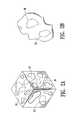
- FIG. 2AA cubic section of a material having internal bubble cavities is illustrated in perspective view in FIG. 2A .
- Internal bubble cavities 36are indicated by dotted lines.
- Comminutioni.e., grinding, pulverizing, or otherwise reducing to small particles
- the particles resulting from such comminutionform an aggregation (which may be termed a powder) which may be deposited within a body.
- An illustration of one such particleis shown in FIG. 2B , which is at a smaller scale than FIG. 2A .
- Gas bubblescan be introduced into a material by whipping a gas into the material during processing of a material, by release of gas from within the material, or by directing a gas into a material.
- bubbles within a materialmay be created by secondary processing such as extruding a polymer and a blowing agent together to create a rod having gas bubbles entrained within it.
- a blowing agentmay decompose to release a gas, or may react with other materials to form a gas.
- a blowing agent that breaks down into a gas at elevated processing temperatures or pressuresmay be included in a mixture in order to produce bubbles within a material.
- the material produced after introduction of such bubblesis one riddled with bubble cavities, forming a foam or sponge-like structure.
- Blowing agents suitable for practicing the methods of the inventioninclude, but are not limited to, sodium bicarbonate, ammonium carbonate, and other carbonates; sodium boron hydride, silicon oxy-hydride, and other hydrides; hydrochlorofluorocarbon compounds, and chlorofluorocarbon compounds.
- Liquid blowing agentssuch as hydrocarbons and alcohols, may also be used.
- liquid blowing agentsparticularly hydrocarbons and alcohols having boiling points in the range of between about 75° C. and about 95° C., are suitable for the practice of the invention.
- blowing agentsinclude, but are not limited to, hexane, heptane, heptene, propyl alcohol, isopropyl alcohol, other hydrocarbons and alcohols, and mixtures of hydrocarbons and alcohols.
- Biocompatible blowing agentsare preferred blowing agents.
- Sodium bicarbonateis a presently preferred blowing agent; it has a broad and relatively low decomposition temperature well within the processing parameters of typical bio-resorbable polymeric materials.
- Mixtures of sodium bicarbonate and polymeric materialusually include less than about 10% by weight sodium bicarbonate, and typically include no greater than about 5% sodium bicarbonate by weight.
- about 2% by weight sodium bicarbonateis combined with polylactic acid and polyglycolic acid copolymer, and the combination is extruded to form a rod.
- the bulk density of raw polymeric material suitable for the practice of the inventionis typically about 1.3 g/ml before processing.
- a preferred raw polymeric materialis a mixture of polylactic acid (PLA) and polyglycolic acid (PGA) in a ratio of 65% to 35% (PLA:PGA) by weight.
- the bulk density of the processed materialcan be varied from about 0.8 g/ml to about 1.5 g/ml to obtain a material which has the desired ultrasound characteristics. This variation can be obtained by increasing the proportion of blowing agent in the mix (for example, up to about 0.5% sodium bicarbonate by weight), by processing the extrusion at a higher temperature, or both.
- a lower bulk density materialmay be produced by either variation of the method (i.e., by increasing blowing agent or increasing extrusion temperature).
- the effect of lowering the density of the processed materialis to increase the fragility of the extruded rod, to reduce the amount of material required, and to reduce its in-vivo lifetime.
- blowing agente.g., typically greater than about 10% by weight in PLA:PGA mixtures
- Such material′was not as suitable for use in an ultrasound-detectable biopsy site marker material as more dense materials.
- blowing agenthad relatively thin polymer structures that degraded more rapidly than thicker structures, and also tended to lose gas from the bubble cavities, which then tended to collapse so that the desirable ultrasound characteristics of the material was lost.
- blowing agente.g., less than about 10%, preferably not greater than about 5%, more preferably about 2%, blowing agent by weight included with polymers and copolymers of polylactic acid, polyglycolic acid, and polycaprolactone
- blowing agente.g., less than about 10%, preferably not greater than about 5%, more preferably about 2%, blowing agent by weight included with polymers and copolymers of polylactic acid, polyglycolic acid, and polycaprolactone
- a binding agentcan be added to the marker material to make the powder a more cohesive mass.
- Materialssuch as porcine gelatin, bovine gelatin, polyethylene glycol, polyvinyl alcohol, glycerin, or other hydrophilic material are suitable for use in materials having features of the invention.
- One desirable characteristic of binding agent materialsis to keep the powder from dispersing away from the biopsy site following delivery at the biopsy site.
- the amount of binding agentis much less than the amount of ultrasound-detectable bio-resorbable powder.
- a suitable mixtureis one part porcine gelatin to 3 parts bio-resorbable ultrasound visible powder.
- a typical human breasthas a substantial number of features that are visualized with ultrasound. These features all have characteristic signals. Fibrous tissue or ligaments tend to show up as bright streaks, fat seems to appear as a dark gray area, the glandular tissue appears as a mottled medium gray mass. Cancerous lesions typically appear as a darker area with a rough outer edge which has reduced through transmission of the ultrasound energy.
- ultrasound-detectable biopsy marker materials of the present inventionprovide an ultrasound signal which can be readily differentiated from anatomic structures within the breast, so that the identification and marking of a biopsy cavity does not require extensive training and experience.
- the gas bubbles within the materials processed as described hereinform bubble cavities within the material that create a bright (white) mark or signal when viewed with ultrasound, enhancing the strength of the ultrasound signal intrinsic to the polymeric material itself.
- the rough edges of fractured or partial cavitiesmay also help to enhance the ultrasound image.
- the gas bubblesalso effectively stop transmission of the ultrasound signal, creating a shadow behind the bright spot.
- FIG. 2Bis a perspective view of a particle of an ultrasound-detectable bio-resorbable marker material embodying features of the invention, having an enclosed bubble cavity 36 indicated with dotted lines.
- the surface of the particle illustrated in the figureis irregular, having curved indentations, rounded features, and jagged edges.
- Particles of ultrasound-detectable finely-divided particulate materials having features of the inventionhave a variety of shapes and surface features, not all particles having the same shapes and features. Many, preferably a majority, of the particles of ultrasound-detectable finely-divided particulate materials having features of the invention include at least one bubble cavity.
- the bubble cavitymay be of any size or shape.
- the size of a bubble cavitymay be determined by measuring a distance along a line passing between one surface of the cavity, through a geometric center of the cavity, to another surface of the cavity.
- most bubble cavitieshave a size between about 10 microns and about 500 microns, more preferably between about 50 microns and about 200 microns.
- the biopsy marker materials of the present inventioninclude ultrasound-detectable bio-resorbable particulate material and may also include a biocompatible fluid vehicle which helps to carry the powder to the biopsy site.
- the ultrasound-detectable marker materialWhen deposited at a cavity at an intracorporeal location, the ultrasound-detectable marker material, with or without a carrier fluid, is able to fill the entire cavity.
- the cavityis a biopsy cavity left after removal of a biopsy sample from a breast
- the size of the ultrasound-detectable markercan be as large as the biopsy sample that was removed from the breast, which makes it much easier to distinguish the ultrasound signal of the marker from signals arising naturally from within a breast.
- the ultrasound-detectable biopsy site marker materials of the present inventionprovide several advantages over prior markers.
- the gas bubbles and bubble cavities within the materialshow up as a bright spot which allow for it to be distinguished from background anatomy.
- the gas bubbles and bubble cavitiesproduce a shadow below the bright spot.
- filling a biopsy cavity with the ultrasound markeroutlines the entire biopsy site.
- the marker masscreates a large curved bright upper surface which matches the outline of the biopsy cavity, and a shadow below this surface.
- Ultrasound-detectable biopsy marker materials having features of the present inventionmay be made by any suitable method, including the methods described below.
- ultrasound-detectable biopsy marker materials having features of the present inventionmay be made by reducing a bio-resorbable material having bubble cavities to a powder.
- a materialmay be reduced to a powder by granulating it, as by grinding the material, chopping the material, subjecting the material to the impact of a hard tool, impacting the material onto a hard surface, agitating the material, or by other methods known in the art.
- the starting materialmay include colorants, contrast agents, and other materials if desired.
- Cryogrindingis a suitable method of comminuting a material to produce bio-resorbable ultrasound-detectable marker materials having features of the invention.
- a starting block of materialmay be frozen, for example by exposure to a liquified gas such as liquid nitrogen, and then pounded or beaten to shatter the block into smaller pieces, which may then be ground between grinding plates to reduce the pieces to a desired particle size.
- Comminution of the materialis continued until a powder having a particle size of less than about 2000 microns is produced.
- the comminutionis effective to provide a powdered biopsy marker material having a particle size of between about 20 microns and about 800 microns, more preferably between about 300 microns and about 500 microns.
- the resulting powderflows freely when poured. It may be stored as a dry powder. Powdered pigment, contrast agent, or other particulate material may also be included at this stage if desired.
- Granulation and powderingtypically produce a wide range of particle sizes.
- a method of selecting particles of a desired sizeis to pass the powdered material through a sieve.
- a powder resulting from granulationmay be loaded onto a sieve having a desired sieve size, and particles collected from beneath the sieve after passing through the sieve. This will separate the powder into two portions: a first portion retained by, the sieve, and thus apparently unable to pass through it, and a second portion that has passed through the sieve.
- the second portion that has passed through the sieveconsists of particles having particle sizes less than the sieve size of the sieve.
- a powder having a particle size of less than about 2000 micronsmay be collected by passing a powder through a sieve having a sieve size of 2000 microns
- a powder having a particle size of less than about 800 micronsmay be collected by passing a powder through a sieve having a sieve size of 800 microns.
- Multiple sievesmay be used in a conventional fashion to separate the powder into different size particle portions. For example, passing a powder through a first sieve with a larger sieve size, and then passing the resulting powder through a second sieve having a smaller sieve size can separate out a powder portion having particle sizes falling between the first sieve size and the second sieve size.
- the bio-resorbable materialis a bio-resorbable polymeric material.
- Suitable polymeric materialsinclude polylactic acid, polyglycolic acid, polycaprolactone, and combinations of these polymers.
- Polymeric mixturesmay include copolymers and alloys of bio-resorbable polymers.
- the bio-resorbable material or materials used to make ultrasound-detectable biopsy site marker materials having features of the present inventionpreferably have bubble cavities.
- One way to characterize bubble cavitiesis to describe the cavity size, which is defined by an average length of a population of, cavities in a material. Where only a portion of what once was a larger bubble cavity remains, cavity size is measured by a length spanning the existing portion of the cavity.
- Cavity sizemay be measured, for example, by examination of the material with an optical microscope, preferably using an eyepiece with a reticle, or by inspection of electron micrographs of specimens of a material.
- Materials suitable for the practice of the inventionhave a cavity size of about 2000 microns or less.
- Preferred materialshave a cavity size of between about 10 microns and about 500 microns, more preferably the cavity size is between about 20 microns and about 200 microns.
- Bubble cavitiesmay be introduced into a material during processing by any suitable method.
- bubble cavitiesmay be introduced into a polymeric material by whipping a gas into a polymeric material while the material is heated and in a plastic or malleable state.
- Gasmay be introduced by providing a source of high pressure gas, such as nitrogen or argon, and directing the gas into a mixing chamber containing heated polymeric material.
- Another suitable method for providing a material having bubble cavitiesis to mix a material and a blowing agent, preferably a biocompatible blowing agent, and to extrude the material and a blowing agent together.
- a blowing agentpreferably a biocompatible blowing agent
- extrusion of a polymeric material and a blowing agentwill create a polymeric rod having gas bubbles entrained within it.
- These bubbleswill typically not have a spherical shape, but instead may be elongated or irregular, due to the shear forces experienced during extrusion.
- an ultrasound-detectable biopsy marker materialmay be made by extruding a polymeric material and a blowing agent to provide a bio-resorbable polymeric material having bubble cavities, and then reducing the material to a powder.
- a mixture of polymer and blowing agentshould include less than about 10% by weight of the blowing agent, preferably no more than about 5%, most preferably about 2%.
- Suitable blowing agentsinclude sodium bicarbonate, ammonium carbonate, sodium boron hydride, silicon oxy-hydride, hydrochlorofluorocarbon compounds, chlorofluorocarbon compounds, mixtures of sodium bicarbonate, ammonium carbonate, sodium boron hydride, silicon oxy-hydride, hydrochlorofluorocarbon compounds, and chlorofluorocarbon compounds, hexane, heptane, heptene, propyl alcohol, isopropyl alcohol, other hydrocarbons and alcohols, and mixtures of hydrocarbons and alcohols.
- an ultrasound-detectable biopsy marker materialmay be made by extruding a copolymer of poly-lactic acid and poly-glycolic acid with sodium bicarbonate.
- the amount of sodium bicarbonateshould make up less than about 10%, and preferably not more than about 5%, of the total starting material weight. More preferably sodium bicarbonate makes up about 2% of the total starting material weight of the mixture.
- a powder of material dissolvable in water or other solventmay be mixed into a polymer matrix to create a polymer encapsulating the dissolvable powder, and the polymer matrix containing the powder then exposed to the solvent.
- the solventdissolves the powder out of the polymer matrix, leaving cavities, thus creating a porous polymer.
- Suitable powder materials for use in practicing this methodinclude salts such as sodium chloride, potassium chloride, calcium chloride and other salts, sugars such as glucose, sucrose, and other water-soluble sugars, and the like.
- Comminution of the polymer matrixmay be performed either before or after dissolving out the powder; preferably, comminution of the polymer matrix is performed after dissolution of the encapsulated powder.
- a slurrycan be made of a dispersion of a polymer (which could also be a copolymer, polymer alloy, polymer mixture, or combination of these) and a dissolvable powder (such as a salt or a sugar, as described above).
- the slurrycan be cast to provide a solid material of polymer with dissolvable powder inclusions, and the powder then dissolved by exposing the cast material to a solvent.
- the inclusionsneed not be solid; an emulsion made up of a polymer (which could also be a copolymer, polymer alloy, polymer mixture, or combination of these) and a non-miscible liquid can be cast to provide a solid with included droplets of the non-miscible liquid.
- the non-miscible liquidmay then be extracted so that the solid material becomes a foam.
- a non-miscible liquidmay be extracted by exposing the cast material to low pressure, or by freeze-drying the cast material in a low-temperature, low pressure environment, allowing the non-miscible liquid to escape from the cast material by diffusion.
- an ultrasound-detectable biopsy marker material having features of the present inventionmay include a binding agent with the powder.
- a binding agenthelps to keep the powder together during deposition at a biopsy site.
- Suitable binding agentsinclude gelatin, polyethylene glycol, polyvinyl alcohol, glycerin, other hydrophilic materials, and combinations of these.
- Suitable gelatinsinclude bovine collagen, porcine collagen, ovine collagen, equine collagen, synthetic collagen, agar, synthetic gelatin, and combinations of these.
- Biopsy site marker materials having features of the present inventionmay be delivered to a biopsy site in dry form, or in wet form, as in a slurry or suspension. Pressure may be applied to the powder in order to eject it from a storage location, such as a delivery tube. Pressure effective to deliver an ultrasound-detectable bio-resorbable marker material having features of the invention includes gas pressure, acoustic pressure, hydraulic pressure, and mechanical pressure. For example, dry ultrasound-detectable bio-resorbable powders may be blown into a biopsy site by the action of gas pressure from a pressurized gas directed behind or alongside the powder and towards the biopsy site.
- Such gas pressuremay be supplied by carbon dioxide or nitrogen gas contained within a pressure vessel under high pressure, and the pressure released into a chamber containing a dry ultrasound-detectable bio-resorbable powder effective to drive the powder through an attached needle or catheter having an orifice within a biopsy site.
- a syringe filled with air or other gasmay be connected to a tube or chamber containing an ultrasound-detectable bio-resorbable powder and the syringe plunger depressed so as to force the air or gas, and the powder, through a needle or catheter attached to the tube or chamber and having an orifice within a biopsy site.
- any device for providing gas or liquid flow through a delivery tube containing an ultrasound-detectable bio-resorbable powderis suitable to deliver a bio-resorbable marker mass to a desired location within a patient's body.
- Mechanical pressuremay be delivered by, for example, direct contact with a plunger.
- an ultrasound-detectable bio-resorbable powder having features of the inventionmay be expelled from the tube by directly pushing on it with a plunger.
- a quantity of powder within the delivery tubemay be expelled by direct action of a plunger on the powder without need for liquid or gas.
- acoustic pressure supplied by, for example, an ultrasound transducermay be used to deliver an ultrasound-detectable bio-resorbable powder having features of the invention.
- a preferred method for delivering an ultrasound-detectable bio-resorbable powder to a biopsy siteutilizes a biocompatible liquid to drive or carry the powder into the biopsy cavity at the biopsy site.
- a quantity of ultrasound-detectable bio-resorbable powdermay be contained within a tube or chamber that leads directly or indirectly to a biopsy site.
- the powdermay be dispensed by the application of hydraulic pressure applied by a syringe containing sterile saline or other suitable liquid. Flow of the liquid carries the powder into the biopsy site; turbulence acts to mix the dry powder with the liquid to provide a slurry or suspension and to provide a fairly uniform distribution of the powder throughout the biopsy cavity.
- the syringeis preferably configured to tightly engage with the tube or chamber, in order to prevent leakage and to insure sufficient pressure and flow is directed towards the biopsy site.
- the tube or chamber containing the ultrasound-detectable bio-resorbable powdermay be an elongated hollow tube, having a bore completely filled with the powder; or an elongated hollow tube, having a bore partially filled with the powder; or a chamber having internal dimensions that change along the length, configured to hold a desired quantity of ultrasound-detectable bio-resorbable powder and to provide a desired amount of mixing with the fluid.
- the quantity of ultrasound-detectable bio-resorbable powderis contained within a tube termed a “delivery tube.”
- the tubehas an outside diameter that is sized to fit within a cannula, such as a Mammotome® or SenoCor 360TM cannula.
- a suitable delivery tubehas an outside diameter (OD) of about 0.096 inches and has an inner diameter (ID) of about 0.074 inches.
- ODoutside diameter
- IDinner diameter
- Other sizesare also suitable, the exact dimensions depending on the biopsy device used.
- a delivery tubemay have markings to aid in determining the depth of the tube within a cannula, surface features (such as pins, slots, bumps, bars, wedges, luer-lock fittings, or bands, including a substantially conical circumferential band) effective to control the depth into which a delivery tube is fitted within a cannula or effective to lock a delivery tube into position within a cannula.
- surface featuressuch as pins, slots, bumps, bars, wedges, luer-lock fittings, or bands, including a substantially conical circumferential band
- a delivery tubemay have pins or bumps configured to engage a slot or a leading edge of a cannula, or a luer-lock fitting configured to lock into a cannula.
- a cannulamay also be configured to receive and to engage a delivery tube.
- a cannulamay have pins, slots, wedges, bumps, bands, luer-lock fittings, or the like, to engage a delivery tube and to hold it into a desired position within the cannula.
- a cannulamay have a luer-lock fitting, or a slot to engage a pin on a delivery tube, or an internal bump wedge or band that limits the distance of travel of the delivery tube within the cannula.
- Delivery tubes embodying features of the present inventionmay be made of any suitable bio-compatible material.
- the materialhas sufficient strength to withstand the hydraulic pressure required to dispense the material (typically greater than 100 psi) and is able to be sterilized.
- a polyether block amidesuch as a Pebax®, preferably a 72 Durometer Pebax®, is one example of a suitable material.
- Hydraulic pressuremay be supplied by depressing the plunger of a syringe, such as a 1 to 3 cc syringe, containing a suitable fluid and connected to the delivery tube.
- the amount of material required within the delivery tubewill vary depending on the biopsy cavity size. Typically, quantities of between about 0.2 to about 1.2 ml of powder will be required. In many cases, a quantity of about 0.5 ml of the powder is suitable.
- the delivery tubecan be sized to accept any volume desired to be injected into the biopsy cavity.
- the average Mammotome® biopsyremoves about 1 ml of tissue.
- the present inventorshave found that about 0.5 ml of dry powder works best to fill such a biopsy cavity with the powder and fluid. Use of more powder typically leads to some filling of the needle track as well as of the cavity at the biopsy site. Smaller volumes of powder may be used for smaller cavities at a biopsy site, such as are created with an automated Tru-Cut® biopsy or a single SenoCor 360TM biopsy, available from SenoRx, the assignee of the present invention.
- Ultrasound-detectable bio-resorbable powdermay be loaded into an delivery tube in preparation for use in marking a biopsy site.
- the delivery tubemay be attached to a syringe having about 0.5 ml to about 2 ml of fluid, preferably about 1.0 ml to about 1.5 ml of fluid, at any suitable time before delivery to a biopsy site.
- the delivery tubeis placed with its distal end at or directed to the biopsy site, typically by placing the delivery tube within a cannula leading to the biopsy site, and the powder is then dispensed by directing the fluid through the delivery tube, so that it is thereby deposited at the biopsy site.
- a syringecould be loaded with an ultrasound-detectable bio-resorbable powder having features of the invention, filled with a biocompatible fluid, and then shaken or stirred to create a slurry.
- the syringemay then be connected to a delivery tube or catheter, and the slurry injected through the delivery tube or catheter to fill the biopsy site with an ultrasound-detectable bio-resorbable marker composition.
- ultrasound-detectable bio-resorbable powders having features of the inventionmay form a suspension, which may be dilute or concentrated, depending on the relative amounts of fluid and of powder.
- a concentrated suspensionmay be termed a slurry, and may be somewhat viscous and have the consistency of a paste.
- the consistency of the suspensionsmay change with time; for example, a suspension of an ultrasound-detectable bio-resorbable powder having features of the invention may set up over time to form a semi-solid gel.
- a binding agentsuch as gelatin, preferably an amount of gelatin of about 5% to about 50% by weight, is effective to help gel formation after mixing an ultrasound-detectable bio-resorbable powder with a suitable fluid.
- Gelatintypically hardens to its ultimate firmness within minutes at normal body temperature.
- the fluid used to deposit the powder at a biopsy sitemay contain other agents, including inert agents, osmotically active agents, pharmaceutical agents, and other bio-active agents.
- a suitable biocompatible liquidmay be selected from the group consisting of sterile saline, sterile saline containing a pharmaceutical agent, sterile saline containing an anesthetic agent, sterile saline containing a hemostatic agent, sterile saline containing a colorant, sterile saline containing a radio contrast agent, sterile sugar solution, sterile sugar solution containing a pharmaceutical agent, sterile sugar solution containing an anesthetic agent, sterile sugar solution containing a hemostatic agent, sterile sugar solution containing a colorant, sterile sugar solution containing a radio contrast agent, biocompatible oils, biocompatible oils containing a pharmaceutical agent, biocompatible oils containing an anesthetic agent, biocompatible oils containing a hemostatic agent, biocompatible oils containing a
- Hemostatic agentstend to reduce bleeding, enhance clotting, or to cause vasoconstriction in a patient.
- Hemostatic agentsinclude adrenochrome, algin, alginic acid, aminocaproic acid, batroxobin, carbazochrome salicylate, cephalins, cotarmine, ellagic acid, epinephrine, ethamsylate, factor VIII, factor IX, factor XIII, fibrin, fibrinogen, naphthoquinone, oxamarin, oxidized cellulose, styptic collodion, sulamrin, thrombin, thromboplastin (factor III), tolonium chloride, tranexamic acid, and vasopression.
- Pharmaceutical agentsare often used to promote healing, and to treat injury, infection, and diseases such as cancer, and may include hormones, hemostatic agents and anesthetics as well as antibacterial, antiviral, antifungal, anticancer, and other medicinal agents. Pharmaceutical agents may be included as part of an ultrasound-detectable bioresorbable material placed within a biopsy cavity in order, for example, to promote healing, prevent infection, and to help treat any cancer cells remaining near the biopsy site.
- pharmaceutical agentswhich may be included with a fluid or with a powder to form an ultrasound-detectable bioresorbable material having features of the invention include, but are not limited to: penicillins, cephalosporins, vancomycins, aminoglycosides, quinolones, polymyxins, erythromycins, tetracyclines, streptomycins, sulfa drugs, chloramphenicols, clindamycins, lincomycins, sulfonamides, paclitaxel, docetaxel, acetyl sulfisoxazole, alkylating agents, antimetabolites, plant alkaloids, mechlorethamine, chlorambucil, cyclophosphamide, melphalan, ifosfamide, methotrexate, 6-mercaptopurine, 5-fluorouracil, cytarabine, vinblastine, vincristine, etoposide, doxorubic
- An assembly for delivering an ultrasound-detectable bio-resorbable powderincludes an delivery tube configured to be received within a biopsy guide cannula. Such a delivery tube is also configured to engage a syringe, and to hold a quantity of ultrasound-detectable bio-resorbable powder.
- the delivery tubemay be configured to hold a quantity of ultrasound-detectable bio-resorbable powder by having an inner volume of between about 0.2 ml and about 1.2 ml, preferably between about 0.5 ml and about 1.0 ml.
- a delivery tube having features of the inventionmay also be configured to fit within a biopsy guide cannula, such as a Mammotome® guide cannula, a SenoCor 360TM cannula, or a Tru-Cut® cannula.
- a delivery tube having an outside diameter less than about 0.1 inch (2.54 mm)is able to fit within a Mammotome® guide cannula.
- a delivery tube having features of the inventionmay also be configured to sealingly engage with a syringe, that is, to form a strong, water-tight connection.
- An example of a suitable connectionis a luer-lock connection configured to sealingly engage with a syringe.
- a system for temporarily marking a biopsy site within a patientincludes a syringe; a supply of ultrasound-detectable bio-resorbable powder; and a delivery tube configured to engage the syringe.
- the engagement between the syringe and delivery tubeis water-tight engagement, and is sufficiently strong as to withstand the hydraulic pressure generated by depressing the syringe plunger and directing fluid within the syringe through the delivery tube effective to deposit ultrasound-detectable bio-resorbable powder at the biopsy site.
- the supply of ultrasound-detectable bio-resorbable powdermay be in a packet, a syringe, a delivery tube, or other container.
- the supply of ultrasound-detectable bio-resorbable powderis located within the delivery tube.
- the delivery tubeis preferably configured to be received within a biopsy guide cannula.
- the delivery tubehas an outside diameter of less than about 0.1 inch (2.54 mm).
- the systemmay further include a supply of biocompatible liquid, which is preferably sterile saline. More preferably, the biocompatible liquid is contained within the syringe of the system for ease of use and to avoid the possibility of spillage or contamination.
- FIG. 3Ais a partially cut-away perspective view of a system 10 as illustrated in FIG. 1 , shown ready to deposit an ultrasound-detectable bio-resorbable powder 16 within a cavity 46 at a biopsy site 38 .
- FIG. 3Bshows the system, following depression of plunger 30 , as it deposits an ultrasound-detectable bio-resorbable marker material 50 embodying features of the invention (which includes ultrasound-detectable bio-resorbable powder 16 mixed with fluid 20 ) at a biopsy site 38 within a breast 40 of a patient.
- Biopsy guide cannula 44extends through incision 42 into cavity 46 at biopsy site 38 .
- Syringe 18containing a bio-compatible fluid 20 , is tightly engaged with a delivery tube 14 , which extends within guide cannula 44 so as to locate delivery tube outlet 26 within biopsy site cavity 46 .
- Delivery tube outer diameter 34is configured to allow delivery tube 14 to fit inside the inner diameter 48 of biopsy guide cannula 44 .
- Markings 52(shown in FIG. 3A ) spaced at regular intervals along the outer surface of delivery tube 14 indicate the depth of insertion of the tube within the cannula and aid in the proper placement of delivery tube outlet 26 into position within cavity 46 .
- Depression of plunger 30forces fluid 20 out of syringe 18 through delivery tube 14 , where fluid 20 mixes with and carries ultrasound-detectable bio-resorbable powder 16 out of delivery tube outlet 26 to deposit ultrasound-detectable bio-resorbable biopsy site marker material 50 within cavity 46 at biopsy site 38 .
- an optional locking mechanism 54including pin 56 (on delivery tube 14 ) configured to engage with slot 58 (on cannula 44 ) may be used to fix delivery tube 14 in position within cannula 44 and to help prevent movement of delivery tube outlet 26 relative to cannula 44 when plunger 30 is depressed.
Landscapes
- Health & Medical Sciences (AREA)
- Life Sciences & Earth Sciences (AREA)
- Engineering & Computer Science (AREA)
- Animal Behavior & Ethology (AREA)
- Biomedical Technology (AREA)
- Veterinary Medicine (AREA)
- Public Health (AREA)
- General Health & Medical Sciences (AREA)
- Nuclear Medicine, Radiotherapy & Molecular Imaging (AREA)
- Physics & Mathematics (AREA)
- Heart & Thoracic Surgery (AREA)
- Medical Informatics (AREA)
- Surgery (AREA)
- Epidemiology (AREA)
- Hematology (AREA)
- Radiology & Medical Imaging (AREA)
- Molecular Biology (AREA)
- Pathology (AREA)
- Acoustics & Sound (AREA)
- Anesthesiology (AREA)
- Dermatology (AREA)
- Optics & Photonics (AREA)
- Oncology (AREA)
- Biodiversity & Conservation Biology (AREA)
- Oral & Maxillofacial Surgery (AREA)
- Biophysics (AREA)
- Medicines Containing Antibodies Or Antigens For Use As Internal Diagnostic Agents (AREA)
- Prostheses (AREA)
- Apparatus For Radiation Diagnosis (AREA)
- Investigating Or Analysing Biological Materials (AREA)
- Ultra Sonic Daignosis Equipment (AREA)
Abstract
Description
This application is a continuation of application Ser. No. 14/627,691, filed Feb. 20, 2015, now U.S. Pat. No. 9,237,937, which is a continuation of application Ser. No. 14/099,352, filed Dec. 6, 2013, now U.S. Pat. No. 8,965,486, which is a continuation of application Ser. No. 13/495,408, filed Jun. 13, 2012, now U.S. Pat. No. 8,626,270, which is a continuation of application Ser. No. 12/852,286, filed Aug. 6, 2010, now U.S. Pat. No. 8,219,182, which is a continuation of application Ser. No. 10/990,327, filed Nov. 16, 2004, now U.S. Pat. No. 7,792,569, which is a continuation of application Ser. No. 10/124,757, filed Apr. 16, 2002, now U.S. Pat. No. 6,862,470, which is a continuation-in-part of application Ser. No. 09/717,909, filed Nov. 20, 2000, now U.S. Pat. No. 6,725,083, which is a continuation-in-part of application Ser. No. 09/343,975, filed Jun. 30, 1999, now U.S. Pat. No. 6,347,241, which is a continuation-in-part of application Ser. No. 09/241,936, filed Feb. 2, 1999, now U.S. Pat. No. 6,161,034, which are all hereby incorporated by reference herein in their entireties and from all of which priority is hereby claimed under 35 U.S.C. §119(e) and §120.
This invention relates generally to the field of acquisition of tissue from a patient, as occurs in a biopsy procedure, in particular to the marking of the site within a body from which a biopsy has been taken.
In diagnosing and treating certain medical conditions, it is often desirable to perform a biopsy, in which a specimen or sample of tissue is removed for pathological examination, tests and analysis. As is known, obtaining a tissue sample by biopsy and the subsequent examination are typically employed in the diagnosis of cancers and other malignant tumors, or to confirm that a suspected lesion or tumor is not malignant. The information obtained from these diagnostic tests and/or examinations is frequently used to devise a plan for the appropriate surgical procedure or other course of treatment. For example, breast biopsies may be taken where a suspicious lump or swelling is noticed in a breast. Examination of tissue samples taken by biopsy is of particular significance in the diagnosis and treatment of breast cancer. In the ensuing discussion, the biopsy and treatment site described will generally be the human breast, although the invention is suitable for marking biopsy sites in other parts of the human and other mammalian body as well.
After the biopsy sample is taken, it may take several days or weeks before the results of the examination of the sample are obtained, and still longer before an appropriate treatment decision is reached. If the decision involves surgery it is clearly important for the surgeon to find the location in the breast from where the tumor tissue has been taken in the biopsy procedure, so that the entire tumor and possibly surrounding healthy tissue can be removed.
However, radiographically imageable tissue features, originally detected in a mammogram, may be removed, altered or obscured by the biopsy procedure. In order for the surgeon or radiation oncologist to direct surgical or radiation treatment to the precise location of the breast lesion several days or weeks after the biopsy procedure was performed, it is desirable that a biopsy site marker be placed in or on the patient's body to serve as a landmark for subsequent location of the lesion.
Various types of biopsy site markers have been described, including visible markers applied externally to the patient's skin, radiographically (X-ray)-detectable tissue markers such as clips and staples, and ultrasound-detectable markers, have also been described. X-ray-detectable marker wires may be inserted through a biopsy needle, leading from the surface of the patient's body to the biopsy site. Some markers may be biodegradable.
However, due to the consistency of breast tissue and the fact that these biopsy site markers are typically introduced while the breast is still compressed between the mammography plates, prior art biopsy markers do not always remain at the specific biopsy location after the breast has been decompressed and removed from the mammography apparatus, and may suffer from additional disadvantages as well. In order to locate an X-ray-detectable marker left at a biopsy site, an additional mammography is generally required at the time of follow up treatment or surgery. In addition, once the biopsy site is located using mammography, the site must usually be marked again with a location wire that is visible to provide guidance to the clinician performing the treatment or surgery. However, as the patient is removed from the mammography apparatus, or otherwise transported, the position of the location wire can change or shift before the treatment or surgery is performed, which may result in treatments being misdirected to undesired locations. Furthermore, at least some prior art biopsy site markers can remain present at the site of implantation for an indefinite period of time and, if not surgically removed, may obscure or otherwise interfere with any subsequent mammography or imaging studies.
As an alternative or adjunct to radiographic imaging, ultrasonic imaging and visualization techniques (abbreviated as “USI”) can be used to image the tissue of interest at the site of interest during a surgical or biopsy procedure or follow-up procedure. USI is capable of providing precise location and imaging of suspicious tissue, surrounding tissue and biopsy instruments within the patient's body during a procedure. Such imaging facilitates accurate and controllable removal or sampling of the suspicious tissue so as to minimize trauma to surrounding healthy tissue.
For example, during a breast biopsy procedure, the biopsy device is often imaged with USI while the device is being inserted into the patient's breast and activated to remove a sample of suspicious breast tissue. As USI is often used to image tissue during follow-up treatment, it may be desirable to have a marker, similar to the radiographic markers discussed above, which can be placed in a patient's body at the site of a surgical procedure and which are detectable using USI. However, radiopaque markers may not be detectable with USI. A marker that is detectable with USI enables a follow-up procedure to be performed without the need for traditional radiographic mammography imaging which, as discussed above, can be subject to inaccuracies as a result of shifting of the location wire as well as being tedious and uncomfortable for the patient.
Thus, there is need in the art for biopsy site markers that are deliverable into the cavity created by removal of the biopsy specimen and not into tissue that is located outside of that biopsy cavity, and which will not migrate from the biopsy cavity even when the breast tissue is moved, manipulated or decompressed. Moreover, such desired markers should remain detectable at the biopsy site (i.e., within the biopsy cavity for a desired time period); should not interfere with imaging of the biopsy site and adjacent tissues at a later time; and should be readily distinguishable in the various imaging procedures from lines of calcifications which frequently are signs for a developing malignancy.
The invention is directed to materials, devices, and methods for marking intracorporeal locations such as biopsy sites for a limited time. The marking material comprises an ultrasound-detectable bio-resorbable finely-divided particulate material (such as a powder), in which many of the particles have internal cavities. Such particulate materials or powders may be composed of particles having sizes typically less than about 2000 microns, and typically between about 20 microns and about 2000 microns, preferably between about 20 microns and about 800 microns, more preferably between about 300 microns and about 500 microns.
The ultrasound-detectable bio-resorbable particulate materials may include any bio-resorbable particulate material having cavities which facilitate USI. For example, the ultrasound-detectable bio-resorbable particulate materials include particles having cavities with dimensions typically between about 10 microns and about 500 microns, preferably about 20 microns to about 200 microns. The particulate materials may also contain binding agents. Typically, a mass of the bio-resorbable particulate material is delivered to an intracorporeal location, such as a biopsy site within a patient. The mass of particulate material remains situated and detectable at the biopsy site during a limited time until it is resorbed by tissue near the biopsy site. Delivery devices having features of the invention are configured to contain the deliverable mass of particles, to fit within a cannula, and to engage with a syringe. Systems for temporarily marking an intracorporeal site may include a tube containing an ultrasound-detectable bio-resorbable powder, and a syringe containing a biocompatible fluid for biopsy site marking. The tube may be configured to fit within a cannula, such as a Mammotome® or SenoCor 360™ biopsy cannula or a coaxial needle guide.
The biopsy site marker materials include powders and powder slurries which can be delivered into a biopsy site cavity. Thus, much or all of the biopsy cavity may be filled with an ultrasound-detectable marker material, creating an ultrasound-detectable marker that can be as large as the biopsy sample that was removed from the patient's body. In addition, this allows the ultrasound detection and definition of the boundaries of the biopsy cavity. The needle track leading to the biopsy cavity may be filled as well if desired. Marker materials embodying features of the invention may be deposited at an intracorporeal location along with other agents, including anesthetic agents, hemostatic agents, pigments, dyes, materials detectable by magnetic resonance imaging (MRI), inert materials, and other compounds. For example, marker materials embodying features of the invention may include radiopaque materials as well as ultrasound-detectable materials. Such radiopaque material can serve to provide temporary or permanent radiographic marking of the location. Upon deposition at a biopsy site within a patient, materials embodying features of the invention may set up or gel to form flexible or relatively rigid solid masses.
The ultrasound-detectable biopsy site markers of the present invention provide several advantages. A biopsy cavity with a marker material having features of the present invention provides a large USI-bright mass, making it much easier, for example, to distinguish the ultrasound signal of the marker from signals arising naturally from within a breast. The marker materials produce bright ultrasound echo signals from one portion of the filled region, which contrasts with the dark ultrasound shadow region immediately behind the bright ultrasound echo region. The strength of the reflected signal, and the contrast with the shadow region, make the marked site readily detectable. Such ready detectability allows, for example, lesser-experienced physicians to perform the identification by USI and the subsequent surgical resection without the need for an interventional radiologist to identify and mark the biopsy cavity. These and other advantages will be further described in the following detailed description of embodiments of the invention.
Adelivery tube 14 may have indicia, such as lines or dots spaced at regular intervals along a length of the tube to indicate the tube's position within a cannula. Adelivery tube 14 may also be configured with a stop, such as a bump, peg, pin, or other feature, to limit the depth of its entrance into a cannula. In addition, adelivery tube 14 may be configured to be affixed to, or lock into, a cannula so that, once introduced into the cannula, it may maintain its position for a desired period. A luer lock mechanism, a pin and slot arrangement, where a pin on the surface of thedelivery tube 14 fits into and engages with a slot on a cannula, or other mechanism or mechanisms may be used to fix the position of adelivery tube 14 with respect to a cannula.
As illustrated inFIGS. 1A-1D , the present invention provides materials suitable for marking intracorporeal locations, and methods and apparatus for delivering such materials to desired intracorporeal locations. For example,particulate material 16 may be any ultrasound-detectable powder or other ultrasound-detectable aggregation of small particles. In some embodiments of the invention,particulate material 16 may include radiopaque elements or materials, MRI-detectable materials, pigments, dyes, or other materials as well.System 10 andapparatus 12 may be any system and apparatus suitable for delivering an ultrasound-detectable particulate material 16 to a location within a body. Any method effective to deliver an ultrasound-detectable particulate material is suitable for the practice of the invention.
Examples of materials, apparatus, systems, and methods embodying features of the invention are presented below. However, before presenting some features and embodiments illustrating the present invention, it is helpful to introduce some terms which are used in describing aspects of the invention.
The term “detectable” as used herein means able to be detected by a person, either with or without the use of imaging instrumentation (such as ultrasound imaging instrumentation). For example, a mass or material that is detectable by ultrasound is able to produce a readily recognizable image on the display apparatus of ultrasound imaging instrumentation. A material is “not readily detectable” within a patient if, for example, an ultrasound examination directed to the location where the material had been placed does not produce a readily recognizable ultrasound image. As used herein, “readily” means without the use of extreme or extraordinary measures or without requiring extraordinary skill.
“Bio-resorbable” as used herein means resorbable in a biological environment, such as within the body of a patient. A bio-resorbable material is thus a material which may be absorbed, dissolved, broken down, degraded, assimilated, or otherwise removed from a biological environment, such as from within the body of a patient. A bio-resorbable polymer is a polymeric material (including copolymers, alloys and polymer mixtures composed of two or more polymers) which is bio-resorbable.
The “in-vivo lifetime” of a bio-resorbable material as used herein refers to the period of time after placement of the material within the body of an animal that the material remains readily detectable. Thus, the “in-vivo lifetime” of an ultrasound-detectable bio-resorbable material is that period of time during which the material remains readily detectable within a patient by ultrasound.
The term “blowing agent” as used herein means a material or combination of materials present during the processing or compounding of a polymer material that produces or breaks down into a gas at processing temperatures or pressures. For example, where particles of polymer or copolymer material also contains a blowing agent, and the particles are heated, extruded, or otherwise processed, the blowing agent decomposes, vaporizes, or reacts to form a gas inside the material, producing cavities within the material. A blowing agent may be a solid, a liquid (such as a liquid that vaporizes at or below the processing temperature), or a gas. A blowing agent may be added to a material or mixture during processing, such as by directing pressurized gas into a mixing chamber or a reaction chamber. Biocompatible blowing agents are preferred.
A “bubble cavity” is an enclosed, sealed cavity within a material filled with a gas or air. A bubble cavity may be produced by gas that is present in a material or mixture during processing. For example, a gas may be present during processing where the gas has been introduced into the processing location; or where it has been evolved or produced by the materials present during processing; or it may have been released by materials present during processing. In particular, inclusion of a blowing agent during or after processing of a material or mixture of materials may aid or cause the production of bubble cavities.
“Particle size” as used herein describes the external physical dimensions of particles making up a population of small particles of a material, and may be determined, for example, by passing a powder through a sieve with a known nominal minimum passage dimension. As used herein, a powder of a given particle size will typically include particles of a wide range of sizes. For example, in an aggregation of particles having a nominal particle size of between about 100 microns to about 150 microns, more than about 80% of the particles (by weight) fall within the nominal range and up to about 20% do not. Thus, most, but not all of the particles usually fall within a nominal range described by the particle size of a powder.
“Gelatin” as used herein means a viscous, semi-solid, or solid material, also termed a “gelatinous” material. Gelatin is typically biologically-derived, although synthetic gelatin is also available. For example, gelatin may consist largely of collagen obtained from processed animal connective tissue. Vegetable gelatin such as agar is often made from kelp, algae, or other plant material. Gelatin may include bovine collagen, porcine collagen, ovine collagen, equine collagen, agar, synthetic gelatin such as synthetic human-derived collagen, and combinations of these.
“Bulk density” as used herein is defined as the quotient of the weight of a material, such as a powder, divided by the volume of the material.
A biocompatible liquid or fluid is a liquid that may be introduced into a patient's body without harming the patient. Sterile saline and sterile water containing a sugar (such as dextrose, sucrose or other sugar) or other suitable osmotically-active compounds are typical biocompatible liquids. Other liquids, including fluids not containing water, such as biocompatible oils, may also be used. A biocompatible liquid may be mixed with other agents or materials and used to carry contrast agents, colorants, markers, inert agents, and pharmaceutical agents into a patient.
Pharmaceutical agents, as used herein, are agents used to treat a disease, injury, or medical condition, and include, but are not limited to, drugs, antibiotics, cancer chemotherapy agents, hormones, anesthetic agents, hemostatic agents, and other medicinal compounds. Hemostatic agents are agents which tend to reduce bleeding, enhance clotting, or to cause vasoconstriction in a patient. Brachytherapy agents are typically sources of radiation for implantation near to the site of a cancerous lesion.
In one aspect, the invention provides a bio-resorbable material suitable for temporarily marking a biopsy site within a patient. Materials embodying features of the invention are detectable within a patient after introduction into a biopsy site, remain detectable for a period of time, and then are not detectable after the period of time. Such materials include bio-resorbable materials which are dissolved and absorbed into body tissue at or near to the biopsy site. Typically, such materials include a powder made from a bio-resorbable polymer such as poly-lactic acid, poly-glycolic acid, poly-caprolactone, and copolymers, alloys, mixtures of these polymers, and combinations of these materials.
Bio-resorbable polymeric materials suitable for use in making ultrasound-detectable biopsy marker materials typically have a bulk density of between about 0.8 g/ml and about 1.5 g/ml. Preferably, the bio-resorbable polymeric material is a foam or other material containing cavities, with a bulk density after processing of less than about 1 g/ml, more preferably between about 0.8 g/ml and about 1 g/ml.
An ultrasound-detectable marker typically must be remain in place and detectable within a patient for at least 2 weeks to have practical clinical value. Thus, an ultrasound-detectable marker material embodying features of the invention is detectable at a biopsy site within a patient for a time period of at least 2 weeks, preferably at least about 6 weeks, and may remain detectable for a time period of up to about 20 weeks, more preferably for a time period of up to about 12 weeks. An ultrasound-detectable marker material embodying features of the invention is preferably not detectable about 6 months after placement at a biopsy site. More preferably, an ultrasound-detectable marker material embodying features of the invention is not detectable with ultrasound about 12 weeks after placement at a biopsy site. A preferable in-vivo lifetime for an ultrasound-detectable biopsy marker mass having features of the invention is between about 6 weeks and about 12 weeks.
Typically, the ultrasound-detectable biopsy marker materials of the present invention are deposited at locations within a patient's body to form a biopsy marker mass. Thus, for example, a quantity of powder formed from comminuted material having finely divided particles with bubble cavities within the particles may be delivered into a cavity at a biopsy site. In some embodiments, the ultrasound-detectable biopsy marker materials form a gel mass upon being introduced within the body of a patient. These a gel masses may be flexible gels or may be rigid, solid gels. In preferred embodiments, the marker materials remain within the biopsy cavity and do not migrate. The marker materials are resorbed by tissue and fluids near the biopsy site, so that, after a limited time, the marker materials are no longer USI-detectable at the biopsy site. The limited time during which the marker materials remain USI-detectable is the in-vivo lifetime.
In addition, ultrasound-detectable biopsy marker materials embodying features of the invention may also include radiopaque materials or radiopaque elements, so that the biopsy site may be detected both with ultrasound and with X-ray or other radiographic imaging techniques. Radiopaque materials and markers may include metal objects such as clips, bands, strips, coils, and other objects made from radiopaque metals and metal alloys, and may also include powders or particulate masses of radiopaque materials. Radiopaque markers may be of any suitable shape or size, and are typically formed in a recognizable shape not naturally found within a patient's body, such as a star, square, rectangular, geometric, gamma, letter, coil or loop shape. Suitable radiopaque materials include stainless steel, platinum, gold, iridium, tantalum, tungsten, silver, rhodium, nickel, bismuth, other radiopaque metals, alloys and oxides of these metals, barium salts, iodine salts, iodinated materials, and combinations of these. Radiopaque materials and markers may be permanent, or may be temporary and not detectable after a period of time subsequent to their placement at a biopsy site within a patient.
In addition, ultrasound-detectable biopsy marker materials embodying features of the invention may also include MRI-detectable materials or markers, so that the biopsy site may be detected both with ultrasound and with MRI or other imaging techniques. MRI contrast agents such as gadolinium and gadolinium compounds, for example, are suitable for use with ultrasound-detectable biopsy marker materials embodying features of the invention. Colorants, such as dyes (e.g., methylene blue and carbon black) and pigments (e.g., barium sulfate), may also be included in ultrasound-detectable biopsy marker materials embodying features of the invention.
Cavity-containing particles embodying features of the invention may be made from a wide variety of biocompatible bio-resorbable material. Thus, cavity-containing particles formed from any material that is not toxic, and which is resorbable, may be used to form ultrasound-detectable bio-resorbable marker materials and compositions having features of the invention. Some particularly suitable materials include bio-resorbable polymers including, but not limited to, polymers*of lactic acid, glycolic acid, caprolactones, and other monomers; thus, for example, suitable bio-resorbable polymers may include poly(esters), poly(hydroxy acids), poly(lactones), poly(amides), poly(ester-amides), poly(amino acids), poly(anhydrides), poly(ortho-esters), poly(carbonates), poly(phosphazines), poly(thioesters), poly(urethanes), polyester urethanes), polysaccharides, polylactic acid, polyglycolic acid, polycaproic acid, polybutyric acid, polyvaleric acid, and copolymers, polymer alloys, polymer mixtures, and combinations thereof.
The in-vivo lifetime of a polymeric material is related to the molecular weight of the polymer. For example, copolymers of lactic and glycolic acids having an initial molecular weight of about 60,000 daltons (60 kD) before processing, are suitable for use in making an ultrasound-detectable marker material having an in-vivo lifetime of about 12 weeks. The starting molecular weight degrades, during an extrusion process that adds gas bubbles to form bubble cavities in the polymeric material, thereby enhancing its detectability by ultrasound. The molecular weight further degrades to about 45,000 dalton (45 kD) following gamma-ray sterilization. As is known to those of ordinary skill in the art, other materials, including other polymeric materials, may require a different starting molecular weight in order to obtain the same in-vivo lifetime. For example, polyglycolic acid typically degrades faster than other materials and as such requires a substantially higher initial molecular weight than polylactic acid or polycaprolactone to obtain a similar in-vivo lifetime.
Comminution of polymers and other starting materials (which preferably are porous materials) by grinding, milling, pulverizing, or otherwise reducing the materials in size, is effective to form a powder. Ultrasound-detectable materials having features of the invention are typically powders having particle sizes of about 20 microns to about 2000 microns. The particles typically contain within them bubble cavities with sizes of between about 10 microns to about 500 microns.
The preferred particle size depends in one aspect on the inner diameter of a tube used to deliver the powder to a biopsy site. Larger particle sizes are more easily accommodated in larger diameter delivery tubes, while smaller delivery tubes may be used to deliver powders having smaller particle sizes. Where the delivery tube has an inner diameter of no greater than about 0.075 inches (1.9 mm), powders having particle sizes of between about 20 microns and about 800 microns, and more preferably between about 300 microns and about 500 microns, are preferred.
The starting materials may have, or may be processed to produce, bubbles which produce internal bubble cavities. Comminution of the starting material provides small particles with pocked, pitted, and ridged surfaces formed from the exposed surfaces of broken bubble cavities. Many of the particles have internal voids formed of bubble cavities. Gas-filled or air-filled bubble cavities are effective to reflect ultrasound energy, and greatly enhance the ability of particles containing gas-filled or air-filled bubble cavities to be imageable by USI. The sizes of such bubble cavities may be measured by lengths across the cavities, through geometric centers of the cavity. Bubble cavity size is typically between about 10 microns and about 500 microns, preferably between about 20 microns to about 200 microns.
Materials embodying features of the invention may also include binding agents which help powder particles to adhere together and so to make the powder a more cohesive mass. Typical binding agents include gelatin, polyethylene glycol, polyvinyl alcohol, glycerin, acrylic hydrogels such as hydroxy ethyl methylacrylate, other organic hydrogels, and other hydrophilic materials. Binding agents may be used singly or in combination with other binding agents. In some embodiments of the invention, gelatin is a preferred binding agent. Gelatin suitable for use in materials embodying features of the invention includes bovine collagen, porcine collagen, ovine collagen, equine collagen, synthetic collagen, agar, synthetic agar, and combinations of these. Synthetic materials may be preferred in order to avoid raising concerns over possible allergies to bovine or porcine collagen. In some embodiments, a marker material includes (by weight) about one part gelatin to about two to five parts bio-resorbable polymeric material. In a preferred embodiment, a marker material includes about one part gelatin to about three to four parts bio-resorbable polymeric material (by weight)
Many properties of a marker material affect the intensity of its ultrasound reflection, including density, physical structure, molecular material, and shape. For example, sharp edges, or multiple reflecting surfaces on or within an object differing in density from its surroundings enhances a marker's ability to be detected by ultrasound. Interfaces separating materials of different densities, such as between a solid and a gas, produce strong ultrasound signals. The methods of the present invention provide marker materials effective to produce strong ultrasound signals, the marker materials having gas-containing bubble cavities.
Marker materials with a high degree of porosity, whether from bubbles formed in situ during manufacture of the material, or from bubbles trapped within the material during later processing, provide strong ultrasound signals when located within the body of an animal which translates to improved ultrasound imaging. Starting material having gas bubbles entrained within it may be ground and sieved to obtain powders providing strong ultrasound signals. Portions of the powders may be selected to have particle sizes within a desired size range.
A cubic section of a material having internal bubble cavities is illustrated in perspective view inFIG. 2A .Internal bubble cavities 36 are indicated by dotted lines. Comminution (i.e., grinding, pulverizing, or otherwise reducing to small particles) of such a material into particles containing bubble cavities produces an ultrasound-detectable material suitable for marking an intracorporeal location. The particles resulting from such comminution form an aggregation (which may be termed a powder) which may be deposited within a body. An illustration of one such particle is shown inFIG. 2B , which is at a smaller scale thanFIG. 2A .
Gas bubbles can be introduced into a material by whipping a gas into the material during processing of a material, by release of gas from within the material, or by directing a gas into a material. Alternatively, bubbles within a material may be created by secondary processing such as extruding a polymer and a blowing agent together to create a rod having gas bubbles entrained within it. A blowing agent may decompose to release a gas, or may react with other materials to form a gas. For example, a blowing agent that breaks down into a gas at elevated processing temperatures or pressures may be included in a mixture in order to produce bubbles within a material. The material produced after introduction of such bubbles is one riddled with bubble cavities, forming a foam or sponge-like structure.
Blowing agents suitable for practicing the methods of the invention include, but are not limited to, sodium bicarbonate, ammonium carbonate, and other carbonates; sodium boron hydride, silicon oxy-hydride, and other hydrides; hydrochlorofluorocarbon compounds, and chlorofluorocarbon compounds. Liquid blowing agents, such as hydrocarbons and alcohols, may also be used. For example, for polymers having molecular weights of up to about 60 kD, liquid blowing agents, particularly hydrocarbons and alcohols having boiling points in the range of between about 75° C. and about 95° C., are suitable for the practice of the invention. Such blowing agents include, but are not limited to, hexane, heptane, heptene, propyl alcohol, isopropyl alcohol, other hydrocarbons and alcohols, and mixtures of hydrocarbons and alcohols. Biocompatible blowing agents are preferred blowing agents.
Sodium bicarbonate is a presently preferred blowing agent; it has a broad and relatively low decomposition temperature well within the processing parameters of typical bio-resorbable polymeric materials. Mixtures of sodium bicarbonate and polymeric material usually include less than about 10% by weight sodium bicarbonate, and typically include no greater than about 5% sodium bicarbonate by weight. In a preferred embodiment of the methods of the present invention, about 2% by weight sodium bicarbonate is combined with polylactic acid and polyglycolic acid copolymer, and the combination is extruded to form a rod.
Mixtures of polymers such as polylactic acid, polyglycolic acid, polycaprolactone, their co-polymers, and the like, with about 2% by weight of a blowing agent such as sodium bicarbonate, produce gas bubbles that vary in size between about 10 microns and about 500 microns, preferably about 20 microns to about 200 microns, with most bubbles averaging about 100 microns in size. The bubbles are typically not spherical in shape, but are generally elongated and distorted, due to the shear applied to the material as it moves though the extrusion die and due to the tension applied to the material in its plastic state. The exterior of rods extruded during processing is typically found to be bumpy. This surface texture is produced as a result of the bubbles being distorted from this process as well as surface bubbles swelling and bursting as the pressure is relieved from within the extrusion melt chamber.
The bulk density of raw polymeric material suitable for the practice of the invention is typically about 1.3 g/ml before processing. A preferred raw polymeric material is a mixture of polylactic acid (PLA) and polyglycolic acid (PGA) in a ratio of 65% to 35% (PLA:PGA) by weight. The bulk density of the processed material can be varied from about 0.8 g/ml to about 1.5 g/ml to obtain a material which has the desired ultrasound characteristics. This variation can be obtained by increasing the proportion of blowing agent in the mix (for example, up to about 0.5% sodium bicarbonate by weight), by processing the extrusion at a higher temperature, or both. A lower bulk density material may be produced by either variation of the method (i.e., by increasing blowing agent or increasing extrusion temperature). The effect of lowering the density of the processed material is to increase the fragility of the extruded rod, to reduce the amount of material required, and to reduce its in-vivo lifetime.
Inclusion of excessive amounts of blowing agent (e.g., typically greater than about 10% by weight in PLA:PGA mixtures) tended to produce processed material that was mostly gas surrounded by very thin layers of polymer. Such material′ was not as suitable for use in an ultrasound-detectable biopsy site marker material as more dense materials. These materials produced with excessive amounts of blowing agent had relatively thin polymer structures that degraded more rapidly than thicker structures, and also tended to lose gas from the bubble cavities, which then tended to collapse so that the desirable ultrasound characteristics of the material was lost. Lower amounts of blowing agent (e.g., less than about 10%, preferably not greater than about 5%, more preferably about 2%, blowing agent by weight included with polymers and copolymers of polylactic acid, polyglycolic acid, and polycaprolactone) so as to provide material having a bulk density of about 0.8 g/ml to about 1.5 g/ml, preferably about 1.0 g/ml or less, provided materials having the desired ultrasound signal properties and the desired in-vivo lifetime.
A binding agent can be added to the marker material to make the powder a more cohesive mass. Materials such as porcine gelatin, bovine gelatin, polyethylene glycol, polyvinyl alcohol, glycerin, or other hydrophilic material are suitable for use in materials having features of the invention. One desirable characteristic of binding agent materials is to keep the powder from dispersing away from the biopsy site following delivery at the biopsy site. In general, the amount of binding agent is much less than the amount of ultrasound-detectable bio-resorbable powder. For example, a suitable mixture is one part porcine gelatin to 3 parts bio-resorbable ultrasound visible powder.
A typical human breast has a substantial number of features that are visualized with ultrasound. These features all have characteristic signals. Fibrous tissue or ligaments tend to show up as bright streaks, fat seems to appear as a dark gray area, the glandular tissue appears as a mottled medium gray mass. Cancerous lesions typically appear as a darker area with a rough outer edge which has reduced through transmission of the ultrasound energy.
However, due to the large amount of fibrous tissue normally present in a human breast, and due to the presence of ligaments running through the breast, a marker that simply has a bright signal alone will not provide a useful signal that can is readily discernable from the many anatomic features normally present within a human breast. Such markers are typically small, being sized to fit within a syringe or other delivery tube, and so are often not readily distinguishable from natural features of the breast, which include occasional small ultrasound-bright spots. One advantage of the ultrasound-detectable biopsy marker materials of the present invention is that the materials provide an ultrasound signal which can be readily differentiated from anatomic structures within the breast, so that the identification and marking of a biopsy cavity does not require extensive training and experience.
The gas bubbles within the materials processed as described herein form bubble cavities within the material that create a bright (white) mark or signal when viewed with ultrasound, enhancing the strength of the ultrasound signal intrinsic to the polymeric material itself. The rough edges of fractured or partial cavities may also help to enhance the ultrasound image. In addition to providing a strong ultrasound signal, the gas bubbles also effectively stop transmission of the ultrasound signal, creating a shadow behind the bright spot.
The biopsy marker materials of the present invention include ultrasound-detectable bio-resorbable particulate material and may also include a biocompatible fluid vehicle which helps to carry the powder to the biopsy site. When deposited at a cavity at an intracorporeal location, the ultrasound-detectable marker material, with or without a carrier fluid, is able to fill the entire cavity. For example, where the cavity is a biopsy cavity left after removal of a biopsy sample from a breast, the size of the ultrasound-detectable marker can be as large as the biopsy sample that was removed from the breast, which makes it much easier to distinguish the ultrasound signal of the marker from signals arising naturally from within a breast.
Thus, the ultrasound-detectable biopsy site marker materials of the present invention provide several advantages over prior markers. For example, the gas bubbles and bubble cavities within the material show up as a bright spot which allow for it to be distinguished from background anatomy. At the same time, the gas bubbles and bubble cavities produce a shadow below the bright spot. Thirdly, filling a biopsy cavity with the ultrasound marker outlines the entire biopsy site. When used with a Mammotome® or SenoCor 360™ biopsy, the marker mass creates a large curved bright upper surface which matches the outline of the biopsy cavity, and a shadow below this surface.
Ultrasound-detectable biopsy marker materials having features of the present invention may be made by any suitable method, including the methods described below. For example, ultrasound-detectable biopsy marker materials having features of the present invention may be made by reducing a bio-resorbable material having bubble cavities to a powder. A material may be reduced to a powder by granulating it, as by grinding the material, chopping the material, subjecting the material to the impact of a hard tool, impacting the material onto a hard surface, agitating the material, or by other methods known in the art. The starting material may include colorants, contrast agents, and other materials if desired.
Cryogrinding is a suitable method of comminuting a material to produce bio-resorbable ultrasound-detectable marker materials having features of the invention. A starting block of material may be frozen, for example by exposure to a liquified gas such as liquid nitrogen, and then pounded or beaten to shatter the block into smaller pieces, which may then be ground between grinding plates to reduce the pieces to a desired particle size.
Comminution of the material is continued until a powder having a particle size of less than about 2000 microns is produced. Preferably, the comminution is effective to provide a powdered biopsy marker material having a particle size of between about 20 microns and about 800 microns, more preferably between about 300 microns and about 500 microns. Typically, the resulting powder flows freely when poured. It may be stored as a dry powder. Powdered pigment, contrast agent, or other particulate material may also be included at this stage if desired.
Granulation and powdering typically produce a wide range of particle sizes. A method of selecting particles of a desired size is to pass the powdered material through a sieve. For example, a powder resulting from granulation may be loaded onto a sieve having a desired sieve size, and particles collected from beneath the sieve after passing through the sieve. This will separate the powder into two portions: a first portion retained by, the sieve, and thus apparently unable to pass through it, and a second portion that has passed through the sieve. The second portion that has passed through the sieve consists of particles having particle sizes less than the sieve size of the sieve. Thus, for example, a powder having a particle size of less than about 2000 microns may be collected by passing a powder through a sieve having a sieve size of 2000 microns, and a powder having a particle size of less than about 800 microns may be collected by passing a powder through a sieve having a sieve size of 800 microns.
Multiple sieves may be used in a conventional fashion to separate the powder into different size particle portions. For example, passing a powder through a first sieve with a larger sieve size, and then passing the resulting powder through a second sieve having a smaller sieve size can separate out a powder portion having particle sizes falling between the first sieve size and the second sieve size.
In preferred embodiments, the bio-resorbable material is a bio-resorbable polymeric material. Suitable polymeric materials include polylactic acid, polyglycolic acid, polycaprolactone, and combinations of these polymers. Polymeric mixtures may include copolymers and alloys of bio-resorbable polymers. The bio-resorbable material or materials used to make ultrasound-detectable biopsy site marker materials having features of the present invention preferably have bubble cavities. One way to characterize bubble cavities is to describe the cavity size, which is defined by an average length of a population of, cavities in a material. Where only a portion of what once was a larger bubble cavity remains, cavity size is measured by a length spanning the existing portion of the cavity. Cavity size may be measured, for example, by examination of the material with an optical microscope, preferably using an eyepiece with a reticle, or by inspection of electron micrographs of specimens of a material. Materials suitable for the practice of the invention have a cavity size of about 2000 microns or less. Preferred materials have a cavity size of between about 10 microns and about 500 microns, more preferably the cavity size is between about 20 microns and about 200 microns.
Bubble cavities may be introduced into a material during processing by any suitable method. For example, bubble cavities may be introduced into a polymeric material by whipping a gas into a polymeric material while the material is heated and in a plastic or malleable state. Gas may be introduced by providing a source of high pressure gas, such as nitrogen or argon, and directing the gas into a mixing chamber containing heated polymeric material.
Another suitable method for providing a material having bubble cavities is to mix a material and a blowing agent, preferably a biocompatible blowing agent, and to extrude the material and a blowing agent together. For example, extrusion of a polymeric material and a blowing agent will create a polymeric rod having gas bubbles entrained within it. These bubbles will typically not have a spherical shape, but instead may be elongated or irregular, due to the shear forces experienced during extrusion. Thus, for example, an ultrasound-detectable biopsy marker material may be made by extruding a polymeric material and a blowing agent to provide a bio-resorbable polymeric material having bubble cavities, and then reducing the material to a powder. Typically, a mixture of polymer and blowing agent should include less than about 10% by weight of the blowing agent, preferably no more than about 5%, most preferably about 2%. Suitable blowing agents include sodium bicarbonate, ammonium carbonate, sodium boron hydride, silicon oxy-hydride, hydrochlorofluorocarbon compounds, chlorofluorocarbon compounds, mixtures of sodium bicarbonate, ammonium carbonate, sodium boron hydride, silicon oxy-hydride, hydrochlorofluorocarbon compounds, and chlorofluorocarbon compounds, hexane, heptane, heptene, propyl alcohol, isopropyl alcohol, other hydrocarbons and alcohols, and mixtures of hydrocarbons and alcohols.
Sodium bicarbonate is a preferred blowing agent. In one embodiment of the methods of the invention, an ultrasound-detectable biopsy marker material may be made by extruding a copolymer of poly-lactic acid and poly-glycolic acid with sodium bicarbonate. The amount of sodium bicarbonate should make up less than about 10%, and preferably not more than about 5%, of the total starting material weight. More preferably sodium bicarbonate makes up about 2% of the total starting material weight of the mixture.
Alternative methods of making a material having cavities are also suitable. For example, a powder of material dissolvable in water or other solvent may be mixed into a polymer matrix to create a polymer encapsulating the dissolvable powder, and the polymer matrix containing the powder then exposed to the solvent. The solvent dissolves the powder out of the polymer matrix, leaving cavities, thus creating a porous polymer. Suitable powder materials for use in practicing this method include salts such as sodium chloride, potassium chloride, calcium chloride and other salts, sugars such as glucose, sucrose, and other water-soluble sugars, and the like. Comminution of the polymer matrix may be performed either before or after dissolving out the powder; preferably, comminution of the polymer matrix is performed after dissolution of the encapsulated powder.
Similarly, a slurry can be made of a dispersion of a polymer (which could also be a copolymer, polymer alloy, polymer mixture, or combination of these) and a dissolvable powder (such as a salt or a sugar, as described above). The slurry can be cast to provide a solid material of polymer with dissolvable powder inclusions, and the powder then dissolved by exposing the cast material to a solvent. The inclusions need not be solid; an emulsion made up of a polymer (which could also be a copolymer, polymer alloy, polymer mixture, or combination of these) and a non-miscible liquid can be cast to provide a solid with included droplets of the non-miscible liquid. The non-miscible liquid may then be extracted so that the solid material becomes a foam. For example, a non-miscible liquid may be extracted by exposing the cast material to low pressure, or by freeze-drying the cast material in a low-temperature, low pressure environment, allowing the non-miscible liquid to escape from the cast material by diffusion.
In addition, an ultrasound-detectable biopsy marker material having features of the present invention may include a binding agent with the powder. A binding agent helps to keep the powder together during deposition at a biopsy site. Suitable binding agents include gelatin, polyethylene glycol, polyvinyl alcohol, glycerin, other hydrophilic materials, and combinations of these. Suitable gelatins include bovine collagen, porcine collagen, ovine collagen, equine collagen, synthetic collagen, agar, synthetic gelatin, and combinations of these.
Biopsy site marker materials having features of the present invention may be delivered to a biopsy site in dry form, or in wet form, as in a slurry or suspension. Pressure may be applied to the powder in order to eject it from a storage location, such as a delivery tube. Pressure effective to deliver an ultrasound-detectable bio-resorbable marker material having features of the invention includes gas pressure, acoustic pressure, hydraulic pressure, and mechanical pressure. For example, dry ultrasound-detectable bio-resorbable powders may be blown into a biopsy site by the action of gas pressure from a pressurized gas directed behind or alongside the powder and towards the biopsy site. Such gas pressure may be supplied by carbon dioxide or nitrogen gas contained within a pressure vessel under high pressure, and the pressure released into a chamber containing a dry ultrasound-detectable bio-resorbable powder effective to drive the powder through an attached needle or catheter having an orifice within a biopsy site. Alternatively, a syringe filled with air or other gas may be connected to a tube or chamber containing an ultrasound-detectable bio-resorbable powder and the syringe plunger depressed so as to force the air or gas, and the powder, through a needle or catheter attached to the tube or chamber and having an orifice within a biopsy site. It will be understood that any device for providing gas or liquid flow through a delivery tube containing an ultrasound-detectable bio-resorbable powder is suitable to deliver a bio-resorbable marker mass to a desired location within a patient's body.
Mechanical pressure may be delivered by, for example, direct contact with a plunger. Where the particle size is significant relative to the diameter of a delivery tube, an ultrasound-detectable bio-resorbable powder having features of the invention may be expelled from the tube by directly pushing on it with a plunger. Thus, for example, where the particle size of such a powder is within a range of between about 60% to about 90% of the inner diameter of the delivery tube bore, a quantity of powder within the delivery tube may be expelled by direct action of a plunger on the powder without need for liquid or gas. Alternatively, acoustic pressure supplied by, for example, an ultrasound transducer, may be used to deliver an ultrasound-detectable bio-resorbable powder having features of the invention.
A preferred method for delivering an ultrasound-detectable bio-resorbable powder to a biopsy site utilizes a biocompatible liquid to drive or carry the powder into the biopsy cavity at the biopsy site. For example, a quantity of ultrasound-detectable bio-resorbable powder may be contained within a tube or chamber that leads directly or indirectly to a biopsy site. The powder may be dispensed by the application of hydraulic pressure applied by a syringe containing sterile saline or other suitable liquid. Flow of the liquid carries the powder into the biopsy site; turbulence acts to mix the dry powder with the liquid to provide a slurry or suspension and to provide a fairly uniform distribution of the powder throughout the biopsy cavity. The syringe is preferably configured to tightly engage with the tube or chamber, in order to prevent leakage and to insure sufficient pressure and flow is directed towards the biopsy site. The tube or chamber containing the ultrasound-detectable bio-resorbable powder may be an elongated hollow tube, having a bore completely filled with the powder; or an elongated hollow tube, having a bore partially filled with the powder; or a chamber having internal dimensions that change along the length, configured to hold a desired quantity of ultrasound-detectable bio-resorbable powder and to provide a desired amount of mixing with the fluid.
In a most preferred embodiment, the quantity of ultrasound-detectable bio-resorbable powder is contained within a tube termed a “delivery tube.” The tube has an outside diameter that is sized to fit within a cannula, such as a Mammotome® or SenoCor 360™ cannula. For example, a suitable delivery tube has an outside diameter (OD) of about 0.096 inches and has an inner diameter (ID) of about 0.074 inches. Other sizes are also suitable, the exact dimensions depending on the biopsy device used. In addition, a delivery tube may have markings to aid in determining the depth of the tube within a cannula, surface features (such as pins, slots, bumps, bars, wedges, luer-lock fittings, or bands, including a substantially conical circumferential band) effective to control the depth into which a delivery tube is fitted within a cannula or effective to lock a delivery tube into position within a cannula. For example, a delivery tube may have pins or bumps configured to engage a slot or a leading edge of a cannula, or a luer-lock fitting configured to lock into a cannula.
A cannula may also be configured to receive and to engage a delivery tube. A cannula may have pins, slots, wedges, bumps, bands, luer-lock fittings, or the like, to engage a delivery tube and to hold it into a desired position within the cannula. For example, a cannula may have a luer-lock fitting, or a slot to engage a pin on a delivery tube, or an internal bump wedge or band that limits the distance of travel of the delivery tube within the cannula.
Delivery tubes embodying features of the present invention may be made of any suitable bio-compatible material. Preferably, the material has sufficient strength to withstand the hydraulic pressure required to dispense the material (typically greater than 100 psi) and is able to be sterilized. A polyether block amide such as a Pebax®, preferably a 72 Durometer Pebax®, is one example of a suitable material. Hydraulic pressure may be supplied by depressing the plunger of a syringe, such as a 1 to 3 cc syringe, containing a suitable fluid and connected to the delivery tube. The amount of material required within the delivery tube will vary depending on the biopsy cavity size. Typically, quantities of between about 0.2 to about 1.2 ml of powder will be required. In many cases, a quantity of about 0.5 ml of the powder is suitable.
The delivery tube can be sized to accept any volume desired to be injected into the biopsy cavity. The average Mammotome® biopsy removes about 1 ml of tissue. The present inventors have found that about 0.5 ml of dry powder works best to fill such a biopsy cavity with the powder and fluid. Use of more powder typically leads to some filling of the needle track as well as of the cavity at the biopsy site. Smaller volumes of powder may be used for smaller cavities at a biopsy site, such as are created with an automated Tru-Cut® biopsy or a single SenoCor 360™ biopsy, available from SenoRx, the assignee of the present invention.
Ultrasound-detectable bio-resorbable powder may be loaded into an delivery tube in preparation for use in marking a biopsy site. The delivery tube may be attached to a syringe having about 0.5 ml to about 2 ml of fluid, preferably about 1.0 ml to about 1.5 ml of fluid, at any suitable time before delivery to a biopsy site. After a biopsy has been taken from a biopsy site within a patient, the delivery tube is placed with its distal end at or directed to the biopsy site, typically by placing the delivery tube within a cannula leading to the biopsy site, and the powder is then dispensed by directing the fluid through the delivery tube, so that it is thereby deposited at the biopsy site.
Alternatively, a syringe could be loaded with an ultrasound-detectable bio-resorbable powder having features of the invention, filled with a biocompatible fluid, and then shaken or stirred to create a slurry. The syringe may then be connected to a delivery tube or catheter, and the slurry injected through the delivery tube or catheter to fill the biopsy site with an ultrasound-detectable bio-resorbable marker composition.
When mixed with a suitable fluid, ultrasound-detectable bio-resorbable powders having features of the invention may form a suspension, which may be dilute or concentrated, depending on the relative amounts of fluid and of powder. A concentrated suspension may be termed a slurry, and may be somewhat viscous and have the consistency of a paste. The consistency of the suspensions may change with time; for example, a suspension of an ultrasound-detectable bio-resorbable powder having features of the invention may set up over time to form a semi-solid gel. Inclusion of a binding agent such as gelatin, preferably an amount of gelatin of about 5% to about 50% by weight, is effective to help gel formation after mixing an ultrasound-detectable bio-resorbable powder with a suitable fluid. Gelatin typically hardens to its ultimate firmness within minutes at normal body temperature.
The fluid used to deposit the powder at a biopsy site may contain other agents, including inert agents, osmotically active agents, pharmaceutical agents, and other bio-active agents. For example, a suitable biocompatible liquid may be selected from the group consisting of sterile saline, sterile saline containing a pharmaceutical agent, sterile saline containing an anesthetic agent, sterile saline containing a hemostatic agent, sterile saline containing a colorant, sterile saline containing a radio contrast agent, sterile sugar solution, sterile sugar solution containing a pharmaceutical agent, sterile sugar solution containing an anesthetic agent, sterile sugar solution containing a hemostatic agent, sterile sugar solution containing a colorant, sterile sugar solution containing a radio contrast agent, biocompatible oils, biocompatible oils containing a pharmaceutical agent, biocompatible oils containing an anesthetic agent, biocompatible oils containing a hemostatic agent, biocompatible oils containing a radio contrast agent, and biocompatible oils containing a colorant. For example, anesthetic agents may be beneficial by reducing patient discomfort.
Hemostatic agents tend to reduce bleeding, enhance clotting, or to cause vasoconstriction in a patient. Hemostatic agents include adrenochrome, algin, alginic acid, aminocaproic acid, batroxobin, carbazochrome salicylate, cephalins, cotarmine, ellagic acid, epinephrine, ethamsylate, factor VIII, factor IX, factor XIII, fibrin, fibrinogen, naphthoquinone, oxamarin, oxidized cellulose, styptic collodion, sulamrin, thrombin, thromboplastin (factor III), tolonium chloride, tranexamic acid, and vasopression.
Pharmaceutical agents are often used to promote healing, and to treat injury, infection, and diseases such as cancer, and may include hormones, hemostatic agents and anesthetics as well as antibacterial, antiviral, antifungal, anticancer, and other medicinal agents. Pharmaceutical agents may be included as part of an ultrasound-detectable bioresorbable material placed within a biopsy cavity in order, for example, to promote healing, prevent infection, and to help treat any cancer cells remaining near the biopsy site.
Thus, pharmaceutical agents which may be included with a fluid or with a powder to form an ultrasound-detectable bioresorbable material having features of the invention include, but are not limited to: penicillins, cephalosporins, vancomycins, aminoglycosides, quinolones, polymyxins, erythromycins, tetracyclines, streptomycins, sulfa drugs, chloramphenicols, clindamycins, lincomycins, sulfonamides, paclitaxel, docetaxel, acetyl sulfisoxazole, alkylating agents, antimetabolites, plant alkaloids, mechlorethamine, chlorambucil, cyclophosphamide, melphalan, ifosfamide, methotrexate, 6-mercaptopurine, 5-fluorouracil, cytarabine, vinblastine, vincristine, etoposide, doxorubicin, daunomycin, bleomycin, mitomycin, carmustine, lomustine, cisplatin, interferon, asparaginase, tamoxifen, flutamide, amantadines, rimantadines, ribavirins, idoxuridines, vidarabines, trifluridines, acyclovirs, ganciclovirs, zidovudines, foscarnets, interferons, prochlorperzine edisylate, ferrous sulfate, aminocaproic acid, mecamylamine hydrochloride, procainamide hydrochloride, isoproterenol sulfate, phenmetrazine hydrochloride, bethanechol chloride, methacholine chloride, isopropamide iodide, tridihexethyl chloride, phenformin hydrochloride, methylphenidate hydrochloride, theophylline cholinate, cephalexin hydrochloride, diphenidol, meclizine hydrochloride, prochlorperazine maleate, phenoxybenzamine, thiethylperzine maleate, anisindone, diphenadione erythrityl tetranitrate, isoflurophate, acetazolamide, methazolamide, bendroflumethiazide, chloropromaide, tolazamide, chlormadinone acetate, phenaglycodol, allopurinol, aluminum aspirin, hydrocortisone, hydrocorticosterone acetate, cortisone acetate, dexamethasone and its derivatives such as betamethasone, triamcinolone, methyltestosterone, 17-S-estradiol, ethinyl estradiol, ethinyl estradiol 3-methyl ether, prednisolone, 17-hydroxyprogesterone acetate compounds, 19-nor-progesterone, norgestrel, norethindrone, norethisterone, norethiederone, progesterone, norgesterone, norethynodrel, aspirin, indomethacin, naproxen, fenoprofen, sulindac, indoprofen, nitroglycerin, isosorbide dinitrate, propranolol, timolol, atenolol, alprenolol, cimetidine, clonidine, imipramine, dihydroxyphenylalanine, theophylline, calcium gluconate, ketoprofen, ibuprofen, cephalexin, haloperidol, zomepirac, ferrous lactate, vincamine, diazepam, phenoxybenzamine, milrinone, capropril, mandol, quanbenz, hydrochlorothiazide, ranitidine, flurbiprofen, fenufen, fluprofen, tolmetin, alclofenac, mefenamic, flufenamic, difuinal, nizatidine, sucralfate, etintidine, tetratolol, minoxidil, chlordiazepoxide, diazepam, amitriptyline, imipramine, prostaglandins, coagulation factors, analogs and derivatives of these compounds, and pharmaceutically acceptable salts of these compounds, or their analogs or derivatives.
Some possible components and combinations of components suitable for use in ultrasound-detectable bio-resorbable marker materials having features of the invention are presented in the following table. Comments are presented regarding the effects of including either a low percentage or a high percentage of the indicated components in a material made up of a mixture of components.
| TABLE | |||
| Low Percentage in | High Percentage in | ||
| Component | Function | the Mixture | the Mixture |
| Bio-resorbable | Ultrasound marker | Lightens the | Increases the |
| polymer | with predetermined in- | ultrasonic contrast, | ultrasound contrast to |
| vivo lifetime | creating a more USI | create a USI-opaque | |
| translucent image of | image of the filled | ||
| the filled cavity | cavity | ||
| Binding agent | Reduces settling or | Forms a thinner slurry | Forms a thicker slurry |
| particulate | which makes delivery | which doesn't easily | |
| segregation | into the biopsy cavity | displace from the | |
| easier; may also help | biopsy cavity | ||
| control the relative | |||
| density of the | |||
| ultrasound marker | |||
| Radiographic marker | Permanent | Not present - no | Provides permanent |
| material or objects | radiographic biopsy- | radiographic marking | radiographic marking |
| site marker | |||
| Biocompatible fluid | Aids in delivering | Thicker slurry, which | Thinner slurry, makes |
| particulate mass and | doesn't easily | deposition into cavity | |
| in forming a slurry | displace from cavity | easier; may also help | |
| control the relative | |||
| density of ultrasound | |||
| contrast agent | |||
| Hemostatic agent | Stops bleeding inside | Stops light bleeding | Stops heavy bleeding |
| cavity | |||
| Anesthetic | Controls pain during | Controls pain during | Controls pain during |
| and after procedure | procedure | and after procedure | |
| Radiographic contrast | Defines size, shape, | Provide light amount | Provide dense |
| agent | boundaries of biopsy | of radiopacity within | radiopacity within |
| cavity for radiographic | cavity; transient in | cavity; transient in | |
| imaging | nature | nature | |
| Colorant | Visual cavity marking | Little or no visual | Dark visual marking of |
| marking of cavity | cavity | ||
An assembly for delivering an ultrasound-detectable bio-resorbable powder includes an delivery tube configured to be received within a biopsy guide cannula. Such a delivery tube is also configured to engage a syringe, and to hold a quantity of ultrasound-detectable bio-resorbable powder. The delivery tube may be configured to hold a quantity of ultrasound-detectable bio-resorbable powder by having an inner volume of between about 0.2 ml and about 1.2 ml, preferably between about 0.5 ml and about 1.0 ml. A delivery tube having features of the invention may also be configured to fit within a biopsy guide cannula, such as a Mammotome® guide cannula, a SenoCor 360™ cannula, or a Tru-Cut® cannula. For example, a delivery tube having an outside diameter less than about 0.1 inch (2.54 mm) is able to fit within a Mammotome® guide cannula. In addition, a delivery tube having features of the invention may also be configured to sealingly engage with a syringe, that is, to form a strong, water-tight connection. An example of a suitable connection is a luer-lock connection configured to sealingly engage with a syringe.
In further embodiments of the invention, a system for temporarily marking a biopsy site within a patient is provided. Such a system includes a syringe; a supply of ultrasound-detectable bio-resorbable powder; and a delivery tube configured to engage the syringe. Preferably, the engagement between the syringe and delivery tube is water-tight engagement, and is sufficiently strong as to withstand the hydraulic pressure generated by depressing the syringe plunger and directing fluid within the syringe through the delivery tube effective to deposit ultrasound-detectable bio-resorbable powder at the biopsy site. The supply of ultrasound-detectable bio-resorbable powder may be in a packet, a syringe, a delivery tube, or other container. In preferred embodiments of the system, the supply of ultrasound-detectable bio-resorbable powder is located within the delivery tube. The delivery tube is preferably configured to be received within a biopsy guide cannula. In more preferred embodiments, the delivery tube has an outside diameter of less than about 0.1 inch (2.54 mm). The system may further include a supply of biocompatible liquid, which is preferably sterile saline. More preferably, the biocompatible liquid is contained within the syringe of the system for ease of use and to avoid the possibility of spillage or contamination.
Anincision 42 in the breast allows access tobiopsy site 38.Biopsy guide cannula 44 extends throughincision 42 intocavity 46 atbiopsy site 38.Syringe 18, containing abio-compatible fluid 20, is tightly engaged with adelivery tube 14, which extends withinguide cannula 44 so as to locatedelivery tube outlet 26 withinbiopsy site cavity 46. Delivery tubeouter diameter 34 is configured to allowdelivery tube 14 to fit inside theinner diameter 48 ofbiopsy guide cannula 44. Markings52 (shown inFIG. 3A ) spaced at regular intervals along the outer surface ofdelivery tube 14 indicate the depth of insertion of the tube within the cannula and aid in the proper placement ofdelivery tube outlet 26 into position withincavity 46. Depression ofplunger 30 forces fluid20 out ofsyringe 18 throughdelivery tube 14, where fluid20 mixes with and carries ultrasound-detectablebio-resorbable powder 16 out ofdelivery tube outlet 26 to deposit ultrasound-detectable bio-resorbable biopsysite marker material 50 withincavity 46 atbiopsy site 38. As shown inFIG. 3B , anoptional locking mechanism 54, including pin56 (on delivery tube14) configured to engage with slot58 (on cannula44) may be used to fixdelivery tube 14 in position withincannula 44 and to help prevent movement ofdelivery tube outlet 26 relative to cannula44 whenplunger 30 is depressed.
Claims (27)
1. A mass comprising:
particles that exhibit an in vivo lifetime and that include a bioresorbable material
and
bubble cavities disposed in the particles,
wherein
the bubble cavities have length, width, and height dimensions at least one of which is less than or equal to about 2000 microns;
the mass exhibits enhanced detectability by ultrasound energy during the lifetime;
and
the lifetime begins upon exposure to in vivo fluids and ends between about 2 and about 20 weeks afterward.
2. The mass ofclaim 1 wherein the lifetime ends between about 6 and about 12 weeks afterward.
3. The mass ofclaim 2 wherein all of the length, width, and height dimensions are less than or equal to about 2000 microns.
4. The mass ofclaim 3 wherein all of the length, width, and height dimensions are between about 10 and about 500 microns.
5. The mass ofclaim 4 wherein all of the length, width, and height dimensions are between about 20 and about 200 microns.
6. The mass ofclaim 3 having a volume between about 0.2 and about 1.2 ml.
7. The mass ofclaim 6 having a volume of about 0.5 ml.
8. The mass ofclaim 6 wherein the bioresorbable material comprises a bioresorbable polymeric material.
9. The mass ofclaim 8 wherein the bioresorbable polymeric material comprises polymers, copolymers, polymer alloys, polymer mixtures, or other combinations of poly(esters), poly(hydroxy acids), poly(lactones), poly(amides), poly(ester-amides), poly(amino acids), poly(anhydrides), poly(orthoesters), poly(carbonates), poly(phosphazines), poly(thioesters), poly(urethanes), poly(ester urethanes), polysaccharides, polylactic acids, polyglycolic acids, polycaproic acids, polybutyric acids, or polyvaleric acids.
10. The mass ofclaim 9 wherein the bioresorbable polymeric material:
comprises polymers, copolymers, polymer alloys, polymer mixtures, or other combinations of polyglycolic acids, polylactic acids, and polycaproic acids
and
has an average molecular weight of less than about 60 kD.
11. The mass ofclaim 10 wherein the bioresorbable material comprises about 65% by weight polylactic acid and about 35% by weight polyglycolic acid.
12. The mass ofclaim 8 further comprising less than or equal to 10% by weight of a blowing agent.
13. The mass ofclaim 12 further comprising less than or equal to 5% by weight of a blowing agent.
14. The mass ofclaim 13 further comprising less than or equal to 2% by weight of a blowing agent.
15. The mass ofclaim 14 wherein the blowing agent comprises any one or any combination of sodium bicarbonate, ammonium carbonate, sodium boron hydride, silicon oxy-hydride, hydrochlorofluorocarbon compounds, chlorofluorocarbon compounds, mixtures of sodium bicarbonate, ammonium carbonate, sodium boron hydride, silicon oxy-hydride, hydrochlorofluorocarbon compounds, and chlorofluorocarbon compounds, hexane, heptane, heptene, propyl alcohol, or isopropyl alcohol.
16. The mass ofclaim 15 wherein the blowing agent comprises sodium bicarbonate.
17. The mass ofclaim 12 further comprising a binding agent.
18. The mass ofclaim 17 wherein the binding agent comprises gelatin, polyethylene glycol, polyvinyl alcohol, glycerin, acrylic hydrogels, organic hydrogels, or their combinations.
19. The mass ofclaim 17 comprises gelatin in a weight proportion of about 1 part gelatin to between about 2 parts polymeric material to about 5 parts polymeric material.
20. The mass ofclaim 19 wherein the weight proportion of about 1 part gelatin to between about 1 part polymeric material to about 3 parts polymeric material.
21. The mass ofclaim 20 wherein gelatin comprises bovine collagen, porcine collagen, ovine collagen, equine collagen, synthetic collagen, agar, synthetic gelatin, or their combinations.
22. The mass ofclaim 19 further comprising a magnetic resonance imaging (MRI) agent, a colorant, a radioactive material, or a radiopaque material.
23. The mass ofclaim 22 comprising a radiopaque marker formed in a recognizable shape not naturally found within a patient's body.
24. The mass ofclaim 22 further comprising a biocompatible liquid.
25. The mass ofclaim 24 wherein the biocompatible liquid comprises any one or any combination of sterile saline, sterile saline containing an pharmaceutical agent, sterile saline containing an anesthetic agent, sterile saline containing a hemostatic agent, sterile saline containing a colorant, sterile saline containing a radio contrast agent, sterile saline containing an osmotic agent, sterile sugar solution, sterile sugar solution containing a pharmaceutical agent, sterile sugar solution containing an anesthetic agent, sterile sugar solution containing a hemostatic agent, sterile sugar solution containing a colorant, sterile sugar solution containing a radio contrast agent, biocompatible oils, biocompatible oils containing an pharmaceutical agent, biocompatible oils containing an anesthetic agent, biocompatible oils containing a hemostatic agent, biocompatible oils containing a colorant, biocompatible oils containing a radio contrast agent, adrenochrome, algin, alginic acid, aminocaproic acid, batroxobin, carbazochrome salicylate, cephalins, cotarmine, ellagic acid, epinephrine, ethamsylate, factor VIII, factor IX, factor XIII, fibrin, fibrinogen, naphthoquinone, oxamarin, oxidized cellulose, styptic collodion, sulamrin, thrombin, thromboplastin (factor III), tolonium chloride, tranexamic acid, or vasopression.
26. The mass ofclaim 25 wherein said biocompatible liquid comprises a pharmaceutical agent selected from the group consisting of penicillins, cephalosporins, vancomycins, aminoglycosides, quinolones, polymyxins, erythromycins, tetracyclines, streptomycins, sulfa drugs, chloramphenicols, clindamycins, lincomycins, sulfonamides, paclitaxel, docetaxel, acetyl sulfisoxazole, alkylating agents, antimetabolites, plant alkaloids, mechlorethamine, chlorambucil, cyclophosphamide, melphalan, ifosfamide, methotrexate, 6-mercaptopurine, 5-fluorouracil, cytarabine, vinblastine, vincristine, etoposide, doxorubicin, daunomycin, bleomycin, mitomycin, carmustine, lomustine, cisplatin, interferon, asparaginase, tamoxifen, flutamide, amantadines, rimantadines, ribavirins, idoxuridines, vidarabines, trifluridines, acyclovirs, ganciclovirs, zidovudines, foscarnets, interferons, prochlorperzine edisylate, ferrous sulfate, aminocaproic acid, mecamylamine hydrochloride, procainamide hydrochloride, isoproterenol sulfate, phenmetrazine hydrochloride, bethanechol chloride, methacholine chloride, isopropamide iodide, tridihexethyl chloride, phenformin hydrochloride, methylphenidate hydrochloride, theophylline cholinate, cephalexin hydrochloride, diphenidol, meclizine hydrochloride, prochlorperazine maleate, phenoxybenzamine, thiethylperzine maleate, anisindone, diphenadione erythrityl tetranitrate, isoflurophate, acetazolamide, methazolamide, bendroflumethiazide, chloropromaide, tolazamide, chlormadinone acetate, phenaglycodol, allopurinol, aluminum aspirin, hydrocortisone, hydrocorticosterone acetate, cortisone acetate, dexamethasone and its derivatives such as betamethasone, triamcinolone, methyltestosterone, 17-S-estradiol, ethinyl estradiol, ethinyl estradiol 3-methyl ether, prednisolone, 17-hydroxyprogesterone acetate compounds, 19-nor-progesterone, norgestrel, norethindrone, norethisterone, norethiederone, progesterone, norgesterone, norethynodrel, aspirin, indomethacin, naproxen, fenoprofen, sulindac, indoprofen, nitroglycerin, isosorbide dinitrate, propranolol, timolol, atenolol, alprenolol, cimetidine, clonidine, imipramine, dihydroxyphenylalanine, theophylline, calcium gluconate, ketoprofen, ibuprofen, cephalexin, haloperidol, zomepirac, ferrous lactate, vincamine, diazepam, phenoxybenzamine, milrinone, capropril, mandol, quanbenz, hydrochlorothiazide, ranitidine, flurbiprofen, fenufen, fluprofen, tolmetin, alclofenac, mefenamic, flufenamic, difuinal, nizatidine, sucralfate, etintidine, tetratolol, minoxidil, chlordiazepoxide, diazepam, amitriptyline, imipramine, prostaglandins, coagulation factors, adrenochrome, algin, alginic acid, aminocaproic acid, batroxobin, carbazochrome salicylate, cephalins, cotarmine, ellagic acid, epinephrine, ethamsylate, factor VIII, factor IX, factor XIII, fibrin, fibrinogen, naphthoquinone, oxamarin, oxidized cellulose, styptic collodion, sulamrin, thrombin, thromboplastin (factor III), tolonium chloride, tranexamic acid, or vasopression or analogs of these compounds, derivatives of these compounds, or pharmaceutically acceptable salts of these compounds, analogs, or derivatives.
27. The mass ofclaim 24 wherein said bio-resorbable material is effective to form a gel upon introduction within the body of an animal.
Priority Applications (1)
| Application Number | Priority Date | Filing Date | Title |
|---|---|---|---|
| US14/995,667US9649093B2 (en) | 1999-02-02 | 2016-01-14 | Cavity-filling biopsy site markers |
Applications Claiming Priority (10)
| Application Number | Priority Date | Filing Date | Title |
|---|---|---|---|
| US09/241,936US6161034A (en) | 1999-02-02 | 1999-02-02 | Methods and chemical preparations for time-limited marking of biopsy sites |
| US09/343,975US6347241B2 (en) | 1999-02-02 | 1999-06-30 | Ultrasonic and x-ray detectable biopsy site marker and apparatus for applying it |
| US09/717,909US6725083B1 (en) | 1999-02-02 | 2000-11-20 | Tissue site markers for in VIVO imaging |
| US10/124,757US6862470B2 (en) | 1999-02-02 | 2002-04-16 | Cavity-filling biopsy site markers |
| US10/990,327US7792569B2 (en) | 1999-02-02 | 2004-11-16 | Cavity-filling biopsy site markers |
| US12/852,286US8219182B2 (en) | 1999-02-02 | 2010-08-06 | Cavity-filling biopsy site markers |
| US13/495,408US8626270B2 (en) | 1999-02-02 | 2012-06-13 | Cavity-filling biopsy site markers |
| US14/099,352US8965486B2 (en) | 1999-02-02 | 2013-12-06 | Cavity filling biopsy site markers |
| US14/627,691US9237937B2 (en) | 1999-02-02 | 2015-02-20 | Cavity-filling biopsy site markers |
| US14/995,667US9649093B2 (en) | 1999-02-02 | 2016-01-14 | Cavity-filling biopsy site markers |
Related Parent Applications (1)
| Application Number | Title | Priority Date | Filing Date |
|---|---|---|---|
| US14/627,691ContinuationUS9237937B2 (en) | 1999-02-02 | 2015-02-20 | Cavity-filling biopsy site markers |
Publications (2)
| Publication Number | Publication Date |
|---|---|
| US20160120510A1 US20160120510A1 (en) | 2016-05-05 |
| US9649093B2true US9649093B2 (en) | 2017-05-16 |
Family
ID=29248381
Family Applications (7)
| Application Number | Title | Priority Date | Filing Date |
|---|---|---|---|
| US10/124,757Expired - LifetimeUS6862470B2 (en) | 1999-02-02 | 2002-04-16 | Cavity-filling biopsy site markers |
| US10/990,327Expired - LifetimeUS7792569B2 (en) | 1999-02-02 | 2004-11-16 | Cavity-filling biopsy site markers |
| US12/852,286Expired - Fee RelatedUS8219182B2 (en) | 1999-02-02 | 2010-08-06 | Cavity-filling biopsy site markers |
| US13/495,408Expired - Fee RelatedUS8626270B2 (en) | 1999-02-02 | 2012-06-13 | Cavity-filling biopsy site markers |
| US14/099,352Expired - Fee RelatedUS8965486B2 (en) | 1999-02-02 | 2013-12-06 | Cavity filling biopsy site markers |
| US14/627,691Expired - Fee RelatedUS9237937B2 (en) | 1999-02-02 | 2015-02-20 | Cavity-filling biopsy site markers |
| US14/995,667Expired - Fee RelatedUS9649093B2 (en) | 1999-02-02 | 2016-01-14 | Cavity-filling biopsy site markers |
Family Applications Before (6)
| Application Number | Title | Priority Date | Filing Date |
|---|---|---|---|
| US10/124,757Expired - LifetimeUS6862470B2 (en) | 1999-02-02 | 2002-04-16 | Cavity-filling biopsy site markers |
| US10/990,327Expired - LifetimeUS7792569B2 (en) | 1999-02-02 | 2004-11-16 | Cavity-filling biopsy site markers |
| US12/852,286Expired - Fee RelatedUS8219182B2 (en) | 1999-02-02 | 2010-08-06 | Cavity-filling biopsy site markers |
| US13/495,408Expired - Fee RelatedUS8626270B2 (en) | 1999-02-02 | 2012-06-13 | Cavity-filling biopsy site markers |
| US14/099,352Expired - Fee RelatedUS8965486B2 (en) | 1999-02-02 | 2013-12-06 | Cavity filling biopsy site markers |
| US14/627,691Expired - Fee RelatedUS9237937B2 (en) | 1999-02-02 | 2015-02-20 | Cavity-filling biopsy site markers |
Country Status (7)
| Country | Link |
|---|---|
| US (7) | US6862470B2 (en) |
| EP (1) | EP1494721B1 (en) |
| AT (1) | ATE546105T1 (en) |
| AU (1) | AU2003225002A1 (en) |
| CA (1) | CA2482305C (en) |
| ES (1) | ES2381342T3 (en) |
| WO (1) | WO2003089012A2 (en) |
Cited By (4)
| Publication number | Priority date | Publication date | Assignee | Title |
|---|---|---|---|---|
| US11191610B2 (en) | 2017-09-26 | 2021-12-07 | Devicor Medical Products, Inc. | Biopsy site marker with microsphere coating |
| US11471244B2 (en) | 2006-12-12 | 2022-10-18 | C.R. Bard, Inc. | Multiple imaging mode tissue marker |
| US11779431B2 (en) | 2008-12-30 | 2023-10-10 | C. R. Bard, Inc. | Marker delivery device for tissue marker placement |
| US12426860B2 (en) | 2018-04-18 | 2025-09-30 | C.R. Bard, Inc. | Dual lumen coaxial introducer having integrated tissue marker delivery |
Families Citing this family (131)
| Publication number | Priority date | Publication date | Assignee | Title |
|---|---|---|---|---|
| US8668737B2 (en) | 1997-10-10 | 2014-03-11 | Senorx, Inc. | Tissue marking implant |
| US7637948B2 (en) | 1997-10-10 | 2009-12-29 | Senorx, Inc. | Tissue marking implant |
| US6347241B2 (en)* | 1999-02-02 | 2002-02-12 | Senorx, Inc. | Ultrasonic and x-ray detectable biopsy site marker and apparatus for applying it |
| US6161034A (en)* | 1999-02-02 | 2000-12-12 | Senorx, Inc. | Methods and chemical preparations for time-limited marking of biopsy sites |
| US20090030309A1 (en)* | 2007-07-26 | 2009-01-29 | Senorx, Inc. | Deployment of polysaccharide markers |
| US7983734B2 (en)* | 2003-05-23 | 2011-07-19 | Senorx, Inc. | Fibrous marker and intracorporeal delivery thereof |
| US20080039819A1 (en)* | 2006-08-04 | 2008-02-14 | Senorx, Inc. | Marker formed of starch or other suitable polysaccharide |
| US8498693B2 (en) | 1999-02-02 | 2013-07-30 | Senorx, Inc. | Intracorporeal marker and marker delivery device |
| US7651505B2 (en) | 2002-06-17 | 2010-01-26 | Senorx, Inc. | Plugged tip delivery for marker placement |
| US9820824B2 (en) | 1999-02-02 | 2017-11-21 | Senorx, Inc. | Deployment of polysaccharide markers for treating a site within a patent |
| US8361082B2 (en) | 1999-02-02 | 2013-01-29 | Senorx, Inc. | Marker delivery device with releasable plug |
| US6725083B1 (en)* | 1999-02-02 | 2004-04-20 | Senorx, Inc. | Tissue site markers for in VIVO imaging |
| US6862470B2 (en) | 1999-02-02 | 2005-03-01 | Senorx, Inc. | Cavity-filling biopsy site markers |
| US6575991B1 (en) | 1999-06-17 | 2003-06-10 | Inrad, Inc. | Apparatus for the percutaneous marking of a lesion |
| CA2659518A1 (en) | 2000-11-20 | 2002-05-30 | Senorx, Inc. | Tissue site markers for in vivo imaging |
| GB0120645D0 (en) | 2001-08-24 | 2001-10-17 | Smiths Group Plc | Medico-surgical devices |
| US7923431B2 (en) | 2001-12-21 | 2011-04-12 | Ferrosan Medical Devices A/S | Haemostatic kit, a method of preparing a haemostatic agent and a method of promoting haemostatis |
| WO2004020024A1 (en)* | 2002-08-30 | 2004-03-11 | Inrad, Inc. | Localizing needle with fluid delivery |
| US20060036158A1 (en) | 2003-11-17 | 2006-02-16 | Inrad, Inc. | Self-contained, self-piercing, side-expelling marking apparatus |
| BR0317237A (en) | 2002-12-11 | 2005-11-01 | Ferrosan As | Sampling or collecting device, kit, uses of a device and a kit, and methods for decreasing the amount of a marker in a sample area, for qualitatively or quantitatively sampling an area for the content of a marker and for grow microorganisms or mammalian cells collected |
| FR2849781B1 (en)* | 2003-01-14 | 2005-03-25 | Edap S A | THERAPY PROBE |
| GB0307350D0 (en) | 2003-03-29 | 2003-05-07 | Smiths Group Plc | Catheters |
| US20050119562A1 (en)* | 2003-05-23 | 2005-06-02 | Senorx, Inc. | Fibrous marker formed of synthetic polymer strands |
| US7877133B2 (en) | 2003-05-23 | 2011-01-25 | Senorx, Inc. | Marker or filler forming fluid |
| US20050020899A1 (en)* | 2003-07-25 | 2005-01-27 | Rubicor Medical, Inc. | Post-biopsy cavity treatmetn implants and methods |
| US20050033157A1 (en)* | 2003-07-25 | 2005-02-10 | Klein Dean A. | Multi-modality marking material and method |
| US7744852B2 (en)* | 2003-07-25 | 2010-06-29 | Rubicor Medical, Llc | Methods and systems for marking post biopsy cavity sites |
| US7537788B2 (en) | 2003-07-25 | 2009-05-26 | Rubicor Medical, Inc. | Post-biopsy cavity treatment implants and methods |
| US7001341B2 (en)* | 2003-08-13 | 2006-02-21 | Scimed Life Systems, Inc. | Marking biopsy sites |
| US8172770B2 (en)* | 2005-09-28 | 2012-05-08 | Suros Surgical Systems, Inc. | System and method for minimally invasive disease therapy |
| KR20050026133A (en)* | 2003-09-09 | 2005-03-15 | 엘지.필립스 엘시디 주식회사 | Manufacture apparatus of lcd and method thereof |
| US20050064223A1 (en)* | 2003-09-22 | 2005-03-24 | Bavaro Vincent Peter | Polymeric marker with high radiopacity |
| US20050273002A1 (en)* | 2004-06-04 | 2005-12-08 | Goosen Ryan L | Multi-mode imaging marker |
| AR054637A1 (en) | 2004-01-30 | 2007-07-11 | Ferrosan As | AEROSOLS AND HEMOSTATIC COMPOSITIONS |
| US20050234336A1 (en)* | 2004-03-26 | 2005-10-20 | Beckman Andrew T | Apparatus and method for marking tissue |
| RU2369408C2 (en) | 2004-07-09 | 2009-10-10 | Ферросан А/С | Hemostatic composition comprising hyaluronic acid |
| US8442623B2 (en)* | 2004-10-13 | 2013-05-14 | Suros Surgical Systems, Inc. | Site marker visible under multiple modalities |
| US8433391B2 (en) | 2004-10-13 | 2013-04-30 | Suros Surgical Systems, Inc. | Site marker |
| US8060183B2 (en) | 2004-10-13 | 2011-11-15 | Suros Surgical Systems, Inc. | Site marker visible under multiple modalities |
| US8280486B2 (en)* | 2004-10-13 | 2012-10-02 | Suros Surgical Systems, Inc. | Site marker visable under multiple modalities |
| US20060079805A1 (en)* | 2004-10-13 | 2006-04-13 | Miller Michael E | Site marker visable under multiple modalities |
| US8062230B1 (en)* | 2004-10-14 | 2011-11-22 | Suros Surgical Systems, Inc. | Surgical site marker delivery system |
| US8419656B2 (en)* | 2004-11-22 | 2013-04-16 | Bard Peripheral Vascular, Inc. | Post decompression marker introducer system |
| US7467075B2 (en)* | 2004-12-23 | 2008-12-16 | Covidien Ag | Three-dimensional finite-element code for electrosurgery and thermal ablation simulations |
| EP1874215A2 (en)* | 2005-03-31 | 2008-01-09 | Cytyc Corporation | Internal biopsy marking |
| US10357328B2 (en) | 2005-04-20 | 2019-07-23 | Bard Peripheral Vascular, Inc. and Bard Shannon Limited | Marking device with retractable cannula |
| US8052658B2 (en) | 2005-10-07 | 2011-11-08 | Bard Peripheral Vascular, Inc. | Drug-eluting tissue marker |
| US7702378B2 (en) | 2005-11-17 | 2010-04-20 | Breast-Med, Inc. | Tissue marker for multimodality radiographic imaging |
| US11241296B2 (en) | 2005-11-17 | 2022-02-08 | Breast-Med, Inc. | Imaging fiducial markers and methods |
| CN105193458B (en) | 2005-12-28 | 2019-03-19 | Pt稳定股份公司 | The system of displacement of tissue caused by for compensating itself |
| US7794666B2 (en)* | 2006-02-07 | 2010-09-14 | Beckman Coulter, Inc. | Method and apparatus for controlling reaction temperature in bio-chemical instruments |
| US8349443B2 (en)* | 2006-02-23 | 2013-01-08 | Meadwestvaco Corporation | Method for treating a substrate |
| US20170066162A9 (en)* | 2006-03-28 | 2017-03-09 | Devicor Medical Products, Inc. | Method of Enhancing Ultrasound Visibility of Hyperechoic Materials |
| US11129690B2 (en)* | 2006-03-28 | 2021-09-28 | Devicor Medical Products, Inc. | Method for making hydrogel markers |
| US20090171198A1 (en)* | 2006-08-04 | 2009-07-02 | Jones Michael L | Powdered marker |
| US7945307B2 (en)* | 2006-08-04 | 2011-05-17 | Senorx, Inc. | Marker delivery system with obturator |
| US20080294039A1 (en)* | 2006-08-04 | 2008-11-27 | Senorx, Inc. | Assembly with hemostatic and radiographically detectable pellets |
| US8430906B2 (en)* | 2006-09-29 | 2013-04-30 | St. Jude Medical, Cardiology Division, Inc. | Method and apparatus to promote hemostasis |
| WO2008051749A2 (en) | 2006-10-23 | 2008-05-02 | C. R. Bard, Inc. | Breast marker |
| WO2008076973A2 (en) | 2006-12-18 | 2008-06-26 | C.R.Bard Inc. | Biopsy marker with in situ-generated imaging properties |
| US20080177179A1 (en)* | 2006-12-19 | 2008-07-24 | Cytyc Corporation | Target Tissue Locator for Image Guided Radiotherapy |
| US20080234572A1 (en)* | 2007-03-23 | 2008-09-25 | Civco Medical Instruments Co., Inc. | Fiducial marker with absorbable connecting sleeve and absorbable spacer for imaging localization |
| JP5074803B2 (en)* | 2007-03-30 | 2012-11-14 | ニプロ株式会社 | Echo-contrastable metal needle |
| US8137320B2 (en) | 2007-05-01 | 2012-03-20 | Suros Surgical Systems, Inc. | Securement for a surgical site marker and deployment device for same |
| EP1987774A1 (en)* | 2007-05-03 | 2008-11-05 | BrainLAB AG | Measurement of sonographic acoustic velocity using a marker device |
| SE0701811L (en)* | 2007-08-02 | 2008-08-26 | Novoaim Ab | Surgical kit for fat transplant |
| WO2009099767A2 (en) | 2008-01-31 | 2009-08-13 | C.R. Bard, Inc. | Biopsy tissue marker |
| US8068895B2 (en)* | 2008-02-25 | 2011-11-29 | Devicor Medical Products, Inc. | Biopsy site marker deployment instrument |
| US8079964B2 (en)* | 2008-02-25 | 2011-12-20 | Devicor Medical Products, Inc. | Method and apparatus for inserting biopsy site marker in marker body |
| JP5569398B2 (en) | 2008-02-29 | 2014-08-13 | フェッローサン メディカル ディバイス エー/エス | Device for promoting hemostasis and / or wound healing |
| US9327061B2 (en) | 2008-09-23 | 2016-05-03 | Senorx, Inc. | Porous bioabsorbable implant |
| US8591559B2 (en) | 2008-10-27 | 2013-11-26 | The University Of Toledo | Fixation assembly having an expandable insert |
| US20100160777A1 (en)* | 2008-12-22 | 2010-06-24 | Hardin Terry D | Reverse deployment device |
| CN101569564A (en)* | 2009-05-19 | 2009-11-04 | 苏州大学附属第二医院 | In-situ inoculating operation method for cerebral tumor tissue block animal and inoculating needle thereof |
| JP5752683B2 (en)* | 2009-06-25 | 2015-07-22 | サノフィ−アベンティス・ドイチュラント・ゲゼルシャフト・ミット・ベシュレンクテル・ハフツング | Cannula assembly for co-delivery of drugs |
| US9386942B2 (en) | 2009-06-26 | 2016-07-12 | Cianna Medical, Inc. | Apparatus, systems, and methods for localizing markers or tissue structures within a body |
| EP3106089B1 (en)* | 2009-06-26 | 2020-12-02 | Cianna Medical, Inc. | System for localizing markers or tissue structures within a body |
| US20110028831A1 (en)* | 2009-07-30 | 2011-02-03 | Kent James P | Permanently visible implantable fiduciary tissue marker |
| US10634741B2 (en) | 2009-12-04 | 2020-04-28 | Endomagnetics Ltd. | Magnetic probe apparatus |
| US9427186B2 (en)* | 2009-12-04 | 2016-08-30 | Endomagnetics Ltd. | Magnetic probe apparatus |
| EP3960215B1 (en)* | 2009-12-15 | 2024-10-16 | Incept, LLC | Implants and biodegradable fiducial markers |
| US9486162B2 (en)* | 2010-01-08 | 2016-11-08 | Ultrasonix Medical Corporation | Spatial needle guidance system and associated methods |
| WO2012154656A1 (en)* | 2011-05-06 | 2012-11-15 | W. L. Gore & Associates, Inc. | Device with echogenic coating |
| WO2013131520A2 (en) | 2012-03-06 | 2013-09-12 | Ferrosan Medical Devices A/S | Pressurized container containing haemostatic paste |
| EP2838546B1 (en)* | 2012-03-12 | 2017-09-13 | The Regents of The University of California | Methods and compositions for treating wounds and reducing the risk of incisional hernias |
| CN104303184B (en)* | 2012-03-21 | 2018-05-15 | 皇家飞利浦有限公司 | Clinical workstation integrating medical imaging and biopsy data and method of using same |
| CN104349797B (en) | 2012-06-12 | 2017-10-27 | 弗罗桑医疗设备公司 | Dry hemostatic composition |
| US20140163371A1 (en)* | 2012-12-12 | 2014-06-12 | Olympus Medical Systems Corp. | Tumor tissue reductive procedure under ultrasound observation |
| EP2945543B1 (en) | 2013-01-18 | 2021-03-10 | The Johns Hopkins University | Ultrasound-detectable markers |
| EP2968560B1 (en) | 2013-03-11 | 2020-04-29 | Endomagnetics Ltd. | Hypoosmotic solutions for lymph node detection |
| US9239314B2 (en) | 2013-03-13 | 2016-01-19 | Endomagnetics Ltd. | Magnetic detector |
| US9234877B2 (en) | 2013-03-13 | 2016-01-12 | Endomagnetics Ltd. | Magnetic detector |
| CA2912357C (en) | 2013-06-21 | 2019-12-31 | Ferrosan Medical Devices A/S | Vacuum expanded dry composition and syringe for retaining same |
| KR102354675B1 (en)* | 2013-08-15 | 2022-01-24 | 인튜어티브 서지컬 오퍼레이션즈 인코포레이티드 | Systems and methods for medical procedure confirmation |
| USD716451S1 (en) | 2013-09-24 | 2014-10-28 | C. R. Bard, Inc. | Tissue marker for intracorporeal site identification |
| USD715442S1 (en) | 2013-09-24 | 2014-10-14 | C. R. Bard, Inc. | Tissue marker for intracorporeal site identification |
| USD716450S1 (en) | 2013-09-24 | 2014-10-28 | C. R. Bard, Inc. | Tissue marker for intracorporeal site identification |
| USD715942S1 (en) | 2013-09-24 | 2014-10-21 | C. R. Bard, Inc. | Tissue marker for intracorporeal site identification |
| RU2678592C1 (en) | 2013-12-11 | 2019-01-30 | Ферросан Медикал Дивайсиз А/С | Dry composition comprising extrusion enhancer |
| US10537393B2 (en) | 2014-04-04 | 2020-01-21 | Izi Medical Products, Llc | Medical device for surgical navigation system and corresponding method of manufacturing |
| US9795455B2 (en) | 2014-08-22 | 2017-10-24 | Breast-Med, Inc. | Tissue marker for multimodality radiographic imaging |
| KR101491576B1 (en)* | 2014-09-02 | 2015-02-09 | 진세훈 | Syringe for selective self dermal rejuvenation |
| AU2015313794B2 (en)* | 2014-09-11 | 2019-08-29 | Ethicon, Inc. | Addition to a medical applicator |
| JP6726852B2 (en) | 2014-10-13 | 2020-07-22 | フェッローサン メディカル ディバイス エー/エス | Dry composition for use in hemostasis and wound healing |
| CA2970710A1 (en) | 2014-12-24 | 2016-06-30 | Ferrosan Medical Devices A/S | Syringe for retaining and mixing first and second substances |
| US20160287794A1 (en)* | 2015-04-02 | 2016-10-06 | Xend Medical Systems, Llc | Hypodermic needle system activation mechanism |
| JP6821665B2 (en) | 2015-06-04 | 2021-01-27 | エンドマグネティクス リミテッド | Magnetic marker |
| EP3316930B1 (en) | 2015-07-03 | 2019-07-31 | Ferrosan Medical Devices A/S | Syringe for mixing two components and for retaining a vacuum in a storage condition |
| EP3322345A4 (en)* | 2015-07-16 | 2019-03-06 | Sonavex, Inc. | POLYMERIC MEDICAL DEVICES CONTAINING MICROCAVITIES FOR ENHANCED ULTRASONIC ECHOGENICITY |
| WO2017083412A1 (en) | 2015-11-11 | 2017-05-18 | Devicor Medical Products, Inc. | Marker delivery device and method of deploying a marker |
| CN105749300A (en)* | 2016-02-24 | 2016-07-13 | 赛昂国际医疗技术(中国)有限公司 | Marker and transferring device thereof |
| US10610841B1 (en)* | 2016-06-30 | 2020-04-07 | Devicor Medical Products, Inc. | Marker having enhanced ultrasound visibility and method of manufacturing the same |
| KR20190121772A (en)* | 2017-02-27 | 2019-10-28 | 아벤트, 인크. | Methods and systems for improving the positioning accuracy of high frequency ablation procedures through reference marking |
| US20180289467A1 (en)* | 2017-04-05 | 2018-10-11 | Ernesto Andrade | Dispensing device, kit, and method for tissue augmentation |
| WO2018187594A2 (en) | 2017-04-07 | 2018-10-11 | View Point Medical, Inc. | Multi-mode imaging markers |
| DE102017211835A1 (en)* | 2017-07-11 | 2019-01-17 | Bip Biomedizinische Instrumente Und Produkte Gmbh | Medical, implantable marker and device for implanting the marker |
| US11406806B2 (en) | 2018-02-21 | 2022-08-09 | Charles P. Virden | Atraumatic trocar apparatus, system and kit |
| US10856907B2 (en)* | 2018-02-21 | 2020-12-08 | Charles P. Virden | Atraumatic trocar medication delivery method |
| WO2019215274A1 (en) | 2018-05-09 | 2019-11-14 | Ferrosan Medical Devices A/S | Method for preparing a haemostatic composition |
| WO2020047766A1 (en)* | 2018-09-05 | 2020-03-12 | Jmd Innovation Inc. | Position marker and delivery system |
| CN109171998B (en) | 2018-10-22 | 2020-07-21 | 西安交通大学 | Ultrasonic deep learning-based thermal ablation area identification and monitoring imaging method and system |
| KR102747176B1 (en) | 2018-12-11 | 2024-12-27 | 삼성메디슨 주식회사 | Ultrasound imaging apparatus, method for controlling the same, and computer program product |
| CN114929152B (en) | 2019-08-28 | 2025-03-25 | 观点医疗有限公司 | Ultrasonic marker detection, markers, and associated systems, methods, and articles |
| KR102338402B1 (en)* | 2019-09-06 | 2021-12-13 | 오스템임플란트 주식회사 | Tissue expander with inlet having micro bubbles |
| US11406489B2 (en) | 2019-10-07 | 2022-08-09 | Cornell University | Implant with fiducial markers |
| US11882992B2 (en) | 2019-11-27 | 2024-01-30 | View Point Medical, Inc. | Composite tissue markers detectable via multiple detection modalities including radiopaque element |
| US11903767B2 (en) | 2019-11-27 | 2024-02-20 | View Point Medical, Inc. | Composite tissue markers detectable via multiple detection modalities |
| JP2023524982A (en)* | 2020-05-08 | 2023-06-14 | ペイジ.エーアイ インコーポレイテッド | Systems and methods for processing electronic images to determine salient information in digital pathology |
| CN111821034B (en)* | 2020-06-29 | 2022-03-18 | 西安交通大学医学院第一附属医院 | Magnetic anchoring lung nodule positioning device for thoracoscopic surgery |
| FR3123442B1 (en) | 2021-05-26 | 2024-05-10 | Echopen Factory | ULTRASOUND PROBE AND IMPLEMENTATION METHOD |
| US20230077402A1 (en)* | 2021-09-13 | 2023-03-16 | Medtronic Inc. | Surgical system and methods of use |
Citations (486)
| Publication number | Priority date | Publication date | Assignee | Title |
|---|---|---|---|---|
| US2481408A (en) | 1946-08-13 | 1949-09-06 | Olin Mathieson | Regeneration of caustic soda-sodium sulfide cooking liquor |
| GB708148A (en) | 1950-12-18 | 1954-04-28 | Hoechst Ag | Hemostatic preparations and process for the manufacture thereof |
| DE1029528B (en) | 1954-01-30 | 1958-05-08 | Med H C Ernst Pohl Dr | Device for marking a drill hole to be made, especially for bone nails and bone screws |
| US2899362A (en) | 1959-08-11 | Hemostatic sponges and method of | ||
| US2907327A (en) | 1957-02-08 | 1959-10-06 | Pfizer & Co C | Pellet implanter |
| US3005457A (en) | 1957-04-01 | 1961-10-24 | Ortho Pharma Corp | Methyl cellulose sponge and method of making |
| US3128744A (en) | 1963-01-02 | 1964-04-14 | Keith B Jefferts | Method for investigating the migratory habits of macro-organisms |
| US3341417A (en) | 1965-07-14 | 1967-09-12 | Edwin S Sinaiko | Method of and means for diagnosis of ingested drugs with radio-opaque and other indicators |
| US3402712A (en) | 1966-07-19 | 1968-09-24 | American Home Prod | Pellet implanter |
| US3516412A (en) | 1965-08-16 | 1970-06-23 | Electro Catheter Corp | Bipolar electrode having irregularity at inserting end thereof and method of insertion |
| US3593343A (en) | 1968-07-19 | 1971-07-20 | Robert F Viggers | Prosthetic ball-check heart valve |
| US3757781A (en) | 1971-09-17 | 1973-09-11 | R Smart | Tool for administering pills to animals |
| US3818894A (en) | 1971-01-22 | 1974-06-25 | Ceskoslovenska Akademie Ved | Laryngeal implant |
| US3820545A (en) | 1973-03-26 | 1974-06-28 | K Jefferts | Tag implanting machine |
| US3823212A (en) | 1968-11-27 | 1974-07-09 | Freudenberg C Fa | Process for the production of collagen fiber fabrics in the form of felt-like membranes or sponge-like layers |
| US3921632A (en) | 1974-08-16 | 1975-11-25 | Frank M Bardani | Implant device |
| US4005699A (en) | 1974-10-09 | 1977-02-01 | Louis Bucalo | Methods and apparatus for use in magnetic treatment of the body |
| US4007732A (en) | 1975-09-02 | 1977-02-15 | Robert Carl Kvavle | Method for location and removal of soft tissue in human biopsy operations |
| US4041931A (en) | 1976-05-17 | 1977-08-16 | Elliott Donald P | Radiopaque anastomosis marker |
| US4086914A (en) | 1977-02-11 | 1978-05-02 | Edwin Bailey Moore | Implant injector |
| US4103690A (en) | 1977-03-21 | 1978-08-01 | Cordis Corporation | Self-suturing cardiac pacer lead |
| US4105030A (en) | 1977-01-03 | 1978-08-08 | Syntex (U.S.A.) Inc. | Implant apparatus |
| US4127774A (en) | 1977-06-20 | 1978-11-28 | Jerry Gillen | X-ray marker |
| US4172449A (en) | 1978-05-01 | 1979-10-30 | New Research And Development Laboratories, Inc. | Body fluid pressure monitor |
| US4197846A (en) | 1974-10-09 | 1980-04-15 | Louis Bucalo | Method for structure for situating in a living body agents for treating the body |
| US4217889A (en) | 1976-09-15 | 1980-08-19 | Heyer-Schulte Corporation | Flap development device and method of progressively increasing skin area |
| US4276885A (en) | 1979-05-04 | 1981-07-07 | Rasor Associates, Inc | Ultrasonic image enhancement |
| US4294241A (en) | 1977-06-09 | 1981-10-13 | Teruo Miyata | Collagen skin dressing |
| US4298998A (en) | 1980-12-08 | 1981-11-10 | Naficy Sadeque S | Breast prosthesis with biologically absorbable outer container |
| US4331654A (en) | 1980-06-13 | 1982-05-25 | Eli Lilly And Company | Magnetically-localizable, biodegradable lipid microspheres |
| US4347234A (en) | 1978-01-09 | 1982-08-31 | Merck Patent Gesellschaft Mit Beschrankter Haftung | Medicinally useful, shaped mass of collagen resorbable in the body |
| US4390018A (en) | 1980-09-15 | 1983-06-28 | Zukowski Henry J | Method for preventing loss of spinal fluid after spinal tap |
| US4400170A (en) | 1981-09-29 | 1983-08-23 | Syntex (U.S.A.) Inc. | Implanting device and implant magazine |
| US4401124A (en) | 1981-08-13 | 1983-08-30 | Technicare Corporation | Reflection enhancement of a biopsy needle |
| US4405314A (en) | 1982-04-19 | 1983-09-20 | Cook Incorporated | Apparatus and method for catheterization permitting use of a smaller gage needle |
| US4428082A (en) | 1980-12-08 | 1984-01-31 | Naficy Sadeque S | Breast prosthesis with filling valve |
| US4438253A (en) | 1982-11-12 | 1984-03-20 | American Cyanamid Company | Poly(glycolic acid)/poly(alkylene glycol) block copolymers and method of manufacturing the same |
| US4442843A (en) | 1980-11-17 | 1984-04-17 | Schering, Ag | Microbubble precursors and methods for their production and use |
| US4470160A (en) | 1980-11-21 | 1984-09-11 | Cavon Joseph F | Cast gel implantable prosthesis |
| US4487209A (en) | 1981-03-16 | 1984-12-11 | Creative Research And Manufacturing Inc. | Biopsy needle |
| EP0146699A1 (en) | 1983-12-22 | 1985-07-03 | GebràDer Sulzer Aktiengesellschaft | Implanted marker |
| US4545367A (en) | 1982-07-16 | 1985-10-08 | Cordis Corporation | Detachable balloon catheter and method of use |
| US4582640A (en) | 1982-03-08 | 1986-04-15 | Collagen Corporation | Injectable cross-linked collagen implant material |
| US4582061A (en) | 1981-11-18 | 1986-04-15 | Indianapolis Center For Advanced Research, Inc. | Needle with ultrasonically reflective displacement scale |
| US4588395A (en) | 1978-03-10 | 1986-05-13 | Lemelson Jerome H | Catheter and method |
| US4597753A (en) | 1983-04-21 | 1986-07-01 | Hundon Forge Limited | Implanting method and device |
| US4647480A (en) | 1983-07-25 | 1987-03-03 | Amchem Products, Inc. | Use of additive in aqueous cure of autodeposited coatings |
| US4655226A (en) | 1983-12-16 | 1987-04-07 | Southland Instruments, Inc. | Disposable biopsy needle unit |
| US4661103A (en) | 1986-03-03 | 1987-04-28 | Engineering Development Associates, Ltd. | Multiple implant injector |
| US4682606A (en) | 1986-02-03 | 1987-07-28 | Decaprio Vincent H | Localizing biopsy apparatus |
| US4693237A (en) | 1986-01-21 | 1987-09-15 | Hoffman Richard B | Radiopaque coded ring markers for use in identifying surgical grafts |
| US4718433A (en)* | 1983-01-27 | 1988-01-12 | Feinstein Steven B | Contrast agents for ultrasonic imaging |
| EP0255123A2 (en) | 1986-07-30 | 1988-02-03 | Sumitomo Pharmaceuticals Company, Limited | Solid preparation administering instrument |
| US4740208A (en) | 1980-11-21 | 1988-04-26 | Cavon Joseph F | Cast gel implantable prosthesis |
| US4762128A (en) | 1986-12-09 | 1988-08-09 | Advanced Surgical Intervention, Inc. | Method and apparatus for treating hypertrophy of the prostate gland |
| EP0292936A2 (en) | 1987-05-26 | 1988-11-30 | Sumitomo Pharmaceuticals Company, Limited | Device for administering solid preparations |
| US4813062A (en) | 1986-08-13 | 1989-03-14 | Milliken Research Corporation | Radio-opaque marker and method |
| US4820267A (en) | 1985-02-19 | 1989-04-11 | Endocon, Inc. | Cartridge injector for pellet medicaments |
| US4832680A (en) | 1986-07-03 | 1989-05-23 | C.R. Bard, Inc. | Apparatus for hypodermically implanting a genitourinary prosthesis |
| US4832686A (en) | 1986-06-24 | 1989-05-23 | Anderson Mark E | Method for administering interleukin-2 |
| US4847049A (en) | 1985-12-18 | 1989-07-11 | Vitaphore Corporation | Method of forming chelated collagen having bactericidal properties |
| WO1989006978A1 (en) | 1988-02-05 | 1989-08-10 | Schering Aktiengesellschaft Berlin Und Bergkamen | Ultrasonic contrast agents, process for producing them and their use as diagnostic and therapeutic agents |
| US4863470A (en) | 1985-03-19 | 1989-09-05 | Medical Engineering Corporation | Identification marker for a breast prosthesis |
| US4870966A (en) | 1988-02-01 | 1989-10-03 | American Cyanamid Company | Bioabsorbable surgical device for treating nerve defects |
| US4874376A (en) | 1987-04-13 | 1989-10-17 | Hawkins Jr Irvin F | Needle guide assembly |
| US4889707A (en) | 1988-01-29 | 1989-12-26 | The Curators Of The University Of Missouri | Composition and method for radiation synovectomy of arthritic joints |
| US4909250A (en) | 1988-11-14 | 1990-03-20 | Smith Joseph R | Implant system for animal identification |
| JPH02131757A (en) | 1988-11-11 | 1990-05-21 | Olympus Optical Co Ltd | Cutting implement for surgery |
| US4938763A (en) | 1988-10-03 | 1990-07-03 | Dunn Richard L | Biodegradable in-situ forming implants and methods of producing the same |
| US4950665A (en) | 1988-10-28 | 1990-08-21 | Oklahoma Medical Research Foundation | Phototherapy using methylene blue |
| EP0386936A1 (en) | 1989-03-09 | 1990-09-12 | Vance Products Incorporated | Echogenic devices |
| US4963150A (en) | 1984-08-30 | 1990-10-16 | Daniel Brauman | Implantable prosthetic devices |
| FR2646674A3 (en) | 1989-05-03 | 1990-11-09 | Bosch Siemens Hausgeraete | TANK AND DOSING DEVICE FOR STORING WASHING OR RINSING PRODUCTS CAPABLE OF BEING PUMPS |
| US4970298A (en) | 1984-03-27 | 1990-11-13 | University Of Medicine And Dentistry Of New Jersey | Biodegradable matrix and methods for producing same |
| US4989608A (en) | 1987-07-02 | 1991-02-05 | Ratner Adam V | Device construction and method facilitating magnetic resonance imaging of foreign objects in a body |
| US4994013A (en) | 1988-07-28 | 1991-02-19 | Best Industries, Inc. | Pellet for a radioactive seed |
| US4994028A (en) | 1987-03-18 | 1991-02-19 | Endocon, Inc. | Injector for inplanting multiple pellet medicaments |
| US5012818A (en) | 1989-05-04 | 1991-05-07 | Joishy Suresh K | Two in one bone marrow surgical needle |
| WO1991006647A1 (en) | 1989-10-24 | 1991-05-16 | Triton Biosciences, Inc. | Gastrin releasing peptide receptor |
| US5018530A (en) | 1989-06-15 | 1991-05-28 | Research Corporation Technologies, Inc. | Helical-tipped lesion localization needle device and method of using the same |
| US5035891A (en) | 1987-10-05 | 1991-07-30 | Syntex (U.S.A.) Inc. | Controlled release subcutaneous implant |
| WO1991012823A1 (en) | 1990-02-20 | 1991-09-05 | Delta Biotechnology Limited | Diagnostic aid |
| US5059197A (en) | 1989-04-15 | 1991-10-22 | Urie Robert G | Lesion location device |
| EP0458745A1 (en) | 1990-05-18 | 1991-11-27 | BRACCO International B.V. | Polymeric gas or air filled microballoons usable as suspensions in liquid carriers for ultrasonic echography |
| EP0475077A2 (en) | 1990-09-10 | 1992-03-18 | Synthes AG, Chur | Bone regeneration membrane |
| EP0481685A1 (en) | 1990-10-15 | 1992-04-22 | Cook Incorporated | Medical device for localizing a lesion |
| US5120802A (en) | 1987-12-17 | 1992-06-09 | Allied-Signal Inc. | Polycarbonate-based block copolymers and devices |
| US5125413A (en) | 1989-03-29 | 1992-06-30 | Baran Gregory W | Automated biopsy instrument |
| US5137928A (en) | 1990-04-26 | 1992-08-11 | Hoechst Aktiengesellschaft | Ultrasonic contrast agents, processes for their preparation and the use thereof as diagnostic and therapeutic agents |
| US5141748A (en) | 1989-02-17 | 1992-08-25 | Hoffmann-La Roche, Inc. | Implant drug delivery device |
| US5147307A (en) | 1991-06-17 | 1992-09-15 | Gluck Seymour M | Anatomical marker device and method |
| US5147631A (en) | 1991-04-30 | 1992-09-15 | Du Pont Merck Pharmaceutical Company | Porous inorganic ultrasound contrast agents |
| US5162430A (en) | 1988-11-21 | 1992-11-10 | Collagen Corporation | Collagen-polymer conjugates |
| US5195540A (en) | 1991-08-12 | 1993-03-23 | Samuel Shiber | Lesion marking process |
| US5197482A (en) | 1989-06-15 | 1993-03-30 | Research Corporation Technologies, Inc. | Helical-tipped lesion localization needle device and method of using the same |
| US5199441A (en) | 1991-08-20 | 1993-04-06 | Hogle Hugh H | Fine needle aspiration biopsy apparatus and method |
| US5201704A (en) | 1989-11-07 | 1993-04-13 | Ray Joel W | Method of making and using a hemostatic agent applicator |
| US5219339A (en) | 1989-11-17 | 1993-06-15 | Masataka Saito | Single use medical needle |
| EP0552924A1 (en) | 1992-01-21 | 1993-07-28 | Vance Products Incorporated | Echogenic devices, material and method |
| WO1993014712A1 (en) | 1992-01-28 | 1993-08-05 | Technomed International | Apparatus for treating a target such as a lesion with a tracer element |
| US5234426A (en) | 1989-06-15 | 1993-08-10 | Research Corporation Technologies, Inc. | Helical-tipped lesion localization needle device and method of using the same |
| US5236410A (en) | 1990-08-02 | 1993-08-17 | Ferrotherm International, Inc. | Tumor treatment method |
| US5242759A (en) | 1991-05-21 | 1993-09-07 | Cook Incorporated | Joint, a laminate, and a method of preparing a nickel-titanium alloy member surface for bonding to another layer of metal |
| WO1993017671A1 (en) | 1992-03-06 | 1993-09-16 | Statens Seruminstitut | Treatment and prophylaxis of diseases caused by parasites, or bacteria |
| WO1993017718A1 (en) | 1992-03-06 | 1993-09-16 | Nycomed Imaging A/S | Improvements in or relating to contrast agents |
| US5250026A (en) | 1992-05-27 | 1993-10-05 | Destron/Idi, Inc. | Adjustable precision transponder injector |
| US5271961A (en) | 1989-11-06 | 1993-12-21 | Alkermes Controlled Therapeutics, Inc. | Method for producing protein microspheres |
| US5273532A (en) | 1991-09-03 | 1993-12-28 | Texas Instruments Incorporated | Injector for hypodermically implanting an object in a living being |
| US5281408A (en) | 1991-04-05 | 1994-01-25 | Unger Evan C | Low density microspheres and their use as contrast agents for computed tomography |
| US5280788A (en) | 1991-02-26 | 1994-01-25 | Massachusetts Institute Of Technology | Devices and methods for optical diagnosis of tissue |
| US5281197A (en) | 1992-07-27 | 1994-01-25 | Symbiosis Corporation | Endoscopic hemostatic agent delivery system |
| US5282781A (en) | 1990-10-25 | 1994-02-01 | Omnitron International Inc. | Source wire for localized radiation treatment of tumors |
| US5284479A (en) | 1989-08-30 | 1994-02-08 | N.V. Nederlandsche Apparatenfabriek Nedap | Implanter |
| US5289831A (en) | 1989-03-09 | 1994-03-01 | Vance Products Incorporated | Surface-treated stent, catheter, cannula, and the like |
| US5312435A (en) | 1993-05-17 | 1994-05-17 | Kensey Nash Corporation | Fail predictable, reinforced anchor for hemostatic puncture closure |
| US5320613A (en) | 1993-01-06 | 1994-06-14 | Scimed Life Systems, Inc. | Medical lumen flushing and guide wire loading device and method |
| US5320100A (en) | 1991-09-16 | 1994-06-14 | Atrium Medical Corporation | Implantable prosthetic device having integral patency diagnostic indicia |
| US5334381A (en) | 1989-12-22 | 1994-08-02 | Unger Evan C | Liposomes as contrast agents for ultrasonic imaging and methods for preparing the same |
| US5334216A (en) | 1992-12-10 | 1994-08-02 | Howmedica Inc. | Hemostatic plug |
| US5344640A (en) | 1991-10-22 | 1994-09-06 | Mallinckrodt Medical, Inc. | Preparation of apatite particles for medical diagnostic imaging |
| US5354623A (en) | 1991-05-21 | 1994-10-11 | Cook Incorporated | Joint, a laminate, and a method of preparing a nickel-titanium alloy member surface for bonding to another layer of metal |
| US5353804A (en) | 1990-09-18 | 1994-10-11 | Peb Biopsy Corporation | Method and device for percutaneous exisional breast biopsy |
| US5358514A (en) | 1991-12-18 | 1994-10-25 | Alfred E. Mann Foundation For Scientific Research | Implantable microdevice with self-attaching electrodes |
| US5366756A (en) | 1992-06-15 | 1994-11-22 | United States Surgical Corporation | Method for treating bioabsorbable implant material |
| US5368030A (en) | 1992-09-09 | 1994-11-29 | Izi Corporation | Non-invasive multi-modality radiographic surface markers |
| US5388588A (en) | 1993-05-04 | 1995-02-14 | Nabai; Hossein | Biopsy wound closure device and method |
| US5394875A (en) | 1993-10-21 | 1995-03-07 | Lewis; Judith T. | Automatic ultrasonic localization of targets implanted in a portion of the anatomy |
| US5395319A (en) | 1991-03-06 | 1995-03-07 | Suddeutsche Feinmechanik Gmbh | Needle for inserting an object into the body |
| WO1995007057A1 (en) | 1993-09-07 | 1995-03-16 | Datascope Investment Corp. | Improved soft tissue implant |
| US5409004A (en) | 1993-06-11 | 1995-04-25 | Cook Incorporated | Localization device with radiopaque markings |
| US5417708A (en) | 1994-03-09 | 1995-05-23 | Cook Incorporated | Intravascular treatment system and percutaneous release mechanism therefor |
| US5422730A (en) | 1994-03-25 | 1995-06-06 | Barlow; Clyde H. | Automated optical detection of tissue perfusion by microspheres |
| US5425366A (en) | 1988-02-05 | 1995-06-20 | Schering Aktiengesellschaft | Ultrasonic contrast agents for color Doppler imaging |
| US5431639A (en) | 1993-08-12 | 1995-07-11 | Boston Scientific Corporation | Treating wounds caused by medical procedures |
| US5433204A (en) | 1993-11-16 | 1995-07-18 | Camilla Olson | Method of assessing placentation |
| US5449560A (en) | 1991-07-05 | 1995-09-12 | Dow Corning S.A. | Composition suitable for glass laminate interlayer and laminate made therefrom |
| US5451406A (en) | 1994-07-14 | 1995-09-19 | Advanced Uroscience, Inc. | Tissue injectable composition and method of use |
| US5458643A (en) | 1991-03-29 | 1995-10-17 | Kyocera Corporation | Artificial intervertebral disc |
| US5460182A (en) | 1992-09-14 | 1995-10-24 | Sextant Medical Corporation | Tissue penetrating apparatus and methods |
| US5469847A (en) | 1992-09-09 | 1995-11-28 | Izi Corporation | Radiographic multi-modality skin markers |
| US5475052A (en) | 1988-11-21 | 1995-12-12 | Collagen Corporation | Collagen-synthetic polymer matrices prepared using a multiple step reaction |
| US5490521A (en) | 1993-08-31 | 1996-02-13 | Medtronic, Inc. | Ultrasound biopsy needle |
| US5494030A (en) | 1993-08-12 | 1996-02-27 | Trustees Of Dartmouth College | Apparatus and methodology for determining oxygen in biological systems |
| US5499989A (en) | 1994-12-22 | 1996-03-19 | Labash; Stephen S. | Breast biopsy apparatus and method of use |
| WO1996008208A1 (en) | 1994-09-16 | 1996-03-21 | Biopsys Medical, Inc. | Methods and devices for defining and marking tissue |
| US5507807A (en) | 1994-03-01 | 1996-04-16 | Shippert; Ronald D. | Apparatus for the release of a substance within a patient |
| US5508021A (en) | 1993-03-26 | 1996-04-16 | Vivorx Pharmaceuticals, Inc. | Non-fluorinated polymeric shells for medical imaging |
| US5514085A (en) | 1990-07-24 | 1996-05-07 | Yoon; Inbae | Multifunctional devices for use in endoscopic surgical procedures and methods therefor |
| US5522896A (en) | 1989-02-15 | 1996-06-04 | Xomed, Inc. | Biocompatible composite material |
| US5538726A (en) | 1992-02-28 | 1996-07-23 | Order; Stanley E. | Method and compositions for delivering cytotoxic agents to cancer |
| US5542915A (en) | 1992-08-12 | 1996-08-06 | Vidamed, Inc. | Thermal mapping catheter with ultrasound probe |
| US5545180A (en) | 1993-12-13 | 1996-08-13 | Ethicon, Inc. | Umbrella-shaped suture anchor device with actuating ring member |
| US5549560A (en) | 1992-05-13 | 1996-08-27 | Wijdeven Gijsbertus G P Van De | Apparatus and method for injecting a pharmaceutical preparation in solid form |
| US5567413A (en) | 1991-03-28 | 1996-10-22 | Nycomed Imaging As | Flexible amphiphilic microbubbles for ultrasound |
| USRE35391E (en) | 1984-08-30 | 1996-12-03 | Brauman; Daniel | Implantable prosthetic devices |
| US5580568A (en) | 1995-07-27 | 1996-12-03 | Micro Therapeutics, Inc. | Cellulose diacetate compositions for use in embolizing blood vessels |
| US5585112A (en) | 1989-12-22 | 1996-12-17 | Imarx Pharmaceutical Corp. | Method of preparing gas and gaseous precursor-filled microspheres |
| US5611352A (en) | 1995-09-14 | 1997-03-18 | Kobren; Myles S. | Cervical biopsy device |
| EP0769281A2 (en) | 1995-10-20 | 1997-04-23 | United States Surgical Corporation | Surgical apparatus and method for marking tissue location |
| US5626611A (en) | 1994-02-10 | 1997-05-06 | United States Surgical Corporation | Composite bioabsorbable materials and surgical articles made therefrom |
| US5628781A (en) | 1985-06-06 | 1997-05-13 | Thomas Jefferson University | Implant materials, methods of treating the surface of implants with microvascular endothelial cells, and the treated implants themselves |
| US5629008A (en) | 1992-06-02 | 1997-05-13 | C.R. Bard, Inc. | Method and device for long-term delivery of drugs |
| US5636255A (en) | 1996-03-05 | 1997-06-03 | Queen's University At Kingston | Method and apparatus for CT image registration |
| US5643246A (en) | 1995-02-24 | 1997-07-01 | Gel Sciences, Inc. | Electromagnetically triggered, responsive gel based drug delivery device |
| US5646146A (en) | 1993-02-02 | 1997-07-08 | Novo Nordisk A/S | Heterocyclic compounds and their preparation and use |
| US5657366A (en) | 1995-02-23 | 1997-08-12 | Nakayama; Masanari | Film markers for specifying body postures of animal photographed in roentgenograms |
| US5665092A (en) | 1993-03-01 | 1997-09-09 | Mangiardi; John R. | Marker for surgical procedures |
| US5667767A (en) | 1995-07-27 | 1997-09-16 | Micro Therapeutics, Inc. | Compositions for use in embolizing blood vessels |
| US5669882A (en) | 1996-04-23 | 1997-09-23 | Pyles; Stephen | Curved epidural needle system |
| US5673841A (en) | 1994-12-19 | 1997-10-07 | Ethicon Endo-Surgery, Inc. | Surgical instrument |
| US5676146A (en) | 1996-09-13 | 1997-10-14 | Osteotech, Inc. | Surgical implant containing a resorbable radiopaque marker and method of locating such within a body |
| US5676925A (en) | 1992-03-06 | 1997-10-14 | Nycomed Imaging As | Contrast agents comprising gas-containing or gas-generating polymer microparticles or microballoons |
| US5688490A (en) | 1991-02-15 | 1997-11-18 | Bracco International B.V. | Mucoadhesive compositions for increasing the ultrasonic image contrast of the digestive tract |
| US5690120A (en) | 1996-05-24 | 1997-11-25 | Sarcos, Inc. | Hybrid catheter guide wire apparatus |
| US5695480A (en) | 1996-07-29 | 1997-12-09 | Micro Therapeutics, Inc. | Embolizing compositions |
| US5702682A (en) | 1995-12-01 | 1997-12-30 | Hercules Incorporated | Methods for preparing radiopaque medical devices |
| US5702128A (en) | 1996-07-18 | 1997-12-30 | Beekley Corporation | Radiographic marker system and method of making same |
| US5702716A (en) | 1988-10-03 | 1997-12-30 | Atrix Laboratories, Inc. | Polymeric compositions useful as controlled release implants |
| US5716981A (en) | 1993-07-19 | 1998-02-10 | Angiogenesis Technologies, Inc. | Anti-angiogenic compositions and methods of use |
| WO1998006346A1 (en) | 1996-08-12 | 1998-02-19 | Biopsys Medical, Inc. | Apparatus and method for marking tissue |
| US5747060A (en) | 1996-03-26 | 1998-05-05 | Euro-Celtique, S.A. | Prolonged local anesthesia with colchicine |
| US5752974A (en) | 1995-12-18 | 1998-05-19 | Collagen Corporation | Injectable or implantable biomaterials for filling or blocking lumens and voids of the body |
| US5762903A (en) | 1995-03-10 | 1998-06-09 | Korea Atomic Energy Research Institute | Radioactive chitosan complex for radiation therapy |
| US5769086A (en) | 1995-12-06 | 1998-06-23 | Biopsys Medical, Inc. | Control system and method for automated biopsy device |
| US5776496A (en) | 1991-07-05 | 1998-07-07 | University Of Rochester | Ultrasmall porous particles for enhancing ultrasound back scatter |
| US5779647A (en) | 1995-06-07 | 1998-07-14 | Chau; Sonny | Automated biopsy instruments |
| US5782775A (en) | 1995-10-20 | 1998-07-21 | United States Surgical Corporation | Apparatus and method for localizing and removing tissue |
| US5782771A (en) | 1995-04-17 | 1998-07-21 | Hussman; Karl L. | Dual, fused, and grooved optical localization fibers |
| US5782764A (en) | 1995-11-07 | 1998-07-21 | Iti Medical Technologies, Inc. | Fiber composite invasive medical instruments and methods for use in interventional imaging procedures |
| US5795308A (en) | 1995-03-09 | 1998-08-18 | Russin; Lincoln D. | Apparatus for coaxial breast biopsy |
| US5799099A (en) | 1993-02-12 | 1998-08-25 | George S. Allen | Automatic technique for localizing externally attached fiducial markers in volume images of the head |
| US5800445A (en) | 1995-10-20 | 1998-09-01 | United States Surgical Corporation | Tissue tagging device |
| US5800541A (en) | 1988-11-21 | 1998-09-01 | Collagen Corporation | Collagen-synthetic polymer matrices prepared using a multiple step reaction |
| US5800389A (en) | 1996-02-09 | 1998-09-01 | Emx, Inc. | Biopsy device |
| US5817022A (en) | 1995-03-28 | 1998-10-06 | Sonometrics Corporation | System for displaying a 2-D ultrasound image within a 3-D viewing environment |
| US5820918A (en) | 1996-07-11 | 1998-10-13 | Hercules Incorporated | Medical devices containing in-situ generated medical compounds |
| US5821184A (en) | 1996-03-29 | 1998-10-13 | Andrew Ungerleider | Foamed glass article for preparing surfaces, use therefor, and method of making same |
| US5824042A (en) | 1996-04-05 | 1998-10-20 | Medtronic, Inc. | Endoluminal prostheses having position indicating markers |
| US5824081A (en) | 1996-09-13 | 1998-10-20 | Lipomatrix Incorporated | Hydraulic foam tissue implant |
| US5823198A (en) | 1996-07-31 | 1998-10-20 | Micro Therapeutics, Inc. | Method and apparatus for intravasculer embolization |
| US5830178A (en) | 1996-10-11 | 1998-11-03 | Micro Therapeutics, Inc. | Methods for embolizing vascular sites with an emboilizing composition comprising dimethylsulfoxide |
| US5830222A (en) | 1995-10-13 | 1998-11-03 | Transvascular, Inc. | Device, system and method for intersititial transvascular intervention |
| US5842477A (en) | 1996-02-21 | 1998-12-01 | Advanced Tissue Sciences, Inc. | Method for repairing cartilage |
| US5842999A (en) | 1996-07-31 | 1998-12-01 | C.R. Bard, Inc. | Automated tissue sampling device |
| US5846220A (en) | 1996-04-30 | 1998-12-08 | Medtronic, Inc. | Therapeutic method for treatment of Alzheimer's disease |
| US5845646A (en) | 1996-11-05 | 1998-12-08 | Lemelson; Jerome | System and method for treating select tissue in a living being |
| US5853366A (en) | 1996-07-08 | 1998-12-29 | Kelsey, Inc. | Marker element for interstitial treatment and localizing device and method using same |
| US5865806A (en) | 1996-04-04 | 1999-02-02 | Becton Dickinson And Company | One step catheter advancement automatic needle retraction system |
| US5869080A (en) | 1995-05-30 | 1999-02-09 | Johnson & Johnson Medical, Inc. | Absorbable implant materials having controlled porosity |
| US5871501A (en) | 1994-01-18 | 1999-02-16 | Datascope Investment Corp. | Guide wire with releasable barb anchor |
| WO1999008607A1 (en) | 1997-08-05 | 1999-02-25 | Boston Scientific Limited | Detachable aneurysm neck bridge |
| US5876340A (en) | 1997-04-17 | 1999-03-02 | Irvine Biomedical, Inc. | Ablation apparatus with ultrasonic imaging capabilities |
| US5891558A (en) | 1994-11-22 | 1999-04-06 | Tissue Engineering, Inc. | Biopolymer foams for use in tissue repair and reconstruction |
| US5897507A (en) | 1996-11-25 | 1999-04-27 | Symbiosis Corporation | Biopsy forceps instrument having irrigation and aspiration capabilities |
| WO1999030764A1 (en) | 1997-12-18 | 1999-06-24 | C.R. Bard, Inc. | Systems and methods for local delivery of an agent |
| US5916164A (en) | 1993-02-12 | 1999-06-29 | George S. Allen | Localization cap for fiducial markers |
| US5921933A (en) | 1998-08-17 | 1999-07-13 | Medtronic, Inc. | Medical devices with echogenic coatings |
| WO1999035966A1 (en) | 1998-01-14 | 1999-07-22 | Leonard Reiffel | System to stabilize an irradiated internal target |
| US5928626A (en) | 1992-01-09 | 1999-07-27 | Nycomed Imaging As | Contrast agents, consisting of carbohydrate particles |
| US5928773A (en) | 1994-02-15 | 1999-07-27 | Vitric Corporation | Foamed glass articles and methods of making same and methods of controlling the pH of same within specific limits |
| US5941890A (en) | 1998-06-26 | 1999-08-24 | Ethicon Endo-Surgery, Inc. | Implantable surgical marker |
| US5942209A (en) | 1996-03-11 | 1999-08-24 | Focal, Inc. | Method of local radiotherapy by polymerizing a material in vivo to form a hydrogel |
| US5941439A (en) | 1997-05-14 | 1999-08-24 | Mitek Surgical Products, Inc. | Applicator and method for deploying a surgical fastener in tissue |
| US5948425A (en) | 1990-09-21 | 1999-09-07 | Datascope Investment Corp. | Device and method for sealing puncture wounds |
| US5954670A (en) | 1994-10-05 | 1999-09-21 | Baker; Gary H. | Mandrel-guided tandem multiple channel biopsy guide device and method of use |
| WO1999051143A1 (en) | 1998-04-07 | 1999-10-14 | Windy Hill Technology, Inc. | Methods and devices for the localization of lesions in solid tissue |
| US5976146A (en) | 1997-07-11 | 1999-11-02 | Olympus Optical Co., Ltd. | Surgical operation system and method of securing working space for surgical operation in body |
| US5980564A (en) | 1997-08-01 | 1999-11-09 | Schneider (Usa) Inc. | Bioabsorbable implantable endoprosthesis with reservoir |
| US5989265A (en) | 1995-03-08 | 1999-11-23 | Bouquet De La Joliniere; Jean Henri | Device for pinpointing suspect lesions of the breast and apparatus for positioning it |
| US6015541A (en) | 1997-11-03 | 2000-01-18 | Micro Therapeutics, Inc. | Radioactive embolizing compositions |
| US6027471A (en) | 1995-01-18 | 2000-02-22 | Fallon; Timothy J. | Apparatus for applying a hemostatic agent onto a tissue |
| US6030333A (en) | 1997-10-24 | 2000-02-29 | Radiomed Corporation | Implantable radiotherapy device |
| US6053925A (en) | 1998-02-27 | 2000-04-25 | Barnhart; William H. | Lesion localization device and method |
| WO2000023124A1 (en) | 1998-10-22 | 2000-04-27 | Edwards Lifesciences Corporation | Multi-functional coatings for medical devices |
| US6056700A (en) | 1998-10-13 | 2000-05-02 | Emx, Inc. | Biopsy marker assembly and method of use |
| WO2000024332A1 (en) | 1998-10-23 | 2000-05-04 | Cortese Armand F | Marker for indicating the location of identified tissue |
| WO2000028554A1 (en) | 1998-11-06 | 2000-05-18 | Nycomed Amersham Plc | Products and methods for brachytherapy |
| US6066325A (en) | 1996-08-27 | 2000-05-23 | Fusion Medical Technologies, Inc. | Fragmented polymeric compositions and methods for their use |
| US6066122A (en) | 1999-06-09 | 2000-05-23 | Fisher; John | Needle apparatus and method for marking lesions |
| US6071310A (en) | 1997-05-20 | 2000-06-06 | George J. Picha | Spinal implant |
| US6071301A (en) | 1998-05-01 | 2000-06-06 | Sub Q., Inc. | Device and method for facilitating hemostasis of a biopsy tract |
| WO2000038579A2 (en) | 1998-12-24 | 2000-07-06 | Vivant Medical, Inc. | Device and method for safe location and marking of a cavity and sentinel lymph nodes |
| US6090996A (en) | 1997-08-04 | 2000-07-18 | Collagen Matrix, Inc. | Implant matrix |
| US6096070A (en) | 1995-06-07 | 2000-08-01 | Med Institute Inc. | Coated implantable medical device |
| US6096065A (en) | 1997-09-29 | 2000-08-01 | Boston Scientific Corporation | Sheath for tissue spectroscopy |
| US6106473A (en) | 1996-11-06 | 2000-08-22 | Sts Biopolymers, Inc. | Echogenic coatings |
| US6117108A (en) | 1997-08-20 | 2000-09-12 | Braun Melsungen Ag | Spring clip safety IV catheter |
| US6120536A (en) | 1995-04-19 | 2000-09-19 | Schneider (Usa) Inc. | Medical devices with long term non-thrombogenic coatings |
| WO2000054689A1 (en) | 1999-03-16 | 2000-09-21 | Accuray Incorporated | Apparatus and method for compensating for respiratory and patient motion during treatment |
| US6135993A (en) | 1995-04-17 | 2000-10-24 | Hussman; Karl L. | Optical localization fiber |
| US6142955A (en) | 1997-09-19 | 2000-11-07 | United States Surgical Corporation | Biopsy apparatus and method |
| US6159445A (en) | 1994-07-20 | 2000-12-12 | Nycomed Imaging As | Light imaging contrast agents |
| US6159240A (en) | 1998-08-31 | 2000-12-12 | Medtronic, Inc. | Rigid annuloplasty device that becomes compliant after implantation |
| US6162192A (en) | 1998-05-01 | 2000-12-19 | Sub Q, Inc. | System and method for facilitating hemostasis of blood vessel punctures with absorbable sponge |
| US6166079A (en) | 1996-09-25 | 2000-12-26 | Board Of Regents, The University Of Texas System | DFMO for the treatment or prevention of cervical intraepithelial neoplasia |
| US6174330B1 (en) | 1997-08-01 | 2001-01-16 | Schneider (Usa) Inc | Bioabsorbable marker having radiopaque constituents |
| US6173715B1 (en) | 1999-03-01 | 2001-01-16 | Lucent Medical Systems, Inc. | Magnetic anatomical marker and method of use |
| US6181960B1 (en) | 1998-01-15 | 2001-01-30 | University Of Virginia Patent Foundation | Biopsy marker device |
| US6183497B1 (en) | 1998-05-01 | 2001-02-06 | Sub-Q, Inc. | Absorbable sponge with contrasting agent |
| WO2001008578A1 (en) | 1999-07-30 | 2001-02-08 | Vivant Medical, Inc. | Device and method for safe location and marking of a cavity and sentinel lymph nodes |
| US6190353B1 (en) | 1995-10-13 | 2001-02-20 | Transvascular, Inc. | Methods and apparatus for bypassing arterial obstructions and/or performing other transvascular procedures |
| US6190350B1 (en) | 1997-12-29 | 2001-02-20 | Alza Corporation | Implanter device for subcutaneous implants |
| US6200258B1 (en) | 1999-08-10 | 2001-03-13 | Syntheon, Llc | Radioactive therapeutic seed having selective marker configuration |
| US6203524B1 (en) | 1997-02-10 | 2001-03-20 | Emx, Inc. | Surgical and pharmaceutical site access guide and methods |
| US6214045B1 (en) | 1997-10-10 | 2001-04-10 | John D. Corbitt, Jr. | Bioabsorbable breast implant |
| US6213957B1 (en) | 1995-09-08 | 2001-04-10 | United States Surgical Corporation | Apparatus and method for removing tissue |
| US6220248B1 (en) | 1998-10-21 | 2001-04-24 | Ethicon Endo-Surgery, Inc. | Method for implanting a biopsy marker |
| US6224630B1 (en) | 1998-05-29 | 2001-05-01 | Advanced Bio Surfaces, Inc. | Implantable tissue repair device |
| US6231615B1 (en) | 1997-10-14 | 2001-05-15 | Parallax Medical, Inc. | Enhanced visibility materials for implantation in hard tissue |
| US6234177B1 (en) | 1999-08-12 | 2001-05-22 | Thomas Barsch | Apparatus and method for deploying an expandable biopsy marker |
| US6241687B1 (en) | 2000-02-18 | 2001-06-05 | Ethicon Endo-Surgery, Inc. | Method of use for a biopsy instrument with breakable sample segments |
| US6241734B1 (en) | 1998-08-14 | 2001-06-05 | Kyphon, Inc. | Systems and methods for placing materials into bone |
| US20010003791A1 (en) | 1999-02-02 | 2001-06-14 | Heller Ehrman White & Mcauliffe | Ultrasonic and x-ray detectable biopsy site marker and apparatus for applying it |
| US6251135B1 (en) | 1997-08-01 | 2001-06-26 | Schneider (Usa) Inc | Radiopaque marker system and method of use |
| EP1114618A2 (en) | 2000-01-04 | 2001-07-11 | Ethicon Endo-Surgery, Inc. | Surgical instrument for applying beads to tissue |
| US6261302B1 (en) | 1998-06-26 | 2001-07-17 | Ethicon Endo-Surgery, Inc. | Applier for implantable surgical marker |
| US6264917B1 (en) | 1996-10-28 | 2001-07-24 | Nycomed Imaging As | Targeted ultrasound contrast agents |
| US6270464B1 (en) | 1998-06-22 | 2001-08-07 | Artemis Medical, Inc. | Biopsy localization method and device |
| US6270472B1 (en) | 1998-12-29 | 2001-08-07 | University Of Pittsburgh Of The Commonwealth System Of Higher Education | Apparatus and a method for automatically introducing implants into soft tissue with adjustable spacing |
| US6287332B1 (en) | 1998-06-25 | 2001-09-11 | Biotronik Mess- Und Therapiegeraete Gmbh & Co. Ingenieurbuero Berlin | Implantable, bioresorbable vessel wall support, in particular coronary stent |
| US6289229B1 (en) | 1998-01-20 | 2001-09-11 | Scimed Life Systems, Inc. | Readable probe array for in vivo use |
| WO2001070114A1 (en) | 2000-03-17 | 2001-09-27 | Rita Medical Systems Inc. | Lung treatment apparatus |
| US6306154B1 (en) | 1997-06-18 | 2001-10-23 | Bhk Holding | Hemostatic system for body cavities |
| US6312429B1 (en) | 1998-09-01 | 2001-11-06 | Senorx, Inc. | Electrosurgical lesion location device |
| US6316522B1 (en) | 1997-08-18 | 2001-11-13 | Scimed Life Systems, Inc. | Bioresorbable hydrogel compositions for implantable prostheses |
| US6325789B1 (en) | 1990-12-27 | 2001-12-04 | Datascope Investment Corporation | Device and method for sealing puncture wounds |
| EP1163888A1 (en) | 2000-06-16 | 2001-12-19 | Inrad, Inc. | Apparatus for the percutaneous marking of a lesion |
| US6335029B1 (en) | 1998-08-28 | 2002-01-01 | Scimed Life Systems, Inc. | Polymeric coatings for controlled delivery of active agents |
| US20020004060A1 (en) | 1997-07-18 | 2002-01-10 | Bernd Heublein | Metallic implant which is degradable in vivo |
| US6343227B1 (en) | 1996-11-21 | 2002-01-29 | Boston Scientific Corporation | Miniature spectrometer |
| WO2002007786A2 (en) | 2000-07-24 | 2002-01-31 | Elliott James B | Apparatus and method for introducing an implant |
| US20020016625A1 (en) | 2000-05-12 | 2002-02-07 | Robert Falotico | Drug/drug delivery systems for the prevention and treatment of vascular disease |
| US6347240B1 (en) | 1990-10-19 | 2002-02-12 | St. Louis University | System and method for use in displaying images of a body part |
| US20020022883A1 (en) | 2000-06-13 | 2002-02-21 | Burg Karen J.L. | Tissue engineering composite |
| US6350274B1 (en) | 1992-05-11 | 2002-02-26 | Regen Biologics, Inc. | Soft tissue closure systems |
| US6350244B1 (en) | 2000-02-21 | 2002-02-26 | Biopsy Sciences, Llc | Bioabsorable markers for use in biopsy procedures |
| US6356782B1 (en) | 1998-12-24 | 2002-03-12 | Vivant Medical, Inc. | Subcutaneous cavity marking device and method |
| US6356112B1 (en) | 2000-03-28 | 2002-03-12 | Translogic Technology, Inc. | Exclusive or/nor circuit |
| US6354989B1 (en) | 1998-10-14 | 2002-03-12 | Terumo Kabushiki Kaisha | Radiation source delivery wire and catheter assembly for radiation therapy provided with the same |
| US6358217B1 (en) | 2000-01-31 | 2002-03-19 | Hugh Bourassa | Automatic and semi-automatic disposable biopsy needle device |
| US6363940B1 (en) | 1998-05-14 | 2002-04-02 | Calypso Medical Technologies, Inc. | System and method for bracketing and removing tissue |
| US20020045842A1 (en) | 2000-10-16 | 2002-04-18 | Van Bladel Kevin H. | Device for biopsy of tumors |
| US20020044969A1 (en) | 2000-05-22 | 2002-04-18 | Jerome Harden | Method for increasing the compressibility of poorly binding powder materials |
| US20020052572A1 (en) | 2000-09-25 | 2002-05-02 | Kenneth Franco | Resorbable anastomosis stents and plugs and their use in patients |
| US20020055731A1 (en) | 1997-10-24 | 2002-05-09 | Anthony Atala | Methods for promoting cell transfection in vivo |
| US20020058868A1 (en) | 2000-06-05 | 2002-05-16 | Tsutomu Hoshino | Interventional MR imaging with detection and display of device position |
| US20020058882A1 (en) | 1998-06-22 | 2002-05-16 | Artemis Medical, Incorporated | Biopsy localization method and device |
| US6394965B1 (en) | 2000-08-15 | 2002-05-28 | Carbon Medical Technologies, Inc. | Tissue marking using biocompatible microparticles |
| WO2002041786A2 (en) | 2000-11-20 | 2002-05-30 | Senorx, Inc. | Tissue site markers for in vivo imaging |
| US6403758B1 (en) | 1997-08-18 | 2002-06-11 | Scimed Life Systems, Inc. | Bioresorbable compositions for implantable prostheses |
| US6405733B1 (en) | 2000-02-18 | 2002-06-18 | Thomas J. Fogarty | Device for accurately marking tissue |
| US20020077687A1 (en) | 2000-12-14 | 2002-06-20 | Ahn Samuel S. | Catheter assembly for treating ischemic tissue |
| US6409742B1 (en) | 1998-08-19 | 2002-06-25 | Artemis Medical, Inc. | Target tissue localization device and method |
| EP1216721A2 (en) | 2000-12-22 | 2002-06-26 | Frank M. Bardani | Implant device and dosage form employable therein |
| US20020082682A1 (en) | 2000-12-19 | 2002-06-27 | Vascular Architects, Inc. | Biologically active agent delivery apparatus and method |
| US20020082519A1 (en) | 2000-11-06 | 2002-06-27 | Miller Michael E. | Biopsy apparatus |
| US6419621B1 (en) | 1997-10-24 | 2002-07-16 | Radiomed Corporation | Coiled brachytherapy device |
| US20020095205A1 (en) | 2001-01-12 | 2002-07-18 | Edwin Tarun J. | Encapsulated radiopaque markers |
| US20020095204A1 (en) | 2001-01-18 | 2002-07-18 | Thompson Paul J. | Grip for stent delivery system |
| US6424857B1 (en) | 1997-06-16 | 2002-07-23 | Amersham Health As | Use of acousto-optical and sonoluminescene contrast agents |
| US6425903B1 (en) | 2000-05-09 | 2002-07-30 | James W. Voegele | Implantable surgical marker |
| US6427081B1 (en) | 1999-02-02 | 2002-07-30 | Senorx, Inc. | Methods and chemical preparations for time-limited marking of biopsy sites |
| US6436030B2 (en) | 2000-01-31 | 2002-08-20 | Om P. Rehil | Hiatal hernia repair patch and method for using the same |
| US6447527B1 (en) | 1998-04-23 | 2002-09-10 | Ronald J. Thompson | Apparatus and methods for the penetration of tissue |
| US6447524B1 (en) | 2000-10-19 | 2002-09-10 | Ethicon Endo-Surgery, Inc. | Fastener for hernia mesh fixation |
| US6450937B1 (en) | 1999-12-17 | 2002-09-17 | C. R. Bard, Inc. | Needle for implanting brachytherapy seeds |
| US6450938B1 (en) | 2000-03-21 | 2002-09-17 | Promex, Llc | Brachytherapy device |
| US20020133148A1 (en) | 2001-01-11 | 2002-09-19 | Daniel Steven A. | Bone-treatment instrument and method |
| US6471700B1 (en) | 1998-04-08 | 2002-10-29 | Senorx, Inc. | Apparatus and method for accessing biopsy site |
| US20020165608A1 (en) | 2001-05-07 | 2002-11-07 | Llanos Gerard H. | Local drug delivery devices and methods for maintaining the drug coatings thereon |
| US20020177776A1 (en) | 1999-04-28 | 2002-11-28 | Ewen James Crawford Kellar | Ultrasound detectable instrument |
| US20020188196A1 (en) | 1999-02-02 | 2002-12-12 | Burbank Fred H. | Cavity-filling biopsy site markers |
| US20020193867A1 (en) | 2001-06-19 | 2002-12-19 | Gladdish Bennie W. | Low profile improved radiopacity intraluminal medical device |
| US6506156B1 (en) | 2000-01-19 | 2003-01-14 | Vascular Control Systems, Inc | Echogenic coating |
| US6511468B1 (en) | 1997-10-17 | 2003-01-28 | Micro Therapeutics, Inc. | Device and method for controlling injection of liquid embolic composition |
| US6511650B1 (en) | 1999-04-09 | 2003-01-28 | The Regents Of The University Of Michigan | Preparing porous hydrogel products |
| EP1281416A2 (en) | 2001-06-27 | 2003-02-05 | Ethicon, Inc. | Medicinal implant, device and method for loading and delivering implants containing drugs and cells |
| US20030032969A1 (en) | 2000-01-26 | 2003-02-13 | Gannoe James R. | Vascular incisor and method |
| US20030036803A1 (en) | 2001-08-14 | 2003-02-20 | Mcghan Jim J. | Medical implant having bioabsorbable textured surface |
| US20030051735A1 (en) | 2001-07-26 | 2003-03-20 | Cook Biotech Incorporated | Vessel closure member, delivery apparatus, and method of inserting the member |
| US6537193B1 (en) | 1998-06-17 | 2003-03-25 | Scimed Life Systems, Inc. | Method and device for delivery of therapeutic agents in conjunction with isotope seed placement |
| US6540981B2 (en) | 1997-12-04 | 2003-04-01 | Amersham Health As | Light imaging contrast agents |
| US6544231B1 (en) | 2000-05-22 | 2003-04-08 | Medcanica, Inc. | Catch, stop and marker assembly for a medical instrument and medical instrument incorporating the same |
| US6544185B2 (en) | 2000-10-23 | 2003-04-08 | Valentino Montegrande | Ultrasound imaging marker and method of use |
| US6551253B2 (en) | 1997-09-12 | 2003-04-22 | Imagyn Medical Technologies | Incisional breast biopsy device |
| US6554760B2 (en) | 2000-10-25 | 2003-04-29 | Gary A. Lamoureux | Pre-loaded needle assembly |
| US6565551B1 (en) | 1997-06-13 | 2003-05-20 | Micro Therapeutics, Inc. | Contoured syringe and novel luer hub and methods for embolizing blood vessels |
| US6575888B2 (en) | 2000-01-25 | 2003-06-10 | Biosurface Engineering Technologies, Inc. | Bioabsorbable brachytherapy device |
| US20030116806A1 (en) | 1999-02-18 | 2003-06-26 | Oki Electric Industry Co., Ltd. | Input protection circuit connected to protection circuit power source potential line |
| US6585773B1 (en) | 1998-08-21 | 2003-07-01 | Providence Health System-Oregon | Insertable stent and methods of making and using same |
| US6605047B2 (en) | 2001-09-10 | 2003-08-12 | Vivant Medical, Inc. | Biopsy marker delivery system |
| US6610026B2 (en) | 1998-05-01 | 2003-08-26 | Sub-Q, Inc. | Method of hydrating a sponge material for delivery to a body |
| US6613002B1 (en) | 1999-06-05 | 2003-09-02 | Wilson-Cook Medical Incorporated | System of indicia for a medical device |
| US20030165478A1 (en) | 2002-02-14 | 2003-09-04 | Sokoll Kenneth K. | Stabilized synthetic immunogen delivery system |
| US6616630B1 (en) | 1997-08-20 | 2003-09-09 | B. Braun Melsungen A.G. | Spring clip safety IV catheter |
| US6628982B1 (en) | 2000-03-30 | 2003-09-30 | The Regents Of The University Of Michigan | Internal marker device for identification of biological substances |
| US6626899B2 (en) | 1999-06-25 | 2003-09-30 | Nidus Medical, Llc | Apparatus and methods for treating tissue |
| US6629947B1 (en) | 1997-08-28 | 2003-10-07 | Boston Scientific Corporation | Systems and methods for delivering flowable substances for use as implants and surgical sealants |
| US20030191355A1 (en) | 2002-04-04 | 2003-10-09 | Ferguson Patrick J. | Hollow bioabsorbable elements for positioning material in living tissue |
| US6636758B2 (en) | 2001-05-01 | 2003-10-21 | Concentric Medical, Inc. | Marker wire and process for using it |
| US20030199887A1 (en) | 2002-04-23 | 2003-10-23 | David Ferrera | Filamentous embolization device and method of use |
| US6638308B2 (en) | 1997-10-10 | 2003-10-28 | John D. Corbitt, Jr. | Bioabsorbable breast implant |
| US6638234B2 (en) | 1998-03-03 | 2003-10-28 | Senorx, Inc. | Sentinel node location and biopsy |
| US6652442B2 (en) | 2002-04-23 | 2003-11-25 | Acueity, Inc. | Micro-endoscope assembly for intraductal brachytherapy of a mammary duct and method of using same |
| EP1364628A1 (en) | 2002-05-20 | 2003-11-26 | Cordis Corporation | Coated medical devices |
| US6656192B2 (en) | 1998-09-25 | 2003-12-02 | United States Surgical Corporatioin | Site marker device |
| US20030225420A1 (en) | 2002-03-11 | 2003-12-04 | Wardle John L. | Surgical coils and methods of deploying |
| US6659933B2 (en) | 1999-12-07 | 2003-12-09 | Asano & Associates Corporation | Radiation source wire member for treating cancer and its delivery apparatus |
| US20030233101A1 (en) | 2002-06-17 | 2003-12-18 | Senorx, Inc. | Plugged tip delivery tube for marker placement |
| US20030236573A1 (en) | 2002-06-13 | 2003-12-25 | Evans Douglas G. | Devices and methods for treating defects in the tissue of a living being |
| US20040002650A1 (en) | 2001-06-20 | 2004-01-01 | Evgenia Mandrusov | Agents that stimulate therapeutic angiogenesis and techniques and devices that enable their delivery |
| US20040001841A1 (en) | 2002-04-03 | 2004-01-01 | Usha Nagavarapu | Use of biomolecular targets in the treatment and visualization of brain tumors |
| US20040016195A1 (en) | 2002-07-24 | 2004-01-29 | Archuleta John Paul | Foamed glass article for use as thermal energy control media |
| US20040030262A1 (en)* | 2002-05-03 | 2004-02-12 | Fisher John S. | Biodegradable polymer for marking tissue and sealing tracts |
| US20040059341A1 (en) | 1998-01-27 | 2004-03-25 | Scimed Life Systems, Inc. | Bone anchor placement device with recessed anchor mount |
| US6712774B2 (en) | 2000-10-13 | 2004-03-30 | James W. Voegele | Lockout for a surgical biopsy device |
| US6712836B1 (en) | 1999-05-13 | 2004-03-30 | St. Jude Medical Atg, Inc. | Apparatus and methods for closing septal defects and occluding blood flow |
| US6716444B1 (en) | 2000-09-28 | 2004-04-06 | Advanced Cardiovascular Systems, Inc. | Barriers for polymer-coated implantable medical devices and methods for making the same |
| US20040073107A1 (en) | 2002-10-11 | 2004-04-15 | Ion Beam Applications S.A. | Elongated markers for soft tissue volume identification |
| US20040073284A1 (en) | 2002-07-12 | 2004-04-15 | Cook Incorporated | Coated medical device |
| US6730044B2 (en) | 2000-10-13 | 2004-05-04 | Ethicon Endo-Surgery, Inc. | Firing mechanism for use in a surgical biopsy device |
| US20040097981A1 (en) | 2002-08-01 | 2004-05-20 | Selis James E. | Biopsy devices and methods |
| US20040101548A1 (en) | 2002-11-26 | 2004-05-27 | Pendharkar Sanyog Manohar | Hemostatic wound dressing containing aldehyde-modified polysaccharide |
| WO2004045444A2 (en) | 2002-11-18 | 2004-06-03 | Inrad, Inc. | Tissue localizing and marking device and method of using same |
| US20040106891A1 (en) | 2002-08-30 | 2004-06-03 | Inrad, Inc. | Localizing needle with fluid delivery |
| US6746661B2 (en) | 2000-11-16 | 2004-06-08 | Microspherix Llc | Brachytherapy seed |
| US6746773B2 (en) | 2000-09-29 | 2004-06-08 | Ethicon, Inc. | Coatings for medical devices |
| US20040116802A1 (en) | 2002-10-05 | 2004-06-17 | Jessop Precision Products, Inc. | Medical imaging marker |
| US6752154B2 (en) | 2000-02-18 | 2004-06-22 | Thomas J. Fogarty | Device for accurately marking tissue |
| US20040127765A1 (en) | 2002-12-30 | 2004-07-01 | Keith Seiler | Apparatuses and methods for percutaneously implanting objects in patients |
| US20040124105A1 (en) | 2002-12-30 | 2004-07-01 | Keith Seiler | Packaged systems for implanting markers in a patient and methods for manufacturing and using such systems |
| US6766186B1 (en) | 1999-06-16 | 2004-07-20 | C. R. Bard, Inc. | Post biospy tissue marker and method of use |
| US6774278B1 (en) | 1995-06-07 | 2004-08-10 | Cook Incorporated | Coated implantable medical device |
| US20040162574A1 (en) | 2001-08-03 | 2004-08-19 | Viola Frank J. | Tissue marking apparatus and method |
| US6780179B2 (en) | 2002-05-22 | 2004-08-24 | Rubicor Medical, Inc. | Methods and systems for in situ tissue marking and orientation stabilization |
| US20040167619A1 (en) | 2003-02-26 | 2004-08-26 | Cook Incorporated | Prosthesis adapted for placement under external imaging |
| US20040193044A1 (en) | 1999-02-02 | 2004-09-30 | Senorx, Inc. | Tissue site markers for in vivo imaging |
| FR2853521A1 (en) | 2003-04-10 | 2004-10-15 | Claude Mialhe | Vessel dilating device for vascular implantation, has external envelope and tapered end part for introducing vessel, where end part is constituted by nose formed at distant end of envelope |
| US20040210208A1 (en) | 2003-04-16 | 2004-10-21 | Cook Incorporated | Medical device with therapeutic agents |
| US20040213756A1 (en) | 2003-04-15 | 2004-10-28 | Michal Eugene T. | Methods and compositions to treat myocardial conditions |
| US20040236212A1 (en) | 2003-05-23 | 2004-11-25 | Senorx, Inc. | Fibrous marker and intracorporeal delivery thereof |
| US20040236211A1 (en) | 2003-05-23 | 2004-11-25 | Senorx, Inc. | Marker or filler forming fluid |
| US6824527B2 (en) | 2001-06-07 | 2004-11-30 | Peter Gollobin | Protective sheath for winged needles and sheath and needle assembly |
| EP1493451A1 (en) | 2003-06-25 | 2005-01-05 | Ethicon, Inc. | Hemostatic devices and methods of making same |
| US6846320B2 (en) | 1998-05-01 | 2005-01-25 | Sub-Q, Inc. | Device and method for facilitating hemostasis of a biopsy tract |
| US20050020916A1 (en) | 2003-06-06 | 2005-01-27 | Macfarlane K. Angela | Subcutaneous biopsy cavity marker device |
| US20050033157A1 (en) | 2003-07-25 | 2005-02-10 | Klein Dean A. | Multi-modality marking material and method |
| WO2005013832A1 (en) | 2003-08-07 | 2005-02-17 | Boston Scientific Limited | Medical closure device |
| US20050036946A1 (en) | 2003-08-11 | 2005-02-17 | Pathak Chandrashekhar P. | Radio-opaque compounds, compositions containing same and methods of their synthesis and use |
| US6863685B2 (en) | 2001-03-29 | 2005-03-08 | Cordis Corporation | Radiopacity intraluminal medical device |
| US20050059887A1 (en) | 2003-09-16 | 2005-03-17 | Hassan Mostafavi | Localization of a target using in vivo markers |
| US20050065354A1 (en) | 2002-04-12 | 2005-03-24 | Roberts William J. | 5-alkyl-7-alkylcarbonate-isoflavone ester and related method |
| US20050065453A1 (en) | 2003-02-24 | 2005-03-24 | Senorx, Inc. | Biopsy device with selectable tissue receiving aperture orientation and site illumination |
| WO2005039446A1 (en) | 2003-10-16 | 2005-05-06 | Rubicor Medical, Inc. | Post-biopsy cavity treatment implants and methods |
| US20050100580A1 (en) | 2003-10-14 | 2005-05-12 | Cook Incorporated | Hydrophilic coated medical device |
| US20050113659A1 (en) | 2003-11-26 | 2005-05-26 | Albert Pothier | Device for data input for surgical navigation system |
| US6899731B2 (en) | 1999-12-30 | 2005-05-31 | Boston Scientific Scimed, Inc. | Controlled delivery of therapeutic agents by insertable medical devices |
| US20050119562A1 (en) | 2003-05-23 | 2005-06-02 | Senorx, Inc. | Fibrous marker formed of synthetic polymer strands |
| US20050143650A1 (en) | 2003-11-26 | 2005-06-30 | Axel Winkel | Tissue marker and method and apparatus for deploying the marker |
| US6918927B2 (en) | 2000-10-31 | 2005-07-19 | Cook Incorporated | Coated implantable medical device |
| US20050175657A1 (en) | 2003-11-10 | 2005-08-11 | Angiotech International Ag | Medical implants and fibrosis-inducing agents |
| US20050181007A1 (en) | 2003-11-20 | 2005-08-18 | Angiotech International Ag | Soft tissue implants and anti-scarring agents |
| US6936014B2 (en) | 2002-10-16 | 2005-08-30 | Rubicor Medical, Inc. | Devices and methods for performing procedures on a breast |
| US6939318B2 (en) | 2002-05-03 | 2005-09-06 | Boston Scientific Scimed, Inc. | Method, tool, and system for deploying an implant into the body |
| US6945973B2 (en) | 2003-05-01 | 2005-09-20 | Nuvasive, Inc. | Slidable bone plate system |
| US20050208122A1 (en) | 2004-03-15 | 2005-09-22 | Christine Allen | Biodegradable biocompatible implant and method of manufacturing same |
| WO2005089664A1 (en) | 2004-03-12 | 2005-09-29 | Boston Scientific Limited | Markers for mri and x-ray visualization |
| US6951564B2 (en) | 1998-10-23 | 2005-10-04 | United States Surgical Corporation | Site marker device |
| US20050234336A1 (en) | 2004-03-26 | 2005-10-20 | Beckman Andrew T | Apparatus and method for marking tissue |
| US6958044B2 (en) | 1998-03-03 | 2005-10-25 | Senorx, Inc. | Methods and apparatus for securing medical instruments to desired locations in a patient's body |
| US20050273002A1 (en) | 2004-06-04 | 2005-12-08 | Goosen Ryan L | Multi-mode imaging marker |
| US20050268922A1 (en) | 1999-09-17 | 2005-12-08 | Restore Medical, Inc. | Injectable snoring implant |
| US20050277871A1 (en) | 2004-06-11 | 2005-12-15 | Selis James E | Biopsy devices and methods |
| US20060009800A1 (en) | 2003-04-11 | 2006-01-12 | Velocimed Pfo, Inc. | Closure devices, related delivery methods, and related methods of use |
| US6992233B2 (en) | 2002-05-31 | 2006-01-31 | Medafor, Inc. | Material delivery system |
| US6994712B1 (en) | 2002-11-12 | 2006-02-07 | Biopsy Sciences, Llc | Bioabsorbable marker having external anchoring means |
| US20060036158A1 (en) | 2003-11-17 | 2006-02-16 | Inrad, Inc. | Self-contained, self-piercing, side-expelling marking apparatus |
| US7001341B2 (en) | 2003-08-13 | 2006-02-21 | Scimed Life Systems, Inc. | Marking biopsy sites |
| US7008382B2 (en) | 2002-11-20 | 2006-03-07 | Scimed Life Systems, Inc. | Medical instrument |
| US7014610B2 (en) | 2001-02-09 | 2006-03-21 | Medtronic, Inc. | Echogenic devices and methods of making and using such devices |
| US7025765B2 (en) | 2000-03-31 | 2006-04-11 | Rita Medical Systems, Inc. | Tissue biopsy and treatment apparatus and method |
| US20060079888A1 (en) | 1997-07-18 | 2006-04-13 | Mulier Peter M J | Device and method for ablating tissue |
| US20060079805A1 (en) | 2004-10-13 | 2006-04-13 | Miller Michael E | Site marker visable under multiple modalities |
| US20060173296A1 (en) | 2004-10-13 | 2006-08-03 | Miller Michael E | Site marker visable under multiple modalities |
| US20060177379A1 (en) | 2004-12-30 | 2006-08-10 | Soheil Asgari | Composition comprising an agent providing a signal, an implant material and a drug |
| WO2006097331A2 (en) | 2005-03-18 | 2006-09-21 | Bkh Technotransfer Gmbh | Marker for human or animal tissues, in particular for soft tissues |
| US20060217635A1 (en) | 2005-03-24 | 2006-09-28 | Mccombs Elizabeth S | Biopsy device marker deployment |
| WO2006105353A2 (en) | 2005-03-31 | 2006-10-05 | Cytyc Corporation | Internal biopsy marking |
| US7125397B2 (en) | 1997-08-20 | 2006-10-24 | B. Braun Melsungen Ag | Protective device for an injection needle |
| US20060241385A1 (en) | 2005-04-12 | 2006-10-26 | Ethicon Endo-Surgery, Inc. | Guided disposable fiducial for breast biopsy localization fixture |
| US20060292690A1 (en) | 2005-06-22 | 2006-12-28 | Cesco Bioengineering Co., Ltd. | Method of making cell growth surface |
| US20070021642A1 (en) | 2005-07-22 | 2007-01-25 | Worldwide Medical Technologies Llc | Devices to resist migration and rotation of implants used in brachytherapy and other radiation therapy |
| US7172549B2 (en) | 1999-02-28 | 2007-02-06 | Slater Charles R | Radioactive therapeutic seed having selective marker configuration |
| US20070057794A1 (en) | 2001-09-14 | 2007-03-15 | Calypso Medical Technologies, Inc. | Miniature resonating marker assembly |
| EP1767167A2 (en) | 2005-09-26 | 2007-03-28 | Inrad, Inc. | Post decompression marker introducer system |
| US20070083132A1 (en) | 2005-10-11 | 2007-04-12 | Sharrow James S | Medical device coil |
| US7214211B2 (en) | 2001-02-26 | 2007-05-08 | B. Braun Melsungen Ag | Needle assembly with protective element |
| US20070106152A1 (en) | 2005-09-23 | 2007-05-10 | Kantrowitz Allen B | Fiducial marker system for subject movement compensation during medical treatment |
| US20070135711A1 (en) | 2003-07-25 | 2007-06-14 | Rubicor Medical, Inc. | Methods and systems for marking post biopsy cavity sites |
| US20070142725A1 (en) | 2005-12-16 | 2007-06-21 | Hardin Terry D | Biopsy site marker deployment device |
| US7236816B2 (en) | 1996-04-25 | 2007-06-26 | Johns Hopkins University | Biopsy and sampling needle antennas for magnetic resonance imaging-guided biopsies |
| US20070167749A1 (en) | 2005-06-21 | 2007-07-19 | Yarnall Stephen T | Tissue interventions using nuclear-emission image guidance |
| US20070167736A1 (en) | 2004-05-21 | 2007-07-19 | Dietz Timothy G | MRI biopsy apparatus incorporating an imageable penetrating portion |
| US7280865B2 (en) | 2001-12-20 | 2007-10-09 | Accuray Incorporated | Anchored fiducial apparatus and method |
| US20070239118A1 (en) | 2006-03-29 | 2007-10-11 | Kawasumi Laboratories, Inc. | Guarded medical winged needle assembly |
| US7294118B2 (en) | 2001-10-24 | 2007-11-13 | Becton, Dickinson And Company | Retractable needle assembly |
| US7297725B2 (en) | 1998-01-09 | 2007-11-20 | Novartis Ag | Biomedical devices having improved surface characteristics |
| US20070287933A1 (en) | 2006-06-08 | 2007-12-13 | Chris Phan | Tissue debulking device and method of using the same |
| US7329402B2 (en) | 1995-06-07 | 2008-02-12 | Imarx Pharmaceutical Corp. | Methods of imaging and treatment |
| US20080097199A1 (en) | 2004-08-20 | 2008-04-24 | David Mullen | Tissue Marking Devices and Systems |
| WO2008077081A2 (en) | 2006-12-19 | 2008-06-26 | Cytyc Corporation | Target tissue locator for image guided radiotherapy |
| US20080269638A1 (en) | 2002-11-20 | 2008-10-30 | Boston Scientific Scimed, Inc. | Medical Instrument |
| US20090000629A1 (en) | 2004-06-23 | 2009-01-01 | Dirk Hornscheidt | Elastically Deformable Marker |
| US20090024225A1 (en) | 2007-07-16 | 2009-01-22 | Stubbs James B | Implant for Targeting Therapeutic Procedure |
| US20090076484A1 (en) | 2004-10-22 | 2009-03-19 | Kaneka Corporation | Therapeutic Catheter |
| US7527610B2 (en) | 2002-03-29 | 2009-05-05 | Bioform Medical, Inc. | Connection indicator for a medical delivery/extraction system |
| US7637948B2 (en) | 1997-10-10 | 2009-12-29 | Senorx, Inc. | Tissue marking implant |
| US20100030149A1 (en) | 2006-10-23 | 2010-02-04 | C.R. Bard, Inc. | Breast marker |
| US7844319B2 (en) | 1998-11-04 | 2010-11-30 | Susil Robert C | Systems and methods for magnetic-resonance-guided interventional procedures |
| US7914553B2 (en) | 1999-08-13 | 2011-03-29 | Anova Corporation | Methods and apparatus for treating disc herniation and preventing the extrusion of interbody bone graft |
| US20110184280A1 (en) | 1999-02-02 | 2011-07-28 | Jones Michael L | Intracorporeal marker and marker delivery device |
| US20120078092A1 (en) | 1999-02-02 | 2012-03-29 | Jones Michael L | Remotely imageable marker system and polysaccharide marker for use in same |
| US20120179251A1 (en) | 1997-10-10 | 2012-07-12 | Senorx, Inc. | Tissue marking implant |
| US8361082B2 (en) | 1999-02-02 | 2013-01-29 | Senorx, Inc. | Marker delivery device with releasable plug |
| US8670818B2 (en) | 2008-12-30 | 2014-03-11 | C. R. Bard, Inc. | Marker delivery device for tissue marker placement |
| US8680498B2 (en) | 1997-10-10 | 2014-03-25 | Senorx, Inc. | Method of utilizing an implant in a human breast |
Family Cites Families (11)
| Publication number | Priority date | Publication date | Assignee | Title |
|---|---|---|---|---|
| US2192270A (en) | 1938-05-25 | 1940-03-05 | American Brake Co | Brake rigging |
| US2832888A (en)* | 1956-05-17 | 1958-04-29 | David R Houston | Box car detector |
| NL286151A (en) | 1962-11-29 | |||
| US4549560A (en)* | 1984-03-19 | 1985-10-29 | Andis Company | Hair curling appliance with elastomer material covering heating element |
| US5197846A (en)* | 1989-12-22 | 1993-03-30 | Hitachi, Ltd. | Six-degree-of-freedom articulated robot mechanism and assembling and working apparatus using same |
| JPH042372U (en)* | 1990-04-23 | 1992-01-09 | ||
| JPH08500274A (en) | 1993-01-19 | 1996-01-16 | リー ハン−フー, | Device and method for implant prosthesis |
| US5469874A (en)* | 1994-02-24 | 1995-11-28 | Televideo Consultants, Inc. | Apparatus and method for preforming dental flossing |
| US7189206B2 (en) | 2003-02-24 | 2007-03-13 | Senorx, Inc. | Biopsy device with inner cutter |
| DE69905007T2 (en)* | 1999-02-17 | 2003-11-06 | Hewlett-Packard Co. (N.D.Ges.D.Staates Delaware), Palo Alto | Multi-pass printing |
| WO2006056739A2 (en) | 2004-11-23 | 2006-06-01 | Quantum Medical Technology Limited | Surgical tag, magnetometer, and associated system |
- 2002
- 2002-04-16USUS10/124,757patent/US6862470B2/ennot_activeExpired - Lifetime
- 2003
- 2003-04-16ESES03721703Tpatent/ES2381342T3/ennot_activeExpired - Lifetime
- 2003-04-16AUAU2003225002Apatent/AU2003225002A1/ennot_activeAbandoned
- 2003-04-16WOPCT/US2003/011767patent/WO2003089012A2/enactiveSearch and Examination
- 2003-04-16CACA2482305Apatent/CA2482305C/ennot_activeExpired - Fee Related
- 2003-04-16ATAT03721703Tpatent/ATE546105T1/enactive
- 2003-04-16EPEP03721703Apatent/EP1494721B1/ennot_activeExpired - Lifetime
- 2004
- 2004-11-16USUS10/990,327patent/US7792569B2/ennot_activeExpired - Lifetime
- 2010
- 2010-08-06USUS12/852,286patent/US8219182B2/ennot_activeExpired - Fee Related
- 2012
- 2012-06-13USUS13/495,408patent/US8626270B2/ennot_activeExpired - Fee Related
- 2013
- 2013-12-06USUS14/099,352patent/US8965486B2/ennot_activeExpired - Fee Related
- 2015
- 2015-02-20USUS14/627,691patent/US9237937B2/ennot_activeExpired - Fee Related
- 2016
- 2016-01-14USUS14/995,667patent/US9649093B2/ennot_activeExpired - Fee Related
Patent Citations (602)
| Publication number | Priority date | Publication date | Assignee | Title |
|---|---|---|---|---|
| US2899362A (en) | 1959-08-11 | Hemostatic sponges and method of | ||
| US2481408A (en) | 1946-08-13 | 1949-09-06 | Olin Mathieson | Regeneration of caustic soda-sodium sulfide cooking liquor |
| GB708148A (en) | 1950-12-18 | 1954-04-28 | Hoechst Ag | Hemostatic preparations and process for the manufacture thereof |
| DE1029528B (en) | 1954-01-30 | 1958-05-08 | Med H C Ernst Pohl Dr | Device for marking a drill hole to be made, especially for bone nails and bone screws |
| US2907327A (en) | 1957-02-08 | 1959-10-06 | Pfizer & Co C | Pellet implanter |
| US3005457A (en) | 1957-04-01 | 1961-10-24 | Ortho Pharma Corp | Methyl cellulose sponge and method of making |
| US3128744A (en) | 1963-01-02 | 1964-04-14 | Keith B Jefferts | Method for investigating the migratory habits of macro-organisms |
| US3341417A (en) | 1965-07-14 | 1967-09-12 | Edwin S Sinaiko | Method of and means for diagnosis of ingested drugs with radio-opaque and other indicators |
| US3516412A (en) | 1965-08-16 | 1970-06-23 | Electro Catheter Corp | Bipolar electrode having irregularity at inserting end thereof and method of insertion |
| US3402712A (en) | 1966-07-19 | 1968-09-24 | American Home Prod | Pellet implanter |
| US3593343A (en) | 1968-07-19 | 1971-07-20 | Robert F Viggers | Prosthetic ball-check heart valve |
| US3823212A (en) | 1968-11-27 | 1974-07-09 | Freudenberg C Fa | Process for the production of collagen fiber fabrics in the form of felt-like membranes or sponge-like layers |
| US3818894A (en) | 1971-01-22 | 1974-06-25 | Ceskoslovenska Akademie Ved | Laryngeal implant |
| US3757781A (en) | 1971-09-17 | 1973-09-11 | R Smart | Tool for administering pills to animals |
| US3820545A (en) | 1973-03-26 | 1974-06-28 | K Jefferts | Tag implanting machine |
| US3921632A (en) | 1974-08-16 | 1975-11-25 | Frank M Bardani | Implant device |
| US4005699A (en) | 1974-10-09 | 1977-02-01 | Louis Bucalo | Methods and apparatus for use in magnetic treatment of the body |
| US4197846A (en) | 1974-10-09 | 1980-04-15 | Louis Bucalo | Method for structure for situating in a living body agents for treating the body |
| US4007732A (en) | 1975-09-02 | 1977-02-15 | Robert Carl Kvavle | Method for location and removal of soft tissue in human biopsy operations |
| US4041931A (en) | 1976-05-17 | 1977-08-16 | Elliott Donald P | Radiopaque anastomosis marker |
| US4217889A (en) | 1976-09-15 | 1980-08-19 | Heyer-Schulte Corporation | Flap development device and method of progressively increasing skin area |
| US4105030A (en) | 1977-01-03 | 1978-08-08 | Syntex (U.S.A.) Inc. | Implant apparatus |
| US4086914A (en) | 1977-02-11 | 1978-05-02 | Edwin Bailey Moore | Implant injector |
| US4103690A (en) | 1977-03-21 | 1978-08-01 | Cordis Corporation | Self-suturing cardiac pacer lead |
| US4294241A (en) | 1977-06-09 | 1981-10-13 | Teruo Miyata | Collagen skin dressing |
| US4127774A (en) | 1977-06-20 | 1978-11-28 | Jerry Gillen | X-ray marker |
| US4347234A (en) | 1978-01-09 | 1982-08-31 | Merck Patent Gesellschaft Mit Beschrankter Haftung | Medicinally useful, shaped mass of collagen resorbable in the body |
| US4588395A (en) | 1978-03-10 | 1986-05-13 | Lemelson Jerome H | Catheter and method |
| US4172449A (en) | 1978-05-01 | 1979-10-30 | New Research And Development Laboratories, Inc. | Body fluid pressure monitor |
| US4276885A (en) | 1979-05-04 | 1981-07-07 | Rasor Associates, Inc | Ultrasonic image enhancement |
| US4331654A (en) | 1980-06-13 | 1982-05-25 | Eli Lilly And Company | Magnetically-localizable, biodegradable lipid microspheres |
| US4390018A (en) | 1980-09-15 | 1983-06-28 | Zukowski Henry J | Method for preventing loss of spinal fluid after spinal tap |
| US4442843A (en) | 1980-11-17 | 1984-04-17 | Schering, Ag | Microbubble precursors and methods for their production and use |
| US4470160A (en) | 1980-11-21 | 1984-09-11 | Cavon Joseph F | Cast gel implantable prosthesis |
| US4740208A (en) | 1980-11-21 | 1988-04-26 | Cavon Joseph F | Cast gel implantable prosthesis |
| US4428082A (en) | 1980-12-08 | 1984-01-31 | Naficy Sadeque S | Breast prosthesis with filling valve |
| US4298998A (en) | 1980-12-08 | 1981-11-10 | Naficy Sadeque S | Breast prosthesis with biologically absorbable outer container |
| US4487209A (en) | 1981-03-16 | 1984-12-11 | Creative Research And Manufacturing Inc. | Biopsy needle |
| US4401124A (en) | 1981-08-13 | 1983-08-30 | Technicare Corporation | Reflection enhancement of a biopsy needle |
| US4400170A (en) | 1981-09-29 | 1983-08-23 | Syntex (U.S.A.) Inc. | Implanting device and implant magazine |
| US4582061A (en) | 1981-11-18 | 1986-04-15 | Indianapolis Center For Advanced Research, Inc. | Needle with ultrasonically reflective displacement scale |
| US4582640A (en) | 1982-03-08 | 1986-04-15 | Collagen Corporation | Injectable cross-linked collagen implant material |
| US4405314A (en) | 1982-04-19 | 1983-09-20 | Cook Incorporated | Apparatus and method for catheterization permitting use of a smaller gage needle |
| US4545367A (en) | 1982-07-16 | 1985-10-08 | Cordis Corporation | Detachable balloon catheter and method of use |
| US4438253A (en) | 1982-11-12 | 1984-03-20 | American Cyanamid Company | Poly(glycolic acid)/poly(alkylene glycol) block copolymers and method of manufacturing the same |
| US4718433A (en)* | 1983-01-27 | 1988-01-12 | Feinstein Steven B | Contrast agents for ultrasonic imaging |
| US4597753A (en) | 1983-04-21 | 1986-07-01 | Hundon Forge Limited | Implanting method and device |
| US4647480A (en) | 1983-07-25 | 1987-03-03 | Amchem Products, Inc. | Use of additive in aqueous cure of autodeposited coatings |
| US4655226A (en) | 1983-12-16 | 1987-04-07 | Southland Instruments, Inc. | Disposable biopsy needle unit |
| EP0146699A1 (en) | 1983-12-22 | 1985-07-03 | GebràDer Sulzer Aktiengesellschaft | Implanted marker |
| US4970298A (en) | 1984-03-27 | 1990-11-13 | University Of Medicine And Dentistry Of New Jersey | Biodegradable matrix and methods for producing same |
| USRE35391E (en) | 1984-08-30 | 1996-12-03 | Brauman; Daniel | Implantable prosthetic devices |
| US4963150A (en) | 1984-08-30 | 1990-10-16 | Daniel Brauman | Implantable prosthetic devices |
| US4963150B1 (en) | 1984-08-30 | 1994-10-04 | Daniel Brauman | Implantable prosthetic device |
| US4820267A (en) | 1985-02-19 | 1989-04-11 | Endocon, Inc. | Cartridge injector for pellet medicaments |
| US4863470A (en) | 1985-03-19 | 1989-09-05 | Medical Engineering Corporation | Identification marker for a breast prosthesis |
| US5628781A (en) | 1985-06-06 | 1997-05-13 | Thomas Jefferson University | Implant materials, methods of treating the surface of implants with microvascular endothelial cells, and the treated implants themselves |
| US4847049A (en) | 1985-12-18 | 1989-07-11 | Vitaphore Corporation | Method of forming chelated collagen having bactericidal properties |
| US4693237A (en) | 1986-01-21 | 1987-09-15 | Hoffman Richard B | Radiopaque coded ring markers for use in identifying surgical grafts |
| US4682606A (en) | 1986-02-03 | 1987-07-28 | Decaprio Vincent H | Localizing biopsy apparatus |
| US4661103A (en) | 1986-03-03 | 1987-04-28 | Engineering Development Associates, Ltd. | Multiple implant injector |
| US4832686A (en) | 1986-06-24 | 1989-05-23 | Anderson Mark E | Method for administering interleukin-2 |
| US4832680A (en) | 1986-07-03 | 1989-05-23 | C.R. Bard, Inc. | Apparatus for hypodermically implanting a genitourinary prosthesis |
| EP0255123A2 (en) | 1986-07-30 | 1988-02-03 | Sumitomo Pharmaceuticals Company, Limited | Solid preparation administering instrument |
| US4813062A (en) | 1986-08-13 | 1989-03-14 | Milliken Research Corporation | Radio-opaque marker and method |
| US4762128A (en) | 1986-12-09 | 1988-08-09 | Advanced Surgical Intervention, Inc. | Method and apparatus for treating hypertrophy of the prostate gland |
| US4994028A (en) | 1987-03-18 | 1991-02-19 | Endocon, Inc. | Injector for inplanting multiple pellet medicaments |
| US4874376A (en) | 1987-04-13 | 1989-10-17 | Hawkins Jr Irvin F | Needle guide assembly |
| EP0292936A2 (en) | 1987-05-26 | 1988-11-30 | Sumitomo Pharmaceuticals Company, Limited | Device for administering solid preparations |
| US4950234A (en) | 1987-05-26 | 1990-08-21 | Sumitomo Pharmaceuticals Company, Limited | Device for administering solid preparations |
| US4989608A (en) | 1987-07-02 | 1991-02-05 | Ratner Adam V | Device construction and method facilitating magnetic resonance imaging of foreign objects in a body |
| US5035891A (en) | 1987-10-05 | 1991-07-30 | Syntex (U.S.A.) Inc. | Controlled release subcutaneous implant |
| US5120802A (en) | 1987-12-17 | 1992-06-09 | Allied-Signal Inc. | Polycarbonate-based block copolymers and devices |
| US4889707A (en) | 1988-01-29 | 1989-12-26 | The Curators Of The University Of Missouri | Composition and method for radiation synovectomy of arthritic joints |
| US4870966A (en) | 1988-02-01 | 1989-10-03 | American Cyanamid Company | Bioabsorbable surgical device for treating nerve defects |
| WO1989006978A1 (en) | 1988-02-05 | 1989-08-10 | Schering Aktiengesellschaft Berlin Und Bergkamen | Ultrasonic contrast agents, process for producing them and their use as diagnostic and therapeutic agents |
| US6177062B1 (en) | 1988-02-05 | 2001-01-23 | Schering Aktiengesellschaft | Agents and methods for enhancing contrast in ultrasound imaging |
| US5425366A (en) | 1988-02-05 | 1995-06-20 | Schering Aktiengesellschaft | Ultrasonic contrast agents for color Doppler imaging |
| US6071496A (en) | 1988-02-05 | 2000-06-06 | Scharing Aktiengesellschaft | Polyalkylcyanoacrylate agents and methods for enhancing contrast in ultrasound imaging |
| US4994013A (en) | 1988-07-28 | 1991-02-19 | Best Industries, Inc. | Pellet for a radioactive seed |
| US5163896A (en) | 1988-07-28 | 1992-11-17 | Best Industries, Inc. | Pellet for a radioactive seed |
| US4938763B1 (en) | 1988-10-03 | 1995-07-04 | Atrix Lab Inc | Biodegradable in-situ forming implants and method of producing the same |
| US5702716A (en) | 1988-10-03 | 1997-12-30 | Atrix Laboratories, Inc. | Polymeric compositions useful as controlled release implants |
| US4938763A (en) | 1988-10-03 | 1990-07-03 | Dunn Richard L | Biodegradable in-situ forming implants and methods of producing the same |
| US4950665A (en) | 1988-10-28 | 1990-08-21 | Oklahoma Medical Research Foundation | Phototherapy using methylene blue |
| JPH02131757A (en) | 1988-11-11 | 1990-05-21 | Olympus Optical Co Ltd | Cutting implement for surgery |
| US4909250A (en) | 1988-11-14 | 1990-03-20 | Smith Joseph R | Implant system for animal identification |
| US5328955A (en) | 1988-11-21 | 1994-07-12 | Collagen Corporation | Collagen-polymer conjugates |
| US5162430A (en) | 1988-11-21 | 1992-11-10 | Collagen Corporation | Collagen-polymer conjugates |
| US5475052A (en) | 1988-11-21 | 1995-12-12 | Collagen Corporation | Collagen-synthetic polymer matrices prepared using a multiple step reaction |
| US5800541A (en) | 1988-11-21 | 1998-09-01 | Collagen Corporation | Collagen-synthetic polymer matrices prepared using a multiple step reaction |
| US5522896A (en) | 1989-02-15 | 1996-06-04 | Xomed, Inc. | Biocompatible composite material |
| US5141748A (en) | 1989-02-17 | 1992-08-25 | Hoffmann-La Roche, Inc. | Implant drug delivery device |
| US5081997A (en) | 1989-03-09 | 1992-01-21 | Vance Products Incorporated | Echogenic devices, material and method |
| EP0386936A1 (en) | 1989-03-09 | 1990-09-12 | Vance Products Incorporated | Echogenic devices |
| US5289831A (en) | 1989-03-09 | 1994-03-01 | Vance Products Incorporated | Surface-treated stent, catheter, cannula, and the like |
| US5125413A (en) | 1989-03-29 | 1992-06-30 | Baran Gregory W | Automated biopsy instrument |
| US5059197A (en) | 1989-04-15 | 1991-10-22 | Urie Robert G | Lesion location device |
| FR2646674A3 (en) | 1989-05-03 | 1990-11-09 | Bosch Siemens Hausgeraete | TANK AND DOSING DEVICE FOR STORING WASHING OR RINSING PRODUCTS CAPABLE OF BEING PUMPS |
| US5012818A (en) | 1989-05-04 | 1991-05-07 | Joishy Suresh K | Two in one bone marrow surgical needle |
| US5018530A (en) | 1989-06-15 | 1991-05-28 | Research Corporation Technologies, Inc. | Helical-tipped lesion localization needle device and method of using the same |
| US5197482A (en) | 1989-06-15 | 1993-03-30 | Research Corporation Technologies, Inc. | Helical-tipped lesion localization needle device and method of using the same |
| US5234426A (en) | 1989-06-15 | 1993-08-10 | Research Corporation Technologies, Inc. | Helical-tipped lesion localization needle device and method of using the same |
| US5284479A (en) | 1989-08-30 | 1994-02-08 | N.V. Nederlandsche Apparatenfabriek Nedap | Implanter |
| WO1991006647A1 (en) | 1989-10-24 | 1991-05-16 | Triton Biosciences, Inc. | Gastrin releasing peptide receptor |
| US5271961A (en) | 1989-11-06 | 1993-12-21 | Alkermes Controlled Therapeutics, Inc. | Method for producing protein microspheres |
| US5201704A (en) | 1989-11-07 | 1993-04-13 | Ray Joel W | Method of making and using a hemostatic agent applicator |
| US5219339A (en) | 1989-11-17 | 1993-06-15 | Masataka Saito | Single use medical needle |
| US5585112A (en) | 1989-12-22 | 1996-12-17 | Imarx Pharmaceutical Corp. | Method of preparing gas and gaseous precursor-filled microspheres |
| US5334381A (en) | 1989-12-22 | 1994-08-02 | Unger Evan C | Liposomes as contrast agents for ultrasonic imaging and methods for preparing the same |
| WO1991012823A1 (en) | 1990-02-20 | 1991-09-05 | Delta Biotechnology Limited | Diagnostic aid |
| US5137928A (en) | 1990-04-26 | 1992-08-11 | Hoechst Aktiengesellschaft | Ultrasonic contrast agents, processes for their preparation and the use thereof as diagnostic and therapeutic agents |
| EP0458745A1 (en) | 1990-05-18 | 1991-11-27 | BRACCO International B.V. | Polymeric gas or air filled microballoons usable as suspensions in liquid carriers for ultrasonic echography |
| US5514085A (en) | 1990-07-24 | 1996-05-07 | Yoon; Inbae | Multifunctional devices for use in endoscopic surgical procedures and methods therefor |
| US5236410A (en) | 1990-08-02 | 1993-08-17 | Ferrotherm International, Inc. | Tumor treatment method |
| EP0475077A2 (en) | 1990-09-10 | 1992-03-18 | Synthes AG, Chur | Bone regeneration membrane |
| US5353804A (en) | 1990-09-18 | 1994-10-11 | Peb Biopsy Corporation | Method and device for percutaneous exisional breast biopsy |
| US5948425A (en) | 1990-09-21 | 1999-09-07 | Datascope Investment Corp. | Device and method for sealing puncture wounds |
| US5221269A (en) | 1990-10-15 | 1993-06-22 | Cook Incorporated | Guide for localizing a nonpalpable breast lesion |
| EP0481685A1 (en) | 1990-10-15 | 1992-04-22 | Cook Incorporated | Medical device for localizing a lesion |
| US6347240B1 (en) | 1990-10-19 | 2002-02-12 | St. Louis University | System and method for use in displaying images of a body part |
| US5282781A (en) | 1990-10-25 | 1994-02-01 | Omnitron International Inc. | Source wire for localized radiation treatment of tumors |
| US6325789B1 (en) | 1990-12-27 | 2001-12-04 | Datascope Investment Corporation | Device and method for sealing puncture wounds |
| US5688490A (en) | 1991-02-15 | 1997-11-18 | Bracco International B.V. | Mucoadhesive compositions for increasing the ultrasonic image contrast of the digestive tract |
| US5280788A (en) | 1991-02-26 | 1994-01-25 | Massachusetts Institute Of Technology | Devices and methods for optical diagnosis of tissue |
| US5395319A (en) | 1991-03-06 | 1995-03-07 | Suddeutsche Feinmechanik Gmbh | Needle for inserting an object into the body |
| US5567413A (en) | 1991-03-28 | 1996-10-22 | Nycomed Imaging As | Flexible amphiphilic microbubbles for ultrasound |
| US5458643A (en) | 1991-03-29 | 1995-10-17 | Kyocera Corporation | Artificial intervertebral disc |
| US5281408A (en) | 1991-04-05 | 1994-01-25 | Unger Evan C | Low density microspheres and their use as contrast agents for computed tomography |
| US5147631A (en) | 1991-04-30 | 1992-09-15 | Du Pont Merck Pharmaceutical Company | Porous inorganic ultrasound contrast agents |
| US5354623A (en) | 1991-05-21 | 1994-10-11 | Cook Incorporated | Joint, a laminate, and a method of preparing a nickel-titanium alloy member surface for bonding to another layer of metal |
| US5242759A (en) | 1991-05-21 | 1993-09-07 | Cook Incorporated | Joint, a laminate, and a method of preparing a nickel-titanium alloy member surface for bonding to another layer of metal |
| US5147307A (en) | 1991-06-17 | 1992-09-15 | Gluck Seymour M | Anatomical marker device and method |
| US5776496A (en) | 1991-07-05 | 1998-07-07 | University Of Rochester | Ultrasmall porous particles for enhancing ultrasound back scatter |
| US5449560A (en) | 1991-07-05 | 1995-09-12 | Dow Corning S.A. | Composition suitable for glass laminate interlayer and laminate made therefrom |
| US5195540A (en) | 1991-08-12 | 1993-03-23 | Samuel Shiber | Lesion marking process |
| US5199441A (en) | 1991-08-20 | 1993-04-06 | Hogle Hugh H | Fine needle aspiration biopsy apparatus and method |
| US5273532A (en) | 1991-09-03 | 1993-12-28 | Texas Instruments Incorporated | Injector for hypodermically implanting an object in a living being |
| US5320100A (en) | 1991-09-16 | 1994-06-14 | Atrium Medical Corporation | Implantable prosthetic device having integral patency diagnostic indicia |
| US5344640A (en) | 1991-10-22 | 1994-09-06 | Mallinckrodt Medical, Inc. | Preparation of apatite particles for medical diagnostic imaging |
| US5358514A (en) | 1991-12-18 | 1994-10-25 | Alfred E. Mann Foundation For Scientific Research | Implantable microdevice with self-attaching electrodes |
| US5928626A (en) | 1992-01-09 | 1999-07-27 | Nycomed Imaging As | Contrast agents, consisting of carbohydrate particles |
| EP0552924A1 (en) | 1992-01-21 | 1993-07-28 | Vance Products Incorporated | Echogenic devices, material and method |
| WO1993014712A1 (en) | 1992-01-28 | 1993-08-05 | Technomed International | Apparatus for treating a target such as a lesion with a tracer element |
| US5538726A (en) | 1992-02-28 | 1996-07-23 | Order; Stanley E. | Method and compositions for delivering cytotoxic agents to cancer |
| WO1993017671A1 (en) | 1992-03-06 | 1993-09-16 | Statens Seruminstitut | Treatment and prophylaxis of diseases caused by parasites, or bacteria |
| WO1993017718A1 (en) | 1992-03-06 | 1993-09-16 | Nycomed Imaging A/S | Improvements in or relating to contrast agents |
| US5676925A (en) | 1992-03-06 | 1997-10-14 | Nycomed Imaging As | Contrast agents comprising gas-containing or gas-generating polymer microparticles or microballoons |
| US6350274B1 (en) | 1992-05-11 | 2002-02-26 | Regen Biologics, Inc. | Soft tissue closure systems |
| US5549560A (en) | 1992-05-13 | 1996-08-27 | Wijdeven Gijsbertus G P Van De | Apparatus and method for injecting a pharmaceutical preparation in solid form |
| US5250026A (en) | 1992-05-27 | 1993-10-05 | Destron/Idi, Inc. | Adjustable precision transponder injector |
| US5629008A (en) | 1992-06-02 | 1997-05-13 | C.R. Bard, Inc. | Method and device for long-term delivery of drugs |
| US5366756A (en) | 1992-06-15 | 1994-11-22 | United States Surgical Corporation | Method for treating bioabsorbable implant material |
| US5281197A (en) | 1992-07-27 | 1994-01-25 | Symbiosis Corporation | Endoscopic hemostatic agent delivery system |
| US5542915A (en) | 1992-08-12 | 1996-08-06 | Vidamed, Inc. | Thermal mapping catheter with ultrasound probe |
| US5469847A (en) | 1992-09-09 | 1995-11-28 | Izi Corporation | Radiographic multi-modality skin markers |
| US5368030A (en) | 1992-09-09 | 1994-11-29 | Izi Corporation | Non-invasive multi-modality radiographic surface markers |
| US5460182A (en) | 1992-09-14 | 1995-10-24 | Sextant Medical Corporation | Tissue penetrating apparatus and methods |
| US5334216A (en) | 1992-12-10 | 1994-08-02 | Howmedica Inc. | Hemostatic plug |
| US5320613A (en) | 1993-01-06 | 1994-06-14 | Scimed Life Systems, Inc. | Medical lumen flushing and guide wire loading device and method |
| US5646146A (en) | 1993-02-02 | 1997-07-08 | Novo Nordisk A/S | Heterocyclic compounds and their preparation and use |
| US5799099A (en) | 1993-02-12 | 1998-08-25 | George S. Allen | Automatic technique for localizing externally attached fiducial markers in volume images of the head |
| US5916164A (en) | 1993-02-12 | 1999-06-29 | George S. Allen | Localization cap for fiducial markers |
| US5665092A (en) | 1993-03-01 | 1997-09-09 | Mangiardi; John R. | Marker for surgical procedures |
| US5508021A (en) | 1993-03-26 | 1996-04-16 | Vivorx Pharmaceuticals, Inc. | Non-fluorinated polymeric shells for medical imaging |
| US5388588A (en) | 1993-05-04 | 1995-02-14 | Nabai; Hossein | Biopsy wound closure device and method |
| US5312435A (en) | 1993-05-17 | 1994-05-17 | Kensey Nash Corporation | Fail predictable, reinforced anchor for hemostatic puncture closure |
| US5409004A (en) | 1993-06-11 | 1995-04-25 | Cook Incorporated | Localization device with radiopaque markings |
| US5716981A (en) | 1993-07-19 | 1998-02-10 | Angiogenesis Technologies, Inc. | Anti-angiogenic compositions and methods of use |
| US5431639A (en) | 1993-08-12 | 1995-07-11 | Boston Scientific Corporation | Treating wounds caused by medical procedures |
| US5494030A (en) | 1993-08-12 | 1996-02-27 | Trustees Of Dartmouth College | Apparatus and methodology for determining oxygen in biological systems |
| US5490521A (en) | 1993-08-31 | 1996-02-13 | Medtronic, Inc. | Ultrasound biopsy needle |
| WO1995007057A1 (en) | 1993-09-07 | 1995-03-16 | Datascope Investment Corp. | Improved soft tissue implant |
| US5922024A (en) | 1993-09-07 | 1999-07-13 | Datascope Investment Corp. | Soft tissue implant |
| US5394875A (en) | 1993-10-21 | 1995-03-07 | Lewis; Judith T. | Automatic ultrasonic localization of targets implanted in a portion of the anatomy |
| US5433204A (en) | 1993-11-16 | 1995-07-18 | Camilla Olson | Method of assessing placentation |
| US5545180A (en) | 1993-12-13 | 1996-08-13 | Ethicon, Inc. | Umbrella-shaped suture anchor device with actuating ring member |
| US5871501A (en) | 1994-01-18 | 1999-02-16 | Datascope Investment Corp. | Guide wire with releasable barb anchor |
| US5626611A (en) | 1994-02-10 | 1997-05-06 | United States Surgical Corporation | Composite bioabsorbable materials and surgical articles made therefrom |
| US5928773A (en) | 1994-02-15 | 1999-07-27 | Vitric Corporation | Foamed glass articles and methods of making same and methods of controlling the pH of same within specific limits |
| US5507807A (en) | 1994-03-01 | 1996-04-16 | Shippert; Ronald D. | Apparatus for the release of a substance within a patient |
| US5417708A (en) | 1994-03-09 | 1995-05-23 | Cook Incorporated | Intravascular treatment system and percutaneous release mechanism therefor |
| US5422730A (en) | 1994-03-25 | 1995-06-06 | Barlow; Clyde H. | Automated optical detection of tissue perfusion by microspheres |
| US5451406A (en) | 1994-07-14 | 1995-09-19 | Advanced Uroscience, Inc. | Tissue injectable composition and method of use |
| US6159445A (en) | 1994-07-20 | 2000-12-12 | Nycomed Imaging As | Light imaging contrast agents |
| WO1996008208A1 (en) | 1994-09-16 | 1996-03-21 | Biopsys Medical, Inc. | Methods and devices for defining and marking tissue |
| US20050165305A1 (en) | 1994-09-16 | 2005-07-28 | Foerster Seth A. | Methods and devices for defining and marking tissue |
| US6228055B1 (en) | 1994-09-16 | 2001-05-08 | Ethicon Endo-Surgery, Inc. | Devices for marking and defining particular locations in body tissue |
| US7044957B2 (en) | 1994-09-16 | 2006-05-16 | Ethicon Endo-Surgery, Inc. | Devices for defining and marking tissue |
| US20040024304A1 (en) | 1994-09-16 | 2004-02-05 | Foerster Seth A. | Methods and devices for defining and marking tissue |
| US7229417B2 (en) | 1994-09-16 | 2007-06-12 | Ethicon Endo-Surgery, Inc. | Methods for marking a biopsy site |
| US20020193815A1 (en) | 1994-09-16 | 2002-12-19 | Foerster Seth A. | Methods and devices for defining and marking tissue |
| US20020026201A1 (en) | 1994-09-16 | 2002-02-28 | Foerster Seth A. | Methods for defining and marking tissue |
| US20060074443A1 (en) | 1994-09-16 | 2006-04-06 | Foerster Seth A | Devices and methods for marking a biopsy site |
| US20010034528A1 (en) | 1994-09-16 | 2001-10-25 | Foerster Seth A. | Methods and devices for defining and marking tissue |
| US5954670A (en) | 1994-10-05 | 1999-09-21 | Baker; Gary H. | Mandrel-guided tandem multiple channel biopsy guide device and method of use |
| US5891558A (en) | 1994-11-22 | 1999-04-06 | Tissue Engineering, Inc. | Biopolymer foams for use in tissue repair and reconstruction |
| US5673841A (en) | 1994-12-19 | 1997-10-07 | Ethicon Endo-Surgery, Inc. | Surgical instrument |
| US5826776A (en) | 1994-12-19 | 1998-10-27 | Ethicon Endo-Surgery, Inc. | Surgical instrument |
| US5499989A (en) | 1994-12-22 | 1996-03-19 | Labash; Stephen S. | Breast biopsy apparatus and method of use |
| US6027471A (en) | 1995-01-18 | 2000-02-22 | Fallon; Timothy J. | Apparatus for applying a hemostatic agent onto a tissue |
| US5657366A (en) | 1995-02-23 | 1997-08-12 | Nakayama; Masanari | Film markers for specifying body postures of animal photographed in roentgenograms |
| US5643246A (en) | 1995-02-24 | 1997-07-01 | Gel Sciences, Inc. | Electromagnetically triggered, responsive gel based drug delivery device |
| US5989265A (en) | 1995-03-08 | 1999-11-23 | Bouquet De La Joliniere; Jean Henri | Device for pinpointing suspect lesions of the breast and apparatus for positioning it |
| US5795308A (en) | 1995-03-09 | 1998-08-18 | Russin; Lincoln D. | Apparatus for coaxial breast biopsy |
| US5762903A (en) | 1995-03-10 | 1998-06-09 | Korea Atomic Energy Research Institute | Radioactive chitosan complex for radiation therapy |
| US5817022A (en) | 1995-03-28 | 1998-10-06 | Sonometrics Corporation | System for displaying a 2-D ultrasound image within a 3-D viewing environment |
| US6135993A (en) | 1995-04-17 | 2000-10-24 | Hussman; Karl L. | Optical localization fiber |
| US5782771A (en) | 1995-04-17 | 1998-07-21 | Hussman; Karl L. | Dual, fused, and grooved optical localization fibers |
| US6120536A (en) | 1995-04-19 | 2000-09-19 | Schneider (Usa) Inc. | Medical devices with long term non-thrombogenic coatings |
| US5869080A (en) | 1995-05-30 | 1999-02-09 | Johnson & Johnson Medical, Inc. | Absorbable implant materials having controlled porosity |
| US6774278B1 (en) | 1995-06-07 | 2004-08-10 | Cook Incorporated | Coated implantable medical device |
| US6626850B1 (en) | 1995-06-07 | 2003-09-30 | Allegiance Corporation | Automated biopsy instruments |
| US5779647A (en) | 1995-06-07 | 1998-07-14 | Chau; Sonny | Automated biopsy instruments |
| US6096070A (en) | 1995-06-07 | 2000-08-01 | Med Institute Inc. | Coated implantable medical device |
| US7329402B2 (en) | 1995-06-07 | 2008-02-12 | Imarx Pharmaceutical Corp. | Methods of imaging and treatment |
| US5667767A (en) | 1995-07-27 | 1997-09-16 | Micro Therapeutics, Inc. | Compositions for use in embolizing blood vessels |
| US5580568A (en) | 1995-07-27 | 1996-12-03 | Micro Therapeutics, Inc. | Cellulose diacetate compositions for use in embolizing blood vessels |
| US5851508A (en) | 1995-07-27 | 1998-12-22 | Microtherapeutics, Inc. | Compositions for use in embolizing blood vessels |
| US6213957B1 (en) | 1995-09-08 | 2001-04-10 | United States Surgical Corporation | Apparatus and method for removing tissue |
| US5800362A (en) | 1995-09-14 | 1998-09-01 | Kobren; Myles S. | Cervical biopsy device |
| US5611352A (en) | 1995-09-14 | 1997-03-18 | Kobren; Myles S. | Cervical biopsy device |
| US5830222A (en) | 1995-10-13 | 1998-11-03 | Transvascular, Inc. | Device, system and method for intersititial transvascular intervention |
| US6190353B1 (en) | 1995-10-13 | 2001-02-20 | Transvascular, Inc. | Methods and apparatus for bypassing arterial obstructions and/or performing other transvascular procedures |
| US5800445A (en) | 1995-10-20 | 1998-09-01 | United States Surgical Corporation | Tissue tagging device |
| EP0769281A2 (en) | 1995-10-20 | 1997-04-23 | United States Surgical Corporation | Surgical apparatus and method for marking tissue location |
| US5782775A (en) | 1995-10-20 | 1998-07-21 | United States Surgical Corporation | Apparatus and method for localizing and removing tissue |
| US5879357A (en) | 1995-10-20 | 1999-03-09 | United States Surgical Corporation | Apparatus for marking tissue location |
| US5782764A (en) | 1995-11-07 | 1998-07-21 | Iti Medical Technologies, Inc. | Fiber composite invasive medical instruments and methods for use in interventional imaging procedures |
| US5702682A (en) | 1995-12-01 | 1997-12-30 | Hercules Incorporated | Methods for preparing radiopaque medical devices |
| US5769086A (en) | 1995-12-06 | 1998-06-23 | Biopsys Medical, Inc. | Control system and method for automated biopsy device |
| US5752974A (en) | 1995-12-18 | 1998-05-19 | Collagen Corporation | Injectable or implantable biomaterials for filling or blocking lumens and voids of the body |
| US6228049B1 (en) | 1996-02-09 | 2001-05-08 | Promex, Inc. | Surgical and pharmaceutical site access guide and methods |
| US5800389A (en) | 1996-02-09 | 1998-09-01 | Emx, Inc. | Biopsy device |
| US5842477A (en) | 1996-02-21 | 1998-12-01 | Advanced Tissue Sciences, Inc. | Method for repairing cartilage |
| US5636255A (en) | 1996-03-05 | 1997-06-03 | Queen's University At Kingston | Method and apparatus for CT image registration |
| US20010006616A1 (en) | 1996-03-11 | 2001-07-05 | Leavitt Richard D | Polymeric delivery of radionuclides and radiopharmaceuticals |
| US5942209A (en) | 1996-03-11 | 1999-08-24 | Focal, Inc. | Method of local radiotherapy by polymerizing a material in vivo to form a hydrogel |
| US5747060A (en) | 1996-03-26 | 1998-05-05 | Euro-Celtique, S.A. | Prolonged local anesthesia with colchicine |
| US5972817A (en) | 1996-03-29 | 1999-10-26 | Andrew Ungerleider | Foamed glass article for preparing surfaces, use therefor, and method of making same |
| US5821184A (en) | 1996-03-29 | 1998-10-13 | Andrew Ungerleider | Foamed glass article for preparing surfaces, use therefor, and method of making same |
| US5865806A (en) | 1996-04-04 | 1999-02-02 | Becton Dickinson And Company | One step catheter advancement automatic needle retraction system |
| US5911705A (en) | 1996-04-04 | 1999-06-15 | Becton Dickinson And Company | One step catheter advancement automatic needle retraction system |
| US5824042A (en) | 1996-04-05 | 1998-10-20 | Medtronic, Inc. | Endoluminal prostheses having position indicating markers |
| US6203568B1 (en) | 1996-04-05 | 2001-03-20 | Medtronic, Inc. | Endoluminal prostheses having position indicating markers |
| US5669882A (en) | 1996-04-23 | 1997-09-23 | Pyles; Stephen | Curved epidural needle system |
| US7236816B2 (en) | 1996-04-25 | 2007-06-26 | Johns Hopkins University | Biopsy and sampling needle antennas for magnetic resonance imaging-guided biopsies |
| US5846220A (en) | 1996-04-30 | 1998-12-08 | Medtronic, Inc. | Therapeutic method for treatment of Alzheimer's disease |
| US5690120A (en) | 1996-05-24 | 1997-11-25 | Sarcos, Inc. | Hybrid catheter guide wire apparatus |
| US5853366A (en) | 1996-07-08 | 1998-12-29 | Kelsey, Inc. | Marker element for interstitial treatment and localizing device and method using same |
| US5820918A (en) | 1996-07-11 | 1998-10-13 | Hercules Incorporated | Medical devices containing in-situ generated medical compounds |
| US5702128A (en) | 1996-07-18 | 1997-12-30 | Beekley Corporation | Radiographic marker system and method of making same |
| US5695480A (en) | 1996-07-29 | 1997-12-09 | Micro Therapeutics, Inc. | Embolizing compositions |
| US5823198A (en) | 1996-07-31 | 1998-10-20 | Micro Therapeutics, Inc. | Method and apparatus for intravasculer embolization |
| US5842999A (en) | 1996-07-31 | 1998-12-01 | C.R. Bard, Inc. | Automated tissue sampling device |
| WO1998006346A1 (en) | 1996-08-12 | 1998-02-19 | Biopsys Medical, Inc. | Apparatus and method for marking tissue |
| US5902310A (en) | 1996-08-12 | 1999-05-11 | Ethicon Endo-Surgery, Inc. | Apparatus and method for marking tissue |
| US6066325A (en) | 1996-08-27 | 2000-05-23 | Fusion Medical Technologies, Inc. | Fragmented polymeric compositions and methods for their use |
| US5676146B1 (en) | 1996-09-13 | 2000-04-18 | Osteotech Inc | Surgical implant containing a resorbable radiopaque marker and method of locating such within a body |
| US5676146A (en) | 1996-09-13 | 1997-10-14 | Osteotech, Inc. | Surgical implant containing a resorbable radiopaque marker and method of locating such within a body |
| US5824081A (en) | 1996-09-13 | 1998-10-20 | Lipomatrix Incorporated | Hydraulic foam tissue implant |
| US6166079A (en) | 1996-09-25 | 2000-12-26 | Board Of Regents, The University Of Texas System | DFMO for the treatment or prevention of cervical intraepithelial neoplasia |
| US5830178A (en) | 1996-10-11 | 1998-11-03 | Micro Therapeutics, Inc. | Methods for embolizing vascular sites with an emboilizing composition comprising dimethylsulfoxide |
| US6264917B1 (en) | 1996-10-28 | 2001-07-24 | Nycomed Imaging As | Targeted ultrasound contrast agents |
| US5845646A (en) | 1996-11-05 | 1998-12-08 | Lemelson; Jerome | System and method for treating select tissue in a living being |
| US6106473A (en) | 1996-11-06 | 2000-08-22 | Sts Biopolymers, Inc. | Echogenic coatings |
| US6343227B1 (en) | 1996-11-21 | 2002-01-29 | Boston Scientific Corporation | Miniature spectrometer |
| US5897507A (en) | 1996-11-25 | 1999-04-27 | Symbiosis Corporation | Biopsy forceps instrument having irrigation and aspiration capabilities |
| US6203524B1 (en) | 1997-02-10 | 2001-03-20 | Emx, Inc. | Surgical and pharmaceutical site access guide and methods |
| US5876340A (en) | 1997-04-17 | 1999-03-02 | Irvine Biomedical, Inc. | Ablation apparatus with ultrasonic imaging capabilities |
| US5941439A (en) | 1997-05-14 | 1999-08-24 | Mitek Surgical Products, Inc. | Applicator and method for deploying a surgical fastener in tissue |
| US6071310A (en) | 1997-05-20 | 2000-06-06 | George J. Picha | Spinal implant |
| US6565551B1 (en) | 1997-06-13 | 2003-05-20 | Micro Therapeutics, Inc. | Contoured syringe and novel luer hub and methods for embolizing blood vessels |
| US6424857B1 (en) | 1997-06-16 | 2002-07-23 | Amersham Health As | Use of acousto-optical and sonoluminescene contrast agents |
| US6306154B1 (en) | 1997-06-18 | 2001-10-23 | Bhk Holding | Hemostatic system for body cavities |
| US5976146A (en) | 1997-07-11 | 1999-11-02 | Olympus Optical Co., Ltd. | Surgical operation system and method of securing working space for surgical operation in body |
| US20020004060A1 (en) | 1997-07-18 | 2002-01-10 | Bernd Heublein | Metallic implant which is degradable in vivo |
| US20060079888A1 (en) | 1997-07-18 | 2006-04-13 | Mulier Peter M J | Device and method for ablating tissue |
| US5980564A (en) | 1997-08-01 | 1999-11-09 | Schneider (Usa) Inc. | Bioabsorbable implantable endoprosthesis with reservoir |
| US6174330B1 (en) | 1997-08-01 | 2001-01-16 | Schneider (Usa) Inc | Bioabsorbable marker having radiopaque constituents |
| US20060004440A1 (en) | 1997-08-01 | 2006-01-05 | Stinson Jonathan S | Bioabsorbable marker having radiopaque constituents and method of using the same |
| US6340367B1 (en) | 1997-08-01 | 2002-01-22 | Boston Scientific Scimed, Inc. | Radiopaque markers and methods of using the same |
| US6251135B1 (en) | 1997-08-01 | 2001-06-26 | Schneider (Usa) Inc | Radiopaque marker system and method of use |
| US20020082683A1 (en) | 1997-08-01 | 2002-06-27 | Stinson Jonathan S. | Radiopaque markers for implantable prostheses |
| US6090996A (en) | 1997-08-04 | 2000-07-18 | Collagen Matrix, Inc. | Implant matrix |
| WO1999008607A1 (en) | 1997-08-05 | 1999-02-25 | Boston Scientific Limited | Detachable aneurysm neck bridge |
| US6403758B1 (en) | 1997-08-18 | 2002-06-11 | Scimed Life Systems, Inc. | Bioresorbable compositions for implantable prostheses |
| US6316522B1 (en) | 1997-08-18 | 2001-11-13 | Scimed Life Systems, Inc. | Bioresorbable hydrogel compositions for implantable prostheses |
| US6287278B1 (en) | 1997-08-20 | 2001-09-11 | B. Braun Melsungen, Ag | Spring dip safety IV catheter |
| US6117108A (en) | 1997-08-20 | 2000-09-12 | Braun Melsungen Ag | Spring clip safety IV catheter |
| US6616630B1 (en) | 1997-08-20 | 2003-09-09 | B. Braun Melsungen A.G. | Spring clip safety IV catheter |
| US7125397B2 (en) | 1997-08-20 | 2006-10-24 | B. Braun Melsungen Ag | Protective device for an injection needle |
| US7264613B2 (en) | 1997-08-20 | 2007-09-04 | B. Braun Melsungen Ag | Spring clip safety IV catheter |
| US6629947B1 (en) | 1997-08-28 | 2003-10-07 | Boston Scientific Corporation | Systems and methods for delivering flowable substances for use as implants and surgical sealants |
| US6551253B2 (en) | 1997-09-12 | 2003-04-22 | Imagyn Medical Technologies | Incisional breast biopsy device |
| US6142955A (en) | 1997-09-19 | 2000-11-07 | United States Surgical Corporation | Biopsy apparatus and method |
| US6096065A (en) | 1997-09-29 | 2000-08-01 | Boston Scientific Corporation | Sheath for tissue spectroscopy |
| US20120179251A1 (en) | 1997-10-10 | 2012-07-12 | Senorx, Inc. | Tissue marking implant |
| US6638308B2 (en) | 1997-10-10 | 2003-10-28 | John D. Corbitt, Jr. | Bioabsorbable breast implant |
| US7871438B2 (en) | 1997-10-10 | 2011-01-18 | Senorx, Inc. | Tissue marking implant |
| US20140142696A1 (en) | 1997-10-10 | 2014-05-22 | Senorx, Inc. | Tissue marking implant |
| US7637948B2 (en) | 1997-10-10 | 2009-12-29 | Senorx, Inc. | Tissue marking implant |
| US6214045B1 (en) | 1997-10-10 | 2001-04-10 | John D. Corbitt, Jr. | Bioabsorbable breast implant |
| US6881226B2 (en) | 1997-10-10 | 2005-04-19 | John D. Corbitt, Jr. | Bioabsorbable breast implant |
| US8157862B2 (en) | 1997-10-10 | 2012-04-17 | Senorx, Inc. | Tissue marking implant |
| US8680498B2 (en) | 1997-10-10 | 2014-03-25 | Senorx, Inc. | Method of utilizing an implant in a human breast |
| US8668737B2 (en) | 1997-10-10 | 2014-03-11 | Senorx, Inc. | Tissue marking implant |
| US6231615B1 (en) | 1997-10-14 | 2001-05-15 | Parallax Medical, Inc. | Enhanced visibility materials for implantation in hard tissue |
| US6511468B1 (en) | 1997-10-17 | 2003-01-28 | Micro Therapeutics, Inc. | Device and method for controlling injection of liquid embolic composition |
| US6030333A (en) | 1997-10-24 | 2000-02-29 | Radiomed Corporation | Implantable radiotherapy device |
| US20020055731A1 (en) | 1997-10-24 | 2002-05-09 | Anthony Atala | Methods for promoting cell transfection in vivo |
| US6419621B1 (en) | 1997-10-24 | 2002-07-16 | Radiomed Corporation | Coiled brachytherapy device |
| US6562317B2 (en) | 1997-11-03 | 2003-05-13 | Micro Therapeutics, Inc. | Radioactive embolizing compositions |
| US6015541A (en) | 1997-11-03 | 2000-01-18 | Micro Therapeutics, Inc. | Radioactive embolizing compositions |
| US6214315B1 (en) | 1997-11-03 | 2001-04-10 | Micro Therapeutics Inc | Radioactive embolizing compositions |
| US6540981B2 (en) | 1997-12-04 | 2003-04-01 | Amersham Health As | Light imaging contrast agents |
| US20010033867A1 (en) | 1997-12-18 | 2001-10-25 | Ahern John E. | Systems and methods for local delivery of an agent |
| US6251418B1 (en) | 1997-12-18 | 2001-06-26 | C.R. Bard, Inc. | Systems and methods for local delivery of an agent |
| WO1999030764A1 (en) | 1997-12-18 | 1999-06-24 | C.R. Bard, Inc. | Systems and methods for local delivery of an agent |
| US6190350B1 (en) | 1997-12-29 | 2001-02-20 | Alza Corporation | Implanter device for subcutaneous implants |
| US7297725B2 (en) | 1998-01-09 | 2007-11-20 | Novartis Ag | Biomedical devices having improved surface characteristics |
| WO1999035966A1 (en) | 1998-01-14 | 1999-07-22 | Leonard Reiffel | System to stabilize an irradiated internal target |
| US6181960B1 (en) | 1998-01-15 | 2001-01-30 | University Of Virginia Patent Foundation | Biopsy marker device |
| US6289229B1 (en) | 1998-01-20 | 2001-09-11 | Scimed Life Systems, Inc. | Readable probe array for in vivo use |
| US20040059341A1 (en) | 1998-01-27 | 2004-03-25 | Scimed Life Systems, Inc. | Bone anchor placement device with recessed anchor mount |
| US6053925A (en) | 1998-02-27 | 2000-04-25 | Barnhart; William H. | Lesion localization device and method |
| US6638234B2 (en) | 1998-03-03 | 2003-10-28 | Senorx, Inc. | Sentinel node location and biopsy |
| US6958044B2 (en) | 1998-03-03 | 2005-10-25 | Senorx, Inc. | Methods and apparatus for securing medical instruments to desired locations in a patient's body |
| US6336904B1 (en) | 1998-04-07 | 2002-01-08 | Pro Duct Health, Inc. | Methods and devices for the localization of lesions in solid tissue |
| WO1999051143A1 (en) | 1998-04-07 | 1999-10-14 | Windy Hill Technology, Inc. | Methods and devices for the localization of lesions in solid tissue |
| US6471700B1 (en) | 1998-04-08 | 2002-10-29 | Senorx, Inc. | Apparatus and method for accessing biopsy site |
| US6447527B1 (en) | 1998-04-23 | 2002-09-10 | Ronald J. Thompson | Apparatus and methods for the penetration of tissue |
| US6162192A (en) | 1998-05-01 | 2000-12-19 | Sub Q, Inc. | System and method for facilitating hemostasis of blood vessel punctures with absorbable sponge |
| US6846320B2 (en) | 1998-05-01 | 2005-01-25 | Sub-Q, Inc. | Device and method for facilitating hemostasis of a biopsy tract |
| US6183497B1 (en) | 1998-05-01 | 2001-02-06 | Sub-Q, Inc. | Absorbable sponge with contrasting agent |
| US6071301A (en) | 1998-05-01 | 2000-06-06 | Sub Q., Inc. | Device and method for facilitating hemostasis of a biopsy tract |
| US6610026B2 (en) | 1998-05-01 | 2003-08-26 | Sub-Q, Inc. | Method of hydrating a sponge material for delivery to a body |
| US6363940B1 (en) | 1998-05-14 | 2002-04-02 | Calypso Medical Technologies, Inc. | System and method for bracketing and removing tissue |
| US6224630B1 (en) | 1998-05-29 | 2001-05-01 | Advanced Bio Surfaces, Inc. | Implantable tissue repair device |
| US6537193B1 (en) | 1998-06-17 | 2003-03-25 | Scimed Life Systems, Inc. | Method and device for delivery of therapeutic agents in conjunction with isotope seed placement |
| US6699205B2 (en) | 1998-06-22 | 2004-03-02 | Artemis Medical, Inc. | Biopsy localization method and device |
| US20020058882A1 (en) | 1998-06-22 | 2002-05-16 | Artemis Medical, Incorporated | Biopsy localization method and device |
| US6270464B1 (en) | 1998-06-22 | 2001-08-07 | Artemis Medical, Inc. | Biopsy localization method and device |
| US20040204660A1 (en) | 1998-06-22 | 2004-10-14 | Artemis Medical, Inc. | Biopsy localization method and device |
| US20060079829A1 (en) | 1998-06-22 | 2006-04-13 | Fulton Richard E | Biopsy localization method and device |
| US20050045192A1 (en) | 1998-06-22 | 2005-03-03 | Artemis Medical, Inc. | Biopsy localization method and device |
| US6730042B2 (en) | 1998-06-22 | 2004-05-04 | Artemis Medical, Inc. | Biopsy localization method and device |
| US20050033195A1 (en) | 1998-06-22 | 2005-02-10 | Fulton Richard E. | Biopsy localization method and device |
| US20010049481A1 (en) | 1998-06-22 | 2001-12-06 | Artemis Medical, Inc. | Biopsy localization method and device |
| US6287332B1 (en) | 1998-06-25 | 2001-09-11 | Biotronik Mess- Und Therapiegeraete Gmbh & Co. Ingenieurbuero Berlin | Implantable, bioresorbable vessel wall support, in particular coronary stent |
| US6261302B1 (en) | 1998-06-26 | 2001-07-17 | Ethicon Endo-Surgery, Inc. | Applier for implantable surgical marker |
| US5941890A (en) | 1998-06-26 | 1999-08-24 | Ethicon Endo-Surgery, Inc. | Implantable surgical marker |
| US6241734B1 (en) | 1998-08-14 | 2001-06-05 | Kyphon, Inc. | Systems and methods for placing materials into bone |
| US5921933A (en) | 1998-08-17 | 1999-07-13 | Medtronic, Inc. | Medical devices with echogenic coatings |
| US20020143359A1 (en) | 1998-08-19 | 2002-10-03 | Fulton Richard Eustis | Target tissue localization device |
| US6409742B1 (en) | 1998-08-19 | 2002-06-25 | Artemis Medical, Inc. | Target tissue localization device and method |
| US6585773B1 (en) | 1998-08-21 | 2003-07-01 | Providence Health System-Oregon | Insertable stent and methods of making and using same |
| US6335029B1 (en) | 1998-08-28 | 2002-01-01 | Scimed Life Systems, Inc. | Polymeric coatings for controlled delivery of active agents |
| US6159240A (en) | 1998-08-31 | 2000-12-12 | Medtronic, Inc. | Rigid annuloplasty device that becomes compliant after implantation |
| US6312429B1 (en) | 1998-09-01 | 2001-11-06 | Senorx, Inc. | Electrosurgical lesion location device |
| US6656192B2 (en) | 1998-09-25 | 2003-12-02 | United States Surgical Corporatioin | Site marker device |
| US6261243B1 (en) | 1998-10-13 | 2001-07-17 | Emx, Inc. | Biopsy marker assembly and method of use |
| US6056700A (en) | 1998-10-13 | 2000-05-02 | Emx, Inc. | Biopsy marker assembly and method of use |
| US6354989B1 (en) | 1998-10-14 | 2002-03-12 | Terumo Kabushiki Kaisha | Radiation source delivery wire and catheter assembly for radiation therapy provided with the same |
| US6220248B1 (en) | 1998-10-21 | 2001-04-24 | Ethicon Endo-Surgery, Inc. | Method for implanting a biopsy marker |
| WO2000023124A1 (en) | 1998-10-22 | 2000-04-27 | Edwards Lifesciences Corporation | Multi-functional coatings for medical devices |
| WO2000024332A1 (en) | 1998-10-23 | 2000-05-04 | Cortese Armand F | Marker for indicating the location of identified tissue |
| US6951564B2 (en) | 1998-10-23 | 2005-10-04 | United States Surgical Corporation | Site marker device |
| US7844319B2 (en) | 1998-11-04 | 2010-11-30 | Susil Robert C | Systems and methods for magnetic-resonance-guided interventional procedures |
| WO2000028554A1 (en) | 1998-11-06 | 2000-05-18 | Nycomed Amersham Plc | Products and methods for brachytherapy |
| US20020107437A1 (en) | 1998-12-24 | 2002-08-08 | Sirimanne D. Laksen | Subcutaneous cavity marking device and method |
| US20050085724A1 (en) | 1998-12-24 | 2005-04-21 | Vivant Medical, Inc. | Biopsy cavity marking device and method |
| US8306602B2 (en) | 1998-12-24 | 2012-11-06 | Devicor Medical Products, Inc. | Biopsy cavity marking device |
| US20060036159A1 (en) | 1998-12-24 | 2006-02-16 | Sirimanne D L | Biopsy cavity marking device |
| US8320993B2 (en) | 1998-12-24 | 2012-11-27 | Devicor Medical Products, Inc. | Subcutaneous cavity marking device |
| US8320994B2 (en) | 1998-12-24 | 2012-11-27 | Devicor Medical Products, Inc. | Biopsy cavity marking device and method |
| US20060079770A1 (en) | 1998-12-24 | 2006-04-13 | Sirimanne D L | Biopsy site marker |
| US6356782B1 (en) | 1998-12-24 | 2002-03-12 | Vivant Medical, Inc. | Subcutaneous cavity marking device and method |
| US20050059888A1 (en) | 1998-12-24 | 2005-03-17 | Sirimanne D. Laksen | Biopsy cavity marking device and method |
| US20050080339A1 (en) | 1998-12-24 | 2005-04-14 | Vivant Medical, Inc. | Biopsy cavity marking device |
| WO2000038579A2 (en) | 1998-12-24 | 2000-07-06 | Vivant Medical, Inc. | Device and method for safe location and marking of a cavity and sentinel lymph nodes |
| US7668582B2 (en) | 1998-12-24 | 2010-02-23 | Ethicon Endo-Surgery, Inc. | Biopsy site marker |
| US20020035324A1 (en) | 1998-12-24 | 2002-03-21 | Sirimanne D. Laksen | Subcutaneous cavity marking device and method |
| US6371904B1 (en) | 1998-12-24 | 2002-04-16 | Vivant Medical, Inc. | Subcutaneous cavity marking device and method |
| US20050080337A1 (en) | 1998-12-24 | 2005-04-14 | Vivant Medical, Inc. | Biopsy site marker |
| US6270472B1 (en) | 1998-12-29 | 2001-08-07 | University Of Pittsburgh Of The Commonwealth System Of Higher Education | Apparatus and a method for automatically introducing implants into soft tissue with adjustable spacing |
| US6996433B2 (en) | 1999-02-02 | 2006-02-07 | Senorx, Inc. | Imageable biopsy site marker |
| US8361082B2 (en) | 1999-02-02 | 2013-01-29 | Senorx, Inc. | Marker delivery device with releasable plug |
| US20040101479A1 (en) | 1999-02-02 | 2004-05-27 | Senorx, Inc. | Biopsy site marker and process and apparatus for applying it |
| US7565191B2 (en) | 1999-02-02 | 2009-07-21 | Senorx, Inc. | Tissue site markers for in vivo imaging |
| US6567689B2 (en) | 1999-02-02 | 2003-05-20 | Senorx, Inc. | Methods and chemical preparations for time-limited marking of biopsy sites |
| US20040193044A1 (en) | 1999-02-02 | 2004-09-30 | Senorx, Inc. | Tissue site markers for in vivo imaging |
| US20160128797A1 (en) | 1999-02-02 | 2016-05-12 | SenoRx. Inc. | Imageable biopsy site marker |
| US20050063908A1 (en) | 1999-02-02 | 2005-03-24 | Senorx, Inc. | Tissue site markers for in vivo imaging |
| US6427081B1 (en) | 1999-02-02 | 2002-07-30 | Senorx, Inc. | Methods and chemical preparations for time-limited marking of biopsy sites |
| US20130281847A1 (en) | 1999-02-02 | 2013-10-24 | Senorx, Inc. | Intracorporeal marker and marker delivery device |
| US20130144157A1 (en) | 1999-02-02 | 2013-06-06 | Senorx, Inc. | Marker delivery device with releasable plug |
| US20090131825A1 (en) | 1999-02-02 | 2009-05-21 | Senorx, Inc. | Imageable biopsy site marker |
| US20120116215A1 (en) | 1999-02-02 | 2012-05-10 | Jones Michael L | Deployment of polysaccharide markers for treating a site within a patient |
| US20020188196A1 (en) | 1999-02-02 | 2002-12-12 | Burbank Fred H. | Cavity-filling biopsy site markers |
| US20020038087A1 (en) | 1999-02-02 | 2002-03-28 | Senorx, Inc. | Imageable biopsy site marker |
| US20100010342A1 (en) | 1999-02-02 | 2010-01-14 | Senorx, Inc. | Tissue site markers for in vivo imaging |
| US20060155190A1 (en) | 1999-02-02 | 2006-07-13 | Senorx, Inc. | Imageable biopsy site marker |
| US20010003791A1 (en) | 1999-02-02 | 2001-06-14 | Heller Ehrman White & Mcauliffe | Ultrasonic and x-ray detectable biopsy site marker and apparatus for applying it |
| US20060122503A1 (en) | 1999-02-02 | 2006-06-08 | Senorx, Inc. | Imageable biopsy site marker |
| US20100198059A1 (en) | 1999-02-02 | 2010-08-05 | Senorx, Inc. | Remotely activated marker |
| US7047063B2 (en) | 1999-02-02 | 2006-05-16 | Senorx, Inc. | Tissue site markers for in vivo imaging |
| US6993375B2 (en) | 1999-02-02 | 2006-01-31 | Senorx, Inc. | Tissue site markers for in vivo imaging |
| US6662041B2 (en) | 1999-02-02 | 2003-12-09 | Senorx, Inc. | Imageable biopsy site marker |
| US20040116806A1 (en) | 1999-02-02 | 2004-06-17 | Senorx, Inc. | Biopsy site marker and process and apparatus for applying it |
| US20110184280A1 (en) | 1999-02-02 | 2011-07-28 | Jones Michael L | Intracorporeal marker and marker delivery device |
| US20060036165A1 (en) | 1999-02-02 | 2006-02-16 | Senorx, Inc. | Tissue site markers for in vivo imaging |
| US20120078092A1 (en) | 1999-02-02 | 2012-03-29 | Jones Michael L | Remotely imageable marker system and polysaccharide marker for use in same |
| US20030116806A1 (en) | 1999-02-18 | 2003-06-26 | Oki Electric Industry Co., Ltd. | Input protection circuit connected to protection circuit power source potential line |
| US7172549B2 (en) | 1999-02-28 | 2007-02-06 | Slater Charles R | Radioactive therapeutic seed having selective marker configuration |
| US6173715B1 (en) | 1999-03-01 | 2001-01-16 | Lucent Medical Systems, Inc. | Magnetic anatomical marker and method of use |
| WO2000054689A1 (en) | 1999-03-16 | 2000-09-21 | Accuray Incorporated | Apparatus and method for compensating for respiratory and patient motion during treatment |
| US6511650B1 (en) | 1999-04-09 | 2003-01-28 | The Regents Of The University Of Michigan | Preparing porous hydrogel products |
| US20020177776A1 (en) | 1999-04-28 | 2002-11-28 | Ewen James Crawford Kellar | Ultrasound detectable instrument |
| US6712836B1 (en) | 1999-05-13 | 2004-03-30 | St. Jude Medical Atg, Inc. | Apparatus and methods for closing septal defects and occluding blood flow |
| US6613002B1 (en) | 1999-06-05 | 2003-09-02 | Wilson-Cook Medical Incorporated | System of indicia for a medical device |
| US6066122A (en) | 1999-06-09 | 2000-05-23 | Fisher; John | Needle apparatus and method for marking lesions |
| US6766186B1 (en) | 1999-06-16 | 2004-07-20 | C. R. Bard, Inc. | Post biospy tissue marker and method of use |
| US6575991B1 (en) | 1999-06-17 | 2003-06-10 | Inrad, Inc. | Apparatus for the percutaneous marking of a lesion |
| US8052708B2 (en) | 1999-06-17 | 2011-11-08 | Bard Peripheral Vascular, Inc. | Apparatus for the percutaneous marking of a lesion |
| US7569065B2 (en) | 1999-06-17 | 2009-08-04 | Bard Peripheral Vascular, Inc. | Apparatus for the percutaneous marking of a lesion |
| US8579931B2 (en) | 1999-06-17 | 2013-11-12 | Bard Peripheral Vascular, Inc. | Apparatus for the percutaneous marking of a lesion |
| US20060025795A1 (en) | 1999-06-17 | 2006-02-02 | Inrad, Inc. | Apparatus for the percutaneous marking of a lesion |
| US20140058258A1 (en) | 1999-06-17 | 2014-02-27 | Bard Peripheral Vascular, Inc. and Bard Shannon Limited | Apparatus for the percutaneous marking of a lesion |
| US6626899B2 (en) | 1999-06-25 | 2003-09-30 | Nidus Medical, Llc | Apparatus and methods for treating tissue |
| WO2001008578A1 (en) | 1999-07-30 | 2001-02-08 | Vivant Medical, Inc. | Device and method for safe location and marking of a cavity and sentinel lymph nodes |
| US6200258B1 (en) | 1999-08-10 | 2001-03-13 | Syntheon, Llc | Radioactive therapeutic seed having selective marker configuration |
| US6234177B1 (en) | 1999-08-12 | 2001-05-22 | Thomas Barsch | Apparatus and method for deploying an expandable biopsy marker |
| US7914553B2 (en) | 1999-08-13 | 2011-03-29 | Anova Corporation | Methods and apparatus for treating disc herniation and preventing the extrusion of interbody bone graft |
| US20050268922A1 (en) | 1999-09-17 | 2005-12-08 | Restore Medical, Inc. | Injectable snoring implant |
| US6659933B2 (en) | 1999-12-07 | 2003-12-09 | Asano & Associates Corporation | Radiation source wire member for treating cancer and its delivery apparatus |
| US6450937B1 (en) | 1999-12-17 | 2002-09-17 | C. R. Bard, Inc. | Needle for implanting brachytherapy seeds |
| US6899731B2 (en) | 1999-12-30 | 2005-05-31 | Boston Scientific Scimed, Inc. | Controlled delivery of therapeutic agents by insertable medical devices |
| EP1114618A2 (en) | 2000-01-04 | 2001-07-11 | Ethicon Endo-Surgery, Inc. | Surgical instrument for applying beads to tissue |
| US6506156B1 (en) | 2000-01-19 | 2003-01-14 | Vascular Control Systems, Inc | Echogenic coating |
| US6575888B2 (en) | 2000-01-25 | 2003-06-10 | Biosurface Engineering Technologies, Inc. | Bioabsorbable brachytherapy device |
| US20030032969A1 (en) | 2000-01-26 | 2003-02-13 | Gannoe James R. | Vascular incisor and method |
| US6436030B2 (en) | 2000-01-31 | 2002-08-20 | Om P. Rehil | Hiatal hernia repair patch and method for using the same |
| US6358217B1 (en) | 2000-01-31 | 2002-03-19 | Hugh Bourassa | Automatic and semi-automatic disposable biopsy needle device |
| US6241687B1 (en) | 2000-02-18 | 2001-06-05 | Ethicon Endo-Surgery, Inc. | Method of use for a biopsy instrument with breakable sample segments |
| US6752154B2 (en) | 2000-02-18 | 2004-06-22 | Thomas J. Fogarty | Device for accurately marking tissue |
| US6405733B1 (en) | 2000-02-18 | 2002-06-18 | Thomas J. Fogarty | Device for accurately marking tissue |
| US6564806B1 (en) | 2000-02-18 | 2003-05-20 | Thomas J. Fogarty | Device for accurately marking tissue |
| US6350244B1 (en) | 2000-02-21 | 2002-02-26 | Biopsy Sciences, Llc | Bioabsorable markers for use in biopsy procedures |
| WO2001070114A1 (en) | 2000-03-17 | 2001-09-27 | Rita Medical Systems Inc. | Lung treatment apparatus |
| US6450938B1 (en) | 2000-03-21 | 2002-09-17 | Promex, Llc | Brachytherapy device |
| US6824507B2 (en) | 2000-03-21 | 2004-11-30 | Promex Technologies, Llc | Device for depositing items into tissue |
| US6356112B1 (en) | 2000-03-28 | 2002-03-12 | Translogic Technology, Inc. | Exclusive or/nor circuit |
| US6628982B1 (en) | 2000-03-30 | 2003-09-30 | The Regents Of The University Of Michigan | Internal marker device for identification of biological substances |
| US7025765B2 (en) | 2000-03-31 | 2006-04-11 | Rita Medical Systems, Inc. | Tissue biopsy and treatment apparatus and method |
| US6425903B1 (en) | 2000-05-09 | 2002-07-30 | James W. Voegele | Implantable surgical marker |
| US20020016625A1 (en) | 2000-05-12 | 2002-02-07 | Robert Falotico | Drug/drug delivery systems for the prevention and treatment of vascular disease |
| US20020044969A1 (en) | 2000-05-22 | 2002-04-18 | Jerome Harden | Method for increasing the compressibility of poorly binding powder materials |
| US6544231B1 (en) | 2000-05-22 | 2003-04-08 | Medcanica, Inc. | Catch, stop and marker assembly for a medical instrument and medical instrument incorporating the same |
| US20020058868A1 (en) | 2000-06-05 | 2002-05-16 | Tsutomu Hoshino | Interventional MR imaging with detection and display of device position |
| US20020022883A1 (en) | 2000-06-13 | 2002-02-21 | Burg Karen J.L. | Tissue engineering composite |
| EP1163888A1 (en) | 2000-06-16 | 2001-12-19 | Inrad, Inc. | Apparatus for the percutaneous marking of a lesion |
| WO2002007786A2 (en) | 2000-07-24 | 2002-01-31 | Elliott James B | Apparatus and method for introducing an implant |
| US6394965B1 (en) | 2000-08-15 | 2002-05-28 | Carbon Medical Technologies, Inc. | Tissue marking using biocompatible microparticles |
| US20020082517A1 (en) | 2000-08-15 | 2002-06-27 | Carbon Medical Technologies, Inc. | Tissue marking using biocompatible microparticles |
| US20020052572A1 (en) | 2000-09-25 | 2002-05-02 | Kenneth Franco | Resorbable anastomosis stents and plugs and their use in patients |
| US6716444B1 (en) | 2000-09-28 | 2004-04-06 | Advanced Cardiovascular Systems, Inc. | Barriers for polymer-coated implantable medical devices and methods for making the same |
| US6746773B2 (en) | 2000-09-29 | 2004-06-08 | Ethicon, Inc. | Coatings for medical devices |
| US6730044B2 (en) | 2000-10-13 | 2004-05-04 | Ethicon Endo-Surgery, Inc. | Firing mechanism for use in a surgical biopsy device |
| US6712774B2 (en) | 2000-10-13 | 2004-03-30 | James W. Voegele | Lockout for a surgical biopsy device |
| US20020045842A1 (en) | 2000-10-16 | 2002-04-18 | Van Bladel Kevin H. | Device for biopsy of tumors |
| US6447524B1 (en) | 2000-10-19 | 2002-09-10 | Ethicon Endo-Surgery, Inc. | Fastener for hernia mesh fixation |
| US6544185B2 (en) | 2000-10-23 | 2003-04-08 | Valentino Montegrande | Ultrasound imaging marker and method of use |
| US6554760B2 (en) | 2000-10-25 | 2003-04-29 | Gary A. Lamoureux | Pre-loaded needle assembly |
| US6918927B2 (en) | 2000-10-31 | 2005-07-19 | Cook Incorporated | Coated implantable medical device |
| US20020082519A1 (en) | 2000-11-06 | 2002-06-27 | Miller Michael E. | Biopsy apparatus |
| US6746661B2 (en) | 2000-11-16 | 2004-06-08 | Microspherix Llc | Brachytherapy seed |
| WO2002041786A2 (en) | 2000-11-20 | 2002-05-30 | Senorx, Inc. | Tissue site markers for in vivo imaging |
| US20140243675A1 (en) | 2000-11-20 | 2014-08-28 | Senorx, Inc. | Tissue site markers for in vivo imaging |
| US8718745B2 (en) | 2000-11-20 | 2014-05-06 | Senorx, Inc. | Tissue site markers for in vivo imaging |
| US20100298698A1 (en) | 2000-11-20 | 2010-11-25 | Senorx, Inc. | Tissue site markers for in vivo imaging |
| US20020077687A1 (en) | 2000-12-14 | 2002-06-20 | Ahn Samuel S. | Catheter assembly for treating ischemic tissue |
| US20020082682A1 (en) | 2000-12-19 | 2002-06-27 | Vascular Architects, Inc. | Biologically active agent delivery apparatus and method |
| US6478790B2 (en) | 2000-12-22 | 2002-11-12 | Frank M. Bardani | Implant device and dosage form employable therein |
| EP1216721A2 (en) | 2000-12-22 | 2002-06-26 | Frank M. Bardani | Implant device and dosage form employable therein |
| US20020133148A1 (en) | 2001-01-11 | 2002-09-19 | Daniel Steven A. | Bone-treatment instrument and method |
| US20020095205A1 (en) | 2001-01-12 | 2002-07-18 | Edwin Tarun J. | Encapsulated radiopaque markers |
| US20020095204A1 (en) | 2001-01-18 | 2002-07-18 | Thompson Paul J. | Grip for stent delivery system |
| US7014610B2 (en) | 2001-02-09 | 2006-03-21 | Medtronic, Inc. | Echogenic devices and methods of making and using such devices |
| US7214211B2 (en) | 2001-02-26 | 2007-05-08 | B. Braun Melsungen Ag | Needle assembly with protective element |
| US6863685B2 (en) | 2001-03-29 | 2005-03-08 | Cordis Corporation | Radiopacity intraluminal medical device |
| US6636758B2 (en) | 2001-05-01 | 2003-10-21 | Concentric Medical, Inc. | Marker wire and process for using it |
| US20020165608A1 (en) | 2001-05-07 | 2002-11-07 | Llanos Gerard H. | Local drug delivery devices and methods for maintaining the drug coatings thereon |
| US6824527B2 (en) | 2001-06-07 | 2004-11-30 | Peter Gollobin | Protective sheath for winged needles and sheath and needle assembly |
| US20020193867A1 (en) | 2001-06-19 | 2002-12-19 | Gladdish Bennie W. | Low profile improved radiopacity intraluminal medical device |
| US20040002650A1 (en) | 2001-06-20 | 2004-01-01 | Evgenia Mandrusov | Agents that stimulate therapeutic angiogenesis and techniques and devices that enable their delivery |
| WO2003000308A1 (en) | 2001-06-22 | 2003-01-03 | Cordis Corporation | Drug delivery devices |
| EP1281416A2 (en) | 2001-06-27 | 2003-02-05 | Ethicon, Inc. | Medicinal implant, device and method for loading and delivering implants containing drugs and cells |
| US20030051735A1 (en) | 2001-07-26 | 2003-03-20 | Cook Biotech Incorporated | Vessel closure member, delivery apparatus, and method of inserting the member |
| US20040162574A1 (en) | 2001-08-03 | 2004-08-19 | Viola Frank J. | Tissue marking apparatus and method |
| US20030036803A1 (en) | 2001-08-14 | 2003-02-20 | Mcghan Jim J. | Medical implant having bioabsorbable textured surface |
| US6605047B2 (en) | 2001-09-10 | 2003-08-12 | Vivant Medical, Inc. | Biopsy marker delivery system |
| US7083576B2 (en) | 2001-09-10 | 2006-08-01 | Sascha Zarins | Biopsy marker delivery system |
| US20080188768A1 (en) | 2001-09-10 | 2008-08-07 | Sascha Zarins | Biopsy marker delivery system |
| US20070057794A1 (en) | 2001-09-14 | 2007-03-15 | Calypso Medical Technologies, Inc. | Miniature resonating marker assembly |
| US7535363B2 (en) | 2001-09-14 | 2009-05-19 | Calypso Medical Technologies, Inc. | Miniature resonating marker assembly |
| US7294118B2 (en) | 2001-10-24 | 2007-11-13 | Becton, Dickinson And Company | Retractable needle assembly |
| US7280865B2 (en) | 2001-12-20 | 2007-10-09 | Accuray Incorporated | Anchored fiducial apparatus and method |
| US20030165478A1 (en) | 2002-02-14 | 2003-09-04 | Sokoll Kenneth K. | Stabilized synthetic immunogen delivery system |
| US20030225420A1 (en) | 2002-03-11 | 2003-12-04 | Wardle John L. | Surgical coils and methods of deploying |
| US7527610B2 (en) | 2002-03-29 | 2009-05-05 | Bioform Medical, Inc. | Connection indicator for a medical delivery/extraction system |
| US20040001841A1 (en) | 2002-04-03 | 2004-01-01 | Usha Nagavarapu | Use of biomolecular targets in the treatment and visualization of brain tumors |
| US20030191355A1 (en) | 2002-04-04 | 2003-10-09 | Ferguson Patrick J. | Hollow bioabsorbable elements for positioning material in living tissue |
| US20050065354A1 (en) | 2002-04-12 | 2005-03-24 | Roberts William J. | 5-alkyl-7-alkylcarbonate-isoflavone ester and related method |
| US6652442B2 (en) | 2002-04-23 | 2003-11-25 | Acueity, Inc. | Micro-endoscope assembly for intraductal brachytherapy of a mammary duct and method of using same |
| US20030199887A1 (en) | 2002-04-23 | 2003-10-23 | David Ferrera | Filamentous embolization device and method of use |
| US20040030262A1 (en)* | 2002-05-03 | 2004-02-12 | Fisher John S. | Biodegradable polymer for marking tissue and sealing tracts |
| US6939318B2 (en) | 2002-05-03 | 2005-09-06 | Boston Scientific Scimed, Inc. | Method, tool, and system for deploying an implant into the body |
| US20080091120A1 (en)* | 2002-05-03 | 2008-04-17 | Biopsy Sciences, Llc | Biodegradable polymer for marking tissue and sealing tracts |
| EP1364628A1 (en) | 2002-05-20 | 2003-11-26 | Cordis Corporation | Coated medical devices |
| US6780179B2 (en) | 2002-05-22 | 2004-08-24 | Rubicor Medical, Inc. | Methods and systems for in situ tissue marking and orientation stabilization |
| US6992233B2 (en) | 2002-05-31 | 2006-01-31 | Medafor, Inc. | Material delivery system |
| US20030236573A1 (en) | 2002-06-13 | 2003-12-25 | Evans Douglas G. | Devices and methods for treating defects in the tissue of a living being |
| US20120215230A1 (en) | 2002-06-17 | 2012-08-23 | Senorx, Inc. | Plugged tip delivery tube for marker placement |
| US8177792B2 (en) | 2002-06-17 | 2012-05-15 | Senorx, Inc. | Plugged tip delivery tube for marker placement |
| US20030233101A1 (en) | 2002-06-17 | 2003-12-18 | Senorx, Inc. | Plugged tip delivery tube for marker placement |
| US20040073284A1 (en) | 2002-07-12 | 2004-04-15 | Cook Incorporated | Coated medical device |
| US20040016195A1 (en) | 2002-07-24 | 2004-01-29 | Archuleta John Paul | Foamed glass article for use as thermal energy control media |
| US20040097981A1 (en) | 2002-08-01 | 2004-05-20 | Selis James E. | Biopsy devices and methods |
| US20040106891A1 (en) | 2002-08-30 | 2004-06-03 | Inrad, Inc. | Localizing needle with fluid delivery |
| US20040116802A1 (en) | 2002-10-05 | 2004-06-17 | Jessop Precision Products, Inc. | Medical imaging marker |
| US20040073107A1 (en) | 2002-10-11 | 2004-04-15 | Ion Beam Applications S.A. | Elongated markers for soft tissue volume identification |
| US6936014B2 (en) | 2002-10-16 | 2005-08-30 | Rubicor Medical, Inc. | Devices and methods for performing procedures on a breast |
| US6994712B1 (en) | 2002-11-12 | 2006-02-07 | Biopsy Sciences, Llc | Bioabsorbable marker having external anchoring means |
| WO2004045444A2 (en) | 2002-11-18 | 2004-06-03 | Inrad, Inc. | Tissue localizing and marking device and method of using same |
| US20090069713A1 (en) | 2002-11-20 | 2009-03-12 | Boston Scientific Scimed, Inc. | Medical Instrument |
| US20080269638A1 (en) | 2002-11-20 | 2008-10-30 | Boston Scientific Scimed, Inc. | Medical Instrument |
| US7008382B2 (en) | 2002-11-20 | 2006-03-07 | Scimed Life Systems, Inc. | Medical instrument |
| US7449000B2 (en) | 2002-11-20 | 2008-11-11 | Boston Scientific Scimed, Inc. | Medical instrument |
| US20040101548A1 (en) | 2002-11-26 | 2004-05-27 | Pendharkar Sanyog Manohar | Hemostatic wound dressing containing aldehyde-modified polysaccharide |
| US20040127765A1 (en) | 2002-12-30 | 2004-07-01 | Keith Seiler | Apparatuses and methods for percutaneously implanting objects in patients |
| US20040124105A1 (en) | 2002-12-30 | 2004-07-01 | Keith Seiler | Packaged systems for implanting markers in a patient and methods for manufacturing and using such systems |
| US20050065453A1 (en) | 2003-02-24 | 2005-03-24 | Senorx, Inc. | Biopsy device with selectable tissue receiving aperture orientation and site illumination |
| US20040167619A1 (en) | 2003-02-26 | 2004-08-26 | Cook Incorporated | Prosthesis adapted for placement under external imaging |
| FR2853521A1 (en) | 2003-04-10 | 2004-10-15 | Claude Mialhe | Vessel dilating device for vascular implantation, has external envelope and tapered end part for introducing vessel, where end part is constituted by nose formed at distant end of envelope |
| US20060009800A1 (en) | 2003-04-11 | 2006-01-12 | Velocimed Pfo, Inc. | Closure devices, related delivery methods, and related methods of use |
| US20040213756A1 (en) | 2003-04-15 | 2004-10-28 | Michal Eugene T. | Methods and compositions to treat myocardial conditions |
| US20040210208A1 (en) | 2003-04-16 | 2004-10-21 | Cook Incorporated | Medical device with therapeutic agents |
| US6945973B2 (en) | 2003-05-01 | 2005-09-20 | Nuvasive, Inc. | Slidable bone plate system |
| US20040236212A1 (en) | 2003-05-23 | 2004-11-25 | Senorx, Inc. | Fibrous marker and intracorporeal delivery thereof |
| US20040236211A1 (en) | 2003-05-23 | 2004-11-25 | Senorx, Inc. | Marker or filler forming fluid |
| US20040236213A1 (en) | 2003-05-23 | 2004-11-25 | Senorx, Inc. | Marker delivery device with releasable plug |
| US20050119562A1 (en) | 2003-05-23 | 2005-06-02 | Senorx, Inc. | Fibrous marker formed of synthetic polymer strands |
| US20130310686A1 (en) | 2003-05-23 | 2013-11-21 | Senorx, Inc. | Fibrous marker and intracorporeal delivery thereof |
| WO2004105626A2 (en) | 2003-05-23 | 2004-12-09 | Senorx, Inc. | Fibrous marker and intracorporeal delivery thereof |
| US20050020916A1 (en) | 2003-06-06 | 2005-01-27 | Macfarlane K. Angela | Subcutaneous biopsy cavity marker device |
| EP1493451A1 (en) | 2003-06-25 | 2005-01-05 | Ethicon, Inc. | Hemostatic devices and methods of making same |
| US20070135711A1 (en) | 2003-07-25 | 2007-06-14 | Rubicor Medical, Inc. | Methods and systems for marking post biopsy cavity sites |
| US20050033157A1 (en) | 2003-07-25 | 2005-02-10 | Klein Dean A. | Multi-modality marking material and method |
| WO2005013832A1 (en) | 2003-08-07 | 2005-02-17 | Boston Scientific Limited | Medical closure device |
| US20050036946A1 (en) | 2003-08-11 | 2005-02-17 | Pathak Chandrashekhar P. | Radio-opaque compounds, compositions containing same and methods of their synthesis and use |
| US7416533B2 (en) | 2003-08-13 | 2008-08-26 | Scimed Life Systems, Inc. | Marking biopsy sites |
| US7001341B2 (en) | 2003-08-13 | 2006-02-21 | Scimed Life Systems, Inc. | Marking biopsy sites |
| US20050059887A1 (en) | 2003-09-16 | 2005-03-17 | Hassan Mostafavi | Localization of a target using in vivo markers |
| US20050100580A1 (en) | 2003-10-14 | 2005-05-12 | Cook Incorporated | Hydrophilic coated medical device |
| WO2005039446A1 (en) | 2003-10-16 | 2005-05-06 | Rubicor Medical, Inc. | Post-biopsy cavity treatment implants and methods |
| US20050175657A1 (en) | 2003-11-10 | 2005-08-11 | Angiotech International Ag | Medical implants and fibrosis-inducing agents |
| US20060036158A1 (en) | 2003-11-17 | 2006-02-16 | Inrad, Inc. | Self-contained, self-piercing, side-expelling marking apparatus |
| US20050181007A1 (en) | 2003-11-20 | 2005-08-18 | Angiotech International Ag | Soft tissue implants and anti-scarring agents |
| US20050143650A1 (en) | 2003-11-26 | 2005-06-30 | Axel Winkel | Tissue marker and method and apparatus for deploying the marker |
| US20050113659A1 (en) | 2003-11-26 | 2005-05-26 | Albert Pothier | Device for data input for surgical navigation system |
| WO2005089664A1 (en) | 2004-03-12 | 2005-09-29 | Boston Scientific Limited | Markers for mri and x-ray visualization |
| US20050208122A1 (en) | 2004-03-15 | 2005-09-22 | Christine Allen | Biodegradable biocompatible implant and method of manufacturing same |
| US20050234336A1 (en) | 2004-03-26 | 2005-10-20 | Beckman Andrew T | Apparatus and method for marking tissue |
| US20070167736A1 (en) | 2004-05-21 | 2007-07-19 | Dietz Timothy G | MRI biopsy apparatus incorporating an imageable penetrating portion |
| US20050273002A1 (en) | 2004-06-04 | 2005-12-08 | Goosen Ryan L | Multi-mode imaging marker |
| US20050277871A1 (en) | 2004-06-11 | 2005-12-15 | Selis James E | Biopsy devices and methods |
| US20090000629A1 (en) | 2004-06-23 | 2009-01-01 | Dirk Hornscheidt | Elastically Deformable Marker |
| US20080097199A1 (en) | 2004-08-20 | 2008-04-24 | David Mullen | Tissue Marking Devices and Systems |
| US20060079805A1 (en) | 2004-10-13 | 2006-04-13 | Miller Michael E | Site marker visable under multiple modalities |
| US20060173296A1 (en) | 2004-10-13 | 2006-08-03 | Miller Michael E | Site marker visable under multiple modalities |
| US20090076484A1 (en) | 2004-10-22 | 2009-03-19 | Kaneka Corporation | Therapeutic Catheter |
| US20060177379A1 (en) | 2004-12-30 | 2006-08-10 | Soheil Asgari | Composition comprising an agent providing a signal, an implant material and a drug |
| WO2006097331A2 (en) | 2005-03-18 | 2006-09-21 | Bkh Technotransfer Gmbh | Marker for human or animal tissues, in particular for soft tissues |
| US20060217635A1 (en) | 2005-03-24 | 2006-09-28 | Mccombs Elizabeth S | Biopsy device marker deployment |
| WO2006105353A2 (en) | 2005-03-31 | 2006-10-05 | Cytyc Corporation | Internal biopsy marking |
| US20060235298A1 (en) | 2005-03-31 | 2006-10-19 | Robert Kotmel | Internal biopsy marking |
| US20060241385A1 (en) | 2005-04-12 | 2006-10-26 | Ethicon Endo-Surgery, Inc. | Guided disposable fiducial for breast biopsy localization fixture |
| US20070167749A1 (en) | 2005-06-21 | 2007-07-19 | Yarnall Stephen T | Tissue interventions using nuclear-emission image guidance |
| US20060292690A1 (en) | 2005-06-22 | 2006-12-28 | Cesco Bioengineering Co., Ltd. | Method of making cell growth surface |
| US20070021642A1 (en) | 2005-07-22 | 2007-01-25 | Worldwide Medical Technologies Llc | Devices to resist migration and rotation of implants used in brachytherapy and other radiation therapy |
| US20070106152A1 (en) | 2005-09-23 | 2007-05-10 | Kantrowitz Allen B | Fiducial marker system for subject movement compensation during medical treatment |
| EP1767167A2 (en) | 2005-09-26 | 2007-03-28 | Inrad, Inc. | Post decompression marker introducer system |
| US20070083132A1 (en) | 2005-10-11 | 2007-04-12 | Sharrow James S | Medical device coil |
| WO2007069105A2 (en) | 2005-12-16 | 2007-06-21 | Suros Surgical Systems, Inc. | Biopsy site marker deployment device |
| US20070142725A1 (en) | 2005-12-16 | 2007-06-21 | Hardin Terry D | Biopsy site marker deployment device |
| US20070239118A1 (en) | 2006-03-29 | 2007-10-11 | Kawasumi Laboratories, Inc. | Guarded medical winged needle assembly |
| US20070287933A1 (en) | 2006-06-08 | 2007-12-13 | Chris Phan | Tissue debulking device and method of using the same |
| US20100030149A1 (en) | 2006-10-23 | 2010-02-04 | C.R. Bard, Inc. | Breast marker |
| WO2008077081A2 (en) | 2006-12-19 | 2008-06-26 | Cytyc Corporation | Target tissue locator for image guided radiotherapy |
| US20090024225A1 (en) | 2007-07-16 | 2009-01-22 | Stubbs James B | Implant for Targeting Therapeutic Procedure |
| US8670818B2 (en) | 2008-12-30 | 2014-03-11 | C. R. Bard, Inc. | Marker delivery device for tissue marker placement |
Non-Patent Citations (22)
| Title |
|---|
| Armstong, J.S., et al., "Differential marking of Excision Planes in Screened Breast lesions by Organically Coloured Gelatins", Journal of Clinical Pathology, Jul. 1990, No. 43 (7) pp. 604-607, XP000971447 abstract; tables 1,2. |
| ARMSTRONG J. S., WEINZWIEG I. P., DAVIES J. D.: "DIFFERENTIAL MARKING OF EXCISION PLANES IN SCREENED BREAST LESIONS BY ORGANICALLY COLOURED GELATINS.", JOURNAL OF CLINICAL PATHOLOGY, BMJ PUBLISHING GROUP, GB, vol. 43., no. 07., 1 July 1990 (1990-07-01), GB, pages 604 - 607., XP000971447, ISSN: 0021-9746, DOI: 10.1136/jcp.43.7.604 |
| Cook Incorporated: Emoblization and Occlusion. From: www.cookgroup.com 6 pages. |
| Crook, et al. (Prostate Motion During Standard Radiotherapy as Assessed by Fiducial Markers, 1995, Radiotherapy and Oncology 37:35-42.). |
| Eiselt, P. et al, "Development of Technologies Aiding Large-Tissue Engineering", Biotechnol. Prog., vol. 14, No. 1, pp. 134-140, 1998. |
| Fajardo, Laurie, et al., "Placement of Endovascular Embolization Microcoils to Localize the Site of Breast Lesions Removed at Stereotactic Core Biopsy", Radiology, Jan. 1998, pp. 275-278, vol. 206—No. 1. |
| Fucci, V., et al., "Large Bowel Transit Times Using Radioopaque Markers in Normal Cats", J. of Am. Animal Hospital Assn., Nov.-Dec. 1995 31 (6) 473-477. |
| H. J. Gent, M.D., et al., Stereotaxic Needle Localization and Cytological Diagnosis of Occult Breast Lesions, Annals of Surgery, Nov. 1986, pp. 580-584, vol. 204—No. 5. |
| Johnson & Johnson: Breast Biopsy (minimally invasive): Surgical Technique: Steps in the Mamotome Surgical Procedure. From http://www.jnjgateway.com. 3 pages. |
| Johnson & Johnson: Mammotome Hand Held Receives FDA Marketing Clearance for Minimally Invasive Breast Biopises; Sep. 1, 1999. From From http://www.jnjgateway.com. 5 pages. |
| Johnson & Johnson: New Minimally Invasive Breast Biopsy Device Receives Marketing Clearance in Canada; Aug. 6, 1999. From http://www.jnjgateway.com. 4 pages. |
| Johnson & Johnson: The Mammotome Breast Biopsy System. From: http://www.breastcareinfo.com/aboutm.htm. 5 pages. |
| JONG WON RHIE, JIN KYUNG SONG, PAIK KWON LEE, SANG TAE AHN: "Implantation of cultured preadipocyte using chitosan/alginate sponge", KEY ENGINEERING MATERIALS, TRANS TECH PUBL, AEDERMANNSDORF, 1 July 2007 (2007-07-01), Aedermannsdorf, pages 346 - 352, XP008159356, ISSN: 0252-1059 |
| Jong-Won Rhie, et al. "Implantation of Cultured Preadipocyte Using Chitosan/Alginate Sponge", Key Engineering Vlaterials, Jul. 1, 2007, pp. 346-352, XP008159356, ISSN: 0252-1059, DOI: 10.4028/www.scientific.net/KEM.342-343.349, Department of Plastic Surgery, College of Medicine, The Catholic University of Korea, Seoul Korea. |
| Liberman, Laura, et al. Percutaneous Removal of Malignant Mammographic Lesions at Stereotactic Vacuum-assisted Biopsy. From: The Departments of Radiology, Pathology, and Surgery. Memorial Sloan-Kettering Cancer Center. From the 1997 RSNA scientific assembly. vol. 206, No. 3. pp. 711-715. |
| Madihally, et al. (Porous chitosan scaffolds for tissue engineering, 1998, Elsevier Science Ltd.). |
| Meuris, Bart, "Calcification of Aortic Wall Tissue in Prosthetic Heart Valves: Initiation, Influencing Factors and Strategies Towards Prevention", Thesis, 2007, pp. 21-36, Leuven University Press; Leuven, Belgium. |
| Press release for Biopsys Ethicon Endo-Surgery (Europe) GmbH; The Mammotome Vacuum Biopsy System. From: http://www.medicine-news.com/articles/devices/mammotome.html. 3 pages. |
| Schindlbeck, N.E., et al., "Measurement of Colon Transit Time", J. of Gastroenterology, No. 28, pp. 399-404, 1990. |
| Shah, et al. (Polyethylene Glycol as a Binder for Tablets, vol. 66, No. 11, Nov. 1977, Journal of Pharmaceutical Sciences). |
| Shiga, et al., Preparation of Poly(D, L-lactide) and Copoly(lactide-glycolide) Microspheres of Uniform Size, J. Pharm. Pharmacol. 1996 48:891-895. |
| Zmora, et al. (Tailoring the pore architecture in 3-D alginate scaffolds by controlling the freezing regime during fabrication, 2001, Elsevier Science Ltd.). |
Cited By (4)
| Publication number | Priority date | Publication date | Assignee | Title |
|---|---|---|---|---|
| US11471244B2 (en) | 2006-12-12 | 2022-10-18 | C.R. Bard, Inc. | Multiple imaging mode tissue marker |
| US11779431B2 (en) | 2008-12-30 | 2023-10-10 | C. R. Bard, Inc. | Marker delivery device for tissue marker placement |
| US11191610B2 (en) | 2017-09-26 | 2021-12-07 | Devicor Medical Products, Inc. | Biopsy site marker with microsphere coating |
| US12426860B2 (en) | 2018-04-18 | 2025-09-30 | C.R. Bard, Inc. | Dual lumen coaxial introducer having integrated tissue marker delivery |
Also Published As
| Publication number | Publication date |
|---|---|
| EP1494721B1 (en) | 2012-02-22 |
| US20140094698A1 (en) | 2014-04-03 |
| US8219182B2 (en) | 2012-07-10 |
| WO2003089012A2 (en) | 2003-10-30 |
| US9237937B2 (en) | 2016-01-19 |
| CA2482305C (en) | 2011-02-15 |
| US20050143656A1 (en) | 2005-06-30 |
| EP1494721A2 (en) | 2005-01-12 |
| US20150157418A1 (en) | 2015-06-11 |
| US7792569B2 (en) | 2010-09-07 |
| US20120253189A1 (en) | 2012-10-04 |
| ATE546105T1 (en) | 2012-03-15 |
| WO2003089012A3 (en) | 2004-04-08 |
| US20020188196A1 (en) | 2002-12-12 |
| AU2003225002A1 (en) | 2003-11-03 |
| US8965486B2 (en) | 2015-02-24 |
| US20160120510A1 (en) | 2016-05-05 |
| US20100324416A1 (en) | 2010-12-23 |
| CA2482305A1 (en) | 2003-10-30 |
| AU2003225002A8 (en) | 2003-11-03 |
| US8626270B2 (en) | 2014-01-07 |
| US6862470B2 (en) | 2005-03-01 |
| ES2381342T3 (en) | 2012-05-25 |
Similar Documents
| Publication | Publication Date | Title |
|---|---|---|
| US9649093B2 (en) | Cavity-filling biopsy site markers | |
| US20160128797A1 (en) | Imageable biopsy site marker | |
| US10045832B2 (en) | Marker or filler forming fluid | |
| US6996433B2 (en) | Imageable biopsy site marker | |
| US20050234336A1 (en) | Apparatus and method for marking tissue |
Legal Events
| Date | Code | Title | Description |
|---|---|---|---|
| STCF | Information on status: patent grant | Free format text:PATENTED CASE | |
| FEPP | Fee payment procedure | Free format text:MAINTENANCE FEE REMINDER MAILED (ORIGINAL EVENT CODE: REM.); ENTITY STATUS OF PATENT OWNER: LARGE ENTITY | |
| LAPS | Lapse for failure to pay maintenance fees | Free format text:PATENT EXPIRED FOR FAILURE TO PAY MAINTENANCE FEES (ORIGINAL EVENT CODE: EXP.); ENTITY STATUS OF PATENT OWNER: LARGE ENTITY | |
| STCH | Information on status: patent discontinuation | Free format text:PATENT EXPIRED DUE TO NONPAYMENT OF MAINTENANCE FEES UNDER 37 CFR 1.362 | |
| FP | Lapsed due to failure to pay maintenance fee | Effective date:20210516 |