US9615913B2 - Materials and methods for improved bone tendon bone transplantation - Google Patents
Materials and methods for improved bone tendon bone transplantationDownload PDFInfo
- Publication number
- US9615913B2 US9615913B2US14/669,782US201514669782AUS9615913B2US 9615913 B2US9615913 B2US 9615913B2US 201514669782 AUS201514669782 AUS 201514669782AUS 9615913 B2US9615913 B2US 9615913B2
- Authority
- US
- United States
- Prior art keywords
- human
- bone
- tendon
- shaped
- bone block
- Prior art date
- Legal status (The legal status is an assumption and is not a legal conclusion. Google has not performed a legal analysis and makes no representation as to the accuracy of the status listed.)
- Expired - Fee Related
Links
Images


Classifications
- A—HUMAN NECESSITIES
- A61—MEDICAL OR VETERINARY SCIENCE; HYGIENE
- A61F—FILTERS IMPLANTABLE INTO BLOOD VESSELS; PROSTHESES; DEVICES PROVIDING PATENCY TO, OR PREVENTING COLLAPSING OF, TUBULAR STRUCTURES OF THE BODY, e.g. STENTS; ORTHOPAEDIC, NURSING OR CONTRACEPTIVE DEVICES; FOMENTATION; TREATMENT OR PROTECTION OF EYES OR EARS; BANDAGES, DRESSINGS OR ABSORBENT PADS; FIRST-AID KITS
- A61F2/00—Filters implantable into blood vessels; Prostheses, i.e. artificial substitutes or replacements for parts of the body; Appliances for connecting them with the body; Devices providing patency to, or preventing collapsing of, tubular structures of the body, e.g. stents
- A61F2/02—Prostheses implantable into the body
- A61F2/08—Muscles; Tendons; Ligaments
- A61F2/0811—Fixation devices for tendons or ligaments
- A—HUMAN NECESSITIES
- A61—MEDICAL OR VETERINARY SCIENCE; HYGIENE
- A61B—DIAGNOSIS; SURGERY; IDENTIFICATION
- A61B17/00—Surgical instruments, devices or methods
- A61B17/16—Instruments for performing osteoclasis; Drills or chisels for bones; Trepans
- A61B17/1635—Instruments for performing osteoclasis; Drills or chisels for bones; Trepans for grafts, harvesting or transplants
- A—HUMAN NECESSITIES
- A61—MEDICAL OR VETERINARY SCIENCE; HYGIENE
- A61B—DIAGNOSIS; SURGERY; IDENTIFICATION
- A61B17/00—Surgical instruments, devices or methods
- A61B17/16—Instruments for performing osteoclasis; Drills or chisels for bones; Trepans
- A61B17/1637—Hollow drills or saws producing a curved cut, e.g. cylindrical
- A—HUMAN NECESSITIES
- A61—MEDICAL OR VETERINARY SCIENCE; HYGIENE
- A61B—DIAGNOSIS; SURGERY; IDENTIFICATION
- A61B17/00—Surgical instruments, devices or methods
- A61B17/16—Instruments for performing osteoclasis; Drills or chisels for bones; Trepans
- A61B17/1662—Instruments for performing osteoclasis; Drills or chisels for bones; Trepans for particular parts of the body
- A61B17/1675—Instruments for performing osteoclasis; Drills or chisels for bones; Trepans for particular parts of the body for the knee
- A—HUMAN NECESSITIES
- A61—MEDICAL OR VETERINARY SCIENCE; HYGIENE
- A61F—FILTERS IMPLANTABLE INTO BLOOD VESSELS; PROSTHESES; DEVICES PROVIDING PATENCY TO, OR PREVENTING COLLAPSING OF, TUBULAR STRUCTURES OF THE BODY, e.g. STENTS; ORTHOPAEDIC, NURSING OR CONTRACEPTIVE DEVICES; FOMENTATION; TREATMENT OR PROTECTION OF EYES OR EARS; BANDAGES, DRESSINGS OR ABSORBENT PADS; FIRST-AID KITS
- A61F2/00—Filters implantable into blood vessels; Prostheses, i.e. artificial substitutes or replacements for parts of the body; Appliances for connecting them with the body; Devices providing patency to, or preventing collapsing of, tubular structures of the body, e.g. stents
- A61F2/02—Prostheses implantable into the body
- A61F2/08—Muscles; Tendons; Ligaments
- A—HUMAN NECESSITIES
- A61—MEDICAL OR VETERINARY SCIENCE; HYGIENE
- A61B—DIAGNOSIS; SURGERY; IDENTIFICATION
- A61B2217/00—General characteristics of surgical instruments
- A61B2217/002—Auxiliary appliance
- A61B2217/007—Auxiliary appliance with irrigation system
- A—HUMAN NECESSITIES
- A61—MEDICAL OR VETERINARY SCIENCE; HYGIENE
- A61F—FILTERS IMPLANTABLE INTO BLOOD VESSELS; PROSTHESES; DEVICES PROVIDING PATENCY TO, OR PREVENTING COLLAPSING OF, TUBULAR STRUCTURES OF THE BODY, e.g. STENTS; ORTHOPAEDIC, NURSING OR CONTRACEPTIVE DEVICES; FOMENTATION; TREATMENT OR PROTECTION OF EYES OR EARS; BANDAGES, DRESSINGS OR ABSORBENT PADS; FIRST-AID KITS
- A61F2/00—Filters implantable into blood vessels; Prostheses, i.e. artificial substitutes or replacements for parts of the body; Appliances for connecting them with the body; Devices providing patency to, or preventing collapsing of, tubular structures of the body, e.g. stents
- A61F2/02—Prostheses implantable into the body
- A61F2/08—Muscles; Tendons; Ligaments
- A61F2/0811—Fixation devices for tendons or ligaments
- A61F2002/0847—Mode of fixation of anchor to tendon or ligament
- A61F2002/0858—Fixation of tendon or ligament between anchor and bone, e.g. interference screws, wedges
- A—HUMAN NECESSITIES
- A61—MEDICAL OR VETERINARY SCIENCE; HYGIENE
- A61F—FILTERS IMPLANTABLE INTO BLOOD VESSELS; PROSTHESES; DEVICES PROVIDING PATENCY TO, OR PREVENTING COLLAPSING OF, TUBULAR STRUCTURES OF THE BODY, e.g. STENTS; ORTHOPAEDIC, NURSING OR CONTRACEPTIVE DEVICES; FOMENTATION; TREATMENT OR PROTECTION OF EYES OR EARS; BANDAGES, DRESSINGS OR ABSORBENT PADS; FIRST-AID KITS
- A61F2/00—Filters implantable into blood vessels; Prostheses, i.e. artificial substitutes or replacements for parts of the body; Appliances for connecting them with the body; Devices providing patency to, or preventing collapsing of, tubular structures of the body, e.g. stents
- A61F2/02—Prostheses implantable into the body
- A61F2/08—Muscles; Tendons; Ligaments
- A61F2/0811—Fixation devices for tendons or ligaments
- A61F2002/0847—Mode of fixation of anchor to tendon or ligament
- A61F2002/087—Anchor integrated into tendons, e.g. bone blocks, integrated rings
- A—HUMAN NECESSITIES
- A61—MEDICAL OR VETERINARY SCIENCE; HYGIENE
- A61F—FILTERS IMPLANTABLE INTO BLOOD VESSELS; PROSTHESES; DEVICES PROVIDING PATENCY TO, OR PREVENTING COLLAPSING OF, TUBULAR STRUCTURES OF THE BODY, e.g. STENTS; ORTHOPAEDIC, NURSING OR CONTRACEPTIVE DEVICES; FOMENTATION; TREATMENT OR PROTECTION OF EYES OR EARS; BANDAGES, DRESSINGS OR ABSORBENT PADS; FIRST-AID KITS
- A61F2/00—Filters implantable into blood vessels; Prostheses, i.e. artificial substitutes or replacements for parts of the body; Appliances for connecting them with the body; Devices providing patency to, or preventing collapsing of, tubular structures of the body, e.g. stents
- A61F2/02—Prostheses implantable into the body
- A61F2/08—Muscles; Tendons; Ligaments
- A61F2/0811—Fixation devices for tendons or ligaments
- A61F2002/0876—Position of anchor in respect to the bone
- A61F2002/0882—Anchor in or on top of a bone tunnel, i.e. a hole running through the entire bone
- A—HUMAN NECESSITIES
- A61—MEDICAL OR VETERINARY SCIENCE; HYGIENE
- A61F—FILTERS IMPLANTABLE INTO BLOOD VESSELS; PROSTHESES; DEVICES PROVIDING PATENCY TO, OR PREVENTING COLLAPSING OF, TUBULAR STRUCTURES OF THE BODY, e.g. STENTS; ORTHOPAEDIC, NURSING OR CONTRACEPTIVE DEVICES; FOMENTATION; TREATMENT OR PROTECTION OF EYES OR EARS; BANDAGES, DRESSINGS OR ABSORBENT PADS; FIRST-AID KITS
- A61F2/00—Filters implantable into blood vessels; Prostheses, i.e. artificial substitutes or replacements for parts of the body; Appliances for connecting them with the body; Devices providing patency to, or preventing collapsing of, tubular structures of the body, e.g. stents
- A61F2/02—Prostheses implantable into the body
- A61F2/28—Bones
- A61F2002/2835—Bone graft implants for filling a bony defect or an endoprosthesis cavity, e.g. by synthetic material or biological material
- A61F2002/2839—Bone plugs or bone graft dowels
- A—HUMAN NECESSITIES
- A61—MEDICAL OR VETERINARY SCIENCE; HYGIENE
- A61F—FILTERS IMPLANTABLE INTO BLOOD VESSELS; PROSTHESES; DEVICES PROVIDING PATENCY TO, OR PREVENTING COLLAPSING OF, TUBULAR STRUCTURES OF THE BODY, e.g. STENTS; ORTHOPAEDIC, NURSING OR CONTRACEPTIVE DEVICES; FOMENTATION; TREATMENT OR PROTECTION OF EYES OR EARS; BANDAGES, DRESSINGS OR ABSORBENT PADS; FIRST-AID KITS
- A61F2/00—Filters implantable into blood vessels; Prostheses, i.e. artificial substitutes or replacements for parts of the body; Appliances for connecting them with the body; Devices providing patency to, or preventing collapsing of, tubular structures of the body, e.g. stents
- A61F2/02—Prostheses implantable into the body
- A61F2/30—Joints
- A61F2/46—Special tools for implanting artificial joints
- A61F2/4644—Preparation of bone graft, bone plugs or bone dowels, e.g. grinding or milling bone material
- A61F2002/4649—Bone graft or bone dowel harvest sites
- A—HUMAN NECESSITIES
- A61—MEDICAL OR VETERINARY SCIENCE; HYGIENE
- A61F—FILTERS IMPLANTABLE INTO BLOOD VESSELS; PROSTHESES; DEVICES PROVIDING PATENCY TO, OR PREVENTING COLLAPSING OF, TUBULAR STRUCTURES OF THE BODY, e.g. STENTS; ORTHOPAEDIC, NURSING OR CONTRACEPTIVE DEVICES; FOMENTATION; TREATMENT OR PROTECTION OF EYES OR EARS; BANDAGES, DRESSINGS OR ABSORBENT PADS; FIRST-AID KITS
- A61F2230/00—Geometry of prostheses classified in groups A61F2/00 - A61F2/26 or A61F2/82 or A61F9/00 or A61F11/00 or subgroups thereof
- A61F2230/0063—Three-dimensional shapes
- A61F2230/0069—Three-dimensional shapes cylindrical
- A—HUMAN NECESSITIES
- A61—MEDICAL OR VETERINARY SCIENCE; HYGIENE
- A61F—FILTERS IMPLANTABLE INTO BLOOD VESSELS; PROSTHESES; DEVICES PROVIDING PATENCY TO, OR PREVENTING COLLAPSING OF, TUBULAR STRUCTURES OF THE BODY, e.g. STENTS; ORTHOPAEDIC, NURSING OR CONTRACEPTIVE DEVICES; FOMENTATION; TREATMENT OR PROTECTION OF EYES OR EARS; BANDAGES, DRESSINGS OR ABSORBENT PADS; FIRST-AID KITS
- A61F2310/00—Prostheses classified in A61F2/28 or A61F2/30 - A61F2/44 being constructed from or coated with a particular material
- A61F2310/00005—The prosthesis being constructed from a particular material
- A61F2310/00359—Bone or bony tissue
Definitions
- Orthopedic medicineis increasingly becoming aware of the vast potential and advantages of using bone/tendon/bone grafts to repair common joint injuries, such as Anterior Cruciate Ligament (ACL) or Posterior Cruciate Ligament (PCL) tears.
- ACLAnterior Cruciate Ligament
- PCLPosterior Cruciate Ligament
- One technique that is currently used for repairing these types of injuriesinvolves surgically reconnecting the torn portions of a damaged ligament. However, this technique is often not possible, especially when the damage to the ligament is extensive.
- another technique commonly performedinvolves redirecting tendons to provide increased support to a damaged knee.
- bone/tendon graftsthere have been some difficulties encountered with utilizing bone/tendon grafts. For example, surgical procedures involving transplantation and fixation of these grafts can be tedious and lengthy. Currently, bone/tendon/bone grafts must be specifically shaped for the recipient during surgery, which can require thirty minutes to over an hour of time. Further, surgeons must establish a means of attaching the graft, which also takes up valuable surgery time.
- the subject inventionconcerns a novel bone tendon bone graft (BTB) that facilitates an easier and more efficient surgery for reconstructing ligaments in a joint.
- BTBbone tendon bone graft
- One aspect of the subject inventionpertains to a BTB that comprises a tendon and two bone blocks positioned at opposite ends of the tendon, wherein the bone blocks are pre-shaped for uniform and consistent alignment into a recipient bone.
- the subject inventionpertains to a bone tendon bone graft useful in orthopedic surgery comprising one or more bone blocks, and a tendon attached to said one or more bone blocks; wherein said one or more bone blocks is cut to provide a groove sufficient to accommodate a fixation screw.
- the subject inventionpertains to a bone tendon bone graft useful in orthopedic surgery comprising one or more bone blocks and a tendon attached to said one or more bone blocks, wherein said one or more bone blocks is pre-shaped into a dowel.
- a further aspect of the subject inventionpertains to a method of obtaining a plurality of bone tendon bone grafts comprising excising a first bone plug having attached thereto a tendon or ligament; and excising a second bone plug having attached thereto a tendon or ligament; wherein said first bone plug and said second bone plug are derived from contiguous bone stock and overlap such that excision of said first bone plug or said second bone plug forms a groove in the bone plug that is excised subsequent to the other.
- the subject inventionpertains to a method of conducting orthopedic surgery on a human or an animal comprising obtaining a bone tendon bone graft, said graft comprising a tendon or ligament having two ends, and one or more bone blocks attached to said tendon or ligament, wherein at least one of said one or more bone blocks has a groove suitable for accommodating a fixation screw.
- An alternative aspect of the inventionpertains to an implant comprising a bone block and a tendon, wherein the bone block comprises a groove for accommodating a fixation screw.
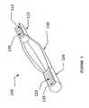
- FIG. 1shows an embodiment of a BTB having a groove with a thread profile disposed thereon.
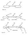
- FIG. 2Ashows a side view of a first embodiment of a BTB in accordance with the invention.
- FIG. 2Bshows a side view of a second embodiment of a BTB in accordance with the inventions.
- FIG. 2Cshows a side view of a third embodiment of a BTB in accordance with the invention.
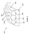
- FIG. 3depicts a frontal view of a donor area for harvesting BTBs in accordance with the teachings herein.
- FIG. 4is a depiction of another embodiment of the invention illustrating a reconstruction of an injured area through implantation of a BTB in accordance with the teachings herein.
- FIG. 5shows a side view of a BTB core cutter of the subject invention designed for harvesting BTB grafts.
- FIG. 6Ashows a close up view of a teeth configuration that is less desired for use with the subject invention.
- FIG. 6Bshows a close up view of a preferred embodiment of the teeth of the embodiment shown in FIG. 5 .
- FIG. 7is a blown up view of the circled region as shown in FIG. 5 .
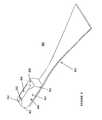
- FIG. 8is three dimensional side view of a further embodiment of the subject BTB that comprises one block that is tapered on both ends.
- FIG. 1there is shown an embodiment directed to a BTB 100 comprising a first bone block 110 and a second bone block 120 interconnected by a tendon 130 , wherein each bone block has been pre-shaped into dowels.
- the term “tendon” as used hereinis intended in its broad sense and refers to fibrous connective tissue for use in grafts, such as, but not limited to, tendons, ligaments and demineralized bone.
- the terms “BTB” or “bone tendon bone graft” as used hereinrefer to a graft implant that comprises one or more tendon portions and one or more bone portions.
- the BTBis preferably isolated from the knee of a donor.
- BTBsare suitable, albeit less preferred, for harvesting BTBs according to the subject invention, such as, but not limited to, the Achilles tendon/calcaneus region or shoulder region.
- suitable implantswould be readily appreciated by those skilled in the art, such as, but not limited to, segmentally demineralized bone (International Pub. No. WO/99/21515).
- one of the bone blocksis derived from the patella while the other is derived from the end of the tibia, and the tendon is derived from the patellar tendon.
- the dowelsare preferably machined down the length of the bone block to form radius cuts 115 , 125 .
- the radius cuts 115 , 125aid in the attachment of the graft to recipient bone because they provide a groove to position a fixation screw, which results in increased surface area at the contact between the bone block and the screw.
- the radius cuts 115 , 125provide the additional advantage of increasing the pull out loads of the bone block, as well as filling of “dead” space in the tunnel.
- Fixation methods known in the artcan be used in accord with the principles of the subject invention, which include, but are not limited to, staples, buttons, screw and washer, interference screws, and self-taping screws.
- fixationis accomplished by interference screws and/or self-taping screws.
- the radius cuts 115 , 125contain a thread profile 135 that matches the thread profile of the fixation screw, thereby further increasing the stability of fixation.
- FIG. 2Ashows an embodiment that comprises a basic configuration of the subject BTBs.
- Bone blocks 210 and 220are in a pre-shaped dowel form with no groove thereon, and are connected by tendon 100 .
- FIG. 2Bshows another version of the BTB, wherein the bone blocks are pre-shaped into dowels with tapered ends.
- Bone block 212is a dowel that has a proximal tapered region 216 in relation to tendon 200
- bone block 214is preshaped into a dowel that has a distal tapered region 218 in relation to tendon 200 .
- FIG. 1shows an embodiment that comprises a basic configuration of the subject BTBs.
- Bone blocks 210 and 220are in a pre-shaped dowel form with no groove thereon, and are connected by tendon 100 .
- FIG. 2Bshows another version of the BTB, wherein the bone blocks are pre-shaped into dowels with tapered ends.
- Bone block 212is a dowel that has
- FIGS. 2B-Cillustrates a preferred version of the invention, which has a bone block 230 with a proximal tapered region 239 and a groove 238 positioned on the bone block 230 .
- This versionalso comprises a second bone block 234 with a distal tapered region and a groove 236 positioned on bone block 234 as well.
- the embodiments shown in FIGS. 2B-Care tapered such that implantation into a pre-formed tunnel in recipient to bone is preferred to occur in the direction of the arrow (see also FIG. 4 ).
- FIG. 3an illustration of a donor area 300 is depicted, wherein three separate grafts 335 , 345 , and 355 are harvested. As shown, the three different grafts individually have at least one bone block 330 , 340 , and 350 . While the sequence of harvesting the grafts is not critical, preferably, graft 335 is excised first, followed by excision of the outside grafts 345 , 355 . Excising graft 335 first results in the automatic cut in the other donor areas, thereby producing a groove in the bone blocks 340 , 350 of the other grafts upon excision.
- the donor areais located at the top of the Tibia at the insertion of the patellar tendon 320 . In an even more preferred embodiment, the donor area extends the length of the patellar tendon to the patella, wherein bone blocks are excised from the patella.
- the bone blockscan be extracted with the use of conventional tools and protocols routinely practiced in the art, such as core cutter and hole saws.
- the bone blockscan be extracted through the use of a BTB bone cutter according to the teachings further described below.
- the extracted bone blocks 330 , 340 , and 350are generally shaped like a plug or dowel and are preferably further shaped by machining through conventional methods known in the art.
- the dowelis machined into dimensions suitable for various surgical procedures. The machining is preferably conducted on a graduated die, a grinding wheel, a lathe, or machining tools may be specifically designed and adapted for this purpose in view of the teachings herein.
- Preferred dimensions for the dowelsinclude 8 mm, 9 mm, 10 mm, 11 mm, and 12 mm in diameter. Reproducibility of the product dimensions is an important feature for the successful use of such grafts in the clinical setting.
- the subject inventionis directed to a method of repairing an injured cruciate ligament in the knee involving the implantation of a BTB.
- FIG. 4illustrates this procedure, and shows a femur 400 and tibia 410 having tunnels formed therein, 466 and 462 , respectively, for receiving BTB 430 , which comprises two bone blocks 432 and 434 connected by tendon 433 .
- BTB 430which comprises two bone blocks 432 and 434 connected by tendon 433 .
- sutures 460are optionally engaged to bone block 432 , which allow a surgeon to pull the BTB 430 through tunnel 462 where the sutures 460 can then be removed.
- the BTB 430is secured in the recipient bone by interference screws 440 .
- the interference screws 440are preferably self taping and are engaged by rotation in the space between grooves 438 and 436 and the inner walls of tunnels 466 and 462 , respectively.
- the BTBcan be pre-marked with alignment markings. Such markings can be positioned on the BTB to aid the surgeon in visualizing the depth of the BTB in the tunnels formed for receiving the BTB, as well as visualizing bone ligament junctions and rotation of the BTB.
- FIGS. 5-7another embodiment of the subject invention is shown that is directed to a BTB harvesting device, such as core cutter 500 that comprises a shaft 502 having a first end 503 and a second end 505 .
- the first end 503 of the shaft 502preferably has a cavity 501 longitudinally disposed thereon, which is designed for engaging a drill, such as by insertion of a Jacob's chuck attached to a power drill (e.g., Dupuy).
- the second end 505 of the shaft 502can be attached to a first end 512 of a hollow cylinder 504 .
- the second end 514 of the cylinder 504preferably has teeth 510 disposed thereon.
- the cylinderhas at least one slot 506 disposed on its surface to aid in the removal of the cut graft tissue from the core cutter 500 .
- the slot 506also provides a means to wash the graft during the extraction procedure to thereby decrease the chance of frictional burning of the graft.
- the shaft 502is approximately 90 mm in length
- the cylinder 504is approximately 50 mm in length
- the slot 506is approximately 30 mm in length.
- the first end of the hollow cylinder 512has a chamfered portion 522 which angles down to the shaft 502 .
- FIG. 6A blown up view of the core cutter teeth 510 is illustrated in FIG. 6 .
- the radius of the teeth A and rake angle of the teeth Bare of appropriate values as to avoid failure (e.g. bending or breaking) of the teeth, as well as undesired damage to the graft.
- FIG. 6Ashows an unacceptable tooth pattern wherein the radius A and bottom angle B are too large, resulting in insufficient support structure for the tooth and inevitable failure.
- a core cutter having a diameter of approximately 10-11 mmpreferably has approximately 14 teeth, with a tooth radius A of approximately 20-30 mm (25 mm being more preferred) and a bottom angle B of approximately 10-20 degrees (15 degrees being more preferred).
- the foregoing dimensionsare preferably maintained, while the number of teeth are appropriately decreased or increased.
- the number of teethare decreased or increased by two for every millimeter below or above, respectively, the 10-11 mm cylinder diameter.
- a core cutter having a 12 mm cylinder diameterwould preferably have about 16 teeth.
- FIG. 7A blown up view of an end section (circle shown in FIG. 5 ) of the cylinder 504 , is shown in FIG. 7 , which illustrates a preferred embodiment of the cylinder 504 wherein the internal diameter (ID) is decreased slightly by adding a relief thickness 520 to the inner surface of the cylinder 504 .
- This embodimentprovides an additional convenience when using a size gauging device (e.g. ring) for selecting extracted bone blocks that are within desired parameters.
- a size gauging devicee.g. ring
- the selection of a BTB through a 10 mm sized gauging devicewould preferably require the BTB to be a slight fraction smaller in diameter than the gauging device, otherwise any insignificant irregularity in the shape of the BTB might cause it to fail to pass through the gauging device.
- the relief thickness 520decreases the ID of the cylinder 504 , thereby effectuating this slight modification to the BTB.
- FIG. 8Shown in FIG. 8 is a further embodiment 800 of the subject BTB that is especially adapted for implantation during knee surgery, wherein the implantation and securement of the BTB is bi-directional.
- BTB embodiment 800comprises one bone block portion 810 and one tendon portion 820 .
- a preferred area from which embodiment 800 is harvestedwould be the heel, thigh, or shoulder. More preferably, the area from which embodiment 800 is harvested is the heel or thigh, whereby tendon portion 820 is derived from an Achilles tendon or quadriceps tendon of a donor.
- the bone block portion 810comprises two ends 812 and 814 which both comprise a tapered region 816 and 818 , respectively.
- the presence of the two tapered regions 812 and 814allows for the BTB embodiment 800 to be inserted and secured bi-directionally, which means, for example, implantation in either the tibial 462 or femoral 466 tunnels as discussed above in reference to the method diagrammed in FIG. 4 .
- the site of implantationcould be approached from a superior point of entry, i.e., establishing a through-tunnel in the femur as opposed to the tibia; BTB embodiment 800 would be suitable for securement in either tunnels in such alternative procedure as well.
- the bone block 810can be provided with a groove 850 to aid in the securement of the implant.
- BTB embodiment 800is provided with preformed graft manipulation holes 852 and 854 for receiving a suture and/or graft insertion tools.
- holes 852 and 854are shown as being vertical or horizontal, respectively, to the axis of the bone block 810 .
- the preformed graft manipulation holescan be made by conventional methods, such as by drilling. Appropriate tools for insertion into preformed holes 852 and 854 will easily be appreciated by those skilled in the art.
- the graft insertion tool(s) usedcomprise an end having a shape and size suitable for insertion into the graft manipulation holes.
- the graftmay be an autograft, allograft, or xenograft.
- Xenograft implantsmay further require treatments to minimize the level of antigenic agents and/or potentially pathogenic agents present in the graft.
- Techniques now known, or those which are later developed, for preparing tissue such that it is suitable for and not rejected by the recipientare incorporated herein.
- a donoris preferably screened for a wide variety of communicable diseases and pathogens, including human immunodeficiency virus, cytomegalovirus hepatitis B, hepatitis C and several other pathogens.
- testsmay be conducted by any of a number of means conventional in the art, including, but not limited to, ELISA assays, PCR assays, or hemagglutination.
- Such testingfollows the requirements of the following associations: (a) American Association of Tissue Banks. Technical Manual for Tissue Banking, Technical Manual-Musculoskeletal Tissues, pages M19-M20; (b) The Food and Drug Administration, Interim Rule, Federal Register, Vol. 58, No. 238, Tuesday, December 14, Rules and Regulations, 65517, D. Infectious Disease Testing and Donor Screening; (c) MMWR, Vol. 43, No.
- RR-8Guidelines for Preventing Transmission of Human Immunodeficiency Virus Through Transplantation of Human Tissue and Organs, pages 4-7; (d) Florida Administrative Weekly, Vol. 10, No. 34, Aug. 21, 1992, 59A-1.001-014, 59A-1.005(12)(c), F.A.C., (12)(a)-(h), 59A-1.005(15, F.A.C., (4) (a)-(8).
- the donor, or their next of kincan be interviewed to ascertain whether the donor engaged in any of a number of high risk behaviors such as having multiple sexual partners, suffering from hemophilia, engaging in intravenous drug use etc. Once a donor has been ascertained to be acceptable, the tissue for obtention of the BTBs as described above are recovered and cleaned.
- a BTBwas harvested according to the following procedure:
- a BTBwas harvested according to the following procedure:
- each plugis cut to approximately 30 mm in length (no less than 45 25 mm)
Landscapes
- Health & Medical Sciences (AREA)
- Life Sciences & Earth Sciences (AREA)
- Surgery (AREA)
- Animal Behavior & Ethology (AREA)
- Public Health (AREA)
- Oral & Maxillofacial Surgery (AREA)
- Orthopedic Medicine & Surgery (AREA)
- Engineering & Computer Science (AREA)
- Biomedical Technology (AREA)
- Heart & Thoracic Surgery (AREA)
- Veterinary Medicine (AREA)
- General Health & Medical Sciences (AREA)
- Transplantation (AREA)
- Nuclear Medicine, Radiotherapy & Molecular Imaging (AREA)
- Molecular Biology (AREA)
- Medical Informatics (AREA)
- Dentistry (AREA)
- Vascular Medicine (AREA)
- Rehabilitation Therapy (AREA)
- Cardiology (AREA)
- Rheumatology (AREA)
- Prostheses (AREA)
- Materials For Medical Uses (AREA)
- Outer Garments And Coats (AREA)
- Surgical Instruments (AREA)
Abstract
Description
Claims (15)
Priority Applications (2)
| Application Number | Priority Date | Filing Date | Title |
|---|---|---|---|
| US14/669,782US9615913B2 (en) | 2000-01-11 | 2015-03-26 | Materials and methods for improved bone tendon bone transplantation |
| US15/458,603US10188505B2 (en) | 2000-01-11 | 2017-03-14 | Materials and methods for improved bone tendon bone transplantation |
Applications Claiming Priority (5)
| Application Number | Priority Date | Filing Date | Title |
|---|---|---|---|
| US09/481,319US6497726B1 (en) | 2000-01-11 | 2000-01-11 | Materials and methods for improved bone tendon bone transplantation |
| US09/528,034US6805713B1 (en) | 2000-01-11 | 2000-03-17 | Materials and methods for improved bone tendon bone transplantation |
| US10/846,399US8167943B2 (en) | 2000-01-11 | 2004-05-14 | Materials and methods for improved bone tendon bone transplantation |
| US13/431,459US20120245687A1 (en) | 2000-01-11 | 2012-03-27 | Materials and methods for improved bone tendon bone transplantation |
| US14/669,782US9615913B2 (en) | 2000-01-11 | 2015-03-26 | Materials and methods for improved bone tendon bone transplantation |
Related Parent Applications (1)
| Application Number | Title | Priority Date | Filing Date |
|---|---|---|---|
| US13/431,459ContinuationUS20120245687A1 (en) | 2000-01-11 | 2012-03-27 | Materials and methods for improved bone tendon bone transplantation |
Related Child Applications (1)
| Application Number | Title | Priority Date | Filing Date |
|---|---|---|---|
| US15/458,603ContinuationUS10188505B2 (en) | 2000-01-11 | 2017-03-14 | Materials and methods for improved bone tendon bone transplantation |
Publications (2)
| Publication Number | Publication Date |
|---|---|
| US20150265395A1 US20150265395A1 (en) | 2015-09-24 |
| US9615913B2true US9615913B2 (en) | 2017-04-11 |
Family
ID=23911497
Family Applications (6)
| Application Number | Title | Priority Date | Filing Date |
|---|---|---|---|
| US09/481,319Expired - LifetimeUS6497726B1 (en) | 1998-11-13 | 2000-01-11 | Materials and methods for improved bone tendon bone transplantation |
| US09/528,034Expired - LifetimeUS6805713B1 (en) | 1998-11-13 | 2000-03-17 | Materials and methods for improved bone tendon bone transplantation |
| US10/846,399Expired - Fee RelatedUS8167943B2 (en) | 2000-01-11 | 2004-05-14 | Materials and methods for improved bone tendon bone transplantation |
| US13/431,459AbandonedUS20120245687A1 (en) | 2000-01-11 | 2012-03-27 | Materials and methods for improved bone tendon bone transplantation |
| US14/669,782Expired - Fee RelatedUS9615913B2 (en) | 2000-01-11 | 2015-03-26 | Materials and methods for improved bone tendon bone transplantation |
| US15/458,603Expired - Fee RelatedUS10188505B2 (en) | 2000-01-11 | 2017-03-14 | Materials and methods for improved bone tendon bone transplantation |
Family Applications Before (4)
| Application Number | Title | Priority Date | Filing Date |
|---|---|---|---|
| US09/481,319Expired - LifetimeUS6497726B1 (en) | 1998-11-13 | 2000-01-11 | Materials and methods for improved bone tendon bone transplantation |
| US09/528,034Expired - LifetimeUS6805713B1 (en) | 1998-11-13 | 2000-03-17 | Materials and methods for improved bone tendon bone transplantation |
| US10/846,399Expired - Fee RelatedUS8167943B2 (en) | 2000-01-11 | 2004-05-14 | Materials and methods for improved bone tendon bone transplantation |
| US13/431,459AbandonedUS20120245687A1 (en) | 2000-01-11 | 2012-03-27 | Materials and methods for improved bone tendon bone transplantation |
Family Applications After (1)
| Application Number | Title | Priority Date | Filing Date |
|---|---|---|---|
| US15/458,603Expired - Fee RelatedUS10188505B2 (en) | 2000-01-11 | 2017-03-14 | Materials and methods for improved bone tendon bone transplantation |
Country Status (8)
| Country | Link |
|---|---|
| US (6) | US6497726B1 (en) |
| EP (1) | EP1246585B1 (en) |
| JP (2) | JP2004500182A (en) |
| AT (1) | ATE418301T1 (en) |
| AU (1) | AU2001227861A1 (en) |
| CA (1) | CA2397071C (en) |
| DE (1) | DE60137114D1 (en) |
| WO (1) | WO2001050999A2 (en) |
Families Citing this family (88)
| Publication number | Priority date | Publication date | Assignee | Title |
|---|---|---|---|---|
| US6482584B1 (en) | 1998-11-13 | 2002-11-19 | Regeneration Technologies, Inc. | Cyclic implant perfusion cleaning and passivation process |
| US20010031254A1 (en)* | 1998-11-13 | 2001-10-18 | Bianchi John R. | Assembled implant |
| US6497726B1 (en)* | 2000-01-11 | 2002-12-24 | Regeneration Technologies, Inc. | Materials and methods for improved bone tendon bone transplantation |
| FR2897259B1 (en) | 2006-02-15 | 2008-05-09 | Ldr Medical Soc Par Actions Si | INTERSOMATIC TRANSFORAMINAL CAGE WITH INTERBREBAL FUSION GRAFT AND CAGE IMPLANTATION INSTRUMENT |
| US6893462B2 (en)* | 2000-01-11 | 2005-05-17 | Regeneration Technologies, Inc. | Soft and calcified tissue implants |
| WO2001078798A1 (en)* | 2000-02-10 | 2001-10-25 | Regeneration Technologies, Inc. | Assembled implant |
| US7530999B2 (en)* | 2000-08-28 | 2009-05-12 | Biomet Sports Medicine, Llc | Method and implant for securing ligament replacement into the knee |
| US6878166B2 (en) | 2000-08-28 | 2005-04-12 | Ron Clark | Method and implant for securing ligament replacement into the knee |
| US7548865B2 (en)* | 2000-10-20 | 2009-06-16 | Arthrex, Inc. | Method of selling procedure specific allografts and associated instrumentation |
| US7195642B2 (en)* | 2001-03-13 | 2007-03-27 | Mckernan Daniel J | Method and apparatus for fixing a graft in a bone tunnel |
| DE10131699A1 (en)* | 2001-06-29 | 2003-01-16 | Tutogen Medical Gmbh | implant |
| FR2827156B1 (en) | 2001-07-13 | 2003-11-14 | Ldr Medical | VERTEBRAL CAGE DEVICE WITH MODULAR FASTENING |
| US7713300B2 (en)* | 2002-01-31 | 2010-05-11 | Biomet Sports Medicince, LLC | Apparatus and method for manipulating a flexible strand and soft tissue replacement during surgery |
| US7033364B1 (en)* | 2002-01-31 | 2006-04-25 | Arthrotek, Inc. | Apparatus and method for manipulating a flexible strand and soft tissue replacement during surgery |
| US20060206206A1 (en) | 2003-06-06 | 2006-09-14 | Peyman Gholam A | Intraocular telescope |
| WO2003086221A1 (en)* | 2002-04-12 | 2003-10-23 | Steenlage Eric S | Method and apparatus for reconstructing a ligament |
| US7955388B2 (en)* | 2006-11-01 | 2011-06-07 | Acumed Llc | Orthopedic connector system |
| US7682392B2 (en)* | 2002-10-30 | 2010-03-23 | Depuy Spine, Inc. | Regenerative implants for stabilizing the spine and devices for attachment of said implants |
| US7112204B2 (en)* | 2003-02-06 | 2006-09-26 | Medicinelodge, Inc. | Tibial tubercle osteotomy for total knee arthroplasty and instruments and implants therefor |
| US7067123B2 (en) | 2003-04-29 | 2006-06-27 | Musculoskeletal Transplant Foundation | Glue for cartilage repair |
| US7901457B2 (en) | 2003-05-16 | 2011-03-08 | Musculoskeletal Transplant Foundation | Cartilage allograft plug |
| US7341592B1 (en) | 2003-10-15 | 2008-03-11 | Biomet Sports Medicine, Inc. | Method and apparatus for graft fixation |
| US7896917B2 (en) | 2003-10-15 | 2011-03-01 | Biomet Sports Medicine, Llc | Method and apparatus for graft fixation |
| EP2113227B1 (en) | 2004-02-04 | 2015-07-29 | LDR Medical | Intervertebral disc prosthesis |
| US7648676B2 (en)* | 2004-04-20 | 2010-01-19 | Rti Biologics, Inc. | Process and apparatus for treating implants comprising soft tissue |
| US20060228252A1 (en)* | 2004-04-20 | 2006-10-12 | Mills C R | Process and apparatus for treating implants comprising soft tissue |
| US7407511B2 (en) | 2004-05-13 | 2008-08-05 | Wright Medical Technology Inc | Methods and materials for connective tissue repair |
| US8002778B1 (en) | 2004-06-28 | 2011-08-23 | Biomet Sports Medicine, Llc | Crosspin and method for inserting the same during soft ligament repair |
| US7837740B2 (en) | 2007-01-24 | 2010-11-23 | Musculoskeletal Transplant Foundation | Two piece cancellous construct for cartilage repair |
| WO2006058221A2 (en) | 2004-11-24 | 2006-06-01 | Abdou Samy M | Devices and methods for inter-vertebral orthopedic device placement |
| MX2007010097A (en)* | 2005-02-18 | 2007-10-10 | Synthasome Inc | Synthetic structure for soft tissue repair. |
| USD583473S1 (en) | 2005-03-04 | 2008-12-23 | Rti Biologics, Inc. | Set of orthopedic bone block assemblies |
| US7763071B2 (en)* | 2005-03-04 | 2010-07-27 | Rti Biologics, Inc. | Bone block assemblies and their use in assembled bone-tendon-bone grafts |
| US7776089B2 (en)* | 2005-03-04 | 2010-08-17 | Rti Biologics, Inc. | Assembled bone-tendon-bone grafts |
| US7727278B2 (en)* | 2005-03-04 | 2010-06-01 | Rti Biologics, Inc. | Self fixing assembled bone-tendon-bone graft |
| US8470038B2 (en) | 2005-03-04 | 2013-06-25 | Rti Biologics, Inc. | Adjustable and fixed assembled bone-tendon-bone graft |
| US7951198B2 (en) | 2005-05-10 | 2011-05-31 | Acumed Llc | Bone connector with pivotable joint |
| US7815926B2 (en) | 2005-07-11 | 2010-10-19 | Musculoskeletal Transplant Foundation | Implant for articular cartilage repair |
| AU2006292224B2 (en) | 2005-09-19 | 2013-08-01 | Histogenics Corporation | Cell-support matrix and a method for preparation thereof |
| FR2891135B1 (en) | 2005-09-23 | 2008-09-12 | Ldr Medical Sarl | INTERVERTEBRAL DISC PROSTHESIS |
| USD583053S1 (en) | 2005-12-19 | 2008-12-16 | Rti Biologics, Inc. | Orthopedic bone block |
| US20070162132A1 (en) | 2005-12-23 | 2007-07-12 | Dominique Messerli | Flexible elongated chain implant and method of supporting body tissue with same |
| US8007533B2 (en) | 2007-02-12 | 2011-08-30 | Rti Biologics, Inc. | Progressive grip assembled bone-tendon-bone grafts, methods of making, and methods of use |
| US8435551B2 (en) | 2007-03-06 | 2013-05-07 | Musculoskeletal Transplant Foundation | Cancellous construct with support ring for repair of osteochondral defects |
| US8147546B2 (en) | 2007-03-13 | 2012-04-03 | Biomet Sports Medicine, Llc | Method and apparatus for graft fixation |
| FR2916956B1 (en) | 2007-06-08 | 2012-12-14 | Ldr Medical | INTERSOMATIC CAGE, INTERVERTEBRAL PROSTHESIS, ANCHORING DEVICE AND IMPLANTATION INSTRUMENTATION |
| FR2924595A1 (en)* | 2007-12-06 | 2009-06-12 | Nicolas Bonin | T-shaped ancillary for sampling patellar ligament of knee, has tubular vertical part emerging at proximal end of ancillary, where proximal end has cutting edge on part of circumference of end that is provided with notches formed on edge |
| EP2265220A1 (en) | 2008-03-05 | 2010-12-29 | Musculoskeletal Transplant Foundation | Cancellous constructs, cartilage particles and combinations of cancellous constructs and cartilage particles |
| KR20110033199A (en)* | 2008-06-19 | 2011-03-30 | 신세스 게엠바하 | Bone Screw Percussion Augmented Implants, Systems and Technologies |
| US9192695B2 (en) | 2008-11-20 | 2015-11-24 | Allosource | Allografts combined with tissue derived stem cells for bone healing |
| EP2349089A4 (en) | 2008-11-21 | 2014-01-15 | Lifecell Corp | Reinforced biologic material |
| US20100249929A1 (en)* | 2009-03-24 | 2010-09-30 | Joint Restoration Foundation | Adjustable patella ligament for acl repair |
| CN102481187B (en)* | 2009-03-31 | 2016-06-08 | 医学嵌入公司暨Imds共同创新公司 | Double bundle acl is repaired |
| JP5306900B2 (en)* | 2009-05-19 | 2013-10-02 | メイラ株式会社 | Transplanted tendon anchor with bone fragment for ligament reconstruction |
| JP5389549B2 (en)* | 2009-06-12 | 2014-01-15 | 高 英卓 | Bone extractor |
| JP5490455B2 (en)* | 2009-06-12 | 2014-05-14 | 高 英卓 | Female set for incising the patella tendon |
| US9597430B2 (en)* | 2009-07-31 | 2017-03-21 | Synthasome, Inc. | Synthetic structure for soft tissue repair |
| CN105326585B (en) | 2009-09-17 | 2018-12-11 | Ldr控股公司 | Intervertebral implant with extensible bone anchoring element |
| US9028553B2 (en) | 2009-11-05 | 2015-05-12 | DePuy Synthes Products, Inc. | Self-pivoting spinal implant and associated instrumentation |
| US8764806B2 (en) | 2009-12-07 | 2014-07-01 | Samy Abdou | Devices and methods for minimally invasive spinal stabilization and instrumentation |
| WO2011080535A1 (en) | 2009-12-31 | 2011-07-07 | Lrd Medical | Anchoring device, intervertebral implant and implantation instrument |
| EP2736545B1 (en) | 2011-07-28 | 2016-01-06 | Harbor Medtech, Inc. | Crosslinked human or animal tissue products and their methods of manufacture and use |
| US8845728B1 (en) | 2011-09-23 | 2014-09-30 | Samy Abdou | Spinal fixation devices and methods of use |
| US8968402B2 (en) | 2011-10-18 | 2015-03-03 | Arthrocare Corporation | ACL implants, instruments, and methods |
| US8920511B2 (en) | 2011-11-17 | 2014-12-30 | Allosource | Multi-piece machine graft systems and methods |
| US20130226240A1 (en) | 2012-02-22 | 2013-08-29 | Samy Abdou | Spinous process fixation devices and methods of use |
| FR2987256B1 (en) | 2012-02-24 | 2014-08-08 | Ldr Medical | ANCHORING DEVICE FOR INTERVERTEBRAL IMPLANT, INTERVERTEBRAL IMPLANT AND IMPLANTATION INSTRUMENTATION |
| WO2013169366A1 (en) | 2012-05-11 | 2013-11-14 | Rti Biologics, Inc. | Xenograft soft tissue implants and methods of making and using |
| US20140039501A1 (en)* | 2012-08-06 | 2014-02-06 | The Cleveland Clinic Foundation | Method and apparatus for providing a soft-tissue transplant to a receiving bone |
| US9198767B2 (en) | 2012-08-28 | 2015-12-01 | Samy Abdou | Devices and methods for spinal stabilization and instrumentation |
| US9320617B2 (en) | 2012-10-22 | 2016-04-26 | Cogent Spine, LLC | Devices and methods for spinal stabilization and instrumentation |
| US10022245B2 (en) | 2012-12-17 | 2018-07-17 | DePuy Synthes Products, Inc. | Polyaxial articulating instrument |
| WO2014130883A2 (en) | 2013-02-22 | 2014-08-28 | Allosource | Cartilage mosaic compositions and methods |
| KR20150126841A (en) | 2013-03-07 | 2015-11-13 | 알로소스 | Consistent calcium content bone allograft systems and methods |
| AU2014236705B2 (en) | 2013-03-15 | 2018-04-12 | Allosource | Perforated osteochondral allograft compositions |
| KR102312720B1 (en) | 2013-03-15 | 2021-10-13 | 알로소스 | Cell repopulated collagen matrix for soft tissue repair and regeneration |
| WO2015175983A1 (en) | 2014-05-16 | 2015-11-19 | Allosource | Composite bone constructs and methods |
| US10077420B2 (en) | 2014-12-02 | 2018-09-18 | Histogenics Corporation | Cell and tissue culture container |
| US10857003B1 (en) | 2015-10-14 | 2020-12-08 | Samy Abdou | Devices and methods for vertebral stabilization |
| WO2017201259A1 (en) | 2016-05-18 | 2017-11-23 | Rti Surgical, Inc. | Osteoinductive nanofiber scaffold for bone regeneration |
| US10744000B1 (en) | 2016-10-25 | 2020-08-18 | Samy Abdou | Devices and methods for vertebral bone realignment |
| US10973648B1 (en) | 2016-10-25 | 2021-04-13 | Samy Abdou | Devices and methods for vertebral bone realignment |
| US11452796B2 (en) | 2017-06-30 | 2022-09-27 | Allosource | Cellular bone grafts, and methods of manufacture and use |
| US10966843B2 (en) | 2017-07-18 | 2021-04-06 | DePuy Synthes Products, Inc. | Implant inserters and related methods |
| US11045331B2 (en) | 2017-08-14 | 2021-06-29 | DePuy Synthes Products, Inc. | Intervertebral implant inserters and related methods |
| US11179248B2 (en) | 2018-10-02 | 2021-11-23 | Samy Abdou | Devices and methods for spinal implantation |
| EP4142647A1 (en) | 2020-05-01 | 2023-03-08 | Harbor Medtech, Inc. | Port-accessible multidirectional reinforced minimally invasive collagen device for soft tissue repair |
| CN114469302B (en)* | 2022-02-11 | 2024-04-02 | 张永飞 | Tendinous bone joint part fixer |
Citations (104)
| Publication number | Priority date | Publication date | Assignee | Title |
|---|---|---|---|---|
| US4400833A (en) | 1981-06-10 | 1983-08-30 | Kurland Kenneth Z | Means and method of implanting bioprosthetics |
| US4605414A (en) | 1984-06-06 | 1986-08-12 | John Czajka | Reconstruction of a cruciate ligament |
| US4804383A (en) | 1984-09-11 | 1989-02-14 | Pierre Rey | Temporary prosthesis for tendons and ligaments, and a method of fitting same |
| US4870957A (en) | 1988-12-27 | 1989-10-03 | Marlowe Goble E | Ligament anchor system |
| US4927421A (en) | 1989-05-15 | 1990-05-22 | Marlowe Goble E | Process of endosteal fixation of a ligament |
| US4942875A (en) | 1986-03-03 | 1990-07-24 | American Cyanamid Company | Surgical repair device having absorbable and nonabsorbable components |
| US5004474A (en) | 1989-11-28 | 1991-04-02 | Baxter International Inc. | Prosthetic anterior cruciate ligament design |
| US5062843A (en) | 1990-02-07 | 1991-11-05 | Mahony Iii Thomas H | Interference fixation screw with integral instrumentation |
| US5067962A (en) | 1989-04-18 | 1991-11-26 | Baxter International Inc. | Bioprosthetic ligament |
| US5078744A (en) | 1987-09-04 | 1992-01-07 | Bio-Products, Inc. | Method of using tendon/ligament substitutes composed of long, parallel, non-antigenic tendon/ligament fibers |
| US5092887A (en) | 1991-08-12 | 1992-03-03 | El Gendler | Artificial ligament produced from demineralized bone for the replacement and augmentation of ligaments, tendons and other fibrous connective tissue |
| US5108431A (en) | 1990-07-06 | 1992-04-28 | Sulzer Brothers Limited | Ligament anchor |
| US5139520A (en) | 1990-01-31 | 1992-08-18 | American Cyanamid Company | Method for acl reconstruction |
| US5151104A (en) | 1989-10-26 | 1992-09-29 | Pfizer Hospital Products Group, Inc. | Self-locking joint connector |
| US5152790A (en) | 1991-03-21 | 1992-10-06 | American Cyanamid Company | Ligament reconstruction graft anchor apparatus |
| US5211647A (en) | 1992-02-19 | 1993-05-18 | Arthrex Inc. | Interference screw and cannulated sheath for endosteal fixation of ligaments |
| US5217495A (en) | 1989-05-10 | 1993-06-08 | United States Surgical Corporation | Synthetic semiabsorbable composite yarn |
| US5234430A (en) | 1991-12-18 | 1993-08-10 | Huebner Randall J | Orthopedic fixation screw and method |
| US5263984A (en) | 1987-07-20 | 1993-11-23 | Regen Biologics, Inc. | Prosthetic ligaments |
| US5320115A (en) | 1991-01-16 | 1994-06-14 | Applied Biological Concepts | Method and apparatus for arthroscopic knee surgery |
| US5366457A (en) | 1991-12-13 | 1994-11-22 | David A. McGuire | Method and apparatus for preparing a bone and tendon graft |
| US5370662A (en) | 1993-06-23 | 1994-12-06 | Kevin R. Stone | Suture anchor assembly |
| US5383878A (en) | 1990-09-04 | 1995-01-24 | Hip Developments Pty Ltd. | Surgical screw |
| US5391169A (en) | 1991-12-13 | 1995-02-21 | Mcguire; David A. | Patellar tendon harvester |
| US5393302A (en) | 1992-10-05 | 1995-02-28 | Clark; Ron | Process for endosteal ligament mounting |
| US5397357A (en) | 1993-02-18 | 1995-03-14 | Arthrex, Inc. | Method for preparing a bone-tendon-bone core graft |
| US5397356A (en) | 1993-01-15 | 1995-03-14 | Depuy Inc. | Pin for securing a replacement ligament to a bone |
| US5425733A (en) | 1992-02-19 | 1995-06-20 | Arthrex, Inc. | Interference screw with rounded back end and cannulated sheath for endosteal fixation of ligaments |
| US5425767A (en) | 1992-11-02 | 1995-06-20 | Sulzer Medizinaltechnik Ag | Anchor for an artificial ligament |
| US5437675A (en) | 1993-06-11 | 1995-08-01 | Wilson; Franklin D. | Polygonal bone punch |
| US5454811A (en) | 1993-11-08 | 1995-10-03 | Smith & Nephew Dyonics, Inc. | Cam lock orthopedic fixation screw and method |
| US5496326A (en) | 1991-06-27 | 1996-03-05 | Johnson; Lanny L. | Fixation screw and method for ligament reconstruction |
| US5507813A (en) | 1993-12-09 | 1996-04-16 | Osteotech, Inc. | Shaped materials derived from elongate bone particles |
| US5562669A (en) | 1994-01-13 | 1996-10-08 | Mcguire; David A. | Cruciate ligament reconstruction with tibial drill guide |
| US5571184A (en) | 1995-06-07 | 1996-11-05 | Wright Medical Technology, Inc. | Graft fixation device and method of using |
| JPH0910245A (en) | 1995-06-28 | 1997-01-14 | Tsunenori Takei | Ligament fixation plug |
| FR2739773A1 (en) | 1995-10-16 | 1997-04-18 | Maire Philippe | ANCILLARY EQUIPMENT FOR SAMPLES OF AUTOGENOUS CYLINDRICAL BONE GRAFTS CALIBRATED MU BY PNEUMATIC OR ELECTRIC MOTOR |
| US5632748A (en) | 1993-06-14 | 1997-05-27 | Linvatec Corporation | Endosteal anchoring device for urging a ligament against a bone surface |
| US5681314A (en) | 1996-06-21 | 1997-10-28 | Lawrence M. Shall | Cutting a bone graft |
| US5688285A (en) | 1995-08-29 | 1997-11-18 | Yamada; Ikufumi | Graft bone fixation tool |
| US5733289A (en) | 1992-10-27 | 1998-03-31 | Neoligaments Limited | Ligament graft harvesting |
| WO1998022047A1 (en) | 1996-11-15 | 1998-05-28 | Medisolve Ltd. | Bone implant |
| JPH10155829A (en) | 1996-11-29 | 1998-06-16 | Nidek Co Ltd | Cornea operation device |
| US5766250A (en) | 1996-10-28 | 1998-06-16 | Medicinelodge, Inc. | Ligament fixator for a ligament anchor system |
| JPH10155820A (en) | 1996-12-02 | 1998-06-16 | Ryohei Takeuchi | Plug for fixing reconstructive ligament into bone hole |
| US5772664A (en) | 1997-02-12 | 1998-06-30 | Wright Medical Technology, Inc. | Instrument for harvesting bone grafts having substantially cylindrical bone plugs |
| US5871504A (en) | 1997-10-21 | 1999-02-16 | Eaton; Katulle Koco | Anchor assembly and method for securing ligaments to bone |
| US5897570A (en) | 1995-03-29 | 1999-04-27 | Linvatec Corporation | Apparatus and method for harvesting a bone-tendon-bone ligament graft |
| WO1999021515A1 (en) | 1997-10-27 | 1999-05-06 | University Of Florida Tissue Bank, Inc. | Segmentally demineralized bone implant |
| US5951560A (en) | 1997-10-15 | 1999-09-14 | Applied Biological Concepts, Inc. | Wedge orthopedic screw |
| US5984966A (en) | 1998-03-02 | 1999-11-16 | Bionx Implants Oy | Bioabsorbable bone block fixation implant |
| US5989253A (en) | 1995-10-27 | 1999-11-23 | Bigliardi; Yves | Ligament anchoring device |
| US6001100A (en) | 1997-08-19 | 1999-12-14 | Bionx Implants Oy | Bone block fixation implant |
| US6099568A (en) | 1998-03-03 | 2000-08-08 | Linvatec Corporation | ACL graft fixation device and method |
| US6099530A (en) | 1998-04-09 | 2000-08-08 | Smith & Nephew, Inc. | Soft-tissue intra-tunnel fixation device |
| US6190411B1 (en) | 1996-04-01 | 2001-02-20 | Kokbing Lo | Fixing element and ligament fixed with fixing element |
| US6200347B1 (en) | 1999-01-05 | 2001-03-13 | Lifenet | Composite bone graft, method of making and using same |
| US6210440B1 (en) | 1995-09-15 | 2001-04-03 | Kevin R. Stone | Anterior cruciate ligament xenografts |
| US6221107B1 (en) | 1998-08-03 | 2001-04-24 | Mark E. Steiner | Ligament fixation device and method |
| US6235057B1 (en) | 1995-01-24 | 2001-05-22 | Smith & Nephew, Inc. | Method for soft tissue reconstruction |
| US6283973B1 (en) | 1998-12-30 | 2001-09-04 | Depuy Orthopaedics, Inc. | Strength fixation device |
| US6306168B1 (en) | 1998-05-04 | 2001-10-23 | Epic Biosonics Inc. | Means for implanting a device in the canalis cochlearis |
| US6355066B1 (en) | 1998-08-19 | 2002-03-12 | Andrew C. Kim | Anterior cruciate ligament reconstruction hamstring tendon fixation system |
| US6440134B1 (en) | 1999-07-29 | 2002-08-27 | Giovanni Zaccherotti | Device for the femoral fixation of the semitendinosus and gracilis tendons for the reconstruction of the anterior cruciate ligament of the knee |
| US6461373B2 (en) | 2000-05-26 | 2002-10-08 | Arthrex, Inc. | Biointerference screw fixation technique |
| US20020165611A1 (en) | 1998-12-22 | 2002-11-07 | Robert-Jan Enzerink | Graft material convenience package |
| US6497726B1 (en) | 2000-01-11 | 2002-12-24 | Regeneration Technologies, Inc. | Materials and methods for improved bone tendon bone transplantation |
| US6533816B2 (en) | 1999-02-09 | 2003-03-18 | Joseph H. Sklar | Graft ligament anchor and method for attaching a graft ligament to a bone |
| US6579295B1 (en) | 2000-09-25 | 2003-06-17 | Robert S. Supinski | Tendon anchors |
| US6893462B2 (en) | 2000-01-11 | 2005-05-17 | Regeneration Technologies, Inc. | Soft and calcified tissue implants |
| US7008451B2 (en) | 2001-09-28 | 2006-03-07 | Ethicon, Inc. | Expanding ligament graft fixation system and method |
| US7083647B1 (en) | 1996-11-27 | 2006-08-01 | Sklar Joseph H | Fixation screw, graft ligament anchor assembly, and method for securing a graft ligament in a bone tunnel |
| US7135025B2 (en) | 2002-04-22 | 2006-11-14 | Inion Ltd. | Surgical implant |
| US7144425B2 (en) | 2002-03-08 | 2006-12-05 | Musculoskeletal Transplant Foundation | Method for inserting improved bone tendon bone assembly with allograft bone block |
| US7172595B1 (en) | 2000-10-03 | 2007-02-06 | Medicinelodge, Inc. | Bone fixation systems and related methods |
| US7182781B1 (en) | 2000-03-02 | 2007-02-27 | Regeneration Technologies, Inc. | Cervical tapered dowel |
| US7235100B2 (en) | 2000-09-15 | 2007-06-26 | Tyco Healthcare Group Lp | Knotless tissue anchor |
| US7309356B2 (en) | 2002-03-08 | 2007-12-18 | Musculoskeletal Transplant Foundation | Bone-tendon- bone assembly with cancellous allograft bone block |
| US7347872B2 (en) | 2000-11-24 | 2008-03-25 | Universite De Montreal | Connective tissue substitutes, method of preparation and uses thereof |
| US7357947B2 (en) | 2001-09-10 | 2008-04-15 | Biomet, Inc. | Bone graft material incorporating demineralized bone matrix and lipids |
| US20080195115A1 (en) | 2007-02-14 | 2008-08-14 | Depuy Mitek | Implement for orientating a tool, particularly useful in surgical tools for harvesting and implanting bone plugs to repair damaged bone tissue |
| US7588586B2 (en) | 2003-05-27 | 2009-09-15 | Ethicon, Inc. | Tissue fixation device |
| US20090234451A1 (en) | 2008-03-12 | 2009-09-17 | Manderson Easton L | Method and system for graft ligament attachment |
| US7594929B2 (en) | 2002-11-21 | 2009-09-29 | Michel Collette | Anchoring screw for a relay strip or suture |
| US20090248068A1 (en) | 2008-03-25 | 2009-10-01 | Linvatec Corporation | Non-metallic knotless suture anchor |
| US20090319043A1 (en) | 2007-08-16 | 2009-12-24 | Mcdevitt Dennis | Helicoil interference fixation system for attaching a graft ligament to a bone |
| US7648676B2 (en) | 2004-04-20 | 2010-01-19 | Rti Biologics, Inc. | Process and apparatus for treating implants comprising soft tissue |
| US7648524B2 (en) | 2005-12-23 | 2010-01-19 | Howmedica Osteonics Corp. | Porous tendon anchor |
| US20100082103A1 (en) | 2007-01-18 | 2010-04-01 | Gordon Blunn | Tendon-integrated prosthesis |
| US7699893B2 (en) | 2003-06-27 | 2010-04-20 | Ethicon, Inc. | Flexible tibial sheath |
| US20100100182A1 (en) | 2008-09-30 | 2010-04-22 | Barnes William S | Method and apparatus for reconstructing a ligament |
| US20100121449A1 (en) | 1996-11-27 | 2010-05-13 | Sklar Joseph H | Graft ligament anchor and method for attaching a graft ligament to a bone |
| US7727278B2 (en) | 2005-03-04 | 2010-06-01 | Rti Biologics, Inc. | Self fixing assembled bone-tendon-bone graft |
| US20100161054A1 (en) | 2008-11-21 | 2010-06-24 | Jason Park | Reinforced Biologic Material |
| US7749250B2 (en) | 2006-02-03 | 2010-07-06 | Biomet Sports Medicine, Llc | Soft tissue repair assembly and associated method |
| US7776089B2 (en) | 2005-03-04 | 2010-08-17 | Rti Biologics, Inc. | Assembled bone-tendon-bone grafts |
| US20100217389A1 (en) | 2009-02-24 | 2010-08-26 | National Yang-Ming University | Graft attachment device for ligament reconstruction |
| US20100249929A1 (en) | 2009-03-24 | 2010-09-30 | Joint Restoration Foundation | Adjustable patella ligament for acl repair |
| US20100249930A1 (en) | 2009-03-31 | 2010-09-30 | Medicinelodge, Inc. Dba Imds Co-Innovation | Double bundle acl repair |
| USD625822S1 (en) | 2005-03-04 | 2010-10-19 | Rti Biologics, Inc. | Orthopedic bone block assembly |
| US20100274355A1 (en) | 2009-01-21 | 2010-10-28 | Mcguire David A | Bone-tendon-bone assembly with cancellous allograft bone block having cortical end portion |
| US20100312341A1 (en) | 2004-06-09 | 2010-12-09 | Biomet Sports Medicine, Llc | Method for Soft Tissue Attachment |
| USD630329S1 (en) | 2007-02-12 | 2011-01-04 | Rti Biologics, Inc. | Orthopedic bone block assembly |
| US7879094B2 (en) | 2006-10-24 | 2011-02-01 | Cayenne Medical, Inc. | Systems for material fixation |
Family Cites Families (4)
| Publication number | Priority date | Publication date | Assignee | Title |
|---|---|---|---|---|
| JPS552395A (en) | 1979-05-18 | 1980-01-09 | Matsushita Electric Ind Co Ltd | Coil insertion device |
| US5131850A (en)* | 1989-11-03 | 1992-07-21 | Cryolife, Inc. | Method for cryopreserving musculoskeletal tissues |
| US5443509A (en)* | 1992-12-10 | 1995-08-22 | Linvatec Corporation | Interference bone-fixation screw with multiple interleaved threads |
| US6067962A (en)* | 1997-12-15 | 2000-05-30 | Caterpillar Inc. | Engine having a high pressure hydraulic system and low pressure lubricating system |
- 2000
- 2000-01-11USUS09/481,319patent/US6497726B1/ennot_activeExpired - Lifetime
- 2000-03-17USUS09/528,034patent/US6805713B1/ennot_activeExpired - Lifetime
- 2001
- 2001-01-11WOPCT/US2001/001008patent/WO2001050999A2/enactiveApplication Filing
- 2001-01-11CACA002397071Apatent/CA2397071C/ennot_activeExpired - Lifetime
- 2001-01-11AUAU2001227861Apatent/AU2001227861A1/ennot_activeAbandoned
- 2001-01-11ATAT01902012Tpatent/ATE418301T1/ennot_activeIP Right Cessation
- 2001-01-11DEDE60137114Tpatent/DE60137114D1/ennot_activeExpired - Lifetime
- 2001-01-11JPJP2001551423Apatent/JP2004500182A/enactivePending
- 2001-01-11EPEP01902012Apatent/EP1246585B1/ennot_activeExpired - Lifetime
- 2004
- 2004-05-14USUS10/846,399patent/US8167943B2/ennot_activeExpired - Fee Related
- 2006
- 2006-10-12JPJP2006279110Apatent/JP2007044548A/enactivePending
- 2012
- 2012-03-27USUS13/431,459patent/US20120245687A1/ennot_activeAbandoned
- 2015
- 2015-03-26USUS14/669,782patent/US9615913B2/ennot_activeExpired - Fee Related
- 2017
- 2017-03-14USUS15/458,603patent/US10188505B2/ennot_activeExpired - Fee Related
Patent Citations (117)
| Publication number | Priority date | Publication date | Assignee | Title |
|---|---|---|---|---|
| US4400833A (en) | 1981-06-10 | 1983-08-30 | Kurland Kenneth Z | Means and method of implanting bioprosthetics |
| US4605414A (en) | 1984-06-06 | 1986-08-12 | John Czajka | Reconstruction of a cruciate ligament |
| US4804383A (en) | 1984-09-11 | 1989-02-14 | Pierre Rey | Temporary prosthesis for tendons and ligaments, and a method of fitting same |
| US4942875A (en) | 1986-03-03 | 1990-07-24 | American Cyanamid Company | Surgical repair device having absorbable and nonabsorbable components |
| US5263984A (en) | 1987-07-20 | 1993-11-23 | Regen Biologics, Inc. | Prosthetic ligaments |
| US5078744A (en) | 1987-09-04 | 1992-01-07 | Bio-Products, Inc. | Method of using tendon/ligament substitutes composed of long, parallel, non-antigenic tendon/ligament fibers |
| US4870957A (en) | 1988-12-27 | 1989-10-03 | Marlowe Goble E | Ligament anchor system |
| US5067962A (en) | 1989-04-18 | 1991-11-26 | Baxter International Inc. | Bioprosthetic ligament |
| US5217495A (en) | 1989-05-10 | 1993-06-08 | United States Surgical Corporation | Synthetic semiabsorbable composite yarn |
| US4927421A (en) | 1989-05-15 | 1990-05-22 | Marlowe Goble E | Process of endosteal fixation of a ligament |
| USRE34871E (en) | 1989-05-15 | 1995-03-07 | Mcguire; David A. | Process of endosteal fixation of a ligament |
| US5151104A (en) | 1989-10-26 | 1992-09-29 | Pfizer Hospital Products Group, Inc. | Self-locking joint connector |
| JPH05502395A (en) | 1989-11-28 | 1993-04-28 | バクスター インターナショナル インコーポレーテッド | artificial anterior cruciate ligament |
| US5004474A (en) | 1989-11-28 | 1991-04-02 | Baxter International Inc. | Prosthetic anterior cruciate ligament design |
| US5139520A (en) | 1990-01-31 | 1992-08-18 | American Cyanamid Company | Method for acl reconstruction |
| US5062843A (en) | 1990-02-07 | 1991-11-05 | Mahony Iii Thomas H | Interference fixation screw with integral instrumentation |
| US5282802A (en) | 1990-02-07 | 1994-02-01 | Mahony Iii Thomas H | Method of securing a tendon graft with an interference fixation screw |
| US5108431A (en) | 1990-07-06 | 1992-04-28 | Sulzer Brothers Limited | Ligament anchor |
| US5383878A (en) | 1990-09-04 | 1995-01-24 | Hip Developments Pty Ltd. | Surgical screw |
| US5320115A (en) | 1991-01-16 | 1994-06-14 | Applied Biological Concepts | Method and apparatus for arthroscopic knee surgery |
| US5152790A (en) | 1991-03-21 | 1992-10-06 | American Cyanamid Company | Ligament reconstruction graft anchor apparatus |
| US5496326A (en) | 1991-06-27 | 1996-03-05 | Johnson; Lanny L. | Fixation screw and method for ligament reconstruction |
| US5092887A (en) | 1991-08-12 | 1992-03-03 | El Gendler | Artificial ligament produced from demineralized bone for the replacement and augmentation of ligaments, tendons and other fibrous connective tissue |
| US5391169A (en) | 1991-12-13 | 1995-02-21 | Mcguire; David A. | Patellar tendon harvester |
| US5366457A (en) | 1991-12-13 | 1994-11-22 | David A. McGuire | Method and apparatus for preparing a bone and tendon graft |
| US5234430A (en) | 1991-12-18 | 1993-08-10 | Huebner Randall J | Orthopedic fixation screw and method |
| US5211647A (en) | 1992-02-19 | 1993-05-18 | Arthrex Inc. | Interference screw and cannulated sheath for endosteal fixation of ligaments |
| US5425733A (en) | 1992-02-19 | 1995-06-20 | Arthrex, Inc. | Interference screw with rounded back end and cannulated sheath for endosteal fixation of ligaments |
| US5393302A (en) | 1992-10-05 | 1995-02-28 | Clark; Ron | Process for endosteal ligament mounting |
| US5733289A (en) | 1992-10-27 | 1998-03-31 | Neoligaments Limited | Ligament graft harvesting |
| US5425767A (en) | 1992-11-02 | 1995-06-20 | Sulzer Medizinaltechnik Ag | Anchor for an artificial ligament |
| US5397356A (en) | 1993-01-15 | 1995-03-14 | Depuy Inc. | Pin for securing a replacement ligament to a bone |
| US5562671A (en) | 1993-01-15 | 1996-10-08 | Depuy Inc. | Ligament replacement cross pinning method |
| US5397357A (en) | 1993-02-18 | 1995-03-14 | Arthrex, Inc. | Method for preparing a bone-tendon-bone core graft |
| US5437675A (en) | 1993-06-11 | 1995-08-01 | Wilson; Franklin D. | Polygonal bone punch |
| US5961520A (en) | 1993-06-14 | 1999-10-05 | Beck, Jr.; Charles L. | Endosteal anchoring device for urging a ligament against a bone surface |
| US5632748A (en) | 1993-06-14 | 1997-05-27 | Linvatec Corporation | Endosteal anchoring device for urging a ligament against a bone surface |
| US6379361B1 (en) | 1993-06-14 | 2002-04-30 | Charles L. Beck, Jr. | Endosteal anchoring device for urging a ligament against a bone surface |
| US5370662A (en) | 1993-06-23 | 1994-12-06 | Kevin R. Stone | Suture anchor assembly |
| US5454811A (en) | 1993-11-08 | 1995-10-03 | Smith & Nephew Dyonics, Inc. | Cam lock orthopedic fixation screw and method |
| US5507813A (en) | 1993-12-09 | 1996-04-16 | Osteotech, Inc. | Shaped materials derived from elongate bone particles |
| US5562669A (en) | 1994-01-13 | 1996-10-08 | Mcguire; David A. | Cruciate ligament reconstruction with tibial drill guide |
| US6235057B1 (en) | 1995-01-24 | 2001-05-22 | Smith & Nephew, Inc. | Method for soft tissue reconstruction |
| US5897570A (en) | 1995-03-29 | 1999-04-27 | Linvatec Corporation | Apparatus and method for harvesting a bone-tendon-bone ligament graft |
| US5571184A (en) | 1995-06-07 | 1996-11-05 | Wright Medical Technology, Inc. | Graft fixation device and method of using |
| JPH0910245A (en) | 1995-06-28 | 1997-01-14 | Tsunenori Takei | Ligament fixation plug |
| US5688285A (en) | 1995-08-29 | 1997-11-18 | Yamada; Ikufumi | Graft bone fixation tool |
| US6210440B1 (en) | 1995-09-15 | 2001-04-03 | Kevin R. Stone | Anterior cruciate ligament xenografts |
| FR2739773A1 (en) | 1995-10-16 | 1997-04-18 | Maire Philippe | ANCILLARY EQUIPMENT FOR SAMPLES OF AUTOGENOUS CYLINDRICAL BONE GRAFTS CALIBRATED MU BY PNEUMATIC OR ELECTRIC MOTOR |
| US5989253A (en) | 1995-10-27 | 1999-11-23 | Bigliardi; Yves | Ligament anchoring device |
| US6190411B1 (en) | 1996-04-01 | 2001-02-20 | Kokbing Lo | Fixing element and ligament fixed with fixing element |
| US5681314A (en) | 1996-06-21 | 1997-10-28 | Lawrence M. Shall | Cutting a bone graft |
| US5766250A (en) | 1996-10-28 | 1998-06-16 | Medicinelodge, Inc. | Ligament fixator for a ligament anchor system |
| WO1998022047A1 (en) | 1996-11-15 | 1998-05-28 | Medisolve Ltd. | Bone implant |
| US20100121449A1 (en) | 1996-11-27 | 2010-05-13 | Sklar Joseph H | Graft ligament anchor and method for attaching a graft ligament to a bone |
| US7083647B1 (en) | 1996-11-27 | 2006-08-01 | Sklar Joseph H | Fixation screw, graft ligament anchor assembly, and method for securing a graft ligament in a bone tunnel |
| JPH10155829A (en) | 1996-11-29 | 1998-06-16 | Nidek Co Ltd | Cornea operation device |
| JPH10155820A (en) | 1996-12-02 | 1998-06-16 | Ryohei Takeuchi | Plug for fixing reconstructive ligament into bone hole |
| US5772664A (en) | 1997-02-12 | 1998-06-30 | Wright Medical Technology, Inc. | Instrument for harvesting bone grafts having substantially cylindrical bone plugs |
| US6001100A (en) | 1997-08-19 | 1999-12-14 | Bionx Implants Oy | Bone block fixation implant |
| US5951560A (en) | 1997-10-15 | 1999-09-14 | Applied Biological Concepts, Inc. | Wedge orthopedic screw |
| US5871504A (en) | 1997-10-21 | 1999-02-16 | Eaton; Katulle Koco | Anchor assembly and method for securing ligaments to bone |
| WO1999021515A1 (en) | 1997-10-27 | 1999-05-06 | University Of Florida Tissue Bank, Inc. | Segmentally demineralized bone implant |
| US5984966A (en) | 1998-03-02 | 1999-11-16 | Bionx Implants Oy | Bioabsorbable bone block fixation implant |
| US6099568A (en) | 1998-03-03 | 2000-08-08 | Linvatec Corporation | ACL graft fixation device and method |
| US6099530A (en) | 1998-04-09 | 2000-08-08 | Smith & Nephew, Inc. | Soft-tissue intra-tunnel fixation device |
| US6306168B1 (en) | 1998-05-04 | 2001-10-23 | Epic Biosonics Inc. | Means for implanting a device in the canalis cochlearis |
| US6221107B1 (en) | 1998-08-03 | 2001-04-24 | Mark E. Steiner | Ligament fixation device and method |
| US6355066B1 (en) | 1998-08-19 | 2002-03-12 | Andrew C. Kim | Anterior cruciate ligament reconstruction hamstring tendon fixation system |
| US20020165611A1 (en) | 1998-12-22 | 2002-11-07 | Robert-Jan Enzerink | Graft material convenience package |
| US6283973B1 (en) | 1998-12-30 | 2001-09-04 | Depuy Orthopaedics, Inc. | Strength fixation device |
| US6200347B1 (en) | 1999-01-05 | 2001-03-13 | Lifenet | Composite bone graft, method of making and using same |
| US6458158B1 (en) | 1999-01-05 | 2002-10-01 | Lifenet | Composite bone graft, method of making and using same |
| US6533816B2 (en) | 1999-02-09 | 2003-03-18 | Joseph H. Sklar | Graft ligament anchor and method for attaching a graft ligament to a bone |
| US6440134B1 (en) | 1999-07-29 | 2002-08-27 | Giovanni Zaccherotti | Device for the femoral fixation of the semitendinosus and gracilis tendons for the reconstruction of the anterior cruciate ligament of the knee |
| US6497726B1 (en) | 2000-01-11 | 2002-12-24 | Regeneration Technologies, Inc. | Materials and methods for improved bone tendon bone transplantation |
| US8167943B2 (en) | 2000-01-11 | 2012-05-01 | Rti Biologics, Inc. | Materials and methods for improved bone tendon bone transplantation |
| US6805713B1 (en) | 2000-01-11 | 2004-10-19 | Regeneration Technologies, Inc. | Materials and methods for improved bone tendon bone transplantation |
| US6893462B2 (en) | 2000-01-11 | 2005-05-17 | Regeneration Technologies, Inc. | Soft and calcified tissue implants |
| US7513910B2 (en) | 2000-01-11 | 2009-04-07 | Rti Biologics, Inc. | Soft and calcified tissue implants |
| US7182781B1 (en) | 2000-03-02 | 2007-02-27 | Regeneration Technologies, Inc. | Cervical tapered dowel |
| US6461373B2 (en) | 2000-05-26 | 2002-10-08 | Arthrex, Inc. | Biointerference screw fixation technique |
| US7235100B2 (en) | 2000-09-15 | 2007-06-26 | Tyco Healthcare Group Lp | Knotless tissue anchor |
| US6780187B2 (en) | 2000-09-25 | 2004-08-24 | Robert S. Supinski | Scissor action tendon anchor |
| US6579295B1 (en) | 2000-09-25 | 2003-06-17 | Robert S. Supinski | Tendon anchors |
| US7172595B1 (en) | 2000-10-03 | 2007-02-06 | Medicinelodge, Inc. | Bone fixation systems and related methods |
| US7347872B2 (en) | 2000-11-24 | 2008-03-25 | Universite De Montreal | Connective tissue substitutes, method of preparation and uses thereof |
| US7357947B2 (en) | 2001-09-10 | 2008-04-15 | Biomet, Inc. | Bone graft material incorporating demineralized bone matrix and lipids |
| US7008451B2 (en) | 2001-09-28 | 2006-03-07 | Ethicon, Inc. | Expanding ligament graft fixation system and method |
| US7309356B2 (en) | 2002-03-08 | 2007-12-18 | Musculoskeletal Transplant Foundation | Bone-tendon- bone assembly with cancellous allograft bone block |
| US7144425B2 (en) | 2002-03-08 | 2006-12-05 | Musculoskeletal Transplant Foundation | Method for inserting improved bone tendon bone assembly with allograft bone block |
| US7135025B2 (en) | 2002-04-22 | 2006-11-14 | Inion Ltd. | Surgical implant |
| US7594929B2 (en) | 2002-11-21 | 2009-09-29 | Michel Collette | Anchoring screw for a relay strip or suture |
| US7588586B2 (en) | 2003-05-27 | 2009-09-15 | Ethicon, Inc. | Tissue fixation device |
| US20100161055A1 (en) | 2003-06-27 | 2010-06-24 | Depuy Mitek, Inc. | Flexible tibial sheath |
| US7699893B2 (en) | 2003-06-27 | 2010-04-20 | Ethicon, Inc. | Flexible tibial sheath |
| US7648676B2 (en) | 2004-04-20 | 2010-01-19 | Rti Biologics, Inc. | Process and apparatus for treating implants comprising soft tissue |
| US20100312341A1 (en) | 2004-06-09 | 2010-12-09 | Biomet Sports Medicine, Llc | Method for Soft Tissue Attachment |
| USD625822S1 (en) | 2005-03-04 | 2010-10-19 | Rti Biologics, Inc. | Orthopedic bone block assembly |
| US7727278B2 (en) | 2005-03-04 | 2010-06-01 | Rti Biologics, Inc. | Self fixing assembled bone-tendon-bone graft |
| US7776089B2 (en) | 2005-03-04 | 2010-08-17 | Rti Biologics, Inc. | Assembled bone-tendon-bone grafts |
| US7648524B2 (en) | 2005-12-23 | 2010-01-19 | Howmedica Osteonics Corp. | Porous tendon anchor |
| US7749250B2 (en) | 2006-02-03 | 2010-07-06 | Biomet Sports Medicine, Llc | Soft tissue repair assembly and associated method |
| US7879094B2 (en) | 2006-10-24 | 2011-02-01 | Cayenne Medical, Inc. | Systems for material fixation |
| US20100082103A1 (en) | 2007-01-18 | 2010-04-01 | Gordon Blunn | Tendon-integrated prosthesis |
| USD630329S1 (en) | 2007-02-12 | 2011-01-04 | Rti Biologics, Inc. | Orthopedic bone block assembly |
| US20080195115A1 (en) | 2007-02-14 | 2008-08-14 | Depuy Mitek | Implement for orientating a tool, particularly useful in surgical tools for harvesting and implanting bone plugs to repair damaged bone tissue |
| US20120083787A1 (en) | 2007-02-14 | 2012-04-05 | Depuy Mitek Inc. | Implement for orientating a tool, particularly useful in surgical tools for harvesting and implanting bone plugs to repair damaged bone tissue |
| US20090319043A1 (en) | 2007-08-16 | 2009-12-24 | Mcdevitt Dennis | Helicoil interference fixation system for attaching a graft ligament to a bone |
| US20090234451A1 (en) | 2008-03-12 | 2009-09-17 | Manderson Easton L | Method and system for graft ligament attachment |
| US20090248068A1 (en) | 2008-03-25 | 2009-10-01 | Linvatec Corporation | Non-metallic knotless suture anchor |
| US20100100182A1 (en) | 2008-09-30 | 2010-04-22 | Barnes William S | Method and apparatus for reconstructing a ligament |
| US20100161054A1 (en) | 2008-11-21 | 2010-06-24 | Jason Park | Reinforced Biologic Material |
| US20100274355A1 (en) | 2009-01-21 | 2010-10-28 | Mcguire David A | Bone-tendon-bone assembly with cancellous allograft bone block having cortical end portion |
| US20100217389A1 (en) | 2009-02-24 | 2010-08-26 | National Yang-Ming University | Graft attachment device for ligament reconstruction |
| US20100249929A1 (en) | 2009-03-24 | 2010-09-30 | Joint Restoration Foundation | Adjustable patella ligament for acl repair |
| US20100249930A1 (en) | 2009-03-31 | 2010-09-30 | Medicinelodge, Inc. Dba Imds Co-Innovation | Double bundle acl repair |
Non-Patent Citations (139)
| Title |
|---|
| "Arthrex All-Inside ACL RetroConstruction with Bone-Tendon-Bone Grafts," Surgical Technique, pp. 1-8 (2012). |
| Advisory Action in U.S. Appl. No. 09/528,034, dated Feb. 24, 2004. |
| Advisory Action in U.S. Appl. No. 09/528,034, dated Nov. 26, 2002. |
| Advisory Action in U.S. Appl. No. 09/924,110, dated Dec. 14, 2004. |
| Andres C. Staehelin, "Patellar-Tendon ACL: Operative Technique," pp. 1-10 (1996). |
| Andres C. Stahelin MD, "Sysorb Bioresorbable Interference Screws Surgical Technique Products Information," pp. 1-24 (2006). |
| Blevins et al., The effects of donor age and strain rate on the biomechanical properties of bone-patellar tendon-bone allografts. American Journal of Sports Medicine, 1994; 22(3):328-333. |
| Canadian Intellectual Property Office, Response to Examiner's Report in Canadian Patent Application No. 2,397,071, dated Mar. 8, 2006. |
| Canadian Patent Office, Office Action, in Canadian Patent Application No. 2,397,071, dated Sep. 12, 2005. |
| European Patent Office, Communication of a notice of opposition, in European Patent No. 1246585, dated Oct. 2, 2009. |
| European Patent Office, Communication pursuant to Article 96(2) EPC, in European Patent Application No. 01 902 012.2-2310, dated Jul. 28, 2005. |
| European Patent Office, Communication pursuant to Article 96(2) EPC, in European Patent Application No. 01 902 012.2-2310, dated Mar. 9, 2004. |
| European Patent Office, Communication under Rule 71(3) EPC, in European Patent Application No. 01 902 012.2-2310, dated Jun. 23, 2008. |
| European patent Office, Decision rejecting the opposition, in European Patent No. 1246585, dated Jan. 12, 2012. |
| European Patent Office, Response in European Patent Application No. 01902012.2, dated Dec. 6, 2005. |
| European Patent Office, Response in European Patent Application No. 01902012.2, dated Feb. 19, 2008. |
| European Patent Office, Response in European Patent Application No. 01902012.2, dated Jan. 25, 2008. |
| European Patent Office, Response in European Patent Application No. 01902012.2, dated May 8, 2007. |
| European Patent Office, Response in European Patent Application No. 01902012.2, dated Oct. 30, 2006. |
| European Patent Office, Response in European Patent Application No. 01902012.2, dated Sep. 15, 2004. |
| European Patent Office, Summons to attend oral proceedings pursuant to Rule 115(1) EPC, in European Patent No. 1246585, dated May 13, 2011. |
| European Patent Office, Summons to attend oral proceedings pursuant to Rule 71(1) EPC, in European Patent Application No. 01 902 012.2-2310, dated Nov. 22, 2007. |
| Fahey et al., Bone tunnel enlargement after anterior cruciate ligament replacement, American Journal of Sports Medicine, 1994; 22(3):410-414. |
| Final Rejection in U.S. Appl. No. 09/528,034, dated Nov. 18, 2003. |
| Final Rejection in U.S. Appl. No. 09/528,034, dated Sep. 11, 2002. |
| Final Rejection in U.S. Appl. No. 09/924,110, dated Jan. 29, 2007. |
| Final Rejection in U.S. Appl. No. 09/924,110, dated Oct. 18, 2005. |
| Final Rejection in U.S. Appl. No. 09/924,110, dated Sep. 28, 2004. |
| Final Rejection in U.S. Appl. No. 10/013,328, dated Apr. 2, 2008. |
| Final Rejection in U.S. Appl. No. 10/013,328, dated May 25, 2006. |
| Final Rejection in U.S. Appl. No. 10/846,399, dated Mar. 16, 2011. |
| Final Rejection in U.S. Appl. No. 13/431,459, dated Sep. 26, 2014. |
| Frank Noyes et al, Reconstruction of the Anterior Cruciate Ligament with Human Allograft. Comparison of Early and Later Results, JBJS The Journal of Bone & Joint Surgery (3 pages).* |
| Gill Jennings & Every, Comments in response to notice of opposition, in European Patent No. 1246585, dated May 18, 2010. |
| Indelicato et al., Clinical comparison of freeze-dried and fresh frozen patellar tendon allografts for anterior cruciate ligament reconstruction of the knee, American Journal of Sports Medicine, 1990; 18(4):335-342. |
| Indelicato et al., The results of fresh-frozen patellar tendon allografts for chronic anterior cruciate ligament deficiency of the knee, American Journal of Sports Medicine, 1992; 20(2):118-121. |
| International Preliminary Examination Report for PCT/US01/01008 dated Apr. 15, 2003. |
| International Search Report for PCT/US01/01008 dated Oct. 18, 2001. |
| International Search Report for PCT/US02/24972 dated Apr. 21, 2003. |
| International Search Report for PCT/US02/25018 dated Apr. 23, 2003. |
| International Search Report in PCT/US02/24972, dated Apr. 24, 2003. |
| International Search Report in PCT/US02/52018, dated Apr. 23, 2003. |
| Japanese Patent Office, Appeal Decision, in Japanese Patent Application No. 2001-551423 dated Mar. 19, 2009. |
| Japanese Patent Office, Decision of Rejection, in Japanese Patent Application No. 2001-551423, dated Jun. 28, 2006. |
| Japanese Patent Office, Notice of Reason for Rejection, in Japanese patent application No. 2006-279110, dated Apr. 4, 2011. |
| Japanese Patent Office, Notice of Reasons for Rejection, in Japanese Patent Application No. 2001-551423, dated Jun. 2, 2005. |
| Japanese Patent Office, Notice of Reasons for Rejection, in Japanese Patent Application No. 2006-279110, dated May 28, 2010. |
| Japanese Patent Office, Pre-Appeal Examination Report, in Japanese Patent Application No. 2001-551423, dated Feb. 27, 2007. |
| Japanese Patent Office, Response in Japanese Patent Application No. 2001-551423, dated Dec. 8, 2005. |
| Japanese Patent Office, Response in Japanese Patent Application No. 2001-551423, dated Nov. 16, 2006. |
| Japanese Patent Office, Response in Japanese Patent Application No. 2001-551423, dated Oct. 3, 2006. |
| Japanese Patent Office, Response in Japanese Patent Application No. 2001-551423, dated Oct. 31, 2007. |
| Japanese Patent Office, Response in Japanese Patent Application No. 2006-279110, dated Nov. 26, 2010. |
| Lambert, Vascularized patellar tendon graft with rigid internal fixation for anterior cruciate ligament insufficiency, Clinical Orthopaedics and Related Research, 1983; 172:85-89. |
| Liu et al., Biomechanics of two types of bone-tendon-bone grafts for ACL reconstruction, Journal of bone and joint surgery, 1995; 77B(2):232-235. |
| Non-Final Rejection in U.S. Appl. No. 09/528,034, dated Dec. 18, 2001. |
| Non-Final Rejection in U.S. Appl. No. 09/528,034, dated Mar. 3, 2003. |
| Non-Final Rejection in U.S. Appl. No. 09/924,110, dated Apr. 18, 2005. |
| Non-Final Rejection in U.S. Appl. No. 09/924,110, dated May 2, 2006. |
| Non-Final Rejection in U.S. Appl. No. 09/924,110, dated Sep. 24, 2003. |
| Non-Final Rejection in U.S. Appl. No. 10/013,228, dated Apr. 6, 2005. |
| Non-Final Rejection in U.S. Appl. No. 10/013,238, dated May 14, 2003. |
| Non-Final Rejection in U.S. Appl. No. 10/013,328, dated Aug. 8, 2007. |
| Non-Final Rejection in U.S. Appl. No. 10/013,328, dated Dec. 3, 2008. |
| Non-Final Rejection in U.S. Appl. No. 10/013,328, dated Jan. 23, 2004. |
| Non-Final Rejection in U.S. Appl. No. 10/013,328, dated Oct. 5, 2005. |
| Non-Final Rejection in U.S. Appl. No. 10/846,399, dated Jul. 7, 2009. |
| Non-Final Rejection in U.S. Appl. No. 10/846,399, dated Mar. 10, 2010. |
| Non-Final Rejection in U.S. Appl. No. 10/846,399, dated Sep. 28, 2010. |
| Non-Final Rejection in U.S. Appl. No. 13/431,459, dated Feb. 21, 2013. |
| Non-Final Rejection in U.S. Appl. No. 13/431,459, dated Jan. 7, 2014. |
| Non-Final Rejection in U.S. Appl. No. 13/431,459, dated Nov. 4, 2013. |
| Noyes et al., Bone-patellar ligament-bone and fascia lata allografts for reconstruction of the anterior cruciate ligament. Journal of Bone and Joint Surgery, 1990; 72A(8); 1125-1136. |
| Noyes, Frank R., et al., "Reconstruction of the Anterior Cruciate Ligament with Human Allograft", The Journal of Bone and Joint Surgery, Incorporated, vol. 78-A, No. 4, Apr. 1996 (14 pages). |
| Office Action dated Apr. 17, 2009 for U.S. Appl. No. 11/796,282, Inventor Kevin C. Carter. |
| Office Action dated Apr. 18, 2005 for U.S. Appl. No. 09/924,110, Inventor Kevin C. Carter. |
| Office Action dated Apr. 2, 2008 for U.S. Appl. No. 10/013,328, Inventor Kevin C. Carter. |
| Office Action dated Apr. 21, 2008 for U.S. Appl. No. 10/846,399, Inventor Kevin C. Carter. |
| Office Action dated Apr. 6, 2005 for U.S. Appl. No. 10/013,328, Inventor Kevin C. Carter. |
| Office Action dated Aug. 5, 2010 for U.S. Appl. No. 11/796,282, Inventor Kevin C. Carter. |
| Office Action dated Aug. 8, 2007 for U.S. Appl. No. 10/013,328, Inventor Kevin C. Carter. |
| Office Action dated Dec. 18, 2002 for U.S. Appl. No. 09/528,034, Inventor Kevin C. Carter. |
| Office Action dated Dec. 19, 2008 for U.S. Appl. No. 10/846,399, Inventor Kevin C. Carter. |
| Office Action dated Dec. 7, 2001 for U.S. Appl. No. 09/481,319, Inventor Kevin C. Carter. |
| Office Action dated Dec. 7, 2009 for U.S. Appl. No. 11/796,282, Inventor Kevin C. Carter. |
| Office Action dated Jan. 23, 2004 for U.S. Appl. No. 10/013,328, Inventor Kevin C. Carter. |
| Office Action dated Jan. 29, 2007 for U.S. Appl. No. 09/924,110, Inventor Kevin C. Carter. |
| Office Action dated Jul. 23, 2007 for U.S. Appl. No. 10/846,399, Inventor Kevin C. Carter. |
| Office Action dated Jun. 13, 2006 for U.S. Appl. No. 10/846,399, Inventor Kevin C. Carter. |
| Office Action dated Mar. 18, 2011 for U.S. Appl. No. 11/796,282, Inventor Kevin C. Carter. |
| Office Action dated Mar. 3, 2003 for U.S. Appl. No. 09/528,034, Inventor Kevin C. Carter. |
| Office Action dated May 10, 2011 for U.S. Appl. No. 12/563,830, Inventor Kevin C. Carter. |
| Office Action dated May 2, 2006 for U.S. Appl. No. 09/924,110, Inventor Kevin C. Carter. |
| Office Action dated May 25, 2006 for U.S. Appl. No. 10/013,328, Inventor Kevin C. Carter. |
| Office Action dated Nov. 18, 2003 for U.S. Appl. No. 09/528,034, Inventor Kevin C. Carter. |
| Office Action dated Oct. 18, 2005 for U.S. Appl. No. 09/924,110, Inventor Kevin C. Carter. |
| Office Action dated Oct. 5, 2005 for U.S. Appl. No. 10/013,328, Inventor Kevin C. Carter. |
| Office Action dated Sep. 11, 2002 for U.S. Appl. No. 09/528,034, Inventor Kevin C. Carter. |
| Office Action dated Sep. 24, 2003 for U.S. Appl. No. 09/924,110, Inventor Kevin C. Carter. |
| Office Action dated Sep. 28, 2004 for U.S. Appl. No. 09/924,110, Inventor Kevin C. Carter. |
| Requirement for Restriction/Election in U.S. Appl. No. 09/528,034, dated Aug. 22, 2001. |
| Requirement for Restriction/Election in U.S. Appl. No. 09/924,110, dated May 16, 2003. |
| Requirement for Restriction/Election in U.S. Appl. No. 12/563,830, dated Nov. 11, 2010. |
| U.S. Patent and Trademark Office, Amendment filed in U.S. Appl. No. 09/528,034, dated Mar. 10, 2004. |
| U.S. Patent and Trademark Office, Election Response filed in U.S. Appl. No. 10/013,328, dated Jul. 11, 2003. |
| U.S. Patent and Trademark Office, Response filed in U.S. Appl. No. 09/481,319, dated May 7, 2002. |
| U.S. Patent and Trademark Office, Response filed in U.S. Appl. No. 09/481,319, dated Oct. 22, 2001. |
| U.S. Patent and Trademark Office, Response filed in U.S. Appl. No. 09/528,034, dated Feb. 2, 2004. |
| U.S. Patent and Trademark Office, Response filed in U.S. Appl. No. 09/528,034, dated May 20, 2002. |
| U.S. Patent and Trademark Office, Response filed in U.S. Appl. No. 09/528,034, dated Nov. 11, 2002. |
| U.S. Patent and Trademark Office, Response filed in U.S. Appl. No. 09/528,034, dated Oct. 22, 2001. |
| U.S. Patent and Trademark Office, Response filed in U.S. Appl. No. 09/528,034, dated Sep. 2, 2003. |
| U.S. Patent and Trademark Office, Response filed in U.S. Appl. No. 09/924,110, dated Apr. 7, 2006. |
| U.S. Patent and Trademark Office, Response filed in U.S. Appl. No. 09/924,110, dated Aug. 15, 2005. |
| U.S. Patent and Trademark Office, Response filed in U.S. Appl. No. 09/924,110, dated Feb. 11, 2004. |
| U.S. Patent and Trademark Office, Response filed in U.S. Appl. No. 09/924,110, dated Jul. 11, 2003. |
| U.S. Patent and Trademark Office, Response filed in U.S. Appl. No. 09/924,110, dated Mar. 28, 2005. |
| U.S. Patent and Trademark Office, Response filed in U.S. Appl. No. 09/924,110, dated Nov. 2, 2006. |
| U.S. Patent and Trademark Office, Response filed in U.S. Appl. No. 09/924,110, dated Nov. 23, 2004. |
| U.S. Patent and Trademark Office, Response filed in U.S. Appl. No. 10/013,328, dated Aug. 15, 2005. |
| U.S. Patent and Trademark Office, Response filed in U.S. Appl. No. 10/013,328, dated Dec. 13, 2004. |
| U.S. Patent and Trademark Office, Response filed in U.S. Appl. No. 10/013,328, dated Feb. 8, 2003. |
| U.S. Patent and Trademark Office, Response filed in U.S. Appl. No. 10/013,328, dated Jul. 10, 2007. |
| U.S. Patent and Trademark Office, Response filed in U.S. Appl. No. 10/013,328, dated Jul. 14, 2004. |
| U.S. Patent and Trademark Office, Response filed in U.S. Appl. No. 10/013,328, dated Mar. 19, 2007. |
| U.S. Patent and Trademark Office, Response filed in U.S. Appl. No. 10/013,328, dated Mar. 9, 2006. |
| U.S. Patent and Trademark Office, Response filed in U.S. Appl. No. 10/013,328, dated May 4, 2009. |
| U.S. Patent and Trademark Office, Response filed in U.S. Appl. No. 10/013,328, dated Oct. 2, 2008. |
| U.S. Patent and Trademark Office, Response filed in U.S. Appl. No. 11/796,282, dated Apr. 7, 2010. |
| U.S. Patent and Trademark Office, Response filed in U.S. Appl. No. 11/796,282, dated Aug. 17, 2009. |
| U.S. Patent and Trademark Office, Response filed in U.S. Appl. No. 11/796,282, dated Jan. 5, 2011. |
| U.S. Patent and Trademark Office, Response filed in U.S. Appl. No. 11/796,282, dated Jul. 14, 2011. |
| U.S. Patent and Trademark Office, Response filed in U.S. Appl. No. 12/563,830, dated Aug. 10, 2011. |
| U.S. Patent and Trademark Office, Response filed in U.S. Appl. No. 12/563,830, dated Jan. 3, 2011. |
| U.S. Patent and Trademark Office, Response filed in U.S. Appl. No. 12/563,830, dated Mar. 4, 2011. |
| U.S. Patent and Trademark Office, Supplemental Amendment filed in U.S. Appl. No. 09/481,319, dated Jul. 31, 2002. |
| U.S. Patent and Trademark Office, Supplemental Election Response filed in U.S. Appl. No. 10/013,328, dated Nov. 10, 2003. |
| U.S. Patent and Trademark Office, Supplemental Response filed in U.S. Appl. No. 11/796,282, dated Aug. 25, 2011. |
| Wagner et al., "Hamstring Tendon Versus Patellar Tendon Anterior Cruciate Ligament Reconstruction Using Biodegradable Interference Fit Fixation," The American Journal of Sports Medicine, pp. 1327-1336. |
Also Published As
| Publication number | Publication date |
|---|---|
| US20120245687A1 (en) | 2012-09-27 |
| WO2001050999A2 (en) | 2001-07-19 |
| WO2001050999A3 (en) | 2002-01-24 |
| CA2397071C (en) | 2008-04-08 |
| US6805713B1 (en) | 2004-10-19 |
| JP2007044548A (en) | 2007-02-22 |
| US20170245979A1 (en) | 2017-08-31 |
| CA2397071A1 (en) | 2001-07-19 |
| EP1246585A2 (en) | 2002-10-09 |
| US10188505B2 (en) | 2019-01-29 |
| ATE418301T1 (en) | 2009-01-15 |
| AU2001227861A1 (en) | 2001-07-24 |
| US20150265395A1 (en) | 2015-09-24 |
| JP2004500182A (en) | 2004-01-08 |
| DE60137114D1 (en) | 2009-02-05 |
| US20040210308A1 (en) | 2004-10-21 |
| US6497726B1 (en) | 2002-12-24 |
| US8167943B2 (en) | 2012-05-01 |
| EP1246585B1 (en) | 2008-12-24 |
Similar Documents
| Publication | Publication Date | Title |
|---|---|---|
| US10188505B2 (en) | Materials and methods for improved bone tendon bone transplantation | |
| US8105379B2 (en) | Materials and methods for improved bone tendon bone transplantation | |
| US8110001B2 (en) | Materials and methods for improved bone tendon bone transplantation | |
| US5562669A (en) | Cruciate ligament reconstruction with tibial drill guide | |
| US8012206B2 (en) | Preformed implants for osteochondral repair | |
| US8221423B2 (en) | Osteochondral plug graft harvesting instrument and kit | |
| US8048079B2 (en) | Retrograde cutting instrument | |
| US20100274355A1 (en) | Bone-tendon-bone assembly with cancellous allograft bone block having cortical end portion | |
| US8133228B2 (en) | Chisel system for osteochondral implants and a surgical procedure involving same | |
| US20220330953A1 (en) | Patella cutting guide | |
| Ferrari et al. | Bone graft procurement for patellar defect grafting in anterior cruciate ligament reconstruction | |
| McGuire et al. | Anterior cruciate ligament reconstruction graft harvesting: pitfalls and tips | |
| DiStefano | Anterior cruciate ligament reconstruction: autograft or allograft? | |
| US10973623B1 (en) | Surgical implants useful in orthopedic surgery | |
| Dockery | The Romano drill: a new surgical curved drill system | |
| Stäubli et al. | Intra-and Postoperative Complications | |
| BG100201A (en) | System of fixing screws and method for the restoration of connections |
Legal Events
| Date | Code | Title | Description |
|---|---|---|---|
| AS | Assignment | Owner name:RTI BIOLOGICS, INC., DELAWARE Free format text:ASSIGNMENT OF ASSIGNORS INTEREST;ASSIGNOR:RTI BIOLOGICS, INC.;REEL/FRAME:041061/0193 Effective date:20050805 Owner name:RTI BIOLOGICS, INC, FLORIDA Free format text:ASSIGNMENT OF ASSIGNORS INTEREST;ASSIGNORS:CARTER, KEVIN C.;ESCH, MICHAEL;LAROCHELLE, PAUL;AND OTHERS;SIGNING DATES FROM 20000324 TO 20011112;REEL/FRAME:041061/0045 Owner name:RTI SURGICAL, INC., FLORIDA Free format text:CHANGE OF NAME;ASSIGNOR:RTI BIOLOGICS, INC;REEL/FRAME:041469/0001 Effective date:20130716 | |
| STCF | Information on status: patent grant | Free format text:PATENTED CASE | |
| AS | Assignment | Owner name:TD BANK, N.A., NEW JERSEY Free format text:SECURITY INTEREST;ASSIGNOR:RTI SURGICAL, INC.;REEL/FRAME:043608/0874 Effective date:20170718 | |
| AS | Assignment | Owner name:JPMORGAN CHASE BANK, N.A., AS THE ADMINISTRATIVE AGENT, ILLINOIS Free format text:SECURITY INTEREST;ASSIGNORS:RTI SURGICAL, INC.;PIONEER SURGICAL TECHNOLOGY, INC.;TUTOGEN MEDICAL, INC.;REEL/FRAME:046303/0926 Effective date:20180605 Owner name:JPMORGAN CHASE BANK, N.A., AS THE ADMINISTRATIVE A Free format text:SECURITY INTEREST;ASSIGNORS:RTI SURGICAL, INC.;PIONEER SURGICAL TECHNOLOGY, INC.;TUTOGEN MEDICAL, INC.;REEL/FRAME:046303/0926 Effective date:20180605 | |
| AS | Assignment | Owner name:RTI SURGICAL, INC., FLORIDA Free format text:RELEASE BY SECURED PARTY;ASSIGNOR:TD BANK, N.A., AS ADMINISTRATIVE AGENT;REEL/FRAME:046327/0864 Effective date:20180605 | |
| AS | Assignment | Owner name:ARES CAPITAL CORPORATION, AS ADMINISTRATIVE AGENT, Free format text:SECURITY INTEREST;ASSIGNORS:RTI SURGICAL, INC.;PIONEER SURGICAL TECHNOLOGY, INC.;TUTOGEN MEDICAL, INC.;AND OTHERS;REEL/FRAME:048543/0505 Effective date:20190308 Owner name:ARES CAPITAL CORPORATION, AS ADMINISTRATIVE AGENT, NEW YORK Free format text:SECURITY INTEREST;ASSIGNORS:RTI SURGICAL, INC.;PIONEER SURGICAL TECHNOLOGY, INC.;TUTOGEN MEDICAL, INC.;AND OTHERS;REEL/FRAME:048543/0505 Effective date:20190308 | |
| AS | Assignment | Owner name:SURGALIGN SPINE TECHNOLOGIES, INC., ILLINOIS Free format text:CHANGE OF NAME;ASSIGNOR:RTI SURGICAL, INC.;REEL/FRAME:053248/0432 Effective date:20200717 Owner name:RTI SURGICAL, INC., FLORIDA Free format text:RELEASE BY SECURED PARTY;ASSIGNOR:ARES CAPITAL CORPORATION, AS AGENT;REEL/FRAME:053257/0652 Effective date:20200720 Owner name:PARADIGM SPINE, LLC, NEW YORK Free format text:RELEASE BY SECURED PARTY;ASSIGNOR:ARES CAPITAL CORPORATION, AS AGENT;REEL/FRAME:053257/0652 Effective date:20200720 Owner name:FOURTH DIMENSION SPINE, LLC, NEW YORK Free format text:RELEASE BY SECURED PARTY;ASSIGNOR:ARES CAPITAL CORPORATION, AS AGENT;REEL/FRAME:053257/0652 Effective date:20200720 Owner name:PIONEER SURGICAL TECHNOLOGY, INC., FLORIDA Free format text:RELEASE BY SECURED PARTY;ASSIGNOR:ARES CAPITAL CORPORATION, AS AGENT;REEL/FRAME:053257/0652 Effective date:20200720 Owner name:TUTOGEN MEDICAL, INC., FLORIDA Free format text:RELEASE BY SECURED PARTY;ASSIGNOR:ARES CAPITAL CORPORATION, AS AGENT;REEL/FRAME:053257/0652 Effective date:20200720 Owner name:RTI OEM, LLC, FRANCE Free format text:INTELLECTUAL PROPERTY ASSIGNMENT AGREEMENT;ASSIGNOR:SURGALIGN SPINE TECHNOLOGIES, INC. (FORMERLY KNOWN AS RTI SURGICAL, INC.);REEL/FRAME:053259/0019 Effective date:20200717 Owner name:RTI SURGICAL, INC., FRANCE Free format text:CHANGE OF NAME;ASSIGNOR:RTI OEM, LLC;REEL/FRAME:053259/0146 Effective date:20200717 | |
| AS | Assignment | Owner name:PIONEER SURGICAL TECHNOLOGY, INC., FLORIDA Free format text:RELEASE BY SECURED PARTY;ASSIGNOR:JPMORGAN CHASE BANK, N.A., AS ADMINISTRATIVE AGENT;REEL/FRAME:053260/0398 Effective date:20200720 Owner name:RTI SURGICAL, INC., FLORIDA Free format text:RELEASE BY SECURED PARTY;ASSIGNOR:JPMORGAN CHASE BANK, N.A., AS ADMINISTRATIVE AGENT;REEL/FRAME:053260/0398 Effective date:20200720 Owner name:TUTOGEN MEDICAL, INC., FLORIDA Free format text:RELEASE BY SECURED PARTY;ASSIGNOR:JPMORGAN CHASE BANK, N.A., AS ADMINISTRATIVE AGENT;REEL/FRAME:053260/0398 Effective date:20200720 | |
| FEPP | Fee payment procedure | Free format text:MAINTENANCE FEE REMINDER MAILED (ORIGINAL EVENT CODE: REM.); ENTITY STATUS OF PATENT OWNER: LARGE ENTITY | |
| LAPS | Lapse for failure to pay maintenance fees | Free format text:PATENT EXPIRED FOR FAILURE TO PAY MAINTENANCE FEES (ORIGINAL EVENT CODE: EXP.); ENTITY STATUS OF PATENT OWNER: LARGE ENTITY | |
| STCH | Information on status: patent discontinuation | Free format text:PATENT EXPIRED DUE TO NONPAYMENT OF MAINTENANCE FEES UNDER 37 CFR 1.362 | |
| FP | Lapsed due to failure to pay maintenance fee | Effective date:20210411 |