US9592123B2 - Therapeutic methods and devices following myocardial infarction - Google Patents
Therapeutic methods and devices following myocardial infarctionDownload PDFInfo
- Publication number
- US9592123B2 US9592123B2US14/448,778US201414448778AUS9592123B2US 9592123 B2US9592123 B2US 9592123B2US 201414448778 AUS201414448778 AUS 201414448778AUS 9592123 B2US9592123 B2US 9592123B2
- Authority
- US
- United States
- Prior art keywords
- struts
- implant
- heart
- hub
- ventricle
- Prior art date
- Legal status (The legal status is an assumption and is not a legal conclusion. Google has not performed a legal analysis and makes no representation as to the accuracy of the status listed.)
- Expired - Fee Related, expires
Links
Images


Classifications
- A—HUMAN NECESSITIES
- A61—MEDICAL OR VETERINARY SCIENCE; HYGIENE
- A61F—FILTERS IMPLANTABLE INTO BLOOD VESSELS; PROSTHESES; DEVICES PROVIDING PATENCY TO, OR PREVENTING COLLAPSING OF, TUBULAR STRUCTURES OF THE BODY, e.g. STENTS; ORTHOPAEDIC, NURSING OR CONTRACEPTIVE DEVICES; FOMENTATION; TREATMENT OR PROTECTION OF EYES OR EARS; BANDAGES, DRESSINGS OR ABSORBENT PADS; FIRST-AID KITS
- A61F2/00—Filters implantable into blood vessels; Prostheses, i.e. artificial substitutes or replacements for parts of the body; Appliances for connecting them with the body; Devices providing patency to, or preventing collapsing of, tubular structures of the body, e.g. stents
- A61F2/02—Prostheses implantable into the body
- A61F2/24—Heart valves ; Vascular valves, e.g. venous valves; Heart implants, e.g. passive devices for improving the function of the native valve or the heart muscle; Transmyocardial revascularisation [TMR] devices; Valves implantable in the body
- A61F2/2478—Passive devices for improving the function of the heart muscle, i.e. devices for reshaping the external surface of the heart, e.g. bags, strips or bands
- A61F2/2487—Devices within the heart chamber, e.g. splints
- A—HUMAN NECESSITIES
- A61—MEDICAL OR VETERINARY SCIENCE; HYGIENE
- A61B—DIAGNOSIS; SURGERY; IDENTIFICATION
- A61B17/00—Surgical instruments, devices or methods
- A61B17/0057—Implements for plugging an opening in the wall of a hollow or tubular organ, e.g. for sealing a vessel puncture or closing a cardiac septal defect
- A—HUMAN NECESSITIES
- A61—MEDICAL OR VETERINARY SCIENCE; HYGIENE
- A61B—DIAGNOSIS; SURGERY; IDENTIFICATION
- A61B17/00—Surgical instruments, devices or methods
- A61B17/12—Surgical instruments, devices or methods for ligaturing or otherwise compressing tubular parts of the body, e.g. blood vessels or umbilical cord
- A61B17/12022—Occluding by internal devices, e.g. balloons or releasable wires
- A61B17/12099—Occluding by internal devices, e.g. balloons or releasable wires characterised by the location of the occluder
- A61B17/12122—Occluding by internal devices, e.g. balloons or releasable wires characterised by the location of the occluder within the heart
- A—HUMAN NECESSITIES
- A61—MEDICAL OR VETERINARY SCIENCE; HYGIENE
- A61B—DIAGNOSIS; SURGERY; IDENTIFICATION
- A61B17/00—Surgical instruments, devices or methods
- A61B17/12—Surgical instruments, devices or methods for ligaturing or otherwise compressing tubular parts of the body, e.g. blood vessels or umbilical cord
- A61B17/12022—Occluding by internal devices, e.g. balloons or releasable wires
- A61B17/12131—Occluding by internal devices, e.g. balloons or releasable wires characterised by the type of occluding device
- A61B17/12168—Occluding by internal devices, e.g. balloons or releasable wires characterised by the type of occluding device having a mesh structure
- A61B17/12172—Occluding by internal devices, e.g. balloons or releasable wires characterised by the type of occluding device having a mesh structure having a pre-set deployed three-dimensional shape
- A—HUMAN NECESSITIES
- A61—MEDICAL OR VETERINARY SCIENCE; HYGIENE
- A61B—DIAGNOSIS; SURGERY; IDENTIFICATION
- A61B17/00—Surgical instruments, devices or methods
- A61B17/00234—Surgical instruments, devices or methods for minimally invasive surgery
- A61B2017/00238—Type of minimally invasive operation
- A61B2017/00243—Type of minimally invasive operation cardiac
- A—HUMAN NECESSITIES
- A61—MEDICAL OR VETERINARY SCIENCE; HYGIENE
- A61B—DIAGNOSIS; SURGERY; IDENTIFICATION
- A61B17/00—Surgical instruments, devices or methods
- A61B17/0057—Implements for plugging an opening in the wall of a hollow or tubular organ, e.g. for sealing a vessel puncture or closing a cardiac septal defect
- A61B2017/00575—Implements for plugging an opening in the wall of a hollow or tubular organ, e.g. for sealing a vessel puncture or closing a cardiac septal defect for closure at remote site, e.g. closing atrial septum defects
- A61B2017/00592—Elastic or resilient implements
- A—HUMAN NECESSITIES
- A61—MEDICAL OR VETERINARY SCIENCE; HYGIENE
- A61B—DIAGNOSIS; SURGERY; IDENTIFICATION
- A61B17/00—Surgical instruments, devices or methods
- A61B17/0057—Implements for plugging an opening in the wall of a hollow or tubular organ, e.g. for sealing a vessel puncture or closing a cardiac septal defect
- A61B2017/00575—Implements for plugging an opening in the wall of a hollow or tubular organ, e.g. for sealing a vessel puncture or closing a cardiac septal defect for closure at remote site, e.g. closing atrial septum defects
- A61B2017/00615—Implements with an occluder on one side of the opening and holding means therefor on the other
- A—HUMAN NECESSITIES
- A61—MEDICAL OR VETERINARY SCIENCE; HYGIENE
- A61B—DIAGNOSIS; SURGERY; IDENTIFICATION
- A61B17/00—Surgical instruments, devices or methods
- A61B17/0057—Implements for plugging an opening in the wall of a hollow or tubular organ, e.g. for sealing a vessel puncture or closing a cardiac septal defect
- A61B2017/00575—Implements for plugging an opening in the wall of a hollow or tubular organ, e.g. for sealing a vessel puncture or closing a cardiac septal defect for closure at remote site, e.g. closing atrial septum defects
- A61B2017/00632—Occluding a cavity, i.e. closing a blind opening
- A—HUMAN NECESSITIES
- A61—MEDICAL OR VETERINARY SCIENCE; HYGIENE
- A61B—DIAGNOSIS; SURGERY; IDENTIFICATION
- A61B17/00—Surgical instruments, devices or methods
- A61B2017/00831—Material properties
- A61B2017/00867—Material properties shape memory effect
- A—HUMAN NECESSITIES
- A61—MEDICAL OR VETERINARY SCIENCE; HYGIENE
- A61B—DIAGNOSIS; SURGERY; IDENTIFICATION
- A61B17/00—Surgical instruments, devices or methods
- A61B17/12—Surgical instruments, devices or methods for ligaturing or otherwise compressing tubular parts of the body, e.g. blood vessels or umbilical cord
- A61B17/12022—Occluding by internal devices, e.g. balloons or releasable wires
- A61B2017/1205—Introduction devices
- A61B2017/12054—Details concerning the detachment of the occluding device from the introduction device
- A61B2017/12095—Threaded connection
- A—HUMAN NECESSITIES
- A61—MEDICAL OR VETERINARY SCIENCE; HYGIENE
- A61F—FILTERS IMPLANTABLE INTO BLOOD VESSELS; PROSTHESES; DEVICES PROVIDING PATENCY TO, OR PREVENTING COLLAPSING OF, TUBULAR STRUCTURES OF THE BODY, e.g. STENTS; ORTHOPAEDIC, NURSING OR CONTRACEPTIVE DEVICES; FOMENTATION; TREATMENT OR PROTECTION OF EYES OR EARS; BANDAGES, DRESSINGS OR ABSORBENT PADS; FIRST-AID KITS
- A61F2/00—Filters implantable into blood vessels; Prostheses, i.e. artificial substitutes or replacements for parts of the body; Appliances for connecting them with the body; Devices providing patency to, or preventing collapsing of, tubular structures of the body, e.g. stents
- A61F2/02—Prostheses implantable into the body
- A61F2/24—Heart valves ; Vascular valves, e.g. venous valves; Heart implants, e.g. passive devices for improving the function of the native valve or the heart muscle; Transmyocardial revascularisation [TMR] devices; Valves implantable in the body
- A61F2/2478—Passive devices for improving the function of the heart muscle, i.e. devices for reshaping the external surface of the heart, e.g. bags, strips or bands
- A61F2/2481—Devices outside the heart wall, e.g. bags, strips or bands
- A61F2002/2484—Delivery devices therefor
Definitions
- the present inventionrelates generally to the field of treating heart disease, particularly preventing remodeling following myocardial infarction.
- myocardiumheart muscle
- AMIacute myocardial infarction
- myocardial ruptureMortality rates for myocardial rupture are extremely high unless early diagnosis and surgical intervention are provided rapidly. Cardiac rupture is a medical emergency. The overall risk of death depends on the speed of the treatment provided, therefore fast and relatively easy treatment option is needed.
- Myocardial regions affected by infarctionmay change size and shape, i.e. remodels, and in many cases non-affected myocardium remodels as well.
- the infracted regionexpands due to the forces produced by the viable myocardium. Whether these changes become permanent and progress to involve infracted border zones and remote non-infarcted myocardium may depend on multiple factors, including infarct size, promptness of reperfusion, post-infarction therapy, etc. However, even following small infarction, many patients treated with the state-of-the-art therapies show some degree of regional and subsequent global ventricular shape changes and enlargement.
- FIG. 18shows a summary flowchart illustrating the effects of acute myocardial infarction.
- Described hereinare methods and devices which may be used for the immediate and early treatment of myocardial infarction. Cardiac rupture post myocardial infarction needs to be treated immediately. The early and rapid appearance of infarct and border zone lengthening and early infarct expansion may be prevented by the early treatments described herein to prevent or attenuate initial myocardial infarct region expansion early after myocardial infarction. These methods and implants may provide an immediate mechanical effect to prevent or attenuate ventricular remodeling, and may also be used in conjunction with therapeutic agents and/or cells to the cardiac endothelium.
- Described hereinare methods, devices and systems for treatment the heart following myocardial infarction.
- these methodstypically require the application of a treatment device that supports and/or isolates the infracted region of the heart within about 72 hours of the ischemic event.
- These methodsmay be used, for example, to treat a portion of the left ventricle that is affected by myocardial infarction.
- a treatment devicemay be a support device that provides mechanical support to the region of the heart affected by the myocardial infarction, and/or a partitioning device (e.g., including a membrane) that at least partially isolates the region of the heart chamber affected by the myocardial infarction and/or cardiac rupture.
- a partitioning devicee.g., including a membrane
- the treatment deviceis both a support device and a partitioning device.
- a method of preventing cardiac rupture following myocardial infarctioncomprising delivering a device to a heart chamber exhibiting myocardial infarction within 72 hours of myocardial infarction (wherein the device comprises a reinforced membrane) and deploying the device in the chamber adjacent the region of the chamber wall exhibiting myocardial infarction.
- the methodmay also include the step of identifying the region of the heart chamber exhibiting myocardial infarction.
- Any appropriate method of identifying the region of the heart chamber exhibiting the myocardial infarctionmay be used, including visual inspection, electrical inspection, imaging by echocardiography, magnetic resonance or computerized tomography, or the like.
- electrical inspectionmay be performed by the use of ECG measurements and analysis, or the use of electrodes on or around the heart tissue.
- Visual inspectionmay be done using direct (light) visualization, or by labeling for markers or reactivity.
- ultrasoundmay be used to identify region of the heart affected by the myocardial infarction.
- a treatment devicemay include a membrane (e.g., a reinforced membrane).
- the membranemay be non-porous or porous to allow fluid (including blood) exchange across it.
- the devicemay include an expandable frame.
- the membranemay be attached or connected to the expandable frame.
- the expandable framemay be formed of an elastic or superelastic material, such as a shape memory material (e.g., NitinolTM, or other super-elastic materials).
- the expandable framemay be formed of a plurality of struts that extend from a hub.
- the devicemay also include a foot (e.g., a non-traumatic foot) for contacting the wall of the chamber. In some variations the device is configured so that only minimal (if any) space is partitioned.
- the step of delivering the devicemay include delivering the device in a collapsed configuration.
- the delivery stepmay include the step of delivering the device in a collapsed state through a catheter or other inserter.
- the devicemay be held in a first, collapsed or delivery, configuration and may be deployed by expanding into the deployed configuration.
- the devicemay be self-expanding, or it may be expanded using a mechanical expander such as a balloon or other structure.
- the step of delivering the devicemay include using a delivery catheter.
- the deviceWhen a device is used to treat the heart, the device may be sealed about the periphery of the membrane of the device against the chamber wall of the heart being treated. Any appropriate sealing technique may be used.
- the devicemay include a seal region, e.g., an expandable, inflatable, or other region. Examples of devices including a seal are provided herein, and may also be found, for example, in US patent application publication No. 2006/0281965, herein incorporated by reference in its entirety.
- the step of deploying the devicemay therefore also include isolating the region of the chamber wall exhibiting myocardial infarction from the rest of the chamber.
- the step of deploying the devicemay also comprise partitioning the heart chamber into a main productive portion and a secondary non-productive portion, with the region of the chamber exhibiting myocardial infarction or cardiac rupture forming a part of the secondary non-productive portion.
- the treatment devicemay include anchors or attachments for securing the device to the wall of the heart chamber.
- the devicemay include hooks and/or barbs on the membrane and/or expandable frame.
- the methods of preventing remodeling due to myocardial infarctionmay include the step of securing or anchoring the device to the heart wall.
- the devicemay be anchored or secured to the heart wall over the region of myocardial infarction.
- One or more therapeutic agentsmay also be delivered to the heart tissue (e.g., the heart wall) from the device.
- the devicemay be coated or impregnated with a therapeutic material.
- a therapeutic materialis added to the heart chamber after the device is inserted, for example in the space between the device and the heart wall.
- Also described herein are methods of preventing cardiac remodeling following myocardial infarctioncomprising the step of: delivering a device to a left ventricle within 72 hours of myocardial infarction (wherein the device comprises a reinforced membrane) and deploying the device in the left ventricle adjacent a region of the left ventricle exhibiting myocardial infarction.
- the step of delivering the support devicemay comprise delivering the support device in a collapsed configuration.
- the support devicemay be any of the treatment devices described herein; for example, the support devices may be a device having a plurality of struts extending from a central hub.
- the support devicemay include a reinforced membrane (which may be impermeable, or permeable, or semi-permeable).
- the support devicemay include a foot (e.g., a non-traumatic foot), or a non-traumatic hub.
- the step of deploying the support devicemay include securing the support device to the wall of the chamber.
- the treatment devices described hereinmay dynamically flex as the wall of the chamber moves.
- the support devicemay be made of a material (e.g., a shape memory alloy) that supports the wall, and flexes as the heart beats.
- FIG. 1Ais a schematic view of a patient's heart having a myocardial infarct.
- FIG. 1Bis a schematic view of the patient's heart of FIG. 1A with a ventricular septal defect resulting from a rupture in the heart wall.
- FIG. 1Cis a schematic view of the patient's heart of FIG. 1B after treatment following rupture of the heart wall.
- FIG. 1Dis a schematic view of the patient's heart after immediate early treatment, as described herein.
- FIG. 2Ais a schematic view of a patient's heart exhibiting a myocardial infarct with free wall rupture of the left ventricular chamber.
- FIG. 2Bis a schematic view of the patient's heart of FIG. 2A with a left ventricular chamber tamponade.
- FIG. 2Cis a schematic view of the patient's heart of FIG. 2B after treatment following development of tamponade.
- FIG. 3is an elevational view of a device in an expanded configuration.
- FIG. 4is a plan view of the device shown in FIG. 3 illustrating the upper surface of the device.
- FIG. 5is a bottom view of the device shown in FIG. 3 .
- FIG. 6is a perspective view of a non-traumatic tip of the distally extending stem of the device shown in FIG. 3 .
- FIG. 7is a partial cross-sectional view of a hub of the device shown in FIG. 4 taken along the lines 7 - 7 .
- FIG. 8is a transverse cross-sectional view of the hub shown in FIG. 7 taken along the lines 8 - 8 .
- FIG. 9is a longitudinal view, partially in section of a reinforcing rib and membrane at the periphery of the device shown in FIG. 3 .
- FIG. 10is a schematic elevational view, partially in section, of a delivery system for a device such as the device shown in FIGS. 3 and 4 .
- FIG. 11is a transverse cross-sectional view of the delivery system shown in FIG. 10 taken along the lines 11 - 11 .
- FIG. 12is an elevational view, partially in section, of the hub shown in FIG. 7 secured to a helical coil of the delivery system shown in FIG. 10 .
- FIGS. 13A-13Eare schematic views of a patient's left ventricular chamber illustrating the deployment of the device shown in FIGS. 3 and 4 with the delivery system shown in FIG. 10 to a patient's heart chamber (e.g., left ventricle).
- a patient's heart chambere.g., left ventricle
- FIG. 14is a schematic view of the patient's heart after treatment according to a method of the present invention.
- FIG. 15Aillustrate one variation of an implant which may be used with the present invention.
- FIG. 15Bshows another variation of an implant which may be used with the present invention.
- FIG. 15Cis a schematic view of a heart in which the implant of FIG. 15A has been implanted.
- FIG. 16Aillustrates another variation of an implant which may be used with the present invention.
- FIG. 16Bis a schematic view of a heart in which the implant of FIG. 16A has been implanted.
- FIGS. 17A and 17Bshow another variation of an implant which may be used following acute myocardial infarction.
- FIGS. 17C and 17Dillustrate a delivery system for delivering an implant such as the implant of FIGS. 17A and 17B .
- FIG. 18schematically illustrates the effects of myocardial infarction.
- FIGS. 19A and 19Bis a schematic illustration of a heart in which the implant has been applied.
- Described hereinare methods of treating a patient to prevent or correct cardiac remodeling following myocardial infarction.
- these methodsmay include inserting or implanting a device in a heart chamber within 72 hours after myocardial infarction, or shortly after a determination of myocardial infarction.
- the deviceis preferably placed within the region of the heart chamber exhibiting one or more indication of myocardial infarction.
- the devicemay be a support device (e.g., a resilient frame) and/or a partitioning device.
- FIG. 1Ais a schematic illustration of a patient's heart 10 showing the right ventricle 11 and the left ventricle 12 with the mitral valve 13 and aortic valve 14 .
- a pericardium membrane 15is shown surrounding the heart 10 .
- At least a portion of myocardium layer 17 of the left ventricle 12is exhibiting an area of infarct 18 (“MI”) extending along a portion of ventricular septum wall 19 which separates the right and left ventricles. This region may exhibit characteristics of an incipient rupture.
- FIG. 1Billustrates the advancing of the infarct leading to the generation of a rupture or opening 20 in the septum wall 19 , a condition referred to as VSD.
- oxygenated blood 21flows directly to the right ventricle 11 through the septum opening 20 .
- the right portion of the heartworks harder pumping a greater volume of blood than it normally would, and secondly, the amount of oxygenated blood in the left ventricle is reduced leading to a lower oxygen level to the other tissues of the body.
- FIG. 1Cillustrates the left ventricle 12 of FIG. 1B after it has been partitioned, with the use of a partitioning device 30 according to the present invention and as described further below, into a main productive or operational portion 23 and a secondary, essentially non-productive portion 24 .
- FIG. 1Cillustrates the normal flow of blood from the left ventricle to the rest of the body through the aortic valve.
- FIG. 1Dshows the schematic illustration of the heart of FIG. 1A shortly after determination of a myocardial infarction.
- the region of the heart chamber exhibiting myocardial infarction(the area of infarct 18 ) is indicated, and in this example a device 30 has been deployed to reinforce this region.
- the deviceis deployed into the heart chamber adjacent to the region of the heart chamber exhibiting myocardial infarction shortly after a determination of the myocardial infarction has been made.
- thisoccurs prior to substantial remodeling of the heart. For example, this may be less than 72 hours after the myocardial infarction, or less than a few days after the determination of a myocardial infarction.
- the occurrence of a myocardial infarctionmay be determined by any appropriate method, including diagnostics based on physical examination, electrocardiogram, blood (or other tests) for cardiac markers, angiograms, or the like.
- enzyme markerse.g., SGOT, LDH, creatine kinase
- other markerse.g., troponins, glycogen phoshyorylase isoenzyme, myoglobin, etc.
- the region of the heart affected by the myocardial infarctionmay also be determined.
- visualization techniquesdirectly or indirect
- angiogramsmay be used.
- Other visualization techniquesincluding scanning (e.g., echocardiography, CT scanning, etc.), electrical mapping, etc. may also be used to localize an area of infarct.
- FIG. 2Ais a schematic illustration of a patient's heart 10 showing the right ventricle 11 and the left ventricle 12 with the mitral valve 13 and aortic valve 14 .
- the pericardium membrane 15is shown surrounding the heart.
- a pericardium(pericardial complex) consists of an outer fibrous layer and an inner serous layer.
- the pericardial space 16normally contains 20-50 mL of fluid. At least a portion of the myocardium layer 17 of the left ventricle 12 , as shown in FIG.
- MIinfarct 18
- FIG. 2Bshows the remodeling of the heart following MI.
- the damage from the infarcthas advanced, leading to the rupture or opening 20 which is increasing in size.
- the flow of the blood 21 into the pericardial space 16increases over time leading to a greater accumulation of blood in the pericardial space.
- This movement and accumulation of blood in the pericardial spacea condition referred to as ventricular tamponade, results in reduced ventricular filling and subsequent hemodynamic compromise.
- FIG. 2Cillustrates the left ventricle 12 of FIG. 2A after a device 30 has been inserted.
- This device 30is a partitioning device which both supports the damaged area, and may partition it from other portions of the heart chamber, into the main productive or operational portion 23 and the secondary, essentially non-productive portion 24 .
- supporting the damaged region of the heart chambere.g., the area of infarct 18
- partitioning itmay prevent or reverse the remodeling of the heart and help restore the normal flow of blood from the left ventricle to the rest of the body through the aortic valve.
- a device for preventing remodeling of the heartcomprises a flexible support frame and one or more anchors, and may optionally include one or more of a foot region (e.g., an atraumatic foot region) and a membrane.
- a foot regione.g., an atraumatic foot region
- a membranee.g.
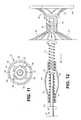
- FIGS. 3-6illustrate one example of a device 30 (in this variation a partitioning device) which embodies features of the invention and which may be utilized in practicing the method of the present invention.
- the device 30includes a partitioning membrane 31 , a hub 32 , preferably centrally located on the partitioning device, and a radially expandable reinforcing frame 33 formed of a plurality of ribs 34 .
- a radially expandable reinforcing frame 33formed of a plurality of ribs 34 .
- at least part of the partitioning membrane 31is secured to a proximal or pressure receiving side 35 of the frame 33 as shown in FIG. 3 .
- the ribs 34have distal ends 36 which are secured to the hub 32 , and free proximal ends 37 which are configured to curve or flare away from a center line axis 38 at least upon expansion of the partitioning device. Radial expansion of the free proximal ends 37 unfurls the membrane 31 secured to the frame 33 so that the membrane presents the pressure receiving surface 35 which defines in part the productive portion 23 of the patient's partitioned heart chamber. A peripheral edge 39 of the membrane 31 may be serrated as shown.
- a continuous expansive strand 40extends around the periphery of the membrane 31 on the pressure receiving side 35 thereof to apply pressure to the pressure side of the flexible material of the membrane to effectively seal the periphery of the membrane against the wall of the ventricular chamber.
- Ends 41 and 42 of the expansive strand 40are shown extending away from the device in FIGS. 3 and 5 .
- the ends 41 and 42may be left unattached or may be secured together, e.g. by a suitable adhesive, to the membrane 31 itself.
- the membrane 31has a proximal layer secured to the proximal faces of the ribs 34 and a distal layer secured to the distal faces of the ribs in a manner described in co-pending application Ser. No. 10/913,608, filed on Aug. 5, 2004, assigned to the assignee of the present invention, and incorporated herein by reference in its entirety.
- the hub 32 shown in FIGS. 6 and 7preferably has a distally extending stem 43 with a non-traumatic support component 44 .
- the support component 44has a plurality of pods or feet 45 extending radially away from the center line axis 38 and the ends of the feet 45 are secured to struts 46 which extend between adjacent feet.
- a plane of material(not shown) may extend between adjacent feet 45 in a web-like fashion to provide further support in addition to or in lieu of the struts 46 .
- the distal ends 36 of the ribs 34are secured within the hub 32 and, as shown in FIG. 8 , a transversely disposed connector bar 47 is secured within the hub which is configured to secure the hub 32 and thus the device 30 to a delivery system such as that shown in FIGS. 10-12 .
- FIG. 9illustrates the curved free proximal ends 37 of ribs 34 which are provided with sharp tip elements 48 configured to engage, and preferably penetrate into, the wall of the heart chamber and hold the device 30 in a deployed position within the patient's heart chamber so as to partition the ventricular chamber into a productive portion and a non-productive portion.
- sharp tip elementsmay also be referred to as anchors.
- the connector bar 47 of the hub 32allows the device 30 to be connected to the non-traumatic component 44 which can be secured to a delivery catheter for delivery and to be released from the delivery system within the patient's heart chamber.
- the distal ends 36 of the reinforcing ribs 34are secured within the hub 32 in a suitable manner or they may be secured to the surface defining the inner lumen of the hub or they may be disposed within channels or bores in the wall of the hub 32 .
- the distal end 36 of the ribs 34are pre-shaped so that when the ribs are not constrained, other than by the membrane 31 secured thereto (as shown in FIGS.
- the free proximal ends 37 thereofexpand to a desired angular displacement, away from the centerline axis 38 , of about 20° (degree) to about 90°, preferably about 50° to about 80°.
- the unconstrained diameter of the device 30is preferably greater than the diameter of the heart chamber at the deployed location of the device so that an outward force is applied to the wall of the heart chamber by the at least partially expanded ribs 34 during systole and diastole so that the resilient frame 33 augments the heart wall movement.
- FIGS. 10-12illustrate one suitable delivery system 50 delivering a device 30 (e.g., the device shown in FIGS. 3 and 4 ) into a patient's heart chamber to prevent remodeling of the heart chamber, as illustrated in FIGS. 13A-13E .
- the delivery system 50includes a guide catheter 51 and a delivery catheter 52 .
- the present inventionmay be practiced after the myocardial infarct (e.g., within 72 hours, within 48 hours, etc.), but before remodeling has lead to the creation of rupture or openings (such as 20) in the heart chamber.
- the methods described hereinmay be used after 72 hours from the myocardial infarction (e.g., within 96 hours, within 120 hours, within 168 hours, within 2 weeks, within 1 month), to minimize the size and/or the effects of remodeling.
- the guide catheter 51has an inner lumen 53 extending between proximal and distal ends, 54 and 55 .
- a flush port 57 on the proximal end 54 of guide catheter 51is in fluid communication with the inner lumen 53 for injecting therapeutic or diagnostic fluids thereto.
- the delivery catheter 52has an outer shaft 58 with an interior 59 , and an adapter 60 at a proximal end thereof with a proximal injection port 61 which is fluid communication with interior 59 for injecting therapeutic or diagnostic fluids thereto.
- a hemostatic valve(not shown) may be provided at the proximal end 54 of the guide catheter 51 to seal about the outer shaft 58 of the delivery catheter 52 .
- the outer shaft 58has an inner shaft 62 with an interior 63 , and is disposed within the interior 59 of the outer shaft and is secured to an inner surface 64 of the outer shaft 58 by webs 65 which extend along a substantial length of the inner shaft 62 .
- the webs 65define in part passageways 66 formed between the inner and outer shafts 62 and 58 .
- the injection port 61is in fluid communication with passageways 66 for directing therapeutic and/or diagnostic fluids thereto.
- a torque shaft 67preferably formed from hypotubing (e.g., stainless steel or superelastic NiTi) and having an inner lumen 68 , is rotatably disposed within an inner lumen 69 of the inner shaft 62 , and is secured at a proximal end 70 thereof within an adapter 71 with a rotating knob 72 .
- hypotubinge.g., stainless steel or superelastic NiTi
- a balloon inflation port 73preferably proximal to the rotating knob 72 , is in fluid communication with the inner lumen 68 of the torque shaft 67 .
- a helical coil screw 74is secured to a distal end 75 of the torque shaft 67 and rotation of the torque knob 72 on the proximal end 70 of the torque shaft 67 rotates the screw 74 on the distal end 75 of torque shaft 67 to facilitate deployment of the device 30 .
- An inflatable balloon 76 at its proximal end 77is sealingly secured (e.g., by way of adhesive 78 ) about the torque shaft 67 proximal to the distal end 75 of the torque shaft and has an interior 79 in fluid communication with the inner lumen 68 of the torque shaft 67 .
- Inflation fluidmay be delivered to the interior 79 of the balloon through port 73 . Inflation of the balloon 76 by inflation fluid through port 73 facilitates securing the device 30 to the heart wall.
- the patientPrior to performing the procedure shown in FIGS. 13A through 13E , the patient may be identified as having recently (e.g., within 72 hours) had a myocardial infarction, by any appropriate method.
- the region of the heart effectede.g., the region of the heart chamber effected
- the device 30is delivered through the delivery system 50 which includes the guide catheter 51 and the delivery catheter 52 .
- the support or partitioning device 30is collapsed to a first delivery configuration which has small enough transverse dimensions to be slidably advanced through the inner lumen 53 of the guide catheter 51 .
- the guide catheter 51has been previously percutaneously introduced and advanced through the patent's vasculature, such as the femoral artery, in a conventional manner to the desired heart chamber, such as the left ventricle 12 .
- the delivery catheter 52 with the device 30 attachedis advanced through the inner lumen 53 of the guide catheter 51 until the device 30 is ready for deployment from the distal end of the guide catheter 51 into the patient's heart chamber, such as left ventricle 12 , to be treated.
- the device 30 mounted on the screw 74is urged partially out of the inner lumen 53 of the guide catheter 51 until the support component 44 of the hub 32 engages the heart wall as shown in FIG. 13B with the free proximal ends 37 of the ribs 34 in a contracted configuration within the guide catheter.
- the guiding catheter 51is withdrawn while the delivery catheter 52 is held in place until the proximal ends 37 of the ribs 34 exit a distal end 55 of the guiding catheter 51 .
- the free proximal ends 37 of ribs 34expand outwardly to press the sharp proximal tips 48 of the ribs 34 against and preferably into the tissue lining the heart chamber.
- inflation fluidis introduced through the inflation port 73 into the inner lumen 68 of the torque shaft 67 and into the balloon interior 79 to inflate the balloon 76 .
- the inflated balloon 76presses against the pressure receiving surface 35 of the membrane 31 of the device 30 to ensure that the sharp proximal tips 48 are pressed well into the tissue lining the heart chamber.
- the balloonmay expand the frame by pressing against the ribs 34 .
- the knob 72 on the torque shaft 67is rotated (e.g., counter-clockwise) to disengage the helical coil screw 74 of the delivery catheter 52 from the stem 43 of the non-traumatic support component.
- the counter-clockwise rotation of the torque shaft 67rotates the helical coil screw 74 which rides in the stem 43 of non-traumatic support component secured within the hub 32 .
- the stem 43 , the delivery system 50including the guide catheter 51 and the delivery catheter 52 , may then be removed from the patient.
- the device 30partitions the patient's heart chamber, such as left ventricle 12 , into the main productive or operational portion 23 and the secondary, essentially non-productive portion 24 .
- the operational portion 23is much smaller than the original ventricular chamber and provides for an improved ejection fraction.
- the devicemay also support the wall of the heart chamber. The partitioning may increase the ejection fraction and provides an improvement in blood flow.
- the non-productive portion 24may fill first with thrombus and subsequently with cellular growth.
- Bio-resorbable fillerssuch as polylactic acid, polyglycolic acid, polycaprolactone and copolymers and blends thereof may be employed to initially fill the non-productive portion 24 .
- Fillersmay be suitably supplied in a suitable solvent such as dimethylsulfoxide (DMSO).
- DMSOdimethylsulfoxide
- Other materials which accelerate tissue growth or thrombusmay be deployed in the non-productive portion 24 as well as non-reactive fillers. It should be noted that although the present figures describe the treatment of the left ventricle, the same can be applied to other chambers of the heart.
- FIG. 14illustrates an alternative design which embodies features of a device usable in practicing methods having features of the present invention, in which the device 30 ′ is provided with an eccentric-shaped membrane 31 ′ which is well suited for treating VSD lesions that may occur further up (more proximal) the ventricular septum because of the different anatomical features and physiologic action of the ventricular septum versus the anterior free wall.
- the septal wallprimarily moves in and out only, relative to the chamber, versus the free wall that has a rotation component to its excursion.
- the outflow trackwhich comprises the upper half of the ventricular septal wall below the aortic valve has very little or no trebeculation.
- the deviceis shown with a nubbin foot 45 ′ (and not the extended stem foot) allowing the device to sit more distally and intimately with the apex.
- the details of the device 30 ′ shown in FIG. 14are essentially the same as in the previous embodiments and elements in this alternative embodiment are given the same reference numbers but primed as similar elements in the previously discussed embodiments.
- the device 30 ′forms a conical shape as in the previously discussed embodiments but the peripheral base of the conical shape which engages the wall that has a first dimension in a first direction greater than a second dimension in a second direction.
- the second directionis at a right angle with respect to the first direction.
- the lengths of the ribs 34 ′are adjusted to provide the desired shape to the periphery of the device which engages the interior of the heart chamber.
- any of the devices described hereinmay be conveniently formed by the method described in co-pending application Ser. No. 10/913,608, which is incorporated herein by reference in its entirety.
- porous ePTFE materialsmay be preferred.
- the membrane 31may be formed of suitable biocompatible polymeric material which includes Nylon, PET (polyethylene terephthalate) and polyesters such as Hytrel.
- the membrane 31is preferably foraminous in nature to facilitate tissue ingrowth after deployment within the patient's heart.
- the delivery catheter 52 and the guiding catheter 51may be formed of suitable high strength polymeric material such as PEEK (polyetheretherketone), polycarbonate, PET, Nylon, and the like. Braided composite shafts may also be employed.
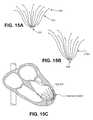
- FIGS. 15A through 16Billustrate other variations of devices and methods for using them to prevent remodeling.
- FIG. 15Asows a device that does not include an occlusive membrane.
- a plurality of struts 1501extend from a central hub 1503 . The ends of each strut 1501 terminate in an anchor 1505 .
- the strutsare typically flexible, and may be collapsed into a delivery configuration and expanded (e.g., self-expanded) into a deployed configuration.
- the support device shown in FIG. 15Bis similar, but has struts of different lengths, similar to the device shown in FIG. 14 .
- FIG. 15Cshows a schematic illustration of a heart in which the support device of FIG.
- the devicesmay be anchored along the length of the struts rather than, or instead of, just at the ends.
- the hubis anchored to the heart chamber wall.
- FIG. 16Ashows another variation of an implant, similar to the implant shown in FIG. 3 , without the plurality of pods or foot 45 .
- the hub 1603may directly contact the wall of the heart chamber.
- FIG. 16Bshows the device of FIG. 16A in the heart.
- the implant devices used to treat post-acute myocardial infracted heartsmay be configured so that the support framework (e.g., struts) and/or any membrane may be positioned adjacent, contacting, or very close to the wall of the heart.
- FIGS. 19A and 19Bshow cross-sectional views of two hears that have devices 1901 , 1901 ′ implanted adjacent to the wall in the region affected by the acute myocardial infarction 1903 , 1903 ′.
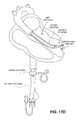
- FIGS. 17A and 17Billustrate another variation of a device that may be implanted following acute myocardial infarction in order to prevent cardiac remodeling or damage (configured as an endocardial implant).
- the deviceis configured to be anchored immediately adjacent to the heart wall (e.g., ventricle wall) across from the region of the infarct.
- the implantincludes a frame comprising a plurality of expandable struts which extend from a central hub.
- the base of the hub in this exampleincludes an anchor (“active anchor”) which may be inserted into the heart wall.
- the hub anchormay be screwed into the heart wall by rotating the device to at least partially penetrate the heart wall and secure the device in place.
- the devicemay also include one or more passive anchors on the struts of the frame, as illustrated.
- the strutsare at least partially covered by a membrane.
- the implant sown in FIG. 17Ais shown in side cross-section in FIG. 17B .
- FIG. 17Cillustrates one variation of a delivery device for delivering the implant to the heart so that it can be deployed and inserted.
- the delivery device shown in FIG. 17Cincludes a delivery catheter having an implant (shown in the collapsed state) at the distal end.
- FIG. 17Dshows the delivery device inserting the implant into the left ventricle of a heart.
- the various components of the devices and delivery systemsmay be formed of conventional materials and in a conventional manner as will be appreciated by those skilled in the art.
- Cardiac endotheliumplays an important role in control of the inflammatory response of the myocardium, growth of the heart muscle cells, contractile performance and rhytmicity of the cardiomyocytes. Cardiac endothelial dysfunction has also important role in the pathogenesis of cardiac failure. Therefore, it may be advantageous to selectively deliver therapeutic agents and/or cells to the endothelium in controlled and predictable fashion.
- the devicese.g., support device and partitioning devices
- the devicesmay be used to treat disorders by delivering a therapeutic material, including drugs and cells.
- a frame of a device and/or the membrane of a devicecan be coated and/or impregnated with a biodegradable coating containing therapeutic agents and deliver these agents to the endothelium.
- a delivery cathetercan provide access to infuse various solutions of the therapeutic agents or cells to the area between the devices (e.g., a membrane of the device) and the endothelium, providing precise control of the delivery process to facilitate healing and local regeneration.
- Any appropriate therapeutic agentsmay be used, including cytokines, chemokines, inflammatory mediators, growth factors, inotropic agents, anti-arrhythmic agents, other pharmaceutical agents commonly used for treatment post-infarction condition, and various types of cells (myocytes, myoblasts, stem cells).
Landscapes
- Health & Medical Sciences (AREA)
- Life Sciences & Earth Sciences (AREA)
- Surgery (AREA)
- Cardiology (AREA)
- Biomedical Technology (AREA)
- Public Health (AREA)
- Heart & Thoracic Surgery (AREA)
- Engineering & Computer Science (AREA)
- Veterinary Medicine (AREA)
- Animal Behavior & Ethology (AREA)
- General Health & Medical Sciences (AREA)
- Nuclear Medicine, Radiotherapy & Molecular Imaging (AREA)
- Medical Informatics (AREA)
- Molecular Biology (AREA)
- Vascular Medicine (AREA)
- Reproductive Health (AREA)
- Transplantation (AREA)
- Oral & Maxillofacial Surgery (AREA)
- Prostheses (AREA)
- Surgical Instruments (AREA)
Abstract
Description
Claims (15)
Priority Applications (2)
| Application Number | Priority Date | Filing Date | Title |
|---|---|---|---|
| US14/448,778US9592123B2 (en) | 2002-08-01 | 2014-07-31 | Therapeutic methods and devices following myocardial infarction |
| US15/452,435US10307147B2 (en) | 1999-08-09 | 2017-03-07 | System for improving cardiac function by sealing a partitioning membrane within a ventricle |
Applications Claiming Priority (6)
| Application Number | Priority Date | Filing Date | Title |
|---|---|---|---|
| US10/212,032US7279007B2 (en) | 1999-08-09 | 2002-08-01 | Method for improving cardiac function |
| US11/199,633US20060229491A1 (en) | 2002-08-01 | 2005-08-09 | Method for treating myocardial rupture |
| US98517107P | 2007-11-02 | 2007-11-02 | |
| US12/129,443US8529430B2 (en) | 2002-08-01 | 2008-05-29 | Therapeutic methods and devices following myocardial infarction |
| US13/973,868US8827892B2 (en) | 2002-08-01 | 2013-08-22 | Therapeutic methods and devices following myocardial infarction |
| US14/448,778US9592123B2 (en) | 2002-08-01 | 2014-07-31 | Therapeutic methods and devices following myocardial infarction |
Related Parent Applications (1)
| Application Number | Title | Priority Date | Filing Date |
|---|---|---|---|
| US13/973,868ContinuationUS8827892B2 (en) | 1999-08-09 | 2013-08-22 | Therapeutic methods and devices following myocardial infarction |
Related Child Applications (2)
| Application Number | Title | Priority Date | Filing Date |
|---|---|---|---|
| US11/151,164Continuation-In-PartUS7582051B2 (en) | 1999-08-09 | 2005-06-10 | Peripheral seal for a ventricular partitioning device |
| US15/452,435Continuation-In-PartUS10307147B2 (en) | 1999-08-09 | 2017-03-07 | System for improving cardiac function by sealing a partitioning membrane within a ventricle |
Publications (2)
| Publication Number | Publication Date |
|---|---|
| US20140343356A1 US20140343356A1 (en) | 2014-11-20 |
| US9592123B2true US9592123B2 (en) | 2017-03-14 |
Family
ID=40137202
Family Applications (3)
| Application Number | Title | Priority Date | Filing Date |
|---|---|---|---|
| US12/129,443Expired - Fee RelatedUS8529430B2 (en) | 1999-08-09 | 2008-05-29 | Therapeutic methods and devices following myocardial infarction |
| US13/973,868Expired - LifetimeUS8827892B2 (en) | 1999-08-09 | 2013-08-22 | Therapeutic methods and devices following myocardial infarction |
| US14/448,778Expired - Fee RelatedUS9592123B2 (en) | 1999-08-09 | 2014-07-31 | Therapeutic methods and devices following myocardial infarction |
Family Applications Before (2)
| Application Number | Title | Priority Date | Filing Date |
|---|---|---|---|
| US12/129,443Expired - Fee RelatedUS8529430B2 (en) | 1999-08-09 | 2008-05-29 | Therapeutic methods and devices following myocardial infarction |
| US13/973,868Expired - LifetimeUS8827892B2 (en) | 1999-08-09 | 2013-08-22 | Therapeutic methods and devices following myocardial infarction |
Country Status (1)
| Country | Link |
|---|---|
| US (3) | US8529430B2 (en) |
Cited By (4)
| Publication number | Priority date | Publication date | Assignee | Title |
|---|---|---|---|---|
| US10307147B2 (en) | 1999-08-09 | 2019-06-04 | Edwards Lifesciences Corporation | System for improving cardiac function by sealing a partitioning membrane within a ventricle |
| US10751183B2 (en) | 2014-09-28 | 2020-08-25 | Edwards Lifesciences Corporation | Apparatuses for treating cardiac dysfunction |
| US10898330B2 (en) | 2017-03-28 | 2021-01-26 | Edwards Lifesciences Corporation | Positioning, deploying, and retrieving implantable devices |
| US11173033B2 (en) | 2017-09-22 | 2021-11-16 | Boston Scientific Scimed, Inc. | Dome structure for improved left ventricle function |
Families Citing this family (29)
| Publication number | Priority date | Publication date | Assignee | Title |
|---|---|---|---|---|
| US7128073B1 (en) | 1998-11-06 | 2006-10-31 | Ev3 Endovascular, Inc. | Method and device for left atrial appendage occlusion |
| US9694121B2 (en) | 1999-08-09 | 2017-07-04 | Cardiokinetix, Inc. | Systems and methods for improving cardiac function |
| US7674222B2 (en) | 1999-08-09 | 2010-03-09 | Cardiokinetix, Inc. | Cardiac device and methods of use thereof |
| US8388672B2 (en) | 1999-08-09 | 2013-03-05 | Cardiokinetix, Inc. | System for improving cardiac function by sealing a partitioning membrane within a ventricle |
| US8500795B2 (en) | 1999-08-09 | 2013-08-06 | Cardiokinetix, Inc. | Retrievable devices for improving cardiac function |
| US8529430B2 (en) | 2002-08-01 | 2013-09-10 | Cardiokinetix, Inc. | Therapeutic methods and devices following myocardial infarction |
| US9332992B2 (en) | 2004-08-05 | 2016-05-10 | Cardiokinetix, Inc. | Method for making a laminar ventricular partitioning device |
| US10064696B2 (en) | 2000-08-09 | 2018-09-04 | Edwards Lifesciences Corporation | Devices and methods for delivering an endocardial device |
| US7762943B2 (en) | 2004-03-03 | 2010-07-27 | Cardiokinetix, Inc. | Inflatable ventricular partitioning device |
| US9332993B2 (en) | 2004-08-05 | 2016-05-10 | Cardiokinetix, Inc. | Devices and methods for delivering an endocardial device |
| US9078660B2 (en) | 2000-08-09 | 2015-07-14 | Cardiokinetix, Inc. | Devices and methods for delivering an endocardial device |
| EP3181074A1 (en) | 2009-01-30 | 2017-06-21 | St. Jude Medical, Inc. | Transapical mini-introducer homeostasis valve and punch |
| US9839415B2 (en)* | 2009-01-30 | 2017-12-12 | St. Jude Medical, Llc | Apex closure device |
| JP5736378B2 (en)* | 2009-09-29 | 2015-06-17 | カーディオキネティックス・インコーポレイテッドCardiokinetix, Inc. | Endocardial device delivery system and system for reducing the effective volume of the ventricle |
| US8380294B2 (en)* | 2009-10-06 | 2013-02-19 | Medtronic, Inc. | Cardiac risk stratification |
| US8790242B2 (en) | 2009-10-26 | 2014-07-29 | Cardiokinetix, Inc. | Ventricular volume reduction |
| US9907962B2 (en)* | 2009-10-29 | 2018-03-06 | Medtronic, Inc. | Arrhythmia prediction based on heart rate turbulence |
| CN204072201U (en)* | 2013-03-14 | 2015-01-07 | 卡迪欧凯尼迪克斯公司 | Assembling fixture and implant |
| US10383726B2 (en)* | 2015-01-13 | 2019-08-20 | George Kramer | Implantable transcatheter intracardiac devices and methods for treating incompetent atrioventricular valves |
| CN107303206B (en)* | 2016-04-22 | 2023-12-22 | 广东脉搏医疗科技有限公司 | Heart volume reduction implant capable of being inserted through apex of heart |
| CN105877794B (en)* | 2016-06-30 | 2018-06-19 | 北京华医圣杰科技有限公司 | A kind of ventricle capacity-reduction device |
| CN106214289A (en)* | 2016-09-05 | 2016-12-14 | 广东脉搏医疗科技有限公司 | A kind of heart volume reduction implant |
| CN106344082B (en)* | 2016-09-28 | 2019-01-25 | 宁波迪创医疗科技有限公司 | A kind of left ventricle capacity-reduction device |
| JP7138928B2 (en)* | 2018-08-22 | 2022-09-20 | 株式会社サンメディカル技術研究所 | Aortic valve assessment aid |
| CN113873957B (en) | 2019-03-25 | 2025-06-24 | 拉米纳公司 | Devices and systems for treating the left atrial appendage |
| US12303116B2 (en) | 2020-03-24 | 2025-05-20 | Laminar, Inc. | Devices, systems, and methods for occluding cavities within the body |
| CN112807047A (en)* | 2021-01-11 | 2021-05-18 | 上海傲流医疗科技有限公司 | Recoverable left ventricle isolating device |
| WO2023076122A1 (en)* | 2021-10-28 | 2023-05-04 | Edwards Lifesciences Corporation | Tissue puncture sealing devices |
| CN115120390B (en)* | 2022-07-08 | 2023-11-07 | 广东脉搏医疗科技有限公司 | Heart volume reduction implant |
Citations (241)
| Publication number | Priority date | Publication date | Assignee | Title |
|---|---|---|---|---|
| US3874388A (en)* | 1973-02-12 | 1975-04-01 | Ochsner Med Found Alton | Shunt defect closure system |
| US4007743A (en) | 1975-10-20 | 1977-02-15 | American Hospital Supply Corporation | Opening mechanism for umbrella-like intravascular shunt defect closure device |
| US4425908A (en) | 1981-10-22 | 1984-01-17 | Beth Israel Hospital | Blood clot filter |
| US4453545A (en) | 1981-05-07 | 1984-06-12 | Hiroshi Inoue | Endotracheal tube with movable endobronchial blocker for one-lung anesthesia |
| US4536893A (en) | 1982-03-03 | 1985-08-27 | Roberto Parravicini | Implant device for substaining the activity of the myocardium |
| US4588404A (en) | 1979-01-22 | 1986-05-13 | Didier Lapeyre | Total cardiac prosthesis |
| US4619246A (en) | 1984-05-23 | 1986-10-28 | William Cook, Europe A/S | Collapsible filter basket |
| US4685446A (en) | 1984-02-21 | 1987-08-11 | Choy Daniel S J | Method for using a ventricular assist device |
| US4710192A (en) | 1985-12-30 | 1987-12-01 | Liotta Domingo S | Diaphragm and method for occlusion of the descending thoracic aorta |
| US4819751A (en) | 1987-10-16 | 1989-04-11 | Baxter Travenol Laboratories, Inc. | Valvuloplasty catheter and method |
| US4832055A (en) | 1988-07-08 | 1989-05-23 | Palestrant Aubrey M | Mechanically locking blood clot filter |
| US4917089A (en) | 1988-08-29 | 1990-04-17 | Sideris Eleftherios B | Buttoned device for the transvenous occlusion of intracardiac defects |
| US4983165A (en) | 1990-01-23 | 1991-01-08 | Loiterman David A | Guidance system for vascular catheter or the like |
| US5104399A (en) | 1986-12-10 | 1992-04-14 | Endovascular Technologies, Inc. | Artificial graft and implantation method |
| US5192301A (en) | 1989-01-17 | 1993-03-09 | Nippon Zeon Co., Ltd. | Closing plug of a defect for medical use and a closing plug device utilizing it |
| US5192314A (en) | 1991-12-12 | 1993-03-09 | Daskalakis Michael K | Synthetic intraventricular implants and method of inserting |
| US5258000A (en) | 1991-11-25 | 1993-11-02 | Cook Incorporated | Tissue aperture repair device |
| US5375612A (en) | 1992-04-07 | 1994-12-27 | B. Braun Celsa | Possibly absorbable blood filter |
| US5385156A (en) | 1993-08-27 | 1995-01-31 | Rose Health Care Systems | Diagnostic and treatment method for cardiac rupture and apparatus for performing the same |
| US5389087A (en) | 1991-09-19 | 1995-02-14 | Baxter International Inc. | Fully exchangeable over-the-wire catheter with rip seam and gated side port |
| US5425744A (en) | 1991-11-05 | 1995-06-20 | C. R. Bard, Inc. | Occluder for repair of cardiac and vascular defects |
| US5433727A (en) | 1994-08-16 | 1995-07-18 | Sideris; Eleftherios B. | Centering buttoned device for the occlusion of large defects for occluding |
| US5451235A (en) | 1991-11-05 | 1995-09-19 | C.R. Bard, Inc. | Occluder and method for repair of cardiac and vascular defects |
| US5496277A (en) | 1990-04-12 | 1996-03-05 | Schneider (Usa) Inc. | Radially expandable body implantable device |
| US5527337A (en) | 1987-06-25 | 1996-06-18 | Duke University | Bioabsorbable stent and method of making the same |
| US5527338A (en) | 1992-09-02 | 1996-06-18 | Board Of Regents, The University Of Texas System | Intravascular device |
| US5549621A (en) | 1993-05-14 | 1996-08-27 | Byron C. Sutherland | Apparatus and method for performing vertical banded gastroplasty |
| US5551435A (en) | 1995-05-26 | 1996-09-03 | Sramek; Bohumir | Method and system for managing hemodynamic state and oxygen transport |
| JPH08257031A (en) | 1995-03-24 | 1996-10-08 | Toshio Saeki | Filter |
| US5578069A (en) | 1995-12-06 | 1996-11-26 | Vnetritex, Inc. | Electrode deployment mechanism and method using artificial muscle |
| US5634942A (en) | 1994-04-21 | 1997-06-03 | B. Braun Celsa | Assembly comprising a blood filter for temporary or definitive use and a device for implanting it |
| US5634936A (en) | 1995-02-06 | 1997-06-03 | Scimed Life Systems, Inc. | Device for closing a septal defect |
| US5647870A (en) | 1993-03-16 | 1997-07-15 | Ep Technologies, Inc. | Multiple electrode support structures |
| US5702343A (en) | 1996-10-02 | 1997-12-30 | Acorn Medical, Inc. | Cardiac reinforcement device |
| US5709707A (en) | 1995-10-30 | 1998-01-20 | Children's Medical Center Corporation | Self-centering umbrella-type septal closure device |
| WO1998003213A1 (en) | 1996-07-23 | 1998-01-29 | Heartport, Inc. | Minimally-invasive devices and methods for treatment of congestive heart failure |
| US5758664A (en) | 1995-06-07 | 1998-06-02 | W. L. Gore & Associates, Inc. | Method of maintaining a left ventricular assist device |
| US5791231A (en) | 1993-05-17 | 1998-08-11 | Endorobotics Corporation | Surgical robotic system and hydraulic actuator therefor |
| US5797960A (en)* | 1993-02-22 | 1998-08-25 | Stevens; John H. | Method and apparatus for thoracoscopic intracardiac procedures |
| US5797849A (en) | 1995-03-28 | 1998-08-25 | Sonometrics Corporation | Method for carrying out a medical procedure using a three-dimensional tracking and imaging system |
| US5800457A (en) | 1997-03-05 | 1998-09-01 | Gelbfish; Gary A. | Intravascular filter and associated methodology |
| US5800517A (en) | 1996-08-19 | 1998-09-01 | Scimed Life Systems, Inc. | Stent delivery system with storage sleeve |
| US5833682A (en) | 1996-08-26 | 1998-11-10 | Illumenex Corporation | Light delivery system with blood flushing capability |
| US5833698A (en) | 1996-07-23 | 1998-11-10 | United States Surgical Corporation | Anastomosis instrument and method |
| US5836968A (en) | 1996-07-17 | 1998-11-17 | Nitinol Medical Technologies, Inc. | Removable embolus blood clot filter |
| US5843170A (en) | 1994-09-02 | 1998-12-01 | Ahn; Sam Seunghae | Apparatus and method for performing aneurysm repair |
| US5861003A (en) | 1996-10-23 | 1999-01-19 | The Cleveland Clinic Foundation | Apparatus and method for occluding a defect or aperture within body surface |
| US5860951A (en) | 1992-01-07 | 1999-01-19 | Arthrocare Corporation | Systems and methods for electrosurgical myocardial revascularization |
| US5865791A (en) | 1995-06-07 | 1999-02-02 | E.P. Technologies Inc. | Atrial appendage stasis reduction procedure and devices |
| US5865730A (en) | 1997-10-07 | 1999-02-02 | Ethicon Endo-Surgery, Inc. | Tissue stabilization device for use during surgery having remotely actuated feet |
| US5871017A (en) | 1996-10-15 | 1999-02-16 | Mayer; Paul W. | Relative motion cancelling platform for surgery |
| US5876449A (en) | 1995-04-01 | 1999-03-02 | Variomed Ag | Stent for the transluminal implantation in hollow organs |
| US5875782A (en) | 1996-11-14 | 1999-03-02 | Cardiothoracic Systems, Inc. | Methods and devices for minimally invasive coronary artery revascularization on a beating heart without cardiopulmonary bypass |
| US5876325A (en) | 1993-11-02 | 1999-03-02 | Olympus Optical Co., Ltd. | Surgical manipulation system |
| US5879366A (en) | 1996-12-20 | 1999-03-09 | W.L. Gore & Associates, Inc. | Self-expanding defect closure device and method of making and using |
| US5882340A (en) | 1992-04-15 | 1999-03-16 | Yoon; Inbae | Penetrating instrument having an expandable anchoring portion for triggering protrusion of a safety member and/or retraction of a penetrating member |
| US5910150A (en) | 1996-12-02 | 1999-06-08 | Angiotrax, Inc. | Apparatus for performing surgery |
| US5916145A (en) | 1998-08-07 | 1999-06-29 | Scimed Life Systems, Inc. | Device and method of using a surgical assembly with mesh sheath |
| US5925076A (en) | 1995-05-19 | 1999-07-20 | Inoue; Kanji | Appliance to be implanted, method of collapsing the appliance to be implanted and method of using the appliance to be implanted |
| US5928260A (en) | 1997-07-10 | 1999-07-27 | Scimed Life Systems, Inc. | Removable occlusion system for aneurysm neck |
| US5961440A (en) | 1997-01-02 | 1999-10-05 | Myocor, Inc. | Heart wall tension reduction apparatus and method |
| US5961539A (en) | 1997-01-17 | 1999-10-05 | Segmed, Inc. | Method and apparatus for sizing, stabilizing and/or reducing the circumference of an anatomical structure |
| US6024756A (en) | 1996-03-22 | 2000-02-15 | Scimed Life Systems, Inc. | Method of reversibly closing a septal defect |
| US6024096A (en) | 1998-05-01 | 2000-02-15 | Correstore Inc | Anterior segment ventricular restoration apparatus and method |
| US6036720A (en) | 1997-12-15 | 2000-03-14 | Target Therapeutics, Inc. | Sheet metal aneurysm neck bridge |
| US6045497A (en) | 1997-01-02 | 2000-04-04 | Myocor, Inc. | Heart wall tension reduction apparatus and method |
| US6059715A (en) | 1997-01-02 | 2000-05-09 | Myocor, Inc. | Heart wall tension reduction apparatus |
| WO2000027292A1 (en) | 1998-11-06 | 2000-05-18 | Appriva Medical, Inc. | Method and device for left atrial appendage occlusion |
| US6076013A (en) | 1999-01-14 | 2000-06-13 | Brennan; Edward F. | Apparatus and methods for treating congestive heart failure |
| US6077214A (en) | 1998-07-29 | 2000-06-20 | Myocor, Inc. | Stress reduction apparatus and method |
| US6093199A (en) | 1998-08-05 | 2000-07-25 | Endovascular Technologies, Inc. | Intra-luminal device for treatment of body cavities and lumens and method of use |
| WO2000042919A1 (en) | 1999-01-22 | 2000-07-27 | Buckberg Gerald D | Anterior and inferior segment cardiac restoration apparatus and method |
| US6096347A (en) | 1996-11-05 | 2000-08-01 | Purdue Research Foundation | Myocardial graft constructs |
| US6095968A (en) | 1998-04-10 | 2000-08-01 | Cardio Technologies, Inc. | Reinforcement device |
| US6099832A (en) | 1997-05-28 | 2000-08-08 | Genzyme Corporation | Transplants for myocardial scars |
| US6102887A (en) | 1998-08-11 | 2000-08-15 | Biocardia, Inc. | Catheter drug delivery system and method for use |
| WO2000050639A2 (en) | 1999-02-22 | 2000-08-31 | Variagenics, Inc. | Gene sequence variations with utility in determining the treatment of disease |
| US6142973A (en) | 1997-11-07 | 2000-11-07 | Ave Connaught | Balloon catheter for repairing bifurcated vessels |
| US6155968A (en) | 1998-07-23 | 2000-12-05 | Wilk; Peter J. | Method and device for improving cardiac function |
| US6156027A (en) | 1996-08-08 | 2000-12-05 | Medtronic, Inc. | Handle for catheter assembly with multifunction wire |
| US6161543A (en) | 1993-02-22 | 2000-12-19 | Epicor, Inc. | Methods of epicardial ablation for creating a lesion around the pulmonary veins |
| US6193731B1 (en) | 1998-10-27 | 2001-02-27 | Fziomed, Inc. | Laparoscopic insertion and deployment device |
| US6221092B1 (en) | 1998-03-30 | 2001-04-24 | Nissho Corporation | Closure device for transcatheter operations and catheter assembly therefor |
| WO2001030266A1 (en) | 1999-10-27 | 2001-05-03 | Atritech, Inc. | Filter apparatus for ostium of left atrial appendage |
| US6231561B1 (en) | 1999-09-20 | 2001-05-15 | Appriva Medical, Inc. | Method and apparatus for closing a body lumen |
| US6230714B1 (en) | 1998-11-18 | 2001-05-15 | Acorn Cardiovascular, Inc. | Cardiac constraint with prior venus occlusion methods |
| US6258021B1 (en) | 1993-06-17 | 2001-07-10 | Peter J. Wilk | Intrapericardial assist method |
| US6267772B1 (en) | 1992-05-20 | 2001-07-31 | C. R. Bard, Inc. | Implantable prosthesis |
| US6296656B1 (en) | 1994-08-05 | 2001-10-02 | Origin Medsystems, Inc. | Surgical helical fastener with applicator |
| WO2001078625A1 (en) | 2000-04-13 | 2001-10-25 | Paolo Ferrazzi | Endoventricular device for the treatment and correction of cardiomyopathies |
| JP2001520910A (en) | 1997-10-27 | 2001-11-06 | イルメネックス コーポレイション | Optical transmission system |
| US6334864B1 (en) | 2000-05-17 | 2002-01-01 | Aga Medical Corp. | Alignment member for delivering a non-symmetric device with a predefined orientation |
| US6343605B1 (en) | 2000-08-08 | 2002-02-05 | Scimed Life Systems, Inc. | Percutaneous transluminal myocardial implantation device and method |
| US20020019580A1 (en) | 2000-03-10 | 2002-02-14 | Lilip Lau | Expandable cardiac harness for treating congestive heart failure |
| US6348068B1 (en) | 1999-07-23 | 2002-02-19 | Sulzer Carbomedics Inc. | Multi-filament valve stent for a cardisc valvular prosthesis |
| US20020026092A1 (en) | 1998-05-01 | 2002-02-28 | Buckberg Gerald D. | Ventricular restoration patch |
| US6355052B1 (en) | 1996-02-09 | 2002-03-12 | Pfm Produkte Fur Die Medizin Aktiengesellschaft | Device for closure of body defect openings |
| US20020032481A1 (en) | 2000-09-12 | 2002-03-14 | Shlomo Gabbay | Heart valve prosthesis and sutureless implantation of a heart valve prosthesis |
| US6360749B1 (en) | 1998-10-09 | 2002-03-26 | Swaminathan Jayaraman | Modification of properties and geometry of heart tissue to influence heart function |
| US6364896B1 (en) | 1998-11-24 | 2002-04-02 | Embol-X, Inc. | Compliant framework and methods of use |
| WO2002030335A2 (en) | 2000-10-06 | 2002-04-18 | Myocor, Inc. | Endovascular splinting devices |
| US20020055775A1 (en) | 1999-01-26 | 2002-05-09 | Alain F. Carpentier | Flexible heart valve |
| US20020055767A1 (en) | 2000-10-18 | 2002-05-09 | Forde Sean T. | Over-the-wire interlock attachment/detachment mechanism |
| US6387042B1 (en) | 1998-08-28 | 2002-05-14 | Juan Hernandez Herrero | Apparatus aiding physiologic systolic and diastolic dynamics of cardiac cavities |
| WO2002045710A1 (en) | 2000-12-07 | 2002-06-13 | Neuromolecular Inc. | Methods for treating neuropsychiatric disorders with nmda receptor antagonists |
| US6406420B1 (en) | 1997-01-02 | 2002-06-18 | Myocor, Inc. | Methods and devices for improving cardiac function in hearts |
| US20020111647A1 (en) | 1999-11-08 | 2002-08-15 | Khairkhahan Alexander K. | Adjustable left atrial appendage occlusion device |
| US20020133227A1 (en) | 2001-02-28 | 2002-09-19 | Gregory Murphy | Ventricular restoration patch apparatus and method of use |
| WO2002071977A2 (en) | 2001-03-08 | 2002-09-19 | Atritech, Inc. | Atrial filter implants |
| US20020161392A1 (en) | 1998-04-27 | 2002-10-31 | Dubrul William R. | Particle-removing medical device and method |
| US20020161394A1 (en) | 1997-09-26 | 2002-10-31 | Macoviak John A. | Aortic filter catheter |
| WO2002087481A1 (en) | 2001-04-27 | 2002-11-07 | Myomend, Inc. | Prevention of myocardial infarction induced ventricular expansion and remodeling |
| US20020169360A1 (en) | 1998-07-16 | 2002-11-14 | Cardiothoracic Systems, Inc., A California Corporation | Surgical procedures and devices for increasing cardiac output of the heart |
| US6482146B1 (en) | 2000-06-13 | 2002-11-19 | Acorn Cardiovascular, Inc. | Cardiac disease treatment and device |
| US6482228B1 (en) | 2000-11-14 | 2002-11-19 | Troy R. Norred | Percutaneous aortic valve replacement |
| US20020183604A1 (en) | 2000-05-22 | 2002-12-05 | Ashok Gowda | Apparatus for access to interstitial fluid, blood, or blood plasma components |
| US20020188170A1 (en) | 2001-04-27 | 2002-12-12 | Santamore William P. | Prevention of myocardial infarction induced ventricular expansion and remodeling |
| US6506204B2 (en) | 1996-01-24 | 2003-01-14 | Aga Medical Corporation | Method and apparatus for occluding aneurysms |
| US6508756B1 (en) | 1995-06-13 | 2003-01-21 | Abiomed, Inc. | Passive cardiac assistance device |
| US6511496B1 (en) | 2000-09-12 | 2003-01-28 | Advanced Cardiovascular Systems, Inc. | Embolic protection device for use in interventional procedures |
| WO2003007778A2 (en) | 2001-07-16 | 2003-01-30 | Relaxis Ltd. | In-vivo method and device for improving diastolic function of the left ventricle |
| US20030045896A1 (en) | 2001-02-28 | 2003-03-06 | Chase Medical, Lp | Method of using a ventricular restoration shaping apparatus |
| US6537198B1 (en) | 2000-03-21 | 2003-03-25 | Myocor, Inc. | Splint assembly for improving cardiac function in hearts, and method for implanting the splint assembly |
| JP2003512128A (en) | 1999-10-27 | 2003-04-02 | アトリテック インコーポレイテッド | Barrier device to cover the ostium of the left atrial appendage |
| JP2003512129A (en) | 1999-10-27 | 2003-04-02 | アトリテック インコーポレイテッド | Barrier device for left atrial appendage stoma |
| US20030078671A1 (en) | 2001-04-27 | 2003-04-24 | Lesniak Jeanne M. | Prevention of myocardial infarction induced ventricular expansion and remodeling |
| WO2003043507A2 (en) | 2001-11-19 | 2003-05-30 | Swaminathan Jayaraman | Modification of properties and geometry of heart tissue to influence heart function |
| US6572643B1 (en) | 2000-07-19 | 2003-06-03 | Vascular Architects, Inc. | Endoprosthesis delivery catheter assembly and method |
| US20030109770A1 (en) | 1999-08-09 | 2003-06-12 | Sharkey Hugh R. | Device with a porous membrane for improving cardiac function |
| US6586414B2 (en) | 2000-03-28 | 2003-07-01 | Medicure International Inc. | Treatment of cerebrovascular disease |
| US6592608B2 (en) | 2001-12-07 | 2003-07-15 | Biopsy Sciences, Llc | Bioabsorbable sealant |
| US20030135230A1 (en) | 2002-01-17 | 2003-07-17 | Massey Joseph B. | Steerable dilatation system, dilator, and related methods for stepped dilatation |
| US20030149422A1 (en) | 2002-02-04 | 2003-08-07 | Charles Muller | Steerable catheter |
| US6613013B2 (en) | 1997-10-01 | 2003-09-02 | Boston Scientific Corporation | Guidewire compatible port and method for inserting same |
| WO2003073961A1 (en) | 2002-03-05 | 2003-09-12 | Salviac Limited | System with embolic filter and retracting snare |
| US6622730B2 (en) | 2001-03-30 | 2003-09-23 | Myocor, Inc. | Device for marking and aligning positions on the heart |
| US20030181942A1 (en) | 2002-01-25 | 2003-09-25 | Sutton Gregg S. | Atrial appendage blood filtration systems |
| US6645199B1 (en) | 1999-11-22 | 2003-11-11 | Scimed Life Systems, Inc. | Loop structures for supporting diagnostic and therapeutic elements contact with body tissue and expandable push devices for use with same |
| US20030220667A1 (en) | 1998-11-06 | 2003-11-27 | Van Der Burg Erik J. | Method of containing embolic material in the left atrial appendage |
| WO2003099300A1 (en) | 2002-05-24 | 2003-12-04 | Zensun (Shanghai) Sci-Tech, Ltd. | Neuregulin based methods and compositions for treating cardiovascular diseases |
| WO2003103743A2 (en) | 2002-06-10 | 2003-12-18 | Leo Rubin | Medical device for intra-lumenal delivery of pharmaceutical agents |
| WO2003103538A1 (en) | 2002-06-07 | 2003-12-18 | Acorn Cardiovascular, Inc. | Cardiac support device |
| WO2004012629A1 (en) | 1999-08-09 | 2004-02-12 | Cardiokinetix, Inc. | A device for improving cardiac function |
| US20040034366A1 (en) | 1999-11-08 | 2004-02-19 | Ev3 Sunnyvale, Inc., A California Corporation | Device for containing embolic material in the LAA having a plurality of tissue retention structures |
| US20040044361A1 (en) | 1998-11-06 | 2004-03-04 | Frazier Andrew G.C. | Detachable atrial appendage occlusion balloon |
| WO2004019866A2 (en) | 2002-08-28 | 2004-03-11 | Immunex Corporation | Compositions and methods for treating cardiovascular disease |
| US20040054394A1 (en) | 2002-09-17 | 2004-03-18 | Don Lee | Stent with combined distal protection device |
| US20040064014A1 (en) | 2001-05-31 | 2004-04-01 | Melvin David B. | Devices and methods for assisting natural heart function |
| US20040122090A1 (en) | 2001-12-07 | 2004-06-24 | Lipton Stuart A. | Methods for treating neuropsychiatric disorders with nmda receptor antagonists |
| US20040133062A1 (en) | 2002-10-11 | 2004-07-08 | Suresh Pai | Minimally invasive cardiac force transfer structures |
| WO2004066805A2 (en) | 2003-01-27 | 2004-08-12 | Corassist Cardiovascular Ltd. | In vivo device for improving diastolic ventricular function |
| US6776754B1 (en) | 2000-10-04 | 2004-08-17 | Wilk Patent Development Corporation | Method for closing off lower portion of heart ventricle |
| US20040172042A1 (en) | 1998-09-24 | 2004-09-02 | Scimed Life Systems, Inc. | Retrieval devices for vena cava filter |
| US20040186511A1 (en) | 2003-03-20 | 2004-09-23 | Adam Stephens | Control handle for intraluminal devices |
| US20040215230A1 (en) | 2003-04-28 | 2004-10-28 | Frazier Andrew G. C. | Left atrial appendage occlusion device with active expansion |
| US20040220610A1 (en) | 1999-11-08 | 2004-11-04 | Kreidler Marc S. | Thin film composite lamination |
| EP1474032A2 (en) | 2002-01-23 | 2004-11-10 | Chase Medical, L. P. | An apical patch and method of use |
| WO2004100803A1 (en) | 2003-05-12 | 2004-11-25 | Cardiokinetix, Inc. | A system for improving cardiac function |
| US20040243170A1 (en) | 2001-09-05 | 2004-12-02 | Mitta Suresh | Method and device for percutaneous surgical ventricular repair |
| US20040260331A1 (en) | 2003-06-20 | 2004-12-23 | D'aquanni Peter | Beta titanium embolic protection frame and guide wire |
| US20040260346A1 (en) | 2003-01-31 | 2004-12-23 | Overall William Ryan | Detection of apex motion for monitoring cardiac dysfunction |
| US20040267086A1 (en) | 2003-06-26 | 2004-12-30 | Anstadt Mark P. | Sensor-equipped and algorithm-controlled direct mechanical ventricular assist device |
| US20040267378A1 (en) | 2003-06-24 | 2004-12-30 | Gazi Bashir Mussa | Semi-stationary balloon in the gastric antrum provided with connecting an anchoring rod for inducing weight reduction in human beings |
| US20050007031A1 (en) | 2003-07-11 | 2005-01-13 | Hubbell Incorporated | Low voltage luminaire assembly |
| US20050015109A1 (en) | 2003-07-16 | 2005-01-20 | Samuel Lichtenstein | Methods and devices for altering blood flow through the left ventricle |
| WO2005007873A2 (en) | 2003-07-14 | 2005-01-27 | Nicholas Debeer | Encapsulation device and methods of use |
| US6852076B2 (en) | 1999-08-09 | 2005-02-08 | Cardiokinetix, Inc. | Method for improving cardiac function |
| US20050038470A1 (en) | 2003-08-15 | 2005-02-17 | Van Der Burg Erik J. | System and method for delivering a left atrial appendage containment device |
| US20050043708A1 (en) | 2002-01-31 | 2005-02-24 | Gleeson James B | Anastomosis device and method |
| US20050065548A1 (en) | 2003-09-23 | 2005-03-24 | Marino Joseph A. | Right retrieval mechanism |
| US20050085826A1 (en) | 2003-10-21 | 2005-04-21 | Scimed Life Systems, Inc. | Unfolding balloon catheter for proximal embolus protection |
| US6887192B1 (en) | 2000-09-08 | 2005-05-03 | Converge Medical, Inc. | Heart support to prevent ventricular remodeling |
| US20050096498A1 (en) | 2001-04-24 | 2005-05-05 | Houser Russell A. | Sizing and shaping device for treating congestive heart failure |
| WO2005041745A2 (en) | 2003-10-31 | 2005-05-12 | Corassist Cardiovascular Ltd. | In vivo device for improving diastolic ventricular function |
| US20050113811A1 (en) | 2001-04-24 | 2005-05-26 | Houser Russell A. | Method and devices for treating ischemic congestive heart failure |
| US20050113861A1 (en) | 2003-11-25 | 2005-05-26 | Corcoran Michael P. | Left atrial appendage closure device |
| US20050124849A1 (en) | 2001-04-24 | 2005-06-09 | Barbut Denise R. | Partial aortic occlusion devices and methods for cerebral perfusion augmentation |
| US20050137690A1 (en) | 2003-12-23 | 2005-06-23 | Sadra Medical | Low profile heart valve and delivery system |
| US20050142180A1 (en) | 2003-10-20 | 2005-06-30 | Bisgaier Charles L. | Pharmaceutical formulations, methods, and dosing regimens for the treatment and prevention of acute coronary syndromes |
| US20050177180A1 (en) | 2001-11-28 | 2005-08-11 | Aptus Endosystems, Inc. | Devices, systems, and methods for supporting tissue and/or structures within a hollow body organ |
| US20050187620A1 (en) | 2003-11-14 | 2005-08-25 | Suresh Pai | Systems for heart treatment |
| US20050216052A1 (en) | 1994-07-08 | 2005-09-29 | Ev3 Inc. | Method of forming medical devices; intravascular occlusion devices |
| WO2005091860A2 (en) | 2004-03-05 | 2005-10-06 | Advanced Resuscitation, Llc. | Method and apparatus for direct mechanical ventricular actuation with favorable conditioning and minimal heart stress |
| US20050228434A1 (en) | 2004-03-19 | 2005-10-13 | Aga Medical Corporation | Multi-layer braided structures for occluding vascular defects |
| JP2005324019A (en) | 2004-05-12 | 2005-11-24 | Medtronic Vascular Inc | Cardiovascular defect patch device and method |
| US20050277983A1 (en) | 2004-06-09 | 2005-12-15 | Usgi Medical Inc. | System for optimizing anchoring force |
| US20050277981A1 (en) | 2004-06-09 | 2005-12-15 | Usgi Medical Inc. | Apparatus and methods for optimizing anchoring force |
| US20050283218A1 (en) | 2004-06-22 | 2005-12-22 | Williams Michael S | Implantable chamber for biological induction or enhancement of muscle contraction |
| US20060025800A1 (en) | 2001-09-05 | 2006-02-02 | Mitta Suresh | Method and device for surgical ventricular repair |
| US20060030881A1 (en) | 2004-08-05 | 2006-02-09 | Cardiokinetix, Inc. | Ventricular partitioning device |
| US20060063970A1 (en) | 2002-09-06 | 2006-03-23 | Mardil, Inc. | Method and apparatus for external stabilization of the heart valves and myocardium |
| WO2006033107A2 (en) | 2004-09-22 | 2006-03-30 | Corassist Cardiovascular Ltd. | In vivo device for assisting and improving diastolic ventricular function |
| US20060069430A9 (en) | 2000-09-20 | 2006-03-30 | Ample Medical, Inc. | Devices, systems, and methods for retaining a native heart valve leaflet |
| US20060079736A1 (en) | 2004-10-13 | 2006-04-13 | Sing-Fatt Chin | Method and device for percutaneous left ventricular reconstruction |
| WO2006055683A2 (en) | 2004-11-16 | 2006-05-26 | Pulmonx | Occlusal stent and methods for its use |
| US20060116692A1 (en) | 2004-12-01 | 2006-06-01 | Scimed Life Systems, Inc. | Single operator medical device handles and related methods of use |
| US20060136043A1 (en) | 2004-12-22 | 2006-06-22 | Cully Edward H | Filament-wound implantable devices |
| US20060199995A1 (en)* | 2005-03-02 | 2006-09-07 | Venkataramana Vijay | Percutaneous cardiac ventricular geometry restoration device and treatment for heart failure |
| US20060229491A1 (en) | 2002-08-01 | 2006-10-12 | Cardiokinetix, Inc. | Method for treating myocardial rupture |
| US20060259124A1 (en) | 2005-03-28 | 2006-11-16 | Terumo Kabushiki Kaisha | Stent delivery device |
| US7144363B2 (en) | 2001-10-16 | 2006-12-05 | Extensia Medical, Inc. | Systems for heart treatment |
| US20060276684A1 (en) | 2003-11-07 | 2006-12-07 | Giovanni Speziali | Device and method for treating congestive heart failure |
| US7172551B2 (en) | 2004-04-12 | 2007-02-06 | Scimed Life Systems, Inc. | Cyclical pressure coronary assist pump |
| WO2007016349A2 (en) | 2005-07-29 | 2007-02-08 | Cvdevices, Llc | Devices and methods for percutaneous endo-ventricular reconstruction of the failing heart |
| US7175660B2 (en) | 2002-08-29 | 2007-02-13 | Mitralsolutions, Inc. | Apparatus for implanting surgical devices for controlling the internal circumference of an anatomic orifice or lumen |
| US20070129753A1 (en) | 2005-12-01 | 2007-06-07 | Chris Quinn | Method and apparatus for recapturing an implant from the left atrial appendage |
| US20070135889A1 (en) | 2003-09-03 | 2007-06-14 | Bolton Medical, Inc. | Lumen repair device with capture structure |
| US20070162048A1 (en) | 2005-12-01 | 2007-07-12 | Chris Quinn | Method and apparatus for retrieving an embolized implant |
| WO2007092354A2 (en) | 2006-02-02 | 2007-08-16 | Sadra Medical, Inc. | Medical implant delivery and deployment tool |
| WO2007143560A2 (en) | 2006-06-02 | 2007-12-13 | Cianna Medical, Inc. | Expandable brachytherapy apparatus |
| US20080015717A1 (en) | 2006-03-03 | 2008-01-17 | Griffin Paul P Jr | Auxiliary power adapter having device controls |
| US7320665B2 (en) | 2005-03-02 | 2008-01-22 | Venkataramana Vijay | Cardiac Ventricular Geometry Restoration Device and Treatment for Heart Failure |
| WO2008010792A1 (en) | 2006-07-14 | 2008-01-24 | Venkataramana Vijay | Cardiac ventricular geometry restoration device and treatment for heart failure |
| US20080045778A1 (en) | 2006-08-02 | 2008-02-21 | Samuel Victor Lichtenstein | System for improving diastolic dysfunction |
| JP2008514291A (en) | 2004-09-24 | 2008-05-08 | エヌエムティー メディカル, インコーポレイティッド | Double fixing system for delivery / recovery of occluder devices |
| US7399271B2 (en) | 2004-01-09 | 2008-07-15 | Cardiokinetix, Inc. | Ventricular partitioning device |
| US7485088B2 (en) | 2001-09-05 | 2009-02-03 | Chase Medical L.P. | Method and device for percutaneous surgical ventricular repair |
| US20090112050A1 (en) | 2007-10-24 | 2009-04-30 | Circulite, Inc. | Transseptal cannula, tip, delivery system, and method |
| US7530998B1 (en) | 2003-05-30 | 2009-05-12 | Thomas David Starkey | Device and method to control volume of a ventricle |
| US7569062B1 (en) | 1998-07-15 | 2009-08-04 | St. Jude Medical, Inc. | Mitral and tricuspid valve repair |
| US7582051B2 (en) | 2005-06-10 | 2009-09-01 | Cardiokinetix, Inc. | Peripheral seal for a ventricular partitioning device |
| US20100030256A1 (en) | 1997-11-12 | 2010-02-04 | Genesis Technologies Llc | Medical Devices and Methods |
| US7674222B2 (en) | 1999-08-09 | 2010-03-09 | Cardiokinetix, Inc. | Cardiac device and methods of use thereof |
| US7758491B2 (en) | 2004-04-05 | 2010-07-20 | Genesee Biomedical, Inc. | Method and apparatus for the surgical treatment of congestive heart failure |
| US7762943B2 (en) | 2004-03-03 | 2010-07-27 | Cardiokinetix, Inc. | Inflatable ventricular partitioning device |
| US20100274227A1 (en) | 2009-02-13 | 2010-10-28 | Alexander Khairkhahan | Delivery catheter handle cover |
| US7862500B2 (en) | 2002-08-01 | 2011-01-04 | Cardiokinetix, Inc. | Multiple partitioning devices for heart treatment |
| US20110087066A1 (en) | 2000-08-09 | 2011-04-14 | Boutillette Michael P | Devices and methods for delivering an endocardial device |
| US20110092761A1 (en) | 2008-02-11 | 2011-04-21 | Corassist Cardiovascular Ltd. | Ventricular function assisting devices and methods of use thereof |
| US7938767B2 (en) | 2006-02-06 | 2011-05-10 | Northwind Ventures | Systems and methods for volume reduction |
| US20110264204A1 (en) | 2008-12-15 | 2011-10-27 | Alexander Khairkhahan | Devices and methods for delivering an endocardial device |
| EP2082690B1 (en) | 2008-01-24 | 2012-06-20 | Kardium, Inc. | Medical device to assist diastolic function and prevent ventricular enlargement |
| US8246671B2 (en) | 1999-08-09 | 2012-08-21 | Cardiokinetix, Inc. | Retrievable cardiac devices |
| US8377114B2 (en) | 1999-08-09 | 2013-02-19 | Cardiokinetix, Inc. | Sealing and filling ventricular partitioning devices to improve cardiac function |
| US8382653B2 (en) | 2006-09-25 | 2013-02-26 | Corassist Cardiovascular Ltd. | Method and system for improving diastolic function of the heart |
| US8398537B2 (en) | 2005-06-10 | 2013-03-19 | Cardiokinetix, Inc. | Peripheral seal for a ventricular partitioning device |
| US8529430B2 (en) | 2002-08-01 | 2013-09-10 | Cardiokinetix, Inc. | Therapeutic methods and devices following myocardial infarction |
| US20130270735A1 (en) | 2004-08-05 | 2013-10-17 | Miles D. Alexander | Systems and methods for making a laminar ventricular partitioning device |
| US20130274595A1 (en) | 2004-08-05 | 2013-10-17 | James R. Kermode | Devices and methods for delivering an endocardial device |
| US20140180271A1 (en) | 2007-04-19 | 2014-06-26 | Jessi Ernest Johnson | Systems, apparatus, methods and procedures for the non-invasive treatment of tissue using microwave energy |
| US8790242B2 (en) | 2009-10-26 | 2014-07-29 | Cardiokinetix, Inc. | Ventricular volume reduction |
- 2008
- 2008-05-29USUS12/129,443patent/US8529430B2/ennot_activeExpired - Fee Related
- 2013
- 2013-08-22USUS13/973,868patent/US8827892B2/ennot_activeExpired - Lifetime
- 2014
- 2014-07-31USUS14/448,778patent/US9592123B2/ennot_activeExpired - Fee Related
Patent Citations (305)
| Publication number | Priority date | Publication date | Assignee | Title |
|---|---|---|---|---|
| US3874388A (en)* | 1973-02-12 | 1975-04-01 | Ochsner Med Found Alton | Shunt defect closure system |
| US4007743A (en) | 1975-10-20 | 1977-02-15 | American Hospital Supply Corporation | Opening mechanism for umbrella-like intravascular shunt defect closure device |
| US4588404A (en) | 1979-01-22 | 1986-05-13 | Didier Lapeyre | Total cardiac prosthesis |
| US4453545A (en) | 1981-05-07 | 1984-06-12 | Hiroshi Inoue | Endotracheal tube with movable endobronchial blocker for one-lung anesthesia |
| US4425908A (en) | 1981-10-22 | 1984-01-17 | Beth Israel Hospital | Blood clot filter |
| US4536893A (en) | 1982-03-03 | 1985-08-27 | Roberto Parravicini | Implant device for substaining the activity of the myocardium |
| US4685446A (en) | 1984-02-21 | 1987-08-11 | Choy Daniel S J | Method for using a ventricular assist device |
| US4619246A (en) | 1984-05-23 | 1986-10-28 | William Cook, Europe A/S | Collapsible filter basket |
| US4710192A (en) | 1985-12-30 | 1987-12-01 | Liotta Domingo S | Diaphragm and method for occlusion of the descending thoracic aorta |
| US5104399A (en) | 1986-12-10 | 1992-04-14 | Endovascular Technologies, Inc. | Artificial graft and implantation method |
| US5527337A (en) | 1987-06-25 | 1996-06-18 | Duke University | Bioabsorbable stent and method of making the same |
| US4819751A (en) | 1987-10-16 | 1989-04-11 | Baxter Travenol Laboratories, Inc. | Valvuloplasty catheter and method |
| US4832055A (en) | 1988-07-08 | 1989-05-23 | Palestrant Aubrey M | Mechanically locking blood clot filter |
| US4917089A (en) | 1988-08-29 | 1990-04-17 | Sideris Eleftherios B | Buttoned device for the transvenous occlusion of intracardiac defects |
| US5192301A (en) | 1989-01-17 | 1993-03-09 | Nippon Zeon Co., Ltd. | Closing plug of a defect for medical use and a closing plug device utilizing it |
| US4983165A (en) | 1990-01-23 | 1991-01-08 | Loiterman David A | Guidance system for vascular catheter or the like |
| US5496277A (en) | 1990-04-12 | 1996-03-05 | Schneider (Usa) Inc. | Radially expandable body implantable device |
| US5389087A (en) | 1991-09-19 | 1995-02-14 | Baxter International Inc. | Fully exchangeable over-the-wire catheter with rip seam and gated side port |
| US5425744A (en) | 1991-11-05 | 1995-06-20 | C. R. Bard, Inc. | Occluder for repair of cardiac and vascular defects |
| US5451235A (en) | 1991-11-05 | 1995-09-19 | C.R. Bard, Inc. | Occluder and method for repair of cardiac and vascular defects |
| US5258000A (en) | 1991-11-25 | 1993-11-02 | Cook Incorporated | Tissue aperture repair device |
| US5192314A (en) | 1991-12-12 | 1993-03-09 | Daskalakis Michael K | Synthetic intraventricular implants and method of inserting |
| US5860951A (en) | 1992-01-07 | 1999-01-19 | Arthrocare Corporation | Systems and methods for electrosurgical myocardial revascularization |
| US5375612A (en) | 1992-04-07 | 1994-12-27 | B. Braun Celsa | Possibly absorbable blood filter |
| US5882340A (en) | 1992-04-15 | 1999-03-16 | Yoon; Inbae | Penetrating instrument having an expandable anchoring portion for triggering protrusion of a safety member and/or retraction of a penetrating member |
| US6267772B1 (en) | 1992-05-20 | 2001-07-31 | C. R. Bard, Inc. | Implantable prosthesis |
| US5527338A (en) | 1992-09-02 | 1996-06-18 | Board Of Regents, The University Of Texas System | Intravascular device |
| US5925062A (en) | 1992-09-02 | 1999-07-20 | Board Of Regents, The University Of Texas System | Intravascular device |
| US6161543A (en) | 1993-02-22 | 2000-12-19 | Epicor, Inc. | Methods of epicardial ablation for creating a lesion around the pulmonary veins |
| US5829447A (en) | 1993-02-22 | 1998-11-03 | Heartport, Inc. | Method and apparatus for thoracoscopic intracardiac procedures |
| US5924424A (en) | 1993-02-22 | 1999-07-20 | Heartport, Inc. | Method and apparatus for thoracoscopic intracardiac procedures |
| US5797960A (en)* | 1993-02-22 | 1998-08-25 | Stevens; John H. | Method and apparatus for thoracoscopic intracardiac procedures |
| US6125852A (en) | 1993-02-22 | 2000-10-03 | Heartport, Inc. | Minimally-invasive devices and methods for treatment of congestive heart failure |
| US5647870A (en) | 1993-03-16 | 1997-07-15 | Ep Technologies, Inc. | Multiple electrode support structures |
| US5549621A (en) | 1993-05-14 | 1996-08-27 | Byron C. Sutherland | Apparatus and method for performing vertical banded gastroplasty |
| US5791231A (en) | 1993-05-17 | 1998-08-11 | Endorobotics Corporation | Surgical robotic system and hydraulic actuator therefor |
| US6258021B1 (en) | 1993-06-17 | 2001-07-10 | Peter J. Wilk | Intrapericardial assist method |
| US5385156A (en) | 1993-08-27 | 1995-01-31 | Rose Health Care Systems | Diagnostic and treatment method for cardiac rupture and apparatus for performing the same |
| US5876325A (en) | 1993-11-02 | 1999-03-02 | Olympus Optical Co., Ltd. | Surgical manipulation system |
| US5634942A (en) | 1994-04-21 | 1997-06-03 | B. Braun Celsa | Assembly comprising a blood filter for temporary or definitive use and a device for implanting it |
| US20050216052A1 (en) | 1994-07-08 | 2005-09-29 | Ev3 Inc. | Method of forming medical devices; intravascular occlusion devices |
| US6296656B1 (en) | 1994-08-05 | 2001-10-02 | Origin Medsystems, Inc. | Surgical helical fastener with applicator |
| US5433727A (en) | 1994-08-16 | 1995-07-18 | Sideris; Eleftherios B. | Centering buttoned device for the occlusion of large defects for occluding |
| US5843170A (en) | 1994-09-02 | 1998-12-01 | Ahn; Sam Seunghae | Apparatus and method for performing aneurysm repair |
| US5634936A (en) | 1995-02-06 | 1997-06-03 | Scimed Life Systems, Inc. | Device for closing a septal defect |
| JPH08257031A (en) | 1995-03-24 | 1996-10-08 | Toshio Saeki | Filter |
| US5797849A (en) | 1995-03-28 | 1998-08-25 | Sonometrics Corporation | Method for carrying out a medical procedure using a three-dimensional tracking and imaging system |
| US5876449A (en) | 1995-04-01 | 1999-03-02 | Variomed Ag | Stent for the transluminal implantation in hollow organs |
| US5925076A (en) | 1995-05-19 | 1999-07-20 | Inoue; Kanji | Appliance to be implanted, method of collapsing the appliance to be implanted and method of using the appliance to be implanted |
| WO1996037859A1 (en) | 1995-05-26 | 1996-11-28 | Bohumir Sramek | System for managing hemodynamic state |
| US5551435A (en) | 1995-05-26 | 1996-09-03 | Sramek; Bohumir | Method and system for managing hemodynamic state and oxygen transport |
| US5758664A (en) | 1995-06-07 | 1998-06-02 | W. L. Gore & Associates, Inc. | Method of maintaining a left ventricular assist device |
| US6132438A (en) | 1995-06-07 | 2000-10-17 | Ep Technologies, Inc. | Devices for installing stasis reducing means in body tissue |
| US5984917A (en) | 1995-06-07 | 1999-11-16 | Ep Technologies, Inc. | Device and method for remote insertion of a closed loop |
| US5865791A (en) | 1995-06-07 | 1999-02-02 | E.P. Technologies Inc. | Atrial appendage stasis reduction procedure and devices |
| US6508756B1 (en) | 1995-06-13 | 2003-01-21 | Abiomed, Inc. | Passive cardiac assistance device |
| US5709707A (en) | 1995-10-30 | 1998-01-20 | Children's Medical Center Corporation | Self-centering umbrella-type septal closure device |
| US5578069A (en) | 1995-12-06 | 1996-11-26 | Vnetritex, Inc. | Electrode deployment mechanism and method using artificial muscle |
| US6506204B2 (en) | 1996-01-24 | 2003-01-14 | Aga Medical Corporation | Method and apparatus for occluding aneurysms |
| US6355052B1 (en) | 1996-02-09 | 2002-03-12 | Pfm Produkte Fur Die Medizin Aktiengesellschaft | Device for closure of body defect openings |
| US6312446B1 (en) | 1996-03-22 | 2001-11-06 | Scimed Life Systems, Inc. | Apparatus and method for closing a septal defect |
| US6024756A (en) | 1996-03-22 | 2000-02-15 | Scimed Life Systems, Inc. | Method of reversibly closing a septal defect |
| US5836968A (en) | 1996-07-17 | 1998-11-17 | Nitinol Medical Technologies, Inc. | Removable embolus blood clot filter |
| US5833698A (en) | 1996-07-23 | 1998-11-10 | United States Surgical Corporation | Anastomosis instrument and method |
| WO1998003213A1 (en) | 1996-07-23 | 1998-01-29 | Heartport, Inc. | Minimally-invasive devices and methods for treatment of congestive heart failure |
| US6156027A (en) | 1996-08-08 | 2000-12-05 | Medtronic, Inc. | Handle for catheter assembly with multifunction wire |
| US5800517A (en) | 1996-08-19 | 1998-09-01 | Scimed Life Systems, Inc. | Stent delivery system with storage sleeve |
| US5833682A (en) | 1996-08-26 | 1998-11-10 | Illumenex Corporation | Light delivery system with blood flushing capability |
| US5702343A (en) | 1996-10-02 | 1997-12-30 | Acorn Medical, Inc. | Cardiac reinforcement device |
| US20030149333A1 (en) | 1996-10-02 | 2003-08-07 | Acorn Cardiovascular, Inc. | Cardiac reinforcement device |
| US6077218A (en) | 1996-10-02 | 2000-06-20 | Acorn Cardiovascular, Inc. | Cardiac reinforcement device |
| US5871017A (en) | 1996-10-15 | 1999-02-16 | Mayer; Paul W. | Relative motion cancelling platform for surgery |
| US5861003A (en) | 1996-10-23 | 1999-01-19 | The Cleveland Clinic Foundation | Apparatus and method for occluding a defect or aperture within body surface |
| US6096347A (en) | 1996-11-05 | 2000-08-01 | Purdue Research Foundation | Myocardial graft constructs |
| US5875782A (en) | 1996-11-14 | 1999-03-02 | Cardiothoracic Systems, Inc. | Methods and devices for minimally invasive coronary artery revascularization on a beating heart without cardiopulmonary bypass |
| US5910150A (en) | 1996-12-02 | 1999-06-08 | Angiotrax, Inc. | Apparatus for performing surgery |
| US5879366A (en) | 1996-12-20 | 1999-03-09 | W.L. Gore & Associates, Inc. | Self-expanding defect closure device and method of making and using |
| US20020169359A1 (en) | 1997-01-02 | 2002-11-14 | Myocor, Inc. | Methods and devices for improving cardiac function in hearts |
| US6059715A (en) | 1997-01-02 | 2000-05-09 | Myocor, Inc. | Heart wall tension reduction apparatus |
| US5961440A (en) | 1997-01-02 | 1999-10-05 | Myocor, Inc. | Heart wall tension reduction apparatus and method |
| US6045497A (en) | 1997-01-02 | 2000-04-04 | Myocor, Inc. | Heart wall tension reduction apparatus and method |
| US6406420B1 (en) | 1997-01-02 | 2002-06-18 | Myocor, Inc. | Methods and devices for improving cardiac function in hearts |
| US5961539A (en) | 1997-01-17 | 1999-10-05 | Segmed, Inc. | Method and apparatus for sizing, stabilizing and/or reducing the circumference of an anatomical structure |
| US5800457A (en) | 1997-03-05 | 1998-09-01 | Gelbfish; Gary A. | Intravascular filter and associated methodology |
| US6099832A (en) | 1997-05-28 | 2000-08-08 | Genzyme Corporation | Transplants for myocardial scars |
| US5928260A (en) | 1997-07-10 | 1999-07-27 | Scimed Life Systems, Inc. | Removable occlusion system for aneurysm neck |
| US20020161394A1 (en) | 1997-09-26 | 2002-10-31 | Macoviak John A. | Aortic filter catheter |
| US6613013B2 (en) | 1997-10-01 | 2003-09-02 | Boston Scientific Corporation | Guidewire compatible port and method for inserting same |
| US5865730A (en) | 1997-10-07 | 1999-02-02 | Ethicon Endo-Surgery, Inc. | Tissue stabilization device for use during surgery having remotely actuated feet |
| JP2001520910A (en) | 1997-10-27 | 2001-11-06 | イルメネックス コーポレイション | Optical transmission system |
| US6142973A (en) | 1997-11-07 | 2000-11-07 | Ave Connaught | Balloon catheter for repairing bifurcated vessels |
| US20100030256A1 (en) | 1997-11-12 | 2010-02-04 | Genesis Technologies Llc | Medical Devices and Methods |
| US6036720A (en) | 1997-12-15 | 2000-03-14 | Target Therapeutics, Inc. | Sheet metal aneurysm neck bridge |
| US6221092B1 (en) | 1998-03-30 | 2001-04-24 | Nissho Corporation | Closure device for transcatheter operations and catheter assembly therefor |
| US6095968A (en) | 1998-04-10 | 2000-08-01 | Cardio Technologies, Inc. | Reinforcement device |
| US20020161392A1 (en) | 1998-04-27 | 2002-10-31 | Dubrul William R. | Particle-removing medical device and method |
| US6221104B1 (en) | 1998-05-01 | 2001-04-24 | Cor Restore, Inc. | Anterior and interior segment cardiac restoration apparatus and method |
| US6450171B1 (en) | 1998-05-01 | 2002-09-17 | Correstore, Inc. | Anterior and inferior segment ventricular restoration apparatus and method |
| US20020026092A1 (en) | 1998-05-01 | 2002-02-28 | Buckberg Gerald D. | Ventricular restoration patch |
| US6024096A (en) | 1998-05-01 | 2000-02-15 | Correstore Inc | Anterior segment ventricular restoration apparatus and method |
| US7569062B1 (en) | 1998-07-15 | 2009-08-04 | St. Jude Medical, Inc. | Mitral and tricuspid valve repair |
| US20020169360A1 (en) | 1998-07-16 | 2002-11-14 | Cardiothoracic Systems, Inc., A California Corporation | Surgical procedures and devices for increasing cardiac output of the heart |
| US6155968A (en) | 1998-07-23 | 2000-12-05 | Wilk; Peter J. | Method and device for improving cardiac function |
| US6077214A (en) | 1998-07-29 | 2000-06-20 | Myocor, Inc. | Stress reduction apparatus and method |
| US6093199A (en) | 1998-08-05 | 2000-07-25 | Endovascular Technologies, Inc. | Intra-luminal device for treatment of body cavities and lumens and method of use |
| US5916145A (en) | 1998-08-07 | 1999-06-29 | Scimed Life Systems, Inc. | Device and method of using a surgical assembly with mesh sheath |
| US6102887A (en) | 1998-08-11 | 2000-08-15 | Biocardia, Inc. | Catheter drug delivery system and method for use |
| US6387042B1 (en) | 1998-08-28 | 2002-05-14 | Juan Hernandez Herrero | Apparatus aiding physiologic systolic and diastolic dynamics of cardiac cavities |
| US20040172042A1 (en) | 1998-09-24 | 2004-09-02 | Scimed Life Systems, Inc. | Retrieval devices for vena cava filter |
| US6360749B1 (en) | 1998-10-09 | 2002-03-26 | Swaminathan Jayaraman | Modification of properties and geometry of heart tissue to influence heart function |
| US6685627B2 (en) | 1998-10-09 | 2004-02-03 | Swaminathan Jayaraman | Modification of properties and geometry of heart tissue to influence heart function |
| US6193731B1 (en) | 1998-10-27 | 2001-02-27 | Fziomed, Inc. | Laparoscopic insertion and deployment device |
| US20030220667A1 (en) | 1998-11-06 | 2003-11-27 | Van Der Burg Erik J. | Method of containing embolic material in the left atrial appendage |
| US20040044361A1 (en) | 1998-11-06 | 2004-03-04 | Frazier Andrew G.C. | Detachable atrial appendage occlusion balloon |
| US6152144A (en) | 1998-11-06 | 2000-11-28 | Appriva Medical, Inc. | Method and device for left atrial appendage occlusion |
| WO2000027292A1 (en) | 1998-11-06 | 2000-05-18 | Appriva Medical, Inc. | Method and device for left atrial appendage occlusion |
| US6230714B1 (en) | 1998-11-18 | 2001-05-15 | Acorn Cardiovascular, Inc. | Cardiac constraint with prior venus occlusion methods |
| US6364896B1 (en) | 1998-11-24 | 2002-04-02 | Embol-X, Inc. | Compliant framework and methods of use |
| US6076013A (en) | 1999-01-14 | 2000-06-13 | Brennan; Edward F. | Apparatus and methods for treating congestive heart failure |
| WO2000042919A1 (en) | 1999-01-22 | 2000-07-27 | Buckberg Gerald D | Anterior and inferior segment cardiac restoration apparatus and method |
| US20020055775A1 (en) | 1999-01-26 | 2002-05-09 | Alain F. Carpentier | Flexible heart valve |
| WO2000050639A2 (en) | 1999-02-22 | 2000-08-31 | Variagenics, Inc. | Gene sequence variations with utility in determining the treatment of disease |
| US6348068B1 (en) | 1999-07-23 | 2002-02-19 | Sulzer Carbomedics Inc. | Multi-filament valve stent for a cardisc valvular prosthesis |
| US20150209144A1 (en) | 1999-08-09 | 2015-07-30 | Alexander Khairkhahan | Retrievable cardiac devices |
| US8672827B2 (en) | 1999-08-09 | 2014-03-18 | Cardiokinetix, Inc. | Cardiac device and methods of use thereof |
| US8657873B2 (en) | 1999-08-09 | 2014-02-25 | Cardiokinetix, Inc. | System for improving cardiac function |
| US6852076B2 (en) | 1999-08-09 | 2005-02-08 | Cardiokinetix, Inc. | Method for improving cardiac function |
| US20070213578A1 (en) | 1999-08-09 | 2007-09-13 | Alexander Khairkhahan | System for improving cardiac function |
| US20130317537A1 (en) | 1999-08-09 | 2013-11-28 | Alexander Khairkhahan | Retrievable cardiac devices |
| US7279007B2 (en) | 1999-08-09 | 2007-10-09 | Cardioklnetix, Inc. | Method for improving cardiac function |
| US7303526B2 (en) | 1999-08-09 | 2007-12-04 | Cardiokinetix, Inc. | Device for improving cardiac function |
| US8500795B2 (en) | 1999-08-09 | 2013-08-06 | Cardiokinetix, Inc. | Retrievable devices for improving cardiac function |
| US8377114B2 (en) | 1999-08-09 | 2013-02-19 | Cardiokinetix, Inc. | Sealing and filling ventricular partitioning devices to improve cardiac function |
| US7674222B2 (en) | 1999-08-09 | 2010-03-09 | Cardiokinetix, Inc. | Cardiac device and methods of use thereof |
| US7887477B2 (en) | 1999-08-09 | 2011-02-15 | Cardiokinetix, Inc. | Method of improving cardiac function using a porous membrane |
| US8500790B2 (en) | 1999-08-09 | 2013-08-06 | Cardiokinetix, Inc. | Retrievable cardiac devices |
| US20130165735A1 (en) | 1999-08-09 | 2013-06-27 | Alexander Khairkhahan | System for improving cardiac function by sealing a partitioning membrane within a ventricle |
| US8192478B2 (en) | 1999-08-09 | 2012-06-05 | Cardiokinetix, Inc. | System for improving cardiac function |
| US8246671B2 (en) | 1999-08-09 | 2012-08-21 | Cardiokinetix, Inc. | Retrievable cardiac devices |
| US8257428B2 (en) | 1999-08-09 | 2012-09-04 | Cardiokinetix, Inc. | System for improving cardiac function |
| US20030109770A1 (en) | 1999-08-09 | 2003-06-12 | Sharkey Hugh R. | Device with a porous membrane for improving cardiac function |
| WO2004012629A1 (en) | 1999-08-09 | 2004-02-12 | Cardiokinetix, Inc. | A device for improving cardiac function |
| US8388672B2 (en) | 1999-08-09 | 2013-03-05 | Cardiokinetix, Inc. | System for improving cardiac function by sealing a partitioning membrane within a ventricle |
| US20010014800A1 (en) | 1999-09-20 | 2001-08-16 | Frazier Andrew G.C. | Method of reducing the volume of the heart |
| US6231561B1 (en) | 1999-09-20 | 2001-05-15 | Appriva Medical, Inc. | Method and apparatus for closing a body lumen |
| US6419669B1 (en) | 1999-09-20 | 2002-07-16 | Appriva Medical, Inc. | Method and apparatus for patching a tissue opening |
| US6436088B2 (en) | 1999-09-20 | 2002-08-20 | Appriva Medical, Inc. | Method and apparatus for closing a subcutaneous tissue opening |
| US6290674B1 (en) | 1999-09-20 | 2001-09-18 | Appriva Medical, Inc. | Method and apparatus for closing intracardiac septal defects |
| US6328727B1 (en) | 1999-09-20 | 2001-12-11 | Appriva Medical, Inc. | Transluminal anastomosis method and apparatus |
| US20030120337A1 (en) | 1999-10-27 | 2003-06-26 | Atritech, Inc. | Barrier device for ostium of left atrial appendage |
| US20040127935A1 (en) | 1999-10-27 | 2004-07-01 | Atritech, Inc. | Filter apparatus for ostium of left atrial appendage |
| US6730108B2 (en) | 1999-10-27 | 2004-05-04 | Atritech, Inc. | Barrier device for ostium of left atrial appendage |
| JP2003512128A (en) | 1999-10-27 | 2003-04-02 | アトリテック インコーポレイテッド | Barrier device to cover the ostium of the left atrial appendage |
| US6551303B1 (en) | 1999-10-27 | 2003-04-22 | Atritech, Inc. | Barrier device for ostium of left atrial appendage |
| JP2003512129A (en) | 1999-10-27 | 2003-04-02 | アトリテック インコーポレイテッド | Barrier device for left atrial appendage stoma |
| US6652555B1 (en) | 1999-10-27 | 2003-11-25 | Atritech, Inc. | Barrier device for covering the ostium of left atrial appendage |
| WO2001030266A1 (en) | 1999-10-27 | 2001-05-03 | Atritech, Inc. | Filter apparatus for ostium of left atrial appendage |
| US20040220610A1 (en) | 1999-11-08 | 2004-11-04 | Kreidler Marc S. | Thin film composite lamination |
| US20040034366A1 (en) | 1999-11-08 | 2004-02-19 | Ev3 Sunnyvale, Inc., A California Corporation | Device for containing embolic material in the LAA having a plurality of tissue retention structures |
| US20020111647A1 (en) | 1999-11-08 | 2002-08-15 | Khairkhahan Alexander K. | Adjustable left atrial appendage occlusion device |
| US6645199B1 (en) | 1999-11-22 | 2003-11-11 | Scimed Life Systems, Inc. | Loop structures for supporting diagnostic and therapeutic elements contact with body tissue and expandable push devices for use with same |
| US20020028981A1 (en) | 1999-12-23 | 2002-03-07 | Lilip Lau | Expandable cardiac harness for treating congestive heart failure |
| US20020019580A1 (en) | 2000-03-10 | 2002-02-14 | Lilip Lau | Expandable cardiac harness for treating congestive heart failure |
| US6537198B1 (en) | 2000-03-21 | 2003-03-25 | Myocor, Inc. | Splint assembly for improving cardiac function in hearts, and method for implanting the splint assembly |
| US6586414B2 (en) | 2000-03-28 | 2003-07-01 | Medicure International Inc. | Treatment of cerebrovascular disease |
| WO2001078625A1 (en) | 2000-04-13 | 2001-10-25 | Paolo Ferrazzi | Endoventricular device for the treatment and correction of cardiomyopathies |
| US6334864B1 (en) | 2000-05-17 | 2002-01-01 | Aga Medical Corp. | Alignment member for delivering a non-symmetric device with a predefined orientation |
| US20020183604A1 (en) | 2000-05-22 | 2002-12-05 | Ashok Gowda | Apparatus for access to interstitial fluid, blood, or blood plasma components |
| US6951534B2 (en) | 2000-06-13 | 2005-10-04 | Acorn Cardiovascular, Inc. | Cardiac support device |
| US6482146B1 (en) | 2000-06-13 | 2002-11-19 | Acorn Cardiovascular, Inc. | Cardiac disease treatment and device |
| US6572643B1 (en) | 2000-07-19 | 2003-06-03 | Vascular Architects, Inc. | Endoprosthesis delivery catheter assembly and method |
| US6343605B1 (en) | 2000-08-08 | 2002-02-05 | Scimed Life Systems, Inc. | Percutaneous transluminal myocardial implantation device and method |
| US20150265405A1 (en) | 2000-08-09 | 2015-09-24 | Michael P. Boutillette | Devices and methods for delivering an endocardial device |
| US20110087066A1 (en) | 2000-08-09 | 2011-04-14 | Boutillette Michael P | Devices and methods for delivering an endocardial device |
| US6887192B1 (en) | 2000-09-08 | 2005-05-03 | Converge Medical, Inc. | Heart support to prevent ventricular remodeling |
| US20020032481A1 (en) | 2000-09-12 | 2002-03-14 | Shlomo Gabbay | Heart valve prosthesis and sutureless implantation of a heart valve prosthesis |
| US6511496B1 (en) | 2000-09-12 | 2003-01-28 | Advanced Cardiovascular Systems, Inc. | Embolic protection device for use in interventional procedures |
| US20060069430A9 (en) | 2000-09-20 | 2006-03-30 | Ample Medical, Inc. | Devices, systems, and methods for retaining a native heart valve leaflet |
| US6776754B1 (en) | 2000-10-04 | 2004-08-17 | Wilk Patent Development Corporation | Method for closing off lower portion of heart ventricle |
| WO2002030335A2 (en) | 2000-10-06 | 2002-04-18 | Myocor, Inc. | Endovascular splinting devices |
| US20020055767A1 (en) | 2000-10-18 | 2002-05-09 | Forde Sean T. | Over-the-wire interlock attachment/detachment mechanism |
| US6482228B1 (en) | 2000-11-14 | 2002-11-19 | Troy R. Norred | Percutaneous aortic valve replacement |
| WO2002045710A1 (en) | 2000-12-07 | 2002-06-13 | Neuromolecular Inc. | Methods for treating neuropsychiatric disorders with nmda receptor antagonists |
| US6959711B2 (en) | 2001-02-28 | 2005-11-01 | Chase Medical, L.P. | Kit and method for use during ventricular restoration |
| US20020133227A1 (en) | 2001-02-28 | 2002-09-19 | Gregory Murphy | Ventricular restoration patch apparatus and method of use |
| US6702763B2 (en) | 2001-02-28 | 2004-03-09 | Chase Medical, L.P. | Sizing apparatus and method for use during ventricular restoration |
| US20030045896A1 (en) | 2001-02-28 | 2003-03-06 | Chase Medical, Lp | Method of using a ventricular restoration shaping apparatus |
| US6681773B2 (en) | 2001-02-28 | 2004-01-27 | Chase Medical, Inc. | Kit and method for use during ventricular restoration |
| US6994093B2 (en) | 2001-02-28 | 2006-02-07 | Chase Medical, L.P. | Ventricular restoration shaping apparatus and method of use |
| WO2002071977A2 (en) | 2001-03-08 | 2002-09-19 | Atritech, Inc. | Atrial filter implants |
| US6622730B2 (en) | 2001-03-30 | 2003-09-23 | Myocor, Inc. | Device for marking and aligning positions on the heart |
| US20050113811A1 (en) | 2001-04-24 | 2005-05-26 | Houser Russell A. | Method and devices for treating ischemic congestive heart failure |
| US20050124849A1 (en) | 2001-04-24 | 2005-06-09 | Barbut Denise R. | Partial aortic occlusion devices and methods for cerebral perfusion augmentation |
| US20050096498A1 (en) | 2001-04-24 | 2005-05-05 | Houser Russell A. | Sizing and shaping device for treating congestive heart failure |
| US20020188170A1 (en) | 2001-04-27 | 2002-12-12 | Santamore William P. | Prevention of myocardial infarction induced ventricular expansion and remodeling |
| WO2002087481A1 (en) | 2001-04-27 | 2002-11-07 | Myomend, Inc. | Prevention of myocardial infarction induced ventricular expansion and remodeling |
| US20030078671A1 (en) | 2001-04-27 | 2003-04-24 | Lesniak Jeanne M. | Prevention of myocardial infarction induced ventricular expansion and remodeling |
| US20040064014A1 (en) | 2001-05-31 | 2004-04-01 | Melvin David B. | Devices and methods for assisting natural heart function |
| WO2003007778A2 (en) | 2001-07-16 | 2003-01-30 | Relaxis Ltd. | In-vivo method and device for improving diastolic function of the left ventricle |
| US20040002626A1 (en) | 2001-07-16 | 2004-01-01 | Yair Feld | In-vivo method and device for improving diastolic function of the left ventricle |
| US7993258B2 (en) | 2001-07-16 | 2011-08-09 | Corassist Cardiovascular Ltd. | In-vivo method and device for improving diastolic function of the left ventricle |
| US20060025800A1 (en) | 2001-09-05 | 2006-02-02 | Mitta Suresh | Method and device for surgical ventricular repair |
| US7485088B2 (en) | 2001-09-05 | 2009-02-03 | Chase Medical L.P. | Method and device for percutaneous surgical ventricular repair |
| US20040243170A1 (en) | 2001-09-05 | 2004-12-02 | Mitta Suresh | Method and device for percutaneous surgical ventricular repair |
| US7144363B2 (en) | 2001-10-16 | 2006-12-05 | Extensia Medical, Inc. | Systems for heart treatment |
| WO2003043507A2 (en) | 2001-11-19 | 2003-05-30 | Swaminathan Jayaraman | Modification of properties and geometry of heart tissue to influence heart function |
| US20050177180A1 (en) | 2001-11-28 | 2005-08-11 | Aptus Endosystems, Inc. | Devices, systems, and methods for supporting tissue and/or structures within a hollow body organ |
| US20040122090A1 (en) | 2001-12-07 | 2004-06-24 | Lipton Stuart A. | Methods for treating neuropsychiatric disorders with nmda receptor antagonists |
| US6592608B2 (en) | 2001-12-07 | 2003-07-15 | Biopsy Sciences, Llc | Bioabsorbable sealant |
| US20030135230A1 (en) | 2002-01-17 | 2003-07-17 | Massey Joseph B. | Steerable dilatation system, dilator, and related methods for stepped dilatation |
| EP1474032A2 (en) | 2002-01-23 | 2004-11-10 | Chase Medical, L. P. | An apical patch and method of use |
| US20030181942A1 (en) | 2002-01-25 | 2003-09-25 | Sutton Gregg S. | Atrial appendage blood filtration systems |
| US20050043708A1 (en) | 2002-01-31 | 2005-02-24 | Gleeson James B | Anastomosis device and method |
| US20030149422A1 (en) | 2002-02-04 | 2003-08-07 | Charles Muller | Steerable catheter |
| WO2003073961A1 (en) | 2002-03-05 | 2003-09-12 | Salviac Limited | System with embolic filter and retracting snare |
| WO2003090716A1 (en) | 2002-04-25 | 2003-11-06 | Myomend, Inc. | Prevention of myocardial infarction induced ventricular expansion and remodeling |
| WO2003099300A1 (en) | 2002-05-24 | 2003-12-04 | Zensun (Shanghai) Sci-Tech, Ltd. | Neuregulin based methods and compositions for treating cardiovascular diseases |
| WO2003099320A1 (en) | 2002-05-24 | 2003-12-04 | Zensun (Shanghai) Sci-Tech.Ltd | Neuregulin based methods and compositions for treating viral myocarditis and dilated cardiomyopathy |
| US20060019888A1 (en) | 2002-05-24 | 2006-01-26 | Zensun (Shanghai) Sci. & Tech. Ltd. | Neuregulin based methods and compositions for treating cardiovascular diseases |
| WO2003103538A1 (en) | 2002-06-07 | 2003-12-18 | Acorn Cardiovascular, Inc. | Cardiac support device |
| WO2003103743A2 (en) | 2002-06-10 | 2003-12-18 | Leo Rubin | Medical device for intra-lumenal delivery of pharmaceutical agents |
| US7862500B2 (en) | 2002-08-01 | 2011-01-04 | Cardiokinetix, Inc. | Multiple partitioning devices for heart treatment |
| US20060229491A1 (en) | 2002-08-01 | 2006-10-12 | Cardiokinetix, Inc. | Method for treating myocardial rupture |
| US8529430B2 (en) | 2002-08-01 | 2013-09-10 | Cardiokinetix, Inc. | Therapeutic methods and devices following myocardial infarction |
| US8827892B2 (en) | 2002-08-01 | 2014-09-09 | Cardiokinetix, Inc. | Therapeutic methods and devices following myocardial infarction |
| WO2004019866A2 (en) | 2002-08-28 | 2004-03-11 | Immunex Corporation | Compositions and methods for treating cardiovascular disease |
| US20040136992A1 (en) | 2002-08-28 | 2004-07-15 | Burton Paul B. J. | Compositions and method for treating cardiovascular disease |
| US7175660B2 (en) | 2002-08-29 | 2007-02-13 | Mitralsolutions, Inc. | Apparatus for implanting surgical devices for controlling the internal circumference of an anatomic orifice or lumen |
| US20060063970A1 (en) | 2002-09-06 | 2006-03-23 | Mardil, Inc. | Method and apparatus for external stabilization of the heart valves and myocardium |
| US20040054394A1 (en) | 2002-09-17 | 2004-03-18 | Don Lee | Stent with combined distal protection device |
| US20040133062A1 (en) | 2002-10-11 | 2004-07-08 | Suresh Pai | Minimally invasive cardiac force transfer structures |
| WO2004066805A2 (en) | 2003-01-27 | 2004-08-12 | Corassist Cardiovascular Ltd. | In vivo device for improving diastolic ventricular function |
| US20040260346A1 (en) | 2003-01-31 | 2004-12-23 | Overall William Ryan | Detection of apex motion for monitoring cardiac dysfunction |
| US20040186511A1 (en) | 2003-03-20 | 2004-09-23 | Adam Stephens | Control handle for intraluminal devices |
| US20040215230A1 (en) | 2003-04-28 | 2004-10-28 | Frazier Andrew G. C. | Left atrial appendage occlusion device with active expansion |
| WO2004100803A1 (en) | 2003-05-12 | 2004-11-25 | Cardiokinetix, Inc. | A system for improving cardiac function |
| US7530998B1 (en) | 2003-05-30 | 2009-05-12 | Thomas David Starkey | Device and method to control volume of a ventricle |
| US20040260331A1 (en) | 2003-06-20 | 2004-12-23 | D'aquanni Peter | Beta titanium embolic protection frame and guide wire |
| US20040267378A1 (en) | 2003-06-24 | 2004-12-30 | Gazi Bashir Mussa | Semi-stationary balloon in the gastric antrum provided with connecting an anchoring rod for inducing weight reduction in human beings |
| US20040267086A1 (en) | 2003-06-26 | 2004-12-30 | Anstadt Mark P. | Sensor-equipped and algorithm-controlled direct mechanical ventricular assist device |
| US20050007031A1 (en) | 2003-07-11 | 2005-01-13 | Hubbell Incorporated | Low voltage luminaire assembly |
| WO2005007873A2 (en) | 2003-07-14 | 2005-01-27 | Nicholas Debeer | Encapsulation device and methods of use |
| WO2005007031A2 (en) | 2003-07-16 | 2005-01-27 | Samuel Lichtenstein | Methods and devices for altering blood flow through the left ventricle |
| US20050015109A1 (en) | 2003-07-16 | 2005-01-20 | Samuel Lichtenstein | Methods and devices for altering blood flow through the left ventricle |
| US20050038470A1 (en) | 2003-08-15 | 2005-02-17 | Van Der Burg Erik J. | System and method for delivering a left atrial appendage containment device |
| US20070135889A1 (en) | 2003-09-03 | 2007-06-14 | Bolton Medical, Inc. | Lumen repair device with capture structure |
| US20050065548A1 (en) | 2003-09-23 | 2005-03-24 | Marino Joseph A. | Right retrieval mechanism |
| US20050142180A1 (en) | 2003-10-20 | 2005-06-30 | Bisgaier Charles L. | Pharmaceutical formulations, methods, and dosing regimens for the treatment and prevention of acute coronary syndromes |
| US20050085826A1 (en) | 2003-10-21 | 2005-04-21 | Scimed Life Systems, Inc. | Unfolding balloon catheter for proximal embolus protection |
| WO2005041745A2 (en) | 2003-10-31 | 2005-05-12 | Corassist Cardiovascular Ltd. | In vivo device for improving diastolic ventricular function |
| US7824325B2 (en) | 2003-10-31 | 2010-11-02 | Corassist Cardiovascular Ltd. | In vivo for improving diastolic ventricular function |
| US20060276684A1 (en) | 2003-11-07 | 2006-12-07 | Giovanni Speziali | Device and method for treating congestive heart failure |
| US20050187620A1 (en) | 2003-11-14 | 2005-08-25 | Suresh Pai | Systems for heart treatment |
| US20050113861A1 (en) | 2003-11-25 | 2005-05-26 | Corcoran Michael P. | Left atrial appendage closure device |
| US20050137690A1 (en) | 2003-12-23 | 2005-06-23 | Sadra Medical | Low profile heart valve and delivery system |
| US7399271B2 (en) | 2004-01-09 | 2008-07-15 | Cardiokinetix, Inc. | Ventricular partitioning device |
| US20080228205A1 (en) | 2004-01-09 | 2008-09-18 | Cardiokinetix, Inc. | Ventricular partitioning device |
| US7976455B2 (en) | 2004-03-03 | 2011-07-12 | Cardiokinetix, Inc. | Inflatable ventricular partitioning device |
| US7762943B2 (en) | 2004-03-03 | 2010-07-27 | Cardiokinetix, Inc. | Inflatable ventricular partitioning device |
| WO2005091860A2 (en) | 2004-03-05 | 2005-10-06 | Advanced Resuscitation, Llc. | Method and apparatus for direct mechanical ventricular actuation with favorable conditioning and minimal heart stress |
| US20050228434A1 (en) | 2004-03-19 | 2005-10-13 | Aga Medical Corporation | Multi-layer braided structures for occluding vascular defects |
| WO2005102181A1 (en) | 2004-03-24 | 2005-11-03 | Aptus Endosystems, Inc. | Devices, systems, and methods for supporting tissue and/or structures within a hollow body organ |
| US7758491B2 (en) | 2004-04-05 | 2010-07-20 | Genesee Biomedical, Inc. | Method and apparatus for the surgical treatment of congestive heart failure |
| US7172551B2 (en) | 2004-04-12 | 2007-02-06 | Scimed Life Systems, Inc. | Cyclical pressure coronary assist pump |
| JP2005324019A (en) | 2004-05-12 | 2005-11-24 | Medtronic Vascular Inc | Cardiovascular defect patch device and method |
| US20050277983A1 (en) | 2004-06-09 | 2005-12-15 | Usgi Medical Inc. | System for optimizing anchoring force |
| US20050277981A1 (en) | 2004-06-09 | 2005-12-15 | Usgi Medical Inc. | Apparatus and methods for optimizing anchoring force |
| US20050283218A1 (en) | 2004-06-22 | 2005-12-22 | Williams Michael S | Implantable chamber for biological induction or enhancement of muscle contraction |
| US20060030881A1 (en) | 2004-08-05 | 2006-02-09 | Cardiokinetix, Inc. | Ventricular partitioning device |
| US7897086B2 (en) | 2004-08-05 | 2011-03-01 | Cardiokinetix, Inc. | Method of making a laminar ventricular partitioning device |
| US20130270735A1 (en) | 2004-08-05 | 2013-10-17 | Miles D. Alexander | Systems and methods for making a laminar ventricular partitioning device |
| US20130274595A1 (en) | 2004-08-05 | 2013-10-17 | James R. Kermode | Devices and methods for delivering an endocardial device |
| WO2006033107A2 (en) | 2004-09-22 | 2006-03-30 | Corassist Cardiovascular Ltd. | In vivo device for assisting and improving diastolic ventricular function |
| JP2008514291A (en) | 2004-09-24 | 2008-05-08 | エヌエムティー メディカル, インコーポレイティッド | Double fixing system for delivery / recovery of occluder devices |
| US20060079736A1 (en) | 2004-10-13 | 2006-04-13 | Sing-Fatt Chin | Method and device for percutaneous left ventricular reconstruction |
| WO2006055683A2 (en) | 2004-11-16 | 2006-05-26 | Pulmonx | Occlusal stent and methods for its use |
| US20060116692A1 (en) | 2004-12-01 | 2006-06-01 | Scimed Life Systems, Inc. | Single operator medical device handles and related methods of use |
| US20060136043A1 (en) | 2004-12-22 | 2006-06-22 | Cully Edward H | Filament-wound implantable devices |
| US7320665B2 (en) | 2005-03-02 | 2008-01-22 | Venkataramana Vijay | Cardiac Ventricular Geometry Restoration Device and Treatment for Heart Failure |
| US20060199995A1 (en)* | 2005-03-02 | 2006-09-07 | Venkataramana Vijay | Percutaneous cardiac ventricular geometry restoration device and treatment for heart failure |
| US20060259124A1 (en) | 2005-03-28 | 2006-11-16 | Terumo Kabushiki Kaisha | Stent delivery device |
| US7582051B2 (en) | 2005-06-10 | 2009-09-01 | Cardiokinetix, Inc. | Peripheral seal for a ventricular partitioning device |
| US8398537B2 (en) | 2005-06-10 | 2013-03-19 | Cardiokinetix, Inc. | Peripheral seal for a ventricular partitioning device |
| WO2007016349A2 (en) | 2005-07-29 | 2007-02-08 | Cvdevices, Llc | Devices and methods for percutaneous endo-ventricular reconstruction of the failing heart |
| US20070129753A1 (en) | 2005-12-01 | 2007-06-07 | Chris Quinn | Method and apparatus for recapturing an implant from the left atrial appendage |
| US20070162048A1 (en) | 2005-12-01 | 2007-07-12 | Chris Quinn | Method and apparatus for retrieving an embolized implant |
| WO2007092354A2 (en) | 2006-02-02 | 2007-08-16 | Sadra Medical, Inc. | Medical implant delivery and deployment tool |
| US20110178362A1 (en) | 2006-02-06 | 2011-07-21 | Evans Michael A | Systems and methods for volume reduction |
| US7938767B2 (en) | 2006-02-06 | 2011-05-10 | Northwind Ventures | Systems and methods for volume reduction |
| US20130090677A1 (en) | 2006-02-06 | 2013-04-11 | Northwind Ventures | Systems and methods for volume reduction |
| US20080015717A1 (en) | 2006-03-03 | 2008-01-17 | Griffin Paul P Jr | Auxiliary power adapter having device controls |
| WO2007143560A2 (en) | 2006-06-02 | 2007-12-13 | Cianna Medical, Inc. | Expandable brachytherapy apparatus |
| WO2008010792A1 (en) | 2006-07-14 | 2008-01-24 | Venkataramana Vijay | Cardiac ventricular geometry restoration device and treatment for heart failure |
| US20080045778A1 (en) | 2006-08-02 | 2008-02-21 | Samuel Victor Lichtenstein | System for improving diastolic dysfunction |
| US8382653B2 (en) | 2006-09-25 | 2013-02-26 | Corassist Cardiovascular Ltd. | Method and system for improving diastolic function of the heart |
| US20140180271A1 (en) | 2007-04-19 | 2014-06-26 | Jessi Ernest Johnson | Systems, apparatus, methods and procedures for the non-invasive treatment of tissue using microwave energy |
| US20090112050A1 (en) | 2007-10-24 | 2009-04-30 | Circulite, Inc. | Transseptal cannula, tip, delivery system, and method |
| EP2082690B1 (en) | 2008-01-24 | 2012-06-20 | Kardium, Inc. | Medical device to assist diastolic function and prevent ventricular enlargement |
| US20110092761A1 (en) | 2008-02-11 | 2011-04-21 | Corassist Cardiovascular Ltd. | Ventricular function assisting devices and methods of use thereof |
| EP2244661B1 (en) | 2008-02-11 | 2012-03-28 | Corassist Cardiovascular Ltd. | Ventricular function assisting devices |
| US20110264204A1 (en) | 2008-12-15 | 2011-10-27 | Alexander Khairkhahan | Devices and methods for delivering an endocardial device |
| US20100274227A1 (en) | 2009-02-13 | 2010-10-28 | Alexander Khairkhahan | Delivery catheter handle cover |
| US8790242B2 (en) | 2009-10-26 | 2014-07-29 | Cardiokinetix, Inc. | Ventricular volume reduction |
| US20140296624A1 (en) | 2009-10-26 | 2014-10-02 | James R. Kermode | Ventricular volume reduction |
| US20150182338A1 (en) | 2009-10-26 | 2015-07-02 | James R. Kermode | Ventricular volume reduction |
Non-Patent Citations (29)
| Title |
|---|
| AGA Medical Corporation. www.amplatzer.com/products. "The Muscular VSD Occluder" and "The Septal Occluder" device description. Accessed Apr. 3, 2002. |
| Anand et al.; Isolated myocyte contractile function is normal in postinfarct remodeled rat heart with systolic dysfunction; Circulation ; 96(11); pp. 3974-3984; Dec. 1997. |
| Artrip et al.; Left ventricular volume reduction surgery for heart failure: A physiologic perspective; J Thorac Cardiovasc Surg; vol. 122; No. 4; pp. 775-782; Oct. 2001. |
| Boersma et al.; Early thrombolytic treatment in acute myocardial infarction: reappraisal of the golden hour; Lancet: vol. 348(9030); pp. 771-775; Sep. 21, 1996. |
| Boutillette et al.; U.S. Appl. No. 15/133,080 entitled "Devices and methods for delivering an endocardial device," filed Apr. 19, 2016. |
| Dang et al.; Akinetic myocardial infarcts must contain contracting myocytes: finite-element model study; Am J Physiol Heart Circ Physiol ; 288; pp. H1844-H1850; Apr. 2005. |
| Dang et al.; Effect of ventricular size and patch stiffness in surgical anterior ventricular restoration: a finite element model study; Ann Thorac Surg; 79; pp. 185-193; Jan. 2005. |
| Di Mattia, et al. Surgical treatment of left ventricular post-infarction aneurysm with endoventriculoplasty: late clinical and functioal results. European Journal of Cardio-thoracic Surgery. 15(4):413-418; Apr. 1999. |
| Dor, et al. Ventricular remodeling in coronary artery disease. Current Opinion in Cardiology. 12(6):533-537; Nov. 1997. |
| Dor, V. The treatment of refractory ischemic ventricular tachycardia by endoventricular patch plasty reconstruction of the left ventricle. Seminars in Thoracic and Cardiovascular Surgery. 9(2): 146-155; Apr. 1997. |
| Dor. Surgery for left ventricular aneurysm. Current Opinion in Cardiology. vol. 5; No. 6; pp. 773-780; Dec. 1990. |
| Gore Medical. www.goremedical.com. "Helex Septal Occluder" product description. Accessed Apr. 3, 2002. |
| Grossman et al.; Wall stress and patterns of hypertrophy in the human left ventricle; J Clin Invest; 56; pp. 56-64; Jul. 1975. |
| Guccione et al.; Finite element stress analysis of left ventricular mechanics in the beating dog heart; J Biomech; 28; pp. 1167-1177; Oct. 1995. |
| Guccione et al.; Mechanics of active contraction in cardiac muscle: Part II-Cylindrical models of the systolic left ventricle; J Biomech Eng; 115; pp. 82-90; Feb. 1993. |
| Gutberlet et al.; Myocardial viability assessment in patients with highly impaired left ventricular function: comparison of delayed enhancement, dobutamine stress MRI, end-diastolic wall thickness, and TI201-SPECT with functional recovery after revascularization; Eur Radiol; 15; pp. 872-880; May 2005. |
| Huisman et al.; Measurement of left ventricular wall stress; Cardiovascular Research; 14; pp. 142-153; Mar. 1980. |
| Jackson et al.; Extension of borderzone myocardium in postinfarction dilated cardiomyopathy; J Am Coll Cardiol; 40(6); 1160-7; and discussion; pp. 1168-1171; Sep. 2002. |
| James et al.; Blood Volume and Brain Natriuretic Peptide in Congestive Heart Failure: A Pilot Study; American Heart Journal; vol. 150; issue 5, pp. 984.e1-984.e6 (abstract); Dec. 6, 2005. |
| Januzzi, James L.; Natriuretic peptide testing: A window into the diagnosis and prognosis of heart failure; Cleveland Clinic Journal of Medicine; vol. 73; No. 2; pp. 149-152 and 155-157; Feb. 2006. |
| Katsumata, et al. An objective appraisal of partial left ventriculectomy for heart failure. Journal of Congestive Heart Failure and Circulator Support. 1(2): 97-106; (year of pub. sufficiently earlier than effective US filing date and any foreign priority date) 1999. |
| Kawata, et al. Systolic and Diastolic Function after Patch Reconstruction of Left Ventricular Aneurysms. Ann. Thorac. Surg. 5(2)9:403-407; Feb. 1995. |
| Kermode et al.; U.S. Appl. No. 15/159,715 entitled "Ventricular volume reduction," filed May 19, 2016. |
| Priola et al.; Functional characteristics of the left ventricular inflow and outflow tracts; Circ Res; 17; pp. 123-129; Aug. 1965. |
| Sharkey et al.; Left ventricular apex occluder. Description of a a ventricular partitioning device; EuroInterv.; 2(1); pp. 125-127; May 2006. |
| U.S. Food & Drug Administration; AneuRx Stent Graft System-Instructions for use; (pre-market approval); Sep. 29, 1999; downloaded Apr. 25, 2013 (http://www.accessdata.fda.gov/cdrh-docs/pdf/P990020c.pdf). |
| Walker et al; Magnetic resonance imaging-based finite element stress analysis after linear repair of left ventricular aneurysm (author manuscript, 17 pgs.); J Thorac Cardiovasc Surg; 135; pp. 1094-1102 e1-2; May 2008. |
| Walker et al; MRI-based finite-element analysis of left ventricular aneurysm; Am J Physiol Heart Circ Physiol; 289; pp. H692-H700; Aug. 2005. |
| Walmsley; Anatomy of left ventricular outflow tract; British Heart Journal; 41; pp. 263-267; Mar. 1979. |
Cited By (6)
| Publication number | Priority date | Publication date | Assignee | Title |
|---|---|---|---|---|
| US10307147B2 (en) | 1999-08-09 | 2019-06-04 | Edwards Lifesciences Corporation | System for improving cardiac function by sealing a partitioning membrane within a ventricle |
| US10751183B2 (en) | 2014-09-28 | 2020-08-25 | Edwards Lifesciences Corporation | Apparatuses for treating cardiac dysfunction |
| US11690720B2 (en) | 2014-09-28 | 2023-07-04 | Edwards Lifesciences Corporation | Systems and methods for treating cardiac dysfunction |
| US12310853B2 (en) | 2014-09-28 | 2025-05-27 | Edwards Lifesciences Corporation | Systems and methods for treating cardiac dysfunction |
| US10898330B2 (en) | 2017-03-28 | 2021-01-26 | Edwards Lifesciences Corporation | Positioning, deploying, and retrieving implantable devices |
| US11173033B2 (en) | 2017-09-22 | 2021-11-16 | Boston Scientific Scimed, Inc. | Dome structure for improved left ventricle function |
Also Published As
| Publication number | Publication date |
|---|---|
| US20140343356A1 (en) | 2014-11-20 |
| US20080319254A1 (en) | 2008-12-25 |
| US8827892B2 (en) | 2014-09-09 |
| US8529430B2 (en) | 2013-09-10 |
| US20130338695A1 (en) | 2013-12-19 |
Similar Documents
| Publication | Publication Date | Title |
|---|---|---|
| US9592123B2 (en) | Therapeutic methods and devices following myocardial infarction | |
| AU2006280120B2 (en) | Method for treating myocardial rupture | |
| US7582051B2 (en) | Peripheral seal for a ventricular partitioning device | |
| US8398537B2 (en) | Peripheral seal for a ventricular partitioning device | |
| EP1781186B1 (en) | Ventricular partitioning device | |
| US10307253B2 (en) | System for improving cardiac function by sealing a partitioning membrane within a ventricle | |
| US10307147B2 (en) | System for improving cardiac function by sealing a partitioning membrane within a ventricle | |
| US20080228205A1 (en) | Ventricular partitioning device | |
| JP2012523267A (en) | Sealed filling ventricular divider for improving cardiac function |
Legal Events
| Date | Code | Title | Description |
|---|---|---|---|
| AS | Assignment | Owner name:CARDIOKINETIX, INC., CALIFORNIA Free format text:ASSIGNMENT OF ASSIGNORS INTEREST;ASSIGNORS:NIKOLIC, SERJAN D.;KHAIRKHAHAN, ALEXANDER;SIGNING DATES FROM 20080903 TO 20080905;REEL/FRAME:034561/0638 | |
| STCF | Information on status: patent grant | Free format text:PATENTED CASE | |
| AS | Assignment | Owner name:CARDIOKINETIX (ASSIGNMENT FOR THE BENEFIT OF CREDITORS), LLC, CALIFORNIA Free format text:ASSIGNMENT OF ASSIGNORS INTEREST;ASSIGNOR:CARDIOKINETIX, INC.;REEL/FRAME:044599/0682 Effective date:20170626 Owner name:CARDIOKINETIX (ASSIGNMENT FOR THE BENEFIT OF CREDI Free format text:ASSIGNMENT OF ASSIGNORS INTEREST;ASSIGNOR:CARDIOKINETIX, INC.;REEL/FRAME:044599/0682 Effective date:20170626 | |
| AS | Assignment | Owner name:EDWARDS LIFESCIENCES CORPORATION, CALIFORNIA Free format text:ASSIGNMENT OF ASSIGNORS INTEREST;ASSIGNOR:CARDIOKINETIX (ASSIGNMENT FOR THE BENEFIT OF CREDITORS), LLC;REEL/FRAME:044348/0922 Effective date:20170921 | |
| FEPP | Fee payment procedure | Free format text:ENTITY STATUS SET TO UNDISCOUNTED (ORIGINAL EVENT CODE: BIG.) | |
| MAFP | Maintenance fee payment | Free format text:PAYMENT OF MAINTENANCE FEE, 4TH YEAR, LARGE ENTITY (ORIGINAL EVENT CODE: M1551); ENTITY STATUS OF PATENT OWNER: LARGE ENTITY Year of fee payment:4 | |
| FEPP | Fee payment procedure | Free format text:MAINTENANCE FEE REMINDER MAILED (ORIGINAL EVENT CODE: REM.); ENTITY STATUS OF PATENT OWNER: LARGE ENTITY | |
| LAPS | Lapse for failure to pay maintenance fees | Free format text:PATENT EXPIRED FOR FAILURE TO PAY MAINTENANCE FEES (ORIGINAL EVENT CODE: EXP.); ENTITY STATUS OF PATENT OWNER: LARGE ENTITY | |
| STCH | Information on status: patent discontinuation | Free format text:PATENT EXPIRED DUE TO NONPAYMENT OF MAINTENANCE FEES UNDER 37 CFR 1.362 | |
| FP | Lapsed due to failure to pay maintenance fee | Effective date:20250314 |