US9480598B2 - Expanding ocular implant devices and methods - Google Patents
Expanding ocular implant devices and methodsDownload PDFInfo
- Publication number
- US9480598B2 US9480598B2US14/029,389US201314029389AUS9480598B2US 9480598 B2US9480598 B2US 9480598B2US 201314029389 AUS201314029389 AUS 201314029389AUS 9480598 B2US9480598 B2US 9480598B2
- Authority
- US
- United States
- Prior art keywords
- expandable
- implant
- expandable sheath
- sheath
- eye
- Prior art date
- Legal status (The legal status is an assumption and is not a legal conclusion. Google has not performed a legal analysis and makes no representation as to the accuracy of the status listed.)
- Active, expires
Links
- 239000007943implantSubstances0.000titleclaimsabstractdescription207
- 238000000034methodMethods0.000titleclaimsabstractdescription24
- 238000002513implantationMethods0.000claimsdescription19
- 239000003814drugSubstances0.000claimsdescription11
- 229940079593drugDrugs0.000claimsdescription11
- 239000000463materialSubstances0.000claimsdescription11
- 238000000608laser ablationMethods0.000claimsdescription9
- 230000008569processEffects0.000claimsdescription7
- 229910001000nickel titaniumInorganic materials0.000claimsdescription6
- HLXZNVUGXRDIFK-UHFFFAOYSA-Nnickel titaniumChemical compound[Ti].[Ti].[Ti].[Ti].[Ti].[Ti].[Ti].[Ti].[Ti].[Ti].[Ti].[Ni].[Ni].[Ni].[Ni].[Ni].[Ni].[Ni].[Ni].[Ni].[Ni].[Ni].[Ni].[Ni].[Ni]HLXZNVUGXRDIFK-UHFFFAOYSA-N0.000claimsdescription6
- 239000012781shape memory materialSubstances0.000claimsdescription6
- 229920000431shape-memory polymerPolymers0.000claimsdescription4
- 238000004140cleaningMethods0.000claimsdescription3
- 230000004962physiological conditionEffects0.000abstractdescription3
- 210000003786scleraAnatomy0.000description27
- 210000002159anterior chamberAnatomy0.000description26
- 239000012530fluidSubstances0.000description24
- 210000001519tissueAnatomy0.000description20
- 208000024304Choroidal EffusionsDiseases0.000description18
- 210000003161choroidAnatomy0.000description18
- 210000004240ciliary bodyAnatomy0.000description16
- 238000000926separation methodMethods0.000description10
- 210000004087corneaAnatomy0.000description9
- 230000004410intraocular pressureEffects0.000description8
- 230000037361pathwayEffects0.000description8
- 210000001742aqueous humorAnatomy0.000description6
- 238000002224dissectionMethods0.000description6
- 238000013459approachMethods0.000description5
- 230000004044responseEffects0.000description5
- 230000000452restraining effectEffects0.000description5
- 208000010412GlaucomaDiseases0.000description4
- 238000003780insertionMethods0.000description4
- 230000037431insertionEffects0.000description4
- 229910001285shape-memory alloyInorganic materials0.000description4
- 238000004891communicationMethods0.000description3
- 210000001328optic nerveAnatomy0.000description3
- 230000009467reductionEffects0.000description3
- 210000001525retinaAnatomy0.000description3
- 238000004381surface treatmentMethods0.000description3
- 210000001585trabecular meshworkAnatomy0.000description3
- 230000008901benefitEffects0.000description2
- 230000008878couplingEffects0.000description2
- 238000010168coupling processMethods0.000description2
- 238000005859coupling reactionMethods0.000description2
- 230000006378damageEffects0.000description2
- 230000006870functionEffects0.000description2
- 230000007246mechanismEffects0.000description2
- 229920000642polymerPolymers0.000description2
- 210000001747pupilAnatomy0.000description2
- 238000012876topographyMethods0.000description2
- 239000003190viscoelastic substanceSubstances0.000description2
- 229940006076viscoelastic substanceDrugs0.000description2
- 201000004569BlindnessDiseases0.000description1
- 206010025421MaculeDiseases0.000description1
- 229930192392MitomycinNatural products0.000description1
- NWIBSHFKIJFRCO-WUDYKRTCSA-NMytomycinChemical compoundC1N2C(C(C(C)=C(N)C3=O)=O)=C3[C@@H](COC(N)=O)[C@@]2(OC)[C@@H]2[C@H]1N2NWIBSHFKIJFRCO-WUDYKRTCSA-N0.000description1
- 208000028389Nerve injuryDiseases0.000description1
- 206010038848Retinal detachmentDiseases0.000description1
- 239000000853adhesiveSubstances0.000description1
- 230000001070adhesive effectEffects0.000description1
- 210000003484anatomyAnatomy0.000description1
- 230000004323axial lengthEffects0.000description1
- 239000000560biocompatible materialSubstances0.000description1
- 210000004204blood vesselAnatomy0.000description1
- 210000004556brainAnatomy0.000description1
- 230000008859changeEffects0.000description1
- 238000000576coating methodMethods0.000description1
- 230000002596correlated effectEffects0.000description1
- 230000000875corresponding effectEffects0.000description1
- 238000005520cutting processMethods0.000description1
- 229910003460diamondInorganic materials0.000description1
- 239000010432diamondSubstances0.000description1
- 239000006185dispersionSubstances0.000description1
- 230000000694effectsEffects0.000description1
- 210000000871endothelium cornealAnatomy0.000description1
- 230000003176fibrotic effectEffects0.000description1
- 238000011010flushing procedureMethods0.000description1
- 230000002757inflammatory effectEffects0.000description1
- 238000002347injectionMethods0.000description1
- 239000007924injectionSubstances0.000description1
- 230000002452interceptive effectEffects0.000description1
- 230000002262irrigationEffects0.000description1
- 238000003973irrigationMethods0.000description1
- 238000003698laser cuttingMethods0.000description1
- 239000007788liquidSubstances0.000description1
- 238000004519manufacturing processMethods0.000description1
- 229960004857mitomycinDrugs0.000description1
- 238000012986modificationMethods0.000description1
- 230000004048modificationEffects0.000description1
- 210000003205muscleAnatomy0.000description1
- 230000008764nerve damageEffects0.000description1
- 230000003287optical effectEffects0.000description1
- 230000000149penetrating effectEffects0.000description1
- 239000000049pigmentSubstances0.000description1
- 229920001296polysiloxanePolymers0.000description1
- 238000003825pressingMethods0.000description1
- 238000012545processingMethods0.000description1
- 230000001737promoting effectEffects0.000description1
- 230000004264retinal detachmentEffects0.000description1
- 230000002207retinal effectEffects0.000description1
- 230000028327secretionEffects0.000description1
- 210000000130stem cellAnatomy0.000description1
- 239000000126substanceSubstances0.000description1
- 238000001356surgical procedureMethods0.000description1
- 238000013269sustained drug releaseMethods0.000description1
- 238000002560therapeutic procedureMethods0.000description1
- 230000000451tissue damageEffects0.000description1
- 231100000827tissue damageToxicity0.000description1
- 238000011269treatment regimenMethods0.000description1
- 210000003462veinAnatomy0.000description1
- 238000012800visualizationMethods0.000description1
- 210000004127vitreous bodyAnatomy0.000description1
Images


Classifications
- A—HUMAN NECESSITIES
- A61—MEDICAL OR VETERINARY SCIENCE; HYGIENE
- A61F—FILTERS IMPLANTABLE INTO BLOOD VESSELS; PROSTHESES; DEVICES PROVIDING PATENCY TO, OR PREVENTING COLLAPSING OF, TUBULAR STRUCTURES OF THE BODY, e.g. STENTS; ORTHOPAEDIC, NURSING OR CONTRACEPTIVE DEVICES; FOMENTATION; TREATMENT OR PROTECTION OF EYES OR EARS; BANDAGES, DRESSINGS OR ABSORBENT PADS; FIRST-AID KITS
- A61F9/00—Methods or devices for treatment of the eyes; Devices for putting in contact-lenses; Devices to correct squinting; Apparatus to guide the blind; Protective devices for the eyes, carried on the body or in the hand
- A61F9/007—Methods or devices for eye surgery
- A61F9/00781—Apparatus for modifying intraocular pressure, e.g. for glaucoma treatment
- A—HUMAN NECESSITIES
- A61—MEDICAL OR VETERINARY SCIENCE; HYGIENE
- A61F—FILTERS IMPLANTABLE INTO BLOOD VESSELS; PROSTHESES; DEVICES PROVIDING PATENCY TO, OR PREVENTING COLLAPSING OF, TUBULAR STRUCTURES OF THE BODY, e.g. STENTS; ORTHOPAEDIC, NURSING OR CONTRACEPTIVE DEVICES; FOMENTATION; TREATMENT OR PROTECTION OF EYES OR EARS; BANDAGES, DRESSINGS OR ABSORBENT PADS; FIRST-AID KITS
- A61F2210/00—Particular material properties of prostheses classified in groups A61F2/00 - A61F2/26 or A61F2/82 or A61F9/00 or A61F11/00 or subgroups thereof
- A61F2210/0014—Particular material properties of prostheses classified in groups A61F2/00 - A61F2/26 or A61F2/82 or A61F9/00 or A61F11/00 or subgroups thereof using shape memory or superelastic materials, e.g. nitinol
- A—HUMAN NECESSITIES
- A61—MEDICAL OR VETERINARY SCIENCE; HYGIENE
- A61F—FILTERS IMPLANTABLE INTO BLOOD VESSELS; PROSTHESES; DEVICES PROVIDING PATENCY TO, OR PREVENTING COLLAPSING OF, TUBULAR STRUCTURES OF THE BODY, e.g. STENTS; ORTHOPAEDIC, NURSING OR CONTRACEPTIVE DEVICES; FOMENTATION; TREATMENT OR PROTECTION OF EYES OR EARS; BANDAGES, DRESSINGS OR ABSORBENT PADS; FIRST-AID KITS
- A61F2250/00—Special features of prostheses classified in groups A61F2/00 - A61F2/26 or A61F2/82 or A61F9/00 or A61F11/00 or subgroups thereof
- A61F2250/0004—Special features of prostheses classified in groups A61F2/00 - A61F2/26 or A61F2/82 or A61F9/00 or A61F11/00 or subgroups thereof adjustable
- A61F2250/001—Special features of prostheses classified in groups A61F2/00 - A61F2/26 or A61F2/82 or A61F9/00 or A61F11/00 or subgroups thereof adjustable for adjusting a diameter
Definitions
- the subject matter described hereinrelates to embodiments of implants and methods for treating one or more physiological conditions of an eye.
- glaucomaresults in abnormally high pressure in the eye, which leads to optic nerve damage. Over time, the increased pressure can cause damage to the optic nerve, which can lead to blindness.
- Treatment strategieshave focused on keeping the intraocular pressure down in order to preserve as much vision as possible over the remainder of the patient's life.
- one or more implantscan be delivered into the eye for shunting fluid out of the anterior chamber in order to regulate pressure in the eye.
- Accurate placement of an implant in the angle of the eyeis critical for the targeted effect of reducing intraocular pressure (IOP). Placing an implant too distally into the eye, such as too distally into the supraciliary space, may leave no portion of the implant remaining in the anterior chamber. This may inhibit aqueous outflow, as the fluid will not have a direct communication with the flow target location if there is no opening to the anterior chamber.
- IOPintraocular pressure
- Implants placed too proximallymay also touch the iris resulting in increased amounts of pigment dispersion in the eye, which can increase outflow resistance and intraocular pressure by clogging the trabecular meshwork. Therefore, at least correct placement of the implant is desired for a safety and a successful surgical outcome.
- Some embodiments disclosed hereininclude an expandable sheath that can have at least one expandable feature configured to form an expanded and a compact configuration with the at least one expandable feature extending from at least one of a proximal collar and a distal collar.
- the at least one of the proximal collar and distal collarcan be adaptable to an ocular implant.
- Some embodiments of methods disclosed hereininclude providing an expandable sheath, wherein the expandable sheath includes at least one expandable feature configured to form an expanded and a compact configuration.
- the at least one expandable featurecan extend from at least one of a proximal collar and a distal collar, and the at least one of the proximal collar and distal collar can be adaptable to an ocular implant.
- the methodcan further include adapting the expandable sheath to the implant and implanting the implant and the expandable sheath into an eye.
- FIG. 1shows an example cross-sectional view of a portion of the human eye.
- FIG. 2shows and an example cross-sectional perspective view of a portion of the eye showing a part of the anterior and posterior chambers of the eye.
- FIG. 3illustrates an embodiment of an expandable sheath in a compact configuration which may be used in combination with one or more ocular implants to form an expandable implant.
- FIG. 4illustrates an embodiment of an expandable sheath in an expanded configuration which may be used in combination with one or more ocular implants to form an expandable implant.
- FIG. 5illustrates an embodiment of an expandable implant in a compact configuration.
- FIG. 6illustrates an embodiment of an expandable implant in an expanded configuration.
- FIG. 7illustrates an embodiment of an expandable sheath with the proximal collar coupled to the implant in a more distal position.
- FIG. 8illustrates an embodiment of an expandable sheath having expandable features which include one or more wings extending from at least one support.
- FIG. 9illustrates an embodiment of an expandable sheath having expandable features which include at least one helical support that extends between the proximal collar and distal collar.
- FIG. 10illustrates an embodiment of an expandable implant that is configured to form more than one expanding area.
- FIG. 11illustrates an embodiment of an expandable implant having one or more expandable features extending in a compact configuration from the distal end of the implant.
- FIG. 12illustrates an embodiment of an expandable implant having one or more expandable features extending in an expanded configuration from the distal end of the implant.
- FIG. 13illustrates another embodiment of an expandable implant having at least one expandable feature in a compact configuration extending between the distal end and proximal end of the implant.
- FIG. 14illustrates an embodiment of an expandable implant having at least one expandable feature in an expanded configuration extending between the distal end and proximal end of the implant.
- FIG. 15Aillustrates an embodiment of a laser cut expandable sheath in a compact configuration.
- FIG. 15Billustrates the laser cut expandable sheath shown in FIG. 15A in an expanded configuration
- FIG. 16illustrates a laser ablation treated surface of a part of the expandable sheath showing a micro-ribbed surface.
- FIG. 17shows an embodiment of a delivery system that can be used to deliver the expandable implant into the eye.
- FIG. 18shows an enlarged view of an expandable implant mounted on a delivery component for inserting the expandable implant into the eye.
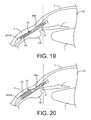
- FIG. 19shows an example of at least a part of the expandable features of the expandable implant positioned in the suprachoroidal space with the expandable features in a compact configuration.
- FIG. 20shows an example of at least a part of the expandable features of the expandable implant positioned in the suprachoroidal space with the expandable features in an expanded configuration.
- an expandable sheaththat can be securely adapted to an ocular implant to form an expandable ocular implant, which can be implanted into the eye.
- the expandable sheathcan include at least one expandable feature that can form a compact and expanded configuration.
- the compact configurationcan allow the expandable implant to be implanted into the eye without requiring a large incision, and the expanded configuration can assist in promoting fluid flow in the eye, such as for assisting in treating glaucoma.
- FIG. 1is a cross-sectional view of a portion of the human eye.
- the eyeis generally spherical and is covered on the outside by the sclera S.
- the retinalines the inside posterior half of the eye.
- the retinaregisters the light and sends signals to the brain via the optic nerve.
- the bulk of the eyeis filled and supported by the vitreous body, a clear, jelly-like substance.
- the elastic lens Lis located near the front of the eye.
- the lens Lprovides adjustment of focus and is suspended within a capsular bag from the ciliary body CB, which contains the muscles that change the focal length of the lens.
- a volume in front of the lens Lis divided into two by the iris I, which controls the aperture of the lens and the amount of light striking the retina.
- the pupilis a hole in the center of the iris I through which light passes.
- the volume between the iris I and the lens Lis the posterior chamber PC.
- the volume between the iris I and the corneais the anterior chamber AC. Both chambers are filled with a clear liquid known as aqueous humor.
- the ciliary body CBcontinuously forms aqueous humor in the posterior chamber PC by secretion from the blood vessels.
- the aqueous humorflows around the lens L and iris I into the anterior chamber and exits the eye through the trabecular meshwork, a sieve-like structure situated at the corner of the iris I and the wall of the eye (the corner is known as the iridocorneal angle).
- Some of the aqueous humorcan filter through the trabecular meshwork near the iris root into Schlemm's canal, a small channel that drains into the ocular veins. A smaller portion rejoins the venous circulation after passing through the ciliary body and eventually through the sclera (the uveoscleral route).
- FIG. 2is a cross-sectional, perspective view of a portion of the eye showing the anterior and posterior chambers of the eye.
- a schematic representation of an embodiment of an implant 10such as an expandable implant, is shown positioned inside the eye such that a proximal end 12 is located in the anterior chamber 16 and a distal end 14 communicates with and/or is located in or near the suprachoroidal space.
- FIG. 1 and other figures hereinare schematic and are not necessarily to scale with respect to size and relative positions of actual eye tissue.
- the implant 10can provide a fluid pathway between at least the anterior chamber 16 into the supraciliary space or the suprachoroidal space.
- the implant 10can include a distal end 14 that may be positioned in the supraciliary space or the suprachoroidal space.
- the implant 10may be positioned at least partially between the ciliary body and the sclera or it may be at least partially positioned between the sclera and the choroid.
- the distal end 14 of the implant 10may be positioned between other anatomical parts of the eye.
- the implant 10can include an elongate tubular element having one or more internal lumens through which aqueous humor can flow from the anterior chamber 16 into the supraciliary space.
- the implant 10can have a substantially uniform internal diameter along its entire length, although the shape of the implant 10 can vary, such as along its length (either before or after insertion of the implant).
- the implant 10can have various cross-sectional shapes (such as a, circular, oval or rectangular shape) and can vary in cross-sectional shape moving along its length.
- the cross-sectional shapecan be selected to facilitate easy insertion into the eye.
- the internal lumen of the implant 10can serve as a passageway for the flow of aqueous humor through the implant 10 directly from the anterior chamber 16 toward or into the suprachoroidal space.
- the internal lumen of the implantcan be used as an access location to mount the implant 10 onto a delivery system, as will be described in more detail below.
- the internal lumencan also be used as a pathway for flowing fluid, such as an irrigation fluid or a visco-elastic substance(s), into the eye for flushing or to maintain pressure in the anterior chamber, or using the fluid to assist in dissection, visualization or hydraulic creation of a dissection plane into or within the suprachoroidal space.
- Fluidcan be flowed toward or into the suprachoroidal space, for example via a delivery cannula or through the internal lumen of the shunt.
- the fluidcan be flowed into the eye with a pressure sufficient to form a dissection plane into or within the suprachoroidal space.
- the fluidcan accumulate within the eye so as to form a lake.
- hydro-dissection or the injection of fluidssuch as a visco-elastic substance(s) can be used to separate the ciliary body from the sclera to enlarge an area of detachment of the ciliary body from the sclera with or without insertion of a device.
- reduction in IOPcan be correlated with the position of the implant 10 creating an area of separation between the choroid and sclera around at least a part of the implant 10 (also known as “tenting”) and a space created around, for example, the most distal portion 14 of the implant 10 (also known as an “aqueous lake”).
- increasing the area of scleral and choroidal separationcan improve IOP reduction in at least some instances.
- a lager implant 10such as an implant larger than approximately 0.5-1.0 mm in diameter
- some drawbacksmay include the requirement for a larger incision, such as along the limbus, due to a greater diameter implant 10 .
- a larger incisionmay cause fluids to escape the eye, such as at least from the anterior chamber, and complicate the implantation procedure.
- an incision less than approximately 2.5 mmmay be preferable for implantation of at least one implant 10 .
- a larger diameter implant 10can include creating a larger cyclodialysis which may result in increased rates of hypotony post operatively and increased rates of retinal detachments.
- a larger implant 10can be more difficult to insert into the supracilliary and suprachoroidal space due to the requirement of greater tissue separation which may result in excess tissue damage. Therefore, an implant 10 that can maintain a compact configuration during implantation and then form an expanded configuration once implanted may overcome the drawbacks discussed above while achieving increased separation between the sclera and choroid for an improved reduction in IOP.
- the present disclosureincludes various embodiments of expandable sheaths that can be functionally coupled to one or more implants 10 and can function by providing a compact configuration during implantation of the implant and form an expanded configuration once implanted in a patient's eye.
- Some embodiments disclosed hereininclude implants having expanding features that function similar to an expanding sheath such that these expanding features can maintain a compact configuration during implantation and expand once the implant has been implanted in the patient's eye.
- FIGS. 3 and 4illustrate an embodiment of an expandable sheath 20 which may be used in combination with one or more ocular implants, such as the implant 10 discussed above, to form an expandable implant 22 (as shown in FIGS. 5 and 6 ).
- the expandable sheath 20can include one or more expandable features 24 , such as struts, that extend between a distal collar 26 and a proximal collar 28 . At least one of the distal collar 26 or proximal collar 28 can be used to couple the expandable sheath 20 to an implant 10 .
- the expandable sheath 20can be made from any number of medical grade materials that allow the expandable sheath to form a condensed configuration during implantation, as shown for example in FIG. 3 , and an expanded configuration, as shown for example in FIG. 4 .
- FIGS. 5 and 6illustrate an embodiment of an expandable implant 22 in a condensed and expanded configuration, respectively.
- the expandable implant 22can include the expandable sheath 20 functionally coupled or securely adapted to the implant 10 such that the expandable sheath 20 can form at least a compact and expanded configuration.
- Some embodiments of the expandable sheath 20may be coupled to the implant 10 such that the expandable sheath 20 can form a compact and expanded configuration without generally disrupting the shape of the implant 10 .
- the proximal collar 28 of the expandable sheath 20can secure to at least a part of the proximal end 12 of the implant 10 .
- the proximal collar 28may be secured to the implant 10 such that the proximal collar 28 is permanently fixed and may not move relative to the implant 10 .
- the distal collar 26 of the expandable sheath 20may be coupled to at least part of the distal end 14 of the implant 10 such that the distal collar 26 may be movable relative to the implant 10 .
- the distal collar 26may be allowed to slide along at least a part of the distal end 14 of the implant 10 during deformation of the expandable sheath 20 , such as from a compact to an expanded shape.
- One or more restraintsmay be used to assist the expandable sheath 20 in maintaining a compact configuration, such as during implantation.
- the one or more restraintsmay be releasable in order to allow the expandable sheath 20 to deform into an expanded configuration.
- Any number of restraintsmay be used that can assist in maintaining the expandable sheath 20 in a compact configuration while also allowing at least the expandable sheath 20 to maintain a small diameter, such as less than approximately 2.5 mm.
- a tubing having an inner diameter that is larger than or equal to the outer diameter of the expandable sheath 20 or expandable features 24 in a compact configurationmay be used to restrain expandable features 24 in a compact configuration.
- any number of features or mechanismscan be coupled with the expandable implant 22 to assist in restraining the expandable features 24 in a compact configuration until expansion is desired without departing from this disclosure.
- the expandable sheath 20may be coupled to the implant 10 and restrained in a compact configuration such that the expandable implant 22 can have a minimal outer diameter.
- the expandable sheath 20can maintain the condensed configuration around the implant during implantation of the expandable implant 22 , such as with the use of a restraint.
- the restraintcan be released to allow the expandable sheath 20 to deform into an expanded configuration, as shown for example in FIG. 6 .
- the expandable sheath 20can be made out of a shape memory material which can assist in deforming the expandable sheath from a compact to an expanded configuration once the restraint is released.
- the one or more expandable features 24can extend between the proximal collar 28 and distal collar 26 and can assist in allowing the expandable sheath 20 to form a condensed and expanded configuration.
- the struts 24may deform from an expanded configuration, as shown in FIG. 6 .
- the expandable features 24can assist in separating surrounding tissue.
- positioning at least part of the expandable sheath 20 between a part of the sclera and choroid and allowing the expandable features 24 to form an expanded configurationthe expandable sheath 20 can assist in increasing the separation between the choroid and sclera.
- the expandable features 24can assist in separating the choroid and sclera while also allowing fluids to at least pass through the expandable features 24 , thus allowing fluid flow through at least the expandable sheath 20 .
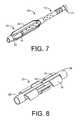
- FIG. 7illustrates an embodiment of an expandable sheath 20 with the proximal collar 28 coupled to the implant 10 in a more distal position.
- the distal collar 26 of the expandable sheath 20can extend more distally than the distal end 14 of the implant 10 .
- the expandable sheath 20can be functionally coupled to the implant 10 in any number of ways, and either the proximal collar 28 or distal collar 26 can be in permanent fixed relation to the implant 10 or may be movable relative to the implant 10 .
- the expandable sheath 20may include a variety of shaped and sized expandable features 24 for assisting in forming an expanded configuration, as will be discussed in greater detail below.
- FIG. 8illustrates an embodiment of an expandable sheath having expandable features 24 including one or more wings 30 extending from at least one support 32 .
- a restraintcan be used for restraining the wings 30 in a compact configuration, such as during implantation. Release of the restraint can allow the expandable sheath 20 to form an expanded configuration, as shown in FIG. 8 .
- the wings 30can extend radially and assist in further separating tissue, such as between the sclera and choroid.
- at least a part of the expandable features 24can be made out of medical grade shape memory material so that upon release of the restraint at least the wings 30 can deform to an expanded configuration.
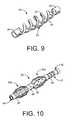
- FIG. 9illustrates an embodiment of an expandable sheath 20 having expandable features 24 which include at least one helical support 25 that extends between the proximal collar 28 and distal collar 26 .
- the helical supports 25may form a condensed configuration with a minimal diameter around the implant 10 , such as during implantation.
- the helical supports 25can deform and expand such that the outer diameter of the helix formed by the helical supports 25 increases.
- a restraintmay also be used, such as those described above, for restraining the helical supports 25 in a compact configuration during implantation.
- the expandable sheath 20can form an expanded configuration, as shown in FIG. 9 .
- the helical supports 25may assist in further separating tissue, such as between the sclera and choroid.
- FIG. 10illustrates an embodiment of an expandable implant 22 that is configured to form more than one expanding area.
- the expandable implant 22 shown in FIG. 10may be comprised of two expandable sheaths 20 a and 20 b coupled consecutively along the length of the implant 10 .
- the more than one expanding areamay be formed by coupling more than one expandable sheath 20 along an implant 10 or a single expandable sheath 20 may be configured to form more than one expanding area.
- One or more restraintscan be used to constrain the expandable features 24 of the expandable sheaths 20 a and 20 b . It may be possible to release the one or more restraints such that the expandable sheaths 20 a and 20 b are allowed to expand independently or in unison.
- any one of the proximal collars 28 or distal collars 26 of the expandable sheaths 20 a and 20 bmay be permanently fixed relative to the implant 10 or movable relative to the implant 10 .
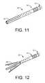
- FIGS. 11 and 12illustrate an embodiment of an expandable implant 22 having one or more expandable features 24 extending from the distal end of the implant 10 .
- the expandable features 24may be part of the implant 10 instead of part of an expandable sheath that is functionally coupled to the implant 10 .
- the expandable features 24can include extensions which may form a condensed configuration, as shown for example in FIG. 11 , and flare out into an expanded configuration, as shown for example in FIG. 12 .
- the expandable implantmay be able to achieve a smaller compact diameter due to the expandable features 24 extending distally from the implant instead of alongside the implant 10 , as shown for example in FIG. 5 .
- the expandable features 24can be made out of a medical grade shape memory material such that upon release of a restraint, the expandable features 24 may deform into an expanded configuration.
- One or more restraintsmay also be used for restraining the expandable features 24 in a compact configuration during implantation. Release of the restraints can allow the extensions to form an expanded configuration, as shown in FIG. 12 . In an expanded configuration, the extensions may assist in further separating tissue, such as between the sclera and choroid.
- FIGS. 13 and 14illustrate another embodiment of an expandable implant 22 having at least one expandable feature 24 extending between the distal end 14 and proximal end 12 of the implant 10 .
- the expandable features 24may be part of the implant 10 instead of part of an expandable sheath that is functionally coupled to the implant 10 .
- the expandable features 24can include extensions or struts which may form a condensed configuration, as shown for example in FIG. 13 , and expand radially into an expanded configuration, as shown for example in FIG. 14 .
- This embodiment of the expandable implant 22may be able to achieve a smaller compact diameter due to the expandable features 24 extending between the distal end 14 and proximal end 12 of the implant 10 instead of alongside the implant 10 , as shown for example in FIG. 5 .
- the expandable features 24 in FIGS. 13 and 14can be made out of a medical grade shape memory material such that upon release of a restraint, the expandable features 24 may deform into an expanded configuration.
- One or more restraintsmay also be used for restraining the expandable features 24 in a compact configuration during implantation. Release of the restraints can allow the extensions to form an expanded configuration, as shown in FIG. 14 . In an expanded configuration, the extensions may assist in further separating tissue, such as the sclera and choroid.
- the expandable features 24can have any number of suitable shapes or patterns that can provide both a compact and expanded configuration for assisting in further separating tissue in an eye, such as between the sclera and choroid.
- any of the expandable features 24 at least described hereinmay be a part of the implant 10 or can be part of an expandable sheath 20 that is functionally coupled to the implant 10 without departing from the scope of this disclosure.
- the expandable sheath 20can be made out of any number of medical grade materials, including at least one of shape memory alloys, such as nitinol, or shape memory polymers.
- shape memory alloyssuch as nitinol, or shape memory polymers.
- any number of medical grade materialsmay be used that allow the expandable sheath 20 to form a compact, or condensed, and expanded configuration.
- the expandable sheath 10can be functionally coupled to the implant 10 in any number of ways, such as medical grade adhesive, heat shrink tubing, or various mechanical coupling.
- the expandable sheathmay be functionally coupled to the implant in any number of ways that allow the expandable sheath to form at least a compact and expanded configuration.
- the features and profile of the expandable sheath 20may be formed by a variety of manufacturing methods.
- the profile of the expandable sheath 20may be laser cut or stamped from a flat sheet of material, such as a shape memory alloy, and rolled into a tubular shape that can functionally couple to an implant 10 .
- FIGS. 15A and 15Billustrate an embodiment of an expandable sheath 100 formed by laser cutting a medical grade material, including medical grade shape memory alloys, such as nitinol, and shape memory polymers.
- a medical grade materialincluding medical grade shape memory alloys, such as nitinol, and shape memory polymers.
- shape memory alloyssuch as nitinol
- shape memory polymerseither a tube shape or a formable flat sheet of material made out of the medical grade material can be used.
- the shape of the expandable sheath 100 in its expanded configurationmay be defined in a shape setting operation, such as those operations used for shape memory alloys and polymers.
- the expandable features 102may be held radially open in an expanded configuration with the use of an internal mandrel or fixture during the shape setting operation.
- the expandable sheath 100can have a compact configuration with an outside diameter dimension in the range of 0.4 millimeter to 0.6 millimeter, such as 0.5 millimeters.
- the expandable sheath 100can have an expanded configuration with an outside diameter dimension in the range of 2.0 millimeters to 3.0 millimeters, such as 2.5 millimeters.
- some embodiments of the implantmay include a body having a tube shaped configuration that is made out of a soft biocompatible material, such as silicone.
- the tube shaped bodyWhen implanted in the eye, the tube shaped body may extend proximally out of the expandable sheath into the anterior chamber while the distal end of the tube may terminate approximately in the middle of the expandable sheath.
- At least a part of a surface of the expandable sheath 20can be treated with one or more surface treatments that can modify the topography of the expandable sheath.
- surface areas of the expandable sheath that have been treated with a surface treatmentcan cause a variety of ocular tissue responses as a result of the expandable sheath being implanted in the eye and contacting the treated surface area to the ocular tissue.
- any number of surface treatmentscan be applied to any part of the expandable sheath for assisting in creating a variety of ocular tissue responses.
- a plasma cleaning processcan be used to treat at least a part of the surface of the expandable sheath.
- one or more variablessuch as power, processing time, and pressure, can be varied, including varying the power by approximately 250%, in order to achieve a desired surface topography.
- FIG. 16illustrates an embodiment of a laser ablation pattern 104 applied to at least a part of the surface of the expandable sheath 100 .
- the laser ablation pattern 104can include at least one ribbed feature, including multiple micro ribs.
- Such laser ablation patterns 104can provide a variety of tissue responses, and by varying at least one of the size and shape of the laser ablation patterns 104 any number of tissue responses can be achieved.
- either the expandable sheath or implantcan at least partially include at least one drug, such as either impregnated or coated with at least one drug.
- at least the expandable sheathcan include mitomycin or 5-FU, which can assist in reducing fibrotic and inflammatory tissue response, such as during trabeculectomy surgeries.
- the one or more drugsmay be combined with a polymer comprising at least a part of either the expandable sheath or implant that can provide a sustained drug release profile during implantation of either the expandable sheath or implant.
- the expandable sheathmay include at least one drug, such as any drug described herein, while the implant, such as the implant having a tube shaped body, may not include a drug. Additionally, some embodiments of the expandable sheath can be implanted in the eye without being coupled to an implant.
- a delivery systemcan be used to deliver an expandable implant 22 into the eye, for example such that the expandable implant 22 at least provides fluid communication between the anterior chamber toward the suprachoroidal space.
- the expandable implant 22can include one or more expandable features 24 which may assist in further separating tissue, such as between the sclera and choroid.
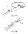
- FIG. 17shows an embodiment of a delivery system 50 that can be used to deliver the expandable implant 22 into the eye. It should be appreciated that these delivery systems 50 are exemplary and that variations in the structure, shape and actuation of the delivery system 50 are possible.
- the delivery system 50generally includes a proximal handle component 52 and a distal delivery component 54 .
- the proximal handle component 52can include an actuator 56 , such as a button, to control the release of an expandable implant 22 from the delivery component 54 into the target location in the eye.
- the actuator 56can vary in structure.
- An embodiment of the delivery component 54can include an elongate applier in the form of a guidewire 58 that inserts longitudinally through an internal lumen of the expandable implant 22 and a “stopper” or sheath 60 positioned axially over the guidewire 58 .
- the sheath 60can aid in the release of the expandable implant 22 from the delivery component 54 into the target location in the eye.
- the actuator 56can be used to control movement or relative movement of the guidewire 58 and/or the sheath 60 .
- the sheath 60can be fixed relative to the handle component 52 and act as a stopper that impedes the expandable implant 22 from moving in a proximal direction as the guidewire 58 is withdrawn proximally from the expandable implant 22 upon actuation of the actuator 56 .
- the guidewire 58can be extended distally relative to the sheath 60 .
- Actuation of the actuator 56such as by pressing the actuator 56 , can cause the guidewire 58 to slide proximally into the sheath 60 . This can effectively disengage the expandable implant 22 off the distal end of the guidewire 58 and releases the expandable implant 22 in a controlled fashion such that the target positioning of the expandable implant 22 is maintained.
- FIG. 18shows an enlarged view of an expandable implant 22 mounted on a delivery component 54 for inserting the expandable implant 22 into the eye.
- the expandable implant 22can be mounted on a distal region of a guidewire 58 .
- the sheath 60can be sized and shaped to receive or abut a portion of the proximal end of the expandable implant 22 .
- the guidewire 58can slide in the proximal direction (arrow P) into the sheath 60 .
- the proximal end of the expandable implant 22can abut the distal edge of the sheath 60 to prevent the expandable implant 22 from sliding in the proximal direction. This can effectively disengage the implant 10 off the distal end of the guidewire 58 and controllably release the expandable implant 22 into the eye tissue.
- the delivery system 50can also assist in providing fluid delivery into the eye during or after implantation of the expandable implant 22 .
- the delivered fluidmay vary and may include a viscoelastic, drugs, stem cells, or a combination thereof.
- the deliverymay be in combination with retinal or macula therapy.
- a fluid delivery featurecan include an elongated tube 30 that extends outward from the handle 52 .
- the tube 30can extend through the handle 52 and can have an internal lumen that communicates at a distal end with the proximal end of an internal lumen in the guidewire 58 .
- One or more outlet openings, such as slots 32can be located on the distal region of the guidewire 58 .
- the tube 30can be connected at a proximal end to a source of fluid so as to provide a pathway for the fluid to be delivered to the internal lumen of the guidewire via the tube 30 .
- the fluidcan then exit the guidewire via the slots 32 for delivery into the eye.
- the fluidmay be delivered to other sections along the axial length of the expandable implant 22 .
- Holes along the length of the expandable implant 22may be configured to be sufficiently large such that a fluid may be delivered through corresponding holes along the guidewire 58 and into the eye, such as into the supraciliary or suprachoroidal space surrounding the body of the expandable implant 22 (depending on where the implant is positioned and the length of the implant). This would be advantageous because it may create additional space surrounding the expandable implant 22 and improve tenting.
- one or more expandable implants 22can be slideably loaded on a delivery system 50 and implanted to a position that communicates with the suprachoroidal space as described herein.
- the expandable implant 22can then be implanted in the eye via an ab-interno procedure through a limbal incision into the anterior chamber.
- the expandable implant 22may then be secured in the eye so that it provides fluid communication between the anterior chamber and the suprachoroidal space, as well as provide increased separation between the sclera and choroid.
- the guidewire 58can be positioned on the delivery system 50 such that the distal tip of the guidewire 58 , the expandable implant 22 and the sheath 60 can penetrate through a small corneal incision and access the anterior chamber, such as within the limbus of the cornea.
- the incisionis very close to the limbus, such as either at the level of the limbus or within 2 mm of the limbus in the clear cornea.
- the guidewire 58can be used to make the incision or a separate cutting device can be used. For example, a knife-tipped device or diamond knife can be used to initially enter the cornea.
- the corneal incisioncan have a size that is sufficient to permit at least the passage of the expandable implant 22 on the guidewire 58 and sheath therethrough.
- the incisioncan be about 1 mm in size.
- the incisionis no greater than about 2.5 mm in size.
- the incisionis no greater than about 2.85 mm and is greater than about 1.5 mm.
- the guidewire 58can be advanced into the anterior chamber along a pathway that enables the expandable implant 22 to be delivered to a position such that the expandable implant 22 provides a flow passageway from the anterior chamber AC toward the suprachoroidal space.
- the guidewire 58can be advanced further into the eye such that the blunt distal tip of the guidewire 58 and/or the expandable implant 22 can seat with and penetrate the iris root IR, or a region of the ciliary body CB, or the iris root part of the ciliary body near its tissue border with the scleral spur.
- the guidewire 58can approach the iris root from the same side of the anterior chamber AC as the deployment location such that the guidewire 58 does not have to be advanced across the iris. Alternately, the guidewire 58 can approach the location from across the anterior chamber AC such that the guidewire 58 is advanced across the iris and/or the anterior chamber toward the opposite iris root.
- the guidewire 58can approach the eye and the iris root IR along a variety of pathways.
- the guidewire 58does not necessarily cross over the eye and does not intersect the optical axis of the eye. In other words, the corneal incision and the location where the implant is implanted at the iris root can be in the same quadrant (if the eye is viewed from the front and divided into four quadrants). Also, the pathway of the implant from the corneal incision to the iris root desirably does not pass through the optic axis of the eye to avoid interfering with the pupil.
- FIG. 19shows an enlarged view of the anterior region of the eye showing the anterior chamber AC, the cornea C, the iris I, and the sclera S.
- the expandable implant 22 mounted on the guidewire 58can approach from the anterior chamber AC.
- the expandable implant 22 and guidewire 58can move along a pathway such that the dissection entry point of the distal tip of the guidewire 58 can penetrate the iris root IR near its junction with the scleral spur SSp or the iris root portion of the ciliary body CB or other desired location.
- the surgeoncan rotate or reposition the handle of the delivery device 50 in order to obtain a proper approach trajectory for the distal tip of the guidewire 58 , as described in further detail below.
- the guidewire 58 with the expandable implant 22 positioned thereuponcan be advanced from a region of the anterior chamber that can be viewed through a transparent zone of the cornea through to a region of the anterior chamber that is obscured by the opaque zone of the cornea.
- the guidewire 58 and expandable implant 22can be advanced through the cornea C until resistance is felt and the delivery device can be seated at a location near the iris root IR, the ciliary body or the iris root portion of the ciliary body.
- the guidewire 58can then be advanced further such that the guidewire 58 and expandable implant 22 loaded thereon penetrate an area of fibrous attachment between the scleral spur SSP and the ciliary body CB. This area of fibrous attachment can be approximately 1 mm.
- the guidewire 58can then more easily cause the sclera S to peel away or otherwise separate from the ciliary body CB and possibly the choroid as it follows the inner curve of the sclera S and enters the supraciliary space.
- a combination of the guidewire's tip shape, material, material properties, diameter, flexibility, compliance, coatings, pre-curvature etc.make it more inclined to follow an implantation pathway that mirrors the curvature of the inner wall of the sclera and between tissue layers such as between the sclera and the ciliary body, and between the sclera and the choroid.
- the dissection plane of the guidewire 58 and expandable implant 22can follow the curve of the inner scleral wall such that the expandable implant 22 mounted on the guidewire 58 after penetrating the iris root or the iris root portion of the ciliary body, bluntly dissects the boundary between tissue layers of the scleral spur SSp and the ciliary body CB such that at least the distal region of the expandable implant 22 extends into the supraciliary space.
- the expandable implant 22is positioned such that it extends sufficiently past the scleral spur SSP such that it is positioned between the tissue boundaries of the sclera and the choroid (the suprachoroidal space).
- the one or more restraintsmay be released in order to allow the expandable implant 22 to enter into an expanded configuration, as shown in FIG. 20 .
- the expandable features 24 of the expandable implant 22can assist in increasing the separation between the sclera and choroid than what was achieved prior to expansion of the expandable implant 22 .
- the increase in separation between the sclera and choroidcan assist in reducing IOP in an eye, such as an eye suffering from glaucoma.
- the expandable implant 22can then be released from the guidewire 58 .
- the expandable implant 22can be released, for example, by withdrawing the guidewire 58 such that the expandable implant 22 is effectively disengaged in a controlled manner from the tip of the guidewire 58 with the sheath 60 .
- the expandable implant 22can include one or more structural features near its proximal region that aid to anchor or retain the implant 105 in the target region in the eye.
- the structural featurescan include flanges, protrusions, wings, tines, or prongs, and the like that can lodge into the surrounding eye anatomy to retain the expandable implant 22 in place and prevent the expandable implant 22 from moving further into the suprachoroidal space SchS.
Landscapes
- Health & Medical Sciences (AREA)
- Ophthalmology & Optometry (AREA)
- Heart & Thoracic Surgery (AREA)
- Surgery (AREA)
- Engineering & Computer Science (AREA)
- Biomedical Technology (AREA)
- Nuclear Medicine, Radiotherapy & Molecular Imaging (AREA)
- Vascular Medicine (AREA)
- Life Sciences & Earth Sciences (AREA)
- Animal Behavior & Ethology (AREA)
- General Health & Medical Sciences (AREA)
- Public Health (AREA)
- Veterinary Medicine (AREA)
- Prostheses (AREA)
Abstract
Description
Claims (20)
Priority Applications (1)
| Application Number | Priority Date | Filing Date | Title |
|---|---|---|---|
| US14/029,389US9480598B2 (en) | 2012-09-17 | 2013-09-17 | Expanding ocular implant devices and methods |
Applications Claiming Priority (2)
| Application Number | Priority Date | Filing Date | Title |
|---|---|---|---|
| US201261702179P | 2012-09-17 | 2012-09-17 | |
| US14/029,389US9480598B2 (en) | 2012-09-17 | 2013-09-17 | Expanding ocular implant devices and methods |
Publications (2)
| Publication Number | Publication Date |
|---|---|
| US20140081195A1 US20140081195A1 (en) | 2014-03-20 |
| US9480598B2true US9480598B2 (en) | 2016-11-01 |
Family
ID=50275205
Family Applications (1)
| Application Number | Title | Priority Date | Filing Date |
|---|---|---|---|
| US14/029,389Active2034-05-10US9480598B2 (en) | 2012-09-17 | 2013-09-17 | Expanding ocular implant devices and methods |
Country Status (6)
| Country | Link |
|---|---|
| US (1) | US9480598B2 (en) |
| EP (2) | EP2895123B1 (en) |
| EA (1) | EA030047B1 (en) |
| ES (1) | ES2633185T3 (en) |
| RU (1) | RU2650203C2 (en) |
| WO (1) | WO2014043698A2 (en) |
Cited By (8)
| Publication number | Priority date | Publication date | Assignee | Title |
|---|---|---|---|---|
| US10322029B2 (en) | 2015-09-22 | 2019-06-18 | Novartis Ag | Ocular implant container |
| WO2021007559A1 (en)* | 2019-07-10 | 2021-01-14 | Anand Doraiswamy | Eye stents and delivery systems |
| US11096707B2 (en) | 2018-12-12 | 2021-08-24 | Alcon Inc. | Actuation handle |
| USD934424S1 (en) | 2019-08-29 | 2021-10-26 | Alcon Inc. | 360 degree actuation handle |
| US11298146B2 (en) | 2019-02-01 | 2022-04-12 | Alcon Inc. | Actuation mechanism with arcuate levers |
| US11490915B2 (en) | 2019-08-29 | 2022-11-08 | Alcon Inc. | Actuation mechanism with grooved actuation levers |
| US11517476B2 (en)* | 2017-06-13 | 2022-12-06 | Innfocus, Inc. | Systems, methods, and apparatus for treatment of glaucoma |
| US12440377B2 (en) | 2024-11-13 | 2025-10-14 | Aquea Health, Inc. | Eye stents and delivery systems and methods |
Families Citing this family (55)
| Publication number | Priority date | Publication date | Assignee | Title |
|---|---|---|---|---|
| US7291125B2 (en) | 2003-11-14 | 2007-11-06 | Transcend Medical, Inc. | Ocular pressure regulation |
| WO2007087061A2 (en) | 2006-01-17 | 2007-08-02 | Transcend Medical, Inc. | Glaucoma treatment device |
| US10085884B2 (en) | 2006-06-30 | 2018-10-02 | Aquesys, Inc. | Intraocular devices |
| US8721702B2 (en) | 2010-11-15 | 2014-05-13 | Aquesys, Inc. | Intraocular shunt deployment devices |
| US8663303B2 (en) | 2010-11-15 | 2014-03-04 | Aquesys, Inc. | Methods for deploying an intraocular shunt from a deployment device and into an eye |
| US20120123316A1 (en) | 2010-11-15 | 2012-05-17 | Aquesys, Inc. | Intraocular shunts for placement in the intra-tenon's space |
| US8852256B2 (en) | 2010-11-15 | 2014-10-07 | Aquesys, Inc. | Methods for intraocular shunt placement |
| US20170360609A9 (en) | 2007-09-24 | 2017-12-21 | Ivantis, Inc. | Methods and devices for increasing aqueous humor outflow |
| ES2920877T3 (en) | 2009-01-28 | 2022-08-11 | Alcon Inc | Ocular implant placement system |
| US8535333B2 (en) | 2009-07-29 | 2013-09-17 | Transcend Medical, Inc. | Ocular implant applier and methods of use |
| US8529492B2 (en) | 2009-12-23 | 2013-09-10 | Trascend Medical, Inc. | Drug delivery devices and methods |
| US20160256320A1 (en) | 2010-11-15 | 2016-09-08 | Aquesys, Inc. | Intraocular shunt placement in the suprachoroidal space |
| US8765210B2 (en) | 2011-12-08 | 2014-07-01 | Aquesys, Inc. | Systems and methods for making gelatin shunts |
| US8852136B2 (en) | 2011-12-08 | 2014-10-07 | Aquesys, Inc. | Methods for placing a shunt into the intra-scleral space |
| US9808373B2 (en) | 2013-06-28 | 2017-11-07 | Aquesys, Inc. | Intraocular shunt implantation |
| US10080682B2 (en) | 2011-12-08 | 2018-09-25 | Aquesys, Inc. | Intrascleral shunt placement |
| US9610195B2 (en) | 2013-02-27 | 2017-04-04 | Aquesys, Inc. | Intraocular shunt implantation methods and devices |
| US9358156B2 (en) | 2012-04-18 | 2016-06-07 | Invantis, Inc. | Ocular implants for delivery into an anterior chamber of the eye |
| US9241832B2 (en) | 2012-04-24 | 2016-01-26 | Transcend Medical, Inc. | Delivery system for ocular implant |
| US9763829B2 (en) | 2012-11-14 | 2017-09-19 | Novartis Ag | Flow promoting ocular implant |
| US10617558B2 (en) | 2012-11-28 | 2020-04-14 | Ivantis, Inc. | Apparatus for delivering ocular implants into an anterior chamber of the eye |
| US10159600B2 (en) | 2013-02-19 | 2018-12-25 | Aquesys, Inc. | Adjustable intraocular flow regulation |
| US9125723B2 (en) | 2013-02-19 | 2015-09-08 | Aquesys, Inc. | Adjustable glaucoma implant |
| WO2014190029A1 (en) | 2013-05-21 | 2014-11-27 | Transcend Medical, Inc. | Flow promoting ocular implant device and methods |
| EP3068354B1 (en) | 2013-11-14 | 2023-06-28 | Aquesys, Inc. | Intraocular shunt inserter |
| WO2016011056A1 (en) | 2014-07-14 | 2016-01-21 | Ivantis, Inc. | Ocular implant delivery system and method |
| US10342702B2 (en) | 2014-08-29 | 2019-07-09 | Camras Vision Inc. | Apparatus and method for reducing intraocular pressure |
| US10201451B2 (en) | 2014-08-29 | 2019-02-12 | Camras Vision Inc. | Device and method for reducing intraocular pressure |
| MA42406A (en) | 2015-06-03 | 2018-05-16 | Aquesys Inc | IMPLEMENTATION OF INTRAOCULAR AB EXTERNO SHUNT |
| AU2016307951B2 (en) | 2015-08-14 | 2021-04-01 | Alcon Inc. | Ocular implant with pressure sensor and delivery system |
| US10524958B2 (en) | 2015-09-30 | 2020-01-07 | Alievio, Inc. | Method and apparatus for reducing intraocular pressure |
| WO2017106517A1 (en) | 2015-12-15 | 2017-06-22 | Ivantis, Inc. | Ocular implant and delivery system |
| MX2018014763A (en) | 2016-06-02 | 2019-04-29 | Aquesys Inc | Intraocular drug delivery. |
| EP4218692A3 (en) | 2017-07-20 | 2023-09-06 | Shifamed Holdings, LLC | Adjustable flow glaucoma shunts and methods for making and using same |
| US11166849B2 (en)* | 2017-07-20 | 2021-11-09 | Shifamed Holdings, Llc | Adjustable flow glaucoma shunts and methods for making and using same |
| US11246753B2 (en) | 2017-11-08 | 2022-02-15 | Aquesys, Inc. | Manually adjustable intraocular flow regulation |
| CA3207829A1 (en) | 2018-02-22 | 2019-08-29 | Alcon Inc. | Ocular implant and delivery system |
| JP2019141450A (en)* | 2018-02-23 | 2019-08-29 | 高島産業株式会社 | Glaucoma treatment stent |
| US11135089B2 (en) | 2018-03-09 | 2021-10-05 | Aquesys, Inc. | Intraocular shunt inserter |
| US10952898B2 (en) | 2018-03-09 | 2021-03-23 | Aquesys, Inc. | Intraocular shunt inserter |
| EP3846747A4 (en)* | 2018-09-04 | 2022-06-08 | University Hospitals Health System Inc. | OCULAR DEVICE FOR THE TREATMENT OF GLAUCOMA AND METHOD FOR THE MINIMUMLY INVASIVE SURGERY ASSOCIATED WITH GLAUCOMA |
| EP3911285A4 (en) | 2019-01-18 | 2022-10-19 | Shifamed Holdings, LLC | Adjustable flow glaucoma shunts and methods for making and using same |
| JP7614193B2 (en) | 2019-10-10 | 2025-01-15 | シファメド・ホールディングス・エルエルシー | Adjustable flow glaucoma shunts and related systems and methods |
| AU2021209698A1 (en)* | 2020-01-23 | 2022-08-04 | Shifamed Holdings, Llc | Adjustable flow glaucoma shunts and associated systems and methods |
| US11291585B2 (en) | 2020-02-14 | 2022-04-05 | Shifamed Holdings, Llc | Shunting systems with rotation-based flow control assemblies, and associated systems and methods |
| US11737920B2 (en) | 2020-02-18 | 2023-08-29 | Shifamed Holdings, Llc | Adjustable flow glaucoma shunts having non-linearly arranged flow control elements, and associated systems and methods |
| WO2021188952A1 (en) | 2020-03-19 | 2021-09-23 | Shifamed Holdings, Llc | Intraocular shunts with low-profile actuation elements and associated systems and methods |
| CN115867237A (en) | 2020-04-16 | 2023-03-28 | 施菲姆德控股有限责任公司 | Adjustable glaucoma treatment devices and related systems and methods |
| WO2022093832A1 (en)* | 2020-10-27 | 2022-05-05 | Glaukos Corporation | Implant to treat retinal vein occlusion |
| US11540940B2 (en) | 2021-01-11 | 2023-01-03 | Alcon Inc. | Systems and methods for viscoelastic delivery |
| EP4281144A4 (en) | 2021-01-22 | 2024-11-27 | Shifamed Holdings, LLC | ADJUSTABLE SHUNTING SYSTEMS WITH PLATE ARRANGEMENTS AND RELATED SYSTEMS AND METHODS |
| JP2025501667A (en)* | 2022-01-14 | 2025-01-22 | リキッド メディカル プロプライエタリー リミテッド | Shunts and methods for treating glaucoma - Patents.com |
| GB202210218D0 (en)* | 2022-07-12 | 2022-08-24 | Univ Oxford Innovation Ltd | Ocular implant, kit for deploying implant, method of deploying implant |
| WO2024196536A1 (en)* | 2023-03-22 | 2024-09-26 | Innfocus, Inc. | Glaucoma drainage device with expandable anchor |
| CN119700417B (en)* | 2024-12-10 | 2025-08-08 | 北京华视诺维医疗科技有限公司 | Ophthalmic minimally invasive drainage device with expansion fixing function and delivery system thereof |
Citations (262)
| Publication number | Priority date | Publication date | Assignee | Title |
|---|---|---|---|---|
| US2990670A (en) | 1957-03-07 | 1961-07-04 | North American Rayon Corp | Yarn crimping and texturing apparatus |
| US3439675A (en) | 1966-06-14 | 1969-04-22 | Becton Dickinson Co | Deformable needle assembly |
| US3767759A (en) | 1970-05-27 | 1973-10-23 | Akademie Ved | Method of molding capillary drain for surgery |
| US3788327A (en) | 1971-03-30 | 1974-01-29 | H Donowitz | Surgical implant device |
| US3915172A (en) | 1970-05-27 | 1975-10-28 | Ceskoslovenska Akademie Ved | Capillary drain for glaucoma |
| US4037604A (en) | 1976-01-05 | 1977-07-26 | Newkirk John B | Artifical biological drainage device |
| GB2101891A (en) | 1981-07-18 | 1983-01-26 | Anthony Christopher Be Molteno | Device for draining aqueous humour |
| US4402681A (en) | 1980-08-23 | 1983-09-06 | Haas Joseph S | Artificial implant valve for the regulation of intraocular pressure |
| US4457757A (en) | 1981-07-20 | 1984-07-03 | Molteno Anthony C B | Device for draining aqueous humour |
| US4521210A (en) | 1982-12-27 | 1985-06-04 | Wong Vernon G | Eye implant for relieving glaucoma, and device and method for use therewith |
| US4554918A (en) | 1982-07-28 | 1985-11-26 | White Thomas C | Ocular pressure relief device |
| US4604087A (en) | 1985-02-26 | 1986-08-05 | Joseph Neil H | Aqueous humor drainage device |
| US4617715A (en) | 1982-08-03 | 1986-10-21 | Oy Tampella Ab | Method for preliminary anchoring of a wire rope bolt |
| US4634418A (en) | 1984-04-06 | 1987-01-06 | Binder Perry S | Hydrogel seton |
| EP0228185A1 (en) | 1985-11-27 | 1987-07-08 | Thomas C. White | Tissue-implantable fluid-dissipating device |
| US4722724A (en) | 1986-06-23 | 1988-02-02 | Stanley Schocket | Anterior chamber tube shunt to an encircling band, and related surgical procedure |
| US4750901A (en) | 1986-03-07 | 1988-06-14 | Molteno Anthony C B | Implant for drainage of aqueous humour |
| US4787885A (en) | 1984-04-06 | 1988-11-29 | Binder Perry S | Hydrogel seton |
| WO1989000869A1 (en) | 1987-08-06 | 1989-02-09 | White Thomas C | Glaucoma drainage in the lacrimal system |
| US4826478A (en) | 1986-06-23 | 1989-05-02 | Stanley Schocket | Anterior chamber tube shunt to an encircling band, and related surgical procedure |
| US4846172A (en) | 1987-05-26 | 1989-07-11 | Berlin Michael S | Laser-delivery eye-treatment method |
| US4863457A (en) | 1986-11-24 | 1989-09-05 | Lee David A | Drug delivery device |
| US4886488A (en) | 1987-08-06 | 1989-12-12 | White Thomas C | Glaucoma drainage the lacrimal system and method |
| US4900300A (en) | 1987-07-06 | 1990-02-13 | Lee David A | Surgical instrument |
| US4930512A (en) | 1988-06-16 | 1990-06-05 | Sonomed, Inc. | Hand held spring-loaded ultrasonic probe |
| US4946436A (en) | 1989-11-17 | 1990-08-07 | Smith Stewart G | Pressure-relieving device and process for implanting |
| US4968296A (en) | 1989-12-20 | 1990-11-06 | Robert Ritch | Transscleral drainage implant device for the treatment of glaucoma |
| US5041081A (en) | 1990-05-18 | 1991-08-20 | Odrich Ronald B | Ocular implant for controlling glaucoma |
| WO1991012046A1 (en) | 1990-02-12 | 1991-08-22 | Atos Medical Ab | Glaucoma valve |
| US5071408A (en) | 1988-10-07 | 1991-12-10 | Ahmed Abdul Mateen | Medical valve |
| US5073163A (en) | 1990-01-29 | 1991-12-17 | Lippman Myron E | Apparatus for treating glaucoma |
| US5092837A (en) | 1989-12-20 | 1992-03-03 | Robert Ritch | Method for the treatment of glaucoma |
| US5127901A (en) | 1990-05-18 | 1992-07-07 | Odrich Ronald B | Implant with subconjunctival arch |
| WO1992019294A1 (en) | 1991-05-08 | 1992-11-12 | Prywes Arnold S | Shunt valve and therapeutic delivery systems |
| US5171213A (en) | 1991-08-14 | 1992-12-15 | Price Jr Francis W | Technique for fistulization of the eye and an eye filtration prosthesis useful therefor |
| US5178604A (en) | 1990-05-31 | 1993-01-12 | Iovision, Inc. | Glaucoma implant |
| US5180362A (en) | 1990-04-03 | 1993-01-19 | Worst J G F | Gonio seton |
| WO1994002081A1 (en) | 1992-07-16 | 1994-02-03 | Wong Vernon G | Eye implant suitable for relief of glaucoma |
| US5284476A (en) | 1992-03-20 | 1994-02-08 | Koch Paul S | Nuclear hydrolysis cannula |
| US5300020A (en) | 1991-05-31 | 1994-04-05 | Medflex Corporation | Surgically implantable device for glaucoma relief |
| WO1994009837A1 (en) | 1992-10-28 | 1994-05-11 | Annuit Coeptis, Inc. | Glaucoma implant device and method for implanting same |
| WO1994009721A1 (en) | 1992-11-03 | 1994-05-11 | Price Francis W Jr | Eye filtration prostheses |
| WO1994013234A1 (en) | 1992-12-17 | 1994-06-23 | Michael Andrew Coote | Implant device and method for treatment of glaucoma |
| US5338291A (en) | 1993-02-03 | 1994-08-16 | Pudenz-Schulte Medical Research Corporation | Glaucoma shunt and method for draining aqueous humor |
| RU2018289C1 (en) | 1992-03-19 | 1994-08-30 | Межотраслевой научно-технический комплекс "Микрохирургия глаза" | Method of treatment of secondary glaucoma in children |
| US5342370A (en) | 1993-03-19 | 1994-08-30 | University Of Miami | Method and apparatus for implanting an artifical meshwork in glaucoma surgery |
| US5346464A (en) | 1992-03-10 | 1994-09-13 | Camras Carl B | Method and apparatus for reducing intraocular pressure |
| US5372577A (en) | 1988-04-11 | 1994-12-13 | Ungerleider; Bruce A. | Apparatus for reducing intraocular pressure |
| US5397300A (en) | 1990-05-31 | 1995-03-14 | Iovision, Inc. | Glaucoma implant |
| WO1995008310A1 (en) | 1993-09-22 | 1995-03-30 | Voir Et Vivre E U R L | Implantable device for the treatment of ×demas |
| WO1995013765A1 (en) | 1993-11-15 | 1995-05-26 | Oculex Pharmaceuticals, Inc. | Biocompatible ocular implants |
| US5423777A (en) | 1993-10-27 | 1995-06-13 | Tajiri; Akira | Punctum plug |
| US5433701A (en) | 1994-12-21 | 1995-07-18 | Rubinstein; Mark H. | Apparatus for reducing ocular pressure |
| US5454746A (en) | 1994-01-06 | 1995-10-03 | Meccano, S.A. | Toy hand tool |
| US5476445A (en) | 1990-05-31 | 1995-12-19 | Iovision, Inc. | Glaucoma implant with a temporary flow restricting seal |
| RU2056818C1 (en) | 1990-04-28 | 1996-03-27 | Межотраслевой научно-технический комплекс "Микрохирургия глаза" | Device for strengthening erection |
| WO1996020742A1 (en) | 1995-01-06 | 1996-07-11 | Wong Vernon G | Improve eye implant for relief of glaucoma |
| US5558630A (en) | 1994-12-30 | 1996-09-24 | Fisher; Bret L. | Intrascleral implant and method for the regulation of intraocular pressure |
| WO1996036377A1 (en) | 1995-05-14 | 1996-11-21 | Optonol Ltd. | Intraocular implant, delivery device, and method of implantation |
| USRE35390E (en) | 1989-11-17 | 1996-12-03 | Smith; Stewart G. | Pressure relieving device and process for implanting |
| US5601094A (en) | 1994-11-22 | 1997-02-11 | Reiss; George R. | Ophthalmic shunt |
| RU2074686C1 (en) | 1994-08-10 | 1997-03-10 | Волгоградский филиал Межотраслевого научно-технического комплекса "Микрохирургия глаза" | Method to treat glaucoma |
| RU2074687C1 (en) | 1994-08-10 | 1997-03-10 | Волгоградский филиал Межотраслевого научно-технического комплекса "Микрохирургия глаза" | Method to treat glaucoma |
| US5626558A (en) | 1995-05-05 | 1997-05-06 | Suson; John | Adjustable flow rate glaucoma shunt and method of using same |
| US5626559A (en) | 1994-05-02 | 1997-05-06 | Ramot University Authority For Applied Research And Industrial Development Ltd. | Ophthalmic device for draining excess intraocular fluid |
| US5676944A (en) | 1993-10-06 | 1997-10-14 | The Regents Of The University Of California | Ocular therapy with homologous macrophages |
| US5704907A (en) | 1994-07-22 | 1998-01-06 | Wound Healing Of Oklahoma | Method and apparatus for lowering the intraocular pressure of an eye |
| US5713844A (en) | 1997-01-10 | 1998-02-03 | Peyman; Gholam A. | Device and method for regulating intraocular pressure |
| US5728465A (en) | 1991-05-03 | 1998-03-17 | Advanced Refractory Technologies, Inc. | Diamond-like nanocomposite corrosion resistant coatings |
| US5741292A (en) | 1995-10-26 | 1998-04-21 | Eagle Vision | Punctum dilating and plug inserting instrument with push-button plug release |
| US5743868A (en) | 1994-02-14 | 1998-04-28 | Brown; Reay H. | Corneal pressure-regulating implant device |
| US5749879A (en) | 1989-08-16 | 1998-05-12 | Medtronic, Inc. | Device or apparatus for manipulating matter |
| US5752928A (en) | 1997-07-14 | 1998-05-19 | Rdo Medical, Inc. | Glaucoma pressure regulator |
| WO1998023237A1 (en) | 1996-11-29 | 1998-06-04 | The Lions Eye Institute Of Western Australia Incorporated | Biological microfistula tube and implantation method and apparatus |
| WO1998030181A1 (en) | 1997-01-10 | 1998-07-16 | University College London | Device for use in the eye |
| US5792075A (en) | 1995-04-11 | 1998-08-11 | Schneider (Europe) A.G. | Method and apparatus for extending the length of a guide wire |
| US5807302A (en) | 1996-04-01 | 1998-09-15 | Wandel; Thaddeus | Treatment of glaucoma |
| US5807244A (en) | 1996-11-15 | 1998-09-15 | Barot; Jagdish Shantilal | Single use disposable iris retractor |
| US5868697A (en) | 1995-05-14 | 1999-02-09 | Optonol Ltd. | Intraocular implant |
| US5882327A (en) | 1997-04-17 | 1999-03-16 | Jacob; Jean T. | Long-term glaucoma drainage implant |
| US5893837A (en) | 1997-02-28 | 1999-04-13 | Staar Surgical Company, Inc. | Glaucoma drain implanting device and method |
| WO1999026567A1 (en) | 1997-11-20 | 1999-06-03 | Optonol Ltd. | Flow regulating implant, method of manufacturing, and delivery device |
| CN1225027A (en) | 1996-05-20 | 1999-08-04 | 佩尔库瑟吉公司 | Low profile catheter valve |
| US5941250A (en) | 1996-11-21 | 1999-08-24 | University Of Louisville Research Foundation Inc. | Retinal tissue implantation method |
| US5968058A (en) | 1996-03-27 | 1999-10-19 | Optonol Ltd. | Device for and method of implanting an intraocular implant |
| US6007510A (en) | 1996-10-25 | 1999-12-28 | Anamed, Inc. | Implantable devices and methods for controlling the flow of fluids within the body |
| US6019786A (en) | 1995-10-11 | 2000-02-01 | Schneider (Usa) Inc | Braided composite prosthesis |
| WO2000006223A1 (en) | 1998-07-27 | 2000-02-10 | Anamed, Inc. | Sutureless implantable device and method for treatment of glaucoma |
| US6036678A (en) | 1995-02-28 | 2000-03-14 | Photogenesis, Inc. | Method for preparation and transplantation of planar implants and surgical instrument therefor |
| US6050970A (en) | 1997-05-08 | 2000-04-18 | Pharmacia & Upjohn Company | Method and apparatus for inserting a glaucoma implant in an anterior and posterior segment of the eye |
| US6050999A (en) | 1997-12-18 | 2000-04-18 | Keravision, Inc. | Corneal implant introducer and method of use |
| US6077299A (en) | 1998-06-22 | 2000-06-20 | Eyetronic, Llc | Non-invasively adjustable valve implant for the drainage of aqueous humor in glaucoma |
| US6102045A (en) | 1994-07-22 | 2000-08-15 | Premier Laser Systems, Inc. | Method and apparatus for lowering the intraocular pressure of an eye |
| RU2157678C1 (en) | 1999-12-22 | 2000-10-20 | Макашова Надежда Васильевна | Surgical method for treating the cases of glaucoma |
| WO2000064393A1 (en) | 1999-04-26 | 2000-11-02 | Lynch Mary G | Shunt device and method for treating glaucoma |
| WO2000064511A1 (en) | 1999-04-27 | 2000-11-02 | The Arizona Board Of Regents Acting On Behalf Of The University Of Arizona | A glaucoma shunt and a method of making and surgically implanting the same |
| US6152918A (en) | 1996-04-05 | 2000-11-28 | Eclipse Surgical Technologies, Inc. | Laser device with auto-piercing tip for myocardial revascularization procedures |
| US6174307B1 (en) | 1996-03-29 | 2001-01-16 | Eclipse Surgical Technologies, Inc. | Viewing surgical scope for minimally invasive procedures |
| US6221078B1 (en) | 1999-06-25 | 2001-04-24 | Stephen S. Bylsma | Surgical implantation apparatus |
| US6251090B1 (en) | 1994-12-12 | 2001-06-26 | Robert Logan Avery | Intravitreal medicine delivery |
| US6261256B1 (en) | 1996-12-20 | 2001-07-17 | Abdul Mateen Ahmed | Pocket medical valve & method |
| US6264668B1 (en) | 1998-09-16 | 2001-07-24 | Arnold S. Prywes | Ophthalmologic instrument for producing a fistula in the sclera |
| US6270472B1 (en) | 1998-12-29 | 2001-08-07 | University Of Pittsburgh Of The Commonwealth System Of Higher Education | Apparatus and a method for automatically introducing implants into soft tissue with adjustable spacing |
| US20010025150A1 (en) | 2000-01-03 | 2001-09-27 | De Juan Eugene | Surgical devices and methods of use thereof for enhanced tactile perception |
| WO2001078656A2 (en) | 2000-04-14 | 2001-10-25 | The Regents Of The University Of California | Device for glaucoma treatment and methods thereof |
| WO2001078631A2 (en) | 2000-04-14 | 2001-10-25 | Glaukos Corporation | Apparatus and method for treating glaucoma |
| US6331313B1 (en) | 1999-10-22 | 2001-12-18 | Oculex Pharmaceticals, Inc. | Controlled-release biocompatible ocular drug delivery implant devices and methods |
| WO2001097727A1 (en) | 2000-06-19 | 2001-12-27 | Glaukos Corporation | Stented trabecular shunt and methods thereof |
| US20020013572A1 (en) | 2000-05-19 | 2002-01-31 | Berlin Michael S. | Delivery system and method of use for the eye |
| US20020013546A1 (en) | 1997-08-15 | 2002-01-31 | Grieshaber & Co. Ag Schaffhausen | Method and device to improve aqueous humor drainage in an eye |
| US20020026200A1 (en) | 2000-08-22 | 2002-02-28 | Savage James A. | Method and apparatus for treatment of glaucoma |
| EP1184010A2 (en) | 2000-08-29 | 2002-03-06 | Aixmed Gesellschaft für Medizintechnik mbH | Aqueous humour drainage device |
| US6375642B1 (en) | 2000-02-15 | 2002-04-23 | Grieshaber & Co. Ag Schaffhausen | Method of and device for improving a drainage of aqueous humor within the eye |
| US6383219B1 (en) | 1997-02-17 | 2002-05-07 | Corneal Industrie | Implant for deep sclerectomy |
| WO2002036052A1 (en) | 2000-11-01 | 2002-05-10 | Glaukos Corporation | Glaucoma treatment device |
| US20020072673A1 (en) | 1999-12-10 | 2002-06-13 | Yamamoto Ronald K. | Treatment of ocular disease |
| US20020087111A1 (en) | 2000-12-29 | 2002-07-04 | Ethier C. Ross | Implantable shunt device |
| US20020111608A1 (en) | 2001-01-18 | 2002-08-15 | George Baerveldt | Minimally invasive glaucoma surgical instrument and method |
| US20020128613A1 (en) | 2001-03-12 | 2002-09-12 | Masanari Nakayama | Method of treating eye diseases of animals and artificial lacrimal duct used therefor |
| WO2002070045A1 (en) | 2001-01-09 | 2002-09-12 | Brown J David | Glaucoma treatment device and method |
| US20020133168A1 (en) | 2001-03-16 | 2002-09-19 | Smedley Gregory T. | Applicator and methods for placing a trabecular shunt for glaucoma treatment |
| US20020143284A1 (en) | 2001-04-03 | 2002-10-03 | Hosheng Tu | Drug-releasing trabecular implant for glaucoma treatment |
| WO2002080811A2 (en) | 2001-04-07 | 2002-10-17 | Glaukos Corporation | Glaucoma stent and methods thereof for glaucoma treatment |
| US6471777B1 (en) | 1998-10-20 | 2002-10-29 | Murata Manufacturing Co., Ltd. | Holder for electroless plating and method of electroless plating |
| US6471666B1 (en) | 2000-02-24 | 2002-10-29 | Steven A. Odrich | Injectable glaucoma device |
| WO2002087479A2 (en) | 2001-04-26 | 2002-11-07 | Austria Wirtschaftsservice Gmbh | Drainage implant for draining aqueous humour from the anterior aqueous chamber of the eye into schlemm's canal |
| US20020165478A1 (en) | 2001-05-02 | 2002-11-07 | Morteza Gharib | Bifurcatable trabecular shunt for glaucoma treatment |
| WO2002089699A2 (en) | 2001-05-03 | 2002-11-14 | Glaukos Corporation | Medical device and methods of use for glaucoma treatment |
| US6494857B1 (en) | 1998-09-02 | 2002-12-17 | Thomas Neuhann | Device for improving in a targeted manner and/or permanently ensuring the ability of the aqueous humor to pass through the trabecular meshwork |
| US20020193804A1 (en) | 2000-10-19 | 2002-12-19 | Tickle Stephen Paul | Instrument for inserting tablets into an eye |
| WO2002102274A2 (en) | 2001-05-01 | 2002-12-27 | Glaukos Corporation | Glaucoma device and methods thereof |
| US20030028127A1 (en) | 2001-08-06 | 2003-02-06 | Scimed Life Systems, Inc. | Guidewire extension system |
| US20030028228A1 (en) | 2001-03-30 | 2003-02-06 | Sand Bruce J. | Treatment of collagen |
| WO2003015659A2 (en) | 2001-08-16 | 2003-02-27 | Gmp Vision Solutions, Inc. | Improved shunt device and method for treating glaucoma |
| WO2003015667A1 (en) | 2001-08-03 | 2003-02-27 | David Castillejos | Method and intra sclera implant for treatment of glaucoma and presbyopia |
| US20030055372A1 (en) | 1999-04-26 | 2003-03-20 | Lynch Mary G. | Shunt device and method for treating glaucoma |
| US6537568B2 (en) | 1997-08-11 | 2003-03-25 | Allergan, Inc. | Implant device with a retinoid for improved biocompatibility |
| US20030060752A1 (en) | 2000-04-14 | 2003-03-27 | Olav Bergheim | Glaucoma device and methods thereof |
| US6558342B1 (en) | 1999-06-02 | 2003-05-06 | Optonol Ltd. | Flow control device, introducer and method of implanting |
| US20030088260A1 (en) | 2001-11-08 | 2003-05-08 | Smedley Gregory T. | Combined treatment for cataract and glaucoma treatment |
| US6561974B1 (en) | 2000-05-31 | 2003-05-13 | Grieshaber & Co. Ag Schaffhausen | Device for use in a surgical procedure on an eye of a living being, and method of retracting the iris |
| US20030097151A1 (en) | 2001-10-25 | 2003-05-22 | Smedley Gregory T. | Apparatus and mitochondrial treatment for glaucoma |
| WO2003041622A2 (en) | 2001-11-13 | 2003-05-22 | Optonol Ltd. | Delivery devices for flow regulating implants |
| US20030097171A1 (en) | 2001-11-20 | 2003-05-22 | Elliott Christopher J. | Stent with differential lengthening/shortening members |
| US6579256B2 (en) | 1989-08-14 | 2003-06-17 | Photogenesis, Inc. | Instrument for subretinal implantation |
| US6589203B1 (en) | 2000-01-26 | 2003-07-08 | Peter Mitrev | Glaucoma drainage device implant |
| US20030135149A1 (en) | 2002-01-13 | 2003-07-17 | Cheryl Cullen | Sinus valved glaucoma shunt |
| WO2003073968A2 (en) | 2002-02-28 | 2003-09-12 | Gmp Vision Solutions, Inc. | Device and method for monitoring aqueous flow within the eye |
| US6648283B2 (en) | 2001-07-06 | 2003-11-18 | Joanne M. Chase | Adjustable ornament display apparatus |
| WO2003099175A1 (en) | 2002-05-29 | 2003-12-04 | University Of Saskatchewan Technologies Inc. | A shunt and method treatment of glaucoma |
| US20030229303A1 (en) | 2002-03-22 | 2003-12-11 | Haffner David S. | Expandable glaucoma implant and methods of use |
| US20030236483A1 (en) | 2002-06-25 | 2003-12-25 | Ren David H | Dual drainage ocular shunt for glaucoma |
| US20040015140A1 (en) | 2002-07-19 | 2004-01-22 | Shields Milton B. | Uveoscleral drainage device |
| US20040024345A1 (en) | 2002-04-19 | 2004-02-05 | Morteza Gharib | Glaucoma implant with valveless flow bias |
| WO2004014218A2 (en) | 2002-08-08 | 2004-02-19 | Glaukos Corporation | Implantable ocular pump to reduce intraocular pressure |
| US20040050392A1 (en) | 2001-08-28 | 2004-03-18 | Hosheng Tu | Glaucoma stent for treating glaucoma and methods of use |
| WO2004026106A2 (en) | 2002-09-18 | 2004-04-01 | Allergan, Inc. | Methods and apparatus for delivery of ocular implants |
| WO2004026347A2 (en) | 2002-09-17 | 2004-04-01 | Iscience Surgical Corporation | Apparatus and method for surgical bypass of aqueous humor |
| US6719750B2 (en) | 2000-08-30 | 2004-04-13 | The Johns Hopkins University | Devices for intraocular drug delivery |
| US20040073156A1 (en) | 2001-01-09 | 2004-04-15 | Brown J. David | Glaucoma treatment device and method |
| US6726676B2 (en) | 2000-01-05 | 2004-04-27 | Grieshaber & Co. Ag Schaffhausen | Method of and device for improving the flow of aqueous humor within the eye |
| US6730056B1 (en) | 2000-09-21 | 2004-05-04 | Motorola, Inc. | Eye implant for treating glaucoma and method for manufacturing same |
| US20040092856A1 (en) | 2000-09-01 | 2004-05-13 | Elie Dahan | Glaucoma drain |
| US20040097984A1 (en) | 1999-09-24 | 2004-05-20 | Zapata Ulises Zertuche | Eye endoplant for the reattachment of a retina |
| US6741666B1 (en) | 1999-02-24 | 2004-05-25 | Canon Kabushiki Kaisha | Device and method for transforming a digital signal |
| US20040102729A1 (en) | 2002-04-08 | 2004-05-27 | David Haffner | Devices and methods for glaucoma treatment |
| WO2004043231A2 (en) | 2002-11-06 | 2004-05-27 | Gmp Vision Solutions, Inc. | Storage apparatus for surgical implant device |
| US20040106977A1 (en) | 1999-05-20 | 2004-06-03 | Sullivan Jason R. | Stent delivery system with nested stabilizer and method of loading and using same |
| US20040111050A1 (en) | 2000-04-14 | 2004-06-10 | Gregory Smedley | Implantable ocular pump to reduce intraocular pressure |
| US6752753B1 (en) | 1999-10-15 | 2004-06-22 | Deschutes Medical Products, Inc. | Brachytherapy instrument and methods |
| US20040127843A1 (en) | 2000-04-14 | 2004-07-01 | Hosheng Tu | Glaucoma implant with therapeutic agents |
| WO2004056294A1 (en) | 2002-12-19 | 2004-07-08 | Wound Healing Of Oklahoma, Inc | Device for ocular aqueous drainage |
| WO2004060219A1 (en) | 2002-12-27 | 2004-07-22 | Japan Science And Technology Agency | Aqueous humor drainage implant for treating glaucoma |
| US20040147870A1 (en) | 2002-04-08 | 2004-07-29 | Burns Thomas W. | Glaucoma treatment kit |
| WO2004062469A2 (en) | 2003-01-13 | 2004-07-29 | Clarity Corporation | Sinus valved glaucoma shunt |
| US20040148022A1 (en) | 1996-03-18 | 2004-07-29 | Eggleston Harry C. | Modular intraocular implant |
| WO2004073552A2 (en) | 2003-02-18 | 2004-09-02 | Hampar Karageozian | Methods and devices for draining fluids and lowering intraocular pressure |
| US6786888B1 (en) | 1996-05-20 | 2004-09-07 | Medtronic Ave, Inc. | Low profile catheter for emboli protection |
| US20040193262A1 (en) | 2003-03-29 | 2004-09-30 | Shadduck John H. | Implants for treating ocular hypertension, methods of use and methods of fabrication |
| US20040193095A1 (en) | 2003-03-29 | 2004-09-30 | Shadduck John H. | Implants for treating ocular hypertension, methods of use and methods of fabrication |
| US20040216749A1 (en) | 2003-01-23 | 2004-11-04 | Hosheng Tu | Vasomodulation during glaucoma surgery |
| US20040225250A1 (en) | 2003-05-05 | 2004-11-11 | Michael Yablonski | Internal shunt and method for treating glaucoma |
| US20040236343A1 (en) | 2003-05-23 | 2004-11-25 | Taylor Jon B. | Insertion tool for ocular implant and method for using same |
| US20040254521A1 (en) | 2003-06-16 | 2004-12-16 | Solx, Inc. | Shunt for the treatment of glaucoma |
| US20050008673A1 (en) | 2003-04-11 | 2005-01-13 | Michael Snyder | Sustained release surgical device and method of making and using the same |
| US20050049578A1 (en) | 2000-04-14 | 2005-03-03 | Hosheng Tu | Implantable ocular pump to reduce intraocular pressure |
| US6881197B1 (en) | 1996-10-25 | 2005-04-19 | Anamed, Inc. | Sutureless implantable device and method for treatment of glaucoma |
| US20050085892A1 (en) | 2002-05-01 | 2005-04-21 | Olympus Corporation | Stent delivery device |
| US20050107734A1 (en) | 2003-11-14 | 2005-05-19 | Coroneo Minas T. | Ocular pressure regulation |
| US20050119636A1 (en) | 2001-05-02 | 2005-06-02 | David Haffner | Implant with intraocular pressure sensor for glaucoma treatment |
| US20050119737A1 (en) | 2000-01-12 | 2005-06-02 | Bene Eric A. | Ocular implant and methods for making and using same |
| US20050125003A1 (en) | 2003-12-05 | 2005-06-09 | Leonard Pinchuk | Glaucoma implant device |
| US20050143817A1 (en) | 2003-11-10 | 2005-06-30 | Angiotech International Ag | Medical implants and anti-scarring agents |
| US20050171507A1 (en) | 2004-01-23 | 2005-08-04 | Christian Jeffrey J. | Composite ophthalmic microcannula |
| US20050192527A1 (en) | 2001-05-02 | 2005-09-01 | Morteza Gharib | Glaucoma implant with extending members |
| US20050197613A1 (en) | 2004-03-02 | 2005-09-08 | Sniegowski Jeffry J. | Implant having MEMS flow module with movable, flow-controlling baffle |
| US20050232972A1 (en) | 2004-04-15 | 2005-10-20 | Steven Odrich | Drug delivery via punctal plug |
| US20050244462A1 (en) | 2004-04-30 | 2005-11-03 | Allergan, Inc. | Devices and methods for treating a mammalian eye |
| US20050245911A1 (en) | 2004-04-29 | 2005-11-03 | Wright David W | User selectable grip for use with ophthalmic surgical handpieces |
| US6962573B1 (en) | 2000-10-18 | 2005-11-08 | Wilcox Michael J | C-shaped cross section tubular ophthalmic implant for reduction of intraocular pressure in glaucomatous eyes and method of use |
| US20050250788A1 (en) | 2004-01-30 | 2005-11-10 | Hosheng Tu | Aqueous outflow enhancement with vasodilated aqueous cavity |
| WO2005105197A2 (en) | 2003-02-28 | 2005-11-10 | Gmp Vision Solutions, Inc. | Indwelling shunt device and methods for treating glaucoma |
| WO2005107664A2 (en) | 2004-04-29 | 2005-11-17 | Iscience Interventional Corporation | Apparatus and method for surgical enhancement of aqueous humor drainage |
| WO2005107845A1 (en) | 2004-04-29 | 2005-11-17 | Iscience Interventional Corporation | Apparatus and method for ocular treatment |
| US20050266047A1 (en) | 2002-04-08 | 2005-12-01 | Hosheng Tu | Injectable glaucoma implants with multiple openings |
| US20050267398A1 (en) | 2004-05-27 | 2005-12-01 | Dimitri Protopsaltis | Glaucoma shunt |
| US20050267397A1 (en) | 2004-05-20 | 2005-12-01 | Ajay Bhalla | Aqueous drainage and flow regulating implant |
| US20050271704A1 (en) | 2002-04-08 | 2005-12-08 | Hosheng Tu | Injectable glaucoma implants with multiple openings |
| US20050277864A1 (en) | 2000-04-14 | 2005-12-15 | David Haffner | Injectable gel implant for glaucoma treatment |
| US20050283108A1 (en) | 2004-06-10 | 2005-12-22 | Savage James A | Apparatus and method for non-pharmacological treatment of glaucoma and lowering intraocular pressure |
| US20050288617A1 (en) | 2004-06-25 | 2005-12-29 | Ira Yaron | Flow regulating implants |
| US20060004348A1 (en) | 2000-04-07 | 2006-01-05 | Scheller Gregg D | Directional laser probe |
| US6989007B2 (en) | 2001-02-21 | 2006-01-24 | Solx, Inc. | Devices and techniques for treating glaucoma |
| US20060020248A1 (en) | 2004-07-26 | 2006-01-26 | Prescott Anthony D | Lacrimal insert having reservoir with controlled release of medication and method of manufacturing the same |
| WO2006012421A2 (en) | 2004-07-20 | 2006-02-02 | St. Jude Medical, Atrial Fibrillation Division, Inc. | Steerable catheter with hydraulic or pneumatic actuator |
| US20060036207A1 (en) | 2004-02-24 | 2006-02-16 | Koonmen James P | System and method for treating glaucoma |
| US20060032507A1 (en) | 2004-08-11 | 2006-02-16 | Hosheng Tu | Contrast-enhanced ocular imaging |
| US20060069340A1 (en) | 2003-06-16 | 2006-03-30 | Solx, Inc. | Shunt for the treatment of glaucoma |
| US20060116626A1 (en) | 2002-03-07 | 2006-06-01 | Gregory Smedley | Fluid infusion methods for glaucoma treatment |
| US20060149194A1 (en) | 2001-11-21 | 2006-07-06 | Conston Stanley R | Ophthalmic microsurgical system |
| US20060173397A1 (en) | 2004-11-23 | 2006-08-03 | Hosheng Tu | Ophthalmology implants and methods of manufacture |
| US20060200113A1 (en) | 2002-03-07 | 2006-09-07 | David Haffner | Liquid jet for glaucoma treatment |
| US20060241580A1 (en) | 2003-06-10 | 2006-10-26 | Neomedix Corporation | Device and methods useable for treatment of glaucoma and other surgical procedures |
| US7192412B1 (en) | 2002-09-14 | 2007-03-20 | Glaukos Corporation | Targeted stent placement and multi-stent therapy |
| US7195774B2 (en) | 2001-08-29 | 2007-03-27 | Carvalho Ricardo Azevedo Ponte | Implantable and sealable system for unidirectional delivery of therapeutic agents to tissues |
| US20070088424A1 (en) | 2005-09-21 | 2007-04-19 | William A. Cook Australia Pty Ltd. | Endoluminal delivery assembly |
| US20070118147A1 (en) | 2002-03-15 | 2007-05-24 | Smedley Gregory T | Combined treatment for cataract and glaucoma treatment |
| US20070129717A1 (en)* | 2003-03-28 | 2007-06-07 | C.R. Bard, Inc. | Braided mesh catheter |
| WO2007087061A2 (en) | 2006-01-17 | 2007-08-02 | Transcend Medical, Inc. | Glaucoma treatment device |
| WO2007115259A2 (en) | 2006-03-31 | 2007-10-11 | Qlt Plug Delivery, Inc. | Nasolacrimal drainage system implants for drug therapy |
| WO2007130393A2 (en) | 2006-05-01 | 2007-11-15 | Glaukos Corporation | Dual drainage pathway shunt device and method for treating glaucoma |
| US20080058704A1 (en) | 2004-04-29 | 2008-03-06 | Michael Hee | Apparatus and Method for Ocular Treatment |
| US20080108933A1 (en) | 2006-06-30 | 2008-05-08 | Dao-Yi Yu | Methods, Systems and Apparatus for Relieving Pressure in an Organ |
| WO2008061043A2 (en) | 2006-11-10 | 2008-05-22 | Glaukos Corporation | Uveoscleral shunt and methods for implanting same |
| US20080147021A1 (en) | 2006-12-15 | 2008-06-19 | Jani Dharmendra M | Drug delivery devices |
| US20080151188A1 (en) | 2006-12-26 | 2008-06-26 | Nidek Co., Ltd. | Eye axial length measurement apparatus |
| WO2009012406A1 (en) | 2007-07-17 | 2009-01-22 | Transcend Medical, Inc. | Ocular implant with hydrogel expansion capabilities reference to priority document |
| US20090036840A1 (en) | 2006-11-22 | 2009-02-05 | Cytyc Corporation | Atraumatic ball tip and side wall opening |
| US7488303B1 (en) | 2002-09-21 | 2009-02-10 | Glaukos Corporation | Ocular implant with anchor and multiple openings |
| WO2009035562A2 (en) | 2007-09-07 | 2009-03-19 | Qlt Plug Delivery, Inc | Drug cores for sustained release of therapeutic agents |
| US20090171358A1 (en) | 2007-12-28 | 2009-07-02 | Iiiuminoss Medical, Inc. | Internal Bone Fixation Sizing Device and Methods |
| WO2009158524A2 (en) | 2008-06-25 | 2009-12-30 | Transcend Medical, Inc. | Ocular implant with shape change capabilities |
| US20100134759A1 (en) | 2008-06-26 | 2010-06-03 | Silvestrini Thomas A | Digital imaging system for eye procedures |
| WO2010065970A1 (en) | 2008-12-05 | 2010-06-10 | Ivantis, Inc. | Methods and apparatus for delivering ocular implants into the eye |
| US20100211079A1 (en) | 2009-02-14 | 2010-08-19 | Aramant Robert B | Subretinal implantation instrument |
| WO2010115101A1 (en) | 2009-04-03 | 2010-10-07 | Transcend Medical, Inc. | Ocular implant delivery systems and methods |
| US20100274259A1 (en) | 1997-11-20 | 2010-10-28 | Optonol Ltd. | Fluid drainage device, delivery device, and associated methods of use and manufacture |
| US20110098809A1 (en) | 2009-10-23 | 2011-04-28 | John Wardle | Ocular Implant System and Method |
| US7972616B2 (en)* | 2003-04-17 | 2011-07-05 | Nanosys, Inc. | Medical device applications of nanostructured surfaces |
| US20110276054A1 (en) | 2010-05-04 | 2011-11-10 | Abbott Medical Optics Inc. | Iol insertion system with semi-automatic trailing haptic configuration management |
| US20110288525A1 (en) | 2010-05-20 | 2011-11-24 | Paul Hallen | Ocular surgical procedure |
| US20110319806A1 (en) | 2010-06-23 | 2011-12-29 | John Wardle | Ocular Implants Deployed in Schlemm's Canal of the Eye |
| US20120035525A1 (en) | 2010-06-09 | 2012-02-09 | Silvestrini Thomas A | Ocular implant with fluid outflow pathways having microporous membranes |
| US20120035524A1 (en)* | 2010-06-09 | 2012-02-09 | Silvestrini Thomas A | Expandable ocular devices |
| WO2012019136A2 (en) | 2010-08-05 | 2012-02-09 | Forsight Vision 4, Inc. | Injector apparatus and method for drug delivery |
| US20120089071A1 (en) | 2010-10-08 | 2012-04-12 | DCS Surgical,.Inc. | Implant device, tool, and methods relating to treatment of paranasal sinuses |
| US20120116504A1 (en) | 2009-03-16 | 2012-05-10 | The Regents Of The University Of Colorado | Reliably Retained Shape Memory Ophthalmological Implants |
| US20120123316A1 (en) | 2010-11-15 | 2012-05-17 | Aquesys, Inc. | Intraocular shunts for placement in the intra-tenon's space |
| US20120123434A1 (en) | 2010-11-15 | 2012-05-17 | Aquesys, Inc. | Devices for deploying intraocular shunts |
| US20120271272A1 (en) | 2010-10-15 | 2012-10-25 | Iscience Interventional Corporation | Device for ocular access |
| US8702727B1 (en) | 1999-02-01 | 2014-04-22 | Hologic, Inc. | Delivery catheter with implant ejection mechanism |
| US20140155805A1 (en) | 2012-04-24 | 2014-06-05 | Transcend Medical, Inc. | Delivery System for Ocular Implant |
Family Cites Families (1)
| Publication number | Priority date | Publication date | Assignee | Title |
|---|---|---|---|---|
| MXPA02006889A (en)* | 2000-01-12 | 2003-05-23 | Becton Dickinson Co | Systems and methods for reducing intraocular pressure. |
- 2013
- 2013-09-17EPEP13836841.0Apatent/EP2895123B1/enactiveActive
- 2013-09-17RURU2015113823Apatent/RU2650203C2/ennot_activeIP Right Cessation
- 2013-09-17ESES13836841.0Tpatent/ES2633185T3/enactiveActive
- 2013-09-17USUS14/029,389patent/US9480598B2/enactiveActive
- 2013-09-17EAEA201590968Apatent/EA030047B1/ennot_activeIP Right Cessation
- 2013-09-17WOPCT/US2013/060155patent/WO2014043698A2/enactiveApplication Filing
- 2013-09-17EPEP17172702.7Apatent/EP3228286A1/ennot_activeWithdrawn
Patent Citations (432)
| Publication number | Priority date | Publication date | Assignee | Title |
|---|---|---|---|---|
| US2990670A (en) | 1957-03-07 | 1961-07-04 | North American Rayon Corp | Yarn crimping and texturing apparatus |
| US3439675A (en) | 1966-06-14 | 1969-04-22 | Becton Dickinson Co | Deformable needle assembly |
| US3767759A (en) | 1970-05-27 | 1973-10-23 | Akademie Ved | Method of molding capillary drain for surgery |
| US3915172A (en) | 1970-05-27 | 1975-10-28 | Ceskoslovenska Akademie Ved | Capillary drain for glaucoma |
| US3788327A (en) | 1971-03-30 | 1974-01-29 | H Donowitz | Surgical implant device |
| US4037604A (en) | 1976-01-05 | 1977-07-26 | Newkirk John B | Artifical biological drainage device |
| US4402681A (en) | 1980-08-23 | 1983-09-06 | Haas Joseph S | Artificial implant valve for the regulation of intraocular pressure |
| GB2101891A (en) | 1981-07-18 | 1983-01-26 | Anthony Christopher Be Molteno | Device for draining aqueous humour |
| US4457757A (en) | 1981-07-20 | 1984-07-03 | Molteno Anthony C B | Device for draining aqueous humour |
| US4554918A (en) | 1982-07-28 | 1985-11-26 | White Thomas C | Ocular pressure relief device |
| US4617715A (en) | 1982-08-03 | 1986-10-21 | Oy Tampella Ab | Method for preliminary anchoring of a wire rope bolt |
| US4521210A (en) | 1982-12-27 | 1985-06-04 | Wong Vernon G | Eye implant for relieving glaucoma, and device and method for use therewith |
| US4634418A (en) | 1984-04-06 | 1987-01-06 | Binder Perry S | Hydrogel seton |
| US4787885A (en) | 1984-04-06 | 1988-11-29 | Binder Perry S | Hydrogel seton |
| US4604087A (en) | 1985-02-26 | 1986-08-05 | Joseph Neil H | Aqueous humor drainage device |
| EP0228185A1 (en) | 1985-11-27 | 1987-07-08 | Thomas C. White | Tissue-implantable fluid-dissipating device |
| US4750901A (en) | 1986-03-07 | 1988-06-14 | Molteno Anthony C B | Implant for drainage of aqueous humour |
| US4722724A (en) | 1986-06-23 | 1988-02-02 | Stanley Schocket | Anterior chamber tube shunt to an encircling band, and related surgical procedure |
| US4826478A (en) | 1986-06-23 | 1989-05-02 | Stanley Schocket | Anterior chamber tube shunt to an encircling band, and related surgical procedure |
| US4863457A (en) | 1986-11-24 | 1989-09-05 | Lee David A | Drug delivery device |
| US4846172A (en) | 1987-05-26 | 1989-07-11 | Berlin Michael S | Laser-delivery eye-treatment method |
| US4900300A (en) | 1987-07-06 | 1990-02-13 | Lee David A | Surgical instrument |
| WO1989000869A1 (en) | 1987-08-06 | 1989-02-09 | White Thomas C | Glaucoma drainage in the lacrimal system |
| US4886488A (en) | 1987-08-06 | 1989-12-12 | White Thomas C | Glaucoma drainage the lacrimal system and method |
| US5372577A (en) | 1988-04-11 | 1994-12-13 | Ungerleider; Bruce A. | Apparatus for reducing intraocular pressure |
| US4930512A (en) | 1988-06-16 | 1990-06-05 | Sonomed, Inc. | Hand held spring-loaded ultrasonic probe |
| US5071408A (en) | 1988-10-07 | 1991-12-10 | Ahmed Abdul Mateen | Medical valve |
| US6579256B2 (en) | 1989-08-14 | 2003-06-17 | Photogenesis, Inc. | Instrument for subretinal implantation |
| US5749879A (en) | 1989-08-16 | 1998-05-12 | Medtronic, Inc. | Device or apparatus for manipulating matter |
| US4946436A (en) | 1989-11-17 | 1990-08-07 | Smith Stewart G | Pressure-relieving device and process for implanting |
| USRE35390E (en) | 1989-11-17 | 1996-12-03 | Smith; Stewart G. | Pressure relieving device and process for implanting |
| US5092837A (en) | 1989-12-20 | 1992-03-03 | Robert Ritch | Method for the treatment of glaucoma |
| US4968296A (en) | 1989-12-20 | 1990-11-06 | Robert Ritch | Transscleral drainage implant device for the treatment of glaucoma |
| US5073163A (en) | 1990-01-29 | 1991-12-17 | Lippman Myron E | Apparatus for treating glaucoma |
| WO1991012046A1 (en) | 1990-02-12 | 1991-08-22 | Atos Medical Ab | Glaucoma valve |
| US5180362A (en) | 1990-04-03 | 1993-01-19 | Worst J G F | Gonio seton |
| RU2056818C1 (en) | 1990-04-28 | 1996-03-27 | Межотраслевой научно-технический комплекс "Микрохирургия глаза" | Device for strengthening erection |
| US5127901A (en) | 1990-05-18 | 1992-07-07 | Odrich Ronald B | Implant with subconjunctival arch |
| US5041081A (en) | 1990-05-18 | 1991-08-20 | Odrich Ronald B | Ocular implant for controlling glaucoma |
| US5558629A (en) | 1990-05-31 | 1996-09-24 | Iovision, Inc. | Glaucoma implant |
| US5178604A (en) | 1990-05-31 | 1993-01-12 | Iovision, Inc. | Glaucoma implant |
| US5397300A (en) | 1990-05-31 | 1995-03-14 | Iovision, Inc. | Glaucoma implant |
| US5476445A (en) | 1990-05-31 | 1995-12-19 | Iovision, Inc. | Glaucoma implant with a temporary flow restricting seal |
| US5728465A (en) | 1991-05-03 | 1998-03-17 | Advanced Refractory Technologies, Inc. | Diamond-like nanocomposite corrosion resistant coatings |
| US6007511A (en) | 1991-05-08 | 1999-12-28 | Prywes; Arnold S. | Shunt valve and therapeutic delivery system for treatment of glaucoma and methods and apparatus for its installation |
| WO1992019294A1 (en) | 1991-05-08 | 1992-11-12 | Prywes Arnold S | Shunt valve and therapeutic delivery systems |
| US5300020A (en) | 1991-05-31 | 1994-04-05 | Medflex Corporation | Surgically implantable device for glaucoma relief |
| US5171213A (en) | 1991-08-14 | 1992-12-15 | Price Jr Francis W | Technique for fistulization of the eye and an eye filtration prosthesis useful therefor |
| US5346464A (en) | 1992-03-10 | 1994-09-13 | Camras Carl B | Method and apparatus for reducing intraocular pressure |
| RU2018289C1 (en) | 1992-03-19 | 1994-08-30 | Межотраслевой научно-технический комплекс "Микрохирургия глаза" | Method of treatment of secondary glaucoma in children |
| US5284476A (en) | 1992-03-20 | 1994-02-08 | Koch Paul S | Nuclear hydrolysis cannula |
| WO1994002081A1 (en) | 1992-07-16 | 1994-02-03 | Wong Vernon G | Eye implant suitable for relief of glaucoma |
| WO1994009837A1 (en) | 1992-10-28 | 1994-05-11 | Annuit Coeptis, Inc. | Glaucoma implant device and method for implanting same |
| US5370607A (en) | 1992-10-28 | 1994-12-06 | Annuit Coeptis, Inc. | Glaucoma implant device and method for implanting same |
| WO1994009721A1 (en) | 1992-11-03 | 1994-05-11 | Price Francis W Jr | Eye filtration prostheses |
| WO1994013234A1 (en) | 1992-12-17 | 1994-06-23 | Michael Andrew Coote | Implant device and method for treatment of glaucoma |
| US5338291A (en) | 1993-02-03 | 1994-08-16 | Pudenz-Schulte Medical Research Corporation | Glaucoma shunt and method for draining aqueous humor |
| US5651782A (en) | 1993-03-19 | 1997-07-29 | University Of Miami | Method and apparatus for implanting an artificial meshwork in glaucoma surgery |
| US5342370A (en) | 1993-03-19 | 1994-08-30 | University Of Miami | Method and apparatus for implanting an artifical meshwork in glaucoma surgery |
| WO1995008310A1 (en) | 1993-09-22 | 1995-03-30 | Voir Et Vivre E U R L | Implantable device for the treatment of ×demas |
| US5676944A (en) | 1993-10-06 | 1997-10-14 | The Regents Of The University Of California | Ocular therapy with homologous macrophages |
| US5423777A (en) | 1993-10-27 | 1995-06-13 | Tajiri; Akira | Punctum plug |
| US5443505A (en) | 1993-11-15 | 1995-08-22 | Oculex Pharmaceuticals, Inc. | Biocompatible ocular implants |
| WO1995013765A1 (en) | 1993-11-15 | 1995-05-26 | Oculex Pharmaceuticals, Inc. | Biocompatible ocular implants |
| US5454746A (en) | 1994-01-06 | 1995-10-03 | Meccano, S.A. | Toy hand tool |
| US5743868A (en) | 1994-02-14 | 1998-04-28 | Brown; Reay H. | Corneal pressure-regulating implant device |
| US5626559A (en) | 1994-05-02 | 1997-05-06 | Ramot University Authority For Applied Research And Industrial Development Ltd. | Ophthalmic device for draining excess intraocular fluid |
| US5704907A (en) | 1994-07-22 | 1998-01-06 | Wound Healing Of Oklahoma | Method and apparatus for lowering the intraocular pressure of an eye |
| US6102045A (en) | 1994-07-22 | 2000-08-15 | Premier Laser Systems, Inc. | Method and apparatus for lowering the intraocular pressure of an eye |
| RU2074687C1 (en) | 1994-08-10 | 1997-03-10 | Волгоградский филиал Межотраслевого научно-технического комплекса "Микрохирургия глаза" | Method to treat glaucoma |
| RU2074686C1 (en) | 1994-08-10 | 1997-03-10 | Волгоградский филиал Межотраслевого научно-технического комплекса "Микрохирургия глаза" | Method to treat glaucoma |
| US5601094A (en) | 1994-11-22 | 1997-02-11 | Reiss; George R. | Ophthalmic shunt |
| US6251090B1 (en) | 1994-12-12 | 2001-06-26 | Robert Logan Avery | Intravitreal medicine delivery |
| US5433701A (en) | 1994-12-21 | 1995-07-18 | Rubinstein; Mark H. | Apparatus for reducing ocular pressure |
| US5558630A (en) | 1994-12-30 | 1996-09-24 | Fisher; Bret L. | Intrascleral implant and method for the regulation of intraocular pressure |
| WO1996020742A1 (en) | 1995-01-06 | 1996-07-11 | Wong Vernon G | Improve eye implant for relief of glaucoma |
| US6036678A (en) | 1995-02-28 | 2000-03-14 | Photogenesis, Inc. | Method for preparation and transplantation of planar implants and surgical instrument therefor |
| US5792075A (en) | 1995-04-11 | 1998-08-11 | Schneider (Europe) A.G. | Method and apparatus for extending the length of a guide wire |
| US6508779B1 (en) | 1995-05-05 | 2003-01-21 | John Suson | Adjustable flow rate glaucoma shunt and method of using same |
| US5626558A (en) | 1995-05-05 | 1997-05-06 | Suson; John | Adjustable flow rate glaucoma shunt and method of using same |
| US20020177856A1 (en) | 1995-05-14 | 2002-11-28 | Jacob Richter | Intraocular implant, delivery device, and method of implantation |
| CN1124164C (en) | 1995-05-14 | 2003-10-15 | 奥普通诺尔有限公司 | Intraocular implant |
| EP1473004A2 (en) | 1995-05-14 | 2004-11-03 | Optonol Ltd. | Ophtalmic implant for treating glaucoma and delivery device |
| US5868697A (en) | 1995-05-14 | 1999-02-09 | Optonol Ltd. | Intraocular implant |
| WO1996036377A1 (en) | 1995-05-14 | 1996-11-21 | Optonol Ltd. | Intraocular implant, delivery device, and method of implantation |
| US6468283B1 (en) | 1995-05-14 | 2002-10-22 | Optonol, Ltd. | Method of regulating pressure with an intraocular implant |
| US5702414A (en) | 1995-05-14 | 1997-12-30 | Optonol Ltd | Method of implanting an intraocular implant |
| US20040088048A1 (en) | 1995-05-14 | 2004-05-06 | Jacob Richter | Intraocular implant, delivery device, and method of implantation |
| US6019786A (en) | 1995-10-11 | 2000-02-01 | Schneider (Usa) Inc | Braided composite prosthesis |
| US5741292A (en) | 1995-10-26 | 1998-04-21 | Eagle Vision | Punctum dilating and plug inserting instrument with push-button plug release |
| US20040148022A1 (en) | 1996-03-18 | 2004-07-29 | Eggleston Harry C. | Modular intraocular implant |
| US5968058A (en) | 1996-03-27 | 1999-10-19 | Optonol Ltd. | Device for and method of implanting an intraocular implant |
| US6174307B1 (en) | 1996-03-29 | 2001-01-16 | Eclipse Surgical Technologies, Inc. | Viewing surgical scope for minimally invasive procedures |
| US5807302A (en) | 1996-04-01 | 1998-09-15 | Wandel; Thaddeus | Treatment of glaucoma |
| US6152918A (en) | 1996-04-05 | 2000-11-28 | Eclipse Surgical Technologies, Inc. | Laser device with auto-piercing tip for myocardial revascularization procedures |
| US6786888B1 (en) | 1996-05-20 | 2004-09-07 | Medtronic Ave, Inc. | Low profile catheter for emboli protection |
| CN1225027A (en) | 1996-05-20 | 1999-08-04 | 佩尔库瑟吉公司 | Low profile catheter valve |
| US20050182350A1 (en) | 1996-10-25 | 2005-08-18 | Alok Nigam | Sutureless implantable device and method for treatment of glaucoma |
| US6881197B1 (en) | 1996-10-25 | 2005-04-19 | Anamed, Inc. | Sutureless implantable device and method for treatment of glaucoma |
| US6007510A (en) | 1996-10-25 | 1999-12-28 | Anamed, Inc. | Implantable devices and methods for controlling the flow of fluids within the body |
| US6142969A (en) | 1996-10-25 | 2000-11-07 | Anamed, Inc. | Sutureless implantable device and method for treatment of glaucoma |
| US5807244A (en) | 1996-11-15 | 1998-09-15 | Barot; Jagdish Shantilal | Single use disposable iris retractor |
| US5941250A (en) | 1996-11-21 | 1999-08-24 | University Of Louisville Research Foundation Inc. | Retinal tissue implantation method |
| WO1998023237A1 (en) | 1996-11-29 | 1998-06-04 | The Lions Eye Institute Of Western Australia Incorporated | Biological microfistula tube and implantation method and apparatus |
| US6544249B1 (en) | 1996-11-29 | 2003-04-08 | The Lions Eye Institute Of Western Australia Incorporated | Biological microfistula tube and implantation method and apparatus |
| US6261256B1 (en) | 1996-12-20 | 2001-07-17 | Abdul Mateen Ahmed | Pocket medical valve & method |
| WO1998030181A1 (en) | 1997-01-10 | 1998-07-16 | University College London | Device for use in the eye |
| US6186974B1 (en) | 1997-01-10 | 2001-02-13 | University College London And Moorfields Eye Hospital Nhs Trust | Device for use in the eye |
| US5713844A (en) | 1997-01-10 | 1998-02-03 | Peyman; Gholam A. | Device and method for regulating intraocular pressure |
| US6383219B1 (en) | 1997-02-17 | 2002-05-07 | Corneal Industrie | Implant for deep sclerectomy |
| US5893837A (en) | 1997-02-28 | 1999-04-13 | Staar Surgical Company, Inc. | Glaucoma drain implanting device and method |
| US5882327A (en) | 1997-04-17 | 1999-03-16 | Jacob; Jean T. | Long-term glaucoma drainage implant |
| US6050970A (en) | 1997-05-08 | 2000-04-18 | Pharmacia & Upjohn Company | Method and apparatus for inserting a glaucoma implant in an anterior and posterior segment of the eye |
| US5752928A (en) | 1997-07-14 | 1998-05-19 | Rdo Medical, Inc. | Glaucoma pressure regulator |
| US6537568B2 (en) | 1997-08-11 | 2003-03-25 | Allergan, Inc. | Implant device with a retinoid for improved biocompatibility |
| US20020013546A1 (en) | 1997-08-15 | 2002-01-31 | Grieshaber & Co. Ag Schaffhausen | Method and device to improve aqueous humor drainage in an eye |
| US6203513B1 (en) | 1997-11-20 | 2001-03-20 | Optonol Ltd. | Flow regulating implant, method of manufacture, and delivery device |
| US6510600B2 (en) | 1997-11-20 | 2003-01-28 | Optonol, Ltd. | Method for manufacturing a flow regulating implant |
| WO1999026567A1 (en) | 1997-11-20 | 1999-06-03 | Optonol Ltd. | Flow regulating implant, method of manufacturing, and delivery device |
| CN1285724A (en) | 1997-11-20 | 2001-02-28 | 奥普通诺尔有限公司 | Flow regulating implant, method of manufacturing, and delivery device |
| US20010000527A1 (en) | 1997-11-20 | 2001-04-26 | Optonol Ltd. | Flow regulating implant, method of manufacture, and delivery device |
| US20100274259A1 (en) | 1997-11-20 | 2010-10-28 | Optonol Ltd. | Fluid drainage device, delivery device, and associated methods of use and manufacture |
| US6050999A (en) | 1997-12-18 | 2000-04-18 | Keravision, Inc. | Corneal implant introducer and method of use |
| US6077299A (en) | 1998-06-22 | 2000-06-20 | Eyetronic, Llc | Non-invasively adjustable valve implant for the drainage of aqueous humor in glaucoma |
| WO2000006223A1 (en) | 1998-07-27 | 2000-02-10 | Anamed, Inc. | Sutureless implantable device and method for treatment of glaucoma |
| US6494857B1 (en) | 1998-09-02 | 2002-12-17 | Thomas Neuhann | Device for improving in a targeted manner and/or permanently ensuring the ability of the aqueous humor to pass through the trabecular meshwork |
| US6264668B1 (en) | 1998-09-16 | 2001-07-24 | Arnold S. Prywes | Ophthalmologic instrument for producing a fistula in the sclera |
| US6471777B1 (en) | 1998-10-20 | 2002-10-29 | Murata Manufacturing Co., Ltd. | Holder for electroless plating and method of electroless plating |
| US6270472B1 (en) | 1998-12-29 | 2001-08-07 | University Of Pittsburgh Of The Commonwealth System Of Higher Education | Apparatus and a method for automatically introducing implants into soft tissue with adjustable spacing |
| US8702727B1 (en) | 1999-02-01 | 2014-04-22 | Hologic, Inc. | Delivery catheter with implant ejection mechanism |
| US6741666B1 (en) | 1999-02-24 | 2004-05-25 | Canon Kabushiki Kaisha | Device and method for transforming a digital signal |
| US20040260228A1 (en) | 1999-04-26 | 2004-12-23 | Lynch Mary G. | Stent device and method for treating glaucoma |
| WO2000064391A1 (en) | 1999-04-26 | 2000-11-02 | Lynch Mary G | Stent device and method for treating glaucoma |
| US20030220603A1 (en) | 1999-04-26 | 2003-11-27 | Lynch Mary G. | Shunt device and method for treating glaucoma |
| US6450984B1 (en) | 1999-04-26 | 2002-09-17 | Gmp Vision Solutions, Inc. | Shunt device and method for treating glaucoma |
| US20030220602A1 (en) | 1999-04-26 | 2003-11-27 | Lynch Mary G. | Shunt device and method for treating glaucoma |
| EP2027837A2 (en) | 1999-04-26 | 2009-02-25 | Glaukos Corporation | Shunt device and method for treating glaucoma |
| US6827700B2 (en) | 1999-04-26 | 2004-12-07 | Gmp Vision Solutions, Inc. | Shunt device and method for treating glaucoma |
| US6464724B1 (en) | 1999-04-26 | 2002-10-15 | Gmp Vision Solutions, Inc. | Stent device and method for treating glaucoma |
| WO2000064393A1 (en) | 1999-04-26 | 2000-11-02 | Lynch Mary G | Shunt device and method for treating glaucoma |
| US6626858B2 (en) | 1999-04-26 | 2003-09-30 | Gmp Vision Solutions, Inc. | Shunt device and method for treating glaucoma |
| US6827699B2 (en) | 1999-04-26 | 2004-12-07 | Gmp Vision Solutions, Inc. | Shunt device and method for treating glaucoma |
| US20050038334A1 (en) | 1999-04-26 | 2005-02-17 | Lynch Mary G. | Shunt device and method for treating glaucoma |
| EP1477146A2 (en) | 1999-04-26 | 2004-11-17 | Gmp Vision Solutions, Inc. | Shunt device and method for treating glaucoma |
| US7850637B2 (en) | 1999-04-26 | 2010-12-14 | Glaukos Corporation | Shunt device and method for treating glaucoma |
| US20050090806A1 (en) | 1999-04-26 | 2005-04-28 | Gmp Vision Solutions Inc. | Shunt device and method for treating glaucoma |
| US20050090807A1 (en) | 1999-04-26 | 2005-04-28 | Gmp Vision Solutions, Inc. | Shunt device and method for treating glaucoma |
| US7220238B2 (en) | 1999-04-26 | 2007-05-22 | Gmp Vision Solutions, Inc. | Shunt device and method for treating glaucoma |
| US20070088432A1 (en) | 1999-04-26 | 2007-04-19 | Kenneth Solovay | Indwelling shunt device and methods for treating glaucoma |
| US20050119601A9 (en) | 1999-04-26 | 2005-06-02 | Lynch Mary G. | Shunt device and method for treating glaucoma |
| WO2000064389A1 (en) | 1999-04-26 | 2000-11-02 | Lynch Mary G | Trabeculotomy device and method for treating glaucoma |
| US20030069637A1 (en) | 1999-04-26 | 2003-04-10 | Lynch Mary G. | Stent device and method for treating glaucoma |
| WO2000064390A1 (en) | 1999-04-26 | 2000-11-02 | Lynch Mary G | Inflatable device and method for treating glaucoma |
| US20030236484A1 (en) | 1999-04-26 | 2003-12-25 | Gmp Vision Solutions, Inc. | Inflatable device and method for treating glaucoma |
| US6783544B2 (en) | 1999-04-26 | 2004-08-31 | Gmp Vision Solutions, Inc. | Stent device and method for treating glaucoma |
| US20030055372A1 (en) | 1999-04-26 | 2003-03-20 | Lynch Mary G. | Shunt device and method for treating glaucoma |
| US6524275B1 (en) | 1999-04-26 | 2003-02-25 | Gmp Vision Solutions, Inc. | Inflatable device and method for treating glaucoma |
| US20030009124A1 (en) | 1999-04-26 | 2003-01-09 | Lynch Mary G. | Shunt device and method for treating glaucoma |
| US20020156413A1 (en) | 1999-04-27 | 2002-10-24 | Stuart K. Williams | Glaucoma shunt and a method of making and surgically implanting the same |
| WO2000064511A1 (en) | 1999-04-27 | 2000-11-02 | The Arizona Board Of Regents Acting On Behalf Of The University Of Arizona | A glaucoma shunt and a method of making and surgically implanting the same |
| US6699210B2 (en) | 1999-04-27 | 2004-03-02 | The Arizona Board Of Regents | Glaucoma shunt and a method of making and surgically implanting the same |
| US20040106977A1 (en) | 1999-05-20 | 2004-06-03 | Sullivan Jason R. | Stent delivery system with nested stabilizer and method of loading and using same |
| US20030208163A1 (en) | 1999-06-02 | 2003-11-06 | Optonol Ltd. | Flow control device, introducer and method of implanting |
| US6558342B1 (en) | 1999-06-02 | 2003-05-06 | Optonol Ltd. | Flow control device, introducer and method of implanting |
| US6726664B2 (en) | 1999-06-02 | 2004-04-27 | Optonol Ltd. | Flow control device, introducer and method of implanting |
| US6221078B1 (en) | 1999-06-25 | 2001-04-24 | Stephen S. Bylsma | Surgical implantation apparatus |
| US20040097984A1 (en) | 1999-09-24 | 2004-05-20 | Zapata Ulises Zertuche | Eye endoplant for the reattachment of a retina |
| US6752753B1 (en) | 1999-10-15 | 2004-06-22 | Deschutes Medical Products, Inc. | Brachytherapy instrument and methods |
| US6331313B1 (en) | 1999-10-22 | 2001-12-18 | Oculex Pharmaceticals, Inc. | Controlled-release biocompatible ocular drug delivery implant devices and methods |
| US20020072673A1 (en) | 1999-12-10 | 2002-06-13 | Yamamoto Ronald K. | Treatment of ocular disease |
| RU2157678C1 (en) | 1999-12-22 | 2000-10-20 | Макашова Надежда Васильевна | Surgical method for treating the cases of glaucoma |
| US6676607B2 (en) | 2000-01-03 | 2004-01-13 | The Johns Hopkins University | Intraoperative microsurgical ultrasonic device and methods related thereto |
| US20010025150A1 (en) | 2000-01-03 | 2001-09-27 | De Juan Eugene | Surgical devices and methods of use thereof for enhanced tactile perception |
| US6969384B2 (en) | 2000-01-03 | 2005-11-29 | The Johns Hopkins University | Surgical devices and methods of use thereof for enhanced tactile perception |
| US6726676B2 (en) | 2000-01-05 | 2004-04-27 | Grieshaber & Co. Ag Schaffhausen | Method of and device for improving the flow of aqueous humor within the eye |
| US20050119737A1 (en) | 2000-01-12 | 2005-06-02 | Bene Eric A. | Ocular implant and methods for making and using same |
| US6589203B1 (en) | 2000-01-26 | 2003-07-08 | Peter Mitrev | Glaucoma drainage device implant |
| US6375642B1 (en) | 2000-02-15 | 2002-04-23 | Grieshaber & Co. Ag Schaffhausen | Method of and device for improving a drainage of aqueous humor within the eye |
| US20020193725A1 (en) | 2000-02-24 | 2002-12-19 | Odrich Steven A. | Injectable glaucoma device |
| US6471666B1 (en) | 2000-02-24 | 2002-10-29 | Steven A. Odrich | Injectable glaucoma device |
| US20060004348A1 (en) | 2000-04-07 | 2006-01-05 | Scheller Gregg D | Directional laser probe |
| US20030191428A1 (en) | 2000-04-14 | 2003-10-09 | Bergheim Olav B. | L-shaped implant with bi-directional flow |
| US20060084907A1 (en) | 2000-04-14 | 2006-04-20 | Bergheim Olav B | Shunt device and method for treating glaucoma |
| US20050049578A1 (en) | 2000-04-14 | 2005-03-03 | Hosheng Tu | Implantable ocular pump to reduce intraocular pressure |
| US20070282245A1 (en) | 2000-04-14 | 2007-12-06 | Glaukos Corporation | Glaucoma implant with valve |
| US20030181848A1 (en) | 2000-04-14 | 2003-09-25 | Bergheim Olav B. | Implant with drug coating |
| US20070282244A1 (en) | 2000-04-14 | 2007-12-06 | Glaukos Corporation | Glaucoma implant with anchor |
| US20030187384A1 (en) | 2000-04-14 | 2003-10-02 | Bergheim Olav B. | Implant with a micropump |
| US20030187385A1 (en) | 2000-04-14 | 2003-10-02 | Bergheim Olav B. | Implant with anchor |
| US7297130B2 (en) | 2000-04-14 | 2007-11-20 | Glaukos Corporation | Implant with anchor |
| US20080045878A1 (en) | 2000-04-14 | 2008-02-21 | Glaukos Corporation | Therapeutic shunt device and method for treating glaucoma |
| US6638239B1 (en) | 2000-04-14 | 2003-10-28 | Glaukos Corporation | Apparatus and method for treating glaucoma |
| US20060195055A1 (en) | 2000-04-14 | 2006-08-31 | Bergheim Olav B | Shunt device and method for treating glaucoma |
| US20080234624A2 (en) | 2000-04-14 | 2008-09-25 | Glaukos Corporation | Ocular implant with anchor and therapeutic agent |
| US7708711B2 (en) | 2000-04-14 | 2010-05-04 | Glaukos Corporation | Ocular implant with therapeutic agents and methods thereof |
| US20100234790A1 (en) | 2000-04-14 | 2010-09-16 | Glaukos Corporation | Ocular implant with therapeutic agents and methods thereof |
| US20040254519A1 (en) | 2000-04-14 | 2004-12-16 | Hosheng Tu | Glaucoma treatment device |
| US20040249333A1 (en) | 2000-04-14 | 2004-12-09 | Bergheim Olav B. | Glaucoma implant with bi-directional flow |
| US7867205B2 (en) | 2000-04-14 | 2011-01-11 | Glaukos Corporation | Method of delivering an implant for treating an ocular disorder |
| US20040111050A1 (en) | 2000-04-14 | 2004-06-10 | Gregory Smedley | Implantable ocular pump to reduce intraocular pressure |
| US20060195056A1 (en) | 2000-04-14 | 2006-08-31 | Bergheim Olav B | Shunt device and method for treating glaucoma |
| WO2001078656A2 (en) | 2000-04-14 | 2001-10-25 | The Regents Of The University Of California | Device for glaucoma treatment and methods thereof |
| US20030120200A1 (en) | 2000-04-14 | 2003-06-26 | Bergheim Olav B. | Apparatus and method for treating glaucoma |
| US20050209550A1 (en) | 2000-04-14 | 2005-09-22 | Bergheim Olav B | Method of treating glaucoma using an implant having a uniform diameter between the anterior chamber and Schlemm's canal |
| US20040210185A1 (en) | 2000-04-14 | 2004-10-21 | Hosheng Tu | Glaucoma implant kit |
| US20050277864A1 (en) | 2000-04-14 | 2005-12-15 | David Haffner | Injectable gel implant for glaucoma treatment |
| US20030060752A1 (en) | 2000-04-14 | 2003-03-27 | Olav Bergheim | Glaucoma device and methods thereof |
| US6736791B1 (en) | 2000-04-14 | 2004-05-18 | Glaukos Corporation | Glaucoma treatment device |
| WO2001078631A2 (en) | 2000-04-14 | 2001-10-25 | Glaukos Corporation | Apparatus and method for treating glaucoma |
| US6780164B2 (en) | 2000-04-14 | 2004-08-24 | Glaukos Corporation | L-shaped implant with bi-directional flow |
| US6955656B2 (en) | 2000-04-14 | 2005-10-18 | Glaukos Corporation | Apparatus and method for treating glaucoma |
| US20050209549A1 (en) | 2000-04-14 | 2005-09-22 | Bergheim Olav B | Glaucoma implant with multiple openings |
| US20040127843A1 (en) | 2000-04-14 | 2004-07-01 | Hosheng Tu | Glaucoma implant with therapeutic agents |
| US20060074375A1 (en) | 2000-04-14 | 2006-04-06 | Bergheim Olav B | Shunt device and method for treating glaucoma |
| US6533768B1 (en) | 2000-04-14 | 2003-03-18 | The Regents Of The University Of California | Device for glaucoma treatment and methods thereof |
| US20020013572A1 (en) | 2000-05-19 | 2002-01-31 | Berlin Michael S. | Delivery system and method of use for the eye |
| US6561974B1 (en) | 2000-05-31 | 2003-05-13 | Grieshaber & Co. Ag Schaffhausen | Device for use in a surgical procedure on an eye of a living being, and method of retracting the iris |
| WO2001097727A1 (en) | 2000-06-19 | 2001-12-27 | Glaukos Corporation | Stented trabecular shunt and methods thereof |
| US6699211B2 (en) | 2000-08-22 | 2004-03-02 | James A. Savage | Method and apparatus for treatment of glaucoma |
| US20020026200A1 (en) | 2000-08-22 | 2002-02-28 | Savage James A. | Method and apparatus for treatment of glaucoma |
| EP1184010A2 (en) | 2000-08-29 | 2002-03-06 | Aixmed Gesellschaft für Medizintechnik mbH | Aqueous humour drainage device |
| US6719750B2 (en) | 2000-08-30 | 2004-04-13 | The Johns Hopkins University | Devices for intraocular drug delivery |
| US20040092856A1 (en) | 2000-09-01 | 2004-05-13 | Elie Dahan | Glaucoma drain |
| US6730056B1 (en) | 2000-09-21 | 2004-05-04 | Motorola, Inc. | Eye implant for treating glaucoma and method for manufacturing same |
| US6962573B1 (en) | 2000-10-18 | 2005-11-08 | Wilcox Michael J | C-shaped cross section tubular ophthalmic implant for reduction of intraocular pressure in glaucomatous eyes and method of use |
| US20020193804A1 (en) | 2000-10-19 | 2002-12-19 | Tickle Stephen Paul | Instrument for inserting tablets into an eye |
| WO2002036052A1 (en) | 2000-11-01 | 2002-05-10 | Glaukos Corporation | Glaucoma treatment device |
| US6544208B2 (en) | 2000-12-29 | 2003-04-08 | C. Ross Ethier | Implantable shunt device |
| US20020087111A1 (en) | 2000-12-29 | 2002-07-04 | Ethier C. Ross | Implantable shunt device |
| WO2002070045A1 (en) | 2001-01-09 | 2002-09-12 | Brown J David | Glaucoma treatment device and method |
| US20060276739A1 (en) | 2001-01-09 | 2006-12-07 | Brown J D | Glaucoma treatment device and method |
| US20020169468A1 (en) | 2001-01-09 | 2002-11-14 | J.David Brown | Glaucoma treatment device and method |
| US20040073156A1 (en) | 2001-01-09 | 2004-04-15 | Brown J. David | Glaucoma treatment device and method |
| US6881198B2 (en) | 2001-01-09 | 2005-04-19 | J. David Brown | Glaucoma treatment device and method |
| US6595945B2 (en) | 2001-01-09 | 2003-07-22 | J. David Brown | Glaucoma treatment device and method |
| US20020111608A1 (en) | 2001-01-18 | 2002-08-15 | George Baerveldt | Minimally invasive glaucoma surgical instrument and method |
| US6989007B2 (en) | 2001-02-21 | 2006-01-24 | Solx, Inc. | Devices and techniques for treating glaucoma |
| US20020128613A1 (en) | 2001-03-12 | 2002-09-12 | Masanari Nakayama | Method of treating eye diseases of animals and artificial lacrimal duct used therefor |
| US20020133168A1 (en) | 2001-03-16 | 2002-09-19 | Smedley Gregory T. | Applicator and methods for placing a trabecular shunt for glaucoma treatment |
| WO2002074052A2 (en) | 2001-03-16 | 2002-09-26 | Glaukos Corporation | Applicator and methods for placing a trabecular shunt for glaucoma treatment |
| US20030028228A1 (en) | 2001-03-30 | 2003-02-06 | Sand Bruce J. | Treatment of collagen |
| US20020143284A1 (en) | 2001-04-03 | 2002-10-03 | Hosheng Tu | Drug-releasing trabecular implant for glaucoma treatment |
| EP1418868B1 (en) | 2001-04-07 | 2008-03-26 | Glaukos Corporation | Glaucoma stent for glaucoma treatment |
| US20020188308A1 (en) | 2001-04-07 | 2002-12-12 | Hosheng Tu | Glaucoma stent and methods thereof for glaucoma treatment |
| US7857782B2 (en) | 2001-04-07 | 2010-12-28 | Glaukos Corporation | Ocular implant delivery system and method thereof |
| US20070112292A1 (en) | 2001-04-07 | 2007-05-17 | Hosheng Tu | Glaucoma stent and methods thereof for glaucoma treatment |
| US8075511B2 (en) | 2001-04-07 | 2011-12-13 | Glaukos Corporation | System for treating ocular disorders and methods thereof |
| US7135009B2 (en) | 2001-04-07 | 2006-11-14 | Glaukos Corporation | Glaucoma stent and methods thereof for glaucoma treatment |
| US20040254520A1 (en) | 2001-04-07 | 2004-12-16 | Eric Porteous | Coil implant for glaucoma treatment |
| US7563241B2 (en) | 2001-04-07 | 2009-07-21 | Glaukos Corporation | Implant and methods thereof for treatment of ocular disorders |
| WO2002080811A2 (en) | 2001-04-07 | 2002-10-17 | Glaukos Corporation | Glaucoma stent and methods thereof for glaucoma treatment |
| US20080200860A1 (en) | 2001-04-07 | 2008-08-21 | Glaukos Corporation | System for treating ocular disorders and methods thereof |
| US20090138022A1 (en) | 2001-04-07 | 2009-05-28 | Glaukos Corporation | Ocular implant delivery system and method thereof |
| US20090036819A1 (en) | 2001-04-07 | 2009-02-05 | Glaukos Corporation | Drug eluting ocular implant with anchor and methods thereof |
| EP1977724A1 (en) | 2001-04-07 | 2008-10-08 | Glaukos Corporation | System for treating ocular disorders |
| WO2002080829A2 (en) | 2001-04-09 | 2002-10-17 | James Savage | Method and apparatus for treatment of glaucoma |
| WO2002087479A2 (en) | 2001-04-26 | 2002-11-07 | Austria Wirtschaftsservice Gmbh | Drainage implant for draining aqueous humour from the anterior aqueous chamber of the eye into schlemm's canal |
| US20040210181A1 (en) | 2001-04-26 | 2004-10-21 | Clemens Vass | Drainage implant for draining aqueous humour from the anterior aqueous chamber of the eye into schlemm's canal |
| WO2002102274A2 (en) | 2001-05-01 | 2002-12-27 | Glaukos Corporation | Glaucoma device and methods thereof |
| US20050192527A1 (en) | 2001-05-02 | 2005-09-01 | Morteza Gharib | Glaucoma implant with extending members |
| US20020165478A1 (en) | 2001-05-02 | 2002-11-07 | Morteza Gharib | Bifurcatable trabecular shunt for glaucoma treatment |
| WO2002087418A2 (en) | 2001-05-02 | 2002-11-07 | Glaukos Corporation | Bifurcatable trabecular shunt for glaucoma treatment |
| US6666841B2 (en) | 2001-05-02 | 2003-12-23 | Glaukos Corporation | Bifurcatable trabecular shunt for glaucoma treatment |
| US20050288619A1 (en) | 2001-05-02 | 2005-12-29 | Morteza Gharib | Biodegradable glaucoma implant |
| US20050119636A1 (en) | 2001-05-02 | 2005-06-02 | David Haffner | Implant with intraocular pressure sensor for glaucoma treatment |
| US6981958B1 (en) | 2001-05-02 | 2006-01-03 | Glaukos Corporation | Implant with pressure sensor for glaucoma treatment |
| WO2002089699A2 (en) | 2001-05-03 | 2002-11-14 | Glaukos Corporation | Medical device and methods of use for glaucoma treatment |
| US7094225B2 (en) | 2001-05-03 | 2006-08-22 | Glaukos Corporation | Medical device and methods of use of glaucoma treatment |
| US20060047263A1 (en) | 2001-05-03 | 2006-03-02 | Hosheng Tu | Medical device and methods of use for glaucoma treatment |
| US7273475B2 (en) | 2001-05-03 | 2007-09-25 | Glaukos Corporation | Medical device and methods of use for glaucoma treatment |
| US20080015488A1 (en) | 2001-05-03 | 2008-01-17 | Glaukos Corporation | Glaucoma implant with double anchor mechanism |
| US20020169130A1 (en) | 2001-05-03 | 2002-11-14 | Hosheng Tu | Medical device and methods of use for glaucoma treatment |
| US6648283B2 (en) | 2001-07-06 | 2003-11-18 | Joanne M. Chase | Adjustable ornament display apparatus |
| WO2003015667A1 (en) | 2001-08-03 | 2003-02-27 | David Castillejos | Method and intra sclera implant for treatment of glaucoma and presbyopia |
| US20030028127A1 (en) | 2001-08-06 | 2003-02-06 | Scimed Life Systems, Inc. | Guidewire extension system |
| WO2003015659A2 (en) | 2001-08-16 | 2003-02-27 | Gmp Vision Solutions, Inc. | Improved shunt device and method for treating glaucoma |
| US20070010827A1 (en) | 2001-08-28 | 2007-01-11 | Hosheng Tu | Glaucoma stent system |
| US20060241749A1 (en) | 2001-08-28 | 2006-10-26 | Hosheng Tu | Glaucoma stent system |
| US7331984B2 (en) | 2001-08-28 | 2008-02-19 | Glaukos Corporation | Glaucoma stent for treating glaucoma and methods of use |
| US20040050392A1 (en) | 2001-08-28 | 2004-03-18 | Hosheng Tu | Glaucoma stent for treating glaucoma and methods of use |
| US7195774B2 (en) | 2001-08-29 | 2007-03-27 | Carvalho Ricardo Azevedo Ponte | Implantable and sealable system for unidirectional delivery of therapeutic agents to tissues |
| US20030097151A1 (en) | 2001-10-25 | 2003-05-22 | Smedley Gregory T. | Apparatus and mitochondrial treatment for glaucoma |
| EP1310222A2 (en) | 2001-11-08 | 2003-05-14 | Glaukos Corporation | Drug-releasing trabecular implant for glaucoma treatment |
| US7163543B2 (en) | 2001-11-08 | 2007-01-16 | Glaukos Corporation | Combined treatment for cataract and glaucoma treatment |
| US20030088260A1 (en) | 2001-11-08 | 2003-05-08 | Smedley Gregory T. | Combined treatment for cataract and glaucoma treatment |
| WO2003041622A2 (en) | 2001-11-13 | 2003-05-22 | Optonol Ltd. | Delivery devices for flow regulating implants |
| US20030097171A1 (en) | 2001-11-20 | 2003-05-22 | Elliott Christopher J. | Stent with differential lengthening/shortening members |
| US20060149194A1 (en) | 2001-11-21 | 2006-07-06 | Conston Stanley R | Ophthalmic microsurgical system |
| US20030135149A1 (en) | 2002-01-13 | 2003-07-17 | Cheryl Cullen | Sinus valved glaucoma shunt |
| US6966888B2 (en) | 2002-01-13 | 2005-11-22 | Eagle Vision, Inc. | Sinus valved glaucoma shunt |
| US6939298B2 (en) | 2002-02-28 | 2005-09-06 | Gmp Vision Solutions, Inc | Device and method for monitoring aqueous flow within the eye |
| WO2003073968A2 (en) | 2002-02-28 | 2003-09-12 | Gmp Vision Solutions, Inc. | Device and method for monitoring aqueous flow within the eye |
| US20030232015A1 (en) | 2002-02-28 | 2003-12-18 | Reay Brown | Device and method for monitoring aqueous flow within the eye |
| US20060200113A1 (en) | 2002-03-07 | 2006-09-07 | David Haffner | Liquid jet for glaucoma treatment |
| US7186232B1 (en) | 2002-03-07 | 2007-03-06 | Glaukoa Corporation | Fluid infusion methods for glaucoma treatment |
| US20060116626A1 (en) | 2002-03-07 | 2006-06-01 | Gregory Smedley | Fluid infusion methods for glaucoma treatment |
| US20070118147A1 (en) | 2002-03-15 | 2007-05-24 | Smedley Gregory T | Combined treatment for cataract and glaucoma treatment |
| US20030229303A1 (en) | 2002-03-22 | 2003-12-11 | Haffner David S. | Expandable glaucoma implant and methods of use |
| US20050266047A1 (en) | 2002-04-08 | 2005-12-01 | Hosheng Tu | Injectable glaucoma implants with multiple openings |
| US7431710B2 (en) | 2002-04-08 | 2008-10-07 | Glaukos Corporation | Ocular implants with anchors and methods thereof |
| US7867186B2 (en) | 2002-04-08 | 2011-01-11 | Glaukos Corporation | Devices and methods for treatment of ocular disorders |
| US20070276316A1 (en) | 2002-04-08 | 2007-11-29 | Glaukos Corporation | Devices and methods for glaucoma treatment |
| US20070276315A1 (en) | 2002-04-08 | 2007-11-29 | Glaukos Corporation | Devices and methods for glaucoma treatment |
| US20040147870A1 (en) | 2002-04-08 | 2004-07-29 | Burns Thomas W. | Glaucoma treatment kit |
| US20050271704A1 (en) | 2002-04-08 | 2005-12-08 | Hosheng Tu | Injectable glaucoma implants with multiple openings |
| US20040102729A1 (en) | 2002-04-08 | 2004-05-27 | David Haffner | Devices and methods for glaucoma treatment |
| US20040024345A1 (en) | 2002-04-19 | 2004-02-05 | Morteza Gharib | Glaucoma implant with valveless flow bias |
| US20050085892A1 (en) | 2002-05-01 | 2005-04-21 | Olympus Corporation | Stent delivery device |
| WO2003099175A1 (en) | 2002-05-29 | 2003-12-04 | University Of Saskatchewan Technologies Inc. | A shunt and method treatment of glaucoma |
| US20050273033A1 (en) | 2002-05-29 | 2005-12-08 | Grahn Bruce H | Shunt and method treatment of glaucoma |
| US20030236483A1 (en) | 2002-06-25 | 2003-12-25 | Ren David H | Dual drainage ocular shunt for glaucoma |
| US7041077B2 (en) | 2002-07-19 | 2006-05-09 | Yale University | Uveoscleral drainage device |
| US20060155238A1 (en) | 2002-07-19 | 2006-07-13 | Shields Milton B | Uveoscleral drainage device |
| US20040015140A1 (en) | 2002-07-19 | 2004-01-22 | Shields Milton B. | Uveoscleral drainage device |
| WO2004014218A2 (en) | 2002-08-08 | 2004-02-19 | Glaukos Corporation | Implantable ocular pump to reduce intraocular pressure |
| US7192412B1 (en) | 2002-09-14 | 2007-03-20 | Glaukos Corporation | Targeted stent placement and multi-stent therapy |
| WO2004026347A2 (en) | 2002-09-17 | 2004-04-01 | Iscience Surgical Corporation | Apparatus and method for surgical bypass of aqueous humor |
| WO2004026106A2 (en) | 2002-09-18 | 2004-04-01 | Allergan, Inc. | Methods and apparatus for delivery of ocular implants |
| US7090681B2 (en) | 2002-09-18 | 2006-08-15 | Allergan, Inc. | Methods and apparatus for delivery of ocular implants |
| US20050101967A1 (en) | 2002-09-18 | 2005-05-12 | Weber David A. | Methods and apparatus for delivery of ocular implants |
| CN1681457A (en) | 2002-09-18 | 2005-10-12 | 阿勒根公司 | Methods and apparatus for delivery of ocular implants |
| US7488303B1 (en) | 2002-09-21 | 2009-02-10 | Glaukos Corporation | Ocular implant with anchor and multiple openings |
| WO2004043231A2 (en) | 2002-11-06 | 2004-05-27 | Gmp Vision Solutions, Inc. | Storage apparatus for surgical implant device |
| US20040260227A1 (en) | 2002-12-19 | 2004-12-23 | Lisk James R. | Article and method for ocular aqueous drainage |
| WO2004056294A1 (en) | 2002-12-19 | 2004-07-08 | Wound Healing Of Oklahoma, Inc | Device for ocular aqueous drainage |
| US7160264B2 (en) | 2002-12-19 | 2007-01-09 | Medtronic-Xomed, Inc. | Article and method for ocular aqueous drainage |
| WO2004060219A1 (en) | 2002-12-27 | 2004-07-22 | Japan Science And Technology Agency | Aqueous humor drainage implant for treating glaucoma |
| US20060235367A1 (en) | 2002-12-27 | 2006-10-19 | Seisuke Takashima | Aqueous humor drainage implant for treatment glaucoma |
| WO2004062469A2 (en) | 2003-01-13 | 2004-07-29 | Clarity Corporation | Sinus valved glaucoma shunt |
| US20040216749A1 (en) | 2003-01-23 | 2004-11-04 | Hosheng Tu | Vasomodulation during glaucoma surgery |
| WO2004073552A2 (en) | 2003-02-18 | 2004-09-02 | Hampar Karageozian | Methods and devices for draining fluids and lowering intraocular pressure |
| US20040254517A1 (en) | 2003-02-18 | 2004-12-16 | Hugo Quiroz-Mercado | Methods and devices for draining fluids and lowering intraocular pressure |
| WO2005105197A2 (en) | 2003-02-28 | 2005-11-10 | Gmp Vision Solutions, Inc. | Indwelling shunt device and methods for treating glaucoma |
| US20070129717A1 (en)* | 2003-03-28 | 2007-06-07 | C.R. Bard, Inc. | Braided mesh catheter |
| US20040193262A1 (en) | 2003-03-29 | 2004-09-30 | Shadduck John H. | Implants for treating ocular hypertension, methods of use and methods of fabrication |
| US20040193095A1 (en) | 2003-03-29 | 2004-09-30 | Shadduck John H. | Implants for treating ocular hypertension, methods of use and methods of fabrication |
| US20050008673A1 (en) | 2003-04-11 | 2005-01-13 | Michael Snyder | Sustained release surgical device and method of making and using the same |
| US7972616B2 (en)* | 2003-04-17 | 2011-07-05 | Nanosys, Inc. | Medical device applications of nanostructured surfaces |
| US20070149915A1 (en) | 2003-05-05 | 2007-06-28 | Judith Yablonski | Internal shunt and method for treating glaucoma |
| US20100152641A1 (en) | 2003-05-05 | 2010-06-17 | Michael Yablonski | Internal shunt and method for treating glaucoma |
| US20040225250A1 (en) | 2003-05-05 | 2004-11-11 | Michael Yablonski | Internal shunt and method for treating glaucoma |
| US20040236343A1 (en) | 2003-05-23 | 2004-11-25 | Taylor Jon B. | Insertion tool for ocular implant and method for using same |
| US20060241580A1 (en) | 2003-06-10 | 2006-10-26 | Neomedix Corporation | Device and methods useable for treatment of glaucoma and other surgical procedures |
| WO2004110391A2 (en) | 2003-06-16 | 2004-12-23 | Solx, Inc. | Shunt for the treatment of glaucoma |
| US7207965B2 (en) | 2003-06-16 | 2007-04-24 | Solx, Inc. | Shunt for the treatment of glaucoma |
| US20060069340A1 (en) | 2003-06-16 | 2006-03-30 | Solx, Inc. | Shunt for the treatment of glaucoma |
| US20040254521A1 (en) | 2003-06-16 | 2004-12-16 | Solx, Inc. | Shunt for the treatment of glaucoma |
| WO2005016418A1 (en) | 2003-08-05 | 2005-02-24 | Glaukos Corporation | Devices and methods for glaucoma treatment |
| US20050175663A1 (en) | 2003-11-10 | 2005-08-11 | Angiotech International Ag | Medical implants and anti-scarring agents |
| US20050191331A1 (en) | 2003-11-10 | 2005-09-01 | Angiotech International Ag | Medical implants and anti-scarring agents |
| US20050143817A1 (en) | 2003-11-10 | 2005-06-30 | Angiotech International Ag | Medical implants and anti-scarring agents |
| US20050149080A1 (en) | 2003-11-10 | 2005-07-07 | Angiotech International Ag | Medical implants and anti-scarring agents |
| US20050181011A1 (en) | 2003-11-10 | 2005-08-18 | Angiotech International Ag | Medical implants and anti-scarring agents |
| US20050181977A1 (en) | 2003-11-10 | 2005-08-18 | Angiotech International Ag | Medical implants and anti-scarring agents |
| WO2005046782A1 (en) | 2003-11-14 | 2005-05-26 | Minas Theodore Coroneo | Ocular pressure regulation |
| US8128588B2 (en) | 2003-11-14 | 2012-03-06 | Transcend Medical, Inc. | Ocular pressure regulation |
| US7291125B2 (en) | 2003-11-14 | 2007-11-06 | Transcend Medical, Inc. | Ocular pressure regulation |
| US20070088242A1 (en) | 2003-11-14 | 2007-04-19 | Coroneo Minas T | Ocular pressure regulation |
| US20110087149A1 (en) | 2003-11-14 | 2011-04-14 | Minas Theodore Coroneo | Ocular pressure regulation |
| US20070106235A1 (en) | 2003-11-14 | 2007-05-10 | Coroneo Minas T | Ocular Pressure Regulation |
| US20070106236A1 (en) | 2003-11-14 | 2007-05-10 | Coroneo Minas T | Ocular Pressure Regulation |
| US20110087150A1 (en) | 2003-11-14 | 2011-04-14 | Minas Theodore Coroneo | Ocular pressure regulation |
| US20110087151A1 (en) | 2003-11-14 | 2011-04-14 | Minas Theodore Coroneo | Ocular pressure regulation |
| US20050107734A1 (en) | 2003-11-14 | 2005-05-19 | Coroneo Minas T. | Ocular pressure regulation |
| US20080195027A1 (en) | 2003-11-14 | 2008-08-14 | Minas Theodore Coroneo | Ocular pressure regulation |
| US20110028884A1 (en) | 2003-11-14 | 2011-02-03 | Minas Theodore Coroneo | Ocular pressure regulation |
| WO2005055873A2 (en) | 2003-12-05 | 2005-06-23 | Innfocus, Llc | Improved glaucoma implant device |
| US20050125003A1 (en) | 2003-12-05 | 2005-06-09 | Leonard Pinchuk | Glaucoma implant device |
| US20050171507A1 (en) | 2004-01-23 | 2005-08-04 | Christian Jeffrey J. | Composite ophthalmic microcannula |
| US20050250788A1 (en) | 2004-01-30 | 2005-11-10 | Hosheng Tu | Aqueous outflow enhancement with vasodilated aqueous cavity |
| US20060036207A1 (en) | 2004-02-24 | 2006-02-16 | Koonmen James P | System and method for treating glaucoma |
| US20050197613A1 (en) | 2004-03-02 | 2005-09-08 | Sniegowski Jeffry J. | Implant having MEMS flow module with movable, flow-controlling baffle |
| US20050232972A1 (en) | 2004-04-15 | 2005-10-20 | Steven Odrich | Drug delivery via punctal plug |
| JP2007535386A (en) | 2004-04-29 | 2007-12-06 | アイサイエンス・インターベンショナル・コーポレーション | Device for surgical enhancement of aqueous humor drainage |
| WO2005107845A1 (en) | 2004-04-29 | 2005-11-17 | Iscience Interventional Corporation | Apparatus and method for ocular treatment |
| US20080058704A1 (en) | 2004-04-29 | 2008-03-06 | Michael Hee | Apparatus and Method for Ocular Treatment |
| WO2005107664A2 (en) | 2004-04-29 | 2005-11-17 | Iscience Interventional Corporation | Apparatus and method for surgical enhancement of aqueous humor drainage |
| US20090043321A1 (en) | 2004-04-29 | 2009-02-12 | Iscience Interventional Corporation | Apparatus And Method For Surgical Enhancement Of Aqueous Humor Drainage |
| US20050245911A1 (en) | 2004-04-29 | 2005-11-03 | Wright David W | User selectable grip for use with ophthalmic surgical handpieces |
| US20050244462A1 (en) | 2004-04-30 | 2005-11-03 | Allergan, Inc. | Devices and methods for treating a mammalian eye |
| US20050267397A1 (en) | 2004-05-20 | 2005-12-01 | Ajay Bhalla | Aqueous drainage and flow regulating implant |
| US20050267398A1 (en) | 2004-05-27 | 2005-12-01 | Dimitri Protopsaltis | Glaucoma shunt |
| US20050283108A1 (en) | 2004-06-10 | 2005-12-22 | Savage James A | Apparatus and method for non-pharmacological treatment of glaucoma and lowering intraocular pressure |
| US20050288617A1 (en) | 2004-06-25 | 2005-12-29 | Ira Yaron | Flow regulating implants |
| WO2006012421A2 (en) | 2004-07-20 | 2006-02-02 | St. Jude Medical, Atrial Fibrillation Division, Inc. | Steerable catheter with hydraulic or pneumatic actuator |
| US20060020248A1 (en) | 2004-07-26 | 2006-01-26 | Prescott Anthony D | Lacrimal insert having reservoir with controlled release of medication and method of manufacturing the same |
| US20060032507A1 (en) | 2004-08-11 | 2006-02-16 | Hosheng Tu | Contrast-enhanced ocular imaging |
| WO2006036715A2 (en) | 2004-09-24 | 2006-04-06 | Glaukos Corporation | Implant and pressure sensor for glaucoma treatment |
| US20060173397A1 (en) | 2004-11-23 | 2006-08-03 | Hosheng Tu | Ophthalmology implants and methods of manufacture |
| US20070088424A1 (en) | 2005-09-21 | 2007-04-19 | William A. Cook Australia Pty Ltd. | Endoluminal delivery assembly |
| US20100010416A1 (en) | 2006-01-17 | 2010-01-14 | Juan Jr Eugene De | Glaucoma treatment device |
| US8721656B2 (en) | 2006-01-17 | 2014-05-13 | Transcend Medical, Inc. | Glaucoma treatment device |
| US20070191863A1 (en) | 2006-01-17 | 2007-08-16 | De Juan Eugene Jr | Glaucoma Treatment Device |
| US20110306915A1 (en) | 2006-01-17 | 2011-12-15 | De Juan Jr Eugene | Glaucoma Treatment Device |
| WO2007087061A2 (en) | 2006-01-17 | 2007-08-02 | Transcend Medical, Inc. | Glaucoma treatment device |
| US20110098629A1 (en) | 2006-01-17 | 2011-04-28 | Juan Jr Eugene De | Glaucoma treatment device |
| US20110028883A1 (en)* | 2006-01-17 | 2011-02-03 | Juan Jr Eugene De | Glaucoma treatment device |
| WO2007115259A2 (en) | 2006-03-31 | 2007-10-11 | Qlt Plug Delivery, Inc. | Nasolacrimal drainage system implants for drug therapy |
| US20070293807A1 (en) | 2006-05-01 | 2007-12-20 | Lynch Mary G | Dual drainage pathway shunt device and method for treating glaucoma |
| WO2007130393A2 (en) | 2006-05-01 | 2007-11-15 | Glaukos Corporation | Dual drainage pathway shunt device and method for treating glaucoma |
| US20080108933A1 (en) | 2006-06-30 | 2008-05-08 | Dao-Yi Yu | Methods, Systems and Apparatus for Relieving Pressure in an Organ |
| WO2008061043A2 (en) | 2006-11-10 | 2008-05-22 | Glaukos Corporation | Uveoscleral shunt and methods for implanting same |
| US20080228127A1 (en) | 2006-11-10 | 2008-09-18 | Glaukos Corporation | Uveoscleral shunt and methods for implanting same |
| US20090036840A1 (en) | 2006-11-22 | 2009-02-05 | Cytyc Corporation | Atraumatic ball tip and side wall opening |
| US20080147021A1 (en) | 2006-12-15 | 2008-06-19 | Jani Dharmendra M | Drug delivery devices |
| US20080151188A1 (en) | 2006-12-26 | 2008-06-26 | Nidek Co., Ltd. | Eye axial length measurement apparatus |
| WO2009012406A1 (en) | 2007-07-17 | 2009-01-22 | Transcend Medical, Inc. | Ocular implant with hydrogel expansion capabilities reference to priority document |
| JP2010533565A (en) | 2007-07-17 | 2010-10-28 | トランセンド・メディカル・インコーポレイテッド | Intraocular implant with hydrogel expansion capability |
| US20090182421A1 (en) | 2007-07-17 | 2009-07-16 | Tom Silvestrini | Ocular implant with hydrogel expansion capabilities |
| WO2009035562A2 (en) | 2007-09-07 | 2009-03-19 | Qlt Plug Delivery, Inc | Drug cores for sustained release of therapeutic agents |
| US20090171358A1 (en) | 2007-12-28 | 2009-07-02 | Iiiuminoss Medical, Inc. | Internal Bone Fixation Sizing Device and Methods |
| WO2009158524A2 (en) | 2008-06-25 | 2009-12-30 | Transcend Medical, Inc. | Ocular implant with shape change capabilities |
| US20100137981A1 (en)* | 2008-06-25 | 2010-06-03 | Silvestrini Thomas A | Ocular implant with shape change capabilities |
| US20100134759A1 (en) | 2008-06-26 | 2010-06-03 | Silvestrini Thomas A | Digital imaging system for eye procedures |
| WO2010065970A1 (en) | 2008-12-05 | 2010-06-10 | Ivantis, Inc. | Methods and apparatus for delivering ocular implants into the eye |
| US20100211079A1 (en) | 2009-02-14 | 2010-08-19 | Aramant Robert B | Subretinal implantation instrument |
| US20120116504A1 (en) | 2009-03-16 | 2012-05-10 | The Regents Of The University Of Colorado | Reliably Retained Shape Memory Ophthalmological Implants |
| US20100280317A1 (en) | 2009-04-03 | 2010-11-04 | Silvestrini Thomas A | Ocular implant delivery systems and methods |
| US8337393B2 (en) | 2009-04-03 | 2012-12-25 | Transcend Medical, Inc. | Ocular implant delivery systems and methods |
| WO2010115101A1 (en) | 2009-04-03 | 2010-10-07 | Transcend Medical, Inc. | Ocular implant delivery systems and methods |
| US20110098809A1 (en) | 2009-10-23 | 2011-04-28 | John Wardle | Ocular Implant System and Method |
| US20110276054A1 (en) | 2010-05-04 | 2011-11-10 | Abbott Medical Optics Inc. | Iol insertion system with semi-automatic trailing haptic configuration management |
| US20110288525A1 (en) | 2010-05-20 | 2011-11-24 | Paul Hallen | Ocular surgical procedure |
| US20120035525A1 (en) | 2010-06-09 | 2012-02-09 | Silvestrini Thomas A | Ocular implant with fluid outflow pathways having microporous membranes |
| US20120035524A1 (en)* | 2010-06-09 | 2012-02-09 | Silvestrini Thomas A | Expandable ocular devices |
| US20110319806A1 (en) | 2010-06-23 | 2011-12-29 | John Wardle | Ocular Implants Deployed in Schlemm's Canal of the Eye |
| WO2012019136A2 (en) | 2010-08-05 | 2012-02-09 | Forsight Vision 4, Inc. | Injector apparatus and method for drug delivery |
| US20120089071A1 (en) | 2010-10-08 | 2012-04-12 | DCS Surgical,.Inc. | Implant device, tool, and methods relating to treatment of paranasal sinuses |
| US20120271272A1 (en) | 2010-10-15 | 2012-10-25 | Iscience Interventional Corporation | Device for ocular access |
| US20120123434A1 (en) | 2010-11-15 | 2012-05-17 | Aquesys, Inc. | Devices for deploying intraocular shunts |
| US20120123316A1 (en) | 2010-11-15 | 2012-05-17 | Aquesys, Inc. | Intraocular shunts for placement in the intra-tenon's space |
| US20140155805A1 (en) | 2012-04-24 | 2014-06-05 | Transcend Medical, Inc. | Delivery System for Ocular Implant |
Non-Patent Citations (118)
| Title |
|---|
| Barsky et al. "Evaluation of absorbable gelatin film (Gelfilm) in cyclodialysis clefts" Arch. Ophth. 60(6): 1044-1052, 1958. |
| Bick MW "Use of tantalum for ocular drainage" Arch Ophthal. 42(4): 373-88 (1949). |
| Bietti "The present state of the use of plastics in eye surgery" Acta Ophthalmol (Copenh) 33(4):337-70 (1955). |
| Brown et al., "Internal Sclerectomy for Glaucoma Filtering Surgery with an Automated Trephine," Archives of Ophthalmology, 105:133-136 (1987). |
| Burchfield JC, Kass MA, Wax MB. Primary valve malfunction of the Krupin eye valve with disk. J Glaucoma. Jun. 1997;6(3):152-6. |
| Chiou et al. "Ultrasound biomicroscopy of eyes undergoing deep sclerectomy with collagen implant" Br J Ophthalmol 80 (1996), pp. 541-544. |
| Chylack LT, Bellows AR. Molecular sieving in suprachoroidal fluid formation in man. Invest Ophthalmol Vis Sci 17: 420, 1978. |
| Classen et al. "A histopathologic and immunohistorchemical analysis of the filtration bleb after unsuccessful glaucoma seton implantation" Am. J. Ophthalmol. 122:205-12 (1996). |
| Cohen et al. "First day post-operative review following uncomplicated phacoemulsification" Eye 12(4):634-6 (1998). |
| Collaborative Normal-Tension Study Group. Comparison of glaucomatous progression between untreated patients with normal-tension glaucoma and patients with therapeutically reduced intraocular pressures. Am J Ophthalmol 1998;126:487-97. |
| Congdon N, O'Colmain B, Klaver CC, et al. Causes and prevalence of visual impairment among adults in the United States. Arch Ophthalmol 2004;122:477-85. |
| Coote. "Glaucoma Hollow Fiber Filters-A New Glaucoma Seton. Preliminary Results." J. Glaucoma. vol. 8 No. 1 Supplement (1999):p. S4. |
| Cullen, et al. "Anterior Chamber of Frontal Sinus Shunt for the Diversion of Aqueous Humor: A Pilot Study in Four Normal Dogs". Veterinary Ophthalmology. vol. 1. No. 1. (1998):31-39. |
| Demailly et al. "Non-penetrating deep sclerectomy (NPDS) with or without collagen device (CD) in primary open-angle glaucoma: middle-term retrospective study" International Ophthalmology 20: 131-140, 1997. |
| Derwent English abstract for EP 1184010, published Mar. 6, 2002 entitled: "Drainage unit for an eye, consists of a hollow line, a distribution member, and a pressure relief valve which only allows water to leave the eye chamber above a certain pressure," Accession Nbr. 12409716 [351]. |
| Dinakaran et al. "Is the first post-operative day review necessary following uncomplicated phacoemulsification surgery?" Eye, 14(3A):364-6 (2000). |
| Draeger "Chirurgische Mabetanahmen bei kongenitalem Glaukom" (Surgical Interventions in Congenital Glaucoma) Klin Monatsbl Augenheilkd 1993; 202(5): 425-427 [Article in German with English summary included]. |
| Draeger "Chirurgische Maβnahmen bei kongenitalem Glaukom" (Surgical Interventions in Congenital Glaucoma) Klin Monatsbl Augenheilkd 1993; 202(5): 425-427 [Article in German with English summary included]. |
| Einmahl et al. "Evaluation of a novel biomaterial in the suprachoroidal space of the rabbit eye" Invest Ophthalmol Vis Sci. 43:1533-1539 (2002). |
| Ellis, RA "A Reduction of Intraocular Pressure Using Plastics in Surgery" Am J Ophth. 50; 1960, 733-742. |
| Emi et al. "Hydrostatic pressure of the suprachoroidal space" Invest. Ophthal. Visual Sci. 30(2):233-238 (1989). |
| Fanous MM, Cohn RA. Propionibacterium endophthalmitis following Molteno tube repositioning. J Glaucoma. Aug. 1997;6(4):201-2. |
| Friedman DS, Wolfs RC, O'Colmain BJ, et al. Prevalence of open-angle glaucoma among adults in the United States. Arch Ophthalmol 2004;122:532-8. |
| Fuchs E. "Detachment of the choroid inadvertently during cataract surgery" [German] von Graefes Arch Ophthalmol, 51:199-224 (1900) [Article in German with English summary]. |
| G. Van Der Veen et al. "The Gonioseton, a surgical treatment for chronic glaucoma" Documenta Ophthalmologica Oct. 1990, vol. 75, Issue 3-4, pp. 365-375. |
| Gills et al. "Action of cyclodialysis utilizing an implant studied by manometry in a human eye" Exp Eye Res 1967; 6:75-78. |
| Gills JP "Cyclodialysis implants" South Med J. 1967 60(7):692-5. |
| Gills, "Cyclodialysis Implants in Human Eyes" Am J Ophth 61:1966,841-846. |
| Goldberg "Management of Uncontrolled Glaucoma With the Molteno System" Australian and New Zealand Journal of Ophthalmology 1987; 15: 97-107. |
| Gordon MO, Kass. MA, for the Ocular Hypertension Treatment Study Group. The Ocular Hypertension Treatment Study. Design and baseline description of the participants. Arch Ophthalmol 1999:573-83. |
| Grant, W.M. , MD, Further Studies on Facility of Flow Through the Trabecular Meshwork, A.M.A. Archives of Ophthalmololgy, Oct. 1958, vol. 60, pp. 523-533. |
| Gross et al. "Surgical therapy of chronic glaucoma in aphakia and pseudophakia" Ophthalmology, 95:1195-201 (1988). |
| Harper SL, Foster CS. Intraocular lens explantation in uveitis. Int Ophthalmol Clin. 2000 Winter; 40(1):107-16. |
| Harrington "Cataract and Glaucoma. Management of the coexistent conditions and a description of a new operation combining lens extraction with reverse cyclodialysis." Am J Ophthalmol. May 1966;61(5 Pt 2):1134-40. |
| Heijl A, Leske MC, Bengtsson B, et al for the Early Manifest Glaucoma Trial Group. Reduction of intraocular pressure and glaucoma progression. Results from the Early Manifest Glaucoma Trial. Arch Ophthalmol 2002;120:1268-79. |
| Heine I. "Cyclodialysis, a new glaucoma operation" [German] Dtsch Med Wochenschr, 31:824-826 (1905). |
| Hildebrand et al. "Efficacy of anterior chamber decompression in controlling early intraocular pressure spikes after uneventful phacoemulsification" J. Catact Refract Surg., 29:1087-92 (2003). |
| Hoskins, et al., "Aqueous Humor Outflow", Becker-Shaffer's Diagnosis and Therapy of the Glaucomas, 6th Edition, Chapter 4, pp. 41-66, 1989. |
| Howorth D J "Feasibility study for a micromachined glaucoma drainage device" Cranfield University School of industrial and manufacturing science MSc Thesis Academic Year 2001-2002 Sep. 13, 2002. |
| Hylton et al. "Update on prostaglandin analogs" Curr Opin Ophthalmol, 14:65-9 (2003). |
| Javitt JC, Chiang YP. Preparing for managed competition. Utilization of ambulatory eye care visits to ophthalmologists. Arch Ophthalmol 1993;111:1034-5. |
| Jay JL, Allan D. The benefit of early trabeculectomy versus conventional management in primary open-angle glaucoma relative to severity of disease. Eye 1989; 3:528-35. |
| Jordan J. "A Novel Approach to Suprachoroidal Drainage for the Surgical Treatment of Intractable Glaucoma" J. Glaucoma 15:200-205 (2006). |
| Jordan JF, Dietlein TS, Dinslage S, Luke C, Konen W, Krieglstein GK. Cyclodialysis ab inferno as a surgical approach to intractable glaucoma. Graefes Arch Clin Exp Ophthalmol. Aug. 2007;245(8):1071-6. |
| Karlen et al. "Deep sclerectomy with collagen implant: medium term results" Br. J. Ophthalmol, Jan. 1999, 83(1):6-11. |
| Kass MA, Heuer DK, Higginbotham EJ, et al for the Ocular Hypertension Treatment Study Group. The Ocular HypertensionTreatment Study. A randomized trial determines that topical ocular hypotensive medication delays or prevents the onset of primary open-angle glaucoma. Arch Ophthalmol 2002;120:701-13. |
| Klemm et al. "Die Ultraschallbiomikroskopie als Kriterium der Funktionsprüfung des suprachorioidalen Spaltes nach kammerwinkelchirurgischen Eingriffen ; (Ultrasound Biomicroscopic Imaging for Assessment of the Suprachoroidal Cleft after Angle Surgery)" Klinische Monatsblätter für Augenheilkunde 1997; 210: 74-77 [Article in German with English summary included]. |
| Klemm et al. "Experimental use of space-retaining substances with extended duration: functional and morphological results" Graefes Arch Clin Exp Ophthalmol Sep. 1995; 233(9):592-7. |
| Kozlov et al. "Nonpenetrating deep sclerectomy with collagen" Eye microsurgery 3:44-46 (1990) [Russian with English translation]. |
| Krejci "Cyclodialysis with hydroxymethyl methacrylate capillary strip (HCS). Animal experiments with a new approach in glaucoma drainage surgery" Ophthalmologica 1972; 164(2):113-21. |
| Krejcí L. "Microdrainage of anterior chamber of eye glaucoma operation using hydron capillary drain." Acta Univ Carol Med Monogr. 1974;(61):1-90. |
| Kupfer "Studies on intraocular pressure. I. A technique for polyethylene tube implantation into the anterior chamber of the rabbit." Arch Ophthalmol. Apr. 1961;65:565-70. |
| La Rocca "Gonioplasty in Glaucoma*A Preliminary Report" Br J Ophth 46:1962, 404-415. |
| Law et al., "Retinal Complications After Aqueous Shunt Surgical Procedures for Glaucoma" Arch Ophthal.; Dec. 1996; vol. 114:1473-1480. |
| Lee et al. "Aqueous-venous shunt and intraocular pressure. Preliminary report of animal studies." Investigative Ophthalmology. vol. 5 No. 1: 59-64. Feb. 1966. |
| Lee et al. "Magnetic resonance imaging of the aqueous flow in eyes implanted with the trabeculo-suprachoroidal glaucoma seton" Invest. Ophthalmol. Vis. Sci. 33:948 (1992). |
| Lee KY. Trabeculo-suprachoroidal shunt for treating recalcitrant and secondary glaucoma. Presented at the American Academy of Ophthalmology Annual Meeting, Anaheim, CA, 1991. |
| Leske MC, Heijl A, Hussein M, et al for the Early Manifest Glaucoma Trial Group. Factors for glaucoma progression and the effect of treatment. The Early Manifest Glaucoma Trial. Arch Ophthalmol Jan. 2003;121:48-56. |
| Lichter PR, Musch DC, Gillespie BW, et al and the CIGTS Study Group. Interim clinical outcomes in the Collaborative Initial Glaucoma Treatment Study comparing initial treatment randomized to medications or surgery. Ophthalmology 2001;108:1943-53. |
| Losche W. "Proposals for improvement of cyclodialysis" Klin Monatsblatter Augenheilkd Augenarztl Fortbild 121(6):715-6 (1952) [German]. |
| Marx et al., "Use of the Ganciclovir Implant in the Treatment of Recurrent Cytomegalovirus Retinitis" Arch Ophthal.; Jul. 1996; vol. 114:815-820. |
| McPherson "Combined Trabeculotomy and Cataract Extraction as a Single Operation*" Tr. Am. Ophth. Soc., vol. LXXIV, 1976; 251-260. |
| Migdal C, Gregory W, Hitchings R. Long term functional outcome after early surgery compared with laser and medicine in open-angle glaucoma. Ophthalmology 1994;101:1651-7. |
| Miglior S, Pfeiffer N, Zeyen T et al for the European Glaucoma Prevention Study Group. Results of the European Glaucoma Prevention Study. Ophthalmology 2005;112:366-75. |
| Miglior S, Zeyen T, Pfeiffer N, et al for the European Glaucoma Prevention Study Group. The European Glaucoma Prevention Study design and baseline description of the participants. Ophthalmology 2002;109:1612-21. |
| Miki, MD et al., "Intraocular Cannula for Continuous, Chronic Drug Delivery-Histopathic Observations and Function" Arch Ophthal.; May 1985; vol. 103:712-717. |
| Molteno et al. "Long tube implants in the management of glaucoma" South African Medical Journal, Jun. 26, 1976;50(27):1062-6. |
| Molteno et al. "The Vicryl tie technique for inserting a draining implant in the treatment of secondary glaucoma." Australian and New Zealand Journal of Ophthalmology 1986; 14: 343-354. |
| Moses RA "Detachment of ciliary body-anatomical and physical considerations" Investigative Ophthalmology & Visual Science, Assoc. for Research in Vision and Ophthalmology, US, vol. 4, No. 5, Oct. 1, 1965. |
| Nesterov AP et al. "Surgical stimulation of the uveoscleral outflow. Experimental studies on enucleated human eyes" Acta Opthalmol (Copenh) June; 57(3):409-17 (1979). |
| Nguyen et al., "Complications of Baerveldt Glaucoma Drainage Implants" Arch Ophthal.; May 1998; vol. 116:571-575. |
| Noecker RJ. Clinical Evaluation of a Novel Gold Micro-Shunt for Reduction of 10 P in Refractory Glaucomas. American Glaucoma Society Annual Meeting, San Francisco, CA, 2007.http://www.glaucomaweb.org/associations/5224/files/AGS%20AM07%20Prgm%20FINAL.pdf. Accessed Nov. 1, 2008). |
| O'Brien et al. "Cyclodialysis" Arch Ophthal. 1949;42(5):606-619. |
| Odrich. "The New Technique During Complex Tube-Shunt Implantation". J. Glaucoma. vol. 9 No. 3 (2000):278-279. |
| Olsen, Timothy W., et al., Cannulation of the Suprachoroidal Space: A Novel Drug Delivery Methodology to the Posterior Segment, American Journal of Ophthalmology, vol. 142, No. 5, Nov. 2006, pp. 777-787.e2. |
| Ozdamar et al. "Suprachoroidal seton implantation in refractory glaucoma: a novel surgical technique" J. Glaucoma Aug. 2003; 12(4):354-9. |
| Pinnas G. et al. "Cyclodialysis with teflon tube implants" Am J. Ophthalmol Nov. 1969; 68(5):879-883. |
| Portney GL, "Silicone elastomer implantation cyclodialysis." Arch Ophthalmol 1973; 89: 10-12. |
| Primary Open Angle Glaucoma. Preferred Practice Patterns, American Academy of Ophthalmology.http://one.aao.org/CE/PracticeGuidelines/PPP-Content.aspx?cid=a5a59e02-450b-4d50-8091-b2dd2lefl ff2#references (Accessed Nov. 1, 2008). |
| Pruett et al., "The Fishmouth Phenomenon-II. Wedge Scleral Buckling" Arch Ophthal.; Oct. 1977; vol. 95:1782-1787. |
| Qadeer "Acrylic Gonio-Subconjunctival Plates in Glaucoma Surgery" Br J Ophthalmol. Jun. 1954; 38(6): 353-356. |
| Quigley HA, Vitale S. Models of open-angle glaucoma prevalence and incidence in the United States. Invest Ophthalmol Vis Sci 1997; 38:83-91. |
| Richards et al. "Artificial Drainage Tubes for Glaucoma" Am J Ophth 60:1965,405-408. |
| Ritch, et al., "Uveoscleral Outflow", The Glaucomas. St. Louis: Mosby, 1996; pp. 337-343. |
| Rohen, Johannes W., Anatomy of the Aqueous Outflow Channels, Glaucoma, vol. 1, Chapter 14, pp. 277-296, Edited by J.E. Cairns, Grune & Stratton, Harcourt Brace Jovanovich Publishers, 1986. |
| Rosenberg, et al. "Implants in glaucoma surgery" Chapter 88, The Glaucomas, Ritch et al. Eds. 2nd Ed. Mosby St. Louis 1996; p. 1783-1807. |
| Row H. "Operation to control glaucoma: preliminary report" Arch. Ophthal 12:325 (1934). |
| Rowan, Patrick J., MD, Combined Cyclodialysis and Cataract Surgery, Ophthalmic Surgery and Lasers, Dec. 1998, vol. 29, No. 12, pp. 962-968 (9 pages). |
| Sampimon "A New Approach to Filtering Glaucoma Surgery" Ophthalmologica (Basel) 151: 1966, 637-644. |
| Schappert S. Office visits for glaucoma: United States, 1991-92. Advance data from vital and health statistics. vol. 262. Hyattsville, MD: National Center for Health Statistics, 1995. |
| Schocket, Stanley S. "Investigations of the Reasons for Success and Failure in the Anterior Shunt-To-The-Encircling-Band Procedure in the Treatment of Refractory Glaucoma." Tr. Am. Ophth. Soc.vol. LXXXIX. (1986):743-798. |
| Shaffer RN, Weiss DI. Concerning cyclodialysis and hypotony. Arch Ophthalmol 68: 25, 1962. |
| SOLX Clinical Literature Handout; Industry Show Feb. 2006; "The SOLX Gold Micro-shunt (GMS) treatment". |
| Sommer A, Tielsch JM, Katz J, et al. Racial differences in the cause-specific prevalence of blindness in east Baltimore. n. Engl J Med 1991;325:1412-7. |
| Sourdille et al. "Reticulated hyaluronic acid implant in non-perforating trabecular surgery." J Cataract Refract Surg 25: 332-339. (1999). |
| Spiegel et al. "Schlemm's Canal Implant: A New Method to Lower Intraocular Pressure in Patients With POAG?" Ophthalmic Surgery and Lasers. vol. 30, No. 6: 492-494. Jun. 1999. |
| Srinivasan et al. "Microbial contamination of the anterior chamber during phacoemulsification" J. Cataract Refract Surg. 28:2173-6 (2002). |
| Suguro K, Toris CB, Pederson JE. Uveoscleral outflow following cyclodialysis in the monkey eye using a fluorescent tracer. Invest Ophthalmol Vis Sci 1985: 26, 810. |
| The Advanced Glaucoma Intervention Study (AGIS): 7. The relationship between control of intraocular pressure and visual field deterioration. The AGIS Investigators. Am J Ophthalmol 2000;130:429-40. |
| The Advanced Glaucoma Intervention Study (AGIS); 13. Comparison of treatment outcomes within race: 10-year results. Ophthalmology 2004;111:651-64. |
| The Glaucoma Laser Trial (GLT) and Glaucoma Laser Trial Follow-up Study: 7. Results. Am J Ophthahnol 1995;120:718-31. |
| The Glaucoma Laser Trial (GLT). 2. Results of argon laser trabeculoplasty versus topical medicines. The Glaucoma Laser Trial Research Group. Ophthalmology 1990;97:1403-13. |
| Thiagalingam S, Tarongoy P, Hamrah P, Lobo AM, Nagao K, Barsam C, Bellows R, Pineda R. Complications of cosmetic iris implants. J Cataract Refract Surg. Jul. 2008:34(7)1222-4. |
| Tielsch JM, Sommer A, Katz J, et al. Racial variations in the prevalence of primary open-angle glaucoma. The Baltimore Eye Survey. JAMA 1991;266:369-74. |
| Toris CB. Extravascular albumin concentration of the uvea. Invest Ophthalmol Vis Sci 1990; 31:43. |
| Toris et al. "Aqueous humor dynamics in the aging human eye" Am J. Ophthalmol., 127:407-12 (1999). |
| Toris et al. "Effect of intraocular pressure on uveoscleral outflow following cyclodialysis in the monkey eye." Investigative Ophthalmology & Visual Science. 26 (1985) 1745-1749. |
| Transcend Medical Inc. v. Glaukos Corporation, Transcend Medical, Inc.'s Disclosures Pursuant to Default Discovery Rule 4 (d) (United States District Court for the District of Delaware, dated Dec. 6, 2013; case No. C.A. No. 13-830 (MSG) and Certificate of Service, dated Dec. 9, 2013. |
| Trigler L, Proia AD, Freedman SF. Fibrovascular ingrowth as a cause of Ahmed glaucoma valve failure in children. Am J Ophthalmol. Feb. 2006;141(2):388-9. |
| Troncoso Manuel U., "Cyclodialysis with insertion of metal implant in treatment of glaucoma, A Preliminary Report" Arch. Ophthal. 23:270 (1940). |
| Troncoso, Manuel U., Tantalum implants for inducing hypotny, Am Journal of Ophthalmology, vol. 32(4):499-508 (1949). |
| Vossmerbaeumer U, Ditzen K, Jonas JB. Removal of an intracorneal hydrogel implant for hyperopia after Lasik. J Refract Surg. Jan. 2007;23(1):102-4. |
| Wagner, Justin A., et al., Characterization of Uveoscleral Outflow in Enucleated Porcine Eyes Perfused under Constant Pressure, Invest Ophthalmol Vis Sci., Published in edited form in Sep. 2004, vol. 45, Issue 9, pp. 3203-3206. |
| Wamsley S, Moster MR, Rai S, Alvim HS, Fontanarosa J. Results of the use of the Ex-Press miniature glaucoma implant in technically challenging, advanced glaucoma cases: a clinical pilot study. Am J Ophthalmol. Dec. 2004; 138(6): 1049-51. |
| Yablonski, "Some thoughts on the pressure dependence of uveoscleral flow" Journal of Glaucoma, 12(1):90-92 (2003). |
| Yablonski, "Trabeculectomy with Internal Tube Shunt: a novel glaucoma surgery" J. Glaucoma 14:91-97 (2005). |
| Yoo C, Kwon SW, Kim YY. Pericardium plug in the repair of the corneoscleral fistula after ahmed glaucoma valve explantation. Korean J Ophthalmol. Dec. 2008;22(4):268-71. |
| Zhou et al. "A trabecular bypass flow hypothesis" J Glaucoma. 14(1):74-83 (2005). |
Cited By (10)
| Publication number | Priority date | Publication date | Assignee | Title |
|---|---|---|---|---|
| US10322029B2 (en) | 2015-09-22 | 2019-06-18 | Novartis Ag | Ocular implant container |
| US11517476B2 (en)* | 2017-06-13 | 2022-12-06 | Innfocus, Inc. | Systems, methods, and apparatus for treatment of glaucoma |
| US11096707B2 (en) | 2018-12-12 | 2021-08-24 | Alcon Inc. | Actuation handle |
| US11298146B2 (en) | 2019-02-01 | 2022-04-12 | Alcon Inc. | Actuation mechanism with arcuate levers |
| WO2021007559A1 (en)* | 2019-07-10 | 2021-01-14 | Anand Doraiswamy | Eye stents and delivery systems |
| US12083044B2 (en)* | 2019-07-10 | 2024-09-10 | Aquea Health, Inc. | Eye stents and delivery systems |
| AU2020310207B2 (en)* | 2019-07-10 | 2025-06-19 | Aquea Health, Inc. | Eye stents and delivery systems |
| USD934424S1 (en) | 2019-08-29 | 2021-10-26 | Alcon Inc. | 360 degree actuation handle |
| US11490915B2 (en) | 2019-08-29 | 2022-11-08 | Alcon Inc. | Actuation mechanism with grooved actuation levers |
| US12440377B2 (en) | 2024-11-13 | 2025-10-14 | Aquea Health, Inc. | Eye stents and delivery systems and methods |
Also Published As
| Publication number | Publication date |
|---|---|
| RU2015113823A (en) | 2016-11-10 |
| EP2895123B1 (en) | 2017-06-07 |
| EP3228286A1 (en) | 2017-10-11 |
| US20140081195A1 (en) | 2014-03-20 |
| EA030047B1 (en) | 2018-06-29 |
| EP2895123A2 (en) | 2015-07-22 |
| WO2014043698A3 (en) | 2015-06-25 |
| EA201590968A1 (en) | 2016-01-29 |
| RU2650203C2 (en) | 2018-04-11 |
| ES2633185T3 (en) | 2017-09-19 |
| EP2895123A4 (en) | 2016-06-29 |
| WO2014043698A2 (en) | 2014-03-20 |
Similar Documents
| Publication | Publication Date | Title |
|---|---|---|
| US9480598B2 (en) | Expanding ocular implant devices and methods | |
| US12233004B2 (en) | Ocular implant with stiffness qualities, methods of implantation and system | |
| US10912676B2 (en) | Delivery system for ocular implant | |
| US9763829B2 (en) | Flow promoting ocular implant | |
| EP1979023B1 (en) | Glaucoma treatment device |
Legal Events
| Date | Code | Title | Description |
|---|---|---|---|
| AS | Assignment | Owner name:TRANSCEND MEDICAL, INC., CALIFORNIA Free format text:ASSIGNMENT OF ASSIGNORS INTEREST;ASSIGNORS:CLAUSON, LUKE;SCHALLER, MICHAEL;LARI, DAVID;REEL/FRAME:031535/0343 Effective date:20131021 | |
| AS | Assignment | Owner name:NOVARTIS AG, SWITZERLAND Free format text:ASSIGNMENT OF ASSIGNORS INTEREST;ASSIGNOR:TRANSCEND MEDICAL, INC.;REEL/FRAME:038967/0487 Effective date:20160608 | |
| STCF | Information on status: patent grant | Free format text:PATENTED CASE | |
| FEPP | Fee payment procedure | Free format text:PAT HOLDER NO LONGER CLAIMS SMALL ENTITY STATUS, ENTITY STATUS SET TO UNDISCOUNTED (ORIGINAL EVENT CODE: STOL); ENTITY STATUS OF PATENT OWNER: LARGE ENTITY | |
| AS | Assignment | Owner name:ALCON INC., SWITZERLAND Free format text:CONFIRMATORY DEED OF ASSIGNMENT EFFECTIVE APRIL 8, 2019;ASSIGNOR:NOVARTIS AG;REEL/FRAME:051454/0788 Effective date:20191111 | |
| MAFP | Maintenance fee payment | Free format text:PAYMENT OF MAINTENANCE FEE, 4TH YEAR, LARGE ENTITY (ORIGINAL EVENT CODE: M1551); ENTITY STATUS OF PATENT OWNER: LARGE ENTITY Year of fee payment:4 | |
| MAFP | Maintenance fee payment | Free format text:PAYMENT OF MAINTENANCE FEE, 8TH YEAR, LARGE ENTITY (ORIGINAL EVENT CODE: M1552); ENTITY STATUS OF PATENT OWNER: LARGE ENTITY Year of fee payment:8 |