US9470801B2 - Gating with anatomically varying durations - Google Patents
Gating with anatomically varying durationsDownload PDFInfo
- Publication number
- US9470801B2 US9470801B2US12/087,150US8715006AUS9470801B2US 9470801 B2US9470801 B2US 9470801B2US 8715006 AUS8715006 AUS 8715006AUS 9470801 B2US9470801 B2US 9470801B2
- Authority
- US
- United States
- Prior art keywords
- image
- initial
- region
- volumetric
- overall volume
- Prior art date
- Legal status (The legal status is an assumption and is not a legal conclusion. Google has not performed a legal analysis and makes no representation as to the accuracy of the status listed.)
- Active, expires
Links
Images


Classifications
- G—PHYSICS
- G01—MEASURING; TESTING
- G01T—MEASUREMENT OF NUCLEAR OR X-RADIATION
- G01T1/00—Measuring X-radiation, gamma radiation, corpuscular radiation, or cosmic radiation
- G01T1/16—Measuring radiation intensity
- G01T1/161—Applications in the field of nuclear medicine, e.g. in vivo counting
- G01T1/164—Scintigraphy
- G01T1/1641—Static instruments for imaging the distribution of radioactivity in one or two dimensions using one or several scintillating elements; Radio-isotope cameras
- G01T1/1644—Static instruments for imaging the distribution of radioactivity in one or two dimensions using one or several scintillating elements; Radio-isotope cameras using an array of optically separate scintillation elements permitting direct location of scintillations
- A—HUMAN NECESSITIES
- A61—MEDICAL OR VETERINARY SCIENCE; HYGIENE
- A61B—DIAGNOSIS; SURGERY; IDENTIFICATION
- A61B5/00—Measuring for diagnostic purposes; Identification of persons
- A61B5/05—Detecting, measuring or recording for diagnosis by means of electric currents or magnetic fields; Measuring using microwaves or radio waves
- A61B5/055—Detecting, measuring or recording for diagnosis by means of electric currents or magnetic fields; Measuring using microwaves or radio waves involving electronic [EMR] or nuclear [NMR] magnetic resonance, e.g. magnetic resonance imaging
- A—HUMAN NECESSITIES
- A61—MEDICAL OR VETERINARY SCIENCE; HYGIENE
- A61B—DIAGNOSIS; SURGERY; IDENTIFICATION
- A61B5/00—Measuring for diagnostic purposes; Identification of persons
- A61B5/41—Detecting, measuring or recording for evaluating the immune or lymphatic systems
- A61B5/414—Evaluating particular organs or parts of the immune or lymphatic systems
- A61B5/415—Evaluating particular organs or parts of the immune or lymphatic systems the glands, e.g. tonsils, adenoids or thymus
- A—HUMAN NECESSITIES
- A61—MEDICAL OR VETERINARY SCIENCE; HYGIENE
- A61B—DIAGNOSIS; SURGERY; IDENTIFICATION
- A61B5/00—Measuring for diagnostic purposes; Identification of persons
- A61B5/41—Detecting, measuring or recording for evaluating the immune or lymphatic systems
- A61B5/414—Evaluating particular organs or parts of the immune or lymphatic systems
- A61B5/416—Evaluating particular organs or parts of the immune or lymphatic systems the spleen
- A—HUMAN NECESSITIES
- A61—MEDICAL OR VETERINARY SCIENCE; HYGIENE
- A61B—DIAGNOSIS; SURGERY; IDENTIFICATION
- A61B5/00—Measuring for diagnostic purposes; Identification of persons
- A61B5/41—Detecting, measuring or recording for evaluating the immune or lymphatic systems
- A61B5/414—Evaluating particular organs or parts of the immune or lymphatic systems
- A61B5/418—Evaluating particular organs or parts of the immune or lymphatic systems lymph vessels, ducts or nodes
- A—HUMAN NECESSITIES
- A61—MEDICAL OR VETERINARY SCIENCE; HYGIENE
- A61B—DIAGNOSIS; SURGERY; IDENTIFICATION
- A61B6/00—Apparatus or devices for radiation diagnosis; Apparatus or devices for radiation diagnosis combined with radiation therapy equipment
- A61B6/02—Arrangements for diagnosis sequentially in different planes; Stereoscopic radiation diagnosis
- A61B6/03—Computed tomography [CT]
- A61B6/037—Emission tomography
- G—PHYSICS
- G01—MEASURING; TESTING
- G01T—MEASUREMENT OF NUCLEAR OR X-RADIATION
- G01T1/00—Measuring X-radiation, gamma radiation, corpuscular radiation, or cosmic radiation
- G01T1/16—Measuring radiation intensity
- G01T1/161—Applications in the field of nuclear medicine, e.g. in vivo counting
- G01T1/164—Scintigraphy
- G01T1/166—Scintigraphy involving relative movement between detector and subject
- G01T1/1663—Processing methods of scan data, e.g. involving contrast enhancement, background reduction, smoothing, motion correction, dual radio-isotope scanning, computer processing ; Ancillary equipment
- G—PHYSICS
- G06—COMPUTING OR CALCULATING; COUNTING
- G06T—IMAGE DATA PROCESSING OR GENERATION, IN GENERAL
- G06T11/00—2D [Two Dimensional] image generation
- G06T11/003—Reconstruction from projections, e.g. tomography
- G06T11/005—Specific pre-processing for tomographic reconstruction, e.g. calibration, source positioning, rebinning, scatter correction, retrospective gating
- G—PHYSICS
- G06—COMPUTING OR CALCULATING; COUNTING
- G06T—IMAGE DATA PROCESSING OR GENERATION, IN GENERAL
- G06T11/00—2D [Two Dimensional] image generation
- G06T11/003—Reconstruction from projections, e.g. tomography
- G06T11/006—Inverse problem, transformation from projection-space into object-space, e.g. transform methods, back-projection, algebraic methods
- G—PHYSICS
- G06—COMPUTING OR CALCULATING; COUNTING
- G06T—IMAGE DATA PROCESSING OR GENERATION, IN GENERAL
- G06T11/00—2D [Two Dimensional] image generation
- G06T11/003—Reconstruction from projections, e.g. tomography
- G06T11/008—Specific post-processing after tomographic reconstruction, e.g. voxelisation, metal artifact correction
- A—HUMAN NECESSITIES
- A61—MEDICAL OR VETERINARY SCIENCE; HYGIENE
- A61B—DIAGNOSIS; SURGERY; IDENTIFICATION
- A61B5/00—Measuring for diagnostic purposes; Identification of persons
- A61B5/72—Signal processing specially adapted for physiological signals or for diagnostic purposes
- A61B5/7271—Specific aspects of physiological measurement analysis
- A61B5/7285—Specific aspects of physiological measurement analysis for synchronizing or triggering a physiological measurement or image acquisition with a physiological event or waveform, e.g. an ECG signal
- A—HUMAN NECESSITIES
- A61—MEDICAL OR VETERINARY SCIENCE; HYGIENE
- A61B—DIAGNOSIS; SURGERY; IDENTIFICATION
- A61B6/00—Apparatus or devices for radiation diagnosis; Apparatus or devices for radiation diagnosis combined with radiation therapy equipment
- A61B6/50—Apparatus or devices for radiation diagnosis; Apparatus or devices for radiation diagnosis combined with radiation therapy equipment specially adapted for specific body parts; specially adapted for specific clinical applications
- A61B6/503—Apparatus or devices for radiation diagnosis; Apparatus or devices for radiation diagnosis combined with radiation therapy equipment specially adapted for specific body parts; specially adapted for specific clinical applications for diagnosis of the heart
- A—HUMAN NECESSITIES
- A61—MEDICAL OR VETERINARY SCIENCE; HYGIENE
- A61B—DIAGNOSIS; SURGERY; IDENTIFICATION
- A61B6/00—Apparatus or devices for radiation diagnosis; Apparatus or devices for radiation diagnosis combined with radiation therapy equipment
- A61B6/54—Control of apparatus or devices for radiation diagnosis
- A61B6/541—Control of apparatus or devices for radiation diagnosis involving acquisition triggered by a physiological signal
- A—HUMAN NECESSITIES
- A61—MEDICAL OR VETERINARY SCIENCE; HYGIENE
- A61B—DIAGNOSIS; SURGERY; IDENTIFICATION
- A61B8/00—Diagnosis using ultrasonic, sonic or infrasonic waves
- A61B8/08—Clinical applications
- A61B8/0883—Clinical applications for diagnosis of the heart
- A—HUMAN NECESSITIES
- A61—MEDICAL OR VETERINARY SCIENCE; HYGIENE
- A61B—DIAGNOSIS; SURGERY; IDENTIFICATION
- A61B8/00—Diagnosis using ultrasonic, sonic or infrasonic waves
- A61B8/54—Control of the diagnostic device
- A61B8/543—Control of the diagnostic device involving acquisition triggered by a physiological signal
- G—PHYSICS
- G06—COMPUTING OR CALCULATING; COUNTING
- G06T—IMAGE DATA PROCESSING OR GENERATION, IN GENERAL
- G06T2207/00—Indexing scheme for image analysis or image enhancement
- G06T2207/10—Image acquisition modality
- G06T2207/10072—Tomographic images
- G06T2207/10104—Positron emission tomography [PET]
- G—PHYSICS
- G06—COMPUTING OR CALCULATING; COUNTING
- G06T—IMAGE DATA PROCESSING OR GENERATION, IN GENERAL
- G06T2207/00—Indexing scheme for image analysis or image enhancement
- G06T2207/30—Subject of image; Context of image processing
- G06T2207/30004—Biomedical image processing
- G06T2207/30048—Heart; Cardiac
- G—PHYSICS
- G06—COMPUTING OR CALCULATING; COUNTING
- G06T—IMAGE DATA PROCESSING OR GENERATION, IN GENERAL
- G06T2211/00—Image generation
- G06T2211/40—Computed tomography
- G06T2211/436—Limited angle
Definitions
- the present inventionrelates to a method and an apparatus for image reconstruction in nuclear medicine imaging and, more particularly, but not exclusively to image reconstruction in nuclear medicine imaging using gating techniques.
- Radionuclide imagingaims at obtaining an image of a radioactively labeled substance, that is, a radiopharmaceutical, within the body, following administration, generally, by injection.
- the substanceis chosen so as to be picked up by active pathologies to a different extent from the amount picked up by the surrounding, healthy tissue in consequence; the pathologies are operative as radioactive-emission sources and may be detected by radioactive-emission imaging.
- Pathologymay appear as a concentrated source of high radiation, that is, a hot region, as may be associated with a tumor, or as a region of low-level radiation, which is nonetheless above the background level, as may be associated with carcinoma.
- Dead tissuehas practically no pick up of radiopharmaceuticals, and is thus operative as a cold region.
- the mechanism of localization of a radiopharmaceutical in a particular organ of interestdepends on various processes in the organ of interest such as antigen-antibody reactions, physical trapping of particles, receptor site binding, removal of intentionally damaged cells from circulation, and transport of a chemical species across a cell membrane and into the cell by a normally operative metabolic process.
- a summary of the mechanisms of localization by radiopharmaceuticalsis found in http://www.lunis.luc.edu/nucmed/tutorial/radpharm/i.htm.
- radionuclidefor labeling antibodies depends upon the chemistry of the labeling procedure and the isotope nuclear properties, such as the number of gamma rays emitted, their respective energies, the emission of other particles such as beta or positrons, the isotope half-life, and the decay scheme.
- PET imagingpositron-emitting radioisotopes are used for labeling, and the imaging camera detects coincidence photons, the gamma pair of 0.511 Mev, traveling in opposite directions. Each one of the coincident detections defines a line of sight, along which annihilation takes place. As such, PET imaging collects emission events, which occurred in an imaginary tubular section enclosed by the PET detectors.
- a gold standard for PET imagingis PET NH 3 rest myocardial perfusion imaging with N-13-ammonia (NH 3 ), at a dose level of 740 MBq, with attenuation correction. Yet, since the annihilation gamma is of 0.511 Mev, regardless of the radioisotope, PET imaging does not provide spectral information, and does not differentiate between radioisotopes.
- each detecting unitwhich represents a single image pixel, has a collimator that defines the solid angle from which radioactive emission events may be detected.
- PET imagingcollects emission events, in the imaginary tubular section enclosed by the PET detectors, while SPECT imaging is limited to the solid collection angles defined by the collimators, generally, PET imaging has a higher sensitivity and spatial resolution than does SPECT. Therefore, the gold standard for spatial and time resolutions in nuclear imaging is defined for PET. For example, there is a gold standard for PET imaging for at rest myocardial perfusion with N-13-ammonia (NH 3 ), at a dose of 740 MBq with attenuation correction.”
- NH 3N-13-ammonia
- SPECT camerasgenerally employ an Anger camera, in which a single-pixel scintillation detector, such as NaI(Tl), LSO, GSO, CsI, CaF, or the like, is associated with a plurality of photomultipliers.
- a single-pixel scintillation detectorsuch as NaI(Tl), LSO, GSO, CsI, CaF, or the like
- Dedicated algorithmsprovide a two dimensional image of the scintillations in the single pixel scintillation detector.
- U.S. Pat. No. 6,628,984, to Weinberg, issued on Sep. 30, 2003 and entitled, “Handheld camera with tomographic capability,”describes a tomographic imaging system, which includes a moveable detector or detectors capable of detecting gamma radiation; one or more position sensors for determining the position and angulation of the detector(s) in relation to a gamma ray emitting source; and a computational device for integrating the position and angulation of the detector(s) with information as to the energy and distribution of gamma rays detected by the detector and deriving a three dimensional representation of the source based on the integration.
- a method of imaging a radiation emitting lesion located in a volumetric region of interestalso is disclosed.
- U.S. Pat. No. 6,242,743, to DeVito, et al., issued on Jun. 5, 2001 and entitled, “Non-orbiting tomographic imaging system,”describes a tomographic imaging system which images ionizing radiation such as gamma rays or x rays and which: 1) can produce tomographic images without requiring an orbiting motion of the detector(s) or collimator(s) around the object of interest, 2) produces smaller tomographic systems with enhanced system mobility, and 3) is capable of observing the object of interest from sufficiently many directions to allow multiple time-sequenced tomographic images to be produced.
- the systemconsists of a plurality of detector modules which are distributed about or around the object of interest and which fully or partially encircle it.
- the detector modulesare positioned close to the object of interest thereby improving spatial resolution and image quality.
- the plurality of detectorsview a portion of the patient or object of interest simultaneously from a plurality of positions.
- These attributesare achieved by configuring small modular radiation detector with high-resolution collimators in a combination of application-specific acquisition geometries and non-orbital detector module motion sequences composed of tilting, swiveling and translating motions, and combinations of such motions.
- Various kinds of module geometry and module or collimator motion sequencesare possible, and several combinations of such geometry and motion are shown.
- the geometric configurationsmay be fixed or variable during the acquisition or between acquisition intervals.
- Clinical applications of various embodiments of U.S. Pat. No. 6,242,743include imaging of the human heart, breast, brain or limbs, or small animals. Methods of using the non-orbiting tomographic imaging system are also included.
- the gamma-camera headhas a detector assembly which includes an array of room temperature, solid state spectroscopy grade detectors each associated with a collimator and preamplifier, which detectors and associated collimators and preamplifiers are arranged in parallel rows extending in a first direction and suitably spaced from each other in a second direction normal to the first direction, each of the parallel detector rows holding a plurality of detectors.
- the headmay optionally have an electric motor for moving the detector in the second direction and optionally also in the first direction, either stepwise or continuously.
- U.S. Pat. No. 6,525,320, to Juni, issued on Feb. 25, 2003, and entitled, single photon emission computed tomography systemdescribes a single photon emission computed tomography system, which produces multiple tomographic images of the type representing a three-dimensional distribution of a photon-emitting radioisotope.
- the systemhas a base including a patient support for supporting a patient such that a portion of the patient is located in a field of view.
- a longitudinal axisis defined through the field of view.
- a detector moduleis adjacent the field of view and includes a photon-responsive detector.
- the detectoris an elongated strip with a central axis that is generally parallel to the longitudinal axis. The detector is operable to detect if a photon strikes the detector.
- the detectorcan also determine a position along the length of the strip where a photon is detected.
- a photon-blocking memberis positioned between the field of view and the detector.
- the blocking memberhas an aperture slot for passage of photons aligned with the aperture slot.
- the slotis generally parallel to the longitudinal axis.
- a line of responseis defined from the detector through the aperture.
- a displacement devicemoves either the detector module or the photon-blocking member relative to the other so that the aperture is displaced relative to the detector and the line of response is swept across at least a portion of the field of view.
- U.S. Pat. No. 6,271,525, to Majewski, et al., issued on Aug. 7, 2001, and entitled, “Mini gamma camera, camera system and method of use,”describes a gamma camera, which comprises essentially and in order from the front outer or gamma ray impinging surface: 1) a collimator, 2) a scintillator layer, 3) a light guide, 4) an array of position sensitive, high resolution photomultiplier tubes, and 5) printed circuitry for receipt of the output of the photomultipliers.
- a systemwherein the output supplied by the high resolution, position sensitive photomultiplier tubes is communicated to: a) a digitizer and b) a computer where it is processed using advanced image processing techniques and a specific algorithm to calculate the center of gravity of any abnormality observed during imaging, and c) optional image display and telecommunications ports.
- U.S. Pat. No. 6,271,524, to Wainer, et al., issued on Aug. 7, 2001 and entitled, “Gamma ray collimator,”describes a gamma ray collimator assembly comprising collimators of different gamma ray acceptance angles.
- the acceptance angle of a first collimatormay be between 0.2 and 5 degrees
- the acceptance angle of a second collimatormay be between about 5 and 30 degrees.
- U.S. Pat. No. 6,212,423, to Krakovitz, issued on Apr. 3, 2001 and entitled, “Diagnostic hybrid probes,”describes a hybrid nuclear and ultrasonic probe, comprising a cylindrical outer casing surrounding a nuclear probe, which comprises two scintillator plates intersecting perpendicularly, each of the scintillator plates having a plurality of parallel collimators; and an ultrasonic probe situated between said casing at the intersection of said scintillator plates.
- List mode data acquisitionis known in PET studies, and enables the determination of coincidence. It relates to recording every radiation event together with data pertinent to that event, which includes:
- the time and location datamay be stamped onto the radiation-event data packet, for example, as a header or as a footer, or otherwise associated with the radiation-event data packet, as known.
- the time-stamped data available in PET studiesmay further be used for perfusion studies, where the timing of physiological processes of short durations, that is, durations shorter than about half the time span between heartbeats, is important.
- Perfusion studiesusually involve a sequence of continuous acquisitions, each of which may represent data acquisition duration of about 10-30 seconds, although longer durations are sometimes employed. Data from each of the frames is independently reconstructed to form a set of images that can be visualized and used to estimate physiological parameters. This approach involves selection of the set of acquisition times, where one must choose between collecting longer scans with good counting statistics but poor temporal resolution, or shorter scans that are noisy but preserve temporal resolution.
- U.S. Pat. No. 5,722,405, to Goldberg, issued on Mar. 3, 1998, and entitled, “Method and apparatus for acquisition and processing of event data in semi list mode,”describes a system for acquisition, processing and display of gated SPECT imaging data for use in diagnosing Coronary Artery Disease (CAD) in nuclear medicine, employing an Anger camera, and provides a physician with two parameters for evaluating CAD: information relating to the distribution of blood flow within the myocardium (perfusion) and information relating to myocardium wall motion (function).
- One aspectprovides the physician with a display of functional images representing quantitative information relating to both perfusion and function with respect to selected regions of interest of the subject heart at end-diastole and end-systole segments of the cardiac cycle.
- the functional displayconsists of arcs of varied width depending on wall motion and color coded to illustrate degrees of myocardial perfusion for different pie shaped sections of a selected region of interest within a given short axis slice of reconstructed volumetric region data.
- Another aspectprovides a series of display images allowing facilitated access, display, and comparison of the numerous image frames of the heart that may be collected during gated SPECT sessions.
- U.S. Pat. No. 5,722,405also teaches the ability to define and recall parameter files representative of data acquisition and processing parameters and protocol for use in gated SPECT studies and includes a semi-list processing mode to increase efficiency of data acquisition within a camera computer system.
- U.S. Pat. No. 7,026,623, to Oaknin, et al., issued on Apr. 11, 2006, and entitled, “Efficient single photon emission imaging,”describes-a method of diagnostic imaging in a shortened acquisition time for obtaining a reconstructed diagnostic image of a portion of a body of a human patient who has been administered with dosage of radiopharmaceutical substance radiating gamma rays, using SPECT and an Anger camera.
- the methodcomprises acquiring photons emitted from said portion of the body, by means of a detector capable of converting the photons into electric signals, wherein the total time of photon acquiring is substantially shorter than the clinically acceptable acquisition time; processing said electric signals by a position logic circuitry and thereby deriving data indicative of positions on said photon detector crystal, where the photons have impinged the detector; and reconstructing an image of a spatial distribution of the pharmaceutical substance within the portion of the body by iteratively processing said data.
- the methodincludes effective acquisition time of less than 10 minutes, or less than 8 minutes, and acquiring photons in a list-mode procedure.
- Gated imagesare used to overcome distortions such as motion artifacts, which are caused due to motion of the heart during image acquisition.
- the imagesare needed as the physical model used for reconstruction assumes that the imaged objects are static.
- photon-countingtakes into account the portion of the heart contraction cycle within which a photon is measured.
- the Gatingenables the reconstruction of an anatomical structure which is subject to periodic motion by enabling image acquisition only when the structure has reached the same configuration.
- Cardiac contractionis usually synchronized to the recorded electrocardiogram (ECG) signal that indicates the current heart pose.
- ECGelectrocardiogram
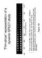
- the period between a certain repetitive wave, such as R-wave, and a subsequent waveis divided into several time segments, called “frames”, which are usually spaced evenly.
- Each photon which is detected by the PET detectors during one of the framesis collected and associated with the related frame.
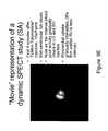
- each frameIn gated imaging, each frame generates a single dataset. The collection of all the datasets belonging to all the frames are defined as a “dynamic” dataset.
- the dynamic datasetis created by dividing the time span between one R-wave to the next R-wave into M frames that usually have an identical duration. Each detected photon is accumulated into a dataset of one of the M frames. Each dataset of the M datasets contains data relevant to a defined portion (“snapshot”) within the cardiac cycle.
- each one of the gated datasets of the M framesis processed independently by a suitable reconstruction algorithm, see Leahy R et al., Computer tomography in: Handbook of Image and Video Processing, BovikA, Academic press, 2000, pp. 771-787; J. Kay. The EM algorithm in medical imaging, Stat. Meth. Med. Res., 6(1):55-75, January 1997; J. A. Fessler, Statistical image reconstruction methods for transmission tomography, Handbook of Medical Imaging, Volumetric region 2, pages 1-70. SPIE, Bellingham, 2000; R. M. Leahy et al., Statistical approaches in quantitative positron emission tomography, 10(2):14765, April 2000; M.
- a common practice in gated SPECT reconstructionis to divide the gated dynamic dataset into M ‘non-gated’ data sets. Each one of the datasets includes data from a single frame i. The reconstruction of each volumetric region is performed independently using the relevant data set.
- the datais processed to reconstruct the intensity distribution within the measured volumetric region.
- the reconstruction processis generally complex, due to the large quantity of data that must be processed in order to obtain an accurate reconstruction.
- the following prior art statistical modelmay be used to perform reconstruction.
- Ian intensity distribution, defined over an input overall volume U, where U denotes a set of basic elements, such as pixels in two dimensional overall volumes and voxels in three dimensional overall volumes, and I(u) is the intensity of a given basic element u ⁇ U.
- the geometrical and physical properties of the detecting unit, together with its position and orientation in a given measurement t,determine the detection probability ⁇ t(u) of a photon emitted from location u in time t.
- the effective intensity of location u as viewed by the detecting unit during measurement tis ⁇ t(u)I(u).
- the random count Xt(u) of photons that are emitted from location u and detected in measurement tis modeled by a Poisson process with mean ⁇ t(u)I(u).
- the Radon transformis not statistical and does not take into account the Poissonian nature of the counts. In addition, it models the views as line projections.
- the Radon transformmaps the spatial domain (x,y) to the Radon domain (p, ⁇ ). For a fixed projection angle, the Radon transform is simply a projection of the object.
- FBPfiltered back-projection
- the basic, idealized problem solved by the FBP approachis to reconstruct an image from its Radon transform.
- the Radon transformwhen properly defined, has a well-defined inverse.
- In order to invert the transformone needs measured data spanning 180°.
- the positioning of the detecting unit relative to the emitting objectis constrained, so that complete measured data is not available.
- Reconstruction based on filtered back-projectionis therefore of limited use for medical imaging.
- Maximum likelihood (ML) and Maximum A Posteriori (MAP) estimation methodswhich address the statistical nature of the counts, have been found to provide better image reconstructions than FBP.
- Limited-angle tomographyis a reconstruction technique in the related art which reconstructs an image from projections acquired over a limited range of angular directions. The success of the reconstruction process depends upon the extent of the angular range acquired compared with the angular range of the missing projections. Any reconstruction from a limited range of projections potentially results in spatial distortions (artifacts) in the image. Limited angle techniques can be applied for both the Radon transform and the statistical models, but better results are generally achieved within the statistical framework. While it is known that the severity of the artifacts increases with the increasing angular range of the missing projections, limited-angle tomography does not provide information on which projections should be used in order to most effectively reconstruct the image.
- ML estimationis a widely used method in the related art for reconstructing an image from a constrained set of measurements.
- a parameterization of the generative model described aboveis obtained by assigning an intensity I(u) to every voxel in U.
- Eqn. 1For the maximum of the likelihood function.
- optimization methodsthat find local maxima of the likelihood are known.
- One such methodis the Expectation-Maximization (EM) process.
- a basic ingredient of the EM formalismis to define a set of random variables that completely define the data generated by the model.
- the main tool in the EM formalismis the complete data likelihood:
- I ⁇ ( u )1 ⁇ t ⁇ ⁇ t ⁇ ( u ) ⁇ ⁇ t ⁇ E ⁇ [ x t ⁇ ( u ) ⁇ y t ; I ′ ] ; ⁇ u ⁇ U . ( 4 )
- Equation 4The expectation in Equation 4 is obtained as follows:
- I ⁇ ( u )1 ⁇ t ⁇ ⁇ t ⁇ ( u ) ⁇ ⁇ t ⁇ y t ⁇ ⁇ t ⁇ ( u ) ⁇ I ′ ⁇ ( u ) ⁇ v ⁇ ⁇ t ⁇ ( v ) ⁇ I ′ ⁇ ( v ) ( 6 )
- each EM iterationimproves the likelihood.
- the EM algorithmiterates the above improvement step until it converges to a local maximum of the likelihood.
- Several random startsincrease the chance of finding a globally good estimator.
- Equation 8can be written in vector and matrix notations as:
- I⁇ + I ′ ⁇ ( ⁇ T ⁇ y ⁇ ⁇ ⁇ I ′ ) ⁇ + ⁇ T ⁇ 1 ( 10 )
- Limited computational resourcesmay require breaking the update computation according to a partition of ⁇ into a set of sub-matrices ( ⁇ i). In that case the intensities can be updated gradually (using only one sub-matrix at each step) according to the following computation:
- I⁇ + I ′ ⁇ ⁇ i ⁇ ⁇ i T ⁇ y t ⁇ i ⁇ I ′ ⁇ + ⁇ i ⁇ ⁇ i T ⁇ 1 ( 11 )
- yidenotes the vector of observations that are obtained using the views of ⁇ i.
- a high-resolution image of the tested objectIn order to achieve a reconstructed image which is adequate for medical diagnostic and treatment purposes, a high-resolution image of the tested object must be obtained.
- high-resolution detecting unitsWhen high-resolution detecting units are used, their efficiency is relatively low, and the detecting units must remain at each position for a relatively long time in order to achieve a high probability of detection. Since during medical testing, measurements are generally performed at many locations as the detecting unit is moved relative to the observed organ, the testing procedure generally requires a long time and is physically and emotionally difficult for the patient. Additionally, reconstruction is based upon a large quantity of data, and is a lengthy and computationally complex process.
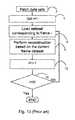
- FIG. 13is a schematic flowchart that illustrates steps of a typical prior art gated image reconstruction method.
- idenotes a frame counter and M denotes the number of frames.
- Mdenotes the number of frames.
- the processing unitis used to perform a frame reconstruction according to the dataset that corresponds with frame i using any suitable reconstruction algorithm known in the art, such as the aforementioned EM algorithm, ordered subset expectation maximization (OSEM), and algebraic reconstruction techniques (ART) FBP.
- any suitable reconstruction algorithmknown in the art, such as the aforementioned EM algorithm, ordered subset expectation maximization (OSEM), and algebraic reconstruction techniques (ART) FBP.
- the frame counter iis incremented by 1. If i is larger than M and there are no more frame the process ends. If i is not larger than M, the next dataset that corresponds with the subsequent frame is loading for reconstruction. In such a manner, the generation of a static imaging of the heart in a specific configuration becomes possible.
- a method for iteratively reconstructing a volumetric image of an overall volume from radioactive emissionscomprising:
- the overall volumecomprising at least a part of a body organ or other body portion
- the modelincluding a general location and shape of the at least a part of a body organ or other body portion and an expected number of photons emitted from the at least a part of a body organ or other body portion;
- the object implantationis used one or more times during the iterative process, each time for providing a better starting point for performing a next iteration of the iterative process, whereby the improved estimation is used to redistribute photon counts in an iteration.
- Implementation of the method and system of the present inventioninvolves performing or completing certain selected tasks or steps manually, automatically, or a combination thereof.
- several selected stepsmay be implemented by hardware or by software on any operating system of any firmware or a combination thereof.
- selected steps of the inventionmay be implemented as a chip or a circuit.
- selected steps of the inventionmay be implemented as a plurality of software instructions being executed by a computer using any suitable operating system.
- selected steps of the method and system of the inventionmay be described as being performed by a data processor, such as a computing platform for executing a plurality of instructions.
- FIGS. 1A-1Dschematically illustrate a dynamic SPECT camera, in accordance with embodiments of the present invention
- FIGS. 2A and 2Bschematically illustrate the camera structure with the assemblies, in accordance with an embodiment of the present invention.
- FIGS. 3A-3Dschematically illustrate viewing positions, in accordance with embodiments of the present invention.
- FIGS. 4A-4Fschematically illustrate stereo views and cross views, in accordance with embodiments of the present invention.
- FIGS. 5A and 5Billustrate experimental radiopharmaceutical data, as known
- FIGS. 5C-5Fillustrate cardiac gating, in accordance with embodiments of the present invention.
- FIG. 6A-6Iillustrate an intracorporeal dynamic SPECT camera, in accordance with embodiments of the present invention
- FIG. 7illustrates assembly-damping parameters, in accordance with embodiments of the present invention.
- FIGS. 8A and 8Bschematically illustrate grid and anatomical construction of voxels, in accordance with embodiments of the present invention
- FIGS. 9A-9Jpresent experimental data, obtained by the dynamic SPECT camera, in accordance with embodiments of the present invention.
- FIG. 10presents experimental data, obtained by the dynamic SPECT camera, in accordance with embodiments of the present invention.
- FIG. 11illustrates components of the dynamic SPECT camera, in accordance with embodiments of the present invention.
- FIG. 12illustrates an electrical scheme, in accordance with embodiments of the present invention.
- FIG. 13is a schematic flowchart that illustrates steps of a typical prior art gated image reconstruction method
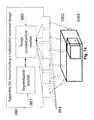
- FIG. 14is a schematic illustration of an apparatus for reconstructing a radioactive emission image of an input overall volume having dynamic and static volumetric regions, according to a preferred embodiment of present invention
- FIG. 15is a schematic isometric view of the input overall volume that is depicted in FIG. 14 , according to one embodiment of the present invention.
- FIG. 16is a schematic cross-sectional view of the input overall volume of FIG. 15 , according to one embodiment of the present invention.
- FIG. 17is a flowchart that depicts a method for reconstruction an input overall volume using anatomically varying time-bin lengths, according to one embodiment of the present invention.
- FIG. 18is a graphical representation of a one-dimensional vector of voxels that represents the reconstruction of the input overall volume, according to one embodiment of the present invention.
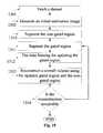
- FIG. 19is another flowchart that depicts another method for reconstruction an input overall volume using anatomically varying time-bin lengths, according to another embodiment of the present invention.
- FIG. 20is a graphical representation of a position of two selected sub-regions in two sequential frames, according to another embodiment of the present invention.
- the present inventionrelates to an apparatus and a method for reconstructing a radioactive emission image of a overall volume having dynamic and static volumetric regions.
- the reconstructingis based on gated images with anatomically varying time-bin lengths.
- the apparatus and the methodare designed to allow the segmentation of the radioactive emission image to gated and non-gated regions. In such a manner, the reconstructions radioactive emissions from the dynamic volumetric region and the static volumetric region are carried out separately.
- the high computational throughput that is needed in order to reconstruct a dynamic volumetric region, such as the heart, using time binning techniqueshas less or no effect on the reconstruction of the static volumetric region, as further described below.
- the disclosed apparatuscomprises a number of detectors, such as PET or SPECT detectors, which are designed for obtaining radioactive emissions from the overall volume and an image reconstruction module that is designed for generating radioactive emission images of the overall volume according to the obtained radioactive emissions.
- the apparatusfurther comprises a segmentation module that is designed for segmenting an initial radioactive emission image to gated and non-gated regions, according to the dynamic and static volumetric regions of the overall volume.
- the image reconstruction moduleis designed to reconstruct separately the gated and non-gated regions in the radioactive emission image respectively according to radioactive emissions the dynamic and static volumetric regions.
- the method for reconstructing a radioactive emission image of a overall volume having static and dynamic volumetric regionscomprises several steps. During the first step, radioactive emissions are obtained from the overall volume. Then, an initial radioactive emission image of the overall volume is reconstructed according to the radioactive-emission data. In the following step, the initial radioactive emission image is segmented to gated and non-gated regions, respectively according to the dynamic and static volumetric regions. During the last step, the radioactive emission image is reconstructed, wherein the gated region is according to radioactive emissions from said dynamic volumetric region and the non-gated region is separately reconstructed according to radioactive emissions from the static volumetric region.
- only the dynamic volumetric regionis reconstructed using time binning.
- the static volumetric regionis reconstructed according to the initial radioactive emission image.
- time binning of different anatomical segmentshas dynamically varying time-bin lengths, as further described below.
- FIG. 14is a schematic illustration of an apparatus 990 for reconstructing a radioactive emission image of an input overall volume 1002 having dynamic 1003 and static 1002 volumetric regions, according to a preferred embodiment of present invention.
- the input overall volume 1002is the thorax
- the static volumetric regionis the related viscus
- the dynamic volumetric regionis the heart and the area that confines it.
- the apparatus 990which is preferably a SPECT camera, has a number of detecting units 993 , such as SPECT detectors. Each one of the detectors 993 is designed for obtaining radiation emission that is emitted from the input overall volume 1002 , as described below, and for generating accordingly radioactive-emission data.
- the apparatus 990comprises an image reconstruction module 992 that is connected to the detecting units 993 .
- the image reconstruction module 992is designed for generating radioactive emission images according to the radioactive-emission data.
- the imagesare preferably gated, as described below.
- the apparatus 990further comprises a segmentation module 991 that is designed for segmenting an initial radioactive emission image, which has been generated by the image reconstruction module 992 , to gated and non-gated regions according to the dynamic and static volumetric regions.
- the gated and non-gated regionsare used by the image reconstruction module 992 for separately reconstructing the dynamic and static volumetric regions, as further described below in the anatomically varying time-bin lengths section.
- the apparatus 990is a dynamic SPECT camera.
- a dynamic SPECT camerawith temporal and spatial resolutions, which meet and even outperforms those of PET, and with a high spectral resolution not available in PET is given.
- Temporal resolutionrelates to a minimal acquisition time for a tomographic reconstruction image of a predetermined volumetric region, for example 15 ⁇ 15 ⁇ 15 cubic centimeters, and predetermined spatial resolution, for example, 10 ⁇ 10 ⁇ 10 cubic millimeters.
- the minimal acquisition timemay be, for example, 30 seconds, 10 seconds, or 1 second.
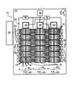
- FIGS. 1A-1Dschematically illustrate a dynamic SPECT camera 10 that is configured for capturing gated images and non-gated image, in accordance with embodiments of the present invention.
- the dynamic SPECT camera 10comprises: an overall structure 15 , which defines proximal and distal ends and, with respect to a body 100 ; a number of assemblies 20 , for example, 6, 9, or 16 assemblies 20 , arranged on the overall structure 15 , forming an array 25 of the assemblies 20 .
- Each one the each assemblies 20comprises a number of detecting units 12 .
- Each detecting unit 12includes a single-pixel detector 14 for detecting radioactive emissions and a dedicated collimator 16 , attached to the single-pixel detector 14 , at the proximal end thereof, for defining a solid collection angle ⁇ for the detecting unit 12 .
- each assembly 20comprises an assembly motion provider 40 , configured for providing the assembly 20 with individual assembly motion, with respect to the overall structure 15 , during the acquisition of radioactive-emission data for a tomographic image.
- the dynamic SPECT camera 10further includes a timing mechanism 30 , in communication with each single-pixel detector 14 , configured for enabling time binning of the radioactive emissions impinging upon each single-pixel detector 14 to periods, which are not greater than substantially 30 seconds.
- a timing mechanism 30in communication with each single-pixel detector 14 , configured for enabling time binning of the radioactive emissions impinging upon each single-pixel detector 14 to periods, which are not greater than substantially 30 seconds.
- the timing mechanism 30has can control each one of the single-pixel detector 14 separately, each one of the single-pixel detectors 14 can be configured according to a different time binning scheme.
- the time binning schemewhich is applied to a certain detector, is determined according to the region in the input overall volume that the detector is designed to detect.
- the dynamic SPECT camera 10further includes a position tracker 50 , which is designed for providing information on the position and orientation of each detecting unit 12 , with respect to the overall structure 15 , substantially at all times, during the individual assembly motion.
- the dynamic SPECT camera 10is configured for acquiring a tomographic reconstruction image of a region of interest of about 15 ⁇ 15 ⁇ 15 cubic centimeters, for example, of a target organ 110 , such as a heart or a stomach, during an acquisition period no greater than 300 seconds, at a spatial resolution of at least 10 ⁇ 10 ⁇ 10 cubic millimeters.
- time periodmay be no greater than 200 seconds, 100 seconds, 60 seconds, 30 seconds, 10 seconds, or 1 second.
- the dynamic SPECT camera 10is configured for acquiring a series of tomographic reconstruction images of a region of interest, as a function of time, at a rate of at least a tomographic reconstruction image every 300 seconds.
- the ratemay further be every 200 seconds, 100 seconds, 60 seconds, 30 seconds, 10 seconds, or 1 second.
- the individual assembly motionmay be, for example, an assembly oscillatory sweeping motion, as described by an arrow 60 . Additionally or alternatively, the individual assembly motion may be a first oscillatory lateral motion, as described by an arrow 80 . Additionally or alternatively, the individual assembly motion may be a second oscillatory lateral motion, orthogonal to the first, as described by an arrow 90 .
- the assembly motion provider 40may comprise between one and three motion providing units, for the different assembly motions.
- the individual assembly motionis an assembly oscillatory sweeping motion, as described by an arrow 60
- the array 25moves with either the first or the second oscillatory lateral motions, described by the arrows 80 and 90 , or with both.
- the detecting units 12may be grouped into square or rectangular blocks 18 , for example, of 4 ⁇ 4 detecting units 12 , as seen in FIG. 1A , or of 16 ⁇ 16, 64 ⁇ 64, 64 ⁇ 128 or another number of detecting units 12 .
- the blocks 18may be provided with individual block oscillatory sweeping motion, as described by an arrow 70 , with respect to the overall structure 15 , during the acquisition of radioactive-emission data for a tomographic image.
- the block oscillatory sweeping motionis orthogonal to, or at an angle to the assembly oscillatory sweeping motion, described by the arrow 60 .
- the assembly motion provider 40may further comprise a dedicated block motion providing unit, in communication with each block of an assembly.
- a control unit 55may be integrated with the dynamic SPECT camera 10 , to form a single physical unit, or in communication with the dynamic SPECT camera 10 .
- a spectral selection mechanism 56in communication with each of the detecting unit 12 , is discussed hereinbelow, under the heading, “dynamically varying spectral bins.”
- the body 100may be a human or an animal, and the region of interest, or the target organ 110 may be a heart, a brain, a breast, a stomach, a GI tract, a colon, a prostate, a uterus, a cervix, a vagina, a throat, a gland, a lymph node, a portion of skin, a portion of bone, portion of another tissue, or another body portion.
- a reference x;y;z coordinate systemillustrates a preferred orientation of the dynamic SPECT camera 10 with respect to the body 100 , wherein z runs along a length of the body 100 .
- the assembly axis along the assembly lengthwill be referred to as the assembly longitudinal axis
- the assembly axis along the assembly widthwill be referred to as the assembly traverse axis.
- the assemblies 20are long and narrow columns, arranged longitudinally against the body 100 , wherein the oscillatory sweeping motion, described by an arrow 60 , is about the z-axis. It will be appreciated that other arrangements are similarly possible.
- the assemblies 20are arranged in an arc or an arc-like structure, about the body 100 , maintaining a shape that follows the body contours, so as to keep as close as possible to the body 100 .
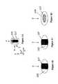
- FIG. 1Dprovides details of the detecting unit 12 .
- the collimatorhas a length L, a collection angle ⁇ , and a septa thickness ⁇ .
- the single pixel detectoris preferably a square of sides D and a detector thickness ⁇ d.
- Preferred dimensions for the detecting unit 12may be, for example, 2.46 mm ⁇ 2.46 mm, and the solid collection angle ⁇ may be at least 0.005 steradians. Generally, there may be 16 ⁇ 64 detecting units 12 per block 18 .
- the detector 14is preferably, a room temperature, solid-state CdZnTe (CZT) detector, which is among the more promising that is currently available. It may be obtained, for example, IMARAD IMAGING SYSTEMS LTD., of Rehovot, ISRAEL, 76124, www.imarad.com, or from eV Products, a division of II-VI Corporation, Saxonburg Pa., 16056, or from or from another source.
- CZTsolid-state CdZnTe
- another solid-state detectorsuch as CdTe, HgI, Si, Ge, or the like, or a combination of a scintillation detector (such as NaI(Tl), LSO, GSO, CsI, CaF, or the like) and a photomultiplier, or another detector as known, may be used, preferably with a photomultiplier tube for each single-pixel detector 14 and collimator 16 , for accurate spatial resolution.
- a scintillation detectorsuch as NaI(Tl), LSO, GSO, CsI, CaF, or the like
- a photomultiplieror another detector as known, may be used, preferably with a photomultiplier tube for each single-pixel detector 14 and collimator 16 , for accurate spatial resolution.
- FIGS. 2A and 2Bschematically illustrate the structure 15 with the assemblies 20 , in accordance with an embodiment of the present invention.
- the assemblies 20are arranged in an arc of an angle ⁇ , around the body 100 , and move in the assembly oscillatory sweeping motion, about the z-axis, so as to provide a plurality of views of the heart 110 , from many positions, along the x-y plane.
- the dynamic camera 10is configured for simultaneous acquisition by the assemblies 20 , each scanning the same region of interest from a different viewing position, thus achieving both shorter acquisition time and better edge definitions.
- the structure 15conforms to the contours of the body 100 , to maintain substantial contact or near contact with the body.
- FIGS. 2A and 2Billustrates a single type of motion-assembly oscillatory sweeping motion about the z-axis, as described by the arrow 60 ( FIG. 1A ).
- additional motions or views from additional directionsmay be desirous, as illustrated in FIGS. 3A-3D , hereinbelow.
- FIGS. 3A-3Dschematically illustrate viewing positions, in accordance with embodiments of the present invention.
- FIG. 3Aillustrates a cylindrical target organ 110 , with a cylindrical radioactive emission source 115 therein.
- a view along the y-axiswill similarly observe the cylindrical radioactive emission source 115 as a bar 115 , thus not adding new information to the view along the x-axis.
- any view along the x-y planewill observe the radioactive emission source 115 as a bar 115 .
- a view along the z-axiswill observe the cylindrical radioactive emission source 115 as a circle 115 , adding new information to the views along the x and y axes.
- views along two axesmay be insufficient for a three-dimensional definition of an object, and it may be beneficial to include views with a component along the third axis.
- views along two axeswill be referred to stereo views, while views that include a component of the third axis will be referred to as cross views, since they intersect the planer stereo views.
- FIGS. 4A-4Fschematically illustrate stereo views and cross views, in accordance with embodiments of the present invention.
- FIG. 4Aillustrate the body 100 with a single assembly 20 arranged for viewing, for example, the heart 110 .
- the assembly 20is afforded with assembly oscillatory sweeping motion along the z-axis, as described by the arrow 60 , and preferably first and second preferably orthogonal oscillatory lateral motions, described the arrows 80 and 90 , respectively.
- the assembly oscillatory sweeping motion along the z-axis, described by the arrow 60produces views 65 in the x-y planes.
- the first and second orthogonal oscillatory lateral motions, described the arrows 80 and 90augment these with additional views 65 in the x-y planes.
- the purpose of the first and second oscillatory lateral motionsis to compensate for “dead areas,” that is, structural areas and other areas that do not participate in the detection, within the assembly 20 and between the assemblies 20 , so as to provide complete coverage of the body 100 , by the array 25 ( FIG. 1A ).
- These motionsproduce views substantially in the x-y plane. It will be appreciated that there is a component of viewing in a third axis, due to the solid collection angle of the collimator 16 . Yet this component is rather small.
- the blocks 18 of the assembly 20may be further afforded with block oscillatory sweeping motion, described by the arrow 70 and preferably orthogonal to the assembly oscillatory sweeping motion described by the arrow 60 .
- the block oscillatory sweeping motiondescribed by the arrow 70 , produces cross views 75 , which supplement views 65 , by providing components of the third axis, namely, the z-axis.
- the views 75may add additional information, not available or barely available in the views 65 along the x-y planes.
- FIGS. 4D and 4Fillustrate an alternative mode for acquiring the cross views 75 .
- the dynamic camera 10further includes assemblies 22 , arranged at an angle ⁇ to the assemblies 20 , and moving with an assembly oscillatory sweeping motion, described by an arrow 62 , so as to provide the cross views 75 .
- the detectors of the dynamic camera 10do not have to be arranged in arrays.
- the detectorsare scattered in front of the body so as to provide complete coverage of the body internal overall volume.
- the detectorscan be scattered in a certain structure or in an arbitrary order.
- the position tracker 50is configured for providing information on the position and orientation of each detecting unit 12 , with respect to the overall structure 15 , substantially at all times, during the individual assembly motion.
- the position tracker 50relates to software and (or) hardware that receive information from the motion provider 40 and calculate the position and orientation of each detecting unit 12 , based on that information. Preferably, the calculation is performed within the control unit 55 .
- position sensorsas known, may be used for determining the position and angular orientation of each detecting unit 12 .
- the timing mechanism 30associates timing information with the radioactive emission data impinging the single-pixel detectors 14 of the detecting units 12 .
- the timing mechanism 30includes a single clock used for all of the single-pixel detectors 14 in the dynamic SPECT camera 10 , so that timing information is synchronized for the camera as a whole.
- the timing informationis collected at the single-pixel level, so that time binning may be performed for the emission data collected by each pixel.
- Exemplary methods for associating timing information with the radioactive emission datainclude:
- Time stampingEach event, impinging on a given single-pixel detector 14 at a given time is stamped with a time of detection and a pixel identification. Stamping may be performed by any manner known in the art, for example as a data packet header or footer. The time-stamped, pixel stamped radioactive emission data may be binned, per time and per pixel, by the control unit 55 .
- Time binningIn an alternate approach, timing information is provided for a cumulative count collected from each single-pixel detector 14 over a fixed time interval, for example, 0.001 seconds, 1 second, or 10 seconds, rather than for individual events. Each time bin is then stamped with a time stamp or sequential number and pixel identification.
- One technique for performing time binningis to insert a periodic clock pulse into the data stream. The interval between the clock pulses equals the minimum bin length. Thus, periodic pulses every 0.001 seconds may lead to bin lengths of 0.001 seconds or greater, for example, 1 second, or 10 seconds.
- the timing Mechanism 30is used by the reconstruction module in order to allow the separate reconstruction of the dynamic and static volumetric regions.
- the timing Mechanism 30allows the reconstruction module to apply a time binning with a certain length on the dynamic volumetric region and a time binning with another length on the static volumetric region.
- Dynamic studiesaimed at obtaining kinetic parameters, require the acquisition of full-reconstructed images at a rate that is no greater than about half the frequency of the sampled kinetic parameter.
- bloodcirculates through the body at a rate of about 1 cycle per minute.
- sampling a process affected by blood circulationshould take place at a rate of at least two samplings per minute.
- samplingshould be at a much greater rate, for example, 6 samplings or 10 samplings per minute—that is, about every 10 seconds or about every 6 seconds.
- the dynamic behavior of a radiopharmaceutical in the bodyvaries as a function of time, depending on the radiopharmaceutical and on the time elapsed since its administration.
- myocardial perfusion of Tc-99m teboroximeshows a very steep uptake between about the first 10-15 seconds and the first 50-60 seconds, followed by a more gradual washout, after the first 60 seconds.
- the rate of sampling of Tc-99m teboroxime, during the first 60 seconds after administrationshould be adjusted to the very steep uptake, for example, a sampling rate of every second. For radiopharmaceutical with a slower dynamic behavior, a slower rate may be sufficient.
- a third unitfor example, a control system or a hospitals ERP system
- This methodmay be employed for example, when administration takes place at a different location than the imaging station.
- a markerfor example, a line of radioactive ink may drawn, for example, on the patient's arm or on the administration device, for defining the time of administration as the time the radiopharmaceutical first crosses the marker.
- observing a flow of the radiopharmaceutical in the administration device or in the patient's veinmay be used to determine the time of administration.
- Observing a transparent administration devicefor example, with a video camera, associated with a clock, may be employed for defining a time of administration based on the radiopharmaceutical distribution in the administration device, or based on the time the radiopharmaceutical first crosses a marker, visible by the video camera.
- Communication between the video camera and the dynamic SPECT camera 10 , or between the video camera, the dynamic SPECT camera 10 , and a third unitwill provide the information to the dynamic SPECT camera 10 .
- the dynamic SPECT camera 10is designed at least for acquiring a tomographic reconstruction image of about 15 ⁇ 15 ⁇ 15 cubic centimeters, which is approximately the volumetric region of a heart, at a predetermined spatial resolution of at least 10 ⁇ 10 ⁇ 10 cubic millimeters, at an acquisition time no greater than about 30 seconds.
- the acquisition timeis no greater than about 10 seconds, and more preferably, the acquisition time is no greater than about 1 second.
- the spatial resolution of the tomographic reconstruction imagemay be at least 7 ⁇ 7 ⁇ 7 cubic millimeters, or better yet, at least 4 ⁇ 4 ⁇ 4 cubic millimeters, or better still, at least 1 ⁇ 1 ⁇ 1 cubic millimeters.
- the time binningis needed in order to generate a clear imaging of a dynamic organ, such as the heart, or a section thereof.
- the time binningallows the acquisition of a clear image of the heart, it has at least one major disadvantage.
- the reconstruction of the image using time binningrequires high computational throughput.
- binning images of the input overall volumemay provide a clear imaging of the heart however have high computational throughput.
- Reconstruction using anatomically varying time-bin lengthscan be used to reduce to computational throughput of the time binning.
- the static regiondoes not have to be gated to provide a clear imaging, only the dynamic region that preferably contains the heart is gated. In such an embodiment, fewer voxels are gated and therefore the computational complexity is reduced.
- different areas in the bodycan be gated in a rate that is adjusted to according to a respective level of activeness.
- the heart that has high level of activenesscan be gated using a large number of bins
- the visceral backgroundwhich is relativity static
- the stomachthat have higher level of activeness than the visceral background but lower level of activeness than the heart is gated using a limited number of bins.
- performing gated image reconstruction using anatomically varying time-bin lengthsimproves the reconstruction quality, reduces the reconstruction time, or both.
- the improvementis an outcome of a reduction in the needed computational resources.
- FIGS. 15 and 16are respectively a schematic isometric view of the input overall volume 1001 segmented into dynamic volumetric region and static volumetric regions 1003 , 1002 , as depicted in FIG. 14 , and a schematic cross-sectional view of the segmented input overall volume 1001 A taken along the lines I 11 -I 11 , according to one embodiment of the present invention.
- a radioactive emission image of the input overall volume 1001is segmented into a non-gated region, which includes non-gated voxels, in accordance with the static volumetric region 1002 , and to a gated region, which includes gated voxels, in accordance with the dynamic volumetric region 1003 .
- the dynamic volumetric region 1003is adjusted to delimit a dynamic organ, such as the human heart that is schematically represented by a hollow sphere 1004 .
- the dynamic volumetric region 1003is larger than the apparent volumetric region of the heart 1004 to account for segmentation errors.
- the dynamic volumetric region 1003may be adjusted to contain other human and animal internal organs such as the stomach.
- the hatched region 1003 Arepresents the cross-section of the dynamic volumetric region 1003 and the annular crosshatched region 1004 A schematically represents a cross-section through the heart muscle of the heart 1004 .
- the region 1002 Arepresents a cross section of the static volumetric region 1002 .
- the cubical shape of the dynamic volumetric region 1003 and the static volumetric region 1002are not obligatory and the segmentation to regions may be performed using differently shaped volumetric regions.
- the dynamic and the static volumetric regions 1003 , 1002have several non-connected parts.
- the dynamic volumetric region 1003may be a spherical volumetric region, an ellipsoid of revolution, an ellipsoid with a hole that represents the blood inside the heart, a cylindrical volumetric region or any other type of suitable regularly shaped or non-regularly shaped volumetric region.
- the dynamic volumetric region 1003comprises non-connected components, which may be referred to as sub-volumetric regions.
- FIG. 15is a flowchart that depicts a method for reconstruction an input overall volume using anatomically varying time-bin lengths, according to one embodiment of the present invention.
- each gated imageis generated by a photon counting that takes into account the portion of the heart contraction cycle within which a photon is measured.
- the number of photons hitting the detector within a specific integration timeis calculated and used as raw data, which may be referred to as datasets.
- the captured datasetsare firstly used in an initial reconstruction process in which an initial estimation image is generated.
- a non-gated reconstructionis used to provide a reconstruction that estimates the static intensity distribution.
- the initial estimation imageis segmented to a gated region and a non-gated region that respectively define the boundaries of the dynamic and static volumetric regions.
- the segmentation of the input overall volume 1001 to dynamic and static volumetric regions 1003 , 1002is performed using a suitable image segmentation method or process.
- the input overall volume 1001is further segmented to one or more other segments such as the liver.
- segmentsmay be joined to the dynamic or to the static volumetric regions 1003 , 1002 according to the nature of the activity level of the segment.
- the livermay be joined to the static volumetric region.
- a system usermarks the boundaries of the dynamic volumetric region that comprises the gated voxels.
- the reconstructed imageis displayed on a screen of a user interface that allows the system user to delimit the dynamic volumetric region.
- the captured imageis blurry, as it is not gated, it provides the system user a perceptual image of the outlines of the internal organs in the input overall volume, including the heart, the liver, the spleen, the kidneys, and the aorta.
- the system usersegments the captured image to gated and non-gated regions according to their level of activity, thereby defines the gated and non-gated regions.
- the system usersegments the heart as a non-gated region.
- the segmentationis based on a voxel value threshold that reflects a certain percentage of the maximal reconstruction value.
- voxels of the reconstructed image having a value above the thresholdare presumed to be voxels that depicts the heart and tagged as gated voxels of the dynamic volumetric region and voxels of the input overall volume 1001 having a value below the threshold are tagged as non-gated voxels of the static volumetric region.
- regions in the captured imageare segmented according to predefined characteristics. For example, the liver region, which can be characterized as a very large segment residing in the lower part of an image that depicts the thorax, is identified and segmented as a static volumetric region 1003 or a section thereof.
- the predefined thresholdis defined according to the radiation intensity of the visceral background of the input overall volume 1001 .
- the radiation intensity of the visceral backgroundis estimated before the segmentation process.
- Such an initial estimationcan be performed using median or linear filters such as Gaussian and moving average filters.
- Each one of the voxels of the input overall volume 1001 with a value that is well above the estimated background radiationis tagged as a gated voxel.
- Each one of the voxels of the input overall volume 1001 with a value, which is below the estimated background radiationis tagged as a non-gated voxel.
- the segmentationis performed according to morphological segmentation methods that adjusted according to the volumetric characteristics of the segmented volumetric regions. For example, for the heart that has convex faces can be segmented using top hat transform.
- the segmentationis performed according to the growing rate of regions of the input overall volume 1001 .
- regionssuch as the heart may be indented.
- voxels having high growing rateare clustered as a group of voxels that depicts the heart.
- the faces of the heartare identified. Such identification may be performed using an objective function with two parts. One part of the objective function is dependent on the organ border smoothness alone and the other part is dependent on the edge strength near a defined border, see M. Kass, A. Witkin, and D. Terzopoulos. Snakes: active contour models, International Conference on Computer Vision, pages 259-268, 1987, which is incorporated in its entirety by reference into the specification.
- Z dyn (u) and Z stat (u)respectively denotes the dynamic volumetric region and the static volumetric region of the captured image.
- the dynamic and static volumetric regionsrespectively define the boundaries of gated and non-gated regions in the radioactive emission image that depicts the input overall volume. It should be noted that though only two volumetric regions are exemplified hereinbelow, the overall volume may be segmented according to any number of volumetric regions such as three volumetric region, four volumetric region, ten volumetric region etc.
- the computational load of the reconstructionmay be reduced by using large voxels with a low resolution in the static volumetric region 1002 and small voxels in the gated volumetric region.
- various morphological methodssuch as, dilation, closing and the like are used after the initial segmentation to expand the dynamic volumetric region 1003 .
- the broadening of the dynamic volumetric region 1003is done in order ensure that if the segmentation has been made according to an organ in a contracted state, the dynamic volumetric region 1003 still encompasses the organ in an expanded state.
- time binning of the dynamic volumetric region of the input overall volumeis performed and a separate reconstruction of the static and the dynamic volumetric regions is enabled.
- the reconstructionis based on an iterative process in which the time binning of gated images of the dynamic volumetric region is enabled.
- I 0 (u)denotes an input image I, which is preferably constant, that depicts u ⁇ U voxels
- tdenotes a certain detector
- gdenotes a certain gate in a set of G gates, such as 8, 16, and 24
- ⁇ t (u)denotes a standard functional matrix that depicts the detection probability of a photon emitted from location u ⁇ U to be detected by detector t
- s tdenotes the sensitivity of the detector t
- T t gdenotes the integration time of detector t for gate g
- I g (u)denotes a set of G gated reconstructed images
- y t gdenotes the number of photons that are emitted from voxel u and detected in detector t at gate g.
- Z dyn (u) and Z stat (u)respectively denote static and dynamic regions in I, as defined in the aforementioned segmentation process.
- Z dyn (u) and Z stat (u)are defined as follows:
- Z dyn (u) and Z stat (u), which respectively confine the static and dynamic volumetric regions,are defined at step 1302 .
- the reconstruction of the input overall volume according to time binning processcommences.
- I stat (u) and I dyn g (u)are calculated for each voxel u ⁇ U in the input overall volume.
- the gated and non-gated regions that represent the static and dynamic images I stat (u), I dyn g (u)are updated.
- the updating of the regionsis calculated according to a deviation between the number of photons that has been detected by the SPECT detectors and an estimation of this number, as described below.
- the gated voxels of the dynamic volumetric regionare binned according to the number of gates and the non-gated voxels are binned only once. As the non-gated voxels are binned only once, the computational complexity of the process is relatively low. The separation between the static and dynamic volumetric regions improves the computational efficiency and reduces the statistical variance.
- ⁇ stat,ts t T t ⁇ u ⁇ t ( u ) I stat ( u )
- ⁇ stat,tdenotes an estimation of the number of photons that are emitted from the voxels u ⁇ U and detected by detector t, wherein values of voxels from the dynamic volumetric region are zeroed. It should be noted that the sensitivity parameter of and the integration time of detector t are taken into account at some stage in the calculation.
- ⁇ dyn,t gs t T t g ⁇ u ⁇ t ( u ) I dyn g ( u )
- ⁇ dyn,t gdenotes an estimation of the number of photons that are emitted at gate g from voxels u ⁇ U and detected in detector t, wherein values of voxels from the static volumetric region are zeroed. It should be noted that the sensitivity parameter and the integration time of detector t for gate g are taken into account at some stage in the calculation.
- ⁇ t gdenotes an estimation of the number of photons that are emitted from a certain voxel u ⁇ U and detected by detector t at gate g. It should be noted that unlike the calculation of y t g , the calculation of ⁇ t g does not take into account the integration time and the sensitivity factor.
- num g (u)sums the deviation between the number of photons that are emitted from voxel u and detected in detector t at gate g and the estimation thereof of all the detectors, wherein the sensitivity and the integration time of each detector t are taken into account. It should be noted that the calculation can be directly extended to an ordered sets method or any of its variations by summing the deviation over subsets of the group of detectors.
- num(u)is a sum of all the numerators that are evaluated for every g ⁇ G.
- I stat (u) and I dyn g (u)are updated according to the calculation of the aforementioned scales and numerators, as follows:
- I stat ⁇ ( u )I stat ⁇ ( u ) + num ⁇ ( u ) scale ⁇ ( u ) ⁇ I stat ⁇ ( u )
- I dyn g ⁇ ( u )I dyn g ⁇ ( u ) + num g ⁇ ( u ) scale g ⁇ ( u ) ⁇ I dyn g ⁇ ( u )
- Step 1306The updated I stat (u) and I dyn g (u) are stored and used during the next iteration, as shown at step 1306 . Steps 1303 - 1306 are repeated iteratively until the reconstruction of the input overall volume has reached a desired quality.
- the number of gated voxels with activity above a predefined thresholdis checked.
- the number of gated voxels with activity level that is in the range between the maximal gated voxel intensity value and 20% therefromis checked.
- FIG. 18is a graphical representation of one dimensional vector I c of voxels that represents the reconstruction of the input overall volume.
- all the non-gated voxels I stat (u) that represent static regions of the input overall volume Uare arranged 1101 first within the vector I c .
- the non-gated voxelsare followed 1101 by gated voxels that comprise a set of different frames in a consecutive order that are arranged in clusters.
- Each clusterrepresents the dynamic volumetric region of the input overall volume at a certain frame.
- the framesare denoted by 1102 A, . . . , 1102 G.
- the framescan be arranged in any predefined order.
- ⁇ t (u)is a standard functional matrix that depicts the detection probability of a photon emitted from a voxel u ⁇ U to be detected by a detector t. Since ⁇ t (u) is a sparse matrix, the number of math operations can be reduced by defining ⁇ t g (u) which is zero wherever I dyn g (u) is zero.
- FIG. 19is a flowchart that depicts another method for reconstruction an input overall volume using anatomically varying time-bin lengths, according to another embodiment of the present invention.
- the static regionis estimated only once according to the initial reconstruction process step.
- the method depicted in FIG. 19is based on the assumption that the non-gated static region equals to the average of the gated dynamic region images reconstructions. Though the assumption is not accurate, it is expected to be sufficient for the reconstruction of the input overall volume. As the static region is calculated only once, the memory usage and the computational complexity decrease.
- Steps 1301 and 1302are as depicted in FIG. 17 .
- the first step I(u)is obtained.
- I dyn g ( u )I ( u ) ⁇ Z dyn ( u ), for each g ⁇ G
- ⁇ stat,ts t T t ⁇ u ⁇ t ( u ) I stat ( u )
- ⁇ t (u)is a representation of the probability to detect a photon emitted from location u ⁇ U by a detector t.
- the calculation of ⁇ t (u)requires high computational complexity as all the voxels of the input overall volume have to be calculated.
- ⁇ t,dyn (u)is used in order to reduce the computational complexity.
- the limited standard functional matrixis defined as follows:
- ⁇ t , dyn ⁇ ( u )⁇ ⁇ ⁇ t ⁇ ( u ) , ⁇ u ⁇ dynamic ⁇ ⁇ region ⁇ 0 , ⁇ otherwise
- I dyn g (u)is calculated.
- ⁇ t gdenotes an estimation of the number of photons that are emitted from a certain voxel u ⁇ U and detected by detector t at gate g. It should be noted that unlike ⁇ dyn,t g , ⁇ stat,t is not recalculated during the iterative process.
- I dyn g (u)is updated as follows:
- I dyn g ⁇ ( u )I dyn g ⁇ ( u ) + num g ⁇ ( u ) scale g ⁇ ( u ) ⁇ I dyn g ⁇ ( u )
- the updated I dyn g (u)is stored and used during the next iteration, as shown at step 1316 .
- the input overall volumeis reconstructed using the updated dynamic region and the static region.
- steps 1311 - 1314are repeated iteratively until the reconstruction of the input overall volume has reached a desired quality.
- the dynamic regionis updated, as described above.
- the gated voxels in the dynamic volumetric region 1003may represent ischemic regions of the heart.
- the radiation reflected from such ischemic regionsmay have specific radiation patterns such as a center with low radiation.
- regionscan be reconstructed using morphological closing methods or by taking into account the typical shape of the heart (one way is by fitting an ellipsoid to the edges in the image, but other methods may also be used).
- FIG. 20is a graphical representation of a position of two selected sub-regions in two sequential frames.
- the reconstruction of the dynamic volumetric regionis based on time binning of a number of consecutive frames that depict a dynamic organ such as the heart. As each frame is based on a number of gated images, it has high computational load.
- the set of framesis a set of sequential images that depict the heart. As all the frames depict the same input overall volume and as the heart has an expected movement pattern, we can use one or more frames to estimate another. In such a manner, fewer frames are calculated and therefore the computational complexity of the reconstruction decreases.
- geometrical information from one or more prior framesassist in the reconstruction of subsequent one or more frames. Such geometric prior methods are generally well known and therefore, are not described here in greater detail.
- FIG. 20depicts two sequential frames, frame i ⁇ 1 1054 and frame i 1056 , which are taken from a sequence of M frames.
- the sub-regions 1057 A and 1059 A in frame 54schematically represent two different regions of the heart 1055 .
- the regions 1057 B and 1059 B of frame 1056schematically represent the respective positions of regions 1057 A and 1059 A in frame 1056 .
- the change in the positionsis an outcome of the movement of the heart 1055 .
- the vector T 1represents the movement of the region 1057 B relative to the region 1057 A and the vector T 2 represents the movement of the region 1059 B relative to the region 1059 A.
- T 1 and T 2can be used to estimate the position of additional one or more frames in some of the embodiments of the present invention.
- Tc-99m-teboroximehas an uptake curve ( FIG. 5B ) which is very steep during the uptake and which becomes less so during the washout.
- different time-bin lengthsmay be desired for different portions of the Tc-99m-teboroxime uptake curve.
- different radiopharmaceuticalshave different uptake curves, and dedicated time-bin lengths may be desired for each radiopharmaceutical, and for different portions of their respective uptake curves.
- the cardiac RR cyclehas very steep periods, during the rise and fall of the R peak ( FIG. 5F ), followed by periods that are nearly flat as a function of time.
- time bin lengths of different durationsmay be employed for the different portions of the RR cycle.
- the actual region of interestfor example, the heart
- adjacent regionsfor example, the chest muscle
- continuous acquisition modemay require shorter time-bin lengths than stop and shoot mode.
- the actual rise and fall of the R peakmay be gated at time bins of 10 milliseconds, while the nearly leveled U-wave may be gated at 100 milliseconds.
- the heart musclemay be gated at an average time bin of 50 milliseconds, the adjacent chest muscle may be gated at time bins of 1 second and longer. It will be appreciated that other values may similarly be employed.
- a lookup system of recommended time-bin lengthsmay be provided, for specifying recommended time-bin lengths as functions of one or more of the following:
- the lookup systemmay be, for example, tables or curves.
- the dynamic SPECT camera 10may be configured for time binning at dynamically varying time-bin lengths, by providing communication between the timing mechanism 30 and the lookup system, wherein the timing mechanism is configured for selecting a recommended time-bin length from the lookup system, for each time bin.
- the dynamic SPECT camera 10may be configured for dynamically determining a spectral energy bin for each detecting unit 12 , as follows:
- a spectral selection mechanism 56( FIG. 1A ), for enabling a selection of a spectral energy bin to be used for each detecting unit 12 , independently from the other detecting units 12 ;
- a lookup system of recommended spectral energy bin valuesas functions of at least one of a specific region of interest, an administered radiopharmaceutical, time since the administration of the radiopharmaceutical, a view of the detecting unit with respect to the region of interest, and patient's details;
- the spectral selection mechanism 56is further configured for dynamically determining the spectral energy bin for each detecting unit, as functions of the specific region of interest, the administered radiopharmaceutical, the time elapsed since the administration of the radiopharmaceutical, the view of the detecting unit with respect to the region of interest, and patients' details, from the lookup system.
- the spectral energy binis designed to include a primary photon energy ⁇ 10%, or the primary photon energy ⁇ 7%, or the primary photon energy ⁇ 5%.
- At least two radiopharmaceuticalsmay be administered and viewed by different groups of detecting units, each group being configured for a different spectral energy bin, so as to view each radiopharmaceutical in the same region independently of the other radiopharmaceutical.
- the spectral selection mechanismmay be a hardware unit or software.
- the spectral selectionmay be performed during data acquisition, or later.
- FIGS. 6A-6Idescribe the dynamic SPECT camera 10 as an intracorporeal dynamic SPECT camera 10 , which includes a single assembly 20 , preferably configured for oscillatory sweeping motion around its longitudinal axis—the z axis, as described by the arrow 60 .
- the blocks 18may be further configured for oscillatory sweeping motion in an orthogonal direction, as described by the arrows 70 .
- An end block 18 ′may be further configured for motion, for example, as described by the arrow 70 ′.
- other motionsare similarly possible, for example, oscillatory lateral motions, or rotational motions.
- the arrow 90describes the oscillatory lateral motion along the z axis of the assembly 20 .
- An ultrasound transducer 45may be included with the intracorporeal dynamic SPECT camera 10 .
- FIG. 6Aillustrates the intracorporeal dynamic SPECT camera 10 as a single rigid unit, for example, for rectal or vaginal insertion.
- FIG. 6Cillustrates the intracorporeal dynamic SPECT camera 10 as having an incorporeal portion 44 , an extracorporeal portion 42 and a cable 46 , for example, for insertion to the esophagus.
- FIGS. 6F and 6Eillustrate motions of the blocks 18 , as described by the arrows 70 .
- FIGS. 6F-6Iillustrate motion of the assembly 20 , as described by the arrow 60 .
- the reconstruction of the radioactive emission imageis based on datasets that have been acquired from a certain overall volume, such as the thorax, with objects having known volume and structure, such as the heart.
- the reconstructed volumetric regionhas a known structure and comprises organs with estimated structure, relative location, and volume, the throughput of the reconstruction can be reduced.
- the reconstruction processis an iterative process. During each step, the reconstruction of the overall volume and one or more volumetric regions thereof are being refined.
- one or more object modelswhich are defined according to an image, such as a CT or an MRI image, an anatomical atlas, or other accurate reconstructions of respective objects, are used to improve and enhance the reconstruction process.
- the general shape and photon counts of the organ in questionare replaced by an implanted model, based on a CT image, an MRI image, an anatomical atlas, or the like, thereby providing both:
- object implantationimproves the reconstruction that is based on counting statistics.
- object implantationmay be employed once or several times during the reconstruction process, each time, providing a better starting point for the next iteration.
- Object implantationcomes to solve the problem that during the first steps of the reconstruction, a blurry radioactive emission image of the overall volume is received, as described above.
- An organsuch as the heart, can be identified in the blurry radioactive emission image according to a cluster of voxels with expected values in an expected relative position.
- the value of voxels in such a clustercan be adjusted or changed according to a respective object model. For example, if after a certain number of iterations a cluster of voxels in the upper right section of the overall volume has voxels with a certain average expected value, the cluster can be identified as the heart.
- the value of voxels in the related clusterare adjusted or changed to have relatively high values, according to the object model. In such a manner, the actual shape of the heart can be reconstructed more efficiently.
- the reconstruction of the radioactive emission imageis based on datasets that have been acquired from a certain overall volume.
- the reconstructionis performed by summing up the photons that are emitted from voxels of the overall volume.
- the sums of the emitted photonsare translated to gray level values, which are used to reconstruct the radioactive emissions image.
- the number of voxels with activity above a predefined thresholdis checked. For example, the number of voxels with activity level that is in the range between the maximal voxel intensity value and 20% therefrom is checked.
- Other criteriamay be determined is order to evaluate whether the reconstruction has reached a desired quality. Threshold values are preferably chosen empirically to yield accurate reconstruction of the overall volume. This analysis applies to gated and ungated regions, alike.
- the values of the voxelsare mapped to a limited number of gray level values, such as 5, 7, 8, 9, or 10.
- a radioactive emissions imagewhich is more coarsened, is generated and the computational load of the reconstruction process is reduced. Though such a limitation reduces the sharpness and the contrast level of radioactive emission image, the coarsened radioactive emission image still depicts enough information that allows a physician to identify ischemic regions in the overall volume. It should be noted that such a mapping may separately be used on one or more volumetric regions of the overall volume, preferably according to the dynamic characteristics thereof.
- Continuous acquisition modealso referred to as fanning
- datais acquired while the array, the assembly, or the block is in continuous motion.
- Continuous acquisition modemay apply also to oscillatory motions, although strictly speaking there is a momentary pause with each change of direction. This mode leads to some blurring of the data, but it does not require the array, assembly, or block to stabilize between periods of motion and periods of stationary data acquisition.
- Interlacingis a fast stop and shoot acquisition mode with oscillatory motion, for example, sweeping oscillatory motion, wherein on rather than stopping at each predetermined locations, with each sweep, the odd locations are visited on a right sweep and the even locations are visited on the left sweep, or vice vera, so that each sweeping direction stops at different locations.
- oscillatory motionfor example, sweeping oscillatory motion, wherein on rather than stopping at each predetermined locations, with each sweep, the odd locations are visited on a right sweep and the even locations are visited on the left sweep, or vice vera, so that each sweeping direction stops at different locations.
- Prescanningrelates to a fast prescan of a subject undergoing diagnosis, to identify a region-of-interest, and thereafter collect higher quality data from the region-of-interest.
- a prescan according to the present inventionmay be performed by the dynamic SPECT camera 10 , preferably, with interlacing, or in a continuous mode, or by any other imaging device, including, for example, ultrasound or MRI.
- Stop and shoot acquisition modeinvolves discontinuities in motion between travel and shooting modes, and at the beginning of each shooting mode, the assemblies 20 must be allowed to stabilize till vibrations are less than about ⁇ 0.25 mm, so as not to interfere with the acquisition.
- Prior art SPECT camerasmust allow for a damping time of about 5 seconds, but the dynamic SPECT camera 10 , according to embodiments of the present invention reaches stability in about 1 second or less.
- FIG. 7schematically illustrate the assembly 20 , according to embodiments of the present invention.
- Mis the mass of the assembly 20 ;
- Tis the thickness of the assembly 20 ;
- Wis the width of the assembly 20 ;
- X 0is the axis of rotation
- Cis a constant that depends on the braking force applied to the assembly 20 .
- the factor 1/12is calculated assuming the assembly proximal end is tangential to the sweeping path.
- the damping timeis highly dependent on both the axis of rotation X 0 and the mass of the assembly 20 .
- the axis of rotationis that of the sweeping motion described by the arrow 60 ( FIG. 1A ), which is considerably shorter than an axis of rotation around the body 100 .
- the mass of a single assemblyis far less than that of a conventional SPECT camera.
- Possible values for the assembly 20may be:
- the assemblyis designed with a damping time constant of under 50 msec during which vibrations amplitude subsides to under 0.25 mm.
- a stationary cameradoes not provide sufficient viewing positions and detecting units, yet the camera may be specifically designed to provide those, to a desired level.
- the assemblies 20are positioned at optimal positions prior to imaging, and imaging takes place while the camera is stationary.
- a stationary dynamic SPECT camera 10which is described herein with reference to FIGS. 1A-1D .
- the stationary dynamic SPECT camera 10comprises:
- the overall structure 15which defines proximal and distal ends with respect to a body
- each assembly 20comprising:
- a position-tracker 50configured for providing information on the position and orientation of each of the detecting units 12 , with respect to the overall structure 15 , during the individual motion
- the stationary dynamic SPECT camera 10being configured for acquiring a tomographic reconstruction image of a region of interest while stationary, for the whole duration of the tomographic image acquisition.
- the region of interestis about 15 ⁇ 15 ⁇ 15 cubic centimeters, and the tomographic image may be acquired during an acquisition time of 60 seconds, at a spatial resolution of at least 20 ⁇ 20 ⁇ 20 cubic millimeter.
- the tomographic imagemay be acquired during an acquisition time of 30 seconds, at a spatial resolution of at least 20 ⁇ 20 ⁇ 20 cubic millimeter.
- the tomographic imagemay be acquired during an acquisition time of 60 seconds, at a spatial resolution of at least 10 ⁇ 10 ⁇ 10 cubic millimeter.
- the tomographic imagemay be acquired during an acquisition time of 30 seconds, at a spatial resolution of at least 10 ⁇ 10 ⁇ 10 cubic millimeter.
- the structure 15conforms to the contours of the body 100 , for acquisition with substantial contact or near contact with the body.
- assemblies 20 in the array 25are configured to provide stereo views in a plane and cross views.
- Anatomic construction of voxelsavoids the smearing effect of a rigid voxel grid construction, where different tissue types, for example, blood and muscle, appear in a same voxel. This is important especially for perfusion studies, where the perfusion of blood into the tissue is sought.
- FIGS. 8A and 8Bschematically illustrate a rigid voxel grid construction and an anatomic construction of voxels, respectively, in accordance with the present invention.
- FIGS. 8A and 8Billustrate a heart 200 , having atria 202 and 204 , chambers 206 and 208 , and a muscle 218 .
- a rigid voxel construction 202causes smearing of the different tissue types.
- blood and muscle tissuesare anatomically divided into different voxels, allowing an accurate study of perfusion.
- the atria and chambersare divided into an anatomic voxel system 222 , or to an anatomic voxel system 224 , while the muscle is divided into a relatively coarse voxel system 226 , or to a finer voxel system 228 , as desired.
- the anatomic voxelsmay vary in volumetric region. For example, since ischemia is not relevant to the atria and chambers, they may be divided into coarse voxels, while the heart muscle may be divided into fine voxels.
- the rigid voxel construction 202may still applied to the surrounding chest muscle.
- parametric equationssuch as F(1) and F(2) may be created and used in the construction of the anatomic construction of the voxels.
- a first method for the anatomic construction of voxelsincludes:
- a structural image of a region of interestsuch as a heart
- the structural imageis provided by a structural imager, selected from the group consisting of 2-D ultrasound, 3-D ultrasound, planner x-rays, CT x-rays, and MRI.
- a structural imagerselected from the group consisting of 2-D ultrasound, 3-D ultrasound, planner x-rays, CT x-rays, and MRI.
- the structural imageris co-registered to a radioactive-emission imaging camera which performs the radioactive-emission imaging.
- Attenuation correction of the radioactive-emission imagingmay be performed, based on the structural image.
- the structural image and the radioactive-emission image, constructed with the anatomic voxels,may be displayed together.
- the structural imageris not co-registered to a radioactive-emission imaging camera which performs the radioactive-emission imaging, and further including corrections for misregistration.
- the structural imageis provided from a lookup system, which is preferably corrected for patient's details.
- anatomic construction of voxelsmay be based on fitting the boundaries of the structural objects to parametric equations and utilizing the parametric equations in the constructing of the anatomic system of voxels.
- anatomic system of voxelsincludes voxels of varying volumetric regions, depending on their anatomic position and relevance.
- the methodincludes time binning of the radioactive emissions to time periods not greater than substantially 30 seconds, or not greater than substantially 10 seconds, or not greater than substantially 1 second.
- anatomic system of voxelsincludes voxels of varying volumetric regions, depending on the relevance of their dynamic activity.
- An alternative method for the anatomic construction of voxelsincludes, relates to the use of the radioactive emission imaging itself for the anatomic reconstruction, as follows:
- a set of radioactive emission datais obtained, possibly with a second injection, in order to concentrate the viewing on the anatomic voxels, as follows:
- Bull's Eye, or polar mapis a semi-automatic method for the quantification and evaluation of coronary artery disease from SPECT tomograms obtained by marking the myocardium with Tl-201 or MIBI-Tc-99.
- the polar mapis computed from cross-sectional slices of the Left Ventricle (LV). For each slice, the center and a radius of search that contains the LV are determined and the LV is divided into radial sectors. The maximum count value of each sector is computed, generating a profile. Profiles are plotted as concentric circle onto the map. The resulting map is a compression of 3D information (LV perfusion) onto a single 2D image.
- a voxel division that is based on an anatomical structurewould be highly preferred, as it would allow the measurements of processes within and across anatomical features, substantially without the smearing effect. For example, if specific voxels are used to define blood regions, and others are used to define muscle regions, than diffusion across boundary membranes and other processes may be evaluated, substantially without a smearing effect.
- Anatomical modelis based on voxels that follow anatomical structures, and may be shaped for example, as a sphere, a tube, or as a shell segment, rather than as a the standard cube.
- the polar mapmay be produced with a reduced number of amplitudes, or levels, for example, 7 levels of severity, or 5 levels of severity, from healthy to severe.
- the cameramay be able to produce a time series of 2D or 3D images, showing reconstructed intensity in overall volume and its changes over time.
- the analysis of the kinetic processmay be after reconstruction of the intensity in the entire volumetric region or in the selected segments has been done for a series of time points.
- each segment or location in overall volume (u)has a list of intensity (I) values in time (t), and the list I(u,t) may be further analyzed to fit parametric kinetic model.
- Such a parametric kinetic modelmay be a variety of kinds, depending on the modeling on the biological process. Examples of such models may be found in PCT/IL2005/001173.
- B(t)is a concentration in the blood, whether obtained from imaging a segment which is pure blood (e.g. major blood vessel, or volumetric region within the heart chamber), or may be known from other sources (by injection profile, other invasive or non invasive measurements from the blood, etc).
- k 1 (u)is the time constant representing a process of uptake into the tissue at segment u
- k 2 (u)is the time constant representing a process of washout from the tissue at segment u.
- a process of fittingmay be used between the reconstructed intensity in overall volume and time and the parametric models mentioned above.
- the parametric modelmay be incorporated into the reconstruction process. In this case, it is not necessary to perform reconstruction of intensities I(u,t) in overall volume and time and then use that information to extract time constants of biological processes k i (u).
- y n ⁇ ( t )Poisson ⁇ ( [ ⁇ u ⁇ ⁇ n ⁇ ( u ) ⁇ B ⁇ ( t ) ⁇ F 1 ⁇ ( k 1 ⁇ ( u ) , ⁇ ) ⁇ F 2 ⁇ ( k 2 ⁇ ( u ) , ⁇ ) ] ) (where y n (t) is the number of photon measured from a viewing position n with a probability function of the view ⁇ n (u)).
- the reconstruction process(e.g. by Maximum-Likelihood, Expectation maximization, or other equation solving techniques) is used to recover the best fitting values of k i (u), instead of recovering I(u,t) and then k i (u).
- the use of a camera directly intended to perform dynamic studiesthe ability to avoid interim recovery of intensities in 3D-overall volume in various time periods may be a benefit, as the design of the scanning is optimized for the kinetic parameters reconstruction, and not necessarily to image quality in each time point.
- the camera of the present inventionmay further include active vision which relates to a method of radioactive-emission measurements of a body structure, comprising:
- Active visionmay be used, for example, to better define an edge, by changing a view direction, to direct a saturating detecting unit away from a hot spot, to change the duration at a certain location, when a greater number of counts are required, or when sufficient counts have been obtained.
- the method of reconstruction employed by the present inventionmay further include a method for stabilizing the reconstruction of an imaged volumetric region, comprising:
- the methodmay include calculating a measure of said reliability of reconstruction, said measure of reliability of reconstruction being for determining a necessity of performing said modifications.
- the methodmay include:
- the methodmay include calculating a condition number of said probability matrix as a measure of said reliability of reconstruction.
- the present inventionfurther utilizes a method of optimal view selection, as follows:
- zooming in onto a suspected pathologymay be performed by a two-step view selection, wherein once the suspected pathology is observed, that region of the volumetric region is modeled anew and a new collection of views is obtained specifically for the suspected pathology.
- FIGS. 9A-9Jschematically illustrate cardiac imaging of Tc-99m-Teboroxime, with the dynamic camera 10 in accordance with aspects of the present invention.
- the significance of the experimental data provided hereinis the ability to successfully image Teboroxime, which as FIG. 5B illustrates is washed out of the body very quickly.
- FIG. 9Aprovides anatomical landmarks, as follows:
- FIG. 9Bis a dynamic study input of bloodpool, mayocardium, and body timed activity.
- FIG. 9Cis a Film-stripe representation of a dynamic SPECT study, as follows:
- FIGG. 9 Dis a Film-stripe representation of a dynamic SPECT study, as follows:
- FIG. 9Eis a Movie representation of a dynamic SPECT study (SA), as follows:
- FIG. 9Fis a Movie representation of a dynamic SPECT study (SA), as follows:
- FIG. 9Gis a Movie representation of a dynamic SPECT study (fast).
- FIG. 9His a Movie representation of a dynamic SPECT study (slow).
- FIG. 9Irepresents volumetric region segmentation for separate tissue flow dynamics measurement
- FIG. 9Jrepresents measured kinetic curves.
- FIG. 10is another experiment, illustrating time binning at a rate of 0.001 seconds.
- High-sensitivity detecting unitssuch as the room temperature, solid-state CdZnTe (CZT) detectors utilized in the present embodiments, must be discharged frequently, as their high-sensitivity can lead to rapid saturation.
- CZTsolid-state CdZnTe
- the data channel from the assembly 20 (or the assembly 20 readout circuitry) to the signal processing componentsmust be fast enough to handle the large quantities of data which are obtained from the detecting units 12 .
- the electronic scheme of the present embodimentspreferably includes one or more of the following solutions for performing frequent detector unit readout, while maintaining high data throughput to prevent data channel saturation.
- the dynamic SPECT camera 10includes a parallel readout unit for performing parallel readout of emission count data.
- Parallel readoutrequires less time than serial readout (in which the pixels are read out in sequence), as multiple pixels may be read out in a single cycle without losing the information of the individual pixel counts. The readout rate can thus be increased without loss of data.
- Parallel readoutmay be performed at many levels.
- FIG. 11illustrates various levels of detector unit organization at which parallel readout may be performed.
- the present exemplary embodimentshows a single detector array 25 , which includes three assemblies 20 .
- Each assemblyincludes a plurality of blocks 18 of detector units 12 .
- Each detecting unit 12includes a single-pixel detector ( FIG. 1D ).
- the parallel readout unitpreferably performs parallel readout at the level of one or more of:
- the parallel readout unitWhen the parallel readout unit performs parallel readout at the level of the detecting units 12 , count values are read out in parallel from each of the electrically insulated single-pixel detector 14 .
- the single-pixel detector 14is discharged at readout, and the photon collection process begins anew.
- the parallel readout unitWhen the parallel readout unit performs parallel readout at the level of the block 18 , count values from each of the detecting units 12 are read out serially, however multiple blocks 18 are read out in parallel. This approach is less complex to implement than parallel readout of the detecting units 12 , although it results in a certain reduction in readout rate to accommodate the serial readout. Again, the single-pixel detectors 14 are discharged at readout.
- Parallel readoutpreferably includes multiple detection, amplification and signal processing paths for each of the pixels, thereby avoiding saturation due to a single localized high emission area—“hot spot”. This is in contrast with the Anger camera, in which multiple collimators are associated with a single-pixel scintillation detector, and saturation of the scintillation detector may occur even due to a localized hot spot.
- FIG. 12illustrates an exemplary embodiment of parallel readout in the dynamic SPECT camera 10 .
- Radioactive emissionsare detected by pixelated CZT crystals, where each crystal is divided into 256 pixels.
- the crystalis part of a ‘CZT MODULE’ (B) which also includes two ASICS each receiving events from 128 pixels.
- the ASICis an OMS ‘XAIM3.4’ made by Orbotech Medical Systems, Rehovot, Israel, together with the CZT crystal.
- the 2 ASICsshare a common output and transmit the data to ‘ADC PCB’ (C) that handles four ‘CZT MODULES’ (B) in parallel.
- ADC PCBADC PCB
- B‘CZT MODULES’
- the systemis capable of further increasing the accepted event rate by channeling every two ASICS through a single ADC.
- the ‘ADC PCB’ (C)transmits the data to the ‘NRG PCB’ (D) that handles ten ‘ADC PCB’s (C) in parallel, but could be further replicated should one want to further decrease “dead time”.
- the ‘NRG PCB’ (D)transmits the data to the ‘PC’ (E) where it is stored.
- the rate at which the radiation emission data is read out of the single-pixel detectors 14may be greater than the rate at which it may be output to the processor.
- One known solution for managing a difference data arrival and data processing ratesis to use a buffer.
- the bufferprovides temporary storage of the incoming data, which is retrieved at a later time.
- a buffered readout configurationcan result in the loss of timing information, unless active steps are taken to preserve the time information associated with the collected emission data, for example, as taught hereinabove, under the heading, “The Timing Mechanism 30 .”
- the electrical schememay include a buffer which stores emission data along with timing information for each data item or group of data items (in the case where emission data from several detectors was obtained at substantially the same time, for example due to parallel readout), and an identification of the associated detector unit. Utilizing a buffer ensures that emission data may be collected from the detectors frequently enough to avoid saturation, even when the data channel throughput is limited. In stop and shoot mode, for example, the emission count data may be stored in the buffer for retrieval while the detector head is moving to the next location. Accurate reconstruction may thus be performed.
- the camera readout circuitryis preferably designed to provide fast readout and detecting unit discharge.
- Fast readout circuitrymay include fast analog and digital circuitry, fast A/D converters, pipelined readout, and so forth.
- the camera electronic schememay include a multiplexer, for combining two or more emission data streams into a single data stream.
- the emission datamay thus be conveyed to the processor over one physical link (or alternately over a reduced number of parallel links).
- the multiplexerincludes both the timing information and an identification of the single-pixel detector 14 supplying the event.
- the multiplexed datamay later be de-multiplexed by the processor, and reconstruction may be performed with complete information for each data item, including for example, total counts per single-pixel detector 14 , per time bin, single-pixel detector location and orientation, and the time bin.
- Parallel readoutmay thus be performed, even when the collected data is to be output over a single data link. Sensitivity Consideration.
- a sampling rate of 8 about minutesis far too slow for myocardial perfusion studies, where a sampling rate of at least two tomographic reconstruction images per heartbeat, that is, about every 30 seconds, is desired, and furthermore, where processes occur at rates of several seconds, and must be sampled at rates of a second or less, as seen in FIG. 5B .
- the dynamic SPECT camera 10 in accordance with embodiments of the present inventionachieves sensitivity sufficient for the required sampling rates of between every 30 seconds and every half a second, by combining several features, specifically intended to increase sensitivity, as follows:
- the structure 15conforming to the body contours, for acquisition with substantial contact or near contact with the body.
- the dynamic SPECT camera 10is capable of producing a “clinically-valuable image” of an intra-body region of interest (ROI) containing a radiopharmaceutical, while fulfilling one or more of the following criteria:
- dynamic SPECT cameraIt is expected that during the life of this patent many relevant dynamic SPECT cameras will be developed and the scope of the term dynamic SPECT camera is intended to include all such new technologies a priori.
Landscapes
- Health & Medical Sciences (AREA)
- Life Sciences & Earth Sciences (AREA)
- Physics & Mathematics (AREA)
- Engineering & Computer Science (AREA)
- Medical Informatics (AREA)
- Biomedical Technology (AREA)
- General Health & Medical Sciences (AREA)
- Molecular Biology (AREA)
- Heart & Thoracic Surgery (AREA)
- Public Health (AREA)
- Veterinary Medicine (AREA)
- Animal Behavior & Ethology (AREA)
- Surgery (AREA)
- Pathology (AREA)
- Biophysics (AREA)
- General Physics & Mathematics (AREA)
- Nuclear Medicine, Radiotherapy & Molecular Imaging (AREA)
- High Energy & Nuclear Physics (AREA)
- Theoretical Computer Science (AREA)
- Vascular Medicine (AREA)
- Immunology (AREA)
- Optics & Photonics (AREA)
- Radiology & Medical Imaging (AREA)
- Spectroscopy & Molecular Physics (AREA)
- Endocrinology (AREA)
- Algebra (AREA)
- Mathematical Analysis (AREA)
- Mathematical Optimization (AREA)
- Mathematical Physics (AREA)
- Pure & Applied Mathematics (AREA)
- General Engineering & Computer Science (AREA)
- Nuclear Medicine (AREA)
Abstract
Description
- 1. The dedicated algorithms associated with the single pixel cannot reach the accuracy of a two-dimensional image of a plurality of single pixel detectors;
- 2. The single-pixel detector is a rigid unit, which does not have the flexibility of motion of a plurality of small detectors, each with independent motion; and
- 3. A single hot spot may cause the single pixel detector of the Anger camera to saturate, whereas when a plurality of single pixel detectors is employed, saturation is localized to a few pixels and does not affect the whole image.
- i. the time the radiation event impinged upon a detector pixel, with respect to a clock, with respect to a time bin, or with respect to another time definition, for example, a time interval between two clock signals; and
- ii. the detector pixel location with respect to a coordinate system, at the time of the impinging.
and hence the EM iteration is:
- It will be appreciated that a dynamic analysis requires precise knowledge of the time of administration.
- In accordance with embodiments of the present invention, the administration may include various administration profiles, for example, bolus, continuous drip, or sinusoidal.
Tt=ΣgTtg
Istat(u)=I0(u)·(1−Zdyn(u))
Idyng(u)=I0(u)·Zdyn
scaleO.S.g(u)=ΣtεO.S.st·Ttg·φt(u)
scaleO.S.(u)=ΣgscaleO.S.g(u)
ŷstat,t=stTtΣuφt(u)Istat(u)
ŷdyn,tg=stTtgΣuφt(u)Idyng(u)
ŷtg=ŷstat,t+ŷdyn,tg
num(u)=Σgnumg(u)
Ig(u)=Istat(u)+Idyng(u)
Istat(u)=I(u)·(1−Zdyn(u))
Idyng(u)=I(u)·Zdyn(u), for each gεG
ŷstat,t=stTtΣuφt(u)Istat(u)
ŷdyn,tg=stTtgΣuφt(u)Idyng(u)
- Then based on ŷdyn,tgand ŷstat,tthat has been calculated in the
step 1310, ŷtgis calculated as follows:
ŷtg=ŷstat,t+ŷdyn,tg
- Then based on ŷdyn,tgand ŷstat,tthat has been calculated in the
Ig(u)=Istat(u)+Idyng(u)
- i. the location and general shape of an organ in question, such as a heart, lungs, a stomach, visceral background elements, etc.; and
- ii. an estimation of the expected number of photons that are emitted from different portions of the organ in question,
- i. a better definition of edges between the organ in question and the surroundings; and
- ii. some analytical evaluation of the expected number of photons that are emitted from different portions of the organ in question, based on the first few iterations, for example, given that the organ is a heart, an average count values for the blood and for the heart muscle, respectively, may be used, based on first few iterations, for the different areas of the model. It will be appreciated that an anatomical construction of voxels may be employed with the voxel implantation.
Damping Time=CX[( 1/12)M(T2+W2)+MX02],
wherein:
- 1. in continuous acquisition mode, the blurring produced by the motion is eliminated;
- 2. in stop and shoot acquisition mode, the time spent in motion is avoided, as are the vibrations, associated with the discontinuities between the motions and the stationary intervals.
- a second plurality of detecting
units 12, each detectingunit 12 including:- a single-
pixel detector 14, for detecting radioactive emissions; and - a
dedicated collimator 16, attached to the single-pixel detector, at the proximal end thereof, for defining a solid collection angle δ for the detecting unit; and
- a single-
- an
assembly motion provider 40, configured for providing theassembly 20 with individual assembly motion with respect to the overall structure, prior to the acquisition of radioactive-emission data;
- a second plurality of detecting
I(u,t)=B(t)●(1−e−k
where B(t) is a concentration in the blood, whether obtained from imaging a segment which is pure blood (e.g. major blood vessel, or volumetric region within the heart chamber), or may be known from other sources (by injection profile, other invasive or non invasive measurements from the blood, etc). k1(u) is the time constant representing a process of uptake into the tissue at segment u, and k2(u) is the time constant representing a process of washout from the tissue at segment u.
I(u,t)=B(t)*F1(k1(u),τ)*F2(k2(u),τ)
where * stands for either multiply operation or convolution in most cases, and F1and F2represent processes. In an example, the effect of such process on the intensity may be modeled in linear cases by convolution of the intensity in the blood with an impulse response of a linear process F1(ki(u), τ). Each of these may include one or more time constants ki(u), and the time profile is described as a function of time τ. There may be one or more such processes Fi, for example 1 (e.g. uptake or decay only), 2 (e.g. simultaneous uptake and clearance processes, 3 (e.g. combination with accumulation or metabolism), 4 or more.
may be explicitly replaced with the model of the intensities
(where yn(t) is the number of photon measured from a viewing position n with a probability function of the view φn(u)).
- Left Ventricle (LV)
- Right Ventricle (RV)
- Left Atrium (LA)
- Right Atrium (RA)
- First 2 minutes after Tc99m-Teboroxime* injection, 10 s/frame
- Mid-ventricular slices (upper row: SA lower row: HLA)
- First 4 minutes after Tc99m-Teboroxime* injection, 10 s/frame
- Mid-ventricular slices (upper row: SA lower row: HLA)
- First 4 minutes after Tc99m-Teboroxime* injection, 10 s/frame
- Mid-ventricular SA slices.
- First 4 minutes after Tc99m-Teboroxime* injection, 10 s/frame
- Mid-ventricular SA slices.
- a) detecting
units 12, each of the single-pixel detector 14; - b) blocks18, which include a plurality of detecting
units 12; - c)
assemblies 20, which include a plurality ofblocks 18 - d)
array 25, which includes a plurality ofassemblies 20.
- 1. An ASIC (128 pixels) can process one photon hit within 3.5 uSec, or 285,000 events/sec over 128 pixels, i.e. over 2200 events/px/sec—an exceedingly high rate.
- 2. Two ASICS share the same output, and hence coincident event output of the two ASICS in a ‘CZT MODULE’ (B) will cause a collision and information loss. The duration of an event output from the ASIC is 1 uSec.
- 1. the
dynamic SPECT camera 10 is capable of acquiring at least one of 5000 photons emitted from the ROI during the image acquisition procedure, such as at least one of 4000, 3000, 2500, 2000, 1500, 1200, 1000, 800, 600, 400, 200, 100, or 50 photons emitted from the ROI. In one particular embodiment, the camera is capable of acquiring at least one of 2000 photons emitted from the ROI during the image acquisition procedure; - 2. the
dynamic SPECT camera 10 is capable of acquiring at least 200,000 photons, such as at least 500,000, 1,000,000, 2,000,000, 3,000,000, 4,000,000, 5,000,000, 8,000,000, or 10,000,000 photons, emitted from a portion of the ROI having a volume of no more than 500 cc, such as a volume of no more than 500 cc, 400 cc, 300 cc, 200 cc, 150 cc, 100 cc, or 50 cc. In one particular embodiment, the camera is capable of acquiring at least 1,000,000 photons emitted from a volume of the ROI having a volume of no more than 200 cc; - 3. the
dynamic SPECT camera 10 is capable of acquiring an image of a resolution of at least 7×7×7 mm, such as at least 6×6×6 mm, 5×5×5 mm, 4×4×4 mm, 4×3×3 mm, or 3×3×3 mm, in at least 50% of the reconstructed volume, wherein the radiopharmaceutical as distributed within the ROI has a range of emission-intensities I (which is measured as emitted photons/unit time/volume), and wherein at least 50% of the voxels of the reconstructed three-dimensional emission-intensity image of the ROI have inaccuracies of less than 30% of range I, such as less than 25%, 20%, 15%, 10%, 5%, 2%, 1%, or 0.5% of range I. For example, the radiopharmaceutical may emit over a range from 0 photons/second/cc to 10^5 photons/second/cc, such that the range I is 10^5 photons/second/cc, and at least 50% of the voxels of the reconstructed three-dimensional intensity image of the ROI have inaccuracies of less than 15% of range I, i.e., less than 1.5×10⁴photons/second/cc. For some applications, the study produce a parametric image related to a physiological process occurring in each voxel. In one particular embodiment, the image has a resolution of at least 5×5×5 mm, and at least 50% of the voxels have inaccuracies of less than 15% of range I; - 4. the
dynamic SPECT camera 10 is capable of acquiring an image, which has a resolution of at least 7×7×7 mm, such as at least 6×6×6 mm, 5×5×5 mm, 4×4×4 mm, 4×3×3 mm, or 3×3×3 mm, in at least 50% of the reconstructed volume, wherein the radiopharmaceutical as distributed within the ROI has a range of emission-intensities I (which is measured as emitted photons/unit time/volume), and wherein at least 50% of the voxels of the reconstructed three-dimensional emission-intensity image of the ROI have inaccuracies of less than 30% of range I, such as less than 25%, 20%, 15%, 10%, 5%, 2%, 1%, or 0.5% of range I. For example, the radiopharmaceutical may emit over a range from 0 photons/second/cc to 105photons/second/cc, such that the range I is 105photons/second/cc, and at least 50% of the voxels of the reconstructed three-dimensional intensity image of the ROI have inaccuracies of less than 15% of range I, i.e., less than 1.5×10⁴photons/second/cc. For some applications, the study produces a parametric image related to a physiological process occurring in each voxel. In one particular embodiment, the image has a resolution of at least 5×5×5 mm, and at least 50% of the voxels have inaccuracies of less than 15% of range I; - 5. the
dynamic SPECT camera 10 is capable of acquiring an image, which has a resolution of at least 20×20×20 mm, such as at least 15×15×15 mm, 10×10×10 mm, 7×7×7 mm, 5×5×5 mm, 4×4×4 mm, 4×3×3 mm, or 3×3×3 mm, wherein values of parameters of a physiological process modeled by a parametric representation have a range of physiological parameter values I, and wherein at least 50% of the voxels of the reconstructed parametric three-dimensional image have inaccuracies less than 100% of range I, such as less than 70%, 50%, 40%, 30%, 25%, 20%, 15%, 10%, 5%, 2%, 1%, or 0.5% of range I. For example, the physiological process may include blood flow, the values of the parameters of the physiological process may have a range from 0 to 100 cc/minute, such that the range I is 100 cc/minute, and at least 50% of the voxels of the reconstructed parametric three-dimensional image have inaccuracies less than 25% of range I, i.e., less than 25 cc/minute. In one particular embodiment, the image has a resolution of at least 5×5×5 mm, and at least 50% of the voxels have inaccuracies of less than 25% of range I; and/or - 6. the
dynamic SPECT camera 10 is capable of acquiring an image, which has a resolution of at least 7×7×7 mm, such as at least 6×6×6 mm, 5×5×5 mm, 4×4×4 mm, 4×3×3 mm, or 3×3×3 mm, in at least 50% of the reconstructed volume, wherein if the radiopharmaceutical is distributed substantially uniformly within a portion of the ROI with an emission-intensity I+/−10% (which is defined as emitted photons/unit time/volume), and wherein at least 85% of the voxels of the reconstructed three-dimensional emission-intensity image of the portion of the ROI have inaccuracies of less than 30% of intensity I, such as less than 15%, 10%, 5%, 2%, 1%, 0.5%, 20%, or 25% of intensity I. For example, the radiopharmaceutical may be distributed within a volumetric region with a uniform emission-intensity I of 10^5 photons/second/cc, and at least 85% of the voxels of the reconstructed three-dimensional intensity image of the volumetric region have inaccuracies of less than 15% of intensity I, i.e., less than 1.5×10⁴photons/second/cc. For some applications, the same definition may apply to a study which produces a parametric image related to a physiological process occurring in each voxel. In one particular embodiment, the image has a resolution of at least 5×5×5 mm, and at least 50% of the voxels have inaccuracies of less than 15% of intensity I.
Claims (32)
Priority Applications (1)
| Application Number | Priority Date | Filing Date | Title |
|---|---|---|---|
| US12/087,150US9470801B2 (en) | 2004-01-13 | 2006-12-28 | Gating with anatomically varying durations |
Applications Claiming Priority (44)
| Application Number | Priority Date | Filing Date | Title |
|---|---|---|---|
| US53583004P | 2004-01-13 | 2004-01-13 | |
| US11/034,007US7176466B2 (en) | 2004-01-13 | 2005-01-13 | Multi-dimensional image reconstruction |
| IL17134605 | 2005-10-10 | ||
| IL171346 | 2005-10-10 | ||
| PCT/IL2005/001173WO2006051531A2 (en) | 2004-11-09 | 2005-11-09 | Radioimaging |
| WOPCT/IL2005/001173 | 2005-11-09 | ||
| ILPCTIL2005001173 | 2005-11-09 | ||
| WOPCT/IL2005/001215 | 2005-11-16 | ||
| PCT/IL2005/001215WO2006054296A2 (en) | 2004-11-17 | 2005-11-16 | Methods of detecting prostate cancer |
| ILPCTIL2005001215 | 2005-11-16 | ||
| IL172349AIL172349A0 (en) | 2005-06-20 | 2005-11-27 | Radiopharmaceutical dispensing management and control |
| IL172349 | 2005-11-27 | ||
| US74144005P | 2005-12-02 | 2005-12-02 | |
| US75028705P | 2005-12-13 | 2005-12-13 | |
| US75033405P | 2005-12-15 | 2005-12-15 | |
| US75059705P | 2005-12-15 | 2005-12-15 | |
| US75419905P | 2005-12-28 | 2005-12-28 | |
| ILPCTIL2006000059 | 2006-01-15 | ||
| WOPCT/IL2006/000059 | 2006-01-15 | ||
| ILPCT/IL2006/000059 | 2006-01-15 | ||
| PCT/IL2006/000059WO2006075333A2 (en) | 2005-01-13 | 2006-01-15 | Multi-dimensional image reconstruction and analysis for expert-system diagnosis |
| US76345806P | 2006-01-31 | 2006-01-31 | |
| US79968806P | 2006-05-11 | 2006-05-11 | |
| WOPCT/IL2006/000562 | 2006-05-11 | ||
| PCT/IL2006/000562WO2006129301A2 (en) | 2005-06-01 | 2006-05-11 | Unified management of radiopharmaceutical dispensing, administration, and imaging |
| ILPCT/IL2006/000562 | 2006-05-11 | ||
| ILPCTIL2006000562 | 2006-05-11 | ||
| US80084606P | 2006-05-17 | 2006-05-17 | |
| US80084506P | 2006-05-17 | 2006-05-17 | |
| US81697006P | 2006-06-28 | 2006-06-28 | |
| WOPCT/IL2006/001291 | 2006-07-19 | ||
| ILPCT/IL2006/000834 | 2006-07-19 | ||
| ILPCT/IL2006/000840 | 2006-07-19 | ||
| WOPCT/IL2006/000834 | 2006-07-19 | ||
| ILPCTIL2006000834 | 2006-07-19 | ||
| WOPCT/IL2006/000840 | 2006-07-19 | ||
| ILPCTIL2006000840 | 2006-07-19 | ||
| PCT/IL2006/000840WO2007010537A2 (en) | 2005-07-19 | 2006-07-19 | Reconstruction stabilizer and active vision |
| PCT/IL2006/000834WO2007010534A2 (en) | 2005-07-19 | 2006-07-19 | Imaging protocols |
| PCT/IL2006/001291WO2007054935A2 (en) | 2005-11-09 | 2006-11-09 | Dynamic spect camera |
| ILPCT/IL2006/001291 | 2006-11-09 | ||
| US12/087,150US9470801B2 (en) | 2004-01-13 | 2006-12-28 | Gating with anatomically varying durations |
| PCT/IL2006/001511WO2007074467A2 (en) | 2005-12-28 | 2006-12-28 | Gating with anatomically varying durations |
| US8455908A | 2008-11-26 | 2008-11-26 |
Related Parent Applications (8)
| Application Number | Title | Priority Date | Filing Date |
|---|---|---|---|
| PCT/IL2006/000059ContinuationWO2006075333A2 (en) | 2004-01-13 | 2006-01-15 | Multi-dimensional image reconstruction and analysis for expert-system diagnosis |
| PCT/IL2006/000562ContinuationWO2006129301A2 (en) | 2004-01-13 | 2006-05-11 | Unified management of radiopharmaceutical dispensing, administration, and imaging |
| PCT/IL2006/000840ContinuationWO2007010537A2 (en) | 2004-01-13 | 2006-07-19 | Reconstruction stabilizer and active vision |
| PCT/IL2006/000834ContinuationWO2007010534A2 (en) | 2004-01-13 | 2006-07-19 | Imaging protocols |
| US12/084,559ContinuationUS7705316B2 (en) | 2005-11-09 | 2006-11-09 | Dynamic SPECT camera |
| PCT/IL2006/001291ContinuationWO2007054935A2 (en) | 2004-01-13 | 2006-11-09 | Dynamic spect camera |
| US11/607,075ContinuationUS8094894B2 (en) | 2000-08-21 | 2006-12-01 | Radioactive-emission-measurement optimization to specific body structures |
| PCT/IL2006/001511A-371-Of-InternationalWO2007074467A2 (en) | 2004-01-13 | 2006-12-28 | Gating with anatomically varying durations |
Related Child Applications (1)
| Application Number | Title | Priority Date | Filing Date |
|---|---|---|---|
| US15/294,737ContinuationUS10964075B2 (en) | 2004-01-13 | 2016-10-16 | Gating with anatomically varying durations |
Publications (3)
| Publication Number | Publication Date |
|---|---|
| US20090201291A1 US20090201291A1 (en) | 2009-08-13 |
| US20160253826A9 US20160253826A9 (en) | 2016-09-01 |
| US9470801B2true US9470801B2 (en) | 2016-10-18 |
Family
ID=40942479
Family Applications (1)
| Application Number | Title | Priority Date | Filing Date |
|---|---|---|---|
| US12/087,150Active2026-12-23US9470801B2 (en) | 2004-01-13 | 2006-12-28 | Gating with anatomically varying durations |
Country Status (1)
| Country | Link |
|---|---|
| US (1) | US9470801B2 (en) |
Cited By (6)
| Publication number | Priority date | Publication date | Assignee | Title |
|---|---|---|---|---|
| US20160193481A1 (en)* | 2013-09-11 | 2016-07-07 | The Board Of Trustees Of The Leland Stanford Junior University | Methods and systems for rf power generation and distribution to facilitate rapid radiation therapies |
| US10576303B2 (en) | 2013-09-11 | 2020-03-03 | The Board of Trsutees of the Leland Stanford Junior University | Methods and systems for beam intensity-modulation to facilitate rapid radiation therapies |
| US10672152B2 (en)* | 2014-07-30 | 2020-06-02 | Navis International Limited | Probe localization |
| US11054534B1 (en) | 2020-04-24 | 2021-07-06 | Ronald Nutt | Time-resolved positron emission tomography encoder system for producing real-time, high resolution, three dimensional positron emission tomographic image without the necessity of performing image reconstruction |
| US20220047227A1 (en)* | 2019-12-31 | 2022-02-17 | GE Precision Healthcare LLC | Methods and systems for motion detection in positron emission tomography |
| US11300695B2 (en) | 2020-04-24 | 2022-04-12 | Ronald Nutt | Time-resolved positron emission tomography encoder system for producing event-by-event, real-time, high resolution, three-dimensional positron emission tomographic image without the necessity of performing image reconstruction |
Families Citing this family (41)
| Publication number | Priority date | Publication date | Assignee | Title |
|---|---|---|---|---|
| US8909325B2 (en) | 2000-08-21 | 2014-12-09 | Biosensors International Group, Ltd. | Radioactive emission detector equipped with a position tracking system and utilization thereof with medical systems and in medical procedures |
| US8565860B2 (en) | 2000-08-21 | 2013-10-22 | Biosensors International Group, Ltd. | Radioactive emission detector equipped with a position tracking system |
| US8489176B1 (en) | 2000-08-21 | 2013-07-16 | Spectrum Dynamics Llc | Radioactive emission detector equipped with a position tracking system and utilization thereof with medical systems and in medical procedures |
| US8571881B2 (en) | 2004-11-09 | 2013-10-29 | Spectrum Dynamics, Llc | Radiopharmaceutical dispensing, administration, and imaging |
| WO2006051531A2 (en) | 2004-11-09 | 2006-05-18 | Spectrum Dynamics Llc | Radioimaging |
| CN1981210A (en) | 2004-01-13 | 2007-06-13 | 光谱动力学有限责任公司 | Multi-dimensional image reconstruction |
| WO2008010227A2 (en) | 2006-07-19 | 2008-01-24 | Spectrum Dynamics Llc | Imaging protocols |
| US7968851B2 (en) | 2004-01-13 | 2011-06-28 | Spectrum Dynamics Llc | Dynamic spect camera |
| US8586932B2 (en) | 2004-11-09 | 2013-11-19 | Spectrum Dynamics Llc | System and method for radioactive emission measurement |
| EP1766550A2 (en)* | 2004-06-01 | 2007-03-28 | Spectrum Dynamics LLC | Methods of view selection for radioactive emission measurements |
| EP1778957A4 (en) | 2004-06-01 | 2015-12-23 | Biosensors Int Group Ltd | OPTIMIZING THE MEASUREMENT OF RADIOACTIVE EMISSIONS IN SPECIFIC BODY STRUCTURES |
| US8615405B2 (en) | 2004-11-09 | 2013-12-24 | Biosensors International Group, Ltd. | Imaging system customization using data from radiopharmaceutical-associated data carrier |
| US8423125B2 (en) | 2004-11-09 | 2013-04-16 | Spectrum Dynamics Llc | Radioimaging |
| US9943274B2 (en) | 2004-11-09 | 2018-04-17 | Spectrum Dynamics Medical Limited | Radioimaging using low dose isotope |
| US9316743B2 (en) | 2004-11-09 | 2016-04-19 | Biosensors International Group, Ltd. | System and method for radioactive emission measurement |
| WO2008059489A2 (en) | 2006-11-13 | 2008-05-22 | Spectrum Dynamics Llc | Radioimaging applications of and novel formulations of teboroxime |
| US8644910B2 (en) | 2005-07-19 | 2014-02-04 | Biosensors International Group, Ltd. | Imaging protocols |
| US8837793B2 (en) | 2005-07-19 | 2014-09-16 | Biosensors International Group, Ltd. | Reconstruction stabilizer and active vision |
| EP1908011B1 (en) | 2005-07-19 | 2013-09-04 | Spectrum Dynamics LLC | Reconstruction stabilizer and active vision |
| US8894974B2 (en) | 2006-05-11 | 2014-11-25 | Spectrum Dynamics Llc | Radiopharmaceuticals for diagnosis and therapy |
| US9072441B2 (en)* | 2006-08-08 | 2015-07-07 | Ge Medical Systems Israel, Ltd. | Method and apparatus for imaging using multiple imaging detectors |
| WO2008075362A2 (en) | 2006-12-20 | 2008-06-26 | Spectrum Dynamics Llc | A method, a system, and an apparatus for using and processing multidimensional data |
| CN101594817B (en)* | 2007-04-24 | 2011-08-24 | 奥林巴斯医疗株式会社 | Medical image processing device and medical image processing method |
| US8521253B2 (en) | 2007-10-29 | 2013-08-27 | Spectrum Dynamics Llc | Prostate imaging |
| DE102009007236A1 (en)* | 2009-02-03 | 2010-08-05 | Siemens Aktiengesellschaft | CT image reconstruction of a moving examination object |
| US8338788B2 (en) | 2009-07-29 | 2012-12-25 | Spectrum Dynamics Llc | Method and system of optimized volumetric imaging |
| US8698925B2 (en)* | 2010-04-21 | 2014-04-15 | Intevac, Inc. | Collimator bonding structure and method |
| WO2012001496A2 (en)* | 2010-07-01 | 2012-01-05 | Szabolcs Osvath | New imaging modality using penetrating radiations |
| CN103339652A (en)* | 2010-12-01 | 2013-10-02 | 皇家飞利浦电子股份有限公司 | Diagnostic image features close to artifact sources |
| CN103732134B (en)* | 2010-12-29 | 2016-08-17 | 迪亚卡帝奥有限公司 | Systems, devices, devices and methods for automated left ventricular function assessment |
| CN103186882B (en)* | 2011-12-30 | 2015-09-30 | 沈阳东软派斯通医疗系统有限公司 | The attenuation correction method of image and device in PET system |
| EP2822472B1 (en) | 2012-03-07 | 2022-09-28 | Ziteo, Inc. | Systems for tracking and guiding sensors and instruments |
| EP3647822A3 (en) | 2012-05-08 | 2020-08-12 | Spectrum Dynamics Medical Limited | Nuclear medicine tomography systems, detectors and methods |
| DE102012207629B4 (en)* | 2012-05-08 | 2023-01-05 | Siemens Healthcare Gmbh | CT image reconstruction in the extended measuring field |
| FI128213B (en)* | 2013-03-04 | 2019-12-31 | Eigenor Oy | Method and arrangement for 3D image production |
| AU2014231354B2 (en)* | 2013-03-15 | 2019-08-29 | Conavi Medical Inc. | Data display and processing algorithms for 3D imaging systems |
| WO2015104672A2 (en) | 2014-01-10 | 2015-07-16 | Tylerton International Holdings Inc. | Detection of scar and fibrous cardiac zones |
| US10617401B2 (en) | 2014-11-14 | 2020-04-14 | Ziteo, Inc. | Systems for localization of targets inside a body |
| JP7603608B2 (en)* | 2019-04-09 | 2024-12-20 | ジティオ, インコーポレイテッド | Method and system for high performance and versatile molecular imaging - Patents.com |
| EP4026101A1 (en)* | 2019-10-10 | 2022-07-13 | Siemens Medical Solutions USA, Inc. | Determination of motion frames based on image data |
| US20220323037A1 (en)* | 2021-04-07 | 2022-10-13 | GE Precision Healthcare LLC | System and method for utilizing patient-specific emission-based body contour detection |
Citations (670)
| Publication number | Priority date | Publication date | Assignee | Title |
|---|---|---|---|---|
| US630611A (en) | 1899-03-02 | 1899-08-08 | Charles H Knapp | Warping and beaming machine. |
| US2776377A (en) | 1954-04-22 | 1957-01-01 | Hal O Anger | In vivo radiation scanner |
| US3340866A (en) | 1958-12-31 | 1967-09-12 | Noller Hans Gunter | Ingestible ph responsive radio transmitter |
| US3446965A (en) | 1966-08-10 | 1969-05-27 | Mallinckrodt Chemical Works | Generation and containerization of radioisotopes |
| DE1516429A1 (en) | 1966-02-18 | 1969-12-04 | Wolf Gmbh Richard | Diagnostic device |
| US3535085A (en) | 1967-08-07 | 1970-10-20 | Mallinckrodt Chemical Works | Closed system generation and containerization of radioisotopes |
| US3684887A (en) | 1970-03-26 | 1972-08-15 | Schlumberger Technology Corp | Apparatus for inspecting tubular goods having an automatic shutter |
| US3690309A (en) | 1970-08-05 | 1972-09-12 | Viktor Mikhailovich Pluzhnikov | Radiocapsule for registering ionizing radiation in the cavities of human bodies |
| US3719183A (en) | 1970-03-05 | 1973-03-06 | H Schwartz | Method for detecting blockage or insufficiency of pancreatic exocrine function |
| US3739279A (en) | 1971-06-30 | 1973-06-12 | Corning Glass Works | Radio capsule oscillator circuit |
| US3971362A (en) | 1972-10-27 | 1976-07-27 | The United States Of America As Represented By The Administrator Of The National Aeronautics And Space Administration | Miniature ingestible telemeter devices to measure deep-body temperature |
| US3978337A (en) | 1975-01-29 | 1976-08-31 | Wisconsin Alumni Research Foundation | Three-dimensional time-of-flight gamma camera system |
| US3988585A (en) | 1974-06-11 | 1976-10-26 | Medical Data Systems Corporation | Three-dimensional rectilinear scanner |
| US4000502A (en) | 1973-11-05 | 1976-12-28 | General Dynamics Corporation | Solid state radiation detector and process |
| US4015592A (en) | 1974-12-24 | 1977-04-05 | Bradley Moore Patrick Ralph | Nuclear medicine system for imaging radiation |
| US4055765A (en) | 1976-04-27 | 1977-10-25 | The Ohio State University | Gamma camera system with composite solid state detector |
| US4061919A (en) | 1976-07-06 | 1977-12-06 | The Ohio State University | Gamma camera system |
| US4095107A (en) | 1976-04-15 | 1978-06-13 | Sebastian Genna | Transaxial radionuclide emission camera apparatus and method |
| US4165462A (en) | 1977-05-05 | 1979-08-21 | Albert Macovski | Variable code gamma ray imaging system |
| US4181856A (en) | 1977-03-24 | 1980-01-01 | Emi Limited | Arrangements for detectors of radiation |
| US4278077A (en) | 1978-07-27 | 1981-07-14 | Olympus Optical Co., Ltd. | Medical camera system |
| US4289969A (en) | 1978-07-10 | 1981-09-15 | Butler Greenwich Inc. | Radiation imaging apparatus |
| US4291708A (en) | 1977-11-02 | 1981-09-29 | Yeda Research & Development Co. Ltd. | Apparatus and method for detection of tumors in tissue |
| US4296785A (en) | 1979-07-09 | 1981-10-27 | Mallinckrodt, Inc. | System for generating and containerizing radioisotopes |
| US4302675A (en) | 1980-01-21 | 1981-11-24 | Technicare Corporation | Method of multiplanar emission tomography and apparatus therefor |
| US4364377A (en) | 1979-02-02 | 1982-12-21 | Walker Scientific, Inc. | Magnetic field hemostasis |
| GB2031142B (en) | 1978-09-23 | 1983-01-12 | Shaw R | Apparatus and methodfor examining a blood vessel of interest using radiation detected outside the body |
| US4383327A (en) | 1980-12-01 | 1983-05-10 | University Of Utah | Radiographic systems employing multi-linear arrays of electronic radiation detectors |
| JPS59141084U (en) | 1983-03-14 | 1984-09-20 | 関東ガラスサ−ビス株式会社 | Support jig for plate glass |
| US4476381A (en) | 1982-02-24 | 1984-10-09 | Rubin Martin I | Patient treatment method |
| US4503331A (en) | 1982-04-21 | 1985-03-05 | Technicare Corporation | Non-circular emission computed tomography |
| US4521688A (en) | 1983-01-21 | 1985-06-04 | The United States Of America As Represented By The Administrator Of The National Aeronautics And Space Administration | Three-dimensional and tomographic imaging device for x-ray and gamma-ray emitting objects |
| US4529882A (en) | 1982-08-09 | 1985-07-16 | E. I. Du Pont De Nemours & Company | Compton scattering gamma radiation camera and method of creating radiological images |
| USH12H (en) | 1983-03-11 | 1986-01-07 | The United States Of America As Represented By The United States Department Of Energy | Nuclear medicine imaging system |
| US4580054A (en) | 1984-03-26 | 1986-04-01 | Elscint, Inc. | Method and apparatus for locating a point in a three-dimensional body using images of the body from a plurality of angular positions |
| US4595014A (en) | 1983-10-18 | 1986-06-17 | University Patents, Inc. | Imaging probe and method |
| JPS6126879Y2 (en) | 1984-10-01 | 1986-08-11 | ||
| US4674107A (en) | 1985-07-31 | 1987-06-16 | Picker International, Inc. | Display for radiation imaging |
| US4679142A (en) | 1984-07-02 | 1987-07-07 | E.I. Du Pont De Nemours And Company | Radioactive material billing system and method |
| US4689041A (en) | 1984-01-20 | 1987-08-25 | Eliot Corday | Retrograde delivery of pharmacologic and diagnostic agents via venous circulation |
| US4689621A (en) | 1986-03-31 | 1987-08-25 | The United States Of America As Represented By The Administrator Of The National Aeronautics And Space Administration | Temperature responsive transmitter |
| US4709382A (en) | 1984-11-21 | 1987-11-24 | Picker International, Inc. | Imaging with focused curved radiation detectors |
| US4710624A (en) | 1984-05-10 | 1987-12-01 | Digirad Corporation | Apparatus and method for measuring light transmittance or reflectance |
| US4731536A (en) | 1985-02-18 | 1988-03-15 | Firma Merfurth GmbH | Apparatus for checking persons for radioactive contamination |
| EP0273257A3 (en) | 1986-12-19 | 1988-08-03 | Siemens Aktiengesellschaft | Scintillation camera and three dimensional multifocal collimator used therewith |
| US4773430A (en) | 1985-01-06 | 1988-09-27 | Yissim Research Development Company | Method and apparatus for the localization of bleeding in the gastrointestinal tract |
| US4782840A (en) | 1984-03-02 | 1988-11-08 | Neoprobe Corporation | Method for locating, differentiating, and removing neoplasms |
| US4791934A (en) | 1986-08-07 | 1988-12-20 | Picker International, Inc. | Computer tomography assisted stereotactic surgery system and method |
| US4801803A (en) | 1987-03-17 | 1989-01-31 | Neoprobe Corporation | Detector and localizer for low energy radiation emissions |
| US4828841A (en) | 1984-07-24 | 1989-05-09 | Colorcon, Inc. | Maltodextrin coating |
| US4834112A (en) | 1987-02-26 | 1989-05-30 | Medrad, Inc. | Device for producing incremental joint movement in a patient |
| US4844067A (en) | 1987-02-19 | 1989-07-04 | Japan Medical Supply Co., Ltd. | Sucrose fatty acid ester coated suture |
| US4844076A (en) | 1988-08-26 | 1989-07-04 | The Johns Hopkins University | Ingestible size continuously transmitting temperature monitoring pill |
| US4853546A (en) | 1986-09-16 | 1989-08-01 | Ube Industries, Ltd. | Automatic radioisotope filling apparatus |
| US4854324A (en) | 1984-01-31 | 1989-08-08 | Medrad, Inc. | Processor-controlled angiographic injector device |
| US4854330A (en) | 1986-07-10 | 1989-08-08 | Medrad, Inc. | Formed core catheter guide wire assembly |
| US4867962A (en) | 1988-02-26 | 1989-09-19 | Neorx Corporation | Functionally specific antibodies |
| US4893013A (en) | 1987-03-17 | 1990-01-09 | Neoprobe Corporation | Detector and localizer for low energy radiation emissions |
| US4893322A (en) | 1985-11-15 | 1990-01-09 | Medrad, Inc. | Film changer |
| US4919146A (en) | 1988-10-25 | 1990-04-24 | Medrad, Inc. | Biopsy device |
| US4924486A (en) | 1985-11-23 | 1990-05-08 | Medrad, Inc. | Film-receiving cassette having spiral guide plate |
| US4928250A (en) | 1986-07-02 | 1990-05-22 | Hewlett-Packard Company | System for deriving radiation images |
| US4929832A (en) | 1988-03-11 | 1990-05-29 | Ledley Robert S | Methods and apparatus for determining distributions of radioactive materials |
| US4938230A (en) | 1987-02-26 | 1990-07-03 | Medrad, Inc. | Device for selectively producing incremental joint movement in a patient in opposite directions |
| US4951653A (en) | 1988-03-02 | 1990-08-28 | Laboratory Equipment, Corp. | Ultrasound brain lesioning system |
| US4959547A (en) | 1989-06-08 | 1990-09-25 | Care Wise Medical Products Corporation | Apparatus and methods for detecting, localizing, and imaging of radiation in biological systems |
| US4970391A (en) | 1987-01-27 | 1990-11-13 | Medrad, Inc. | Radiation detector with an ionizable gas atop an integrated circuit |
| US4995396A (en) | 1988-12-08 | 1991-02-26 | Olympus Optical Co., Ltd. | Radioactive ray detecting endoscope |
| US5014708A (en) | 1988-09-14 | 1991-05-14 | Olympus Optical Co. | Radioactive ray detecting therapeutic apparatus |
| US5018182A (en) | 1988-11-15 | 1991-05-21 | Medrad, Inc. | Film changer having film-receiving, nondriven cassette with spiral-shaped guide plate |
| US5032729A (en) | 1989-10-18 | 1991-07-16 | Georges Charpak | Process and device for determining the spatial distribution of electrons emerging from the surface of a radioactive body |
| US5033998A (en) | 1984-01-20 | 1991-07-23 | Eliot Corday | Retrograde delivery of pharmacologic and diagnostic agents via venous circulation |
| US5039863A (en) | 1988-11-15 | 1991-08-13 | Ube Industries, Ltd. | Automatic radioisotope filling apparatus |
| US5042056A (en) | 1985-11-15 | 1991-08-20 | Medrad, Inc. | Film changer |
| US5070878A (en) | 1988-11-14 | 1991-12-10 | Neoprobe Corporation | Detector and localizer for low energy radiation emissions |
| US5070877A (en) | 1988-08-11 | 1991-12-10 | Medco Research, Inc. | Novel method of myocardial imaging |
| JPH03121549U (en) | 1990-03-26 | 1991-12-12 | ||
| US5088492A (en) | 1987-09-16 | 1992-02-18 | Olympus Optical Co., Ltd. | Radioactive ray detecting endoscope |
| WO1992000402A3 (en) | 1990-06-25 | 1992-02-20 | Allied Signal Inc | Azeotrope-like compositions of dichloropentafluoropropane, 2-propanol and a hydrocarbon containing six carbon atoms |
| US5115137A (en) | 1989-02-22 | 1992-05-19 | Spectraphos Ab | Diagnosis by means of fluorescent light emission from tissue |
| JPH04151120A (en) | 1990-10-15 | 1992-05-25 | Sharp Corp | Optical deflecting element of liquid crystal projector |
| US5119818A (en) | 1990-07-25 | 1992-06-09 | Care Wise Medical Products Corporation | Radiation detecting biopsy probe |
| US5132542A (en) | 1991-02-11 | 1992-07-21 | Bernd Bassalleck | Digital gamma ray imaging device |
| US5142557A (en) | 1990-12-21 | 1992-08-25 | Photometrics Ltd. | CCD and phosphor screen digital radiology apparatus and method for high resolution mammography |
| US5145163A (en) | 1989-11-24 | 1992-09-08 | Medrad, Inc. | Film sheet load magazine |
| US5151598A (en) | 1987-03-17 | 1992-09-29 | Neoprobe Corporation | Detector and localizer for low energy radiation emissions |
| US5170055A (en) | 1990-07-25 | 1992-12-08 | Care Wise Medical Products Corporation | Radiation detecting biopsy probe |
| US5170439A (en) | 1991-06-11 | 1992-12-08 | Picker International, Inc. | Cone beam reconstruction using combined circle and line orbits |
| US5170789A (en) | 1987-06-17 | 1992-12-15 | Perinchery Narayan | Insertable NMR coil probe |
| US5196796A (en) | 1991-08-06 | 1993-03-23 | Medrad, Inc. | Anatomically conformal quadrature mri surface coil |
| US5210421A (en) | 1991-06-10 | 1993-05-11 | Picker International, Inc. | Simultaneous transmission and emission converging tomography |
| US5243988A (en) | 1991-03-13 | 1993-09-14 | Scimed Life Systems, Inc. | Intravascular imaging apparatus and methods for use and manufacture |
| US5246005A (en) | 1991-07-02 | 1993-09-21 | Care Wise Medical Products Corporation | Apparatus and method for producing statistically valid discriminable signals |
| US5249124A (en) | 1991-04-16 | 1993-09-28 | Siemens Gammasonics, Inc. | Multi-isotope imaging using energy-weighted acquisition for, e.g., myocardial perfusion studies |
| US5252830A (en) | 1992-01-22 | 1993-10-12 | Irving Weinberg | Dedicated apparatus and method for emission mammography |
| US5254101A (en) | 1992-11-27 | 1993-10-19 | Medrad, Inc. | Fluid presence indicator for rotatable syringe |
| US5258717A (en) | 1991-08-09 | 1993-11-02 | Medrad, Inc. | Geometrically isolated multiple port volume MRI receiving coil comprising multiple quadrature coils |
| US5263077A (en) | 1988-11-15 | 1993-11-16 | Medrad, Inc. | Film changer having film-receiving, nondriven cassette with spiral-shaped guide plate |
| US5279607A (en) | 1991-05-30 | 1994-01-18 | The State University Of New York | Telemetry capsule and process |
| US5284147A (en) | 1989-05-22 | 1994-02-08 | Hitachi Medical Corporation | Ultrasonic probe to be installed on fingertip |
| US5299253A (en) | 1992-04-10 | 1994-03-29 | Akzo N.V. | Alignment system to overlay abdominal computer aided tomography and magnetic resonance anatomy with single photon emission tomography |
| US5304165A (en) | 1991-12-09 | 1994-04-19 | Habley Medical Technology Corporation | Syringe-filling medication dispenser |
| JPH06109848A (en) | 1992-09-28 | 1994-04-22 | Toshiba Corp | Radiation intensity distribution measurement method |
| US5307808A (en) | 1992-04-01 | 1994-05-03 | General Electric Company | Tracking system and pulse sequences to monitor the position of a device using magnetic resonance |
| US5307814A (en) | 1991-09-17 | 1994-05-03 | Medrad, Inc. | Externally moveable intracavity probe for MRI imaging and spectroscopy |
| US5309959A (en) | 1992-08-19 | 1994-05-10 | British Nuclear Fuels Plc | Dispensing apparatus |
| US5317506A (en) | 1989-01-30 | 1994-05-31 | Abbott Laboratories | Infusion fluid management system |
| US5323006A (en) | 1992-01-22 | 1994-06-21 | Frederick M. Mako | Dedicated apparatus and method for emission mammography |
| US5329976A (en) | 1991-12-09 | 1994-07-19 | Habley Medical Technology Corporation | Syringe-filling and medication mixing dispenser |
| US5334141A (en) | 1992-06-26 | 1994-08-02 | Medrad, Inc. | Extravasation detection system and apparatus |
| US5349190A (en) | 1991-12-02 | 1994-09-20 | Adac Laboratories | Adjustable triple-detector image data acquisition system |
| US5355087A (en) | 1989-02-27 | 1994-10-11 | Medrad, Inc. | Intracavity probe and interface device for MRI imaging and spectroscopy |
| US5365069A (en) | 1993-06-02 | 1994-11-15 | The State Of Israel, Atomic Energy Commission, Soreq Nuclear Research Center | Light weight γ-camera head and γ-camera assemblies containing it |
| US5367552A (en) | 1991-10-03 | 1994-11-22 | In Vision Technologies, Inc. | Automatic concealed object detection system having a pre-scan stage |
| US5365928A (en) | 1992-11-25 | 1994-11-22 | Medrad, Inc. | Endorectal probe with planar moveable MRI coil |
| US5377681A (en) | 1989-11-13 | 1995-01-03 | University Of Florida | Method of diagnosing impaired blood flow |
| US5381791A (en) | 1992-03-10 | 1995-01-17 | Siemens Medical Systems, Inc. | Automatic indentification of anatomical features of interest from data acquired in nuclear medicine studies and automatic positioning of scintillation cameras to carry out such studies at optimal positions |
| US5383858A (en) | 1992-08-17 | 1995-01-24 | Medrad, Inc. | Front-loading medical injector and syringe for use therewith |
| US5383456A (en) | 1992-12-18 | 1995-01-24 | The Ohio State University Research Foundation | Radiation-based laparoscopic method for determining treatment modality |
| US5386446A (en) | 1992-07-06 | 1995-01-31 | Kabushiki Kaisha Toshiba | Positional adjustment of resolution in radiation CT scanner |
| US5387409A (en) | 1990-01-18 | 1995-02-07 | Bracco International B.V. | Boronic acid adducts of rhenium dioxime and technetium-99m dioxime complexes containing a biochemically active group |
| US5391877A (en) | 1994-01-26 | 1995-02-21 | Marks; Michael A. | Combined imaging scanner |
| US5395366A (en) | 1991-05-30 | 1995-03-07 | The State University Of New York | Sampling capsule and process |
| US5396531A (en) | 1992-11-05 | 1995-03-07 | General Electric Company | Method of achieving reduced dose X-ray fluoroscopy by employing statistical estimation of poisson noise |
| US5399868A (en) | 1992-03-12 | 1995-03-21 | Jones; Barbara L. | Radiation probe |
| US5404293A (en) | 1991-06-11 | 1995-04-04 | The University Of Utah | Cone beam reconstruction using helical data collection paths |
| US5415181A (en) | 1993-12-01 | 1995-05-16 | The Johns Hopkins University | AM/FM multi-channel implantable/ingestible biomedical monitoring telemetry system |
| JPH07141523A (en) | 1993-11-19 | 1995-06-02 | Toshiba Medical Eng Co Ltd | 3D image display device |
| JPH0759763B2 (en) | 1986-03-24 | 1995-06-28 | 株式会社バイオマテリアル・ユニバース | High-strength, high-modulus polyvinyl alcohol fiber and method for producing the same |
| US5431161A (en) | 1993-04-15 | 1995-07-11 | Adac Laboratories | Method and apparatus for information acquistion, processing, and display within a medical camera system |
| US5435302A (en) | 1988-09-09 | 1995-07-25 | Medrad, Inc. | Flexible surface coils for use in nuclear magnetic resonance imaging |
| US5436458A (en) | 1993-12-06 | 1995-07-25 | Minnesota Mining And Manufacturing Company | Solid state radiation detection panel having tiled photosensitive detectors arranged to minimize edge effects between tiles |
| US5441050A (en) | 1992-12-18 | 1995-08-15 | Neoprobe Corporation | Radiation responsive surgical instrument |
| US5448073A (en) | 1991-03-08 | 1995-09-05 | Jeanguillaume; Christian R. | High sensitivity gamma camera system |
| US5451232A (en) | 1991-10-07 | 1995-09-19 | Medrad, Inc. | Probe for MRI imaging and spectroscopy particularly in the cervical region |
| US5472403A (en) | 1993-05-11 | 1995-12-05 | The Regents Of The University Of California | Device for automatic injection of radionuclide |
| US5475219A (en) | 1994-10-26 | 1995-12-12 | Neoprobe Corporation | Validation of photon emission based signals using an energy window network in conjunction with a fundamental mode discriminator circuit |
| US5475232A (en) | 1992-12-22 | 1995-12-12 | Syncor International Corp. | Method for elution of a radioisotope according to an elution run schedule |
| US5476095A (en) | 1989-02-24 | 1995-12-19 | Medrad, Inc. | Intracavity probe and interface device for MRI imaging and spectroscopy |
| US5479969A (en) | 1992-08-19 | 1996-01-02 | British Nuclear Fuels Plc | Apparatus for dispensing substances which are biologically hazardous |
| US5481115A (en) | 1991-06-10 | 1996-01-02 | University Of Utah, The | Electronic calibration of single photon emission computed tomography cameras |
| US5484384A (en) | 1991-01-29 | 1996-01-16 | Med Institute, Inc. | Minimally invasive medical device for providing a radiation treatment |
| US5489782A (en) | 1994-03-24 | 1996-02-06 | Imaging Laboratory, Inc. | Method and apparatus for quantum-limited data acquisition |
| US5493595A (en) | 1982-02-24 | 1996-02-20 | Schoolman Scientific Corp. | Stereoscopically displayed three dimensional medical imaging |
| EP0697193A2 (en) | 1994-08-18 | 1996-02-21 | Picker International, Inc. | Nuclear camera systems |
| US5493805A (en) | 1993-01-25 | 1996-02-27 | Precision Dynamics Corporation | Memory chip holder and method of using same |
| US5494036A (en) | 1993-11-26 | 1996-02-27 | Medrad, Inc. | Patient infusion system for use with MRI |
| US5501674A (en) | 1994-03-07 | 1996-03-26 | Medrad, Inc. | Intravenous catheter with needle cover and blood collection tube |
| US5517120A (en) | 1993-11-24 | 1996-05-14 | Medrad, Inc. | Quadrature coil for neurovascular imaging and spectroscopy of the human anatomy |
| US5519222A (en) | 1994-02-07 | 1996-05-21 | Picker International, Inc. | 90 degree parallel path collimators for three head spect cameras |
| US5519221A (en) | 1992-01-22 | 1996-05-21 | Ansel M. Schwartz | Dedicated apparatus and method for emission mammography |
| US5519931A (en) | 1994-03-16 | 1996-05-28 | Syncor International Corporation | Container and method for transporting a syringe containing radioactive material |
| US5520182A (en) | 1991-09-17 | 1996-05-28 | The United States Of America As Represented By The Department Of Health And Human Services | Method and apparatus for producing an image of a substance using a radioactively tagged material |
| US5521506A (en) | 1993-11-19 | 1996-05-28 | Medrad, Inc. | Orthogonal adjustment of magnetic resonance surface coils |
| US5520653A (en) | 1995-09-01 | 1996-05-28 | Medrad, Inc. | Syringe adapter for front-loading medical injector |
| US5524622A (en) | 1994-09-20 | 1996-06-11 | Oregon Health Sciences University | Non-invasive method of determining inflammation of the gastrointestinal tract |
| EP0543626B1 (en) | 1991-11-20 | 1996-08-28 | General Electric Company | Deconvolution filter for CT system |
| US5559335A (en) | 1994-12-28 | 1996-09-24 | The University Of Utah | Rotating and warping projector/backprojector for converging-beam geometries |
| US5565684A (en) | 1995-06-30 | 1996-10-15 | The University Of Utah | Three-dimensional SPECT reconstruction of combined cone-beam and fan-beam data |
| US5569181A (en) | 1993-10-28 | 1996-10-29 | Medrad, Inc. | Sterility assurance for contrast delivery system |
| JPH08292268A (en) | 1995-04-20 | 1996-11-05 | Shimadzu Corp | Positron emission CT system |
| US5572132A (en) | 1995-08-15 | 1996-11-05 | Pulyer; Yuly M. | MRI probe for external imaging |
| US5572999A (en) | 1992-05-27 | 1996-11-12 | International Business Machines Corporation | Robotic system for positioning a surgical instrument relative to a patient's body |
| US5579766A (en) | 1992-07-02 | 1996-12-03 | Board Of Regents, The University Of Texas System | Method of predicting carcinomic metastases |
| US5580541A (en) | 1991-05-01 | 1996-12-03 | Mallinkrodt Medical, Inc. | Method of conveying liquid materials and device for the automated elution of a radionuclidic generator |
| US5585637A (en) | 1995-06-09 | 1996-12-17 | Adac Laboratories | Multi-head nuclear medicine camera for dual SPECT and PET imaging |
| US5587585A (en) | 1993-06-02 | 1996-12-24 | Eisen; Yosef | Light weight gamma-camera head and gamma-camera assemblies containing it |
| US5591143A (en) | 1993-04-02 | 1997-01-07 | Medrad Inc. | Luer connector with torque indicator |
| US5600145A (en) | 1995-01-19 | 1997-02-04 | Picker International, Inc. | Emission/transmission device for use with a dual head nuclear medicine gamma camera with the transmission source located behind the emission collimator |
| US5604531A (en) | 1994-01-17 | 1997-02-18 | State Of Israel, Ministry Of Defense, Armament Development Authority | In vivo video camera system |
| US5610520A (en) | 1994-02-24 | 1997-03-11 | Medrad Inc. | Automatic orthogonality adjustment device for a quadrature surface coil for magnetic resonance imaging or spectroscopy |
| US5617858A (en) | 1994-08-30 | 1997-04-08 | Vingmed Sound A/S | Apparatus for endoscopic or gastroscopic examination |
| US5630034A (en) | 1994-04-05 | 1997-05-13 | Hitachi, Ltd. | Three-dimensional image producing method and apparatus |
| US5629524A (en) | 1995-02-21 | 1997-05-13 | Advanced Scientific Concepts, Inc. | High speed crystallography detector |
| US5635717A (en) | 1992-07-17 | 1997-06-03 | Dimason | Apparatus for detecting and locating radioactive biological markers |
| US5657759A (en) | 1993-05-13 | 1997-08-19 | Synectics Medical, Incorporated | Measurement of gastric emptying and gastrointestinal output |
| US5672877A (en) | 1996-03-27 | 1997-09-30 | Adac Laboratories | Coregistration of multi-modality data in a medical imaging system |
| US5677539A (en) | 1995-10-13 | 1997-10-14 | Digirad | Semiconductor radiation detector with enhanced charge collection |
| US5682888A (en) | 1996-06-13 | 1997-11-04 | Neoprobe Corporation | Apparatus and system for detecting and locating photon emissions with remote switch control |
| US5687250A (en) | 1994-02-14 | 1997-11-11 | International Business Machines Corporation | Image quality analysis method and apparatus |
| WO1997042524A1 (en) | 1996-05-07 | 1997-11-13 | The Regents Of The University Of Michigan | Solid state beta-sensitive surgical probe |
| US5687542A (en) | 1995-08-22 | 1997-11-18 | Medrad, Inc. | Isolation module for molding and packaging articles substantially free from contaminants |
| US5690691A (en) | 1996-05-08 | 1997-11-25 | The Center For Innovative Technology | Gastro-intestinal pacemaker having phased multi-point stimulation |
| US5692640A (en) | 1995-12-05 | 1997-12-02 | Caulfield; Patricia E. | Syringe content identification system |
| US5694933A (en) | 1995-04-28 | 1997-12-09 | Care Wise Medical Products Corporation | Apparatus and methods for determining spatial coordinates of radiolabelled tissue using gamma-rays and associated characteristic X-rays |
| US5695500A (en) | 1991-06-13 | 1997-12-09 | International Business Machines Corporation | System for manipulating movement of a surgical instrument with computer controlled brake |
| US5716595A (en) | 1992-05-06 | 1998-02-10 | Immunomedics, Inc. | Intraperative, intravascular and endoscopic tumor and lesion detection and therapy with monovalent antibody fragments |
| US5729129A (en) | 1995-06-07 | 1998-03-17 | Biosense, Inc. | Magnetic location system with feedback adjustment of magnetic field generator |
| US5727554A (en) | 1996-09-19 | 1998-03-17 | University Of Pittsburgh Of The Commonwealth System Of Higher Education | Apparatus responsive to movement of a patient during treatment/diagnosis |
| US5732704A (en) | 1995-10-13 | 1998-03-31 | Neoprobe Corporation | Radiation based method locating and differentiating sentinel nodes |
| US5739508A (en) | 1994-07-12 | 1998-04-14 | Medrad, Inc. | Closed loop information path for medical fluid delivery systems |
| US5742060A (en) | 1994-12-23 | 1998-04-21 | Digirad Corporation | Medical system for obtaining multiple images of a body from different perspectives |
| WO1998016852A1 (en) | 1996-10-17 | 1998-04-23 | Siemens Medical Systems, Inc. | Rotating small camera for tomography |
| US5744805A (en) | 1996-05-07 | 1998-04-28 | University Of Michigan | Solid state beta-sensitive surgical probe |
| US5757006A (en) | 1997-01-30 | 1998-05-26 | Siemens Medical Systems, Inc. | Articulating detector array for a gamma camera |
| US5780855A (en) | 1995-09-08 | 1998-07-14 | Smv International | Gamma camera with an improved patient carrier bed |
| US5781442A (en) | 1995-05-15 | 1998-07-14 | Alaris Medical Systems, Inc. | System and method for collecting data and managing patient care |
| US5779675A (en) | 1995-08-25 | 1998-07-14 | Medrad, Inc. | Front load pressure jacket system with syringe holder |
| US5784432A (en) | 1996-01-24 | 1998-07-21 | The Penn State Research Foundation | Large angle solid state position sensitive x-ray detector system |
| US5786597A (en) | 1994-12-23 | 1998-07-28 | Digirad Corporation | Semiconductor gamma-ray camera and medical imaging system |
| US5799111A (en) | 1991-06-14 | 1998-08-25 | D.V.P. Technologies, Ltd. | Apparatus and methods for smoothing images |
| US5800355A (en) | 1995-09-27 | 1998-09-01 | Kabushiki Kaisha Toshiba | Image processing apparatus and method for quantitatively detecting and correcting the twist between images |
| US5808203A (en) | 1997-05-12 | 1998-09-15 | Medrad, Inc. | Fluid pressure measurement devices |
| US5806519A (en) | 1993-10-28 | 1998-09-15 | Medrad, Inc. | Total system for contrast delivery |
| US5811814A (en) | 1996-02-12 | 1998-09-22 | Cordis Corporation | Radiation measuring catheter apparatus and method |
| US5810742A (en) | 1994-10-24 | 1998-09-22 | Transcan Research & Development Co., Ltd. | Tissue characterization based on impedance images and on impedance measurements |
| US5813985A (en) | 1995-07-31 | 1998-09-29 | Care Wise Medical Products Corporation | Apparatus and methods for providing attenuation guidance and tumor targeting for external beam radiation therapy administration |
| JPH10260258A (en) | 1997-03-17 | 1998-09-29 | Toshiba Corp | Nuclear medicine diagnostic equipment |
| US5818050A (en) | 1997-04-07 | 1998-10-06 | Brookhaven Science Associates Llc | Collimator-free photon tomography |
| US5821541A (en) | 1996-02-02 | 1998-10-13 | Tuemer; Tuemay O. | Method and apparatus for radiation detection |
| US5825031A (en) | 1996-10-11 | 1998-10-20 | Board Of Regents The University Of Texas System | Tomographic pet camera with adjustable diameter detector ring |
| US5828073A (en) | 1997-05-30 | 1998-10-27 | Syncor International Corporation | Dual purpose shielded container for a syringe containing radioactive material |
| US5827219A (en) | 1993-10-28 | 1998-10-27 | Medrad, Inc. | Injection system and pumping system for use therein |
| US5833603A (en) | 1996-03-13 | 1998-11-10 | Lipomatrix, Inc. | Implantable biosensing transponder |
| US5838009A (en) | 1996-11-27 | 1998-11-17 | Picker International, Inc. | Variable angle multiple detector nuclear medicine gantry |
| US5840026A (en) | 1994-09-21 | 1998-11-24 | Medrad, Inc. | Patient specific dosing contrast delivery systems and methods |
| US5841141A (en) | 1997-06-03 | 1998-11-24 | The University Of Utah | Image reconstruction from V-projections acquired by Compton camera |
| US5842977A (en) | 1995-07-24 | 1998-12-01 | The Johns Hopkins University | Multi-channel pill with integrated optical interface |
| US5843037A (en) | 1993-10-28 | 1998-12-01 | Medrad Inc. | Multipatient fluid dispensing |
| US5846513A (en) | 1997-07-08 | 1998-12-08 | Carewise Medical Products Corporation | Tumor localization and removal system using penetratable detection probe and removal instrument |
| US5857463A (en) | 1995-10-13 | 1999-01-12 | Neoprobe Corporation | Remotely controlled apparatus and system for tracking and locating a source of photoemissions |
| US5871013A (en) | 1995-05-31 | 1999-02-16 | Elscint Ltd. | Registration of nuclear medicine images |
| US5873861A (en) | 1996-11-12 | 1999-02-23 | Medrad, Inc. | Plunger systems |
| US5880475A (en) | 1996-12-27 | 1999-03-09 | Mitsubishi Denki Kabushiki Kaisha | Scintillation fiber type radiation detector |
| JPH1172564A (en) | 1997-08-29 | 1999-03-16 | Toshiba Corp | Gamma camera system |
| US5882338A (en) | 1993-05-04 | 1999-03-16 | Zeneca Limited | Syringes and syringe pumps |
| US5884457A (en) | 1997-02-05 | 1999-03-23 | Smithkline Beecham Corporation | Method and apparatus for automatically producing a plurality of sterile liquid filled delivery devices |
| US5891030A (en) | 1997-01-24 | 1999-04-06 | Mayo Foundation For Medical Education And Research | System for two dimensional and three dimensional imaging of tubular structures in the human body |
| US5893397A (en) | 1996-01-12 | 1999-04-13 | Bioject Inc. | Medication vial/syringe liquid-transfer apparatus |
| US5900533A (en) | 1995-08-03 | 1999-05-04 | Trw Inc. | System and method for isotope ratio analysis and gas detection by photoacoustics |
| US5903008A (en) | 1997-07-02 | 1999-05-11 | General Electric Company | Scatter correction methods and systems in single photon emission computed tomography |
| US5910112A (en) | 1996-11-08 | 1999-06-08 | Northwestern University | 23 NA and 39 K imaging of the heart |
| US5911252A (en) | 1997-04-29 | 1999-06-15 | Cassel; Douglas | Automated syringe filling system for radiographic contrast agents and other injectable substances |
| WO1999030610A1 (en) | 1997-12-15 | 1999-06-24 | Given Imaging Ltd. | Energy management of a video capsule |
| US5916167A (en) | 1997-10-10 | 1999-06-29 | Neoprobe Corporation | Surgical probe apparatus and system |
| US5916197A (en) | 1997-02-14 | 1999-06-29 | Medrad, Inc. | Injection system, pump system for use therein and method of use of pumping system |
| US5927351A (en) | 1997-05-30 | 1999-07-27 | Syncor International Corp. | Drawing station system for radioactive material |
| US5928150A (en) | 1997-10-04 | 1999-07-27 | Neoprobe Corporation | System for locating and detecting a source of photon emissions |
| US5932879A (en) | 1996-05-07 | 1999-08-03 | Regents Of The University Of Michigan | Solid state beta-sensitive surgical probe |
| WO1999039650A1 (en) | 1998-02-04 | 1999-08-12 | American Cardiac Ablation Co. Inc. | Catheter positioning system |
| US5939724A (en) | 1993-06-02 | 1999-08-17 | State Of Israel, The, Atomic Energy Commission, Soreo Nuclear Research Center | Light weight-camera head and-camera assemblies containing it |
| US5944190A (en) | 1997-05-30 | 1999-08-31 | Mallinckrodt Inc. | Radiopharmaceutical capsule safe |
| US5944694A (en) | 1996-11-12 | 1999-08-31 | Medrad, Inc. | Prefillable syringes and injectors for use therewith |
| US5947935A (en) | 1996-11-12 | 1999-09-07 | Medrad, Inc. | Syringes, syringe plungers and injector systems |
| US5954668A (en) | 1996-06-14 | 1999-09-21 | Medrad, Inc. | Extravasation detector using microwave radiometry |
| US5961457A (en) | 1996-05-03 | 1999-10-05 | The Regents Of The University Of Michigan | Method and apparatus for radiopharmaceutical-guided biopsy |
| DE19814199A1 (en) | 1998-03-25 | 1999-10-07 | Las Laser Analytical Systems G | Method and device for tunable frequency conversion |
| DE19815362A1 (en) | 1998-03-30 | 1999-10-14 | Las Laser Analytical Systems G | Parasitic charge carrier grating modification in optically nonlinear materials especially during frequency doubling of laser radiation |
| US5967983A (en) | 1995-10-31 | 1999-10-19 | Digirad Corporation | Apparatus for securing a medical imaging device to a body |
| US5973598A (en) | 1997-09-11 | 1999-10-26 | Precision Dynamics Corporation | Radio frequency identification tag on flexible substrate |
| US5974165A (en) | 1993-11-30 | 1999-10-26 | Arch Development Corporation | Automated method and system for the alignment and correlation of images from two different modalities |
| US5984860A (en) | 1998-03-25 | 1999-11-16 | Shan; Yansong | Pass-through duodenal enteroscopic device |
| US5987350A (en) | 1997-10-10 | 1999-11-16 | Neoprobe Corporation | Surgical probe apparatus and system |
| US5993378A (en) | 1980-10-28 | 1999-11-30 | Lemelson; Jerome H. | Electro-optical instruments and methods for treating disease |
| US6002480A (en) | 1997-06-02 | 1999-12-14 | Izatt; Joseph A. | Depth-resolved spectroscopic optical coherence tomography |
| US6002134A (en) | 1995-10-13 | 1999-12-14 | Digirad Corporation | Cross-strip semiconductor detector with cord-wood construction |
| US6019745A (en) | 1993-05-04 | 2000-02-01 | Zeneca Limited | Syringes and syringe pumps |
| US6021341A (en) | 1995-07-13 | 2000-02-01 | Consiglio Nazionale Delle Ricerche | Surgical probe for laparoscopy or intracavitary tumor localization |
| US6026317A (en) | 1998-02-06 | 2000-02-15 | Baylor College Of Medicine | Myocardial perfusion imaging during coronary vasodilation with selective adenosine A2 receptor agonists |
| WO2000010034A1 (en) | 1998-08-14 | 2000-02-24 | Prato Frank S | Application of scatter and attenuation correction to emission tomography images using inferred anatomy from atlas |
| US6037595A (en) | 1995-10-13 | 2000-03-14 | Digirad Corporation | Radiation detector with shielding electrode |
| US6040697A (en) | 1997-11-26 | 2000-03-21 | Medrad, Inc. | Magnetic resonance imaging receiver/transmitter coils |
| US6042565A (en) | 1996-10-18 | 2000-03-28 | Medrad, Inc. | Syringe, injector and injector system |
| WO2000018294A1 (en) | 1998-09-30 | 2000-04-06 | Sicel Medical Group | Methods, systems, and associated implantable devices for dynamic monitoring of tumors |
| US6052618A (en) | 1997-07-11 | 2000-04-18 | Siemens-Elema Ab | Device for mapping electrical activity in the heart |
| US6055450A (en) | 1994-12-23 | 2000-04-25 | Digirad Corporation | Bifurcated gamma camera system |
| WO2000022975A1 (en) | 1998-10-22 | 2000-04-27 | Given Imaging Ltd. | A method for delivering a device to a target location |
| US6072177A (en) | 1997-01-08 | 2000-06-06 | Smv America, Inc. | Gamma camera for PET and SPECT studies |
| US6076009A (en) | 1997-05-05 | 2000-06-13 | The University Of Michigan | Solid state beta-sensitive surgical probe |
| USD426892S (en) | 1999-06-29 | 2000-06-20 | Medrad, Inc. | Medical injector |
| USD426891S (en) | 1999-06-29 | 2000-06-20 | Medrad, Inc. | Injector head for a medical injector |
| US6082366A (en) | 1995-09-05 | 2000-07-04 | Aesculap Meditec Gmbh | Method and arrangement for determining the position of a marker in an organic cavity |
| USD428491S (en) | 1999-06-29 | 2000-07-18 | Medrad, Inc. | Combined handle and display for a medical injector |
| WO2000025268A3 (en) | 1998-10-26 | 2000-07-20 | Romidot Ltd | Computerized tomography for non-destructive testing |
| WO2000038197B1 (en) | 1998-12-18 | 2000-07-27 | Izzie Boxen | Dynamic collimators |
| US6096011A (en) | 1998-01-29 | 2000-08-01 | Medrad, Inc. | Aseptic connector and fluid delivery system using such an aseptic connector |
| US6107102A (en) | 1995-06-07 | 2000-08-22 | Regents Of The University Of California | Therapeutic microdevices and methods of making and using same |
| US6115635A (en) | 1997-12-15 | 2000-09-05 | Medtronic, Inc. | Method and apparatus for electrical stimulation of the gastrointestinal tract |
| US6129670A (en) | 1997-11-24 | 2000-10-10 | Burdette Medical Systems | Real time brachytherapy spatial registration and visualization system |
| US6135968A (en) | 1997-09-10 | 2000-10-24 | Scantek Medical, Inc. | Differential temperature measuring device and method |
| US6137109A (en) | 1997-05-30 | 2000-10-24 | Picker International, Inc. | Autonomous tangential motion control in a multi-detector gamma camera |
| US6147353A (en) | 1997-05-30 | 2000-11-14 | Picker International, Inc. | Image shift for gamma camera |
| US6148229A (en) | 1998-12-07 | 2000-11-14 | Medrad, Inc. | System and method for compensating for motion artifacts in a strong magnetic field |
| US6147352A (en) | 1998-02-23 | 2000-11-14 | Digirad Corporation | Low profile open ring single photon emission computed tomographic imager |
| US6155485A (en) | 1998-11-09 | 2000-12-05 | Scriptpro Llc | Medicament dispensing station |
| US6160398A (en) | 1999-07-02 | 2000-12-12 | Vista Clara, Inc. | Adaptive reconstruction of phased array NMR imagery |
| US6162198A (en) | 1996-06-11 | 2000-12-19 | Syncor International Corporation | Injection shield and method for discharging a syringe containing radioactive material |
| US6173201B1 (en) | 1999-02-22 | 2001-01-09 | V-Target Ltd. | Stereotactic diagnosis and treatment with reference to a combined image |
| US6184530B1 (en) | 1991-05-23 | 2001-02-06 | Adac Laboratories | Adjustable dual-detector image data acquisition system |
| US6189195B1 (en) | 1995-08-22 | 2001-02-20 | Medrad, Inc. | Manufacture of prefilled syringes |
| US6194726B1 (en) | 1994-12-23 | 2001-02-27 | Digirad Corporation | Semiconductor radiation detector with downconversion element |
| US6194725B1 (en) | 1998-07-31 | 2001-02-27 | General Electric Company | Spect system with reduced radius detectors |
| US6205347B1 (en) | 1998-02-27 | 2001-03-20 | Picker International, Inc. | Separate and combined multi-modality diagnostic imaging system |
| US6202923B1 (en) | 1999-08-23 | 2001-03-20 | Innovation Associates, Inc. | Automated pharmacy |
| US6203775B1 (en) | 1993-03-19 | 2001-03-20 | The General Hospital Corporation | Chelating polymers for labeling of proteins |
| US6212423B1 (en) | 1994-03-02 | 2001-04-03 | Mark Krakovitz | Diagnostic hybrid probes |
| US6223065B1 (en) | 1998-04-15 | 2001-04-24 | Medrad, Inc. | Automatic coil element selection in large MRI coil arrays |
| US6224577B1 (en) | 1998-03-02 | 2001-05-01 | Medrad, Inc. | Syringes and plungers for use therein |
| US6226350B1 (en) | 1998-12-31 | 2001-05-01 | General Electric Company | Methods and apparatus for cardiac scoring with a multi-beam scanner |
| US6229145B1 (en) | 1992-01-22 | 2001-05-08 | Pem Technologies, Inc. | Dedicated apparatus and method emission mammography |
| US6232605B1 (en) | 1997-05-02 | 2001-05-15 | C.N.R. Consiglio Nazionale Delle Ricerche | Flat scintillation gamma camera, with very high spatial resolution, with modular structure |
| US6233304B1 (en) | 1998-11-25 | 2001-05-15 | General Electric Company | Methods and apparatus for calcification scoring |
| US6236880B1 (en) | 1999-05-21 | 2001-05-22 | Raymond R. Raylman | Radiation-sensitive surgical probe with interchangeable tips |
| US6236050B1 (en) | 1996-02-02 | 2001-05-22 | TüMER TüMAY O. | Method and apparatus for radiation detection |
| US6236878B1 (en) | 1998-05-22 | 2001-05-22 | Charles A. Taylor | Method for predictive modeling for planning medical interventions and simulating physiological conditions |
| US6240312B1 (en) | 1997-10-23 | 2001-05-29 | Robert R. Alfano | Remote-controllable, micro-scale device for use in in vivo medical diagnosis and/or treatment |
| US6239438B1 (en) | 1998-11-19 | 2001-05-29 | General Electric Company | Dual acquisition imaging method and apparatus |
| US6242745B1 (en) | 1996-11-24 | 2001-06-05 | Ge Medical Systems Israel Ltd. | Solid state gamma camera |
| US6242743B1 (en) | 1998-08-11 | 2001-06-05 | Mosaic Imaging Technology, Inc. | Non-orbiting tomographic imaging system |
| US6242744B1 (en) | 1997-04-23 | 2001-06-05 | C.N.R. Consiglio Nazionale Delle Ricerche | Miniaturized gamma camera with very high spatial resolution |
| US6246901B1 (en) | 1999-05-05 | 2001-06-12 | David A. Benaron | Detecting, localizing, and targeting internal sites in vivo using optical contrast agents |
| US6252924B1 (en) | 1999-09-30 | 2001-06-26 | General Electric Company | Method and apparatus for motion-free cardiac CT imaging |
| WO2000031522A9 (en) | 1998-11-20 | 2001-07-05 | Direct Radiography Corp | Interactive digital radiographic system |
| US6258576B1 (en) | 1996-06-19 | 2001-07-10 | Board Of Regents, The University Of Texas System | Diagnostic method and apparatus for cervical squamous intraepithelial lesions in vitro and in vivo using fluorescence spectroscopy |
| US6259095B1 (en) | 1998-10-23 | 2001-07-10 | Neoprobe Corporation | System and apparatus for detecting and locating sources of radiation |
| US6261562B1 (en) | 1997-02-25 | 2001-07-17 | Corixa Corporation | Compounds for immunotherapy of prostate cancer and methods for their use |
| US6263229B1 (en) | 1998-11-13 | 2001-07-17 | Johns Hopkins University School Of Medicine | Miniature magnetic resonance catheter coils and related methods |
| US6269340B1 (en) | 1992-10-15 | 2001-07-31 | The General Hospital | Infusion pump with an electronically loadable drug library and a user interface for loading the library |
| US6270463B1 (en) | 1999-11-23 | 2001-08-07 | Medrad, Inc. | System and method for measuring temperature in a strong electromagnetic field |
| US6271525B1 (en) | 1998-09-23 | 2001-08-07 | Southeastern University Research Assn. | Mini gamma camera, camera system and method of use |
| US6271524B1 (en) | 1998-08-05 | 2001-08-07 | Elgems, Ltd. | Gamma ray collimator |
| US6280704B1 (en) | 1993-07-30 | 2001-08-28 | Alliance Pharmaceutical Corp. | Ultrasonic imaging system utilizing a long-persistence contrast agent |
| US20010020131A1 (en) | 2000-03-06 | 2001-09-06 | Tetsuya Kawagishi | Ultrasonic diagnosis system |
| US6310968B1 (en) | 1998-11-24 | 2001-10-30 | Picker International, Inc. | Source-assisted attenuation correction for emission computed tomography |
| US20010035902A1 (en) | 2000-03-08 | 2001-11-01 | Iddan Gavriel J. | Device and system for in vivo imaging |
| US6317648B1 (en) | 1996-09-06 | 2001-11-13 | Merck & Co., Inc. | Customer specific packaging line having containers with tag means containing medication order information |
| US6315981B1 (en) | 1989-12-22 | 2001-11-13 | Imarx Therapeutics, Inc. | Gas filled microspheres as magnetic resonance imaging contrast agents |
| US6317623B1 (en) | 1999-03-12 | 2001-11-13 | Medrad, Inc. | Apparatus and method for controlling contrast enhanced imaging procedures |
| US6324418B1 (en) | 1997-09-29 | 2001-11-27 | Boston Scientific Corporation | Portable tissue spectroscopy apparatus and method |
| US6323648B1 (en) | 1997-11-26 | 2001-11-27 | Medrad, Inc. | Peripheral vascular array |
| US20010049608A1 (en) | 2000-01-25 | 2001-12-06 | Hochman Mark N. | Injection tracking and management system |
| USD452737S1 (en) | 1999-10-13 | 2002-01-01 | Medrad, Inc. | Medical injector |
| US6336913B1 (en) | 1996-03-29 | 2002-01-08 | Medrad, Inc. | Front-loading syringe adapter for front-loading medical injector |
| US6339718B1 (en) | 1999-07-30 | 2002-01-15 | Medrad, Inc. | Programmable injector control |
| US6344745B1 (en) | 1998-11-25 | 2002-02-05 | Medrad, Inc. | Tapered birdcage resonator for improved homogeneity in MRI |
| US6346886B1 (en) | 1996-12-20 | 2002-02-12 | Carlos De La Huerga | Electronic identification apparatus |
| US6346706B1 (en) | 1999-06-24 | 2002-02-12 | The Regents Of The University Of Michigan | High resolution photon detector |
| US6356081B1 (en) | 1998-11-25 | 2002-03-12 | Medrad, Inc. | Multimode operation of quadrature phased array MR coil systems |
| WO2001089384A3 (en) | 2000-05-22 | 2002-03-21 | Wisconsin Alumni Res Found | Combined ultrasound-radionuclide device for percutaneous ultrasound-guided biopsy and method of use |
| EP0887661B1 (en) | 1997-05-30 | 2002-03-27 | Marconi Medical Systems, Inc. | Diagnostic imaging |
| US6368331B1 (en) | 1999-02-22 | 2002-04-09 | Vtarget Ltd. | Method and system for guiding a diagnostic or therapeutic instrument towards a target region inside the patient's body |
| US6377838B1 (en) | 1999-06-04 | 2002-04-23 | Photon Imaging, Inc. | Integral gamma-ray camera and compression member |
| US6381349B1 (en) | 1997-11-12 | 2002-04-30 | The University Of Utah | Projector/backprojector with slice-to-slice blurring for efficient 3D scatter modeling |
| US6388258B1 (en) | 1996-11-24 | 2002-05-14 | Ge. Medical Systems Israel Ltd. | Solid state gamma camera |
| US6388244B1 (en) | 2000-03-20 | 2002-05-14 | Philips Medical Systems (Cleveland), Inc. | Virtual contouring for transmission scanning in spect and pet studies |
| US6392235B1 (en) | 1999-02-22 | 2002-05-21 | The Arizona Board Of Regents On Behalf Of The University Of Arizona | Coded-aperture system for planar imaging of volumetric sources |
| US6397098B1 (en) | 1994-09-21 | 2002-05-28 | Medrad, Inc. | Data communication and control for medical imaging systems |
| US6399951B1 (en) | 2000-02-02 | 2002-06-04 | Ut-Battelle, Llc | Simultaneous CT and SPECT tomography using CZT detectors |
| US20020068864A1 (en) | 2000-12-01 | 2002-06-06 | Harry Bishop | Methods of detecting occlusion of the coronary artery system and imaging the heart |
| US6402718B1 (en) | 1992-08-17 | 2002-06-11 | Medrad, Inc. | Front-loading medical injector and syringe for use therewith |
| US20020072784A1 (en) | 2000-10-10 | 2002-06-13 | Sheppard Norman F. | Microchip reservoir devices using wireless transmission of power and data |
| US6408204B1 (en) | 1999-07-28 | 2002-06-18 | Medrad, Inc. | Apparatuses and methods for extravasation detection |
| US6407391B1 (en) | 1995-06-20 | 2002-06-18 | Centre National De La Recherche Scientifique (Cnrs) | Device for non-invasive analysis by radio-imaging, in particular for the in vivo examination of small animals, and method of use |
| US6409987B1 (en) | 1999-04-07 | 2002-06-25 | Intimax Corporation | Targeted agents useful for diagnostic and therapeutic applications |
| US6415046B1 (en) | 1999-10-07 | 2002-07-02 | Edmund Kenneth Kerut, Sr. | Method and apparatus for the early detection of tissue pathology using wavelet transformation |
| US20020085748A1 (en) | 2000-10-27 | 2002-07-04 | Baumberg Adam Michael | Image generation method and apparatus |
| US20020087101A1 (en) | 2000-01-04 | 2002-07-04 | Barrick Earl Frederick | System and method for automatic shape registration and instrument tracking |
| US20020099295A1 (en) | 1999-11-26 | 2002-07-25 | Applied Spectral Imaging Ltd. | System and method for functional brain mapping and an oxygen saturation difference map algorithm for effecting same |
| US20020099310A1 (en) | 2001-01-22 | 2002-07-25 | V-Target Ltd. | Gastrointestinal-tract sensor |
| US20020099334A1 (en) | 2000-11-29 | 2002-07-25 | Robert Hanson | Drug delivery device incorporating a tracking code |
| US6426917B1 (en) | 1997-06-02 | 2002-07-30 | Schlumberger Technology Corporation | Reservoir monitoring through modified casing joint |
| US20020103429A1 (en) | 2001-01-30 | 2002-08-01 | Decharms R. Christopher | Methods for physiological monitoring, training, exercise and regulation |
| US20020103431A1 (en) | 2001-01-30 | 2002-08-01 | Emre Toker | Medical instrument guidance using stereo radiolocation |
| US6429431B1 (en) | 1999-09-24 | 2002-08-06 | Peter J. Wilk | Medical diagnostic method and apparatus utilizing radioactivity detection |
| US6432089B1 (en) | 2000-06-21 | 2002-08-13 | Medrad, Inc. | Medical syringe |
| US6431175B1 (en) | 1997-12-30 | 2002-08-13 | Remon Medical Technologies Ltd. | System and method for directing and monitoring radiation |
| EP0813692B1 (en) | 1994-05-10 | 2002-08-14 | Trustees Of Boston University | Three-dimensional imaging detector employing wavelength-shifting optical fibers |
| US6438401B1 (en) | 2000-04-28 | 2002-08-20 | Alpha Intervention Technology, Inc. | Indentification and quantification of needle displacement departures from treatment plan |
| US6439444B1 (en) | 2000-10-12 | 2002-08-27 | Shields, Ii Levi E. | Cantilever carrying apparatus |
| US6448560B1 (en) | 1996-02-02 | 2002-09-10 | Tumay O. Tumer | Method and apparatus for gamma ray detection |
| US6453199B1 (en) | 1996-04-01 | 2002-09-17 | Valery Ivanovich Kobozev | Electrical gastro-intestinal tract stimulator |
| WO2002075357A1 (en) | 2001-03-15 | 2002-09-26 | Koninklijke Philips Electronics Nv | Focused rotating slat-hole for gamma cameras |
| US6459925B1 (en) | 1998-11-25 | 2002-10-01 | Fischer Imaging Corporation | User interface system for mammographic imager |
| US20020145114A1 (en) | 2001-02-28 | 2002-10-10 | Anzai Medical Kabushiki Kaisha | Gamma camera apparatus |
| US20020148970A1 (en) | 2001-01-16 | 2002-10-17 | Board Of Regents, The University Of Texas System | Pet camera with individually rotatable detector modules and/or individually movable shielding sections |
| US6469306B1 (en) | 1997-08-19 | 2002-10-22 | Van Dulmen Adrianus A | Method of imaging by SPECT |
| US6468261B1 (en) | 1999-07-14 | 2002-10-22 | Mallinckrodt Inc. | Medical fluid delivery system |
| US6471674B1 (en) | 2000-04-21 | 2002-10-29 | Medrad, Inc. | Fluid delivery systems, injector systems and methods of fluid delivery |
| US20020165491A1 (en) | 1999-11-24 | 2002-11-07 | Reilly David M. | Injectors, injector systems, syringes and methods of connecting a syringe to an injector |
| US6480732B1 (en) | 1999-07-01 | 2002-11-12 | Kabushiki Kaisha Toshiba | Medical image processing device for producing a composite image of the three-dimensional images |
| US20020168094A1 (en) | 2000-08-22 | 2002-11-14 | Kaushikkar Shantanu V. | System, method, and computer software product for gain adjustment in biological microarray scanner |
| US20020168317A1 (en) | 2000-03-03 | 2002-11-14 | Intramedical Imaging, Llc | Methods and devices to expand applications of intraoperative radiation probes |
| US6484051B1 (en) | 2001-05-15 | 2002-11-19 | James Daniel | Coincident multiple compton scatter nuclear medical imager |
| US20020172405A1 (en) | 2001-05-18 | 2002-11-21 | Schultz Leonard S. | Methods and apparatus for image recognition and dictation |
| US6490476B1 (en) | 1999-10-14 | 2002-12-03 | Cti Pet Systems, Inc. | Combined PET and X-ray CT tomograph and method for using same |
| US6488661B1 (en) | 1998-07-31 | 2002-12-03 | Medrad, Inc. | Pressure control systems for medical injectors and syringes used therewith |
| US20020183645A1 (en) | 2001-04-04 | 2002-12-05 | Ehud Nachaliel | Breast cancer detection |
| US20020179843A1 (en) | 2001-06-05 | 2002-12-05 | Eiichi Tanaka | Positron emission tomography apparatus |
| US20020188197A1 (en) | 2000-12-01 | 2002-12-12 | Harry Bishop | Cardiovascular imaging and functional analysis system |
| US20020191734A1 (en) | 2001-06-19 | 2002-12-19 | Shinichi Kojima | Radiological imaging apparatus and radiological imaging method |
| US20020198738A1 (en) | 1995-08-10 | 2002-12-26 | Forhealth Technologies, Inc. | Parenteral products automation system (PPAS) |
| US20030001098A1 (en) | 2001-05-09 | 2003-01-02 | Stoddart Hugh A. | High resolution photon emission computed tomographic imaging tool |
| US20030001837A1 (en) | 2001-05-18 | 2003-01-02 | Baumberg Adam Michael | Method and apparatus for generating confidence data |
| US6504899B2 (en) | 2000-09-25 | 2003-01-07 | The Board Of Trustees Of The Leland Stanford Junior University | Method for selecting beam orientations in intensity modulated radiation therapy |
| US6504178B2 (en) | 1999-07-02 | 2003-01-07 | Digirad Corporation | Indirect back surface contact to semiconductor devices |
| US6504157B2 (en) | 1999-04-14 | 2003-01-07 | Jack E. Juhi | Single photon emission computed tomography system |
| US6506155B2 (en) | 2000-12-15 | 2003-01-14 | Atl Ultrasound, Inc. | Data entry and setup system and method for ultrasound imaging |
| US20030013950A1 (en) | 2001-06-27 | 2003-01-16 | David Rollo | Dual isotope studies in nuclear medicine imaging |
| US20030013966A1 (en) | 1996-06-28 | 2003-01-16 | Sonosite, Inc. | Balance body ultrasound system |
| US6510336B1 (en) | 2000-03-03 | 2003-01-21 | Intra Medical Imaging, Llc | Methods and devices to expand applications of intraoperative radiation probes |
| US6512374B1 (en) | 1999-04-26 | 2003-01-28 | Medrad, Inc. | MR local imaging coil operable as a receive only or a transmit/receive coil |
| US6516213B1 (en) | 1999-09-03 | 2003-02-04 | Robin Medical, Inc. | Method and apparatus to estimate location and orientation of objects during magnetic resonance imaging |
| WO2002016965A3 (en) | 2000-08-21 | 2003-02-06 | Target Ltd V | Radioactive emission detector |
| US6519569B1 (en) | 1999-12-01 | 2003-02-11 | B. Braun Medical, Inc. | Security infusion pump with bar code reader |
| US6520930B2 (en) | 1999-11-24 | 2003-02-18 | Medrad, Inc. | Injectors, injector systems and injector control |
| US20030055685A1 (en) | 2001-09-19 | 2003-03-20 | Safety Syringes, Inc. | Systems and methods for monitoring administration of medical products |
| JP2003098259A (en) | 2001-09-27 | 2003-04-03 | Nihon Medi Physics Co Ltd | Radiation detector |
| US20030063787A1 (en) | 1995-05-31 | 2003-04-03 | Elscint Ltd. | Registration of nuclear medicine images |
| US6549646B1 (en) | 2000-02-15 | 2003-04-15 | Deus Technologies, Llc | Divide-and-conquer method and system for the detection of lung nodule in radiological images |
| US20030071219A1 (en) | 2001-10-15 | 2003-04-17 | Kabushiki Kaisha Toshiba | Nuclear medicine diagnostic apparatus |
| US20030081716A1 (en) | 2001-10-25 | 2003-05-01 | Tumer Tumay O. | Imaging probe |
| US6560354B1 (en) | 1999-02-16 | 2003-05-06 | University Of Rochester | Apparatus and method for registration of images to physical space using a weighted combination of points and surfaces |
| US6563942B2 (en) | 1994-03-07 | 2003-05-13 | Fuji Photo Film Co., Ltd. | Method for adjusting positions of radiation images |
| US6565502B1 (en) | 2002-03-04 | 2003-05-20 | Capintec, Inc. | Needle holder assembly |
| US6574304B1 (en) | 2002-09-13 | 2003-06-03 | Ge Medical Systems Global Technology Company, Llc | Computer aided acquisition of medical images |
| US6576918B1 (en) | 2000-08-09 | 2003-06-10 | Syncor International Corp. | Container and method for transporting a syringe containing radioactive material |
| US6575930B1 (en) | 1999-03-12 | 2003-06-10 | Medrad, Inc. | Agitation devices and dispensing systems incorporating such agitation devices |
| US6583420B1 (en) | 2000-06-07 | 2003-06-24 | Robert S. Nelson | Device and system for improved imaging in nuclear medicine and mammography |
| US6584348B2 (en) | 2000-05-31 | 2003-06-24 | Given Imaging Ltd. | Method for measurement of electrical characteristics of tissue |
| US6587710B1 (en) | 1999-06-06 | 2003-07-01 | Elgems Ltd. | Hand-held gamma camera |
| US6585700B1 (en) | 2000-10-05 | 2003-07-01 | Medrad, Inc. | Syringe, syringe plunger and attachment mechanism for front loading medical injector |
| US6591127B1 (en) | 1999-03-15 | 2003-07-08 | General Electric Company | Integrated multi-modality imaging system and method |
| US6589158B2 (en) | 1999-08-06 | 2003-07-08 | Proxima Therapeutics, Inc. | Radiation shield for a syringe |
| US6592520B1 (en) | 2001-07-31 | 2003-07-15 | Koninklijke Philips Electronics N.V. | Intravascular ultrasound imaging apparatus and method |
| US20030135388A1 (en) | 2002-01-11 | 2003-07-17 | James Martucci | Medication delivery system |
| US20030136912A1 (en) | 1999-04-14 | 2003-07-24 | Juni Jack E. | Single photon emission computed tomography system |
| US20030144322A1 (en) | 1997-04-07 | 2003-07-31 | Kozikowski Alan P. | Analogs of cocaine |
| US6602488B1 (en) | 2000-03-03 | 2003-08-05 | Intramedical Imaging, Llc | Use of radiopharmaceuticals and intraoperative radiation probe for delivery of medicinal treatments |
| US20030147887A1 (en) | 2001-11-02 | 2003-08-07 | Shen-Wu Wang | B-cell lymphoma specific antigen for use in diagnosis and treatment of B-cell malignancies |
| US6607301B1 (en) | 1999-08-04 | 2003-08-19 | Given Imaging Ltd. | Device and method for dark current noise temperature sensing in an imaging device |
| US20030158481A1 (en) | 2000-10-11 | 2003-08-21 | Rainer Stotzka | Ultrasonic tomograph |
| US6611141B1 (en) | 1998-12-23 | 2003-08-26 | Howmedica Leibinger Inc | Hybrid 3-D probe tracked by multiple sensors |
| US6614453B1 (en) | 2000-05-05 | 2003-09-02 | Koninklijke Philips Electronics, N.V. | Method and apparatus for medical image display for surgical tool planning and navigation in clinical environments |
| WO2003073938A1 (en) | 2002-02-28 | 2003-09-12 | Clyde Jupiter | Non-invasive stationary system |
| US6620134B1 (en) | 1998-11-23 | 2003-09-16 | Medrad, Inc. | Syringes and injector systems with collapsible cartridges |
| US20030174804A1 (en)* | 2002-03-15 | 2003-09-18 | Cherik Bulkes | Method and apparatus of cardiac CT imaging using ECG and mechanical motion signals |
| US20030178559A1 (en) | 2002-03-21 | 2003-09-25 | Cti Pet Systems, Inc. | Normalization apparatus for PET and SPECT scanners and method for using same |
| US6628983B1 (en) | 2000-10-25 | 2003-09-30 | Koninklijke Philips Electronics N.V. | Nuclear imaging systems and methods with feature-enhanced transmission imaging |
| US6628984B2 (en) | 2000-04-12 | 2003-09-30 | Pem Technologies, Inc. | Hand held camera with tomographic capability |
| US20030183226A1 (en) | 2000-07-15 | 2003-10-02 | Brand Peter John | Medicament dispenser |
| US6630735B1 (en) | 1999-04-09 | 2003-10-07 | Digirad Corporation | Insulator/metal bonding island for active-area silver epoxy bonding |
| US20030191430A1 (en) | 2002-04-08 | 2003-10-09 | D'andrea David T. | Method of using, and determining location of, an ingestible capsule |
| US20030189174A1 (en) | 2000-08-30 | 2003-10-09 | Eiichi Tanaka | Pet device |
| US6633658B1 (en) | 2000-03-17 | 2003-10-14 | Senorx, Inc. | System and method for managing intermittent interference on imaging systems |
| US6632216B2 (en) | 1999-12-21 | 2003-10-14 | Phaeton Research Ltd. | Ingestible device |
| WO2003086170A2 (en) | 2002-04-08 | 2003-10-23 | Pem Technologies, Inc. | Flexible geometries for hand-held pet and spect cameras |
| US6638752B2 (en) | 1996-04-19 | 2003-10-28 | Xenogen Corporation | Biodetectors targeted to specific ligands |
| US20030202629A1 (en) | 2002-04-30 | 2003-10-30 | Dunham Bruce Matthew | Computed tomography system with integrated analogic computer |
| US6643538B1 (en) | 2000-10-20 | 2003-11-04 | Southeastern Universities Research Assn. | Directional intraoperative probe |
| US20030208117A1 (en) | 2000-08-10 | 2003-11-06 | Shwartz Shoulamit C. | Spect gamma camera |
| US20030215122A1 (en) | 2002-04-03 | 2003-11-20 | Kabushiki Kaisha Toshiba | Medical image processing apparatus with a function of measurement on a medical image |
| US20030216631A1 (en) | 2002-04-03 | 2003-11-20 | Isabelle Bloch | Registration of thoracic and abdominal imaging modalities |
| US20030215124A1 (en) | 2002-05-20 | 2003-11-20 | Jianying Li | Method and apparatus of scoring an arterial obstruction |
| WO2002058531A8 (en) | 2001-01-22 | 2003-11-20 | Target Technologies Ltd V | Ingestible device |
| US6652489B2 (en) | 2000-02-07 | 2003-11-25 | Medrad, Inc. | Front-loading medical injector and syringes, syringe interfaces, syringe adapters and syringe plungers for use therewith |
| US20030219149A1 (en) | 2002-05-22 | 2003-11-27 | Aditya Vailaya | System and methods for extracting semantics from images |
| US6657200B2 (en) | 2000-07-30 | 2003-12-02 | Imarad Imaging Systems Ltd. | Readout system for solid-state detector arrays |
| US6662036B2 (en) | 1991-01-28 | 2003-12-09 | Sherwood Services Ag | Surgical positioning system |
| US6664542B2 (en) | 2001-12-20 | 2003-12-16 | Koninklijke Philips Electronics N.V. | Gamma camera error correction using multiple point sources |
| US6670258B2 (en) | 2000-04-20 | 2003-12-30 | Digirad Corporation | Fabrication of low leakage-current backside illuminated photodiodes |
| US6671563B1 (en) | 1995-05-15 | 2003-12-30 | Alaris Medical Systems, Inc. | System and method for collecting data and managing patient care |
| US20040003001A1 (en) | 2002-04-03 | 2004-01-01 | Fuji Photo Film Co., Ltd. | Similar image search system |
| US6674834B1 (en) | 2000-03-31 | 2004-01-06 | Ge Medical Systems Global Technology Company, Llc | Phantom and method for evaluating calcium scoring |
| US6673033B1 (en) | 1999-11-24 | 2004-01-06 | Medrad, Inc. | Injectors, injector systems and injector control |
| US6677182B2 (en) | 2000-04-20 | 2004-01-13 | Digirad Corporation | Technique for suppression of edge current in semiconductor devices |
| US20040010397A1 (en) | 2002-04-06 | 2004-01-15 | Barbour Randall L. | Modification of the normalized difference method for real-time optical tomography |
| US6680750B1 (en) | 1996-10-14 | 2004-01-20 | Commissariat A L'energie Atomique | Device and method for collecting and encoding signals coming from photodetectors |
| US20040015075A1 (en) | 2000-08-21 | 2004-01-22 | Yoav Kimchy | Radioactive emission detector equipped with a position tracking system and utilization thereof with medical systems and in medical procedures |
| US20040021065A1 (en) | 2000-07-22 | 2004-02-05 | Simone Weber | Test body and test body systems for nuclear medicine devices, production and use thereof |
| US6694172B1 (en) | 2001-06-23 | 2004-02-17 | Koninklijke Philips Electronics, N.V. | Fault-tolerant detector for gamma ray imaging |
| US6697660B1 (en) | 1998-01-23 | 2004-02-24 | Ctf Systems, Inc. | Method for functional brain imaging from magnetoencephalographic data by estimation of source signal-to-noise ratio |
| WO2004016166A1 (en) | 2002-08-15 | 2004-02-26 | Yale University | Hybrid oct/scintigraphy intravascular device |
| US20040044282A1 (en) | 2002-08-28 | 2004-03-04 | Mixon Lonnie Mark | Medical imaging systems and methods |
| US6704592B1 (en) | 2000-06-02 | 2004-03-09 | Medrad, Inc. | Communication systems for use with magnetic resonance imaging systems |
| US20040051368A1 (en) | 2002-09-17 | 2004-03-18 | Jimmy Caputo | Systems and methods for programming pumps |
| US20040054248A1 (en) | 2000-08-21 | 2004-03-18 | Yoav Kimchy | Radioactive emission detector equipped with a position tracking system |
| US20040054278A1 (en) | 2001-01-22 | 2004-03-18 | Yoav Kimchy | Ingestible pill |
| US6713766B2 (en) | 2001-06-28 | 2004-03-30 | Koninklijke Philips Electronics N.V. | Gamma camera with capability of modifying study during exam |
| WO2004004787A3 (en) | 2002-07-05 | 2004-04-01 | Univ Libre De Bruxelles Hopita | Method and device for dispensing individual doses of a radiopharmaceutical solution |
| US6716195B2 (en) | 1999-07-30 | 2004-04-06 | Medrad, Inc. | Syringe adapters for use with an injector |
| US20040075058A1 (en) | 2002-10-22 | 2004-04-22 | Ira Blevis | Gamma camera |
| US6728583B2 (en) | 2001-06-27 | 2004-04-27 | Koninklijke Philips Electronics N.V. | User interface for a gamma camera which acquires multiple simultaneous data sets |
| US20040081623A1 (en) | 2001-01-12 | 2004-04-29 | Morten Eriksen | Perfusion imaging method |
| US20040082918A1 (en) | 2000-03-22 | 2004-04-29 | Docusys, Inc. | Drug delivery and monitoring system |
| US20040084340A1 (en) | 2001-04-13 | 2004-05-06 | Jean-Luc Morelle | Process and device for preparing radiopharmaceutical products for injection |
| US20040086437A1 (en) | 2002-10-28 | 2004-05-06 | Ge Medical Systems Global Technology Co., Llc | Transportable manufacturing facility for radioactive materials |
| US6734430B2 (en) | 2001-05-23 | 2004-05-11 | C.N.R.-Consiglio Nazionale Delle Ricerche | High spatial resolution scintigraphic device having collimator with integrated crystals |
| US6733477B2 (en) | 1999-12-07 | 2004-05-11 | Medrad, Inc. | Syringes, syringe tubing and fluid transfer systems |
| US6737652B2 (en) | 2000-09-29 | 2004-05-18 | Massachusetts Institute Of Technology | Coded aperture imaging |
| WO2004042546A1 (en) | 2002-11-04 | 2004-05-21 | V-Target Technologies Ltd. | Apparatus and methods for imaging and attenuation correction |
| US20040101177A1 (en) | 2002-11-21 | 2004-05-27 | Siemens Aktiengesellschaft | Method and system for retrieving a medical picture |
| US20040101176A1 (en) | 2002-11-27 | 2004-05-27 | General Electric Company | Method and system for measuring disease relevant tissue changes |
| US6743202B2 (en) | 1998-06-15 | 2004-06-01 | Medrad, Inc. | Encoding of syringe information |
| US6748259B1 (en) | 2000-06-15 | 2004-06-08 | Spectros Corporation | Optical imaging of induced signals in vivo under ambient light conditions |
| US20040116807A1 (en) | 2002-10-17 | 2004-06-17 | Roni Amrami | Blood vessels wall imaging catheter |
| US20040120557A1 (en) | 2002-12-18 | 2004-06-24 | Sabol John M. | Data processing and feedback method and system |
| US20040125918A1 (en) | 2002-12-27 | 2004-07-01 | Giridharan Shanmugavel | Proximity detector and radiography system |
| US20040138557A1 (en) | 2002-10-05 | 2004-07-15 | Le Toan Thanh | Systems and methods for improving usability of images for medical applications |
| US6766048B1 (en) | 1999-10-26 | 2004-07-20 | Ge Medical Systems Sa | Method of multiresolution reconstruction of a three-dimensional image of an object |
| US6765981B2 (en) | 2002-07-31 | 2004-07-20 | Agilent Technologies, Inc. | Computed tomography |
| US20040144925A1 (en) | 2002-05-09 | 2004-07-29 | Stoddart Hugh A | High resolution photon emission computed tomographic imaging tool |
| US6771802B1 (en) | 2000-04-13 | 2004-08-03 | Photon Imaging, Inc. | Method and apparatus for imaging and localizing radiation |
| US20040153128A1 (en) | 2003-01-30 | 2004-08-05 | Mitta Suresh | Method and system for image processing and contour assessment |
| US6776977B2 (en) | 2001-01-09 | 2004-08-17 | Bristol-Myers Squibb Pharma Company | Polypodal chelants for metallopharmaceuticals |
| US20040162492A1 (en) | 2003-02-18 | 2004-08-19 | Pentax Corporation | Diagnosis supporting device |
| WO2004032151A3 (en) | 2002-10-02 | 2004-08-26 | Mallinckrodt Inc | Pharmaceutical pig and method of use |
| US20040171924A1 (en) | 2003-01-30 | 2004-09-02 | Mire David A. | Method and apparatus for preplanning a surgical procedure |
| US6787777B1 (en) | 2000-11-09 | 2004-09-07 | Koninklijke Philips Electronics, N.V. | Nuclear imaging system and method using segmented field of view |
| US6788758B2 (en) | 2001-01-17 | 2004-09-07 | African Medical Imaging (Proprietary) Limited | Method of reconstructing tomographic images |
| US20040184644A1 (en) | 2002-10-31 | 2004-09-23 | Cadvision Medical Technologies Ltd. | Display for computer-aided evaluation of medical images and for establishing clinical recommendation therefrom |
| US6798206B2 (en) | 1998-11-25 | 2004-09-28 | Medrad, Inc. | Neurovascular coil system and interface and system therefor and method of operating same in a multitude of modes |
| US20040193453A1 (en) | 2003-03-28 | 2004-09-30 | Butterfield Robert D. | Infusion data communication system |
| US20040195512A1 (en) | 2000-05-16 | 2004-10-07 | Crosetto Dario B. | Method and apparatus for anatomical and functional medical imaging |
| US20040205343A1 (en) | 2003-04-14 | 2004-10-14 | Forth Gerald E. | Pharmaceutical tracking system |
| US20040204646A1 (en) | 2002-11-04 | 2004-10-14 | V-Target Technologies Ltd. | Intracorporeal-imaging head |
| US20040210126A1 (en) | 2000-11-24 | 2004-10-21 | Benny Hajaj | Multimodality imaging system |
| US6809321B2 (en) | 2002-07-30 | 2004-10-26 | Siemens Medical Solutions Usa, Inc. | Dynamically optimized coincidence septa |
| US6813868B2 (en) | 2000-08-10 | 2004-11-09 | Baxa Corporation | Method, system, and apparatus for handling, labeling, filling and capping syringes |
| US6821013B2 (en) | 2001-12-20 | 2004-11-23 | Medrad, Inc. | Adapters, adapter systems and method for use in connection with powered injectors for agitation of multi-component fluids |
| US20040238743A1 (en) | 2003-05-26 | 2004-12-02 | Commissariat A L'energie Atomique | Photovoltaic infrared radiation detector with independent three-dimensional conducting grid |
| US20040251419A1 (en) | 2003-06-16 | 2004-12-16 | Nelson Robert Sigurd | Device and system for enhanced SPECT, PET, and Compton scatter imaging in nuclear medicine |
| US20040253177A1 (en) | 2003-04-17 | 2004-12-16 | Elmaleh David R. | Method for monitoring blood flow and metabolic uptake in tissue with radiolabeled alkanoic acid |
| US20040258201A1 (en) | 2003-06-18 | 2004-12-23 | Canon Kabushiki Kaisha | Radiography apparatus and radiography method |
| US20040263865A1 (en) | 2003-06-27 | 2004-12-30 | Pawlak John Thomas | Non-circular-orbit detection method and apparatus |
| US6838672B2 (en) | 2001-12-17 | 2005-01-04 | Siemens Medical Solutions Usa | High resolution, multiple detector tomographic radionuclide imaging based upon separated radiation detection elements |
| US20050001170A1 (en) | 1999-04-14 | 2005-01-06 | Juni Jack E. | Single photon emission computed tomography system |
| US6841782B1 (en) | 1999-06-06 | 2005-01-11 | Elgems Ltd. | Gamma camera and CT system |
| US20050006589A1 (en) | 2003-06-27 | 2005-01-13 | Siemens Medical Solutions Usa, Inc. | Nuclear imaging system using scintillation bar detectors and method for event position calculation using the same |
| WO2005002971A1 (en) | 2003-07-02 | 2005-01-13 | Iphase Technologies Pty. Limited | Process and device for the dose dispensing of a radioactive solution |
| US6843357B2 (en) | 2002-07-26 | 2005-01-18 | Cardinal Health 301, Inc. | Two-axis robot for specimen transfer |
| US20050020898A1 (en) | 2003-07-10 | 2005-01-27 | Vosniak Kenneth J. | System and method for configuring a scanning procedure |
| US20050020915A1 (en) | 2002-07-29 | 2005-01-27 | Cv Therapeutics, Inc. | Myocardial perfusion imaging methods and compositions |
| US20050023474A1 (en) | 1999-06-17 | 2005-02-03 | Siemens Medical Systems,Inc. | Nuclear imaging using three-dimensional gamma particle interaction detection |
| US6851615B2 (en) | 1998-07-20 | 2005-02-08 | Noven Pharmaceuticals, Inc. | Method of individually tracking and identifying a drug delivery device |
| US20050033157A1 (en) | 2003-07-25 | 2005-02-10 | Klein Dean A. | Multi-modality marking material and method |
| US20050029277A1 (en) | 2001-09-12 | 2005-02-10 | Yasuharu Tachibana | Drug container and drug infusion device comprising the same (as amended) |
| US20050055174A1 (en) | 2000-08-21 | 2005-03-10 | V Target Ltd. | Radioactive emission detector equipped with a position tracking system and utilization thereof with medical systems and in medical procedures |
| US6866654B2 (en) | 2000-10-18 | 2005-03-15 | Medrad, Inc. | Pressure isolation mechanisms and fluid delivery systems including pressure isolation mechanisms |
| US6870175B2 (en) | 2001-06-29 | 2005-03-22 | Capintec, Inc. | Delivery methods, systems and components for use with radiopharmaceutical substances |
| WO2004113951A3 (en) | 2003-06-22 | 2005-03-31 | Ultra Spect Ltd | Radiopharmaceutical imaging |
| US20050074402A1 (en) | 2000-10-19 | 2005-04-07 | Aldo Cagnolini | Radiopharmaceutical formulations |
| US6881043B2 (en) | 2001-01-28 | 2005-04-19 | Medrad, Inc. | Injection apparatus incorporating clamping and squeezing members for pumping liquid through flexible tubing |
| US20050107698A1 (en) | 2003-09-30 | 2005-05-19 | Powers Donald L. | Containment, shielding, information display, distribution and administration of radioactive pharmaceuticals |
| US20050108044A1 (en) | 2003-11-05 | 2005-05-19 | Koster Karl H. | Systems and methods for detecting counterfeit pharmaceutical drugs at the point of retail sale |
| US20050113960A1 (en) | 2003-11-26 | 2005-05-26 | Karau Kelly L. | Methods and systems for computer aided targeting |
| US20050117029A1 (en) | 2003-11-28 | 2005-06-02 | Canon Kabushiki Kaisha | Image capture apparatus and image capture method in which an image is processed by a plurality of image processing devices |
| US20050121505A1 (en) | 2003-12-09 | 2005-06-09 | Metz Stephen W. | Patient-centric data acquisition protocol selection and identification tags therefor |
| US20050131270A1 (en) | 2003-12-12 | 2005-06-16 | Siemens Medical Solutions Usa, Inc. | Radiation treatment system utilizing therapeutic agent and associated identifier |
| WO2005059592A1 (en) | 2003-12-16 | 2005-06-30 | Philips Intellectual Property & Standards Gmbh | Correction of artifacts caused by the heel effect |
| WO2005059840A1 (en) | 2003-12-16 | 2005-06-30 | Philips Intellectual Property & Standards Gmbh | Imaging method with filtered back projection |
| US20050149350A1 (en) | 2003-12-24 | 2005-07-07 | Kerr Roger S. | Patient information management system and method |
| US20050148869A1 (en) | 2003-10-29 | 2005-07-07 | Nemoto Kyorindo Co., Ltd. | Liquid injection system having liquid injector capable of optically reading two-dimensional code assigned to liquid syringe |
| US20050145797A1 (en)* | 2004-01-07 | 2005-07-07 | Oaknin Jacob H. | Efficient single photon emission imaging |
| US6915823B2 (en) | 2002-12-03 | 2005-07-12 | Forhealth Technologies, Inc. | Automated apparatus and process for reconstitution and delivery of medication to an automated syringe preparation apparatus |
| US6917828B2 (en) | 2000-10-03 | 2005-07-12 | Nemoto Kyorindo Co., Ltd. | Automatic injection device |
| US20050156115A1 (en) | 1997-11-04 | 2005-07-21 | Canon Kabushiki Kaisha | Photoelectric converter, method for driving photoelectric converter and system having photoelectric converter |
| USD507832S1 (en) | 2003-04-28 | 2005-07-26 | Medrad, Inc. | User interface for a medical device |
| WO2005067383A2 (en) | 2004-01-13 | 2005-07-28 | Spectrum Dynamics Llc | Multi-dimensional image reconstruction |
| US6928142B2 (en) | 2002-10-18 | 2005-08-09 | Koninklijke Philips Electronics N.V. | Non-invasive plaque detection using combined nuclear medicine and x-ray system |
| US20050187465A1 (en) | 2004-01-05 | 2005-08-25 | Kabushiki Kaisha Toshiba | Nuclear medical diagnostic equipment and data acquisition method for nuclear medical diagnosis |
| US6936030B1 (en) | 2000-11-08 | 2005-08-30 | Medrad, Inc. | Injector systems incorporating a base unit attached to a surface |
| US6935560B2 (en) | 2002-02-26 | 2005-08-30 | Safety Syringes, Inc. | Systems and methods for tracking pharmaceuticals within a facility |
| US20050203389A1 (en) | 2004-02-11 | 2005-09-15 | E-Z-Em, Inc. | Method system and apparatus for operating a medical injector and diagnostic imaging device |
| US20050207526A1 (en) | 2004-03-16 | 2005-09-22 | Hernan Altman | Methods and systems for multi-modality imaging |
| US20050205796A1 (en) | 2004-03-19 | 2005-09-22 | Douglas Bryman | Unidimensional array 3-D position sensitive ionization detector |
| US20050211909A1 (en) | 2004-03-26 | 2005-09-29 | Board Of Regents, The University Of Texas System | Method and system for improved image reconstruction and data collection for compton cameras |
| US20050215889A1 (en) | 2004-03-29 | 2005-09-29 | The Board of Supervisory of Louisiana State University | Methods for using pet measured metabolism to determine cognitive impairment |
| US6957522B2 (en) | 2001-08-10 | 2005-10-25 | Baxa Corporation | Method and system for labeling syringe bodies |
| US6963770B2 (en) | 1998-09-30 | 2005-11-08 | North Carolina State University | Methods, systems, and associated implantable devices for dynamic monitoring of physiological and biological properties of tumors |
| US20050247893A1 (en) | 2001-12-05 | 2005-11-10 | Cardinal Health 414, Inc. | Apparatus and method for transporting radiopharmaceuticals |
| US20050253073A1 (en) | 2002-07-17 | 2005-11-17 | Christian Joram | Gamma ray detector for positron emission tomography (pet) and single photon emisson computed tomography (spect) |
| US20050261937A1 (en) | 2004-05-18 | 2005-11-24 | Kia Silverbrook | Pharmaceutical procuct packaging |
| US20050266074A1 (en) | 2004-05-20 | 2005-12-01 | Yoel Zilberstein | Ingestible device platform for the colon |
| US20050277911A1 (en) | 2004-05-27 | 2005-12-15 | Janice Stewart | Apparatus and method for therapeutic delivery of medication |
| US20050278066A1 (en) | 2004-06-15 | 2005-12-15 | Kevin Graves | Automated dispensing system and associated method of use |
| US20050277833A1 (en) | 2004-05-27 | 2005-12-15 | E-Z-Em, Inc. | System, method, and computer program product for handling, mixing, dispensing, and injecting radiopharmaceutical agents |
| US20050288869A1 (en) | 1998-06-04 | 2005-12-29 | Smithkline Beecham Corporation | Patient-specific dosimetry |
| US20060000983A1 (en) | 2004-06-18 | 2006-01-05 | Martin Charron | Fast dynamic imaging protocol using a multi-head single photon emission computed tomography system |
| US6984222B1 (en) | 1996-11-12 | 2006-01-10 | Medrad, Inc. | Plunger systems |
| US6988981B2 (en) | 2000-12-15 | 2006-01-24 | Kawasumi Laboratories, Inc. | Protective tool for therapeutic material delivery device, cartridge for therapeutic material delivery device, and a therapeutic material delivery device |
| US6994249B2 (en) | 2003-05-27 | 2006-02-07 | Cardinal Health Technologies, Llc | System and method for drug management utilizing transferable labels |
| US7009183B2 (en) | 1998-09-24 | 2006-03-07 | Elgems Ltd. | Pixelated photon detector |
| US7011814B2 (en) | 2001-04-23 | 2006-03-14 | Sicel Technologies, Inc. | Systems, methods and devices for in vivo monitoring of a localized response via a radiolabeled analyte in a subject |
| US7019783B2 (en) | 2003-10-02 | 2006-03-28 | Digirad Corporation | Charge pump power supply with noise control |
| US7018363B2 (en) | 2001-01-18 | 2006-03-28 | Medrad, Inc. | Encoding and sensing of syringe information |
| US7017622B2 (en) | 2002-12-03 | 2006-03-28 | Forhealth Technologies, Inc. | Automated means for removing, parking and replacing a syringe tip cap from a syringe |
| US20060072799A1 (en) | 2004-08-26 | 2006-04-06 | Mclain Peter B | Dynamic contrast visualization (DCV) |
| US20060074290A1 (en) | 2004-10-04 | 2006-04-06 | Banner Health | Methodologies linking patterns from multi-modality datasets |
| US7025757B2 (en) | 2001-02-08 | 2006-04-11 | Medrad, Inc. | Syringe loading devices for use with syringes and medical injectors |
| US7043063B1 (en) | 1999-08-27 | 2006-05-09 | Mirada Solutions Limited | Non-rigid motion image analysis |
| WO2005118659A3 (en) | 2004-06-01 | 2006-05-18 | Spectrum Dynamics Israel Ltd | Methods of view selection for radioactive emission measurements |
| US20060104519A1 (en) | 2004-11-03 | 2006-05-18 | Jonathan Stoeckel | System and method for a contiguous support vector machine |
| US20060109950A1 (en) | 2004-11-24 | 2006-05-25 | Arenson Jerome S | Method and system of ct data correction |
| US20060122503A1 (en) | 1999-02-02 | 2006-06-08 | Senorx, Inc. | Imageable biopsy site marker |
| US20060145081A1 (en) | 2004-09-27 | 2006-07-06 | Hawman Eric G | Collimator with variable focusing and direction of view for nuclear medicine imaging |
| WO2006054296A3 (en) | 2004-11-17 | 2006-07-13 | Spectrum Dynamics Israel Ltd | Methods of detecting prostate cancer |
| US20060160157A1 (en) | 2005-01-19 | 2006-07-20 | Zuckerman Mathew M | Method, compositions and classification for tumor diagnostics and treatment |
| US20060188136A1 (en) | 2005-01-20 | 2006-08-24 | Ritt Daniel M | Relative calibration for dosimetric devices |
| US7103204B1 (en) | 1998-11-06 | 2006-09-05 | The University Of British Columbia | Method and apparatus for producing a representation of a measurable property which varies in time and space, for producing an image representing changes in radioactivity in an object and for analyzing tomography scan images |
| US20060214097A1 (en) | 2005-03-28 | 2006-09-28 | Wang Sharon X | Detector head position correction for hybrid SPECT/CT imaging apparatus |
| WO2006051531A3 (en) | 2004-11-09 | 2006-09-28 | Spectrum Dynamics Llc | Radioimaging |
| US7127026B2 (en)* | 2002-03-20 | 2006-10-24 | Hitachi, Ltd. | Radiological imaging apparatus and radiological imaging method and radiological imaging support method |
| US20060257012A1 (en) | 2001-07-17 | 2006-11-16 | Accuimage Diagnostics Corp | Methods and software for self-gating a set of images |
| US7142634B2 (en) | 2003-01-29 | 2006-11-28 | New England Medical Center Hospitals, Inc. | Radiation field detection |
| WO2006042077A3 (en) | 2004-10-09 | 2006-11-30 | Viatronix Inc | Sampling medical images for virtual histology |
| US7145986B2 (en) | 2004-05-04 | 2006-12-05 | General Electric Company | Solid state X-ray detector with improved spatial resolution |
| WO2006075333A3 (en) | 2005-01-13 | 2006-12-14 | Spectrum Dynamics Llc | Multi-dimensional image reconstruction and analysis for expert-system diagnosis |
| US7164130B2 (en) | 2003-02-18 | 2007-01-16 | Digirad Corporation | Signal enhancement module |
| WO2007010534A2 (en) | 2005-07-19 | 2007-01-25 | Spectrum Dynamics Llc | Imaging protocols |
| US20070081700A1 (en) | 2005-09-29 | 2007-04-12 | General Electric Company | Systems, methods and apparatus for creation of a database of images from categorical indices |
| US20070133852A1 (en) | 2005-11-23 | 2007-06-14 | Jeffrey Collins | Method and system of computer-aided quantitative and qualitative analysis of medical images |
| WO2007074467A2 (en) | 2005-12-28 | 2007-07-05 | Spectrum Dynamics Llc | Gating with anatomically varying durations |
| US20070156047A1 (en) | 2000-08-21 | 2007-07-05 | Michael Nagler | Radioactive-emission-measurement optimization to specific body structures |
| WO2006129301A3 (en) | 2005-06-01 | 2007-07-05 | Spectrum Dynamics | Unified management of radiopharmaceutical dispensing, administration, and imaging |
| US20070166227A1 (en) | 2004-02-10 | 2007-07-19 | Shuang Liu | Crowned dithiocarbamate metal complexes and methods for their use |
| US20070183582A1 (en) | 2006-02-01 | 2007-08-09 | Joachim Baumann | Focus-detector arrangement for generating projective or tomographic phase contrast recordings with X-ray optical gratings |
| US20070189436A1 (en) | 2004-06-16 | 2007-08-16 | Hitachi Medical Corporation | Radiotomography apparatus |
| US20070265230A1 (en) | 2006-05-11 | 2007-11-15 | Benny Rousso | Radiopharmaceuticals For Diagnosis And Therapy |
| WO2007054935A3 (en) | 2005-11-09 | 2007-11-22 | Spectrum Dynamics Llc | Dynamic spect camera |
| US20080001090A1 (en) | 2006-06-28 | 2008-01-03 | Spectrum Dynamics Llc | Imaging Techniques For Reducing Blind Spots |
| WO2007010537A3 (en) | 2005-07-19 | 2008-01-17 | Spectrum Dynamics Llc | Reconstruction stabilizer and active vision |
| US7327822B2 (en) | 2005-07-20 | 2008-02-05 | Purdue Research Foundation | Methods, apparatus, and software for reconstructing an image |
| US20080029704A1 (en) | 2006-08-03 | 2008-02-07 | Yaron Hefetz | Method and apparatus for imaging with imaging detectors having small fields of view |
| US20080036882A1 (en)* | 2006-08-11 | 2008-02-14 | Olympus Imaging Corp. | Image taking apparatus and control method therefor |
| US20080039721A1 (en) | 2006-08-08 | 2008-02-14 | Eyal Shai | Method and apparatus for imaging using multiple imaging detectors |
| US20080042067A1 (en) | 2004-11-09 | 2008-02-21 | Spectrum Dynamics Llc | Radioimaging |
| US7359535B2 (en) | 2003-06-20 | 2008-04-15 | Ge Medical Systems Global Technology Company, Llc | Systems and methods for retrospective internal gating |
| US20080137938A1 (en) | 2006-12-11 | 2008-06-12 | Cytyc Corporation | Method for assessing image focus quality |
| WO2008075362A2 (en) | 2006-12-20 | 2008-06-26 | Spectrum Dynamics Llc | A method, a system, and an apparatus for using and processing multidimensional data |
| US7394923B2 (en)* | 2004-02-10 | 2008-07-01 | The University Of Chicago | Imaging system for generating a substantially exact reconstruction of a region of interest |
| US20080230705A1 (en) | 2004-11-09 | 2008-09-25 | Spectrum Dynamics Llc | Radioimaging |
| US20080237482A1 (en) | 2007-03-28 | 2008-10-02 | Orbotech Medical Solutions Ltd. | Variable collimation in radiation detection |
| US20080260580A1 (en) | 2005-10-31 | 2008-10-23 | Medi-Physics, Inc. | Method and System for Radiopharmaceutical Kit Preparation |
| US7444010B2 (en) | 2003-12-09 | 2008-10-28 | General Electric Company | Method and apparatus for the reduction of artifacts in computed tomography images |
| US20080277591A1 (en) | 2007-05-08 | 2008-11-13 | Orbotech Medical Solutions Ltd. | Directional radiation detector |
| US20090001273A1 (en) | 2007-06-29 | 2009-01-01 | Siemens Medical Solutions Usa, Inc. | Non-Rotating Transaxial Radionuclide Imaging |
| US20090018412A1 (en) | 2007-07-12 | 2009-01-15 | Siemens Aktiengesellschaft | Medical unit with an apparatus for an examination of a patient and an associated method |
| US7490085B2 (en) | 2002-12-18 | 2009-02-10 | Ge Medical Systems Global Technology Company, Llc | Computer-assisted data processing system and method incorporating automated learning |
| US7495225B2 (en) | 2004-12-08 | 2009-02-24 | General Electric Company | Methods and apparatus for pixilated detector masking |
| US7502499B2 (en) | 2004-11-02 | 2009-03-10 | Siemens Medical Solutions Usa, Inc. | System and method for filtering noise from a medical image |
| US20090112086A1 (en) | 2007-10-29 | 2009-04-30 | Spectrum Dynamics Llc | Prostate imaging |
| WO2008010227A3 (en) | 2006-07-19 | 2009-05-07 | Spectrum Dynamics Llc | Imaging protocols |
| US7570732B2 (en) | 2005-11-09 | 2009-08-04 | Dexela Limited | Methods and apparatus for obtaining low-dose imaging |
| US20090236532A1 (en) | 2006-07-28 | 2009-09-24 | Koninklijke Philips Electronics N. V. | Time of flight measurements in positron emission tomography |
| US7627084B2 (en) | 2007-03-30 | 2009-12-01 | General Electric Compnay | Image acquisition and processing chain for dual-energy radiography using a portable flat panel detector |
| US20100006770A1 (en) | 2008-05-22 | 2010-01-14 | Dr. Vladimir Balakin | Charged particle beam acceleration and extraction method and apparatus used in conjunction with a charged particle cancer therapy system |
| US7672491B2 (en) | 2004-03-23 | 2010-03-02 | Siemens Medical Solutions Usa, Inc. | Systems and methods providing automated decision support and medical imaging |
| US7671331B2 (en) | 2006-07-17 | 2010-03-02 | General Electric Company | Apparatus and methods for processing imaging data from multiple detectors |
| US7671340B2 (en) | 2007-07-16 | 2010-03-02 | General Electric Company | Adjustable-focal-length collimators method and system |
| US7680240B2 (en) | 2007-03-30 | 2010-03-16 | General Electric Company | Iterative reconstruction of tomographic image data method and system |
| US20100102242A1 (en) | 2008-10-29 | 2010-04-29 | General Electric Company | Multi-layer radiation detector assembly |
| US20100140483A1 (en) | 2006-11-13 | 2010-06-10 | Benny Rousso | Radioimaging applications of and novel formulations of teboroxime |
| US20100202664A1 (en) | 2007-07-26 | 2010-08-12 | Koninklijke Philips Electronics N.V. | Motion correction in nuclear imaging |
| US20100245354A1 (en) | 2004-01-13 | 2010-09-30 | Spectrum Dynamics Llc | Dynamic spect camera |
| US7831024B2 (en) | 2006-03-17 | 2010-11-09 | The Trustees Of The University Of Pennsylvania | Slit-slat collimation |
| US7835927B2 (en) | 2002-12-27 | 2010-11-16 | Carefusion 303, Inc. | Medication management system |
| US7894650B2 (en) | 2005-11-10 | 2011-02-22 | Microsoft Corporation | Discover biological features using composite images |
| US8013308B2 (en) | 2006-10-20 | 2011-09-06 | Commissariat A L'energie Atomique | Gamma-camera utilizing the interaction depth in a detector |
| US8158951B2 (en) | 2009-02-03 | 2012-04-17 | General Electric Company | Apparatus and methods for evaluating operation of pixelated detectors |
| US8163661B2 (en) | 2007-12-10 | 2012-04-24 | Olympus Corporation | Luminescence measurement system |
| US20120106820A1 (en) | 2005-07-19 | 2012-05-03 | Spectrum Dynamics Llc | Reconstruction stabilizer and active vision |
| US8204500B2 (en) | 2005-12-28 | 2012-06-19 | Starhome Gmbh | Optimal voicemail deposit for roaming cellular telephony |
| US20120248320A1 (en) | 2011-03-31 | 2012-10-04 | General Electric Company | Nuclear medicine imaging system and method using multiple types of imaging detectors |
| US8338788B2 (en) | 2009-07-29 | 2012-12-25 | Spectrum Dynamics Llc | Method and system of optimized volumetric imaging |
| US20120326034A1 (en) | 2011-06-21 | 2012-12-27 | General Electric Company | Motion correction of spect images |
| US20130051643A1 (en) | 2011-08-25 | 2013-02-28 | John Irvin Jackson | System and method for distributed processing of tomographic images |
| US8440168B2 (en) | 2008-03-24 | 2013-05-14 | The Board Of Regents Of The University Of Texas System | Image-guided therapy of myocardial disease: composition, manufacturing and applications |
| US20140151563A1 (en) | 2004-11-09 | 2014-06-05 | Biosensors International Group, Ltd. | System and method for radioactive emission measurement |
| US20140163368A1 (en) | 2004-11-09 | 2014-06-12 | Biosensors International Group, Ltd. | Radioimaging using low dose isotope |
| US20140193336A1 (en) | 2005-07-19 | 2014-07-10 | Biosensors International Group, Ltd. | Imaging protocols |
- 2006
- 2006-12-28USUS12/087,150patent/US9470801B2/enactiveActive
Patent Citations (831)
| Publication number | Priority date | Publication date | Assignee | Title |
|---|---|---|---|---|
| US630611A (en) | 1899-03-02 | 1899-08-08 | Charles H Knapp | Warping and beaming machine. |
| US2776377A (en) | 1954-04-22 | 1957-01-01 | Hal O Anger | In vivo radiation scanner |
| US3340866A (en) | 1958-12-31 | 1967-09-12 | Noller Hans Gunter | Ingestible ph responsive radio transmitter |
| DE1516429A1 (en) | 1966-02-18 | 1969-12-04 | Wolf Gmbh Richard | Diagnostic device |
| US3446965A (en) | 1966-08-10 | 1969-05-27 | Mallinckrodt Chemical Works | Generation and containerization of radioisotopes |
| US3535085A (en) | 1967-08-07 | 1970-10-20 | Mallinckrodt Chemical Works | Closed system generation and containerization of radioisotopes |
| US3719183A (en) | 1970-03-05 | 1973-03-06 | H Schwartz | Method for detecting blockage or insufficiency of pancreatic exocrine function |
| US3684887A (en) | 1970-03-26 | 1972-08-15 | Schlumberger Technology Corp | Apparatus for inspecting tubular goods having an automatic shutter |
| US3690309A (en) | 1970-08-05 | 1972-09-12 | Viktor Mikhailovich Pluzhnikov | Radiocapsule for registering ionizing radiation in the cavities of human bodies |
| US3739279A (en) | 1971-06-30 | 1973-06-12 | Corning Glass Works | Radio capsule oscillator circuit |
| US3971362A (en) | 1972-10-27 | 1976-07-27 | The United States Of America As Represented By The Administrator Of The National Aeronautics And Space Administration | Miniature ingestible telemeter devices to measure deep-body temperature |
| US4000502A (en) | 1973-11-05 | 1976-12-28 | General Dynamics Corporation | Solid state radiation detector and process |
| US3988585A (en) | 1974-06-11 | 1976-10-26 | Medical Data Systems Corporation | Three-dimensional rectilinear scanner |
| US4015592A (en) | 1974-12-24 | 1977-04-05 | Bradley Moore Patrick Ralph | Nuclear medicine system for imaging radiation |
| US3978337A (en) | 1975-01-29 | 1976-08-31 | Wisconsin Alumni Research Foundation | Three-dimensional time-of-flight gamma camera system |
| US4095107A (en) | 1976-04-15 | 1978-06-13 | Sebastian Genna | Transaxial radionuclide emission camera apparatus and method |
| US4055765A (en) | 1976-04-27 | 1977-10-25 | The Ohio State University | Gamma camera system with composite solid state detector |
| US4061919A (en) | 1976-07-06 | 1977-12-06 | The Ohio State University | Gamma camera system |
| US4181856A (en) | 1977-03-24 | 1980-01-01 | Emi Limited | Arrangements for detectors of radiation |
| US4165462A (en) | 1977-05-05 | 1979-08-21 | Albert Macovski | Variable code gamma ray imaging system |
| US4291708A (en) | 1977-11-02 | 1981-09-29 | Yeda Research & Development Co. Ltd. | Apparatus and method for detection of tumors in tissue |
| US4289969A (en) | 1978-07-10 | 1981-09-15 | Butler Greenwich Inc. | Radiation imaging apparatus |
| US4278077A (en) | 1978-07-27 | 1981-07-14 | Olympus Optical Co., Ltd. | Medical camera system |
| GB2031142B (en) | 1978-09-23 | 1983-01-12 | Shaw R | Apparatus and methodfor examining a blood vessel of interest using radiation detected outside the body |
| US4364377A (en) | 1979-02-02 | 1982-12-21 | Walker Scientific, Inc. | Magnetic field hemostasis |
| US4296785A (en) | 1979-07-09 | 1981-10-27 | Mallinckrodt, Inc. | System for generating and containerizing radioisotopes |
| US4302675A (en) | 1980-01-21 | 1981-11-24 | Technicare Corporation | Method of multiplanar emission tomography and apparatus therefor |
| US5993378A (en) | 1980-10-28 | 1999-11-30 | Lemelson; Jerome H. | Electro-optical instruments and methods for treating disease |
| US4383327A (en) | 1980-12-01 | 1983-05-10 | University Of Utah | Radiographic systems employing multi-linear arrays of electronic radiation detectors |
| US4476381A (en) | 1982-02-24 | 1984-10-09 | Rubin Martin I | Patient treatment method |
| US5493595A (en) | 1982-02-24 | 1996-02-20 | Schoolman Scientific Corp. | Stereoscopically displayed three dimensional medical imaging |
| US4503331A (en) | 1982-04-21 | 1985-03-05 | Technicare Corporation | Non-circular emission computed tomography |
| US4529882A (en) | 1982-08-09 | 1985-07-16 | E. I. Du Pont De Nemours & Company | Compton scattering gamma radiation camera and method of creating radiological images |
| US4521688A (en) | 1983-01-21 | 1985-06-04 | The United States Of America As Represented By The Administrator Of The National Aeronautics And Space Administration | Three-dimensional and tomographic imaging device for x-ray and gamma-ray emitting objects |
| USH12H (en) | 1983-03-11 | 1986-01-07 | The United States Of America As Represented By The United States Department Of Energy | Nuclear medicine imaging system |
| JPS59141084U (en) | 1983-03-14 | 1984-09-20 | 関東ガラスサ−ビス株式会社 | Support jig for plate glass |
| US4595014A (en) | 1983-10-18 | 1986-06-17 | University Patents, Inc. | Imaging probe and method |
| US5033998A (en) | 1984-01-20 | 1991-07-23 | Eliot Corday | Retrograde delivery of pharmacologic and diagnostic agents via venous circulation |
| US4689041A (en) | 1984-01-20 | 1987-08-25 | Eliot Corday | Retrograde delivery of pharmacologic and diagnostic agents via venous circulation |
| US4854324A (en) | 1984-01-31 | 1989-08-08 | Medrad, Inc. | Processor-controlled angiographic injector device |
| US4782840A (en) | 1984-03-02 | 1988-11-08 | Neoprobe Corporation | Method for locating, differentiating, and removing neoplasms |
| US4580054A (en) | 1984-03-26 | 1986-04-01 | Elscint, Inc. | Method and apparatus for locating a point in a three-dimensional body using images of the body from a plurality of angular positions |
| US4710624A (en) | 1984-05-10 | 1987-12-01 | Digirad Corporation | Apparatus and method for measuring light transmittance or reflectance |
| US4679142A (en) | 1984-07-02 | 1987-07-07 | E.I. Du Pont De Nemours And Company | Radioactive material billing system and method |
| US4828841A (en) | 1984-07-24 | 1989-05-09 | Colorcon, Inc. | Maltodextrin coating |
| JPS6126879Y2 (en) | 1984-10-01 | 1986-08-11 | ||
| US4709382A (en) | 1984-11-21 | 1987-11-24 | Picker International, Inc. | Imaging with focused curved radiation detectors |
| US4773430A (en) | 1985-01-06 | 1988-09-27 | Yissim Research Development Company | Method and apparatus for the localization of bleeding in the gastrointestinal tract |
| US4731536A (en) | 1985-02-18 | 1988-03-15 | Firma Merfurth GmbH | Apparatus for checking persons for radioactive contamination |
| US4674107A (en) | 1985-07-31 | 1987-06-16 | Picker International, Inc. | Display for radiation imaging |
| US4893322A (en) | 1985-11-15 | 1990-01-09 | Medrad, Inc. | Film changer |
| US5042056A (en) | 1985-11-15 | 1991-08-20 | Medrad, Inc. | Film changer |
| US5317619A (en) | 1985-11-15 | 1994-05-31 | Medrad, Inc. | Film changer |
| US4924486A (en) | 1985-11-23 | 1990-05-08 | Medrad, Inc. | Film-receiving cassette having spiral guide plate |
| JPH0759763B2 (en) | 1986-03-24 | 1995-06-28 | 株式会社バイオマテリアル・ユニバース | High-strength, high-modulus polyvinyl alcohol fiber and method for producing the same |
| US4689621A (en) | 1986-03-31 | 1987-08-25 | The United States Of America As Represented By The Administrator Of The National Aeronautics And Space Administration | Temperature responsive transmitter |
| US4928250A (en) | 1986-07-02 | 1990-05-22 | Hewlett-Packard Company | System for deriving radiation images |
| US4854330A (en) | 1986-07-10 | 1989-08-08 | Medrad, Inc. | Formed core catheter guide wire assembly |
| US4791934A (en) | 1986-08-07 | 1988-12-20 | Picker International, Inc. | Computer tomography assisted stereotactic surgery system and method |
| US4853546A (en) | 1986-09-16 | 1989-08-01 | Ube Industries, Ltd. | Automatic radioisotope filling apparatus |
| EP0273257A3 (en) | 1986-12-19 | 1988-08-03 | Siemens Aktiengesellschaft | Scintillation camera and three dimensional multifocal collimator used therewith |
| US4970391A (en) | 1987-01-27 | 1990-11-13 | Medrad, Inc. | Radiation detector with an ionizable gas atop an integrated circuit |
| US4844067A (en) | 1987-02-19 | 1989-07-04 | Japan Medical Supply Co., Ltd. | Sucrose fatty acid ester coated suture |
| US4938230A (en) | 1987-02-26 | 1990-07-03 | Medrad, Inc. | Device for selectively producing incremental joint movement in a patient in opposite directions |
| US4834112A (en) | 1987-02-26 | 1989-05-30 | Medrad, Inc. | Device for producing incremental joint movement in a patient |
| US4893013A (en) | 1987-03-17 | 1990-01-09 | Neoprobe Corporation | Detector and localizer for low energy radiation emissions |
| US4801803A (en) | 1987-03-17 | 1989-01-31 | Neoprobe Corporation | Detector and localizer for low energy radiation emissions |
| US5151598A (en) | 1987-03-17 | 1992-09-29 | Neoprobe Corporation | Detector and localizer for low energy radiation emissions |
| US5170789A (en) | 1987-06-17 | 1992-12-15 | Perinchery Narayan | Insertable NMR coil probe |
| US5088492A (en) | 1987-09-16 | 1992-02-18 | Olympus Optical Co., Ltd. | Radioactive ray detecting endoscope |
| US4867962A (en) | 1988-02-26 | 1989-09-19 | Neorx Corporation | Functionally specific antibodies |
| US4951653A (en) | 1988-03-02 | 1990-08-28 | Laboratory Equipment, Corp. | Ultrasound brain lesioning system |
| US4929832A (en) | 1988-03-11 | 1990-05-29 | Ledley Robert S | Methods and apparatus for determining distributions of radioactive materials |
| US5070877A (en) | 1988-08-11 | 1991-12-10 | Medco Research, Inc. | Novel method of myocardial imaging |
| US4844076A (en) | 1988-08-26 | 1989-07-04 | The Johns Hopkins University | Ingestible size continuously transmitting temperature monitoring pill |
| US5435302A (en) | 1988-09-09 | 1995-07-25 | Medrad, Inc. | Flexible surface coils for use in nuclear magnetic resonance imaging |
| US5014708A (en) | 1988-09-14 | 1991-05-14 | Olympus Optical Co. | Radioactive ray detecting therapeutic apparatus |
| US4919146A (en) | 1988-10-25 | 1990-04-24 | Medrad, Inc. | Biopsy device |
| US5070878A (en) | 1988-11-14 | 1991-12-10 | Neoprobe Corporation | Detector and localizer for low energy radiation emissions |
| US5018182A (en) | 1988-11-15 | 1991-05-21 | Medrad, Inc. | Film changer having film-receiving, nondriven cassette with spiral-shaped guide plate |
| US5263077A (en) | 1988-11-15 | 1993-11-16 | Medrad, Inc. | Film changer having film-receiving, nondriven cassette with spiral-shaped guide plate |
| US5039863A (en) | 1988-11-15 | 1991-08-13 | Ube Industries, Ltd. | Automatic radioisotope filling apparatus |
| US4995396A (en) | 1988-12-08 | 1991-02-26 | Olympus Optical Co., Ltd. | Radioactive ray detecting endoscope |
| US5317506A (en) | 1989-01-30 | 1994-05-31 | Abbott Laboratories | Infusion fluid management system |
| US5115137A (en) | 1989-02-22 | 1992-05-19 | Spectraphos Ab | Diagnosis by means of fluorescent light emission from tissue |
| US5476095A (en) | 1989-02-24 | 1995-12-19 | Medrad, Inc. | Intracavity probe and interface device for MRI imaging and spectroscopy |
| US5355087A (en) | 1989-02-27 | 1994-10-11 | Medrad, Inc. | Intracavity probe and interface device for MRI imaging and spectroscopy |
| US5284147A (en) | 1989-05-22 | 1994-02-08 | Hitachi Medical Corporation | Ultrasonic probe to be installed on fingertip |
| US4959547A (en) | 1989-06-08 | 1990-09-25 | Care Wise Medical Products Corporation | Apparatus and methods for detecting, localizing, and imaging of radiation in biological systems |
| US5032729A (en) | 1989-10-18 | 1991-07-16 | Georges Charpak | Process and device for determining the spatial distribution of electrons emerging from the surface of a radioactive body |
| US5377681A (en) | 1989-11-13 | 1995-01-03 | University Of Florida | Method of diagnosing impaired blood flow |
| US5145163A (en) | 1989-11-24 | 1992-09-08 | Medrad, Inc. | Film sheet load magazine |
| US6315981B1 (en) | 1989-12-22 | 2001-11-13 | Imarx Therapeutics, Inc. | Gas filled microspheres as magnetic resonance imaging contrast agents |
| US5387409A (en) | 1990-01-18 | 1995-02-07 | Bracco International B.V. | Boronic acid adducts of rhenium dioxime and technetium-99m dioxime complexes containing a biochemically active group |
| JPH03121549U (en) | 1990-03-26 | 1991-12-12 | ||
| WO1992000402A3 (en) | 1990-06-25 | 1992-02-20 | Allied Signal Inc | Azeotrope-like compositions of dichloropentafluoropropane, 2-propanol and a hydrocarbon containing six carbon atoms |
| US5170055A (en) | 1990-07-25 | 1992-12-08 | Care Wise Medical Products Corporation | Radiation detecting biopsy probe |
| US5119818A (en) | 1990-07-25 | 1992-06-09 | Care Wise Medical Products Corporation | Radiation detecting biopsy probe |
| JPH04151120A (en) | 1990-10-15 | 1992-05-25 | Sharp Corp | Optical deflecting element of liquid crystal projector |
| US5142557A (en) | 1990-12-21 | 1992-08-25 | Photometrics Ltd. | CCD and phosphor screen digital radiology apparatus and method for high resolution mammography |
| US20040122311A1 (en) | 1991-01-28 | 2004-06-24 | Cosman Eric R. | Surgical positioning system |
| US6662036B2 (en) | 1991-01-28 | 2003-12-09 | Sherwood Services Ag | Surgical positioning system |
| US5484384A (en) | 1991-01-29 | 1996-01-16 | Med Institute, Inc. | Minimally invasive medical device for providing a radiation treatment |
| US5132542A (en) | 1991-02-11 | 1992-07-21 | Bernd Bassalleck | Digital gamma ray imaging device |
| US5448073A (en) | 1991-03-08 | 1995-09-05 | Jeanguillaume; Christian R. | High sensitivity gamma camera system |
| US5243988A (en) | 1991-03-13 | 1993-09-14 | Scimed Life Systems, Inc. | Intravascular imaging apparatus and methods for use and manufacture |
| US5249124A (en) | 1991-04-16 | 1993-09-28 | Siemens Gammasonics, Inc. | Multi-isotope imaging using energy-weighted acquisition for, e.g., myocardial perfusion studies |
| US5580541A (en) | 1991-05-01 | 1996-12-03 | Mallinkrodt Medical, Inc. | Method of conveying liquid materials and device for the automated elution of a radionuclidic generator |
| US6281505B1 (en) | 1991-05-23 | 2001-08-28 | Adac Laboratories | Adjustable dual-detector image data acquisition system |
| US6184530B1 (en) | 1991-05-23 | 2001-02-06 | Adac Laboratories | Adjustable dual-detector image data acquisition system |
| US5395366A (en) | 1991-05-30 | 1995-03-07 | The State University Of New York | Sampling capsule and process |
| US5279607A (en) | 1991-05-30 | 1994-01-18 | The State University Of New York | Telemetry capsule and process |
| US5481115A (en) | 1991-06-10 | 1996-01-02 | University Of Utah, The | Electronic calibration of single photon emission computed tomography cameras |
| EP0526970B1 (en) | 1991-06-10 | 2001-08-29 | Marconi Medical Systems, Inc. | Imaging methods and apparatus |
| US5210421A (en) | 1991-06-10 | 1993-05-11 | Picker International, Inc. | Simultaneous transmission and emission converging tomography |
| EP0525954B1 (en) | 1991-06-11 | 2001-08-29 | Marconi Medical Systems, Inc. | Imaging methods and apparatus |
| US5170439A (en) | 1991-06-11 | 1992-12-08 | Picker International, Inc. | Cone beam reconstruction using combined circle and line orbits |
| US5404293A (en) | 1991-06-11 | 1995-04-04 | The University Of Utah | Cone beam reconstruction using helical data collection paths |
| US5695500A (en) | 1991-06-13 | 1997-12-09 | International Business Machines Corporation | System for manipulating movement of a surgical instrument with computer controlled brake |
| US5799111A (en) | 1991-06-14 | 1998-08-25 | D.V.P. Technologies, Ltd. | Apparatus and methods for smoothing images |
| US5246005A (en) | 1991-07-02 | 1993-09-21 | Care Wise Medical Products Corporation | Apparatus and method for producing statistically valid discriminable signals |
| US5196796A (en) | 1991-08-06 | 1993-03-23 | Medrad, Inc. | Anatomically conformal quadrature mri surface coil |
| US5258717A (en) | 1991-08-09 | 1993-11-02 | Medrad, Inc. | Geometrically isolated multiple port volume MRI receiving coil comprising multiple quadrature coils |
| US5520182A (en) | 1991-09-17 | 1996-05-28 | The United States Of America As Represented By The Department Of Health And Human Services | Method and apparatus for producing an image of a substance using a radioactively tagged material |
| US5307814A (en) | 1991-09-17 | 1994-05-03 | Medrad, Inc. | Externally moveable intracavity probe for MRI imaging and spectroscopy |
| US5367552A (en) | 1991-10-03 | 1994-11-22 | In Vision Technologies, Inc. | Automatic concealed object detection system having a pre-scan stage |
| US5451232A (en) | 1991-10-07 | 1995-09-19 | Medrad, Inc. | Probe for MRI imaging and spectroscopy particularly in the cervical region |
| EP0543626B1 (en) | 1991-11-20 | 1996-08-28 | General Electric Company | Deconvolution filter for CT system |
| US5349190A (en) | 1991-12-02 | 1994-09-20 | Adac Laboratories | Adjustable triple-detector image data acquisition system |
| US5304165A (en) | 1991-12-09 | 1994-04-19 | Habley Medical Technology Corporation | Syringe-filling medication dispenser |
| US5329976A (en) | 1991-12-09 | 1994-07-19 | Habley Medical Technology Corporation | Syringe-filling and medication mixing dispenser |
| US20040183022A1 (en) | 1992-01-22 | 2004-09-23 | Irving Weinberg | Dedicated apparatus and method for emission mammography |
| US20030038240A1 (en) | 1992-01-22 | 2003-02-27 | Irving Weinberg | Dedicated apparatus and method for emission mammography |
| US5519221A (en) | 1992-01-22 | 1996-05-21 | Ansel M. Schwartz | Dedicated apparatus and method for emission mammography |
| US6545280B2 (en) | 1992-01-22 | 2003-04-08 | Pem Technologies, Inc. | Dedicated apparatus and method for emission mammography |
| US6740882B2 (en) | 1992-01-22 | 2004-05-25 | Naviscan Pet Systems, Inc. | Dedicated apparatus and method for emission mammography |
| US6229145B1 (en) | 1992-01-22 | 2001-05-08 | Pem Technologies, Inc. | Dedicated apparatus and method emission mammography |
| US5323006A (en) | 1992-01-22 | 1994-06-21 | Frederick M. Mako | Dedicated apparatus and method for emission mammography |
| US5252830A (en) | 1992-01-22 | 1993-10-12 | Irving Weinberg | Dedicated apparatus and method for emission mammography |
| US5381791A (en) | 1992-03-10 | 1995-01-17 | Siemens Medical Systems, Inc. | Automatic indentification of anatomical features of interest from data acquired in nuclear medicine studies and automatic positioning of scintillation cameras to carry out such studies at optimal positions |
| US5399868A (en) | 1992-03-12 | 1995-03-21 | Jones; Barbara L. | Radiation probe |
| US5307808A (en) | 1992-04-01 | 1994-05-03 | General Electric Company | Tracking system and pulse sequences to monitor the position of a device using magnetic resonance |
| US5299253A (en) | 1992-04-10 | 1994-03-29 | Akzo N.V. | Alignment system to overlay abdominal computer aided tomography and magnetic resonance anatomy with single photon emission tomography |
| US5716595A (en) | 1992-05-06 | 1998-02-10 | Immunomedics, Inc. | Intraperative, intravascular and endoscopic tumor and lesion detection and therapy with monovalent antibody fragments |
| US5572999A (en) | 1992-05-27 | 1996-11-12 | International Business Machines Corporation | Robotic system for positioning a surgical instrument relative to a patient's body |
| US5334141A (en) | 1992-06-26 | 1994-08-02 | Medrad, Inc. | Extravasation detection system and apparatus |
| US5579766A (en) | 1992-07-02 | 1996-12-03 | Board Of Regents, The University Of Texas System | Method of predicting carcinomic metastases |
| US5386446A (en) | 1992-07-06 | 1995-01-31 | Kabushiki Kaisha Toshiba | Positional adjustment of resolution in radiation CT scanner |
| US5635717A (en) | 1992-07-17 | 1997-06-03 | Dimason | Apparatus for detecting and locating radioactive biological markers |
| US6562008B1 (en) | 1992-08-17 | 2003-05-13 | Medrad, Inc. | Front loading medical injector and syringe for use therewith |
| US5383858B1 (en) | 1992-08-17 | 1996-10-29 | Medrad Inc | Front-loading medical injector and syringe for use therewith |
| US5997502A (en) | 1992-08-17 | 1999-12-07 | Medrad, Inc. | Front loading medical injector and syringe for use therewith |
| US6733478B2 (en) | 1992-08-17 | 2004-05-11 | Medrad, Inc. | System and method for providing information from a syringe to an injector |
| US6402717B1 (en) | 1992-08-17 | 2002-06-11 | Medrad, Inc. | Front-loading medical injector and syringe for use therewith |
| US5741232A (en) | 1992-08-17 | 1998-04-21 | Medrad, Inc. | Front loading medical injector and syringe for use therewith |
| US6402718B1 (en) | 1992-08-17 | 2002-06-11 | Medrad, Inc. | Front-loading medical injector and syringe for use therewith |
| US6090064A (en) | 1992-08-17 | 2000-07-18 | Medrad, Inc. | Front loading medical injector and syringe for use therewith |
| US5383858A (en) | 1992-08-17 | 1995-01-24 | Medrad, Inc. | Front-loading medical injector and syringe for use therewith |
| US5795333A (en) | 1992-08-17 | 1998-08-18 | Medrad, Inc. | Front-loading medical injector and syringe for use therewith |
| US6808513B2 (en) | 1992-08-17 | 2004-10-26 | Medrad, Inc. | Front loading medical injector and syringe for use therewith |
| US5479969A (en) | 1992-08-19 | 1996-01-02 | British Nuclear Fuels Plc | Apparatus for dispensing substances which are biologically hazardous |
| US5309959A (en) | 1992-08-19 | 1994-05-10 | British Nuclear Fuels Plc | Dispensing apparatus |
| JPH06109848A (en) | 1992-09-28 | 1994-04-22 | Toshiba Corp | Radiation intensity distribution measurement method |
| EP0592093B1 (en) | 1992-10-05 | 2001-01-10 | The University Of Utah | Imaging methods and apparatus |
| US6269340B1 (en) | 1992-10-15 | 2001-07-31 | The General Hospital | Infusion pump with an electronically loadable drug library and a user interface for loading the library |
| US5396531A (en) | 1992-11-05 | 1995-03-07 | General Electric Company | Method of achieving reduced dose X-ray fluoroscopy by employing statistical estimation of poisson noise |
| US5365928A (en) | 1992-11-25 | 1994-11-22 | Medrad, Inc. | Endorectal probe with planar moveable MRI coil |
| US5254101A (en) | 1992-11-27 | 1993-10-19 | Medrad, Inc. | Fluid presence indicator for rotatable syringe |
| US5383456A (en) | 1992-12-18 | 1995-01-24 | The Ohio State University Research Foundation | Radiation-based laparoscopic method for determining treatment modality |
| US5441050A (en) | 1992-12-18 | 1995-08-15 | Neoprobe Corporation | Radiation responsive surgical instrument |
| US5475232A (en) | 1992-12-22 | 1995-12-12 | Syncor International Corp. | Method for elution of a radioisotope according to an elution run schedule |
| US5493805A (en) | 1993-01-25 | 1996-02-27 | Precision Dynamics Corporation | Memory chip holder and method of using same |
| US6203775B1 (en) | 1993-03-19 | 2001-03-20 | The General Hospital Corporation | Chelating polymers for labeling of proteins |
| US5591143A (en) | 1993-04-02 | 1997-01-07 | Medrad Inc. | Luer connector with torque indicator |
| US5431161A (en) | 1993-04-15 | 1995-07-11 | Adac Laboratories | Method and apparatus for information acquistion, processing, and display within a medical camera system |
| US5803914A (en)* | 1993-04-15 | 1998-09-08 | Adac Laboratories | Method and apparatus for displaying data in a medical imaging system |
| US5882338A (en) | 1993-05-04 | 1999-03-16 | Zeneca Limited | Syringes and syringe pumps |
| US6019745A (en) | 1993-05-04 | 2000-02-01 | Zeneca Limited | Syringes and syringe pumps |
| US5472403A (en) | 1993-05-11 | 1995-12-05 | The Regents Of The University Of California | Device for automatic injection of radionuclide |
| US6132372A (en) | 1993-05-13 | 2000-10-17 | Synectics Medical, Incorporated | Measurement of gastric emptying and gastrointestinal output |
| US5657759A (en) | 1993-05-13 | 1997-08-19 | Synectics Medical, Incorporated | Measurement of gastric emptying and gastrointestinal output |
| US5365069A (en) | 1993-06-02 | 1994-11-15 | The State Of Israel, Atomic Energy Commission, Soreq Nuclear Research Center | Light weight γ-camera head and γ-camera assemblies containing it |
| US5939724A (en) | 1993-06-02 | 1999-08-17 | State Of Israel, The, Atomic Energy Commission, Soreo Nuclear Research Center | Light weight-camera head and-camera assemblies containing it |
| US5587585A (en) | 1993-06-02 | 1996-12-24 | Eisen; Yosef | Light weight gamma-camera head and gamma-camera assemblies containing it |
| US6280704B1 (en) | 1993-07-30 | 2001-08-28 | Alliance Pharmaceutical Corp. | Ultrasonic imaging system utilizing a long-persistence contrast agent |
| US5806519A (en) | 1993-10-28 | 1998-09-15 | Medrad, Inc. | Total system for contrast delivery |
| US5885216A (en) | 1993-10-28 | 1999-03-23 | Medrad, Inc. | Total system for contrast delivery |
| US6149627A (en) | 1993-10-28 | 2000-11-21 | Medrad, Inc. | Multi-patient fluid dispensing |
| US6442418B1 (en) | 1993-10-28 | 2002-08-27 | Medrad, Inc. | Total system for contrast delivery |
| US6731971B2 (en) | 1993-10-28 | 2004-05-04 | Medrad, Inc. | Fluid delivery system including a reusable flow path and a per-patient disposable fluid path |
| US5827219A (en) | 1993-10-28 | 1998-10-27 | Medrad, Inc. | Injection system and pumping system for use therein |
| US5569181A (en) | 1993-10-28 | 1996-10-29 | Medrad, Inc. | Sterility assurance for contrast delivery system |
| US5843037A (en) | 1993-10-28 | 1998-12-01 | Medrad Inc. | Multipatient fluid dispensing |
| US6063052A (en) | 1993-10-28 | 2000-05-16 | Medrad, Inc. | Injection system and pumping system for use therein |
| JPH07141523A (en) | 1993-11-19 | 1995-06-02 | Toshiba Medical Eng Co Ltd | 3D image display device |
| US5521506A (en) | 1993-11-19 | 1996-05-28 | Medrad, Inc. | Orthogonal adjustment of magnetic resonance surface coils |
| US5517120A (en) | 1993-11-24 | 1996-05-14 | Medrad, Inc. | Quadrature coil for neurovascular imaging and spectroscopy of the human anatomy |
| US5494036A (en) | 1993-11-26 | 1996-02-27 | Medrad, Inc. | Patient infusion system for use with MRI |
| USRE36648E (en) | 1993-11-26 | 2000-04-11 | Medrad, Inc. | Patient infusion system for use with MRI |
| USRE37602E1 (en) | 1993-11-26 | 2002-03-26 | Medrad, Inc. | Patient infusion system for use with MRI |
| US5974165A (en) | 1993-11-30 | 1999-10-26 | Arch Development Corporation | Automated method and system for the alignment and correlation of images from two different modalities |
| US5415181A (en) | 1993-12-01 | 1995-05-16 | The Johns Hopkins University | AM/FM multi-channel implantable/ingestible biomedical monitoring telemetry system |
| US5436458A (en) | 1993-12-06 | 1995-07-25 | Minnesota Mining And Manufacturing Company | Solid state radiation detection panel having tiled photosensitive detectors arranged to minimize edge effects between tiles |
| US5545899A (en) | 1993-12-06 | 1996-08-13 | Minnesota Mining And Manufacturing Company | Solid state radiation detection panel having tiled photosensitive detectors arranged to minimize edge effects between tiles |
| US5604531A (en) | 1994-01-17 | 1997-02-18 | State Of Israel, Ministry Of Defense, Armament Development Authority | In vivo video camera system |
| US5391877A (en) | 1994-01-26 | 1995-02-21 | Marks; Michael A. | Combined imaging scanner |
| US5519222A (en) | 1994-02-07 | 1996-05-21 | Picker International, Inc. | 90 degree parallel path collimators for three head spect cameras |
| US5687250A (en) | 1994-02-14 | 1997-11-11 | International Business Machines Corporation | Image quality analysis method and apparatus |
| US5610520A (en) | 1994-02-24 | 1997-03-11 | Medrad Inc. | Automatic orthogonality adjustment device for a quadrature surface coil for magnetic resonance imaging or spectroscopy |
| US6212423B1 (en) | 1994-03-02 | 2001-04-03 | Mark Krakovitz | Diagnostic hybrid probes |
| US5501674A (en) | 1994-03-07 | 1996-03-26 | Medrad, Inc. | Intravenous catheter with needle cover and blood collection tube |
| US6563942B2 (en) | 1994-03-07 | 2003-05-13 | Fuji Photo Film Co., Ltd. | Method for adjusting positions of radiation images |
| US6425174B1 (en) | 1994-03-16 | 2002-07-30 | Syncor International Corp. | Container and method for transporting a syringe containing radioactive material |
| US5519931A (en) | 1994-03-16 | 1996-05-28 | Syncor International Corporation | Container and method for transporting a syringe containing radioactive material |
| US5536945A (en) | 1994-03-16 | 1996-07-16 | Syncor International Corporation | Container and method for transporting a syringe containing radioactive material |
| US6722499B2 (en) | 1994-03-16 | 2004-04-20 | Syncor International Corporation | Container transporting a syringe containing radioactive material |
| US20050198800A1 (en) | 1994-03-16 | 2005-09-15 | Cardinal Health 414, Inc. | Container and method for transporting a syringe containing radioactive material |
| USRE36693E (en) | 1994-03-16 | 2000-05-16 | Syncor International | Container and method for transporting a syringe containing radioactive material |
| US5489782A (en) | 1994-03-24 | 1996-02-06 | Imaging Laboratory, Inc. | Method and apparatus for quantum-limited data acquisition |
| US5630034A (en) | 1994-04-05 | 1997-05-13 | Hitachi, Ltd. | Three-dimensional image producing method and apparatus |
| EP0813692B1 (en) | 1994-05-10 | 2002-08-14 | Trustees Of Boston University | Three-dimensional imaging detector employing wavelength-shifting optical fibers |
| US5739508A (en) | 1994-07-12 | 1998-04-14 | Medrad, Inc. | Closed loop information path for medical fluid delivery systems |
| US5920054A (en) | 1994-07-12 | 1999-07-06 | Medrad, Inc. | Closed loop information path for medical fluid delivery systems |
| EP0697193A2 (en) | 1994-08-18 | 1996-02-21 | Picker International, Inc. | Nuclear camera systems |
| US5617858A (en) | 1994-08-30 | 1997-04-08 | Vingmed Sound A/S | Apparatus for endoscopic or gastroscopic examination |
| US5524622A (en) | 1994-09-20 | 1996-06-11 | Oregon Health Sciences University | Non-invasive method of determining inflammation of the gastrointestinal tract |
| US6385483B1 (en) | 1994-09-21 | 2002-05-07 | Medrad, Inc. | Patient specific dosing contrast delivery systems and methods |
| US5840026A (en) | 1994-09-21 | 1998-11-24 | Medrad, Inc. | Patient specific dosing contrast delivery systems and methods |
| US6889074B2 (en) | 1994-09-21 | 2005-05-03 | Medrad, Inc. | Patient specific dosing contrast delivery systems and methods |
| US6397098B1 (en) | 1994-09-21 | 2002-05-28 | Medrad, Inc. | Data communication and control for medical imaging systems |
| US6970735B2 (en) | 1994-09-21 | 2005-11-29 | Medrad, Inc. | Data communication and control for medical imaging systems |
| US6055452A (en) | 1994-10-24 | 2000-04-25 | Transcan Research & Development Co., Ltd. | Tissue characterization based on impedance images and on impedance measurements |
| US5810742A (en) | 1994-10-24 | 1998-09-22 | Transcan Research & Development Co., Ltd. | Tissue characterization based on impedance images and on impedance measurements |
| US6308097B1 (en) | 1994-10-24 | 2001-10-23 | Transscan Medical Ltd. | Tissue characterization based on impedance images and on impedance measurements |
| US5475219A (en) | 1994-10-26 | 1995-12-12 | Neoprobe Corporation | Validation of photon emission based signals using an energy window network in conjunction with a fundamental mode discriminator circuit |
| US6091070A (en) | 1994-12-23 | 2000-07-18 | Digirad Corporation | Semiconductor gamma- ray camera and medical imaging system |
| US6055450A (en) | 1994-12-23 | 2000-04-25 | Digirad Corporation | Bifurcated gamma camera system |
| US6541763B2 (en) | 1994-12-23 | 2003-04-01 | Digirad Corporation | Semiconductor gamma-ray camera and medical imaging system |
| US6172362B1 (en) | 1994-12-23 | 2001-01-09 | Digirad Corporation | Semiconductor gamma-ray camera and medical imaging system |
| US6194726B1 (en) | 1994-12-23 | 2001-02-27 | Digirad Corporation | Semiconductor radiation detector with downconversion element |
| US5847396A (en) | 1994-12-23 | 1998-12-08 | Digirad Corporation | Semiconductor gamma-ray camera and medical imaging system |
| US6080984A (en) | 1994-12-23 | 2000-06-27 | Digirad Corporation | Semiconductor gamma-ray camera and medical imaging system |
| US5742060A (en) | 1994-12-23 | 1998-04-21 | Digirad Corporation | Medical system for obtaining multiple images of a body from different perspectives |
| US6194715B1 (en) | 1994-12-23 | 2001-02-27 | Digirad Corporation | Semiconductor gamma-ray camera and medical imaging system |
| US5786597A (en) | 1994-12-23 | 1998-07-28 | Digirad Corporation | Semiconductor gamma-ray camera and medical imaging system |
| US5559335A (en) | 1994-12-28 | 1996-09-24 | The University Of Utah | Rotating and warping projector/backprojector for converging-beam geometries |
| US5600145A (en) | 1995-01-19 | 1997-02-04 | Picker International, Inc. | Emission/transmission device for use with a dual head nuclear medicine gamma camera with the transmission source located behind the emission collimator |
| US5629524A (en) | 1995-02-21 | 1997-05-13 | Advanced Scientific Concepts, Inc. | High speed crystallography detector |
| JPH08292268A (en) | 1995-04-20 | 1996-11-05 | Shimadzu Corp | Positron emission CT system |
| US5694933A (en) | 1995-04-28 | 1997-12-09 | Care Wise Medical Products Corporation | Apparatus and methods for determining spatial coordinates of radiolabelled tissue using gamma-rays and associated characteristic X-rays |
| US6135955A (en) | 1995-04-28 | 2000-10-24 | Care Wise Medical Products Corporation | Apparatus and methods for determining spatial coordinates of radiolabeled tissue using gamma-rays and associated characteristic x-rays |
| US20050113945A1 (en) | 1995-05-15 | 2005-05-26 | Alaris Medical Systems, Inc. | System and method for programming a clinical device |
| US6731989B2 (en) | 1995-05-15 | 2004-05-04 | Alaris Medical Systems, Inc. | System and method for collecting data and managing patient care |
| US5781442A (en) | 1995-05-15 | 1998-07-14 | Alaris Medical Systems, Inc. | System and method for collecting data and managing patient care |
| US20050107914A1 (en) | 1995-05-15 | 2005-05-19 | Alaris Medical Systems, Inc. | System and method for controlling the delivery of medication to a patient |
| US6671563B1 (en) | 1995-05-15 | 2003-12-30 | Alaris Medical Systems, Inc. | System and method for collecting data and managing patient care |
| US6915170B2 (en) | 1995-05-15 | 2005-07-05 | Alaris Medical Systems, Inc. | System and method for collecting data and managing patient care |
| US20030063787A1 (en) | 1995-05-31 | 2003-04-03 | Elscint Ltd. | Registration of nuclear medicine images |
| US5871013A (en) | 1995-05-31 | 1999-02-16 | Elscint Ltd. | Registration of nuclear medicine images |
| US6937750B2 (en)* | 1995-05-31 | 2005-08-30 | Ge Medical Systems Israel Ltd. | Registration of nuclear medicine images |
| US5729129A (en) | 1995-06-07 | 1998-03-17 | Biosense, Inc. | Magnetic location system with feedback adjustment of magnetic field generator |
| US6107102A (en) | 1995-06-07 | 2000-08-22 | Regents Of The University Of California | Therapeutic microdevices and methods of making and using same |
| US5585637A (en) | 1995-06-09 | 1996-12-17 | Adac Laboratories | Multi-head nuclear medicine camera for dual SPECT and PET imaging |
| US6407391B1 (en) | 1995-06-20 | 2002-06-18 | Centre National De La Recherche Scientifique (Cnrs) | Device for non-invasive analysis by radio-imaging, in particular for the in vivo examination of small animals, and method of use |
| US5565684A (en) | 1995-06-30 | 1996-10-15 | The University Of Utah | Three-dimensional SPECT reconstruction of combined cone-beam and fan-beam data |
| US6021341A (en) | 1995-07-13 | 2000-02-01 | Consiglio Nazionale Delle Ricerche | Surgical probe for laparoscopy or intracavitary tumor localization |
| US5842977A (en) | 1995-07-24 | 1998-12-01 | The Johns Hopkins University | Multi-channel pill with integrated optical interface |
| US5813985A (en) | 1995-07-31 | 1998-09-29 | Care Wise Medical Products Corporation | Apparatus and methods for providing attenuation guidance and tumor targeting for external beam radiation therapy administration |
| US5900533A (en) | 1995-08-03 | 1999-05-04 | Trw Inc. | System and method for isotope ratio analysis and gas detection by photoacoustics |
| US20020198738A1 (en) | 1995-08-10 | 2002-12-26 | Forhealth Technologies, Inc. | Parenteral products automation system (PPAS) |
| US5572132A (en) | 1995-08-15 | 1996-11-05 | Pulyer; Yuly M. | MRI probe for external imaging |
| US5953884A (en) | 1995-08-22 | 1999-09-21 | Medrad, Inc. | Molding and packaging articles substantially free from contaminants |
| US5687542A (en) | 1995-08-22 | 1997-11-18 | Medrad, Inc. | Isolation module for molding and packaging articles substantially free from contaminants |
| US6189195B1 (en) | 1995-08-22 | 2001-02-20 | Medrad, Inc. | Manufacture of prefilled syringes |
| US6145277A (en) | 1995-08-22 | 2000-11-14 | Medrad, Inc. | Molding and packaging articles substantially free from contaminants |
| US6974443B2 (en) | 1995-08-25 | 2005-12-13 | Medrad, Inc. | Front loading injector system with pressure jacket assembly and syringe |
| US5779675A (en) | 1995-08-25 | 1998-07-14 | Medrad, Inc. | Front load pressure jacket system with syringe holder |
| US5938639A (en) | 1995-08-25 | 1999-08-17 | Medrad, Inc. | Front load pressure jacket system with syringe holder |
| US5899885A (en) | 1995-08-25 | 1999-05-04 | Medrad, Inc. | Front load pressure jacket system with syringe holder |
| US6371938B1 (en) | 1995-08-25 | 2002-04-16 | Medrad, Inc. | Front load pressure jacket system with syringe holder |
| USRE37487E1 (en) | 1995-08-25 | 2001-12-25 | Medrad, Inc. | Front load pressure jacket system with syringe holder |
| US6241708B1 (en) | 1995-08-25 | 2001-06-05 | Medrad, Inc. | Front load pressure jacket system with syringe holder |
| US6921384B2 (en) | 1995-08-25 | 2005-07-26 | Medrad, Inc. | Front loading injector with pressure jacket assembly |
| US5520653A (en) | 1995-09-01 | 1996-05-28 | Medrad, Inc. | Syringe adapter for front-loading medical injector |
| US6082366A (en) | 1995-09-05 | 2000-07-04 | Aesculap Meditec Gmbh | Method and arrangement for determining the position of a marker in an organic cavity |
| US5780855A (en) | 1995-09-08 | 1998-07-14 | Smv International | Gamma camera with an improved patient carrier bed |
| US5800355A (en) | 1995-09-27 | 1998-09-01 | Kabushiki Kaisha Toshiba | Image processing apparatus and method for quantitatively detecting and correcting the twist between images |
| US6037595A (en) | 1995-10-13 | 2000-03-14 | Digirad Corporation | Radiation detector with shielding electrode |
| US5857463A (en) | 1995-10-13 | 1999-01-12 | Neoprobe Corporation | Remotely controlled apparatus and system for tracking and locating a source of photoemissions |
| US5677539A (en) | 1995-10-13 | 1997-10-14 | Digirad | Semiconductor radiation detector with enhanced charge collection |
| US6046454A (en) | 1995-10-13 | 2000-04-04 | Digirad Corporation | Semiconductor radiation detector with enhanced charge collection |
| US6002134A (en) | 1995-10-13 | 1999-12-14 | Digirad Corporation | Cross-strip semiconductor detector with cord-wood construction |
| US5732704A (en) | 1995-10-13 | 1998-03-31 | Neoprobe Corporation | Radiation based method locating and differentiating sentinel nodes |
| US5967983A (en) | 1995-10-31 | 1999-10-19 | Digirad Corporation | Apparatus for securing a medical imaging device to a body |
| US5692640A (en) | 1995-12-05 | 1997-12-02 | Caulfield; Patricia E. | Syringe content identification system |
| US5893397A (en) | 1996-01-12 | 1999-04-13 | Bioject Inc. | Medication vial/syringe liquid-transfer apparatus |
| US5784432A (en) | 1996-01-24 | 1998-07-21 | The Penn State Research Foundation | Large angle solid state position sensitive x-ray detector system |
| US5821541A (en) | 1996-02-02 | 1998-10-13 | Tuemer; Tuemay O. | Method and apparatus for radiation detection |
| US6448560B1 (en) | 1996-02-02 | 2002-09-10 | Tumay O. Tumer | Method and apparatus for gamma ray detection |
| US20040065838A1 (en) | 1996-02-02 | 2004-04-08 | Tumer Tumay O. | Method and apparatus for radiation detection |
| US6420711B2 (en) | 1996-02-02 | 2002-07-16 | Tuemer Tuemay O. | Method and apparatus for radiation detection |
| US20030006376A1 (en) | 1996-02-02 | 2003-01-09 | Tumer Tumay O. | Method and apparatus for radiation detection |
| US6236050B1 (en) | 1996-02-02 | 2001-05-22 | TüMER TüMAY O. | Method and apparatus for radiation detection |
| US20010016029A1 (en) | 1996-02-02 | 2001-08-23 | Tumer Tumay O. | Method and apparatus for radiation detection |
| US20050173643A1 (en) | 1996-02-02 | 2005-08-11 | Tumer Tumay O. | Method and apparatus for gamma ray detection |
| US5811814A (en) | 1996-02-12 | 1998-09-22 | Cordis Corporation | Radiation measuring catheter apparatus and method |
| US5833603A (en) | 1996-03-13 | 1998-11-10 | Lipomatrix, Inc. | Implantable biosensing transponder |
| US5672877A (en) | 1996-03-27 | 1997-09-30 | Adac Laboratories | Coregistration of multi-modality data in a medical imaging system |
| US6676634B1 (en) | 1996-03-29 | 2004-01-13 | Medrad, Inc. | Front-loading syringe adapter for front-loading medical injector |
| US6336913B1 (en) | 1996-03-29 | 2002-01-08 | Medrad, Inc. | Front-loading syringe adapter for front-loading medical injector |
| US6453199B1 (en) | 1996-04-01 | 2002-09-17 | Valery Ivanovich Kobozev | Electrical gastro-intestinal tract stimulator |
| US6638752B2 (en) | 1996-04-19 | 2003-10-28 | Xenogen Corporation | Biodetectors targeted to specific ligands |
| US5961457A (en) | 1996-05-03 | 1999-10-05 | The Regents Of The University Of Michigan | Method and apparatus for radiopharmaceutical-guided biopsy |
| WO1997042524A1 (en) | 1996-05-07 | 1997-11-13 | The Regents Of The University Of Michigan | Solid state beta-sensitive surgical probe |
| US5744805A (en) | 1996-05-07 | 1998-04-28 | University Of Michigan | Solid state beta-sensitive surgical probe |
| US5932879A (en) | 1996-05-07 | 1999-08-03 | Regents Of The University Of Michigan | Solid state beta-sensitive surgical probe |
| US5690691A (en) | 1996-05-08 | 1997-11-25 | The Center For Innovative Technology | Gastro-intestinal pacemaker having phased multi-point stimulation |
| US6162198A (en) | 1996-06-11 | 2000-12-19 | Syncor International Corporation | Injection shield and method for discharging a syringe containing radioactive material |
| US5682888A (en) | 1996-06-13 | 1997-11-04 | Neoprobe Corporation | Apparatus and system for detecting and locating photon emissions with remote switch control |
| US5954668A (en) | 1996-06-14 | 1999-09-21 | Medrad, Inc. | Extravasation detector using microwave radiometry |
| US6375624B1 (en) | 1996-06-14 | 2002-04-23 | Medrad, Inc. | Extravasation detector using microwave radiometry |
| US6258576B1 (en) | 1996-06-19 | 2001-07-10 | Board Of Regents, The University Of Texas System | Diagnostic method and apparatus for cervical squamous intraepithelial lesions in vitro and in vivo using fluorescence spectroscopy |
| US20030013966A1 (en) | 1996-06-28 | 2003-01-16 | Sonosite, Inc. | Balance body ultrasound system |
| US6522945B2 (en) | 1996-09-06 | 2003-02-18 | Merck & Company, Inc. | Customer specific packaging line |
| US6317648B1 (en) | 1996-09-06 | 2001-11-13 | Merck & Co., Inc. | Customer specific packaging line having containers with tag means containing medication order information |
| US5727554A (en) | 1996-09-19 | 1998-03-17 | University Of Pittsburgh Of The Commonwealth System Of Higher Education | Apparatus responsive to movement of a patient during treatment/diagnosis |
| US5825031A (en) | 1996-10-11 | 1998-10-20 | Board Of Regents The University Of Texas System | Tomographic pet camera with adjustable diameter detector ring |
| US6680750B1 (en) | 1996-10-14 | 2004-01-20 | Commissariat A L'energie Atomique | Device and method for collecting and encoding signals coming from photodetectors |
| WO1998016852A1 (en) | 1996-10-17 | 1998-04-23 | Siemens Medical Systems, Inc. | Rotating small camera for tomography |
| US6042565A (en) | 1996-10-18 | 2000-03-28 | Medrad, Inc. | Syringe, injector and injector system |
| US6048334A (en) | 1996-10-18 | 2000-04-11 | Medrad, Inc. | Syringe, injector and injector system |
| US5910112A (en) | 1996-11-08 | 1999-06-08 | Northwestern University | 23 NA and 39 K imaging of the heart |
| US6017330A (en) | 1996-11-12 | 2000-01-25 | Medrad, Inc. | Plunger systems |
| US6322535B1 (en) | 1996-11-12 | 2001-11-27 | Medrad, Inc. | Prefillable syringes and injectors for use therewith |
| US5944694A (en) | 1996-11-12 | 1999-08-31 | Medrad, Inc. | Prefillable syringes and injectors for use therewith |
| US6984222B1 (en) | 1996-11-12 | 2006-01-10 | Medrad, Inc. | Plunger systems |
| US5873861A (en) | 1996-11-12 | 1999-02-23 | Medrad, Inc. | Plunger systems |
| US5947935A (en) | 1996-11-12 | 1999-09-07 | Medrad, Inc. | Syringes, syringe plungers and injector systems |
| US6388258B1 (en) | 1996-11-24 | 2002-05-14 | Ge. Medical Systems Israel Ltd. | Solid state gamma camera |
| US6242745B1 (en) | 1996-11-24 | 2001-06-05 | Ge Medical Systems Israel Ltd. | Solid state gamma camera |
| US5838009A (en) | 1996-11-27 | 1998-11-17 | Picker International, Inc. | Variable angle multiple detector nuclear medicine gantry |
| US6346886B1 (en) | 1996-12-20 | 2002-02-12 | Carlos De La Huerga | Electronic identification apparatus |
| US5880475A (en) | 1996-12-27 | 1999-03-09 | Mitsubishi Denki Kabushiki Kaisha | Scintillation fiber type radiation detector |
| US6072177A (en) | 1997-01-08 | 2000-06-06 | Smv America, Inc. | Gamma camera for PET and SPECT studies |
| US5891030A (en) | 1997-01-24 | 1999-04-06 | Mayo Foundation For Medical Education And Research | System for two dimensional and three dimensional imaging of tubular structures in the human body |
| US5757006A (en) | 1997-01-30 | 1998-05-26 | Siemens Medical Systems, Inc. | Articulating detector array for a gamma camera |
| US5884457A (en) | 1997-02-05 | 1999-03-23 | Smithkline Beecham Corporation | Method and apparatus for automatically producing a plurality of sterile liquid filled delivery devices |
| US5916197A (en) | 1997-02-14 | 1999-06-29 | Medrad, Inc. | Injection system, pump system for use therein and method of use of pumping system |
| US6197000B1 (en) | 1997-02-14 | 2001-03-06 | Medrad, Inc. | Injection system, pump system for use therein and method of use of pumping system |
| US6261562B1 (en) | 1997-02-25 | 2001-07-17 | Corixa Corporation | Compounds for immunotherapy of prostate cancer and methods for their use |
| JPH10260258A (en) | 1997-03-17 | 1998-09-29 | Toshiba Corp | Nuclear medicine diagnostic equipment |
| US20030144322A1 (en) | 1997-04-07 | 2003-07-31 | Kozikowski Alan P. | Analogs of cocaine |
| US5818050A (en) | 1997-04-07 | 1998-10-06 | Brookhaven Science Associates Llc | Collimator-free photon tomography |
| US6242744B1 (en) | 1997-04-23 | 2001-06-05 | C.N.R. Consiglio Nazionale Delle Ricerche | Miniaturized gamma camera with very high spatial resolution |
| US5911252A (en) | 1997-04-29 | 1999-06-15 | Cassel; Douglas | Automated syringe filling system for radiographic contrast agents and other injectable substances |
| US6232605B1 (en) | 1997-05-02 | 2001-05-15 | C.N.R. Consiglio Nazionale Delle Ricerche | Flat scintillation gamma camera, with very high spatial resolution, with modular structure |
| US6076009A (en) | 1997-05-05 | 2000-06-13 | The University Of Michigan | Solid state beta-sensitive surgical probe |
| US5808203A (en) | 1997-05-12 | 1998-09-15 | Medrad, Inc. | Fluid pressure measurement devices |
| US5944190A (en) | 1997-05-30 | 1999-08-31 | Mallinckrodt Inc. | Radiopharmaceutical capsule safe |
| US6147353A (en) | 1997-05-30 | 2000-11-14 | Picker International, Inc. | Image shift for gamma camera |
| US5828073A (en) | 1997-05-30 | 1998-10-27 | Syncor International Corporation | Dual purpose shielded container for a syringe containing radioactive material |
| US6137109A (en) | 1997-05-30 | 2000-10-24 | Picker International, Inc. | Autonomous tangential motion control in a multi-detector gamma camera |
| US5927351A (en) | 1997-05-30 | 1999-07-27 | Syncor International Corp. | Drawing station system for radioactive material |
| EP0887661B1 (en) | 1997-05-30 | 2002-03-27 | Marconi Medical Systems, Inc. | Diagnostic imaging |
| US6426917B1 (en) | 1997-06-02 | 2002-07-30 | Schlumberger Technology Corporation | Reservoir monitoring through modified casing joint |
| US6002480A (en) | 1997-06-02 | 1999-12-14 | Izatt; Joseph A. | Depth-resolved spectroscopic optical coherence tomography |
| US5841141A (en) | 1997-06-03 | 1998-11-24 | The University Of Utah | Image reconstruction from V-projections acquired by Compton camera |
| US5903008A (en) | 1997-07-02 | 1999-05-11 | General Electric Company | Scatter correction methods and systems in single photon emission computed tomography |
| US5846513B1 (en) | 1997-07-08 | 2000-11-28 | Carewise Medical Products Corp | Tumor localization and removal system using penetratable detection probe and removal instrument |
| US5846513A (en) | 1997-07-08 | 1998-12-08 | Carewise Medical Products Corporation | Tumor localization and removal system using penetratable detection probe and removal instrument |
| WO1999003003A1 (en) | 1997-07-08 | 1999-01-21 | Carewise Medical Products Corporation | Insertable radiation detection probe for tumor localization and removal |
| US6052618A (en) | 1997-07-11 | 2000-04-18 | Siemens-Elema Ab | Device for mapping electrical activity in the heart |
| US6469306B1 (en) | 1997-08-19 | 2002-10-22 | Van Dulmen Adrianus A | Method of imaging by SPECT |
| JPH1172564A (en) | 1997-08-29 | 1999-03-16 | Toshiba Corp | Gamma camera system |
| US6135968A (en) | 1997-09-10 | 2000-10-24 | Scantek Medical, Inc. | Differential temperature measuring device and method |
| US5973598A (en) | 1997-09-11 | 1999-10-26 | Precision Dynamics Corporation | Radio frequency identification tag on flexible substrate |
| US6324418B1 (en) | 1997-09-29 | 2001-11-27 | Boston Scientific Corporation | Portable tissue spectroscopy apparatus and method |
| US5928150A (en) | 1997-10-04 | 1999-07-27 | Neoprobe Corporation | System for locating and detecting a source of photon emissions |
| US5987350A (en) | 1997-10-10 | 1999-11-16 | Neoprobe Corporation | Surgical probe apparatus and system |
| US5916167A (en) | 1997-10-10 | 1999-06-29 | Neoprobe Corporation | Surgical probe apparatus and system |
| US6240312B1 (en) | 1997-10-23 | 2001-05-29 | Robert R. Alfano | Remote-controllable, micro-scale device for use in in vivo medical diagnosis and/or treatment |
| US20050156115A1 (en) | 1997-11-04 | 2005-07-21 | Canon Kabushiki Kaisha | Photoelectric converter, method for driving photoelectric converter and system having photoelectric converter |
| US6381349B1 (en) | 1997-11-12 | 2002-04-30 | The University Of Utah | Projector/backprojector with slice-to-slice blurring for efficient 3D scatter modeling |
| US6129670A (en) | 1997-11-24 | 2000-10-10 | Burdette Medical Systems | Real time brachytherapy spatial registration and visualization system |
| US6396273B2 (en) | 1997-11-26 | 2002-05-28 | Medrad, Inc. | Magnetic resonance imaging receiver/transmitter coils |
| US6714013B2 (en) | 1997-11-26 | 2004-03-30 | Medrad, Inc. | Magnetic resonance imaging receiver/transmitter coils |
| US6737866B2 (en) | 1997-11-26 | 2004-05-18 | Medrad, Inc. | Method of imaging a vasculature of a patient |
| US6747454B2 (en) | 1997-11-26 | 2004-06-08 | Medrad, Inc. | Array of coils for use in imaging the vasculature of a patient |
| US6040697A (en) | 1997-11-26 | 2000-03-21 | Medrad, Inc. | Magnetic resonance imaging receiver/transmitter coils |
| US6714012B2 (en) | 1997-11-26 | 2004-03-30 | Medrad, Inc. | Apparatus and method for positioning a patient to obtain images of the vasculature |
| US6323648B1 (en) | 1997-11-26 | 2001-11-27 | Medrad, Inc. | Peripheral vascular array |
| US6897658B2 (en) | 1997-11-26 | 2005-05-24 | Medrad, Inc. | Circuit for selectively enabling and disabling coils of a multi-coil array |
| US6888351B2 (en) | 1997-11-26 | 2005-05-03 | Medrad, Inc. | Interface for coupling an array of coils to a magnetic resonance imaging system |
| US6677755B2 (en) | 1997-11-26 | 2004-01-13 | Medrad, Inc. | Circuit for selectively enabling and disabling coils of a multi-coil array |
| US7012430B2 (en) | 1997-11-26 | 2006-03-14 | Medrad, Inc. | Transmit/receive phased array coil system |
| US6115635A (en) | 1997-12-15 | 2000-09-05 | Medtronic, Inc. | Method and apparatus for electrical stimulation of the gastrointestinal tract |
| WO1999030610A1 (en) | 1997-12-15 | 1999-06-24 | Given Imaging Ltd. | Energy management of a video capsule |
| US6431175B1 (en) | 1997-12-30 | 2002-08-13 | Remon Medical Technologies Ltd. | System and method for directing and monitoring radiation |
| US6697660B1 (en) | 1998-01-23 | 2004-02-24 | Ctf Systems, Inc. | Method for functional brain imaging from magnetoencephalographic data by estimation of source signal-to-noise ratio |
| US6440107B1 (en) | 1998-01-29 | 2002-08-27 | Medrad, Inc. | Fluid delivery system and an aseptic connector for use therewith |
| US6096011A (en) | 1998-01-29 | 2000-08-01 | Medrad, Inc. | Aseptic connector and fluid delivery system using such an aseptic connector |
| WO1999039650A1 (en) | 1998-02-04 | 1999-08-12 | American Cardiac Ablation Co. Inc. | Catheter positioning system |
| US6026317A (en) | 1998-02-06 | 2000-02-15 | Baylor College Of Medicine | Myocardial perfusion imaging during coronary vasodilation with selective adenosine A2 receptor agonists |
| US6147352A (en) | 1998-02-23 | 2000-11-14 | Digirad Corporation | Low profile open ring single photon emission computed tomographic imager |
| US6205347B1 (en) | 1998-02-27 | 2001-03-20 | Picker International, Inc. | Separate and combined multi-modality diagnostic imaging system |
| US6224577B1 (en) | 1998-03-02 | 2001-05-01 | Medrad, Inc. | Syringes and plungers for use therein |
| US5984860A (en) | 1998-03-25 | 1999-11-16 | Shan; Yansong | Pass-through duodenal enteroscopic device |
| DE19814199A1 (en) | 1998-03-25 | 1999-10-07 | Las Laser Analytical Systems G | Method and device for tunable frequency conversion |
| DE19815362A1 (en) | 1998-03-30 | 1999-10-14 | Las Laser Analytical Systems G | Parasitic charge carrier grating modification in optically nonlinear materials especially during frequency doubling of laser radiation |
| US6223065B1 (en) | 1998-04-15 | 2001-04-24 | Medrad, Inc. | Automatic coil element selection in large MRI coil arrays |
| US6236878B1 (en) | 1998-05-22 | 2001-05-22 | Charles A. Taylor | Method for predictive modeling for planning medical interventions and simulating physiological conditions |
| US20050288869A1 (en) | 1998-06-04 | 2005-12-29 | Smithkline Beecham Corporation | Patient-specific dosimetry |
| US6743202B2 (en) | 1998-06-15 | 2004-06-01 | Medrad, Inc. | Encoding of syringe information |
| US6851615B2 (en) | 1998-07-20 | 2005-02-08 | Noven Pharmaceuticals, Inc. | Method of individually tracking and identifying a drug delivery device |
| US6194725B1 (en) | 1998-07-31 | 2001-02-27 | General Electric Company | Spect system with reduced radius detectors |
| US6488661B1 (en) | 1998-07-31 | 2002-12-03 | Medrad, Inc. | Pressure control systems for medical injectors and syringes used therewith |
| US6271524B1 (en) | 1998-08-05 | 2001-08-07 | Elgems, Ltd. | Gamma ray collimator |
| US6242743B1 (en) | 1998-08-11 | 2001-06-05 | Mosaic Imaging Technology, Inc. | Non-orbiting tomographic imaging system |
| WO2000010034A1 (en) | 1998-08-14 | 2000-02-24 | Prato Frank S | Application of scatter and attenuation correction to emission tomography images using inferred anatomy from atlas |
| US6271525B1 (en) | 1998-09-23 | 2001-08-07 | Southeastern University Research Assn. | Mini gamma camera, camera system and method of use |
| US7009183B2 (en) | 1998-09-24 | 2006-03-07 | Elgems Ltd. | Pixelated photon detector |
| WO2000018294A1 (en) | 1998-09-30 | 2000-04-06 | Sicel Medical Group | Methods, systems, and associated implantable devices for dynamic monitoring of tumors |
| US6963770B2 (en) | 1998-09-30 | 2005-11-08 | North Carolina State University | Methods, systems, and associated implantable devices for dynamic monitoring of physiological and biological properties of tumors |
| WO2000022975A1 (en) | 1998-10-22 | 2000-04-27 | Given Imaging Ltd. | A method for delivering a device to a target location |
| US6259095B1 (en) | 1998-10-23 | 2001-07-10 | Neoprobe Corporation | System and apparatus for detecting and locating sources of radiation |
| WO2000025268A3 (en) | 1998-10-26 | 2000-07-20 | Romidot Ltd | Computerized tomography for non-destructive testing |
| US7103204B1 (en) | 1998-11-06 | 2006-09-05 | The University Of British Columbia | Method and apparatus for producing a representation of a measurable property which varies in time and space, for producing an image representing changes in radioactivity in an object and for analyzing tomography scan images |
| US6155485A (en) | 1998-11-09 | 2000-12-05 | Scriptpro Llc | Medicament dispensing station |
| US6318630B1 (en) | 1998-11-09 | 2001-11-20 | Scriptpro Llc | Medicament dispensing station |
| US6263229B1 (en) | 1998-11-13 | 2001-07-17 | Johns Hopkins University School Of Medicine | Miniature magnetic resonance catheter coils and related methods |
| US6239438B1 (en) | 1998-11-19 | 2001-05-29 | General Electric Company | Dual acquisition imaging method and apparatus |
| WO2000031522A9 (en) | 1998-11-20 | 2001-07-05 | Direct Radiography Corp | Interactive digital radiographic system |
| US6620134B1 (en) | 1998-11-23 | 2003-09-16 | Medrad, Inc. | Syringes and injector systems with collapsible cartridges |
| US6339652B1 (en) | 1998-11-24 | 2002-01-15 | Picker International, Inc. | Source-assisted attenuation correction for emission computed tomography |
| US6310968B1 (en) | 1998-11-24 | 2001-10-30 | Picker International, Inc. | Source-assisted attenuation correction for emission computed tomography |
| US6344745B1 (en) | 1998-11-25 | 2002-02-05 | Medrad, Inc. | Tapered birdcage resonator for improved homogeneity in MRI |
| US6233304B1 (en) | 1998-11-25 | 2001-05-15 | General Electric Company | Methods and apparatus for calcification scoring |
| US6833705B2 (en) | 1998-11-25 | 2004-12-21 | Medrad, Inc. | Neurovascular coil system and interface and system therefor and method of operating same in a plurality of modes |
| US6356081B1 (en) | 1998-11-25 | 2002-03-12 | Medrad, Inc. | Multimode operation of quadrature phased array MR coil systems |
| US6798206B2 (en) | 1998-11-25 | 2004-09-28 | Medrad, Inc. | Neurovascular coil system and interface and system therefor and method of operating same in a multitude of modes |
| US6459925B1 (en) | 1998-11-25 | 2002-10-01 | Fischer Imaging Corporation | User interface system for mammographic imager |
| US6148229A (en) | 1998-12-07 | 2000-11-14 | Medrad, Inc. | System and method for compensating for motion artifacts in a strong magnetic field |
| US6353227B1 (en) | 1998-12-18 | 2002-03-05 | Izzie Boxen | Dynamic collimators |
| WO2000038197B1 (en) | 1998-12-18 | 2000-07-27 | Izzie Boxen | Dynamic collimators |
| US6611141B1 (en) | 1998-12-23 | 2003-08-26 | Howmedica Leibinger Inc | Hybrid 3-D probe tracked by multiple sensors |
| US6226350B1 (en) | 1998-12-31 | 2001-05-01 | General Electric Company | Methods and apparatus for cardiac scoring with a multi-beam scanner |
| US20060122503A1 (en) | 1999-02-02 | 2006-06-08 | Senorx, Inc. | Imageable biopsy site marker |
| US6560354B1 (en) | 1999-02-16 | 2003-05-06 | University Of Rochester | Apparatus and method for registration of images to physical space using a weighted combination of points and surfaces |
| US6173201B1 (en) | 1999-02-22 | 2001-01-09 | V-Target Ltd. | Stereotactic diagnosis and treatment with reference to a combined image |
| US6567687B2 (en) | 1999-02-22 | 2003-05-20 | Yaron Front | Method and system for guiding a diagnostic or therapeutic instrument towards a target region inside the patient's body |
| US6368331B1 (en) | 1999-02-22 | 2002-04-09 | Vtarget Ltd. | Method and system for guiding a diagnostic or therapeutic instrument towards a target region inside the patient's body |
| US6392235B1 (en) | 1999-02-22 | 2002-05-21 | The Arizona Board Of Regents On Behalf Of The University Of Arizona | Coded-aperture system for planar imaging of volumetric sources |
| US6939302B2 (en) | 1999-03-12 | 2005-09-06 | Medrad, Inc. | Apparatus and method for controlling contrast enhanced imaging procedures |
| US6317623B1 (en) | 1999-03-12 | 2001-11-13 | Medrad, Inc. | Apparatus and method for controlling contrast enhanced imaging procedures |
| US6575930B1 (en) | 1999-03-12 | 2003-06-10 | Medrad, Inc. | Agitation devices and dispensing systems incorporating such agitation devices |
| US6591127B1 (en) | 1999-03-15 | 2003-07-08 | General Electric Company | Integrated multi-modality imaging system and method |
| US6409987B1 (en) | 1999-04-07 | 2002-06-25 | Intimax Corporation | Targeted agents useful for diagnostic and therapeutic applications |
| US6630735B1 (en) | 1999-04-09 | 2003-10-07 | Digirad Corporation | Insulator/metal bonding island for active-area silver epoxy bonding |
| US6525321B2 (en) | 1999-04-14 | 2003-02-25 | Jack E. Juni | Single photon emission computed tomography system |
| US20030136912A1 (en) | 1999-04-14 | 2003-07-24 | Juni Jack E. | Single photon emission computed tomography system |
| US20060033028A1 (en) | 1999-04-14 | 2006-02-16 | Juni Jack E | Single photon emission computed tomography system |
| US6525320B1 (en) | 1999-04-14 | 2003-02-25 | Jack E. Juni | Single photon emission computed tomography system |
| US20050001170A1 (en) | 1999-04-14 | 2005-01-06 | Juni Jack E. | Single photon emission computed tomography system |
| US20050056788A1 (en) | 1999-04-14 | 2005-03-17 | Juni Jack E. | Single photon emission computed tomography system |
| US6504157B2 (en) | 1999-04-14 | 2003-01-07 | Jack E. Juhi | Single photon emission computed tomography system |
| US6512374B1 (en) | 1999-04-26 | 2003-01-28 | Medrad, Inc. | MR local imaging coil operable as a receive only or a transmit/receive coil |
| US6246901B1 (en) | 1999-05-05 | 2001-06-12 | David A. Benaron | Detecting, localizing, and targeting internal sites in vivo using optical contrast agents |
| US6236880B1 (en) | 1999-05-21 | 2001-05-22 | Raymond R. Raylman | Radiation-sensitive surgical probe with interchangeable tips |
| US6377838B1 (en) | 1999-06-04 | 2002-04-23 | Photon Imaging, Inc. | Integral gamma-ray camera and compression member |
| US6841782B1 (en) | 1999-06-06 | 2005-01-11 | Elgems Ltd. | Gamma camera and CT system |
| US6723988B1 (en) | 1999-06-06 | 2004-04-20 | Elgems Ltd. | Hand-held gamma camera |
| US6587710B1 (en) | 1999-06-06 | 2003-07-01 | Elgems Ltd. | Hand-held gamma camera |
| US20050023474A1 (en) | 1999-06-17 | 2005-02-03 | Siemens Medical Systems,Inc. | Nuclear imaging using three-dimensional gamma particle interaction detection |
| US6346706B1 (en) | 1999-06-24 | 2002-02-12 | The Regents Of The University Of Michigan | High resolution photon detector |
| USD426892S (en) | 1999-06-29 | 2000-06-20 | Medrad, Inc. | Medical injector |
| USD428491S (en) | 1999-06-29 | 2000-07-18 | Medrad, Inc. | Combined handle and display for a medical injector |
| USD426891S (en) | 1999-06-29 | 2000-06-20 | Medrad, Inc. | Injector head for a medical injector |
| US6480732B1 (en) | 1999-07-01 | 2002-11-12 | Kabushiki Kaisha Toshiba | Medical image processing device for producing a composite image of the three-dimensional images |
| US6160398A (en) | 1999-07-02 | 2000-12-12 | Vista Clara, Inc. | Adaptive reconstruction of phased array NMR imagery |
| US6504178B2 (en) | 1999-07-02 | 2003-01-07 | Digirad Corporation | Indirect back surface contact to semiconductor devices |
| US6468261B1 (en) | 1999-07-14 | 2002-10-22 | Mallinckrodt Inc. | Medical fluid delivery system |
| US6751500B2 (en) | 1999-07-28 | 2004-06-15 | Medrad, Inc. | Apparatuses and methods for extravasation detection |
| US6408204B1 (en) | 1999-07-28 | 2002-06-18 | Medrad, Inc. | Apparatuses and methods for extravasation detection |
| US6459931B1 (en) | 1999-07-28 | 2002-10-01 | Medrad, Inc. | Apparatuses and methods for extravasation detection |
| US6643537B1 (en) | 1999-07-30 | 2003-11-04 | Medrad, Inc. | Programmable injector control |
| US6726657B1 (en) | 1999-07-30 | 2004-04-27 | Medrad, Inc. | Injector systems and syringe adapters for use therewith |
| US6716195B2 (en) | 1999-07-30 | 2004-04-06 | Medrad, Inc. | Syringe adapters for use with an injector |
| US6339718B1 (en) | 1999-07-30 | 2002-01-15 | Medrad, Inc. | Programmable injector control |
| US6743205B2 (en) | 1999-07-30 | 2004-06-01 | Medrad, Inc. | Injector systems and syringe adapters for use therewith |
| US6607301B1 (en) | 1999-08-04 | 2003-08-19 | Given Imaging Ltd. | Device and method for dark current noise temperature sensing in an imaging device |
| US6589158B2 (en) | 1999-08-06 | 2003-07-08 | Proxima Therapeutics, Inc. | Radiation shield for a syringe |
| US6202923B1 (en) | 1999-08-23 | 2001-03-20 | Innovation Associates, Inc. | Automated pharmacy |
| US7043063B1 (en) | 1999-08-27 | 2006-05-09 | Mirada Solutions Limited | Non-rigid motion image analysis |
| US6516213B1 (en) | 1999-09-03 | 2003-02-04 | Robin Medical, Inc. | Method and apparatus to estimate location and orientation of objects during magnetic resonance imaging |
| US6429431B1 (en) | 1999-09-24 | 2002-08-06 | Peter J. Wilk | Medical diagnostic method and apparatus utilizing radioactivity detection |
| US6252924B1 (en) | 1999-09-30 | 2001-06-26 | General Electric Company | Method and apparatus for motion-free cardiac CT imaging |
| US6415046B1 (en) | 1999-10-07 | 2002-07-02 | Edmund Kenneth Kerut, Sr. | Method and apparatus for the early detection of tissue pathology using wavelet transformation |
| USD452737S1 (en) | 1999-10-13 | 2002-01-01 | Medrad, Inc. | Medical injector |
| US6490476B1 (en) | 1999-10-14 | 2002-12-03 | Cti Pet Systems, Inc. | Combined PET and X-ray CT tomograph and method for using same |
| US6631284B2 (en) | 1999-10-14 | 2003-10-07 | Cti Pet Systems, Inc. | Combined PET and X-ray CT tomograph |
| US6766048B1 (en) | 1999-10-26 | 2004-07-20 | Ge Medical Systems Sa | Method of multiresolution reconstruction of a three-dimensional image of an object |
| US6270463B1 (en) | 1999-11-23 | 2001-08-07 | Medrad, Inc. | System and method for measuring temperature in a strong electromagnetic field |
| US6520930B2 (en) | 1999-11-24 | 2003-02-18 | Medrad, Inc. | Injectors, injector systems and injector control |
| US6958053B1 (en) | 1999-11-24 | 2005-10-25 | Medrad, Inc. | Injector providing drive member advancement and engagement with syringe plunger, and method of connecting a syringe to an injector |
| US6673033B1 (en) | 1999-11-24 | 2004-01-06 | Medrad, Inc. | Injectors, injector systems and injector control |
| US20020165491A1 (en) | 1999-11-24 | 2002-11-07 | Reilly David M. | Injectors, injector systems, syringes and methods of connecting a syringe to an injector |
| US20020099295A1 (en) | 1999-11-26 | 2002-07-25 | Applied Spectral Imaging Ltd. | System and method for functional brain mapping and an oxygen saturation difference map algorithm for effecting same |
| US6519569B1 (en) | 1999-12-01 | 2003-02-11 | B. Braun Medical, Inc. | Security infusion pump with bar code reader |
| US6733477B2 (en) | 1999-12-07 | 2004-05-11 | Medrad, Inc. | Syringes, syringe tubing and fluid transfer systems |
| US6632216B2 (en) | 1999-12-21 | 2003-10-14 | Phaeton Research Ltd. | Ingestible device |
| US20020087101A1 (en) | 2000-01-04 | 2002-07-04 | Barrick Earl Frederick | System and method for automatic shape registration and instrument tracking |
| US20010049608A1 (en) | 2000-01-25 | 2001-12-06 | Hochman Mark N. | Injection tracking and management system |
| US6399951B1 (en) | 2000-02-02 | 2002-06-04 | Ut-Battelle, Llc | Simultaneous CT and SPECT tomography using CZT detectors |
| US6652489B2 (en) | 2000-02-07 | 2003-11-25 | Medrad, Inc. | Front-loading medical injector and syringes, syringe interfaces, syringe adapters and syringe plungers for use therewith |
| US6549646B1 (en) | 2000-02-15 | 2003-04-15 | Deus Technologies, Llc | Divide-and-conquer method and system for the detection of lung nodule in radiological images |
| US6602488B1 (en) | 2000-03-03 | 2003-08-05 | Intramedical Imaging, Llc | Use of radiopharmaceuticals and intraoperative radiation probe for delivery of medicinal treatments |
| US7373197B2 (en) | 2000-03-03 | 2008-05-13 | Intramedical Imaging, Llc | Methods and devices to expand applications of intraoperative radiation probes |
| US20020168317A1 (en) | 2000-03-03 | 2002-11-14 | Intramedical Imaging, Llc | Methods and devices to expand applications of intraoperative radiation probes |
| US6510336B1 (en) | 2000-03-03 | 2003-01-21 | Intra Medical Imaging, Llc | Methods and devices to expand applications of intraoperative radiation probes |
| US20010020131A1 (en) | 2000-03-06 | 2001-09-06 | Tetsuya Kawagishi | Ultrasonic diagnosis system |
| US20010035902A1 (en) | 2000-03-08 | 2001-11-01 | Iddan Gavriel J. | Device and system for in vivo imaging |
| US6633658B1 (en) | 2000-03-17 | 2003-10-14 | Senorx, Inc. | System and method for managing intermittent interference on imaging systems |
| US6388244B1 (en) | 2000-03-20 | 2002-05-14 | Philips Medical Systems (Cleveland), Inc. | Virtual contouring for transmission scanning in spect and pet studies |
| US20040082918A1 (en) | 2000-03-22 | 2004-04-29 | Docusys, Inc. | Drug delivery and monitoring system |
| US6674834B1 (en) | 2000-03-31 | 2004-01-06 | Ge Medical Systems Global Technology Company, Llc | Phantom and method for evaluating calcium scoring |
| US6628984B2 (en) | 2000-04-12 | 2003-09-30 | Pem Technologies, Inc. | Hand held camera with tomographic capability |
| US6771802B1 (en) | 2000-04-13 | 2004-08-03 | Photon Imaging, Inc. | Method and apparatus for imaging and localizing radiation |
| US7256386B2 (en) | 2000-04-20 | 2007-08-14 | Digirad Corporation | Fabrication of low leakage-current backside illuminated photodiodes |
| US6670258B2 (en) | 2000-04-20 | 2003-12-30 | Digirad Corporation | Fabrication of low leakage-current backside illuminated photodiodes |
| US6734416B2 (en) | 2000-04-20 | 2004-05-11 | Digirad Corporation | Fabrication of low leakage-current backside illuminated photodiodes |
| US7217953B2 (en) | 2000-04-20 | 2007-05-15 | Digirad Corporation | Technique for suppression of edge current in semiconductor devices |
| US6677182B2 (en) | 2000-04-20 | 2004-01-13 | Digirad Corporation | Technique for suppression of edge current in semiconductor devices |
| US6972001B2 (en) | 2000-04-21 | 2005-12-06 | Medrad, Inc. | Fluid delivery system having pump systems, check valves and a removable patient interface |
| US6471674B1 (en) | 2000-04-21 | 2002-10-29 | Medrad, Inc. | Fluid delivery systems, injector systems and methods of fluid delivery |
| US6699219B2 (en) | 2000-04-21 | 2004-03-02 | Medrad, Inc. | Fluid delivery systems, injector systems and methods of fluid delivery |
| US6438401B1 (en) | 2000-04-28 | 2002-08-20 | Alpha Intervention Technology, Inc. | Indentification and quantification of needle displacement departures from treatment plan |
| US6614453B1 (en) | 2000-05-05 | 2003-09-02 | Koninklijke Philips Electronics, N.V. | Method and apparatus for medical image display for surgical tool planning and navigation in clinical environments |
| US20040195512A1 (en) | 2000-05-16 | 2004-10-07 | Crosetto Dario B. | Method and apparatus for anatomical and functional medical imaging |
| WO2001089384A3 (en) | 2000-05-22 | 2002-03-21 | Wisconsin Alumni Res Found | Combined ultrasound-radionuclide device for percutaneous ultrasound-guided biopsy and method of use |
| US6584348B2 (en) | 2000-05-31 | 2003-06-24 | Given Imaging Ltd. | Method for measurement of electrical characteristics of tissue |
| US6704592B1 (en) | 2000-06-02 | 2004-03-09 | Medrad, Inc. | Communication systems for use with magnetic resonance imaging systems |
| US7147372B2 (en) | 2000-06-07 | 2006-12-12 | Robert Sigurd Nelson | Device and system for improved imaging in nuclear medicine and mammography |
| US6583420B1 (en) | 2000-06-07 | 2003-06-24 | Robert S. Nelson | Device and system for improved imaging in nuclear medicine and mammography |
| US6748259B1 (en) | 2000-06-15 | 2004-06-08 | Spectros Corporation | Optical imaging of induced signals in vivo under ambient light conditions |
| US6432089B1 (en) | 2000-06-21 | 2002-08-13 | Medrad, Inc. | Medical syringe |
| US20030183226A1 (en) | 2000-07-15 | 2003-10-02 | Brand Peter John | Medicament dispenser |
| US20040021065A1 (en) | 2000-07-22 | 2004-02-05 | Simone Weber | Test body and test body systems for nuclear medicine devices, production and use thereof |
| US6657200B2 (en) | 2000-07-30 | 2003-12-02 | Imarad Imaging Systems Ltd. | Readout system for solid-state detector arrays |
| US6576918B1 (en) | 2000-08-09 | 2003-06-10 | Syncor International Corp. | Container and method for transporting a syringe containing radioactive material |
| US6943355B2 (en) | 2000-08-10 | 2005-09-13 | U.C.G. Technologies | SPECT gamma camera |
| US6976349B2 (en) | 2000-08-10 | 2005-12-20 | Baxa Corporation | Method for filling and capping syringes |
| US6813868B2 (en) | 2000-08-10 | 2004-11-09 | Baxa Corporation | Method, system, and apparatus for handling, labeling, filling and capping syringes |
| US20030208117A1 (en) | 2000-08-10 | 2003-11-06 | Shwartz Shoulamit C. | Spect gamma camera |
| US20050055174A1 (en) | 2000-08-21 | 2005-03-10 | V Target Ltd. | Radioactive emission detector equipped with a position tracking system and utilization thereof with medical systems and in medical procedures |
| US20040054248A1 (en) | 2000-08-21 | 2004-03-18 | Yoav Kimchy | Radioactive emission detector equipped with a position tracking system |
| US7652259B2 (en) | 2000-08-21 | 2010-01-26 | Spectrum Dynamics Llc | Apparatus and methods for imaging and attenuation correction |
| WO2002016965A3 (en) | 2000-08-21 | 2003-02-06 | Target Ltd V | Radioactive emission detector |
| US20140187927A1 (en) | 2000-08-21 | 2014-07-03 | Biosensors International Group, Ltd. | Radioactive-emission-measurement optimization to specific body structures |
| US20140249402A1 (en) | 2000-08-21 | 2014-09-04 | Blosensor International Group, Ltd. | Radioactive emission detector equipped with a position tracking system |
| US20060237652A1 (en) | 2000-08-21 | 2006-10-26 | Yoav Kimchy | Apparatus and methods for imaging and attenuation correction |
| US20070156047A1 (en) | 2000-08-21 | 2007-07-05 | Michael Nagler | Radioactive-emission-measurement optimization to specific body structures |
| US20040015075A1 (en) | 2000-08-21 | 2004-01-22 | Yoav Kimchy | Radioactive emission detector equipped with a position tracking system and utilization thereof with medical systems and in medical procedures |
| US7826889B2 (en) | 2000-08-21 | 2010-11-02 | Spectrum Dynamics Llc | Radioactive emission detector equipped with a position tracking system and utilization thereof with medical systems and in medical procedures |
| US20020168094A1 (en) | 2000-08-22 | 2002-11-14 | Kaushikkar Shantanu V. | System, method, and computer software product for gain adjustment in biological microarray scanner |
| US20030189174A1 (en) | 2000-08-30 | 2003-10-09 | Eiichi Tanaka | Pet device |
| US6504899B2 (en) | 2000-09-25 | 2003-01-07 | The Board Of Trustees Of The Leland Stanford Junior University | Method for selecting beam orientations in intensity modulated radiation therapy |
| US6737652B2 (en) | 2000-09-29 | 2004-05-18 | Massachusetts Institute Of Technology | Coded aperture imaging |
| US6917828B2 (en) | 2000-10-03 | 2005-07-12 | Nemoto Kyorindo Co., Ltd. | Automatic injection device |
| US6585700B1 (en) | 2000-10-05 | 2003-07-01 | Medrad, Inc. | Syringe, syringe plunger and attachment mechanism for front loading medical injector |
| US20020072784A1 (en) | 2000-10-10 | 2002-06-13 | Sheppard Norman F. | Microchip reservoir devices using wireless transmission of power and data |
| US20030158481A1 (en) | 2000-10-11 | 2003-08-21 | Rainer Stotzka | Ultrasonic tomograph |
| US6439444B1 (en) | 2000-10-12 | 2002-08-27 | Shields, Ii Levi E. | Cantilever carrying apparatus |
| US6866654B2 (en) | 2000-10-18 | 2005-03-15 | Medrad, Inc. | Pressure isolation mechanisms and fluid delivery systems including pressure isolation mechanisms |
| US20050074402A1 (en) | 2000-10-19 | 2005-04-07 | Aldo Cagnolini | Radiopharmaceutical formulations |
| US6643538B1 (en) | 2000-10-20 | 2003-11-04 | Southeastern Universities Research Assn. | Directional intraoperative probe |
| US6628983B1 (en) | 2000-10-25 | 2003-09-30 | Koninklijke Philips Electronics N.V. | Nuclear imaging systems and methods with feature-enhanced transmission imaging |
| US20020085748A1 (en) | 2000-10-27 | 2002-07-04 | Baumberg Adam Michael | Image generation method and apparatus |
| US6936030B1 (en) | 2000-11-08 | 2005-08-30 | Medrad, Inc. | Injector systems incorporating a base unit attached to a surface |
| US6787777B1 (en) | 2000-11-09 | 2004-09-07 | Koninklijke Philips Electronics, N.V. | Nuclear imaging system and method using segmented field of view |
| US20040210126A1 (en) | 2000-11-24 | 2004-10-21 | Benny Hajaj | Multimodality imaging system |
| US20020099334A1 (en) | 2000-11-29 | 2002-07-25 | Robert Hanson | Drug delivery device incorporating a tracking code |
| US20020188197A1 (en) | 2000-12-01 | 2002-12-12 | Harry Bishop | Cardiovascular imaging and functional analysis system |
| US20020068864A1 (en) | 2000-12-01 | 2002-06-06 | Harry Bishop | Methods of detecting occlusion of the coronary artery system and imaging the heart |
| US6988981B2 (en) | 2000-12-15 | 2006-01-24 | Kawasumi Laboratories, Inc. | Protective tool for therapeutic material delivery device, cartridge for therapeutic material delivery device, and a therapeutic material delivery device |
| US6506155B2 (en) | 2000-12-15 | 2003-01-14 | Atl Ultrasound, Inc. | Data entry and setup system and method for ultrasound imaging |
| US6776977B2 (en) | 2001-01-09 | 2004-08-17 | Bristol-Myers Squibb Pharma Company | Polypodal chelants for metallopharmaceuticals |
| US20040081623A1 (en) | 2001-01-12 | 2004-04-29 | Morten Eriksen | Perfusion imaging method |
| US20020148970A1 (en) | 2001-01-16 | 2002-10-17 | Board Of Regents, The University Of Texas System | Pet camera with individually rotatable detector modules and/or individually movable shielding sections |
| US6788758B2 (en) | 2001-01-17 | 2004-09-07 | African Medical Imaging (Proprietary) Limited | Method of reconstructing tomographic images |
| US7018363B2 (en) | 2001-01-18 | 2006-03-28 | Medrad, Inc. | Encoding and sensing of syringe information |
| US20020099310A1 (en) | 2001-01-22 | 2002-07-25 | V-Target Ltd. | Gastrointestinal-tract sensor |
| US8055329B2 (en) | 2001-01-22 | 2011-11-08 | Spectrum Dynamics Llc | Ingestible device for radioimaging of the gastrointestinal tract |
| US20040054278A1 (en) | 2001-01-22 | 2004-03-18 | Yoav Kimchy | Ingestible pill |
| WO2002058531A8 (en) | 2001-01-22 | 2003-11-20 | Target Technologies Ltd V | Ingestible device |
| US6881043B2 (en) | 2001-01-28 | 2005-04-19 | Medrad, Inc. | Injection apparatus incorporating clamping and squeezing members for pumping liquid through flexible tubing |
| US20020103429A1 (en) | 2001-01-30 | 2002-08-01 | Decharms R. Christopher | Methods for physiological monitoring, training, exercise and regulation |
| US20020103431A1 (en) | 2001-01-30 | 2002-08-01 | Emre Toker | Medical instrument guidance using stereo radiolocation |
| US7025757B2 (en) | 2001-02-08 | 2006-04-11 | Medrad, Inc. | Syringe loading devices for use with syringes and medical injectors |
| US20020145114A1 (en) | 2001-02-28 | 2002-10-10 | Anzai Medical Kabushiki Kaisha | Gamma camera apparatus |
| EP1237013A3 (en) | 2001-02-28 | 2006-10-11 | Anzai Medical Kabushiki Kaisha | Gamma camera apparatus |
| US6822237B2 (en) | 2001-02-28 | 2004-11-23 | Anzai Medical Kabushiki Kaisha | Gamma camera apparatus |
| WO2002075357A1 (en) | 2001-03-15 | 2002-09-26 | Koninklijke Philips Electronics Nv | Focused rotating slat-hole for gamma cameras |
| US6627893B1 (en) | 2001-03-15 | 2003-09-30 | Koninklijke Philips Electronics, N.V. | Focused rotating slat-hole for gamma cameras |
| US20020183645A1 (en) | 2001-04-04 | 2002-12-05 | Ehud Nachaliel | Breast cancer detection |
| US20040084340A1 (en) | 2001-04-13 | 2004-05-06 | Jean-Luc Morelle | Process and device for preparing radiopharmaceutical products for injection |
| US7011814B2 (en) | 2001-04-23 | 2006-03-14 | Sicel Technologies, Inc. | Systems, methods and devices for in vivo monitoring of a localized response via a radiolabeled analyte in a subject |
| US20030001098A1 (en) | 2001-05-09 | 2003-01-02 | Stoddart Hugh A. | High resolution photon emission computed tomographic imaging tool |
| US6484051B1 (en) | 2001-05-15 | 2002-11-19 | James Daniel | Coincident multiple compton scatter nuclear medical imager |
| US20030001837A1 (en) | 2001-05-18 | 2003-01-02 | Baumberg Adam Michael | Method and apparatus for generating confidence data |
| US20020172405A1 (en) | 2001-05-18 | 2002-11-21 | Schultz Leonard S. | Methods and apparatus for image recognition and dictation |
| US6734430B2 (en) | 2001-05-23 | 2004-05-11 | C.N.R.-Consiglio Nazionale Delle Ricerche | High spatial resolution scintigraphic device having collimator with integrated crystals |
| US20020179843A1 (en) | 2001-06-05 | 2002-12-05 | Eiichi Tanaka | Positron emission tomography apparatus |
| US20020191734A1 (en) | 2001-06-19 | 2002-12-19 | Shinichi Kojima | Radiological imaging apparatus and radiological imaging method |
| US6694172B1 (en) | 2001-06-23 | 2004-02-17 | Koninklijke Philips Electronics, N.V. | Fault-tolerant detector for gamma ray imaging |
| US6728583B2 (en) | 2001-06-27 | 2004-04-27 | Koninklijke Philips Electronics N.V. | User interface for a gamma camera which acquires multiple simultaneous data sets |
| US20030013950A1 (en) | 2001-06-27 | 2003-01-16 | David Rollo | Dual isotope studies in nuclear medicine imaging |
| US6713766B2 (en) | 2001-06-28 | 2004-03-30 | Koninklijke Philips Electronics N.V. | Gamma camera with capability of modifying study during exam |
| US6870175B2 (en) | 2001-06-29 | 2005-03-22 | Capintec, Inc. | Delivery methods, systems and components for use with radiopharmaceutical substances |
| US20060257012A1 (en) | 2001-07-17 | 2006-11-16 | Accuimage Diagnostics Corp | Methods and software for self-gating a set of images |
| US6592520B1 (en) | 2001-07-31 | 2003-07-15 | Koninklijke Philips Electronics N.V. | Intravascular ultrasound imaging apparatus and method |
| US6957522B2 (en) | 2001-08-10 | 2005-10-25 | Baxa Corporation | Method and system for labeling syringe bodies |
| US20050029277A1 (en) | 2001-09-12 | 2005-02-10 | Yasuharu Tachibana | Drug container and drug infusion device comprising the same (as amended) |
| US20030055685A1 (en) | 2001-09-19 | 2003-03-20 | Safety Syringes, Inc. | Systems and methods for monitoring administration of medical products |
| JP2003098259A (en) | 2001-09-27 | 2003-04-03 | Nihon Medi Physics Co Ltd | Radiation detector |
| US20030071219A1 (en) | 2001-10-15 | 2003-04-17 | Kabushiki Kaisha Toshiba | Nuclear medicine diagnostic apparatus |
| US20060036157A1 (en) | 2001-10-25 | 2006-02-16 | Tumer Tumay O | Imaging probe |
| US20030081716A1 (en) | 2001-10-25 | 2003-05-01 | Tumer Tumay O. | Imaging probe |
| US6940070B2 (en) | 2001-10-25 | 2005-09-06 | Tumay O Tumer | Imaging probe |
| US20030147887A1 (en) | 2001-11-02 | 2003-08-07 | Shen-Wu Wang | B-cell lymphoma specific antigen for use in diagnosis and treatment of B-cell malignancies |
| US20050247893A1 (en) | 2001-12-05 | 2005-11-10 | Cardinal Health 414, Inc. | Apparatus and method for transporting radiopharmaceuticals |
| US6838672B2 (en) | 2001-12-17 | 2005-01-04 | Siemens Medical Solutions Usa | High resolution, multiple detector tomographic radionuclide imaging based upon separated radiation detection elements |
| US6821013B2 (en) | 2001-12-20 | 2004-11-23 | Medrad, Inc. | Adapters, adapter systems and method for use in connection with powered injectors for agitation of multi-component fluids |
| US6664542B2 (en) | 2001-12-20 | 2003-12-16 | Koninklijke Philips Electronics N.V. | Gamma camera error correction using multiple point sources |
| US20030135388A1 (en) | 2002-01-11 | 2003-07-17 | James Martucci | Medication delivery system |
| US6985870B2 (en) | 2002-01-11 | 2006-01-10 | Baxter International Inc. | Medication delivery system |
| US6935560B2 (en) | 2002-02-26 | 2005-08-30 | Safety Syringes, Inc. | Systems and methods for tracking pharmaceuticals within a facility |
| WO2003073938A1 (en) | 2002-02-28 | 2003-09-12 | Clyde Jupiter | Non-invasive stationary system |
| US6565502B1 (en) | 2002-03-04 | 2003-05-20 | Capintec, Inc. | Needle holder assembly |
| US20030174804A1 (en)* | 2002-03-15 | 2003-09-18 | Cherik Bulkes | Method and apparatus of cardiac CT imaging using ECG and mechanical motion signals |
| US7127026B2 (en)* | 2002-03-20 | 2006-10-24 | Hitachi, Ltd. | Radiological imaging apparatus and radiological imaging method and radiological imaging support method |
| US20030178559A1 (en) | 2002-03-21 | 2003-09-25 | Cti Pet Systems, Inc. | Normalization apparatus for PET and SPECT scanners and method for using same |
| US6774358B2 (en) | 2002-03-21 | 2004-08-10 | Cti Pet Systems, Inc. | Normalization apparatus for PET and SPECT scanners and method for using same |
| US20040003001A1 (en) | 2002-04-03 | 2004-01-01 | Fuji Photo Film Co., Ltd. | Similar image search system |
| US20030215122A1 (en) | 2002-04-03 | 2003-11-20 | Kabushiki Kaisha Toshiba | Medical image processing apparatus with a function of measurement on a medical image |
| US20030216631A1 (en) | 2002-04-03 | 2003-11-20 | Isabelle Bloch | Registration of thoracic and abdominal imaging modalities |
| US20040010397A1 (en) | 2002-04-06 | 2004-01-15 | Barbour Randall L. | Modification of the normalized difference method for real-time optical tomography |
| US20030191430A1 (en) | 2002-04-08 | 2003-10-09 | D'andrea David T. | Method of using, and determining location of, an ingestible capsule |
| WO2003086170A2 (en) | 2002-04-08 | 2003-10-23 | Pem Technologies, Inc. | Flexible geometries for hand-held pet and spect cameras |
| US20030202629A1 (en) | 2002-04-30 | 2003-10-30 | Dunham Bruce Matthew | Computed tomography system with integrated analogic computer |
| US20040144925A1 (en) | 2002-05-09 | 2004-07-29 | Stoddart Hugh A | High resolution photon emission computed tomographic imaging tool |
| US20030215124A1 (en) | 2002-05-20 | 2003-11-20 | Jianying Li | Method and apparatus of scoring an arterial obstruction |
| US20030219149A1 (en) | 2002-05-22 | 2003-11-27 | Aditya Vailaya | System and methods for extracting semantics from images |
| WO2004004787A3 (en) | 2002-07-05 | 2004-04-01 | Univ Libre De Bruxelles Hopita | Method and device for dispensing individual doses of a radiopharmaceutical solution |
| US20050253073A1 (en) | 2002-07-17 | 2005-11-17 | Christian Joram | Gamma ray detector for positron emission tomography (pet) and single photon emisson computed tomography (spect) |
| US6843357B2 (en) | 2002-07-26 | 2005-01-18 | Cardinal Health 301, Inc. | Two-axis robot for specimen transfer |
| US20050020915A1 (en) | 2002-07-29 | 2005-01-27 | Cv Therapeutics, Inc. | Myocardial perfusion imaging methods and compositions |
| US6809321B2 (en) | 2002-07-30 | 2004-10-26 | Siemens Medical Solutions Usa, Inc. | Dynamically optimized coincidence septa |
| US6765981B2 (en) | 2002-07-31 | 2004-07-20 | Agilent Technologies, Inc. | Computed tomography |
| WO2004016166A1 (en) | 2002-08-15 | 2004-02-26 | Yale University | Hybrid oct/scintigraphy intravascular device |
| US20040044282A1 (en) | 2002-08-28 | 2004-03-04 | Mixon Lonnie Mark | Medical imaging systems and methods |
| US6574304B1 (en) | 2002-09-13 | 2003-06-03 | Ge Medical Systems Global Technology Company, Llc | Computer aided acquisition of medical images |
| US20040051368A1 (en) | 2002-09-17 | 2004-03-18 | Jimmy Caputo | Systems and methods for programming pumps |
| US20050234424A1 (en) | 2002-10-02 | 2005-10-20 | Quent Besing | Pharmaceutical pig and method of use |
| WO2004032151A3 (en) | 2002-10-02 | 2004-08-26 | Mallinckrodt Inc | Pharmaceutical pig and method of use |
| US7620444B2 (en) | 2002-10-05 | 2009-11-17 | General Electric Company | Systems and methods for improving usability of images for medical applications |
| US20040138557A1 (en) | 2002-10-05 | 2004-07-15 | Le Toan Thanh | Systems and methods for improving usability of images for medical applications |
| US20040116807A1 (en) | 2002-10-17 | 2004-06-17 | Roni Amrami | Blood vessels wall imaging catheter |
| US6928142B2 (en) | 2002-10-18 | 2005-08-09 | Koninklijke Philips Electronics N.V. | Non-invasive plaque detection using combined nuclear medicine and x-ray system |
| US20040075058A1 (en) | 2002-10-22 | 2004-04-22 | Ira Blevis | Gamma camera |
| US7102138B2 (en) | 2002-10-22 | 2006-09-05 | Elgems Ltd. | Gamma camera |
| US6906330B2 (en) | 2002-10-22 | 2005-06-14 | Elgems Ltd. | Gamma camera |
| US20040086437A1 (en) | 2002-10-28 | 2004-05-06 | Ge Medical Systems Global Technology Co., Llc | Transportable manufacturing facility for radioactive materials |
| US20040184644A1 (en) | 2002-10-31 | 2004-09-23 | Cadvision Medical Technologies Ltd. | Display for computer-aided evaluation of medical images and for establishing clinical recommendation therefrom |
| WO2004042546A1 (en) | 2002-11-04 | 2004-05-21 | V-Target Technologies Ltd. | Apparatus and methods for imaging and attenuation correction |
| US20040204646A1 (en) | 2002-11-04 | 2004-10-14 | V-Target Technologies Ltd. | Intracorporeal-imaging head |
| US20040101177A1 (en) | 2002-11-21 | 2004-05-27 | Siemens Aktiengesellschaft | Method and system for retrieving a medical picture |
| US20040101176A1 (en) | 2002-11-27 | 2004-05-27 | General Electric Company | Method and system for measuring disease relevant tissue changes |
| US7017622B2 (en) | 2002-12-03 | 2006-03-28 | Forhealth Technologies, Inc. | Automated means for removing, parking and replacing a syringe tip cap from a syringe |
| US6915823B2 (en) | 2002-12-03 | 2005-07-12 | Forhealth Technologies, Inc. | Automated apparatus and process for reconstitution and delivery of medication to an automated syringe preparation apparatus |
| US20040120557A1 (en) | 2002-12-18 | 2004-06-24 | Sabol John M. | Data processing and feedback method and system |
| US7187790B2 (en) | 2002-12-18 | 2007-03-06 | Ge Medical Systems Global Technology Company, Llc | Data processing and feedback method and system |
| US7490085B2 (en) | 2002-12-18 | 2009-02-10 | Ge Medical Systems Global Technology Company, Llc | Computer-assisted data processing system and method incorporating automated learning |
| US20040125918A1 (en) | 2002-12-27 | 2004-07-01 | Giridharan Shanmugavel | Proximity detector and radiography system |
| US7835927B2 (en) | 2002-12-27 | 2010-11-16 | Carefusion 303, Inc. | Medication management system |
| US7142634B2 (en) | 2003-01-29 | 2006-11-28 | New England Medical Center Hospitals, Inc. | Radiation field detection |
| US20040171924A1 (en) | 2003-01-30 | 2004-09-02 | Mire David A. | Method and apparatus for preplanning a surgical procedure |
| US20040153128A1 (en) | 2003-01-30 | 2004-08-05 | Mitta Suresh | Method and system for image processing and contour assessment |
| US7164130B2 (en) | 2003-02-18 | 2007-01-16 | Digirad Corporation | Signal enhancement module |
| US20040162492A1 (en) | 2003-02-18 | 2004-08-19 | Pentax Corporation | Diagnosis supporting device |
| US20040193453A1 (en) | 2003-03-28 | 2004-09-30 | Butterfield Robert D. | Infusion data communication system |
| US20040205343A1 (en) | 2003-04-14 | 2004-10-14 | Forth Gerald E. | Pharmaceutical tracking system |
| US20040253177A1 (en) | 2003-04-17 | 2004-12-16 | Elmaleh David R. | Method for monitoring blood flow and metabolic uptake in tissue with radiolabeled alkanoic acid |
| USD507832S1 (en) | 2003-04-28 | 2005-07-26 | Medrad, Inc. | User interface for a medical device |
| US20040238743A1 (en) | 2003-05-26 | 2004-12-02 | Commissariat A L'energie Atomique | Photovoltaic infrared radiation detector with independent three-dimensional conducting grid |
| US6994249B2 (en) | 2003-05-27 | 2006-02-07 | Cardinal Health Technologies, Llc | System and method for drug management utilizing transferable labels |
| US20040251419A1 (en) | 2003-06-16 | 2004-12-16 | Nelson Robert Sigurd | Device and system for enhanced SPECT, PET, and Compton scatter imaging in nuclear medicine |
| US7291841B2 (en) | 2003-06-16 | 2007-11-06 | Robert Sigurd Nelson | Device and system for enhanced SPECT, PET, and Compton scatter imaging in nuclear medicine |
| US20040258201A1 (en) | 2003-06-18 | 2004-12-23 | Canon Kabushiki Kaisha | Radiography apparatus and radiography method |
| US7359535B2 (en) | 2003-06-20 | 2008-04-15 | Ge Medical Systems Global Technology Company, Llc | Systems and methods for retrospective internal gating |
| WO2004113951A3 (en) | 2003-06-22 | 2005-03-31 | Ultra Spect Ltd | Radiopharmaceutical imaging |
| US20040263865A1 (en) | 2003-06-27 | 2004-12-30 | Pawlak John Thomas | Non-circular-orbit detection method and apparatus |
| US7470896B2 (en) | 2003-06-27 | 2008-12-30 | Siemens Medical Solutions Usa, Inc. | Non-circular-orbit detection method and apparatus |
| US20050006589A1 (en) | 2003-06-27 | 2005-01-13 | Siemens Medical Solutions Usa, Inc. | Nuclear imaging system using scintillation bar detectors and method for event position calculation using the same |
| WO2005002971A1 (en) | 2003-07-02 | 2005-01-13 | Iphase Technologies Pty. Limited | Process and device for the dose dispensing of a radioactive solution |
| US20050020898A1 (en) | 2003-07-10 | 2005-01-27 | Vosniak Kenneth J. | System and method for configuring a scanning procedure |
| US20050033157A1 (en) | 2003-07-25 | 2005-02-10 | Klein Dean A. | Multi-modality marking material and method |
| US20050107698A1 (en) | 2003-09-30 | 2005-05-19 | Powers Donald L. | Containment, shielding, information display, distribution and administration of radioactive pharmaceuticals |
| US7019783B2 (en) | 2003-10-02 | 2006-03-28 | Digirad Corporation | Charge pump power supply with noise control |
| US20050148869A1 (en) | 2003-10-29 | 2005-07-07 | Nemoto Kyorindo Co., Ltd. | Liquid injection system having liquid injector capable of optically reading two-dimensional code assigned to liquid syringe |
| US20050108044A1 (en) | 2003-11-05 | 2005-05-19 | Koster Karl H. | Systems and methods for detecting counterfeit pharmaceutical drugs at the point of retail sale |
| US20050113960A1 (en) | 2003-11-26 | 2005-05-26 | Karau Kelly L. | Methods and systems for computer aided targeting |
| US20050117029A1 (en) | 2003-11-28 | 2005-06-02 | Canon Kabushiki Kaisha | Image capture apparatus and image capture method in which an image is processed by a plurality of image processing devices |
| US20050121505A1 (en) | 2003-12-09 | 2005-06-09 | Metz Stephen W. | Patient-centric data acquisition protocol selection and identification tags therefor |
| US7444010B2 (en) | 2003-12-09 | 2008-10-28 | General Electric Company | Method and apparatus for the reduction of artifacts in computed tomography images |
| US20050131270A1 (en) | 2003-12-12 | 2005-06-16 | Siemens Medical Solutions Usa, Inc. | Radiation treatment system utilizing therapeutic agent and associated identifier |
| WO2005059592A1 (en) | 2003-12-16 | 2005-06-30 | Philips Intellectual Property & Standards Gmbh | Correction of artifacts caused by the heel effect |
| WO2005059840A1 (en) | 2003-12-16 | 2005-06-30 | Philips Intellectual Property & Standards Gmbh | Imaging method with filtered back projection |
| US20050149350A1 (en) | 2003-12-24 | 2005-07-07 | Kerr Roger S. | Patient information management system and method |
| US20050187465A1 (en) | 2004-01-05 | 2005-08-25 | Kabushiki Kaisha Toshiba | Nuclear medical diagnostic equipment and data acquisition method for nuclear medical diagnosis |
| US20050145797A1 (en)* | 2004-01-07 | 2005-07-07 | Oaknin Jacob H. | Efficient single photon emission imaging |
| US7026623B2 (en) | 2004-01-07 | 2006-04-11 | Jacob Oaknin | Efficient single photon emission imaging |
| US7968851B2 (en) | 2004-01-13 | 2011-06-28 | Spectrum Dynamics Llc | Dynamic spect camera |
| US20050205792A1 (en) | 2004-01-13 | 2005-09-22 | Benny Rousso | Multi-dimensional image reconstruction |
| US20140200447A1 (en) | 2004-01-13 | 2014-07-17 | Biosensors International Group, Ltd. | Multi-dimensional image reconstruction |
| WO2005067383A2 (en) | 2004-01-13 | 2005-07-28 | Spectrum Dynamics Llc | Multi-dimensional image reconstruction |
| US20070194241A1 (en) | 2004-01-13 | 2007-08-23 | Spectrum Dynamics Llc | Multi-dimensional image reconstruction |
| US7176466B2 (en) | 2004-01-13 | 2007-02-13 | Spectrum Dynamics Llc | Multi-dimensional image reconstruction |
| US20100021378A1 (en) | 2004-01-13 | 2010-01-28 | Spectrum Dynamics Llc | Imaging protocols |
| US20100245354A1 (en) | 2004-01-13 | 2010-09-30 | Spectrum Dynamics Llc | Dynamic spect camera |
| US7394923B2 (en)* | 2004-02-10 | 2008-07-01 | The University Of Chicago | Imaging system for generating a substantially exact reconstruction of a region of interest |
| US20070166227A1 (en) | 2004-02-10 | 2007-07-19 | Shuang Liu | Crowned dithiocarbamate metal complexes and methods for their use |
| US20050203389A1 (en) | 2004-02-11 | 2005-09-15 | E-Z-Em, Inc. | Method system and apparatus for operating a medical injector and diagnostic imaging device |
| US20050207526A1 (en) | 2004-03-16 | 2005-09-22 | Hernan Altman | Methods and systems for multi-modality imaging |
| US20050205796A1 (en) | 2004-03-19 | 2005-09-22 | Douglas Bryman | Unidimensional array 3-D position sensitive ionization detector |
| US7672491B2 (en) | 2004-03-23 | 2010-03-02 | Siemens Medical Solutions Usa, Inc. | Systems and methods providing automated decision support and medical imaging |
| US20050211909A1 (en) | 2004-03-26 | 2005-09-29 | Board Of Regents, The University Of Texas System | Method and system for improved image reconstruction and data collection for compton cameras |
| US20050215889A1 (en) | 2004-03-29 | 2005-09-29 | The Board of Supervisory of Louisiana State University | Methods for using pet measured metabolism to determine cognitive impairment |
| WO2005104939A3 (en) | 2004-05-03 | 2007-05-18 | Target Technologies Ltd V | Intracorporeal-imaging head |
| US7145986B2 (en) | 2004-05-04 | 2006-12-05 | General Electric Company | Solid state X-ray detector with improved spatial resolution |
| US20050261936A1 (en) | 2004-05-18 | 2005-11-24 | Kia Silverbrook | Interactive pharmaceutical product packaging |
| US20050261938A1 (en) | 2004-05-18 | 2005-11-24 | Kia Silverbrook | Method for remote authentication of pharmaceutical products |
| US20050261937A1 (en) | 2004-05-18 | 2005-11-24 | Kia Silverbrook | Pharmaceutical procuct packaging |
| US20050266074A1 (en) | 2004-05-20 | 2005-12-01 | Yoel Zilberstein | Ingestible device platform for the colon |
| US20050277833A1 (en) | 2004-05-27 | 2005-12-15 | E-Z-Em, Inc. | System, method, and computer program product for handling, mixing, dispensing, and injecting radiopharmaceutical agents |
| US20050277911A1 (en) | 2004-05-27 | 2005-12-15 | Janice Stewart | Apparatus and method for therapeutic delivery of medication |
| WO2005118659A3 (en) | 2004-06-01 | 2006-05-18 | Spectrum Dynamics Israel Ltd | Methods of view selection for radioactive emission measurements |
| WO2005119025A3 (en) | 2004-06-01 | 2007-07-05 | Spectrum Dynamics Llc | Radioactive-emission-measurement optimization to specific body structures |
| US20080260228A1 (en) | 2004-06-01 | 2008-10-23 | Eli Dichterman | Methods of View Selection for Radioactive Emission Measurements |
| US20050278066A1 (en) | 2004-06-15 | 2005-12-15 | Kevin Graves | Automated dispensing system and associated method of use |
| US20070189436A1 (en) | 2004-06-16 | 2007-08-16 | Hitachi Medical Corporation | Radiotomography apparatus |
| US7468513B2 (en) | 2004-06-18 | 2008-12-23 | The Children's Hospital Of Philadelphia | Fast dynamic imaging protocol using a multi-head single photon emission computed tomography system |
| US20060000983A1 (en) | 2004-06-18 | 2006-01-05 | Martin Charron | Fast dynamic imaging protocol using a multi-head single photon emission computed tomography system |
| US20060072799A1 (en) | 2004-08-26 | 2006-04-06 | Mclain Peter B | Dynamic contrast visualization (DCV) |
| US20060145081A1 (en) | 2004-09-27 | 2006-07-06 | Hawman Eric G | Collimator with variable focusing and direction of view for nuclear medicine imaging |
| US20060074290A1 (en) | 2004-10-04 | 2006-04-06 | Banner Health | Methodologies linking patterns from multi-modality datasets |
| WO2006042077A3 (en) | 2004-10-09 | 2006-11-30 | Viatronix Inc | Sampling medical images for virtual histology |
| US7502499B2 (en) | 2004-11-02 | 2009-03-10 | Siemens Medical Solutions Usa, Inc. | System and method for filtering noise from a medical image |
| US20060104519A1 (en) | 2004-11-03 | 2006-05-18 | Jonathan Stoeckel | System and method for a contiguous support vector machine |
| US20080230702A1 (en) | 2004-11-09 | 2008-09-25 | Spectrum Dynamics Llc | Radioimaging |
| US20090078875A1 (en) | 2004-11-09 | 2009-03-26 | Spectrum Dynamics Llc | Radioimaging |
| US20080042067A1 (en) | 2004-11-09 | 2008-02-21 | Spectrum Dynamics Llc | Radioimaging |
| US20140151563A1 (en) | 2004-11-09 | 2014-06-05 | Biosensors International Group, Ltd. | System and method for radioactive emission measurement |
| WO2006051531A3 (en) | 2004-11-09 | 2006-09-28 | Spectrum Dynamics Llc | Radioimaging |
| US20080128626A1 (en) | 2004-11-09 | 2008-06-05 | Spectrum Dynamics Llc | Radioimaging |
| US20140163368A1 (en) | 2004-11-09 | 2014-06-12 | Biosensors International Group, Ltd. | Radioimaging using low dose isotope |
| US20080230705A1 (en) | 2004-11-09 | 2008-09-25 | Spectrum Dynamics Llc | Radioimaging |
| WO2006054296A3 (en) | 2004-11-17 | 2006-07-13 | Spectrum Dynamics Israel Ltd | Methods of detecting prostate cancer |
| US20080260637A1 (en) | 2004-11-17 | 2008-10-23 | Dalia Dickman | Methods of Detecting Prostate Cancer |
| US20060109950A1 (en) | 2004-11-24 | 2006-05-25 | Arenson Jerome S | Method and system of ct data correction |
| US20070116170A1 (en) | 2004-11-24 | 2007-05-24 | De Man Bruno K B | Method and system of ct data correction |
| US7495225B2 (en) | 2004-12-08 | 2009-02-24 | General Electric Company | Methods and apparatus for pixilated detector masking |
| US7872235B2 (en) | 2005-01-13 | 2011-01-18 | Spectrum Dynamics Llc | Multi-dimensional image reconstruction and analysis for expert-system diagnosis |
| WO2006075333A3 (en) | 2005-01-13 | 2006-12-14 | Spectrum Dynamics Llc | Multi-dimensional image reconstruction and analysis for expert-system diagnosis |
| US20080033291A1 (en) | 2005-01-13 | 2008-02-07 | Benny Rousso | Multi-Dimensional Image Reconstruction and Analysis for Expert-System Diagnosis |
| US20060160157A1 (en) | 2005-01-19 | 2006-07-20 | Zuckerman Mathew M | Method, compositions and classification for tumor diagnostics and treatment |
| US20060188136A1 (en) | 2005-01-20 | 2006-08-24 | Ritt Daniel M | Relative calibration for dosimetric devices |
| US20060214097A1 (en) | 2005-03-28 | 2006-09-28 | Wang Sharon X | Detector head position correction for hybrid SPECT/CT imaging apparatus |
| WO2006129301A3 (en) | 2005-06-01 | 2007-07-05 | Spectrum Dynamics | Unified management of radiopharmaceutical dispensing, administration, and imaging |
| US20120106820A1 (en) | 2005-07-19 | 2012-05-03 | Spectrum Dynamics Llc | Reconstruction stabilizer and active vision |
| US20140193336A1 (en) | 2005-07-19 | 2014-07-10 | Biosensors International Group, Ltd. | Imaging protocols |
| US8111886B2 (en) | 2005-07-19 | 2012-02-07 | Spectrum Dynamics Llc | Reconstruction stabilizer and active vision |
| US20090304582A1 (en) | 2005-07-19 | 2009-12-10 | Spectrum Dynamics Llc | Imaging Protocols |
| WO2007010537A3 (en) | 2005-07-19 | 2008-01-17 | Spectrum Dynamics Llc | Reconstruction stabilizer and active vision |
| WO2007010534A2 (en) | 2005-07-19 | 2007-01-25 | Spectrum Dynamics Llc | Imaging protocols |
| US20090190807A1 (en) | 2005-07-19 | 2009-07-30 | Spectrum Dynamics Llc | Reconstruction Stabilizer and Active Vision |
| US7327822B2 (en) | 2005-07-20 | 2008-02-05 | Purdue Research Foundation | Methods, apparatus, and software for reconstructing an image |
| US20070081700A1 (en) | 2005-09-29 | 2007-04-12 | General Electric Company | Systems, methods and apparatus for creation of a database of images from categorical indices |
| US20080260580A1 (en) | 2005-10-31 | 2008-10-23 | Medi-Physics, Inc. | Method and System for Radiopharmaceutical Kit Preparation |
| US7570732B2 (en) | 2005-11-09 | 2009-08-04 | Dexela Limited | Methods and apparatus for obtaining low-dose imaging |
| US20090152471A1 (en) | 2005-11-09 | 2009-06-18 | Spectrum Dynamics Llc | Dynamic Spect Camera |
| US7705316B2 (en) | 2005-11-09 | 2010-04-27 | Spectrum Dynamics Llc | Dynamic SPECT camera |
| WO2007054935A3 (en) | 2005-11-09 | 2007-11-22 | Spectrum Dynamics Llc | Dynamic spect camera |
| US7894650B2 (en) | 2005-11-10 | 2011-02-22 | Microsoft Corporation | Discover biological features using composite images |
| US20070133852A1 (en) | 2005-11-23 | 2007-06-14 | Jeffrey Collins | Method and system of computer-aided quantitative and qualitative analysis of medical images |
| WO2007074467A2 (en) | 2005-12-28 | 2007-07-05 | Spectrum Dynamics Llc | Gating with anatomically varying durations |
| US8204500B2 (en) | 2005-12-28 | 2012-06-19 | Starhome Gmbh | Optimal voicemail deposit for roaming cellular telephony |
| US20070183582A1 (en) | 2006-02-01 | 2007-08-09 | Joachim Baumann | Focus-detector arrangement for generating projective or tomographic phase contrast recordings with X-ray optical gratings |
| US7831024B2 (en) | 2006-03-17 | 2010-11-09 | The Trustees Of The University Of Pennsylvania | Slit-slat collimation |
| US20070265230A1 (en) | 2006-05-11 | 2007-11-15 | Benny Rousso | Radiopharmaceuticals For Diagnosis And Therapy |
| US20080001090A1 (en) | 2006-06-28 | 2008-01-03 | Spectrum Dynamics Llc | Imaging Techniques For Reducing Blind Spots |
| US7671331B2 (en) | 2006-07-17 | 2010-03-02 | General Electric Company | Apparatus and methods for processing imaging data from multiple detectors |
| WO2008010227A3 (en) | 2006-07-19 | 2009-05-07 | Spectrum Dynamics Llc | Imaging protocols |
| US20090236532A1 (en) | 2006-07-28 | 2009-09-24 | Koninklijke Philips Electronics N. V. | Time of flight measurements in positron emission tomography |
| US20080029704A1 (en) | 2006-08-03 | 2008-02-07 | Yaron Hefetz | Method and apparatus for imaging with imaging detectors having small fields of view |
| US7592597B2 (en) | 2006-08-03 | 2009-09-22 | Ge Healthcare Israel | Method and apparatus for imaging with imaging detectors having small fields of view |
| US20080039721A1 (en) | 2006-08-08 | 2008-02-14 | Eyal Shai | Method and apparatus for imaging using multiple imaging detectors |
| US20080036882A1 (en)* | 2006-08-11 | 2008-02-14 | Olympus Imaging Corp. | Image taking apparatus and control method therefor |
| US8013308B2 (en) | 2006-10-20 | 2011-09-06 | Commissariat A L'energie Atomique | Gamma-camera utilizing the interaction depth in a detector |
| US20100140483A1 (en) | 2006-11-13 | 2010-06-10 | Benny Rousso | Radioimaging applications of and novel formulations of teboroxime |
| US20080137938A1 (en) | 2006-12-11 | 2008-06-12 | Cytyc Corporation | Method for assessing image focus quality |
| WO2008075362A2 (en) | 2006-12-20 | 2008-06-26 | Spectrum Dynamics Llc | A method, a system, and an apparatus for using and processing multidimensional data |
| US20080237482A1 (en) | 2007-03-28 | 2008-10-02 | Orbotech Medical Solutions Ltd. | Variable collimation in radiation detection |
| US7627084B2 (en) | 2007-03-30 | 2009-12-01 | General Electric Compnay | Image acquisition and processing chain for dual-energy radiography using a portable flat panel detector |
| US7680240B2 (en) | 2007-03-30 | 2010-03-16 | General Electric Company | Iterative reconstruction of tomographic image data method and system |
| US20080277591A1 (en) | 2007-05-08 | 2008-11-13 | Orbotech Medical Solutions Ltd. | Directional radiation detector |
| US20090001273A1 (en) | 2007-06-29 | 2009-01-01 | Siemens Medical Solutions Usa, Inc. | Non-Rotating Transaxial Radionuclide Imaging |
| US20090018412A1 (en) | 2007-07-12 | 2009-01-15 | Siemens Aktiengesellschaft | Medical unit with an apparatus for an examination of a patient and an associated method |
| US7671340B2 (en) | 2007-07-16 | 2010-03-02 | General Electric Company | Adjustable-focal-length collimators method and system |
| US20100202664A1 (en) | 2007-07-26 | 2010-08-12 | Koninklijke Philips Electronics N.V. | Motion correction in nuclear imaging |
| US20090112086A1 (en) | 2007-10-29 | 2009-04-30 | Spectrum Dynamics Llc | Prostate imaging |
| US8163661B2 (en) | 2007-12-10 | 2012-04-24 | Olympus Corporation | Luminescence measurement system |
| US8440168B2 (en) | 2008-03-24 | 2013-05-14 | The Board Of Regents Of The University Of Texas System | Image-guided therapy of myocardial disease: composition, manufacturing and applications |
| US20100006770A1 (en) | 2008-05-22 | 2010-01-14 | Dr. Vladimir Balakin | Charged particle beam acceleration and extraction method and apparatus used in conjunction with a charged particle cancer therapy system |
| US20100102242A1 (en) | 2008-10-29 | 2010-04-29 | General Electric Company | Multi-layer radiation detector assembly |
| US8158951B2 (en) | 2009-02-03 | 2012-04-17 | General Electric Company | Apparatus and methods for evaluating operation of pixelated detectors |
| US8338788B2 (en) | 2009-07-29 | 2012-12-25 | Spectrum Dynamics Llc | Method and system of optimized volumetric imaging |
| US20130114792A1 (en) | 2009-07-29 | 2013-05-09 | Spectrum Dynamics Llc | Method and system of optimized volumetric imaging |
| US20130308749A1 (en) | 2009-07-29 | 2013-11-21 | Biosensors International Group, Ltd. | Method and system of optimized volumetric imaging |
| US20120248320A1 (en) | 2011-03-31 | 2012-10-04 | General Electric Company | Nuclear medicine imaging system and method using multiple types of imaging detectors |
| US20120326034A1 (en) | 2011-06-21 | 2012-12-27 | General Electric Company | Motion correction of spect images |
| US20130051643A1 (en) | 2011-08-25 | 2013-02-28 | John Irvin Jackson | System and method for distributed processing of tomographic images |
Non-Patent Citations (461)
| Title |
|---|
| "Attenuation Map" http://www.cs.uni-magdeburg.de/~nroeber/english/intern/node52.html. Archived on Jul. 31, 2002. Retrieved on Jun. 9, 2013.* |
| "Attenuation Map" http://www.cs.uni-magdeburg.de/˜nroeber/english/intern/node52.html. Archived on Jul. 31, 2002. Retrieved on Jun. 9, 2013.* |
| Advisory Action Before the Filing of an Appeal Brief Dated Feb. 26, 2013 From the US Patent and Trademark Office Re. U.S. Appl. No. 11/989,223. |
| Advisory Action Before the Filing of an Appeal Brief Dated Feb. 26, 2013 From the US Patent and Trademark Office Re. U.S. Appl. No. 12/514,785. |
| Advisory Action before the Filing of an Appeal Brief Dated May 21, 2013 From the US Patent and Trademark Office Re. U.S. Appl. No. 11/980,653. |
| Amendment After Allowance Under 37 CFR 1.312 Dated Sep. 13, 2010 to Notice of Allowance of Jul. 22, 2010 From the US Patent and Trademark Office Re.: U.S. Appl. No. 11/794,799. |
| Aoi et al. "Absolute Quantitation of Regional Myocardial Blood Flow of Rats Using Dynamic Pinhole Spect", IEEE Nuclear Science Symposium and Medical Imaging Conference Record, 3: 1780-1783, 2002. Abstract, Figs. |
| Appeal Brief Dated Jan. 19, 2010 to Notice of Appeal of Nov. 16, 2009 From the US Patent and Trademark Office Re.: U.S. Appl. No. 10/616,301. |
| Applicant-initiated interview Summary Dated Jan. 28, 2013 From the US Patent and Trademark Office Re.: U.S. Appl. No. 11/798,017. |
| Applicant-Initiated Interview Summary Dated Jan. 29, 2014 From the US Patent and Trademark Office Re. U.S. Appl. No. 13/345,773. |
| Applicant-Initiated Interview Summary Dated Jun. 11, 2015 From the US Patent and Trademark Office Re. U.S. Appl. No. 14/214,960. |
| Applicant-Initiated Interview Summary Dated May 1, 2013 From the US Patent and Trademark Office Re. U.S. Appl. No. 10/343,792. |
| Applicant-Initiated Interview Summary Dated May 1, 2013 From the US Patent and Trademark Office Re. U.S. Appl. No. 11/980,653. |
| Applicant-Initiated Interview Summary Dated May 9, 2013 From the US Patent and Trademark Office Re. U.S. Appl. No. 12/448,473. |
| Bacharach, Stephen L., and Irene Buvat. "Attenuation correction in cardiac positron emission tomography and single-photon emission computed tomography." Journal of Nuclear Cardiology 2.3 (1995): 246-255.* |
| Beekman et al. "Efficient Fully 3-D Iterative Spect Reconstruction With Monte Carlo-Based Scatter Compensation", IEEE Transactions on Medical Imaging, 21(8): 867-877, Aug. 2002. |
| Berman et al. "D-SPECT: A Novel Camera for High Speed Quantitative Molecular Imaging: Initial Clinical Results", The Journal of Nuclear Medicine, 47(Supp1.1): 131P, 2006. |
| Berman et al. "Dual-Isotope Myocardial Perfusion Spect With Rest Thallium-201 and Stress Tc-99m Sestamibi", Cardiology Clinics, 12(2): 261-270, May 1994. |
| Berman et al. "Myocardial Perfusion Imaging With Technetium-99m-Sestamibi: Comparative Analysis of Available Imaging Protocols", The Journal of Nuclear Medicine, 35: 681-688, 1994. |
| Bloch et al. "Application of Computerized Tomography to Radiation Therapy and Surgical Planning", Proceedings of the IEEE, 71(3): 351-355, Mar. 1983. |
| Borges-Neto et al. "Perfusion and Function at Rest and Treadmill Exercise Using Technetium-99m-Sestamibi: Comparison of One- and Two-Day Protocols in Normal Volunteers", The Journal of Nuclear Medicine, 31(7): 1128-1132, Jul. 1990. |
| Bowsher et al. "Treatment of Compton Scattering in Maximum-Likelihood, Expectation-Maximization Reconstructions of Spect Images", Journal of Nuclear Medicine, 32(6): 1285-1291, 1991. |
| Bracco Diagnostics "Cardiotec®: Kit for the Preparation of Technetium Tc 99m Teboroxime. For Diagnostic Use", Bracco Diagnostic Inc., Product Sheet, 2 P., Jul. 2003. |
| Bracco Diagnostics "Techneplex®: Kit for the Preparation of Technetium Tc 99m Pentetate Injection. Diagnostic-for Intravenous Use", Bracco Diagnostics™ Product Sheet, 5 P., Jun. 1995. |
| Bromiley et al. "Attenuation Correction in PET Using Consistency Conditions and a Three-Dimensional Template", IEEE Transactions on Nuclear Science, 48(4): 1371-1377, 2001. p. 1376, col. 2, § 2. |
| Bromiley et al. "Attenuation Correction in PET Using Consistency Conditions and a Three-Dimensional Template", IEEE Transactions on Nuclear Science, XP002352920, 48(4): 1371-1377, 2001. p. 1376, col. 2, § 2. |
| Brown et al. "Method for Segmenting Chest CT Image Data Using an Anatomical Model: Preliminary Results", IEEE Transactions on Medical Imaging, 16(6): 828-839, Dec. 1997. |
| Brzymialkiewicz et al. "Evaluation of Fully 3-D Emission Mammotomography With a Compact Cadmium Zinc Telluride Detector", IEEE Transactions on Medical Imaging, 24(7): 868-877, Jul. 2005. |
| Cancer Medicine "Radiolabeled Monoclonal Antibodies. Historical Perspective", Cancer Medicine, 5th Ed., Sec.16: Principles of Biotherapeutics, Chap.65: Monoclonal Serotherapy, 2000. |
| Charland et al. "The Use of Deconvolution and Total Least Squares in Recovering a Radiation Detector Linc Spread Function", Medical Physics, 25(2): 152-160, Feb. 1998. Abstract Only. |
| Chengazi et al. "Imaging Prostate Cancer With Technetium-99m-7E11-05.3 (CYT-351)", Journal of Nuclear Medicine, 38: 675-682, 1997. |
| Communication Pursuant to Article 93(3) EPC Dated Mar. 8, 2010 From the European Patent Office Re.: Application No. 06832278.3. |
| Communication Pursuant to Article 94(3) EPC Dated Apr. 16, 2010 From the European Patent Office Re. Application No. 01951883.6. |
| Communication Pursuant to Article 94(3) EPC Dated Jul. 22, 2009 From the European Patent Office Re.: Application No. 06809851.6. |
| Communication Pursuant to Article 94(3) EPC Dated Jun. 11, 2012 From the European Patent Office Re.: Application No. 06756259.5. |
| Communication Pursuant to Article 94(3) EPC Dated Mar. 2, 2011 From the European Patent Office Re.: Application No. 06756259.5. |
| Communication Pursuant to Article 94(3) EPC Dated Mar. 8, 2010 From the European Patent Office Re.: Application No. 06832278.3. |
| Communication Pursuant to Article 94(3) EPC Dated May 12, 2010 From the European Patent Office Re. Application No. 06809851.6. |
| Communication Pursuant to Article 94(3) EPC Dated May 12, 2010 From the European Patent Office Re.: Application No. 06809851.6. |
| Communication Pursuant to Article 94(3) EPC Dated May 29, 2012 From the European Patent Office Re. Application No. 05803689.8. |
| Communication Pursuant to Article 94(3) EPC Dated May 8, 2014 From the European Patent Office Re. Application No. 05803689.8. |
| Communication Pursuant to Article 94(3) EPC Dated Nov. 12, 2012 From the European Patent Office Re. Application No. 06756258.7. |
| Communication Pursuant to Article 94(3) EPC Dated Nov. 18, 2011 From the European Patent Office Re. Application No. 05803689.8. |
| Communication Pursuant to Article 94(3) EPC Dated Nov. 25, 2013 From the European Patent Office Re. Application No. 06756258.7. |
| Communication Pursuant to Article 94(3) EPC Dated Oct. 10, 2014 From the European Patent Office Re. Application No. 05803689.8. |
| Communication Pursuant to Article 94(3) EPC Dated Oct. 17, 2014 From the European Patent Office Re. Application No. 06809851.6. |
| Communication Pursuant to Article 94(3) Epc Dated Oct. 21, 2009 From the European Patent Office Re.: Application No. 02716285.8. |
| Communication Pursuant to Article 94(3) EPC Dated Oct. 26, 2012 From the European Patent Office Re. Application No. 05803689.8. |
| Communication Pursuant to Article 94(3) EPC Dated Sep. 12, 2014 From the European Patent Office Re. Application No. 06832278.3. |
| Communication Pursuant to Article 94(3) EPC Dated Sep. 16, 2013 From the European Patent Office Re.: Application No. 06832278.3. |
| Communication Pursuant to Article 94(3) EPC Dated Sep. 17, 2012 From the European Patent Office Re. Application No. 06832278.3. |
| Communication Pursuant to Article 94(3) EPC Dated Sep. 22, 2011 From the European Patent Office Re. Application No. 06756258.7. |
| Communication Pursuant to Article 94(3) EPC Dated Sep. 23, 2011 From the European Patent Office Re.: Application No. 06832278.3. |
| Communication Pursuant to Article 96(2) EPC Dated Aug. 30, 2007 From the European Patent Office Re.: Application No. 03810570.6. |
| Communication Pursuant to Article 96(2) EPC Dated Jun. 19, 2006 From the European Patent Office Re.: Application No. 03810570.6. |
| Communication Pursuant to Rules 70(2) and 70a(2) EPC Dated Apr. 4, 2011 From the European Patent Office Re. Application No. 05803689.8. |
| Communication Relating to the Results of the Partial International Search Dated Apr. 18, 2007 From the International Searching Authority of the Patent Cooperation Treaty Re.: Application No. PCT/IL2006/001291. |
| Communication Relating to the Results of the Partial International Search Dated May 21, 2008 From the International Searching Authority of the Patent Cooperation Treaty Re.: Application No. PCT/IL2007/001588. |
| Communication Under Rule 71(3) EPC Dated Feb. 26, 2013 From the European Patent Office Re. Application No. 06756259.5. |
| Communication Under Rule 71(3) EPC Dated May 30, 2012 From the European Patent Office Re.: Application No. 02716285.8. |
| Corstens et al. "Nuclear Medicine's Role in Infection and Inflammation", The Lancet, 354: 765-770, 1999. |
| Day et al. "Localization of Radioiodinated Rat Fibrogen in Transplanted Rat Tumors", Journal of the National Cancer Institute, 23(4): 799-812, 1959. |
| DeGrado et al. "Topics in Integrated Systems Physiology. Tracer Kinetic Modeling in Nuclear Cardiology", Journal of Nuclear Cardiology, 7: 686-700, 2000. |
| Del Guerra et al. "An Integrated PET-SPECT Small Animal Imager: Preliminary Results", Nuclear Science Symposium, IEEE Records, 1: 541-544, 1999. |
| Dewaraja et al. "Accurate Dosimetry in 1311 Radionuclide Therapy Using Patient- Specific, 3-Dimensional Methods for Spect Reconstruction and Basorbed Dose Calculation", The Journal of Nuclear Medicine, 46(5): 840-849, May 2005. |
| Di Bella et al. "Automated Region Selection for Analysis of Dynamic Cardiac SPECT Data", IEEE Transactions on Nuclear Science, XP011087693, 44(3): 1355-1361, Jun. 1997. Section II, B), Blood and Liver Correlations, Section III, Results, Figs.4, 5. |
| Dillman "Radiolabeled Anti-CD20 Monoclonal Antibodies for the Treatment of B-Cell Lymphoma", Journal of Clinical Oncology, 20(16): 3545-3557, Aug. 15, 2002. |
| Ellestad "Stress Testing: Principles and Practice", XP008143015, 5th Edition, p. 432, Jan. 1, 2003. |
| Erbil et al. "Use and Limitations of Serum Total and Lipid-Bound Sialic Acid Concentrations as Markers for Colorectal Cancer", Cancer, 55:.404-409, 1985. |
| Examination Report Dated Jun. 22, 2011 From the Government of India, Patent Office, Intellectual Property Building Re. Application No. 2963/CBENP/2006. |
| Garcia et al. "Accuracy of Dynamic SPECT Acquisition for Tc-99m Teboroxime Myocardial Perfusion Imaging: Preliminary Results", American College of Cardiology, 51st Annual Scientific Session, Atlanta, Georgia, USA, 8 P., 2002. |
| Gatsonis, Constantine, et al., eds. Case studies in bayesian statistics. vol. 4. Springer, 1999 (pp. 149-228).* |
| Ge Healthcare "Myoview™: Kit for the Preparation of Technetium Tc99m Tetrofosmin for Injection. Diagnostic Radiopharmaceutical. For Intravenous Use Only. Rx Only", GE Healthcare, Product Sheet, 4 P., Aug. 2006. |
| Gilland et al. "A 3D Model of Non-Uniform Attenuation and Detector Response for Efficient Iterative Reconstruction in SPECT", Physics in Medicine and Biology, XP002558623, 39(3): 547-561, Mar. 1994. p. 549-550, Section 2.3 'Active Voxel Reconstruction', p. 551, Lines 4-8. |
| Gilland et al. "Long Focal Length, Asymmetric Fan Beam Collimation for Transmission Acquisition With a Triple Camera Spect System", IEEE Transactions on Nuclear Science, NP011087666, 44(3): 1191-1196, Jun. 1, 1997. |
| Gilland et al. "Simultaneous Reconstruction and Motion Estimation for Gated Cardiac ECT", IEEE Transactions on Nuclear Science, XP011077797, 49(5): 2344-2349, Oct. 1, 2002. p. 2344, Section 'Introduction', First §. |
| Gugnin et al "Radiocapsule for Recording the Ionizing Radiation in the Gastrointestinal Tract", UDC 615. 417:616.34-005.1-073.916-71 (All-Union Scientific-Research Institute of medical Instrument Design, Moscow. Translated from Meditsinskaya Tekhnika, 1:21-25, Jan.-Feb. 1972). |
| Hassan et al. "A Radiotelemetry Pill for the Measurement of Ionising Radiation Using a Mercuric Iodide Detector", Physics in Medicine and Biology, 23(2): 302-308, 1978. |
| Hayakawa et al. "A PET-MRI Registration Technique for PET Studies of the Rat Brain", Nuclear Medicine & Biology, 27: 121-125, 2000. p. 121, col. 1. |
| Hayakawa et al. "A PET-MRI Registration Technique for Pet Studies of the Rat Brain", Nuclear Medicine & Biology, 27: 121-125, 2000. p.121, col. 1. |
| Hoffman et al. "Intraoperative Probes and Imaging Probes", European Journal of Nuclear Medicine, 26(8): 913-935, 1999. |
| Huesman et al. "Kinetic Parameter Estimation From SPECT Cone-Beam Projection Measurements", Physics in Medicine and Biology, 43(4): 973-982, 1998. |
| International Preliminary Report on Patentability Dated Apr. 16, 2009 From the International Bureau of WIPO Re.: Applicaiton No. PCT/IL2007/000918. |
| International Preliminary Report on Patentability Dated Apr. 26, 2007 From the International Bureau of WIPO Re.: Application No. PCT/IL2005/000394. |
| international Preliminary Report on Patentability Dated Apr. 7, 2009 From the International Bureau of WIPO Re. Application No. PCT/IL2007/000918. |
| International Preliminary Report on Patentability Dated Jan. 13, 2009 From the International Bureau of WIPO Re. Application No. PCT/IL2006/000834. |
| International Preliminary Report on Patentability Dated Jan. 22, 2009 From the International Bureau of WIPO Re.: Application No. PCT/IL2006/000834. |
| International Preliminary Report on Patentability Dated Jan. 22, 2009 From the International Bureau of WIPO Re.: Application No. PCT/IL2006/001511. |
| International Preliminary Report on Patentability Dated Jan. 31, 2008 From the International Bureau of WIPO Re.: Application No. PCT/IL2006/000840. |
| International Preliminary Report on Patentability Dated Jun. 21, 2007 From the International Bureau of WIPO Re.: Application No. PCT/IL2005/000575. |
| International Preliminary Report on Patentability Dated May 14, 2008 From the International Bureau of WIPO Re. Application No. PCT/IL2006/001291. |
| International Preliminary Report on Patentability Dated May 15, 2007 From the International Bureau of WIPO Re. Application No. PCT/IL2005/001173. |
| International Preliminary Report on Patentability Dated May 22, 2007 From the International Preliminary Examining Authority Re.: Application No. PCT/IL06/00059. |
| International Preliminary Report on Patentability Dated May 22, 2008 From the International Bureau of WIPO Re.: Application No. PCT/IL2006/001291. |
| International Preliminary Report on Patentability Dated May 24, 2007 From the International Bureau of WIPO Re.: Application No. PCT/IL2005/001173. |
| International Search Report Dated Aug. 3, 2006 From the International Searching Authority of the Patent Cooperation Treaty Re.: Application No. PCT/IL05/001173. |
| International Search Report Dated Aug. 3, 2006 From the International Searching Authority Re. Application No. PCT/IL2005/001173. |
| International Search Report Dated Feb. 1, 2006 From the International Searching Authority of the Patent Cooperation Treaty Re.: Application No. PCT/IL05/00048. |
| International Search Report Dated Jul. 1, 2008 From the International Searching Authority of the Patent Cooperation Treaty Re.: Application No. PCT/IL06/00834. |
| International Search Report Dated Jul. 1, 2008 From the International Searching Authority Re. Application No. PCT/IL2006/000834. |
| International Search Report Dated Jul. 11, 2008 From the International Searching Authority of the Patent Cooperation Treaty Re.: Application No. PCT/IL06/01511. |
| International Search Report Dated Jul. 2, 2007 From the International Searching Authority of the Patent Cooperation Treaty Re.: Application No. PCT/IL2006/001291. |
| International Search Report Dated Jul. 2, 2007 From the International Searching Authority Re. Application No. PCT/IL2006/001291. |
| International Search Report Dated Jul. 25, 2008 From the International Searching Authority of the Patent Cooperation Treaty Re.: Application No. PCT/IL2007/001588. |
| International Search Report Dated Mar. 18, 2004 From the International Searching Authority of the Patent Cooperation Treaty Re.: Application No. PCT/IL03/00917. |
| International Search Report Dated Mar. 23, 2006 From the International Searching Authority of the Patent Cooperation Treaty Re.: Application No. PCT/IL05/00572. |
| International Search Report Dated Mar. 26, 2007 From the International Searching Authority of the Patent Cooperation Treaty Re.: Application No. PCT/IL05/00394. |
| International Search Report Dated May 11, 2006 From the International Searching Authority of the Patent Cooperation Treaty Re.: Application No. PCT/IL05/001215. |
| International Search Report Dated May 24, 2007 From the International Searching Authority of the Patent Cooperation Treaty Re.: Application No. PCT/IL05/00575. |
| International Search Report Dated Nov. 1, 2007 From the International Searching Authority of the Patent Cooperation Treaty Re.: Application No. PCT/IL06/00840. |
| International Search Report Dated Oct. 10, 2006 From the International Searching Authority of the Patent Cooperation Treaty Re.: Application No. PCT/IL06/00059. |
| International Search Report Dated Oct. 15, 2008 From the International Searching Authority Re. Application No. PCT/2007/000918. |
| International Search Report Dated Oct. 15, 2008 From the International Searching Authority Re.: Application No. PCT/IL07/00918. |
| International Search Report Dated Sep. 11, 2002 From the International Searching Authority of the Patent Cooperation Treaty Re.: Application No. PCT/IL01/00638. |
| International Search Report Dated Sep. 12, 2002 From the International Searching Authority of the Patent Cooperation Treaty Re: Application No. PCT/IL02/00057. |
| Interview Summary Dated Mar. 25, 2011 From the US Patent and Trademark Office Re.: U.S. Appl. No. 10/836,223. |
| Interview Summary Dated May 31, 2011 From the US Patent and Trademark Office Re. U.S. Appl. No. 10/616,301. |
| Invitation to Pay Additional Fees Dated Feb. 15, 2007 From the International Searching Authority Re.: Application No. PCT/IL05/00575. |
| Invitation to Pay Additional Fees Dated Jul. 10, 2008 From the International Searching Authority Re.: Application No. PCT/IL06/01511. |
| Jan et al. "Preliminary Results From the Aropet", IEEE Nuclear Science Symposium Conference Record, Nov. 4-10, 2001, 3: 1607-1610, 2001. |
| Jeanguillaume et al. "From the Whole-Body Counting to Imaging: The Computer Aided Collimation Gamma Camera Project (CACAO)", Radiation Projection Dosimetry 89(3-4): 349-352, 2000. |
| Jessup "Tumor Markers-Prognostic and Therapeutic Implications for Colorectal Carcinoma", Surgical Oncology, 7: 139-151, 1998. |
| Kadrmas et al. "Static Versus Dynamic Teboroxime Myocardial Perfusion SPECT in Canines", IEEE Transactions on Nuclear Science, 47(3): 1112-1117, Jun. 2000. |
| Kinahan et al. "Attenuation Correction for a Combined 3D PET/CT Scanner", Medical Physics, 25(10): 2046-2053, Oct. 1998. |
| Kojima et al. "Quantitative Planar Imaging Method for Measurement of Renal Activity by Using a Conjugate-Emission Image and Transmission Data", Medical Physics, 27(3): 608-615, 2000. p. 608. |
| Krieg et al. "Mitochondrial Proteome: Cancer-Altered Metabolism Associated With Cytochrome C Oxidase Subunit Level Variation", Proteomics, XP002625779, 4(9): 2789-2795, Sep. 2004. |
| Kwok et al. "Feasability of Simultaneous Dual-Isotope Myocardial Perfusion Acquisition Using a Lower Dose of Sestamibi", European Journal of Nuclear Medicine, 24(3): 281-285, Mar. 1997. |
| Lange et al. "EM Reconstruction Algorithms for Emission and Transmission Tomography", Journal of Computer Assisted Tomography, 8(2): 306-316, Apr. 1984. |
| lann et al. "Mitochondrial Proteome: Altered Cytochtrome C Oxidase Subunit Levels in Prostate Cancer", Proteomics, XP002625778, 3(9): 1801-1810, Sep. 2003. |
| Lavall?e et al. "Building a Hybrid Patient's Model for Augmented Reality in Surgery: A Registration Problem", Computing in Biological Medicine, 25(2): 149-164, 1995. |
| Lavallee et al. "Building a Hybrid Patient's Model for Augmented Reality in Surgery: A Registration Problem", Computing in Biological Medicine, 25(2): 149-164, 1995. |
| Li et al. "A HOTLink/Networked PC Data Acquisition and Image Reconstruction System for a High Resolution Whole-Body Pet With Respiratory or ECG-Gated Performance", IEEE Nuclear Sience Symposium and Medical Imaging Conference, Norfolk, VA, USA, Nov. 10-16, 2002, XP010663724, 2: 1135-1139, Nov. 10, 2002. p. 1137, First col., 2nd §. |
| Lin et al. "Improved Sensor Pills for Physiological Monitoring", NASA Technical Brief, JPL New Technology Report, NPO-20652, 25(2), 2000. |
| Links "Advances in SPECT and PET Imaging", Annals in Nuclear Medical Science, 13(2): 107-120, Jun. 2000. |
| Mallinckrodt "Kit for the Preparation of Technetium Tc 99m Sestamibi Injection", Mallinckrodt Inc., Product Sheet, 2 P., Sep. 8, 2008. |
| Mallinckrodt "OctreoScan®: Kit for the Preparation of Indium In-111 Pentetreotide. Diagnostic-for Intravenous Use. Rx Only", Mallinckrodt Inc., Product Sheet, 2 P., Oct. 25, 2006. |
| Mao et al. "Human Prostatic Carcinoma: An Electron Microscope Study", Cancer Research, XP002625777, 26(5): 955-973, May 1966. |
| McJilton et al. "Protein Kinase Cε Interacts With Bax and Promotes Survival of Human Prostate Cancer Cells", Oncogene, 22; 7958-7968, 2003. |
| Mettler et al. "Legal Requirements and Radiation Safely", Essentials of Nuclear Medicine Imaging, 2nd Ed., Chap.13: 323-331, 1985. |
| Mettler et al. "Legal Requirements and Radiation Safety", Essentials of Nuclear Medicine imaging, 2nd Ed., Chap.13: 323-331, 1985. |
| Meyers et al. "Age, Perfusion Test Results and Dipyridamole Reaction", Radiologic Technology, XP008142909, 73(5): 409-414, May 1, 2002. |
| Mingwu Jin; Yongyi Yang; Wernick, M.N.; , "Reconstruction of cardiac-gated dynamic SPECT images," Image Processing, 2005. ICIP 2005. IEEE International Conference on, vol. 3, No., pp. III-752-5, Sep. 11-14, 2005.* |
| Molinolo et al. "Enhanced Tumor Binding Using Immunohistochemical Analyses by Second Generation Anti-Tumor-Associated Glycoprotein 72 Monoclonal Antibodies versus Monoclonal Antibody 872.3 in Human Tissue", Cancer Research, 50: 1291-1298, 1990. |
| Moore et al. "Quantitative Multi-Detector Emission Computerized Tomography Using Iterative Attenuation Compensation", Journal of Nuclear Medicine, XP002549083, 23(8): 706-714, Aug. 1982. Abstract, p. 707, Section 'The Multi-Detector Scanner', First §. |
| Mori et al. "Overexpression of Matrix Metalloproteinase-7mRNA in Human Colon Carcinomas", Cancer, 75: 1516-1519, 1995. |
| Notice of Allowance Dated Aug. 25, 2010 From the US Patent and Trademark Office Re. U.S. Appl. No. 11/980,617. |
| Notice of Allowance Dated Dec. 17, 2010 From the US Patent and Trademark Office Re. U.S. Appl. No. 11/980,617. |
| Notice of Allowance Dated Dec. 17, 2013 From the US Patent and Trademark Office Re. U.S. Appl. No. 13/913,804. |
| Notice of Allowance Dated Dec. 26, 2012 From the US Patent and Trademark Office Re.: U.S. Appl. No. 11/980,690. |
| Notice of Allowance Dated Feb. 2, 2012 From the US Patent and Trademark Office Re. U.S. Appl. No. 11/628,074. |
| Notice of Allowance Dated Feb. 21, 2013 From the US Patent and Trademark Office Re. U.S. Appl. No. 11/798,017. |
| Notice of Allowance Dated Feb. 23, 2011 From the US Patent and Trademark Office Re.: U.S. Appl. No. 11/980,690. |
| Notice of Allowance Dated Feb. 4, 2014 From the US Patent and Trademark Office Re. U.S. Appl. No. 13/947,198. |
| Notice of Allowance Dated Jul. 15, 2013 From the US Patent and Trademark Office Re. U.S. Appl. No. 11/932,987. |
| Notice of Allowance Dated Jul. 16, 2009 From the US Patent and Trademark Office Re.: U.S. Appl. No. 12/084,559. |
| Notice of Allowance Dated Jul. 19, 2013 From the US Patent and Trademark Office Re. U.S. Appl. No. 11/989,223. |
| Notice of Allowance Dated Jul. 22, 2010 From the US Patent and Trademark Office Re.: U.S. Appl. No. 11/794,799. |
| Notice of Allowance Dated Jul. 25, 2013 From the US Patent and Trademark Office Re. U.S. Appl. No. 13/345,719. |
| Notice of Allowance Dated Jun. 14, 2013 From the US Patent and Trademark Office Re. U.S. Appl. No. 10/616,307. |
| Notice of Allowance Dated Jun. 21, 2013 From the US Patent and Trademark Office Re. U.S. Appl. No. 11/980,653. |
| Notice of Allowance Dated Jun. 23, 2011 From the US Patent and Trademark Office Re. : U.S. Appl. No. 10/616,301. |
| Notice of Allowance Dated Jun. 30, 2010 From the United States Patent and Trademark Office Re.: U.S. Appl. No. 09/727,464. |
| Notice of Allowance Dated Mar. 14, 2013 From the US Patent and Trademark Office Re.: U.S. Appl. No. 11/798,017. |
| Notice of Allowance Dated Mar. 7, 2013 From the US Patent and Trademark Office Re. U.S. Appl. No. 13/726,316. |
| Notice of Allowance Dated May 28, 2014 From the US Patent and Trademark Office Re. U.S. Appl. No. 13/345,773. |
| Notice of Allowance Dated May 5, 2011 From the US Patent and Trademark Office Re.: U.S. Appl. No. 10/240,239. |
| Notice of Allowance Dated May 6, 2011 From the US Patent and Trademark Office Re. U.S. Appl. No. 11/980,617. |
| Notice of Allowance Dated Nov. 15, 2010 From the US Patent and Trademark Office Re. U.S. Appl. No. 10/616,301. |
| Notice of Allowance Dated Nov. 15, 2012 From the US Patent and Trademark Office Re.: U.S. Appl. No. 11/980,683. |
| Notice of Allowance Dated Nov. 23, 2009 From the US Patent and Trademark Office Re.: U.S. Appl. No. 12/084,559. |
| Notice of Allowance Dated Oct. 11, 2011 From the US Patent and Trademark Office Re. U.S. Appl. No. 11/988,926. |
| Notice of Allowance Dated Oct. 26, 2012 From the US Patent and Trademark Office Re. U.S. Appl. No. 12/514,785. |
| Notice of Allowance Dated Sep. 16, 2011 From the US Patent and Trademark Office Re.: U.S. Appl. No. 11/607,075. |
| Notice of Allowance Dated Sep. 17, 2009 From the US Patent and Trademark Office Re.: U.S. Appl. No. 10/533,568. |
| Notice of Allowance Dated Sep. 28, 2012 From the US Patent and Trademark Office Re. U.S. Appl. No. 12/792,856. |
| Notice of Appeal and Pre-Appeal Brief Dated Jan. 4, 2010 to Official Action of Sep. 2, 2009 From the US Patent and Trademark Office Re.: U.S. Appl. No. 10/343,792. |
| Notice of Appeal Dated 16 Nov. 2009 to Official Action of Jul. 15, 2009 From the US Patent and Trademark Office Re.: U.S. Appl. No. 10/616,301. |
| Notice of Non-Compliant Amendment Dated Feb. 14, 2011 From the US Patent and Trademark Office Re.: U.S. Appl. No. 10/616,307. |
| Notice of Panel Decision From Pre-Appeal Brief Review Dated Feb. 29, 2012 From the US Patent and Trademark Office Re.: U.S. Appl. No. 10/836,223. |
| Nu et al. "Quantitative Expression Profile of Androgen-Regulated Genes in Prostate Cancer Cells and Identification of Prostate-Specific Genes", International Journal of Cancer, 92: 322-328, 2001. |
| Office Action Dated Dec. 2, 2007 From the Israeli Patent Office Re.: Application No. 158442. |
| Office Action Dated Jan. 2, 2006 From the Israeli Patent Office Re.: Application No. 154323. |
| Office Action Dated Jul. 17, 2007 From the Israeli Patent Office Re.: Application No. 154323 and Its Translation Into English. |
| Office Action Dated Jul. 17, 2007 From the Israeli Patent Office Re.: Application No. 154323. |
| Office Action Dated Sep. 4, 2007 From the Israeli Patent Office Re.: Application No. 157007. |
| Official Action Dated Apr. 11, 2014 From the US Patent and Trademark Office Re. U.S. Appl. No. 12/309,479. |
| Official Action Dated Apr. 15, 2008 From the US Patent and Trademark Office Re.: U.S. Appl. No. 09/641,973. |
| Official Action Dated Apr. 15, 2015 From the US Patent and Trademark Office Re. U.S. Appl. No. 14/214,960. |
| Official Action Dated Apr. 16, 2012 From the US Patent and Trademark Office Re.: U.S. Appl. No. 10/836,223. |
| Official Action Dated Apr. 20, 2006 From the United States Patent and Trademark Office Re.: U.S. Appl. No. 10/240,239. |
| Official Action Dated Apr. 20, 2011 From the US Patent and Trademark Office Re. : U.S. Appl. No. 11/798,017. |
| Official Action Dated Apr. 20, 2011 From the US Patent and Trademark Office Re. U.S. Appl. No. 11/798,017. |
| Official Action Dated Apr. 27, 2011 From the US Patent and Trademark Office Re.: U.S. Appl. No. 10/836,223. |
| Official Action Dated Apr. 28, 2010 From the US Patent and Trademark Office Re.: U.S. Appl. No. 10/616,301. |
| Official Action Dated Apr. 29, 2009 From the US Patent and Trademark Office Re.: U.S. Appl. No. 11/980,690. |
| Official Action Dated Apr. 8, 2010 From the US Patent and Trademark Office Re.: U.S. Appl. No. 11/980,690. |
| Official Action Dated Apr. 9, 2010 From the US Patent and Trademark Office Re. U.S. Appl. No. 11/798,017. |
| Official Action Dated Apr. 9, 2010 From the US Patent and Trademark Office Re.: U.S. Appl. No. 11/798,017. |
| Official Action Dated Aug. 10, 2007 From the US Patent and Trademark Office Re.: U.S. Appl. No. 10/836,223. |
| Official Action Dated Aug. 11, 2009 From the US Patent and Trademark Office Re.: U.S. Appl. No. 09/641,973. |
| Official Action Dated Aug. 13, 2008 From the US Patent and Trademark Office Re. U.S. Appl. No. 11/769,826. |
| Official Action Dated Aug. 14, 2013 From the US Patent and Trademark Office Re. U.S. Appl. No. 12/448,473. |
| Official Action Dated Aug. 28, 2009 From the US Patent and Trademark Office Re.: U.S. Appl. No. 10/240,239. |
| Official Action Dated Aug. 3, 2010 From the US Patent and Trademark Office Re.: U.S. Appl. No. 11/980,690. |
| Official Action Dated Aug. 31, 2012 From the US Patent and Trademark Office Re. U.S. Appl. No. 11/980,653. |
| Official Action Dated Aug. 4, 2015 From the US Patent and Trademark Office Re. U.S. Appl. No. 14/082,314. |
| Official Action Dated Aug. 5, 2013 From the US Patent and Trademark Office Re. U.S. Appl. No. 12/309,479. |
| Official Action Dated Dec. 12, 2014 From the US Patent and Trademark Office Re. U.S. Appl. No. 12/448,473. |
| Official Action Dated Dec. 13, 2007 From the US Patent and Trademark Office Re.: U.S. Appl. No. 10/616,301. |
| Official Action Dated Dec. 15, 2006 From the US Patent and Trademark Office Re.: U.S. Appl. No. 10/616,301. |
| Official Action Dated Dec. 15, 2011 From the US Patent and Trademark Office Re.: U.S. Appl. No. 10/343,792. |
| Official Action Dated Dec. 15, 2015 From the US Patent and Trademark Office Re. U.S. Appl. No. 11/667,793. |
| Official Action Dated Dec. 16, 2008 From the US Patent and Trademark Office Re.: U.S. Appl. No. 10/343,792. |
| Official Action Dated Dec. 19, 2012 From the US Patent and Trademark Office Re. U.S. Appl. No. 12/448,473. |
| Official Action Dated Dec. 20, 2011 From the US Patent and Trademark Office Re. U.S. Appl. No. 12/309,479. |
| Official Action Dated Dec. 22, 2011 From the US Patent and Trademark Office Re. U.S. Appl. No. 12/514,785. |
| Official Action Dated Dec. 23, 2008 From the US Patent and Trademark Office Re.: U.S. Appl. No. 09/727,464. |
| Official Action Dated Dec. 28, 2010 From the US Patent and Trademark Office Re.: U.S. Appl. No. 11/607,075. |
| Official Action Dated Dec. 28, 2012 From the US Patent and Trademark Office Re.: U.S. Appl. No. 10/343,792. |
| Official Action Dated Dec. 8, 2009 From the US Patent and Trademark Office Re.: U.S. Appl. No. 11/132,320. |
| Official Action Dated Dec. 8, 2010 From the US Patent and Trademark Office Re. U.S. Appl. No. 11/980,690. |
| Official Action Dated Dec. 8, 2010 From the US Patent and Trademark Office Re.: U.S. Appl. No. 09/641,973. |
| Official Action Dated Dec. 8, 2010 From the US Patent and Trademark Office Re.: U.S. Appl. No. 11/980,690. |
| Official Action Dated Feb. 10, 2014 From the US Patent and Trademark Office Re. U.S. Appl. No. 11/667,793. |
| Official Action Dated Feb. 15, 2008 From the US Patent and Trademark Office Re.: U.S. Appl. No. 10/343,792. |
| Official Action Dated Feb. 16, 2012 From the US Patent and Trademark Office Re. U.S. Appl. No. 11/747,378. |
| Official Action Dated Feb. 22, 2013 From the US Patent and Trademark Office Re.: U.S. Appl. No. 10/616,307. |
| Official Action Dated Feb. 23, 2010 From the US Patent and Trademark Office Re.: U.S. Appl. No. 09/641,973. |
| Official Action Dated Feb. 28, 2012 From the US Patent and Trademark Office Re.: U.S. Appl. No. 09/641,973. |
| Official Action Dated Feb. 7, 2013 From the US Patent and Trademark Office Re. U.S. Appl. No. 11/980,653. |
| Official Action Dated Jan. 17, 2006 From the United States Patent and Trademark Office Re.: U.S. Appl. No. 11/034,007. |
| Official Action Dated Jan. 19, 2012 From the US Patent and Trademark Office Re. U.S. Appl. No. 11/667,793. |
| Official Action Dated Jan. 28, 2011 From the US Patent and Trademark Office Re.: U.S. Appl. No. 09/641,973. |
| Official Action Dated Jan. 31, 2011 From the US Patent and Trademark Office Re. U.S. Appl. No. 11/667,793. |
| Official Action Dated Jan. 7, 2009 From the US Patent and Trademark Office Re.: U.S. Appl. No. 10/616,307. |
| Official Action Dated Jan. 8, 2010 From the US Patent and Trademark Office Re.: U.S. Appl. No. 11/656,548. |
| Official Action Dated Jul. 11, 2011 From the US Patent and Trademark Office Re.: U.S. Appl. No. 11/980,683. |
| Official Action Dated Jul. 12, 2007 From the US Patent and Trademark Office Re.: U.S. Appl. No. 10/616,301. |
| Official Action Dated Jul. 12, 2011 From the US Patent and Trademark Office Re.: U.S. Appl. No. 09/641,973. |
| Official Action Dated Jul. 15, 2008 From the United States Patent and Trademark Office Re.: U.S. Appl. No. 09/641,973. |
| Official Action Dated Jul. 15, 2008 From the US Patent and Trademark Office Re.: U.S. Appl. No. 09/641,973. |
| Official Action Dated Jul. 15, 2009 From the US Patent and Trademark Office Re.: U.S. Appl. No. 10/616,301. |
| Official Action Dated Jul. 19, 2010 From the US Patent and Trademark Office Re.: U.S. Appl. No. 11/607,075. |
| Official Action Dated Jul. 19, 2010 From the US Patent and Trademark Office Re.: U.S. Appl. No. 11/656,548. |
| Official Action Dated Jul. 2, 2004 From the US Patent and Trademark Office Re.: U.S. Appl. No. 09/641,973. |
| Official Action Dated Jul. 20, 2009 From the US Patent and Trademark Office Re.: U.S. Appl. No. 11/980,617. |
| Official Action Dated Jul. 27, 2010 From the US Patent and Trademark Office Re.: U.S. Appl. No. 10/616,307. |
| Official Action Dated Jul. 30, 2012 From the US Patent and Trademark Office Re.: U.S. Appl. No. 11/980,683. |
| Official Action Dated Jul. 30, 2013 From the US Patent and Trademark Office Re. U.S. Appl. No. 10/343,792. |
| Official Action Dated Jul. 31, 2013 From the US Patent and Trademark Office Re. U.S. Appl. No. 11/667,793. |
| Official Action Dated Jul. 5, 2013 From the US Patent and Trademark Office Re.: U.S. Appl. No. 11/656,548. |
| Official Action Dated Jul. 7, 2009 From the US Patent and Trademark Office Re.: U.S. Appl. No. 10/533,568. |
| Official Action Dated Jul. 8, 2015 From the US Patent and Trademark Office Re. U.S. Appl. No. 11/667,793. |
| Official Action Dated Jun. 1, 2006 From the US Patent and Trademark Office Re.: U.S. Appl. No. 10/686,536. |
| Official Action Dated Jun. 21, 2012 From the US Patent and Trademark Office Re. U.S. Appl. No. 12/309,479. |
| Official Action Dated Jun. 23, 2006 From the United States Patent and Trademark Office Re.: U.S. Appl. No. 09/727,464. |
| Official Action Dated Jun. 25, 2008 From the US Patent and Trademark Office Re.: U.S. Appl. No. 10/616,301. |
| Official Action Dated Jun. 28, 2011 From the US Patent and Trademark Office Re. U.S. Appl. No. 11/628,074. |
| Official Action Dated Mar. 1, 2010 From the US Patent and Trademark Office Re.: U.S. Appl. No. 11/794,799. |
| Official Action Dated Mar. 1, 2012 From the US Patent and Trademark Office Re.: U.S Appl. No. 10/616,307. |
| Official Action Dated Mar. 11, 2010 From the US Patent and Trademark Office Re.: U.S. Appl. No. 11/607,075. |
| Official Action Dated Mar. 11, 2013 From the US Patent and Trademark Office Re. U.S. Appl. No. 13/345,719. |
| Official Action Dated Mar. 15, 2004 From the US Patent and Trademark Office Re.: U.S. Appl. No. 09/765,316. |
| Official Action Dated Mar. 15, 2011 From the US Patent and Trademark Office Re.: U.S. Appl. No. 10/343,792. |
| Official Action Dated Mar. 19, 2010 From the US Patent and Trademark Office Re.: U.S. Appl. No. 10/240,239. |
| Official Action Dated Mar. 2, 2010 From the US Patent and Trademark Office Re. U.S. Appl. No. 11/980,617. |
| Official Action Dated Mar. 2, 2010 From the US Patent and Trademark Office Re.: U.S. Appl. No. 10/836,223. |
| Official Action Dated Mar. 2, 2010 From the US Patent and Trademark Office Re.: U.S. Appl. No. 11/980,617. |
| Official Action Dated Mar. 21, 2008 From the US Patent and Trademark Office Re.: U.S. Appl. No. 10/533,568. |
| Official Action Dated Mar. 26, 2015 From the US Patent and Trademark Office Re. U.S. Appl. No. 14/147,682. |
| Official Action Dated Mar. 6, 2012 From the US Patent and Trademark Office Re. U.S. Appl. No. 12/792,856. |
| Official Action Dated Mar. 9, 2011 From the US Patent and Trademark Office Re.: U.S. Appl. No. 10/616,301. |
| Official Action Dated May 13, 2009 From the US Patent and Trademark Office Re.: U.S. Appl. No. 11/798,017. |
| Official Action Dated May 13, 2014 From the US Patent and Trademark Office Re. U.S. Appl. No. 12/448,473. |
| Official Action Dated May 14, 2009 From the US Patent and Trademark Office Re.: U.S. Appl. No. 11/656,548. |
| Official Action Dated May 18, 2012 From the US Patent and Trademark Office Re. U.S. Appl. No. 11/989,223. |
| Official Action Dated May 23, 2011 From the US Patent and Trademark Office Re. U.S. Appl. No. 11/667,793. |
| Official Action Dated May 23, 2011 From the US Patent and Trademark Office Re. U.S. Appl. No. 11/980,690. |
| Official Action Dated May 23, 2011 From the US Patent and Trademark Office Re.: U.S. Appl. No. 10/616,307. |
| Official Action Dated May 23, 2011 From the US Patent and Trademark Office Re.: U.S. Appl. No. 11/980,690. |
| Official Action Dated May 3, 2007 From the US Patent and Trademark Office Re.: U.S. Appl. No. 10/240,239. |
| Official Action Dated Nov. 1, 2010 From the US Patent and Trademark Office Re. U.S. Appl. No. 12/728,383. |
| Official Action Dated Nov. 1, 2010 From the US Patent and Trademark Office Re.: U.S. Appl. No. 11/980,690. |
| Official Action Dated Nov. 10, 2010 From the US Patent and Trademark Office Re.: U.S. Appl. No. 10/616,307. |
| Official Action Dated Nov. 10, 2010 From the US Patent and Trademark Office Re.: U.S. Appl. No. 10/836,223. |
| Official Action Dated Nov. 15, 2013 From the US Patent and Trademark Office Re. U.S. Appl. No. 13/345,773. |
| Official Action Dated Nov. 23, 2010 From the US Patent and Trademark Office Re.: U.S. Appl. No. 11/656,548. |
| Official Action Dated Nov. 26, 2008 From the US Patent and Trademark Office Re.: U.S. Appl. No. 10/240,239. |
| Official Action Dated Nov. 30, 2012 From the US Patent and Trademark Office Re. U.S. Appl. No. 11/989,223. |
| Official Action Dated Nov. 30, 2012 From the US Patent and Trademark Office Re. U.S. Appl. No. 12/514,785. |
| Official Action Dated Oct. 10, 2012 From the US Patent and Trademark Office Re. U.S. Appl. No. 11/798,017. |
| Official Action Dated Oct. 11, 2012 From the US Patent and Trademark Office Re. U.S. Appl. No. 11/976,852. |
| Official Action Dated Oct. 26, 2011 From the US Patent and Trademark Office Re.: U.S. Appl. No. 10/836,223. |
| Official Action Dated Oct. 27, 2011 From the US Patent and Trademark Office Re.: U.S. Appl. No. 11/656,548. |
| Official Action Dated Oct. 29, 2015 From the US Patent and Trademark Office Re. U.S. Appl. No. 14/214,960. |
| Official Action Dated Oct. 30, 2009 From the US Patent and Trademark Office Re.: U.S. Appl. No. 11/980,690. |
| Official Action Dated Oct. 7, 2008 From the US Patent and Trademark Office Re.: U.S. Appl. No. 10/616,301. |
| Official Action Dated Oct. 7, 2008 From the US Patent Office Re.: U.S. Appl. No. 10/616,301. |
| Official Action Dated Oct. 7, 2011 From the US Patent and Trademark Office Re. U.S. Appl. No. 11/750,057. |
| Official Action Dated Oct. 7, 2011 From the US Patent and Trademark Office Re. U.S. Appl. No. 11/932,872. |
| Official Action Dated Sep. 1, 2009 From the US Patent and Trademark Office Re.: U.S. Appl. No. 11/794,799. |
| Official Action Dated Sep. 12, 2011 From the US Patent and Trademark Office Re. U.S. Appl. No. 11/980,653. |
| Official Action Dated Sep. 13, 2011 From the US Patent and Trademark Office Re. U.S. Appl. No. 11/976,852. |
| Official Action Dated Sep. 15, 2009 From the US Patent and Trademark Office Re.: U.S. Appl. No. 10/616,307. |
| Official Action Dated Sep. 15, 2009 From the US Patent and Trademark Office Re.: U.S. Appl. No. 10/836,223. |
| Official Action Dated Sep. 15, 2015 From the US Patent and Trademark Office Re. U.S. Appl. No. 14/147,682. |
| Official Action Dated Sep. 16, 2009 From the US Patent and Trademark Office Re.: U.S. Appl. No. 09/727,464. |
| Official Action Dated Sep. 2, 2009 From the US Patent and Trademark Office Re.: U.S. Appl. No. 10/343,792. |
| Official Action Dated Sep. 21, 2009 From the US Patent and Trademark Office Re. U.S. Appl. No. 11/798,017. |
| Official Action Dated Sep. 21, 2009 From the US Patent and Trademark Office Re.: U.S. Appl. No. 11/798,017. |
| Official Action Dated Sep. 25, 2006 From the US Patent and Trademark Office Re.: U.S. Appl. No. 10/616,301. |
| Official Action Dated Sep. 30, 2008 From the US Patent and Trademark Office Re.: U.S. Appl. No. 10/616,301. |
| Official Action Dated Sep. 30, 2010 From the US Patent and Trademark Office Re. U.S. Appl. No. 11/798,017. |
| Official Action Dated Sep. 30, 2010 From the US Patent and Trademark Office Re.: U.S. Appl. No. 11/798,017. |
| Official Action Dated Sep. 4, 2008 From the US Patent and Trademark Office Re.: U.S. Appl. No. 10/533,568. |
| Official Action Dated Sep. 5, 2002 From the US Patent and Trademark Office Re.: U.S. Appl. No. 12/084,559. |
| Official Action Dated Sep. 5, 2013 From the US Patent and Trademark Office Re. U.S. Appl. No. 13/947,198. |
| Ogawa et al. "Ultra High Resoultion Pinhole SPECT", IEEE Nuclear Science Symposium, 2: 1600-1604, 1998. |
| Ohno et al. "Selection of Optimum Projection Angles in Three Dimensional Myocardial Spect", IEEE Nuclear Science Symposium Conference Record 2001, 4: 2166-2169, 2001. |
| Ohrvall et al. "Intraoperative Gamma Detection Reveals Abdominal EndocrineTumors More Efficiently Than Somatostatin Receptor Scintigraphy", 6th Conference on Radioimmunodetection and Radioimmunotherapy of Cancer, Cancer, 80: 2490-2494, 1997. |
| Ouyang et al. "Incorporation of Correlated Structural Images in PET Image Reconstruction", IEEE Transactions of Medical Imaging, 13(4): 627-640, Dec. 1994. |
| Pardridge et al. "Tracer Kinetic Model of Blood-Brain Barrier Transport of Plasma Protein-Bound Ligands", Journal of Clinical Investigation, 74: 745-752, 1984. Suppl. IDS in 27480;. |
| Patton et al. "D-SPECT: A New Solid State Camera for High Speed Molecular Imaging", The Journal of Nuclear Medicine, 47(Suppl.1): 189P, 2006. |
| Pellegrini et al. "Design of Compact Pinhole SPECT System Based on Flat Panel PMT", IEEE Nuclear Science Symposium Conference Record, 3: 1828-1832, 2003. |
| Pharmalucence "Kit for the Preparation of Technetium Tc99m Sulfur Colloid Injection for Subcutaneous, Intraperitoneal, Intravenous, and Oral Use", Pharmalucence Inc., Reference ID: 2977567, Prescribing Information, 10 P., Jul. 2011. |
| Piperno et al. "Breast Cancer Screening by Impedance Measurements", Frontiers Med. Biol. Engng., 2(2): 11-17, 1990. |
| Pluim et al. "Image Registration by Maximization of Combined Mutual Information and Gradient Information", IEEE Transactions on Medical Imaging, 19(8): 1-6, 2000. |
| Qi et al. "Resolution and noise Properties of MAP Reconstruction for Fully 3-D PET", IEEE Transactions on Medical Imaging, XP002549082, 19(5): 493-506, May 2000. p. 493, col. 2, Lines 10-21, p. 495, col. 1, Last §. |
| Quartuccia et al. "Computer Assisted Collimation Gama Camera: A New Approach to Imaging Contaminated Tissues", Radiation Projection Dosimetry, 89(3-4): 343348, 2000. |
| Rajshekhar "Continuous Impedence Monitoring During CT-Guided Stereotactic Surgery: Relative Value in Cystic and Solid Lesions", British Journal of Neurosurgery, 6: 439-444, 1992. |
| Response Dated Apr. 5, 2011 to Official Action of Nov. 10, 2010 From the US Patent and Trademark Office Re.: U.S. Appl. No. 10/836,223. |
| Response Dated Apr. 7, 2009 to Official Action of Oct. 7, 2008 From the US Patent and Trademark Office Re.: U.S. Appl. No. 10/616,301. |
| Response Dated Aug. 14, 2008 to Official Action of Apr. 15, 2008 From the US Patent and Trademark Office Re.: U.S. Appl. No. 09/727,464. |
| Response Dated Aug. 16, 2010 to Communication Pursuant to Article 94(3) EPC of Apr. 16, 2010 From the European Patent Office Re. Application No. 01951883.6. |
| Response Dated Aug. 25, 2010 to Official Action of Jul. 27, 2010 From the US Patent and Trademark Office Re.: U.S. Appl. No. 10/616,307. |
| Response Dated Aug. 29, 2011 to Official Action of Apr. 27, 2011 From the US Patent and Trademark Office Re.: U.S. Appl. No. 10/836,223. |
| Response Dated Dec. 10, 2009 to Official Action of Aug. 11, 2009 From the US Patent and Trademark Office Re.: U.S. Appl. No. 09/641,973. |
| Response Dated Dec. 15, 2010 to Official Action of Jul. 19, 2010 From the US Patent and Trademark Office Re.: U.S. Appl. No. 11/656,548. |
| Response Dated Dec. 28, 2009 to Official Action of Aug. 28, 2009 From the US Patent and Trademark Office Re.: U.S. Appl. No. 10/240,239. |
| Response Dated Dec. 29, 2011 to Office Action of Jul. 17, 2007 From the Israeli Patent Office Re.: Application No. 154323. |
| Response Dated Dec. 30, 2009 to Official Action of Oct. 30, 2009 From the US Patent and Trademark Office Re.: U.S. Appl. No. 11/980,690. |
| Response Dated Dec. 30, 2009 to Official Action of Sep. 1, 2009 From the US Patent and Trademark Office Re.: U.S. Appl. No. 11/794,799. |
| Response Dated Dec. 8, 2011 to Restriction Official Action of Nov. 8, 2011 From the US Patent and Trademark Office Re. U.S. Appl. No. 12/309,479. |
| Response Dated Feb. 10, 2011 to Notice of Allowance of Nov. 15, 2010 From the US Patent and Trademark Office Re. U.S. Appl. No. 10/616,301. |
| Response Dated Feb. 10, 2011 to Official Action of Nov. 10, 2010 From the US Patent and Trademark Office Re.: U.S. Appl. No. 10/616,307. |
| Response Dated Feb. 22, 2010 to Communication Pursuant to Article 94(3) EPC of Oct. 21, 2009 From the European Patent Office Re.: Application No. 02716285.8. |
| Response Dated Jan. 14, 2010 to Official Action of Sep. 15, 2009 From the US Patent and Trademark Office Re.: U.S. Appl. No. 10/616,307. |
| Response Dated Jan. 14, 2010 to Official Action of Sep. 15, 2009 From the US Patent and Trademark Office Re.: U.S. Appl. No. 10/836,223. |
| Response Dated Jan. 21, 2010 to Official Action of Sep. 21, 2009 From the US Patent and Trademark Office Re.: U.S. Appl. No. 11/798,017. |
| Response Dated Jan. 27, 2011 to Official Action of Nov. 21, 2010 From the US Patent and Trademark Office Re.: U.S. Appl. No. 11/980,690. |
| Response Dated Jan. 31, 2011 to Official Action of Sep. 30, 2010 From the US Patent and Trademark Office Re.: U.S. Appl. No. 11/798,017. |
| Response Dated Jul. 1, 2010 to Official Action of Mar. 2, 2010 From the US Patent and Trademark Office Re.: U.S. Appl. No. 10/836,223. |
| Response Dated Jul. 1, 2010 to Official Action of Mar. 2, 2010 From the US Patent and Trademark Office Re.: U.S. Appl. No. 11/980,617. |
| Response Dated Jul. 14, 2011 to Official Action of Mar. 15, 2011 From the US Patent and Trademark Office Re.: U.S. Appl. No. 10/343,792. |
| Response Dated Jul. 26, 2010 to Official Action of Apr. 28, 2010 From the US Patent and Trademark Office Re.: U.S. Appl. No. 10/616,301. |
| Response Dated Jul. 8, 2010 to Communication Pursuant to Article 94(3) EPC of Mar. 8, 2010 From the European Office Re.: Application No. 06832278.3. |
| Response Dated Jul. 8, 2010 to Official Action of Apr. 9, 2010 From the US Patent and Trademark Office Re.: U.S. Appl. No. 11/798,017. |
| Response Dated Jun. 1, 2010 to Official Action of Mar. 1, 2010 From the US Patent and Trademark Office Re.: U.S. Appl. No. 11/794,799. |
| Response Dated Jun. 23, 2010 to Official Action of Feb. 23, 2010 From the US Patent and Trademark Office Re.: U.S. Appl. No. 09/641,973. |
| Response Dated Jun. 28, 2011 to Official Action of Dec. 28, 2010 From the US Patent and Trademark Office Re.: U.S. Appl. No. 11/607,075. |
| Response Dated Jun. 3, 2010 to Notice of Appeal and Pre-Appeal Brief of Jan. 4, 2010 to Official Action of Sep. 2, 2009 From the US Patent and Trademark Office Re.: U.S. Appl. No. 10/343,792. |
| Response Dated Jun. 7, 2011 to Official Action of Mar. 9, 2011 From the US Patent and Trademark Office Re.: U.S. Appl. No. 10/616,301. |
| Response Dated Mar. 13, 2008 to Official Action of Dec. 13, 2007 From the US Patent and Trademark Office Re.: U.S. Appl. No. 10/616,301. |
| Response Dated Mar. 15, 2007 to Official Action of Dec. 15, 2006 From the US Patent and Trademark Office Re.: U.S. Appl. No. 10/616,301. |
| Response Dated Mar. 24, 2011 to Official Action of Dec. 8, 2010 From the US Patent and Trademark Office Re.: U.S. Appl. No. 09/641,973. |
| Response Dated Mar. 3, 2011 to Notice of Non-Compliant Amendment of Feb. 14, 2011 From the US Patent and Trademark Office Re.: U.S. Appl. No. 10/616,307. |
| Response Dated Mar. 31, 2011 to Official Action of Jan. 31, 2011 From the US Patent and Trademark Office Re. U.S. Appl. No. 11/667,793. |
| Response Dated Mar. 8, 2011 to Official Action of Dec. 8, 2010 From the US Patent and Trademark Office Re.: U.S. Appl. No. 11/980,690. |
| Response Dated May 10, 2010 to Official Action of Apr. 8, 2010 From the US Patent and Trademark Office Re.: U.S. Appl. No. 11/980,690. |
| Response Dated May 10, 2010 to Official Action of Jan. 8, 2010 From the US Patent and Trademark Office Re.: U.S. Appl. No. 11/656,548. |
| Response Dated May 11, 2010 to Official Action of Mar. 11, 2010 From the US Patent and Trademark Office Re.: U.S. Appl. No. 11/607,075. |
| Response Dated May 26, 2010 to Official Action of Mar. 19, 2010 From the US Patent and Trademark Office Re.: U.S. Appl. No. 10/240,239. |
| Response Dated Nov. 13, 2011 to Official Action of Sep. 12, 2011 From the US Patent and Trademark Office Re. U.. Appl. No. 11/980,653. |
| Response Dated Nov. 14, 2011 to Official Action of Jul. 12, 2011 From the US Patent and Trademark Office Re.: U.S. Appl. No. 09/641,973. |
| Response Dated Nov. 16, 2009 to Official Action of Jul. 15, 2009 From the US Patent and Trademark Office Re.: U.S. Appl. No. 10/616,301. |
| Response Dated Nov. 18, 2010 to Official Action of Jul. 19, 2010 From the US Patent and Trademark Office Re.: U.S. Appl. No. 11/607,075. |
| Response Dated Nov. 23, 2011 to Official Action of May 23, 2011 From the US Patent and Trademark Office Re.: U.S. Appl. No. 10/616,307. |
| Response Dated Nov. 23, 2011 to Official Action of May 23, 2011 From the US Patent and Trademark Office Re.: U.S. Appl. No. 11/980,690. |
| Response Dated Nov. 25, 2005 to Office Action of May 13, 2005 From the Patent Office of the People's Republic of China Re.: Application No. 1817689.5. |
| Response Dated Nov. 28, 2011 to Official Action of Jun. 28, 2011 From the US Patent and Trademark Office Re. U.S. Appl. No. 11/628,074. |
| Response Dated Oct. 12, 2009 to Notice of Allowance of Jul. 16, 2009 From the US Patent and Trademark Office Re.: U.S. Appl. No. 12/084,559. |
| Response Dated Oct. 14, 2009 to Official Action of May 14, 2009 From the US Patent and Trademark Office Re.: U.S. Appl. No. 11/656,548. |
| Response Dated Oct. 14, 2011 to Communication Pursuant to Rules 70(2) and 70a(2) EPC of Apr. 4, 2011 From the European Patent Office Re. Application No. 05803689.8. |
| Response Dated Oct. 14, 2011 to Supplementary European Search Report and the European Search Opinion of Mar. 16, 2011 From the European Patent Office Re. Application No. 05803689.8. |
| Response Dated Oct. 24, 2011 to Official Action of May 23, 2011 From the US Patent and Trademark Office Re. U.S. Appl. No. 11/667,793. |
| Response Dated Oct. 31, 2007 to Official Action of Jul. 12, 2007 From the US Patent and Trademark Office Re.: U.S. Appl. No. 10/616,301. |
| Response Dated Oct. 5, 2010 to Official Action of Jul. 19, 2010 From the US Patent and Trademark Office Re.: U.S. Appl. No. 11/656,548. |
| Response dated Sep. 1, 2010 to Official Action of Aug. 3, 2010 From the US Patent and Trademark Office Re.: U.S. Appl. No. 11/980,690. |
| Response Dated Sep. 1, 2011 to Communication Pursuant to Article 94(3) EPC of Mar. 2, 2011 From the European Patent Office Re.: Application No. 06756259.5. |
| Response Dated Sep. 12, 2011 to Official Action of Jul. 11, 2011 From the US Patent and Trademark Office Re.: U.S. Appl. No. 11/980,683. |
| Response Dated Sep. 20, 2011 to Official Action of Apr. 20, 2011 From the US Patent and Trademark Office Re.: U.S. Appl. No. 11/798,017. |
| Response Dated Sep. 22, 2008 to Official Action of Jun. 25, 2008 From US Patent and Trademark Office Re.: U.S. Appl. No. 10/616,301. |
| Response Dated Sep. 8, 2010 to Communication Pursuant to Article 94(3) EPC Dated May 12, 2010 From the European Patent Office Re.: Application No. 06809851.6. |
| Response to the International Search Report and the Written Opinion of Oct. 10, 2006 From the International Searching Authority Re.: Appliction No. PCT/IL06/00059. |
| Restriction Official Action Dated Apr. 13, 2012 From the US Patent and Trademark Office Re. U.S. Appl. No. 11/989,223. |
| Restriction Official Action Dated Apr. 21, 2016 From the US Patent and Trademark Office Re. U.S. Appl. No. 14/058,363. |
| Restriction Official Action Dated Aug. 16, 2012 From the US Patent and Trademark Office Re. U.S. Appl. No. 12/448,473. |
| Restriction Official Action Dated Mar. 9, 2012 From the US Patent and Trademark Office Re. U.S. Appl. No. 11/976,852. |
| Restriction Official Action Dated Nov. 15, 2011 From the US Patent and Trademark Office Re. U.S. Appl. No. 11/980,683. |
| Restriction Official Action Dated Nov. 8, 2011 From the US Patent and Trademark Office Re. U.S. Appl. No. 12/309,479. |
| Reutter et al. "Direct Least Squares Estimation of Spatiotemporal Distributions From Dynamic Spect Projections Using a Spatial Segmentation and Temporal B-Splines", IEEE Transactions on Medical Imaging, 19(5): 434-450, 2000. |
| Reutter et al. "Kinetic Parameter Estimation From Attenuated SPECT Projection Measurements", IEEE Transactions on Nuclear Science, 45(6): 3007-3013, 1998. |
| Rockmore et al. "A Maximum Likelihood Approach to Emission Image Reconstruction From Projections", IEEE Transactions on Nuclear Science, 23(4): 1428-1432, Aug. 1976. |
| Saltz et al. "Interim Report of Randomized Phase II Trial of Cetuximab/Bevacizumab/Trinotecan (CBI) Versus Cetuximab/Bevacizumab (CB) in Irinotecan-Refractory Colorectal Cancer", Gastrointestinal Cancer Symposium, Hollywood, FT USA, Jan. 27-29, 2005, American Society of Clinical Oncology, Abstract 169b, 4P., 2005. |
| Sands et al. "Methods for the Study of the Metabolism of Radiolabeled Monoclonal Antibodies by Liver and Tumor", The Journal of Nuclear Medicine, 28: 390-398, 1987. |
| Second International Search Report Dated Jun. 1, 2009 From the International Searching Authority Re.: Application No. PCT/IL07/00918. |
| Second Written Opinion Dated Jun. 1, 2009 From the International Searching Authority Re.: Application No. PCT/IL07/00918. |
| Seret et al. "Intrinsic Uniformity Requirements for Pinhole Spect", Journal of Nuclear Medicine Technology, 34(1): 43-47, Mar. 2006. |
| Sharir et al. "D-SPECT: High Speed Myocardial Perfusion Imaging: A Comparison With Dual Detector Anger Camera (A-SPECT)", The Journal of Nuclear Medicine, 48(Supp1.2): 51P, # 169, 2007. |
| Shepp et al. "Maximum Likelihood Reconstruction for Emission Tomography", IEEE Transactions on Medical Imaging, MI-1: 113-122, Oct. 1982. |
| Sitek et al. "Reconstruction of Dynamic Renal Tomographic Data Acquired by Slow Rotation", The Journal of Nuclear Medicine, 42(11): 1704-1712, Nov. 2001. |
| Smither "High Resolution Medical Imaging System for 3-D Imaging of Radioactive Sources With 1 mm FWHM Spatial Resolution", Proceedings of the SPIE, Medical Imaging 2003: Physics of Medical Imaging, 5030: 1052-1060, Jun. 9, 2003. |
| Solanki "The Use of Automation in Radiopharmacy", Hospital Pharmacist, 7(4): 94-98, Apr. 2000. |
| Stefanie Handrick, Bahare Naimipour, Daniela Raicu and Jacob Furst, "Evaluation of binning strategies for tissue classification in computed tomography images", Proc. SPIE 6144, 61444L (2006).* |
| Stoddart et al. "New Multi-Dimensional Reconstructions for the 12-Detector, Scanned Focal Point, Single-Photon Tomograph", Physics in Medicine and Biology, XP020021960, 37(3): 579-586, Mar. 1, 1992. p. 582, § 2-p. 585, § 1. |
| Storey et al. "Tc-99m Sestamibi Uptake in Metastatic Prostate Carcinoma", Clinical Nuclear Medicine, XP009145398, 25(2): 133-134, Feb. 2000. |
| Studen "Compton Camera With Position-Sensitive Silicon Detectors", Doctoral Thesis, University of Ljubljana, Faculty of Mathematics and Physics, 36 P. |
| Summons to Attend Oral Proceedings Pursuant to Rule 115(1) EPC Dated Jan. 16, 2009 From the European Patent Office Re.: Application No. 03810570.6. |
| Summons to Attend Oral Proceedings Pursuant to Rule 115(1) EPC Dated Nov. 29, 2012 From the European Patent Office Re. Application No. 06756259.5. |
| Supplemental Notice of Allowability Dated Oct. 24, 2011 From the US Patent and Trademark Office Re.: U.S. Appl. No. 11/607,075. |
| Supplemental Response After Interview Dated Aug. 4, 2010 to Official Action of Mar. 2, 2010 From the US Patent and Trademark Office Re.: U.S. Appl. No. 11/980,617. |
| Supplemental Response Under 37 C.F.R. § 1.125 Dated 12 Aug. 2010 to Telephonic Interview of Aug. 6, 2010 From the US Patent and Trademark Office Re. U.S. Appl. No. 11/980,617. |
| Supplemental Response Under 37 C.F.R. § 1.125 Dated Aug. 12, 2010 to Telephonic Interview of Aug. 6, 2010 From the IJS Patent and Trademark Office Re. U.S. Appl. No. 11/980,617. |
| Supplementary European Search Report and the European Search Opinion Dated Mar. 16, 2011 From the European Patent Office Re. Application No. 05803689.8. |
| Supplementary European Search Report and the European Search Opinion Dated Nov. 13, 2012 From the European Patent Office Re. Application No. 06728347.3. |
| Supplementary European Search Report Dated Dec. 12, 2005 From the European Patent Office Re.: Application No. 03810570.6. |
| Supplementary European Search Report Dated Nov. 25, 2015 From the European Patent Office Re. Application No. 05747259.9. |
| Supplementary European Search Report Dated Oct. 22, 2015 From the European Patent Office Re. Application No. 05703091.8. |
| Supplementary Partial European Search Report and the European Search Opinion Dated Dec. 15, 2009 From the European Patent Office Re.: Application No. 06832278.3. |
| Supplementary Partial European Search Report and the European Search Opinion Dated Oct. 16, 2009 From the European Patent Office Re.: Application No. 06756259.5. |
| Supplementary Partial European Search Report Dated Nov. 11, 2008 From the European Patent Office Re.: Application No. 01951883.6. |
| Supplementary Partial European Search Report Dated Nov. 20, 2007 From the European Patent Office Re.: Application No. 02716285.8. |
| Supplementary Partial European Search Report Dated Sep. 4, 2007 From the European Patent Office Re.: Application No. 0 2716285.8. |
| Takahashi et al. "Attenuation Correction of Myocardial SPECT Images With X-Ray CT: Effects of Registration Errors Between X-Ray CT and SPECT", Annals of Nuclear Medicine, 16(6): 431-435, Sep. 2002. |
| Toennies et al., "Scatter segmentation in dynamic SPECT images using principal component analysis", Progress in Biomedical Optics and Imaging. vol. 4, No. 23, 2003, pp. 507-516.* |
| Tornai et al. "A 3D Gantry Single Photon Emission Tomograph With hemispherical Coverage for Dedicated Breast Imaging", Nuclear Instruments & Methods in Physics Research, Section A, 497: 157-167, 2003. |
| Translation of Office Action Dated May 13, 2005 From the Patent Office of the People's Republic of China Re.: Application No. 01817689.5. |
| Trikha et al. "Monoclonal Antibodies as Therapeutics in Oncology", Current Opinion in Biotechnology, 13: 609-614, 2002. |
| Volkow et al "Imaging the Living Human Brain: Magnetic Resonance Imaging and Positron Emission Tomography", Proc. Natl. Acad. Sci. USA, 94: 2787-2788, Apr. 1997. |
| Weldon et al. "Quantification of Inflammatory Bowel Disease Activity Using Technetium-99m HMPAO Labelled Leucocyte Single Photon Emission Computerised Tomography (SPECT)", Gut, 36: 243-250, 1995. |
| Wilson et al. "Non-Stationary Noise Characteristics for SPECT Images", Proceedings of the Nuclear Science Symposium and Medical Imaging Conference, Santa FE, CA, USA, Nov. 2-9, 1991, XP010058168, p. 1736-1740, Nov. 2, 1991. p. 1736, col. 2, Lines 4-6. |
| Wong et al. "Segmentation of Dynamic PET Images Using Cluster Analysis", IEEE Transactions on Nuclear Science, XP002347001, 49(1): 200-207, Feb. 2002. Introduction, Section I, Last Para, Section II, A, Segmentation Scheme, Section II, C, Validation Study, Figs.2, 5. |
| Written Opinion Dated Aug. 3, 2006 From the International Searching Authority of the Patent Cooperation Treaty Re.: Application No. PCT/IL05/001173. |
| Written Opinion Dated Feb. 1, 2006 From the International Searching Authority of the Patent Cooperation Treaty Re.: Application No. PCT/IL05/00048. |
| Written Opinion Dated Jul. 1, 2008 From the International Searching Authority of the Patent Cooperation Treaty Re.: Application No. PCT/IL06/00834. |
| Written Opinion Dated Jul. 11, 2008 from the International Searching Authority of the Patent Cooperation Treaty Re.: Application No. PCT/IL06/01511. |
| Written Opinion Dated Jul. 2, 2007 From the International Searching Authority of the Patent Cooperation Treaty Re.: Application No. PCT/IL2006/001291. |
| Written Opinion Dated Jul. 25, 2008 From the International Searching Authority of the Patent Cooperation Treaty Re.: Application No. PCT/IL05/001173. |
| Written Opinion Dated Mar. 23, 2006 From the International Searching Authority of the Patent Cooperation Treaty Re.: Application No. PCT/IL05/00572. |
| Written Opinion Dated Mar. 26, 2007 From the International Searching Authority of the Patent Cooperation Treaty Re.: Application No. PCT/IL05/00394. |
| Written Opinion Dated May 24, 2007 From the International Searching Authority of the Patent Cooperation Treaty Re.: Application No. PCT/IL05/00575. |
| Written Opinion Dated Nov. 1, 2007 from the International Searching Authority of the Patent Cooperation Treaty Re.: Application No. PCT/IL06/00840. |
| Written Opinion Dated Oct. 10, 2006 From the International Searching Authority of the Patent Cooperation Treaty Re.: Application No. PCT/IL06/00059. |
| Written Opinion Dated Oct. 15, 2008 From the International Searching Authority Re.: Application No. PCT/IL07/00918. |
| Wu et al. "ECG-Gated Pinhole SPECT in Mice With Millimeter Spatial Resolution", IEEE Transactions on Nuclear Science, 47(3): 1218-1221, Jun. 2000. |
| Yu et al. "Using Correlated CT Images in Compensation for Attenuation in PET Image Reconstruction", Proceedings of the SPIE, Applications of Optical Engineering: Proceedings of OE/Midwest '90, 1396: 56-58, 1991. |
| Zaidi et al. "Magenetic Resonance Imaging-Guided Attenuation and Scatter Corrections in Three-Dimensional Brain Positron Emission Tomography", Medical Physics, 30(5): 937-948, May 2003. |
| Zaidi et al. "MRI-Guided Attenuation Correction in 3D Brain PET", Neuroimage Human Brain Mapping 2002 Meeting, 16(2): Abstract 504, Jun. 2002. |
| Zaidi, Habib, and Bruce Hasegawa. "Determination of the attenuation map in emission tomography." Journal of Nuclear Medicine 44.2 (2003): 291-315.* |
| Zhang et al. "An Innovative High Efficiency and High Resolution Probe for Prostate Imaging", The Journal of Nuclear Medicine, 68: 18, 2000. Abstract. |
| Zhang et al. "Potential of a Compton Camera for High Performance Scintimammography", Physics in Medicine and Biology, XP020024019, 49(4): 617-638, Feb. 21, 2004. |
Cited By (9)
| Publication number | Priority date | Publication date | Assignee | Title |
|---|---|---|---|---|
| US20160193481A1 (en)* | 2013-09-11 | 2016-07-07 | The Board Of Trustees Of The Leland Stanford Junior University | Methods and systems for rf power generation and distribution to facilitate rapid radiation therapies |
| US10485991B2 (en)* | 2013-09-11 | 2019-11-26 | The Board Of Trustees Of The Leland Stanford Junior University | Methods and systems for RF power generation and distribution to facilitate rapid radiation therapies |
| US10576303B2 (en) | 2013-09-11 | 2020-03-03 | The Board of Trsutees of the Leland Stanford Junior University | Methods and systems for beam intensity-modulation to facilitate rapid radiation therapies |
| US10806950B2 (en) | 2013-09-11 | 2020-10-20 | The Board Of Trustees Of The Leland Stanford Junior University | Rapid imaging systems and methods for facilitating rapid radiation therapies |
| US10672152B2 (en)* | 2014-07-30 | 2020-06-02 | Navis International Limited | Probe localization |
| US20220047227A1 (en)* | 2019-12-31 | 2022-02-17 | GE Precision Healthcare LLC | Methods and systems for motion detection in positron emission tomography |
| US11918390B2 (en)* | 2019-12-31 | 2024-03-05 | GE Precision Healthcare LLC | Methods and systems for motion detection in positron emission tomography |
| US11054534B1 (en) | 2020-04-24 | 2021-07-06 | Ronald Nutt | Time-resolved positron emission tomography encoder system for producing real-time, high resolution, three dimensional positron emission tomographic image without the necessity of performing image reconstruction |
| US11300695B2 (en) | 2020-04-24 | 2022-04-12 | Ronald Nutt | Time-resolved positron emission tomography encoder system for producing event-by-event, real-time, high resolution, three-dimensional positron emission tomographic image without the necessity of performing image reconstruction |
Also Published As
| Publication number | Publication date |
|---|---|
| US20090201291A1 (en) | 2009-08-13 |
| US20160253826A9 (en) | 2016-09-01 |
Similar Documents
| Publication | Publication Date | Title |
|---|---|---|
| US10964075B2 (en) | Gating with anatomically varying durations | |
| US9470801B2 (en) | Gating with anatomically varying durations | |
| EP1971257B1 (en) | Gating with anatomically varying durations | |
| US7705316B2 (en) | Dynamic SPECT camera | |
| US8280124B2 (en) | Methods of view selection for radioactive emission measurements | |
| Ljungberg et al. | SPECT/CT: an update on technological developments and clinical applications | |
| DePuey | Advances in SPECT camera software and hardware: currently available and new on the horizon | |
| US8098916B2 (en) | System and method for image-based attenuation correction of PET/SPECT images | |
| US5376795A (en) | Emission-transmission imaging system using single energy and dual energy transmission and radionuclide emission data | |
| US8837793B2 (en) | Reconstruction stabilizer and active vision | |
| Levin | New imaging technologies to enhance the molecular sensitivity of positron emission tomography | |
| RU2471204C2 (en) | Local positron emission tomography | |
| US20070217666A1 (en) | Enhanced Planar Single Photon Emission Imaging | |
| US20190133542A1 (en) | Systems and methods for data-driven respiratory gating in positron emission tomography | |
| Loudos et al. | A 3D high-resolution gamma camera for radiopharmaceutical studies with small animals | |
| US20080187094A1 (en) | Method and system for performing local tomography | |
| Amirrashedi et al. | Towards quantitative small-animal imaging on hybrid PET/CT and PET/MRI systems | |
| Hutton et al. | SPECT and SPECT/CT | |
| Hu et al. | MAP image reconstruction for arbitrary geometry PET systems with application to a prostate-specific scanner | |
| US7890282B2 (en) | Estimation of crystal efficiency with axially compressed sinogram | |
| Cañizares Ledo | Characterization of dedicated pet equipment with non-conventional geometry | |
| Raylman et al. | Comparison of scintillators for positron emission mammography (PEM) systems | |
| Celler | Nuclear medicine: SPECT and PET imaging principles | |
| Pointoin | Model-based randoms correction for 3D positron emission tomography | |
| Bowen | Quantification in fluorine-18-fluorodeoxyglucose dedicated breast PET/CT |
Legal Events
| Date | Code | Title | Description |
|---|---|---|---|
| AS | Assignment | Owner name:SPECTRUM DYNAMICS LLC, NEW YORK Free format text:ASSIGNMENT OF ASSIGNORS INTEREST;ASSIGNORS:ZIV, OMER;RAVHON, RAN;DICHTERMAN, ELI;AND OTHERS;SIGNING DATES FROM 20080618 TO 20081204;REEL/FRAME:022037/0846 Owner name:SPECTRUM DYNAMICS LLC, NEW YORK Free format text:ASSIGNMENT OF ASSIGNORS INTEREST;ASSIGNORS:ZIV, OMER;RAVHON, RAN;DICHTERMAN, ELI;AND OTHERS;REEL/FRAME:022037/0846;SIGNING DATES FROM 20080618 TO 20081204 | |
| AS | Assignment | Owner name:MEDINVEST CAPITAL S.A R.L.,LUXEMBOURG Free format text:SECURITY AGREEMENT;ASSIGNOR:SPECTRUM DYNAMICS LLC;REEL/FRAME:022408/0833 Effective date:20090309 Owner name:MEDINVEST CAPITAL S.A R.L., LUXEMBOURG Free format text:SECURITY AGREEMENT;ASSIGNOR:SPECTRUM DYNAMICS LLC;REEL/FRAME:022408/0833 Effective date:20090309 | |
| AS | Assignment | Owner name:SPECTRUM DYNAMICS LLC, NEW YORK Free format text:RELEASE OF INTELLECTUAL PROPERTY SECURITY AGREEMENT;ASSIGNORS:MEDINVEST CAPITAL S.A.R.L.;MORGAN STANLEY BANK INTERNATIONAL LIMITED;REEL/FRAME:035387/0005 Effective date:20150331 | |
| STCF | Information on status: patent grant | Free format text:PATENTED CASE | |
| AS | Assignment | Owner name:SPECTRUM DYNAMICS LLC, NEW YORK Free format text:ASSIGNMENT OF ASSIGNORS INTEREST;ASSIGNORS:ZIV, OMER;RAVHON, RAN;DICHTERMAN, ELI;AND OTHERS;SIGNING DATES FROM 20080618 TO 20081204;REEL/FRAME:041174/0354 | |
| CC | Certificate of correction | ||
| FEPP | Fee payment procedure | Free format text:ENTITY STATUS SET TO SMALL (ORIGINAL EVENT CODE: SMAL); ENTITY STATUS OF PATENT OWNER: SMALL ENTITY | |
| FEPP | Fee payment procedure | Free format text:SURCHARGE FOR LATE PAYMENT, SMALL ENTITY (ORIGINAL EVENT CODE: M2554); ENTITY STATUS OF PATENT OWNER: SMALL ENTITY | |
| MAFP | Maintenance fee payment | Free format text:PAYMENT OF MAINTENANCE FEE, 4TH YR, SMALL ENTITY (ORIGINAL EVENT CODE: M2551); ENTITY STATUS OF PATENT OWNER: SMALL ENTITY Year of fee payment:4 | |
| MAFP | Maintenance fee payment | Free format text:PAYMENT OF MAINTENANCE FEE, 8TH YR, SMALL ENTITY (ORIGINAL EVENT CODE: M2552); ENTITY STATUS OF PATENT OWNER: SMALL ENTITY Year of fee payment:8 |